US20200046997A1 - Method and system for concurrent photothermal ablation and interstitial photodynamic therapy - Google Patents
Method and system for concurrent photothermal ablation and interstitial photodynamic therapy Download PDFInfo
- Publication number
- US20200046997A1 US20200046997A1 US16/607,683 US201816607683A US2020046997A1 US 20200046997 A1 US20200046997 A1 US 20200046997A1 US 201816607683 A US201816607683 A US 201816607683A US 2020046997 A1 US2020046997 A1 US 2020046997A1
- Authority
- US
- United States
- Prior art keywords
- tissue
- light
- temperature
- fluence rate
- pdt
- Prior art date
- Legal status (The legal status is an assumption and is not a legal conclusion. Google has not performed a legal analysis and makes no representation as to the accuracy of the status listed.)
- Abandoned
Links
Images


Classifications
-
- A—HUMAN NECESSITIES
- A61—MEDICAL OR VETERINARY SCIENCE; HYGIENE
- A61N—ELECTROTHERAPY; MAGNETOTHERAPY; RADIATION THERAPY; ULTRASOUND THERAPY
- A61N5/00—Radiation therapy
- A61N5/06—Radiation therapy using light
- A61N5/0613—Apparatus adapted for a specific treatment
- A61N5/062—Photodynamic therapy, i.e. excitation of an agent
-
- A—HUMAN NECESSITIES
- A61—MEDICAL OR VETERINARY SCIENCE; HYGIENE
- A61N—ELECTROTHERAPY; MAGNETOTHERAPY; RADIATION THERAPY; ULTRASOUND THERAPY
- A61N5/00—Radiation therapy
- A61N5/06—Radiation therapy using light
- A61N5/0601—Apparatus for use inside the body
-
- G—PHYSICS
- G01—MEASURING; TESTING
- G01K—MEASURING TEMPERATURE; MEASURING QUANTITY OF HEAT; THERMALLY-SENSITIVE ELEMENTS NOT OTHERWISE PROVIDED FOR
- G01K7/00—Measuring temperature based on the use of electric or magnetic elements directly sensitive to heat ; Power supply therefor, e.g. using thermoelectric elements
- G01K7/36—Measuring temperature based on the use of electric or magnetic elements directly sensitive to heat ; Power supply therefor, e.g. using thermoelectric elements using magnetic elements, e.g. magnets, coils
-
- A—HUMAN NECESSITIES
- A61—MEDICAL OR VETERINARY SCIENCE; HYGIENE
- A61B—DIAGNOSIS; SURGERY; IDENTIFICATION
- A61B18/00—Surgical instruments, devices or methods for transferring non-mechanical forms of energy to or from the body
- A61B18/18—Surgical instruments, devices or methods for transferring non-mechanical forms of energy to or from the body by applying electromagnetic radiation, e.g. microwaves
- A61B18/20—Surgical instruments, devices or methods for transferring non-mechanical forms of energy to or from the body by applying electromagnetic radiation, e.g. microwaves using laser
- A61B18/22—Surgical instruments, devices or methods for transferring non-mechanical forms of energy to or from the body by applying electromagnetic radiation, e.g. microwaves using laser the beam being directed along or through a flexible conduit, e.g. an optical fibre; Couplings or hand-pieces therefor
-
- A—HUMAN NECESSITIES
- A61—MEDICAL OR VETERINARY SCIENCE; HYGIENE
- A61B—DIAGNOSIS; SURGERY; IDENTIFICATION
- A61B18/00—Surgical instruments, devices or methods for transferring non-mechanical forms of energy to or from the body
- A61B2018/00571—Surgical instruments, devices or methods for transferring non-mechanical forms of energy to or from the body for achieving a particular surgical effect
- A61B2018/00577—Ablation
-
- A—HUMAN NECESSITIES
- A61—MEDICAL OR VETERINARY SCIENCE; HYGIENE
- A61B—DIAGNOSIS; SURGERY; IDENTIFICATION
- A61B18/00—Surgical instruments, devices or methods for transferring non-mechanical forms of energy to or from the body
- A61B2018/00636—Sensing and controlling the application of energy
- A61B2018/00642—Sensing and controlling the application of energy with feedback, i.e. closed loop control
-
- A—HUMAN NECESSITIES
- A61—MEDICAL OR VETERINARY SCIENCE; HYGIENE
- A61B—DIAGNOSIS; SURGERY; IDENTIFICATION
- A61B18/00—Surgical instruments, devices or methods for transferring non-mechanical forms of energy to or from the body
- A61B2018/00636—Sensing and controlling the application of energy
- A61B2018/00666—Sensing and controlling the application of energy using a threshold value
- A61B2018/00678—Sensing and controlling the application of energy using a threshold value upper
-
- A—HUMAN NECESSITIES
- A61—MEDICAL OR VETERINARY SCIENCE; HYGIENE
- A61B—DIAGNOSIS; SURGERY; IDENTIFICATION
- A61B18/00—Surgical instruments, devices or methods for transferring non-mechanical forms of energy to or from the body
- A61B2018/00636—Sensing and controlling the application of energy
- A61B2018/00696—Controlled or regulated parameters
- A61B2018/00702—Power or energy
-
- A—HUMAN NECESSITIES
- A61—MEDICAL OR VETERINARY SCIENCE; HYGIENE
- A61B—DIAGNOSIS; SURGERY; IDENTIFICATION
- A61B18/00—Surgical instruments, devices or methods for transferring non-mechanical forms of energy to or from the body
- A61B2018/00636—Sensing and controlling the application of energy
- A61B2018/00773—Sensed parameters
- A61B2018/00791—Temperature
- A61B2018/00797—Temperature measured by multiple temperature sensors
-
- A—HUMAN NECESSITIES
- A61—MEDICAL OR VETERINARY SCIENCE; HYGIENE
- A61B—DIAGNOSIS; SURGERY; IDENTIFICATION
- A61B18/00—Surgical instruments, devices or methods for transferring non-mechanical forms of energy to or from the body
- A61B2018/00636—Sensing and controlling the application of energy
- A61B2018/00773—Sensed parameters
- A61B2018/00791—Temperature
- A61B2018/00809—Temperature measured thermochromatically
-
- A—HUMAN NECESSITIES
- A61—MEDICAL OR VETERINARY SCIENCE; HYGIENE
- A61B—DIAGNOSIS; SURGERY; IDENTIFICATION
- A61B18/00—Surgical instruments, devices or methods for transferring non-mechanical forms of energy to or from the body
- A61B2018/00636—Sensing and controlling the application of energy
- A61B2018/00773—Sensed parameters
- A61B2018/00791—Temperature
- A61B2018/00815—Temperature measured by a thermistor
-
- A—HUMAN NECESSITIES
- A61—MEDICAL OR VETERINARY SCIENCE; HYGIENE
- A61N—ELECTROTHERAPY; MAGNETOTHERAPY; RADIATION THERAPY; ULTRASOUND THERAPY
- A61N5/00—Radiation therapy
- A61N5/06—Radiation therapy using light
- A61N5/0601—Apparatus for use inside the body
- A61N2005/0612—Apparatus for use inside the body using probes penetrating tissue; interstitial probes
-
- A—HUMAN NECESSITIES
- A61—MEDICAL OR VETERINARY SCIENCE; HYGIENE
- A61N—ELECTROTHERAPY; MAGNETOTHERAPY; RADIATION THERAPY; ULTRASOUND THERAPY
- A61N5/00—Radiation therapy
- A61N5/06—Radiation therapy using light
- A61N2005/0626—Monitoring, verifying, controlling systems and methods
- A61N2005/0627—Dose monitoring systems and methods
- A61N2005/0628—Dose monitoring systems and methods including a radiation sensor
-
- A—HUMAN NECESSITIES
- A61—MEDICAL OR VETERINARY SCIENCE; HYGIENE
- A61N—ELECTROTHERAPY; MAGNETOTHERAPY; RADIATION THERAPY; ULTRASOUND THERAPY
- A61N5/00—Radiation therapy
- A61N5/06—Radiation therapy using light
- A61N2005/063—Radiation therapy using light comprising light transmitting means, e.g. optical fibres
Definitions
- the present disclosure relates to photodynamic therapy.
- Photodynamic therapy in particular interstitial photodynamic therapy (I-PDT), offers promising outcomes for patients with refractory locally advanced cancer.
- I-PDT with porfimer sodium (Photofrin®) is approved for palliation in patients with esophageal cancer or lung cancer with airway obstruction, who are non-candidates for surgery or radiation therapy.
- I-PDT with porfimer sodium has been used, in compassionate care settings, to treat patients with head and neck squamous cell carcinoma.
- a principal clinical goal has been to shorten treatment times by administering the therapeutic light at high dose rates (i.e., 400 mW/cm) that are clinically approved by the FDA for I-PDT with porfimer sodium.
- the cure rate for I-PDT with porfimer sodium is limited.
- PDT has also been viewed as beneficial when considering the relatively minor nature of adverse effects. Additionally, it has been noted that PDT provides excellent cosmetic outcomes. To date, it has been believed that during I-PDT the changes in tissue temperature do not affect the response. Physicians and researchers assumed that PDT is associated with minimal heating. The clinically approved, and used, light dose rate (400 mW/cm) for PDT or I-PDT with porfimer sodium was chosen arbitrarily, about 25 years ago. There was no systemic study to evaluate potential tissue heating during I-PDT. Several retrospective clinical studies suggest that PDT and I-PDT will result in retention of functional anatomy and other benefits. While offering improved palliative outcomes for patients with such advanced diseases, there remains a need for further improvement.
- the present disclosure provides a method for treating a tissue.
- a photosensitizer is administered to the tissue.
- One or more optical fibers are placed in the tissue. For example, a portion (such as, for example, an end portion) of the one or more optical fibers are inserted in the tissue.
- the optical fibers may be spaced apart from one another such that a light dose may be applied to the tissue.
- the method includes applying a treatment light to the tissue by way of the one or more optical fibers.
- a temperature of the tissue is measured during application of the treatment light.
- the fluence rate (mW/cm 2 ) of the treatment light is modified based on the temperature of the tissue.
- the fluence rate may be modified to be lower if the temperature of the tissue is higher than a predetermined threshold.
- the fluence rate is modified to maintain a tissue temperature between 50° C. and 65° C.
- the fluence rate is modified to maintain a tissue temperature of substantially 60° C.
- the fluence rate is modified to maintain a tissue temperature between 60° C. and 90° C.
- one or more dosimetry fibers may be placed in the tissue and configured to measure light dose (J/cm 2 ).
- the optical fiber(s) and/or the dosimetry fiber(s) may be disposed within one or more light-transmitting catheters (LTCs) placed in the tissue.
- LTCs light-transmitting catheters
- each optical fiber may be disposed in a corresponding LTC.
- each of a plurality of LTCs may contain an optical fiber and a dosimetry fiber.
- the present disclosure may be embodied as a system for treating a tissue.
- the system includes a light source and an optical fiber operably coupled to the light source.
- the optical fiber is configured to deliver a light dose of treatment light to the tissue.
- a temperature sensor is configured to measure a temperature of the tissue.
- the temperature sensor may be any suitable sensor such as, for example, a thermistor, a thermal imaging sensor, a fiber optic, a magnetic resonance thermometer (providing volumetric temperature data), or the like.
- the temperature sensor is configured to measure temperature at a plurality of locations throughout the volume of the tissue.
- the temperature sensor comprises a plurality of temperature sensitive catheters.
- a controller such as, for example, a programmable microprocessor, is in communication with the temperature sensor and configured to modify a fluence rate of the treatment light based on a measured temperature.
- the controller is configured to modify the fluence rate of the treatment light to maintain a tissue temperature between 50° C. and 65° C.
- the controller is configured to modify the fluence rate of the treatment light to maintain a tissue temperature of substantially 60° C.
- the controller is configured to modify the fluence rate of the treatment light to maintain a tissue temperature between 60° C. and 90° C.
- the system may further include a dosimetry fiber for measuring the light dose.
- a spectrometer may be operably coupled to the dosimetry fiber.
- the system may further include an LTC, and the optical fiber and/or the dosimetry fiber may be disposed in the LTC.
- a second optical fiber is operably coupled to the light source and configured to deliver a second light dose of treatment light to the tissue.
- the controller may be configured to modify a fluence rate of the second light dose based on the measured temperature.
- the fluence rate of the second light dose may be modified based on a temperature at a second location of the tissue.
- FIG. 1 depicts a system according to an embodiment of the present disclosure
- FIG. 2 is a chart showing a method according to another embodiment of the present disclosure.
- FIG. 3 shows a mouse being fitted with two optical fibers, each disposed within a light-transmitting catheter, for use in interstitial photodynamic therapy (I-PDT);
- I-PDT interstitial photodynamic therapy
- FIG. 4 is a chart showing intratumoral heating results for a light dose of 150 mW/cm, 100 J/cm with and without photosensitizer;
- FIG. 5 is a chart showing intratumoral heating results for a light dose of 350 mW/cm, 100 J/cm with and without photosensitizer;
- the present disclosure advantageously utilizes increased temperatures induced in the tissue using the I-PDT techniques disclosed herein to enhance efficacy as compared to I-PDT without an increased temperature.
- the present disclosure may be embodied as a method 100 for treating a tissue, for example, treating a tumor, of an individual using interstitial photodynamic therapy (I-PDT) (see FIG. 2 ).
- the method 100 includes administering 103 a photosensitizer to the tissue.
- the photosensitizer may be, for example, porfimer sodium (Photofrin®) or any other photosensitizer known for use in I-PDT—e.g., capable of generating reactive oxygen species and radicals when activated by light in the presence of oxygen.
- the photosensitizer may be administered 103 by, for example, intravenous injection.
- One or more optical fibers are placed 106 into the tissue to be treated.
- the optical fibers may be placed at locations in the tissue according to a predetermined treatment plan.
- the optical fiber(s) are placed 106 into the tissue by way of light-transmitting catheter(s) (LTCs) where each optical fiber is disposed in a light-transmitting catheter.
- LTCs light-transmitting catheter(s)
- a treatment light is applied 109 to the tissue by way of the one or more optical fibers.
- the treatment light has a fluence (measured in, for example, joules per square centimeter—J/cm 2 ) and a fluence rate (measured in, for example, milliwatts per square centimeter—mW/cm 2 ).
- the method 100 includes measuring 112 a temperature of the tissue during application of the treatment light (i.e., during “treatment”). Measurement 112 of the tissue may be performed using any technique appropriate. For example, volumetric measurement may be accomplished using magnetic-resonance thermometry (MR thermometry or MRT). In another example, temperature is measured using a temperature-sensitive catheter. In embodiments where LTCs are placed in the tissue, a temperature-sensitive catheter may optionally be disposed in an LTC. Other methods for measuring 112 a temperature of a tissue may be used.
- the fluence rate of the treatment light may be modified 115 based on the temperature of the tissue.
- the fluence rate of the treatment light within the tissue may be modified by adjusting the light dose rate. For example, the fluence rate may be decreased when the tissue temperature is higher than a (first) predetermined threshold. In this way, the temperature of the tissue will decrease. Similarly, the fluence rate of the treatment light may be increased if the tissue temperature is lower than a second predetermined threshold, which may be the same as or different from the first predetermined threshold. By increasing the fluence rate, the tissue temperature will increase. In exemplary embodiments, the fluence rate is modified to maintain a tissue temperature of between 50-65° C., inclusive.
- the fluence rate is modified to maintain a tissue temperature of less than 60° C. In another exemplary embodiment, the fluence rate is modified to maintain a tissue temperature of substantially 60° C.
- embodiments may maintain the temperature of the tissue within a desired tolerance, for example, ⁇ 5° C., ⁇ 1° C., ⁇ 0.5° C., ⁇ 0.2° C., or other tolerance levels between these exemplary values, for example, in 2° C. increments.
- the temperature may be maintained at greater than 60° C. and less than 100° C. (or in some embodiments, less than 90° C.) in order to avoid tissue carbonization.
- the present disclosure may be embodied as a system 10 for treating tissue 90 using I-PDT is provided.
- the system 10 includes a light source 12 .
- the light source 12 may be a laser configured to emit light at a wavelength for activating a selected photosensitizer.
- the emitted light may be 630 nm.
- An optical fiber 20 is operably coupled to the light source 12 .
- the optical fiber 20 is configured to deliver a light dose to the tissue 90 .
- the optical fiber 20 may have a cylindrical diffuser, a fiber with a flat-cut end, or other configuration.
- Embodiments of the system 10 may include additional optical fibers, for example, the embodiment depicted in FIG. 1 includes a second optical fiber 20 for delivering a second light dose to the tissue 90 .
- the system 10 includes a temperature sensor 30 for measuring a temperature of the tissue 90 .
- the temperature sensor 30 may be configured to measure the temperature of the tissue 90 at more than one location, for example, more than one 3-D location within the tissue 90 .
- the temperature sensor is a magnetic-resonance thermometer.
- the temperature sensor comprises one or more temperature-sensitive catheters (e.g., a thermistor disposed in a catheter).
- the system 10 may include a light-transmitting catheter 22 (e.g., a transparent catheter)(an “LTC”).
- the optical fiber(s) 20 of the system 10 may be disposed within a corresponding number of LTCs 22 .
- each optical fiber 20 may be disposed in a lumen 23 of an LTC 22 .
- the system 10 further includes a controller 40 in communication with the temperature sensor 30 .
- the controller 40 is configured to modify a fluence rate of the treatment light based on the temperature of the tissue 90 , as described above.
- the controller 40 is a programmable microprocessor, programmed to modify the treatment light based on a signal received from the temperature sensor.
- the treatment light may be modified by adjusting the intensity of the light source, attenuating the light emitted from the light source, independently attenuating the light in each optical fiber, or any other technique for increasing or decreasing the fluence rate of the treatment light delivered to the tissue via the optical fiber(s).
- Some embodiments of the presently-disclosed system may include a dosimetry fiber 25 for measuring a light dose.
- the dosimetry fiber 25 may be disposed in an LTC 22 , for example, within a lumen 23 of an LTC 22 .
- the dosimetry fiber 25 may be operably coupled to a spectrometer 27 for measuring the light dose delivered to the tissue. In this way, a desired total dose may be delivered while also maintaining the temperature of the tissue in a desired range for effective photothermal ablation (or other synergistic effect when combined with I-PDT).
- the mechanism of action in photodynamic therapy involves the generation of reactive oxygen species and radicals through light activation of photosensitizer in the presence of oxygen—i.e., an effective photoreaction.
- oxygen-conserving light fluence rate mW/cm 2
- I-PDT multiple optical fibers with a cylindrical diffuser or optical fibers with flat cut end are inserted into the tumor.
- the light dose rate mW/cm
- the light fluence rate depends on the light dose rate delivered from the optical fibers.
- Photofrin is the only approved photosensitizer in the treatment of obstructing esophageal, non-small cell endobronchial lung cancer and high-grade dysplasia in Barrett's esophagus.
- the FDA-approved light dose rate for I-PDT with Photofrin is 400 mW/cm length of the optical fiber cylindrical diffuser, which translates to a fluence rate of up to 800 mW/cm 2 .
- This clinically-approved light dose rate induces significant thermal ablation that could overwhelm the photoreaction.
- our preclinical data suggest that this is not the case, and cure of locally advanced squamous cell carcinoma can be achieved in a mouse model treated with I-PDT and thermal ablation.
- the laser light can induce significant heating that can induce immediate tissue ablation, at T>60° C., without impeding the efficacy of the I-PDT.
- MRT magnetic resonance thermometry
- mice bearing locally advanced SCCVII tumors were treated when the tumors reached a size of 9-10 mm in their largest diameter, and a volume of 400-500 mm 3 calculated from caliper measurements, as is known in the art.
- mice were randomly assigned to receive light only (no drug) or I-PDT by administering 5 mg/kg porfimer sodium 24 hours prior to light delivery. Mice were considered cured if there was no palpable tumor any time at or after 60 days post I-PDT.
- I-PDT Interstitial Photodynamic Therapy
- the laser light (treatment light) was delivered through one to three 0.98 mm diameter optical fibers with 20 mm cylindrical light diffuser RD20 (Medlight SA, Ecublens, Switzerland).
- the cylindrical light diffusers were connected to 1.0 Watt laser diode modules that emit 630 ⁇ 3 nm light (ML6500, Modulight Inc., Tampere, Finland).
- ML6500 laser diode modules that emit 630 ⁇ 3 nm light
- a custom made template was utilized to guide the placement of the laser fibers into the tumor, as shown in FIG. 3 .
- Magnetic resonance thermometry (MRT) by proton resonance frequency methods was carried out in a 4.7 Tesla preclinical scanner using the ParaVision 3.0.2 imaging platform (Bruker Biospin, Billerica Mass.) and a 35 mm quadrature transceiver coil.
- FIGS. 4 and 5 show that there was a temperature increase during the interstitial light delivery, with and without drug. These temperatures were averaged over the entire tumor volume.
- FIG. 6 shows the percentage of mice with tumors less than 4000 mm 3 over a period of 60 days post treatment.
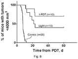
- FIG. 8 shows the beneficial results of an embodiment of the presently-disclosed techniques using concurrent I-PDT and thermal ablation. It can be seen that the use of light without photosensitizer results in a cure rate of 40%. Despite the previous consideration that a lack of significant temperature change of the tissue was a benefit of PDT, FIG. 8 shows that concurrent photothermal ablation and I-PDT resulted in a cure rate of 70%.
Abstract
Description
- This application claims priority to U.S. provisional Application No. 62/492,171, filed on Apr. 29, 2017, now pending, the disclosure of which is incorporated herein by reference.
- This invention was made with government support under CA193610 awarded by the National Institutes of Health. The government has certain rights in the invention.
- The present disclosure relates to photodynamic therapy.
- Photodynamic therapy (PDT), in particular interstitial photodynamic therapy (I-PDT), offers promising outcomes for patients with refractory locally advanced cancer. The use of I-PDT with porfimer sodium (Photofrin®) is approved for palliation in patients with esophageal cancer or lung cancer with airway obstruction, who are non-candidates for surgery or radiation therapy. In addition, I-PDT with porfimer sodium has been used, in compassionate care settings, to treat patients with head and neck squamous cell carcinoma. A principal clinical goal has been to shorten treatment times by administering the therapeutic light at high dose rates (i.e., 400 mW/cm) that are clinically approved by the FDA for I-PDT with porfimer sodium. However, the cure rate for I-PDT with porfimer sodium is limited.
- PDT has also been viewed as beneficial when considering the relatively minor nature of adverse effects. Additionally, it has been noted that PDT provides excellent cosmetic outcomes. To date, it has been believed that during I-PDT the changes in tissue temperature do not affect the response. Physicians and researchers assumed that PDT is associated with minimal heating. The clinically approved, and used, light dose rate (400 mW/cm) for PDT or I-PDT with porfimer sodium was chosen arbitrarily, about 25 years ago. There was no systemic study to evaluate potential tissue heating during I-PDT. Several retrospective clinical studies suggest that PDT and I-PDT will result in retention of functional anatomy and other benefits. While offering improved palliative outcomes for patients with such advanced diseases, there remains a need for further improvement.
- In a first aspect, the present disclosure provides a method for treating a tissue. A photosensitizer is administered to the tissue. One or more optical fibers are placed in the tissue. For example, a portion (such as, for example, an end portion) of the one or more optical fibers are inserted in the tissue. The optical fibers may be spaced apart from one another such that a light dose may be applied to the tissue. The method includes applying a treatment light to the tissue by way of the one or more optical fibers. A temperature of the tissue is measured during application of the treatment light. The fluence rate (mW/cm2) of the treatment light is modified based on the temperature of the tissue. For example, by adjusting the light dose rate one may govern the intratumoral fluence rate within the tissue (e.g., intratumoral fluence rate). For example, the fluence rate may be modified to be lower if the temperature of the tissue is higher than a predetermined threshold. In some embodiments, the fluence rate is modified to maintain a tissue temperature between 50° C. and 65° C. In some embodiments, the fluence rate is modified to maintain a tissue temperature of substantially 60° C. In some embodiments, the fluence rate is modified to maintain a tissue temperature between 60° C. and 90° C.
- In some embodiments, one or more dosimetry fibers may be placed in the tissue and configured to measure light dose (J/cm2). The optical fiber(s) and/or the dosimetry fiber(s) may be disposed within one or more light-transmitting catheters (LTCs) placed in the tissue. For example, each optical fiber may be disposed in a corresponding LTC. In another example, each of a plurality of LTCs may contain an optical fiber and a dosimetry fiber.
- In another aspect, the present disclosure may be embodied as a system for treating a tissue. The system includes a light source and an optical fiber operably coupled to the light source. The optical fiber is configured to deliver a light dose of treatment light to the tissue. A temperature sensor is configured to measure a temperature of the tissue. The temperature sensor may be any suitable sensor such as, for example, a thermistor, a thermal imaging sensor, a fiber optic, a magnetic resonance thermometer (providing volumetric temperature data), or the like. In some embodiments, the temperature sensor is configured to measure temperature at a plurality of locations throughout the volume of the tissue. In some embodiments, the temperature sensor comprises a plurality of temperature sensitive catheters.
- A controller, such as, for example, a programmable microprocessor, is in communication with the temperature sensor and configured to modify a fluence rate of the treatment light based on a measured temperature. In some embodiments, the controller is configured to modify the fluence rate of the treatment light to maintain a tissue temperature between 50° C. and 65° C. In some embodiments, the controller is configured to modify the fluence rate of the treatment light to maintain a tissue temperature of substantially 60° C. In some embodiments, the controller is configured to modify the fluence rate of the treatment light to maintain a tissue temperature between 60° C. and 90° C.
- The system may further include a dosimetry fiber for measuring the light dose. A spectrometer may be operably coupled to the dosimetry fiber. The system may further include an LTC, and the optical fiber and/or the dosimetry fiber may be disposed in the LTC.
- In some embodiments, a second optical fiber is operably coupled to the light source and configured to deliver a second light dose of treatment light to the tissue. The controller may be configured to modify a fluence rate of the second light dose based on the measured temperature. The fluence rate of the second light dose may be modified based on a temperature at a second location of the tissue.
- For a fuller understanding of the nature and objects of the disclosure, reference should be made to the following detailed description taken in conjunction with the accompanying drawings, in which:
-
FIG. 1 depicts a system according to an embodiment of the present disclosure; -
FIG. 2 is a chart showing a method according to another embodiment of the present disclosure; -
FIG. 3 shows a mouse being fitted with two optical fibers, each disposed within a light-transmitting catheter, for use in interstitial photodynamic therapy (I-PDT); -
FIG. 4 is a chart showing intratumoral heating results for a light dose of 150 mW/cm, 100 J/cm with and without photosensitizer; -
FIG. 5 is a chart showing intratumoral heating results for a light dose of 350 mW/cm, 100 J/cm with and without photosensitizer; -
FIG. 6 is a chart showing tumor size over time in populations of mice, where a control population was untreated, a second population was treated with a light dose of 150 mW/cm, 100 J/cm (control vs. light p=<0.0001), and a third population was treated with I-PDT (control vs. I-PDT p=<0.0001; light vs. I-PDT p<0.05); -
FIG. 7 is a chart showing tumor size over time in populations of mice, where a control population was untreated, a second population was treated with a light dose of 350 mW/cm, 100 J/cm (control vs. light p=<0.0001), and a third population was treated with I-PDT (control vs. I-PDT p=<0.0001; light vs. I-PDT p=0.339); and -
FIG. 8 is a chart showing tumor size over time in populations of mice, where a control population was untreated, a second population was treated with a light dose of 100 mW/cm, 540 J/cm (control vs. light p=<0.0001), and a third population was treated with I-PDT concurrent with the same light dose as the second population (control vs. light p=<0.0001; light vs. I-PDT/light p=0.164). - As mentioned above, a perceived benefit of photodynamic therapy has been the lack of significant changes in tissue temperature. However, the present disclosure advantageously utilizes increased temperatures induced in the tissue using the I-PDT techniques disclosed herein to enhance efficacy as compared to I-PDT without an increased temperature.
- With reference to
FIG. 2 , the present disclosure may be embodied as amethod 100 for treating a tissue, for example, treating a tumor, of an individual using interstitial photodynamic therapy (I-PDT) (seeFIG. 2 ). Themethod 100 includes administering 103 a photosensitizer to the tissue. The photosensitizer may be, for example, porfimer sodium (Photofrin®) or any other photosensitizer known for use in I-PDT—e.g., capable of generating reactive oxygen species and radicals when activated by light in the presence of oxygen. The photosensitizer may be administered 103 by, for example, intravenous injection. - One or more optical fibers are placed 106 into the tissue to be treated. The optical fibers may be placed at locations in the tissue according to a predetermined treatment plan. In some embodiments, the optical fiber(s) are placed 106 into the tissue by way of light-transmitting catheter(s) (LTCs) where each optical fiber is disposed in a light-transmitting catheter.
- A treatment light is applied 109 to the tissue by way of the one or more optical fibers. The treatment light has a fluence (measured in, for example, joules per square centimeter—J/cm2) and a fluence rate (measured in, for example, milliwatts per square centimeter—mW/cm2).
- The
method 100 includes measuring 112 a temperature of the tissue during application of the treatment light (i.e., during “treatment”).Measurement 112 of the tissue may be performed using any technique appropriate. For example, volumetric measurement may be accomplished using magnetic-resonance thermometry (MR thermometry or MRT). In another example, temperature is measured using a temperature-sensitive catheter. In embodiments where LTCs are placed in the tissue, a temperature-sensitive catheter may optionally be disposed in an LTC. Other methods for measuring 112 a temperature of a tissue may be used. - The fluence rate of the treatment light may be modified 115 based on the temperature of the tissue. The fluence rate of the treatment light within the tissue (e.g., intratumoral fluence rate) may be modified by adjusting the light dose rate. For example, the fluence rate may be decreased when the tissue temperature is higher than a (first) predetermined threshold. In this way, the temperature of the tissue will decrease. Similarly, the fluence rate of the treatment light may be increased if the tissue temperature is lower than a second predetermined threshold, which may be the same as or different from the first predetermined threshold. By increasing the fluence rate, the tissue temperature will increase. In exemplary embodiments, the fluence rate is modified to maintain a tissue temperature of between 50-65° C., inclusive. In another exemplary embodiment, the fluence rate is modified to maintain a tissue temperature of less than 60° C. In another exemplary embodiment, the fluence rate is modified to maintain a tissue temperature of substantially 60° C. By substantially, embodiments may maintain the temperature of the tissue within a desired tolerance, for example, ±5° C., ±1° C., ±0.5° C., ±0.2° C., or other tolerance levels between these exemplary values, for example, in 2° C. increments. In embodiments incorporating thermal ablation, the temperature may be maintained at greater than 60° C. and less than 100° C. (or in some embodiments, less than 90° C.) in order to avoid tissue carbonization.
- With reference to
FIG. 1 , the present disclosure may be embodied as asystem 10 for treatingtissue 90 using I-PDT is provided. Thesystem 10 includes a light source 12. For example, the light source 12 may be a laser configured to emit light at a wavelength for activating a selected photosensitizer. For example, when using Photofrin, the emitted light may be 630 nm. Anoptical fiber 20 is operably coupled to the light source 12. Theoptical fiber 20 is configured to deliver a light dose to thetissue 90. Theoptical fiber 20 may have a cylindrical diffuser, a fiber with a flat-cut end, or other configuration. Embodiments of thesystem 10 may include additional optical fibers, for example, the embodiment depicted inFIG. 1 includes a secondoptical fiber 20 for delivering a second light dose to thetissue 90. - The
system 10 includes atemperature sensor 30 for measuring a temperature of thetissue 90. In some embodiments, thetemperature sensor 30 may be configured to measure the temperature of thetissue 90 at more than one location, for example, more than one 3-D location within thetissue 90. In some embodiments, the temperature sensor is a magnetic-resonance thermometer. In some embodiments, the temperature sensor comprises one or more temperature-sensitive catheters (e.g., a thermistor disposed in a catheter). - In some embodiments, the
system 10 may include a light-transmitting catheter 22 (e.g., a transparent catheter)(an “LTC”). In such embodiments, the optical fiber(s) 20 of thesystem 10 may be disposed within a corresponding number of LTCs 22. For example, eachoptical fiber 20 may be disposed in alumen 23 of anLTC 22. - The
system 10 further includes acontroller 40 in communication with thetemperature sensor 30. Thecontroller 40 is configured to modify a fluence rate of the treatment light based on the temperature of thetissue 90, as described above. In some embodiments, thecontroller 40 is a programmable microprocessor, programmed to modify the treatment light based on a signal received from the temperature sensor. The treatment light may be modified by adjusting the intensity of the light source, attenuating the light emitted from the light source, independently attenuating the light in each optical fiber, or any other technique for increasing or decreasing the fluence rate of the treatment light delivered to the tissue via the optical fiber(s). - Some embodiments of the presently-disclosed system may include a
dosimetry fiber 25 for measuring a light dose. In embodiments wherein the system includes anLTC 22, thedosimetry fiber 25 may be disposed in anLTC 22, for example, within alumen 23 of anLTC 22. Thedosimetry fiber 25 may be operably coupled to aspectrometer 27 for measuring the light dose delivered to the tissue. In this way, a desired total dose may be delivered while also maintaining the temperature of the tissue in a desired range for effective photothermal ablation (or other synergistic effect when combined with I-PDT). - The present disclosure is further illustrated in the discussion below, which includes exemplary embodiments used to test the disclosed technique using mouse models.
- The mechanism of action in photodynamic therapy (PDT) involves the generation of reactive oxygen species and radicals through light activation of photosensitizer in the presence of oxygen—i.e., an effective photoreaction. Previous studies demonstrated that oxygen-conserving light fluence rate (mW/cm2) is required for an effective photoreaction. In I-PDT, multiple optical fibers with a cylindrical diffuser or optical fibers with flat cut end are inserted into the tumor. During I-PDT, the light dose rate (mW/cm) dictates the resulted fluence rate in the tumor. Thus, the light fluence rate depends on the light dose rate delivered from the optical fibers.
- In the U.S., Photofrin is the only approved photosensitizer in the treatment of obstructing esophageal, non-small cell endobronchial lung cancer and high-grade dysplasia in Barrett's esophagus. The FDA-approved light dose rate for I-PDT with Photofrin is 400 mW/cm length of the optical fiber cylindrical diffuser, which translates to a fluence rate of up to 800 mW/cm2. This clinically-approved light dose rate induces significant thermal ablation that could overwhelm the photoreaction. However, our preclinical data suggest that this is not the case, and cure of locally advanced squamous cell carcinoma can be achieved in a mouse model treated with I-PDT and thermal ablation. In an ongoing preclinical study it was found that the laser light can induce significant heating that can induce immediate tissue ablation, at T>60° C., without impeding the efficacy of the I-PDT.
- The inventors hypothesized that the limited cure rate for I-PDT with porfimer sodium is due to the high dose rate that could limit the photodynamic efficiency by depleting tumor oxygen levels.
- As further described below, aspects of the presently-disclosed method and system were implemented for testing. In particular, in vivo magnetic resonance thermometry (MRT) was used to quantify photothermal ablation during I-PDT. Time-to-event and cure rates were analyzed with Kaplan Meier curves. Safe and effective light dose rates and doses with photothermal ablation were identified.
- All procedures were carried out in accordance with a protocol approved by the Institutional Animal Care and Use Committee (IACUC) at Roswell Park Cancer Institute (RPCI). Female C3H, 8-12 weeks, mice bearing locally advanced SCCVII tumors were treated when the tumors reached a size of 9-10 mm in their largest diameter, and a volume of 400-500 mm3 calculated from caliper measurements, as is known in the art.
- The animals were randomly assigned to receive light only (no drug) or I-PDT by administering 5 mg/kg porfimer sodium 24 hours prior to light delivery. Mice were considered cured if there was no palpable tumor any time at or after 60 days post I-PDT.
- The laser light (treatment light) was delivered through one to three 0.98 mm diameter optical fibers with 20 mm cylindrical light diffuser RD20 (Medlight SA, Ecublens, Switzerland). The cylindrical light diffusers were connected to 1.0 Watt laser diode modules that emit 630±3 nm light (ML6500, Modulight Inc., Tampere, Finland). During light delivery, a custom made template was utilized to guide the placement of the laser fibers into the tumor, as shown in
FIG. 3 . - Magnetic resonance thermometry (MRT) by proton resonance frequency methods was carried out in a 4.7 Tesla preclinical scanner using the ParaVision 3.0.2 imaging platform (Bruker Biospin, Billerica Mass.) and a 35 mm quadrature transceiver coil.
FIGS. 4 and 5 show that there was a temperature increase during the interstitial light delivery, with and without drug. These temperatures were averaged over the entire tumor volume. -
FIG. 6 shows the percentage of mice with tumors less than 4000 mm3 over a period of 60 days post treatment. As shown in the chart, a control population was untreated, a second population was treated with a light dose of 150 mW/cm, 100 J/cm (control vs. light p≤0.0001), and a third population was treated with I-PDT and similar dose (control vs. I-PDT p≤0.0001; light vs. I-PDT p=0.0004).FIG. 7 is a similar chart for populations treated with a light dose of 350 mW/cm, 100 J/cm (control vs. light p≤0.0001; control vs. I-PDT p≤0.0001; light vs. I-PDT p=0.339). It can be seen that after 60 days, only 10% of mice remained with tumors less than 4,000 mm3. -
FIG. 8 shows the beneficial results of an embodiment of the presently-disclosed techniques using concurrent I-PDT and thermal ablation. It can be seen that the use of light without photosensitizer results in a cure rate of 40%. Despite the previous consideration that a lack of significant temperature change of the tissue was a benefit of PDT,FIG. 8 shows that concurrent photothermal ablation and I-PDT resulted in a cure rate of 70%. - Although claimed subject matter is described in terms of certain embodiments, other embodiments, including embodiments that do not provide all of the benefits and features set forth herein, are also within the scope of this disclosure. Various structural, logical, process step, and electronic changes may be made without departing from the scope of the disclosure.
Claims (20)
Priority Applications (1)
| Application Number | Priority Date | Filing Date | Title |
|---|---|---|---|
| US16/607,683 US20200046997A1 (en) | 2017-04-29 | 2018-04-30 | Method and system for concurrent photothermal ablation and interstitial photodynamic therapy |
Applications Claiming Priority (3)
| Application Number | Priority Date | Filing Date | Title |
|---|---|---|---|
| US201762492171P | 2017-04-29 | 2017-04-29 | |
| PCT/US2018/030314 WO2018201156A1 (en) | 2017-04-29 | 2018-04-30 | Method and system for concurrent photothermal ablation and interstitial photodynamic therapy |
| US16/607,683 US20200046997A1 (en) | 2017-04-29 | 2018-04-30 | Method and system for concurrent photothermal ablation and interstitial photodynamic therapy |
Related Parent Applications (1)
| Application Number | Title | Priority Date | Filing Date |
|---|---|---|---|
| PCT/US2018/030314 A-371-Of-International WO2018201156A1 (en) | 2017-04-29 | 2018-04-30 | Method and system for concurrent photothermal ablation and interstitial photodynamic therapy |
Related Child Applications (1)
| Application Number | Title | Priority Date | Filing Date |
|---|---|---|---|
| US18/162,995 Division US20230173301A1 (en) | 2017-04-29 | 2023-02-01 | Method and system for concurrent photothermal ablation and interstitial photodynamic therapy |
Publications (1)
| Publication Number | Publication Date |
|---|---|
| US20200046997A1 true US20200046997A1 (en) | 2020-02-13 |
Family
ID=63919282
Family Applications (2)
| Application Number | Title | Priority Date | Filing Date |
|---|---|---|---|
| US16/607,683 Abandoned US20200046997A1 (en) | 2017-04-29 | 2018-04-30 | Method and system for concurrent photothermal ablation and interstitial photodynamic therapy |
| US18/162,995 Pending US20230173301A1 (en) | 2017-04-29 | 2023-02-01 | Method and system for concurrent photothermal ablation and interstitial photodynamic therapy |
Family Applications After (1)
| Application Number | Title | Priority Date | Filing Date |
|---|---|---|---|
| US18/162,995 Pending US20230173301A1 (en) | 2017-04-29 | 2023-02-01 | Method and system for concurrent photothermal ablation and interstitial photodynamic therapy |
Country Status (4)
| Country | Link |
|---|---|
| US (2) | US20200046997A1 (en) |
| EP (1) | EP3614939A4 (en) |
| CA (1) | CA3058949A1 (en) |
| WO (1) | WO2018201156A1 (en) |
Cited By (3)
| Publication number | Priority date | Publication date | Assignee | Title |
|---|---|---|---|---|
| WO2021194877A1 (en) * | 2020-03-25 | 2021-09-30 | Lumeda Inc. | Optical applicator feature optimizer |
| WO2022266665A1 (en) * | 2021-06-17 | 2022-12-22 | Lumeda Inc. | Configurable optical applicator |
| US20230238099A1 (en) * | 2020-08-18 | 2023-07-27 | Simphotek, Inc. | Interstitial photodynamic therapy |
Families Citing this family (3)
| Publication number | Priority date | Publication date | Assignee | Title |
|---|---|---|---|---|
| CN109481013B (en) * | 2018-12-19 | 2021-05-28 | 南京康友医疗科技有限公司 | Microwave ablation device with thermal field monitoring function |
| CN109758227B (en) * | 2019-01-23 | 2021-12-21 | 深圳安泰创新科技股份有限公司 | Tumor ablation simulation method and device, electronic equipment and readable storage medium |
| WO2023198724A1 (en) * | 2022-04-11 | 2023-10-19 | Spectracure Ab | System and method for combined thermal and photodynamic therapy of malignant tumors |
Family Cites Families (10)
| Publication number | Priority date | Publication date | Assignee | Title |
|---|---|---|---|---|
| US5292320A (en) * | 1992-07-06 | 1994-03-08 | Ceramoptec, Inc. | Radial medical laser delivery device |
| US5483958A (en) | 1994-01-25 | 1996-01-16 | United States Of America As Represented By The Secretary Of The Department Of Health And Human Services | Fluorescent-tipped dosimeter probe |
| IL154101A0 (en) | 2003-01-23 | 2003-07-31 | Univ Ramot | Minimally invasive controlled surgical system with feedback |
| EP2155098A4 (en) * | 2007-06-08 | 2013-11-06 | Cynosure Inc | Thermal surgery safety apparatus and method |
| CA2811605A1 (en) * | 2010-09-16 | 2012-03-22 | Case Western Reserve University | Photodynamic therapy system, device and associated method of treatment |
| US10617470B2 (en) * | 2012-10-16 | 2020-04-14 | Boston Scientific Scimed, Inc. | Laser ablation with electromagnetic energy feedback |
| EP2799111A1 (en) | 2013-04-30 | 2014-11-05 | Clinical Laserthermia Systems AB | Apparatus and method for controlling immunostimulating laser thermotherapy |
| US11065063B2 (en) | 2015-07-02 | 2021-07-20 | Board Of Regents, The University Of Texas System | Utilization of laser interstitial thermotherapy guided with real time thermal MRI |
| US9987089B2 (en) * | 2015-07-13 | 2018-06-05 | University of Central Oklahoma | Device and a method for imaging-guided photothermal laser therapy for cancer treatment |
| CA2996503A1 (en) | 2015-07-23 | 2017-01-26 | Health Research, Inc. | System and method for delivering light dose to tissue |
-
2018
- 2018-04-30 WO PCT/US2018/030314 patent/WO2018201156A1/en active Application Filing
- 2018-04-30 US US16/607,683 patent/US20200046997A1/en not_active Abandoned
- 2018-04-30 CA CA3058949A patent/CA3058949A1/en active Pending
- 2018-04-30 EP EP18790729.0A patent/EP3614939A4/en active Pending
-
2023
- 2023-02-01 US US18/162,995 patent/US20230173301A1/en active Pending
Cited By (6)
| Publication number | Priority date | Publication date | Assignee | Title |
|---|---|---|---|---|
| WO2021194877A1 (en) * | 2020-03-25 | 2021-09-30 | Lumeda Inc. | Optical applicator feature optimizer |
| US11925814B2 (en) | 2020-03-25 | 2024-03-12 | Lumeda Inc. | Optical applicator feature optimizer |
| US20230238099A1 (en) * | 2020-08-18 | 2023-07-27 | Simphotek, Inc. | Interstitial photodynamic therapy |
| US11771916B2 (en) * | 2020-08-18 | 2023-10-03 | Simphotek, Inc. | Interstitial photodynamic therapy |
| WO2022266665A1 (en) * | 2021-06-17 | 2022-12-22 | Lumeda Inc. | Configurable optical applicator |
| US11947146B2 (en) | 2021-06-17 | 2024-04-02 | Lumeda Inc. | Configurable optical applicator |
Also Published As
| Publication number | Publication date |
|---|---|
| US20230173301A1 (en) | 2023-06-08 |
| EP3614939A4 (en) | 2020-09-16 |
| CA3058949A1 (en) | 2018-11-01 |
| EP3614939A1 (en) | 2020-03-04 |
| WO2018201156A1 (en) | 2018-11-01 |
Similar Documents
| Publication | Publication Date | Title |
|---|---|---|
| US20230173301A1 (en) | Method and system for concurrent photothermal ablation and interstitial photodynamic therapy | |
| Salehpour et al. | Penetration profiles of visible and near-infrared lasers and light-emitting diode light through the head tissues in animal and human species: a review of literature | |
| Arami et al. | Remotely controlled near-infrared-triggered photothermal treatment of brain tumours in freely behaving mice using gold nanostar s | |
| Moro et al. | Photobiomodulation inside the brain: a novel method of applying near-infrared light intracranially and its impact on dopaminergic cell survival in MPTP-treated mice | |
| Reinhart et al. | Intracranial application of near-infrared light in a hemi-parkinsonian rat model: the impact on behavior and cell survival | |
| US20230097605A1 (en) | Damaging cancerous cells utilizing radio frequency waves in heating with heating enhanced by infusion or injection of glucose | |
| US20240123253A1 (en) | Photodynamic therapy devices, systems and methods | |
| EP1824562B1 (en) | Device for photodynamic therapy of the nasopharyngeal cavity | |
| Marijnissen et al. | Pilot study on light dosimetry for endobronchial photodynamic therapy | |
| US20190038909A1 (en) | A Therapeutic Method and Device Therefor | |
| US11040217B2 (en) | System and method for delivering dose light to tissue | |
| Feather et al. | Light delivery to tumour tissue through implanted optical fibres during photodynamic therapy | |
| LT6795B (en) | Laser therapy fiber optic probe | |
| Cassim et al. | Iron oxide nanoparticle hyperthermia and radiation cancer treatment | |
| van Veen et al. | In vivo fluence rate measurements during Foscan®-mediated photodynamic therapy of persistent and recurrent nasopharyngeal carcinomas using a dedicated light applicator | |
| RU2617090C1 (en) | Method for photodynamic therapy of malignant tumours | |
| US20230045729A1 (en) | Implantable light delivery device | |
| US20230076544A1 (en) | Systems, methods, and devices for damaging cancerous cells by application of energy to the entirety of the cancerous cells and the area of the body immediately surrounding the cancerous cells | |
| Bown | Scientists and clinicians create a bright future for photodynamic therapy (PDT) | |
| CA3183491A1 (en) | Systems, methods, and devices for damaging cancerous cells by application of energy to the entirety of the cancerous cells and the area of the body immediately surrounding the cancerous cells | |
| AU2005305606A1 (en) | Device and method for photodynamic therapy of the nasopharyngeal cavity | |
| Betz et al. | Interstitial PDT Cancer Treatment | |
| Weersink et al. | Optical delivery and monitoring of photodynamic therapy of prostate cancer | |
| Day et al. | Photothermal Therapy of Glioma in a Mouse Model With Near-Infrared Excited Nanoshells |
Legal Events
| Date | Code | Title | Description |
|---|---|---|---|
| AS | Assignment |
Owner name: HEALTH RESEARCH, INC., NEW YORK Free format text: ASSIGNMENT OF ASSIGNORS INTEREST;ASSIGNORS:SHAFIRSTEIN, GAL;BELLNIER, DAVID;SIGNING DATES FROM 20200727 TO 20200806;REEL/FRAME:054041/0630 |
|
| STPP | Information on status: patent application and granting procedure in general |
Free format text: NON FINAL ACTION MAILED |
|
| STPP | Information on status: patent application and granting procedure in general |
Free format text: RESPONSE TO NON-FINAL OFFICE ACTION ENTERED AND FORWARDED TO EXAMINER |
|
| STPP | Information on status: patent application and granting procedure in general |
Free format text: FINAL REJECTION MAILED |
|
| STPP | Information on status: patent application and granting procedure in general |
Free format text: DOCKETED NEW CASE - READY FOR EXAMINATION |
|
| STPP | Information on status: patent application and granting procedure in general |
Free format text: NON FINAL ACTION MAILED |
|
| STPP | Information on status: patent application and granting procedure in general |
Free format text: RESPONSE TO NON-FINAL OFFICE ACTION ENTERED AND FORWARDED TO EXAMINER |
|
| STPP | Information on status: patent application and granting procedure in general |
Free format text: FINAL REJECTION MAILED |
|
| STCB | Information on status: application discontinuation |
Free format text: ABANDONED -- FAILURE TO RESPOND TO AN OFFICE ACTION |