US20040097402A1 - Compounds and methods for regulating bacterial growth and pathogenesis - Google Patents
Compounds and methods for regulating bacterial growth and pathogenesis Download PDFInfo
- Publication number
- US20040097402A1 US20040097402A1 US10/300,818 US30081802A US2004097402A1 US 20040097402 A1 US20040097402 A1 US 20040097402A1 US 30081802 A US30081802 A US 30081802A US 2004097402 A1 US2004097402 A1 US 2004097402A1
- Authority
- US
- United States
- Prior art keywords
- autoinducer
- antibiotic
- activity
- typhimurium
- group
- Prior art date
- Legal status (The legal status is an assumption and is not a legal conclusion. Google has not performed a legal analysis and makes no representation as to the accuracy of the status listed.)
- Granted
Links
- 238000000034 method Methods 0.000 title claims abstract description 187
- 230000001580 bacterial effect Effects 0.000 title claims abstract description 102
- 150000001875 compounds Chemical class 0.000 title claims description 161
- 230000012010 growth Effects 0.000 title abstract description 69
- 230000001105 regulatory effect Effects 0.000 title abstract description 44
- 230000008506 pathogenesis Effects 0.000 title abstract description 19
- BVIYGXUQVXBHQS-IUYQGCFVSA-N (2R,4S)-2-methyltetrahydrofuran-2,3,3,4-tetrol Chemical class C[C@@]1(O)OC[C@H](O)C1(O)O BVIYGXUQVXBHQS-IUYQGCFVSA-N 0.000 claims abstract description 413
- 230000000694 effects Effects 0.000 claims abstract description 206
- 210000004027 cell Anatomy 0.000 claims description 221
- 241000607618 Vibrio harveyi Species 0.000 claims description 215
- 108090000623 proteins and genes Proteins 0.000 claims description 209
- 238000004519 manufacturing process Methods 0.000 claims description 200
- 230000003115 biocidal effect Effects 0.000 claims description 135
- 102000004169 proteins and genes Human genes 0.000 claims description 113
- 230000018612 quorum sensing Effects 0.000 claims description 113
- 235000018102 proteins Nutrition 0.000 claims description 112
- 239000003112 inhibitor Substances 0.000 claims description 103
- 239000000203 mixture Substances 0.000 claims description 100
- 230000037361 pathway Effects 0.000 claims description 63
- 125000004435 hydrogen atom Chemical group [H]* 0.000 claims description 48
- YRYOXRMDHALAFL-UHFFFAOYSA-N N-(3-oxohexanoyl)homoserine lactone Chemical compound CCCC(=O)CC(=O)NC1CCOC1=O YRYOXRMDHALAFL-UHFFFAOYSA-N 0.000 claims description 46
- 230000002195 synergetic effect Effects 0.000 claims description 46
- 125000000217 alkyl group Chemical group 0.000 claims description 41
- 229910052739 hydrogen Inorganic materials 0.000 claims description 38
- 239000001257 hydrogen Substances 0.000 claims description 38
- 125000002887 hydroxy group Chemical group [H]O* 0.000 claims description 38
- ZJUKTBDSGOFHSH-WFMPWKQPSA-N S-Adenosylhomocysteine Chemical compound O[C@@H]1[C@H](O)[C@@H](CSCC[C@H](N)C(O)=O)O[C@H]1N1C2=NC=NC(N)=C2N=C1 ZJUKTBDSGOFHSH-WFMPWKQPSA-N 0.000 claims description 35
- 239000003242 anti bacterial agent Substances 0.000 claims description 35
- 239000000284 extract Substances 0.000 claims description 33
- 238000004020 luminiscence type Methods 0.000 claims description 33
- 230000004044 response Effects 0.000 claims description 33
- -1 modified N-(3-oxododecanoyl)-L-homoserine lactone compound Chemical class 0.000 claims description 32
- 244000005700 microbiome Species 0.000 claims description 31
- 150000007523 nucleic acids Chemical class 0.000 claims description 31
- 125000002252 acyl group Chemical group 0.000 claims description 30
- 230000033228 biological regulation Effects 0.000 claims description 30
- DLVYTANECMRFGX-UHFFFAOYSA-N norfuraneol Natural products CC1=C(O)C(=O)CO1 DLVYTANECMRFGX-UHFFFAOYSA-N 0.000 claims description 29
- 102000039446 nucleic acids Human genes 0.000 claims description 29
- 108020004707 nucleic acids Proteins 0.000 claims description 29
- IQFWYNFDWRYSRA-OEQWSMLSSA-N S-(5-deoxy-beta-D-ribos-5-yl)-L-homocysteine Chemical compound OC(=O)[C@@H](N)CCSC[C@H]1O[C@@H](O)[C@H](O)[C@@H]1O IQFWYNFDWRYSRA-OEQWSMLSSA-N 0.000 claims description 22
- MYSWGUAQZAJSOK-UHFFFAOYSA-N ciprofloxacin Chemical compound C12=CC(N3CCNCC3)=C(F)C=C2C(=O)C(C(=O)O)=CN1C1CC1 MYSWGUAQZAJSOK-UHFFFAOYSA-N 0.000 claims description 22
- 239000008194 pharmaceutical composition Substances 0.000 claims description 22
- 108090000765 processed proteins & peptides Proteins 0.000 claims description 22
- 241001465754 Metazoa Species 0.000 claims description 21
- 239000000090 biomarker Substances 0.000 claims description 21
- 238000006243 chemical reaction Methods 0.000 claims description 20
- 230000001965 increasing effect Effects 0.000 claims description 20
- SMWDFEZZVXVKRB-UHFFFAOYSA-N Quinoline Chemical compound N1=CC=CC2=CC=CC=C21 SMWDFEZZVXVKRB-UHFFFAOYSA-N 0.000 claims description 18
- 230000007246 mechanism Effects 0.000 claims description 18
- 208000015181 infectious disease Diseases 0.000 claims description 17
- 229910052717 sulfur Inorganic materials 0.000 claims description 17
- 230000006698 induction Effects 0.000 claims description 16
- 230000005764 inhibitory process Effects 0.000 claims description 16
- 125000004430 oxygen atom Chemical group O* 0.000 claims description 16
- 125000003368 amide group Chemical group 0.000 claims description 15
- 229960000723 ampicillin Drugs 0.000 claims description 15
- AVKUERGKIZMTKX-NJBDSQKTSA-N ampicillin Chemical compound C1([C@@H](N)C(=O)N[C@H]2[C@H]3SC([C@@H](N3C2=O)C(O)=O)(C)C)=CC=CC=C1 AVKUERGKIZMTKX-NJBDSQKTSA-N 0.000 claims description 15
- 125000003118 aryl group Chemical group 0.000 claims description 15
- 125000001424 substituent group Chemical group 0.000 claims description 15
- 241000193996 Streptococcus pyogenes Species 0.000 claims description 14
- 230000001404 mediated effect Effects 0.000 claims description 14
- IJGRMHOSHXDMSA-UHFFFAOYSA-N Atomic nitrogen Chemical group N#N IJGRMHOSHXDMSA-UHFFFAOYSA-N 0.000 claims description 13
- 238000000338 in vitro Methods 0.000 claims description 13
- 229910052760 oxygen Inorganic materials 0.000 claims description 13
- 239000001301 oxygen Substances 0.000 claims description 13
- 125000004423 acyloxy group Chemical group 0.000 claims description 12
- QVGXLLKOCUKJST-UHFFFAOYSA-N atomic oxygen Chemical compound [O] QVGXLLKOCUKJST-UHFFFAOYSA-N 0.000 claims description 12
- 238000001514 detection method Methods 0.000 claims description 12
- 229940124530 sulfonamide Drugs 0.000 claims description 12
- 229930182555 Penicillin Natural products 0.000 claims description 11
- JGSARLDLIJGVTE-MBNYWOFBSA-N Penicillin G Chemical compound N([C@H]1[C@H]2SC([C@@H](N2C1=O)C(O)=O)(C)C)C(=O)CC1=CC=CC=C1 JGSARLDLIJGVTE-MBNYWOFBSA-N 0.000 claims description 11
- 108010059993 Vancomycin Proteins 0.000 claims description 11
- 230000004075 alteration Effects 0.000 claims description 11
- 230000008859 change Effects 0.000 claims description 11
- 229960003405 ciprofloxacin Drugs 0.000 claims description 11
- 150000003456 sulfonamides Chemical class 0.000 claims description 11
- 229960003165 vancomycin Drugs 0.000 claims description 11
- MYPYJXKWCTUITO-UHFFFAOYSA-N vancomycin Natural products O1C(C(=C2)Cl)=CC=C2C(O)C(C(NC(C2=CC(O)=CC(O)=C2C=2C(O)=CC=C3C=2)C(O)=O)=O)NC(=O)C3NC(=O)C2NC(=O)C(CC(N)=O)NC(=O)C(NC(=O)C(CC(C)C)NC)C(O)C(C=C3Cl)=CC=C3OC3=CC2=CC1=C3OC1OC(CO)C(O)C(O)C1OC1CC(C)(N)C(O)C(C)O1 MYPYJXKWCTUITO-UHFFFAOYSA-N 0.000 claims description 11
- GWCRPYGYVRXVLI-UHFFFAOYSA-N 2-ethyl-4-hydroxy-5-methyl-3(2H)-furanone Chemical group CCC1OC(C)=C(O)C1=O GWCRPYGYVRXVLI-UHFFFAOYSA-N 0.000 claims description 10
- NINIDFKCEFEMDL-UHFFFAOYSA-N Sulfur Chemical group [S] NINIDFKCEFEMDL-UHFFFAOYSA-N 0.000 claims description 10
- 230000003247 decreasing effect Effects 0.000 claims description 10
- 229910052757 nitrogen Chemical group 0.000 claims description 10
- 239000011593 sulfur Chemical group 0.000 claims description 10
- NHUHCSRWZMLRLA-UHFFFAOYSA-N Sulfisoxazole Chemical compound CC1=NOC(NS(=O)(=O)C=2C=CC(N)=CC=2)=C1C NHUHCSRWZMLRLA-UHFFFAOYSA-N 0.000 claims description 9
- 125000003342 alkenyl group Chemical group 0.000 claims description 9
- 230000000845 anti-microbial effect Effects 0.000 claims description 9
- 101150024601 luxN gene Proteins 0.000 claims description 9
- 229940049954 penicillin Drugs 0.000 claims description 9
- 102000004196 processed proteins & peptides Human genes 0.000 claims description 9
- 229960000654 sulfafurazole Drugs 0.000 claims description 9
- 239000002253 acid Substances 0.000 claims description 8
- 239000007787 solid Substances 0.000 claims description 8
- 125000004434 sulfur atom Chemical group 0.000 claims description 7
- 125000003277 amino group Chemical group 0.000 claims description 6
- 125000001072 heteroaryl group Chemical group 0.000 claims description 6
- 238000001727 in vivo Methods 0.000 claims description 6
- 229920001184 polypeptide Polymers 0.000 claims description 6
- 101710185027 5'-methylthioadenosine/S-adenosylhomocysteine nucleosidase Proteins 0.000 claims description 5
- 101710081557 Aminodeoxyfutalosine nucleosidase Proteins 0.000 claims description 5
- 239000002671 adjuvant Substances 0.000 claims description 5
- 230000006696 biosynthetic metabolic pathway Effects 0.000 claims description 5
- 239000003937 drug carrier Substances 0.000 claims description 5
- 229910052736 halogen Chemical group 0.000 claims description 5
- 150000002367 halogens Chemical group 0.000 claims description 5
- 239000011159 matrix material Substances 0.000 claims description 5
- QJGQUHMNIGDVPM-UHFFFAOYSA-N nitrogen group Chemical group [N] QJGQUHMNIGDVPM-UHFFFAOYSA-N 0.000 claims description 5
- 150000003839 salts Chemical class 0.000 claims description 5
- 239000003981 vehicle Substances 0.000 claims description 5
- QRAZOUJGIUABSI-NWHYMTRRSA-N (2s)-2-[[(2s)-2-[[(2s)-2-[[(2s)-2-[[(2r)-2-[[(2s)-2-[[(2s)-4-amino-2-[[(2s)-2-[(2-aminoacetyl)amino]-3-methylbutanoyl]amino]-4-oxobutanoyl]amino]propanoyl]amino]-3-sulfanylpropanoyl]amino]-3-hydroxypropanoyl]amino]-3-hydroxypropanoyl]amino]-4-methylpentan Chemical compound NCC(=O)N[C@@H](C(C)C)C(=O)N[C@@H](CC(N)=O)C(=O)N[C@@H](C)C(=O)N[C@@H](CS)C(=O)N[C@@H](CO)C(=O)N[C@@H](CO)C(=O)N[C@@H](CC(C)C)C(=O)N[C@H](C(O)=O)CC1=CC=CC=C1 QRAZOUJGIUABSI-NWHYMTRRSA-N 0.000 claims description 4
- OROGUZVNAFJPHA-UHFFFAOYSA-N 3-hydroxy-2,4-dimethyl-2H-thiophen-5-one Chemical compound CC1SC(=O)C(C)=C1O OROGUZVNAFJPHA-UHFFFAOYSA-N 0.000 claims description 4
- 239000004599 antimicrobial Substances 0.000 claims description 4
- 210000004899 c-terminal region Anatomy 0.000 claims description 4
- 125000002915 carbonyl group Chemical group [*:2]C([*:1])=O 0.000 claims description 4
- 125000000151 cysteine group Chemical group N[C@@H](CS)C(=O)* 0.000 claims description 4
- 125000000686 lactone group Chemical group 0.000 claims description 4
- 125000003607 serino group Chemical group [H]N([H])[C@]([H])(C(=O)[*])C(O[H])([H])[H] 0.000 claims description 4
- 241000607598 Vibrio Species 0.000 claims description 3
- 125000003545 alkoxy group Chemical group 0.000 claims description 3
- 239000005557 antagonist Substances 0.000 claims description 3
- VIHAEDVKXSOUAT-UHFFFAOYSA-N but-2-en-4-olide Chemical group O=C1OCC=C1 VIHAEDVKXSOUAT-UHFFFAOYSA-N 0.000 claims description 3
- CREMABGTGYGIQB-UHFFFAOYSA-N carbon carbon Chemical compound C.C CREMABGTGYGIQB-UHFFFAOYSA-N 0.000 claims description 3
- 239000011203 carbon fibre reinforced carbon Substances 0.000 claims description 3
- 125000005843 halogen group Chemical group 0.000 claims description 3
- 150000003384 small molecules Chemical class 0.000 claims description 3
- BLWINLJDTOJSRU-UHFFFAOYSA-N 2-methoxy-2,4-diphenylfuran-3-one Chemical group O=C1C(OC)(C=2C=CC=CC=2)OC=C1C1=CC=CC=C1 BLWINLJDTOJSRU-UHFFFAOYSA-N 0.000 claims description 2
- 101150026549 luxP gene Proteins 0.000 claims description 2
- 150000003290 ribose derivatives Chemical class 0.000 claims description 2
- 125000001246 bromo group Chemical group Br* 0.000 claims 6
- INAXVXBDKKUCGI-UHFFFAOYSA-N 4-hydroxy-2,5-dimethylfuran-3-one Chemical group CC1OC(C)=C(O)C1=O INAXVXBDKKUCGI-UHFFFAOYSA-N 0.000 claims 4
- MYPYJXKWCTUITO-LYRMYLQWSA-O vancomycin(1+) Chemical compound O([C@@H]1[C@@H](O)[C@H](O)[C@@H](CO)O[C@H]1OC1=C2C=C3C=C1OC1=CC=C(C=C1Cl)[C@@H](O)[C@H](C(N[C@@H](CC(N)=O)C(=O)N[C@H]3C(=O)N[C@H]1C(=O)N[C@H](C(N[C@@H](C3=CC(O)=CC(O)=C3C=3C(O)=CC=C1C=3)C([O-])=O)=O)[C@H](O)C1=CC=C(C(=C1)Cl)O2)=O)NC(=O)[C@@H](CC(C)C)[NH2+]C)[C@H]1C[C@](C)([NH3+])[C@H](O)[C@H](C)O1 MYPYJXKWCTUITO-LYRMYLQWSA-O 0.000 claims 4
- RTZKZFJDLAIYFH-UHFFFAOYSA-N Diethyl ether Chemical compound CCOCC RTZKZFJDLAIYFH-UHFFFAOYSA-N 0.000 claims 2
- 125000003668 acetyloxy group Chemical group [H]C([H])([H])C(=O)O[*] 0.000 claims 2
- 125000002346 iodo group Chemical group I* 0.000 claims 2
- 125000003396 thiol group Chemical group [H]S* 0.000 claims 2
- 206010010144 Completed suicide Diseases 0.000 claims 1
- 150000001408 amides Chemical class 0.000 claims 1
- 230000002860 competitive effect Effects 0.000 claims 1
- 150000002148 esters Chemical class 0.000 claims 1
- 241000293869 Salmonella enterica subsp. enterica serovar Typhimurium Species 0.000 description 198
- WQZGKKKJIJFFOK-GASJEMHNSA-N Glucose Natural products OC[C@H]1OC(O)[C@H](O)[C@@H](O)[C@@H]1O WQZGKKKJIJFFOK-GASJEMHNSA-N 0.000 description 123
- 239000008103 glucose Substances 0.000 description 122
- 241000588724 Escherichia coli Species 0.000 description 120
- FAPWRFPIFSIZLT-UHFFFAOYSA-M Sodium chloride Chemical compound [Na+].[Cl-] FAPWRFPIFSIZLT-UHFFFAOYSA-M 0.000 description 118
- 239000006137 Luria-Bertani broth Substances 0.000 description 83
- 241000894006 Bacteria Species 0.000 description 74
- 241000405383 Salmonella enterica subsp. enterica serovar Typhimurium str. LT2 Species 0.000 description 74
- 230000011664 signaling Effects 0.000 description 68
- 239000012532 cell-free culture fluid Substances 0.000 description 63
- 239000011780 sodium chloride Substances 0.000 description 60
- 238000003556 assay Methods 0.000 description 57
- 230000014509 gene expression Effects 0.000 description 51
- 241000620209 Escherichia coli DH5[alpha] Species 0.000 description 46
- 230000015556 catabolic process Effects 0.000 description 42
- 239000012530 fluid Substances 0.000 description 41
- 230000007781 signaling event Effects 0.000 description 41
- 238000006731 degradation reaction Methods 0.000 description 39
- 101150078797 luxS gene Proteins 0.000 description 39
- 108020004414 DNA Proteins 0.000 description 38
- 230000032770 biofilm formation Effects 0.000 description 38
- 239000002609 medium Substances 0.000 description 36
- 101100407726 Bacillus subtilis (strain 168) perR gene Proteins 0.000 description 33
- 101100075645 Escherichia coli (strain K12) luxS gene Proteins 0.000 description 33
- 230000003204 osmotic effect Effects 0.000 description 32
- 230000035939 shock Effects 0.000 description 32
- 238000002474 experimental method Methods 0.000 description 29
- 238000003780 insertion Methods 0.000 description 28
- 230000037431 insertion Effects 0.000 description 28
- 239000000589 Siderophore Substances 0.000 description 26
- 102000005962 receptors Human genes 0.000 description 25
- 108020003175 receptors Proteins 0.000 description 25
- 210000001519 tissue Anatomy 0.000 description 25
- 241001646719 Escherichia coli O157:H7 Species 0.000 description 24
- 241000402142 Vibrio campbellii ATCC BAA-1116 Species 0.000 description 24
- 230000015572 biosynthetic process Effects 0.000 description 23
- 239000012531 culture fluid Substances 0.000 description 23
- MEFKEPWMEQBLKI-AIRLBKTGSA-N S-adenosyl-L-methioninate Chemical compound O[C@@H]1[C@H](O)[C@@H](C[S+](CC[C@H](N)C([O-])=O)C)O[C@H]1N1C2=NC=NC(N)=C2N=C1 MEFKEPWMEQBLKI-AIRLBKTGSA-N 0.000 description 22
- 229960001570 ademetionine Drugs 0.000 description 22
- 230000005526 G1 to G0 transition Effects 0.000 description 21
- 238000004166 bioassay Methods 0.000 description 21
- 238000012360 testing method Methods 0.000 description 20
- 238000002360 preparation method Methods 0.000 description 19
- 239000000047 product Substances 0.000 description 19
- 241000894007 species Species 0.000 description 19
- 241000660147 Escherichia coli str. K-12 substr. MG1655 Species 0.000 description 18
- 238000004458 analytical method Methods 0.000 description 18
- 239000001963 growth medium Substances 0.000 description 18
- 238000011282 treatment Methods 0.000 description 18
- 150000001413 amino acids Chemical group 0.000 description 17
- 230000010261 cell growth Effects 0.000 description 17
- 230000002401 inhibitory effect Effects 0.000 description 17
- 230000000670 limiting effect Effects 0.000 description 17
- 230000029918 bioluminescence Effects 0.000 description 16
- 238000005415 bioluminescence Methods 0.000 description 16
- 239000003795 chemical substances by application Substances 0.000 description 16
- 230000006870 function Effects 0.000 description 16
- 238000003752 polymerase chain reaction Methods 0.000 description 16
- 101150083126 luxO gene Proteins 0.000 description 15
- 101150067683 rpo10 gene Proteins 0.000 description 15
- 101150011750 rpoN gene Proteins 0.000 description 15
- 102200093904 rs3810948 Human genes 0.000 description 15
- 241000607626 Vibrio cholerae Species 0.000 description 14
- 229940088710 antibiotic agent Drugs 0.000 description 14
- 210000004369 blood Anatomy 0.000 description 14
- 239000008280 blood Substances 0.000 description 14
- 238000005119 centrifugation Methods 0.000 description 14
- 230000001717 pathogenic effect Effects 0.000 description 14
- 102000004190 Enzymes Human genes 0.000 description 13
- 108090000790 Enzymes Proteins 0.000 description 13
- 230000004913 activation Effects 0.000 description 13
- 229940088598 enzyme Drugs 0.000 description 13
- 239000012634 fragment Substances 0.000 description 13
- 230000006799 invasive growth in response to glucose limitation Effects 0.000 description 13
- 230000014616 translation Effects 0.000 description 13
- WEVYAHXRMPXWCK-UHFFFAOYSA-N Acetonitrile Chemical compound CC#N WEVYAHXRMPXWCK-UHFFFAOYSA-N 0.000 description 12
- LFQSCWFLJHTTHZ-UHFFFAOYSA-N Ethanol Chemical compound CCO LFQSCWFLJHTTHZ-UHFFFAOYSA-N 0.000 description 12
- 210000000349 chromosome Anatomy 0.000 description 12
- 239000013612 plasmid Substances 0.000 description 12
- 239000000126 substance Substances 0.000 description 12
- 239000003053 toxin Substances 0.000 description 12
- 230000001018 virulence Effects 0.000 description 12
- 108091034117 Oligonucleotide Proteins 0.000 description 11
- 108700026244 Open Reading Frames Proteins 0.000 description 11
- 229910052799 carbon Inorganic materials 0.000 description 11
- 238000010367 cloning Methods 0.000 description 11
- 239000000463 material Substances 0.000 description 11
- 230000035772 mutation Effects 0.000 description 11
- 238000001243 protein synthesis Methods 0.000 description 11
- 239000000523 sample Substances 0.000 description 11
- 238000003786 synthesis reaction Methods 0.000 description 11
- 230000017423 tissue regeneration Effects 0.000 description 11
- XLYOFNOQVPJJNP-UHFFFAOYSA-N water Substances O XLYOFNOQVPJJNP-UHFFFAOYSA-N 0.000 description 11
- OKTJSMMVPCPJKN-UHFFFAOYSA-N Carbon Chemical compound [C] OKTJSMMVPCPJKN-UHFFFAOYSA-N 0.000 description 10
- IAZDPXIOMUYVGZ-UHFFFAOYSA-N Dimethylsulphoxide Chemical compound CS(C)=O IAZDPXIOMUYVGZ-UHFFFAOYSA-N 0.000 description 10
- XEEYBQQBJWHFJM-UHFFFAOYSA-N Iron Chemical compound [Fe] XEEYBQQBJWHFJM-UHFFFAOYSA-N 0.000 description 10
- 241000607142 Salmonella Species 0.000 description 10
- DTQVDTLACAAQTR-UHFFFAOYSA-N Trifluoroacetic acid Chemical compound OC(=O)C(F)(F)F DTQVDTLACAAQTR-UHFFFAOYSA-N 0.000 description 10
- 238000005273 aeration Methods 0.000 description 10
- 239000003814 drug Substances 0.000 description 10
- 239000008188 pellet Substances 0.000 description 10
- 239000000243 solution Substances 0.000 description 10
- 230000000638 stimulation Effects 0.000 description 10
- 231100000765 toxin Toxicity 0.000 description 10
- 108700012359 toxins Proteins 0.000 description 10
- 239000000304 virulence factor Substances 0.000 description 10
- 230000007923 virulence factor Effects 0.000 description 10
- 235000001014 amino acid Nutrition 0.000 description 9
- 244000052616 bacterial pathogen Species 0.000 description 9
- 230000007423 decrease Effects 0.000 description 9
- 238000010790 dilution Methods 0.000 description 9
- 239000012895 dilution Substances 0.000 description 9
- 238000000746 purification Methods 0.000 description 9
- 241000607620 Aliivibrio fischeri Species 0.000 description 8
- 102100037355 Chromosome alignment-maintaining phosphoprotein 1 Human genes 0.000 description 8
- 102000053602 DNA Human genes 0.000 description 8
- 101000741320 Homo sapiens Cathelicidin antimicrobial peptide Proteins 0.000 description 8
- 101000880066 Homo sapiens Chromosome alignment-maintaining phosphoprotein 1 Proteins 0.000 description 8
- 238000004128 high performance liquid chromatography Methods 0.000 description 8
- 238000009396 hybridization Methods 0.000 description 8
- 239000007788 liquid Substances 0.000 description 8
- 238000007422 luminescence assay Methods 0.000 description 8
- 239000013598 vector Substances 0.000 description 8
- 108091032973 (ribonucleotides)n+m Proteins 0.000 description 7
- 108091026890 Coding region Proteins 0.000 description 7
- 101710121036 Delta-hemolysin Proteins 0.000 description 7
- 230000008901 benefit Effects 0.000 description 7
- 239000000306 component Substances 0.000 description 7
- 239000013078 crystal Substances 0.000 description 7
- 201000010099 disease Diseases 0.000 description 7
- 208000037265 diseases, disorders, signs and symptoms Diseases 0.000 description 7
- 229940079593 drug Drugs 0.000 description 7
- 230000037433 frameshift Effects 0.000 description 7
- 230000004927 fusion Effects 0.000 description 7
- 238000011534 incubation Methods 0.000 description 7
- 230000003993 interaction Effects 0.000 description 7
- 238000012163 sequencing technique Methods 0.000 description 7
- 239000002904 solvent Substances 0.000 description 7
- MYPYJXKWCTUITO-LYRMYLQWSA-N vancomycin Chemical compound O([C@@H]1[C@@H](O)[C@H](O)[C@@H](CO)O[C@H]1OC1=C2C=C3C=C1OC1=CC=C(C=C1Cl)[C@@H](O)[C@H](C(N[C@@H](CC(N)=O)C(=O)N[C@H]3C(=O)N[C@H]1C(=O)N[C@H](C(N[C@@H](C3=CC(O)=CC(O)=C3C=3C(O)=CC=C1C=3)C(O)=O)=O)[C@H](O)C1=CC=C(C(=C1)Cl)O2)=O)NC(=O)[C@@H](CC(C)C)NC)[C@H]1C[C@](C)(N)[C@H](O)[C@H](C)O1 MYPYJXKWCTUITO-LYRMYLQWSA-N 0.000 description 7
- 229940118696 vibrio cholerae Drugs 0.000 description 7
- 244000063299 Bacillus subtilis Species 0.000 description 6
- 235000014469 Bacillus subtilis Nutrition 0.000 description 6
- 208000035143 Bacterial infection Diseases 0.000 description 6
- YMWUJEATGCHHMB-UHFFFAOYSA-N Dichloromethane Chemical compound ClCCl YMWUJEATGCHHMB-UHFFFAOYSA-N 0.000 description 6
- PEDCQBHIVMGVHV-UHFFFAOYSA-N Glycerine Chemical compound OCC(O)CO PEDCQBHIVMGVHV-UHFFFAOYSA-N 0.000 description 6
- OKKJLVBELUTLKV-UHFFFAOYSA-N Methanol Chemical compound OC OKKJLVBELUTLKV-UHFFFAOYSA-N 0.000 description 6
- 241000191963 Staphylococcus epidermidis Species 0.000 description 6
- 150000001720 carbohydrates Chemical class 0.000 description 6
- 235000014633 carbohydrates Nutrition 0.000 description 6
- 229960005091 chloramphenicol Drugs 0.000 description 6
- WIIZWVCIJKGZOK-RKDXNWHRSA-N chloramphenicol Chemical compound ClC(Cl)C(=O)N[C@H](CO)[C@H](O)C1=CC=C([N+]([O-])=O)C=C1 WIIZWVCIJKGZOK-RKDXNWHRSA-N 0.000 description 6
- 230000003413 degradative effect Effects 0.000 description 6
- 230000001419 dependent effect Effects 0.000 description 6
- 230000007613 environmental effect Effects 0.000 description 6
- 239000007943 implant Substances 0.000 description 6
- 239000012528 membrane Substances 0.000 description 6
- 244000052769 pathogen Species 0.000 description 6
- 238000012216 screening Methods 0.000 description 6
- 239000000758 substrate Substances 0.000 description 6
- 235000000346 sugar Nutrition 0.000 description 6
- 238000013518 transcription Methods 0.000 description 6
- 230000035897 transcription Effects 0.000 description 6
- 230000002792 vascular Effects 0.000 description 6
- 229920001817 Agar Polymers 0.000 description 5
- 108700028369 Alleles Proteins 0.000 description 5
- 241001333951 Escherichia coli O157 Species 0.000 description 5
- 229920002444 Exopolysaccharide Polymers 0.000 description 5
- CEAZRRDELHUEMR-URQXQFDESA-N Gentamicin Chemical compound O1[C@H](C(C)NC)CC[C@@H](N)[C@H]1O[C@H]1[C@H](O)[C@@H](O[C@@H]2[C@@H]([C@@H](NC)[C@@](C)(O)CO2)O)[C@H](N)C[C@@H]1N CEAZRRDELHUEMR-URQXQFDESA-N 0.000 description 5
- 241000606768 Haemophilus influenzae Species 0.000 description 5
- 241000590002 Helicobacter pylori Species 0.000 description 5
- 101000627861 Homo sapiens Matrix metalloproteinase-28 Proteins 0.000 description 5
- FFFHZYDWPBMWHY-VKHMYHEASA-N L-homocysteine Chemical compound OC(=O)[C@@H](N)CCS FFFHZYDWPBMWHY-VKHMYHEASA-N 0.000 description 5
- 102100026799 Matrix metalloproteinase-28 Human genes 0.000 description 5
- 108091028043 Nucleic acid sequence Proteins 0.000 description 5
- 108091005804 Peptidases Proteins 0.000 description 5
- 102000035195 Peptidases Human genes 0.000 description 5
- 241000589517 Pseudomonas aeruginosa Species 0.000 description 5
- 238000002105 Southern blotting Methods 0.000 description 5
- 241000191967 Staphylococcus aureus Species 0.000 description 5
- JLCPHMBAVCMARE-UHFFFAOYSA-N [3-[[3-[[3-[[3-[[3-[[3-[[3-[[3-[[3-[[3-[[3-[[5-(2-amino-6-oxo-1H-purin-9-yl)-3-[[3-[[3-[[3-[[3-[[3-[[5-(2-amino-6-oxo-1H-purin-9-yl)-3-[[5-(2-amino-6-oxo-1H-purin-9-yl)-3-hydroxyoxolan-2-yl]methoxy-hydroxyphosphoryl]oxyoxolan-2-yl]methoxy-hydroxyphosphoryl]oxy-5-(5-methyl-2,4-dioxopyrimidin-1-yl)oxolan-2-yl]methoxy-hydroxyphosphoryl]oxy-5-(6-aminopurin-9-yl)oxolan-2-yl]methoxy-hydroxyphosphoryl]oxy-5-(6-aminopurin-9-yl)oxolan-2-yl]methoxy-hydroxyphosphoryl]oxy-5-(6-aminopurin-9-yl)oxolan-2-yl]methoxy-hydroxyphosphoryl]oxy-5-(6-aminopurin-9-yl)oxolan-2-yl]methoxy-hydroxyphosphoryl]oxyoxolan-2-yl]methoxy-hydroxyphosphoryl]oxy-5-(5-methyl-2,4-dioxopyrimidin-1-yl)oxolan-2-yl]methoxy-hydroxyphosphoryl]oxy-5-(4-amino-2-oxopyrimidin-1-yl)oxolan-2-yl]methoxy-hydroxyphosphoryl]oxy-5-(5-methyl-2,4-dioxopyrimidin-1-yl)oxolan-2-yl]methoxy-hydroxyphosphoryl]oxy-5-(5-methyl-2,4-dioxopyrimidin-1-yl)oxolan-2-yl]methoxy-hydroxyphosphoryl]oxy-5-(6-aminopurin-9-yl)oxolan-2-yl]methoxy-hydroxyphosphoryl]oxy-5-(6-aminopurin-9-yl)oxolan-2-yl]methoxy-hydroxyphosphoryl]oxy-5-(4-amino-2-oxopyrimidin-1-yl)oxolan-2-yl]methoxy-hydroxyphosphoryl]oxy-5-(4-amino-2-oxopyrimidin-1-yl)oxolan-2-yl]methoxy-hydroxyphosphoryl]oxy-5-(4-amino-2-oxopyrimidin-1-yl)oxolan-2-yl]methoxy-hydroxyphosphoryl]oxy-5-(6-aminopurin-9-yl)oxolan-2-yl]methoxy-hydroxyphosphoryl]oxy-5-(4-amino-2-oxopyrimidin-1-yl)oxolan-2-yl]methyl [5-(6-aminopurin-9-yl)-2-(hydroxymethyl)oxolan-3-yl] hydrogen phosphate Polymers Cc1cn(C2CC(OP(O)(=O)OCC3OC(CC3OP(O)(=O)OCC3OC(CC3O)n3cnc4c3nc(N)[nH]c4=O)n3cnc4c3nc(N)[nH]c4=O)C(COP(O)(=O)OC3CC(OC3COP(O)(=O)OC3CC(OC3COP(O)(=O)OC3CC(OC3COP(O)(=O)OC3CC(OC3COP(O)(=O)OC3CC(OC3COP(O)(=O)OC3CC(OC3COP(O)(=O)OC3CC(OC3COP(O)(=O)OC3CC(OC3COP(O)(=O)OC3CC(OC3COP(O)(=O)OC3CC(OC3COP(O)(=O)OC3CC(OC3COP(O)(=O)OC3CC(OC3COP(O)(=O)OC3CC(OC3COP(O)(=O)OC3CC(OC3COP(O)(=O)OC3CC(OC3COP(O)(=O)OC3CC(OC3COP(O)(=O)OC3CC(OC3CO)n3cnc4c(N)ncnc34)n3ccc(N)nc3=O)n3cnc4c(N)ncnc34)n3ccc(N)nc3=O)n3ccc(N)nc3=O)n3ccc(N)nc3=O)n3cnc4c(N)ncnc34)n3cnc4c(N)ncnc34)n3cc(C)c(=O)[nH]c3=O)n3cc(C)c(=O)[nH]c3=O)n3ccc(N)nc3=O)n3cc(C)c(=O)[nH]c3=O)n3cnc4c3nc(N)[nH]c4=O)n3cnc4c(N)ncnc34)n3cnc4c(N)ncnc34)n3cnc4c(N)ncnc34)n3cnc4c(N)ncnc34)O2)c(=O)[nH]c1=O JLCPHMBAVCMARE-UHFFFAOYSA-N 0.000 description 5
- 238000002835 absorbance Methods 0.000 description 5
- 239000008272 agar Substances 0.000 description 5
- 208000022362 bacterial infectious disease Diseases 0.000 description 5
- 238000012512 characterization method Methods 0.000 description 5
- 230000000295 complement effect Effects 0.000 description 5
- 230000001276 controlling effect Effects 0.000 description 5
- 230000007547 defect Effects 0.000 description 5
- 239000013604 expression vector Substances 0.000 description 5
- 239000000706 filtrate Substances 0.000 description 5
- 101150026421 ftsQ gene Proteins 0.000 description 5
- 150000002241 furanones Chemical class 0.000 description 5
- 230000004153 glucose metabolism Effects 0.000 description 5
- 150000004676 glycans Chemical class 0.000 description 5
- 229940037467 helicobacter pylori Drugs 0.000 description 5
- 230000002503 metabolic effect Effects 0.000 description 5
- 238000002703 mutagenesis Methods 0.000 description 5
- 231100000350 mutagenesis Toxicity 0.000 description 5
- 235000015097 nutrients Nutrition 0.000 description 5
- 229920001282 polysaccharide Polymers 0.000 description 5
- 239000005017 polysaccharide Substances 0.000 description 5
- 230000008569 process Effects 0.000 description 5
- 230000002829 reductive effect Effects 0.000 description 5
- 230000028327 secretion Effects 0.000 description 5
- 230000019491 signal transduction Effects 0.000 description 5
- 230000004936 stimulating effect Effects 0.000 description 5
- 241000194032 Enterococcus faecalis Species 0.000 description 4
- 101100095255 Escherichia coli (strain K12) sdiA gene Proteins 0.000 description 4
- 229930182566 Gentamicin Natural products 0.000 description 4
- 206010018910 Haemolysis Diseases 0.000 description 4
- KDXKERNSBIXSRK-UHFFFAOYSA-N Lysine Natural products NCCCCC(N)C(O)=O KDXKERNSBIXSRK-UHFFFAOYSA-N 0.000 description 4
- 238000012408 PCR amplification Methods 0.000 description 4
- 241000607272 Vibrio parahaemolyticus Species 0.000 description 4
- 208000027418 Wounds and injury Diseases 0.000 description 4
- 241000607447 Yersinia enterocolitica Species 0.000 description 4
- 241000607479 Yersinia pestis Species 0.000 description 4
- 238000009825 accumulation Methods 0.000 description 4
- 125000000539 amino acid group Chemical group 0.000 description 4
- 230000000844 anti-bacterial effect Effects 0.000 description 4
- 229940121375 antifungal agent Drugs 0.000 description 4
- 239000003429 antifungal agent Substances 0.000 description 4
- 230000000890 antigenic effect Effects 0.000 description 4
- MSWZFWKMSRAUBD-UHFFFAOYSA-N beta-D-galactosamine Natural products NC1C(O)OC(CO)C(O)C1O MSWZFWKMSRAUBD-UHFFFAOYSA-N 0.000 description 4
- 210000000988 bone and bone Anatomy 0.000 description 4
- 229940041514 candida albicans extract Drugs 0.000 description 4
- 239000006285 cell suspension Substances 0.000 description 4
- 239000003153 chemical reaction reagent Substances 0.000 description 4
- 238000000576 coating method Methods 0.000 description 4
- 238000010276 construction Methods 0.000 description 4
- 239000012228 culture supernatant Substances 0.000 description 4
- 238000011161 development Methods 0.000 description 4
- 230000018109 developmental process Effects 0.000 description 4
- 238000007865 diluting Methods 0.000 description 4
- 230000008034 disappearance Effects 0.000 description 4
- 101150027005 divIB gene Proteins 0.000 description 4
- 229940032049 enterococcus faecalis Drugs 0.000 description 4
- 101150002100 ftsK gene Proteins 0.000 description 4
- 229940047650 haemophilus influenzae Drugs 0.000 description 4
- 210000003709 heart valve Anatomy 0.000 description 4
- 108010025306 histidylleucine Proteins 0.000 description 4
- 229910052742 iron Inorganic materials 0.000 description 4
- SBUJHOSQTJFQJX-NOAMYHISSA-N kanamycin Chemical compound O[C@@H]1[C@@H](O)[C@H](O)[C@@H](CN)O[C@@H]1O[C@H]1[C@H](O)[C@@H](O[C@@H]2[C@@H]([C@@H](N)[C@H](O)[C@@H](CO)O2)O)[C@H](N)C[C@@H]1N SBUJHOSQTJFQJX-NOAMYHISSA-N 0.000 description 4
- 238000002156 mixing Methods 0.000 description 4
- 210000000056 organ Anatomy 0.000 description 4
- 230000001737 promoting effect Effects 0.000 description 4
- 230000002797 proteolythic effect Effects 0.000 description 4
- 239000011550 stock solution Substances 0.000 description 4
- UCSJYZPVAKXKNQ-HZYVHMACSA-N streptomycin Chemical compound CN[C@H]1[C@H](O)[C@@H](O)[C@H](CO)O[C@H]1O[C@@H]1[C@](C=O)(O)[C@H](C)O[C@H]1O[C@@H]1[C@@H](NC(N)=N)[C@H](O)[C@@H](NC(N)=N)[C@H](O)[C@H]1O UCSJYZPVAKXKNQ-HZYVHMACSA-N 0.000 description 4
- 238000012546 transfer Methods 0.000 description 4
- 239000012137 tryptone Substances 0.000 description 4
- 239000012138 yeast extract Substances 0.000 description 4
- 229940098232 yersinia enterocolitica Drugs 0.000 description 4
- OTLLEIBWKHEHGU-UHFFFAOYSA-N 2-[5-[[5-(6-aminopurin-9-yl)-3,4-dihydroxyoxolan-2-yl]methoxy]-3,4-dihydroxy-6-(hydroxymethyl)oxan-2-yl]oxy-3,5-dihydroxy-4-phosphonooxyhexanedioic acid Chemical compound C1=NC=2C(N)=NC=NC=2N1C(C(C1O)O)OC1COC1C(CO)OC(OC(C(O)C(OP(O)(O)=O)C(O)C(O)=O)C(O)=O)C(O)C1O OTLLEIBWKHEHGU-UHFFFAOYSA-N 0.000 description 3
- QKNYBSVHEMOAJP-UHFFFAOYSA-N 2-amino-2-(hydroxymethyl)propane-1,3-diol;hydron;chloride Chemical compound Cl.OCC(N)(CO)CO QKNYBSVHEMOAJP-UHFFFAOYSA-N 0.000 description 3
- HLOLRONOOCGTRG-UHFFFAOYSA-N 4,5-dihydroxycyclopent-2-en-1-one Chemical compound OC1C=CC(=O)C1O HLOLRONOOCGTRG-UHFFFAOYSA-N 0.000 description 3
- UZSQXCMNUPKLCC-FJXKBIBVSA-N Arg-Thr-Gly Chemical compound [H]N[C@@H](CCCNC(N)=N)C(=O)N[C@@H]([C@@H](C)O)C(=O)NCC(O)=O UZSQXCMNUPKLCC-FJXKBIBVSA-N 0.000 description 3
- 241000589968 Borrelia Species 0.000 description 3
- 241000589875 Campylobacter jejuni Species 0.000 description 3
- 229930186147 Cephalosporin Natural products 0.000 description 3
- 108020004705 Codon Proteins 0.000 description 3
- 108010069514 Cyclic Peptides Proteins 0.000 description 3
- 102000001189 Cyclic Peptides Human genes 0.000 description 3
- 201000003883 Cystic fibrosis Diseases 0.000 description 3
- 241000192091 Deinococcus radiodurans Species 0.000 description 3
- 101100015982 Dictyostelium discoideum gcsA gene Proteins 0.000 description 3
- 241000196324 Embryophyta Species 0.000 description 3
- XEKOWRVHYACXOJ-UHFFFAOYSA-N Ethyl acetate Chemical compound CCOC(C)=O XEKOWRVHYACXOJ-UHFFFAOYSA-N 0.000 description 3
- DENRBIYENOKSEX-PEXQALLHSA-N Gly-Ile-His Chemical compound NCC(=O)N[C@@H]([C@@H](C)CC)C(=O)N[C@H](C(O)=O)CC1=CN=CN1 DENRBIYENOKSEX-PEXQALLHSA-N 0.000 description 3
- JBCLFWXMTIKCCB-UHFFFAOYSA-N H-Gly-Phe-OH Natural products NCC(=O)NC(C(O)=O)CC1=CC=CC=C1 JBCLFWXMTIKCCB-UHFFFAOYSA-N 0.000 description 3
- 241000282412 Homo Species 0.000 description 3
- QHUREMVLLMNUAX-OSUNSFLBSA-N Ile-Thr-Val Chemical compound CC[C@H](C)[C@@H](C(=O)N[C@@H]([C@@H](C)O)C(=O)N[C@@H](C(C)C)C(=O)O)N QHUREMVLLMNUAX-OSUNSFLBSA-N 0.000 description 3
- KDXKERNSBIXSRK-YFKPBYRVSA-N L-lysine Chemical compound NCCCC[C@H](N)C(O)=O KDXKERNSBIXSRK-YFKPBYRVSA-N 0.000 description 3
- OIARJGNVARWKFP-YUMQZZPRSA-N Leu-Asn-Gly Chemical compound [H]N[C@@H](CC(C)C)C(=O)N[C@@H](CC(N)=O)C(=O)NCC(O)=O OIARJGNVARWKFP-YUMQZZPRSA-N 0.000 description 3
- 208000016604 Lyme disease Diseases 0.000 description 3
- STLBOMUOQNIALW-BQBZGAKWSA-N Met-Gly-Cys Chemical compound CSCC[C@H](N)C(=O)NCC(=O)N[C@@H](CS)C(O)=O STLBOMUOQNIALW-BQBZGAKWSA-N 0.000 description 3
- 108010011756 Milk Proteins Proteins 0.000 description 3
- 102000014171 Milk Proteins Human genes 0.000 description 3
- 241000187479 Mycobacterium tuberculosis Species 0.000 description 3
- 101100172084 Mycobacterium tuberculosis (strain ATCC 25618 / H37Rv) egtA gene Proteins 0.000 description 3
- 241000588652 Neisseria gonorrhoeae Species 0.000 description 3
- 241000588650 Neisseria meningitidis Species 0.000 description 3
- 241001494479 Pecora Species 0.000 description 3
- RIYZXJVARWJLKS-KKUMJFAQSA-N Phe-Asp-Leu Chemical compound CC(C)C[C@@H](C(O)=O)NC(=O)[C@H](CC(O)=O)NC(=O)[C@@H](N)CC1=CC=CC=C1 RIYZXJVARWJLKS-KKUMJFAQSA-N 0.000 description 3
- OOLOTUZJUBOMAX-GUBZILKMSA-N Pro-Ala-Val Chemical compound [H]N1CCC[C@H]1C(=O)N[C@@H](C)C(=O)N[C@@H](C(C)C)C(O)=O OOLOTUZJUBOMAX-GUBZILKMSA-N 0.000 description 3
- 102000007056 Recombinant Fusion Proteins Human genes 0.000 description 3
- 108010008281 Recombinant Fusion Proteins Proteins 0.000 description 3
- HEMHJVSKTPXQMS-UHFFFAOYSA-M Sodium hydroxide Chemical compound [OH-].[Na+] HEMHJVSKTPXQMS-UHFFFAOYSA-M 0.000 description 3
- 241000193998 Streptococcus pneumoniae Species 0.000 description 3
- 101000983177 Streptomyces rimosus N,N-dimethyltransferase OxyT Proteins 0.000 description 3
- FZSPNKUFROZBSG-ZKWXMUAHSA-N Val-Ala-Asp Chemical compound CC(C)[C@H](N)C(=O)N[C@@H](C)C(=O)N[C@H](C(O)=O)CC(O)=O FZSPNKUFROZBSG-ZKWXMUAHSA-N 0.000 description 3
- 230000001464 adherent effect Effects 0.000 description 3
- 101150118631 agrA gene Proteins 0.000 description 3
- 230000003321 amplification Effects 0.000 description 3
- 230000003698 anagen phase Effects 0.000 description 3
- 238000005349 anion exchange Methods 0.000 description 3
- 239000003443 antiviral agent Substances 0.000 description 3
- 238000013459 approach Methods 0.000 description 3
- 230000007921 bacterial pathogenicity Effects 0.000 description 3
- 239000003782 beta lactam antibiotic agent Substances 0.000 description 3
- 230000008238 biochemical pathway Effects 0.000 description 3
- 210000004204 blood vessel Anatomy 0.000 description 3
- 150000001768 cations Chemical class 0.000 description 3
- 230000001413 cellular effect Effects 0.000 description 3
- 229940124587 cephalosporin Drugs 0.000 description 3
- 150000001780 cephalosporins Chemical class 0.000 description 3
- 239000013611 chromosomal DNA Substances 0.000 description 3
- 239000011248 coating agent Substances 0.000 description 3
- 230000001332 colony forming effect Effects 0.000 description 3
- 238000013461 design Methods 0.000 description 3
- 239000003599 detergent Substances 0.000 description 3
- 238000009510 drug design Methods 0.000 description 3
- 101150117890 emrB gene Proteins 0.000 description 3
- 239000002095 exotoxin Substances 0.000 description 3
- 231100000776 exotoxin Toxicity 0.000 description 3
- 239000012737 fresh medium Substances 0.000 description 3
- 230000002068 genetic effect Effects 0.000 description 3
- 229960002518 gentamicin Drugs 0.000 description 3
- 101150019860 gshA gene Proteins 0.000 description 3
- 239000003228 hemolysin Substances 0.000 description 3
- 230000008588 hemolysis Effects 0.000 description 3
- 238000002513 implantation Methods 0.000 description 3
- 239000000543 intermediate Substances 0.000 description 3
- 229960000318 kanamycin Drugs 0.000 description 3
- 229930027917 kanamycin Natural products 0.000 description 3
- 229930182823 kanamycin A Natural products 0.000 description 3
- 239000006401 lm-medium Substances 0.000 description 3
- 108010064235 lysylglycine Proteins 0.000 description 3
- 239000003120 macrolide antibiotic agent Substances 0.000 description 3
- 229940041033 macrolides Drugs 0.000 description 3
- 230000004060 metabolic process Effects 0.000 description 3
- 235000021239 milk protein Nutrition 0.000 description 3
- 238000003199 nucleic acid amplification method Methods 0.000 description 3
- 239000002773 nucleotide Substances 0.000 description 3
- 125000003729 nucleotide group Chemical group 0.000 description 3
- 239000003960 organic solvent Substances 0.000 description 3
- 230000007918 pathogenicity Effects 0.000 description 3
- 238000007747 plating Methods 0.000 description 3
- NLKNQRATVPKPDG-UHFFFAOYSA-M potassium iodide Chemical compound [K+].[I-] NLKNQRATVPKPDG-UHFFFAOYSA-M 0.000 description 3
- 238000011069 regeneration method Methods 0.000 description 3
- 239000000565 sealant Substances 0.000 description 3
- 235000020183 skimmed milk Nutrition 0.000 description 3
- 238000010561 standard procedure Methods 0.000 description 3
- 229940031000 streptococcus pneumoniae Drugs 0.000 description 3
- 150000008163 sugars Chemical class 0.000 description 3
- 239000006228 supernatant Substances 0.000 description 3
- 230000007704 transition Effects 0.000 description 3
- IBIDRSSEHFLGSD-UHFFFAOYSA-N valinyl-arginine Natural products CC(C)C(N)C(=O)NC(C(O)=O)CCCN=C(N)N IBIDRSSEHFLGSD-UHFFFAOYSA-N 0.000 description 3
- 239000002132 β-lactam antibiotic Substances 0.000 description 3
- 229940124586 β-lactam antibiotics Drugs 0.000 description 3
- FIXDIFPJOFIIEC-RITPCOANSA-N (3r)-3-hydroxy-n-[(3s)-2-oxooxolan-3-yl]butanamide Chemical compound C[C@@H](O)CC(=O)N[C@H]1CCOC1=O FIXDIFPJOFIIEC-RITPCOANSA-N 0.000 description 2
- KIUMMUBSPKGMOY-UHFFFAOYSA-N 3,3'-Dithiobis(6-nitrobenzoic acid) Chemical compound C1=C([N+]([O-])=O)C(C(=O)O)=CC(SSC=2C=C(C(=CC=2)[N+]([O-])=O)C(O)=O)=C1 KIUMMUBSPKGMOY-UHFFFAOYSA-N 0.000 description 2
- ALYNCZNDIQEVRV-UHFFFAOYSA-N 4-aminobenzoic acid Chemical compound NC1=CC=C(C(O)=O)C=C1 ALYNCZNDIQEVRV-UHFFFAOYSA-N 0.000 description 2
- QTBSBXVTEAMEQO-UHFFFAOYSA-M Acetate Chemical compound CC([O-])=O QTBSBXVTEAMEQO-UHFFFAOYSA-M 0.000 description 2
- 229930024421 Adenine Natural products 0.000 description 2
- GFFGJBXGBJISGV-UHFFFAOYSA-N Adenine Chemical compound NC1=NC=NC2=C1N=CN2 GFFGJBXGBJISGV-UHFFFAOYSA-N 0.000 description 2
- 108010083528 Adenylate Cyclase Toxin Proteins 0.000 description 2
- QHASENCZLDHBGX-ONGXEEELSA-N Ala-Gly-Phe Chemical compound C[C@H](N)C(=O)NCC(=O)N[C@H](C(O)=O)CC1=CC=CC=C1 QHASENCZLDHBGX-ONGXEEELSA-N 0.000 description 2
- DWYROCSXOOMOEU-CIUDSAMLSA-N Ala-Met-Glu Chemical compound C[C@@H](C(=O)N[C@@H](CCSC)C(=O)N[C@@H](CCC(=O)O)C(=O)O)N DWYROCSXOOMOEU-CIUDSAMLSA-N 0.000 description 2
- APKFDSVGJQXUKY-KKGHZKTASA-N Amphotericin-B Natural products O[C@H]1[C@@H](N)[C@H](O)[C@@H](C)O[C@H]1O[C@H]1C=CC=CC=CC=CC=CC=CC=C[C@H](C)[C@@H](O)[C@@H](C)[C@H](C)OC(=O)C[C@H](O)C[C@H](O)CC[C@@H](O)[C@H](O)C[C@H](O)C[C@](O)(C[C@H](O)[C@H]2C(O)=O)O[C@H]2C1 APKFDSVGJQXUKY-KKGHZKTASA-N 0.000 description 2
- NUBPTCMEOCKWDO-DCAQKATOSA-N Arg-Asn-His Chemical compound C1=C(NC=N1)C[C@@H](C(=O)O)NC(=O)[C@H](CC(=O)N)NC(=O)[C@H](CCCN=C(N)N)N NUBPTCMEOCKWDO-DCAQKATOSA-N 0.000 description 2
- KEZVOBAKAXHMOF-GUBZILKMSA-N Arg-Val-Ala Chemical compound OC(=O)[C@H](C)NC(=O)[C@H](C(C)C)NC(=O)[C@@H](N)CCCN=C(N)N KEZVOBAKAXHMOF-GUBZILKMSA-N 0.000 description 2
- 206010053555 Arthritis bacterial Diseases 0.000 description 2
- RKNIUWSZIAUEPK-PBCZWWQYSA-N Asp-His-Thr Chemical compound C[C@H]([C@@H](C(=O)O)NC(=O)[C@H](CC1=CN=CN1)NC(=O)[C@H](CC(=O)O)N)O RKNIUWSZIAUEPK-PBCZWWQYSA-N 0.000 description 2
- KLYPOCBLKMPBIQ-GHCJXIJMSA-N Asp-Ile-Ser Chemical compound CC[C@H](C)[C@@H](C(=O)N[C@@H](CO)C(=O)O)NC(=O)[C@H](CC(=O)O)N KLYPOCBLKMPBIQ-GHCJXIJMSA-N 0.000 description 2
- GIKOVDMXBAFXDF-NHCYSSNCSA-N Asp-Val-Leu Chemical compound [H]N[C@@H](CC(O)=O)C(=O)N[C@@H](C(C)C)C(=O)N[C@@H](CC(C)C)C(O)=O GIKOVDMXBAFXDF-NHCYSSNCSA-N 0.000 description 2
- 108010062877 Bacteriocins Proteins 0.000 description 2
- 241000589969 Borreliella burgdorferi Species 0.000 description 2
- HEDRZPFGACZZDS-UHFFFAOYSA-N Chloroform Chemical compound ClC(Cl)Cl HEDRZPFGACZZDS-UHFFFAOYSA-N 0.000 description 2
- VWFCHDSQECPREK-LURJTMIESA-N Cidofovir Chemical compound NC=1C=CN(C[C@@H](CO)OCP(O)(O)=O)C(=O)N=1 VWFCHDSQECPREK-LURJTMIESA-N 0.000 description 2
- 241001112695 Clostridiales Species 0.000 description 2
- ULGZDMOVFRHVEP-RWJQBGPGSA-N Erythromycin Chemical compound O([C@@H]1[C@@H](C)C(=O)O[C@@H]([C@@]([C@H](O)[C@@H](C)C(=O)[C@H](C)C[C@@](C)(O)[C@H](O[C@H]2[C@@H]([C@H](C[C@@H](C)O2)N(C)C)O)[C@H]1C)(C)O)CC)[C@H]1C[C@@](C)(OC)[C@@H](O)[C@H](C)O1 ULGZDMOVFRHVEP-RWJQBGPGSA-N 0.000 description 2
- 241000206602 Eukaryota Species 0.000 description 2
- ZHNUHDYFZUAESO-UHFFFAOYSA-N Formamide Chemical compound NC=O ZHNUHDYFZUAESO-UHFFFAOYSA-N 0.000 description 2
- XUZQMPGBGFQJMY-SRVKXCTJSA-N Gln-Met-His Chemical compound CSCC[C@@H](C(=O)N[C@@H](CC1=CN=CN1)C(=O)O)NC(=O)[C@H](CCC(=O)N)N XUZQMPGBGFQJMY-SRVKXCTJSA-N 0.000 description 2
- GFLQTABMFBXRIY-GUBZILKMSA-N Glu-Gln-Arg Chemical compound [H]N[C@@H](CCC(O)=O)C(=O)N[C@@H](CCC(N)=O)C(=O)N[C@@H](CCCNC(N)=N)C(O)=O GFLQTABMFBXRIY-GUBZILKMSA-N 0.000 description 2
- ZCOJVESMNGBGLF-GRLWGSQLSA-N Glu-Ile-Ile Chemical compound [H]N[C@@H](CCC(O)=O)C(=O)N[C@@H]([C@@H](C)CC)C(=O)N[C@@H]([C@@H](C)CC)C(O)=O ZCOJVESMNGBGLF-GRLWGSQLSA-N 0.000 description 2
- HVYWQYLBVXMXSV-GUBZILKMSA-N Glu-Leu-Ala Chemical compound [H]N[C@@H](CCC(O)=O)C(=O)N[C@@H](CC(C)C)C(=O)N[C@@H](C)C(O)=O HVYWQYLBVXMXSV-GUBZILKMSA-N 0.000 description 2
- VMKCPNBBPGGQBJ-GUBZILKMSA-N Glu-Leu-Asn Chemical compound CC(C)C[C@@H](C(=O)N[C@@H](CC(=O)N)C(=O)O)NC(=O)[C@H](CCC(=O)O)N VMKCPNBBPGGQBJ-GUBZILKMSA-N 0.000 description 2
- MYXNLWDWWOTERK-BHNWBGBOSA-N Gly-Thr-Pro Chemical compound C[C@H]([C@@H](C(=O)N1CCC[C@@H]1C(=O)O)NC(=O)CN)O MYXNLWDWWOTERK-BHNWBGBOSA-N 0.000 description 2
- FOKISINOENBSDM-WLTAIBSBSA-N Gly-Thr-Tyr Chemical compound [H]NCC(=O)N[C@@H]([C@@H](C)O)C(=O)N[C@@H](CC1=CC=C(O)C=C1)C(O)=O FOKISINOENBSDM-WLTAIBSBSA-N 0.000 description 2
- OAKJQQAXSVQMHS-UHFFFAOYSA-N Hydrazine Chemical compound NN OAKJQQAXSVQMHS-UHFFFAOYSA-N 0.000 description 2
- 208000004575 Infectious Arthritis Diseases 0.000 description 2
- QJPWUUJVYOJNMH-VKHMYHEASA-N L-homoserine lactone Chemical compound N[C@H]1CCOC1=O QJPWUUJVYOJNMH-VKHMYHEASA-N 0.000 description 2
- FFEARJCKVFRZRR-BYPYZUCNSA-N L-methionine Chemical compound CSCC[C@H](N)C(O)=O FFEARJCKVFRZRR-BYPYZUCNSA-N 0.000 description 2
- PVMPDMIKUVNOBD-CIUDSAMLSA-N Leu-Asp-Ser Chemical compound CC(C)C[C@H](N)C(=O)N[C@@H](CC(O)=O)C(=O)N[C@@H](CO)C(O)=O PVMPDMIKUVNOBD-CIUDSAMLSA-N 0.000 description 2
- 239000007993 MOPS buffer Substances 0.000 description 2
- 241000124008 Mammalia Species 0.000 description 2
- YLDSJJOGQNEQJK-AVGNSLFASA-N Met-Pro-Leu Chemical compound CSCC[C@H](N)C(=O)N1CCC[C@H]1C(=O)N[C@@H](CC(C)C)C(O)=O YLDSJJOGQNEQJK-AVGNSLFASA-N 0.000 description 2
- BYBLEWFAAKGYCD-UHFFFAOYSA-N Miconazole Chemical compound ClC1=CC(Cl)=CC=C1COC(C=1C(=CC(Cl)=CC=1)Cl)CN1C=NC=C1 BYBLEWFAAKGYCD-UHFFFAOYSA-N 0.000 description 2
- 238000005481 NMR spectroscopy Methods 0.000 description 2
- 206010031252 Osteomyelitis Diseases 0.000 description 2
- 229910019142 PO4 Inorganic materials 0.000 description 2
- 241000588701 Pectobacterium carotovorum Species 0.000 description 2
- MSSXKZBDKZAHCX-UNQGMJICSA-N Phe-Thr-Val Chemical compound [H]N[C@@H](CC1=CC=CC=C1)C(=O)N[C@@H]([C@@H](C)O)C(=O)N[C@@H](C(C)C)C(O)=O MSSXKZBDKZAHCX-UNQGMJICSA-N 0.000 description 2
- ZTVSVSFBHUVYIN-UFYCRDLUSA-N Phe-Tyr-Met Chemical compound C([C@@H](C(=O)N[C@@H](CCSC)C(O)=O)NC(=O)[C@@H](N)CC=1C=CC=CC=1)C1=CC=C(O)C=C1 ZTVSVSFBHUVYIN-UFYCRDLUSA-N 0.000 description 2
- 101710124951 Phospholipase C Proteins 0.000 description 2
- 102000015439 Phospholipases Human genes 0.000 description 2
- 108010064785 Phospholipases Proteins 0.000 description 2
- 102000004160 Phosphoric Monoester Hydrolases Human genes 0.000 description 2
- 108090000608 Phosphoric Monoester Hydrolases Proteins 0.000 description 2
- 108091000080 Phosphotransferase Proteins 0.000 description 2
- 101100512018 Photobacterium phosphoreum luxL gene Proteins 0.000 description 2
- 239000004365 Protease Substances 0.000 description 2
- 241000589516 Pseudomonas Species 0.000 description 2
- 241001645955 Pseudomonas chlororaphis subsp. aureofaciens Species 0.000 description 2
- 240000004808 Saccharomyces cerevisiae Species 0.000 description 2
- 235000014680 Saccharomyces cerevisiae Nutrition 0.000 description 2
- 241001138501 Salmonella enterica Species 0.000 description 2
- HEUVHBXOVZONPU-BJDJZHNGSA-N Ser-Leu-Ile Chemical compound [H]N[C@@H](CO)C(=O)N[C@@H](CC(C)C)C(=O)N[C@@H]([C@@H](C)CC)C(O)=O HEUVHBXOVZONPU-BJDJZHNGSA-N 0.000 description 2
- MTCFGRXMJLQNBG-UHFFFAOYSA-N Serine Natural products OCC(N)C(O)=O MTCFGRXMJLQNBG-UHFFFAOYSA-N 0.000 description 2
- 239000004098 Tetracycline Substances 0.000 description 2
- VTVVYQOXJCZVEB-WDCWCFNPSA-N Thr-Leu-Glu Chemical compound [H]N[C@@H]([C@@H](C)O)C(=O)N[C@@H](CC(C)C)C(=O)N[C@@H](CCC(O)=O)C(O)=O VTVVYQOXJCZVEB-WDCWCFNPSA-N 0.000 description 2
- 101710195626 Transcriptional activator protein Proteins 0.000 description 2
- 241000607594 Vibrio alginolyticus Species 0.000 description 2
- 101100512007 Vibrio harveyi luxM gene Proteins 0.000 description 2
- 238000010521 absorption reaction Methods 0.000 description 2
- ZSLZBFCDCINBPY-ZSJPKINUSA-N acetyl-CoA Chemical compound O[C@@H]1[C@H](OP(O)(O)=O)[C@@H](COP(O)(=O)OP(O)(=O)OCC(C)(C)[C@@H](O)C(=O)NCCC(=O)NCCSC(=O)C)O[C@H]1N1C2=NC=NC(N)=C2N=C1 ZSLZBFCDCINBPY-ZSJPKINUSA-N 0.000 description 2
- 230000002378 acidificating effect Effects 0.000 description 2
- 230000009471 action Effects 0.000 description 2
- 230000003213 activating effect Effects 0.000 description 2
- WOZSCQDILHKSGG-UHFFFAOYSA-N adefovir depivoxil Chemical compound N1=CN=C2N(CCOCP(=O)(OCOC(=O)C(C)(C)C)OCOC(=O)C(C)(C)C)C=NC2=C1N WOZSCQDILHKSGG-UHFFFAOYSA-N 0.000 description 2
- 229960000643 adenine Drugs 0.000 description 2
- OIRDTQYFTABQOQ-KQYNXXCUSA-N adenosine Chemical compound C1=NC=2C(N)=NC=NC=2N1[C@@H]1O[C@H](CO)[C@@H](O)[C@H]1O OIRDTQYFTABQOQ-KQYNXXCUSA-N 0.000 description 2
- 239000000556 agonist Substances 0.000 description 2
- 239000002776 alpha toxin Substances 0.000 description 2
- 229960004821 amikacin Drugs 0.000 description 2
- LKCWBDHBTVXHDL-RMDFUYIESA-N amikacin Chemical compound O([C@@H]1[C@@H](N)C[C@H]([C@@H]([C@H]1O)O[C@@H]1[C@@H]([C@@H](N)[C@H](O)[C@@H](CO)O1)O)NC(=O)[C@@H](O)CCN)[C@H]1O[C@H](CN)[C@@H](O)[C@H](O)[C@H]1O LKCWBDHBTVXHDL-RMDFUYIESA-N 0.000 description 2
- 229940126575 aminoglycoside Drugs 0.000 description 2
- APKFDSVGJQXUKY-INPOYWNPSA-N amphotericin B Chemical compound O[C@H]1[C@@H](N)[C@H](O)[C@@H](C)O[C@H]1O[C@H]1/C=C/C=C/C=C/C=C/C=C/C=C/C=C/[C@H](C)[C@@H](O)[C@@H](C)[C@H](C)OC(=O)C[C@H](O)C[C@H](O)CC[C@@H](O)[C@H](O)C[C@H](O)C[C@](O)(C[C@H](O)[C@H]2C(O)=O)O[C@H]2C1 APKFDSVGJQXUKY-INPOYWNPSA-N 0.000 description 2
- 229960003942 amphotericin b Drugs 0.000 description 2
- 230000003602 anti-herpes Effects 0.000 description 2
- 230000001355 anti-mycobacterial effect Effects 0.000 description 2
- 239000003409 antileprotic agent Substances 0.000 description 2
- 108010036533 arginylvaline Proteins 0.000 description 2
- 108010038633 aspartylglutamate Proteins 0.000 description 2
- 230000003385 bacteriostatic effect Effects 0.000 description 2
- 230000004888 barrier function Effects 0.000 description 2
- WPYMKLBDIGXBTP-UHFFFAOYSA-N benzoic acid Chemical compound OC(=O)C1=CC=CC=C1 WPYMKLBDIGXBTP-UHFFFAOYSA-N 0.000 description 2
- 230000004071 biological effect Effects 0.000 description 2
- 230000001851 biosynthetic effect Effects 0.000 description 2
- 210000000845 cartilage Anatomy 0.000 description 2
- 238000005341 cation exchange Methods 0.000 description 2
- 210000003850 cellular structure Anatomy 0.000 description 2
- 238000004587 chromatography analysis Methods 0.000 description 2
- 230000002759 chromosomal effect Effects 0.000 description 2
- 230000001684 chronic effect Effects 0.000 description 2
- HZZVJAQRINQKSD-PBFISZAISA-N clavulanic acid Chemical class OC(=O)[C@H]1C(=C/CO)/O[C@@H]2CC(=O)N21 HZZVJAQRINQKSD-PBFISZAISA-N 0.000 description 2
- 229960004287 clofazimine Drugs 0.000 description 2
- WDQPAMHFFCXSNU-BGABXYSRSA-N clofazimine Chemical compound C12=CC=CC=C2N=C2C=C(NC=3C=CC(Cl)=CC=3)C(=N/C(C)C)/C=C2N1C1=CC=C(Cl)C=C1 WDQPAMHFFCXSNU-BGABXYSRSA-N 0.000 description 2
- 239000013599 cloning vector Substances 0.000 description 2
- 230000006378 damage Effects 0.000 description 2
- 230000002950 deficient Effects 0.000 description 2
- WHBIGIKBNXZKFE-UHFFFAOYSA-N delavirdine Chemical compound CC(C)NC1=CC=CN=C1N1CCN(C(=O)C=2NC3=CC=C(NS(C)(=O)=O)C=C3C=2)CC1 WHBIGIKBNXZKFE-UHFFFAOYSA-N 0.000 description 2
- 238000012217 deletion Methods 0.000 description 2
- 230000037430 deletion Effects 0.000 description 2
- 238000010586 diagram Methods 0.000 description 2
- XBDQKXXYIPTUBI-UHFFFAOYSA-N dimethylselenoniopropionate Natural products CCC(O)=O XBDQKXXYIPTUBI-UHFFFAOYSA-N 0.000 description 2
- 238000009826 distribution Methods 0.000 description 2
- 231100000673 dose–response relationship Toxicity 0.000 description 2
- 229940072185 drug for treatment of tuberculosis Drugs 0.000 description 2
- 238000002330 electrospray ionisation mass spectrometry Methods 0.000 description 2
- 239000002158 endotoxin Substances 0.000 description 2
- 231100000655 enterotoxin Toxicity 0.000 description 2
- 210000003743 erythrocyte Anatomy 0.000 description 2
- AEUTYOVWOVBAKS-UWVGGRQHSA-N ethambutol Chemical compound CC[C@@H](CO)NCCN[C@@H](CC)CO AEUTYOVWOVBAKS-UWVGGRQHSA-N 0.000 description 2
- AEOCXXJPGCBFJA-UHFFFAOYSA-N ethionamide Chemical compound CCC1=CC(C(N)=S)=CC=N1 AEOCXXJPGCBFJA-UHFFFAOYSA-N 0.000 description 2
- 229960002001 ethionamide Drugs 0.000 description 2
- 230000008622 extracellular signaling Effects 0.000 description 2
- 229960004396 famciclovir Drugs 0.000 description 2
- GGXKWVWZWMLJEH-UHFFFAOYSA-N famcyclovir Chemical compound N1=C(N)N=C2N(CCC(COC(=O)C)COC(C)=O)C=NC2=C1 GGXKWVWZWMLJEH-UHFFFAOYSA-N 0.000 description 2
- 238000001914 filtration Methods 0.000 description 2
- 231100000221 frame shift mutation induction Toxicity 0.000 description 2
- 238000012268 genome sequencing Methods 0.000 description 2
- 239000006481 glucose medium Substances 0.000 description 2
- QJPWUUJVYOJNMH-UHFFFAOYSA-N homoserine lactone Chemical compound NC1CCOC1=O QJPWUUJVYOJNMH-UHFFFAOYSA-N 0.000 description 2
- 238000000099 in vitro assay Methods 0.000 description 2
- 230000001939 inductive effect Effects 0.000 description 2
- 230000006859 interspecies communication Effects 0.000 description 2
- 238000002955 isolation Methods 0.000 description 2
- 101150066555 lacZ gene Proteins 0.000 description 2
- 229960001627 lamivudine Drugs 0.000 description 2
- JTEGQNOMFQHVDC-NKWVEPMBSA-N lamivudine Chemical compound O=C1N=C(N)C=CN1[C@H]1O[C@@H](CO)SC1 JTEGQNOMFQHVDC-NKWVEPMBSA-N 0.000 description 2
- 101150036498 luxM gene Proteins 0.000 description 2
- 101150077664 luxQ gene Proteins 0.000 description 2
- 101150016512 luxR gene Proteins 0.000 description 2
- 108010025153 lysyl-alanyl-alanine Proteins 0.000 description 2
- 108010009298 lysylglutamic acid Proteins 0.000 description 2
- 238000005259 measurement Methods 0.000 description 2
- 108020004999 messenger RNA Proteins 0.000 description 2
- 229930182817 methionine Natural products 0.000 description 2
- 125000002496 methyl group Chemical group [H]C([H])([H])* 0.000 description 2
- 229960002509 miconazole Drugs 0.000 description 2
- 238000007392 microtiter assay Methods 0.000 description 2
- 238000012544 monitoring process Methods 0.000 description 2
- 239000013642 negative control Substances 0.000 description 2
- 230000007935 neutral effect Effects 0.000 description 2
- NQDJXKOVJZTUJA-UHFFFAOYSA-N nevirapine Chemical compound C12=NC=CC=C2C(=O)NC=2C(C)=CC=NC=2N1C1CC1 NQDJXKOVJZTUJA-UHFFFAOYSA-N 0.000 description 2
- WWZKQHOCKIZLMA-UHFFFAOYSA-N octanoic acid Chemical compound CCCCCCCC(O)=O WWZKQHOCKIZLMA-UHFFFAOYSA-N 0.000 description 2
- 150000002894 organic compounds Chemical class 0.000 description 2
- 230000002018 overexpression Effects 0.000 description 2
- 150000002960 penicillins Chemical class 0.000 description 2
- 230000003239 periodontal effect Effects 0.000 description 2
- 239000008177 pharmaceutical agent Substances 0.000 description 2
- 239000003016 pheromone Substances 0.000 description 2
- 239000010452 phosphate Substances 0.000 description 2
- 230000026731 phosphorylation Effects 0.000 description 2
- 238000006366 phosphorylation reaction Methods 0.000 description 2
- 102000020233 phosphotransferase Human genes 0.000 description 2
- 229920000642 polymer Polymers 0.000 description 2
- 239000013641 positive control Substances 0.000 description 2
- 238000011533 pre-incubation Methods 0.000 description 2
- 108010070643 prolylglutamic acid Proteins 0.000 description 2
- 235000019833 protease Nutrition 0.000 description 2
- 230000017854 proteolysis Effects 0.000 description 2
- 239000013014 purified material Substances 0.000 description 2
- 150000007660 quinolones Chemical class 0.000 description 2
- 238000004064 recycling Methods 0.000 description 2
- 230000009467 reduction Effects 0.000 description 2
- 230000008929 regeneration Effects 0.000 description 2
- 230000008439 repair process Effects 0.000 description 2
- 238000011160 research Methods 0.000 description 2
- 229960001225 rifampicin Drugs 0.000 description 2
- JQXXHWHPUNPDRT-WLSIYKJHSA-N rifampicin Chemical compound O([C@](C1=O)(C)O/C=C/[C@@H]([C@H]([C@@H](OC(C)=O)[C@H](C)[C@H](O)[C@H](C)[C@@H](O)[C@@H](C)\C=C\C=C(C)/C(=O)NC=2C(O)=C3C([O-])=C4C)C)OC)C4=C1C3=C(O)C=2\C=N\N1CC[NH+](C)CC1 JQXXHWHPUNPDRT-WLSIYKJHSA-N 0.000 description 2
- 238000007423 screening assay Methods 0.000 description 2
- 230000035945 sensitivity Effects 0.000 description 2
- 238000000926 separation method Methods 0.000 description 2
- 201000001223 septic arthritis Diseases 0.000 description 2
- 108010026333 seryl-proline Proteins 0.000 description 2
- 230000003595 spectral effect Effects 0.000 description 2
- 238000003892 spreading Methods 0.000 description 2
- 230000007480 spreading Effects 0.000 description 2
- 108010075210 streptolysin O Proteins 0.000 description 2
- 229960005322 streptomycin Drugs 0.000 description 2
- 239000004094 surface-active agent Substances 0.000 description 2
- 238000001356 surgical procedure Methods 0.000 description 2
- 238000003239 susceptibility assay Methods 0.000 description 2
- 239000000725 suspension Substances 0.000 description 2
- 229960002180 tetracycline Drugs 0.000 description 2
- 229930101283 tetracycline Natural products 0.000 description 2
- 235000019364 tetracycline Nutrition 0.000 description 2
- 150000003522 tetracyclines Chemical class 0.000 description 2
- 230000001225 therapeutic effect Effects 0.000 description 2
- 238000013519 translation Methods 0.000 description 2
- 239000003656 tris buffered saline Substances 0.000 description 2
- FUIZKNBTOOKONL-DPSBJRLESA-K trisodium;5-[(e)-(3-carboxy-5-methyl-4-oxocyclohexa-2,5-dien-1-ylidene)-(2,6-dichloro-3-sulfonatophenyl)methyl]-3-methyl-2-oxidobenzoate Chemical compound [Na+].[Na+].[Na+].C1=C(C([O-])=O)C(=O)C(C)=C\C1=C(C=1C(=C(C=CC=1Cl)S([O-])(=O)=O)Cl)\C1=CC(C)=C(O)C(C([O-])=O)=C1 FUIZKNBTOOKONL-DPSBJRLESA-K 0.000 description 2
- 229960005486 vaccine Drugs 0.000 description 2
- 238000005406 washing Methods 0.000 description 2
- XMAYWYJOQHXEEK-OZXSUGGESA-N (2R,4S)-ketoconazole Chemical compound C1CN(C(=O)C)CCN1C(C=C1)=CC=C1OC[C@@H]1O[C@@](CN2C=NC=C2)(C=2C(=CC(Cl)=CC=2)Cl)OC1 XMAYWYJOQHXEEK-OZXSUGGESA-N 0.000 description 1
- BLSQLHNBWJLIBQ-OZXSUGGESA-N (2R,4S)-terconazole Chemical compound C1CN(C(C)C)CCN1C(C=C1)=CC=C1OC[C@@H]1O[C@@](CN2N=CN=C2)(C=2C(=CC(Cl)=CC=2)Cl)OC1 BLSQLHNBWJLIBQ-OZXSUGGESA-N 0.000 description 1
- MTCFGRXMJLQNBG-REOHCLBHSA-N (2S)-2-Amino-3-hydroxypropansäure Chemical compound OC[C@H](N)C(O)=O MTCFGRXMJLQNBG-REOHCLBHSA-N 0.000 description 1
- LOBUWFUSGOYXQX-DHUJRADRSA-N (2r)-2-(9h-fluoren-9-ylmethoxycarbonylamino)-3-[(4-methoxyphenyl)-diphenylmethyl]sulfanylpropanoic acid Chemical compound C1=CC(OC)=CC=C1C(C=1C=CC=CC=1)(C=1C=CC=CC=1)SC[C@@H](C(O)=O)NC(=O)OCC1C2=CC=CC=C2C2=CC=CC=C21 LOBUWFUSGOYXQX-DHUJRADRSA-N 0.000 description 1
- APUIQTDTBPKWKE-RNPGEKFLSA-N (2r,6s)-6-[[(4r)-4-[[(2s)-2-[2-[[(1r,2s,3r,4r,5r)-4-acetamido-2-[(2s,3r,4r,5s,6r)-3-acetamido-4,5-dihydroxy-6-(hydroxymethyl)oxan-2-yl]oxy-6,8-dioxabicyclo[3.2.1]octan-3-yl]oxy]propanoylamino]propanoyl]amino]-4-carboxybutanoyl]amino]-2-amino-7-[[(2r)-2-am Chemical compound O([C@@H]1[C@H]2CO[C@H](O2)[C@H](NC(C)=O)[C@H]1OC(C)C(=O)N[C@@H](C)C(=O)N[C@H](CCC(=O)N[C@@H](CCC[C@@H](N)C(O)=O)C(=O)NC(=O)[C@H](N)C)C(O)=O)[C@@H]1O[C@H](CO)[C@@H](O)[C@H](O)[C@H]1NC(C)=O APUIQTDTBPKWKE-RNPGEKFLSA-N 0.000 description 1
- UCARTONYOJORBQ-UMSFTDKQSA-N (2s)-2-(9h-fluoren-9-ylmethoxycarbonylamino)-3-trityloxypropanoic acid Chemical compound C([C@@H](C(=O)O)NC(=O)OCC1C2=CC=CC=C2C2=CC=CC=C21)OC(C=1C=CC=CC=1)(C=1C=CC=CC=1)C1=CC=CC=C1 UCARTONYOJORBQ-UMSFTDKQSA-N 0.000 description 1
- AXFMEGAFCUULFV-BLFANLJRSA-N (2s)-2-[[(2s)-1-[(2s,3r)-2-amino-3-methylpentanoyl]pyrrolidine-2-carbonyl]amino]pentanedioic acid Chemical compound CC[C@@H](C)[C@H](N)C(=O)N1CCC[C@H]1C(=O)N[C@@H](CCC(O)=O)C(O)=O AXFMEGAFCUULFV-BLFANLJRSA-N 0.000 description 1
- QJVHTELASVOWBE-AGNWQMPPSA-N (2s,5r,6r)-6-[[(2r)-2-amino-2-(4-hydroxyphenyl)acetyl]amino]-3,3-dimethyl-7-oxo-4-thia-1-azabicyclo[3.2.0]heptane-2-carboxylic acid;(2r,3z,5r)-3-(2-hydroxyethylidene)-7-oxo-4-oxa-1-azabicyclo[3.2.0]heptane-2-carboxylic acid Chemical compound OC(=O)[C@H]1C(=C/CO)/O[C@@H]2CC(=O)N21.C1([C@@H](N)C(=O)N[C@H]2[C@H]3SC([C@@H](N3C2=O)C(O)=O)(C)C)=CC=C(O)C=C1 QJVHTELASVOWBE-AGNWQMPPSA-N 0.000 description 1
- XWMVMWTVLSLJGY-FAJPTIRJSA-N (2s,5r,6r)-6-[[(2r)-2-carboxy-2-thiophen-3-ylacetyl]amino]-3,3-dimethyl-7-oxo-4-thia-1-azabicyclo[3.2.0]heptane-2-carboxylic acid;(2r,3z,5r)-3-(2-hydroxyethylidene)-7-oxo-4-oxa-1-azabicyclo[3.2.0]heptane-2-carboxylic acid Chemical compound OC(=O)[C@H]1C(=C/CO)/O[C@@H]2CC(=O)N21.C=1([C@@H](C(O)=O)C(=O)N[C@H]2[C@H]3SC([C@@H](N3C2=O)C(O)=O)(C)C)C=CSC=1 XWMVMWTVLSLJGY-FAJPTIRJSA-N 0.000 description 1
- VCOPTHOUUNAYKQ-WBTCAYNUSA-N (3s)-3,6-diamino-n-[[(2s,5s,8e,11s,15s)-15-amino-11-[(6r)-2-amino-1,4,5,6-tetrahydropyrimidin-6-yl]-8-[(carbamoylamino)methylidene]-2-(hydroxymethyl)-3,6,9,12,16-pentaoxo-1,4,7,10,13-pentazacyclohexadec-5-yl]methyl]hexanamide;(3s)-3,6-diamino-n-[[(2s,5s,8 Chemical compound N1C(=O)\C(=C/NC(N)=O)NC(=O)[C@H](CNC(=O)C[C@@H](N)CCCN)NC(=O)[C@H](C)NC(=O)[C@@H](N)CNC(=O)[C@@H]1[C@@H]1NC(N)=NCC1.N1C(=O)\C(=C/NC(N)=O)NC(=O)[C@H](CNC(=O)C[C@@H](N)CCCN)NC(=O)[C@H](CO)NC(=O)[C@@H](N)CNC(=O)[C@@H]1[C@@H]1NC(N)=NCC1 VCOPTHOUUNAYKQ-WBTCAYNUSA-N 0.000 description 1
- XIYOPDCBBDCGOE-IWVLMIASSA-N (4s,4ar,5s,5ar,12ar)-4-(dimethylamino)-1,5,10,11,12a-pentahydroxy-6-methylidene-3,12-dioxo-4,4a,5,5a-tetrahydrotetracene-2-carboxamide Chemical compound C=C1C2=CC=CC(O)=C2C(O)=C2[C@@H]1[C@H](O)[C@H]1[C@H](N(C)C)C(=O)C(C(N)=O)=C(O)[C@@]1(O)C2=O XIYOPDCBBDCGOE-IWVLMIASSA-N 0.000 description 1
- SGKRLCUYIXIAHR-AKNGSSGZSA-N (4s,4ar,5s,5ar,6r,12ar)-4-(dimethylamino)-1,5,10,11,12a-pentahydroxy-6-methyl-3,12-dioxo-4a,5,5a,6-tetrahydro-4h-tetracene-2-carboxamide Chemical compound C1=CC=C2[C@H](C)[C@@H]([C@H](O)[C@@H]3[C@](C(O)=C(C(N)=O)C(=O)[C@H]3N(C)C)(O)C3=O)C3=C(O)C2=C1O SGKRLCUYIXIAHR-AKNGSSGZSA-N 0.000 description 1
- FFTVPQUHLQBXQZ-KVUCHLLUSA-N (4s,4as,5ar,12ar)-4,7-bis(dimethylamino)-1,10,11,12a-tetrahydroxy-3,12-dioxo-4a,5,5a,6-tetrahydro-4h-tetracene-2-carboxamide Chemical compound C1C2=C(N(C)C)C=CC(O)=C2C(O)=C2[C@@H]1C[C@H]1[C@H](N(C)C)C(=O)C(C(N)=O)=C(O)[C@@]1(O)C2=O FFTVPQUHLQBXQZ-KVUCHLLUSA-N 0.000 description 1
- GUXHBMASAHGULD-SEYHBJAFSA-N (4s,4as,5as,6s,12ar)-7-chloro-4-(dimethylamino)-1,6,10,11,12a-pentahydroxy-3,12-dioxo-4a,5,5a,6-tetrahydro-4h-tetracene-2-carboxamide Chemical compound C1([C@H]2O)=C(Cl)C=CC(O)=C1C(O)=C1[C@@H]2C[C@H]2[C@H](N(C)C)C(=O)C(C(N)=O)=C(O)[C@@]2(O)C1=O GUXHBMASAHGULD-SEYHBJAFSA-N 0.000 description 1
- 125000003088 (fluoren-9-ylmethoxy)carbonyl group Chemical group 0.000 description 1
- KCHIOGFOPPOUJC-UHFFFAOYSA-N (methylpyridazine piperidine ethyloxyphenyl)ethylacetate Chemical compound C1=CC(C(=O)OCC)=CC=C1OCCC1CCN(C=2N=NC(C)=CC=2)CC1 KCHIOGFOPPOUJC-UHFFFAOYSA-N 0.000 description 1
- NCCJWSXETVVUHK-ZYSAIPPVSA-N (z)-7-[(2r)-2-amino-2-carboxyethyl]sulfanyl-2-[[(1s)-2,2-dimethylcyclopropanecarbonyl]amino]hept-2-enoic acid;(5r,6s)-3-[2-(aminomethylideneamino)ethylsulfanyl]-6-[(1r)-1-hydroxyethyl]-7-oxo-1-azabicyclo[3.2.0]hept-2-ene-2-carboxylic acid Chemical compound C1C(SCC\N=C/N)=C(C(O)=O)N2C(=O)[C@H]([C@H](O)C)[C@H]21.CC1(C)C[C@@H]1C(=O)N\C(=C/CCCCSC[C@H](N)C(O)=O)C(O)=O NCCJWSXETVVUHK-ZYSAIPPVSA-N 0.000 description 1
- UBCHPRBFMUDMNC-UHFFFAOYSA-N 1-(1-adamantyl)ethanamine Chemical compound C1C(C2)CC3CC2CC1(C(N)C)C3 UBCHPRBFMUDMNC-UHFFFAOYSA-N 0.000 description 1
- IPVFGAYTKQKGBM-BYPJNBLXSA-N 1-[(2r,3s,4r,5r)-3-fluoro-4-hydroxy-5-(hydroxymethyl)oxolan-2-yl]-5-iodopyrimidine-2,4-dione Chemical compound F[C@H]1[C@H](O)[C@@H](CO)O[C@H]1N1C(=O)NC(=O)C(I)=C1 IPVFGAYTKQKGBM-BYPJNBLXSA-N 0.000 description 1
- LEZWWPYKPKIXLL-UHFFFAOYSA-N 1-{2-(4-chlorobenzyloxy)-2-(2,4-dichlorophenyl)ethyl}imidazole Chemical compound C1=CC(Cl)=CC=C1COC(C=1C(=CC(Cl)=CC=1)Cl)CN1C=NC=C1 LEZWWPYKPKIXLL-UHFFFAOYSA-N 0.000 description 1
- FRPZMMHWLSIFAZ-UHFFFAOYSA-N 10-undecenoic acid Chemical compound OC(=O)CCCCCCCCC=C FRPZMMHWLSIFAZ-UHFFFAOYSA-N 0.000 description 1
- 238000001644 13C nuclear magnetic resonance spectroscopy Methods 0.000 description 1
- 238000005160 1H NMR spectroscopy Methods 0.000 description 1
- ZIIUUSVHCHPIQD-UHFFFAOYSA-N 2,4,6-trimethyl-N-[3-(trifluoromethyl)phenyl]benzenesulfonamide Chemical compound CC1=CC(C)=CC(C)=C1S(=O)(=O)NC1=CC=CC(C(F)(F)F)=C1 ZIIUUSVHCHPIQD-UHFFFAOYSA-N 0.000 description 1
- UEJJHQNACJXSKW-UHFFFAOYSA-N 2-(2,6-dioxopiperidin-3-yl)-1H-isoindole-1,3(2H)-dione Chemical compound O=C1C2=CC=CC=C2C(=O)N1C1CCC(=O)NC1=O UEJJHQNACJXSKW-UHFFFAOYSA-N 0.000 description 1
- MSWZFWKMSRAUBD-IVMDWMLBSA-N 2-amino-2-deoxy-D-glucopyranose Chemical compound N[C@H]1C(O)O[C@H](CO)[C@@H](O)[C@@H]1O MSWZFWKMSRAUBD-IVMDWMLBSA-N 0.000 description 1
- GWFOVSGRNGAGDL-FSDSQADBSA-N 2-amino-9-[(1r,2r,3s)-2,3-bis(hydroxymethyl)cyclobutyl]-3h-purin-6-one Chemical compound C1=2NC(N)=NC(=O)C=2N=CN1[C@@H]1C[C@H](CO)[C@H]1CO GWFOVSGRNGAGDL-FSDSQADBSA-N 0.000 description 1
- MBRHNTMUYWQHMR-UHFFFAOYSA-N 2-aminoethanol;6-cyclohexyl-1-hydroxy-4-methylpyridin-2-one Chemical compound NCCO.ON1C(=O)C=C(C)C=C1C1CCCCC1 MBRHNTMUYWQHMR-UHFFFAOYSA-N 0.000 description 1
- HUADITLKOCMHSB-RPOYNCMSSA-N 2-butan-2-yl-4-[4-[4-[4-[[(4s)-2-(2,4-difluorophenyl)-2-(1,2,4-triazol-1-ylmethyl)-1,3-dioxolan-4-yl]methoxy]phenyl]piperazin-1-yl]phenyl]-1,2,4-triazol-3-one Chemical compound O=C1N(C(C)CC)N=CN1C1=CC=C(N2CCN(CC2)C=2C=CC(OC[C@@H]3OC(CN4N=CN=C4)(OC3)C=3C(=CC(F)=CC=3)F)=CC=2)C=C1 HUADITLKOCMHSB-RPOYNCMSSA-N 0.000 description 1
- ATAFDSCDEDHMOK-UHFFFAOYSA-N 3,3-diaminopropanoic acid Chemical compound NC(N)CC(O)=O ATAFDSCDEDHMOK-UHFFFAOYSA-N 0.000 description 1
- WZRJTRPJURQBRM-UHFFFAOYSA-N 4-amino-n-(5-methyl-1,2-oxazol-3-yl)benzenesulfonamide;5-[(3,4,5-trimethoxyphenyl)methyl]pyrimidine-2,4-diamine Chemical compound O1C(C)=CC(NS(=O)(=O)C=2C=CC(N)=CC=2)=N1.COC1=C(OC)C(OC)=CC(CC=2C(=NC(N)=NC=2)N)=C1 WZRJTRPJURQBRM-UHFFFAOYSA-N 0.000 description 1
- WUBBRNOQWQTFEX-UHFFFAOYSA-N 4-aminosalicylic acid Chemical compound NC1=CC=C(C(O)=O)C(O)=C1 WUBBRNOQWQTFEX-UHFFFAOYSA-N 0.000 description 1
- GWLDADMCKOCKLF-UHFFFAOYSA-N 4-bromo-5-(bromomethylidene)-3-(1-hydroxybutyl)furan-2-one Chemical group CCCC(O)C1=C(Br)C(=CBr)OC1=O GWLDADMCKOCKLF-UHFFFAOYSA-N 0.000 description 1
- KTKGSSUXUIUZDA-UHFFFAOYSA-N 4-hydroxy-5-methyloxolan-3-one Chemical compound CC1OCC(=O)C1O KTKGSSUXUIUZDA-UHFFFAOYSA-N 0.000 description 1
- HBAQYPYDRFILMT-UHFFFAOYSA-N 8-[3-(1-cyclopropylpyrazol-4-yl)-1H-pyrazolo[4,3-d]pyrimidin-5-yl]-3-methyl-3,8-diazabicyclo[3.2.1]octan-2-one Chemical class C1(CC1)N1N=CC(=C1)C1=NNC2=C1N=C(N=C2)N1C2C(N(CC1CC2)C)=O HBAQYPYDRFILMT-UHFFFAOYSA-N 0.000 description 1
- GSDSWSVVBLHKDQ-UHFFFAOYSA-N 9-fluoro-3-methyl-10-(4-methylpiperazin-1-yl)-7-oxo-2,3-dihydro-7H-[1,4]oxazino[2,3,4-ij]quinoline-6-carboxylic acid Chemical compound FC1=CC(C(C(C(O)=O)=C2)=O)=C3N2C(C)COC3=C1N1CCN(C)CC1 GSDSWSVVBLHKDQ-UHFFFAOYSA-N 0.000 description 1
- 108010073491 Ace toxin Proteins 0.000 description 1
- 229920000936 Agarose Polymers 0.000 description 1
- 101710175576 Aggregation substance Proteins 0.000 description 1
- 241000589155 Agrobacterium tumefaciens Species 0.000 description 1
- KVWLTGNCJYDJET-LSJOCFKGSA-N Ala-Arg-His Chemical compound C[C@@H](C(=O)N[C@@H](CCCN=C(N)N)C(=O)N[C@@H](CC1=CN=CN1)C(=O)O)N KVWLTGNCJYDJET-LSJOCFKGSA-N 0.000 description 1
- NKJBKNVQHBZUIX-ACZMJKKPSA-N Ala-Gln-Asp Chemical compound C[C@H](N)C(=O)N[C@@H](CCC(N)=O)C(=O)N[C@@H](CC(O)=O)C(O)=O NKJBKNVQHBZUIX-ACZMJKKPSA-N 0.000 description 1
- XUCHENWTTBFODJ-FXQIFTODSA-N Ala-Met-Ala Chemical compound [H]N[C@@H](C)C(=O)N[C@@H](CCSC)C(=O)N[C@@H](C)C(O)=O XUCHENWTTBFODJ-FXQIFTODSA-N 0.000 description 1
- MAEQBGQTDWDSJQ-LSJOCFKGSA-N Ala-Met-His Chemical compound C[C@@H](C(=O)N[C@@H](CCSC)C(=O)N[C@@H](CC1=CN=CN1)C(=O)O)N MAEQBGQTDWDSJQ-LSJOCFKGSA-N 0.000 description 1
- VQAVBBCZFQAAED-FXQIFTODSA-N Ala-Pro-Asn Chemical compound C[C@@H](C(=O)N1CCC[C@H]1C(=O)N[C@@H](CC(=O)N)C(=O)O)N VQAVBBCZFQAAED-FXQIFTODSA-N 0.000 description 1
- WZGZDOXCDLLTHE-SYWGBEHUSA-N Ala-Trp-Ile Chemical compound C1=CC=C2C(C[C@@H](C(=O)N[C@@H]([C@@H](C)CC)C(O)=O)NC(=O)[C@H](C)N)=CNC2=C1 WZGZDOXCDLLTHE-SYWGBEHUSA-N 0.000 description 1
- PHQXWZGXKAFWAZ-ZLIFDBKOSA-N Ala-Trp-Lys Chemical compound C1=CC=C2C(C[C@H](NC(=O)[C@@H](N)C)C(=O)N[C@@H](CCCCN)C(O)=O)=CNC2=C1 PHQXWZGXKAFWAZ-ZLIFDBKOSA-N 0.000 description 1
- XSLGWYYNOSUMRM-ZKWXMUAHSA-N Ala-Val-Asn Chemical compound [H]N[C@@H](C)C(=O)N[C@@H](C(C)C)C(=O)N[C@@H](CC(N)=O)C(O)=O XSLGWYYNOSUMRM-ZKWXMUAHSA-N 0.000 description 1
- 101710092462 Alpha-hemolysin Proteins 0.000 description 1
- 101710197219 Alpha-toxin Proteins 0.000 description 1
- RUXPNBWPIRDVTH-UHFFFAOYSA-N Amifloxacin Chemical compound C1=C2N(NC)C=C(C(O)=O)C(=O)C2=CC(F)=C1N1CCN(C)CC1 RUXPNBWPIRDVTH-UHFFFAOYSA-N 0.000 description 1
- 102100030988 Angiotensin-converting enzyme Human genes 0.000 description 1
- WZPBZJONDBGPKJ-UHFFFAOYSA-N Antibiotic SQ 26917 Natural products O=C1N(S(O)(=O)=O)C(C)C1NC(=O)C(=NOC(C)(C)C(O)=O)C1=CSC(N)=N1 WZPBZJONDBGPKJ-UHFFFAOYSA-N 0.000 description 1
- YFBGNGASPGRWEM-DCAQKATOSA-N Arg-Asp-His Chemical compound C1=C(NC=N1)C[C@@H](C(=O)O)NC(=O)[C@H](CC(=O)O)NC(=O)[C@H](CCCN=C(N)N)N YFBGNGASPGRWEM-DCAQKATOSA-N 0.000 description 1
- NKBQZKVMKJJDLX-SRVKXCTJSA-N Arg-Glu-Leu Chemical compound [H]N[C@@H](CCCNC(N)=N)C(=O)N[C@@H](CCC(O)=O)C(=O)N[C@@H](CC(C)C)C(O)=O NKBQZKVMKJJDLX-SRVKXCTJSA-N 0.000 description 1
- FRMQITGHXMUNDF-GMOBBJLQSA-N Arg-Ile-Asn Chemical compound CC[C@H](C)[C@@H](C(=O)N[C@@H](CC(=O)N)C(=O)O)NC(=O)[C@H](CCCN=C(N)N)N FRMQITGHXMUNDF-GMOBBJLQSA-N 0.000 description 1
- YKBHOXLMMPZPHQ-GMOBBJLQSA-N Arg-Ile-Asp Chemical compound [H]N[C@@H](CCCNC(N)=N)C(=O)N[C@@H]([C@@H](C)CC)C(=O)N[C@@H](CC(O)=O)C(O)=O YKBHOXLMMPZPHQ-GMOBBJLQSA-N 0.000 description 1
- PYZPXCZNQSEHDT-GUBZILKMSA-N Arg-Met-Asn Chemical compound CSCC[C@@H](C(=O)N[C@@H](CC(=O)N)C(=O)O)NC(=O)[C@H](CCCN=C(N)N)N PYZPXCZNQSEHDT-GUBZILKMSA-N 0.000 description 1
- VIINVRPKMUZYOI-DCAQKATOSA-N Arg-Met-Glu Chemical compound [H]N[C@@H](CCCNC(N)=N)C(=O)N[C@@H](CCSC)C(=O)N[C@@H](CCC(O)=O)C(O)=O VIINVRPKMUZYOI-DCAQKATOSA-N 0.000 description 1
- BSGSDLYGGHGMND-IHRRRGAJSA-N Arg-Phe-Cys Chemical compound C1=CC=C(C=C1)C[C@@H](C(=O)N[C@@H](CS)C(=O)O)NC(=O)[C@H](CCCN=C(N)N)N BSGSDLYGGHGMND-IHRRRGAJSA-N 0.000 description 1
- PRLPSDIHSRITSF-UNQGMJICSA-N Arg-Phe-Thr Chemical compound [H]N[C@@H](CCCNC(N)=N)C(=O)N[C@@H](CC1=CC=CC=C1)C(=O)N[C@@H]([C@@H](C)O)C(O)=O PRLPSDIHSRITSF-UNQGMJICSA-N 0.000 description 1
- 206010060968 Arthritis infective Diseases 0.000 description 1
- AYKKKGFJXIDYLX-ACZMJKKPSA-N Asn-Gln-Asn Chemical compound [H]N[C@@H](CC(N)=O)C(=O)N[C@@H](CCC(N)=O)C(=O)N[C@@H](CC(N)=O)C(O)=O AYKKKGFJXIDYLX-ACZMJKKPSA-N 0.000 description 1
- OOWSBIOUKIUWLO-RCOVLWMOSA-N Asn-Gly-Val Chemical compound [H]N[C@@H](CC(N)=O)C(=O)NCC(=O)N[C@@H](C(C)C)C(O)=O OOWSBIOUKIUWLO-RCOVLWMOSA-N 0.000 description 1
- GQRDIVQPSMPQME-ZPFDUUQYSA-N Asn-Ile-Leu Chemical compound [H]N[C@@H](CC(N)=O)C(=O)N[C@@H]([C@@H](C)CC)C(=O)N[C@@H](CC(C)C)C(O)=O GQRDIVQPSMPQME-ZPFDUUQYSA-N 0.000 description 1
- VWADICJNCPFKJS-ZLUOBGJFSA-N Asn-Ser-Asp Chemical compound [H]N[C@@H](CC(N)=O)C(=O)N[C@@H](CO)C(=O)N[C@@H](CC(O)=O)C(O)=O VWADICJNCPFKJS-ZLUOBGJFSA-N 0.000 description 1
- WUQXMTITJLFXAU-JIOCBJNQSA-N Asn-Thr-Pro Chemical compound C[C@H]([C@@H](C(=O)N1CCC[C@@H]1C(=O)O)NC(=O)[C@H](CC(=O)N)N)O WUQXMTITJLFXAU-JIOCBJNQSA-N 0.000 description 1
- WLKVEEODTPQPLI-ACZMJKKPSA-N Asp-Gln-Asn Chemical compound [H]N[C@@H](CC(O)=O)C(=O)N[C@@H](CCC(N)=O)C(=O)N[C@@H](CC(N)=O)C(O)=O WLKVEEODTPQPLI-ACZMJKKPSA-N 0.000 description 1
- KTTCQQNRRLCIBC-GHCJXIJMSA-N Asp-Ile-Ala Chemical compound [H]N[C@@H](CC(O)=O)C(=O)N[C@@H]([C@@H](C)CC)C(=O)N[C@@H](C)C(O)=O KTTCQQNRRLCIBC-GHCJXIJMSA-N 0.000 description 1
- JSHWXQIZOCVWIA-ZKWXMUAHSA-N Asp-Ser-Val Chemical compound [H]N[C@@H](CC(O)=O)C(=O)N[C@@H](CO)C(=O)N[C@@H](C(C)C)C(O)=O JSHWXQIZOCVWIA-ZKWXMUAHSA-N 0.000 description 1
- 241000972773 Aulopiformes Species 0.000 description 1
- 108090001008 Avidin Proteins 0.000 description 1
- 241000193738 Bacillus anthracis Species 0.000 description 1
- 108010001478 Bacitracin Proteins 0.000 description 1
- 208000034309 Bacterial disease carrier Diseases 0.000 description 1
- 108091005658 Basic proteases Proteins 0.000 description 1
- 239000005711 Benzoic acid Substances 0.000 description 1
- 239000002028 Biomass Substances 0.000 description 1
- 102000007350 Bone Morphogenetic Proteins Human genes 0.000 description 1
- 108010007726 Bone Morphogenetic Proteins Proteins 0.000 description 1
- 108030001720 Bontoxilysin Proteins 0.000 description 1
- 239000002126 C01EB10 - Adenosine Substances 0.000 description 1
- 108010059574 C5a peptidase Proteins 0.000 description 1
- FWPKHBSTLJXXIA-CATQOQJWSA-N CC[C@H](C)[C@H](NC(=O)[C@@H](NC(=O)[C@H](CO)NC(=O)[C@@H](NC(=O)[C@@H](NC(=O)[C@H](CC(O)=O)NC(=O)[C@H](CCC(N)=O)NC(=O)[C@H](C)NC(=O)[C@@H](N)CCSC)[C@@H](C)CC)[C@@H](C)CC)[C@@H](C)O)C(=O)N[C@@H](CO)C(=O)N[C@@H](CC(O)=O)C(=O)N[C@@H](CC(C)C)C(=O)N[C@@H](C(C)C)C(=O)N[C@@H](CCCCN)C(=O)N[C@@H](Cc1c[nH]c2ccccc12)C(=O)N[C@@H]([C@@H](C)CC)C(=O)N[C@@H]([C@@H](C)CC)C(=O)N[C@@H](CC(O)=O)C(=O)N[C@@H]([C@@H](C)O)C(=O)N[C@@H](C(C)C)C(=O)N[C@@H](CC(N)=O)C(=O)N[C@@H](CCCCN)C(=O)N[C@@H](Cc1ccccc1)C(=O)N[C@@H]([C@@H](C)O)C(=O)N[C@@H](CCCCN)C(=O)N[C@@H](CCCCN)C(O)=O Chemical compound CC[C@H](C)[C@H](NC(=O)[C@@H](NC(=O)[C@H](CO)NC(=O)[C@@H](NC(=O)[C@@H](NC(=O)[C@H](CC(O)=O)NC(=O)[C@H](CCC(N)=O)NC(=O)[C@H](C)NC(=O)[C@@H](N)CCSC)[C@@H](C)CC)[C@@H](C)CC)[C@@H](C)O)C(=O)N[C@@H](CO)C(=O)N[C@@H](CC(O)=O)C(=O)N[C@@H](CC(C)C)C(=O)N[C@@H](C(C)C)C(=O)N[C@@H](CCCCN)C(=O)N[C@@H](Cc1c[nH]c2ccccc12)C(=O)N[C@@H]([C@@H](C)CC)C(=O)N[C@@H]([C@@H](C)CC)C(=O)N[C@@H](CC(O)=O)C(=O)N[C@@H]([C@@H](C)O)C(=O)N[C@@H](C(C)C)C(=O)N[C@@H](CC(N)=O)C(=O)N[C@@H](CCCCN)C(=O)N[C@@H](Cc1ccccc1)C(=O)N[C@@H]([C@@H](C)O)C(=O)N[C@@H](CCCCN)C(=O)N[C@@H](CCCCN)C(O)=O FWPKHBSTLJXXIA-CATQOQJWSA-N 0.000 description 1
- 101100512078 Caenorhabditis elegans lys-1 gene Proteins 0.000 description 1
- 108010065839 Capreomycin Proteins 0.000 description 1
- 239000005635 Caprylic acid (CAS 124-07-2) Substances 0.000 description 1
- 102000014914 Carrier Proteins Human genes 0.000 description 1
- GNWUOVJNSFPWDD-XMZRARIVSA-M Cefoxitin sodium Chemical compound [Na+].N([C@]1(OC)C(N2C(=C(COC(N)=O)CS[C@@H]21)C([O-])=O)=O)C(=O)CC1=CC=CS1 GNWUOVJNSFPWDD-XMZRARIVSA-M 0.000 description 1
- ZAMOUSCENKQFHK-UHFFFAOYSA-N Chlorine atom Chemical compound [Cl] ZAMOUSCENKQFHK-UHFFFAOYSA-N 0.000 description 1
- 239000004099 Chlortetracycline Substances 0.000 description 1
- 206010008631 Cholera Diseases 0.000 description 1
- 108010049048 Cholera Toxin Proteins 0.000 description 1
- 102000009016 Cholera Toxin Human genes 0.000 description 1
- KRKNYBCHXYNGOX-UHFFFAOYSA-K Citrate Chemical compound [O-]C(=O)CC(O)(CC([O-])=O)C([O-])=O KRKNYBCHXYNGOX-UHFFFAOYSA-K 0.000 description 1
- HZZVJAQRINQKSD-UHFFFAOYSA-N Clavulanic acid Natural products OC(=O)C1C(=CCO)OC2CC(=O)N21 HZZVJAQRINQKSD-UHFFFAOYSA-N 0.000 description 1
- 108010065693 Clostridium perfringens theta-toxin Proteins 0.000 description 1
- 108010078777 Colistin Proteins 0.000 description 1
- 102000008186 Collagen Human genes 0.000 description 1
- 108010035532 Collagen Proteins 0.000 description 1
- 101000644617 Conus textile Textile convulsant peptide Proteins 0.000 description 1
- FBUKMFOXMZRGRB-UHFFFAOYSA-N Coronaric acid Natural products CCCCCC=CCC1OC1CCCCCCCC(O)=O FBUKMFOXMZRGRB-UHFFFAOYSA-N 0.000 description 1
- 208000011231 Crohn disease Diseases 0.000 description 1
- 102000004127 Cytokines Human genes 0.000 description 1
- 108090000695 Cytokines Proteins 0.000 description 1
- 101710112752 Cytotoxin Proteins 0.000 description 1
- DYDCUQKUCUHJBH-UWTATZPHSA-N D-Cycloserine Chemical compound N[C@@H]1CONC1=O DYDCUQKUCUHJBH-UWTATZPHSA-N 0.000 description 1
- DYDCUQKUCUHJBH-UHFFFAOYSA-N D-Cycloserine Natural products NC1CONC1=O DYDCUQKUCUHJBH-UHFFFAOYSA-N 0.000 description 1
- FBPFZTCFMRRESA-JGWLITMVSA-N D-glucitol Chemical compound OC[C@H](O)[C@@H](O)[C@H](O)[C@H](O)CO FBPFZTCFMRRESA-JGWLITMVSA-N 0.000 description 1
- WHUUTDBJXJRKMK-GSVOUGTGSA-N D-glutamic acid Chemical compound OC(=O)[C@H](N)CCC(O)=O WHUUTDBJXJRKMK-GSVOUGTGSA-N 0.000 description 1
- WQZGKKKJIJFFOK-QTVWNMPRSA-N D-mannopyranose Chemical compound OC[C@H]1OC(O)[C@@H](O)[C@@H](O)[C@@H]1O WQZGKKKJIJFFOK-QTVWNMPRSA-N 0.000 description 1
- HMFHBZSHGGEWLO-SOOFDHNKSA-N D-ribofuranose Chemical compound OC[C@H]1OC(O)[C@H](O)[C@@H]1O HMFHBZSHGGEWLO-SOOFDHNKSA-N 0.000 description 1
- 102000012410 DNA Ligases Human genes 0.000 description 1
- 108010061982 DNA Ligases Proteins 0.000 description 1
- 238000007399 DNA isolation Methods 0.000 description 1
- MQJKPEGWNLWLTK-UHFFFAOYSA-N Dapsone Chemical compound C1=CC(N)=CC=C1S(=O)(=O)C1=CC=C(N)C=C1 MQJKPEGWNLWLTK-UHFFFAOYSA-N 0.000 description 1
- 241000035450 Davallia pulchra Species 0.000 description 1
- FMTDIUIBLCQGJB-UHFFFAOYSA-N Demethylchlortetracyclin Natural products C1C2C(O)C3=C(Cl)C=CC(O)=C3C(=O)C2=C(O)C2(O)C1C(N(C)C)C(O)=C(C(N)=O)C2=O FMTDIUIBLCQGJB-UHFFFAOYSA-N 0.000 description 1
- 206010012735 Diarrhoea Diseases 0.000 description 1
- BXZVVICBKDXVGW-NKWVEPMBSA-N Didanosine Chemical compound O1[C@H](CO)CC[C@@H]1N1C(NC=NC2=O)=C2N=C1 BXZVVICBKDXVGW-NKWVEPMBSA-N 0.000 description 1
- MYMOFIZGZYHOMD-UHFFFAOYSA-N Dioxygen Chemical compound O=O MYMOFIZGZYHOMD-UHFFFAOYSA-N 0.000 description 1
- 108010053187 Diphtheria Toxin Proteins 0.000 description 1
- 102000016607 Diphtheria Toxin Human genes 0.000 description 1
- JWCSIUVGFCSJCK-CAVRMKNVSA-N Disodium Moxalactam Chemical compound N([C@]1(OC)C(N2C(=C(CSC=3N(N=NN=3)C)CO[C@@H]21)C(O)=O)=O)C(=O)C(C(O)=O)C1=CC=C(O)C=C1 JWCSIUVGFCSJCK-CAVRMKNVSA-N 0.000 description 1
- 241001521131 Emberiza citrinella Species 0.000 description 1
- 101100119095 Enterococcus faecalis (strain ATCC 700802 / V583) ermB gene Proteins 0.000 description 1
- 101100175366 Enterococcus faecalis (strain ATCC 700802 / V583) gelE gene Proteins 0.000 description 1
- 101710146739 Enterotoxin Proteins 0.000 description 1
- 241000588722 Escherichia Species 0.000 description 1
- 241001646716 Escherichia coli K-12 Species 0.000 description 1
- 101710082714 Exotoxin A Proteins 0.000 description 1
- 102000010834 Extracellular Matrix Proteins Human genes 0.000 description 1
- 108010037362 Extracellular Matrix Proteins Proteins 0.000 description 1
- 101710205374 Extracellular elastase Proteins 0.000 description 1
- 108050001049 Extracellular proteins Proteins 0.000 description 1
- VTLYFUHAOXGGBS-UHFFFAOYSA-N Fe3+ Chemical compound [Fe+3] VTLYFUHAOXGGBS-UHFFFAOYSA-N 0.000 description 1
- 102000008946 Fibrinogen Human genes 0.000 description 1
- 108010049003 Fibrinogen Proteins 0.000 description 1
- 101710154643 Filamentous hemagglutinin Proteins 0.000 description 1
- 241000192125 Firmicutes Species 0.000 description 1
- 241000628997 Flos Species 0.000 description 1
- 229930091371 Fructose Natural products 0.000 description 1
- 239000005715 Fructose Substances 0.000 description 1
- RFSUNEUAIZKAJO-ARQDHWQXSA-N Fructose Chemical compound OC[C@H]1O[C@](O)(CO)[C@@H](O)[C@@H]1O RFSUNEUAIZKAJO-ARQDHWQXSA-N 0.000 description 1
- 241000233866 Fungi Species 0.000 description 1
- 208000018522 Gastrointestinal disease Diseases 0.000 description 1
- 108010026132 Gelatinases Proteins 0.000 description 1
- 102000013382 Gelatinases Human genes 0.000 description 1
- 108700028146 Genetic Enhancer Elements Proteins 0.000 description 1
- 108700039691 Genetic Promoter Regions Proteins 0.000 description 1
- RKAQZCDMSUQTSS-FXQIFTODSA-N Gln-Asp-Gln Chemical compound C(CC(=O)N)[C@@H](C(=O)N[C@@H](CC(=O)O)C(=O)N[C@@H](CCC(=O)N)C(=O)O)N RKAQZCDMSUQTSS-FXQIFTODSA-N 0.000 description 1
- PNENQZWRFMUZOM-DCAQKATOSA-N Gln-Glu-Leu Chemical compound [H]N[C@@H](CCC(N)=O)C(=O)N[C@@H](CCC(O)=O)C(=O)N[C@@H](CC(C)C)C(O)=O PNENQZWRFMUZOM-DCAQKATOSA-N 0.000 description 1
- HDUDGCZEOZEFOA-KBIXCLLPSA-N Gln-Ile-Ala Chemical compound CC[C@H](C)[C@@H](C(=O)N[C@@H](C)C(=O)O)NC(=O)[C@H](CCC(=O)N)N HDUDGCZEOZEFOA-KBIXCLLPSA-N 0.000 description 1
- KKCJHBXMYYVWMX-KQXIARHKSA-N Gln-Ile-Pro Chemical compound CC[C@H](C)[C@@H](C(=O)N1CCC[C@@H]1C(=O)O)NC(=O)[C@H](CCC(=O)N)N KKCJHBXMYYVWMX-KQXIARHKSA-N 0.000 description 1
- OGMQXTXGLDNBSS-FXQIFTODSA-N Glu-Ala-Gln Chemical compound [H]N[C@@H](CCC(O)=O)C(=O)N[C@@H](C)C(=O)N[C@@H](CCC(N)=O)C(O)=O OGMQXTXGLDNBSS-FXQIFTODSA-N 0.000 description 1
- JJKKWYQVHRUSDG-GUBZILKMSA-N Glu-Ala-Lys Chemical compound [H]N[C@@H](CCC(O)=O)C(=O)N[C@@H](C)C(=O)N[C@@H](CCCCN)C(O)=O JJKKWYQVHRUSDG-GUBZILKMSA-N 0.000 description 1
- DIXKFOPPGWKZLY-CIUDSAMLSA-N Glu-Arg-Asp Chemical compound [H]N[C@@H](CCC(O)=O)C(=O)N[C@@H](CCCNC(N)=N)C(=O)N[C@@H](CC(O)=O)C(O)=O DIXKFOPPGWKZLY-CIUDSAMLSA-N 0.000 description 1
- XHWLNISLUFEWNS-CIUDSAMLSA-N Glu-Gln-Gln Chemical compound [H]N[C@@H](CCC(O)=O)C(=O)N[C@@H](CCC(N)=O)C(=O)N[C@@H](CCC(N)=O)C(O)=O XHWLNISLUFEWNS-CIUDSAMLSA-N 0.000 description 1
- ILWHFUZZCFYSKT-AVGNSLFASA-N Glu-Lys-Leu Chemical compound [H]N[C@@H](CCC(O)=O)C(=O)N[C@@H](CCCCN)C(=O)N[C@@H](CC(C)C)C(O)=O ILWHFUZZCFYSKT-AVGNSLFASA-N 0.000 description 1
- QGAJQIGFFIQJJK-IHRRRGAJSA-N Glu-Tyr-Gln Chemical compound C1=CC(=CC=C1C[C@@H](C(=O)N[C@@H](CCC(=O)N)C(=O)O)NC(=O)[C@H](CCC(=O)O)N)O QGAJQIGFFIQJJK-IHRRRGAJSA-N 0.000 description 1
- HQTDNEZTGZUWSY-XVKPBYJWSA-N Glu-Val-Gly Chemical compound CC(C)[C@H](NC(=O)[C@@H](N)CCC(O)=O)C(=O)NCC(O)=O HQTDNEZTGZUWSY-XVKPBYJWSA-N 0.000 description 1
- NTNUEBVGKMVANB-NHCYSSNCSA-N Glu-Val-Met Chemical compound [H]N[C@@H](CCC(O)=O)C(=O)N[C@@H](C(C)C)C(=O)N[C@@H](CCSC)C(O)=O NTNUEBVGKMVANB-NHCYSSNCSA-N 0.000 description 1
- KQDMENMTYNBWMR-WHFBIAKZSA-N Gly-Asp-Ala Chemical compound [H]NCC(=O)N[C@@H](CC(O)=O)C(=O)N[C@@H](C)C(O)=O KQDMENMTYNBWMR-WHFBIAKZSA-N 0.000 description 1
- PMNHJLASAAWELO-FOHZUACHSA-N Gly-Asp-Thr Chemical compound [H]NCC(=O)N[C@@H](CC(O)=O)C(=O)N[C@@H]([C@@H](C)O)C(O)=O PMNHJLASAAWELO-FOHZUACHSA-N 0.000 description 1
- MXIULRKNFSCJHT-STQMWFEESA-N Gly-Phe-Met Chemical compound CSCC[C@@H](C(O)=O)NC(=O)[C@@H](NC(=O)CN)CC1=CC=CC=C1 MXIULRKNFSCJHT-STQMWFEESA-N 0.000 description 1
- FFJQHWKSGAWSTJ-BFHQHQDPSA-N Gly-Thr-Ala Chemical compound [H]NCC(=O)N[C@@H]([C@@H](C)O)C(=O)N[C@@H](C)C(O)=O FFJQHWKSGAWSTJ-BFHQHQDPSA-N 0.000 description 1
- SYOJVRNQCXYEOV-XVKPBYJWSA-N Gly-Val-Glu Chemical compound [H]NCC(=O)N[C@@H](C(C)C)C(=O)N[C@@H](CCC(O)=O)C(O)=O SYOJVRNQCXYEOV-XVKPBYJWSA-N 0.000 description 1
- 229920002306 Glycocalyx Polymers 0.000 description 1
- CTETYYAZBPJBHE-UHFFFAOYSA-N Haloprogin Chemical compound ClC1=CC(Cl)=C(OCC#CI)C=C1Cl CTETYYAZBPJBHE-UHFFFAOYSA-N 0.000 description 1
- 101710154606 Hemagglutinin Proteins 0.000 description 1
- 108010006464 Hemolysin Proteins Proteins 0.000 description 1
- OEROYDLRVAYIMQ-YUMQZZPRSA-N His-Gly-Asp Chemical compound [H]N[C@@H](CC1=CNC=N1)C(=O)NCC(=O)N[C@@H](CC(O)=O)C(O)=O OEROYDLRVAYIMQ-YUMQZZPRSA-N 0.000 description 1
- ZSKJIISDJXJQPV-BZSNNMDCSA-N His-Leu-Phe Chemical compound C([C@H](N)C(=O)N[C@@H](CC(C)C)C(=O)N[C@@H](CC=1C=CC=CC=1)C(O)=O)C1=CN=CN1 ZSKJIISDJXJQPV-BZSNNMDCSA-N 0.000 description 1
- KHUFDBQXGLEIHC-BZSNNMDCSA-N His-Leu-Tyr Chemical compound C([C@H](N)C(=O)N[C@@H](CC(C)C)C(=O)N[C@@H](CC=1C=CC(O)=CC=1)C(O)=O)C1=CN=CN1 KHUFDBQXGLEIHC-BZSNNMDCSA-N 0.000 description 1
- FCPSGEVYIVXPPO-QTKMDUPCSA-N His-Thr-Arg Chemical compound [H]N[C@@H](CC1=CNC=N1)C(=O)N[C@@H]([C@@H](C)O)C(=O)N[C@@H](CCCNC(N)=N)C(O)=O FCPSGEVYIVXPPO-QTKMDUPCSA-N 0.000 description 1
- 241000713772 Human immunodeficiency virus 1 Species 0.000 description 1
- UFHFLCQGNIYNRP-UHFFFAOYSA-N Hydrogen Chemical compound [H][H] UFHFLCQGNIYNRP-UHFFFAOYSA-N 0.000 description 1
- XQFRJNBWHJMXHO-RRKCRQDMSA-N IDUR Chemical compound C1[C@H](O)[C@@H](CO)O[C@H]1N1C(=O)NC(=O)C(I)=C1 XQFRJNBWHJMXHO-RRKCRQDMSA-N 0.000 description 1
- PWDSHAAAFXISLE-SXTJYALSSA-N Ile-Ile-Asp Chemical compound CC[C@H](C)[C@H](N)C(=O)N[C@@H]([C@@H](C)CC)C(=O)N[C@@H](CC(O)=O)C(O)=O PWDSHAAAFXISLE-SXTJYALSSA-N 0.000 description 1
- JNLSTRPWUXOORL-MMWGEVLESA-N Ile-Ser-Pro Chemical compound CC[C@H](C)[C@@H](C(=O)N[C@@H](CO)C(=O)N1CCC[C@@H]1C(=O)O)N JNLSTRPWUXOORL-MMWGEVLESA-N 0.000 description 1
- 101710088470 Immunoglobulin G-binding protein A Proteins 0.000 description 1
- 102000006992 Interferon-alpha Human genes 0.000 description 1
- 108010047761 Interferon-alpha Proteins 0.000 description 1
- 102000014150 Interferons Human genes 0.000 description 1
- 108010050904 Interferons Proteins 0.000 description 1
- 101710167241 Intimin Proteins 0.000 description 1
- 108091092195 Intron Proteins 0.000 description 1
- 101710198693 Invasin Proteins 0.000 description 1
- 101710158696 Ivanolysin Proteins 0.000 description 1
- ONIBWKKTOPOVIA-BYPYZUCNSA-N L-Proline Chemical compound OC(=O)[C@@H]1CCCN1 ONIBWKKTOPOVIA-BYPYZUCNSA-N 0.000 description 1
- ZDXPYRJPNDTMRX-VKHMYHEASA-N L-glutamine Chemical compound OC(=O)[C@@H](N)CCC(N)=O ZDXPYRJPNDTMRX-VKHMYHEASA-N 0.000 description 1
- 229930182816 L-glutamine Natural products 0.000 description 1
- RCFDOSNHHZGBOY-UHFFFAOYSA-N L-isoleucyl-L-alanine Natural products CCC(C)C(N)C(=O)NC(C)C(O)=O RCFDOSNHHZGBOY-UHFFFAOYSA-N 0.000 description 1
- TWQIYNGNYNJUFM-NHCYSSNCSA-N Leu-Asn-Val Chemical compound [H]N[C@@H](CC(C)C)C(=O)N[C@@H](CC(N)=O)C(=O)N[C@@H](C(C)C)C(O)=O TWQIYNGNYNJUFM-NHCYSSNCSA-N 0.000 description 1
- IWTBYNQNAPECCS-AVGNSLFASA-N Leu-Glu-His Chemical compound CC(C)C[C@H](N)C(=O)N[C@@H](CCC(O)=O)C(=O)N[C@H](C(O)=O)CC1=CN=CN1 IWTBYNQNAPECCS-AVGNSLFASA-N 0.000 description 1
- QJXHMYMRGDOHRU-NHCYSSNCSA-N Leu-Ile-Gly Chemical compound [H]N[C@@H](CC(C)C)C(=O)N[C@@H]([C@@H](C)CC)C(=O)NCC(O)=O QJXHMYMRGDOHRU-NHCYSSNCSA-N 0.000 description 1
- LZHJZLHSRGWBBE-IHRRRGAJSA-N Leu-Lys-Val Chemical compound [H]N[C@@H](CC(C)C)C(=O)N[C@@H](CCCCN)C(=O)N[C@@H](C(C)C)C(O)=O LZHJZLHSRGWBBE-IHRRRGAJSA-N 0.000 description 1
- ZDBMWELMUCLUPL-QEJZJMRPSA-N Leu-Phe-Ala Chemical compound CC(C)C[C@H](N)C(=O)N[C@H](C(=O)N[C@@H](C)C(O)=O)CC1=CC=CC=C1 ZDBMWELMUCLUPL-QEJZJMRPSA-N 0.000 description 1
- VULJUQZPSOASBZ-SRVKXCTJSA-N Leu-Pro-Glu Chemical compound [H]N[C@@H](CC(C)C)C(=O)N1CCC[C@H]1C(=O)N[C@@H](CCC(O)=O)C(O)=O VULJUQZPSOASBZ-SRVKXCTJSA-N 0.000 description 1
- YRRCOJOXAJNSAX-IHRRRGAJSA-N Leu-Pro-Lys Chemical compound CC(C)C[C@@H](C(=O)N1CCC[C@H]1C(=O)N[C@@H](CCCCN)C(=O)O)N YRRCOJOXAJNSAX-IHRRRGAJSA-N 0.000 description 1
- JIHDFWWRYHSAQB-GUBZILKMSA-N Leu-Ser-Glu Chemical compound CC(C)C[C@H](N)C(=O)N[C@@H](CO)C(=O)N[C@H](C(O)=O)CCC(O)=O JIHDFWWRYHSAQB-GUBZILKMSA-N 0.000 description 1
- 101710170970 Leukotoxin Proteins 0.000 description 1
- 108700027766 Listeria monocytogenes hlyA Proteins 0.000 description 1
- 101710164436 Listeriolysin O Proteins 0.000 description 1
- 108060001084 Luciferase Proteins 0.000 description 1
- 239000005089 Luciferase Substances 0.000 description 1
- 108010062166 Lys-Asn-Asp Proteins 0.000 description 1
- BYPMOIFBQPEWOH-CIUDSAMLSA-N Lys-Asn-Asp Chemical compound C(CCN)C[C@@H](C(=O)N[C@@H](CC(=O)N)C(=O)N[C@@H](CC(=O)O)C(=O)O)N BYPMOIFBQPEWOH-CIUDSAMLSA-N 0.000 description 1
- WGCKDDHUFPQSMZ-ZPFDUUQYSA-N Lys-Asp-Ile Chemical compound CC[C@H](C)[C@@H](C(O)=O)NC(=O)[C@H](CC(O)=O)NC(=O)[C@@H](N)CCCCN WGCKDDHUFPQSMZ-ZPFDUUQYSA-N 0.000 description 1
- ULUQBUKAPDUKOC-GVXVVHGQSA-N Lys-Glu-Val Chemical compound [H]N[C@@H](CCCCN)C(=O)N[C@@H](CCC(O)=O)C(=O)N[C@@H](C(C)C)C(O)=O ULUQBUKAPDUKOC-GVXVVHGQSA-N 0.000 description 1
- IZJGPPIGYTVXLB-FQUUOJAGSA-N Lys-Ile-Pro Chemical compound CC[C@H](C)[C@@H](C(=O)N1CCC[C@@H]1C(=O)O)NC(=O)[C@H](CCCCN)N IZJGPPIGYTVXLB-FQUUOJAGSA-N 0.000 description 1
- WAAZECNCPVGPIV-RHYQMDGZSA-N Lys-Thr-Met Chemical compound [H]N[C@@H](CCCCN)C(=O)N[C@@H]([C@@H](C)O)C(=O)N[C@@H](CCSC)C(O)=O WAAZECNCPVGPIV-RHYQMDGZSA-N 0.000 description 1
- XABXVVSWUVCZST-GVXVVHGQSA-N Lys-Val-Gln Chemical compound NC(=O)CC[C@@H](C(O)=O)NC(=O)[C@H](C(C)C)NC(=O)[C@@H](N)CCCCN XABXVVSWUVCZST-GVXVVHGQSA-N 0.000 description 1
- UGCIQUYEJIEHKX-GVXVVHGQSA-N Lys-Val-Glu Chemical compound [H]N[C@@H](CCCCN)C(=O)N[C@@H](C(C)C)C(=O)N[C@@H](CCC(O)=O)C(O)=O UGCIQUYEJIEHKX-GVXVVHGQSA-N 0.000 description 1
- 108090000988 Lysostaphin Proteins 0.000 description 1
- 241000701076 Macacine alphaherpesvirus 1 Species 0.000 description 1
- 101710085938 Matrix protein Proteins 0.000 description 1
- 102000018697 Membrane Proteins Human genes 0.000 description 1
- 108010052285 Membrane Proteins Proteins 0.000 description 1
- 101710127721 Membrane protein Proteins 0.000 description 1
- 108090000301 Membrane transport proteins Proteins 0.000 description 1
- YLLWCSDBVGZLOW-CIUDSAMLSA-N Met-Gln-Ala Chemical compound [H]N[C@@H](CCSC)C(=O)N[C@@H](CCC(N)=O)C(=O)N[C@@H](C)C(O)=O YLLWCSDBVGZLOW-CIUDSAMLSA-N 0.000 description 1
- 101000966542 Methanosarcina barkeri Trimethylamine corrinoid protein Proteins 0.000 description 1
- 101100023016 Methanothermobacter marburgensis (strain ATCC BAA-927 / DSM 2133 / JCM 14651 / NBRC 100331 / OCM 82 / Marburg) mat gene Proteins 0.000 description 1
- RJQXTJLFIWVMTO-TYNCELHUSA-N Methicillin Chemical compound COC1=CC=CC(OC)=C1C(=O)N[C@@H]1C(=O)N2[C@@H](C(O)=O)C(C)(C)S[C@@H]21 RJQXTJLFIWVMTO-TYNCELHUSA-N 0.000 description 1
- 101000686985 Mouse mammary tumor virus (strain C3H) Protein PR73 Proteins 0.000 description 1
- 101100301239 Myxococcus xanthus recA1 gene Proteins 0.000 description 1
- PHSRRHGYXQCRPU-AWEZNQCLSA-N N-(3-oxododecanoyl)-L-homoserine lactone Chemical class CCCCCCCCCC(=O)CC(=O)N[C@H]1CCOC1=O PHSRRHGYXQCRPU-AWEZNQCLSA-N 0.000 description 1
- YBAFDPFAUTYYRW-UHFFFAOYSA-N N-L-alpha-glutamyl-L-leucine Natural products CC(C)CC(C(O)=O)NC(=O)C(N)CCC(O)=O YBAFDPFAUTYYRW-UHFFFAOYSA-N 0.000 description 1
- VFFNZZXXTGXBOG-ZCFIWIBFSA-N N-butyryl-L-homoserine lactone Chemical class CCCC(=O)N[C@@H]1CCOC1=O VFFNZZXXTGXBOG-ZCFIWIBFSA-N 0.000 description 1
- 101000783356 Naja sputatrix Cytotoxin Proteins 0.000 description 1
- 229930193140 Neomycin Natural products 0.000 description 1
- 101710138657 Neurotoxin Proteins 0.000 description 1
- 239000000020 Nitrocellulose Substances 0.000 description 1
- 206010030113 Oedema Diseases 0.000 description 1
- 101710093908 Outer capsid protein VP4 Proteins 0.000 description 1
- 101710135467 Outer capsid protein sigma-1 Proteins 0.000 description 1
- 239000004100 Oxytetracycline Substances 0.000 description 1
- 241001631646 Papillomaviridae Species 0.000 description 1
- JNTOCHDNEULJHD-UHFFFAOYSA-N Penciclovir Chemical compound N1C(N)=NC(=O)C2=C1N(CCC(CO)CO)C=N2 JNTOCHDNEULJHD-UHFFFAOYSA-N 0.000 description 1
- 229930195708 Penicillin V Natural products 0.000 description 1
- 108010056995 Perforin Proteins 0.000 description 1
- 102000004503 Perforin Human genes 0.000 description 1
- 208000037581 Persistent Infection Diseases 0.000 description 1
- 108010081690 Pertussis Toxin Proteins 0.000 description 1
- CPTJPDZTFNKFOU-MXAVVETBSA-N Phe-Cys-Ile Chemical compound CC[C@H](C)[C@@H](C(=O)O)NC(=O)[C@H](CS)NC(=O)[C@H](CC1=CC=CC=C1)N CPTJPDZTFNKFOU-MXAVVETBSA-N 0.000 description 1
- 101710092783 Phosphorelay protein LuxU Proteins 0.000 description 1
- 101000831437 Plasmodium falciparum Trophozoite cysteine proteinase Proteins 0.000 description 1
- 101710183389 Pneumolysin Proteins 0.000 description 1
- 108010093965 Polymyxin B Proteins 0.000 description 1
- 101000606032 Pomacea maculata Perivitellin-2 31 kDa subunit Proteins 0.000 description 1
- 101000606027 Pomacea maculata Perivitellin-2 67 kDa subunit Proteins 0.000 description 1
- MTHRMUXESFIAMS-DCAQKATOSA-N Pro-Asn-Lys Chemical compound C1C[C@H](NC1)C(=O)N[C@@H](CC(=O)N)C(=O)N[C@@H](CCCCN)C(=O)O MTHRMUXESFIAMS-DCAQKATOSA-N 0.000 description 1
- PULPZRAHVFBVTO-DCAQKATOSA-N Pro-Glu-Arg Chemical compound [H]N1CCC[C@H]1C(=O)N[C@@H](CCC(O)=O)C(=O)N[C@@H](CCCNC(N)=N)C(O)=O PULPZRAHVFBVTO-DCAQKATOSA-N 0.000 description 1
- VOZIBWWZSBIXQN-SRVKXCTJSA-N Pro-Glu-Lys Chemical compound NCCCC[C@H](NC(=O)[C@H](CCC(O)=O)NC(=O)[C@@H]1CCCN1)C(O)=O VOZIBWWZSBIXQN-SRVKXCTJSA-N 0.000 description 1
- ONIBWKKTOPOVIA-UHFFFAOYSA-N Proline Natural products OC(=O)C1CCCN1 ONIBWKKTOPOVIA-UHFFFAOYSA-N 0.000 description 1
- 101710176177 Protein A56 Proteins 0.000 description 1
- 229940123361 Quorum sensing inhibitor Drugs 0.000 description 1
- 108010079005 RDV peptide Proteins 0.000 description 1
- 108020004511 Recombinant DNA Proteins 0.000 description 1
- 208000035415 Reinfection Diseases 0.000 description 1
- 108700008625 Reporter Genes Proteins 0.000 description 1
- IWUCXVSUMQZMFG-AFCXAGJDSA-N Ribavirin Chemical compound N1=C(C(=O)N)N=CN1[C@H]1[C@H](O)[C@H](O)[C@@H](CO)O1 IWUCXVSUMQZMFG-AFCXAGJDSA-N 0.000 description 1
- PYMYPHUHKUWMLA-LMVFSUKVSA-N Ribose Natural products OC[C@@H](O)[C@@H](O)[C@@H](O)C=O PYMYPHUHKUWMLA-LMVFSUKVSA-N 0.000 description 1
- 241000293871 Salmonella enterica subsp. enterica serovar Typhi Species 0.000 description 1
- 241000701835 Salmonella virus P22 Species 0.000 description 1
- 206010040047 Sepsis Diseases 0.000 description 1
- 238000012300 Sequence Analysis Methods 0.000 description 1
- ZXLUWXWISXIFIX-ACZMJKKPSA-N Ser-Asn-Glu Chemical compound [H]N[C@@H](CO)C(=O)N[C@@H](CC(N)=O)C(=O)N[C@@H](CCC(O)=O)C(O)=O ZXLUWXWISXIFIX-ACZMJKKPSA-N 0.000 description 1
- QKQDTEYDEIJPNK-GUBZILKMSA-N Ser-Glu-Lys Chemical compound NCCCC[C@@H](C(O)=O)NC(=O)[C@H](CCC(O)=O)NC(=O)[C@@H](N)CO QKQDTEYDEIJPNK-GUBZILKMSA-N 0.000 description 1
- RIAKPZVSNBBNRE-BJDJZHNGSA-N Ser-Ile-Leu Chemical compound OC[C@H](N)C(=O)N[C@@H]([C@@H](C)CC)C(=O)N[C@@H](CC(C)C)C(O)=O RIAKPZVSNBBNRE-BJDJZHNGSA-N 0.000 description 1
- NLOAIFSWUUFQFR-CIUDSAMLSA-N Ser-Leu-Asp Chemical compound [H]N[C@@H](CO)C(=O)N[C@@H](CC(C)C)C(=O)N[C@@H](CC(O)=O)C(O)=O NLOAIFSWUUFQFR-CIUDSAMLSA-N 0.000 description 1
- GJFYFGOEWLDQGW-GUBZILKMSA-N Ser-Leu-Gln Chemical compound CC(C)C[C@@H](C(=O)N[C@@H](CCC(=O)N)C(=O)O)NC(=O)[C@H](CO)N GJFYFGOEWLDQGW-GUBZILKMSA-N 0.000 description 1
- XNXRTQZTFVMJIJ-DCAQKATOSA-N Ser-Met-Leu Chemical compound [H]N[C@@H](CO)C(=O)N[C@@H](CCSC)C(=O)N[C@@H](CC(C)C)C(O)=O XNXRTQZTFVMJIJ-DCAQKATOSA-N 0.000 description 1
- 108010022999 Serine Proteases Proteins 0.000 description 1
- 102000012479 Serine Proteases Human genes 0.000 description 1
- 108010079723 Shiga Toxin Proteins 0.000 description 1
- 241001591005 Siga Species 0.000 description 1
- 108090000233 Signal peptidase II Proteins 0.000 description 1
- 108020004682 Single-Stranded DNA Proteins 0.000 description 1
- 108091027568 Single-stranded nucleotide Proteins 0.000 description 1
- GCQYYIHYQMVWLT-HQNLTJAPSA-N Sorivudine Chemical compound O[C@H]1[C@H](O)[C@@H](CO)O[C@H]1N1C(=O)NC(=O)C(\C=C\Br)=C1 GCQYYIHYQMVWLT-HQNLTJAPSA-N 0.000 description 1
- 108700015755 Staphylococcus aureus Agr Proteins 0.000 description 1
- 101100269408 Staphylococcus aureus agrA gene Proteins 0.000 description 1
- 108091081024 Start codon Proteins 0.000 description 1
- XNKLLVCARDGLGL-JGVFFNPUSA-N Stavudine Chemical compound O=C1NC(=O)C(C)=CN1[C@H]1C=C[C@@H](CO)O1 XNKLLVCARDGLGL-JGVFFNPUSA-N 0.000 description 1
- 208000007107 Stomach Ulcer Diseases 0.000 description 1
- 108010090804 Streptavidin Proteins 0.000 description 1
- 101000815632 Streptococcus suis (strain 05ZYH33) Rqc2 homolog RqcH Proteins 0.000 description 1
- 101710172711 Structural protein Proteins 0.000 description 1
- 229930006000 Sucrose Natural products 0.000 description 1
- CZMRCDWAGMRECN-UGDNZRGBSA-N Sucrose Chemical compound O[C@H]1[C@H](O)[C@@H](CO)O[C@@]1(CO)O[C@@H]1[C@H](O)[C@@H](O)[C@H](O)[C@@H](CO)O1 CZMRCDWAGMRECN-UGDNZRGBSA-N 0.000 description 1
- 102000019197 Superoxide Dismutase Human genes 0.000 description 1
- 108010012715 Superoxide dismutase Proteins 0.000 description 1
- 108010055044 Tetanus Toxin Proteins 0.000 description 1
- WKDDRNSBRWANNC-UHFFFAOYSA-N Thienamycin Natural products C1C(SCCN)=C(C(O)=O)N2C(=O)C(C(O)C)C21 WKDDRNSBRWANNC-UHFFFAOYSA-N 0.000 description 1
- PCMDGXKXVMBIFP-VEVYYDQMSA-N Thr-Met-Asn Chemical compound [H]N[C@@H]([C@@H](C)O)C(=O)N[C@@H](CCSC)C(=O)N[C@@H](CC(N)=O)C(O)=O PCMDGXKXVMBIFP-VEVYYDQMSA-N 0.000 description 1
- PUEWAXRPXOEQOW-HJGDQZAQSA-N Thr-Met-Gln Chemical compound [H]N[C@@H]([C@@H](C)O)C(=O)N[C@@H](CCSC)C(=O)N[C@@H](CCC(N)=O)C(O)=O PUEWAXRPXOEQOW-HJGDQZAQSA-N 0.000 description 1
- NDXSOKGYKCGYKT-VEVYYDQMSA-N Thr-Pro-Asp Chemical compound C[C@@H](O)[C@H](N)C(=O)N1CCC[C@H]1C(=O)N[C@@H](CC(O)=O)C(O)=O NDXSOKGYKCGYKT-VEVYYDQMSA-N 0.000 description 1
- JAJOFWABAUKAEJ-QTKMDUPCSA-N Thr-Pro-His Chemical compound C[C@H]([C@@H](C(=O)N1CCC[C@H]1C(=O)N[C@@H](CC2=CN=CN2)C(=O)O)N)O JAJOFWABAUKAEJ-QTKMDUPCSA-N 0.000 description 1
- GFRIEEKFXOVPIR-RHYQMDGZSA-N Thr-Pro-Lys Chemical compound C[C@@H](O)[C@H](N)C(=O)N1CCC[C@H]1C(=O)N[C@@H](CCCCN)C(O)=O GFRIEEKFXOVPIR-RHYQMDGZSA-N 0.000 description 1
- NLLARHRWSFNEMH-NUTKFTJISA-N Trp-Lys-Ala Chemical compound C[C@@H](C(=O)O)NC(=O)[C@H](CCCCN)NC(=O)[C@H](CC1=CNC2=CC=CC=C21)N NLLARHRWSFNEMH-NUTKFTJISA-N 0.000 description 1
- CWQZAUYFWRLITN-AVGNSLFASA-N Tyr-Gln-Cys Chemical compound C1=CC(=CC=C1C[C@@H](C(=O)N[C@@H](CCC(=O)N)C(=O)N[C@@H](CS)C(=O)O)N)O CWQZAUYFWRLITN-AVGNSLFASA-N 0.000 description 1
- OGPKMBOPMDTEDM-IHRRRGAJSA-N Tyr-Met-Ser Chemical compound CSCC[C@@H](C(=O)N[C@@H](CO)C(=O)O)NC(=O)[C@H](CC1=CC=C(C=C1)O)N OGPKMBOPMDTEDM-IHRRRGAJSA-N 0.000 description 1
- 208000025865 Ulcer Diseases 0.000 description 1
- 108010046334 Urease Proteins 0.000 description 1
- RUCNAYOMFXRIKJ-DCAQKATOSA-N Val-Ala-Lys Chemical compound CC(C)[C@H](N)C(=O)N[C@@H](C)C(=O)N[C@H](C(O)=O)CCCCN RUCNAYOMFXRIKJ-DCAQKATOSA-N 0.000 description 1
- LGXUZJIQCGXKGZ-QXEWZRGKSA-N Val-Pro-Asn Chemical compound CC(C)[C@@H](C(=O)N1CCC[C@H]1C(=O)N[C@@H](CC(=O)N)C(=O)O)N LGXUZJIQCGXKGZ-QXEWZRGKSA-N 0.000 description 1
- DOBHJKVVACOQTN-DZKIICNBSA-N Val-Tyr-Gln Chemical compound NC(=O)CC[C@@H](C(O)=O)NC(=O)[C@@H](NC(=O)[C@@H](N)C(C)C)CC1=CC=C(O)C=C1 DOBHJKVVACOQTN-DZKIICNBSA-N 0.000 description 1
- HDOVUKNUBWVHOX-QMMMGPOBSA-N Valacyclovir Chemical compound N1C(N)=NC(=O)C2=C1N(COCCOC(=O)[C@@H](N)C(C)C)C=N2 HDOVUKNUBWVHOX-QMMMGPOBSA-N 0.000 description 1
- OIRDTQYFTABQOQ-UHTZMRCNSA-N Vidarabine Chemical compound C1=NC=2C(N)=NC=NC=2N1[C@@H]1O[C@H](CO)[C@@H](O)[C@@H]1O OIRDTQYFTABQOQ-UHTZMRCNSA-N 0.000 description 1
- 241000700605 Viruses Species 0.000 description 1
- WREGKURFCTUGRC-POYBYMJQSA-N Zalcitabine Chemical compound O=C1N=C(N)C=CN1[C@@H]1O[C@H](CO)CC1 WREGKURFCTUGRC-POYBYMJQSA-N 0.000 description 1
- 101710157851 Zona occludens toxin Proteins 0.000 description 1
- YNCKAQVPQJWLJW-ZETCQYMHSA-N [(1r)-4-oxocyclopent-2-en-1-yl] acetate Chemical compound CC(=O)O[C@@H]1CC(=O)C=C1 YNCKAQVPQJWLJW-ZETCQYMHSA-N 0.000 description 1
- OKSSKVHGKYJNLL-LJRZAWCWSA-N [(3as,4r,9s,10as)-2,6-diamino-10,10-dihydroxy-9-sulfooxy-3a,4,8,9-tetrahydro-1h-pyrrolo[1,2-c]purin-4-yl]methoxycarbonylsulfamic acid Chemical compound OS(=O)(=O)NC(=O)OC[C@@H]1N=C(N)N2C[C@H](OS(O)(=O)=O)C(O)(O)[C@@]22N=C(N)N[C@H]21 OKSSKVHGKYJNLL-LJRZAWCWSA-N 0.000 description 1
- ZWBTYMGEBZUQTK-PVLSIAFMSA-N [(7S,9E,11S,12R,13S,14R,15R,16R,17S,18S,19E,21Z)-2,15,17,32-tetrahydroxy-11-methoxy-3,7,12,14,16,18,22-heptamethyl-1'-(2-methylpropyl)-6,23-dioxospiro[8,33-dioxa-24,27,29-triazapentacyclo[23.6.1.14,7.05,31.026,30]tritriaconta-1(32),2,4,9,19,21,24,26,30-nonaene-28,4'-piperidine]-13-yl] acetate Chemical compound CO[C@H]1\C=C\O[C@@]2(C)Oc3c(C2=O)c2c4NC5(CCN(CC(C)C)CC5)N=c4c(=NC(=O)\C(C)=C/C=C/[C@H](C)[C@H](O)[C@@H](C)[C@@H](O)[C@@H](C)[C@H](OC(C)=O)[C@@H]1C)c(O)c2c(O)c3C ZWBTYMGEBZUQTK-PVLSIAFMSA-N 0.000 description 1
- 229940100228 acetyl coenzyme a Drugs 0.000 description 1
- 229960004150 aciclovir Drugs 0.000 description 1
- 150000007513 acids Chemical class 0.000 description 1
- 229960001997 adefovir Drugs 0.000 description 1
- 229960005305 adenosine Drugs 0.000 description 1
- 108010014387 aerolysin Proteins 0.000 description 1
- XOHQPRNFRWNWQW-NYUPDZBESA-N afovirsen Chemical compound O=C1NC(=O)C(C)=CN1[C@@H]1O[C@H](CO)[C@@H](OP(O)(=S)OC[C@@H]2[C@H](C[C@@H](O2)N2C(NC(=O)C(C)=C2)=O)OP(O)(=S)OC[C@@H]2[C@H](C[C@@H](O2)N2C3=C(C(NC(N)=N3)=O)N=C2)OP(O)(=S)OC[C@@H]2[C@H](C[C@@H](O2)N2C(N=C(N)C=C2)=O)OP(O)(=S)OC[C@@H]2[C@H](C[C@@H](O2)N2C(NC(=O)C(C)=C2)=O)OP(O)(=S)OC[C@@H]2[C@H](C[C@@H](O2)N2C(NC(=O)C(C)=C2)=O)OP(O)(=S)OC[C@@H]2[C@H](C[C@@H](O2)N2C(N=C(N)C=C2)=O)OP(O)(=S)OC[C@@H]2[C@H](C[C@@H](O2)N2C(N=C(N)C=C2)=O)OP(O)(=S)OC[C@@H]2[C@H](C[C@@H](O2)N2C3=NC=NC(N)=C3N=C2)OP(O)(=S)OC[C@@H]2[C@H](C[C@@H](O2)N2C(NC(=O)C(C)=C2)=O)OP(O)(=S)OC[C@@H]2[C@H](C[C@@H](O2)N2C(N=C(N)C=C2)=O)OP(O)(=S)OC[C@@H]2[C@H](C[C@@H](O2)N2C(NC(=O)C(C)=C2)=O)OP(O)(=S)OC[C@@H]2[C@H](C[C@@H](O2)N2C(NC(=O)C(C)=C2)=O)OP(O)(=S)OC[C@@H]2[C@H](C[C@@H](O2)N2C(N=C(N)C=C2)=O)OP(O)(=S)OC[C@@H]2[C@H](C[C@@H](O2)N2C(N=C(N)C=C2)=O)OP(O)(=S)OC[C@@H]2[C@H](C[C@@H](O2)N2C(NC(=O)C(C)=C2)=O)OP(O)(=S)OC[C@@H]2[C@H](C[C@@H](O2)N2C(N=C(N)C=C2)=O)OP(O)(=S)OC[C@@H]2[C@H](C[C@@H](O2)N2C3=C(C(NC(N)=N3)=O)N=C2)OP(O)(=S)OC[C@@H]2[C@H](C[C@@H](O2)N2C(NC(=O)C(C)=C2)=O)OP(O)(=S)OC[C@@H]2[C@H](C[C@@H](O2)N2C(N=C(N)C=C2)=O)O)C1 XOHQPRNFRWNWQW-NYUPDZBESA-N 0.000 description 1
- 229950007848 afovirsen Drugs 0.000 description 1
- 238000000246 agarose gel electrophoresis Methods 0.000 description 1
- 108010041407 alanylaspartic acid Proteins 0.000 description 1
- 108010047495 alanylglycine Proteins 0.000 description 1
- 108010087924 alanylproline Proteins 0.000 description 1
- 229950006704 aldesulfone Drugs 0.000 description 1
- HMFHBZSHGGEWLO-UHFFFAOYSA-N alpha-D-Furanose-Ribose Natural products OCC1OC(O)C(O)C1O HMFHBZSHGGEWLO-UHFFFAOYSA-N 0.000 description 1
- WQZGKKKJIJFFOK-PHYPRBDBSA-N alpha-D-galactose Chemical compound OC[C@H]1O[C@H](O)[C@H](O)[C@@H](O)[C@H]1O WQZGKKKJIJFFOK-PHYPRBDBSA-N 0.000 description 1
- KOSRFJWDECSPRO-UHFFFAOYSA-N alpha-L-glutamyl-L-glutamic acid Natural products OC(=O)CCC(N)C(=O)NC(CCC(O)=O)C(O)=O KOSRFJWDECSPRO-UHFFFAOYSA-N 0.000 description 1
- 108010012597 alveolysin Proteins 0.000 description 1
- 229960003805 amantadine Drugs 0.000 description 1
- DKNWSYNQZKUICI-UHFFFAOYSA-N amantadine Chemical compound C1C(C2)CC3CC2CC1(N)C3 DKNWSYNQZKUICI-UHFFFAOYSA-N 0.000 description 1
- 229950009484 amifloxacin Drugs 0.000 description 1
- 238000003277 amino acid sequence analysis Methods 0.000 description 1
- 229960004909 aminosalicylic acid Drugs 0.000 description 1
- 229960003022 amoxicillin Drugs 0.000 description 1
- LSQZJLSUYDQPKJ-NJBDSQKTSA-N amoxicillin Chemical compound C1([C@@H](N)C(=O)N[C@H]2[C@H]3SC([C@@H](N3C2=O)C(O)=O)(C)C)=CC=C(O)C=C1 LSQZJLSUYDQPKJ-NJBDSQKTSA-N 0.000 description 1
- 229940035676 analgesics Drugs 0.000 description 1
- 238000000137 annealing Methods 0.000 description 1
- 239000000730 antalgic agent Substances 0.000 description 1
- 230000003110 anti-inflammatory effect Effects 0.000 description 1
- 229940124393 anti-influenza virus drug Drugs 0.000 description 1
- 230000002128 anti-rhinoviral effect Effects 0.000 description 1
- 230000000692 anti-sense effect Effects 0.000 description 1
- 239000006067 antibiotic powder Substances 0.000 description 1
- 238000009635 antibiotic susceptibility testing Methods 0.000 description 1
- 239000003146 anticoagulant agent Substances 0.000 description 1
- 229940127219 anticoagulant drug Drugs 0.000 description 1
- 239000000427 antigen Substances 0.000 description 1
- 230000030741 antigen processing and presentation Effects 0.000 description 1
- 108091007433 antigens Proteins 0.000 description 1
- 102000036639 antigens Human genes 0.000 description 1
- 238000002802 antimicrobial activity assay Methods 0.000 description 1
- 229940124522 antiretrovirals Drugs 0.000 description 1
- 239000003903 antiretrovirus agent Substances 0.000 description 1
- 238000002617 apheresis Methods 0.000 description 1
- PYMYPHUHKUWMLA-WDCZJNDASA-N arabinose Chemical compound OC[C@@H](O)[C@@H](O)[C@H](O)C=O PYMYPHUHKUWMLA-WDCZJNDASA-N 0.000 description 1
- PYMYPHUHKUWMLA-UHFFFAOYSA-N arabinose Natural products OCC(O)C(O)C(O)C=O PYMYPHUHKUWMLA-UHFFFAOYSA-N 0.000 description 1
- 239000012911 assay medium Substances 0.000 description 1
- 125000004429 atom Chemical group 0.000 description 1
- 230000003416 augmentation Effects 0.000 description 1
- 229950003588 axetil Drugs 0.000 description 1
- 229960004099 azithromycin Drugs 0.000 description 1
- MQTOSJVFKKJCRP-BICOPXKESA-N azithromycin Chemical compound O([C@@H]1[C@@H](C)C(=O)O[C@@H]([C@@]([C@H](O)[C@@H](C)N(C)C[C@H](C)C[C@@](C)(O)[C@H](O[C@H]2[C@@H]([C@H](C[C@@H](C)O2)N(C)C)O)[C@H]1C)(C)O)CC)[C@H]1C[C@@](C)(OC)[C@@H](O)[C@H](C)O1 MQTOSJVFKKJCRP-BICOPXKESA-N 0.000 description 1
- WZPBZJONDBGPKJ-VEHQQRBSSA-N aztreonam Chemical compound O=C1N(S([O-])(=O)=O)[C@@H](C)[C@@H]1NC(=O)C(=N/OC(C)(C)C(O)=O)\C1=CSC([NH3+])=N1 WZPBZJONDBGPKJ-VEHQQRBSSA-N 0.000 description 1
- 229960003644 aztreonam Drugs 0.000 description 1
- 229960002699 bacampicillin Drugs 0.000 description 1
- PFOLLRNADZZWEX-FFGRCDKISA-N bacampicillin Chemical compound C1([C@@H](N)C(=O)N[C@H]2[C@@H]3N(C2=O)[C@H](C(S3)(C)C)C(=O)OC(C)OC(=O)OCC)=CC=CC=C1 PFOLLRNADZZWEX-FFGRCDKISA-N 0.000 description 1
- 229960003071 bacitracin Drugs 0.000 description 1
- 229930184125 bacitracin Natural products 0.000 description 1
- CLKOFPXJLQSYAH-ABRJDSQDSA-N bacitracin A Chemical compound C1SC([C@@H](N)[C@@H](C)CC)=N[C@@H]1C(=O)N[C@@H](CC(C)C)C(=O)N[C@H](CCC(O)=O)C(=O)N[C@@H]([C@@H](C)CC)C(=O)N[C@@H]1C(=O)N[C@H](CCCN)C(=O)N[C@@H]([C@@H](C)CC)C(=O)N[C@H](CC=2C=CC=CC=2)C(=O)N[C@@H](CC=2N=CNC=2)C(=O)N[C@H](CC(O)=O)C(=O)N[C@@H](CC(N)=O)C(=O)NCCCC1 CLKOFPXJLQSYAH-ABRJDSQDSA-N 0.000 description 1
- 230000007940 bacterial gene expression Effects 0.000 description 1
- 230000007924 bacterial virulence factor Effects 0.000 description 1
- 239000003899 bactericide agent Substances 0.000 description 1
- 241000573739 bacterium V Species 0.000 description 1
- 239000011324 bead Substances 0.000 description 1
- 230000009286 beneficial effect Effects 0.000 description 1
- 229960004365 benzoic acid Drugs 0.000 description 1
- 235000010233 benzoic acid Nutrition 0.000 description 1
- SRBFZHDQGSBBOR-UHFFFAOYSA-N beta-D-Pyranose-Lyxose Natural products OC1COC(O)C(O)C1O SRBFZHDQGSBBOR-UHFFFAOYSA-N 0.000 description 1
- WQZGKKKJIJFFOK-VFUOTHLCSA-N beta-D-glucose Chemical compound OC[C@H]1O[C@@H](O)[C@H](O)[C@@H](O)[C@@H]1O WQZGKKKJIJFFOK-VFUOTHLCSA-N 0.000 description 1
- 108091008324 binding proteins Proteins 0.000 description 1
- 239000003139 biocide Substances 0.000 description 1
- 239000012620 biological material Substances 0.000 description 1
- 229920001222 biopolymer Polymers 0.000 description 1
- 108700021042 biotin binding protein Proteins 0.000 description 1
- 102000043871 biotin binding protein Human genes 0.000 description 1
- 239000012490 blank solution Substances 0.000 description 1
- 230000000903 blocking effect Effects 0.000 description 1
- 239000007478 blood agar base Substances 0.000 description 1
- 210000001124 body fluid Anatomy 0.000 description 1
- 238000009835 boiling Methods 0.000 description 1
- 229940112869 bone morphogenetic protein Drugs 0.000 description 1
- 229940053031 botulinum toxin Drugs 0.000 description 1
- 238000002815 broth microdilution Methods 0.000 description 1
- SWLMUYACZKCSHZ-UHFFFAOYSA-N butoconazole Chemical compound C1=CC(Cl)=CC=C1CCC(SC=1C(=CC=CC=1Cl)Cl)CN1C=NC=C1 SWLMUYACZKCSHZ-UHFFFAOYSA-N 0.000 description 1
- 229960005074 butoconazole Drugs 0.000 description 1
- 239000006227 byproduct Substances 0.000 description 1
- 238000004364 calculation method Methods 0.000 description 1
- 229940015062 campylobacter jejuni Drugs 0.000 description 1
- 229960004602 capreomycin Drugs 0.000 description 1
- KHAVLLBUVKBTBG-UHFFFAOYSA-N caproleic acid Natural products OC(=O)CCCCCCCC=C KHAVLLBUVKBTBG-UHFFFAOYSA-N 0.000 description 1
- 239000002775 capsule Substances 0.000 description 1
- 229940041011 carbapenems Drugs 0.000 description 1
- 229960003669 carbenicillin Drugs 0.000 description 1
- FPPNZSSZRUTDAP-UWFZAAFLSA-N carbenicillin Chemical compound N([C@H]1[C@H]2SC([C@@H](N2C1=O)C(O)=O)(C)C)C(=O)C(C(O)=O)C1=CC=CC=C1 FPPNZSSZRUTDAP-UWFZAAFLSA-N 0.000 description 1
- 230000025938 carbohydrate utilization Effects 0.000 description 1
- 125000004432 carbon atom Chemical group C* 0.000 description 1
- 230000000747 cardiac effect Effects 0.000 description 1
- 239000002340 cardiotoxin Substances 0.000 description 1
- 231100000677 cardiotoxin Toxicity 0.000 description 1
- 229960000717 carindacillin Drugs 0.000 description 1
- JIRBAUWICKGBFE-MNRDOXJOSA-N carindacillin Chemical group N([C@H]1[C@H]2SC([C@@H](N2C1=O)C(O)=O)(C)C)C(=O)C(C(=O)OC=1C=C2CCCC2=CC=1)C1=CC=CC=C1 JIRBAUWICKGBFE-MNRDOXJOSA-N 0.000 description 1
- 101150055766 cat gene Proteins 0.000 description 1
- 125000002091 cationic group Chemical group 0.000 description 1
- 230000001364 causal effect Effects 0.000 description 1
- 229960005361 cefaclor Drugs 0.000 description 1
- QYIYFLOTGYLRGG-GPCCPHFNSA-N cefaclor Chemical compound C1([C@H](C(=O)N[C@@H]2C(N3C(=C(Cl)CS[C@@H]32)C(O)=O)=O)N)=CC=CC=C1 QYIYFLOTGYLRGG-GPCCPHFNSA-N 0.000 description 1
- 229960004841 cefadroxil Drugs 0.000 description 1
- NBFNMSULHIODTC-CYJZLJNKSA-N cefadroxil monohydrate Chemical compound O.C1([C@@H](N)C(=O)N[C@H]2[C@@H]3N(C2=O)C(=C(CS3)C)C(O)=O)=CC=C(O)C=C1 NBFNMSULHIODTC-CYJZLJNKSA-N 0.000 description 1
- 229960000603 cefalotin Drugs 0.000 description 1
- 229960003012 cefamandole Drugs 0.000 description 1
- OLVCFLKTBJRLHI-AXAPSJFSSA-N cefamandole Chemical compound CN1N=NN=C1SCC1=C(C(O)=O)N2C(=O)[C@@H](NC(=O)[C@H](O)C=3C=CC=CC=3)[C@H]2SC1 OLVCFLKTBJRLHI-AXAPSJFSSA-N 0.000 description 1
- 229960001139 cefazolin Drugs 0.000 description 1
- MLYYVTUWGNIJIB-BXKDBHETSA-N cefazolin Chemical compound S1C(C)=NN=C1SCC1=C(C(O)=O)N2C(=O)[C@@H](NC(=O)CN3N=NN=C3)[C@H]2SC1 MLYYVTUWGNIJIB-BXKDBHETSA-N 0.000 description 1
- 229960002100 cefepime Drugs 0.000 description 1
- HVFLCNVBZFFHBT-ZKDACBOMSA-N cefepime Chemical compound S([C@@H]1[C@@H](C(N1C=1C([O-])=O)=O)NC(=O)\C(=N/OC)C=2N=C(N)SC=2)CC=1C[N+]1(C)CCCC1 HVFLCNVBZFFHBT-ZKDACBOMSA-N 0.000 description 1
- 229960004489 cefonicid Drugs 0.000 description 1
- DYAIAHUQIPBDIP-AXAPSJFSSA-N cefonicid Chemical compound S([C@@H]1[C@@H](C(N1C=1C(O)=O)=O)NC(=O)[C@H](O)C=2C=CC=CC=2)CC=1CSC1=NN=NN1CS(O)(=O)=O DYAIAHUQIPBDIP-AXAPSJFSSA-N 0.000 description 1
- 229960004682 cefoperazone Drugs 0.000 description 1
- GCFBRXLSHGKWDP-XCGNWRKASA-N cefoperazone Chemical compound O=C1C(=O)N(CC)CCN1C(=O)N[C@H](C=1C=CC(O)=CC=1)C(=O)N[C@@H]1C(=O)N2C(C(O)=O)=C(CSC=3N(N=NN=3)C)CS[C@@H]21 GCFBRXLSHGKWDP-XCGNWRKASA-N 0.000 description 1
- 229960004292 ceforanide Drugs 0.000 description 1
- SLAYUXIURFNXPG-CRAIPNDOSA-N ceforanide Chemical compound NCC1=CC=CC=C1CC(=O)N[C@@H]1C(=O)N2C(C(O)=O)=C(CSC=3N(N=NN=3)CC(O)=O)CS[C@@H]21 SLAYUXIURFNXPG-CRAIPNDOSA-N 0.000 description 1
- 229960004261 cefotaxime Drugs 0.000 description 1
- AZZMGZXNTDTSME-JUZDKLSSSA-M cefotaxime sodium Chemical compound [Na+].N([C@@H]1C(N2C(=C(COC(C)=O)CS[C@@H]21)C([O-])=O)=O)C(=O)\C(=N/OC)C1=CSC(N)=N1 AZZMGZXNTDTSME-JUZDKLSSSA-M 0.000 description 1
- 229960005495 cefotetan Drugs 0.000 description 1
- SRZNHPXWXCNNDU-RHBCBLIFSA-N cefotetan Chemical compound N([C@]1(OC)C(N2C(=C(CSC=3N(N=NN=3)C)CS[C@@H]21)C(O)=O)=O)C(=O)C1SC(=C(C(N)=O)C(O)=O)S1 SRZNHPXWXCNNDU-RHBCBLIFSA-N 0.000 description 1
- 229960002682 cefoxitin Drugs 0.000 description 1
- 229960004797 cefpodoxime proxetil Drugs 0.000 description 1
- LTINZAODLRIQIX-FBXRGJNPSA-N cefpodoxime proxetil Chemical compound N([C@H]1[C@@H]2N(C1=O)C(=C(CS2)COC)C(=O)OC(C)OC(=O)OC(C)C)C(=O)C(=N/OC)\C1=CSC(N)=N1 LTINZAODLRIQIX-FBXRGJNPSA-N 0.000 description 1
- 229960000484 ceftazidime Drugs 0.000 description 1
- ORFOPKXBNMVMKC-DWVKKRMSSA-N ceftazidime Chemical compound S([C@@H]1[C@@H](C(N1C=1C([O-])=O)=O)NC(=O)\C(=N/OC(C)(C)C(O)=O)C=2N=C(N)SC=2)CC=1C[N+]1=CC=CC=C1 ORFOPKXBNMVMKC-DWVKKRMSSA-N 0.000 description 1
- 229960001991 ceftizoxime Drugs 0.000 description 1
- NNULBSISHYWZJU-LLKWHZGFSA-N ceftizoxime Chemical compound N([C@@H]1C(N2C(=CCS[C@@H]21)C(O)=O)=O)C(=O)\C(=N/OC)C1=CSC(N)=N1 NNULBSISHYWZJU-LLKWHZGFSA-N 0.000 description 1
- 229960004755 ceftriaxone Drugs 0.000 description 1
- VAAUVRVFOQPIGI-SPQHTLEESA-N ceftriaxone Chemical compound S([C@@H]1[C@@H](C(N1C=1C(O)=O)=O)NC(=O)\C(=N/OC)C=2N=C(N)SC=2)CC=1CSC1=NC(=O)C(=O)NN1C VAAUVRVFOQPIGI-SPQHTLEESA-N 0.000 description 1
- 229960001668 cefuroxime Drugs 0.000 description 1
- JFPVXVDWJQMJEE-IZRZKJBUSA-N cefuroxime Chemical compound N([C@@H]1C(N2C(=C(COC(N)=O)CS[C@@H]21)C(O)=O)=O)C(=O)\C(=N/OC)C1=CC=CO1 JFPVXVDWJQMJEE-IZRZKJBUSA-N 0.000 description 1
- 230000032823 cell division Effects 0.000 description 1
- 230000004663 cell proliferation Effects 0.000 description 1
- 230000003833 cell viability Effects 0.000 description 1
- 210000002421 cell wall Anatomy 0.000 description 1
- 230000023549 cell-cell signaling Effects 0.000 description 1
- 230000019522 cellular metabolic process Effects 0.000 description 1
- 229920002301 cellulose acetate Polymers 0.000 description 1
- 210000003169 central nervous system Anatomy 0.000 description 1
- 229940106164 cephalexin Drugs 0.000 description 1
- ZAIPMKNFIOOWCQ-UEKVPHQBSA-N cephalexin Chemical compound C1([C@@H](N)C(=O)N[C@H]2[C@@H]3N(C2=O)C(=C(CS3)C)C(O)=O)=CC=CC=C1 ZAIPMKNFIOOWCQ-UEKVPHQBSA-N 0.000 description 1
- VUFGUVLLDPOSBC-XRZFDKQNSA-M cephalothin sodium Chemical compound [Na+].N([C@H]1[C@@H]2N(C1=O)C(=C(CS2)COC(=O)C)C([O-])=O)C(=O)CC1=CC=CS1 VUFGUVLLDPOSBC-XRZFDKQNSA-M 0.000 description 1
- 238000001311 chemical methods and process Methods 0.000 description 1
- 230000035605 chemotaxis Effects 0.000 description 1
- 239000000460 chlorine Substances 0.000 description 1
- 229910052801 chlorine Inorganic materials 0.000 description 1
- CYDMQBQPVICBEU-UHFFFAOYSA-N chlorotetracycline Natural products C1=CC(Cl)=C2C(O)(C)C3CC4C(N(C)C)C(O)=C(C(N)=O)C(=O)C4(O)C(O)=C3C(=O)C2=C1O CYDMQBQPVICBEU-UHFFFAOYSA-N 0.000 description 1
- 229960004475 chlortetracycline Drugs 0.000 description 1
- CYDMQBQPVICBEU-XRNKAMNCSA-N chlortetracycline Chemical compound C1=CC(Cl)=C2[C@](O)(C)[C@H]3C[C@H]4[C@H](N(C)C)C(O)=C(C(N)=O)C(=O)[C@@]4(O)C(O)=C3C(=O)C2=C1O CYDMQBQPVICBEU-XRNKAMNCSA-N 0.000 description 1
- 235000019365 chlortetracycline Nutrition 0.000 description 1
- 229960004375 ciclopirox olamine Drugs 0.000 description 1
- 229960000724 cidofovir Drugs 0.000 description 1
- 229960004621 cinoxacin Drugs 0.000 description 1
- VDUWPHTZYNWKRN-UHFFFAOYSA-N cinoxacin Chemical compound C1=C2N(CC)N=C(C(O)=O)C(=O)C2=CC2=C1OCO2 VDUWPHTZYNWKRN-UHFFFAOYSA-N 0.000 description 1
- 229960002626 clarithromycin Drugs 0.000 description 1
- AGOYDEPGAOXOCK-KCBOHYOISA-N clarithromycin Chemical compound O([C@@H]1[C@@H](C)C(=O)O[C@@H]([C@@]([C@H](O)[C@@H](C)C(=O)[C@H](C)C[C@](C)([C@H](O[C@H]2[C@@H]([C@H](C[C@@H](C)O2)N(C)C)O)[C@H]1C)OC)(C)O)CC)[C@H]1C[C@@](C)(OC)[C@@H](O)[C@H](C)O1 AGOYDEPGAOXOCK-KCBOHYOISA-N 0.000 description 1
- 229960003324 clavulanic acid Drugs 0.000 description 1
- 238000003776 cleavage reaction Methods 0.000 description 1
- 229960002227 clindamycin Drugs 0.000 description 1
- KDLRVYVGXIQJDK-AWPVFWJPSA-N clindamycin Chemical compound CN1C[C@H](CCC)C[C@H]1C(=O)N[C@H]([C@H](C)Cl)[C@@H]1[C@H](O)[C@H](O)[C@@H](O)[C@@H](SC)O1 KDLRVYVGXIQJDK-AWPVFWJPSA-N 0.000 description 1
- 229960004022 clotrimazole Drugs 0.000 description 1
- VNFPBHJOKIVQEB-UHFFFAOYSA-N clotrimazole Chemical compound ClC1=CC=CC=C1C(N1C=NC=C1)(C=1C=CC=CC=1)C1=CC=CC=C1 VNFPBHJOKIVQEB-UHFFFAOYSA-N 0.000 description 1
- 229960003326 cloxacillin Drugs 0.000 description 1
- LQOLIRLGBULYKD-JKIFEVAISA-N cloxacillin Chemical compound N([C@@H]1C(N2[C@H](C(C)(C)S[C@@H]21)C(O)=O)=O)C(=O)C1=C(C)ON=C1C1=CC=CC=C1Cl LQOLIRLGBULYKD-JKIFEVAISA-N 0.000 description 1
- 229940047766 co-trimoxazole Drugs 0.000 description 1
- 229960003346 colistin Drugs 0.000 description 1
- 229920001436 collagen Polymers 0.000 description 1
- 230000006854 communication Effects 0.000 description 1
- 238000004891 communication Methods 0.000 description 1
- 239000002299 complementary DNA Substances 0.000 description 1
- 238000011960 computer-aided design Methods 0.000 description 1
- 230000002153 concerted effect Effects 0.000 description 1
- 230000021615 conjugation Effects 0.000 description 1
- 108091036078 conserved sequence Proteins 0.000 description 1
- 238000013270 controlled release Methods 0.000 description 1
- 238000007796 conventional method Methods 0.000 description 1
- 239000000287 crude extract Substances 0.000 description 1
- 238000002425 crystallisation Methods 0.000 description 1
- 230000008025 crystallization Effects 0.000 description 1
- 210000004748 cultured cell Anatomy 0.000 description 1
- 229960003077 cycloserine Drugs 0.000 description 1
- XUJNEKJLAYXESH-UHFFFAOYSA-N cysteine Natural products SCC(N)C(O)=O XUJNEKJLAYXESH-UHFFFAOYSA-N 0.000 description 1
- 235000018417 cysteine Nutrition 0.000 description 1
- 108010016616 cysteinylglycine Proteins 0.000 description 1
- 210000000805 cytoplasm Anatomy 0.000 description 1
- 231100000599 cytotoxic agent Toxicity 0.000 description 1
- 239000002619 cytotoxin Substances 0.000 description 1
- 229960000860 dapsone Drugs 0.000 description 1
- 239000001064 degrader Substances 0.000 description 1
- 230000000593 degrading effect Effects 0.000 description 1
- 239000008367 deionised water Substances 0.000 description 1
- 229910021641 deionized water Inorganic materials 0.000 description 1
- 229960005319 delavirdine Drugs 0.000 description 1
- 229960002398 demeclocycline Drugs 0.000 description 1
- 238000000502 dialysis Methods 0.000 description 1
- YFAGHNZHGGCZAX-JKIFEVAISA-N dicloxacillin Chemical compound N([C@@H]1C(N2[C@H](C(C)(C)S[C@@H]21)C(O)=O)=O)C(=O)C1=C(C)ON=C1C1=C(Cl)C=CC=C1Cl YFAGHNZHGGCZAX-JKIFEVAISA-N 0.000 description 1
- 229960001585 dicloxacillin Drugs 0.000 description 1
- 229960002656 didanosine Drugs 0.000 description 1
- 230000009274 differential gene expression Effects 0.000 description 1
- 238000009792 diffusion process Methods 0.000 description 1
- 208000010643 digestive system disease Diseases 0.000 description 1
- 239000003085 diluting agent Substances 0.000 description 1
- 230000003467 diminishing effect Effects 0.000 description 1
- ZPWVASYFFYYZEW-UHFFFAOYSA-L dipotassium hydrogen phosphate Chemical compound [K+].[K+].OP([O-])([O-])=O ZPWVASYFFYYZEW-UHFFFAOYSA-L 0.000 description 1
- 235000019797 dipotassium phosphate Nutrition 0.000 description 1
- 229910000396 dipotassium phosphate Inorganic materials 0.000 description 1
- 229940042399 direct acting antivirals protease inhibitors Drugs 0.000 description 1
- AZBNFLZFSZDPQF-UHFFFAOYSA-L disodium;[4-[4-(sulfinatomethylamino)phenyl]sulfonylanilino]methanesulfinate Chemical compound [Na+].[Na+].C1=CC(NCS(=O)[O-])=CC=C1S(=O)(=O)C1=CC=C(NCS([O-])=O)C=C1 AZBNFLZFSZDPQF-UHFFFAOYSA-L 0.000 description 1
- 239000006185 dispersion Substances 0.000 description 1
- 238000010494 dissociation reaction Methods 0.000 description 1
- 230000005593 dissociations Effects 0.000 description 1
- 238000004090 dissolution Methods 0.000 description 1
- 229960003722 doxycycline Drugs 0.000 description 1
- 239000003651 drinking water Substances 0.000 description 1
- 235000020188 drinking water Nutrition 0.000 description 1
- 238000012377 drug delivery Methods 0.000 description 1
- 229960003913 econazole Drugs 0.000 description 1
- 230000001819 effect on gene Effects 0.000 description 1
- 230000008030 elimination Effects 0.000 description 1
- 238000003379 elimination reaction Methods 0.000 description 1
- 239000002895 emetic Substances 0.000 description 1
- 238000005516 engineering process Methods 0.000 description 1
- 239000003623 enhancer Substances 0.000 description 1
- 244000000021 enteric pathogen Species 0.000 description 1
- 239000000147 enterotoxin Substances 0.000 description 1
- 230000006353 environmental stress Effects 0.000 description 1
- 230000002255 enzymatic effect Effects 0.000 description 1
- 210000000981 epithelium Anatomy 0.000 description 1
- 230000008029 eradication Effects 0.000 description 1
- 229960003276 erythromycin Drugs 0.000 description 1
- 229960000285 ethambutol Drugs 0.000 description 1
- 230000007717 exclusion Effects 0.000 description 1
- 229920000912 exopolymer Polymers 0.000 description 1
- 210000002744 extracellular matrix Anatomy 0.000 description 1
- 238000000855 fermentation Methods 0.000 description 1
- 230000004151 fermentation Effects 0.000 description 1
- 229910001447 ferric ion Inorganic materials 0.000 description 1
- LLYJISDUHFXOHK-GOCONZMPSA-N ferroptocide Chemical compound C[C@@H]1CC[C@@]23C[C@@H](C(=O)[C@]2([C@@]1([C@@H](C[C@H]([C@@H]3C)C4=CCN5C(=O)N(C(=O)N5C4)C6=CC=CC=C6)OC(=O)CCl)C)O)O LLYJISDUHFXOHK-GOCONZMPSA-N 0.000 description 1
- 229950008802 fialuridine Drugs 0.000 description 1
- 229940012952 fibrinogen Drugs 0.000 description 1
- 102000036072 fibronectin binding proteins Human genes 0.000 description 1
- 229960003306 fleroxacin Drugs 0.000 description 1
- XBJBPGROQZJDOJ-UHFFFAOYSA-N fleroxacin Chemical compound C1CN(C)CCN1C1=C(F)C=C2C(=O)C(C(O)=O)=CN(CCF)C2=C1F XBJBPGROQZJDOJ-UHFFFAOYSA-N 0.000 description 1
- 238000000684 flow cytometry Methods 0.000 description 1
- RFHAOTPXVQNOHP-UHFFFAOYSA-N fluconazole Chemical compound C1=NC=NN1CC(C=1C(=CC(F)=CC=1)F)(O)CN1C=NC=N1 RFHAOTPXVQNOHP-UHFFFAOYSA-N 0.000 description 1
- 229960004884 fluconazole Drugs 0.000 description 1
- 229960004413 flucytosine Drugs 0.000 description 1
- XRECTZIEBJDKEO-UHFFFAOYSA-N flucytosine Chemical compound NC1=NC(=O)NC=C1F XRECTZIEBJDKEO-UHFFFAOYSA-N 0.000 description 1
- 101150098467 fnbA gene Proteins 0.000 description 1
- 238000005187 foaming Methods 0.000 description 1
- 108020001507 fusion proteins Proteins 0.000 description 1
- 102000037865 fusion proteins Human genes 0.000 description 1
- 229930182830 galactose Natural products 0.000 description 1
- 229960002963 ganciclovir Drugs 0.000 description 1
- 208000018685 gastrointestinal system disease Diseases 0.000 description 1
- 210000001035 gastrointestinal tract Anatomy 0.000 description 1
- 239000000499 gel Substances 0.000 description 1
- 108091008053 gene clusters Proteins 0.000 description 1
- 108091006104 gene-regulatory proteins Proteins 0.000 description 1
- 102000034356 gene-regulatory proteins Human genes 0.000 description 1
- 238000012252 genetic analysis Methods 0.000 description 1
- 239000011521 glass Substances 0.000 description 1
- 229960002442 glucosamine Drugs 0.000 description 1
- 230000009229 glucose formation Effects 0.000 description 1
- 108010055341 glutamyl-glutamic acid Proteins 0.000 description 1
- 108010008237 glutamyl-valyl-glycine Proteins 0.000 description 1
- 108010049041 glutamylalanine Proteins 0.000 description 1
- 210000004517 glycocalyx Anatomy 0.000 description 1
- 108010019832 glycyl-asparaginyl-glycine Proteins 0.000 description 1
- 108010089804 glycyl-threonine Proteins 0.000 description 1
- 239000003102 growth factor Substances 0.000 description 1
- 229960001906 haloprogin Drugs 0.000 description 1
- 238000010438 heat treatment Methods 0.000 description 1
- 239000000185 hemagglutinin Substances 0.000 description 1
- 108010005397 hemolysin B Proteins 0.000 description 1
- 108010037896 heparin-binding hemagglutinin Proteins 0.000 description 1
- 230000002440 hepatic effect Effects 0.000 description 1
- 208000002672 hepatitis B Diseases 0.000 description 1
- 235000010299 hexamethylene tetramine Nutrition 0.000 description 1
- VKYKSIONXSXAKP-UHFFFAOYSA-N hexamethylenetetramine Chemical compound C1N(C2)CN3CN1CN2C3 VKYKSIONXSXAKP-UHFFFAOYSA-N 0.000 description 1
- 108010036413 histidylglycine Proteins 0.000 description 1
- 238000002744 homologous recombination Methods 0.000 description 1
- 230000006801 homologous recombination Effects 0.000 description 1
- 229940088597 hormone Drugs 0.000 description 1
- 239000005556 hormone Substances 0.000 description 1
- 230000009215 host defense mechanism Effects 0.000 description 1
- 239000000017 hydrogel Substances 0.000 description 1
- 229960004716 idoxuridine Drugs 0.000 description 1
- 101150020087 ilvG gene Proteins 0.000 description 1
- 150000002460 imidazoles Chemical class 0.000 description 1
- ZSKVGTPCRGIANV-ZXFLCMHBSA-N imipenem Chemical compound C1C(SCC\N=C\N)=C(C(O)=O)N2C(=O)[C@H]([C@H](O)C)[C@H]21 ZSKVGTPCRGIANV-ZXFLCMHBSA-N 0.000 description 1
- 229960002182 imipenem Drugs 0.000 description 1
- 230000003100 immobilizing effect Effects 0.000 description 1
- 230000001900 immune effect Effects 0.000 description 1
- 230000036039 immunity Effects 0.000 description 1
- 238000003119 immunoblot Methods 0.000 description 1
- 230000000984 immunochemical effect Effects 0.000 description 1
- 238000001114 immunoprecipitation Methods 0.000 description 1
- 238000007901 in situ hybridization Methods 0.000 description 1
- 230000000415 inactivating effect Effects 0.000 description 1
- 229960001936 indinavir Drugs 0.000 description 1
- CBVCZFGXHXORBI-PXQQMZJSSA-N indinavir Chemical compound C([C@H](N(CC1)C[C@@H](O)C[C@@H](CC=2C=CC=CC=2)C(=O)N[C@H]2C3=CC=CC=C3C[C@H]2O)C(=O)NC(C)(C)C)N1CC1=CC=CN=C1 CBVCZFGXHXORBI-PXQQMZJSSA-N 0.000 description 1
- 238000011419 induction treatment Methods 0.000 description 1
- 230000001524 infective effect Effects 0.000 description 1
- 238000013383 initial experiment Methods 0.000 description 1
- 238000011081 inoculation Methods 0.000 description 1
- 239000002054 inoculum Substances 0.000 description 1
- 229910052500 inorganic mineral Inorganic materials 0.000 description 1
- 230000008611 intercellular interaction Effects 0.000 description 1
- 230000002452 interceptive effect Effects 0.000 description 1
- 229940047124 interferons Drugs 0.000 description 1
- 238000012977 invasive surgical procedure Methods 0.000 description 1
- 238000011835 investigation Methods 0.000 description 1
- 150000002500 ions Chemical class 0.000 description 1
- 210000004153 islets of langerhan Anatomy 0.000 description 1
- 229960003350 isoniazid Drugs 0.000 description 1
- QRXWMOHMRWLFEY-UHFFFAOYSA-N isoniazide Chemical compound NNC(=O)C1=CC=NC=C1 QRXWMOHMRWLFEY-UHFFFAOYSA-N 0.000 description 1
- 150000004715 keto acids Chemical class 0.000 description 1
- 229960004125 ketoconazole Drugs 0.000 description 1
- 238000002372 labelling Methods 0.000 description 1
- 238000010030 laminating Methods 0.000 description 1
- 238000011031 large-scale manufacturing process Methods 0.000 description 1
- 229960000433 latamoxef Drugs 0.000 description 1
- 239000012633 leachable Substances 0.000 description 1
- 150000002632 lipids Chemical class 0.000 description 1
- 238000009630 liquid culture Methods 0.000 description 1
- 210000004185 liver Anatomy 0.000 description 1
- 229950005339 lobucavir Drugs 0.000 description 1
- 230000004807 localization Effects 0.000 description 1
- 229960002422 lomefloxacin Drugs 0.000 description 1
- ZEKZLJVOYLTDKK-UHFFFAOYSA-N lomefloxacin Chemical compound FC1=C2N(CC)C=C(C(O)=O)C(=O)C2=CC(F)=C1N1CCNC(C)C1 ZEKZLJVOYLTDKK-UHFFFAOYSA-N 0.000 description 1
- 230000007774 longterm Effects 0.000 description 1
- 229960001977 loracarbef Drugs 0.000 description 1
- JAPHQRWPEGVNBT-UTUOFQBUSA-N loracarbef Chemical compound C1([C@H](C(=O)N[C@@H]2C(N3C(=C(Cl)CC[C@@H]32)C([O-])=O)=O)[NH3+])=CC=CC=C1 JAPHQRWPEGVNBT-UTUOFQBUSA-N 0.000 description 1
- 239000006210 lotion Substances 0.000 description 1
- CJPLEFFCVDQQFZ-UHFFFAOYSA-N loviride Chemical compound CC(=O)C1=CC=C(C)C=C1NC(C(N)=O)C1=C(Cl)C=CC=C1Cl CJPLEFFCVDQQFZ-UHFFFAOYSA-N 0.000 description 1
- 229950006243 loviride Drugs 0.000 description 1
- 210000004072 lung Anatomy 0.000 description 1
- 101150040799 luxI gene Proteins 0.000 description 1
- 238000012423 maintenance Methods 0.000 description 1
- 239000003550 marker Substances 0.000 description 1
- 230000013011 mating Effects 0.000 description 1
- 229960002260 meropenem Drugs 0.000 description 1
- DMJNNHOOLUXYBV-PQTSNVLCSA-N meropenem Chemical compound C=1([C@H](C)[C@@H]2[C@H](C(N2C=1C(O)=O)=O)[C@H](O)C)S[C@@H]1CN[C@H](C(=O)N(C)C)C1 DMJNNHOOLUXYBV-PQTSNVLCSA-N 0.000 description 1
- 101150095438 metK gene Proteins 0.000 description 1
- 239000002207 metabolite Substances 0.000 description 1
- 229940042016 methacycline Drugs 0.000 description 1
- MYWUZJCMWCOHBA-VIFPVBQESA-N methamphetamine Chemical compound CN[C@@H](C)CC1=CC=CC=C1 MYWUZJCMWCOHBA-VIFPVBQESA-N 0.000 description 1
- 229960004011 methenamine Drugs 0.000 description 1
- 230000011987 methylation Effects 0.000 description 1
- 238000007069 methylation reaction Methods 0.000 description 1
- 229960003085 meticillin Drugs 0.000 description 1
- 229960000198 mezlocillin Drugs 0.000 description 1
- YPBATNHYBCGSSN-VWPFQQQWSA-N mezlocillin Chemical compound N([C@@H](C(=O)N[C@H]1[C@H]2SC([C@@H](N2C1=O)C(O)=O)(C)C)C=1C=CC=CC=1)C(=O)N1CCN(S(C)(=O)=O)C1=O YPBATNHYBCGSSN-VWPFQQQWSA-N 0.000 description 1
- 230000003278 mimic effect Effects 0.000 description 1
- 239000011707 mineral Substances 0.000 description 1
- 229960004023 minocycline Drugs 0.000 description 1
- 230000004048 modification Effects 0.000 description 1
- 238000012986 modification Methods 0.000 description 1
- 230000003020 moisturizing effect Effects 0.000 description 1
- 238000010369 molecular cloning Methods 0.000 description 1
- 230000000877 morphologic effect Effects 0.000 description 1
- 230000004899 motility Effects 0.000 description 1
- 210000003205 muscle Anatomy 0.000 description 1
- JORAUNFTUVJTNG-BSTBCYLQSA-N n-[(2s)-4-amino-1-[[(2s,3r)-1-[[(2s)-4-amino-1-oxo-1-[[(3s,6s,9s,12s,15r,18s,21s)-6,9,18-tris(2-aminoethyl)-3-[(1r)-1-hydroxyethyl]-12,15-bis(2-methylpropyl)-2,5,8,11,14,17,20-heptaoxo-1,4,7,10,13,16,19-heptazacyclotricos-21-yl]amino]butan-2-yl]amino]-3-h Chemical compound CC(C)CCCCC(=O)N[C@@H](CCN)C(=O)N[C@H]([C@@H](C)O)CN[C@@H](CCN)C(=O)N[C@H]1CCNC(=O)[C@H]([C@@H](C)O)NC(=O)[C@H](CCN)NC(=O)[C@H](CCN)NC(=O)[C@H](CC(C)C)NC(=O)[C@@H](CC(C)C)NC(=O)[C@H](CCN)NC1=O.CCC(C)CCCCC(=O)N[C@@H](CCN)C(=O)N[C@H]([C@@H](C)O)CN[C@@H](CCN)C(=O)N[C@H]1CCNC(=O)[C@H]([C@@H](C)O)NC(=O)[C@H](CCN)NC(=O)[C@H](CCN)NC(=O)[C@H](CC(C)C)NC(=O)[C@@H](CC(C)C)NC(=O)[C@H](CCN)NC1=O JORAUNFTUVJTNG-BSTBCYLQSA-N 0.000 description 1
- GPXLMGHLHQJAGZ-JTDSTZFVSA-N nafcillin Chemical compound C1=CC=CC2=C(C(=O)N[C@@H]3C(N4[C@H](C(C)(C)S[C@@H]43)C(O)=O)=O)C(OCC)=CC=C21 GPXLMGHLHQJAGZ-JTDSTZFVSA-N 0.000 description 1
- 229960000515 nafcillin Drugs 0.000 description 1
- 229960004313 naftifine Drugs 0.000 description 1
- OZGNYLLQHRPOBR-DHZHZOJOSA-N naftifine Chemical compound C=1C=CC2=CC=CC=C2C=1CN(C)C\C=C\C1=CC=CC=C1 OZGNYLLQHRPOBR-DHZHZOJOSA-N 0.000 description 1
- 229960000210 nalidixic acid Drugs 0.000 description 1
- MHWLWQUZZRMNGJ-UHFFFAOYSA-N nalidixic acid Chemical compound C1=C(C)N=C2N(CC)C=C(C(O)=O)C(=O)C2=C1 MHWLWQUZZRMNGJ-UHFFFAOYSA-N 0.000 description 1
- 229960004927 neomycin Drugs 0.000 description 1
- 229960000808 netilmicin Drugs 0.000 description 1
- ZBGPYVZLYBDXKO-HILBYHGXSA-N netilmycin Chemical compound O([C@@H]1[C@@H](N)C[C@H]([C@@H]([C@H]1O)O[C@@H]1[C@]([C@H](NC)[C@@H](O)CO1)(C)O)NCC)[C@H]1OC(CN)=CC[C@H]1N ZBGPYVZLYBDXKO-HILBYHGXSA-N 0.000 description 1
- 239000002581 neurotoxin Substances 0.000 description 1
- 231100000618 neurotoxin Toxicity 0.000 description 1
- 229960000689 nevirapine Drugs 0.000 description 1
- 239000002547 new drug Substances 0.000 description 1
- 229920001220 nitrocellulos Polymers 0.000 description 1
- 229960000564 nitrofurantoin Drugs 0.000 description 1
- NXFQHRVNIOXGAQ-YCRREMRBSA-N nitrofurantoin Chemical compound O1C([N+](=O)[O-])=CC=C1\C=N\N1C(=O)NC(=O)C1 NXFQHRVNIOXGAQ-YCRREMRBSA-N 0.000 description 1
- 125000004433 nitrogen atom Chemical group N* 0.000 description 1
- 229960001180 norfloxacin Drugs 0.000 description 1
- OGJPXUAPXNRGGI-UHFFFAOYSA-N norfloxacin Chemical compound C1=C2N(CC)C=C(C(O)=O)C(=O)C2=CC(F)=C1N1CCNCC1 OGJPXUAPXNRGGI-UHFFFAOYSA-N 0.000 description 1
- 238000007899 nucleic acid hybridization Methods 0.000 description 1
- 229960000988 nystatin Drugs 0.000 description 1
- VQOXZBDYSJBXMA-NQTDYLQESA-N nystatin A1 Chemical compound O[C@H]1[C@@H](N)[C@H](O)[C@@H](C)O[C@H]1O[C@H]1/C=C/C=C/C=C/C=C/CC/C=C/C=C/[C@H](C)[C@@H](O)[C@@H](C)[C@H](C)OC(=O)C[C@H](O)C[C@H](O)C[C@H](O)CC[C@@H](O)[C@H](O)C[C@](O)(C[C@H](O)[C@H]2C(O)=O)O[C@H]2C1 VQOXZBDYSJBXMA-NQTDYLQESA-N 0.000 description 1
- 229960002446 octanoic acid Drugs 0.000 description 1
- 229960001699 ofloxacin Drugs 0.000 description 1
- 238000002515 oligonucleotide synthesis Methods 0.000 description 1
- 244000039328 opportunistic pathogen Species 0.000 description 1
- 230000008723 osmotic stress Effects 0.000 description 1
- 238000012261 overproduction Methods 0.000 description 1
- UWYHMGVUTGAWSP-JKIFEVAISA-N oxacillin Chemical compound N([C@@H]1C(N2[C@H](C(C)(C)S[C@@H]21)C(O)=O)=O)C(=O)C1=C(C)ON=C1C1=CC=CC=C1 UWYHMGVUTGAWSP-JKIFEVAISA-N 0.000 description 1
- 229960001019 oxacillin Drugs 0.000 description 1
- 230000036542 oxidative stress Effects 0.000 description 1
- 229960000625 oxytetracycline Drugs 0.000 description 1
- IWVCMVBTMGNXQD-PXOLEDIWSA-N oxytetracycline Chemical compound C1=CC=C2[C@](O)(C)[C@H]3[C@H](O)[C@H]4[C@H](N(C)C)C(O)=C(C(N)=O)C(=O)[C@@]4(O)C(O)=C3C(=O)C2=C1O IWVCMVBTMGNXQD-PXOLEDIWSA-N 0.000 description 1
- 235000019366 oxytetracycline Nutrition 0.000 description 1
- LSQZJLSUYDQPKJ-UHFFFAOYSA-N p-Hydroxyampicillin Natural products O=C1N2C(C(O)=O)C(C)(C)SC2C1NC(=O)C(N)C1=CC=C(O)C=C1 LSQZJLSUYDQPKJ-UHFFFAOYSA-N 0.000 description 1
- 230000036961 partial effect Effects 0.000 description 1
- 229960004236 pefloxacin Drugs 0.000 description 1
- FHFYDNQZQSQIAI-UHFFFAOYSA-N pefloxacin Chemical compound C1=C2N(CC)C=C(C(O)=O)C(=O)C2=CC(F)=C1N1CCN(C)CC1 FHFYDNQZQSQIAI-UHFFFAOYSA-N 0.000 description 1
- 229960001179 penciclovir Drugs 0.000 description 1
- 229940056360 penicillin g Drugs 0.000 description 1
- 229940056367 penicillin v Drugs 0.000 description 1
- 239000000137 peptide hydrolase inhibitor Substances 0.000 description 1
- 230000000737 periodic effect Effects 0.000 description 1
- 230000002688 persistence Effects 0.000 description 1
- 230000002085 persistent effect Effects 0.000 description 1
- 101150059244 pfs gene Proteins 0.000 description 1
- 210000001539 phagocyte Anatomy 0.000 description 1
- 230000000144 pharmacologic effect Effects 0.000 description 1
- BPLBGHOLXOTWMN-MBNYWOFBSA-N phenoxymethylpenicillin Chemical compound N([C@H]1[C@H]2SC([C@@H](N2C1=O)C(O)=O)(C)C)C(=O)COC1=CC=CC=C1 BPLBGHOLXOTWMN-MBNYWOFBSA-N 0.000 description 1
- NBIIXXVUZAFLBC-UHFFFAOYSA-K phosphate Chemical compound [O-]P([O-])([O-])=O NBIIXXVUZAFLBC-UHFFFAOYSA-K 0.000 description 1
- 229910052698 phosphorus Inorganic materials 0.000 description 1
- 230000000704 physical effect Effects 0.000 description 1
- 229960002292 piperacillin Drugs 0.000 description 1
- WCMIIGXFCMNQDS-IDYPWDAWSA-M piperacillin sodium Chemical compound [Na+].O=C1C(=O)N(CC)CCN1C(=O)N[C@H](C=1C=CC=CC=1)C(=O)N[C@@H]1C(=O)N2[C@@H](C([O-])=O)C(C)(C)S[C@@H]21 WCMIIGXFCMNQDS-IDYPWDAWSA-M 0.000 description 1
- 229950011136 pirodavir Drugs 0.000 description 1
- 239000013600 plasmid vector Substances 0.000 description 1
- 239000004033 plastic Substances 0.000 description 1
- 229920003023 plastic Polymers 0.000 description 1
- 229920002401 polyacrylamide Polymers 0.000 description 1
- 238000002264 polyacrylamide gel electrophoresis Methods 0.000 description 1
- 229920000024 polymyxin B Polymers 0.000 description 1
- XDJYMJULXQKGMM-UHFFFAOYSA-N polymyxin E1 Natural products CCC(C)CCCCC(=O)NC(CCN)C(=O)NC(C(C)O)C(=O)NC(CCN)C(=O)NC1CCNC(=O)C(C(C)O)NC(=O)C(CCN)NC(=O)C(CCN)NC(=O)C(CC(C)C)NC(=O)C(CC(C)C)NC(=O)C(CCN)NC1=O XDJYMJULXQKGMM-UHFFFAOYSA-N 0.000 description 1
- KNIWPHSUTGNZST-UHFFFAOYSA-N polymyxin E2 Natural products CC(C)CCCCC(=O)NC(CCN)C(=O)NC(C(C)O)C(=O)NC(CCN)C(=O)NC1CCNC(=O)C(C(C)O)NC(=O)C(CCN)NC(=O)C(CCN)NC(=O)C(CC(C)C)NC(=O)C(CC(C)C)NC(=O)C(CCN)NC1=O KNIWPHSUTGNZST-UHFFFAOYSA-N 0.000 description 1
- 229960005266 polymyxin b Drugs 0.000 description 1
- 108091033319 polynucleotide Proteins 0.000 description 1
- 102000040430 polynucleotide Human genes 0.000 description 1
- 239000002157 polynucleotide Substances 0.000 description 1
- 230000029279 positive regulation of transcription, DNA-dependent Effects 0.000 description 1
- 230000003389 potentiating effect Effects 0.000 description 1
- 239000002243 precursor Substances 0.000 description 1
- 230000002028 premature Effects 0.000 description 1
- 230000002265 prevention Effects 0.000 description 1
- 230000000750 progressive effect Effects 0.000 description 1
- 108010031719 prolyl-serine Proteins 0.000 description 1
- 230000000644 propagated effect Effects 0.000 description 1
- 235000019260 propionic acid Nutrition 0.000 description 1
- 235000019419 proteases Nutrition 0.000 description 1
- 230000001681 protective effect Effects 0.000 description 1
- 238000002731 protein assay Methods 0.000 description 1
- 235000019624 protein content Nutrition 0.000 description 1
- 229940024999 proteolytic enzymes for treatment of wounds and ulcers Drugs 0.000 description 1
- 229960005206 pyrazinamide Drugs 0.000 description 1
- IPEHBUMCGVEMRF-UHFFFAOYSA-N pyrazinecarboxamide Chemical compound NC(=O)C1=CN=CC=N1 IPEHBUMCGVEMRF-UHFFFAOYSA-N 0.000 description 1
- 238000012207 quantitative assay Methods 0.000 description 1
- IUVKMZGDUIUOCP-BTNSXGMBSA-N quinbolone Chemical compound O([C@H]1CC[C@H]2[C@H]3[C@@H]([C@]4(C=CC(=O)C=C4CC3)C)CC[C@@]21C)C1=CCCC1 IUVKMZGDUIUOCP-BTNSXGMBSA-N 0.000 description 1
- 108010071077 quinupristin-dalfopristin Proteins 0.000 description 1
- 238000002708 random mutagenesis Methods 0.000 description 1
- 239000011541 reaction mixture Substances 0.000 description 1
- 238000010188 recombinant method Methods 0.000 description 1
- 238000011084 recovery Methods 0.000 description 1
- 238000007430 reference method Methods 0.000 description 1
- 230000026267 regulation of growth Effects 0.000 description 1
- 230000024814 regulation of invasive growth in response to glucose limitation Effects 0.000 description 1
- 238000004007 reversed phase HPLC Methods 0.000 description 1
- 230000002441 reversible effect Effects 0.000 description 1
- 229960000329 ribavirin Drugs 0.000 description 1
- HZCAHMRRMINHDJ-DBRKOABJSA-N ribavirin Natural products O[C@@H]1[C@H](O)[C@@H](CO)O[C@H]1N1N=CN=C1 HZCAHMRRMINHDJ-DBRKOABJSA-N 0.000 description 1
- 239000007320 rich medium Substances 0.000 description 1
- 229960000885 rifabutin Drugs 0.000 description 1
- 229960000888 rimantadine Drugs 0.000 description 1
- 101150098466 rpsL gene Proteins 0.000 description 1
- YGSDEFSMJLZEOE-UHFFFAOYSA-N salicylic acid Chemical compound OC(=O)C1=CC=CC=C1O YGSDEFSMJLZEOE-UHFFFAOYSA-N 0.000 description 1
- 235000019515 salmon Nutrition 0.000 description 1
- 229960001852 saquinavir Drugs 0.000 description 1
- QWAXKHKRTORLEM-UGJKXSETSA-N saquinavir Chemical compound C([C@@H]([C@H](O)CN1C[C@H]2CCCC[C@H]2C[C@H]1C(=O)NC(C)(C)C)NC(=O)[C@H](CC(N)=O)NC(=O)C=1N=C2C=CC=CC2=CC=1)C1=CC=CC=C1 QWAXKHKRTORLEM-UGJKXSETSA-N 0.000 description 1
- 230000007017 scission Effects 0.000 description 1
- 229930000044 secondary metabolite Natural products 0.000 description 1
- 239000013606 secretion vector Substances 0.000 description 1
- 238000011896 sensitive detection Methods 0.000 description 1
- 238000013207 serial dilution Methods 0.000 description 1
- 230000015607 signal release Effects 0.000 description 1
- 229920006268 silicone film Polymers 0.000 description 1
- 229920002545 silicone oil Polymers 0.000 description 1
- 210000001154 skull base Anatomy 0.000 description 1
- 239000000344 soap Substances 0.000 description 1
- FQENQNTWSFEDLI-UHFFFAOYSA-J sodium diphosphate Chemical compound [Na+].[Na+].[Na+].[Na+].[O-]P([O-])(=O)OP([O-])([O-])=O FQENQNTWSFEDLI-UHFFFAOYSA-J 0.000 description 1
- 238000002415 sodium dodecyl sulfate polyacrylamide gel electrophoresis Methods 0.000 description 1
- 239000012064 sodium phosphate buffer Substances 0.000 description 1
- 229940048086 sodium pyrophosphate Drugs 0.000 description 1
- 159000000000 sodium salts Chemical class 0.000 description 1
- JJICLMJFIKGAAU-UHFFFAOYSA-M sodium;2-amino-9-(1,3-dihydroxypropan-2-yloxymethyl)purin-6-olate Chemical compound [Na+].NC1=NC([O-])=C2N=CN(COC(CO)CO)C2=N1 JJICLMJFIKGAAU-UHFFFAOYSA-M 0.000 description 1
- RMLUKZWYIKEASN-UHFFFAOYSA-M sodium;2-amino-9-(2-hydroxyethoxymethyl)purin-6-olate Chemical compound [Na+].O=C1[N-]C(N)=NC2=C1N=CN2COCCO RMLUKZWYIKEASN-UHFFFAOYSA-M 0.000 description 1
- 229950009279 sorivudine Drugs 0.000 description 1
- 229960004954 sparfloxacin Drugs 0.000 description 1
- DZZWHBIBMUVIIW-DTORHVGOSA-N sparfloxacin Chemical compound C1[C@@H](C)N[C@@H](C)CN1C1=C(F)C(N)=C2C(=O)C(C(O)=O)=CN(C3CC3)C2=C1F DZZWHBIBMUVIIW-DTORHVGOSA-N 0.000 description 1
- 229960000268 spectinomycin Drugs 0.000 description 1
- UNFWWIHTNXNPBV-WXKVUWSESA-N spectinomycin Chemical compound O([C@@H]1[C@@H](NC)[C@@H](O)[C@H]([C@@H]([C@H]1O1)O)NC)[C@]2(O)[C@H]1O[C@H](C)CC2=O UNFWWIHTNXNPBV-WXKVUWSESA-N 0.000 description 1
- 238000002798 spectrophotometry method Methods 0.000 description 1
- 230000028070 sporulation Effects 0.000 description 1
- 238000005507 spraying Methods 0.000 description 1
- 239000003381 stabilizer Substances 0.000 description 1
- 229960001203 stavudine Drugs 0.000 description 1
- 108010075242 streptolysin S Proteins 0.000 description 1
- 238000006467 substitution reaction Methods 0.000 description 1
- 239000005720 sucrose Substances 0.000 description 1
- 229960002673 sulfacetamide Drugs 0.000 description 1
- SKIVFJLNDNKQPD-UHFFFAOYSA-N sulfacetamide Chemical compound CC(=O)NS(=O)(=O)C1=CC=C(N)C=C1 SKIVFJLNDNKQPD-UHFFFAOYSA-N 0.000 description 1
- 229960004306 sulfadiazine Drugs 0.000 description 1
- SEEPANYCNGTZFQ-UHFFFAOYSA-N sulfadiazine Chemical compound C1=CC(N)=CC=C1S(=O)(=O)NC1=NC=CC=N1 SEEPANYCNGTZFQ-UHFFFAOYSA-N 0.000 description 1
- 229960005404 sulfamethoxazole Drugs 0.000 description 1
- FDDDEECHVMSUSB-UHFFFAOYSA-N sulfanilamide Chemical compound NC1=CC=C(S(N)(=O)=O)C=C1 FDDDEECHVMSUSB-UHFFFAOYSA-N 0.000 description 1
- 150000003457 sulfones Chemical class 0.000 description 1
- JLKIGFTWXXRPMT-UHFFFAOYSA-N sulphamethoxazole Chemical compound O1C(C)=CC(NS(=O)(=O)C=2C=CC(N)=CC=2)=N1 JLKIGFTWXXRPMT-UHFFFAOYSA-N 0.000 description 1
- 231100000617 superantigen Toxicity 0.000 description 1
- 239000013589 supplement Substances 0.000 description 1
- 239000002344 surface layer Substances 0.000 description 1
- 238000011477 surgical intervention Methods 0.000 description 1
- 230000004083 survival effect Effects 0.000 description 1
- 230000008961 swelling Effects 0.000 description 1
- PPKJUHVNTMYXOD-GKSTYDNOSA-N synercid Chemical compound O=C([C@@H]1N(C2=O)CC[C@H]1S(=O)(=O)CCN(CC)CC)O[C@H](C(C)C)[C@H](C)\C=C\C(=O)NC\C=C\C(\C)=C\[C@@H](O)CC(=O)CC1=NC2=CO1.N([C@@H]1C(=O)N[C@@H](C(N2CCC[C@H]2C(=O)N(C)[C@@H](CC=2C=CC(=CC=2)N(C)C)C(=O)N2C[C@@H](CS[C@H]3C4CCN(CC4)C3)C(=O)CC2C(=O)N[C@H](C(=O)O[C@@H]1C)C=1C=CC=CC=1)=O)CC)C(=O)C1=NC=CC=C1O PPKJUHVNTMYXOD-GKSTYDNOSA-N 0.000 description 1
- 230000002194 synthesizing effect Effects 0.000 description 1
- 238000010189 synthetic method Methods 0.000 description 1
- 230000009885 systemic effect Effects 0.000 description 1
- 210000003582 temporal bone Anatomy 0.000 description 1
- DOMXUEMWDBAQBQ-WEVVVXLNSA-N terbinafine Chemical compound C1=CC=C2C(CN(C\C=C\C#CC(C)(C)C)C)=CC=CC2=C1 DOMXUEMWDBAQBQ-WEVVVXLNSA-N 0.000 description 1
- 229960002722 terbinafine Drugs 0.000 description 1
- 229960000580 terconazole Drugs 0.000 description 1
- IWVCMVBTMGNXQD-UHFFFAOYSA-N terramycin dehydrate Natural products C1=CC=C2C(O)(C)C3C(O)C4C(N(C)C)C(O)=C(C(N)=O)C(=O)C4(O)C(O)=C3C(=O)C2=C1O IWVCMVBTMGNXQD-UHFFFAOYSA-N 0.000 description 1
- 101150015970 tetM gene Proteins 0.000 description 1
- 229940118376 tetanus toxin Drugs 0.000 description 1
- 235000019818 tetrasodium diphosphate Nutrition 0.000 description 1
- 239000001577 tetrasodium phosphonato phosphate Substances 0.000 description 1
- WROMPOXWARCANT-UHFFFAOYSA-N tfa trifluoroacetic acid Chemical compound OC(=O)C(F)(F)F.OC(=O)C(F)(F)F WROMPOXWARCANT-UHFFFAOYSA-N 0.000 description 1
- 229960003433 thalidomide Drugs 0.000 description 1
- 229940124597 therapeutic agent Drugs 0.000 description 1
- 238000002560 therapeutic procedure Methods 0.000 description 1
- 150000003573 thiols Chemical class 0.000 description 1
- 229960004659 ticarcillin Drugs 0.000 description 1
- OHKOGUYZJXTSFX-KZFFXBSXSA-N ticarcillin Chemical compound C=1([C@@H](C(O)=O)C(=O)N[C@H]2[C@H]3SC([C@@H](N3C2=O)C(O)=O)(C)C)C=CSC=1 OHKOGUYZJXTSFX-KZFFXBSXSA-N 0.000 description 1
- 229960004089 tigecycline Drugs 0.000 description 1
- FPZLLRFZJZRHSY-HJYUBDRYSA-N tigecycline Chemical class C([C@H]1C2)C3=C(N(C)C)C=C(NC(=O)CNC(C)(C)C)C(O)=C3C(=O)C1=C(O)[C@@]1(O)[C@@H]2[C@H](N(C)C)C(O)=C(C(N)=O)C1=O FPZLLRFZJZRHSY-HJYUBDRYSA-N 0.000 description 1
- 239000003106 tissue adhesive Substances 0.000 description 1
- 229940075469 tissue adhesives Drugs 0.000 description 1
- 230000008467 tissue growth Effects 0.000 description 1
- 229960000707 tobramycin Drugs 0.000 description 1
- NLVFBUXFDBBNBW-PBSUHMDJSA-S tobramycin(5+) Chemical compound [NH3+][C@@H]1C[C@H](O)[C@@H](C[NH3+])O[C@@H]1O[C@H]1[C@H](O)[C@@H](O[C@@H]2[C@@H]([C@@H]([NH3+])[C@H](O)[C@@H](CO)O2)O)[C@H]([NH3+])C[C@@H]1[NH3+] NLVFBUXFDBBNBW-PBSUHMDJSA-S 0.000 description 1
- FUSNMLFNXJSCDI-UHFFFAOYSA-N tolnaftate Chemical compound C=1C=C2C=CC=CC2=CC=1OC(=S)N(C)C1=CC=CC(C)=C1 FUSNMLFNXJSCDI-UHFFFAOYSA-N 0.000 description 1
- 229960004880 tolnaftate Drugs 0.000 description 1
- 229940034610 toothpaste Drugs 0.000 description 1
- 239000000606 toothpaste Substances 0.000 description 1
- 230000000699 topical effect Effects 0.000 description 1
- 230000005026 transcription initiation Effects 0.000 description 1
- 230000002103 transcriptional effect Effects 0.000 description 1
- 230000009466 transformation Effects 0.000 description 1
- 230000032258 transport Effects 0.000 description 1
- 150000003852 triazoles Chemical class 0.000 description 1
- 230000004102 tricarboxylic acid cycle Effects 0.000 description 1
- 229960003962 trifluridine Drugs 0.000 description 1
- VSQQQLOSPVPRAZ-RRKCRQDMSA-N trifluridine Chemical compound C1[C@H](O)[C@@H](CO)O[C@H]1N1C(=O)NC(=O)C(C(F)(F)F)=C1 VSQQQLOSPVPRAZ-RRKCRQDMSA-N 0.000 description 1
- LZMSXDHGHZKXJD-VJANTYMQSA-N trypanothione disulfide Chemical compound OC(=O)[C@@H](N)CCC(=O)N[C@H]1CSSC[C@H](NC(=O)CC[C@H](N)C(O)=O)C(=O)NCC(=O)NCCCNCCCCNC(=O)CNC1=O LZMSXDHGHZKXJD-VJANTYMQSA-N 0.000 description 1
- 239000006150 trypticase soy agar Substances 0.000 description 1
- 108010005834 tyrosyl-alanyl-glycine Proteins 0.000 description 1
- 231100000397 ulcer Toxicity 0.000 description 1
- 238000000108 ultra-filtration Methods 0.000 description 1
- 238000005199 ultracentrifugation Methods 0.000 description 1
- 229960002703 undecylenic acid Drugs 0.000 description 1
- 238000011144 upstream manufacturing Methods 0.000 description 1
- 210000001635 urinary tract Anatomy 0.000 description 1
- 239000002996 urinary tract agent Substances 0.000 description 1
- 238000003817 vacuum liquid chromatography Methods 0.000 description 1
- 229940093257 valacyclovir Drugs 0.000 description 1
- 239000005526 vasoconstrictor agent Substances 0.000 description 1
- 229940124549 vasodilator Drugs 0.000 description 1
- 239000003071 vasodilator agent Substances 0.000 description 1
- 238000012795 verification Methods 0.000 description 1
- 229960003636 vidarabine Drugs 0.000 description 1
- 239000011782 vitamin Substances 0.000 description 1
- 235000013343 vitamin Nutrition 0.000 description 1
- 229940088594 vitamin Drugs 0.000 description 1
- 229930003231 vitamin Natural products 0.000 description 1
- 239000003643 water by type Substances 0.000 description 1
- 238000001262 western blot Methods 0.000 description 1
- 230000037314 wound repair Effects 0.000 description 1
- 229960000523 zalcitabine Drugs 0.000 description 1
- 229960002555 zidovudine Drugs 0.000 description 1
- HBOMLICNUCNMMY-XLPZGREQSA-N zidovudine Chemical compound O=C1NC(=O)C(C)=CN1[C@@H]1O[C@H](CO)[C@@H](N=[N+]=[N-])C1 HBOMLICNUCNMMY-XLPZGREQSA-N 0.000 description 1
Images


Classifications
-
- C—CHEMISTRY; METALLURGY
- C12—BIOCHEMISTRY; BEER; SPIRITS; WINE; VINEGAR; MICROBIOLOGY; ENZYMOLOGY; MUTATION OR GENETIC ENGINEERING
- C12Q—MEASURING OR TESTING PROCESSES INVOLVING ENZYMES, NUCLEIC ACIDS OR MICROORGANISMS; COMPOSITIONS OR TEST PAPERS THEREFOR; PROCESSES OF PREPARING SUCH COMPOSITIONS; CONDITION-RESPONSIVE CONTROL IN MICROBIOLOGICAL OR ENZYMOLOGICAL PROCESSES
- C12Q1/00—Measuring or testing processes involving enzymes, nucleic acids or microorganisms; Compositions therefor; Processes of preparing such compositions
- C12Q1/02—Measuring or testing processes involving enzymes, nucleic acids or microorganisms; Compositions therefor; Processes of preparing such compositions involving viable microorganisms
- C12Q1/18—Testing for antimicrobial activity of a material
-
- A—HUMAN NECESSITIES
- A61—MEDICAL OR VETERINARY SCIENCE; HYGIENE
- A61K—PREPARATIONS FOR MEDICAL, DENTAL OR TOILETRY PURPOSES
- A61K31/00—Medicinal preparations containing organic active ingredients
-
- A—HUMAN NECESSITIES
- A61—MEDICAL OR VETERINARY SCIENCE; HYGIENE
- A61K—PREPARATIONS FOR MEDICAL, DENTAL OR TOILETRY PURPOSES
- A61K31/00—Medicinal preparations containing organic active ingredients
- A61K31/33—Heterocyclic compounds
- A61K31/335—Heterocyclic compounds having oxygen as the only ring hetero atom, e.g. fungichromin
- A61K31/34—Heterocyclic compounds having oxygen as the only ring hetero atom, e.g. fungichromin having five-membered rings with one oxygen as the only ring hetero atom, e.g. isosorbide
-
- A—HUMAN NECESSITIES
- A61—MEDICAL OR VETERINARY SCIENCE; HYGIENE
- A61K—PREPARATIONS FOR MEDICAL, DENTAL OR TOILETRY PURPOSES
- A61K31/00—Medicinal preparations containing organic active ingredients
- A61K31/33—Heterocyclic compounds
- A61K31/335—Heterocyclic compounds having oxygen as the only ring hetero atom, e.g. fungichromin
- A61K31/365—Lactones
-
- A—HUMAN NECESSITIES
- A61—MEDICAL OR VETERINARY SCIENCE; HYGIENE
- A61K—PREPARATIONS FOR MEDICAL, DENTAL OR TOILETRY PURPOSES
- A61K31/00—Medicinal preparations containing organic active ingredients
- A61K31/33—Heterocyclic compounds
- A61K31/38—Heterocyclic compounds having sulfur as a ring hetero atom
- A61K31/385—Heterocyclic compounds having sulfur as a ring hetero atom having two or more sulfur atoms in the same ring
-
- A—HUMAN NECESSITIES
- A61—MEDICAL OR VETERINARY SCIENCE; HYGIENE
- A61K—PREPARATIONS FOR MEDICAL, DENTAL OR TOILETRY PURPOSES
- A61K31/00—Medicinal preparations containing organic active ingredients
- A61K31/33—Heterocyclic compounds
- A61K31/395—Heterocyclic compounds having nitrogen as a ring hetero atom, e.g. guanethidine or rifamycins
- A61K31/40—Heterocyclic compounds having nitrogen as a ring hetero atom, e.g. guanethidine or rifamycins having five-membered rings with one nitrogen as the only ring hetero atom, e.g. sulpiride, succinimide, tolmetin, buflomedil
-
- A—HUMAN NECESSITIES
- A61—MEDICAL OR VETERINARY SCIENCE; HYGIENE
- A61K—PREPARATIONS FOR MEDICAL, DENTAL OR TOILETRY PURPOSES
- A61K45/00—Medicinal preparations containing active ingredients not provided for in groups A61K31/00 - A61K41/00
- A61K45/06—Mixtures of active ingredients without chemical characterisation, e.g. antiphlogistics and cardiaca
-
- A—HUMAN NECESSITIES
- A61—MEDICAL OR VETERINARY SCIENCE; HYGIENE
- A61P—SPECIFIC THERAPEUTIC ACTIVITY OF CHEMICAL COMPOUNDS OR MEDICINAL PREPARATIONS
- A61P31/00—Antiinfectives, i.e. antibiotics, antiseptics, chemotherapeutics
- A61P31/04—Antibacterial agents
-
- A—HUMAN NECESSITIES
- A61—MEDICAL OR VETERINARY SCIENCE; HYGIENE
- A61P—SPECIFIC THERAPEUTIC ACTIVITY OF CHEMICAL COMPOUNDS OR MEDICINAL PREPARATIONS
- A61P43/00—Drugs for specific purposes, not provided for in groups A61P1/00-A61P41/00
-
- C—CHEMISTRY; METALLURGY
- C07—ORGANIC CHEMISTRY
- C07K—PEPTIDES
- C07K14/00—Peptides having more than 20 amino acids; Gastrins; Somatostatins; Melanotropins; Derivatives thereof
- C07K14/195—Peptides having more than 20 amino acids; Gastrins; Somatostatins; Melanotropins; Derivatives thereof from bacteria
- C07K14/28—Peptides having more than 20 amino acids; Gastrins; Somatostatins; Melanotropins; Derivatives thereof from bacteria from Vibrionaceae (F)
-
- A—HUMAN NECESSITIES
- A61—MEDICAL OR VETERINARY SCIENCE; HYGIENE
- A61K—PREPARATIONS FOR MEDICAL, DENTAL OR TOILETRY PURPOSES
- A61K39/00—Medicinal preparations containing antigens or antibodies
-
- Y—GENERAL TAGGING OF NEW TECHNOLOGICAL DEVELOPMENTS; GENERAL TAGGING OF CROSS-SECTIONAL TECHNOLOGIES SPANNING OVER SEVERAL SECTIONS OF THE IPC; TECHNICAL SUBJECTS COVERED BY FORMER USPC CROSS-REFERENCE ART COLLECTIONS [XRACs] AND DIGESTS
- Y02—TECHNOLOGIES OR APPLICATIONS FOR MITIGATION OR ADAPTATION AGAINST CLIMATE CHANGE
- Y02A—TECHNOLOGIES FOR ADAPTATION TO CLIMATE CHANGE
- Y02A50/00—TECHNOLOGIES FOR ADAPTATION TO CLIMATE CHANGE in human health protection, e.g. against extreme weather
- Y02A50/30—Against vector-borne diseases, e.g. mosquito-borne, fly-borne, tick-borne or waterborne diseases whose impact is exacerbated by climate change
Definitions
- This invention relates to bacterial diseases of humans and other mammals.
- the invention provides a novel signaling factor involved in regulating bacterial growth and pathogenesis, analogs and derivatives of the signaling factor, and methods for controlling bacterial growth and pathogenesis through use of such analogs and derivatives.
- This phenomenon is now recognized as a general mechanism for gene regulation in many Gram-negative bacteria, and it allows them to perform in unison such activities as bioluminescence, swarming, biofilm formation, production of proteolytic enzymes, synthesis of antibiotics, development of genetic competence, plasmid conjugal transfer, and sporulation.
- Quorum-sensing bacteria fall into two classes, depending on how many density-sensing systems they have. Both classes synthesize, release, and respond to signaling molecules called autoinducers to control gene expression as a function of cell density. Bacteria in the larger class use acyl-homoserine lactone signals in a single density-sensing system, with one gene that encodes an autoinducer synthase, and another that encodes a transcriptional activator protein that mediates response to the autoinducer. These genes are homologous to luxI and luxR of V. fischeri , respectively (Bassler and Silverman, in Two component Signal Transduction , Hoch et al., eds, Am. Soc. Microbiol. Washington D.C., pp 431-435, 1995).
- autoinducer-1 signaling factor many bacteria that use the autoinducer-1 signaling factor associate with higher organisms, i.e., plants and animals, at some point during their lifecycles.
- Pseudomonas aeruginosa an opportunistic pathogen in humans with cystic fibrosis, regulates various virulence determinants with autoinducer-1.
- Other examples of autoinducer-1-producing bacteria include Erwinia carotovora, Pseudomonas aureofaciens, Yersinia enterocolitica, Vibrio harveyi , and Agrobacterium tumefaciens.
- carotovora infects certain plants and creates enzymes that degrade the plant's cell walls, resulting in what is called “soft rot disease.”
- Yersinia enterocolitica causes gastrointestinal disease in humans and reportedly produces an autoinducer.
- P. aureofaciens synthesizes antibiotics under autoinducer control that block fungus growth in the roots.
- V. harveyi Bacteria of the other class, exemplified by V. harveyi , have not one but two independent density-sensing systems. V. harveyi apparently uses the more species-specific Signaling System 1 for intra-species communication, and the less species-selective Signaling System 2 for inter-species communication (Bassler et al., J. Bacteriol. 179: 4043-4045, 1997). Each system comprises a sensor-autoinducer pair; Signaling System 1 uses Sensor 1 and autoinducer-1 (AI-1), while Signaling System 2 uses Sensor 2 and autoinducer-2 (AI-2) (Bassler et al., Mol. Microbiol. 13: 273-286, 1994).
- AI-1 Sensor 1 and autoinducer-1
- AI-2 Sensor 2 and autoinducer-2
- autoinducer-1 is N-(3-hydroxy butanoyl)-L-homoserine lactone (HSL) (see Bassler et al., Mol. Microbiol. 9: 773-786, 1993), the structure of autoinducer-2 has not been established, nor have the gene(s) involved in its biosynthesis been identified.
- HSL N-(3-hydroxy butanoyl)-L-homoserine lactone
- Bacteria that produce the AI-2 signaling factor of the invention include Vibrio harveyi, Vibrio cholerae, Vibrio parahaemolyticus, Vibrio alginolyticus, Pseudomonas phosphoreum, Yersinia enterocolitica, Escherichia coli, Salmonella typhimurium, Haemophilus influenzae, Helicobacter pylori, Bacillus subtilis, Borrelia burgfdorferi, Neisseria meningitidis, Neisseria gonorrhoeae, Yersinia pestis, Campylobacterjejuni, Deinococcus radiodurans, Mycobacterium tuberculosis, Enterococcus faecalis, Strepto
- the signaling molecule is expected to stimulate genes related to pathogenesis in bacteria that produce it.
- the invention provides a synthetic form of the molecule and derivatives of it that regulate bacterial growth and pathogenesis.
- a method for regulating the activity of an autoinducer-2 receptor comprising contacting an autoinducer-2 receptor with an AI-2 agonist or antagonist compound.
- the invention provides a method of regulating autoinducer-2 activity by contacting a bacterial cell comprising autoinducer-2, or extract thereof, with a compound of structure I, II, I or IV.
- the invention provides a method for regulating autoinducer-2 receptor activity by contacting an autoinducer-2 receptor with a compound of structure I, II, I or IV.
- the invention provides a method for controlling bacterial growth or virulence by identifying a subject infected with an autoinducer-2-producing bacterium and administering to the subject a compound of structure I, II, III or IV.
- the invention provides a method for inhibiting bacterial growth or virulence in a subject, by identifying a subject in which bacteria are producing autoinducer-2 and administering to the subject an inhibitor of an autoinducer-2 of the present invention.
- the invention provides a method for identifying a compound that regulates the activity of autoinducer-2 by comparing the activity of autoinducer-2 obtained in the presence of the compound to that obtained in its absence.
- the invention provides a method for identifying an autoinducer analog by contacting a cell, or cell extract, that produces a detectable amount of light in response to an autoinducer with the autoinducer analog and comparing the amount of light produced in the presence and the absence of the autoinducer analog.
- the invention provides a method for identifying a compound that regulates the production or activity of autoinducer-2 by contacting with the compound a cell that produces autoinducer-2, and determining whether autoinducer-2 activity is present in the cell.
- the invention provides a method for identifying a compound that affects binding of autoinducer-2 to an autoinducer-2 receptor by: (a) contacting autoinducer-2 and the autoinducer-2 receptor with the compound; (b) contacting (a) with a cell, or cell extract, that produces light in response to autoinducer-2 binding to the autoinducer-2 receptor; and (c) measuring the effect of the compound on light production.
- the invention provides a method for identifying a compound that affects autoinducer-2 binding to an autoinducer-2 receptor by: (a) contacting with the compound a complex formed between autoinducer-2 and the autoinducer-2 receptor to allow dissociation of the complex; (b) contacting (a) with a cell, or cell extract thereof, that produces light in response to binding of autoinducer-2 to the autoinducer-2 receptor; and (c) measuring the effect of the compound on light production.
- the invention provides a method for regulating expression of a siderophore in a bacterial cell by contacting a cell capable of producing the siderophore with a compound of structure I, II, I or IV.
- the invention provides a method for regulating exopolysaccharide production in a cell by contacting a cell capable of producing an exopolysaccharide with a compound of structure I, II, i or IV.
- the invention provides a method for regulating bacterial colony morphology by contacting a bacterial colony with a compound of structure I, II, III or IV.
- the invention provides a method for regulating bacterial biofilm formation by contacting a bacterium capable of biofilm formation with any combination of compounds set forth in structures I-IV.
- the invention provides a method for producing autoinducer-2 by contacting S-adenosylhomocysteine (SAH) with a LuxS protein.
- SAH S-adenosylhomocysteine
- the invention further provides a method for producing autoinducer-2 by; a) contacting S-adenosylhomocysteine (SAH) with a 5′-methylthioadenosine/S-adenosylhomocysteine nucleosidase (pfs) protein to form S-ribosylhomocysteine; b) contacting the S-ribosylhomocysteine from a) with a LuxS protein to promote the conversion of S-ribosylhomocysteine to autoinducer-2.
- SAH S-adenosylhomocysteine
- pfs 5′-methylthioadenosine/S-adenosylhomocysteine nucleosidase
- the invention further provides a method for detecting an autoinducer-associated biomarker by: (a) contacting at least one cell with an autoinducer to promote induction of a biomarker; and (b) detecting the biomarker.
- the invention provides a method for regulating bacterial cell growth or expression of a virulence factor comprising contacting a bacterial cell with an isolated autoinducer-2 analog comprising the structure:
- the invention further provides a synergistic antibiotic composition comprising an antibiotic and an inhibitor of the quorum-sensing pathway of a microorganism.
- the invention further provides a medical device comprising a synergistic antibiotic composition comprising an antibiotic and an inhibitor of the quorum-sensing pathway of a microorganism, as well as a method of treating infections in a warm-blooded animal caused by microorganisms possessing a quorum-sensing mechanism, comprising administering to the animal a therapeutically effective amount of the synergistic antibiotic composition.
- the invention further provides a pharmaceutical composition
- a pharmaceutical composition comprising a synergistic antibiotic composition comprising an antibiotic and an inhibitor of a quorum-sensing pathway of a microorganism or a pharmaceutically acceptable salt thereof and one or more pharmaceutically acceptable carriers, adjuvants or vehicles.
- the invention also provides a method of treating infections in a warm-blooded animal caused by microorganisms possessing a quorum-sensing mechanism that comprises administering to the animal a therapeutically effective amount of the synergistic antibiotic composition comprising an antibiotic and an inhibitor of the quorum-sensing pathway of a microorganism.
- the invention further provides a method for inhibiting biofilm formation by contacting a bacterium capable of biofilm formation with a compound having the structure set forth in structure III or structure IV.
- the invention further provides a medical device comprising at least one antimicrobial compound having the structure set forth in structure III or structure IV, where the device is supplemented with the compound and the compound is present in a concentration sufficient to provide a localized antimicrobial effect.
- the invention further provides a medical device comprising at least one synergistic antibiotic composition of an antibiotic and an inhibitor of the quorum-sensing pathway of a microorganism, where the composition is present in a concentration sufficient to provide a localized antimicrobial effect.
- the invention further provides a medical device comprising at least one pharmaceutical composition comprising a synergistic antibiotic composition comprising an antibiotic and an inhibitor of a quorum-sensing pathway of a microorganism or a pharmaceutically-acceptable salt thereof and one or more pharmaceutically acceptable carriers, adjuvants or vehicles, wherein the composition is present in a concentration sufficient to provide a localized anti-microbial effect.
- a synergistic antibiotic composition comprising an antibiotic and an inhibitor of a quorum-sensing pathway of a microorganism or a pharmaceutically-acceptable salt thereof and one or more pharmaceutically acceptable carriers, adjuvants or vehicles, wherein the composition is present in a concentration sufficient to provide a localized anti-microbial effect.
- FIG. 1 Signaling substance from E. coli AB1157 and S. typhimurium LT2 cell-free culture fluids that induces luminescence in V. harveyi .
- the responses of V. harveyi reporter strains BB170 (Sensor 1 ⁇ , Sensor 2 + ) (FIG. 1A), and BB886 (Sensor 1 + , Sensor 2 ⁇ ) (FIG. 1B) to signaling substances present in cell-free culture fluids from E. coli, S. typhimurium and V. harveyi strains are shown.
- a bright culture of each reporter strain was diluted 1:5000 into fresh medium, and the light production per cell was then measured during the growth of the diluted culture.
- FIG. 2 Secretion of the autoinducer-2 signaling molecule by viable E. coli and S. typhimurium .
- the response of the V. harveyi reporter strain BB170 (Sensor 1 ⁇ , Sensor 2 + ) to a signaling substance produced and secreted by E. coli AB1157 and S. typhimurium LT2 but not E. coli DH5 ⁇ is shown.
- V. harveyi reporter strain BB170 was diluted 1:5000 in AB medium and light output per cell was monitored during growth. At the start of the experiment, either 1 ⁇ 10 6 E. coli AB1157 , S. typhimurium LT2 or E.
- coli DH5 ⁇ washed and resuspended viable cells (left-hand, white bars) or UV-killed cells (right-hand, black bars) was added.
- the data are presented as the fold-activation above the endogenous level of luminescence expressed by V. harveyi BB170 at the 5-hour time point.
- Abbreviations used for the different strains are: S.t; Salmonella typhimurium , and E.c; Escherichia coli.
- FIG. 3 Effect of glucose depletion on the production and degradation of the autoinducer-2 signaling activity by S. typhimurium LT2 .
- S. typhimurium LT2 was grown in LB medium containing either 0.1% glucose (FIG. 3A) or 0.5% glucose (FIG. 3B).
- cell-free culture fluids were prepared and assayed for signaling activity in the luminescence stimulation assay (Bars), and the concentration of glucose remaining (circles). The cell number was determined at each time by diluting and plating the S. typhimurium LT2 on LB medium and counting colonies the next day (squares). The signaling activity is presented as the percent of the activity obtained when V.
- harveyi cell-free spent culture fluids are added. These data correspond to the 5 h time point in the luminescence stimulation assay. The glucose concentration is shown as % glucose remaining. Cell number is cells/ml ⁇ 10 9 . The symbol indicates that the time axis is not drawn to scale after 8 h.
- FIG. 4 Response curve of V. harveyi to AI-2 produced by V. harveyi and S. typhimurium .
- the V. harveyi reporter strain BB170 (Sensor 1 ⁇ , Sensor 2 + ) was tested for its response to the addition of exogenous AI-2 made by V. harveyi strain BB152 (AI-1 ⁇ , AI-2 + ) and to that made by S. typhimurium LT2.
- a bright culture of the reporter strain was diluted 1:5000 and either 10% (v/v) growth medium (closed circles), cell-free culture fluid from V. harveyi BB152 grown overnight in AB (open circles), or cell-free culture fluid from S.
- typhimurium LT2 grown for 6 h on LB+0.5% glucose (closed squares) was added at the start of the experiment.
- RLU denotes relative light units and is defined as (counts min ⁇ 1 ⁇ 10 ⁇ 3 )/(colony-forming units ml 1 ).
- FIG. 5 Conditions affecting autoinducer production in S. typhimurium.
- S. typhimurium LT2 was subjected to a variety of treatments after which cell-free culture fluids or osmotic shock fluids were prepared. These preparations were added to a diluted culture of the V. harveyi AI-2 reporter strain BB170 at 10% (v/v) and light output was measured thereafter. Fold activation is the level of light produced by the reporter following addition of the specified S. typhimurium preparation divided by the light output of the reporter when growth medium alone was added.
- the bars in FIG. 5A represent cell-free fluids prepared from S.
- FIG. 6 S. typhimurium signaling activity in limiting and non-limiting concentrations of glucose.
- S. typhimurium LT2 was grown in LB in the presence of limiting (0.1%) and non-limiting (1.0%) concentrations of glucose.
- the activity present in the cell-free culture fluids black bars
- the increase in signaling activity measured in the 0.4 M NaCl osmotic shock fluids prepared from the same cells is shown as the white bars on top of the black bars. These data are also normalized for 1 ⁇ 10 9 cells.
- the signaling activity for limiting glucose is shown in FIGS.
- FIGS. 6A, 6C, and 6 E and that for non-limiting glucose is shown in FIGS. 6B, 6D, and 6 F.
- FIGS. 6A and 6B also show the percent glucose remaining (triangles)
- FIGS. 6C and 6D show the cell number (squares)
- Panels E and F show the pH (circles) at each time point.
- FIG. 7 Effects of glucose and pH on signal production by S. typhimurium .
- the quorum-sensing signal released by S. typhimurium LT2 was measured when the cells were grown in LB medium containing 0.5% glucose at pH 7.2 (FIG. 7A, bars), and when the cells were grown in LB at pH 5.0 without an added carbon source (FIG. 7B, bars).
- the level of signal present in cell free culture fluids (black bars) and in 0.4 M NaCl osmotic shock fluids was measured (white bars on top of black bars) at the time points indicated.
- the circles represent the pH of the medium, and the squares show the cell number at the different time points.
- FIG. 8 High osmolarity induces signal release and low osmolarity induces signal degradation in S. typhimurium LT2.
- S. typhimurium LT2 was pre-grown in LB containing 0.5% glucose for 6 h. The cells were harvested and resuspended in 0.4 M NaCl (FIG. 8A) or 0.1 M NaCl (FIG. 8B) in the presence or absence of 30 g/ml Cm for the time periods indicated.
- the open symbols represent the activity measured in the absence of Cm and the closed symbols represent the activity measured in the presence of Cm.
- FIG. 9A shows a restriction map of the V. harveyi luxS V.h. chromosomal region which was defined by Tn5 insertion. The sites of Tn5 insertions that disrupted the AI-2 production function and one control Tn5 insertion outside of the luxS V.h. locus are shown (triangles).
- FIG. 9B depicts the ygaG region in the E. coli MG1655 chromosome. This ORF is flanked by the emrB and gshA genes. The direction of transcription of each gene is indicated by the horizontal arrows.
- H, R, P, and B denote HindIII, EcoRI, PstI and BamHI restriction sites, respectively.
- FIG. 10 shows autoinducer production phenotypes of V. harveyi and S. typhimurium strains.
- Cell-free culture fluids from V. harveyi and S. typhimurium strains were prepared and tested for AI-2 activity in the V. harveyi BB170 bioassay.
- FIG. 10A AI-2 production phenotypes of the wild type V. harveyi strain MM28 which contains a Tn5 insertion outside of luxS V.h. (denoted WT) and the luxS V.h. ::Tn5 mutant strain MM30 (denoted luxS ⁇ ).
- FIG. 10B AI-2 production phenotypes of wild type S.
- FIG. 11 is a graph showing complementation of AI-2 production in S. typhimurium CS 132 and E. coli DH5 ⁇ .
- Cell-free culture fluids from E. coli and S. typhimurium strains were tested for AI-2 activity in the bioassay. The activity present in these fluids was compared to that produced by wild type V. harveyi BB120. In the figure, the level of BB120 activity was normalized to 100%.
- FIG. 11A AI-2 activity in cell-free fluids from wild type V. harveyi BB120 , E. coli 0157:H7, and S. typhimurium LT2.
- FIG. 11B Complementation of S.
- FIG. 11C Complementation of E. coli DH5 ⁇ .
- the in trans AI-2 production genes are the following: vector control (denoted: none), E. coli 0157:H7 ygaG; and V. harveyi BB120 luxS V.h.
- E. coli and V. harveyi are abbreviated E.c. and V.h ., respectively.
- FIG. 12 shows the alignment of LuxS and YgaG protein sequences.
- the translated protein sequences for the AI-2 production family of proteins are shown.
- the S. typhimurium LT2 ygaG (re-named herein luxS S.t. partial sequence came from the S. typhimurium database.
- Amino acid residues that are not identical to the LuxS V.h. protein are underlined and not in bold font.
- the site of the frame shift mutation in the E. coli DH5 ⁇ DNA sequence is denoted by an “*.”
- the 20 altered amino acid residues that are translated following the frame shift are enclosed by the box.
- FIG. 13 provides a diagram of the hybrid quorum-sensing circuit of Vibrio harveyi .
- the AI-1 and AI-2 circuits are independently stimulated but integrate their signals for light expression. Each pathway, however, is also independently competent to generate light. This allows for reciprocal mutations in the LuxN or LuxQ sensors to be used to construct a reporter specific for AI-2 or AI-1, respectively.
- FIG. 14 is a graph representing response phenotypes of V. harveyi wild type and lux regulatory mutants.
- cell-free culture fluids (10%), or nothing (N.A.) was added.
- Wild type cell-free culture fluid (AI-1+AI2); LuxS ⁇ cell-free culture fluid (AI-1); LuxM ⁇ cell-free culture fluid (AI-2).
- Relative light units are defined as cpm ⁇ 10 3 /CFU/ml.
- FIG. 15 shows a diagram of the biosynthetic pathway of autoinducer-2 (AI-2).
- FIG. 16 shows bar graphs indicating the result of luminescence screening assays of AI-2 analogs.
- FIG. 16 panels A and B show the results of a 6 hour assay using compounds 6, 9, 15 and 18 (panel A) and compounds 3, 4, 7 and 13 (panel B) on test strain BB170.
- FIG. 16 panels C and D show the effect of the same compounds on control strain JAF78.
- FIG. 17 shows examples of structures of analog compounds that inhibit the activity of AI-2 in the V. harveyi luminescence assay.
- the structures of these compounds indicate that the C2 and C3 positions are important to the inhibitory activity of the compounds.
- FIG. 18 is a bar graph showing the effect of compounds QXP009, QXP010, QXP015 and QXP018 on AI-2 activity in the V. harveyi luminescence assay.
- FIG. 19 shows a list of compounds and their effect on AI-2 activity in the V. harveyi luminescence assay.
- FIG. 20 is a photograph showing ⁇ 54 and LuxO regulation of colony morphology in V. harveyi .
- the smooth and rugose colony morphologies of different V. harveyi strains are shown in the photographs.
- Each V. harveyi strain was grown in LM broth overnight at 30° C. The strains were streaked onto LM plates, grown for 24 hr at 30° C. and photographed.
- the strain denotations are the following: wt, BB120; luxO D47E, JAF548; rpoN::Cmr, BNL240; and luxO D47E, rpoN::Cmr, BNL244. Both BNL240 and BNL244 were supplemented with 1 mM L-glutamine in broth and on plates.
- FIG. 21 shows the effect of compound QXP031 on bioluminescence intensity in V. harveyi tester strain. The results are normalized to cell count and control strain. Results demonstrate up to 15000 fold specific decrease in bioluminescence as a result of AI-2 inhibition at various compound QXP031 concentrations.
- FIG. 22 shows a CAMP assay indicating inhibition of CAMP expression by compound QXPO31.
- the horizontal streak is S. aureus 25923 and the vertical streaks are S. pyogenes 19615.
- Filter disks containing either DMSO no drug control or compound QXP031 were applied to plates and incubated anaerobically overnight at 37° C. No compound control results in a positive CAMP score as indicated by the arrow shaped region of complete beta-hemolysis.
- Application of QXP031 results in a complete inhibition of hemolysis.
- FIG. 23 shows inhibition proteolysis of milk protein by compound QXP031 ((R)-4-acetoxy-cyclopent-2-eneone).
- Skim milk containing plates were overlaid with top agar containing S. pyogenes 19615.
- Filter disks containing either DMSO no compound control or compound QXPO31 were applied to plates and incubated anaerobically overnight at 37° C.
- the entire plate clears due to S. pyogenes proteolytic activity except for the zone surrounding the QXPO31 containing disc where proteolysis was inhibited.
- FIG. 24 shows induction of bioluminescence by synthetic 4-hydroxy-5methyl-2h-furan-3-one.
- the MM32 indicator strain (LuxS-, LuxN-) of V. harveyi was incubated with various doses of AI-2 and the bioluminescence signal measured at various time points during a 6-hour incubation.
- FIG. 25 shows biofilm formation for wild-type V. harveyi (strain BB120), strain MM32 (LuxN-, LuxS-) and V. harveyi indicator strain BB170 (LuxN-).
- the strains were tested for biofilm formation using either AB media with supplemented with glucose (ABG; Panel A) or LB media supplemented with glucose (LBG; Panel B). Absorbance reading at 600 nm (A600) were used to measure the amounts of crystal violet adhered to the biofilm.
- FIG. 26 shows V. harveyi biofilm formation in response to increasing amounts of 4-hydroxy-5-methyl-2h-furan-3-one. The final concentration of AI-2 in each assay is indicated.
- FIG. 27 shows Pseudomonas aeruginosa biofilm formation in response to AI-2 .
- Pseudomonas aeruginosa ATCC 27853 were tested for biofilm formation using AB media supplemented with glucose (LBG).
- Absorbance reading at 600 nm A600 were used to measure the amounts of crystal violet adhered to the biofilm. Doses in the legend refer to the final concentration of AI-2 in the assay.
- FIG. 28 shows a BB170 assay measuring the effect of QXP-048 (2-methoxy-2,4-diphenyl-3(2H)furanone) on the bioluminescence signal emanating from endogenously produced autoinducer-2.
- QXP-048 was added at the outset of the assay at a final concentration of 25 ⁇ g/ml.
- FIG. 29 shows a BB170 Assay measuring the effect of QXP-048 on signal from an exogenous autoinducer-2 signal.
- Exogenous synthetic autoinducer-2 was added at the outset of the assay at a final concentration of 25 ⁇ g/ml.
- QXP-048 was added at the outset of the assay at a final concentration of 25 ⁇ g/ml.
- FIG. 30 shows an MM32 assay further identifying the inhibitory capability of compound QXP-048.
- Exogeneous synthetic autoinducer-2 was added at the outset of the assay to a final concentration of 25 ⁇ g/ml.
- QXP-048 was added at the outset of the assay at a final concentration of 25 ⁇ g/ml.
- FIG. 31 shows inhibition of bioluminescence by compound QXP-035.
- Various amounts of QXP-035 were used in BB170-based AI-2-dependent bioluminescence assay (indicated in micrograms/milliliter in the figure legend) and the effects on bioluminescence monitored over time.
- FIG. 32 shows the effect of compound QXP-035 (2-ethyl-4-hydroxy-5methyl-3(2H)-furanone) on cell growth. Streptococcus pyogenes ATCC 19615 was grown in the presence of either 2.4 ⁇ g/ml or 2.5 mg/ml of compound QXP-035. Cell growth was monitored by taking periodic measurements of absorbance at 600 nm.
- FIG. 33 are examples of inhibitory (Column A) and stimulatory (Columns B and C) compounds for the AI-2 receptor.
- FIG. 34 are examples of stimulatory compounds for the AI-2 receptor.
- “Lower alkyl” refers to alkyl groups have from one to about ten carbon atoms.
- “Purified from a native source” includes autoinducer-2 analogs that have been manufactured by an organism, and includes isolating an autoinducer-2 analog from the culture medium or cytoplasm of bacteria such as S. typhimurium through use of conventional purification techniques.
- Inhibiting infectivity includes methods of affecting the ability of a pathogenic organism to initially infect or further infect a subject that would benefit from such treatment.
- Biomarker refers to any bacterial cell component that is identifiable by known microscopic, histological, or molecular biological techniques. Such a biomarker can be, for example, a molecule present on a cell surface, a protein, a nucleic acid, a phosphorylation event, or any molecular or morphological characteristic of a bacterial cell that changes when the bacterium is in the presence of an autoinducer.
- a “probe” can be a nucleic acid, protein, small molecule, or antibody useful for detecting a bacterial biomarker present in a sample.
- the term “medical device” means a device having surfaces that contact tissue, blood, or other bodily fluids in the course of their operation.
- This definition includes within its scope, for example, surgical implants, surgical sutures, wound dressings, extracorporeal devices for use in surgery such as blood oxygenators, blood pumps, blood sensors, tubing used to carry blood and the like which contact blood which is then returned to the subject.
- the definition includes within its scope endoprostheses implanted in blood contact in a human or animal body such as vascular grafts, stents, pacemaker leads, heart valves, and the like that are implanted in blood vessels or in the heart.
- the definition also includes within its scope devices for temporary intravascular use such as catheters, guide wires, and the like which are placed into the blood vessels or the heart for purposes of monitoring or repair.
- a “compound” can be any agent, composition, or molecule that affects the activity of AI-2 or affects the activity of a protein that binds with AI-2.
- a compound of the invention can be a nucleic acid, a protein, an analog of AI-2 or small molecule.
- a “compound” includes molecules that regulate the autoinducer-2 receptors, such as LuxP or LuxQ.
- a compound of Structure I, II, I or IV can regulate the activity of a LuxP protein, a LuxQ protein, a LuxP-LuxQ complex, a LuxP-AI-2 complex, or a LuxP-LuxQ-AI-2 complex, such that bacterial growth or the expression of an extracellular virulence factor is regulated.
- Such compounds include inhibitors that interact directly with AI-2 such that AI-2 is prevented from acting as a sensor for quorum-sensing Signaling System-2.
- a compound of the invention could interact directly with 4,5dihydroxy-2-cyclopenten-1-one or 4-hydroxy-5-methyl-2h-furan-3-one.
- AI-2 “inhibitor” refers to molecules that interfere with the ability of the autoinducer to act as a signal for luminescence, bacterial growth or pathogenesis, and includes molecules that degrade or bind to AI-2. Inhibitors also include those compounds that regulate AI-2 activity by interacting with those proteins normally associated with bacterial growth.
- the “activity” of AI-2 encompasses any aspect of AI-2's ability to act as a signaling factor in bacterial quorum sensing, growth regulation, and pathogenesis.
- Autoinducer-2 analog means any compound with at least 10% of the autoinducer-2 activity of any stereoisomer of 4-hydroxy-5-methyl-2H-furan-3-one, and includes the naturally-occurring autoinducer-2.
- “Signaling factor,” “signaling molecule,” “autoinducer,” and more specifically, “autoinducer-2,” or “AI-2” all refer to the novel signaling factor of the present invention.
- the terms “autoinducer-2” and “AI-2” refer specifically to the signaling factor involved in quorum sensing system 2.
- the terms “signaling factor” or “signaling molecule,” “autoinducer” or “AI-2-like molecule” refer generally to the signaling factors of the present invention, of which AI-2 is an example.
- isolated nucleic acid when applied to DNA, refers to a DNA molecule that is separated from sequences with which it is immediately contiguous (in the 5′ and 3′ directions) in the naturally occurring genome of the organism from which it was derived.
- the “isolated nucleic acid” may comprise a DNA molecule inserted into a vector, such as a plasmid or virus vector, or integrated into the genomic DNA of a procaryote or eucaryote.
- An “isolated nucleic acid molecule” may also comprise a cDNA molecule.
- isolated nucleic acid primarily refers to an RNA molecule encoded by an isolated DNA molecule as defined above.
- the term may refer to an RNA molecule that has been sufficiently separated from RNA molecules with which it would be associated in its natural state (i.e., in cells or tissues), such that it exists in a “substantially pure” form (the term “substantially pure” is defined below).
- isolated protein or “isolated and purified protein” refers primarily to a protein produced by expression of an isolated nucleic acid molecule of the invention. Alternatively, this term may refer to a protein that has been sufficiently separated from other proteins with which it would naturally be associated, to exist in “substantially pure” form.
- substantially pure refers to a preparation comprising at least 50-60% by weight the factor of interest (e.g., pathogenesis signaling factor, nucleic acid, oligonucleotide, or protein). More preferably, the preparation comprises at least 75% by weight, and most preferably 90-99% by weight, the factor of interest. Purity is measured by methods appropriate for the factor of interest (e.g., chromatographic methods, agarose or polyacrylamide gel electrophoresis, HPLC analysis, and the like).
- the factor of interest e.g., pathogenesis signaling factor, nucleic acid, oligonucleotide, or protein. More preferably, the preparation comprises at least 75% by weight, and most preferably 90-99% by weight, the factor of interest. Purity is measured by methods appropriate for the factor of interest (e.g., chromatographic methods, agarose or polyacrylamide gel electrophoresis, HPLC analysis, and the like).
- Immunologically specific refers to antibodies that bind to one or more epitopes of a protein of interest, but that do not substantially recognize and bind other molecules in a sample containing a mixed population of antigenic biological molecules.
- “Specifically hybridizing” refers to the association between two single-stranded nucleotide molecules of sufficiently complementary sequence to permit such hybridization under predetermined conditions generally used in the art (sometimes termed “substantially complementary”).
- the term refers to hybridization of an oligonucleotide with a substantially complementary sequence contained within a singlestranded DNA or RNA molecule of the invention, to the substantial exclusion of hybridization of the oligonucleotide with single-stranded nucleic acids of non-complementary sequence.
- Promoter region refers to the transcriptional regulatory regions of a gene, which may be found at the 5′ or 3′ side of the coding region, or within the coding region, or within introns.
- Selectable marker gene refers to a gene encoding a product that, when expressed, confers a selectable phenotype such as antibiotic resistance on a transformed cell.
- Reporter gene refers to a gene that encodes a product that is easily detectable by standard methods, either directly or indirectly.
- “Operably linked” means that the regulatory sequences necessary for expression of the coding sequence are placed in the DNA molecule in the appropriate positions relative to the coding sequence to enable expression of the coding sequence. This same definition is sometimes applied to the arrangement of transcription units and other regulatory elements (e.g., enhancers or translation regulatory sequences) in an expression vector.
- Autoinducer-associated bacterial biomarker refers to any bacterial cell component that an autoinducer regulates, modifies, enhances, inhibits, or induces.
- the term “regulate” has its usual meaning, but also encompasses the meanings of the words enhance, inhibit, and mimic.
- expression when used in connection with a gene such as LuxP, LuxQ or a gene encoding a virulence factor, has its usual meaning, but also encompasses the transcription of the gene, the longevity of functional MRNA transcribed from the gene, the translation of that mRNA, and the activity of the gene product.
- Regular encompasses inhibition or activation of the autoinducer-2 signaling pathway.
- Antibody includes intact molecules of polyclonal or monoclonal antibodies, as well as fragments thereof, such as Fab and F(ab′)2,.
- monoclonal antibodies are made from antigen containing fragments of a protein by methods well known to those skilled in the art (Kohler et al., Nature, 256:495, 1975).
- substantially the same refers to nucleic acid or amino acid sequences having sequence variation that do not materially affect the nature of the protein (i.e., the structural characteristics and/or biological activity of the protein).
- the term “substantially the same” refers to the coding region and to conserved sequences governing expression, and refers primarily to degenerate codons encoding the same amino acid, or alternate codons encoding conservative substitute amino acids in the encoded polypeptide.
- amino acid sequences refers generally to conservative substitutions and/or variations in regions of the polypeptide not involved in determination of structure or function.
- percent identity and “percent similarity” are also used herein in comparisons among amino acid sequences. These terms are defined as in the UWGCG sequence analysis program (Devereaux et al., Nucl. Acids Res. 12: 387-397, 1984), available from the University of Wisconsin, and the parameters used by that program are the parameters to be used herein to compare sequence identity and similarity.
- Antibiotic includes bactericidal, as well as bacteristatic agents, and includes sulfonamides, anti-urinary tract agents, ⁇ -lactam antibiotics, cephalosporins, clavulanic acid derivatives, aminoglycosides, tetracylines and related antibacterial agents, macrolides, anti-tuberculosis drugs, anti-Mycobacterium avium agents, anti-leprosy agents, antifungal agents and antiviral agents.
- “Sulfonamides” includes sulfanilamide, sulfamethoxazole, sulfacetamide, sulfadiazine, sulfisoxazole, para-aminobenzoic acid and the like, as well as trimethoprimsulfamethoxazole.
- Quinolones means nalidixic acid, cinoxacin, norfloxacin, ciprofloxacin, ofloxacin, sparfloxacin, lomefloxacin, fleroxacin, pefloxacin, amifloxacin and the like.
- Anti-urinary tract infectives means methenamine, nitrofurantoin and the like.
- ⁇ -lactam antibiotics we mean penicillins, cephalosporins and the like.
- penicillins examples are penicillin G, penicillin V, methicillin, oxacillin, cloxacillin, dicloxacillin, nafcillin, ampicillin, amoxicillin, carbenicillin, carbenicillin indanyl, ticarcillin, mezlocillin, piperacillin, bacampicillin and the like.
- “Cephalosporins” means compounds such as cephalothin, cefazolin, cephalexin, cefadroxil, cefamandole, cefoxitin, cefaclor, cefuroxime, cefuiroxime axetil, loracarbef, cefonicid, cefotetan, ceforanide, cefotaxime, cefpodoxime proxetil, ceftizoxime, ceftriaxone, cefoperazone, ceftazidime, cefepime, moxalactam and the like.
- carbapenems we mean beta-lactam antibiotics such as imipenem, meropenem, aztreonam (imipenemcilastatin).
- “Clavulanic acid derivatives” means combinations of amoxicillin and clavulanic acid and ticarcillin and clavulanic acid.
- Amikacin means streptomycin, gentamicin, tobramycin, amikacin, netilmicin, kanamycin, neomycin and the like.
- Tetracylines means chlortetracycline, oxytetracycline, demeclocycline, methacycline, doxycycline, minocycline and the like.
- Related antibacterial agents means chloramphenicol, clindamycin, spectinomycin, polymyxin B, colistin, vancomycin, bacitracin, RP 59500, glycylcyclines and the like.
- Macrolides means erythromycin, clarithromycin, azithromycin and the like.
- Anti-tuberculosis drugs means isoniazid, rifampin, ethambutol, streptomycin, pyrazinamide, ethionamide, aminosalicylic acid, cycloserine, capreomycin and the like.
- drugs for the “anti-Mycobacterium avium agents” we mean rifabutin, macrolides, quinolones, clofazimine, amikacin and the like.
- Anti-leprosy agents means sulfones such as dapsone and sulfoxone sodium, rifampin, clofazimine, thalidomide and ethionamide.
- Antifungal agents means systemic antifungal agents such as amphotericin B, flucytosine, imidazoles and triazoles, ketoconazole, miconazole, itraconzole, fluconazole and the like. This category also includes griseofuilvin.
- topical anti fungal agents such as clotrimazole, econazole, miconazole, terconazole, butoconazole, ciclopirox olamine, haloprogin, tolnaftate, naftifine, terbinafine as well as nystatin, amphotericin B, undecylenic acid, benzoic acid, salicyclic acid, propionic acid, caprylic acid and potassium iodide.
- topical anti fungal agents such as clotrimazole, econazole, miconazole, terconazole, butoconazole, ciclopirox olamine, haloprogin, tolnaftate, naftifine, terbinafine as well as nystatin, amphotericin B, undecylenic acid, benzoic acid, salicyclic acid, propionic acid, caprylic acid and potassium iodide.
- Antiviral agents means anti-herpes virus agents such as acyclovir, famciclovir, foscamet, ganciclovir, idoxuridine, sorivudine, trifluridine, valacyclovir, vidarabine, penciclovir as well as newer agents such as anti HIV-1 such as lamivudine, FTC, adefovir (PMEA), nevirapine, delavirdine, loviride, saquinavir, indinavir, and the like; anti-hepatitis B virus agents such as lamivudine, famciclovir and fialuridine; anti-herpes viruses such as cidofovir (HPMPC) and lobucavir; anti papillomavirus agents such as afovirsen; anti rhinovirus agents such as sICAM-1, pirodavir, and the like; anti-influenza virus drugs such as GG
- the applicants have identified an analog of an extracellular signaling factor produced by several strains of pathogenic bacteria, including Salmonella typhimurium and Escherichia coli , that affects the pathogenesis or virulence of these bacteria. They have also identified and cloned genes involved in the biosynthesis of the naturally-occurring signaling factor.
- the identification of an autoinducer-2 analog and the cloning the genes that encode proteins involved in biosynthesis of the naturally-occurring autoinducer-2 open a new avenue for drug design aimed at affecting quorum sensing in bacteria. Drugs designed to interfere with quorum sensing constitute a new class of antibiotics.
- the invention further provides methods for detecting an autoinducer and methods for producing in vitro compounds with autoinducer-2 activity.
- the signaling factor of the present invention may be purified from its natural sources, i.e., the bacteria that produce it. Alterations in the culture medium, e.g., by addition of glucose or another sugar, an increase in the osmolarity, and/or decreases in pH, can increase production of the signaling molecule in Salmonella and other enteric bacteria and have enabled purification of the signaling molecule to near-homogeneity.
- the molecule has now been highly purified from culture fluids of enteric bacteria (e.g., E. coli, S. typhimurium ) through use of the following protocol:
- [0120] Grow a culture of the signal producing enteric bacterium overnight in LB containing 0.5% glucose or another sugar (37° C., with aeration). Inoculate fresh LB containing glucose or another sugar at 0.5% with the overnight culture, at a 1:100 dilution. Grow the diluted culture to mid-exponential phase (3.5 h, 37° C., with aeration).
- the product from the C 18 column contains the signaling molecule and a small number of other organic molecules.
- This purified preparation of the signaling molecule has activity 50-100 times greater than that of the partially purified material described above (the preparation of which did not include the high osmoticum step or the final HPLC step), i.e., 1-10 ⁇ g material stimulates a 1,000-fold increase in luminescence in the V. harveyi bioassay.
- the present invention also provides a method for producing AI-2 in vitro.
- the present invention provides a mechanism for generating large quantities of pure AI-2 useful for mass spectral and NMR analysis, and for screening compounds that regulate the activity of AI-2.
- the present invention provides a method for determining the in vivo biosynthetic pathway for AI-2 synthesis.
- the in vitro method for AI-2 analog production is described below in Example 5 and FIG. 15. The method provides a novel means for efficiently producing autoinducer molecules for further study.
- the method also provides a means for producing substantial quantities of AI-2 analogs for use in commercial applications.
- Such applications include, but are not limited to, adding AI-2 analogs to a growth medium to increase bacterial growth.
- Such a method is particularly useful in the in the production of antibiotics from cultured bacteria.
- the AI-2 analog is produced by the in vitro method set forth in Example 5 of the disclosure.
- the autoinducer-2 analogs described here are useful for influencing quorum sensing in bacteria,. Such analogs may be identified by large-scale screening of a variety of test compounds through use of the V. harveyi bioassay. Reduction in signaling activity in the presence of a test compound indicates the ability of that compound to, for example, block bacterial pathogenesis by affecting the expression of one or more virulence factors.
- Naturally produced compounds can be screened for their effect on the interaction between autoinducer-2 and receptors for it, such as LuxP and LuxP-LuxQ.
- the invention provides a method for selecting autoinducer-2 analogs such as 4-hydroxy-5-methyl-2h-furan-3-one by measuring the activity of a selected gene by the naturally-occurring autoinducer-2 in the presence and absence of a suspected inhibitor or synergist. In this manner, compounds that regulate bacterial pathogenesis can be rapidly screened.
- Compounds identified in the method of the invention can be further evaluated, detected, cloned, sequenced, and the like, either in solution or after binding to a solid support, by any method usually applied to the detection of a specific DNA sequence such as PCR, oligomer restriction (Saiki et al., Bio/Technology, 3:1008-1012, 1985), allele-specific oligonucleotide (ASO) probe analysis (Conner et al., Proc. Natl. Acad. Sci. USA, 80:278, 1983), oligonucleotide ligation assays (OLAs) (Landegren et al., Science, 241:1077, 1988), and the like.
- PCR oligomer restriction
- ASO allele-specific oligonucleotide
- OLAs oligonucleotide ligation assays
- Naturally-occurring autoinducer-2 can be purified from the native source using conventional purification techniques, derived synthetically by chemical means, or preferably, produced by the in vitro method of the invention described below.
- One of the instant methods entails regulating the activity of an autoinducer-2 receptor comprising contacting an autoinducer-2 receptor with an AI-2 agonist or antagonist.
- a further embodiment of this method employs a compound of Formulas I, II, III, or IV of any combination thereof.
- a further embodiment of either of these embodiments entails when the autoinduced-2 receptor is selected from the group consisting of Lux P and Lux Q. Further embodiments of any of the foregoing embodiments occur when the AI-2 receptor is found on a bacterial cell, and in turn when the bacterial cell is found in a warmblooded host.
- the activity regulated in any of the foregoing embodiments can be, for example, bacterial cell growth, bacterial virulence, siderophore expression, exopolysaccharide production in bacterial cells, bacteria colony morphology (including smooth colony morphology, such as that exhibited by a pathogenic bacterial cell), biofilm formation, and the like.
- These embodiments can be practiced with compounds from Formulas I, II, III, and IV, or any combination thereof, and especially so with 4-hydroxy-5-methyl-3(2H)furanone.
- AI-2 receptor is part of a bacterial cell selected from the group consisting of Vibrio harveyi, Vibrio cholerae, Vibrio parahaemolyticus, Vibrio alginolyticus, Pseudomonas phosphoreum, Yersinia enterocolitica, Escherichia coli, Salmonella typhimurium, Haemophilus influenzae, Helicobacter pylori, Bacillus subtilis, Borrelia burgfdorferi, Neisseria meningitidis, Neisseria gonorrhoeae, Yersinia pestis, Campylobacter jejuni, Deinococcus radiodurans, Mycobacterium tuberculosis, Enterococcus faecalis, Streptococcus pneumoniae, Streptococcus pyogenes and Staphylococcus aureus.
- the invention further provides methods of inhibiting the infectivity of a pathogenic organism as well as therapeutic compositions containing an AI-2 analog or AI-2 inhibitor of the invention.
- the methods comprise administering to a subject a therapeutically effective amount of a pharmaceutical composition that inhibits the activity of naturally-occurring AI-2.
- a pharmaceutical composition of the invention can include a compound as set forth in Structure I, II, I or IV.
- a composition including such a compound can, for example, prevent the transcriptional activation of extracellular virulence factors such as accessory cholera enterotoxin, adenylate cyclase toxin, adhesin, aerolysin toxin, aggregation substance, i.e., asa373, Agr A,B,C,D, SigB etc, alkaline protease, alpha toxin, alpha-haemolysin, alveolysin, anthrax toxin, APX toxin, beta toxin, botulinum toxin, bundle forming pilus structural subunit, C2 toxin, C3 toxin, C5A peptidase, cardiotoxin, chemotaxis, cholera toxin, ciliotoxin, clostridial cytotoxin, clostridial neuto
- the invention provides autoinducer-2 analogs that influence bacterial cell growth or the expression of a bacterial virulence factor by regulating the activity of pathways activated by naturally-occurring autoinducer-2. Such pathways include, for example, the production of siderophores and the regulation of bacterial colony morphology.
- An autoinducer-2 analog of the present invention can be used to increase bacterial growth by acting in concert with, or as a replacement for, AI-2.
- an autoinducer-2 analog can inhibit AI-2 by competing with naturally-occurring AI-2 for binding to autoinducer-2 receptors including LuxP or LuxQ.
- autoinducer-2 receptors such as LuxP or LuxQ provide a common target for the development of a vaccine.
- Antibodies raised to autoinducer-2 receptors, such as LuxP or LuxQ, for example, can inhibit the activation of bacterial pathways associated with virulence.
- Autoinducer-2 receptors provide common antigenic determinants that can be used to immunize a subject against multiple pathogen-associated disease states. Challenging a subject with a polypeptide derived from an autoinducer-2 receptor, or an antigenic fragment thereof, isolated from a particular organism may confer protective immunity to other disease states associated with a different organism.
- a vaccine developed to the LuxP protein isolated from V. cholerae may be capable of cross-reacting with a LuxP homologue expressed by a different organism.
- methods of the present invention can be used to treat pathogen-associated disease states.
- the invention further provides a method for promoting production of a bacterial product, such as an antibiotic or virulence factor, by adding an autoinducer-2 analog to a culture of bacteria at a concentration effective to promote cellular metabolism, growth or recovery.
- a bacterial product such as an antibiotic or virulence factor
- an autoinducer-2 analog to a culture of bacteria at a concentration effective to promote cellular metabolism, growth or recovery.
- antibiotic-producing bacteria typically only produce an antibiotic near the peak of log phase growth.
- Addition of autoinducer-2 analogs to a culture of such bacteria provides a method for inducing antibiotic production at an earlier phase of growth, and/or for increasing the amount of antibiotic produced.
- the invention further provides a method for identifying factors that degrade or inhibit synthesis autoinducer-2.
- concentration of naturally-occurring autoinducer-2 reaches a maximum in early log phase of bacterial cell growth and subsequently decreases in late log phase and stationary phase. These data indicate that a mechanism exists for the degradation of autoinducer-2 at a specific point in bacterial growth.
- the invention enables identification of the mechanism that controls levels of naturally-occurring autoinducer-2. For example, partially purified bacterial extracts can be assayed against isolated autoinducer-2 to identify those fractions that degrade autoinducer-2.
- Autoinducer-2 analogs can also be used in screens for other targets that they regulate.
- Cloned promoter-fusion libraries can be prepared from any species of bacteria and these libraries can be used to identify genes that are affected by the signaling factor, simply by screening for differences in reporter activity in the presence and absence of an autoinducer-2 analog.
- the applicants have cloned and characterized the genes that encode a novel family of proteins responsible for autoinducer production in V. harveyi, S. typhimurium and E. coli .
- Members of this family of autoinducer production genes are designated as luxS, specifically luxS V.h. , luxS S.t. , and luxS E.c. , for E. coli, V. harveyi, S. typhimurium and E. coli ., respectively, and their sequences appear as SEQ ID NO:1, SEQ ID NO:2 and SEQ ID NOS:3 and 4, respectively (the sequences read in the 5′ to 3′ direction).
- SEQ ID NO:10 corresponds to SEQ ID NO:10, SEQ ID NO:11 and SEQ ID NO:12, respectively (and in FIG. 12). It is believed that SEQ ID NOS:1 and 2 constitute full-length clones, whereas SEQ ID NO:3 and SEQ ID NO:4 do not.
- This invention encompasses luxS genes and their encoded enzymes from any bacterial species, having the sequence, structural and functional properties of the luxS-encoded proteins described herein.
- homologous nucleic acid sequences have been identified in a variety of bacterial species, but their identity as luxS genes heretofore had not been appreciated.
- luxS nucleotide and deduced amino acid sequences from other bacterial species appear as SEQ ID NOS: 5-9 and 13-17, respectively, and include sequences from Haemophilus influenzae, Helicobacter pylori, Bacillus subtilis, Borrelia burgdorferi and Vibrio cholerae.
- variants and natural mutants of SEQ ID NOS:1-9 are likely to exist within different species or strains of Vibrio, Escherichia and Salmonella (indeed, E. coli strain DH5 ⁇ possesses a non-functional mutant form of the gene). Because such variants are expected to differ in nucleotide and amino acid sequence, this invention provides an isolated luxS nucleic acid and encoded protein having at least about 50-60% (preferably 60-80%, most preferably over 80%) sequence homology in the coding region with the nucleotide sequences set forth as SEQ ID NOS:1-9, respectively (and, preferably, specifically comprising the coding regions of SEQ ID NOS:1-9), and the amino acid sequence of SEQ ID NOS:10-17.
- luxS genes identified here bear no homology to other genes known to be involved in production of acyl-homoserine lactone autoinducers (luxI-like (Fuqua et al., J. Bacteriol. 176, 269-275, 1994), luxLM-ainS-like (Bassler et al, 1993, supra; Gilson et al, J. Bacteriol. 177, 6946-6951, 1995).
- Database analysis of bacterial genomes reveals that many other species of bacteria possess a gene homologous to luxS from V. harveyi, S. typhimurium and E. coli , as shown in Table 1. TABLE 1 Percent homology to Percent identity to Bacterium LuxS of V.
- harveyi LuxS of V. harveyi Haemophilus influenza 88 72 Helicobacter pylori 62 40 Bacillus subtilis 58 38 Borrelia burgfdorferi 52 32 Neisseria meningitidis 89 80 Neisseria gonorrhoeae 89 80 Yersinia pestis 85 77 Campylobacter jejuni 85 74 Vibrio cholerae 95 90 Deinococcus radiodurans 65 45 Mycobacterium tuberculosis 59 41 Enterococcus faecalis 60 44 Streptococcus pneumoniae 57 36 Streptococcus pyogenes 57 36.
- Pathogenicity is connected with quorum sensing. Pathogenic and nonpathogenic strains differ in their production of naturally-occurring autoinducer-2. For example, E. coli O157:H7 strains produce naturally-occurring autoinducer-2 with or without glucose while E. coli K-12 strains do not produce it in the absence of a preferred carbon source. Furthermore, all of the E. coli O157 strains tested produce greater signaling activity than do non-pathogenic E. coli strains. Similarly, pathogenic S. typhimurium 14028 produces significantly more signaling activity than does S. typhimurium LT2.
- Nucleic Acids Nucleotide sequence information, such as the DNAs having SEQ ID NOS:1-9, enables preparation of an isolated nucleic acid molecule of the invention by oligonucleotide synthesis.
- Synthetic oligonucleotides may be prepared by the phosphoramadite method employed in the Applied Biosystems 38A DNA Synthesizer or similar devices.
- the resultant construct may be purified according to methods known in the art, such as high performance liquid chromatography (HPLC). Long, double-stranded polynucleotides, such as a DNA molecule of the present invention, must be synthesized in stages, due to the size limitations inherent in current oligonucleotide synthetic methods.
- Such long double-stranded molecules may be synthesized as several smaller segments of appropriate complementarity.
- Complementary segments thus produced may be annealed such that each segment possesses appropriate cohesive termini for attachment of an adjacent segment.
- Adjacent segments may be ligated by annealing cohesive termini in the presence of DNA ligase to construct an entire 1.8 kb double-stranded molecule.
- a synthetic DNA molecule so constructed may then be cloned and amplified in an appropriate vector.
- LuxS nucleic acids also may be isolated from appropriate biological sources through use of known methods.
- a genomic clone is isolated from a cosmid expression library of an S. typhimurium or E. coli genome, or a genomic clone is isolated from a cosmid library of another bacterial genome.
- Nucleic acids having the appropriate level sequence homology with the protein coding region of any of SEQ ID NOS: 1-9 may be identified by use of hybridization and washing conditions of appropriate stringency. For example, hybridizations may be performed, according to the method of Sambrook et al., through use of hybridization solution comprising: 5 ⁇ SSC, 5 ⁇ Denhardt's reagent, 1.0% SDS, 100 g/ml denatured, fragmented salmon sperm DNA, 0.05% sodium pyrophosphate and up to 50% formamide. After hybridization at 37-42° C.
- filters are washed as follows: (1) five minutes at room temperature in 2 ⁇ SSC and 1% SDS; (2) fifteen minutes at room temperature in 2 ⁇ SSC and 0.1% SDS; (3) thirty minutes to one hour at 37° C. in 1 ⁇ SSC and 1% SDS; (4) 2 hours at 42-65° in 1 ⁇ SSC and 1% SDS, changing the solution every 30 minutes.
- LuxS nucleic acids can also be found through a search of publicly available databases for the luxS sequence in the bacterial genome of interest, design of PCR primers from the sequence, amplification of the gene directly from the chromosome, and cloning of the PCR product. Alternatively, if the complete sequence of a specific bacterial genome is not available, the sequences set forth in the present invention, or any other luxS sequence, may be used to design degenerate oligonucleotides for PCR amplification and cloning of luxS from the chromosome.
- Nucleic acids of the present invention may be maintained as DNA in any convenient cloning vector.
- clones are maintained in plasmid cloning/expression vector, such as pBluescript (Stratagene, La Jolla, Calif.), which is propagated in a suitable E. coli host cell.
- LuxS nucleic acids of the invention include DNA, RNA, and fragments thereof, which may be single or double-stranded.
- this invention provides oligonucleotides (sense or antisense strands of DNA or RNA) having sequences capable of hybridizing with at least one sequence of a nucleic acid molecule of the present invention, such as selected segments of the DNA having SEQ ID NOS:1, 2 or 3. Such oligonucleotides are useful as probes for detecting luxS genes or transcripts.
- a full-length luxS gene product of the present invention may be prepared in a variety of ways, according to known methods.
- the protein may be purified from appropriate sources, e.g., cultured bacteria such as S. typhimurium, E. coli or V. harveyi.
- the enzyme may be produced by expression in a suitable expression system.
- a DNA molecule such as the DNA having SEQ ID NO:1 or 2
- a plasmid vector adapted for expression in a bacterial cell, such as E. coli , or a eucaryotic cell, such as Saccharomyces cerevisiae or other yeast.
- Such vectors comprise the regulatory elements necessary for expression of the DNA in the host cell, positioned in such a manner as to permit expression of the DNA in the host cell.
- regulatory elements required for expression include promoter sequences, transcription initiation sequences and, optionally, enhancer sequences.
- the protein produced by luxS gene expression in a recombinant procaryotic or eucyarotic system may be purified according to methods known in the art.
- a commercially available expression/secretion system can be used, whereby the recombinant protein is expressed and thereafter secreted from the host cell, to be easily purified from the surrounding medium.
- an alternative approach involves purifying the recombinant protein by affinity separation, such as by immunological interaction with antibodies that bind specifically to the recombinant protein. Such methods are commonly used by skilled practitioners.
- the protein encoded by the luxS gene of the invention may be analyzed according to standard procedures.
- the protein may be subjected to amino acid sequence analysis, according to known methods.
- the stability and biological activity of the enzyme may be determined according to standard methods, such as by the ability of the protein to catalyze production of the signaling molecule under different conditions.
- the present invention also provides antibodies capable of immunospecifically binding to the luxS-encoded protein of the invention.
- LuxS nucleic acids or fragments thereof may be used as probes for luxS genes in (1) in situ hybridization; (2) Southern hybridization (3) northern hybridization; and (4) assorted amplification reactions such as polymerase chain reactions (PCR). They may also be used as probes to identify related genes from other bacteria.
- PCR polymerase chain reactions
- LuxS nucleic acids may further be used to produce large quantities of substantially pure encoded protein, or selected portions thereof.
- the cloned genes inserted into expression vectors can be used to make large quantities of the signaling molecule itself, from any selected bacterial species, in a recombinant host such as E. coli DH5 ⁇ . Cloning of specific luxS genes, and production of a large quantity of the encoded protein, thereby allows production of a large quantity of the specific autoinducer. This capability will be particularly useful in determining differences, if any, in the structures of autoinducers from different species.
- large quantities of signaling molecule from the species of interest could be made through use of the cloned gene in an expression vector, and thereafter used in library screens for potential targets in Petri plate assays.
- Purified luxS gene products, or fragments thereof, may be used to produce antibodies that also may serve as sensitive detection reagents for those proteins in cultured cells. Recombinant techniques enable expression of fusion proteins containing part or all of a selected luxS-encoded protein. The full length protein or fragments of the protein may be used to advantage to generate an array of antibodies specific for various epitopes of the protein, thereby providing even greater sensitivity for detection of the protein. Other uses of the LuxS proteins include overproduction to make a quantity sufficient for crystallization. Knowledge of the crystal structure of the LuxS proteins would enable determination of the LuxS active site that produces the naturally-occurring autoinducer-2, and that could therefore be used for computer-aided design of autoinducer-2 analogs, LuxS inhibitors, and rational drug design.
- Antibodies specific for a LuxS-encoded protein may be used in a variety of assays designed to detect and quantitate the protein. Such assays include, but are not limited to: (1) flow cytometric analysis; (2) immunochemical localization of a LuxS protein in cells or tissues; and (3) immunoblot analysis (e.g., dot blot, Western blot) of extracts from various cells and tissues. Additionally, as described above, antibodies can be used for purification of the proteins (e.g., affinity column purification, immunoprecipitation).
- the invention provides a luxN ⁇ and luxS ⁇ strain of V. harveyi , designated MM32, for identifying inhibitors of the quorum-sensing pathway. Since the new strain is luxN ⁇ (i.e., sensor 1 ⁇ ), AI-1 does not affect its ability to grow or to luminesce. Further, since MM32 cannot produce AI-2, addition of exogenous AI-2 or its analogs allows rapid identification of inhibitors of AI-2.
- the light emission response of wild type, LuxN ⁇ and LuxQ ⁇ phenotypes to increasing cell density is shown in FIG. 14.
- kits may comprise a carrier compartmentalized to receive one or more containers such as vials, tubes, and the like, each of the containers comprising one of the separate elements to be used in the method.
- the container may comprise a strain of bacteria capable of detecting the presence of an autoinducer.
- the bacterial strain is capable of providing a signal in the presence of autoinducer-2.
- the desired strain lacks the ability to detect AI-1 (sensor 1 ⁇ ) and to synthesize AI-2.
- the kit may provide a luxN ⁇ and luxS ⁇ strain of V. harveyi designated MM32 for identifying autoinducer-2 as well as inhibitors of autoinducer-2 and the quorum-sensing pathway.
- the present invention provides methods of using autoinducer-2 analogs to identify and regulate the expression of bacterial biomarkers in bacteria.
- the method is particularly useful for identifying a biomarker of pathogenic bacteria, which are thought to express an antigenic determinant or other biomarker in response to increased autoinducer concentration in the surrounding environment.
- the present invention provides a method for identifying such a biomarker by exposing a bacterium to an autoinducer-2 analog and assaying for the presence of the biomarker through use of a labeled nucleic acid or other biomolecule, such as an antibody.
- the method can be used in a screen to detect infections, for example.
- the method of the invention can be used to analyze differential gene expression in a bacterial cell following contact with an autoinducer through comparison of the expression of genes in different cells.
- kits may comprise a carrier compartmentalized to receive one or more containers such as vials, tubes, and the like, each container comprising one of the separate elements to be used in the method.
- a kit of the invention may contain a first container comprising isolated autoinducer-2.
- the isolated autoinducer-2 can be used to regulate the expression of a biomarker in a target bacterium.
- autoinducer-2 can be used to induce expression of a particular biomarker that can then be identified by a probe.
- the kit may contain a second container comprising a probe that can be detectably labeled.
- the kit may also have a third container comprising a reporter, such as a biotin-binding protein, such as avidin or streptavidin, bound to a label, such as an enzymatic, fluorescent, or radionuclide label.
- a reporter such as a biotin-binding protein, such as avidin or streptavidin
- a label such as an enzymatic, fluorescent, or radionuclide label.
- Other reporters and labels are well known in the art.
- the kit of the invention may provide reagents necessary to perform nucleic acid hybridization analysis as described herein or reagents necessary to detect antibody binding to a target.
- biofilms are a highly hydrated matrix of exopolymers, typically polysaccharide, and other biopolymers.
- the biofilm mode of existence involving colonyforming sessile bacteria, accounts for a sizable portion of the biomass in many environments.
- biofilm-associated forms of bacteria may outnumber their planktonic (free-swimming) equivalents by several orders of magnitude. The transition from a planktonic existence to growth attached to a surface occurs in response to many environmental factors, including long-term growth under conditions of nutrient deprivation or high osmolarity.
- Biofilms may contain either a single species, or multiple species of bacteria, and these can be organized into higher order structures (e.g. water/nutrient channels, cellular pillars, dense monolayers punctuated by microcolonies) that benefit the entire community. Biofilm colonies exhibit coordinated metabolic responses, such as spatially distinct gene expression in different regions of the biofilm, that contribute to their overall fitness. Biofilms allow bacteria to survive in hostile environments.
- Bacterial biofilms are at the root of many persistent and chronic bacterial infections. Consequently, the prevention of colonization, and eradication of biofilmassociated bacteria is a major goal of modem medicine. Unlike single cells, biofilm formation can withstand exposure to most biocidal treatments. In the biofilm, the extracellular matrix (glycocalyx) provides a barrier that protects and isolates the microorganisms from biocides such as surfactants and antibiotics. In one study, biofilm cultures were able to withstand a dose of antibiotic twenty-fold greater than needed to eradicate planktonic cultures of the same bacteria. It also shields the resident microorganisms from host defense mechanisms such as antibodies and antigen processing by phagocytes.
- host defense mechanisms such as antibodies and antigen processing by phagocytes.
- Biofilm-associated infections have been implicated in a number of disease conditions. Examples include porstatitis, osteomyelitis, septic arthritis, and cystic fibrosis. In some of these diseases (e.g. septic arthritis), the biofilm can cause destruction of the host surface structure. In many of these conditions, the biofilm provides a reservoir of bacteria that can lead to chronic and recurrent infections (sepsis). Biofilm formation can also be a complication in bioimplants, such as bone prostheses, heart valves, and pacemakers. Biofilm formation may compromise the function of the implant, or lead to serious bone or joint infections.
- quorum-sensing blockers can impede formation of biofilms and substantially decrease the extent of biofilms that have already formed on a surface.
- AI-2 autoinducer-2
- the present invention provides a new approach to inhibiting bacterial infections by regulating biofilm formation.
- the invention further provides data indicating that AI-2 regulates bacterial biofilm formation.
- Null mutant strains of V. harveyi lacking autoinducer-1 and autoinducer-2 activity (AI-1 ⁇ /AI-2 ⁇ ) do not produce biofilm, unlike wild type, AI-1 ⁇ /AI-2+and AI-1+/AI-2 ⁇ /AI-2 ⁇ ) V. harveyi strains.
- Addition of AI-1, AI-2 or Salmonella AI-2 to the medium allows (AI-1 ⁇ /AI-2 ⁇ ) V. harveyi to produce biofilm.
- the invention provides a way of regulating biofilm formation by contacting a bacterial cell with autoinducer-2 or an autoinducer-2 analog.
- the invention provides a method of removing a biofilm from a surface that comprises treating the surface with a compound of the invention.
- the surface is preferably the inside of an aqueous liquid distribution system, such as a drinking water distribution system or a supply line connected to a dental air-water system, where removal of biofilms can be particularly difficult to achieve.
- the compound is preferably applied to the surface either alone or together with other materials such as conventional detergents or surfactants.
- a further embodiment of the invention is an antibacterial composition
- a further embodiment of the invention is an antibacterial composition
- the compound of the invention helps to remove the biofilm while the bacteriocidal agent kills the bacteria.
- the antibacterial composition is preferably in the form of a solution or suspension for spraying and/or wiping on a surface.
- the invention provides an article coated and/or impregnated with a compound of the invention in order to inhibit and/or prevent biofilm formation thereon.
- the article is preferably composed of plastic with the compound of the invention distributed throughout the material.
- the invention provides a synergistic antibiotic composition
- a synergistic antibiotic composition comprising an antibiotic and an inhibitor of the quorum-sensing pathway of a microorganism, or a pharmaceutically-acceptable salt thereof, and one or more pharmaceutically acceptable carriers, adjuvants or vehicles.
- the synergistic antibiotic composition of the invention can be used to treat infections in a warm-blooded animal caused by microorganisms possessing a quorum-sensing mechanism, which comprises administering to the animal a therapeutically effective amount of the synergistic antibiotic composition of this invention.
- Another aspect of the instant invention is directed to a synergistic antibiotic composition comprising an antibiotic and an inhibitor of the quorum-sensing pathway of a microorganism.
- inhibitors of the quorum-sensing pathway of a micro organism we mean inhibitors that can either inhibit the production, or the response to, an autoinducer. Such inhibitors may inhibit one or more of the AI-1, AI-2 and the peptide-mediated quorum sensing pathways.
- inhibitors of the AI-2 pathway (depicted in FIG. 13) are molecules that either bind to or inhibit the production of the proteins derived from the LuxP, LuxS and LuxQ genes of the V. harveyi and their homologs in other species.
- the assays for detecting inhibitors of the AI-1, AI-2 and peptide-mediated sensing pathways appear in Examples 6, 4, and 7, respectively.
- inhibitors are found by their inhibition of bioluminescence of mutant strains V. harveyi that are otherwise insensitive to the effects of AI-1. Inhibitors of the AI-1 pathway are also measured by their ability to inhibit bioluminescence of V. fischeri . Finally, inhibitors of the peptide-mediated quorum sensing pathway are detected by their ability diminish the production of the virulence factor ⁇ -toxin from Staphlycoccus aureus as colonies of this bacteria grew to confluence.
- inhibitors of the AI-1 quorum sensing pathway are analogs of N-acyl-L-homoserine lactones in which the N-acyl side chain has been modified, specifically:
- a modified N-butyryl-L-homoserine lactone compound of the structure V is a modified N-butyryl-L-homoserine lactone compound of the structure V:
- R 1 —R 22 are independently selected from the group consisting of C 1 to C 4 alkyl; a hydrogen atom, hydroxy, amino, and thiol;
- R 22 —R 23 are independently an oxygen or a sulfur atom
- R 24 —R 28 are independently a hydrogen or a halogen atom. These molecules are described in Davies et al., PCT Patent Application Publication No. WO98/58075, published Dec. 23, 1998, the entirety of which is incorporated herein by reference.
- inhibitors of AI-1 quorum-sensing pathways are halogenated furanones of the structure VII:
- halogenated furanones are produced by benthic marine macroalga D. pulchra .
- One particular example of such an inhibitor is 4-bromo-5-(bromomethylene)-3-(1-hydroxybutyl)-2-(5H)-furanone. These molecules are described in Manefield et al., Microbiology, 145, 283-291 (1999)).
- Examples of the inhibitors of the AI-2 mediated quorum sensing pathway includes compounds of structures I, II, I and IV:
- C 2 is additionally bonded to at least one substituent selected from hydrogen, hydroxyl, C 1-5 alkyl, C 2-5 alkenyl, C 2-5 alkanoyl, C 2-5 alkanoyloxy, heteroaryl, or forms a double bond with an oxygen atom or C 3 ;
- C 3 is additionally bonded to at least one substituent selected from hydrogen, hydroxyl, C 1-5 alkyl, C 2-5 alkenyl, C 2-5 alkanoyl, C 2-5 alkanoyloxy, or forms a double bond with an oxygen atom or C 2 ;
- C 4 is additionally bonded to at least one substituent selected from hydrogen, C 1-5 alkyl, C 2-5 alkanoyl, C 2-5 alkanoyloxy, or forms a double bond with an oxygen atom or C 5 ;
- C 5 is additionally bonded to at least one substituent selected from hydrogen, C 1-5 alkyl, C 2-5 alkanoyl, C 2-5 alkanoyl
- R is a C 1-5 alkoxyl group
- R 1a is either hydrogen, hydroxy, alkyl, acyl, amido, hydroxyl, amino, thio, or aryl
- R 1b is either hydrogen, hydroxy, alkyl, acyl, amido, hydroxyl, amino, mercapto, thio, or aryl, or R 1a and R 1b can include the same atom bonded to C 2 to form a double bond with C 2
- R 2 is hydrogen, alkyl, or halogen
- R 3 is hydrogen, alkyl, acyl, amido, hydroxyl, amino, thio, or aryl
- R4 is hydrogen or alkyl, if X is nitrogen, or is absent if X is oxygen or sulfur; and wherein C 4 and C 5 can be further joined by a double bond
- R 1a is either hydrogen, hydroxy, alkyl, acyl, amido, hydroxyl, amino, thio, or aryl
- R 1b is either hydrogen, hydroxy, alkyl,
- R 7a is hydrogen, hydroxy, alkyl, acyl, amido, hydroxyl, amino, thio, or aryl
- R 7b is hydrogen, hydroxy, alkyl, acyl, amido, hydroxyl, amino, mercapto, thio, or aryl, or R 7a and R 7b can together form a double bond
- R 6 is hydrogen, alkyl, or halogen
- R 7 is hydrogen, alkyl, acyl, amido, hydroxyl, amino, thio, or aryl
- R 8 is hydrogen, if X is nitrogen, or is absent if X is oxygen or sulfur; and wherein C 4 and C 5 can be further joined by a double bond.
- inhibitors of peptide-mediated quorum sensing molecules include chemically-modified pheromones of Staphylococcus epidermidis that are competent inhibitors of the Staphylococcus aureus agr (accessory gene regulator) system. These inhibitors include molecules of the structures VIII and IX:
- the C-terminal carbonyl group forms 1) a thiolactone with the sulfur atom of the cysteine residue (YSTCDFIM and GVNACSSLF); 2) a lactone group with the oxygen atom of the first serine residue (GVNASSSLF); or 3) an amide bond with amino group of the diaminoproprionic acid (DAPA) residue (GVNA(DAPA)SSLF).
- DAPA diaminoproprionic acid residue
- Further embodiments of the instant synergistic antibiotic composition include those wherein the inhibitor inhibits the AI-1 quorum sensing pathway.
- further specific embodiments include those where the inhibitor is a halogenated 2(5H) furanone of Structure VII above, or when the inhibitor is either a modified N-butyryl-homoserine lactone of Structure VIII above or a modified N-(3-oxododecanoyl)-L-homoserine lactone of Structure IX above.
- Another group of embodiments occurs when the inhibitor of the synergistic antibiotic composition is one that inhibits the AI-2 quorum sensing pathway, and more so when it is of the Structure III or Structure IV above.
- One further embodiment is when the inhibitor is 2-ethyl-4-hydroxy-5-methyl-3(2H)-furanone.
- Another group of embodiments for a composition containing an AI-2 inhibitor occurs when 1) the antibiotic is a penicillin, and further when the penicillin is ampicillin; 2) the antibiotic is a quinoline, and further when the quinoline is ciprofloxacin; 3) the antibiotic is vancomycin; and 4) the antibiotic is a sulfonamide, and further when the sulfonamide is sulfisoxaxole.
- Synergistic antibiotic compositions where the inhibitor inhibits the peptide-mediated quorum sensing pathway constitute another embodiment of the instant synergistic compositions.
- Yet another group of embodiments of the instant synergistic antibiotic compounds occurs when 1) the antibiotic is a penicillin, and further when the penicillin is ampicillin; 2) the antibiotic is a quinoline, and further when the quinoline is ciprofloxacin; 3) the antibiotic is vancomycin; and 4) the antibiotic is a sulfonamide, and further when the sulfonamide is sulfisoxaxole.
- a further aspect of this invention is directed to a method of treating infections in a warm-blooded animal caused by microorganisms possessing a quorum-sensing mechanism that comprises administering to the animal a therapeutically effective amount of the synergistic antibiotic composition of this invention.
- the amount of antibiotic administered in the instant method will normally be less than that administered if the inhibitor compound were not present.
- the amount normally necessary to treat an infection by a particular microorganism can be determined by using the antibiotic susceptibility assay of Example 8, and also in the case of known antibiotics by consulting sources such as J. Hardman and L.
- the synergistic antibiotic composition administered in this method can also be in the form of a pharmaceutical composition of this invention.
- the synergistic antibiotic compound can be administered to warm-blooded animal infected with a microorganism possessing a quorum sensing pathway that would otherwise be refractory to the treatment with the antibiotic alone if screens such as the one set forth in Example 8 below indicate that the synergistic antibiotic composition does indeed have activity against that microorganism.
- Further embodiments of this method includes methods for treating infections in warm blooded animals caused by Streptococcus pyogenes , and especially so when the inhibitor that is administered is one that inhibits the AI-2 quorum sensing pathway.
- further embodiments include those where the antibiotic is vancomycin, when the antibiotic is a quinoline antibiotic, for example, ciprofloxacin; or when the antibiotic is a sulfonamide, for example, sulfisoxazole.
- Yet another embodiment of the instant method includes methods for treating infections in warm blooded animals caused by Staphylococcus aureus, and especially so when the inhibitor inhibits the AI-2 quorum-sensing pathway.
- further embodiments include those wherein the antibiotic is a penicillin, and in particular when the antibiotic is ampicillin.
- the compounds and synergistic antibiotic compositions of the invention can be used to inhibit bacterial cell growth and/or biofilm formation on a medical device.
- Percutaneous devices such as catheters
- implanted medical devices e.g., pacemakers, vascular grafts, stents, and heart valves
- the tendency of some microorganisms to adhere to and colonize the surface of the device promotes such infections, which increase the morbidity and mortality associated with use of the devices. Consequently, physicians have great interest in development of surfaces that are less prone to bacterial colonization.
- the synergistic antibiotic compositions of the invention can be used to inhibit bacterial cell growth and biofilm formation on substrates used to manufacture medical devices associated with noninvasive and invasive medical procedures.
- substrates include tubular, sheet, rod and articles of proper shape for use in a number of medical devices such as vascular grafts, aortic grafts, arterial, venous, or vascular tubing, vascular stents, dialysis membranes, tubing or connectors, blood oxygenator tubing or membranes, surgical instruments, ultrafiltration membranes, intra-aortic balloons, stents, blood bags, catheters, sutures, soft or hard tissue prostheses, synthetic prostheses, prosthetic heart valves, tissue adhesives, cardiac pacemaker leads, artificial organs, endotracheal tubes, lenses for the eye such as contact or intraocular lenses, blood handling equipment, apheresis equipment, diagnostic and monitoring catheters and sensors, biosensors, dental devices, drug delivery systems, or bodily implants of any kind.
- arthroscopic surgery is routinely performed with use of medical devices that minimize the invasiveness of the procedure.
- medical devices include, for example, ultrathin microfiberoptic endoscopes that offer the laryngologist unique access to the limited spaces of the temporal bone and skull base.
- a stent supplemented with a composition of the invention that deters bacterial infections resulting from the presence of the implanted stent can be constructed. Stents are used to maintain an open lumen in tissues including the tracheo-bronchial system, the biliary hepatic system, the esophageal bowel system, and the urinary tract system.
- a medical device may be further supplemented with, for example, one or more antibodies, analgesics, anticoagulants, anti-inflammatory compounds, antimicrobial compositions, cytokines, drugs, growth factors, interferons, hormones, lipids, demineralized bone or bone morphogenetic proteins, cartilage inducing factors, oligonucleotides polymers, polysaccharides, polypeptides, protease inhibitors, vasoconstrictors or vasodilators, vitamins, minerals, stabilizers and the like.
- Supplemented, as used herein, includes medical devices that are impregnated, infused, coated, covered, layered, permeated, attached or connected with a compound or synergistic antibiotic composition of the invention.
- U.S. Pat. No. 5,902,283 further discloses a method for coating a medical device with an antimicrobial agent such that the agent penetrates the exposed surfaces of the device and is impregnated throughout the material of the device.
- nonresorbable or bioabsorbable membranes are used to accelerate tissue regeneration by promoting the repopulation of the wound area with cells that form the architectural and structural matrix of the tissue.
- the compounds and syngeristic antibiotic compositions of the invention can be used to inhibit bacterial growth at the site of tissue regeneration.
- a compound or syngeristic antibiotic composition can be used to aid periodontal tissue regeneration in a human or lower animal by placing a composition containing a bioresorbable polymer, leachable solvent, and a compound or syngeristic composition of the invention, at a site in need of periodontal tissue regeneration in a human or other mammal such that the composition is effective for inhibiting bacterial cell growth by releasing a therapeutically-effective amount of the compound or syngeristic composition at the site.
- a medical device supplemented with a compound or composition of the invention is further useful for promoting tissue growth during the process of tissue engineering.
- tissue engineering is meant the creation, design, and fabrication of biological prosthetic devices, in combination with synthetic or natural materials, for the creation, augmentation, or replacement of body tissues and organs.
- a medical device supplemented with a compound or composition of the invention includes cell-containing or cell-free device that induce the regeneration of functional human tissues when implanted at a site that requires regeneration.
- biomaterial-guided tissue regeneration can be used to promote cell proliferation in, for example, digestive tract tissue for treatment of gastric ulcers or the pathogenic result of Crohn's disease.
- a compound or synergistic antibiotic composition of the invention can be used to promote the growth of reconstituted tissues assembled into three-dimensional configurations at the site of a wound or other tissue in need of such repair by inhibiting pathogenic bacterial cell growth at the site.
- a compound or synergistic antibiotic composition can be included with the device upon implantation or the device itself can be impregnated with a compound or synergistic antibiotic composition such that the compound or synergistic antibiotic composition is released slowly during tissue regeneration.
- a compound or synergistic antibiotic composition of the invention can be included in external or internal medical devices containing human tissues designed to replace the function of diseased internal tissues. This approach involves isolating cells from the body, placing them on or within structural matrices, and implanting the new system inside the body or using the system outside the body.
- a compound or synergistic antibiotic composition invention can be included in such matrices to promote the growth of tissues contained in the matrices by inhibiting bacterial growth.
- compound or synergistic antibiotic composition of the invention can be included in a cell-lined vascular graft to inhibit bacterial cell growth and biofilm formation in the graft. It is envisioned that the invention can be used to augment tissue repair, regeneration, and engineering in products, such as epithelial tissue, cartilage and bone, central nervous system tissues, muscle, liver, and pancreatic islet (insulinproducing) cells.
- a specific, non-limiting example is the use of the invention in promoting the growth of skin graft replacements that are used as a therapy in the treatment of burns and ulcers. Bum victims are particularly susceptible to infections resulting from skin graft treatments.
- the compounds or synergistic antibiotic composition of the invention can be included during tissue engineering of a skin graft such that the graft is free of bacterial infection when transplanted to a burn victim.
- Compounds or synergistic antibiotic compositions of the invention can also be included at the site of transplant to further minimize the possibility of infection.
- the invention encompasses inhibiting bacterial cell growth and biofilm formation during preparation and application of a tissue graft using an effective amount of a compound or synergistic antibiotic composition of the invention.
- the synergistic antibiotic compositions of the invention may be incorporated into a biodegradable carrier.
- a biodegradable carrier is described in U.S. Pat. No. 5,788,979.
- Time-controlled release of the compound or composition is attributable to the degradation or disintegration of the carrier itself, so that the drug or other agent remains captive within the carrier until it is dispensed or released, i.e., freed from its host, by progressive dissolution upon continuing diffusion of the carrier from the reservoir.
- the compound or composition tends to act locally rather than systemically in such an arrangement.
- the synergistic antibiotic compositions of the invention can be used to aid wound repair.
- U.S. Pat. No. 6,117,485 describes a foaming tissue sealant for treating wounded tissue in a subject.
- the sealant can be formulated to include a compound or composition of the invention.
- the sealant is useful for significantly diminishing or preventing blood or fluid loss from injured tissues, organs or blood vessels, while also providing a barrier to infection.
- synergistic antibiotic compositions of the invention can be used to inhibit bacterial cell growth and biofilm formation in or on products or devices used for personal hygiene.
- Soap, toothpaste, dental floss, laundry detergent or moisturizing lotion are examples of consumer products that would benefit from the inclusion of a compound or synergistic antibiotic composition of the invention.
- such a compound or composition can be included in a personal hygiene device such as a toothbrush, tongue depressor, or any other such device which comes in contact with a tissue.
- V. harveyi reporter strain BB170 (with quorum-sensing phenotype Sensor 1 ⁇ , Sensor 2 + ) can induce lux expression only through Signaling System 2.
- Addition of 10% cell-free spent culture fluid prepared from V. harveyi strain BB152 (which contains the autoinducer-2 analog) stimulates a roughly 1000-fold increase in luminescence, an increase that is normalized to 100% activity in FIG. 1.
- E. coli strain AB1157 and S. typhimurium strain LT2 were each grown for 8 h in LB broth or LB broth containing 0.5% glucose and the E. coli or S. typhimurium cells removed from the growth medium. Addition of 10% cell-free culture fluid maximally induced luminescence in the reporter strain BB170, similar to culture fluids from V. harveyi BB152 (FIG. 1A). Specifically, E. coli AB1157 produced 106% and S. typhimurium produced 237% of the V. harveyi BB152 activity.
- V. harveyi reporter strain BB886 (Sensor 1 + , Sensor 2 ⁇ ).
- V. harveyi BB886 is defective in its response to signaling molecules that act through the Signaling System 2 detector, but it is an otherwise wild type strain (Bassler et al., Mol. Microbiol. 13: 273-286, 1994).
- FIG. 1B shows the normalized 100% activation of V. harveyi BB886 by cell-free spent culture fluids prepared from V. harveyi BB120 .
- harveyi BB120 produces the System 1 autoinducer N-(3-hydroxybutanoyl)-L-homoserine lactone (Bassler et al., 1993, supra).
- FIGS. 1A and 1B show that the signaling molecule produced by E. coli and S. typhimurium acts specifically through V. harveyi Signaling System 2 and not some other, unidentified pathway.
- FIG. 2 shows that viable E. coli AB1157 and S. typhimurium LT2 are Required for Secretion of the Signaling Molecule.
- E. coli AB1157 and S. typhimurium LT2 were grown for 8 h in LB containing 0.5% glucose, the cells removed by centrifugation, and the cell pellets washed and resuspended in sterile V. harveyi luminescence assay medium.
- E. coli AB1157 or S. typhimurium LT2 cells (1 ⁇ 106 cells) were added to the diluted V. harveyi BB170 culture at the start of the experiment.
- E. coli DH5 ⁇ Does Not Produce the Signaling Activity. Clinical isolates of E. coli and Salmonella also produce the signaling compound. Ten clinical isolates of Salmonella and five pathogenic isolates of E. coli O157 were assayed and all produced the activity. It was conceivable that the signal was some normal byproduct of glucose metabolism that simply diffuses out of the cells. This is not the case however, because we show that E. coli DH5 ⁇ , which is equally capable of utilizing glucose as E. coli AB1157 and S. typhimurium LT2, does not produce the signaling activity. FIG. 1A demonstrates that unlike E. coli AB1157 and S. typhimurium LT2, the addition of 10% cell-free culture fluid prepared from E.
- E. coli DH5 ⁇ grown 8 h in LB containing 0.5% glucose does not stimulate light production in V. harveyi BB170.
- inclusion of washed viable or killed E. coli DH5 ⁇ cells in the luminescence assay does not stimulate V. harveyi BB170 to produce light (FIG. 2).
- the inability of E. coli DH5 ⁇ to produce the activity indicates that this highly domesticated strain lacks the gene or genes necessary for either the production or the export of the signaling activity.
- We assayed other laboratory strains of E. coli for the signaling activity (Table 1). Only E. coli DH5 ⁇ was completely defective in producing the extracellular signal.
- V. harveyi reporter strain BB170 The induction of luminescence in V. harveyi reporter strain BB170 by cell-free culture fluids from V. harveyi, S. typhimurium and E. coli is shown.
- Cell-free culture fluids were prepared from various strains of V. harveyi, S. typhimurium and E. coli as described and tested for production of a signaling substance that could stimulate light production in the reporter strain V. harveyi BB170.
- the level of V. harveyi stimulation was normalized to 100%.
- the data for the 5 h time point are shown. TABLE 2 Induction of Species and Strain Luminescence (%) V. harveyi BB152 100 S. typhimurium LT2 237 E. coli AB1157 106 E. coli DH5 ⁇ 5 E. coli JM109 76 E. coli MG1655 100 E. coli MC4100 93
- Cell-free culture fluids from S. typhimurium LT2 and E. coli AB1157 cells grown in LB without added glucose did not stimulate the expression of luminescence in the reporter strain, indicating that metabolism of glucose is necessary for the production of the signal.
- We tested other carbohydrates, and in general, growth in the presence of PTS sugars see Postma et al., in Escherichia coli and Salmonella Cellular and Molecular Biology , (F. C. Niehardt (ed), Am. Soc. Microbiol., Washington D.C., pp. 1149-1174, 1996) enabled E.
- FIG. 3 shows results with S. typhimurium LT2 grown in LB containing limiting (0.1%) and non-limiting (0.5%) glucose concentrations.
- FIG. 3A shows that when glucose is limiting, S. typhimurium LT2 produces the signal in mid-exponential phase (after 4 h growth), but stops producing the signaling activity once glucose is depleted from the medium.
- FIG. 3B shows that when glucose does not become limiting, S. typhimurium LT2 produces greater total activity and continues to produce the signaling activity throughout exponential phase, with maximal activity at 6 h growth.
- the Figure also shows that the signaling activity synthesized by mid-exponential phase cells is degraded by the time the cells reach stationary phase.
- the signaling activity synthesized by mid-exponential phase cells is degraded by the time the cells reach stationary phase.
- no activity remained at stationary phase, and when glucose was plentiful, only 24% of the activity remained.
- Increasing the concentration of glucose in the growth medium did not change these results, i.e., the activity was secreted during mid-exponential growth, and severely reduced activity remained in the spent culture fluids by stationary phase.
- E. coli and S. typhimurium produce a signaling substance that stimulates one specific quorum-sensing system in V. harveyi .
- Many other bacteria have previously been assayed for such an activity, and only rarely were species identified that are positive for production of this factor (Bassler et al., 1997, supra).
- the E. coli and S. typhimurium signal is potent; these bacteria make activity equal to that of V. harveyi .
- the degradation of the E. coli and S. typhimurium signal prior to stationary phase indicates that quorum sensing in these bacteria is tuned to low cell densities, suggesting that quorum sensing in E.
- E. coli and S. typhimurium is regulated so that the response to the signal does not persist into stationary phase. Additionally, quorum sensing in E. coli and S. typhimurium is influenced by several environmental factors. The production and the degradation of the signal are sensitive not only to growth phase but also to the metabolic activity of the cells. These results indicate that the quorum-sensing signal in E. coli and S. typhimurium has two functions; it allows the cells to communicate to one another their growth phase and also the metabolic potential of the environment.
- typhimurium appear appropriately regulated for the niche in which each organism exists. In both cases, quorum sensing could be useful for communicating that the bacteria reside in the host, not free-living in the environment. Additional complexity exists in the E. coli and S. typhimurium systems because these bacteria channel both cell density information and metabolic cues into the quorum-sensing circuit. Again, signals relaying information regarding the abundance of glucose or other metabolites could communicate to the bacteria that they should undergo the transition from a free-living mode to the mode of existence inside the host.
- the signaling activity described in this example does not extract quantitatively into organic solvents and it does not bind to either a cation or anion exchange column.
- Preliminary characterization indicates that the signal is a small (less than 1000 MW) polar but apparently uncharged organic compound.
- the activity is acid stabile and base labile, it is heat resistant to 80 but not 100° C. Purification of the E. coli, S. typhimurium and V. harveyi signal is described in greater detail in the following examples.
- S. typhimurium possess a protein called SdiA which is homologous to LuxR from V. fischeri (Wang et al., EMBO J. 10: 3363-3372, 1991; Ahmer et al., J. Bacteriol. 180: 1185-1193, 1998). SdiA is proposed to respond to an extracellular factor (Sitnikov et al., Proc. Natl. Acad. Sci.
- S. typhimurium LT2 Produces an Autoinducer-like Activity.
- Example 1 demonstrates that S. typhimurium and E. coli strains produce a signaling activity that stimulates lux expression in V. harveyi , and the signaling molecule acts exclusively through the V. harveyi quorum-sensing System 2.
- FIG. 4 shows the induction of luminescence in the V. harveyi System 2 reporter strain BB170 (Sensor 1 ⁇ , Sensor 2 + ). The control experiment shows the characteristic quorum-sensing behavior of V. harveyi (closed circles). Immediately after dilution into fresh medium, the light emitted per cell by V.
- harveyi drops rapidly, over 1000-fold.
- a critical cell density which corresponds to the accumulation of a critical concentration of endogenously produced autoinducer (AI-2) in the medium, the luminescence per cell increases exponentially, approximately three orders of magnitude, to reattain the predilution level.
- FIG. 5A demonstrates three important aspects of the regulation of autoinducer production in S. typhimurium .
- FIG. 5B shows that removal of the pre-growth culture fluid from these cells and resuspension of the cells in LB without glucose, in 0.1 M NaCl (hypotonic conditions), or heat shock at 43° C. for 2 h resulted in no or very low autoinducer production.
- FIG. 6 shows that the pattern of production and disappearance of autoinducer observed in 0.4 M NaCl osmotic shock fluids mimics that observed in cell-free culture fluids. However, at every time point that autoinducer is produced, much greater activity is detected in the osmotic shock fluids than in the corresponding cell-free culture fluids.
- glucose FIGS. 6A, 6C and 6 E
- S. typhimurium produces the signaling activity between 2-4 h (Bars). However, the glucose becomes completely depleted at 4 h, and at that time production of the factor ceases (FIG. 6A). In contrast, when the cells are grown in 1.0% glucose (FIGS.
- glucose is present in the medium throughout the entire experiment (FIG. 6B). Under these conditions, the cells continue to synthesize activity for 12 hours. Similar to the results shown in FIG. 5 and those reported in Example 1, almost no activity was observed in cell-free culture fluids or osmotic shock fluids from stationary phase cells at 24 h regardless of the glucose concentration.
- S. typhimurium grows at roughly the same rate in both high and low glucose media during exponential phase.
- the S. typhimurium culture grown in high glucose medium does not reach the cell density achieved by the S. typhimurium grown in the low glucose medium (FIGS. 6C and 6D).
- Cell growth is probably inhibited in this culture by the dramatically reduced pH that occurs from increased glucose utilization.
- FIGS. 6E and 6F show the pH of the low and high glucose cultures at each time point.
- the pH of the culture initially decreases as the cells utilize the glucose.
- the pH begins to rise.
- the pH of the medium decreases to below pH 5 (FIG. 6F).
- both glucose catabolism and decreasing pH occur simultaneously suggesting that either or both of these factors could be responsible for signal production by S. typhimurium.
- FIG. 7A shows that the pH was maintained between 7.15 and 7.25 for 8 h, after this time, the pH of the culture no longer decreased, but began increasing presumably because the cells had depleted the glucose. We allowed the pH to continue to increase for the duration of the experiment. Also shown in the figure is the cell number at each time point. At pH 7.2, the cells grew rapidly and reached a high cell density.
- V. harveyi JAF305 was used as the reporter strain in the activity assay.
- This V. harveyi strain contains a Cmr cassette in the luxN gene, and its phenotype is Sensor 1 ⁇ , Sensor 2 + , a phenotype identical to that of V. harveyi BB170.
- the LuxR Homolog SdiA is not Involved in Response to the AI-2 Autoinducer.
- a gene homologous to luxR of V. fischeri has been identified in E. coli and S. typhimurium and is called sdiA.
- SdiA modestly regulates the expression of the cell division locus ftsQAZ in response to a factor present in cell-free culture fluids (Garcia-Lara et al., 1996, supra), and in response to a few homoserine lactone autoinducers (Sitnikov et al., 1996, supra). Completion of the sequence of the E. coli genome shows that no LuxI homologue exists in E.
- SdiA activity in S. typhimurium is proposed to be regulated by an extracellular factor.
- Example 1 typhimurium LT2 , E. coli O157, and 0.4 M NaCl osmotic shock fluid from E. coli DH5 ⁇ .
- DH5 ⁇ does not produce AI-2 activity under our growth conditions.
- S. typhimurium LT2 produces an amount of activity roughly equivalent to that produced by V. Harveyi , with approximately 800-fold stimulation of the V. harveyi reporter strain BB170 upon addition of 10% S. typhimurium cell-free culture fluids.
- the timing of lux induction and the shape of the response curve of V. harveyi to the S. typhimurium signal are indistinguishable from those of V. harveyi responding to its own AI-2.
- This example shows that, in contrast to other quorum-sensing systems, the S. typhimurium signal does not accumulate in stationary phase. Autoinducer production and degradation both contribute to this regulation.
- This example establishes autoinducer production as an increase in the signaling activity present in cell-free fluids. Activity could increase from release of newly biosynthesized autoinducer, release of stored autoinducer, repression of degradation of autoinducer, or some combination of these.
- autoinducer degradation as the disappearance of signaling activity from the cell-free fluids. This disappearance could be due to destruction of the autoinducer, re-uptake of the autoinducer, or a combination of these activities. Autoinducer production and degradation could occur simultaneously under some conditions.
- quorum sensing in S. typhimurium is regulated such that the signal and presumably the response to the signal do not persist into stationary phase. Because signal production requires use of a preferred carbohydrate, quorum sensing in S. typhimurium may be used for measuring both the cell density and the potential of the environment for growth.
- S. typhimurium cells that are actively producing signal can be further stimulated to produce signal by specific environmental treatments, such as 0.4 M NaCl osmotic shock, which indicates that several independent regulatory pathways channel information into autoinducer synthesis.
- specific environmental treatments such as 0.4 M NaCl osmotic shock, which indicates that several independent regulatory pathways channel information into autoinducer synthesis.
- S. typhimurium cells producing autoinducer exhibit significantly greater activity when they can synthesize protein than when protein synthesis is blocked. Furthermore, degradation of the signal also requires protein synthesis.
- luxS V.h. Genes responsible for AI-2 production in V. harveyi, E. coli and S. typhimurium (named luxS V.h. , luxS E.c. , and luxS S.t. , respectively) are highly homologous to each other, and are thought to define a new family of proteins involved in autoinducer production.
- the genes have been identified in many bacteria by genome sequencing projects, but until now no function has been ascribed to them in any organism.
- the luxS genes do not bear homology to any other gene known to be involved in autoinducer production.
- Cosmid pBB2929 was digested with HindIII and the 8 resulting fragments were subcloned in both orientations into pALTER (Promega). The pALTER subcloned were transformed into E. coli DH5 ⁇ , and subsequently tested for AI-2 production. The only strains capable of producing AI-2 contained the 2.6 kb HindIII fragment identified in the Tn5 mutagenesis. This fragment was sequenced, and only one open reading frame (ORF) could be identified, and its location corresponded to the map positions of the four Tn5 insertions that eliminated AI-2 production. We named the ORF LuxS V.h. (FIG. 9A).
- FIG. 10A we show the AI-2 production phenotypes of the wild type control Tn5 insertion strain MM28 and one representative luxS V.h. ::Tn5 insertion strain, MM30 .
- V. harveyi MM28 and MM30 were grown to high cell density, after which cell-free culture fluids were prepared. The culture fluids were assayed for AI-2 activity by the ability to induce luminescence in the AI-2 detector strain BB 170.
- FIG. 10A shows that addition of culture fluids from the control Tn5 insertion strain MM28 induced luminescence in the reporter 780-fold, while culture fluid from the luxS V.h. ::Tn5 insertion strain MM30 did not induce the expression of luminescence in the reporter. Therefore, a null mutation in luxS V.h. in V. harveyi eliminates AI-2 production.
- FIG. 10B shows the AI-2 production phenotypes of S. typhimurium strain LT2 and the corresponding MudJ insertion strain CS132.
- the strains were grown to mid-exponential phase in LB containing glucose, and cell-free culture fluids were prepared and assayed for AI-2 .
- S. typhimurium LT2 culture fluids induced the reporter strain 500-fold, while culture fluids from strain CS132 contained no AI-2 activity.
- strain CS132 did not produce AI-2 under any of the growth conditions that we have previously reported induce AI-2 production in S. typhimurium (not shown).
- the site of the MudJ insertion in S. typhimurium CS132 was determined by PCR amplification followed by sequencing of the 110 bp of chromosomal DNA adjacent to the transposon. This sequence was used to search the database for DNA homologies. The sequence matched a site (89/105 bp identity) in the E. coli MG1655 genome that corresponded to an open reading frame of unknown function denoted ygaG (Blattner et al., 1997, supra). In the chromosome, the E. coli ygaG gene is flanked by the gshA and emrB genes (FIG. 9B).
- the ygaG gene is transcribed from its own promoter which is located immediately upstream of the gene, indicating that it is not in an operon with gshA.
- the emrB gene is transcribed in the opposite direction.
- FIGS. 11B and 11C show the AI-2 complementation results for S. typhimurium CS132 and E. coli DH5 ⁇ .
- FIG. 11B demonstrates that introduction of the E. coli O157:H7 ygaG gene into S. typhimurium CS132 restored AI-2 production beyond the level of production of wild type S. typhimurium (i.e.,. 209% activity).
- FIGS. 11A and 11B shows that the E. coli ygaG gene in S. typhimurium resulted in AI-2 production exceeding that produced in vivo by E. coli O157:H7.
- FIG. 11C shows that E. coli DH5 ⁇ was also complemented to AI-2 production by both the cloned E. coli O157:H7 and the V. harveyi BB120 AI-2 production genes.
- introduction of E. coli O157:H7 ygaG and V. harveyi BB120 luxS V.h. into E. coli DH5 ⁇ resulted in only 31% and 43% of the V. harveyi BB120 AI-2 activity, respectively.
- FIGS. 11B and 11C show that the control vectors produced no activity in the complementation experiments.
- the ygaG loci from E. coli encode proteins that are highly homologous to one another and also to LuxS from V. harveyi .
- the E. coli MG1655 (SEQ ID NO: 25) and the E. coli O157:H7 (SEQ ID NO: 11) YgaG proteins are 77% and 76% identical to LuxS from V. harveyi BB120 (SEQ ID NO: 10).
- the DNA sequence we determined for ygaG from E. coli O157:H7 differs at five sites from the reported (and our) sequence for the E. coli MG1655 ygaG gene. Four of the changes are silent, the fifth results in a conservative Ala to Val alteration at amino acid residue 103 in the E. coli O157:H7 protein.
- E. coli DH5 ⁇ possesses the ygaG gene because we could PCR amplify this region from the chromosome using the same primers we employed to amplify it from E. coli MG1655 and E. coli O157:H7.
- Examination of the E. coli DH5 ⁇ ygaG promoter showed that it is identical to that of E. coli MG1655, indicating that the AI-2 defect in E. coli DH5 ⁇ is not simply due to decreased transcription of ygaG.
- FIG. 12 shows that the truncated E. coli DH5 ⁇ protein is 111 amino acids, while the E. coli MG1655 and E. coli O157:H7 proteins are 171 residues. Twenty altered amino acids are translated after the frame shift and prior to termination of the protein.
- Our complementation results demonstrate that the AI-2 production defect in E. coli DH5 ⁇ is recessive to in trans expression of ygaG, which is consistent with the defect being due to a null mutation caused by the frame shift in the E. coli DH5 ⁇ ygaG gene.
- V. harveyi null mutants in each of the lux genes luxL, luxM, luxN, luxS and luxQ fail either to make or to respond to one specific autoinducer, but they still produce light because, in each case, one quorum-sensing system remains operational.
- a double luxN, luxS V. harveyi mutant does not emit light without the addition of exogenous AI-2 because this mutant does not respond to AI-1 and it does not produce AI-2.
- V. harveyi LuxS gene has been cloned into E. coli DH5 ⁇ on a broad host range mobilizable cosmid called pLAFR2. This construction restores AI-2 production to E. coli DH5 ⁇ .
- a marked null mutation was engineered into the luxS gene by introducing a chloramphenicol resistance (Cm r ) cassette into an internal restriction site. Placement of the Cm r cassette at this site in luxS subsequently eliminated AI-2 production in E. coli DH5 ⁇ .
- Cm r chloramphenicol resistance
- the luxS::Cm r null allele was transferred onto the chromosome of V. harveyi strain BB170.
- Strain BB170 contains a Tn5Kan r in luxN and does not respond to AI-1.
- triparental conjugations were carried out by mixing stationary phase cultures of E. coli DH5 ⁇ carrying the V. harveyi lucS::CM r construction in pLAFR2 (pLAFR2 carries tetracycline resistance), E. coli DH5 ⁇ carrying the tra donor plasmid pRK2013 and the V. harveyi recipient strain BB170.
- V. harveyi strain MM30 (luxS::Tn5) which is phenotypically AI1 + , AI-2 ⁇
- V. harveyi strain BB152 (luxM::Tn5) which is phenotypically AI-1 ⁇ , AI-2 + were used as the sources of AI-1 and AI-2, respectively.
- the AI-1 and AI-2 present in culture fluids of these strains was tested for stimulation of light production of the V. harveyi LuxN, LuxS double mutant reporter strain.
- autoinducer preparations from MM30, BB152 or sterile medium controls were added to the wells of microtiter plates, followed by the addition of the V. harveyi reporter strain.
- the resulting light production was monitored using a liquid scintillation counter in the chemiluminescence mode.
- Maximal stimulation of light production in the V. harveyi luxN, luxS reporter strain was compared to that produced by the Sensor 1 + , Sensor 2 ⁇ V. harveyi strain BB886 and the Sensor 1 ⁇ , Sensor 2 + V. harveyi strain BB170.
- These two V. harveyi strains are routinely used in this assay as reporters of AI-1 and AI-2 activity, respectively.
- autoinducer-2 is not a acyl-homoserine lactone.
- Autoinducer-2 resists purification by conventional techniques used for the isolation of acyl-homoserine lactone autoinducers such as AI-1 from V. harveyi .
- AI-2 activity does not extract quantitatively into organic solvents.
- autoinducer-2 fails to bind to either a cation or anion exchange column.
- the present characterization of autoinducer-2 indicates that it has a molecular weight of less than 1000 kDa, and is a, polar but apparently uncharged organic compound.
- the AI-2 activity is acid-stable, base-labile, and withstands heating to about 80° C. but not 100° C.
- the luxS genes identified bear no homology to other genes known to be involved in production of HSL autoinducers further indicating that the present AI-2 class of autoinducers is novel.
- the present invention in addition to providing a cloned, overexpressed, and purified S. typhimurium LuxS protein, the present invention also provides a method for producing AI-2 in vitro.
- the present invention provides a mechanism for generating large quantities of pure AI-2 useful for mass spectral and NMR analysis, and for screening compounds that regulate the activity of AI-2.
- the present invention provides a method for determining the in vivo biosynthetic pathway for AI-2 synthesis.
- the luxS gene is the third gene in a three-gene operon with two genes (metK and pfs).
- MetK and Pfs are involved in the conversion of S-adenosyl methionine (SAM) to homocysteine and autoinducer-2 (FIG. 15).
- SAM S-adenosyl methionine
- SAM S-adenosyl homocysteine
- Pfs The designation “pfs” refers to an open reading frame in the E. coli genome that has recently been determined to encode the enzyme 5′-methylthioadenosine/S-adenosylhomocysteine nucleosidase, also known as MTA/SAH nucleosidase.
- the enzyme cleaves the glycosidic bond in S-adenosylhomocycteine (SAH).
- SAH S-adenosylhomocycteine
- Pfs converts SAH to adenine and S-ribosyl homocysteine.
- S-ribosyl homocysteine undergoes cleavage to homocysteine and autoinducer-2. Homocysteine can re-enter this pathway after methylation to generate methionine, which MetK can convert to SAM.
- Catabolism of SAH is a salvage pathway for recycling metabolic intermediates (adenine and homocysteine). Some bacteria, however, eliminate SAH by removing adenosine directly from SAH to generate homocysteine. Cells that use this second mechanism do not produce autoinducer-2. In the pathway shown in FIG. 15, the enzyme responsible for conversion of S-ribosyl homocysteine to 4,5-dihydroxy-2-cyclopenten-1-one or 4-hydroxy-5-methyl-2h-furan-3-one was not previously identified, cloned, or purified.
- LuxS is involved in the pathway shown in FIG. 15, and SAM and SAH are involved in AI-2 production.
- the structure of AI-2 can be 4,5-dihydroxy-2-cyclopenten-1one or 4-hydroxy-5-methyl-2h-furan-3-one, in which case LuxS is the uncharacterized enzyme that acts on S-ribosyl homocysteine.
- LuxS could act on one of the intermediates to make AI-2.
- LuxS would represent a branch point off the known pathway.
- the reactions were incubated at room temperature for 60 min, followed by centrifugation in a 5000 MWCO centricon.
- the material with MW ⁇ 5000 was added to the standard V. harveyi bioassay as previously described.
- Dialyzed LuxS ⁇ cell extracts to which SAM was added or extracts containing LuxS protein without the addition of SAM produced no AI-2 activity.
- identical extracts to which LuxS protein and SAM had been added produced AI-2 that resulted in over 500-fold stimulation in light production in the bioassay.
- AI-2 is a derivative of ribose. It is noteworthy that, in V. harveyi , LuxP, the primary sensor for AI-2, is a homologue of the E. coli and S. typhimurium ribose binding protein.
- test compounds such as the halogenated furanones of Structure VII are dissolved in ethanol for use in the current assay.
- Bioluminescence is quantified as relative light units on a liquid scintillation counter. After addition of OHHL (10 nM to 100 nM OHHL) and the test compounds (up to 100 ⁇ M) to dilute 10 mL cultures (OD600 ⁇ 0.0005) the luminescence response of 100 ⁇ L samples is measured in triplicate. Appropriate concentrations of ethanol solvent are incorporated into control treatments. Growth is monitored by following the OD600 of 10 ml cultures in appropriate media from lag-, through exponential- into stationary-phase growth.
- S. epidermidis Tü3298 DSM
- S. aureus strains tested for 6-toxin production are S. aureus Newman, 8325-4, SA113, ATCC 12600, RN4220, and ATCC 33591 of Otto et al. (1999).
- S. aureus RN6390 is a prototypic strain from which the following mutant strains are derived: S.
- aureus RN6911 “agr-” an isogenic mutant carrying an agr::getM mutation, “sar-” carrying a sar::Tn917LTV1 mutation, and “agr-/sar-” carrying both a sar::Tn917LTV1 and an agr::tetM mutation of A. L. Cheung et al.,. J. Clin. Invest. 94, 1815-1822 (1994).
- the promoter test plasmid inserted into S. epidermidis Tü3298 contains the promoterless pUB112 cat gene of R. Brüickner et al., EMBO J.
- Plasmid pRB594P3 is constructed by insertion of a BamHI-digested PCR product of the agr P3 region of S. epidermidis ATCC 14990 into the BamHI site of the multiple cloning site.
- S. epidermidis cells grown in TSB or BM (‘basic medium’: 1% tryptone (Difco), 0.5% yeast extract (Gibco BRL), 0.5% NaCl, 0.1% K2HPO4, 0.1% glucose) are disrupted in 20 mM Tris-HCI (pH 7.8) by glass beads as described in C. Sizemore et al., Mol. Gen Genet. 227, 377-384 (1991). Cell debris is removed by centrifugation (10 min, 5000 ⁇ g). Membrane fractions are prepared by additional ultracentrifugation of the crude cell extract at 10500 ⁇ g for 1 h. Surface-associated proteins are isolated by boiling cells at 100° C.
- the concentration of purified peptides, redissolved in dimethylsulfoxide (DMSO), is determined using analytical HPLC on a Kontron HPLC System with Kroma System 2000 software.
- An analytical column (Spherisorb ODS2 2 ⁇ 100 mm; 5 ⁇ M; Grom,dorfberg, Germany) is eluted at a flow rate of 250 ⁇ l/min with a linear gradient (0-100% B in A in 30 min; solvent A: 0.1% TFA in water; solvent B: 0.1% TFA in acetonitrile).
- the detection wavelength is 214 nm.
- a known amount of the (unmodified) peptide DSVCASYF is used as a reference.
- the amount of delta-toxin is quantified using the same system.
- a Pharmacia Resource PHE 1 ml column is eluted with 1.5 column volumes of a linear gradient (0-100% of B in A; A: 0.1% TFA in water; B: 0.1% TFA in acetonitrile).
- the S. epidermidis delta-toxin is eluted using the same conditions on a ⁇ KTA explorer 100 system (Amersham Pharmacia Biotech, Freiburg, Germany).
- the isolated delta-toxin is chemically analyzed by ESI-MS.
- CAT activity is determined according to the method of W.V. Shaw, (1975) Methods Enzymol. 43, 737-755.
- the assay mixture contained 100 mM Tris-HCL (pH 7.8), 0.1 mM acetyl-coenzyme A and 0.4 mg 5,5′-dithiobis-2-nitrobenzoic acid (DTNB/ml).
- Assays are performed in 96 well microtiter plates using a SpectraMax 340 microtiter plate reader (Molecular Devices, Sunnyvale, Calif. USA) with SpectraMaxPro software.
- Cell extract (5 ⁇ l) and 5 mM chloramphenicol in 100% ethanol (5gl) (or 5 ⁇ l 100% ethanol in controls) are added to 90 ⁇ l of the assay mixture.
- protein contents of the cell extracts are determined using the Bio-Rad DC protein assay for detergent-containing samples (Bio-Rad Laboratories GmbH, Kunststoff, Germany).
- SDS-polyacrylamide gels are blotted onto nitrocellulose membranes (Schleicher and Schuell BA 83) using the semi-dry blotting technique. Blots are blocked overnight with 5% skim milk. The first antibody is applied for 2 h at a concentration of 1:20,000 (anti- ⁇ -toxin) or 1:40,000 (anti-protein A). After washing, the blots are incubated with anti-IgG-coupled HRP from Amersham Pharmacia (1:5000) for 1 h. All dilutions are made in Tris-buffered saline (TBS: 10 mM Tris-HCl, pH 7.4, 150 mM NaCl). Signals are detected with the ECL detection system (Amersham Pharmacia Biotech, Freiburg, Germany).
- Blood plates are prepared with sheep blood agar base (Oxoid) to which 5% defibrinated sheep blood is added. Samples are spotted onto filters, which are dried and then laid on agar plates and incubated at 37° C. for at least 24 h.
- Sodium salts of the antibiotics were used in these experiments.
- a stock solution (10 mg/ml) was prepared as stipulated by the manufacturer by dissolving a weighed amount of antibiotic in water or DMSO. All antibiotics were added in two-fold serial dilutions to a series of wells to yield final concentrations ranging from 100 ⁇ g/ml to 0.1 ⁇ g/ml. The single exception was sulfisoxazole where the range of final doses ranged from 300 ⁇ g/ml to 2.5 ⁇ g/ml. Bacterial broth was used as the diluent. The range of antibiotic concentrations used were chosen to be at and below the MIC for the antibiotic alone. Cultures were sealed and incubated for 20 hours at 37° C.
- MIC values were determined by spectrophotometric measurements of absorbance at 600 nm. The MIC is defined as the lowest concentration of antibiotic that completely inhibits growth of 99% the test organism in the tubes. Fold-changes in MIC concentration were determined by comparing the MIC for antibiotic and quorum inhibitor compounds with the MIC measured using antibiotic alone.
- Vancomycin had the same MIC when the concentration of the quorum inhibitor compound was increased to 50 ⁇ g/ml. Finally, with the inhibitor alone, the MIC was greater than 100 ⁇ g/ml. Across the same test strain, ciprofloxacin alone has a MIC of 0.8 ⁇ g/ml. In combination with the same quorum inhibitor molecule at a concentration of 25 ⁇ g/ml, ciprofloxacin displayed a MIC of 0.4 ⁇ g/ml. Again, when the inhibitor was tested alone the MIC was greater than 100 ⁇ g/ml.
- halogenated furanones of Structure VII are extracted from their host bacteria according to protocols established by R. de Nys, Tetrahedron 49, 11213-11220(1993). Algal tissue is frozen, freeze-dried, extracted with dichloromethane and reduced in vacuo. Pure compounds are isolated from this crude extract by vacuum liquid chromatography and HPLC, and identified by 1H and 13C NMR spectroscopy.
- Cyclic peptides of the formula (cyclo)-YSTCDFIM; (cyclo)-GVNACSSLF; (cyclo)-GVNASSSLF; and (cyclo)-GVNA(DAPA)SSLF in which the C-terminal carbonyl group forms a thiolactone with the sulfur atom of the cysteine residue (YSTCDFIM and GVNACSSLF); a lactone group with the oxygen atom of the first serine residue (GVNASSSLF); or an amide bond with amino group of the diaminoproprionic acid (DAPA) residue (GVNA(DAPA)SSLF; are synthesized through use of the Fmoc/tBu strategy on Tritylresin (PepChem: Clausen and Goldammer, Tüibingen, Germany).
- the sequence of the peptide is DSVXASYF, with cysteine (C), serine (S) or 1,3-diaminopropionic acid (Dpr) in the X position.
- the corresponding protected amino acids for the synthesis of cyclic peptides are Fmoc-Cys(Mmt)-OH, Fmoc-Ser(Trt)-OH (both cleavable with TFA:TIS in dichloromethane) and Fmoc-Dpr(Dde)-OH (cleavable with hydrazine).
- the cyclic peptides are synthesized and purified according to M Otto, et al. (1998) FEBS Lett. 424, 89-94. The purity of peptides (>90%) is controlled by RP-C18 chromatography and ESI-MS.
- the invention further provides a method for an in vitro procedure for large-scale production of pure AI-2.
- SAH is a precursor in the LuxS dependent biosynthesis of AI-2.
- LuxS does not act directly on SAH.
- Pfs Prior to the action of LuxS, some enzyme in dialyzed cell extracts must first act on SAH to convert it to S-ribosyl homocysteine by Pfs. Therefore the substrate for LuxS is S-ribosyl homocysteine.
- the Pfs enzyme can be purified and used to convert SAH to S-ribosyl homocysteine.
- the pfs gene has been cloned from S. typhimurium 14028 placed into the overexpression vector pLM-1.
- the Pfs enzyme will be overexpressed and SAH will be added to purified Pfs to produce S-ribosyl homocysteine.
- the conversion of SAH to S-ribosyl homocysteine will be confirmed by reverse phase HPLC analysis (SAH is UV active while S-ribosyl homocysteine is not).
- the S-ribosyl homocysteine produced by Pfs will be added to purified LuxS. Following incubation, the mixture will be filtered over a 5000 MWCO centricon. The filtrate will be tested for AI-2 activity in the previously described V. harveyi bioassay.
- Compounds of the invention include those that interact with LuxP or LuxQ.
- the invention provides a method for the regulation of LuxP or LuxQ protein resulting in the regulation of bacterial cell growth or the regulation of bacterial pathogenicity by regulating the expression of a factor associated with bacterial virulence.
- the method of the invention envisions contacting LuxP or LuxQ with pentenomycin, or derivatives thereof, such that LuxP or LuxQ activity is regulated.
- Pentenomycin has previously been identified as having antibiotic activity.
- the present study provides the first data indicating that pentenomycin acts as an inhibitor of AI-2 initiated activation of a biochemical pathway.
- the chemical structure of pentenomycin, and derivatives thereof is/are similar to that of the structure of autoinducer-2 as identified in the present study.
- the present invention provides a basis for the rational design of derivatives and analogs of AI-2 for regulating bacterial growth and pathogenicity.
- Such analogs and derivatives can be used to regulate the activity of those proteins involved in the autoinducer-2 signaling pathway, such as LuxP or LuxQ.
- An autoinducer-2 (AI-2) molecule of the invention can interact with LuxP, the protein encoded by the homologue of the luxP gene of pathogenic bacteria such as V. cholerae, S. typhimurium and E. coli .
- the AI-2-LuxP complex can interact with LuxQ, which is the protein product encoded by the luxQ gene.
- the AI-2-LuxP-LuxQ interaction can promote luminescence in bacteria such as Vibrio spp by activating the expression of genes involved in luminesence.
- the AI-2-LuxP-LuxQ interaction has been linked to the activation of biochemical pathways required for bacterial pathogenicity.
- the invention provides a method for controlling bacterial gene expression and for regulating bacterial pathogenicity by regulating AI-2-LuxP-LuxQ interactions using an AI-2 analogue, such as pentenomycin.
- an AI-2 analogue can compete with endogenous AI-2 for binding to LuxP or LuxQ proteins thereby providing a means for regulating the activity of the protein.
- the present invention further provides examples of AI-2 analogs capable of inhibiting AI-2 activated luminescence in the V. harveyi luminescence assay (see FIGS. 16 - 18 ).
- AI-2 analogs capable of inhibiting AI-2 activated luminescence in the V. harveyi luminescence assay (see FIGS. 16 - 18 ).
- These AI-2 analogs have not previously been shown to have antimicrobial activity or act as an inhibitor of AI-2 initiated activation of a biochemical pathway. Examples of such analogs include:
- FIG. 16 shows the results of screening assays of AI-2 analogues.
- FIG. 16 panels A and B show the results of a 6 hour assay using compounds 6, 9, 15 and 18 (panel A) and compounds 3, 4, 7 and 13 (panel B) on test strain BB170.
- FIG. 16 panels C and D show the effect of the same compounds on control strain JAF78.
- Strain BB170 synthesizes AI-2 starting at 4.5 hours into the assay. Light produced at the start of the assays is from AI-2 carried over from cells diluted from stationary phase.
- Strain JAF78 is a mutant strain in which light production is “locked on” and is unaffected by the presence or absence of autoinducer. Increase in light over the time course reflects increase in cell population density.
- Compounds 6,9,15 and 18 produced approximately a 100-fold decrease in light emission from the no-compound control at the 6 hour time point in BB 170 but no significant change in light emission in JAF78 (panels A&C). Direct counts of cells plated from each treatment showed no significant differences in cell viability. Compounds 3,4,7 and 13 produced no significant difference in light emission in either strain (panels B&D).
- the LuxQ sensor acts as a kinase.
- the sensor autophosphorylates conserved His residues and transfers the phosphoryl group to conserved Asp residues in attached response regulator domains.
- the first phosphotransfer event is intra-molecular.
- inter-molecular phospho-transfer occurs from the LuxQ sensor to a conserved His residue of the phosphorelay protein LuxU.
- the phosphoryl group is transferred to the conserved Asp in the response regulator protein LuxO. Phosphorylation of LuxO activates the protein, and its function is to cause repression of the luxCDABEGH operon.
- the bacteria make no light.
- LuxQ activity is altered, switching from kinase to phosphatase activity.
- the sensor drains phosphate out of the system.
- the phosphatase activity of the sensor results in rapid elimination of LuxO-phosphate, and the dephosphorylated form of LuxO is inactive. Therefore, at high cell density, no repression of luxCDABEGH occurs, and the bacteria emit light.
- V. harveyi uses this complex quorum sensing system, specifically the LuxPQ/AI-2 quorum sensing circuit, to communicate between species. Therefore, V. harveyi monitors not only its own cell-population density but also that of other bacteria. This ability allows V. harveyi to regulate behavior based on whether it exists alone or in consortium.
- luxS the gene encoding the AI-2 synthase, belongs to a highly conserved family of genes that specify AI-2 production in a wide range of both Gram-negative and Gram-positive bacteria, including E. coli, S.
- AI-2 for inter-species communication.
- the present study indicates that the presence of AI-2 initiates a cascade of events culminating in the derepression (i.e., activation) of various genes. It is an object of the invention to provide compounds and methods for regulating AI-2 activity such that the derepression/activation of downstream genetic elements is also regulated.
- cholerae is under the control of the ferric uptake regulation (Fur) protein.
- the Fur protein binds Fe(ll) ions and represses the transcription of genes required for siderophore biosynthesis and transport. De-repression of these genes occurs during periods of iron deprivation, when Fur is not bound to Fe(ll)
- the results presented in Table 4 indicate that LuxO and ⁇ 54 have a role in activating the production of siderophores. TABLE 4 LuxO and ⁇ 54 regulate siderophore production in V. harveyi V.
- Disruption of rpoN in the luxO D47E background (strain BNL244) reduces siderophore production to wild type levels (4 units), indicating that similar to what was shown above for Lux regulation, phospho-LuxO controls siderophore production when wild type ⁇ 54 is present.
- V. harveyi mutants possessing a constitutively activated LuxO i.e., LuxO D47E or LuxN L166R
- a constitutively activated LuxO i.e., LuxO D47E or LuxN L166R
- the rugose phenotype requires a large gene cluster called vps that is necessary for the production of the exopolysaccharide rugose polysaccharide.
- vps a homologue of the V.
- Vibrio cholerae is the causal organism of the diarrheal disease cholera and the rugose variants of V. cholerae have been shown to produce an exopolysaccharide matrix.
- the rugose polysaccharide has been shown to confer increased resistance to a variety of agents such as chlorine, bioacids, and oxidative and osmotic stresses.
- the rugose phenotype promotes biofilm formation thereby increasing the survival of the organism in aquatic environments.
- FIG. 20 is a photograph showing the colony morphologies of various V. harveyi strains. Colonies of wild type V. harveyi and the rpoN::Cm r null strain are smooth and glassy in appearance, while colonies of the luxO D47E strain are wrinkled and opaque. The figure shows that the colony morphology phenotype caused by the activated luxO D47E protein is dependent upon the presence of wild type rpoN because strain BNL244 (luxO D47E, rpoN::Cm r ) has the wild type smooth colony morphology. Similar to that observed for rugose strains of V. cholerae , the V.
- harveyi luxO D47E mutant forms a pellicle when grown in liquid culture. Pellicle formation is also dependent on wild type rpoN. Identical results were obtained when the “locked” LuxN L166R strain JAF549 was used in place of the LuxO D47E strain JAF548. The fact that a single amino acid change in LuxO or LuxN can affect various phenotypes including siderophore production and colony morphology indicates that LuxO and ⁇ 54 are involved in the regulation of multiple target genes.
- CAMP diffulsible extracellular protein
- Proteinase activity is an important component of the virulence response in many pathogenic bacteria. Skim milk containing agar plates can be used to score for proteinase activity in a variety of bacteria. Proteolytic activity is scored as a clearing zone surrounding a test inoculum. The clearing zone is the result of the proteolytic breakdown of the milk proteins suspended in the agar plates. Regions where milk protein is not broken retain a milky opaque appearance. FIG. 23 shows the inhibition of S. pyogenes proteolytic activity by QXP031.
- the present invention to provide compounds and methods for regulating the effect of AI-2 on such targets.
- compounds of structure I, II, III or IV and pharmaceutical compositions comprising such compounds and methods of using the compounds and compositions of the invention to regulate bacterial growth and virulence by regulating the activity of AI-2 and proteins that interact with AI-2.
- the invention provides a mechanism for the control of bacterial growth, such as by inhibition of bacterial growth, utilizing the compounds of the invention.
- the invention further provides a mechanism to not only control bacterial growth but also to control those pathways involved in expression of phenotypes associated with bacterial virulence and pathogenicity such as siderophore production and rugose polysaccharide production.
- Biofilm formation was measured using a microtiter-based assay. Freeswimming (planktonic) bacteria were cultured in AB or LB media supplemented with glucose in 96 well microtiter dishes. Biofilms typically formed along the walls of the wells at the interface of the liquid and air phase. At the end of the incubation, aliquots of a crystal violet solution (stock 0.1%) were added to the wells to stain both the adherent and the non-adherent cells. The wells were then washed with deionized water to remove non-adherent cells, and the bound crystal violet solubilized with 200 microliters of water. Absorbance reading at 600 nm (A600) were used to quantitate the amount of crystal violet dye that had adhered to the biofilms. When indicated, synthetic AI-2 was added to the wells at the beginning of the cultures.
- V. harveyi Various genotypes of V. harveyi were tested for biofilm formation in the absence of any exogenous AI-2 (FIG. 26).
- the MM32 strain (LuxN ⁇ , LuxS ⁇ ) formed more biofilm that either the wild-type V. harveyi (strain BB120) or V. harveyi indicator strain BB170 which contains an inactivating mutation in LuxN.
- the results were the same regardless of the whether AB media with glucose (Panel A) or LB media supplemented with glucose (Panel B) was used.
- strains of V. harveyi were used to test the effects of exogenous 4-hydroxy-5-methyl-2h-furan-3-one on biofilm formation.
- Various amounts of synthetic AI-2 were added to the initial free-swimming bacterial cultures in LB media supplemented with glucose, and biofilm formation measured at the end of the incubation. The results indicate that AI-2 affects biofilm formation in all V. harveyi strains, and that the results are reproducible for each genotype.
- low doses of AI-2 block biofilm formation.
- addition of the same low doses of AI-2 to wild-type V. harveyi (strain BB120) or V. harveyi indicator strain BB170 increases the extent of biofilm that forms. Higher concentrations of AI-2 stimulated biofilm formation in all V. harveyi strains tested.
- Luria broth contained 10 g Bacto Tryptone (Difco), 5 g Yeast Extract (Difco) and 10 g NaCl per liter (Sambrook et al., 1989).
- the recipe for Autoinducer Bioassay (AB) medium has been reported previously (Greenberg et al., Arch. Microbiol. 120: 87-91, 1979).
- LM medium L-Marine contains 20 g NaCl, 10 g Bacto Tryptone, 5 g Bacto Yeast Extract and 15 g Agar per liter (Bassler et al., 1994, supra).
- S. typhimurium LT2 was grown overnight in LB broth with shaking at 30° C. The next day, 30 ⁇ l of the overnight culture was used to inoculate 3 ml of fresh LB broth. In cultures containing additional carbon sources, at the time of inoculation, 20% sterile stock solutions were added to give the specified final concentrations. Following subculturing of the cells, the tubes were shaken at 200 rpm at 30C for the time periods indicated in the text. Cell-free culture fluids were prepared by removal of the cells from the culture medium by centrifugation for 5 min at 15,000 rpm in a microcentrifuge.
- V. harveyi reporter strain BB170 (Sensor 1 ⁇ , Sensor 2 + ) (Bassler et al., 1993, supra) was grown for 12 h at 30° C. in AB medium, and diluted 1:5000 into fresh AB medium. Luminescence was measured as a function of cell density by quantitating light production at different times during growth with a Wallac Model 1409 liquid scintillation counter (Wallac Inc., Gaithersburg, Md.).
- the cell density was measured by diluting the same aliquots of cells used for measuring luminescence, spreading the dilutions onto solid LM medium, incubating the plates overnight at 30° C., and counting the resulting colonies the following day. Relative Light Units are (counts min ⁇ 1 ml ⁇ 1 ⁇ 10 3 )/(colony forming units ml ⁇ 1 ). Cell-free culture supernatants from V. harveyi or S. typhimurium strains were added to a final concentration of 10% (v/v) at the time of the first measurement. In control experiments, 10% (v/v) of AB medium, LB medium or LB medium containing 0.5% glucose was added instead of cell-free culture fluids.
- S. typhimurium Autoinducer Activity Assay The quorum-sensing signaling activity released by S. typhimurium LT2 was assayed following growth under various conditions. 10 ⁇ l of cell-free culture fluids from S. typhimurium LT2 grown and harvested as described above were added to 96-well microtiter dishes. The V. harveyi reporter strain BB170 was grown overnight and diluted as described above. Ninety microliters of the diluted V. harveyi cells were added to the wells containing the S. typhimurium cell-free culture fluids. Positive control wells contained 10 ⁇ l of cell-free culture fluid from V.
- harveyi BB152 (A1-1 ⁇ , AI-2 + ) (Bassler et al., 1993, supra).
- the microtiter dishes were shaken in a rotary shaker at 200 rpm at 30° C.
- Light production was measured hourly using a Wallac Model 1450 Microbeta Plus liquid scintillation counter designed for microtiter dishes (Wallac Inc., Gaithersburg, Md.).
- the cell density was not measured at each time point. Rather, to ensure that increased light production was due to a signaling activity and not a growth medium component, the luminescence production by V. harveyi in wells containing cell-free culture fluids was compared to that produced by V. harveyi in wells containing 10 ⁇ l of the identical growth medium alone. Data are reported as fold-stimulation over that obtained for growth medium alone.
- S. typhimurium LT2 was grown for 6 h in LB containing 0.5% glucose as described above. The mid-exponential phase culture was divided into several identical aliquots. One aliquot of cells was grown to stationary phase (24 h at 30° C. with shaking). In the remaining aliquots, the cells were removed from the LB-glucose growth medium by centrifugation for 5 min at 15,000 rpm in a microcentrifuge.
- the resulting cell pellets were resuspended at an OD 600 of 2.0 in either LB, LB+0.5% glucose, LB at pH 5.0, or in 0.1M NaCl, or 0.4M NaCl (in water).
- the resuspended cells were shaken at 30° C. or 43° C. for 2 h.
- Cell-free fluids were prepared from the stationary phase culture, and from the cells that had been resuspended and incubated in the various media or the osmotic shock solutions. The cell-free fluids were tested for signaling activity in the S. typhimurium activity assay as described above.
- S. typhimurium LT2 was grown at 30° C. for various times in LB containing limiting (0.1%) and non-limiting (1.0%) glucose concentrations. At the times specified in the text, the cell number was determined by plating dilutions of the S. typhimurium cultures onto LB medium and counting colonies the following day. The pH of the two cultures was measured, and the percent glucose remaining in each culture was determined using the Trinder assay as described in Example 1. Cell-free culture fluids were prepared from the LB-glucose cultures as described above.
- the same cells from which the cell-free culture fluids were prepared were resuspended in 0.4M NaCl osmotic shock solution and shaken at 200 rpm, 30° C. for 2 h. We determined that this timing was optimal for production of autoinducer.
- the cells were removed from the osmotic shock solution by centrifugation at 15,000 rpm for 5 min in a microcentrifuge.
- Cell-free osmotic shock fluids were prepared from the resuspended cells exactly as described for cell-free culture fluids. Signaling activity in both the cell-free culture fluids and the cell-free osmotic shock fluids was assayed as described above.
- S. typhimurium LT2 was pre-grown in LB containing 0.5% glucose at 30° C. to an OD 600 of 2.5 (approximately 6-8 h). The culture was divided into four identical aliquots. Two aliquots were treated with 100 ⁇ g/ml Cm for 5 min at room temperature after which the cells were harvested by centrifugation at 15,000 rpm for 5 min.
- One Cm-treated cell pellet was resuspended in 0.1M NaCl containing 30 ⁇ g/ml Cm, and the second pellet was resuspended in 0.4M NaCl containing 30 g/ml Cm. Each of these pellets was resuspended to a final OD 600 of 2.0. The remaining two culture aliquots were not treated with Cm. Instead, the cells in these two aliquots were harvested by centrifugation and resuspended in 0.1M and 0.4M NaCl exactly as described for the Cm-treated cells. The cell suspensions were incubated at 30° C. with shaking. At the times indicated in the text, 1.5 ml aliquots were removed from the cell suspensions and cell-free osmotic shock fluids were prepared by the procedure described above.
- the ftsQ1p2p PCR product was cloned into the BamHI site of vector pMLB1034 (Silhavy et al., Experiments with Gene Fusions, Cold Spring Harbor Press, 1984) to generate a lacZ fusion that contained the promoters, ribosome-binding site, and initiation codon of ftsQ.
- the plasmids pMS207 and pMS209 were transformed into E. coli strain MC4100 (Silhavy et al., 1984, supra), and the transformants were grown overnight in LB containing 100 mg/L ampicillin at 30° C. with aeration.
- S. typhimurium strains BA1105 (rck::MudJ) and BA1305 (rck::MudJ sdiA) were grown overnight in LB containing 100 mg/L kanamycin at 30° C. with aeration. The overnight cultures were diluted 20-fold into fresh medium and grown for an additional 4.5 h.
- each culture was divided into five identical aliquots and 10% (v/v) of one of the following was added to each aliquot: LB, 0.4M NaCl, 0.4M osmotic shock fluids from S. typhimurium LT2 , E. coli O157 or E. coli strain DH5 ⁇ (negative control).
- the osmotic shock fluids were prepared as described above, following pre-growth of the S. typhimurium LT2 and E. coli in LB containing 0.5% glucose for 6 h.
- the cell suspensions were incubated at 30° C. for 2 h, after which standard-galactosidase reactions were performed on the samples (Miller, A Short Course in Bacterial Genetics, Cold Spring Harbor Laboratory Press, 1992).
- E. coli strains AB1157 and DH5 ⁇ and S. typhimurium strain LT2 were grown at 30° C. overnight with aeration in LB broth containing glucose at the concentrations specified in the text. The following morning, fresh LB medium containing the same concentration of glucose used for the overnight growth was inoculated at a 1:100 dilution with the overnight grown cultures. The fresh cultures were grown for various times at 30° C. with aeration. Cell-free culture fluids were prepared by removing the cells from the growth medium by centrifugation at 15,000 rpm for 5 min in a microcentrifuge.
- V. harveyi Autoinducer-2 were prepared from V. harveyi strain BB152 (Autoinducer 1 ⁇ , Autoinducer 2 + ).
- V. harveyi BB120 Autoinducer 1+, Autoinducer 2 + ) was used to prepare culture fluids containing Autoinducer-1.
- the V. harveyi strains were grown overnight at 30° C. with aeration in AB (Autoinducer Bioassay) (Bassler et al., 1993, supra) medium.
- Cell-free culture fluids from V. harveyi were prepared from the overnight culture exactly as described above for E. coli and S. typhimurium.
- Positive control wells contained 10 ⁇ l of cell-free culture fluid from strain V. harveyi BB152 (Autoinducer-1l, Autoinducer-2 + ) or V. harveyi BB120 (Autoinducer-1+, Autoinducer-2 + ).
- Negative control wells contained 10 ⁇ l of sterile growth medium. The microtiter dishes were shaken in a rotary shaker at 175 rpm at 30° C.
- V. harveyi cell density was measured by diluting the same aliquots of cells used for measuring luminescence, spreading the dilutions onto solid LM medium (Bassler et al., 1993, supra, incubating the plates overnight at 30° C., and counting the resulting colonies the following day.
- E. coli AB1157 , E. coli DH5 ⁇ and S. typhimurium LT2 cultures were grown for 8 h in LB containing 0.5% glucose at 30° C. with aeration. The cultures were subjected to centrifugation for 5 min at 15,000 rpm in a microcentrifuge and the growth medium was removed from the cell pellets by aspiration. The cell pellets were resuspended in AB medium and washed by vigorous mixing. The cells were again subjected to centrifugation for 5 min at 15,000 rpm.
- the AB wash medium was removed and discarded and the cells were resuspended in fresh AB medium.
- Each cell suspension was diluted to give 1 ⁇ 10 6 cells/10 ⁇ l, and multiple 10 ⁇ l aliquots were added to wells of microtiter dishes. Half of the cell aliquots were treated with short wavelength ultraviolet light for 15 min at a distance of 10 cm. This treatment was sufficient to kill all of the cells as judged by plating and incubating the UV-treated cells, and ensuring that no growth occurred by the next day.
- 90 ⁇ l of the diluted V. harveyi reporter strain BB170 was next added to the wells containing either the viable or dead E. coli and S. typhimurium cells, and the activity assay was carried out exactly as described in the previous section.
- Glucose concentrations were determined in cell-free culture fluids prepared from S. typhimurium using a Trinder assay (Diagnostic Chemicals Ltd.) according to the recommendations of the manufacturer, except that the glucose standards were prepared in LB medium. The assay was sensitive to less than 0.002% glucose. No interfering substances were present in LB medium or spent LB culture fluids.
- V. harveyi BB120 is the wild type strain (Bassler et al., 1997, supra).
- S. typhimurium strain LT2 was obtained from Dr. K. Hughes (University of Washington),
- S. typhimurium 14028 is ATCC strain 14028 Organism: Salmonella choleraesuis.
- E. coli O157:H7 is a clinical isolate supplied by Dr. Paddy Gibb (University of Calgary).
- Luria broth (LB) contained 10 g Bacto Tryptone (Difco), 5 g Yeast Extract (Difco) and 10 g NaCl per liter.
- V. harveyi BB120 genomic library was constructed in the cosmid pLAFR2 as described (Bassler et al., 1993, supra).
- the method for TnS mutagenesis of cloned V. harveyi genes, and the allelic replacement technique for inserting Tn5 mutated genes into the V. harveyi chromosome have been reported (Bassler et al., 1993, supra).
- V. harveyi reporter strain BB170 (Sensor 1 ⁇ , Sensor 2 + ) has been discussed in the previous examples.
- Cell-free culture fluids from V. harveyi, E. coli , or S. typhimurium strains to be tested for AI-2 activity were prepared as described above, and assayed at 10% (v/v).
- AI-2 activity is reported as the fold-induction of the reporter strain over background, or as the percent of the activity obtained from V. harveyi BB120 (wild type) cell-free culture fluid.
- typhimurium was identified by PCR amplification and sequencing of the chromosomal DNA at the insertion junction. A two-step amplification procedure was used (Caetano-Annoles, G. (1993) Meth. Appl. 3, 85-92). In the first PCR reaction, the arbitrary primer 5′-GGCCACGCGTCGACTAGTACNNNNNNACGCCC-3′, and the MudJ specific primer 5′-GCACTACAGGCTTGCAAGCCC-3′ were used.
- coli DH5 ⁇ ygaG genes The primers used are: 5′-GTGAAGCTTGTTTACTGACTAGATC-3′ and 5′-GTGTCTAGAAAAACACGCCTGACAG-3′.
- the PCR products were purified, digested, and cloned into pUC19. In each case, the PCR products from three independent reactions were cloned and sequenced.
Landscapes
- Health & Medical Sciences (AREA)
- Chemical & Material Sciences (AREA)
- Life Sciences & Earth Sciences (AREA)
- General Health & Medical Sciences (AREA)
- Medicinal Chemistry (AREA)
- Animal Behavior & Ethology (AREA)
- Veterinary Medicine (AREA)
- Public Health (AREA)
- Pharmacology & Pharmacy (AREA)
- Epidemiology (AREA)
- Organic Chemistry (AREA)
- Proteomics, Peptides & Aminoacids (AREA)
- Engineering & Computer Science (AREA)
- Molecular Biology (AREA)
- Wood Science & Technology (AREA)
- Zoology (AREA)
- Biochemistry (AREA)
- Biophysics (AREA)
- Genetics & Genomics (AREA)
- Bioinformatics & Cheminformatics (AREA)
- Gastroenterology & Hepatology (AREA)
- Nuclear Medicine, Radiotherapy & Molecular Imaging (AREA)
- Chemical Kinetics & Catalysis (AREA)
- General Engineering & Computer Science (AREA)
- Toxicology (AREA)
- Physics & Mathematics (AREA)
- Analytical Chemistry (AREA)
- Biotechnology (AREA)
- Immunology (AREA)
- Microbiology (AREA)
- General Chemical & Material Sciences (AREA)
- Communicable Diseases (AREA)
- Oncology (AREA)
- Measuring Or Testing Involving Enzymes Or Micro-Organisms (AREA)
- Pharmaceuticals Containing Other Organic And Inorganic Compounds (AREA)
- Indole Compounds (AREA)
- Micro-Organisms Or Cultivation Processes Thereof (AREA)
- Organic Low-Molecular-Weight Compounds And Preparation Thereof (AREA)
- Acyclic And Carbocyclic Compounds In Medicinal Compositions (AREA)
- Furan Compounds (AREA)
Abstract
Description
- This application is a divisional of U.S. patent application No. 09/853,832, filed May 10, 2001, which claims priority to U.S. Provisional Application No. 60/203,000, filed May 10, 2000 and U.S. Provisional Application No. 60/254,398, filed Dec. 7, 2000, all of which are herein incorporated by reference.
- [0002] The U.S. Government has certain rights in this invention, which was made in part with funds from the National Science Foundation, Grant No. MCB-9506033.
- 1. Field of the Invention
- This invention relates to bacterial diseases of humans and other mammals. In particular, the invention provides a novel signaling factor involved in regulating bacterial growth and pathogenesis, analogs and derivatives of the signaling factor, and methods for controlling bacterial growth and pathogenesis through use of such analogs and derivatives.
- 2. Background of the Invention
- Intercellular cooperation confers a considerable advantage on multicellular organisms that was thought to be unavailable to unicellular organisms such as prokaryotes. Research in the last twenty years has revealed, however, that prokaryotes can communicate with each other in a way that modulates gene expression, and thereby can reap benefits that would otherwise be exclusive to eukaryotes. This ability was discovered in luminous marine bacteria such as Vibrio fischeri and Vibrio harveyi, which activate the expression of genes involved in light production only when their population density exceeds a critical value. This phenomenon, known as quorum-sensing, is now recognized as a general mechanism for gene regulation in many Gram-negative bacteria, and it allows them to perform in unison such activities as bioluminescence, swarming, biofilm formation, production of proteolytic enzymes, synthesis of antibiotics, development of genetic competence, plasmid conjugal transfer, and sporulation.
- Quorum-sensing bacteria fall into two classes, depending on how many density-sensing systems they have. Both classes synthesize, release, and respond to signaling molecules called autoinducers to control gene expression as a function of cell density. Bacteria in the larger class use acyl-homoserine lactone signals in a single density-sensing system, with one gene that encodes an autoinducer synthase, and another that encodes a transcriptional activator protein that mediates response to the autoinducer. These genes are homologous to luxI and luxR of V. fischeri, respectively (Bassler and Silverman, in Two component Signal Transduction, Hoch et al., eds, Am. Soc. Microbiol. Washington D.C., pp 431-435, 1995).
- Many bacteria that use the autoinducer-1 signaling factor associate with higher organisms, i.e., plants and animals, at some point during their lifecycles. For example, Pseudomonas aeruginosa, an opportunistic pathogen in humans with cystic fibrosis, regulates various virulence determinants with autoinducer-1. Other examples of autoinducer-1-producing bacteria include Erwinia carotovora, Pseudomonas aureofaciens, Yersinia enterocolitica, Vibrio harveyi, and Agrobacterium tumefaciens. E. carotovora infects certain plants and creates enzymes that degrade the plant's cell walls, resulting in what is called “soft rot disease.” Yersinia enterocolitica causes gastrointestinal disease in humans and reportedly produces an autoinducer. P. aureofaciens synthesizes antibiotics under autoinducer control that block fungus growth in the roots.
- Bacteria of the other class, exemplified by V. harveyi, have not one but two independent density-sensing systems. V. harveyi apparently uses the more species-
specific Signaling System 1 for intra-species communication, and the less species-selective Signaling System 2 for inter-species communication (Bassler et al., J. Bacteriol. 179: 4043-4045, 1997). Each system comprises a sensor-autoinducer pair;Signaling System 1 usesSensor 1 and autoinducer-1 (AI-1), while SignalingSystem 2 usesSensor 2 and autoinducer-2 (AI-2) (Bassler et al., Mol. Microbiol. 13: 273-286, 1994). While autoinducer-1 is N-(3-hydroxy butanoyl)-L-homoserine lactone (HSL) (see Bassler et al., Mol. Microbiol. 9: 773-786, 1993), the structure of autoinducer-2 has not been established, nor have the gene(s) involved in its biosynthesis been identified. - Recent research indicates that quorum-sensing takes place not only among luminous marine bacteria, but also among pathogenic bacteria, where it regulates the production of virulence factors that are critical factors in bacterial pathogenesis. Thus, it would be an advance in the art to identify and characterize compounds with autoinducer-2 activity and the genes encoding the proteins required for production of the naturally-occurring autoinducer-2. Such an advance would provide a way to identify compounds useful for controlling pathogenic bacteria, a way to augment traditional antibiotic treatments, and a new target for the development of new antimicrobial agents.
- The applicants have now discovered that many bacteria, including some non-luminous pathogens, secrete a signaling molecule that mimics V. harveyi autoinducer-2 in its function and physical properties. Bacteria that produce the AI-2 signaling factor of the invention include Vibrio harveyi, Vibrio cholerae, Vibrio parahaemolyticus, Vibrio alginolyticus, Pseudomonas phosphoreum, Yersinia enterocolitica, Escherichia coli, Salmonella typhimurium, Haemophilus influenzae, Helicobacter pylori, Bacillus subtilis, Borrelia burgfdorferi, Neisseria meningitidis, Neisseria gonorrhoeae, Yersinia pestis, Campylobacterjejuni, Deinococcus radiodurans, Mycobacterium tuberculosis, Enterococcus faecalis, Streptococcus pneumoniae, Streptococcus pyogenes and Staphylococcus aureus.
- Free-living bacteria produce this novel signaling molecule only upon shifting to a colonizing, and therefore potentially pathogenic, existence in a host organism. Thus, in addition to stimulating luminescence genes in V. harveyi, the signaling molecule is expected to stimulate genes related to pathogenesis in bacteria that produce it. In addition to a purified molecule with autoinducer-2 signaling activity, the invention provides a synthetic form of the molecule and derivatives of it that regulate bacterial growth and pathogenesis.
- In another aspect, there is provided a method for regulating the activity of an autoinducer-2 receptor comprising contacting an autoinducer-2 receptor with an AI-2 agonist or antagonist compound.
- In another aspect, the invention provides a method of regulating autoinducer-2 activity by contacting a bacterial cell comprising autoinducer-2, or extract thereof, with a compound of structure I, II, I or IV.
- In yet another aspect, the invention provides a method for regulating autoinducer-2 receptor activity by contacting an autoinducer-2 receptor with a compound of structure I, II, I or IV.
- In another aspect, the invention provides a method for controlling bacterial growth or virulence by identifying a subject infected with an autoinducer-2-producing bacterium and administering to the subject a compound of structure I, II, III or IV.
- In yet another aspect, the invention provides a method for inhibiting bacterial growth or virulence in a subject, by identifying a subject in which bacteria are producing autoinducer-2 and administering to the subject an inhibitor of an autoinducer-2 of the present invention.
- In another aspect, the invention provides a method for identifying a compound that regulates the activity of autoinducer-2 by comparing the activity of autoinducer-2 obtained in the presence of the compound to that obtained in its absence.
- In another aspect, the invention provides a method for identifying an autoinducer analog by contacting a cell, or cell extract, that produces a detectable amount of light in response to an autoinducer with the autoinducer analog and comparing the amount of light produced in the presence and the absence of the autoinducer analog.
- In another aspect, the invention provides a method for identifying a compound that regulates the production or activity of autoinducer-2 by contacting with the compound a cell that produces autoinducer-2, and determining whether autoinducer-2 activity is present in the cell.
- In another aspect, the invention provides a method for identifying a compound that affects binding of autoinducer-2 to an autoinducer-2 receptor by: (a) contacting autoinducer-2 and the autoinducer-2 receptor with the compound; (b) contacting (a) with a cell, or cell extract, that produces light in response to autoinducer-2 binding to the autoinducer-2 receptor; and (c) measuring the effect of the compound on light production.
- In yet another aspect, the invention provides a method for identifying a compound that affects autoinducer-2 binding to an autoinducer-2 receptor by: (a) contacting with the compound a complex formed between autoinducer-2 and the autoinducer-2 receptor to allow dissociation of the complex; (b) contacting (a) with a cell, or cell extract thereof, that produces light in response to binding of autoinducer-2 to the autoinducer-2 receptor; and (c) measuring the effect of the compound on light production.
- In yet another aspect, the invention provides a method for regulating expression of a siderophore in a bacterial cell by contacting a cell capable of producing the siderophore with a compound of structure I, II, I or IV.
- In another aspect, the invention provides a method for regulating exopolysaccharide production in a cell by contacting a cell capable of producing an exopolysaccharide with a compound of structure I, II, i or IV.
- In another aspect, the invention provides a method for regulating bacterial colony morphology by contacting a bacterial colony with a compound of structure I, II, III or IV.
- In another aspect, the invention provides a method for regulating bacterial biofilm formation by contacting a bacterium capable of biofilm formation with any combination of compounds set forth in structures I-IV.
- In another aspect, the invention provides a method for producing autoinducer-2 by contacting S-adenosylhomocysteine (SAH) with a LuxS protein.
- In another aspect, the invention further provides a method for producing autoinducer-2 by; a) contacting S-adenosylhomocysteine (SAH) with a 5′-methylthioadenosine/S-adenosylhomocysteine nucleosidase (pfs) protein to form S-ribosylhomocysteine; b) contacting the S-ribosylhomocysteine from a) with a LuxS protein to promote the conversion of S-ribosylhomocysteine to autoinducer-2.
- In yet another aspect, the invention further provides a method for detecting an autoinducer-associated biomarker by: (a) contacting at least one cell with an autoinducer to promote induction of a biomarker; and (b) detecting the biomarker.
- In another aspect, the invention provides a method for regulating bacterial cell growth or expression of a virulence factor comprising contacting a bacterial cell with an isolated autoinducer-2 analog comprising the structure:

- The invention further provides a synergistic antibiotic composition comprising an antibiotic and an inhibitor of the quorum-sensing pathway of a microorganism.
- The invention further provides a medical device comprising a synergistic antibiotic composition comprising an antibiotic and an inhibitor of the quorum-sensing pathway of a microorganism, as well as a method of treating infections in a warm-blooded animal caused by microorganisms possessing a quorum-sensing mechanism, comprising administering to the animal a therapeutically effective amount of the synergistic antibiotic composition.
- The invention further provides a pharmaceutical composition comprising a synergistic antibiotic composition comprising an antibiotic and an inhibitor of a quorum-sensing pathway of a microorganism or a pharmaceutically acceptable salt thereof and one or more pharmaceutically acceptable carriers, adjuvants or vehicles.
- The invention also provides a method of treating infections in a warm-blooded animal caused by microorganisms possessing a quorum-sensing mechanism that comprises administering to the animal a therapeutically effective amount of the synergistic antibiotic composition comprising an antibiotic and an inhibitor of the quorum-sensing pathway of a microorganism.
- The invention further provides a method for inhibiting biofilm formation by contacting a bacterium capable of biofilm formation with a compound having the structure set forth in structure III or structure IV.
- The invention further provides a medical device comprising at least one antimicrobial compound having the structure set forth in structure III or structure IV, where the device is supplemented with the compound and the compound is present in a concentration sufficient to provide a localized antimicrobial effect.
- The invention further provides a medical device comprising at least one synergistic antibiotic composition of an antibiotic and an inhibitor of the quorum-sensing pathway of a microorganism, where the composition is present in a concentration sufficient to provide a localized antimicrobial effect.
- The invention further provides a medical device comprising at least one pharmaceutical composition comprising a synergistic antibiotic composition comprising an antibiotic and an inhibitor of a quorum-sensing pathway of a microorganism or a pharmaceutically-acceptable salt thereof and one or more pharmaceutically acceptable carriers, adjuvants or vehicles, wherein the composition is present in a concentration sufficient to provide a localized anti-microbial effect.
- Additional features and advantages of the present invention will be better understood by reference to the drawings, detailed description and examples that follow.
- FIG. 1. Signaling substance from E. coli AB1157 and S. typhimurium LT2 cell-free culture fluids that induces luminescence in V. harveyi. The responses of V. harveyi reporter strains BB170 (
Sensor 1−, Sensor 2+) (FIG. 1A), and BB886 (Sensor 1+, Sensor 2−) (FIG. 1B) to signaling substances present in cell-free culture fluids from E. coli, S. typhimurium and V. harveyi strains are shown. A bright culture of each reporter strain was diluted 1:5000 into fresh medium, and the light production per cell was then measured during the growth of the diluted culture. Cell-free culture fluids or sterile growth medium were added at a final concentration of 10% (v/v) at the start of the experiment. The data for the 5-hour time point are shown and are presented as the percent of the activity obtained when V. harveyi cell-free spent culture fluids are added. Abbreviations used for the different strains are: V.h; Vibrio harveyi, S.t; Salmonella typhimurium, and E.c; Escherichia coli. - FIG. 2. Secretion of the autoinducer-2 signaling molecule by viable E. coli and S. typhimurium. The response of the V. harveyi reporter strain BB170 (
Sensor 1−, Sensor 2+) to a signaling substance produced and secreted by E. coli AB1157 and S. typhimurium LT2 but not E. coli DH5α is shown. V. harveyi reporter strain BB170 was diluted 1:5000 in AB medium and light output per cell was monitored during growth. At the start of the experiment, either 1×10⁶ E. coli AB1157, S. typhimurium LT2 or E. coli DH5α washed and resuspended viable cells (left-hand, white bars) or UV-killed cells (right-hand, black bars) was added. The data are presented as the fold-activation above the endogenous level of luminescence expressed by V. harveyi BB170 at the 5-hour time point. Abbreviations used for the different strains are: S.t; Salmonella typhimurium, and E.c; Escherichia coli. - FIG. 3. Effect of glucose depletion on the production and degradation of the autoinducer-2 signaling activity by S. typhimurium LT2. S. typhimurium LT2 was grown in LB medium containing either 0.1% glucose (FIG. 3A) or 0.5% glucose (FIG. 3B). At the specified times cell-free culture fluids were prepared and assayed for signaling activity in the luminescence stimulation assay (Bars), and the concentration of glucose remaining (circles). The cell number was determined at each time by diluting and plating the S. typhimurium LT2 on LB medium and counting colonies the next day (squares). The signaling activity is presented as the percent of the activity obtained when V. harveyi cell-free spent culture fluids are added. These data correspond to the 5 h time point in the luminescence stimulation assay. The glucose concentration is shown as % glucose remaining. Cell number is cells/ml×109. The symbol indicates that the time axis is not drawn to scale after 8 h.
- FIG. 4. Response curve of V. harveyi to AI-2 produced by V. harveyi and S. typhimurium. The V. harveyi reporter strain BB170 (
Sensor 1−, Sensor 2+) was tested for its response to the addition of exogenous AI-2 made by V. harveyi strain BB152 (AI-1−, AI-2+) and to that made by S. typhimurium LT2. A bright culture of the reporter strain was diluted 1:5000 and either 10% (v/v) growth medium (closed circles), cell-free culture fluid from V. harveyi BB152 grown overnight in AB (open circles), or cell-free culture fluid from S. typhimurium LT2 grown for 6 h on LB+0.5% glucose (closed squares) was added at the start of the experiment. RLU denotes relative light units and is defined as (counts min−1×10⁻³)/(colony-forming units ml1). - FIG. 5. Conditions affecting autoinducer production in S. typhimurium. S. typhimurium LT2 was subjected to a variety of treatments after which cell-free culture fluids or osmotic shock fluids were prepared. These preparations were added to a diluted culture of the V. harveyi AI-2 reporter strain BB170 at 10% (v/v) and light output was measured thereafter. Fold activation is the level of light produced by the reporter following addition of the specified S. typhimurium preparation divided by the light output of the reporter when growth medium alone was added. The bars in FIG. 5A represent cell-free fluids prepared from S. typhimurium after the following treatments:
LB 6 h; 6 h growth in LB at 30° C., LB+Glc 6 h; 6 h growth in LB+0.5% glucose at 30° C., LB+Glc 24 h; 24 h growth in LB+0.5% glucose at 30° C. In all the experiments presented in FIG. 5B, the S. typhimurium were pre-grown at 30° C. for 6 h in LB containing 0.5% glucose, then pelleted and resuspended for 2 h under the following conditions: LB; in LB at 30° C., LB+Glc; in LB+0.5% glucose at 30° C.,LB pH 5; in LB at pH 5.0 at 30° C., 0.4 M NaCl; in 0.4 M NaCl at 30° C., 0.1M NaCl; in 0.1M NaCl at 30° C., and Heat Shock 43°; in LB+0.5% glucose at 43° C. After these two-hour treatments, cell-free fluids were prepared from each sample and assayed. - FIG. 6. S. typhimurium signaling activity in limiting and non-limiting concentrations of glucose. S. typhimurium LT2 was grown in LB in the presence of limiting (0.1%) and non-limiting (1.0%) concentrations of glucose. The activity present in the cell-free culture fluids (black bars) was assayed at the times indicated and normalized to that produced by 1×10⁹ cells. The increase in signaling activity measured in the 0.4 M NaCl osmotic shock fluids prepared from the same cells is shown as the white bars on top of the black bars. These data are also normalized for 1×10⁹ cells. The signaling activity for limiting glucose is shown in FIGS. 6A, 6C, and 6E, and that for non-limiting glucose is shown in FIGS. 6B, 6D, and 6F. FIGS. 6A and 6B also show the percent glucose remaining (triangles), FIGS. 6C and 6D show the cell number (squares), and Panels E and F show the pH (circles) at each time point.
- FIG. 7. Effects of glucose and pH on signal production by S. typhimurium. The quorum-sensing signal released by S. typhimurium LT2 was measured when the cells were grown in LB medium containing 0.5% glucose at pH 7.2 (FIG. 7A, bars), and when the cells were grown in LB at pH 5.0 without an added carbon source (FIG. 7B, bars). The level of signal present in cell free culture fluids (black bars) and in 0.4 M NaCl osmotic shock fluids was measured (white bars on top of black bars) at the time points indicated. In each panel, the circles represent the pH of the medium, and the squares show the cell number at the different time points.
- FIG. 8. High osmolarity induces signal release and low osmolarity induces signal degradation in S. typhimurium LT2. The quorum-sensing signal released by S. typhimurium LT2 resuspended in 0.4 M NaCl and in 0.1M NaCl was measured in the presence and absence of protein synthesis. S. typhimurium LT2 was pre-grown in LB containing 0.5% glucose for 6 h. The cells were harvested and resuspended in 0.4 M NaCl (FIG. 8A) or 0.1 M NaCl (FIG. 8B) in the presence or absence of 30 g/ml Cm for the time periods indicated. In each panel, the open symbols represent the activity measured in the absence of Cm and the closed symbols represent the activity measured in the presence of Cm.
- FIG. 9. The luxS and ygaG genes from V. harveyi and E. coli MG1655. FIG. 9A shows a restriction map of the V. harveyi luxSV.h. chromosomal region which was defined by Tn5 insertion. The sites of Tn5 insertions that disrupted the AI-2 production function and one control Tn5 insertion outside of the luxSV.h. locus are shown (triangles). FIG. 9B depicts the ygaG region in the E. coli MG1655 chromosome. This ORF is flanked by the emrB and gshA genes. The direction of transcription of each gene is indicated by the horizontal arrows. The corresponding position of the MudJ insertion that eliminated AI-2 production in S. typhimurium LT2 is shown by a vertical arrow. H, R, P, and B denote HindIII, EcoRI, PstI and BamHI restriction sites, respectively.
- FIG. 10 shows autoinducer production phenotypes of V. harveyi and S. typhimurium strains. Cell-free culture fluids from V. harveyi and S. typhimurium strains were prepared and tested for AI-2 activity in the V. harveyi BB170 bioassay. FIG. 10A: AI-2 production phenotypes of the wild type V. harveyi strain MM28 which contains a Tn5 insertion outside of luxSV.h. (denoted WT) and the luxSV.h.::Tn5 mutant strain MM30 (denoted luxS−). FIG. 10B: AI-2 production phenotypes of wild type S. typhimurium LT2 (denoted WT) and the ygaG::MudJ insertion mutant strain CS 132 (denoted ygaG−). Activity is reported as fold-induction of luminescence expression of the V. harveyi BB 170 reporter strain over that when sterile medium was added.
- FIG. 11 is a graph showing complementation of AI-2 production in S. typhimurium CS 132 and E. coli DH5α. Cell-free culture fluids from E. coli and S. typhimurium strains were tested for AI-2 activity in the bioassay. The activity present in these fluids was compared to that produced by wild type V. harveyi BB120. In the figure, the level of BB120 activity was normalized to 100%. FIG. 11A: AI-2 activity in cell-free fluids from wild type V. harveyi BB120, E. coli 0157:H7, and S. typhimurium LT2. FIG. 11B: Complementation of S. typhimurium CS132 (ygaG::MudJ) and FIG. 11C: Complementation of E. coli DH5α. In Panel B and C, the in trans AI-2 production genes are the following: vector control (denoted: none), E. coli 0157:H7 ygaG; and V. harveyi BB120 luxSV.h. E. coli and V. harveyi are abbreviated E.c. and V.h., respectively.
- FIG. 12 shows the alignment of LuxS and YgaG protein sequences. The translated protein sequences for the AI-2 production family of proteins are shown. We determined the sequences for the luxS V.h. gene from V. harveyi BB120 (SEQ ID NO: 10), and the ygaG genes (re-named herein as luxSE.c. from E. coli MG1655 (SEQ ID NO: 25), E. coli 0157:H7 (SEQ ID NO: 11), and E. coli DH5α (SEQ ID NO: 26). The S. typhimurium LT2 ygaG (re-named herein luxSS.t. partial sequence came from the S. typhimurium database. Amino acid residues that are not identical to the LuxSV.h. protein are underlined and not in bold font. The site of the frame shift mutation in the E. coli DH5α DNA sequence is denoted by an “*.” The 20 altered amino acid residues that are translated following the frame shift are enclosed by the box.
- FIG. 13 provides a diagram of the hybrid quorum-sensing circuit of Vibrio harveyi. The AI-1 and AI-2 circuits are independently stimulated but integrate their signals for light expression. Each pathway, however, is also independently competent to generate light. This allows for reciprocal mutations in the LuxN or LuxQ sensors to be used to construct a reporter specific for AI-2 or AI-1, respectively.
- FIG. 14 is a graph representing response phenotypes of V. harveyi wild type and lux regulatory mutants. At the first time point, cell-free culture fluids (10%), or nothing (N.A.) was added. Wild type, cell-free culture fluid (AI-1+AI2); LuxS− cell-free culture fluid (AI-1); LuxM− cell-free culture fluid (AI-2). Relative light units are defined as cpm×103/CFU/ml.
- FIG. 15 shows a diagram of the biosynthetic pathway of autoinducer-2 (AI-2).
- FIG. 16 shows bar graphs indicating the result of luminescence screening assays of AI-2 analogs. FIG. 16 panels A and B show the results of a 6 hour
assay using compounds - FIG. 17 shows examples of structures of analog compounds that inhibit the activity of AI-2 in the V. harveyi luminescence assay. The structures of these compounds indicate that the C2 and C3 positions are important to the inhibitory activity of the compounds.
- FIG. 18 is a bar graph showing the effect of compounds QXP009, QXP010, QXP015 and QXP018 on AI-2 activity in the V. harveyi luminescence assay.
- FIG. 19 shows a list of compounds and their effect on AI-2 activity in the V. harveyi luminescence assay.
- FIG. 20 is a photograph showing σ 54 and LuxO regulation of colony morphology in V. harveyi. The smooth and rugose colony morphologies of different V. harveyi strains are shown in the photographs. Each V. harveyi strain was grown in LM broth overnight at 30° C. The strains were streaked onto LM plates, grown for 24 hr at 30° C. and photographed. The strain denotations are the following: wt, BB120; luxO D47E, JAF548; rpoN::Cmr, BNL240; and luxO D47E, rpoN::Cmr, BNL244. Both BNL240 and BNL244 were supplemented with 1 mM L-glutamine in broth and on plates.
- FIG. 21 shows the effect of compound QXP031 on bioluminescence intensity in V. harveyi tester strain. The results are normalized to cell count and control strain. Results demonstrate up to 15000 fold specific decrease in bioluminescence as a result of AI-2 inhibition at various compound QXP031 concentrations.
- FIG. 22 shows a CAMP assay indicating inhibition of CAMP expression by compound QXPO31. The horizontal streak is S. aureus 25923 and the vertical streaks are S. pyogenes 19615. Filter disks containing either DMSO no drug control or compound QXP031 were applied to plates and incubated anaerobically overnight at 37° C. No compound control results in a positive CAMP score as indicated by the arrow shaped region of complete beta-hemolysis. Application of QXP031 results in a complete inhibition of hemolysis.
- FIG. 23 shows inhibition proteolysis of milk protein by compound QXP031 ((R)-4-acetoxy-cyclopent-2-eneone). Skim milk containing plates were overlaid with top agar containing S. pyogenes 19615. Filter disks containing either DMSO no compound control or compound QXPO31 were applied to plates and incubated anaerobically overnight at 37° C. As shown in FIG. 23, the entire plate clears due to S. pyogenes proteolytic activity except for the zone surrounding the QXPO31 containing disc where proteolysis was inhibited.
- FIG. 24 shows induction of bioluminescence by synthetic 4-hydroxy-5methyl-2h-furan-3-one. The MM32 indicator strain (LuxS-, LuxN-) of V. harveyi was incubated with various doses of AI-2 and the bioluminescence signal measured at various time points during a 6-hour incubation.
- FIG. 25 shows biofilm formation for wild-type V. harveyi (strain BB120), strain MM32 (LuxN-, LuxS-) and V. harveyi indicator strain BB170 (LuxN-). The strains were tested for biofilm formation using either AB media with supplemented with glucose (ABG; Panel A) or LB media supplemented with glucose (LBG; Panel B). Absorbance reading at 600 nm (A600) were used to measure the amounts of crystal violet adhered to the biofilm.
- FIG. 26 shows V. harveyi biofilm formation in response to increasing amounts of 4-hydroxy-5-methyl-2h-furan-3-one. The final concentration of AI-2 in each assay is indicated.
- FIG. 27 shows Pseudomonas aeruginosa biofilm formation in response to AI-2. Pseudomonas aeruginosa (ATCC 27853) were tested for biofilm formation using AB media supplemented with glucose (LBG). Absorbance reading at 600 nm (A600) were used to measure the amounts of crystal violet adhered to the biofilm. Doses in the legend refer to the final concentration of AI-2 in the assay.
- FIG. 28 shows a BB170 assay measuring the effect of QXP-048 (2-methoxy-2,4-diphenyl-3(2H)furanone) on the bioluminescence signal emanating from endogenously produced autoinducer-2. QXP-048 was added at the outset of the assay at a final concentration of 25 μg/ml.
- FIG. 29 shows a BB170 Assay measuring the effect of QXP-048 on signal from an exogenous autoinducer-2 signal. Exogenous synthetic autoinducer-2 was added at the outset of the assay at a final concentration of 25 μg/ml. QXP-048 was added at the outset of the assay at a final concentration of 25 μg/ml. FIG. 30 shows an MM32 assay further identifying the inhibitory capability of compound QXP-048. Exogeneous synthetic autoinducer-2 was added at the outset of the assay to a final concentration of 25 μg/ml. QXP-048 was added at the outset of the assay at a final concentration of 25 μg/ml.
- FIG. 31 shows inhibition of bioluminescence by compound QXP-035. Various amounts of QXP-035 were used in BB170-based AI-2-dependent bioluminescence assay (indicated in micrograms/milliliter in the figure legend) and the effects on bioluminescence monitored over time.
- FIG. 32 shows the effect of compound QXP-035 (2-ethyl-4-hydroxy-5methyl-3(2H)-furanone) on cell growth. Streptococcus pyogenes ATCC 19615 was grown in the presence of either 2.4 μg/ml or 2.5 mg/ml of compound QXP-035. Cell growth was monitored by taking periodic measurements of absorbance at 600 nm.
- FIG. 33 are examples of inhibitory (Column A) and stimulatory (Columns B and C) compounds for the AI-2 receptor.
- FIG. 34 are examples of stimulatory compounds for the AI-2 receptor.
- Definitions
- “Lower alkyl” refers to alkyl groups have from one to about ten carbon atoms.
- “Isomer” means stereoisomers, and includes enantiomers and diastereomers.
- “Purified from a native source” includes autoinducer-2 analogs that have been manufactured by an organism, and includes isolating an autoinducer-2 analog from the culture medium or cytoplasm of bacteria such as S. typhimurium through use of conventional purification techniques.
- “Inhibiting infectivity” includes methods of affecting the ability of a pathogenic organism to initially infect or further infect a subject that would benefit from such treatment.
- “Biomarker” refers to any bacterial cell component that is identifiable by known microscopic, histological, or molecular biological techniques. Such a biomarker can be, for example, a molecule present on a cell surface, a protein, a nucleic acid, a phosphorylation event, or any molecular or morphological characteristic of a bacterial cell that changes when the bacterium is in the presence of an autoinducer.
- A “probe” can be a nucleic acid, protein, small molecule, or antibody useful for detecting a bacterial biomarker present in a sample.
- As used herein, the term “medical device” means a device having surfaces that contact tissue, blood, or other bodily fluids in the course of their operation. This definition includes within its scope, for example, surgical implants, surgical sutures, wound dressings, extracorporeal devices for use in surgery such as blood oxygenators, blood pumps, blood sensors, tubing used to carry blood and the like which contact blood which is then returned to the subject. The definition includes within its scope endoprostheses implanted in blood contact in a human or animal body such as vascular grafts, stents, pacemaker leads, heart valves, and the like that are implanted in blood vessels or in the heart. The definition also includes within its scope devices for temporary intravascular use such as catheters, guide wires, and the like which are placed into the blood vessels or the heart for purposes of monitoring or repair.
- A “compound” can be any agent, composition, or molecule that affects the activity of AI-2 or affects the activity of a protein that binds with AI-2. For example, a compound of the invention can be a nucleic acid, a protein, an analog of AI-2 or small molecule. A “compound” includes molecules that regulate the autoinducer-2 receptors, such as LuxP or LuxQ. For example, a compound of Structure I, II, I or IV can regulate the activity of a LuxP protein, a LuxQ protein, a LuxP-LuxQ complex, a LuxP-AI-2 complex, or a LuxP-LuxQ-AI-2 complex, such that bacterial growth or the expression of an extracellular virulence factor is regulated. Such compounds include inhibitors that interact directly with AI-2 such that AI-2 is prevented from acting as a sensor for quorum-sensing Signaling System-2. For example, a compound of the invention could interact directly with 4,5dihydroxy-2-cyclopenten-1-one or 4-hydroxy-5-methyl-2h-furan-3-one.
- AI-2 “inhibitor” refers to molecules that interfere with the ability of the autoinducer to act as a signal for luminescence, bacterial growth or pathogenesis, and includes molecules that degrade or bind to AI-2. Inhibitors also include those compounds that regulate AI-2 activity by interacting with those proteins normally associated with bacterial growth.
- The “activity” of AI-2 encompasses any aspect of AI-2's ability to act as a signaling factor in bacterial quorum sensing, growth regulation, and pathogenesis.
- “Autoinducer-2 analog” means any compound with at least 10% of the autoinducer-2 activity of any stereoisomer of 4-hydroxy-5-methyl-2H-furan-3-one, and includes the naturally-occurring autoinducer-2.
- “Signaling factor,” “signaling molecule,” “autoinducer,” and more specifically, “autoinducer-2,” or “AI-2” all refer to the novel signaling factor of the present invention. The terms “autoinducer-2” and “AI-2” refer specifically to the signaling factor involved in
quorum sensing system 2. The terms “signaling factor” or “signaling molecule,” “autoinducer” or “AI-2-like molecule” refer generally to the signaling factors of the present invention, of which AI-2 is an example. - “Isolated nucleic acid”, when applied to DNA, refers to a DNA molecule that is separated from sequences with which it is immediately contiguous (in the 5′ and 3′ directions) in the naturally occurring genome of the organism from which it was derived. For example, the “isolated nucleic acid” may comprise a DNA molecule inserted into a vector, such as a plasmid or virus vector, or integrated into the genomic DNA of a procaryote or eucaryote. An “isolated nucleic acid molecule” may also comprise a cDNA molecule.
- “Isolated nucleic acid” primarily refers to an RNA molecule encoded by an isolated DNA molecule as defined above. Alternatively, the term may refer to an RNA molecule that has been sufficiently separated from RNA molecules with which it would be associated in its natural state (i.e., in cells or tissues), such that it exists in a “substantially pure” form (the term “substantially pure” is defined below).
- “Isolated protein” or “isolated and purified protein” refers primarily to a protein produced by expression of an isolated nucleic acid molecule of the invention. Alternatively, this term may refer to a protein that has been sufficiently separated from other proteins with which it would naturally be associated, to exist in “substantially pure” form.
- “Substantially pure” refers to a preparation comprising at least 50-60% by weight the factor of interest (e.g., pathogenesis signaling factor, nucleic acid, oligonucleotide, or protein). More preferably, the preparation comprises at least 75% by weight, and most preferably 90-99% by weight, the factor of interest. Purity is measured by methods appropriate for the factor of interest (e.g., chromatographic methods, agarose or polyacrylamide gel electrophoresis, HPLC analysis, and the like).
- “Immunologically specific” refers to antibodies that bind to one or more epitopes of a protein of interest, but that do not substantially recognize and bind other molecules in a sample containing a mixed population of antigenic biological molecules.
- “Specifically hybridizing” refers to the association between two single-stranded nucleotide molecules of sufficiently complementary sequence to permit such hybridization under predetermined conditions generally used in the art (sometimes termed “substantially complementary”). In particular, the term refers to hybridization of an oligonucleotide with a substantially complementary sequence contained within a singlestranded DNA or RNA molecule of the invention, to the substantial exclusion of hybridization of the oligonucleotide with single-stranded nucleic acids of non-complementary sequence.
- “Promoter region” refers to the transcriptional regulatory regions of a gene, which may be found at the 5′ or 3′ side of the coding region, or within the coding region, or within introns.
- “Selectable marker gene” refers to a gene encoding a product that, when expressed, confers a selectable phenotype such as antibiotic resistance on a transformed cell.
- “Reporter gene” refers to a gene that encodes a product that is easily detectable by standard methods, either directly or indirectly.
- “Operably linked” means that the regulatory sequences necessary for expression of the coding sequence are placed in the DNA molecule in the appropriate positions relative to the coding sequence to enable expression of the coding sequence. This same definition is sometimes applied to the arrangement of transcription units and other regulatory elements (e.g., enhancers or translation regulatory sequences) in an expression vector.
- “Autoinducer-associated bacterial biomarker” refers to any bacterial cell component that an autoinducer regulates, modifies, enhances, inhibits, or induces.
- The term “regulate” has its usual meaning, but also encompasses the meanings of the words enhance, inhibit, and mimic. In addition, as used herein, the term “expression” when used in connection with a gene such as LuxP, LuxQ or a gene encoding a virulence factor, has its usual meaning, but also encompasses the transcription of the gene, the longevity of functional MRNA transcribed from the gene, the translation of that mRNA, and the activity of the gene product. “Regulating” encompasses inhibition or activation of the autoinducer-2 signaling pathway.
- “Antibody” includes intact molecules of polyclonal or monoclonal antibodies, as well as fragments thereof, such as Fab and F(ab′)2,. For example, monoclonal antibodies are made from antigen containing fragments of a protein by methods well known to those skilled in the art (Kohler et al., Nature, 256:495, 1975).
- “Substantially the same” refers to nucleic acid or amino acid sequences having sequence variation that do not materially affect the nature of the protein (i.e., the structural characteristics and/or biological activity of the protein). With particular reference to nucleic acid sequences, the term “substantially the same” refers to the coding region and to conserved sequences governing expression, and refers primarily to degenerate codons encoding the same amino acid, or alternate codons encoding conservative substitute amino acids in the encoded polypeptide. With reference to amino acid sequences, the term “substantially the same” refers generally to conservative substitutions and/or variations in regions of the polypeptide not involved in determination of structure or function. The terms “percent identity” and “percent similarity” are also used herein in comparisons among amino acid sequences. These terms are defined as in the UWGCG sequence analysis program (Devereaux et al., Nucl. Acids Res. 12: 387-397, 1984), available from the University of Wisconsin, and the parameters used by that program are the parameters to be used herein to compare sequence identity and similarity.
- “Antibiotic” includes bactericidal, as well as bacteristatic agents, and includes sulfonamides, anti-urinary tract agents, β-lactam antibiotics, cephalosporins, clavulanic acid derivatives, aminoglycosides, tetracylines and related antibacterial agents, macrolides, anti-tuberculosis drugs, anti-Mycobacterium avium agents, anti-leprosy agents, antifungal agents and antiviral agents.
- “Sulfonamides” includes sulfanilamide, sulfamethoxazole, sulfacetamide, sulfadiazine, sulfisoxazole, para-aminobenzoic acid and the like, as well as trimethoprimsulfamethoxazole.
- “Quinolones” means nalidixic acid, cinoxacin, norfloxacin, ciprofloxacin, ofloxacin, sparfloxacin, lomefloxacin, fleroxacin, pefloxacin, amifloxacin and the like.
- “Anti-urinary tract infectives” means methenamine, nitrofurantoin and the like.
- By “β-lactam antibiotics” we mean penicillins, cephalosporins and the like. By penicillins, examples are penicillin G, penicillin V, methicillin, oxacillin, cloxacillin, dicloxacillin, nafcillin, ampicillin, amoxicillin, carbenicillin, carbenicillin indanyl, ticarcillin, mezlocillin, piperacillin, bacampicillin and the like.
- “Cephalosporins” means compounds such as cephalothin, cefazolin, cephalexin, cefadroxil, cefamandole, cefoxitin, cefaclor, cefuroxime, cefuiroxime axetil, loracarbef, cefonicid, cefotetan, ceforanide, cefotaxime, cefpodoxime proxetil, ceftizoxime, ceftriaxone, cefoperazone, ceftazidime, cefepime, moxalactam and the like. By carbapenems we mean beta-lactam antibiotics such as imipenem, meropenem, aztreonam (imipenemcilastatin).
- “Clavulanic acid derivatives” means combinations of amoxicillin and clavulanic acid and ticarcillin and clavulanic acid.
- “Aminoglycosides” means streptomycin, gentamicin, tobramycin, amikacin, netilmicin, kanamycin, neomycin and the like.
- “Tetracylines” means chlortetracycline, oxytetracycline, demeclocycline, methacycline, doxycycline, minocycline and the like.
- “Related antibacterial agents” means chloramphenicol, clindamycin, spectinomycin, polymyxin B, colistin, vancomycin, bacitracin, RP 59500, glycylcyclines and the like.
- “Macrolides” means erythromycin, clarithromycin, azithromycin and the like.
- “Anti-tuberculosis drugs” means isoniazid, rifampin, ethambutol, streptomycin, pyrazinamide, ethionamide, aminosalicylic acid, cycloserine, capreomycin and the like.
- By drugs for the “anti-Mycobacterium avium agents” we mean rifabutin, macrolides, quinolones, clofazimine, amikacin and the like.
- “Anti-leprosy agents” means sulfones such as dapsone and sulfoxone sodium, rifampin, clofazimine, thalidomide and ethionamide.
- “Antifungal agents” means systemic antifungal agents such as amphotericin B, flucytosine, imidazoles and triazoles, ketoconazole, miconazole, itraconzole, fluconazole and the like. This category also includes griseofuilvin. We also mean topical anti fungal agents such as clotrimazole, econazole, miconazole, terconazole, butoconazole, ciclopirox olamine, haloprogin, tolnaftate, naftifine, terbinafine as well as nystatin, amphotericin B, undecylenic acid, benzoic acid, salicyclic acid, propionic acid, caprylic acid and potassium iodide.
- “Antiviral agents” means anti-herpes virus agents such as acyclovir, famciclovir, foscamet, ganciclovir, idoxuridine, sorivudine, trifluridine, valacyclovir, vidarabine, penciclovir as well as newer agents such as anti HIV-1 such as lamivudine, FTC, adefovir (PMEA), nevirapine, delavirdine, loviride, saquinavir, indinavir, and the like; anti-hepatitis B virus agents such as lamivudine, famciclovir and fialuridine; anti-herpes viruses such as cidofovir (HPMPC) and lobucavir; anti papillomavirus agents such as afovirsen; anti rhinovirus agents such as sICAM-1, pirodavir, and the like; anti-influenza virus drugs such as GG167; anti-retroviral agents such as didanosine, stavudine, zalcitabine, zidovudine and the like as well as other antiviral agents such amantadine, interferon alpha, ribavirin and rimantadine.
- In accordance with the present invention, the applicants have identified an analog of an extracellular signaling factor produced by several strains of pathogenic bacteria, including Salmonella typhimurium and Escherichia coli, that affects the pathogenesis or virulence of these bacteria. They have also identified and cloned genes involved in the biosynthesis of the naturally-occurring signaling factor. The identification of an autoinducer-2 analog and the cloning the genes that encode proteins involved in biosynthesis of the naturally-occurring autoinducer-2 open a new avenue for drug design aimed at affecting quorum sensing in bacteria. Drugs designed to interfere with quorum sensing constitute a new class of antibiotics. The invention further provides methods for detecting an autoinducer and methods for producing in vitro compounds with autoinducer-2 activity.
- Preparation of the Factor with AI-2 Signaling Activity
- Initial strategies for purifying the signaling molecule of the invention resulted in a partially purified preparation comprising the molecule with a specific signaling activity estimated at about 0.1-1.0 mg of the partially purified material stimulating a 1,000-fold increase in luminescence, as measured in the V. harveyi bioassay. The signaling activity does not extract quantitatively into organic solvents and it does not bind to either a cation or an anion exchange column. The molecule is a small (less than 1,000 kDa), polar but uncharged organic factor. The activity is acid stable and base labile, and it is heat resistant to 80° C. but not 100° C. These features of the signaling molecule make it clear that the molecule is different from any previously described autoinducer.
- The signaling factor of the present invention may be purified from its natural sources, i.e., the bacteria that produce it. Alterations in the culture medium, e.g., by addition of glucose or another sugar, an increase in the osmolarity, and/or decreases in pH, can increase production of the signaling molecule in Salmonella and other enteric bacteria and have enabled purification of the signaling molecule to near-homogeneity. Thus, the molecule has now been highly purified from culture fluids of enteric bacteria (e.g., E. coli, S. typhimurium) through use of the following protocol:
- Grow a culture of the signal producing enteric bacterium overnight in LB containing 0.5% glucose or another sugar (37° C., with aeration). Inoculate fresh LB containing glucose or another sugar at 0.5% with the overnight culture, at a 1:100 dilution. Grow the diluted culture to mid-exponential phase (3.5 h, 37° C., with aeration).
- Pellet the cells (10,000 rpm, 10 min, 4° C.). Discard the culture medium. Resuspend the cells and wash in {fraction (1/10)}th the original volume of low osmolarity NaCl solution (0.1M NaCl in water).
- Pellet the cells again (10,000 rpm, 10 min, 4° C.). Discard the low osmolarity culture fluid. Resuspend the cells in {fraction (1/10)}th the original volume of high osmolarity NaCl solution (0.4 M NaCl in water). Incubate the suspension at 37° C. for 2 h with aeration. During this time, increased production and secretion of the signaling molecule occurs.
- Pellet the cells (10,000 rpm, 10 min, 4° C.). Collect the supernatant containing the secreted signaling molecule, and filter the supernatant through a 0.2 M bacterial filter to remove any remaining cells.
- Evaporate the aqueous filtrate using a rotary evaporator at 30° C. Extract the dried filtrate in {fraction (1/10)}th the original volume of chloroform:methanol (70:30).
- Evaporate the organic extract using a rotary evaporator at room temperature. Re-dissolve the dried extract in methanol at {fraction (1/100)}th of the original volume.
- Subject the partially purified signal to high performance liquid chromatography (HPLC), using a preparative reverse phase C18 column. Elute the molecule with a linear gradient of 0-100% acetonitrile in water at 5 mL per minute. Collect 30 fractions, 5 mL each.
- Assay the HPLC fractions in the V. harveyi BB170 AI-2 assay, and pool the active fractions.
- The product from the C 18 column contains the signaling molecule and a small number of other organic molecules. This purified preparation of the signaling molecule has activity 50-100 times greater than that of the partially purified material described above (the preparation of which did not include the high osmoticum step or the final HPLC step), i.e., 1-10 μg material stimulates a 1,000-fold increase in luminescence in the V. harveyi bioassay.
- Subsequent strategies for purifying the AI-2 analog have led to the identification of a novel in vitro system for producing AI-2. Thus, in addition to providing a cloned, overexpressed and purified S. typhimurium LuxS protein, the present invention also provides a method for producing AI-2 in vitro. The present invention provides a mechanism for generating large quantities of pure AI-2 useful for mass spectral and NMR analysis, and for screening compounds that regulate the activity of AI-2. Moreover, the present invention provides a method for determining the in vivo biosynthetic pathway for AI-2 synthesis. The in vitro method for AI-2 analog production is described below in Example 5 and FIG. 15. The method provides a novel means for efficiently producing autoinducer molecules for further study. The method also provides a means for producing substantial quantities of AI-2 analogs for use in commercial applications. Such applications include, but are not limited to, adding AI-2 analogs to a growth medium to increase bacterial growth. Such a method is particularly useful in the in the production of antibiotics from cultured bacteria. Preferably, the AI-2 analog is produced by the in vitro method set forth in Example 5 of the disclosure.
- Uses of AI-2 Analogs
- The autoinducer-2 analogs described here are useful for influencing quorum sensing in bacteria,. Such analogs may be identified by large-scale screening of a variety of test compounds through use of the V. harveyi bioassay. Reduction in signaling activity in the presence of a test compound indicates the ability of that compound to, for example, block bacterial pathogenesis by affecting the expression of one or more virulence factors.
- For example, naturally produced compounds can be screened for their effect on the interaction between autoinducer-2 and receptors for it, such as LuxP and LuxP-LuxQ.
- Now that components of the quorum sensing pathway have been identified in E. coli, inhibition of one or more of those components can be used to screen potential signaling molecule inhibitors or analogs. The inventors have prepared a ler-lacZ reporter fusion construct to be used in testing for reduction of expression of the Type Im secretion gene in E. coli O157:H7 (pathogenic strain) directly. Furthermore, a similar locus exists in S. typhimurium.
- Thus, the invention provides a method for selecting autoinducer-2 analogs such as 4-hydroxy-5-methyl-2h-furan-3-one by measuring the activity of a selected gene by the naturally-occurring autoinducer-2 in the presence and absence of a suspected inhibitor or synergist. In this manner, compounds that regulate bacterial pathogenesis can be rapidly screened.
- Compounds identified in the method of the invention can be further evaluated, detected, cloned, sequenced, and the like, either in solution or after binding to a solid support, by any method usually applied to the detection of a specific DNA sequence such as PCR, oligomer restriction (Saiki et al., Bio/Technology, 3:1008-1012, 1985), allele-specific oligonucleotide (ASO) probe analysis (Conner et al., Proc. Natl. Acad. Sci. USA, 80:278, 1983), oligonucleotide ligation assays (OLAs) (Landegren et al., Science, 241:1077, 1988), and the like. Molecular techniques for DNA analysis have been reviewed (Landegren et al., Science, 242:229-237, 1988). Also included in the screening method of the invention are combinatorial chemistry methods for identifying chemical compounds that bind to LuxP or LuxQ. See, for example, Plunkett and Ellman, “Combinatorial Chemistry and New Drugs,” Scientific American, April, p.69 (1997).
- Naturally-occurring autoinducer-2 can be purified from the native source using conventional purification techniques, derived synthetically by chemical means, or preferably, produced by the in vitro method of the invention described below.
- One of the instant methods entails regulating the activity of an autoinducer-2 receptor comprising contacting an autoinducer-2 receptor with an AI-2 agonist or antagonist. A further embodiment of this method employs a compound of Formulas I, II, III, or IV of any combination thereof. A further embodiment of either of these embodiments entails when the autoinduced-2 receptor is selected from the group consisting of Lux P and Lux Q. Further embodiments of any of the foregoing embodiments occur when the AI-2 receptor is found on a bacterial cell, and in turn when the bacterial cell is found in a warmblooded host.
- The activity regulated in any of the foregoing embodiments can be, for example, bacterial cell growth, bacterial virulence, siderophore expression, exopolysaccharide production in bacterial cells, bacteria colony morphology (including smooth colony morphology, such as that exhibited by a pathogenic bacterial cell), biofilm formation, and the like. These embodiments can be practiced with compounds from Formulas I, II, III, and IV, or any combination thereof, and especially so with 4-hydroxy-5-methyl-3(2H)furanone. One embodiment of any of the foregoing embodiments can be practiced wherein AI-2 receptor is part of a bacterial cell selected from the group consisting of Vibrio harveyi, Vibrio cholerae, Vibrio parahaemolyticus, Vibrio alginolyticus, Pseudomonas phosphoreum, Yersinia enterocolitica, Escherichia coli, Salmonella typhimurium, Haemophilus influenzae, Helicobacter pylori, Bacillus subtilis, Borrelia burgfdorferi, Neisseria meningitidis, Neisseria gonorrhoeae, Yersinia pestis, Campylobacter jejuni, Deinococcus radiodurans, Mycobacterium tuberculosis, Enterococcus faecalis, Streptococcus pneumoniae, Streptococcus pyogenes and Staphylococcus aureus.
- The invention further provides methods of inhibiting the infectivity of a pathogenic organism as well as therapeutic compositions containing an AI-2 analog or AI-2 inhibitor of the invention. The methods comprise administering to a subject a therapeutically effective amount of a pharmaceutical composition that inhibits the activity of naturally-occurring AI-2.
- A pharmaceutical composition of the invention can include a compound as set forth in Structure I, II, I or IV. A composition including such a compound can, for example, prevent the transcriptional activation of extracellular virulence factors such as accessory cholera enterotoxin, adenylate cyclase toxin, adhesin, aerolysin toxin, aggregation substance, i.e., asa373, Agr A,B,C,D, SigB etc, alkaline protease, alpha toxin, alpha-haemolysin, alveolysin, anthrax toxin, APX toxin, beta toxin, botulinum toxin, bundle forming pilus structural subunit, C2 toxin, C3 toxin, C5A peptidase, cardiotoxin, chemotaxis, cholera toxin, ciliotoxin, clostridial cytotoxin, clostridial neutotoxin, collagen adhesion gene, crystal endotoxin, cyaA toxin, cytolysin, delta toxin, delta toxin, delta-lysin, diphtheria toxin, emetic toxin, endotoxin, staphylococcal enterotoxins A, B, C1, C2, C3, D, E, G, enterotoxin, exfoliative toxin, exotoxin, exotoxin A, exotoxin B, exotoxin C, extracellular elastase, fibrinogen, fibronectin binding protein, i.e., fnbA, filamentous hemagglutinin, fimbriae, gamma hemolysin, gelatinase, i.e., gelE, haemolysin, hemolysin B, hemagglutinin, histolyticolysin, IGG binding protein A, i.e., spal, intimin, invasin, iron siderophores, ivanolysin, ivanolysin O, lantibiotic modifying enzyme, lantibiotic structural protein, lecithinase, ler (positive regulator of LEE genes), leukotoxin, lipoprotein signal peptidase, listeriolysin O, M protein, motility, neurotoxin, nonfimbrial adhesins, oedema factor, perfringolysin O, permease, pertussis toxin, phospholipase, pili, plasmid encoded regulator per, pneumolysin, poly-D-glutamic acid capsule, pore-forming toxin, proline pernease, RNAIU, RTX toxin, serine protease, shiga toxin, siderophore/iron acquisition protein, SigA proteases, Spe A, Spe B, Spe C, Sta toxin, Stb toxin, streptolysin O, streptolysin S, superantigen, superoxide dismutase, TCP, tetanus toxin, thiol-activated cytolysin, tracheal cytotoxin, TSST toxin (TSST-1), urease and zona occludens toxin.
- The invention provides autoinducer-2 analogs that influence bacterial cell growth or the expression of a bacterial virulence factor by regulating the activity of pathways activated by naturally-occurring autoinducer-2. Such pathways include, for example, the production of siderophores and the regulation of bacterial colony morphology. An autoinducer-2 analog of the present invention can be used to increase bacterial growth by acting in concert with, or as a replacement for, AI-2. Alternatively, an autoinducer-2 analog can inhibit AI-2 by competing with naturally-occurring AI-2 for binding to autoinducer-2 receptors including LuxP or LuxQ.
- In addition, autoinducer-2 receptors such as LuxP or LuxQ provide a common target for the development of a vaccine. Antibodies raised to autoinducer-2 receptors, such as LuxP or LuxQ, for example, can inhibit the activation of bacterial pathways associated with virulence. Autoinducer-2 receptors provide common antigenic determinants that can be used to immunize a subject against multiple pathogen-associated disease states. Challenging a subject with a polypeptide derived from an autoinducer-2 receptor, or an antigenic fragment thereof, isolated from a particular organism may confer protective immunity to other disease states associated with a different organism. For example, a vaccine developed to the LuxP protein isolated from V. cholerae may be capable of cross-reacting with a LuxP homologue expressed by a different organism. Thus methods of the present invention can be used to treat pathogen-associated disease states.
- The invention further provides a method for promoting production of a bacterial product, such as an antibiotic or virulence factor, by adding an autoinducer-2 analog to a culture of bacteria at a concentration effective to promote cellular metabolism, growth or recovery. For example, antibiotic-producing bacteria typically only produce an antibiotic near the peak of log phase growth. Addition of autoinducer-2 analogs to a culture of such bacteria provides a method for inducing antibiotic production at an earlier phase of growth, and/or for increasing the amount of antibiotic produced.
- The invention further provides a method for identifying factors that degrade or inhibit synthesis autoinducer-2. The concentration of naturally-occurring autoinducer-2 reaches a maximum in early log phase of bacterial cell growth and subsequently decreases in late log phase and stationary phase. These data indicate that a mechanism exists for the degradation of autoinducer-2 at a specific point in bacterial growth. The invention enables identification of the mechanism that controls levels of naturally-occurring autoinducer-2. For example, partially purified bacterial extracts can be assayed against isolated autoinducer-2 to identify those fractions that degrade autoinducer-2.
- Autoinducer-2 analogs can also be used in screens for other targets that they regulate. Cloned promoter-fusion libraries can be prepared from any species of bacteria and these libraries can be used to identify genes that are affected by the signaling factor, simply by screening for differences in reporter activity in the presence and absence of an autoinducer-2 analog.
- Description of Nucleic Acids Encoding Proteins Involved in Signaling Factor Biosynthesis
- The applicants have cloned and characterized the genes that encode a novel family of proteins responsible for autoinducer production in V. harveyi, S. typhimurium and E. coli. Members of this family of autoinducer production genes are designated as luxS, specifically luxSV.h., luxSS.t., and luxSE.c., for E. coli, V. harveyi, S. typhimurium and E. coli., respectively, and their sequences appear as SEQ ID NO:1, SEQ ID NO:2 and SEQ ID NOS:3 and 4, respectively (the sequences read in the 5′ to 3′ direction). Corresponding amino acid sequences appear as SEQ ID NO:10, SEQ ID NO:11 and SEQ ID NO:12, respectively (and in FIG. 12). It is believed that SEQ ID NOS:1 and 2 constitute full-length clones, whereas SEQ ID NO:3 and SEQ ID NO:4 do not.
- This invention encompasses luxS genes and their encoded enzymes from any bacterial species, having the sequence, structural and functional properties of the luxS-encoded proteins described herein. As mentioned in Example 3, homologous nucleic acid sequences have been identified in a variety of bacterial species, but their identity as luxS genes heretofore had not been appreciated. luxS nucleotide and deduced amino acid sequences from other bacterial species appear as SEQ ID NOS: 5-9 and 13-17, respectively, and include sequences from Haemophilus influenzae, Helicobacter pylori, Bacillus subtilis, Borrelia burgdorferi and Vibrio cholerae.
- Variants and natural mutants of SEQ ID NOS:1-9 are likely to exist within different species or strains of Vibrio, Escherichia and Salmonella (indeed, E. coli strain DH5α possesses a non-functional mutant form of the gene). Because such variants are expected to differ in nucleotide and amino acid sequence, this invention provides an isolated luxS nucleic acid and encoded protein having at least about 50-60% (preferably 60-80%, most preferably over 80%) sequence homology in the coding region with the nucleotide sequences set forth as SEQ ID NOS:1-9, respectively (and, preferably, specifically comprising the coding regions of SEQ ID NOS:1-9), and the amino acid sequence of SEQ ID NOS:10-17. Because of the natural sequence variation likely to exist among these proteins and nucleic acids encoding them, one skilled in the art would expect to find up to about 40-50% sequence variation, while still maintaining the unique properties of the LuxS-encoded proteins of the present invention. Such an expectation is due in part to the degeneracy of the genetic code, as well as to the known evolutionary success of conservative amino acid sequence variations, which do not appreciably alter the nature of the protein. Accordingly, such variants are considered substantially the same as one another and are included within the scope of the present invention.
- The luxS genes identified here bear no homology to other genes known to be involved in production of acyl-homoserine lactone autoinducers (luxI-like (Fuqua et al., J. Bacteriol. 176, 269-275, 1994), luxLM-ainS-like (Bassler et al, 1993, supra; Gilson et al, J. Bacteriol. 177, 6946-6951, 1995). Database analysis of bacterial genomes reveals that many other species of bacteria possess a gene homologous to luxS from V. harveyi, S. typhimurium and E. coli, as shown in Table 1.
TABLE 1 Percent homology to Percent identity to Bacterium LuxS of V. harveyi LuxS of V. harveyi Haemophilus influenza 88 72 Helicobacter pylori 62 40 Bacillus subtilis 58 38 Borrelia burgfdorferi 52 32 Neisseria meningitidis 89 80 Neisseria gonorrhoeae 89 80 Yersinia pestis 85 77 Campylobacter jejuni 85 74 Vibrio cholerae 95 90 Deinococcus radiodurans 65 45 Mycobacterium tuberculosis 59 41 Enterococcus faecalis 60 44 Streptococcus pneumoniae 57 36 Streptococcus pyogenes 57 36. - Mutagenesis of luxS in V. harveyi, S. typhimurium and E. coli eliminates production of the signaling molecule in all three species of bacteria. S. typhimurium could be complemented to full production of the molecule by the introduction of either the E. coli O157:H7 1UXSE.c. gene or the V. harveyi BB120 luxSV.h. gene. These results indicate that both the E. coli and V. harveyi LuxS proteins can function with S. typhimurium cellular components to produce the signaling molecule. Introduction of either the E. coli O157:H7 luxSE.c. or the V. harveyi BB120 luxSV.h. gene partially complemented E. coli DH5α to produce naturally-occurring autoinducer-2.
- Pathogenicity is connected with quorum sensing. Pathogenic and nonpathogenic strains differ in their production of naturally-occurring autoinducer-2. For example, E. coli O157:H7 strains produce naturally-occurring autoinducer-2 with or without glucose while E. coli K-12 strains do not produce it in the absence of a preferred carbon source. Furthermore, all of the E. coli O157 strains tested produce greater signaling activity than do non-pathogenic E. coli strains. Similarly, pathogenic S. typhimurium 14028 produces significantly more signaling activity than does S. typhimurium LT2.
- Preparation of LuxS Nucleic Acids, Encoded Proteins, and Immunologically Specific Antibodies
- Nucleic Acids. Nucleotide sequence information, such as the DNAs having SEQ ID NOS:1-9, enables preparation of an isolated nucleic acid molecule of the invention by oligonucleotide synthesis. Synthetic oligonucleotides may be prepared by the phosphoramadite method employed in the Applied Biosystems 38A DNA Synthesizer or similar devices. The resultant construct may be purified according to methods known in the art, such as high performance liquid chromatography (HPLC). Long, double-stranded polynucleotides, such as a DNA molecule of the present invention, must be synthesized in stages, due to the size limitations inherent in current oligonucleotide synthetic methods. Such long double-stranded molecules may be synthesized as several smaller segments of appropriate complementarity. Complementary segments thus produced may be annealed such that each segment possesses appropriate cohesive termini for attachment of an adjacent segment. Adjacent segments may be ligated by annealing cohesive termini in the presence of DNA ligase to construct an entire 1.8 kb double-stranded molecule. A synthetic DNA molecule so constructed may then be cloned and amplified in an appropriate vector.
- LuxS nucleic acids also may be isolated from appropriate biological sources through use of known methods. In a preferred embodiment, a genomic clone is isolated from a cosmid expression library of an S. typhimurium or E. coli genome, or a genomic clone is isolated from a cosmid library of another bacterial genome.
- Nucleic acids having the appropriate level sequence homology with the protein coding region of any of SEQ ID NOS: 1-9 may be identified by use of hybridization and washing conditions of appropriate stringency. For example, hybridizations may be performed, according to the method of Sambrook et al., through use of hybridization solution comprising: 5×SSC, 5×Denhardt's reagent, 1.0% SDS, 100 g/ml denatured, fragmented salmon sperm DNA, 0.05% sodium pyrophosphate and up to 50% formamide. After hybridization at 37-42° C. for at least six hours, filters are washed as follows: (1) five minutes at room temperature in 2×SSC and 1% SDS; (2) fifteen minutes at room temperature in 2×SSC and 0.1% SDS; (3) thirty minutes to one hour at 37° C. in 1×SSC and 1% SDS; (4) 2 hours at 42-65° in 1×SSC and 1% SDS, changing the solution every 30 minutes.
- LuxS nucleic acids can also be found through a search of publicly available databases for the luxS sequence in the bacterial genome of interest, design of PCR primers from the sequence, amplification of the gene directly from the chromosome, and cloning of the PCR product. Alternatively, if the complete sequence of a specific bacterial genome is not available, the sequences set forth in the present invention, or any other luxS sequence, may be used to design degenerate oligonucleotides for PCR amplification and cloning of luxS from the chromosome.
- Nucleic acids of the present invention may be maintained as DNA in any convenient cloning vector. In a preferred embodiment, clones are maintained in plasmid cloning/expression vector, such as pBluescript (Stratagene, La Jolla, Calif.), which is propagated in a suitable E. coli host cell.
- LuxS nucleic acids of the invention include DNA, RNA, and fragments thereof, which may be single or double-stranded. Thus, this invention provides oligonucleotides (sense or antisense strands of DNA or RNA) having sequences capable of hybridizing with at least one sequence of a nucleic acid molecule of the present invention, such as selected segments of the DNA having SEQ ID NOS:1, 2 or 3. Such oligonucleotides are useful as probes for detecting luxS genes or transcripts.
- Proteins and Antibodies. A full-length luxS gene product of the present invention may be prepared in a variety of ways, according to known methods. The protein may be purified from appropriate sources, e.g., cultured bacteria such as S. typhimurium, E. coli or V. harveyi.
- The availability of full-length luxS nucleic acids enables production of the encoded protein through use of in vitro expression methods known in the art. According to a preferred embodiment, the enzyme may be produced by expression in a suitable expression system. For example, part or all of a DNA molecule, such as the DNA having SEQ ID NO:1 or 2, may be inserted into a plasmid vector adapted for expression in a bacterial cell, such as E. coli, or a eucaryotic cell, such as Saccharomyces cerevisiae or other yeast. Such vectors comprise the regulatory elements necessary for expression of the DNA in the host cell, positioned in such a manner as to permit expression of the DNA in the host cell. Such regulatory elements required for expression include promoter sequences, transcription initiation sequences and, optionally, enhancer sequences.
- The protein produced by luxS gene expression in a recombinant procaryotic or eucyarotic system may be purified according to methods known in the art. In a preferred embodiment, a commercially available expression/secretion system can be used, whereby the recombinant protein is expressed and thereafter secreted from the host cell, to be easily purified from the surrounding medium. If expression/secretion vectors are not used, an alternative approach involves purifying the recombinant protein by affinity separation, such as by immunological interaction with antibodies that bind specifically to the recombinant protein. Such methods are commonly used by skilled practitioners.
- The protein encoded by the luxS gene of the invention, prepared by one of the aforementioned methods, may be analyzed according to standard procedures. For example, the protein may be subjected to amino acid sequence analysis, according to known methods. The stability and biological activity of the enzyme may be determined according to standard methods, such as by the ability of the protein to catalyze production of the signaling molecule under different conditions.
- The present invention also provides antibodies capable of immunospecifically binding to the luxS-encoded protein of the invention.
- Uses of LuxS Nucleic Acids, Encoded Protein and Immunologically Specific Antibodies
- LuxS nucleic acids or fragments thereof may be used as probes for luxS genes in (1) in situ hybridization; (2) Southern hybridization (3) northern hybridization; and (4) assorted amplification reactions such as polymerase chain reactions (PCR). They may also be used as probes to identify related genes from other bacteria.
- LuxS nucleic acids may further be used to produce large quantities of substantially pure encoded protein, or selected portions thereof. Furthermore, the cloned genes inserted into expression vectors can be used to make large quantities of the signaling molecule itself, from any selected bacterial species, in a recombinant host such as E. coli DH5α. Cloning of specific luxS genes, and production of a large quantity of the encoded protein, thereby allows production of a large quantity of the specific autoinducer. This capability will be particularly useful in determining differences, if any, in the structures of autoinducers from different species. Alternatively, large quantities of signaling molecule from the species of interest could be made through use of the cloned gene in an expression vector, and thereafter used in library screens for potential targets in Petri plate assays.
- Purified luxS gene products, or fragments thereof, may be used to produce antibodies that also may serve as sensitive detection reagents for those proteins in cultured cells. Recombinant techniques enable expression of fusion proteins containing part or all of a selected luxS-encoded protein. The full length protein or fragments of the protein may be used to advantage to generate an array of antibodies specific for various epitopes of the protein, thereby providing even greater sensitivity for detection of the protein. Other uses of the LuxS proteins include overproduction to make a quantity sufficient for crystallization. Knowledge of the crystal structure of the LuxS proteins would enable determination of the LuxS active site that produces the naturally-occurring autoinducer-2, and that could therefore be used for computer-aided design of autoinducer-2 analogs, LuxS inhibitors, and rational drug design.
- Antibodies specific for a LuxS-encoded protein may be used in a variety of assays designed to detect and quantitate the protein. Such assays include, but are not limited to: (1) flow cytometric analysis; (2) immunochemical localization of a LuxS protein in cells or tissues; and (3) immunoblot analysis (e.g., dot blot, Western blot) of extracts from various cells and tissues. Additionally, as described above, antibodies can be used for purification of the proteins (e.g., affinity column purification, immunoprecipitation).
- Vibrio Harveyi Screening Strain
- The invention provides a luxN− and luxS− strain of V. harveyi, designated MM32, for identifying inhibitors of the quorum-sensing pathway. Since the new strain is luxN− (i.e.,
sensor 1−), AI-1 does not affect its ability to grow or to luminesce. Further, since MM32 cannot produce AI-2, addition of exogenous AI-2 or its analogs allows rapid identification of inhibitors of AI-2. - V. harveyi strains containing separate mutations in Lux genes L or M, or in Lux genes S or Q, lack the ability to synthesize or detect autoinducer-1 or autoinducer-2, respectively. Use of strain BB170, which is
sensor 1−,sensor 2+(LuxN−, LuxQ+) allows detection of autoinducer-2 in diverse bacteria. The light emission response of wild type, LuxN− and LuxQ− phenotypes to increasing cell density is shown in FIG. 14. - The strains described above are ideally suited for the preparation of a kit. Such a kit may comprise a carrier compartmentalized to receive one or more containers such as vials, tubes, and the like, each of the containers comprising one of the separate elements to be used in the method. The container may comprise a strain of bacteria capable of detecting the presence of an autoinducer. Preferably, the bacterial strain is capable of providing a signal in the presence of autoinducer-2. More preferably, the desired strain lacks the ability to detect AI-1 (
sensor 1−) and to synthesize AI-2. Thus, the kit may provide a luxN− and luxS− strain of V. harveyi designated MM32 for identifying autoinducer-2 as well as inhibitors of autoinducer-2 and the quorum-sensing pathway. - Methods for Detecting a Bacterial Biomarker
- The present invention provides methods of using autoinducer-2 analogs to identify and regulate the expression of bacterial biomarkers in bacteria. The method is particularly useful for identifying a biomarker of pathogenic bacteria, which are thought to express an antigenic determinant or other biomarker in response to increased autoinducer concentration in the surrounding environment. Thus the present invention provides a method for identifying such a biomarker by exposing a bacterium to an autoinducer-2 analog and assaying for the presence of the biomarker through use of a labeled nucleic acid or other biomolecule, such as an antibody.
- The method can be used in a screen to detect infections, for example. In addition, the method of the invention can be used to analyze differential gene expression in a bacterial cell following contact with an autoinducer through comparison of the expression of genes in different cells.
- The materials described above are ideally suited for the preparation of a kit. Such a kit may comprise a carrier compartmentalized to receive one or more containers such as vials, tubes, and the like, each container comprising one of the separate elements to be used in the method. A kit of the invention may contain a first container comprising isolated autoinducer-2. The isolated autoinducer-2 can be used to regulate the expression of a biomarker in a target bacterium. For example, autoinducer-2 can be used to induce expression of a particular biomarker that can then be identified by a probe. Thus, the kit may contain a second container comprising a probe that can be detectably labeled. The kit may also have a third container comprising a reporter, such as a biotin-binding protein, such as avidin or streptavidin, bound to a label, such as an enzymatic, fluorescent, or radionuclide label. Other reporters and labels are well known in the art. For example, the kit of the invention may provide reagents necessary to perform nucleic acid hybridization analysis as described herein or reagents necessary to detect antibody binding to a target.
- Regulation of Biofilm Formation
- Bacteria can adhere to solid surfaces to form slimy, slippery coatings known as biofilms, which are a highly hydrated matrix of exopolymers, typically polysaccharide, and other biopolymers. The biofilm mode of existence, involving colonyforming sessile bacteria, accounts for a sizable portion of the biomass in many environments. In some instances, biofilm-associated forms of bacteria may outnumber their planktonic (free-swimming) equivalents by several orders of magnitude. The transition from a planktonic existence to growth attached to a surface occurs in response to many environmental factors, including long-term growth under conditions of nutrient deprivation or high osmolarity. Biofilms may contain either a single species, or multiple species of bacteria, and these can be organized into higher order structures (e.g. water/nutrient channels, cellular pillars, dense monolayers punctuated by microcolonies) that benefit the entire community. Biofilm colonies exhibit coordinated metabolic responses, such as spatially distinct gene expression in different regions of the biofilm, that contribute to their overall fitness. Biofilms allow bacteria to survive in hostile environments.
- Bacterial biofilms are at the root of many persistent and chronic bacterial infections. Consequently, the prevention of colonization, and eradication of biofilmassociated bacteria is a major goal of modem medicine. Unlike single cells, biofilm formation can withstand exposure to most biocidal treatments. In the biofilm, the extracellular matrix (glycocalyx) provides a barrier that protects and isolates the microorganisms from biocides such as surfactants and antibiotics. In one study, biofilm cultures were able to withstand a dose of antibiotic twenty-fold greater than needed to eradicate planktonic cultures of the same bacteria. It also shields the resident microorganisms from host defense mechanisms such as antibodies and antigen processing by phagocytes.
- Biofilm-associated infections have been implicated in a number of disease conditions. Examples include porstatitis, osteomyelitis, septic arthritis, and cystic fibrosis. In some of these diseases (e.g. septic arthritis), the biofilm can cause destruction of the host surface structure. In many of these conditions, the biofilm provides a reservoir of bacteria that can lead to chronic and recurrent infections (sepsis). Biofilm formation can also be a complication in bioimplants, such as bone prostheses, heart valves, and pacemakers. Biofilm formation may compromise the function of the implant, or lead to serious bone or joint infections.
- Because quorum sensing strongly influences biofilm formation, quorum-sensing blockers can impede formation of biofilms and substantially decrease the extent of biofilms that have already formed on a surface. Thus, by providing the structure of autoinducer-2 (AI-2) and analogs of AI-2, such as Structure I, II, III or IV, the present invention provides a new approach to inhibiting bacterial infections by regulating biofilm formation.
- The invention further provides data indicating that AI-2 regulates bacterial biofilm formation. Null mutant strains of V. harveyi lacking autoinducer-1 and autoinducer-2 activity (AI-1−/AI-2−) do not produce biofilm, unlike wild type, AI-1−/AI-2+and AI-1+/AI-2−/AI-2−) V. harveyi strains. Addition of AI-1, AI-2 or Salmonella AI-2 to the medium allows (AI-1−/AI-2−) V. harveyi to produce biofilm. Thus, the invention provides a way of regulating biofilm formation by contacting a bacterial cell with autoinducer-2 or an autoinducer-2 analog.
- Furthermore, the unexpected finding that quorum-sensing blockers inhibit biofilm formation implies that compounds that exhibit quorum-sensing blocking in other systems, such as protease production, will also inhibit biofilm formation.
- In another embodiment, the invention provides a method of removing a biofilm from a surface that comprises treating the surface with a compound of the invention. The surface is preferably the inside of an aqueous liquid distribution system, such as a drinking water distribution system or a supply line connected to a dental air-water system, where removal of biofilms can be particularly difficult to achieve. The compound is preferably applied to the surface either alone or together with other materials such as conventional detergents or surfactants.
- A further embodiment of the invention is an antibacterial composition comprising a compound of the invention together with a bacteriocidal agent. In the antibacterial compositions, the compound of the invention helps to remove the biofilm while the bacteriocidal agent kills the bacteria. The antibacterial composition is preferably in the form of a solution or suspension for spraying and/or wiping on a surface.
- In yet another aspect, the invention provides an article coated and/or impregnated with a compound of the invention in order to inhibit and/or prevent biofilm formation thereon. The article is preferably composed of plastic with the compound of the invention distributed throughout the material.
- Synergistic Antibiotic Compositions
- In a further embodiment, the invention provides a synergistic antibiotic composition comprising an antibiotic and an inhibitor of the quorum-sensing pathway of a microorganism, or a pharmaceutically-acceptable salt thereof, and one or more pharmaceutically acceptable carriers, adjuvants or vehicles.
- The synergistic antibiotic composition of the invention can be used to treat infections in a warm-blooded animal caused by microorganisms possessing a quorum-sensing mechanism, which comprises administering to the animal a therapeutically effective amount of the synergistic antibiotic composition of this invention.
- Surprisingly, we have found that combinations of inhibitors for one or more of the quorum sensing pathways with antibiotics effective against microorganisms containing the quorum sensing mechanism reduces the amount of antibiotic normally necessary for its therapeutic effect. Also, the combination may lower the therapeutically effective amount of the antibiotic so that it has a beneficial effect against the microorganism when ordinarily it was considered to have no effect at concentrations considered both effective and safe. Thus, another aspect of the instant invention is directed to a synergistic antibiotic composition comprising an antibiotic and an inhibitor of the quorum-sensing pathway of a microorganism. By “synergistic”, we mean that the combination of the quorum sensing inhibitor lowers the dose of the antibiotic required to kill (in a bactericidal agent) or arrest the growth (in a bacteristatic agent) of the microorganism being treated in warmblooded animal, or renders the antibiotic effective against the organism in an animal when it ordinarily is not effective.
- By “inhibitor of the quorum-sensing pathway of a micro organism” we mean inhibitors that can either inhibit the production, or the response to, an autoinducer. Such inhibitors may inhibit one or more of the AI-1, AI-2 and the peptide-mediated quorum sensing pathways. For instance, inhibitors of the AI-2 pathway (depicted in FIG. 13) are molecules that either bind to or inhibit the production of the proteins derived from the LuxP, LuxS and LuxQ genes of the V. harveyi and their homologs in other species. The assays for detecting inhibitors of the AI-1, AI-2 and peptide-mediated sensing pathways appear in Examples 6, 4, and 7, respectively. For the AI-2 pathway, inhibitors are found by their inhibition of bioluminescence of mutant strains V. harveyi that are otherwise insensitive to the effects of AI-1. Inhibitors of the AI-1 pathway are also measured by their ability to inhibit bioluminescence of V. fischeri. Finally, inhibitors of the peptide-mediated quorum sensing pathway are detected by their ability diminish the production of the virulence factor δ-toxin from Staphlycoccus aureus as colonies of this bacteria grew to confluence.
- Examples of inhibitors of the AI-1 quorum sensing pathway are analogs of N-acyl-L-homoserine lactones in which the N-acyl side chain has been modified, specifically:

- A modified N-butyryl-L-homoserine lactone compound of the structure V:

- or a modified N-(3-oxododecanoyl)-L-homoserine lactone compound of the structure VI:
- wherein R 1—R22 are independently selected from the group consisting of C1 to C4 alkyl; a hydrogen atom, hydroxy, amino, and thiol;
- R 22—R23 are independently an oxygen or a sulfur atom; and
- R 24—R28 are independently a hydrogen or a halogen atom. These molecules are described in Davies et al., PCT Patent Application Publication No. WO98/58075, published Dec. 23, 1998, the entirety of which is incorporated herein by reference.
- Other examples of inhibitors of AI-1 quorum-sensing pathways are halogenated furanones of the structure VII:

- These halogenated furanones are produced by benthic marine macroalga D. pulchra. One particular example of such an inhibitor is 4-bromo-5-(bromomethylene)-3-(1-hydroxybutyl)-2-(5H)-furanone. These molecules are described in Manefield et al., Microbiology, 145, 283-291 (1999)).
- Examples of the inhibitors of the AI-2 mediated quorum sensing pathway includes compounds of structures I, II, I and IV:

- wherein C 2 is additionally bonded to at least one substituent selected from hydrogen, hydroxyl, C1-5 alkyl, C2-5 alkenyl, C2-5 alkanoyl, C2-5 alkanoyloxy, heteroaryl, or forms a double bond with an oxygen atom or C3; wherein C3 is additionally bonded to at least one substituent selected from hydrogen, hydroxyl, C1-5 alkyl, C2-5 alkenyl, C2-5 alkanoyl, C2-5 alkanoyloxy, or forms a double bond with an oxygen atom or C2; wherein C4 is additionally bonded to at least one substituent selected from hydrogen, C1-5 alkyl, C2-5 alkanoyl, C2-5 alkanoyloxy, or forms a double bond with an oxygen atom or C5; and wherein C5 is additionally bonded to at least one substituent selected from hydrogen, C1-5 alkyl, C2-5 alkanoyl, C2-5 alkanoyloxy, or forms a double bond with an oxygen atom or C4; wherein at least one of C2, C3, C4 or C5 is bonded to a substituent selected from hydroxyl, C 1-5 alkyl, C2-5 alkenyl, C2-5 alkanoyl, and heteroaryl; and wherein at most one carbon-carbon double-bond is present in the ring of Structure I;

- wherein R is a C 1-5 alkoxyl group;

- wherein X is either an oxygen, sulfur or nitrogen atom; R 1a is either hydrogen, hydroxy, alkyl, acyl, amido, hydroxyl, amino, thio, or aryl; R1b is either hydrogen, hydroxy, alkyl, acyl, amido, hydroxyl, amino, mercapto, thio, or aryl, or R1a and R1b can include the same atom bonded to C2 to form a double bond with C2; R2 is hydrogen, alkyl, or halogen; R3 is hydrogen, alkyl, acyl, amido, hydroxyl, amino, thio, or aryl; R4 is hydrogen or alkyl, if X is nitrogen, or is absent if X is oxygen or sulfur; and wherein C4 and C5 can be further joined by a double bond;

- wherein X is oxygen, sulfur or nitrogen; R 7a is hydrogen, hydroxy, alkyl, acyl, amido, hydroxyl, amino, thio, or aryl; R7b is hydrogen, hydroxy, alkyl, acyl, amido, hydroxyl, amino, mercapto, thio, or aryl, or R7a and R7b can together form a double bond; R6 is hydrogen, alkyl, or halogen; R7 is hydrogen, alkyl, acyl, amido, hydroxyl, amino, thio, or aryl; R8 is hydrogen, if X is nitrogen, or is absent if X is oxygen or sulfur; and wherein C4 and C5 can be further joined by a double bond.
- Examples of inhibitors of peptide-mediated quorum sensing molecules include chemically-modified pheromones of Staphylococcus epidermidis that are competent inhibitors of the Staphylococcus aureus agr (accessory gene regulator) system. These inhibitors include molecules of the structures VIII and IX:

- (cyclo-SVCASYF)
- (cyclo-DSV(DAPA)ASYF)
- These modified pheromones were especially effective in inhibiting production of 6-toxin in the S. aureus strains as described in Otto et al., FEBS Letters, 450 257-262 (1999).
- Further inhibitors of peptide-mediated quorum sensing are compounds of the structure:
- (cyclo)-YSTCDFIM; (X)
- (cyclo)-GVNACSSLF; (XI)
- (cyclo)-GVNASSSLF; (XII) or
- (cyclo)-GVNA(DAPA)SSLF, (XIII)
- wherein in these four structures the C-terminal carbonyl group forms 1) a thiolactone with the sulfur atom of the cysteine residue (YSTCDFIM and GVNACSSLF); 2) a lactone group with the oxygen atom of the first serine residue (GVNASSSLF); or 3) an amide bond with amino group of the diaminoproprionic acid (DAPA) residue (GVNA(DAPA)SSLF). The synthesis of these molecules and activity of these molecules have been described in Mayville et al., Proc. Natl. Acad. Sci. USA, 96, 1218-1223 (1999).
- Further embodiments of the instant synergistic antibiotic composition include those wherein the inhibitor inhibits the AI-1 quorum sensing pathway. Of this latter embodiment, further specific embodiments include those where the inhibitor is a halogenated 2(5H) furanone of Structure VII above, or when the inhibitor is either a modified N-butyryl-homoserine lactone of Structure VIII above or a modified N-(3-oxododecanoyl)-L-homoserine lactone of Structure IX above.
- Another group of embodiments occurs when the inhibitor of the synergistic antibiotic composition is one that inhibits the AI-2 quorum sensing pathway, and more so when it is of the Structure III or Structure IV above. One further embodiment is when the inhibitor is 2-ethyl-4-hydroxy-5-methyl-3(2H)-furanone. Another group of embodiments for a composition containing an AI-2 inhibitor occurs when 1) the antibiotic is a penicillin, and further when the penicillin is ampicillin; 2) the antibiotic is a quinoline, and further when the quinoline is ciprofloxacin; 3) the antibiotic is vancomycin; and 4) the antibiotic is a sulfonamide, and further when the sulfonamide is sulfisoxaxole.
- Synergistic antibiotic compositions where the inhibitor inhibits the peptide-mediated quorum sensing pathway constitute another embodiment of the instant synergistic compositions. Within this latter embodiment are those compositions that include inhibitors of the Structure (VIII) and (IX) above, and alternatively those that contain inhibitors of Structures (X) through (XIII) above.
- Yet another group of embodiments of the instant synergistic antibiotic compounds occurs when 1) the antibiotic is a penicillin, and further when the penicillin is ampicillin; 2) the antibiotic is a quinoline, and further when the quinoline is ciprofloxacin; 3) the antibiotic is vancomycin; and 4) the antibiotic is a sulfonamide, and further when the sulfonamide is sulfisoxaxole.
- Further embodiments of the instant pharmaceutical compositions track those set forth above for the synergistic antibiotic compositions of this invention, as determined by the identity of the inhibitors and antibiotics in the pharmaceutical composition.
- A further aspect of this invention is directed to a method of treating infections in a warm-blooded animal caused by microorganisms possessing a quorum-sensing mechanism that comprises administering to the animal a therapeutically effective amount of the synergistic antibiotic composition of this invention. It will be understood that the amount of antibiotic administered in the instant method will normally be less than that administered if the inhibitor compound were not present. The amount normally necessary to treat an infection by a particular microorganism can be determined by using the antibiotic susceptibility assay of Example 8, and also in the case of known antibiotics by consulting sources such as J. Hardman and L. Limbard, Editors-in-Chief, Goodman and Gilman's The Pharmacological Basis of Therapeutics, Ninth Edition, McGraw-Hill, New York, (1996), especially at pages 1027 through 1223. Of course, the synergistic antibiotic composition administered in this method can also be in the form of a pharmaceutical composition of this invention. Also, the synergistic antibiotic compound can be administered to warm-blooded animal infected with a microorganism possessing a quorum sensing pathway that would otherwise be refractory to the treatment with the antibiotic alone if screens such as the one set forth in Example 8 below indicate that the synergistic antibiotic composition does indeed have activity against that microorganism.
- Further embodiments of this method includes methods for treating infections in warm blooded animals caused by Streptococcus pyogenes, and especially so when the inhibitor that is administered is one that inhibits the AI-2 quorum sensing pathway. Of this latter embodiment, further embodiments include those where the antibiotic is vancomycin, when the antibiotic is a quinoline antibiotic, for example, ciprofloxacin; or when the antibiotic is a sulfonamide, for example, sulfisoxazole.
- Yet another embodiment of the instant method includes methods for treating infections in warm blooded animals caused by Staphylococcus aureus, and especially so when the inhibitor inhibits the AI-2 quorum-sensing pathway. Of this latter embodiment, further embodiments include those wherein the antibiotic is a penicillin, and in particular when the antibiotic is ampicillin.
- Medical Devices
- In another embodiment, the compounds and synergistic antibiotic compositions of the invention can be used to inhibit bacterial cell growth and/or biofilm formation on a medical device. Percutaneous devices (such as catheters) and implanted medical devices (e.g., pacemakers, vascular grafts, stents, and heart valves) commonly serve as foci for bacterial infection. The tendency of some microorganisms to adhere to and colonize the surface of the device promotes such infections, which increase the morbidity and mortality associated with use of the devices. Consequently, physicians have great interest in development of surfaces that are less prone to bacterial colonization.
- The synergistic antibiotic compositions of the invention can be used to inhibit bacterial cell growth and biofilm formation on substrates used to manufacture medical devices associated with noninvasive and invasive medical procedures. Such substrates include tubular, sheet, rod and articles of proper shape for use in a number of medical devices such as vascular grafts, aortic grafts, arterial, venous, or vascular tubing, vascular stents, dialysis membranes, tubing or connectors, blood oxygenator tubing or membranes, surgical instruments, ultrafiltration membranes, intra-aortic balloons, stents, blood bags, catheters, sutures, soft or hard tissue prostheses, synthetic prostheses, prosthetic heart valves, tissue adhesives, cardiac pacemaker leads, artificial organs, endotracheal tubes, lenses for the eye such as contact or intraocular lenses, blood handling equipment, apheresis equipment, diagnostic and monitoring catheters and sensors, biosensors, dental devices, drug delivery systems, or bodily implants of any kind. For example, arthroscopic surgery is routinely performed with use of medical devices that minimize the invasiveness of the procedure. Such devices include, for example, ultrathin microfiberoptic endoscopes that offer the laryngologist unique access to the limited spaces of the temporal bone and skull base. In another example, a stent supplemented with a composition of the invention that deters bacterial infections resulting from the presence of the implanted stent can be constructed. Stents are used to maintain an open lumen in tissues including the tracheo-bronchial system, the biliary hepatic system, the esophageal bowel system, and the urinary tract system. U.S. Pat. No. 5,637,113 issued to Tartaglia, and incorporated herein by reference, teaches a stent with a sheet of polymeric film wrapped around the exterior. With regard to the present invention, the film may be loaded or coated with a compound or composition of the invention. Alternatively, the material used to manufacture the stent can be impregnated with a composition of the invention.
- A medical device may be further supplemented with, for example, one or more antibodies, analgesics, anticoagulants, anti-inflammatory compounds, antimicrobial compositions, cytokines, drugs, growth factors, interferons, hormones, lipids, demineralized bone or bone morphogenetic proteins, cartilage inducing factors, oligonucleotides polymers, polysaccharides, polypeptides, protease inhibitors, vasoconstrictors or vasodilators, vitamins, minerals, stabilizers and the like. Supplemented, as used herein, includes medical devices that are impregnated, infused, coated, covered, layered, permeated, attached or connected with a compound or synergistic antibiotic composition of the invention. Methods for immobilizing biomaterials to a medical device are discussed in U.S. Pat. No. 5,925,552, which is incorporated herein by reference. Additional methods of coating surfaces of medical devices with antimicrobial compositions are taught in U.S. Pat. No. 4,895,566 (a medical device substrate carrying a negatively charged group having a pKa of less than 6 and a cationic antibiotic bound to the negatively charged group); U.S. Pat. No. 4,917,686 (antibiotics are dissolved in a swelling agent which is absorbed into the matrix of the surface material of the medical device); U.S. Pat. No. 4,107,121 (constructing the medical device with ionogenic hydrogels, which thereafter absorb or ionically bind antibiotics); U.S. Pat. No. 5,013,306 (laminating an antibiotic to a polymeric surface layer of a medical device); and U.S. Pat. No. 4,952,419 (applying a film of silicone oil to the surface of an implant and then contacting the silicone film bearing surface with antibiotic powders). U.S. Pat. No. 5,902,283 further discloses a method for coating a medical device with an antimicrobial agent such that the agent penetrates the exposed surfaces of the device and is impregnated throughout the material of the device.
- The desirability of coating medical devices such as, inter alia, surgical implants, sutures and wound dressings with pharmaceutical agents is well documented in the art. Such coated devices could theoretically provide a means for locally delivering pharmaceutical or therapeutic agents at the site of medical intervention to treat a variety of diseases. For example, surgical implants or sutures coated with antibiotics can provide local delivery of antibiotic directly at an implantation or suture site, thereby decreasing the onset of infection following the surgical intervention. Thus, compounds and synergistic antibiotic compositions of the invention can supplement medical devices used for implantation in guided tissue regeneration (GTR) procedures by preventing or ameliorating infections at the site of tissue regeneration. Such devices and procedures are currently used by those skilled in the medical arts to accelerate tissue regeneration following invasive surgical procedures. For example, nonresorbable or bioabsorbable membranes are used to accelerate tissue regeneration by promoting the repopulation of the wound area with cells that form the architectural and structural matrix of the tissue. The compounds and syngeristic antibiotic compositions of the invention can be used to inhibit bacterial growth at the site of tissue regeneration. For example, a compound or syngeristic antibiotic composition can be used to aid periodontal tissue regeneration in a human or lower animal by placing a composition containing a bioresorbable polymer, leachable solvent, and a compound or syngeristic composition of the invention, at a site in need of periodontal tissue regeneration in a human or other mammal such that the composition is effective for inhibiting bacterial cell growth by releasing a therapeutically-effective amount of the compound or syngeristic composition at the site.
- A medical device supplemented with a compound or composition of the invention is further useful for promoting tissue growth during the process of tissue engineering. By “tissue engineering” is meant the creation, design, and fabrication of biological prosthetic devices, in combination with synthetic or natural materials, for the creation, augmentation, or replacement of body tissues and organs. Thus, a medical device supplemented with a compound or composition of the invention includes cell-containing or cell-free device that induce the regeneration of functional human tissues when implanted at a site that requires regeneration. As previously discussed, biomaterial-guided tissue regeneration can be used to promote cell proliferation in, for example, digestive tract tissue for treatment of gastric ulcers or the pathogenic result of Crohn's disease. A compound or synergistic antibiotic composition of the invention can be used to promote the growth of reconstituted tissues assembled into three-dimensional configurations at the site of a wound or other tissue in need of such repair by inhibiting pathogenic bacterial cell growth at the site. A compound or synergistic antibiotic composition can be included with the device upon implantation or the device itself can be impregnated with a compound or synergistic antibiotic composition such that the compound or synergistic antibiotic composition is released slowly during tissue regeneration.
- A compound or synergistic antibiotic composition of the invention can be included in external or internal medical devices containing human tissues designed to replace the function of diseased internal tissues. This approach involves isolating cells from the body, placing them on or within structural matrices, and implanting the new system inside the body or using the system outside the body. A compound or synergistic antibiotic composition invention can be included in such matrices to promote the growth of tissues contained in the matrices by inhibiting bacterial growth. For example, compound or synergistic antibiotic composition of the invention can be included in a cell-lined vascular graft to inhibit bacterial cell growth and biofilm formation in the graft. It is envisioned that the invention can be used to augment tissue repair, regeneration, and engineering in products, such as epithelial tissue, cartilage and bone, central nervous system tissues, muscle, liver, and pancreatic islet (insulinproducing) cells.
- A specific, non-limiting example is the use of the invention in promoting the growth of skin graft replacements that are used as a therapy in the treatment of burns and ulcers. Bum victims are particularly susceptible to infections resulting from skin graft treatments. The compounds or synergistic antibiotic composition of the invention can be included during tissue engineering of a skin graft such that the graft is free of bacterial infection when transplanted to a burn victim. Compounds or synergistic antibiotic compositions of the invention can also be included at the site of transplant to further minimize the possibility of infection. Thus, the invention encompasses inhibiting bacterial cell growth and biofilm formation during preparation and application of a tissue graft using an effective amount of a compound or synergistic antibiotic composition of the invention.
- Additionally, or alternatively, the synergistic antibiotic compositions of the invention may be incorporated into a biodegradable carrier. For example, such a carrier is described in U.S. Pat. No. 5,788,979. Time-controlled release of the compound or composition is attributable to the degradation or disintegration of the carrier itself, so that the drug or other agent remains captive within the carrier until it is dispensed or released, i.e., freed from its host, by progressive dissolution upon continuing diffusion of the carrier from the reservoir. The compound or composition tends to act locally rather than systemically in such an arrangement.
- It is further envisioned that the synergistic antibiotic compositions of the invention can be used to aid wound repair. For example, U.S. Pat. No. 6,117,485 describes a foaming tissue sealant for treating wounded tissue in a subject. The sealant can be formulated to include a compound or composition of the invention. The sealant is useful for significantly diminishing or preventing blood or fluid loss from injured tissues, organs or blood vessels, while also providing a barrier to infection.
- It is further envisioned that the synergistic antibiotic compositions of the invention can be used to inhibit bacterial cell growth and biofilm formation in or on products or devices used for personal hygiene. Soap, toothpaste, dental floss, laundry detergent or moisturizing lotion are examples of consumer products that would benefit from the inclusion of a compound or synergistic antibiotic composition of the invention. In addition, such a compound or composition can be included in a personal hygiene device such as a toothbrush, tongue depressor, or any other such device which comes in contact with a tissue.
- The following description sets forth the general procedures involved in practicing this aspect of the present invention. To the extent that specific materials are mentioned, it is merely for purposes of illustration and is not intended to limit the invention. Unless otherwise specified, general cloning procedures, such as those set forth in Sambrook et al., Molecular Cloning, Cold Spring Harbor Laboratory (1989) (hereinafter “Sambrook et al.”) or Ausubel et al. (eds) Current Protocols in Molecular Biology, John Wiley & Sons (1998) (hereinafter “Ausubel et al.”) are used.
- Quorum Sensing in Escherichia coli and Salmonella typhimurium
- Assay conditions. V. harveyi reporter strain BB170 (with quorum-
sensing phenotype Sensor 1−, Sensor 2+) can induce lux expression only throughSignaling System 2. Addition of 10% cell-free spent culture fluid prepared from V. harveyi strain BB152 (which contains the autoinducer-2 analog) stimulates a roughly 1000-fold increase in luminescence, an increase that is normalized to 100% activity in FIG. 1. - E. coli strain AB1157 and S. typhimurium strain LT2 were each grown for 8 h in LB broth or LB broth containing 0.5% glucose and the E. coli or S. typhimurium cells removed from the growth medium. Addition of 10% cell-free culture fluid maximally induced luminescence in the reporter strain BB170, similar to culture fluids from V. harveyi BB152 (FIG. 1A). Specifically, E. coli AB1157 produced 106% and S. typhimurium produced 237% of the V. harveyi BB152 activity.
- In control experiments, E. coli and S. typhimurium grown in LB without added glucose did not produce the signaling factor, nor did blank solutions containing 10% (v/v) of LB medium containing 0.5% glucose. Glucose, amino acids, cAMP, acetate, homoserine lactone, -ketoglutarate and other keto acids that are known to be excreted lacked activity.
- Analogous experiments were performed with the V. harveyi reporter strain BB886 (
Sensor 1+, Sensor 2−). V. harveyi BB886 is defective in its response to signaling molecules that act through theSignaling System 2 detector, but it is an otherwise wild type strain (Bassler et al., Mol. Microbiol. 13: 273-286, 1994). FIG. 1B shows the normalized 100% activation of V. harveyi BB886 by cell-free spent culture fluids prepared from V. harveyi BB120. V. harveyi BB120 produces theSystem 1 autoinducer N-(3-hydroxybutanoyl)-L-homoserine lactone (Bassler et al., 1993, supra). Addition of S. typhimurium LT2 and E. coli AB1157 cell-free culture fluids to V. harveyi strain BB886 caused a 5% and a 1% increase above the control level (FIG. 1B). Together the results of FIGS. 1A and 1B show that the signaling molecule produced by E. coli and S. typhimurium acts specifically through V.harveyi Signaling System 2 and not some other, unidentified pathway. - FIG. 2 shows that viable E. coli AB1157 and S. typhimurium LT2 are Required for Secretion of the Signaling Molecule.
- Growth of E. coli AB1157 and S. typhimurium LT2 in LB medium containing glucose do not simply remove some pre-existing inhibitor of induction of luminescence. Washed E. coli and S. typhimurium cells added directly to the luminescence assay.
- E. coli AB1157 and S. typhimurium LT2 were grown for 8 h in LB containing 0.5% glucose, the cells removed by centrifugation, and the cell pellets washed and resuspended in sterile V. harveyi luminescence assay medium. E. coli AB1157 or S. typhimurium LT2 cells (1×10⁶ cells) were added to the diluted V. harveyi BB170 culture at the start of the experiment.
- The presence of washed E. coli AB1157 or S. typhimurium LT2 cells fully induces Lux expression in V. harveyi BB170 (FIG. 2, left-hand bar in each series)(821-fold and 766-fold, respectively). Identical aliquots of the washed E. coli or S. typhimurium cells killed with short wave ultraviolet light before addition to the assay did not stimulate luminescence (FIG. 2, right-hand bar for each strain). Taken together, the results show that E. coli AB1157 and S. typhimurium LT2 cells produce the stimulatory factor themselves during the experiment.
- E. coli DH5α Does Not Produce the Signaling Activity. Clinical isolates of E. coli and Salmonella also produce the signaling compound. Ten clinical isolates of Salmonella and five pathogenic isolates of E. coli O157 were assayed and all produced the activity. It was conceivable that the signal was some normal byproduct of glucose metabolism that simply diffuses out of the cells. This is not the case however, because we show that E. coli DH5α, which is equally capable of utilizing glucose as E. coli AB1157 and S. typhimurium LT2, does not produce the signaling activity. FIG. 1A demonstrates that unlike E. coli AB1157 and S. typhimurium LT2, the addition of 10% cell-free culture fluid prepared from E. coli DH5α grown 8 h in LB containing 0.5% glucose does not stimulate light production in V. harveyi BB170. Similarly, inclusion of washed viable or killed E. coli DH5α cells in the luminescence assay does not stimulate V. harveyi BB170 to produce light (FIG. 2). The inability of E. coli DH5α to produce the activity indicates that this highly domesticated strain lacks the gene or genes necessary for either the production or the export of the signaling activity. We assayed other laboratory strains of E. coli for the signaling activity (Table 1). Only E. coli DH5α was completely defective in producing the extracellular signal.
- The induction of luminescence in V. harveyi reporter strain BB170 by cell-free culture fluids from V. harveyi, S. typhimurium and E. coli is shown. Cell-free culture fluids were prepared from various strains of V. harveyi, S. typhimurium and E. coli as described and tested for production of a signaling substance that could stimulate light production in the reporter strain V. harveyi BB170. The level of V. harveyi stimulation was normalized to 100%. The data for the 5 h time point are shown.
TABLE 2 Induction of Species and Strain Luminescence (%) V. harveyi BB152 100 S. typhimurium LT2 237 E. coli AB1157 106 E. coli DH5α 5 E. coli JM109 76 E. coli MG1655 100 E. coli MC4100 93 - Glucose Regulates the Production and Degradation of the Signaling Factor by S. typhimurium LT2. Cell-free culture fluids from S. typhimurium LT2 and E. coli AB1157 cells grown in LB without added glucose did not stimulate the expression of luminescence in the reporter strain, indicating that metabolism of glucose is necessary for the production of the signal. We tested other carbohydrates, and in general, growth in the presence of PTS sugars (see Postma et al., in Escherichia coli and Salmonella Cellular and Molecular Biology, (F. C. Niehardt (ed), Am. Soc. Microbiol., Washington D.C., pp. 1149-1174, 1996) enabled E. coli AB1157 and S. typhimurium LT2 to produce the signal. Of the sugars tested, growth on glucose induced the synthesis of the highest level of activity. Growth on other carbon sources, for example TCA cycle intermediates and glycerol did not induce significant production of the signaling activity.
- We tested whether the presence of glucose was required for the cells to continue to produce the signal. FIG. 3 shows results with S. typhimurium LT2 grown in LB containing limiting (0.1%) and non-limiting (0.5%) glucose concentrations. FIG. 3A shows that when glucose is limiting, S. typhimurium LT2 produces the signal in mid-exponential phase (after 4 h growth), but stops producing the signaling activity once glucose is depleted from the medium. FIG. 3B shows that when glucose does not become limiting, S. typhimurium LT2 produces greater total activity and continues to produce the signaling activity throughout exponential phase, with maximal activity at 6 h growth. Furthermore, the Figure also shows that the signaling activity synthesized by mid-exponential phase cells is degraded by the time the cells reach stationary phase. In conditions of limiting glucose, no activity remained at stationary phase, and when glucose was plentiful, only 24% of the activity remained. Increasing the concentration of glucose in the growth medium did not change these results, i.e., the activity was secreted during mid-exponential growth, and severely reduced activity remained in the spent culture fluids by stationary phase.
- In sum, the results presented in this example show that E. coli and S. typhimurium produce a signaling substance that stimulates one specific quorum-sensing system in V. harveyi. Many other bacteria have previously been assayed for such an activity, and only rarely were species identified that are positive for production of this factor (Bassler et al., 1997, supra). Furthermore, as shown here, the E. coli and S. typhimurium signal is potent; these bacteria make activity equal to that of V. harveyi. The degradation of the E. coli and S. typhimurium signal prior to stationary phase indicates that quorum sensing in these bacteria is tuned to low cell densities, suggesting that quorum sensing in E. coli and S. typhimurium is regulated so that the response to the signal does not persist into stationary phase. Additionally, quorum sensing in E. coli and S. typhimurium is influenced by several environmental factors. The production and the degradation of the signal are sensitive not only to growth phase but also to the metabolic activity of the cells. These results indicate that the quorum-sensing signal in E. coli and S. typhimurium has two functions; it allows the cells to communicate to one another their growth phase and also the metabolic potential of the environment.
- Understanding the regulation of quorum sensing in E. coli and S. typhimurium is important for understanding community structure and cell-cell interactions in pathogenesis. In the wild, pathogenic E. coli and S. typhimurium may never reach stationary phase because dispersion is critical. It is therefore appropriate that quorum sensing in E. coli and S. typhimurium should be functioning at low cell density. This situation is in contrast to that of V. fischeri, the luminescent marine symbiont, where the quorum-sensing system is only operational at high cell densities; cell densities indicative of existence inside the specialized light organ of the host. The specific quorum-sensing systems of V. fischeri and E. coli and S. typhimurium appear appropriately regulated for the niche in which each organism exists. In both cases, quorum sensing could be useful for communicating that the bacteria reside in the host, not free-living in the environment. Additional complexity exists in the E. coli and S. typhimurium systems because these bacteria channel both cell density information and metabolic cues into the quorum-sensing circuit. Again, signals relaying information regarding the abundance of glucose or other metabolites could communicate to the bacteria that they should undergo the transition from a free-living mode to the mode of existence inside the host.
- Under all the conditions we have tested, the signaling activity described in this example does not extract quantitatively into organic solvents and it does not bind to either a cation or anion exchange column. Preliminary characterization indicates that the signal is a small (less than 1000 MW) polar but apparently uncharged organic compound. The activity is acid stabile and base labile, it is heat resistant to 80 but not 100° C. Purification of the E. coli, S. typhimurium and V. harveyi signal is described in greater detail in the following examples.
- Regulation of Autoinducer Production in Salmonella typhimurium
- In this example, the conditions under which S. typhimurium LT2 produces AI-2, the extracellular factor that stimulates lux expression in the
V. harveyi Sensor 1−,Sensor 2+ reporter strain, are elucidated. Production of the signaling molecule by S. typhimurium occurs during growth on preferred carbohydrates that, upon utilization by the bacteria, result in a decrease in the pH of the medium. Lowering the pH of the growth medium in the absence of a preferred carbon source induces limited production of the factor, indicating that the cells are influenced by both the changing pH and the utilization of the carbon source. The signaling activity is degraded by the time the cells reach stationary phase, and protein synthesis is required for degradation of the activity. Osmotic shock following growth on an appropriate carbon source greatly increases the amount of activity present in the S. typhimurium culture fluids. This increased activity is apparently due to induction of synthesis of the autoinducer and repression of degradation of the activity. E. coli and S. typhimurium possess a protein called SdiA which is homologous to LuxR from V. fischeri (Wang et al., EMBO J. 10: 3363-3372, 1991; Ahmer et al., J. Bacteriol. 180: 1185-1193, 1998). SdiA is proposed to respond to an extracellular factor (Sitnikov et al., Proc. Natl. Acad. Sci. USA 93: 336-341, 1996; Garcia-Lara et al., J. Bacteriol. 178: 2742-2748, 1996), and it has been shown to control virulence factor production in S. typhimurium (Ahmer et al., 1998, supra). The analysis set forth below shows that the AI-2 autoinducer signaling activity does not function through the SdiA pathway. - S. typhimurium LT2 Produces an Autoinducer-like Activity. Example 1 demonstrates that S. typhimurium and E. coli strains produce a signaling activity that stimulates lux expression in V. harveyi, and the signaling molecule acts exclusively through the V. harveyi quorum-
sensing System 2. FIG. 4 shows the induction of luminescence in theV. harveyi System 2 reporter strain BB170 (Sensor 1−, Sensor 2+). The control experiment shows the characteristic quorum-sensing behavior of V. harveyi (closed circles). Immediately after dilution into fresh medium, the light emitted per cell by V. harveyi drops rapidly, over 1000-fold. At a critical cell density, which corresponds to the accumulation of a critical concentration of endogenously produced autoinducer (AI-2) in the medium, the luminescence per cell increases exponentially, approximately three orders of magnitude, to reattain the predilution level. - Addition of 10% cell-free culture fluid prepared from V. harveyi BB152 (AI-1−, AI-2+) caused the reporter strain to maintain a high level of light output following dilution (open circles). The increased light output is due to the V. harveyi BB170 cells responding to the presence of AI-2 in the cell-free culture fluids prepared from V. harveyi strain BB152 (Bassler et al., 1993, supra). Similarly, addition of cell-free culture fluid from S. typhimurium LT2 grown in LB+0.5% glucose induced luminescence in the reporter strain approximately 800-fold over the control level (solid squares). No activity similar to V. harveyi AI-1 was produced by S. typhimurium LT2 under these conditions and there is no AI-1 or AI-2 activity in LB+0.5% glucose (see Example 1).
- Environmental Factors Influence Autoinducer Production and Degradation in S. typhimurium. Control of autoinducer production in S. typhimurium is different than in other described quorum-sensing systems. FIG. 5A demonstrates three important aspects of the regulation of autoinducer production in S. typhimurium. First, no autoinducer activity is observed when S. typhimurium is grown for 6 h in LB in the absence of glucose. Second, growth in the presence of glucose for 6 h results in substantial production of autoinducer (760-fold activation of the reporter strain). Third, activity, while detectable, is severely reduced when the S. typhimurium culture is allowed to grow to stationary phase (33-fold activation of the reporter strain).
- We subjected S. typhimurium LT2 to several different treatments including some environmental stresses in order to begin to understand what conditions favor autoinducer production versus those that favor autoinducer degradation. In the experiment presented in FIG. 5B, the S. typhimurium cells were induced for signal production by pre-growth in LB containing 0.5% glucose for 6 h. We have shown that under these conditions, the glucose is not depleted (Surette and Bassler, 1998). After the induction phase of growth, the culture fluid was removed and aliquots of the cells were resuspended and incubated for 2 h under a variety of conditions that are described in the description of FIG. 2. Following each of these treatments cell-free fluids were prepared and tested for activity on BB 170.
- It is important to note that in the results presented in FIG. 5B, the S. typhimurium were pre-induced for autoinducer production at the start of the experiment, i.e., their cell-free culture fluid activated the reporter strain 760-fold. FIG. 5B shows that removal of the pre-growth culture fluid from these cells and resuspension of the cells in LB without glucose, in 0.1 M NaCl (hypotonic conditions), or heat shock at 43° C. for 2 h resulted in no or very low autoinducer production. These results indicate that the above treatments result in termination of autoinducer production, or degradation of newly released autoinducer, or both.
- In contrast to the above results, resuspension of pre-induced cells in fresh LB+glucose resulted in continued high-level production of autoinducer (735-fold activation of the reporter). Similarly, acidic pH promoted continued production of autoinducer (600-fold activation), and hypertonic osmotic shock (0.4 M NaCl) resulted in 1300-fold induction of the reporter. Increased AI-2 activity was only observed in the pH 5.0 fluids or 0.4 M NaCl osmotic shock fluids of cells that were already actively producing AI-2, i.e., if glucose was not included during the pre-growth, no measurable activity was produced following the identical 2 h treatments.
- Shifting S. typhimurium cells from LB+glucose to 0.4 M NaCl resulted in an accumulation of AI-2 activity to a level much greater than that observed under any other condition tested. Below it is shown that S. typhimurium cells resuspended in 0.4 M NaCl increase the biosynthesis and/or release of autoinducer, and furthermore they apparently do not degrade significant quantities of the released activity. A similar increase in AI-2 production occurs when the S. typhimurium cells are resuspended in 0.4 M NaCl, 0.4 M KCl or 0.8M sucrose, indicating that the NaCl effect on AI-2 production is an osmotic one, not an ionic one. This apparent osmotic shock effect on the S. typhimurium cells was extremely useful because it enabled us to measure maximal release of autoinducer activity in the absence of loss due to degradation.
- The Effect of Glucose on Signal Production in S. typhimurium. In Example 1 we showed that the continued presence of glucose was required for S. typhimurium to produce the quorum-sensing signaling factor. Because sugar utilization both increases the growth rate while decreasing the pH of the culture, we further analyzed the effect of metabolism of glucose, decreasing pH and increasing cell number on signal production by S. typhimurium. In the experiment presented in FIG. 6, we measured signal production, growth rate, and pH in growing S. typhimurium LT2 cultures containing limiting (0.1%) and non-limiting (1.0%) concentrations of glucose. In the data presented in FIG. 6, at various times, the level of autoinducer produced in both the cell-free culture fluids and in the corresponding 0.4 M NaCl osmotic shock fluids was measured and normalized for 1×10⁹ cells. It should be noted that unlike in FIG. 5, the cells in this experiment were not pre-induced for signal production.
- FIG. 6 shows that the pattern of production and disappearance of autoinducer observed in 0.4 M NaCl osmotic shock fluids mimics that observed in cell-free culture fluids. However, at every time point that autoinducer is produced, much greater activity is detected in the osmotic shock fluids than in the corresponding cell-free culture fluids. Under conditions of limiting (0.1%) glucose (FIGS. 6A, 6C and 6E), S. typhimurium produces the signaling activity between 2-4 h (Bars). However, the glucose becomes completely depleted at 4 h, and at that time production of the factor ceases (FIG. 6A). In contrast, when the cells are grown in 1.0% glucose (FIGS. 6B, 6D, and 6F), glucose is present in the medium throughout the entire experiment (FIG. 6B). Under these conditions, the cells continue to synthesize activity for 12 hours. Similar to the results shown in FIG. 5 and those reported in Example 1, almost no activity was observed in cell-free culture fluids or osmotic shock fluids from stationary phase cells at 24 h regardless of the glucose concentration.
- S. typhimurium grows at roughly the same rate in both high and low glucose media during exponential phase. In fact, the S. typhimurium culture grown in high glucose medium does not reach the cell density achieved by the S. typhimurium grown in the low glucose medium (FIGS. 6C and 6D). Cell growth is probably inhibited in this culture by the dramatically reduced pH that occurs from increased glucose utilization. These results show that the higher level of activity produced by S. typhimurium in the LB containing 1% glucose is not due to higher cell number, but due to induction of signal production caused by glucose metabolism.
- FIGS. 6E and 6F show the pH of the low and high glucose cultures at each time point. Under conditions of low glucose (FIG. 6E), the pH of the culture initially decreases as the cells utilize the glucose. However, simultaneous to the complete depletion of the glucose, the pH begins to rise. In contrast, under conditions of high glucose, the pH of the medium decreases to below pH 5 (FIG. 6F). In the experiments presented in FIG. 6, both glucose catabolism and decreasing pH occur simultaneously suggesting that either or both of these factors could be responsible for signal production by S. typhimurium.
- Both Glucose Metabolism and Low pH Independently Control Signal Production in S. typhimurium. To distinguish between the contribution from glucose metabolism and that from low pH in signal production by S. typhimurium, we compared the activity produced by S. typhimurium grown in LB containing 0.5% glucose in a culture in which the pH was maintained at 7.2 (FIG. 7A), to that produced by S. typhimurium grown in LB without glucose where the pH was maintained at 5.0 (FIG. 7B). Again, we measured the signal present in cell-free culture fluids and in 0.4 M NaCl osmotic shock fluids. Similar to the data presented in FIG. 3, the level of signal observed in cell-free culture fluids was lower than that observed in the 0.4 M osmotic shock fluids.
- When S. typhimurium was grown in LB+0.5% glucose at pH 7.2 increasing amounts of the quorum-sensing signal were detected for 6 h. At 6 h, in 0.4 M NaCl osmotic shock fluids, there was an approximately 550-fold stimulation of light production of the V. harveyi reporter strain BB170. No activity was produced after the 6 h time point. FIG. 7A shows that the pH was maintained between 7.15 and 7.25 for 8 h, after this time, the pH of the culture no longer decreased, but began increasing presumably because the cells had depleted the glucose. We allowed the pH to continue to increase for the duration of the experiment. Also shown in the figure is the cell number at each time point. At pH 7.2, the cells grew rapidly and reached a high cell density.
- Analysis of time courses similar to those presented here, has shown that S. typhimurium does not produce any signal when it is grown in LB without glucose at neutral pH (see Example 1). However, S. typhimurium did transiently produce the quorum-sensing factor in the absence of glucose when grown at pH 5.0 (FIG. 7B). Signal was produced for 4 h, and about 450-fold stimulation of the reporter was the maximum activity achieved in 0.4 M NaCl osmotic shock fluids. Very little signal was produced by 5 h, and signal was completely absent after 6 h of incubation. FIG. 7B shows that the pH was maintained between 5.0 and 5.2 in this experiment. Note that the cells grew much more slowly at pH 5.0 than at pH 7.2.
- Preliminary Characterization of the S. typhimurium Autoinducer Degradative Apparatus. The quorum-sensing activity produced by S. typhimurium LT2 is degraded by the onset of stationary phase. We have determined that the activity contained in cell-free culture supernatants and 0.4 M NaCl osmotic shock fluids from cells grown for 6 h in LB+glucose is stable for at least 24 h at 30° C., indicating that no degradative activity is present in these cell-free fluids. Furthermore, mixing cell-free culture fluids prepared from actively producing S. typhimurium (i.e., from cultures grown for 6 h in LB+glucose) with cell-free culture fluids prepared from S. typhimurium that have degraded the factor (i.e., from cultures grown for 12 or 24 h in LB+glucose) does not result in degradation of the activity. This result indicates that the degradative activity is not released, but instead, is associated with the cells.
- We show in FIG. 5 that no further autoinducer is produced if S. typhimurium cells that are actively releasing autoinducer are shifted to 0.1 M NaCl. However, when these same cells are shifted to 0.4 M NaCl, we observe even greater autoinducer production. This result implies that low osmolarity could be a signal that induces the autoinducer degradative machinery. To begin to analyze the mechanism by which osmolarity affects autoinducer production and degradation in S. typhimurium, we investigated the requirement for protein synthesis in signal production and degradation by S. typhimurium in high and low osmolarity. As described in the legend to FIG. 5, S. typhimurium LT2 was grown in LB containing 0.5% glucose to achieve maximal induction of signal production then treated with 0.1M or 0.4 M NaCl in the presence and absence of protein synthesis. Cell-free fluids were prepared and tested for signaling activity. Because half of the cell-free osmotic shock fluids contained chloramphenicol (Cm), V. harveyi JAF305 was used as the reporter strain in the activity assay. This V. harveyi strain contains a Cmr cassette in the luxN gene, and its phenotype is
Sensor 1−,Sensor 2+, a phenotype identical to that of V. harveyi BB170. - When the cells were resuspended in 0.4 M NaCl, the S. typhimurium produced and released increasing amounts of the signal for 200 min (FIG. 8A, open squares). After this time, the level of signaling activity present in the cell-free osmotic shock fluid decreased somewhat, suggesting that some of the released signal was degraded. Quite different results were obtained when the S. typhimurium cells were resuspended in 0.1M NaCl (FIG. 8B, open squares). In this case, at early time points, the S. typhimurium produced a quantity of activity equivalent to that produced by cells resuspended in 0.4 M NaCl. However, by 120 min, no activity remained in the cell-free low osmolarity fluid. This result indicates that under conditions of low osmolarity, the released activity is rapidly degraded. We do not observe degradation of the activity in cell-free culture fluids, indicating that the disappearance of the activity from low osmolarity cell-free fluids is not due to chemical instability of the signaling molecule.
- Under conditions of high osmolarity, when the cells were treated with Cm to inhibit protein synthesis, only about one quarter of the activity was produced compared to untreated cells. The closed squares in FIG. 8A show that 300-fold induction of the reporter strain occurred in the presence of Cm as compared to 1200-fold induction with the untreated cells (FIG. 8A, open squares). When the S. typhimurium was resuspended in low osmolarity (FIG. 8B), roughly three-quarters of the activity produced in the absence of Cm (open squares) was produced in the presence of Cm (closed squares). In the presence of Cm, the released activity was not degraded by 300 min in high osmolarity and only partially degraded in low osmolarity.
- To show that high osmolarity does not inhibit AI-2 signal degradation, we added the activity contained in the 0.4 M NaCl cell-free osmotic shock fluids to S. typhimurium cells that had been resuspended in 0.1M NaCl for two hours. As shown in FIG. 8, these are cells that can degrade the factor. Table 3 shows that these S. typhimurium cells degraded greater than 98% of the signaling activity while incubated at high osmolarity. The table also shows that S. typhimurium cells that had been incubated in 0.4 M NaCl (these are cells that are actively producing the signal) released no further activity when resuspended in the 0.1M NaCl incubation fluid obtained from the actively degrading cells. Furthermore, mixing active and inactive 0.4 M and 0.1M cell-free osmotic fluids did not result in degradation of the activity in the 0.4 M fluids.
TABLE 3 High osmolarity induces release and low osmolarity induces degradation of the S. typhimurium signaling factor Fold-induction Treatment of Luminescence 0.1 M NaCl activity a4 0.1 M NaCl activity a4 0.4M NaCl activitya 944 0.1M cells + 0.4M activity b17 0.4M cells + 0.1M activity c6 - The LuxR Homolog SdiA is not Involved in Response to the AI-2 Autoinducer. A gene homologous to luxR of V. fischeri has been identified in E. coli and S. typhimurium and is called sdiA. Two reports suggest that in E. coli, SdiA modestly regulates the expression of the cell division locus ftsQAZ in response to a factor present in cell-free culture fluids (Garcia-Lara et al., 1996, supra), and in response to a few homoserine lactone autoinducers (Sitnikov et al., 1996, supra). Completion of the sequence of the E. coli genome shows that no LuxI homologue exists in E. coli so the locus responsible for the biosynthesis of the hypothesized soluble factor(s) has not been determined. Overexpression of SdiA in S. typhimurium has recently been shown to influence the expression of several ORFs located on the S. typhimurium virulence plasmid (Ahmer et al., 1998, supra). As in the E. coli studies, SdiA activity in S. typhimurium is proposed to be regulated by an extracellular factor.
- It was possible that the AI-2 autoinducer that we have been characterizing in S. typhimurium and E. coli acted through SdiA. We tested whether AI-2 had an effect on genes regulated by SdiA in E. coli and S. typhimurium. In E. coli, we assayed an ftsQlp2p-lacZ reporter, and in S. typhimurium we assayed an rck::MudJ fusion in both an sdiA+ and sdiA− background. We tested the effects of addition of LB, 0.4 M NaCl, 0.4 M NaCl osmotic shock fluids containing AI-2 activity from S. typhimurium LT2, E. coli O157, and 0.4 M NaCl osmotic shock fluid from E. coli DH5α. We have shown previously in Example 1 that DH5α does not produce AI-2 activity under our growth conditions.
- For the E. coli experiments we determined that MC4100 and MC4100/pMS209 (containing ftsQlp2p in the incorrect orientation) had no measurable-galactosidase activity. The level of -galactosidase produced by MC4100/pMS207 (containing the ftsQlp2p-lacZ fusion) was roughly 20-30 Miller units, and this level of activity did not change under any of the conditions tested here. This level of activity of the fusion was comparable to that reported previously (Sitnikov et al., 1996, supra; Garcia-Lara et al., 1996, supra). In the S. typhimurium SdiA studies, similar to Ahmer et al. (1998, supra), we obtained ˜30 Miller units of rck::MudJ activity in the sdiA+ background and this level was reduced to 10 units in the sdiA− background. No change in -galactosidase production occurred following the addition of AI-2 from E. coli or S. typhimurium. These results indicate that, if an extracellular factor exists that regulates the activity of SdiA, under the conditions we have tested, it is not AI-2.
- Quorum Sensing in E. coli and S. typhimurium. We have developed a heterologous bio-assay that enables detection of an extracellular signaling factor produced by S. typhimurium. The factor mimics the action of AI-2 of the quorum-sensing bacterium V. harveyi, and it acts specifically through the V.
harveyi Signaling System 2 detector LuxQ. Results using lacZ fusions to the ftsQ and rck promoters indicate that, under our assay conditions, the AI-2 quorum-sensing factor does not signal to SdiA, at least with respect to regulation of these genes. The AI-2 quorum-sensing system is therefore involved in a different S. typhimurium and E. coli signal transduction pathway than the one(s) investigated previously. - S. typhimurium LT2 produces an amount of activity roughly equivalent to that produced by V. Harveyi, with approximately 800-fold stimulation of the V. harveyi reporter strain BB170 upon addition of 10% S. typhimurium cell-free culture fluids. The timing of lux induction and the shape of the response curve of V. harveyi to the S. typhimurium signal are indistinguishable from those of V. harveyi responding to its own AI-2. Furthermore, we have been successful at partially purifying both the V. harveyi AI-2 and the S. typhimurium signal molecule using identical purification procedures. These two results lead us to believe that the S. typhimurium signaling molecule is identical to or very closely related to AI-2 of V. harveyi.
- Growth Conditions Regulate Signal Production and Degradation in S. typhimurium. In this example, we further characterize the regulation of the signaling activity in S. typhimurium LT2. Accumulation of signaling activity in S. typhimurium culture supernatants is maximal during mid-exponential phase when the cells are actively using glucose in rich medium. Under these growth conditions, use of glucose is accompanied by a rapid drop in pH of the culture. The results demonstrate that either glucose metabolism or low pH induces S. typhimurium LT2 to produce the quorum-sensing factor, indicating that both glucose and acidity generate independent signals for autoinducer production. In the presence of glucose, when the pH is not maintained, probably both the decreasing pH and the presence of an appropriate carbon source contribute to the regulation of quorum sensing in S. typhimurium. The results also show that production of the autoinducer ceases before stationary phase in the presence of glucose at neutral pH and in the absence of glucose at low pH. Therefore, a combination of acidic conditions and the absence of glucose is not required to cue S. typhimurium to terminate production of autoinducer.
- In addition to glucose, growth on several other carbohydrates also induces production of the signaling activity. These include both PTS (fructose, mannose, glucitol, and glucosamine) and non-PTS (galactose and arabinose) sugars. These findings eliminate an exclusive role for the PTS in the regulation of autoinducer biosynthesis. When the S. typhimurium LT2 are grown on several other carbon sources (acetate, glycerol, citrate and serine) no significant accumulation of signaling activity is observed. Example 1 shows that the signal is not any of a number of substances known to be secreted by S. typhimurium, including the major products of mixed acid fermentation. Clearly, the cells precisely regulate production of the signaling molecule and favor its production when growing on preferred carbohydrates. Identification of the signaling molecule and cloning of the biosynthetic gene(s) will aid in a fuller understanding of the regulation process.
- This example shows that, in contrast to other quorum-sensing systems, the S. typhimurium signal does not accumulate in stationary phase. Autoinducer production and degradation both contribute to this regulation. This example establishes autoinducer production as an increase in the signaling activity present in cell-free fluids. Activity could increase from release of newly biosynthesized autoinducer, release of stored autoinducer, repression of degradation of autoinducer, or some combination of these. We define autoinducer degradation as the disappearance of signaling activity from the cell-free fluids. This disappearance could be due to destruction of the autoinducer, re-uptake of the autoinducer, or a combination of these activities. Autoinducer production and degradation could occur simultaneously under some conditions. These findings indicate that quorum sensing in S. typhimurium is regulated such that the signal and presumably the response to the signal do not persist into stationary phase. Because signal production requires use of a preferred carbohydrate, quorum sensing in S. typhimurium may be used for measuring both the cell density and the potential of the environment for growth.
- Osmolarity Influences Signal Production and Degradation in S. typhimurium. S. typhimurium cells that are actively producing signal can be further stimulated to produce signal by specific environmental treatments, such as 0.4 M NaCl osmotic shock, which indicates that several independent regulatory pathways channel information into autoinducer synthesis. On resuspension in 0.4 M NaCl, S. typhimurium cells producing autoinducer exhibit significantly greater activity when they can synthesize protein than when protein synthesis is blocked. Furthermore, degradation of the signal also requires protein synthesis.
- These results have several implications. First, in the presence of Cm, S. typhimurium resuspended at both high and low osmolarity produce a similar amount of activity. This result indicates that, following growth in the presence of glucose, the S. typhimurium cells have a pre-defined capacity to produce signaling activity (and/or to release already synthesized activity from the cell). Second, resuspension of the cells in high osmolarity medium increases signal production well beyond this level, which requires protein synthesis. High osmolarity is apparently one environmental cue that induces S. typhimurium to synthesize more of the biosynthetic apparatus necessary for signal production and/or release. Third, low osmolarity causes an initial release of activity, followed by a rapid degradation that requires protein synthesis because it does occur in the presence of Cm. These results imply that the environment has changed from conditions favoring autoinducer production (LB+a preferred carbohydrate, or high osmolarity) to conditions where autoinducer production is not favored (low osmolarity, or absence of a preferred carbon source). This environmental change induces S. typhimurium to synthesize the protein(s) required for degradation of the signaling activity.
- When the S. typhimurium cells were incubated in 0.4 M NaCl no significant degradation of the activity occurred by 200 min. This result indicates that either the necessary degradative protein(s) are not synthesized under these conditions, or alternatively, the degradative apparatus is assembled, but its activity is inhibited by high osmolarity. The results show that high osmolarity does not inhibit signal degradation, because cells induced to degrade the activity can do so at high osmolarity. Therefore, the persistence of the activity in the high NaCl samples occurs because the degradation machinery is not synthesized, not because its activity is inhibited.
- It is difficult to determine precisely when S. typhimurium cells produce autoinducer and when they degrade it because both processes could occur simultaneously. It appears, however, that little or no degradation occurs in high osmolarity, that conversion of cells from overall signal producers to overall signal degraders occurs in low osmolarity and that degradation requires protein synthesis. Preliminary characterization indicates that degradation is cell-associated, because the autoinducer activity is stable in cell-free culture supernatants for long periods. In addition, combining active with inactive cell-free culture fluids or active and inactive high and low osmolarity cell-free fluids does not promote degradation of the autoinducer.
- The Role for Quorum Sensing in Salmonella Pathogenesis. The observations presented here on the regulation of signal production and degradation by S. typhimurium LT2 implicate a role for quorum sensing in pathogenesis of Salmonella. The conditions favoring signal production (nutrient rich, high osmolarity and low pH) are those likely to be encountered upon the first interaction of an enteric pathogen with its host. Conditions favoring degradation of the signal (nutrient poor, low osmolarity) are those most probably encountered as the pathogen exits the host. The initial colonization of the host may be a concerted effort between a population of cells coordinated through this cell-cell signaling system. Other cues, that we have not yet tested, could also regulate quorum sensing in S. typhimurium. These may represent independent or overlapping signaling pathways involved in pathogenesis. We are isolating S. typhimurium mutants to test these hypotheses. Finally, Salmonella pathogenesis is a dynamic process of interaction between the host and metabolically active bacteria. Consistent with a role for quorum sensing in pathogenesis, our evidence suggests that this quorum-sensing system is not functioning during stationary phase. We have shown that the signaling molecule is not produced during stationary phase, and furthermore, existing signal is degraded. Perhaps quorum sensing is critical for S. typhimurium to undergo the transition between a host-associated and a free-living existence.
- Quorum Sensing in Escherichia coli, Salmonella typhimurium and Vibrio harveyi: A New Family of Genes Responsible for Autoinducer Production
- Genes responsible for AI-2 production in V. harveyi, E. coli and S. typhimurium (named luxSV.h., luxSE.c., and luxSS.t., respectively) are highly homologous to each other, and are thought to define a new family of proteins involved in autoinducer production. The genes have been identified in many bacteria by genome sequencing projects, but until now no function has been ascribed to them in any organism. The luxS genes do not bear homology to any other gene known to be involved in autoinducer production.
- Identification and Cloning of the Gene Responsible for AI-2 Production in V. harveyi. Previous examples show that, unlike many other E. coli strains, E. coli strain DH5α does not produce an AI-2 signal molecule that can be detected by V. harveyi. We reasoned therefore, that we could use E. coli DH5α as a mutant to clone the V. harveyi AI-2 production gene. A library of wild type V. harveyi BB120 genomic DNA was transformed into E. coli strain DH5α, and the transformants were screened for AI-2 production in the V. harveyi BB170 AI-2 detection bioassay. The library consisted of 2,500 clones each containing roughly 25 kb of V. harveyi genomic DNA. Five DH5α clones were identified that resulted in upwards of 300-fold stimulation of the reporter strain in the bioassay.
- The recombinant cosmid DNA from the five AI-2 producing E. coli DH5α clones was analyzed by restriction analysis and Southern blotting. All five of the cosmids contained an overlapping subset of identical V. harveyi genomic restriction fragments, indicating that we had cloned the same locus several times. One cosmid, called pBB2929 was selected for further analysis. Random mutagenesis using transposon Tn5 was carried out on cosmid pBB2929, and pools of cosmids harboring Tn5 insertions were subsequently transformed into E. coli DH5α. We tested 962 individual E. coli DH5α/pBB2929::Tn5 strains for the loss of the ability to produce AI-2. Four E. coli DH5α strains harboring Tn5 insertions in pBB2929 were identified that failed to produce AI-2. We mapped the locations of these Tn5 insertions in pBB2929 and found that all four transposon insertions resided in the same 2.6 kb HindIII V. harveyi genomic DNA fragment (FIG. 9A).
- Cosmid pBB2929 was digested with HindIII and the 8 resulting fragments were subcloned in both orientations into pALTER (Promega). The pALTER subcloned were transformed into E. coli DH5α, and subsequently tested for AI-2 production. The only strains capable of producing AI-2 contained the 2.6 kb HindIII fragment identified in the Tn5 mutagenesis. This fragment was sequenced, and only one open reading frame (ORF) could be identified, and its location corresponded to the map positions of the four Tn5 insertions that eliminated AI-2 production. We named the ORF LuxSV.h. (FIG. 9A).
- Mutagenesis of luxS V.h. in V. harveyi. We analyzed the effects of luxSV.h. null mutations on AI-2 production in V. harveyi. The four Tn5 insertions that mapped to the LuxSV.h. gene and the control Tn5 insertion adjacent to the luxSV.h. locus were transferred to the corresponding locations in the V. harveyi BB120 chromosome to make strains MM37, MM30, MM36, MM38 and MM28, respectively (FIG. 9A). Southern blotting was used to confirm the correct placement of all five Tn5 insertions in the V. harveyi chromosome. The four V. harveyi luxSV.h.::Tn5 insertion strains were tested for the ability to produce AI-2, and all four strains gave identical results.
- In FIG. 10A, we show the AI-2 production phenotypes of the wild type control Tn5 insertion strain MM28 and one representative luxS V.h.::Tn5 insertion strain, MM30. V. harveyi MM28 and MM30 were grown to high cell density, after which cell-free culture fluids were prepared. The culture fluids were assayed for AI-2 activity by the ability to induce luminescence in the AI-2 detector strain BB 170. FIG. 10A shows that addition of culture fluids from the control Tn5 insertion strain MM28 induced luminescence in the reporter 780-fold, while culture fluid from the luxSV.h.::Tn5 insertion strain MM30 did not induce the expression of luminescence in the reporter. Therefore, a null mutation in luxSV.h. in V. harveyi eliminates AI-2 production.
- Identification and Analysis of S. typhimurium Autoinducer Production Mutants. To identify the gene responsible for AI-2 production in S. typhimurium, we randomly mutagenized S. typhimurium LT2 using the MudJ transposon (Maloy et al., 1996, supra). Ten-thousand S. typhimurium LT2 insertion mutants were assayed for AI-2 production in the V. harveyi BB170 bioassay. One S. typhimurium MudJ insertion mutant (strain CS 132) was identified that lacked detectable AI-2 in culture fluids at mid-exponential phase.
- FIG. 10B shows the AI-2 production phenotypes of S. typhimurium strain LT2 and the corresponding MudJ insertion strain CS132. The strains were grown to mid-exponential phase in LB containing glucose, and cell-free culture fluids were prepared and assayed for AI-2. S. typhimurium LT2 culture fluids induced the reporter strain 500-fold, while culture fluids from strain CS132 contained no AI-2 activity. Furthermore, strain CS132 did not produce AI-2 under any of the growth conditions that we have previously reported induce AI-2 production in S. typhimurium (not shown).
- The site of the MudJ insertion in S. typhimurium CS132 was determined by PCR amplification followed by sequencing of the 110 bp of chromosomal DNA adjacent to the transposon. This sequence was used to search the database for DNA homologies. The sequence matched a site (89/105 bp identity) in the E. coli MG1655 genome that corresponded to an open reading frame of unknown function denoted ygaG (Blattner et al., 1997, supra). In the chromosome, the E. coli ygaG gene is flanked by the gshA and emrB genes (FIG. 9B). The ygaG gene is transcribed from its own promoter which is located immediately upstream of the gene, indicating that it is not in an operon with gshA. The emrB gene is transcribed in the opposite direction. We PCR amplified the ygaG region from the chromosomes of E. coli O157:H7 and E. coli MG1655, and the two E. coli ygaG genes were cloned into pUC19.
- Complementation of S. typhimurium and E. coli AI-2−Mutants. We tested whether the E. coli O157:H7 ygaG gene and the V. harveyi luxSV.h. gene could restore AI-2 production in the AI-2−strains S. typhimurium CS132 and E. coli DH5α. In FIG. 11A, we show the AI-2 activity produced by wild type V. harveyi BB120, E. coli O157:H7 and S. typhimurium LT2. In this figure, the level of AI-2 activity present in V. harveyi BB 120 cell-free culture fluids was normalized to 100%, and the activities in cell-free culture fluids from E. coli and S. typhimurium compared to that. In this experiment, E. coli 0157:H7 produced 1.5 times and S. typhimurium LT2 produced 1.4 times more AI-2 activity than V. harveyi BB120 (i.e., 150% and 141%, respectively).
- FIGS. 11B and 11C show the AI-2 complementation results for S. typhimurium CS132 and E. coli DH5α. FIG. 11B demonstrates that introduction of the E. coli O157:H7 ygaG gene into S. typhimurium CS132 restored AI-2 production beyond the level of production of wild type S. typhimurium (i.e.,. 209% activity). Comparison of the data in FIGS. 11A and 11B shows that the E. coli ygaG gene in S. typhimurium resulted in AI-2 production exceeding that produced in vivo by E. coli O157:H7. Introduction of the V. harveyi LuxSV.h. gene into S. typhimurium resulted in AI-2 production at slightly less than the level produced by wild type V. harveyi BB120 (i.e., 73% of the level of V. harveyi BB120). FIG. 11C shows that E. coli DH5α was also complemented to AI-2 production by both the cloned E. coli O157:H7 and the V. harveyi BB120 AI-2 production genes. However, introduction of E. coli O157:H7 ygaG and V. harveyi BB120 luxSV.h. into E. coli DH5α resulted in only 31% and 43% of the V. harveyi BB120 AI-2 activity, respectively. FIGS. 11B and 11C show that the control vectors produced no activity in the complementation experiments.
- Analysis of the AI-2 Production Genes from V. harveyi, E. coli and S. typhimurium. We sequenced the AI-2 production gene LuxSV.h. from V. harveyi BB1120 and the ygaG loci from E. coli O157:H7, E. coli MG1655 and E. coli DH5α. The translated protein sequences encoded by the ygaG ORF's are shown in FIG. 12, and they are aligned with the translated LuxS protein sequence from V. harveyi. The non-bold, underlined amino acids indicate the residues in the E. coli proteins that differ from the V. harveyi LuxS protein. The ygaG loci from E. coli encode proteins that are highly homologous to one another and also to LuxS from V. harveyi. The E. coli MG1655 (SEQ ID NO: 25) and the E. coli O157:H7 (SEQ ID NO: 11) YgaG proteins are 77% and 76% identical to LuxS from V. harveyi BB120 (SEQ ID NO: 10). The DNA sequence we determined for ygaG from E. coli O157:H7 differs at five sites from the reported (and our) sequence for the E. coli MG1655 ygaG gene. Four of the changes are silent, the fifth results in a conservative Ala to Val alteration at
amino acid residue 103 in the E. coli O157:H7 protein. - Identification of the ygaG locus in E. coli MG1655 and E. coli O157:H7 allowed us to investigate the AI-2 production defect in E. coli DHS5α. E. coli DH5α possesses the ygaG gene because we could PCR amplify this region from the chromosome using the same primers we employed to amplify it from E. coli MG1655 and E. coli O157:H7. Examination of the E. coli DH5α ygaG promoter showed that it is identical to that of E. coli MG1655, indicating that the AI-2 defect in E. coli DH5α is not simply due to decreased transcription of ygaG. However, sequence analysis of the E. coli DH5α ygaG coding region showed that a one G-C base pair deletion and a T to A transversion exist at bp 222 and 224, respectively. The frameshift mutation resulting from the G/C deletion causes premature truncation of the E. coli DH5α protein. FIG. 12 shows that the truncated E. coli DH5α protein is 111 amino acids, while the E. coli MG1655 and E. coli O157:H7 proteins are 171 residues. Twenty altered amino acids are translated after the frame shift and prior to termination of the protein. Our complementation results (FIG. 11) demonstrate that the AI-2 production defect in E. coli DH5α is recessive to in trans expression of ygaG, which is consistent with the defect being due to a null mutation caused by the frame shift in the E. coli DH5α ygaG gene.
- We searched the S. typhimurium database using the sequence we obtained adjacent to the MudJ that inactivated the AI-2 production function in S. typhimurium CS132. A perfect match (110/110 bp) was identified to fragment B_TR7095.85-T7 in the S. typhimurium LT2 genome-sequencing database (Genome Sequencing Center, Washington University, St. Louis). However, the S. typhimurium LT2 database ygaG sequence (SEQ ID NO: 26) is incomplete (FIG. 12). The translated sequence matches the E. coli and V. harveyi sequences beginning at
amino acid residue 8. The translated sequence shows that the S. typhimurium protein is 75% identical to LuxS of V. harveyi. In order to align the S. typhimurium sequence with the V. harveyi LuxS protein, we corrected three apparent frame shift errors in the database sequence. Considering that only crude, unannotated sequence data is currently available for S. typhimurium, we predict that the S. typhimurium protein contains seven more amino acids, and that the frame shift mutations are sequencing errors. We were unsuccessful at PCR amplifying either the S. typhimurium 14028 or the S. typhimurium LT2 ygaG gene using the primers designed for E. coli, so we do not yet have the complete sequence of the S. typhimurium gene. - The results set forth above indicate that the genes we have identified and analyzed encode a novel family of proteins responsible for autoinducer production. Members of this new family of genes, referred to herein as LuxS, are highly homologous to one another but not to any other identified genes. The encoded product of the LuxS genes catalyze an essential step in the synthesis of the signaling molecules of the present invention.
- Construction of a
Sensor 1−,AI −2− V. harveyi Reporter Strain - V. harveyi null mutants in each of the lux genes luxL, luxM, luxN, luxS and luxQ fail either to make or to respond to one specific autoinducer, but they still produce light because, in each case, one quorum-sensing system remains operational. A double luxN, luxS V. harveyi mutant does not emit light without the addition of exogenous AI-2 because this mutant does not respond to AI-1 and it does not produce AI-2.
- The V. harveyi LuxS gene has been cloned into E. coli DH5α on a broad host range mobilizable cosmid called pLAFR2. This construction restores AI-2 production to E. coli DH5α. A marked null mutation was engineered into the luxS gene by introducing a chloramphenicol resistance (Cmr) cassette into an internal restriction site. Placement of the Cmr cassette at this site in luxS subsequently eliminated AI-2 production in E. coli DH5α.
- The luxS::Cm r null allele was transferred onto the chromosome of V. harveyi strain BB170. Strain BB170 contains a Tn5Kanr in luxN and does not respond to AI-1. To construct the double mutant, triparental conjugations were carried out by mixing stationary phase cultures of E. coli DH5α carrying the V. harveyi lucS::CMr construction in pLAFR2 (pLAFR2 carries tetracycline resistance), E. coli DH5α carrying the tra donor plasmid pRK2013 and the V. harveyi recipient strain BB170. Exchange of the luxS::Cmr mutant allele for the wild type luxS allele on the chromosome occurs by homologous recombination. The exogenote cosmid in V. harveyi was eliminated by the introduction of a second incompatible plasmid pPH1JI. This was accomplished by mating E. coli DH5α containing pPH1JI with the V. harveyi BB170 recipient containing the luxS::Cmr cosmid, and selecting for exconjugants on plates containing ampicillin (for counter selection of the E. coli donor) chloramphenicol (for inheritance of the mutant luxS::Cmr allele) and gentamicin (for maintenance of the plasmid pPH1JI). Southern blot analysis was used to verify that the exogenote pLAFR2 cosmid has been eliminated and that the lxS::Cmr construction had been transferred to the corresponding position in the genome of V. harveyi. The pPH1JI cosmid was subsequently eliminated by growth in the absence of gentamicin selection.
- Verification that the LuxN, LuxS Double Mutant Responds to AI-2. The V. harveyi strain that is mutant in luxN and luxS was stimulated to produce light in response to the exogenous addition of AI-2 but not AI-1. This was verified in a luminescence assay for response to V. harveyi AI-1 and AI-2. V. harveyi strain MM30 (luxS::Tn5) which is phenotypically AI1+, AI-2−, and V. harveyi strain BB152 (luxM::Tn5) which is phenotypically AI-1−, AI-2+ were used as the sources of AI-1 and AI-2, respectively. The AI-1 and AI-2 present in culture fluids of these strains was tested for stimulation of light production of the V. harveyi LuxN, LuxS double mutant reporter strain. In this assay, autoinducer preparations from MM30, BB152 or sterile medium controls were added to the wells of microtiter plates, followed by the addition of the V. harveyi reporter strain. The resulting light production was monitored using a liquid scintillation counter in the chemiluminescence mode. Maximal stimulation of light production in the V. harveyi luxN, luxS reporter strain was compared to that produced by the
Sensor 1+,Sensor 2− V. harveyi strain BB886 and theSensor 1−,Sensor 2+ V. harveyi strain BB170. These two V. harveyi strains are routinely used in this assay as reporters of AI-1 and AI-2 activity, respectively. - Determine Optimum Concentrations of AI-2 in Microtiter Assays. The aforementioned screen will be optimized for use in 96-well microtiter assays. The screen will be used in inhibitor assays for identifying inhibitors of AI-2. Purified or synthetic AI-2 will be added to the microtiter wells containing the newly constructed reporter strain and inhibition will be measured by a decrease in light emission from the wells containing an inhibitor. The assay will be optimized by determining the concentration of cells and AI-2 in the microtiter wells that will allow for maximal sensitivity. The optimal AI-2 concentration will be that which stimulates half-maximal light output for a given concentration of cells per unit time. Initial experiments will be conducted in this concentration range to determine the range of AI-2 concentration that produces the greatest change in light output. Similar experiments using AI-1 and a non self-stimulating sensor-1 +, sensor-2− mutant (BB886) showed that the assay was sensitive to concentrations as low as 100 nM AI-1 and that light emission was linear over 6 orders of magnitude (light emission from a self-stimulating strain was linear over 3 orders). Similar results for AI-2 using the new reporter strain that will not self-stimulate and therefore have zero background light emission are predicted. Light emission from the microtiter wells will be measured with a Wallac TriLux liquid scintillation counter model 1450-021 in the chemiluminescence mode. This machine will accommodate 16 plates and will therefore allow for 1536 separate concentration experiments per run.
- In-Vitro Method for Synthesizing AI-2, Purification and Identification of AI-2.
- Several lines of evidence show that autoinducer-2 is not a acyl-homoserine lactone. Autoinducer-2 resists purification by conventional techniques used for the isolation of acyl-homoserine lactone autoinducers such as AI-1 from V. harveyi. Unlike other acyl-homoserine lactone autoinducers, AI-2 activity does not extract quantitatively into organic solvents. Furthermore, autoinducer-2 fails to bind to either a cation or anion exchange column. The present characterization of autoinducer-2 indicates that it has a molecular weight of less than 1000 kDa, and is a, polar but apparently uncharged organic compound. The AI-2 activity is acid-stable, base-labile, and withstands heating to about 80° C. but not 100° C. The luxS genes identified bear no homology to other genes known to be involved in production of HSL autoinducers further indicating that the present AI-2 class of autoinducers is novel.
- Thus, in addition to providing a cloned, overexpressed, and purified S. typhimurium LuxS protein, the present invention also provides a method for producing AI-2 in vitro. The present invention provides a mechanism for generating large quantities of pure AI-2 useful for mass spectral and NMR analysis, and for screening compounds that regulate the activity of AI-2. Moreover, the present invention provides a method for determining the in vivo biosynthetic pathway for AI-2 synthesis.
- Analysis of the genomic locations of the various luxS genes identified in the present invention indicates that the luxS genes do not consistently reside in any one location in the chromosome, nor do they typically appear in close proximity to any specific gene(s). In one case, however, the luxS gene is the third gene in a three-gene operon with two genes (metK and pfs). In E. coli, Salmonella and many other bacteria, MetK and Pfs are involved in the conversion of S-adenosyl methionine (SAM) to homocysteine and autoinducer-2 (FIG. 15). MetK converts methionine to SAM, which is an important cofactor in one-carbon metabolism. SAM methylates DNA, RNA, and a variety of cell proteins, and several SAM dependent methyl transferases act at this step. S-adenosyl homocysteine (SAH) results from transfer of the methyl group from SAM to its substrates. SAH strongly inhibits SAM dependent methyltransferases. Therefore, bacteria rapidly degrade SAH via the enzyme Pfs. The designation “pfs” refers to an open reading frame in the E. coli genome that has recently been determined to encode the
enzyme 5′-methylthioadenosine/S-adenosylhomocysteine nucleosidase, also known as MTA/SAH nucleosidase. In the present system, the enzyme cleaves the glycosidic bond in S-adenosylhomocycteine (SAH). Thus, Pfs converts SAH to adenine and S-ribosyl homocysteine. In a final step, S-ribosyl homocysteine undergoes cleavage to homocysteine and autoinducer-2. Homocysteine can re-enter this pathway after methylation to generate methionine, which MetK can convert to SAM. - Catabolism of SAH is a salvage pathway for recycling metabolic intermediates (adenine and homocysteine). Some bacteria, however, eliminate SAH by removing adenosine directly from SAH to generate homocysteine. Cells that use this second mechanism do not produce autoinducer-2. In the pathway shown in FIG. 15, the enzyme responsible for conversion of S-ribosyl homocysteine to 4,5-dihydroxy-2-cyclopenten-1-one or 4-hydroxy-5-methyl-2h-furan-3-one was not previously identified, cloned, or purified.
- LuxS is involved in the pathway shown in FIG. 15, and SAM and SAH are involved in AI-2 production. The structure of AI-2 can be 4,5-dihydroxy-2-cyclopenten-1one or 4-hydroxy-5-methyl-2h-furan-3-one, in which case LuxS is the uncharacterized enzyme that acts on S-ribosyl homocysteine. Second, LuxS could act on one of the intermediates to make AI-2. LuxS would represent a branch point off the known pathway.
- To confirm that LuxS is involved in the conversion of SAM to AI-2, the gene encoding the S. typhimurium LuxS protein was cloned, overexpressed and the S. typhimurium LuxS protein was purified. This protein was used in combination with dialyzed cell-free extracts prepared from a S. typhimurium luxS null mutant to show that addition of SAM and LuxS protein could restore AI-2 production to dialyzed LuxS−cell extracts. Reaction mixtures were prepared containing 10 mM Sodium Phosphate buffer pH 7.0, dialyzed S. typhimurium LuxS−cell extract and SAM. Purified LuxS protein was added to some of these mixtures. The reactions were incubated at room temperature for 60 min, followed by centrifugation in a 5000 MWCO centricon. The material with MW<5000 was added to the standard V. harveyi bioassay as previously described. Dialyzed LuxS−cell extracts to which SAM was added or extracts containing LuxS protein without the addition of SAM produced no AI-2 activity. However, identical extracts to which LuxS protein and SAM had been added produced AI-2 that resulted in over 500-fold stimulation in light production in the bioassay.
- Further investigation showed that SAM is not the direct substrate for LuxS, and that LuxS must act at a step subsequent to the conversion of SAM to SAH (FIG. 15). It was determined that AI-2 was not produced if SAM was added directly to LuxS protein, however activity was produced by pre-incubation of SAM with the LuxS−extracts, filtration, and subsequent addition of LuxS protein to the filtrate. Importantly, these studies indicate that SAM can react with an element in the cell extract before it can be used by LuxS to make AI-2. Presumably, the SAM dependent methyl transferases present in the cell extract use SAM as a methyl donor and convert it to SAH in the process. To verify this, SAH was substituted for SAM in an in vitro assay. Addition of SAH to the in vitro assay resulted in much greater AI-2 production than when SAM was added. This result indicates that LuxS functions in the pathway subsequent to the conversion of SAM to SAH. Again, addition of SAH directly to LuxS protein is not sufficient for production of AI-2 activity, while pre-incubation of SAH with dialyzed LuxS−extracts followed by filtration and subsequent addition of LuxS protein to the filtrates does result in AI-2 production. Presumably SAH is converted to S-ribosyl homocysteine and then LuxS acts to produce AI-2.
- The proposed pathway shown in FIG. 15 is not a salvage pathway for recycling secondary metabolites, but rather is the pathway for production of AI-2. According to the invention, AI-2 is a derivative of ribose. It is noteworthy that, in V. harveyi, LuxP, the primary sensor for AI-2, is a homologue of the E. coli and S. typhimurium ribose binding protein.
- Bioassay for Inhibitor of the AI-1 Mediated Quorum Sensing Pathway
- The quantitative assay for detection of an autoinducer-1 quorum sensing system derives from a previous report by in M. Manefield et al, Microbiol 145, 283-291(1999).
- In one example of this bioassay, test compounds such as the halogenated furanones of Structure VII are dissolved in ethanol for use in the current assay.
- Synthetic OHHL (N-3-(oxohexanoyl)-L-homoserine), the AI-1 autoinducer in V. fischeri, prepared according to the method of A. Eberhard et al. Arch Microbiol 146, 35-40 (1986), is dissolved in ethyl acetate for use in the assay. [3H]OHHL is prepared based on the method of H. Kaplan et al., J Label Compd Radiopharm 22, 387-395 (1985).
- Bioluminescence is quantified as relative light units on a liquid scintillation counter. After addition of OHHL (10 nM to 100 nM OHHL) and the test compounds (up to 100 μM) to dilute 10 mL cultures (OD600<0.0005) the luminescence response of 100 μL samples is measured in triplicate. Appropriate concentrations of ethanol solvent are incorporated into control treatments. Growth is monitored by following the OD600 of 10 ml cultures in appropriate media from lag-, through exponential- into stationary-phase growth.
- Bioassay for Inhibitors of the Peptide-Mediated Quorum Sensing Mechanism
- The assay for detection of a peptide-mediated quorum sensing system is based on a previous report described in M. Otto et al., FEBS Lett. 450, 257-262. (1999). S. epidermidis Tü3298 (DSM) is the wild-type test strain and the host for the promoter test plasmid. S. aureus strains tested for 6-toxin production are S. aureus Newman, 8325-4, SA113, ATCC 12600, RN4220, and ATCC 33591 of Otto et al. (1999). S. aureus RN6390 is a prototypic strain from which the following mutant strains are derived: S. aureus RN6911 “agr-” an isogenic mutant carrying an agr::getM mutation, “sar-” carrying a sar::Tn917LTV1 mutation, and “agr-/sar-” carrying both a sar::Tn917LTV1 and an agr::tetM mutation of A. L. Cheung et al.,. J. Clin. Invest. 94, 1815-1822 (1994). The promoter test plasmid inserted into S. epidermidis Tü3298 contains the promoterless pUB112 cat gene of R. Brüickner et al., EMBO J. 4 2950-2300 (1985) adjacent to a multiple cloning site and carries an erythromycin-resistance gene ermB from transposon Tn551. Plasmid pRB594P3 is constructed by insertion of a BamHI-digested PCR product of the agr P3 region of S. epidermidis ATCC 14990 into the BamHI site of the multiple cloning site.
- S. epidermidis cells grown in TSB or BM (‘basic medium’: 1% tryptone (Difco), 0.5% yeast extract (Gibco BRL), 0.5% NaCl, 0.1% K2HPO4, 0.1% glucose) are disrupted in 20 mM Tris-HCI (pH 7.8) by glass beads as described in C. Sizemore et al., Mol. Gen Genet. 227, 377-384 (1991). Cell debris is removed by centrifugation (10 min, 5000×g). Membrane fractions are prepared by additional ultracentrifugation of the crude cell extract at 10500×g for 1 h. Surface-associated proteins are isolated by boiling cells at 100° C. for 5 min and centrifugation (10 min, 5000×g). Surface proteins are isolated by incubating cells with lysostaphin for 10 min at 37° C. and centrifugation (10 min, 5000×g). Chromosomal staphylococcal DNA is prepared according to the method of J. Mamur, J. Mol. Biol. 3, 208-218 (1961). Proteins are separated by tricine-SDS-PAGE according to H. Schägger, H. Anal. Biochem. 166, 368-379 (1987) using Bio-Rad Protean IIxi chambers and a separation length of 16 cm.
- Crude peptides are isolated on a
Waters 600 Multi Solvent Delivery System equipped with a Lambda Max Model 481 as detector. A semi-preparative column (Nucleosil C18, 4×250 mm; 5 m; Grom, Herrenberg, Germany) is eluted at a flow rate of 3.5 ml/min with a linear gradient (10-100% B in A in 45 min; solvent A: 0.1% trifluoroacetic acid (TFA) in water; solvent B: 0.1% TFA in acetonitrile). The detection wavelength is 214 nm. The concentration of purified peptides, redissolved in dimethylsulfoxide (DMSO), is determined using analytical HPLC on a Kontron HPLC System with Kroma System 2000 software. An analytical column (Spherisorb ODS2 2×100 mm; 5 μM; Grom, Herrenberg, Germany) is eluted at a flow rate of 250 μl/min with a linear gradient (0-100% B in A in 30 min; solvent A: 0.1% TFA in water; solvent B: 0.1% TFA in acetonitrile). The detection wavelength is 214 nm. A known amount of the (unmodified) peptide DSVCASYF is used as a reference. The amount of delta-toxin is quantified using the same system. APharmacia Resource PHE 1 ml column is eluted with 1.5 column volumes of a linear gradient (0-100% of B in A; A: 0.1% TFA in water; B: 0.1% TFA in acetonitrile). The S. epidermidis delta-toxin is eluted using the same conditions on aÄKTA explorer 100 system (Amersham Pharmacia Biotech, Freiburg, Germany). The isolated delta-toxin is chemically analyzed by ESI-MS. - CAT activity is determined according to the method of W.V. Shaw, (1975) Methods Enzymol. 43, 737-755. The assay mixture contained 100 mM Tris-HCL (pH 7.8), 0.1 mM acetyl-coenzyme A and 0.4
mg μl 100% ethanol in controls) are added to 90 μl of the assay mixture. Cell extracts are diluted 1:10 or 1:100 with 20 mM Tris-HCL (pH 7.8) when necessary. Absorption at 412 nm is measured every 15 s for 20 min. The linear part of the resulting curve is used to determine the CAT activity (absorption coefficient ε=13600 1/M for DTNB). For calculation of the specific activity, protein contents of the cell extracts are determined using the Bio-Rad DC protein assay for detergent-containing samples (Bio-Rad Laboratories GmbH, Munich, Germany). - SDS-polyacrylamide gels are blotted onto nitrocellulose membranes (Schleicher and Schuell BA 83) using the semi-dry blotting technique. Blots are blocked overnight with 5% skim milk. The first antibody is applied for 2 h at a concentration of 1:20,000 (anti-α-toxin) or 1:40,000 (anti-protein A). After washing, the blots are incubated with anti-IgG-coupled HRP from Amersham Pharmacia (1:5000) for 1 h. All dilutions are made in Tris-buffered saline (TBS: 10 mM Tris-HCl, pH 7.4, 150 mM NaCl). Signals are detected with the ECL detection system (Amersham Pharmacia Biotech, Freiburg, Germany).
- Blood plates are prepared with sheep blood agar base (Oxoid) to which 5% defibrinated sheep blood is added. Samples are spotted onto filters, which are dried and then laid on agar plates and incubated at 37° C. for at least 24 h.
- Antibiotic Susceptibility Assay
- Antimicrobial susceptibility testing was conducted according to the National Committee for Clinical Laboratory Standards (NCCLS) reference method “Broth Dilution Procedure (Microdilution)” published January 1977 (UPDATE VERSION). The method yields a minimum inhibitory dose (MIC) required for complete inhibition of bacterial growth. MICs were determined using a dilution series of antibiotic, in which all of the wells contained a constant concentration of the inhibitor compound was maintained. In addition, before the instant susceptibility tests were performed, the inhibitor compounds were tested in the appropriate inhibitor screens (See, for example, FIG. 32 for the AI-2 inhibitor 2-ethyl-4-hydroxy-5-methyl-3(2H)-furanone. Once their inhibitory activity is demonstrated in screens, the inhibitors were also tested for antibiotic activity. Experiments were conducted in 96 well microtiter plates using a final volume of 0.1 ml/well. Assays were conducted using either Mueller-Hinton broth, Todd-Hewitt broth, or Nutrient Broth (Difco). The bacterial innoculum was prepared from a log phase overnight culture of Streptococcus pyogenes ATCC 19615 or Staphylococcus aureus ATCC 25923. Turbidity of the cultures was adjusted with saline to 0.05 McFarland standard (107 CFU/ml), and 5 microliters of this innoculum was added to each well. The inhibitor compounds were prepared by dissolving a stock solution in water. Working stocks of quorum inhibitors compounds were made by diluting the stock solutions with bacterial broth. Sodium salts of the antibiotics were used in these experiments. A stock solution (10 mg/ml) was prepared as stipulated by the manufacturer by dissolving a weighed amount of antibiotic in water or DMSO. All antibiotics were added in two-fold serial dilutions to a series of wells to yield final concentrations ranging from 100 μg/ml to 0.1 μg/ml. The single exception was sulfisoxazole where the range of final doses ranged from 300 μg/ml to 2.5 μg/ml. Bacterial broth was used as the diluent. The range of antibiotic concentrations used were chosen to be at and below the MIC for the antibiotic alone. Cultures were sealed and incubated for 20 hours at 37° C. MIC values were determined by spectrophotometric measurements of absorbance at 600 nm. The MIC is defined as the lowest concentration of antibiotic that completely inhibits growth of 99% the test organism in the tubes. Fold-changes in MIC concentration were determined by comparing the MIC for antibiotic and quorum inhibitor compounds with the MIC measured using antibiotic alone.
- Using the above antimicrobial assay, the combination of the quorum inhibitor compound 2-ethyl-4-hydroxy-5-methyl-3(2H)-furanone in combination with either vancomycin, ciprofloxacin or sulfisoxazole showed marked synergies against the test strain. The quorum inhibitor compound did not inhibit cell growth (FIG. 33). Vancomycin alone had an Minimum Inhibitory Concentration (MIC) of approximately 100 μg/ml; when it was combined with the inhibitor compound at a concentration of 5 μg/ml it had approximately the same MIC, when it was combined with the quorum inhibitor compound at 12.5 μg/ml it had an MIC of approximately 1.6 micrograms/milliliter. Vancomycin had the same MIC when the concentration of the quorum inhibitor compound was increased to 50 μg/ml. Finally, with the inhibitor alone, the MIC was greater than 100 μg/ml. Against the same test strain, ciprofloxacin alone has a MIC of 0.8 μg/ml. In combination with the same quorum inhibitor molecule at a concentration of 25 μg/ml, ciprofloxacin displayed a MIC of 0.4 μg/ml. Again, when the inhibitor was tested alone the MIC was greater than 100 μg/ml. Against the same test strain sulfisoxazole alone exhibited an MIC of approximately 300 micrograms per milliliter, while in the presence of a concentration of 100 micrograms per milliliter of the quorum inhibitor compound it exhibited a MIC of approximately 100 μg/ml. As before, the inhibitor compound alone demonstrated and MIC of greater than 100 micrograms per milliliter. Against the other test strain, Staphlylococcus aureus, ampicillin alone had a MIC of 1.6 μg/ml, while in the presence of the inhibitor compound (25 μg/ml) the antibiotic had a MIC of 0.4 μg/ml.
- Isolation of Halogenated Furanones
- Many of the halogenated furanones of Structure VII are extracted from their host bacteria according to protocols established by R. de Nys, Tetrahedron 49, 11213-11220(1993). Algal tissue is frozen, freeze-dried, extracted with dichloromethane and reduced in vacuo. Pure compounds are isolated from this crude extract by vacuum liquid chromatography and HPLC, and identified by 1H and 13C NMR spectroscopy.
- Preparation of Inhibitors of the Peptide Quorum-Sensing Mechanism
- Cyclic peptides of the formula (cyclo)-YSTCDFIM; (cyclo)-GVNACSSLF; (cyclo)-GVNASSSLF; and (cyclo)-GVNA(DAPA)SSLF, in which the C-terminal carbonyl group forms a thiolactone with the sulfur atom of the cysteine residue (YSTCDFIM and GVNACSSLF); a lactone group with the oxygen atom of the first serine residue (GVNASSSLF); or an amide bond with amino group of the diaminoproprionic acid (DAPA) residue (GVNA(DAPA)SSLF; are synthesized through use of the Fmoc/tBu strategy on Tritylresin (PepChem: Clausen and Goldammer, Tüibingen, Germany). The sequence of the peptide is DSVXASYF, with cysteine (C), serine (S) or 1,3-diaminopropionic acid (Dpr) in the X position. The corresponding protected amino acids for the synthesis of cyclic peptides are Fmoc-Cys(Mmt)-OH, Fmoc-Ser(Trt)-OH (both cleavable with TFA:TIS in dichloromethane) and Fmoc-Dpr(Dde)-OH (cleavable with hydrazine). The cyclic peptides are synthesized and purified according to M Otto, et al. (1998) FEBS Lett. 424, 89-94. The purity of peptides (>90%) is controlled by RP-C18 chromatography and ESI-MS.
- Characterization and Biosynthesis of an AI-2 Analog 103061 The invention further provides a method for an in vitro procedure for large-scale production of pure AI-2. As indicated in FIG. 15, SAH is a precursor in the LuxS dependent biosynthesis of AI-2. Furthermore, LuxS does not act directly on SAH. Prior to the action of LuxS, some enzyme in dialyzed cell extracts must first act on SAH to convert it to S-ribosyl homocysteine by Pfs. Therefore the substrate for LuxS is S-ribosyl homocysteine.
- To confirm that LuxS acts on S-ribosyl homocysteine, the Pfs enzyme can be purified and used to convert SAH to S-ribosyl homocysteine. Toward this end, the pfs gene has been cloned from S. typhimurium 14028 placed into the overexpression vector pLM-1. The Pfs enzyme will be overexpressed and SAH will be added to purified Pfs to produce S-ribosyl homocysteine. The conversion of SAH to S-ribosyl homocysteine will be confirmed by reverse phase HPLC analysis (SAH is UV active while S-ribosyl homocysteine is not). Subsequently, the S-ribosyl homocysteine produced by Pfs will be added to purified LuxS. Following incubation, the mixture will be filtered over a 5000 MWCO centricon. The filtrate will be tested for AI-2 activity in the previously described V. harveyi bioassay.
- Regulation of LuxP or LuxO Activity
- It is an object of the invention to provide compounds and methods for regulating the activity of autoinducer-2 receptors such as LuxP or LuxQ. Compounds of the invention include those that interact with LuxP or LuxQ. Specifically, the invention provides a method for the regulation of LuxP or LuxQ protein resulting in the regulation of bacterial cell growth or the regulation of bacterial pathogenicity by regulating the expression of a factor associated with bacterial virulence. The method of the invention envisions contacting LuxP or LuxQ with pentenomycin, or derivatives thereof, such that LuxP or LuxQ activity is regulated. Pentenomycin has previously been identified as having antibiotic activity. The present study provides the first data indicating that pentenomycin acts as an inhibitor of AI-2 initiated activation of a biochemical pathway. The chemical structure of pentenomycin, and derivatives thereof (see below), is/are similar to that of the structure of autoinducer-2 as identified in the present study.

- The present invention provides a basis for the rational design of derivatives and analogs of AI-2 for regulating bacterial growth and pathogenicity. Such analogs and derivatives can be used to regulate the activity of those proteins involved in the autoinducer-2 signaling pathway, such as LuxP or LuxQ.
- An autoinducer-2 (AI-2) molecule of the invention can interact with LuxP, the protein encoded by the homologue of the luxP gene of pathogenic bacteria such as V. cholerae, S. typhimurium and E. coli. In turn, the AI-2-LuxP complex can interact with LuxQ, which is the protein product encoded by the luxQ gene. The AI-2-LuxP-LuxQ interaction can promote luminescence in bacteria such as Vibrio spp by activating the expression of genes involved in luminesence. In addition, the AI-2-LuxP-LuxQ interaction has been linked to the activation of biochemical pathways required for bacterial pathogenicity. Thus, the invention provides a method for controlling bacterial gene expression and for regulating bacterial pathogenicity by regulating AI-2-LuxP-LuxQ interactions using an AI-2 analogue, such as pentenomycin. For example, an AI-2 analogue can compete with endogenous AI-2 for binding to LuxP or LuxQ proteins thereby providing a means for regulating the activity of the protein.
- The present invention further provides examples of AI-2 analogs capable of inhibiting AI-2 activated luminescence in the V. harveyi luminescence assay (see FIGS. 16-18). These AI-2 analogs have not previously been shown to have antimicrobial activity or act as an inhibitor of AI-2 initiated activation of a biochemical pathway. Examples of such analogs include:

- FIG. 16 shows the results of screening assays of AI-2 analogues. FIG. 16 panels A and B show the results of a 6 hour
assay using compounds Compounds Compounds - LuxO Regulation of Expression. At low cell density, in the absence of autoinducer-2, the LuxQ sensor acts as a kinase. The sensor autophosphorylates conserved His residues and transfers the phosphoryl group to conserved Asp residues in attached response regulator domains. Thus, the first phosphotransfer event is intra-molecular. Subsequently, inter-molecular phospho-transfer occurs from the LuxQ sensor to a conserved His residue of the phosphorelay protein LuxU. In the final step, the phosphoryl group is transferred to the conserved Asp in the response regulator protein LuxO. Phosphorylation of LuxO activates the protein, and its function is to cause repression of the luxCDABEGH operon. Therefore, at low cell density, the bacteria make no light. At high cell density, in the presence of autoinducer-2, LuxQ activity is altered, switching from kinase to phosphatase activity. In this mode, the sensor drains phosphate out of the system. The phosphatase activity of the sensor results in rapid elimination of LuxO-phosphate, and the dephosphorylated form of LuxO is inactive. Therefore, at high cell density, no repression of luxCDABEGH occurs, and the bacteria emit light.
- V. harveyi uses this complex quorum sensing system, specifically the LuxPQ/AI-2 quorum sensing circuit, to communicate between species. Therefore, V. harveyi monitors not only its own cell-population density but also that of other bacteria. This ability allows V. harveyi to regulate behavior based on whether it exists alone or in consortium. luxS, the gene encoding the AI-2 synthase, belongs to a highly conserved family of genes that specify AI-2 production in a wide range of both Gram-negative and Gram-positive bacteria, including E. coli, S. typhimurium, Salmonella typhi, Vibrio cholerae, Yersinia pestis, Staphylococcus aureus, Streptococcus pyogenes, Enterococcusfaecalis, and Bacillus subtilis. Thus, various species of bacteria can use AI-2 for inter-species communication. Moreover, the present study indicates that the presence of AI-2 initiates a cascade of events culminating in the derepression (i.e., activation) of various genes. It is an object of the invention to provide compounds and methods for regulating AI-2 activity such that the derepression/activation of downstream genetic elements is also regulated. To that end, the present study indicates that LuxO activates transcription of downstream target genes including the production of siderophore, as well as colony morphology (Table 4 and FIG. 20). These are the first examples of quorum sensing regulated phenotypes, other than Lux, in V. harveyi clearly indicating that AI-2 quorum sensing controls potentially pathogenic phenotypes.
- In the present study, mutations in luxO (the gene encoding the LuxO protein) and/or rpoN (the gene encoding the σ 54 protein) were tested to determine if they affected siderophore production in V. harveyi. The Schwyn and Neilands chromazurol S assay was used to measure siderophore released by different V. harveyi strains. The S assay quantitatively measures siderophore by optically assessing the color change that chromazurol S undergoes when it loses its chelated ferric ion to siderophore present in spent culture fluids. Regulation of siderophore production in many species of bacteria including E. coli and V. cholerae is under the control of the ferric uptake regulation (Fur) protein. In these cases, under iron-rich conditions, the Fur protein binds Fe(ll) ions and represses the transcription of genes required for siderophore biosynthesis and transport. De-repression of these genes occurs during periods of iron deprivation, when Fur is not bound to Fe(ll) The results presented in Table 4 indicate that LuxO and σ54 have a role in activating the production of siderophores.
TABLE 4 LuxO and σ54 regulate siderophore production in V. harveyi V. harveyi Siderophore strain Genotype Plac-rpoNa unitsb BB120 wild type − 8 ± 3 JAF78 luxO::Cmr − 7 ± 4 JAF548 luxO D47E − 50 ± 5 BNL240 rpoN::Cmr − 3 ± 3 BNL240 rpoN::Cmr + 6 ± 3 BNL244 luxO D47E, rpoN::Cmr − 4 ± 1 BNL244 luxO D47E, rpoN::Cmr + 25 ± 3 - Wild type strain BB120, the −luxO strain JAF78, and the rpoN::Cm r null strain BNL240 all produce similar amounts of siderophore (3 to 8 units) when grown in AB minimal medium. In contrast, the presence of activated luxO D47E in JAF548 increases siderophore production to 50 units. This result indicates that phospho-LuxO activates siderophore production. Disruption of rpoN in the luxO D47E background (strain BNL244) reduces siderophore production to wild type levels (4 units), indicating that similar to what was shown above for Lux regulation, phospho-LuxO controls siderophore production when wild type σ54 is present. In trans introduction of wild type rpoN into the luxO D47E, rpoN::Cmr strain complements the defect. In this case, siderophore production increased to 25 units, approaching that of the luxO D47E strain. These results demonstrate that the activated form of LuxO, along with σ54, has a role in regulation of siderophore production.
- In addition to the siderophore production phenotype, the present study shows that V. harveyi mutants possessing a constitutively activated LuxO (i.e., LuxO D47E or LuxN L166R) also consistently exhibit an altered colony morphology that is similar to the rugose colony morphology described for V. cholerae and the opaque colony morphology described for Vibrio parahaemolyticus. In V. cholerae, the rugose phenotype requires a large gene cluster called vps that is necessary for the production of the exopolysaccharide rugose polysaccharide. In V. parahaemolyticus a homologue of the V. harveyi LuxR transcriptional activator protein called OpaR is involved in the switch to the opaque phenotype. Vibrio cholerae is the causal organism of the diarrheal disease cholera and the rugose variants of V. cholerae have been shown to produce an exopolysaccharide matrix. The rugose polysaccharide has been shown to confer increased resistance to a variety of agents such as chlorine, bioacids, and oxidative and osmotic stresses. In addition, the rugose phenotype promotes biofilm formation thereby increasing the survival of the organism in aquatic environments.
- FIG. 20 is a photograph showing the colony morphologies of various V. harveyi strains. Colonies of wild type V. harveyi and the rpoN::Cmr null strain are smooth and glassy in appearance, while colonies of the luxO D47E strain are wrinkled and opaque. The figure shows that the colony morphology phenotype caused by the activated luxO D47E protein is dependent upon the presence of wild type rpoN because strain BNL244 (luxO D47E, rpoN::Cmr) has the wild type smooth colony morphology. Similar to that observed for rugose strains of V. cholerae, the V. harveyi luxO D47E mutant forms a pellicle when grown in liquid culture. Pellicle formation is also dependent on wild type rpoN. Identical results were obtained when the “locked” LuxN L166R strain JAF549 was used in place of the LuxO D47E strain JAF548. The fact that a single amino acid change in LuxO or LuxN can affect various phenotypes including siderophore production and colony morphology indicates that LuxO and σ54 are involved in the regulation of multiple target genes.
- The inhibition of an AI-2 mediated response was further measured in the V. harveyi bioassay. Compound QXPO31 was tested against V. harveyi tester and control strains. As shown in FIG. 21, at the concentrations indicated, compound QXP031 specifically inhibited AI-2 mediated luciferase expression in V. harveyi as monitored by bioluminescence intensity. The results are normalized to cell count and any minimal effects seen in the control strain. The fold difference in bioluminescence intensity at the different drug concentrations versus no drug control is indicated above the appropriate bars in FIG. 21.
- In addition, the effect of a compound of the invention on the expression of a virulence factor was measured in a CAMP assay. Many streptococci produce a diffulsible extracellular protein (CAMP) that acts synergistically with staphylococcal beta lysin to lyse erythrocytes on trypticase soy agar plates containing sheep blood. Hemolysis of erythrocytes is an important component of the virulence response in many pathogenic bacteria. A single straight streak of a beta lysin producing S. aureus strain is made. Steptococci to be tested are streaked perpendicular to the S. aureus streak. After overnight incubation a positive test is characterized by an, arrowhead-shaped zone of complete hemolysis in the area into which both staphylococcal beta lysin and streptococcal CAMP factor have diffused. Compound QXP031 was tested in a CAMP assay employing S. aureus and S. pyogenes pathogenic bacteria. Application of QXP031 resulted in a negative CAMP score as shown in FIG. 22.
- Proteinase activity is an important component of the virulence response in many pathogenic bacteria. Skim milk containing agar plates can be used to score for proteinase activity in a variety of bacteria. Proteolytic activity is scored as a clearing zone surrounding a test inoculum. The clearing zone is the result of the proteolytic breakdown of the milk proteins suspended in the agar plates. Regions where milk protein is not broken retain a milky opaque appearance. FIG. 23 shows the inhibition of S. pyogenes proteolytic activity by QXP031.
- Thus, the present invention to provide compounds and methods for regulating the effect of AI-2 on such targets. Provided herein are compounds of structure I, II, III or IV and pharmaceutical compositions comprising such compounds and methods of using the compounds and compositions of the invention to regulate bacterial growth and virulence by regulating the activity of AI-2 and proteins that interact with AI-2. Thus, the invention provides a mechanism for the control of bacterial growth, such as by inhibition of bacterial growth, utilizing the compounds of the invention. The invention further provides a mechanism to not only control bacterial growth but also to control those pathways involved in expression of phenotypes associated with bacterial virulence and pathogenicity such as siderophore production and rugose polysaccharide production.
- Biofilm Inhibition
- Biofilm formation was measured using a microtiter-based assay. Freeswimming (planktonic) bacteria were cultured in AB or LB media supplemented with glucose in 96 well microtiter dishes. Biofilms typically formed along the walls of the wells at the interface of the liquid and air phase. At the end of the incubation, aliquots of a crystal violet solution (stock 0.1%) were added to the wells to stain both the adherent and the non-adherent cells. The wells were then washed with deionized water to remove non-adherent cells, and the bound crystal violet solubilized with 200 microliters of water. Absorbance reading at 600 nm (A600) were used to quantitate the amount of crystal violet dye that had adhered to the biofilms. When indicated, synthetic AI-2 was added to the wells at the beginning of the cultures.
- Various genotypes of V. harveyi were tested for biofilm formation in the absence of any exogenous AI-2 (FIG. 26). The MM32 strain (LuxN−, LuxS−) formed more biofilm that either the wild-type V. harveyi (strain BB120) or V. harveyi indicator strain BB170 which contains an inactivating mutation in LuxN. The results were the same regardless of the whether AB media with glucose (Panel A) or LB media supplemented with glucose (Panel B) was used. These observations suggest that more biofilm is produced by V. harveyi when the endogenous synthesis of AI-2 is eliminated.
- As shown in FIG. 26, strains of V. harveyi were used to test the effects of exogenous 4-hydroxy-5-methyl-2h-furan-3-one on biofilm formation. Various amounts of synthetic AI-2 were added to the initial free-swimming bacterial cultures in LB media supplemented with glucose, and biofilm formation measured at the end of the incubation. The results indicate that AI-2 affects biofilm formation in all V. harveyi strains, and that the results are reproducible for each genotype. In the MM32 strain, low doses of AI-2 block biofilm formation. In contrast, addition of the same low doses of AI-2 to wild-type V. harveyi (strain BB120) or V. harveyi indicator strain BB170 increases the extent of biofilm that forms. Higher concentrations of AI-2 stimulated biofilm formation in all V. harveyi strains tested.
- The effects of AI-2 on biofilm formation were also evaluated in Pseudomonas aeruginosa (ATCC 27853) (FIG. 27). Biofilm formation by this organism is of particular interest because of the association of Pseudomonas aeruginosa biofilms with decreased lung capacity in subjects with cystic fibrosis. Using AB media with glucose as the media, addition of exogenous synthetic AI-2 caused a dose-dependent increase in the level of biofilm formation by P. aeruginosa. The magnitude and dose-response to AI-2 was found to be pH-dependent in this system.
- Materials and Methods
- Strains and Media. The bacterial strains used and their genotypes and phenotypes are listed in Table 5.
TABLE 5 Bacterial Strains; Their Genotypes and Relevant Phenotypes Strain Genotype Relevant phenotype S. typhimurium LT2 Wild type E. coli O157 Wild type E. coli MG1655 F-, ilvG ,rfb-50 Wild type E. coli MC4100 (lac)U169, araD139, rpsL, LacZ- thi E. coli DH5α supE44, hsdR17, recA1, AI-2 endA1, gyrA96, thi-1, relA1 V. harvey BB170 luxN:: Tn5 Sensor 1−, Sensor 2+V. harveyi BB152 luxL::Tn5 AI-1−, AI-2+ V. harveyi JAF305 luxN:: Cm5 Sensor 1−, Sensor 2+ - Luria broth (LB) contained 10 g Bacto Tryptone (Difco), 5 g Yeast Extract (Difco) and 10 g NaCl per liter (Sambrook et al., 1989). The recipe for Autoinducer Bioassay (AB) medium has been reported previously (Greenberg et al., Arch. Microbiol. 120: 87-91, 1979). LM medium (L-Marine) contains 20 g NaCl, 10 g Bacto Tryptone, 5 g Bacto Yeast Extract and 15 g Agar per liter (Bassler et al., 1994, supra). Regulation of AI-2 production similar to that reported here was also observed with the ATCC strain Salmonella enterica Serovar Typhimurium 14028, an independent clinical isolate of Salmonella enterica Serovar Typhimurium, and nine other Salmonella enterica serovars (other than Typhimurium).
- Growth Conditions for S. typhimurium LT2 and Preparation of Cell-free Culture Fluids. S. typhimurium LT2 was grown overnight in LB broth with shaking at 30° C. The next day, 30 μl of the overnight culture was used to inoculate 3 ml of fresh LB broth. In cultures containing additional carbon sources, at the time of inoculation, 20% sterile stock solutions were added to give the specified final concentrations. Following subculturing of the cells, the tubes were shaken at 200 rpm at 30C for the time periods indicated in the text. Cell-free culture fluids were prepared by removal of the cells from the culture medium by centrifugation for 5 min at 15,000 rpm in a microcentrifuge. The cleared supernatants were passed through 0.2 lm cellulose acetate Spin X filters (CoStar, Cambridge, Mass.) by centrifugation for 1 min at 8000×g. Samples were stored at −20C. Similar results to those reported here were obtained when we grew the S. typhimurium at 37° C. The preparation of cell-free culture fluids from V. harveyi strains has already been reported (Bassler et al., 1993, supra; Bassler et al., 1997, supra).
- Density-dependent Bioluminescence Assay. The V. harveyi reporter strain BB170 (
Sensor 1−, Sensor 2+) (Bassler et al., 1993, supra) was grown for 12 h at 30° C. in AB medium, and diluted 1:5000 into fresh AB medium. Luminescence was measured as a function of cell density by quantitating light production at different times during growth with a Wallac Model 1409 liquid scintillation counter (Wallac Inc., Gaithersburg, Md.). The cell density was measured by diluting the same aliquots of cells used for measuring luminescence, spreading the dilutions onto solid LM medium, incubating the plates overnight at 30° C., and counting the resulting colonies the following day. Relative Light Units are (counts min−1 ml−1×10³)/(colony forming units ml−1). Cell-free culture supernatants from V. harveyi or S. typhimurium strains were added to a final concentration of 10% (v/v) at the time of the first measurement. In control experiments, 10% (v/v) of AB medium, LB medium or LB medium containing 0.5% glucose was added instead of cell-free culture fluids. - S. typhimurium Autoinducer Activity Assay. The quorum-sensing signaling activity released by S. typhimurium LT2 was assayed following growth under various conditions. 10 μl of cell-free culture fluids from S. typhimurium LT2 grown and harvested as described above were added to 96-well microtiter dishes. The V. harveyi reporter strain BB170 was grown overnight and diluted as described above. Ninety microliters of the diluted V. harveyi cells were added to the wells containing the S. typhimurium cell-free culture fluids. Positive control wells contained 10 μl of cell-free culture fluid from V. harveyi BB152 (A1-1−, AI-2+) (Bassler et al., 1993, supra). The microtiter dishes were shaken in a rotary shaker at 200 rpm at 30° C. Light production was measured hourly using a Wallac Model 1450 Microbeta Plus liquid scintillation counter designed for microtiter dishes (Wallac Inc., Gaithersburg, Md.). In these experiments, the cell density was not measured at each time point. Rather, to ensure that increased light production was due to a signaling activity and not a growth medium component, the luminescence production by V. harveyi in wells containing cell-free culture fluids was compared to that produced by V. harveyi in wells containing 10 μl of the identical growth medium alone. Data are reported as fold-stimulation over that obtained for growth medium alone.
- Factors Controlling Signal Production in S. typhimurium. S. typhimurium LT2 was grown for 6 h in LB containing 0.5% glucose as described above. The mid-exponential phase culture was divided into several identical aliquots. One aliquot of cells was grown to stationary phase (24 h at 30° C. with shaking). In the remaining aliquots, the cells were removed from the LB-glucose growth medium by centrifugation for 5 min at 15,000 rpm in a microcentrifuge. The resulting cell pellets were resuspended at an OD600 of 2.0 in either LB, LB+0.5% glucose, LB at pH 5.0, or in 0.1M NaCl, or 0.4M NaCl (in water). The resuspended cells were shaken at 30° C. or 43° C. for 2 h. Cell-free fluids were prepared from the stationary phase culture, and from the cells that had been resuspended and incubated in the various media or the osmotic shock solutions. The cell-free fluids were tested for signaling activity in the S. typhimurium activity assay as described above.
- Effects of Growth Phase, pH, Glucose Concentration and Osmolarity on Autoinducer Production by S. typhimurium. S. typhimurium LT2 was grown at 30° C. for various times in LB containing limiting (0.1%) and non-limiting (1.0%) glucose concentrations. At the times specified in the text, the cell number was determined by plating dilutions of the S. typhimurium cultures onto LB medium and counting colonies the following day. The pH of the two cultures was measured, and the percent glucose remaining in each culture was determined using the Trinder assay as described in Example 1. Cell-free culture fluids were prepared from the LB-glucose cultures as described above. The same cells from which the cell-free culture fluids were prepared were resuspended in 0.4M NaCl osmotic shock solution and shaken at 200 rpm, 30° C. for 2 h. We determined that this timing was optimal for production of autoinducer. The cells were removed from the osmotic shock solution by centrifugation at 15,000 rpm for 5 min in a microcentrifuge. Cell-free osmotic shock fluids were prepared from the resuspended cells exactly as described for cell-free culture fluids. Signaling activity in both the cell-free culture fluids and the cell-free osmotic shock fluids was assayed as described above. In experiments in which the pH was maintained at 7.2, the cells were grown in LB+0.5% glucose containing 50 mM MOPS at pH 7.2. The pH was adjusted every 15-30 min using 1 M MOPS pH 7.2. In experiments performed at pH 5.0, LB broth was maintained between pH 5.0 and 5.2 with IM NaOH.
- Requirement for Protein Synthesis in Signal Production, Release and Degradation by S. typhimurium LT2. S. typhimurium LT2 was pre-grown in LB containing 0.5% glucose at 30° C. to an OD600 of 2.5 (approximately 6-8 h). The culture was divided into four identical aliquots. Two aliquots were treated with 100 μg/ml Cm for 5 min at room temperature after which the cells were harvested by centrifugation at 15,000 rpm for 5 min. One Cm-treated cell pellet was resuspended in 0.1M NaCl containing 30 μg/ml Cm, and the second pellet was resuspended in 0.4M NaCl containing 30 g/ml Cm. Each of these pellets was resuspended to a final OD600 of 2.0. The remaining two culture aliquots were not treated with Cm. Instead, the cells in these two aliquots were harvested by centrifugation and resuspended in 0.1M and 0.4M NaCl exactly as described for the Cm-treated cells. The cell suspensions were incubated at 30° C. with shaking. At the times indicated in the text, 1.5 ml aliquots were removed from the cell suspensions and cell-free osmotic shock fluids were prepared by the procedure described above.
- Analysis of the Effect of Autoinducer on SdiA Regulated Gene Expression. A sequence that includes the ftsQ1p and ftsQ2p promoters (Wang et al., 1991, supra) was amplified from E. coli MG1655 chromosomal DNA using the following primers: ftsQlp, 5′-CGGAGATCTGCGCTTTCAATGGATAAACTACG-3′; ftsQ2p, 5′-CGCGGATCCTCTTCTTCGCTGTTTCGCGTG-3′. The amplified product contained both the ftsQ promoters and the first 14 codons of the ftsQ gene flanked by BamHI and Bgm sites. The ftsQ1p2p PCR product was cloned into the BamHI site of vector pMLB1034 (Silhavy et al., Experiments with Gene Fusions, Cold Spring Harbor Press, 1984) to generate a lacZ fusion that contained the promoters, ribosome-binding site, and initiation codon of ftsQ. A correctly oriented clone, pMS207, and a clone containing the ftsQlp2p insert in the opposite orientation, pMS209, were chosen for further analysis. Both inserts were sequenced to ensure that no errors were introduced during the PCR reaction.
- For ftsQ regulation in E. coli, the plasmids pMS207 and pMS209 were transformed into E. coli strain MC4100 (Silhavy et al., 1984, supra), and the transformants were grown overnight in LB containing 100 mg/L ampicillin at 30° C. with aeration. For rck regulation, S. typhimurium strains BA1105 (rck::MudJ) and BA1305 (rck::MudJ sdiA) were grown overnight in LB containing 100 mg/L kanamycin at 30° C. with aeration. The overnight cultures were diluted 20-fold into fresh medium and grown for an additional 4.5 h. At this time, each culture was divided into five identical aliquots and 10% (v/v) of one of the following was added to each aliquot: LB, 0.4M NaCl, 0.4M osmotic shock fluids from S. typhimurium LT2, E. coli O157 or E. coli strain DH5α (negative control). The osmotic shock fluids were prepared as described above, following pre-growth of the S. typhimurium LT2 and E. coli in LB containing 0.5% glucose for 6 h. The cell suspensions were incubated at 30° C. for 2 h, after which standard-galactosidase reactions were performed on the samples (Miller, A Short Course in Bacterial Genetics, Cold Spring Harbor Laboratory Press, 1992).
- Preparation of Cell-free Culture Fuids. E. coli strains AB1157 and DH5α and S. typhimurium strain LT2 were grown at 30° C. overnight with aeration in LB broth containing glucose at the concentrations specified in the text. The following morning, fresh LB medium containing the same concentration of glucose used for the overnight growth was inoculated at a 1:100 dilution with the overnight grown cultures. The fresh cultures were grown for various times at 30° C. with aeration. Cell-free culture fluids were prepared by removing the cells from the growth medium by centrifugation at 15,000 rpm for 5 min in a microcentrifuge. The cleared culture fluids were passed through 0.2 μm HT Tuffryn filters (Gelman) and stored at −20° C. Cell-free culture fluids containing V. harveyi Autoinducer-2 were prepared from V. harveyi strain BB152 (
Autoinducer 1−, Autoinducer 2+). V. harveyi BB120 (Autoinducer 1+, Autoinducer 2+) was used to prepare culture fluids containing Autoinducer-1. In both cases, the V. harveyi strains were grown overnight at 30° C. with aeration in AB (Autoinducer Bioassay) (Bassler et al., 1993, supra) medium. Cell-free culture fluids from V. harveyi were prepared from the overnight culture exactly as described above for E. coli and S. typhimurium. - Assay for Production of Signaling Molecules. Cell-free culture fluids from E. coli, S. typhimurium and V. harveyi strains were tested for the presence of signaling substances that could induce luminescence in the V. harveyi reporter strain BB170 or BB886. In the assays, 10 μl of cell-free culture fluids from E. coli AB1157, E. coli DH5α, and S. typhimurium LT2 strains grown and harvested as described above were added to 96-well microtiter dishes. The V. harveyi reporter strain BB170 or BB886 was grown for 16 h at 30° C. with aeration in AB medium, diluted 1:5000 into fresh AB medium, and 90 μl of the diluted cells were added to the wells containing the E. coli and S. typhimurium cell-free culture fluids. Positive control wells contained 10 μl of cell-free culture fluid from strain V. harveyi BB152 (Autoinducer-1l, Autoinducer-2+) or V. harveyi BB120 (Autoinducer-1+, Autoinducer-2+). Negative control wells contained 10 μl of sterile growth medium. The microtiter dishes were shaken in a rotary shaker at 175 rpm at 30° C. Every hour, light production was measured using a Wallac Model 1450 Microbeta Plus liquid scintillation counter in the chemiluminescence mode. The V. harveyi cell density was measured by diluting the same aliquots of cells used for measuring luminescence, spreading the dilutions onto solid LM medium (Bassler et al., 1993, supra, incubating the plates overnight at 30° C., and counting the resulting colonies the following day.
- Preparation of E. coli and S. typhimurium Viable and UV-killed Cells for the Activity Assay. E. coli AB1157, E. coli DH5α and S. typhimurium LT2 cultures were grown for 8 h in LB containing 0.5% glucose at 30° C. with aeration. The cultures were subjected to centrifugation for 5 min at 15,000 rpm in a microcentrifuge and the growth medium was removed from the cell pellets by aspiration. The cell pellets were resuspended in AB medium and washed by vigorous mixing. The cells were again subjected to centrifugation for 5 min at 15,000 rpm. The AB wash medium was removed and discarded and the cells were resuspended in fresh AB medium. Each cell suspension was diluted to give 1×10⁶ cells/10 μl, and multiple 10 μl aliquots were added to wells of microtiter dishes. Half of the cell aliquots were treated with short wavelength ultraviolet light for 15 min at a distance of 10 cm. This treatment was sufficient to kill all of the cells as judged by plating and incubating the UV-treated cells, and ensuring that no growth occurred by the next day. 90 μl of the diluted V. harveyi reporter strain BB170 was next added to the wells containing either the viable or dead E. coli and S. typhimurium cells, and the activity assay was carried out exactly as described in the previous section.
- Analysis of Glucose in S. typhimurium LT2 Culture Fluids. Glucose concentrations were determined in cell-free culture fluids prepared from S. typhimurium using a Trinder assay (Diagnostic Chemicals Ltd.) according to the recommendations of the manufacturer, except that the glucose standards were prepared in LB medium. The assay was sensitive to less than 0.002% glucose. No interfering substances were present in LB medium or spent LB culture fluids.
- Bacterial Strains, Media and Recombinant DNA Techniques. V. harveyi BB120 is the wild type strain (Bassler et al., 1997, supra). S. typhimurium strain LT2 was obtained from Dr. K. Hughes (University of Washington), S. typhimurium 14028 is ATCC strain 14028 Organism: Salmonella choleraesuis. E. coli O157:H7 is a clinical isolate supplied by Dr. Paddy Gibb (University of Calgary). Luria broth (LB) contained 10 g Bacto Tryptone (Difco), 5 g Yeast Extract (Difco) and 10 g NaCl per liter. The recipe for Autoinducer Bioassay (AB) medium has been reported previously (Greenberg, E. P., Hastings, J. W., and Ulitzur, S. (1979) Arch. Microbiol. 120, 87-91). Where specified, glucose was added from a sterile 20% stock to a final concentration of 0.5%. Antibiotics were used at the following concentrations (mg/L): Ampicillin (Amp) 100, Chloramphenicol (Cm) 10, Gentamycin (Gn) 100, Kanamycin (Kn) 100, and Tetracycline (Tet) 10. DNA isolation, restriction analysis and transformation of E. coli was performed as described by Sambrook et al. Probes for Southern Blot analysis were labeled using the Multiprime DNA labeling system of Amersham. Sequencing was carried out using an Applied Biosystems sequencing apparatus. The V. harveyi BB120 genomic library was constructed in the cosmid pLAFR2 as described (Bassler et al., 1993, supra). The method for TnS mutagenesis of cloned V. harveyi genes, and the allelic replacement technique for inserting Tn5 mutated genes into the V. harveyi chromosome have been reported (Bassler et al., 1993, supra).
- Bioluminescence Assay. The AI-2 bioassay using the V. harveyi reporter strain BB170 (
Sensor 1−, Sensor 2+) has been discussed in the previous examples. Cell-free culture fluids from V. harveyi, E. coli, or S. typhimurium strains to be tested for AI-2 activity were prepared as described above, and assayed at 10% (v/v). AI-2 activity is reported as the fold-induction of the reporter strain over background, or as the percent of the activity obtained from V. harveyi BB120 (wild type) cell-free culture fluid. - Mutagenesis and Analysis of the AI-2 Production Gene in S. typhimurium LT2. MudJ insertion mutants of S. typhimurium LT2 were generated using a phage P22 delivery system as described (Maloy, S. R., Stewart, V. J., and Taylor, R. K. (1996) Genetic analysis of pathogenic bacteria: a laboratory manual. Cold Spring Harbor Laboratory Press, Cold Spring Harbor, N.Y.). Following growth to mid-exponential phase in LB containing 0.5% glucose, the S. typhimurium insertion mutants were tested for AI-2 production using the V. harveyi BB170 bioassay. The site of the MudJ insertion that inactivated the AI-2 production function in S. typhimurium was identified by PCR amplification and sequencing of the chromosomal DNA at the insertion junction. A two-step amplification procedure was used (Caetano-Annoles, G. (1993) Meth. Appl. 3, 85-92). In the first PCR reaction, the
arbitrary primer 5′-GGCCACGCGTCGACTAGTACNNNNNNACGCCC-3′, and the MudJspecific primer 5′-GCACTACAGGCTTGCAAGCCC-3′ were used. Next, 1 μl of this PCR reaction was used as the template in a second PCR amplification employing a second arbitrary primer (5′-GGCCACGCGTCGACTAGTCA-3′) and another MudJ specific primer (5′-TCTAATCCCATCAGATCCCG-3′). The PCR product from the second reaction was purified and sequenced. - Cloning and Sequencing of the E. coli MG1655, E. coli O157:H7, and E. coli DH5α AI-2 Production Genes. The DNA sequence obtained from the S. typhimurium LT2 MudJ screen was used to search the E. coli MG1655 genome sequence to identify the corresponding E. coli region (Blattner et al., Science 277, 1453-1462, 1997). The gene identified from the sequencing project had the designation ygaG. Primers that flanked the ygaG gene and incorporated restriction sites were designed and used to amplify the E. coli MG1655, E. coli O157:H7 and E. coli DH5α ygaG genes. The primers used are: 5′-GTGAAGCTTGTTTACTGACTAGATC-3′ and 5′-GTGTCTAGAAAAACACGCCTGACAG-3′. The PCR products were purified, digested, and cloned into pUC19. In each case, the PCR products from three independent reactions were cloned and sequenced.
- The present invention is not limited to the embodiments described and exemplified above, but is capable of variation and modification within the scope of the appended claims.
-
1 27 1 519 DNA Vibrio harveyi 1 atgcctttat tagacagctt taccgtagac cacacgcgta tgaatgcacc agcggttcgt 60 gtggctaaaa cgatgcaaac tccaaaagga gacaccatca cggtattcga cctacgtttc 120 actgctccaa acaaagacat cctttctgag aaaggaattc atacattaga gcatttgtac 180 gcaggcttta tgcgtaatca cctaaatggt gatagcgttg agatcattga tatctcacca 240 atggggtgcc gtactggttt ctacatgagc ttgattggta cgccttcaga gcagcaagtg 300 gctgacgctt ggattgccgc gatggaagac gtactaaaag tagaaaacca aaacaagatc 360 cctgagttga acgaatacca atgtggtaca gcagcgatgc actctctgga tgaagcgaag 420 caaatcgcga agaacattct agaagtgggt gtggcggtga ataagaatga tgaattggca 480 ctgccagagt caatgctgag agagctacgc atcgactaa 519 2 516 DNA Escherichia coli 2 atgccgttgt tagatagctt cacagtcgat catacccgga tggaagcgcc tgcagttcgg 60 gtggcgaaaa caatgaacac cccgcatggc gacgcaatca ccgtgttcga tctgcgcttc 120 tgcgtgccga acaaagaagt gatgccagaa agagggatcc ataccctgga gcacctgttt 180 gctggtttta tgcgtaacca tcttaacggt aatggtgtag agattatcga tatctcgcca 240 atgggctgcc gcaccggttt ttatatgagt ctgattggta cgccagatga gcagcgtgtt 300 gctgatgcct ggaaagcggc aatggaagac gtgctgaaag tgcaggatca gaatcagatc 360 ccggaactga acgtctacca gtgtggcact taccagatgc actcgttgca ggaagcgcag 420 gatattgcgc gtagcattct ggaacgtgac gtacgcatca acagcaacga agaactggca 480 ctgccgaaag agaagttgca ggaactgcac atctag 516 3 110 DNA Salmonella typhimurium misc_feature (1)...(110) sequence from MudJ 3 gatgtgctga aagtgcagga tcaaaaccag atcccggagc tgaacgttta ccagtgcggt 60 acgtatcaga tgcactcgct cagtgaagcg caggacattg cccgtcatat 110 4 492 DNA Salmonella typhimurium 4 aattcggatc ataccggatg caagcgccgg cggtccgggt tgcaaaaacg atgaacaccc 60 cgcatggcga cgcaatcacg tgtttgatct gcgtttttgc attccgaaca aagaagtgat 120 gccggaaaaa gggattcata cgcttgagca tctgtttgct ggctttatgc gcgaccacct 180 caacggtaac ggcgttgaga ttatcgatat ctcgccgatg ggctgccgca ccggctttta 240 catgagcctg attggcacgc cggacgagca gcgtgttgcc gacgcctgga aagcggcgat 300 ggcggatgtg ctgaaagtgc aggatcaaaa ccagatcccg gagctgaacg tttaccagtg 360 cggtacgtat cagatgcact cgctcagtga agcgcaggac attgcccgtc atattctgga 420 gcgtgatgtg cgcgtgaaca gcaataaaga gctggcgctg ccgaaagaaa aactgcagga 480 actgatattt ag 492 5 504 DNA Haemophilus influenzae 5 atgccattac ttgatagttt taaagtggat cacacaaaaa tgaacgcacc tgcagtacgc 60 attgcaaaaa cgatgctcac gccaaaaggc gataatatta ctgtttttga tttacgtttt 120 tgtattccaa acaaagaaat tctttcccca aaaggcattc atacacttga acatttattt 180 gctggattta tgcgcgatca tttaaatggc gatagcatag aaattattga tatttctccg 240 atgggatgtc gcacgggatt ttatatgtct ttgattggca caccaaatga acagaaagtg 300 tctgaggctt ggttagcttc aatgcaagat gttttaggtg tacaagatca agcttctatt 360 cctgaattaa atatctatca atgcggaagc tatacggaac attccttaga agatgcacac 420 gaaattgcca aaaatgttat cgcacgcggt ataggtgtaa ataaaaatga agatttgtca 480 ctcgataatt ccttattaaa atag 504 6 468 DNA Helicobacter pylori 6 atgaaaacac caaaaatgaa tgtagagagt tttaatttgg atcacaccaa agtcaaagcc 60 ccttatgtgc gtgtcgctga tcgcaaaaag ggcgttaatg gggatttgat tgtcaaatac 120 gatgtgcgct tcaagcagcc caaccaagat cacatggaca tgcctagcct acattcttta 180 gagcatttag tcgctgaaat tatccgcaac catgccagtt atgtcgtgga ttggtcgcct 240 atgggttgcc aaacgggatt ttatctcaca gtgttaaacc atgacaatta cacagagatt 300 ttagaggttt tagaaaagac catgcaagat gtgttaaagg ctacagaagt gcctgccagc 360 aatgaaaagc aatgcggttg ggcggctaac cacactttag agggtgctaa ggatttagcg 420 cgcgcttttt tagacaaacg cgctgagtgg tctgaagtgg gggtttga 468 7 482 DNA Bacillus subtilis 7 atgccttcag tagaaagttt tgagcttgat cataatgcgg ttgttgctcc atatgtaaga 60 cattgcggcg tgcataaagt gggaacagac ggcgttgtaa ataaatttga cattcgtttt 120 tgccagccaa ataaacaggc gatgaagcct gacaccattc acacactcga gcatttgctc 180 gcgtttacga ttcgttctca cgctgagaaa tacgatcatt ttgatatcat tgatatttct 240 ccaatgggct gccagacagg ctattatcta gttgtgagcg gagagccgac atcagcggaa 300 atcgttgatc tgcttgaaga cacaatgaag gaagcggtag agattacaga aatacctgct 360 gcgaatgaaa agcagtgcgg ccaagcgaag cttcatgatc tggaaggcgc taaacgttta 420 atgcgtttct ggctttcaca ggataaagaa gaattgctaa aagtatttgg ctaaaataga 480 aa 482 8 537 DNA Borrelia burdorferi 8 atgaatttga atgggaaaaa ttagattttg taaaaaaaat acaaacagcg ctaaaaaaat 60 gaaaaaaata acaagcttta caatagatca tacaaaactc aaccctggca tatatgtctc 120 aagaaaagat acctttgaaa atgtaatatt tactacaata gacattagaa tcaaagctcc 180 caacatcgaa ccaataattg aaaacgcagc aatacataca atagagcaca taggagctac 240 tttacttaga aataatgaag tttggaccga aaaaatagta tattttggcc ctatgggatg 300 cagaactggt ttttacttaa taatttttgg agactatgaa agtaaagatc ttgttgactt 360 agtctcatgg cttttttccg aaatcgtaaa tttttcagaa cctatcccag gcgcaagtga 420 taaggaatgc ggaaattaca aagaacataa ccttgatatg gctaaatatg aatcttctaa 480 atacttacaa atattaaaca atattaaaga agaaaattta aaatatcctt agctcat 537 9 519 DNA Vibrio cholerae 9 atgccattat tagacagttt taccgtcgat catactcgta tgaatgcacc ggcggtgcgt 60 gttgccaaaa ccatgcaaac cccaaaaggg gatacgatta ccgtatttga tttgcgtttt 120 actatgccaa acaaagatat cttgtctgag cgcggtatcc atactctaga gcatctctac 180 gcgggcttta tgcgcaatca ccttaacggc agccaagtgg agatcatcga tatttcacca 240 atgggttgcc gtacaggttt ctacatgagc ttgattggtg cgccgacaga acagcaagtg 300 gcacaagcat ggctagccgc aatgcaagat gtgttgaaag ttgaaagcca agagcaaatt 360 cctgagctga atgagtacca gtgcggcact gcggcgatgc actcgctcga agaagccaaa 420 gcgattgcga aaaacgtgat tgcggcaggc atctcggtta accgtaacga tgagttggcg 480 ctgcccgaat ctatgctcaa tgagctgaag gttcactaa 519 10 172 PRT Vibrio harveyi 10 Met Pro Leu Leu Asp Ser Phe Thr Val Asp His Thr Arg Met Asn Ala 1 5 10 15 Pro Ala Val Arg Val Ala Lys Thr Met Gln Thr Pro Lys Gly Asp Thr 20 25 30 Ile Thr Val Phe Asp Leu Arg Phe Thr Ala Pro Asn Lys Asp Ile Leu 35 40 45 Ser Glu Lys Gly Ile His Thr Leu Glu His Leu Tyr Ala Gly Phe Met 50 55 60 Arg Asn His Leu Asn Gly Asp Ser Val Glu Ile Ile Asp Ile Ser Pro 65 70 75 80 Met Gly Cys Arg Thr Gly Phe Tyr Met Ser Leu Ile Gly Thr Pro Ser 85 90 95 Glu Gln Gln Val Ala Asp Ala Trp Ile Ala Ala Met Glu Asp Val Leu 100 105 110 Lys Val Glu Asn Gln Asn Lys Ile Pro Glu Leu Asn Glu Tyr Gln Cys 115 120 125 Gly Thr Ala Ala Met His Ser Leu Asp Glu Ala Lys Gln Ile Ala Lys 130 135 140 Asn Ile Leu Glu Val Gly Val Ala Val Asn Lys Asn Asp Glu Leu Ala 145 150 155 160 Leu Pro Glu Ser Met Leu Arg Glu Leu Arg Ile Asp 165 170 11 171 PRT Escherichia coli 11 Met Pro Leu Leu Asp Ser Phe Thr Val Asp His Thr Arg Met Glu Ala 1 5 10 15 Pro Ala Val Arg Val Ala Lys Thr Met Asn Thr Pro His Gly Asp Ala 20 25 30 Ile Thr Val Phe Asp Leu Arg Phe Cys Val Pro Asn Lys Glu Val Met 35 40 45 Pro Glu Arg Gly Ile His Thr Leu Glu His Leu Phe Ala Gly Phe Met 50 55 60 Arg Asn His Leu Asn Gly Asn Gly Val Glu Ile Ile Asp Ile Ser Pro 65 70 75 80 Met Gly Cys Arg Thr Gly Phe Tyr Met Ser Leu Ile Gly Thr Pro Asp 85 90 95 Glu Gln Arg Val Ala Asp Ala Trp Lys Ala Ala Met Glu Asp Val Leu 100 105 110 Lys Val Gln Asp Gln Asn Gln Ile Pro Glu Leu Asn Val Tyr Gln Cys 115 120 125 Gly Thr Tyr Gln Met His Ser Leu Gln Glu Ala Gln Asp Ile Ala Arg 130 135 140 Ser Ile Leu Glu Arg Asp Val Arg Ile Asn Ser Asn Glu Glu Leu Ala 145 150 155 160 Leu Pro Lys Glu Lys Leu Gln Glu Leu His Ile 165 170 12 164 PRT Salmonella typhimurium 12 Asn Ser Asp His Thr Arg Met Gln Ala Pro Ala Val Arg Val Ala Lys 1 5 10 15 Thr Met Asn Thr Pro His Gly Asp Ala Ile Thr Val Phe Asp Leu Arg 20 25 30 Phe Cys Ile Pro Asn Lys Glu Val Met Pro Glu Lys Gly Ile His Thr 35 40 45 Leu Glu His Leu Phe Ala Gly Phe Met Arg Asp His Leu Asn Gly Asn 50 55 60 Gly Val Glu Ile Ile Asp Ile Ser Pro Met Gly Cys Arg Thr Gly Phe 65 70 75 80 Tyr Met Ser Leu Ile Gly Thr Pro Asp Glu Gln Arg Val Ala Asp Ala 85 90 95 Trp Lys Ala Ala Met Ala Asp Val Leu Lys Val Gln Asp Gln Asn Gln 100 105 110 Ile Pro Glu Leu Asn Val Tyr Gln Cys Gly Thr Tyr Gln Met His Ser 115 120 125 Leu Ser Glu Ala Gln Asp Ile Ala Arg His Ile Leu Glu Arg Asp Val 130 135 140 Arg Val Asn Ser Asn Lys Glu Leu Ala Leu Pro Lys Glu Lys Leu Gln 145 150 155 160 Glu Thr Asp Ile 13 167 PRT Haemophilus influenzae 13 Met Pro Leu Leu Asp Ser Phe Lys Val Asp His Thr Lys Met Asn Ala 1 5 10 15 Pro Ala Val Arg Ile Ala Lys Thr Met Leu Thr Pro Lys Gly Asp Asn 20 25 30 Ile Thr Val Phe Asp Leu Arg Phe Cys Ile Pro Asn Lys Glu Ile Leu 35 40 45 Ser Pro Lys Gly Ile His Thr Leu Glu His Leu Phe Ala Gly Phe Met 50 55 60 Arg Asp His Leu Asn Gly Asp Ser Ile Glu Ile Ile Asp Ile Ser Pro 65 70 75 80 Met Gly Cys Arg Thr Gly Phe Tyr Met Ser Leu Ile Gly Thr Pro Asn 85 90 95 Glu Gln Lys Val Ser Glu Ala Trp Leu Ala Ser Met Gln Asp Val Leu 100 105 110 Gly Val Gln Asp Gln Ala Ser Ile Pro Glu Leu Asn Ile Tyr Gln Cys 115 120 125 Gly Ser Tyr Thr Glu His Ser Leu Glu Asp Ala His Glu Ile Ala Lys 130 135 140 Asn Val Ile Ala Arg Gly Ile Gly Val Asn Lys Asn Glu Asp Leu Ser 145 150 155 160 Leu Asp Asn Ser Leu Leu Lys 165 14 155 PRT Helicobacter pylori 14 Met Lys Thr Pro Lys Met Asn Val Glu Ser Phe Asn Leu Asp His Thr 1 5 10 15 Lys Val Lys Ala Pro Tyr Val Arg Val Ala Asp Arg Lys Lys Gly Val 20 25 30 Asn Gly Asp Leu Ile Val Lys Tyr Asp Val Arg Phe Lys Gln Pro Asn 35 40 45 Gln Asp His Met Asp Met Pro Ser Leu His Ser Leu Glu His Leu Val 50 55 60 Ala Glu Ile Ile Arg Asn His Ala Ser Tyr Val Val Asp Trp Ser Pro 65 70 75 80 Met Gly Cys Gln Thr Gly Phe Tyr Leu Thr Val Leu Asn His Asp Asn 85 90 95 Tyr Thr Glu Ile Leu Glu Val Leu Glu Lys Thr Met Gln Asp Val Leu 100 105 110 Lys Ala Thr Glu Val Pro Ala Ser Asn Glu Lys Gln Cys Gly Trp Ala 115 120 125 Ala Asn His Thr Leu Glu Gly Ala Lys Asp Leu Ala Arg Ala Phe Leu 130 135 140 Asp Lys Arg Ala Glu Trp Ser Glu Val Gly Val 145 150 155 15 157 PRT Bacillus subtilis 15 Met Pro Ser Val Glu Ser Phe Glu Leu Asp His Asn Ala Val Val Ala 1 5 10 15 Pro Tyr Val Arg His Cys Gly Val His Lys Val Gly Thr Asp Gly Val 20 25 30 Val Asn Lys Phe Asp Ile Arg Phe Cys Gln Pro Asn Lys Gln Ala Met 35 40 45 Lys Pro Asp Thr Ile His Thr Leu Glu His Leu Leu Ala Phe Thr Ile 50 55 60 Arg Ser His Ala Glu Lys Tyr Asp His Phe Asp Ile Ile Asp Ile Ser 65 70 75 80 Pro Met Gly Cys Gln Thr Gly Tyr Tyr Leu Val Val Ser Gly Glu Pro 85 90 95 Thr Ser Ala Glu Ile Val Asp Leu Leu Glu Asp Thr Met Lys Glu Ala 100 105 110 Val Glu Ile Thr Glu Ile Pro Ala Ala Asn Glu Lys Gln Cys Gly Gln 115 120 125 Ala Lys Leu His Asp Leu Glu Gly Ala Lys Arg Leu Met Arg Phe Trp 130 135 140 Leu Ser Gln Asp Lys Glu Glu Leu Leu Lys Val Phe Gly 145 150 155 16 173 PRT Borrelia burgdorferi 16 Met Gly Lys Ile Arg Phe Cys Lys Lys Asn Thr Asn Ser Ala Lys Lys 1 5 10 15 Met Lys Lys Ile Thr Ser Phe Thr Ile Asp His Thr Lys Leu Asn Pro 20 25 30 Gly Ile Tyr Val Ser Arg Lys Asp Thr Phe Glu Asn Val Ile Phe Thr 35 40 45 Thr Ile Asp Ile Arg Ile Lys Ala Pro Asn Ile Glu Pro Ile Ile Glu 50 55 60 Asn Ala Ala Ile His Thr Ile Glu His Ile Gly Ala Thr Leu Leu Arg 65 70 75 80 Asn Asn Glu Val Trp Thr Glu Lys Ile Val Tyr Phe Gly Pro Met Gly 85 90 95 Cys Arg Thr Gly Phe Tyr Leu Ile Ile Phe Gly Asp Tyr Glu Ser Lys 100 105 110 Asp Leu Val Asp Leu Val Ser Trp Leu Phe Ser Glu Ile Val Asn Phe 115 120 125 Ser Glu Pro Ile Pro Gly Ala Ser Asp Lys Glu Cys Gly Asn Tyr Lys 130 135 140 Glu His Asn Leu Asp Met Ala Lys Tyr Glu Ser Ser Lys Tyr Leu Gln 145 150 155 160 Ile Leu Asn Asn Ile Lys Glu Glu Asn Leu Lys Tyr Pro 165 170 17 172 PRT Vibrio cholerae 17 Met Pro Leu Leu Asp Ser Phe Thr Val Asp His Thr Arg Met Asn Ala 1 5 10 15 Pro Ala Val Arg Val Ala Lys Thr Met Gln Thr Pro Lys Gly Asp Thr 20 25 30 Ile Thr Val Phe Asp Leu Arg Phe Thr Met Pro Asn Lys Asp Ile Leu 35 40 45 Ser Glu Arg Gly Ile His Thr Leu Glu His Leu Tyr Ala Gly Phe Met 50 55 60 Arg Asn His Leu Asn Gly Ser Gln Val Glu Ile Ile Asp Ile Ser Pro 65 70 75 80 Met Gly Cys Arg Thr Gly Phe Tyr Met Ser Leu Ile Gly Ala Pro Thr 85 90 95 Glu Gln Gln Val Ala Gln Ala Trp Leu Ala Ala Met Gln Asp Val Leu 100 105 110 Lys Val Glu Ser Gln Glu Gln Ile Pro Glu Leu Asn Glu Tyr Gln Cys 115 120 125 Gly Thr Ala Ala Met His Ser Leu Glu Glu Ala Lys Ala Ile Ala Lys 130 135 140 Asn Val Ile Ala Ala Gly Ile Ser Val Asn Arg Asn Asp Glu Leu Ala 145 150 155 160 Leu Pro Glu Ser Met Leu Asn Glu Leu Lys Val His 165 170 18 32 DNA Artificial Sequence oligonucleotide primer for fts Q1P promoter 18 cggagatctg cgctttcaat ggataaacta cg 32 19 30 DNA Artificial Sequence oligonucleotide primer for fts Q2P promoter 19 cgcggatcct cttcttcgct gtttcgcgtg 30 20 36 DNA Artificial Sequence oligonucleotide primer 20 ggccacgcgt cgactagtac nnnnnnnnnn acgccc 36 21 21 DNA Artificial Sequence MudJ specific oligonucleotide primer 21 gcactacagg cttgcaagcc c 21 22 20 DNA Artificial Sequence arbitrary oligonucleotide primer 22 ggccacgcgt cgactagtca 20 23 20 DNA Artificial Sequence MudJ specific oligonucleotide primer 23 tctaatccca tcagatcccg 20 24 25 DNA Artificial Sequence oligonucleotide primer 24 gtgaagcttg tttactgact agatc 25 25 25 DNA Artificial Sequence oligonucleotide primer 25 gtgtctagaa aaacacgcct gacag 25 26 171 PRT Escherichia coli 26 Met Pro Leu Leu Asp Ser Phe Thr Val Asp His Thr Arg Met Glu Ala 1 5 10 15 Pro Ala Val Arg Val Ala Lys Thr Met Asn Thr Pro His Gly Asp Ala 20 25 30 Ile Thr Val Phe Asp Leu Arg Phe Cys Val Pro Asn Lys Glu Val Met 35 40 45 Pro Glu Arg Gly Ile His Thr Leu Glu His Leu Phe Ala Gly Phe Met 50 55 60 Arg Asn His Leu Asn Gly Asn Gly Val Glu Ile Ile Asp Ile Ser Pro 65 70 75 80 Met Gly Cys Arg Thr Gly Phe Tyr Met Ser Leu Ile Gly Thr Pro Asp 85 90 95 Glu Gln Arg Val Ala Asp Ala Trp Lys Ala Ala Met Glu Asp Val Leu 100 105 110 Lys Val Gln Asp Gln Asn Gln Ile Pro Glu Leu Asn Val Tyr Gln Cys 115 120 125 Gly Thr Tyr Gln Met His Ser Leu Gln Glu Ala Gln Asp Ile Ala Arg 130 135 140 Ser Ile Leu Glu Arg Asp Val Arg Ile Asn Ser Asn Glu Glu Leu Ala 145 150 155 160 Leu Pro Lys Glu Lys Leu Gln Glu Leu His Ile 165 170 27 111 PRT Escherichia coli 27 Met Pro Leu Leu Asp Ser Phe Thr Val Asp His Thr Arg Met Glu Ala 1 5 10 15 Pro Ala Val Arg Val Ala Lys Thr Met Asn Thr Pro His Gly Asp Ala 20 25 30 Ile Thr Val Phe Asp Leu Arg Phe Cys Val Pro Asn Lys Glu Val Met 35 40 45 Pro Glu Arg Gly Ile His Thr Leu Glu His Leu Phe Ala Gly Phe Met 50 55 60 Arg Asn His Leu Asn Gly Asn Gly Val Glu Ile Ile Asp Ile Ser Pro 65 70 75 80 Met Gly Cys Arg Thr Gly Phe Tyr Met Ser Leu Leu Val Arg Gln Met 85 90 95 Ser Ser Val Leu Leu Met Pro Gly Lys Arg Gln Trp Lys Thr Cys 100 105 110
Claims (106)


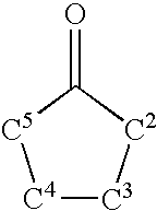


Priority Applications (2)
| Application Number | Priority Date | Filing Date | Title |
|---|---|---|---|
| US10/300,818 US6780890B2 (en) | 2000-05-10 | 2002-11-19 | Compounds and methods for regulating bacterial growth and pathogenesis |
| US10/802,425 US7666619B2 (en) | 2000-05-10 | 2004-03-17 | Compounds and methods for regulating bacterial growth and pathogenesis |
Applications Claiming Priority (5)
| Application Number | Priority Date | Filing Date | Title |
|---|---|---|---|
| US20299900P | 2000-05-10 | 2000-05-10 | |
| US20300000P | 2000-05-10 | 2000-05-10 | |
| US25439800P | 2000-12-07 | 2000-12-07 | |
| US09/853,832 US6559176B1 (en) | 2000-05-10 | 2001-05-10 | Compounds and methods for regulating bacterial growth and pathogenesis |
| US10/300,818 US6780890B2 (en) | 2000-05-10 | 2002-11-19 | Compounds and methods for regulating bacterial growth and pathogenesis |
Related Parent Applications (1)
| Application Number | Title | Priority Date | Filing Date |
|---|---|---|---|
| US09/853,832 Division US6559176B1 (en) | 2000-05-10 | 2001-05-10 | Compounds and methods for regulating bacterial growth and pathogenesis |
Related Child Applications (2)
| Application Number | Title | Priority Date | Filing Date |
|---|---|---|---|
| US10/802,425 Division US7666619B2 (en) | 2000-05-10 | 2004-03-17 | Compounds and methods for regulating bacterial growth and pathogenesis |
| US10/802,425 Continuation US7666619B2 (en) | 2000-05-10 | 2004-03-17 | Compounds and methods for regulating bacterial growth and pathogenesis |
Publications (2)
| Publication Number | Publication Date |
|---|---|
| US20040097402A1 true US20040097402A1 (en) | 2004-05-20 |
| US6780890B2 US6780890B2 (en) | 2004-08-24 |
Family
ID=26898214
Family Applications (3)
| Application Number | Title | Priority Date | Filing Date |
|---|---|---|---|
| US09/853,832 Expired - Fee Related US6559176B1 (en) | 2000-05-10 | 2001-05-10 | Compounds and methods for regulating bacterial growth and pathogenesis |
| US10/300,818 Expired - Fee Related US6780890B2 (en) | 2000-05-10 | 2002-11-19 | Compounds and methods for regulating bacterial growth and pathogenesis |
| US10/802,425 Expired - Fee Related US7666619B2 (en) | 2000-05-10 | 2004-03-17 | Compounds and methods for regulating bacterial growth and pathogenesis |
Family Applications Before (1)
| Application Number | Title | Priority Date | Filing Date |
|---|---|---|---|
| US09/853,832 Expired - Fee Related US6559176B1 (en) | 2000-05-10 | 2001-05-10 | Compounds and methods for regulating bacterial growth and pathogenesis |
Family Applications After (1)
| Application Number | Title | Priority Date | Filing Date |
|---|---|---|---|
| US10/802,425 Expired - Fee Related US7666619B2 (en) | 2000-05-10 | 2004-03-17 | Compounds and methods for regulating bacterial growth and pathogenesis |
Country Status (5)
| Country | Link |
|---|---|
| US (3) | US6559176B1 (en) |
| EP (1) | EP1282415A2 (en) |
| JP (1) | JP2003532698A (en) |
| AU (1) | AU2001259734A1 (en) |
| WO (1) | WO2001085664A2 (en) |
Cited By (6)
| Publication number | Priority date | Publication date | Assignee | Title |
|---|---|---|---|---|
| US20060189910A1 (en) * | 2001-02-16 | 2006-08-24 | Kci Licensing, Inc. | Biocompatible wound dressing |
| US20070059414A1 (en) * | 2005-09-13 | 2007-03-15 | Novak John S | Method and Process of Using Controlled Gas Environments to Inhibit Microbial Growth |
| US7326542B2 (en) * | 1998-12-02 | 2008-02-05 | Princeton University | Compositions and methods for regulating bacterial pathogenesis |
| US7700819B2 (en) | 2001-02-16 | 2010-04-20 | Kci Licensing, Inc. | Biocompatible wound dressing |
| US20100291566A1 (en) * | 2007-09-10 | 2010-11-18 | Sylvia Daunert | Systems and methods for diagnosis and monitoring of bacteria-related conditions |
| US10653140B2 (en) * | 2007-05-14 | 2020-05-19 | The Research Foundation For The State University Of New York | Induction of a physiological dispersion response in bacterial cells in a biofilm |
Families Citing this family (77)
| Publication number | Priority date | Publication date | Assignee | Title |
|---|---|---|---|---|
| US6720415B2 (en) | 1998-12-02 | 2004-04-13 | Princeton University | Compositions and methods for regulating bacterial pathogenesis |
| US6656703B1 (en) * | 1999-12-29 | 2003-12-02 | Millennium Pharamaceuticals, Inc. | High throughput screen for inhibitors of fatty acid biosynthesis in bacteria |
| US9522217B2 (en) | 2000-03-15 | 2016-12-20 | Orbusneich Medical, Inc. | Medical device with coating for capturing genetically-altered cells and methods for using same |
| US8088060B2 (en) | 2000-03-15 | 2012-01-03 | Orbusneich Medical, Inc. | Progenitor endothelial cell capturing with a drug eluting implantable medical device |
| US20030023032A1 (en) * | 2000-05-10 | 2003-01-30 | Bassler Bonnie L. | LuxO-sigma54 interactions and methods of use |
| US6559176B1 (en) | 2000-05-10 | 2003-05-06 | Princeton University | Compounds and methods for regulating bacterial growth and pathogenesis |
| US20030036091A1 (en) * | 2000-10-03 | 2003-02-20 | Lewis Hal A. | Crystals and structure of LuxS |
| US6528472B2 (en) | 2000-11-17 | 2003-03-04 | S.C. Johnson & Son, Inc. | Antimicrobial compositions containing quaternary ammonium compounds, silanes and other disinfectants with furanones |
| US7597895B2 (en) * | 2001-02-20 | 2009-10-06 | The Governing Council Of The University Of Toronto | Signal peptides, nucleic acid molecules and methods for treatment of caries |
| US20030113742A1 (en) * | 2001-04-20 | 2003-06-19 | University Of Iowa Research Foundation | Methods and compositions for the modulation of biofilm formation |
| AU2002322010A1 (en) * | 2001-05-21 | 2002-12-03 | Quorex Pharmaceuticals, Inc. | Methods for regulating bacteria |
| US7575889B2 (en) * | 2001-05-22 | 2009-08-18 | President And Fellows Of Harvard College | Compound combinations for inhibiting cell division and methods for their identification and use |
| AUPR575401A0 (en) * | 2001-06-18 | 2001-07-12 | Unisearch Limited | Method of causing sloughing |
| WO2003039549A2 (en) | 2001-11-07 | 2003-05-15 | 4Sc Ag | Selective antibacterial agents |
| TW200400263A (en) * | 2002-03-13 | 2004-01-01 | Takara Bio Inc | Effect of treatment with 4,5-dihydroxy-2-cyclopenten-1-one (DHCP) on gene expression and quorum-sensing in bacteria |
| DE10219031A1 (en) * | 2002-04-29 | 2003-11-20 | Bayer Ag | Detecting biologically active compounds, useful for detecting toxins and in drug screening, based on their effect on luminescent bacteria |
| DE50305726D1 (en) | 2002-04-29 | 2007-01-04 | Bayer Innovation Gmbh | Method and device for detecting biologically active substances |
| US7427408B2 (en) * | 2002-06-06 | 2008-09-23 | The Regents Of The University Of California | Quorum sensing and biofilm formation |
| US7498292B2 (en) * | 2002-08-15 | 2009-03-03 | The Research Foundation Of State University Of New York | Combinatorial libraries of autoinducer analogs, autoinducer agonists and antagonists, and methods of use thereof |
| US7488757B2 (en) * | 2003-03-24 | 2009-02-10 | Becton, Dickinson And Company | Invisible antimicrobial glove and hand antiseptic |
| ES2319197T3 (en) * | 2003-09-02 | 2009-05-05 | Yissum Research Development Company Of The Hebrew University Of Jerusalem | OXAZABOROLIDINS AS BACTERIAL EFFECTS. |
| GB0326192D0 (en) * | 2003-11-10 | 2003-12-17 | Univ Kent Canterbury | Proteins involved in signal transduction |
| GB0326194D0 (en) * | 2003-11-10 | 2003-12-17 | Univ Kent Canterbury | Proteins involved in signal transduction |
| WO2005079210A2 (en) * | 2003-12-04 | 2005-09-01 | Biofilms Strategies, Inc. | Methods and compositions for preventing biofilm formations, reducing existing biofilms, and for reducing existing biofilms, and for reducing populations of bacteria |
| DE10361457A1 (en) | 2003-12-23 | 2005-07-28 | Henkel Kgaa | New alkoxylactones, alkoxylactams and alkoxythiolactams |
| US20050187190A1 (en) * | 2004-01-23 | 2005-08-25 | Sanjay Vasu | Autoinducer-2 compounds as immunomodulatory agents |
| US7875598B2 (en) | 2004-03-04 | 2011-01-25 | The Regents Of The University Of California | Compositions useful for the treatment of microbial infections |
| US20060229259A1 (en) * | 2004-04-12 | 2006-10-12 | Miller Stephen T | AI-2 compounds and analogs based on Salmonella typhimurium LsrB structure |
| US7405050B2 (en) * | 2004-04-12 | 2008-07-29 | The Trustees Of Princeton University | Small RNAs and bacterial strains involved in quorum sensing |
| JP4806804B2 (en) * | 2004-08-17 | 2011-11-02 | 株式会社片山化学工業研究所 | Method for preventing microbial damage in sterilization target systems |
| WO2006073514A2 (en) * | 2004-08-25 | 2006-07-13 | Tufts University | Compositions, methods and kits for repressing virulence in gram positive bacteria |
| US20100129349A1 (en) * | 2004-08-26 | 2010-05-27 | Heinrichs David E | Methods of inhibiting staphylobactin-mediated iron uptake in S. aureus |
| US8729013B2 (en) | 2004-08-26 | 2014-05-20 | The University Of Western Ontario | Methods of inhibiting staphylobactin-mediated iron uptake in S. aureus |
| CA2596480A1 (en) * | 2005-02-04 | 2006-08-10 | Wisconsin Alumni Research Foundation | Compounds and methods for modulating communication and virulence in quorum sensing bacteria |
| US7312049B2 (en) * | 2005-06-14 | 2007-12-25 | The Board Of Trustees Of The Leland Stanford Junior University | Total amino acid stabilization during cell-free protein synthesis |
| US20100311770A1 (en) * | 2005-06-16 | 2010-12-09 | President And Fellows Of Harvard College | Compound combinations for inhibiting cell division and methods for their identification and use |
| US20090215079A1 (en) * | 2005-12-05 | 2009-08-27 | Technische Universitat Dresden | Method and Devices for the Detection of Microorganisms and/or the Activity Thereof |
| US8512294B2 (en) * | 2006-07-28 | 2013-08-20 | Becton, Dickinson And Company | Vascular access device antimicrobial materials and solutions |
| CA2957056A1 (en) | 2006-09-06 | 2008-03-13 | C3 Jian, Inc. | Selectively targeted antimicrobial peptides and the use thereof |
| WO2008076938A1 (en) * | 2006-12-14 | 2008-06-26 | University Of Iowa Research Foundation | Method of making cyclic polypeptides with inteins |
| WO2008088771A2 (en) * | 2007-01-12 | 2008-07-24 | Cornell Research Foundation, Inc. | Adenylyl cyclases as novel targets for the treatment of infection by eukaryotic pathogens |
| US9017681B2 (en) * | 2007-01-12 | 2015-04-28 | Cornell Research Foundation, Inc. | Adenylyl cyclases as novel targets for antibactrial interventions |
| WO2008116029A1 (en) * | 2007-03-19 | 2008-09-25 | Wisconsin Alumni Research Foundation | Modulation of bacterial quorum sensing with synthetic ligands |
| EP2529793B1 (en) * | 2007-06-08 | 2016-08-10 | Georgia State University Research Foundation, Inc. | Compositions for regulating or modulating quorum sensing in bacteria, methods of using the compounds, and methods of regulating or modulating quorum sensing in bacteria |
| WO2009088402A2 (en) * | 2007-10-01 | 2009-07-16 | Princeton University | Identification of bacterial autoinducer and use in treating bacterial pathogenicity |
| EP2208986B1 (en) * | 2007-10-17 | 2012-12-12 | Kao Corporation | Method of quantifying autoinducer-2 |
| EP2842565B1 (en) * | 2007-10-25 | 2016-08-24 | The Scripps Research Institute | Antibody-mediated disruption of quorum sensing in bacteria |
| EP2100602A1 (en) * | 2008-03-12 | 2009-09-16 | QuoNova Europe GmbH | Method and compositions suitable for treatment of wounds |
| KR100868574B1 (en) | 2008-03-17 | 2008-11-13 | 한국광해관리공단 | Assessing biofilm formation onto natural soil particles |
| US8568756B2 (en) * | 2008-06-02 | 2013-10-29 | The Trustees Of Princeton University | Inhibition of quorum sensing-mediated processes in bacteria |
| US8445190B2 (en) * | 2008-09-30 | 2013-05-21 | The Trustees Of The University Of Pennsylvania | Method of inhibiting biomaterial-induced procoagulant activity using complement inhibitors |
| GB0818547D0 (en) * | 2008-10-09 | 2008-11-19 | Uni I Oslo | Antimicrobial compositions and uses |
| WO2011005587A1 (en) * | 2009-06-24 | 2011-01-13 | University Of Dubuque | Vaccine compositions and methods of use to protect against infectious disease |
| US8624063B2 (en) | 2009-06-30 | 2014-01-07 | Wisconsin Alumni Research Foundation | Non-lactone carbocyclic and heterocyclic antagonists and agonists of bacterial quorum sensing |
| US8821455B2 (en) * | 2009-07-09 | 2014-09-02 | Becton, Dickinson And Company | Antimicrobial coating for dermally invasive devices |
| EP2459149A2 (en) * | 2009-07-28 | 2012-06-06 | QSlant Limited | Antibacterial combinations comprising a garlic extrtact or s-allyl cysteine |
| US20110065798A1 (en) * | 2009-09-17 | 2011-03-17 | Becton, Dickinson And Company | Anti-infective lubricant for medical devices and methods for preparing the same |
| JP2012097033A (en) * | 2010-11-02 | 2012-05-24 | Kao Corp | Autoinducer-2 inhibitor |
| JP5946608B2 (en) * | 2011-05-26 | 2016-07-06 | 花王株式会社 | Autoinducer-2 inhibitor |
| KR101982894B1 (en) | 2011-07-26 | 2019-05-27 | 교토후고리츠다이가쿠호진 | Pathogenic factor production-inhibiting fiber and method for producing same |
| US9750928B2 (en) | 2013-02-13 | 2017-09-05 | Becton, Dickinson And Company | Blood control IV catheter with stationary septum activator |
| US9695323B2 (en) | 2013-02-13 | 2017-07-04 | Becton, Dickinson And Company | UV curable solventless antimicrobial compositions |
| US9327095B2 (en) | 2013-03-11 | 2016-05-03 | Becton, Dickinson And Company | Blood control catheter with antimicrobial needle lube |
| US9750927B2 (en) | 2013-03-11 | 2017-09-05 | Becton, Dickinson And Company | Blood control catheter with antimicrobial needle lube |
| JP5827704B2 (en) * | 2014-01-24 | 2015-12-02 | 花王株式会社 | Periodontal disease marker |
| US10376686B2 (en) | 2014-04-23 | 2019-08-13 | Becton, Dickinson And Company | Antimicrobial caps for medical connectors |
| US9789279B2 (en) | 2014-04-23 | 2017-10-17 | Becton, Dickinson And Company | Antimicrobial obturator for use with vascular access devices |
| US9675793B2 (en) | 2014-04-23 | 2017-06-13 | Becton, Dickinson And Company | Catheter tubing with extraluminal antimicrobial coating |
| US10232088B2 (en) | 2014-07-08 | 2019-03-19 | Becton, Dickinson And Company | Antimicrobial coating forming kink resistant feature on a vascular access device |
| GB201511129D0 (en) | 2015-06-24 | 2015-08-05 | Linea Ab Q | Method of determining antimicrobial susceptibility of a microorganism |
| CN105017209B (en) * | 2015-07-07 | 2017-03-29 | 华东师范大学 | A kind of 3 ketone compounds of polysubstituted dihydro-thiophene and its synthetic method |
| US10493244B2 (en) | 2015-10-28 | 2019-12-03 | Becton, Dickinson And Company | Extension tubing strain relief |
| US20190071706A1 (en) * | 2016-04-04 | 2019-03-07 | University Of Virginia Patent Foundation | Compositions and methods for preventing and treating disease |
| NO342374B1 (en) * | 2016-07-08 | 2018-05-14 | Inhibio As | Compounds and compositions for biofilm prevention |
| US10526278B2 (en) | 2017-10-19 | 2020-01-07 | Wisconsin Alumni Research Foundation | Inhibitors of quorum sensing receptor LasR |
| WO2019237083A1 (en) * | 2018-06-08 | 2019-12-12 | The Regents Of The University Of California | Multistrain population control systems and methods |
| AU2022359222A1 (en) | 2021-10-09 | 2024-03-21 | Joint Stock Company "Pharmasyntez" | A new class of antiviral drugs |
Citations (1)
| Publication number | Priority date | Publication date | Assignee | Title |
|---|---|---|---|---|
| US6559176B1 (en) * | 2000-05-10 | 2003-05-06 | Princeton University | Compounds and methods for regulating bacterial growth and pathogenesis |
Family Cites Families (36)
| Publication number | Priority date | Publication date | Assignee | Title |
|---|---|---|---|---|
| CS179567B1 (en) | 1974-11-25 | 1977-11-30 | Vladimir Stoy | Ionogennic hydrophilic in water insoluble gels based on partial saponificated polymers or copolymers acrylonitrile and method of preparing them |
| GB8414273D0 (en) | 1984-06-05 | 1984-07-11 | Oxoid Ltd | Identifying streptococcal grouping |
| US4861709A (en) | 1985-05-31 | 1989-08-29 | Technicon Research A.G. | Detection and/or identification of microorganisms in a test sample using bioluminescence or other exogenous genetically-introduced marker |
| US4917686A (en) | 1985-12-16 | 1990-04-17 | Colorado Biomedical, Inc. | Antimicrobial device and method |
| US4895566A (en) | 1986-07-25 | 1990-01-23 | C. R. Bard, Inc. | Coating medical devices with cationic antibiotics |
| US4846844A (en) | 1987-08-31 | 1989-07-11 | Eli Lilly And Company | Antimicrobial coated implants |
| US5013306A (en) | 1989-01-18 | 1991-05-07 | Becton, Dickinson And Company | Anti-infective and antithrombogenic medical articles and method for their preparation |
| US4891709A (en) * | 1989-03-31 | 1990-01-02 | Eastman Kodak Company | Flexible formatting interface for pictorial data transfer |
| JPH03202609A (en) | 1989-12-28 | 1991-09-04 | Nissan Motor Co Ltd | Engine exhaust emission control device |
| US5103306A (en) | 1990-03-28 | 1992-04-07 | Transitions Research Corporation | Digital image compression employing a resolution gradient |
| US5196318A (en) | 1990-06-26 | 1993-03-23 | The Texas A&M University System | Precisely regulated expression of deleterious genes |
| US6017722A (en) | 1991-04-04 | 2000-01-25 | Board Of Regents, The University Of Texas System | Luminous bacteria and methods for the isolation, identification and quantitation of toxicants |
| GB9108307D0 (en) | 1991-04-18 | 1991-06-05 | Univ Nottingham | Autoinducer |
| CA2114103A1 (en) | 1991-07-30 | 1993-02-18 | Reinhardt Arthur Rosson | Device for detecting aqueous contaminants |
| US5591872A (en) * | 1993-08-09 | 1997-01-07 | The University Of Iowa Research Foundation | Autoinducer molecule |
| FI96697C (en) | 1993-08-18 | 2001-05-16 | Valio Oy | New Streptococcus thermophilus strains and their use for antibiotic testing |
| US5788979A (en) | 1994-07-22 | 1998-08-04 | Inflow Dynamics Inc. | Biodegradable coating with inhibitory properties for application to biocompatible materials |
| US5637113A (en) | 1994-12-13 | 1997-06-10 | Advanced Cardiovascular Systems, Inc. | Polymer film for wrapping a stent structure |
| US5876791A (en) | 1995-02-01 | 1999-03-02 | Cal-West Equipment Co., Inc. | Protective coating compositions and methods of use thereof |
| US5624704A (en) | 1995-04-24 | 1997-04-29 | Baylor College Of Medicine | Antimicrobial impregnated catheters and other medical implants and method for impregnating catheters and other medical implants with an antimicrobial agent |
| US6020121A (en) | 1995-09-29 | 2000-02-01 | Microcide Pharmaceuticals, Inc. | Inhibitors of regulatory pathways |
| US5759798A (en) * | 1995-12-08 | 1998-06-02 | Woods Hole Oceanographic Institution | Assays, test kits and bacteria for detection of autoinducers |
| US5925552A (en) | 1996-04-25 | 1999-07-20 | Medtronic, Inc. | Method for attachment of biomolecules to medical devices surfaces |
| CA2263563C (en) | 1996-09-27 | 2003-12-30 | Takara Shuzo Co., Ltd. | Cyclopentenones, process for preparing the same, and the use thereof |
| CN1156432C (en) | 1997-03-11 | 2004-07-07 | 宝酒造株式会社 | Cyclopentenone derivatives |
| WO1998058075A2 (en) | 1997-06-18 | 1998-12-23 | The Research And Development Institute, Inc. | Homoserine lactones biofilm regulating compounds and their methods of use |
| KR100547582B1 (en) | 1997-06-30 | 2006-02-01 | 다카라 바이오 가부시키가이샤 | Cyclopentenone derivatives |
| AU732113B2 (en) | 1997-07-02 | 2001-04-12 | Takara Bio Inc. | Antiallergic agents |
| WO1999029647A1 (en) | 1997-12-11 | 1999-06-17 | Takara Shuzo Co., Ltd. | Apoptosis inducer |
| HU227920B1 (en) | 1998-03-19 | 2012-06-28 | Vertex Pharma | Inhibitors of caspases, pharmaceutical compositions comprising thereof and their use |
| GB9814648D0 (en) * | 1998-07-07 | 1998-09-02 | Quest Int | Sub-lethal perfumes |
| CN1225472C (en) | 1998-08-19 | 2005-11-02 | 宝生物工程株式会社 | 5-membered ring compounds |
| US6197591B1 (en) | 1998-09-14 | 2001-03-06 | Pfizer Inc. | Streptomyces avermitilis regulatory genes for increased avermectin production |
| US6720415B2 (en) * | 1998-12-02 | 2004-04-13 | Princeton University | Compositions and methods for regulating bacterial pathogenesis |
| WO2000032152A2 (en) | 1998-12-02 | 2000-06-08 | Princeton University | Compositions and methods for regulating bacterial pathogenesis |
| US7326542B2 (en) * | 1998-12-02 | 2008-02-05 | Princeton University | Compositions and methods for regulating bacterial pathogenesis |
-
2001
- 2001-05-10 US US09/853,832 patent/US6559176B1/en not_active Expired - Fee Related
- 2001-05-10 EP EP01933298A patent/EP1282415A2/en not_active Withdrawn
- 2001-05-10 AU AU2001259734A patent/AU2001259734A1/en not_active Abandoned
- 2001-05-10 WO PCT/US2001/015221 patent/WO2001085664A2/en active Application Filing
- 2001-05-10 JP JP2001582266A patent/JP2003532698A/en active Pending
-
2002
- 2002-11-19 US US10/300,818 patent/US6780890B2/en not_active Expired - Fee Related
-
2004
- 2004-03-17 US US10/802,425 patent/US7666619B2/en not_active Expired - Fee Related
Patent Citations (1)
| Publication number | Priority date | Publication date | Assignee | Title |
|---|---|---|---|---|
| US6559176B1 (en) * | 2000-05-10 | 2003-05-06 | Princeton University | Compounds and methods for regulating bacterial growth and pathogenesis |
Cited By (12)
| Publication number | Priority date | Publication date | Assignee | Title |
|---|---|---|---|---|
| US7326542B2 (en) * | 1998-12-02 | 2008-02-05 | Princeton University | Compositions and methods for regulating bacterial pathogenesis |
| US20060189910A1 (en) * | 2001-02-16 | 2006-08-24 | Kci Licensing, Inc. | Biocompatible wound dressing |
| US7700819B2 (en) | 2001-02-16 | 2010-04-20 | Kci Licensing, Inc. | Biocompatible wound dressing |
| US7763769B2 (en) | 2001-02-16 | 2010-07-27 | Kci Licensing, Inc. | Biocompatible wound dressing |
| US8084664B2 (en) | 2001-02-16 | 2011-12-27 | Kci Licensing, Inc. | Biocompatible wound dressing |
| US8163974B2 (en) | 2001-02-16 | 2012-04-24 | Kci Licensing, Inc. | Biocompatible wound dressing |
| US8735644B2 (en) | 2001-02-16 | 2014-05-27 | Kci Licensing, Inc. | Biocompatible wound dressing |
| US20070059414A1 (en) * | 2005-09-13 | 2007-03-15 | Novak John S | Method and Process of Using Controlled Gas Environments to Inhibit Microbial Growth |
| US10653140B2 (en) * | 2007-05-14 | 2020-05-19 | The Research Foundation For The State University Of New York | Induction of a physiological dispersion response in bacterial cells in a biofilm |
| US11452291B2 (en) | 2007-05-14 | 2022-09-27 | The Research Foundation for the State University | Induction of a physiological dispersion response in bacterial cells in a biofilm |
| US20100291566A1 (en) * | 2007-09-10 | 2010-11-18 | Sylvia Daunert | Systems and methods for diagnosis and monitoring of bacteria-related conditions |
| US8663927B2 (en) | 2007-09-10 | 2014-03-04 | University Of Kentucky Research Foundation | Systems and methods for diagnosis and monitoring of bacteria-related conditions |
Also Published As
| Publication number | Publication date |
|---|---|
| EP1282415A2 (en) | 2003-02-12 |
| WO2001085664A8 (en) | 2003-11-20 |
| AU2001259734A1 (en) | 2001-11-20 |
| US7666619B2 (en) | 2010-02-23 |
| US20040180829A1 (en) | 2004-09-16 |
| JP2003532698A (en) | 2003-11-05 |
| US6559176B1 (en) | 2003-05-06 |
| WO2001085664A2 (en) | 2001-11-15 |
| WO2001085664A3 (en) | 2002-08-08 |
| US6780890B2 (en) | 2004-08-24 |
Similar Documents
| Publication | Publication Date | Title |
|---|---|---|
| US6559176B1 (en) | Compounds and methods for regulating bacterial growth and pathogenesis | |
| US6720415B2 (en) | Compositions and methods for regulating bacterial pathogenesis | |
| US7427408B2 (en) | Quorum sensing and biofilm formation | |
| US7326542B2 (en) | Compositions and methods for regulating bacterial pathogenesis | |
| CA2573440A1 (en) | Inhibition of biofilm formation | |
| AU1933800A (en) | Compositions and methods for regulating bacterial pathogenesis | |
| US9415040B2 (en) | Inhibition of quorum sensing-mediated processes in bacteria | |
| US20060269951A1 (en) | Inhibitors of autoinducer transporters | |
| US8063108B2 (en) | Method for modulating microbial quorum sensing | |
| WO2002015896A2 (en) | Methods and compositions for potentiating antibiotic action against persistent/tolerant pathogenic microorganisms | |
| Zlatkov et al. | Unconventional cyclic di-GMP signaling in Escherichia coli | |
| MXPA01005448A (en) | Compositions and methods for regulating bacterial pathogenesis | |
| Wood | Light sensing in a human pathogen: genetic, biochemical, functional and proteomics analyses of blue light regulation in Acinetobacter baumannii | |
| Foley | Population dynamics in Enterococcal biofilms: resistance plasmids and antibiotic susceptibility | |
| Raterman | Motility and Signaling Systems in Uropathogenic Escherichia coli: the Role of Chemotaxis and c-di-GMP Production in Colonization of the Urinary Tract | |
| Hoang | Studies on fatty acid and acylated homoserine lactone biosynthesis in Pseudomonas aeruginosa | |
| CA2130025A1 (en) | Method of treating fungal infections |
Legal Events
| Date | Code | Title | Description |
|---|---|---|---|
| AS | Assignment |
Owner name: PRINCETON UNIVERSITY, NEW JERSEY Free format text: ASSIGNMENT OF ASSIGNORS INTEREST;ASSIGNORS:BASSLER, BONNIE L.;SCHAUDER, STEPHAN;REEL/FRAME:014371/0451 Effective date: 20010821 Owner name: QUOREX PHARMACEUTICALS, INC., CALIFORNIA Free format text: ASSIGNMENT OF ASSIGNORS INTEREST;ASSIGNORS:DAMMEL, CAROL;SHOKAT, KEVAN;STEIN, JEFFREY;REEL/FRAME:014371/0326;SIGNING DATES FROM 20010821 TO 20011023 Owner name: UNIVERSITY TECHNOLOGIES INTERNATIONAL, INC., CANAD Free format text: ASSIGNMENT OF ASSIGNORS INTEREST;ASSIGNOR:SURETTE, MICHAEL G.;REEL/FRAME:014371/0298 Effective date: 20010824 |
|
| FPAY | Fee payment |
Year of fee payment: 4 |
|
| REMI | Maintenance fee reminder mailed | ||
| LAPS | Lapse for failure to pay maintenance fees | ||
| STCH | Information on status: patent discontinuation |
Free format text: PATENT EXPIRED DUE TO NONPAYMENT OF MAINTENANCE FEES UNDER 37 CFR 1.362 |
|
| FP | Lapsed due to failure to pay maintenance fee |
Effective date: 20120824 |
|
| AS | Assignment |
Owner name: NATIONAL SCIENCE FOUNDATION, VIRGINIA Free format text: CONFIRMATORY LICENSE;ASSIGNOR:PRINCETON UNIVERSITY;REEL/FRAME:040078/0103 Effective date: 20161020 |