US12300357B2 - Applied computer technology for management, synthesis, visualization, and exploration of parameters in large multi-parameter data sets - Google Patents
Applied computer technology for management, synthesis, visualization, and exploration of parameters in large multi-parameter data sets Download PDFInfo
- Publication number
- US12300357B2 US12300357B2 US15/838,724 US201715838724A US12300357B2 US 12300357 B2 US12300357 B2 US 12300357B2 US 201715838724 A US201715838724 A US 201715838724A US 12300357 B2 US12300357 B2 US 12300357B2
- Authority
- US
- United States
- Prior art keywords
- axis
- scatterplot
- parameter
- cell
- gene
- Prior art date
- Legal status (The legal status is an assumption and is not a legal conclusion. Google has not performed a legal analysis and makes no representation as to the accuracy of the status listed.)
- Active, expires
Links
Images


Classifications
-
- G—PHYSICS
- G16—INFORMATION AND COMMUNICATION TECHNOLOGY [ICT] SPECIALLY ADAPTED FOR SPECIFIC APPLICATION FIELDS
- G16B—BIOINFORMATICS, i.e. INFORMATION AND COMMUNICATION TECHNOLOGY [ICT] SPECIALLY ADAPTED FOR GENETIC OR PROTEIN-RELATED DATA PROCESSING IN COMPUTATIONAL MOLECULAR BIOLOGY
- G16B45/00—ICT specially adapted for bioinformatics-related data visualisation, e.g. displaying of maps or networks
-
- C—CHEMISTRY; METALLURGY
- C12—BIOCHEMISTRY; BEER; SPIRITS; WINE; VINEGAR; MICROBIOLOGY; ENZYMOLOGY; MUTATION OR GENETIC ENGINEERING
- C12N—MICROORGANISMS OR ENZYMES; COMPOSITIONS THEREOF; PROPAGATING, PRESERVING, OR MAINTAINING MICROORGANISMS; MUTATION OR GENETIC ENGINEERING; CULTURE MEDIA
- C12N15/00—Mutation or genetic engineering; DNA or RNA concerning genetic engineering, vectors, e.g. plasmids, or their isolation, preparation or purification; Use of hosts therefor
- C12N15/09—Recombinant DNA-technology
- C12N15/10—Processes for the isolation, preparation or purification of DNA or RNA
- C12N15/1034—Isolating an individual clone by screening libraries
- C12N15/1089—Design, preparation, screening or analysis of libraries using computer algorithms
-
- G—PHYSICS
- G01—MEASURING; TESTING
- G01N—INVESTIGATING OR ANALYSING MATERIALS BY DETERMINING THEIR CHEMICAL OR PHYSICAL PROPERTIES
- G01N15/00—Investigating characteristics of particles; Investigating permeability, pore-volume or surface-area of porous materials
- G01N15/10—Investigating individual particles
-
- G—PHYSICS
- G01—MEASURING; TESTING
- G01N—INVESTIGATING OR ANALYSING MATERIALS BY DETERMINING THEIR CHEMICAL OR PHYSICAL PROPERTIES
- G01N33/00—Investigating or analysing materials by specific methods not covered by groups G01N1/00 - G01N31/00
- G01N33/48—Biological material, e.g. blood, urine; Haemocytometers
-
- G—PHYSICS
- G16—INFORMATION AND COMMUNICATION TECHNOLOGY [ICT] SPECIALLY ADAPTED FOR SPECIFIC APPLICATION FIELDS
- G16B—BIOINFORMATICS, i.e. INFORMATION AND COMMUNICATION TECHNOLOGY [ICT] SPECIALLY ADAPTED FOR GENETIC OR PROTEIN-RELATED DATA PROCESSING IN COMPUTATIONAL MOLECULAR BIOLOGY
- G16B25/00—ICT specially adapted for hybridisation; ICT specially adapted for gene or protein expression
-
- G—PHYSICS
- G16—INFORMATION AND COMMUNICATION TECHNOLOGY [ICT] SPECIALLY ADAPTED FOR SPECIFIC APPLICATION FIELDS
- G16B—BIOINFORMATICS, i.e. INFORMATION AND COMMUNICATION TECHNOLOGY [ICT] SPECIALLY ADAPTED FOR GENETIC OR PROTEIN-RELATED DATA PROCESSING IN COMPUTATIONAL MOLECULAR BIOLOGY
- G16B50/00—ICT programming tools or database systems specially adapted for bioinformatics
- G16B50/10—Ontologies; Annotations
-
- G—PHYSICS
- G16—INFORMATION AND COMMUNICATION TECHNOLOGY [ICT] SPECIALLY ADAPTED FOR SPECIFIC APPLICATION FIELDS
- G16B—BIOINFORMATICS, i.e. INFORMATION AND COMMUNICATION TECHNOLOGY [ICT] SPECIALLY ADAPTED FOR GENETIC OR PROTEIN-RELATED DATA PROCESSING IN COMPUTATIONAL MOLECULAR BIOLOGY
- G16B50/00—ICT programming tools or database systems specially adapted for bioinformatics
- G16B50/30—Data warehousing; Computing architectures
-
- G—PHYSICS
- G01—MEASURING; TESTING
- G01N—INVESTIGATING OR ANALYSING MATERIALS BY DETERMINING THEIR CHEMICAL OR PHYSICAL PROPERTIES
- G01N15/00—Investigating characteristics of particles; Investigating permeability, pore-volume or surface-area of porous materials
- G01N15/10—Investigating individual particles
- G01N2015/1006—Investigating individual particles for cytology
-
- G—PHYSICS
- G01—MEASURING; TESTING
- G01N—INVESTIGATING OR ANALYSING MATERIALS BY DETERMINING THEIR CHEMICAL OR PHYSICAL PROPERTIES
- G01N15/00—Investigating characteristics of particles; Investigating permeability, pore-volume or surface-area of porous materials
- G01N15/10—Investigating individual particles
- G01N15/14—Optical investigation techniques, e.g. flow cytometry
- G01N2015/1402—Data analysis by thresholding or gating operations performed on the acquired signals or stored data
Definitions
- cellular gene expression data can include gene expression data for thousands of genes (e.g., 10,000-30,000 or more genes), can now be measured for individual cells, and thousand cells can be measured per sample. This has presented a tremendous technical problem in the art of visualizing, analyzing, exploring, and making sense of cellular gene expression data.
- the visualization is a final end point and the visualization is reached as a result of a user manually writing scripts using the R programming language, which requires the user to have knowledge of different libraries in order to perform data input, reformatting, manipulations, calculations and graphing.
- These scripts usually have to be customized for particular data sets, and their creation requires expert knowledge of the programming language, existing libraries, and required inputs to produce results.
- such conventional approaches prevent the deep exploration of heterogeneous cell populations.
- the inventors disclose the application of computer technology using innovative scatterplot displays across various dimensions of cellular expression data including cell (or cell population) view scatterplots in which cells are visualized as individual data points (e.g., gene vs gene scatterplots of cells) and gene view scatterplots in which genes are visualized as individual data points (e.g., cell population vs cell population scatterplots of genes). Gating can be performed within these scatterplots to create cell populations and gene sets respectively that can serve as biologically-relevant dimensions to be added as new data objects in a workspace for use in augmenting the cellular gene expression data and opening new avenues for meaningful investigation.
- the visualization serves as an endpoint in the process and cannot serve as a starting point for further creating further visualization refinements for further investigations.
- samples of immune cells from metastatic melanoma patients may contain T cells, and conventional visualization systems in the art would be able only to identify this subset within the immune cells.
- the innovative computer systems described herein allow for deep exploration and analysis of the T cell subset to identify multiple subsets within T cells, for example, T cells which are “exhausted”, track this state to individual genes which can then be targeted to reverse this exhaustion, activate T cells, and thus possibly stimulate an immune response to eradicate the metastases, as explained in greater detail below with reference to example embodiments.
- example embodiments of the invention provide significant technical advances in the applied bioinformatics arts.
- FIG. 1 discloses an example computer system that can be used to support the innovative data processing and visualization techniques described herein.
- FIG. 2 A depicts examples of cellular gene expression data sets.
- FIG. 2 B depicts a table view of example cellular gene expression data.
- FIG. 3 depicts an example cell view graph window scatterplot.
- FIG. 4 depicts an example process flow for execution to create a cell view scatterplot.
- FIG. 5 shows an example cell view scatterplot user interface that includes parameter selection menus.
- FIG. 6 shows an example of how gating can be performed within a cell view scatterplot user interface to create a cell population.
- FIG. 7 shows an example user interface that permits a user to spread the zeros of a cell view scatterplot.
- FIG. 8 shows an example cell view scatterplot where parameters other than genes are selected as axis parameters.
- FIG. 9 shows an example of how gating can be performed within the cell view scatterplot of FIG. 8 to create a cell population.
- FIG. 10 shows an example user interface for adjusting display settings in a scatterplot presentation.
- FIG. 11 depicts an example gene view graph window scatterplot including cell population selection menus.
- FIG. 12 depicts an example of how cellular gene expression data can be pivoted to create cell population data for a gene view scatterplot.
- FIGS. 13 A and B depict examples of how complementary cell populations can be created and defined in a workspace for use in gene view scatterplots.
- FIG. 14 shows an example of how gating can be performed within a gene view scatterplot user interface to create a gene set.
- FIG. 15 shows an example of how cellular gene expression data can be augmented with gene sets as synthetic parameters.
- FIG. 16 shows an example of a cell view scatterplot where gene sets are presented as options in the parameter selection menus.
- FIGS. 17 A and B show an example user interface for viewing, editing, and creating new gene sets from other gene sets in a workspace.
- FIG. 18 A-D show examples of how a user-selected third dimension can be overlaid on a scatterplot.
- FIG. 19 shows an example of how gating can be performed within the 3D scatterplots of FIGS. 18 A-D .
- FIGS. 20 A-D show example reports can be created through the system.
- FIG. 21 shows an example process flow for execution by the system to switch between cell view and gene view modes.
- FIG. 22 shows an example of how the system can be operated to switch between cell view and gene view modes to support investigations and research into cell data.
- FIG. 1 discloses an example computer system 100 that can be used to support the innovative data processing and visualization techniques described herein.
- the example computer system 100 comprises a processor 102 , memory 104 , database 106 , and display 108 that can be in communication with each other over an interconnect technology such as bus 110 .
- Processor 102 can take the form of any processor suitable for performing the operations described herein.
- processor 102 may comprise multiple processors, including distributed processors that communicate with each other over a network to carry out tasks described herein (e.g., cloud computing processing resources).
- Memory 104 can take the form of any computer memory suitable for cooperating with processor 102 in the execution of the tasks described herein. It should be understood that memory 104 may take the form of multiple memory devices, including memory that is distributed across a network.
- database 106 can take the form of any data repository accessible to the processor 102 (e.g., a file system on a computer, relational database, etc.), and it should be understood that database 106 may take the form of multiple distributed databases (e.g., cloud storage).
- Display 108 can take the form of a computer monitor or screen that is capable of generating the visualizations described herein.
- Cellular gene expression data 112 can be generated by next-generation sequencing (e.g. for the measurement of RNA-Sequencing (RNASeq) and single cell RNA sequencing (scRNA-Seq) among other sequencing approaches).
- next-generation sequencing e.g. for the measurement of RNA-Sequencing (RNASeq) and single cell RNA sequencing (scRNA-Seq) among other sequencing approaches.
- RNASeq RNA-Sequencing
- scRNA-Seq single cell RNA sequencing
- Additional examples include polymerase chain reaction approaches including digital droplet and reverse transcriptase.
- Still more examples include RNA measurement by flow cytometry, and microarrays, among others, that produce data files which contain the quantification of DNA and/or RNA, or through software programs that process the raw read data (primary and secondary analysis) to generate the gene expression data files.
- gene expression data derived from stochastic labeling of a sample can be found in U.S. Pat. Nos. 9,567,645 and 8,835,358 and in patent application Ser. No. 15/715,028, filed Sep. 25, 2017, and entitled “Measurement of Protein Expression Using Reagents with Barcoded Oligonucleotide Sequences”, the entire disclosures of each of which are incorporated by reference.
- the innovative data analysis and visualization techniques described herein can be applied to data generated from single cells by a variety of means.
- Single cell analysis may include the stochastic labeling of nucleic acids or proteins or any combination of proteins and nucleic acids.
- the innovative data analysis and visualization techniques described herein may be used to analyze quantitative features of protein density or gene expression or any combination thereof.
- the innovative data analysis and visualization techniques described herein can also provide for an improved visualization of quantitative data generated by stochastic labeling of a variety of proteins or nucleic acids in single cells. Quantitative values of biological populations (nucleic acids, proteins etc.) in individual cells may be compared with other individual cells, or between cell types or even between methods of generating the data.
- gene expression values may be visualized as a function of the method used of generating the data set.
- the innovative data analysis and visualization techniques described herein can also be used to compare quantitative data generated by a variety of means described here in ways that provide for the visualization of quantitative biological population data independent of the method of generating such data.
- This data 112 can be characterized as a large multi-parameter data set which poses special technical challenges in terms of difficulty in creating meaningful visualizations, particularly when considered with respect to underlying biology so that biologically-relevant information is meaningfully presented to users in a visual manner.
- the cellular gene expression data may comprise data for large numbers of individual cells and cell populations, with parameters for each cell or cell population that may stretch into 10,000-30,000 or more parameters.
- Cellular gene expression data 112 can be read out of files in database 106 and loaded into memory 104 as a plurality of data structures 116 to be manipulated by processor 102 during execution of an analysis and visualization program 114 .
- Program 114 may comprise processor-executable computer code in the form of a plurality of processor-executable instructions that are resident on a non-transitory computer-readable storage medium such as memory 104 .
- FIG. 2 A depicts examples of cellular gene expression data sets where each cell (or cell population) is identified by a Cell ID and is associated with a plurality of parameters, each parameter having an ID and a value in relation to a Cell ID.
- gene expression data for cells is highly dimensional and the number of parameters for each cell may reach 10,000-30,000 or more parameters, either per cell or per population of cells. Examples of parameters in the cellular data include counts of gene expression in the subject cell for a large number of genes.
- Parameter 1 for Cell 1 may correspond to Gene 1 and its value can be a count of expressions for Gene 1 in Cell 1 .
- Parameter 2 for Cell 1 may correspond to Gene 2 and its value can be a count of expressions for Gene 2 in Cell 1 .
- the cellular gene expression data 112 may include data values for parameters such as t-distributed stochastic neighbor embedding (tSNE), principal component analysis (PCA), linear discriminant analysis (LDA), etc. in each table cell, where these data values represent an analysis calculation whose value captures differences for individual cells across n parameters.
- the cellular gene expression data 112 can be stored in any of a number of formats (e.g., as CSV files, database tables (e.g., as relational data in a relational database), spare data representations, binary formats, and others).
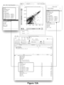
- FIG. 3 depicts an example graph window (GW) that can be produced through execution of program 114 for presentation to a user via display 108 , where the graph window visualizes Gene 1 vs. Gene 2 with respect to a cell population.
- This visualization can be referred to as a cell view of the cellular gene expression data 112 .
- This graph window presents a scatterplot 300 of gene expressions for two user-selected genes 302 and 304 (TMEM216 and MMP2, respectively in this example) with respect to a cell population in a user-selected file 306 .
- Each file may correspond to a single sample of cell populations, a single cell population (e.g.
- Each dot 308 in the scatterplot represents a cell in the cell population of the subject file 306 .
- the scale of the X-axis and Y-axis in this example identify counts for the corresponding gene.
- the position of a dot 308 on the horizontal X-axis identifies a count of how many of the gene MMP2 are present in the cell corresponding to the subject dot 308
- the position of the dot 308 on the vertical Y-axis identifies a count of how many of the gene TMEM216 are present in the cell corresponding to the subject dot 308 .
- dots 308 in the upper right quadrant of the scatterplot 300 correspond to cells where both TMEM216 and MMP2 are highly expressed
- dots in the lower left quadrant of the scatterplot 300 correspond to cells where both TMEM216 and MMP2 are expressed at a low level.
- the upper left quadrant corresponds to cells where TMEM216 is highly expressed but MMP2 is not
- the lower right quadrant corresponds to cells where MMP2 is highly expressed, but TMEM216 is not.
- the distance of a cell 308 away from this diagonal in either direction indicates the extent of differential expression of the selected genes in a given cell. It is expected that for many cell populations, there will be large numbers of cells where the expression of the selected genes for those cells is at the zero level. This leads to a large clustering of dots 308 at the zero levels 310 and 312 on the X-axis and Y-axis respectively. Color coding can be used at 310 and 312 to indicate the density of cells with such zero-level gene expressions.
- FIG. 4 depicts an example process flow for execution as part of program 114 that describes how scatterplot 300 can be generated.
- the processor creates a data structure in a memory workspace (see 116 in FIG. 1 ). This data structure can be used for holding cell view data.
- the processor selects a gene in the cellular gene expression data 112 based on user input (step 402 ). For example, the processor can respond to user input to select a gene column in table 200 .
- FIG. 5 shows how the user interface can present a user with a list of genes for each axis.
- a user can select a gene selector 302 or 304 for the Y and X axes respectively. If we assume the first axis in this example is the Y-axis, upon selection of 302 , a parameter selection menu 500 is shown which presents a list of parameters available for selection with respect to the Y-axis. This list can be populated with parameters from the cellular gene expression data 112 .
- the user selection 502 for the Y-axis gene is TMEM216.
- the processor populates the data structure with a list of cells from the cellular gene expression data 112 (e.g., the cells in the cell column of table 200 ). Each listed cell is associated with its count value for the selected first axis gene.
- the processor selects another gene in the cellular gene expression data 112 based on user input (step 406 ). For example, the processor can respond to user input to select another gene column in table 200 .
- FIG. 5 shows an example of a user interface where 304 is selected to access an X-axis parameter selection menu 506 (resulting in selection 508 of MMP2).
- the processor augments the cell list to add to each cell its associated count value for the selected second axis gene.
- the data structure comprises a list of cells associated with count pairs for the selected genes of the first and second axis; for example the list can comprise a set of vectors ⁇ Cell ID 1 , Gene 1 Count, Gene 2 Count ⁇ for each cell in the cellular gene expression data 112 .
- the process parses the list to find the maximum values for each selected gene (the highest counts). These maximum values are then used by the processor to define the appropriate scales for the X-axis and Y-axis in the scatterplot (step 412 ). For example, if the maximum value for the X-axis gene is 10, the X-axis scale can be from 0-10.
- the processor draws the scatterplot based on the cell list and the defined scales using the cell's associated count values as X,Y coordinates in the scatterplot. The result is a scatterplot 300 as shown by FIG. 3 .
- a user can access gate creation tools 320 in the user interface to create a gate through which child cell populations are created.
- the user can access a tool 320 to draw a shape in the scatterplot 300 that encompasses a subset of the cells 308 .
- FIG. 6 depicts an example gate 600 drawn to capture the cells that have non-zero expressions of the two selected genes.
- the cells 308 falling inside the drawn shape 600 are gated into their own child cell population, and this cell population can be added to the workspace as a distinct object 602 .
- the processor can translate gate 600 into a plurality of boundary conditions for the gated cell population.
- the boundary conditions can be all cells with (1) X-axis values between 1 and 10, and (2) Y-axis values between 1 and 8.
- the cell list data structure e.g., the set of vectors ⁇ Cell ID 1 , Gene 1 Count, Gene 2 Count ⁇
- the cell list data structure can be traversed to find all Cell IDs meeting these criteria, and the data for these cell IDs can populate a new child cell population data structure in the workspace.
- a corresponding scatterplot 300 of gene expressions for the child cell population can then be presented (where the cell population includes one or more cells 308 ). This gating can allow a user to focus on a biologically-interesting cluster of cells 308 in the scatterplot 300 .
- the cell view mode of FIG. 3 permits users to visually compare different samples, including child and parent cell populations using navigation tools 322 .
- a user can navigate to a scatterplot 300 for a next and previous sample in the workspace.
- a user can navigate to a child cell population in an analysis hierarchy (e.g., to the child cell population created via gate 600 of FIG. 6 ).
- an up button (not shown), a user can navigate to a parent cell population in the analysis hierarchy.
- the user interface can include a “spread zeros” user control 700 (such as a check box, button, etc.) as shown in FIG. 7 .
- This control 700 can be provided on a menu for selecting the axis parameters, although if desired by a practitioner, control 700 could be positioned elsewhere such as somewhere on the user interface shown by FIG. 3 .
- the top half of FIG. 7 shows the scatterplot when the zeros are not spread.
- the bottom half of FIG. 7 shows the scatterplot when the spread zeros option is selected via input mechanism 700 .
- box 702 provides an expanded view of the cells that exhibit zero values with respect to the expression of the X-axis gene (MMP2 in this example).
- Box 704 provides an expanded view of the cells that exhibit zero values with respect to the expression of the Y-axis gene (TMEM216 in this example).
- the depth of the zero space along the X and Y axes can be defined as a function of how many dots/cells are at the zero levels for each gene, and the dots/cells can be spread across the zero-space defined by boxes 702 and 704 as a function of the density of cells at each zero location, although other distribution techniques could be used.
- the upper right quadrant of this scatterplot comprises the cells with positive expressions of both genes (effectively, the scatterplot shown in the top part of FIG. 7 ).
- the cell population of the upper right quadrant is much sparser than the other quadrants. The inventors believe that the ability to visualize the zero levels in this spread manner can provide users with biologically relevant information (such as an ability to assess gene combinations where unexpected distributions occur—e.g., where the upper right quadrant is not as sparse as would be generally expected).
- the cell view scatterplot can also display cell information for selected parameters in the cellular gene expression data 112 other than genes.
- the cellular gene expression data 112 may include parameters from dimensionality reduction such as those resulting from tSNE, LDA, PCA, etc. and quality control parameters (above threshold, parameters relating to ribosomal RNA (rRNA) abundance, etc.) These parameters can be presented as options on the parameter selection menus 500 and 506 .
- FIG. 8 shows an example where selection 800 for the Y-axis is the parameter tSNE axis 2 and where selection 02 for the X-axis is the parameter tSNE axis 1 .
- the resultant scatterplot of FIG. 8 can be created via the FIG. 4 process flow.
- a user can also gate desired cell populations in the scatterplot of FIG. 8 (see gate 900 in FIG. 9 ) as explained above with respect to FIG. 6 .
- the cell view user interface of FIG. 3 can also include a user control 330 (e.g., the “T” button of FIG. 3 ) through which the user can alter the display of information in scatterplot 300 .
- T button 330 Upon selection of T button 330 , the user interface of FIG. 10 can be presented.
- the list at right in FIG. 10 shows the available and selected parameters for the X-axis of the scatterplot.
- a histogram 1000 shows the binning of values for the selected parameter with respect to the subject cell population. Given the high prevalence of zeros, histogram 1000 shows a large spike at the zero level, and the scale of this example obscures the non-zero levels in the histogram.
- left/right arrows shown toward the top of FIG. 10 a user can quickly preview displays for different samples.
- +/ ⁇ controls in the middle of FIG. 10 a user can easily alter the histogram zoom level.
- the user can adjust the X-axis of the scatterplot in a variety of ways.
- the X-axis scale can be defined to exhibit a liner scale or some other scale (such as a log 2 scale) (see control 1004 ).
- min/max controls 1006 the user can define the minimum and maximum boundaries on the X-axis.
- the minimum value can be defined to be a value greater than zero, which would remove the zero spike from the histogram and re-present a newly scaled histogram where the distribution of non-zero values across the X-axis can be more clearly seen.
- Sliders 1008 can provide users with easy control over the transformation variables.
- FIG. 11 shows an example of a scatterplot 1100 in the gene view mode.
- a “gene view” visualization refers to a visualization where genes are the individual data points measured against the axis dimensions (e.g., genes as the dots in a scatterplot measured against two cell populations).
- the axis parameters are cell populations (see the X-axis parameter 1102 which identifies a population of B cells, and the Y-axis parameter 1104 which identifies a population of “not B” cells in this example).
- the dots 1106 in scatterplot 1106 represent specific genes (rather than individual cells as in scatterplot 300 of FIG. 3 ).
- Population selection menu 1110 provides a list of cell populations available for selection to be used on the X-axis
- population selection menu 1112 provides a list of cell populations available for selection to be used on the Y-axis.
- selections 1114 and 1116 correspond to populations of B cells and “not B” cells respectively.
- the table 200 can be pivoted such the genes become rows in the table and subsets of the cells are grouped into two cell populations that become the columns of the tables. Furthermore, computations can be performed on the gene counts in table 2 for the cells of the two cell populations to determine the values that will populate the cells of the pivoted table.
- a user can define the computations that are to be performed on the gene data to compute the values for the cells in the pivoted table. In the example of FIG. 11 , a normalized mean computation has been selected.
- the computation performed on the pivoted table data can be any user-defined function that is deemed biologically-relevant to a user.
- the inventors note that it may be desirable for the computation to be based on averaging of some sort to account for potential discrepancies in the cell counts of the two cell populations and/or normalization to account for the variability between samples (e.g. normalization to a spike-in of External RNA Controls Consortium (ERCC) controls which contain a known amount of RNA to be measured).
- ERCC External RNA Controls Consortium
- the pivoted table values were straight counts of each gene in the two cell populations, and if there was a meaningful difference in the count of cells in the two cell populations, the aggregated gene count totals across the two cell populations would not be very informative in a comparative sense. However, if the cell populations were roughly similar in size, straight gene counts for each gene in the two cell populations might nevertheless be informative.
- FIG. 12 depicts an example pivot from a cell view to a gene view.
- Table 200 in FIG. 12 shows gene expressions per cell for a number of cells in a sample. Each row corresponds to a different cell, and the columns correspond to expression counts for different genes in the associated cells. If subsets of these cells are grouped into two cell populations 1210 and 1212 as shown in FIG. 12 (e.g., via gating in the cell view scatterplot 300 ), these two cell populations can be pivoted as shown to create the pivoted table 1200 .
- the rows are the genes that were columns in table 200 .
- the columns in table 1200 are the two cell populations 1210 and 1212 .
- Each table cell is populated with a concatenation of the gene counts for the associated gene in the cells of each cell population.
- the computation being performed on these values is a straight averaging to compute means as shown in pivoted table 1200 a.
- a gene view scatterplot of the type shown by scatterplot 1100 in FIG. 11 can then be created from pivoted table 1200 a using the same basic techniques described in FIG. 4 (albeit using pivoted table 1200 a rather than table 200 ).
- the gene view scatterplot 1100 thus provides users with a powerful manner of visualizing differential gene expression between two cell populations over a list of genes that may be extremely large in size.
- the inventors believe that scatterplot 1100 represents a pioneering new way of visualizing cellular gene expression data in a manner that opens up a wide new array of investigatory options for practitioners, some examples of which are described below.
- the example gene view scatterplot 1100 of FIG. 11 provides users with flexibility in specifying the x/y axis populations and dynamically updating gene expression graphs as populations change.
- the example gene view scatterplot 1100 also provides users with a plethora of graph options and resolutions. Importantly, the analysis does not stop with this graph; it continues with the ability to create new gene sets and dig deeper into the data as discussed below. Examining cellular heterogeneity and differences between samples/patients revealed by particularly new single cell methods is not possible without this approach.
- the distance of a gene 1106 away from this diagonal in either direction indicates the extent of differential expression of the subject gene 1106 as between two selected cell populations. Given that it is expected that most genes 1106 will not be expressed (or will be only lightly expressed) in many cell populations, it is expected that there will typically be large cluster of genes 1106 in the lower left quadrant of scatterplot 1100 .
- FIGS. 13 A and B show examples of such complementary Boolean cell populations in a workspace. Accordingly, a user can create a scatterplot 1100 that shows differential gene expression as between a population of B cells and a population of “not B” cells.
- FIG. 14 shows an example where gate 1400 is created in the gene view scatterplot to capture a set of genes that the user deems worthy of further investigation.
- Gate 1400 can be drawn on the scatterplot using the techniques described above in connection with FIG. 6 . This gating will create a data structure in the workspace corresponding to the gene set defined by gate 1400 (where the gene set comprises one or more genes depending on how many genes 1106 are encompassed by the user-defined gate 1400 ). Such a gene set can then serve as a synthetic parameter in relation to the cellular gene expression data 112 that can be selected for visualization while in the cell view mode.
- gate 1400 is a trapezoidal shape that captures the genes 1106 that have a positive differential expression in B cells versus non-B cells.
- FIG. 15 shows an example of how the cellular gene expression data 112 can be augmented with gene sets as a synthetic parameter (in the form of example augmented table 200 ).
- This table 200 has been augmented with two gene sets as parameters (see the Gene Set 1 and Gene Set 2 columns).
- Gene Set 1 consists of ⁇ Gene 2 , Gene 3 ⁇
- Gene Set 2 consists of ⁇ Gene 1 , Gene 4 ⁇ .
- the table cells for these gene sets (each corresponding to a different cell in the table rows) can then be populated with the sums of the gene counts for the constituent genes of the subject gene sets.
- a user can return to the cell view mode to create a cell view scatterplot where one or both of the X-axis and Y-axis parameters are gene sets.
- FIG. 16 An example of this is shown by FIG. 16 .
- the parameter selection menus for each axis in FIG. 16 include a section that lists available gene set options.
- the cell view scatterplot of FIG. 16 thus shows a scatterplot of cells 308 where the X-axis parameter is the gene set labeled “B_GeneTable” and the Y-axis parameter is the gene set labeled “HighTHighB”.
- the gating capabilities within the gene view scatterplot to create gene sets in combination with the ability use the created gene sets as a synthetic parameter used in a cell view scatterplot provide users with unprecedented capabilities for intelligently reducing the multi-parameter data space of cellular gene expression data.
- This combination allows the user to overcome the extremely difficult problem of identifying common genes in populations and identifying the differential genes in populations. In other words, whether the user is looking for what genes are in common, or which genes are different, the ability to create new gene sets and viewing/analyzing them as a single parameter allows the user to focus on the important relationships between cell populations and gene sets.
- these gene sets can be shared with other users by passing these gene sets (as a defined list of encompassed genes) to other users so that those other uses can evaluate the gene sets with their samples of cell data. Based on such sharing and independent investigation across different cellular data sets, it is expected that greater insights into gene behavior with respect to cells can be gained.
- FIG. 17 A shows an example view into a workspace that breaks the workspace down into various gene sets 1700 , samples 1702 , and cell populations 1704 that exist within the workspace.
- the section 1700 that lists gene sets can include various display fields that provide metadata about each subject gene set (e.g., a name, count of genes within the gene set, and a description of the gene set). The name and description can be editable by a user and will be helpful for informing users about pertinent characteristics of the gene set.
- the gene sets can be listed in a manner that indicates any hierarchical relationships that exist within gene sets.
- Gene set controls 1710 provide a user with an ability to create gene sets from other gene sets via Boolean operations.
- a user via the union button, a user can create a new gene set that is the union of two or more selected gene sets within list 1700 .
- a user Via the intersection button, a user can create a new gene set that consists of the genes that are present in both/all of two or more selected gene sets within list 1700 .
- a user can create a complementary gene set from two gene sets (Gene Set 1 and Gene Set 2 ) within list 1700 where a first complementary new gene set consists of all of the genes that are in Gene Set 1 but not Gene Set 2 and where a second complementary new gene set consists of all of the genes that are in Gene Set 2 but not Gene Set 1 .
- a user Via the “All Comparisons” button, a user can create several new gene sets at the same time via each of the techniques described above (new gene sets via the union operation, the intersection operation, and the complementary operation). This flexible ability to create new gene sets from existing gene sets allows for the comparison of gene sets between treatment conditions, patients, experiments, etc. Accordingly, by using controls 1710 , a user can create gene collections that comprise combinations of gene sets.
- FIGS. 18 A-D Another powerful and innovative aspect of the visualizations provided by program 116 include an ability to overlap a third dimension on the cell view and/or gene view scatterplots.
- color coding e.g., a heatmap
- FIGS. 18 A-D An example of this is shown by FIGS. 18 A-D .
- FIG. 18 A shows how third dimension controls 1800 can be used to define how the third dimension is presented within an example cell view scatterplot display (e.g., as a heatmap statistic). Through control 1804 , the user can define the statistic to be used for the heatmap.
- FIG. 18 A shows how third dimension controls 1800 can be used to define how the third dimension is presented within an example cell view scatterplot display (e.g., as a heatmap statistic).
- control 1804 the user can define the statistic to be used for the heatmap.
- a heatmap statistic can be calculated as follows: Each dot in the graph represents one or more cells (usually multiple cells). For each dot, the selected statistic (e.g., mean, median, etc) is calculated for the cell(s). Thus if there are 400 dots in the graph, then 400 statistical values are calculated.
- the min and max are determined, and are used as the lower and upper bound to index into a color map (an array of color values whose gradient changes from one color to another) i.e the min value is mapped to index 0 in the color array, and the max value is mapped to the last index in the color map. Colors in the color map can then be applied to values between the min and max values in some fashion (such as a linear distribution of colors to values).
- FIG. 18 C shows how a user can select a parameter 1808 from the cellular gene expression data for use as the third dimension.
- Parameter list 1810 can be populated with a list of the parameters for the cellular gene expression data (e.g., the columns in table 200 ).
- the user has selected the parameter HighTHighB as a third dimension to be overlayed via color coding on the cell view scatterplot, as shown by FIG. 18 D .
- This third dimension overlay provides a user with a further capability for meaningfully interpreting a scatterplot.
- the user can use the color coding to identify cell populations of interest and gate such cell populations (see gate 1900 in FIG. 19 ), thereby creating another new object in the workspace as a cell population for further investigation.
- reports can be created in a report editor by dragging cell populations from the workspace into the report editor (see FIG. 20 A ).
- cell populations can be overlayed by dragging one cell population on top of another to create 2D overlaps and heatmaps by gene set. Overlays of cell populations are shown in FIG. 20 A-D in two ways. In the scatter plot, the different populations are shown as dot plots of different colors in the same graph (and you can choose which population is laid on top of the other). In the heat map, the different populations are organized and appended horizontally as additional columns in the heat map, i.e. all cells of population 1 are rendered together and labeled, followed by all cells of population 2 , etc.
- FIG. 20 B shows example heatmaps by gene sets and cell populations.
- FIG. 20 C shows an enlarged view of the leftmost portion of FIG. 20 B
- FIG. 20 D shows an enlarged view of the rightmost portion of FIG. 20 C .
- the disclosed system provides a powerful mechanism for investigating cellular gene expression data 112 by switching between the cell view mode and gene view mode (or vice versa) while performing gating in those two viewing modes to focus on data of interest.
- FIG. 21 depicts an example process flow for execution as part of program 114 that describes how a user can switch between the cell view mode and gene view mode to support deep investigations of cellular gene expression data.
- the processor selects a sample from the workspace in response to user input.
- a decision is made regarding whether to operate in cell view mode or gene view mode. This decision can be made in response to user input. If a cell view mode is selected, the process flow proceeds to step 2104 . If a gene view mode is selected, the process flow proceeds to step 2122 . Steps 2104 - 2120 correspond to operation in the cell view mode, while steps 2122 - 2140 correspond to operation in the gene view mode.
- the processor selects cell view axis parameters in response to user input (e.g., selection of parameters such as genes or gene sets).
- the processor generates a cell view scatterplot data structure from the cellular gene expression data 112 based on the axis parameters selected at step 2104 . This step may involve selecting the columns corresponding to the selected parameters in table 200 to obtain a list of cells and their associated values for each selected parameter.
- the processor generates the cell view scatterplot 300 from the data within the cell view scatterplot data structure created at step 2106 for presentation to the user.
- the processor receives a gate specification with respect to the cell view scatterplot in response to input from a user.
- This gating creates a cell population (step 2112 ), where the created cell population gets saved as a new data structure in the workspace.
- the user can choose whether to (1) work with a new sample (see step 2114 with progression back to step 2100 ), (2) define one or more new axis parameters with respect to the current sample while in the cell view mode (see step 2116 with progression back to step 2104 ), (3) define a new gate with respect to the current sample while in the cell view mode (see step 2118 with progression back to step 2110 ), or (4) switch to the gene view mode (see step 2120 with progression to step 2122 ).
- the processor pivots the cellular genetic expression data 112 as discussed above. Then, at step 2124 , the processor selects cell populations from the workspace in response to user input. At step 2126 , the processor generates a gene view scatterplot data structure from the pivoted cellular gene expression data based on the cell populations selected at step 2124 . This step may involve selecting the pivoted columns corresponding to the selected cell populations in a pivoted version of table 200 to obtain a list of genes and their associated metrics for each selected cell population. At step 2128 , the processor generates the gene view scatterplot 1100 from the data within the gene view scatterplot data structure created at step 2126 for presentation to the user.
- the processor receives a gate specification with respect to the gene view scatterplot in response to input from a user.
- This gating creates a gene set (step 2132 ), where the created gene set gets saved as a new synthetic parameter in the workspace for association with the cellular genetic expression data.
- the cellular genetic expression data can thus be augmented with new data values corresponding to the gene set created at step 2132 .
- the user can choose whether to (1) work with a new sample (see step 2134 with progression back to step 2100 ), (2) define one or more new cell populations with respect to the current sample while in the gene view mode (see step 2136 with progression back to step 2124 ), (3) define a new gate with respect to the current sample while in the gene view mode (see step 2138 with progression back to step 2130 ), or (4) switch to the cell view mode (see step 2140 with progression to step 2104 ).
- FIG. 21 shows how the system can be used by a user to quickly transition between cell view modes and gene view modes while creating cell populations and gene sets respectively via gating that can be used to aid visualizations after switching between modes.
- a powerful and innovative mode of operation is shown by the example process flow of FIG. 22 where a user interacts with program 114 via the process flow of FIGS. 21 to (1) create one or more cell populations within cell view modes of the display (step 2200 ), (2) pivot to a gene view mode that comparatively displays gene expressions across multiple cell populations (step 2202 ), (3) create one or more gene sets within gene view mode of the display (step 2204 ) which augments the cellular gene expression data with the new gene set(s) as a synthetic parameter (step 2206 ), (4) pivots back to a cell view scatterplot using one or more of these gene sets as axis parameter(s) for a cell view scatterplot (step 2208 ), and (5) iteratively repeats these operations as desired to drill down into biologically relevant relationships that might exist within the cellular gene expression data 112 (and where operations (1) and (3) may be aided with overlays of user-defined third dimensions on the cell view or gene view data display).
- the disclosed system can serve as a powerful
- a user might be able to analyze cell populations to identify differentially expressed gene sets that are correlated to better chances for survival with respect to a particular Cancer X (which we can label as “Survival Gene Set”).
- a user might be able to analyze cell populations to identify differentially expressed gene sets that are correlated to poor chances for survival with respect to Cancer X (which we can label as “Not Survival Gene Set”).
- a user might be able to analyze cell populations to identify differentially expressed gene sets that are correlated to responding well to Therapy Y for Cancer X (which we can label “Therapy Responsive Gene Set”).
- these gene sets can be used as synthetic parameters in the cell view mode of the system to find cell populations in patients that are genetically predisposed to respond well to treatment by Therapy Y in order to survive Cancer X.
- the cell view scatterplot can use the Survival Gene Set as one axis parameter (e.g., X-axis parameter) and the Not Survival Gene Set as the other axis parameter (e.g., Y-axis parameter), while using the Therapy Responsive Gene Set as the third dimensional overlay.
- the resultant scatterplot can show cell populations that will correlate well with both survival and therapy responsiveness (as well as cell populations that do not correlate well with survival or therapy responsiveness).
Landscapes
- Life Sciences & Earth Sciences (AREA)
- Engineering & Computer Science (AREA)
- Health & Medical Sciences (AREA)
- Physics & Mathematics (AREA)
- Chemical & Material Sciences (AREA)
- Bioinformatics & Cheminformatics (AREA)
- General Health & Medical Sciences (AREA)
- Biotechnology (AREA)
- Biophysics (AREA)
- Genetics & Genomics (AREA)
- Bioinformatics & Computational Biology (AREA)
- Theoretical Computer Science (AREA)
- Medical Informatics (AREA)
- Spectroscopy & Molecular Physics (AREA)
- Evolutionary Biology (AREA)
- General Engineering & Computer Science (AREA)
- Biomedical Technology (AREA)
- Organic Chemistry (AREA)
- Molecular Biology (AREA)
- Zoology (AREA)
- Wood Science & Technology (AREA)
- Biochemistry (AREA)
- Bioethics (AREA)
- Databases & Information Systems (AREA)
- Analytical Chemistry (AREA)
- Crystallography & Structural Chemistry (AREA)
- Immunology (AREA)
- General Physics & Mathematics (AREA)
- Pathology (AREA)
- Plant Pathology (AREA)
- Microbiology (AREA)
- Dispersion Chemistry (AREA)
- Data Mining & Analysis (AREA)
- Medicinal Chemistry (AREA)
- Hematology (AREA)
- Urology & Nephrology (AREA)
- Food Science & Technology (AREA)
- Measuring Or Testing Involving Enzymes Or Micro-Organisms (AREA)
- Apparatus Associated With Microorganisms And Enzymes (AREA)
Priority Applications (2)
| Application Number | Priority Date | Filing Date | Title |
|---|---|---|---|
| US15/838,724 US12300357B2 (en) | 2016-12-14 | 2017-12-12 | Applied computer technology for management, synthesis, visualization, and exploration of parameters in large multi-parameter data sets |
| US19/189,884 US20250279165A1 (en) | 2016-12-14 | 2025-04-25 | Applied computer technology for management, synthesis, visualization, and exploration of parameters in large multi-parameter data sets |
Applications Claiming Priority (2)
| Application Number | Priority Date | Filing Date | Title |
|---|---|---|---|
| US201662433930P | 2016-12-14 | 2016-12-14 | |
| US15/838,724 US12300357B2 (en) | 2016-12-14 | 2017-12-12 | Applied computer technology for management, synthesis, visualization, and exploration of parameters in large multi-parameter data sets |
Related Child Applications (1)
| Application Number | Title | Priority Date | Filing Date |
|---|---|---|---|
| US19/189,884 Continuation US20250279165A1 (en) | 2016-12-14 | 2025-04-25 | Applied computer technology for management, synthesis, visualization, and exploration of parameters in large multi-parameter data sets |
Publications (2)
| Publication Number | Publication Date |
|---|---|
| US20180165414A1 US20180165414A1 (en) | 2018-06-14 |
| US12300357B2 true US12300357B2 (en) | 2025-05-13 |
Family
ID=62489507
Family Applications (2)
| Application Number | Title | Priority Date | Filing Date |
|---|---|---|---|
| US15/838,724 Active 2040-11-29 US12300357B2 (en) | 2016-12-14 | 2017-12-12 | Applied computer technology for management, synthesis, visualization, and exploration of parameters in large multi-parameter data sets |
| US19/189,884 Pending US20250279165A1 (en) | 2016-12-14 | 2025-04-25 | Applied computer technology for management, synthesis, visualization, and exploration of parameters in large multi-parameter data sets |
Family Applications After (1)
| Application Number | Title | Priority Date | Filing Date |
|---|---|---|---|
| US19/189,884 Pending US20250279165A1 (en) | 2016-12-14 | 2025-04-25 | Applied computer technology for management, synthesis, visualization, and exploration of parameters in large multi-parameter data sets |
Country Status (5)
| Country | Link |
|---|---|
| US (2) | US12300357B2 (enExample) |
| EP (1) | EP3555590A4 (enExample) |
| JP (2) | JP7336384B2 (enExample) |
| CN (1) | CN109937358B (enExample) |
| WO (1) | WO2018111982A1 (enExample) |
Families Citing this family (6)
| Publication number | Priority date | Publication date | Assignee | Title |
|---|---|---|---|---|
| US10616219B2 (en) | 2014-12-11 | 2020-04-07 | FlowJo, LLC | Single cell data management and analysis systems and methods |
| ES2980676T3 (es) | 2017-05-25 | 2024-10-02 | Flowjo Llc | Visualización, análisis comparativo y detección de diferencias automatizada para grandes conjuntos de datos multiparamétricos |
| US20200020419A1 (en) | 2018-07-16 | 2020-01-16 | Flagship Pioneering Innovations Vi, Llc. | Methods of analyzing cells |
| WO2020219347A1 (en) | 2019-04-21 | 2020-10-29 | Becton, Dickinson And Company | Cytometric bead array analysis |
| CN110706750B (zh) * | 2019-10-28 | 2022-04-19 | 广州基迪奥生物科技有限公司 | 一种动态交互式微生物组学在线分析云平台及其生成方法 |
| CN117116356B (zh) * | 2023-10-25 | 2024-01-30 | 智泽童康(广州)生物科技有限公司 | 细胞亚群关联网络图的生成方法、存储介质和服务器 |
Citations (102)
| Publication number | Priority date | Publication date | Assignee | Title |
|---|---|---|---|---|
| JPS6231815A (ja) | 1985-08-02 | 1987-02-10 | Hitachi Ltd | 焦点位置検出装置 |
| JPS63259442A (ja) | 1987-04-15 | 1988-10-26 | Omron Tateisi Electronics Co | 細胞分析装置 |
| US4845653A (en) | 1987-05-07 | 1989-07-04 | Becton, Dickinson And Company | Method of displaying multi-parameter data sets to aid in the analysis of data characteristics |
| JPH05209821A (ja) | 1992-01-30 | 1993-08-20 | Toa Medical Electronics Co Ltd | 粒子判定装置 |
| US5627040A (en) | 1991-08-28 | 1997-05-06 | Becton Dickinson And Company | Flow cytometric method for autoclustering cells |
| US5739000A (en) | 1991-08-28 | 1998-04-14 | Becton Dickinson And Company | Algorithmic engine for automated N-dimensional subset analysis |
| US5962238A (en) | 1993-02-17 | 1999-10-05 | Biometric Imaging, Inc. | Method and apparatus for cell counting and cell classification |
| US6014904A (en) | 1996-05-09 | 2000-01-18 | Becton, Dickinson And Company | Method for classifying multi-parameter data |
| US6221592B1 (en) | 1998-10-20 | 2001-04-24 | Wisconsin Alumi Research Foundation | Computer-based methods and systems for sequencing of individual nucleic acid molecules |
| JP2001511546A (ja) | 1997-07-25 | 2001-08-14 | アフィメトリックス インコーポレイテッド | 遺伝子発現および評価システム |
| US20020067358A1 (en) | 2000-01-21 | 2002-06-06 | Georg Casari | Data analysis software |
| US20030009470A1 (en) | 2001-04-25 | 2003-01-09 | Leary James F. | Subtractive clustering for use in analysis of data |
| US20030078703A1 (en) | 2001-10-19 | 2003-04-24 | Surromed, Inc. | Cytometry analysis system and method using database-driven network of cytometers |
| US6560546B1 (en) | 2000-08-07 | 2003-05-06 | Infrasoft Llc | Remote analysis system |
| US20030088657A1 (en) | 2001-11-07 | 2003-05-08 | Eggers Mitchell D | Archive and analysis system and method |
| US20040019690A1 (en) | 2000-08-17 | 2004-01-29 | Cardno Andrew John | Data transfer system and method |
| US20040061713A1 (en) | 2000-09-12 | 2004-04-01 | Jennings Terry D. | Mark-up language implementation of graphical or non-graphical user interfaces |
| US6769030B1 (en) | 2000-02-07 | 2004-07-27 | International Business Machines Corporation | Method and apparatus to evaluate and measure the optimal network packet size for file transfer in high-speed networks |
| US20040161767A1 (en) | 2002-06-28 | 2004-08-19 | Baldwin Brett R. | Detection and quantification of aromatic oxygenase genes by real-time PCR |
| WO2004072866A1 (ja) | 2003-02-14 | 2004-08-26 | Fujitsu Limited | データ解析装置 |
| US20040242216A1 (en) | 2003-06-02 | 2004-12-02 | Nokia Corporation | Systems and methods for transferring data between mobile stations |
| US20040250118A1 (en) | 2003-04-29 | 2004-12-09 | International Business Machines Corporation | Single sign-on method for web-based applications |
| US20050038608A1 (en) | 2002-09-30 | 2005-02-17 | Genstruct, Inc. | System, method and apparatus for assembling and mining life science data |
| JP2005209821A (ja) | 2004-01-21 | 2005-08-04 | Tdk Corp | 希土類焼結磁石及びその製造方法 |
| US6944338B2 (en) | 2000-05-11 | 2005-09-13 | Becton Dickinson And Company | System for identifying clusters in scatter plots using smoothed polygons with optimal boundaries |
| US20050239125A1 (en) | 2000-09-06 | 2005-10-27 | Hodge Timothy A | Methods for genotype screening |
| US20050247114A1 (en) | 2004-05-07 | 2005-11-10 | Sensicore, Inc. | Multi-sensor system for fluid monitoring with selective exposure of sensors |
| US20050272085A1 (en) | 2000-09-06 | 2005-12-08 | Hodge Timothy A | Methods for forensic and congenic screening |
| JP2005352771A (ja) | 2004-06-10 | 2005-12-22 | Hitachi Software Eng Co Ltd | 発現プロファイルによるパターン認識システム |
| US20060014192A1 (en) | 2000-09-06 | 2006-01-19 | Hodge Timothy A | Methods for genotype screening using magnetic particles |
| US7010582B1 (en) | 2000-06-26 | 2006-03-07 | Entrust Limited | Systems and methods providing interactions between multiple servers and an end use device |
| US20060063264A1 (en) | 2004-09-17 | 2006-03-23 | Stephen Turner | Apparatus and method for performing nucleic acid analysis |
| US20060148063A1 (en) | 2003-05-14 | 2006-07-06 | Fauzzi John A | Method and apparatus for automated pre-treatment and processing of biological samples |
| JP2006230333A (ja) | 2005-02-28 | 2006-09-07 | Hitachi Medical Corp | フローサイトメータ、細胞の解析方法、細胞解析プログラム、蛍光検出器の感度設定方法および陽性率判定法における基準ゲート設定方法 |
| US20070014305A1 (en) | 2005-07-15 | 2007-01-18 | Elias Assad | A system and method for formatted inter-node communications over a computer network. |
| US20070031823A1 (en) | 2002-12-05 | 2007-02-08 | Rosetta Genomics | Bioinformatically detectable group of novel vaccinia regulatory genes and uses thereof |
| US20070041395A1 (en) | 2003-09-26 | 2007-02-22 | Alfred Boucek | Data transmission method |
| US7194531B2 (en) | 2002-12-02 | 2007-03-20 | International Business Machines Corporation | System and method for determining the availability of a web page |
| US20070128633A1 (en) | 2005-10-11 | 2007-06-07 | Chembridge Research Laboratories, Inc. | Cell-free protein expression systems and methods of use thereof |
| US20070219728A1 (en) | 2005-11-16 | 2007-09-20 | Sensicore, Inc. | System and methods for fluid quality sensing, data sharing and data visualization |
| US7277938B2 (en) | 2000-04-04 | 2007-10-02 | Microsoft Corporation | Method and system for managing performance of data transfers for a data access system |
| US7356598B1 (en) | 1999-04-12 | 2008-04-08 | International Business Machines Corporation | System using IP transmission priority for improving overall network response time in telnet 3270 sessions |
| US20080097917A1 (en) | 2006-10-24 | 2008-04-24 | Kent Dicks | Systems and methods for wireless processing and medical device monitoring via remote command execution |
| US20080109175A1 (en) | 2006-08-30 | 2008-05-08 | Sensicore, Inc. | Systems and methods for dynamic monitoring of fluid movement in a fluid distribution network using controlled concentration pulses of additives |
| US20080154513A1 (en) | 2006-12-21 | 2008-06-26 | University Of Virginia Patent Foundation | Systems, Methods and Computer Program Codes for Recognition of Patterns of Hyperglycemia and Hypoglycemia, Increased Glucose Variability, and Ineffective Self-Monitoring in Diabetes |
| US20080212643A1 (en) | 2007-03-02 | 2008-09-04 | Mcgahhey D David | Temperature monitoring device |
| US20080263468A1 (en) | 2007-04-17 | 2008-10-23 | Guava Technologies, Inc. | Graphical User Interface for Analysis and Comparison of Location-Specific Multiparameter Data Sets |
| US7472342B2 (en) | 2001-10-24 | 2008-12-30 | Bea Systems, Inc. | System and method for portal page layout |
| US7492372B2 (en) | 2006-02-21 | 2009-02-17 | Bio-Rad Laboratories, Inc. | Overlap density (OD) heatmaps and consensus data displays |
| US20090070841A1 (en) | 2007-09-12 | 2009-03-12 | Proximetry, Inc. | Systems and methods for delivery of wireless data and multimedia content to aircraft |
| US20090192363A1 (en) | 2008-01-29 | 2009-07-30 | Robert Case | Method and system for delivering clinical lab quality and professional interpretation to home and clinic testing |
| US20090204557A1 (en) | 2008-02-08 | 2009-08-13 | Health Discovery Corporation | Method and System for Analysis of Flow Cytometry Data Using Support Vector Machines |
| US20090246782A1 (en) | 2008-02-29 | 2009-10-01 | Northwestern University | Barriers for facilitating biological reactions |
| US20090307757A1 (en) | 2005-07-05 | 2009-12-10 | Koninklijke Kpn N.V. | Method and System for Centralized Access Authorization To Online Streaming Content |
| US20100042351A1 (en) | 2008-07-10 | 2010-02-18 | Covey Todd M | Methods and apparatus related to management of experiments |
| US20100043047A1 (en) | 2008-08-12 | 2010-02-18 | Verizon Business Network Services Inc. | Unauthorized data transfer detection and prevention |
| US20100070904A1 (en) | 2008-09-16 | 2010-03-18 | Beckman Coulter, Inc. | Interactive Tree Plot for Flow Cytometry Data |
| US20100070459A1 (en) | 2008-09-16 | 2010-03-18 | Beckman Coulter, Inc. | Extensible data warehouse for flow cytometry data |
| US20100161561A1 (en) | 2008-05-22 | 2010-06-24 | The Board Of Trustees Of The Leland Stanford Junior University | Method and system for data archiving |
| US20100254581A1 (en) | 2009-04-07 | 2010-10-07 | Reveal Sciences, Llc | Device, method, and apparatus for biological testing with a mobile device |
| US20110066385A1 (en) | 2008-04-01 | 2011-03-17 | Purdue Research Foundation | Quantification of differences between measured values and statistical validation based on the differences |
| US20110099497A1 (en) | 2008-06-04 | 2011-04-28 | Nec Corporation | Method for enabling a mobile user equipment to drag and drop data objects between distributed applications |
| US20110191899A1 (en) | 2010-01-22 | 2011-08-04 | Dow Agrosciences Llc | Engineered landing pads for gene targeting in plants |
| US20110282870A1 (en) | 2008-05-16 | 2011-11-17 | Herzenberg Leonore A | System and method for selecting a multiparameter reagent combination and for automated fluorescence compensation |
| US20120029832A1 (en) | 2010-07-27 | 2012-02-02 | Dodgson John R | Apparatus and method for distribution of biological material |
| JP2012505460A (ja) | 2008-10-13 | 2012-03-01 | コーニンクレッカ フィリップス エレクトロニクス エヌ ヴィ | 画像のコントラスト強調 |
| US20120140641A1 (en) | 2010-12-03 | 2012-06-07 | Kevin Reese | Methods, apparatus and articles of manufacture to test home networks |
| US20120179779A1 (en) | 2011-01-12 | 2012-07-12 | Dolphin Enterprise Solutions Corporation d/b/a Dolphin | System and method for data storage and retrieval |
| US20120214190A1 (en) | 2012-02-20 | 2012-08-23 | Wanqiu Hou | System and method based on blood components for estimating human physiological parameters |
| US20120239297A1 (en) | 2011-03-18 | 2012-09-20 | Fujitsu Limited | Value acquiring method, sensor control apparatus, sensor control method, sensor control medium, and acquisition interval control medium |
| US20120245889A1 (en) | 2011-03-21 | 2012-09-27 | Becton, Dickinson And Company | Neighborhood Thresholding in Mixed Model Density Gating |
| US20130091135A1 (en) | 2011-10-06 | 2013-04-11 | Hitachi, Ltd. | File aggregation method and information processing system using the same |
| US20130117298A1 (en) | 2011-11-08 | 2013-05-09 | Xerox Corporation | Web service performance optimization by adaptively using compression |
| US20130177933A1 (en) | 2012-01-11 | 2013-07-11 | Baxter Healthcare Sa | Characterization of Subvisible Particles Using a Particle Analyzer |
| US20130197894A1 (en) | 2011-11-18 | 2013-08-01 | Tomasz Sablinski | Systems and methods for drug development |
| US20130226813A1 (en) | 2012-02-23 | 2013-08-29 | Robert Matthew Voltz | Cyberspace Identification Trust Authority (CITA) System and Method |
| WO2013143533A1 (de) | 2012-03-30 | 2013-10-03 | Seramun Diagnostica Gmbh | Vorrichtung zum bestimmen von proben in einer probenanordnung und ständer für die vorrichtung |
| US20130289925A1 (en) | 2012-04-27 | 2013-10-31 | Labthink Instruments Co., Ltd. | Plastic Packaging Materials Testing System Based On Internet Of Things And Cloud Technology |
| WO2014022787A1 (en) | 2012-08-03 | 2014-02-06 | Aklian Mannix | Systems and methods for designing, developing, and sharing assays |
| US20140072189A1 (en) | 2012-09-05 | 2014-03-13 | Sidhant Jena | Portable medical diagnostic systems and methods using a mobile device |
| US20140154789A1 (en) | 2011-03-31 | 2014-06-05 | Neil Polwart | Testing apparatus |
| US20140164564A1 (en) | 2012-12-12 | 2014-06-12 | Gregory John Hoofnagle | General-purpose importer for importing medical data |
| US20140213468A1 (en) | 2011-09-13 | 2014-07-31 | Joel R.L. Ehrenkranz | Device and method for performing a diagnostic test |
| US20140216128A1 (en) | 2013-02-01 | 2014-08-07 | Becton Dickinson And Company | Methods and systems for assessing sample behavior in a flow cytometer |
| US20140222866A1 (en) | 2013-02-01 | 2014-08-07 | Google Inc. | Accessing objects in hosted storage |
| US8835358B2 (en) | 2009-12-15 | 2014-09-16 | Cellular Research, Inc. | Digital counting of individual molecules by stochastic attachment of diverse labels |
| US20150120883A1 (en) | 2013-10-25 | 2015-04-30 | Louis Gurtowski | Selective Capture with Rapid Sharing of User Computer or Mixed Reality Actions, States Using Interactive Virtual Streaming |
| US20150295972A1 (en) | 2012-11-16 | 2015-10-15 | Chris Hagan | System for optimising wireless data downloads |
| US20150363563A1 (en) | 2014-06-13 | 2015-12-17 | SnappSkin Inc. | Methods and systems for automated deployment of remote measurement, patient monitoring, and home care and multi-media collaboration services in health care and telemedicine |
| US20160122341A1 (en) | 2013-06-04 | 2016-05-05 | Bayer Pharma Aktiengesellschaft | 3-aryl-substituted imidazo[1,2-a]pyridines and their use |
| US20160130574A1 (en) | 2009-12-23 | 2016-05-12 | Merck Sharp & Dohme Corp. | Methods of measuring gene expression in facs-sorted cells |
| US20160170980A1 (en) | 2014-12-11 | 2016-06-16 | FlowJo, LLC | Single Cell Data Management and Analysis Systems and Methods |
| US20160243251A1 (en) | 2013-10-14 | 2016-08-25 | The Broad Institute Inc. | Artificial transcription factors comprising a sliding domain and uses thereof |
| US20160328249A1 (en) | 2015-05-08 | 2016-11-10 | FlowJo, LLC | Plugin Interface and Framework for Integrating External Algorithms with Sample Data Analysis Software |
| US20160337786A1 (en) | 2014-01-31 | 2016-11-17 | National Institute Of Information And Communications Technology | Dynamic mobile sensors network platform for identifier-based communication |
| US20160362408A1 (en) | 2013-12-05 | 2016-12-15 | Bayer Pharma Aktiengesellschaft | Aryl- and hetaryl-substituted imidazo[1,2-a]pyridine-3-carboxamides and use thereof |
| US20160370350A1 (en) | 2014-01-14 | 2016-12-22 | Asedasciences Ag | Identification of functional cell states |
| US9567645B2 (en) | 2013-08-28 | 2017-02-14 | Cellular Research, Inc. | Massively parallel single cell analysis |
| US20170102310A1 (en) | 2014-04-17 | 2017-04-13 | Shenzhen Mindray Bio-Medical Electronics Co., Ltd. | Flow cytometer and a multi-dimensional data classification method and an apparatus thereof |
| US9762598B1 (en) | 2012-09-21 | 2017-09-12 | Google Inc. | Automatic dynamic vetting of browser extensions and web applications |
| US20180010134A1 (en) | 2014-09-24 | 2018-01-11 | The Broad Institute Inc. | Delivery, use and therapeutic applications of the crispr-cas systems and compositions for modeling competition fo multiple cancer mutations in vivo |
| US20180340890A1 (en) | 2017-05-25 | 2018-11-29 | FlowJo, LLC | Visualization, comparative analysis, and automated difference detection for large multi-parameter data sets |
Family Cites Families (1)
| Publication number | Priority date | Publication date | Assignee | Title |
|---|---|---|---|---|
| US9589099B2 (en) * | 2011-07-21 | 2017-03-07 | The Chinese University Of Hong Kong | Determination of gene expression levels of a cell type |
-
2017
- 2017-12-12 US US15/838,724 patent/US12300357B2/en active Active
- 2017-12-13 WO PCT/US2017/065987 patent/WO2018111982A1/en not_active Ceased
- 2017-12-13 EP EP17881922.3A patent/EP3555590A4/en active Pending
- 2017-12-13 JP JP2019531469A patent/JP7336384B2/ja active Active
- 2017-12-13 CN CN201780069990.6A patent/CN109937358B/zh active Active
-
2023
- 2023-02-10 JP JP2023018931A patent/JP2023065439A/ja not_active Abandoned
-
2025
- 2025-04-25 US US19/189,884 patent/US20250279165A1/en active Pending
Patent Citations (104)
| Publication number | Priority date | Publication date | Assignee | Title |
|---|---|---|---|---|
| JPS6231815A (ja) | 1985-08-02 | 1987-02-10 | Hitachi Ltd | 焦点位置検出装置 |
| JPS63259442A (ja) | 1987-04-15 | 1988-10-26 | Omron Tateisi Electronics Co | 細胞分析装置 |
| US4845653A (en) | 1987-05-07 | 1989-07-04 | Becton, Dickinson And Company | Method of displaying multi-parameter data sets to aid in the analysis of data characteristics |
| US5627040A (en) | 1991-08-28 | 1997-05-06 | Becton Dickinson And Company | Flow cytometric method for autoclustering cells |
| US5739000A (en) | 1991-08-28 | 1998-04-14 | Becton Dickinson And Company | Algorithmic engine for automated N-dimensional subset analysis |
| US5795727A (en) | 1991-08-28 | 1998-08-18 | Becton Dickinson And Company | Gravitational attractor engine for adaptively autoclustering n-dimensional datastreams |
| JPH05209821A (ja) | 1992-01-30 | 1993-08-20 | Toa Medical Electronics Co Ltd | 粒子判定装置 |
| US5962238A (en) | 1993-02-17 | 1999-10-05 | Biometric Imaging, Inc. | Method and apparatus for cell counting and cell classification |
| US6014904A (en) | 1996-05-09 | 2000-01-18 | Becton, Dickinson And Company | Method for classifying multi-parameter data |
| JP2001511546A (ja) | 1997-07-25 | 2001-08-14 | アフィメトリックス インコーポレイテッド | 遺伝子発現および評価システム |
| US6221592B1 (en) | 1998-10-20 | 2001-04-24 | Wisconsin Alumi Research Foundation | Computer-based methods and systems for sequencing of individual nucleic acid molecules |
| US7356598B1 (en) | 1999-04-12 | 2008-04-08 | International Business Machines Corporation | System using IP transmission priority for improving overall network response time in telnet 3270 sessions |
| US20020067358A1 (en) | 2000-01-21 | 2002-06-06 | Georg Casari | Data analysis software |
| US6769030B1 (en) | 2000-02-07 | 2004-07-27 | International Business Machines Corporation | Method and apparatus to evaluate and measure the optimal network packet size for file transfer in high-speed networks |
| US7277938B2 (en) | 2000-04-04 | 2007-10-02 | Microsoft Corporation | Method and system for managing performance of data transfers for a data access system |
| US6944338B2 (en) | 2000-05-11 | 2005-09-13 | Becton Dickinson And Company | System for identifying clusters in scatter plots using smoothed polygons with optimal boundaries |
| US7010582B1 (en) | 2000-06-26 | 2006-03-07 | Entrust Limited | Systems and methods providing interactions between multiple servers and an end use device |
| US6560546B1 (en) | 2000-08-07 | 2003-05-06 | Infrasoft Llc | Remote analysis system |
| US20040019690A1 (en) | 2000-08-17 | 2004-01-29 | Cardno Andrew John | Data transfer system and method |
| US20050239125A1 (en) | 2000-09-06 | 2005-10-27 | Hodge Timothy A | Methods for genotype screening |
| US20060014192A1 (en) | 2000-09-06 | 2006-01-19 | Hodge Timothy A | Methods for genotype screening using magnetic particles |
| US20050272085A1 (en) | 2000-09-06 | 2005-12-08 | Hodge Timothy A | Methods for forensic and congenic screening |
| US20040061713A1 (en) | 2000-09-12 | 2004-04-01 | Jennings Terry D. | Mark-up language implementation of graphical or non-graphical user interfaces |
| US20030009470A1 (en) | 2001-04-25 | 2003-01-09 | Leary James F. | Subtractive clustering for use in analysis of data |
| US20030078703A1 (en) | 2001-10-19 | 2003-04-24 | Surromed, Inc. | Cytometry analysis system and method using database-driven network of cytometers |
| US7472342B2 (en) | 2001-10-24 | 2008-12-30 | Bea Systems, Inc. | System and method for portal page layout |
| US20030088657A1 (en) | 2001-11-07 | 2003-05-08 | Eggers Mitchell D | Archive and analysis system and method |
| US20040161767A1 (en) | 2002-06-28 | 2004-08-19 | Baldwin Brett R. | Detection and quantification of aromatic oxygenase genes by real-time PCR |
| US20050038608A1 (en) | 2002-09-30 | 2005-02-17 | Genstruct, Inc. | System, method and apparatus for assembling and mining life science data |
| US7194531B2 (en) | 2002-12-02 | 2007-03-20 | International Business Machines Corporation | System and method for determining the availability of a web page |
| US20070031823A1 (en) | 2002-12-05 | 2007-02-08 | Rosetta Genomics | Bioinformatically detectable group of novel vaccinia regulatory genes and uses thereof |
| WO2004072866A1 (ja) | 2003-02-14 | 2004-08-26 | Fujitsu Limited | データ解析装置 |
| US20040250118A1 (en) | 2003-04-29 | 2004-12-09 | International Business Machines Corporation | Single sign-on method for web-based applications |
| US20060148063A1 (en) | 2003-05-14 | 2006-07-06 | Fauzzi John A | Method and apparatus for automated pre-treatment and processing of biological samples |
| US20040242216A1 (en) | 2003-06-02 | 2004-12-02 | Nokia Corporation | Systems and methods for transferring data between mobile stations |
| US20070041395A1 (en) | 2003-09-26 | 2007-02-22 | Alfred Boucek | Data transmission method |
| JP2005209821A (ja) | 2004-01-21 | 2005-08-04 | Tdk Corp | 希土類焼結磁石及びその製造方法 |
| US20050247114A1 (en) | 2004-05-07 | 2005-11-10 | Sensicore, Inc. | Multi-sensor system for fluid monitoring with selective exposure of sensors |
| JP2005352771A (ja) | 2004-06-10 | 2005-12-22 | Hitachi Software Eng Co Ltd | 発現プロファイルによるパターン認識システム |
| US20060063264A1 (en) | 2004-09-17 | 2006-03-23 | Stephen Turner | Apparatus and method for performing nucleic acid analysis |
| JP2006230333A (ja) | 2005-02-28 | 2006-09-07 | Hitachi Medical Corp | フローサイトメータ、細胞の解析方法、細胞解析プログラム、蛍光検出器の感度設定方法および陽性率判定法における基準ゲート設定方法 |
| US20090307757A1 (en) | 2005-07-05 | 2009-12-10 | Koninklijke Kpn N.V. | Method and System for Centralized Access Authorization To Online Streaming Content |
| US20070014305A1 (en) | 2005-07-15 | 2007-01-18 | Elias Assad | A system and method for formatted inter-node communications over a computer network. |
| US20070128633A1 (en) | 2005-10-11 | 2007-06-07 | Chembridge Research Laboratories, Inc. | Cell-free protein expression systems and methods of use thereof |
| US20070219728A1 (en) | 2005-11-16 | 2007-09-20 | Sensicore, Inc. | System and methods for fluid quality sensing, data sharing and data visualization |
| US7492372B2 (en) | 2006-02-21 | 2009-02-17 | Bio-Rad Laboratories, Inc. | Overlap density (OD) heatmaps and consensus data displays |
| US20080109175A1 (en) | 2006-08-30 | 2008-05-08 | Sensicore, Inc. | Systems and methods for dynamic monitoring of fluid movement in a fluid distribution network using controlled concentration pulses of additives |
| US20080097917A1 (en) | 2006-10-24 | 2008-04-24 | Kent Dicks | Systems and methods for wireless processing and medical device monitoring via remote command execution |
| US20080154513A1 (en) | 2006-12-21 | 2008-06-26 | University Of Virginia Patent Foundation | Systems, Methods and Computer Program Codes for Recognition of Patterns of Hyperglycemia and Hypoglycemia, Increased Glucose Variability, and Ineffective Self-Monitoring in Diabetes |
| US20080212643A1 (en) | 2007-03-02 | 2008-09-04 | Mcgahhey D David | Temperature monitoring device |
| US20080263468A1 (en) | 2007-04-17 | 2008-10-23 | Guava Technologies, Inc. | Graphical User Interface for Analysis and Comparison of Location-Specific Multiparameter Data Sets |
| US20090070841A1 (en) | 2007-09-12 | 2009-03-12 | Proximetry, Inc. | Systems and methods for delivery of wireless data and multimedia content to aircraft |
| US20090192363A1 (en) | 2008-01-29 | 2009-07-30 | Robert Case | Method and system for delivering clinical lab quality and professional interpretation to home and clinic testing |
| US20090204557A1 (en) | 2008-02-08 | 2009-08-13 | Health Discovery Corporation | Method and System for Analysis of Flow Cytometry Data Using Support Vector Machines |
| US20090246782A1 (en) | 2008-02-29 | 2009-10-01 | Northwestern University | Barriers for facilitating biological reactions |
| US20110066385A1 (en) | 2008-04-01 | 2011-03-17 | Purdue Research Foundation | Quantification of differences between measured values and statistical validation based on the differences |
| US20110282870A1 (en) | 2008-05-16 | 2011-11-17 | Herzenberg Leonore A | System and method for selecting a multiparameter reagent combination and for automated fluorescence compensation |
| US20100161561A1 (en) | 2008-05-22 | 2010-06-24 | The Board Of Trustees Of The Leland Stanford Junior University | Method and system for data archiving |
| US20110099497A1 (en) | 2008-06-04 | 2011-04-28 | Nec Corporation | Method for enabling a mobile user equipment to drag and drop data objects between distributed applications |
| US20100042351A1 (en) | 2008-07-10 | 2010-02-18 | Covey Todd M | Methods and apparatus related to management of experiments |
| US20120215481A1 (en) | 2008-07-10 | 2012-08-23 | Nodality, Inc. | Methods and apparatus related to management of experiments |
| US20100043047A1 (en) | 2008-08-12 | 2010-02-18 | Verizon Business Network Services Inc. | Unauthorized data transfer detection and prevention |
| US20100070459A1 (en) | 2008-09-16 | 2010-03-18 | Beckman Coulter, Inc. | Extensible data warehouse for flow cytometry data |
| US20100070904A1 (en) | 2008-09-16 | 2010-03-18 | Beckman Coulter, Inc. | Interactive Tree Plot for Flow Cytometry Data |
| JP2012505460A (ja) | 2008-10-13 | 2012-03-01 | コーニンクレッカ フィリップス エレクトロニクス エヌ ヴィ | 画像のコントラスト強調 |
| US20100254581A1 (en) | 2009-04-07 | 2010-10-07 | Reveal Sciences, Llc | Device, method, and apparatus for biological testing with a mobile device |
| US8835358B2 (en) | 2009-12-15 | 2014-09-16 | Cellular Research, Inc. | Digital counting of individual molecules by stochastic attachment of diverse labels |
| US20160130574A1 (en) | 2009-12-23 | 2016-05-12 | Merck Sharp & Dohme Corp. | Methods of measuring gene expression in facs-sorted cells |
| US20110191899A1 (en) | 2010-01-22 | 2011-08-04 | Dow Agrosciences Llc | Engineered landing pads for gene targeting in plants |
| US20120029832A1 (en) | 2010-07-27 | 2012-02-02 | Dodgson John R | Apparatus and method for distribution of biological material |
| US20120140641A1 (en) | 2010-12-03 | 2012-06-07 | Kevin Reese | Methods, apparatus and articles of manufacture to test home networks |
| US20120179779A1 (en) | 2011-01-12 | 2012-07-12 | Dolphin Enterprise Solutions Corporation d/b/a Dolphin | System and method for data storage and retrieval |
| US20120239297A1 (en) | 2011-03-18 | 2012-09-20 | Fujitsu Limited | Value acquiring method, sensor control apparatus, sensor control method, sensor control medium, and acquisition interval control medium |
| US20120245889A1 (en) | 2011-03-21 | 2012-09-27 | Becton, Dickinson And Company | Neighborhood Thresholding in Mixed Model Density Gating |
| US20140154789A1 (en) | 2011-03-31 | 2014-06-05 | Neil Polwart | Testing apparatus |
| US20140213468A1 (en) | 2011-09-13 | 2014-07-31 | Joel R.L. Ehrenkranz | Device and method for performing a diagnostic test |
| US20130091135A1 (en) | 2011-10-06 | 2013-04-11 | Hitachi, Ltd. | File aggregation method and information processing system using the same |
| US20130117298A1 (en) | 2011-11-08 | 2013-05-09 | Xerox Corporation | Web service performance optimization by adaptively using compression |
| US20130197894A1 (en) | 2011-11-18 | 2013-08-01 | Tomasz Sablinski | Systems and methods for drug development |
| US20130177933A1 (en) | 2012-01-11 | 2013-07-11 | Baxter Healthcare Sa | Characterization of Subvisible Particles Using a Particle Analyzer |
| US20120214190A1 (en) | 2012-02-20 | 2012-08-23 | Wanqiu Hou | System and method based on blood components for estimating human physiological parameters |
| US20130226813A1 (en) | 2012-02-23 | 2013-08-29 | Robert Matthew Voltz | Cyberspace Identification Trust Authority (CITA) System and Method |
| WO2013143533A1 (de) | 2012-03-30 | 2013-10-03 | Seramun Diagnostica Gmbh | Vorrichtung zum bestimmen von proben in einer probenanordnung und ständer für die vorrichtung |
| US20130289925A1 (en) | 2012-04-27 | 2013-10-31 | Labthink Instruments Co., Ltd. | Plastic Packaging Materials Testing System Based On Internet Of Things And Cloud Technology |
| WO2014022787A1 (en) | 2012-08-03 | 2014-02-06 | Aklian Mannix | Systems and methods for designing, developing, and sharing assays |
| US20140072189A1 (en) | 2012-09-05 | 2014-03-13 | Sidhant Jena | Portable medical diagnostic systems and methods using a mobile device |
| US9762598B1 (en) | 2012-09-21 | 2017-09-12 | Google Inc. | Automatic dynamic vetting of browser extensions and web applications |
| US20150295972A1 (en) | 2012-11-16 | 2015-10-15 | Chris Hagan | System for optimising wireless data downloads |
| US20140164564A1 (en) | 2012-12-12 | 2014-06-12 | Gregory John Hoofnagle | General-purpose importer for importing medical data |
| US20140222866A1 (en) | 2013-02-01 | 2014-08-07 | Google Inc. | Accessing objects in hosted storage |
| US20140216128A1 (en) | 2013-02-01 | 2014-08-07 | Becton Dickinson And Company | Methods and systems for assessing sample behavior in a flow cytometer |
| US20160122341A1 (en) | 2013-06-04 | 2016-05-05 | Bayer Pharma Aktiengesellschaft | 3-aryl-substituted imidazo[1,2-a]pyridines and their use |
| US9567645B2 (en) | 2013-08-28 | 2017-02-14 | Cellular Research, Inc. | Massively parallel single cell analysis |
| US20160243251A1 (en) | 2013-10-14 | 2016-08-25 | The Broad Institute Inc. | Artificial transcription factors comprising a sliding domain and uses thereof |
| US20150120883A1 (en) | 2013-10-25 | 2015-04-30 | Louis Gurtowski | Selective Capture with Rapid Sharing of User Computer or Mixed Reality Actions, States Using Interactive Virtual Streaming |
| US20160362408A1 (en) | 2013-12-05 | 2016-12-15 | Bayer Pharma Aktiengesellschaft | Aryl- and hetaryl-substituted imidazo[1,2-a]pyridine-3-carboxamides and use thereof |
| US20160370350A1 (en) | 2014-01-14 | 2016-12-22 | Asedasciences Ag | Identification of functional cell states |
| US20160337786A1 (en) | 2014-01-31 | 2016-11-17 | National Institute Of Information And Communications Technology | Dynamic mobile sensors network platform for identifier-based communication |
| US20170102310A1 (en) | 2014-04-17 | 2017-04-13 | Shenzhen Mindray Bio-Medical Electronics Co., Ltd. | Flow cytometer and a multi-dimensional data classification method and an apparatus thereof |
| US20150363563A1 (en) | 2014-06-13 | 2015-12-17 | SnappSkin Inc. | Methods and systems for automated deployment of remote measurement, patient monitoring, and home care and multi-media collaboration services in health care and telemedicine |
| US20180010134A1 (en) | 2014-09-24 | 2018-01-11 | The Broad Institute Inc. | Delivery, use and therapeutic applications of the crispr-cas systems and compositions for modeling competition fo multiple cancer mutations in vivo |
| US20160170980A1 (en) | 2014-12-11 | 2016-06-16 | FlowJo, LLC | Single Cell Data Management and Analysis Systems and Methods |
| US20160328249A1 (en) | 2015-05-08 | 2016-11-10 | FlowJo, LLC | Plugin Interface and Framework for Integrating External Algorithms with Sample Data Analysis Software |
| US20180340890A1 (en) | 2017-05-25 | 2018-11-29 | FlowJo, LLC | Visualization, comparative analysis, and automated difference detection for large multi-parameter data sets |
Non-Patent Citations (28)
| Title |
|---|
| Amir, El-ad David et al. "viSNE enables visualization of high dimensional single-cell data and reveals phenotypic heterogeneity of leukemia." Nature biotechnology 31.6 (2013): 545-552. |
| Archival Cytometry Standard, Oct. 13, 2010, International Society for Advancement of Cytometry Candidate Recommendation (draft), version 1000929, downloaded from http://flowcyt.sf.net/acs/latest.pdf. |
| Bauer et al. (eds.), Clinical Flow Cytometry: Principles and Applications, Williams & Wilkins (1993). |
| DeTomaso et al., Aug. 23, 2016, FastProject: a tool for low-dimensional analysis of single-cell RNA-Seq data, BMC Bioinformatics, 17(1):1-12. |
| Hsiao et al. "Mapping cell populations in flow cytometry data for cross-sample comparision using the Friedman-Rafsky test statistic as a distance meaure: FCM Cross-Sample Comparision." Cytometry, Part A, vol. 89, No. 1, pp. 71-88, Aug. 14, 2015. |
| International Search Report and Written Opinion dated Jun. 16, 2016 in International application No. PCT/US15/65045. |
| International Search Report for International Application No. PCT/US2017/065987 dated Feb. 23, 2018. |
| International Search Report for International Application No. PCT/US2018/034199 dated Jul. 26, 2018. |
| Jaroszeski et al. (eds.), Flow Cytometry Protocols, Methods in Molecular Biology No. 91, Humana Press (1998). |
| Kiselev et al., Aug. 20, 2016, 9. Seurat: analysis of single cell RNA-seq data, https://scrnaseq-course.cog.sanger.ac.uk/website/seurat-chapter.html, retrieved on Jun. 26, 2020, 37 pp. |
| Landy et al. (eds.), Clinical Flow Cytometry, Annals of the New York Academy of Sciences vol. 677 (1993). |
| Macosko, Evan Z et al. "Highly parallel genome-wide expression profiling of individual cells using nanoliter droplets." Cell 161.5 (2015): 1202-1214. |
| Melamed et al., May 2, 2007, GenePattern: Multiplot (v2), https://www.genepattern.org/modules/docs/Multiplot/2, retrieved on Jun. 25, 2020, 11 pp. |
| Newell et. al. Cytometry by Time-of-Flight Shows Combinatorial Cytokine Expression and Virus-Specific Cell Niches within a Continuum of CD8+ T Cell Phenotypes Immunity, 2012. |
| Ormerod (ed.), Flow Cytometry: A Practical Approach, Oxford Univ. Press (1994). |
| Pawley (ed.), Handbook of Biological Confocal Microscopy, 2nd Edition, Plenum Press (1989). |
| R. E. Bellman; Rand Corporation (1957). Dynamic Programming. Princeton University Press. Republished: Richard Ernest Bellman (2003). Dynamic Programming. Courier Dover Publications. & Richard Ernest Bellman (1961). Adaptive Control Processes: a guided tour. Princeton University Press.]. |
| Roderer et al. "Probability binning comparison: a metric for quantitating multivariate distribution differences." Cytometry, vol. 45, No. 1, pp. 47-55, Aug. 24, 2001. |
| Roederer et al. "Frequency difference gating: A multivariate method for identifying subsets that differ between samples." Cytometry. vol. 45, No. 1, pp. 56-64, Aug. 24, 2001. |
| Roederer et. al. The genetic architecture of the human immune system: a bioresource for autoimmunity and disease pathogenesis. Cell. Apr. 9, 2015;161(2):387-403. doi: 10.1016/j.cell.2015.02.046. Epub Mar. 12, 2015. |
| Roederer, 2001, Probability binning comparison: a metric for quantitating univariate distribution differences, Cytometry, 45:37-46. |
| Shapiro, Howard. Practical Flow Cytometry, 4th ed., Wiley-Liss (2003). |
| Shekhar, Karthik et al. "Automatic classification of cellular expression by nonlinear stochastic embedding (ACCENSE)." Proceedings of the National Academy of Sciences 111.1 (2014): 202-207. |
| Supplementary European Search Report for Application No. EP 15 86 6701 dated Jun. 21, 2018. |
| Tirosh, Itay et al. "Dissecting the multicellular ecosystem of metastatic melanoma by single-cell RNA-seq." Science 352.6282 (2016): 189-196. |
| Van den Bulcke, T. et al. SynTREN: A Generator of Synthetic Gene Expression Data for Design and Analysis of Structure Learning Algorithms, BMC Bioinformatics, Jan. 26, 2006; vol. 7, No. 43; pp. 1-12. |
| Van der Maaten, Laurens, and Geoffrey Hinton. "Visualizing data using t-SNE." Journal of Machine Learning Research 9.2579-2605 (2008): 85. |
| Van Der Maaten, Laurens, Eric Postma, and Jaap Van den Herik. "Dimensionality reduction: a comparative." J Mach Learn Res 10 (2009): 66-71. |
Also Published As
| Publication number | Publication date |
|---|---|
| US20180165414A1 (en) | 2018-06-14 |
| JP7336384B2 (ja) | 2023-08-31 |
| JP2023065439A (ja) | 2023-05-12 |
| CN109937358A (zh) | 2019-06-25 |
| EP3555590A1 (en) | 2019-10-23 |
| EP3555590A4 (en) | 2020-08-12 |
| US20250279165A1 (en) | 2025-09-04 |
| CN109937358B (zh) | 2024-03-12 |
| WO2018111982A1 (en) | 2018-06-21 |
| JP2020513630A (ja) | 2020-05-14 |
Similar Documents
| Publication | Publication Date | Title |
|---|---|---|
| US20250279165A1 (en) | Applied computer technology for management, synthesis, visualization, and exploration of parameters in large multi-parameter data sets | |
| US10289802B2 (en) | Spanning-tree progression analysis of density-normalized events (SPADE) | |
| CN108780661B (zh) | 用于改善将具有相似简档的患者聚类在一起的聚类模型的性能的相关性反馈 | |
| JP7194119B2 (ja) | 大規模マルチパラメータデータセットの可視化、比較分析、及び自動差異検出 | |
| Pretorius et al. | Visualization of parameter space for image analysis | |
| CN106537422B (zh) | 用于捕获信息内的关系的系统和方法 | |
| US20080281818A1 (en) | Segmented storage and retrieval of nucleotide sequence information | |
| US10753848B2 (en) | Efficient contours and gating | |
| Höllt et al. | CyteGuide: Visual guidance for hierarchical single-cell analysis | |
| CN110352345B (zh) | 索引分选系统和方法 | |
| Cruz et al. | Interactive and coordinated visualization approaches for biological data analysis | |
| CN115472298B (zh) | 基于ai的高通量测序数据智能分析系统及方法 | |
| KR20220048290A (ko) | Fcs 데이터의 초입방체 형상 테이블을 기초로 초입방체 픽셀의 그라디언트 분석을 통해 자동으로 군집을 분석하는 장치 및 방법 | |
| Walker | Visualising multi-objective populations with treemaps | |
| Nguyen et al. | Biomedical data analytics and visualisation—a methodological framework | |
| Jeong et al. | Interactive visual analysis of time-series microarray data | |
| Aouabed et al. | Visualizing biclustering results on gene expression data: A survey | |
| US11886513B2 (en) | Data analysis system, data analysis method, and computer program product | |
| Chae et al. | Visual Analytics of Performance of Quantum Computing Systems and Circuit Optimization | |
| Dang et al. | MultiLayerMatrix: visualizing large taxonomic datasets | |
| Dimitrov et al. | A process-centric data mining and visual analytic tool for exploring complex social networks | |
| US20220083508A1 (en) | Techniques for intuitive visualization and analysis of life science information | |
| Indukuri | Fusion: a Visualization Framework for Interactive Ilp Rule Mining With Applications to Bioinformatics | |
| Abdalla et al. | Using Graphs to Visualize Overlapping Sets | |
| Fernstad | Algorithmically Guided Information Visualization Explorative Approaches for High Dimensional, Mixed and Categorical Data |
Legal Events
| Date | Code | Title | Description |
|---|---|---|---|
| FEPP | Fee payment procedure |
Free format text: ENTITY STATUS SET TO UNDISCOUNTED (ORIGINAL EVENT CODE: BIG.); ENTITY STATUS OF PATENT OWNER: LARGE ENTITY |
|
| STPP | Information on status: patent application and granting procedure in general |
Free format text: DOCKETED NEW CASE - READY FOR EXAMINATION |
|
| AS | Assignment |
Owner name: FLOWJO, LLC, OREGON Free format text: ASSIGNMENT OF ASSIGNORS INTEREST;ASSIGNORS:ALMARODE, JAMES EDWARD;SPIDLEN, JOSEF;STADNISKY, MICHAEL DAVID;SIGNING DATES FROM 20170403 TO 20171215;REEL/FRAME:047085/0673 |
|
| STPP | Information on status: patent application and granting procedure in general |
Free format text: NON FINAL ACTION MAILED |
|
| STPP | Information on status: patent application and granting procedure in general |
Free format text: RESPONSE TO NON-FINAL OFFICE ACTION ENTERED AND FORWARDED TO EXAMINER |
|
| STPP | Information on status: patent application and granting procedure in general |
Free format text: FINAL REJECTION MAILED |
|
| STPP | Information on status: patent application and granting procedure in general |
Free format text: RESPONSE AFTER FINAL ACTION FORWARDED TO EXAMINER |
|
| STPP | Information on status: patent application and granting procedure in general |
Free format text: ADVISORY ACTION MAILED |
|
| STPP | Information on status: patent application and granting procedure in general |
Free format text: DOCKETED NEW CASE - READY FOR EXAMINATION |
|
| STPP | Information on status: patent application and granting procedure in general |
Free format text: RESPONSE TO NON-FINAL OFFICE ACTION ENTERED AND FORWARDED TO EXAMINER |
|
| STPP | Information on status: patent application and granting procedure in general |
Free format text: FINAL REJECTION MAILED |
|
| STPP | Information on status: patent application and granting procedure in general |
Free format text: ADVISORY ACTION MAILED |
|
| STPP | Information on status: patent application and granting procedure in general |
Free format text: DOCKETED NEW CASE - READY FOR EXAMINATION |
|
| STPP | Information on status: patent application and granting procedure in general |
Free format text: NON FINAL ACTION MAILED |
|
| STPP | Information on status: patent application and granting procedure in general |
Free format text: RESPONSE TO NON-FINAL OFFICE ACTION ENTERED AND FORWARDED TO EXAMINER |
|
| STPP | Information on status: patent application and granting procedure in general |
Free format text: NOTICE OF ALLOWANCE MAILED -- APPLICATION RECEIVED IN OFFICE OF PUBLICATIONS |
|
| STCF | Information on status: patent grant |
Free format text: PATENTED CASE |