US11954863B2 - Image segmentation method and apparatus, diagnosis system, storage medium, and computer device - Google Patents
Image segmentation method and apparatus, diagnosis system, storage medium, and computer device Download PDFInfo
- Publication number
- US11954863B2 US11954863B2 US17/204,894 US202117204894A US11954863B2 US 11954863 B2 US11954863 B2 US 11954863B2 US 202117204894 A US202117204894 A US 202117204894A US 11954863 B2 US11954863 B2 US 11954863B2
- Authority
- US
- United States
- Prior art keywords
- image
- segmentation
- network
- features
- tumor
- Prior art date
- Legal status (The legal status is an assumption and is not a legal conclusion. Google has not performed a legal analysis and makes no representation as to the accuracy of the status listed.)
- Active, expires
Links
- 238000003709 image segmentation Methods 0.000 title claims abstract description 126
- 238000000034 method Methods 0.000 title claims abstract description 71
- 238000003745 diagnosis Methods 0.000 title description 16
- 206010028980 Neoplasm Diseases 0.000 claims abstract description 331
- 230000011218 segmentation Effects 0.000 claims abstract description 254
- 230000004807 localization Effects 0.000 claims abstract description 44
- 238000010801 machine learning Methods 0.000 claims abstract description 24
- 238000012549 training Methods 0.000 claims description 51
- 230000004927 fusion Effects 0.000 claims description 28
- 238000013527 convolutional neural network Methods 0.000 claims description 26
- 230000002708 enhancing effect Effects 0.000 claims description 26
- 238000011176 pooling Methods 0.000 claims description 11
- 238000004590 computer program Methods 0.000 claims description 9
- 230000008569 process Effects 0.000 description 20
- 230000000694 effects Effects 0.000 description 17
- 230000006870 function Effects 0.000 description 16
- 238000010586 diagram Methods 0.000 description 14
- 208000003174 Brain Neoplasms Diseases 0.000 description 13
- 238000012545 processing Methods 0.000 description 10
- 238000002595 magnetic resonance imaging Methods 0.000 description 8
- 239000000284 extract Substances 0.000 description 6
- 238000004422 calculation algorithm Methods 0.000 description 4
- 238000000605 extraction Methods 0.000 description 4
- 238000002372 labelling Methods 0.000 description 4
- 230000001174 ascending effect Effects 0.000 description 3
- 230000005540 biological transmission Effects 0.000 description 3
- 238000004891 communication Methods 0.000 description 3
- 238000002591 computed tomography Methods 0.000 description 3
- 230000004913 activation Effects 0.000 description 2
- 230000003416 augmentation Effects 0.000 description 2
- 238000005516 engineering process Methods 0.000 description 2
- 230000000877 morphologic effect Effects 0.000 description 2
- 238000003062 neural network model Methods 0.000 description 2
- 238000010606 normalization Methods 0.000 description 2
- 206010018338 Glioma Diseases 0.000 description 1
- 230000009286 beneficial effect Effects 0.000 description 1
- 238000004364 calculation method Methods 0.000 description 1
- 201000011510 cancer Diseases 0.000 description 1
- 230000008859 change Effects 0.000 description 1
- 238000006243 chemical reaction Methods 0.000 description 1
- 239000003086 colorant Substances 0.000 description 1
- 230000007797 corrosion Effects 0.000 description 1
- 238000005260 corrosion Methods 0.000 description 1
- 238000013135 deep learning Methods 0.000 description 1
- 238000013461 design Methods 0.000 description 1
- JXSJBGJIGXNWCI-UHFFFAOYSA-N diethyl 2-[(dimethoxyphosphorothioyl)thio]succinate Chemical compound CCOC(=O)CC(SP(=S)(OC)OC)C(=O)OCC JXSJBGJIGXNWCI-UHFFFAOYSA-N 0.000 description 1
- 238000011156 evaluation Methods 0.000 description 1
- 238000007499 fusion processing Methods 0.000 description 1
- 230000003993 interaction Effects 0.000 description 1
- 230000003902 lesion Effects 0.000 description 1
- 230000036210 malignancy Effects 0.000 description 1
- 230000003211 malignant effect Effects 0.000 description 1
- 238000012986 modification Methods 0.000 description 1
- 230000004048 modification Effects 0.000 description 1
- 230000003287 optical effect Effects 0.000 description 1
- 230000002085 persistent effect Effects 0.000 description 1
- 210000003625 skull Anatomy 0.000 description 1
Images


Classifications
-
- G—PHYSICS
- G06—COMPUTING; CALCULATING OR COUNTING
- G06T—IMAGE DATA PROCESSING OR GENERATION, IN GENERAL
- G06T7/00—Image analysis
- G06T7/10—Segmentation; Edge detection
- G06T7/11—Region-based segmentation
-
- G—PHYSICS
- G06—COMPUTING; CALCULATING OR COUNTING
- G06F—ELECTRIC DIGITAL DATA PROCESSING
- G06F18/00—Pattern recognition
- G06F18/20—Analysing
- G06F18/25—Fusion techniques
- G06F18/253—Fusion techniques of extracted features
-
- G—PHYSICS
- G06—COMPUTING; CALCULATING OR COUNTING
- G06N—COMPUTING ARRANGEMENTS BASED ON SPECIFIC COMPUTATIONAL MODELS
- G06N20/00—Machine learning
-
- G—PHYSICS
- G06—COMPUTING; CALCULATING OR COUNTING
- G06N—COMPUTING ARRANGEMENTS BASED ON SPECIFIC COMPUTATIONAL MODELS
- G06N3/00—Computing arrangements based on biological models
- G06N3/02—Neural networks
- G06N3/04—Architecture, e.g. interconnection topology
- G06N3/045—Combinations of networks
-
- G—PHYSICS
- G06—COMPUTING; CALCULATING OR COUNTING
- G06N—COMPUTING ARRANGEMENTS BASED ON SPECIFIC COMPUTATIONAL MODELS
- G06N3/00—Computing arrangements based on biological models
- G06N3/02—Neural networks
- G06N3/08—Learning methods
- G06N3/084—Backpropagation, e.g. using gradient descent
-
- G—PHYSICS
- G06—COMPUTING; CALCULATING OR COUNTING
- G06T—IMAGE DATA PROCESSING OR GENERATION, IN GENERAL
- G06T7/00—Image analysis
- G06T7/0002—Inspection of images, e.g. flaw detection
- G06T7/0012—Biomedical image inspection
-
- G—PHYSICS
- G06—COMPUTING; CALCULATING OR COUNTING
- G06V—IMAGE OR VIDEO RECOGNITION OR UNDERSTANDING
- G06V10/00—Arrangements for image or video recognition or understanding
- G06V10/70—Arrangements for image or video recognition or understanding using pattern recognition or machine learning
- G06V10/77—Processing image or video features in feature spaces; using data integration or data reduction, e.g. principal component analysis [PCA] or independent component analysis [ICA] or self-organising maps [SOM]; Blind source separation
- G06V10/80—Fusion, i.e. combining data from various sources at the sensor level, preprocessing level, feature extraction level or classification level
- G06V10/806—Fusion, i.e. combining data from various sources at the sensor level, preprocessing level, feature extraction level or classification level of extracted features
-
- G—PHYSICS
- G06—COMPUTING; CALCULATING OR COUNTING
- G06V—IMAGE OR VIDEO RECOGNITION OR UNDERSTANDING
- G06V30/00—Character recognition; Recognising digital ink; Document-oriented image-based pattern recognition
- G06V30/10—Character recognition
- G06V30/24—Character recognition characterised by the processing or recognition method
- G06V30/248—Character recognition characterised by the processing or recognition method involving plural approaches, e.g. verification by template match; Resolving confusion among similar patterns, e.g. "O" versus "Q"
- G06V30/2504—Coarse or fine approaches, e.g. resolution of ambiguities or multiscale approaches
-
- G—PHYSICS
- G06—COMPUTING; CALCULATING OR COUNTING
- G06N—COMPUTING ARRANGEMENTS BASED ON SPECIFIC COMPUTATIONAL MODELS
- G06N7/00—Computing arrangements based on specific mathematical models
- G06N7/01—Probabilistic graphical models, e.g. probabilistic networks
-
- G—PHYSICS
- G06—COMPUTING; CALCULATING OR COUNTING
- G06T—IMAGE DATA PROCESSING OR GENERATION, IN GENERAL
- G06T2207/00—Indexing scheme for image analysis or image enhancement
- G06T2207/10—Image acquisition modality
- G06T2207/10072—Tomographic images
- G06T2207/10081—Computed x-ray tomography [CT]
-
- G—PHYSICS
- G06—COMPUTING; CALCULATING OR COUNTING
- G06T—IMAGE DATA PROCESSING OR GENERATION, IN GENERAL
- G06T2207/00—Indexing scheme for image analysis or image enhancement
- G06T2207/10—Image acquisition modality
- G06T2207/10072—Tomographic images
- G06T2207/10088—Magnetic resonance imaging [MRI]
-
- G—PHYSICS
- G06—COMPUTING; CALCULATING OR COUNTING
- G06T—IMAGE DATA PROCESSING OR GENERATION, IN GENERAL
- G06T2207/00—Indexing scheme for image analysis or image enhancement
- G06T2207/20—Special algorithmic details
- G06T2207/20081—Training; Learning
-
- G—PHYSICS
- G06—COMPUTING; CALCULATING OR COUNTING
- G06T—IMAGE DATA PROCESSING OR GENERATION, IN GENERAL
- G06T2207/00—Indexing scheme for image analysis or image enhancement
- G06T2207/20—Special algorithmic details
- G06T2207/20084—Artificial neural networks [ANN]
-
- G—PHYSICS
- G06—COMPUTING; CALCULATING OR COUNTING
- G06T—IMAGE DATA PROCESSING OR GENERATION, IN GENERAL
- G06T2207/00—Indexing scheme for image analysis or image enhancement
- G06T2207/30—Subject of image; Context of image processing
- G06T2207/30004—Biomedical image processing
- G06T2207/30016—Brain
-
- G—PHYSICS
- G06—COMPUTING; CALCULATING OR COUNTING
- G06T—IMAGE DATA PROCESSING OR GENERATION, IN GENERAL
- G06T2207/00—Indexing scheme for image analysis or image enhancement
- G06T2207/30—Subject of image; Context of image processing
- G06T2207/30004—Biomedical image processing
- G06T2207/30096—Tumor; Lesion
-
- G—PHYSICS
- G06—COMPUTING; CALCULATING OR COUNTING
- G06V—IMAGE OR VIDEO RECOGNITION OR UNDERSTANDING
- G06V2201/00—Indexing scheme relating to image or video recognition or understanding
- G06V2201/03—Recognition of patterns in medical or anatomical images
- G06V2201/031—Recognition of patterns in medical or anatomical images of internal organs
Definitions
- the present disclosure relates to the field of computer technologies, and in particular, to an image segmentation method and apparatus, a diagnosis system, a storage medium, and a computing device which may be a computer.
- Gliomas are common primary malignant brain tumors, also referred to as brain tumors, with varying degrees of invasiveness.
- Magnetic resonance imaging has been used as a clinical method for examination and diagnosis of brain tumors. From images generated by multi-modality MM scanning, various regions of the brain tumors may be segmented for subsequent medical evaluation.
- a tumor image is segmented mainly based on deep learning, for example, by using fully convolutional neural networks (FCNNs).
- FCNNs fully convolutional neural networks
- embodiments of the present disclosure provide an image segmentation method and apparatus, a diagnosis system, a storage medium, and a computing device which may be a computer.
- One aspect of the present disclosure provides an image segmentation method for execution by a computing device.
- the method includes obtaining a general tumor image, performing tumor localization on the tumor image to obtain a candidate image indicating a position of a tumor region in the general tumor image, inputting the candidate image to a cascaded segmentation network constructed based on a machine learning model, and performing image segmentation on the general tumor region in the candidate image using a first-level segmentation network and a second-level segmentation network in the cascaded segmentation network to obtain a segmented image.
- the image segmentation apparatus includes a memory storing computer program instructions, and a processor coupled to the memory and configured to executing the computer program instructions and perform obtaining a general tumor image, performing tumor localization on the tumor image to obtain a candidate image indicating a position of a tumor region in the general tumor image, inputting the candidate image to a cascaded segmentation network constructed based on a machine learning model, and performing image segmentation on the general tumor region in the candidate image using a first-level segmentation network and a second-level segmentation network in the cascaded segmentation network to obtain a segmented image.
- Non-transitory computer-readable storage medium stores computer program instructions executable by at least one processor to perform obtaining a general tumor image, performing tumor localization on the tumor image to obtain a candidate image indicating a position of a tumor region in the general tumor image, inputting the candidate image to a cascaded segmentation network constructed based on a machine learning model, and performing image segmentation on the general tumor region in the candidate image using a first-level segmentation network and a second-level segmentation network in the cascaded segmentation network to obtain a segmented image.
- FIG. 1 is a schematic diagram of an implementation environment according to one or more embodiments of the present disclosure
- FIG. 2 is a block diagram of a hardware structure of a segmentation end according to according to one or more embodiments of the present disclosure
- FIG. 3 is a flowchart of an image segmentation method according to according to one or more embodiments of the present disclosure
- FIG. 4 is a schematic diagram of segmentation results of segmentation networks in a cascaded segmentation network as an addition and/or alternative to the method of FIG. 3 ;
- FIG. 5 is a schematic diagram of a tumor image, a tumor localization process, and a candidate image as an addition and/or alternative to the method of FIG. 3 ;
- FIG. 6 is a schematic structural diagram of the cascaded segmentation network as an addition and/or alternative to the method of FIG. 3 ;
- FIG. 7 a is a flowchart of an embodiment of step 330 as an addition and/or alternative to the method of FIG. 3 ;
- FIG. 7 b is a schematic structural diagram of a U-Net-based network according to one or more embodiments of the present disclosure.
- FIG. 8 is a flowchart of an embodiment of step 410 as an addition and/or alternative to the network depicted in FIG. 7 a;
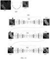
- FIG. 9 is a schematic diagram of a network structure of a 3D U-Net network as an addition and/or alternative to the network depicted FIG. 8 ;
- FIG. 10 is a schematic diagram of a network structure of a segmentation network according to one or more embodiments of the present disclosure.
- FIG. 11 is a schematic structural diagram of a dense block layer as an addition and/or alternative to the network depicted in FIG. 10 ;
- FIG. 12 is a flowchart of an image segmentation process according to one or more embodiments of the present disclosure.
- FIG. 13 is a flowchart of another image segmentation method according to one or more embodiments of the present disclosure.
- FIG. 14 is a schematic diagram of an implementation of an image segmentation method according to one or more embodiments of the present disclosure.
- FIG. 15 is a flowchart of the image segmentation method as an addition and/or alternative to the method depicted in FIG. 14 ;
- FIG. 16 is a block diagram of an image segmentation apparatus according to one or more embodiments of the present disclosure.
- FIG. 17 is a structural block diagram of a computing device according to one or more embodiments of the present disclosure.
- the computing device may be a computer.
- the present disclosure in one or more embodiments provides an image segmentation method based on a stepwise image segmentation, which is believed to effectively improve segmentation effects in tumor image segmentation.
- the image segmentation method is suitable for a tumor image segmentation apparatus.
- the tumor image segmentation apparatus may be deployed in a computing device with a Von Neumann architecture, for example, the computing device may be a personal computer (PC), a server, or the like.
- the present disclosure offers a realization that features learned by the fully convolutional neural network method are based on a part of a general image, but the feature learning ability for the general image may be poor, which may further lead to a poor segmentation effect.
- FIG. 1 is a schematic diagram of an implementation environment according to one or more embodiments of the present disclosure.
- the implementation environment includes a diagnosis system 100 , and the diagnosis system 100 includes an acquisition end 110 , a segmentation end 130 , and a diagnosis end 150 .
- the acquisition end 110 is an electronic device for acquiring tumor images, for example, an Mill device or a CT (Computed Tomography) device. This is not limited herein.
- the segmentation end 130 is an electronic device for providing a background service for a user, such as, a PC or a server.
- the background service includes an image segmentation service.
- the segmentation end 130 may be a server, or may be a server cluster composed of a plurality of servers, or even a cloud computing center composed of a plurality of servers, so as to better provide the background service for a large number of users. This is not limited herein.
- a tumor localization network 131 for locating a position of a general tumor region in a tumor image, and a cascaded segmentation network 132 constructed based on a machine learning model are deployed on the segmentation end 130 deploys, to implement stepwise image segmentation.
- the cascaded segmentation network 132 includes a plurality of levels of segmentation networks 1321 , 1322 , . . . , and 132 X.
- the diagnosis end 150 is an electronic device configured to assist a diagnostician in performing tumor diagnosis, for example, a PC equipped with a display screen.
- the segmentation end 130 establishes a wireless or wired network connection to each of the acquisition end 110 and the diagnosis end 150 , so as to implement data transmission in the diagnosis system 100 through the network connection.
- this data transmission includes tumor images, segmented images, and the like.
- the acquisition end 110 transmits the acquired tumor image to the segmentation end 130 .
- the segmentation end 130 receives a tumor image 111 transmitted by the acquisition end 110 , and performs tumor localization on the tumor image 111 based on the tumor localization network 131 , and a candidate image 1311 for indicating the position of the general tumor region in the tumor image 111 is obtained, and then is inputted to the cascaded segmentation network 132 .
- Image segmentation is performed on the general tumor region in the candidate image 1311 starting from a first-level segmentation network 1321 in the cascaded segmentation network 132 , and image segmentation is performed on an enhancing tumor core region level by level to a last-level segmentation network 132 X, to obtain a segmented image 1301 .
- the segmented image 1301 can be displayed on the display screen with which the diagnosis end 150 is equipped, so as to assist the diagnostician in performing the tumor diagnosis.
- FIG. 2 is a block diagram of a hardware structure of a segmentation end according to one or more embodiments of the present disclosure.
- the segmentation end is applicable to the segmentation end 130 of the implementation environment shown in FIG. 1 .
- the segmentation end is merely an example and does not limit a use range of the present disclosure.
- the segmentation end does not have to reply on or to have one or more components of an exemplary segmentation end 200 shown in FIG. 2 .
- a hardware structure of the segmentation end 200 may vary greatly as configuration or performance varies. As shown in FIG. 2 , the segmentation end 200 includes: a power supply 210 , an interface 230 , at least one memory 250 , and at least one central processing unit (CPU) 270 .
- the power supply 210 is configured to provide a working voltage for each hardware device on the segmentation end 200 .
- the interface 230 includes at least one wired or wireless network interface for interacting with external devices.
- the wired or wireless network interface interacts with the acquisition end 110 of the implementation environment shown in FIG. 1 , or interacts with the diagnosis end 150 of the implementation environment shown in FIG. 1 .
- the interface 230 may further include at least one serial to parallel conversion interface 233 , at least one input/output interface 235 , and at least one USB interface 237 . As shown in FIG. 2 , this is not limited herein.
- the memory 250 as a carrier for resource storage can be a read-only memory, a random access memory, a magnetic disk, an optical disc, or the like.
- Resources stored on the memory include an operating system 251 , an implementation program 253 , and data 255 .
- a storage method can be temporary storage or persistent storage.
- the operating system 251 is configured to manage and control the hardware devices and the implementation program 253 on the segmentation end 200 , so as to implement operations and processing of the CPU 270 on the massive data 255 in the memory 250 , and may be Windows ServerTM, Mac OS XTM, UnixTM, Linux, FreeBSDTM, or the like.
- the implementation program 253 is a computer program that executes at least one specific task based on the operating system 251 , and may include at least one module (not shown in FIG. 2 ), and each module may include a series of computer-readable instructions for the segmentation end 200 .
- the tumor image segmentation apparatus can be regarded as the application program 253 deployed on the segmentation end 200 , to implement the image segmentation method.
- the data 255 may be photos, pictures, and the like stored in the magnetic disk, or may be tumor images, segmented images, and the like stored in the memory 250 .
- the CPU 270 may include one or more processors, and is configured to communicate with the memory 250 by using at least one communication bus, to read computer-readable instructions stored in the memory 250 , thereby implementing the operations and processing on the massive data 255 in the memory 250 .
- the image segmentation method is executed in the form of reading a series of computer-readable instructions stored in the memory 250 by using the CPU 270 .
- present disclosure may alternatively be implemented through a hardware circuit or a hardware circuit in combination with software. Therefore, the present disclosure is implemented through, but not limited to, any specific hardware circuit, software, or a combination thereof.
- an image segmentation method is applicable to the segmentation end in the implementation environment shown in FIG. 1 .
- a structure of the segmentation end may be as shown in FIG. 2 .
- the image segmentation method can be performed by the segmentation end, and can include the following steps:
- Step 310 Obtain a tumor image.
- the tumor image is generated by the acquisition end by scanning a part where a human may have a tumor, so as to facilitate subsequent image segmentation on the tumor image.
- the acquisition end may be an MM device, a CT device, or the like.
- the tumor image may be derived from an image scanned by the acquisition end in real time, or an image pre-stored by the segmentation end and transmitted by the acquisition end.
- the segmentation end is a server, and further the server can obtain the tumor image through local reading or network transmission.
- the image scanned by the acquisition end can be obtained in real time, to facilitate the image segmentation on the tumor image in real time, or the image scanned by the acquisition end in a historical period of time can be obtained, to help the segmentation end to perform the image segmentation on the tumor image when there are fewer processing tasks, or, the image segmentation is performed on the tumor image under an instruction of an operator.
- This is not specifically limited in this embodiment.
- the segmentation end may perform denoising on the received tumor image, so as to improve accuracy of the subsequent image segmentation.
- the denoising may include removing a skull and a background in the tumor image.
- the tumor image obtained by the segmentation end includes but is not limited to one or more of four-modality MRI images, for example, FLAIR, T1, T1c, and T2.
- Step 330 Perform tumor localization on the obtained tumor image, to obtain a candidate image for indicating a position of a general tumor region in the tumor image.
- the brain tumors have varying degrees of invasiveness, and may be divided into three regions: a general tumor region, a tumor core region and an enhancing tumor core region.
- the most important feature is a proper inclusion relationship among the regions, as shown in sub-figures (a) to (c) in FIG. 4 , that is, the general tumor region 3011 includes the tumor core region 3021 , and the tumor core region 3021 includes the enhancing tumor core region 3031 .
- the term “general tumor region” may refer to a complete tumor region or an overall tumor region.
- the tumor localization refers to localization of a rough position of the general tumor region in the tumor image, so as to accommodate the general tumor region in the candidate image according to the localized position.
- the candidate image accommodates the general tumor region in the tumor image by using a designated region.
- a shape of the designated region may be a rectangle, a triangle, a circle, or the like, which is not limited herein.
- the designated region is a rectangular frame, and a maximum size of the rectangular frame is obtained by expanding maximum values of segmentation coordinates toward the periphery by a distance of a designated number of pixel points.
- the designated number can be flexibly adjusted according to an actual requirement of an application scenario. For example, in an application scenario, the designated number is 5.
- 305 represents a tumor image
- 306 represents a tumor localization process
- 307 represents a candidate image.
- a general tumor region 3071 is accommodated in a designated region 3072 .
- the designated region 3072 is a rectangular frame.
- the candidate image is merely a part of the tumor image, and the candidate image accommodates the general tumor region by using the designated region, thereby indicating a rough position of the general tumor region in the tumor image, which is beneficial for subsequent finer image segmentation based on the candidate image.
- the tumor localization can be implemented through the image segmentation, that is, the tumor image is segmented into a general tumor region and a non-tumor region, so that a localization frame can accommodate the general tumor region obtained through segmentation.
- the image segmentation includes: ordinary segmentation, semantic segmentation, instance segmentation, and the like, where the ordinary segmentation further includes: threshold segmentation, region segmentation, edge segmentation, histogram segmentation, and the like, which is not limited in this embodiment.
- the image segmentation can be implemented by using a machine learning model.
- the machine learning model can be a convolutional neural network model or a residual neural network model.
- Step 340 Input the candidate image to a cascaded segmentation network constructed based on a machine learning model.
- Step 350 Perform image segmentation on the general tumor region in the candidate image starting from a first-level segmentation network in the cascaded segmentation network, and perform image segmentation on an enhancing tumor core region level by level to a last-level segmentation network, to obtain a segmented image.
- the cascaded segmentation network includes a plurality of levels of segmentation networks, and is constructed based on the machine learning model.
- the machine learning model can be a convolutional neural network model or a residual neural network model.
- segmentation networks at all levels in the cascaded segmentation network based on a first-level segmentation network and parameters thereof, image segmentation is performed on the general tumor region in the candidate image, and a segmentation result is outputted to a second-level segmentation network.
- image segmentation is performed on the segmentation result outputted by the first-level segmentation network, and a segmentation result is outputted to a third-level segmentation network; and image segmentation is performed on an enhancing tumor core region level by level to a last-level segmentation network, and a segmentation result of the last-level segmentation network is used as a segmented image.
- stepwise image segmentation that starts from a boundary position of the general tumor region in the candidate image and gradually proceeds inward to the enhancing tumor core region is implemented.
- a cascaded segmentation network includes three levels of segmentation networks.
- a cascaded segmentation network 400 includes a first-level segmentation network 401 , a second-level segmentation network 402 , and a third-level segmentation network 403 .
- image segmentation is performed on the candidate image by using the first-level segmentation network 401 , to obtain a first-level intermediate segmented image 405 .
- Image segmentation is performed on the first-level intermediate segmented image 405 by using the second-level segmentation network 402 , to obtain a second-level intermediate segmented image.
- Image segmentation is performed on the second-level intermediate segmented image 406 by using the third-level segmentation network 403 , to obtain the segmented image.
- a first-level intermediate segmented image 301 is a segmentation result of the first-level segmentation network 401 , and the general tumor region 3011 included in the image is marked, as shown in sub-figure (a) in FIG. 4 .
- a second-level intermediate segmented image 302 is a segmentation result of the second-level segmentation network 402 , and the general tumor region 3011 and the tumor core region 3021 in the image are marked differently to reflect a proper inclusion relationship between the general tumor region 3011 and the tumor core region 3021 , as shown in sub-figure (b) in FIG. 4 .
- a segmented image 303 is a segmentation result of the third-level segmentation network 403 , and the general tumor region 3011 , the tumor core region 3021 , and the enhancing tumor core region 3031 included in the image are marked differently, as shown in the sub-figure (c) in FIG. 4 . That is, the segmented image 303 reflects a proper inclusion relationship among the general tumor region 3011 , the tumor core region 3021 , and the enhancing tumor core region 3031 .
- parameters used by the segmentation networks at all levels are different, so as to better adapt to the image segmentation between different regions included in brain tumors, to further help to improve segmentation effects of the tumor image.
- the tumor image segmentation based on machine learning is implemented, and the segmentation effects of the tumor image are effectively improved by using image segmentation processes of different scales.
- step 330 may include the following steps:
- Step 410 Extract a corresponding feature map from the obtained tumor image based on a three-dimensional U-shaped fully convolutional neural network (3D U-Net).
- 3D U-Net three-dimensional U-shaped fully convolutional neural network
- the tumor image generated by scanning of the acquisition end is usually a three-dimensional image, that is, a tumor image composed of many slices. If a two-dimensional machine learning model is used for processing a three-dimensional tumor image, not only a segmentation effect is not good, but also segmentation efficiency is relatively low because each of the slices that constitute the tumor image needs to be inputted to the machine learning model for training or class prediction, which is excessively cumbersome.
- tumor localization is implemented by using a three-dimensional machine learning model, that is, a 3D U-Net network.
- the 3D U-Net network is also referred to as a three-dimensional U-Net-based network. It can be understood that the 3D U-Net network is constructed by using the U-Net-based network as a prototype, and also has a U-shaped network structure.
- the U-Net-based network is an improved fully convolutional neural network.
- the U-Net-based network includes a contracting path 105 and an expanding path 107 .
- An input image is convoluted a plurality of times and contracted through the contracting path 105 to obtain a plurality of feature maps, and then the contracted input image is deconvoluted a plurality of times and expanded through the expanding path 107 .
- the input image is further correspondingly merged with the plurality of feature maps obtained by using the contracting path 105 , as shown in 1051 - 1054 in FIG. 7 b , so as to obtain features in different dimensions of the input image, thereby improving the segmentation effects.
- the 3D U-Net network includes an encoder network and a decoder network.
- the encoder network is used for extracting context features of the tumor image, so as to accurately describe the tumor image locally/globally by using the context features, to capture context information in the tumor image.
- the decoder network is used for extracting localization features of the tumor image, so as to perform, by using the localization features, accurate localization on a region in the tumor image on which image segmentation needs to be performed.
- feature fusion of the context features and the localization features is further performed, to obtain features in different dimensions of the tumor image, so that the segmentation effects of the image segmentation are better.
- Step 430 Perform class prediction on pixel points in the feature map corresponding to the tumor image, to obtain classes of the pixel points in the feature map corresponding to the tumor image.
- the class prediction is implemented based on a classifier that is set in the 3D U-Net network, that is, probabilities that the pixel points in the feature map corresponding to the tumor image belong to different classes are calculated by using the classifier.
- the tumor localization is essentially first segmenting the tumor image into a tumor region and a non-tumor region. Therefore, the classes include a general tumor region class and a non-tumor region class.
- probabilities that the pixel point belongs to different classes are calculated respectively. Assuming that a probability that the pixel point belongs to the general tumor region class is P1 and a probability that the pixel point belongs to the non-tumor region class is P2, if P1>P2, it indicates that the pixel point belongs to the general tumor region class; otherwise, if P1 ⁇ P2, it indicates that the pixel point belongs to the non-tumor region class.
- segmentation of the tumor region and the non-tumor region in the tumor image is performed, that is, a rough position of the general tumor region is localized in the tumor image.
- Step 450 Obtain the candidate image on which the general tumor region is accommodated in a designated region according to pixel points belonging to a general tumor region class in the feature map.
- the pixel points belonging to the general tumor region class can be obtained, so as to construct a designated region based on the obtained pixel points.
- the pixel points belonging to the general tumor region class are enclosed within the designated region, that is, the general tumor region is regarded to be accommodated in the designated region, so that a candidate image on which the general tumor region is accommodated in the designated region is generated, as shown by 307 in FIG. 5 .
- the designated region is used as a center, and expansion is made toward the periphery, so that a size of the candidate image reaches a designated size, so as to fully ensure the segmentation effects of the tumor image.
- the designated size may be flexibly set according to an actual requirement of an application scenario, which is not limited in this embodiment.
- the designated size is 96 ⁇ 96 ⁇ 96.
- coarse segmentation on the tumor image is implemented based on the 3D U-Net network, which not only localizes the rough position of the general tumor region in the tumor image from a macro perspective, avoiding a loss of accuracy of the image segmentation, but also contracts the tumor image to the candidate image, effectively reducing the size of the image, thereby not only reducing a background proportion, helping to improve a segmentation granularity of small tumors, but also helping to design a deeper network, and further improving the segmentation effects.
- a size of the designated region can dynamically change with a size of the general tumor region, which helps to fully guarantee the balance between positive and negative samples during subsequent model training of the segmentation network.
- step 410 may include the following steps:
- Step 411 Extract context features of the tumor image by using the encoder network.
- a 3D U-Net network 500 includes an encoder network 501 and a decoder network 502 .
- the encoder network 501 includes several downsampling layers 5011 - 5015
- the decoder network 502 includes several upsampling layers 5021 - 5025 .
- the 3D U-Net network 500 further includes a classification layer 503 , equipped with a classifier, configured to calculate probabilities that pixel points in the feature map corresponding to the tumor image belong to different classes, so as to implement the class prediction on the pixel points in the feature map corresponding to the tumor image.
- a classification layer 503 equipped with a classifier, configured to calculate probabilities that pixel points in the feature map corresponding to the tumor image belong to different classes, so as to implement the class prediction on the pixel points in the feature map corresponding to the tumor image.
- the encoder network 501 can extract the context features of the tumor image by using the several downsampling layers 5011 - 5015 , and transmit the extracted context features to the decoder network 502 by using the several feature propagation layers 5031 - 5034 .
- the tumor image is inputted to a shallowest downsampling layer 5011 in the encoder network 501 , convolution is performed on the inputted tumor image by using the shallowest downsampling layer 5011 , to obtain local features corresponding to the shallowest downsampling layer 5011 , and the local features are downsampled and then inputted to a second shallowest downsampling layer 5012 .
- the downsampling layers 5012 , 5013 , and 5014 in the encoder network 501 are traversed, to obtain local features corresponding to the traversed downsampling layers 5012 , 5013 , and 5014 .
- feature propagation of the foregoing local features is performed respectively by using the several feature propagation layers 5031 - 5034 .
- horizontal arrows represent convolution
- downward arrows represent downsampling
- a feature is a local feature or a global feature
- the feature is regarded as a context feature of the tumor image, so as to accurately describe the tumor image locally/globally.
- Step 413 Extract localization features of the general tumor region by using the decoder network, and perform fusion of the context features and the localization features, to obtain a feature map corresponding to the tumor image.
- the decoder network not only extracts the localization features of the tumor image by using the several upsampling layers, but also performs feature fusion of the context features and the localization features for the tumor image.
- context features global features corresponding to the deepest downsampling layer 5015 in the encoder network 501 are used as localization features corresponding to the deepest upsampling layer 5025 .
- Upsampling is performed on the localization features corresponding to the deepest upsampling layer 5025 , to obtain to-be-fused features.
- the to-be-fused features are inputted to a second deepest upsampling layer 5024 , and fused with context features (local features) corresponding to a second deepest downsampling layer 5014 , and by using deconvolution, localization features corresponding to the second deepest upsampling layer 5024 are obtained.
- the other upsampling layers 5023 , 5022 , 5021 are traversed, to obtain localization features corresponding to the traversed upsampling layers.
- a feature map corresponding to the tumor image is obtained according to the localization features corresponding to the shallowest upsampling layer 5021 .
- horizontal arrows represent deconvolution, and upward arrows represent upsampling.
- An input image is the candidate image, the first-level intermediate segmented image, or the second-level intermediate segmented image.
- An output image is the first-level intermediate segmented image, the second-level intermediate segmented image, or the segmented image.
- a segmentation network is the segmentation networks at all levels in the cascaded segmentation network.
- Parameters of the segmentation network are parameters of the segmentation networks at all levels in the cascaded segmentation network.
- the segmentation network 600 includes a downsampling stage 610 and an upsampling stage 630 .
- the downsampling stage 610 includes several first basic network layers 611 and 612 and several first dense block layers 613 and 614 connected sequentially.
- the upsampling stage 630 includes several third dense block layers 634 and 633 and several second basic network layers 632 and 631 connected sequentially.
- the upsampling stage 630 and the downsampling stage 610 are symmetric to each other, including: the first basic network layer 611 and the second basic network layer 631 are symmetric to each other, the first basic network layer 612 and the second basic network layer 632 are symmetric to each other, the first dense block layer 613 and the third dense block layer 633 are symmetric to each other, and the first dense block layer 614 and the third dense block layer 634 are symmetric to each other.
- the first basic network layer 611 includes a second dense block layer 6111 and a pooling layer 6112 connected sequentially.
- the first basic network layer 612 includes a second dense block layer 6121 and a pooling layer 6122 connected sequentially.
- the second basic network layer 631 includes an upsampling layer 6311 and a fourth dense block layer 6312 connected sequentially.
- the second basic network layer 632 includes an upsampling layer 6321 and a fourth dense block layer 6322 connected sequentially.
- the second dense block layer 6111 and the fourth dense block layer 6312 are symmetric to each other, and the second dense block layer 6121 and the fourth dense block layer 6322 are symmetric to each other.
- each of the above dense block layers includes an input unit and at least one dense unit.
- Each dense unit further includes a convolution layer, an activation layer, and a normalization layer connected sequentially, avoiding using a pure convolution layer or residual convolution layer, to ensure the accuracy of image segmentation.
- the dense block layer includes one input unit and four dense units H1, H2, H3, and H4.
- Each of the dense units further includes a convolution layer Cony, an activation layer Relu, and a normalization layer BN.
- a feature x0 corresponding to the input image Input is inputted by the input unit and simultaneously outputted to the dense units H1, H2, H3, and H4.
- a feature x1 outputted by the dense unit H1 is simultaneously outputted to the dense units H2, H3, and H4.
- a feature x2 outputted by the dense unit H2 is simultaneously outputted to the dense units H3 and H4, and a feature x3 outputted by the dense unit H3 is outputted to the dense unit H4.
- the features x0 and x1 corresponding to the input image Input are combined.
- the features x0, x1, and x2 corresponding to the input image Input are combined.
- the features x0, x1, x2, and x3 corresponding to the input image Input are combined.
- the dense block layer can not only reuse features of shallow layers, such as x0 and x1, to fully ensure integrity of the input image, but also combine features of deep and shallow layers, such as x0, x1, and x2, which helps to reduce complexity of image segmentation, and further effectively improves the segmentation effects of image segmentation.
- each convolution layer in the first dense block layer and the third dense block layer includes several three-dimensional convolution kernels (not shown in FIG. 10 ).
- Each convolution layer in the second dense block layer and the fourth dense block layer includes several tangent convolution kernels (not shown in FIG. 10 ) and several normal convolution kernels, as shown by 6111 a , 6121 a , 6322 a , and 6312 a in FIG. 10 .
- the convolution layer in the second dense block layer and the fourth dense block layer transforms the several three-dimensional convolution kernels (k ⁇ k ⁇ k) into the several tangent convolution kernels (k ⁇ k ⁇ 1) and the several normal convolution kernels (1 ⁇ 1 ⁇ k).
- the tumor image is particular, that is, the tumor image is composed of numerous slices, resulting in a relatively large difference between a tangent resolution and a normal resolution when the numerous slices synthesize a three-dimensional image.
- An error of pure three-dimensional image segmentation is relatively large, and pure two-dimensional segmentation directly ignores a correlation between the locality and the globality of the image, so that only 2.5-dimensional tumor image segmentation is most suitable.
- the image segmentation process that is, performing image segmentation on the input image by using the segmentation network, to obtain the output image may include the following steps:
- Step 510 Extract key features from the input image in the downsampling stage of the segmentation network.
- the input image Input is inputted to the downsampling stage 610 of the segmentation network 600 , and convolution and downsampling is performed by using the several first basic network layers 611 and 612 to obtain intermediate features.
- the input image Input is inputted to a first first basic network layer 611 , and convolution is performed on the input image by using the second dense block layer 6111 in the first first basic network layer 611 .
- Downsampling is performed on features outputted after the convolution by using the pooling layer 6112 in the first first basic network layer 611 , so as to output the downsampled features to a second first basic network layer 612 .
- the other first basic network layers 612 in the several first basic network layers are traversed according to a feature propagation sequence. After the traversal is performed, features downsampled by the second first basic network layer 612 , that is, a last first basic network layer 612 are used as the intermediate features.
- Step 530 Input the key features to the upsampling stage of the segmentation network, and perform multi-scale feature fusion, to obtain a feature map corresponding to the input image.
- the key features are inputted to the upsampling stage 630 of the segmentation network 600 , and deconvolution is performed by using the several third dense block layers 634 and 633 , to obtain first scale features 651 , to input the first scale features to the first several second basic network layers.
- first several second basic network layers feature fusion of fourth dense block layers in the first several second basic network layers and second dense block layers in several of the first basic network layers that are symmetric each other is performed, and the first several second basic network layers in the upsampling stage 630 are connected between a last third dense block layer 633 and a last second basic network layer 631 .
- upsampling is performed on features outputted after the fusion in the first several second basic network layers by using the upsampling layer 6311 in the last second basic network layer 631 , to obtain second scale features 652 .
- Fusion is performed on the first scale features 651 , the second scale features 652 , and the third scale features 653 by using the fourth dense block layer 6312 in the last second basic network layer 631 , and deconvolution is performed, to obtain a feature map Output corresponding to the input image Input.
- the feature map Output corresponding to the input image Input is not only based on 1 ⁇ upsampling features (the second scale features 652 ), 2 ⁇ upsampling features (the features 653 outputted after the convolution of the second dense block layer 6121 ), and 4 ⁇ upsampling features (the first scale features 651 ), but also based on zero upsampling features (the features 653 outputted after the convolution of the second dense block layer 6111 ), thereby implementing multi-scale feature fusion, so that segmentation results of the segmentation networks at all levels can achieve the best segmentation effects locally and globally, which effectively improves the segmentation effects.
- a process of the feature fusion of the fourth dense block layer and the second dense block layer that are symmetric to each other is further described as follows:
- the first scale features 651 are inputted to the first second basic network layer 632 , and upsampling is performed on the inputted first scale features 651 by using an upsampling layer 6321 in the first second basic network layer 632 .
- features 654 outputted after the convolution of a second dense block layer 6121 in the several first basic network layers 612 that is symmetric to the fourth dense block layer are obtained, and fused with the upsampled features, to obtain combined features.
- Deconvolution is performed on the combined features by using the fourth dense block layer 6322 in the first second basic network layer 632 , to output the deconvoluted features to the second second basic network layer 631 .
- the other second basic network layers in the first several second basic network layers are traversed according to a feature propagation sequence, and after the traversal is performed, feature fusion of the fourth dense block layers and the second dense block layers that are symmetric to each other is performed.
- FIG. 10 only includes two second basic network layers, in the upsampling stage 630 , the second second basic network layer is essentially the last second basic network layer. Therefore, there is no need to traverse the other second basic network layers in the first several second basic network layers, and provided that deconvolution performed in the first second basic network layer 632 is performed, feature fusion of the fourth dense block layer 6322 and the second dense block layer 6121 that are symmetric to each other is performed.
- Step 550 Perform class prediction on pixel points in the feature map corresponding to the input image, to obtain classes of the pixel points in the feature map corresponding to the input image.
- the class prediction is implemented based on a classifier that is set in the segmentation network, that is, probabilities that pixel points in the feature map corresponding to the input image belong to different classes are calculated by using the classifier.
- the classes can be an other-regions class, a general tumor region class, a tumor core region class, and an enhancing tumor core region class.
- the segmentation networks at all levels are constructed based on two classes, that is, in the first-level segmentation network, the classes include the other-regions class and the general tumor region class. In this case, the other regions are non-tumor regions.
- the classes include the other-regions class and the tumor core region class.
- the other regions refer to the non-tumor regions and the general tumor region that does not include the tumor core region.
- the classes include the other-regions class and the enhancing tumor core region class.
- the other regions refer to the non-tumor regions and the general tumor region that does not include the enhancing tumor core region.
- Image segmentation of the other regions and the tumor core region performed in the second-level segmentation network is used as an example for description.
- probabilities that the pixel point belongs to different classes are respectively calculated. Assuming that a probability that the pixel point belongs to the other-regions class is P1 and a probability that the pixel point belongs to the tumor core region class is P2, if P1 is greater, it indicates that the pixel point belongs to the other-regions class; otherwise, if P2 is greater, it indicates that the pixel point belongs to the tumor core region class.
- Step 570 Mark pixel points of a designated class in the feature map corresponding to the input image, to obtain the output image.
- Marking is performed according to the class to which the pixel point belongs.
- the marking can be made by using a color or a symbol such as an asterisk, which is not limited herein.
- different classes of pixel points are marked with different colors, as shown in sub-figure (c) in FIG. 4 .
- designated classes are all different.
- the designated class in the first-level segmentation network, the designated class is the general tumor region class; in the second-level segmentation network, the designated class is the tumor core region; and in the third-level segmentation network, the designated class is the enhancing tumor core region.
- the segmentation networks at all levels provided that the class prediction of all the pixel points in the feature maps corresponding to all the input images is performed, the segmentation of the general tumor region, the tumor core region, and the enhancing tumor region in the tumor image is performed, that is, more precise positions of the general tumor region, the tumor core region, and the enhancing tumor region are localized in the tumor image.
- the method further includes:
- the constructing the cascaded segmentation network based on the machine learning model may include the following steps:
- Step 710 Obtain training samples carrying labels.
- the training samples are a tumor images on which the general tumor region, the tumor core region, and the enhancing tumor core region are labeled by using different types of labels.
- labeling refers to only adding non-zero markers to the general tumor region, the tumor core region, or the enhancing tumor core region in the tumor image, and zero markers are made on the pixel points in the other regions in the tumor image.
- the marking of the tumor image is performed, and a training sample carrying a general tumor region label is obtained.
- the non-zero markers are only added to the tumor core region in the tumor image, and zero markers are made on the pixel points in the other regions in the tumor image, a training sample carrying a tumor core region label is obtained.
- a training sample carrying an enhancing tumor core region label is obtained.
- the pixel points in the tumor image are normalized, to improve the accuracy of image segmentation.
- Step 730 Establish a plurality of training sample sets according to types of the labels carried by the training samples, each of the training sample sets corresponding to a type.
- the sample augmentation processing includes: flipping, rotating, zooming, contrast enhancement, and the like.
- the flipping refers to front-and-back flipping and left-and-right flipping of the tumor image;
- the rotating refers to rotating at a designated angle of the tumor image;
- the zooming refers to enlarging the tumor image, or shrinking the tumor image;
- the contrast enhancement refers to changing contrast of pixel points in the tumor image.
- the enlarging means that a 96 ⁇ 96 ⁇ 96 tumor image is interpolated into a 120 ⁇ 120 ⁇ 120-sized image, and then an intermediate image is cropped from the 120 ⁇ 120 ⁇ 120-sized image to 96 ⁇ 96 ⁇ 96; and the shrinking means that a 120 ⁇ 120 ⁇ 120 tumor image is shrunk to 96 ⁇ 96 ⁇ 96.
- a corresponding training sample set is established by using training samples carrying the same type of labels, and then, a plurality of corresponding training sample sets may be established by using training samples of a plurality types of labels.
- a corresponding training sample set is constructed by using training samples carrying the general tumor region label, and after the model training is performed on the convolutional neural network model, the image segmentation on the general tumor region is performed.
- Step 750 Perform model training on a plurality of convolutional neural network models having designated model structures respectively by using the plurality of training sample sets.
- the model training is essentially to iteratively optimize parameters of the convolutional neural network model with a designated model structure by using the training sample set, so that a designated algorithm function constructed based on the parameters meets a convergence condition.
- the designated model structure is shown in FIG. 10 .
- the designated algorithm function includes but is not limited to: an expectation maximization function, a loss function, and the like.
- parameters of the convolutional neural network model are randomly initialized, a probability is calculated through forward propagation according to a current training sample in the training sample set and based on the randomly initialized parameters, a loss function is constructed by using a Dice distance between the calculated probability and correct labeling, and further a loss value of the loss function is calculated.
- the parameters of the convolutional neural network model are updated through back propagation, a probability is calculated according to a next training sample in the training sample set and based on the updated parameters, the loss function is reconstructed by using the Dice distance between the calculated probability and correct labeling, and the loss value of the reconstructed loss function is calculated again.
- Such iterative looping is performed until the loss value of the constructed loss function reaches the minimum value, and the loss function is considered as convergent. In this case, if the convolutional neural network model also converges and meets a preset precision requirement, iteration is stopped.
- the parameters of the convolutional neural network model are iteratively updated, and the loss value of the constructed loss function is calculated according to the other training samples in the training sample set and the updated parameters, until the loss function converges.
- the convolutional neural network model When the convolutional neural network model converges and meets the preset precision requirement, it indicates that the convolutional neural network model completes the model training, so that the cascaded segmentation network can be further constructed.
- Step 770 Cascade the plurality of convolutional neural network models after the model training is performed, to obtain the cascaded segmentation network.
- a plurality of convolutional neural network models that general the model training can be obtained.
- Each of the convolutional neural network models that complete the model training corresponds to a training sample set.
- the training sample is a tumor image on which the general tumor region is labeled by using labels, and then the convolutional neural network model that therefore completes the model training performs image segmentation on the general tumor region.
- a convolutional neural network model that completes the model training is used as a level of segmentation network, and a plurality of levels of segmentation networks are cascaded, to construct a cascaded segmentation network.
- the cascaded segmentation network 400 includes the three levels of segmentation networks 401 , 402 , and 403 .
- the segmentation end has an ability to predict classes of the pixel points of the feature map corresponding to the input image.
- class prediction can be performed on the pixel points in the feature map corresponding to the input image, to obtain the classes of the pixel points in the feature map, thereby implementing the image segmentation on the input image.
- step 350 the method described above may further include the following steps:
- the morphological algorithm includes but is not limited to corrosion, expansion, hole filling, Dense CRF (conditional random field), and the like, which is not limited in this embodiment.
- the segmented image is corrected, so that segmentation edges among the general tumor region, the tumor core region, and the enhancing tumor core region in the segmented image are smoothed, and/or, noises in the segmented image are canceled, thereby further effectively improving the segmentation effects of image segmentation.
- a segmentation end divides a tumor image segmentation task into a coarse segmentation subtask and a fine segmentation subtask.
- the tumor image is generated by scanning of an MRI device at an acquisition end, and is essentially a four-modality MM image.
- the segmentation end by performing step 801 , obtains a tumor image 811 generated by scanning of the acquisition end.
- the coarse segmentation subtask is based on the tumor image 811 , and tumor localization is performed by using a 3D U-Net network 820 , to obtain a candidate image 812 with a general tumor region included in a rectangular frame, so that the coarse segmentation subtask is performed, that is, step 802 is performed: based on the tumor image 811 , coarse segmentation on the general tumor region is performed.
- the candidate image 812 is used as a basis of the fine segmentation subtask. It can be seen that, a size of the candidate image 812 is greatly reduced compared with that of the tumor image 811 .
- the candidate image 812 is inputted to a first-level segmentation network 831 in a cascaded segmentation network 830 , and image segmentation is performed, to obtain a first-level intermediate segmented image 813 marked with the general tumor region.
- the general tumor region included in the first-level intermediate segmented image 813 is no longer roughly included in the rectangular frame, but is marked more specifically, and first time of fine segmentation on the tumor image is implemented, that is, step 803 is performed: based on the candidate image 812 , image segmentation on the general tumor region is performed.
- the first-level intermediate segmented image 813 is used as an input of a second-level segmentation network 832 , image segmentation is performed, to obtain a second-level intermediate segmented image 814 , the second-level intermediate segmented image 814 reflects a proper inclusion relationship between the general tumor region and the tumor core region, and second time of fine segmentation on the tumor image is implemented, that is, step 804 is performed: based on the first-level intermediate segmented image 813 , image segmentation on the tumor core region is performed.
- the second-level intermediate segmented image 814 is used as an input of a third-level segmentation network 833 , image segmentation is performed to obtain a segmented image 815 , the segmented image 815 reflects a proper inclusion relationship among the general tumor region, the tumor core region, and the enhancing tumor core region, and third time of fine segmentation on the tumor image is implemented, that is, step 805 is performed: based on the second-level intermediate segmented image 814 , image segmentation on the enhancing tumor core region is performed.
- the fine segmentation subtasks are performed through stepwise image segmentation, that is, image segmentation on different regions performed based on different input images.
- a segmented image 815 obtained by the segmentation end can be received, so that a doctor understands the three regions of different severity degrees in the brain tumor in time, and the doctor is assisted in diagnosing a tumor more rapidly and accurately, for example, analyzing benignity or malignancy or a malignant degree of a tumor of a patient.
- a network structure of the 3D U-Net network 810 is shown in FIG. 9
- a network structure of the segmentation networks at all levels in the cascaded segmentation network 830 is shown in FIG. 10 .
- [3 ⁇ 3 ⁇ 1 cony] represents a tangent convolution kernel
- [1 ⁇ 1 ⁇ 3 cony] represents a normal convolution kernel
- [3 ⁇ 3 ⁇ 3 cony] represents a three-dimensional convolution kernel.
- Quantities of various types of convolution kernels that are set in different dense block layers can be flexibly adjusted according to an actual requirement of an application scenario, which is not limited herein.
- 12 three-dimensional convolution kernels and 3 three-dimensional convolution kernels are respectively set in the first dense block layers.
- a four-modality Mill image four channels are essentially configured in each of the convolution layers, so that the four-modality Mill image is inputted, through different channels to the cascaded segmentation network, for image segmentation, thereby fully ensuring integrity of the tumor image, and helping to improve the segmentation effects.
- end-to-end automatic image segmentation is implemented, that is, as long as different-modality Mill images corresponding to a patient are inputted, three regions with different severity degrees can be obtained, which can not only effectively assist a doctor in further analyzing a treatment plan for the patient, but also determine an operation region for the patient, so as to more accurately process a lesion.
- Apparatus embodiments of the present disclosure are described below, and may be used for performing the image segmentation method involved in the present disclosure. For details not disclosed in the apparatus embodiment of the present disclosure, refer to the method embodiment of the image segmentation method involved in the present disclosure.
- an image segmentation apparatus 900 includes, but is not limited to: an image obtaining module 910 , an image coarse segmentation module 930 , an image input module 940 , and an image fine segmentation module 950 .
- the image obtaining module 910 is configured to obtain a tumor image.
- the image coarse segmentation module 930 is configured to perform tumor localization on the obtained tumor image, to obtain a candidate image for indicating a position of a general tumor region in the tumor image.
- the image input module 940 is configured to input the candidate image to a cascaded segmentation network constructed based on a machine learning model.
- the image fine segmentation module 950 is configured to perform image segmentation on the general tumor region in the candidate image starting from a first-level segmentation network in the cascaded segmentation network, and perform image segmentation on an enhancing tumor core region level by level to a last-level segmentation network, to obtain a segmented image.
- Each module/unit in various disclosed embodiments can be integrated in a processing unit, or each module/unit can exist separately and physically, or two or more modules/units can be integrated in one unit.
- the modules/units as disclosed herein can be implemented in the form of hardware (e.g., processing circuitry and/or memory) or in the form of software functional unit(s) (e.g., developed using one or more computer programming languages), or a combination of hardware and software.
- Each module/unit or submodule/subunit can be implemented using one or more processors (or processors and memory).
- a processor or processor and memory
- each module/unit may be developed using a computer programming language, or be part of an overall module/unit that is developed using a computer programming language to encompass the functionalities of each module/unit.
- the division of the functional modules is merely used as an example for description.
- the functions may be distributed to and implemented by different functional modules according to the requirements, that is, an internal structure of the image segmentation apparatus is divided into different functional modules, so as to finish all or some of the functions described above.
- a computing device 1000 includes at least one processor 1001 , at least one memory 1002 , and at least one communication bus 1003 .
- the memory 1002 stores computer-readable instructions, and the processor 1001 reads, by using the communication bus 1003 , the computer-readable instructions stored in the memory 1002 .
- the computer-readable instructions when executed by the processor 1001 , implement the image segmentation method in the foregoing embodiments.
- a computer-readable storage medium stores a computer program, the computer program, when executed by a processor, implementing the image segmentation method in the foregoing embodiments.
Applications Claiming Priority (3)
| Application Number | Priority Date | Filing Date | Title |
|---|---|---|---|
| CN201811462063.3 | 2018-11-30 | ||
| CN201811462063.3A CN109598728B (zh) | 2018-11-30 | 2018-11-30 | 图像分割方法、装置、诊断系统及存储介质 |
| PCT/CN2019/121246 WO2020108525A1 (fr) | 2018-11-30 | 2019-11-27 | Procédé et appareil de segmentation d'images, système de diagnostic, support de stockage, et dispositif informatique |
Related Parent Applications (1)
| Application Number | Title | Priority Date | Filing Date |
|---|---|---|---|
| PCT/CN2019/121246 Continuation WO2020108525A1 (fr) | 2018-11-30 | 2019-11-27 | Procédé et appareil de segmentation d'images, système de diagnostic, support de stockage, et dispositif informatique |
Publications (2)
| Publication Number | Publication Date |
|---|---|
| US20210241027A1 US20210241027A1 (en) | 2021-08-05 |
| US11954863B2 true US11954863B2 (en) | 2024-04-09 |
Family
ID=65959310
Family Applications (1)
| Application Number | Title | Priority Date | Filing Date |
|---|---|---|---|
| US17/204,894 Active 2041-04-22 US11954863B2 (en) | 2018-11-30 | 2021-03-17 | Image segmentation method and apparatus, diagnosis system, storage medium, and computer device |
Country Status (4)
| Country | Link |
|---|---|
| US (1) | US11954863B2 (fr) |
| EP (1) | EP3828825A4 (fr) |
| CN (1) | CN109598728B (fr) |
| WO (1) | WO2020108525A1 (fr) |
Families Citing this family (63)
| Publication number | Priority date | Publication date | Assignee | Title |
|---|---|---|---|---|
| CN111160346A (zh) * | 2018-11-07 | 2020-05-15 | 电子科技大学 | 基于三维卷积的缺血性脑卒中分割系统 |
| CN109598728B (zh) | 2018-11-30 | 2019-12-27 | 腾讯科技(深圳)有限公司 | 图像分割方法、装置、诊断系统及存储介质 |
| CN110084297B (zh) * | 2019-04-23 | 2023-09-15 | 东华大学 | 一种面向小样本的影像语义对齐系统 |
| CN110097921B (zh) * | 2019-05-30 | 2023-01-06 | 复旦大学 | 基于影像组学的胶质瘤内基因异质性可视化定量方法和系统 |
| CN110211134B (zh) * | 2019-05-30 | 2021-11-05 | 上海商汤智能科技有限公司 | 一种图像分割方法及装置、电子设备和存储介质 |
| CN110211140B (zh) * | 2019-06-14 | 2023-04-07 | 重庆大学 | 基于3D残差U-Net和加权损失函数的腹部血管分割方法 |
| CN110232361B (zh) * | 2019-06-18 | 2021-04-02 | 中国科学院合肥物质科学研究院 | 基于三维残差稠密网络的人体行为意图识别方法与系统 |
| CN110276755B (zh) * | 2019-06-25 | 2021-07-06 | 广东工业大学 | 一种肿瘤位置定位系统及相关装置 |
| CN110363776B (zh) * | 2019-06-28 | 2021-10-22 | 联想(北京)有限公司 | 图像处理方法及电子设备 |
| CN110390680A (zh) * | 2019-07-04 | 2019-10-29 | 上海联影智能医疗科技有限公司 | 图像分割方法、计算机设备和存储介质 |
| CN110310280B (zh) * | 2019-07-10 | 2021-05-11 | 广东工业大学 | 肝胆管及结石的图像识别方法、系统、设备及存储介质 |
| CN110349170B (zh) * | 2019-07-13 | 2022-07-08 | 长春工业大学 | 一种全连接crf级联fcn和k均值脑肿瘤分割算法 |
| CN110378976B (zh) * | 2019-07-18 | 2020-11-13 | 北京市商汤科技开发有限公司 | 图像处理方法及装置、电子设备和存储介质 |
| CN110415234A (zh) * | 2019-07-29 | 2019-11-05 | 北京航空航天大学 | 基于多参数磁共振成像的脑部肿瘤分割方法 |
| WO2021017006A1 (fr) * | 2019-08-01 | 2021-02-04 | 京东方科技集团股份有限公司 | Procédé et appareil de traitement d'image, réseau neuronal et procédé d'apprentissage, et support d'enregistrement |
| CN110458833B (zh) * | 2019-08-15 | 2023-07-11 | 腾讯科技(深圳)有限公司 | 基于人工智能的医学图像处理方法、医学设备和存储介质 |
| CN110717913B (zh) * | 2019-09-06 | 2022-04-22 | 浪潮电子信息产业股份有限公司 | 一种图像分割方法及装置 |
| CN110874842B (zh) * | 2019-10-10 | 2022-04-29 | 浙江大学 | 一种基于级联残差全卷积网络的胸腔多器官分割方法 |
| CN110866908B (zh) * | 2019-11-12 | 2021-03-26 | 腾讯科技(深圳)有限公司 | 图像处理方法、装置、服务器及存储介质 |
| CN111028236A (zh) * | 2019-11-18 | 2020-04-17 | 浙江工业大学 | 一种基于多尺度卷积U-Net的癌细胞图像分割方法 |
| CN111047602A (zh) * | 2019-11-26 | 2020-04-21 | 中国科学院深圳先进技术研究院 | 图像分割方法、装置及终端设备 |
| CN111047606B (zh) * | 2019-12-05 | 2022-10-04 | 北京航空航天大学 | 一种基于级联思想的病理全切片图像分割算法 |
| CN111145186B (zh) * | 2019-12-17 | 2023-08-22 | 中国科学院深圳先进技术研究院 | 神经网络结构、图像分割方法、装置及存储介质 |
| CN111192320B (zh) * | 2019-12-30 | 2023-07-25 | 上海联影医疗科技股份有限公司 | 一种位置信息确定方法、装置、设备和存储介质 |
| US11645505B2 (en) * | 2020-01-17 | 2023-05-09 | Servicenow Canada Inc. | Method and system for generating a vector representation of an image |
| CN111275721B (zh) * | 2020-02-14 | 2021-06-08 | 推想医疗科技股份有限公司 | 一种图像分割方法、装置、电子设备及存储介质 |
| CN111507215B (zh) * | 2020-04-08 | 2022-01-28 | 常熟理工学院 | 基于时空卷积循环神经网络与空洞卷积的视频目标分割方法 |
| CN111626298B (zh) * | 2020-04-17 | 2023-08-18 | 中国科学院声学研究所 | 一种实时图像语义分割装置及分割方法 |
| CN111696084A (zh) * | 2020-05-20 | 2020-09-22 | 平安科技(深圳)有限公司 | 细胞图像分割方法、装置、电子设备及可读存储介质 |
| CN111369576B (zh) * | 2020-05-28 | 2020-09-18 | 腾讯科技(深圳)有限公司 | 图像分割模型的训练方法、图像分割方法、装置及设备 |
| CN111368849B (zh) * | 2020-05-28 | 2020-08-28 | 腾讯科技(深圳)有限公司 | 图像处理方法、装置、电子设备及存储介质 |
| CN111640100B (zh) * | 2020-05-29 | 2023-12-12 | 京东方科技集团股份有限公司 | 肿瘤图像的处理方法和装置、电子设备、存储介质 |
| CN112037171B (zh) * | 2020-07-30 | 2023-08-15 | 西安电子科技大学 | 基于多模态特征融合的多任务mri脑瘤图像分割方法 |
| CN112085743A (zh) * | 2020-09-04 | 2020-12-15 | 厦门大学 | 一种肾肿瘤的图像分割方法 |
| CN112070752A (zh) * | 2020-09-10 | 2020-12-11 | 杭州晟视科技有限公司 | 一种医学图像的心耳分割方法、装置及存储介质 |
| CN112150470B (zh) * | 2020-09-22 | 2023-10-03 | 平安科技(深圳)有限公司 | 图像分割方法、装置、介质及电子设备 |
| CN111968137A (zh) * | 2020-10-22 | 2020-11-20 | 平安科技(深圳)有限公司 | 头部ct图像分割方法、装置、电子设备及存储介质 |
| CN112017189B (zh) * | 2020-10-26 | 2021-02-02 | 腾讯科技(深圳)有限公司 | 图像分割方法、装置、计算机设备和存储介质 |
| CN112258526B (zh) * | 2020-10-30 | 2023-06-27 | 南京信息工程大学 | 一种基于对偶注意力机制的ct肾脏区域级联分割方法 |
| CN112396620A (zh) * | 2020-11-17 | 2021-02-23 | 齐鲁工业大学 | 一种基于多阈值的图像语义分割方法及系统 |
| KR20220080249A (ko) | 2020-12-07 | 2022-06-14 | 삼성전자주식회사 | 영상 처리 방법 및 장치 |
| CN112784897B (zh) | 2021-01-20 | 2024-03-26 | 北京百度网讯科技有限公司 | 图像处理方法、装置、设备和存储介质 |
| CN112767417B (zh) * | 2021-01-20 | 2022-09-13 | 合肥工业大学 | 一种基于级联U-Net网络的多模态图像分割方法 |
| KR102321427B1 (ko) * | 2021-01-20 | 2021-11-04 | 메디컬아이피 주식회사 | 의료영상을 이용한 인체성분 분석 방법 및 그 장치 |
| CN112785605B (zh) * | 2021-01-26 | 2023-07-28 | 西安电子科技大学 | 基于语义迁移的多时相ct图像肝肿瘤分割方法 |
| CN112862830B (zh) * | 2021-01-28 | 2023-12-22 | 陕西师范大学 | 一种多模态图像分割方法、系统、终端及可读存储介质 |
| CN112767407B (zh) * | 2021-02-02 | 2023-07-07 | 南京信息工程大学 | 一种基于级联门控3DUnet模型的CT图像肾脏肿瘤分割方法 |
| CN113781449A (zh) * | 2021-09-14 | 2021-12-10 | 上海布眼人工智能科技有限公司 | 一种基于多尺度特征融合的纺织品瑕疵分类方法 |
| CN113569865B (zh) * | 2021-09-27 | 2021-12-17 | 南京码极客科技有限公司 | 一种基于类别原型学习的单样本图像分割方法 |
| CN113658180B (zh) * | 2021-10-20 | 2022-03-04 | 北京矩视智能科技有限公司 | 一种基于空间上下文引导的表面缺陷区域分割方法和装置 |
| CN114267443B (zh) * | 2021-11-08 | 2022-10-04 | 东莞市人民医院 | 基于深度学习的胰腺肿瘤纤维化程度预测方法及相关装置 |
| US11961618B2 (en) * | 2021-11-17 | 2024-04-16 | City University Of Hong Kong | Task interaction network for prostate cancer diagnosis |
| CN114170244A (zh) * | 2021-11-24 | 2022-03-11 | 北京航空航天大学 | 一种基于级联神经网络结构的脑胶质瘤分割方法 |
| CN114092815B (zh) * | 2021-11-29 | 2022-04-15 | 自然资源部国土卫星遥感应用中心 | 一种大范围光伏发电设施遥感智能提取方法 |
| CN114445726B (zh) * | 2021-12-13 | 2022-08-02 | 广东省国土资源测绘院 | 一种基于深度学习的样本库建立方法和装置 |
| CN114241344B (zh) * | 2021-12-20 | 2023-05-02 | 电子科技大学 | 一种基于深度学习的植物叶片病虫害严重程度评估方法 |
| CN114299288A (zh) * | 2021-12-23 | 2022-04-08 | 广州方硅信息技术有限公司 | 图像分割方法、装置、设备和存储介质 |
| CN114496145B (zh) * | 2022-01-27 | 2023-02-10 | 深圳市铱硙医疗科技有限公司 | 一种医疗图像档案管理方法与系统 |
| CN114612479B (zh) * | 2022-02-09 | 2023-03-24 | 苏州大学 | 基于全局与局部特征重建网络的医学图像分割方法和装置 |
| CN115115577A (zh) * | 2022-05-19 | 2022-09-27 | 北京深睿博联科技有限责任公司 | 一种基于混合感知的多阶段器官分割方法及装置 |
| CN114648529B (zh) * | 2022-05-19 | 2022-09-23 | 深圳市中科先见医疗科技有限公司 | 一种基于cnn网络的dpcr液滴荧光检测方法 |
| CN116912502B (zh) * | 2023-09-08 | 2024-01-16 | 南方医科大学珠江医院 | 全局视角辅助下图像关键解剖结构的分割方法及其设备 |
| CN117455935B (zh) * | 2023-12-22 | 2024-03-19 | 中国人民解放军总医院第一医学中心 | 基于腹部ct医学图像融合及器官分割方法及系统 |
Citations (12)
| Publication number | Priority date | Publication date | Assignee | Title |
|---|---|---|---|---|
| US20030142857A1 (en) | 2002-01-25 | 2003-07-31 | General Electric Company | Method and system for segmenting magnetic resonance images |
| US20080292194A1 (en) * | 2005-04-27 | 2008-11-27 | Mark Schmidt | Method and System for Automatic Detection and Segmentation of Tumors and Associated Edema (Swelling) in Magnetic Resonance (Mri) Images |
| CN104123417A (zh) | 2014-07-22 | 2014-10-29 | 上海交通大学 | 一种基于聚类融合的图像分割的方法 |
| CN108268870A (zh) | 2018-01-29 | 2018-07-10 | 重庆理工大学 | 基于对抗学习的多尺度特征融合超声图像语义分割方法 |
| WO2018156778A1 (fr) | 2017-02-22 | 2018-08-30 | The United States Of America, As Represented By The Secretary, Department Of Health And Human Services | Détection du cancer de la prostate dans une irm multi-paramétrique à l'aide d'une forêt aléatoire, avec pondération d'instance et segmentation de prostate rm par apprentissage profond avec des réseaux imbriqués de manière holistique |
| CN108492297A (zh) | 2017-12-25 | 2018-09-04 | 重庆理工大学 | 基于深度级联卷积网络的mri脑肿瘤定位与瘤内分割方法 |
| CN108564582A (zh) | 2018-04-23 | 2018-09-21 | 重庆大学 | 一种基于深度神经网络的mri脑肿瘤自动识别方法 |
| CN108765422A (zh) | 2018-06-13 | 2018-11-06 | 云南大学 | 一种视网膜图像血管自动分割方法 |
| CN108830855A (zh) | 2018-04-02 | 2018-11-16 | 华南理工大学 | 一种基于多尺度低层特征融合的全卷积网络语义分割方法 |
| CN109271992A (zh) * | 2018-09-26 | 2019-01-25 | 上海联影智能医疗科技有限公司 | 一种医学图像处理方法、系统、装置和计算机可读存储介质 |
| CN109598728A (zh) | 2018-11-30 | 2019-04-09 | 腾讯科技(深圳)有限公司 | 图像分割方法、装置、诊断系统及存储介质 |
| US20200020098A1 (en) * | 2017-04-19 | 2020-01-16 | Siemens Healthcare Gmbh | Target detection in latent space |
Family Cites Families (2)
| Publication number | Priority date | Publication date | Assignee | Title |
|---|---|---|---|---|
| US20110286654A1 (en) * | 2010-05-21 | 2011-11-24 | Siemens Medical Solutions Usa, Inc. | Segmentation of Biological Image Data |
| CN106709568B (zh) * | 2016-12-16 | 2019-03-22 | 北京工业大学 | 基于深层卷积网络的rgb-d图像的物体检测和语义分割方法 |
-
2018
- 2018-11-30 CN CN201811462063.3A patent/CN109598728B/zh active Active
-
2019
- 2019-11-27 EP EP19889004.8A patent/EP3828825A4/fr active Pending
- 2019-11-27 WO PCT/CN2019/121246 patent/WO2020108525A1/fr unknown
-
2021
- 2021-03-17 US US17/204,894 patent/US11954863B2/en active Active
Patent Citations (13)
| Publication number | Priority date | Publication date | Assignee | Title |
|---|---|---|---|---|
| US20030142857A1 (en) | 2002-01-25 | 2003-07-31 | General Electric Company | Method and system for segmenting magnetic resonance images |
| US20080292194A1 (en) * | 2005-04-27 | 2008-11-27 | Mark Schmidt | Method and System for Automatic Detection and Segmentation of Tumors and Associated Edema (Swelling) in Magnetic Resonance (Mri) Images |
| CN104123417A (zh) | 2014-07-22 | 2014-10-29 | 上海交通大学 | 一种基于聚类融合的图像分割的方法 |
| WO2018156778A1 (fr) | 2017-02-22 | 2018-08-30 | The United States Of America, As Represented By The Secretary, Department Of Health And Human Services | Détection du cancer de la prostate dans une irm multi-paramétrique à l'aide d'une forêt aléatoire, avec pondération d'instance et segmentation de prostate rm par apprentissage profond avec des réseaux imbriqués de manière holistique |
| US20200020098A1 (en) * | 2017-04-19 | 2020-01-16 | Siemens Healthcare Gmbh | Target detection in latent space |
| CN108492297A (zh) | 2017-12-25 | 2018-09-04 | 重庆理工大学 | 基于深度级联卷积网络的mri脑肿瘤定位与瘤内分割方法 |
| CN108268870A (zh) | 2018-01-29 | 2018-07-10 | 重庆理工大学 | 基于对抗学习的多尺度特征融合超声图像语义分割方法 |
| CN108830855A (zh) | 2018-04-02 | 2018-11-16 | 华南理工大学 | 一种基于多尺度低层特征融合的全卷积网络语义分割方法 |
| CN108564582A (zh) | 2018-04-23 | 2018-09-21 | 重庆大学 | 一种基于深度神经网络的mri脑肿瘤自动识别方法 |
| CN108765422A (zh) | 2018-06-13 | 2018-11-06 | 云南大学 | 一种视网膜图像血管自动分割方法 |
| CN109271992A (zh) * | 2018-09-26 | 2019-01-25 | 上海联影智能医疗科技有限公司 | 一种医学图像处理方法、系统、装置和计算机可读存储介质 |
| CN109598728A (zh) | 2018-11-30 | 2019-04-09 | 腾讯科技(深圳)有限公司 | 图像分割方法、装置、诊断系统及存储介质 |
| US20210241027A1 (en) | 2018-11-30 | 2021-08-05 | Tencent Technology (Shenzhen) Company Limited | Image segmentation method and apparatus, diagnosis system, storage medium, and computer device |
Non-Patent Citations (8)
| Title |
|---|
| Fabian Isensee et al., "Brain Tumor Segmentation and Radiomics Survival Prediction: Contribution to the BRATS 2017 Challenge," Springer International Publishing AG, part of Springer Nature 2018, A. Crimi et al. (Eds.): BrainLes 2017, LNCS 10670, pp. 287-297, 2018. 11 pages. |
| Isensee Fabian et al.: "Brain Tumor Segmentation and Radiomics Survival Prediction: Contribution to the BRATS 2017 Challenge", Feb. 17, 2018, ICIAP: International Conference On Image Analysis and Processing , 17th International Conference, Naples, Italy, Sep. 9-13, 2013 PROCEEDINGS; (Year: 2018). * |
| Ke Hu et al., "A 2.5D Cancer Segmentation for MRI Image Based on U-Net," 2018 5th international Conference on Information Science and Control Engineering, Jul. 22, 2018, pp. 6-10. 5 pages. |
| Ke Hu et al., "A 2.5D Cancer Segmentation for MRI Image Based on U-Net," 2018 5th International Conference on Information Science and Control Engineering, No. Jul. 22, 2018 (Jul. 22, 2018), pp. 6-10. 5 pages. |
| The European Patent Office (EPO) The Extended European Search Report for 19889004.8 dated Oct. 8, 19, 2021 7 Pages. |
| The State Intellectual Property Office of the People's Republic of China (SIPO) Office Action 1 for 201811462063.3 dated Jul. 19, 2019 10 Pages (including translation). |
| The World Intellectual Property Organization (WIPO) International Search Report for PCT/CN2019/121246 dated Mar. 4, 2020 5 Pages (including translation). |
| Zichen Zhang, "Improving Semantic Image Segmentation by Object Localization," A Thesis Submitted in Partial Fulfillment of the Requirements for the Degree of Master of Science, Jan. 1, 2017 (Jan. 1, 2017), pp. 1-59. 59 pages. |
Also Published As
| Publication number | Publication date |
|---|---|
| CN109598728B (zh) | 2019-12-27 |
| WO2020108525A1 (fr) | 2020-06-04 |
| EP3828825A1 (fr) | 2021-06-02 |
| CN109598728A (zh) | 2019-04-09 |
| EP3828825A4 (fr) | 2021-11-17 |
| US20210241027A1 (en) | 2021-08-05 |
Similar Documents
| Publication | Publication Date | Title |
|---|---|---|
| US11954863B2 (en) | Image segmentation method and apparatus, diagnosis system, storage medium, and computer device | |
| Candemir et al. | Lung segmentation in chest radiographs using anatomical atlases with nonrigid registration | |
| JP6623265B2 (ja) | 偽陽性低減での小結節検出 | |
| Žbontar et al. | Stereo matching by training a convolutional neural network to compare image patches | |
| US7916919B2 (en) | System and method for segmenting chambers of a heart in a three dimensional image | |
| US8213726B2 (en) | Image labeling using multi-scale processing | |
| CN112580416A (zh) | 基于深暹罗网络和贝叶斯优化的视频跟踪 | |
| KR20210002606A (ko) | 의료 영상 처리 방법 및 장치, 전자 기기 및 저장 매체 | |
| WO2023137914A1 (fr) | Procédé et appareil de traitement d'image, dispositif électronique et support d'enregistrement | |
| JP2020098603A (ja) | 画像処理方法及び情報処理装置 | |
| US20230052133A1 (en) | Medical image processing method and apparatus, device, storage medium, and product | |
| US8224093B2 (en) | System and method for image segmentation using continuous valued MRFs with normed pairwise distributions | |
| Lei et al. | Echocardiographic image multi‐structure segmentation using Cardiac‐SegNet | |
| CN112529900A (zh) | 匹配乳腺图像中roi的方法、装置、终端与存储介质 | |
| Wu et al. | Semiautomatic segmentation of glioma on mobile devices | |
| CN111127475A (zh) | Ct扫描图像处理方法、系统、可读存储介质和设备 | |
| CN112750124B (zh) | 模型生成、图像分割方法、装置、电子设备及存储介质 | |
| WO2023104464A1 (fr) | Sélection de données d'apprentissage à annoter | |
| US9449246B1 (en) | Method for deformable image registration | |
| CN111862098B (zh) | 基于光场语义的个体匹配方法、装置、设备和介质 | |
| US11295451B2 (en) | Robust pulmonary lobe segmentation | |
| CN115908515B (zh) | 影像配准方法、影像配准模型的训练方法及装置 | |
| CN117095177A (zh) | 肾脏图像定位方法、装置和电子设备 | |
| Khan et al. | Transformative Deep Neural Network Approaches in Kidney Ultrasound Segmentation: Empirical Validation with an Annotated Dataset | |
| Liu et al. | 3D scanning data processing and registration for the construction of urban scenes via image methods |
Legal Events
| Date | Code | Title | Description |
|---|---|---|---|
| AS | Assignment |
Owner name: TENCENT TECHNOLOGY (SHENZHEN) COMPANY LIMITED, CHINA Free format text: ASSIGNMENT OF ASSIGNORS INTEREST;ASSIGNORS:HU, YIFAN;ZHENG, YEFENG;SIGNING DATES FROM 20210220 TO 20210312;REEL/FRAME:055630/0755 |
|
| FEPP | Fee payment procedure |
Free format text: ENTITY STATUS SET TO UNDISCOUNTED (ORIGINAL EVENT CODE: BIG.); ENTITY STATUS OF PATENT OWNER: LARGE ENTITY |
|
| STPP | Information on status: patent application and granting procedure in general |
Free format text: APPLICATION DISPATCHED FROM PREEXAM, NOT YET DOCKETED |
|
| STPP | Information on status: patent application and granting procedure in general |
Free format text: DOCKETED NEW CASE - READY FOR EXAMINATION |
|
| STPP | Information on status: patent application and granting procedure in general |
Free format text: NON FINAL ACTION MAILED |
|
| STPP | Information on status: patent application and granting procedure in general |
Free format text: RESPONSE TO NON-FINAL OFFICE ACTION ENTERED AND FORWARDED TO EXAMINER |
|
| STPP | Information on status: patent application and granting procedure in general |
Free format text: NOTICE OF ALLOWANCE MAILED -- APPLICATION RECEIVED IN OFFICE OF PUBLICATIONS |
|
| STPP | Information on status: patent application and granting procedure in general |
Free format text: PUBLICATIONS -- ISSUE FEE PAYMENT VERIFIED |
|
| STCF | Information on status: patent grant |
Free format text: PATENTED CASE |