US11878035B2 - T cell-antigen coupler with various construct optimizations - Google Patents

T cell-antigen coupler with various construct optimizations Download PDF

Info

Publication number
US11878035B2
US11878035B2 US18/188,326 US202318188326A US11878035B2 US 11878035 B2 US11878035 B2 US 11878035B2 US 202318188326 A US202318188326 A US 202318188326A US 11878035 B2 US11878035 B2 US 11878035B2
Authority
US
United States
Prior art keywords
seq
tac
cells
ligand
tri
Prior art date
Legal status (The legal status is an assumption and is not a legal conclusion. Google has not performed a legal analysis and makes no representation as to the accuracy of the status listed.)
Active
Application number
US18/188,326
Other versions
US20230210905A1 (en
Inventor
Jonathan Lorne BRAMSON
Christopher W. Helsen
Joanne Alicia HAMMILL
Kenneth Anthony Mwawasi
Current Assignee (The listed assignees may be inaccurate. Google has not performed a legal analysis and makes no representation or warranty as to the accuracy of the list.)
McMaster University
Triumvira Immunologics Usa Inc
Original Assignee
McMaster University
Triumvira Immunologics Usa Inc
Priority date (The priority date is an assumption and is not a legal conclusion. Google has not performed a legal analysis and makes no representation as to the accuracy of the date listed.)
Filing date
Publication date
Priority claimed from US16/442,274 external-priority patent/US10640562B2/en
Priority claimed from PCT/US2019/042297 external-priority patent/WO2020018727A1/en
Application filed by McMaster University, Triumvira Immunologics Usa Inc filed Critical McMaster University
Priority to US18/188,326 priority Critical patent/US11878035B2/en
Publication of US20230210905A1 publication Critical patent/US20230210905A1/en
Assigned to MCMASTER UNIVERSITY reassignment MCMASTER UNIVERSITY ASSIGNMENT OF ASSIGNORS INTEREST (SEE DOCUMENT FOR DETAILS). Assignors: MWAWASI, Kenneth Anthony, HAMMILL, Joanne Alicia, BRAMSON, Jonathan Lorne
Assigned to TRIUMVIRA IMMUNOLOGICS USA, INC. reassignment TRIUMVIRA IMMUNOLOGICS USA, INC. ASSIGNMENT OF ASSIGNORS INTEREST (SEE DOCUMENT FOR DETAILS). Assignors: HELSEN, Christopher W.
Application granted granted Critical
Publication of US11878035B2 publication Critical patent/US11878035B2/en
Active legal-status Critical Current
Anticipated expiration legal-status Critical

Links

Images

Classifications

    • AHUMAN NECESSITIES
    • A61MEDICAL OR VETERINARY SCIENCE; HYGIENE
    • A61KPREPARATIONS FOR MEDICAL, DENTAL OR TOILETRY PURPOSES
    • A61K35/00Medicinal preparations containing materials or reaction products thereof with undetermined constitution
    • A61K35/12Materials from mammals; Compositions comprising non-specified tissues or cells; Compositions comprising non-embryonic stem cells; Genetically modified cells
    • A61K35/14Blood; Artificial blood
    • A61K35/17Lymphocytes; B-cells; T-cells; Natural killer cells; Interferon-activated or cytokine-activated lymphocytes
    • AHUMAN NECESSITIES
    • A61MEDICAL OR VETERINARY SCIENCE; HYGIENE
    • A61KPREPARATIONS FOR MEDICAL, DENTAL OR TOILETRY PURPOSES
    • A61K39/00Medicinal preparations containing antigens or antibodies
    • A61K39/46Cellular immunotherapy
    • A61K39/461Cellular immunotherapy characterised by the cell type used
    • A61K39/4611T-cells, e.g. tumor infiltrating lymphocytes [TIL], lymphokine-activated killer cells [LAK] or regulatory T cells [Treg]
    • AHUMAN NECESSITIES
    • A61MEDICAL OR VETERINARY SCIENCE; HYGIENE
    • A61KPREPARATIONS FOR MEDICAL, DENTAL OR TOILETRY PURPOSES
    • A61K39/00Medicinal preparations containing antigens or antibodies
    • A61K39/46Cellular immunotherapy
    • A61K39/463Cellular immunotherapy characterised by recombinant expression
    • A61K39/4631Chimeric Antigen Receptors [CAR]
    • AHUMAN NECESSITIES
    • A61MEDICAL OR VETERINARY SCIENCE; HYGIENE
    • A61KPREPARATIONS FOR MEDICAL, DENTAL OR TOILETRY PURPOSES
    • A61K39/00Medicinal preparations containing antigens or antibodies
    • A61K39/46Cellular immunotherapy
    • A61K39/463Cellular immunotherapy characterised by recombinant expression
    • A61K39/4632T-cell receptors [TCR]; antibody T-cell receptor constructs
    • AHUMAN NECESSITIES
    • A61MEDICAL OR VETERINARY SCIENCE; HYGIENE
    • A61KPREPARATIONS FOR MEDICAL, DENTAL OR TOILETRY PURPOSES
    • A61K39/00Medicinal preparations containing antigens or antibodies
    • A61K39/46Cellular immunotherapy
    • A61K39/464Cellular immunotherapy characterised by the antigen targeted or presented
    • A61K39/4643Vertebrate antigens
    • A61K39/4644Cancer antigens
    • A61K39/464402Receptors, cell surface antigens or cell surface determinants
    • A61K39/464403Receptors for growth factors
    • A61K39/464406Her-2/neu/ErbB2, Her-3/ErbB3 or Her 4/ ErbB4
    • AHUMAN NECESSITIES
    • A61MEDICAL OR VETERINARY SCIENCE; HYGIENE
    • A61KPREPARATIONS FOR MEDICAL, DENTAL OR TOILETRY PURPOSES
    • A61K39/00Medicinal preparations containing antigens or antibodies
    • A61K39/46Cellular immunotherapy
    • A61K39/464Cellular immunotherapy characterised by the antigen targeted or presented
    • A61K39/4643Vertebrate antigens
    • A61K39/4644Cancer antigens
    • A61K39/464402Receptors, cell surface antigens or cell surface determinants
    • A61K39/464411Immunoglobulin superfamily
    • A61K39/464412CD19 or B4
    • AHUMAN NECESSITIES
    • A61MEDICAL OR VETERINARY SCIENCE; HYGIENE
    • A61KPREPARATIONS FOR MEDICAL, DENTAL OR TOILETRY PURPOSES
    • A61K39/00Medicinal preparations containing antigens or antibodies
    • A61K39/46Cellular immunotherapy
    • A61K39/464Cellular immunotherapy characterised by the antigen targeted or presented
    • A61K39/4643Vertebrate antigens
    • A61K39/4644Cancer antigens
    • A61K39/464402Receptors, cell surface antigens or cell surface determinants
    • A61K39/464416Receptors for cytokines
    • A61K39/464417Receptors for tumor necrosis factors [TNF], e.g. lymphotoxin receptor [LTR], CD30
    • AHUMAN NECESSITIES
    • A61MEDICAL OR VETERINARY SCIENCE; HYGIENE
    • A61PSPECIFIC THERAPEUTIC ACTIVITY OF CHEMICAL COMPOUNDS OR MEDICINAL PREPARATIONS
    • A61P35/00Antineoplastic agents
    • CCHEMISTRY; METALLURGY
    • C07ORGANIC CHEMISTRY
    • C07KPEPTIDES
    • C07K14/00Peptides having more than 20 amino acids; Gastrins; Somatostatins; Melanotropins; Derivatives thereof
    • C07K14/435Peptides having more than 20 amino acids; Gastrins; Somatostatins; Melanotropins; Derivatives thereof from animals; from humans
    • C07K14/705Receptors; Cell surface antigens; Cell surface determinants
    • C07K14/70503Immunoglobulin superfamily
    • C07K14/7051T-cell receptor (TcR)-CD3 complex
    • CCHEMISTRY; METALLURGY
    • C07ORGANIC CHEMISTRY
    • C07KPEPTIDES
    • C07K14/00Peptides having more than 20 amino acids; Gastrins; Somatostatins; Melanotropins; Derivatives thereof
    • C07K14/435Peptides having more than 20 amino acids; Gastrins; Somatostatins; Melanotropins; Derivatives thereof from animals; from humans
    • C07K14/705Receptors; Cell surface antigens; Cell surface determinants
    • C07K14/70503Immunoglobulin superfamily
    • C07K14/70514CD4
    • CCHEMISTRY; METALLURGY
    • C07ORGANIC CHEMISTRY
    • C07KPEPTIDES
    • C07K14/00Peptides having more than 20 amino acids; Gastrins; Somatostatins; Melanotropins; Derivatives thereof
    • C07K14/435Peptides having more than 20 amino acids; Gastrins; Somatostatins; Melanotropins; Derivatives thereof from animals; from humans
    • C07K14/705Receptors; Cell surface antigens; Cell surface determinants
    • C07K14/70503Immunoglobulin superfamily
    • C07K14/70517CD8
    • CCHEMISTRY; METALLURGY
    • C07ORGANIC CHEMISTRY
    • C07KPEPTIDES
    • C07K14/00Peptides having more than 20 amino acids; Gastrins; Somatostatins; Melanotropins; Derivatives thereof
    • C07K14/82Translation products from oncogenes
    • CCHEMISTRY; METALLURGY
    • C07ORGANIC CHEMISTRY
    • C07KPEPTIDES
    • C07K16/00Immunoglobulins [IGs], e.g. monoclonal or polyclonal antibodies
    • C07K16/18Immunoglobulins [IGs], e.g. monoclonal or polyclonal antibodies against material from animals or humans
    • C07K16/28Immunoglobulins [IGs], e.g. monoclonal or polyclonal antibodies against material from animals or humans against receptors, cell surface antigens or cell surface determinants
    • C07K16/2803Immunoglobulins [IGs], e.g. monoclonal or polyclonal antibodies against material from animals or humans against receptors, cell surface antigens or cell surface determinants against the immunoglobulin superfamily
    • C07K16/2809Immunoglobulins [IGs], e.g. monoclonal or polyclonal antibodies against material from animals or humans against receptors, cell surface antigens or cell surface determinants against the immunoglobulin superfamily against the T-cell receptor (TcR)-CD3 complex
    • CCHEMISTRY; METALLURGY
    • C07ORGANIC CHEMISTRY
    • C07KPEPTIDES
    • C07K16/00Immunoglobulins [IGs], e.g. monoclonal or polyclonal antibodies
    • C07K16/18Immunoglobulins [IGs], e.g. monoclonal or polyclonal antibodies against material from animals or humans
    • C07K16/32Immunoglobulins [IGs], e.g. monoclonal or polyclonal antibodies against material from animals or humans against translation products of oncogenes
    • AHUMAN NECESSITIES
    • A61MEDICAL OR VETERINARY SCIENCE; HYGIENE
    • A61KPREPARATIONS FOR MEDICAL, DENTAL OR TOILETRY PURPOSES
    • A61K2239/00Indexing codes associated with cellular immunotherapy of group A61K39/46
    • A61K2239/31Indexing codes associated with cellular immunotherapy of group A61K39/46 characterized by the route of administration
    • AHUMAN NECESSITIES
    • A61MEDICAL OR VETERINARY SCIENCE; HYGIENE
    • A61KPREPARATIONS FOR MEDICAL, DENTAL OR TOILETRY PURPOSES
    • A61K2239/00Indexing codes associated with cellular immunotherapy of group A61K39/46
    • A61K2239/38Indexing codes associated with cellular immunotherapy of group A61K39/46 characterised by the dose, timing or administration schedule
    • AHUMAN NECESSITIES
    • A61MEDICAL OR VETERINARY SCIENCE; HYGIENE
    • A61KPREPARATIONS FOR MEDICAL, DENTAL OR TOILETRY PURPOSES
    • A61K2239/00Indexing codes associated with cellular immunotherapy of group A61K39/46
    • A61K2239/46Indexing codes associated with cellular immunotherapy of group A61K39/46 characterised by the cancer treated
    • A61K2239/48Blood cells, e.g. leukemia or lymphoma
    • AHUMAN NECESSITIES
    • A61MEDICAL OR VETERINARY SCIENCE; HYGIENE
    • A61KPREPARATIONS FOR MEDICAL, DENTAL OR TOILETRY PURPOSES
    • A61K2239/00Indexing codes associated with cellular immunotherapy of group A61K39/46
    • A61K2239/46Indexing codes associated with cellular immunotherapy of group A61K39/46 characterised by the cancer treated
    • A61K2239/59Reproductive system, e.g. uterus, ovaries, cervix or testes
    • CCHEMISTRY; METALLURGY
    • C07ORGANIC CHEMISTRY
    • C07KPEPTIDES
    • C07K2317/00Immunoglobulins specific features
    • C07K2317/60Immunoglobulins specific features characterized by non-natural combinations of immunoglobulin fragments
    • C07K2317/62Immunoglobulins specific features characterized by non-natural combinations of immunoglobulin fragments comprising only variable region components
    • C07K2317/622Single chain antibody (scFv)
    • CCHEMISTRY; METALLURGY
    • C07ORGANIC CHEMISTRY
    • C07KPEPTIDES
    • C07K2319/00Fusion polypeptide
    • C07K2319/01Fusion polypeptide containing a localisation/targetting motif
    • C07K2319/03Fusion polypeptide containing a localisation/targetting motif containing a transmembrane segment

Definitions

  • nucleic acid sequences encoding a CD19 Trifunctional T cell-antigen coupler comprises: (a) a first polynucleotide encoding a ligand that selectively binds a CD19 antigen.
  • nucleic acid sequence encoding a CD19 Trifunctional T cell-antigen coupler comprises: (b) a second polynucleotide encoding a UCHT1 ligand that binds CD3.
  • the nucleic acid sequence encoding a CD19 Trifunctional T cell-antigen coupler comprise: (c) a third polynucleotide encoding a TCR signaling domain polypeptide comprising a cytosolic domain and a transmembrane domain.
  • the components encoded by the first, second, and/or third polynucleotides are connected in any suitable manner, such as in any suitable order and/or comprising any suitable linker(s).
  • the components encoded by (a), components encoded by (b), and components encoded by (c) are fused directly to each other, or joined by at least one linker.
  • the ligand that selectively binds the CD19 antigen is a single chain variable fragment (scFv).
  • the ligand that selectively binds the CD19 antigen comprises an amino acid sequence having at least 80%, at least 85%, at least 90%, at least 95%, at least 96%, at least 97%, at least 98%, at least 99%, or 100% sequence identity with SEQ ID NO: 36.
  • the UCHT1 ligand is a single chain antibody.
  • the UCHT1 ligand comprises a Y182T mutation (SEQ ID NO: 72).
  • the UCHT1 ligand is a humanized variant of UCHT1 (huUCHT1) ligand (SEQ ID NO: 44). In some embodiments, the UCHT1 ligand is a humanized variant of UCHT1 comprising a Y177T mutation (huUCHT1 (Y177T)) (SEQ ID NO: 46). In some embodiments, the UCHT1 ligand comprises an amino acid sequence having at least 80%, at least 85%, at least 90%, at least 95%, at least 96%, at least 97%, at least 98%, at least 99%, or 100% sequence identity with SEQ ID NO: 14, SEQ ID NO: 72, SEQ ID NO: 44, or SEQ ID NO: 46.
  • the cytosolic domain is a CD4 cytosolic domain and the transmembrane domain is a CD4 transmembrane domain.
  • the third polynucleotide encodes a polypeptide comprises an amino acid sequence having at least 80%, at least 85%, at least 90%, at least 95%, at least 96%, at least 97%, at least 98%, at least 99%, or 100% sequence identity with SEQ ID NO: 18.
  • the component encoded by (a) and the component encoded by (c) are fused to the component encoded by (b).
  • the component encoded by (b) and the component encoded by (c) are fused to the component encoded by (a).
  • At least one linker joins the component encoded by (a) to the component encoded by (b).
  • the at least one linker is a G 4 S flexible linker (SEQ ID NO: 73), a large protein domain, a long helix structure, or a short helix structure.
  • the at least one linker comprises an amino acid sequence having at least 80%, at least 85%, at least 90%, at least 95%, at least 96%, at least 97%, at least 98%, at least 99%, or 100% sequence identity with SEQ ID NO: 12 (G 4 S flexible linker (“G 4 S” disclosed as SEQ ID NO: 73)), SEQ ID NO: 32 (large protein domain), SEQ ID NO: 30 (long helix structure), or SEQ ID NO: 28 (short helix structure).
  • the CD3 is of a TCR complex on a cell expressing the second polynucleotide. In some embodiments, the binding of the CD3 induces activation of a cell expressing the second polynucleotide.
  • the CD19-TAC comprises a nucleic acid sequence having at least 80%, at least 85%, at least 90%, at least 95%, at least 96%, at least 97%, at least 98%, at least 99%, or 100% sequence identity with SEQ ID NO: 63. In some embodiments, the CD19-TAC comprises an amino acid sequence having at least 80%, at least 85%, at least 90%, at least 95%, at least 96%, at least 97%, at least 98%, at least 99%, or 100% sequence identity with SEQ ID NO: 64. In some embodiments, the nucleic acid sequence does not encode a co-stimulatory domain. In some embodiments, the nucleic acid sequence does not encode an activation domain.
  • vector constructs comprising: (a) a nucleic acid sequence disclosed herein (e.g., a nucleic acid sequence encoding a CD19-TAC); and (b) a promoter functional in a mammalian cell.
  • T cells comprising a nucleic acid sequence disclosed herein (e.g., a nucleic acid sequence encoding a CD19-TAC).
  • compositions comprising the T cell disclosed herein, and a pharmaceutically acceptable excipient.
  • a pharmaceutical composition disclosed herein comprising a T cell comprising any nucleic acid sequence described herein, such as any nucleic acid sequence or sequences described herein as encoding a CD19 Trifunctional T cell-antigen coupler (CD19-TAC)
  • the cancer is a B cell malignancy.
  • the cancer is B cell lymphoma, acute lymphoblastic leukemia (ALL), chronic lymphocytic leukemia (CLL), or Non-Hodgkins Lymphoma.
  • the pharmaceutical composition is administered transarterially, subcutaneously, intradermally, intratumorally, intranodally, intrameduliary, intramuscularly, intravenously or intraperitoneally.
  • nucleic acid sequences encoding a Trifunctional T cell-antigen coupler comprising: (a) a first polynucleotide encoding a target-specific ligand; (b) a second polynucleotide encoding a ligand that binds a protein associated with a TCR complex; and (c) a third polynucleotide encoding a T cell receptor signaling domain polypeptide; wherein the ligand that binds the protein associated with the TCR complex is selected from OKT3, F6A or L2K.
  • Tri-TAC Trifunctional T cell-antigen coupler
  • component encoded by (a), component encoded by (b), and component encoded by (c) are fused directly to each other, or joined by at least one linker.
  • the component encoded by (a) and the component encoded by (b) are directly fused and joined to the component encoded by (c) by a linker.
  • the component encoded by (b) and the component encoded by (c) are directly fused and joined to the component encoded by (a) by a linker.
  • the at least one linker is a G 4 S flexible linker (SEQ ID NO: 73), a large protein domain, a long helix structure, or a short helix structure.
  • the at least one linker has an amino acid sequence having at least 80%, at least 85%, at least 90%, at least 95%, at least 96%, at least 97%, at least 98%, at least 99%, or 100% sequence identity with SEQ ID NO: 12 (G 4 S flexible linker (“G 4 S” disclosed as SEQ ID NO: 73)), SEQ ID NO: 32 (large protein domain), SEQ ID NO: 30 (long helix structure), or SEQ ID NO: 28 (short helix structure).
  • the ligand that binds the protein associated with the TCR complex is OKT3.
  • the ligand that binds a protein associated with the TCR complex comprises an amino acid sequence having at least 80%, at least 85%, at least 90%, at least 95%, at least 96%, at least 97%, at least 98%, at least 99%, or 100% sequence identity with SEQ ID NO: 22.
  • the ligand that binds the protein associated with the TCR complex is F6A.
  • the ligand that binds the protein associated with the TCR complex comprises an amino acid sequence having at least 80%, at least 85%, at least 90%, at least 95%, at least 96%, at least 97%, at least 98%, at least 99%, or 100% sequence identity with SEQ ID NO: 24.
  • the ligand that binds the protein associated with the TCR complex is L2K. In some embodiments, the ligand that binds the protein associated with the TCR complex comprises an amino acid sequence having at least 80%, at least 85%, at least 90%, at least 95%, at least 96%, at least 97%, at least 98%, at least 99%, or 100% sequence identity with SEQ ID NO: 26. In some embodiments, the protein associated with the TCR complex is CD3. In some embodiments, the target-specific ligand selectively binds a tumor antigen. In some embodiments, the target-specific ligand is a designed ankyrin repeat (DARPin) polypeptide, or a single chain variable fragment (scFv).
  • DARPin ankyrin repeat
  • scFv single chain variable fragment
  • the target-specific ligand selectively binds a CD19 antigen, a HER2 antigen, or a BCMA antigen.
  • the target-specific ligand selectively binds a HER-2 antigen comprises an antigen binding domain of an antibody selected from Trastuzumab, Pertuzumab, Lapatinib, Neratinib, Ado-trastuzmab Emtansine, Gancotamab, Margetuximab, Timigutuzumab, and Ertumaxomab.
  • the target-specific ligand selectively binds a BCMA antigen comprises an antigen binding domain of an antibody selected from Belantamab mafodotin, and GSK2857916.
  • the target-specific ligand comprises an amino acid sequence having at least 80%, at least 85%, at least 90%, at least 95%, at least 96%, at least 97%, at least 98%, at least 99%, or 100% sequence identity with SEQ ID NO: 36, SEQ ID NO: 8 or SEQ ID NO: 34.
  • the T cell receptor signaling domain polypeptide comprises a cytosolic domain and a transmembrane domain.
  • the cytosolic domain is a CD4 cytosolic domain and the transmembrane domain is a CD4 transmembrane domain, or wherein the cytosolic domain is a CD8 cytosolic domain and the transmembrane domain is a CD8 transmembrane domain.
  • the nucleic acid sequences further comprise a leader sequence.
  • the leader sequence comprises an amino acid sequence having at least 80%, at least 85%, at least 90%, at least 95%, at least 96%, at least 97%, at least 98%, at least 99%, or 100% sequence identity with SEQ ID NO: 6, SEQ ID NO: 48, or SEQ ID NO: 50.
  • the CD3 is of a TCR complex on a cell expressing the second polynucleotide. In some embodiments, the binding of the CD3 induces activation of a cell expressing the second polynucleotide.
  • the Tri-TAC comprises a nucleic acid sequence having at least 80%, at least 85%, at least 90%, at least 95%, at least 96%, at least 97%, at least 98%, at least 99%, or 100% sequence identity with SEQ ID NO: 63, SEQ ID NO: 65, SEQ ID NO: 67, SEQ ID NO: 75, SEQ ID NO: 55, SEQ ID NO: 57, SEQ ID NO: 59, or SEQ ID NO: 61.
  • the Tri-TAC comprises an amino acid sequence having at least 80%, at least 85%, at least 90%, at least 95%, at least 96%, at least 97%, at least 98%, at least 99%, or 100% sequence identity with SEQ ID NO: 64, SEQ ID NO: 66, SEQ ID NO: 68, SEQ ID NO: 76, SEQ ID NO: 56, SEQ ID NO: 58, SEQ ID NO: 60, or SEQ ID NO: 62.
  • the nucleic acid sequence does not encode a co-stimulatory domain. In some embodiments, the nucleic acid sequence does not encode an activation domain.
  • nucleic acid sequences encoding a Trifunctional T cell-antigen coupler comprising: (a) a first polynucleotide encoding a target-specific ligand; (b) a second polynucleotide encoding a ligand that binds a protein associated with a TCR complex; and (c) a third polynucleotide encoding a T cell receptor signaling domain polypeptide; wherein the nucleic acid sequence further comprises a leader sequence, and wherein component encoded by (a), component encoded by (b), and component encoded by (c) are fused directly to each other, or joined by at least one linker.
  • Tri-TAC Trifunctional T cell-antigen coupler
  • the target-specific ligand selectively binds a tumor antigen.
  • the target-specific ligand is a designed ankyrin repeat (DARPin) polypeptide, or a single chain variable fragment (scFv).
  • DARPin ankyrin repeat
  • scFv single chain variable fragment
  • the target-specific ligand selectively binds a CD19 antigen, a HER2 antigen, or a BCMA antigen.
  • the target-specific ligand selectively binds a HER-2 antigen comprises an antigen binding domain of an antibody selected from Trastuzumab, Pertuzumab, Lapatinib, Neratinib, Ado-trastuzmab Emtansine, Gancotamab, Margetuximab, Timigutuzumab, and Ertumaxomab.
  • the target-specific ligand selectively binds a BCMA antigen comprises an antigen binding domain of an antibody selected from Belantamab mafodotin, and GSK2857916.
  • the target-specific ligand comprises an amino acid sequence having at least 80%, at least 85%, at least 90%, at least 95%, at least 96%, at least 97%, at least 98%, at least 99%, or 100% sequence identity with SEQ ID NO: 36, SEQ ID NO: 8, SEQ ID NO: 34, SEQ ID NO: 52, or SEQ ID NO: 54.
  • the ligand that binds the protein associated with the TCR complex is selected from UCHT1, UCHT1 (Y182T), huUCHT1, huUCHT1 (Y177T), OKT3, F6A, or L2K.
  • the ligand that binds a protein associated with the TCR complex has an amino acid sequence having at least 80%, at least 85%, at least 90%, at least 95%, at least 96%, at least 97%, at least 98%, at least 99%, or 100% sequence identity with SEQ ID NO: 14, SEQ ID NO: 72, SEQ ID NO: 44, SEQ ID NO: 46, SEQ ID NO: 22, SEQ ID NO: 24, or SEQ ID NO: 26.
  • the protein associated with the TCR complex is CD3.
  • the T cell receptor signaling domain polypeptide comprises a cytosolic domain and a transmembrane domain.
  • the cytosolic domain is a CD4 cytosolic domain and the transmembrane domain is a CD4 transmembrane domain, or wherein the cytosolic domain is a CD8 cytosolic domain and the transmembrane domain is a CD8 transmembrane domain.
  • the leader sequence comprises an amino acid sequence having at least 80%, at least 85%, at least 90%, at least 95%, at least 96%, at least 97%, at least 98%, at least 99%, or 100% sequence identity with SEQ ID NO: 6, SEQ ID NO: 48, or SEQ ID NO: 50.
  • the component encoded by (a) and the component encoded by (b) are directly fused and joined to the component encoded by (c) by a linker. In some embodiments, the component encoded by (b) and the component encoded by (c) are directly fused and joined to the component encoded by (a) by a linker. In some embodiments, the at least one linker is a G 4 S flexible linker (SEQ ID NO: 73), a large protein domain, a long helix structure, or a short helix structure.
  • the at least one linker has an amino acid sequence having at least 80%, at least 85%, at least 90%, at least 95%, at least 96%, at least 97%, at least 98%, at least 99%, or 100% sequence identity with SEQ ID NO: 12 (G 4 S flexible linker (“G 4 S” disclosed as SEQ ID NO: 73)), SEQ ID NO: 32 (large protein domain), SEQ ID NO: 30 (long helix structure), or SEQ ID NO: 28 (short helix structure).
  • the CD3 is of a TCR complex on a cell expressing the second polynucleotide. In some embodiments, the binding of the CD3 induces activation of a cell expressing the second polynucleotide.
  • the Tri-TAC comprises a nucleic acid sequence having at least 80%, at least 85%, at least 90%, at least 95%, at least 96%, at least 97%, at least 98%, at least 99%, or 100% sequence identity with SEQ ID NO: 63, SEQ ID NO: 65, SEQ ID NO: 67, SEQ ID NO: 75, SEQ ID NO: 55, SEQ ID NO: 57, SEQ ID NO: 59, or SEQ ID NO: 61.
  • the Tri-TAC comprises an amino acid sequence having at least 80%, at least 85%, at least 90%, at least 95%, at least 96%, at least 97%, at least 98%, at least 99%, or 100% sequence identity with SEQ ID NO: 64, SEQ ID NO: 66, SEQ ID NO: 68, SEQ ID NO: 76, SEQ ID NO: 56, SEQ ID NO: 58, SEQ ID NO: 60, or SEQ ID NO: 62.
  • the nucleic acid sequence does not encode a co-stimulatory domain. In some embodiments, the nucleic acid sequence does not encode an activation domain.
  • polypeptides encoded by the nucleic acid sequence disclosed herein are polypeptides encoded by the nucleic acid sequence disclosed herein.
  • vector constructs comprising: (a) a nucleic acid sequence disclosed herein; and (b) a promoter functional in a mammalian cell.
  • T cells comprising the nucleic acid sequence disclosed herein.
  • compositions comprising the T cell disclosed herein, and a pharmaceutically acceptable excipient.
  • the cancer is a solid cancer or a liquid cancer.
  • the cancer is a lung cancer, a breast cancer, multiple myeloma, glioblastoma, gastric cancer, ovarian cancer, stomach cancer, colorectal cancer, urothelial cancer, endometrial cancer, or a colon cancer.
  • the cancer comprises a CD19 expressing cancer cell.
  • the cancer is a B cell malignancy.
  • the cancer is B cell lymphoma, acute lymphoblastic leukemia (ALL), chronic lymphocytic leukemia (CLL), or Non-Hodgkins Lymphoma.
  • the cancer comprises a HER-2 expressing cancer cell.
  • the cancer is breast cancer, bladder cancer, pancreatic cancer, ovarian cancer, or stomach cancer.
  • the cancer comprises a BCMA expressing cancer cell.
  • the cancer is leukemia, lymphoma, or multiple myeloma.
  • the pharmaceutical composition is administered to the individual transarterially, subcutaneously, intradermally, intratumorally, intranodally, intrameduliary, intramuscularly, intravenously or intraperitoneally.
  • the pharmaceutical composition is in a unit dose form.
  • the pharmaceutical composition comprises about 0.5-2 ⁇ 10 9 T cells.
  • the pharmaceutical composition is administered daily, weekly, bi-weekly, monthly, bi-month or yearly.
  • CD19-TAC Trifunctional T cell-antigen coupler
  • the nucleic acid sequence does not encode a co-stimulatory domain, an activation domain, or both a co-stimulatory domain and an activation domain.
  • the ligand that selectively binds the CD19 antigen is a single chain variable fragment (scFv).
  • the ligand that selectively binds the CD19 antigen comprises an amino acid sequence having at least 80%, at least 90%, or at least 95% sequence identity with SEQ ID NO: 36.
  • the ligand that selectively binds the CD19 antigen comprises an amino acid sequence of SEQ ID NO: 36.
  • the huUCHT1 (Y177T) ligand is a single chain antibody.
  • the huUCHT1 (Y177T) ligand comprises an amino acid sequence having at least 80%, at least 90%, or at least 95% sequence identity with SEQ ID NO: 46. In some embodiments, the huUCHT1 (Y177T) ligand comprises an amino acid sequence of SEQ ID NO: 46. In some embodiments, the third polynucleotide encodes a polypeptide comprising an amino acid sequence having at least 80%, at least 90%, or at least 95% sequence identity with SEQ ID NO: 18.
  • the third polynucleotide encodes a polypeptide comprising an amino acid sequence of SEQ ID NO: 18.
  • the at least one linker is a G4S flexible linker, a large protein domain, a long helix structure, or a short helix structure.
  • the at least one linker comprises an amino acid sequence having at least 80%, at least 90%, or at least 95% sequence identity with SEQ ID NO: 12, SEQ ID NO: 32, SEQ ID NO: 30, or SEQ ID NO: 28.
  • the at least one linker comprises an amino acid sequence of SEQ ID NO: 12, SEQ ID NO: 32, SEQ ID NO: 30, or SEQ ID NO: 28.
  • the CD3 is expressed on a cell expressing the second polynucleotide.
  • the CD19-TAC comprises a nucleic acid sequence having at least 80%, at least 90%, or at least 95% sequence identity with SEQ ID NO: 63.
  • the CD19-TAC comprises an amino acid sequence having at least 80%, at least 90%, or at least 95% sequence identity with SEQ ID NO: 64.
  • the CD19-TAC comprises a sequence of SEQ ID NO: 63 or SEQ ID NO: 64.
  • vector constructs comprising: (a) a nucleic acid sequence disclosed herein; and (b) a promoter functional in a mammalian cell.
  • compositions comprising the vector disclosed herein, and an excipient.
  • nucleic acid sequences encoding a HER2 Trifunctional T cell-antigen coupler (HER2-TAC) comprising a nucleic acid sequence having at least 80%, at least 85%, at least 90%, at least 95%, at least 96%, at least 97%, at least 98%, at least 99%, or 100% sequence identity with SEQ ID NO: 65, SEQ ID NO: 67, or SEQ ID NO: 75.
  • HER2-TAC HER2 Trifunctional T cell-antigen coupler
  • the HER2-TAC comprises an amino acid sequence having at least 80%, at least 85%, at least 90%, at least 95%, at least 96%, at least 97%, at least 98%, at least 99%, or 100% sequence identity with SEQ ID NO: 66, SEQ ID NO: 68, or SEQ ID NO: 76.
  • the nucleic acid sequence does not encode a co-stimulatory domain. In some embodiments, the nucleic acid sequence does not encode an activation domain.
  • nucleic acid sequences encoding a BCMA Trifunctional T cell-antigen coupler comprising a nucleic acid sequence having at least 80%, at least 85%, at least 90%, at least 95%, at least 96%, at least 97%, at least 98%, at least 99%, or 100% sequence identity with SEQ ID NO: 55, SEQ ID NO: 57, SEQ ID NO: 59, or SEQ ID NO: 61.
  • the BCMA-TAC comprises an amino acid sequence having at least 80%, at least 85%, at least 90%, at least 95%, at least 96%, at least 97%, at least 98%, at least 99%, or 100% sequence identity with SEQ ID NO: 56, SEQ ID NO: 58, SEQ ID NO: 60, or SEQ ID NO: 62.
  • the nucleic acid sequence does not encode a co-stimulatory domain. In some embodiments, the nucleic acid sequence does not encode an activation domain.
  • FIG. 1 A is a schematic of natural T-cell activation.
  • FIG. 1 B is a schematic of CAR based T-cell activation.
  • FIG. 1 C is a schematic of a trifunctional-T cell-antigen coupler (Tri-TAC) based T cell activation.
  • Tri-TAC trifunctional-T cell-antigen coupler
  • FIG. 1 D is a schematic of natural T-cell activation.
  • FIG. 1 E is a schematic of CAR based T-cell activation.
  • FIG. 1 F is a schematic of Tri-TAC based T cell activation.
  • FIG. 2 A is a schematic of a Tri-TAC configuration with the UCHT1 domain being centered between the trans-membrane domain (TM) and the antigen binding domain.
  • FIG. 2 B is a schematic of a Tri-TAC configuration in which the UCHT1 domain is N-terminal, followed by the antigen binding domain and the trans-membrane domain.
  • FIG. 2 C is a schematic of a Tri-TAC molecule with a generic antigen binding domain and a UCHT1 domain.
  • FIG. 3 A is a schematic of a Tri-TAC molecule with a generic antigen binding domain.
  • FIG. 3 B is a schematic of a Tri-TAC with an anti-HER-2 DARPin antigen binding domain.
  • FIG. 3 C is a schematic of a Tri-TAC with an anti-CD19 scFv antigen binding domain.
  • FIG. 3 D is a schematic of a Tri-TAC with an anti-BCMA scFv antigen binding domain.
  • FIG. 3 E is a schematic of a Tri-TAC molecule with the Anti-HER-2 DARPin antigen binding domain.
  • FIG. 3 F is a schematic of a Tri-TAC molecule with the Anti-BCMA scFv antigen binding domain.
  • FIG. 4 A - FIG. 4 D exemplify T cells engineered with a Tri-TAC or a CD28-based CAR directed against HER-2 using a DARPin.
  • FIG. 4 A exemplifies the surface expression of the Tri-TAC and CAR compared to T cells that express no chimeric receptor.
  • FIG. 4 B exemplifies growth of three cell populations.
  • FIG. 4 C - FIG. 4 D exemplify the percentage of engineered cells positive for various T cell activation markers following stimulation with antigen.
  • FIG. 5 illustrates a model of the CD19-TAC protein structure.
  • FIG. 6 A - FIG. 6 J illustrate receptor surface expression and activation of various anti-HER-2 DARPin Tri-TAC controls.
  • T cells were engineered with a Tri-TAC variant that lacks the targeting element (-DARPin), a Tri-TAC variant that lacks UCHT1 (-UCHT1), or the full-length Tri-TAC.
  • FIG. 6 A , FIG. 6 D , FIG. 6 G illustrate T cell transduction and Her2 binding ability (left); FIG. 6 B , FIG. 6 E , FIG. 6 H degranulation (middle) and FIG. 6 C , FIG. 6 F , FIG. 6 I cytokine production (right).
  • FIG. 6 J illustrates that only full length anti-HER-2 DARPin Tri-TAC is able to elicit a cytotoxic response.
  • FIG. 7 A - FIG. 7 C illustrate anti-tumor activity, toxicity, and cytokine production of T cells engineered with either the anti-HER-2 DARPin Tri-TAC or the anti-HER-2 DARPin CD28-based CAR.
  • Mice bearing established OVCAR-3 tumors were treated with T cells engineered with the anti-HER-2 DARPin Tri-TAC or the anti-HER-2 DARPin CAR.
  • FIG. 7 A exemplifies the change in tumor growth relative to the day of T cell infusion (day 35).
  • FIG. 7 B exemplifies the change in weight, a measure of toxicity, in the same mice.
  • FIG. 7 C illustrates cytokine concentrations in serum of mice on day 7 post T-cell infusion.
  • FIG. 8 A - FIG. 8 H illustrate Tri-TACs designed with various alternatives to the UCHT1 scFv-CD3 recruitment domain.
  • FIG. 8 A provides a schematic representation of TAC receptor constructs utilizing the anti-HER-2 DARPin, paired with either the UCHT1 or OKT3 anti-CD3 scFv.
  • FIG. 8 B illustrates HER-2 TAC surface expression of CD8+NGFR+(left) or CD4+NGFR+ T cells (right).
  • FIG. 8 C , FIG. 8 C 1 illustrate cytokine production by HER-2-specific TAC-T cells stimulated with antigen-positive SK-OV-3 tumor cells.
  • FIG. 8 A provides a schematic representation of TAC receptor constructs utilizing the anti-HER-2 DARPin, paired with either the UCHT1 or OKT3 anti-CD3 scFv.
  • FIG. 8 B illustrates HER-2 TAC surface expression of CD8+NGFR+(left) or CD4+NGFR
  • FIG. 8 D illustrates killing of SK-OV-3 tumor cells by HER-2 TAC and vector control (vector only carrying tNGFR) T cells.
  • Vector control T cells (circles) are compared against HER-2-specific TAC-T cells bearing UCHT1 (square) or OKT3 (triangle).
  • FIG. 8 E provides a schematic representation of TAC receptor constructs utilizing the anti-CD19 scFv, paired with either huUCHT1, F6A, or L2K anti-CD3 scFv.
  • FIG. 8 F illustrates CD19-TAC surface expression of CD8+NGFR+(left) or CD4+NGFR+ T cells (right).
  • FIG. 8 G FIG.
  • FIG. 8 G 1 illustrate cytokine production by CD19-specific TAC-T cells stimulated with antigen-positive Raji tumor cells. Cytokine producing cells are compared from TAC-T cells bearing huUCHT1 (square), F6A (triangle), or L2K (diamond).
  • FIG. 8 H illustrates killing of NALM-6 tumor cells by CD19 TAC and vector control (vector only carrying tNGFR) T cells. Vector control T cells (circles) are compared against CD19-specific TAC-T cells bearing huUCHT1 (square), F6A (triangle), or L2K (diamond).
  • FIG. 9 A - FIG. 9 H illustrates the effect of various anti-CD3 scFv on TCR surface expression.
  • FIG. 9 A , FIG. 9 E illustrate TCR surface expression of T cells engineered with either control vector (tNGFR), UCHT1, or OKT3 TAC variants.
  • FIG. 9 B , FIG. 9 F illustrate that T cells engineered with OKT3-TAC have significantly reduced TCR surface expression relative to UCHT1-TAC.
  • FIG. 9 C , FIG. 9 G illustrate TCR surface expression of T cells engineered with control vector (tNGFR), huUCHT1, F6A or L2K TAC variants.
  • FIG. 9 D , FIG. 9 H illustrates that T cells engineered with L2K TAC have significantly reduced TCR surface expression relative to huUCHT1-TAC.
  • FIG. 10 A - FIG. 10 B illustrate connector domain variants.
  • the domain the connecting antigen binding domain with the TCR recruitment domain is termed the connector domain.
  • FIG. 10 A provides schematics of TAC variants with different connector domains: (i) a flexible connector, (ii) a large domain connector (constructed from domains 3 and 4 derived from the extracellular CD4 domain), (iii) a long helical connector, and (iv) a short helical connector.
  • FIG. 10 B provides exemplary amino acid sequence of the domains represented in FIG. 10 A . (SEQ ID NOS 69, 28, 30, and 32, respectively, in order of appearance)
  • FIG. 11 A - FIG. 11 E illustrate exemplary in vitro parameters of CD19 TAC engineered with different connector variants.
  • FIG. 11 A illustrates TAC variant surface expression in both CD4 and CD8 cells.
  • FIG. 11 B illustrates surface expression of TAC comprising flexible connectors relative to TAC comprising helical or large domain connectors.
  • FIG. 11 C illustrates overall transduction of TAC comprising alternative connectors relative to the flexible connector.
  • FIG. 11 D , FIG. 11 E illustrate relative cell reactivity to antigen positive Raji cells.
  • FIG. 12 A illustrates in vitro cytotoxicity of BCMA Tri-TAC variants engineered with different connectors.
  • FIG. 12 B illustrates in vivo tumor control of BCMA Tri-TAC variants engineered with the flexible connector compared to the short helical connector.
  • FIG. 13 A - FIG. 13 C illustrate properties of CD8 ⁇ Tri-TAC scFv anti HER-2, and CD8 ⁇ Tri-TAC DARPin anti-HER-2.
  • FIG. 13 A , FIG. 13 C illustrate surface expression.
  • FIG. 13 B illustrates cytokine production.
  • FIG. 14 A - FIG. 14 D provide schematics of CD8 Tri-TAC variants.
  • the anti HER-2-DARPin is used as an exemplary antigen-binding domain and the UCHT1 CD3 recruitment domain is used as an exemplary recruitment domain.
  • FIG. 14 A illustrates a Tri-TAC comprising a CD4 transmembrane and cytosolic domain (left), and comparable regions of a CD8 ⁇ /CD8 ⁇ heterodimer (right). Key regions for co-receptor functionality (arginine rich domain and CXCP motif) are highlighted.
  • FIG. 14 B is a schematic of a CD8 ⁇ Tri-TAC comprising a Cysteine to Serine mutation to ensure a monomeric receptor distribution, and a CD8 ⁇ cytosolic domain.
  • FIG. 14 A illustrates a Tri-TAC comprising a CD4 transmembrane and cytosolic domain (left), and comparable regions of a CD8 ⁇ /CD8 ⁇ heterodimer (right). Key regions for co-
  • FIG. 14 C is a schematic of a CD8 ⁇ +R ⁇ Tri-TAC comprising a Cysteine to Serine mutation to ensure a monomeric receptor distribution, and a chimeric CD8 ⁇ cytosolic domain where the CD8 ⁇ arginine rich region is replaced with the CD8 ⁇ arginine rich region.
  • FIG. 14 D is a schematic of a CD8 ⁇ +Lck Tri-TAC comprising a Cysteine to Serine mutation to ensure a monomeric receptor distribution, and a chimeric CD8 ⁇ cytosolic domain, where the CD8 ⁇ CXCP domain, which contains a Lck binding motif, was added to the C-terminus of the CD8 ⁇ cytosolic domain.
  • FIG. 15 A - FIG. 15 E illustrate in vitro characterization of CD8 Tri-TAC variants relative to the prototypic Tri-TAC containing CD4 regions.
  • FIG. 15 A - FIG. 15 B illustrate surface expression of CD8-Tri TAC variants relative to the prototypic Tri-TAC.
  • FIG. 15 C illustrates in vitro cytotoxicity of CD8-Tri TAC variants co-cultured with LOX IMVI (HER-2 negative) or A549, SKOV3, SKBR3 or MBA MB 231 (HER-2 positive).
  • FIG. 15 D illustrates cell division of T cells engineered with either the CD8 Tri-TAC variants or the prototypic Tri-TAC.
  • FIG. 15 E illustrates TCR surface expression of engineered T cells comprising CD8 Tri-TAC variants or the prototypic Tri-TAC.
  • FIG. 16 illustrates various Tri-TACs.
  • FIG. 17 illustrates TAC-CD19 insert in a pCCL lentiviral vector.
  • FIG. 17 illustrates the various domains of a TAC-CD19 (a CD8a leader, FMC63 scFv, Myc Tag, huUCHT1 Y177T mutant and a truncated CD4 anchoring co-receptor domain).
  • FIG. 18 illustrates the in vivo efficacy of TAC-CD19 generated from different donors.
  • FIG. 19 A - FIG. 19 C illustrates an in vitro example of TAC-CD19 cytotoxicity against the tumor lines.
  • FIG. 19 B Jeko-1 Mantle Cell Lymphoma
  • FIG. 19 C Raji Burkitt's lymphoma.
  • FIG. 19 D illustrates the schematic of 3 different in vivo tumor models in NRG mice.
  • FIG. 19 E - FIG. 19 G illustrate in vivo efficacy of CD19-TAC in NALM-6 (acute lymphoblastic leukemia) FIG. 19 E , Jeko-1 (Mantle Cell Lymphoma) FIG. 19 F , and Raji (Burkitt's lymphoma) FIG. 19 G .
  • FIG. 20 A illustrates the experimental set up of TAC-CD19 treated mice with NALM-6 tumor. Following successful treatment mice are then re-challenged with either NALM-6 (CD19 positive) or KMS11 (CD19 negative) tumor cells.
  • FIG. 20 B illustrates in vivo efficacy of mice treated with TAC-CD19.
  • FIG. 21 A illustrates the experimental design of evaluating dose regime and dosing impact on efficacy and cell expansion.
  • FIG. 21 B illustrates in vivo survival of NALM-6 bearing mice treated with either a single or split dose of TAC-CD19.
  • FIG. 22 A - FIG. 22 B illustrate an experimental setup and data with regard to in vivo expansion of TAC-CD19 following a split dose administration.
  • FIG. 22 A illustrates the gating strategy used to identify T cells in mouse blood.
  • FIG. 22 B illustrates in vivo results of T cell expansion in blood.
  • FIG. 23 A - FIG. 23 C illustrate long term in vivo studies of TAC-CD19 in mice.
  • FIG. 23 A illustrates an experimental protocol of NALM-6 bearing mice being treated with various controls and TAC-CD19 at two dose levels.
  • FIG. 23 B illustrates in vivo efficacy of control vs two dose levels of TAC-CD19 treatment groups.
  • FIG. 23 C illustrates long term survival of low dose TAC-CD19 treated mice.
  • FIG. 24 illustrates clinical chemistry analysis results from mice treated with TAC-CD19 or non-transduced T cells.
  • FIG. 25 illustrates human cytokine released in mice blood following treatment with TAC-CD19 or non-transduced T cells.
  • FIG. 26 A - FIG. 26 C illustrates efficacy of BCMA-TAC in different configurations.
  • FIG. 26 A illustrates an experimental design.
  • FIG. 26 B illustrates various controls and test articles.
  • FIG. 26 C illustrates in vivo efficacy of various TAC constructs.
  • FIG. 26 A - FIG. 26 C disclose “G 4 S” as SEQ ID NO: 73.
  • FIG. 27 illustrates that TACs proliferate when encountering antigen on cells, but not when the antigen is presented on artificial beads; but CARs proliferate irrespective if antigens are presented on beads or cells.
  • FIG. 28 A - FIG. 28 B illustrate TAC engineered T cells expand in vivo and provide long term protection, indicating cell persistence in a model of myeloma.
  • FIG. 28 A - FIG. 28 B illustrate BCMA-TAC T cells reject multiple myeloma tumors in a KMS-11 xenograft model engineered with NanoLuc (KMS 11-NanoLuc) (BCMA pos ). Following tumor engraftment mice were treated with BCMA TAC-T cells (carrying Firefly Luciferase). TAC-T cells expand significantly following administration. This correlates with tumor regression. Treated mice were resistant to tumor rechallenge indicating long term persistence of TAC-T cells.
  • FIG. 29 illustrates human cytokine released in mice blood following treatment with TAC-CD19 or non-transduced T cells.
  • FIG. 30 illustrates exemplary histograms of TAC receptor surface expression in CD4 and CD8 engineered T cells.
  • Cells were engineered with the muIgG HER2 TAC (EF1 ⁇ promoter), the huIgG HER2 TAC (MSCV promoter) and muIgG HER2 TAC (MSCV promoter).
  • EF1 ⁇ promoter muIgG HER2 TAC
  • MSCV promoter muIgG HER2 TAC
  • MSCV promoter muIgG HER2 TAC
  • FIG. 31 illustrates the relative percentage of T cells expressing either TNF ⁇ , IFN ⁇ or IL-2 following co-cultured either with OVCAR3 (HER2 positive) or LOX IMVI (HER2 negative) cells.
  • T cells were engineered with the muIgG HER2 TAC (EF1 ⁇ promoter), a TAC construct lacking the HER2 binding domain ( ⁇ binding TAC; EF1 ⁇ promoter), the huIgG HER2 TAC (MSCV promoter) or the muIgG HER2 TAC (MSCV promoter).
  • LOX-IMVI HER2 neg
  • All HER2 TAC engineered constructs co-cultured with OVCAR3(HER2 pos ) show similar ability to produce cytokines while control T cells engineered with ⁇ binding TAC show no meaningful cytokine production.
  • FIG. 32 Illustrates in vivo efficacy of TAC engineered T cells in the OVCAR3 solid tumor model.
  • T cells were engineered with the ⁇ binding TAC (EF1 ⁇ promoter), the muIgG HER2 TAC (EF1 ⁇ promoter), the huIgG HER2 TAC (MSCV promoter) or the muIgG HER2 TAC (MSCV promoter).
  • Mice had been inoculated subcutaneously with OVCAR3 (HER-2-positive) tumors. These were grown to about 100 mm 3 in size. Mice were then treated with a split dose of 6 million total HER2 TAC engineered, or ⁇ binding TAC control T cells 48 h apart via tail vain injection.
  • Tumor progression was followed by biweekly measurements. ⁇ binding TAC showed no tumor control or tumor regression. All HER2 TAC engineered T cells showed significantly reduced tumor progression, including tumor regression, relative to control mice. All HER2 TAC engineered T cells had similar anti-tumor activity.
  • Cancer is a major health challenge, with over 150,000 cases of cancer expected to be diagnosed in Canada alone. While patients with early stage disease are sometimes treated effectively by conventional therapies (surgery, radiation, chemotherapy), few options are available to patients with advanced disease, and those options are typically palliative in nature.
  • Active immunotherapy seeks to employ the patient's immune system to clear tumors and offers an option to patients who have failed conventional therapies.
  • this treatment involves infusing patients with large numbers of tumor-specific T cells.
  • This approach has proven to be successful in early phase clinical trials for a number of diseases, including melanoma, myeloma, leukemia, lymphoma and synovial sarcoma.
  • diseases including melanoma, myeloma, leukemia, lymphoma and synovial sarcoma.
  • immunotherapy with T cells are curative in patients with advanced melanoma, confirming the utility of this approach.
  • patients suffering from chronic lymphocytic leukemia (CLL) and acute lymphoblastic leukemia (ALL) have also been effectively treated and cured with T cell immunotherapy.
  • CLL chronic lymphocytic leukemia
  • ALL acute lymphoblastic leukemia
  • T cells isolated from a tumor-bearing patient are grown to large numbers ex vivo and are administered back into the patient to induce a robust anti-tumor immune response.
  • Tumor specificity is achieved by either: (i) isolating naturally-occurring tumor-specific T cells from the patient; or (ii) engineering bulk T cells from the peripheral blood to express tumor-specific receptors.
  • Naturally occurring tumor-specific T cells are rare and isolating such cells in therapeutic quantities from cancer patients is a laborious and costly procedure.
  • T cell receptor TCR
  • CAR chimeric antigen receptor
  • the chimeric antigen receptors used for engineering T cells consist of: (i) a targeting domain, usually a single-chain fragment variable (scFv); (ii) a transmembrane domain; and (iii) a cytosolic domain that contains signaling elements from the T cell receptor and associated proteins.
  • scFv single-chain fragment variable
  • Such chimeric antigen receptors have also been referred to as “T-body” or “Chimeric Immune Receptor” (CIR), but currently, most researchers use the term “CAR”.
  • T-body or “Chimeric Immune Receptor” (CIR)
  • CIR Chomeric Immune Receptor
  • One advantage of the CAR approach is that it allows any patient's immune cells to be targeted against any desirable target in a major histocompatibility complex (MHC) independent manner. This is appealing as MHC presentation is often defective in tumor cells.
  • MHC major histocompatibility
  • CARs are considered in modular terms and scientists have spent considerable time investigating the influence of different cytoplasmic signaling domains on CAR function.
  • Conventional CARs generally share two main components: (i) the CD3 zeta cytoplasmic domain, which contains immunotyrosine activation motifs (ITAMs) critical for T cell activation; and (ii) components of costimulatory receptors that trigger important survival pathways such as the Akt pathway.
  • ITAMs immunotyrosine activation motifs
  • the first-generation CARs employed a single signaling domain from either CD3 ⁇ or Fc ⁇ RI ⁇ .
  • Second-generation CARs combined the signaling domain of CD3 ⁇ with the cytoplasmic domain of costimulatory receptors from either the CD28 or TNFR family of receptors.
  • Most CAR-engineered T cells that are currently being tested in the clinic employ second-generation CARs where CD3 ⁇ is coupled to the cytoplasmic domain of either CD28 or CD137. These second generation CARs have demonstrated anti-tumor activity in CD19-positive tumors.
  • Third-generation CARs combined multiple costimulatory domains, but there is concern that third-generation CARs may lose antigen-specificity.
  • TCR T cell receptor
  • BiTEs Bispecific T-cell Engagers
  • These proteins employ bispecific antibody fragments to crosslink T-cell TCR receptors with target antigens. This leads to efficient T-cell activation, triggering cytotoxicity.
  • bi-specific antibodies have been generated that accomplish this goal and some scientists have simply linked anti-CD3 antibodies to tumor-specific antibodies employing chemical linkage. While these bi-specific proteins have demonstrated some activity in vitro, GMP production, short biological half-lives, and limited bioavailability represent significant challenges to the successful use of these molecules in cancer treatment. Additionally, these molecules also fail to properly recapitulate natural TCR signaling because they do not engage the TCR co-receptors (CD8 and CD4).
  • Tri-TAC Trifunctional T cell Antigen Coupler
  • TAC Trifunctional T cell Antigen Coupler
  • T cell refers to a type of lymphocyte that plays a central role in cell-mediated immunity.
  • T cells also referred to as T lymphocytes, are distinguished from other lymphocytes, such as B cells and natural killer cells, by the presence of a T-cell receptor (TCR) on the cell surface.
  • TCR T-cell receptor
  • T cell antigen coupler or TAC is used interchangeably with “trifunctional T cell antigen coupler” or Tri-TAC and refers to an engineered nucleic acid construct or polypeptide, that when expressed on a T cell, helps to facilitate the targeting of the T cell to a particular antigen.
  • the TAC comprises (a) a target-specific ligand, (b) a ligand that binds a protein associated with a T cell receptor (TCR) complex, and (c) a T cell receptor signaling domain.
  • nucleic acid sequence refers to a sequence of nucleoside or nucleotide monomers consisting of bases, sugars and intersugar (backbone) linkages. The term also includes modified or substituted sequences comprising non-naturally occurring monomers or portions thereof.
  • the nucleic acid sequences of the present application may be deoxyribonucleic acid sequences (DNA) or ribonucleic acid sequences (RNA) and may include naturally occurring bases including adenine, guanine, cytosine, thymidine and uracil. The sequences may also contain modified bases.
  • modified bases include aza and deaza adenine, guanine, cytosine, thymidine and uracil; and xanthine and hypoxanthine.
  • the nucleic acids of the present disclosure may be isolated from biological organisms, formed by laboratory methods of genetic recombination or obtained by chemical synthesis or other known protocols for creating nucleic acids.
  • isolated polynucleotide or “isolated nucleic acid sequence” as used herein refers to a nucleic acid substantially free of cellular material or culture medium when produced by recombinant DNA techniques, or chemical precursors, or other chemicals when chemically synthesized.
  • An isolated nucleic acid is also substantially free of sequences which naturally flank the nucleic acid (i.e. sequences located at the 5′ and 3′ ends of the nucleic acid) from which the nucleic acid is derived.
  • nucleic acid is intended to include DNA and RNA and is either double stranded or single stranded, and represents the sense or antisense strand. Further, the term “nucleic acid” includes the complementary nucleic acid sequences.
  • recombinant nucleic acid or “engineered nucleic acid” as used herein refers to a nucleic acid or polynucleotide that is not found in a biological organism.
  • recombinant nucleic acids may be formed by laboratory methods of genetic recombination (such as molecular cloning) to create sequences that would not otherwise be found in nature.
  • Recombinant nucleic acids may also be created by chemical synthesis or other known protocols for creating nucleic acids.
  • polypeptide or “protein” as used herein describes a chain of amino acids.
  • a polypeptide or protein of this disclosure is a peptide, which usually describes a chain of amino acids.
  • protein as used herein also describes a large molecule comprising one or more chains of amino acids and, in some embodiments, is a fragment or domain of a protein or a full length protein.
  • protein either refers to a linear chain of amino acids or to a chain of amino acids that has been processed and folded into a functional protein.
  • the protein structure is divided into four distinct levels: (1) primary structure—referring to the sequence of amino acids in the polypeptide chain, (2) secondary structure—referring to the regular local sub-structures on the polypeptide backbone chain, such as ⁇ -helix and ⁇ -sheets, (3) tertiary structure—referring to the three-dimensional structure if monomeric and multimeric protein molecules, and (4) quaternary structure—referring to the three-dimensional structure comprising the aggregation of two or more individual polypeptide chains that operate as a single functional unit.
  • the proteins of the present disclosure are obtained by isolation and purification of the proteins from cells where they are produced naturally, by enzymatic (e.g., proteolytic) cleavage, and/or recombinantly by expression of nucleic acid encoding the proteins or fragments of this disclosure.
  • the proteins and/or fragments of this disclosure in some embodiments, is obtained by chemical synthesis or other known protocols for producing proteins and fragments.
  • isolated polypeptide refers to a polypeptide substantially free of cellular material or culture medium when produced by recombinant DNA techniques, or chemical precursors or other chemicals when chemically synthesized.
  • antibody as used herein is intended to include monoclonal antibodies, polyclonal antibodies, single chain antibodies, chimeric antibodies, and antibody fusions. The antibody may be from recombinant sources and/or produced in transgenic animals.
  • antibody fragment as used herein is intended to include without limitations Fab, Fab′, F(ab′)2, scFv, dsFv, ds-scFv, dimers, minibodies, diabodies, and multimers thereof, multispecific antibody fragments and Domain Antibodies.
  • a vector refers to a polynucleotide that is used to deliver a nucleic acid to the inside of a cell.
  • a vector is an expression vector comprising expression control sequences (for example, a promoter) operatively linked to a nucleic acid to be expressed in a cell.
  • Expression control sequences for example, a promoter
  • Vectors known in the art include, but are not limited to, plasmids, phages, cosmids and viruses.
  • tumor antigen or “tumor associated antigen” as used herein refers to an antigenic substance produced in tumor cells that triggers an immune response in a host (e.g. which is presented by MHC complexes).
  • a tumor antigen is on the surface of a tumor cell.
  • T cell receptor refers to a complex of integral membrane proteins that participates in the activation of T cells in response to the binding of an antigen.
  • the TCR is a disulfide-linked membrane-anchored heterodimer normally consisting of the highly variable alpha ( ⁇ ) and beta ( ⁇ ) chains expressed as part of a complex with the invariant CD3 (cluster of differentiation 3) chain molecules.
  • T cells expressing this receptor are referred to as ⁇ : ⁇ (or ⁇ ) T cells, though a minority of T cells express an alternate receptor, formed by variable gamma ( ⁇ ) and delta ( ⁇ ) chains, referred as ⁇ T cells.
  • CD3 is a protein complex composed of four distinct chains. In mammals, the complex contains a CD3 ⁇ chain, a CD3 ⁇ chain, two CD3 ⁇ chains and two CD3 ⁇ chains.
  • transmembrane and cytosolic domain refers to a polypeptide that comprises a transmembrane domain and a cytosolic domain of a protein associated with the T cell receptor (TCR) complex.
  • TCR T cell receptor
  • such transmembrane and cytosolic domain may include, but is not limited to, protein domains that (a) associate with the lipid raft and/or (b) bind Lck.
  • TCR co-receptor refers to a molecule that assists the T cell receptor (TCR) in communicating with an antigen-presenting cell and may be considered part of the first signal that leads to the activation of the TCR.
  • TCR co-receptors include, but are not limited to, CD4, LAG3, and CD8.
  • TCR co-stimulator refers to a molecule that enhances the response of a T cell to an antigen and may be considered as the second signal that leads to the activation of the TCR.
  • TCR co-stimulators include, but are not limited to, ICOS, CD27, CD28, 4-1BB (CD 137), OX40 (CD134), CD30, CD40, lymphocyte fiction-associated antigen 1 (LFA-1), CD2, CD7, LIGHT, NKG2C, B7-H3, and a ligand that specifically binds CD83.
  • TCR co-inhibitor or “checkpoint receptor” as used herein, refers to a molecule that inhibits the response of a T cell to an antigen.
  • TCR co-inhibitors include, but are not limited to, PD-1, TIM3, LAG-3, TIGIT, BTLA, CD160, and CD37.
  • the terms “recipient”, “individual”, “subject”, “host”, and “patient”, are used interchangeably herein and in some embodiments, refer to any mammalian subject for whom diagnosis, treatment, or therapy is desired, particularly humans.
  • “Mammal” for purposes of treatment refers to any animal classified as a mammal, including humans, domestic and farm animals, and laboratory, zoo, sports, or pet animals, such as dogs, horses, cats, cows, sheep, goats, pigs, mice, rats, rabbits, guinea pigs, monkeys etc. In some embodiments, the mammal is human. None of these terms require the supervision of medical personnel.
  • treatment refers to administering an agent, or carrying out a procedure, for the purposes of obtaining an effect.
  • the effect may be prophylactic in terms of completely or partially preventing a disease or symptom thereof and/or may be therapeutic in terms of affecting a partial or complete cure for a disease and/or symptoms of the disease.
  • Treatment may include treatment of a disease or disorder (e.g.
  • cancer in a mammal, particularly in a human, and includes: (a) preventing the disease or a symptom of a disease from occurring in a subject which may be predisposed to the disease but has not yet been diagnosed as having it (e.g., including diseases that may be associated with or caused by a primary disease; (b) inhibiting the disease, i.e., arresting its development; and (c) relieving the disease, i.e., causing regression of the disease.
  • Treating may refer to any indicia of success in the treatment or amelioration or prevention of a cancer, including any objective or subjective parameter such as abatement; remission; diminishing of symptoms; or making the disease condition more tolerable to the patient; slowing in the rate of degeneration or decline; or making the final point of degeneration less debilitating.
  • the treatment or amelioration of symptoms is based on one or more objective or subjective parameters; including the results of an examination by a physician.
  • treating includes the administration of the compounds or agents of the present invention to prevent, delay, alleviate, arrest or inhibit development of the symptoms or conditions associated with diseases (e.g. cancer).
  • therapeutic effect refers to the reduction, elimination, or prevention of the disease, symptoms of the disease, or side effects of the disease in the subject.
  • an antibody includes a plurality of antibodies and reference to “an antibody” in some embodiments includes multiple antibodies, and so forth.
  • references to a range of 90-100% includes 91%, 92%, 93%, 94%, 95%, 95%, 96%, 97%, etc., as well as 91.1%, 91.2%, 91.3%, 91.4%, 91.5%, etc., 92.1%, 92.2%, 92.3%, 92.4%, 92.5%, etc., and so forth.
  • reference to a range of 1-5,000 fold includes 1, 2, 3, 4, 5, 6, 7, 8, 9, 10, 11, 12, 13, 14, 15, 16, 17, 18, 19, 20, fold, etc., as well as 1.1, 1.2, 1.3, 1.4, 1.5, fold, etc., 2.1, 2.2, 2.3, 2.4, 2.5, fold, etc., and so forth.
  • “About” a number refers to range including the number and ranging from 10% below that number to 10% above that number. “About” a range refers to 10% below the lower limit of the range, spanning to 10% above the upper limit of the range.
  • Percent (%) identity refers to the extent to which two sequences (nucleotide or amino acid) have the same residue at the same positions in an alignment.
  • an amino acid sequence is X % identical to SEQ ID NO: Y refers to % identity of the amino acid sequence to SEQ ID NO: Y and is elaborated as X % of residues in the amino acid sequence are identical to the residues of sequence disclosed in SEQ ID NO: Y.
  • computer programs are employed for such calculations.
  • Exemplary programs that compare and align pairs of sequences include ALIGN (Myers and Miller, 1988), FASTA (Pearson and Lipman, 1988; Pearson, 1990) and gapped BLAST (Altschul et al., 1997), BLASTP, BLASTN, or GCG (Devereux et al., 1984).
  • selective binding refers to the higher affinity with which a molecule (e.g. protein such as a target-binding ligand of TAC) binds its target molecule (e.g. target antigen such as HER-2, BCMA, or CD19) over other molecules.
  • a molecule e.g. protein such as a target-binding ligand of TAC
  • target molecule e.g. target antigen such as HER-2, BCMA, or CD19
  • T Cell Antigen Coupler Tri-TAC or TAC
  • nucleic acids encoding a Trifunctional T cell-antigen coupler comprises: (a) a first polynucleotide encoding a target-specific ligand; (b) a second polynucleotide encoding a ligand that binds a TCR complex; and (c) a third polynucleotide encoding a transmembrane domain and cytosolic domain.
  • the nucleic acids encoding a Tri-TAC do not encode a co-stimulatory domain. In some embodiments, the nucleic acids encoding a Tri-TAC do not encode a co-activation domain.
  • the target-specific ligand also referred to as an antigen binding domain, refers to any substance or molecule that binds, directly or indirectly, to a target cell.
  • the target specific ligand binds to an antigen on the target cell.
  • a target cell is a cell associated with a disease state, including, but not limited to, cancer, hematologic malignancy, large B-cell lymphoma, diffuse large B-cell lymphoma, primary mediastinal B cell lymphoma, high grade B-cell lymphoma, or large B cell lymphoma arising from follicular lymphoma.
  • a target cell is a tumor cell.
  • a target-specific ligand binds to a tumor antigen or tumor associated antigen on a tumor cell.
  • the target antigen is a tumor antigen.
  • the tumor antigen when proteinaceous is a sequence of 8 or more amino acids up to the full protein.
  • the tumor antigen is any number of amino acids in between 8 and the full length protein which comprises at least one antigenic fragment of the full length protein that is presented in a Major Histocompatibility Complex (MHC).
  • MHC Major Histocompatibility Complex
  • tumor antigens include, but are not limited to, CD19, HER-2 (erbB-2), B-cell maturation antigen (BCMA), alphafetoprotein (AFP), carcinoembryonic antigen (CEA), CA-125, MUC-1, epithelial tumor antigen (ETA), tyrosinase, melanoma-associated antigen (MAGE), prostate-specific antigen (PSA), glioma-associated antigen, ⁇ -human chorionic gonadotropin, thyroglobulin, RAGE-1, MN-CA IX, human telomerase reverse transcriptase, RU1, RU2 (AS), intestinal carboxyl esterase, mut hsp70-2, M-CSF, prostase, PAP, NY-ESO-1, LAGE-1a, p53, prostein, PSMA, survivin and telomerase, prostate-carcinoma tumor antigen-1 (PCTA-1), ELF2M, neutrophil elastas
  • the target-specific ligands include, but are not limited to, antibodies and fragments thereof, for example single chain antibodies such as single-chain antibodies (scFvs), single domain antibodies, peptides, peptidomimetics, proteins, glycoproteins, or proteoglycans that bind to the target cell and/or antigen.
  • the target-specific ligands include, but are not limited to, designed ankyrin repeat proteins (DARPins), lectins, knottins, centryrins, anticalins, or naturally occurring ligands for the tumor antigen, such as growth factors, enzyme substrates, receptors or binding proteins.
  • DARPins ankyrin repeat proteins
  • target specific ligands include non-protein compounds that bind to target cells and/or antigens, including but not limited to carbohydrates, lipids, nucleic acids, or small molecules.
  • a target-specific ligand is a designed ankyrin repeat (DARPin) targeted to a specific cell and/or antigen.
  • DARPin ankyrin repeat
  • a target-specific ligand is a single-chain variable fragment (ScFv) targeted to a specific cell and/or antigen.
  • the tumor antigen is a HER-2 antigen.
  • the HER-2 specific ligand comprises an antigen binding domain of an antibody selected from Trastuzumab, Pertuzumab, Lapatinib, Neratinib, Ado-trastuzmab Emtansine, Gancotamab, Margetuximab, Timigutuzumab, and Ertumaxomab.
  • the target-specific ligand is a DARPin that selectively binds a HER-2 (erbB-2) antigen.
  • the target-specific ligand is a DARPin that specifically binds a HER-2 (erbB-2) antigen.
  • the DARPin targeted to HER-2 (erb-2) comprises SEQ ID NO: 7 or SEQ ID NO: 8.
  • the first polynucleotide comprises a nucleotide sequence having at least 70% sequence identity with SEQ ID NO: 7. In some embodiments, the first polynucleotide comprises a nucleotide sequence having at least 75% sequence identity with SEQ ID NO: 7. In some embodiments, the first polynucleotide comprises a nucleotide sequence having at least 80% sequence identity with SEQ ID NO: 7. In some embodiments, the first polynucleotide comprises a nucleotide sequence having at least 85% sequence identity with SEQ ID NO: 7. In some embodiments, the first polynucleotide comprises a nucleotide sequence having at least 90% sequence identity with SEQ ID NO: 7.
  • the first polynucleotide comprises a nucleotide sequence having at least 95% sequence identity with SEQ ID NO: 7. In some embodiments, the first polynucleotide comprises a nucleotide sequence of SEQ ID NO: 7.
  • the target-specific ligand comprises an amino acid sequence having at least 70% sequence identity with SEQ ID NO: 8. In some embodiments, the target-specific ligand comprises an amino acid sequence having at least 75% sequence identity with SEQ ID NO: 8. In some embodiments, the target-specific ligand comprises an amino acid sequence having at least 80% sequence identity with SEQ ID NO: 8. In some embodiments, the target-specific ligand comprises an amino acid sequence having at least 85% sequence identity with SEQ ID NO: 8. In some embodiments, the target-specific ligand comprises an amino acid sequence having at least 90% sequence identity with SEQ ID NO: 8. In some embodiments, the target-specific ligand comprises an amino acid sequence having at least 95% sequence identity with SEQ ID NO: 8. In some embodiments, the target-specific ligand comprises an amino acid sequence of SEQ ID NO: 8.
  • the tumor antigen is a BCMA antigen.
  • the BCMA specific ligand comprises an antigen binding domain of an antibody selected from Belantamab mafodotin, and GSK2857916.
  • the target-specific ligand is a scFv that selectively binds BCMA.
  • the target-specific ligand is a scFv that specifically binds BCMA.
  • the scFv that binds BCMA comprises SEQ ID NO: 33 or SEQ ID NO: 34.
  • the first polynucleotide comprises a nucleotide sequence having at least 70% sequence identity with SEQ ID NO: 33. In some embodiments, the first polynucleotide comprises a nucleotide sequence having at least 75% sequence identity with SEQ ID NO: 33. In some embodiments, the first polynucleotide comprises a nucleotide sequence having at least 80% sequence identity with SEQ ID NO: 33. In some embodiments, the first polynucleotide comprises a nucleotide sequence having at least 85% sequence identity with SEQ ID NO: 33. In some embodiments, the first polynucleotide comprises a nucleotide sequence having at least 90% sequence identity with SEQ ID NO: 33.
  • the first polynucleotide comprises a nucleotide sequence having at least 95% sequence identity with SEQ ID NO: 33. In some embodiments, the first polynucleotide comprises a nucleotide sequence of SEQ ID NO: 33.
  • the target-specific ligand comprises an amino acid sequence having at least 70% sequence identity with SEQ ID NO: 34. In some embodiments, the target-specific ligand comprises an amino acid sequence having at least 75% sequence identity with SEQ ID NO: 34. In some embodiments, the target-specific ligand comprises an amino acid sequence having at least 80% sequence identity with SEQ ID NO: 34. In some embodiments, the target-specific ligand comprises an amino acid sequence having at least 85% sequence identity with SEQ ID NO: 34. In some embodiments, the target-specific ligand comprises an amino acid sequence having at least 90% sequence identity with SEQ ID NO: 34. In some embodiments, the target-specific ligand comprises an amino acid sequence having at least 95% sequence identity with SEQ ID NO: 34. In some embodiments, the target-specific ligand comprises an amino acid sequence of SEQ ID NO: 34.
  • the tumor antigen is a CD19 antigen.
  • the target-specific ligand is a scFv that selectively binds CD19.
  • the target-specific ligand is a scFv that specifically binds CD19.
  • the scFv that binds CD19 comprises SEQ ID NO: 35 or SEQ ID NO: 36.
  • the first polynucleotide comprises a nucleotide sequence having at least 70% sequence identity with SEQ ID NO: 35. In some embodiments, the first polynucleotide comprises a nucleotide sequence having at least 75% sequence identity with SEQ ID NO: 35. In some embodiments, the first polynucleotide comprises a nucleotide sequence having at least 80% sequence identity with SEQ ID NO: 35. In some embodiments, the first polynucleotide comprises a nucleotide sequence having at least 85% sequence identity with SEQ ID NO: 35. In some embodiments, the first polynucleotide comprises a nucleotide sequence having at least 90% sequence identity with SEQ ID NO: 35.
  • the first polynucleotide comprises a nucleotide sequence having at least 95% sequence identity with SEQ ID NO: 35. In some embodiments, the first polynucleotide comprises a nucleotide sequence of SEQ ID NO: 35.
  • the target-specific ligand comprises an amino acid sequence having at least 70% sequence identity with SEQ ID NO: 36. In some embodiments, the target-specific ligand comprises an amino acid sequence having at least 75% sequence identity with SEQ ID NO: 36. In some embodiments, the target-specific ligand comprises an amino acid sequence having at least 80% sequence identity with SEQ ID NO: 36. In some embodiments, the target-specific ligand comprises an amino acid sequence having at least 85% sequence identity with SEQ ID NO: 36. In some embodiments, the target-specific ligand comprises an amino acid sequence having at least 90% sequence identity with SEQ ID NO: 36. In some embodiments, the target-specific ligand comprises an amino acid sequence having at least 95% sequence identity with SEQ ID NO: 36. In some embodiments, the target-specific ligand comprises an amino acid sequence of SEQ ID NO: 36.
  • the TAC comprises a ligand that binds a protein associated with the TCR complex.
  • the ligand that binds a protein associated with a TCR complex comprises a substance that binds, directly or indirectly, to a protein of the TCR.
  • the ligand that binds a protein associated with a TCR complex comprises a substance that selectively binds to a protein of the TCR.
  • the ligand that binds a protein associated with a TCR complex comprises a substance that specifically binds to a protein of the TCR.
  • Proteins associated with the TCR include, but are not limited, to the TCR alpha ( ⁇ ) chain, TCR beta ( ⁇ ) chain, TCR gamma ( ⁇ ) chain, TCR delta ( ⁇ ) chain, CD3 ⁇ chain, CD3 ⁇ chain and CD3 ⁇ chains.
  • a ligand that binds a protein associated with the TCR complex is an antibody to the TCR alpha ( ⁇ ) chain, TCR beta ( ⁇ ) chain, TCR gamma ( ⁇ ) chain, TCR delta ( ⁇ ) chain, CD3 ⁇ chain, CD3 ⁇ chain and/or CD3 ⁇ chain.
  • the protein associated with a TCR complex is CD3.
  • the protein associated with a TCR complex is CD3 ⁇ .
  • CD3 antibodies include, but are not limited to, for.
  • the antibody that binds CD3 is a single chain antibody, for example a single-chain variable fragment (scFv).
  • the ligand that binds a TCR is anti-CD3 antibody, or a fragment thereof, such as muromonab, otelixizumab, teplizumab, visilizumab, CD3-12, MEM-57, 4D10A6, CD3D, or TR66.
  • the CD3 is of a TCR complex on a cell expressing the second polynucleotide. In some embodiments, the binding of the CD3 induces activation of a cell expressing the second polynucleotide.
  • the ligand that binds a TCR complex is UCHT1, or a variant thereof. In some embodiments, the ligand that binds a TCR complex is UCHT1 (SEQ ID NO: 13, SEQ ID NO: 14 or homologs thereof). In some embodiments, the UCHT1 ligand binds CD3. In some embodiments, the UCHT1 ligand selectively binds CD3. In some embodiments, the UCHT1 ligand specifically binds CD3. In some embodiments, the UCHT1 ligand binds CD3 ⁇ . In some embodiments, the UCHT1 ligand selectively binds CD3 ⁇ . In some embodiments, the UCHT1 ligand specifically binds CD3 ⁇ .
  • the UCHT1 ligand is encoded by SEQ ID NO 13. In some embodiments, the UCHT1 ligand comprises SEQ ID NO 14. In some embodiments, the UCHT1 ligand is mutated. In some embodiments, the UCHT1 ligand comprises a Y182T mutation (also referred to as UCHT1 (Y182T)) (SEQ ID NO: 71 and SEQ ID NO: 72). In some embodiments, the UCHT1 (Y182T) ligand binds CD3. In some embodiments, the UCHT1 (Y182T) ligand selectively binds CD3. In some embodiments, the UCHT1 (Y182T) ligand specifically binds CD3.
  • the UCHT1 (Y182T) ligand binds CD3 ⁇ . In some embodiments, the UCHT1 (Y182T) ligand selectively binds CD3 ⁇ . In some embodiments, the UCHT1 (Y182T) ligand specifically binds CD3 ⁇ . In some embodiments, the UCHT1 (Y182T) ligand is encoded by SEQ ID NO 71. In some embodiments, the UCHT1 (Y182T) ligand comprises SEQ ID NO 72. In some embodiments, the ligand that binds a TCR complex is a humanized UCHT1 (huUCHT1).
  • the ligand that binds a TCR complex is huUCHT1 (SEQ ID NO 43, SEQ ID NO: 44 or homologs thereof).
  • the huUCHT1 ligand binds CD3.
  • the huUCHT1 ligand selectively binds CD3.
  • the huUCHT1 ligand specifically binds CD3.
  • the huUCHT1 ligand binds CD3 ⁇ .
  • the huUCHT1 ligand selectively binds CD3 ⁇ .
  • the huUCHT1 ligand specifically binds CD3 ⁇ .
  • the huUCHT1 ligand is encoded by SEQ ID NO 43. In some embodiments, the huUCHT1 ligand comprises SEQ ID NO 44. In some embodiments, the huUCHT1 has a Y177T mutation (also referred to as huUCHT1 (Y177T)) (SEQ ID NO: 45 and SEQ ID NO: 46). In some embodiments, the huUCHT1 (Y177T) ligand binds CD3. In some embodiments, the huUCHT1 (Y177T) ligand selectively binds CD3. In some embodiments, the huUCHT1 (Y177T) ligand specifically binds CD3.
  • the huUCHT1 (Y177T) ligand binds CD3 ⁇ . In some embodiments, the huUCHT1 (Y177T) ligand selectively binds CD3 ⁇ . In some embodiments, the huUCHT1 (Y177T) ligand specifically binds CD3 ⁇ . In some embodiments, the huUCHT1 (Y177T) ligand is encoded by SEQ ID NO 45. In some embodiments, the huUCHT1 ligand comprises SEQ ID NO 46.
  • the second polynucleotide comprises a nucleotide sequence having at least 70% sequence identity with SEQ ID NO: 13. In some embodiments, the second polynucleotide comprises a nucleotide sequence having at least 75% sequence identity with SEQ ID NO: 13. In some embodiments, the second polynucleotide comprises a nucleotide sequence having at least 80% sequence identity with SEQ ID NO: 13. In some embodiments, the second polynucleotide comprises a nucleotide sequence having at least 85% sequence identity with SEQ ID NO: 13. In some embodiments, the second polynucleotide comprises a nucleotide sequence having at least 90% sequence identity with SEQ ID NO: 13.
  • the second polynucleotide comprises a nucleotide sequence having at least 95% sequence identity with SEQ ID NO: 13. In some embodiments, the second polynucleotide comprises a nucleotide sequence of SEQ ID NO: 13.
  • the ligand that binds a TCR complex comprises an amino acid sequence having at least 70% sequence identity with SEQ ID NO: 14. In some embodiments, the ligand that binds a TCR complex comprises an amino acid sequence having at least 75% sequence identity with SEQ ID NO: 14. In some embodiments, the ligand that binds a TCR complex comprises an amino acid sequence having at least 80% sequence identity with SEQ ID NO: 14. In some embodiments, the ligand that binds a TCR complex comprises an amino acid sequence having at least 85% sequence identity with SEQ ID NO: 14. In some embodiments, the ligand that binds a TCR complex comprises an amino acid sequence having at least 90% sequence identity with SEQ ID NO: 14.
  • the ligand that binds a TCR complex comprises an amino acid sequence having at least 95% sequence identity with SEQ ID NO: 14. In some embodiments, the ligand that binds a TCR complex comprises an amino acid sequence of SEQ ID NO: 14.
  • the second polynucleotide comprises a nucleotide sequence having at least 70% sequence identity with SEQ ID NO: 71. In some embodiments, the second polynucleotide comprises a nucleotide sequence having at least 75% sequence identity with SEQ ID NO: 71. In some embodiments, the second polynucleotide comprises a nucleotide sequence having at least 80% sequence identity with SEQ ID NO: 71. In some embodiments, the second polynucleotide comprises a nucleotide sequence having at least 85% sequence identity with SEQ ID NO: 71.
  • the second polynucleotide comprises a nucleotide sequence having at least 90% sequence identity with SEQ ID NO: 71. In some embodiments, the second polynucleotide comprises a nucleotide sequence having at least 95% sequence identity with SEQ ID NO: 71. In some embodiments, the second polynucleotide comprises a nucleotide sequence of SEQ ID NO: 71.
  • the ligand that binds a TCR complex comprises an amino acid sequence having at least 70% sequence identity with SEQ ID NO: 72. In some embodiments, the ligand that binds a TCR complex comprises an amino acid sequence having at least 75% sequence identity with SEQ ID NO: 72. In some embodiments, the ligand that binds a TCR complex comprises an amino acid sequence having at least 80% sequence identity with SEQ ID NO: 72. In some embodiments, the ligand that binds a TCR complex comprises an amino acid sequence having at least 85% sequence identity with SEQ ID NO: 72. In some embodiments, the ligand that binds a TCR complex comprises an amino acid sequence having at least 90% sequence identity with SEQ ID NO: 72.
  • the ligand that binds a TCR complex comprises an amino acid sequence having at least 95% sequence identity with SEQ ID NO: 72. In some embodiments, the ligand that binds a TCR complex comprises an amino acid sequence of SEQ ID NO: 72.
  • the second polynucleotide comprises a nucleotide sequence having at least 70% sequence identity with SEQ ID NO: 43. In some embodiments, the second polynucleotide comprises a nucleotide sequence having at least 75% sequence identity with SEQ ID NO: 43. In some embodiments, the second polynucleotide comprises a nucleotide sequence having at least 80% sequence identity with SEQ ID NO: 43. In some embodiments, the second polynucleotide comprises a nucleotide sequence having at least 85% sequence identity with SEQ ID NO: 43. In some embodiments, the second polynucleotide comprises a nucleotide sequence having at least 90% sequence identity with SEQ ID NO: 43.
  • the second polynucleotide comprises a nucleotide sequence having at least 95% sequence identity with SEQ ID NO: 43. In some embodiments, the second polynucleotide comprises a nucleotide sequence of SEQ ID NO: 43.
  • the ligand that binds a TCR complex comprises an amino acid sequence having at least 70% sequence identity with SEQ ID NO: 44. In some embodiments, the ligand that binds a TCR complex comprises an amino acid sequence having at least 75% sequence identity with SEQ ID NO: 44. In some embodiments, the ligand that binds a TCR complex comprises an amino acid sequence having at least 80% sequence identity with SEQ ID NO: 44. In some embodiments, the ligand that binds a TCR complex comprises an amino acid sequence having at least 85% sequence identity with SEQ ID NO: 44. In some embodiments, the ligand that binds a TCR complex comprises an amino acid sequence having at least 90% sequence identity with SEQ ID NO: 44.
  • the ligand that binds a TCR complex comprises an amino acid sequence having at least 95% sequence identity with SEQ ID NO: 44. In some embodiments, the ligand that binds a TCR complex comprises an amino acid sequence of SEQ ID NO: 44.
  • the second polynucleotide comprises a nucleotide sequence having at least 70% sequence identity with SEQ ID NO: 45. In some embodiments, the second polynucleotide comprises a nucleotide sequence having at least 75% sequence identity with SEQ ID NO: 45. In some embodiments, the second polynucleotide comprises a nucleotide sequence having at least 80% sequence identity with SEQ ID NO: 45. In some embodiments, the second polynucleotide comprises a nucleotide sequence having at least 85% sequence identity with SEQ ID NO: 45. In some embodiments, the second polynucleotide comprises a nucleotide sequence having at least 90% sequence identity with SEQ ID NO: 45.
  • the second polynucleotide comprises a nucleotide sequence having at least 95% sequence identity with SEQ ID NO: 45. In some embodiments, the second polynucleotide comprises a nucleotide sequence of SEQ ID NO: 45.
  • the ligand that binds a TCR complex comprises an amino acid sequence having at least 70% sequence identity with SEQ ID NO: 46. In some embodiments, the ligand that binds a TCR complex comprises an amino acid sequence having at least 75% sequence identity with SEQ ID NO: 46. In some embodiments, the ligand that binds a TCR complex comprises an amino acid sequence having at least 80% sequence identity with SEQ ID NO: 46. In some embodiments, the ligand that binds a TCR complex comprises an amino acid sequence having at least 85% sequence identity with SEQ ID NO: 46. In some embodiments, the ligand that binds a TCR complex comprises an amino acid sequence having at least 90% sequence identity with SEQ ID NO: 46.
  • the ligand that binds a TCR complex comprises an amino acid sequence having at least 95% sequence identity with SEQ ID NO: 46. In some embodiments, the ligand that binds a TCR complex comprises an amino acid sequence of SEQ ID NO: 46.
  • the ligand that binds to a CD3 is OKT3.
  • the OKT3 ligand binds CD3.
  • the OKT3 ligand selectively binds CD3.
  • the OKT3 ligand specifically binds CD3.
  • the OKT3 ligand binds CD3 ⁇ .
  • the OKT3 ligand selectively binds CD3 ⁇ .
  • the OKT3 ligand specifically binds CD3 ⁇ .
  • the murine OKT3 ligand is encoded by SEQ ID NO 21.
  • the OKT3 ligand comprises SEQ ID NO 22.
  • the second polynucleotide comprises a nucleotide sequence having at least 70% sequence identity with SEQ ID NO: 21. In some embodiments, the second polynucleotide comprises a nucleotide sequence having at least 75% sequence identity with SEQ ID NO: 21. In some embodiments, the second polynucleotide comprises a nucleotide sequence having at least 80% sequence identity with SEQ ID NO: 21. In some embodiments, the second polynucleotide comprises a nucleotide sequence having at least 85% sequence identity with SEQ ID NO: 21. In some embodiments, the second polynucleotide comprises a nucleotide sequence having at least 90% sequence identity with SEQ ID NO: 21.
  • the second polynucleotide comprises a nucleotide sequence having at least 95% sequence identity with SEQ ID NO: 21. In some embodiments, the second polynucleotide comprises a nucleotide sequence of SEQ ID NO: 21.
  • the ligand that binds a TCR complex comprises an amino acid sequence having at least 70% sequence identity with SEQ ID NO: 22. In some embodiments, the ligand that binds a TCR complex comprises an amino acid sequence having at least 75% sequence identity with SEQ ID NO: 22. In some embodiments, the ligand that binds a TCR complex comprises an amino acid sequence having at least 80% sequence identity with SEQ ID NO: 22. In some embodiments, the ligand that binds a TCR complex comprises an amino acid sequence having at least 85% sequence identity with SEQ ID NO: 22. In some embodiments, the ligand that binds a TCR complex comprises an amino acid sequence having at least 90% sequence identity with SEQ ID NO: 22.
  • the ligand that binds a TCR complex comprises an amino acid sequence having at least 95% sequence identity with SEQ ID NO: 22. In some embodiments, the ligand that binds a TCR complex comprises an amino acid sequence of SEQ ID NO: 22.
  • the ligand that binds to a CD3 is F6A. In some embodiments, the F6A ligand binds CD3. In some embodiments, the F6A ligand selectively binds CD3. In some embodiments, the F6A ligand specifically binds CD3. In some embodiments, the F6A ligand binds CD3 ⁇ . In some embodiments, the F6A ligand selectively binds CD3 ⁇ . In some embodiments, the F6A ligand specifically binds CD3 ⁇ . In some embodiments, the murine F6A ligand is encoded by SEQ ID NO 23. In some embodiments, the F6A ligand comprises SEQ ID NO 24.
  • the second polynucleotide comprises a nucleotide sequence having at least 70% sequence identity with SEQ ID NO: 23. In some embodiments, the second polynucleotide comprises a nucleotide sequence having at least 75% sequence identity with SEQ ID NO: 23. In some embodiments, the second polynucleotide comprises a nucleotide sequence having at least 80% sequence identity with SEQ ID NO: 23. In some embodiments, the second polynucleotide comprises a nucleotide sequence having at least 85% sequence identity with SEQ ID NO: 23. In some embodiments, the second polynucleotide comprises a nucleotide sequence having at least 90% sequence identity with SEQ ID NO: 23.
  • the second polynucleotide comprises a nucleotide sequence having at least 95% sequence identity with SEQ ID NO: 23. In some embodiments, the second polynucleotide comprises a nucleotide sequence of SEQ ID NO: 23.
  • the ligand that binds a TCR complex comprises an amino acid sequence having at least 70% sequence identity with SEQ ID NO: 24. In some embodiments, the ligand that binds a TCR complex comprises an amino acid sequence having at least 75% sequence identity with SEQ ID NO: 24. In some embodiments, the ligand that binds a TCR complex comprises an amino acid sequence having at least 80% sequence identity with SEQ ID NO: 24. In some embodiments, the ligand that binds a TCR complex comprises an amino acid sequence having at least 85% sequence identity with SEQ ID NO: 24. In some embodiments, the ligand that binds a TCR complex comprises an amino acid sequence having at least 90% sequence identity with SEQ ID NO: 24.
  • the ligand that binds a TCR complex comprises an amino acid sequence having at least 95% sequence identity with SEQ ID NO: 24. In some embodiments, the ligand that binds a TCR complex comprises an amino acid sequence of SEQ ID NO: 24.
  • the ligand that binds to a CD3 is L2K. In some embodiments, the L2K ligand binds CD3. In some embodiments, the L2K ligand selectively binds CD3. In some embodiments, the L2K ligand specifically binds CD3. In some embodiments, the L2K ligand binds CD3 ⁇ . In some embodiments, the L2K ligand selectively binds CD3 ⁇ . In some embodiments, the L2K ligand specifically binds CD3 ⁇ . In some embodiments, the murine L2K ligand is encoded by SEQ ID NO 25. In some embodiments, the L2K ligand comprises SEQ ID NO 26.
  • the second polynucleotide comprises a nucleotide sequence having at least 70% sequence identity with SEQ ID NO: 25. In some embodiments, the second polynucleotide comprises a nucleotide sequence having at least 75% sequence identity with SEQ ID NO: 25. In some embodiments, the second polynucleotide comprises a nucleotide sequence having at least 80% sequence identity with SEQ ID NO: 25. In some embodiments, the second polynucleotide comprises a nucleotide sequence having at least 85% sequence identity with SEQ ID NO: 25. In some embodiments, the second polynucleotide comprises a nucleotide sequence having at least 90% sequence identity with SEQ ID NO: 25.
  • the second polynucleotide comprises a nucleotide sequence having at least 95% sequence identity with SEQ ID NO: 25. In some embodiments, the second polynucleotide comprises a nucleotide sequence of SEQ ID NO: 25.
  • the ligand that binds a TCR complex comprises an amino acid sequence having at least 70% sequence identity with SEQ ID NO: 26. In some embodiments, the ligand that binds a TCR complex comprises an amino acid sequence having at least 75% sequence identity with SEQ ID NO: 26. In some embodiments, the ligand that binds a TCR complex comprises an amino acid sequence having at least 80% sequence identity with SEQ ID NO: 26. In some embodiments, the ligand that binds a TCR complex comprises an amino acid sequence having at least 85% sequence identity with SEQ ID NO: 26. In some embodiments, the ligand that binds a TCR complex comprises an amino acid sequence having at least 90% sequence identity with SEQ ID NO: 26.
  • the ligand that binds a TCR complex comprises an amino acid sequence having at least 95% sequence identity with SEQ ID NO: 26. In some embodiments, the ligand that binds a TCR complex comprises an amino acid sequence of SEQ ID NO: 26.
  • a T cell antigen coupler includes a T cell receptor signaling domain polypeptide.
  • the TCR signaling domain polypeptide comprises a transmembrane domain.
  • the TCR signaling domain polypeptide comprises a cytosolic domain.
  • the TCR signaling domain polypeptide comprises a transmembrane domain and a cytosolic domain.
  • the cytosolic domain and transmembrane domains are optionally joined by a linker.
  • the T cell receptor signaling domain polypeptide comprises a TCR co-receptor domain.
  • the T cell receptor signaling domain polypeptide does not comprise a TCR co-stimulator domain.
  • the TCR signaling domain polypeptide comprises a transmembrane domain and/or a cytosolic domain of a TCR co-receptor.
  • the TCR co-receptor is CD4, CD8, LAG3, or a chimeric variation thereof.
  • the TCR co-receptor is CD4.
  • the TCR signaling domain polypeptide comprises the transmembrane and cytosolic domains of the CD4 co-receptor encoded by SEQ ID NO: 17.
  • the TCR signaling domain polypeptide comprises the transmembrane and cytosolic domains of the CD4 co-receptor comprising SEQ ID NO: 18.
  • the third polynucleotide comprises a nucleotide sequence having at least 70% sequence identity with SEQ ID NO: 17. In some embodiments, the third polynucleotide comprises a nucleotide sequence having at least 75% sequence identity with SEQ ID NO: 17. In some embodiments, the third polynucleotide comprises a nucleotide sequence having at least 80% sequence identity with SEQ ID NO: 17. In some embodiments, the third polynucleotide comprises a nucleotide sequence having at least 85% sequence identity with SEQ ID NO: 17. In some embodiments, the third polynucleotide comprises a nucleotide sequence having at least 90% sequence identity with SEQ ID NO: 17.
  • the third polynucleotide comprises a nucleotide sequence having at least 95% sequence identity with SEQ ID NO: 17. In some embodiments, the third polynucleotide comprises a nucleotide sequence of SEQ ID NO: 17.
  • the transmembrane domain and cytosolic domain comprise an amino acid sequence having at least 70% sequence identity with SEQ ID NO: 18. In some embodiments, the transmembrane domain and cytosolic domain comprise an amino acid sequence having at least 75% sequence identity with SEQ ID NO: 18. In some embodiments, the transmembrane domain and cytosolic domain comprise an amino acid sequence having at least 80% sequence identity with SEQ ID NO: 18. In some embodiments, the transmembrane domain and cytosolic domain comprise an amino acid sequence having at least 85% sequence identity with SEQ ID NO: 18. In some embodiments, the transmembrane domain and cytosolic domain comprise an amino acid sequence having at least 90% sequence identity with SEQ ID NO: 18.
  • the transmembrane domain and cytosolic domain comprise an amino acid sequence having at least 95% sequence identity with SEQ ID NO: 18. In some embodiments, the transmembrane domain and cytosolic domain comprise an amino acid sequence of SEQ ID NO: 18.
  • the TCR co-receptor is CD8. In some embodiments, the TCR co-receptor is CD8 ⁇ . In some embodiments, the TCR signaling domain polypeptide comprises the transmembrane and cytosolic domains of the CD8 ⁇ co-receptor encoded by SEQ ID NO: 37. In some embodiments, the TCR signaling domain polypeptide comprises the transmembrane and cytosolic domains of the CD8 ⁇ co-receptor comprising SEQ ID NO: 38.
  • the third polynucleotide comprises a nucleotide sequence having at least 70% sequence identity with SEQ ID NO: 37. In some embodiments, the third polynucleotide comprises a nucleotide sequence having at least 75% sequence identity with SEQ ID NO: 37. In some embodiments, the third polynucleotide comprises a nucleotide sequence having at least 80% sequence identity with SEQ ID NO: 37. In some embodiments, the third polynucleotide comprises a nucleotide sequence having at least 85% sequence identity with SEQ ID NO: 37. In some embodiments, the third polynucleotide comprises a nucleotide sequence having at least 90% sequence identity with SEQ ID NO: 37.
  • the third polynucleotide comprises a nucleotide sequence having at least 95% sequence identity with SEQ ID NO: 37. In some embodiments, the third polynucleotide comprises a nucleotide sequence of SEQ ID NO: 37.
  • the transmembrane domain and cytosolic domain comprise an amino acid sequence having at least 70% sequence identity with SEQ ID NO: 38. In some embodiments, the transmembrane domain and cytosolic domain comprise an amino acid sequence having at least 75% sequence identity with SEQ ID NO: 38. In some embodiments, the transmembrane domain and cytosolic domain comprise an amino acid sequence having at least 80% sequence identity with SEQ ID NO: 38. In some embodiments, the transmembrane domain and cytosolic domain comprise an amino acid sequence having at least 85% sequence identity with SEQ ID NO: 38. In some embodiments, the transmembrane domain and cytosolic domain comprise an amino acid sequence having at least 90% sequence identity with SEQ ID NO: 38.
  • the transmembrane domain and cytosolic domain comprise an amino acid sequence having at least 95% sequence identity with SEQ ID NO: 38. In some embodiments, the transmembrane domain and cytosolic domain comprise an amino acid sequence of SEQ ID NO: 38.
  • the TCR signaling domain polypeptide comprises a chimera of sequences or domains from co-receptors. In some embodiments, the TCR signaling domain polypeptide comprises a chimera of CD8 ⁇ and CD8 ⁇ , wherein the CD8 ⁇ arginine rich region is replaced with the CD8 ⁇ arginine rich region. In some embodiments, the T cell receptor signaling domain polypeptide comprises the transmembrane and cytosolic domains of the CD8 ⁇ +R( ⁇ ) co-receptor chimera encoded by SEQ ID NO: 39.
  • the T cell receptor signaling domain polypeptide comprises the transmembrane and cytosolic domains of the CD8 ⁇ +R( ⁇ ) co-receptor chimera provided by SEQ ID NO: 40.
  • the TCR signaling domain polypeptide comprises a chimera of CD8 ⁇ and CD8 ⁇ , the CD8 ⁇ CXCP domain, which contains an Lck binding motif, is appended to the C-terminus of the CD8 ⁇ cytosolic domain.
  • the T cell receptor signaling domain polypeptide comprises the transmembrane and cytosolic domains of the CD8 ⁇ +Lck co-receptor chimera encoded by SEQ ID NO: 41.
  • the T cell receptor signaling domain polypeptide comprises the transmembrane and cytosolic domains of the CD8 ⁇ +Lck co-receptor chimera provided by SEQ ID NO: 42.
  • the third polynucleotide comprises a nucleotide sequence having at least 70% sequence identity with SEQ ID NO: 39. In some embodiments, the third polynucleotide comprises a nucleotide sequence having at least 75% sequence identity with SEQ ID NO: 39. In some embodiments, the third polynucleotide comprises a nucleotide sequence having at least 80% sequence identity with SEQ ID NO: 39. In some embodiments, the third polynucleotide comprises a nucleotide sequence having at least 85% sequence identity with SEQ ID NO: 39. In some embodiments, the third polynucleotide comprises a nucleotide sequence having at least 90% sequence identity with SEQ ID NO: 39.
  • the third polynucleotide comprises a nucleotide sequence having at least 95% sequence identity with SEQ ID NO: 39. In some embodiments, the third polynucleotide comprises a nucleotide sequence of SEQ ID NO: 39.
  • the transmembrane domain and cytosolic domain comprise an amino acid sequence having at least 70% sequence identity with SEQ ID NO: 40. In some embodiments, the transmembrane domain and cytosolic domain comprise an amino acid sequence having at least 75% sequence identity with SEQ ID NO: 40. In some embodiments, the transmembrane domain and cytosolic domain comprise an amino acid sequence having at least 80% sequence identity with SEQ ID NO: 40. In some embodiments, the transmembrane domain and cytosolic domain comprise an amino acid sequence having at least 85% sequence identity with SEQ ID NO: 40. In some embodiments, the transmembrane domain and cytosolic domain comprise an amino acid sequence having at least 90% sequence identity with SEQ ID NO: 40.
  • the transmembrane domain and cytosolic domain comprise an amino acid sequence having at least 95% sequence identity with SEQ ID NO: 40. In some embodiments, the transmembrane domain and cytosolic domain comprise an amino acid sequence of SEQ ID NO: 40.
  • the third polynucleotide comprises a nucleotide sequence having at least 70% sequence identity with SEQ ID NO: 41. In some embodiments, the third polynucleotide comprises a nucleotide sequence having at least 75% sequence identity with SEQ ID NO: 41. In some embodiments, the third polynucleotide comprises a nucleotide sequence having at least 80% sequence identity with SEQ ID NO: 41. In some embodiments, the third polynucleotide comprises a nucleotide sequence having at least 85% sequence identity with SEQ ID NO: 41. In some embodiments, the third polynucleotide comprises a nucleotide sequence having at least 90% sequence identity with SEQ ID NO: 41.
  • the third polynucleotide comprises a nucleotide sequence having at least 95% sequence identity with SEQ ID NO: 41. In some embodiments, the third polynucleotide comprises a nucleotide sequence of SEQ ID NO: 41.
  • the transmembrane domain and cytosolic domain comprise an amino acid sequence having at least 70% sequence identity with SEQ ID NO: 42. In some embodiments, the transmembrane domain and cytosolic domain comprise an amino acid sequence having at least 75% sequence identity with SEQ ID NO: 42. In some embodiments, the transmembrane domain and cytosolic domain comprise an amino acid sequence having at least 80% sequence identity with SEQ ID NO: 42. In some embodiments, the transmembrane domain and cytosolic domain comprise an amino acid sequence having at least 85% sequence identity with SEQ ID NO: 42. In some embodiments, the transmembrane domain and cytosolic domain comprise an amino acid sequence having at least 90% sequence identity with SEQ ID NO: 42.
  • the transmembrane domain and cytosolic domain comprise an amino acid sequence having at least 95% sequence identity with SEQ ID NO: 42. In some embodiments, the transmembrane domain and cytosolic domain comprise an amino acid sequence of SEQ ID NO: 42.
  • the T cell receptor signaling domain polypeptide comprises a TCR co-stimulator domain.
  • the TCR co-stimulator is ICOS.
  • the TCR co-stimulator is CD27.
  • the TCR co-stimulator is CD28.
  • the TCR co-stimulator is 4-1BB (CD137).
  • the TCR co-stimulator is OX40 (CD134).
  • the TCR co-stimulator is CD30.
  • the TCR co-stimulator is CD40.
  • the TCR co-stimulator is lymphocyte fiction-associated antigen 1 (LFA-1).
  • the TCR co-stimulator is CD2. In some embodiments, the TCR co-stimulator is CD7. In some embodiments, the TCR co-stimulator is LIGHT. In some embodiments, the TCR co-stimulator is NKG2C. In some embodiments, the TCR co-stimulator is B7-H3. In some embodiments, the TCR co-stimulator is a ligand that specifically binds CD83.
  • the TCR signaling domain polypeptide comprises a transmembrane domain and/or a cytosolic domain of a TCR co-inhibitor.
  • the TCR co-inhibitor is PD-1.
  • the TCR co-inhibitor is TIM3.
  • the TCR co-inhibitor is LAG-3.
  • the TCR co-inhibitor is TIGIT.
  • the TCR co-inhibitor is BTLA.
  • the TCR co-inhibitor is CD160.
  • the TCR co-inhibitor is CD37.
  • the TCR signaling domain polypeptide includes both a cytosolic domain and a transmembrane domain of a TCR co-receptor or co-stimulator protein.
  • the cytosolic domain and transmembrane domain are from the same co-receptor or co-stimulator or from different co-receptors or co-stimulators.
  • the TAC further comprises other polypeptides that directly or indirectly act to target or activate the T cell.
  • a nucleic acid disclosed herein is in an order of (1) a first polynucleotide encoding a target-specific ligand; (2) a second polynucleotide encoding a ligand that binds a TCR complex; (3) a third polynucleotide encoding a transmembrane domain and a cytosolic domain.
  • a nucleic acid disclosed herein is in an order of (1) a first polynucleotide encoding a target-specific ligand; (2) a second polynucleotide encoding a ligand that binds a TCR complex; (3) a third polynucleotide encoding a transmembrane domain and a cytosolic domain, wherein the order is 5′ end to 3′ end.
  • a nucleic acid disclosed herein is in an order of (1) a first polynucleotide encoding a target-specific ligand; (2) a second polynucleotide encoding a ligand that binds a TCR complex; (3) a third polynucleotide encoding a transmembrane domain and a cytosolic domain, wherein the order is 3′ end to 5′ end.
  • a nucleic acid described herein is in an order of (1) a first polynucleotide encoding a ligand that binds a TCR complex; (2) a second polynucleotide encoding a target-specific ligand; (3) a third polynucleotide encoding a transmembrane domain and a cytosolic domain.
  • a nucleic acid described herein is in an order of (1) a first polynucleotide encoding a ligand that binds a TCR complex; (2) a second polynucleotide encoding a target-specific ligand; (3) a third polynucleotide encoding a transmembrane domain and a cytosolic domain, wherein the order is 5′ end to 3′ end.
  • a nucleic acid described herein is in an order of (1) a first polynucleotide encoding a ligand that binds a TCR complex; (2) a second polynucleotide encoding a target-specific ligand; (3) a third polynucleotide encoding a transmembrane domain and a cytosolic domain, wherein the order is 3′ end to 5′ end.
  • the first nucleic acid encodes a first polypeptide
  • the second nucleic acid encodes a second polypeptide
  • the third nucleic acid encodes a third polypeptide.
  • the first polypeptide, the second polypeptide, and the third polypeptide are directly fused.
  • the target-specific ligand and the T cell receptor signaling domain polypeptide are both fused to the ligand that binds the TCR complex.
  • the first polypeptide, the second polypeptide, and the third polypeptide are joined by at least one linker.
  • the first polypeptide and the second polypeptide are directly fused, and joined to the third polypeptide by a linker.
  • the second polypeptide and the third polypeptide are directly fused, and joined to the first polypeptide by a linker.
  • the linker is a peptide linker. In some embodiments, the peptide linker comprises 1 to 40 amino acids. In some embodiments, the peptide linker comprises 1 to 30 amino acids. In some embodiments, the peptide linker comprises 1 to 15 amino acids. In some embodiments, the peptide linker comprises 1 to 10 amino acids. In some embodiments, the peptide linker comprises 1 to 6 amino acids. In some embodiments, the peptide linker comprises 30 to 40 amino acids. In some embodiments, the peptide linker comprises 32 to 36 amino acids. In some embodiments, the peptide linker comprises 5 to 30 amino acids. In some embodiments, the peptide linker comprises 5 amino acids.
  • the peptide linker comprises 10 amino acids. In some embodiments, the peptide linker comprises 15 amino acids. In some embodiments, the peptide linker comprises 20 amino acids. In some embodiments, the peptide linker comprises 25 amino acids. In some embodiments, the peptide linker comprises 30 amino acids.
  • the peptide linker comprises a G 4 S 3 linker (SEQ ID NO: 74). In some embodiments, the peptide linker comprises SEQ ID NOs: 11, 12, 15, 16, 19, 20, or variants or fragments thereof.
  • the peptide linker that joins the target-specific ligand to the ligand that binds a TCR complex (e.g. UCHT1) is known as the connector to distinguish this protein domain from other linkers in the Tri-TAC.
  • the connector is of any size.
  • the connector between ligand that binds a TCR complex and a target-specific ligand is a short helix comprising SEQ ID NO ID: 28.
  • the connector between ligand that binds a TCR complex and a target-specific ligand is a short helix encoded by SEQ ID NO ID: 27.
  • the connector between ligand that binds a TCR complex and a target-specific ligand is a long helix comprising SEQ ID NO ID: 30. In some embodiments, the connector between ligand that binds a TCR complex and a target-specific ligand is a long helix encoded by SEQ ID NO ID: 29. In some embodiments, the connector between ligand that binds a TCR complex and a target-specific ligand is a large domain comprising SEQ ID NO ID: 32. In some embodiments, the connector between ligand that binds a TCR complex and a target-specific ligand is a large domain encoded by SEQ ID NO ID: 31.
  • a nucleic acid disclosed herein comprises a leader sequence.
  • the leader sequence comprises a nucleotide sequence having at least 70% sequence identity with SEQ ID NO: 5, 47, or 49.
  • the leader sequence comprises a nucleotide sequence having at least 75% sequence identity with SEQ ID NO: 5, 47, or 49.
  • the leader sequence comprises a nucleotide sequence having at least 80% sequence identity with SEQ ID NO: 5, 47, or 49.
  • the leader sequence comprises a nucleotide sequence having at least 85% sequence identity with SEQ ID NO: 5, 47, or 49.
  • the leader sequence comprises a nucleotide sequence having at least 90% sequence identity with SEQ ID NO: 5, 47, or 49. In some embodiments, the leader sequence comprises a nucleotide sequence having at least 95% sequence identity with SEQ ID NO: 5, 47, or 49. In some embodiments, the leader sequence comprises a nucleotide sequence of SEQ ID NO: 5, 47, or 49.
  • a nucleic acid disclosed herein comprises a leader sequence.
  • the leader sequence comprises an amino acid sequence having at least 70% sequence identity with SEQ ID NO: 6, 48, or 50. In some embodiments, the leader sequence comprises an amino acid sequence having at least 75% sequence identity with SEQ ID NO: 6, 48, or 50. In some embodiments, the leader sequence comprises an amino acid sequence having at least 80% sequence identity with SEQ ID NO: 6, 48, or 50. In some embodiments, the leader sequence comprises an amino acid sequence having at least 85% sequence identity with SEQ ID NO: 6, 48, or 50. In some embodiments, the leader sequence comprises an amino acid sequence having at least 90% sequence identity with SEQ ID NO: 6, 48, or 50. In some embodiments, the leader sequence comprises an amino acid sequence having at least 95% sequence identity with SEQ ID NO: 6, 48, or 50. In some embodiments, the leader sequence comprises an amino acid sequence of SEQ ID NO: 6, 48, or 50.
  • Tri-TAC is contemplated to be present in various configurations and combinations of (a) target-specific ligand, (b) a ligand that binds a TCR complex, and (c) a TCR signaling domain, as disclosed herein.
  • the Tri-TAC comprises (a) a target-specific ligand, (b) a single-chain antibody (scFv) that binds CD3 ⁇ , and (c) a transmembrane and cytosolic domain of the CD4 co-receptor.
  • the Tri-TAC comprises (a) a target-specific ligand, (b) UCHT1, and (c) a transmembrane and cytosolic domain of the CD4 co-receptor.
  • the Tri-TAC comprises (a) a target-specific ligand, (b) UCHT1 (Y182T), and (c) a transmembrane and cytosolic domain of the CD4 co-receptor. In some embodiments, the Tri-TAC comprises (a) a target-specific ligand, (b) huUCHT1, and (c) a transmembrane and cytosolic domain of the CD4 co-receptor. In some embodiments, the Tri-TAC comprises (a) a target-specific ligand, (b) huUCHT1 (Y177T), and (c) a transmembrane and cytosolic domain of the CD4 co-receptor.
  • the Tri-TAC comprises (a) a target-specific ligand, (b) OKT3, and (c) a transmembrane and cytosolic domain of the CD4 co-receptor. In some embodiments, the Tri-TAC comprises (a) a target-specific ligand, (b) F6A, and (c) a transmembrane and cytosolic domain of the CD4 co-receptor. In some embodiments, the Tri-TAC comprises (a) a target-specific ligand, (b) L2K, and (c) a transmembrane and cytosolic domain of the CD4 co-receptor.
  • the Tri-TAC comprises (a) a DARPin, (b) UCHT1, and (c) a transmembrane and cytosolic domain of the CD4 co-receptor. In some embodiments, the Tri-TAC comprises (a) a DARPin, (b) UCHT1 (Y182T), and (c) a transmembrane and cytosolic domain of the CD4 co-receptor. In some embodiments, the Tri-TAC comprises (a) a DARPin, (b) huUCHT1, and (c) a transmembrane and cytosolic domain of the CD4 co-receptor.
  • the Tri-TAC comprises (a) a DARPin, (b) huUCHT1 (Y177T), and (c) a transmembrane and cytosolic domain of the CD4 co-receptor. In some embodiments, the Tri-TAC comprises (a) a DARPin, (b) OKT3, and (c) a transmembrane and cytosolic domain of the CD4 co-receptor. In some embodiments, the Tri-TAC comprises (a) a DARPin, (b) F6A, and (c) a transmembrane and cytosolic domain of the CD4 co-receptor. In some embodiments, the Tri-TAC comprises (a) a DARPin, (b) L2K, and (c) a transmembrane and cytosolic domain of the CD4 co-receptor.
  • the Tri-TAC comprises (a) a scFv, (b) UCHT1, and (c) a transmembrane and cytosolic domain of the CD4 co-receptor. In some embodiments, the Tri-TAC comprises (a) a scFv, (b) UCHT1 (Y182T), and (c) a transmembrane and cytosolic domain of the CD4 co-receptor. In some embodiments, the Tri-TAC comprises (a) a scFv, (b) huUCHT1, and (c) a transmembrane and cytosolic domain of the CD4 co-receptor.
  • the Tri-TAC comprises (a) a scFv, (b) huUCHT1 (Y177T), and (c) a transmembrane and cytosolic domain of the CD4 co-receptor. In some embodiments, the Tri-TAC comprises (a) a scFv, (b) OKT3, and (c) a transmembrane and cytosolic domain of the CD4 co-receptor. In some embodiments, the Tri-TAC comprises (a) a scFv, (b) F6A, and (c) a transmembrane and cytosolic domain of the CD4 co-receptor. In some embodiments, the Tri-TAC comprises (a) a scFv, (b) L2K, and (c) a transmembrane and cytosolic domain of the CD4 co-receptor.
  • the Tri-TAC comprises (a) a HER-2-specific DARPin, (b) UCHT1, and (c) a transmembrane and cytosolic domain of the CD4 co-receptor. In some embodiments, the Tri-TAC comprises (a) a HER-2-specific DARPin, (b) UCHT1 (Y182T), and (c) a transmembrane and cytosolic domain of the CD4 co-receptor. In some embodiments, the Tri-TAC comprises (a) a HER-2-specific DARPin, (b) huUCHT1, and (c) a transmembrane and cytosolic domain of the CD4 co-receptor.
  • the Tri-TAC comprises (a) a target-specific ligand, (b) huUCHT1 (Y177T), and (c) a transmembrane and cytosolic domain of the CD4 co-receptor.
  • the Tri-TAC comprises (a) a HER-2-specific DARPin, (b) OKT3, and (c) a transmembrane and cytosolic domain of the CD4 co-receptor.
  • the Tri-TAC comprises (a) a HER-2-specific DARPin, (b) F6A, and (c) a transmembrane and cytosolic domain of the CD4 co-receptor.
  • the Tri-TAC comprises (a) a HER-2-specific DARPin, (b) L2K, and (c) a transmembrane and cytosolic domain of the CD4 co-receptor.
  • the Tri-TAC comprises (a) a BCMA-specific ScFv, (b) UCHT1, and (c) a transmembrane and cytosolic domain of the CD4 co-receptor. In some embodiments, the Tri-TAC comprises (a) a BCMA-specific ScFv, (b) UCHT1 (Y182T), and (c) a transmembrane and cytosolic domain of the CD4 co-receptor. In some embodiments, the Tri-TAC comprises (a) a BCMA-specific ScFv, (b) huUCHT1, and (c) a transmembrane and cytosolic domain of the CD4 co-receptor.
  • the Tri-TAC comprises (a) a BCMA-specific ScFv, (b) huUCHT1 (Y177T), and (c) a transmembrane and cytosolic domain of the CD4 co-receptor.
  • the Tri-TAC comprises (a) a BCMA-specific ScFv, (b) OKT3, and (c) a transmembrane and cytosolic domain of the CD4 co-receptor.
  • the Tri-TAC comprises (a) a BCMA-specific ScFv, (b) F6A, and (c) a transmembrane and cytosolic domain of the CD4 co-receptor.
  • the Tri-TAC comprises (a) a BCMA-specific ScFv, (b) L2K, and (c) a transmembrane and cytosolic domain of the CD4 co-receptor.
  • the Tri-TAC comprises (a) a CD19-specific ScFv, (b) UCHT1, and (c) a transmembrane and cytosolic domain of the CD4 co-receptor. In some embodiments, the Tri-TAC comprises (a) a CD19-specific ScFv, (b) UCHT1 (Y182T), and (c) a transmembrane and cytosolic domain of the CD4 co-receptor. In some embodiments, the Tri-TAC comprises (a) a CD19-specific ScFv, (b) huUCHT1, and (c) a transmembrane and cytosolic domain of the CD4 co-receptor.
  • the Tri-TAC comprises (a) a CD19-specific ScFv, (b) huUCHT1 (Y177T), and (c) a transmembrane and cytosolic domain of the CD4 co-receptor. In some embodiments, the Tri-TAC comprises (a) a CD19-specific ScFv, (b) OKT3, and (c) a transmembrane and cytosolic domain of the CD4 co-receptor. In some embodiments, the Tri-TAC comprises (a) a CD19-specific ScFv, (b) F6A, and (c) a transmembrane and cytosolic domain of the CD4 co-receptor. In some embodiments, the Tri-TAC comprises (a) a CD19-specific ScFv, (b) L2K, and (c) a transmembrane and cytosolic domain of the CD4 co-receptor.
  • the Tri-TAC comprises (a) a target-specific ligand, (b) a single-chain antibody (scFv) that binds CD3 ⁇ , and (c) a transmembrane and cytosolic domain of the CD8 co-receptor.
  • the Tri-TAC comprises (a) a target-specific ligand, (b) UCHT1, and (c) a transmembrane and cytosolic domain of the CD8 co-receptor.
  • the Tri-TAC comprises (a) a target-specific ligand, (b) UCHT1 (Y182T), and (c) a transmembrane and cytosolic domain of the CD8 co-receptor. In some embodiments, the Tri-TAC comprises (a) a target-specific ligand, (b) huUCHT1, and (c) a transmembrane and cytosolic domain of the CD8 co-receptor. In some embodiments, the Tri-TAC comprises (a) a target-specific ligand, (b) huUCHT1 (Y177T), and (c) a transmembrane and cytosolic domain of the CD8 co-receptor.
  • the Tri-TAC comprises (a) a target-specific ligand, (b) OKT3, and (c) a transmembrane and cytosolic domain of the CD8 co-receptor. In some embodiments, the Tri-TAC comprises (a) a target-specific ligand, (b) F6A, and (c) a transmembrane and cytosolic domain of the CD8 co-receptor. In some embodiments, the Tri-TAC comprises (a) a target-specific ligand, (b) L2K, and (c) a transmembrane and cytosolic domain of the CD8 co-receptor.
  • the Tri-TAC comprises (a) a DARPin, (b) UCHT1, and (c) a transmembrane and cytosolic domain of the CD4 co-receptor. In some embodiments, the Tri-TAC comprises (a) a DARPin, (b) UCHT1 (Y182T), and (c) a transmembrane and cytosolic domain of the CD8 co-receptor. In some embodiments, the Tri-TAC comprises (a) a DARPin, (b) huUCHT1, and (c) a transmembrane and cytosolic domain of the CD8 co-receptor.
  • the Tri-TAC comprises (a) a DARPin, (b) huUCHT1 (Y177T), and (c) a transmembrane and cytosolic domain of the CD8 co-receptor. In some embodiments, the Tri-TAC comprises (a) a DARPin, (b) OKT3, and (c) a transmembrane and cytosolic domain of the CD8 co-receptor. In some embodiments, the Tri-TAC comprises (a) a DARPin, (b) F6A, and (c) a transmembrane and cytosolic domain of the CD8 co-receptor. In some embodiments, the Tri-TAC comprises (a) a DARPin, (b) L2K, and (c) a transmembrane and cytosolic domain of the CD8 co-receptor.
  • the Tri-TAC comprises (a) a scFv, (b) UCHT1, and (c) a transmembrane and cytosolic domain of the CD8 co-receptor. In some embodiments, the Tri-TAC comprises (a) a scFv, (b) UCHT1 (Y182T), and (c) a transmembrane and cytosolic domain of the CD8 co-receptor. In some embodiments, the Tri-TAC comprises (a) a scFv, (b) huUCHT1, and (c) a transmembrane and cytosolic domain of the CD8 co-receptor.
  • the Tri-TAC comprises (a) a scFv, (b) huUCHT1 (Y177T), and (c) a transmembrane and cytosolic domain of the CD8 co-receptor. In some embodiments, the Tri-TAC comprises (a) a scFv, (b) OKT3, and (c) a transmembrane and cytosolic domain of the CD8 co-receptor. In some embodiments, the Tri-TAC comprises (a) a scFv, (b) F6A, and (c) a transmembrane and cytosolic domain of the CD8 co-receptor. In some embodiments, the Tri-TAC comprises (a) a scFv, (b) L2K, and (c) a transmembrane and cytosolic domain of the CD8 co-receptor.
  • the Tri-TAC comprises (a) a HER-2-specific DARPin, (b) UCHT1, and (c) a transmembrane and cytosolic domain of the CD8 co-receptor. In some embodiments, the Tri-TAC comprises (a) a HER-2-specific DARPin, (b) UCHT1 (Y182T), and (c) a transmembrane and cytosolic domain of the CD8 co-receptor. In some embodiments, the Tri-TAC comprises (a) a HER-2-specific DARPin, (b) huUCHT1, and (c) a transmembrane and cytosolic domain of the CD8 co-receptor.
  • the Tri-TAC comprises (a) a target-specific ligand, (b) huUCHT1 (Y177T), and (c) a transmembrane and cytosolic domain of the CD8 co-receptor.
  • the Tri-TAC comprises (a) a HER-2-specific DARPin, (b) OKT3, and (c) a transmembrane and cytosolic domain of the CD8 co-receptor.
  • the Tri-TAC comprises (a) a HER-2-specific DARPin, (b) F6A, and (c) a transmembrane and cytosolic domain of the CD8 co-receptor.
  • the Tri-TAC comprises (a) a HER-2-specific DARPin, (b) L2K, and (c) a transmembrane and cytosolic domain of the CD8 co-receptor.
  • the Tri-TAC comprises (a) a BCMA-specific ScFv, (b) UCHT1, and (c) a transmembrane and cytosolic domain of the CD8 co-receptor. In some embodiments, the Tri-TAC comprises (a) a BCMA-specific ScFv, (b) UCHT1 (Y182T), and (c) a transmembrane and cytosolic domain of the CD8 co-receptor. In some embodiments, the Tri-TAC comprises (a) a BCMA-specific ScFv, (b) huUCHT1, and (c) a transmembrane and cytosolic domain of the CD8 co-receptor.
  • the Tri-TAC comprises (a) a BCMA-specific ScFv, (b) huUCHT1 (Y177T), and (c) a transmembrane and cytosolic domain of the CD8 co-receptor.
  • the Tri-TAC comprises (a) a BCMA-specific ScFv, (b) OKT3, and (c) a transmembrane and cytosolic domain of the CD8 co-receptor.
  • the Tri-TAC comprises (a) a BCMA-specific ScFv, (b) F6A, and (c) a transmembrane and cytosolic domain of the CD8 co-receptor.
  • the Tri-TAC comprises (a) a BCMA-specific ScFv, (b) L2K, and (c) a transmembrane and cytosolic domain of the CD8 co-receptor.
  • the Tri-TAC comprises (a) a CD19-specific ScFv, (b) UCHT1, and (c) a transmembrane and cytosolic domain of the CD8 co-receptor. In some embodiments, the Tri-TAC comprises (a) a CD19-specific ScFv, (b) UCHT1 (Y182T), and (c) a transmembrane and cytosolic domain of the CD8 co-receptor. In some embodiments, the Tri-TAC comprises (a) a CD19-specific ScFv, (b) huUCHT1, and (c) a transmembrane and cytosolic domain of the CD8 co-receptor.
  • the Tri-TAC comprises (a) a CD19-specific ScFv, (b) huUCHT1 (Y177T), and (c) a transmembrane and cytosolic domain of the CD8 co-receptor.
  • the Tri-TAC comprises (a) a CD19-specific ScFv, (b) OKT3, and (c) a transmembrane and cytosolic domain of the CD8 co-receptor.
  • the Tri-TAC comprises (a) a CD19-specific ScFv, (b) F6A, and (c) a transmembrane and cytosolic domain of the CD8 co-receptor.
  • the Tri-TAC comprises (a) a CD19-specific ScFv, (b) L2K, and (c) a transmembrane and cytosolic domain of the CD8 co-receptor.
  • the Tri-TAC draws CD3 and TCR into lipid raft regions of the membrane, and brings Lck into the proximity of the TCR, similar to natural MHC binding.
  • the TAC disclosed herein is the anti-HER-2 DARPin Tri-TAC (also referred to as configuration 1; SEQ ID NO: 1 and 2) includes, in order:
  • the TAC disclosed herein is a HER2-TAC.
  • the HER2-TAC comprises a nucleotide sequence having at least 70% sequence identity with SEQ ID NO: 65.
  • the HER2-TAC comprises a nucleotide sequence having at least 75% sequence identity with SEQ ID NO: 65.
  • the HER2-TAC comprises a nucleotide sequence having at least 80% sequence identity with SEQ ID NO: 65.
  • the HER2-TAC comprises a nucleotide sequence having at least 85% sequence identity with SEQ ID NO: 65.
  • the HER2-TAC comprises a nucleotide sequence having at least 90% sequence identity with SEQ ID NO: 65. In some embodiments, the HER2-TAC comprises a nucleotide sequence having at least 95% sequence identity with SEQ ID NO: 65. In some embodiments, the HER2-TAC comprises a nucleotide sequence of SEQ ID NO: 65.
  • the HER2-TAC comprises an amino acid sequence having at least 70% sequence identity with SEQ ID NO: 66. In some embodiments, the HER2-TAC comprises an amino acid sequence having at least 75% sequence identity with SEQ ID NO: 66. In some embodiments, the HER2-TAC comprises an amino acid sequence having at least 80% sequence identity with SEQ ID NO: 66. In some embodiments, the HER2-TAC comprises an amino acid sequence having at least 85% sequence identity with SEQ ID NO: 66. In some embodiments, the HER2-TAC an amino acid sequence having at least 90% sequence identity with SEQ ID NO: 66. In some embodiments, the HER2-TAC comprises an amino acid sequence having at least 95% sequence identity with SEQ ID NO: 66. In some embodiments, the HER2-TAC comprises an amino acid sequence of SEQ ID NO: 66.
  • the TAC disclosed herein is a HER2-TAC.
  • the HER2-TAC comprises a nucleotide sequence having at least 70% sequence identity with SEQ ID NO: 67.
  • the HER2-TAC comprises a nucleotide sequence having at least 75% sequence identity with SEQ ID NO: 67.
  • the HER2-TAC comprises a nucleotide sequence having at least 80% sequence identity with SEQ ID NO: 67.
  • the HER2-TAC comprises a nucleotide sequence having at least 85% sequence identity with SEQ ID NO: 67.
  • the HER2-TAC comprises a nucleotide sequence having at least 90% sequence identity with SEQ ID NO: 67. In some embodiments, the HER2-TAC comprises a nucleotide sequence having at least 95% sequence identity with SEQ ID NO: 67. In some embodiments, the HER2-TAC comprises a nucleotide sequence of SEQ ID NO: 67.
  • the HER2-TAC comprises an amino acid sequence having at least 70% sequence identity with SEQ ID NO: 68. In some embodiments, the HER2-TAC comprises an amino acid sequence having at least 75% sequence identity with SEQ ID NO: 68. In some embodiments, the HER2-TAC comprises an amino acid sequence having at least 80% sequence identity with SEQ ID NO: 68. In some embodiments, the HER2-TAC comprises an amino acid sequence having at least 85% sequence identity with SEQ ID NO: 68. In some embodiments, the HER2-TAC an amino acid sequence having at least 90% sequence identity with SEQ ID NO: 68. In some embodiments, the HER2-TAC comprises an amino acid sequence having at least 95% sequence identity with SEQ ID NO: 68. In some embodiments, the HER2-TAC comprises an amino acid sequence of SEQ ID NO: 68.
  • the TAC disclosed herein is a HER2-TAC.
  • the HER2-TAC comprises a nucleotide sequence having at least 70% sequence identity with SEQ ID NO: 75.
  • the HER2-TAC comprises a nucleotide sequence having at least 75% sequence identity with SEQ ID NO: 75.
  • the HER2-TAC comprises a nucleotide sequence having at least 80% sequence identity with SEQ ID NO: 75.
  • the HER2-TAC comprises a nucleotide sequence having at least 85% sequence identity with SEQ ID NO: 75.
  • the HER2-TAC comprises a nucleotide sequence having at least 90% sequence identity with SEQ ID NO: 75. In some embodiments, the HER2-TAC comprises a nucleotide sequence having at least 95% sequence identity with SEQ ID NO: 75. In some embodiments, the HER2-TAC comprises a nucleotide sequence of SEQ ID NO: 75.
  • the HER2-TAC comprises an amino acid sequence having at least 70% sequence identity with SEQ ID NO: 76. In some embodiments, the HER2-TAC comprises an amino acid sequence having at least 75% sequence identity with SEQ ID NO: 76. In some embodiments, the HER2-TAC comprises an amino acid sequence having at least 80% sequence identity with SEQ ID NO: 76. In some embodiments, the HER2-TAC comprises an amino acid sequence having at least 85% sequence identity with SEQ ID NO: 76. In some embodiments, the HER2-TAC an amino acid sequence having at least 90% sequence identity with SEQ ID NO: 76. In some embodiments, the HER2-TAC comprises an amino acid sequence having at least 95% sequence identity with SEQ ID NO: 76. In some embodiments, the HER2-TAC comprises an amino acid sequence of SEQ ID NO: 76.
  • the TAC disclosed herein is a BCMA-TAC.
  • the BCMA-TAC comprises a nucleotide sequence having at least 70% sequence identity with SEQ ID NO: 55, 57, 59 or 61.
  • the BCMA-TAC comprises a nucleotide sequence having at least 75% sequence identity with SEQ ID NO: 55, 57, 59 or 61.
  • the BCMA-TAC comprises a nucleotide sequence having at least 80% sequence identity with SEQ ID NO: 55, 57, 59 or 61.
  • the BCMA-TAC comprises a nucleotide sequence having at least 85% sequence identity with SEQ ID NO: 55, 57, 59 or 61. In some embodiments, the BCMA-TAC comprises a nucleotide sequence having at least 90% sequence identity with SEQ ID NO: 55, 57, 59 or 61. In some embodiments, the BCMA-TAC comprises a nucleotide sequence having at least 95% sequence identity with SEQ ID NO: 55, 57, 59 or 61. In some embodiments, the BCMA-TAC comprises a nucleotide sequence of SEQ ID NO: 55, 57, 59 or 61.
  • the BCMA-TAC comprises an amino acid sequence having at least 70% sequence identity with SEQ ID NO: 56, 58, 60 or 62. In some embodiments, the BCMA-TAC comprises an amino acid sequence having at least 75% sequence identity with SEQ ID NO: 56, 58, 60 or 62. In some embodiments, the BCMA-TAC comprises an amino acid sequence having at least 80% sequence identity with SEQ ID NO: 56, 58, 60 or 62. In some embodiments, the BCMA-TAC comprises an amino acid sequence having at least 85% sequence identity with SEQ ID NO: 56, 58, 60 or 62.
  • the BCMA-TAC comprises an amino acid sequence having at least 90% sequence identity with SEQ ID NO: 56, 58, 60 or 62. In some embodiments, the BCMA-TAC comprises an amino acid sequence having at least 95% sequence identity with SEQ ID NO: 56, 58, 60 or 62. In some embodiments, the BCMA-TAC comprises an amino acid sequence of SEQ ID NO: 56, 58, 60 or 62.
  • the TAC disclosed herein is a CD19-TAC.
  • the CD19-TAC comprises a nucleotide sequence having at least 70% sequence identity with SEQ ID NO: 63.
  • the CD19-TAC comprises a nucleotide sequence having at least 75% sequence identity with SEQ ID NO: 63.
  • the CD19-TAC comprises a nucleotide sequence having at least 80% sequence identity with SEQ ID NO: 63.
  • the CD19-TAC comprises a nucleotide sequence having at least 85% sequence identity with SEQ ID NO: 63.
  • the CD19-TAC comprises a nucleotide sequence having at least 90% sequence identity with SEQ ID NO: 63. In some embodiments, the CD19-TAC comprises a nucleotide sequence having at least 95% sequence identity with SEQ ID NO: 63. In some embodiments, the CD19-TAC comprises a nucleotide sequence of SEQ ID NO: 63.
  • the CD19-TAC comprises an amino acid sequence having at least 70% sequence identity with SEQ ID NO: 64. In some embodiments, the CD19-TAC comprises an amino acid sequence having at least 75% sequence identity with SEQ ID NO: 64. In some embodiments, the CD19-TAC comprises an amino acid sequence having at least 80% sequence identity with SEQ ID NO: 64. In some embodiments, the CD19-TAC comprises an amino acid sequence having at least 85% sequence identity with SEQ ID NO: 64. In some embodiments, the CD19-TAC comprises an amino acid sequence having at least 90% sequence identity with SEQ ID NO: 64. In some embodiments, the CD19-TAC comprises an amino acid sequence having at least 95% sequence identity with SEQ ID NO: 64. In some embodiments, the CD19-TAC comprises an amino acid sequence of SEQ ID NO: 64.
  • polypeptides encoded by the nucleic acid sequence as disclosed herein are polypeptides encoded by the nucleic acid sequence as disclosed herein.
  • vectors comprising the nucleic acid sequence as disclosed herein.
  • the vectors further comprise a promoter.
  • the promoter is functional in a mammalian cell. Promoters, regions of DNA that initiate transcription of a particular nucleic acid sequence, are well known in the art.
  • a “promoter functional in a mammalian cell” refers to a promoter that drives expression of the associated nucleic acid sequence in a mammalian cell.
  • a promoter that drives expression of a nucleic acid sequence is referred to as being “operably connected” to the nucleic acid sequence.
  • a variety of delivery vectors and expression vehicles are employed to introduce nucleic acids described herein into a cell.
  • polynucleotides comprised in a vector to provide a vector construct, also herein referred to as a vector.
  • a vector comprising:
  • the target of the target-specific ligand binds to HER-2, BCMA, or CD19.
  • the target-specific ligand is a DARPin that selectively binds a HER-2 (erbB-2) antigen.
  • the target-specific ligand is a DARPin that specifically binds a HER-2 (erbB-2) antigen.
  • the DARPin targeted to HER-2 (erb-2) comprises SEQ ID NO: 7 or SEQ ID NO: 8.
  • the target-specific ligand is a scFv that selectively binds BCMA.
  • the target-specific ligand is a scFv that specifically binds BCMA.
  • the scFv that binds BCMA comprises SEQ ID NO: 33 or SEQ ID NO: 34.
  • the target-specific ligand is a scFv that selectively binds CD19.
  • the target-specific ligand is a scFv that specifically binds CD19.
  • the scFv that binds CD19 comprises SEQ ID NO: 35 or SEQ ID NO: 36.
  • the ligand that binds a protein associated with a TCR complex is UCHT1, humanized UCHT1 (huUCHT1), OKT3, F6A, or L2K. In some embodiments, the ligand that binds a protein associated with a TCR complex is UCHT1, or a variant thereof. In some embodiments, the ligand that binds a protein associated with a TCR complex is UCHT1 and is encoded by SEQ ID NO: 13.
  • the ligand that binds a protein associated with a TCR complex is UCHT1 and comprises SEQ ID NO: 14
  • the UCHT1 ligand that binds a protein associated with a TCR complex has a Y182T mutation (UCHT1 (Y182T)) and is encoded by SEQ ID NO: 71.
  • the ligand that binds a protein associated with a TCR complex is UCHT1 (Y182T) and comprises SEQ ID NO: 72.
  • the ligand that binds a protein associated with a TCR complex is humanized UCHT1 (huUCHT1), or a variant thereof.
  • the ligand that binds a protein associated with a TCR complex is humanized UCHT1 (huUCHT1) and is encoded by SEQ ID NO: 43. In some embodiments, the ligand that binds a protein associated with a TCR complex is huUCHT1 and comprises SEQ ID NO: 44. In some embodiments, the huUCHT1 ligand that binds a protein associated with a TCR complex has a Y177T mutation (huUCHT1 (Y177T)) and is encoded by SEQ ID NO: 45. In some embodiments, the ligand that binds a protein associated with a TCR complex is huUCHT1 (Y177T) and comprises SEQ ID NO: 46.
  • the ligand that binds a protein associated with a TCR complex is OKT3, or a variant thereof. In some embodiments, the ligand that binds a protein associated with a TCR complex is OKT3 and is encoded by SEQ ID NO: 21. In some embodiments, the ligand that binds a protein associated with a TCR complex is OKT3 and comprises SEQ ID NO: 22.
  • the ligand that binds a protein associated with a TCR complex is F6A, or a variant thereof. In some embodiments, the ligand that binds a protein associated with a TCR complex is F6A and is encoded by SEQ ID NO: 23. In some embodiments, the ligand that binds a protein associated with a TCR complex is F6A and comprises SEQ ID NO: 24.
  • the ligand that binds a protein associated with a TCR complex is L2K, or a variant thereof. In some embodiments, the ligand that binds a protein associated with a TCR complex is L2K and is encoded by SEQ ID NO: 25. In some embodiments, the ligand that binds a protein associated with a TCR complex is L2K and comprises SEQ ID NO: 26.
  • the protein associated with a TCR complex is CD3. In some embodiments, the protein associated with a TCR complex is CD3 ⁇ .
  • the TCR signaling domain polypeptide comprises a transmembrane domain and/or a cytosolic domain of a TCR co-receptor.
  • the TCR co-receptor is CD4, CD8, LAG3, or a chimeric variation thereof.
  • the first polynucleotide and third polynucleotide are fused to the second polynucleotide and the coding sequence is operably connected to the promoter.
  • the second polynucleotide and third polynucleotide are fused to the first polynucleotide and the coding sequence is operably connected to the promoter.
  • the vector is designed for expression in mammalian cells such as T cells.
  • the vector is a viral vector.
  • the viral vector is a retroviral vector.
  • vectors that are useful comprise vectors derived from lentiviruses, Murine Stem Cell Viruses (MSCV), pox viruses, oncoretroviruses, adenoviruses, and adeno-associated viruses.
  • Other delivery vectors that are useful comprise vectors derived from herpes simplex viruses, transposons, vaccinia viruses, human papilloma virus, Simian immunodeficiency viruses, HTLV, human foamy virus and variants thereof.
  • vectors that are useful comprise vectors derived from spumaviruses, mammalian type B retroviruses, mammalian type C retroviruses, avian type C retroviruses, mammalian type D retroviruses and HTLV/BLV type retroviruses.
  • a lentiviral vector useful in the disclosed compositions and methods is the pCCL4 vector.
  • the nucleic acid is a recombinant, or engineered, nucleic acid.
  • the first, second and/or third polynucleotides are recombinant, or engineered, polynucleotides.
  • the polynucleotides described herein are be modified or mutated to optimize the function of the encoded polypeptide and/or the function, activity and/or expression of the T cell antigen coupler.
  • the nucleic acid encodes a polypeptide.
  • modifications are made to the polynucleotide sequences including vector sequences and polypeptides sequences disclosed herein. Modifications include substitution, insertion or deletion of nucleotides or amino acids or altering the relative positions or order of nucleotides or amino acids.
  • T cells comprising the nucleic acid sequences disclosed herein, or the vectors disclosed herein.
  • human T cells engineered to express a Tri-TAC disclosed herein.
  • the T cell expresses a Tri-TAC disclosed herein.
  • the T cell is an isolated T cell.
  • the human T cells engineered to express a Tri-TAC demonstrate functionality equivalent to a conventional CAR in vitro. In some embodiments, T cells engineered with the Tri-TAC demonstrate functionality superior to a conventional CAR in vitro. Disclosed herein, in some embodiments, are human T cells engineered with a Tri-TAC that demonstrate enhanced safety compared to traditional CARs. In some embodiments, human T cells engineered to express a Tri-TAC demonstrate enhanced safety compared to traditional CARs.
  • T cells are obtained from a number of sources, including, but not limited to blood (for example, peripheral blood mononuclear cells), bone marrow, thymus tissue, lymph node tissue, cord blood, thymus tissue, tissue from an infection site, spleen tissue, or tumors.
  • the T cells are autologous T cells.
  • the T cells are obtained from a cell line of T cells.
  • the T cells are obtained from donors (allogeneic T cells).
  • the T cells are obtained by differentiation of embryonic or adult stem cells or from induced pluripotent stem cells.
  • the T cells regardless of the source of T cells, the T cells have been modified so that they lack expression of an endogenous TCR and/or permanently or transiently lack expression of MHC/HLA molecules (universal donor T cells).
  • the T cells are autologous with respect to the subject.
  • the cells are allogeneic, syngeneic, or xenogeneic with respect to the subject.
  • the T cells are optionally enriched in vitro.
  • a population of cells is enriched by positive or negative selection.
  • the T cells are optionally frozen or cryopreserved and then thawed at a later date.
  • T cells are activated and/or expanded before or after introducing the Tri-TAC to the T cells.
  • the T cells are expanded by contact with a surface having attached thereto an agent that stimulates a CD3/TCR complex associated signal and a ligand that stimulates a co-stimulator molecule on the surface of the T cells.
  • the T cells are expanded by contact with one or more soluble agents that stimulate CD3/TCR complex signaling and co-stimulator molecule signaling.
  • the T cells are transduced or transfected with nucleic acid sequences.
  • the transduced or transfected T cells express proteins coded for by the transfected or transduced nucleic acid sequences.
  • a nucleic acid may be introduced into a cell by physical, chemical, or biological means. Physical means include, but are not limited to, microinjection, electroporation, particle bombardment, lipofection and calcium phosphate precipitation.
  • Biological means include the use of DNA and RNA vectors.
  • Viral vectors are used to introduce and express a nucleic acid into a T cell.
  • Viral vectors include vectors derived from lentivirus, Murine Stem Cell Viruses (MSCV), pox viruses, herpes simplex virus I, adenovirus and adeno-associated viruses.
  • the vector optionally includes a promoter that drives expression of the transduced nucleic acid molecule in a T cell (e.g., a CMV promoter, eF1a promoter, or MSCV promoter).
  • any suitable assay is used to confirm the presence and/or expression of the transduced nucleic acid sequence and/or the polypeptide encoded by the nucleic acid in the T cell.
  • Assays include, but are not limited to, Southern and Northern blotting, RT-PCR and PCR, ELISA, Western blotting, and flow cytometry.
  • a T cell expressing a TAC has increased T cell activation in the presence of an antigen compared to a T cell not expressing a TAC and/or as compared to a T cell expressing a traditional CAR.
  • Increased T cell activation is ascertained by numerous methods, including but not limited to, increased tumor cell line killing, increased cytokine production, increased cytolysis, increased degranulation and/or increased expression of activation markers such as CD107 ⁇ , IFN ⁇ , IL2 or TNF ⁇ .
  • increases are measured in an individual cell or in a population of cells.
  • the terms “increased” or “increasing” as used herein refer to at least a 1%, 2%, 5%, 10%, 25%, 50%, 100% or 200% increase in a T cell or population of T cells expressing a TAC compared to a T cell or population of T cells not expressing a TAC and/or as compared to a T cell or population of T cells expressing a traditional CAR.
  • compositions comprising an engineered T cell disclosed herein (transduced with and/or expressing a TAC), and a pharmaceutically acceptable carrier.
  • Pharmaceutically acceptable carriers include, but are not limited to, buffers such as neutral buffered saline, phosphate buffered saline and the like; carbohydrates such as glucose, mannose, sucrose or dextrans, mannitol; proteins; polypeptides or amino acids such as glycine; antioxidants; chelating agents such as EDTA or glutathione; adjuvants (e.g., aluminum hydroxide); or preservatives.
  • the engineered T cells are formulated for intravenous administration.
  • compositions are administered in a manner appropriate to the disease to be treated (or prevented).
  • the quantity and frequency of administration is determined by such factors as the condition of the patient, and the type and severity of the patient's disease, although appropriate dosages are determined by clinical trials.
  • an immunologically effective amount “an anti-tumor effective amount,” “a tumor-inhibiting effective amount,” or “therapeutic amount” is indicated
  • the precise amount of the compositions of the present invention to be administered is determined by a physician with consideration of individual differences in age, weight, tumor size, extent of infection or metastasis, and condition of the patient (subject).
  • the modified T cells and/or pharmaceutical compositions described herein are administered at a dosage of 10 1 to 10 15 cells per kg body weight, 10 4 to 10 9 cells per kg body weight, optionally 10 5 to 10 8 cells per kg body weight, 10 6 to 10 7 cells per kg body weight or 10 5 to 10 6 cells per kg body weight, including all integer values within those ranges.
  • the modified T cells and/or pharmaceutical compositions described herein are administered at a dosage of greater than 10 1 cells per kg body weight.
  • the modified T cells and/or pharmaceutical compositions described herein are administered at a dosage of less than 10 15 cells per kg body weight.
  • the modified T cells and/or pharmaceutical compositions described herein are administered at a dosage of 0.5 ⁇ 10 6 cells, 2 ⁇ 10 6 cells, 4 ⁇ 10 6 cells, 5 ⁇ 10 6 cells, 1.2 ⁇ 10 7 cells, 2 ⁇ 10 7 cells, 5 ⁇ 10 7 cells, 2 ⁇ 10 8 cells, 5 ⁇ 10 8 cells, 2 ⁇ 10 9 cells, 0.5-2000 ⁇ 10 6 cells, 0.5-2 ⁇ 10 6 cells, 0.5-2 ⁇ 10 7 cells, 0.5-2 ⁇ 10 8 cells, or 0.5-2 ⁇ 10 9 cells, including all integer values within those ranges.
  • T cell compositions are administered multiple times at these dosages.
  • the dosage is administered a single time or multiple times, for example daily, weekly, biweekly, or monthly, hourly, or is administered upon recurrence, relapse or progression of the cancer being treated.
  • the cells in some embodiments, are administered by using infusion techniques that are commonly known in immunotherapy (see, e.g., Rosenberg et al., New Eng. J. of Med. 319:1676, 1988).
  • the pharmaceutical composition is substantially free of, e.g., there are no detectable levels of a contaminant, e.g., selected from the group consisting of endotoxin, mycoplasma, replication competent lentivirus (RCL), p24, VSV-G nucleic acid, HIV gag, residual anti-CD3/anti-CD28 coated beads, mouse antibodies, pooled human serum, bovine serum albumin, bovine serum, culture media components, vector packaging cell or plasmid components, a bacterium a fungus, mycoplasma, IL-2, and IL-7.
  • a contaminant e.g., selected from the group consisting of endotoxin, mycoplasma, replication competent lentivirus (RCL), p24, VSV-G nucleic acid, HIV gag, residual anti-CD3/anti-CD28 coated beads, mouse antibodies, pooled human serum, bovine serum albumin, bovine serum, culture media components, vector packaging cell or plasmid components, a bacterium
  • engineered T-cells disclose herein are administered to a subject and blood is subsequently redrawn (or apheresis performed), T-cells therefrom are activated and reinfused into the patient with engineered T cells. This process, in some embodiments, is carried out multiple times every few weeks. T-cells are activated from blood draws of from 10 cc to 400 cc. T-cells are activated from blood draws of 20 cc, 30 cc, 40 cc, 50 cc, 60 cc, 70 cc, 80 cc, 90 cc, or 100 cc.
  • the modified/engineered T cells and/or pharmaceutical compositions are administered by methods including, but not limited to, aerosol inhalation, injection, infusion, ingestion, transfusion, implantation or transplantation.
  • the modified T cells and/or pharmaceutical compositions are administered to a subject transarterially, subcutaneously, intradermally, intratumorally, intranodally, intrameduliary, intramuscularly, by intravenous (i.v.) injection, by intravenous (i.v.) infusion, or intraperitoneally.
  • the modified/engineered T cells and/or pharmaceutical compositions thereof are administered to a patient by intradermal or subcutaneous injection.
  • the modified/engineered T cells and/or pharmaceutical compositions thereof are administered by i.v. injection.
  • the modified/engineered T cells and/or pharmaceutical compositions thereof are injected directly into a tumor, lymph node, or site of infection.
  • the modified/engineered T cells T cells and/or pharmaceutical compositions are administered in a volume of about 5 mL, 10 mL, 15 mL, 20 mL, 25 mL, 30 mL, 35 mL, 40 mL, 45 mL, 50 mL, 60 mL, 70 mL, 80 mL, 90 mL, 100 mL, 110 mL, 120 mL, 130 mL, 140 mL, 150 mL, 200 mL, 300 mL, 400 mL, or 500 mL.
  • the modified/engineered T cells T cells and/or pharmaceutical compositions are administered in a volume of at greater than at most about 5 mL, 10 mL, 15 mL, 20 mL, 25 mL, 30 mL, 35 mL, 40 mL, 45 mL, 50 mL, 60 mL, 70 mL, 80 mL, 90 mL, 100 mL, 110 mL, 120 mL, 130 mL, 140 mL, 150 mL, 200 mL, 300 mL, 400 mL, or 500 mL.
  • the modified/engineered T cells T cells and/or pharmaceutical compositions are administered in a volume of at least about 5 mL, 10 mL, 15 mL, 20 mL, 25 mL, 30 mL, 35 mL, 40 mL, 45 mL, 50 mL, 60 mL, 70 mL, 80 mL, 90 mL, 100 mL, 110 mL, 120 mL, 130 mL, 140 mL, 150 mL, 200 mL, 300 mL, 400 mL, or 500 mL.
  • a pharmaceutical composition is prepared by per se known methods for the preparation of pharmaceutically acceptable compositions that are administered to subjects, such that an effective quantity of the T cells is combined in a mixture with a pharmaceutically acceptable carrier.
  • Suitable carriers are described, for example, in Remington's Pharmaceutical Sciences (Remington's Pharmaceutical Sciences, 20 th ed., Mack Publishing Company, Easton, Pa., USA, 2000).
  • the compositions include, albeit not exclusively, solutions of the substances in association with one or more pharmaceutically acceptable carriers or diluents, and contained in buffered solutions with a suitable pH and iso-osmotic with the physiological fluids.
  • Suitable pharmaceutically acceptable carriers include essentially chemically inert and nontoxic compositions that do not interfere with the effectiveness of the biological activity of the pharmaceutical composition.
  • suitable pharmaceutical carriers include, but are not limited to, water, saline solutions, glycerol solutions, N-(1(2,3-dioleyloxy)propyl)N,N,N-trimethylammonium chloride (DOTMA), diolesylphosphotidyl-ethanolamine (DOPE), and liposomes.
  • DOTMA N-(1(2,3-dioleyloxy)propyl)N,N,N-trimethylammonium chloride
  • DOPE diolesylphosphotidyl-ethanolamine
  • liposomes include a therapeutically effective amount of the compound, together with a suitable amount of carrier so as to provide the form for direct administration to the patient.
  • compositions include, without limitation, lyophilized powders or aqueous or non-aqueous sterile injectable solutions or suspensions, which may further contain antioxidants, buffers, bacteriostats and solutes that render the compositions substantially compatible with the tissues or the blood of an intended recipient.
  • Other components that may be present in such compositions include water, surfactants (such as Tween), alcohols, polyols, glycerin and vegetable oils, for example.
  • Extemporaneous injection solutions and suspensions may be prepared from sterile powders, granules, tablets, or concentrated solutions or suspensions.
  • a pharmaceutical composition disclosed herein is formulated into a variety of forms and administered by a number of different means.
  • a pharmaceutical formulation is administered orally, rectally, or parenterally, in formulations containing conventionally acceptable carriers, adjuvants, and vehicles as desired.
  • parenteral as used herein includes subcutaneous, intravenous, intramuscular, or intrasternal injection and infusion techniques.
  • Administration includes injection or infusion, including intra-arterial, intracardiac, intracerebroventricular, intradermal, intraduodenal, intramedullary, intramuscular, intraosseous, intraperitoneal, intrathecal, intravascular, intravenous, intravitreal, epidural and subcutaneous), inhalational, transdermal, transmucosal, sublingual, buccal and topical (including epicutaneous, dermal, enema, eye drops, ear drops, intranasal, vaginal) administration.
  • a route of administration is via an injection such as an intramuscular, intravenous, subcutaneous, or intraperitoneal injection.
  • Liquid formulations include an oral formulation, an intravenous formulation, an intranasal formulation, an ocular formulation, an otic formulation, an aerosol, and the like. In certain embodiments, a combination of various formulations is administered. In certain embodiments a composition is formulated for an extended release profile.
  • a target-specific ligand of the TACs disclosed herein bind to a tumor antigen or tumor associated antigen on a tumor cell.
  • a target-specific ligand of the TACs disclosed herein selectively bind to a tumor antigen or tumor associated antigen on a tumor cell.
  • a target-specific ligand of the TACs disclosed herein specifically bind to a tumor antigen or tumor associated antigen on a tumor cell.
  • the target antigen is a tumor antigen.
  • tumor antigens include, but are not limited to, CD19, HER-2 (erbB-2), B-cell maturation antigen (BCMA), alphafetoprotein (AFP), carcinoembryonic antigen (CEA), CA-125, MUC-1, epithelial tumor antigen (ETA), tyrosinase, melanoma-associated antigen (MAGE), prostate-specific antigen (PSA), glioma-associated antigen, ⁇ -human chorionic gonadotropin, thyroglobulin, RAGE-1, MN-CA IX, human telomerase reverse transcriptase, RU1, RU2 (AS), intestinal carboxyl esterase, mut hsp70-2, M-CSF, prostase, PAP, NY-ESO-1, LAGE-1a, p53, prostein, PSMA, survivin and telomerase, prostate-carcinoma tumor antigen-1 (PCTA-1), ELF2M, neutrophil elastas
  • a cancer expressing a target antigen in an individual in need thereof comprising administering to the individual engineered T cells disclosed herein.
  • the target antigen is CD19.
  • the method of treating a cancer expressing CD19 in an individual in need thereof comprises administering to the individual engineered T cells comprising a TAC comprising a CD19-targeting ligand.
  • examples of cancers that are treated by a TAC comprising a CD19-targeting ligand include, but are not limited to B cell malignancies.
  • examples of cancers that are treated by a TAC comprising a CD19-targeting ligand include, but are not limited to B cell lymphomas, acute lymphoblastic leukemia (ALL), and chronic lymphocytic leukemia (CLL).
  • examples of cancers that are treated by a TAC comprising a CD19-targeting ligand include, but are not limited to Non-Hodgkin's lymphoma (NHL).
  • the target antigen is HER-2.
  • the method of treating a cancer wherein a cancer cell expresses HER-2 in an individual in need thereof comprises administering to the individual engineered T cells comprising a TAC comprising a HER-2-targeting ligand.
  • examples of cancers that are treated by a TAC comprising a HER-2-targeting ligand include, but are not limited to breast cancer, bladder cancer, pancreatic cancer, ovarian cancer, and stomach cancer.
  • the target antigen is BCMA.
  • the method of treating a cancer wherein a cancer cell expresses BCMA in an individual in need thereof comprises administering to the individual engineered T cells comprising a TAC comprising a BCMA-targeting ligand.
  • examples of cancers that are treated by a TAC comprising a BCMA-targeting ligand include, but are not limited to leukemia, lymphomas, and multiple myeloma.
  • an engineered T cell disclosed herein in the preparation of a medicament to treat cancer in an individual in need thereof. Also disclosed herein is the use of a mixture of T cells comprising modified and unmodified cells, or comprising different populations of modified cells with or without unmodified cells.
  • a therapeutic quantity of modified T cells need not be homogenous in nature.
  • the engineered T cells disclosed herein are part of a combination therapy.
  • effectiveness of a therapy disclosure herein is assessed multiple times.
  • patients are stratified based on a response to a treatment disclosed herein.
  • an effectiveness of treatment determines entrance into a trial.
  • cancers that are treated engineered T cells comprising any one of the TAC disclosed herein include any form of neoplastic disease.
  • examples of cancers that are treated include, but are not limited to breast cancer, lung cancer and leukemia, for example mixed lineage leukemia (MLL), chronic lymphocytic leukemia (CLL) acute lymphoblastic leukemia (ALL).
  • examples of cancers that are treated include, but are not limited to large B-cell lymphoma, diffuse large B-cell lymphoma, primary mediastinal B cell lymphoma, high grade B-cell lymphoma, or large B cell lymphoma arising from follicular lymphoma.
  • cancers include carcinomas, blastomas, melanomas, sarcomas, hematological cancers, lymphoid malignancies, benign and malignant tumors, and malignancies.
  • the cancer comprises non-solid tumors or solid tumors.
  • cancers that are treated include tumors that are not vascularized, or not yet substantially vascularized, as well as vascularized tumors.
  • the cancer is a solid cancer or comprises a solid tumor.
  • the cancer is a liquid cancer or comprises a liquid tumor.
  • the cancer is a lung cancer, a breast cancer, a colon cancer, multiple myeloma, glioblastoma, gastric cancer, ovarian cancer, stomach cancer, colorectal cancer, urothelial cancer, endometrial cancer, or a melanoma.
  • the cancer is a lung cancer.
  • the cancer is a breast cancer.
  • the cancer is a colon cancer.
  • the cancer is multiple myeloma.
  • the cancer is a glioblastoma.
  • the cancer is a gastric cancer.
  • the cancer is an ovarian cancer.
  • the cancer is a stomach cancer. In some embodiments, the cancer is a colorectal cancer. In some embodiments, the cancer is urothelial cancer. In some embodiments, the cancer is an endometrial cancer. In some embodiments, the cancer is a melanoma.
  • FIG. 1 A - FIG. 1 C An overview of the Tri-TAC technology is provided in FIG. 1 A - FIG. 1 C .
  • FIG. 1 A shows an example of CD8 T-cell activation based on the co-assembly of different receptors and their associated protein partners.
  • the major histocompatibility complex I is presenting an antigen (helix). This is recognized by a T cell receptor (TCR) complex capable of binding the antigen.
  • TCR T cell receptor
  • the TCR complex contains several individual subunits. The ⁇ / ⁇ domains are able to interact directly with the antigen presented on MHC-I. The ⁇ / ⁇ domains then interact with several other domains ( ⁇ , ⁇ , ⁇ , and ⁇ ), all of which participate in T-cell activation via various intracellular activation domains.
  • the TCR complex interacts with MHC-I concurrently with the CD8 co-receptor.
  • the CD8 co-receptor binds to the MHC-I in an antigen independent manner.
  • CD8 directly interacts with Lck, a protein kinase important for activating the TCR receptor complex.
  • Lck a protein kinase important for activating the TCR receptor complex.
  • the CD8 and Lck interaction also ensures their association with lipid rafts (membrane portion) microdomains, which are hypothesized to organize and encapsulate other relevant signaling moieties (dark spheres). Later stages of activation then lead to CD28 recruitment. If this interaction cascade occurs several times in parallel, T-cells become activated and are able to exert their cytotoxic effects.
  • FIG. 1 B provides an overview of Chimeric Antigen Receptors (CAR).
  • CARs seek to reproduce the complex mechanism of T-cell activation by combining several key activation domains, such as CD3 ⁇ and CD28 in a single synthetically engineered molecule. The CAR then directly interacts with an antigen of choice using specific binding domains. Depicted here is an ankyrin repeat protein (DARPin). It is believed that several such interactions occurring in parallel lead to T-cell activation.
  • DARPin ankyrin repeat protein
  • FIG. 1 C is an overview of the Tri-TAC technology mimicking the natural activation process.
  • the Tri-TAC was developed to better recapitulate the natural signaling through the TCR, while retaining MHC unrestricted targeting.
  • T-cell activation occurs following ligation of MHC by the TCR and T cell co-receptor (either CD4 or CD8), which simultaneously bind to conserved regions within the MHC molecule.
  • the co-receptors are specifically located within “lipid rafts”, membrane micro domains that are particularly important for TCR signal complex formation. In addition to ensuring the correct microdomain localization of the TCR activation complex, these co-receptors also bind directly to Lck, a protein kinase that is crucial for T-cell activation.
  • DARPin ankyrin repeat
  • Tri-TAC Trifunctional T cell-antigen coupler
  • Tri-TAC configurations are possible ( FIG. 2 A and FIG. 2 B ).
  • configuration 1 FIG. 2 A
  • the Antigen binding domain is located N-terminal, connected to the CD3 ligand binding domain and then the co-receptor domain.
  • configuration 2 FIG. 2 B
  • the CD3 ligand binding domain is located N-terminal, connected to the antigen binding domain which in turn connects to the co-receptor domain.
  • FIG. 3 A - FIG. 3 D Multiple classes of ligand binding domains can be incorporated into the Tri-TAC molecule.
  • the examples herein illustrate a general schematic of a configuration 1 Tri-TAC ( FIG. 3 A ), a Tri-TAC bearing a HER-2-specific DARPin ( FIG. 3 B ), a Tri-TAC bearing a CD19-specific scFv ( FIG. 3 C ), and a Tri-TAC bearing a BCMA-specific scFv ( FIG. 3 D ).
  • FIG. 4 A - FIG. 4 D illustrate the functionality of a Tri-TAC bearing the HER-2-specific DARPin.
  • Human T cells were engineered to express either the Tri-TAC as disclosed herein or a conventional CAR with the same DARPin. It was determined that in all aspects, T cells engineered with the Tri-TAC demonstrated functionality at least equivalent to a conventional CAR. Interestingly, with regard to 2 parameters (TNF- ⁇ production and CD107a mobilization), it was observed that the Tri-TAC was more active than a conventional CAR in some circumstances.
  • FIG. 4 A shows surface expression of Anti-HER-2 DARPin Tri-TAC compared to Anti-HER-2 DARPin CAR, and control T cells.
  • the chimeric receptors were detected by incubation with recombinant HER-2.
  • the Anti-HER-2 DARPin Tri-TAC was expressed well on the surface of the engineered T cells.
  • FIG. 4 B shows growth of the engineered T cells cultures. T cells were activated with anti-CD3/anti-CD28 Dynabeads and engineered with lentiviruses encoding the Tri-TAC, CAR or no receptor (control). After 2 weeks, the CAR and control cultures had grown to similar numbers while the Tri-TAC cultures grew slightly more slowly.
  • T cells engineered to express the Tri-TAC or the CAR bearing the HER-2 DARPin were stimulated with plate-bound antigen.
  • the T cells engineered to express the Tri-TAC and CAR could elaborate all measured functions (TNF- ⁇ production, IFN-7 production and CD107a mobilization, FIG. 3 C and FIG. 3 D ).
  • T cells engineered with the Tri-TAC exhibited elevated frequencies of CD107a-positive cells following stimulation relative to T cells engineered with a CAR ( FIG. 3 D ), suggesting enhanced cytotoxicity on a per-cell basis.
  • FIG. 6 A - FIG. 6 J provides data confirming the importance of both ligand binding domain and the UCHT1 CD3 binding domain for Tri-TAC functionality.
  • T cells were engineered with the full-length Tri-TAC bearing the HER-2 DARPin ( FIG. 6 G , FIG. 6 H , FIG. 6 I , bottom row), a Tri-TAC variant that lacks the DARPin ( FIG. 6 A , FIG. 6 B , FIG. 6 C , top row), or a Tri-TAC variant that lacks the UCHT1 ( FIG. 6 D , FIG. 6 E , FIG. 6 F , middle row). All three engineered T cell populations were stimulated with HER-2-positive tumor cells.
  • T cells engineered with the full-length Tri-TAC could produce IFN-g, TNF- and IL-2 following stimulation, whereas the variants failed to produce any cytokine following stimulation.
  • the three T cell populations were also co-cultured with D2F2/E2 cells (HER-2-expressing) or D2F2 cells (HER-2-negative) at an effector: target of 4:1 ( FIG. 6 J ).
  • T cells engineered with full-length Tri-TAC demonstrated robust killing against D2F2/E2 cells but did not kill the D2F2 cells.
  • FIG. 7 A - FIG. 7 C show the results of mice treated with vector control (NGFR), Anti-HER-2 DARPin CAR or Anti-HER-2 DARPin Tri-TAC.
  • NGFR vector control
  • Anti-HER-2 DARPin CAR Anti-HER-2 DARPin Tri-TAC.
  • a xenograft mouse model was used.
  • FIG. 7 A shows relative tumor progression normalized to tumor size at day of treatment.
  • Anti-HER-2 DARPin Tri-TAC engineered T-cells caused a rapid decrease in tumor volume, control had no effect, and CAR cells slowed tumor growth and showed a delayed reduction in tumor size.
  • FIG. 7 B illustrates relative changes in body weight post T-cell infusion. Both control and anti-HER-2 DARPin Tri-TAC engineered cells show no significant changes in mouse body weight post treatment. In contrast, Anti-HER-2 DARPin CAR-treated mice show significant loss in body weight indicative of severe toxicity.
  • FIG. 7 C illustrates cytokine concentrations in serum of mice on day 7 post T-cell infusion. Cytokine levels were higher in CAR-treated mice compared to Tri-TAC-treated mice.
  • FIG. 8 A - FIG. 8 H illustrate the functionality of Tri-TACs bearing alternate CD3 binding domains.
  • the domains are listed in FIG. 8 A and FIG. 8 E .
  • Tri-TACs containing UCHT1 ( FIG. 8 B ), OKT3 ( FIG. 8 B ) and huUCHT1 ( FIG. 8 F ) displayed high surface expression, whereas the Tri-TACs containing F6A ( FIG. 8 F ) and L2K ( FIG. 8 F ) revealed lower surface expression.
  • Cells expressing the Tri-TAC containing OKT3 exhibited low cytokine production ( FIG. 8 C , FIG. 8 C 1 ) and intermediate cytotoxicity ( FIG. 8 D ) upon Tri-TAC ligation.
  • FIG. 8 G , FIG. 8 G 1 Cells expressing the Tri-TAC containing F6A exhibited strong cytokine production ( FIG. 8 G , FIG. 8 G 1 ) and cytotoxicity ( FIG. 8 H ) following Tri-TAC ligation.
  • Cells expressing the Tri-TAC containing L2K exhibited low cytokine production ( FIG. 8 G , FIG. 8 G 1 ) and intermediate cytotoxicity ( FIG. 8 H ).
  • FIG. 9 A - FIG. 9 H illustrates TCR surface expression on T cells engineered with different Tri-TAC variants shown in FIG. 8 A and FIG. 8 E .
  • T cells engineered with the Tri-TAC variants comprising OKT3 ( FIG. 9 A , FIG. 9 E and FIG. 9 B , FIG. 9 F ) or L2K ( FIG. 9 C , FIG. 9 G and FIG. 9 D , FIG. 9 H ) exhibited lower TCR surface expression relative to the T cells engineered with Tri-TACs comprising UCHT1 or huUCHT1, respectively.
  • T cells engineered with the Tri-TAC variant comprising F6A did not reveal TCR downregulation relative to the Tri-TAC carrying huUCHT1 ( FIG.
  • the F6A substitution reduced Tri-TAC receptor surface expression, while retaining moderate cytokine production and cytotoxicity.
  • the L2K substitution moderately reduced surface expression and reduced cytokine production, but retained intermediate cytotoxicity.
  • the OKT3 substitution resulted in high Tri-TAC surface expression, low cytokine production, and intermediate cytotoxicity.
  • the scFv substitutions attenuated the ability of the engineered T cell to elaborate IFN- ⁇ , TNF- ⁇ , and IL-2, yet the engineered T cells retained the ability to kill target cells.
  • Excessive cytokine production has been associated with adverse events in clinical settings, limiting current CAR technologies to life-threatening diseases.
  • the ability to modify TAC molecules to reduce their cytokine production while retaining moderate cytotoxicity will allow generation Tri-TAC receptors with the exact level of reactivity required to satisfy clinical efficacy and safety.
  • Tri-TAC variant comprising OKT3 to suppress TCR surface expression and cytokine production, while retaining cytotoxicity, could be of great value in allogeneic situations where the suppression of TCR could suppress graft versus host disease.
  • Tri-TACs useful in various applications (e.g., oncology, autoimmunity, allergy).
  • FIG. 10 A - FIG. 10 B illustrate several TAC variants with different linkers connecting the ligand that binds a TCR complex and the target-binding ligand domain.
  • the flexible connector allows movement between the two domains.
  • the large domain connector contains two folded domains and is very large and rigid.
  • the small and long helix connectors also introduce rigidity but are less restrictive when compared to the large domain linker.
  • FIG. 11 A - FIG. 11 E illustrate the impact of connector substitution on Tri-TAC surface expression, Tri-TAC transduction efficiency, and cytokine production upon Tri-TAC ligation.
  • FIG. 11 A and FIG. 11 B show that the helical linkers enhance surface expression and transduction efficiency when compared to the flexible linker, while the large domain connector enhances transduction efficiency but not surface expression.
  • FIG. 11 D , FIG. 11 E illustrates cytokine production by cells expressing Tri-TACs with short helix, long helix, or large domain connectors.
  • FIG. 12 A illustrates enhanced in vitro cytotoxicity of T cells expressing Tri-TACs with the short helix connector.
  • FIG. 12 B illustrates enhanced in vivo tumor control of T cells expressing Tri-TACs with the short helix connector.
  • the short helical connector was associated with high in vitro cytotoxicity and effective in vivo tumor control.
  • FIG. 13 A illustrates surface expression of CD8 ⁇ Tri-TAC paired with an anti-HER-2 scFv or FIG. 13 C anti-HER-2 DARPin.
  • FIG. 13 B illustrates cytokine production by T cells expressing CD8 ⁇ Tri-TAC paired with an anti-HER-2 scFv or anti-HER-2 DARPin.
  • FIG. 14 A illustrates a CD4 Tri-TAC monomer and a CD8 ⁇ / ⁇ heterodimer.
  • TCR co-receptors both CD4 and CD8, carry functional domains that are important for the co-receptor functionality. These regions include the arginine rich region that is hypothesized to be important for lipid raft association, and the CXCP motif required for Lck binding.
  • CD4 which is a monomer
  • the CD8 co-receptor is a heterodimer composed of an a and a p subunit ( FIG. 14 A ). Both the ⁇ and ⁇ CD8 subunits contain arginine rich regions, but only the a subunit contains the CXCP motif.
  • FIG. 14 B - FIG. 14 D provide schematics of Tri-TAC variants that incorporate elements from the CD8 co-receptor shown in FIG. 14 A .
  • the cysteine responsible for dimerizing CD8 ⁇ and CD8 ⁇ was replaced with an alanine in all CD8 Tri-TAC variants.
  • FIG. 14 B is a schematic of a CD8 ⁇ Tri-TAC comprising a Cysteine to Serine mutation to ensure a monomeric receptor distribution, and a CD8 ⁇ cytosolic domain.
  • FIG. 14 B is a schematic of a CD8 ⁇ Tri-TAC comprising a Cysteine to Serine mutation to ensure a monomeric receptor distribution, and a CD8 ⁇ cytosolic domain.
  • FIG. 14 C is a schematic of a CD8 ⁇ +RR Tri-TAC comprising a Cysteine to Serine mutation to ensure a monomeric receptor distribution, and a chimeric CD8 ⁇ cytosolic domain where the CD8 ⁇ arginine rich region is replaced with the CD8 ⁇ arginine rich region.
  • FIG. 14 D is a schematic of a CD8 ⁇ +Lck Tri-TAC comprising a Cysteine to Serine mutation to ensure a monomeric receptor distribution, and a chimeric CD8 ⁇ cytosolic domain, where the CD8 ⁇ CXCP domain, which contains an Lck binding motif, was added to the C-terminus of the CD8 ⁇ cytosolic domain.
  • FIG. 15 A - FIG. 15 D illustrate various phenotypic and functional attributes of the CD8-based Tri-TAC variants relative to the prototypical Tri-TAC.
  • FIG. 15 A - FIG. 15 B illustrate surface expression of CD8-Tri TAC variants relative to the prototypic Tri-TAC. Surface expression was comparable among the different Tri-TACs.
  • FIG. 15 C illustrates in vitro cytotoxicity of CD8-Tri TAC variants co-cultured with LOX IMVI (HER-2 negative) and A549, SKOV3, SKBR3 or MBA MB 231 (all are HER-2 positive). All T cells engineered with Tri-TACs exhibited cytotoxicity.
  • FIG. 15 C illustrates in vitro cytotoxicity of CD8-Tri TAC variants co-cultured with LOX IMVI (HER-2 negative) and A549, SKOV3, SKBR3 or MBA MB 231 (all are HER-2 positive). All T cells engineered with Tri-TACs exhibited cyto
  • FIG. 15 D illustrates cell division of T cells engineered with either the CD8 Tri-TAC variants or the prototypic Tri-TAC ( FIG. 15 D ).
  • FIG. 15 E illustrates TCR surface expression of engineered T cells comprising CD8 Tri-TAC variants or the prototypic Tri-TAC. All Tri-TAC variants had a similar effect on TCR expression. While the CD4 co-receptor demonstrated good surface expression and functionality with both the scFv and DARPin anti HER-2, the CD8 ⁇ construct showed activity only in the context of the DARPin antigen binding domain. When testing different CD8 ⁇ cytosolic domains, all the configurations contained the reported key sequence attributes associated with co receptor functionality (Arginine rich region and CXCP).
  • T cells engineered with the CD8 ⁇ +R( ⁇ ) and the CD8 ⁇ +Lck Tri-TACs were significantly impaired relative to the growth of T cells engineered with the other variations. Despite a significant impact on growth, these Tri-TACs all displayed a comparable ability to activate T cells. The reduced growth of the CD8 ⁇ +R( ⁇ ) and the CD8 ⁇ +Lck Tri-TACs may be advantageous for certain application where maximal T cell expansion is not desirable.
  • FIG. 16 illustrates the step-wise development of a CD19-TAC construct.
  • lentiviral vectors are created with various alterations in design elements to ensure CD19-specificity, proper TAC expression, and GMP-grade lentivirus production.
  • Each box represents a lentiviral vector and specifies the 3 major design elements: (A) the antigen-binding domain, (B) the TCR/CD3-binding domain, and (C) the co-receptor domain. Shaded areas indicate domains that have been the subject of modification during the vector development process.
  • the TAC in the first step comprises a HER-2-specific designed ankyrin repeat protein (DARPin), a murine UCHT1 CD3-specific scFv, and a flexible transmembrane and cytosolic CD4 polypeptide.
  • DARPin ankyrin repeat protein
  • the TAC is cloned into a pCCL4 lentiviral vector.
  • the HER-2-specific DARPin was replaced with a polypeptide comprising an N-terminal CD8a leader peptide fused to an anti-CD19 scFv.
  • the heavy and light chains of the CD19 scFv were connected via glycine-serine linker region.
  • the UCHT1 domain was replaced with a humanized version (huUCHT1) to reduce immunogenicity.
  • This TAC construct exhibited superior surface expression levels than its precursor.
  • FIG. 17 illustrates a CD19-TAC insert in a pCCL lentiviral vector.
  • the pCCL vector features a bi-directional promoter system with ⁇ NGFR(hu) under control of the mCMV promoter and TAC expression being driven by the EF-1 ⁇ promoter.
  • the ⁇ NGFR(hu) is a truncated human CD271 (Tumor necrosis factor receptor superfamily member 16), with transmembrane domain but lacking the cytosolic signaling domain.
  • the ⁇ NGFR(hu) expression product is used to quantify lentiviral transduction.
  • the CD19-TAC #921 open reading frame is enlarged to show the key elements of the TAC construct: The CD8 ⁇ leader, FMC63 single chain (anti-CD19 scFv), the human c-Myc Tag, the huUCHT1 (Y177T) and the ⁇ CD4 domain.
  • the huUCHT1 (Y177T) mutation was identified by examining point mutations randomly introduced into resides of the murine UCHT1 CD3 epsilon binding interface. In a screen the (Y177T) mutation was successfully identified.
  • the (Y177T) mutation results in better Tri TAC surface expression while retaining T cell activation.
  • ⁇ CD4 lacks the four CD4 extracellular immunoglobulin like domains and retains the extracellular linker, transmembrane and cytosolic domains.
  • the CD19-Tri-TAC construct was cloned into a new lentiviral vector under the control of a MSCV promoter.
  • the CD19-Tri-TAC construct is the same as shown in FIG. 17 .
  • FIG. 18 illustrates the efficacy of CD19 TAC-expressing T cells manufactured from multiple donors.
  • CD19-TAC-expressing T cells were produced using T cells from three different donors, and tested in the NALM-6 tumor model. Mice bearing established NALM-6 tumors were treated with a single dose of 4 ⁇ 10 6 CD19 TAC-expressing T cells. Control mice showed rapid tumor outgrowth, with all mice reaching endpoint by the termination of the study. T cell products from Donors 1 & 2 resulted in complete control in all mice. T cell product from Donor 3 resulted in robust tumor control in all mice and long-term control in 2/4 treated mice. The study confirms that tumor rejection is achieved by CD19 TAC-expressing T cells derived from multiple healthy donors. The results of the NALM-6 tumor model in FIG. 18 suggest that efficacious CD19 TAC is produced from multiple donor source materials.
  • Tri-TAC-engineered T cells were co-cultured with either NALM-6 (acute lymphoblastic leukemia), Raji (Burkitt lymphoma) or Jeko-1 (Mantle Cell Lymphoma).
  • NALM-6, Jeko-1 and Raji cells were engineered with enhanced firefly luciferase to enable tracking of tumor burden in vitro and in the live animal via bioluminescence imaging.
  • FIG. 19 A - FIG. 19 C illustrates killing of tumor cell lines by CD19-TAC-expressing T cells.
  • the effects were dose-dependent and increased with increasing effector-to-target (E:T) ratios.
  • E:T effector-to-target
  • As negative controls cells engineered with ⁇ TAC (lacking an antigen-binding domain) or non-transduced T cells were used. These results demonstrate that CD19-TAC-expressing T cells kill CD19-positive tumor cells.
  • FIG. 19 D - FIG. 19 G illustrates the design and outcome of an in vivo study assessing efficacy of CD19-TAC in mice engrafted with either NALM-6 (acute lymphoblastic leukemia), Raji (Burkitt lymphoma) or Jeko-1 (Mantle Cell Lymphoma) liquid tumors.
  • NALM-6 acute lymphoblastic leukemia
  • Raji Burkitt lymphoma
  • Jeko-1 Mantle Cell Lymphoma
  • mice were inoculated with NALM-6, Raji or Jeko-1 cells and housed 4 or 7 days, respectively, to allow the engraftment of tumors.
  • CD19-TAC-expressing T cells were given as an intravenous tail vein injection. Tumor burden was measured at weekly intervals, and the data are plotted as the average radiance [p/s/cm ⁇ circumflex over ( ) ⁇ 2/sr].
  • FIG. 19 E - FIG. 19 G illustrates that CD19-TAC engineered T cells are efficacious in inducing tumor regression and long-term tumor control in NALM-6 (acute lymphoblastic leukemia), Raji (Burkitt lymphoma) or Jeko-1 (Mantle Cell Lymphoma) liquid tumors.
  • NALM-6 acute lymphoblastic leukemia
  • Raji Burkitt lymphoma
  • Jeko-1 Mantle Cell Lymphoma
  • FIG. 20 A - FIG. 20 B illustrate persistence of tumor immunity and resistance to re-challenge in mice receiving CD19-TAC-expressing T cells. Mice bearing established NALM-6 tumors were treated with CD19-TAC-expressing T cells.
  • FIG. 20 A illustrates the experimental set up to determine CD19-TAC persistence in mice.
  • Mice were first inoculated with NALM-6 cells, which following a 4 day engraftment period were treated with CD19-TAC. All mice showed tumor regression and complete tumor control.
  • 56 days after the initial treatment mice were re-challenged with either NALM-6 (CD19 positive) or KMS11 (CD19 negative) liquid tumors.
  • NALM-6 CD19 positive
  • KMS11 CD19 negative
  • FIG. 20 B Mice bearing established NALM-6 tumors were treated with CD19-TAC-expressing T cells given as split dose totaling 4 ⁇ 10 6 engineered cells. As controls, a group of un-treated animals was used. Following ACT, treated mice presented durable anti-tumor responses. In contrast, control mice showed exponential increases in tumor masses and reached tumor burden related endpoint. On day 56 post-ACT, mice were re-challenged with either NALM-6 tumor cells (CD19 positive) or KMS11 tumor cells (CD19 negative). CD19-TAC-treated mice remain protected from NALM-6 (CD19 positive) tumor cells, but not from KMS11 (CD19 negative) tumor cells.
  • FIG. 21 and FIG. 22 illustrate dose dependency, dose regime (split or single) and expansion of CD19-TAC-expressing T cells in a NALM-6 cancer model.
  • FIG. 21 A illustrates experimental design. Mice received either a single dose of CD19-TAC-expressing T cells on day four post-tumor inoculation, or a split dose delivered seven days apart. Multiple CD19-TAC-expressing T cells doses were tested: 0.5 ⁇ 10 6 , 1 ⁇ 10 6 , and 4 ⁇ 10 6 cells.
  • FIG. 21 B control groups of mice receive 4 ⁇ 10 6 non-transduced cells, or freezing media (vehicle control).
  • FIG. 21 B illustrate survival of mice after NALM-6 injection and CD19-TAC injection. Dose-dependent promotion of survival were observed both in the single dose and split dose groups, with the highest single administration dose limiting tumor growth and promoting survival the mouse.
  • FIG. 22 A illustrates the gating strategy used to assess T cell proliferation.
  • Cells were first selected based on forward and sideways scatter to select for the lymphocyte population. Singlet cells were identified via a forward scatter area over height gate. Live cells were identified via near IR gating. Human cells were identified via a hCD45 gate. The resulting subset of cells was further divided into CD3 positive cells. These cells were then gated on CD4/CD8 and Protein L. The staining strategy also contained muCD45_1 to identify murine blood cells. CD19 was included to stain for NALM-6 cells.
  • FIG. 22 B expansion of T cells in mice after split dose adoptive T cell transfer (ACT).
  • ACT split dose adoptive T cell transfer
  • blood samples were taken regularly and analyzed via flow cytometry. Values were normalized to the number of total T cells present in the blood post ACT1. Values were also normalized to the total number of CD45.1+(murine) cells to account for differences in blood draw.
  • T cells in mice treated with CD19-TAC engineered cells were shown to expand in recipient mice within approximately 1-2 weeks after the first ACT ( FIG. 22 B ). Non-transduced cells did not expand ( FIG. 22 B ).
  • FIG. 23 - FIG. 25 demonstrate the long-term safety and efficacy ( FIG. 23 ) and in the absence of any acute treatment associated toxicities ( FIG. 24 - FIG. 25 ).
  • FIG. 23 A illustrates the experiment design. Mice were injected with 0.5 ⁇ 10 6 enhanced luciferase engineered NALM-6 cells, which were allowed to engraft for 4 days. Mice are then treated with two dose levels (4 and 12 ⁇ 10 6 engineered cells) of CD19-TAC-engineered T cells in a single dose administration. Tumor growth was then followed via regular luminesce measurements. Mouse health was regularly assessed via inspection of mouse behavior and physical characteristics (grooming, motility, fur integrity)
  • FIG. 23 B illustrates the tumor burden via luminescence following treatment with either vehicle alone (Freezing media), non-engineered control cells (Total T cell dose equal to total T cell dose of highest engineered treatment group) and either 4 or 12 ⁇ 10 6 engineered CD19-TAC engineered T cells. Both controls show rapid tumor outgrowth and no anti-tumor efficacy. The control dose results in a delay in tumor outgrowth relative to vehicle alone, presumably due to competition between high dose T cell and tumor cells for engraftment niches. Engineered T cell show tumor regression in all cases. High dose treatment groups show complete tumor control in all cases. The 4 ⁇ 10 6 treatment group shows 3 mice with complete control, one with delayed tumor outgrowth and one with controlled but high tumor burden.
  • FIG. 23 C illustrates overall survival of the different treatment groups.
  • the vehicle and non-engineered control mice all mice succumb to the tumor within 23 to 35 days respectively.
  • all mice develop GvHD symptoms and succumb to GvHD within 61 days.
  • GvHD is a consequence of the mouse model itself and not the treatment with the modified T cells.
  • Low dose mice show survival of 3 mice until end of study at 90 days, one mouse succumbs to high tumor burden, one mouse succumbs to GvHD.
  • FIG. 24 and FIG. 25 illustrates clinical chemistry parameters and cytokine levels from vehicle control, non-engineered and CD19-TAC (4 and 12 ⁇ 10 6 effective CD19-TAC engineered cells) treated mice.
  • Mice were followed for 33 days with blood samples taken 5, 12 and 33 days post ACT. Only CD19-TAC treated mice survived for 33 days.
  • Vehicle control mice succumbed to tumor burden before a 3 rd blood sample could be collected, non-engineered cells were sacrificed early on day 26, immediately prior to mice reaching tumor burden related endpoint. All blood samples were analyzed for several clinical chemistry parameters and cytokine levels.
  • FIG. 24 illustrates that at day 5 and 12 CD19-TAC treated mice show no parameter that is significantly higher compared to control groups. At day 33 all treated mice show clinical chemistry parameters comparable to early treatment time points, with the exception of Alanine Aminotransferase (ALT) and Aspartate Aminotransferase (AST) where some mice experience high levels, similar to mice treated with non-engineered cells sampled on day 26.
  • ALT Alanine Aminotransferase
  • AST Aspartate Aminotransferase
  • FIG. 25 illustrates the cytokine response on day 5, 12 and 33.
  • ACT CD19-TAC but not control mice show elevation in all cytokines tested.
  • the cytokine increase is in agreement with an inflammatory response of CD19-TAC engineered T cells recognizing and reacting to antigen positive NALM-6 tumor cells. Following their initial reaction by day 12 cytokine levels subside, which correlates with the by then induced tumor regression and generally low tumor burden.
  • cytokine levels between CD19-TAC treated are either similar or lower than non-engineered T cells except for IL10.
  • FIG. 29 illustrates cytokine response on day 5, 12, 26 and 33.
  • mice treated with CD19-TAC and their clinical chemistry profile demonstrate that engineered T cells are safe to use and do not show any indication of toxicity caused specifically by CD19-TAC engineering.
  • the results of the cytokine study demonstrate an early inflammatory response associated with anti-tumor efficacy, following by a drop in all cytokine levels, suggesting that a controlled inflammatory response.
  • FIG. 26 illustrates an in vivo efficacy study of various BCMA Tri-TAC constructs.
  • FIG. 26 A illustrates the overall experimental design. 1 million luciferase-engineered KMS11 (BCMA positive) tumor cells were allowed to engraft for 12 days. Mice were then treated with a single effective dose of 4 million BCMA constructs and controls ( FIG. 26 B ). Tumor burden was regularly assessed via luminescence measurements. All mice that showed tumor regression and tumor control were then re-challenged on day 25 post ACT with 1 million KMS11 cells.
  • FIG. 26 C Following ACT, control mice exhibited a rapid outgrowth of tumor cells reaching tumor associated endpoint within 19 to 25 days. In contrast all BCMA-TAC treated mice showed initial tumor regression. Tumor control varied across constructs with the G 4 S (SEQ ID NO: 73) 3625VH-VL showing the lowest level of initial tumor control and Short Helix 3625 VL-VH showing the highest level of initial tumor control. Following re-challenge, a majority of all constructs that had maintained tumor control until day 25 remained protected against re-challenge.
  • mice are inoculated at the hind flank with OVCAR3 solid tumors. Tumors are allowed to establish and grow to a size of 100 mm 3 . Mice are then treated with a tail vain injection of TAC-Her2 engineered T cells. Tumor volume is measured regularly.
  • a clinical study is undertaken wherein subjects of at least 18 years of age with CD19-positive Diffuse Large B-cell Lymphoma who have failed at least two prior lines of therapies including ASCT or who are ineligible for ASCT are treated with CD19-TAC-expressing T cells.
  • the study is an open label, single arm, Phase 1 ⁇ 2 two-stage trial, featuring a dose escalation stage to determine the maximum tolerated dose (MTD) or recommended phase II dose (RPh2D), followed by an expansion cohort at the selected dose.
  • MTD maximum tolerated dose
  • RPh2D recommended phase II dose
  • subjects Upon enrollment, subjects undergo leukapheresis to obtain T cells for manufacture of CD19-TAC-expressing T cells.
  • subjects Upon successful manufacture, subjects enter the treatment phase. This phase involves a lymphodepleting chemotherapy with fludarabine and cyclophosphamide, followed by intravenous (IV) administration of CD19-TAC-expressing T cells.
  • IV intravenous
  • subjects After treatment with CD19-TAC-expressing T cells, subjects enter post-treatment follow-up and are followed for safety, disease status, and survival for 2 years after their last dose of CD19-TAC-expressing T cells.
  • subjects are followed for survival, long-term toxicity, and viral vector safety in a separate long-term follow up protocol for up to 15 years after their last dose of CD19-TAC-expressing T cells.
  • T cell expansion is assessed from the time of the first dose of CD19-TAC-expressing T cells until cells are no longer detectable.
  • Radiographic disease assessment is performed by positron emission tomography (PET) and/or computed tomography (CT) scans pre-treatment and approximately 3, 6, 9, 12, 18, and 24 months following the last dose of CD19-TAC-expressing T cells, or until progressive disease, or treatment with additional anti-cancer therapy.
  • PET positron emission tomography
  • CT computed tomography
  • the manufacturing process of CD19-TAC-expressing T cells drug products involves selecting CD4/CD8 T cells from a leukapheresis product, activating the CD4/CD8 positive cells, transducing the cells with a lentiviral vector comprising the CD19-TAC construct (as described in example 5), expanding the transduced cells to level adequate for the proposed dosing schedule, and harvesting and cryopreserving the final product.
  • the patient's leukapheresis material with its associated unique subject identifier is received into a manufacturing site and given a unique specimen number (ISN).
  • the CD4/CD8 cells are selected are cryopreserved until initiation of the culture process steps.
  • the cryopreserved CD4/CD8 positively selected T cells are thawed at 37° C., resuspended in appropriate medium and seeded into culture bags with activating reagents, the cultures are incubated overnight at 37° C./5% CO2.
  • the cells are transduced with the CD19-TAC lentiviral vector at an appropriate multiplicity of infection (MOI) and incubated overnight at 37° C./5% CO2. On subsequent days, the culture is supplemented with complete medium to maintain a desired cell concentration and eventually pooled into transfer bags, pelleted, resuspended and seeded to larger culture bags at the targeted cell density.
  • MOI multiplicity of infection
  • the harvested cell suspension is resuspended in excipient and cryopreserved with a controlled-rate freezer then transferred to LN2 storage.
  • the product is shipped to the clinical site in its frozen state, thawed at the bedside and administered intravenously.
  • FIG. 27 illustrates that TACs proliferate when encountering antigen on cells, but not when the antigen is presented on artificial beads; but CARs proliferate irrespective if antigens are presented on beads or cells.
  • FIG. 28 A - FIG. 28 B illustrate TAC engineered T cells expand in vivo and provide long term protection, indicating cell persistence in a model of myeloma.
  • FIG. 28 A - FIG. 28 B illustrate BCMA-TAC T cells reject multiple myeloma tumors in a KMS-11 xenograft model engineered with NanoLuc (KMS 11-NanoLuc) (BCMA pos ). Following tumor engraftment mice were treated with BCMA TAC-T cells (carrying Firefly Luciferase). TAC-T cells expand significantly following administration. This correlates with tumor regression. Treated mice were resistant to tumor rechallenge indicating long term persistence of TAC-T cells.
  • TAC-T cells destroy tumor cells likely via a mechanism that mimics the natural process of T cell activation.
  • the TAC technology illustrates 1) strong efficacy in liquid, 2) in vivo proliferation, 3) T cell persistence, protecting mice from re-challenge, and 4) cell expansion following T cell administration.
  • CD4 and CD8 T cells were engineered with a variety of TAC expressing viruses.
  • One set of cells was engineered with a lentivirus that employed the MSCV promoter to express a TAC specific for HER-2 that employed the murine IgG signal peptide [muIgG TAC (MSCV)].
  • Another set of cells was engineered with a lentivirus that employed the MSCV promoter to express a TAC specific for HER-2 that employed the human IgG signal peptide [huIgG TAC (MSCV)].
  • a third set of cells was engineered with a lentivirus that employed the EF1 ⁇ promoter to express a TAC specific for HER-2 that employed the murine IgG signal peptide [muIgG TAC (EF1 ⁇ )].
  • a TAC construct lacking the HER2 binding domain ( ⁇ binding domain TAC) was used as negative control.
  • Engineered cells were then characterized in vitro for resurface expression and specific activity and in vivo for activity in the OVCAR3 HER2-positive solid tumor model.
  • FIG. 30 illustrates T cell surface expression of either human or murine IgG leader HER2 TAC receptors, under control of a MCSV or EF1 ⁇ promoter.
  • Surface expression is a key requirement for biological activity and this illustrates that the HER2 TAC receptors expression is not influenced by the species source of the IgG signal peptide.
  • FIG. 31 demonstrates that T cells engineered with either human or murine IgG leader HER2 TAC constructs, under control of a MCSV or EF1 ⁇ promoter induce cytokine production, when co-cultured with HER2 positive target cells (OVCAR3) but not with HER2 negative cells (LOX IMVI).
  • HER2 TAC receptor engineered T cells are capable of specifically engaging HER2 expressing target cells but are non-reactive against antigen negative cells.
  • FIG. 32 demonstrates the in vivo efficacy of human or murine IgG leader HER2 TAC constructs, under control of a MCSV or EF1 ⁇ promoter. Following split dose administration of engineered T cells, HER2 TAC engineered cells show significant impact on tumor growth, including tumor regression, relative to the negative control ⁇ binding TAC. This in vivo experiment demonstrates that all HER2 TAC engineered cells show significant activity against a solid tumor model in vivo.

Abstract

A trifunctional molecule is provided, comprising (i) a target-specific ligand, (ii) a ligand that binds a protein associated with a TCR complex, and (iii) a T cell receptor signaling domain polypeptide. Variants of the molecule are provided, including variants that exhibit optimized surface expression, transduction efficiency, and effector functionality. Variations include, for example, different ligands that bind CD3 epsilon (e.g., OKT3, L2K, F6A, UCHT1 and humanized UCHT1), different signaling domains, and different linkers between domains.

Description

CROSS-REFERENCE
This application is a continuation of U.S. application Ser. No. 17/810,238, filed on Jun. 30, 2022, which is a continuation of U.S. application Ser. No. 17/394,280, filed on Aug. 4, 2021, now issued as U.S. Pat. No. 11,406,667 on Aug. 9, 2022, which is a continuation of U.S. application Ser. No. 15/929,513, filed on May 6, 2020, now issued as U.S. Pat. No. 11,110,123 on Sep. 7, 2021, which is a continuation of International Application No. PCT/US2019/042297, filed on Jul. 17, 2019, which claims the benefit of U.S. Provisional Application No. 62/699,173, filed on Jul. 17, 2018, U.S. Provisional Application No. 62/703,037, filed on Jul. 25, 2018, U.S. Provisional Application No. 62/773,120, filed on Nov. 29, 2018, U.S. Provisional Application No. 62/826,853, filed on Mar. 29, 2019, U.S. Provisional Application No. 62/828,879, filed on Apr. 3, 2019, U.S. Provisional Application No. 62/839,235, filed on Apr. 26, 2019, U.S. Non-Provisional application Ser. No. 16/442,274, filed on Jun. 14, 2019, now issued as U.S. Pat. No. 10,640,562 on May 5, 2020, and U.S. Provisional Application No. 62/874,426, filed on Jul. 15, 2019, each of which are incorporated herein by reference in their entireties.
REFERENCE TO AN ELECTRONIC SEQUENCE LISTING
The contents of the electronic sequence listing (TMV-003C6_SL.xml; Size: 137,136 bytes; and Date of Creation: Mar. 22, 2023) is herein incorporated by reference in its entirety.
SUMMARY
Disclosed herein, in certain embodiments, are nucleic acid sequences encoding a CD19 Trifunctional T cell-antigen coupler (CD19-TAC). In some embodiments, the nucleic acid sequence encoding a CD19 Trifunctional T cell-antigen coupler (CD19-TAC) comprises: (a) a first polynucleotide encoding a ligand that selectively binds a CD19 antigen. In some embodiments, the nucleic acid sequence encoding a CD19 Trifunctional T cell-antigen coupler (CD19-TAC) comprises: (b) a second polynucleotide encoding a UCHT1 ligand that binds CD3. In some embodiments, the nucleic acid sequence encoding a CD19 Trifunctional T cell-antigen coupler (CD19-TAC) comprise: (c) a third polynucleotide encoding a TCR signaling domain polypeptide comprising a cytosolic domain and a transmembrane domain. In some embodiments, the components encoded by the first, second, and/or third polynucleotides are connected in any suitable manner, such as in any suitable order and/or comprising any suitable linker(s). In some embodiments, the components encoded by (a), components encoded by (b), and components encoded by (c) are fused directly to each other, or joined by at least one linker. In some embodiments, the ligand that selectively binds the CD19 antigen is a single chain variable fragment (scFv). In some embodiments, the ligand that selectively binds the CD19 antigen comprises an amino acid sequence having at least 80%, at least 85%, at least 90%, at least 95%, at least 96%, at least 97%, at least 98%, at least 99%, or 100% sequence identity with SEQ ID NO: 36. In some embodiments, the UCHT1 ligand is a single chain antibody. In some embodiments, the UCHT1 ligand comprises a Y182T mutation (SEQ ID NO: 72). In some embodiments, the UCHT1 ligand is a humanized variant of UCHT1 (huUCHT1) ligand (SEQ ID NO: 44). In some embodiments, the UCHT1 ligand is a humanized variant of UCHT1 comprising a Y177T mutation (huUCHT1 (Y177T)) (SEQ ID NO: 46). In some embodiments, the UCHT1 ligand comprises an amino acid sequence having at least 80%, at least 85%, at least 90%, at least 95%, at least 96%, at least 97%, at least 98%, at least 99%, or 100% sequence identity with SEQ ID NO: 14, SEQ ID NO: 72, SEQ ID NO: 44, or SEQ ID NO: 46. In some embodiments, the cytosolic domain is a CD4 cytosolic domain and the transmembrane domain is a CD4 transmembrane domain. In some embodiments, the third polynucleotide encodes a polypeptide comprises an amino acid sequence having at least 80%, at least 85%, at least 90%, at least 95%, at least 96%, at least 97%, at least 98%, at least 99%, or 100% sequence identity with SEQ ID NO: 18. In some embodiments, the component encoded by (a) and the component encoded by (c) are fused to the component encoded by (b). In some embodiments, the component encoded by (b) and the component encoded by (c) are fused to the component encoded by (a). In some embodiments, at least one linker joins the component encoded by (a) to the component encoded by (b). In some embodiments, the at least one linker is a G4S flexible linker (SEQ ID NO: 73), a large protein domain, a long helix structure, or a short helix structure. In some embodiments, the at least one linker comprises an amino acid sequence having at least 80%, at least 85%, at least 90%, at least 95%, at least 96%, at least 97%, at least 98%, at least 99%, or 100% sequence identity with SEQ ID NO: 12 (G4S flexible linker (“G4S” disclosed as SEQ ID NO: 73)), SEQ ID NO: 32 (large protein domain), SEQ ID NO: 30 (long helix structure), or SEQ ID NO: 28 (short helix structure). In some embodiments, the CD3 is of a TCR complex on a cell expressing the second polynucleotide. In some embodiments, the binding of the CD3 induces activation of a cell expressing the second polynucleotide. In some embodiments, the CD19-TAC comprises a nucleic acid sequence having at least 80%, at least 85%, at least 90%, at least 95%, at least 96%, at least 97%, at least 98%, at least 99%, or 100% sequence identity with SEQ ID NO: 63. In some embodiments, the CD19-TAC comprises an amino acid sequence having at least 80%, at least 85%, at least 90%, at least 95%, at least 96%, at least 97%, at least 98%, at least 99%, or 100% sequence identity with SEQ ID NO: 64. In some embodiments, the nucleic acid sequence does not encode a co-stimulatory domain. In some embodiments, the nucleic acid sequence does not encode an activation domain.
Disclosed herein, in certain embodiments, are vector constructs comprising: (a) a nucleic acid sequence disclosed herein (e.g., a nucleic acid sequence encoding a CD19-TAC); and (b) a promoter functional in a mammalian cell.
Disclosed herein, in certain embodiments, are T cells comprising a nucleic acid sequence disclosed herein (e.g., a nucleic acid sequence encoding a CD19-TAC).
Disclosed herein, in certain embodiments, are pharmaceutical compositions comprising the T cell disclosed herein, and a pharmaceutically acceptable excipient.
Disclosed herein, in certain embodiments, are methods of treating cancer expressing CD19 in an individual in need thereof, comprising administering to the individual a pharmaceutical composition disclosed herein. (e.g., a pharmaceutical composition comprising a T cell comprising any nucleic acid sequence described herein, such as any nucleic acid sequence or sequences described herein as encoding a CD19 Trifunctional T cell-antigen coupler (CD19-TAC)). In some embodiments, the cancer is a B cell malignancy. In some embodiments, the cancer is B cell lymphoma, acute lymphoblastic leukemia (ALL), chronic lymphocytic leukemia (CLL), or Non-Hodgkins Lymphoma. In some embodiments, the pharmaceutical composition is administered transarterially, subcutaneously, intradermally, intratumorally, intranodally, intrameduliary, intramuscularly, intravenously or intraperitoneally.
Disclosed herein, in certain embodiments, are nucleic acid sequences encoding a Trifunctional T cell-antigen coupler (Tri-TAC) comprising: (a) a first polynucleotide encoding a target-specific ligand; (b) a second polynucleotide encoding a ligand that binds a protein associated with a TCR complex; and (c) a third polynucleotide encoding a T cell receptor signaling domain polypeptide; wherein the ligand that binds the protein associated with the TCR complex is selected from OKT3, F6A or L2K. In some embodiments, component encoded by (a), component encoded by (b), and component encoded by (c) are fused directly to each other, or joined by at least one linker. In some embodiments, the component encoded by (a) and the component encoded by (b) are directly fused and joined to the component encoded by (c) by a linker. In some embodiments, the component encoded by (b) and the component encoded by (c) are directly fused and joined to the component encoded by (a) by a linker. In some embodiments, the at least one linker is a G4S flexible linker (SEQ ID NO: 73), a large protein domain, a long helix structure, or a short helix structure. In some embodiments, the at least one linker has an amino acid sequence having at least 80%, at least 85%, at least 90%, at least 95%, at least 96%, at least 97%, at least 98%, at least 99%, or 100% sequence identity with SEQ ID NO: 12 (G4S flexible linker (“G4S” disclosed as SEQ ID NO: 73)), SEQ ID NO: 32 (large protein domain), SEQ ID NO: 30 (long helix structure), or SEQ ID NO: 28 (short helix structure). In some embodiments, the ligand that binds the protein associated with the TCR complex is OKT3. In some embodiments, the ligand that binds a protein associated with the TCR complex comprises an amino acid sequence having at least 80%, at least 85%, at least 90%, at least 95%, at least 96%, at least 97%, at least 98%, at least 99%, or 100% sequence identity with SEQ ID NO: 22. In some embodiments, the ligand that binds the protein associated with the TCR complex is F6A. In some embodiments, the ligand that binds the protein associated with the TCR complex comprises an amino acid sequence having at least 80%, at least 85%, at least 90%, at least 95%, at least 96%, at least 97%, at least 98%, at least 99%, or 100% sequence identity with SEQ ID NO: 24. In some embodiments, the ligand that binds the protein associated with the TCR complex is L2K. In some embodiments, the ligand that binds the protein associated with the TCR complex comprises an amino acid sequence having at least 80%, at least 85%, at least 90%, at least 95%, at least 96%, at least 97%, at least 98%, at least 99%, or 100% sequence identity with SEQ ID NO: 26. In some embodiments, the protein associated with the TCR complex is CD3. In some embodiments, the target-specific ligand selectively binds a tumor antigen. In some embodiments, the target-specific ligand is a designed ankyrin repeat (DARPin) polypeptide, or a single chain variable fragment (scFv). In some embodiments, the target-specific ligand selectively binds a CD19 antigen, a HER2 antigen, or a BCMA antigen. In some embodiments, the target-specific ligand selectively binds a HER-2 antigen comprises an antigen binding domain of an antibody selected from Trastuzumab, Pertuzumab, Lapatinib, Neratinib, Ado-trastuzmab Emtansine, Gancotamab, Margetuximab, Timigutuzumab, and Ertumaxomab. In some embodiments, the target-specific ligand selectively binds a BCMA antigen comprises an antigen binding domain of an antibody selected from Belantamab mafodotin, and GSK2857916. In some embodiments, the target-specific ligand comprises an amino acid sequence having at least 80%, at least 85%, at least 90%, at least 95%, at least 96%, at least 97%, at least 98%, at least 99%, or 100% sequence identity with SEQ ID NO: 36, SEQ ID NO: 8 or SEQ ID NO: 34. In some embodiments, the T cell receptor signaling domain polypeptide comprises a cytosolic domain and a transmembrane domain. In some embodiments, the cytosolic domain is a CD4 cytosolic domain and the transmembrane domain is a CD4 transmembrane domain, or wherein the cytosolic domain is a CD8 cytosolic domain and the transmembrane domain is a CD8 transmembrane domain. In some embodiments, the nucleic acid sequences further comprise a leader sequence. In some embodiments, the leader sequence comprises an amino acid sequence having at least 80%, at least 85%, at least 90%, at least 95%, at least 96%, at least 97%, at least 98%, at least 99%, or 100% sequence identity with SEQ ID NO: 6, SEQ ID NO: 48, or SEQ ID NO: 50. In some embodiments, the CD3 is of a TCR complex on a cell expressing the second polynucleotide. In some embodiments, the binding of the CD3 induces activation of a cell expressing the second polynucleotide. In some embodiments, the Tri-TAC comprises a nucleic acid sequence having at least 80%, at least 85%, at least 90%, at least 95%, at least 96%, at least 97%, at least 98%, at least 99%, or 100% sequence identity with SEQ ID NO: 63, SEQ ID NO: 65, SEQ ID NO: 67, SEQ ID NO: 75, SEQ ID NO: 55, SEQ ID NO: 57, SEQ ID NO: 59, or SEQ ID NO: 61. In some embodiments, the Tri-TAC comprises an amino acid sequence having at least 80%, at least 85%, at least 90%, at least 95%, at least 96%, at least 97%, at least 98%, at least 99%, or 100% sequence identity with SEQ ID NO: 64, SEQ ID NO: 66, SEQ ID NO: 68, SEQ ID NO: 76, SEQ ID NO: 56, SEQ ID NO: 58, SEQ ID NO: 60, or SEQ ID NO: 62. In some embodiments, the nucleic acid sequence does not encode a co-stimulatory domain. In some embodiments, the nucleic acid sequence does not encode an activation domain.
Disclosed herein, in certain embodiments, are nucleic acid sequences encoding a Trifunctional T cell-antigen coupler (Tri-TAC) comprising: (a) a first polynucleotide encoding a target-specific ligand; (b) a second polynucleotide encoding a ligand that binds a protein associated with a TCR complex; and (c) a third polynucleotide encoding a T cell receptor signaling domain polypeptide; wherein the nucleic acid sequence further comprises a leader sequence, and wherein component encoded by (a), component encoded by (b), and component encoded by (c) are fused directly to each other, or joined by at least one linker. In some embodiments, the target-specific ligand selectively binds a tumor antigen. In some embodiments, the target-specific ligand is a designed ankyrin repeat (DARPin) polypeptide, or a single chain variable fragment (scFv). In some embodiments, the target-specific ligand selectively binds a CD19 antigen, a HER2 antigen, or a BCMA antigen. In some embodiments, the target-specific ligand selectively binds a HER-2 antigen comprises an antigen binding domain of an antibody selected from Trastuzumab, Pertuzumab, Lapatinib, Neratinib, Ado-trastuzmab Emtansine, Gancotamab, Margetuximab, Timigutuzumab, and Ertumaxomab. In some embodiments, the target-specific ligand selectively binds a BCMA antigen comprises an antigen binding domain of an antibody selected from Belantamab mafodotin, and GSK2857916. In some embodiments, the target-specific ligand comprises an amino acid sequence having at least 80%, at least 85%, at least 90%, at least 95%, at least 96%, at least 97%, at least 98%, at least 99%, or 100% sequence identity with SEQ ID NO: 36, SEQ ID NO: 8, SEQ ID NO: 34, SEQ ID NO: 52, or SEQ ID NO: 54. In some embodiments, the ligand that binds the protein associated with the TCR complex is selected from UCHT1, UCHT1 (Y182T), huUCHT1, huUCHT1 (Y177T), OKT3, F6A, or L2K. In some embodiments, the ligand that binds a protein associated with the TCR complex has an amino acid sequence having at least 80%, at least 85%, at least 90%, at least 95%, at least 96%, at least 97%, at least 98%, at least 99%, or 100% sequence identity with SEQ ID NO: 14, SEQ ID NO: 72, SEQ ID NO: 44, SEQ ID NO: 46, SEQ ID NO: 22, SEQ ID NO: 24, or SEQ ID NO: 26. In some embodiments, the protein associated with the TCR complex is CD3. In some embodiments, the T cell receptor signaling domain polypeptide comprises a cytosolic domain and a transmembrane domain. In some embodiments, the cytosolic domain is a CD4 cytosolic domain and the transmembrane domain is a CD4 transmembrane domain, or wherein the cytosolic domain is a CD8 cytosolic domain and the transmembrane domain is a CD8 transmembrane domain. In some embodiments, the leader sequence comprises an amino acid sequence having at least 80%, at least 85%, at least 90%, at least 95%, at least 96%, at least 97%, at least 98%, at least 99%, or 100% sequence identity with SEQ ID NO: 6, SEQ ID NO: 48, or SEQ ID NO: 50. In some embodiments, the component encoded by (a) and the component encoded by (b) are directly fused and joined to the component encoded by (c) by a linker. In some embodiments, the component encoded by (b) and the component encoded by (c) are directly fused and joined to the component encoded by (a) by a linker. In some embodiments, the at least one linker is a G4S flexible linker (SEQ ID NO: 73), a large protein domain, a long helix structure, or a short helix structure. In some embodiments, the at least one linker has an amino acid sequence having at least 80%, at least 85%, at least 90%, at least 95%, at least 96%, at least 97%, at least 98%, at least 99%, or 100% sequence identity with SEQ ID NO: 12 (G4S flexible linker (“G4S” disclosed as SEQ ID NO: 73)), SEQ ID NO: 32 (large protein domain), SEQ ID NO: 30 (long helix structure), or SEQ ID NO: 28 (short helix structure). In some embodiments, the CD3 is of a TCR complex on a cell expressing the second polynucleotide. In some embodiments, the binding of the CD3 induces activation of a cell expressing the second polynucleotide. In some embodiments, the Tri-TAC comprises a nucleic acid sequence having at least 80%, at least 85%, at least 90%, at least 95%, at least 96%, at least 97%, at least 98%, at least 99%, or 100% sequence identity with SEQ ID NO: 63, SEQ ID NO: 65, SEQ ID NO: 67, SEQ ID NO: 75, SEQ ID NO: 55, SEQ ID NO: 57, SEQ ID NO: 59, or SEQ ID NO: 61. In some embodiments, the Tri-TAC comprises an amino acid sequence having at least 80%, at least 85%, at least 90%, at least 95%, at least 96%, at least 97%, at least 98%, at least 99%, or 100% sequence identity with SEQ ID NO: 64, SEQ ID NO: 66, SEQ ID NO: 68, SEQ ID NO: 76, SEQ ID NO: 56, SEQ ID NO: 58, SEQ ID NO: 60, or SEQ ID NO: 62. In some embodiments, the nucleic acid sequence does not encode a co-stimulatory domain. In some embodiments, the nucleic acid sequence does not encode an activation domain.
Disclosed herein, in certain embodiments, are polypeptides encoded by the nucleic acid sequence disclosed herein.
Disclosed herein, in certain embodiments, are vector constructs comprising: (a) a nucleic acid sequence disclosed herein; and (b) a promoter functional in a mammalian cell.
Disclosed herein, in certain embodiments, are T cells comprising the nucleic acid sequence disclosed herein.
Disclosed herein, in certain embodiments, are pharmaceutical compositions comprising the T cell disclosed herein, and a pharmaceutically acceptable excipient.
Disclosed herein, in certain embodiments, are methods of treating a cancer in an individual in need thereof, comprising administering to the individual a pharmaceutical composition disclosed herein. In some embodiments, the subject is a mammal. In some embodiments, the cancer is a solid cancer or a liquid cancer. In some embodiments, the cancer is a lung cancer, a breast cancer, multiple myeloma, glioblastoma, gastric cancer, ovarian cancer, stomach cancer, colorectal cancer, urothelial cancer, endometrial cancer, or a colon cancer. In some embodiments, the cancer comprises a CD19 expressing cancer cell. In some embodiments, the cancer is a B cell malignancy. In some embodiments, the cancer is B cell lymphoma, acute lymphoblastic leukemia (ALL), chronic lymphocytic leukemia (CLL), or Non-Hodgkins Lymphoma. In some embodiments, the cancer comprises a HER-2 expressing cancer cell. In some embodiments, the cancer is breast cancer, bladder cancer, pancreatic cancer, ovarian cancer, or stomach cancer. In some embodiments, the cancer comprises a BCMA expressing cancer cell. In some embodiments, the cancer is leukemia, lymphoma, or multiple myeloma. In some embodiments, the pharmaceutical composition is administered to the individual transarterially, subcutaneously, intradermally, intratumorally, intranodally, intrameduliary, intramuscularly, intravenously or intraperitoneally. In some embodiments, the pharmaceutical composition is in a unit dose form. In some embodiments, the pharmaceutical composition comprises about 0.5-2×10⁹ T cells. In some embodiments, the pharmaceutical composition is administered daily, weekly, bi-weekly, monthly, bi-month or yearly.
Disclosed herein, in certain embodiments, are nucleic acid sequences encoding a CD19 Trifunctional T cell-antigen coupler (CD19-TAC) comprising: (a) a first polynucleotide encoding a ligand that selectively binds a CD19 antigen; (b) a second polynucleotide encoding a humanized variant of a UCHT1 (huUCHT1) ligand comprising a Y177T mutation (huUCHT1 (Y177T)) that binds CD3; and (c) a third polynucleotide encoding a polypeptide comprising a CD4 cytosolic domain and a CD4 transmembrane domain; wherein ligand encoded by (a), ligand encoded by (b), and polypeptide encoded by (c) are fused directly to each other, or joined by at least one linker. In some embodiments, the nucleic acid sequence does not encode a co-stimulatory domain, an activation domain, or both a co-stimulatory domain and an activation domain. In some embodiments, the ligand that selectively binds the CD19 antigen is a single chain variable fragment (scFv). In some embodiments, the ligand that selectively binds the CD19 antigen comprises an amino acid sequence having at least 80%, at least 90%, or at least 95% sequence identity with SEQ ID NO: 36. In some embodiments, the ligand that selectively binds the CD19 antigen comprises an amino acid sequence of SEQ ID NO: 36. In some embodiments, the huUCHT1 (Y177T) ligand is a single chain antibody. In some embodiments, the huUCHT1 (Y177T) ligand comprises an amino acid sequence having at least 80%, at least 90%, or at least 95% sequence identity with SEQ ID NO: 46. In some embodiments, the huUCHT1 (Y177T) ligand comprises an amino acid sequence of SEQ ID NO: 46. In some embodiments, the third polynucleotide encodes a polypeptide comprising an amino acid sequence having at least 80%, at least 90%, or at least 95% sequence identity with SEQ ID NO: 18.
In some embodiments, the third polynucleotide encodes a polypeptide comprising an amino acid sequence of SEQ ID NO: 18. In some embodiments, the at least one linker is a G4S flexible linker, a large protein domain, a long helix structure, or a short helix structure. In some embodiments, the at least one linker comprises an amino acid sequence having at least 80%, at least 90%, or at least 95% sequence identity with SEQ ID NO: 12, SEQ ID NO: 32, SEQ ID NO: 30, or SEQ ID NO: 28. In some embodiments, the at least one linker comprises an amino acid sequence of SEQ ID NO: 12, SEQ ID NO: 32, SEQ ID NO: 30, or SEQ ID NO: 28. In some embodiments, the CD3 is expressed on a cell expressing the second polynucleotide. In some embodiments, the CD19-TAC comprises a nucleic acid sequence having at least 80%, at least 90%, or at least 95% sequence identity with SEQ ID NO: 63. In some embodiments, the CD19-TAC comprises an amino acid sequence having at least 80%, at least 90%, or at least 95% sequence identity with SEQ ID NO: 64. In some embodiments, the CD19-TAC comprises a sequence of SEQ ID NO: 63 or SEQ ID NO: 64. Disclosed herein, in certain embodiments, are vector constructs comprising: (a) a nucleic acid sequence disclosed herein; and (b) a promoter functional in a mammalian cell. Disclosed herein, in certain embodiments, are compositions comprising the vector disclosed herein, and an excipient. Disclosed herein, in certain embodiments, are polypeptides encoded by the nucleic acid sequence disclosed herein.
Disclosed herein, in certain embodiments, are nucleic acid sequences encoding a HER2 Trifunctional T cell-antigen coupler (HER2-TAC) comprising a nucleic acid sequence having at least 80%, at least 85%, at least 90%, at least 95%, at least 96%, at least 97%, at least 98%, at least 99%, or 100% sequence identity with SEQ ID NO: 65, SEQ ID NO: 67, or SEQ ID NO: 75. In some embodiments, the HER2-TAC comprises an amino acid sequence having at least 80%, at least 85%, at least 90%, at least 95%, at least 96%, at least 97%, at least 98%, at least 99%, or 100% sequence identity with SEQ ID NO: 66, SEQ ID NO: 68, or SEQ ID NO: 76. In some embodiments, the nucleic acid sequence does not encode a co-stimulatory domain. In some embodiments, the nucleic acid sequence does not encode an activation domain.
Disclosed herein, in certain embodiments, are nucleic acid sequences encoding a BCMA Trifunctional T cell-antigen coupler (BCMA-TAC) comprising a nucleic acid sequence having at least 80%, at least 85%, at least 90%, at least 95%, at least 96%, at least 97%, at least 98%, at least 99%, or 100% sequence identity with SEQ ID NO: 55, SEQ ID NO: 57, SEQ ID NO: 59, or SEQ ID NO: 61. In some embodiments, the BCMA-TAC comprises an amino acid sequence having at least 80%, at least 85%, at least 90%, at least 95%, at least 96%, at least 97%, at least 98%, at least 99%, or 100% sequence identity with SEQ ID NO: 56, SEQ ID NO: 58, SEQ ID NO: 60, or SEQ ID NO: 62. In some embodiments, the nucleic acid sequence does not encode a co-stimulatory domain. In some embodiments, the nucleic acid sequence does not encode an activation domain.
BRIEF DESCRIPTION OF THE DRAWINGS
The novel features of the invention are set forth with particularity in the appended claims. A better understanding of the features and advantages of the present invention will be obtained by reference to the following detailed description that sets forth illustrative embodiments, in which the principles of the invention are utilized, and the accompanying drawings of which:
FIG. 1A is a schematic of natural T-cell activation.
FIG. 1B is a schematic of CAR based T-cell activation.
FIG. 1C is a schematic of a trifunctional-T cell-antigen coupler (Tri-TAC) based T cell activation.
FIG. 1D is a schematic of natural T-cell activation.
FIG. 1E is a schematic of CAR based T-cell activation.
FIG. 1F is a schematic of Tri-TAC based T cell activation.
FIG. 2A is a schematic of a Tri-TAC configuration with the UCHT1 domain being centered between the trans-membrane domain (TM) and the antigen binding domain.
FIG. 2B is a schematic of a Tri-TAC configuration in which the UCHT1 domain is N-terminal, followed by the antigen binding domain and the trans-membrane domain.
FIG. 2C is a schematic of a Tri-TAC molecule with a generic antigen binding domain and a UCHT1 domain.
FIG. 3A is a schematic of a Tri-TAC molecule with a generic antigen binding domain.
FIG. 3B is a schematic of a Tri-TAC with an anti-HER-2 DARPin antigen binding domain.
FIG. 3C is a schematic of a Tri-TAC with an anti-CD19 scFv antigen binding domain.
FIG. 3D is a schematic of a Tri-TAC with an anti-BCMA scFv antigen binding domain.
FIG. 3E is a schematic of a Tri-TAC molecule with the Anti-HER-2 DARPin antigen binding domain.
FIG. 3F is a schematic of a Tri-TAC molecule with the Anti-BCMA scFv antigen binding domain.
FIG. 4A-FIG. 4D exemplify T cells engineered with a Tri-TAC or a CD28-based CAR directed against HER-2 using a DARPin. FIG. 4A exemplifies the surface expression of the Tri-TAC and CAR compared to T cells that express no chimeric receptor. FIG. 4B exemplifies growth of three cell populations. FIG. 4C-FIG. 4D exemplify the percentage of engineered cells positive for various T cell activation markers following stimulation with antigen.
FIG. 5 illustrates a model of the CD19-TAC protein structure.
FIG. 6A-FIG. 6J illustrate receptor surface expression and activation of various anti-HER-2 DARPin Tri-TAC controls. T cells were engineered with a Tri-TAC variant that lacks the targeting element (-DARPin), a Tri-TAC variant that lacks UCHT1 (-UCHT1), or the full-length Tri-TAC. FIG. 6A, FIG. 6D, FIG. 6G illustrate T cell transduction and Her2 binding ability (left); FIG. 6B, FIG. 6E, FIG. 6H degranulation (middle) and FIG. 6C, FIG. 6F, FIG. 6I cytokine production (right). FIG. 6J illustrates that only full length anti-HER-2 DARPin Tri-TAC is able to elicit a cytotoxic response.
FIG. 7A-FIG. 7C illustrate anti-tumor activity, toxicity, and cytokine production of T cells engineered with either the anti-HER-2 DARPin Tri-TAC or the anti-HER-2 DARPin CD28-based CAR. Mice bearing established OVCAR-3 tumors were treated with T cells engineered with the anti-HER-2 DARPin Tri-TAC or the anti-HER-2 DARPin CAR. FIG. 7A exemplifies the change in tumor growth relative to the day of T cell infusion (day 35). FIG. 7B exemplifies the change in weight, a measure of toxicity, in the same mice. FIG. 7C illustrates cytokine concentrations in serum of mice on day 7 post T-cell infusion.
FIG. 8A-FIG. 8H illustrate Tri-TACs designed with various alternatives to the UCHT1 scFv-CD3 recruitment domain. FIG. 8A provides a schematic representation of TAC receptor constructs utilizing the anti-HER-2 DARPin, paired with either the UCHT1 or OKT3 anti-CD3 scFv. FIG. 8B illustrates HER-2 TAC surface expression of CD8+NGFR+(left) or CD4+NGFR+ T cells (right). FIG. 8C, FIG. 8C1 illustrate cytokine production by HER-2-specific TAC-T cells stimulated with antigen-positive SK-OV-3 tumor cells. FIG. 8D illustrates killing of SK-OV-3 tumor cells by HER-2 TAC and vector control (vector only carrying tNGFR) T cells. Vector control T cells (circles) are compared against HER-2-specific TAC-T cells bearing UCHT1 (square) or OKT3 (triangle). FIG. 8E provides a schematic representation of TAC receptor constructs utilizing the anti-CD19 scFv, paired with either huUCHT1, F6A, or L2K anti-CD3 scFv. FIG. 8F illustrates CD19-TAC surface expression of CD8+NGFR+(left) or CD4+NGFR+ T cells (right). FIG. 8G, FIG. 8G1 illustrate cytokine production by CD19-specific TAC-T cells stimulated with antigen-positive Raji tumor cells. Cytokine producing cells are compared from TAC-T cells bearing huUCHT1 (square), F6A (triangle), or L2K (diamond). FIG. 8H illustrates killing of NALM-6 tumor cells by CD19 TAC and vector control (vector only carrying tNGFR) T cells. Vector control T cells (circles) are compared against CD19-specific TAC-T cells bearing huUCHT1 (square), F6A (triangle), or L2K (diamond).
FIG. 9A-FIG. 9H illustrates the effect of various anti-CD3 scFv on TCR surface expression. FIG. 9A, FIG. 9E illustrate TCR surface expression of T cells engineered with either control vector (tNGFR), UCHT1, or OKT3 TAC variants. FIG. 9B, FIG. 9F illustrate that T cells engineered with OKT3-TAC have significantly reduced TCR surface expression relative to UCHT1-TAC. FIG. 9C, FIG. 9G illustrate TCR surface expression of T cells engineered with control vector (tNGFR), huUCHT1, F6A or L2K TAC variants. FIG. 9D, FIG. 9H illustrates that T cells engineered with L2K TAC have significantly reduced TCR surface expression relative to huUCHT1-TAC.
FIG. 10A-FIG. 10B illustrate connector domain variants. The domain the connecting antigen binding domain with the TCR recruitment domain is termed the connector domain. FIG. 10A provides schematics of TAC variants with different connector domains: (i) a flexible connector, (ii) a large domain connector (constructed from domains 3 and 4 derived from the extracellular CD4 domain), (iii) a long helical connector, and (iv) a short helical connector. FIG. 10B provides exemplary amino acid sequence of the domains represented in FIG. 10A. (SEQ ID NOS 69, 28, 30, and 32, respectively, in order of appearance)
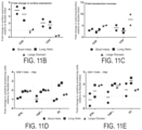
FIG. 11A-FIG. 11E illustrate exemplary in vitro parameters of CD19 TAC engineered with different connector variants. FIG. 11A illustrates TAC variant surface expression in both CD4 and CD8 cells. FIG. 11B illustrates surface expression of TAC comprising flexible connectors relative to TAC comprising helical or large domain connectors. FIG. 11C illustrates overall transduction of TAC comprising alternative connectors relative to the flexible connector.
FIG. 11D, FIG. 11E illustrate relative cell reactivity to antigen positive Raji cells.
FIG. 12A illustrates in vitro cytotoxicity of BCMA Tri-TAC variants engineered with different connectors. FIG. 12B illustrates in vivo tumor control of BCMA Tri-TAC variants engineered with the flexible connector compared to the short helical connector.
FIG. 13A-FIG. 13C illustrate properties of CD8α Tri-TAC scFv anti HER-2, and CD8α Tri-TAC DARPin anti-HER-2. FIG. 13A, FIG. 13C illustrate surface expression. FIG. 13B illustrates cytokine production.
FIG. 14A-FIG. 14D provide schematics of CD8 Tri-TAC variants. The anti HER-2-DARPin is used as an exemplary antigen-binding domain and the UCHT1 CD3 recruitment domain is used as an exemplary recruitment domain. FIG. 14A illustrates a Tri-TAC comprising a CD4 transmembrane and cytosolic domain (left), and comparable regions of a CD8α/CD8β heterodimer (right). Key regions for co-receptor functionality (arginine rich domain and CXCP motif) are highlighted. FIG. 14B is a schematic of a CD8α Tri-TAC comprising a Cysteine to Serine mutation to ensure a monomeric receptor distribution, and a CD8α cytosolic domain. FIG. 14C is a schematic of a CD8α+Rβ Tri-TAC comprising a Cysteine to Serine mutation to ensure a monomeric receptor distribution, and a chimeric CD8α cytosolic domain where the CD8α arginine rich region is replaced with the CD8β arginine rich region. FIG. 14D is a schematic of a CD8β+Lck Tri-TAC comprising a Cysteine to Serine mutation to ensure a monomeric receptor distribution, and a chimeric CD8β cytosolic domain, where the CD8α CXCP domain, which contains a Lck binding motif, was added to the C-terminus of the CD8β cytosolic domain.
FIG. 15A-FIG. 15E illustrate in vitro characterization of CD8 Tri-TAC variants relative to the prototypic Tri-TAC containing CD4 regions. FIG. 15A-FIG. 15B illustrate surface expression of CD8-Tri TAC variants relative to the prototypic Tri-TAC. FIG. 15C illustrates in vitro cytotoxicity of CD8-Tri TAC variants co-cultured with LOX IMVI (HER-2 negative) or A549, SKOV3, SKBR3 or MBA MB 231 (HER-2 positive). FIG. 15D illustrates cell division of T cells engineered with either the CD8 Tri-TAC variants or the prototypic Tri-TAC. FIG. 15E illustrates TCR surface expression of engineered T cells comprising CD8 Tri-TAC variants or the prototypic Tri-TAC.
FIG. 16 illustrates various Tri-TACs.
FIG. 17 illustrates TAC-CD19 insert in a pCCL lentiviral vector. FIG. 17 illustrates the various domains of a TAC-CD19 (a CD8a leader, FMC63 scFv, Myc Tag, huUCHT1 Y177T mutant and a truncated CD4 anchoring co-receptor domain).
FIG. 18 illustrates the in vivo efficacy of TAC-CD19 generated from different donors.
FIG. 19A-FIG. 19C illustrates an in vitro example of TAC-CD19 cytotoxicity against the tumor lines. FIG. 19A NALM-6 (acute lymphoblastic leukemia), FIG. 19B Jeko-1 (Mantle Cell Lymphoma) and FIG. 19C Raji (Burkitt's lymphoma).
FIG. 19D illustrates the schematic of 3 different in vivo tumor models in NRG mice.
FIG. 19E-FIG. 19G illustrate in vivo efficacy of CD19-TAC in NALM-6 (acute lymphoblastic leukemia) FIG. 19E, Jeko-1 (Mantle Cell Lymphoma) FIG. 19F, and Raji (Burkitt's lymphoma) FIG. 19G.
FIG. 20A illustrates the experimental set up of TAC-CD19 treated mice with NALM-6 tumor. Following successful treatment mice are then re-challenged with either NALM-6 (CD19 positive) or KMS11 (CD19 negative) tumor cells.
FIG. 20B illustrates in vivo efficacy of mice treated with TAC-CD19.
FIG. 21A illustrates the experimental design of evaluating dose regime and dosing impact on efficacy and cell expansion.
FIG. 21B illustrates in vivo survival of NALM-6 bearing mice treated with either a single or split dose of TAC-CD19.
FIG. 22A-FIG. 22B illustrate an experimental setup and data with regard to in vivo expansion of TAC-CD19 following a split dose administration. FIG. 22A illustrates the gating strategy used to identify T cells in mouse blood. FIG. 22B illustrates in vivo results of T cell expansion in blood.
FIG. 23A-FIG. 23C illustrate long term in vivo studies of TAC-CD19 in mice. FIG. 23A illustrates an experimental protocol of NALM-6 bearing mice being treated with various controls and TAC-CD19 at two dose levels. FIG. 23B illustrates in vivo efficacy of control vs two dose levels of TAC-CD19 treatment groups. FIG. 23C illustrates long term survival of low dose TAC-CD19 treated mice.
FIG. 24 illustrates clinical chemistry analysis results from mice treated with TAC-CD19 or non-transduced T cells.
FIG. 25 illustrates human cytokine released in mice blood following treatment with TAC-CD19 or non-transduced T cells.
FIG. 26A-FIG. 26C illustrates efficacy of BCMA-TAC in different configurations. FIG. 26A illustrates an experimental design. FIG. 26B illustrates various controls and test articles. FIG. 26C illustrates in vivo efficacy of various TAC constructs. FIG. 26A-FIG. 26C disclose “G4S” as SEQ ID NO: 73.
FIG. 27 illustrates that TACs proliferate when encountering antigen on cells, but not when the antigen is presented on artificial beads; but CARs proliferate irrespective if antigens are presented on beads or cells.
FIG. 28A-FIG. 28B illustrate TAC engineered T cells expand in vivo and provide long term protection, indicating cell persistence in a model of myeloma. FIG. 28A-FIG. 28B illustrate BCMA-TAC T cells reject multiple myeloma tumors in a KMS-11 xenograft model engineered with NanoLuc (KMS 11-NanoLuc) (BCMApos). Following tumor engraftment mice were treated with BCMA TAC-T cells (carrying Firefly Luciferase). TAC-T cells expand significantly following administration. This correlates with tumor regression. Treated mice were resistant to tumor rechallenge indicating long term persistence of TAC-T cells.
FIG. 29 illustrates human cytokine released in mice blood following treatment with TAC-CD19 or non-transduced T cells.
FIG. 30 illustrates exemplary histograms of TAC receptor surface expression in CD4 and CD8 engineered T cells. Cells were engineered with the muIgG HER2 TAC (EF1α promoter), the huIgG HER2 TAC (MSCV promoter) and muIgG HER2 TAC (MSCV promoter). Following engineering, T cells were stained with a TAC specific reagent and measured using flow cytometry. All constructs show comparable levels of surface expression, with EF1α driven expression being higher compared to the MSCV constructs.
FIG. 31 illustrates the relative percentage of T cells expressing either TNFα, IFNγ or IL-2 following co-cultured either with OVCAR3 (HER2 positive) or LOX IMVI (HER2 negative) cells. T cells were engineered with the muIgG HER2 TAC (EF1α promoter), a TAC construct lacking the HER2 binding domain (Δbinding TAC; EF1α promoter), the huIgG HER2 TAC (MSCV promoter) or the muIgG HER2 TAC (MSCV promoter). When co-cultured with LOX-IMVI (HER2neg), Δbinding TAC control and HER2 TAC cells do not show meaningful cytokine expression. All HER2 TAC engineered constructs co-cultured with OVCAR3(HER2pos) show similar ability to produce cytokines while control T cells engineered with Δbinding TAC show no meaningful cytokine production.
FIG. 32 . Illustrates in vivo efficacy of TAC engineered T cells in the OVCAR3 solid tumor model. T cells were engineered with the Δbinding TAC (EF1α promoter), the muIgG HER2 TAC (EF1α promoter), the huIgG HER2 TAC (MSCV promoter) or the muIgG HER2 TAC (MSCV promoter). Mice had been inoculated subcutaneously with OVCAR3 (HER-2-positive) tumors. These were grown to about 100 mm3 in size. Mice were then treated with a split dose of 6 million total HER2 TAC engineered, or Δbinding TAC control T cells 48 h apart via tail vain injection. Tumor progression was followed by biweekly measurements. Δbinding TAC showed no tumor control or tumor regression. All HER2 TAC engineered T cells showed significantly reduced tumor progression, including tumor regression, relative to control mice. All HER2 TAC engineered T cells had similar anti-tumor activity.
DETAILED DESCRIPTION
Cancer is a major health challenge, with over 150,000 cases of cancer expected to be diagnosed in Canada alone. While patients with early stage disease are sometimes treated effectively by conventional therapies (surgery, radiation, chemotherapy), few options are available to patients with advanced disease, and those options are typically palliative in nature.
Active immunotherapy seeks to employ the patient's immune system to clear tumors and offers an option to patients who have failed conventional therapies. Generally, this treatment involves infusing patients with large numbers of tumor-specific T cells. This approach has proven to be successful in early phase clinical trials for a number of diseases, including melanoma, myeloma, leukemia, lymphoma and synovial sarcoma. As a specific example, several clinical studies have demonstrated that immunotherapy with T cells are curative in patients with advanced melanoma, confirming the utility of this approach. Additionally, patients suffering from chronic lymphocytic leukemia (CLL) and acute lymphoblastic leukemia (ALL) have also been effectively treated and cured with T cell immunotherapy.
A key challenge facing the clinical application of adoptive T cell therapy is the source of the T cells. Typically, T cells isolated from a tumor-bearing patient are grown to large numbers ex vivo and are administered back into the patient to induce a robust anti-tumor immune response. Tumor specificity is achieved by either: (i) isolating naturally-occurring tumor-specific T cells from the patient; or (ii) engineering bulk T cells from the peripheral blood to express tumor-specific receptors. Naturally occurring tumor-specific T cells are rare and isolating such cells in therapeutic quantities from cancer patients is a laborious and costly procedure. In contrast, it is becoming more efficient to engineer readily available peripheral T cells with tumor-specific receptors through genetic manipulation. Techniques have been developed for this engineering process, which are clinically viable, and several clinical trials have demonstrated the feasibility and efficacy of genetically-engineered T cells for the treatment of cancer.
To this point, most engineered T cell therapies involving genetic modification of the T cells yield: (i) forced expression of T cell receptor (TCR); or (ii) a chimeric antigen receptor (CAR) specific for antigen targets on the tumor. To date, the chimeric antigen receptors used for engineering T cells consist of: (i) a targeting domain, usually a single-chain fragment variable (scFv); (ii) a transmembrane domain; and (iii) a cytosolic domain that contains signaling elements from the T cell receptor and associated proteins. Such chimeric antigen receptors have also been referred to as “T-body” or “Chimeric Immune Receptor” (CIR), but currently, most researchers use the term “CAR”. One advantage of the CAR approach is that it allows any patient's immune cells to be targeted against any desirable target in a major histocompatibility complex (MHC) independent manner. This is appealing as MHC presentation is often defective in tumor cells.
CARs are considered in modular terms and scientists have spent considerable time investigating the influence of different cytoplasmic signaling domains on CAR function. Conventional CARs generally share two main components: (i) the CD3 zeta cytoplasmic domain, which contains immunotyrosine activation motifs (ITAMs) critical for T cell activation; and (ii) components of costimulatory receptors that trigger important survival pathways such as the Akt pathway.
The first-generation CARs employed a single signaling domain from either CD3ξ or FcϵRIγ. Second-generation CARs combined the signaling domain of CD3ξ with the cytoplasmic domain of costimulatory receptors from either the CD28 or TNFR family of receptors. Most CAR-engineered T cells that are currently being tested in the clinic employ second-generation CARs where CD3ξ is coupled to the cytoplasmic domain of either CD28 or CD137. These second generation CARs have demonstrated anti-tumor activity in CD19-positive tumors. Third-generation CARs combined multiple costimulatory domains, but there is concern that third-generation CARs may lose antigen-specificity.
While CAR-engineered T cells have shown considerable promise in clinical application, they rely on a synthetic method for replacing the native activation signal that is provided by the T cell receptor (TCR). Since this synthetic receptor does not deliver all of the signaling components associated with the TCR (ex. ITAMs on CD3γ, CD3δ, CD3ϵ), it remains unclear whether the T cells are optimally activated by the CAR or how the CAR activation affects T cell differentiation (ex. progression to memory). Furthermore, since the CAR signaling domains are disconnected from their natural regulatory partners by the very nature of the CAR structure, there is an inherent risk that CARs may lead to a low-level of constitutive activation, which could result in off-target toxicities. Therefore, the synthetic nature of the prototypic CAR may disrupt canonical mechanisms that limit TCR activation, and may underpin the severe toxicity often associated with therapeutic doses of conventional CAR T cells.
Given these limitations, it is preferable to re-direct T cells to attack tumors via their natural TCR. To this end, a class of recombinant proteins termed “Bispecific T-cell Engagers” (BiTEs) has been created. These proteins employ bispecific antibody fragments to crosslink T-cell TCR receptors with target antigens. This leads to efficient T-cell activation, triggering cytotoxicity. Similarly, bi-specific antibodies have been generated that accomplish this goal and some scientists have simply linked anti-CD3 antibodies to tumor-specific antibodies employing chemical linkage. While these bi-specific proteins have demonstrated some activity in vitro, GMP production, short biological half-lives, and limited bioavailability represent significant challenges to the successful use of these molecules in cancer treatment. Additionally, these molecules also fail to properly recapitulate natural TCR signaling because they do not engage the TCR co-receptors (CD8 and CD4).
In view of the above, a need remains for chimeric receptors with enhanced activity and safety.
An alternate chimeric receptor, termed a Trifunctional T cell Antigen Coupler (Tri-TAC or TAC) receptor, has been developed which employs a distinct biology to direct the T cell to attack tumors. While the CAR is a fully synthetic receptor that stitches together components of T cell receptor (TCR) signaling complex, the TAC receptor re-directs the TCR towards tumor targets and recapitulates the native TCR signaling structure. For example, in some embodiments, the TACs disclosed herein activate natural Major Histocompatibility complex (MHC) signaling through the T-cell receptor (TCR), while retaining MHC-unrestricted targeting. Further, the TACs disclosed herein recruit the T-Cell Receptor (TCR) in combination with co-receptor stimulation. Moreover, in some embodiments, Tri-TACs disclosed herein show enhanced activity and safety.
Certain Terminology
The term “T cell” as used herein refers to a type of lymphocyte that plays a central role in cell-mediated immunity. T cells, also referred to as T lymphocytes, are distinguished from other lymphocytes, such as B cells and natural killer cells, by the presence of a T-cell receptor (TCR) on the cell surface. There are several subsets of T cells with distinct functions, including but not limited to, T helper cells, cytotoxic T cells, memory T cells, regulatory T cells and natural killer T cells.
The term “T cell antigen coupler” or TAC is used interchangeably with “trifunctional T cell antigen coupler” or Tri-TAC and refers to an engineered nucleic acid construct or polypeptide, that when expressed on a T cell, helps to facilitate the targeting of the T cell to a particular antigen. In some embodiments, the TAC comprises (a) a target-specific ligand, (b) a ligand that binds a protein associated with a T cell receptor (TCR) complex, and (c) a T cell receptor signaling domain.
The term “polynucleotide” and/or “nucleic acid sequence” and/or “nucleic acid” as used herein refers to a sequence of nucleoside or nucleotide monomers consisting of bases, sugars and intersugar (backbone) linkages. The term also includes modified or substituted sequences comprising non-naturally occurring monomers or portions thereof. The nucleic acid sequences of the present application may be deoxyribonucleic acid sequences (DNA) or ribonucleic acid sequences (RNA) and may include naturally occurring bases including adenine, guanine, cytosine, thymidine and uracil. The sequences may also contain modified bases. Examples of such modified bases include aza and deaza adenine, guanine, cytosine, thymidine and uracil; and xanthine and hypoxanthine. The nucleic acids of the present disclosure may be isolated from biological organisms, formed by laboratory methods of genetic recombination or obtained by chemical synthesis or other known protocols for creating nucleic acids.
The term “isolated polynucleotide” or “isolated nucleic acid sequence” as used herein refers to a nucleic acid substantially free of cellular material or culture medium when produced by recombinant DNA techniques, or chemical precursors, or other chemicals when chemically synthesized. An isolated nucleic acid is also substantially free of sequences which naturally flank the nucleic acid (i.e. sequences located at the 5′ and 3′ ends of the nucleic acid) from which the nucleic acid is derived. The term “nucleic acid” is intended to include DNA and RNA and is either double stranded or single stranded, and represents the sense or antisense strand. Further, the term “nucleic acid” includes the complementary nucleic acid sequences.
The term “recombinant nucleic acid” or “engineered nucleic acid” as used herein refers to a nucleic acid or polynucleotide that is not found in a biological organism. For example, recombinant nucleic acids may be formed by laboratory methods of genetic recombination (such as molecular cloning) to create sequences that would not otherwise be found in nature.
Recombinant nucleic acids may also be created by chemical synthesis or other known protocols for creating nucleic acids.
The term “polypeptide” or “protein” as used herein describes a chain of amino acids. A polypeptide or protein of this disclosure is a peptide, which usually describes a chain of amino acids. The term protein as used herein also describes a large molecule comprising one or more chains of amino acids and, in some embodiments, is a fragment or domain of a protein or a full length protein. Furthermore, as used herein, the term protein either refers to a linear chain of amino acids or to a chain of amino acids that has been processed and folded into a functional protein. The protein structure is divided into four distinct levels: (1) primary structure—referring to the sequence of amino acids in the polypeptide chain, (2) secondary structure—referring to the regular local sub-structures on the polypeptide backbone chain, such as α-helix and β-sheets, (3) tertiary structure—referring to the three-dimensional structure if monomeric and multimeric protein molecules, and (4) quaternary structure—referring to the three-dimensional structure comprising the aggregation of two or more individual polypeptide chains that operate as a single functional unit. The proteins of the present disclosure, in some embodiments, are obtained by isolation and purification of the proteins from cells where they are produced naturally, by enzymatic (e.g., proteolytic) cleavage, and/or recombinantly by expression of nucleic acid encoding the proteins or fragments of this disclosure. The proteins and/or fragments of this disclosure, in some embodiments, is obtained by chemical synthesis or other known protocols for producing proteins and fragments.
The term “isolated polypeptide” refers to a polypeptide substantially free of cellular material or culture medium when produced by recombinant DNA techniques, or chemical precursors or other chemicals when chemically synthesized.
The term “antibody” as used herein is intended to include monoclonal antibodies, polyclonal antibodies, single chain antibodies, chimeric antibodies, and antibody fusions. The antibody may be from recombinant sources and/or produced in transgenic animals. The term “antibody fragment” as used herein is intended to include without limitations Fab, Fab′, F(ab′)2, scFv, dsFv, ds-scFv, dimers, minibodies, diabodies, and multimers thereof, multispecific antibody fragments and Domain Antibodies.
The term “vector” as used herein refers to a polynucleotide that is used to deliver a nucleic acid to the inside of a cell. In some embodiments, a vector is an expression vector comprising expression control sequences (for example, a promoter) operatively linked to a nucleic acid to be expressed in a cell. Vectors known in the art include, but are not limited to, plasmids, phages, cosmids and viruses.
The term “tumor antigen” or “tumor associated antigen” as used herein refers to an antigenic substance produced in tumor cells that triggers an immune response in a host (e.g. which is presented by MHC complexes). In some embodiments, a tumor antigen is on the surface of a tumor cell.
The term “T cell receptor” or TCR as used herein refers to a complex of integral membrane proteins that participates in the activation of T cells in response to the binding of an antigen. The TCR is a disulfide-linked membrane-anchored heterodimer normally consisting of the highly variable alpha (α) and beta (β) chains expressed as part of a complex with the invariant CD3 (cluster of differentiation 3) chain molecules. T cells expressing this receptor are referred to as α:β (or αβ) T cells, though a minority of T cells express an alternate receptor, formed by variable gamma (γ) and delta (δ) chains, referred as γδT cells. CD3 is a protein complex composed of four distinct chains. In mammals, the complex contains a CD3γ chain, a CD3δ chain, two CD3ϵ chains and two CD3ξ chains.
As used herein, the term “transmembrane and cytosolic domain” refers to a polypeptide that comprises a transmembrane domain and a cytosolic domain of a protein associated with the T cell receptor (TCR) complex. In some embodiments, such transmembrane and cytosolic domain may include, but is not limited to, protein domains that (a) associate with the lipid raft and/or (b) bind Lck.
A “TCR co-receptor” as used herein, refers to a molecule that assists the T cell receptor (TCR) in communicating with an antigen-presenting cell and may be considered part of the first signal that leads to the activation of the TCR. Examples of TCR co-receptors include, but are not limited to, CD4, LAG3, and CD8.
A “TCR co-stimulator” as used herein, refers to a molecule that enhances the response of a T cell to an antigen and may be considered as the second signal that leads to the activation of the TCR. Examples of TCR co-stimulators include, but are not limited to, ICOS, CD27, CD28, 4-1BB (CD 137), OX40 (CD134), CD30, CD40, lymphocyte fiction-associated antigen 1 (LFA-1), CD2, CD7, LIGHT, NKG2C, B7-H3, and a ligand that specifically binds CD83.
A “TCR co-inhibitor” or “checkpoint receptor” as used herein, refers to a molecule that inhibits the response of a T cell to an antigen. Examples of TCR co-inhibitors include, but are not limited to, PD-1, TIM3, LAG-3, TIGIT, BTLA, CD160, and CD37.
The terms “recipient”, “individual”, “subject”, “host”, and “patient”, are used interchangeably herein and in some embodiments, refer to any mammalian subject for whom diagnosis, treatment, or therapy is desired, particularly humans. “Mammal” for purposes of treatment refers to any animal classified as a mammal, including humans, domestic and farm animals, and laboratory, zoo, sports, or pet animals, such as dogs, horses, cats, cows, sheep, goats, pigs, mice, rats, rabbits, guinea pigs, monkeys etc. In some embodiments, the mammal is human. None of these terms require the supervision of medical personnel.
As used herein, the terms “treatment,” “treating,” and the like, in some embodiments, refer to administering an agent, or carrying out a procedure, for the purposes of obtaining an effect. The effect may be prophylactic in terms of completely or partially preventing a disease or symptom thereof and/or may be therapeutic in terms of affecting a partial or complete cure for a disease and/or symptoms of the disease. “Treatment,” as used herein, may include treatment of a disease or disorder (e.g. cancer) in a mammal, particularly in a human, and includes: (a) preventing the disease or a symptom of a disease from occurring in a subject which may be predisposed to the disease but has not yet been diagnosed as having it (e.g., including diseases that may be associated with or caused by a primary disease; (b) inhibiting the disease, i.e., arresting its development; and (c) relieving the disease, i.e., causing regression of the disease. Treating may refer to any indicia of success in the treatment or amelioration or prevention of a cancer, including any objective or subjective parameter such as abatement; remission; diminishing of symptoms; or making the disease condition more tolerable to the patient; slowing in the rate of degeneration or decline; or making the final point of degeneration less debilitating. The treatment or amelioration of symptoms is based on one or more objective or subjective parameters; including the results of an examination by a physician. Accordingly, the term “treating” includes the administration of the compounds or agents of the present invention to prevent, delay, alleviate, arrest or inhibit development of the symptoms or conditions associated with diseases (e.g. cancer). The term “therapeutic effect” refers to the reduction, elimination, or prevention of the disease, symptoms of the disease, or side effects of the disease in the subject.
As used herein, singular forms “a”, “and,” and “the” include plural referents unless the context clearly indicates otherwise. Thus, for example, reference to “an antibody” includes a plurality of antibodies and reference to “an antibody” in some embodiments includes multiple antibodies, and so forth.
As used herein, all numerical values or numerical ranges include whole integers within or encompassing such ranges and fractions of the values or the integers within or encompassing ranges unless the context clearly indicates otherwise. Thus, for example, reference to a range of 90-100%, includes 91%, 92%, 93%, 94%, 95%, 95%, 96%, 97%, etc., as well as 91.1%, 91.2%, 91.3%, 91.4%, 91.5%, etc., 92.1%, 92.2%, 92.3%, 92.4%, 92.5%, etc., and so forth. In another example, reference to a range of 1-5,000 fold includes 1, 2, 3, 4, 5, 6, 7, 8, 9, 10, 11, 12, 13, 14, 15, 16, 17, 18, 19, 20, fold, etc., as well as 1.1, 1.2, 1.3, 1.4, 1.5, fold, etc., 2.1, 2.2, 2.3, 2.4, 2.5, fold, etc., and so forth.
“About” a number, as used herein, refers to range including the number and ranging from 10% below that number to 10% above that number. “About” a range refers to 10% below the lower limit of the range, spanning to 10% above the upper limit of the range.
“Percent (%) identity” refers to the extent to which two sequences (nucleotide or amino acid) have the same residue at the same positions in an alignment. For example, “an amino acid sequence is X % identical to SEQ ID NO: Y” refers to % identity of the amino acid sequence to SEQ ID NO: Y and is elaborated as X % of residues in the amino acid sequence are identical to the residues of sequence disclosed in SEQ ID NO: Y. Generally, computer programs are employed for such calculations. Exemplary programs that compare and align pairs of sequences, include ALIGN (Myers and Miller, 1988), FASTA (Pearson and Lipman, 1988; Pearson, 1990) and gapped BLAST (Altschul et al., 1997), BLASTP, BLASTN, or GCG (Devereux et al., 1984).
As used herein, the term “selective binding” refers to the higher affinity with which a molecule (e.g. protein such as a target-binding ligand of TAC) binds its target molecule (e.g. target antigen such as HER-2, BCMA, or CD19) over other molecules.
T Cell Antigen Coupler (Tri-TAC or TAC)
Disclosed herein, in certain embodiments, are nucleic acids encoding a Trifunctional T cell-antigen coupler (Tri-TAC). In some embodiments, the nucleic acids encoding a Tri-TAC comprises: (a) a first polynucleotide encoding a target-specific ligand; (b) a second polynucleotide encoding a ligand that binds a TCR complex; and (c) a third polynucleotide encoding a transmembrane domain and cytosolic domain. In some embodiments, the nucleic acids encoding a Tri-TAC do not encode a co-stimulatory domain. In some embodiments, the nucleic acids encoding a Tri-TAC do not encode a co-activation domain.
Target-Specific Ligand
The target-specific ligand, also referred to as an antigen binding domain, refers to any substance or molecule that binds, directly or indirectly, to a target cell. In some embodiments, the target specific ligand binds to an antigen on the target cell. In some embodiments, a target cell is a cell associated with a disease state, including, but not limited to, cancer, hematologic malignancy, large B-cell lymphoma, diffuse large B-cell lymphoma, primary mediastinal B cell lymphoma, high grade B-cell lymphoma, or large B cell lymphoma arising from follicular lymphoma. In some embodiments, a target cell is a tumor cell. In some embodiments, a target-specific ligand binds to a tumor antigen or tumor associated antigen on a tumor cell. In some embodiments, the target antigen is a tumor antigen. In some embodiments, the tumor antigen when proteinaceous is a sequence of 8 or more amino acids up to the full protein. In some embodiments, the tumor antigen is any number of amino acids in between 8 and the full length protein which comprises at least one antigenic fragment of the full length protein that is presented in a Major Histocompatibility Complex (MHC). Examples of tumor antigens include, but are not limited to, CD19, HER-2 (erbB-2), B-cell maturation antigen (BCMA), alphafetoprotein (AFP), carcinoembryonic antigen (CEA), CA-125, MUC-1, epithelial tumor antigen (ETA), tyrosinase, melanoma-associated antigen (MAGE), prostate-specific antigen (PSA), glioma-associated antigen, β-human chorionic gonadotropin, thyroglobulin, RAGE-1, MN-CA IX, human telomerase reverse transcriptase, RU1, RU2 (AS), intestinal carboxyl esterase, mut hsp70-2, M-CSF, prostase, PAP, NY-ESO-1, LAGE-1a, p53, prostein, PSMA, survivin and telomerase, prostate-carcinoma tumor antigen-1 (PCTA-1), ELF2M, neutrophil elastase, CD22, insulin growth factor (IGF)-I, IGF-II, IGF-I receptor and mesothelin.
In some embodiments, the target-specific ligands include, but are not limited to, antibodies and fragments thereof, for example single chain antibodies such as single-chain antibodies (scFvs), single domain antibodies, peptides, peptidomimetics, proteins, glycoproteins, or proteoglycans that bind to the target cell and/or antigen. In some embodiments, the target-specific ligands include, but are not limited to, designed ankyrin repeat proteins (DARPins), lectins, knottins, centryrins, anticalins, or naturally occurring ligands for the tumor antigen, such as growth factors, enzyme substrates, receptors or binding proteins. In some embodiments, target specific ligands include non-protein compounds that bind to target cells and/or antigens, including but not limited to carbohydrates, lipids, nucleic acids, or small molecules. In some embodiments, a target-specific ligand is a designed ankyrin repeat (DARPin) targeted to a specific cell and/or antigen. In some embodiments, a target-specific ligand is a single-chain variable fragment (ScFv) targeted to a specific cell and/or antigen.
In some embodiments, the tumor antigen is a HER-2 antigen. In some embodiments, the HER-2 specific ligand comprises an antigen binding domain of an antibody selected from Trastuzumab, Pertuzumab, Lapatinib, Neratinib, Ado-trastuzmab Emtansine, Gancotamab, Margetuximab, Timigutuzumab, and Ertumaxomab. In some embodiments, the target-specific ligand is a DARPin that selectively binds a HER-2 (erbB-2) antigen. In some embodiments, the target-specific ligand is a DARPin that specifically binds a HER-2 (erbB-2) antigen. In some embodiments, the DARPin targeted to HER-2 (erb-2) comprises SEQ ID NO: 7 or SEQ ID NO: 8.
In some embodiments, the first polynucleotide comprises a nucleotide sequence having at least 70% sequence identity with SEQ ID NO: 7. In some embodiments, the first polynucleotide comprises a nucleotide sequence having at least 75% sequence identity with SEQ ID NO: 7. In some embodiments, the first polynucleotide comprises a nucleotide sequence having at least 80% sequence identity with SEQ ID NO: 7. In some embodiments, the first polynucleotide comprises a nucleotide sequence having at least 85% sequence identity with SEQ ID NO: 7. In some embodiments, the first polynucleotide comprises a nucleotide sequence having at least 90% sequence identity with SEQ ID NO: 7. In some embodiments, the first polynucleotide comprises a nucleotide sequence having at least 95% sequence identity with SEQ ID NO: 7. In some embodiments, the first polynucleotide comprises a nucleotide sequence of SEQ ID NO: 7.
In some embodiments, the target-specific ligand comprises an amino acid sequence having at least 70% sequence identity with SEQ ID NO: 8. In some embodiments, the target-specific ligand comprises an amino acid sequence having at least 75% sequence identity with SEQ ID NO: 8. In some embodiments, the target-specific ligand comprises an amino acid sequence having at least 80% sequence identity with SEQ ID NO: 8. In some embodiments, the target-specific ligand comprises an amino acid sequence having at least 85% sequence identity with SEQ ID NO: 8. In some embodiments, the target-specific ligand comprises an amino acid sequence having at least 90% sequence identity with SEQ ID NO: 8. In some embodiments, the target-specific ligand comprises an amino acid sequence having at least 95% sequence identity with SEQ ID NO: 8. In some embodiments, the target-specific ligand comprises an amino acid sequence of SEQ ID NO: 8.
In some embodiments, the tumor antigen is a BCMA antigen. In some embodiments, the BCMA specific ligand comprises an antigen binding domain of an antibody selected from Belantamab mafodotin, and GSK2857916. In some embodiments, the target-specific ligand is a scFv that selectively binds BCMA. In some embodiments, the target-specific ligand is a scFv that specifically binds BCMA. In some embodiments, the scFv that binds BCMA comprises SEQ ID NO: 33 or SEQ ID NO: 34.
In some embodiments, the first polynucleotide comprises a nucleotide sequence having at least 70% sequence identity with SEQ ID NO: 33. In some embodiments, the first polynucleotide comprises a nucleotide sequence having at least 75% sequence identity with SEQ ID NO: 33. In some embodiments, the first polynucleotide comprises a nucleotide sequence having at least 80% sequence identity with SEQ ID NO: 33. In some embodiments, the first polynucleotide comprises a nucleotide sequence having at least 85% sequence identity with SEQ ID NO: 33. In some embodiments, the first polynucleotide comprises a nucleotide sequence having at least 90% sequence identity with SEQ ID NO: 33. In some embodiments, the first polynucleotide comprises a nucleotide sequence having at least 95% sequence identity with SEQ ID NO: 33. In some embodiments, the first polynucleotide comprises a nucleotide sequence of SEQ ID NO: 33.
In some embodiments, the target-specific ligand comprises an amino acid sequence having at least 70% sequence identity with SEQ ID NO: 34. In some embodiments, the target-specific ligand comprises an amino acid sequence having at least 75% sequence identity with SEQ ID NO: 34. In some embodiments, the target-specific ligand comprises an amino acid sequence having at least 80% sequence identity with SEQ ID NO: 34. In some embodiments, the target-specific ligand comprises an amino acid sequence having at least 85% sequence identity with SEQ ID NO: 34. In some embodiments, the target-specific ligand comprises an amino acid sequence having at least 90% sequence identity with SEQ ID NO: 34. In some embodiments, the target-specific ligand comprises an amino acid sequence having at least 95% sequence identity with SEQ ID NO: 34. In some embodiments, the target-specific ligand comprises an amino acid sequence of SEQ ID NO: 34.
In some embodiments, the tumor antigen is a CD19 antigen. In some embodiments, the target-specific ligand is a scFv that selectively binds CD19. In some embodiments, the target-specific ligand is a scFv that specifically binds CD19. In some embodiments, the scFv that binds CD19 comprises SEQ ID NO: 35 or SEQ ID NO: 36.
In some embodiments, the first polynucleotide comprises a nucleotide sequence having at least 70% sequence identity with SEQ ID NO: 35. In some embodiments, the first polynucleotide comprises a nucleotide sequence having at least 75% sequence identity with SEQ ID NO: 35. In some embodiments, the first polynucleotide comprises a nucleotide sequence having at least 80% sequence identity with SEQ ID NO: 35. In some embodiments, the first polynucleotide comprises a nucleotide sequence having at least 85% sequence identity with SEQ ID NO: 35. In some embodiments, the first polynucleotide comprises a nucleotide sequence having at least 90% sequence identity with SEQ ID NO: 35. In some embodiments, the first polynucleotide comprises a nucleotide sequence having at least 95% sequence identity with SEQ ID NO: 35. In some embodiments, the first polynucleotide comprises a nucleotide sequence of SEQ ID NO: 35.
In some embodiments, the target-specific ligand comprises an amino acid sequence having at least 70% sequence identity with SEQ ID NO: 36. In some embodiments, the target-specific ligand comprises an amino acid sequence having at least 75% sequence identity with SEQ ID NO: 36. In some embodiments, the target-specific ligand comprises an amino acid sequence having at least 80% sequence identity with SEQ ID NO: 36. In some embodiments, the target-specific ligand comprises an amino acid sequence having at least 85% sequence identity with SEQ ID NO: 36. In some embodiments, the target-specific ligand comprises an amino acid sequence having at least 90% sequence identity with SEQ ID NO: 36. In some embodiments, the target-specific ligand comprises an amino acid sequence having at least 95% sequence identity with SEQ ID NO: 36. In some embodiments, the target-specific ligand comprises an amino acid sequence of SEQ ID NO: 36.
Ligand that Binds a TCR Complex
In some embodiments, the TAC comprises a ligand that binds a protein associated with the TCR complex. In some embodiments, the ligand that binds a protein associated with a TCR complex comprises a substance that binds, directly or indirectly, to a protein of the TCR. In some embodiments, the ligand that binds a protein associated with a TCR complex comprises a substance that selectively binds to a protein of the TCR. In some embodiments, the ligand that binds a protein associated with a TCR complex comprises a substance that specifically binds to a protein of the TCR. Proteins associated with the TCR include, but are not limited, to the TCR alpha (α) chain, TCR beta (β) chain, TCR gamma (γ) chain, TCR delta (δ) chain, CD3γ chain, CD3δ chain and CD3ϵ chains. In some embodiments, a ligand that binds a protein associated with the TCR complex is an antibody to the TCR alpha (α) chain, TCR beta (β) chain, TCR gamma (γ) chain, TCR delta (δ) chain, CD3γ chain, CD3δ chain and/or CD3ϵ chain. In some embodiments, the protein associated with a TCR complex is CD3. In some embodiments, the protein associated with a TCR complex is CD3ϵ. Examples of CD3 antibodies, include, but are not limited to, for. In some embodiments, the antibody that binds CD3 is a single chain antibody, for example a single-chain variable fragment (scFv). In some embodiments, the ligand that binds a TCR is anti-CD3 antibody, or a fragment thereof, such as muromonab, otelixizumab, teplizumab, visilizumab, CD3-12, MEM-57, 4D10A6, CD3D, or TR66.
In some embodiments, the CD3 is of a TCR complex on a cell expressing the second polynucleotide. In some embodiments, the binding of the CD3 induces activation of a cell expressing the second polynucleotide.
In some embodiments, the ligand that binds a TCR complex is UCHT1, or a variant thereof. In some embodiments, the ligand that binds a TCR complex is UCHT1 (SEQ ID NO: 13, SEQ ID NO: 14 or homologs thereof). In some embodiments, the UCHT1 ligand binds CD3. In some embodiments, the UCHT1 ligand selectively binds CD3. In some embodiments, the UCHT1 ligand specifically binds CD3. In some embodiments, the UCHT1 ligand binds CD3ϵ. In some embodiments, the UCHT1 ligand selectively binds CD3ϵ. In some embodiments, the UCHT1 ligand specifically binds CD3ϵ. In some embodiments, the UCHT1 ligand is encoded by SEQ ID NO 13. In some embodiments, the UCHT1 ligand comprises SEQ ID NO 14. In some embodiments, the UCHT1 ligand is mutated. In some embodiments, the UCHT1 ligand comprises a Y182T mutation (also referred to as UCHT1 (Y182T)) (SEQ ID NO: 71 and SEQ ID NO: 72). In some embodiments, the UCHT1 (Y182T) ligand binds CD3. In some embodiments, the UCHT1 (Y182T) ligand selectively binds CD3. In some embodiments, the UCHT1 (Y182T) ligand specifically binds CD3. In some embodiments, the UCHT1 (Y182T) ligand binds CD3ϵ. In some embodiments, the UCHT1 (Y182T) ligand selectively binds CD3ϵ. In some embodiments, the UCHT1 (Y182T) ligand specifically binds CD3ϵ. In some embodiments, the UCHT1 (Y182T) ligand is encoded by SEQ ID NO 71. In some embodiments, the UCHT1 (Y182T) ligand comprises SEQ ID NO 72. In some embodiments, the ligand that binds a TCR complex is a humanized UCHT1 (huUCHT1). In some embodiments, the ligand that binds a TCR complex is huUCHT1 (SEQ ID NO 43, SEQ ID NO: 44 or homologs thereof). In some embodiments, the huUCHT1 ligand binds CD3. In some embodiments, the huUCHT1 ligand selectively binds CD3. In some embodiments, the huUCHT1 ligand specifically binds CD3. In some embodiments, the huUCHT1 ligand binds CD3ϵ. In some embodiments, the huUCHT1 ligand selectively binds CD3ϵ. In some embodiments, the huUCHT1 ligand specifically binds CD3ϵ. In some embodiments, the huUCHT1 ligand is encoded by SEQ ID NO 43. In some embodiments, the huUCHT1 ligand comprises SEQ ID NO 44. In some embodiments, the huUCHT1 has a Y177T mutation (also referred to as huUCHT1 (Y177T)) (SEQ ID NO: 45 and SEQ ID NO: 46). In some embodiments, the huUCHT1 (Y177T) ligand binds CD3. In some embodiments, the huUCHT1 (Y177T) ligand selectively binds CD3. In some embodiments, the huUCHT1 (Y177T) ligand specifically binds CD3. In some embodiments, the huUCHT1 (Y177T) ligand binds CD3ϵ. In some embodiments, the huUCHT1 (Y177T) ligand selectively binds CD3ϵ. In some embodiments, the huUCHT1 (Y177T) ligand specifically binds CD3ϵ. In some embodiments, the huUCHT1 (Y177T) ligand is encoded by SEQ ID NO 45. In some embodiments, the huUCHT1 ligand comprises SEQ ID NO 46.
In some embodiments, the second polynucleotide comprises a nucleotide sequence having at least 70% sequence identity with SEQ ID NO: 13. In some embodiments, the second polynucleotide comprises a nucleotide sequence having at least 75% sequence identity with SEQ ID NO: 13. In some embodiments, the second polynucleotide comprises a nucleotide sequence having at least 80% sequence identity with SEQ ID NO: 13. In some embodiments, the second polynucleotide comprises a nucleotide sequence having at least 85% sequence identity with SEQ ID NO: 13. In some embodiments, the second polynucleotide comprises a nucleotide sequence having at least 90% sequence identity with SEQ ID NO: 13. In some embodiments, the second polynucleotide comprises a nucleotide sequence having at least 95% sequence identity with SEQ ID NO: 13. In some embodiments, the second polynucleotide comprises a nucleotide sequence of SEQ ID NO: 13.
In some embodiments, the ligand that binds a TCR complex comprises an amino acid sequence having at least 70% sequence identity with SEQ ID NO: 14. In some embodiments, the ligand that binds a TCR complex comprises an amino acid sequence having at least 75% sequence identity with SEQ ID NO: 14. In some embodiments, the ligand that binds a TCR complex comprises an amino acid sequence having at least 80% sequence identity with SEQ ID NO: 14. In some embodiments, the ligand that binds a TCR complex comprises an amino acid sequence having at least 85% sequence identity with SEQ ID NO: 14. In some embodiments, the ligand that binds a TCR complex comprises an amino acid sequence having at least 90% sequence identity with SEQ ID NO: 14. In some embodiments, the ligand that binds a TCR complex comprises an amino acid sequence having at least 95% sequence identity with SEQ ID NO: 14. In some embodiments, the ligand that binds a TCR complex comprises an amino acid sequence of SEQ ID NO: 14.
In some embodiments, the second polynucleotide comprises a nucleotide sequence having at least 70% sequence identity with SEQ ID NO: 71. In some embodiments, the second polynucleotide comprises a nucleotide sequence having at least 75% sequence identity with SEQ ID NO: 71. In some embodiments, the second polynucleotide comprises a nucleotide sequence having at least 80% sequence identity with SEQ ID NO: 71. In some embodiments, the second polynucleotide comprises a nucleotide sequence having at least 85% sequence identity with SEQ ID NO: 71. In some embodiments, the second polynucleotide comprises a nucleotide sequence having at least 90% sequence identity with SEQ ID NO: 71. In some embodiments, the second polynucleotide comprises a nucleotide sequence having at least 95% sequence identity with SEQ ID NO: 71. In some embodiments, the second polynucleotide comprises a nucleotide sequence of SEQ ID NO: 71.
In some embodiments, the ligand that binds a TCR complex comprises an amino acid sequence having at least 70% sequence identity with SEQ ID NO: 72. In some embodiments, the ligand that binds a TCR complex comprises an amino acid sequence having at least 75% sequence identity with SEQ ID NO: 72. In some embodiments, the ligand that binds a TCR complex comprises an amino acid sequence having at least 80% sequence identity with SEQ ID NO: 72. In some embodiments, the ligand that binds a TCR complex comprises an amino acid sequence having at least 85% sequence identity with SEQ ID NO: 72. In some embodiments, the ligand that binds a TCR complex comprises an amino acid sequence having at least 90% sequence identity with SEQ ID NO: 72. In some embodiments, the ligand that binds a TCR complex comprises an amino acid sequence having at least 95% sequence identity with SEQ ID NO: 72. In some embodiments, the ligand that binds a TCR complex comprises an amino acid sequence of SEQ ID NO: 72.
In some embodiments, the second polynucleotide comprises a nucleotide sequence having at least 70% sequence identity with SEQ ID NO: 43. In some embodiments, the second polynucleotide comprises a nucleotide sequence having at least 75% sequence identity with SEQ ID NO: 43. In some embodiments, the second polynucleotide comprises a nucleotide sequence having at least 80% sequence identity with SEQ ID NO: 43. In some embodiments, the second polynucleotide comprises a nucleotide sequence having at least 85% sequence identity with SEQ ID NO: 43. In some embodiments, the second polynucleotide comprises a nucleotide sequence having at least 90% sequence identity with SEQ ID NO: 43. In some embodiments, the second polynucleotide comprises a nucleotide sequence having at least 95% sequence identity with SEQ ID NO: 43. In some embodiments, the second polynucleotide comprises a nucleotide sequence of SEQ ID NO: 43.
In some embodiments, the ligand that binds a TCR complex comprises an amino acid sequence having at least 70% sequence identity with SEQ ID NO: 44. In some embodiments, the ligand that binds a TCR complex comprises an amino acid sequence having at least 75% sequence identity with SEQ ID NO: 44. In some embodiments, the ligand that binds a TCR complex comprises an amino acid sequence having at least 80% sequence identity with SEQ ID NO: 44. In some embodiments, the ligand that binds a TCR complex comprises an amino acid sequence having at least 85% sequence identity with SEQ ID NO: 44. In some embodiments, the ligand that binds a TCR complex comprises an amino acid sequence having at least 90% sequence identity with SEQ ID NO: 44. In some embodiments, the ligand that binds a TCR complex comprises an amino acid sequence having at least 95% sequence identity with SEQ ID NO: 44. In some embodiments, the ligand that binds a TCR complex comprises an amino acid sequence of SEQ ID NO: 44.
In some embodiments, the second polynucleotide comprises a nucleotide sequence having at least 70% sequence identity with SEQ ID NO: 45. In some embodiments, the second polynucleotide comprises a nucleotide sequence having at least 75% sequence identity with SEQ ID NO: 45. In some embodiments, the second polynucleotide comprises a nucleotide sequence having at least 80% sequence identity with SEQ ID NO: 45. In some embodiments, the second polynucleotide comprises a nucleotide sequence having at least 85% sequence identity with SEQ ID NO: 45. In some embodiments, the second polynucleotide comprises a nucleotide sequence having at least 90% sequence identity with SEQ ID NO: 45. In some embodiments, the second polynucleotide comprises a nucleotide sequence having at least 95% sequence identity with SEQ ID NO: 45. In some embodiments, the second polynucleotide comprises a nucleotide sequence of SEQ ID NO: 45.
In some embodiments, the ligand that binds a TCR complex comprises an amino acid sequence having at least 70% sequence identity with SEQ ID NO: 46. In some embodiments, the ligand that binds a TCR complex comprises an amino acid sequence having at least 75% sequence identity with SEQ ID NO: 46. In some embodiments, the ligand that binds a TCR complex comprises an amino acid sequence having at least 80% sequence identity with SEQ ID NO: 46. In some embodiments, the ligand that binds a TCR complex comprises an amino acid sequence having at least 85% sequence identity with SEQ ID NO: 46. In some embodiments, the ligand that binds a TCR complex comprises an amino acid sequence having at least 90% sequence identity with SEQ ID NO: 46. In some embodiments, the ligand that binds a TCR complex comprises an amino acid sequence having at least 95% sequence identity with SEQ ID NO: 46. In some embodiments, the ligand that binds a TCR complex comprises an amino acid sequence of SEQ ID NO: 46.
In some embodiments, the ligand that binds to a CD3 is OKT3. In some embodiments, the OKT3 ligand binds CD3. In some embodiments, the OKT3 ligand selectively binds CD3. In some embodiments, the OKT3 ligand specifically binds CD3. In some embodiments, the OKT3 ligand binds CD3ϵ. In some embodiments, the OKT3 ligand selectively binds CD3ϵ. In some embodiments, the OKT3 ligand specifically binds CD3ϵ. In some embodiments, the murine OKT3 ligand is encoded by SEQ ID NO 21. In some embodiments, the OKT3 ligand comprises SEQ ID NO 22.
In some embodiments, the second polynucleotide comprises a nucleotide sequence having at least 70% sequence identity with SEQ ID NO: 21. In some embodiments, the second polynucleotide comprises a nucleotide sequence having at least 75% sequence identity with SEQ ID NO: 21. In some embodiments, the second polynucleotide comprises a nucleotide sequence having at least 80% sequence identity with SEQ ID NO: 21. In some embodiments, the second polynucleotide comprises a nucleotide sequence having at least 85% sequence identity with SEQ ID NO: 21. In some embodiments, the second polynucleotide comprises a nucleotide sequence having at least 90% sequence identity with SEQ ID NO: 21. In some embodiments, the second polynucleotide comprises a nucleotide sequence having at least 95% sequence identity with SEQ ID NO: 21. In some embodiments, the second polynucleotide comprises a nucleotide sequence of SEQ ID NO: 21.
In some embodiments, the ligand that binds a TCR complex comprises an amino acid sequence having at least 70% sequence identity with SEQ ID NO: 22. In some embodiments, the ligand that binds a TCR complex comprises an amino acid sequence having at least 75% sequence identity with SEQ ID NO: 22. In some embodiments, the ligand that binds a TCR complex comprises an amino acid sequence having at least 80% sequence identity with SEQ ID NO: 22. In some embodiments, the ligand that binds a TCR complex comprises an amino acid sequence having at least 85% sequence identity with SEQ ID NO: 22. In some embodiments, the ligand that binds a TCR complex comprises an amino acid sequence having at least 90% sequence identity with SEQ ID NO: 22. In some embodiments, the ligand that binds a TCR complex comprises an amino acid sequence having at least 95% sequence identity with SEQ ID NO: 22. In some embodiments, the ligand that binds a TCR complex comprises an amino acid sequence of SEQ ID NO: 22.
In some embodiments, the ligand that binds to a CD3 is F6A. In some embodiments, the F6A ligand binds CD3. In some embodiments, the F6A ligand selectively binds CD3. In some embodiments, the F6A ligand specifically binds CD3. In some embodiments, the F6A ligand binds CD3ϵ. In some embodiments, the F6A ligand selectively binds CD3ϵ. In some embodiments, the F6A ligand specifically binds CD3ϵ. In some embodiments, the murine F6A ligand is encoded by SEQ ID NO 23. In some embodiments, the F6A ligand comprises SEQ ID NO 24.
In some embodiments, the second polynucleotide comprises a nucleotide sequence having at least 70% sequence identity with SEQ ID NO: 23. In some embodiments, the second polynucleotide comprises a nucleotide sequence having at least 75% sequence identity with SEQ ID NO: 23. In some embodiments, the second polynucleotide comprises a nucleotide sequence having at least 80% sequence identity with SEQ ID NO: 23. In some embodiments, the second polynucleotide comprises a nucleotide sequence having at least 85% sequence identity with SEQ ID NO: 23. In some embodiments, the second polynucleotide comprises a nucleotide sequence having at least 90% sequence identity with SEQ ID NO: 23. In some embodiments, the second polynucleotide comprises a nucleotide sequence having at least 95% sequence identity with SEQ ID NO: 23. In some embodiments, the second polynucleotide comprises a nucleotide sequence of SEQ ID NO: 23.
In some embodiments, the ligand that binds a TCR complex comprises an amino acid sequence having at least 70% sequence identity with SEQ ID NO: 24. In some embodiments, the ligand that binds a TCR complex comprises an amino acid sequence having at least 75% sequence identity with SEQ ID NO: 24. In some embodiments, the ligand that binds a TCR complex comprises an amino acid sequence having at least 80% sequence identity with SEQ ID NO: 24. In some embodiments, the ligand that binds a TCR complex comprises an amino acid sequence having at least 85% sequence identity with SEQ ID NO: 24. In some embodiments, the ligand that binds a TCR complex comprises an amino acid sequence having at least 90% sequence identity with SEQ ID NO: 24. In some embodiments, the ligand that binds a TCR complex comprises an amino acid sequence having at least 95% sequence identity with SEQ ID NO: 24. In some embodiments, the ligand that binds a TCR complex comprises an amino acid sequence of SEQ ID NO: 24.
In some embodiments, the ligand that binds to a CD3 is L2K. In some embodiments, the L2K ligand binds CD3. In some embodiments, the L2K ligand selectively binds CD3. In some embodiments, the L2K ligand specifically binds CD3. In some embodiments, the L2K ligand binds CD3ϵ. In some embodiments, the L2K ligand selectively binds CD3ϵ. In some embodiments, the L2K ligand specifically binds CD3ϵ. In some embodiments, the murine L2K ligand is encoded by SEQ ID NO 25. In some embodiments, the L2K ligand comprises SEQ ID NO 26.
In some embodiments, the second polynucleotide comprises a nucleotide sequence having at least 70% sequence identity with SEQ ID NO: 25. In some embodiments, the second polynucleotide comprises a nucleotide sequence having at least 75% sequence identity with SEQ ID NO: 25. In some embodiments, the second polynucleotide comprises a nucleotide sequence having at least 80% sequence identity with SEQ ID NO: 25. In some embodiments, the second polynucleotide comprises a nucleotide sequence having at least 85% sequence identity with SEQ ID NO: 25. In some embodiments, the second polynucleotide comprises a nucleotide sequence having at least 90% sequence identity with SEQ ID NO: 25. In some embodiments, the second polynucleotide comprises a nucleotide sequence having at least 95% sequence identity with SEQ ID NO: 25. In some embodiments, the second polynucleotide comprises a nucleotide sequence of SEQ ID NO: 25.
In some embodiments, the ligand that binds a TCR complex comprises an amino acid sequence having at least 70% sequence identity with SEQ ID NO: 26. In some embodiments, the ligand that binds a TCR complex comprises an amino acid sequence having at least 75% sequence identity with SEQ ID NO: 26. In some embodiments, the ligand that binds a TCR complex comprises an amino acid sequence having at least 80% sequence identity with SEQ ID NO: 26. In some embodiments, the ligand that binds a TCR complex comprises an amino acid sequence having at least 85% sequence identity with SEQ ID NO: 26. In some embodiments, the ligand that binds a TCR complex comprises an amino acid sequence having at least 90% sequence identity with SEQ ID NO: 26. In some embodiments, the ligand that binds a TCR complex comprises an amino acid sequence having at least 95% sequence identity with SEQ ID NO: 26. In some embodiments, the ligand that binds a TCR complex comprises an amino acid sequence of SEQ ID NO: 26.
Transmembrane Domain and Cytosolic Domain
In some embodiments, a T cell antigen coupler includes a T cell receptor signaling domain polypeptide. In some embodiments, the TCR signaling domain polypeptide comprises a transmembrane domain. In some embodiments, the TCR signaling domain polypeptide comprises a cytosolic domain. In some embodiments, the TCR signaling domain polypeptide comprises a transmembrane domain and a cytosolic domain. In some embodiments, the cytosolic domain and transmembrane domains are optionally joined by a linker. In some embodiments, the T cell receptor signaling domain polypeptide comprises a TCR co-receptor domain. In some embodiments, the T cell receptor signaling domain polypeptide does not comprise a TCR co-stimulator domain. In some embodiments, the TCR signaling domain polypeptide comprises a transmembrane domain and/or a cytosolic domain of a TCR co-receptor. In some embodiments, the TCR co-receptor is CD4, CD8, LAG3, or a chimeric variation thereof.
In some embodiments, the TCR co-receptor is CD4. In some embodiments, the TCR signaling domain polypeptide comprises the transmembrane and cytosolic domains of the CD4 co-receptor encoded by SEQ ID NO: 17. In some embodiments, the TCR signaling domain polypeptide comprises the transmembrane and cytosolic domains of the CD4 co-receptor comprising SEQ ID NO: 18.
In some embodiments, the third polynucleotide comprises a nucleotide sequence having at least 70% sequence identity with SEQ ID NO: 17. In some embodiments, the third polynucleotide comprises a nucleotide sequence having at least 75% sequence identity with SEQ ID NO: 17. In some embodiments, the third polynucleotide comprises a nucleotide sequence having at least 80% sequence identity with SEQ ID NO: 17. In some embodiments, the third polynucleotide comprises a nucleotide sequence having at least 85% sequence identity with SEQ ID NO: 17. In some embodiments, the third polynucleotide comprises a nucleotide sequence having at least 90% sequence identity with SEQ ID NO: 17. In some embodiments, the third polynucleotide comprises a nucleotide sequence having at least 95% sequence identity with SEQ ID NO: 17. In some embodiments, the third polynucleotide comprises a nucleotide sequence of SEQ ID NO: 17.
In some embodiments, the transmembrane domain and cytosolic domain comprise an amino acid sequence having at least 70% sequence identity with SEQ ID NO: 18. In some embodiments, the transmembrane domain and cytosolic domain comprise an amino acid sequence having at least 75% sequence identity with SEQ ID NO: 18. In some embodiments, the transmembrane domain and cytosolic domain comprise an amino acid sequence having at least 80% sequence identity with SEQ ID NO: 18. In some embodiments, the transmembrane domain and cytosolic domain comprise an amino acid sequence having at least 85% sequence identity with SEQ ID NO: 18. In some embodiments, the transmembrane domain and cytosolic domain comprise an amino acid sequence having at least 90% sequence identity with SEQ ID NO: 18. In some embodiments, the transmembrane domain and cytosolic domain comprise an amino acid sequence having at least 95% sequence identity with SEQ ID NO: 18. In some embodiments, the transmembrane domain and cytosolic domain comprise an amino acid sequence of SEQ ID NO: 18.
In some embodiments, the TCR co-receptor is CD8. In some embodiments, the TCR co-receptor is CD8α. In some embodiments, the TCR signaling domain polypeptide comprises the transmembrane and cytosolic domains of the CD8α co-receptor encoded by SEQ ID NO: 37. In some embodiments, the TCR signaling domain polypeptide comprises the transmembrane and cytosolic domains of the CD8α co-receptor comprising SEQ ID NO: 38.
In some embodiments, the third polynucleotide comprises a nucleotide sequence having at least 70% sequence identity with SEQ ID NO: 37. In some embodiments, the third polynucleotide comprises a nucleotide sequence having at least 75% sequence identity with SEQ ID NO: 37. In some embodiments, the third polynucleotide comprises a nucleotide sequence having at least 80% sequence identity with SEQ ID NO: 37. In some embodiments, the third polynucleotide comprises a nucleotide sequence having at least 85% sequence identity with SEQ ID NO: 37. In some embodiments, the third polynucleotide comprises a nucleotide sequence having at least 90% sequence identity with SEQ ID NO: 37. In some embodiments, the third polynucleotide comprises a nucleotide sequence having at least 95% sequence identity with SEQ ID NO: 37. In some embodiments, the third polynucleotide comprises a nucleotide sequence of SEQ ID NO: 37.
In some embodiments, the transmembrane domain and cytosolic domain comprise an amino acid sequence having at least 70% sequence identity with SEQ ID NO: 38. In some embodiments, the transmembrane domain and cytosolic domain comprise an amino acid sequence having at least 75% sequence identity with SEQ ID NO: 38. In some embodiments, the transmembrane domain and cytosolic domain comprise an amino acid sequence having at least 80% sequence identity with SEQ ID NO: 38. In some embodiments, the transmembrane domain and cytosolic domain comprise an amino acid sequence having at least 85% sequence identity with SEQ ID NO: 38. In some embodiments, the transmembrane domain and cytosolic domain comprise an amino acid sequence having at least 90% sequence identity with SEQ ID NO: 38. In some embodiments, the transmembrane domain and cytosolic domain comprise an amino acid sequence having at least 95% sequence identity with SEQ ID NO: 38. In some embodiments, the transmembrane domain and cytosolic domain comprise an amino acid sequence of SEQ ID NO: 38.
In some embodiments, the TCR signaling domain polypeptide comprises a chimera of sequences or domains from co-receptors. In some embodiments, the TCR signaling domain polypeptide comprises a chimera of CD8α and CD8β, wherein the CD8α arginine rich region is replaced with the CD8β arginine rich region. In some embodiments, the T cell receptor signaling domain polypeptide comprises the transmembrane and cytosolic domains of the CD8α+R(β) co-receptor chimera encoded by SEQ ID NO: 39. In some embodiments, the T cell receptor signaling domain polypeptide comprises the transmembrane and cytosolic domains of the CD8α+R(β) co-receptor chimera provided by SEQ ID NO: 40. In some embodiments, the TCR signaling domain polypeptide comprises a chimera of CD8α and CD8β, the CD8α CXCP domain, which contains an Lck binding motif, is appended to the C-terminus of the CD8β cytosolic domain. In some embodiments, the T cell receptor signaling domain polypeptide comprises the transmembrane and cytosolic domains of the CD8β+Lck co-receptor chimera encoded by SEQ ID NO: 41. In some embodiments, the T cell receptor signaling domain polypeptide comprises the transmembrane and cytosolic domains of the CD8β+Lck co-receptor chimera provided by SEQ ID NO: 42.
In some embodiments, the third polynucleotide comprises a nucleotide sequence having at least 70% sequence identity with SEQ ID NO: 39. In some embodiments, the third polynucleotide comprises a nucleotide sequence having at least 75% sequence identity with SEQ ID NO: 39. In some embodiments, the third polynucleotide comprises a nucleotide sequence having at least 80% sequence identity with SEQ ID NO: 39. In some embodiments, the third polynucleotide comprises a nucleotide sequence having at least 85% sequence identity with SEQ ID NO: 39. In some embodiments, the third polynucleotide comprises a nucleotide sequence having at least 90% sequence identity with SEQ ID NO: 39. In some embodiments, the third polynucleotide comprises a nucleotide sequence having at least 95% sequence identity with SEQ ID NO: 39. In some embodiments, the third polynucleotide comprises a nucleotide sequence of SEQ ID NO: 39.
In some embodiments, the transmembrane domain and cytosolic domain comprise an amino acid sequence having at least 70% sequence identity with SEQ ID NO: 40. In some embodiments, the transmembrane domain and cytosolic domain comprise an amino acid sequence having at least 75% sequence identity with SEQ ID NO: 40. In some embodiments, the transmembrane domain and cytosolic domain comprise an amino acid sequence having at least 80% sequence identity with SEQ ID NO: 40. In some embodiments, the transmembrane domain and cytosolic domain comprise an amino acid sequence having at least 85% sequence identity with SEQ ID NO: 40. In some embodiments, the transmembrane domain and cytosolic domain comprise an amino acid sequence having at least 90% sequence identity with SEQ ID NO: 40. In some embodiments, the transmembrane domain and cytosolic domain comprise an amino acid sequence having at least 95% sequence identity with SEQ ID NO: 40. In some embodiments, the transmembrane domain and cytosolic domain comprise an amino acid sequence of SEQ ID NO: 40.
In some embodiments, the third polynucleotide comprises a nucleotide sequence having at least 70% sequence identity with SEQ ID NO: 41. In some embodiments, the third polynucleotide comprises a nucleotide sequence having at least 75% sequence identity with SEQ ID NO: 41. In some embodiments, the third polynucleotide comprises a nucleotide sequence having at least 80% sequence identity with SEQ ID NO: 41. In some embodiments, the third polynucleotide comprises a nucleotide sequence having at least 85% sequence identity with SEQ ID NO: 41. In some embodiments, the third polynucleotide comprises a nucleotide sequence having at least 90% sequence identity with SEQ ID NO: 41. In some embodiments, the third polynucleotide comprises a nucleotide sequence having at least 95% sequence identity with SEQ ID NO: 41. In some embodiments, the third polynucleotide comprises a nucleotide sequence of SEQ ID NO: 41.
In some embodiments, the transmembrane domain and cytosolic domain comprise an amino acid sequence having at least 70% sequence identity with SEQ ID NO: 42. In some embodiments, the transmembrane domain and cytosolic domain comprise an amino acid sequence having at least 75% sequence identity with SEQ ID NO: 42. In some embodiments, the transmembrane domain and cytosolic domain comprise an amino acid sequence having at least 80% sequence identity with SEQ ID NO: 42. In some embodiments, the transmembrane domain and cytosolic domain comprise an amino acid sequence having at least 85% sequence identity with SEQ ID NO: 42. In some embodiments, the transmembrane domain and cytosolic domain comprise an amino acid sequence having at least 90% sequence identity with SEQ ID NO: 42. In some embodiments, the transmembrane domain and cytosolic domain comprise an amino acid sequence having at least 95% sequence identity with SEQ ID NO: 42. In some embodiments, the transmembrane domain and cytosolic domain comprise an amino acid sequence of SEQ ID NO: 42.
In some embodiments, the T cell receptor signaling domain polypeptide comprises a TCR co-stimulator domain. In some embodiments, the TCR co-stimulator is ICOS. In some embodiments, the TCR co-stimulator is CD27. In some embodiments, the TCR co-stimulator is CD28. In some embodiments, the TCR co-stimulator is 4-1BB (CD137). In some embodiments, the TCR co-stimulator is OX40 (CD134). In some embodiments, the TCR co-stimulator is CD30. In some embodiments, the TCR co-stimulator is CD40. In some embodiments, the TCR co-stimulator is lymphocyte fiction-associated antigen 1 (LFA-1). In some embodiments, the TCR co-stimulator is CD2. In some embodiments, the TCR co-stimulator is CD7. In some embodiments, the TCR co-stimulator is LIGHT. In some embodiments, the TCR co-stimulator is NKG2C. In some embodiments, the TCR co-stimulator is B7-H3. In some embodiments, the TCR co-stimulator is a ligand that specifically binds CD83.
In some embodiments, the TCR signaling domain polypeptide comprises a transmembrane domain and/or a cytosolic domain of a TCR co-inhibitor. In some embodiments, the TCR co-inhibitor is PD-1. In some embodiments, the TCR co-inhibitor is TIM3. In some embodiments, the TCR co-inhibitor is LAG-3. In some embodiments, the TCR co-inhibitor is TIGIT. In some embodiments, the TCR co-inhibitor is BTLA. In some embodiments, the TCR co-inhibitor is CD160. In some embodiments, the TCR co-inhibitor is CD37.
In some embodiments, the TCR signaling domain polypeptide includes both a cytosolic domain and a transmembrane domain of a TCR co-receptor or co-stimulator protein. In some embodiments, the cytosolic domain and transmembrane domain are from the same co-receptor or co-stimulator or from different co-receptors or co-stimulators. In some embodiments, the TAC further comprises other polypeptides that directly or indirectly act to target or activate the T cell.
Linkers, Connectors, and Configurations
In some embodiments, a nucleic acid disclosed herein is in an order of (1) a first polynucleotide encoding a target-specific ligand; (2) a second polynucleotide encoding a ligand that binds a TCR complex; (3) a third polynucleotide encoding a transmembrane domain and a cytosolic domain. In some embodiments, a nucleic acid disclosed herein is in an order of (1) a first polynucleotide encoding a target-specific ligand; (2) a second polynucleotide encoding a ligand that binds a TCR complex; (3) a third polynucleotide encoding a transmembrane domain and a cytosolic domain, wherein the order is 5′ end to 3′ end. In some embodiments, a nucleic acid disclosed herein is in an order of (1) a first polynucleotide encoding a target-specific ligand; (2) a second polynucleotide encoding a ligand that binds a TCR complex; (3) a third polynucleotide encoding a transmembrane domain and a cytosolic domain, wherein the order is 3′ end to 5′ end. In some embodiments, a nucleic acid described herein is in an order of (1) a first polynucleotide encoding a ligand that binds a TCR complex; (2) a second polynucleotide encoding a target-specific ligand; (3) a third polynucleotide encoding a transmembrane domain and a cytosolic domain. In some embodiments, a nucleic acid described herein is in an order of (1) a first polynucleotide encoding a ligand that binds a TCR complex; (2) a second polynucleotide encoding a target-specific ligand; (3) a third polynucleotide encoding a transmembrane domain and a cytosolic domain, wherein the order is 5′ end to 3′ end. In some embodiments, a nucleic acid described herein is in an order of (1) a first polynucleotide encoding a ligand that binds a TCR complex; (2) a second polynucleotide encoding a target-specific ligand; (3) a third polynucleotide encoding a transmembrane domain and a cytosolic domain, wherein the order is 3′ end to 5′ end.
In some embodiments, the first nucleic acid encodes a first polypeptide, the second nucleic acid encodes a second polypeptide, and the third nucleic acid encodes a third polypeptide. In some embodiments, the first polypeptide, the second polypeptide, and the third polypeptide are directly fused. For example, the target-specific ligand and the T cell receptor signaling domain polypeptide are both fused to the ligand that binds the TCR complex. In some embodiments, the first polypeptide, the second polypeptide, and the third polypeptide are joined by at least one linker. In some embodiments, the first polypeptide and the second polypeptide are directly fused, and joined to the third polypeptide by a linker. In some embodiments, the second polypeptide and the third polypeptide are directly fused, and joined to the first polypeptide by a linker.
In some embodiments, the linker is a peptide linker. In some embodiments, the peptide linker comprises 1 to 40 amino acids. In some embodiments, the peptide linker comprises 1 to 30 amino acids. In some embodiments, the peptide linker comprises 1 to 15 amino acids. In some embodiments, the peptide linker comprises 1 to 10 amino acids. In some embodiments, the peptide linker comprises 1 to 6 amino acids. In some embodiments, the peptide linker comprises 30 to 40 amino acids. In some embodiments, the peptide linker comprises 32 to 36 amino acids. In some embodiments, the peptide linker comprises 5 to 30 amino acids. In some embodiments, the peptide linker comprises 5 amino acids. In some embodiments, the peptide linker comprises 10 amino acids. In some embodiments, the peptide linker comprises 15 amino acids. In some embodiments, the peptide linker comprises 20 amino acids. In some embodiments, the peptide linker comprises 25 amino acids. In some embodiments, the peptide linker comprises 30 amino acids.
In some embodiments, the peptide linker comprises a G4S3 linker (SEQ ID NO: 74). In some embodiments, the peptide linker comprises SEQ ID NOs: 11, 12, 15, 16, 19, 20, or variants or fragments thereof.
In some embodiments, the peptide linker that joins the target-specific ligand to the ligand that binds a TCR complex (e.g. UCHT1) is known as the connector to distinguish this protein domain from other linkers in the Tri-TAC. The connector is of any size. In some embodiments, the connector between ligand that binds a TCR complex and a target-specific ligand is a short helix comprising SEQ ID NO ID: 28. In some embodiments, the connector between ligand that binds a TCR complex and a target-specific ligand is a short helix encoded by SEQ ID NO ID: 27. In some embodiments, the connector between ligand that binds a TCR complex and a target-specific ligand is a long helix comprising SEQ ID NO ID: 30. In some embodiments, the connector between ligand that binds a TCR complex and a target-specific ligand is a long helix encoded by SEQ ID NO ID: 29. In some embodiments, the connector between ligand that binds a TCR complex and a target-specific ligand is a large domain comprising SEQ ID NO ID: 32. In some embodiments, the connector between ligand that binds a TCR complex and a target-specific ligand is a large domain encoded by SEQ ID NO ID: 31.
In some embodiments, a nucleic acid disclosed herein comprises a leader sequence. In some embodiments, the leader sequence comprises a nucleotide sequence having at least 70% sequence identity with SEQ ID NO: 5, 47, or 49. In some embodiments, the leader sequence comprises a nucleotide sequence having at least 75% sequence identity with SEQ ID NO: 5, 47, or 49. In some embodiments, the leader sequence comprises a nucleotide sequence having at least 80% sequence identity with SEQ ID NO: 5, 47, or 49. In some embodiments, the leader sequence comprises a nucleotide sequence having at least 85% sequence identity with SEQ ID NO: 5, 47, or 49. In some embodiments, the leader sequence comprises a nucleotide sequence having at least 90% sequence identity with SEQ ID NO: 5, 47, or 49. In some embodiments, the leader sequence comprises a nucleotide sequence having at least 95% sequence identity with SEQ ID NO: 5, 47, or 49. In some embodiments, the leader sequence comprises a nucleotide sequence of SEQ ID NO: 5, 47, or 49.
In some embodiments, a nucleic acid disclosed herein comprises a leader sequence. In some embodiments, the leader sequence comprises an amino acid sequence having at least 70% sequence identity with SEQ ID NO: 6, 48, or 50. In some embodiments, the leader sequence comprises an amino acid sequence having at least 75% sequence identity with SEQ ID NO: 6, 48, or 50. In some embodiments, the leader sequence comprises an amino acid sequence having at least 80% sequence identity with SEQ ID NO: 6, 48, or 50. In some embodiments, the leader sequence comprises an amino acid sequence having at least 85% sequence identity with SEQ ID NO: 6, 48, or 50. In some embodiments, the leader sequence comprises an amino acid sequence having at least 90% sequence identity with SEQ ID NO: 6, 48, or 50. In some embodiments, the leader sequence comprises an amino acid sequence having at least 95% sequence identity with SEQ ID NO: 6, 48, or 50. In some embodiments, the leader sequence comprises an amino acid sequence of SEQ ID NO: 6, 48, or 50.
The Tri-TAC is contemplated to be present in various configurations and combinations of (a) target-specific ligand, (b) a ligand that binds a TCR complex, and (c) a TCR signaling domain, as disclosed herein.
In some embodiments, the Tri-TAC comprises (a) a target-specific ligand, (b) a single-chain antibody (scFv) that binds CD3ϵ, and (c) a transmembrane and cytosolic domain of the CD4 co-receptor. In some embodiments, the Tri-TAC comprises (a) a target-specific ligand, (b) UCHT1, and (c) a transmembrane and cytosolic domain of the CD4 co-receptor. In some embodiments, the Tri-TAC comprises (a) a target-specific ligand, (b) UCHT1 (Y182T), and (c) a transmembrane and cytosolic domain of the CD4 co-receptor. In some embodiments, the Tri-TAC comprises (a) a target-specific ligand, (b) huUCHT1, and (c) a transmembrane and cytosolic domain of the CD4 co-receptor. In some embodiments, the Tri-TAC comprises (a) a target-specific ligand, (b) huUCHT1 (Y177T), and (c) a transmembrane and cytosolic domain of the CD4 co-receptor. In some embodiments, the Tri-TAC comprises (a) a target-specific ligand, (b) OKT3, and (c) a transmembrane and cytosolic domain of the CD4 co-receptor. In some embodiments, the Tri-TAC comprises (a) a target-specific ligand, (b) F6A, and (c) a transmembrane and cytosolic domain of the CD4 co-receptor. In some embodiments, the Tri-TAC comprises (a) a target-specific ligand, (b) L2K, and (c) a transmembrane and cytosolic domain of the CD4 co-receptor.
In some embodiments, the Tri-TAC comprises (a) a DARPin, (b) UCHT1, and (c) a transmembrane and cytosolic domain of the CD4 co-receptor. In some embodiments, the Tri-TAC comprises (a) a DARPin, (b) UCHT1 (Y182T), and (c) a transmembrane and cytosolic domain of the CD4 co-receptor. In some embodiments, the Tri-TAC comprises (a) a DARPin, (b) huUCHT1, and (c) a transmembrane and cytosolic domain of the CD4 co-receptor. In some embodiments, the Tri-TAC comprises (a) a DARPin, (b) huUCHT1 (Y177T), and (c) a transmembrane and cytosolic domain of the CD4 co-receptor. In some embodiments, the Tri-TAC comprises (a) a DARPin, (b) OKT3, and (c) a transmembrane and cytosolic domain of the CD4 co-receptor. In some embodiments, the Tri-TAC comprises (a) a DARPin, (b) F6A, and (c) a transmembrane and cytosolic domain of the CD4 co-receptor. In some embodiments, the Tri-TAC comprises (a) a DARPin, (b) L2K, and (c) a transmembrane and cytosolic domain of the CD4 co-receptor.
In some embodiments, the Tri-TAC comprises (a) a scFv, (b) UCHT1, and (c) a transmembrane and cytosolic domain of the CD4 co-receptor. In some embodiments, the Tri-TAC comprises (a) a scFv, (b) UCHT1 (Y182T), and (c) a transmembrane and cytosolic domain of the CD4 co-receptor. In some embodiments, the Tri-TAC comprises (a) a scFv, (b) huUCHT1, and (c) a transmembrane and cytosolic domain of the CD4 co-receptor. In some embodiments, the Tri-TAC comprises (a) a scFv, (b) huUCHT1 (Y177T), and (c) a transmembrane and cytosolic domain of the CD4 co-receptor. In some embodiments, the Tri-TAC comprises (a) a scFv, (b) OKT3, and (c) a transmembrane and cytosolic domain of the CD4 co-receptor. In some embodiments, the Tri-TAC comprises (a) a scFv, (b) F6A, and (c) a transmembrane and cytosolic domain of the CD4 co-receptor. In some embodiments, the Tri-TAC comprises (a) a scFv, (b) L2K, and (c) a transmembrane and cytosolic domain of the CD4 co-receptor.
In some embodiments, the Tri-TAC comprises (a) a HER-2-specific DARPin, (b) UCHT1, and (c) a transmembrane and cytosolic domain of the CD4 co-receptor. In some embodiments, the Tri-TAC comprises (a) a HER-2-specific DARPin, (b) UCHT1 (Y182T), and (c) a transmembrane and cytosolic domain of the CD4 co-receptor. In some embodiments, the Tri-TAC comprises (a) a HER-2-specific DARPin, (b) huUCHT1, and (c) a transmembrane and cytosolic domain of the CD4 co-receptor. In some embodiments, the Tri-TAC comprises (a) a target-specific ligand, (b) huUCHT1 (Y177T), and (c) a transmembrane and cytosolic domain of the CD4 co-receptor. In some embodiments, the Tri-TAC comprises (a) a HER-2-specific DARPin, (b) OKT3, and (c) a transmembrane and cytosolic domain of the CD4 co-receptor. In some embodiments, the Tri-TAC comprises (a) a HER-2-specific DARPin, (b) F6A, and (c) a transmembrane and cytosolic domain of the CD4 co-receptor. In some embodiments, the Tri-TAC comprises (a) a HER-2-specific DARPin, (b) L2K, and (c) a transmembrane and cytosolic domain of the CD4 co-receptor.
In some embodiments, the Tri-TAC comprises (a) a BCMA-specific ScFv, (b) UCHT1, and (c) a transmembrane and cytosolic domain of the CD4 co-receptor. In some embodiments, the Tri-TAC comprises (a) a BCMA-specific ScFv, (b) UCHT1 (Y182T), and (c) a transmembrane and cytosolic domain of the CD4 co-receptor. In some embodiments, the Tri-TAC comprises (a) a BCMA-specific ScFv, (b) huUCHT1, and (c) a transmembrane and cytosolic domain of the CD4 co-receptor. In some embodiments, the Tri-TAC comprises (a) a BCMA-specific ScFv, (b) huUCHT1 (Y177T), and (c) a transmembrane and cytosolic domain of the CD4 co-receptor. In some embodiments, the Tri-TAC comprises (a) a BCMA-specific ScFv, (b) OKT3, and (c) a transmembrane and cytosolic domain of the CD4 co-receptor. In some embodiments, the Tri-TAC comprises (a) a BCMA-specific ScFv, (b) F6A, and (c) a transmembrane and cytosolic domain of the CD4 co-receptor. In some embodiments, the Tri-TAC comprises (a) a BCMA-specific ScFv, (b) L2K, and (c) a transmembrane and cytosolic domain of the CD4 co-receptor.
In some embodiments, the Tri-TAC comprises (a) a CD19-specific ScFv, (b) UCHT1, and (c) a transmembrane and cytosolic domain of the CD4 co-receptor. In some embodiments, the Tri-TAC comprises (a) a CD19-specific ScFv, (b) UCHT1 (Y182T), and (c) a transmembrane and cytosolic domain of the CD4 co-receptor. In some embodiments, the Tri-TAC comprises (a) a CD19-specific ScFv, (b) huUCHT1, and (c) a transmembrane and cytosolic domain of the CD4 co-receptor. In some embodiments, the Tri-TAC comprises (a) a CD19-specific ScFv, (b) huUCHT1 (Y177T), and (c) a transmembrane and cytosolic domain of the CD4 co-receptor. In some embodiments, the Tri-TAC comprises (a) a CD19-specific ScFv, (b) OKT3, and (c) a transmembrane and cytosolic domain of the CD4 co-receptor. In some embodiments, the Tri-TAC comprises (a) a CD19-specific ScFv, (b) F6A, and (c) a transmembrane and cytosolic domain of the CD4 co-receptor. In some embodiments, the Tri-TAC comprises (a) a CD19-specific ScFv, (b) L2K, and (c) a transmembrane and cytosolic domain of the CD4 co-receptor.
In some embodiments, the Tri-TAC comprises (a) a target-specific ligand, (b) a single-chain antibody (scFv) that binds CD3ϵ, and (c) a transmembrane and cytosolic domain of the CD8 co-receptor. In some embodiments, the Tri-TAC comprises (a) a target-specific ligand, (b) UCHT1, and (c) a transmembrane and cytosolic domain of the CD8 co-receptor. In some embodiments, the Tri-TAC comprises (a) a target-specific ligand, (b) UCHT1 (Y182T), and (c) a transmembrane and cytosolic domain of the CD8 co-receptor. In some embodiments, the Tri-TAC comprises (a) a target-specific ligand, (b) huUCHT1, and (c) a transmembrane and cytosolic domain of the CD8 co-receptor. In some embodiments, the Tri-TAC comprises (a) a target-specific ligand, (b) huUCHT1 (Y177T), and (c) a transmembrane and cytosolic domain of the CD8 co-receptor. In some embodiments, the Tri-TAC comprises (a) a target-specific ligand, (b) OKT3, and (c) a transmembrane and cytosolic domain of the CD8 co-receptor. In some embodiments, the Tri-TAC comprises (a) a target-specific ligand, (b) F6A, and (c) a transmembrane and cytosolic domain of the CD8 co-receptor. In some embodiments, the Tri-TAC comprises (a) a target-specific ligand, (b) L2K, and (c) a transmembrane and cytosolic domain of the CD8 co-receptor.
In some embodiments, the Tri-TAC comprises (a) a DARPin, (b) UCHT1, and (c) a transmembrane and cytosolic domain of the CD4 co-receptor. In some embodiments, the Tri-TAC comprises (a) a DARPin, (b) UCHT1 (Y182T), and (c) a transmembrane and cytosolic domain of the CD8 co-receptor. In some embodiments, the Tri-TAC comprises (a) a DARPin, (b) huUCHT1, and (c) a transmembrane and cytosolic domain of the CD8 co-receptor. In some embodiments, the Tri-TAC comprises (a) a DARPin, (b) huUCHT1 (Y177T), and (c) a transmembrane and cytosolic domain of the CD8 co-receptor. In some embodiments, the Tri-TAC comprises (a) a DARPin, (b) OKT3, and (c) a transmembrane and cytosolic domain of the CD8 co-receptor. In some embodiments, the Tri-TAC comprises (a) a DARPin, (b) F6A, and (c) a transmembrane and cytosolic domain of the CD8 co-receptor. In some embodiments, the Tri-TAC comprises (a) a DARPin, (b) L2K, and (c) a transmembrane and cytosolic domain of the CD8 co-receptor.
In some embodiments, the Tri-TAC comprises (a) a scFv, (b) UCHT1, and (c) a transmembrane and cytosolic domain of the CD8 co-receptor. In some embodiments, the Tri-TAC comprises (a) a scFv, (b) UCHT1 (Y182T), and (c) a transmembrane and cytosolic domain of the CD8 co-receptor. In some embodiments, the Tri-TAC comprises (a) a scFv, (b) huUCHT1, and (c) a transmembrane and cytosolic domain of the CD8 co-receptor. In some embodiments, the Tri-TAC comprises (a) a scFv, (b) huUCHT1 (Y177T), and (c) a transmembrane and cytosolic domain of the CD8 co-receptor. In some embodiments, the Tri-TAC comprises (a) a scFv, (b) OKT3, and (c) a transmembrane and cytosolic domain of the CD8 co-receptor. In some embodiments, the Tri-TAC comprises (a) a scFv, (b) F6A, and (c) a transmembrane and cytosolic domain of the CD8 co-receptor. In some embodiments, the Tri-TAC comprises (a) a scFv, (b) L2K, and (c) a transmembrane and cytosolic domain of the CD8 co-receptor.
In some embodiments, the Tri-TAC comprises (a) a HER-2-specific DARPin, (b) UCHT1, and (c) a transmembrane and cytosolic domain of the CD8 co-receptor. In some embodiments, the Tri-TAC comprises (a) a HER-2-specific DARPin, (b) UCHT1 (Y182T), and (c) a transmembrane and cytosolic domain of the CD8 co-receptor. In some embodiments, the Tri-TAC comprises (a) a HER-2-specific DARPin, (b) huUCHT1, and (c) a transmembrane and cytosolic domain of the CD8 co-receptor. In some embodiments, the Tri-TAC comprises (a) a target-specific ligand, (b) huUCHT1 (Y177T), and (c) a transmembrane and cytosolic domain of the CD8 co-receptor. In some embodiments, the Tri-TAC comprises (a) a HER-2-specific DARPin, (b) OKT3, and (c) a transmembrane and cytosolic domain of the CD8 co-receptor. In some embodiments, the Tri-TAC comprises (a) a HER-2-specific DARPin, (b) F6A, and (c) a transmembrane and cytosolic domain of the CD8 co-receptor. In some embodiments, the Tri-TAC comprises (a) a HER-2-specific DARPin, (b) L2K, and (c) a transmembrane and cytosolic domain of the CD8 co-receptor.
In some embodiments, the Tri-TAC comprises (a) a BCMA-specific ScFv, (b) UCHT1, and (c) a transmembrane and cytosolic domain of the CD8 co-receptor. In some embodiments, the Tri-TAC comprises (a) a BCMA-specific ScFv, (b) UCHT1 (Y182T), and (c) a transmembrane and cytosolic domain of the CD8 co-receptor. In some embodiments, the Tri-TAC comprises (a) a BCMA-specific ScFv, (b) huUCHT1, and (c) a transmembrane and cytosolic domain of the CD8 co-receptor. In some embodiments, the Tri-TAC comprises (a) a BCMA-specific ScFv, (b) huUCHT1 (Y177T), and (c) a transmembrane and cytosolic domain of the CD8 co-receptor. In some embodiments, the Tri-TAC comprises (a) a BCMA-specific ScFv, (b) OKT3, and (c) a transmembrane and cytosolic domain of the CD8 co-receptor. In some embodiments, the Tri-TAC comprises (a) a BCMA-specific ScFv, (b) F6A, and (c) a transmembrane and cytosolic domain of the CD8 co-receptor. In some embodiments, the Tri-TAC comprises (a) a BCMA-specific ScFv, (b) L2K, and (c) a transmembrane and cytosolic domain of the CD8 co-receptor.
In some embodiments, the Tri-TAC comprises (a) a CD19-specific ScFv, (b) UCHT1, and (c) a transmembrane and cytosolic domain of the CD8 co-receptor. In some embodiments, the Tri-TAC comprises (a) a CD19-specific ScFv, (b) UCHT1 (Y182T), and (c) a transmembrane and cytosolic domain of the CD8 co-receptor. In some embodiments, the Tri-TAC comprises (a) a CD19-specific ScFv, (b) huUCHT1, and (c) a transmembrane and cytosolic domain of the CD8 co-receptor. In some embodiments, the Tri-TAC comprises (a) a CD19-specific ScFv, (b) huUCHT1 (Y177T), and (c) a transmembrane and cytosolic domain of the CD8 co-receptor. In some embodiments, the Tri-TAC comprises (a) a CD19-specific ScFv, (b) OKT3, and (c) a transmembrane and cytosolic domain of the CD8 co-receptor. In some embodiments, the Tri-TAC comprises (a) a CD19-specific ScFv, (b) F6A, and (c) a transmembrane and cytosolic domain of the CD8 co-receptor. In some embodiments, the Tri-TAC comprises (a) a CD19-specific ScFv, (b) L2K, and (c) a transmembrane and cytosolic domain of the CD8 co-receptor.
In some embodiments, the Tri-TAC draws CD3 and TCR into lipid raft regions of the membrane, and brings Lck into the proximity of the TCR, similar to natural MHC binding.
In some embodiments, the TAC disclosed herein is the anti-HER-2 DARPin Tri-TAC (also referred to as configuration 1; SEQ ID NO: 1 and 2) includes, in order:
    • i) the anti-HER-2 Tri-TAC leader sequence (secretion signal) (SEQ ID NO: 5 and 6)
    • ii) DARPin specific for HER-2 antigen (SEQ ID NO: 7 and 8)
    • iii) Myc tag (SEQ ID NO: 9 and 10)
    • iv) Connector (SEQ ID NO: 11 and 12)
    • v) UCHT1 (SEQ ID NO: 13 and 14)
    • vi) Linker (SEQ ID NO: 15 and 16)
    • vii) CD4 (SEQ ID NO: 17 and 18).
In some embodiments, the TAC disclosed herein is a HER2-TAC. In some embodiments, the HER2-TAC comprises a nucleotide sequence having at least 70% sequence identity with SEQ ID NO: 65. In some embodiments, the HER2-TAC comprises a nucleotide sequence having at least 75% sequence identity with SEQ ID NO: 65. In some embodiments, the HER2-TAC comprises a nucleotide sequence having at least 80% sequence identity with SEQ ID NO: 65. In some embodiments, the HER2-TAC comprises a nucleotide sequence having at least 85% sequence identity with SEQ ID NO: 65. In some embodiments, the HER2-TAC comprises a nucleotide sequence having at least 90% sequence identity with SEQ ID NO: 65. In some embodiments, the HER2-TAC comprises a nucleotide sequence having at least 95% sequence identity with SEQ ID NO: 65. In some embodiments, the HER2-TAC comprises a nucleotide sequence of SEQ ID NO: 65.
In some embodiments, the HER2-TAC comprises an amino acid sequence having at least 70% sequence identity with SEQ ID NO: 66. In some embodiments, the HER2-TAC comprises an amino acid sequence having at least 75% sequence identity with SEQ ID NO: 66. In some embodiments, the HER2-TAC comprises an amino acid sequence having at least 80% sequence identity with SEQ ID NO: 66. In some embodiments, the HER2-TAC comprises an amino acid sequence having at least 85% sequence identity with SEQ ID NO: 66. In some embodiments, the HER2-TAC an amino acid sequence having at least 90% sequence identity with SEQ ID NO: 66. In some embodiments, the HER2-TAC comprises an amino acid sequence having at least 95% sequence identity with SEQ ID NO: 66. In some embodiments, the HER2-TAC comprises an amino acid sequence of SEQ ID NO: 66.
In some embodiments, the TAC disclosed herein is a HER2-TAC. In some embodiments, the HER2-TAC comprises a nucleotide sequence having at least 70% sequence identity with SEQ ID NO: 67. In some embodiments, the HER2-TAC comprises a nucleotide sequence having at least 75% sequence identity with SEQ ID NO: 67. In some embodiments, the HER2-TAC comprises a nucleotide sequence having at least 80% sequence identity with SEQ ID NO: 67. In some embodiments, the HER2-TAC comprises a nucleotide sequence having at least 85% sequence identity with SEQ ID NO: 67. In some embodiments, the HER2-TAC comprises a nucleotide sequence having at least 90% sequence identity with SEQ ID NO: 67. In some embodiments, the HER2-TAC comprises a nucleotide sequence having at least 95% sequence identity with SEQ ID NO: 67. In some embodiments, the HER2-TAC comprises a nucleotide sequence of SEQ ID NO: 67.
In some embodiments, the HER2-TAC comprises an amino acid sequence having at least 70% sequence identity with SEQ ID NO: 68. In some embodiments, the HER2-TAC comprises an amino acid sequence having at least 75% sequence identity with SEQ ID NO: 68. In some embodiments, the HER2-TAC comprises an amino acid sequence having at least 80% sequence identity with SEQ ID NO: 68. In some embodiments, the HER2-TAC comprises an amino acid sequence having at least 85% sequence identity with SEQ ID NO: 68. In some embodiments, the HER2-TAC an amino acid sequence having at least 90% sequence identity with SEQ ID NO: 68. In some embodiments, the HER2-TAC comprises an amino acid sequence having at least 95% sequence identity with SEQ ID NO: 68. In some embodiments, the HER2-TAC comprises an amino acid sequence of SEQ ID NO: 68.
In some embodiments, the TAC disclosed herein is a HER2-TAC. In some embodiments, the HER2-TAC comprises a nucleotide sequence having at least 70% sequence identity with SEQ ID NO: 75. In some embodiments, the HER2-TAC comprises a nucleotide sequence having at least 75% sequence identity with SEQ ID NO: 75. In some embodiments, the HER2-TAC comprises a nucleotide sequence having at least 80% sequence identity with SEQ ID NO: 75. In some embodiments, the HER2-TAC comprises a nucleotide sequence having at least 85% sequence identity with SEQ ID NO: 75. In some embodiments, the HER2-TAC comprises a nucleotide sequence having at least 90% sequence identity with SEQ ID NO: 75. In some embodiments, the HER2-TAC comprises a nucleotide sequence having at least 95% sequence identity with SEQ ID NO: 75. In some embodiments, the HER2-TAC comprises a nucleotide sequence of SEQ ID NO: 75.
In some embodiments, the HER2-TAC comprises an amino acid sequence having at least 70% sequence identity with SEQ ID NO: 76. In some embodiments, the HER2-TAC comprises an amino acid sequence having at least 75% sequence identity with SEQ ID NO: 76. In some embodiments, the HER2-TAC comprises an amino acid sequence having at least 80% sequence identity with SEQ ID NO: 76. In some embodiments, the HER2-TAC comprises an amino acid sequence having at least 85% sequence identity with SEQ ID NO: 76. In some embodiments, the HER2-TAC an amino acid sequence having at least 90% sequence identity with SEQ ID NO: 76. In some embodiments, the HER2-TAC comprises an amino acid sequence having at least 95% sequence identity with SEQ ID NO: 76. In some embodiments, the HER2-TAC comprises an amino acid sequence of SEQ ID NO: 76.
In some embodiments, the TAC disclosed herein is a BCMA-TAC. In some embodiments, the BCMA-TAC comprises a nucleotide sequence having at least 70% sequence identity with SEQ ID NO: 55, 57, 59 or 61. In some embodiments, the BCMA-TAC comprises a nucleotide sequence having at least 75% sequence identity with SEQ ID NO: 55, 57, 59 or 61. In some embodiments, the BCMA-TAC comprises a nucleotide sequence having at least 80% sequence identity with SEQ ID NO: 55, 57, 59 or 61. In some embodiments, the BCMA-TAC comprises a nucleotide sequence having at least 85% sequence identity with SEQ ID NO: 55, 57, 59 or 61. In some embodiments, the BCMA-TAC comprises a nucleotide sequence having at least 90% sequence identity with SEQ ID NO: 55, 57, 59 or 61. In some embodiments, the BCMA-TAC comprises a nucleotide sequence having at least 95% sequence identity with SEQ ID NO: 55, 57, 59 or 61. In some embodiments, the BCMA-TAC comprises a nucleotide sequence of SEQ ID NO: 55, 57, 59 or 61.
In some embodiments, the BCMA-TAC comprises an amino acid sequence having at least 70% sequence identity with SEQ ID NO: 56, 58, 60 or 62. In some embodiments, the BCMA-TAC comprises an amino acid sequence having at least 75% sequence identity with SEQ ID NO: 56, 58, 60 or 62. In some embodiments, the BCMA-TAC comprises an amino acid sequence having at least 80% sequence identity with SEQ ID NO: 56, 58, 60 or 62. In some embodiments, the BCMA-TAC comprises an amino acid sequence having at least 85% sequence identity with SEQ ID NO: 56, 58, 60 or 62. In some embodiments, the BCMA-TAC comprises an amino acid sequence having at least 90% sequence identity with SEQ ID NO: 56, 58, 60 or 62. In some embodiments, the BCMA-TAC comprises an amino acid sequence having at least 95% sequence identity with SEQ ID NO: 56, 58, 60 or 62. In some embodiments, the BCMA-TAC comprises an amino acid sequence of SEQ ID NO: 56, 58, 60 or 62.
In some embodiments, the TAC disclosed herein is a CD19-TAC. In some embodiments, the CD19-TAC comprises a nucleotide sequence having at least 70% sequence identity with SEQ ID NO: 63. In some embodiments, the CD19-TAC comprises a nucleotide sequence having at least 75% sequence identity with SEQ ID NO: 63. In some embodiments, the CD19-TAC comprises a nucleotide sequence having at least 80% sequence identity with SEQ ID NO: 63. In some embodiments, the CD19-TAC comprises a nucleotide sequence having at least 85% sequence identity with SEQ ID NO: 63. In some embodiments, the CD19-TAC comprises a nucleotide sequence having at least 90% sequence identity with SEQ ID NO: 63. In some embodiments, the CD19-TAC comprises a nucleotide sequence having at least 95% sequence identity with SEQ ID NO: 63. In some embodiments, the CD19-TAC comprises a nucleotide sequence of SEQ ID NO: 63.
In some embodiments, the CD19-TAC comprises an amino acid sequence having at least 70% sequence identity with SEQ ID NO: 64. In some embodiments, the CD19-TAC comprises an amino acid sequence having at least 75% sequence identity with SEQ ID NO: 64. In some embodiments, the CD19-TAC comprises an amino acid sequence having at least 80% sequence identity with SEQ ID NO: 64. In some embodiments, the CD19-TAC comprises an amino acid sequence having at least 85% sequence identity with SEQ ID NO: 64. In some embodiments, the CD19-TAC comprises an amino acid sequence having at least 90% sequence identity with SEQ ID NO: 64. In some embodiments, the CD19-TAC comprises an amino acid sequence having at least 95% sequence identity with SEQ ID NO: 64. In some embodiments, the CD19-TAC comprises an amino acid sequence of SEQ ID NO: 64.
Polypeptides and Vector Constructs
Disclosed herein, in certain embodiments, are polypeptides encoded by the nucleic acid sequence as disclosed herein. Also disclosed herein, are vectors comprising the nucleic acid sequence as disclosed herein. In some embodiments, the vectors further comprise a promoter. In some embodiments, the promoter is functional in a mammalian cell. Promoters, regions of DNA that initiate transcription of a particular nucleic acid sequence, are well known in the art. A “promoter functional in a mammalian cell” refers to a promoter that drives expression of the associated nucleic acid sequence in a mammalian cell. A promoter that drives expression of a nucleic acid sequence is referred to as being “operably connected” to the nucleic acid sequence.
A variety of delivery vectors and expression vehicles are employed to introduce nucleic acids described herein into a cell.
Disclosed herein, in certain embodiments, are polynucleotides comprised in a vector to provide a vector construct, also herein referred to as a vector. In some embodiments, the present disclosure provides a vector comprising:
    • a. a first polynucleotide encoding a target-specific ligand;
    • b. a second polynucleotide encoding a ligand that binds a protein associated with a TCR complex;
    • c. a third polynucleotide encoding a T cell receptor signaling domain polypeptide; and
    • d. a promoter that is functional in a mammalian cell.
In some embodiments, the target of the target-specific ligand binds to HER-2, BCMA, or CD19. In some embodiments, the target-specific ligand is a DARPin that selectively binds a HER-2 (erbB-2) antigen. In some embodiments, the target-specific ligand is a DARPin that specifically binds a HER-2 (erbB-2) antigen. In some embodiments, the DARPin targeted to HER-2 (erb-2) comprises SEQ ID NO: 7 or SEQ ID NO: 8. In some embodiments, the target-specific ligand is a scFv that selectively binds BCMA. In some embodiments, the target-specific ligand is a scFv that specifically binds BCMA. In some embodiments, the scFv that binds BCMA comprises SEQ ID NO: 33 or SEQ ID NO: 34. In some embodiments, the target-specific ligand is a scFv that selectively binds CD19. In some embodiments, the target-specific ligand is a scFv that specifically binds CD19. In some embodiments, the scFv that binds CD19 comprises SEQ ID NO: 35 or SEQ ID NO: 36.
In some embodiments, the ligand that binds a protein associated with a TCR complex is UCHT1, humanized UCHT1 (huUCHT1), OKT3, F6A, or L2K. In some embodiments, the ligand that binds a protein associated with a TCR complex is UCHT1, or a variant thereof. In some embodiments, the ligand that binds a protein associated with a TCR complex is UCHT1 and is encoded by SEQ ID NO: 13. In some embodiments, the ligand that binds a protein associated with a TCR complex is UCHT1 and comprises SEQ ID NO: 14 In some embodiments, the UCHT1 ligand that binds a protein associated with a TCR complex has a Y182T mutation (UCHT1 (Y182T)) and is encoded by SEQ ID NO: 71. In some embodiments, the ligand that binds a protein associated with a TCR complex is UCHT1 (Y182T) and comprises SEQ ID NO: 72. In some embodiments, the ligand that binds a protein associated with a TCR complex is humanized UCHT1 (huUCHT1), or a variant thereof. In some embodiments, the ligand that binds a protein associated with a TCR complex is humanized UCHT1 (huUCHT1) and is encoded by SEQ ID NO: 43. In some embodiments, the ligand that binds a protein associated with a TCR complex is huUCHT1 and comprises SEQ ID NO: 44. In some embodiments, the huUCHT1 ligand that binds a protein associated with a TCR complex has a Y177T mutation (huUCHT1 (Y177T)) and is encoded by SEQ ID NO: 45. In some embodiments, the ligand that binds a protein associated with a TCR complex is huUCHT1 (Y177T) and comprises SEQ ID NO: 46.
In some embodiments, the ligand that binds a protein associated with a TCR complex is OKT3, or a variant thereof. In some embodiments, the ligand that binds a protein associated with a TCR complex is OKT3 and is encoded by SEQ ID NO: 21. In some embodiments, the ligand that binds a protein associated with a TCR complex is OKT3 and comprises SEQ ID NO: 22.
In some embodiments, the ligand that binds a protein associated with a TCR complex is F6A, or a variant thereof. In some embodiments, the ligand that binds a protein associated with a TCR complex is F6A and is encoded by SEQ ID NO: 23. In some embodiments, the ligand that binds a protein associated with a TCR complex is F6A and comprises SEQ ID NO: 24.
In some embodiments, the ligand that binds a protein associated with a TCR complex is L2K, or a variant thereof. In some embodiments, the ligand that binds a protein associated with a TCR complex is L2K and is encoded by SEQ ID NO: 25. In some embodiments, the ligand that binds a protein associated with a TCR complex is L2K and comprises SEQ ID NO: 26.
In some embodiments, the protein associated with a TCR complex is CD3. In some embodiments, the protein associated with a TCR complex is CD3ϵ.
In some embodiments, the TCR signaling domain polypeptide comprises a transmembrane domain and/or a cytosolic domain of a TCR co-receptor. In some embodiments, the TCR co-receptor is CD4, CD8, LAG3, or a chimeric variation thereof.
In some embodiments, the first polynucleotide and third polynucleotide are fused to the second polynucleotide and the coding sequence is operably connected to the promoter. In some embodiments, the second polynucleotide and third polynucleotide are fused to the first polynucleotide and the coding sequence is operably connected to the promoter. In some embodiments, the vector is designed for expression in mammalian cells such as T cells. In some embodiments, the vector is a viral vector. In some embodiments, the viral vector is a retroviral vector.
In some embodiments, vectors that are useful comprise vectors derived from lentiviruses, Murine Stem Cell Viruses (MSCV), pox viruses, oncoretroviruses, adenoviruses, and adeno-associated viruses. Other delivery vectors that are useful comprise vectors derived from herpes simplex viruses, transposons, vaccinia viruses, human papilloma virus, Simian immunodeficiency viruses, HTLV, human foamy virus and variants thereof. Further vectors that are useful comprise vectors derived from spumaviruses, mammalian type B retroviruses, mammalian type C retroviruses, avian type C retroviruses, mammalian type D retroviruses and HTLV/BLV type retroviruses. One example of a lentiviral vector useful in the disclosed compositions and methods is the pCCL4 vector.
In some embodiments, the nucleic acid is a recombinant, or engineered, nucleic acid. In some embodiments, the first, second and/or third polynucleotides are recombinant, or engineered, polynucleotides. In some embodiments, the polynucleotides described herein are be modified or mutated to optimize the function of the encoded polypeptide and/or the function, activity and/or expression of the T cell antigen coupler. In some embodiments, the nucleic acid encodes a polypeptide.
In some embodiments, modifications are made to the polynucleotide sequences including vector sequences and polypeptides sequences disclosed herein. Modifications include substitution, insertion or deletion of nucleotides or amino acids or altering the relative positions or order of nucleotides or amino acids.
Expression in T Cells
Disclosed herein, in certain embodiments, are engineered T cells comprising the nucleic acid sequences disclosed herein, or the vectors disclosed herein. Disclosed herein, in certain embodiments, are human T cells engineered to express a Tri-TAC disclosed herein. In some embodiments, the T cell expresses a Tri-TAC disclosed herein. Further disclosed herein, are T cells transduced or transfected with T cell antigen coupler or a vector comprising a Tri-TAC. In some embodiments, the T cell is an isolated T cell.
In some embodiments, the human T cells engineered to express a Tri-TAC demonstrate functionality equivalent to a conventional CAR in vitro. In some embodiments, T cells engineered with the Tri-TAC demonstrate functionality superior to a conventional CAR in vitro. Disclosed herein, in some embodiments, are human T cells engineered with a Tri-TAC that demonstrate enhanced safety compared to traditional CARs. In some embodiments, human T cells engineered to express a Tri-TAC demonstrate enhanced safety compared to traditional CARs.
T cells, in some embodiments, are obtained from a number of sources, including, but not limited to blood (for example, peripheral blood mononuclear cells), bone marrow, thymus tissue, lymph node tissue, cord blood, thymus tissue, tissue from an infection site, spleen tissue, or tumors. In some embodiments, the T cells are autologous T cells. In some embodiments, the T cells are obtained from a cell line of T cells. In some embodiments, the T cells are obtained from donors (allogeneic T cells). In some embodiments, the T cells are obtained by differentiation of embryonic or adult stem cells or from induced pluripotent stem cells. In some embodiments, regardless of the source of T cells, the T cells have been modified so that they lack expression of an endogenous TCR and/or permanently or transiently lack expression of MHC/HLA molecules (universal donor T cells). In some embodiments, the T cells are autologous with respect to the subject. In some embodiments, the cells are allogeneic, syngeneic, or xenogeneic with respect to the subject.
In some embodiments, once obtained, the T cells are optionally enriched in vitro. In some embodiments, a population of cells is enriched by positive or negative selection. Further, the T cells are optionally frozen or cryopreserved and then thawed at a later date.
In some embodiments, T cells are activated and/or expanded before or after introducing the Tri-TAC to the T cells. In some embodiments, the T cells are expanded by contact with a surface having attached thereto an agent that stimulates a CD3/TCR complex associated signal and a ligand that stimulates a co-stimulator molecule on the surface of the T cells. In some embodiments, the T cells are expanded by contact with one or more soluble agents that stimulate CD3/TCR complex signaling and co-stimulator molecule signaling.
In some embodiments, the T cells are transduced or transfected with nucleic acid sequences. The transduced or transfected T cells express proteins coded for by the transfected or transduced nucleic acid sequences. A nucleic acid may be introduced into a cell by physical, chemical, or biological means. Physical means include, but are not limited to, microinjection, electroporation, particle bombardment, lipofection and calcium phosphate precipitation. Biological means include the use of DNA and RNA vectors.
Viral vectors, including retroviral vectors, are used to introduce and express a nucleic acid into a T cell. Viral vectors include vectors derived from lentivirus, Murine Stem Cell Viruses (MSCV), pox viruses, herpes simplex virus I, adenovirus and adeno-associated viruses. The vector optionally includes a promoter that drives expression of the transduced nucleic acid molecule in a T cell (e.g., a CMV promoter, eF1a promoter, or MSCV promoter).
Any suitable assay is used to confirm the presence and/or expression of the transduced nucleic acid sequence and/or the polypeptide encoded by the nucleic acid in the T cell. Assays include, but are not limited to, Southern and Northern blotting, RT-PCR and PCR, ELISA, Western blotting, and flow cytometry.
A T cell expressing a TAC has increased T cell activation in the presence of an antigen compared to a T cell not expressing a TAC and/or as compared to a T cell expressing a traditional CAR. Increased T cell activation is ascertained by numerous methods, including but not limited to, increased tumor cell line killing, increased cytokine production, increased cytolysis, increased degranulation and/or increased expression of activation markers such as CD107α, IFNγ, IL2 or TNFα. In some embodiments, increases are measured in an individual cell or in a population of cells.
The terms “increased” or “increasing” as used herein refer to at least a 1%, 2%, 5%, 10%, 25%, 50%, 100% or 200% increase in a T cell or population of T cells expressing a TAC compared to a T cell or population of T cells not expressing a TAC and/or as compared to a T cell or population of T cells expressing a traditional CAR.
Pharmaceutical Compositions
Disclosed herein, in certain embodiments, are pharmaceutical compositions comprising an engineered T cell disclosed herein (transduced with and/or expressing a TAC), and a pharmaceutically acceptable carrier. Pharmaceutically acceptable carriers include, but are not limited to, buffers such as neutral buffered saline, phosphate buffered saline and the like; carbohydrates such as glucose, mannose, sucrose or dextrans, mannitol; proteins; polypeptides or amino acids such as glycine; antioxidants; chelating agents such as EDTA or glutathione; adjuvants (e.g., aluminum hydroxide); or preservatives. In some embodiments, the engineered T cells are formulated for intravenous administration.
Pharmaceutical compositions are administered in a manner appropriate to the disease to be treated (or prevented). The quantity and frequency of administration is determined by such factors as the condition of the patient, and the type and severity of the patient's disease, although appropriate dosages are determined by clinical trials. When “an immunologically effective amount,” “an anti-tumor effective amount,” “a tumor-inhibiting effective amount,” or “therapeutic amount” is indicated, the precise amount of the compositions of the present invention to be administered is determined by a physician with consideration of individual differences in age, weight, tumor size, extent of infection or metastasis, and condition of the patient (subject).
In some embodiments, the modified T cells and/or pharmaceutical compositions described herein are administered at a dosage of 101 to 1015 cells per kg body weight, 104 to 109 cells per kg body weight, optionally 105 to 108 cells per kg body weight, 106 to 107 cells per kg body weight or 105 to 106 cells per kg body weight, including all integer values within those ranges. In some embodiments, the modified T cells and/or pharmaceutical compositions described herein are administered at a dosage of greater than 101 cells per kg body weight. In some embodiments, the modified T cells and/or pharmaceutical compositions described herein are administered at a dosage of less than 1015 cells per kg body weight.
In some embodiments, the modified T cells and/or pharmaceutical compositions described herein are administered at a dosage of 0.5×10⁶ cells, 2×10⁶ cells, 4×10⁶ cells, 5×10⁶ cells, 1.2×10⁷ cells, 2×10⁷ cells, 5×10⁷ cells, 2×10⁸ cells, 5×10⁸ cells, 2×10⁹ cells, 0.5-2000×106 cells, 0.5-2×10⁶ cells, 0.5-2×10⁷ cells, 0.5-2×10⁸ cells, or 0.5-2×10⁹ cells, including all integer values within those ranges.
In some embodiments, T cell compositions are administered multiple times at these dosages. In some embodiments, the dosage is administered a single time or multiple times, for example daily, weekly, biweekly, or monthly, hourly, or is administered upon recurrence, relapse or progression of the cancer being treated. The cells, in some embodiments, are administered by using infusion techniques that are commonly known in immunotherapy (see, e.g., Rosenberg et al., New Eng. J. of Med. 319:1676, 1988).
The pharmaceutical composition is substantially free of, e.g., there are no detectable levels of a contaminant, e.g., selected from the group consisting of endotoxin, mycoplasma, replication competent lentivirus (RCL), p24, VSV-G nucleic acid, HIV gag, residual anti-CD3/anti-CD28 coated beads, mouse antibodies, pooled human serum, bovine serum albumin, bovine serum, culture media components, vector packaging cell or plasmid components, a bacterium a fungus, mycoplasma, IL-2, and IL-7.
In some embodiments, engineered T-cells disclose herein are administered to a subject and blood is subsequently redrawn (or apheresis performed), T-cells therefrom are activated and reinfused into the patient with engineered T cells. This process, in some embodiments, is carried out multiple times every few weeks. T-cells are activated from blood draws of from 10 cc to 400 cc. T-cells are activated from blood draws of 20 cc, 30 cc, 40 cc, 50 cc, 60 cc, 70 cc, 80 cc, 90 cc, or 100 cc.
The modified/engineered T cells and/or pharmaceutical compositions are administered by methods including, but not limited to, aerosol inhalation, injection, infusion, ingestion, transfusion, implantation or transplantation. The modified T cells and/or pharmaceutical compositions are administered to a subject transarterially, subcutaneously, intradermally, intratumorally, intranodally, intrameduliary, intramuscularly, by intravenous (i.v.) injection, by intravenous (i.v.) infusion, or intraperitoneally. The modified/engineered T cells and/or pharmaceutical compositions thereof are administered to a patient by intradermal or subcutaneous injection. The modified/engineered T cells and/or pharmaceutical compositions thereof are administered by i.v. injection. The modified/engineered T cells and/or pharmaceutical compositions thereof are injected directly into a tumor, lymph node, or site of infection.
The modified/engineered T cells T cells and/or pharmaceutical compositions are administered in a volume of about 5 mL, 10 mL, 15 mL, 20 mL, 25 mL, 30 mL, 35 mL, 40 mL, 45 mL, 50 mL, 60 mL, 70 mL, 80 mL, 90 mL, 100 mL, 110 mL, 120 mL, 130 mL, 140 mL, 150 mL, 200 mL, 300 mL, 400 mL, or 500 mL.
The modified/engineered T cells T cells and/or pharmaceutical compositions are administered in a volume of at greater than at most about 5 mL, 10 mL, 15 mL, 20 mL, 25 mL, 30 mL, 35 mL, 40 mL, 45 mL, 50 mL, 60 mL, 70 mL, 80 mL, 90 mL, 100 mL, 110 mL, 120 mL, 130 mL, 140 mL, 150 mL, 200 mL, 300 mL, 400 mL, or 500 mL.
The modified/engineered T cells T cells and/or pharmaceutical compositions are administered in a volume of at least about 5 mL, 10 mL, 15 mL, 20 mL, 25 mL, 30 mL, 35 mL, 40 mL, 45 mL, 50 mL, 60 mL, 70 mL, 80 mL, 90 mL, 100 mL, 110 mL, 120 mL, 130 mL, 140 mL, 150 mL, 200 mL, 300 mL, 400 mL, or 500 mL.
A pharmaceutical composition is prepared by per se known methods for the preparation of pharmaceutically acceptable compositions that are administered to subjects, such that an effective quantity of the T cells is combined in a mixture with a pharmaceutically acceptable carrier. Suitable carriers are described, for example, in Remington's Pharmaceutical Sciences (Remington's Pharmaceutical Sciences, 20th ed., Mack Publishing Company, Easton, Pa., USA, 2000). On this basis, the compositions include, albeit not exclusively, solutions of the substances in association with one or more pharmaceutically acceptable carriers or diluents, and contained in buffered solutions with a suitable pH and iso-osmotic with the physiological fluids.
Suitable pharmaceutically acceptable carriers include essentially chemically inert and nontoxic compositions that do not interfere with the effectiveness of the biological activity of the pharmaceutical composition. Examples of suitable pharmaceutical carriers include, but are not limited to, water, saline solutions, glycerol solutions, N-(1(2,3-dioleyloxy)propyl)N,N,N-trimethylammonium chloride (DOTMA), diolesylphosphotidyl-ethanolamine (DOPE), and liposomes. In some embodiments, such compositions contain a therapeutically effective amount of the compound, together with a suitable amount of carrier so as to provide the form for direct administration to the patient.
Pharmaceutical compositions include, without limitation, lyophilized powders or aqueous or non-aqueous sterile injectable solutions or suspensions, which may further contain antioxidants, buffers, bacteriostats and solutes that render the compositions substantially compatible with the tissues or the blood of an intended recipient. Other components that may be present in such compositions include water, surfactants (such as Tween), alcohols, polyols, glycerin and vegetable oils, for example. Extemporaneous injection solutions and suspensions may be prepared from sterile powders, granules, tablets, or concentrated solutions or suspensions.
A pharmaceutical composition disclosed herein is formulated into a variety of forms and administered by a number of different means. A pharmaceutical formulation is administered orally, rectally, or parenterally, in formulations containing conventionally acceptable carriers, adjuvants, and vehicles as desired. The term “parenteral” as used herein includes subcutaneous, intravenous, intramuscular, or intrasternal injection and infusion techniques. Administration includes injection or infusion, including intra-arterial, intracardiac, intracerebroventricular, intradermal, intraduodenal, intramedullary, intramuscular, intraosseous, intraperitoneal, intrathecal, intravascular, intravenous, intravitreal, epidural and subcutaneous), inhalational, transdermal, transmucosal, sublingual, buccal and topical (including epicutaneous, dermal, enema, eye drops, ear drops, intranasal, vaginal) administration. In some exemplary embodiments, a route of administration is via an injection such as an intramuscular, intravenous, subcutaneous, or intraperitoneal injection.
Liquid formulations include an oral formulation, an intravenous formulation, an intranasal formulation, an ocular formulation, an otic formulation, an aerosol, and the like. In certain embodiments, a combination of various formulations is administered. In certain embodiments a composition is formulated for an extended release profile.
Methods of Treatment and Use
Disclosed herein, in certain embodiments, are methods of use of Tri-TACs disclosed herein in the treatment of cancer in an individual in need thereof. In some embodiments, a target-specific ligand of the TACs disclosed herein bind to a tumor antigen or tumor associated antigen on a tumor cell. In some embodiments, a target-specific ligand of the TACs disclosed herein selectively bind to a tumor antigen or tumor associated antigen on a tumor cell. In some embodiments, a target-specific ligand of the TACs disclosed herein specifically bind to a tumor antigen or tumor associated antigen on a tumor cell. In some embodiments, the target antigen is a tumor antigen. Examples of tumor antigens include, but are not limited to, CD19, HER-2 (erbB-2), B-cell maturation antigen (BCMA), alphafetoprotein (AFP), carcinoembryonic antigen (CEA), CA-125, MUC-1, epithelial tumor antigen (ETA), tyrosinase, melanoma-associated antigen (MAGE), prostate-specific antigen (PSA), glioma-associated antigen, β-human chorionic gonadotropin, thyroglobulin, RAGE-1, MN-CA IX, human telomerase reverse transcriptase, RU1, RU2 (AS), intestinal carboxyl esterase, mut hsp70-2, M-CSF, prostase, PAP, NY-ESO-1, LAGE-1a, p53, prostein, PSMA, survivin and telomerase, prostate-carcinoma tumor antigen-1 (PCTA-1), ELF2M, neutrophil elastase, CD22, insulin growth factor (IGF)-I, IGF-II, IGF-I receptor and mesothelin.
Disclosed herein, in certain embodiments, are methods of treating a cancer expressing a target antigen in an individual in need thereof, comprising administering to the individual engineered T cells disclosed herein. In some embodiments, the target antigen is CD19. In some embodiments, the method of treating a cancer expressing CD19 in an individual in need thereof comprises administering to the individual engineered T cells comprising a TAC comprising a CD19-targeting ligand. In some embodiments, examples of cancers that are treated by a TAC comprising a CD19-targeting ligand include, but are not limited to B cell malignancies. In some embodiments, examples of cancers that are treated by a TAC comprising a CD19-targeting ligand include, but are not limited to B cell lymphomas, acute lymphoblastic leukemia (ALL), and chronic lymphocytic leukemia (CLL). In some embodiments, examples of cancers that are treated by a TAC comprising a CD19-targeting ligand include, but are not limited to Non-Hodgkin's lymphoma (NHL).
In some embodiments, the target antigen is HER-2. In some embodiments, the method of treating a cancer wherein a cancer cell expresses HER-2 in an individual in need thereof comprises administering to the individual engineered T cells comprising a TAC comprising a HER-2-targeting ligand. In some embodiments, examples of cancers that are treated by a TAC comprising a HER-2-targeting ligand include, but are not limited to breast cancer, bladder cancer, pancreatic cancer, ovarian cancer, and stomach cancer.
In some embodiments, the target antigen is BCMA. In some embodiments, the method of treating a cancer wherein a cancer cell expresses BCMA in an individual in need thereof comprises administering to the individual engineered T cells comprising a TAC comprising a BCMA-targeting ligand. In some embodiments, examples of cancers that are treated by a TAC comprising a BCMA-targeting ligand include, but are not limited to leukemia, lymphomas, and multiple myeloma.
Further disclosed herein is use of an engineered T cell disclosed herein in the preparation of a medicament to treat cancer in an individual in need thereof. Also disclosed herein is the use of a mixture of T cells comprising modified and unmodified cells, or comprising different populations of modified cells with or without unmodified cells. One of ordinary skill in the art would understand that a therapeutic quantity of modified T cells need not be homogenous in nature.
In some embodiment, the engineered T cells disclosed herein are part of a combination therapy. In some embodiments, effectiveness of a therapy disclosure herein is assessed multiple times. In some embodiments, patients are stratified based on a response to a treatment disclosed herein. In some embodiments, an effectiveness of treatment determines entrance into a trial.
In some embodiments, cancers that are treated engineered T cells comprising any one of the TAC disclosed herein include any form of neoplastic disease. In some embodiments, examples of cancers that are treated include, but are not limited to breast cancer, lung cancer and leukemia, for example mixed lineage leukemia (MLL), chronic lymphocytic leukemia (CLL) acute lymphoblastic leukemia (ALL). In some embodiments, examples of cancers that are treated include, but are not limited to large B-cell lymphoma, diffuse large B-cell lymphoma, primary mediastinal B cell lymphoma, high grade B-cell lymphoma, or large B cell lymphoma arising from follicular lymphoma. Other cancers include carcinomas, blastomas, melanomas, sarcomas, hematological cancers, lymphoid malignancies, benign and malignant tumors, and malignancies. In some embodiments, the cancer comprises non-solid tumors or solid tumors. In some embodiments, cancers that are treated include tumors that are not vascularized, or not yet substantially vascularized, as well as vascularized tumors. In some embodiments, the cancer is a solid cancer or comprises a solid tumor. In some embodiments, the cancer is a liquid cancer or comprises a liquid tumor. In some embodiments, the cancer is a lung cancer, a breast cancer, a colon cancer, multiple myeloma, glioblastoma, gastric cancer, ovarian cancer, stomach cancer, colorectal cancer, urothelial cancer, endometrial cancer, or a melanoma. In some embodiments, the cancer is a lung cancer. In some embodiments, the cancer is a breast cancer. In some embodiments, the cancer is a colon cancer. In some embodiments, the cancer is multiple myeloma. In some embodiments, the cancer is a glioblastoma. In some embodiments, the cancer is a gastric cancer. In some embodiments, the cancer is an ovarian cancer. In some embodiments, the cancer is a stomach cancer. In some embodiments, the cancer is a colorectal cancer. In some embodiments, the cancer is urothelial cancer. In some embodiments, the cancer is an endometrial cancer. In some embodiments, the cancer is a melanoma.
TABLE 1
Table of Sequences
Nucleotide/
SEQ ID NO Description Amino Acid
SEQ ID NO: 1 Tri TAC Configuration 1 Nucleotide
SEQ ID NO: 2 Tri TAC Configuration 1 Amino Acid
SEQ ID NO: 3 Tri TAC Configuration 2 Nucleotide
SEQ ID NO: 4 Tri TAC Configuration 2 Amino Acid
SEQ ID NO: 5 muIgG leader (secretion signal) Nucleotide
SEQ ID NO: 6 muIgG leader (secretion signal) Amino Acid
SEQ ID NO: 7 DARPin specific for Her2 antigen Nucleotide
SEQ ID NO: 8 DARPin specific for Her2 antigen Amino Acid
SEQ ID NO: 9 Myc Tag Nucleotide
SEQ ID NO: 10 Myc Tag Amino Acid
SEQ ID NO: 11 Linker 1 Nucleotide
SEQ ID NO: 12 Linker 1 Amino Acid
SEQ ID NO: 13 UCHT11 Nucleotide
SEQ ID NO: 14 UCHT12 Amino Acid
SEQ ID NO: 15 Linker 2 Nucleotide
SEQ ID NO: 16 Linker 2 Amino Acid
SEQ ID NO: 17 CD4 Domain3 Nucleotide
SEQ ID NO: 18 CD4 Domain4 Amino Acid
SEQ ID NO: 19 CD4 based linker Nucleotide
SEQ ID NO: 20 CD4 based linker Amino Acid
SEQ ID NO: 21 OKT3 Nucleotide
SEQ ID NO: 22 OKT3 Amino Acid
SEQ ID NO: 23 F6A Nucleotide
SEQ ID NO: 24 F6A Amino Acid
SEQ ID NO: 25 L2K Nucleotide
SEQ ID NO: 26 L2K Amino Acid
SEQ ID NO: 27 Short Helix connector Nucleotide
SEQ ID NO: 28 Short Helix connector Amino Acid
SEQ ID NO: 29 Long Helix connector Nucleotide
SEQ ID NO: 30 Long Helix connector Amino Acid
SEQ ID NO: 31 Large domain connector Nucleotide
SEQ ID NO: 32 Large domain connector Amino Acid
SEQ ID NO: 33 ScFv specific for BCMA antigen Nucleotide
SEQ ID NO: 34 ScFv specific for BCMA antigen Amino Acid
SEQ ID NO: 35 ScFv specific for CD19 antigen Nucleotide
SEQ ID NO: 36 ScFv specific for CD19 antigen Amino Acid
SEQ ID NO: 37 CD8α Domain Nucleotide
SEQ ID NO: 38 CD8α Domain Amino Acid
SEQ ID NO: 39 CD8α + R(β) Domain Nucleotide
SEQ ID NO: 40 CD8α + R(β) Domain Amino Acid
SEQ ID NO: 41 CD8 α + Lck Domain Nucleotide
SEQ ID NO: 42 CD8 α + Lck Domain Amino Acid
SEQ ID NO: 43 huUCHT1 Nucleotide
SEQ ID NO: 44 huUCHT1 Amino Acid
SEQ ID NO: 45 huUCHT1 (Y177T) Nucleotide
SEQ ID NO: 46 huUCHT1 (Y177T) Amino Acid
SEQ ID NO: 47 huIgG Nucleotide
SEQ ID NO: 48 huIgG Amino Acid
SEQ ID NO: 49 huCD8a Nucleotide
SEQ ID NO: 50 huCD8a Amino Acid
SEQ ID NO: 51 3625 scFv BCMA Vh-Vl Nucleotide
SEQ ID NO: 52 3625 scFv BCMA Vh-Vl Amino Acid
SEQ ID NO: 53 3625 scFv BCMA Vl-Vh Nucleotide
SEQ ID NO: 54 3625 scFv BCMA Vl-Vh Amino Acid
SEQ ID NO: 55 3625 TAC Helix Vh-Vl huUCHT1 Nucleotide
SEQ ID NO: 56 3625 TAC Helix Vh-Vl huUCHT1 Amino Acid
SEQ ID NO: 57 3625 TAC Helix Vl-Vh huUCHT1 Nucleotide
SEQ ID NO: 58 3625 TAC Helix Vl-Vh huUCHT1 Amino Acid
SEQ ID NO: 59 3625 TAC G4S Vh-Vl huUCHT1 Nucleotide
SEQ ID NO: 60 3625 TAC G4S Vh-Vl huUCHT1 Amino Acid
SEQ ID NO: 61 3625 TAC G4S VL-VH huUCHT1 Nucleotide
SEQ ID NO: 62 3625 TAC G4S VL-VH huUCHT1 Amino Acid
SEQ ID NO: 63 CD19-TAC Nucleotide
SEQ ID NO: 64 CD19-TAC Amino Acid
SEQ ID NO: 65 huIgG Her2 TAC huUCHT1 Nucleotide
SEQ ID NO: 66 huIgG Her2 TAC huUCHT1 Amino Acid
SEQ ID NO: 67 CD8a Her2 TAC huUCHT1 Nucleotide
SEQ ID NO: 68 CD8a Her2 TAC huUCHT1 Amino Acid
SEQ ID NO: 69 Flexible Connector Amino Acid
SEQ ID NO: 70 Flexible Connector Nucleotide
SEQ ID NO: 71 UCHT1(Y182T) Nucleotide
SEQ ID NO: 72 UCHT1 (Y182T) Amino Acid
SEQ ID NO: 73 G4S flexible linker Amino Acid
SEQ ID NO: 74 G4S3 linker Amino Acid
SEQ ID NO: 75 muIgG Her2 TAC huUCHT1 Nucleotide
SEQ ID NO: 76 muIgG Her2 TAC huUCHT1 Amino Acid
SEQ ID NO: 77 G4S3 linker Nucleotide
1Light chain, nucleotides 1-324; Linker, nucleotides 325-387; Heavy chain, nucleotides 388-750
2Light chain, amino acids 1-108; Linker, amino acids 109-128; Heavy chain, amino acids 129-250
3Extracellular linker, nucleotides 1-66; Transmembrane domain, nucleotides 67-132; Cytosolic domain, nucleotides 133-254
4Extracellular linker, amino acids 1-22; Transmembrane domain, amino acids 23-44; Cytosolic domain, amino acids 45-84
EXAMPLES
The following examples are given for the purpose of illustrating various embodiments of the invention and are not meant to limit the present invention in any fashion. The present examples, along with the methods described herein are presently representative of preferred embodiments, are exemplary, and are not intended as limitations on the scope of the invention. Changes therein and other uses which are encompassed within the spirit of the invention as defined by the scope of the claims will occur to those skilled in the art.
Example 1. Characterization of the Tri-TAC Technology
An overview of the Tri-TAC technology is provided in FIG. 1A-FIG. 1C.
FIG. 1A shows an example of CD8 T-cell activation based on the co-assembly of different receptors and their associated protein partners. Initially, the major histocompatibility complex I is presenting an antigen (helix). This is recognized by a T cell receptor (TCR) complex capable of binding the antigen. The TCR complex contains several individual subunits. The α/β domains are able to interact directly with the antigen presented on MHC-I. The α/β domains then interact with several other domains (ϵ, γ, δ, and ξ), all of which participate in T-cell activation via various intracellular activation domains. The TCR complex interacts with MHC-I concurrently with the CD8 co-receptor. The CD8 co-receptor binds to the MHC-I in an antigen independent manner. CD8 directly interacts with Lck, a protein kinase important for activating the TCR receptor complex. The CD8 and Lck interaction also ensures their association with lipid rafts (membrane portion) microdomains, which are hypothesized to organize and encapsulate other relevant signaling moieties (dark spheres). Later stages of activation then lead to CD28 recruitment. If this interaction cascade occurs several times in parallel, T-cells become activated and are able to exert their cytotoxic effects.
FIG. 1B provides an overview of Chimeric Antigen Receptors (CAR). CARs seek to reproduce the complex mechanism of T-cell activation by combining several key activation domains, such as CD3ξ and CD28 in a single synthetically engineered molecule. The CAR then directly interacts with an antigen of choice using specific binding domains. Depicted here is an ankyrin repeat protein (DARPin). It is believed that several such interactions occurring in parallel lead to T-cell activation.
FIG. 1C is an overview of the Tri-TAC technology mimicking the natural activation process. The Tri-TAC was developed to better recapitulate the natural signaling through the TCR, while retaining MHC unrestricted targeting. T-cell activation occurs following ligation of MHC by the TCR and T cell co-receptor (either CD4 or CD8), which simultaneously bind to conserved regions within the MHC molecule. The co-receptors are specifically located within “lipid rafts”, membrane micro domains that are particularly important for TCR signal complex formation. In addition to ensuring the correct microdomain localization of the TCR activation complex, these co-receptors also bind directly to Lck, a protein kinase that is crucial for T-cell activation. None of the traditional chimeric receptors or bi-functional proteins engage the co-receptor molecules or Lck. A molecule was created where the transmembrane and intracellular regions of the CD4 co-receptor, which localize to the lipid raft and bind Lck, respectively, were fused to single-chain antibody that binds CD3 (UCHT1; SEQ ID NO: 13, 14 and homologs thereof). This construct is designed to draw the CD3 molecule and the TCR into regions of lipid rafts and bring Lck into the proximity of the TCR, similar to natural MHC binding. To target this receptor, a designed ankyrin repeat (DARPin) was linked to the CD4-UCHT1 chimera to generate a Trifunctional T cell-antigen coupler (Tri-TAC). In this example, the DARPin was specific for the proto-oncogene, HER-2 (erbB-2).
Multiple Tri-TAC configurations are possible (FIG. 2A and FIG. 2B). In configuration 1 (FIG. 2A) the Antigen binding domain is located N-terminal, connected to the CD3 ligand binding domain and then the co-receptor domain. In configuration 2 (FIG. 2B) the CD3 ligand binding domain is located N-terminal, connected to the antigen binding domain which in turn connects to the co-receptor domain.
Multiple classes of ligand binding domains can be incorporated into the Tri-TAC molecule (FIG. 3A-FIG. 3D). The examples herein illustrate a general schematic of a configuration 1 Tri-TAC (FIG. 3A), a Tri-TAC bearing a HER-2-specific DARPin (FIG. 3B), a Tri-TAC bearing a CD19-specific scFv (FIG. 3C), and a Tri-TAC bearing a BCMA-specific scFv (FIG. 3D).
FIG. 4A-FIG. 4D illustrate the functionality of a Tri-TAC bearing the HER-2-specific DARPin. Human T cells were engineered to express either the Tri-TAC as disclosed herein or a conventional CAR with the same DARPin. It was determined that in all aspects, T cells engineered with the Tri-TAC demonstrated functionality at least equivalent to a conventional CAR. Interestingly, with regard to 2 parameters (TNF-α production and CD107a mobilization), it was observed that the Tri-TAC was more active than a conventional CAR in some circumstances.
FIG. 4A shows surface expression of Anti-HER-2 DARPin Tri-TAC compared to Anti-HER-2 DARPin CAR, and control T cells. The chimeric receptors were detected by incubation with recombinant HER-2. The Anti-HER-2 DARPin Tri-TAC was expressed well on the surface of the engineered T cells. FIG. 4B shows growth of the engineered T cells cultures. T cells were activated with anti-CD3/anti-CD28 Dynabeads and engineered with lentiviruses encoding the Tri-TAC, CAR or no receptor (control). After 2 weeks, the CAR and control cultures had grown to similar numbers while the Tri-TAC cultures grew slightly more slowly. FIG. 4C and FIG. 4D show the functional attributes of the engineered T cells. T cells engineered to express the Tri-TAC or the CAR bearing the HER-2 DARPin were stimulated with plate-bound antigen. The T cells engineered to express the Tri-TAC and CAR could elaborate all measured functions (TNF-α production, IFN-7 production and CD107a mobilization, FIG. 3C and FIG. 3D). T cells engineered with the Tri-TAC exhibited elevated frequencies of CD107a-positive cells following stimulation relative to T cells engineered with a CAR (FIG. 3D), suggesting enhanced cytotoxicity on a per-cell basis.
FIG. 6A-FIG. 6J provides data confirming the importance of both ligand binding domain and the UCHT1 CD3 binding domain for Tri-TAC functionality. T cells were engineered with the full-length Tri-TAC bearing the HER-2 DARPin (FIG. 6G, FIG. 6H, FIG. 6I, bottom row), a Tri-TAC variant that lacks the DARPin (FIG. 6A, FIG. 6B, FIG. 6C, top row), or a Tri-TAC variant that lacks the UCHT1 (FIG. 6D, FIG. 6E, FIG. 6F, middle row). All three engineered T cell populations were stimulated with HER-2-positive tumor cells. The T cells engineered with the full-length Tri-TAC could produce IFN-g, TNF- and IL-2 following stimulation, whereas the variants failed to produce any cytokine following stimulation. The three T cell populations were also co-cultured with D2F2/E2 cells (HER-2-expressing) or D2F2 cells (HER-2-negative) at an effector: target of 4:1 (FIG. 6J). T cells engineered with full-length Tri-TAC demonstrated robust killing against D2F2/E2 cells but did not kill the D2F2 cells. The other Tri-TAC variants lacking either the DARPin or the UCHT1, exhibited no killing.
FIG. 7A-FIG. 7C show the results of mice treated with vector control (NGFR), Anti-HER-2 DARPin CAR or Anti-HER-2 DARPin Tri-TAC. A xenograft mouse model was used.
OVCAR-3 tumor cells were administered to mice subcutaneously and allowed to grow until the tumors reached a size of 100-200 mm3. FIG. 7A shows relative tumor progression normalized to tumor size at day of treatment. Anti-HER-2 DARPin Tri-TAC engineered T-cells caused a rapid decrease in tumor volume, control had no effect, and CAR cells slowed tumor growth and showed a delayed reduction in tumor size. FIG. 7B illustrates relative changes in body weight post T-cell infusion. Both control and anti-HER-2 DARPin Tri-TAC engineered cells show no significant changes in mouse body weight post treatment. In contrast, Anti-HER-2 DARPin CAR-treated mice show significant loss in body weight indicative of severe toxicity. FIG. 7C illustrates cytokine concentrations in serum of mice on day 7 post T-cell infusion. Cytokine levels were higher in CAR-treated mice compared to Tri-TAC-treated mice.
Example 2. Substitutions of UCHT1 Influence Tri-TAC Function
FIG. 8A-FIG. 8H illustrate the functionality of Tri-TACs bearing alternate CD3 binding domains. The domains are listed in FIG. 8A and FIG. 8E. Tri-TACs containing UCHT1 (FIG. 8B), OKT3 (FIG. 8B) and huUCHT1 (FIG. 8F) displayed high surface expression, whereas the Tri-TACs containing F6A (FIG. 8F) and L2K (FIG. 8F) revealed lower surface expression. Cells expressing the Tri-TAC containing OKT3 exhibited low cytokine production (FIG. 8C, FIG. 8C1) and intermediate cytotoxicity (FIG. 8D) upon Tri-TAC ligation. Cells expressing the Tri-TAC containing F6A exhibited strong cytokine production (FIG. 8G, FIG. 8G1) and cytotoxicity (FIG. 8H) following Tri-TAC ligation. Cells expressing the Tri-TAC containing L2K exhibited low cytokine production (FIG. 8G, FIG. 8G1) and intermediate cytotoxicity (FIG. 8H).
FIG. 9A-FIG. 9H illustrates TCR surface expression on T cells engineered with different Tri-TAC variants shown in FIG. 8A and FIG. 8E. T cells engineered with the Tri-TAC variants comprising OKT3 (FIG. 9A, FIG. 9E and FIG. 9B, FIG. 9F) or L2K (FIG. 9C, FIG. 9G and FIG. 9D, FIG. 9H) exhibited lower TCR surface expression relative to the T cells engineered with Tri-TACs comprising UCHT1 or huUCHT1, respectively. In contrast, T cells engineered with the Tri-TAC variant comprising F6A did not reveal TCR downregulation relative to the Tri-TAC carrying huUCHT1 (FIG. 9C, FIG. 9G and FIG. 9D, FIG. 9H). The F6A substitution reduced Tri-TAC receptor surface expression, while retaining moderate cytokine production and cytotoxicity. The L2K substitution moderately reduced surface expression and reduced cytokine production, but retained intermediate cytotoxicity. The OKT3 substitution resulted in high Tri-TAC surface expression, low cytokine production, and intermediate cytotoxicity. These data indicate that Tri-TAC surface expression and T cell effector functions are not inherently proportional, and that Tri-TAC domain substitutions, in some instances, alters effector functions independent of surface expression levels. It is conceivable that a TAC variant with reduced cytotoxicity and low surface expression could be of value in certain clinical applications.
In many cases, the scFv substitutions attenuated the ability of the engineered T cell to elaborate IFN-γ, TNF-α, and IL-2, yet the engineered T cells retained the ability to kill target cells. Excessive cytokine production has been associated with adverse events in clinical settings, limiting current CAR technologies to life-threatening diseases. The ability to modify TAC molecules to reduce their cytokine production while retaining moderate cytotoxicity will allow generation Tri-TAC receptors with the exact level of reactivity required to satisfy clinical efficacy and safety.
The capacity of the Tri-TAC variant comprising OKT3 to suppress TCR surface expression and cytokine production, while retaining cytotoxicity, could be of great value in allogeneic situations where the suppression of TCR could suppress graft versus host disease.
These data demonstrate that scFv substitutions of UCHT1 influences the function of Tri-TACs. Further modifications will be result in Tri-TACs useful in various applications (e.g., oncology, autoimmunity, allergy).
Example 3. Introducing Various Linkers Connecting the Ligand that Binds a TCR Complex to the Target-Binding Ligand Domain
FIG. 10A-FIG. 10B illustrate several TAC variants with different linkers connecting the ligand that binds a TCR complex and the target-binding ligand domain. The flexible connector allows movement between the two domains. The large domain connector contains two folded domains and is very large and rigid. The small and long helix connectors also introduce rigidity but are less restrictive when compared to the large domain linker.
FIG. 11A-FIG. 11E illustrate the impact of connector substitution on Tri-TAC surface expression, Tri-TAC transduction efficiency, and cytokine production upon Tri-TAC ligation. FIG. 11A and FIG. 11B show that the helical linkers enhance surface expression and transduction efficiency when compared to the flexible linker, while the large domain connector enhances transduction efficiency but not surface expression. FIG. 11D, FIG. 11E illustrates cytokine production by cells expressing Tri-TACs with short helix, long helix, or large domain connectors.
FIG. 12A illustrates enhanced in vitro cytotoxicity of T cells expressing Tri-TACs with the short helix connector. FIG. 12B illustrates enhanced in vivo tumor control of T cells expressing Tri-TACs with the short helix connector. The short helical connector was associated with high in vitro cytotoxicity and effective in vivo tumor control.
Example 4. Introducing a CD8α/β Cytosolic Domain
FIG. 13A illustrates surface expression of CD8α Tri-TAC paired with an anti-HER-2 scFv or FIG. 13C anti-HER-2 DARPin. FIG. 13B illustrates cytokine production by T cells expressing CD8α Tri-TAC paired with an anti-HER-2 scFv or anti-HER-2 DARPin.
FIG. 14A illustrates a CD4 Tri-TAC monomer and a CD8α/β heterodimer. TCR co-receptors, both CD4 and CD8, carry functional domains that are important for the co-receptor functionality. These regions include the arginine rich region that is hypothesized to be important for lipid raft association, and the CXCP motif required for Lck binding. Unlike CD4, which is a monomer, the CD8 co-receptor is a heterodimer composed of an a and a p subunit (FIG. 14A). Both the α and β CD8 subunits contain arginine rich regions, but only the a subunit contains the CXCP motif.
FIG. 14B-FIG. 14D provide schematics of Tri-TAC variants that incorporate elements from the CD8 co-receptor shown in FIG. 14A. The cysteine responsible for dimerizing CD8α and CD8β was replaced with an alanine in all CD8 Tri-TAC variants. FIG. 14B is a schematic of a CD8α Tri-TAC comprising a Cysteine to Serine mutation to ensure a monomeric receptor distribution, and a CD8α cytosolic domain. FIG. 14C is a schematic of a CD8α+RR Tri-TAC comprising a Cysteine to Serine mutation to ensure a monomeric receptor distribution, and a chimeric CD8α cytosolic domain where the CD8α arginine rich region is replaced with the CD8β arginine rich region. FIG. 14D is a schematic of a CD8β+Lck Tri-TAC comprising a Cysteine to Serine mutation to ensure a monomeric receptor distribution, and a chimeric CD8β cytosolic domain, where the CD8α CXCP domain, which contains an Lck binding motif, was added to the C-terminus of the CD8β cytosolic domain.
FIG. 15A-FIG. 15D illustrate various phenotypic and functional attributes of the CD8-based Tri-TAC variants relative to the prototypical Tri-TAC. FIG. 15A-FIG. 15B illustrate surface expression of CD8-Tri TAC variants relative to the prototypic Tri-TAC. Surface expression was comparable among the different Tri-TACs. FIG. 15C illustrates in vitro cytotoxicity of CD8-Tri TAC variants co-cultured with LOX IMVI (HER-2 negative) and A549, SKOV3, SKBR3 or MBA MB 231 (all are HER-2 positive). All T cells engineered with Tri-TACs exhibited cytotoxicity. FIG. 15D illustrates cell division of T cells engineered with either the CD8 Tri-TAC variants or the prototypic Tri-TAC (FIG. 15D). FIG. 15E illustrates TCR surface expression of engineered T cells comprising CD8 Tri-TAC variants or the prototypic Tri-TAC. All Tri-TAC variants had a similar effect on TCR expression. While the CD4 co-receptor demonstrated good surface expression and functionality with both the scFv and DARPin anti HER-2, the CD8α construct showed activity only in the context of the DARPin antigen binding domain. When testing different CD8α cytosolic domains, all the configurations contained the reported key sequence attributes associated with co receptor functionality (Arginine rich region and CXCP). All CD8α/β constructs showed similar performance when compared to the CD4 prototype. This emphasizes that the retention of specific biochemical properties, such as lipid raft affinity and Lck binding, is more important to determine Tri TAC performance than a specific cytosolic polypeptide sequence.
The growth of T cells engineered with the CD8α+R(β) and the CD8β+Lck Tri-TACs was significantly impaired relative to the growth of T cells engineered with the other variations. Despite a significant impact on growth, these Tri-TACs all displayed a comparable ability to activate T cells. The reduced growth of the CD8α+R(β) and the CD8β+Lck Tri-TACs may be advantageous for certain application where maximal T cell expansion is not desirable.
Example 5. Development of a CD19-TAC Construct
FIG. 16 illustrates the step-wise development of a CD19-TAC construct. Several generations of lentiviral vectors are created with various alterations in design elements to ensure CD19-specificity, proper TAC expression, and GMP-grade lentivirus production. Each box represents a lentiviral vector and specifies the 3 major design elements: (A) the antigen-binding domain, (B) the TCR/CD3-binding domain, and (C) the co-receptor domain. Shaded areas indicate domains that have been the subject of modification during the vector development process.
The TAC in the first step comprises a HER-2-specific designed ankyrin repeat protein (DARPin), a murine UCHT1 CD3-specific scFv, and a flexible transmembrane and cytosolic CD4 polypeptide. The TAC is cloned into a pCCL4 lentiviral vector.
To generate a CD19-specific Tri-TAC, the HER-2-specific DARPin was replaced with a polypeptide comprising an N-terminal CD8a leader peptide fused to an anti-CD19 scFv. The heavy and light chains of the CD19 scFv were connected via glycine-serine linker region.
The UCHT1 domain was replaced with a humanized version (huUCHT1) to reduce immunogenicity. This TAC construct exhibited superior surface expression levels than its precursor.
To further improve receptor expression on the cellular surface of T cells without impairing functionality, two separate modifications were evaluated in parallel. To increase single chain stabilization, the G4S linker (SEQ ID NO: 73) used in the anti-CD19 scFv was replaced with the more structured Whitlow linker. Separately, a Y177T mutation was introduced into the huUCHT1 domain. Both strategies enhanced the expression of the TAC receptor, and a receptor was generated with both the Whitlow linker and the Y177T mutation.
FIG. 17 illustrates a CD19-TAC insert in a pCCL lentiviral vector. The pCCL vector features a bi-directional promoter system with ΔNGFR(hu) under control of the mCMV promoter and TAC expression being driven by the EF-1α promoter. The ΔNGFR(hu) is a truncated human CD271 (Tumor necrosis factor receptor superfamily member 16), with transmembrane domain but lacking the cytosolic signaling domain. The ΔNGFR(hu) expression product is used to quantify lentiviral transduction. The CD19-TAC #921 open reading frame is enlarged to show the key elements of the TAC construct: The CD8α leader, FMC63 single chain (anti-CD19 scFv), the human c-Myc Tag, the huUCHT1 (Y177T) and the ΔCD4 domain. The huUCHT1 (Y177T) mutation was identified by examining point mutations randomly introduced into resides of the murine UCHT1 CD3 epsilon binding interface. In a screen the (Y177T) mutation was successfully identified. The (Y177T) mutation results in better Tri TAC surface expression while retaining T cell activation. ΔCD4 lacks the four CD4 extracellular immunoglobulin like domains and retains the extracellular linker, transmembrane and cytosolic domains.
To generate a GMP-grade lentiviral vector, the CD19-Tri-TAC construct was cloned into a new lentiviral vector under the control of a MSCV promoter. The CD19-Tri-TAC construct is the same as shown in FIG. 17 .
Example 6. Ability to Manufacture CD19-TAC-Expressing T Cells from Different Donor Material
FIG. 18 illustrates the efficacy of CD19 TAC-expressing T cells manufactured from multiple donors. CD19-TAC-expressing T cells were produced using T cells from three different donors, and tested in the NALM-6 tumor model. Mice bearing established NALM-6 tumors were treated with a single dose of 4×10⁶ CD19 TAC-expressing T cells. Control mice showed rapid tumor outgrowth, with all mice reaching endpoint by the termination of the study. T cell products from Donors 1 & 2 resulted in complete control in all mice. T cell product from Donor 3 resulted in robust tumor control in all mice and long-term control in 2/4 treated mice. The study confirms that tumor rejection is achieved by CD19 TAC-expressing T cells derived from multiple healthy donors. The results of the NALM-6 tumor model in FIG. 18 suggest that efficacious CD19 TAC is produced from multiple donor source materials.
Example 7. In Vitro Cytotoxicity and In Vivo Efficacy of CD19-TAC-Expressing T Cells
To evaluate the ability of CD19-TAC to effectively engage various CD19-positive cells, Tri-TAC-engineered T cells were co-cultured with either NALM-6 (acute lymphoblastic leukemia), Raji (Burkitt lymphoma) or Jeko-1 (Mantle Cell Lymphoma). NALM-6, Jeko-1 and Raji cells were engineered with enhanced firefly luciferase to enable tracking of tumor burden in vitro and in the live animal via bioluminescence imaging.
FIG. 19A-FIG. 19C illustrates killing of tumor cell lines by CD19-TAC-expressing T cells. The effects were dose-dependent and increased with increasing effector-to-target (E:T) ratios. As negative controls, cells engineered with ΔTAC (lacking an antigen-binding domain) or non-transduced T cells were used. These results demonstrate that CD19-TAC-expressing T cells kill CD19-positive tumor cells.
FIG. 19D-FIG. 19G illustrates the design and outcome of an in vivo study assessing efficacy of CD19-TAC in mice engrafted with either NALM-6 (acute lymphoblastic leukemia), Raji (Burkitt lymphoma) or Jeko-1 (Mantle Cell Lymphoma) liquid tumors. To initiate NALM-6, Raji and Jeko-1 tumors, mice were inoculated with NALM-6, Raji or Jeko-1 cells and housed 4 or 7 days, respectively, to allow the engraftment of tumors. On day 4 or 7, CD19-TAC-expressing T cells were given as an intravenous tail vein injection. Tumor burden was measured at weekly intervals, and the data are plotted as the average radiance [p/s/cm{circumflex over ( )}2/sr].
FIG. 19E-FIG. 19G illustrates that CD19-TAC engineered T cells are efficacious in inducing tumor regression and long-term tumor control in NALM-6 (acute lymphoblastic leukemia), Raji (Burkitt lymphoma) or Jeko-1 (Mantle Cell Lymphoma) liquid tumors.
The results of the NAML-6, Raji or Jeko-1 tumor models in FIG. 19A-FIG. 19G suggest that CD19-TAC is efficacious in a variety of CD19 positive tumor models.
Example 8. CD19-TAC-Expressing T Cell Persistence and Lasting Tumor Immunity
FIG. 20A-FIG. 20B illustrate persistence of tumor immunity and resistance to re-challenge in mice receiving CD19-TAC-expressing T cells. Mice bearing established NALM-6 tumors were treated with CD19-TAC-expressing T cells.
FIG. 20A illustrates the experimental set up to determine CD19-TAC persistence in mice. Mice were first inoculated with NALM-6 cells, which following a 4 day engraftment period were treated with CD19-TAC. All mice showed tumor regression and complete tumor control. 56 days after the initial treatment mice were re-challenged with either NALM-6 (CD19 positive) or KMS11 (CD19 negative) liquid tumors. In all cases naïve mice are co-injected with tumor cells and used as negative controls. Tumor burden is followed via luminescence signal.
FIG. 20B: Mice bearing established NALM-6 tumors were treated with CD19-TAC-expressing T cells given as split dose totaling 4×10⁶ engineered cells. As controls, a group of un-treated animals was used. Following ACT, treated mice presented durable anti-tumor responses. In contrast, control mice showed exponential increases in tumor masses and reached tumor burden related endpoint. On day 56 post-ACT, mice were re-challenged with either NALM-6 tumor cells (CD19 positive) or KMS11 tumor cells (CD19 negative). CD19-TAC-treated mice remain protected from NALM-6 (CD19 positive) tumor cells, but not from KMS11 (CD19 negative) tumor cells.
The results of re-challenge experiments in FIG. 20A and FIG. 20B suggest that CD19-TAC, in some instances, differentiates into long-lived memory cells that retain anti-tumor properties.
Example 9. In Vivo Expansion and Dose Dependency of CD19-TAC-Expressing T Cells
FIG. 21 and FIG. 22 illustrate dose dependency, dose regime (split or single) and expansion of CD19-TAC-expressing T cells in a NALM-6 cancer model. FIG. 21A illustrates experimental design. Mice received either a single dose of CD19-TAC-expressing T cells on day four post-tumor inoculation, or a split dose delivered seven days apart. Multiple CD19-TAC-expressing T cells doses were tested: 0.5×10⁶, 1×10⁶, and 4×10⁶ cells. FIG. 21B control groups of mice receive 4×10⁶ non-transduced cells, or freezing media (vehicle control).
FIG. 21B illustrate survival of mice after NALM-6 injection and CD19-TAC injection. Dose-dependent promotion of survival were observed both in the single dose and split dose groups, with the highest single administration dose limiting tumor growth and promoting survival the mouse.
FIG. 22A illustrates the gating strategy used to assess T cell proliferation. Cells were first selected based on forward and sideways scatter to select for the lymphocyte population. Singlet cells were identified via a forward scatter area over height gate. Live cells were identified via near IR gating. Human cells were identified via a hCD45 gate. The resulting subset of cells was further divided into CD3 positive cells. These cells were then gated on CD4/CD8 and Protein L. The staining strategy also contained muCD45_1 to identify murine blood cells. CD19 was included to stain for NALM-6 cells.
FIG. 22B expansion of T cells in mice after split dose adoptive T cell transfer (ACT). After ACT, blood samples were taken regularly and analyzed via flow cytometry. Values were normalized to the number of total T cells present in the blood post ACT1. Values were also normalized to the total number of CD45.1+(murine) cells to account for differences in blood draw. T cells in mice treated with CD19-TAC engineered cells were shown to expand in recipient mice within approximately 1-2 weeks after the first ACT (FIG. 22B). Non-transduced cells did not expand (FIG. 22B).
The results of the various doses, dose regimen (FIG. 21B) and T cell counts (FIG. 22B) suggest that CD19-TAC efficacy is dose dependent, that engineered T cells expand in vivo, and that this expansion is specific to CD19-TAC engineered cells in animals carrying CD19-positive tumors.
Example 10. In Vivo Efficacy, Long Term Efficacy and Safety CD19-TAC Treatment
FIG. 23 -FIG. 25 demonstrate the long-term safety and efficacy (FIG. 23 ) and in the absence of any acute treatment associated toxicities (FIG. 24 -FIG. 25 ).
FIG. 23A illustrates the experiment design. Mice were injected with 0.5×10⁶ enhanced luciferase engineered NALM-6 cells, which were allowed to engraft for 4 days. Mice are then treated with two dose levels (4 and 12×106 engineered cells) of CD19-TAC-engineered T cells in a single dose administration. Tumor growth was then followed via regular luminesce measurements. Mouse health was regularly assessed via inspection of mouse behavior and physical characteristics (grooming, motility, fur integrity)
FIG. 23B illustrates the tumor burden via luminescence following treatment with either vehicle alone (Freezing media), non-engineered control cells (Total T cell dose equal to total T cell dose of highest engineered treatment group) and either 4 or 12×106 engineered CD19-TAC engineered T cells. Both controls show rapid tumor outgrowth and no anti-tumor efficacy. The control dose results in a delay in tumor outgrowth relative to vehicle alone, presumably due to competition between high dose T cell and tumor cells for engraftment niches. Engineered T cell show tumor regression in all cases. High dose treatment groups show complete tumor control in all cases. The 4×10⁶ treatment group shows 3 mice with complete control, one with delayed tumor outgrowth and one with controlled but high tumor burden.
FIG. 23C illustrates overall survival of the different treatment groups. In both, the vehicle and non-engineered control mice, all mice succumb to the tumor within 23 to 35 days respectively. In case of high dose CD19-TAC treatment all mice develop GvHD symptoms and succumb to GvHD within 61 days. GvHD is a consequence of the mouse model itself and not the treatment with the modified T cells. Low dose mice show survival of 3 mice until end of study at 90 days, one mouse succumbs to high tumor burden, one mouse succumbs to GvHD.
FIG. 24 and FIG. 25 illustrates clinical chemistry parameters and cytokine levels from vehicle control, non-engineered and CD19-TAC (4 and 12×106 effective CD19-TAC engineered cells) treated mice. Mice were followed for 33 days with blood samples taken 5, 12 and 33 days post ACT. Only CD19-TAC treated mice survived for 33 days. Vehicle control mice succumbed to tumor burden before a 3rd blood sample could be collected, non-engineered cells were sacrificed early on day 26, immediately prior to mice reaching tumor burden related endpoint. All blood samples were analyzed for several clinical chemistry parameters and cytokine levels.
FIG. 24 illustrates that at day 5 and 12 CD19-TAC treated mice show no parameter that is significantly higher compared to control groups. At day 33 all treated mice show clinical chemistry parameters comparable to early treatment time points, with the exception of Alanine Aminotransferase (ALT) and Aspartate Aminotransferase (AST) where some mice experience high levels, similar to mice treated with non-engineered cells sampled on day 26.
FIG. 25 illustrates the cytokine response on day 5, 12 and 33. On day 5 post ACT CD19-TAC but not control mice show elevation in all cytokines tested. The cytokine increase is in agreement with an inflammatory response of CD19-TAC engineered T cells recognizing and reacting to antigen positive NALM-6 tumor cells. Following their initial reaction by day 12 cytokine levels subside, which correlates with the by then induced tumor regression and generally low tumor burden. On day 12 cytokine levels between CD19-TAC treated are either similar or lower than non-engineered T cells except for IL10. At the later stage all mice treated with non-transduced or CD19-TAC engineered T cells show an increase in cytokines, presumably associated with GvHD onset. See also FIG. 29 , which illustrates cytokine response on day 5, 12, 26 and 33.
The results of the long-term follow up of mice treated with CD19-TAC and their clinical chemistry profile demonstrate that engineered T cells are safe to use and do not show any indication of toxicity caused specifically by CD19-TAC engineering. The results of the cytokine study demonstrate an early inflammatory response associated with anti-tumor efficacy, following by a drop in all cytokine levels, suggesting that a controlled inflammatory response.
Example 11. In Vivo Efficacy of Several BCMA Tri-TAC Variants
FIG. 26 illustrates an in vivo efficacy study of various BCMA Tri-TAC constructs. FIG. 26A illustrates the overall experimental design. 1 million luciferase-engineered KMS11 (BCMA positive) tumor cells were allowed to engraft for 12 days. Mice were then treated with a single effective dose of 4 million BCMA constructs and controls (FIG. 26B). Tumor burden was regularly assessed via luminescence measurements. All mice that showed tumor regression and tumor control were then re-challenged on day 25 post ACT with 1 million KMS11 cells.
FIG. 26C: Following ACT, control mice exhibited a rapid outgrowth of tumor cells reaching tumor associated endpoint within 19 to 25 days. In contrast all BCMA-TAC treated mice showed initial tumor regression. Tumor control varied across constructs with the G4S (SEQ ID NO: 73) 3625VH-VL showing the lowest level of initial tumor control and Short Helix 3625 VL-VH showing the highest level of initial tumor control. Following re-challenge, a majority of all constructs that had maintained tumor control until day 25 remained protected against re-challenge.
The results of this in vivo study demonstrate, that a variety of BCMA Tri-TAC constructs are effective in controlling KMS11 (BCMA positive) liquid tumors. But that certain preferred configurations provide superior efficacy. In general, the helical connector region provided a relative benefit when compared to the flexible linker within the same scFv configuration.
Example 12. In Vivo of TAC-Her2
Mice are inoculated at the hind flank with OVCAR3 solid tumors. Tumors are allowed to establish and grow to a size of 100 mm3. Mice are then treated with a tail vain injection of TAC-Her2 engineered T cells. Tumor volume is measured regularly.
Example 13. Clinical Trial
A clinical study is undertaken wherein subjects of at least 18 years of age with CD19-positive Diffuse Large B-cell Lymphoma who have failed at least two prior lines of therapies including ASCT or who are ineligible for ASCT are treated with CD19-TAC-expressing T cells. The study is an open label, single arm, Phase ½ two-stage trial, featuring a dose escalation stage to determine the maximum tolerated dose (MTD) or recommended phase II dose (RPh2D), followed by an expansion cohort at the selected dose.
Upon enrollment, subjects undergo leukapheresis to obtain T cells for manufacture of CD19-TAC-expressing T cells. Upon successful manufacture, subjects enter the treatment phase. This phase involves a lymphodepleting chemotherapy with fludarabine and cyclophosphamide, followed by intravenous (IV) administration of CD19-TAC-expressing T cells. After treatment with CD19-TAC-expressing T cells, subjects enter post-treatment follow-up and are followed for safety, disease status, and survival for 2 years after their last dose of CD19-TAC-expressing T cells. After study completion, subjects are followed for survival, long-term toxicity, and viral vector safety in a separate long-term follow up protocol for up to 15 years after their last dose of CD19-TAC-expressing T cells.
In all groups, safety is assessed throughout the study. T cell expansion is assessed from the time of the first dose of CD19-TAC-expressing T cells until cells are no longer detectable. Radiographic disease assessment is performed by positron emission tomography (PET) and/or computed tomography (CT) scans pre-treatment and approximately 3, 6, 9, 12, 18, and 24 months following the last dose of CD19-TAC-expressing T cells, or until progressive disease, or treatment with additional anti-cancer therapy.
Example 14. Manufacturing of CD19-TAC-Expressing T Cell Drug Products
The manufacturing process of CD19-TAC-expressing T cells drug products involves selecting CD4/CD8 T cells from a leukapheresis product, activating the CD4/CD8 positive cells, transducing the cells with a lentiviral vector comprising the CD19-TAC construct (as described in example 5), expanding the transduced cells to level adequate for the proposed dosing schedule, and harvesting and cryopreserving the final product.
The patient's leukapheresis material with its associated unique subject identifier (UPN) is received into a manufacturing site and given a unique specimen number (ISN). The CD4/CD8 cells are selected are cryopreserved until initiation of the culture process steps.
The cryopreserved CD4/CD8 positively selected T cells are thawed at 37° C., resuspended in appropriate medium and seeded into culture bags with activating reagents, the cultures are incubated overnight at 37° C./5% CO2.
The cells are transduced with the CD19-TAC lentiviral vector at an appropriate multiplicity of infection (MOI) and incubated overnight at 37° C./5% CO2. On subsequent days, the culture is supplemented with complete medium to maintain a desired cell concentration and eventually pooled into transfer bags, pelleted, resuspended and seeded to larger culture bags at the targeted cell density.
For drug product formulation, the harvested cell suspension is resuspended in excipient and cryopreserved with a controlled-rate freezer then transferred to LN2 storage.
The product is shipped to the clinical site in its frozen state, thawed at the bedside and administered intravenously.
Prior to the clinical trial, engineering manufacturing runs are conducted inclusive of all in-process and release testing using healthy donor leukapheresis material. In addition to in-process and release testing, studies supportive of regulatory filings are conducted on final drug product from these engineering runs. These studies include post-thaw stability, initiation of long-term stability, residual testing to assure the clearance of growth-promoting cytokines and early assessment of potential functional/potency indicating assays.
Example 15. Preclinical Development of BCMA Specific T-Cell Antigen Coupler (TAC) Therapy for the Treatment of BCMA Positive Malignancies
FIG. 27 illustrates that TACs proliferate when encountering antigen on cells, but not when the antigen is presented on artificial beads; but CARs proliferate irrespective if antigens are presented on beads or cells.
FIG. 28A-FIG. 28B illustrate TAC engineered T cells expand in vivo and provide long term protection, indicating cell persistence in a model of myeloma. FIG. 28A-FIG. 28B illustrate BCMA-TAC T cells reject multiple myeloma tumors in a KMS-11 xenograft model engineered with NanoLuc (KMS 11-NanoLuc) (BCMApos). Following tumor engraftment mice were treated with BCMA TAC-T cells (carrying Firefly Luciferase). TAC-T cells expand significantly following administration. This correlates with tumor regression. Treated mice were resistant to tumor rechallenge indicating long term persistence of TAC-T cells.
The data illustrates that TAC-T cells destroy tumor cells likely via a mechanism that mimics the natural process of T cell activation. The TAC technology illustrates 1) strong efficacy in liquid, 2) in vivo proliferation, 3) T cell persistence, protecting mice from re-challenge, and 4) cell expansion following T cell administration.
Example 16. In Vivo and In Vitro Activity of Hu- or muIgκ HER2-TAC with MSCV or EF1α Promoter
CD4 and CD8 T cells were engineered with a variety of TAC expressing viruses. One set of cells was engineered with a lentivirus that employed the MSCV promoter to express a TAC specific for HER-2 that employed the murine IgG signal peptide [muIgG TAC (MSCV)]. Another set of cells was engineered with a lentivirus that employed the MSCV promoter to express a TAC specific for HER-2 that employed the human IgG signal peptide [huIgG TAC (MSCV)]. A third set of cells was engineered with a lentivirus that employed the EF1α promoter to express a TAC specific for HER-2 that employed the murine IgG signal peptide [muIgG TAC (EF1α)]. As negative control, a TAC construct lacking the HER2 binding domain (Δbinding domain TAC) was used. Engineered cells were then characterized in vitro for resurface expression and specific activity and in vivo for activity in the OVCAR3 HER2-positive solid tumor model.
FIG. 30 illustrates T cell surface expression of either human or murine IgG leader HER2 TAC receptors, under control of a MCSV or EF1α promoter. Surface expression is a key requirement for biological activity and this illustrates that the HER2 TAC receptors expression is not influenced by the species source of the IgG signal peptide.
FIG. 31 demonstrates that T cells engineered with either human or murine IgG leader HER2 TAC constructs, under control of a MCSV or EF1α promoter induce cytokine production, when co-cultured with HER2 positive target cells (OVCAR3) but not with HER2 negative cells (LOX IMVI). This illustrates that HER2 TAC receptor engineered T cells are capable of specifically engaging HER2 expressing target cells but are non-reactive against antigen negative cells.
FIG. 32 . demonstrates the in vivo efficacy of human or murine IgG leader HER2 TAC constructs, under control of a MCSV or EF1α promoter. Following split dose administration of engineered T cells, HER2 TAC engineered cells show significant impact on tumor growth, including tumor regression, relative to the negative control Δbinding TAC. This in vivo experiment demonstrates that all HER2 TAC engineered cells show significant activity against a solid tumor model in vivo.
While preferred embodiments of the present invention have been shown and described herein, it will be obvious to those skilled in the art that such embodiments are provided by way of example only. Numerous variations, changes, and substitutions will now occur to those skilled in the art without departing from the invention. It should be understood that various alternatives to the embodiments of the invention described herein may be employed in practicing the invention. It is intended that the following claims define the scope of the invention and that methods and structures within the scope of these claims and their equivalents be covered thereby.

Claims (6)

What is claimed is:
1. An engineered γδT cell comprising an expression vector comprising a nucleic acid encoding a human epidermal growth factor receptor (HER2) T cell-antigen coupler (HER2-TAC) polypeptide having the amino acid sequence of SEQ ID NO: 66, SEQ ID NO: 68, or SEQ ID NO: 76, wherein the HER2-TAC polypeptide is expressed by the engineered γδT cell.
2. The engineered γδT cell of claim 1, wherein the amino acid sequence of SEQ ID NO: 66 is encoded by the nucleic acid sequence of SEQ ID NO: 65, the amino acid sequence of SEQ ID NO: 68 is encoded by the nucleic acid sequence of SEQ ID NO: 67, and the amino acid sequence of SEQ ID NO: 76 is encoded by the nucleic acid sequence of SEQ ID NO: 75.
3. A pharmaceutical composition, comprising the γδT cell of claim 1, and a pharmaceutically acceptable excipient.
4. A method of providing a cancer immunotherapy for treating a HER2-expressing cancer in a subject in need thereof, the method comprising administering to the subject a therapeutically effective amount of the engineered γδT cell of claim 1, wherein an antigen binding antibody fragment of the HER2-TAC polypeptide binds to a HER2 antigen expressed by the cancer, wherein administration of said engineered γδT cell results in a reduction in cancer cells in the subject.
5. The method of claim 4, wherein the cancer is breast cancer, bladder cancer, pancreatic cancer, ovarian cancer, glioblastoma, or gastric cancer.
6. The method of claim 4, wherein the cancer is a carcinoma, a blastoma, or a sarcoma.
US18/188,326 2018-07-17 2023-03-22 T cell-antigen coupler with various construct optimizations Active US11878035B2 (en)

Priority Applications (1)

Application Number Priority Date Filing Date Title
US18/188,326 US11878035B2 (en) 2018-07-17 2023-03-22 T cell-antigen coupler with various construct optimizations

Applications Claiming Priority (13)

Application Number Priority Date Filing Date Title
US201862699173P 2018-07-17 2018-07-17
US201862703037P 2018-07-25 2018-07-25
US201862773120P 2018-11-29 2018-11-29
US201962826853P 2019-03-29 2019-03-29
US201962828879P 2019-04-03 2019-04-03
US201962839235P 2019-04-26 2019-04-26
US16/442,274 US10640562B2 (en) 2018-07-17 2019-06-14 T cell-antigen coupler with various construct optimizations
US201962874426P 2019-07-15 2019-07-15
PCT/US2019/042297 WO2020018727A1 (en) 2018-07-17 2019-07-17 T cell-antigen coupler with various construct optimizations
US15/929,513 US11110123B2 (en) 2018-07-17 2020-05-06 T cell-antigen coupler with various construct optimizations
US17/394,280 US11406667B2 (en) 2018-07-17 2021-08-04 T cell-antigen coupler with various construct optimizations
US17/810,238 US20220331364A1 (en) 2018-07-17 2022-06-30 T cell-antigen coupler with various construct optimizations
US18/188,326 US11878035B2 (en) 2018-07-17 2023-03-22 T cell-antigen coupler with various construct optimizations

Related Parent Applications (1)

Application Number Title Priority Date Filing Date
US17/810,238 Continuation US20220331364A1 (en) 2018-07-17 2022-06-30 T cell-antigen coupler with various construct optimizations

Publications (2)

Publication Number Publication Date
US20230210905A1 US20230210905A1 (en) 2023-07-06
US11878035B2 true US11878035B2 (en) 2024-01-23

Family

ID=72040972

Family Applications (4)

Application Number Title Priority Date Filing Date
US15/929,513 Active US11110123B2 (en) 2018-07-17 2020-05-06 T cell-antigen coupler with various construct optimizations
US17/394,280 Active US11406667B2 (en) 2018-07-17 2021-08-04 T cell-antigen coupler with various construct optimizations
US17/810,238 Pending US20220331364A1 (en) 2018-07-17 2022-06-30 T cell-antigen coupler with various construct optimizations
US18/188,326 Active US11878035B2 (en) 2018-07-17 2023-03-22 T cell-antigen coupler with various construct optimizations

Family Applications Before (3)

Application Number Title Priority Date Filing Date
US15/929,513 Active US11110123B2 (en) 2018-07-17 2020-05-06 T cell-antigen coupler with various construct optimizations
US17/394,280 Active US11406667B2 (en) 2018-07-17 2021-08-04 T cell-antigen coupler with various construct optimizations
US17/810,238 Pending US20220331364A1 (en) 2018-07-17 2022-06-30 T cell-antigen coupler with various construct optimizations

Country Status (1)

Country Link
US (4) US11110123B2 (en)

Families Citing this family (7)

* Cited by examiner, † Cited by third party
Publication number Priority date Publication date Assignee Title
CN106459990B (en) 2014-02-07 2021-06-01 麦克马斯特大学 Trifunctional T cell antigen conjugate and preparation method and application thereof
AU2018349093A1 (en) 2017-10-12 2020-05-28 Mcmaster University T cell-antigen coupler with Y182T mutation and methods and uses thereof
US10640562B2 (en) 2018-07-17 2020-05-05 Mcmaster University T cell-antigen coupler with various construct optimizations
US11110123B2 (en) 2018-07-17 2021-09-07 Triumvira Immunologics Usa, Inc. T cell-antigen coupler with various construct optimizations
KR20240037892A (en) * 2021-06-01 2024-03-22 트리움비라 이뮤놀로직스 유에스에이, 인크. Claudin 18.2 T cell-antigen coupler and uses thereof
US11453723B1 (en) 2021-06-25 2022-09-27 Mcmaster University BCMA T cell-antigen couplers and uses thereof
WO2023147293A2 (en) * 2022-01-25 2023-08-03 The Trustees Of The University Of Pennsylvania Compositions and methods comprising anti-cd38 chimeric antigen receptors (cars)

Citations (57)

* Cited by examiner, † Cited by third party
Publication number Priority date Publication date Assignee Title
WO1988000290A1 (en) * 1986-07-03 1988-01-14 Anti-P, Inc. Fuel injected engine pollutant control system
WO1997044461A2 (en) 1996-05-22 1997-11-27 Novopharm Biotech, Inc. Antigen binding fragments that specifically detect cancer cells, nucleotides encoding the fragments, and use thereof for the prophylaxis and detection of cancers
WO1999057268A1 (en) 1998-05-06 1999-11-11 Celltech Therapeutics Limited Chimeric receptors
US20020081296A1 (en) 2000-05-12 2002-06-27 Theill Lars Eyde Methods and compositions of matter concerning APRIL/G70, BCMA, BLYS/AGP-3 and TACI
US20020107869A1 (en) 1994-09-13 2002-08-08 Pierre Leroy Novel implant and novel vector for the treatment of acquired diseases
US20030012783A1 (en) 2001-02-20 2003-01-16 Wayne Kindsvogel Antibodies that bind both BCMA and TACI
JP2003111595A (en) 2001-06-25 2003-04-15 Kyogo Ito Tumor antigen
US20030095967A1 (en) 1999-01-25 2003-05-22 Mackay Fabienne BAFF, inhibitors thereof and their use in the modulation of B-cell response and treatment of autoimmune disorders
US20040162411A1 (en) 2002-10-11 2004-08-19 Micromet Ag Potent T cell modulating molecules
WO2004106380A2 (en) 2003-05-31 2004-12-09 Micromet Ag Human-anti-human cd3 binding molecules
WO2005040220A1 (en) 2003-10-16 2005-05-06 Micromet Ag Multispecific deimmunized cd3-binders
US7083785B2 (en) 1999-08-17 2006-08-01 Biogen Idcc MA Inc. Methods of treatment by administering an anti-BCMA antibody
US20060233791A1 (en) 2005-02-15 2006-10-19 Duke University Anti-CD19 antibodies and uses in oncology
US20080044413A1 (en) 2005-12-21 2008-02-21 Hammond Scott A EphA2 BiTE molecules and uses thereof
US20080095766A1 (en) 2006-06-14 2008-04-24 Macrogenics, Inc. Methods for the Treatment of Autoimmune Disorders Using Immunosuppressive Monoclonal Antibodies with Reduced Toxicity
US20080260737A1 (en) 2007-03-27 2008-10-23 Ponce Rafael A COMBINATION OF BLyS AND/OR APRIL INHIBITION AND IMMUNNOSUPPRESSANTS FOR TREATMENT OF AUTOIMMUNE DISEASE
US20080267965A1 (en) 2002-02-21 2008-10-30 Kalled Susan L Use of Bcma as an Immunoregulatory Agent
US20090004186A1 (en) 2007-01-24 2009-01-01 Kyowa Hakko Kogyo Co., Ltd. Effector function enhanced recombinant antibody composition
WO2010037835A2 (en) 2008-10-01 2010-04-08 Micromet Ag Cross-species-specific pscaxcd3, cd19xcd3, c-metxcd3, endosialinxcd3, epcamxc d3, igf-1rxcd3 or fapalpha xcd3 bispecific single chain antibody
US7947805B2 (en) 2004-12-23 2011-05-24 Merck Serono S.A. BCMA polypeptides and uses thereof
US8084030B2 (en) 2003-10-20 2011-12-27 Biogen Idec Ma Inc, Method of treating a patient having an autoimmune disorder by administering an antibody that binds human BCMA
US20120009190A1 (en) 2007-04-20 2012-01-12 Amgen Inc. Identification and method for using the pre-ligand assembly domain of the il-17 receptor
US20120082661A1 (en) 2009-03-10 2012-04-05 Kalled Susan L Anti-bcma antibodies
WO2012066058A1 (en) 2010-11-16 2012-05-24 Boehringer Ingelheim International Gmbh Agents and methods for treating diseases that correlate with bcma expression
WO2012106587A1 (en) 2011-02-04 2012-08-09 Genentech, Inc. Fc VARIANTS AND METHODS FOR THEIR PRODUCTION
US20120213768A1 (en) 2011-02-19 2012-08-23 Baylor Research Institute Diagnostic and Therapeutic Uses for B Cell Maturation Antigen
WO2012135345A1 (en) 2011-03-28 2012-10-04 Sanofi Dual variable region antibody-like binding proteins having cross-over binding region orientation
WO2012163805A1 (en) 2011-05-27 2012-12-06 Glaxo Group Limited Bcma (cd269/tnfrsf17) -binding proteins
US20130101599A1 (en) 2011-04-21 2013-04-25 Boehringer Ingelheim International Gmbh Bcma-based stratification and therapy for multiple myeloma patients
WO2013059885A2 (en) 2011-10-28 2013-05-02 Cephalon Australia Pty Ltd Polypeptide constructs and uses thereof
US20130156770A1 (en) 2011-11-15 2013-06-20 Boehringer Ingelheim International Gmbh Binding molecules for bcma and cd3
WO2013092001A1 (en) 2011-12-19 2013-06-27 Synimmune Gmbh Bispecific antibody molecule
US20130171152A1 (en) 2010-03-26 2013-07-04 Memorial Sloan Kettering Cancer Center Antibodies to muc16 and methods of use thereof
WO2013123061A1 (en) 2012-02-13 2013-08-22 Seattle Children's Hospital D/B/A Seattle Children's Research Institute Bispecific chimeric antigen receptors and therapeutic uses thereof
WO2013132268A1 (en) 2012-03-09 2013-09-12 Ucl Business Plc Chemical modification of antibodies
US20130280280A1 (en) 2011-05-27 2013-10-24 Glaxosmithkline Antigen binding proteins
US20130330323A1 (en) 2010-09-01 2013-12-12 Robert J. Dunn Rapid Generation of Anti-Idiotypic Antibodies
WO2014011988A2 (en) 2012-07-13 2014-01-16 The Trustees Of The University Of Pennsylvania Enhancing activity of car t cells by co-introducing a bispecific antibody
WO2014122144A1 (en) 2013-02-05 2014-08-14 Engmab Ag BISPECIFIC ANTIBODIES AGAINST CD3ε AND BCMA
WO2015006749A2 (en) 2013-07-12 2015-01-15 Zymeworks Inc. Bispecific cd3 and cd19 antigen binding contructs
WO2015117229A1 (en) 2014-02-07 2015-08-13 Mcmaster University Trifunctional t cell-antigen coupler and methods and uses thereof
US20160228546A1 (en) 2009-01-12 2016-08-11 Cytomx Therapeutics, Inc. Modified antibody compositions, methods of making and using thereof
WO2016166139A1 (en) 2015-04-14 2016-10-20 Eberhard Karls Universität Tübingen Bispecific fusion proteins for enhancing immune responses of lymphocytes against tumor cells
US20160362472A1 (en) 2015-04-08 2016-12-15 Hans Bitter Cd20 therapies, cd22 therapies, and combination therapies with a cd19 chimeric antigen receptor (car)- expressing cell
WO2017040344A2 (en) 2015-08-28 2017-03-09 Amunix Operating Inc. Chimeric polypeptide assembly and methods of making and using the same
WO2017087723A1 (en) 2015-11-19 2017-05-26 The Regents Of The University Of California Conditionally repressible immune cell receptors and methods of use thereof
WO2018027155A1 (en) 2016-08-04 2018-02-08 Memorial Sloan-Kettering Cancer Center Compositions and methods for immunotherapy
WO2018121605A1 (en) 2016-12-29 2018-07-05 天津天锐生物科技有限公司 Multifunctional protein
WO2019071358A1 (en) 2017-10-12 2019-04-18 Mcmaster University T cell-antigen coupler with y182t mutation and methods and uses thereof
US20200024345A1 (en) 2018-07-17 2020-01-23 Mcmaster University T cell-antigen coupler with various construct optimizations
WO2020018727A1 (en) 2018-07-17 2020-01-23 Triumvira Immunologics Usa, Inc. T cell-antigen coupler with various construct optimizations
WO2020156554A1 (en) 2019-02-01 2020-08-06 科济生物医药(上海)有限公司 Tcr fusion protein and cell expressing tcr fusion protein
US20200261500A1 (en) 2018-07-17 2020-08-20 Triumvira Immunologics Usa, Inc. T cell-antigen coupler with various construct optimizations
US10822408B2 (en) 2015-12-15 2020-11-03 Amgen Inc. PACAP antibodies and uses thereof
WO2022099076A1 (en) 2020-11-06 2022-05-12 Mcmaster University Cells comprising t cell-antigen couplers and uses thereof
US11453723B1 (en) 2021-06-25 2022-09-27 Mcmaster University BCMA T cell-antigen couplers and uses thereof
WO2022256449A1 (en) 2021-06-01 2022-12-08 Triumvira Immunologics Usa, Inc. Claudin 18.2 t cell-antigen couplers and uses thereof

Patent Citations (89)

* Cited by examiner, † Cited by third party
Publication number Priority date Publication date Assignee Title
WO1988000290A1 (en) * 1986-07-03 1988-01-14 Anti-P, Inc. Fuel injected engine pollutant control system
US20020107869A1 (en) 1994-09-13 2002-08-08 Pierre Leroy Novel implant and novel vector for the treatment of acquired diseases
WO1997044461A2 (en) 1996-05-22 1997-11-27 Novopharm Biotech, Inc. Antigen binding fragments that specifically detect cancer cells, nucleotides encoding the fragments, and use thereof for the prophylaxis and detection of cancers
CN1229436A (en) 1996-05-22 1999-09-22 诺弗法姆生物技术股份有限公司 Antigen binding fragments that specifically detect cancer cells, nucleotindes encoding the fragments, and use thereof for the prophylaxis and detection of cancers
WO1999057268A1 (en) 1998-05-06 1999-11-11 Celltech Therapeutics Limited Chimeric receptors
US20030095967A1 (en) 1999-01-25 2003-05-22 Mackay Fabienne BAFF, inhibitors thereof and their use in the modulation of B-cell response and treatment of autoimmune disorders
US7083785B2 (en) 1999-08-17 2006-08-01 Biogen Idcc MA Inc. Methods of treatment by administering an anti-BCMA antibody
US20020081296A1 (en) 2000-05-12 2002-06-27 Theill Lars Eyde Methods and compositions of matter concerning APRIL/G70, BCMA, BLYS/AGP-3 and TACI
US20070048221A1 (en) 2001-02-20 2007-03-01 Zymogenetics, Inc. Antibodies that bind both bcma and taci
US20030012783A1 (en) 2001-02-20 2003-01-16 Wayne Kindsvogel Antibodies that bind both BCMA and TACI
US20070049735A1 (en) 2001-02-20 2007-03-01 Zymogenetics, Inc. Antibodies that bind both bcma and taci
US20070048319A1 (en) 2001-02-20 2007-03-01 Zymogenetics, Inc. Antibodies that bind both bcma and taci
JP2003111595A (en) 2001-06-25 2003-04-15 Kyogo Ito Tumor antigen
US20080267965A1 (en) 2002-02-21 2008-10-30 Kalled Susan L Use of Bcma as an Immunoregulatory Agent
US20040162411A1 (en) 2002-10-11 2004-08-19 Micromet Ag Potent T cell modulating molecules
WO2004106380A2 (en) 2003-05-31 2004-12-09 Micromet Ag Human-anti-human cd3 binding molecules
WO2005040220A1 (en) 2003-10-16 2005-05-06 Micromet Ag Multispecific deimmunized cd3-binders
US8084030B2 (en) 2003-10-20 2011-12-27 Biogen Idec Ma Inc, Method of treating a patient having an autoimmune disorder by administering an antibody that binds human BCMA
US7947805B2 (en) 2004-12-23 2011-05-24 Merck Serono S.A. BCMA polypeptides and uses thereof
US20060233791A1 (en) 2005-02-15 2006-10-19 Duke University Anti-CD19 antibodies and uses in oncology
US20080044413A1 (en) 2005-12-21 2008-02-21 Hammond Scott A EphA2 BiTE molecules and uses thereof
US20080095766A1 (en) 2006-06-14 2008-04-24 Macrogenics, Inc. Methods for the Treatment of Autoimmune Disorders Using Immunosuppressive Monoclonal Antibodies with Reduced Toxicity
CN101679966A (en) 2007-01-24 2010-03-24 协和发酵麒麟株式会社 Genetically recombinant antibody composition having enhanced effector activity
US20090004186A1 (en) 2007-01-24 2009-01-01 Kyowa Hakko Kogyo Co., Ltd. Effector function enhanced recombinant antibody composition
US20080260737A1 (en) 2007-03-27 2008-10-23 Ponce Rafael A COMBINATION OF BLyS AND/OR APRIL INHIBITION AND IMMUNNOSUPPRESSANTS FOR TREATMENT OF AUTOIMMUNE DISEASE
US20120009190A1 (en) 2007-04-20 2012-01-12 Amgen Inc. Identification and method for using the pre-ligand assembly domain of the il-17 receptor
WO2010037835A2 (en) 2008-10-01 2010-04-08 Micromet Ag Cross-species-specific pscaxcd3, cd19xcd3, c-metxcd3, endosialinxcd3, epcamxc d3, igf-1rxcd3 or fapalpha xcd3 bispecific single chain antibody
US20160228546A1 (en) 2009-01-12 2016-08-11 Cytomx Therapeutics, Inc. Modified antibody compositions, methods of making and using thereof
US20120082661A1 (en) 2009-03-10 2012-04-05 Kalled Susan L Anti-bcma antibodies
US20130171152A1 (en) 2010-03-26 2013-07-04 Memorial Sloan Kettering Cancer Center Antibodies to muc16 and methods of use thereof
US20130330323A1 (en) 2010-09-01 2013-12-12 Robert J. Dunn Rapid Generation of Anti-Idiotypic Antibodies
WO2012066058A1 (en) 2010-11-16 2012-05-24 Boehringer Ingelheim International Gmbh Agents and methods for treating diseases that correlate with bcma expression
US20130273055A1 (en) 2010-11-16 2013-10-17 Eric Borges Agents and methods for treating diseases that correlate with bcma expression
WO2012106587A1 (en) 2011-02-04 2012-08-09 Genentech, Inc. Fc VARIANTS AND METHODS FOR THEIR PRODUCTION
US20120213768A1 (en) 2011-02-19 2012-08-23 Baylor Research Institute Diagnostic and Therapeutic Uses for B Cell Maturation Antigen
CN103562221A (en) 2011-03-28 2014-02-05 赛诺菲 Dual variable region antibody-like binding proteins having cross-over binding region orientation
WO2012135345A1 (en) 2011-03-28 2012-10-04 Sanofi Dual variable region antibody-like binding proteins having cross-over binding region orientation
US20130101599A1 (en) 2011-04-21 2013-04-25 Boehringer Ingelheim International Gmbh Bcma-based stratification and therapy for multiple myeloma patients
US20130280280A1 (en) 2011-05-27 2013-10-24 Glaxosmithkline Antigen binding proteins
WO2012163805A1 (en) 2011-05-27 2012-12-06 Glaxo Group Limited Bcma (cd269/tnfrsf17) -binding proteins
WO2013059885A2 (en) 2011-10-28 2013-05-02 Cephalon Australia Pty Ltd Polypeptide constructs and uses thereof
US20130156770A1 (en) 2011-11-15 2013-06-20 Boehringer Ingelheim International Gmbh Binding molecules for bcma and cd3
WO2013092001A1 (en) 2011-12-19 2013-06-27 Synimmune Gmbh Bispecific antibody molecule
US9718893B2 (en) 2011-12-19 2017-08-01 Synimmune Gmbh Bispecific antibody molecule
US20150119555A1 (en) 2011-12-19 2015-04-30 Synimmune Gmbh Bispecific antibody molecule
WO2013123061A1 (en) 2012-02-13 2013-08-22 Seattle Children's Hospital D/B/A Seattle Children's Research Institute Bispecific chimeric antigen receptors and therapeutic uses thereof
WO2013132268A1 (en) 2012-03-09 2013-09-12 Ucl Business Plc Chemical modification of antibodies
WO2014011988A2 (en) 2012-07-13 2014-01-16 The Trustees Of The University Of Pennsylvania Enhancing activity of car t cells by co-introducing a bispecific antibody
US20150322169A1 (en) 2012-07-13 2015-11-12 The Trustees Of The University Of Pennsylvania Enhancing Activity of CAR T Cells by Co-Introducing a Bispecific Antibody
WO2014122144A1 (en) 2013-02-05 2014-08-14 Engmab Ag BISPECIFIC ANTIBODIES AGAINST CD3ε AND BCMA
WO2015006749A2 (en) 2013-07-12 2015-01-15 Zymeworks Inc. Bispecific cd3 and cd19 antigen binding contructs
US11421014B2 (en) 2014-02-07 2022-08-23 Mcmaster University Trifunctional T cell-antigen coupler and methods and uses thereof
US20220332790A1 (en) 2014-02-07 2022-10-20 Mcmaster University Trifunctional t cell-antigen coupler and methods and uses thereof
US20160368964A1 (en) 2014-02-07 2016-12-22 Mcmaster University Trifunctional t cell-antigen coupler and methods and uses thereof
US11001621B1 (en) 2014-02-07 2021-05-11 Mcmaster University Trifunctional T cell-antigen coupler and methods and uses thereof
US11008376B2 (en) 2014-02-07 2021-05-18 Mcmaster University Trifunctional T cell-antigen coupler and methods and uses thereof
WO2015117229A1 (en) 2014-02-07 2015-08-13 Mcmaster University Trifunctional t cell-antigen coupler and methods and uses thereof
US20200071377A1 (en) 2014-02-07 2020-03-05 Mcmaster University Trifunctional t cell-antigen coupler and methods and uses thereof
US20230212258A1 (en) 2014-02-07 2023-07-06 Mcmaster University Trifunctional t cell-antigen coupler and methods and uses thereof
US20200270330A1 (en) 2014-02-07 2020-08-27 Mcmaster University Trifunctional t cell-antigen coupler and methods and uses thereof
US10435453B2 (en) 2014-02-07 2019-10-08 Mcmaster University Trifunctional T cell-antigen coupler and methods and uses thereof
US20160362472A1 (en) 2015-04-08 2016-12-15 Hans Bitter Cd20 therapies, cd22 therapies, and combination therapies with a cd19 chimeric antigen receptor (car)- expressing cell
WO2016166139A1 (en) 2015-04-14 2016-10-20 Eberhard Karls Universität Tübingen Bispecific fusion proteins for enhancing immune responses of lymphocytes against tumor cells
US20190153115A1 (en) 2015-08-28 2019-05-23 Amunix Operating Inc. Chimeric polypeptide assembly and methods of making and using the same
WO2017040344A2 (en) 2015-08-28 2017-03-09 Amunix Operating Inc. Chimeric polypeptide assembly and methods of making and using the same
WO2017087723A1 (en) 2015-11-19 2017-05-26 The Regents Of The University Of California Conditionally repressible immune cell receptors and methods of use thereof
US10822408B2 (en) 2015-12-15 2020-11-03 Amgen Inc. PACAP antibodies and uses thereof
WO2018027155A1 (en) 2016-08-04 2018-02-08 Memorial Sloan-Kettering Cancer Center Compositions and methods for immunotherapy
WO2018121605A1 (en) 2016-12-29 2018-07-05 天津天锐生物科技有限公司 Multifunctional protein
US11198737B2 (en) 2017-10-12 2021-12-14 Mcmaster University T cell-antigen coupler with Y177 mutation and methods of uses thereof
US20200392247A1 (en) 2017-10-12 2020-12-17 Mcmaster University T cell-antigen coupler with y182t mutation and methods and uses thereof
WO2019071358A1 (en) 2017-10-12 2019-04-18 Mcmaster University T cell-antigen coupler with y182t mutation and methods and uses thereof
US10640562B2 (en) 2018-07-17 2020-05-05 Mcmaster University T cell-antigen coupler with various construct optimizations
US11406667B2 (en) 2018-07-17 2022-08-09 Triumvira Immunologies USA, Inc. T cell-antigen coupler with various construct optimizations
US20200239571A1 (en) 2018-07-17 2020-07-30 Mcmaster University T cell-antigen coupler with various construct optimizations
US11110123B2 (en) 2018-07-17 2021-09-07 Triumvira Immunologics Usa, Inc. T cell-antigen coupler with various construct optimizations
US11111298B2 (en) 2018-07-17 2021-09-07 Mcmaster University T cell-antigen coupler with various construct optimizations
US20210369780A1 (en) 2018-07-17 2021-12-02 Triumvira Immunologics Usa, Inc. T cell-antigen coupler with various construct optimizations
US20200308278A1 (en) 2018-07-17 2020-10-01 Mcmaster University T cell-antigen coupler with various construct optimizations
US20200261500A1 (en) 2018-07-17 2020-08-20 Triumvira Immunologics Usa, Inc. T cell-antigen coupler with various construct optimizations
US20220331364A1 (en) 2018-07-17 2022-10-20 Triumvira Immunologics Usa, Inc. T cell-antigen coupler with various construct optimizations
US20200024345A1 (en) 2018-07-17 2020-01-23 Mcmaster University T cell-antigen coupler with various construct optimizations
WO2020018727A1 (en) 2018-07-17 2020-01-23 Triumvira Immunologics Usa, Inc. T cell-antigen coupler with various construct optimizations
WO2020156554A1 (en) 2019-02-01 2020-08-06 科济生物医药(上海)有限公司 Tcr fusion protein and cell expressing tcr fusion protein
US20220127372A1 (en) 2019-02-01 2022-04-28 Cafa Therapeutics Limited Tcr fusion protein and cell expressing tcr fusion protein
WO2022099076A1 (en) 2020-11-06 2022-05-12 Mcmaster University Cells comprising t cell-antigen couplers and uses thereof
WO2022256449A1 (en) 2021-06-01 2022-12-08 Triumvira Immunologics Usa, Inc. Claudin 18.2 t cell-antigen couplers and uses thereof
US11453723B1 (en) 2021-06-25 2022-09-27 Mcmaster University BCMA T cell-antigen couplers and uses thereof
WO2022266778A1 (en) 2021-06-25 2022-12-29 Mcmaster University Bcma t cell-antigen couplers and uses thereof

Non-Patent Citations (98)

* Cited by examiner, † Cited by third party
Title
Acuto et al. T cell activation and the cytoskeleton. Annu. Rev. Immunol. 18:165-184 (2000).
Alabanza et al. Function of Novel Anti-CD19 Chimeric Antigen Receptors with Human Variable Regions Is Affected by Hinge and Transmembrane Domains. Mol Ther 25(11):2452-2465 (2017).
Anderson et al. Comodulation of CD3 and CD4. Evidence for a specific association between CD4 and approximately 5% of the CD3:T cell receptor complexes on helper T lymphocytes. J Immunol 140:1732-1737 (1988).
Apuri, S., et al., "Outcomes in Patients with Acute Myeloid Leukemia Preceded by Breast Cancer", Blood, 120(21): 4316 (2012).
Arcaro et al. Essential role of CD8 palmitoylation in CD8 coreceptor function. J. Immunol. 165:2068-2076 (2000).
Bezverbnaya, K., et al., Development of a B-cell maturation antigen-specific T-cell antigen coupler receptor for multiple myeloma, Cytotherapy, 23(9): 820-832 (2021).
Bezverbnaya, K., et al., Preclinical evaluation of BCMA-specific TAC receptor-engineered T cells for multiple myeloma, 32nd annual meeting and pre-conference programs of the society for immunotherapy of cancer (SITC 2017): Part one, J Immunother Cancer, 5(Suppl 2): 86 (2017).
Borst et al 1988 J. Exp. MED. pp. 1625-1644 Distinct Molecular Forms of Human T Cell Receptor ,y/8 Detected on Viable T Cells by a Monoclonal Antibody. *
Borst, J., et al., Distinct molecular forms of human T cell receptor γ/δ detected on viable T cells by a monoclonal antibody, J Exp Med, 167(5): 1625-1644 (1988).
Carpenter, R.O., et al., B-cell maturation antigen is a promising target for adoptive T-cell therapy of multiple myeloma, Clin Cancer Res, 19(8): 2048-2060 (2013).
Chames et al. Bispecific antibodies for cancer therapy: the light at the end of the tunnel? MAbs 1:539-547 (2009).
Chervin et al. The impact of TCR-binding properties and antigen presentation format on T cell responsiveness. J. Immunol. 183:1166-1178 (2009).
Chiu, A., et al., Hodgkin lymphoma cells express TACI and BCMA receptors and generate survival and proliferation signals in response to BAFF and APRIL, Blood, 109(2): 729-739 (2007).
Compte et al. Inhibition of tumor growth in vivo by in situ secretion of bispecific anti-CEA x anti-CD3 diabodies from lentivirally transduced human lymphocytes. Cancer Gene Therapy 14:380-388 (2007).
De Novo, New Products from R&D Systems, Antibody catalog including BCMA mAB, pp. 1-10 (Mar. 2004).
Deans et al. Interaction of CD4:lck with the T cell receptor/CD3 complex induces early signaling events in the absence of CD45 tyrosine phosphatase. Eur J Immunol 22:661-668 (1992).
Deng, S., et al., B-lymphocyte-induced maturation protein1 up-regulates the expression of B-cell maturation antigen in mouse plasma cells, Mol Biol Rep, 37(8): 3747-3755 (2010).
Deshayes, S., et al., Cell-penetrating peptides: tools for intracellular delivery of therapeutics, Cell Mol Life Sci, 62(16): 1839-1849 (2005).
Dotti et al. Fifteen years of gene therapy based on chimeric antigen receptors: "are we nearly there yet?" Hum. Gene Ther. 20:1229-1239 (2009).
EP15746948.7 Communication pursuant to Rule 114(2) EPC dated Jan. 21, 2019.
Finney et al. Activation of resting human primary T cells with chimeric receptors: costimulation from CD28, inducible costimulator, CD134, and CD137 in series with signals from the TCR zeta chain. J. Immunol. 172:104-113 (2004).
Fournier et al. Bispecific antibodies and trispecific immunocytokines for targeting the immune system against cancer: preparing for the future. BioDrugs 27:35-53 (2013).
Fragoso et al. Lipid raft distribution of CD4 depends on its palmitoylation and association with Lek, and evidence for CD4-induced lipid raft aggregation as an additional mechanism to enhance CD3 signaling. J. Immunol. 170:913-921 (2003).
Frankel et al. Targeting T cells to tumor cells using bispecific antibodies. Curr Opin Chem Biol 17(3):385-392 (2013).
Fry et al. T-cell adoptive immunotherapy for acute lymphoblastic leukemia. Hematology Am. Soc. Hematol. Educ. Program 2013:348-353 (2013).
Geiger et al. Integrated src kinase and costimulatory activity enhances signal transduction through single-chain chimeric receptors in T lymphocytes. Blood 98(8):2364-2371 (2001).
Geyer et al. Review: Current clinical applications of chimeric antigen receptor (CAR) modified T cells. Cytotherapy 18(11):1393-1409 (2016).
Guadagnoli, M., et al., Development and characterization of APRIL antagonistic monoclonal antibodies for treatment of B-cell lymphomas, Blood, 117(25): 6856-6865 (2011).
Hammill, J.A., Pre-clinical development of synthetic receptor-engineered T lymphocytes for the treatment of cancer-novel receptors and understanding toxicity, Thesis submitted to McMaster Univerisity, pp. 1-220 (2017).
Hammond et al. Selective targeting and potent control of tumor growth using an EphA2/CD3-Bispecific single-chain antibody construct. 67(8):3927-3935 (2007).
Han et al. Chimeric antigen receptor-engineered T cells for cancer immunotherapy: progress and challenges. J. Hematol. Oncol. 6:4 7 (2013).
He et al. T-cell antigen receptor triggering and lipid rafts: a matter of space and time scales. Talking Point on the involvement of lipid rafts in T-cell activation. EMBO Rep. 9:525-530 (2008).
Helsen et al. The chimeric TAC receptor co-opts the T cell receptor yielding robust anti-tumor activity without toxicity. Nature Communications 9:3049 (2018).
Helsen et al. Tri-functional T cell receptor antigen coupler (Tri-TAC): a novel methodto direct T cells against tumors. J Immunother Cancer 2(Supp 3):P17 (2014).
Hexham et al. Optimization of the anti-(human CD3) immunotoxin DT389-scFv(UCHT1) N-terminal sequence to yield a homogeneous protein. Biotechnol Appl Biochem 34(Pt 3):183-187 (2010).
Humphries. Adoptive cell therapy: Honing that killer instinct. Nature 504:S13-15 (2013).
Itano et al. The cytoplasmic domain of CD4 promotes the development of CD4 lineage T cells. J Exp Med. 183(3):731-741 (1996).
Jamal, S., et al., "Immunophenotypic Analysis of Peripheral T-Cell Neoplasms", Am. J. Clin. Pathol., vol. 116, pp. 512-526, (2001).
Kiewe, P., et al., "Phase I Trial of the Trifunctional Anti-HER2 x Anti-CD3 Antibody Ertumaxomab in Matastatic Breast Cancer", Clin. Cancer Res., 12(10), pp. 3085-3091, (2006).
Kim et al. A zinc clasp structure tethers Lek to T cell coreceptors CD4 and CD8. Science 301:1725-1728 (2003).
Kimchi-Sarfaty, C., et al., "A ‘silent’ polymorphiosm in the MDR1 gene changes substrate specificity", Science, 315:525-528, (2007).
Klinger et al. Harnessing T cells to fight cancer with BITER antibody constructs—past developments and future directions. Immunol Rev. 270(1):193-208 (2016).
Kochenderfer et al. Treating B-cell cancer with T cells expressing anti-CD19 chimeric antigen receptors. Nat. Rev. Clin. Oncol. 10:267-276 (2013).
Kuhns et al. TCR Signaling Emerges from the Sum of Many Parts. Front. Immunol. 3:159 (2012).
Lang, J.M., et al., Pilot trial of interleukin-2 and zoledronic acid to augment γδ T cells as treatment for patients with refractory renal cell carcinoma, Cancer Immunol Immunother, 60(10): 1447-1460 (2011).
Löffler et al. A recombinant bispecific single-chain antibody, CD19 X CD3, induces rapid and high lymphoma-directed cytotoxicity by unstimulated T lymphocytes. Blood 95(6):2098-2103 (2000).
Marsden, H.R., et al., Model systems for membrane fusion, Chem Soc Rev, 40(3): 1572-1585 (2011).
Methi et al. Short-interfering RNA-mediated Lek knockdown results in augmented downstream T cell responses. J. Immunol. 175(11):7398-7406 (2005).
Milone et al. Chimeric receptors containing CD137 signal transduction domains mediate enhanced survival of T cells and increased antileukemic efficacy in vivo. Mol. Ther. 17: 1453-1464 (2009).
Molhoj, et al. CD19-/CD3-bispecific antibody of the BiTE class is far superior to tandem diabody with respect to redirected tumor cell lysis. Mol Immunol. Mar. 2007;44(8):1935-43. Epub Nov. 2, 2006.
Nagorsen et al. Immunomodulatory therapy of cancer with T cell-engaging BiTE antibody blinatumomab. Exp Cell Res 317(9):1255-1260 (2011).
Nagorsen et al. Immunotherapy of lymphoma and leukemia with T-cell engaging BiTE antibody blinatumomab. Leuk Lymph 50(6): 886-891 (2009).
Novak, A.J., et al., Expression of BLyS and its receptors in B-cell non-Hodgkin lymphoma: correlation with disease activity and patient outcome, Blood, 104(8): 2247-2253 (2004).
Panka et al. Variable region framework differences result in decreased or increased affinity of variant anti-digoxin antibodies. PNAS USA 85:3080-3084 (1988).
PCT/CA2015/000068 International Preliminary Report on Patentability dated Aug. 9, 2016.
PCT/CA2015/000068 International Search Report and Written Opinion dated May 4, 2015.
PCT/CA2018/051290 International Search Report and Written Opinion dated Jan. 17, 2019.
PCT/CA2022/051024 International Search Report and Written Opinion dated Aug. 22, 2022.
PCT/US2019/042297 International Search Report and Written Opinion dated Oct. 30, 2019.
PCT/US2021/058339 International Search Report and Written Opinion dated Mar. 10, 2022.
PCT/US2022/031836 International Search Report and Written Opinion dated Nov. 3, 2022.
Pilozzi et al. Co-expression of CD79a (JCB117) and CD3 by lymphoblastic lymphoma. J Pathol 186(2):140-143. (1998).
Popik, et al. CD4 receptor localized to non-raft membrane microdomains supports HIV-1 entry. Identification of a novel raft localization marker in CD4. J Biol Chem 279(1):704-712 (2004).
Portell et al. Clinical and pharmacologic aspects of blinatumomab in the treatment of B-cell acute lymphoblastic leukemia. Clin. Pharmacol. 5(Suppl 1):5-11 (2013).
Rosenberg, et al. Use of tumor-infiltrating lymphocytes and interleukin-2 in the immunotherapy of patients with matastatic melanoma. A preliminary report. NEJM 319: 1676 (1988).
Rudikoff et al. Single amino acid substitution altering antigen-binding specificity. PNAS USA 79:1979-1983 (1982).
Ryan, M.C., et al., Antibody targeting of B-cell maturation antigen on malignant plasma cells, Mol Cancer Ther, 6(11): 3009-3018 (2007).
Tai, Y-T., et al., Novel Fc-Engineered Anti-B Cell Maturation Antigen-Monomethyl Auristatin F Antibody-Drug Conjugate (GSK2857916) Induces Potent and Selective Anti-Multiple Myeloma Activity Via Enhanced Effector Function and Direct Tumor Cell Killing, Blood, 122(21): 877 (2013).
Thompson et al. CLUSTAL W: improving the sensitivity of progressive multiple sequence alignment through sequence weighting, position-specific gap penalties and weight matrix choice. Nucleic Acid Res. 22:4673-4680 (1994).
Till et al. CD20-specific adoptive immunotherapy for lymphoma using a chimeric antigen receptor with both CD28 and 4-1 BB domains: pilot clinical trial results. Blood 119(17):3940-3950 (2012).
U.S. Appl. No. 15/117,173 Office Action dated Jan. 24, 2018.
U.S. Appl. No. 15/117,173 Office Action dated Jun. 21, 2019.
U.S. Appl. No. 15/117,173 Office Action dated Oct. 24, 2018.
U.S. Appl. No. 15/929,510 Office Action dated Nov. 9, 2020.
U.S. Appl. No. 15/929,513 Office Action dated May 11, 2021.
U.S. Appl. No. 15/929,513 Office Action dated Nov. 30, 2020.
U.S. Appl. No. 16/442,274 Office Action dated Nov. 6, 2019.
U.S. Appl. No. 16/547,421 Office Action dated Nov. 24, 2021.
U.S. Appl. No. 16/753,577, filed Mar. 22, 2023, now U.S. Pat. No. 11,643,472, May 8, 2023.
U.S. Appl. No. 16/826,053 Office Action dated Aug. 14, 2023.
U.S. Appl. No. 16/904,451 Office Action dated Dec. 1, 2020.
U.S. Appl. No. 16/904,451 Office Action dated May 10, 2021.
U.S. Appl. No. 17/248,174 Office Action dated Mar. 11, 2021.
U.S. Appl. No. 17/304,924, filed Jun. 28, 2021.
U.S. Appl. No. 17/394,280 Office Action dated Dec. 10, 2021.
U.S. Appl. No. 17/394,280, filed Aug. 4, 2021.
Velasquez. T cells expressing CD19-specific Engager Molecules for the Immunotherapy of CD19-positive Malignancies. Sci Rep 6:27130 (2016).
Voet, D., et al., Biochemistry, John Wiley and Sons, New York, pp. 126-128, (1990).
Wang, M., et al., Efficient delivery of genome-editing proteins using bioreducible lipid nanoparticles, PNAS, 113(11): 2868-2873 (2016).
Wels et al. Construction, Bacterial Expression and Characterization of a Bifunctional Single-Chain Antibody-Phosphatase Fusion Protein Targeted to the human ERBB-2 receptor. Nature Biotech 10: 1128-1132 (1992).
Wittlich et al. Structural characterization of the transmembrane and cytoplasmic domains of human CD4. Biochimica et Biophysica Acta 1768:2949-2960 (2007).
Wykosky, J., et al. The EphA2 repector and ephrinA1 ligand in solid tumors: function and therapeutic targeting, Mol Cancer Res, 6(12):1795-1806 (2008).
Yin et al. Crystal structure of a complete ternary complex of T-cell receptor, peptide-MHC, and CD4. PNAS USA 109:5405-5410 (2012).
Yong, K. L., et al., Evaluation Of Bcma As a Therapeutic Target In Multiple Myeloma Using An Antibody-Drug Conjugate, Blood, 122(21): 4447 (2013).
Zahnd et al. Efficient Tumor Targeting with High-Affinity Designed Ankyrin Repeat Proteins: Effects of Affinity and Molecular Size. Cancer Res 70: 1595-1605 (2010).
Zahnd et al. Selection and Characterization of Her2 Binding-designed Ankyrin Repeat Proteins. The Journal Of Biological Chemistry 281 (46):35167-35175 (2006).
Zhang et al. Sequestration of CD4-associated Lek from the TCR complex may elicit T cell hyporesponsiveness in nonobese diabetic mice. J Immunol 160:1148-1157 (1998).
Zhukovsky et al. Bispecific antibodies and CARs: generalized immunotherapeutics harnessing T cell redirection. Curr Opin Immunol 40:24-35 (2016).

Also Published As

Publication number Publication date
US20210369780A1 (en) 2021-12-02
US11406667B2 (en) 2022-08-09
US20230210905A1 (en) 2023-07-06
US20220331364A1 (en) 2022-10-20
US11110123B2 (en) 2021-09-07
US20200261500A1 (en) 2020-08-20

Similar Documents

Publication Publication Date Title
US11111298B2 (en) T cell-antigen coupler with various construct optimizations
US11878035B2 (en) T cell-antigen coupler with various construct optimizations
US11001621B1 (en) Trifunctional T cell-antigen coupler and methods and uses thereof
JP7404279B2 (en) T cell antigen couplers with various construct optimizations
US11198737B2 (en) T cell-antigen coupler with Y177 mutation and methods of uses thereof
JP2023548974A (en) Cells containing T cell antigen couplers and uses thereof
US20230364237A1 (en) Claudin 18.2 t cell-antigen couplers and uses thereof
EA045368B1 (en) T-CELL ANTIGEN BINDING AGENT WITH VARIOUS OPTIMIZATION OF DESIGNS

Legal Events

Date Code Title Description
FEPP Fee payment procedure

Free format text: ENTITY STATUS SET TO UNDISCOUNTED (ORIGINAL EVENT CODE: BIG.); ENTITY STATUS OF PATENT OWNER: SMALL ENTITY

FEPP Fee payment procedure

Free format text: ENTITY STATUS SET TO SMALL (ORIGINAL EVENT CODE: SMAL); ENTITY STATUS OF PATENT OWNER: SMALL ENTITY

STPP Information on status: patent application and granting procedure in general

Free format text: NON FINAL ACTION MAILED

AS Assignment

Owner name: MCMASTER UNIVERSITY, CANADA

Free format text: ASSIGNMENT OF ASSIGNORS INTEREST;ASSIGNORS:BRAMSON, JONATHAN LORNE;HAMMILL, JOANNE ALICIA;MWAWASI, KENNETH ANTHONY;SIGNING DATES FROM 20200928 TO 20201001;REEL/FRAME:064639/0156

Owner name: TRIUMVIRA IMMUNOLOGICS USA, INC., TEXAS

Free format text: ASSIGNMENT OF ASSIGNORS INTEREST;ASSIGNOR:HELSEN, CHRISTOPHER W.;REEL/FRAME:064639/0083

Effective date: 20200611

STPP Information on status: patent application and granting procedure in general

Free format text: RESPONSE TO NON-FINAL OFFICE ACTION ENTERED AND FORWARDED TO EXAMINER

STPP Information on status: patent application and granting procedure in general

Free format text: NOTICE OF ALLOWANCE MAILED -- APPLICATION RECEIVED IN OFFICE OF PUBLICATIONS

STPP Information on status: patent application and granting procedure in general

Free format text: PUBLICATIONS -- ISSUE FEE PAYMENT RECEIVED

STPP Information on status: patent application and granting procedure in general

Free format text: PUBLICATIONS -- ISSUE FEE PAYMENT VERIFIED

STCF Information on status: patent grant

Free format text: PATENTED CASE