US11712252B2 - Flexible transport auger - Google Patents
Flexible transport auger Download PDFInfo
- Publication number
- US11712252B2 US11712252B2 US17/095,448 US202017095448A US11712252B2 US 11712252 B2 US11712252 B2 US 11712252B2 US 202017095448 A US202017095448 A US 202017095448A US 11712252 B2 US11712252 B2 US 11712252B2
- Authority
- US
- United States
- Prior art keywords
- membrane
- end portion
- auger
- substantially helical
- flexible
- Prior art date
- Legal status (The legal status is an assumption and is not a legal conclusion. Google has not performed a legal analysis and makes no representation as to the accuracy of the status listed.)
- Active, expires
Links
Images


Classifications
-
- A—HUMAN NECESSITIES
- A61—MEDICAL OR VETERINARY SCIENCE; HYGIENE
- A61B—DIAGNOSIS; SURGERY; IDENTIFICATION
- A61B17/00—Surgical instruments, devices or methods, e.g. tourniquets
- A61B17/16—Bone cutting, breaking or removal means other than saws, e.g. Osteoclasts; Drills or chisels for bones; Trepans
- A61B17/1662—Bone cutting, breaking or removal means other than saws, e.g. Osteoclasts; Drills or chisels for bones; Trepans for particular parts of the body
- A61B17/1671—Bone cutting, breaking or removal means other than saws, e.g. Osteoclasts; Drills or chisels for bones; Trepans for particular parts of the body for the spine
-
- A—HUMAN NECESSITIES
- A61—MEDICAL OR VETERINARY SCIENCE; HYGIENE
- A61B—DIAGNOSIS; SURGERY; IDENTIFICATION
- A61B17/00—Surgical instruments, devices or methods, e.g. tourniquets
- A61B17/30—Surgical pincettes without pivotal connections
-
- A—HUMAN NECESSITIES
- A61—MEDICAL OR VETERINARY SCIENCE; HYGIENE
- A61B—DIAGNOSIS; SURGERY; IDENTIFICATION
- A61B17/00—Surgical instruments, devices or methods, e.g. tourniquets
- A61B17/32—Surgical cutting instruments
- A61B17/320016—Endoscopic cutting instruments, e.g. arthroscopes, resectoscopes
- A61B17/32002—Endoscopic cutting instruments, e.g. arthroscopes, resectoscopes with continuously rotating, oscillating or reciprocating cutting instruments
-
- A—HUMAN NECESSITIES
- A61—MEDICAL OR VETERINARY SCIENCE; HYGIENE
- A61B—DIAGNOSIS; SURGERY; IDENTIFICATION
- A61B17/00—Surgical instruments, devices or methods, e.g. tourniquets
- A61B17/00234—Surgical instruments, devices or methods, e.g. tourniquets for minimally invasive surgery
- A61B2017/00238—Type of minimally invasive operation
- A61B2017/00261—Discectomy
-
- A—HUMAN NECESSITIES
- A61—MEDICAL OR VETERINARY SCIENCE; HYGIENE
- A61B—DIAGNOSIS; SURGERY; IDENTIFICATION
- A61B17/00—Surgical instruments, devices or methods, e.g. tourniquets
- A61B2017/00681—Aspects not otherwise provided for
- A61B2017/00685—Archimedes screw
-
- A—HUMAN NECESSITIES
- A61—MEDICAL OR VETERINARY SCIENCE; HYGIENE
- A61B—DIAGNOSIS; SURGERY; IDENTIFICATION
- A61B17/00—Surgical instruments, devices or methods, e.g. tourniquets
- A61B17/32—Surgical cutting instruments
- A61B17/320016—Endoscopic cutting instruments, e.g. arthroscopes, resectoscopes
- A61B17/32002—Endoscopic cutting instruments, e.g. arthroscopes, resectoscopes with continuously rotating, oscillating or reciprocating cutting instruments
- A61B2017/320024—Morcellators, e.g. having a hollow cutting tube with an annular cutter for morcellating and removing tissue
Definitions
- the natural intervertebral disc contains a jelly-like nucleus pulposus surrounded by a fibrous annulus fibrosus. Under an axial load, the nucleus pulposus compresses and radially transfers that load to the annulus fibrosus.
- the laminated nature of the annulus fibrosus provides it with a high tensile strength and so allows it to expand radially in response to this transferred load.
- ECM extracellular matrix
- proteoglycans contain sulfated functional groups that retain water, thereby providing the nucleus pulposus within its cushioning qualities.
- These nucleus pulposus cells may also secrete small amounts of cytokines such as interleukin-1 ⁇ and TNF- ⁇ as well as matrix metalloproteinases (“MMPs”). These cytokines and MMPs help regulate the metabolism of the nucleus pulposus cells.
- MMPs matrix metalloproteinases
- DDD disc degeneration disease
- mechanical instabilities in other portions of the spine In these instances, increased loads and pressures on the nucleus pulposus cause the cells within the disc (or invading macrophages) to emit larger than normal amounts of the above-mentioned cytokines.
- genetic factors or apoptosis can also cause the cells within the nucleus pulposus to emit toxic amounts of these cytokines and MMPs.
- the pumping action of the disc may malfunction (due to, for example, a decrease in the proteoglycan concentration within the nucleus pulposus), thereby retarding the flow of nutrients into the disc as well as the flow of waste products out of the disc.
- This reduced capacity to eliminate waste may result in the accumulation of high levels of toxins that may cause nerve irritation and pain.
- cytokines and MMPs present in the nucleus pulposus begin to degrade the extracellular matrix, in particular, the MMPs (as mediated by the cytokines) begin cleaving the water-retaining portions of the proteoglycans, thereby reducing its water-retaining capabilities.
- This degradation leads to a less flexible nucleus pulposus, and so changes the loading pattern within the disc, thereby possibly causing delamination of the annulus fibrosus. These changes cause more mechanical instability, thereby causing the cells to emit even more cytokines, thereby upregulating MMPs.
- the disc begins to bulge (“a herniated disc”), and then ultimately ruptures, causing the nucleus pulposus to contact the spinal cord and produce pain.
- discectomy Two of the more common treatments of the herniated disc are the discectomy, total disc replacement and the fusion.
- a discectomy the surgeon removes the bulging disc material.
- a fusion the surgeon clears out a portion of the disc, inserts an intervertebral implant into the disc space. In each case, removal of disc material is an important step.
- U.S. Pat. No. 7,591,790 discloses an apparatus for removing tissue and/or other material from a patient.
- the apparatus generally includes a handpiece and a tissue removal mechanism connected thereto.
- the tissue removal mechanism includes a cannula having an open distal tip and an outer diameter of less than about 5 mm, or less than about 2 mm.
- the mechanism further includes a rotatable element having a distal portion with helical threading. The distal portion of the rotatable element extends beyond the open distal tip of the cannula in order to allow tissue to prolapse between turns of the helical threading.
- the apparatus is designed to draw soft tissue into the cannula upon rotation of the rotatable element and without the need for supplemental sources of aspiration.
- the auger of the '790 patent design has a center axis that is solid, which means that the removed material is transported against the auger itself.
- tissue removal devices wherein the tissue removal device may comprise a handheld housing, a motor, and a tissue removal mechanism coupled to the handheld housing.
- the tissue removal mechanism may comprise a tubular member, a rotatable elongated member disposed within a lumen of the tubular member, a first impeller distal to the rotatable elongated member, and a second impeller adjacent the first impeller.
- Fabro discloses a discectomy device comprising a wire wrapped around a core.
- U.S. Pat. No. 6,033,105 (“Barker”) discloses an integrated bone cement mixing and delivery system using an auger mechanism as the means for advancing the bone cement through the delivery chamber.
- the present invention relates to a flexible hollow auger for low-torque transmitting drive shafts, which allows for effective tissue material transport through curved, flexible tubes and channels.
- the present invention describes a hollow flexible auger.
- An hollow auger has a hollow center, so that the helical member hugs the inner wall of an outer tube. In use, material is transported along both the center axis and the inner wall of the tube.
- the hollow flexible auger allows for transportation of material from an operative location in the patient (material removal) as well as to an operative location in the patient (material delivery).
- the target tissue may be soft tissue (such as intervertebral disc tissue) or hard tissue (such as the cancellous bone of a vertebral body).
- the present invention is especially designed for use in transporting excised material from an intervertebral disc, but could find application in other medical areas in which transportation of solid or liquid material along a curved or flexible tube or channel is desired.
- the flexible auger is made from a standard helical spring and a shrunken heat-shrink tube disposed over and contacting the spring.
- the heat-shrink tube is shrunk through application of heat, the final assembly takes on a thread-like appearance.
- a biomedical material transfer device comprising:
- FIGS. 1 A and 1 B disclose a biomedical material transfer device of the present invention.
- FIG. 2 A discloses a second device of the present invention.
- FIG. 2 B discloses a second device of the present invention.
- FIGS. 2 C- 2 D disclose longitudinal cross-sections of the transfer element component of the present invention.
- FIGS. 3 A-B disclose the device of the present invention further comprising a flexible inner shaft disposed within the hollow auger.
- FIGS. 4 A and 4 B disclose various geometries of the opening at the distal end portion of the transfer element.
- FIGS. 5 A-C disclose the device of the present invention further comprising a flexible inner tube disposed within the hollow auger.
- FIG. 6 discloses a pulling mechanism
- FIG. 7 discloses a necked flexible auger of the present invention.
- FIG. 8 disclose the device of the present invention having an inlet for fluid passage.
- FIG. 9 discloses a cross-section of the outer cannula that surrounds the flexible auger, wherein a plurality of lumens are disposed in the cannula wall.
- a biomedical material transfer device 1 comprising:
- the flexible auger is made out of a coiled mono filament such as a standard (metallic) helical spring 13 and a heat shrinkable (or dip coating) polymeric (such as PTFE) tube 15 disposed over it.
- the spring may be made from various materials, including metallic, polymeric-based or ceramic-based materials. Preferably, however, the spring is metallic.
- the helical spring has an outer diameter D1
- the shrink tube has an inner diameter substantially equivalent to the outer diameter of the helical spring, so that the outer surface 17 of the helical spring contacts the inner surface 19 of the tube.
- the non-contact portions 21 of the tube begin to shrink to a minimum diameter D2, while the contact portions of the tube keep their original diameter D1 wherever they touch the helical spring.
- the heat-shrunk tube forms “valleys and summits” and provides a thread-like appearance.
- the thread produced by this design is able to transport material within the auger upon rotation.
- the hollow auger comprises a tube 15 and a substantially helical element 13 housed within the tube. More preferably, the tube has an inner surface 19 and the substantially helical element extends inwardly from the inner surface.
- a helical coil is integrated into the tubing.
- the transfer element comprises a substantially helical element and a membrane wrapped around the substantially helical element.
- the membrane is a heat-shrunk membrane
- the substantially helical element has a first diameter D1
- the tube has a first diameter D2
- the first diameter D1 is greater than the second diameter D2.
- this tube has a thread-like topology.
- the proximal end portion of the transfer element comprises a second tube 21 having a threaded outer surface 23 .
- This second tube of the transfer element may be integral with the substantially helical element housed within the first tube.
- this second tube of the transfer element may also be discrete from (i.e., not be integral with) the substantially helical element housed within the first tube.
- the second tube of the transfer element rotates with the substantially helical element housed within the first tube, while in others the substantially helical element housed within the first tube rotates independently of the second tube of the transfer element.
- the substantially helical element housed within the first tube has a cross-sectional thickness, and the tube has an outer diameter, and the cross-sectional thickness of the helical element is between 3% and 30% of the diameter.
- the intermediate portion of the transfer element is flexible.
- the distal end portion of the transfer element is adapted to cut intervertebral disc tissue and to fit between opposing vertebrae.
- the distal end of the transfer element comprises a cutting tip 25 , wherein the cutting tip has a proximal end portion 27 adapted to mate with the substantially helical element.
- the distal end of the transfer element comprises a distal end opening (not shown).
- the drive element 9 comprises either a drive handle or a motor.
- the device further comprises a tubular shaft surrounding the transfer element and connected to the housing that surrounds the drive element, this housing having a relative rotational or translational movement to the drive element.
- the tubular shaft is optionally transparent.
- the device further comprises a flexible inner shaft 31 disposed within the hollow auger.
- the flexible inner shaft is hollow.
- the flexible inner shaft comprises a narrow-wound spring.
- the device further comprises an outer cannula, wherein the hollow auger is disposed within the outer cannula.
- the outer cannula comprises a wall and an inner bore, wherein the wall has at least one lumen therein.
- the device further comprises a wire running longitudinally in the lumen.
- the outer cannula comprises a wall and an inner bore, wherein the wall has at least one stationary wire running longitudinally therein.
- the hollow auger has one or more steering mechanisms attached thereto. These steering mechanisms may include at least one pulling mechanism, or at least one pushing mechanism, or a combination thereof.
- the proximal end portion of the transfer element is more rigid than the hollow auger.
- the proximal end portion of the transfer element comprises an inlet adapted to provide fluid transfer (for purposes such as irrigation, heating or cooling) to a space between the flexible auger and the flexible inner shaft.
- the proximal end portion of the transfer element comprises an inlet adapted to provide fluid transfer (for purposes such as irrigation, heating or cooling) to a space between the flexible auger and the outer cannula.
- the hollow auger has a length having a variable bending stiffness.
- the stated pitch of the auger communicates the number of turns of the helix over a given distance.
- the pitch determines the ratio between speed and force during tissue removal.
- a steeply-pitched auger i.e., a few turns per millimeter
- a shallow pitched auger enables better force transmission than the steeper pitch.
- the pitch of the flexible auger is between 3.5 and 7.0 mm.
- variable pitch embodiments of the present invention may be advantageous in applications involving material delivery.
- the geometry of the opening at the distal end portion of the transfer element determines how tissue enters the auger.
- a straight tip 47 i.e. an edge perpendicular to the longitudinal axis
- an angled tip 49 (as shown in FIG. 4 B ) allows material to enter from one side of the auger.
- One angled tip embodiment comprises a distal opening cut at 30° relative to the straight tip.
- the outer diameter of the opening of the distal end portion of the transfer element is less than 10 mm.
- the distal end portion of the transfer element is a tube having a length of about 60 mm and an articulation radius of 15-21 mm (as measured at the inside of the bend).
- the overall length of the material transfer element is between about 10 mm and 1000 mm, and preferably approximately 220 mm.
- the distal end portion of the transfer element has an outer diameter of no more than 30 mm.
- the distal end portion of the transfer element comprises a cutting tip adapted to cut intervertebral disc tissue, so that the overall device is a tissue removal device.
- a robust, inexpensive coupling method is carried out to connect the distal cutting tip to the intermediate auger. This coupling method should provide strength to the overall device and allow (e.g., not impede) tissue transport.
- the proximal end portion 27 of the cutting tip 25 is provided with a helical channel 28 to mate with the distal end portion of the helix of the hollow auger.
- the distal end portion of the helix may be spot welded to the proximal end portion of the cutting tip.
- the tissue removal device comprises a flexible central shaft and a helically-shaped shrink tube (i.e., without a helical coil).
- the first step in the manufacture of this embodiment involves making the transfer device described above, inserting a flexible shaft down its center bore, and then removing the helical coil by screwing it out of the assembly, thereby leaving behind the helically-shaped shrinktube disposed over the flexible central shaft.
- a material transfer device comprising:
- the material transfer device consists of a flexible central shaft surrounded by a helical coil (i.e., without the shrink tube). If the particles-to-be-transported have an adequate size and sufficient viscosity, a flexible central shaft surrounded by a “floating” helical coil can be sufficient to transport that material. This coil can be moved axially relative to the central shaft to effect particle transport, or rotated relative to the central shaft, or both. Axial movement of the coil can include both advancement and retraction.
- a material transfer device comprising:
- the transfer element comprises a flexible central shaft surrounded by a flexible helical coil, which is surrounded by a flexible heat-shrink tube.
- this flexible central shaft could be formed from a narrow-wound helical spring.
- the device comprises a cannulated torque-transmitting inner flexible shaft disposed in a flexible hollow auger, which in turn is disposed in an outer cannula.
- a flexible hollow auger which in turn is disposed in an outer cannula.
- the device has one or more pulling mechanisms 71 attached to the hollow auger that can simultaneously manipulate both the helical coil pitch and diameter.
- the outer diameter of the flexible auger could be made permanently slightly smaller in order to prevent contact/friction with the outer cannula.
- This embodiment provides a clearance between the helix and the cannula.
- the manufacture of this neck 73 can also be achieved in a very cost effective manner with helical springs.
- the bending stiffness along the flexible auger could be varied by producing the heat-shrink tube via an intermittent extrusion process, as described in U.S. Pat. No. 4,888,146, the specification of which is incorporated by reference in its entirety. This process can provide a soft tip or a combination of flexibility and stiffness for insertion. In many cases, such extrusions are used to replace manually assembled composite shaft constructions (i.e. hand layups).”
- the proximal portion of the transfer element is designed to be rigid in order to stabilize turning movement and bearing in case of torque transmission.
- the end of the shrink tube itself might be the bearing surface.
- the proximal portion of the transfer element comprises a sealable inlet 75 that provides irrigation into the space between the hollow auger and the inner shaft.
- the proximal portion of the transfer element comprises a sealable inlet that provides irrigation into the space between the hollow auger and the outer cannula.
- the inlet may also allow the provision of a) fluids that provide heating or cooling, or b) material delivery as well.
- the inlet is one possible design feature that allows providing cooling fluid/irrigation/material transport from outside to the lumen between the flexible inner shaft and shrink tube of the embodiments described in FIGS. 5 A-C .
- the cannula that surrounds the flexible auger comprises a wall and an inner bore, wherein the wall has at least one longitudinal lumen therein.
- the outer cannula can have two or more lumens of similar size and shape, or of varying sizes and shapes.
- One common two lumen configuration sometimes referred to as a double-D, may be used for a peripherally-inserted central cannula (or PICC lines).
- PICC lines may also have three lumens with a cross-sectional view that represents a ‘peace sign.’
- Two- and three-lumen configurations in which the lumens are dissimilar in size and shape are frequently used for percutaneous transluminal coronary angioplasty (PTCA) catheters. More simple cannulae, such as those used for drainage, may require only one lumen.
- Other applications such as drug atomizing devices, may require tubing with more than a dozen lumens.
- reinforced wire technology could be adopted to achieve steering capabilities of the outer cannula that surrounds the flexible auger.
- These wires 83 are housed in the above-described lumen and should be able to be moved axially in order to provide a push/pull steering capability.
- the cannula comprises a wall and an inner bore, wherein the wall has at least one stationary wire running longitudinally therein.
- Longitudinal wires or fibers incorporated into an extrusion cross-section provide specific benefits, such as structural support or electrical data transmission. Wires can also provide excellent stretch resistance but limits flexibility depending on the number and location of reinforcing members. It is also possible to combine braided or spiral reinforcing with linear reinforcing elements to produce a hybrid design. Reinforcement material, tensile strength, size, and placement of the elements are critical aspects with linear reinforcing. High-tensile stainless steel round wire is commonly used for wire reinforcing. In thin wall sections, flat wire provides an excellent alternative. Other materials, such as aramid fiber or polymer monofilaments, can also be used for specialty linear reinforcement applications.
- the outer cannula could comprise an elastomer bulk with an internal metal wire pattern for reinforcement.
- the arrangement of the wire pattern can determine the outer cannula's ability to flex in certain directions, while being stiffer in other directions.
- the helical element in the flexible auger is a standard helical spring.
- this spring can be made out of biocompatible metals, such as titanium alloy, stainless steel and cobalt chrome.
- the tube component of the flexible auger is a tube made out of heat shrink material.
- the heat shrink material is a polymeric, such as a PTFE.
- the method comprises physically expanding a tube over a helical spring and then letting the tube contract.
- the tube member in its initial state has an inner diameter ID that is smaller than the outer diameter OD of the spring.
- the tube can then be physically expanded in diameter by known methods (e.g., with high-pressure air, or with heat) until it reaches a dimension where its ID is larger than the OD of the spring.
- the tube is passed over the spring.
- the force/energy upon the tube is removed, the tube in this assembly reverts to its original dimensions, except in the locations where it touches the helical spring member.
- the resulting product is an auger-like assembly with peaks and valleys.
- the method comprises radially compressing the helical spring and then applying heat to the spring to allow its stressed areas to expand to a larger diameter.
- the helical spring's outer diameter is reduced from its resting state (e.g., by winding the helical spring very tightly, or by pulling the helical member and thereby producing an elongated spring having a smaller outer diameter).
- the radially compressed spring can be inserted into the tube member.
- the helical spring reverts to its original dimensions. Because the tube member possesses some flexibility, the elastic nature of the tube allows it to be deformed by the pressure of the expanding helical member upon the tube ID. Another auger-like assembly with valleys and summits is thereby produced.
- the method comprises simultaneously applying heat and vacuum to an ordinary polymeric tube to obtain the same effect as a heat shrink.
- the tube member is made of a material that is not heat-shrinkable, but rather is plastically deformable under heat and so over time arrives at an elastomeric state.
- the tube member has an inner diameter ID in its initial state that is larger than the outer diameter OD of the helical spring, so that the helical member can be inserted into the tube member.
- a low pressure region/vacuum is created inside of the tube member, and the tube member is heated at the same time.
Abstract
A flexible auger design for low-torque transmitting drive shafts, which allows effective tissue material transport through curved, flexible tubes and channels. A hollow auger has a hollow center, so that the helical member hugs the inner wall of the tube and material is transported along the center axis and the inner wall of the tube. The hollow flexible auger allows for transportation of material from an operative location in the patient (material removal) as well as to operative location in the patient (material delivery).
Description
This application is a continuation of U.S. application Ser. No. 15/968,437, filed May 1, 2018. U.S. application Ser. No. 15/968,437 is a continuation of U.S. application Ser. No. 14/571,874, filed on Dec. 16, 2014 (now issued as U.S. Pat. No. 9,980,737). U.S. application Ser. No. 14/571,874 claims priority to U.S. Provisional Application No. 62/032,754, filed Aug. 4, 2014. The entire contents of each of these applications are incorporated herein by reference.
The natural intervertebral disc contains a jelly-like nucleus pulposus surrounded by a fibrous annulus fibrosus. Under an axial load, the nucleus pulposus compresses and radially transfers that load to the annulus fibrosus. The laminated nature of the annulus fibrosus provides it with a high tensile strength and so allows it to expand radially in response to this transferred load.
In a healthy intervertebral disc, cells within the nucleus pulposus produce an extracellular matrix (ECM) containing a high percentage of proteoglycans. These proteoglycans contain sulfated functional groups that retain water, thereby providing the nucleus pulposus within its cushioning qualities. These nucleus pulposus cells may also secrete small amounts of cytokines such as interleukin-1β and TNF-α as well as matrix metalloproteinases (“MMPs”). These cytokines and MMPs help regulate the metabolism of the nucleus pulposus cells.
In some instances of disc degeneration disease (DDD), gradual degeneration of the intervetebral disc is caused by mechanical instabilities in other portions of the spine. In these instances, increased loads and pressures on the nucleus pulposus cause the cells within the disc (or invading macrophages) to emit larger than normal amounts of the above-mentioned cytokines. In other instances of DDD, genetic factors or apoptosis can also cause the cells within the nucleus pulposus to emit toxic amounts of these cytokines and MMPs. In some instances, the pumping action of the disc may malfunction (due to, for example, a decrease in the proteoglycan concentration within the nucleus pulposus), thereby retarding the flow of nutrients into the disc as well as the flow of waste products out of the disc. This reduced capacity to eliminate waste may result in the accumulation of high levels of toxins that may cause nerve irritation and pain.
As DDD progresses, toxic levels of the cytokines and MMPs present in the nucleus pulposus begin to degrade the extracellular matrix, in particular, the MMPs (as mediated by the cytokines) begin cleaving the water-retaining portions of the proteoglycans, thereby reducing its water-retaining capabilities. This degradation leads to a less flexible nucleus pulposus, and so changes the loading pattern within the disc, thereby possibly causing delamination of the annulus fibrosus. These changes cause more mechanical instability, thereby causing the cells to emit even more cytokines, thereby upregulating MMPs. As this destructive cascade continues and DDD further progresses, the disc begins to bulge (“a herniated disc”), and then ultimately ruptures, causing the nucleus pulposus to contact the spinal cord and produce pain.
Two of the more common treatments of the herniated disc are the discectomy, total disc replacement and the fusion. In a discectomy, the surgeon removes the bulging disc material. In a fusion, the surgeon clears out a portion of the disc, inserts an intervertebral implant into the disc space. In each case, removal of disc material is an important step.
Automatic transportation of cut disc material has shown to be a significant challenge for automated disc removal tools, especially with small diameter tools designed for minimally invasive surgery. Auger designs have been shown to be very effective in terms of transportation. However, as flexibility and steerability are desirable in disc removal tools, transport augers should also be flexible.
U.S. Pat. No. 7,591,790 (“Pflueger”) discloses an apparatus for removing tissue and/or other material from a patient. The apparatus generally includes a handpiece and a tissue removal mechanism connected thereto. The tissue removal mechanism includes a cannula having an open distal tip and an outer diameter of less than about 5 mm, or less than about 2 mm. The mechanism further includes a rotatable element having a distal portion with helical threading. The distal portion of the rotatable element extends beyond the open distal tip of the cannula in order to allow tissue to prolapse between turns of the helical threading. The apparatus is designed to draw soft tissue into the cannula upon rotation of the rotatable element and without the need for supplemental sources of aspiration. The auger of the '790 patent design has a center axis that is solid, which means that the removed material is transported against the auger itself.
US 2013-0103067 (“Fabro”) discloses tissue removal devices, wherein the tissue removal device may comprise a handheld housing, a motor, and a tissue removal mechanism coupled to the handheld housing. The tissue removal mechanism may comprise a tubular member, a rotatable elongated member disposed within a lumen of the tubular member, a first impeller distal to the rotatable elongated member, and a second impeller adjacent the first impeller. Fabro discloses a discectomy device comprising a wire wrapped around a core.
U.S. Pat. No. 6,033,105 (“Barker”) discloses an integrated bone cement mixing and delivery system using an auger mechanism as the means for advancing the bone cement through the delivery chamber.
The present invention relates to a flexible hollow auger for low-torque transmitting drive shafts, which allows for effective tissue material transport through curved, flexible tubes and channels.
In one embodiment, the present invention describes a hollow flexible auger. An hollow auger has a hollow center, so that the helical member hugs the inner wall of an outer tube. In use, material is transported along both the center axis and the inner wall of the tube. The hollow flexible auger allows for transportation of material from an operative location in the patient (material removal) as well as to an operative location in the patient (material delivery).
When the hollow flexible auger allows for transportation of tissue material from an operative location in the patient (tissue removal), the target tissue may be soft tissue (such as intervertebral disc tissue) or hard tissue (such as the cancellous bone of a vertebral body).
This design has the potential of providing very low manufacturing costs, which could present an economic advantage in single-use applications. The present invention is especially designed for use in transporting excised material from an intervertebral disc, but could find application in other medical areas in which transportation of solid or liquid material along a curved or flexible tube or channel is desired.
In one embodiment, the flexible auger is made from a standard helical spring and a shrunken heat-shrink tube disposed over and contacting the spring. When the heat-shrink tube is shrunk through application of heat, the final assembly takes on a thread-like appearance.
Therefore, in accordance with the present invention, there is provided a biomedical material transfer device comprising:
-
- a) a transfer element having a proximal end portion, a distal end portion, and an intermediate portion comprising a flexible, hollow auger, and
- b) a drive element adapted to rotate the transfer element, the drive element having a distal end portion,
- wherein the distal end portion of the drive element is connected to the proximal end portion of the transfer element.
Now referring to FIGS. 1A and 1B , there is provided a biomedical material transfer device 1 comprising:
-
- a) a
transfer element 3 having a proximal end portion 5, a distal end portion 6, and an intermediate portion 7 comprising a flexible, hollow auger 7, and - b) a
drive element 9 adapted to rotate the transfer element, the drive element having adistal end portion 11,
wherein the distal end portion of the drive element is connected to the proximal end portion of the transfer element.
- a) a
Now referring to FIG. 2AD , in one embodiment, the flexible auger is made out of a coiled mono filament such as a standard (metallic) helical spring 13 and a heat shrinkable (or dip coating) polymeric (such as PTFE) tube 15 disposed over it. The spring may be made from various materials, including metallic, polymeric-based or ceramic-based materials. Preferably, however, the spring is metallic. The helical spring has an outer diameter D1, and the shrink tube has an inner diameter substantially equivalent to the outer diameter of the helical spring, so that the outer surface 17 of the helical spring contacts the inner surface 19 of the tube. If sufficient heat is applied to the tube, the non-contact portions 21 of the tube begin to shrink to a minimum diameter D2, while the contact portions of the tube keep their original diameter D1 wherever they touch the helical spring. In this manner, the heat-shrunk tube forms “valleys and summits” and provides a thread-like appearance. The thread produced by this design is able to transport material within the auger upon rotation.
Preferably, the hollow auger comprises a tube 15 and a substantially helical element 13 housed within the tube. More preferably, the tube has an inner surface 19 and the substantially helical element extends inwardly from the inner surface.
In some embodiments, a helical coil is integrated into the tubing.
In some embodiments, the transfer element comprises a substantially helical element and a membrane wrapped around the substantially helical element. Preferably, the membrane is a heat-shrunk membrane, the substantially helical element has a first diameter D1, the tube has a first diameter D2, and, and the first diameter D1 is greater than the second diameter D2. Preferably, this tube has a thread-like topology.
Now referring to FIG. 2 a , optionally, the proximal end portion of the transfer element comprises a second tube 21 having a threaded outer surface 23. This second tube of the transfer element may be integral with the substantially helical element housed within the first tube. However, this second tube of the transfer element may also be discrete from (i.e., not be integral with) the substantially helical element housed within the first tube.
In some embodiments, the second tube of the transfer element rotates with the substantially helical element housed within the first tube, while in others the substantially helical element housed within the first tube rotates independently of the second tube of the transfer element.
In some embodiments, the substantially helical element housed within the first tube has a cross-sectional thickness, and the tube has an outer diameter, and the cross-sectional thickness of the helical element is between 3% and 30% of the diameter.
Preferably, the intermediate portion of the transfer element is flexible.
Preferably, the distal end portion of the transfer element is adapted to cut intervertebral disc tissue and to fit between opposing vertebrae.
Still referring to FIG. 2 a , in some embodiments, the distal end of the transfer element comprises a cutting tip 25, wherein the cutting tip has a proximal end portion 27 adapted to mate with the substantially helical element. Preferably, the distal end of the transfer element comprises a distal end opening (not shown).
Typically, the drive element 9 comprises either a drive handle or a motor.
In some embodiments, the device further comprises a tubular shaft surrounding the transfer element and connected to the housing that surrounds the drive element, this housing having a relative rotational or translational movement to the drive element. The tubular shaft is optionally transparent.
Now referring to FIGS. 3A and 3B , in some embodiments, the device further comprises a flexible inner shaft 31 disposed within the hollow auger. Preferably, the flexible inner shaft is hollow. Preferably, the flexible inner shaft comprises a narrow-wound spring.
In some embodiments, the device further comprises an outer cannula, wherein the hollow auger is disposed within the outer cannula. In some embodiments, the outer cannula comprises a wall and an inner bore, wherein the wall has at least one lumen therein.
In some embodiments, the device further comprises a wire running longitudinally in the lumen. Preferably, the outer cannula comprises a wall and an inner bore, wherein the wall has at least one stationary wire running longitudinally therein.
In some embodiments, the hollow auger has one or more steering mechanisms attached thereto. These steering mechanisms may include at least one pulling mechanism, or at least one pushing mechanism, or a combination thereof.
In some embodiments, the proximal end portion of the transfer element is more rigid than the hollow auger.
In some embodiments, the proximal end portion of the transfer element comprises an inlet adapted to provide fluid transfer (for purposes such as irrigation, heating or cooling) to a space between the flexible auger and the flexible inner shaft.
In some embodiments, the proximal end portion of the transfer element comprises an inlet adapted to provide fluid transfer (for purposes such as irrigation, heating or cooling) to a space between the flexible auger and the outer cannula.
In some embodiments, the hollow auger has a length having a variable bending stiffness.
The stated pitch of the auger communicates the number of turns of the helix over a given distance. The pitch determines the ratio between speed and force during tissue removal. A steeply-pitched auger (i.e., a few turns per millimeter) is able to remove material more quickly than a shallow-pitched auger. However, a shallow pitched auger enables better force transmission than the steeper pitch. Hence, there is a tradeoff in pitch with respect to the desirable speed and force qualities. In preferred embodiments, the pitch of the flexible auger is between 3.5 and 7.0 mm.
It is believed that the variable pitch embodiments of the present invention may be advantageous in applications involving material delivery.
The geometry of the opening at the distal end portion of the transfer element determines how tissue enters the auger. As shown in FIG. 4A , a straight tip 47 (i.e. an edge perpendicular to the longitudinal axis) only allows material to enter from directly in front of the auger, whereas an angled tip 49 (as shown in FIG. 4B ) allows material to enter from one side of the auger. One angled tip embodiment comprises a distal opening cut at 30° relative to the straight tip. Preferably, the outer diameter of the opening of the distal end portion of the transfer element is less than 10 mm. In some embodiments, the distal end portion of the transfer element is a tube having a length of about 60 mm and an articulation radius of 15-21 mm (as measured at the inside of the bend).
In some embodiments, the overall length of the material transfer element is between about 10 mm and 1000 mm, and preferably approximately 220 mm.
In some embodiments, the distal end portion of the transfer element has an outer diameter of no more than 30 mm.
In some embodiments, the distal end portion of the transfer element comprises a cutting tip adapted to cut intervertebral disc tissue, so that the overall device is a tissue removal device. Typically, a robust, inexpensive coupling method is carried out to connect the distal cutting tip to the intermediate auger. This coupling method should provide strength to the overall device and allow (e.g., not impede) tissue transport. In one coupling embodiment, the proximal end portion 27 of the cutting tip 25 is provided with a helical channel 28 to mate with the distal end portion of the helix of the hollow auger. In other embodiments, the distal end portion of the helix may be spot welded to the proximal end portion of the cutting tip.
In one embodiment, the tissue removal device comprises a flexible central shaft and a helically-shaped shrink tube (i.e., without a helical coil). The first step in the manufacture of this embodiment involves making the transfer device described above, inserting a flexible shaft down its center bore, and then removing the helical coil by screwing it out of the assembly, thereby leaving behind the helically-shaped shrinktube disposed over the flexible central shaft.
Therefore, in accordance with the present invention, there is provided a material transfer device comprising:
-
- a) a transfer element having a proximal end portion, a distal end portion, and an intermediate portion consisting of a flexible membrane having a threadform shape and a flexible central shaft housed longitudinally within the membrane,
- b) a drive element adapted to rotate the transfer element, the drive element having a distal end portion,
wherein the distal end portion of the drive element is connected to the proximal end portion of the transfer element.
In one embodiment, the material transfer device consists of a flexible central shaft surrounded by a helical coil (i.e., without the shrink tube). If the particles-to-be-transported have an adequate size and sufficient viscosity, a flexible central shaft surrounded by a “floating” helical coil can be sufficient to transport that material. This coil can be moved axially relative to the central shaft to effect particle transport, or rotated relative to the central shaft, or both. Axial movement of the coil can include both advancement and retraction.
Therefore, in accordance with the present invention, there is provided a material transfer device comprising:
-
- a) a transfer element having a proximal end portion, a distal end portion, and an intermediate portion consisting of a flexible helix and a flexible shaft housed within the helix,
- b) a drive element adapted to rotate the transfer element, the drive element having a distal end portion,
wherein the distal end portion of the drive element is connected to the proximal end portion of the transfer element.
In one embodiment, the transfer element comprises a flexible central shaft surrounded by a flexible helical coil, which is surrounded by a flexible heat-shrink tube. In one embodiment, this flexible central shaft could be formed from a narrow-wound helical spring.
In some embodiments, and now referring to FIGS. 5A-C the device comprises a cannulated torque-transmitting inner flexible shaft disposed in a flexible hollow auger, which in turn is disposed in an outer cannula. In such an embodiment, there may be three working lumens:
-
- a) a
cylindrical lumen 61 within the cannulated torque transmitting shaft 63 (this lumen can be used for placement of a flexible guide wire); - b) a first
helical lumen 64 between theauger membrane wall 65 and the torque transmitting shaft (this lumen can be used for irrigation); and - c) a second
helical lumen 67 between the auger membrane and the outer cannula 69 (this lumen can be used for material transport).
- a) a
In some embodiments, and now referring to FIG. 6 , the device has one or more pulling mechanisms 71 attached to the hollow auger that can simultaneously manipulate both the helical coil pitch and diameter.
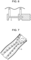
Within narrow curves, and now referring to FIG. 7 , the outer diameter of the flexible auger could be made permanently slightly smaller in order to prevent contact/friction with the outer cannula. This embodiment provides a clearance between the helix and the cannula. The manufacture of this neck 73 can also be achieved in a very cost effective manner with helical springs.
In another embodiment, the bending stiffness along the flexible auger could be varied by producing the heat-shrink tube via an intermittent extrusion process, as described in U.S. Pat. No. 4,888,146, the specification of which is incorporated by reference in its entirety. This process can provide a soft tip or a combination of flexibility and stiffness for insertion. In many cases, such extrusions are used to replace manually assembled composite shaft constructions (i.e. hand layups).”
In some embodiments, the proximal portion of the transfer element is designed to be rigid in order to stabilize turning movement and bearing in case of torque transmission. In some embodiments thereof, the end of the shrink tube itself might be the bearing surface.
In some embodiments, and now referring to FIG. 8 , the proximal portion of the transfer element comprises a sealable inlet 75 that provides irrigation into the space between the hollow auger and the inner shaft. Alternatively, the proximal portion of the transfer element comprises a sealable inlet that provides irrigation into the space between the hollow auger and the outer cannula. The inlet may also allow the provision of a) fluids that provide heating or cooling, or b) material delivery as well. The inlet is one possible design feature that allows providing cooling fluid/irrigation/material transport from outside to the lumen between the flexible inner shaft and shrink tube of the embodiments described in FIGS. 5A-C .
In some embodiments, and now referring to FIG. 9 , there is at least one lumen 77 disposed in the wall 79 of the outer cannula that surrounds the flexible auger. This lumen can be used to perform various functions. For example, a surgeon could perform device steering by pulling or pushing strings that run through such a lumen in the outer cannula wall. Another lumen in the outer cannula wall could provide irrigation. Yet another lumen 81 in the outer cannula wall could allow visualization by providing imaging means (such as optical fibres) that run through the lumen. Therefore, in some embodiments, the cannula that surrounds the flexible auger comprises a wall and an inner bore, wherein the wall has at least one longitudinal lumen therein. In some embodiments, the outer cannula can have two or more lumens of similar size and shape, or of varying sizes and shapes. One common two lumen configuration, sometimes referred to as a double-D, may be used for a peripherally-inserted central cannula (or PICC lines). PICC lines may also have three lumens with a cross-sectional view that represents a ‘peace sign.’ Two- and three-lumen configurations in which the lumens are dissimilar in size and shape are frequently used for percutaneous transluminal coronary angioplasty (PTCA) catheters. More simple cannulae, such as those used for drainage, may require only one lumen. Other applications, such as drug atomizing devices, may require tubing with more than a dozen lumens.
In some embodiments, reinforced wire technology could be adopted to achieve steering capabilities of the outer cannula that surrounds the flexible auger. These wires 83 are housed in the above-described lumen and should be able to be moved axially in order to provide a push/pull steering capability.
In other embodiments, the cannula comprises a wall and an inner bore, wherein the wall has at least one stationary wire running longitudinally therein. Longitudinal wires or fibers incorporated into an extrusion cross-section provide specific benefits, such as structural support or electrical data transmission. Wires can also provide excellent stretch resistance but limits flexibility depending on the number and location of reinforcing members. It is also possible to combine braided or spiral reinforcing with linear reinforcing elements to produce a hybrid design. Reinforcement material, tensile strength, size, and placement of the elements are critical aspects with linear reinforcing. High-tensile stainless steel round wire is commonly used for wire reinforcing. In thin wall sections, flat wire provides an excellent alternative. Other materials, such as aramid fiber or polymer monofilaments, can also be used for specialty linear reinforcement applications.
In some embodiments, the outer cannula could comprise an elastomer bulk with an internal metal wire pattern for reinforcement. The arrangement of the wire pattern can determine the outer cannula's ability to flex in certain directions, while being stiffer in other directions.
In some embodiments, the helical element in the flexible auger is a standard helical spring. Typically, this spring can be made out of biocompatible metals, such as titanium alloy, stainless steel and cobalt chrome. In some embodiments, the tube component of the flexible auger is a tube made out of heat shrink material. Typically, the heat shrink material is a polymeric, such as a PTFE.
Also in accordance with the present invention, there is provided a method comprising the steps of:
-
- a) passing a helical spring into a tube comprising a heat-shrink material,
- b) heating the tube to effect shrinking of the tube onto the helical spring to form an auger,
- c) inserting the auger into an intervertebral disc,
- d) rotating the auger to effect removal of intervertebral disc material.
In addition to the above-described heat shrink method of making the auger of the present invention, there is further contemplated additional embodiments of creating a flexible auger that entail low manufacturing costs.
In a first embodiment, the method comprises physically expanding a tube over a helical spring and then letting the tube contract. In this embodiment, the tube member in its initial state has an inner diameter ID that is smaller than the outer diameter OD of the spring. The tube can then be physically expanded in diameter by known methods (e.g., with high-pressure air, or with heat) until it reaches a dimension where its ID is larger than the OD of the spring. In this expanded condition, the tube is passed over the spring. When the force/energy upon the tube is removed, the tube in this assembly reverts to its original dimensions, except in the locations where it touches the helical spring member. The resulting product is an auger-like assembly with peaks and valleys.
Therefore, in accordance with the present invention, there is provided a method comprising the steps of:
-
- a) applying a physical force upon a tube to expand the tube,
- b) passing a helical spring into the expanded tube,
- c) releasing the physical force upon the tube to effect shrinking of the tube onto the helical spring to form an auger,
- d) inserting the auger into an intervertebral disc,
- e) rotating the auger to effect removal of intervertebral disc material.
In a second embodiment, the method comprises radially compressing the helical spring and then applying heat to the spring to allow its stressed areas to expand to a larger diameter. In this embodiment, the helical spring's outer diameter is reduced from its resting state (e.g., by winding the helical spring very tightly, or by pulling the helical member and thereby producing an elongated spring having a smaller outer diameter). In this reduced-diameter configuration, the radially compressed spring can be inserted into the tube member. Upon release of the mechanical force causing diameter reduction, the helical spring reverts to its original dimensions. Because the tube member possesses some flexibility, the elastic nature of the tube allows it to be deformed by the pressure of the expanding helical member upon the tube ID. Another auger-like assembly with valleys and summits is thereby produced.
Therefore, in accordance with the present invention, there is provided a method comprising the steps of:
-
- a) applying a physical force upon a helical spring to radially compress the spring,
- b) passing the compressed helical spring into a tube having an inner diameter,
- c) heating the compressed spring to effect radial expansion of the helical spring so as to contact the inner diameter of the tube and form an auger,
- d) inserting the auger into an intervertebral disc,
- e) rotating the auger to effect removal of intervertebral disc material.
In a third embodiment, the method comprises simultaneously applying heat and vacuum to an ordinary polymeric tube to obtain the same effect as a heat shrink. In this embodiment, the tube member is made of a material that is not heat-shrinkable, but rather is plastically deformable under heat and so over time arrives at an elastomeric state. In this embodiment, the tube member has an inner diameter ID in its initial state that is larger than the outer diameter OD of the helical spring, so that the helical member can be inserted into the tube member. Once the spring is inside the tube, a low pressure region/vacuum is created inside of the tube member, and the tube member is heated at the same time. These conditions create deformation in the wall of the tube member caused by the vacuum pulling towards the center of the tube. This reduces the diameter of the tube member, except for the locations where the helical member contacts the inside of the tube and so prevents deformation of the tube member in these areas. This results in another auger-like assembly with valleys and summits.
Therefore, in accordance with the present invention, there is provided a method comprising the steps of:
-
- a) passing a helical spring into a tube having a bore and comprising a polymeric material,
- b) simultaneously heating the tube and applying a vacuum to the bore of the tube to effect shrinking of the tube onto the helical spring to form an auger,
- c) inserting the auger into an intervertebral disc,
- d) rotating the auger to effect removal of intervertebral disc material.
Claims (22)
1. A biomedical material transfer device (1) comprising:
a) a transfer element having a proximal end portion, a distal end portion, and an intermediate portion comprising a flexible, hollow auger, the hollow auger comprising a substantially helical element and a membrane around the substantially helical element, and
b) a drive element adapted to rotate the hollow auger axially relative to an outer sleeve in which the hollow auger is disposed, the drive element having a distal end portion,
wherein the distal end portion of the drive element is connected to the proximal end portion of the transfer element, and
wherein the membrane has an inner surface and the substantially helical element extends inwardly from the inner surface,
the membrane being wrapped around the substantially helical element forming valleys and summits providing a thread-like topology for transporting material between an outer surface of the membrane and the outer sleeve.
2. The device of claim 1 , wherein the membrane is a heat-shrunk membrane that is integral with the substantially helical element housed within the membrane.
3. The device of claim 1 , wherein the membrane is not integral with the substantially helical element housed within the membrane.
4. The device of claim 1 , wherein the membrane rotates with the substantially helical element housed within the membrane.
5. The device of claim 1 , wherein the substantially helical element has a first diameter D1, the membrane has a minimum diameter D2, and the first diameter D1 is greater than the minimum diameter D2, the substantially helical element housed within the membrane rotating independently of the membrane of the transfer element.
6. The device of claim 1 , wherein the substantially helical element housed within the membrane has a thickness, and the membrane has an outer diameter, and the thickness is between 3% and 30% of the diameter.
7. The device of claim 1 , wherein the substantially helical element housed within the membrane has a variable pitch.
8. The device of claim 1 , wherein the proximal end portion of the transfer element comprises a tube having a threaded outer surface, the intermediate portion of the transfer element is flexible, and the distal end portion of the transfer element is adapted to cut intervertebral disc tissue.
9. The device of claim 1 , wherein the distal end of the transfer element comprises a distal end opening.
10. The device of claim 1 , wherein the distal end portion further comprises a cutting tip, the cutting tip having a proximal end portion adapted to mate with the substantially helical element.
11. The device of claim 1 , wherein the drive element includes a drive handle.
12. The device of claim 1 , wherein the drive element includes a motor.
13. The device of claim 1 , further comprising a tubular shaft connecting the transfer element and drive element.
14. The device of claim 13 , wherein the tubular shaft is transparent.
15. The device of claim 1 , further comprising a flexible inner shaft disposed within the hollow auger.
16. The device of claim 15 , wherein the flexible inner shaft is hollow.
17. The device of claim 16 , wherein the flexible inner shaft comprises a narrow-wound spring.
18. The device of claim 1 , further comprising an outer cannula, wherein the hollow auger is disposed within the outer cannula, and wherein the outer cannula comprises a wall and an inner bore, the wall having at least one lumen therein.
19. The device of claim 18 , further comprising a wire running longitudinally in the lumen.
20. The device of claim 1 , further comprising an outer cannula, wherein the hollow auger is disposed within the outer cannula, and wherein the outer cannula comprises a wall and an inner bore, the wall having at least one stationary wire running longitudinally therein.
21. The device of claim 1 , wherein the hollow auger has a steering mechanism attached thereto.
22. The device of claim 1 , wherein the hollow auger has a length having a variable bending stiffness.
Priority Applications (2)
| Application Number | Priority Date | Filing Date | Title |
|---|---|---|---|
| US17/095,448 US11712252B2 (en) | 2014-08-04 | 2020-11-11 | Flexible transport auger |
| US18/213,606 US20230355251A1 (en) | 2014-08-04 | 2023-06-23 | Flexible transport auger |
Applications Claiming Priority (4)
| Application Number | Priority Date | Filing Date | Title |
|---|---|---|---|
| US201462032754P | 2014-08-04 | 2014-08-04 | |
| US14/571,874 US9980737B2 (en) | 2014-08-04 | 2014-12-16 | Flexible transport auger |
| US15/968,437 US10863994B2 (en) | 2014-08-04 | 2018-05-01 | Flexible transport auger |
| US17/095,448 US11712252B2 (en) | 2014-08-04 | 2020-11-11 | Flexible transport auger |
Related Parent Applications (1)
| Application Number | Title | Priority Date | Filing Date |
|---|---|---|---|
| US15/968,437 Continuation US10863994B2 (en) | 2014-08-04 | 2018-05-01 | Flexible transport auger |
Related Child Applications (1)
| Application Number | Title | Priority Date | Filing Date |
|---|---|---|---|
| US18/213,606 Continuation US20230355251A1 (en) | 2014-08-04 | 2023-06-23 | Flexible transport auger |
Publications (2)
| Publication Number | Publication Date |
|---|---|
| US20210068850A1 US20210068850A1 (en) | 2021-03-11 |
| US11712252B2 true US11712252B2 (en) | 2023-08-01 |
Family
ID=55178814
Family Applications (4)
| Application Number | Title | Priority Date | Filing Date |
|---|---|---|---|
| US14/571,874 Active 2036-03-22 US9980737B2 (en) | 2014-08-04 | 2014-12-16 | Flexible transport auger |
| US15/968,437 Active 2035-07-22 US10863994B2 (en) | 2014-08-04 | 2018-05-01 | Flexible transport auger |
| US17/095,448 Active 2035-12-08 US11712252B2 (en) | 2014-08-04 | 2020-11-11 | Flexible transport auger |
| US18/213,606 Pending US20230355251A1 (en) | 2014-08-04 | 2023-06-23 | Flexible transport auger |
Family Applications Before (2)
| Application Number | Title | Priority Date | Filing Date |
|---|---|---|---|
| US14/571,874 Active 2036-03-22 US9980737B2 (en) | 2014-08-04 | 2014-12-16 | Flexible transport auger |
| US15/968,437 Active 2035-07-22 US10863994B2 (en) | 2014-08-04 | 2018-05-01 | Flexible transport auger |
Family Applications After (1)
| Application Number | Title | Priority Date | Filing Date |
|---|---|---|---|
| US18/213,606 Pending US20230355251A1 (en) | 2014-08-04 | 2023-06-23 | Flexible transport auger |
Country Status (5)
| Country | Link |
|---|---|
| US (4) | US9980737B2 (en) |
| EP (1) | EP3177216B1 (en) |
| JP (1) | JP6602844B2 (en) |
| AU (1) | AU2015301215B2 (en) |
| WO (1) | WO2016022529A1 (en) |
Families Citing this family (38)
| Publication number | Priority date | Publication date | Assignee | Title |
|---|---|---|---|---|
| DE10154163A1 (en) | 2001-11-03 | 2003-05-22 | Advanced Med Tech | Device for straightening and stabilizing the spine |
| US8979931B2 (en) | 2006-12-08 | 2015-03-17 | DePuy Synthes Products, LLC | Nucleus replacement device and method |
| AU2008308426A1 (en) | 2007-10-05 | 2009-04-09 | Synthes Gmbh | Dilation system and method of using the same |
| US8088163B1 (en) | 2008-02-06 | 2012-01-03 | Kleiner Jeffrey B | Tools and methods for spinal fusion |
| US9247943B1 (en) | 2009-02-06 | 2016-02-02 | Kleiner Intellectual Property, Llc | Devices and methods for preparing an intervertebral workspace |
| US8906028B2 (en) | 2009-09-18 | 2014-12-09 | Spinal Surgical Strategies, Llc | Bone graft delivery device and method of using the same |
| US10245159B1 (en) | 2009-09-18 | 2019-04-02 | Spinal Surgical Strategies, Llc | Bone graft delivery system and method for using same |
| US20170238984A1 (en) | 2009-09-18 | 2017-08-24 | Spinal Surgical Strategies, Llc | Bone graft delivery device with positioning handle |
| US10973656B2 (en) | 2009-09-18 | 2021-04-13 | Spinal Surgical Strategies, Inc. | Bone graft delivery system and method for using same |
| US9622779B2 (en) | 2011-10-27 | 2017-04-18 | DePuy Synthes Products, Inc. | Method and devices for a sub-splenius / supra-levator scapulae surgical access technique |
| US9808232B2 (en) | 2011-11-01 | 2017-11-07 | DePuy Synthes Products, Inc. | Dilation system |
| US9265490B2 (en) | 2012-04-16 | 2016-02-23 | DePuy Synthes Products, Inc. | Detachable dilator blade |
| US9480855B2 (en) | 2012-09-26 | 2016-11-01 | DePuy Synthes Products, Inc. | NIR/red light for lateral neuroprotection |
| US9980737B2 (en) | 2014-08-04 | 2018-05-29 | Medos International Sarl | Flexible transport auger |
| US10264959B2 (en) | 2014-09-09 | 2019-04-23 | Medos International Sarl | Proximal-end securement of a minimally invasive working channel |
| US9924979B2 (en) | 2014-09-09 | 2018-03-27 | Medos International Sarl | Proximal-end securement of a minimally invasive working channel |
| US10111712B2 (en) | 2014-09-09 | 2018-10-30 | Medos International Sarl | Proximal-end securement of a minimally invasive working channel |
| US9636477B2 (en) * | 2014-10-09 | 2017-05-02 | Vascular Solutions, Inc. | Catheter |
| US10786264B2 (en) | 2015-03-31 | 2020-09-29 | Medos International Sarl | Percutaneous disc clearing device |
| US11672562B2 (en) | 2015-09-04 | 2023-06-13 | Medos International Sarl | Multi-shield spinal access system |
| US11439380B2 (en) | 2015-09-04 | 2022-09-13 | Medos International Sarl | Surgical instrument connectors and related methods |
| US11744447B2 (en) | 2015-09-04 | 2023-09-05 | Medos International | Surgical visualization systems and related methods |
| US10987129B2 (en) | 2015-09-04 | 2021-04-27 | Medos International Sarl | Multi-shield spinal access system |
| CN113143355A (en) | 2015-09-04 | 2021-07-23 | 美多斯国际有限公司 | Multi-shield spinal access system |
| US10081845B2 (en) * | 2015-12-04 | 2018-09-25 | Baker Hughes, A Ge Company, Llc | Tubular strengthening and patterning method for enhanced heat transfer |
| US10299838B2 (en) | 2016-02-05 | 2019-05-28 | Medos International Sarl | Method and instruments for interbody fusion and posterior fixation through a single incision |
| US10653431B2 (en) | 2016-06-14 | 2020-05-19 | Medos International Sarl | Drill assemblies and methods for drilling into bone |
| US20180028211A1 (en) * | 2016-07-27 | 2018-02-01 | Loubert S. Suddaby | Endoscopic inflatable abrading device for spinal disc removal |
| CN108888311A (en) * | 2018-05-28 | 2018-11-27 | 吴淑霞 | A kind of orthopaedics brill bone automatic water filling device |
| US20210186756A1 (en) * | 2018-07-24 | 2021-06-24 | Johnson & Johnson Surgical Vision, Inc. | Surgical instruments for ocular surgery |
| US11000295B2 (en) * | 2018-09-27 | 2021-05-11 | Acumed Llc | Bone harvesting system |
| US11013530B2 (en) | 2019-03-08 | 2021-05-25 | Medos International Sarl | Surface features for device retention |
| US11241252B2 (en) | 2019-03-22 | 2022-02-08 | Medos International Sarl | Skin foundation access portal |
| US11129727B2 (en) | 2019-03-29 | 2021-09-28 | Medos International Sari | Inflatable non-distracting intervertebral implants and related methods |
| US11813026B2 (en) | 2019-04-05 | 2023-11-14 | Medos International Sarl | Systems, devices, and methods for providing surgical trajectory guidance |
| US20220040458A1 (en) * | 2020-08-05 | 2022-02-10 | Kenneth R. STUEBE | Mechanized surgical drain and method of use |
| EP4199843A1 (en) | 2020-08-19 | 2023-06-28 | Tag Dream Medical Ltd. | Hybrid laser cutter |
| US11771517B2 (en) | 2021-03-12 | 2023-10-03 | Medos International Sarl | Camera position indication systems and methods |
Citations (289)
| Publication number | Priority date | Publication date | Assignee | Title |
|---|---|---|---|---|
| US4573448A (en) | 1983-10-05 | 1986-03-04 | Pilling Co. | Method for decompressing herniated intervertebral discs |
| US4646738A (en) | 1985-12-05 | 1987-03-03 | Concept, Inc. | Rotary surgical tool |
| US4653496A (en) * | 1985-02-01 | 1987-03-31 | Bundy Mark A | Transluminal lysing system |
| US4678459A (en) | 1984-07-23 | 1987-07-07 | E-Z-Em, Inc. | Irrigating, cutting and aspirating system for percutaneous surgery |
| US4732154A (en) * | 1984-05-14 | 1988-03-22 | Surgical Systems & Instruments, Inc. | Rotary catheter system |
| US4863430A (en) | 1987-08-26 | 1989-09-05 | Surgical Dynamics, Inc. | Introduction set with flexible trocar with curved cannula |
| US4888146A (en) | 1988-05-19 | 1989-12-19 | Dandeneau James V | Method and apparatus of forming extruded article |
| US4914060A (en) * | 1989-03-17 | 1990-04-03 | Seas James A | Connector for antennas and coaxial cable |
| JPH0380872A (en) | 1989-05-12 | 1991-04-05 | Samuel Shiber | Atelectomy apparatus |
| US5041082A (en) * | 1986-06-16 | 1991-08-20 | Samuel Shiber | Mechanical atherectomy system and method |
| US5078723A (en) * | 1989-05-08 | 1992-01-07 | Medtronic, Inc. | Atherectomy device |
| US5080662A (en) | 1989-11-27 | 1992-01-14 | Paul Kamaljit S | Spinal stereotaxic device and method |
| US5135531A (en) * | 1984-05-14 | 1992-08-04 | Surgical Systems & Instruments, Inc. | Guided atherectomy system |
| US5195541A (en) | 1991-10-18 | 1993-03-23 | Obenchain Theodore G | Method of performing laparoscopic lumbar discectomy |
| EP0537116A1 (en) | 1991-10-11 | 1993-04-14 | Mauro Caponi | Double duct surgical instrument |
| US5285795A (en) | 1991-09-12 | 1994-02-15 | Surgical Dynamics, Inc. | Percutaneous discectomy system having a bendable discectomy probe and a steerable cannula |
| DE9415039U1 (en) | 1994-09-16 | 1994-11-03 | Kernforschungsz Karlsruhe | Device for guiding surgical instruments |
| US5395317A (en) | 1991-10-30 | 1995-03-07 | Smith & Nephew Dyonics, Inc. | Unilateral biportal percutaneous surgical procedure |
| US5439464A (en) | 1993-03-09 | 1995-08-08 | Shapiro Partners Limited | Method and instruments for performing arthroscopic spinal surgery |
| US5529580A (en) | 1987-10-30 | 1996-06-25 | Olympus Optical Co., Ltd. | Surgical resecting tool |
| US5540706A (en) | 1993-01-25 | 1996-07-30 | Aust; Gilbert M. | Surgical instrument |
| WO1996029014A1 (en) | 1995-03-22 | 1996-09-26 | Evi Corporation | Intra-artery obstruction clearing apparatus and methods |
| US5569290A (en) | 1995-01-30 | 1996-10-29 | Paul C. McAfee | Method of and apparatus for laparoscopic or endoscopic spinal surgery using an unsealed anteriorly inserted transparent trochar |
| US5591187A (en) * | 1995-07-14 | 1997-01-07 | Dekel; Moshe | Laparoscopic tissue retrieval device and method |
| US5601569A (en) | 1993-06-15 | 1997-02-11 | Pisharodi; Madhavan | Stereotactic frame and localization method |
| US5662300A (en) | 1993-07-26 | 1997-09-02 | Michelson; Gary Karlin | Gooseneck surgical instrument holder |
| US5688222A (en) | 1995-06-02 | 1997-11-18 | Olympus Winter & Ibe Gmbh | Endoscopic instrument |
| EP0807415A2 (en) | 1996-05-09 | 1997-11-19 | Olympus Optical Co., Ltd. | A cavity retaining tool for bone surgery, a cavity retaining tool for general surgery, an endoscopic surgery system involving the use of a cavity retaining tool, and a procedure for surgery |
| US5730754A (en) | 1995-01-10 | 1998-03-24 | Obenchain; Theodore G. | Nerve deflecting conduit needle and method |
| US5733242A (en) | 1996-02-07 | 1998-03-31 | Rayburn; Robert L. | Intubation system having an axially moveable memory cylinder |
| US5735792A (en) | 1992-11-25 | 1998-04-07 | Clarus Medical Systems, Inc. | Surgical instrument including viewing optics and an atraumatic probe |
| US5820623A (en) | 1995-06-20 | 1998-10-13 | Ng; Wan Sing | Articulated arm for medical procedures |
| US5885300A (en) | 1996-04-01 | 1999-03-23 | Asahi Kogaku Kogyo Kabushiki Kaisha | Guide apparatus of intervertebral implant |
| US5894369A (en) | 1996-11-15 | 1999-04-13 | Fuji Photo Optical Co., Ltd. | Lens device with anti-fogging |
| US5899425A (en) | 1997-05-02 | 1999-05-04 | Medtronic, Inc. | Adjustable supporting bracket having plural ball and socket joints |
| US5954635A (en) | 1996-03-22 | 1999-09-21 | Sdgi Holdings Inc. | Devices and methods for percutaneous surgery |
| DE29916026U1 (en) | 1999-09-11 | 1999-11-18 | Aesculap Ag & Co Kg | Holding device for a surgical instrument |
| US6033105A (en) | 1996-11-15 | 2000-03-07 | Barker; Donald | Integrated bone cement mixing and dispensing system |
| US6053907A (en) | 1998-08-13 | 2000-04-25 | Endius Incorporated | Surgical instruments with flexible drive shaft |
| US6063021A (en) | 1998-07-31 | 2000-05-16 | Pilling Weck Incorporated | Stabilizer for surgery |
| US6110182A (en) | 1998-06-22 | 2000-08-29 | Ohio Medical Instruments Company, Inc. | Target socket |
| US6139508A (en) * | 1998-08-04 | 2000-10-31 | Endonetics, Inc. | Articulated medical device |
| US6200322B1 (en) | 1999-08-13 | 2001-03-13 | Sdgi Holdings, Inc. | Minimal exposure posterior spinal interbody instrumentation and technique |
| US6217509B1 (en) | 1996-03-22 | 2001-04-17 | Sdgi Holdings, Inc. | Devices and methods for percutaneous surgery |
| US6234961B1 (en) | 1998-04-15 | 2001-05-22 | Pineridge Holding Pty. Ltd. | Ball and socket interconnection and retractor assembly employing the same |
| WO2001056490A1 (en) | 2000-02-01 | 2001-08-09 | Universitair Medisch Centrum Utrecht | Supporting arm for surgical purposes |
| US6283966B1 (en) | 1999-07-07 | 2001-09-04 | Sulzer Spine-Tech Inc. | Spinal surgery tools and positioning method |
| US6286179B1 (en) | 1998-10-19 | 2001-09-11 | Donny M. Byrne | Apparatus and method for removing debris from the lens-cleaning nozzle of an endoscope |
| US6296644B1 (en) | 1998-08-26 | 2001-10-02 | Jean Saurat | Spinal instrumentation system with articulated modules |
| US6322498B1 (en) | 1996-10-04 | 2001-11-27 | University Of Florida | Imaging scope |
| WO2001089371A1 (en) | 2000-05-19 | 2001-11-29 | A.M.I. Agency For Medical Innovations Gmbh | Lens cleaning device for an endoscope |
| WO2002002016A1 (en) | 2000-06-30 | 2002-01-10 | Abbott Laboratories | Surgical support clamp |
| US20020022762A1 (en) | 2000-02-18 | 2002-02-21 | Richard Beane | Devices and methods for warming and cleaning lenses of optical surgical instruments |
| US6354992B1 (en) | 1999-11-08 | 2002-03-12 | Daniel T. Kato | Automated laparoscopic lens cleaner |
| US6383191B1 (en) | 2000-03-15 | 2002-05-07 | Sdgi Holdings, Inc. | Laparoscopic instrument sleeve |
| US6447446B1 (en) | 1999-11-02 | 2002-09-10 | Medtronic Xomed, Inc. | Method and apparatus for cleaning an endoscope lens |
| US20020138020A1 (en) | 2001-03-23 | 2002-09-26 | Devonrex, Inc. | Micro-invasive breast biopsy device |
| US6468289B1 (en) | 1990-06-28 | 2002-10-22 | Peter M. Bonutti | Apparatus and method for tissue removal |
| US20030083555A1 (en) | 2001-10-29 | 2003-05-01 | Scott Hunt | Segmented arm support system and method for stabilizing tissue |
| US6558407B1 (en) | 2000-10-24 | 2003-05-06 | Tyco Healthcare Group Lp | Breast stabilizer with instrument guide |
| US6575899B1 (en) | 1999-10-20 | 2003-06-10 | Sdgi Holdings, Inc. | Methods and instruments for endoscopic interbody surgical techniques |
| US6579281B2 (en) | 2000-10-11 | 2003-06-17 | Popcab, Llc | Instrument stabilizer for through-a-port surgery |
| US20030171744A1 (en) | 2002-03-05 | 2003-09-11 | Baylis Medical Co. Inc. | Intradiscal lesioning device |
| US6626830B1 (en) | 1999-05-04 | 2003-09-30 | Cardiothoracic Systems, Inc. | Methods and devices for improved tissue stabilization |
| US20030191474A1 (en) | 2000-02-16 | 2003-10-09 | Cragg Andrew H. | Apparatus for performing a discectomy through a trans-sacral axial bore within the vertebrae of the spine |
| US6648915B2 (en) | 1999-12-23 | 2003-11-18 | John A. Sazy | Intervertebral cage and method of use |
| US6676597B2 (en) | 2001-01-13 | 2004-01-13 | Medtronic, Inc. | Method and device for organ positioning |
| US6688564B2 (en) | 2000-04-19 | 2004-02-10 | Karl Storz Gmbh & Co. Kg | Flexible tensioning device, especially for medical purposes |
| US20040122446A1 (en) | 2002-12-20 | 2004-06-24 | Solar Matthew S. | Organ access device and method |
| US20040127992A1 (en) | 2002-12-31 | 2004-07-01 | Serhan Hassan A. | Annular nucleus pulposus replacement |
| US6758809B2 (en) | 2002-06-06 | 2004-07-06 | Medtronic, Inc. | Surgical tool for engagement of body tissue |
| US20040143165A1 (en) | 2000-01-21 | 2004-07-22 | Neville Alleyne | Intervertebral disc repair methods and apparatus |
| US6808505B2 (en) | 2000-02-01 | 2004-10-26 | Kadan Jeffrey S | Diagnostic needle arthroscopy and lavage system |
| WO2004103430A2 (en) | 2003-05-19 | 2004-12-02 | Usgi Medical Inc. | Endoluminal tool deployment system |
| US20050085692A1 (en) | 2003-10-17 | 2005-04-21 | Ralf Kiehn | Endoscope |
| US20050090848A1 (en) | 2003-10-22 | 2005-04-28 | Adams Kenneth M. | Angled tissue cutting instruments and method of fabricating angled tissue cutting instruments having flexible inner tubular members of tube and sleeve construction |
| US6887198B2 (en) | 2001-10-05 | 2005-05-03 | Burns P. Phillips | Gooseneck surgical retractor positioner and method of its use |
| US20050187570A1 (en) | 2004-02-19 | 2005-08-25 | Applied Medical Resources Corporation | Embolectomy capture sheath |
| US20050256525A1 (en) | 2002-11-14 | 2005-11-17 | Brad Culbert | Dilation introducer for orthopedic surgery |
| US6983930B1 (en) | 2004-10-28 | 2006-01-10 | Christopher Louis La Mendola | Clamping device with flexible arm |
| US7087058B2 (en) | 2000-02-16 | 2006-08-08 | Trans1, Inc. | Method and apparatus for providing posterior or anterior trans-sacral access to spinal vertebrae |
| US7104986B2 (en) | 1996-07-16 | 2006-09-12 | Arthrocare Corporation | Intervertebral disc replacement method |
| US20060206118A1 (en) | 2005-03-11 | 2006-09-14 | Kim Daniel H | Percutaneous endoscopic access tools for the spinal epidural space and related methods of treatment |
| US7137949B2 (en) | 2001-07-13 | 2006-11-21 | United States Surgical Corporation | Surgical instrument |
| US7182731B2 (en) | 2002-01-23 | 2007-02-27 | Genesee Biomedical, Inc. | Support arm for cardiac surgery |
| US20070055259A1 (en) | 2005-08-17 | 2007-03-08 | Norton Britt K | Apparatus and methods for removal of intervertebral disc tissues |
| US20070129634A1 (en) | 2005-10-17 | 2007-06-07 | Hickey Katherine M | Biomedical positioning and stabilization system |
| US20070149975A1 (en) | 2005-11-29 | 2007-06-28 | Oliver Dana A | Method and apparatus for removing material from an intervertebral disc space, such as in performing a nucleotomy |
| US20070203396A1 (en) | 2006-02-28 | 2007-08-30 | Mccutcheon John G | Endoscopic Tool |
| US20070225556A1 (en) | 2006-03-23 | 2007-09-27 | Ethicon Endo-Surgery, Inc. | Disposable endoscope devices |
| JP2007530098A (en) | 2004-03-04 | 2007-11-01 | シュトラウブ メディカル アーゲー | Catheter for inhaling, crushing and expelling removable substances from blood vessels |
| US20070260113A1 (en) | 2005-01-07 | 2007-11-08 | Takashi Otawara | Endoscope insertion portion |
| US20080015621A1 (en) | 1997-09-04 | 2008-01-17 | Smith & Nephew, Inc. | Surgical endoscopic cutting device and method for its use |
| US20080033251A1 (en) | 2006-06-30 | 2008-02-07 | Ali Araghi | Surgical retractor and method of use |
| US7341556B2 (en) | 2001-06-11 | 2008-03-11 | M.S. Vision Ltd. | Endoscope with cleaning optics |
| US20080081951A1 (en) | 2006-09-29 | 2008-04-03 | Depuy Spine, Inc. | Inflatable retractor |
| US20080109015A1 (en) * | 2006-11-07 | 2008-05-08 | Chu Michael S | Delivering Sutures |
| US20080125856A1 (en) * | 2006-11-29 | 2008-05-29 | Mi4Spine, Llc | Bone Graft Delivery System for a Vertebral Interbody Device |
| US20080188714A1 (en) | 2007-02-07 | 2008-08-07 | Boston Scientific Scimed, Inc. | Electromechanical in-situ cleaning of optical elements |
| US7434325B2 (en) | 2004-07-26 | 2008-10-14 | Warsaw Orthopedic, Inc. | Systems and methods for determining optimal retractor length in minimally invasive procedures |
| US20090018566A1 (en) * | 2006-06-30 | 2009-01-15 | Artheromed, Inc. | Atherectomy devices, systems, and methods |
| US20090024158A1 (en) | 2007-07-16 | 2009-01-22 | Zimmer Spine, Inc. | Access Port Expander And Method |
| US20090062871A1 (en) | 2007-08-27 | 2009-03-05 | Singfatt Chin | Balloon cannula system for accessing and visualizing spine and related methods |
| WO2009033207A1 (en) | 2007-09-12 | 2009-03-19 | Columna Pty Ltd | Equipment for, and a method of, removing tissue from a site in a patient's body |
| US20090105543A1 (en) | 2007-10-19 | 2009-04-23 | Miller Eric C | Endoscope Lens Cleaner |
| US20090156898A1 (en) | 2006-08-30 | 2009-06-18 | Hironobu Ichimura | Distal end hood for endoscope and endoscope with hood |
| US20090187080A1 (en) | 2004-08-15 | 2009-07-23 | Kevin Seex | Distraction and retraction assemblies |
| US20090192494A1 (en) * | 2006-03-29 | 2009-07-30 | Kaneka Corporation | Catheter for Blood Removal |
| US20090240111A1 (en) | 2008-03-18 | 2009-09-24 | Joshua Kessler | Light Source and Lens Cleaner for Laparoscopic Surgery |
| US7594888B2 (en) | 2004-10-29 | 2009-09-29 | Depuy Spine, Inc. | Expandable ports and methods for minimally invasive surgery |
| US7618431B2 (en) | 2002-09-19 | 2009-11-17 | Warsaw Orthopedic, Inc. | Oval dilator and retractor set and method |
| US20090287061A1 (en) | 2007-10-01 | 2009-11-19 | Gft Technologies | Surgical access device for minimally invasive surgery |
| US20090318765A1 (en) | 2008-06-23 | 2009-12-24 | Torii Yuuichi | Endoscope |
| US7637905B2 (en) | 2003-01-15 | 2009-12-29 | Usgi Medical, Inc. | Endoluminal tool deployment system |
| US7641659B2 (en) | 2003-03-13 | 2010-01-05 | Zimmer Spine, Inc. | Spinal access instrument |
| US20100004651A1 (en) | 2008-06-13 | 2010-01-07 | The University Of Toledo | Transpedicular access to the intervertebral disc space for discectomy, end plate preparation, and interbody fusion |
| US20100022841A1 (en) | 2008-07-22 | 2010-01-28 | Fujifilm Corporation | Endoscope optical system device and endoscope with the same |
| US20100076476A1 (en) * | 2008-07-25 | 2010-03-25 | To John T | Systems and methods for cable-based tissue removal |
| US20100114147A1 (en) | 2008-10-30 | 2010-05-06 | The University Of Toledo | Directional soft tissue dilator and docking pin with integrated light source for optimization of retractor placement in minimally invasive spine surgery |
| US20100151161A1 (en) | 2005-10-05 | 2010-06-17 | Orlando Da Rolo | Flexible hollow shaft |
| US20100161060A1 (en) | 2008-12-23 | 2010-06-24 | Benvenue Medical, Inc. | Tissue Removal Tools And Methods Of Use |
| US7771384B2 (en) | 2003-08-20 | 2010-08-10 | Biagio Ravo | Trocar with integral irrigation and suction tube |
| US7794456B2 (en) | 2003-05-13 | 2010-09-14 | Arthrocare Corporation | Systems and methods for electrosurgical intervertebral disc replacement |
| US20100256446A1 (en) | 2007-05-11 | 2010-10-07 | Board Of Regents, The University Of Texas System | Medical scope carrier and scope as system and method |
| US7811303B2 (en) | 2003-08-26 | 2010-10-12 | Medicine Lodge Inc | Bodily tissue dilation systems and methods |
| US20100280325A1 (en) | 2009-04-30 | 2010-11-04 | Tamer Ibrahim | Retractors and surgical systems including the same |
| US20100284580A1 (en) | 2009-05-07 | 2010-11-11 | Ouyang Xiaolong | Tissue visualization systems and methods for using the same |
| US20100286477A1 (en) | 2009-05-08 | 2010-11-11 | Ouyang Xiaolong | Internal tissue visualization system comprising a rf-shielded visualization sensor module |
| US20100312053A1 (en) | 2006-04-13 | 2010-12-09 | Larsen Dane M | Resectoscopic device and method |
| US20110028791A1 (en) | 2009-07-28 | 2011-02-03 | Marino James F | Arcuate surgical guidance system and methods |
| US20110054507A1 (en) | 2009-04-17 | 2011-03-03 | David Batten | Devices and methods for arched roof cutters |
| US7931579B2 (en) | 2003-07-29 | 2011-04-26 | Endoscopic Technologies, Inc. | Tissue positioner |
| US20110106019A1 (en) * | 2007-11-21 | 2011-05-05 | Piezo Resonance Innovations, Inc. | Devices for clearing blockages in in-situ artificial lumens |
| US20110106261A1 (en) | 2009-10-30 | 2011-05-05 | Spinefrontier, Inc | System and method for an intervertebral implant assembly |
| US7946981B1 (en) | 2003-10-23 | 2011-05-24 | Anthony Cubb | Two-piece video laryngoscope |
| US20110125158A1 (en) | 2008-05-01 | 2011-05-26 | Ashish Dhar Diwan | Systems, methods and apparatuses for formation and insertion of tissue prostheses |
| US20110130634A1 (en) | 2009-05-20 | 2011-06-02 | Synthes Usa, Llc | Patient-mounted retraction |
| US7959564B2 (en) | 2006-07-08 | 2011-06-14 | Stephen Ritland | Pedicle seeker and retractor, and methods of use |
| JP2011517601A (en) | 2008-04-10 | 2011-06-16 | アセローメド, インコーポレイテッド | Atherotomy device and method |
| US7988623B2 (en) | 2002-10-25 | 2011-08-02 | Zimmer Spine, Inc. | Apparatus and methods for shielding body structures during surgery |
| US8007492B2 (en) | 2001-05-15 | 2011-08-30 | Zimmer Spine, Inc. | Cannula for receiving surgical instruments |
| US8038606B2 (en) | 2005-01-07 | 2011-10-18 | Olympus Medical Systems Corp. | Endoscope insertion portion |
| US8043381B2 (en) | 2007-10-29 | 2011-10-25 | Zimmer Spine, Inc. | Minimally invasive interbody device and method |
| US8062218B2 (en) | 2009-02-27 | 2011-11-22 | Warsaw Orthopedic, Inc. | Surgical access instrument |
| US20110295070A1 (en) | 2010-05-26 | 2011-12-01 | Olympus Corporation | Endoscope apparatus |
| US20110319941A1 (en) | 2008-12-01 | 2011-12-29 | Yossef Bar | Robot Guided Oblique Spinal Stabilization |
| GB2481727A (en) | 2009-10-12 | 2012-01-04 | Endoguard Ltd | Flow guide for cleaning an endoscope |
| US8092464B2 (en) | 2005-04-30 | 2012-01-10 | Warsaw Orthopedic, Inc. | Syringe devices and methods useful for delivering osteogenic material |
| US8096957B2 (en) * | 2005-03-11 | 2012-01-17 | Kimberly-Clark Inc. | Method for removing material from a patient's body |
| US8096944B2 (en) | 2007-10-26 | 2012-01-17 | Harrel Stephen K | Air shield for videoscope imagers |
| US20120095296A1 (en) | 2010-10-19 | 2012-04-19 | Warsaw Orthopedic | Expandable Spinal Access Instruments and Methods of Use |
| US20120101338A1 (en) | 2010-10-20 | 2012-04-26 | O'prey Cormac | Endoscope wiper blade cleaner |
| US8202216B2 (en) | 2007-03-08 | 2012-06-19 | Warsaw Orthopedic, Inc. | Tissue retractor |
| US8236006B2 (en) | 2008-01-17 | 2012-08-07 | Life Spine, Inc. | One step entry pedicular preparation device and disc access system |
| US20120209273A1 (en) | 2010-11-15 | 2012-08-16 | Zaretzka Gary D | Tissue removal system with retention mechanism |
| US20120221007A1 (en) | 2010-12-20 | 2012-08-30 | David Batten | Articulating tissue removal systems and methods |
| US20120232350A1 (en) | 2009-08-31 | 2012-09-13 | Kevin Seex | Retractor blade including a flexible member for anchorage engagement |
| US20120232552A1 (en) | 2011-03-10 | 2012-09-13 | Interventional Spine, Inc. | Method and apparatus for minimally invasive insertion of intervertebral implants |
| CN102727309A (en) | 2011-04-11 | 2012-10-17 | 上海优益基医疗器械有限公司 | Surgical navigation system combined with endoscope image and surgical navigation method |
| US20120298820A1 (en) | 2011-05-25 | 2012-11-29 | Spiros Manolidis | Surgical tool holder |
| US20120316400A1 (en) | 2011-06-10 | 2012-12-13 | Vijayanagar R | Surgical tissue retractor |
| US8333690B2 (en) | 2008-02-15 | 2012-12-18 | Fujifilm Corporation | Endoscopic fluid feed system |
| US8360970B2 (en) | 2005-06-22 | 2013-01-29 | Vycor Medical, Inc. | Surgical access instruments for use with spinal or orthopedic surgery |
| US8372131B2 (en) | 2007-07-16 | 2013-02-12 | Power Ten , LLC | Surgical site access system and deployment device for same |
| US8382048B2 (en) | 2009-03-20 | 2013-02-26 | Aesculap Ag | Surgical holding arm and surgical holding device |
| WO2013033426A2 (en) | 2011-09-01 | 2013-03-07 | Covidien Lp | Catheter with helical drive shaft and methods of manufacture |
| US8397335B2 (en) | 2006-10-03 | 2013-03-19 | Virtual Ports Ltd. | Device and method for lens cleaning for surgical procedures |
| US20130103067A1 (en) * | 2011-07-28 | 2013-04-25 | Myra I. L. Fabro | Discectomy devices and related methods |
| WO2013059640A1 (en) | 2011-10-21 | 2013-04-25 | Specialty Surgical Instrumentation Inc. | Universal arm system |
| US20130103103A1 (en) | 2011-10-24 | 2013-04-25 | Warsaw Orthopedic, Inc | Surgical system methods for spinal access |
| US8435174B2 (en) | 2009-12-11 | 2013-05-07 | Ethicon Endo-Surgery, Inc. | Methods and devices for accessing a body cavity |
| US8460186B2 (en) | 2009-12-11 | 2013-06-11 | Ethicon Endo-Surgery, Inc. | Methods and devices for providing access through tissue to a surgical site |
| US8460310B2 (en) | 2004-08-04 | 2013-06-11 | Leslie Stern | Surgical base unit and retractor support mechanism |
| US8460180B1 (en) | 2010-04-16 | 2013-06-11 | Hector Zarate | Endoscopic lens cleaner |
| US20130150674A1 (en) | 2011-12-07 | 2013-06-13 | Covidien Lp | Thoracic scope port sponge cleaner |
| US20130150670A1 (en) | 2011-12-07 | 2013-06-13 | Covidien Lp | Thoracic scope port cleaner |
| US20130172676A1 (en) | 2010-09-20 | 2013-07-04 | Peer Medical Ltd. | Multi-camera endoscope having fluid channels |
| US8535220B2 (en) | 2007-02-16 | 2013-09-17 | Robert MONDSCHEIN | Laparoscope cleaning system |
| US20130282022A1 (en) | 2012-04-18 | 2013-10-24 | Basem Fayez Yousef | Manipulator For Surgical Tools |
| US20130289399A1 (en) | 2012-04-27 | 2013-10-31 | Vertiflex, Inc. | Decompression systems and methods of using the same |
| US20130303846A1 (en) | 2008-07-22 | 2013-11-14 | Trice Orthopedics, Inc. | Tissue modification devices and methods of using the same |
| US8585726B2 (en) | 2012-01-31 | 2013-11-19 | Globus Medical, Inc. | Surgical disc removal tool |
| US8602979B2 (en) | 2011-03-31 | 2013-12-10 | Fujifilm Corporation | Electronic endoscope having front-view and side-view image capturing |
| US8622894B2 (en) | 2003-04-01 | 2014-01-07 | Boston Scientific Scimed, Inc. | Articulation joint |
| US8636655B1 (en) | 2010-01-19 | 2014-01-28 | Ronald Childs | Tissue retraction system and related methods |
| US20140066940A1 (en) | 2012-08-31 | 2014-03-06 | National Cheng Kung University | Assistant device and guiding assembly for percutaneous surgery |
| US20140074170A1 (en) | 2012-02-10 | 2014-03-13 | Herbert H. Mertens | Delivery Device With Interior Dilation Element Channel |
| WO2014050236A1 (en) | 2012-09-28 | 2014-04-03 | オリンパスメディカルシステムズ株式会社 | Endoscope device having cleaning mechanism |
| US8690764B2 (en) | 2010-10-20 | 2014-04-08 | Covidien Lp | Endoscope cleaner |
| US20140142584A1 (en) | 2012-11-16 | 2014-05-22 | Spinal Generations, Llc | Multichannel cannula and methods for using same |
| US20140148647A1 (en) | 2011-08-03 | 2014-05-29 | Olympus Corporation | Endoscope |
| US8740779B2 (en) | 2010-09-06 | 2014-06-03 | Fujifilm Corporation | Imaging device and method of cleaning an illumination window of the imaging device |
| WO2014100761A2 (en) | 2012-12-20 | 2014-06-26 | Spine View, Inc. | Discectomy devices and methods |
| US8784421B2 (en) | 2004-03-03 | 2014-07-22 | Boston Scientific Scimed, Inc. | Apparatus and methods for removing vertebral bone and disc tissue |
| US20140215736A1 (en) | 2013-02-05 | 2014-08-07 | Next Wave Surgical LLC | Minimally invasive apparatus and method for cleaning endoscope lenses |
| US8821378B2 (en) | 2006-11-27 | 2014-09-02 | Joimax Gmbh | Device and method for minimally invasive spinal intervention |
| US20140257489A1 (en) | 2013-03-11 | 2014-09-11 | Interventional Spine, Inc. | Method and apparatus for minimally invasive insertion of intervertebral implants |
| US8834507B2 (en) | 2011-05-17 | 2014-09-16 | Warsaw Orthopedic, Inc. | Dilation instruments and methods |
| US20140275799A1 (en) | 2013-03-15 | 2014-09-18 | Pro Med Instruments Gmbh | Flexible Arm and Method of Using |
| US20140276840A1 (en) | 2013-03-15 | 2014-09-18 | DePuy Synthes Products, LLC | Tools and methods for tissue removal |
| US20140277204A1 (en) | 2013-03-14 | 2014-09-18 | Interventional Spine, Inc. | Method and apparatus for minimally invasive insertion of intervertebral implants |
| US8845734B2 (en) | 2010-09-03 | 2014-09-30 | Globus Medical, Inc. | Expandable fusion device and method of installation thereof |
| US8852242B2 (en) | 2011-03-10 | 2014-10-07 | Interventional Spine, Inc. | Method and apparatus for minimally invasive insertion of intervertebral implants |
| US8870753B2 (en) | 2006-04-20 | 2014-10-28 | Boston Scientific Scimed, Inc. | Imaging assembly with transparent distal cap |
| US8870756B2 (en) | 2010-10-08 | 2014-10-28 | ERBE-USA, Inc. | Hybrid apparatus for fluid supply for endoscopic irrigation and lens cleaning |
| US20140318582A1 (en) | 2013-04-26 | 2014-10-30 | Medtronic-Xomed, Inc. | Endoscope lens cleaning device |
| US8876712B2 (en) | 2009-07-29 | 2014-11-04 | Edwards Lifesciences Corporation | Intracardiac sheath stabilizer |
| WO2014185334A1 (en) | 2013-05-16 | 2014-11-20 | アドバンストヘルスケア株式会社 | Trocar, port and surgical support system |
| US8894653B2 (en) | 2005-07-29 | 2014-11-25 | Vertos Medical, Inc. | Percutaneous tissue excision devices and methods |
| US8894573B2 (en) | 2009-08-05 | 2014-11-25 | Thomas Stuart Loftus | Retractor component system and method comprising same |
| US20140357945A1 (en) | 2013-05-30 | 2014-12-04 | Edward Duckworth | Laparoscopic Trocar with Ventriculoperitoneal Shunt Entry Port |
| US8926502B2 (en) | 2011-03-07 | 2015-01-06 | Endochoice, Inc. | Multi camera endoscope having a side service channel |
| US8932360B2 (en) | 2011-11-15 | 2015-01-13 | Baxano Surgical, Inc. | Implants for spinal therapy |
| US8932207B2 (en) | 2008-07-10 | 2015-01-13 | Covidien Lp | Integrated multi-functional endoscopic tool |
| US20150018623A1 (en) | 2013-07-09 | 2015-01-15 | Adam Friedrich | Surgical Access Systems and Methods |
| US8936605B2 (en) | 2011-12-30 | 2015-01-20 | Blackstone Medical, Inc. | Direct vertebral rotation tool and method of using same |
| US20150065795A1 (en) | 2011-02-16 | 2015-03-05 | The General Hospital Corporation | Optical Coupler for an Endoscope |
| US8974381B1 (en) | 2011-04-26 | 2015-03-10 | Nuvasive, Inc. | Cervical retractor |
| US8986199B2 (en) | 2012-02-17 | 2015-03-24 | Ethicon Endo-Surgery, Inc. | Apparatus and methods for cleaning the lens of an endoscope |
| US9028522B1 (en) | 2011-11-15 | 2015-05-12 | Seaspine, Inc. | Tissue dilator and retractor system and method of use |
| US9050146B2 (en) | 2009-11-10 | 2015-06-09 | Nuvasive, Inc. | Method and apparatus for performing spinal surgery |
| US9055936B2 (en) | 2010-07-08 | 2015-06-16 | Warsaw Orthopedic, Inc. | Over dilation |
| US20150164496A1 (en) | 2006-06-06 | 2015-06-18 | Globus Medical, Inc. | Surgical retractor system |
| US9072431B2 (en) | 2009-11-13 | 2015-07-07 | Hologic, Inc. | Access system with removable outflow channel |
| US9078562B2 (en) | 2010-01-11 | 2015-07-14 | Minimally Invasive Devices, Inc. | Systems and methods for optimizing and maintaining visualization of a surgical field during the use of surgical scopes |
| US20150223676A1 (en) | 2005-12-13 | 2015-08-13 | Avantis Medical Systems, Inc. | Detachable imaging device, endoscope having a detachable imaging device, and method of configuring such an endoscope |
| US20150230697A1 (en) | 2012-09-19 | 2015-08-20 | Nanyang Technological University | Flexible master - slave robotic endoscopy system |
| US9198674B2 (en) | 2012-12-14 | 2015-12-01 | Warsaw Orthopedic, Inc. | Surgical instrument and method |
| US20150342621A1 (en) | 2014-05-29 | 2015-12-03 | Avery M. Jackson, III | Illuminated endoscopic pedicle probe with dynamic real time monitoring for proximity to nerves |
| US9211059B2 (en) | 2007-06-19 | 2015-12-15 | Minimally Invasive Devices, Inc. | Systems and methods for optimizing and maintaining visualization of a surgical field during the use of surgical scopes |
| US9216125B2 (en) | 2011-07-22 | 2015-12-22 | Frederick H. Sklar | Surgical head holder and surgical accessories for use with same |
| US9216016B2 (en) | 2012-09-06 | 2015-12-22 | Medacta International Sa | Surgical device for minimally invasive spinal fusion and surgical system comprising the same |
| US9232935B2 (en) | 2011-08-01 | 2016-01-12 | Misder, Llc | Handle for actuating a device |
| US20160015467A1 (en) | 2014-05-22 | 2016-01-21 | Invuity, Inc. | Medical device featuring cladded waveguide |
| US9247997B2 (en) | 2011-09-30 | 2016-02-02 | Ethicon Endo-Surgery, Inc. | Patient-referenced surgical support frame |
| US20160030061A1 (en) | 2014-08-04 | 2016-02-04 | DePuy Synthes Products, LLC | Flexible transport auger |
| US9265491B2 (en) | 2004-12-29 | 2016-02-23 | U.S. Spine, Inc. | Minimally-invasive portal methods for performing lumbar decompression, instrumented fusion/stabilization, and the like |
| US9277928B2 (en) | 2013-03-11 | 2016-03-08 | Interventional Spine, Inc. | Method and apparatus for minimally invasive insertion of intervertebral implants |
| US20160066965A1 (en) | 2014-09-09 | 2016-03-10 | DePuy Synthes Products, LLC | Proximal-End Securement of a Minimally Invasive Working Channel |
| US20160067003A1 (en) | 2014-09-09 | 2016-03-10 | DePuy Synthes Products, LLC | Proximal-End Securement of a Minimally Invasive Working Channel |
| US20160074029A1 (en) | 2014-08-13 | 2016-03-17 | Nuvasive, Inc. | Minimally disruptive retractor and associated methods for spinal surgery |
| US20160095505A1 (en) | 2013-11-22 | 2016-04-07 | Massachusetts Institute Of Technology | Instruments for minimally invasive surgical procedures |
| US9307972B2 (en) | 2011-05-10 | 2016-04-12 | Nuvasive, Inc. | Method and apparatus for performing spinal fusion surgery |
| US20160106408A1 (en) | 2014-10-21 | 2016-04-21 | Globus Medical, Inc. | Orthopedic Tools for Implantation |
| US9320419B2 (en) | 2010-12-09 | 2016-04-26 | Endochoice Innovation Center Ltd. | Fluid channeling component of a multi-camera endoscope |
| USRE46007E1 (en) | 2004-09-30 | 2016-05-24 | Boston Scientific Scimed, Inc. | Automated control of irrigation and aspiration in a single-use endoscope |
| US20160166135A1 (en) | 2014-12-12 | 2016-06-16 | Medix3d LLC | Cleaning Device for Cleaning a Scope, Laparoscope or Microscope Used in Surgery or Other Medical Procedures and a Method of Using the Device During Surgical or Other Medical Procedures |
| US20160174814A1 (en) | 2013-08-31 | 2016-06-23 | Morena Medical Applications Ltd. | Endoscope with shared working channel |
| USRE46062E1 (en) | 2009-10-12 | 2016-07-12 | Endoguard Limited | Flow guide |
| US9387313B2 (en) | 2004-08-03 | 2016-07-12 | Interventional Spine, Inc. | Telescopic percutaneous tissue dilation systems and related methods |
| US9386971B1 (en) | 2011-08-31 | 2016-07-12 | Nuvasive, Inc. | Systems and methods for performing spine surgery |
| WO2016111373A1 (en) | 2015-01-09 | 2016-07-14 | 東レ・メディカル株式会社 | Endoscope lens cleaning device, and endoscope provided with said cleaning device |
| US20160213500A1 (en) | 2015-01-22 | 2016-07-28 | Aesculap Ag | Stent retractor/distractor |
| US20160228280A1 (en) | 2015-02-05 | 2016-08-11 | Pro Med Instruments Gmbh | System and method for invasive and non-invasive head fixation |
| US9414828B2 (en) | 2014-05-01 | 2016-08-16 | Blackstone Medical, Inc. | Integrated retractor-distractor system for use with modular bone screws |
| US20160235284A1 (en) | 2013-09-30 | 2016-08-18 | Nagase Medicals Co., Ltd. | Endoscope lens cleaner |
| WO2016131077A1 (en) | 2015-02-16 | 2016-08-25 | Eftekhar Behzad | Surgical rectractor blade with distal end formation for engaging anchor pin |
| US20160287264A1 (en) | 2015-03-31 | 2016-10-06 | DePuy Synthes Products, Inc. | Percutaneous Disc Clearing Device |
| US20160296220A1 (en) | 2011-08-31 | 2016-10-13 | Zimmer Biomet Spine, Inc. | Lateral retractor system and methods of use |
| WO2016168673A1 (en) | 2015-04-15 | 2016-10-20 | Trustees Of Boston University | Retractable endoscopic suction tube |
| US9486296B2 (en) | 2010-07-08 | 2016-11-08 | Warsaw Orthopedic, Inc. | Surgical assembly with flexible arm |
| US9510853B2 (en) | 2009-03-06 | 2016-12-06 | Procept Biorobotics Corporation | Tissue resection and treatment with shedding pulses |
| US20160353978A1 (en) | 2015-06-02 | 2016-12-08 | GI Scientific, LLC | Conductive optical element |
| US9526401B2 (en) | 2005-02-02 | 2016-12-27 | Intuitive Surgical Operations, Inc. | Flow reduction hood systems |
| US20170003493A1 (en) | 2014-03-21 | 2017-01-05 | Olympus Winter & Ibe Gmbh | Endoscope objective, method for cleaning an endoscope objective and for repairing an endoscope |
| US20170007226A1 (en) | 2013-11-18 | 2017-01-12 | Fehling Medical Corporation | Retractor for cranial surgery |
| WO2017006684A1 (en) | 2015-07-07 | 2017-01-12 | 株式会社カネカ | Lens cleaner for endoscope |
| WO2017015480A1 (en) | 2015-07-21 | 2017-01-26 | GI Scientific, LLC | Endoscope accessory with angularly adjustable exit portal |
| US20170027606A1 (en) | 2015-07-31 | 2017-02-02 | Purdue Research Foundation | Systems and methods for performing a surgical procedure |
| US20170042408A1 (en) | 2015-08-11 | 2017-02-16 | Trice Medical, Inc. | Fully integrated, disposable tissue visualization device |
| US20170042411A1 (en) | 2015-02-10 | 2017-02-16 | Yoon Sik Kang | Endoscope and endoscope system comprising same |
| US9579012B2 (en) | 2009-02-06 | 2017-02-28 | Endoclear Llc | Visualized endotracheal tube placement systems |
| US20170065287A1 (en) | 2013-04-28 | 2017-03-09 | Octavio Cesar Silva | Infrared Endoscopic Probe |
| US20170065269A1 (en) | 2015-09-04 | 2017-03-09 | DePuy Synthes Products, Inc. | Multi-Shield Spinal Access System |
| US9603510B2 (en) | 2011-05-17 | 2017-03-28 | Mario Ammirati | Method and apparatus for delivering an endoscope via microsurgical instruments while performing microscopic surgery |
| US20170086939A1 (en) | 2009-11-10 | 2017-03-30 | Invuity, Inc. | Illuminated suction apparatus |
| US9610095B2 (en) | 2008-08-27 | 2017-04-04 | Spine View, Inc. | Retractor cannula system for accessing and visualizing spine and related methods |
| US9610007B2 (en) | 2014-01-13 | 2017-04-04 | Trice Medical, Inc. | Fully integrated, disposable tissue visualization device |
| US9629521B2 (en) | 2004-10-11 | 2017-04-25 | Nitesh Ratnakar | Dual view endoscope |
| US20170135699A1 (en) | 2015-05-27 | 2017-05-18 | University Of Maryland, Baltimore | Apparatus and Method for Placement of Device along Wall of a Body Lumen |
| WO2017083648A1 (en) | 2015-11-13 | 2017-05-18 | Bodner Daryl | Endoscope |
| US9655639B2 (en) | 2008-12-16 | 2017-05-23 | Nico Corporation | Tissue removal device for use with imaging devices in neurosurgical and spinal surgery applications |
| US9655605B2 (en) | 2010-06-14 | 2017-05-23 | Maquet Cardiovascular Llc | Surgical instruments, systems and methods of use |
| US9668643B2 (en) | 2011-12-29 | 2017-06-06 | Cook Medical Technologies Llc | Space-optimized visualization catheter with oblong shape |
| US20170156755A1 (en) | 2010-12-03 | 2017-06-08 | Wayne L. Poll | Devices, systems, and methods for performing endoscopic surgical procedures |
| US9675235B2 (en) | 2005-03-21 | 2017-06-13 | Jonas V. Lieponis | Multi-purpose surgical instrument with removable component |
| US20170196549A1 (en) | 2009-12-16 | 2017-07-13 | Boston Scientific Scimed, Inc. | Substantially rigid and stable endoluminal surgical suite for treating a gastrointestinal lesion |
| US9706905B2 (en) | 2009-06-18 | 2017-07-18 | Endochoice Innovation Center Ltd. | Multi-camera endoscope |
| US20170224391A1 (en) | 2016-02-05 | 2017-08-10 | DePuy Synthes Products, Inc. | Method and instruments for interbody fusion and posterior fixation through a single incision |
-
2014
- 2014-12-16 US US14/571,874 patent/US9980737B2/en active Active
-
2015
- 2015-08-04 AU AU2015301215A patent/AU2015301215B2/en active Active
- 2015-08-04 WO PCT/US2015/043554 patent/WO2016022529A1/en active Application Filing
- 2015-08-04 EP EP15756710.8A patent/EP3177216B1/en active Active
- 2015-08-04 JP JP2017506356A patent/JP6602844B2/en active Active
-
2018
- 2018-05-01 US US15/968,437 patent/US10863994B2/en active Active
-
2020
- 2020-11-11 US US17/095,448 patent/US11712252B2/en active Active
-
2023
- 2023-06-23 US US18/213,606 patent/US20230355251A1/en active Pending
Patent Citations (316)
| Publication number | Priority date | Publication date | Assignee | Title |
|---|---|---|---|---|
| US4573448A (en) | 1983-10-05 | 1986-03-04 | Pilling Co. | Method for decompressing herniated intervertebral discs |
| US5135531A (en) * | 1984-05-14 | 1992-08-04 | Surgical Systems & Instruments, Inc. | Guided atherectomy system |
| US4732154A (en) * | 1984-05-14 | 1988-03-22 | Surgical Systems & Instruments, Inc. | Rotary catheter system |
| US4678459A (en) | 1984-07-23 | 1987-07-07 | E-Z-Em, Inc. | Irrigating, cutting and aspirating system for percutaneous surgery |
| US4653496A (en) * | 1985-02-01 | 1987-03-31 | Bundy Mark A | Transluminal lysing system |
| US4646738A (en) | 1985-12-05 | 1987-03-03 | Concept, Inc. | Rotary surgical tool |
| US5041082A (en) * | 1986-06-16 | 1991-08-20 | Samuel Shiber | Mechanical atherectomy system and method |
| US4863430A (en) | 1987-08-26 | 1989-09-05 | Surgical Dynamics, Inc. | Introduction set with flexible trocar with curved cannula |
| US5529580A (en) | 1987-10-30 | 1996-06-25 | Olympus Optical Co., Ltd. | Surgical resecting tool |
| US4888146A (en) | 1988-05-19 | 1989-12-19 | Dandeneau James V | Method and apparatus of forming extruded article |
| US4914060A (en) * | 1989-03-17 | 1990-04-03 | Seas James A | Connector for antennas and coaxial cable |
| US5078723A (en) * | 1989-05-08 | 1992-01-07 | Medtronic, Inc. | Atherectomy device |
| JPH0380872A (en) | 1989-05-12 | 1991-04-05 | Samuel Shiber | Atelectomy apparatus |
| US5080662A (en) | 1989-11-27 | 1992-01-14 | Paul Kamaljit S | Spinal stereotaxic device and method |
| US6468289B1 (en) | 1990-06-28 | 2002-10-22 | Peter M. Bonutti | Apparatus and method for tissue removal |
| US5285795A (en) | 1991-09-12 | 1994-02-15 | Surgical Dynamics, Inc. | Percutaneous discectomy system having a bendable discectomy probe and a steerable cannula |
| EP0537116A1 (en) | 1991-10-11 | 1993-04-14 | Mauro Caponi | Double duct surgical instrument |
| US5195541A (en) | 1991-10-18 | 1993-03-23 | Obenchain Theodore G | Method of performing laparoscopic lumbar discectomy |
| US5395317A (en) | 1991-10-30 | 1995-03-07 | Smith & Nephew Dyonics, Inc. | Unilateral biportal percutaneous surgical procedure |
| US5735792A (en) | 1992-11-25 | 1998-04-07 | Clarus Medical Systems, Inc. | Surgical instrument including viewing optics and an atraumatic probe |
| US5540706A (en) | 1993-01-25 | 1996-07-30 | Aust; Gilbert M. | Surgical instrument |
| US5439464A (en) | 1993-03-09 | 1995-08-08 | Shapiro Partners Limited | Method and instruments for performing arthroscopic spinal surgery |
| US5601569A (en) | 1993-06-15 | 1997-02-11 | Pisharodi; Madhavan | Stereotactic frame and localization method |
| US5662300A (en) | 1993-07-26 | 1997-09-02 | Michelson; Gary Karlin | Gooseneck surgical instrument holder |
| DE9415039U1 (en) | 1994-09-16 | 1994-11-03 | Kernforschungsz Karlsruhe | Device for guiding surgical instruments |
| US5730754A (en) | 1995-01-10 | 1998-03-24 | Obenchain; Theodore G. | Nerve deflecting conduit needle and method |
| US5569290A (en) | 1995-01-30 | 1996-10-29 | Paul C. McAfee | Method of and apparatus for laparoscopic or endoscopic spinal surgery using an unsealed anteriorly inserted transparent trochar |
| WO1996029014A1 (en) | 1995-03-22 | 1996-09-26 | Evi Corporation | Intra-artery obstruction clearing apparatus and methods |
| US5688222A (en) | 1995-06-02 | 1997-11-18 | Olympus Winter & Ibe Gmbh | Endoscopic instrument |
| US5820623A (en) | 1995-06-20 | 1998-10-13 | Ng; Wan Sing | Articulated arm for medical procedures |
| US5591187A (en) * | 1995-07-14 | 1997-01-07 | Dekel; Moshe | Laparoscopic tissue retrieval device and method |
| US5733242A (en) | 1996-02-07 | 1998-03-31 | Rayburn; Robert L. | Intubation system having an axially moveable memory cylinder |
| US6217509B1 (en) | 1996-03-22 | 2001-04-17 | Sdgi Holdings, Inc. | Devices and methods for percutaneous surgery |
| US5954635A (en) | 1996-03-22 | 1999-09-21 | Sdgi Holdings Inc. | Devices and methods for percutaneous surgery |
| US5885300A (en) | 1996-04-01 | 1999-03-23 | Asahi Kogaku Kogyo Kabushiki Kaisha | Guide apparatus of intervertebral implant |
| EP0807415A2 (en) | 1996-05-09 | 1997-11-19 | Olympus Optical Co., Ltd. | A cavity retaining tool for bone surgery, a cavity retaining tool for general surgery, an endoscopic surgery system involving the use of a cavity retaining tool, and a procedure for surgery |
| US6371968B1 (en) | 1996-05-09 | 2002-04-16 | Olympus Optical Co., Ltd. | Cavity retaining tool for bone surgery, a cavity retaining tool for general surgery, an endoscopic surgery system involving the use of a cavity retaining tool, and a procedure for surgery |
| US7104986B2 (en) | 1996-07-16 | 2006-09-12 | Arthrocare Corporation | Intervertebral disc replacement method |
| US6322498B1 (en) | 1996-10-04 | 2001-11-27 | University Of Florida | Imaging scope |
| US5894369A (en) | 1996-11-15 | 1999-04-13 | Fuji Photo Optical Co., Ltd. | Lens device with anti-fogging |
| US6033105A (en) | 1996-11-15 | 2000-03-07 | Barker; Donald | Integrated bone cement mixing and dispensing system |
| US5899425A (en) | 1997-05-02 | 1999-05-04 | Medtronic, Inc. | Adjustable supporting bracket having plural ball and socket joints |
| US20080015621A1 (en) | 1997-09-04 | 2008-01-17 | Smith & Nephew, Inc. | Surgical endoscopic cutting device and method for its use |
| US6234961B1 (en) | 1998-04-15 | 2001-05-22 | Pineridge Holding Pty. Ltd. | Ball and socket interconnection and retractor assembly employing the same |
| US6110182A (en) | 1998-06-22 | 2000-08-29 | Ohio Medical Instruments Company, Inc. | Target socket |
| US6063021A (en) | 1998-07-31 | 2000-05-16 | Pilling Weck Incorporated | Stabilizer for surgery |
| US6139508A (en) * | 1998-08-04 | 2000-10-31 | Endonetics, Inc. | Articulated medical device |
| US6053907A (en) | 1998-08-13 | 2000-04-25 | Endius Incorporated | Surgical instruments with flexible drive shaft |
| US6296644B1 (en) | 1998-08-26 | 2001-10-02 | Jean Saurat | Spinal instrumentation system with articulated modules |
| US6286179B1 (en) | 1998-10-19 | 2001-09-11 | Donny M. Byrne | Apparatus and method for removing debris from the lens-cleaning nozzle of an endoscope |
| US6626830B1 (en) | 1999-05-04 | 2003-09-30 | Cardiothoracic Systems, Inc. | Methods and devices for improved tissue stabilization |
| US6283966B1 (en) | 1999-07-07 | 2001-09-04 | Sulzer Spine-Tech Inc. | Spinal surgery tools and positioning method |
| US6200322B1 (en) | 1999-08-13 | 2001-03-13 | Sdgi Holdings, Inc. | Minimal exposure posterior spinal interbody instrumentation and technique |
| DE29916026U1 (en) | 1999-09-11 | 1999-11-18 | Aesculap Ag & Co Kg | Holding device for a surgical instrument |
| US6575899B1 (en) | 1999-10-20 | 2003-06-10 | Sdgi Holdings, Inc. | Methods and instruments for endoscopic interbody surgical techniques |
| US6447446B1 (en) | 1999-11-02 | 2002-09-10 | Medtronic Xomed, Inc. | Method and apparatus for cleaning an endoscope lens |
| US6354992B1 (en) | 1999-11-08 | 2002-03-12 | Daniel T. Kato | Automated laparoscopic lens cleaner |
| US6648915B2 (en) | 1999-12-23 | 2003-11-18 | John A. Sazy | Intervertebral cage and method of use |
| US20040143165A1 (en) | 2000-01-21 | 2004-07-22 | Neville Alleyne | Intervertebral disc repair methods and apparatus |
| US6808505B2 (en) | 2000-02-01 | 2004-10-26 | Kadan Jeffrey S | Diagnostic needle arthroscopy and lavage system |
| WO2001056490A1 (en) | 2000-02-01 | 2001-08-09 | Universitair Medisch Centrum Utrecht | Supporting arm for surgical purposes |
| US20030191474A1 (en) | 2000-02-16 | 2003-10-09 | Cragg Andrew H. | Apparatus for performing a discectomy through a trans-sacral axial bore within the vertebrae of the spine |
| US7087058B2 (en) | 2000-02-16 | 2006-08-08 | Trans1, Inc. | Method and apparatus for providing posterior or anterior trans-sacral access to spinal vertebrae |
| US20020022762A1 (en) | 2000-02-18 | 2002-02-21 | Richard Beane | Devices and methods for warming and cleaning lenses of optical surgical instruments |
| US6383191B1 (en) | 2000-03-15 | 2002-05-07 | Sdgi Holdings, Inc. | Laparoscopic instrument sleeve |
| US6688564B2 (en) | 2000-04-19 | 2004-02-10 | Karl Storz Gmbh & Co. Kg | Flexible tensioning device, especially for medical purposes |
| WO2001089371A1 (en) | 2000-05-19 | 2001-11-29 | A.M.I. Agency For Medical Innovations Gmbh | Lens cleaning device for an endoscope |
| WO2002002016A1 (en) | 2000-06-30 | 2002-01-10 | Abbott Laboratories | Surgical support clamp |
| US6579281B2 (en) | 2000-10-11 | 2003-06-17 | Popcab, Llc | Instrument stabilizer for through-a-port surgery |
| US6558407B1 (en) | 2000-10-24 | 2003-05-06 | Tyco Healthcare Group Lp | Breast stabilizer with instrument guide |
| US6676597B2 (en) | 2001-01-13 | 2004-01-13 | Medtronic, Inc. | Method and device for organ positioning |
| US20020138020A1 (en) | 2001-03-23 | 2002-09-26 | Devonrex, Inc. | Micro-invasive breast biopsy device |
| US7591790B2 (en) | 2001-03-23 | 2009-09-22 | Stryker Puerto Rico Limited | Micro-invasive device |
| US8007492B2 (en) | 2001-05-15 | 2011-08-30 | Zimmer Spine, Inc. | Cannula for receiving surgical instruments |
| US7341556B2 (en) | 2001-06-11 | 2008-03-11 | M.S. Vision Ltd. | Endoscope with cleaning optics |
| US7137949B2 (en) | 2001-07-13 | 2006-11-21 | United States Surgical Corporation | Surgical instrument |
| US6887198B2 (en) | 2001-10-05 | 2005-05-03 | Burns P. Phillips | Gooseneck surgical retractor positioner and method of its use |
| US20030083555A1 (en) | 2001-10-29 | 2003-05-01 | Scott Hunt | Segmented arm support system and method for stabilizing tissue |
| US7182731B2 (en) | 2002-01-23 | 2007-02-27 | Genesee Biomedical, Inc. | Support arm for cardiac surgery |
| US20030171744A1 (en) | 2002-03-05 | 2003-09-11 | Baylis Medical Co. Inc. | Intradiscal lesioning device |
| US6758809B2 (en) | 2002-06-06 | 2004-07-06 | Medtronic, Inc. | Surgical tool for engagement of body tissue |
| US7618431B2 (en) | 2002-09-19 | 2009-11-17 | Warsaw Orthopedic, Inc. | Oval dilator and retractor set and method |
| US7988623B2 (en) | 2002-10-25 | 2011-08-02 | Zimmer Spine, Inc. | Apparatus and methods for shielding body structures during surgery |
| US20050256525A1 (en) | 2002-11-14 | 2005-11-17 | Brad Culbert | Dilation introducer for orthopedic surgery |
| US20040122446A1 (en) | 2002-12-20 | 2004-06-24 | Solar Matthew S. | Organ access device and method |
| US7636596B2 (en) | 2002-12-20 | 2009-12-22 | Medtronic, Inc. | Organ access device and method |
| US20040127992A1 (en) | 2002-12-31 | 2004-07-01 | Serhan Hassan A. | Annular nucleus pulposus replacement |
| US7637905B2 (en) | 2003-01-15 | 2009-12-29 | Usgi Medical, Inc. | Endoluminal tool deployment system |
| US7641659B2 (en) | 2003-03-13 | 2010-01-05 | Zimmer Spine, Inc. | Spinal access instrument |
| US8622894B2 (en) | 2003-04-01 | 2014-01-07 | Boston Scientific Scimed, Inc. | Articulation joint |
| US7794456B2 (en) | 2003-05-13 | 2010-09-14 | Arthrocare Corporation | Systems and methods for electrosurgical intervertebral disc replacement |
| US7951141B2 (en) | 2003-05-13 | 2011-05-31 | Arthrocare Corporation | Systems and methods for electrosurgical intervertebral disc replacement |
| WO2004103430A2 (en) | 2003-05-19 | 2004-12-02 | Usgi Medical Inc. | Endoluminal tool deployment system |
| US7931579B2 (en) | 2003-07-29 | 2011-04-26 | Endoscopic Technologies, Inc. | Tissue positioner |
| US7771384B2 (en) | 2003-08-20 | 2010-08-10 | Biagio Ravo | Trocar with integral irrigation and suction tube |
| US7811303B2 (en) | 2003-08-26 | 2010-10-12 | Medicine Lodge Inc | Bodily tissue dilation systems and methods |
| US20050085692A1 (en) | 2003-10-17 | 2005-04-21 | Ralf Kiehn | Endoscope |
| US20050090848A1 (en) | 2003-10-22 | 2005-04-28 | Adams Kenneth M. | Angled tissue cutting instruments and method of fabricating angled tissue cutting instruments having flexible inner tubular members of tube and sleeve construction |
| US7946981B1 (en) | 2003-10-23 | 2011-05-24 | Anthony Cubb | Two-piece video laryngoscope |
| US20050187570A1 (en) | 2004-02-19 | 2005-08-25 | Applied Medical Resources Corporation | Embolectomy capture sheath |
| US8784421B2 (en) | 2004-03-03 | 2014-07-22 | Boston Scientific Scimed, Inc. | Apparatus and methods for removing vertebral bone and disc tissue |
| JP2007530098A (en) | 2004-03-04 | 2007-11-01 | シュトラウブ メディカル アーゲー | Catheter for inhaling, crushing and expelling removable substances from blood vessels |
| US7434325B2 (en) | 2004-07-26 | 2008-10-14 | Warsaw Orthopedic, Inc. | Systems and methods for determining optimal retractor length in minimally invasive procedures |
| US9387313B2 (en) | 2004-08-03 | 2016-07-12 | Interventional Spine, Inc. | Telescopic percutaneous tissue dilation systems and related methods |
| US8460310B2 (en) | 2004-08-04 | 2013-06-11 | Leslie Stern | Surgical base unit and retractor support mechanism |
| US20090187080A1 (en) | 2004-08-15 | 2009-07-23 | Kevin Seex | Distraction and retraction assemblies |
| US20140194697A1 (en) | 2004-08-15 | 2014-07-10 | Kevin Seex | Distraction and retraction assembly |
| USRE46007E1 (en) | 2004-09-30 | 2016-05-24 | Boston Scientific Scimed, Inc. | Automated control of irrigation and aspiration in a single-use endoscope |
| US9629521B2 (en) | 2004-10-11 | 2017-04-25 | Nitesh Ratnakar | Dual view endoscope |
| US6983930B1 (en) | 2004-10-28 | 2006-01-10 | Christopher Louis La Mendola | Clamping device with flexible arm |
| US7594888B2 (en) | 2004-10-29 | 2009-09-29 | Depuy Spine, Inc. | Expandable ports and methods for minimally invasive surgery |
| US9265491B2 (en) | 2004-12-29 | 2016-02-23 | U.S. Spine, Inc. | Minimally-invasive portal methods for performing lumbar decompression, instrumented fusion/stabilization, and the like |
| US20070260113A1 (en) | 2005-01-07 | 2007-11-08 | Takashi Otawara | Endoscope insertion portion |
| US8038606B2 (en) | 2005-01-07 | 2011-10-18 | Olympus Medical Systems Corp. | Endoscope insertion portion |
| US9526401B2 (en) | 2005-02-02 | 2016-12-27 | Intuitive Surgical Operations, Inc. | Flow reduction hood systems |
| US8201563B2 (en) * | 2005-03-11 | 2012-06-19 | Kimberly-Clark, Inc. | Method for introducing materials into a body |
| US8096957B2 (en) * | 2005-03-11 | 2012-01-17 | Kimberly-Clark Inc. | Method for removing material from a patient's body |
| US20060206118A1 (en) | 2005-03-11 | 2006-09-14 | Kim Daniel H | Percutaneous endoscopic access tools for the spinal epidural space and related methods of treatment |
| US9675235B2 (en) | 2005-03-21 | 2017-06-13 | Jonas V. Lieponis | Multi-purpose surgical instrument with removable component |
| US8092464B2 (en) | 2005-04-30 | 2012-01-10 | Warsaw Orthopedic, Inc. | Syringe devices and methods useful for delivering osteogenic material |
| US8360970B2 (en) | 2005-06-22 | 2013-01-29 | Vycor Medical, Inc. | Surgical access instruments for use with spinal or orthopedic surgery |
| US8894653B2 (en) | 2005-07-29 | 2014-11-25 | Vertos Medical, Inc. | Percutaneous tissue excision devices and methods |
| US20070055259A1 (en) | 2005-08-17 | 2007-03-08 | Norton Britt K | Apparatus and methods for removal of intervertebral disc tissues |
| US20100151161A1 (en) | 2005-10-05 | 2010-06-17 | Orlando Da Rolo | Flexible hollow shaft |
| US20070129634A1 (en) | 2005-10-17 | 2007-06-07 | Hickey Katherine M | Biomedical positioning and stabilization system |
| US20070149975A1 (en) | 2005-11-29 | 2007-06-28 | Oliver Dana A | Method and apparatus for removing material from an intervertebral disc space, such as in performing a nucleotomy |
| US20150223676A1 (en) | 2005-12-13 | 2015-08-13 | Avantis Medical Systems, Inc. | Detachable imaging device, endoscope having a detachable imaging device, and method of configuring such an endoscope |
| US20070203396A1 (en) | 2006-02-28 | 2007-08-30 | Mccutcheon John G | Endoscopic Tool |
| US20070225556A1 (en) | 2006-03-23 | 2007-09-27 | Ethicon Endo-Surgery, Inc. | Disposable endoscope devices |
| US20090192494A1 (en) * | 2006-03-29 | 2009-07-30 | Kaneka Corporation | Catheter for Blood Removal |
| US20100312053A1 (en) | 2006-04-13 | 2010-12-09 | Larsen Dane M | Resectoscopic device and method |
| US8870753B2 (en) | 2006-04-20 | 2014-10-28 | Boston Scientific Scimed, Inc. | Imaging assembly with transparent distal cap |
| US20150164496A1 (en) | 2006-06-06 | 2015-06-18 | Globus Medical, Inc. | Surgical retractor system |
| US20090018566A1 (en) * | 2006-06-30 | 2009-01-15 | Artheromed, Inc. | Atherectomy devices, systems, and methods |
| US20080033251A1 (en) | 2006-06-30 | 2008-02-07 | Ali Araghi | Surgical retractor and method of use |
| US7959564B2 (en) | 2006-07-08 | 2011-06-14 | Stephen Ritland | Pedicle seeker and retractor, and methods of use |
| US20090156898A1 (en) | 2006-08-30 | 2009-06-18 | Hironobu Ichimura | Distal end hood for endoscope and endoscope with hood |
| US20080081951A1 (en) | 2006-09-29 | 2008-04-03 | Depuy Spine, Inc. | Inflatable retractor |
| US8397335B2 (en) | 2006-10-03 | 2013-03-19 | Virtual Ports Ltd. | Device and method for lens cleaning for surgical procedures |
| US20080109015A1 (en) * | 2006-11-07 | 2008-05-08 | Chu Michael S | Delivering Sutures |
| US8821378B2 (en) | 2006-11-27 | 2014-09-02 | Joimax Gmbh | Device and method for minimally invasive spinal intervention |
| US20080125856A1 (en) * | 2006-11-29 | 2008-05-29 | Mi4Spine, Llc | Bone Graft Delivery System for a Vertebral Interbody Device |
| WO2008121162A1 (en) | 2006-11-29 | 2008-10-09 | Mi4Spine, Llc | Disc space preparation device for spinal surgery |
| US7803159B2 (en) * | 2006-11-29 | 2010-09-28 | Mi4Spine, Llc | Disc space preparation device for spinal surgery |
| US20080188714A1 (en) | 2007-02-07 | 2008-08-07 | Boston Scientific Scimed, Inc. | Electromechanical in-situ cleaning of optical elements |
| US8535220B2 (en) | 2007-02-16 | 2013-09-17 | Robert MONDSCHEIN | Laparoscope cleaning system |
| US8202216B2 (en) | 2007-03-08 | 2012-06-19 | Warsaw Orthopedic, Inc. | Tissue retractor |
| US20100256446A1 (en) | 2007-05-11 | 2010-10-07 | Board Of Regents, The University Of Texas System | Medical scope carrier and scope as system and method |
| US9211059B2 (en) | 2007-06-19 | 2015-12-15 | Minimally Invasive Devices, Inc. | Systems and methods for optimizing and maintaining visualization of a surgical field during the use of surgical scopes |
| US8372131B2 (en) | 2007-07-16 | 2013-02-12 | Power Ten , LLC | Surgical site access system and deployment device for same |
| US20090024158A1 (en) | 2007-07-16 | 2009-01-22 | Zimmer Spine, Inc. | Access Port Expander And Method |
| US20090062871A1 (en) | 2007-08-27 | 2009-03-05 | Singfatt Chin | Balloon cannula system for accessing and visualizing spine and related methods |
| WO2009033207A1 (en) | 2007-09-12 | 2009-03-19 | Columna Pty Ltd | Equipment for, and a method of, removing tissue from a site in a patient's body |
| US20090287061A1 (en) | 2007-10-01 | 2009-11-19 | Gft Technologies | Surgical access device for minimally invasive surgery |
| US20090105543A1 (en) | 2007-10-19 | 2009-04-23 | Miller Eric C | Endoscope Lens Cleaner |
| US8096944B2 (en) | 2007-10-26 | 2012-01-17 | Harrel Stephen K | Air shield for videoscope imagers |
| US8043381B2 (en) | 2007-10-29 | 2011-10-25 | Zimmer Spine, Inc. | Minimally invasive interbody device and method |
| US8262645B2 (en) * | 2007-11-21 | 2012-09-11 | Actuated Medical, Inc. | Devices for clearing blockages in in-situ artificial lumens |
| US20110106019A1 (en) * | 2007-11-21 | 2011-05-05 | Piezo Resonance Innovations, Inc. | Devices for clearing blockages in in-situ artificial lumens |
| US8236006B2 (en) | 2008-01-17 | 2012-08-07 | Life Spine, Inc. | One step entry pedicular preparation device and disc access system |
| US8333690B2 (en) | 2008-02-15 | 2012-12-18 | Fujifilm Corporation | Endoscopic fluid feed system |
| US20090240111A1 (en) | 2008-03-18 | 2009-09-24 | Joshua Kessler | Light Source and Lens Cleaner for Laparoscopic Surgery |
| JP2011517601A (en) | 2008-04-10 | 2011-06-16 | アセローメド, インコーポレイテッド | Atherotomy device and method |
| US20110125158A1 (en) | 2008-05-01 | 2011-05-26 | Ashish Dhar Diwan | Systems, methods and apparatuses for formation and insertion of tissue prostheses |
| US20150216593A1 (en) | 2008-06-13 | 2015-08-06 | The University Of Toledo | Transpedicular access to the intervertebral disc space for discectomy, end plate preparation, and interbody fusion |
| US20100004651A1 (en) | 2008-06-13 | 2010-01-07 | The University Of Toledo | Transpedicular access to the intervertebral disc space for discectomy, end plate preparation, and interbody fusion |
| US20090318765A1 (en) | 2008-06-23 | 2009-12-24 | Torii Yuuichi | Endoscope |
| US8932207B2 (en) | 2008-07-10 | 2015-01-13 | Covidien Lp | Integrated multi-functional endoscopic tool |
| US20130303846A1 (en) | 2008-07-22 | 2013-11-14 | Trice Orthopedics, Inc. | Tissue modification devices and methods of using the same |
| US20100022841A1 (en) | 2008-07-22 | 2010-01-28 | Fujifilm Corporation | Endoscope optical system device and endoscope with the same |
| US20100076476A1 (en) * | 2008-07-25 | 2010-03-25 | To John T | Systems and methods for cable-based tissue removal |
| US9610095B2 (en) | 2008-08-27 | 2017-04-04 | Spine View, Inc. | Retractor cannula system for accessing and visualizing spine and related methods |
| US20100114147A1 (en) | 2008-10-30 | 2010-05-06 | The University Of Toledo | Directional soft tissue dilator and docking pin with integrated light source for optimization of retractor placement in minimally invasive spine surgery |
| US8992580B2 (en) | 2008-12-01 | 2015-03-31 | Mazor Robotics Ltd. | Robot guided oblique spinal stabilization |
| US20110319941A1 (en) | 2008-12-01 | 2011-12-29 | Yossef Bar | Robot Guided Oblique Spinal Stabilization |
| US9655639B2 (en) | 2008-12-16 | 2017-05-23 | Nico Corporation | Tissue removal device for use with imaging devices in neurosurgical and spinal surgery applications |
| US20100161060A1 (en) | 2008-12-23 | 2010-06-24 | Benvenue Medical, Inc. | Tissue Removal Tools And Methods Of Use |
| US9579012B2 (en) | 2009-02-06 | 2017-02-28 | Endoclear Llc | Visualized endotracheal tube placement systems |
| US8062218B2 (en) | 2009-02-27 | 2011-11-22 | Warsaw Orthopedic, Inc. | Surgical access instrument |
| US9510853B2 (en) | 2009-03-06 | 2016-12-06 | Procept Biorobotics Corporation | Tissue resection and treatment with shedding pulses |
| US8382048B2 (en) | 2009-03-20 | 2013-02-26 | Aesculap Ag | Surgical holding arm and surgical holding device |
| US20110054507A1 (en) | 2009-04-17 | 2011-03-03 | David Batten | Devices and methods for arched roof cutters |
| US20100280325A1 (en) | 2009-04-30 | 2010-11-04 | Tamer Ibrahim | Retractors and surgical systems including the same |
| US20100284580A1 (en) | 2009-05-07 | 2010-11-11 | Ouyang Xiaolong | Tissue visualization systems and methods for using the same |
| US20100286477A1 (en) | 2009-05-08 | 2010-11-11 | Ouyang Xiaolong | Internal tissue visualization system comprising a rf-shielded visualization sensor module |
| US20110130634A1 (en) | 2009-05-20 | 2011-06-02 | Synthes Usa, Llc | Patient-mounted retraction |
| US9706905B2 (en) | 2009-06-18 | 2017-07-18 | Endochoice Innovation Center Ltd. | Multi-camera endoscope |
| US8721536B2 (en) | 2009-07-28 | 2014-05-13 | Trinity Orthopedics, Llc | Arcuate surgical guidance system and methods |
| US20110028791A1 (en) | 2009-07-28 | 2011-02-03 | Marino James F | Arcuate surgical guidance system and methods |
| US8876712B2 (en) | 2009-07-29 | 2014-11-04 | Edwards Lifesciences Corporation | Intracardiac sheath stabilizer |
| US8894573B2 (en) | 2009-08-05 | 2014-11-25 | Thomas Stuart Loftus | Retractor component system and method comprising same |
| US20120232350A1 (en) | 2009-08-31 | 2012-09-13 | Kevin Seex | Retractor blade including a flexible member for anchorage engagement |
| USRE46062E1 (en) | 2009-10-12 | 2016-07-12 | Endoguard Limited | Flow guide |
| GB2481727A (en) | 2009-10-12 | 2012-01-04 | Endoguard Ltd | Flow guide for cleaning an endoscope |
| US20110106261A1 (en) | 2009-10-30 | 2011-05-05 | Spinefrontier, Inc | System and method for an intervertebral implant assembly |
| US9050146B2 (en) | 2009-11-10 | 2015-06-09 | Nuvasive, Inc. | Method and apparatus for performing spinal surgery |
| US20170086939A1 (en) | 2009-11-10 | 2017-03-30 | Invuity, Inc. | Illuminated suction apparatus |
| US9072431B2 (en) | 2009-11-13 | 2015-07-07 | Hologic, Inc. | Access system with removable outflow channel |
| US8460186B2 (en) | 2009-12-11 | 2013-06-11 | Ethicon Endo-Surgery, Inc. | Methods and devices for providing access through tissue to a surgical site |
| US8435174B2 (en) | 2009-12-11 | 2013-05-07 | Ethicon Endo-Surgery, Inc. | Methods and devices for accessing a body cavity |
| US20170196549A1 (en) | 2009-12-16 | 2017-07-13 | Boston Scientific Scimed, Inc. | Substantially rigid and stable endoluminal surgical suite for treating a gastrointestinal lesion |
| US9078562B2 (en) | 2010-01-11 | 2015-07-14 | Minimally Invasive Devices, Inc. | Systems and methods for optimizing and maintaining visualization of a surgical field during the use of surgical scopes |
| US8636655B1 (en) | 2010-01-19 | 2014-01-28 | Ronald Childs | Tissue retraction system and related methods |
| US8460180B1 (en) | 2010-04-16 | 2013-06-11 | Hector Zarate | Endoscopic lens cleaner |
| US20110295070A1 (en) | 2010-05-26 | 2011-12-01 | Olympus Corporation | Endoscope apparatus |
| US9655605B2 (en) | 2010-06-14 | 2017-05-23 | Maquet Cardiovascular Llc | Surgical instruments, systems and methods of use |
| US9486296B2 (en) | 2010-07-08 | 2016-11-08 | Warsaw Orthopedic, Inc. | Surgical assembly with flexible arm |
| US9055936B2 (en) | 2010-07-08 | 2015-06-16 | Warsaw Orthopedic, Inc. | Over dilation |
| US8845734B2 (en) | 2010-09-03 | 2014-09-30 | Globus Medical, Inc. | Expandable fusion device and method of installation thereof |
| US8740779B2 (en) | 2010-09-06 | 2014-06-03 | Fujifilm Corporation | Imaging device and method of cleaning an illumination window of the imaging device |
| US20130172676A1 (en) | 2010-09-20 | 2013-07-04 | Peer Medical Ltd. | Multi-camera endoscope having fluid channels |
| US20150374213A1 (en) | 2010-10-08 | 2015-12-31 | ERBE-USA, Inc. | Hybrid apparatus for fluid supply for endoscopic irrigation and lens cleaning |
| US8870756B2 (en) | 2010-10-08 | 2014-10-28 | ERBE-USA, Inc. | Hybrid apparatus for fluid supply for endoscopic irrigation and lens cleaning |
| US9144374B2 (en) | 2010-10-08 | 2015-09-29 | ERBE-USA, Inc. | Hybrid apparatus for fluid supply for endoscopic irrigation and lens cleaning |
| US20120095296A1 (en) | 2010-10-19 | 2012-04-19 | Warsaw Orthopedic | Expandable Spinal Access Instruments and Methods of Use |
| US8690764B2 (en) | 2010-10-20 | 2014-04-08 | Covidien Lp | Endoscope cleaner |
| US20120101338A1 (en) | 2010-10-20 | 2012-04-26 | O'prey Cormac | Endoscope wiper blade cleaner |
| US20120209273A1 (en) | 2010-11-15 | 2012-08-16 | Zaretzka Gary D | Tissue removal system with retention mechanism |
| US20170156755A1 (en) | 2010-12-03 | 2017-06-08 | Wayne L. Poll | Devices, systems, and methods for performing endoscopic surgical procedures |
| US9320419B2 (en) | 2010-12-09 | 2016-04-26 | Endochoice Innovation Center Ltd. | Fluid channeling component of a multi-camera endoscope |
| US20120221007A1 (en) | 2010-12-20 | 2012-08-30 | David Batten | Articulating tissue removal systems and methods |
| US20150065795A1 (en) | 2011-02-16 | 2015-03-05 | The General Hospital Corporation | Optical Coupler for an Endoscope |
| US8926502B2 (en) | 2011-03-07 | 2015-01-06 | Endochoice, Inc. | Multi camera endoscope having a side service channel |
| US9492194B2 (en) | 2011-03-10 | 2016-11-15 | Interventional Spine, Inc. | Method and apparatus for minimally invasive insertion of intervertebral implants |
| US20120232552A1 (en) | 2011-03-10 | 2012-09-13 | Interventional Spine, Inc. | Method and apparatus for minimally invasive insertion of intervertebral implants |
| US8852242B2 (en) | 2011-03-10 | 2014-10-07 | Interventional Spine, Inc. | Method and apparatus for minimally invasive insertion of intervertebral implants |
| US8518087B2 (en) | 2011-03-10 | 2013-08-27 | Interventional Spine, Inc. | Method and apparatus for minimally invasive insertion of intervertebral implants |
| US20150112398A1 (en) | 2011-03-10 | 2015-04-23 | Interventional Spine, Inc. | Method and apparatus for minimally invasive insertion of intervertebral implants |
| US8602979B2 (en) | 2011-03-31 | 2013-12-10 | Fujifilm Corporation | Electronic endoscope having front-view and side-view image capturing |
| CN102727309A (en) | 2011-04-11 | 2012-10-17 | 上海优益基医疗器械有限公司 | Surgical navigation system combined with endoscope image and surgical navigation method |
| US8974381B1 (en) | 2011-04-26 | 2015-03-10 | Nuvasive, Inc. | Cervical retractor |
| US9307972B2 (en) | 2011-05-10 | 2016-04-12 | Nuvasive, Inc. | Method and apparatus for performing spinal fusion surgery |
| US8834507B2 (en) | 2011-05-17 | 2014-09-16 | Warsaw Orthopedic, Inc. | Dilation instruments and methods |
| US9603510B2 (en) | 2011-05-17 | 2017-03-28 | Mario Ammirati | Method and apparatus for delivering an endoscope via microsurgical instruments while performing microscopic surgery |
| US20120298820A1 (en) | 2011-05-25 | 2012-11-29 | Spiros Manolidis | Surgical tool holder |
| US20120316400A1 (en) | 2011-06-10 | 2012-12-13 | Vijayanagar R | Surgical tissue retractor |
| US8556809B2 (en) | 2011-06-10 | 2013-10-15 | Raghavendra Rao Vijayanagar | Surgical tissue retractor |
| US9216125B2 (en) | 2011-07-22 | 2015-12-22 | Frederick H. Sklar | Surgical head holder and surgical accessories for use with same |
| US20130103067A1 (en) * | 2011-07-28 | 2013-04-25 | Myra I. L. Fabro | Discectomy devices and related methods |
| US9232935B2 (en) | 2011-08-01 | 2016-01-12 | Misder, Llc | Handle for actuating a device |
| US20140148647A1 (en) | 2011-08-03 | 2014-05-29 | Olympus Corporation | Endoscope |
| US20160296220A1 (en) | 2011-08-31 | 2016-10-13 | Zimmer Biomet Spine, Inc. | Lateral retractor system and methods of use |
| US9386971B1 (en) | 2011-08-31 | 2016-07-12 | Nuvasive, Inc. | Systems and methods for performing spine surgery |
| WO2013033426A2 (en) | 2011-09-01 | 2013-03-07 | Covidien Lp | Catheter with helical drive shaft and methods of manufacture |
| US9247997B2 (en) | 2011-09-30 | 2016-02-02 | Ethicon Endo-Surgery, Inc. | Patient-referenced surgical support frame |
| WO2013059640A1 (en) | 2011-10-21 | 2013-04-25 | Specialty Surgical Instrumentation Inc. | Universal arm system |
| US20130103103A1 (en) | 2011-10-24 | 2013-04-25 | Warsaw Orthopedic, Inc | Surgical system methods for spinal access |
| US8932360B2 (en) | 2011-11-15 | 2015-01-13 | Baxano Surgical, Inc. | Implants for spinal therapy |
| US9028522B1 (en) | 2011-11-15 | 2015-05-12 | Seaspine, Inc. | Tissue dilator and retractor system and method of use |
| US20130150670A1 (en) | 2011-12-07 | 2013-06-13 | Covidien Lp | Thoracic scope port cleaner |
| US20130150674A1 (en) | 2011-12-07 | 2013-06-13 | Covidien Lp | Thoracic scope port sponge cleaner |
| US9668643B2 (en) | 2011-12-29 | 2017-06-06 | Cook Medical Technologies Llc | Space-optimized visualization catheter with oblong shape |
| US8936605B2 (en) | 2011-12-30 | 2015-01-20 | Blackstone Medical, Inc. | Direct vertebral rotation tool and method of using same |
| US8585726B2 (en) | 2012-01-31 | 2013-11-19 | Globus Medical, Inc. | Surgical disc removal tool |
| US20140074170A1 (en) | 2012-02-10 | 2014-03-13 | Herbert H. Mertens | Delivery Device With Interior Dilation Element Channel |
| US8986199B2 (en) | 2012-02-17 | 2015-03-24 | Ethicon Endo-Surgery, Inc. | Apparatus and methods for cleaning the lens of an endoscope |
| US20130282022A1 (en) | 2012-04-18 | 2013-10-24 | Basem Fayez Yousef | Manipulator For Surgical Tools |
| US20130289399A1 (en) | 2012-04-27 | 2013-10-31 | Vertiflex, Inc. | Decompression systems and methods of using the same |
| US20140066940A1 (en) | 2012-08-31 | 2014-03-06 | National Cheng Kung University | Assistant device and guiding assembly for percutaneous surgery |
| US9131948B2 (en) | 2012-08-31 | 2015-09-15 | National Cheng Kung University | Assistant device and guiding assembly for percutaneous surgery |
| US9216016B2 (en) | 2012-09-06 | 2015-12-22 | Medacta International Sa | Surgical device for minimally invasive spinal fusion and surgical system comprising the same |
| US20150230697A1 (en) | 2012-09-19 | 2015-08-20 | Nanyang Technological University | Flexible master - slave robotic endoscopy system |
| US20150073218A1 (en) | 2012-09-28 | 2015-03-12 | Olympus Medical Systems Corp. | Endoscopic device including cleaning mechanism |
| WO2014050236A1 (en) | 2012-09-28 | 2014-04-03 | オリンパスメディカルシステムズ株式会社 | Endoscope device having cleaning mechanism |
| US20140142584A1 (en) | 2012-11-16 | 2014-05-22 | Spinal Generations, Llc | Multichannel cannula and methods for using same |
| US9198674B2 (en) | 2012-12-14 | 2015-12-01 | Warsaw Orthopedic, Inc. | Surgical instrument and method |
| WO2014100761A2 (en) | 2012-12-20 | 2014-06-26 | Spine View, Inc. | Discectomy devices and methods |
| US20140180321A1 (en) * | 2012-12-20 | 2014-06-26 | Spine View, Inc. | Discectomy devices and methods |
| US20140215736A1 (en) | 2013-02-05 | 2014-08-07 | Next Wave Surgical LLC | Minimally invasive apparatus and method for cleaning endoscope lenses |
| US20140257489A1 (en) | 2013-03-11 | 2014-09-11 | Interventional Spine, Inc. | Method and apparatus for minimally invasive insertion of intervertebral implants |
| US9277928B2 (en) | 2013-03-11 | 2016-03-08 | Interventional Spine, Inc. | Method and apparatus for minimally invasive insertion of intervertebral implants |
| US20140277204A1 (en) | 2013-03-14 | 2014-09-18 | Interventional Spine, Inc. | Method and apparatus for minimally invasive insertion of intervertebral implants |
| US20140275799A1 (en) | 2013-03-15 | 2014-09-18 | Pro Med Instruments Gmbh | Flexible Arm and Method of Using |
| US20140276840A1 (en) | 2013-03-15 | 2014-09-18 | DePuy Synthes Products, LLC | Tools and methods for tissue removal |
| US9603610B2 (en) | 2013-03-15 | 2017-03-28 | DePuy Synthes Products, Inc. | Tools and methods for tissue removal |
| US20140318582A1 (en) | 2013-04-26 | 2014-10-30 | Medtronic-Xomed, Inc. | Endoscope lens cleaning device |
| US9700378B2 (en) | 2013-04-26 | 2017-07-11 | Medtronic Xomed, Inc. | Endoscope lens cleaning device |
| US20170065287A1 (en) | 2013-04-28 | 2017-03-09 | Octavio Cesar Silva | Infrared Endoscopic Probe |
| WO2014185334A1 (en) | 2013-05-16 | 2014-11-20 | アドバンストヘルスケア株式会社 | Trocar, port and surgical support system |
| US20140357945A1 (en) | 2013-05-30 | 2014-12-04 | Edward Duckworth | Laparoscopic Trocar with Ventriculoperitoneal Shunt Entry Port |
| US20150018623A1 (en) | 2013-07-09 | 2015-01-15 | Adam Friedrich | Surgical Access Systems and Methods |
| US20160174814A1 (en) | 2013-08-31 | 2016-06-23 | Morena Medical Applications Ltd. | Endoscope with shared working channel |
| US20160235284A1 (en) | 2013-09-30 | 2016-08-18 | Nagase Medicals Co., Ltd. | Endoscope lens cleaner |
| US20170007226A1 (en) | 2013-11-18 | 2017-01-12 | Fehling Medical Corporation | Retractor for cranial surgery |
| US20160095505A1 (en) | 2013-11-22 | 2016-04-07 | Massachusetts Institute Of Technology | Instruments for minimally invasive surgical procedures |
| US9610007B2 (en) | 2014-01-13 | 2017-04-04 | Trice Medical, Inc. | Fully integrated, disposable tissue visualization device |
| US20170003493A1 (en) | 2014-03-21 | 2017-01-05 | Olympus Winter & Ibe Gmbh | Endoscope objective, method for cleaning an endoscope objective and for repairing an endoscope |
| US9414828B2 (en) | 2014-05-01 | 2016-08-16 | Blackstone Medical, Inc. | Integrated retractor-distractor system for use with modular bone screws |
| US20160015467A1 (en) | 2014-05-22 | 2016-01-21 | Invuity, Inc. | Medical device featuring cladded waveguide |
| US20150342621A1 (en) | 2014-05-29 | 2015-12-03 | Avery M. Jackson, III | Illuminated endoscopic pedicle probe with dynamic real time monitoring for proximity to nerves |
| US20160030061A1 (en) | 2014-08-04 | 2016-02-04 | DePuy Synthes Products, LLC | Flexible transport auger |
| US10863994B2 (en) | 2014-08-04 | 2020-12-15 | Medos International Sàrl | Flexible transport auger |
| US20180242984A1 (en) | 2014-08-04 | 2018-08-30 | Medos International Sàrl | Flexible transport auger |
| US9980737B2 (en) | 2014-08-04 | 2018-05-29 | Medos International Sarl | Flexible transport auger |
| US20160074029A1 (en) | 2014-08-13 | 2016-03-17 | Nuvasive, Inc. | Minimally disruptive retractor and associated methods for spinal surgery |
| US20160067003A1 (en) | 2014-09-09 | 2016-03-10 | DePuy Synthes Products, LLC | Proximal-End Securement of a Minimally Invasive Working Channel |
| US20160066965A1 (en) | 2014-09-09 | 2016-03-10 | DePuy Synthes Products, LLC | Proximal-End Securement of a Minimally Invasive Working Channel |
| US20160106408A1 (en) | 2014-10-21 | 2016-04-21 | Globus Medical, Inc. | Orthopedic Tools for Implantation |
| US20160166135A1 (en) | 2014-12-12 | 2016-06-16 | Medix3d LLC | Cleaning Device for Cleaning a Scope, Laparoscope or Microscope Used in Surgery or Other Medical Procedures and a Method of Using the Device During Surgical or Other Medical Procedures |
| WO2016111373A1 (en) | 2015-01-09 | 2016-07-14 | 東レ・メディカル株式会社 | Endoscope lens cleaning device, and endoscope provided with said cleaning device |
| US20160213500A1 (en) | 2015-01-22 | 2016-07-28 | Aesculap Ag | Stent retractor/distractor |
| US20160228280A1 (en) | 2015-02-05 | 2016-08-11 | Pro Med Instruments Gmbh | System and method for invasive and non-invasive head fixation |
| US20170042411A1 (en) | 2015-02-10 | 2017-02-16 | Yoon Sik Kang | Endoscope and endoscope system comprising same |
| WO2016131077A1 (en) | 2015-02-16 | 2016-08-25 | Eftekhar Behzad | Surgical rectractor blade with distal end formation for engaging anchor pin |
| US20160287264A1 (en) | 2015-03-31 | 2016-10-06 | DePuy Synthes Products, Inc. | Percutaneous Disc Clearing Device |
| WO2016168673A1 (en) | 2015-04-15 | 2016-10-20 | Trustees Of Boston University | Retractable endoscopic suction tube |
| US20170135699A1 (en) | 2015-05-27 | 2017-05-18 | University Of Maryland, Baltimore | Apparatus and Method for Placement of Device along Wall of a Body Lumen |
| US20160353978A1 (en) | 2015-06-02 | 2016-12-08 | GI Scientific, LLC | Conductive optical element |
| WO2017006684A1 (en) | 2015-07-07 | 2017-01-12 | 株式会社カネカ | Lens cleaner for endoscope |
| WO2017015480A1 (en) | 2015-07-21 | 2017-01-26 | GI Scientific, LLC | Endoscope accessory with angularly adjustable exit portal |
| US20170027606A1 (en) | 2015-07-31 | 2017-02-02 | Purdue Research Foundation | Systems and methods for performing a surgical procedure |
| US20170042408A1 (en) | 2015-08-11 | 2017-02-16 | Trice Medical, Inc. | Fully integrated, disposable tissue visualization device |
| US20170156814A1 (en) | 2015-09-04 | 2017-06-08 | Medos International Sarl | Multi-Shield Spinal Access System |
| US20170065269A1 (en) | 2015-09-04 | 2017-03-09 | DePuy Synthes Products, Inc. | Multi-Shield Spinal Access System |
| WO2017083648A1 (en) | 2015-11-13 | 2017-05-18 | Bodner Daryl | Endoscope |
| US20170224391A1 (en) | 2016-02-05 | 2017-08-10 | DePuy Synthes Products, Inc. | Method and instruments for interbody fusion and posterior fixation through a single incision |
Non-Patent Citations (8)
| Title |
|---|
| **International Search Report and Written Opinion for Application No. PCT/US15/43554, dated Nov. 19, 2015 (8 pages). |
| **International Search Report and Written Opinion for Application No. PCT/US2015/048485, dated Feb. 9, 2016. (16 pages). |
| **International Search Report and Written Opinion for Application No. PCT/US2015/060978, dated Feb. 15, 2016 (8 pages). |
| **International Search Report and Written Opinion for Application No. PCT/US2016/050022, dated Feb. 1, 2017 (19 pages). |
| **Invitation to Pay Additional Fees for Application No. PCT/US2016/050022, dated Nov. 3, 2016 (2 pages). |
| **Iprenburg, M, "Percutaneous Transforaminal Endoscopic Discectomy: The Thessys Method," in Lewandrowski, K., et al, Minimally Invasive Spinal Fusion Techniques, Summit Communications, 2008 pp. 65-81. |
| **Japanese Office Action for Application No. 2017-506356 dated May 7, 2019 (13 pages). |
| **Jung, K., et al., "A hands-free region-of-interest selection interface for solo surgery with a wide-angle endoscope: preclinical proof of concept," Surg Endosc, 2017, v. 31, pp. 974-980. |
Also Published As
| Publication number | Publication date |
|---|---|
| US20230355251A1 (en) | 2023-11-09 |
| US10863994B2 (en) | 2020-12-15 |
| US20180242984A1 (en) | 2018-08-30 |
| JP2017522989A (en) | 2017-08-17 |
| WO2016022529A1 (en) | 2016-02-11 |
| US20160030061A1 (en) | 2016-02-04 |
| US9980737B2 (en) | 2018-05-29 |
| JP6602844B2 (en) | 2019-11-06 |
| EP3177216B1 (en) | 2019-09-25 |
| US20210068850A1 (en) | 2021-03-11 |
| AU2015301215B2 (en) | 2019-09-19 |
| AU2015301215A1 (en) | 2017-03-02 |
| EP3177216A1 (en) | 2017-06-14 |
Similar Documents
| Publication | Publication Date | Title |
|---|---|---|
| US11712252B2 (en) | Flexible transport auger | |
| AU2002354761B8 (en) | Torqueable soft tip medical device and method of usage | |
| US9808595B2 (en) | Microfabricated catheter with improved bonding structure | |
| JP7111621B2 (en) | flexible catheter | |
| AU2006308824B2 (en) | Steerable catheter devices and methods of articulating catheter devices | |
| US20100256619A1 (en) | Methods and Devices for Transpedicular Discectomy | |
| JP5371937B2 (en) | Multi-wire cable catheter | |
| CN101132740A (en) | Transpedicular intervertebral disk access methods and devices | |
| AU2002354761A1 (en) | Torqueable soft tip medical device and method of usage | |
| CN103281974A (en) | Tissue removal apparatus and method of manufacturing same | |
| KR20060079137A (en) | Transpedicular intervertebral disk access methods and devices | |
| JP2011067660A5 (en) | ||
| JP2024056921A (en) | Progressively flexible catheter support frame | |
| WO2020158096A1 (en) | Basket catheter |
Legal Events
| Date | Code | Title | Description |
|---|---|---|---|
| FEPP | Fee payment procedure |
Free format text: ENTITY STATUS SET TO UNDISCOUNTED (ORIGINAL EVENT CODE: BIG.); ENTITY STATUS OF PATENT OWNER: LARGE ENTITY |
|
| STPP | Information on status: patent application and granting procedure in general |
Free format text: APPLICATION DISPATCHED FROM PREEXAM, NOT YET DOCKETED |
|
| STPP | Information on status: patent application and granting procedure in general |
Free format text: DOCKETED NEW CASE - READY FOR EXAMINATION |
|
| STPP | Information on status: patent application and granting procedure in general |
Free format text: NON FINAL ACTION MAILED |
|
| STPP | Information on status: patent application and granting procedure in general |
Free format text: PUBLICATIONS -- ISSUE FEE PAYMENT VERIFIED |
|
| STCF | Information on status: patent grant |
Free format text: PATENTED CASE |