US11547761B1 - Antibody adjuvant conjugates - Google Patents
Antibody adjuvant conjugates Download PDFInfo
- Publication number
- US11547761B1 US11547761B1 US17/466,873 US202117466873A US11547761B1 US 11547761 B1 US11547761 B1 US 11547761B1 US 202117466873 A US202117466873 A US 202117466873A US 11547761 B1 US11547761 B1 US 11547761B1
- Authority
- US
- United States
- Prior art keywords
- cancer
- shows
- rituximab
- immunoconjugate
- hours
- Prior art date
- Legal status (The legal status is an assumption and is not a legal conclusion. Google has not performed a legal analysis and makes no representation as to the accuracy of the status listed.)
- Active, expires
Links
- 239000002671 adjuvant Substances 0.000 title claims abstract description 167
- 229940127121 immunoconjugate Drugs 0.000 claims abstract description 846
- 238000000034 method Methods 0.000 claims abstract description 788
- 206010028980 Neoplasm Diseases 0.000 claims abstract description 71
- 201000011510 cancer Diseases 0.000 claims abstract description 64
- 230000027455 binding Effects 0.000 claims abstract description 28
- 239000000427 antigen Substances 0.000 claims abstract description 22
- 108091007433 antigens Proteins 0.000 claims abstract description 21
- 102000036639 antigens Human genes 0.000 claims abstract description 21
- 229960004641 rituximab Drugs 0.000 claims description 700
- 229960000106 biosimilars Drugs 0.000 claims description 240
- 125000005647 linker group Chemical group 0.000 claims description 86
- 229960003301 nivolumab Drugs 0.000 claims description 70
- 229960000575 trastuzumab Drugs 0.000 claims description 64
- 229960003852 atezolizumab Drugs 0.000 claims description 59
- 239000000203 mixture Substances 0.000 claims description 56
- 229960005386 ipilimumab Drugs 0.000 claims description 50
- 108090000765 processed proteins & peptides Proteins 0.000 claims description 47
- 229960002621 pembrolizumab Drugs 0.000 claims description 45
- 229960005395 cetuximab Drugs 0.000 claims description 37
- 229960000397 bevacizumab Drugs 0.000 claims description 36
- 229960002087 pertuzumab Drugs 0.000 claims description 36
- 108010008165 Etanercept Proteins 0.000 claims description 31
- 229960003347 obinutuzumab Drugs 0.000 claims description 31
- 229960004137 elotuzumab Drugs 0.000 claims description 30
- 229950008516 olaratumab Drugs 0.000 claims description 30
- 229960002204 daratumumab Drugs 0.000 claims description 29
- 229960000403 etanercept Drugs 0.000 claims description 25
- 150000001413 amino acids Chemical class 0.000 claims description 17
- 235000001014 amino acid Nutrition 0.000 claims description 16
- 125000000539 amino acid group Chemical group 0.000 claims description 12
- 125000003588 lysine group Chemical group [H]N([H])C([H])([H])C([H])([H])C([H])([H])C([H])([H])C([H])(N([H])[H])C(*)=O 0.000 claims description 11
- 125000004429 atom Chemical group 0.000 claims description 7
- 150000003839 salts Chemical class 0.000 claims description 7
- 229920000642 polymer Polymers 0.000 claims description 6
- 101001012157 Homo sapiens Receptor tyrosine-protein kinase erbB-2 Proteins 0.000 claims 9
- 102100030086 Receptor tyrosine-protein kinase erbB-2 Human genes 0.000 claims 9
- 206010004593 Bile duct cancer Diseases 0.000 claims 8
- 206010005003 Bladder cancer Diseases 0.000 claims 8
- 206010006187 Breast cancer Diseases 0.000 claims 8
- 208000026310 Breast neoplasm Diseases 0.000 claims 8
- 206010008342 Cervix carcinoma Diseases 0.000 claims 8
- 206010009944 Colon cancer Diseases 0.000 claims 8
- 208000001333 Colorectal Neoplasms Diseases 0.000 claims 8
- 208000000461 Esophageal Neoplasms Diseases 0.000 claims 8
- 208000022072 Gallbladder Neoplasms Diseases 0.000 claims 8
- 206010058467 Lung neoplasm malignant Diseases 0.000 claims 8
- 206010030155 Oesophageal carcinoma Diseases 0.000 claims 8
- 206010033128 Ovarian cancer Diseases 0.000 claims 8
- 206010061535 Ovarian neoplasm Diseases 0.000 claims 8
- 208000005718 Stomach Neoplasms Diseases 0.000 claims 8
- 208000007097 Urinary Bladder Neoplasms Diseases 0.000 claims 8
- 208000006105 Uterine Cervical Neoplasms Diseases 0.000 claims 8
- 208000002495 Uterine Neoplasms Diseases 0.000 claims 8
- 125000002947 alkylene group Chemical group 0.000 claims 8
- 208000026900 bile duct neoplasm Diseases 0.000 claims 8
- 201000010881 cervical cancer Diseases 0.000 claims 8
- 208000006990 cholangiocarcinoma Diseases 0.000 claims 8
- 201000004101 esophageal cancer Diseases 0.000 claims 8
- 201000010175 gallbladder cancer Diseases 0.000 claims 8
- 206010017758 gastric cancer Diseases 0.000 claims 8
- 201000010536 head and neck cancer Diseases 0.000 claims 8
- 208000014829 head and neck neoplasm Diseases 0.000 claims 8
- 125000004474 heteroalkylene group Chemical group 0.000 claims 8
- 201000007270 liver cancer Diseases 0.000 claims 8
- 208000014018 liver neoplasm Diseases 0.000 claims 8
- 201000005202 lung cancer Diseases 0.000 claims 8
- 208000020816 lung neoplasm Diseases 0.000 claims 8
- 201000001441 melanoma Diseases 0.000 claims 8
- 201000011549 stomach cancer Diseases 0.000 claims 8
- 201000005112 urinary bladder cancer Diseases 0.000 claims 8
- 206010046766 uterine cancer Diseases 0.000 claims 8
- 239000000546 pharmaceutical excipient Substances 0.000 claims 5
- 125000004042 4-aminobutyl group Chemical group [H]C([*])([H])C([H])([H])C([H])([H])C([H])([H])N([H])[H] 0.000 claims 4
- 125000003118 aryl group Chemical group 0.000 claims 3
- 125000004404 heteroalkyl group Chemical group 0.000 claims 3
- 125000001072 heteroaryl group Chemical group 0.000 claims 3
- 125000004169 (C1-C6) alkyl group Chemical group 0.000 claims 2
- 125000004122 cyclic group Chemical group 0.000 claims 2
- 125000005842 heteroatom Chemical group 0.000 claims 2
- 125000000623 heterocyclic group Chemical group 0.000 claims 2
- 229910052739 hydrogen Inorganic materials 0.000 claims 2
- 239000001257 hydrogen Substances 0.000 claims 2
- 125000004435 hydrogen atom Chemical group [H]* 0.000 claims 2
- 229910052757 nitrogen Inorganic materials 0.000 claims 2
- 229910052760 oxygen Inorganic materials 0.000 claims 2
- 229920006395 saturated elastomer Polymers 0.000 claims 2
- 125000002653 sulfanylmethyl group Chemical group [H]SC([H])([H])[*] 0.000 claims 2
- 229910052717 sulfur Inorganic materials 0.000 claims 2
- 125000004417 unsaturated alkyl group Chemical group 0.000 claims 2
- 125000000217 alkyl group Chemical group 0.000 claims 1
- 125000003710 aryl alkyl group Chemical group 0.000 claims 1
- 229950002916 avelumab Drugs 0.000 claims 1
- 125000000753 cycloalkyl group Chemical group 0.000 claims 1
- 125000004446 heteroarylalkyl group Chemical group 0.000 claims 1
- 125000000592 heterocycloalkyl group Chemical group 0.000 claims 1
- 229950003135 margetuximab Drugs 0.000 claims 1
- 210000000066 myeloid cell Anatomy 0.000 description 527
- 230000000638 stimulation Effects 0.000 description 475
- 102100022005 B-lymphocyte antigen CD20 Human genes 0.000 description 251
- 101000897405 Homo sapiens B-lymphocyte antigen CD20 Proteins 0.000 description 251
- 238000004458 analytical method Methods 0.000 description 241
- 230000014509 gene expression Effects 0.000 description 241
- 230000003827 upregulation Effects 0.000 description 232
- 238000004895 liquid chromatography mass spectrometry Methods 0.000 description 230
- 230000021615 conjugation Effects 0.000 description 226
- 101150013553 CD40 gene Proteins 0.000 description 104
- 102100040245 Tumor necrosis factor receptor superfamily member 5 Human genes 0.000 description 104
- 230000022811 deglycosylation Effects 0.000 description 90
- 102000000447 Peptide-N4-(N-acetyl-beta-glucosaminyl) Asparagine Amidase Human genes 0.000 description 87
- 108010055817 Peptide-N4-(N-acetyl-beta-glucosaminyl) Asparagine Amidase Proteins 0.000 description 87
- 101000946889 Homo sapiens Monocyte differentiation antigen CD14 Proteins 0.000 description 86
- 102100035877 Monocyte differentiation antigen CD14 Human genes 0.000 description 86
- 230000003828 downregulation Effects 0.000 description 84
- 108010058597 HLA-DR Antigens Proteins 0.000 description 81
- 102000006354 HLA-DR Antigens Human genes 0.000 description 81
- 101000998120 Homo sapiens Interleukin-3 receptor subunit alpha Proteins 0.000 description 80
- 102100033493 Interleukin-3 receptor subunit alpha Human genes 0.000 description 80
- 101000917858 Homo sapiens Low affinity immunoglobulin gamma Fc region receptor III-A Proteins 0.000 description 77
- 101000917839 Homo sapiens Low affinity immunoglobulin gamma Fc region receptor III-B Proteins 0.000 description 76
- 102100029185 Low affinity immunoglobulin gamma Fc region receptor III-B Human genes 0.000 description 76
- 230000028327 secretion Effects 0.000 description 71
- 229940125904 compound 1 Drugs 0.000 description 58
- 230000004913 activation Effects 0.000 description 55
- 210000004027 cell Anatomy 0.000 description 50
- 239000000562 conjugate Substances 0.000 description 48
- 108060008682 Tumor Necrosis Factor Proteins 0.000 description 46
- 102000000852 Tumor Necrosis Factor-alpha Human genes 0.000 description 46
- 229920001184 polypeptide Polymers 0.000 description 42
- 102000004196 processed proteins & peptides Human genes 0.000 description 42
- 230000000052 comparative effect Effects 0.000 description 31
- 150000002148 esters Chemical class 0.000 description 31
- 101100167439 Arabidopsis thaliana CLPC1 gene Proteins 0.000 description 29
- 101100509022 Arabidopsis thaliana IRM1 gene Proteins 0.000 description 29
- 230000000875 corresponding effect Effects 0.000 description 24
- 210000000612 antigen-presenting cell Anatomy 0.000 description 22
- 231100000673 dose–response relationship Toxicity 0.000 description 22
- 229940027941 immunoglobulin g Drugs 0.000 description 22
- 108091003079 Bovine Serum Albumin Proteins 0.000 description 21
- 229940098773 bovine serum albumin Drugs 0.000 description 21
- 238000011534 incubation Methods 0.000 description 21
- 125000000538 pentafluorophenyl group Chemical group FC1=C(F)C(F)=C(*)C(F)=C1F 0.000 description 19
- 108010045069 keyhole-limpet hemocyanin Proteins 0.000 description 18
- 102000004127 Cytokines Human genes 0.000 description 17
- 108090000695 Cytokines Proteins 0.000 description 17
- 108090000623 proteins and genes Proteins 0.000 description 16
- 238000001542 size-exclusion chromatography Methods 0.000 description 15
- 101000924577 Homo sapiens Adenomatous polyposis coli protein Proteins 0.000 description 14
- 229940024606 amino acid Drugs 0.000 description 14
- 102000055691 human APC Human genes 0.000 description 14
- 235000018102 proteins Nutrition 0.000 description 14
- 102000004169 proteins and genes Human genes 0.000 description 14
- 238000002965 ELISA Methods 0.000 description 13
- 102000002689 Toll-like receptor Human genes 0.000 description 13
- 108020000411 Toll-like receptor Proteins 0.000 description 13
- 102000039446 nucleic acids Human genes 0.000 description 12
- 108020004707 nucleic acids Proteins 0.000 description 12
- 150000007523 nucleic acids Chemical class 0.000 description 12
- 230000009257 reactivity Effects 0.000 description 12
- 241001529936 Murinae Species 0.000 description 11
- FHJATBIERQTCTN-UHFFFAOYSA-N 1-[4-amino-2-(ethylaminomethyl)imidazo[4,5-c]quinolin-1-yl]-2-methylpropan-2-ol Chemical compound C1=CC=CC2=C(N(C(CNCC)=N3)CC(C)(C)O)C3=C(N)N=C21 FHJATBIERQTCTN-UHFFFAOYSA-N 0.000 description 10
- 108010021625 Immunoglobulin Fragments Proteins 0.000 description 10
- 229940124670 gardiquimod Drugs 0.000 description 10
- 102000008394 Immunoglobulin Fragments Human genes 0.000 description 9
- INNTZVXVIZIYBF-PXSLIBMESA-N (2S)-6-amino-2-[[(2S)-6-amino-2-[[(2S)-6-amino-2-[[(2S)-6-amino-2-[[(2S)-2-[[(2R)-3-[2,3-di(hexadecanoyloxy)propylsulfanyl]-2-(hexadecanoylamino)propanoyl]amino]-3-hydroxypropanoyl]amino]hexanoyl]amino]hexanoyl]amino]hexanoyl]amino]hexanoic acid trihydrochloride Chemical compound Cl.Cl.Cl.CCCCCCCCCCCCCCCC(=O)N[C@@H](CSCC(COC(=O)CCCCCCCCCCCCCCC)OC(=O)CCCCCCCCCCCCCCC)C(=O)N[C@@H](CO)C(=O)N[C@@H](CCCCN)C(=O)N[C@@H](CCCCN)C(=O)N[C@@H](CCCCN)C(=O)N[C@@H](CCCCN)C(O)=O INNTZVXVIZIYBF-PXSLIBMESA-N 0.000 description 8
- LJUIOEFZFQRWJG-GHYFRYPYSA-N (2s)-6-amino-2-[[(2s)-6-amino-2-[[(2s)-6-amino-2-[[(2s)-6-amino-2-[[(2s)-2-[[(2r)-2-amino-3-[(2r)-2,3-di(hexadecanoyloxy)propyl]sulfanylpropanoyl]amino]-3-hydroxypropanoyl]amino]hexanoyl]amino]hexanoyl]amino]hexanoyl]amino]hexanoic acid Chemical compound CCCCCCCCCCCCCCCC(=O)OC[C@@H](OC(=O)CCCCCCCCCCCCCCC)CSC[C@H](N)C(=O)N[C@@H](CO)C(=O)N[C@@H](CCCCN)C(=O)N[C@@H](CCCCN)C(=O)N[C@@H](CCCCN)C(=O)N[C@@H](CCCCN)C(O)=O LJUIOEFZFQRWJG-GHYFRYPYSA-N 0.000 description 8
- 101000669402 Homo sapiens Toll-like receptor 7 Proteins 0.000 description 8
- 102100039390 Toll-like receptor 7 Human genes 0.000 description 8
- 210000004443 dendritic cell Anatomy 0.000 description 8
- 230000009467 reduction Effects 0.000 description 8
- 238000013207 serial dilution Methods 0.000 description 8
- PZBFGYYEXUXCOF-UHFFFAOYSA-N TCEP Chemical compound OC(=O)CCP(CCC(O)=O)CCC(O)=O PZBFGYYEXUXCOF-UHFFFAOYSA-N 0.000 description 7
- 102100033110 Toll-like receptor 8 Human genes 0.000 description 7
- 238000000684 flow cytometry Methods 0.000 description 7
- AICOOMRHRUFYCM-ZRRPKQBOSA-N oxazine, 1 Chemical compound C([C@@H]1[C@H](C(C[C@]2(C)[C@@H]([C@H](C)N(C)C)[C@H](O)C[C@]21C)=O)CC1=CC2)C[C@H]1[C@@]1(C)[C@H]2N=C(C(C)C)OC1 AICOOMRHRUFYCM-ZRRPKQBOSA-N 0.000 description 7
- 210000004881 tumor cell Anatomy 0.000 description 7
- 101000800483 Homo sapiens Toll-like receptor 8 Proteins 0.000 description 6
- 238000001514 detection method Methods 0.000 description 6
- 230000004069 differentiation Effects 0.000 description 6
- 229940073621 enbrel Drugs 0.000 description 6
- 239000012634 fragment Substances 0.000 description 6
- 239000000047 product Substances 0.000 description 6
- 150000003335 secondary amines Chemical class 0.000 description 6
- 230000011664 signaling Effects 0.000 description 6
- 102100030310 5,6-dihydroxyindole-2-carboxylic acid oxidase Human genes 0.000 description 5
- 101000773083 Homo sapiens 5,6-dihydroxyindole-2-carboxylic acid oxidase Proteins 0.000 description 5
- 102100038551 Peptide-N(4)-(N-acetyl-beta-glucosaminyl)asparagine amidase Human genes 0.000 description 5
- 241000700159 Rattus Species 0.000 description 5
- 150000002019 disulfides Chemical group 0.000 description 5
- 210000001616 monocyte Anatomy 0.000 description 5
- 108040002068 peptide-N4-(N-acetyl-beta-glucosaminyl)asparagine amidase activity proteins Proteins 0.000 description 5
- 230000004044 response Effects 0.000 description 5
- FBFJOZZTIXSPPR-UHFFFAOYSA-N 1-(4-aminobutyl)-2-(ethoxymethyl)imidazo[4,5-c]quinolin-4-amine Chemical compound C1=CC=CC2=C(N(C(COCC)=N3)CCCCN)C3=C(N)N=C21 FBFJOZZTIXSPPR-UHFFFAOYSA-N 0.000 description 4
- 101000831567 Homo sapiens Toll-like receptor 2 Proteins 0.000 description 4
- 101000831496 Homo sapiens Toll-like receptor 3 Proteins 0.000 description 4
- 101000669447 Homo sapiens Toll-like receptor 4 Proteins 0.000 description 4
- 101000669460 Homo sapiens Toll-like receptor 5 Proteins 0.000 description 4
- 101000669406 Homo sapiens Toll-like receptor 6 Proteins 0.000 description 4
- 108090001005 Interleukin-6 Proteins 0.000 description 4
- 229940124613 TLR 7/8 agonist Drugs 0.000 description 4
- 102000008235 Toll-Like Receptor 9 Human genes 0.000 description 4
- 108010060818 Toll-Like Receptor 9 Proteins 0.000 description 4
- 102100024333 Toll-like receptor 2 Human genes 0.000 description 4
- 102100024324 Toll-like receptor 3 Human genes 0.000 description 4
- 102100039360 Toll-like receptor 4 Human genes 0.000 description 4
- 102100039357 Toll-like receptor 5 Human genes 0.000 description 4
- 102100039387 Toll-like receptor 6 Human genes 0.000 description 4
- 239000000556 agonist Substances 0.000 description 4
- 229940099472 immunoglobulin a Drugs 0.000 description 4
- 230000002601 intratumoral effect Effects 0.000 description 4
- 230000035772 mutation Effects 0.000 description 4
- 102000007863 pattern recognition receptors Human genes 0.000 description 4
- 108010089193 pattern recognition receptors Proteins 0.000 description 4
- 150000003141 primary amines Chemical class 0.000 description 4
- 230000002829 reductive effect Effects 0.000 description 4
- 239000000126 substance Substances 0.000 description 4
- 239000013598 vector Substances 0.000 description 4
- 102100024222 B-lymphocyte antigen CD19 Human genes 0.000 description 3
- DHMQDGOQFOQNFH-UHFFFAOYSA-N Glycine Chemical compound NCC(O)=O DHMQDGOQFOQNFH-UHFFFAOYSA-N 0.000 description 3
- 101000980825 Homo sapiens B-lymphocyte antigen CD19 Proteins 0.000 description 3
- 101000763579 Homo sapiens Toll-like receptor 1 Proteins 0.000 description 3
- 101000763537 Homo sapiens Toll-like receptor 10 Proteins 0.000 description 3
- 108060003951 Immunoglobulin Proteins 0.000 description 3
- XUJNEKJLAYXESH-REOHCLBHSA-N L-Cysteine Chemical compound SC[C@H](N)C(O)=O XUJNEKJLAYXESH-REOHCLBHSA-N 0.000 description 3
- 101100481579 Mus musculus Tlr11 gene Proteins 0.000 description 3
- 101100481580 Mus musculus Tlr12 gene Proteins 0.000 description 3
- 102100027010 Toll-like receptor 1 Human genes 0.000 description 3
- 102100027009 Toll-like receptor 10 Human genes 0.000 description 3
- 150000001732 carboxylic acid derivatives Chemical group 0.000 description 3
- 230000020411 cell activation Effects 0.000 description 3
- 150000001875 compounds Chemical class 0.000 description 3
- 239000003814 drug Substances 0.000 description 3
- 229940079593 drug Drugs 0.000 description 3
- 230000000694 effects Effects 0.000 description 3
- 102000018358 immunoglobulin Human genes 0.000 description 3
- 238000002347 injection Methods 0.000 description 3
- 239000007924 injection Substances 0.000 description 3
- 238000001840 matrix-assisted laser desorption--ionisation time-of-flight mass spectrometry Methods 0.000 description 3
- DAZSWUUAFHBCGE-KRWDZBQOSA-N n-[(2s)-3-methyl-1-oxo-1-pyrrolidin-1-ylbutan-2-yl]-3-phenylpropanamide Chemical compound N([C@@H](C(C)C)C(=O)N1CCCC1)C(=O)CCC1=CC=CC=C1 DAZSWUUAFHBCGE-KRWDZBQOSA-N 0.000 description 3
- 230000008685 targeting Effects 0.000 description 3
- ALYNCZNDIQEVRV-UHFFFAOYSA-N 4-aminobenzoic acid Chemical group NC1=CC=C(C(O)=O)C=C1 ALYNCZNDIQEVRV-UHFFFAOYSA-N 0.000 description 2
- 108010032595 Antibody Binding Sites Proteins 0.000 description 2
- 108090000342 C-Type Lectins Proteins 0.000 description 2
- 102000003930 C-Type Lectins Human genes 0.000 description 2
- 102100021784 Cysteine-rich protein 2-binding protein Human genes 0.000 description 2
- 101710109240 Cysteine-rich protein 2-binding protein Proteins 0.000 description 2
- BWGNESOTFCXPMA-UHFFFAOYSA-N Dihydrogen disulfide Chemical compound SS BWGNESOTFCXPMA-UHFFFAOYSA-N 0.000 description 2
- 101150050927 Fcgrt gene Proteins 0.000 description 2
- WHUUTDBJXJRKMK-UHFFFAOYSA-N Glutamic acid Natural products OC(=O)C(N)CCC(O)=O WHUUTDBJXJRKMK-UHFFFAOYSA-N 0.000 description 2
- 102100026122 High affinity immunoglobulin gamma Fc receptor I Human genes 0.000 description 2
- 101000913074 Homo sapiens High affinity immunoglobulin gamma Fc receptor I Proteins 0.000 description 2
- 108700005091 Immunoglobulin Genes Proteins 0.000 description 2
- 108010067060 Immunoglobulin Variable Region Proteins 0.000 description 2
- COLNVLDHVKWLRT-QMMMGPOBSA-N L-phenylalanine Chemical compound OC(=O)[C@@H](N)CC1=CC=CC=C1 COLNVLDHVKWLRT-QMMMGPOBSA-N 0.000 description 2
- 230000006051 NK cell activation Effects 0.000 description 2
- 102000012064 NLR Proteins Human genes 0.000 description 2
- 108091005686 NOD-like receptors Proteins 0.000 description 2
- 108091028043 Nucleic acid sequence Proteins 0.000 description 2
- 108091005685 RIG-I-like receptors Proteins 0.000 description 2
- 108020004511 Recombinant DNA Proteins 0.000 description 2
- 108010060825 Toll-Like Receptor 7 Proteins 0.000 description 2
- 108010060752 Toll-Like Receptor 8 Proteins 0.000 description 2
- 230000003213 activating effect Effects 0.000 description 2
- 150000001371 alpha-amino acids Chemical class 0.000 description 2
- 235000008206 alpha-amino acids Nutrition 0.000 description 2
- 102000025171 antigen binding proteins Human genes 0.000 description 2
- 108091000831 antigen binding proteins Proteins 0.000 description 2
- 230000008901 benefit Effects 0.000 description 2
- 230000004071 biological effect Effects 0.000 description 2
- SNCZNSNPXMPCGN-UHFFFAOYSA-N butanediamide Chemical compound NC(=O)CCC(N)=O SNCZNSNPXMPCGN-UHFFFAOYSA-N 0.000 description 2
- 230000024245 cell differentiation Effects 0.000 description 2
- 229960002173 citrulline Drugs 0.000 description 2
- 238000003776 cleavage reaction Methods 0.000 description 2
- 238000010367 cloning Methods 0.000 description 2
- 235000018417 cysteine Nutrition 0.000 description 2
- XUJNEKJLAYXESH-UHFFFAOYSA-N cysteine Natural products SCC(N)C(O)=O XUJNEKJLAYXESH-UHFFFAOYSA-N 0.000 description 2
- 125000000151 cysteine group Chemical group N[C@@H](CS)C(=O)* 0.000 description 2
- 108091008878 cytoplasmic PRRs Proteins 0.000 description 2
- 230000029087 digestion Effects 0.000 description 2
- 239000000539 dimer Substances 0.000 description 2
- 102000045715 human TLR7 Human genes 0.000 description 2
- 230000028993 immune response Effects 0.000 description 2
- 210000000987 immune system Anatomy 0.000 description 2
- 238000001727 in vivo Methods 0.000 description 2
- 230000015788 innate immune response Effects 0.000 description 2
- 230000003834 intracellular effect Effects 0.000 description 2
- 238000001990 intravenous administration Methods 0.000 description 2
- 230000003447 ipsilateral effect Effects 0.000 description 2
- 102000027540 membrane-bound PRRs Human genes 0.000 description 2
- 108091008872 membrane-bound PRRs Proteins 0.000 description 2
- 239000000178 monomer Substances 0.000 description 2
- 210000000822 natural killer cell Anatomy 0.000 description 2
- 244000052769 pathogen Species 0.000 description 2
- 230000001717 pathogenic effect Effects 0.000 description 2
- 210000003819 peripheral blood mononuclear cell Anatomy 0.000 description 2
- 239000013612 plasmid Substances 0.000 description 2
- 238000004321 preservation Methods 0.000 description 2
- BXNMTOQRYBFHNZ-UHFFFAOYSA-N resiquimod Chemical compound C1=CC=CC2=C(N(C(COCC)=N3)CC(C)(C)O)C3=C(N)N=C21 BXNMTOQRYBFHNZ-UHFFFAOYSA-N 0.000 description 2
- 229950010550 resiquimod Drugs 0.000 description 2
- 230000007017 scission Effects 0.000 description 2
- 239000000021 stimulant Substances 0.000 description 2
- 239000006228 supernatant Substances 0.000 description 2
- 150000003573 thiols Chemical group 0.000 description 2
- NFGXHKASABOEEW-UHFFFAOYSA-N 1-methylethyl 11-methoxy-3,7,11-trimethyl-2,4-dodecadienoate Chemical compound COC(C)(C)CCCC(C)CC=CC(C)=CC(=O)OC(C)C NFGXHKASABOEEW-UHFFFAOYSA-N 0.000 description 1
- 101150095491 AACS gene Proteins 0.000 description 1
- 108020004774 Alkaline Phosphatase Proteins 0.000 description 1
- 102000002260 Alkaline Phosphatase Human genes 0.000 description 1
- 102100040839 C-type lectin domain family 6 member A Human genes 0.000 description 1
- 101710125370 C-type lectin domain family 6 member A Proteins 0.000 description 1
- 238000011740 C57BL/6 mouse Methods 0.000 description 1
- 108050005493 CD3 protein, epsilon/gamma/delta subunit Proteins 0.000 description 1
- 102000017420 CD3 protein, epsilon/gamma/delta subunit Human genes 0.000 description 1
- 208000005623 Carcinogenesis Diseases 0.000 description 1
- 108010047041 Complementarity Determining Regions Proteins 0.000 description 1
- 150000008574 D-amino acids Chemical class 0.000 description 1
- CKLJMWTZIZZHCS-UWTATZPHSA-N D-aspartic acid Chemical compound OC(=O)[C@H](N)CC(O)=O CKLJMWTZIZZHCS-UWTATZPHSA-N 0.000 description 1
- WHUUTDBJXJRKMK-GSVOUGTGSA-N D-glutamic acid Chemical compound OC(=O)[C@H](N)CCC(O)=O WHUUTDBJXJRKMK-GSVOUGTGSA-N 0.000 description 1
- SHZGCJCMOBCMKK-UHFFFAOYSA-N D-mannomethylose Natural products CC1OC(O)C(O)C(O)C1O SHZGCJCMOBCMKK-UHFFFAOYSA-N 0.000 description 1
- 238000012286 ELISA Assay Methods 0.000 description 1
- 108010087819 Fc receptors Proteins 0.000 description 1
- 102000009109 Fc receptors Human genes 0.000 description 1
- PNNNRSAQSRJVSB-SLPGGIOYSA-N Fucose Natural products C[C@H](O)[C@@H](O)[C@H](O)[C@H](O)C=O PNNNRSAQSRJVSB-SLPGGIOYSA-N 0.000 description 1
- 239000004471 Glycine Substances 0.000 description 1
- 108010017213 Granulocyte-Macrophage Colony-Stimulating Factor Proteins 0.000 description 1
- 102100039620 Granulocyte-macrophage colony-stimulating factor Human genes 0.000 description 1
- 241000282412 Homo Species 0.000 description 1
- 101000581981 Homo sapiens Neural cell adhesion molecule 1 Proteins 0.000 description 1
- 101001117317 Homo sapiens Programmed cell death 1 ligand 1 Proteins 0.000 description 1
- 101000800479 Homo sapiens Toll-like receptor 9 Proteins 0.000 description 1
- PMMYEEVYMWASQN-DMTCNVIQSA-N Hydroxyproline Chemical compound O[C@H]1CN[C@H](C(O)=O)C1 PMMYEEVYMWASQN-DMTCNVIQSA-N 0.000 description 1
- 102000037982 Immune checkpoint proteins Human genes 0.000 description 1
- 108091008036 Immune checkpoint proteins Proteins 0.000 description 1
- 102000017727 Immunoglobulin Variable Region Human genes 0.000 description 1
- 102100026720 Interferon beta Human genes 0.000 description 1
- 108090000467 Interferon-beta Proteins 0.000 description 1
- 108010072621 Interleukin-1 Receptor-Associated Kinases Proteins 0.000 description 1
- 102000006940 Interleukin-1 Receptor-Associated Kinases Human genes 0.000 description 1
- 102000014158 Interleukin-12 Subunit p40 Human genes 0.000 description 1
- 108010011429 Interleukin-12 Subunit p40 Proteins 0.000 description 1
- 108090000978 Interleukin-4 Proteins 0.000 description 1
- QNAYBMKLOCPYGJ-REOHCLBHSA-N L-alanine Chemical compound C[C@H](N)C(O)=O QNAYBMKLOCPYGJ-REOHCLBHSA-N 0.000 description 1
- 150000008575 L-amino acids Chemical class 0.000 description 1
- CKLJMWTZIZZHCS-REOHCLBHSA-N L-aspartic acid Chemical compound OC(=O)[C@@H](N)CC(O)=O CKLJMWTZIZZHCS-REOHCLBHSA-N 0.000 description 1
- SHZGCJCMOBCMKK-DHVFOXMCSA-N L-fucopyranose Chemical compound C[C@@H]1OC(O)[C@@H](O)[C@H](O)[C@@H]1O SHZGCJCMOBCMKK-DHVFOXMCSA-N 0.000 description 1
- AGPKZVBTJJNPAG-WHFBIAKZSA-N L-isoleucine Chemical compound CC[C@H](C)[C@H](N)C(O)=O AGPKZVBTJJNPAG-WHFBIAKZSA-N 0.000 description 1
- ROHFNLRQFUQHCH-YFKPBYRVSA-N L-leucine Chemical compound CC(C)C[C@H](N)C(O)=O ROHFNLRQFUQHCH-YFKPBYRVSA-N 0.000 description 1
- ROHFNLRQFUQHCH-UHFFFAOYSA-N Leucine Natural products CC(C)CC(N)C(O)=O ROHFNLRQFUQHCH-UHFFFAOYSA-N 0.000 description 1
- 102100029193 Low affinity immunoglobulin gamma Fc region receptor III-A Human genes 0.000 description 1
- 102000043136 MAP kinase family Human genes 0.000 description 1
- 108091054455 MAP kinase family Proteins 0.000 description 1
- 206010027476 Metastases Diseases 0.000 description 1
- 101000574441 Mus musculus Alkaline phosphatase, germ cell type Proteins 0.000 description 1
- 108010057466 NF-kappa B Proteins 0.000 description 1
- 102100027347 Neural cell adhesion molecule 1 Human genes 0.000 description 1
- 208000015914 Non-Hodgkin lymphomas Diseases 0.000 description 1
- 102100023050 Nuclear factor NF-kappa-B p105 subunit Human genes 0.000 description 1
- 102000057297 Pepsin A Human genes 0.000 description 1
- 108090000284 Pepsin A Proteins 0.000 description 1
- 102000035195 Peptidases Human genes 0.000 description 1
- 108091005804 Peptidases Proteins 0.000 description 1
- 108091000080 Phosphotransferase Proteins 0.000 description 1
- 102100024216 Programmed cell death 1 ligand 1 Human genes 0.000 description 1
- 108700008625 Reporter Genes Proteins 0.000 description 1
- 229920002684 Sepharose Polymers 0.000 description 1
- 241001591005 Siga Species 0.000 description 1
- 108010003723 Single-Domain Antibodies Proteins 0.000 description 1
- 230000006052 T cell proliferation Effects 0.000 description 1
- 229940124614 TLR 8 agonist Drugs 0.000 description 1
- 108010043173 Toll-Like Receptor 10 Proteins 0.000 description 1
- 108010060804 Toll-Like Receptor 4 Proteins 0.000 description 1
- 108010060826 Toll-Like Receptor 6 Proteins 0.000 description 1
- 108010060889 Toll-like receptor 1 Proteins 0.000 description 1
- 101710091929 Toll-like receptor 11 Proteins 0.000 description 1
- 108010060888 Toll-like receptor 2 Proteins 0.000 description 1
- 108010060885 Toll-like receptor 3 Proteins 0.000 description 1
- 108010060812 Toll-like receptor 5 Proteins 0.000 description 1
- 108010018242 Transcription Factor AP-1 Proteins 0.000 description 1
- 102100023132 Transcription factor Jun Human genes 0.000 description 1
- 238000009825 accumulation Methods 0.000 description 1
- 230000009471 action Effects 0.000 description 1
- 235000004279 alanine Nutrition 0.000 description 1
- 125000003295 alanine group Chemical group N[C@@H](C)C(=O)* 0.000 description 1
- 239000004191 allura red AC Substances 0.000 description 1
- 150000001412 amines Chemical class 0.000 description 1
- 125000003277 amino group Chemical group 0.000 description 1
- 230000000259 anti-tumor effect Effects 0.000 description 1
- 230000005809 anti-tumor immunity Effects 0.000 description 1
- 230000010056 antibody-dependent cellular cytotoxicity Effects 0.000 description 1
- 230000005888 antibody-dependent cellular phagocytosis Effects 0.000 description 1
- 230000000890 antigenic effect Effects 0.000 description 1
- 238000003491 array Methods 0.000 description 1
- 235000003704 aspartic acid Nutrition 0.000 description 1
- 239000011324 bead Substances 0.000 description 1
- OQFSQFPPLPISGP-UHFFFAOYSA-N beta-carboxyaspartic acid Natural products OC(=O)C(N)C(C(O)=O)C(O)=O OQFSQFPPLPISGP-UHFFFAOYSA-N 0.000 description 1
- 210000004899 c-terminal region Anatomy 0.000 description 1
- 230000036952 cancer formation Effects 0.000 description 1
- 125000002915 carbonyl group Chemical group [*:2]C([*:1])=O 0.000 description 1
- UHBYWPGGCSDKFX-UHFFFAOYSA-N carboxyglutamic acid Chemical compound OC(=O)C(N)CC(C(O)=O)C(O)=O UHBYWPGGCSDKFX-UHFFFAOYSA-N 0.000 description 1
- 231100000504 carcinogenesis Toxicity 0.000 description 1
- 230000030570 cellular localization Effects 0.000 description 1
- 229940125782 compound 2 Drugs 0.000 description 1
- 230000006378 damage Effects 0.000 description 1
- 230000003292 diminished effect Effects 0.000 description 1
- LOKCTEFSRHRXRJ-UHFFFAOYSA-I dipotassium trisodium dihydrogen phosphate hydrogen phosphate dichloride Chemical compound P(=O)(O)(O)[O-].[K+].P(=O)(O)([O-])[O-].[Na+].[Na+].[Cl-].[K+].[Cl-].[Na+] LOKCTEFSRHRXRJ-UHFFFAOYSA-I 0.000 description 1
- KAKKHKRHCKCAGH-UHFFFAOYSA-L disodium;(4-nitrophenyl) phosphate;hexahydrate Chemical compound O.O.O.O.O.O.[Na+].[Na+].[O-][N+](=O)C1=CC=C(OP([O-])([O-])=O)C=C1 KAKKHKRHCKCAGH-UHFFFAOYSA-L 0.000 description 1
- VHJLVAABSRFDPM-QWWZWVQMSA-N dithiothreitol Chemical compound SC[C@@H](O)[C@H](O)CS VHJLVAABSRFDPM-QWWZWVQMSA-N 0.000 description 1
- PMMYEEVYMWASQN-UHFFFAOYSA-N dl-hydroxyproline Natural products OC1C[NH2+]C(C([O-])=O)C1 PMMYEEVYMWASQN-UHFFFAOYSA-N 0.000 description 1
- 239000012636 effector Substances 0.000 description 1
- 230000002255 enzymatic effect Effects 0.000 description 1
- 230000017188 evasion or tolerance of host immune response Effects 0.000 description 1
- 230000006870 function Effects 0.000 description 1
- 230000002068 genetic effect Effects 0.000 description 1
- 235000013922 glutamic acid Nutrition 0.000 description 1
- 239000004220 glutamic acid Substances 0.000 description 1
- 150000002337 glycosamines Chemical group 0.000 description 1
- 230000013595 glycosylation Effects 0.000 description 1
- 238000006206 glycosylation reaction Methods 0.000 description 1
- HNDVDQJCIGZPNO-UHFFFAOYSA-N histidine Natural products OC(=O)C(N)CC1=CN=CN1 HNDVDQJCIGZPNO-UHFFFAOYSA-N 0.000 description 1
- 102000056142 human TLR1 Human genes 0.000 description 1
- 102000050028 human TLR10 Human genes 0.000 description 1
- 102000045718 human TLR2 Human genes 0.000 description 1
- 102000045716 human TLR3 Human genes 0.000 description 1
- 102000045717 human TLR4 Human genes 0.000 description 1
- 102000045719 human TLR5 Human genes 0.000 description 1
- 102000045706 human TLR6 Human genes 0.000 description 1
- 102000045720 human TLR8 Human genes 0.000 description 1
- 102000045710 human TLR9 Human genes 0.000 description 1
- 229960002591 hydroxyproline Drugs 0.000 description 1
- 230000005934 immune activation Effects 0.000 description 1
- 229940126546 immune checkpoint molecule Drugs 0.000 description 1
- 229940072221 immunoglobulins Drugs 0.000 description 1
- 230000001771 impaired effect Effects 0.000 description 1
- 239000000411 inducer Substances 0.000 description 1
- 230000001939 inductive effect Effects 0.000 description 1
- 230000002757 inflammatory effect Effects 0.000 description 1
- 229960000310 isoleucine Drugs 0.000 description 1
- 150000002605 large molecules Chemical class 0.000 description 1
- 210000004901 leucine-rich repeat Anatomy 0.000 description 1
- 210000000265 leukocyte Anatomy 0.000 description 1
- 229920002521 macromolecule Polymers 0.000 description 1
- 239000003550 marker Substances 0.000 description 1
- 239000000463 material Substances 0.000 description 1
- VNWKTOKETHGBQD-UHFFFAOYSA-N methane Chemical compound C VNWKTOKETHGBQD-UHFFFAOYSA-N 0.000 description 1
- 125000002496 methyl group Chemical group [H]C([H])([H])* 0.000 description 1
- 125000000325 methylidene group Chemical group [H]C([H])=* 0.000 description 1
- 230000004048 modification Effects 0.000 description 1
- 238000012986 modification Methods 0.000 description 1
- 239000002773 nucleotide Substances 0.000 description 1
- 125000003729 nucleotide group Chemical group 0.000 description 1
- 229940111202 pepsin Drugs 0.000 description 1
- 210000005259 peripheral blood Anatomy 0.000 description 1
- 239000011886 peripheral blood Substances 0.000 description 1
- 238000002823 phage display Methods 0.000 description 1
- COLNVLDHVKWLRT-UHFFFAOYSA-N phenylalanine Natural products OC(=O)C(N)CC1=CC=CC=C1 COLNVLDHVKWLRT-UHFFFAOYSA-N 0.000 description 1
- 239000002953 phosphate buffered saline Substances 0.000 description 1
- 230000026731 phosphorylation Effects 0.000 description 1
- 238000006366 phosphorylation reaction Methods 0.000 description 1
- BZQFBWGGLXLEPQ-REOHCLBHSA-N phosphoserine Chemical compound OC(=O)[C@@H](N)COP(O)(O)=O BZQFBWGGLXLEPQ-REOHCLBHSA-N 0.000 description 1
- 102000020233 phosphotransferase Human genes 0.000 description 1
- 230000003389 potentiating effect Effects 0.000 description 1
- 230000000770 proinflammatory effect Effects 0.000 description 1
- 235000019833 protease Nutrition 0.000 description 1
- 238000002271 resection Methods 0.000 description 1
- 239000000523 sample Substances 0.000 description 1
- 230000003248 secreting effect Effects 0.000 description 1
- 210000002966 serum Anatomy 0.000 description 1
- 230000019491 signal transduction Effects 0.000 description 1
- 238000002798 spectrophotometry method Methods 0.000 description 1
- 238000001308 synthesis method Methods 0.000 description 1
- 238000007910 systemic administration Methods 0.000 description 1
- 150000003568 thioethers Chemical class 0.000 description 1
- 229940044616 toll-like receptor 7 agonist Drugs 0.000 description 1
- FGMPLJWBKKVCDB-UHFFFAOYSA-N trans-L-hydroxy-proline Natural products ON1CCCC1C(O)=O FGMPLJWBKKVCDB-UHFFFAOYSA-N 0.000 description 1
- 238000011282 treatment Methods 0.000 description 1
- 230000004614 tumor growth Effects 0.000 description 1
Images


Classifications
-
- A—HUMAN NECESSITIES
- A61—MEDICAL OR VETERINARY SCIENCE; HYGIENE
- A61K—PREPARATIONS FOR MEDICAL, DENTAL OR TOILETRY PURPOSES
- A61K39/00—Medicinal preparations containing antigens or antibodies
- A61K39/395—Antibodies; Immunoglobulins; Immune serum, e.g. antilymphocytic serum
- A61K39/44—Antibodies bound to carriers
-
- A—HUMAN NECESSITIES
- A61—MEDICAL OR VETERINARY SCIENCE; HYGIENE
- A61K—PREPARATIONS FOR MEDICAL, DENTAL OR TOILETRY PURPOSES
- A61K47/00—Medicinal preparations characterised by the non-active ingredients used, e.g. carriers or inert additives; Targeting or modifying agents chemically bound to the active ingredient
- A61K47/50—Medicinal preparations characterised by the non-active ingredients used, e.g. carriers or inert additives; Targeting or modifying agents chemically bound to the active ingredient the non-active ingredient being chemically bound to the active ingredient, e.g. polymer-drug conjugates
- A61K47/51—Medicinal preparations characterised by the non-active ingredients used, e.g. carriers or inert additives; Targeting or modifying agents chemically bound to the active ingredient the non-active ingredient being chemically bound to the active ingredient, e.g. polymer-drug conjugates the non-active ingredient being a modifying agent
- A61K47/68—Medicinal preparations characterised by the non-active ingredients used, e.g. carriers or inert additives; Targeting or modifying agents chemically bound to the active ingredient the non-active ingredient being chemically bound to the active ingredient, e.g. polymer-drug conjugates the non-active ingredient being a modifying agent the modifying agent being an antibody, an immunoglobulin or a fragment thereof, e.g. an Fc-fragment
- A61K47/6801—Drug-antibody or immunoglobulin conjugates defined by the pharmacologically or therapeutically active agent
-
- A—HUMAN NECESSITIES
- A61—MEDICAL OR VETERINARY SCIENCE; HYGIENE
- A61K—PREPARATIONS FOR MEDICAL, DENTAL OR TOILETRY PURPOSES
- A61K39/00—Medicinal preparations containing antigens or antibodies
- A61K39/39—Medicinal preparations containing antigens or antibodies characterised by the immunostimulating additives, e.g. chemical adjuvants
-
- A—HUMAN NECESSITIES
- A61—MEDICAL OR VETERINARY SCIENCE; HYGIENE
- A61K—PREPARATIONS FOR MEDICAL, DENTAL OR TOILETRY PURPOSES
- A61K39/00—Medicinal preparations containing antigens or antibodies
- A61K39/395—Antibodies; Immunoglobulins; Immune serum, e.g. antilymphocytic serum
- A61K39/39533—Antibodies; Immunoglobulins; Immune serum, e.g. antilymphocytic serum against materials from animals
- A61K39/3955—Antibodies; Immunoglobulins; Immune serum, e.g. antilymphocytic serum against materials from animals against proteinaceous materials, e.g. enzymes, hormones, lymphokines
-
- A—HUMAN NECESSITIES
- A61—MEDICAL OR VETERINARY SCIENCE; HYGIENE
- A61K—PREPARATIONS FOR MEDICAL, DENTAL OR TOILETRY PURPOSES
- A61K39/00—Medicinal preparations containing antigens or antibodies
- A61K39/395—Antibodies; Immunoglobulins; Immune serum, e.g. antilymphocytic serum
- A61K39/39533—Antibodies; Immunoglobulins; Immune serum, e.g. antilymphocytic serum against materials from animals
- A61K39/39558—Antibodies; Immunoglobulins; Immune serum, e.g. antilymphocytic serum against materials from animals against tumor tissues, cells, antigens
-
- A—HUMAN NECESSITIES
- A61—MEDICAL OR VETERINARY SCIENCE; HYGIENE
- A61K—PREPARATIONS FOR MEDICAL, DENTAL OR TOILETRY PURPOSES
- A61K47/00—Medicinal preparations characterised by the non-active ingredients used, e.g. carriers or inert additives; Targeting or modifying agents chemically bound to the active ingredient
- A61K47/50—Medicinal preparations characterised by the non-active ingredients used, e.g. carriers or inert additives; Targeting or modifying agents chemically bound to the active ingredient the non-active ingredient being chemically bound to the active ingredient, e.g. polymer-drug conjugates
- A61K47/51—Medicinal preparations characterised by the non-active ingredients used, e.g. carriers or inert additives; Targeting or modifying agents chemically bound to the active ingredient the non-active ingredient being chemically bound to the active ingredient, e.g. polymer-drug conjugates the non-active ingredient being a modifying agent
- A61K47/54—Medicinal preparations characterised by the non-active ingredients used, e.g. carriers or inert additives; Targeting or modifying agents chemically bound to the active ingredient the non-active ingredient being chemically bound to the active ingredient, e.g. polymer-drug conjugates the non-active ingredient being a modifying agent the modifying agent being an organic compound
- A61K47/55—Medicinal preparations characterised by the non-active ingredients used, e.g. carriers or inert additives; Targeting or modifying agents chemically bound to the active ingredient the non-active ingredient being chemically bound to the active ingredient, e.g. polymer-drug conjugates the non-active ingredient being a modifying agent the modifying agent being an organic compound the modifying agent being also a pharmacologically or therapeutically active agent, i.e. the entire conjugate being a codrug, i.e. a dimer, oligomer or polymer of pharmacologically or therapeutically active compounds
-
- A—HUMAN NECESSITIES
- A61—MEDICAL OR VETERINARY SCIENCE; HYGIENE
- A61K—PREPARATIONS FOR MEDICAL, DENTAL OR TOILETRY PURPOSES
- A61K47/00—Medicinal preparations characterised by the non-active ingredients used, e.g. carriers or inert additives; Targeting or modifying agents chemically bound to the active ingredient
- A61K47/50—Medicinal preparations characterised by the non-active ingredients used, e.g. carriers or inert additives; Targeting or modifying agents chemically bound to the active ingredient the non-active ingredient being chemically bound to the active ingredient, e.g. polymer-drug conjugates
- A61K47/51—Medicinal preparations characterised by the non-active ingredients used, e.g. carriers or inert additives; Targeting or modifying agents chemically bound to the active ingredient the non-active ingredient being chemically bound to the active ingredient, e.g. polymer-drug conjugates the non-active ingredient being a modifying agent
- A61K47/68—Medicinal preparations characterised by the non-active ingredients used, e.g. carriers or inert additives; Targeting or modifying agents chemically bound to the active ingredient the non-active ingredient being chemically bound to the active ingredient, e.g. polymer-drug conjugates the non-active ingredient being a modifying agent the modifying agent being an antibody, an immunoglobulin or a fragment thereof, e.g. an Fc-fragment
- A61K47/6801—Drug-antibody or immunoglobulin conjugates defined by the pharmacologically or therapeutically active agent
- A61K47/6803—Drugs conjugated to an antibody or immunoglobulin, e.g. cisplatin-antibody conjugates
-
- A—HUMAN NECESSITIES
- A61—MEDICAL OR VETERINARY SCIENCE; HYGIENE
- A61K—PREPARATIONS FOR MEDICAL, DENTAL OR TOILETRY PURPOSES
- A61K47/00—Medicinal preparations characterised by the non-active ingredients used, e.g. carriers or inert additives; Targeting or modifying agents chemically bound to the active ingredient
- A61K47/50—Medicinal preparations characterised by the non-active ingredients used, e.g. carriers or inert additives; Targeting or modifying agents chemically bound to the active ingredient the non-active ingredient being chemically bound to the active ingredient, e.g. polymer-drug conjugates
- A61K47/51—Medicinal preparations characterised by the non-active ingredients used, e.g. carriers or inert additives; Targeting or modifying agents chemically bound to the active ingredient the non-active ingredient being chemically bound to the active ingredient, e.g. polymer-drug conjugates the non-active ingredient being a modifying agent
- A61K47/68—Medicinal preparations characterised by the non-active ingredients used, e.g. carriers or inert additives; Targeting or modifying agents chemically bound to the active ingredient the non-active ingredient being chemically bound to the active ingredient, e.g. polymer-drug conjugates the non-active ingredient being a modifying agent the modifying agent being an antibody, an immunoglobulin or a fragment thereof, e.g. an Fc-fragment
- A61K47/6835—Medicinal preparations characterised by the non-active ingredients used, e.g. carriers or inert additives; Targeting or modifying agents chemically bound to the active ingredient the non-active ingredient being chemically bound to the active ingredient, e.g. polymer-drug conjugates the non-active ingredient being a modifying agent the modifying agent being an antibody, an immunoglobulin or a fragment thereof, e.g. an Fc-fragment the modifying agent being an antibody or an immunoglobulin bearing at least one antigen-binding site
- A61K47/6849—Medicinal preparations characterised by the non-active ingredients used, e.g. carriers or inert additives; Targeting or modifying agents chemically bound to the active ingredient the non-active ingredient being chemically bound to the active ingredient, e.g. polymer-drug conjugates the non-active ingredient being a modifying agent the modifying agent being an antibody, an immunoglobulin or a fragment thereof, e.g. an Fc-fragment the modifying agent being an antibody or an immunoglobulin bearing at least one antigen-binding site the antibody targeting a receptor, a cell surface antigen or a cell surface determinant
-
- A—HUMAN NECESSITIES
- A61—MEDICAL OR VETERINARY SCIENCE; HYGIENE
- A61K—PREPARATIONS FOR MEDICAL, DENTAL OR TOILETRY PURPOSES
- A61K47/00—Medicinal preparations characterised by the non-active ingredients used, e.g. carriers or inert additives; Targeting or modifying agents chemically bound to the active ingredient
- A61K47/50—Medicinal preparations characterised by the non-active ingredients used, e.g. carriers or inert additives; Targeting or modifying agents chemically bound to the active ingredient the non-active ingredient being chemically bound to the active ingredient, e.g. polymer-drug conjugates
- A61K47/51—Medicinal preparations characterised by the non-active ingredients used, e.g. carriers or inert additives; Targeting or modifying agents chemically bound to the active ingredient the non-active ingredient being chemically bound to the active ingredient, e.g. polymer-drug conjugates the non-active ingredient being a modifying agent
- A61K47/68—Medicinal preparations characterised by the non-active ingredients used, e.g. carriers or inert additives; Targeting or modifying agents chemically bound to the active ingredient the non-active ingredient being chemically bound to the active ingredient, e.g. polymer-drug conjugates the non-active ingredient being a modifying agent the modifying agent being an antibody, an immunoglobulin or a fragment thereof, e.g. an Fc-fragment
- A61K47/6835—Medicinal preparations characterised by the non-active ingredients used, e.g. carriers or inert additives; Targeting or modifying agents chemically bound to the active ingredient the non-active ingredient being chemically bound to the active ingredient, e.g. polymer-drug conjugates the non-active ingredient being a modifying agent the modifying agent being an antibody, an immunoglobulin or a fragment thereof, e.g. an Fc-fragment the modifying agent being an antibody or an immunoglobulin bearing at least one antigen-binding site
- A61K47/6851—Medicinal preparations characterised by the non-active ingredients used, e.g. carriers or inert additives; Targeting or modifying agents chemically bound to the active ingredient the non-active ingredient being chemically bound to the active ingredient, e.g. polymer-drug conjugates the non-active ingredient being a modifying agent the modifying agent being an antibody, an immunoglobulin or a fragment thereof, e.g. an Fc-fragment the modifying agent being an antibody or an immunoglobulin bearing at least one antigen-binding site the antibody targeting a determinant of a tumour cell
- A61K47/6855—Medicinal preparations characterised by the non-active ingredients used, e.g. carriers or inert additives; Targeting or modifying agents chemically bound to the active ingredient the non-active ingredient being chemically bound to the active ingredient, e.g. polymer-drug conjugates the non-active ingredient being a modifying agent the modifying agent being an antibody, an immunoglobulin or a fragment thereof, e.g. an Fc-fragment the modifying agent being an antibody or an immunoglobulin bearing at least one antigen-binding site the antibody targeting a determinant of a tumour cell the tumour determinant being from breast cancer cell
-
- A—HUMAN NECESSITIES
- A61—MEDICAL OR VETERINARY SCIENCE; HYGIENE
- A61K—PREPARATIONS FOR MEDICAL, DENTAL OR TOILETRY PURPOSES
- A61K47/00—Medicinal preparations characterised by the non-active ingredients used, e.g. carriers or inert additives; Targeting or modifying agents chemically bound to the active ingredient
- A61K47/50—Medicinal preparations characterised by the non-active ingredients used, e.g. carriers or inert additives; Targeting or modifying agents chemically bound to the active ingredient the non-active ingredient being chemically bound to the active ingredient, e.g. polymer-drug conjugates
- A61K47/51—Medicinal preparations characterised by the non-active ingredients used, e.g. carriers or inert additives; Targeting or modifying agents chemically bound to the active ingredient the non-active ingredient being chemically bound to the active ingredient, e.g. polymer-drug conjugates the non-active ingredient being a modifying agent
- A61K47/68—Medicinal preparations characterised by the non-active ingredients used, e.g. carriers or inert additives; Targeting or modifying agents chemically bound to the active ingredient the non-active ingredient being chemically bound to the active ingredient, e.g. polymer-drug conjugates the non-active ingredient being a modifying agent the modifying agent being an antibody, an immunoglobulin or a fragment thereof, e.g. an Fc-fragment
- A61K47/6835—Medicinal preparations characterised by the non-active ingredients used, e.g. carriers or inert additives; Targeting or modifying agents chemically bound to the active ingredient the non-active ingredient being chemically bound to the active ingredient, e.g. polymer-drug conjugates the non-active ingredient being a modifying agent the modifying agent being an antibody, an immunoglobulin or a fragment thereof, e.g. an Fc-fragment the modifying agent being an antibody or an immunoglobulin bearing at least one antigen-binding site
- A61K47/6851—Medicinal preparations characterised by the non-active ingredients used, e.g. carriers or inert additives; Targeting or modifying agents chemically bound to the active ingredient the non-active ingredient being chemically bound to the active ingredient, e.g. polymer-drug conjugates the non-active ingredient being a modifying agent the modifying agent being an antibody, an immunoglobulin or a fragment thereof, e.g. an Fc-fragment the modifying agent being an antibody or an immunoglobulin bearing at least one antigen-binding site the antibody targeting a determinant of a tumour cell
- A61K47/6857—Medicinal preparations characterised by the non-active ingredients used, e.g. carriers or inert additives; Targeting or modifying agents chemically bound to the active ingredient the non-active ingredient being chemically bound to the active ingredient, e.g. polymer-drug conjugates the non-active ingredient being a modifying agent the modifying agent being an antibody, an immunoglobulin or a fragment thereof, e.g. an Fc-fragment the modifying agent being an antibody or an immunoglobulin bearing at least one antigen-binding site the antibody targeting a determinant of a tumour cell the tumour determinant being from lung cancer cell
-
- A—HUMAN NECESSITIES
- A61—MEDICAL OR VETERINARY SCIENCE; HYGIENE
- A61P—SPECIFIC THERAPEUTIC ACTIVITY OF CHEMICAL COMPOUNDS OR MEDICINAL PREPARATIONS
- A61P35/00—Antineoplastic agents
-
- C—CHEMISTRY; METALLURGY
- C07—ORGANIC CHEMISTRY
- C07K—PEPTIDES
- C07K16/00—Immunoglobulins [IGs], e.g. monoclonal or polyclonal antibodies
- C07K16/18—Immunoglobulins [IGs], e.g. monoclonal or polyclonal antibodies against material from animals or humans
- C07K16/28—Immunoglobulins [IGs], e.g. monoclonal or polyclonal antibodies against material from animals or humans against receptors, cell surface antigens or cell surface determinants
- C07K16/2803—Immunoglobulins [IGs], e.g. monoclonal or polyclonal antibodies against material from animals or humans against receptors, cell surface antigens or cell surface determinants against the immunoglobulin superfamily
- C07K16/2818—Immunoglobulins [IGs], e.g. monoclonal or polyclonal antibodies against material from animals or humans against receptors, cell surface antigens or cell surface determinants against the immunoglobulin superfamily against CD28 or CD152
-
- C—CHEMISTRY; METALLURGY
- C07—ORGANIC CHEMISTRY
- C07K—PEPTIDES
- C07K16/00—Immunoglobulins [IGs], e.g. monoclonal or polyclonal antibodies
- C07K16/18—Immunoglobulins [IGs], e.g. monoclonal or polyclonal antibodies against material from animals or humans
- C07K16/28—Immunoglobulins [IGs], e.g. monoclonal or polyclonal antibodies against material from animals or humans against receptors, cell surface antigens or cell surface determinants
- C07K16/2851—Immunoglobulins [IGs], e.g. monoclonal or polyclonal antibodies against material from animals or humans against receptors, cell surface antigens or cell surface determinants against the lectin superfamily, e.g. CD23, CD72
-
- C—CHEMISTRY; METALLURGY
- C07—ORGANIC CHEMISTRY
- C07K—PEPTIDES
- C07K16/00—Immunoglobulins [IGs], e.g. monoclonal or polyclonal antibodies
- C07K16/18—Immunoglobulins [IGs], e.g. monoclonal or polyclonal antibodies against material from animals or humans
- C07K16/28—Immunoglobulins [IGs], e.g. monoclonal or polyclonal antibodies against material from animals or humans against receptors, cell surface antigens or cell surface determinants
- C07K16/30—Immunoglobulins [IGs], e.g. monoclonal or polyclonal antibodies against material from animals or humans against receptors, cell surface antigens or cell surface determinants from tumour cells
- C07K16/3053—Skin, nerves, brain
-
- C—CHEMISTRY; METALLURGY
- C07—ORGANIC CHEMISTRY
- C07K—PEPTIDES
- C07K16/00—Immunoglobulins [IGs], e.g. monoclonal or polyclonal antibodies
- C07K16/18—Immunoglobulins [IGs], e.g. monoclonal or polyclonal antibodies against material from animals or humans
- C07K16/28—Immunoglobulins [IGs], e.g. monoclonal or polyclonal antibodies against material from animals or humans against receptors, cell surface antigens or cell surface determinants
- C07K16/30—Immunoglobulins [IGs], e.g. monoclonal or polyclonal antibodies against material from animals or humans against receptors, cell surface antigens or cell surface determinants from tumour cells
- C07K16/3061—Blood cells
-
- C—CHEMISTRY; METALLURGY
- C07—ORGANIC CHEMISTRY
- C07K—PEPTIDES
- C07K16/00—Immunoglobulins [IGs], e.g. monoclonal or polyclonal antibodies
- C07K16/18—Immunoglobulins [IGs], e.g. monoclonal or polyclonal antibodies against material from animals or humans
- C07K16/32—Immunoglobulins [IGs], e.g. monoclonal or polyclonal antibodies against material from animals or humans against translation products of oncogenes
-
- C—CHEMISTRY; METALLURGY
- C07—ORGANIC CHEMISTRY
- C07K—PEPTIDES
- C07K16/00—Immunoglobulins [IGs], e.g. monoclonal or polyclonal antibodies
- C07K16/44—Immunoglobulins [IGs], e.g. monoclonal or polyclonal antibodies against material not provided for elsewhere, e.g. haptens, metals, DNA, RNA, amino acids
-
- A—HUMAN NECESSITIES
- A61—MEDICAL OR VETERINARY SCIENCE; HYGIENE
- A61K—PREPARATIONS FOR MEDICAL, DENTAL OR TOILETRY PURPOSES
- A61K39/00—Medicinal preparations containing antigens or antibodies
- A61K2039/555—Medicinal preparations containing antigens or antibodies characterised by a specific combination antigen/adjuvant
- A61K2039/55511—Organic adjuvants
- A61K2039/55516—Proteins; Peptides
-
- A—HUMAN NECESSITIES
- A61—MEDICAL OR VETERINARY SCIENCE; HYGIENE
- A61K—PREPARATIONS FOR MEDICAL, DENTAL OR TOILETRY PURPOSES
- A61K39/00—Medicinal preparations containing antigens or antibodies
- A61K2039/555—Medicinal preparations containing antigens or antibodies characterised by a specific combination antigen/adjuvant
- A61K2039/55511—Organic adjuvants
- A61K2039/5555—Muramyl dipeptides
-
- A—HUMAN NECESSITIES
- A61—MEDICAL OR VETERINARY SCIENCE; HYGIENE
- A61K—PREPARATIONS FOR MEDICAL, DENTAL OR TOILETRY PURPOSES
- A61K39/00—Medicinal preparations containing antigens or antibodies
- A61K2039/555—Medicinal preparations containing antigens or antibodies characterised by a specific combination antigen/adjuvant
- A61K2039/55511—Organic adjuvants
- A61K2039/55561—CpG containing adjuvants; Oligonucleotide containing adjuvants
-
- A—HUMAN NECESSITIES
- A61—MEDICAL OR VETERINARY SCIENCE; HYGIENE
- A61K—PREPARATIONS FOR MEDICAL, DENTAL OR TOILETRY PURPOSES
- A61K39/00—Medicinal preparations containing antigens or antibodies
- A61K2039/555—Medicinal preparations containing antigens or antibodies characterised by a specific combination antigen/adjuvant
- A61K2039/55511—Organic adjuvants
- A61K2039/55572—Lipopolysaccharides; Lipid A; Monophosphoryl lipid A
-
- C—CHEMISTRY; METALLURGY
- C07—ORGANIC CHEMISTRY
- C07K—PEPTIDES
- C07K16/00—Immunoglobulins [IGs], e.g. monoclonal or polyclonal antibodies
- C07K16/18—Immunoglobulins [IGs], e.g. monoclonal or polyclonal antibodies against material from animals or humans
- C07K16/28—Immunoglobulins [IGs], e.g. monoclonal or polyclonal antibodies against material from animals or humans against receptors, cell surface antigens or cell surface determinants
- C07K16/2887—Immunoglobulins [IGs], e.g. monoclonal or polyclonal antibodies against material from animals or humans against receptors, cell surface antigens or cell surface determinants against CD20
-
- C—CHEMISTRY; METALLURGY
- C07—ORGANIC CHEMISTRY
- C07K—PEPTIDES
- C07K16/00—Immunoglobulins [IGs], e.g. monoclonal or polyclonal antibodies
- C07K16/18—Immunoglobulins [IGs], e.g. monoclonal or polyclonal antibodies against material from animals or humans
- C07K16/28—Immunoglobulins [IGs], e.g. monoclonal or polyclonal antibodies against material from animals or humans against receptors, cell surface antigens or cell surface determinants
- C07K16/30—Immunoglobulins [IGs], e.g. monoclonal or polyclonal antibodies against material from animals or humans against receptors, cell surface antigens or cell surface determinants from tumour cells
-
- C—CHEMISTRY; METALLURGY
- C07—ORGANIC CHEMISTRY
- C07K—PEPTIDES
- C07K2317/00—Immunoglobulins specific features
- C07K2317/20—Immunoglobulins specific features characterized by taxonomic origin
- C07K2317/24—Immunoglobulins specific features characterized by taxonomic origin containing regions, domains or residues from different species, e.g. chimeric, humanized or veneered
-
- C—CHEMISTRY; METALLURGY
- C07—ORGANIC CHEMISTRY
- C07K—PEPTIDES
- C07K2317/00—Immunoglobulins specific features
- C07K2317/50—Immunoglobulins specific features characterized by immunoglobulin fragments
- C07K2317/52—Constant or Fc region; Isotype
-
- C—CHEMISTRY; METALLURGY
- C07—ORGANIC CHEMISTRY
- C07K—PEPTIDES
- C07K2317/00—Immunoglobulins specific features
- C07K2317/70—Immunoglobulins specific features characterized by effect upon binding to a cell or to an antigen
- C07K2317/74—Inducing cell proliferation
Definitions
- tumor growth necessitates the acquisition of mutations that facilitate immune evasion. Even so, tumorigenesis results in the accumulation of mutated antigens, or neoantigens, that are readily recognized by the host immune system following ex vivo stimulation. Why and how the immune system fails to recognize neoantigens are beginning to be elucidated. Groundbreaking studies by Carmi et al. ( Nature, 521: 99-104 (2015)) have indicated that immune ignorance can be overcome by delivering neoantigens to activated dendritic cells via antibody-tumor immune complexes. In these studies, simultaneous delivery of tumor binding antibodies and dendritic cell adjuvants via intratumoral injections resulted in robust anti-tumor immunity. New compositions and methods for the delivery of antibodies and dendritic cell adjuvants are needed in order to reach inaccessible tumors and to expand treatment options for cancer patients and other subjects
- the invention provides an immunoconjugate comprising (a) an antibody construct comprising (i) an antigen binding domain and (ii) an Fc domain, (b) an adjuvant moiety, and (c) a linker, wherein each adjuvant moiety is covalently bonded to the antibody construct via the linker.
- the immunoconjugate has a structure according to Formula
- Ab is an antibody construct
- A is an unmodified amino acid sidechain in the antibody construct or a modified amino acid sidechain in the antibody construct
- Z is a linking moiety
- Adj is an adjuvant moiety
- subscript r is an integer from 1 to 10.
- the invention provides a composition comprising a plurality of immunoconjugates as described herein.
- the invention provides a method for treating cancer.
- the method includes administering a therapeutically effective amount of an immunoconjugate according to the invention to a subject in need thereof.
- FIG. 1 shows that functionalized adjuvant is a potent inducer of myeloid cell activation.
- Peripheral blood antigen presenting cells APCs
- R848, Compound 2 or a control TLR agonist at 37° C.
- FIG. 2 shows that functionalized adjuvants maintain TLR agonist activity.
- HEK293 cells were co-transfected with human TLR7 or TLR8 (top two panels) or murine TLR7 (bottom panel) and an inducible secreted embryonic alkaline phosphatase reporter gene under the control of the IFN- ⁇ minimal promoter fused to NF- ⁇ B and AP-1 binding sites. Cells were subsequently incubated with 2-fold serial dilutions of the indicated adjuvant for 12 hours at 37° C. Activity was measured by spectrophotometry (OD 650 nm) following addition of alkaline phosphatase substrate.
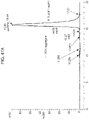
- FIG. 3 shows the analysis of adjuvant linker compounds via liquid chromatography-mass spectrometry (LC-MS).
- FIG. 4 shows that antibody-adjuvant conjugates are superior at eliciting APC activation, compared to unconjugated antibody and adjuvant, as indicated by expression of CD40, CD86 and HLA-DR.
- FIG. 5 shows that antibody-adjuvant conjugates induce lower levels of PD-L1 expression on human APCs, compared to unconjugated antibody and adjuvant.
- FIG. 6 shows that antibody-adjuvant conjugates elicit DC differentiation.
- Human APCs that were ⁇ 95% monocytes were stimulated with 2-fold serial dilutions of Rituximab-SATA-SMCC-Compound 1 (conjugated), Rituximab alone (Ab), Compound 1 alone or Rituximab+Compound 1 (Mixture) in the presence of CFSE-labeled tumor cells.
- P-values ⁇ 0.05 depicted by * P-values ⁇ 0.01 depicted by **, P-values ⁇ 0.001 depicted by ***, P-values ⁇ 0.0001 depicted by ****.
- FIG. 7 shows that antibody-adjuvant conjugates are superior to mixtures of unconjugated antibody and adjuvant for eliciting the secretion of proinflammatory cytokines from human APCs.
- FIG. 8 A shows that immunoconjugates with cleavable linkers elicit APC activation and DC differentiation.
- Human APCs that were ⁇ 95% monocytes were stimulated with 2-fold serial dilutions of Rituximab-SATA-SPDP-Compound 1 (Conjugated, cleavable), Rituximab alone (Ab), Compound 1 alone or Rituximab+Compound 1 (Mixture) in the presence of CFSE-labeled tumor cells.
- the immunoconjugate (AAC—cleavable) had a drug to antibody ratio (DAR) of 1.4 as confirmed by MALDI-TOF.
- DAR drug to antibody ratio
- FIG. 8 B shows that immunoconjugates (AACs) with cleavable linkers elicit APC activation and DC differentiation.
- Human APCs that were ⁇ 95% monocytes were stimulated with 2-fold serial dilutions of Rituximab-SATA-SPDP-Compound 1 (Conjugated, cleavable), Rituximab alone (Ab), Compound 1 alone or Rituximab+Compound 1 (Mixture) in the presence of CFSE-labeled tumor cells.
- the immunoconjugates (AAC—Cleavable) had a drug to antibody ratio (DAR) of 1.4 as confirmed by MALDI-TOF.
- DAR drug to antibody ratio
- FIG. 8 C shows that immunoconjugates with cleavable linkers elicit APC activation and DC differentiation.
- Human APCs that were ⁇ 95% monocytes were stimulated with 2-fold serial dilutions of Rituximab-SATA-SPDP-Compound 1 (Conjugated, cleavable), Rituximab alone (Ab), Compound 1 alone or Rituximab+Compound 1 (Mixture) in the presence of CFSE-labeled tumor cells
- Immunoconjugates (AAC—Cleavable) had a drug to antibody ratio (DAR) of 1.4 as confirmed by MALDI-TOF.
- DAR drug to antibody ratio
- FIG. 9 A shows that antibody-adjuvant conjugates reduce tumors in vivo.
- C57BL/6 mice with B16F10 tumors in the right flank were injected intratumorally with PBS (Untreated), ⁇ GP75+Compound 1 (Mixture) or ⁇ GP75-SATA-SMCC-Compound 1 ( ⁇ GP75-immunoconjugate).
- FIG. 9 B shows that ⁇ GP75-immunoconjugate reduces tumors in vivo when administered via intratumoral (IT) or intravenous (IV) injection.
- FIG. 10 A shows the analysis of ipilimumab via LC-MS.
- FIG. 10 B shows that ipilimumab-adjuvant (Ipilimumab Boltbody) conjugates are superior at eliciting APC activation, compared to unconjugated ipilimumab, as indicated by expression of HLA-DR.
- ipilimumab-adjuvant Ipilimumab Boltbody
- FIG. 10 C shows that ipilimumab-adjuvant (Ipilimumab Boltbody) conjugates are superior at eliciting APC activation, compared to unconjugated ipilimumab, as indicated by expression of CD14.
- Ipilimumab Boltbody ipilimumab-adjuvant
- FIG. 10 D shows that ipilimumab-adjuvant (Ipilimumab Boltbody) conjugates are superior at eliciting APC activation, compared to unconjugated ipilimumab, as indicated by expression of CD40.
- Ipilimumab Boltbody ipilimumab-adjuvant
- FIG. 10 E shows that ipilimumab-adjuvant (Ipilimumab Boltbody) conjugates are superior at eliciting APC activation, compared to unconjugated ipilimumab, as indicated by expression of CD86.
- Ipilimumab Boltbody ipilimumab-adjuvant
- FIG. 11 A shows the analysis of pembrolizumab via LC-MS.
- FIG. 11 B shows that pembrolizumab-adjuvant (Pembrolizumab Boltbody) conjugates are superior at eliciting APC activation, compared to unconjugated pembrolizumab, as indicated by expression of HLA-DR.
- pembrolizumab-adjuvant Piericzumab Boltbody
- FIG. 11 C shows that pembrolizumab-adjuvant (Pembrolizumab Boltbody) conjugates are superior at eliciting APC activation, compared to unconjugated pembrolizumab, as indicated by expression of CD14.
- pembrolizumab-adjuvant Piericzumab Boltbody
- FIG. 11 D shows that pembrolizumab-adjuvant (Pembrolizumab Boltbody) conjugates are superior at eliciting APC activation, compared to unconjugated pembrolizumab, as indicated by expression of CD40.
- pembrolizumab-adjuvant Piericzumab Boltbody
- FIG. 11 E shows that pembrolizumab-adjuvant (Pembrolizumab Boltbody) conjugates are superior at eliciting APC activation, compared to unconjugated pembrolizumab, as indicated by expression of CD86.
- pembrolizumab-adjuvant Piericzumab Boltbody
- FIG. 12 A shows the analysis of nivolumab via LC-MS.
- FIG. 12 B shows that nivolumab-adjuvant (Nivolumab Boltbody) conjugates are superior at eliciting APC activation, compared to unconjugated nivolumab, as indicated by expression of HLA-DR.
- nivolumab-adjuvant Navolumab Boltbody
- FIG. 12 C shows that nivolumab-adjuvant (Nivolumab Boltbody) conjugates are superior at eliciting APC activation, compared to unconjugated nivolumab, as indicated by expression of CD14.
- nivolumab-adjuvant Navolumab Boltbody
- FIG. 12 D shows that nivolumab-adjuvant (Nivolumab Boltbody) conjugates are superior at eliciting APC activation, compared to unconjugated nivolumab, as indicated by expression of CD40.
- nivolumab-adjuvant Navolumab Boltbody
- FIG. 12 E shows that nivolumab-adjuvant (Nivolumab Boltbody) conjugates are superior at eliciting APC activation, compared to unconjugated nivolumab, as indicated by expression of CD86.
- nivolumab-adjuvant Navolumab Boltbody
- FIG. 13 A shows the analysis of atezolizumab via LC-MS.
- FIG. 13 B shows that atezolizumab-adjuvant (Atezolizumab Boltbody) conjugates are superior at eliciting APC activation, compared to unconjugated atezolizumab, as indicated by expression of HLA-DR.
- FIG. 13 C shows that atezolizumab-adjuvant (Atezolizumab Boltbody) conjugates are superior at eliciting APC activation, compared to unconjugated atezolizumab, as indicated by expression of CD14.
- FIG. 13 D shows that atezolizumab-adjuvant (Atezolizumab Boltbody) conjugates are superior at eliciting APC activation, compared to unconjugated atezolizumab, as indicated by expression of CD40.
- FIG. 13 E shows that the level of activation of atezolizumab-adjuvant (Atezolizumab Boltbody) conjugates, as indicated by expression of CD86.
- FIG. 14 A shows that atezolizumab immunoconjugate (Atezolizumab IgG1 NQ Boltbody)-differentiated cells secrete higher amounts of TNF ⁇ than atezolizumab-differentiated cells.
- FIG. 14 B shows that atezolizumab immunoconjugate (Atezolizumab IgG1 NQ Boltbody)-differentiated cells secrete higher amounts of IL-1 ⁇ than atezolizumab-differentiated cells.
- FIG. 15 A shows that nivolumab immunoconjugate (Nivolumab IgG4 Boltbody)-differentiated cells secrete higher amounts of TNF ⁇ than nivolumab-differentiated cells.
- nivolumab immunoconjugate Navolumab IgG4 Boltbody
- FIG. 15 B shows that nivolumab immunoconjugate (Nivolumab IgG4 Boltbody)-differentiated cells secrete higher amounts of IL-1 ⁇ than nivolumab-differentiated cells.
- nivolumab immunoconjugate Navolumab IgG4 Boltbody
- FIG. 16 A shows that pembrolizumab immunoconjugate (Pembrolizumab Boltbody)-differentiated cells secrete higher amounts of TNF ⁇ than pembrolizumab-differentiated cells.
- FIG. 16 B shows that pembrolizumab immunoconjugate (Pembrolizumab Boltbody)-differentiated cells secrete higher amounts of IL-1 ⁇ than pembrolizumab-differentiated cells.
- FIG. 17 shows the analysis of pembrolizumab-adjuvant conjugates via LC-MS.
- FIG. 18 shows the analysis of nivolumab-adjuvant conjugates via LC-MS.
- FIG. 19 shows the analysis of atezolizumab-adjuvant conjugates via LC-MS.
- FIG. 20 shows that ipilimumab immunoconjugate (Ipilimumab Boltbody)-differentiated cells secret higher amounts of TNF ⁇ than ipilimumab-differentiated cells.
- ipilimumab immunoconjugate Ipilimumab Boltbody
- FIG. 21 shows that Dectin-2 immunoconjugate-differentiated cells secrete higher amounts of TNF ⁇ , IL-6, and IL-12p70 than cells exposed to equivalent amounts of the unconjugated components.
- the line that is significantly higher than the x-axis for each cytokine is the anti-Dectin-2 immunoconjugate (anti-Dectin-2-Cmpd1 (antibody conjugated with adjuvant Compound 1)).
- the line which is barely above the x-axis for IL-6 represents a control antibody, an ACC with Compound 1 as the adjuvant and a rat IgG2a isotype control antibody (labeled “Iso-Cmpd 1” in FIG. 21 ).
- the Iso-Cmpd 1 line is not visible as it is along the x-axis.
- FIG. 22 A shows the structure of adjuvant CL264 and the circle indicates a position on the adjuvant where it could be conjugated to the linker, specifically, the terminal carboxylic acid of the adjuvant.
- FIG. 22 B shows the structure of adjuvant CL401 and the circle indicates a position on the adjuvant where it could be conjugated to the linker, specifically, the primary amine of the adjuvant.
- FIG. 22 C shows the structure of adjuvant CL413 and the circle indicates a position on the adjuvant where it could be conjugated to the linker, specifically, the first lysine residue of the adjuvant.
- FIG. 22 D shows the structure of adjuvant CL413 and the circle indicates a position on the adjuvant where it could be conjugated to the linker, specifically, the second lysine residue of the adjuvant.
- FIG. 22 E shows the structure of adjuvant CL413 and the circle indicates a position on the adjuvant where it could be conjugated to the linker, specifically, the third lysine residue of the adjuvant.
- FIG. 22 F shows the structure of adjuvant CL413 and the circle indicates a position on the adjuvant where it could be conjugated to the linker, specifically, the fourth lysine residue of the adjuvant.
- FIG. 22 G shows the structure of adjuvant CL413 and the circle indicates a position on the adjuvant where it could be conjugated to the linker, specifically, the primary amine of the adjuvant.
- FIG. 22 H shows the structure of adjuvant CL419 and the circles indicate positions on the adjuvant where it could be conjugated to the linker, specifically, the amines of the adjuvant (terminal amine in the top part of FIG. 22 H and secondary amine in the bottom part of FIG. 22 H ).
- FIG. 22 I shows the structure of adjuvant CL553 and the circle indicates a position on the adjuvant where it could be conjugated to the linker, specifically, a secondary amine of the adjuvant.
- FIG. 22 J shows the structure of adjuvant CL553 and the circle indicates a position on the adjuvant where it could be conjugated to the linker, specifically, another secondary amine of the adjuvant.
- FIG. 22 K shows the structure of adjuvant CL553 and the circle indicates a position on the adjuvant where it could be conjugated to the linker, specifically, a primary amine of the adjuvant.
- FIG. 22 L shows the structure of adjuvant CL553 and the circle indicates a position on the adjuvant where it could be conjugated to the linker, specifically, another secondary amine of the adjuvant.
- FIG. 22 M shows the structure of adjuvant CL553 and the circle indicates a position on the adjuvant where it could be conjugated to the linker, specifically, another secondary amine of the adjuvant.
- FIG. 22 N shows the structure of adjuvant CL553 and the circle indicates a position on the adjuvant where it could be conjugated to the linker, specifically, another secondary amine of the adjuvant.
- FIG. 22 O shows the structure of adjuvant CL572 and the circles indicate positions on the adjuvant where it could be conjugated to the linker, specifically, the primary amine (top part of FIG. 22 O ) and the carbonyl (bottom part of FIG. 22 O ).
- FIG. 22 P shows the structure of adjuvant Pam2CSK4 and the circle indicates a position on the adjuvant where it could be conjugated to the linker, specifically, the terminal carboxylic acid of the adjuvant.
- FIG. 22 Q shows the structure of adjuvant Pam2CSK4 and the circle indicates a position on the adjuvant where it could be conjugated to the linker, specifically, the terminal thiol of the adjuvant.
- FIG. 22 R shows the structure of adjuvant Pam2CSK4 and the circle indicates a position on the adjuvant where it could be conjugated to the linker, specifically, the second lysine residue of the adjuvant.
- FIG. 22 S shows the structure of adjuvant Pam2CSK4 and the circle indicates a position on the adjuvant where it could be conjugated to the linker, specifically, the third lysine residue of the adjuvant.
- FIG. 22 T shows the structure of adjuvant Pam2CSK4 and the circle indicates a position on the adjuvant where it could be conjugated to the linker, specifically, the terminal lysine residue of the adjuvant.
- FIG. 22 U shows the structure of adjuvant Pam3CSK4 and the circle indicates a position on the adjuvant where it could be conjugated to the linker, specifically, the terminal carboxylic acid of the adjuvant.
- FIG. 22 V shows the structure of adjuvant Pam3CSK4 and the circle indicates a position on the adjuvant where it could be conjugated to the linker, specifically, the terminal thiol of the adjuvant.
- FIG. 22 W shows the structure of adjuvant Pam3CSK4 and the circle indicates a position on the adjuvant where it could be conjugated to the linker, specifically, the second lysine residue of the adjuvant.
- FIG. 22 X shows the structure of adjuvant Pam3CSK4 and the circle indicates a position on the adjuvant where it could be conjugated to the linker, specifically, the third lysine residue of the adjuvant.
- FIG. 23 A shows ⁇ CLEC5A immunoconjugate-differentiated cells secrete higher amounts of IL-6 than cells exposed to equivalent amounts of the unconjugated components.
- the line that is significantly higher than the x-axis for each cytokine is ⁇ CLEC5A immunoconjugate ( ⁇ CLEC5A antibody conjugated with adjuvant Compound 1).
- the line that is along the x-axis shows that a mixture of ⁇ CLEC5A antibody and adjuvant Compound 1 (unconjugated) failed to produce any cytokine response.
- the line between the x-axis and the ⁇ CLEC5A immunoconjugate line represents a control conjugate, rat IgG2a isotype conjugated to Compound 1.
- FIG. 23 B shows ⁇ CLEC5A immunoconjugate-differentiated cells secrete higher amounts of IL-12p40 than cells exposed to equivalent amounts of the unconjugated components.
- the line that is significantly higher than the x-axis for each cytokine is ⁇ CLEC5A immunoconjugate ( ⁇ CLEC5A-Cmpd1 AAC).
- the line that is along the x-axis shows that a mixture of ⁇ CLEC5A antibody and adjuvant Compound 1 (unconjugated; ⁇ CLEC5A-Cmpd1 Mixture) failed to produce any cytokine response.
- the line between the x-axis and the ⁇ CLEC5A immunoconjugate line represents a control conjugate, rat IgG2a isotype conjugated to Compound 1.
- FIG. 23 C shows ⁇ CLEC5A immunoconjugate-differentiated cells secrete higher amounts of IL-12p70 than cells exposed to equivalent amounts of the unconjugated components.
- the line that is significantly higher than the x-axis for each cytokine is ⁇ CLEC5A immunoconjugate ( ⁇ CLEC5A antibody conjugated with adjuvant Compound 1).
- the line that is along the x-axis shows that a mixture of ⁇ CLEC5A antibody and adjuvant Compound 1 (unconjugated) failed to produce any cytokine response.
- the line between the x-axis and the ⁇ CLEC5A immunoconjugate line represents a control conjugate, rat IgG2a isotype conjugated to Compound 1.
- FIG. 23 D shows ⁇ CLEC5A immunoconjugate-differentiated cells secrete higher amounts of TNF ⁇ than cells exposed to equivalent amounts of the unconjugated components.
- the line that is significantly higher than the x-axis for each cytokine is ⁇ CLEC5A immunoconjugate ( ⁇ CLEC5A antibody conjugated with adjuvant Compound 1).
- the line that is along the x-axis shows that a mixture of ⁇ CLEC5A antibody and adjuvant Compound 1 (unconjugated) failed to produce any cytokine response.
- the line between the x-axis and the ⁇ CLEC5A immunoconjugate line represents a control conjugate, rat IgG2a isotype conjugated to Compound 1.
- FIG. 23 E shows the analysis of ⁇ CLEC5A immunoconjugate via LC-MS.
- FIG. 24 shows increased dendritic cell differentiation with an anti-Her2 antibody adjuvant conjugate ( ⁇ Her2 immunoconjugate, closed circles) linked to the TLR 7/8 agonist Compound 1 as compared to when the same antibody and adjuvant components ( ⁇ Her2 and Compound 1, closed squares) are delivered as an unlinked mixture.
- ⁇ Her2 immunoconjugate, closed circles an anti-Her2 antibody adjuvant conjugate linked to the TLR 7/8 agonist Compound 1 as compared to when the same antibody and adjuvant components ( ⁇ Her2 and Compound 1, closed squares) are delivered as an unlinked mixture.
- FIG. 25 shows increased dendritic cell differentiation with an anti-EGFR antibody adjuvant conjugate ( ⁇ EGFR immunoconjugate, closed circles) linked to the TLR 7/8 agonist Compound 1 as compared to when the same components ( ⁇ EGFR and Compound 1, closed squares) are delivered as an unlinked mixture.
- ⁇ EGFR immunoconjugate, closed circles an anti-EGFR antibody adjuvant conjugate linked to the TLR 7/8 agonist Compound 1 as compared to when the same components ( ⁇ EGFR and Compound 1, closed squares) are delivered as an unlinked mixture.
- FIG. 26 shows that an anti-CD20 antibody conjugated to TLR 7/8 agonist exhibits robust dendritic cell activation, while activation is significantly reduced in the deglycosylated conjugate.
- FIG. 27 compares rituximab and obinutuzumab antibodies conjugated to Compound 1.
- Obinutuzumab has reduced fucose content as compared to rituximab and exhibits increased CD40 upregulation.
- FIG. 28 illustrates NK cell activation using an ⁇ EGFR immunoconjugate linked to the TLR 7/8 agonist Compound 1.
- the immunoconjugate exhibits significantly greater NK cell activation as compared to the unconjugated mixture of ⁇ EGFR and Compound 1.
- FIG. 29 illustrates robust activation of dendritic cell populations from peripheral blood mononuclear cells isolated from human subjects with an ⁇ EGFR immunoconjugate.
- FIG. 30 A shows a liquid chromatography-mass spectrometry analysis of immunoconjugate BB-01 synthesized using the SATA method.
- FIG. 30 B shows a liquid chromatography-mass spectrometry analysis of immunoconjugate BB-01 synthesized using the ester method.
- FIG. 31 A shows a size-exclusion chromatography analysis of immunoconjugate BB01 synthesized using the SATA method.
- FIG. 31 B shows a size-exclusion chromatography analysis of immunoconjugate BB-01 synthesized using the ester method.
- FIG. 32 shows a liquid chromatography-mass spectrometry analysis of immunoconjugate BB-14 synthesized using the ester method.
- FIG. 33 shows a size-exclusion chromatography analysis of immunoconjugate BB-14 synthesized using the ester method.
- FIG. 34 shows a liquid chromatography-mass spectrometry analysis of immunoconjugate BB-15 synthesized using the ester method.
- FIG. 35 shows a size-exclusion chromatography analysis of immunoconjugate BB-15 synthesized using the ester method.
- FIG. 36 shows a liquid chromatography-mass spectrometry analysis of immunoconjugate synthesized using the ester method.
- FIG. 37 shows a size-exclusion chromatography analysis of immunoconjugate synthesized using the ester method.
- FIG. 38 A shows BB-01 and BB-17 synthesized using the ester method elicits myeloid activation as indicated by CD14 downregulation while the control does not.
- CD20 is the unconjugated monoclonal antibody used as a control.
- FIG. 38 B shows BB-01 and BB-17 synthesized using the ester method elicits myeloid activation as indicated by CD16 downregulation while the control does not.
- CD20 is the unconjugated monoclonal antibody used as a control.
- FIG. 38 C shows BB-01 and BB-17 synthesized using the ester method elicits myeloid activation as indicated by CD40 upregulation while the control does not.
- CD20 is the unconjugated monoclonal antibody used as a control.
- FIG. 38 D shows BB-01 and BB-17 synthesized using the ester method elicits myeloid activation as indicated by CD86 upregulation while the control does not.
- CD20 is the unconjugated monoclonal antibody used as a control.
- FIG. 38 E shows BB-01 and BB-17 synthesized using the ester method elicits myeloid activation as indicated by CD123 upregulation while the control does not.
- CD20 is the unconjugated monoclonal antibody used as a control.
- FIG. 38 F shows BB-01 and BB-17 synthesized using the ester method elicits myeloid activation as indicated by Human Leukocyte Antigen-antigen D Related or “HLA-DR” while the control does not.
- CD20 is the unconjugated monoclonal antibody used as a control.
- FIG. 39 A shows that BB-01 elicits myeloid activation as indicated by CD14 downregulation while comparative IRM1 and IRM2 immunoconjugates do not.
- CD20 is the unconjugated monoclonal antibody used as a control.
- FIG. 39 B shows that BB-01 elicits myeloid activation as indicated by CD16 downregulation while comparative IRM1 and IRM2 immunoconjugates do not.
- CD20 is the unconjugated monoclonal antibody used as a control.
- FIG. 39 C shows that BB-01 elicits myeloid activation as indicated by CD40 upregulation while comparative IRM1 and IRM2 immunoconjugates do not.
- CD20 is the unconjugated monoclonal antibody used as a control.
- FIG. 39 D shows that BB-01 elicits myeloid activation as indicated by CD86 upregulation while comparative IRM1 and IRM2 immunoconjugates do not.
- CD20 is the unconjugated monoclonal antibody used as a control.
- FIG. 39 E shows that BB-01 elicits myeloid activation as indicated by CD123 upregulation while comparative IRM1 and IRM2 immunoconjugates do not.
- CD20 is the unconjugated monoclonal antibody used as a control.
- FIG. 39 F shows that BB-01 elicits myeloid activation as indicated by HLA-DR upregulation while comparative IRM1 and IRM2 immunoconjugates do not.
- CD20 is the unconjugated monoclonal antibody used as a control.
- FIG. 40 A shows that BB-01 elicits cytokine secretion (IL-1 ⁇ ) while comparative IRM1 and IRM2 immunoconjugates do not.
- CD20 is the unconjugated monoclonal antibody used as a control.
- FIG. 40 B shows that BB-01 elicits cytokine secretion (IL-6) while comparative IRM1 and IRM2 immunoconjugates do not.
- CD20 is the unconjugated monoclonal antibody used as a control.
- FIG. 40 C shows that BB-01 elicits cytokine secretion (TNF ⁇ ) while comparative IRM1 and IRM2 immunoconjugates do not.
- CD20 is the unconjugated monoclonal antibody used as a control.
- FIG. 41 A shows a size-exclusion chromatography analysis of immunoconjugate BB-26 synthesized using the ester method.
- FIG. 41 B shows a liquid chromatography-mass spectrometry analysis of immunoconjugate BB-26 synthesized using the ester method.
- FIG. 42 A shows a size-exclusion chromatography analysis of immunoconjugate BB-27 synthesized using the ester method.
- FIG. 42 B shows a liquid chromatography-mass spectrometry analysis of immunoconjugate BB-27 synthesized using the ester method.
- FIG. 43 A shows a size-exclusion chromatography analysis of immunoconjugate BB-36 synthesized using the ester method.
- FIG. 43 B shows a liquid chromatography-mass spectrometry analysis of immunoconjugate BB-36 synthesized using the ester method.
- FIG. 44 A shows a size-exclusion chromatography analysis of Comparative Conjugate IRM1.
- FIG. 44 B shows a size-exclusion chromatography analysis of Comparative Conjugate IRM2.
- FIG. 44 C shows a size-exclusion chromatography analysis of BB-01.
- FIG. 45 A shows a liquid chromatography-mass spectrometry analysis of IRM1 conjugate following overnight deglycosylation with PNGase F.
- FIG. 45 B show a liquid chromatography-mass spectrometry analysis of BB-01 conjugate following overnight deglycosylation with PNGase F.
- FIG. 46 A shows a size-exclusion chromatography analysis of immunoconjugate BB-45 synthesized using the ester method.
- FIG. 46 B shows a liquid chromatography-mass spectrometry analysis of immunoconjugate BB-45 synthesized using the ester method.
- FIG. 47 A shows a size-exclusion chromatography analysis of immunoconjugate BB-24 synthesized using the ester method.
- FIG. 47 B shows a liquid chromatography-mass spectrometry analysis of immunoconjugate BB-24 synthesized using the ester method.
- FIG. 48 A shows a size-exclusion chromatography analysis of immunoconjugate BB-37 synthesized using the ester method.
- FIG. 48 B shows a liquid chromatography-mass spectrometry analysis of immunoconjugate BB-37 synthesized using the ester method.
- FIG. 49 A shows a size-exclusion chromatography analysis of immunoconjugate BB-42 synthesized using the ester method.
- FIG. 49 B shows a liquid chromatography-mass spectrometry analysis of immunoconjugate BB-42 synthesized using the ester method.
- FIG. 50 shows a liquid chromatography-mass spectrometry analysis of immunoconjugate BB-43 synthesized using the ester method.
- FIG. 51 shows a liquid chromatography-mass spectrometry analysis of immunoconjugate BB-44 synthesized using the ester method.
- FIG. 52 A shows that BB-14 elicits myeloid activation as indicated by CD14 downregulation while the control does not.
- CD20 is the unconjugated monoclonal antibody used as a control.
- FIG. 52 B shows that BB-14 elicits myeloid activation as indicated by CD40 upregulation while the control does not.
- CD20 is the unconjugated monoclonal antibody used as a control.
- FIG. 52 C shows that BB-14 elicits myeloid activation as indicated by CD86 upregulation while the control does not.
- CD20 is the unconjugated monoclonal antibody used as a control.
- FIG. 52 D shows that BB-14 elicits myeloid activation as indicated by HLA-DR upregulation while the control does do not.
- CD20 is the unconjugated monoclonal antibody used as a control.
- FIG. 53 A shows that BB-15 elicits myeloid activation as indicated by CD14 downregulation while the control does not.
- CD20 is the unconjugated monoclonal antibody used as a control.
- FIG. 53 B shows that BB-15 elicits myeloid activation as indicated by CD40 upregulation while the control does not.
- CD20 is the unconjugated monoclonal antibody used as a control.
- FIG. 53 C shows that BB-15 elicits myeloid activation as indicated by CD86 upregulation while the control does not.
- CD20 is the unconjugated monoclonal antibody used as a control.
- FIG. 53 D shows that BB-27 elicits myeloid activation as indicated by HLA-DR upregulation while the control does do not.
- CD20 is the unconjugated monoclonal antibody used as a control.
- FIG. 54 A shows that BB-27 elicits myeloid activation as indicated by CD14 downregulation while the control does not.
- CD20 is the unconjugated monoclonal antibody used as a control.
- FIG. 54 B shows that BB-27 elicits myeloid activation as indicated by CD40 upregulation while the control does not.
- CD20 is the unconjugated monoclonal antibody used as a control.
- FIG. 54 C shows that BB-27 elicits myeloid activation as indicated by CD86 upregulation while the control does not.
- CD20 is the unconjugated monoclonal antibody used as a control.
- FIG. 54 D shows that BB-27 elicits myeloid activation as indicated by HLA-DR upregulation while the control does do not.
- CD20 is the unconjugated monoclonal antibody used as a control.
- FIG. 55 A shows that BB-45 elicits myeloid activation as indicated by CD14 downregulation while the control does not.
- CD20 is the unconjugated monoclonal antibody used as a control.
- FIG. 55 B shows that BB-45 elicits myeloid activation as indicated by CD40 upregulation while the control does not.
- CD20 is the unconjugated monoclonal antibody used as a control.
- FIG. 55 C shows that BB-45 elicits myeloid activation as indicated by CD86 upregulation while the control does not.
- CD20 is the unconjugated monoclonal antibody used as a control.
- FIG. 55 D shows that BB-45 elicits myeloid activation as indicated by HLA-DR upregulation while the control does do not.
- CD20 is the unconjugated monoclonal antibody used as a control.
- FIG. 56 A shows that BB-24 elicits myeloid activation as indicated by CD14 downregulation while the control does not.
- CD20 is the unconjugated monoclonal antibody used as a control.
- FIG. 56 B shows that BB-24 elicits myeloid activation as indicated by CD40 upregulation while the control does not.
- CD20 is the unconjugated monoclonal antibody used as a control.
- FIG. 56 C shows that BB-24 elicits myeloid activation as indicated by CD86 upregulation while the control does not.
- CD20 is the unconjugated monoclonal antibody used as a control.
- FIG. 56 D shows that BB-24 elicits myeloid activation as indicated by HLA-DR upregulation while the control does do not.
- CD20 is the unconjugated monoclonal antibody used as a control.
- FIG. 57 shows BB-01 binding to CD20 Toledo cells, which are a cell line used as a model system for studying non-Hodgkin lymphomas.
- BB-01 had stronger binding than the antibodies rituximab or cetuximab.
- FIG. 58 shows aEGFR immunoconjugate with Compound 1 (aEGFR Boltbody) was more effective than the mixture of antibody and adjuvant at activating NK cells.
- PBMCs were activated with the immunoconjugate or the mixture for 18 hours.
- NK cells were gated according to lineage negative (CD3, CD19, CD14 negative) and CD56 positive.
- FIG. 59 shows the analysis of a comparative immunoconjugate via LC-MS (DG).
- This comparative conjugate was prepared with trastuzumab and a noncleavable maleimide-PEG4 linker containing a pentafluorophenyl group with gardiquimod (see US 2017/0158772, paragraph 0275, description of immunoconjugate ATAC3).
- FIG. 60 shows the analysis of a comparative immunoconjugate via LC-MS (heavy chain).
- This comparative conjugate was prepared with trastuzumab and a noncleavable maleimide-PEG4 linker containing a pentafluorophenyl group with gardiquimod (see US 2017/0158772, paragraph 0275, description of immunoconjugate ATAC3).
- FIG. 61 shows the analysis of a comparative immunoconjugate via LC-MS.
- This comparative conjugate was prepared with trastuzumab and a noncleavable maleimide-PEG4 linker containing a pentafluorophenyl group with gardiquimod (see US 2017/0158772, paragraph 0275, description of immunoconjugate ATAC3).
- FIG. 62 shows the analysis of a comparative immunoconjugate via LC-MS (light chain).
- This comparative conjugate was prepared with trastuzumab and a noncleavable maleimide-PEG4 linker containing a pentafluorophenyl group with gardiquimod (see US 2017/0158772, paragraph 0275, description of immunoconjugate ATAC3).
- FIG. 63 shows the analysis of a comparative immunoconjugate via LC-MS (DG, heavy chain).
- This comparative conjugate was prepared with trastuzumab and a noncleavable maleimide-PEG4 linker containing a pentafluorophenyl group with gardiquimod (see US 2017/0158772, paragraph 0275, description of immunoconjugate ATAC3).
- FIG. 64 shows the analysis of a comparative immunoconjugate via LC-MS (DG, light chain).
- This comparative conjugate was prepared with trastuzumab and a noncleavable maleimide-PEG4 linker containing a pentafluorophenyl group with gardiquimod (see US 2017/0158772, paragraph 0275, description of immunoconjugate ATAC3).
- FIG. 65 shows the analysis of a comparative immunoconjugate via LC-MS (DG).
- This comparative conjugate was prepared with trastuzumab and a cleavable valine-citrulline linker containing a PABA group with succinamide (see US 2017/0158772, paragraph 0275, description of immunoconjugate ATAC2).
- FIG. 66 shows the analysis of a comparative immunoconjugate via LC-MS.
- This comparative conjugate was prepared with trastuzumab and a cleavable valine-citrulline linker containing a PABA group with succinamide (see US 2017/0158772, paragraph 0275, description of immunoconjugate ATAC2).
- FIG. 67 A shows that the rituximab immunoconjugate produced according to the BB-01 SATA method (Rituximab Boltbody) elicits superior IL-1 ⁇ secretion from myeloid cells as compared to unconjugated rituximab (Roche) as well as equimolar concentrations of comparative conjugates prepared with rituximab and either a valine-citruline-PABC or a maleimide-PEG4 linker, both containing a pentafluorophenyl group with gardiquimod (Rituximab-ATAC2, Rituximab-ATAC3 respectively; US 2017/0158772) following 18 hours of stimulation.
- FIG. 67 B shows that the rituximab immunoconjugate produced according to the BB-01 SATA method (Rituximab Boltbody) elicits superior TNF ⁇ secretion from myeloid cells as compared to unconjugated rituximab (Roche) as well as equimolar concentrations of comparative conjugates prepared with rituximab and either a valine-citruline-PABC or a maleimide-PEG4 linker, both containing a pentafluorophenyl group with gardiquimod (Rituximab-ATAC2, Rituximab-ATAC3 respectively; US 2017/0158772) following 18 hours of stimulation.
- FIG. 67 C shows a liquid chromatography-mass spectrometry analysis of unconjugated rituximab (Roche) that was utilized to produce the rituximab immunoconjugate according to the BB-01 conjugation method or the rituximab immunoconjugates according to the methods described in US 2017/0158772 following overnight deglycosylation with PNGase F.
- FIG. 67 D shows a liquid chromatography-mass spectrometry analysis of unconjugated rituximab (Roche) that was utilized to produce the rituximab immunoconjugate according to the BB-01 conjugation method or the rituximab immunoconjugates according to the methods described in US 2017/0158772.
- FIG. 67 E shows a liquid chromatography-mass spectrometry analysis of the ipsilateral heavy-light chain of the rituximab immunoconjugate produced using a valine-citruline-PABC linker as described in US 2017/0158772 following overnight deglycosylation with PNGase F.
- FIG. 67 F shows a liquid chromatography-mass spectrometry analysis of the light chain of the rituximab immunoconjugate produced using a valine-citruline-PABC linker as described in US 2017/0158772 following overnight deglycosylation with PNGase F.
- FIG. 67 G shows that the rituximab with valine-citruline-PABC linker immunoconjugate produced according to the methods described in US 2017/0158772 (Ritux-ATAC2) fails to elicit CD123 upregulation on myeloid cells following 18 hours of stimulation.
- FIG. 67 G also shows that the BB-01 immunoconjugate produced according to the SATA method [Rituximab Boltbody (BB-01)] is superior at eliciting CD123 upregulation as compared to Ritux-ATAC2 and equimolar concentrations of unconjugated rituximab (Roche).
- FIG. 67 H shows that the rituximab with valine-citruline-PABC linker immunoconjugate produced according to the methods described in US 2017/0158772 (Ritux-ATAC2) fails to elicit CD14 downregulation on myeloid cells following 18 hours of stimulation.
- FIG. 67 H also shows that the BB-01 immunoconjugate produced according to the SATA method [Rituximab Boltbody (BB-01)] is superior at eliciting CD14 downregulation as compared to Ritux-ATAC2 and equimolar concentrations of unconjugated rituximab (Roche).
- FIG. 67 I shows that the BB-01 immunoconjugate produced according to the SATA method [Rituximab Boltbody (BB-01)] is superior at eliciting CD16 downregulation on myeloid cells as compared to the rituximab with valine-citruline-PABC linker immunoconjugate produced according to the methods described in US 2017/0158772 (Ritux-ATAC2) and equimolar concentrations of unconjugated rituximab (Roche).
- FIG. 67 J shows that the rituximab with valine-citruline-PABC linker immunoconjugate produced according to the methods described in US 2017/0158772 (Ritux-ATAC2) fails to elicit CD40 upregulation on myeloid cells following 18 hours of stimulation.
- FIG. 67 J also shows that the BB-01 immunoconjugate produced according to the SATA method [Rituximab Boltbody (BB-01)] is superior at eliciting CD40 upregulation as compared to Ritux-ATAC2 and equimolar concentrations of unconjugated rituximab (Roche).
- FIG. 67 K shows that the BB-01 immunoconjugate produced according to the SATA method [Rituximab Boltbody (BB-01)] is superior at eliciting CD86 upregulation on myeloid cells as compared to the rituximab with valine-citruline-PABC linker immunoconjugate produced according to the methods described in US 2017/0158772 (Ritux-ATAC2) and equimolar concentrations of unconjugated rituximab (Roche).
- FIG. 67 L shows CD123 expression on myeloid cells following 18 hours of stimulation with the rituximab with valine-citruline-PABC linker immunoconjugate produced according to the methods described in US 2017/0158772 (Ritux-ATAC2) as compared to unconjugated rituximab (Roche).
- FIG. 67 M shows CD14 expression on myeloid cells following 18 hours of stimulation with the rituximab with valine-citruline-PABC linker immunoconjugate produced according to the methods described in US 2017/0158772 (Ritux-ATAC2) as compared to unconjugated rituximab (Roche).
- FIG. 67 N shows CD16 expression on myeloid cells following 18 hours of stimulation with the rituximab with valine-citruline-PABC linker immunoconjugate produced according to the methods described in US 2017/0158772 (Ritux-ATAC2) as compared to unconjugated rituximab (Roche).
- FIG. 67 O shows CD40 expression on myeloid cells following 18 hours of stimulation with the rituximab with valine-citruline-PABC linker immunoconjugate produced according to the methods described in US 2017/0158772 (Ritux-ATAC2) as compared to unconjugated rituximab (Roche).
- FIG. 67 P shows CD86 expression on myeloid cells following 18 hours of stimulation with the rituximab with valine-citruline-PABC linker immunoconjugate produced according to the methods described in US 2017/0158772 (Ritux-ATAC2) as compared to unconjugated rituximab (Roche).
- FIG. 68 A shows that the rituximab immunoconjugate produced according to the BB-01 SATA method (Rituximab Boltbody) elicits superior IL-1 ⁇ secretion from myeloid cells as compared to unconjugated rituximab (Roche) as well as equimolar concentrations of comparative conjugates prepared with rituximab and either a valine-citruline-PABC or a maleimide-PEG4 linker, both containing a pentafluorophenyl group with gardiquimod (Rituximab-ATAC2, Rituximab-ATAC3 respectively; US 2017/0158772) following 18 hours of stimulation.
- FIG. 68 B shows that the rituximab immunoconjugate produced according to the BB-01 SATA method (Rituximab Boltbody) elicits superior TNF ⁇ secretion from myeloid cells as compared to unconjugated rituximab (Roche) as well as equimolar concentrations of comparative conjugates prepared with rituximab and either a valine-citruline-PABC or a maleimide-PEG4 linker, both containing a pentafluorophenyl group with gardiquimod (Rituximab-ATAC2, Rituximab-ATAC3 respectively; US 2017/0158772) following 18 hours of stimulation.
- FIG. 68 C shows a liquid chromatography-mass spectrometry analysis of unconjugated rituximab (Roche) that was utilized to produce the rituximab immunoconjugate according to the BB-01 conjugation method or the rituximab immunoconjugates according to the methods described in US 2017/0158772 following overnight deglycosylation with PNGase F.
- FIG. 68 D shows a liquid chromatography-mass spectrometry analysis of unconjugated rituximab (Roche) that was utilized to produce the rituximab immunoconjugate according to the BB-01 conjugation method or the rituximab immunoconjugates according to the methods described in US 2017/0158772.
- FIG. 68 E shows a liquid chromatography-mass spectrometry analysis of the ipsilateral heavy-light chain of the rituximab immunoconjugate produced using a maleimide-PEG4 linker as described in US 2017/0158772 following overnight deglycosylation with PNGase F.
- FIG. 68 F shows a liquid chromatography-mass spectrometry analysis of the light chain of the rituximab immunoconjugate produced using a maleimide-PEG4 linker as described in US 2017/0158772 following overnight deglycosylation with PNGase F.
- FIG. 68 G shows that the rituximab with maleimide-PEG4 linker immunoconjugate produced according to the methods described in US 2017/0158772 (Ritux-ATAC3) fails to elicit CD123 upregulation on myeloid cells following 18 hours of stimulation.
- FIG. 68 G also shows that the BB-01 immunoconjugate produced according to the SATA method [Rituximab Boltbody (BB-01)] is superior at eliciting CD123 upregulation as compared to Ritux-ATAC3 and equimolar concentrations of unconjugated rituximab (Roche).
- FIG. 68 H shows that the BB-01 immunoconjugate produced according to the SATA method [Rituximab Boltbody (BB-01)] is superior at eliciting CD14 downregulation on myeloid cells as compared to the rituximab with maleimide-PEG4 linker immunoconjugate produced according to the methods described in US 2017/0158772 (Ritux-ATAC3) and equimolar concentrations of unconjugated rituximab (Roche) following 18 hours of stimulation.
- FIG. 68 I shows that the rituximab with maleimide-PEG4 linker immunoconjugate produced according to the methods described in US 2017/0158772 (Ritux-ATAC3) fails to elicit CD16 downregulation on myeloid cells following 18 hours of stimulation.
- FIG. 68 I also shows that the BB-01 immunoconjugate produced according to the SATA method [Rituximab Boltbody (BB-01)] is superior at eliciting CD40 upregulation as compared to Ritux-ATAC2 and equimolar concentrations of unconjugated rituximab (Roche).
- FIG. 68 J shows that the rituximab with maleimide-PEG4 linker immunoconjugate produced according to the methods described in US 2017/0158772 (Ritux-ATAC3) fails to elicit CD40 upregulation on myeloid cells following 18 hours of stimulation.
- FIG. 68 J also shows that the BB-01 immunoconjugate produced according to the SATA method [Rituximab Boltbody (BB-01)] is superior at eliciting CD40 upregulation as compared to Ritux-ATAC2 and equimolar concentrations of unconjugated rituximab (Roche).
- FIG. 68 K shows that the rituximab with maleimide-PEG4 linker immunoconjugate produced according to the methods described in US 2017/0158772 (Ritux-ATAC3) fails to elicit CD86 upregulation on myeloid cells following 18 hours of stimulation.
- FIG. 68 J also shows that the BB-01 immunoconjugate produced according to the SATA method [Rituximab Boltbody (BB-01)] is superior at eliciting CD86 upregulation as compared to Ritux-ATAC2 and equimolar concentrations of unconjugated rituximab (Roche).
- FIG. 68 L shows CD123 expression on myeloid cells following 18 hours of stimulation with the rituximab with maleimide-PEG4 linker immunoconjugate produced according to the methods described in US 2017/0158772 (Ritux-ATAC3) as compared to unconjugated rituximab (Roche).
- FIG. 68 M shows HLA-DR expression on myeloid cells following 18 hours of stimulation with the rituximab with maleimide-PEG4 linker immunoconjugate produced according to the methods described in US 2017/0158772 (Ritux-ATAC3) as compared to unconjugated rituximab (Roche).
- FIG. 68 N shows CD14 expression on myeloid cells following 18 hours of stimulation with the rituximab with maleimide-PEG4 linker immunoconjugate produced according to the methods described in US 2017/0158772 (Ritux-ATAC3) as compared to unconjugated rituximab (Roche)
- FIG. 68 O shows CD16 expression on myeloid cells following 18 hours of stimulation with the rituximab with maleimide-PEG4 linker immunoconjugate produced according to the methods described in US 2017/0158772 (Ritux-ATAC3) as compared to unconjugated rituximab (Roche).
- FIG. 68 P shows CD40 expression on myeloid cells following 18 hours of stimulation with the rituximab with maleimide-PEG4 linker immunoconjugate produced according to the methods described in US 2017/0158772 (Ritux-ATAC3) as compared to unconjugated rituximab (Roche).
- FIG. 68 Q shows CD86 expression on myeloid cells following 18 hours of stimulation with the rituximab with maleimide-PEG4 linker immunoconjugate produced according to the methods described in US 2017/0158772 (Ritux-ATAC3) as compared to unconjugated rituximab (Roche).
- FIG. 69 A shows that the atezolizumab immunoconjugate produced according to the BB-01 method (Atezolizumab IgG1 NQ Boltbody) elicits superior IL-1 ⁇ secretion from myeloid cells as compared to equimolar concentrations of unconjugated atezolizumab (Roche) following 18 hours of stimulation.
- FIG. 69 B shows that the atezolizumab immunoconjugate produced according to the BB-01 method (Atezolizumab IgG1 NQ Boltbody) elicits superior TNF ⁇ secretion from myeloid cells as compared to equimolar concentrations of unconjugated atezolizumab (Roche) following 18 hours of stimulation.
- FIG. 69 C shows a liquid chromatography-mass spectrometry analysis of the atezolizumab immunoconjugate produced according to the BB-01 conjugation method following overnight deglycosylation with PNGase F.
- FIG. 69 D shows a liquid chromatography-mass spectrometry analysis of unconjugated atezolizumab (Roche) that was utilized to produce the atezolizumab immunoconjugate according to the BB-01 conjugation method following overnight deglycosylation with PNGase F.
- FIG. 69 E shows a liquid chromatography-mass spectrometry analysis of unconjugated atezolizumab (Roche) that was utilized to produce the atezolizumab immunoconjugate according to the BB-01 method.
- FIG. 69 F shows CD123 expression on myeloid cells following 18 hours of stimulation with the atezolizumab immunoconjugate produced according to the BB-01 method (Atezolizumab Boltbody) as compared to unconjugated atezolizumab (Roche).
- FIG. 69 G shows HLA-DR expression on myeloid cells following 18 hours of stimulation with the atezolizumab immunoconjugate produced according to the BB-01 method (Atezolizumab Boltbody) as compared to unconjugated atezolizumab (Roche).
- FIG. 69 H shows that the atezolizumab immunoconjugate produced according to the BB-01 method (Atezolizumab Boltbody) is superior at eliciting CD14 downregulation on myeloid cells as compared to the unconjugated atezolizumab (Roche) following 18 hours of stimulation.
- FIG. 69 I shows that the atezolizumab immunoconjugate produced according to the BB-01 method (Atezolizumab Boltbody) is superior at eliciting CD16 downregulation on myeloid cells as compared to the unconjugated atezolizumab (Roche) following 18 hours of stimulation.
- FIG. 69 J shows that the atezolizumab immunoconjugate produced according to the BB-01 method (Atezolizumab Boltbody) is superior at eliciting CD40 upregulation on myeloid cells as compared to the unconjugated atezolizumab (Roche) following 18 hours of stimulation.
- FIG. 69 K shows CD86 expression on myeloid cells following 18 hours of stimulation with the atezolizumab immunoconjugate produced according to the BB-01 method (Atezolizumab Boltbody) as compared to unconjugated atezolizumab (Roche).
- FIG. 70 A shows that the bevacizumab immunoconjugate produced according to the BB-01 method (Bevacizumab Boltbody) elicits superior IL-1 ⁇ secretion from myeloid cells as compared to equimolar concentrations of unconjugated bevacizumab (Roche) following 18 hours of stimulation.
- BB-01 method Bevacizumab Boltbody
- FIG. 70 B shows that the bevacizumab immunoconjugate produced according to the BB-01 method (Bevacizumab Boltbody) elicits superior TNF ⁇ secretion from myeloid cells as compared to equimolar concentrations of unconjugated bevacizumab (Roche) following 18 hours of stimulation.
- BB-01 method Bevacizumab Boltbody
- FIG. 70 C shows that the bevacizumab immunoconjugate produced according to the BB-01 method (Bevacizumab Boltbody) elicits superior IL-1 ⁇ secretion from myeloid cells as compared to equimolar concentrations of unconjugated bevacizumab (Roche) following 36 hours of stimulation.
- BB-01 method Bevacizumab Boltbody
- FIG. 70 D shows that the bevacizumab immunoconjugate produced according to the BB-01 method (Bevacizumab Boltbody) elicits superior TNF ⁇ secretion from myeloid cells as compared to equimolar concentrations of unconjugated bevacizumab (Roche) following 36 hours of stimulation.
- BB-01 method Bevacizumab Boltbody
- FIG. 70 E shows a liquid chromatography-mass spectrometry analysis of the bevacizumab immunoconjugate produced according to the BB-01 conjugation method following overnight deglycosylation with PNGase F.
- FIG. 70 F shows a liquid chromatography-mass spectrometry analysis of unconjugated bevacizumab (Roche) that was utilized to produce the bevacizumab immunoconjugate according to the BB-01 conjugation method following overnight deglycosylation with PNGase F.
- FIG. 70 G shows a liquid chromatography-mass spectrometry analysis of unconjugated bevacizumab (Roche) that was utilized to produce the bevacizumab immunoconjugate according to the BB-01 method.
- FIG. 70 H shows that the bevacizumab immunoconjugate produced according to the BB-01 method (Bevacizumab Boltbody) is superior at eliciting CD123 upregulation on myeloid cells as compared to the unconjugated bevacizumab (Roche) following 18 hours of stimulation.
- BB-01 method Bevacizumab Boltbody
- FIG. 70 I shows HLA-DR expression on myeloid cells following 18 hours of stimulation with the bevacizumab immunoconjugate produced according to the BB-01 method (Bevacizumab Boltbody) as compared to unconjugated bevacizumab (Roche).
- FIG. 70 J shows that the bevacizumab immunoconjugate produced according to the BB-01 method (Bevacizumab Boltbody) is superior at eliciting CD14 downregulation on myeloid cells as compared to the unconjugated bevacizumab (Roche) following 18 hours of stimulation.
- FIG. 70 K shows that the bevacizumab immunoconjugate produced according to the BB-01 method (Bevacizumab Boltbody) is superior at eliciting CD16 downregulation on myeloid cells as compared to the unconjugated bevacizumab (Roche) following 18 hours of stimulation.
- FIG. 70 L shows that the bevacizumab immunoconjugate produced according to the BB-01 method (Bevacizumab Boltbody) is superior at eliciting CD40 upregulation on myeloid cells as compared to the unconjugated bevacizumab (Roche) following 18 hours of stimulation.
- FIG. 70 M shows that the bevacizumab immunoconjugate produced according to the BB-01 method (Bevacizumab Boltbody) is superior at eliciting CD86 upregulation on myeloid cells as compared to the unconjugated bevacizumab (Roche) following 18 hours of stimulation.
- BB-01 method Bevacizumab Boltbody
- FIG. 71 A shows a liquid chromatography-mass spectrometry analysis of the cetuximab immunoconjugate produced according to the BB-01 conjugation method from the cetuximab biosimilar (Alphamab) following overnight deglycosylation with PNGase F.
- FIG. 71 B shows a liquid chromatography-mass spectrometry analysis of the cetuximab immunoconjugate produced according to the BB-01 conjugation method.
- FIG. 71 C shows a liquid chromatography-mass spectrometry analysis of unconjugated cetuximab biosimilar (Alphamab) that was utilized to produce the cetuximab immunoconjugate according to the BB-01 method following overnight deglycosylation with PNGase F.
- FIG. 71 D shows a liquid chromatography-mass spectrometry analysis of unconjugated cetuximab biosimilar (Alphamab) that was utilized to produce the cetuximab immunoconjugate according to the BB-01 conjugation method.
- FIG. 71 E shows that the rituximab immunoconjugates produced according to the BB-01 method from rituximab biosimilars (AmAb, Alphamab; JHL, JHL Biotech; LGM, LGM Pharma) elicit comparable CD123 upregulation on myeloid cells following 18 hours of stimulation.
- the dashed line indicates the level of expression on unstimulated myeloid cells cultured for 18 hours.
- FIG. 71 F shows that the rituximab immunoconjugates produced according to the BB-01 method from rituximab biosimilars (AmAb, Alphamab; JHL, JHL Biotech; LGM, LGM Pharma) elicit comparable HLA-DR expression on myeloid cells following 18 hours of stimulation.
- the dashed line indicates the level of expression on unstimulated myeloid cells cultured for 18 hours.
- FIG. 71 G shows that the rituximab immunoconjugates produced according to the BB-01 method from rituximab biosimilars (biosimilar 1, Alphamab; biosimilar 2, JHL Biotech; biosimilar 3, LGM Pharma) elicit comparable CD14 downregulation on myeloid cells following 18 hours of stimulation.
- the dashed line indicates the level of expression on unstimulated myeloid cells cultured for 18 hours.
- FIG. 71 H shows that the rituximab immunoconjugates produced according to the BB-01 method from rituximab biosimilars (AmAb, Alphamab; JHL, JHL Biotech; LGM, LGM Pharma) elicit comparable CD16 downregulation on myeloid cells following 18 hours of stimulation.
- the dashed line indicates the level of expression on unstimulated myeloid cells cultured for 18 hours.
- FIG. 71 I shows that the rituximab immunoconjugates produced according to the BB-01 method from rituximab biosimilars (AmAb, Alphamab; JHL, JHL Biotech; LGM, LGM Pharma) elicit comparable CD40 upregulation on myeloid cells following 18 hours of stimulation.
- the dashed line indicates the level of expression on unstimulated myeloid cells cultured for 18 hours.
- FIG. 71 J shows that the rituximab immunoconjugates produced according to the BB-01 method from rituximab biosimilars (AmAb, Alphamab; JHL, JHL Biotech; LGM, LGM Pharma) elicit comparable CD86 upregulation on myeloid cells following 18 hours of stimulation.
- the dashed line indicates the level of expression on unstimulated myeloid cells cultured for 18 hours.
- FIG. 71 K shows a liquid chromatography-mass spectrometry analysis of the rituximab immunoconjugate produced according to the BB-01 conjugation method from the rituximab biosimilar (Alphamab) following overnight deglycosylation with PNGase F.
- FIG. 71 L shows a liquid chromatography-mass spectrometry analysis of the rituximab immunoconjugate produced according to the BB-01 conjugation method from the rituximab biosimilar (Alphamab).
- FIG. 71 M shows a liquid chromatography-mass spectrometry analysis of an unconjugated rituximab biosimilar (Alphamab) that was utilized to produce the rituximab biosimilar immunoconjugate according to the BB-01 method following overnight deglycosylation with PNGase F.
- FIG. 71 N shows a liquid chromatography-mass spectrometry analysis of an unconjugated rituximab biosimilar (Alphamab) that was utilized to produce the rituximab biosimilar immunoconjugate according to the BB-01 method.
- FIG. 71 O shows that the rituximab biosimilar immunoconjugate produced according to the BB-01 method from a rituximab biosimilar [BB-01 (biosimilar 1)] is superior at eliciting CD123 upregulation on myeloid cells as compared to the corresponding unconjugated rituximab biosimilar [(CD20 (biosimilar 1), Alphamab] following 18 hours of stimulation.
- FIG. 71 P shows that the rituximab biosimilar immunoconjugate produced according to the BB-01 method from a rituximab biosimilar [BB-01 (biosimilar 1)] is superior at eliciting HLA-DR upregulation on myeloid cells as compared to the corresponding unconjugated rituximab biosimilar [(CD20 (biosimilar 1), Alphamab] following 18 hours of stimulation.
- FIG. 71 Q shows that the rituximab biosimilar immunoconjugate produced according to the BB-01 method from a rituximab biosimilar [BB-01 (biosimilar 1)] is superior at eliciting CD14 downregulation on myeloid cells as compared to the corresponding unconjugated rituximab biosimilar [(CD20 (biosimilar 1), Alphamab] following 18 hours of stimulation.
- FIG. 71 R shows that the rituximab biosimilar immunoconjugate produced according to the BB-01 method from a rituximab biosimilar [BB-01 (biosimilar 1)] is superior at eliciting CD16 downregulation on myeloid cells as compared to the corresponding unconjugated rituximab biosimilar [(CD20 (biosimilar 1), Alphamab] following 18 hours of stimulation.
- FIG. 71 S shows that the rituximab biosimilar immunoconjugate produced according to the BB-01 method from a rituximab biosimilar [BB-01 (biosimilar 1)] is superior at eliciting CD40 upregulation on myeloid cells as compared to the corresponding unconjugated rituximab biosimilar [(CD20 (biosimilar 1), Alphamab] following 18 hours of stimulation.
- FIG. 71 T shows that the rituximab biosimilar immunoconjugate produced according to the BB-01 method from a rituximab biosimilar [BB-01 (biosimilar 1)] is superior at eliciting CD86 upregulation on myeloid cells as compared to the corresponding unconjugated rituximab biosimilar [(CD20 (biosimilar 1), Alphamab] following 18 hours of stimulation.
- FIG. 71 U shows a liquid chromatography-mass spectrometry analysis of the rituximab immunoconjugate produced according to the BB-01 conjugation method from the rituximab biosimilar (LGM Pharma) following overnight deglycosylation with PNGase F.
- FIG. 71 V shows a liquid chromatography-mass spectrometry analysis of the rituximab immunoconjugate produced according to the BB-01 conjugation method from the rituximab biosimilar (LGM Pharma).
- FIG. 71 W shows a liquid chromatography-mass spectrometry analysis of an unconjugated rituximab biosimilar (LGM Pharma) that was utilized to produce the rituximab biosimilar immunoconjugate according to the BB-01 method following overnight deglycosylation with PNGase F.
- LGM Pharma unconjugated rituximab biosimilar
- FIG. 71 X shows a liquid chromatography-mass spectrometry analysis of an unconjugated rituximab biosimilar (LGM Pharma) that was utilized to produce the rituximab biosimilar immunoconjugate according to the BB-01 method.
- LGM Pharma unconjugated rituximab biosimilar
- FIG. 71 Y shows that the rituximab biosimilar immunoconjugate produced according to the BB-01 method from a rituximab biosimilar (BB-01) is superior at eliciting CD123 upregulation on myeloid cells as compared to the corresponding unconjugated rituximab biosimilar (CD20, LGM Pharma) following 18 hours of stimulation.
- FIG. 71 Z shows that the rituximab biosimilar immunoconjugate produced according to the BB-01 method from a rituximab biosimilar (BB-01) is superior at eliciting HLA-DR upregulation on myeloid cells as compared to the corresponding unconjugated rituximab biosimilar (CD20, LGM Pharma) following 18 hours of stimulation.
- BB-01 rituximab biosimilar
- FIG. 71 AA shows that the rituximab biosimilar immunoconjugate produced according to the BB-01 method from a rituximab biosimilar (BB-01) is superior at eliciting CD14 downregulation on myeloid cells as compared to the corresponding unconjugated rituximab biosimilar (CD20, LGM Pharma) following 18 hours of stimulation.
- FIG. 71 AB shows that the rituximab biosimilar immunoconjugate produced according to the BB-01 method from a rituximab biosimilar (BB-01) is superior at eliciting CD16 downregulation on myeloid cells as compared to the corresponding unconjugated rituximab biosimilar (CD20, LGM Pharma) following 18 hours of stimulation.
- FIG. 71 AC shows that the rituximab biosimilar immunoconjugate produced according to the BB-01 method from a rituximab biosimilar (BB-01) is superior at eliciting CD40 upregulation on myeloid cells as compared to the corresponding unconjugated rituximab biosimilar (CD20, LGM Pharma) following 18 hours of stimulation.
- FIG. 71 AD shows that the rituximab biosimilar immunoconjugate produced according to the BB-01 method from a rituximab biosimilar (BB-01) is superior at eliciting CD86 upregulation on myeloid cells as compared to the corresponding unconjugated rituximab biosimilar (CD20, LGM Pharma) following 18 hours of stimulation.
- FIG. 71 AE shows a liquid chromatography-mass spectrometry analysis of the rituximab immunoconjugate produced according to the BB-01 conjugation method from the rituximab biosimilar (JHL Biotech) following overnight deglycosylation with PNGase F.
- FIG. 71 AF shows a liquid chromatography-mass spectrometry analysis of the rituximab immunoconjugate produced according to the BB-01 conjugation method from the rituximab biosimilar (JHL Biotech).
- FIG. 71 AG shows a liquid chromatography-mass spectrometry analysis of an unconjugated rituximab biosimilar (JHL Biotech) that was utilized to produce the rituximab biosimilar immunoconjugate according to the BB-01 method following overnight deglycosylation with PNGase F.
- FIG. 71 AH shows a liquid chromatography-mass spectrometry analysis of an unconjugated rituximab biosimilar (JHL Biotech) that was utilized to produce the rituximab biosimilar immunoconjugate according to the BB-01 method.
- FIG. 71 AI shows that the rituximab biosimilar immunoconjugate produced according to the BB-01 method from a rituximab biosimilar [BB-01 (biosimilar 2)] is superior at eliciting CD123 upregulation on myeloid cells as compared to the corresponding unconjugated rituximab biosimilar [(CD20 (biosimilar 2), JHL Biotech] following 18 hours of stimulation.
- FIG. 71 AJ shows that the rituximab biosimilar immunoconjugate produced according to the BB-01 method from a rituximab biosimilar [BB-01 (biosimilar 2)] is superior at eliciting HLA-DR upregulation on myeloid cells as compared to the corresponding unconjugated rituximab biosimilar [(CD20 (biosimilar 2), JHL Biotech] following 18 hours of stimulation.
- FIG. 71 AK shows that the rituximab biosimilar immunoconjugate produced according to the BB-01 method from a rituximab biosimilar [BB-01 (biosimilar 2)] is superior at eliciting CD40 upregulation on myeloid cells as compared to the corresponding unconjugated rituximab biosimilar [(CD20 (biosimilar 2), JHL Biotech] following 18 hours of stimulation.
- FIG. 71 AL shows that the rituximab biosimilar immunoconjugate produced according to the BB-01 method from a rituximab biosimilar [BB-01 (biosimilar 2)] is superior at eliciting CD86 upregulation on myeloid cells as compared to the corresponding unconjugated rituximab biosimilar [(CD20 (biosimilar 2), JHL Biotech] following 18 hours of stimulation.
- FIG. 71 AM shows a liquid chromatography-mass spectrometry analysis of unconjugated rituximab (Roche).
- FIG. 71 AN shows a liquid chromatography-mass spectrometry analysis of the trastuzumab immunoconjugate produced according to the BB-01 conjugation method from the trastuzumab biosimilar (JHL Biotech) following overnight deglycosylation with PNGase F.
- FIG. 71 AO shows a liquid chromatography-mass spectrometry analysis of the trastuzumab immunoconjugate produced according to the BB-01 conjugation method from the trastuzumab biosimilar (JHL Biotech).
- FIG. 71 AP shows a liquid chromatography-mass spectrometry analysis of an unconjugated trastuzumab biosimilar (JHL Biotech) that was utilized to produce the trastuzumab biosimilar immunoconjugate according to the BB-01 method following overnight deglycosylation with PNGase F.
- FIG. 71 AQ shows a liquid chromatography-mass spectrometry analysis of an unconjugated trastuzumab biosimilar (JHL Biotech) that was utilized to produce the trastuzumab biosimilar immunoconjugate according to the BB-01 method.
- FIG. 71 AR shows that the trastuzumab biosimilar immunoconjugate produced according to the BB-01 method from a trastuzumab biosimilar (BB-40) is superior at eliciting CD123 upregulation on myeloid cells as compared to the corresponding unconjugated trastuzumab biosimilar [Trastuzumab (JHL), JHL Biotech) following 18 hours of stimulation.
- FIG. 71 AS shows that the trastuzumab biosimilar immunoconjugate produced according to the BB-01 method from a trastuzumab biosimilar (BB-40) is superior at eliciting HLA-DR upregulation on myeloid cells as compared to the corresponding unconjugated trastuzumab biosimilar [Trastuzumab (JHL), JHL Biotech) following 18 hours of stimulation.
- BB-40 trastuzumab biosimilar
- FIG. 71 AT shows that the trastuzumab biosimilar immunoconjugate produced according to the BB-01 method from a trastuzumab biosimilar (BB-40) is superior at eliciting CD14 downregulation on myeloid cells as compared to the corresponding unconjugated trastuzumab biosimilar [Trastuzumab (JHL), JHL Biotech) following 18 hours of stimulation.
- FIG. 71 AU shows that the trastuzumab biosimilar immunoconjugate produced according to the BB-01 method from a trastuzumab biosimilar (BB-40) is superior at eliciting CD16 downregulation on myeloid cells as compared to the corresponding unconjugated trastuzumab biosimilar [Trastuzumab (JHL), JHL Biotech) following 18 hours of stimulation.
- FIG. 71 AV shows that the trastuzumab biosimilar immunoconjugate produced according to the BB-01 method from a trastuzumab biosimilar (BB-40) is superior at eliciting CD40 upregulation on myeloid cells as compared to the corresponding unconjugated trastuzumab biosimilar [Trastuzumab (JHL), JHL Biotech) following 18 hours of stimulation.
- FIG. 71 AW shows that the trastuzumab biosimilar immunoconjugate produced according to the BB-01 method from a trastuzumab biosimilar (BB-40) is superior at eliciting CD86 upregulation on myeloid cells as compared to the corresponding unconjugated trastuzumab biosimilar [Trastuzumab (JHL), JHL Biotech) following 18 hours of stimulation.
- FIG. 72 A shows that the cetuximab immunoconjugate produced according to the BB-01 method (Cetuximab Boltbody) elicits superior IL-1 ⁇ secretion from myeloid cells as compared to equimolar concentrations of unconjugated cetuximab (Imclone/Lilly) following 18 hours of stimulation.
- FIG. 72 B shows that the cetuximab immunoconjugate produced according to the BB-01 method (Cetuximab Boltbody) elicits superior TNF ⁇ secretion from myeloid cells as compared to equimolar concentrations of unconjugated cetuximab (Imclone/Lilly) following 18 hours of stimulation.
- FIG. 72 C shows a liquid chromatography-mass spectrometry analysis of unconjugated cetuximab (Imclone/Lilly) that was utilized to produce the cetuximab immunoconjugate according to the BB-01 conjugation method following overnight deglycosylation with PNGase F.
- FIG. 72 D shows a liquid chromatography-mass spectrometry analysis of unconjugated cetuximab (Imclone/Lilly) that was utilized to produce the cetuximab immunoconjugate according to the BB-01 conjugation method.
- FIG. 72 E shows that the cetuximab immunoconjugate produced according to the BB-01 method (closed circles, red) is superior at eliciting CD123 upregulation on myeloid cells as compared to the unconjugated cetuximab (closed squares, black; Imclone/Lilly) following 18 hours of stimulation.
- FIG. 72 F shows that the cetuximab immunoconjugate produced according to the BB-01 method (closed circles, red) is superior at eliciting HLA-DR upregulation on myeloid cells as compared to the unconjugated cetuximab (closed squares, black; Imclone/Lilly) following 18 hours of stimulation.
- FIG. 72 G shows that the cetuximab immunoconjugate produced according to the BB-01 method (closed circles, red) is superior at eliciting CD14 downregulation on myeloid cells as compared to the unconjugated cetuximab (closed squares, black; Imclone/Lilly) following 18 hours of stimulation.
- FIG. 72 H shows that the cetuximab immunoconjugate produced according to the BB-01 method (closed circles, red) is superior at eliciting CD16 downregulation on myeloid cells as compared to the unconjugated cetuximab (closed squares, black; Imclone/Lilly) following 18 hours of stimulation.
- FIG. 72 I shows that the cetuximab immunoconjugate produced according to the BB-01 method (closed circles, red) is superior at eliciting CD40 upregulation on myeloid cells as compared to the unconjugated cetuximab (closed squares, black; Imclone/Lilly) following 18 hours of stimulation.
- FIG. 72 J shows that the cetuximab immunoconjugate produced according to the BB-01 method (closed circles, red) is superior at eliciting CD86 upregulation on myeloid cells as compared to the unconjugated cetuximab (closed squares, black; Imclone/Lilly) following 18 hours of stimulation.
- FIG. 73 A shows that the daratumumab immunoconjugate produced according to the BB-01 method (Daratumumab Boltbody) elicits superior IL-1 ⁇ secretion from myeloid cells as compared to equimolar concentrations of unconjugated daratumumab (Genmab/Janssen Biotech) following 18 hours of stimulation.
- FIG. 73 B shows that the daratumumab immunoconjugate produced according to the BB-01 method (Daratumumab Boltbody) elicits superior TNF ⁇ secretion from myeloid cells as compared to equimolar concentrations of unconjugated daratumumab (Genmab/Janssen Biotech) following 18 hours of stimulation.
- FIG. 73 C shows that the daratumumab immunoconjugate produced according to the BB-01 method (Daratuzumab [sic] Boltbody) elicits superior TNF ⁇ secretion from myeloid cells as compared to equimolar concentrations of unconjugated daratumumab (Daratuzumab [sic], Genmab/Janssen Biotech) following 36 hours of stimulation.
- BB-01 method Daratuzumab [sic] Boltbody
- FIG. 73 D shows that the daratumumab immunoconjugate produced according to the BB-01 method (Daratuzumab [sic] Boltbody) elicits superior IL-1 ⁇ secretion from myeloid cells as compared to equimolar concentrations of unconjugated daratumumab (Daratuzumab [sic], Genmab/Janssen Biotech) following 36 hours of stimulation.
- BB-01 method Daratuzumab [sic] Boltbody
- FIG. 73 E shows a liquid chromatography-mass spectrometry analysis of unconjugated daratumumab (Genmab/Janssen Biotech) that was utilized to produce the daratumumab immunoconjugate according to the BB-01 method following overnight deglycosylation with PNGase F.
- FIG. 73 F shows a liquid chromatography-mass spectrometry analysis of the daratumumab immunoconjugate produced according to the BB-01 method following overnight deglycosylation with PNGase F.
- FIG. 73 G shows a liquid chromatography-mass spectrometry analysis of unconjugated daratumumab (Genmab/Janssen Biotech) that was utilized to produce the daratumumab immunoconjugate according to the BB-01 method.
- FIG. 73 H shows that the daratumumab immunoconjugate produced according to the BB-01 method (Daratuzumab [sic] Boltbody) is superior at eliciting CD123 upregulation on myeloid cells as compared to the unconjugated daratumumab (Daratuzumab [sic], Genmab/Janssen Biotech) following 18 hours of stimulation.
- FIG. 73 I shows HLA-DR expression on myeloid cells following 18 hours of stimulation with the daratumumab immunoconjugate produced according to the BB-01 method (Daratumumab Boltbody) as compared to unconjugated daratumumab (Genmab/Janssen Biotech).
- FIG. 73 J shows that the daratumumab immunoconjugate produced according to the BB-01 method (Daratuzumab [sic] Boltbody) is superior at eliciting CD14 downregulation on myeloid cells as compared to the unconjugated daratumumab (Daratuzumab [sic], Genmab/Janssen Biotech) following 18 hours of stimulation.
- FIG. 73 K shows that the daratumumab immunoconjugate produced according to the BB-01 method (Daratuzumab [sic] Boltbody) is superior at eliciting CD16 downregulation on myeloid cells as compared to the unconjugated daratumumab (Daratuzumab [sic], Genmab/Janssen Biotech) following 18 hours of stimulation.
- FIG. 73 L shows that the daratumumab immunoconjugate produced according to the BB-01 method (Daratuzumab [sic] Boltbody) is superior at eliciting CD40 upregulation on myeloid cells as compared to the unconjugated daratumumab (Daratuzumab [sic], Genmab/Janssen Biotech) following 18 hours of stimulation.
- FIG. 73 M shows that the daratumumab immunoconjugate produced according to the BB-01 method (Daratuzumab [sic] Boltbody) is superior at eliciting CD86 upregulation on myeloid cells as compared to the unconjugated daratumumab (Daratuzumab [sic], Genmab/Janssen Biotech) following 18 hours of stimulation.
- FIG. 74 A shows that the elotuzumab immunoconjugate produced according to the BB-01 method (Elotuzumab Boltbody) elicits superior IL-1 ⁇ secretion from myeloid cells as compared to equimolar concentrations of unconjugated elotuzumab (BMS) following 36 hours of stimulation.
- BB-01 method Elotuzumab Boltbody
- FIG. 74 B shows that the elotuzumab immunoconjugate produced according to the BB-01 method (Elotuzumab Boltbody) elicits superior TNF ⁇ secretion from myeloid cells as compared to equimolar concentrations of unconjugated elotuzumab (BMS) following 36 hours of stimulation.
- BB-01 method Elotuzumab Boltbody
- FIG. 74 C shows a liquid chromatography-mass spectrometry analysis of the elotuzumab immunoconjugate produced according to the BB-01 conjugation method following overnight deglycosylation with PNGase F.
- FIG. 74 D shows a liquid chromatography-mass spectrometry analysis of unconjugated elotuzumab (BMS) that was utilized to produce the elotuzumab immunoconjugate according to the BB-01 conjugation method following overnight deglycosylation with PNGase F.
- BMS unconjugated elotuzumab
- FIG. 74 E shows a liquid chromatography-mass spectrometry analysis of unconjugated elotuzumab (BMS) that was utilized to produce the elotuzumab immunoconjugate according to the BB-01 method.
- FIG. 74 F shows that the elotuzumab immunoconjugate produced according to the BB-01 method (Elotuzumab Boltbody) is superior at eliciting CD123 upregulation on myeloid cells as compared to the unconjugated elotuzumab (BMS) following 18 hours of stimulation.
- BB-01 method Elotuzumab Boltbody
- FIG. 74 G shows that the elotuzumab immunoconjugate produced according to the BB-01 method (Elotuzumab Boltbody) is superior at eliciting HLA-DR upregulation on myeloid cells as compared to the unconjugated elotuzumab (BMS) following 18 hours of stimulation.
- BB-01 method Elotuzumab Boltbody
- FIG. 74 H shows that the elotuzumab immunoconjugate produced according to the BB-01 method (Elotuzumab Boltbody) is superior at eliciting CD14 downregulation on myeloid cells as compared to the unconjugated elotuzumab (BMS) following 18 hours of stimulation.
- BB-01 method Elotuzumab Boltbody
- FIG. 74 I shows that the elotuzumab immunoconjugate produced according to the BB-01 method (Elotuzumab Boltbody) is superior at eliciting CD16 downregulation on myeloid cells as compared to the unconjugated elotuzumab (BMS) following 18 hours of stimulation.
- BB-01 method Elotuzumab Boltbody
- FIG. 74 J shows that the elotuzumab immunoconjugate produced according to the BB-01 method (Elotuzumab Boltbody) is superior at eliciting CD40 upregulation on myeloid cells as compared to the unconjugated elotuzumab (BMS) following 18 hours of stimulation.
- BB-01 method Elotuzumab Boltbody
- FIG. 74 K shows that the elotuzumab immunoconjugate produced according to the BB-01 method (Elotuzumab Boltbody) is superior at eliciting CD86 upregulation on myeloid cells as compared to the unconjugated elotuzumab (BMS) following 18 hours of stimulation.
- BB-01 method Elotuzumab Boltbody
- FIG. 75 A shows that the ipilimumab immunoconjugate produced according to the BB-01 method (Ipilimumab Boltbody) elicits superior TNF ⁇ secretion from myeloid cells as compared to equimolar concentrations of unconjugated ipilimumab (BMS) following 36 hours of stimulation.
- BB-01 method Ipilimumab Boltbody
- FIG. 75 B shows that the ipilimumab immunoconjugate produced according to the BB-01 method (Ipilimumab Boltbody) elicits superior IL-1 ⁇ secretion from myeloid cells as compared to equimolar concentrations of unconjugated ipilimumab (BMS) following 18 hours of stimulation.
- BB-01 method Ipilimumab Boltbody
- FIG. 75 C shows that the ipilimumab immunoconjugate produced according to the BB-01 method (Ipilimumab Boltbody) elicits superior TNF ⁇ secretion from myeloid cells as compared to equimolar concentrations of unconjugated ipilimumab (BMS) following 18 hours of stimulation.
- BB-01 method Ipilimumab Boltbody
- FIG. 75 D shows a liquid chromatography-mass spectrometry analysis of unconjugated ipilimumab (BMS) that was utilized to produce the ipilimumab immunoconjugate according to the BB-01 conjugation method following overnight deglycosylation with PNGase F.
- BMS unconjugated ipilimumab
- FIG. 75 E shows a liquid chromatography-mass spectrometry analysis of unconjugated ipilimumab (BMS) that was utilized to produce the ipilimumab immunoconjugate according to the BB-01 method.
- FIG. 75 F shows that the ipilimumab immunoconjugate produced according to the BB-01 method (Ipilimumab Boltbody) is superior at eliciting CD123 upregulation on myeloid cells as compared to the unconjugated ipilimumab (BMS) following 18 hours of stimulation.
- FIG. 75 G shows that the ipilimumab immunoconjugate produced according to the BB-01 method (Ipilimumab Boltbody) is superior at eliciting HLA-DR upregulation on myeloid cells as compared to the unconjugated ipilimumab (BMS) following 18 hours of stimulation.
- FIG. 75 H shows that the ipilimumab immunoconjugate produced according to the BB-01 method (Ipilimumab Boltbody) is superior at eliciting CD14 downregulation on myeloid cells as compared to the unconjugated ipilimumab (BMS) following 18 hours of stimulation.
- FIG. 75 I shows that the ipilimumab immunoconjugate produced according to the BB-01 method (Ipilimumab Boltbody) is superior at eliciting CD16 downregulation on myeloid cells as compared to the unconjugated ipilimumab (BMS) following 18 hours of stimulation.
- FIG. 75 J shows that the ipilimumab immunoconjugate produced according to the BB-01 method (Ipilimumab Boltbody) is superior at eliciting CD40 upregulation on myeloid cells as compared to the unconjugated ipilimumab (BMS) following 18 hours of stimulation.
- FIG. 75 K shows that the ipilimumab immunoconjugate produced according to the BB-01 method (Ipilimumab Boltbody) is superior at eliciting CD86 upregulation on myeloid cells as compared to the unconjugated ipilimumab (BMS) following 18 hours of stimulation.
- FIG. 76 A shows that the nivolumab immunoconjugate produced according to the BB-01 method (Nivolumab IgG4 Boltbody) elicits superior IL-1 ⁇ secretion from myeloid cells as compared to equimolar concentrations of unconjugated nivolumab (Nivolumab IgG4, BMS) following 18 hours of stimulation.
- FIG. 76 B shows that the nivolumab immunoconjugate produced according to the BB-01 method (Nivolumab IgG4 Boltbody) elicits superior TNF ⁇ secretion from myeloid cells as compared to equimolar concentrations of unconjugated nivolumab (Nivolumab IgG4, BMS) following 18 hours of stimulation.
- FIG. 76 C shows that the nivolumab immunoconjugate produced according to the BB-01 method (Nivolumab IgG4 Boltbody) elicits superior IL-1 ⁇ secretion from myeloid cells as compared to equimolar concentrations of unconjugated nivolumab (Nivolumab IgG4, BMS) following 36 hours of stimulation.
- BB-01 method Navolumab IgG4 Boltbody
- FIG. 76 D shows that the nivolumab immunoconjugate produced according to the BB-01 method (Nivolumab IgG4 Boltbody) elicits superior TNF ⁇ secretion from myeloid cells as compared to equimolar concentrations of unconjugated nivolumab (Nivolumab IgG4, BMS) following 36 hours of stimulation.
- FIG. 76 E shows a liquid chromatography-mass spectrometry analysis of the nivolumab immunoconjugate produced according to the BB-01 conjugation method following overnight deglycosylation with PNGase F.
- FIG. 76 F shows a liquid chromatography-mass spectrometry analysis of unconjugated nivolumab (BMS) that was utilized to produce the nivolumab immunoconjugate according to the BB-01 conjugation method following overnight deglycosylation with PNGase F.
- FIG. 76 G shows a liquid chromatography-mass spectrometry analysis of unconjugated nivolumab (BMS) that was utilized to produce the nivolumab immunoconjugate according to the BB-01 method.
- FIG. 76 H shows that the nivolumab immunoconjugate produced according to the BB-01 method (Nivolumab Boltbody) is superior at eliciting CD123 upregulation on myeloid cells as compared to the unconjugated nivolumab (BMS) following 18 hours of stimulation.
- FIG. 76 I shows HLA-DR expression on myeloid cells following 18 hours of stimulation with the nivolumab immunoconjugate produced according to the BB-01 method (Nivolumab Boltbody) as compared to unconjugated nivolumab (BMS).
- FIG. 76 J shows that the nivolumab immunoconjugate produced according to the BB-01 method (Nivolumab Boltbody) is superior at eliciting CD14 downregulation on myeloid cells as compared to the unconjugated nivolumab (BMS) following 18 hours of stimulation.
- FIG. 76 K shows that the nivolumab immunoconjugate produced according to the BB-01 method (Nivolumab Boltbody) is superior at eliciting CD16 downregulation on myeloid cells as compared to the unconjugated nivolumab (BMS) following 18 hours of stimulation.
- FIG. 76 L shows that the nivolumab immunoconjugate produced according to the BB-01 method (Nivolumab Boltbody) is superior at eliciting CD40 upregulation on myeloid cells as compared to the unconjugated nivolumab (BMS) following 18 hours of stimulation.
- FIG. 76 M shows that the nivolumab immunoconjugate produced according to the BB-01 method (Nivolumab Boltbody) is superior at eliciting CD86 upregulation on myeloid cells as compared to the unconjugated nivolumab (BMS) following 18 hours of stimulation.
- FIG. 77 A shows that the obinutuzumab immunoconjugate produced according to the BB-01 method (Obinutuzumab Boltbody) elicits superior IL-1 ⁇ secretion from myeloid cells as compared to equimolar concentrations of unconjugated obinutuzumab (Roche) following 36 hours of stimulation.
- FIG. 77 B shows that the obinutuzumab immunoconjugate produced according to the BB-01 method (Obinutuzumab Boltbody) elicits superior TNF ⁇ secretion from myeloid cells as compared to equimolar concentrations of unconjugated obinutuzumab (Roche) following 36 hours of stimulation.
- FIG. 77 C shows a liquid chromatography-mass spectrometry analysis of unconjugated obinutuzumab (Roche) that was utilized to produce the obinutuzumab immunoconjugate according to the BB-01 conjugation method following overnight deglycosylation with PNGase F.
- FIG. 77 D shows a liquid chromatography-mass spectrometry analysis of unconjugated obinutuzumab (Roche) that was utilized to produce the obinutuzumab immunoconjugate according to the BB-01 conjugation method.
- FIG. 77 E shows that the obinutuzumab immunoconjugate produced according to the BB-01 method (Obinutuzumab Boltbody) is superior at eliciting CD123 upregulation on myeloid cells as compared to the unconjugated CD20 mAb (Roche) following 18 hours of stimulation.
- FIG. 77 F shows HLA-DR expression on myeloid cells following 18 hours of stimulation with the obinutuzumab immunoconjugate produced according to the BB-01 method (Obinutuzumab Boltbody) as compared to unconjugated CD20 mAb (Roche).
- FIG. 77 G shows that the obinutuzumab immunoconjugate produced according to the BB-01 method (Obinutuzumab Boltbody) is superior at eliciting CD14 downregulation on myeloid cells as compared to the unconjugated CD20 mAb (Roche) following 18 hours of stimulation.
- FIG. 77 H shows that the obinutuzumab immunoconjugate produced according to the BB-01 method (Obinutuzumab Boltbody) is superior at eliciting CD16 downregulation on myeloid cells as compared to the unconjugated CD20 mAb (Roche) following 18 hours of stimulation.
- FIG. 77 I shows that the obinutuzumab immunoconjugate produced according to the BB-01 method (Obinutuzumab Boltbody) is superior at eliciting CD40 upregulation on myeloid cells as compared to the unconjugated CD20 mAb (Roche) following 18 hours of stimulation.
- FIG. 77 J shows that the obinutuzumab immunoconjugate produced according to the BB-01 method (Obinutuzumab Boltbody) is superior at eliciting CD86 upregulation on myeloid cells as compared to the unconjugated CD20 mAb (Roche) following 18 hours of stimulation.
- FIG. 78 A shows that the olaratumab immunoconjugate produced according to the BB-01 method (Olaratumab Boltbody) elicits superior IL-1 ⁇ secretion from myeloid cells as compared to equimolar concentrations of unconjugated olaratumab (Lilly) following 36 hours of stimulation.
- BB-01 method Oletumab Boltbody
- FIG. 78 B shows that the olaratumab immunoconjugate produced according to the BB-01 method (Olaratumab Boltbody) elicits superior TNF ⁇ secretion from myeloid cells as compared to equimolar concentrations of unconjugated olaratumab (Lilly) following 36 hours of stimulation.
- BB-01 method Oletumab Boltbody
- FIG. 78 C shows a liquid chromatography-mass spectrometry analysis of the olaratumab immunoconjugate produced according to the BB-01 conjugation method following overnight deglycosylation with PNGase F.
- FIG. 78 D shows a liquid chromatography-mass spectrometry analysis of unconjugated olaratumab (Lilly) that was utilized to produce the olaratumab immunoconjugate according to the BB-01 conjugation method following overnight deglycosylation with PNGase F.
- FIG. 78 E shows a liquid chromatography-mass spectrometry analysis of unconjugated olaratumab (Lilly) that was utilized to produce the olaratumab immunoconjugate according to the BB-01 conjugation method.
- FIG. 78 F shows that the olaratumab immunoconjugate produced according to the BB-01 method (Olaratumab Boltbody) is superior at eliciting CD123 upregulation on myeloid cells as compared to the unconjugated olaratumab (Lilly) following 18 hours of stimulation.
- FIG. 78 G shows that the olaratumab immunoconjugate produced according to the BB-01 method (Olaratumab Boltbody) is superior at eliciting HLA-DR upregulation on myeloid cells as compared to the unconjugated olaratumab (Lilly) following 18 hours of stimulation.
- FIG. 78 H shows that the olaratumab immunoconjugate produced according to the BB-01 method (Olaratumab Boltbody) is superior at eliciting CD14 downregulation on myeloid cells as compared to the unconjugated olaratumab (Lilly) following 18 hours of stimulation.
- FIG. 78 I shows that the olaratumab immunoconjugate produced according to the BB-01 method (Olaratumab Boltbody) is superior at eliciting CD16 downregulation on myeloid cells as compared to the unconjugated olaratumab (Lilly) following 18 hours of stimulation.
- FIG. 78 J shows that the olaratumab immunoconjugate produced according to the BB-01 method (Olaratumab Boltbody) is superior at eliciting CD40 upregulation on myeloid cells as compared to the unconjugated olaratumab (Lilly) following 18 hours of stimulation.
- FIG. 78 K shows that the olaratumab immunoconjugate produced according to the BB-01 method (Olaratumab Boltbody) is superior at eliciting CD86 upregulation on myeloid cells as compared to the unconjugated olaratumab (Lilly) following 18 hours of stimulation.
- FIG. 79 A shows that the pembrolizumab immunoconjugate produced according to the BB-01 method (Pembrolizumab Boltbody) elicits superior IL-1 ⁇ secretion from myeloid cells as compared to equimolar concentrations of unconjugated pembrolizumab (Merck) following 36 hours of stimulation.
- FIG. 79 B shows that the pembrolizumab immunoconjugate produced according to the BB-01 method (Pembrolizumab Boltbody) elicits superior TNF ⁇ secretion from myeloid cells as compared to equimolar concentrations of unconjugated pembrolizumab (Merck) following 36 hours of stimulation.
- FIG. 79 C shows a liquid chromatography-mass spectrometry analysis of the pembrolizumab immunoconjugate produced according to the BB-01 conjugation method following overnight deglycosylation with PNGase F.
- FIG. 79 D shows a liquid chromatography-mass spectrometry analysis of unconjugated pembrolizumab (Merck) that was utilized to produce the pembrolizumab immunoconjugate according to the BB-01 conjugation method following overnight deglycosylation with PNGase F.
- FIG. 79 E shows a liquid chromatography-mass spectrometry analysis of unconjugated pembrolizumab (Merck) that was utilized to produce the pembrolizumab immunoconjugate according to the BB-01 conjugation method.
- FIG. 79 F shows that the pembrolizumab immunoconjugate produced according to the BB-01 method (Pembrolizumab Boltbody) is superior at eliciting CD123 upregulation on myeloid cells as compared to the unconjugated pembrolizumab (Merck) following 18 hours of stimulation.
- FIG. 79 G shows that the pembrolizumab immunoconjugate produced according to the BB-01 method (Pembrolizumab Boltbody) is superior at eliciting HLA-DR upregulation on myeloid cells as compared to the unconjugated pembrolizumab (Merck) following 18 hours of stimulation.
- FIG. 79 H shows that the pembrolizumab immunoconjugate produced according to the BB-01 method (Pembrolizumab Boltbody) is superior at eliciting CD14 downregulation on myeloid cells as compared to the unconjugated pembrolizumab (Merck) following 18 hours of stimulation.
- FIG. 79 I shows that the pembrolizumab immunoconjugate produced according to the BB-01 method (Pembrolizumab Boltbody) is superior at eliciting CD16 downregulation on myeloid cells as compared to the unconjugated pembrolizumab (Merck) following 18 hours of stimulation.
- FIG. 79 J shows that the pembrolizumab immunoconjugate produced according to the BB-01 method (Pembrolizumab Boltbody) is superior at eliciting CD40 upregulation on myeloid cells as compared to the unconjugated pembrolizumab (Merck) following 18 hours of stimulation.
- FIG. 79 K shows that the pembrolizumab immunoconjugate produced according to the BB-01 method (Pembrolizumab Boltbody) is superior at eliciting CD86 upregulation on myeloid cells as compared to the unconjugated pembrolizumab (Merck) following 18 hours of stimulation.
- FIG. 80 A shows that the pertuzumab immunoconjugate produced according to the BB-01 method (Pertuzumab Boltbody) elicits superior IL-1 ⁇ secretion from myeloid cells as compared to equimolar concentrations of unconjugated pertuzumab (Roche) following 18 hours of stimulation.
- BB-01 method Pertuzumab Boltbody
- FIG. 80 B shows that the pertuzumab immunoconjugate produced according to the BB-01 method (Pertuzumab Boltbody) elicits superior TNF ⁇ secretion from myeloid cells as compared to equimolar concentrations of unconjugated pertuzumab (Roche) following 18 hours of stimulation.
- BB-01 method Pertuzumab Boltbody
- FIG. 80 C shows that the pertuzumab immunoconjugate produced according to the BB-01 method (Pertuzumab Boltbody) elicits superior IL-1 ⁇ secretion from myeloid cells as compared to equimolar concentrations of unconjugated pertuzumab (Roche) following 36 hours of stimulation.
- BB-01 method Pertuzumab Boltbody
- FIG. 80 D shows that the pertuzumab immunoconjugate produced according to the BB-01 method (Pertuzumab Boltbody) elicits superior TNF ⁇ secretion from myeloid cells as compared to equimolar concentrations of unconjugated pertuzumab (Roche) following 36 hours of stimulation.
- BB-01 method Pertuzumab Boltbody
- FIG. 80 E shows a liquid chromatography-mass spectrometry analysis of the pertuzumab immunoconjugate produced according to the BB-01 conjugation method following overnight deglycosylation with PNGase F.
- FIG. 80 F shows a liquid chromatography-mass spectrometry analysis of unconjugated pertuzumab (Roche) that was utilized to produce the pertuzumab immunoconjugate according to the BB-01 conjugation method following overnight deglycosylation with PNGase F.
- FIG. 80 G shows a liquid chromatography-mass spectrometry analysis of unconjugated pertuzumab (Roche) that was utilized to produce the pertuzumab immunoconjugate according to the BB-01 conjugation method.
- FIG. 80 H shows that the pertuzumab immunoconjugate produced according to the BB-01 method (Pertuzumab Boltbody) is superior at eliciting CD123 upregulation on myeloid cells as compared to the unconjugated pertuzumab (Roche) following 18 hours of stimulation.
- FIG. 80 I shows that the pertuzumab immunoconjugate produced according to the BB-01 method (Pertuzumab Boltbody) is superior at eliciting HLA-DR upregulation on myeloid cells as compared to the unconjugated pertuzumab (Roche) following 18 hours of stimulation.
- FIG. 80 J shows that the pertuzumab immunoconjugate produced according to the BB-01 method (Pertuzumab Boltbody) is superior at eliciting CD14 downregulation on myeloid cells as compared to the unconjugated pertuzumab (Roche) following 18 hours of stimulation.
- FIG. 80 K shows that the pertuzumab immunoconjugate produced according to the BB-01 method (Pertuzumab Boltbody) is superior at eliciting CD16 downregulation on myeloid cells as compared to the unconjugated pertuzumab (Roche) following 18 hours of stimulation.
- FIG. 80 L shows that the pertuzumab immunoconjugate produced according to the BB-01 method (Pertuzumab Boltbody) is superior at eliciting CD40 upregulation on myeloid cells as compared to the unconjugated pertuzumab (Roche) following 18 hours of stimulation.
- FIG. 80 M shows that the pertuzumab immunoconjugate produced according to the BB-01 method (Pertuzumab Boltbody) is superior at eliciting CD86 upregulation on myeloid cells as compared to the unconjugated pertuzumab (Roche) following 18 hours of stimulation.
- FIG. 81 A shows that the rituximab immunoconjugate produced according to the BB-01 method (Rituximab Boltbody) elicits superior IL-1 ⁇ secretion from myeloid cells as compared to equimolar concentrations of unconjugated rituximab (Roche) following 36 hours of stimulation.
- FIG. 81 B shows that the rituximab immunoconjugate produced according to the BB-01 method (Rituximab Boltbody) elicits superior TNF ⁇ secretion from myeloid cells as compared to equimolar concentrations of unconjugated rituximab (Roche) following 36 hours of stimulation.
- FIG. 81 C shows a liquid chromatography-mass spectrometry analysis of the rituximab immunoconjugate produced according to the BB-01 conjugation method following overnight deglycosylation with PNGase F.
- FIG. 81 D shows a liquid chromatography-mass spectrometry analysis of the rituximab immunoconjugate produced according to the BB-01 conjugation method following overnight deglycosylation with PNGase F.
- FIG. 81 E shows a liquid chromatography-mass spectrometry analysis of the rituximab immunoconjugate produced according to the BB-01 conjugation method.
- FIG. 81 F shows a liquid chromatography-mass spectrometry analysis of unconjugated rituximab (Roche) that was utilized to produce the rituximab immunoconjugate according to the BB-01 conjugation method following overnight deglycosylation with PNGase F.
- FIG. 81 G shows a liquid chromatography-mass spectrometry analysis of unconjugated rituximab (Roche) that was utilized to produce the rituximab immunoconjugate according to the BB-01 conjugation method.
- FIG. 81 H shows that the rituximab immunoconjugate produced according to the BB-01 method (BB-01) is superior at eliciting CD123 upregulation on myeloid cells as compared to the unconjugated rituximab (CD20, Roche) following 18 hours of stimulation.
- FIG. 81 I shows that the rituximab immunoconjugate produced according to the BB-01 method (BB-01) is superior at eliciting HLA-DR upregulation on myeloid cells as compared to the unconjugated rituximab (CD20, Roche) following 18 hours of stimulation.
- FIG. 81 J shows that the rituximab immunoconjugate produced according to the BB-01 method (BB-01) is superior at eliciting CD14 downregulation on myeloid cells as compared to the unconjugated rituximab (CD20, Roche) following 18 hours of stimulation.
- FIG. 81 K shows that the rituximab immunoconjugate produced according to the BB-01 method (BB-01) is superior at eliciting CD16 downregulation on myeloid cells as compared to the unconjugated rituximab (CD20, Roche) following 18 hours of stimulation.
- FIG. 81 L shows that the rituximab immunoconjugate produced according to the BB-01 method (BB-01) is superior at eliciting CD40 upregulation on myeloid cells as compared to the unconjugated rituximab (CD20, Roche) following 18 hours of stimulation.
- FIG. 81 M shows that the rituximab immunoconjugate produced according to the BB-01 method (BB-01) is superior at eliciting CD86 upregulation on myeloid cells as compared to the unconjugated rituximab (CD20, Roche) following 18 hours of stimulation.
- FIG. 82 A shows that the trastuzumab immunoconjugate produced according to the BB-01 method (Trastuzumab Boltbody) elicits superior IL-1 ⁇ secretion from myeloid cells as compared to equimolar concentrations of unconjugated trastuzumab (Roche) following 36 hours of stimulation.
- BB-01 method Trastuzumab Boltbody
- FIG. 82 B shows that the trastuzumab immunoconjugate produced according to the BB-01 method (Trastuzumab Boltbody) elicits superior TNF ⁇ secretion from myeloid cells as compared to equimolar concentrations of unconjugated trastuzumab (Roche) following 36 hours of stimulation.
- BB-01 method Trastuzumab Boltbody
- FIG. 82 C shows a liquid chromatography-mass spectrometry analysis of the trastuzumab immunoconjugate produced according to the BB-01 conjugation method following overnight deglycosylation with PNGase F.
- FIG. 82 D shows a liquid chromatography-mass spectrometry analysis of unconjugated trastuzumab (Roche) that was utilized to produce the trastuzumab immunoconjugate according to the BB-01 conjugation method following overnight deglycosylation with PNGase F.
- FIG. 82 E shows a liquid chromatography-mass spectrometry analysis of unconjugated trastuzumab (Roche) that was utilized to produce the trastuzumab immunoconjugate according to the BB-01 conjugation method.
- FIG. 82 F shows that the trastuzumab immunoconjugate produced according to the BB-01 method (closed circles, red) is superior at eliciting CD123 upregulation on myeloid cells as compared to the unconjugated trastuzumab (closed squares, black; Roche) following 18 hours of stimulation.
- FIG. 82 G shows that the trastuzumab immunoconjugate produced according to the BB-01 method (closed circles, red) is superior at eliciting HLA-DR upregulation on myeloid cells as compared to the unconjugated trastuzumab (closed squares, black; Roche) following 18 hours of stimulation.
- FIG. 82 H shows that the trastuzumab immunoconjugate produced according to the BB-01 method (closed circles, red) is superior at eliciting CD14 downregulation on myeloid cells as compared to the unconjugated trastuzumab (closed squares, black; Roche) following 18 hours of stimulation.
- FIG. 82 I shows that the trastuzumab immunoconjugate produced according to the BB-01 method (closed circles, red is superior at eliciting CD16 downregulation on myeloid cells as compared to the unconjugated trastuzumab (closed squares, black; Roche) following 18 hours of stimulation.
- FIG. 82 J shows that the trastuzumab immunoconjugate produced according to the BB-01 method (closed circles, red) is superior at eliciting CD40 upregulation on myeloid cells as compared to the unconjugated trastuzumab (closed squares, black; Roche) following 18 hours of stimulation.
- FIG. 82 K shows that the trastuzumab immunoconjugate produced according to the BB-01 method (closed circles, red) is superior at eliciting CD86 upregulation on myeloid cells as compared to the unconjugated trastuzumab (closed squares, black; Roche) following 18 hours of stimulation.
- FIG. 83 A shows that the etanercept immunoconjugate produced according to the BB-01 method (Etanercept Boltbody) elicits superior IL-1 ⁇ secretion from myeloid cells as compared to equimolar concentrations of unconjugated etanercept (Amgen) following 36 hours of stimulation.
- BB-01 method Etanercept Boltbody
- FIG. 83 B shows that the etanercept immunoconjugate produced according to the BB-01 method (Etanercept Boltbody) elicits superior TNF ⁇ secretion from myeloid cells as compared to equimolar concentrations of unconjugated etanercept (Amgen) following 36 hours of stimulation.
- BB-01 method Etanercept Boltbody
- FIG. 83 C shows that the etanercept immunoconjugate produced according to the BB-01 method (Etanercept Boltbody) is superior at eliciting CD14 downregulation on myeloid cells as compared to the unconjugated etanercept (Amgen) following 18 hours of stimulation.
- FIG. 83 D shows that the etanercept immunoconjugate produced according to the BB-01 method (Etanercept Boltbody is superior at eliciting CD16 downregulation on myeloid cells as compared to the unconjugated etanercept (Amgen) following 18 hours of stimulation.
- FIG. 83 E shows that the etanercept immunoconjugate produced according to the BB-01 method (Etanercept Boltbody) is superior at eliciting CD40 upregulation on myeloid cells as compared to the unconjugated etanercept (Amgen) following 18 hours of stimulation.
- FIG. 83 F shows that the etanercept immunoconjugate produced according to the BB-01 method (Etanercept Boltbody) is superior at eliciting CD86 upregulation on myeloid cells as compared to the unconjugated etanercept (Amgen) following 18 hours of stimulation.
- FIG. 83 G shows that the etanercept immunoconjugate produced according to the BB-01 method (Etanercept Boltbody) is superior at eliciting CD123 upregulation on myeloid cells as compared to the unconjugated etanercept (Amgen) following 18 hours of stimulation.
- FIG. 83 H shows that the etanercept immunoconjugate produced according to the BB-01 method (Etanercept Boltbody) is superior at eliciting HLA-DR upregulation on myeloid cells as compared to the unconjugated etanercept (Amgen) following 18 hours of stimulation.
- FIG. 84 A shows a liquid chromatography-mass spectrometry analysis of the rituximab immunoconjugate produced according to the BB-01 conjugation method from the rituximab biosimilar (LGM Pharma).
- the calculated DAR is 0.7.
- FIG. 84 B shows a liquid chromatography-mass spectrometry analysis of unconjugated rituximab biosimilar (LGM Pharma) that was utilized to produce the rituximab immunoconjugate according to the BB-01 conjugation method following overnight deglycosylation with PNGase F.
- LGM Pharma unconjugated rituximab biosimilar
- FIG. 84 C shows a liquid chromatography-mass spectrometry analysis of unconjugated rituximab (LGM Pharma) that was utilized to produce the rituximab immunoconjugate according to the BB-01 conjugation method.
- FIG. 84 D shows CD14 expression on myeloid cells following 18 hours of stimulation with the rituximab immunoconjugate produced according to the BB-01 method [BB-01 (DAR 0.7)].
- the dashed line indicates the level of expression on unstimulated myeloid cells cultured for 18 hours.
- FIG. 84 E shows CD16 expression on myeloid cells following 18 hours of stimulation with the rituximab immunoconjugate produced according to the BB-01 method [BB-01 (DAR 0.7)].
- the dashed line indicates the level of expression on unstimulated myeloid cells cultured for 18 hours.
- FIG. 84 F shows CD40 expression on myeloid cells following 18 hours of stimulation with the rituximab immunoconjugate produced according to the BB-01 method [BB-01 (DAR 0.7)].
- the dashed line indicates the level of expression on unstimulated myeloid cells cultured for 18 hours.
- FIG. 84 G shows CD86 expression on myeloid cells following 18 hours of stimulation with the rituximab immunoconjugate produced according to the BB-01 method [BB-01 (DAR 0.7)].
- the dashed line indicates the level of expression on unstimulated myeloid cells cultured for 18 hours.
- FIG. 84 H shows CD123 expression on myeloid cells following 18 hours of stimulation with the rituximab immunoconjugate produced according to the BB-01 method [BB-01 (DAR 0.7)].
- the dashed line indicates the level of expression on unstimulated myeloid cells cultured for 18 hours.
- FIG. 84 I shows HLA-DR expression on myeloid cells following 18 hours of stimulation with the rituximab immunoconjugate produced according to the BB-01 method [BB-01 (DAR 0.7)].
- the dashed line indicates the level of expression on unstimulated myeloid cells cultured for 18 hours.
- FIG. 85 A shows a liquid chromatography-mass spectrometry analysis of the rituximab immunoconjugate produced according to the BB-01 conjugation method from the rituximab biosimilar (LGM Pharma).
- the calculated DAR is 1.6.
- FIG. 85 B shows a liquid chromatography-mass spectrometry analysis of unconjugated rituximab biosimilar (LGM Pharma) that was utilized to produce the rituximab immunoconjugate according to the BB-01 conjugation method following overnight deglycosylation with PNGase F.
- LGM Pharma unconjugated rituximab biosimilar
- FIG. 85 C shows a liquid chromatography-mass spectrometry analysis of unconjugated rituximab (LGM Pharma) that was utilized to produce the rituximab immunoconjugate according to the BB-01 conjugation method.
- FIG. 85 D shows a liquid chromatography-mass spectrometry analysis of unconjugated rituximab (LGM Pharma) that was utilized to produce the rituximab immunoconjugate according to the BB-01 conjugation method.
- FIG. 85 E shows CD14 expression on myeloid cells following 18 hours of stimulation with the rituximab immunoconjugate produced according to the BB-01 method [BB-01 (DAR 1.6)].
- the dashed line indicates the level of expression on unstimulated myeloid cells cultured for 18 hours.
- FIG. 85 F shows CD16 expression on myeloid cells following 18 hours of stimulation with the rituximab immunoconjugate produced according to the BB-01 method [BB-01 (DAR 1.6)].
- the dashed line indicates the level of expression on unstimulated myeloid cells cultured for 18 hours.
- FIG. 85 G shows CD40 expression on myeloid cells following 18 hours of stimulation with the rituximab immunoconjugate produced according to the BB-01 method [BB-01 (DAR 1.6)].
- the dashed line indicates the level of expression on unstimulated myeloid cells cultured for 18 hours.
- FIG. 85 H shows CD86 expression on myeloid cells following 18 hours of stimulation with the rituximab immunoconjugate produced according to the BB-01 method [BB-01 (DAR 1.6)].
- the dashed line indicates the level of expression on unstimulated myeloid cells cultured for 18 hours.
- FIG. 85 I shows CD123 expression on myeloid cells following 18 hours of stimulation with the rituximab immunoconjugate produced according to the BB-01 method [BB-01 (DAR 1.6)].
- the dashed line indicates the level of expression on unstimulated myeloid cells cultured for 18 hours.
- FIG. 85 J shows HLA-DR expression on myeloid cells following 18 hours of stimulation with the rituximab immunoconjugate produced according to the BB-01 method [BB-01 (DAR 1.6)].
- the dashed line indicates the level of expression on unstimulated myeloid cells cultured for 18 hours.
- FIG. 86 A shows a liquid chromatography-mass spectrometry analysis of the rituximab immunoconjugate produced according to the BB-01 conjugation method from the rituximab biosimilar (LGM Pharma).
- the calculated DAR is 2.5.
- FIG. 86 B shows a liquid chromatography-mass spectrometry analysis of unconjugated rituximab biosimilar (LGM Pharma) that was utilized to produce the rituximab immunoconjugate according to the BB-01 conjugation method following overnight deglycosylation with PNGase F.
- LGM Pharma unconjugated rituximab biosimilar
- FIG. 86 C shows a liquid chromatography-mass spectrometry analysis of unconjugated rituximab (LGM Pharma) that was utilized to produce the rituximab immunoconjugate according to the BB-01 conjugation method.
- FIG. 86 D shows a liquid chromatography-mass spectrometry analysis of the rituximab immunoconjugate produced according to the BB-01 conjugation method from the rituximab biosimilar (LGM Pharma).
- the calculated DAR is 2.5.
- FIG. 86 E shows CD14 expression on myeloid cells following 18 hours of stimulation with the rituximab immunoconjugate produced according to the BB-01 method [BB-01 (DAR 2.5)].
- the dashed line indicates the level of expression on unstimulated myeloid cells cultured for 18 hours.
- FIG. 86 F shows CD16 expression on myeloid cells following 18 hours of stimulation with the rituximab immunoconjugate produced according to the BB-01 method [BB-01 (DAR 2.5)].
- the dashed line indicates the level of expression on unstimulated myeloid cells cultured for 18 hours.
- FIG. 86 G shows CD40 expression on myeloid cells following 18 hours of stimulation with the rituximab immunoconjugate produced according to the BB-01 method [BB-01 (DAR 2.5)].
- the dashed line indicates the level of expression on unstimulated myeloid cells cultured for 18 hours.
- FIG. 86 H shows CD86 expression on myeloid cells following 18 hours of stimulation with the rituximab immunoconjugate produced according to the BB-01 method [BB-01 (DAR 2.5)].
- the dashed line indicates the level of expression on unstimulated myeloid cells cultured for 18 hours.
- FIG. 86 I shows CD123 expression on myeloid cells following 18 hours of stimulation with the rituximab immunoconjugate produced according to the BB-01 method [BB-01 (DAR 1.6)].
- the dashed line indicates the level of expression on unstimulated myeloid cells cultured for 18 hours.
- FIG. 86 J shows HLA-DR expression on myeloid cells following 18 hours of stimulation with the rituximab immunoconjugate produced according to the BB-01 method [BB-01 (DAR 1.6)].
- the dashed line indicates the level of expression on unstimulated myeloid cells cultured for 18 hours.
- FIG. 87 A shows that the rituximab immunoconjugates of varying DAR, all produced according to the BB-01 method from the rituximab biosimilar (LGM Pharma) elicit comparable CD14 downregulation on myeloid cells following 18 hours of stimulation.
- the dashed line indicates the level of expression on unstimulated myeloid cells cultured for 18 hours.
- FIG. 87 B shows that the rituximab immunoconjugates of varying DAR, all produced according to the BB-01 method from the rituximab biosimilar (LGM Pharma) elicit comparable CD16 downregulation on myeloid cells following 18 hours of stimulation.
- the dashed line indicates the level of expression on unstimulated myeloid cells cultured for 18 hours.
- FIG. 87 C shows that the rituximab immunoconjugates of varying DAR, all produced according to the BB-01 method from the rituximab biosimilar (LGM Pharma) elicit comparable CD40 upregulation on myeloid cells following 18 hours of stimulation.
- the dashed line indicates the level of expression on unstimulated myeloid cells cultured for 18 hours.
- FIG. 87 D shows that the rituximab immunoconjugates of varying DAR, all produced according to the BB-01 method from the rituximab biosimilar (LGM Pharma) elicit comparable CD86 upregulation on myeloid cells following 18 hours of stimulation.
- the dashed line indicates the level of expression on unstimulated myeloid cells cultured for 18 hours.
- FIG. 87 E shows that the rituximab immunoconjugates of varying DAR, all produced according to the BB-01 method from the rituximab biosimilar (LGM Pharma) elicit comparable CD123 upregulation on myeloid cells following 18 hours of stimulation.
- the dashed line indicates the level of expression on unstimulated myeloid cells cultured for 18 hours.
- FIG. 87 F shows that the rituximab immunoconjugates of varying DAR, all produced according to the BB-01 method from the rituximab biosimilar (LGM Pharma) elicit comparable HLA-DR upregulation on myeloid cells following 18 hours of stimulation.
- the dashed line indicates the level of expression on unstimulated myeloid cells cultured for 18 hours.
- FIG. 88 A shows a liquid chromatography-mass spectrometry analysis of unconjugated rituximab IgA2 (Invivogen, hcd20-mab7) that was utilized to produce the rituximab immunoconjugate according to the BB-01 conjugation method following overnight deglycosylation with PNGase F.
- FIG. 88 B shows a liquid chromatography-mass spectrometry analysis of the rituximab IgA2 immunoconjugate produced according to the BB-01 conjugation method following overnight deglycosylation with PNGase F.
- FIG. 88 C shows that the rituximab IgA2 immunoconjugate produced according to the BB-01 method (CD20 IgA2 Boltbody) is superior at eliciting CD14 downregulation on myeloid cells as compared to the unconjugated rituximab (CD20 IgA2; Invivogen, hcd20-mac7) following 18 hours of stimulation.
- FIG. 88 D shows that the rituximab IgA2 immunoconjugate produced according to the BB-01 method (CD20 IgA2 Boltbody) is superior at eliciting CD16 downregulation on myeloid cells as compared to the unconjugated rituximab (CD20 IgA2; Invivogen, hcd20-mac7) following 18 hours of stimulation.
- FIG. 88 E shows that the rituximab IgA2 immunoconjugate produced according to the BB-01 method (CD20 IgA2 Boltbody) is superior at eliciting CD40 upregulation on myeloid cells as compared to the unconjugated rituximab (CD20 IgA2; Invivogen, hcd20-mac7) following 18 hours of stimulation.
- FIG. 88 F shows that the rituximab IgA2 immunoconjugate produced according to the BB-01 method (CD20 IgA2 Boltbody) is superior at eliciting CD86 upregulation on myeloid cells as compared to the unconjugated rituximab (CD20 IgA2; Invivogen, hcd20-mac7) following 18 hours of stimulation.
- FIG. 88 G shows that the rituximab IgA2 immunoconjugate produced according to the BB-01 method (CD20 IgA2 Boltbody) is superior at eliciting CD123 upregulation on myeloid cells as compared to the unconjugated rituximab (CD20 IgA2; Invivogen, hcd20-mac7) following 18 hours of stimulation.
- FIG. 88 H shows that the rituximab IgA2 immunoconjugate produced according to the BB-01 method (CD20 IgA2 Boltbody) is superior at eliciting HLA-DR upregulation on myeloid cells as compared to the unconjugated rituximab (CD20 IgA2; Invivogen, hcd20-mac7) following 18 hours of stimulation.
- FIG. 89 A shows that the rituximab IgG1 immunoconjugate produced according to the BB-01 method (IgG1 Boltbody) elicits superior TNF ⁇ secretion from myeloid cells as compared to equimolar concentrations (0.2 ⁇ M) of unconjugated rituximab (IgG1; Invivogen, hcd20-mab1) following 36 hours of stimulation.
- BB-01 method IgG1 Boltbody
- FIG. 89 B shows a liquid chromatography-mass spectrometry analysis of unconjugated rituximab IgG1 (Invivogen, hcd20-mab1) that was utilized to produce the rituximab immunoconjugate according to the BB-01 conjugation method.
- FIG. 89 C shows a liquid chromatography-mass spectrometry analysis of the rituximab IgG1 immunoconjugate produced according to the BB-01 conjugation method.
- FIG. 89 D shows that the rituximab IgG1 immunoconjugate produced according to the BB-01 method (CD20 IgG1 Boltbody) is superior at eliciting CD14 downregulation on myeloid cells as compared to the unconjugated rituximab (CD20 IgG1; Invivogen, hcd20-mab1) following 18 hours of stimulation.
- FIG. 89 E shows that the rituximab IgG1 immunoconjugate produced according to the BB-01 method (CD20 IgG1 Boltbody) is superior at eliciting CD16 downregulation on myeloid cells as compared to the unconjugated rituximab (CD20 IgG1; Invivogen, hcd20-mab1) following 18 hours of stimulation.
- FIG. 89 F shows that the rituximab IgG1 immunoconjugate produced according to the BB-01 method (CD20 IgG1 Boltbody) is superior at eliciting CD40 upregulation on myeloid cells as compared to the unconjugated rituximab (CD20 IgG1; Invivogen, hcd20-mab1) following 18 hours of stimulation.
- FIG. 89 G shows that the rituximab IgG1 immunoconjugate produced according to the BB-01 method (CD20 IgG1 Boltbody) is superior at eliciting CD86 upregulation on myeloid cells as compared to the unconjugated rituximab (CD20 IgG1; Invivogen, hcd20-mab1) following 18 hours of stimulation.
- FIG. 89 H shows that the rituximab IgG1 immunoconjugate produced according to the BB-01 method (CD20 IgG1 Boltbody) is superior at eliciting CD123 upregulation on myeloid cells as compared to the unconjugated rituximab (CD20 IgG1; Invivogen, hcd20-mab1) following 18 hours of stimulation.
- FIG. 89 I shows that the rituximab IgG1 immunoconjugate produced according to the BB-01 method (CD20 IgG1 Boltbody) is superior at eliciting HLA-DR upregulation on myeloid cells as compared to the unconjugated rituximab (CD20 IgG1; Invivogen, hcd20-mab1) following 18 hours of stimulation.
- FIG. 90 A shows that the rituximab afucosylated IgG1 immunoconjugate produced according to the BB-01 method (IgG1 AF Boltbody) elicits superior TNF ⁇ secretion from myeloid cells as compared to equimolar concentrations (0.2 ⁇ M) of unconjugated rituximab (IgG1 AF; Invivogen, hcd20-mab13) following 18 hours of stimulation.
- FIG. 90 B shows a liquid chromatography-mass spectrometry analysis of unconjugated rituximab IgG1 (Invivogen, hcd20-mab13) that was utilized to produce the rituximab immunoconjugate according to the BB-01 conjugation method.
- FIG. 90 C shows a liquid chromatography-mass spectrometry analysis of the rituximab IgG1 immunoconjugate produced according to the BB-01 conjugation method.
- FIG. 90 D shows that the rituximab IgG1 AF immunoconjugate produced according to the BB-01 method (IgG1 AF Boltbody) is superior at eliciting CD14 downregulation on myeloid cells as compared to the unconjugated rituximab (IgG1 AF; Invivogen, hcd20-mab13) following 18 hours of stimulation.
- FIG. 90 E shows that the rituximab IgG1 immunoconjugate produced according to the BB-01 method (IgG1 AF Boltbody) is superior at eliciting CD16 downregulation on myeloid cells as compared to the unconjugated rituximab (IgG1 AF; Invivogen, hcd20-mab13) following 18 hours of stimulation.
- FIG. 90 F shows that the rituximab IgG1 immunoconjugate produced according to the BB-01 method (IgG1 AF Boltbody) is superior at eliciting CD40 upregulation on myeloid cells as compared to the unconjugated rituximab (IgG1 AF; Invivogen, hcd20-mab13) following 18 hours of stimulation.
- FIG. 90 G shows that the rituximab IgG1 immunoconjugate produced according to the BB-01 method (IgG1 AF Boltbody) is superior at eliciting CD86 upregulation on myeloid cells as compared to the unconjugated rituximab (IgG1 AF; Invivogen, hcd20-mab13) following 18 hours of stimulation.
- FIG. 90 H shows that the rituximab IgG1 immunoconjugate produced according to the BB-01 method (IgG1 AF Boltbody) is superior at eliciting CD123 upregulation on myeloid cells as compared to the unconjugated rituximab (IgG1 AF; Invivogen, hcd20-mab13) following 18 hours of stimulation.
- FIG. 90 I shows that the rituximab IgG1 immunoconjugate produced according to the BB-01 method (IgG1 AF Boltbody) is superior at eliciting HLA-DR upregulation on myeloid cells as compared to the unconjugated rituximab (IgG1 AF; Invivogen, hcd20-mab13) following 18 hours of stimulation.
- IgG1 AF Invivogen, hcd20-mab13
- FIG. 91 A shows that the rituximab N297Q mutant IgG1 immunoconjugate produced according to the BB-01 method (IgG1 NQ Boltbody) elicits superior TNF ⁇ secretion from myeloid cells as compared to equimolar concentrations (0.2 ⁇ M) of unconjugated rituximab (IgG1 NQ; Invivogen, hcd20-mab12) following 36 hours of stimulation.
- BB-01 method IgG1 NQ Boltbody
- FIG. 91 B shows a liquid chromatography-mass spectrometry analysis of unconjugated rituximab IgG1 (Invivogen, hcd20-mab12) that was utilized to produce the rituximab immunoconjugate according to the BB-01 conjugation method.
- FIG. 91 C shows a liquid chromatography-mass spectrometry analysis of the rituximab IgG1 immunoconjugate produced according to the BB-01 conjugation method.
- FIG. 91 D shows CD14 expression on myeloid cells following 18 hours of stimulation with the rituximab N297Q mutant IgG1 immunoconjugate produced according to the BB-01 method (IgG1 NQ Boltbody) as compared to unconjugated rituximab IgG1 (Invivogen, hcd20-mab12).
- FIG. 91 E shows CD16 expression on myeloid cells following 18 hours of stimulation with the rituximab N297Q mutant IgG1 immunoconjugate produced according to the BB-01 method (IgG1 NQ Boltbody) as compared to unconjugated rituximab IgG1 (Invivogen, hcd20-mab12).
- FIG. 91 F shows CD40 expression on myeloid cells following 18 hours of stimulation with the rituximab N297Q mutant IgG1 immunoconjugate produced according to the BB-01 method (IgG1 NQ Boltbody) as compared to unconjugated rituximab IgG1 (Invivogen, hcd20-mab12).
- FIG. 91 G shows CD86 expression on myeloid cells following 18 hours of stimulation with the rituximab N297Q mutant IgG1 immunoconjugate produced according to the BB-01 method (IgG1 NQ Boltbody) as compared to unconjugated rituximab IgG1 (Invivogen, hcd20-mab12).
- FIG. 91 H shows CD123 expression on myeloid cells following 18 hours of stimulation with the rituximab N297Q mutant IgG1 immunoconjugate produced according to the BB-01 method (IgG1 NQ Boltbody) as compared to unconjugated rituximab IgG1 (Invivogen, hcd20-mab12).
- FIG. 91 I shows HLA-DR expression on myeloid cells following 18 hours of stimulation with the rituximab N297Q mutant IgG1 immunoconjugate produced according to the BB-01 method (IgG1 NQ Boltbody) as compared to unconjugated rituximab IgG1 (Invivogen, hcd20-mab12).
- FIG. 92 A shows that the rituximab IgG2 immunoconjugate produced according to the BB-01 method (IgG2 Boltbody) elicits superior TNF ⁇ secretion from myeloid cells as compared to equimolar concentrations (0.2 ⁇ M) of unconjugated rituximab (IgG2; Invivogen, hcd20-mab2) following 18 hours of stimulation.
- BB-01 method IgG2 Boltbody
- FIG. 92 B shows a liquid chromatography-mass spectrometry analysis of unconjugated rituximab IgG2 (Invivogen, hcd20-mab2) that was utilized to produce the rituximab immunoconjugate according to the BB-01 conjugation method.
- FIG. 92 C shows a liquid chromatography-mass spectrometry analysis of the rituximab IgG2 immunoconjugate produced according to the BB-01 conjugation method.
- FIG. 92 D shows that the rituximab IgG2 immunoconjugate produced according to the BB-01 method (IgG2 Boltbody) is superior at eliciting CD14 downregulation on myeloid cells as compared to the unconjugated rituximab (IgG2; Invivogen, hcd20-mab2) following 18 hours of stimulation.
- FIG. 92 E shows that the rituximab IgG2 immunoconjugate produced according to the BB-01 method (IgG2 Boltbody) is superior at eliciting CD16 downregulation on myeloid cells as compared to the unconjugated rituximab (IgG2; Invivogen, hcd20-mab2) following 18 hours of stimulation.
- FIG. 92 F shows that the rituximab IgG2 immunoconjugate produced according to the BB-01 method (IgG2 Boltbody) is superior at eliciting CD40 upregulation on myeloid cells as compared to the unconjugated rituximab (IgG2; Invivogen, hcd20-mab2) following 18 hours of stimulation.
- FIG. 92 G shows that the rituximab IgG2 immunoconjugate produced according to the BB-01 method (IgG2 Boltbody) is superior at eliciting CD86 upregulation on myeloid cells as compared to the unconjugated rituximab (IgG2; Invivogen, hcd20-mab2) following 18 hours of stimulation.
- IgG2 Invivogen, hcd20-mab2
- FIG. 92 H shows that the rituximab IgG2 immunoconjugate produced according to the BB-01 method (IgG2 Boltbody) is superior at eliciting CD123 upregulation on myeloid cells as compared to the unconjugated rituximab (IgG2; Invivogen, hcd20-mab2) following 18 hours of stimulation.
- IgG2 Invivogen, hcd20-mab2
- FIG. 92 I shows that the rituximab IgG2 immunoconjugate produced according to the BB-01 method (IgG2 Boltbody) is superior at eliciting HLA-DR upregulation on myeloid cells as compared to the unconjugated rituximab (IgG2; Invivogen, hcd20-mab2) following 18 hours of stimulation.
- IgG2 Invivogen, hcd20-mab2
- FIG. 93 A shows that the rituximab IgG3 immunoconjugate produced according to the BB-01 method (IgG3 Boltbody) elicits superior TNF ⁇ secretion from myeloid cells as compared to equimolar concentrations (0.2 ⁇ M) of unconjugated rituximab (IgG3; Invivogen, hcd20-mab3) following 18 hours of stimulation.
- BB-01 method IgG3 Boltbody
- FIG. 93 B shows a liquid chromatography-mass spectrometry analysis of unconjugated rituximab IgG3 (Invivogen, hcd20-mab3) that was utilized to produce the rituximab immunoconjugate according to the BB-01 conjugation method.
- FIG. 93 C shows a liquid chromatography-mass spectrometry analysis of the rituximab IgG3 immunoconjugate produced according to the BB-01 conjugation method.
- FIG. 93 D shows that the rituximab IgG3 immunoconjugate produced according to the BB-01 method (IgG3 Boltbody) is superior at eliciting CD14 downregulation on myeloid cells as compared to the unconjugated rituximab (IgG3; Invivogen, hcd20-mab3) following 18 hours of stimulation.
- FIG. 93 E shows that the rituximab IgG3 immunoconjugate produced according to the BB-01 method (IgG3 Boltbody) is superior at eliciting CD16 downregulation on myeloid cells as compared to the unconjugated rituximab (IgG3; Invivogen, hcd20-mab3) following 18 hours of stimulation.
- FIG. 93 F shows that the rituximab IgG3 immunoconjugate produced according to the BB-01 method (IgG3 Boltbody) is superior at eliciting CD40 upregulation on myeloid cells as compared to the unconjugated rituximab (IgG3; Invivogen, hcd20-mab3) following 18 hours of stimulation.
- FIG. 93 G shows that the rituximab IgG3 immunoconjugate produced according to the BB-01 method (IgG3 Boltbody) is superior at eliciting CD86 upregulation on myeloid cells as compared to the unconjugated rituximab (IgG3; Invivogen, hcd20-mab3) following 18 hours of stimulation.
- FIG. 93 H shows that the rituximab IgG3 immunoconjugate produced according to the BB-01 method (IgG3 Boltbody) is superior at eliciting CD123 upregulation on myeloid cells as compared to the unconjugated rituximab (IgG3; Invivogen, hcd20-mab3) following 18 hours of stimulation.
- FIG. 93 I shows that the rituximab IgG3 immunoconjugate produced according to the BB-01 method (IgG3 Boltbody) is superior at eliciting HLA-DR upregulation on myeloid cells as compared to the unconjugated rituximab (IgG3; Invivogen, hcd20-mab3) following 18 hours of stimulation.
- IgG3 Invivogen, hcd20-mab3
- FIG. 94 A shows that the rituximab IgG4 immunoconjugate produced according to the BB-01 method (IgG4 Boltbody) elicits superior TNF ⁇ secretion from myeloid cells as compared to equimolar concentrations (0.2 ⁇ M) of unconjugated rituximab (IgG4; Invivogen, hcd20-mab4) following 18 hours of stimulation.
- BB-01 method IgG4 Boltbody
- FIG. 94 B shows a liquid chromatography-mass spectrometry analysis of unconjugated rituximab IgG4 (Invivogen, hcd20-mab4) that was utilized to produce the rituximab immunoconjugate according to the BB-01 conjugation method.
- FIG. 94 C shows a liquid chromatography-mass spectrometry analysis of the rituximab IgG4 immunoconjugate produced according to the BB-01 conjugation method.
- FIG. 94 D shows that the rituximab IgG4 immunoconjugate produced according to the BB-01 method (IgG4 Boltbody) is superior at eliciting CD14 downregulation on myeloid cells as compared to the unconjugated rituximab (IgG4; Invivogen, hcd20-mab4) following 18 hours of stimulation.
- FIG. 94 E shows that the rituximab IgG4 immunoconjugate produced according to the BB-01 method (IgG4 Boltbody) is superior at eliciting CD16 downregulation on myeloid cells as compared to the unconjugated rituximab (IgG4; Invivogen, hcd20-mab4) following 18 hours of stimulation.
- FIG. 94 F shows that the rituximab IgG4 immunoconjugate produced according to the BB-01 method (IgG4 Boltbody) is superior at eliciting CD40 upregulation on myeloid cells as compared to the unconjugated rituximab (IgG4; Invivogen, hcd20-mab4) following 18 hours of stimulation.
- FIG. 94 G shows that the rituximab IgG4 immunoconjugate produced according to the BB-01 method (IgG4 Boltbody) is superior at eliciting CD86 upregulation on myeloid cells as compared to the unconjugated rituximab (IgG4; Invivogen, hcd20-mab4) following 18 hours of stimulation.
- FIG. 94 H shows that the rituximab IgG4 immunoconjugate produced according to the BB-01 method (IgG4 Boltbody) is superior at eliciting CD123 upregulation on myeloid cells as compared to the unconjugated rituximab (IgG4; Invivogen, hcd20-mab4) following 18 hours of stimulation.
- FIG. 94 I shows that the rituximab IgG4 immunoconjugate produced according to the BB-01 method (IgG4 Boltbody) is superior at eliciting HLA-DR upregulation on myeloid cells as compared to the unconjugated rituximab (IgG4; Invivogen, hcd20-mab4) following 18 hours of stimulation.
- FIG. 95 is a table that lists the EC50 values and fold-changes of CD14, CD40, and CD86 expression for IgG1 Boltbody (BB-IgG1), IgG1 AF Boltbody (BB-IgG1 AF), IgG2 Boltbody (BB-IgG2), IgG3 Boltbody (BB-IgG3), and IgG4 Boltbody (BB-IgG4) referenced in FIGS. 89 , 90 , 92 , 93 , and 94 respectively.
- EC50 values were computed based on dose-response curves generated from 5-fold serial dilutions. All fold-changes were calculated relative to the respective naked antibody at the indicated concentration.
- FIG. 96 A shows a liquid chromatography-mass spectrometry analysis of unconjugated atezolizumab IgG1 isotype variant (Invivogen, hpdl1-mab1) that was utilized to produce the atezolizumab immunoconjugate according to the BB-01 conjugation method.
- FIG. 96 B shows a liquid chromatography-mass spectrometry analysis of the atezolizumab IgG1 isotype variant immunoconjugate produced according to the BB-01 conjugation method.
- FIG. 96 C shows CD14 expression on myeloid cells following 18 hours of stimulation with the atezolizumab IgG1 isotype variant immunoconjugate produced according to the BB-01 method (Atezolizumab—IgG1 Boltbody) as compared to unconjugated atezolizumab (Atezolizumab—IgG1; Invivogen, hpdl1-mab1).
- FIG. 96 D shows CD16 expression on myeloid cells following 18 hours of stimulation with the atezolizumab IgG1 isotype variant immunoconjugate produced according to the BB-01 method (Atezolizumab—IgG1 Boltbody) as compared to unconjugated atezolizumab (Atezolizumab—IgG1; Invivogen, hpdl1-mab1).
- FIG. 96 E shows CD40 expression on myeloid cells following 18 hours of stimulation with the atezolizumab IgG1 isotype variant immunoconjugate produced according to the BB-01 method (Atezolizumab—IgG1 Boltbody) as compared to unconjugated atezolizumab (Atezolizumab—IgG1; Invivogen, hpdl1-mab1).
- FIG. 96 F shows CD86 expression on myeloid cells following 18 hours of stimulation with the atezolizumab IgG1 isotype variant immunoconjugate produced according to the BB-01 method (Atezolizumab—IgG1 Boltbody) as compared to unconjugated atezolizumab (Atezolizumab—IgG1; Invivogen, hpdl1-mab1).
- FIG. 96 G shows CD123 expression on myeloid cells following 18 hours of stimulation with the atezolizumab IgG1 isotype variant immunoconjugate produced according to the BB-01 method (Atezolizumab—IgG1 Boltbody) as compared to unconjugated atezolizumab (Atezolizumab—IgG1; Invivogen, hpdl1-mab1).
- FIG. 96 H shows HLA-DR expression on myeloid cells following 18 hours of stimulation with the atezolizumab IgG1 isotype variant immunoconjugate produced according to the BB-01 method (Atezolizumab—IgG1 Boltbody) as compared to unconjugated atezolizumab (Atezolizumab—IgG1; Invivogen, hpdl1-mab1).
- FIG. 97 A shows a liquid chromatography-mass spectrometry analysis of unconjugated nivolumab IgG1 isotype variant (Invivogen, hpd1ni-mab1) that was utilized to produce the nivolumab immunoconjugate according to the BB-01 conjugation method.
- FIG. 97 B shows a liquid chromatography-mass spectrometry analysis of the nivolumab IgG1 isotype variant immunoconjugate produced according to the BB-01 conjugation method.
- FIG. 97 C shows CD14 expression on myeloid cells following 18 hours of stimulation with the nivolumab IgG1 isotype variant immunoconjugate produced according to the BB-01 method (Nivolumab—IgG1 Boltbody) as compared to unconjugated nivolumab (Nivolumab—IgG1; Invivogen, hpd1ni-mab1).
- FIG. 97 D shows CD16 expression on myeloid cells following 18 hours of stimulation with the nivolumab IgG1 isotype variant immunoconjugate produced according to the BB-01 method (Nivolumab—IgG1 Boltbody) as compared to unconjugated nivolumab (Nivolumab—IgG1; Invivogen, hpd1ni-mab1).
- FIG. 97 E shows CD40 expression on myeloid cells following 18 hours of stimulation with the nivolumab IgG1 isotype variant immunoconjugate produced according to the BB-01 method (Nivolumab—IgG1 Boltbody) as compared to unconjugated nivolumab (Nivolumab—IgG1; Invivogen, hpd1ni-mab1).
- FIG. 97 F shows CD86 expression on myeloid cells following 18 hours of stimulation with the nivolumab IgG1 isotype variant immunoconjugate produced according to the BB-01 method (Nivolumab—IgG1 Boltbody) as compared to unconjugated nivolumab (Nivolumab—IgG1; Invivogen, hpd1ni-mab1).
- FIG. 97 G shows CD123 expression on myeloid cells following 18 hours of stimulation with the nivolumab IgG1 isotype variant immunoconjugate produced according to the BB-01 method (Nivolumab—IgG1 Boltbody) as compared to unconjugated nivolumab (Nivolumab—IgG1; Invivogen, hpd1ni-mab1).
- FIG. 97 H shows HLA-DR expression on myeloid cells following 18 hours of stimulation with the nivolumab IgG1 isotype variant immunoconjugate produced according to the BB-01 method (Nivolumab—IgG1 Boltbody) as compared to unconjugated nivolumab (Nivolumab—IgG1; Invivogen, hpd1ni-mab1).
- FIG. 98 A shows a liquid chromatography-mass spectrometry analysis of unconjugated anti-gp75 mAb (BioXcell, TA99-BE0151) that was utilized to produce the anti-gp75 mAb immunoconjugate according to the BB-01 conjugation method following overnight deglycosylation with PNGase F.
- FIG. 98 B shows a liquid chromatography-mass spectrometry analysis of the anti-gp75 mAb immunoconjugate produced according to the BB-01 conjugation method following overnight deglycosylation with PNGase F.
- FIG. 98 C shows CD14 expression on myeloid cells following 18 hours of stimulation with the anti-gp75 mAb immunoconjugate produced according to the BB-01 method (GP75 Boltbody.
- FIG. 98 D shows CD16 expression on myeloid cells following 18 hours of stimulation with the anti-gp75 mAb immunoconjugate produced according to the BB-01 method (GP75 Boltbody).
- FIG. 98 E shows CD40 expression on myeloid cells following 18 hours of stimulation with the anti-gp75 mAb immunoconjugate produced according to the BB-01 method (GP75 Boltbody.
- FIG. 98 F shows CD86 expression on myeloid cells following 18 hours of stimulation with the anti-gp75 mAb immunoconjugate produced according to the BB-01 method (GP75 Boltbody).
- FIG. 98 G shows HLA-DR expression on myeloid cells following 18 hours of stimulation with the anti-gp75 mAb immunoconjugate produced according to the BB-01 method (GP75 Boltbody).
- FIG. 99 A shows a liquid chromatography-mass spectrometry analysis of the unconjugated rituximab biosimilar (CD20, LGM Pharma) that was utilized to produce the rituximab immunoconjugate according to the BB-03 conjugation method following overnight deglycosylation with PNGase F.
- FIG. 99 B shows a liquid chromatography-mass spectrometry analysis of the unconjugated rituximab biosimilar (CD20, LGM Pharma) that was utilized to produce the rituximab immunoconjugate according to the BB-03 conjugation method.
- FIG. 99 C shows a liquid chromatography-mass spectrometry analysis of the BB-03 immunoconjugate produced according to the BB-03 conjugation method.
- FIG. 99 D shows that the BB-03 immunoconjugate produced according to the BB-03 method (BB-03) is superior at eliciting CD123 upregulation on myeloid cells as compared to the unconjugated rituximab biosimilar (CD20; LGM Pharma) following 18 hours of stimulation.
- FIG. 99 E shows that the BB-03 immunoconjugate produced according to the BB-03 method (BB-03) is superior at eliciting HLA-DR upregulation on myeloid cells as compared to the unconjugated rituximab biosimilar (CD20; LGM Pharma) following 18 hours of stimulation.
- FIG. 99 F shows that the BB-03 immunoconjugate produced according to the BB-03 method (BB-03) is superior at eliciting CD14 downregulation on myeloid cells as compared to the unconjugated rituximab biosimilar (CD20; LGM Pharma) following 18 hours of stimulation.
- FIG. 99 G shows that the BB-03 immunoconjugate produced according to the BB-03 method (BB-03) is superior at eliciting CD16 downregulation on myeloid cells as compared to the unconjugated rituximab biosimilar (CD20; LGM Pharma) following 18 hours of stimulation.
- FIG. 99 H shows that the BB-03 immunoconjugate produced according to the BB-03 method (BB-03) is superior at eliciting CD40 upregulation on myeloid cells as compared to the unconjugated rituximab biosimilar (CD20; LGM Pharma) following 18 hours of stimulation.
- FIG. 99 I shows that the BB-03 immunoconjugate produced according to the BB-03 method (BB-03) is superior at eliciting CD86 upregulation on myeloid cells as compared to the unconjugated rituximab biosimilar (CD20; LGM Pharma) following 18 hours of stimulation.
- FIG. 100 A shows a liquid chromatography-mass spectrometry analysis of the unconjugated rituximab biosimilar (CD20, LGM Pharma) that was utilized to produce the rituximab immunoconjugate according to the BB-05 conjugation method following overnight deglycosylation with PNGase F.
- FIG. 100 B shows a liquid chromatography-mass spectrometry analysis of the BB-05 immunoconjugate produced according to the BB-05 conjugation method following overnight deglycosylation with PNGase F.
- FIG. 100 C shows a liquid chromatography-mass spectrometry analysis of the unconjugated rituximab biosimilar (CD20, LGM Pharma) that was utilized to produce the rituximab immunoconjugate according to the BB-05 conjugation method.
- FIG. 100 D shows CD123 expression on myeloid cells following 18 hours of stimulation with the BB-05 immunoconjugate produced according to the BB-05 method (BB-05).
- the dashed line indicates the level of expression on unstimulated myeloid cells cultured for 18 hours.
- FIG. 100 E shows HLA-DR expression on myeloid cells following 18 hours of stimulation with the BB-05 immunoconjugate produced according to the BB-05 method (BB-05).
- the dashed line indicates the level of expression on unstimulated myeloid cells cultured for 18 hours.
- FIG. 100 F shows CD14 expression on myeloid cells following 18 hours of stimulation with the BB-05 immunoconjugate produced according to the BB-05 method (BB-05).
- the dashed line indicates the level of expression on unstimulated myeloid cells cultured for 18 hours.
- FIG. 100 G shows CD16 expression on myeloid cells following 18 hours of stimulation with the BB-05 immunoconjugate produced according to the BB-05 method (BB-05).
- the dashed line indicates the level of expression on unstimulated myeloid cells cultured for 18 hours.
- FIG. 100 H shows CD40 expression on myeloid cells following 18 hours of stimulation with the BB-05 immunoconjugate produced according to the BB-05 method (BB-05).
- the dashed line indicates the level of expression on unstimulated myeloid cells cultured for 18 hours.
- FIG. 100 I shows CD86 expression on myeloid cells following 18 hours of stimulation with the BB-05 immunoconjugate produced according to the BB-05 method (BB-05).
- the dashed line indicates the level of expression on unstimulated myeloid cells cultured for 18 hours.
- FIG. 101 A shows a liquid chromatography-mass spectrometry analysis of the unconjugated rituximab biosimilar (CD20, LGM Pharma) that was utilized to produce the rituximab immunoconjugate according to the BB-06 conjugation method following overnight deglycosylation with PNGase F.
- FIG. 101 B shows a liquid chromatography-mass spectrometry analysis of the unconjugated rituximab biosimilar (CD20, LGM Pharma) that was utilized to produce the rituximab immunoconjugate according to the BB-06 conjugation method.
- FIG. 101 C shows a liquid chromatography-mass spectrometry analysis of the BB-06 immunoconjugate produced according to the BB-06 conjugation method.
- FIG. 102 A shows a liquid chromatography-mass spectrometry analysis of the BB-07 immunoconjugate produced according to the BB-07 conjugation method following overnight deglycosylation with PNGase F.
- FIG. 102 B shows a liquid chromatography-mass spectrometry analysis of the unconjugated rituximab biosimilar (CD20, LGM Pharma) that was utilized to produce the rituximab immunoconjugate according to the BB-07 conjugation method following overnight deglycosylation with PNGase F.
- FIG. 102 C shows a liquid chromatography-mass spectrometry analysis of the unconjugated rituximab biosimilar (CD20, LGM Pharma) that was utilized to produce the rituximab immunoconjugate according to the BB-07 conjugation method.
- FIG. 102 D shows CD123 expression on myeloid cells following 18 hours of stimulation with the BB-07 immunoconjugate produced according to the BB-07 method (BB-07).
- the dashed line indicates the level of expression on unstimulated myeloid cells cultured for 18 hours.
- FIG. 102 D also compares BB-07 to the BB-01 immunoconjugate produced according to the BB-01 conjugation method (BB-01).
- FIG. 102 E shows HLA-DR expression on myeloid cells following 18 hours of stimulation with the BB-07 immunoconjugate produced according to the BB-07 method (BB-07).
- the dashed line indicates the level of expression on unstimulated myeloid cells cultured for 18 hours.
- FIG. 102 E also compares BB-07 to the BB-01 immunoconjugate produced according to the BB-01 conjugation method (BB-01).
- FIG. 102 F shows CD14 expression on myeloid cells following 18 hours of stimulation with the BB-07 immunoconjugate produced according to the BB-07 method (BB-07).
- the dashed line indicates the level of expression on unstimulated myeloid cells cultured for 18 hours.
- FIG. 102 F also compares BB-07 to the BB-01 immunoconjugate produced according to the BB-01 conjugation method (BB-01).
- FIG. 102 G shows CD16 expression on myeloid cells following 18 hours of stimulation with the BB-07 immunoconjugate produced according to the BB-07 method (BB-07).
- the dashed line indicates the level of expression on unstimulated myeloid cells cultured for 18 hours.
- FIG. 102 G also compares BB-07 to the BB-01 immunoconjugate produced according to the BB-01 conjugation method (BB-01).
- FIG. 102 H shows CD40 expression on myeloid cells following 18 hours of stimulation with the BB-07 immunoconjugate produced according to the BB-07 method (BB-07).
- the dashed line indicates the level of expression on unstimulated myeloid cells cultured for 18 hours.
- FIG. 102 H also compares BB-07 to the BB-01 immunoconjugate produced according to the BB-01 conjugation method (BB-01).
- FIG. 102 I shows CD86 expression on myeloid cells following 18 hours of stimulation with the BB-07 immunoconjugate produced according to the BB-07 method (BB-07).
- the dashed line indicates the level of expression on unstimulated myeloid cells cultured for 18 hours.
- FIG. 102 I also compares BB-07 to the BB-01 immunoconjugate produced according to the BB-01 conjugation method (BB-01).
- FIG. 102 J shows CD123 expression on myeloid cells following 18 hours of stimulation with the BB-07 immunoconjugate produced according to the BB-07 method (BB-07).
- the dashed line indicates the level of expression on unstimulated myeloid cells cultured for 18 hours.
- FIG. 102 K shows CD14 expression on myeloid cells following 18 hours of stimulation with the BB-07 immunoconjugate produced according to the BB-07 method (BB-07).
- the dashed line indicates the level of expression on unstimulated myeloid cells cultured for 18 hours.
- FIG. 102 L shows CD16 expression on myeloid cells following 18 hours of stimulation with the BB-07 immunoconjugate produced according to the BB-07 method (BB-07).
- the dashed line indicates the level of expression on unstimulated myeloid cells cultured for 18 hours.
- FIG. 102 M shows CD40 expression on myeloid cells following 18 hours of stimulation with the BB-07 immunoconjugate produced according to the BB-07 method (BB-07).
- the dashed line indicates the level of expression on unstimulated myeloid cells cultured for 18 hours.
- FIG. 102 M also compares BB-07 to the BB-01 immunoconjugate produced according to the BB-01 conjugation method (BB-01).
- FIG. 102 N shows CD86 expression on myeloid cells following 18 hours of stimulation with the BB-07 immunoconjugate produced according to the BB-07 method (BB-07).
- the dashed line indicates the level of expression on unstimulated myeloid cells cultured for 18 hours.
- FIG. 103 A shows a liquid chromatography-mass spectrometry analysis of the unconjugated rituximab biosimilar (CD20, LGM Pharma) that was utilized to produce the rituximab immunoconjugate according to the BB-11 conjugation method following overnight deglycosylation with PNGase F.
- FIG. 103 B shows a liquid chromatography-mass spectrometry analysis of the unconjugated rituximab biosimilar (CD20, LGM Pharma) that was utilized to produce the rituximab immunoconjugate according to the BB-11 conjugation method.
- FIG. 103 C shows a liquid chromatography-mass spectrometry analysis of the BB-11 immunoconjugate produced according to the BB-11 conjugation method following overnight deglycosylation with PNGase F.
- FIG. 103 D shows CD123 expression on myeloid cells following 18 hours of stimulation with the BB-11 immunoconjugate produced according to the BB-11 method (BB-11).
- the dashed line indicates the level of expression on unstimulated myeloid cells cultured for 18 hours.
- FIG. 103 E shows HLA-DR expression on myeloid cells following 18 hours of stimulation with the BB-11 immunoconjugate produced according to the BB-11 method (BB-11).
- the dashed line indicates the level of expression on unstimulated myeloid cells cultured for 18 hours.
- FIG. 103 F shows CD14 expression on myeloid cells following 18 hours of stimulation with the BB-11 immunoconjugate produced according to the BB-11 method (BB-11).
- the dashed line indicates the level of expression on unstimulated myeloid cells cultured for 18 hours.
- FIG. 103 G shows CD16 expression on myeloid cells following 18 hours of stimulation with the BB-11 immunoconjugate produced according to the BB-11 method (BB-11).
- the dashed line indicates the level of expression on unstimulated myeloid cells cultured for 18 hours.
- FIG. 103 H shows CD40 expression on myeloid cells following 18 hours of stimulation with the BB-11 immunoconjugate produced according to the BB-11 method (BB-11).
- the dashed line indicates the level of expression on unstimulated myeloid cells cultured for 18 hours.
- FIG. 103 I shows CD86 expression on myeloid cells following 18 hours of stimulation with the BB-11 immunoconjugate produced according to the BB-11 method (BB-11).
- the dashed line indicates the level of expression on unstimulated myeloid cells cultured for 18 hours.
- FIG. 104 A shows a liquid chromatography-mass spectrometry analysis of the unconjugated rituximab biosimilar (CD20, LGM Pharma) that was utilized to produce the rituximab immunoconjugate according to the BB-14 PFP conjugation method.
- FIG. 104 B shows a liquid chromatography-mass spectrometry analysis of the unconjugated rituximab biosimilar (CD20, LGM Pharma) that was utilized to produce the rituximab immunoconjugate according to the BB-14 PFP conjugation method following overnight deglycosylation with PNGase F.
- FIG. 104 C shows a liquid chromatography-mass spectrometry analysis of the BB-14 immunoconjugate produced according to the BB-14 PFP conjugation method.
- FIG. 104 D shows that the rituximab immunoconjugate produced according to the BB-14 PFP conjugation method (BB-14) is superior at eliciting CD123 upregulation on myeloid cells as compared to the unconjugated rituximab (CD20, LGM Pharma) following 18 hours of stimulation.
- FIG. 104 E shows that the rituximab immunoconjugate produced according to the BB-14 PFP conjugation method (BB-14) is superior at eliciting HLA-DR upregulation on myeloid cells as compared to the unconjugated rituximab (CD20, LGM Pharma) following 18 hours of stimulation.
- FIG. 104 F shows that the rituximab immunoconjugate produced according to the BB-14 PFP conjugation method (BB-14) is superior at eliciting CD14 upregulation on myeloid cells as compared to the unconjugated rituximab (CD20, LGM Pharma) following 18 hours of stimulation.
- FIG. 104 G shows that the rituximab immunoconjugate produced according to the BB-14 PFP conjugation method (BB-14) is superior at eliciting CD16 upregulation on myeloid cells as compared to the unconjugated rituximab (CD20, LGM Pharma) following 18 hours of stimulation.
- FIG. 104 H shows that the rituximab immunoconjugate produced according to the BB-14 PFP conjugation method (BB-14) is superior at eliciting CD40 upregulation on myeloid cells as compared to the unconjugated rituximab (CD20, LGM Pharma) following 18 hours of stimulation.
- FIG. 104 I shows that the rituximab immunoconjugate produced according to the BB-14 PFP conjugation method (BB-14) is superior at eliciting CD86 upregulation on myeloid cells as compared to the unconjugated rituximab (CD20, LGM Pharma) following 18 hours of stimulation.
- FIG. 105 A shows a liquid chromatography-mass spectrometry analysis of the unconjugated rituximab biosimilar (CD20, LGM Pharma) that was utilized to produce the rituximab immunoconjugate according to the BB-15 NHS conjugation method following overnight deglycosylation with PNGase F.
- FIG. 105 B shows a liquid chromatography-mass spectrometry analysis of the unconjugated rituximab biosimilar (CD20, LGM Pharma) that was utilized to produce the rituximab immunoconjugate according to the BB-15 NHS conjugation method.
- FIG. 105 C shows a liquid chromatography-mass spectrometry analysis of the BB-15 immunoconjugate produced according to the BB-15 NHS conjugation method.
- FIG. 105 D shows that the rituximab immunoconjugate produced according to the BB-15 NHS conjugation method (BB-15) is superior at eliciting CD123 upregulation on myeloid cells as compared to the unconjugated rituximab (CD20, LGM Pharma) following 18 hours of stimulation.
- BB-15 BB-15 NHS conjugation method
- FIG. 105 E shows that the rituximab immunoconjugate produced according to the BB-15 NHS conjugation method (BB-15) is superior at eliciting HLA-DR upregulation on myeloid cells as compared to the unconjugated rituximab (CD20, LGM Pharma) following 18 hours of stimulation.
- BB-15 BB-15 NHS conjugation method
- FIG. 105 F shows that the rituximab immunoconjugate produced according to the BB-15 NHS conjugation method (BB-15) is superior at eliciting CD14 upregulation on myeloid cells as compared to the unconjugated rituximab (CD20, LGM Pharma) following 18 hours of stimulation.
- BB-15 BB-15 NHS conjugation method
- FIG. 105 G shows that the rituximab immunoconjugate produced according to the BB-15 NHS conjugation method (BB-15) is superior at eliciting CD16 upregulation on myeloid cells as compared to the unconjugated rituximab (CD20, LGM Pharma) following 18 hours of stimulation.
- BB-15 BB-15 NHS conjugation method
- FIG. 105 H shows that the rituximab immunoconjugate produced according to the BB-15 NHS conjugation method (BB-15) is superior at eliciting CD40 upregulation on myeloid cells as compared to the unconjugated rituximab (CD20, LGM Pharma) following 18 hours of stimulation.
- BB-15 BB-15 NHS conjugation method
- FIG. 105 I shows that the rituximab immunoconjugate produced according to the BB-15 NHS conjugation method (BB-15) is superior at eliciting CD86 upregulation on myeloid cells as compared to the unconjugated rituximab (CD20, LGM Pharma) following 18 hours of stimulation.
- BB-15 BB-15 NHS conjugation method
- FIG. 106 A shows a liquid chromatography-mass spectrometry analysis of the BB-17 immunoconjugate produced according to the BB-17 TFP conjugation method following overnight deglycosylation with PNGase F.
- FIG. 106 B shows a liquid chromatography-mass spectrometry analysis of the unconjugated rituximab biosimilar (CD20, LGM Pharma) that was utilized to produce the rituximab immunoconjugate according to the BB-17 TFP conjugation method following overnight deglycosylation with PNGase F.
- FIG. 106 C shows a liquid chromatography-mass spectrometry analysis of the unconjugated rituximab biosimilar (CD20, LGM Pharma) that was utilized to produce the rituximab immunoconjugate according to the BB-17 TFP conjugation method.
- FIG. 106 D shows that the rituximab immunoconjugate produced according to the BB-17 TFP conjugation method (BB-17) is superior at eliciting CD123 upregulation on myeloid cells as compared to the unconjugated rituximab (CD20, LGM Pharma) following 18 hours of stimulation.
- FIG. 106 E shows that the rituximab immunoconjugate produced according to the BB-17 TFP conjugation method (BB-17) is superior at eliciting HLA-DR upregulation on myeloid cells as compared to the unconjugated rituximab (CD20, LGM Pharma) following 18 hours of stimulation.
- FIG. 106 F shows that the rituximab immunoconjugate produced according to the BB-17 TFP conjugation method (BB-17) is superior at eliciting CD14 upregulation on myeloid cells as compared to the unconjugated rituximab (CD20, LGM Pharma) following 18 hours of stimulation.
- FIG. 106 G shows that the rituximab immunoconjugate produced according to the BB-17 TFP conjugation method (BB-17) is superior at eliciting CD16 upregulation on myeloid cells as compared to the unconjugated rituximab (CD20, LGM Pharma) following 18 hours of stimulation.
- FIG. 106 H shows that the rituximab immunoconjugate produced according to the BB-17 TFP conjugation method (BB-17) is superior at eliciting CD40 upregulation on myeloid cells as compared to the unconjugated rituximab (CD20, LGM Pharma) following 18 hours of stimulation.
- FIG. 106 I shows that the rituximab immunoconjugate produced according to the BB-17 TFP conjugation method (BB-17) is superior at eliciting CD86 upregulation on myeloid cells as compared to the unconjugated rituximab (CD20, LGM Pharma) following 18 hours of stimulation.
- FIG. 107 A shows a liquid chromatography-mass spectrometry analysis of the unconjugated rituximab biosimilar (CD20, LGM Pharma) that was utilized to produce the rituximab immunoconjugate according to the BB-22 SATA conjugation method following overnight deglycosylation with PNGase F.
- FIG. 107 B shows a liquid chromatography-mass spectrometry analysis of the unconjugated rituximab biosimilar (CD20, LGM Pharma) that was utilized to produce the rituximab immunoconjugate according to the BB-22 SATA conjugation method.
- FIG. 107 C shows a liquid chromatography-mass spectrometry analysis of the BB-22 immunoconjugate produced according to the BB-22 SATA conjugation method.
- FIG. 108 A shows a liquid chromatography-mass spectrometry analysis of the BB-24 immunoconjugate produced according to the BB-24 TFP conjugation method.
- FIG. 108 B shows a liquid chromatography-mass spectrometry analysis of the unconjugated rituximab biosimilar (CD20, Alphamab) that was utilized to produce the rituximab immunoconjugate according to the BB-24 TFP conjugation method.
- FIG. 108 C shows a liquid chromatography-mass spectrometry analysis of the unconjugated rituximab biosimilar (CD20, Alphamab) that was utilized to produce the rituximab immunoconjugate according to the BB-24 TFP conjugation method following overnight deglycosylation with PNGase F.
- FIG. 108 D shows that the rituximab immunoconjugate produced according to the BB-24 TFP conjugation method (BB-24) is superior at eliciting CD123 upregulation on myeloid cells as compared to the unconjugated rituximab (CD20, Alphamab) following 18 hours of stimulation.
- FIG. 108 E shows that the rituximab immunoconjugate produced according to the BB-24 TFP conjugation method (BB-24) is superior at eliciting HLA-DR upregulation on myeloid cells as compared to the unconjugated rituximab (CD20, Alphamab) following 18 hours of stimulation.
- FIG. 108 F shows that the rituximab immunoconjugate produced according to the BB-24 TFP conjugation method (BB-24) is superior at eliciting CD14 upregulation on myeloid cells as compared to the unconjugated rituximab (CD20, Alphamab) following 18 hours of stimulation.
- FIG. 108 G shows that the rituximab immunoconjugate produced according to the BB-24 TFP conjugation method (BB-24) is superior at eliciting CD16 upregulation on myeloid cells as compared to the unconjugated rituximab (CD20, Alphamab) following 18 hours of stimulation.
- FIG. 108 H shows that the rituximab immunoconjugate produced according to the BB-24 TFP conjugation method (BB-24) is superior at eliciting CD40 upregulation on myeloid cells as compared to the unconjugated rituximab (CD20, Alphamab) following 18 hours of stimulation.
- FIG. 108 I shows that the rituximab immunoconjugate produced according to the BB-24 TFP conjugation method (BB-24) is superior at eliciting CD86 upregulation on myeloid cells as compared to the unconjugated rituximab (CD20, Alphamab) following 18 hours of stimulation.
- FIG. 109 A shows a liquid chromatography-mass spectrometry analysis of the BB-26 immunoconjugate produced according to the BB-26 TFP conjugation method following overnight deglycosylation with PNGase F.
- FIG. 109 B shows a liquid chromatography-mass spectrometry analysis of the unconjugated rituximab biosimilar (CD20, LGM Pharma) that was utilized to produce the rituximab immunoconjugate according to the BB-26 TFP conjugation method following overnight deglycosylation with PNGase F.
- FIG. 109 C shows a liquid chromatography-mass spectrometry analysis of the unconjugated rituximab biosimilar (CD20, LGM Pharma) that was utilized to produce the rituximab immunoconjugate according to the BB-26 TFP conjugation method.
- FIG. 109 D shows that the rituximab immunoconjugate produced according to the BB-26 TFP conjugation method (BB-26) is superior at eliciting CD123 upregulation on myeloid cells as compared to the unconjugated rituximab (CD20, LGM Pharma) following 18 hours of stimulation.
- FIG. 109 E shows that the rituximab immunoconjugate produced according to the BB-26 TFP conjugation method (BB-26) is superior at eliciting HLA-DR upregulation on myeloid cells as compared to the unconjugated rituximab (CD20, LGM Pharma) following 18 hours of stimulation.
- BB-26 TFP conjugation method
- FIG. 109 F shows that the rituximab immunoconjugate produced according to the BB-26 TFP conjugation method (BB-26) is superior at eliciting CD40 upregulation on myeloid cells as compared to the unconjugated rituximab (CD20, LGM Pharma) following 18 hours of stimulation.
- FIG. 109 G shows that the rituximab immunoconjugate produced according to the BB-26 TFP conjugation method (BB-26) is superior at eliciting CD86 upregulation on myeloid cells as compared to the unconjugated rituximab (CD20, LGM Pharma) following 18 hours of stimulation.
- FIG. 110 A shows that the rituximab immunoconjugate produced according to the BB-27 TFP conjugation method (BB-27) is superior at eliciting CD123 upregulation on myeloid cells as compared to the unconjugated rituximab (CD20, LGM Pharma) following 18 hours of stimulation.
- BB-27 TFP conjugation method
- FIG. 110 B shows that the rituximab immunoconjugate produced according to the BB-27 TFP conjugation method (BB-27) is superior at eliciting HLA-DR upregulation on myeloid cells as compared to the unconjugated rituximab (CD20, LGM Pharma) following 18 hours of stimulation.
- BB-27 TFP conjugation method
- FIG. 110 C shows that the rituximab immunoconjugate produced according to the BB-27 TFP conjugation method (BB-27) is superior at eliciting CD14 upregulation on myeloid cells as compared to the unconjugated rituximab (CD20, LGM Pharma) following 18 hours of stimulation.
- BB-27 TFP conjugation method
- FIG. 110 D shows that the rituximab immunoconjugate produced according to the BB-27 TFP conjugation method (BB-27) is superior at eliciting CD16 upregulation on myeloid cells as compared to the unconjugated rituximab (CD20, LGM Pharma) following 18 hours of stimulation.
- BB-27 TFP conjugation method
- FIG. 110 E shows that the rituximab immunoconjugate produced according to the BB-27 TFP conjugation method (BB-27) is superior at eliciting CD40 upregulation on myeloid cells as compared to the unconjugated rituximab (CD20, LGM Pharma) following 18 hours of stimulation.
- BB-27 TFP conjugation method
- FIG. 110 F shows that the rituximab immunoconjugate produced according to the BB-27 TFP conjugation method (BB-27) is superior at eliciting CD86 upregulation on myeloid cells as compared to the unconjugated rituximab (CD20, LGM Pharma) following 18 hours of stimulation.
- FIG. 110 G shows a liquid chromatography-mass spectrometry analysis of the BB-27 immunoconjugate produced according to the BB-27 TFP conjugation method following overnight deglycosylation with PNGase F.
- FIG. 110 H shows a liquid chromatography-mass spectrometry analysis of the unconjugated rituximab biosimilar (CD20, LGM Pharma) that was utilized to produce the rituximab immunoconjugate according to the BB-27 TFP conjugation method following overnight deglycosylation with PNGase F.
- FIG. 110 I shows a liquid chromatography-mass spectrometry analysis of the unconjugated rituximab biosimilar (CD20, LGM Pharma) that was utilized to produce the rituximab immunoconjugate according to the BB-27 TFP conjugation method.
- FIG. 111 A shows a liquid chromatography-mass spectrometry analysis of the BB-36 immunoconjugate produced according to the BB-36 TFP conjugation method following overnight deglycosylation with PNGase F.
- FIG. 111 B shows a liquid chromatography-mass spectrometry analysis of the unconjugated rituximab biosimilar (CD20, Alphamab) that was utilized to produce the rituximab immunoconjugate according to the BB-36 TFP conjugation method.
- FIG. 111 C shows a liquid chromatography-mass spectrometry analysis of the unconjugated rituximab biosimilar (CD20, Alphamab) that was utilized to produce the rituximab immunoconjugate according to the BB-36 TFP conjugation method following overnight deglycosylation with PNGase F.
- FIG. 111 D shows that the rituximab immunoconjugate produced according to the BB-36 TFP conjugation method (BB-36) is superior at eliciting CD123 upregulation on myeloid cells as compared to the unconjugated rituximab (CD20, Alphamab) following 18 hours of stimulation.
- BB-36 BB-36 TFP conjugation method
- FIG. 111 E shows that the rituximab immunoconjugate produced according to the BB-36 TFP conjugation method (BB-36) is superior at eliciting HLA-DR upregulation on myeloid cells as compared to the unconjugated rituximab (CD20, Alphamab) following 18 hours of stimulation.
- BB-36 BB-36 TFP conjugation method
- FIG. 111 F shows that the rituximab immunoconjugate produced according to the BB-36 TFP conjugation method (BB-36) is superior at eliciting CD14 upregulation on myeloid cells as compared to the unconjugated rituximab (CD20, Alphamab) following 18 hours of stimulation.
- BB-36 BB-36 TFP conjugation method
- FIG. 111 G shows that the rituximab immunoconjugate produced according to the BB-36 TFP conjugation method (BB-36) is superior at eliciting CD16 upregulation on myeloid cells as compared to the unconjugated rituximab (CD20, Alphamab) following 18 hours of stimulation.
- BB-36 BB-36 TFP conjugation method
- FIG. 111 H shows that the rituximab immunoconjugate produced according to the BB-36 TFP conjugation method (BB-36) is superior at eliciting CD40 upregulation on myeloid cells as compared to the unconjugated rituximab (CD20, Alphamab) following 18 hours of stimulation.
- BB-36 BB-36 TFP conjugation method
- FIG. 111 I shows that the rituximab immunoconjugate produced according to the BB-36 TFP conjugation method (BB-36) is superior at eliciting CD86 upregulation on myeloid cells as compared to the unconjugated rituximab (CD20, Alphamab) following 18 hours of stimulation.
- BB-36 BB-36 TFP conjugation method
- FIG. 112 A shows a liquid chromatography-mass spectrometry analysis of the BB-37 immunoconjugate produced according to the BB-37 TFP conjugation method.
- FIG. 112 B shows a liquid chromatography-mass spectrometry analysis of the unconjugated rituximab biosimilar (CD20, Alphamab) that was utilized to produce the rituximab immunoconjugate according to the BB-37 TFP conjugation method.
- FIG. 112 C shows a liquid chromatography-mass spectrometry analysis of the unconjugated rituximab biosimilar (CD20, Alphamab) that was utilized to produce the rituximab immunoconjugate according to the BB-37 TFP conjugation method following overnight deglycosylation with PNGase F.
- FIG. 112 D shows that the rituximab immunoconjugate produced according to the BB-37 TFP conjugation method (BB-37) is superior at eliciting CD123 upregulation on myeloid cells as compared to the unconjugated rituximab (CD20, Alphamab) following 18 hours of stimulation.
- BB-37 TFP conjugation method
- FIG. 112 E shows that the rituximab immunoconjugate produced according to the BB-37 TFP conjugation method (BB-37) is superior at eliciting HLA-DR upregulation on myeloid cells as compared to the unconjugated rituximab (CD20, Alphamab) following 18 hours of stimulation.
- BB-37 TFP conjugation method
- FIG. 112 F shows that the rituximab immunoconjugate produced according to the BB-37 TFP conjugation method (BB-37) is superior at eliciting CD14 upregulation on myeloid cells as compared to the unconjugated rituximab (CD20, Alphamab) following 18 hours of stimulation.
- FIG. 112 G shows that the rituximab immunoconjugate produced according to the BB-37 TFP conjugation method (BB-37) is superior at eliciting CD16 upregulation on myeloid cells as compared to the unconjugated rituximab (CD20, Alphamab) following 18 hours of stimulation.
- BB-37 TFP conjugation method
- FIG. 112 H shows that the rituximab immunoconjugate produced according to the BB-37 TFP conjugation method (BB-37) is superior at eliciting CD40 upregulation on myeloid cells as compared to the unconjugated rituximab (CD20, Alphamab) following 18 hours of stimulation.
- BB-37 TFP conjugation method
- FIG. 112 I shows that the rituximab immunoconjugate produced according to the BB-37 TFP conjugation method (BB-37) is superior at eliciting CD86 upregulation on myeloid cells as compared to the unconjugated rituximab (CD20, Alphamab) following 18 hours of stimulation.
- BB-37 TFP conjugation method
- FIG. 113 A shows a liquid chromatography-mass spectrometry analysis of the BB-45 immunoconjugate produced according to the BB-45 TFP conjugation method.
- FIG. 113 B shows a liquid chromatography-mass spectrometry analysis of the unconjugated rituximab biosimilar (CD20, Alphamab) that was utilized to produce the rituximab immunoconjugate according to the BB-45 TFP conjugation method.
- FIG. 113 C shows a liquid chromatography-mass spectrometry analysis of the unconjugated rituximab biosimilar (CD20, Alphamab) that was utilized to produce the rituximab immunoconjugate according to the BB-45 TFP conjugation method following overnight deglycosylation with PNGase F.
- FIG. 113 D shows that the rituximab immunoconjugate produced according to the BB-45 TFP conjugation method (BB-45) is superior at eliciting CD123 upregulation on myeloid cells as compared to the unconjugated rituximab (CD20, Alphamab) following 18 hours of stimulation.
- BB-45 TFP conjugation method
- FIG. 113 E shows that the rituximab immunoconjugate produced according to the BB-45 TFP conjugation method (BB-45) is superior at eliciting HLA-DR upregulation on myeloid cells as compared to the unconjugated rituximab (CD20, Alphamab) following 18 hours of stimulation.
- BB-45 TFP conjugation method
- FIG. 113 F shows that the rituximab immunoconjugate produced according to the BB-45 TFP conjugation method (BB-45) is superior at eliciting CD14 upregulation on myeloid cells as compared to the unconjugated rituximab (CD20, Alphamab) following 18 hours of stimulation.
- BB-45 TFP conjugation method
- FIG. 113 G shows that the rituximab immunoconjugate produced according to the BB-45 TFP conjugation method (BB-45) is superior at eliciting CD16 upregulation on myeloid cells as compared to the unconjugated rituximab (CD20, Alphamab) following 18 hours of stimulation.
- BB-45 TFP conjugation method
- FIG. 113 H shows that the rituximab immunoconjugate produced according to the BB-45 TFP conjugation method (BB-45) is superior at eliciting CD40 upregulation on myeloid cells as compared to the unconjugated rituximab (CD20, Alphamab) following 18 hours of stimulation.
- BB-45 TFP conjugation method
- FIG. 113 I shows that the rituximab immunoconjugate produced according to the BB-45 TFP conjugation method (BB-45) is superior at eliciting CD86 upregulation on myeloid cells as compared to the unconjugated rituximab (CD20, Alphamab) following 18 hours of stimulation.
- BB-45 TFP conjugation method
- FIG. 114 A shows a liquid chromatography-mass spectrometry analysis of the heavy chain of an unconjugated CD40 monoclonal antibody (Bioxcell, BE0016-2)
- FIG. 114 B shows a liquid chromatography-mass spectrometry analysis of the light chain of an unconjugated CD40 monoclonal antibody (Bioxcell, BE0016-2).
- FIG. 114 C shows a liquid chromatography-mass spectrometry analysis of the heavy chain of a CD40 immunoconjugate produced according to US 2017/0158772.
- FIG. 114 D shows a liquid chromatography-mass spectrometry analysis of the light chain of a CD40 immunoconjugate produced according to US 2017/0158772.
- FIG. 115 A shows a liquid chromatography-mass spectrometry analysis of an unconjugated CD40 monoclonal antibody (Bioxcell, BE0016-2).
- FIG. 115 B shows a liquid chromatography-mass spectrometry analysis of a CD40 immunoconjugate produced according to the BB-01 conjugation method.
- FIG. 116 A shows a liquid chromatography-mass spectrometry analysis of an unconjugated CLEC5a monoclonal antibody (R&D Systems, mab1639).
- FIG. 116 B shows a liquid chromatography-mass spectrometry analysis of a CLEC5a immunoconjugate produced according to the BB-01 conjugation method.
- FIG. 117 A shows a schematic for a CL264 immunoconjugate produced according to BB-01 conjugation method.
- FIG. 117 B shows a schematic for a CL264 immunoconjugate produced according to the ester synthesis method.
- FIG. 118 A shows that the Rituximab-CL307 immunoconjugate produced according to the BB-01 conjugation method upregulates CD123 on myeloid cells in a dose-dependent manner following 18 hours of stimulation.
- the dashed line indicates the level of expression on unstimulated cells cultured for 18 hours.
- FIG. 118 B shows that the Rituximab-CL307 immunoconjugate produced according to the BB-01 conjugation method upregulates HLA-DR on myeloid cells in a dose-dependent manner following 18 hours of stimulation.
- the dashed line indicates the level of expression on unstimulated cells cultured for 18 hours.
- FIG. 118 C shows that the Rituximab-CL307 immunoconjugate produced according to the BB-01 conjugation method downregulates CD14 on myeloid cells in a dose-dependent manner following 18 hours of stimulation.
- the dashed line indicates the level of expression on unstimulated cells cultured for 18 hours.
- FIG. 118 D shows that the Rituximab-CL307 immunoconjugate produced according to the BB-01 conjugation method downregulates CD16 on myeloid cells in a dose-dependent manner following 18 hours of stimulation.
- the dashed line indicates the level of expression on unstimulated cells cultured for 18 hours.
- FIG. 118 E shows that the Rituximab-CL307 immunoconjugate produced according to the BB-01 conjugation method upregulates CD40 on myeloid cells in a dose-dependent manner following 18 hours of stimulation.
- the dashed line indicates the level of expression on unstimulated cells cultured for 18 hours.
- FIG. 118 F shows that the Rituximab-CL307 immunoconjugate produced according to the BB-01 conjugation method upregulates CD86 on myeloid cells in a dose-dependent manner following 18 hours of stimulation.
- the dashed line indicates the level of expression on unstimulated cells cultured for 18 hours.
- FIG. 118 G shows a liquid chromatography-mass spectrometry analysis of unconjugated Rituximab (Roche) that was utilized to produce Rituximab-CL307.
- FIG. 118 H shows a liquid chromatography-mass spectrometry analysis of unconjugated Rituximab (Roche) that was utilized to produce Rituximab-CL307 following overnight deglycosylation with PNGase F.
- FIG. 118 I shows that the Rituximab-CL307 immunoconjugate produced according to the BB-01 conjugation method elicits TNF ⁇ secretion in a dose-dependent manner following 18 hours of stimulation.
- FIG. 118 J shows a liquid chromatography-mass spectrometry analysis of the Rituximab-CL307 immunoconjugate produced according to the BB-01 conjugation method.
- FIG. 119 A shows that the Rituximab-CL419 immunoconjugate produced according to the BB-01 method (Rituximab-CL419 Boltbody) is superior at eliciting IL-1 ⁇ secretion from myeloid cells as compared to unconjugated Rituximab (Roche) following 36 hours of stimulation.
- FIG. 119 B shows that the Rituximab-CL419 immunoconjugate produced according to the BB-01 method (Rituximab-CL419 Boltbody) is superior at eliciting TNF ⁇ secretion from myeloid cells as compared to unconjugated Rituximab (Roche) following 36 hours of stimulation.
- FIG. 119 C shows a liquid chromatography-mass spectrometry analysis of unconjugated Rituximab (Roche) that was utilized to produce Rituximab-CL419 following overnight deglycosylation with PNGase F.
- FIG. 119 D shows a liquid chromatography-mass spectrometry analysis of unconjugated Rituximab (Roche) that was utilized to produce Rituximab-CL419.
- FIG. 119 E shows a liquid chromatography-mass spectrometry analysis of the Rituximab-CL419 immunoconjugate produced according to the BB-01 conjugation method following overnight deglycosylation with PNGase F.
- FIG. 119 F shows that the Rituximab-CL419 immunoconjugate produced according to the BB-01 method (CL419 Boltbody) is superior at eliciting CD16 downregulation on myeloid cells as compared to unconjugated Rituximab (CD20; Roche) following 18 hours of stimulation.
- FIG. 119 G shows that the Rituximab-CL419 immunoconjugate produced according to the BB-01 method (CL419 Boltbody) is superior at eliciting CD40 upregulation on myeloid cells as compared to unconjugated Rituximab (CD20; Roche) following 18 hours of stimulation.
- FIG. 119 H shows that the Rituximab-CL419 immunoconjugate produced according to the BB-01 method (CL419 Boltbody) is superior at eliciting CD86 upregulation on myeloid cells as compared to unconjugated Rituximab (CD20; Roche) following 18 hours of stimulation.
- FIG. 119 I shows that the Rituximab-CL419 immunoconjugate produced according to the BB-01 method (CL419 Boltbody) is superior at eliciting CD123 upregulation on myeloid cells as compared to unconjugated Rituximab (CD20; Roche) following 18 hours of stimulation.
- FIG. 120 A shows that the Rituximab-CL572 immunoconjugate produced according to the BB-01 method (Rituximab-CL572 Boltbody) is superior at eliciting IL-1 ⁇ secretion from myeloid cells as compared to unconjugated Rituximab (Roche) following 36 hours of stimulation.
- FIG. 120 B shows that the Rituximab-CL572 immunoconjugate produced according to the BB-01 method (Rituximab-CL572 Boltbody) is superior at eliciting TNF ⁇ secretion from myeloid cells as compared to unconjugated Rituximab (Roche) following 36 hours of stimulation.
- FIG. 120 C shows a liquid chromatography-mass spectrometry analysis of unconjugated Rituximab (Roche) that was utilized to produce Rituximab-CL572 following overnight deglycosylation with PNGase F.
- FIG. 120 D shows a liquid chromatography-mass spectrometry analysis of unconjugated Rituximab (Roche) that was utilized to produce Rituximab-CL572.
- FIG. 120 E shows a liquid chromatography-mass spectrometry analysis of the Rituximab-CL572 immunoconjugate produced according to the BB-01 conjugation method following overnight deglycosylation with PNGase F.
- FIG. 120 F shows that the Rituximab-CL572 immunoconjugate produced according to the BB-01 method (CL572 Boltbody) is superior at eliciting CD123 upregulation on myeloid cells as compared to unconjugated Rituximab (CD20; Roche) following 18 hours of stimulation.
- FIG. 120 G shows that the Rituximab-CL572 immunoconjugate produced according to the BB-01 method (CL572 Boltbody) is superior at eliciting HLA-DR upregulation on myeloid cells as compared to unconjugated Rituximab (CD20; Roche) following 18 hours of stimulation.
- FIG. 120 H shows that the Rituximab-CL572 immunoconjugate produced according to the BB-01 method (CL572 Boltbody) is superior at eliciting CD16 downregulation on myeloid cells as compared to unconjugated Rituximab (CD20; Roche) following 18 hours of stimulation.
- FIG. 120 I shows that the Rituximab-CL572 immunoconjugate produced according to the BB-01 method (CL572 Boltbody) is superior at eliciting CD40 upregulation on myeloid cells as compared to unconjugated Rituximab (CD20; Roche) following 18 hours of stimulation.
- FIG. 121 A shows that the Rituximab-Pam2CSK4 immunoconjugate produced according to the BB-01 method (Rituximab-Pam2CSK4 Boltbody) is superior at eliciting IL-1 ⁇ secretion from myeloid cells as compared to unconjugated Rituximab (Roche) following 36 hours of stimulation.
- FIG. 121 B shows that the Rituximab-Pam2CSK4 immunoconjugate produced according to the BB-01 method (Rituximab-Pam2CSK4 Boltbody) is superior at eliciting TNF ⁇ secretion from myeloid cells as compared to unconjugated Rituximab (Roche) following 36 hours of stimulation.
- FIG. 121 C shows a liquid chromatography-mass spectrometry analysis of unconjugated Rituximab (Roche) that was utilized to produce Rituximab-Pam2CSK4 following overnight deglycosylation with PNGase F.
- FIG. 121 D shows a liquid chromatography-mass spectrometry analysis of unconjugated Rituximab (Roche) that was utilized to produce Rituximab-Pam2CSK4.
- FIG. 121 E shows a liquid chromatography-mass spectrometry analysis of the Rituximab-Pam2CSK4 immunoconjugate produced according to the BB-01 conjugation method following overnight deglycosylation with PNGase F.
- FIG. 121 F shows that the Rituximab-Pam2CSK4 immunoconjugate produced according to the BB-01 method (Pam2CSK4 Boltbody) is superior at eliciting CD16 downregulation on myeloid cells as compared to unconjugated Rituximab (CD20; Roche) following 18 hours of stimulation.
- FIG. 121 G shows that the Rituximab-Pam2CSK4 immunoconjugate produced according to the BB-01 method (Pam2CSK4 Boltbody) is superior at eliciting CD40 upregulation on myeloid cells as compared to unconjugated Rituximab (CD20; Roche) following 18 hours of stimulation.
- FIG. 121 H shows that the Rituximab-Pam2CSK4 immunoconjugate produced according to the BB-01 method (Pam2CSK4 Boltbody) is superior at eliciting CD86 upregulation on myeloid cells as compared to unconjugated Rituximab (CD20; Roche) following 18 hours of stimulation.
- FIG. 122 A shows that the Rituximab-Pam3CSK4 immunoconjugate produced according to the BB-01 method (Rituximab-Pam3CSK4 Boltbody) is superior at eliciting IL-1 ⁇ secretion from myeloid cells as compared to unconjugated Rituximab (Roche) following 36 hours of stimulation.
- FIG. 122 B shows that the Rituximab-Pam3CSK4 immunoconjugate produced according to the BB-01 method (Rituximab-Pam3CSK4 Boltbody) is superior at eliciting TNF ⁇ secretion from myeloid cells as compared to unconjugated Rituximab (Roche) following 36 hours of stimulation.
- FIG. 122 C shows a liquid chromatography-mass spectrometry analysis of unconjugated Rituximab (Roche) that was utilized to produce Rituximab-Pam3CSK4 following overnight deglycosylation with PNGase F.
- FIG. 122 D shows a liquid chromatography-mass spectrometry analysis of unconjugated Rituximab (Roche) that was utilized to produce Rituximab-Pam3CSK4.
- FIG. 122 E shows a liquid chromatography-mass spectrometry analysis of the Rituximab-Pam3CSK4 immunoconjugate produced according to the BB-01 conjugation method following overnight deglycosylation with PNGase F.
- FIG. 122 F shows that the Rituximab-Pam3CSK4 immunoconjugate produced according to the BB-01 method (Pam3CSK4 Boltbody) is superior at eliciting CD16 downregulation on myeloid cells as compared to unconjugated Rituximab (CD20; Roche) following 18 hours of stimulation.
- FIG. 122 G shows that the Rituximab-Pam3CSK4 immunoconjugate produced according to the BB-01 method (Pam3CSK4 Boltbody) is superior at eliciting CD40 upregulation on myeloid cells as compared to unconjugated Rituximab (CD20; Roche) following 18 hours of stimulation.
- FIG. 122 H shows that the Rituximab-Pam3CSK4 immunoconjugate produced according to the BB-01 method (Pam3CSK4 Boltbody) is superior at eliciting CD86 upregulation on myeloid cells as compared to unconjugated Rituximab (CD20; Roche) following 18 hours of stimulation.
- FIG. 122 I shows that the Rituximab-Pam3CSK4 immunoconjugate produced according to the BB-01 method (Pam3CSK4 Boltbody) is superior at eliciting CD123 upregulation on myeloid cells as compared to unconjugated Rituximab (CD20; Roche) following 18 hours of stimulation.
- FIG. 123 A shows a liquid chromatography-mass spectrometry analysis of the BB-43 immunoconjugate produced according to the TFP conjugation method.
- FIG. 123 B shows a liquid chromatography-mass spectrometry analysis of an unconjugated Rituximab biosimilar (LGM Pharma) that was utilized to produce BB-43 following overnight deglycosylation with PNGase F.
- FIG. 123 C shows a liquid chromatography-mass spectrometry analysis of an unconjugated Rituximab biosimilar (LGM Pharma) that was utilized to produce BB-43.
- FIG. 123 D shows that the BB-43 immunoconjugate produced according to the TFP method is superior at eliciting CD123 upregulation on myeloid cells as compared to an unconjugated Rituximab biosimilar (LGM Pharma) following 18 hours of stimulation.
- FIG. 123 E shows that the BB-43 immunoconjugate produced according to the TFP method is superior at eliciting HLA-DR upregulation on myeloid cells as compared to an unconjugated Rituximab biosimilar (LGM Pharma) following 18 hours of stimulation.
- FIG. 123 F shows that the BB-43 immunoconjugate produced according to the TFP method is superior at eliciting CD14 downregulation on myeloid cells as compared to an unconjugated Rituximab biosimilar (LGM Pharma) following 18 hours of stimulation.
- FIG. 123 G shows that the BB-43 immunoconjugate produced according to the TFP method is superior at eliciting CD16 downregulation on myeloid cells as compared to an unconjugated Rituximab biosimilar (LGM Pharma) following 18 hours of stimulation.
- FIG. 123 H shows that the BB-43 immunoconjugate produced according to the TFP method is superior at eliciting CD40 upregulation on myeloid cells as compared to an unconjugated Rituximab biosimilar (LGM Pharma) following 18 hours of stimulation.
- FIG. 123 I shows that the BB-43 immunoconjugate produced according to the TFP method is superior at eliciting CD86 upregulation on myeloid cells as compared to an unconjugated Rituximab biosimilar (LGM Pharma) following 18 hours of stimulation.
- FIG. 124 A shows that the Rituximab-SATA-T782 immunoconjugate produced according to the BB-01 method (Rituximab-SATA-T782 Boltbody) elicits TNF ⁇ secretion from myeloid cells in a dose-dependent manner following 18 hours of stimulation.
- FIG. 124 B shows a liquid chromatography-mass spectrometry analysis of unconjugated Rituximab (Roche) that was utilized to produce Rituximab-SATA-T782 following overnight deglycosylation with PNGase F.
- FIG. 124 C shows a liquid chromatography-mass spectrometry analysis of unconjugated Rituximab (Roche) that was utilized to produce Rituximab-SATA-T782.
- FIG. 124 D shows a liquid chromatography-mass spectrometry analysis of Rituximab-SATA-T782 produced according to the BB-01 method.
- FIG. 124 E shows that the Rituximab-SATA-T782 immunoconjugate produced according to the BB-01 conjugation method (Rituximab-SATA-T782) upregulates CD123 on myeloid cells in a dose-dependent manner following 18 hours of stimulation.
- the dashed line indicates the level of expression on unstimulated myeloid cells cultured for 18 hours.
- FIG. 124 F shows that the Rituximab-SATA-T782 immunoconjugate produced according to the BB-01 conjugation method (Rituximab-SATA-T782) upregulates HLA-DR on myeloid cells in a dose-dependent manner following 18 hours of stimulation.
- the dashed line indicates the level of expression on unstimulated myeloid cells cultured for 18 hours.
- FIG. 124 G shows that the Rituximab-SATA-T782 immunoconjugate produced according to the BB-01 conjugation method (Rituximab-SATA-T782) downregulates CD14 on myeloid cells in a dose-dependent manner following 18 hours of stimulation.
- the dashed line indicates the level of expression on unstimulated myeloid cells cultured for 18 hours.
- FIG. 124 H shows that the Rituximab-SATA-T782 immunoconjugate produced according to the BB-01 conjugation method (Rituximab-SATA-T782) downregulates CD16 on myeloid cells in a dose-dependent manner following 18 hours of stimulation.
- the dashed line indicates the level of expression on unstimulated myeloid cells cultured for 18 hours.
- FIG. 124 I shows that the Rituximab-SATA-T782 immunoconjugate produced according to the BB-01 conjugation method (Rituximab-SATA-T782) upregulates CD40 on myeloid cells in a dose-dependent manner following 18 hours of stimulation.
- the dashed line indicates the level of expression on unstimulated myeloid cells cultured for 18 hours.
- FIG. 124 J shows that the Rituximab-SATA-T782 immunoconjugate produced according to the BB-01 conjugation method (Rituximab-SATA-T782) upregulates CD86 on myeloid cells in a dose-dependent manner following 18 hours of stimulation.
- the dashed line indicates the level of expression on unstimulated myeloid cells cultured for 18 hours.
- FIG. 125 A shows that the Rituximab-SATP-T782 immunoconjugate produced according to the BB-01 method (Rituximab-SATP-T782 Boltbody) elicits TNF ⁇ secretion from myeloid cells in a dose-dependent manner following 18 hours of stimulation.
- FIG. 125 B shows a liquid chromatography-mass spectrometry analysis of unconjugated Rituximab (Roche) that was utilized to produce Rituximab-SATP-T782 following overnight deglycosylation with PNGase F.
- FIG. 125 C shows a liquid chromatography-mass spectrometry analysis of unconjugated Rituximab (Roche) that was utilized to produce Rituximab-SATP-T782.
- FIG. 125 D shows a liquid chromatography-mass spectrometry analysis of Rituximab-SATP-T782 produced according to the BB-01 method.
- FIG. 125 E shows that the Rituximab-SATP-T782 immunoconjugate produced according to the BB-01 conjugation method (Rituximab-SATP-T782) upregulates CD123 on myeloid cells in a dose-dependent manner following 18 hours of stimulation.
- the dashed line indicates the level of expression on unstimulated myeloid cells cultured for 18 hours.
- FIG. 125 F shows that the Rituximab-SATP-T782 immunoconjugate produced according to the BB-01 conjugation method (Rituximab-SATP-T782) upregulates HLA-DR on myeloid cells in a dose-dependent manner following 18 hours of stimulation.
- the dashed line indicates the level of expression on unstimulated myeloid cells cultured for 18 hours.
- FIG. 125 G shows that the Rituximab-SATP-T782 immunoconjugate produced according to the BB-01 conjugation method (Rituximab-SATP-T782) downregulates CD14 on myeloid cells in a dose-dependent manner following 18 hours of stimulation.
- the dashed line indicates the level of expression on unstimulated myeloid cells cultured for 18 hours.
- FIG. 125 H shows that the Rituximab-SATP-T782 immunoconjugate produced according to the BB-01 conjugation method (Rituximab-SATP-T782) downregulates CD16 on myeloid cells in a dose-dependent manner following 18 hours of stimulation.
- the dashed line indicates the level of expression on unstimulated myeloid cells cultured for 18 hours.
- FIG. 125 I shows that the Rituximab-SATP-T782 immunoconjugate produced according to the BB-01 conjugation method (Rituximab-SATP-T782) upregulates CD40 on myeloid cells in a dose-dependent manner following 18 hours of stimulation.
- the dashed line indicates the level of expression on unstimulated myeloid cells cultured for 18 hours.
- FIG. 125 J shows that the Rituximab-SATP-T782 immunoconjugate produced according to the BB-01 conjugation method (Rituximab-SATP-T782) upregulates CD86 on myeloid cells in a dose-dependent manner following 18 hours of stimulation.
- the dashed line indicates the level of expression on unstimulated myeloid cells cultured for 18 hours.
- FIG. 126 A shows a liquid chromatography-mass spectrometry analysis of the BB-12 immunoconjugate produced according to the SATA conjugation method following overnight conjugation with PNGase F.
- FIG. 126 B shows a liquid chromatography-mass spectrometry analysis of an unconjugated Rituximab biosimilar (LGM Pharma) that was utilized to produce BB-12 following overnight deglycosylation with PNGase F.
- LGM Pharma unconjugated Rituximab biosimilar
- FIG. 126 C shows a liquid chromatography-mass spectrometry analysis of an unconjugated Rituximab biosimilar (LGM Pharma) that was utilized to produce BB-12.
- FIG. 126 D shows that the BB-12 immunoconjugate produced according to the SATA method fails to elicit CD123 upregulation following 18 hours of stimulation.
- FIG. 126 D also shows that the BB-11 immunoconjugate produced according to the SATA method is superior at eliciting CD123 upregulation as compared to BB-12 and equimolar concentrations of the mixture (CD20+IRM1). It should be noted that BB-11 and BB-12 are constructed with identical linkers, but have distinct adjuvants.
- FIG. 126 E shows that the BB-12 immunoconjugate produced according to the SATA method fails to elicit HLA-DR upregulation following 18 hours of stimulation.
- FIG. 126 E also shows that the BB-11 immunoconjugate produced according to the SATA method is superior at eliciting HLA-DR upregulation as compared to BB-12 and equimolar concentrations of the mixture (CD20+IRM1). It should be noted that BB-11 and BB-12 are constructed with identical linkers, but have distinct adjuvants.
- FIG. 126 F shows that the BB-12 immunoconjugate produced according to the SATA method fails to elicit CD14 downregulation following 18 hours of stimulation as compared to equimolar concentrations of the mixture (CD20+IRM1).
- FIG. 126 F also shows that the BB-11 immunoconjugate produced according to the SATA method is superior at eliciting CD14 downregulation as compared to BB-12 and equimolar concentrations of the mixture (CD20+IRM1). It should be noted that BB-11 and BB-12 are constructed with identical linkers, but have distinct adjuvants.
- FIG. 126 G also shows that the BB-11 immunoconjugate produced according to the SATA method is superior at eliciting CD16 downregulation as compared to BB-12 and equimolar concentrations of the mixture (CD20+IRM1). It should be noted that BB-11 and BB-12 are constructed with identical linkers, but have distinct adjuvants.
- FIG. 126 H shows that the BB-12 immunoconjugate produced according to the SATA method fails to elicit CD40 upregulation following 18 hours of stimulation as compared to equimolar concentrations of the mixture (CD20+IRM1).
- FIG. 126 H also shows that the BB-11 immunoconjugate produced according to the SATA method is superior at eliciting CD40 upregulation as compared to BB-12 and equimolar concentrations of the mixture (CD20+IRM1). It should be noted that BB-11 and BB-12 are constructed with identical linkers, but have distinct adjuvants.
- FIG. 126 I shows that the BB-12 immunoconjugate produced according to the SATA method fails to elicit CD86 upregulation following 18 hours of stimulation as compared to equimolar concentrations of the mixture (CD20+IRM1).
- FIG. 126 I also shows that the BB-11 immunoconjugate produced according to the SATA method is superior at eliciting CD86 upregulation as compared to BB-12 and equimolar concentrations of the mixture (CD20+IRM1). It should be noted that BB-11 and BB-12 are constructed with identical linkers, but have distinct adjuvants.
- FIG. 126 J shows CD123 expression following 18 hours of stimulation with BB-12.
- the dashed line indicates the level of CD123 expression on unstimulated cells following 18 hours of incubation.
- FIG. 126 K shows HLA-DR expression following 18 hours of stimulation with BB-12.
- the dashed line indicates the level of HLA-DR expression on unstimulated cells following 18 hours of incubation.
- FIG. 126 L shows CD14 expression following 18 hours of stimulation with BB-12.
- the dashed line indicates the level of CD14 expression on unstimulated cells following 18 hours of incubation.
- FIG. 126 M shows CD16 expression following 18 hours of stimulation with BB-12.
- the dashed line indicates the level of CD16 expression on unstimulated cells following 18 hours of incubation.
- FIG. 126 N shows CD40 expression following 18 hours of stimulation with BB-12.
- the dashed line indicates the level of CD40 expression on unstimulated cells following 18 hours of incubation.
- FIG. 126 O shows CD86 expression following 18 hours of stimulation with BB-12.
- the dashed line indicates the level of CD86 expression on unstimulated cells following 18 hours of incubation.
- FIG. 127 A shows a liquid chromatography-mass spectrometry analysis of the BB-10 immunoconjugate produced according to the SATA conjugation method following overnight deglycosylation with PNGase.
- FIG. 127 B shows a liquid chromatography-mass spectrometry analysis of an unconjugated Rituximab biosimilar (LGM Pharma) that was utilized to produce BB-10.
- FIG. 127 C shows a liquid chromatography-mass spectrometry analysis of an unconjugated Rituximab biosimilar (LGM Pharma) that was utilized to produce BB-10 following overnight deglycosylation with PNGase F.
- LGM Pharma unconjugated Rituximab biosimilar
- FIG. 127 D shows that the BB-10 immunoconjugate produced according to the SATA method fails to elicit CD123 upregulation following 18 hours of stimulation.
- FIG. 127 D also shows that the BB-05 immunoconjugate produced according to the SATA method is superior at eliciting CD123 upregulation as compared to BB-10 and equimolar concentrations of the mixture (CD20+IRM1). It should be noted that BB-05 and BB-10 are constructed with identical linkers, but have distinct adjuvants.
- FIG. 127 E shows that the BB-10 immunoconjugate produced according to the SATA method fails to elicit HLA-DR upregulation following 18 hours of stimulation.
- FIG. 127 E also shows that the BB-05 immunoconjugate produced according to the SATA method is superior at eliciting HLA-DR upregulation as compared to BB-10 and equimolar concentrations of the mixture (CD20+IRM1). It should be noted that BB-05 and BB-10 are constructed with identical linkers, but have distinct adjuvants.
- FIG. 127 F shows that the BB-10 immunoconjugate produced according to the SATA method fails to elicit CD14 downregulation following 18 hours of stimulation as compared to equimolar concentrations of the mixture (CD20+IRM1).
- FIG. 127 F also shows that the BB-05 immunoconjugate produced according to the SATA method is superior at eliciting CD14 downregulation as compared to BB-10 and equimolar concentrations of the mixture (CD20+IRM1). It should be noted that BB-05 and BB-10 are constructed with identical linkers, but have distinct adjuvants.
- FIG. 127 G shows that the BB-05 immunoconjugate produced according to the SATA method is superior at eliciting CD16 downregulation as compared to BB-10 and equimolar concentrations of the mixture (CD20+IRM1). It should be noted that BB-05 and BB-10 are constructed with identical linkers, but have distinct adjuvants.
- FIG. 127 H shows that the BB-10 immunoconjugate produced according to the SATA method fails to elicit CD40 upregulation following 18 hours of stimulation as compared to equimolar concentrations of the mixture (CD20+IRM1).
- FIG. 127 H also shows that the BB-05 immunoconjugate produced according to the SATA method is superior at eliciting CD40 upregulation as compared to BB-10 and equimolar concentrations of the mixture (CD20+IRM1). It should be noted that BB-05 and BB-10 are constructed with identical linkers, but have distinct adjuvants.
- FIG. 127 I shows that the BB-10 immunoconjugate produced according to the SATA method fails to elicit CD86 upregulation following 18 hours of stimulation as compared to equimolar concentrations of the mixture (CD20+IRM1).
- FIG. 127 I also shows that the BB-05 immunoconjugate produced according to the SATA method is superior at eliciting CD86 upregulation as compared to BB-10 and equimolar concentrations of the mixture (CD20+IRM1). It should be noted that BB-05 and BB-10 are constructed with identical linkers, but have distinct adjuvants.
- FIG. 127 J shows CD123 expression following 18 hours of stimulation with BB-10.
- the dashed line indicates the level of CD123 expression on unstimulated cells following 18 hours of incubation.
- FIG. 127 K shows HLA-DR expression following 18 hours of stimulation with BB-10.
- the dashed line indicates the level of HLA-DR expression on unstimulated cells following 18 hours of incubation.
- FIG. 127 L shows CD14 expression following 18 hours of stimulation with BB-10.
- the dashed line indicates the level of CD14 expression on unstimulated cells following 18 hours of incubation.
- FIG. 127 M shows CD16 expression following 18 hours of stimulation with BB-10.
- the dashed line indicates the level of CD16 expression on unstimulated cells following 18 hours of incubation.
- FIG. 127 N shows CD40 expression following 18 hours of stimulation with BB-10.
- the dashed line indicates the level of CD40 expression on unstimulated cells following 18 hours of incubation.
- FIG. 127 O shows CD86 expression following 18 hours of stimulation with BB-10.
- the dashed line indicates the level of CD40 expression on unstimulated cells following 18 hours of incubation.
- FIG. 128 A shows that the BB-01 immunoconjugate produced according to the BB-01 method elicits superior IL-1 ⁇ secretion from myeloid cells as compared to equimolar concentrations of BB-19 produced according to methods disclosed in U.S. Pat. No. 8,951,528 and unconjugated Rituximab biosimilar (LGM Pharma) following 18 hours of stimulation.
- FIG. 128 B shows that the BB-01 immunoconjugate produced according to the BB-01 method elicits superior TNF ⁇ secretion from myeloid cells as compared to equimolar concentrations of BB-19 produced according to methods disclosed in U.S. Pat. No. 8,951,528 and unconjugated Rituximab biosimilar (LGM Pharma) following 18 hours of stimulation.
- FIG. 128 C shows IL-1 ⁇ secretion from myeloid cells following an 18 hour incubation with equimolar concentrations of unconjugated Rituximab biosimilar (LGM Pharma) or BB-19 produced according to methods disclosed in U.S. Pat. No. 8,951,528.
- FIG. 128 D shows TNF ⁇ secretion from myeloid cells following an 18 hour incubation with equimolar concentrations of unconjugated Rituximab biosimilar (LGM Pharma) or BB-19 produced according to methods disclosed in U.S. Pat. No. 8,951,528.
- FIG. 128 E shows a liquid chromatography-mass spectrometry analysis of the BB-19 produced according to methods disclosed in U.S. Pat. No. 8,951,528 following overnight deglycosylation with PNGase.
- FIG. 128 F shows a liquid chromatography-mass spectrometry analysis of an unconjugated Rituximab biosimilar (LGM Pharma) that was utilized to produce BB-19 following overnight deglycosylation with PNGase.
- LGM Pharma unconjugated Rituximab biosimilar
- FIG. 128 G shows a liquid chromatography-mass spectrometry analysis of an unconjugated Rituximab biosimilar (LGM Pharma) that was utilized to produce BB-19
- FIG. 128 H shows that BB-01 produced according to the BB-01 method is superior at eliciting CD123 upregulation on myeloid cells as compared to the BB-19 immunoconjugate produced according to methods disclosed in U.S. Pat. No. 8,951,528 and the unconjugated Rituximab biosimilar (CD20; LGM Pharma) following 18 hours of stimulation.
- FIG. 128 I shows that BB-01 produced according to the BB-01 method is superior at eliciting HLA-DR upregulation on myeloid cells as compared to the BB-19 immunoconjugate produced according to methods disclosed in U.S. Pat. No. 8,951,528 and the unconjugated Rituximab biosimilar (CD20; LGM Pharma) following 18 hours of stimulation.
- FIG. 128 J shows that BB-01 produced according to the BB-01 method is superior at eliciting CD14 downregulation on myeloid cells as compared to the BB-19 immunoconjugate produced according to methods disclosed in U.S. Pat. No. 8,951,528 and the unconjugated Rituximab biosimilar (CD20; LGM Pharma) following 18 hours of stimulation.
- FIG. 128 K shows that BB-01 produced according to the BB-01 method is superior at eliciting CD16 downregulation on myeloid cells as compared to the BB-19 immunoconjugate produced according to methods disclosed in U.S. Pat. No. 8,951,528 and the unconjugated Rituximab biosimilar (CD20; LGM Pharma) following 18 hours of stimulation.
- FIG. 128 L shows that the BB-19 immunoconjugate produced according to methods disclosed in U.S. Pat. No. 8,951,528 fails to elicit CD40 upregulation following 18 hours of stimulation.
- FIG. 128 L also shows that the BB-01 immunoconjugate produced according to the BB-01 method is superior at eliciting CD40 upregulation as compared to BB-19 and the unconjugated Rituximab biosimilar (CD20; LGM Pharma).
- FIG. 128 M shows that BB-01 produced according to the BB-01 method is superior at eliciting CD86 upregulation on myeloid cells as compared to the BB-19 immunoconjugate produced according to methods disclosed in U.S. Pat. No. 8,951,528 and the unconjugated Rituximab biosimilar (CD20; LGM Pharma) following 18 hours of stimulation.
- FIG. 128 N shows CD123 expression on myeloid cells following 18 hours of stimulation with the BB-19 immunoconjugate produced according to methods disclosed in U.S. Pat. No. 8,951,528 or the unconjugated Rituximab biosimilar (CD20; LGM Pharma).
- FIG. 128 O shows HLA-DR expression on myeloid cells following 18 hours of stimulation with the BB-19 immunoconjugate produced according to methods disclosed in U.S. Pat. No. 8,951,528 or the unconjugated Rituximab biosimilar (CD20; LGM Pharma).
- FIG. 128 P shows CD14 expression on myeloid cells following 18 hours of stimulation with the BB-19 immunoconjugate produced according to the methods disclosed in U.S. Pat. No. 8,951,528 or the unconjugated Rituximab biosimilar (CD20; LGM Pharma).
- FIG. 128 Q shows CD16 expression on myeloid cells following 18 hours of stimulation with the BB-19 immunoconjugate produced according to the methods disclosed in U.S. Pat. No. 8,951,528 or the unconjugated Rituximab biosimilar (CD20; LGM Pharma).
- FIG. 128 R shows CD40 expression on myeloid cells following 18 hours of stimulation with the BB-19 immunoconjugate produced according to the methods disclosed in U.S. Pat. No. 8,951,528 or the unconjugated Rituximab biosimilar (CD20; LGM Pharma).
- FIG. 128 S shows CD86 expression on myeloid cells following 18 hours of stimulation with the BB-19 immunoconjugate produced according to methods disclosed in U.S. Pat. No. 8,951,528 or the unconjugated Rituximab biosimilar (CD20; LGM Pharma).
- FIG. 129 A shows that the BB-01 immunoconjugate produced according to the BB-01 method elicits superior IL-1 ⁇ secretion from myeloid cells as compared to equimolar concentrations of BB-20 produced according to the methods disclosed in U.S. Pat. No. 8,951,528 and unconjugated Rituximab biosimilar (LGM Pharma) following 18 hours of stimulation.
- FIG. 129 B shows that the BB-01 immunoconjugate produced according to the BB-01 method elicits superior TNF ⁇ secretion from myeloid cells as compared to equimolar concentrations of BB-20 produced according to methods disclosed in U.S. Pat. No. 8,951,528 and unconjugated Rituximab biosimilar (LGM Pharma) following 18 hours of stimulation.
- FIG. 129 C shows IL-1 ⁇ secretion from myeloid cells following an 18 hour incubation with equimolar concentrations of unconjugated Rituximab biosimilar (LGM Pharma) or BB-20 produced according to the methods disclosed in U.S. Pat. No. 8,951,528.
- LGM Pharma unconjugated Rituximab biosimilar
- BB-20 produced according to the methods disclosed in U.S. Pat. No. 8,951,528.
- FIG. 129 D shows TNF ⁇ secretion from myeloid cells following an 18 hour incubation with equimolar concentrations of unconjugated Rituximab biosimilar (LGM Pharma) or BB-20 produced according to the methods disclosed in U.S. Pat. No. 8,951,528.
- FIG. 129 E shows a liquid chromatography-mass spectrometry analysis of the BB-20 produced according to methods disclosed in U.S. Pat. No. 8,951,528 following overnight deglycosylation with PNGase.
- FIG. 129 F shows a liquid chromatography-mass spectrometry analysis of an unconjugated Rituximab biosimilar (LGM Pharma) that was utilized to produce BB-20 following overnight deglycosylation with PNGase.
- FIG. 129 G shows a liquid chromatography-mass spectrometry analysis of an unconjugated Rituximab biosimilar (LGM Pharma) that was utilized to produce BB-20
- FIG. 129 H shows that BB-01 produced according to the BB-01 method is superior at eliciting CD123 upregulation on myeloid cells as compared to the BB-20 immunoconjugate produced according to methods disclosed in U.S. Pat. No. 8,951,528 and the unconjugated Rituximab biosimilar (CD20; LGM Pharma) following 18 hours of stimulation.
- FIG. 129 I shows that BB-01 produced according to the BB-01 method is superior at eliciting HLA-DR upregulation on myeloid cells as compared to the BB-20 immunoconjugate produced according to methods disclosed in U.S. Pat. No. 8,951,528 and the unconjugated Rituximab biosimilar (CD20; LGM Pharma) following 18 hours of stimulation.
- FIG. 129 J shows that BB-01 produced according to the BB-01 method is superior at eliciting CD14 downregulation on myeloid cells as compared to the BB-20 immunoconjugate produced according to methods disclosed in U.S. Pat. No. 8,951,528 and the unconjugated Rituximab biosimilar (CD20; LGM Pharma) following 18 hours of stimulation.
- FIG. 129 K shows that BB-01 produced according to the BB-01 method is superior at eliciting CD16 downregulation on myeloid cells as compared to the BB-20 immunoconjugate produced according to methods disclosed in U.S. Pat. No. 8,951,528 and the unconjugated Rituximab biosimilar (CD20; LGM Pharma) following 18 hours of stimulation.
- FIG. 129 L shows that the BB-20 immunoconjugate produced according to methods disclosed in U.S. Pat. No. 8,951,528 fails to elicit CD40 upregulation following 18 hours of stimulation.
- FIG. 129 L also shows that the BB-01 immunoconjugate produced according to the BB-01 method is superior at eliciting CD40 upregulation as compared to BB-20 and the unconjugated Rituximab biosimilar (CD20; LGM Pharma).
- FIG. 129 M shows that the BB-20 immunoconjugate produced according to methods disclosed in U.S. Pat. No. 8,951,528 fails to elicit CD86 upregulation following 18 hours of stimulation.
- FIG. 129 M also shows that the BB-01 immunoconjugate produced according to the BB-01 method is superior at eliciting CD86 upregulation as compared to BB-20 and the unconjugated Rituximab biosimilar (CD20; LGM Pharma).
- FIG. 129 N shows CD123 expression on myeloid cells following 18 hours of stimulation with the BB-20 immunoconjugate produced according to methods disclosed in U.S. Pat. No. 8,951,528 or the unconjugated Rituximab biosimilar (CD20; LGM Pharma).
- FIG. 129 O shows HLA-DR expression on myeloid cells following 18 hours of stimulation with the BB-20 immunoconjugate produced according to methods disclosed in U.S. Pat. No. 8,951,528 or the unconjugated Rituximab biosimilar (CD20; LGM Pharma).
- FIG. 129 P shows CD14 expression on myeloid cells following 18 hours of stimulation with the BB-20 immunoconjugate produced according to methods disclosed in U.S. Pat. No. 8,951,528 or the unconjugated Rituximab biosimilar (CD20; LGM Pharma).
- FIG. 129 Q shows CD16 expression on myeloid cells following 18 hours of stimulation with the BB-20 immunoconjugate produced according to methods disclosed in U.S. Pat. No. 8,951,528 or the unconjugated Rituximab biosimilar (CD20; LGM Pharma).
- FIG. 129 R shows CD40 expression on myeloid cells following 18 hours of stimulation with the BB-20 immunoconjugate produced according to methods disclosed in U.S. Pat. No. 8,951,528 or the unconjugated Rituximab biosimilar (CD20; LGM Pharma).
- FIG. 129 S shows CD86 expression on myeloid cells following 18 hours of stimulation with the BB-20 immunoconjugate produced according to methods disclosed in U.S. Pat. No. 8,951,528 or the unconjugated Rituximab biosimilar (CD20; LGM Pharma).
- FIG. 130 A shows CD14 expression on myeloid cells following 18 hours of stimulation with equimolar concentrations of bovine serum albumin (BSA) or BSA immunoconjugate (BSA-Compound 1) produced according the BB-37 TFP method.
- BSA bovine serum albumin
- BSA-Compound 1 BSA immunoconjugate
- FIG. 130 B shows CD16 expression on myeloid cells following 18 hours of stimulation with equimolar concentrations of bovine serum albumin (BSA) or BSA immunoconjugate (BSA-Compound 1) produced according the BB-37 TFP method.
- BSA bovine serum albumin
- BSA-Compound 1 BSA immunoconjugate
- FIG. 130 C shows CD123 expression on myeloid cells following 18 hours of stimulation with equimolar concentrations of bovine serum albumin (BSA) or BSA immunoconjugate (BSA-Compound 1) produced according the BB-37 TFP method.
- BSA bovine serum albumin
- BSA-Compound 1 BSA immunoconjugate
- FIG. 130 D shows CD40 expression on myeloid cells following 18 hours of stimulation with equimolar concentrations of bovine serum albumin (BSA) or BSA immunoconjugate (BSA-Compound 1) produced according the BB-37 TFP method.
- BSA bovine serum albumin
- BSA-Compound 1 BSA immunoconjugate
- FIG. 130 E shows CD86 expression on myeloid cells following 18 hours of stimulation with equimolar concentrations of bovine serum albumin (BSA) or BSA immunoconjugate (BSA-Compound 1) produced according the BB-37 TFP method.
- BSA bovine serum albumin
- BSA-Compound 1 BSA immunoconjugate
- FIG. 130 F shows HLR-DR expression on myeloid cells following 18 hours of stimulation with equimolar concentrations of bovine serum albumin (BSA) or BSA immunoconjugate (BSA-Compound 1) produced according the BB-37 TFP method.
- BSA bovine serum albumin
- BSA-Compound 1 BSA immunoconjugate
- FIG. 130 G shows HLA-DR expression on myeloid cells following 18 hours of stimulation with equimolar concentrations of bovine serum albumin (BSA) or BSA immunoconjugate (BSA-Compound 1) produced according the BB-37 TFP method.
- BSA bovine serum albumin
- BSA-Compound 1 BSA immunoconjugate
- FIG. 130 H shows LC-MS of naked BSA-M.
- FIG. 131 A shows CD14 expression on myeloid cells following 18 hours of stimulation with equimolar concentrations of keyhole limpet hemocyanin (KLH) or KLH immunoconjugate (KLH-Compound 1) produced according the BB-17 TFP method.
- KLH keyhole limpet hemocyanin
- KLH-Compound 1 KLH-Compound 1
- FIG. 131 B shows CD16 expression on myeloid cells following 18 hours of stimulation with equimolar concentrations of keyhole limpet hemocyanin (KLH) or KLH immunoconjugate (KLH-Compound 1) produced according the BB-17 TFP method.
- KLH keyhole limpet hemocyanin
- KLH-Compound 1 KLH-Compound 1
- FIG. 131 C shows CD123 expression on myeloid cells following 18 hours of stimulation with equimolar concentrations of keyhole limpet hemocyanin (KLH) or KLH immunoconjugate (KLH-Compound 1) produced according the BB-17 TFP method.
- KLH keyhole limpet hemocyanin
- KLH-Compound 1 KLH-Compound 1
- FIG. 131 D shows CD40 expression on myeloid cells following 18 hours of stimulation with equimolar concentrations of keyhole limpet hemocyanin (KLH) or KLH immunoconjugate (KLH-Compound 1) produced according the BB-17 TFP method.
- KLH keyhole limpet hemocyanin
- KLH-Compound 1 KLH-Compound 1
- FIG. 131 E shows CD86 expression on myeloid cells following 18 hours of stimulation with equimolar concentrations keyhole limpet hemocyanin (KLH) or KLH immunoconjugate (KLH-Compound 1) produced according the BB-17 TFP method.
- KLH keyhole limpet hemocyanin
- KLH-Compound 1 KLH-Compound 1
- FIG. 131 F shows HLA-DR expression on myeloid cells following 18 hours of stimulation with equimolar concentrations keyhole limpet hemocyanin (KLH) or KLH immunoconjugate (KLH-Compound 1) produced according the BB-17 TFP method.
- KLH keyhole limpet hemocyanin
- KLH-Compound 1 KLH-Compound 1
- FIG. 132 A shows that Enbrel (Amgen) and Enbrel immunoconjugate (BB-01 Enbrel), produced using the BB-01 conjugation method, show comparable reactivity with anti-human IgG detection antibody when captured on anti-human IgG coated ELISA plates.
- FIG. 132 B shows that Enbrel immunoconjugate (BB-01 Enbrel), produced using the BB-01 conjugation method, but not Enbrel (Amgen), shows strong reactivity with the anti-Compound 1 antibody following capture on an anti-human IgG coated ELISA plate.
- FIG. 132 C shows that Cetuximab (Imclone/Lilly) and Cetuximab immunoconjugate (BB-01 Cetuximab), produced using the BB-01 conjugation method, show comparable reactivity with anti-human IgG detection antibody when captured on anti-human IgG coated ELISA plates.
- FIG. 132 D shows that Cetuximab immunoconjugate (BB-01 Cetuximab), produced using the BB-01 conjugation method, but not Cetuximab (Imclone/Lilly), shows strong reactivity with the anti-Compound 1 antibody following capture on an anti-human IgG coated ELISA plate.
- FIG. 132 E shows that Ipilimumab (BMS) and Ipilimumab immunoconjugate (BB-01 Ipilimumab), produced using the BB-01 conjugation method, show comparable reactivity with anti-human IgG detection antibody when captured on anti-human IgG coated ELISA plates.
- FIG. 132 F shows that Ipilimumab immunoconjugate (BB-01 Ipilimumab), produced using the BB-01 conjugation method, but not Ipilimumab (BMS), shows strong reactivity with the anti-Compound 1 antibody following capture on an anti-human IgG coated ELISA plate.
- FIG. 132 G shows that Obinutuzumab (Roche) and Obinutuzumab immunoconjugate (BB-01 Obinutuzumab), produced using the BB-01 conjugation method, show comparable reactivity with anti-human IgG detection antibody when captured on anti-human IgG coated ELISA plates.
- FIG. 132 H shows that Obinutuzumab immunoconjugate (BB-01 Obinutuzumab), produced using the BB-01 conjugation method, but not Obinutuzumab (Roche), shows strong reactivity with the anti-Compound 1 antibody following capture on an anti-human IgG coated ELISA plate.
- FIG. 132 I shows that Rituximab (Roche) and Rituximab immunoconjugate (BB-01 Rituximab), produced using the BB-01 conjugation method, show comparable reactivity with anti-human IgG detection antibody when captured on anti-human IgG coated ELISA plates.
- FIG. 132 J shows that Rituximab immunoconjugate (BB-01 Rituximab), produced using the BB-01 conjugation method, but not Rituximab (Roche), shows strong reactivity with the anti-Compound 1 antibody following capture on an anti-human IgG coated ELISA plate.
- FIG. 132 K shows that Anti-Dectin 2 Antibody (Biorad MCA2415) and Anti-Dectin 2 immunoconjugate (BB-01 Anti-Dectin 2), produced using the BB-01 conjugation method, show comparable reactivity with IgG detection antibody when coated onto an ELISA plate.
- FIG. 132 L shows that Anti-Dectin 2 immunoconjugate (BB-01 Anti-Dectin 2), produced using the BB-01 conjugation method, but not Anti-Dectin 2 Antibody (Biorad MCA2415), shows strong reactivity with the anti-Compound 1 antibody by ELISA assay.
- FIG. 133 A shows that Rituximab immunoconjugates (Rituximab BB-01) retain binding activity for CD16a. Binding was assayed by ELISA as described in Example 29. Compounds show are Rituximab, aglycosyl Rituximab, (Invivogen hcd20-mab12), or Rituximab immunoconjugates (Rituximab BB-01). DAR levels on Rituximab conjugates were as follows: BB-01, 1.1; BB-14, 2.0; BB-36 low DAR, 1.4; BB36 high DAR, 2.8; BB37 low DAR, 1.7; BB-37 high DAR, 2.6. The Y axis shows the fraction of maximal OD signal at highest concentration for each sample. The aglycosyl mutant of Rituximab shows diminished binding, consistent with the role of glycosylation in effector function.
- FIG. 133 B shows that Rituximab (Roche) and Rituximab immunoconjugates (BB-01 Rituximab), produced using the BB-01 conjugation method, show comparable binding to CD64 immobilized on ELISA plates.
- FIG. 133 C shows that Rituximab and a Rituximab immunoconjugate (Rituximab BB-37) bind to protein A.
- Duplicate samples were subjected to pull down using protein A sepharose. No unbound Rituximab or Rituximab BB-37 was detected in the pull down supernatants.
- FIG. 134 A shows CD123 expression on myeloid cells following 18 hours of stimulation with the BB-48 immunoconjugate produced according to the BB-48 method or the unconjugated Rituximab biosimilar (CD20; Alphamab).
- FIG. 134 B shows HLA-DR expression on myeloid cells following 18 hours of stimulation with the BB-48 immunoconjugate produced according to the BB-48 method or the unconjugated Rituximab biosimilar (CD20; Alphamab).
- FIG. 134 C shows CD14 expression on myeloid cells following 18 hours of stimulation with the BB-48 immunoconjugate produced according to the BB-48 method or the unconjugated Rituximab biosimilar (CD20; Alphamab).
- FIG. 134 D shows CD16 expression on myeloid cells following 18 hours of stimulation with the BB-48 immunoconjugate produced according to the BB-48 method or the unconjugated Rituximab biosimilar (CD20; Alphamab).
- FIG. 134 E shows CD40 expression on myeloid cells following 18 hours of stimulation with the BB-48 immunoconjugate produced according to the BB-48 method or the unconjugated Rituximab biosimilar (CD20; Alphamab).
- FIG. 134 F shows CD86 expression on myeloid cells following 18 hours of stimulation with the BB-48 immunoconjugate produced according to the BB-48 method or the unconjugated Rituximab biosimilar (CD20; Alphamab).
- FIG. 134 G shows LC-MS for BB-48 immunoconjugate produced according to the BB-48 method.
- FIG. 135 A shows CD123 expression on myeloid cells following 18 hours of stimulation with the BB-49 immunoconjugate produced according to the BB-49 method or the unconjugated Rituximab biosimilar (CD20; Alphamab).
- FIG. 135 B shows HLA-DR expression on myeloid cells following 18 hours of stimulation with the BB-49 immunoconjugate produced according to the BB-49 method or the unconjugated Rituximab biosimilar (CD20; Alphamab).
- FIG. 135 C shows CD14 expression on myeloid cells following 18 hours of stimulation with the BB-49 immunoconjugate produced according to the BB-49 method or the unconjugated Rituximab biosimilar (CD20; Alphamab).
- FIG. 135 D shows CD16 expression on myeloid cells following 18 hours of stimulation with the BB-49 immunoconjugate produced according to the BB-49 method or the unconjugated Rituximab biosimilar (CD20; Alphamab).
- FIG. 135 E shows CD40 expression on myeloid cells following 18 hours of stimulation with the BB-49 immunoconjugate produced according to the BB-49 method or the unconjugated Rituximab biosimilar (CD20; Alphamab).
- FIG. 135 F shows CD86 expression on myeloid cells following 18 hours of stimulation with the BB-49 immunoconjugate produced according to the BB-49 method or the unconjugated Rituximab biosimilar (CD20; Alphamab).
- FIG. 135 G shows LC-MS for BB-49 immunoconjugate produced according to the BB-49 method.
- FIG. 136 A shows CD123 expression on myeloid cells following 18 hours of stimulation with the BB-50 immunoconjugate produced according to the BB-50 method or the unconjugated Rituximab biosimilar (CD20; Alphamab).
- FIG. 136 B shows HLA-DR expression on myeloid cells following 18 hours of stimulation with the BB-50 immunoconjugate produced according to the BB-50 method or the unconjugated Rituximab biosimilar (CD20; Alphamab).
- FIG. 136 C shows CD14 expression on myeloid cells following 18 hours of stimulation with the BB-50 immunoconjugate produced according to the BB-50 method or the unconjugated Rituximab biosimilar (CD20; Alphamab).
- FIG. 136 D shows CD16 expression on myeloid cells following 18 hours of stimulation with the BB-50 immunoconjugate produced according to the BB-50 method or the unconjugated Rituximab biosimilar (CD20; Alphamab).
- FIG. 136 E shows CD40 expression on myeloid cells following 18 hours of stimulation with the BB-50 immunoconjugate produced according to the BB-50 method or the unconjugated Rituximab biosimilar (CD20; Alphamab).
- FIG. 136 F shows CD86 expression on myeloid cells following 18 hours of stimulation with the BB-50 immunoconjugate produced according to the BB-50 method or the unconjugated Rituximab biosimilar (CD20; Alphamab).
- FIG. 136 G shows LC-MS for BB-50 immunoconjugate produced according to the BB-50 method.
- FIG. 137 A shows that the BB-01 immunoconjugate produced according to the BB-01 SATA method (Rituximab Boltbody) is superior at eliciting CD14 downregulation on myeloid cells as compared to the rituximab immunoconjugate conjugated through the interchain disulfides residues following TCEP reduction via SMCC-Cmpd1 (Rituximab-Cys-Cmpd1). Data were obtained following 18-hour incubation with either Rituximab Boltbody or Rituximab-Cys-Cmpd1.
- FIG. 137 B shows that the BB-01 immunoconjugate produced according to the BB-01 SATA method (Rituximab Boltbody) is superior at eliciting CD16 downregulation on myeloid cells as compared to the rituximab immunoconjugate conjugated through the interchain disulfides residues following TCEP reduction via SMCC-Cmpd1 (Rituximab-Cys-Cmpd1). Data were obtained following 18-hour incubation with either Rituximab Boltbody or Rituximab-Cys-Cmpd1.
- FIG. 137 C shows that the BB-01 immunoconjugate produced according to the BB-01 SATA method (Rituximab Boltbody) is superior at eliciting CD40 upregulation on myeloid cells as compared to the rituximab immunoconjugate conjugated through the interchain disulfides residues following TCEP reduction via SMCC-Cmpd1 (Rituximab-Cys-Cmpd1). Data were obtained following 18-hour incubation with either Rituximab Boltbody or Rituximab-Cys-Cmpd1.
- FIG. 137 D shows that the BB-01 immunoconjugate produced according to the BB-01 SATA method (Rituximab Boltbody) is superior at eliciting CD86 upregulation on myeloid cells as compared to the rituximab immunoconjugate conjugated through the interchain disulfides residues following TCEP reduction via SMCC-Cmpd1 (Rituximab-Cys-Cmpd1). Data were obtained following 18-hour incubation with either Rituximab Boltbody or Rituximab-Cys-Cmpd1.
- FIG. 137 E shows that the BB-01 immunoconjugate produced according to the BB-01 SATA method (Rituximab Boltbody) is superior at eliciting CD123 upregulation on myeloid cells as compared to the rituximab immunoconjugate conjugated through the interchain disulfides residues following TCEP reduction via SMCC-Cmpd1 (Rituximab-Cys-Cmpd1). Data were obtained following 18-hour incubation with either Rituximab Boltbody or Rituximab-Cys-Cmpd1.
- FIG. 137 F shows a liquid chromatography-mass spectrometry analysis of unconjugated rituximab (Roche) following reduction with TCEP that was utilized to produce the rituximab-cys-cmpd1 immunoconjugate.
- FIG. 137 G shows a liquid chromatography-mass spectrometry analysis of the light chain of unconjugated rituximab (Roche) following reduction with TCEP that was utilized to produce the rituximab-cys-cmpd1 immunoconjugate.
- FIG. 137 H shows a liquid chromatography-mass spectrometry analysis of the heavy chain of the rituximab-cys-cmpd1 immunoconjugate.
- FIG. 137 I shows a liquid chromatography-mass spectrometry analysis of the heavy chain of the rituximab-cys-cmpd1 immunoconjugate.
- FIG. 138 A shows the DNA sequence for the vector encoding the heavy chain of wildtype rituximab.
- FIG. 138 B shows the DNA sequence for the vector encoding the kappa light chain of rituximab with the V205C mutation (denoted using kabat numbering).
- FIG. 138 C shows the vector map for the pFUSE-CHIg-hG1 cloning plasmid (Invivogen, pfuse-hchg1) encoding for the wildtype rituximab IgG1 heavy chain.
- FIG. 138 D shows the vector map for the pFUSE2-CLIg-hK cloning plasmid (Invivogen, pfuse2-hclk) engineered to encode the V205C mutation in the constant region of the rituximab Ig kappa light chain.
- FIG. 138 E shows the structure of the rituximab-V205C immunoconjugate produced by direct linkage of compound 1-SMCC to the engineered cysteine residues as described in FIG. 138 D .
- FIG. 138 F shows a liquid chromatography-mass spectrometry analysis of unconjugated rituximab containing the V205C mutation that was utilized to produce the rituximab-V205C immunoconjugate.
- FIG. 138 G shows a liquid chromatography-mass spectrometry analysis of the rituximab-V205C immunoconjugate produced by direct linkage of compound 1-SMCC to the engineered cysteine residues as described in FIG. 138 D .
- FIG. 139 shows synthetic Scheme 3 of Example 2.
- FIG. 140 shows synthetic Scheme 14 of Example 8.
- FIG. 141 shows synthetic Scheme 15 of Example 9.
- FIG. 142 shows synthetic Scheme 16 of Example 10.
- FIG. 143 shows synthetic Scheme 17 of Example 11.
- FIG. 144 shows synthetic Scheme 20 of Example 13.
- FIG. 145 shows synthetic Scheme 21 of Example 14.
- FIG. 146 shows synthetic Scheme 22 of Example 15.
- FIG. 147 shows synthetic Scheme 23 of Example 16.
- FIG. 148 shows synthetic Scheme 24 of Example 17.
- FIG. 149 shows synthetic Scheme 25 of Example 18.
- FIG. 150 shows synthetic Scheme 27 of Example 20.
- FIG. 151 shows synthetic Scheme 28 of Example 21.
- the invention provides antibody-adjuvant immunoconjugates having a number of advantages including: antibodies that promote antibody-dependent cellular cytotoxicity, antibody-dependent cellular phagocytosis and antibodies that block the actions of cancer produced proteins that act as immune checkpoint molecules, adjuvants that promote dendritic cell activation and T cell proliferation, and covalent linkages between antibody and adjuvant that promote anti-tumor efficacy.
- antibodies that promote antibody-dependent cellular cytotoxicity, antibody-dependent cellular phagocytosis and antibodies that block the actions of cancer produced proteins that act as immune checkpoint molecules, adjuvants that promote dendritic cell activation and T cell proliferation, and covalent linkages between antibody and adjuvant that promote anti-tumor efficacy For example, in some cases human monocytes undergo DC differentiation following overnight stimulation with immunoconjugates of the invention, whereas DC differentiation protocols with known stimulants (e.g., GM-CSF and IL-4) require much longer periods. Immunoconjugate-activated cells express higher amounts (
- immunoconjugates are quantitatively and qualitatively more effective at eliciting immune activation than non-covalently attached antibody-adjuvant mixtures.
- antibody-adjuvant immunoconjugates linked according to the present invention are much more effective than other known immunoconjugates.
- immunoconjugates are disclosed in U.S. Pat. No. 8,951,528. However, these immunoconjugates fail to effectively activate myeloid cells (see for example, FIGS. 128 A- 129 S ).
- Another publication, US Patent Application Publication 2017/0158772 discloses immunoconjugates, as well. The immunoconjugates disclosed therein also do not effectively activate myeloid cells as seen in FIGS.
- systemic administration of the adjuvant-antibody conjugates allows for the simultaneous targeting of the primary tumor and associated metastases without the need for intra-tumoral injections and surgical resection.
- immunoconjugate refers to an antibody construct, or antibody, that is covalently bonded to a non-naturally occurring chemical moiety as described herein.
- immunoconjugate refers to an antibody construct, or antibody, that is covalently bonded to a non-naturally occurring chemical moiety as described herein.
- antibody-adjuvant immunoconjugate AAC
- Boltbody Boltbody
- antibody construct refers to polypeptide comprising an antigen binding domain and an Fc domain.
- An antibody construct can comprise an antibody.
- the phrase “antigen binding domain” refers to a protein, or a portion of a protein, that specifically binds a specified antigen (e.g., a paratope). For example, that portion of an antigen-binding protein that contains the amino acid residues that interact with an antigen and confer on the antigen-binding protein its specificity and affinity for the antigen.
- Fc domain refers to the fragment crystallizable region, or the tail region of an antibody.
- the Fc domain interacts with Fc receptors on cells' surfaces.
- targeting binding domain refers to a protein, or a portion of a protein, that specifically binds a second antigen that is distinct from the antigen bound by the antigen binding domain of the immunoconjugates.
- the targeting binding domain can be conjugated to the antibody construct at a C-terminal end of the Fc domain.
- antibody refers to a polypeptide comprising an antigen binding region (including the complementarity determining region (CDRs)) from an immunoglobulin gene or fragments thereof that specifically binds and recognizes an antigen.
- CDRs complementarity determining region
- the recognized immunoglobulin genes include the kappa, lambda, alpha, gamma, delta, epsilon, and mu constant region genes, as well as numerous immunoglobulin variable region genes.
- An exemplary immunoglobulin (antibody) structural unit comprises a tetramer.
- Each tetramer is composed of two identical pairs of polypeptide chains, each pair having one “light” (about 25 kD) and one “heavy” chain (about 50-70 kD).
- the N-terminus of each chain defines a variable region of about 100 to 110 or more amino acids primarily responsible for antigen recognition.
- the terms variable light chain (V L ) and variable heavy chain (V H ) refer to these light and heavy chains respectively.
- Light chains are classified as either kappa or lambda.
- Heavy chains are classified as gamma, mu, alpha, delta, or epsilon, which in turn define the immunoglobulin classes, IgG, IgM, IgA, IgD and IgE, respectively.
- IgG antibodies are large molecules of about 150 kDa composed of four peptide chains.
- IgG antibodies contain two identical class ⁇ heavy chains of about 50 kDa and two identical light chains of about 25 kDa, thus a tetrameric quaternary structure. The two heavy chains are linked to each other and to a light chain each by disulfide bonds. The resulting tetramer has two identical halves, which together form the Y-like shape. Each end of the fork contains an identical antigen binding site.
- IgG subclasses IgG1, 2, 3, and 4
- IgG1 being the most abundant.
- the antigen-binding region of an antibody will be most critical in specificity and affinity of binding.
- Dimeric IgA antibodies are around 320 kDa.
- IgA has two subclasses (IgA1 and IgA2) and can be produced as a monomeric as well as a dimeric form.
- the IgA dimeric form secretory or sIgA is the most abundant.
- Antibodies exist, e.g., as intact immunoglobulins or as a number of well-characterized fragments produced by digestion with various peptidases.
- pepsin digests an antibody below the disulfide linkages in the hinge region to produce F(ab)′ 2 , a dimer of Fab which itself is a light chain joined to V H -C H 1 by a disulfide bond.
- the F(ab)′ 2 may be reduced under mild conditions to break the disulfide linkage in the hinge region, thereby converting the F(ab)′ 2 dimer into a Fab′ monomer.
- the Fab′ monomer is essentially Fab with part of the hinge region (see, Fundamental Immunology (Paul ed., 7e ed. 2012). While various antibody fragments are defined in terms of the digestion of an intact antibody, one of skill will appreciate that such fragments may be synthesized de novo either chemically or by using recombinant DNA methodology. Thus, the term antibody, as used herein, also includes antibody fragments either produced by the modification of whole antibodies, or those synthesized de novo using recombinant DNA methodologies (e.g., single chain Fv) or those identified using phage display libraries (see, e.g., McCafferty et al., Nature, 348: 552-554 (1990)).
- antibody is used in the broadest sense and specifically covers monoclonal antibodies (including full length monoclonal antibodies), polyclonal antibodies, multispecific antibodies (e.g., bispecific antibodies), and antibody fragments so long as they exhibit the desired biological activity.
- Antibody fragment and all grammatical variants thereof, as used herein are defined as a portion of an intact antibody comprising the antigen binding site or variable region of the intact antibody, wherein the portion is free of the constant heavy chain domains (i.e. CH2, CH3, and CH4, depending on antibody isotype) of the Fc region of the intact antibody.
- antibody fragments include Fab, Fab′, Fab′-SH, F(ab′) 2 , and Fv fragments; diabodies; any antibody fragment that is a polypeptide having a primary structure consisting of one uninterrupted sequence of contiguous amino acid residues (referred to herein as a “single-chain antibody fragment” or “single chain polypeptide”), including without limitation (1) single-chain Fv (scFv) molecules; (2) single chain polypeptides containing only one light chain variable domain, or a fragment thereof that contains the three CDRs of the light chain variable domain, without an associated heavy chain moiety; (3) single chain polypeptides containing only one heavy chain variable region, or a fragment thereof containing the three CDRs of the heavy chain variable region, without an associated light chain moiety; (4) nanobodies comprising single Ig domains from non-human species or other specific single-domain binding modules; and (5) multispecific or multivalent structures formed from antibody fragments.
- the heavy chain(s) can contain any constant domain sequence (e.g. CH1 in the IgG isotype) found in a non-Fc region of an intact antibody, and/or can contain any hinge region sequence found in an intact antibody, and/or can contain a leucine zipper sequence fused to or situated in the hinge region sequence or the constant domain sequence of the heavy chain(s).
- any constant domain sequence e.g. CH1 in the IgG isotype
- biological product in reference to a biological product, means that the biological product is highly similar to the reference product notwithstanding minor differences in clinically inactive components, and there are no clinically meaningful differences between the biological product and the reference product in terms of the safety, purity, and potency of the product.
- epitopic determinants means any antigenic determinant on an antigen to which the antigen-binding site, also referred to as the paratope, of an antibody binds.
- Epitopic determinants usually consist of chemically active surface groupings of molecules such as amino acids or sugar side chains and usually have specific three dimensional structural characteristics, as well as specific charge characteristics.
- polypeptide “peptide,” and “protein” are used interchangeably herein to refer to a polymer of amino acid residues.
- the terms also apply to amino acid polymers in which one or more amino acid residue is an artificial chemical mimetic of a corresponding naturally occurring amino acid, as well as to naturally occurring amino acid polymers and non-naturally occurring amino acid polymer.
- adjuvant refers to a substance capable of eliciting an immune response in a subject exposed to the adjuvant.
- adjuvant moiety refers to an adjuvant that is covalently bonded to an antibody as described herein.
- the adjuvant moiety can elicit the immune response while bonded to the antibody, or after cleavage (e.g., enzymatic cleavage) from the antibody following administration of an immunoconjugate to the subject.
- Pattern recognition receptor and “PRR” refer to any member of a class of conserved mammalian proteins which recognize pathogen-associated molecular patterns (PAMPs) or damage-associated molecular patterns (DAMPs), and act as key signaling elements in innate immunity. Pattern recognition receptors are divided into membrane-bound PRRs, cytoplasmic PRRs, and secreted PRRs. Examples of membrane-bound PRRs include Toll-like receptors (TLRs) and C-type lectin receptors (CLRs). Examples of cytoplasmic PRRs include NOD-like receptors (NLRs) and Rig-I-like receptors (RLRs).
- TLRs Toll-like receptors
- CLRs C-type lectin receptors
- NLRs NOD-like receptors
- RLRs Rig-I-like receptors
- TLR Toll-like receptor
- TLR polypeptides share a characteristic structure that includes an extracellular domain that has leucine-rich repeats, a transmembrane domain, and an intracellular domain that is involved in TLR signaling.
- Toll-like receptor 1 and “TLR1” refer to nucleic acids or polypeptides sharing at least 70%; 80%, 90%, 95%, 96%, 97%, 98%, 99%, or more sequence identity to a publicly-available TLR1 sequence, e.g., GenBank accession number AAY85643 for human TLR1 polypeptide, or GenBank accession number AAG37302 for murine TLR1 polypeptide.
- Toll-like receptor 2 and “TLR2” refer to nucleic acids or polypeptides sharing at least 70%; 80%, 90%, 95%, 96%, 97%, 98%, 99%, or more sequence identity to a publicly-available TLR2 sequence, e.g., GenBank accession number AAY85648 for human TLR2 polypeptide, or GenBank accession number AAD49335 for murine TLR2 polypeptide.
- Toll-like receptor 3 and “TLR3” refer to nucleic acids or polypeptides sharing at least 70%; 80%, 90%, 95%, 96%, 97%, 98%, 99%, or more sequence identity to a publicly-available TLR3 sequence, e.g., GenBank accession number AAC34134 for human TLR3 polypeptide, or GenBank accession number AAK26117 for murine TLR3 polypeptide.
- Toll-like receptor 4 and “TLR4” refer to nucleic acids or polypeptides sharing at least 70%; 80%, 90%, 95%, 96%, 97%, 98%, 99%, or more sequence identity to a publicly-available TLR4 sequence, e.g., GenBank accession number AAY82270 for human TLR4 polypeptide, or GenBank accession number AAD29272 for murine TLR4 polypeptide.
- Toll-like receptor 5 and “TLR5” refer to nucleic acids or polypeptides sharing at least 70%; 80%, 90%, 95%, 96%, 97%, 98%, 99%, or more sequence identity to a publicly-available TLR5 sequence, e.g., GenBank accession number ACM69034 for human TLR5 polypeptide, or GenBank accession number AAF65625 for murine TLR5 polypeptide.
- Toll-like receptor 6 and “TLR6” refer to nucleic acids or polypeptides sharing at least 70%; 80%, 90%, 95%, 96%, 97%, 98%, 99%, or more sequence identity to a publicly-available TLR6 sequence, e.g., GenBank accession number ABY67133 for human TLR6 polypeptide, or GenBank accession number AAG38563 for murine TLR6 polypeptide.
- Toll-like receptor 7 and “TLR7” refer to nucleic acids or polypeptides sharing at least 70%; 80%, 90%, 95%, 96%, 97%, 98%, 99%, or more sequence identity to a publicly-available TLR7 sequence, e.g., GenBank accession number AAZ99026 for human TLR7 polypeptide, or GenBank accession number AAK62676 for murine TLR7 polypeptide.
- Toll-like receptor 8 and “TLR8” refer to nucleic acids or polypeptides sharing at least 70%; 80%, 90%, 95%, 96%, 97%, 98%, 99%, or more sequence identity to a publicly-available TLR8 sequence, e.g., GenBank accession number AAZ95441 for human TLR8 polypeptide, or GenBank accession number AAK62677 for murine TLR8 polypeptide.
- Toll-like receptor 7/8 and “TLR7/8” refer to nucleic acids or polypeptides that are both TLR7 agonists and TLR8 agonists.
- Toll-like receptor 9 and “TLR9” refer to nucleic acids or polypeptides sharing at least 70%; 80%, 90%, 95%, 96%, 97%, 98%, 99%, or more sequence identity to a publicly-available TLR9 sequence, e.g., GenBank accession number AAF78037 for human TLR9 polypeptide, or GenBank accession number AAK28488 for murine TLR9 polypeptide.
- Toll-like receptor 10 and “TLR10” refer to nucleic acids or polypeptides sharing at least 70%; 80%, 90%, 95%, 96%, 97%, 98%, 99%, or more sequence identity to a publicly-available TLR10 sequence, e.g., GenBank accession number AAK26744 for human TLR10 polypeptide.
- Toll-like receptor 11 and “TLR11” refer to nucleic acids or polypeptides sharing at least 70%; 80%, 90%, 95%, 96%, 97%, 98%, 99%, or more sequence identity to a publicly-available TLR11 sequence, e.g., GenBank accession number AAS83531 for murine TLR11 polypeptide.
- TLR agonist is a substance that binds, directly or indirectly, to a TLR (e.g., TLR7 and/or TLR8) to induce TLR signaling. Any detectable difference in TLR signaling can indicate that an agonist stimulates or activates a TLR. Signaling differences can be manifested, for example, as changes in the expression of target genes, in the phosphorylation of signal transduction components, in the intracellular localization of downstream elements such as NK- ⁇ B, in the association of certain components (such as IRAK) with other proteins or intracellular structures, or in the biochemical activity of components such as kinases (such as MAPK).
- amino acid refers to any monomeric unit that can be incorporated into a peptide, polypeptide, or protein
- Amino acids include naturally-occurring ⁇ -amino acids and their stereoisomers, as well as unnatural (non-naturally occurring) amino acids and their stereoisomers.
- “Stereoisomers” of a given amino acid refer to isomers having the same molecular formula and intramolecular bonds but different three-dimensional arrangements of bonds and atoms (e.g., an L -amino acid and the corresponding D -amino acid).
- Naturally-occurring amino acids are those encoded by the genetic code, as well as those amino acids that are later modified, e.g., hydroxyproline, ⁇ -carboxyglutamate, and O-phosphoserine.
- Naturally-occurring ⁇ -amino acids include, without limitation, alanine (Ala), cysteine (Cys), aspartic acid (Asp), glutamic acid (Glu), phenylalanine (Phe), glycine (Gly), histidine (His), isoleucine (Ile), arginine (Arg), lysine (Lys), leucine (Leu), methionine (Met), asparagine (Asn), proline (Pro), glutamine (Gln), serine (Ser), threonine (Thr), valine (Val), tryptophan (Trp), tyrosine (Tyr), and combinations thereof.
- Stereoisomers of a naturally-occurring ⁇ -amino acids include, without limitation, D-alanine (D-Ala), D-cysteine (D-Cys), D-aspartic acid (D-Asp), D-glutamic acid (D-Glu), D-phenylalanine (D-Phe), D-histidine (D-His), D-isoleucine (D-Ile), D-arginine (D-Arg), D-lysine (D-Lys), D-leucine (D-Leu), D-methionine (D-Met), D-asparagine (D-Asn), D-proline (D-Pro), D-glutamine (D-Gln), D-serine (D-Ser), D-threonine (D-Thr), D-valine (D-Val), D-tryptophan (D-Trp), D-tyrosine (D-Tyr), and combinations thereof.
- Unnatural (non-naturally occurring) amino acids include, without limitation, amino acid analogs, amino acid mimetics, synthetic amino acids, N-substituted glycines, and N-methyl amino acids in either the L- or D-configuration that function in a manner similar to the naturally-occurring amino acids.
- amino acid analogs can be unnatural amino acids that have the same basic chemical structure as naturally-occurring amino acids (i.e., a carbon that is bonded to a hydrogen, a carboxyl group, an amino group) but have modified side-chain groups or modified peptide backbones, e.g., homoserine, norleucine, methionine sulfoxide, methionine methyl sulfonium.
- Amino acids may be referred to herein by either the commonly known three letter symbols or by the one-letter symbols recommended by the IUPAC-IUB Biochemical Nomenclature Commission.
- immune checkpoint inhibitors refers to any modulator that inhibits the activity of the immune checkpoint molecule
- Immune checkpoint inhibitors can include, but are not limited to, immune checkpoint molecule binding proteins, small molecule inhibitors, antibodies, antibody-derivatives (including Fc fusions, Fab fragments and scFvs), antibody-drug conjugates, antisense oligonucleotides, siRNA, aptamers, peptides and peptide mimetics.
- linking moiety refers to a functional group that covalently bonds two or more moieties in a compound or material.
- the linking moiety can serve to covalently bond an adjuvant moiety to an antibody in an immunoconjugate.
- linking moieties include, but are not limited to, amides, amines, esters, carbamates, ureas, thioethers, thiocarbamates, thiocarbonates, and thioureas.
- a “divalent” linking moiety contains two points of attachment for linking two functional groups; polyvalent linking moieties can have additional points of attachment for linking further functional groups.
- divalent linking moieties include divalent polymer moieties such as divalent poly(ethylene glycol), divalent poly(propylene glycol), and divalent poly(vinyl alcohol).
- linker refers to a functional group that covalently bonds two or more moieties in a compound or material.
- the linker can serve to covalently bond an adjuvant moiety to an antibody construct in an immunoconjugate.
- alkyl refers to a straight or branched, saturated, aliphatic radical having the number of carbon atoms indicated. Alkyl can include any number of carbons, such as C 1-2 , C 1-3 , C 1-4 , C 1-5 , C 1-6 , C 1-7 , C 1-8 , C 1-9 , C 1-10 , C 2-3 , C 2-4 , C 2-5 , C 2-6 , C 3-4 , C 3-5 , C 3-6 , C 4-5 , C 4-6 and C 5-6 .
- C 1-6 alkyl includes, but is not limited to, methyl, ethyl, propyl, isopropyl, butyl, isobutyl, sec-butyl, tert-butyl, pentyl, isopentyl, hexyl, etc.
- Alkyl can also refer to alkyl groups having up to 30 carbons atoms, such as, but not limited to heptyl, octyl, nonyl, decyl, etc. Alkyl groups can be substituted or unsubstituted.
- Substituted alkyl groups can be substituted with one or more groups selected from halo, hydroxy, amino, oxo ( ⁇ O), alkylamino, amido, acyl, nitro, cyano, and alkoxy.
- alkylene refers to a divalent alkyl radical.
- heteroalkyl refers to an alkyl group as described herein, wherein one or more carbon atoms are optionally and independently replaced with heteroatom selected from N, O, and S.
- heteroalkylene refers to a divalent heteroalkyl radical.
- Carbocycle refers to a saturated or partially unsaturated, monocyclic, fused bicyclic, or bridged polycyclic ring assembly containing from 3 to 12 ring atoms, or the number of atoms indicated.
- Carbocycles can include any number of carbons, such as C 3-6 , C 4-6 , C 5-6 , C 3-8 , C 4-8 , C 5-8 , C 6-8 , C 3-9 , C 3-10 , C 3-11 , and C 3-12 .
- Saturated monocyclic carbocyclic rings include, for example, cyclopropyl, cyclobutyl, cyclopentyl, cyclohexyl, and cyclooctyl.
- Saturated bicyclic and polycyclic carbocyclic rings include, for example, norbornane, [2.2.2] bicyclooctane, decahydronaphthalene and adamantane.
- Carbocyclic groups can also be partially unsaturated, having one or more double or triple bonds in the ring.
- carbocyclic groups that are partially unsaturated include, but are not limited to, cyclobutene, cyclopentene, cyclohexene, cyclohexadiene (1,3- and 1,4-isomers), cycloheptene, cycloheptadiene, cyclooctene, cyclooctadiene (1,3-, 1,4- and 1,5-isomers), norbornene, and norbornadiene.
- Unsaturated carbocyclic groups also include aryl groups.
- aryl refers to an aromatic ring system having any suitable number of ring atoms and any suitable number of rings.
- Aryl groups can include any suitable number of ring atoms, such as, 6, 7, 8, 9, 10, 11, 12, 13, 14, 15 or 16 ring atoms, as well as from 6 to 10, 6 to 12, or 6 to 14 ring members.
- Aryl groups can be monocyclic, fused to form bicyclic or tricyclic groups, or linked by a bond to form a biaryl group.
- Representative aryl groups include phenyl, naphthyl and biphenyl.
- Other aryl groups include benzyl, having a methylene linking group.
- Some aryl groups have from 6 to 12 ring members, such as phenyl, naphthyl or biphenyl.
- Other aryl groups have from 6 to 10 ring members, such as phenyl or naphthyl.
- a “divalent” carbocycle refers to a carbocyclic group having two points of attachment for covalently linking two moieties in a molecule or material.
- Carbocycles can be substituted or unsubstituted.
- “Substituted carbocycle” groups can be substituted with one or more groups selected from halo, hydroxy, amino, alkylamino, amido, acyl, nitro, cyano, and alkoxy.
- heterocycle refers to heterocycloalkyl groups and heteroaryl groups.
- “Heteroaryl,” by itself or as part of another substituent, refers to a monocyclic or fused bicyclic or tricyclic aromatic ring assembly containing 5 to 16 ring atoms, where from 1 to 5 of the ring atoms are a heteroatom such as N, O or S. Additional heteroatoms can also be useful, including, but not limited to, B, Al, Si and P. The heteroatoms can be oxidized to form moieties such as, but not limited to, —S(O)— and —S(O) 2 —.
- Heteroaryl groups can include any number of ring atoms, such as 3 to 6, 4 to 6, 5 to 6, 3 to 8, 4 to 8, 5 to 8, 6 to 8, 3 to 9, 3 to 10, 3 to 11, or 3 to 12 ring members. Any suitable number of heteroatoms can be included in the heteroaryl groups, such as 1, 2, 3, 4, or 5, or 1 to 2, 1 to 3, 1 to 4, 1 to 5, 2 to 3, 2 to 4, 2 to 5, 3 to 4, or 3 to 5.
- the heteroaryl group can include groups such as pyrrole, pyridine, imidazole, pyrazole, triazole, tetrazole, pyrazine, pyrimidine, pyridazine, triazine (1,2,3-, 1,2,4- and 1,3,5-isomers), thiophene, furan, thiazole, isothiazole, oxazole, and isoxazole.
- heteroaryl groups can also be fused to aromatic ring systems, such as a phenyl ring, to form members including, but not limited to, benzopyrroles such as indole and isoindole, benzopyridines such as quinoline and isoquinoline, benzopyrazine (quinoxaline), benzopyrimidine (quinazoline), benzopyridazines such as phthalazine and cinnoline, benzothiophene, and benzofuran.
- Other heteroaryl groups include heteroaryl rings linked by a bond, such as bipyridine. Heteroaryl groups can be substituted or unsubstituted.
- Substituted heteroaryl groups can be substituted with one or more groups selected from halo, hydroxy, amino, oxo ( ⁇ O), alkylamino, amido, acyl, nitro, cyano, and alkoxy.
- Heteroaryl groups can be linked via any position on the ring.
- pyrrole includes 1-, 2- and 3-pyrrole
- pyridine includes 2-, 3- and 4-pyridine
- imidazole includes 1-, 2-, 4- and 5-imidazole
- pyrazole includes 1-, 3-, 4- and 5-pyrazole
- triazole includes 1-, 4- and 5-triazole
- tetrazole includes 1- and 5-tetrazole
- pyrimidine includes 2-, 4-, 5- and 6-pyrimidine
- pyridazine includes 3- and 4-pyridazine
- 1,2,3-triazine includes 4- and 5-triazine
- 1,2,4-triazine includes 3-, 5- and 6-triazine
- 1,3,5-triazine includes 2-triazine
- thiophene includes 2- and 3-thiophene
- furan includes 2- and 3-furan
- thiazole includes 2-, 4- and 5-thiazole
- isothiazole includes 3-, 4- and 5-
- Heterocyclyl by itself or as part of another substituent, refers to a saturated ring system having from 3 to 12 ring members and from 1 to 4 heteroatoms of N, O and S. Additional heteroatoms can also be useful, including, but not limited to, B, Al, Si and P. The heteroatoms can be oxidized to form moieties such as, but not limited to, —S(O)— and —S(O) 2 —. Heterocyclyl groups can include any number of ring atoms, such as, 3 to 6, 4 to 6, 5 to 6, 3 to 8, 4 to 8, 5 to 8, 6 to 8, 3 to 9, 3 to 10, 3 to 11, or 3 to 12 ring members.
- heterocyclyl groups can include groups such as aziridine, azetidine, pyrrolidine, piperidine, azepane, azocane, quinuclidine, pyrazolidine, imidazolidine, piperazine (1,2-, 1,3- and 1,4-isomers), oxirane, oxetane, tetrahydrofuran, oxane (tetrahydropyran), oxepane, thiirane, thietane, thiolane (tetrahydrothiophene), thiane (tetrahydrothiopyran), oxazolidine, isoxazolidine, thiazolidine, isothiazolidine, dioxolane, dithiolane, morpholine, thio
- heterocyclyl groups can also be fused to aromatic or non-aromatic ring systems to form members including, but not limited to, indoline.
- Heterocyclyl groups can be unsubstituted or substituted.
- “Substituted heterocyclyl” groups can be substituted with one or more groups selected from halo, hydroxy, amino, oxo ( ⁇ O), alkylamino, amido, acyl, nitro, cyano, and alkoxy.
- Heterocyclyl groups can be linked via any position on the ring.
- aziridine can be 1- or 2-aziridine
- azetidine can be 1- or 2-azetidine
- pyrrolidine can be 1-, 2- or 3-pyrrolidine
- piperidine can be 1-, 2-, 3- or 4-piperidine
- pyrazolidine can be 1-, 2-, 3-, or 4-pyrazolidine
- imidazolidine can be 1-, 2-, 3- or 4-imidazolidine
- piperazine can be 1-, 2-, 3- or 4-piperazine
- tetrahydrofuran can be 1- or 2-tetrahydrofuran
- oxazolidine can be 2-, 3-, 4- or 5-oxazolidine
- isoxazolidine can be 2-, 3-, 4- or 5-isoxazolidine
- thiazolidine can be 2-, 3-, 4- or 5-thiazolidine
- isothiazolidine can be 2-, 3-, 4- or 5-isothiazolidine
- halo and “halogen,” by themselves or as part of another substituent, refer to a fluorine, chlorine, bromine, or iodine atom.
- carbonyl by itself or as part of another substituent, refers to —C(O)—, i.e., a carbon atom double-bonded to oxygen and bound to two other groups in the moiety having the carbonyl.
- amino refers to a moiety —NR 3 , wherein each R group is H or alkyl. An amino moiety can be ionized to form the corresponding ammonium cation.
- hydroxy refers to the moiety —OH.
- cyano refers to a carbon atom triple-bonded to a nitrogen atom (i.e., the moiety —C ⁇ N).
- carboxy refers to the moiety —C(O)OH.
- a carboxy moiety can be ionized to form the corresponding carboxylate anion.
- amido refers to a moiety —NRC(O)R or —C(O)NR 2 , wherein each R group is H or alkyl.
- nitro refers to the moiety —NO 2 .
- oxo refers to an oxygen atom that is double-bonded to a compound (i.e., O ⁇ ).
- the terms “treat,” “treatment,” and “treating” refer to any indicia of success in the treatment or amelioration of an injury, pathology, condition, or symptom (e.g., cognitive impairment), including any objective or subjective parameter such as abatement; remission; diminishing of symptoms or making the symptom, injury, pathology or condition more tolerable to the patient; reduction in the rate of symptom progression; decreasing the frequency or duration of the symptom or condition; or, in some situations, preventing the onset of the symptom.
- the treatment or amelioration of symptoms can be based on any objective or subjective parameter; including, e.g., the result of a physical examination.
- cancer refers to conditions including solid cancers, lymphomas, and leukemias.
- examples of different types of cancer include, but are not limited to, lung cancer (e.g., non-small cell lung cancer or NSCLC), ovarian cancer, prostate cancer, colorectal cancer, liver cancer (i.e., hepatocarcinoma), renal cancer (i.e., renal cell carcinoma), bladder cancer, breast cancer, thyroid cancer, pleural cancer, pancreatic cancer, uterine cancer, cervical cancer, testicular cancer, anal cancer, bile duct cancer, gastrointestinal carcinoid tumors, esophageal cancer, gall bladder cancer, appendix cancer, small intestine cancer, stomach (gastric) cancer, cancer of the central nervous system, skin cancer (e.g., melanoma), choriocarcinoma, head and neck cancer, blood cancer, osteogenic sarcoma, fibrosarcoma, neuroblastoma, glioma, melanoma), chori
- the terms “effective amount” and “therapeutically effective amount” refer to a dose of a substance such as an immunoconjugate that produces therapeutic effects for which it is administered. The exact dose will depend on the purpose of the treatment, and will be ascertainable by one skilled in the art using known techniques (see, e.g., Lieberman, Pharmaceutical Dosage Forms (vols.
- the term “subject” refers to animals such as mammals, including, but not limited to, primates (e.g., humans), cows, sheep, goats, horses, dogs, cats, rabbits, rats, mice and the like. In certain embodiments, the subject is a human.
- administering refers to parenteral, intravenous, intraperitoneal, intramuscular, intratumoral, intralesional, intranasal or subcutaneous administration, oral administration, administration as a suppository, topical contact, intrathecal administration, or the implantation of a slow-release device, e.g., a mini-osmotic pump, to the subject.
- a slow-release device e.g., a mini-osmotic pump
- the invention provides immunoconjugates containing an antibody construct comprising an antigen binding domain and an Fc domain, an adjuvant moiety, and a linker, wherein each adjuvant moiety is covalently bonded to the antibody via the linker.
- Immunoconjugates as described herein can provide an unexpectedly increased activation response of an antigen presenting cell (APC). This increased activation can be detected in vitro or in vivo. In some cases, increased APC activation can be detected in the form of a reduced time to achieve a specified level of APC activation. For example, in an in vitro assay, % APC activation can be achieved at an equivalent dose with an immunoconjugate within 1%, 10%, or 50% of the time required to receive the same or similar percentage of APC activation with a mixture of unconjugated antibody and TLR agonist. In some cases, an immunoconjugate can activate APCs (e.g., dendritic cells) and/or NK cells in a reduced amount of time.
- APCs e.g., dendritic cells
- an antibody TLR agonist mixture can activate APCs (e.g., dendritic cells) and/or NK cells and/or induce dendritic cell differentiation after incubation with the mixture for 2, 3, 4, 5, 1-5, 2-5, 3-5, or 4-7 days; while, in contrast immunoconjugates described herein can activate and/or induce differentiation within 4 hours, 8 hours, 12 hours, 16 hours, or 1 day.
- the increased APC activation can be detected in the form of a reduced concentration of immunoconjugate required to achieve an amount (e.g., percent APCs), level (e.g., as measured by a level of upregulation of a suitable marker), or rate (e.g., as detected by a time of incubation required to activate) of APC activation.
- an amount e.g., percent APCs
- level e.g., as measured by a level of upregulation of a suitable marker
- rate e.g., as detected by a time of incubation required to activate
- Immunoconjugates of the present invention must include an Fc region. As FIGS. 130 A- 131 E illustrate, non-FcR binding proteins do not activate myeloid cells when conjugated to Compound 1.
- the immunoconjugates of the present invention provide more than a 5% increase in activity compared to the immunoconjugates of the prior art (for example, the immunoconjugates disclosed in the '528 patent). In another embodiment, the immunoconjugates of the present invention provide more than a 10%, 15%, 20%, 25%, 30%, 35%, 40%, 45%, 50%, 55%, 60%, 65%, or 70% increase in activity compared to the immunoconjugates of the prior art.
- the increase in activity can be assessed by any means known by one of skill in the art and can include myeloid activation or assessment by cytokine secretion.
- the immunoconjugates of the present invention provide an improved drug to adjuvant ratio.
- the average number of adjuvant moieties per immunoconjugate ranges from about 1 to about 10.
- the desirable drug to adjuvant ratio can be determined by one of skill in the depending on the desired effect of the treatment. For example, a drug to adjuvant ratio of greater than 1.2 may be desired. In an embodiment, a drug to adjuvant ratio of greater than 0.2, 0.4, 0.6, 0.8, 1, 1.2, 1.4, 1.6. 1.8, 2.0, 2.2, 2.4, 2.6, 2.8, 3.0, 3.2, 3.4, 3.6, 3.8, 4.0, 5.0, 6.0, 7.0, 8.0, or 9.0 may be desired.
- a drug to adjuvant ratio of less than 10.0, 9.0, 8.0, 7.0, 6.0, 5.0, 4.0, 3.8, 3.6, 3.4, 3.2, 3.0, 2.8, 2.6, 2.4, 2.2, 2.0, 1.8, 1.6, 1.4, 1.2, 0.8, 0.6, 0.4 or 0.2 may be desirable.
- the drug to adjuvant ratio can be assessed by any means known by one of skill in the art.
- the immunoconjugates of the invention contain linking moieties that covalently bond the adjuvant moieties to the antibodies.
- the immunoconjugate has a structure according to Formula I:
- Ab is an antibody
- A is an unmodified amino acid sidechain in the antibody or a modified amino acid sidechain in the antibody
- Z is a linking moiety
- Adj is an adjuvant moiety
- subscript r is an integer from 1 to 10 (i.e., 1, 2, 3, 4, 5, 6, 7, 8, 9, or 10).
- the immunoconjugate has a structure according to Formula Ia:
- the immunoconjugate has a structure according to Formula Ib:
- the immunoconjugate has a structure according to Formula Ic:
- the immunoconjugate has a structure according to Formula Id:
- Ab is an antibody; A is an unmodified amino acid sidechain in the antibody or a modified amino acid sidechain in the antibody; R 2 is selected from H, OH, and NH 2 , and subscript r is an integer from 1 to 10 (i.e., 1, 2, 3, 4, 5, 6, 7, 8, 9, or 10). In certain embodiments, subscript r is an integer from 1 to 4 (i.e., 1, 2, 3, or 4).
- A is a thiol-modified lysine sidechain.
- A is a cysteine sidechain.
- Z is selected from:
- subscript x is an integer from 1 to 12 (i.e., 1, 2, 3, 4, 5, 6, 7, 8, 9, 10, 11, or 12); subscript y is an integer from 1 to 30 (i.e., 1, 2, 3, 4, 5, 6, 7, 8, 9, 10, 11, 12, 13, 14, 15, 16, 17, 18, 19, 20, 21, 22, 23, 24, 25, 26, 27, 28, 29, or 30); the dashed line (“ ”) represents the point of attachment to the adjuvant moiety; and the wavy line (“ ”) represents the point of attachment to an amino acid sidechain in the antibody.
- the immunoconjugate has a structure according to Formula II:
- Ab is an antibody; wherein A is an unmodified amino acid sidechain in the antibody or a modified amino acid sidechain in the antibody; wherein Adj is an adjuvant moiety; wherein subscript r is an integer 1 to 10 (i.e., 1, 2, 3, 4, 5, 6, 7, 8, 9, or 10); and wherein:
- the immunoconjugate has a structure according to Formula IIa:
- Z 5 is a thiol-bonded moiety. In certain embodiments of the immunoconjugates of Formula II and Formula IIa, Z 5 is a thiol-bonded moiety and A is a thiol-modified lysine sidechain. In certain embodiments of the immunoconjugates of Formula II and Formula IIa, Z 5 is a thiol-bonded moiety and A is a cysteine sidechain.
- the linking moiety i.e., the structural components between the adjuvant (“Adj”) and the amino acid (“A”)
- the linking moiety includes a structure selected from:
- Z 3 is a divalent peptide moiety.
- the peptide includes a first residue selected from an alanine residue, a valine residue, a leucine residue, an isoleucine residue, a methionine residue, a phenylalanine residue, a tryptophan residue, and a proline residue.
- the peptide includes a second amino acid selected from an unprotected lysine residue, a protected lysine residue, an unprotected arginine residue, a protected arginine residue, a histidine residue, an unprotected ornithine residue, a protected ornithine residue, a lysine residue, and a citrulline.
- the peptide includes a first residue selected from a phenylalanine residue and a valine residue.
- the peptide includes a second residue selected from a lysine residue and a citrulline residue.
- the peptide moiety will contain about 2-12 amino acid residues.
- the peptide can contain from 2 to 8 amino acid residues, or from 2 to 4 amino acid residues.
- the peptide is dipeptide.
- the peptide is a tetrapeptide.
- the peptide is selected from Phe-Lys; Val-Lys; Phe-Phe-Lys; D -Phe-Phe-Lys; Gly-Phe-Lys; Ala-Lys; Val-Cit; Val-Ala; Phe-Cit; Leu-Cit; Ile-Cit; Trp-Cit; Phe-Ala; Gly-Phe-Leu-Gly; Ala-Leu-Ala-Leu; Phe-N 9 -tosyl-Arg; and Phe-N 9 -nitro-Arg.
- the peptide can be cleaved by a protease such as cathepsin B, cathepsin C, or cathepsin D.
- Cathepsin B-sensitive peptides can be particularly useful as linker components, because cathepsin B is implicated in a number of pathologies and oncogenic processes. While expression and activity of cathepsin B is tightly regulated in healthy tissues and organs, regulation can be altered at multiple levels in tumors and other malignancies. Overexpression of cathepsin B has been observed in various cancers, including brain, lung, prostate, breast, and colorectal cancer. See, e.g., Gondi et al., Expert Opin. Ther.
- Linkers containing cathepsin B-sensitive peptide components can therefore be cleaved when an immunoconjugate reaches a malignant target such as a tumor in a subject. Because these peptide components are generally insensitive to enzymes in the circulatory system and healthy tissues, the adjuvant moieties are not released before the immunoconjugate reaches the target in the subject.
- Z 2 is selected from the group consisting C 1-30 alkylene and 3- to 30-membered heteroalkylene, wherein one or more groupings of adjacent atoms are optionally and independently replaced by —C(O)—, —NHC(O)—, or —C(O)NH—; and one or more groupings of adjacent atoms are optionally and independently replaced by a 4- to 8-membered, divalent carbocycle.
- Z 2 is selected from:
- dashed line (“ ”) represents the point of attachment to Z 1
- the wavy line (“ ”) represents the point of attachment to Z 3 .
- —Z 1 —Z 2 — is:
- Z 3 is a divalent peptide moiety selected from Phe-Lys and Val-Cit.
- Z 4 is C 1-30 alkylene, wherein one or more groupings of adjacent atoms are optionally and independently replaced by —C(O)—, —NHC(O)—, or —C(O)NH—; and one or more groupings of adjacent atoms are optionally and independently replaced by a 4- to 8-membered, divalent carbocycle.
- Z 4 is selected from:
- Z 3 is a divalent peptide moiety selected from Phe-Lys and Val-Cit.
- the adjuvant moieties in the conjugates can be covalently bonded to the antibodies using various chemistries for protein modification, and that the linking moieties described above result from the reaction of protein functional groups (i.e., amino acid side chains), with reagents having reactive linker groups.
- protein functional groups i.e., amino acid side chains
- reagents having reactive linker groups A wide variety of such reagents are known in the art.
- reagents include, but are not limited to, N-hydroxysuccinimidyl (NHS) esters and N-hydroxysulfosuccinimidyl (sulfo-NHS) esters (amine reactive); carbodiimides (amine and carboxyl reactive); hydroxymethyl phosphines (amine reactive); maleimides (thiol reactive); halogenated acetamides such as N-iodoacetamides (thiol reactive); aryl azides (primary amine reactive); fluorinated aryl azides (reactive via carbon-hydrogen (C—H) insertion); pentafluorophenyl (PFP) esters (amine reactive); tetrafluorophenyl (TFP) esters (amine reactive); imidoesters (amine reactive); isocyanates (hydroxyl reactive); vinyl sulfones (thiol, amine, and hydroxyl reactive); pyridyl disulfides (thiol reactive); and
- Linkers containing maleimide groups, vinyl sulfone groups, pyridyl disulfide groups, and halogenated acetamide groups are particularly useful for covalent bonding to thiol groups in an antibody.
- Thiol groups in an antibody are generally located in cysteine sidechains. Free thiol groups may be present in naturally-occurring, solvent-accessible cysteine residues in the antibody. Free thiols can also be present in engineered cysteine residues, as described below. In addition, thiol groups can be generated via full or partial reduction of disulfide linkages between cysteine sidechains in an antibody.
- Thiol groups can be also appended to lysine sidechains using known methods with reagents including, but not limited to, 2-iminothiolane (Traut's reagent), N-succinimidyl-S-acetylthioacetate (SATA), and SATP (N-succinimidyl-S-acetylthiopropionate).
- reagents including, but not limited to, 2-iminothiolane (Traut's reagent), N-succinimidyl-S-acetylthioacetate (SATA), and SATP (N-succinimidyl-S-acetylthiopropionate).
- SATA N-succinimidyl-S-acetylthioacetate
- SATP N-succinimidyl-S-acetylthiopropionate.
- acetyl groups can be removed via hydrolysis with hydroxylamine or similar reagents
- the linker can have any suitable length such that when the linker is covalently bound to the antibody construct and the adjuvant moiety, the function of the antibody construct and the adjuvant moiety is maintained.
- the linker can have a length of about 3 ⁇ or more, for example, about 4 ⁇ or more, about 5 ⁇ or more, about 6 ⁇ or more, about 7 ⁇ or more, about 8 ⁇ or more, about 9 ⁇ or more, or about 10 ⁇ or more.
- the linker can have a length of about 50 ⁇ or less, for example, about 45 ⁇ or less, about 40 ⁇ or less, about 35 ⁇ or less, about 30 ⁇ or less, about 25 ⁇ or less, about 20 ⁇ or less, or about 15 ⁇ or less.
- the linker can have a length bounded by any two of the aforementioned endpoints.
- the linker can have a length from about 3 ⁇ to about 50 ⁇ , for example, from about 3 ⁇ to about 45 ⁇ , from about 3 ⁇ to about 40 ⁇ , from about 3 ⁇ to about 35 ⁇ , from about 3 ⁇ to about 30 ⁇ , from about 3 ⁇ to about 25 ⁇ , from about 3 ⁇ to about 20 ⁇ , from about 3 ⁇ to about 15 ⁇ , from about 5 ⁇ to about 50 ⁇ , from about 5 ⁇ to about 25 ⁇ , from about 5 ⁇ to about 20 ⁇ , from about 10 ⁇ to about 50 ⁇ , from about 10 ⁇ to about 20 ⁇ , from about 5 ⁇ to about 30 ⁇ , or from about 5 ⁇ to about 15 ⁇ .
- the linker has a length from about 3 ⁇ to about 20 ⁇ .
- the invention provides embodiments wherein the adjuvant moieties are covalently bonded to the antibody using a reagent (or covalent bonding reagent (“CBR”)) selected from:
- X is halogen (e.g., iodo, bromo, or chloro); R′ is H or sulfo; R′′ is optionally substituted aryl (e.g., 3-carboxy-4-nitrophenyl) or optionally substituted heteroaryl (e.g., pyridin-2-yl); R′′′ is optionally substituted alkyl (e.g., methoxy); Z 1 , Z 2 , Z 3 , and Z 4 are as described above; and the dashed line (“ ”) represents the point of attachment to the adjuvant moiety.
- X is halogen (e.g., iodo, bromo, or chloro);
- R′ is H or sulfo;
- R′′ is optionally substituted aryl (e.g., 3-carboxy-4-nitrophenyl) or optionally substituted heteroaryl (e.g., pyridin-2-yl);
- R′′′ is optionally substituted al
- the linker moiety —Z 1 —Z 2 —Z 3 —Z 4 —Z 5 — is:
- the amino acid sidechain is a cysteine sidechain or a modified lysine sidechain containing a thiol group.
- the immunoconjugate has a structure according to Formula III:
- Ab is an antibody with at least one lysine side chain
- Adj is an adjuvant
- G is CH 2 , C ⁇ O, or a bond
- L is a linker
- subscript r is an integer from 1 to 10 (i.e., 1, 2, 3, 4, 5, 6, 7, 8, 9, or 10).
- that antibody does not contain a thiol-modified lysine sidechain.
- L is selected from:
- R is optionally present and is a linear or branched, cyclic or straight, saturated or unsaturated alkyl, heteroalkyl, aryl, or heteroaryl chain comprising from 1 to 8 carbon units; a is an integer from 1 to 40; each A is independently selected from any amino acid; subscript c is an integer from 1 to 20; the dashed line (“ ”) represents the point of attachment to
Abstract
The invention provides an immunoconjugate comprising an antibody construct which includes an antigen binding domain and an Fc domain, an adjuvant moiety, and a linker, wherein each adjuvant moiety is covalently bonded to the antibody via the linker. Methods for treating cancer with the immunoconjugates of the invention are also described.
Description
This patent application is a continuation of co-pending U.S. patent application Ser. No. 16/817,864, filed Mar. 13, 2020, which is a continuation of U.S. patent application Ser. No. 16/140,309, filed Sep. 24, 2018, issued as U.S. Pat. No. 10,675,358 on Jun. 9, 2020, which is a continuation of International Patent Application No. PCT/US2017/041268, filed on Jul. 7, 2017, which claims the benefit of U.S. Provisional Application 62/359,626, filed on Jul. 7, 2016, U.S. Provisional Application 62/359,627, filed on Jul. 7, 2016, U.S. Provisional Application 62/432,530, filed on Dec. 9, 2016, U.S. Provisional Application 62/433,742, filed on Dec. 13, 2016, U.S. Provisional Application 62/522,623, filed on Jun. 20, 2017, and U.S. Provisional Application 62/526,306, filed on Jun. 28, 2017, the disclosures of which are incorporated herein by reference in their entireties for all purposes.
Incorporated by reference in its entirety herein is a computer-readable nucleotide/amino acid sequence listing submitted concurrently herewith and identified as follows: one 13 kilobyte byte file named “STAN-134000N2-SeqList-ST25.txt,” created Aug. 31, 2021.
It is now well appreciated that tumor growth necessitates the acquisition of mutations that facilitate immune evasion. Even so, tumorigenesis results in the accumulation of mutated antigens, or neoantigens, that are readily recognized by the host immune system following ex vivo stimulation. Why and how the immune system fails to recognize neoantigens are beginning to be elucidated. Groundbreaking studies by Carmi et al. (Nature, 521: 99-104 (2015)) have indicated that immune ignorance can be overcome by delivering neoantigens to activated dendritic cells via antibody-tumor immune complexes. In these studies, simultaneous delivery of tumor binding antibodies and dendritic cell adjuvants via intratumoral injections resulted in robust anti-tumor immunity. New compositions and methods for the delivery of antibodies and dendritic cell adjuvants are needed in order to reach inaccessible tumors and to expand treatment options for cancer patients and other subjects
In a first aspect, the invention provides an immunoconjugate comprising (a) an antibody construct comprising (i) an antigen binding domain and (ii) an Fc domain, (b) an adjuvant moiety, and (c) a linker, wherein each adjuvant moiety is covalently bonded to the antibody construct via the linker.
In some embodiments, the immunoconjugate has a structure according to Formula

or a pharmaceutically acceptable salt thereof, wherein Ab is an antibody construct; A is an unmodified amino acid sidechain in the antibody construct or a modified amino acid sidechain in the antibody construct; Z is a linking moiety; Adj is an adjuvant moiety; and subscript r is an integer from 1 to 10.
In a related aspect, the invention provides a composition comprising a plurality of immunoconjugates as described herein.
In another aspect, the invention provides a method for treating cancer. The method includes administering a therapeutically effective amount of an immunoconjugate according to the invention to a subject in need thereof.
The invention is best understood from the following detailed description when read in conjunction with the accompanying drawings. According to common practice, the various features of the drawings are not to-scale. On the contrary, the dimensions of the various features are arbitrarily expanded or reduced for clarity.
General
The invention provides antibody-adjuvant immunoconjugates having a number of advantages including: antibodies that promote antibody-dependent cellular cytotoxicity, antibody-dependent cellular phagocytosis and antibodies that block the actions of cancer produced proteins that act as immune checkpoint molecules, adjuvants that promote dendritic cell activation and T cell proliferation, and covalent linkages between antibody and adjuvant that promote anti-tumor efficacy. For example, in some cases human monocytes undergo DC differentiation following overnight stimulation with immunoconjugates of the invention, whereas DC differentiation protocols with known stimulants (e.g., GM-CSF and IL-4) require much longer periods. Immunoconjugate-activated cells express higher amounts (e.g., in some cases several fold higher amounts) of co-stimulatory molecules and inflammatory cytokines than is achievable with known stimulants.
As demonstrated herein, immunoconjugates are quantitatively and qualitatively more effective at eliciting immune activation than non-covalently attached antibody-adjuvant mixtures. Further, as demonstrated herein, antibody-adjuvant immunoconjugates linked according to the present invention are much more effective than other known immunoconjugates. For example, immunoconjugates are disclosed in U.S. Pat. No. 8,951,528. However, these immunoconjugates fail to effectively activate myeloid cells (see for example, FIGS. 128A-129S ). Another publication, US Patent Application Publication 2017/0158772 discloses immunoconjugates, as well. The immunoconjugates disclosed therein also do not effectively activate myeloid cells as seen in FIGS. 67A-68P . International Patent Application Publication WO 2015/103987 A1 shows in claim 1 an immunoconjugate attachment site to an adjuvant (resiquimod) in a location that, through experimentation, inactivates the adjuvant and results in negligible myeloid activation. The publication also indicates that conjugation of the linker-adjuvant to the antibody occurs through cysteine hinge residues (thioether linkages) (WO 2015/103987, paragraphs 0273-0273) following reduction of the antibody with an excess of DTT. Through experimentation, this mode of conjugation prevents the immunoconjugate from effectively activating myeloid cells (see FIGS. 67A-68P and 137A-137I ). In contrast, the immunoconjugates of the invention provide superior biological activity as seen, for example, in FIGS. 67G-K , 128A-12M, 129E-129, and 137A-137I.
Finally, systemic administration of the adjuvant-antibody conjugates allows for the simultaneous targeting of the primary tumor and associated metastases without the need for intra-tumoral injections and surgical resection.
As demonstrated by FIGS. 1-138G , numerous immunoconjugates were created and assayed in accordance with the invention and other sources.
As used herein, the term “immunoconjugate” refers to an antibody construct, or antibody, that is covalently bonded to a non-naturally occurring chemical moiety as described herein. The terms “immunoconjugate,” “antibody-adjuvant immunoconjugate,” “AAC,” and “Boltbody” are used interchangeably herein.
As used herein, the phrase “antibody construct” refers to polypeptide comprising an antigen binding domain and an Fc domain. An antibody construct can comprise an antibody.
As used herein, the phrase “antigen binding domain” refers to a protein, or a portion of a protein, that specifically binds a specified antigen (e.g., a paratope). For example, that portion of an antigen-binding protein that contains the amino acid residues that interact with an antigen and confer on the antigen-binding protein its specificity and affinity for the antigen.
As used herein, the phrase “Fc domain” refers to the fragment crystallizable region, or the tail region of an antibody. The Fc domain interacts with Fc receptors on cells' surfaces.
As used herein, the phrase “targeting binding domain” refers to a protein, or a portion of a protein, that specifically binds a second antigen that is distinct from the antigen bound by the antigen binding domain of the immunoconjugates. The targeting binding domain can be conjugated to the antibody construct at a C-terminal end of the Fc domain.
As used herein, the term “antibody” refers to a polypeptide comprising an antigen binding region (including the complementarity determining region (CDRs)) from an immunoglobulin gene or fragments thereof that specifically binds and recognizes an antigen. The recognized immunoglobulin genes include the kappa, lambda, alpha, gamma, delta, epsilon, and mu constant region genes, as well as numerous immunoglobulin variable region genes.
An exemplary immunoglobulin (antibody) structural unit comprises a tetramer. Each tetramer is composed of two identical pairs of polypeptide chains, each pair having one “light” (about 25 kD) and one “heavy” chain (about 50-70 kD). The N-terminus of each chain defines a variable region of about 100 to 110 or more amino acids primarily responsible for antigen recognition. The terms variable light chain (VL) and variable heavy chain (VH) refer to these light and heavy chains respectively. Light chains are classified as either kappa or lambda. Heavy chains are classified as gamma, mu, alpha, delta, or epsilon, which in turn define the immunoglobulin classes, IgG, IgM, IgA, IgD and IgE, respectively.
IgG antibodies are large molecules of about 150 kDa composed of four peptide chains. IgG antibodies contain two identical class γ heavy chains of about 50 kDa and two identical light chains of about 25 kDa, thus a tetrameric quaternary structure. The two heavy chains are linked to each other and to a light chain each by disulfide bonds. The resulting tetramer has two identical halves, which together form the Y-like shape. Each end of the fork contains an identical antigen binding site. There are four IgG subclasses (IgG1, 2, 3, and 4) in humans, named in order of their abundance in serum (IgG1 being the most abundant). Typically, the antigen-binding region of an antibody will be most critical in specificity and affinity of binding.
Dimeric IgA antibodies are around 320 kDa. IgA has two subclasses (IgA1 and IgA2) and can be produced as a monomeric as well as a dimeric form. The IgA dimeric form (secretory or sIgA) is the most abundant.
Antibodies exist, e.g., as intact immunoglobulins or as a number of well-characterized fragments produced by digestion with various peptidases. Thus, for example, pepsin digests an antibody below the disulfide linkages in the hinge region to produce F(ab)′2, a dimer of Fab which itself is a light chain joined to VH-C H1 by a disulfide bond. The F(ab)′2 may be reduced under mild conditions to break the disulfide linkage in the hinge region, thereby converting the F(ab)′2 dimer into a Fab′ monomer. The Fab′ monomer is essentially Fab with part of the hinge region (see, Fundamental Immunology (Paul ed., 7e ed. 2012). While various antibody fragments are defined in terms of the digestion of an intact antibody, one of skill will appreciate that such fragments may be synthesized de novo either chemically or by using recombinant DNA methodology. Thus, the term antibody, as used herein, also includes antibody fragments either produced by the modification of whole antibodies, or those synthesized de novo using recombinant DNA methodologies (e.g., single chain Fv) or those identified using phage display libraries (see, e.g., McCafferty et al., Nature, 348: 552-554 (1990)).
The term “antibody” is used in the broadest sense and specifically covers monoclonal antibodies (including full length monoclonal antibodies), polyclonal antibodies, multispecific antibodies (e.g., bispecific antibodies), and antibody fragments so long as they exhibit the desired biological activity. “Antibody fragment,” and all grammatical variants thereof, as used herein are defined as a portion of an intact antibody comprising the antigen binding site or variable region of the intact antibody, wherein the portion is free of the constant heavy chain domains (i.e. CH2, CH3, and CH4, depending on antibody isotype) of the Fc region of the intact antibody. Examples of antibody fragments include Fab, Fab′, Fab′-SH, F(ab′)2, and Fv fragments; diabodies; any antibody fragment that is a polypeptide having a primary structure consisting of one uninterrupted sequence of contiguous amino acid residues (referred to herein as a “single-chain antibody fragment” or “single chain polypeptide”), including without limitation (1) single-chain Fv (scFv) molecules; (2) single chain polypeptides containing only one light chain variable domain, or a fragment thereof that contains the three CDRs of the light chain variable domain, without an associated heavy chain moiety; (3) single chain polypeptides containing only one heavy chain variable region, or a fragment thereof containing the three CDRs of the heavy chain variable region, without an associated light chain moiety; (4) nanobodies comprising single Ig domains from non-human species or other specific single-domain binding modules; and (5) multispecific or multivalent structures formed from antibody fragments. In an antibody fragment comprising one or more heavy chains, the heavy chain(s) can contain any constant domain sequence (e.g. CH1 in the IgG isotype) found in a non-Fc region of an intact antibody, and/or can contain any hinge region sequence found in an intact antibody, and/or can contain a leucine zipper sequence fused to or situated in the hinge region sequence or the constant domain sequence of the heavy chain(s).
As used herein, the term “biosimilar” in reference to a biological product, means that the biological product is highly similar to the reference product notwithstanding minor differences in clinically inactive components, and there are no clinically meaningful differences between the biological product and the reference product in terms of the safety, purity, and potency of the product.
As used herein, the term “epitope” means any antigenic determinant on an antigen to which the antigen-binding site, also referred to as the paratope, of an antibody binds. Epitopic determinants usually consist of chemically active surface groupings of molecules such as amino acids or sugar side chains and usually have specific three dimensional structural characteristics, as well as specific charge characteristics.
The terms “polypeptide,” “peptide,” and “protein” are used interchangeably herein to refer to a polymer of amino acid residues. The terms also apply to amino acid polymers in which one or more amino acid residue is an artificial chemical mimetic of a corresponding naturally occurring amino acid, as well as to naturally occurring amino acid polymers and non-naturally occurring amino acid polymer.
As used herein, the term “adjuvant” refers to a substance capable of eliciting an immune response in a subject exposed to the adjuvant.
As used herein, the term “adjuvant moiety” refers to an adjuvant that is covalently bonded to an antibody as described herein. The adjuvant moiety can elicit the immune response while bonded to the antibody, or after cleavage (e.g., enzymatic cleavage) from the antibody following administration of an immunoconjugate to the subject.
As used herein, the terms “Pattern recognition receptor” and “PRR” refer to any member of a class of conserved mammalian proteins which recognize pathogen-associated molecular patterns (PAMPs) or damage-associated molecular patterns (DAMPs), and act as key signaling elements in innate immunity. Pattern recognition receptors are divided into membrane-bound PRRs, cytoplasmic PRRs, and secreted PRRs. Examples of membrane-bound PRRs include Toll-like receptors (TLRs) and C-type lectin receptors (CLRs). Examples of cytoplasmic PRRs include NOD-like receptors (NLRs) and Rig-I-like receptors (RLRs).
As used herein, the terms “Toll-like receptor” and “TLR” refer to any member of a family of highly-conserved mammalian proteins which recognize pathogen-associated molecular patterns and act as key signaling elements in innate immunity. TLR polypeptides share a characteristic structure that includes an extracellular domain that has leucine-rich repeats, a transmembrane domain, and an intracellular domain that is involved in TLR signaling.
The terms “Toll-like receptor 1” and “TLR1” refer to nucleic acids or polypeptides sharing at least 70%; 80%, 90%, 95%, 96%, 97%, 98%, 99%, or more sequence identity to a publicly-available TLR1 sequence, e.g., GenBank accession number AAY85643 for human TLR1 polypeptide, or GenBank accession number AAG37302 for murine TLR1 polypeptide.
The terms “Toll-like receptor 2” and “TLR2” refer to nucleic acids or polypeptides sharing at least 70%; 80%, 90%, 95%, 96%, 97%, 98%, 99%, or more sequence identity to a publicly-available TLR2 sequence, e.g., GenBank accession number AAY85648 for human TLR2 polypeptide, or GenBank accession number AAD49335 for murine TLR2 polypeptide.
The terms “Toll-like receptor 3” and “TLR3” refer to nucleic acids or polypeptides sharing at least 70%; 80%, 90%, 95%, 96%, 97%, 98%, 99%, or more sequence identity to a publicly-available TLR3 sequence, e.g., GenBank accession number AAC34134 for human TLR3 polypeptide, or GenBank accession number AAK26117 for murine TLR3 polypeptide.
The terms “Toll-like receptor 4” and “TLR4” refer to nucleic acids or polypeptides sharing at least 70%; 80%, 90%, 95%, 96%, 97%, 98%, 99%, or more sequence identity to a publicly-available TLR4 sequence, e.g., GenBank accession number AAY82270 for human TLR4 polypeptide, or GenBank accession number AAD29272 for murine TLR4 polypeptide.
The terms “Toll-like receptor 5” and “TLR5” refer to nucleic acids or polypeptides sharing at least 70%; 80%, 90%, 95%, 96%, 97%, 98%, 99%, or more sequence identity to a publicly-available TLR5 sequence, e.g., GenBank accession number ACM69034 for human TLR5 polypeptide, or GenBank accession number AAF65625 for murine TLR5 polypeptide.
The terms “Toll-like receptor 6” and “TLR6” refer to nucleic acids or polypeptides sharing at least 70%; 80%, 90%, 95%, 96%, 97%, 98%, 99%, or more sequence identity to a publicly-available TLR6 sequence, e.g., GenBank accession number ABY67133 for human TLR6 polypeptide, or GenBank accession number AAG38563 for murine TLR6 polypeptide.
The terms “Toll-like receptor 7” and “TLR7” refer to nucleic acids or polypeptides sharing at least 70%; 80%, 90%, 95%, 96%, 97%, 98%, 99%, or more sequence identity to a publicly-available TLR7 sequence, e.g., GenBank accession number AAZ99026 for human TLR7 polypeptide, or GenBank accession number AAK62676 for murine TLR7 polypeptide.
The terms “Toll-like receptor 8” and “TLR8” refer to nucleic acids or polypeptides sharing at least 70%; 80%, 90%, 95%, 96%, 97%, 98%, 99%, or more sequence identity to a publicly-available TLR8 sequence, e.g., GenBank accession number AAZ95441 for human TLR8 polypeptide, or GenBank accession number AAK62677 for murine TLR8 polypeptide.
The terms “Toll-like receptor 7/8” and “TLR7/8” refer to nucleic acids or polypeptides that are both TLR7 agonists and TLR8 agonists.
The terms “Toll-like receptor 9” and “TLR9” refer to nucleic acids or polypeptides sharing at least 70%; 80%, 90%, 95%, 96%, 97%, 98%, 99%, or more sequence identity to a publicly-available TLR9 sequence, e.g., GenBank accession number AAF78037 for human TLR9 polypeptide, or GenBank accession number AAK28488 for murine TLR9 polypeptide.
The terms “Toll-like receptor 10” and “TLR10” refer to nucleic acids or polypeptides sharing at least 70%; 80%, 90%, 95%, 96%, 97%, 98%, 99%, or more sequence identity to a publicly-available TLR10 sequence, e.g., GenBank accession number AAK26744 for human TLR10 polypeptide.
The terms “Toll-like receptor 11” and “TLR11” refer to nucleic acids or polypeptides sharing at least 70%; 80%, 90%, 95%, 96%, 97%, 98%, 99%, or more sequence identity to a publicly-available TLR11 sequence, e.g., GenBank accession number AAS83531 for murine TLR11 polypeptide.
A “TLR agonist” is a substance that binds, directly or indirectly, to a TLR (e.g., TLR7 and/or TLR8) to induce TLR signaling. Any detectable difference in TLR signaling can indicate that an agonist stimulates or activates a TLR. Signaling differences can be manifested, for example, as changes in the expression of target genes, in the phosphorylation of signal transduction components, in the intracellular localization of downstream elements such as NK-κB, in the association of certain components (such as IRAK) with other proteins or intracellular structures, or in the biochemical activity of components such as kinases (such as MAPK).
As used herein, the term “amino acid” refers to any monomeric unit that can be incorporated into a peptide, polypeptide, or protein Amino acids include naturally-occurring α-amino acids and their stereoisomers, as well as unnatural (non-naturally occurring) amino acids and their stereoisomers. “Stereoisomers” of a given amino acid refer to isomers having the same molecular formula and intramolecular bonds but different three-dimensional arrangements of bonds and atoms (e.g., an L -amino acid and the corresponding D -amino acid).
Naturally-occurring amino acids are those encoded by the genetic code, as well as those amino acids that are later modified, e.g., hydroxyproline, γ-carboxyglutamate, and O-phosphoserine. Naturally-occurring α-amino acids include, without limitation, alanine (Ala), cysteine (Cys), aspartic acid (Asp), glutamic acid (Glu), phenylalanine (Phe), glycine (Gly), histidine (His), isoleucine (Ile), arginine (Arg), lysine (Lys), leucine (Leu), methionine (Met), asparagine (Asn), proline (Pro), glutamine (Gln), serine (Ser), threonine (Thr), valine (Val), tryptophan (Trp), tyrosine (Tyr), and combinations thereof. Stereoisomers of a naturally-occurring α-amino acids include, without limitation, D-alanine (D-Ala), D-cysteine (D-Cys), D-aspartic acid (D-Asp), D-glutamic acid (D-Glu), D-phenylalanine (D-Phe), D-histidine (D-His), D-isoleucine (D-Ile), D-arginine (D-Arg), D-lysine (D-Lys), D-leucine (D-Leu), D-methionine (D-Met), D-asparagine (D-Asn), D-proline (D-Pro), D-glutamine (D-Gln), D-serine (D-Ser), D-threonine (D-Thr), D-valine (D-Val), D-tryptophan (D-Trp), D-tyrosine (D-Tyr), and combinations thereof.
Unnatural (non-naturally occurring) amino acids include, without limitation, amino acid analogs, amino acid mimetics, synthetic amino acids, N-substituted glycines, and N-methyl amino acids in either the L- or D-configuration that function in a manner similar to the naturally-occurring amino acids. For example, “amino acid analogs” can be unnatural amino acids that have the same basic chemical structure as naturally-occurring amino acids (i.e., a carbon that is bonded to a hydrogen, a carboxyl group, an amino group) but have modified side-chain groups or modified peptide backbones, e.g., homoserine, norleucine, methionine sulfoxide, methionine methyl sulfonium. “Amino acid mimetics” refer to chemical compounds that have a structure that is different from the general chemical structure of an amino acid, but that functions in a manner similar to a naturally-occurring amino acid. Amino acids may be referred to herein by either the commonly known three letter symbols or by the one-letter symbols recommended by the IUPAC-IUB Biochemical Nomenclature Commission.
As used herein, the term “immune checkpoint inhibitors” refers to any modulator that inhibits the activity of the immune checkpoint molecule Immune checkpoint inhibitors can include, but are not limited to, immune checkpoint molecule binding proteins, small molecule inhibitors, antibodies, antibody-derivatives (including Fc fusions, Fab fragments and scFvs), antibody-drug conjugates, antisense oligonucleotides, siRNA, aptamers, peptides and peptide mimetics.
As used herein, the term “linking moiety” refers to a functional group that covalently bonds two or more moieties in a compound or material. For example, the linking moiety can serve to covalently bond an adjuvant moiety to an antibody in an immunoconjugate.
Useful bonds for connecting linking moieties to proteins and other materials include, but are not limited to, amides, amines, esters, carbamates, ureas, thioethers, thiocarbamates, thiocarbonates, and thioureas. A “divalent” linking moiety contains two points of attachment for linking two functional groups; polyvalent linking moieties can have additional points of attachment for linking further functional groups. For example, divalent linking moieties include divalent polymer moieties such as divalent poly(ethylene glycol), divalent poly(propylene glycol), and divalent poly(vinyl alcohol).
As used herein, when the term “optionally present” is used to refer to a chemical structure (e.g., “R” or “Q”), if that chemical structure is not present, the bond originally made to the chemical structure is made directly to the adjacent atom.
As used herein, the term “linker” refers to a functional group that covalently bonds two or more moieties in a compound or material. For example, the linker can serve to covalently bond an adjuvant moiety to an antibody construct in an immunoconjugate.
As used herein, the term “alkyl” refers to a straight or branched, saturated, aliphatic radical having the number of carbon atoms indicated. Alkyl can include any number of carbons, such as C1-2, C1-3, C1-4, C1-5, C1-6, C1-7, C1-8, C1-9, C1-10, C2-3, C2-4, C2-5, C2-6, C3-4, C3-5, C3-6, C4-5, C4-6 and C5-6. For example, C1-6 alkyl includes, but is not limited to, methyl, ethyl, propyl, isopropyl, butyl, isobutyl, sec-butyl, tert-butyl, pentyl, isopentyl, hexyl, etc. Alkyl can also refer to alkyl groups having up to 30 carbons atoms, such as, but not limited to heptyl, octyl, nonyl, decyl, etc. Alkyl groups can be substituted or unsubstituted. “Substituted alkyl” groups can be substituted with one or more groups selected from halo, hydroxy, amino, oxo (═O), alkylamino, amido, acyl, nitro, cyano, and alkoxy. The term “alkylene” refers to a divalent alkyl radical.
As used herein, the term “heteroalkyl” refers to an alkyl group as described herein, wherein one or more carbon atoms are optionally and independently replaced with heteroatom selected from N, O, and S. The term “heteroalkylene” refers to a divalent heteroalkyl radical.
As used herein, the term “carbocycle” refers to a saturated or partially unsaturated, monocyclic, fused bicyclic, or bridged polycyclic ring assembly containing from 3 to 12 ring atoms, or the number of atoms indicated. Carbocycles can include any number of carbons, such as C3-6, C4-6, C5-6, C3-8, C4-8, C5-8, C6-8, C3-9, C3-10, C3-11, and C3-12. Saturated monocyclic carbocyclic rings include, for example, cyclopropyl, cyclobutyl, cyclopentyl, cyclohexyl, and cyclooctyl. Saturated bicyclic and polycyclic carbocyclic rings include, for example, norbornane, [2.2.2] bicyclooctane, decahydronaphthalene and adamantane. Carbocyclic groups can also be partially unsaturated, having one or more double or triple bonds in the ring. Representative carbocyclic groups that are partially unsaturated include, but are not limited to, cyclobutene, cyclopentene, cyclohexene, cyclohexadiene (1,3- and 1,4-isomers), cycloheptene, cycloheptadiene, cyclooctene, cyclooctadiene (1,3-, 1,4- and 1,5-isomers), norbornene, and norbornadiene.
Unsaturated carbocyclic groups also include aryl groups. The term “aryl” refers to an aromatic ring system having any suitable number of ring atoms and any suitable number of rings. Aryl groups can include any suitable number of ring atoms, such as, 6, 7, 8, 9, 10, 11, 12, 13, 14, 15 or 16 ring atoms, as well as from 6 to 10, 6 to 12, or 6 to 14 ring members. Aryl groups can be monocyclic, fused to form bicyclic or tricyclic groups, or linked by a bond to form a biaryl group. Representative aryl groups include phenyl, naphthyl and biphenyl. Other aryl groups include benzyl, having a methylene linking group. Some aryl groups have from 6 to 12 ring members, such as phenyl, naphthyl or biphenyl. Other aryl groups have from 6 to 10 ring members, such as phenyl or naphthyl.
A “divalent” carbocycle refers to a carbocyclic group having two points of attachment for covalently linking two moieties in a molecule or material. Carbocycles can be substituted or unsubstituted. “Substituted carbocycle” groups can be substituted with one or more groups selected from halo, hydroxy, amino, alkylamino, amido, acyl, nitro, cyano, and alkoxy.
As used herein, the term “heterocycle” refers to heterocycloalkyl groups and heteroaryl groups. “Heteroaryl,” by itself or as part of another substituent, refers to a monocyclic or fused bicyclic or tricyclic aromatic ring assembly containing 5 to 16 ring atoms, where from 1 to 5 of the ring atoms are a heteroatom such as N, O or S. Additional heteroatoms can also be useful, including, but not limited to, B, Al, Si and P. The heteroatoms can be oxidized to form moieties such as, but not limited to, —S(O)— and —S(O)2—. Heteroaryl groups can include any number of ring atoms, such as 3 to 6, 4 to 6, 5 to 6, 3 to 8, 4 to 8, 5 to 8, 6 to 8, 3 to 9, 3 to 10, 3 to 11, or 3 to 12 ring members. Any suitable number of heteroatoms can be included in the heteroaryl groups, such as 1, 2, 3, 4, or 5, or 1 to 2, 1 to 3, 1 to 4, 1 to 5, 2 to 3, 2 to 4, 2 to 5, 3 to 4, or 3 to 5. The heteroaryl group can include groups such as pyrrole, pyridine, imidazole, pyrazole, triazole, tetrazole, pyrazine, pyrimidine, pyridazine, triazine (1,2,3-, 1,2,4- and 1,3,5-isomers), thiophene, furan, thiazole, isothiazole, oxazole, and isoxazole. The heteroaryl groups can also be fused to aromatic ring systems, such as a phenyl ring, to form members including, but not limited to, benzopyrroles such as indole and isoindole, benzopyridines such as quinoline and isoquinoline, benzopyrazine (quinoxaline), benzopyrimidine (quinazoline), benzopyridazines such as phthalazine and cinnoline, benzothiophene, and benzofuran. Other heteroaryl groups include heteroaryl rings linked by a bond, such as bipyridine. Heteroaryl groups can be substituted or unsubstituted. “Substituted heteroaryl” groups can be substituted with one or more groups selected from halo, hydroxy, amino, oxo (═O), alkylamino, amido, acyl, nitro, cyano, and alkoxy.
Heteroaryl groups can be linked via any position on the ring. For example, pyrrole includes 1-, 2- and 3-pyrrole, pyridine includes 2-, 3- and 4-pyridine, imidazole includes 1-, 2-, 4- and 5-imidazole, pyrazole includes 1-, 3-, 4- and 5-pyrazole, triazole includes 1-, 4- and 5-triazole, tetrazole includes 1- and 5-tetrazole, pyrimidine includes 2-, 4-, 5- and 6-pyrimidine, pyridazine includes 3- and 4-pyridazine, 1,2,3-triazine includes 4- and 5-triazine, 1,2,4-triazine includes 3-, 5- and 6-triazine, 1,3,5-triazine includes 2-triazine, thiophene includes 2- and 3-thiophene, furan includes 2- and 3-furan, thiazole includes 2-, 4- and 5-thiazole, isothiazole includes 3-, 4- and 5-isothiazole, oxazole includes 2-, 4- and 5-oxazole, isoxazole includes 3-, 4- and 5-isoxazole, indole includes 1-, 2- and 3-indole, isoindole includes 1- and 2-isoindole, quinoline includes 2-, 3- and 4-quinoline, isoquinoline includes 1-, 3- and 4-isoquinoline, quinazoline includes 2- and 4-quinoazoline, cinnoline includes 3- and 4-cinnoline, benzothiophene includes 2- and 3-benzothiophene, and benzofuran includes 2- and 3-benzofuran.
“Heterocyclyl,” by itself or as part of another substituent, refers to a saturated ring system having from 3 to 12 ring members and from 1 to 4 heteroatoms of N, O and S. Additional heteroatoms can also be useful, including, but not limited to, B, Al, Si and P. The heteroatoms can be oxidized to form moieties such as, but not limited to, —S(O)— and —S(O)2—. Heterocyclyl groups can include any number of ring atoms, such as, 3 to 6, 4 to 6, 5 to 6, 3 to 8, 4 to 8, 5 to 8, 6 to 8, 3 to 9, 3 to 10, 3 to 11, or 3 to 12 ring members. Any suitable number of heteroatoms can be included in the heterocyclyl groups, such as 1, 2, 3, or 4, or 1 to 2, 1 to 3, 1 to 4, 2 to 3, 2 to 4, or 3 to 4. The heterocyclyl group can include groups such as aziridine, azetidine, pyrrolidine, piperidine, azepane, azocane, quinuclidine, pyrazolidine, imidazolidine, piperazine (1,2-, 1,3- and 1,4-isomers), oxirane, oxetane, tetrahydrofuran, oxane (tetrahydropyran), oxepane, thiirane, thietane, thiolane (tetrahydrothiophene), thiane (tetrahydrothiopyran), oxazolidine, isoxazolidine, thiazolidine, isothiazolidine, dioxolane, dithiolane, morpholine, thiomorpholine, dioxane, or dithiane. The heterocyclyl groups can also be fused to aromatic or non-aromatic ring systems to form members including, but not limited to, indoline. Heterocyclyl groups can be unsubstituted or substituted. “Substituted heterocyclyl” groups can be substituted with one or more groups selected from halo, hydroxy, amino, oxo (═O), alkylamino, amido, acyl, nitro, cyano, and alkoxy.
Heterocyclyl groups can be linked via any position on the ring. For example, aziridine can be 1- or 2-aziridine, azetidine can be 1- or 2-azetidine, pyrrolidine can be 1-, 2- or 3-pyrrolidine, piperidine can be 1-, 2-, 3- or 4-piperidine, pyrazolidine can be 1-, 2-, 3-, or 4-pyrazolidine, imidazolidine can be 1-, 2-, 3- or 4-imidazolidine, piperazine can be 1-, 2-, 3- or 4-piperazine, tetrahydrofuran can be 1- or 2-tetrahydrofuran, oxazolidine can be 2-, 3-, 4- or 5-oxazolidine, isoxazolidine can be 2-, 3-, 4- or 5-isoxazolidine, thiazolidine can be 2-, 3-, 4- or 5-thiazolidine, isothiazolidine can be 2-, 3-, 4- or 5-isothiazolidine, and morpholine can be 2-, 3- or 4-morpholine.
As used herein, the terms “halo” and “halogen,” by themselves or as part of another substituent, refer to a fluorine, chlorine, bromine, or iodine atom.
As used herein, the term “carbonyl,” by itself or as part of another substituent, refers to —C(O)—, i.e., a carbon atom double-bonded to oxygen and bound to two other groups in the moiety having the carbonyl.
As used herein, the term “amino” refers to a moiety —NR3, wherein each R group is H or alkyl. An amino moiety can be ionized to form the corresponding ammonium cation.
As used herein, the term “hydroxy” refers to the moiety —OH.
As used herein, the term “cyano” refers to a carbon atom triple-bonded to a nitrogen atom (i.e., the moiety —C≡N).
As used herein, the term “carboxy” refers to the moiety —C(O)OH. A carboxy moiety can be ionized to form the corresponding carboxylate anion.
As used herein, the term “amido” refers to a moiety —NRC(O)R or —C(O)NR2, wherein each R group is H or alkyl.
As used herein, the term “nitro” refers to the moiety —NO2.
As used herein, the term “oxo” refers to an oxygen atom that is double-bonded to a compound (i.e., O═).
As used herein, the terms “treat,” “treatment,” and “treating” refer to any indicia of success in the treatment or amelioration of an injury, pathology, condition, or symptom (e.g., cognitive impairment), including any objective or subjective parameter such as abatement; remission; diminishing of symptoms or making the symptom, injury, pathology or condition more tolerable to the patient; reduction in the rate of symptom progression; decreasing the frequency or duration of the symptom or condition; or, in some situations, preventing the onset of the symptom. The treatment or amelioration of symptoms can be based on any objective or subjective parameter; including, e.g., the result of a physical examination.
As used herein, the term “cancer” refers to conditions including solid cancers, lymphomas, and leukemias. Examples of different types of cancer include, but are not limited to, lung cancer (e.g., non-small cell lung cancer or NSCLC), ovarian cancer, prostate cancer, colorectal cancer, liver cancer (i.e., hepatocarcinoma), renal cancer (i.e., renal cell carcinoma), bladder cancer, breast cancer, thyroid cancer, pleural cancer, pancreatic cancer, uterine cancer, cervical cancer, testicular cancer, anal cancer, bile duct cancer, gastrointestinal carcinoid tumors, esophageal cancer, gall bladder cancer, appendix cancer, small intestine cancer, stomach (gastric) cancer, cancer of the central nervous system, skin cancer (e.g., melanoma), choriocarcinoma, head and neck cancer, blood cancer, osteogenic sarcoma, fibrosarcoma, neuroblastoma, glioma, melanoma, B-cell lymphoma, non-Hodgkin's lymphoma, Burkitt's lymphoma, Small Cell lymphoma, Large Cell lymphoma, monocytic leukemia, myelogenous leukemia, acute lymphocytic leukemia, acute myelocytic leukemia, and multiple myeloma.
As used herein the terms “effective amount” and “therapeutically effective amount” refer to a dose of a substance such as an immunoconjugate that produces therapeutic effects for which it is administered. The exact dose will depend on the purpose of the treatment, and will be ascertainable by one skilled in the art using known techniques (see, e.g., Lieberman, Pharmaceutical Dosage Forms (vols. 1-3, 1992); Lloyd, The Art, Science and Technology of Pharmaceutical Compounding (1999); Pickar, Dosage Calculations (1999); Goodman & Gilman's The Pharmacological Basis of Therapeutics, 11th Edition, 2006, Brunton, Ed., McGraw-Hill; and Remington: The Science and Practice of Pharmacy, 21St Edition, 2005, Hendrickson, Ed., Lippincott, Williams & Wilkins).
As used herein, the term “subject” refers to animals such as mammals, including, but not limited to, primates (e.g., humans), cows, sheep, goats, horses, dogs, cats, rabbits, rats, mice and the like. In certain embodiments, the subject is a human.
As used herein, the term “administering” refers to parenteral, intravenous, intraperitoneal, intramuscular, intratumoral, intralesional, intranasal or subcutaneous administration, oral administration, administration as a suppository, topical contact, intrathecal administration, or the implantation of a slow-release device, e.g., a mini-osmotic pump, to the subject.
The terms “about” and “around,” as used herein to modify a numerical value, indicate a close range surrounding that explicit value. If “X” were the value, “about X” or “around X” would indicate a value from 0.9X to 1.1X, e.g., from 0.95X to 1.05X or from 0.99X to 1.01X. Any reference to “about X” or “around X” specifically indicates at least the values X, 0.95X, 0.96X, 0.97X, 0.98X, 0.99X, 1.01X, 1.02X, 1.03X, 1.04X, and 1.05X. Thus, “about X” and “around X” are intended to teach and provide written description support for a claim limitation of, e.g., “0.98X.”
Antibody Adjuvant Immunoconjugates
The invention provides immunoconjugates containing an antibody construct comprising an antigen binding domain and an Fc domain, an adjuvant moiety, and a linker, wherein each adjuvant moiety is covalently bonded to the antibody via the linker.
Immunoconjugates as described herein can provide an unexpectedly increased activation response of an antigen presenting cell (APC). This increased activation can be detected in vitro or in vivo. In some cases, increased APC activation can be detected in the form of a reduced time to achieve a specified level of APC activation. For example, in an in vitro assay, % APC activation can be achieved at an equivalent dose with an immunoconjugate within 1%, 10%, or 50% of the time required to receive the same or similar percentage of APC activation with a mixture of unconjugated antibody and TLR agonist. In some cases, an immunoconjugate can activate APCs (e.g., dendritic cells) and/or NK cells in a reduced amount of time. For example, in some cases, an antibody TLR agonist mixture can activate APCs (e.g., dendritic cells) and/or NK cells and/or induce dendritic cell differentiation after incubation with the mixture for 2, 3, 4, 5, 1-5, 2-5, 3-5, or 4-7 days; while, in contrast immunoconjugates described herein can activate and/or induce differentiation within 4 hours, 8 hours, 12 hours, 16 hours, or 1 day. Alternatively, the increased APC activation can be detected in the form of a reduced concentration of immunoconjugate required to achieve an amount (e.g., percent APCs), level (e.g., as measured by a level of upregulation of a suitable marker), or rate (e.g., as detected by a time of incubation required to activate) of APC activation.
Immunoconjugates of the present invention must include an Fc region. As FIGS. 130A-131E illustrate, non-FcR binding proteins do not activate myeloid cells when conjugated to Compound 1.
In one embodiment, the immunoconjugates of the present invention provide more than a 5% increase in activity compared to the immunoconjugates of the prior art (for example, the immunoconjugates disclosed in the '528 patent). In another embodiment, the immunoconjugates of the present invention provide more than a 10%, 15%, 20%, 25%, 30%, 35%, 40%, 45%, 50%, 55%, 60%, 65%, or 70% increase in activity compared to the immunoconjugates of the prior art. The increase in activity can be assessed by any means known by one of skill in the art and can include myeloid activation or assessment by cytokine secretion.
In one embodiment, the immunoconjugates of the present invention provide an improved drug to adjuvant ratio. In some embodiments, the average number of adjuvant moieties per immunoconjugate ranges from about 1 to about 10. The desirable drug to adjuvant ratio can be determined by one of skill in the depending on the desired effect of the treatment. For example, a drug to adjuvant ratio of greater than 1.2 may be desired. In an embodiment, a drug to adjuvant ratio of greater than 0.2, 0.4, 0.6, 0.8, 1, 1.2, 1.4, 1.6. 1.8, 2.0, 2.2, 2.4, 2.6, 2.8, 3.0, 3.2, 3.4, 3.6, 3.8, 4.0, 5.0, 6.0, 7.0, 8.0, or 9.0 may be desired. In another embodiment, a drug to adjuvant ratio of less than 10.0, 9.0, 8.0, 7.0, 6.0, 5.0, 4.0, 3.8, 3.6, 3.4, 3.2, 3.0, 2.8, 2.6, 2.4, 2.2, 2.0, 1.8, 1.6, 1.4, 1.2, 0.8, 0.6, 0.4 or 0.2 may be desirable. The drug to adjuvant ratio can be assessed by any means known by one of skill in the art.
The immunoconjugates of the invention contain linking moieties that covalently bond the adjuvant moieties to the antibodies. In some embodiments, the immunoconjugate has a structure according to Formula I:

or a pharmaceutically acceptable salt thereof, wherein Ab is an antibody; A is an unmodified amino acid sidechain in the antibody or a modified amino acid sidechain in the antibody; Z is a linking moiety; Adj is an adjuvant moiety; and subscript r is an integer from 1 to 10 (i.e., 1, 2, 3, 4, 5, 6, 7, 8, 9, or 10).
In some embodiments, the immunoconjugate has a structure according to Formula Ia:

-
- or a pharmaceutically acceptable salt thereof, wherein
- Ab is an antibody;
- A is an unmodified amino acid sidechain in the antibody or a modified amino acid sidechain in the antibody;
- Z is a linking moiety;
- R1 is selected from H and C1-4 alkyl; or
- Z, R1, and the nitrogen atom to which they are attached form a linking moiety comprising a 5- to 8-membered heterocycle;
- each Y is independently CHR2, wherein R2 is selected from H, OH, and NH2,
- R3 is selected from C1-6 alkyl and 2- to 6-membered heteroalkyl, each of which is optionally substituted with one or more members selected from the group consisting of halo, hydroxy, amino, oxo (═O), alkylamino, amido, acyl, nitro, cyano, and alkoxy;
- X is selected from O and CH2;
- subscript n is an integer from 1 to 12 (i.e., 1, 2, 3, 4, 5, 6, 7, 8, 9, 10, 11, or 12); and subscript r is an integer from 1 to 10 (i.e., 1, 2, 3, 4, 5, 6, 7, 8, 9, or 10).
In certain embodiments of the immunoconjugate of Formula Ia, subscript n is an integer from 1 to 6 (i.e., 1, 2, 3, 4, 5, or 6).
In some embodiments, the immunoconjugate has a structure according to Formula Ib:

-
- or a pharmaceutically acceptable salt thereof, wherein
- Ab is an antibody;
- A is an unmodified amino acid sidechain in the antibody or a modified amino acid sidechain in the antibody;
- Z is a linking moiety;
- R1 is selected from H and C1-4 alkyl; or
- Z, R1, and the nitrogen atom to which they are attached form a linking moiety comprising a 5- to 8-membered heterocycle;
- each Y is independently CHR2, wherein R2 is selected from H, OH, and NH2;
- X is selected from O and CH2;
- subscript n is an integer from 1 to 12 (i.e., 1, 2, 3, 4, 5, 6, 7, 8, 9, 10, 11, or 12);
- and W is selected from the group consisting of 0 and CH2.
In some embodiments, the immunoconjugate has a structure according to Formula Ic:

-
- or a pharmaceutically acceptable salt thereof, wherein
- Ab is an antibody;
- subscript r is an integer from 1 to 10 (i.e., 1, 2, 3, 4, 5, 6, 7, 8, 9, or 10);
- A is an unmodified amino acid sidechain in the antibody or a modified amino acid sidechain in the antibody;
- Z is a linking moiety; and
- R1 is selected from H and C1-4 alkyl; or
- Z, R1, and the nitrogen atom to which they are attached form a linking moiety comprising a 5- to 8-membered heterocycle; and
- R2 is selected from H, OH, and NH2.
In some embodiments, the immunoconjugate has a structure according to Formula Id:

or a pharmaceutically acceptable salt thereof, wherein Ab is an antibody; A is an unmodified amino acid sidechain in the antibody or a modified amino acid sidechain in the antibody; R2 is selected from H, OH, and NH2, and subscript r is an integer from 1 to 10 (i.e., 1, 2, 3, 4, 5, 6, 7, 8, 9, or 10). In certain embodiments, subscript r is an integer from 1 to 4 (i.e., 1, 2, 3, or 4). In certain embodiments of the immunoconjugates of Formula I and Formulae Ia-Id, A is a thiol-modified lysine sidechain. In some embodiments of the immunoconjugates of Formula I and Formulae Ia-Id, A is a cysteine sidechain.
In some embodiments, Z is selected from:


wherein subscript x is an integer from 1 to 12 (i.e., 1, 2, 3, 4, 5, 6, 7, 8, 9, 10, 11, or 12); subscript y is an integer from 1 to 30 (i.e., 1, 2, 3, 4, 5, 6, 7, 8, 9, 10, 11, 12, 13, 14, 15, 16, 17, 18, 19, 20, 21, 22, 23, 24, 25, 26, 27, 28, 29, or 30); the dashed line (“
In some embodiments, the immunoconjugate has a structure according to Formula II:

or a pharmaceutically acceptable salt thereof, wherein Ab is an antibody; wherein A is an unmodified amino acid sidechain in the antibody or a modified amino acid sidechain in the antibody; wherein Adj is an adjuvant moiety; wherein subscript r is an
-
- Z1 is selected from —C(O)—, —C(O)NH—, —CH2—;
- Z2 and Z4 are independently selected from a bond, C1-30 alkylene, and 3- to 30-membered heteroalkylene, wherein:
- one or more groupings of adjacent atoms in the C1-30 alkylene and 3- to 30-membered heteroalkylene are optionally and independently replaced by —C(O)—, —NRaC(O)—, or —C(O)NRa—,
- one or more groupings of adjacent atoms in the C1-30 alkylene and 3- to 30-membered heteroalkylene are optionally and independently replaced by a 4- to 8-membered, divalent carbocycle,
- one or more groupings of adjacent atoms in the C1-30 alkylene and 3- to 30-membered heteroalkylene are optionally and independently replaced by a 4- to 8-membered, divalent heterocycle having one to four heteroatoms selected from O, S, and N, and
- each Ra is independently selected from H and C1-6 alkyl;
- Z3 is selected from a bond, a divalent peptide moiety, and a divalent polymer moiety; and
- Z5 is bonded to the sidechain of an amino acid sidechain in the antibody.
In some embodiments, the immunoconjugate has a structure according to Formula IIa:

-
- or a pharmaceutically acceptable salt thereof, wherein
- Z1 is selected from —C(O)—, —C(O)NH—, —CH2—;
- Z2 and Z4 are independently selected from a bond, C1-30 alkylene, and 3- to 30-membered heteroalkylene, wherein:
- one or more groupings of adjacent atoms in the C1-30 alkyl and 3- to 30-membered heteroalkylene are optionally and independently replaced by —C(O)—,
- —NRaC(O)—, or —C(O)NRa—;
- one or more groupings of adjacent atoms in the C1-30 alkylene and 3- to 30-membered heteroalkylene are optionally and independently replaced by a 4- to 8-membered, divalent carbocycle,
- one or more groupings of adjacent atoms in the C1-30 alkylene and 3- to 30-membered heteroalkylene are optionally and independently replaced by a 4- to 8-membered, divalent heterocycle having one to four heteroatoms selected from O, S, and N, and
- each Ra is independently selected from H and C1-6 alkyl;
- one or more groupings of adjacent atoms in the C1-30 alkyl and 3- to 30-membered heteroalkylene are optionally and independently replaced by —C(O)—,
- Z3 is selected from a bond, a divalent peptide moiety, and a divalent polymer moiety; and
- Z5 is selected from an amine-bonded moiety and a thiol-bonded moiety.
In certain embodiments of the immunoconjugates of Formula II and Formula IIa, Z5 is a thiol-bonded moiety. In certain embodiments of the immunoconjugates of Formula II and Formula IIa, Z5 is a thiol-bonded moiety and A is a thiol-modified lysine sidechain. In certain embodiments of the immunoconjugates of Formula II and Formula IIa, Z5 is a thiol-bonded moiety and A is a cysteine sidechain.
In certain embodiments of the immunoconjugates of Formula II and Formula IIa, the linking moiety (i.e., the structural components between the adjuvant (“Adj”) and the amino acid (“A”)) includes a structure selected from:

wherein Z1, Z2, Z3, and Z4 are described as above; the dashed line (“
In some embodiments, Z3 is a divalent peptide moiety. In some embodiments, the peptide includes a first residue selected from an alanine residue, a valine residue, a leucine residue, an isoleucine residue, a methionine residue, a phenylalanine residue, a tryptophan residue, and a proline residue. In some such embodiments, the peptide includes a second amino acid selected from an unprotected lysine residue, a protected lysine residue, an unprotected arginine residue, a protected arginine residue, a histidine residue, an unprotected ornithine residue, a protected ornithine residue, a lysine residue, and a citrulline. In some embodiments, the peptide includes a first residue selected from a phenylalanine residue and a valine residue. In some such embodiments, the peptide includes a second residue selected from a lysine residue and a citrulline residue. Typically, the peptide moiety will contain about 2-12 amino acid residues. For example, the peptide can contain from 2 to 8 amino acid residues, or from 2 to 4 amino acid residues. In some embodiments, the peptide is dipeptide. In some embodiments, the peptide is a tetrapeptide.
In some embodiments, the peptide is selected from Phe-Lys; Val-Lys; Phe-Phe-Lys; D -Phe-Phe-Lys; Gly-Phe-Lys; Ala-Lys; Val-Cit; Val-Ala; Phe-Cit; Leu-Cit; Ile-Cit; Trp-Cit; Phe-Ala; Gly-Phe-Leu-Gly; Ala-Leu-Ala-Leu; Phe-N9-tosyl-Arg; and Phe-N9-nitro-Arg. In some embodiments, the peptide can be cleaved by a protease such as cathepsin B, cathepsin C, or cathepsin D. Cathepsin B-sensitive peptides can be particularly useful as linker components, because cathepsin B is implicated in a number of pathologies and oncogenic processes. While expression and activity of cathepsin B is tightly regulated in healthy tissues and organs, regulation can be altered at multiple levels in tumors and other malignancies. Overexpression of cathepsin B has been observed in various cancers, including brain, lung, prostate, breast, and colorectal cancer. See, e.g., Gondi et al., Expert Opin. Ther. Targets, 2013; 17(3): 281-291. Linkers containing cathepsin B-sensitive peptide components, such as Phe-Lys and Val-Cit dipeptides, can therefore be cleaved when an immunoconjugate reaches a malignant target such as a tumor in a subject. Because these peptide components are generally insensitive to enzymes in the circulatory system and healthy tissues, the adjuvant moieties are not released before the immunoconjugate reaches the target in the subject.
In some embodiments, Z2 is selected from the group consisting C1-30 alkylene and 3- to 30-membered heteroalkylene, wherein one or more groupings of adjacent atoms are optionally and independently replaced by —C(O)—, —NHC(O)—, or —C(O)NH—; and one or more groupings of adjacent atoms are optionally and independently replaced by a 4- to 8-membered, divalent carbocycle. In some embodiments, Z2 is selected from:

wherein the dashed line (“
In certain embodiments, —Z1—Z2— is:

wherein the dashed line (“
In some embodiments, Z4 is C1-30 alkylene, wherein one or more groupings of adjacent atoms are optionally and independently replaced by —C(O)—, —NHC(O)—, or —C(O)NH—; and one or more groupings of adjacent atoms are optionally and independently replaced by a 4- to 8-membered, divalent carbocycle. In some embodiments, Z4 is selected from:

wherein the dashed line (“
One of skill in the art will appreciate that the adjuvant moieties in the conjugates can be covalently bonded to the antibodies using various chemistries for protein modification, and that the linking moieties described above result from the reaction of protein functional groups (i.e., amino acid side chains), with reagents having reactive linker groups. A wide variety of such reagents are known in the art. Examples of such reagents include, but are not limited to, N-hydroxysuccinimidyl (NHS) esters and N-hydroxysulfosuccinimidyl (sulfo-NHS) esters (amine reactive); carbodiimides (amine and carboxyl reactive); hydroxymethyl phosphines (amine reactive); maleimides (thiol reactive); halogenated acetamides such as N-iodoacetamides (thiol reactive); aryl azides (primary amine reactive); fluorinated aryl azides (reactive via carbon-hydrogen (C—H) insertion); pentafluorophenyl (PFP) esters (amine reactive); tetrafluorophenyl (TFP) esters (amine reactive); imidoesters (amine reactive); isocyanates (hydroxyl reactive); vinyl sulfones (thiol, amine, and hydroxyl reactive); pyridyl disulfides (thiol reactive); and benzophenone derivatives (reactive via C—H bond insertion). Further reagents include but are not limited to those described in Hermanson, Bioconjugate Techniques 2nd Edition, Academic Press, 2008.
Linkers containing maleimide groups, vinyl sulfone groups, pyridyl disulfide groups, and halogenated acetamide groups are particularly useful for covalent bonding to thiol groups in an antibody. Thiol groups in an antibody are generally located in cysteine sidechains. Free thiol groups may be present in naturally-occurring, solvent-accessible cysteine residues in the antibody. Free thiols can also be present in engineered cysteine residues, as described below. In addition, thiol groups can be generated via full or partial reduction of disulfide linkages between cysteine sidechains in an antibody. Thiol groups can be also appended to lysine sidechains using known methods with reagents including, but not limited to, 2-iminothiolane (Traut's reagent), N-succinimidyl-S-acetylthioacetate (SATA), and SATP (N-succinimidyl-S-acetylthiopropionate). When the antibody is modified with acetylated reagents like SATA and SATP, acetyl groups can be removed via hydrolysis with hydroxylamine or similar reagents in order to generate free thiol groups for further conjugation. See, e.g., Traut et al. (Biochem., 12(17): 3266-3273 (1973)) and Duncan et al. (Anal. Biochem., 132(1): 68-73 (1983)).
The linker can have any suitable length such that when the linker is covalently bound to the antibody construct and the adjuvant moiety, the function of the antibody construct and the adjuvant moiety is maintained. The linker can have a length of about 3 Å or more, for example, about 4 Å or more, about 5 Å or more, about 6 Å or more, about 7 Å or more, about 8 Å or more, about 9 Å or more, or about 10 Å or more. Alternatively, or in addition to, the linker can have a length of about 50 Å or less, for example, about 45 Å or less, about 40 Å or less, about 35 Å or less, about 30 Å or less, about 25 Å or less, about 20 Å or less, or about 15 Å or less. Thus, the linker can have a length bounded by any two of the aforementioned endpoints. The linker can have a length from about 3 Å to about 50 Å, for example, from about 3 Å to about 45 Å, from about 3 Å to about 40 Å, from about 3 Å to about 35 Å, from about 3 Å to about 30 Å, from about 3 Å to about 25 Å, from about 3 Å to about 20 Å, from about 3 Å to about 15 Å, from about 5 Å to about 50 Å, from about 5 Å to about 25 Å, from about 5 Å to about 20 Å, from about 10 Å to about 50 Å, from about 10 Å to about 20 Å, from about 5 Å to about 30 Å, or from about 5 Å to about 15 Å. In preferred embodiments, the linker has a length from about 3 Å to about 20 Å.
Accordingly, the invention provides embodiments wherein the adjuvant moieties are covalently bonded to the antibody using a reagent (or covalent bonding reagent (“CBR”)) selected from:

wherein X is halogen (e.g., iodo, bromo, or chloro); R′ is H or sulfo; R″ is optionally substituted aryl (e.g., 3-carboxy-4-nitrophenyl) or optionally substituted heteroaryl (e.g., pyridin-2-yl); R′″ is optionally substituted alkyl (e.g., methoxy); Z1, Z2, Z3, and Z4 are as described above; and the dashed line (“
In some embodiments, the linker moiety —Z1—Z2—Z3—Z4—Z5— is:

wherein the dashed line (“
In some embodiments, the immunoconjugate has a structure according to Formula III:

or a pharmaceutically acceptable salt thereof, wherein Ab is an antibody with at least one lysine side chain, Adj is an adjuvant, G is CH2, C═O, or a bond, L is a linker, and subscript r is an integer from 1 to 10 (i.e., 1, 2, 3, 4, 5, 6, 7, 8, 9, or 10). In certain embodiments of the immunoconjugate of Formula III, that antibody does not contain a thiol-modified lysine sidechain.
In some embodiments, L is selected from:

wherein R is optionally present and is a linear or branched, cyclic or straight, saturated or unsaturated alkyl, heteroalkyl, aryl, or heteroaryl chain comprising from 1 to 8 carbon units; a is an integer from 1 to 40; each A is independently selected from any amino acid; subscript c is an integer from 1 to 20; the dashed line (“
and the wavy line (“

In some embodiments, the immunoconjugate has a structure according to Formula IIIa:

or a pharmaceutically acceptable salt thereof, wherein Ab is an antibody with at least one lysine side chain; Adj is an adjuvant; G is CH2, C═O, or a bond; R is optionally present and is a linear or branched, cyclic or straight, saturated or unsaturated alkyl, heteroalkyl, aryl, or heteroaryl chain comprising from 1 to 8 carbon units; subscript a is an integer from 1 to 40; and subscript r is an integer from 1 to 10 (i.e., 1, 2, 3, 4, 5, 6, 7, 8, 9, or 10).
In some embodiments, the immunoconjugate has a structure according to Formula IIIb:

or a pharmaceutically acceptable salt thereof, wherein Ab is an antibody with at least one lysine side chain; Adj is an adjuvant; G is CH2, C═O, or a bond; subscript a is an integer from 1 to 40; and subscript r is an integer from 1 to 10 (i.e., 1, 2, 3, 4, 5, 6, 7, 8, 9, or 10).
In some embodiments, the immunoconjugate has a structure according to Formula IIc:

or a pharmaceutically acceptable salt thereof, wherein Ab is an antibody with at least one lysine side chain; Adj is an adjuvant; G is CH2, C═O, or a bond; R is optionally present and is a linear or branched, cyclic or straight, saturated or unsaturated alkyl, heteroalkyl, aryl, or heteroaryl chain comprising from 1 to 8 carbon units; each A is independently selected from any amino acid; subscript c is an integer from 1 to 20; and subscript r is an integer from 1 to 10 (i.e., 1, 2, 3, 4, 5, 6, 7, 8, 9, or 10).
In some embodiments, the immunoconjugate has a structure according to Formula IIId:

or a pharmaceutically acceptable salt thereof, wherein Ab is an antibody with at least one lysine side chain; Adj is an adjuvant; G is CH2, C═O, or a bond; R is optionally present and is a linear or branched, cyclic or straight, saturated or unsaturated alkyl, heteroalkyl, aryl, or heteroaryl chain comprising from 1 to 8 carbon units; subscript c is an integer from 1 to 20; and subscript r is an integer from 1 to 10 (i.e., 1, 2, 3, 4, 5, 6, 7, 8, 9, or 10).
In some embodiments, the immunoconjugate has a structure according to Formula IIIe:

or a pharmaceutically acceptable salt thereof, wherein Ab is an antibody with at least one lysine side chain; Adj is an adjuvant; G is CH2, C═O, or a bond; R is optionally present and is a linear or branched, cyclic or straight, saturated or unsaturated alkyl, heteroalkyl, aryl, or heteroaryl chain comprising from 1 to 8 carbon units; and subscript r is an integer from 1 to 10 (i.e., 1, 2, 3, 4, 5, 6, 7, 8, 9, or 10).
In some embodiments, the immunoconjugate has a structure according to Formula IIIf:

or a pharmaceutically acceptable salt thereof, wherein Ab is an antibody with at least one lysine side chain; Adj is an adjuvant; G is CH2, C═O, or a bond; R is optionally present and is a linear or branched, cyclic or straight, saturated or unsaturated alkyl, heteroalkyl, aryl, or heteroaryl chain comprising from 1 to 8 carbon units; and subscript r is an integer from 1 to 10 (i.e., 1, 2, 3, 4, 5, 6, 7, 8, 9, or 10).
In some embodiments, the immunoconjugate has a structure according to Formula IIIg:

or a pharmaceutically acceptable salt thereof, wherein Ab is an antibody with at least one lysine side chain; Adj is an adjuvant; G is CH2, C═O, or a bond; R is optionally present and is a linear or branched, cyclic or straight, saturated or unsaturated alkyl, heteroalkyl, aryl, or heteroaryl chain comprising from 1 to 8 carbon units; and subscript r is an integer from 1 to 10 (i.e., 1, 2, 3, 4, 5, 6, 7, 8, 9, or 10).
Accordingly, the immunoconjugate can have a structure according to Formula IVa-Formula IVk:


or a pharmaceutically acceptable salt thereof, wherein Ab is an antibody with at least one lysine side chain; Adj is an adjuvant; and subscript r is an integer from 1 to 10 (i.e., 1, 2, 3, 4, 5, 6, 7, 8, 9, or 10). In certain embodiments, subscript r is an integer from 1 to 4 (i.e., 1, 2, 3, or 4).
In certain embodiments, the immunoconjugate has a structure selected from:


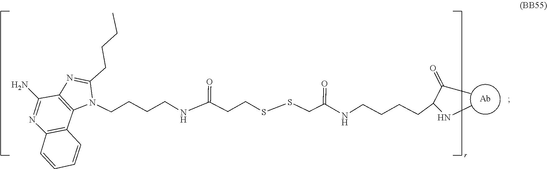
or a pharmaceutically acceptable salt thereof, wherein Ab is an antibody with at least one lysine side chain and subscript r is an integer from 1 to 10 (i.e., 1, 2, 3, 4, 5, 6, 7, 8, 9, or 10). In certain embodiments, subscript r is an integer from 1 to 4 (i.e., 1, 2, 3, or 4).
In certain embodiments, the immunoconjugate has a structure selected from:


or a pharmaceutically acceptable salt thereof, wherein Ab is an antibody with at least one lysine side chain and subscript r is an integer from 1 to 10 (i.e., 1, 2, 3, 4, 5, 6, 7, 8, 9, or 10). In certain embodiments, subscript r is an integer from 1 to 4 (i.e., 1, 2, 3, or 4).
In a second aspect, the invention provides an improved method for producing an immunoconjugate of Formula III from one or more compounds of Formula V and an antibody of Formula VI, the method comprising the step of:

wherein Adj is an adjuvant; G is CH2, C═O, or a bond; L is a linker; E is an ester; Formula VI is an antibody with at least one lysine side chain; and subscript r is an integer from 1 to 10 (i.e., 1, 2, 3, 4, 5, 6, 7, 8, 9, or 10). In certain embodiments, the adjuvant (“Adj”) is a TLR agonist.
Any suitable linker can be used provided it can be bound to the antibody (compound of Formula VI) through an ester. For example, the linker (“L”) can have the following formula

wherein R is optionally present and is a linear or branched, cyclic or straight, saturated or unsaturated alkyl, heteroalkyl, aryl, or heteroaryl chain comprising from 1 to 8 (i.e., 1, 2, 3, 4, 5, 6, 7, or 8) carbon units; subscript a is an integer from 1 to 40; the dashed line (“
and the wavy line (“
The linker (“L”) can have the following formula

wherein subscript a is an integer from 1 to 40; the dashed line (“
and the wavy line (“
In some embodiments, subscript a is an integer from 1 to 20. In some embodiments, subscript a is an integer from 1 to 10. In some embodiments, subscript a is an integer from 1 to 5. In some embodiments, subscript a is an integer from 1 to 3.
The linker (“L”) can also have the following formula

wherein R is optionally present and is a linear or branched, cyclic or straight, saturated or unsaturated alkyl, heteroalkyl, aryl, or heteroaryl chain comprising from 1 to 8 (i.e., 1, 2, 3, 4, 5, 6, 7, or 8) carbon units; each A is independently selected from any amino acid; subscript c is an integer from 1 to 20; the dashed line (“
and the wavy line (“
The linker (“L”) can also have the following formula

wherein R is optionally present and is a linear or branched, cyclic or straight, saturated or unsaturated alkyl, heteroalkyl, aryl, or heteroaryl chain comprising from 1 to 8 (i.e., 1, 2, 3, 4, 5, 6, 7, or 8) carbon units; subscript c is an integer from 1 to 20; the dashed line (“
and the wavy line (“
The linker (“L”) can also have the following formula

wherein R is optionally present and is a linear or branched, cyclic or straight, saturated or unsaturated alkyl, heteroalkyl, aryl, or heteroaryl chain comprising from 1 to 8 (i.e., 1, 2, 3, 4, 5, 6, 7, or 8) carbon units; the dashed line (“ ”) represents the point of attachment to
and the wavy line (“
The linker (“L”) can also have the following formula

wherein R is optionally present and is a linear or branched, cyclic or straight, saturated or unsaturated alkyl, heteroalkyl, aryl, or heteroaryl chain comprising from 1 to 8 (i.e., 1, 2, 3, 4, 5, 6, 7, or 8) carbon units; the dashed line (“
and the wavy line (“
In some embodiments, the compound of Formula V is selected from:

wherein G is CH2, C═O, or a bond; R is optionally present and is a linear or branched, cyclic or straight, saturated or unsaturated alkyl, heteroalkyl, aryl, or heteroaryl chain comprising from 1 to 8 carbon units; subscript a is an integer from 1 to 40; each A is independently selected from any amino acid; subscript c is an integer from 1 to 20, and E is an ester.
As previously discussed, there are many ways of forming an immunoconjugate. Each of the prior art methods suffers from downsides. The present method includes a one-step process which conjugates an adjuvant, modified to include a linker, to the lysine side chain of an antibody (compound of Formula VI). This process is possible by using an ester. The ester can be any suitable ester capable of linking the compound of Formula V to a lysine side chain of an antibody (compound of Formula VI).
For example, the ester of Formula V can be an N-hydroxysuccinimide (“NHS”) ester of the formula:

wherein the wavy line (“
The ester of Formula V can also be a sulfo-N-hydroxysuccinimide ester of the formula:

wherein M is any cation and the wavy line (“
The ester of Formula V can also be a phenol ester of the formula:

wherein each R2 is independently selected from hydrogen or fluorine and the wavy line (“
The ester of Formula V can also be a phenol ester of the formula:

(tetrafluorophenyl) or

(pentafluorophenyl); wherein the wavy line (“
In some embodiments, the antibody of Formula VI and the ester of Formula V are combined in any suitable aqueous buffer. An exemplary list of suitable aqueous buffers is phosphate buffered saline, borate buffered saline, and tris buffered saline.
Using a tetrafluorophenyl (“TFP”) or pentafluorophenyl (“PFP”) is especially effective in synthesizing the immunoconjugates of the present invention.
Accordingly, an exemplary, but non-limiting, list of compounds of Formula V is:


wherein Adj is an adjuvant and M is any cation. For example, the cation counter ion (“M”) can be a proton, ammonium, a quaternary amine, a cation of an alkali metal, a cation of an alkaline earth metal, a cation of a transition metal, a cation of a rare-earth metal, a main group element cation, or a combination thereof.
Accordingly, the one or more compounds of Formula V and an antibody of Formula VI can be combined to form an immunoconjugate of Formula III. An exemplary, but non-limiting list of immunoconjugates of Formula III is:

or a pharmaceutically acceptable salt thereof, wherein Ab is an antibody with at least one lysine side chain; Adj is an adjuvant; G is CH2, C═O, or a bond; R is optionally present and is a linear or branched, cyclic or straight, saturated or unsaturated alkyl, heteroalkyl, aryl, or heteroaryl chain comprising from 1 to 8 carbon units; subscript a is an integer from 1 to 40; each A is independently selected from any amino acid; subscript c is an integer from 1 to 20; and subscript r is an integer from 1 to 10 (i.e., 1, 2, 3, 4, 5, 6, 7, 8, 9, or 10).
In a third aspect, the invention provides an immunoconjugate contain linking moieties that covalently bond the adjuvant moieties, comprising an oligonucleotide, to the antibodies. In certain embodiments, the immunoconjugate is an A-type CPG oligonucleotide immunoconjugate selected from an immunoconjugate of Formula VIIa:

or a pharmaceutically acceptable salt thereof, wherein Ab is an antibody bound at an unmodified amino acid sidechain in the antibody or a modified amino acid sidechain in the antibody; Z is a linking moiety; subscript r is an integer from 1 to 10 (i.e., 1, 2, 3, 4, 5, 6, 7, 8, 9, or 10); lowercase nucleotides signify a phosphorothioate linkage; and uppercase nucleotides signify a phosphodiester linkage. In certain embodiments, the linking moiety (“Z”) is as defined above and herein.
In certain embodiments, the immunoconjugate is an B-type CPG oligonucleotide immunoconjugate selected from an immunoconjugate of Formula VIIb:

or a pharmaceutically acceptable salt thereof, wherein Ab is an antibody bound at an unmodified amino acid sidechain in the antibody or a modified amino acid sidechain in the antibody; Z is a linking moiety; subscript r is an integer from 1 to 10 (i.e., 1, 2, 3, 4, 5, 6, 7, 8, 9, or 10); and uppercase nucleotides signify a phosphorothioate linkage. In certain embodiments, the linking moiety (“Z”) is as defined above and herein.
In certain embodiments, the immunoconjugate is an C-type CPG oligonucleotide immunoconjugate selected from an immunoconjugate of Formula VIIc:

or a pharmaceutically acceptable salt thereof, wherein Ab is an antibody bound at an unmodified amino acid sidechain in the antibody or a modified amino acid sidechain in the antibody; Z is a linking moiety; subscript r is an integer from 1 to 10 (i.e., 1, 2, 3, 4, 5, 6, 7, 8, 9, or 10); and uppercase nucleotides signify a phosphorothioate linkage. In certain embodiments, the linking moiety (“Z”) is as defined above and herein.
In certain embodiments, the immunoconjugate is an PolyI:C oligonucleotide immunoconjugate selected from an immunoconjugate of Formula VIId:

or a pharmaceutically acceptable salt thereof, wherein Ab is an antibody bound at an unmodified amino acid sidechain in the antibody or a modified amino acid sidechain in the antibody; Z is a linking moiety; and subscript r is an integer from 1 to 10 (i.e., 1, 2, 3, 4, 5, 6, 7, 8, 9, or 10). In certain embodiments, the linking moiety (“Z”) is as defined above and herein.
Adjuvants
In some embodiments, the adjuvant moiety is a compound that elicits an immune response. In some embodiments, the adjuvant moiety is a pattern recognition receptor (“PRR”) agonist. Any adjuvant capable of activating a pattern recognition receptor (PRR) can be installed in the immunoconjugates of the invention. As used herein, the terms “Pattern recognition receptor” and “PRR” refer to any member of a class of conserved mammalian proteins which recognize pathogen-associated molecular patterns (“PAMPs”) or damage-associated molecular patterns (“DAMPs”), and act as key signaling elements in innate immunity. Pattern recognition receptors are divided into membrane-bound PRRs, cytoplasmic PRRs, and secreted PRRs. Examples of membrane-bound PRRs include Toll-like receptors (“TLRs”) and C-type lectin receptors (“CLRs”). Examples of cytoplasmic PRRs include NOD-like receptors (“NLRs”) and Rig-I-like receptors (“RLRs”). In some embodiments, the immunoconjugate can have more than one distinct PRR adjuvant moiety.
In certain embodiments, the adjuvant moiety in an immunoconjugate of the invention is a Toll-like receptor (TLR) agonist. Suitable TLR agonists include TLR1, TLR2, TLR3, TLR4, TLR5, TLR6, TLR7, TLR8, TLR9, TLR10, TLR11, or any combination thereof (e.g., TLR7/8 agonists). Any adjuvant capable of activating a Toll-like receptor (TLR) can be installed in the immunoconjugates of the invention. Toll-like receptors (TLRs) are type-I transmembrane proteins that are responsible for initiation of innate immune responses in vertebrates. TLRs recognize a variety of pathogen-associated molecular patterns from bacteria, viruses, and fungi and act as a first line of defense against invading pathogens. TLRs elicit overlapping yet distinct biological responses due to differences in cellular expression and in the signaling pathways that they initiate. Once engaged (e.g., by a natural stimulus or a synthetic TLR agonist) TLRs initiate a signal transduction cascade leading to activation of NF-κB via the adapter protein myeloid differentiation primary response gene 88 (MyD88) and recruitment of the IL-1 receptor associated kinase (IRAK). Phosphorylation of IRAK then leads to recruitment of TNF-receptor associated factor 6 (TRAF6), which results in the phosphorylation of the NF-κB inhibitor I-κB. As a result, NF-κB enters the cell nucleus and initiates transcription of genes whose promoters contain NF-κB binding sites, such as cytokines. Additional modes of regulation for TLR signaling include TIR-domain containing adapter-inducing interferon-β (TRIF)-dependent induction of TRAF6 and activation of MyD88 independent pathways via TRIF and TRAF3, leading to the phosphorylation of interferon response factor three (IRF3). Similarly, the MyD88 dependent pathway also activates several IRF family members, including IRF5 and IRF7 whereas the TRIF dependent pathway also activates the NF-κB pathway.
Examples of TLR3 agonists include Polyinosine-polycytidylic acid (poly (I:C)), Polyadenylic-polyuridylic acid (poly (A:U), and poly(I)-poly(C12U).
Examples of TLR4 agonists include Lipopolysaccharide (LPS) and Monophosphoryl lipid A (MPLA).
An example of a TLR5 agonist includes Flagellin.
Examples of TLR9 agonists include single strand CpG oligodeoxynucleotides (CpG ODN). Three major classes of stimulatory CpG ODNs have been identified based on structural characteristics and activity on human peripheral blood mononuclear cells (PBMCs), in particular B cells and plasmacytoid dendritic cells (pDCs). These three classes are Class A (Type D), Class B (Type K) and Class C.
Examples of Nod Like Receptor (NLR) agonists include acylated derivative of iE-DAP, D-gamma-Glu-mDAP, L-Ala-gamma-D-Glu-mDAP, Muramyldipeptide with a C18 fatty acid chain, Muramyldipeptide, muramyl tripeptide, and N-glycolylated muramyldipeptide.
Examples of RIG-I-Like receptor (RLR) agonists include 5′ppp-dsrna (5′-pppGCAUGCGACCUCUGUUUGA-3′ (SEQ ID NO: 3): 3′-CGUACGCUGGAGACAAACU-5′ (SEQ ID NO: 4)), and Poly(deoxyadenylic-deoxythymidylic) acid (Poly(dA:dT))
Additional immune-stimulatory compounds, such as cytosolic DNA and unique bacterial nucleic acids called cyclic dinucleotides, can be recognized by stimulator of interferon genes (“STING”), which can act a cytosolic DNA sensor. ADU-SlOO can be a STING agonist. Non-limiting examples of STING agonists include: Cyclic [G(2′,5′)pA(2′,5′)p] (2′2′-cGAMP), cyclic [G(2′,5′)pA(3′,5′)p] (2′3′-cGAMP), cyclic [G(3′,5′)pA(3′,5′)p] (3′3′-cGAMP), Cyclic di-adenylate monophosphate (c-di-AMP), 2′,5′-3′,5′-c-diAMP (2′3′-c-di-AMP), Cyclic di-guanylate monophosphate (c-di-GMP), 2′,5′-3′,5′-c-diGMP (2′3′-c-di-GMP), Cyclic di-inosine monophosphate (c-di-IMP), Cyclic di-uridine monophosphate (c-di-UMP), KIN700, KIN1148, KIN600, KIN500, KINlOO, KIN101, KIN400, KIN2000, or SB-9200 can be recognized.
Any adjuvant capable of activating TLR7 and/or TLR8 can be installed in the immunoconjugates of the invention. Examples of TLR7 agonists and TLR8 agonists are described, e.g., by Vacchelli et al. (OncoImmunology, 2: 8, e25238, DOI: 10.4161/onci.25238 (2013)) and Carson et al. (U.S. Patent Application Publication 2013/0165455, which is hereby incorporated by reference in its entirety). TLR7 and TLR8 are both expressed in monocytes and dendritic cells. In humans, TLR7 is also expressed in plasmacytoid dendritic cells (pDCs) and B cells. TLR8 is expressed mostly in cells of myeloid origin, i.e., monocytes, granulocytes, and myeloid dendritic cells. TLR7 and TLR8 are capable of detecting the presence of “foreign” single-stranded RNA within a cell, as a means to respond to viral invasion. Treatment of TLR8-expressing cells, with TLR8 agonists can result in production of high levels of IL-12, IFN-γ, IL-1, TNF-α, IL-6, and other inflammatory cytokines. Similarly, stimulation of TLR7-expressing cells, such as pDCs, with TLR7 agonists can result in production of high levels of IFN-α and other inflammatory cytokines. TLR7/TLR8 engagement and resulting cytokine production can activate dendritic cells and other antigen-presenting cells, driving diverse innate and acquired immune response mechanisms leading to tumor destruction.
Examples of TLR7, TLR8 or TLR7/8 agonists include but are not limited to: Gardiquimod (1-(4-amino-2-ethylaminomethylimidazo[4,5-c]quinolin-1-yl)-2-methylpropan-2-ol), Imiquimod (R837) (agonist for TLR7), loxoribine (agonist for TLR7), IRM1 (1-(2-amino-2-methylpropyl)-2-(ethoxymethyl)-1H-imidazo-[4,5-c]quinolin-4-amine), IRM2 (2-methyl-1-[2-(3-pyridin-3-ylpropoxy)ethyl]-1H-imidazo[4,5-c]quinolin-4-amine) (agonist for TLR8), IRM3 (N-(2-[2-[4-amino-2-(2-methoxyethyl)-1H-imidazo[4,5-c]quinolin-1-yl]ethoxy]ethyl)-N-methylcyclohexanecarboxamide) (agonist for TLR8), CL097 (2-(ethoxymethyl)-1H-imidazo[4,5-c]quinolin-4-amine) (agonist for TLR7/8), CL307 (agonist for TLR7), CL264 (agonist for TLR7), Resiquimod (agonist for TLR7/8), 3M-052/MEDI9197 (agonist for TLR7/8), SD-101 (N-[(4S)-2,5-dioxo-4-imidazolidinyl]-urea) (agonist for TLR7/8), motolimod (2-amino-N,N-dipropyl-8-[4-(pyrrolidine-1-carbonyl)phenyl]-3H-1-benzazepine-4-carboxamide) (agonist for TLR8), CL075 (3M002, 2-propylthiazolo[4,5-c]quinolin-4-amine) (agonist for TLR7/8), and TL8-506 (3H-1-benzazepine-4-carboxylic acid, 2-amino-8-(3-cyanophenyl)-, ethyl ester) (agonist for TLR8).
Examples of TLR2 agonists include but are not limited to an agent comprising N-α-palmitoyl-S-[2,3-bis(palmitoyloxy)-(2RS)-propyl]-L-cysteine, palmitoyl-Cys((RS)-2,3-di(palmitoyloxy)-propyl) (“Pam3Cys”), e.g., Pam3Cys, Pam3Cys-Ser-(Lys)4 (also known as “Pam3Cys-SKKKK” and “Pam3CSK4”), Triacyl lipid A (“OM-174”), Lipoteichoic acid (“LTA”), peptidoglycan, and CL419 (S-(2,3-bis(palmitoyloxy)-(2RS)propyl)-(R)-cysteinyl spermine).
An example of a TLR2/6 agonist is Pam2CSK4 (S-[2,3-bis(palmitoyloxy)-(2RS)-propyl]-[R]-cysteinyl-[S]-seryl-[S]-lysyl-[S]-lysyl-[S]-lysyl-[S]-lysine× 3 CF3COOH).
Examples of TLR2/7 agonist include CL572 (S-(2-myristoyloxy ethyl)-(R)-cysteinyl 4-((6-amino-2-(butylamino)-8-hydroxy-9H-purin-9-yl)methyl) aniline), CL413 (5-(2,3-bis(palmitoyloxy)-(2RS)propyl)-(R)-cysteinyl-(S)-seryl-(S)-lysyl-(S)-lysyl-(S)-lysyl-(S)-lysyl 4-((6-amino-2-(butylamino)-8-hydroxy-9H-purin-9-yl)methyl)aniline), and CL401 (5-(2,3-bis(palmitoyloxy)-(2RS)propyl)-(R)-cysteinyl 4-((6-amino-2(butyl amino)-8-hydroxy-9H-purin-9-yl)methyl) aniline).
In some embodiments, the adjuvant moiety is an imidazoquinoline compound. Examples of useful imidazoquinoline compounds include those described in U.S. Pat. Nos. 5,389,640; 6,069,149; and 7,968,562, which are hereby incorporated by reference in their entirety.
In some embodiments, the adjuvant (“Adj”) is of formula:

wherein each J independently is hydrogen, OR4, or R4; each R4 independently is hydrogen, or an alkyl, heteroalkyl, cycloalkyl, heterocycloalkyl, aryl, heteroaryl, arylalkyl, or heteroarylalkyl group comprising from 1 to 8 (i.e., 1, 2, 3, 4, 5, 6, 7, or 8) carbon units; Q is optionally present and is an alkyl, heteroalkyl, cycloalkyl, heterocycloalkyl, aryl, heteroaryl, arylalkyl, or heteroarylalkyl group comprising from 1 to 8 (i.e., 1, 2, 3, 4, 5, 6, 7, or 8) carbon units; and the dashed line (“

wherein each R4 independently is selected from the group consisting of hydrogen, or alkyl, heteroalkyl, cycloalkyl, heterocycloalkyl, aryl, heteroaryl, arylalkyl, and heteroarylalkyl group comprising from 1 to 8 (i.e., 1, 2, 3, 4, 5, 6, 7, or 8) carbon units and the dashed line (“
In some embodiments, the adjuvant (“Adj”) is of formula:

wherein J is hydrogen, OR4, or R4; each R4 independently is hydrogen, or alkyl, heteroalkyl, cycloalkyl, heterocycloalkyl, aryl, heteroaryl, arylalkyl, and heteroarylalkyl group comprising from 1 to 8 (i.e., 1, 2, 3, 4, 5, 6, 7, or 8) carbon units; Q is selected from the group consisting of alkyl, or heteroalkyl, cycloalkyl, heterocycloalkyl, aryl, heteroaryl, arylalkyl, and heteroarylalkyl group comprising from 1 to 8 (i.e., 1, 2, 3, 4, 5, 6, 7, or 8) carbon units; and the dashed line (“

wherein each R4 independently is selected from the group consisting of hydrogen, or alkyl, heteroalkyl, cycloalkyl, heterocycloalkyl, aryl, heteroaryl, arylalkyl, and heteroarylalkyl group comprising from 1 to 8 (i.e., 1, 2, 3, 4, 5, 6, 7, or 8) carbon units and the dashed line (“
In some embodiments, the adjuvant (“Adj”) is of formula:

wherein each R4 independently is hydrogen, or alkyl, heteroalkyl, cycloalkyl, heterocycloalkyl, aryl, heteroaryl, arylalkyl, or heteroarylalkyl group comprising from 1 to 8 (i.e., 1, 2, 3, 4, 5, 6, 7, or 8) carbon units; Q is alkyl, heteroalkyl, cycloalkyl, heterocycloalkyl, aryl, heteroaryl, arylalkyl, or heteroarylalkyl group comprising from 1 to 8 (i.e., 1, 2, 3, 4, 5, 6, 7, or 8) carbon units; and the dashed line (“
In some embodiments, the adjuvant (“Adj”) is of formula:

wherein each J independently is hydrogen, OR4, or R4; each R4 independently is hydrogen, or an alkyl, heteroalkyl, cycloalkyl, heterocycloalkyl, aryl, heteroaryl, arylalkyl, or heteroarylalkyl group comprising from 1 to 8 (i.e., 1, 2, 3, 4, 5, 6, 7, or 8) carbon units; each U independently is CH or N wherein at least one U is N; each subscript t independently is an integer from 1 to 3 (i.e., 1, 2, or 3); Q is optionally present and is an alkyl, heteroalkyl, cycloalkyl, heterocycloalkyl, aryl, heteroaryl, arylalkyl, or heteroarylalkyl group comprising from 1 to 8 (i.e., 1, 2, 3, 4, 5, 6, 7, or 8) carbon units; and the dashed line (“

wherein R4 is selected from the group consisting of hydrogen, or alkyl, heteroalkyl, cycloalkyl, heterocycloalkyl, aryl, heteroaryl, arylalkyl, and heteroarylalkyl group comprising from 1 to 8 (i.e., 1, 2, 3, 4, 5, 6, 7, or 8) carbon units Q is an alkyl, heteroalkyl, cycloalkyl, heterocycloalkyl, aryl, heteroaryl, arylalkyl, or heteroarylalkyl group comprising from 1 to 8 (i.e., 1, 2, 3, 4, 5, 6, 7, or 8) carbon units; and the dashed line (“
In some embodiments, the adjuvant (“Adj”) is of formula:

wherein J is hydrogen, OR4, or R4; each R4 independently is hydrogen, or an alkyl, heteroalkyl, cycloalkyl, heterocycloalkyl, aryl, heteroaryl, arylalkyl, or heteroarylalkyl group comprising from 1 to 8 (i.e., 1, 2, 3, 4, 5, 6, 7, or 8) carbon units; R5 is hydrogen, or an alkyl, heteroalkyl, cycloalkyl, heterocycloalkyl, aryl, heteroaryl, arylalkyl, or heteroarylalkyl group comprising from 1 to 10 (i.e., 1, 2, 3, 4, 5, 6, 7, 8, 9, or 10) carbon units; Q is an alkyl, heteroalkyl, cycloalkyl, heterocycloalkyl, aryl, heteroaryl, arylalkyl, or heteroarylalkyl group comprising from 1 to 8 (i.e., 1, 2, 3, 4, 5, 6, 7, or 8) carbon units; and the dashed line (“

wherein J is hydrogen, OR4, or R4; each R4 independently is selected from the group consisting of hydrogen, or alkyl, heteroalkyl, cycloalkyl, heterocycloalkyl, aryl, heteroaryl, arylalkyl, and heteroarylalkyl group comprising from 1 to 8 (i.e., 1, 2, 3, 4, 5, 6, 7, or 8) carbon units; U is CH or N; V is CH2, O, or NH; each subscript t independently is an integer from 1 to 3 (i.e., 1, 2, or 3); and the dashed line (“
In some embodiments, the adjuvant (“Adj”) is of formula:

wherein R1 is selected from H and C1-4 alkyl; R3 is selected from C1-6 alkyl and 2- to 6-membered heteroalkyl, each of which is optionally substituted with one or more members selected from the group consisting of halo, hydroxy, amino, oxo (═O), alkylamino, amido, acyl, nitro, cyano, and alkoxy; X is selected from O and CH2; each Y is independently CHR2, wherein R2 is selected from H, OH, and NH2, subscript n is an integer from 1 to 12 (i.e., 1, 2, 3, 4, 5, 6, 7, 8, 9, 10, 11, or 12); and the dashed line (“
In some embodiments, the adjuvant (“Adj”) is of formula:

wherein W is selected from the group consisting of O and CH2; R1 is selected from H and C1-4 alkyl; each Y is independently CHR2, wherein R2 is selected from H, OH, and NH2; subscript n is an integer from 1 to 12 (i.e., 1, 2, 3, 4, 5, 6, 7, 8, 9, 10, 11, or 12); and the dashed line (“
In some embodiments, the adjuvant (“Adj”) is of formula:

wherein W is selected from the group consisting of O and CH2; X is selected from O and CH2; each Y is independently CHR2, wherein R2 is selected from H, OH, and NH2; subscript n is an integer from 1 to 12 (i.e., 1, 2, 3, 4, 5, 6, 7, 8, 9, 10, 11, or 12); and the dashed line (“
In some embodiments, the adjuvant (“Adj”) is of formula:

wherein R1 is selected from H and C1-4 alkyl; R2 is selected from H, OH, and NH2; and the dashed line (“
In some embodiments, the adjuvant (“Adj”) is of formula:

wherein R1 is selected from H and C1-4 alkyl; R2 is selected from H, OH, and NH2; and the dashed line (“
In certain embodiments, the adjuvant (“Adj”) is:


wherein the dashed line (“
In some embodiments, the adjuvant is not a fluorophore. In some embodiments, the adjuvant is not a radiodiagnostic compound. In some embodiments, the adjuvant is not a radiotherapeutic compound. In some embodiments, the adjuvant is not a tubulin inhibitor. In some embodiments, the adjuvant is not a DNA crosslinker/alkylator. In some embodiments, the adjuvant is not a topoisomerase inhibitor.
Antibodies
The antibodies in the immunoconjugates can be allogeneic antibodies. The terms “allogeneic antibody” or “alloantibody” refer to an antibody that is not from the individual in question (e.g., an individual with a tumor and seeking treatment), but is from the same species, or is from a different species, but has been engineered to reduce, mitigate, or avoid recognition as a xeno-antibody (e.g., non-self). For example, the “allogeneic antibody” can be a humanized antibody. Unless specifically stated otherwise, “antibody” and “allogeneic antibodies” as used herein refer to immunoglobulin G (IgG) or immunoglobulin A (IgA).
If a cancer cell of a human individual is contacted with an antibody that was not generated by that same person (e.g., the antibody was generated by a second human individual, the antibody was generated by another species such as a mouse, the antibody is a humanized antibody that was generated by another species, etc.), then the antibody is considered to be allogeneic (relative to the first individual). A humanized mouse monoclonal antibody that recognizes a human antigen (e.g., a cancer-specific antigen, an antigen that is enriched in and/or on cancer cells, etc.) is considered to be an “alloantibody” (an allogeneic antibody).
In some embodiments, the antibody is a polyclonal allogeneic IgG antibody. In some embodiments, the antibody is present in a mixture of polyclonal IgG antibodies with a plurality of binding specificities. In some cases, the antibodies of the mixture specifically bind to different target molecules, and in some cases the antibodies of the mixture specifically bind to different epitopes of the same target molecule. Thus, a mixture of antibodies can in some cases include more than one immunoconjugate of the invention (e.g., adjuvant moieties can be covalently bonded to antibodies of a mixture, e.g., a mixture of polyclonal IgG antibodies, resulting in a mixture of antibody-adjuvant conjugates of the invention). A mixture of antibodies can be pooled from 2 or more individuals (e.g., 3 or more individuals, 4 or more individuals, 5 or more individuals, 6 or more individuals, 7 or more individuals, 8 or more individuals, 9 or more individuals, 10 or more individuals, etc.). In some cases, pooled serum is used as a source of alloantibody, where the serum can come from any number of individuals, none of whom are the first individual (e.g., the serum can be pooled from 2 or more individuals, 3 or more individuals, 4 or more individuals, 5 or more individuals, 6 or more individuals, 7 or more individuals, 8 or more individuals, 9 or more individuals, 10 or more individuals, etc.). In some cases, the antibodies are isolated or purified from serum prior to use. The purification can be conducted before or after pooling the antibodies from different individuals.
In some cases where the antibodies in the immunoconjugates comprise IgGs from serum, the target antigens for some (e.g., greater than 0% but less than 50%), half, most (greater than 50% but less than 100%), or even all of the antibodies (i.e., IgGs from the serum) will be unknown. However, the chances are high that at least one antibody in the mixture will recognize the target antigen of interest because such a mixture contains a wide variety of antibodies specific for a wide variety of target antigens.
In some embodiments, the antibody is a polyclonal allogeneic IgA antibody. In some embodiments, the antibody is present in a mixture of polyclonal IgA antibodies with a plurality of binding specificities. In some cases, the antibodies of the mixture specifically bind to different target molecules, and in some cases the antibodies of the mixture specifically bind to different epitopes of the same target molecule. Thus, a mixture of antibodies can in some cases include more than one immunoconjugate of the invention (e.g., adjuvant moieties can be covalently bonded to antibodies of a mixture, e.g., a mixture of polyclonal IgA antibodies, resulting in a mixture of antibody-adjuvant conjugates of the invention). A mixture of antibodies can be pooled from 2 or more individuals (e.g., 3 or more individuals, 4 or more individuals, 5 or more individuals, 6 or more individuals, 7 or more individuals, 8 or more individuals, 9 or more individuals, 10 or more individuals, etc.). In some cases, pooled serum is used as a source of alloantibody, where the serum can come from any number of individuals, none of whom are the first individual (e.g., the serum can be pooled from 2 or more individuals, 3 or more individuals, 4 or more individuals, 5 or more individuals, 6 or more individuals, 7 or more individuals, 8 or more individuals, 9 or more individuals, 10 or more individuals, etc.). In some cases, the antibodies are isolated or purified from serum prior to use. The purification can be conducted before or after pooling the antibodies from different individuals.
In some cases where the antibodies in the immunoconjugates comprise IgAs from serum, the target antigens for some (e.g., greater than 0% but less than 50%), half, most (greater than 50% but less than 100%), or even all of the antibodies (i.e., IgAs from the serum) will be unknown. However, the chances are high that at least one antibody in the mixture will recognize the target antigen of interest because such a mixture contains a wide variety of antibodies specific for a wide variety of target antigens.
In some cases, the antibody in the immunoconjugates includes intravenous immunoglobulin (IVIG) and/or antibodies from (e.g., enriched from, purified from, e.g., affinity purified from) IVIG. IVIG is a blood product that contains IgG (immunoglobulin G) pooled from the plasma (e.g., in some cases without any other proteins) from many (e.g., sometimes over 1,000 to 60,000) normal and healthy blood donors. IVIG is commercially available. IVIG contains a high percentage of native human monomeric IVIG, and has low IgA content. When administered intravenously, IVIG ameliorates several disease conditions. Therefore, the United States Food and Drug Administration (FDA) has approved the use of IVIG for a number of diseases including (1) Kawasaki disease; (2) immune-mediated thrombocytopenia; (3) primary immunodeficiencies; (4) hematopoietic stem cell transplantation (for those older than 20 years); (5) chronic B-cell lymphocytic leukemia; and (6) pediatric HIV type 1 infection. In 2004, the FDA approved the Cedars-Sinai IVIG Protocol for kidney transplant recipients so that such recipients could accept a living donor kidney from any healthy donor, regardless of blood type (ABO incompatible) or tissue match. These and other aspects of IVIG are described, for example, in US Patent Application Publications 2010/0150942; 2004/0101909; 2013/0177574; 2013/0108619; and 2013/0011388; which are hereby incorporated by reference in their entirety.
In some cases, the antibody is a monoclonal antibody of a defined sub-class (e.g., IgG1, IgG2, IgG3, IgG4, IgA1, or IgA2). If combinations of antibodies are used, the antibodies can be from the same subclass or from different subclasses. For example, the antibodies can be IgG1 antibodies. Various combinations of different subclasses, in different relative proportions, can be obtained by those of skill in the art. In some cases, a specific subclass, or a specific combination of different subclasses can be particularly effective at cancer treatment or tumor size reduction. Accordingly, some embodiments of the invention provide immunoconjugates wherein the antibody is a monoclonal antibody. In some embodiments, the monoclonal antibody is humanized.
In some embodiments, the antibody binds to an antigen of a cancer cell. For example, the antibody can bind to a target antigen that is present at an amount of at least 10; 100; 1,000; 10,000; 100,000; 1,000,000; 2.5×10⁶; 5×10⁶; or 1×10⁷ copies or more on the surface of a cancer cell.
In some embodiments, the antibody binds to an antigen on a cancer or immune cell at a higher affinity than a corresponding antigen on a non-cancer cell. For example, the antibody may preferentially recognize an antigen containing a polymorphism that is found on a cancer or immune cell as compared to recognition of a corresponding wild-type antigen on the non-cancer or non-immune cell. In some cases, the antibody binds a cancer or immune cell with greater avidity than a non-cancer or non-immune cell. For example, the cancer or immune cell can express a higher density of an antigen, thus providing for a higher affinity binding of a multivalent antibody to the cancer or immune cell.
In some cases, the antibody does not significantly bind non-cancer antigens (e.g., the antibody binds one or more non-cancer antigens with at least 10; 100; 1,000; 10,000; 100,000; or 1,000,000-fold lower affinity (higher Kd) than the target cancer antigen). In some cases, the target cancer antigen to which the antibody binds is enriched on the cancer cell. For example, the target cancer antigen can be present on the surface of the cancer cell at a level that is at least 2, 5, 10; 100; 1,000; 10,000; 100,000; or 1,000,000-fold higher than a corresponding non-cancer cell. In some cases, the corresponding non-cancer cell is a cell of the same tissue or origin that is not hyperproliferative or otherwise cancerous. In general, a subject IgG antibody that specifically binds to an antigen (a target antigen) of a cancer cell preferentially binds to that particular antigen relative to other available antigens. However, the target antigen need not be specific to the cancer cell or even enriched in cancer cells relative to other cells (e.g., the target antigen can be expressed by other cells). Thus, in the phrase “an antibody that specifically binds to an antigen of a cancer cell,” the term “specifically” refers to the specificity of the antibody and not to the uniqueness of the antigen in that particular cell type.
Modified Fc Region
In some embodiments, the antibodies in the immunoconjugates contain a modified Fc region, wherein the modification modulates the binding of the Fc region to one or more Fc receptors.
The terms “Fc receptor” or “FcR” refer to a receptor that binds to the Fc region of an antibody. There are three main classes of Fc receptors: FcγR which bind to IgG, FcαR which binds to IgA, and FcεR which binds to IgE. The FcγR family includes several members, such as FcγI (CD64), FcγRIIA (CD32A), FcγRIIB (CD32B), FcγRIIIA (CD16A), FcγRIIIB (CD16B). The Fcγ receptors differ in their affinity for IgG and also have different affinities for the IgG subclasses (e.g., IgG1, IgG2, IgG3, IgG4).
In some embodiments, the antibodies in the immunoconjugates (e.g., antibodies conjugated to a TLR agonist such as a TLR7/8 agonist via a linker) contain one or more modifications (e.g., amino acid insertion, deletion, and/or substitution) in the Fc region that results in modulated binding (e.g., increased binding or decreased binding) to one or more Fc receptors (e.g., FcγRI (CD64), FcγRIIA (CD32A), FcγRIIB (CD32B), FcγRIIIA (CD16a), and/or FcγRIIIB (CD16b)) as compared to the native antibody lacking the mutation in the Fc region. In some embodiments, the antibodies in the immunoconjugates contain one or more modifications (e.g., amino acid insertion, deletion, and/or substitution) in the Fc region that reduce the binding of the Fc region of the antibody to FcγRIIB. In some embodiments, the antibodies in the immunoconjugates contain one or more modifications (e.g., amino acid insertion, deletion, and/or substitution) in the Fc region of the antibody that reduce the binding of the antibody to FcγRIIB while maintaining the same binding or having increased binding to FcγRI (CD64), FcγRIIA (CD32A), and/or FcRγIIIA (CD16a) as compared to the native antibody lacking the mutation in the Fc region. In some embodiments, the antibodies in the immunoconjugates contain one of more modifications in the Fc region that increase the binding of the Fc region of the antibody to FcγRIIB.
In some cases, the modulated binding is provided by mutations in the Fc region of the antibody relative to the native Fc region of the antibody. The mutations can be in a CH2 domain, a CH3 domain, or a combination thereof. A “native Fc region” is synonymous with a “wild-type Fc region” and comprises an amino acid sequence that is identical to the amino acid sequence of an Fc region found in nature or identical to the amino acid sequence of the Fc region found in the native antibody (e.g., rituximab) Native sequence human Fc regions include a native sequence human IgG1 Fc region; native sequence human IgG2 Fc region; native sequence human IgG3 Fc region; and native sequence human IgG4 Fc region as well as naturally occurring variants thereof. Native sequence Fc includes the various allotypes of Fcs (see, e.g., Jefferis et al., mAbs, 1(4): 332-338 (2009)).
In some embodiments, the mutations in the Fc region that result in modulated binding to one or more Fc receptors can include one or more of the following mutations: SD (S239D), SDIE (S239D/I332E), SE (S267E), SELF (S267E/L328F), SDIE (S239D/I332E), SDIEAL (S239D/I332E/A330L), GA (G236A), ALIE (A330L/I332E), GASDALIE (G236A/S239D/A330L/I332E), V9 (G237D/P238D/P271G/A330R), and V11 (G237D/P238D/H268D/P271G/A330R) and/or one or more mutations at the following amino acids: E233, G237, P238, H268, P271, L328 and A330. Additional Fc region modifications for modulating Fc receptor binding are described, e.g., in US Patent Application Publication 2016/0145350, and U.S. Pat. Nos. 7,416,726 and 5,624,821.
In some embodiments, the Fc region of the antibodies of the immunoconjugates are modified to have an altered glycosylation pattern of the Fc region compared to the native non-modified Fc region.
Human immunoglobulin is glycosylated at the Asn297 residue in the Cγ2 domain of each heavy chain. This N-linked oligosaccharide is composed of a core heptasaccharide, N-acetylglucosamine4Mannose3 (GlcNAc4Man3). Removal of the heptasaccharide with endoglycosidase or PNGase F is known to lead to conformational changes in the antibody Fc region, which can significantly reduce antibody-binding affinity to activating FcγR and lead to decreased effector function. The core heptasaccharide is often decorated with galactose, bisecting GlcNAc, fucose or sialic acid, which differentially impacts Fc binding to activating and inhibitory FcγR. Additionally, it has been demonstrated that α2,6-sialyation enhances anti-inflammatory activity in vivo while defucosylation leads to improved FcγRIIIa binding and a 10-fold increase in antibody-dependent cellular cytotoxicity and antibody-dependent phagocytosis. Specific glycosylation patterns can therefore be used to control inflammatory effector functions.
In some embodiments, the modification to alter the glycosylation pattern is a mutation. For example, a substitution at Asn297. In some embodiments, Asn297 is mutated to glutamine (N297Q). Methods for controlling immune response with antibodies that modulate FcγR-regulated signaling are described, for example, in U.S. Pat. No. 7,416,726, as well as US 2007/0014795 and US 2008/0286819.
In some embodiments, the antibodies of the immunoconjugates are modified to contain an engineered Fab region with a non-naturally occurring glycosylation pattern. For example, hybridomas can be genetically engineered to secrete afucosylated mAb, desialylated mAb or deglycosylated Fc with specific mutations that enable increased FcRγIIIa binding and effector function. In some embodiments, the antibodies of the immunoconjugates are engineered to be afucosylated (e.g., afucosylated rituximab, available from Invivogen, hcd20-mab13).
In some embodiments, the entire Fc region of an antibody in the immunoconjugates is exchanged with a different Fc region, so that the Fab region of the antibody is conjugated to a non-native Fc region. For example, the Fab region of rituximab, which normally comprises an IgG1 Fc region, can be conjugated to IgG2, IgG3, IgG4, or IgA, or the Fab region of nivolumab, which normally comprises an IgG4 Fc region, can be conjugated to IgG1, IgG2, IgG3, IgA1 or IgG2. In some embodiments, the Fc modified antibody with a non-native Fc domain also comprises one or more amino acid modification, such as the S228P mutation within the IgG4 Fc, that modulate the stability of the Fc domain described. In some embodiments, the Fc modified antibody with a non-native Fc domain also comprises one or more amino acid modifications described herein that modulate Fc binding to FcR.
In some embodiments, the modifications that modulate the binding of the Fc region to FcR do not alter the binding of the Fab region of the antibody to its antigen when compared to the native non-modified antibody. In other embodiments, the modifications that modulate the binding of the Fc region to FcR also increase the binding of the Fab region of the antibody to its antigen when compared to the native non-modified antibody.
Antibody Targets
In some embodiments, the antibody is capable of binding one or more targets selected from (e.g., specifically binds to a target selected from) 5T4, ABL, ABCF1, ACVR1, ACVR1B, ACVR2, ACVR2B, ACVRL1, ADORA2A, Aggrecan, AGR2, AICDA, AIF1, AIGI, AKAP1, AKAP2, AMH, AMHR2, ANGPT1, ANGPT2, ANGPTL3, ANGPTL4, ANPEP, APC, APOC1, AR, aromatase, ATX, AX1, AZGP1 (zinc-a-glycoprotein), B7.1, B7.2, B7-H1, BAD, BAFF, BAG1, BAI1, BCR, BCL2, BCL6, BDNF, BLNK, BLR1 (MDR15), BIyS, BMP1, BMP2, BMP3B (GDFIO), BMP4, BMP6, BMP8, BMPR1A, BMPR1B, BMPR2, BPAG1 (plectin), BRCA1, C19orflO (IL27w), C3, C4A, C5, C5R1, CANT1, CAPRIN-1, CASP1, CASP4, CAV1, CCBP2 (D6/JAB61), CCL1 (1-309), CCLI1 (eotaxin), CCL13 (MCP-4), CCL15 (MIP-Id), CCL16 (HCC-4), CCL17 (TARC), CCL18 (PARC), CCL19 (MIP-3b), CCL2 (MCP-1), MCAF, CCL20 (MIP-3a), CCL21 (MEP-2), SLC, exodus-2, CCL22(MDC/STC-I), CCL23 (MPIF-I), CCL24 (MPIF-2/eotaxin-2), CCL25 (TECK), CCL26(eotaxin-3), CCL27 (CTACK/ILC), CCL28, CCL3 (MIP-Ia), CCL4 (MIPIb), CCL5(RANTES), CCL7 (MCP-3), CCL8 (mcp-2), CCNA1, CCNA2, CCND1, CCNE1, CCNE2, CCR1 (CKR1/HM145), CCR2 (mcp-IRB/RA), CCR3 (CKR3/CMKBR3), CCR4, CCR5(CMKBR5/ChemR13), CCR6 (CMKBR6/CKR-L3/STRL22/DRY6), CCR7 (CKR7/EBI1), CCR8 (CMKBR8/TERI/CKR-L1), CCR9 (GPR-9-6), CCRL1 (VSHK1), CCRL2 (L-CCR), CD164, CD19, CDIC, CD2, CD20, CD21, CD200, CD-22, CD24, CD27, CD28, CD3, CD33, CD35, CD37, CD38, CD3E, CD3G, CD3Z, CD4, CD38, CD40, CD40L, CD44, CD45RB, CD47, CD52, CD69, CD72, CD74, CD79A, CD79B, CD8, CD80, CD81, CD83, CD86, CD137, CD152, CD274, CDH1 (Ecadherin), CDH1O, CDH12, CDH13, CDH18, CDH19, CDH2O, CDH5, CDH7, CDH8, CDH9, CDK2, CDK3, CDK4, CDK5, CDK6, CDK7, CDK9, CDKN1A (p21Wap1/Cip1), CDKN1B (p27Kip1), CDKN1C, CDKN2A (p16INK4a), CDKN2B, CDKN2C, CDKN3, CEBPB, CERI, CHGA, CHGB, Chitinase, CHST1O, CKLFSF2, CKLFSF3, CKLFSF4, CKLFSF5, CKLFSF6, CKLFSF7, CKLFSF8, CLDN3, CLDN7 (claudin-7), CLN3, CLU (clusterin), CMKLR1, CMKOR1 (RDC1), CNR1, COL18A1, COLIA1, COL4A3, COL6A1, CR2, Cripto, CRP, CSF1 (M-CSF), CSF2 (GM-CSF), CSF3 (GCSF), CTL8, CTNNB1 (b-catenin), CTSB (cathepsin B), CX3CL1 (SCYD1), CX3CR1 (V28), CXCL1 (GRO1), CXCL1O (IP-IO), CXCLI1 (1-TAC/IP-9), CXCL12 (SDF1), CXCL13, CXCL14, CXCL16, CXCL2 (GRO2), CXCL3 (GRO3), CXCL5 (ENA-78/LIX), CXCL6 (GCP-2), CXCL9 (MIG), CXCR3 (GPR9/CKR-L2), CXCR4, CXCR6 (TYMSTR/STRL33/Bonzo), CYB5, CYC1, CYSLTR1, DAB2IP, DES, DKFZp451J0118, DNCL1, DPP4, E2F1, Engel, Edge, Fennel, EFNA3, EFNB2, EGF, EGFR, ELAC2, ENG, Enola, ENO2, ENO3, EPHA1, EPHA2, EPHA3, EPHA4, EPHA5, EPHA6, EPHA7, EPHA8, EPHA9, EPHA10, EPHB1, EPHB2, EPHB3, EPHB4, EPHB5, EPHB6, EPHRIN-A1, EPHRIN-A2, EPHRINA3, EPHRIN-A4, EPHRIN-A5, EPHRIN-A6, EPHRIN-B1, EPHRIN-B2, EPHRIN-B3, EPHB4, EPG, ERBB2 (Her-2), EREG, ERK8, Estrogen receptor, Earl, ESR2, F3 (TF), FADD, farnesyltransferase, FasL, FASNf, FCER1A, FCER2, FCGR3A, FGF, FGF1 (aFGF), FGF10, FGF11, FGF12, FGF12B, FGF13, FGF14, FGF16, FGF17, FGF18, FGF19, FGF2 (bFGF), FGF20, FGF21, FGF22, FGF23, FGF3 (int-2), FGF4 (HST), FGF5, FGF6 (HST-2), FGF7 (KGF), FGF8, FGF9, FGFR3, FIGF (VEGFD), FIL1(EPSILON), FBL1 (ZETA), FLJ12584, FLJ25530, FLRT1 (fibronectin), FLT1, FLT-3, FOS, FOSL1(FRA-1), FY (DARC), GABRP (GABAa), GAGEB1, GAGEC1, GALNAC4S-6ST, GATA3, GD2, GDF5, GFI1, GGT1, GM-CSF, GNAS1, GNRH1, GPR2 (CCR10), GPR31, GPR44, GPR81 (FKSG80), GRCC1O (C1O), GRP, GSN (Gelsolin), GSTP1, HAVCR2, HDAC, HDAC4, HDAC5, HDAC7A, HDAC9, Hedgehog, HGF, HIF1A, HIP1, histamine and histamine receptors, HLA-A, HLA-DRA, HLA-E, HM74, HMOXI, HSP90, HUMCYT2A, ICEBERG, ICOSL, ID2, IFN-A, IFNA1, IFNA2, IFNA4, IFNA5, EFNA6, BFNA7, IFNB1, IFNgamma, IFNW1, IGBP1, IGF1, IGFIR, IGF2, IGFBP2, IGFBP3, IGFBP6, DL-1, ILIO, ILIORA, ILIORB, IL-1, IL1R1 (CD121a), IL1R2(CD121b), IL-IRA, IL-2, IL2RA (CD25), IL2RB(CD122), IL2RG(CD132), IL-4, IL-4R(CD123), IL-5, IL5RA(CD125), IL3RB(CD131), IL-6, IL6RA, (CD126), IR6RB(CD130), IL-7, IL7RA(CD127), IL-8, CXCR1 (IL8RA), CXCR2, (IL8RB/CD128), IL-9, IL9R(CD129), IL-10, IL10RA(CD210), IL10RB(CDW210B), IL-11, IL11RA, IL-12, IL-12A, IL-12B, IL-12RB1, IL-12RB2, IL-13, IL13RA1, IL13RA2, IL14, IL15, IL15RA, IL16, IL17, IL17A, IL17B, IL17C, IL17R, IL18, IL18BP, IL18R1, IL18RAP, IL19, ILIA, ILIB, ILIF10, ILIF5, IL1F6, ILIF7, IL1F8, DL1F9, ILIHYI, ILIR1, IL1R2, ILIRAP, ILIRAPLI, ILIRAPL2, ILIRL1, IL1RL2, ILIRN, IL2, IL20, IL20RA, IL21R, IL22, IL22R, IL22RA2, IL23, DL24, IL25, IL26, IL27, IL28A, IL28B, IL29, IL2RA, IL2RB, IL2RG, IL3, IL30, IL3RA, IL4, 1L4, IL6ST (glycoprotein 130), ILK, INHA, INHBA, INSL3, INSL4, IRAK1, IRAK2, ITGA1, ITGA2, ITGA3, ITGA6 (α6 integrin), ITGAV, ITGB3, ITGB4 (β4 integrin), JAG1, JAK1, JAK3, JTB, JUN, K6HF, KAI1, KDR, KITLG, KLF5 (GC Box BP), KLF6, KLK10, KLK12, KLK13, KLK14, KLK15, KLK3, KLK4, KLK5, KLK6, KLK9, KRT1, KRT19 (Keratin 19), KRT2A, KRTHB6 (hair-specific type II keratin), LAMA5, LEP (leptin), Lingo-p75, Lingo-Troy, LPS, LTA (TNF-b), LTB, LTB4R (GPR16), LTB4R2, LTBR, MACMARCKS, MAG or OMgp, MAP2K7 (c-Jun), MCP-1, MDK, MIB1, midkine, MIF, MISRII, MJP-2, MK, MKI67 (Ki-67), MMP2, MMP9, MS4A1, MSMB, MT3 (metallothionectin-UI), mTOR, MTSS1, MUC1 (mucin), MYC, MYD88, NCK2, neurocan, NFKBI, NFKB2, NGFB (NGF), NGFR, NgR-Lingo, NgRNogo66, (Nogo), NgR-p75, NgR-Troy, NMEI (NM23A), NOTCH, NOTCH1, NOX5, NPPB, NROB1, NROB2, NRID1, NR1D2, NR1H2, NR1H3, NR1H4, NR112, NR113, NR2C1, NR2C2, NR2E1, NR2E3, NR2F1, NR2F2, NR2F6, NR3C1, NR3C2, NR4A1, NR4A2, NR4A3, NR5A1, NR5A2, NR6A1, NRP1, NRP2, NT5E, NTN4, ODZI, OPRDI, P2RX7, PAP, PART1, PATE, PAWR, PCA3, PCDGF, PCNA, PDGFA, PDGFB, PDGFRA, PDGFRB, PECAMI, peg-asparaginase, PF4 (CXCL4), PGF, PGR, phosphacan, PIAS2, PI3 Kinase, PIK3CG, PLAU (uPA), PLG, PLXDCI, PKC, PKC-beta, PPBP (CXCL7), PPID, PR1, PRKCQ, PRKD1, PRL, PROC, PROK2, PSAP, PSCA, PTAFR, PTEN, PTGS2 (COX-2), PTN, RAC2 (P21Rac2), RANK, RANK ligand, RARB, RGS1, RGS13, RGS3, RNFI1O (ZNF144), Ron, ROBO2, RXR, S100A2, SCGB 1D2 (lipophilin B), SCGB2A1 (mammaglobin 2), SCGB2A2 (mammaglobin 1), SCYE1 (endothelial Monocyte-activating cytokine), SDF2, SERPENA1, SERPINA3, SERPINB5 (maspin), SERPINEI (PAI-I), SERPINFI, SHIP-1, SHIP-2, SHB1, SHB2, SHBG, SfcAZ, SLC2A2, SLC33A1, SLC43A1, SLIT2, SPP1, SPRR1B (Spr1), ST6GAL1, STAB1, STATE, STEAP, STEAP2, TB4R2, TBX21, TCP1O, TDGF1, TEK, TGFA, TGFB1, TGFB1I1, TGFB2, TGFB3, TGFBI, TGFBR1, TGFBR2, TGFBR3, THIL, THBS1 (thrombospondin-1), THBS2, THBS4, THPO, TIE (Tie-1), TIMP3, tissue factor, TLR1, TLR2, TLR3, TLR4, TLR5, TLR6, TLR7, TLR8, TLR9, TLR10, TLR11, TNF, TNF-a, TNFAIP2 (B94), TNFAIP3, TNFRSFI1A, TNFRSF1A, TNFRSF1B, TNFRSF21, TNFRSF5, TNFRSF6 (Fas), TNFRSF7, TNFRSF8, TNFRSF9, TNFSF10 (TRAIL), TNFSF11 (TRANCE), TNFSF12 (APO3L), TNFSF13 (April), TNFSF13B, TNFSF14 (HVEM-L), TNFRSF14 (HVEM), TNFSF15 (VEGI), TNFSF18, TNFSF4 (OX40 ligand), TNFSF5 (CD40 ligand), TNFSF6 (FasL), TNFSF7 (CD27 ligand), TNFSF8 (CD30 ligand), TNFSF9 (4-1BB ligand), TOLLIP, Toll-like receptors, TOP2A (topoisomerase ha), TP53, TPM1, TPM2, TRADD, TRAF1, TRAF2, TRAF3, TRAF4, TRAF5, TRAF6, TRKA, TREM1, TREM2, TRPC6, TSLP, TWEAK, Tyrosinase, uPAR, VEGF, VEGFB, VEGFC, versican, VHL C5, VLA-4, Wnt-1, XCL1 (lymphotactin), XCL2 (SCM-Ib), XCRI (GPR5/CCXCR1), YY1, ZFPM2, CLEC4C (BDCA-2, DLEC, CD303, CLECSF7), CLEC4D (MCL, CLECSF8), CLEC4E (Mincle), CLEC6A (Dectin-2), CLEC5A (MDL-1, CLECSF5), CLEC1B (CLEC-2), CLEC9A (DNGR-1), CLEC7A (Dectin-1), PDGFRa, SLAMF7, GP6 (GPVI), LILRA1 (CD85I), LILRA2 (CD85H, ILT1), LILRA4 (CD85G, ILT7), LILRA5 (CD85F, ILT11), LILRA6 (CD85b, ILT8), NCR1 (CD335, LY94, NKp46), NCR3 (CD335, LY94, NKp46), NCR3 (CD337, NKp30), OSCAR, TARM1, CD300C, CD300E, CD300LB (CD300B), CD300LD (CD300D), KIR2DL4 (CD158D), KIR2DS, KLRC2 (CD159C, NKG2C), KLRK1 (CD314, NKG2D), NCR2 (CD336, NKp44), PILRB, SIGLEC1 (CD169, SN), SIGLEC14, SIGLEC15 (CD33L3), SIGLEC16, SIRPB1 (CD172B), TREM1 (CD354), TREM2, and KLRF1 (NKp80).
In some embodiments, the antibody binds to an FcRγ-coupled receptor. In some embodiments, the FcRγ-coupled receptor is selected from the group consisting of GP6 (GPVI), LILRA1 (CD85I), LILRA2 (CD85H, ILT1), LILRA4 (CD85G, ILT7), LILRA5 (CD85F, ILT11), LILRA6 (CD85b, ILT8), NCR1 (CD335, LY94, NKp46), NCR3 (CD335, LY94, NKp46), NCR3 (CD337, NKp30), OSCAR, and TARM1.
In some embodiments, the antibody binds to a DAP12-coupled receptor. In some embodiments, the DAP12-coupled receptor is selected from the group consisting of CD300C, CD300E, CD300LB (CD300B), CD300LD (CD300D), KIR2DL4 (CD158D), KIR2DS, KLRC2 (CD159C, NKG2C), KLRK1 (CD314, NKG2D), NCR2 (CD336, NKp44), PILRB, SIGLEC1 (CD169, SN), SIGLEC14, SIGLEC15 (CD33L3), SIGLEC16, SIRPB1 (CD172B), TREM1 (CD354), and TREM2.
In some embodiments, the antibody binds to a hemITAM-bearing receptor. In some embodiments, the hemITAM-bearing receptor is KLRF1 (NKp80).
In some embodiments, the antibody is capable of binding one or more targets selected from CLEC4C (BDCA-2, DLEC, CD303, CLECSF7), CLEC4D (MCL, CLECSF8), CLEC4E (Mincle), CLEC6A (Dectin-2), CLEC5A (MDL-1, CLECSF5), CLEC1B (CLEC-2), CLEC9A (DNGR-1), and CLEC7A (Dectin-1). In some embodiments, the antibody is capable of binding CLEC6A (Dectin-2) or CLEC5A. In some embodiments, the antibody is capable of binding CLEC6A (Dectin-2).
In some embodiments, the antibody is capable of binding one or more targets selected from (e.g., specifically binds to a target selected from): ATP5I (Q06185), OAT (P29758), AIFM1 (Q9Z0X1), AOFA (Q64133), MTDC (P18155), CMC1 (Q8BH59), PREP (Q8K411), YMEL1 (088967), LPPRC (Q6PB66), LONM (Q8CGK3), ACON (Q99K10), ODO1 (Q60597), IDHP (P54071), ALDH2 (P47738), ATPB (P56480), AAON (P05202), TMM93 (Q9CQW0), ERGI3 (Q9CQE7), RTN4 (Q99P72), CL041 (Q8BQR4), ERLN2 (Q8BFZ9), TERA (Q01853), DAD1 (P61804), CALX (P35564), CALU (O35887), VAPA (Q9WV55), MOGS (Q80UM7), GANAB (Q8BHN3), ERO1A (Q8R180), UGGG1 (Q6P5E4), P4HA1 (Q60715), HYEP (Q9D379), CALR (P14211), AT2A2 (O55143), PDIA4 (P08003), PDIA1 (P09103), PDIA3 (P27773), PDIA6 (Q922R8), CLH (Q68FD5), PPIB (P24369), TCPG (P80318), MOT4 (P57787), NICA (P57716), BASI (P18572), VAPA (Q9WV55), ENV2 (P11370), VAT1 (Q62465), 4F2 (P10852), ENOA (P17182), ILK (O55222), GPNMB (Q99P91), ENV1 (P10404), ERO1A (Q8R180), CLH (Q68FD5), DSG1A (Q61495), AT1A1 (Q8VDN2), HYOU1 (Q9JKR6), TRAP1 (Q9CQN1), GRP75 (P38647), ENPL (P08113), CH60 (P63038), and CH10 (Q64433). In the preceding list, accession numbers are shown in parentheses.
In some embodiments, the antibody binds to an antigen selected from CDH1, CD19, CD20, CD29, CD30, CD38, CD40, CD47, EpCAM, MUC1, MUC16, EGFR, Her2, SLAMF7, and gp75. In some embodiments, the antigen is selected from CD19, CD20, CD47, EpCAM, MUC1, MUC16, EGFR, and Her2. In some embodiments, the antibody binds to an antigen selected from the Tn antigen and the Thomsen-Friedenreich antigen.
In some embodiments, the antibody or Fc fusion protein is selected from: abagovomab, abatacept (also known as ORENCIA™), abciximab (also known as REOPRO™, c7E3 Fab), adalimumab (also known as HUMIRA™), adecatumumab, alemtuzumab (also known as CAMPATH™, MabCampath or Campath-1H), altumomab, afelimomab, anatumomab mafenatox, anetumumab, anrukizumab, apolizumab, arcitumomab, aselizumab, atlizumab, atorolimumab, bapineuzumab, basiliximab (also known as SIMULECT™), bavituximab, bectumomab (also known as LYMPHOSCAN™), belimumab (also known as LYMPHO-STAT-B™, bertilimumab, besilesomab, bevacizumab (also known as AVASTIN™), biciromab brallobarbital, bivatuzumab mertansine, campath, canakinumab (also known as ACZ885), cantuzumab mertansine, capromab (also known as PROSTASCINT™), catumaxomab (also known as REMOVAB™), cedelizumab (also known as CIMZIA™), certolizumab pegol, cetuximab (also known as ERBITUX™), clenoliximab, dacetuzumab, dacliximab, daclizumab (also known as ZENAPAX™) denosumab (also known as AMG 162), detumomab, dorlimomab aritox, dorlixizumab, duntumumab, durimulumab, durmulumab, ecromeximab, eculizumab (also known as SOLIRIS™), edobacomab, edrecolomab (also known as Mab17-1A, PANOREX™) efalizumab (also known as RAPTIVA™), efungumab (also known as MYCOGRAB™) elsilimomab, enlimomab pegol, epitumomab cituxetan, efalizumab, epitumomab, epratuzumab, erlizumab, ertumaxomab (also known as REXOMUN™), etanercept (also known as ENBREL™), etaracizumab (also known as etaratuzumab, VITAXIN™, ABEGRIN™), exbivirumab, fanolesomab (also known as NEUTROSPEC™), faralimomab, felvizumab, fontolizumab (also known as HUZAF™), galiximab, gantenerumab, gavilimomab (also known as ABXCBL™), gemtuzumab ozogamicin (also known as MYLOTARG™), golimumab (also known as CNTO 148), gomiliximab, ibalizumab (also known as TNX-355), ibritumomab tiuxetan (also known as ZEVALIN™), igovomab, imciromab, infliximab (also known as REMICADE™), inolimomab, inotuzumab ozogamicin, ipilimumab (also known as MDX-010, MDX-101), iratumumab, keliximab, labetuzumab, lemalesomab, lebrilizumab, lerdelimumab, lexatumumab (also known as, HGS-ETR2, ETR2-ST01), lexitumumab, libivirumab, lintuzumab, lucatumumab, lumiliximab, mapatumumab (also known as HGSETR1, TRM-1), maslimomab, matuzumab (also known as EMD72000), mepolizumab (also known as BOSATRIA™), metelimumab, milatuzumab, minretumomab, mitumomab, morolimumab, motavizumab (also known as NUMAX™), muromonab (also known as OKT3), nacolomab tafenatox, naptumomab estafenatox, natalizumab (also known as TYSABRI™, ANTEGREN™), nebacumab, nerelimomab, nimotuzumab (also known as THERACIM hR3™, THERA-CIM-hR3™, THERALOC™), nofetumomab merpentan (also known as VERLUMA™), ocrelizumab, odulimomab, ofatumumab, omalizumab (also known as XOLAIR™), oregovomab (also known as OVAREX™), otelixizumab, pagibaximab, palivizumab (also known as SYNAGIS™) panitumumab (also known as ABX-EGF, VECTIBIX™), pascolizumab, pemtumomab (also known as THERAGYN™), pertuzumab (also known as 2C4, OMNITARG™), pexelizumab, pintumomab, priliximab, pritumumab, ranibizumab (also known as LUCENTIS™), raxibacumab, regavirumab, reslizumab, rituximab (also known as RITUXAN™, MabTHERA™), rovelizumab, ruplizumab, satumomab, sevirumab, sibrotuzumab, siplizumab (also known as MEDI-507), sontuzumab, stamulumab (also known as MYO-029), sulesomab (also known as LEUKOSCAN™), tacatuzumab tetraxetan, tadocizumab, talizumab, taplitumomab paptox, tefibazumab (also known as AUREXIS™), telimomab aritox, teneliximab, teplizumab, ticilimumab, tocilizumab (also known as ACTEMRA™), toralizumab, tositumomab, trastuzumab (also known as HERCEPTIN™), tremelimumab (also known as CP-675,206), tucotuzumab celmoleukin, tuvirumab, urtoxazumab, ustekinumab (also known as CNTO 1275), vapaliximab, veltuzumab, vepalimomab, visilizumab (also known as NUVION™), volociximab (also known as M200), votumumab (also known as HUMASPECT™), zalutumumab, zanolimumab (also known as HuMAX-CD4), ziralimumab, zolimomab aritox, daratumumab, elotuxumab, obintunzumab, olaratumab, brentuximab vedotin, afibercept, abatacept, belatacept, afibercept, etanercept, romiplostim, SBT-040 (sequences listed in US 2017/0158772. In some embodiments, the antibody is rituximab
Checkpoint Inhibitors
Any suitable immune checkpoint inhibitor is contemplated for use with the immunoconjugates disclosed herein. In some embodiments, the immune checkpoint inhibitor reduces the expression or activity of one or more immune checkpoint proteins. In another embodiment, the immune checkpoint inhibitor reduces the interaction between one or more immune checkpoint proteins and their ligands. Inhibitory nucleic acids that decrease the expression and/or activity of immune checkpoint molecules can also be used in the methods disclosed herein.
The data herein show that immune checkpoint inhibitor Nivolumab which is normally an IgG4, can we modified to include an IgG1 Fc, and subsequently converted into a immunoconjugates of the invention. The data indicate that the Nivolumab IgG1 immunoconjugate is still very potent. Similarly, when the IgG1 NQ Fc on the clinical grade Atezolizumab was replaced with IgG1, there were improved results. See FIGS. 97A-97H .
Most checkpoint antibodies are designed not to have effector function as they are not trying to kill cells, but rather to block the signalling Immunoconjugates of the present invention can add back the “effector functionality” needed to activate myeloid immunity. Hence, for most checkpoint antibody inhibitors this discovery will be critical.
In some embodiments, the immune checkpoint inhibitor is cytotoxic T-lymphocyte antigen 4 (CTLA4, also known as CD152), T cell immunoreceptor with Ig and ITIM domains (TIGIT), glucocorticoid-induced TNFR-related protein (GITR, also known as TNFRSF18), inducible T cell costimulatory (ICOS, also known as CD278), CD96, poliovirus receptor-related 2 (PVRL2, also known as CD112R, programmed cell death protein 1 (PD-1, also known as CD279), programmed cell death 1 ligand 1 (PD-L1, also known as B7-H3 and CD274), programmed cell death ligand 2 (PD-L2, also known as B7-DC and CD273), lymphocyte activation gene-3 (LAG-3, also known as CD223), B7-H4, killer immunoglobulin receptor (KIR), Tumor Necrosis Factor Receptor superfamily member 4 (TNFRSF4, also known as OX40 and CD134) and its ligand OX40L (CD252), indoleamine 2,3-dioxygenase 1 (IDO-1), indoleamine 2,3-dioxygenase 2 (IDO-2), carcinoembryonic antigen-related cell adhesion molecule 1 (CEACAM1), B and T lymphocyte attenuator (BTLA, also known as CD272), T-cell membrane protein 3 (TIM3), the adenosine A2A receptor (A2Ar), and V-domain Ig suppressor of T cell activation (VISTA protein). In some embodiments, the immune checkpoint inhibitor is an inhibitor of CTLA4, PD-1, or PD-L1.
In some embodiments, the antibody is selected from: ipilimumab (also known as Yervoy®) pembrolizumab (also known as Keytruda®), nivolumab (also known as Opdivo®), atezolizumab (also known as Tecentrig®), avelumab (also known as Bavencio®), and durvalumab (also known as Imfinzi™). In some embodiments, the antibody is selected from: ipilimumab (also known as Yervoy®), pembrolizumab (also known as Keytruda®), nivolumab (also known as Opdivo®), and atezolizumab (also known as Tecentrig®).
In some embodiments, the immune checkpoint inhibitor is an inhibitor of CTLA4. In some embodiments, the immune checkpoint inhibitor is an antibody against CTLA4. In some embodiments, the immune checkpoint inhibitor is a monoclonal antibody against CTLA4. In some embodiments, the immune checkpoint inhibitor is a human or humanized antibody against CTLA4. In some embodiments, the immune checkpoint inhibitor reduces the expression or activity of one or more immune checkpoint proteins, such as CTLA4.
In some embodiments, the immune checkpoint inhibitor is an inhibitor of PD-1. In some embodiments, the immune checkpoint inhibitor is an antibody against PD-1. In some embodiments, the immune checkpoint inhibitor is a monoclonal antibody against PD-1. In some embodiments, the immune checkpoint inhibitor is a human or humanized antibody against PD-1. In some embodiments, the immune checkpoint inhibitor reduces the expression or activity of one or more immune checkpoint proteins, such as PD-1.
In some embodiments, the immune checkpoint inhibitor is an inhibitor of PD-L1. In some embodiments, the immune checkpoint inhibitor is an antibody against PD-L1. In some embodiments, the immune checkpoint inhibitor is a monoclonal antibody against PD-L1. In some embodiments, the immune checkpoint inhibitor is a human or humanized antibody against PD-L1. In some embodiments, the immune checkpoint inhibitor reduces the expression or activity of one or more immune checkpoint proteins, such as PD-L1. In some embodiments, the immune checkpoint inhibitor reduces the interaction between PD-1 and PD-L1.
In some embodiments, the immune checkpoint inhibitor is an inhibitor of PD-L2. In some embodiments, the immune checkpoint inhibitor is an antibody against PD-L2. In some embodiments, the immune checkpoint inhibitor is a monoclonal antibody against PD-L2. In some embodiments, the immune checkpoint inhibitor is a human or humanized antibody against PD-L2. In some embodiments, the immune checkpoint inhibitor reduces the expression or activity of one or more immune checkpoint proteins, such as PD-L2. In some embodiments, the immune checkpoint inhibitor reduces the interaction between PD-1 and PD-L2.
In some embodiments, the immune checkpoint inhibitor is an inhibitor of LAG-3. In some embodiments, the immune checkpoint inhibitor is an antibody against LAG-3. In some embodiments, the immune checkpoint inhibitor is a monoclonal antibody against LAG-3. In some embodiments, the immune checkpoint inhibitor is a human or humanized antibody against LAG-3. In some embodiments, the immune checkpoint inhibitor reduces the expression or activity of one or more immune checkpoint proteins, such as LAG-3.
In some embodiments, the immune checkpoint inhibitor is an inhibitor of B7-H4. In some embodiments, the immune checkpoint inhibitor is an antibody against B7-H4. In some embodiments, the immune checkpoint inhibitor is a monoclonal antibody against B7-H4. In some embodiments, the immune checkpoint inhibitor is a human or humanized antibody against B7-H4. In some embodiments, the immune checkpoint inhibitor reduces the expression or activity of one or more immune checkpoint proteins, such as B7-H4.
In some embodiments, the immune checkpoint inhibitor is an inhibitor of KIR. In some embodiments, the immune checkpoint inhibitor is an antibody against KIR. In some embodiments, the immune checkpoint inhibitor is a monoclonal antibody against KIR. In some embodiments, the immune checkpoint inhibitor is a human or humanized antibody against KIR. In some embodiments, the immune checkpoint inhibitor reduces the expression or activity of one or more immune checkpoint proteins, such as KIR.
In some embodiments, the immune checkpoint inhibitor is an inhibitor of TNFRSF4. In some embodiments, the immune checkpoint inhibitor is an antibody against TNFRSF4. In some embodiments, the immune checkpoint inhibitor is a monoclonal antibody against TNFRSF4. In some embodiments, the immune checkpoint inhibitor is a human or humanized antibody against TNFRSF4. In some embodiments, the immune checkpoint inhibitor reduces the expression or activity of one or more immune checkpoint proteins, such as TNFRSF4.
In some embodiments, the immune checkpoint inhibitor is an inhibitor of OX40L. In some embodiments, the immune checkpoint inhibitor is an antibody against OX40L. In some embodiments, the immune checkpoint inhibitor is a monoclonal antibody against OX40L. In some embodiments, the immune checkpoint inhibitor is a human or humanized antibody against OX40L. In some embodiments, the immune checkpoint inhibitor reduces the expression or activity of one or more immune checkpoint proteins, such as OX40L. In some embodiments, the immune checkpoint inhibitor reduces the interaction between TNFRSF4 and OX40L.
In some embodiments, the immune checkpoint inhibitor is an inhibitor of IDO-1. In some embodiments, the immune checkpoint inhibitor is an antibody against IDO-1. In some embodiments, the immune checkpoint inhibitor is a monoclonal antibody against IDO-1. In some embodiments, the immune checkpoint inhibitor is a human or humanized antibody against IDO-1. In some embodiments, the immune checkpoint inhibitor reduces the expression or activity of one or more immune checkpoint proteins, such as IDO-1.
In some embodiments, the immune checkpoint inhibitor is an inhibitor of IDO-2. In some embodiments, the immune checkpoint inhibitor is an antibody against IDO-2. In some embodiments, the immune checkpoint inhibitor is a monoclonal antibody against IDO-2. In some embodiments, the immune checkpoint inhibitor is a human or humanized antibody against IDO-2. In some embodiments, the immune checkpoint inhibitor reduces the expression or activity of one or more immune checkpoint proteins, such as IDO-2.
In some embodiments, the immune checkpoint inhibitor is an inhibitor of CEACAM1. In some embodiments, the immune checkpoint inhibitor is an antibody against CEACAM1. In some embodiments, the immune checkpoint inhibitor is a monoclonal antibody against CEACAM1. In some embodiments, the immune checkpoint inhibitor is a human or humanized antibody against CEACAM1. In some embodiments, the immune checkpoint inhibitor reduces the expression or activity of one or more immune checkpoint proteins, such as CEACAM1.
In some embodiments, the immune checkpoint inhibitor is an inhibitor of BTLA. In some embodiments, the immune checkpoint inhibitor is an antibody against BTLA. In some embodiments, the immune checkpoint inhibitor is a monoclonal antibody against BTLA. In some embodiments, the immune checkpoint inhibitor is a human or humanized antibody against BTLA. In some embodiments, the immune checkpoint inhibitor reduces the expression or activity of one or more immune checkpoint proteins, such as BTLA.
In some embodiments, the immune checkpoint inhibitor is an inhibitor of TIM3. In some embodiments, the immune checkpoint inhibitor is an antibody against TIM3. In some embodiments, the immune checkpoint inhibitor is a monoclonal antibody against TIM3. In some embodiments, the immune checkpoint inhibitor is a human or humanized antibody against TIM3. In some embodiments, the immune checkpoint inhibitor reduces the expression or activity of one or more immune checkpoint proteins, such as TIM3.
In some embodiments, the immune checkpoint inhibitor is an inhibitor of A2Ar. In some embodiments, the immune checkpoint inhibitor is an antibody against A2Ar. In some embodiments, the immune checkpoint inhibitor is a monoclonal antibody against A2Ar. In some embodiments, the immune checkpoint inhibitor is a human or humanized antibody against A2Ar. In some embodiments, the immune checkpoint inhibitor reduces the expression or activity of one or more immune checkpoint proteins, such as A2Ar.
In some embodiments, the immune checkpoint inhibitor is an inhibitor of VISTA protein. In some embodiments, the immune checkpoint inhibitor is an antibody against VISTA protein. In some embodiments, the immune checkpoint inhibitor is a monoclonal antibody against VISTA protein. In some embodiments, the immune checkpoint inhibitor is a human or humanized antibody against VISTA protein. In some embodiments, the immune checkpoint inhibitor reduces the expression or activity of one or more immune checkpoint proteins, such as VISTA protein.
Biosimilars
The immunoconjugates of the present invention are also effective with antibody constructs that are highly similar, or biosimilar, to the commercially available, or “innovator”, antibody constructs. For example, biosimilar antibodies to cetuximab, rituximab, and trastuzumab were used in several successful immunoconjugates of the present invention as seen in FIGS. 71A-71AQ . The biosimilar immunoconjugates elicited myeloid activation as effectively as the commercially available antibodies. From these studies, it is expected that biosimilar immunoconjugates will perform similarly to immunoconjugates of the innovator products.
DAR Ratios
The immunoconjugates of the present invention provide DAR ratios which are desirable. As seen in FIGS. 84A-87C , the immunoconjugates of the present invention provide DAR ratios of 0.7, 1.6, and 2.5.
The immunoconjugates shown with varying DAR ratios were all effective at activating myeloid cells and eliciting cytokine secretion. The data indicate that the immunoconjugates with varying DAR ratios were all superior at eliciting APC activation as CD40, CD86 and HLA-DR were expressed at higher levels in APCs stimulated with immunoconjugates as compared to those stimulated with the antibody alone. The immunoconjugates with varying DARs consistently induced the downregulation of CD14 and CD16 and increased expression of CD123, as compared to the antibody alone. From these studies, it is expected all DAR ratios will be effective at eliciting myeloid cell activation.
Isotype Modification
The data herein show (see FIGS. 88C-88H ) that when the IgG1 fc region of antibody, such as rituximab, is exchanged for IgG1 AF, IgG1 NQ, IgG2, IgG3, IgG4, or IgA2, and then formed into an immunoconjugates of the present invention, the activity of the immunoconjugate can be modulated and often, improved, for the desired application.
Around 30% of human IgG is glycosylated within the Fab region, and the antibody in the immunoconjugates of the invention can contain an engineered Fab region with a non-naturally occurring glycosylation pattern. For example, hybridomas can be genetically engineered to secrete afucosylated mAb, desialylated mAb or deglycosylated Fc with specific mutations that enable increased FcRγIIIa binding and effector function.
Antibodies for forming immunoconjugates can contain engineered (i.e., non-naturally occurring) cysteine residues characterized by altered (e.g., enhanced) reactivity toward the reagents used for covalently bonding the adjuvant moieties to the antibodies. In certain embodiments, an engineered cysteine residue will have a thiol reactivity value in the range of 0.6 to 1.0. In many cases, the engineered antibody will be more reactive than the parent antibody.
In general, the engineered residues are “free” cysteine residues that are not part of disulfide bridges. The term “thiol reactivity value” is a quantitative characterization of the reactivity of free cysteine amino acids. As used herein, the term “thiol reactivity value” refers to the percentage of a free cysteine amino acid in an engineered antibody which reacts with a thiol-reactive reagent, and converted to a maximum value of 1. For example, a cysteine residue in an engineered antibody which reacts in 100% yield with a thiol-reactive reagent, such as a maleimide, to form a modified antibody has a thiol reactivity value of 1.0. Another cysteine residue engineered into the same or different parent antibody which reacts in 80% yield with a thiol-reactive reagent has a thiol reactivity value of 0.8. Determination of the thiol reactivity value of a particular cysteine residue can be conducted by ELISA assay, mass spectroscopy, liquid chromatography, autoradiography, or other quantitative analytical tests.
Engineered cysteine residues can be located in the antibody heavy chains or the antibody light chains. In certain embodiments, engineered cysteine residues are located in the Fc region of the heavy chains. For example, amino acid residues at positions L-15, L-43, L-110, L-144, L-168 in the light chains of an antibody or H-40, H-88, H-119, H-121, H-122, H-175, and H-179 in the heavy chains of an antibody can be replaced with cysteine residues. Ranges within about 5 amino acid residues on each side of these positions can also be replaced with cysteine residues, i.e., L-10 to L-20; L-38 to L-48; L-105 to L-115; L-139 to L-149; L-163 to L-173; H-35 to H-45; H-83 to H-93; H-114 to H-127; and H-170 to H-184, as well as the ranges in the Fc region selected from H-268 to H-291; H-319 to H-344; H-370 to H-380; and H-395 to H-405, to provide useful cysteine engineered antibodies for forming immunoconjugates. Other engineered antibodies are described, for example, in U.S. Pat. Nos. 7,855,275; 8,309,300; and 9,000,130, which are hereby incorporated by reference.
In addition to antibodies, alternative protein scaffolds may be used as part of the immunoconjugates. The term “alternative protein scaffold” refers to a non-immunoglobulin derived protein or peptide. Such proteins and peptides are generally amenable to engineering and can be designed to confer monospecificity against a given antigen, bispecificity, or multispecificity. Engineering of an alternative protein scaffold can be conducted using several approaches. A loop grafting approach can be used where sequences of known specificity are grafted onto a variable loop of a scaffold. Sequence randomization and mutagenesis can be used to develop a library of mutants, which can be screened using various display platforms (e.g., phage display) to identify a novel binder. Site-specific mutagenesis can also be used as part of a similar approach. Alternative protein scaffolds exist in a variety of sizes, ranging from small peptides with minimal secondary structure to large proteins of similar size to a full-sized antibody. Examples of scaffolds include, but are not limited to, cystine knotted miniproteins (also known as knottins), cyclic cystine knotted miniproteins (also known as cyclotides), avimers, affibodies, the tenth type III domain of human fibronectin, DARPins (designed ankyrin repeats), and anticalins (also known as lipocalins). Naturally occurring ligands with known specificity can also be engineered to confer novel specificity against a given target. Examples of naturally occurring ligands that may be engineered include the EGF ligand and VEGF ligand. Engineered proteins can either be produced as monomeric proteins or as multimers, depending on the desired binding strategy and specificities. Protein engineering strategies can be used to fuse alternative protein scaffolds to Fc domains.
Preparation of Antibody Adjuvant Conjugates
Reactions for forming the immunoconjugates of the invention are conducted under conditions sufficient to covalently bond the adjuvant moiety to the antibody. In general, the reactions are conducted by contacting an antibody with an adjuvant-linker compound such that an amino acid sidechain in the antibody reacts with the adjuvant linker compound. In some embodiments, the adjuvant-linker compound and the antibody are used in approximately equimolar amounts when forming the immunoconjugates. In some embodiments, an excess of the adjuvant-linker compound is used when forming the immunoconjugates. For example, a reaction mixture for forming an immunoconjugate can contain from about 1.1 to about 50 molar equivalents of the adjuvant-linker compound with respect to the antibody.
The reactions can be conducted at any suitable temperature. In general, the reactions are conducted at a temperature of from about 4° C. to about 40° C. The reactions can be conducted, for example, at about 25° C. or about 37° C. The reactions can be conducted at any suitable pH. In general, the reactions are conducted at a pH of from about 4.5 to about 10. The reactions can be conducted, for example, at a pH of from about 5 to about 9. In some embodiments, the reaction is conducted at near neutral pH (i.e., around pH 7). In some embodiments, the reaction is conducted at a pH ranging from 7.2 to 7.5. The reactions can be conducted for any suitable length of time. In general, the reaction mixtures are incubated under suitable conditions for anywhere between about 1 minute and several hours. The reactions can be conducted, for example, for about 1 minute, or about 5 minutes, or about 10 minutes, or about 30 minutes, or about 1 hour, or about 2 hours, or about 4 hours, or about 8 hours, or about 12 hours, or about 24 hours, or about 48 hours, or about 72 hours. Other reaction conditions may be employed in the methods of the invention, depending on the identity of the antibody in the conjugate and the reagent used for installing the adjuvant moiety.
Reaction mixtures for forming the antibody adjuvant conjugates can contain additional reagents of the sort typically used in bioconjugation reactions. For example, in certain embodiments, the reaction mixtures can contain buffers (e.g., 2-(N-morpholino)ethanesulfonic acid (MES), 2-[4-(2-hydroxyethyl)piperazin-1-yl]ethanesulfonic acid (HEPES), 3-morpholinopropane-1-sulfonic acid (MOPS), potassium phosphate, sodium phosphate, phosphate-buffered saline, sodium citrate, sodium acetate, and sodium borate), cosolvents (e.g., dimethylsulfoxide, dimethylformamide, ethanol, methanol, tetrahydrofuran, acetone, and acetic acid), salts (e.g., NaCl, KCl, CaCl2, and salts of Mn2+ and Mg2+), detergents/surfactants (e.g., a non-ionic surfactant such as NN-bis[3-(D-gluconamido)propyl]cholamide, polyoxyethylene (20) cetyl ether, dimethyldecylphosphine oxide, branched octylphenoxy poly(ethyleneoxy)ethanol, a polyoxyethylene-polyoxypropylene block copolymer, t-octylphenoxypolyethoxyethanol, polyoxyethylene (20) sorbitan monooleate, and the like; an anionic surfactant such as sodium cholate, N-lauroylsarcosine, sodium dodecyl sulfate, and the like; a cationic surfactant such as hexdecyltrimethyl ammonium bromide, trimethyl(tetradecyl) ammonium bromide, and the like; or a zwitterionic surfactant such as an amidosulfobetaine, 3-[(3-cholamidopropyl)dimethyl-ammonio]-1-propanesulfonate, and the like), chelators (e.g., ethylene glycol-bis(2-aminoethylether)-N,N,N′,N′-tetraacetic acid (EGTA), 2-({2-[bis(carboxymethyl)amino]ethyl} (carboxymethyl)amino)acetic acid (EDTA), and 1,2-bis(o-aminophenoxy)ethane-N,N,N′,N′-tetraacetic acid (BAPTA)), and reducing agents (e.g., dithiothreitol (DTT), β-mercaptoethanol (BME), and tris(2-carboxyethyl)phosphine (TCEP)). Buffers, cosolvents, salts, detergents/surfactants, chelators, and reducing agents can be used at any suitable concentration, which can be readily determined by one of skill in the art. In general, buffers, cosolvents, salts, detergents/surfactants, chelators, and reducing agents are included in reaction mixtures at concentrations ranging from about 1 μM to about 1 M. For example, a buffer, a cosolvent, a salt, a detergent/surfactant, a chelator, or a reducing agent can be included in a reaction mixture at a concentration of about 1 μM, or about 10 μM, or about 100 μM, or about 1 mM, or about 10 mM, or about 25 mM, or about 50 mM, or about 100 mM, or about 250 mM, or about 500 mM, or about 1 M.
Formulation and Administration of Immunoconjugates
In a related aspect, the invention provides a composition comprising a plurality of immunoconjugates as described above. In some embodiments, the average number of adjuvant moieties per immunoconjugate ranges from about 1 to about 10. The average number of adjuvant moieties per immunoconjugate can range, for example, from about 1 to about 10, or from about 1 to about 6, or from about 1 to about 4. The average number of adjuvant moieties per immunoconjugate can be about 0.8, 1, 1.2, 1.4, 1.6, 1.8, 2, 2.2, 2.4, 2.6, 2.8, 3, 3.2, 3.4, 3.6, 3.8, 4.0, or 4.2. In some embodiments, the average number of adjuvant moieties per immunoconjugate is about 4. In some embodiments, the average number of adjuvant moieties per immunoconjugate is about 2. In some cases, the antibody is covalently bonded to a single adjuvant moiety. In some cases, the antibody is covalently bonded to 2 or more adjuvant moieties (e.g., 3 or more, 4 or more, or 5 or more adjuvant moieties). In some cases, the antibody is covalently bonded to 1-10 adjuvant moieties (e.g., 1-8, 1-5, 1-3, 2-10, 2-8, 2-5, 2-3, or 3-8 adjuvant moieties). In some cases, the antibody is covalently bonded to 2-10 adjuvant moieties (e.g., 2-8, 2-5, 2-3, or 3-10, or 3-8 adjuvant moieties). In some cases in which the antibody is covalently bonded to more than one adjuvant moiety, the attached adjuvant moieties can be the same or different. For example, in some cases two or more of the adjuvant moieties can be the same (e.g., two different molecules of the same adjuvant moiety can each be attached to the antibody at a different site on the antibody). In some cases, the antibody is covalently bonded to 2 or more different adjuvant moieties (e.g., 3 or more, 4 or more, or 5 or more different adjuvant moieties). For example, when generating an immunoconjugate of the invention, one or more antibodies can be contacted with a mixture that includes two or more (e.g., 3 or more, 4 or more, or 5 or more) different adjuvant-linker compounds such that amino acid sidechains in the one or more antibodies reacts with the adjuvant-linker compounds, thus resulting in one or more immunoconjugates that are each covalently bonded to two or more different adjuvant moieties.
Site-specific antibody conjugation allows for precise placement of the adjuvant on the antibody and a homogenous DAR as compared to the heterogeneous conjugation product resulting from attachment to lysine residues in the antibody. Site-specific immunoconjugates may be generated through various modifications of the antibody. Methods for site-specific conjugation include the following methods but are not limited to those methods described herein. One method for site-specific conjugation involves the incorporation of a sequence that is then recognized by an enzyme, resulting in chemical modification. For example, the enzyme FGE recognizes the sequence Cys-X-Pro-X-Arg. Co-expression of the modified antibody along with FGE in mammalian culture generates an antibody containing an aldehyde-tag at the engineered site(s). Other enzymes may be used that recognize naturally occurring sequences or residues for conversion to chemically reactive groups allowing for site-specific conjugation. Bacterial transglutaminases (BTGs) can catalyze the formation of bonds between glutamine residues and primary amines; the bacterial enzyme sortase A can catalyze transpeptidation reactions through a recognition motif. Non-natural amino acids may also be incorporated into the antibody sequence that may then be reacted to generate site-specific conjugates. Naturally occurring residues, such as the amino acid selenocysteine, may be incorporated into the antibody and subsequently reacted with the appropriate reactive groups including but not limited to maleimides and iodoacetamides for site-specific conjugation. Another method is the incorporation of engineered cysteine residues that are added into the heavy or light chain of the antibody construct. Vectors encoding for the heavy and/or light chains are modified to incorporate the codon sequence for a cysteine residue (vector sequence in FIGS. 138A-138B and vector map in FIGS. 138C-138D ). Conjugation is performed by first reducing the antibody and then re-oxidizing to regenerate the native disulfide bonds of the antibody, resulting in the uncapping of a reactive thiol(s). Once reacted with adjuvant-linker, the resulting product contains a homogenous population of immunoconjugate with a DAR defined by the number of cysteine residues engineered into the antibody (structure shown in FIG. 138E ). For example, the incorporation of a mutation in the light chain at position 205 from a valine to cysteine (V205C mutation) results in a product with the adjuvant conjugated at the defined sites (V205C; FIGS. 138F-138G ).
In some embodiments, the composition further comprises one or more pharmaceutically acceptable excipients. For example, the immunoconjugates of the invention can be formulated for parenteral administration, such as intravenous (IV) administration or administration into a body cavity or lumen of an organ. Alternatively, the immunoconjugates can be injected intra-tumorally. Formulations for injection will commonly comprise a solution of the immunoconjugate dissolved in a pharmaceutically acceptable carrier. Among the acceptable vehicles and solvents that can be employed are water and Ringer's solution, an isotonic sodium chloride. In addition, sterile fixed oils can conventionally be employed as a solvent or suspending medium. For this purpose, any bland fixed oil can be employed including synthetic monoglycerides or diglycerides. In addition, fatty acids such as oleic acid can likewise be used in the preparation of injectables. These solutions are sterile and generally free of undesirable matter. These formulations can be sterilized by conventional, well known sterilization techniques. The formulations can contain pharmaceutically acceptable auxiliary substances as required to approximate physiological conditions such as pH adjusting and buffering agents, toxicity adjusting agents, e.g., sodium acetate, sodium chloride, potassium chloride, calcium chloride, sodium lactate and the like. The concentration of the immunoconjugate in these formulations can vary widely, and will be selected primarily based on fluid volumes, viscosities, body weight, and the like, in accordance with the particular mode of administration selected and the patient's needs. In certain embodiments, the concentration of an immunoconjugate in a solution formulation for injection will range from about 0.1% (w/w) to about 10% (w/w).
In another aspect, the invention provides a method for treating cancer. The method includes comprising administering a therapeutically effective amount of an immunoconjugate (e.g., as a composition as described above) to a subject in need thereof. For example, the methods can include administering the immunoconjugate to provide a dose of from about 100 ng/kg to about 50 mg/kg to the subject. The immunoconjugate dose can range from about 5 mg/kg to about 50 mg/kg, from about 10 μg/kg to about 5 mg/kg, or from about 100 μg/kg to about 1 mg/kg. The immunoconjugate dose can be about 100, 200, 300, 400, or 500 μg/kg. The immunoconjugate dose can be about 1, 2, 3, 4, 5, 6, 7, 8, 9, or 10 mg/kg. The immunoconjugate dose can also lie outside of these ranges, depending on the particular conjugate as well as the type and severity of the cancer being treated. Frequency of administration can range from a single dose to multiple doses per week, or more frequently. In some embodiments, the immunoconjugate is administered from about once per month to about five times per week. In some embodiments, the immunoconjugate is administered once per week.
Some embodiments of the invention provide methods for treating cancer as described above, wherein the cancer is a head and neck cancer. Head and neck cancer (as well as head and neck squamous cell carcinoma) refers to a variety of cancers characterized by squamous cell carcinomas of the oral cavity, pharynx and larynx, salivary glands, paranasal sinuses, and nasal cavity, as well as the lymph nodes of the upper part of the neck. Head and neck cancers account for approximately 3 to 5 percent of all cancers in the United States. These cancers are more common in men and in people over age 50. Tobacco (including smokeless tobacco) and alcohol use are the most important risk factors for head and neck cancers, particularly those of the oral cavity, oropharynx, hypopharynx and larynx. Eighty-five percent of head and neck cancers are linked to tobacco use. In the methods of the invention, the immunoconjugates can be used to target a number of malignant cells. For example, the immunoconjugates can be used to target squamous epithelial cells of the lip, oral cavity, pharynx, larynx, nasal cavity, or paranasal sinuses. The immunoconjugates can be used to target mucoepidermoid carcinoma cells, adenoid cystic carcinoma cells, adenocarcinoma cells, small-cell undifferentiated cancer cells, esthesioneuroblastoma cells, Hodgkin lymphoma cells, and Non-Hodgkin lymphoma cells. In some embodiments, methods for treating head and neck cancer include administering an immunoconjugate containing an antibody that is capable of binding EGFR (e.g., cetuximab, panitumumab, matuzumab, and zalutumumab), PD-1 (e.g., pembrolizumab), and/or MUC1.
Some embodiments of the invention provide methods for treating cancer as described above, wherein the cancer is breast cancer. Breast cancer can originate from different areas in the breast, and a number of different types of breast cancer have been characterized. For example, the immunoconjugates of the invention can be used for treating ductal carcinoma in situ; invasive ductal carcinoma (e.g., tubular carcinoma; medullary carcinoma; mucinous carcinoma; papillary carcinoma; or cribriform carcinoma of the breast); lobular carcinoma in situ; invasive lobular carcinoma; inflammatory breast cancer; and other forms of breast cancer. In some embodiments, methods for treating breast cancer include administering an immunoconjugate containing an antibody that is capable of binding HER2 (e.g., trastuzumab, margetuximab), glycoprotein NMB (e.g., glembatumumab), and/or MUC1.
Examples of Non-Limiting Aspects of the Disclosure
Aspects, including embodiments, of the present subject matter described herein may be beneficial alone or in combination, with one or more other aspects or embodiments. Without limiting the foregoing description, certain non-limiting aspects of the disclosure numbered 1-98 are provided below. As will be apparent to those of skill in the art upon reading this disclosure, each of the individually numbered aspects may be used or combined with any of the preceding or following individually numbered aspects. This is intended to provide support for all such combinations of aspects and is not limited to combinations of aspects explicitly provided below:
1. An immunoconjugate comprising
-
- (a) an antibody construct comprising (i) an antigen binding domain and (ii) an Fc domain,
- (b) an adjuvant moiety, and
- (c) a linker,
wherein each adjuvant moiety is covalently bonded to the antibody construct via the linker.
2. The immunoconjugate of aspect 1, wherein the antibody construct further comprises a targeting binding domain.
3. The immunoconjugate of aspect 1, wherein the antibody construct is an antibody.
4. The immunoconjugate of any one of aspects 1-3, wherein the antigen binding domain binds to an antigen of a cancer cell.
5. The immunoconjugate of any one of aspects 1-4, wherein the antigen binding domain binds to an antigen selected from the group consisting of CDH1, CD19, CD20, CD29, CD30, CD38, CD40, CD47, EpCAM, MUC1, MUC16, EGFR, VEGF, HER2, SLAMF7, PDGFRa, and gp75.
6. The immunoconjugate of any one of aspects 1-5, wherein the antigen binding domain binds to an antigen selected from the group consisting of CD19, CD20, CD40, CD47, EpCAM, MUC1, MUC16, PDGFRa, EGFR, and HER2.
7. The immunoconjugate of any one of aspects 1-6, wherein the antigen binding domain binds to an antigen selected from the group consisting of Tn antigen and the Thomsen-Friedenreich antigen.
8. The immunoconjugate of any one of aspects 3-7, wherein the antibody is a polyclonal antibody.
9. The immunoconjugate of any one of aspects 3-7, wherein the antibody is a monoclonal antibody.
10. The immunoconjugate of aspect 8 or 9, wherein the antibody is humanized.
11. The immunoconjugate of aspect 8 or 9, wherein the antibody is murine.
12. The immunoconjugate of any one of aspects 3-11, wherein the antibody is selected from the group consisting of olaratumab, obinutuzumab, trastuzumab, cetuximab, rituximab, pertuzumab, bevacizumab, daratumumab, etanercept, and elotuzumab.
13. The immunoconjugate of any one of aspects 3-11, wherein the antibody is olaratumab.
14. The immunoconjugate of any one of aspects 3-11, wherein the antibody is obinutuzumab.
15. The immunoconjugate of any one of aspects 3-11, wherein the antibody is trastuzumab.
16. The immunoconjugate of any one of aspects 3-11, wherein the antibody is cetuximab.
17. The immunoconjugate of any one of aspects 3-11, wherein the antibody is rituximab.
18. The immunoconjugate of any one of aspects 3-11, wherein the antibody is pertuzumab.
19. The immunoconjugate of any one of aspects 3-11, wherein the antibody is bevacizumab.
20. The immunoconjugate of any one of aspects 3-11, wherein the antibody is daratumumab.
21. The immunoconjugate of any one of aspects 3-11, wherein the antibody is elotuzumab.
22. The immunoconjugate of any one of aspects 3-11, wherein the antibody is etanercept.
23. The immunoconjugate of any one of aspects 3-11, wherein the antibody binds to an antigen of an immune checkpoint inhibitor.
24. The immunoconjugate of any one of aspects 3-11, wherein the antibody binds to an antigen selected from the group consisting of CTLA4, PD-1, PD-L1, PD-L2, LAG-3, B7-H4, KIR, TNFRSF4, OX40L, IDO-1, IDO-2, CEACAM1, BTLA, TIM3, A2Ar, and VISTA.
25. The immunoconjugate of any one of aspects 3-11, wherein the antibody binds to an antigen selected from the group consisting of CTLA4, PD-1, and PD-L1.
26. The immunoconjugate of any one of aspects 3-11, wherein the antibody binds a PD-1 antigen.
27. The immunoconjugate of any one of aspects 3-11, wherein the antibody binds a PD-L1 antigen.
28. The immunoconjugate of any one of aspects 3-11, wherein the antibody binds a CTLA4 antigen.
29. The immunoconjugate of any one of aspects 3-11, wherein the antibody is selected from the group consisting of pembrolizumab, nivolumab, atezolizumab, and ipilimumab.
30. The immunoconjugate of any one of aspects 3-11, wherein the antibody is pembrolizumab.
31. The immunoconjugate of any one of aspects 3-11, wherein the antibody is nivolumab.
32. The immunoconjugate of any one of aspects 3-11, wherein the antibody is atezolizumab.
33. The immunoconjugate of any one of aspects 3-11, wherein the antibody is ipilimumab.
34. The immunoconjugate of any one of aspects 3-11, wherein the antibody binds to an antigen selected from the group consisting of CLEC4C (BDCA-2, DLEC, CD303, CLECSF7), CLEC4D (MCL, CLECSF8), CLEC4E (Mincle), CLEC6A (Dectin-2), CLEC5A (MDL-1, CLECSF5), CLEC1B (CLEC-2), CLEC5A (DNGR-1), and CLEC5A (Dectin-1).
35. The immunoconjugate of any one of aspects 3-11, wherein the antibody binds to CLEC5A.
36. The immunoconjugate of any one of aspects 3-11, wherein the antibody binds to CLEC6A (Dectin-2).
37. The immunoconjugate of any one of aspects 3-11, wherein the antibody is an IgA1.
38. The immunoconjugate of any one of aspects 3-11, wherein the antibody is an IgA2 antibody.
39. The immunoconjugate of any one of aspects 3-11, wherein the antibody is an IgG antibody.
40. The immunoconjugate of any one of aspects 3-11, wherein the antibody is an IgG1 antibody.
41. The immunoconjugate of any one of aspects 3-11, wherein the antibody is an IgG2 antibody.
42. The immunoconjugate of any one of aspects 3-11, wherein the antibody is an IgG3 antibody.
43. The immunoconjugate of any one of aspects 3-11, wherein the antibody is an IgG4 antibody.
44. The immunoconjugate of any one of aspects 3-11, wherein the antibody is a biosimilar of an antibody selected from the group consisting of pembrolizumab, nivolumab, atezolizumab, ipilimumab obinutuzumab, trastuzumab, cetuximab, rituximab, pertuzumab, bevacizumab, daratumumab, etanercept olaratumab, and elotuzumab.
45. The immunoconjugate of any one of aspects 3-11, wherein the antibody is a biosimilar of cetuximab.
46. The immunoconjugate of any one of aspects 3-11, wherein the antibody is a biosimilar of rituximab.
47. The immunoconjugate of any one of aspects 3-11, wherein the antibody is a biosimilar of trastuzumab.
48. The immunoconjugate of any one of aspects 3-11, wherein the antibody comprises a modified Fc region.
49. The immunoconjugate of aspect 48, wherein the modified Fc region contains at least one amino acid insertion, deletion, or substitution.
50. The immunoconjugate of aspect 48, wherein the modified Fc region results in modulated binding of an Fc receptor selected from the group consisting of FcγRI (CD64), FcγRIIA (CD32), FcγRIIB (CD32), FcγRIIIA (CD16a), and FcγRIIIB (CD16b), as compared to the native antibody lacking the modified Fc region.
51. The immunoconjugate of aspect 48, wherein the modified Fc region increases the binding of the Fc region to an Fc receptor FcγRIIIA (CD16a).
52. The immunoconjugate of aspect 48, wherein the modified Fc region increases the binding of the Fc region to an Fc receptor FcγRIIIB (CD16b).
53. The immunoconjugate of any one of aspects 1-52, wherein the immunoconjugate has a structure according to Formula I:

or a pharmaceutically acceptable salt thereof, wherein Ab is an antibody; A is an unmodified amino acid sidechain in the antibody or a modified amino acid sidechain in the antibody; Z is a linking moiety; Adj is an adjuvant moiety; and subscript r is an integer from 1 to 10.
54. The immunoconjugate of aspect 53, wherein the immunoconjugate has a structure according to Formula Ia:

-
- or a pharmaceutically acceptable salt thereof, wherein
- Ab is an antibody;
- A is an unmodified amino acid sidechain in the antibody or a modified amino acid sidechain in the antibody;
- Z is a linking moiety;
- R1 is selected from H and C1-4 alkyl; or
- Z, R1, and the nitrogen atom to which they are attached form a linking moiety comprising a 5- to 8-membered heterocycle;
- each Y is independently CHR2, wherein R2 is selected from H, OH, and NH2,
- R3 is selected from C1-6 alkyl and 2- to 6-membered heteroalkyl, each of which is optionally substituted with one or more members selected from the group consisting of halo, hydroxy, amino, oxo (═O), alkylamino, amido, acyl, nitro, cyano, and alkoxy;
- X is selected from O and CH2;
- subscript n is an integer from 1 to 12; and
- subscript r is an integer from 1 to 10.
55. The immunoconjugate of aspect 54, wherein the immunoconjugate has a structure according to Formula Ib:

-
- or a pharmaceutically acceptable salt thereof, wherein
- Ab is an antibody;
- A is an unmodified amino acid sidechain in the antibody or a modified amino acid sidechain in the antibody;
- Z is a linking moiety;
- R1 is selected from H and C1-4 alkyl; or
- Z, R1, and the nitrogen atom to which they are attached form a linking moiety comprising a 5- to 8-membered heterocycle;
- each Y is independently CHR2, wherein R2 is selected from H, OH, and NH2;
- X is selected from O and CH2;
- subscript n is an integer from 1 to 12; and
- W is selected from the group consisting of O and CH2.
56. The immunoconjugate of aspect 55, wherein the immunoconjugate has a structure according to Formula Ic:

-
- or a pharmaceutically acceptable salt thereof, wherein
- Ab is an antibody;
- subscript r is an integer from 1 to 10;
- A is an unmodified amino acid sidechain in the antibody or a modified amino acid sidechain in the antibody;
- Z is a linking moiety; and
- R1 is selected from H and C1-4 alkyl; or
- Z, R1, and the nitrogen atom to which they are attached form a linking moiety comprising a 5- to 8-membered heterocycle; and
- R2 is selected from H, OH, and NH2.
57. The immunoconjugate of aspect 56, the immunoconjugate has a structure according to Formula Id:

or a pharmaceutically acceptable salt thereof, wherein Ab is an antibody; A is an unmodified amino acid sidechain in the antibody or a modified amino acid sidechain in the antibody; R2 is selected from H, OH, and NH2; and subscript r is an integer from 1 to 10.
58. The immunoconjugate of any one of aspects 53-56, wherein Z is selected from:


wherein subscript x is an integer from 1 to 12; subscript y is an integer from 1 to 30; the dashed line (“
59. The immunoconjugate of any one of aspects 1-52, wherein the immunoconjugate has a structure according to Formula II:

or a pharmaceutically acceptable salt thereof, wherein Ab is an antibody; wherein A is an unmodified amino acid sidechain in the antibody or a modified amino acid sidechain in the antibody; wherein Adj is an adjuvant moiety; wherein subscript r is an
-
- Z1 is selected from —C(O)—, —C(O)NH—, —CH2—;
- Z2 and Z4 are independently selected from a bond, C1-30 alkylene, and 3- to 30-membered heteroalkylene, wherein:
- one or more groupings of adjacent atoms in the C1-30 alkylene and 3- to 30-membered heteroalkylene are optionally and independently replaced by —C(O)—, —NRaC(O)—, or —C(O)NRa—,
- one or more groupings of adjacent atoms in the C1-30 alkylene and 3- to 30-membered heteroalkylene are optionally and independently replaced by a 4- to 8-membered, divalent carbocycle,
- one or more groupings of adjacent atoms in the C1-30 alkylene and 3- to 30-membered heteroalkylene are optionally and independently replaced by a 4- to 8-membered, divalent heterocycle having one to four heteroatoms selected from O, S, and N, and
- each Ra is independently selected from H and C1-6 alkyl;
- Z3 is selected from a bond, a divalent peptide moiety, and a divalent polymer moiety; and
- Z5 is bonded to the sidechain of an amino acid sidechain in the antibody.
60. The immunoconjugate of aspect 59, wherein the immunoconjugate has a structure according to Formula IIa:

-
- or a pharmaceutically acceptable salt thereof, wherein
- Z1 is selected from —C(O)—, —C(O)NH—, —CH2—;
- Z2 and Z4 are independently selected from a bond, C1-30 alkylene, and 3- to 30-membered heteroalkylene, wherein:
- one or more groupings of adjacent atoms in the C1-30 alkyl and 3- to 30-membered heteroalkylene are optionally and independently replaced by —C(O)—,
- —NRaC(O)—, or —C(O)NRa—;
- one or more groupings of adjacent atoms in the C1-30 alkylene and 3- to 30-membered heteroalkylene are optionally and independently replaced by a 4- to 8-membered, divalent carbocycle,
- one or more groupings of adjacent atoms in the C1-30 alkylene and 3- to 30-membered heteroalkylene are optionally and independently replaced by a 4- to 8-membered, divalent heterocycle having one to four heteroatoms selected from O, S, and N, and
- each Ra is independently selected from H and C1-6 alkyl;
- one or more groupings of adjacent atoms in the C1-30 alkyl and 3- to 30-membered heteroalkylene are optionally and independently replaced by —C(O)—,
- Z3 is selected from a bond, a divalent peptide moiety, and a divalent polymer moiety; and
- Z5 is selected from an amine-bonded moiety and a thiol-bonded moiety.
61. The immunoconjugate of any one of aspects 1-52, wherein the immunoconjugate has a structure according to Formula III:

or a pharmaceutically acceptable salt thereof, wherein Ab is an antibody with at least one lysine side chain, Adj is an adjuvant, G is CH2, C═O, or a bond, L is a linker, and subscript r is an integer from 1 to 10.
62. The immunoconjugate of aspect 61, wherein L is selected from:

wherein R is optionally present and is a linear or branched, cyclic or straight, saturated or unsaturated alkyl, heteroalkyl, aryl, or heteroaryl chain comprising from 1 to 8 carbon units; a is an integer from 1 to 40; each A is independently selected from any amino acid; subscript c is an integer from 1 to 20; the dashed line (“
and the wavy line (“

63. The immunoconjugate of aspect 61, wherein the immunoconjugate has a structure according to Formula IIIa-Formula IIIg:

or a pharmaceutically acceptable salt thereof, wherein Ab is an antibody with at least one lysine side chain; Adj is an adjuvant; G is CH2, C═O, or a bond; R is optionally present and is a linear or branched, cyclic or straight, saturated or unsaturated alkyl, heteroalkyl, aryl, or heteroaryl chain comprising from 1 to 8 carbon units; subscript a is an integer from 1 to 40; each A is independently selected from any amino acid; subscript c is an integer from 1 to 20; and subscript r is an integer from 1 to 10.
64. The immunoconjugate of any one of aspects 61-63, wherein the immunoconjugate has a structure according to Formula IVa-Formula IVk:


or a pharmaceutically acceptable salt thereof, wherein Ab is an antibody with at least one lysine side chain; Adj is an adjuvant; and subscript r is an integer from 1 to 10.
65. The immunoconjugate of any one of aspects 1-53, 59, and 61-64, wherein the adjuvant moiety is a pattern recognition receptor (PRR) agonist.
66. The immunoconjugate of aspect 65, wherein the adjuvant moiety is a Toll-like receptor (TLR) agonist.
67. The immunoconjugate of aspect 65, wherein the adjuvant moiety is a Toll-like receptor (TLR) agonist selected from the group consisting of a TLR2 agonist, a TLR3 agonist, a TLR4 agonist, a TLR7 agonist, a TLR8 agonist, a TLR7/TLR8 agonist, and a TLR9 agonist.
68. The immunoconjugate of aspect 65, wherein the adjuvant moiety is a TLR7 agonist, a TLR8 agonist, or a TLR7/TLR8 agonist.
69. The immunoconjugate of aspect 65, wherein the adjuvant moiety is selected from the group consisting of gardiquimod (1-(4-amino-2-ethylaminomethylimidazo[4,5-c]quinolin-1-yl)-2-methylpropan-2-ol), imiquimod (R837), loxoribine, IRM1 (1-(2-amino-2-methylpropyl)-2-(ethoxymethyl)-1H-imidazo-[4,5-c]quinolin-4-amine), IRM2 (2-methyl-1-[2-(3-pyridin-3-ylpropoxy)ethyl]-1H-imidazo[4,5-c]quinolin-4-amine), IRM3 (N-(2-[2-[4-amino-2-(2-methoxyethyl)-1H-imidazo[4,5-c]quinolin-1-yl]ethoxy]ethyl)-N-methylcyclohexanecarboxamide), CL097 (2-(ethoxymethyl)-1H-imidazo[4,5-c]quinolin-4-amine), CL307, CL264, resiquimod, 3M-052/MEDI9197, SD-101 (N-[(4S)-2,5-dioxo-4-imidazolidinyl]-urea), motolimod (2-amino-N,N-dipropyl-8-[4-(pyrrolidine-1-carbonyl)phenyl]-3H-1-benzazepine-4-carboxamide), CL075 (2-propylthiazolo[4,5-c]quinolin-4-amine), and TL8-506 (3H-1-benzazepine-4-carboxylic acid, 2-amino-8-(3-cyanophenyl)-, ethyl ester), N-α-palmitoyl-S-[2,3-bis(palmitoyloxy)-(2RS)-propyl]-L-cysteine, palmitoyl-Cys((RS)-2,3-di(palmitoyloxy)-propyl) (Pam3Cys), triacyl lipid A (OM-174), Lipoteichoic acid (LTA), peptidoglycan, CL419 (S-(2,3-bis(palmitoyloxy)-(2RS)propyl)-(R)-cysteinyl spermine), Pam2CSK4 (S-[2,3-bis(palmitoyloxy)-(2RS)-propyl]-[R]-cysteinyl-[S]-seryl[S]-lysyl-[S]-lysyl-[S]-lysyl-[S]-lysine×3 CF3COOH), CL572 (S-(2-myristoyloxy ethyl)-(R)-cysteinyl 4-((6-amino-2-(butylamino)-8-hydroxy-9H-purin-9-yl)methyl) aniline), CL413 (S-(2,3-bis(palmitoyloxy)-(2RS)propyl)-(R)-cysteinyl-(S)-seryl-(S)-lysyl-(S)-lysyl-(S)-lysyl-(S)-lysyl 4-((6-amino-2-(butylamino)-8-hydroxy-9H-purin-9-yl)methyl)aniline), and CL401 (S-(2,3-bis(palmitoyloxy)-(2RS)propyl)-(R)-cysteinyl 4-((6-amino-2(butyl amino)-8-hydroxy-9H-purin-9-yl)methyl) aniline).
70. The immunoconjugate of aspect 65, wherein the adjuvant is an imidazoquinoline compound.
71. The immunoconjugate of any one of aspects 1-53, 59, and 61-64, wherein the adjuvant moiety is of formula:

wherein each J independently is hydrogen, OR4, or R4; each R4 independently is hydrogen, or an alkyl, heteroalkyl, cycloalkyl, heterocycloalkyl, aryl, heteroaryl, arylalkyl, or heteroarylalkyl group comprising from 1 to 8 carbon units; Q is optionally present and is an alkyl, heteroalkyl, cycloalkyl, heterocycloalkyl, aryl, heteroaryl, arylalkyl, or heteroarylalkyl group comprising from 1 to 8 carbon units; and the dashed line (“
72. The immunoconjugate of aspect 71, wherein the adjuvant moiety is of formula:

wherein each R4 independently is selected from the group consisting of hydrogen, or alkyl, heteroalkyl, cycloalkyl, heterocycloalkyl, aryl, heteroaryl, arylalkyl, and heteroarylalkyl group comprising from 1 to 8 carbon units and the dashed line (“
73. The immunoconjugate of any one of aspects 1-53, 59, and 61-64, wherein the adjuvant moiety is of formula:

wherein J is hydrogen, OR4, or R4; each R4 independently is hydrogen, or alkyl, heteroalkyl, cycloalkyl, heterocycloalkyl, aryl, heteroaryl, arylalkyl, and heteroarylalkyl group comprising from 1 to 8 carbon units; Q is selected from the group consisting of alkyl, or heteroalkyl, cycloalkyl, heterocycloalkyl, aryl, heteroaryl, arylalkyl, and heteroarylalkyl group comprising from 1 to 8 carbon units; and the dashed line (“
74. The immunoconjugate of aspect 72, wherein the adjuvant moiety is of formula:

wherein each R4 independently is selected from the group consisting of hydrogen, or alkyl, heteroalkyl, cycloalkyl, heterocycloalkyl, aryl, heteroaryl, arylalkyl, and heteroarylalkyl group comprising from 1 to 8 carbon units and the dashed line (“
75. The immunoconjugate of any one of aspects 1-53, 59, and 61-64, wherein the adjuvant moiety is of formula:

wherein each R4 independently is hydrogen, or alkyl, heteroalkyl, cycloalkyl, heterocycloalkyl, aryl, heteroaryl, arylalkyl, or heteroarylalkyl group comprising from 1 to 8 carbon units; Q is alkyl, heteroalkyl, cycloalkyl, heterocycloalkyl, aryl, heteroaryl, arylalkyl, or heteroarylalkyl group comprising from 1 to 8 carbon units; and the dashed line (“
76. The immunoconjugate of any one of aspects 1-53, 59, and 61-64, wherein the adjuvant moiety is of formula:

wherein each J independently is hydrogen, OR4, or R4; each R4 independently is hydrogen, or an alkyl, heteroalkyl, cycloalkyl, heterocycloalkyl, aryl, heteroaryl, arylalkyl, or heteroarylalkyl group comprising from 1 to 8 carbon units; each U independently is CH or N wherein at least one U is N; each subscript t independently is an integer from 1 to 3; Q is optionally present and is an alkyl, heteroalkyl, cycloalkyl, heterocycloalkyl, aryl, heteroaryl, arylalkyl, or heteroarylalkyl group comprising from 1 to 8 carbon units; and the dashed line (“
77. The immunoconjugate of aspect 74, wherein the adjuvant moiety is of formula:

wherein R4 is selected from the group consisting of hydrogen, or alkyl, heteroalkyl, cycloalkyl, heterocycloalkyl, aryl, heteroaryl, arylalkyl, and heteroarylalkyl group comprising from 1 to 8 carbon units Q is an alkyl, heteroalkyl, cycloalkyl, heterocycloalkyl, aryl, heteroaryl, arylalkyl, or heteroarylalkyl group comprising from 1 to 8 carbon units; and the dashed line (“
78. The immunoconjugate of any one of aspects 1-53, 59, and 61-63, wherein the adjuvant moiety is of formula:

wherein J is hydrogen, OR4, or R4; each R4 independently is hydrogen, or an alkyl, heteroalkyl, cycloalkyl, heterocycloalkyl, aryl, heteroaryl, arylalkyl, or heteroarylalkyl group comprising from 1 to 8 carbon units; R5 is hydrogen, or an alkyl, heteroalkyl, cycloalkyl, heterocycloalkyl, aryl, heteroaryl, arylalkyl, or heteroarylalkyl group comprising from 1 to 10 carbon units; Q is an alkyl, heteroalkyl, cycloalkyl, heterocycloalkyl, aryl, heteroaryl, arylalkyl, or heteroarylalkyl group comprising from 1 to 8 carbon units; and the dashed line (“
79. The immunoconjugate of aspect 76, wherein the adjuvant moiety is of formula:

wherein J is hydrogen, OR4, or R4; each R4 independently is selected from the group consisting of hydrogen, or alkyl, heteroalkyl, cycloalkyl, heterocycloalkyl, aryl, heteroaryl, arylalkyl, and heteroarylalkyl group comprising from 1 to 8 carbon units; U is CH or N; V is CH2, O, or NH; each subscript t independently is an integer from 1 to 3; and the dashed line (“
80. The immunoconjugate of any one of aspects 1-53, 59, and 61-64, wherein the adjuvant moiety is of formula:

wherein R1 is selected from H and C1-4 alkyl; R3 is selected from C1-6 alkyl and 2- to 6-membered heteroalkyl, each of which is optionally substituted with one or more members selected from the group consisting of halo, hydroxy, amino, oxo (═O), alkylamino, amido, acyl, nitro, cyano, and alkoxy; X is selected from O and CH2; each Y is independently CHR2, wherein R2 is selected from H, OH, and NH2, subscript n is an integer from 1 to 12; and the dashed line (“
81. The immunoconjugate of any one of aspects 1-53, 59, and 61-64, wherein the adjuvant moiety is of formula:

wherein W is selected from the group consisting of O and CH2; R1 is selected from H and C1-4 alkyl; each Y is independently CHR2, wherein R2 is selected from H, OH, and NH2; subscript n is an integer from 1 to 12; and the dashed line (“
82. The immunoconjugate of any one of aspects 1-53, 59, and 61-64, wherein the adjuvant moiety is of formula:

wherein W is selected from the group consisting of O and CH2; R1 is selected from H and C1-4 alkyl; each Y is independently CHR2, wherein R2 is selected from H, OH, and NH2; subscript n is an integer from 1 to 12; and the dashed line (“
83. The immunoconjugate of any one of aspects 1-53, 59, and 61-64, wherein the adjuvant moiety is of formula:
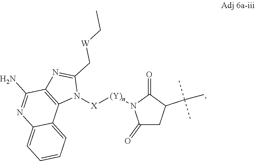
wherein W is selected from the group consisting of O and CH2; X is selected from O and CH2; each Y is independently CHR2, wherein R2 is selected from H, OH, and NH2; subscript n is an integer from 1 to 12; and the dashed line (“
84. The immunoconjugate of any one of aspects 1-53, 59, and 61-64, wherein the adjuvant moiety is of formula:

wherein R1 is selected from H and C1-4 alkyl; R2 is selected from H, OH, and NH2; and the dashed line (“
85. The immunoconjugate of any one of aspects 1-53, 59, and 61-64, wherein the adjuvant moiety is of formula:

wherein R1 is selected from H and C1-4 alkyl; R2 is selected from H, OH, and NH2; and the dashed line (“
86. The immunoconjugate of any one of aspects 1-53, 59, and 61-64, wherein the adjuvant moiety is:


wherein the dashed line (“
87. An immunoconjugate selected from:


or a pharmaceutically acceptable salt thereof, wherein Ab is an antibody with at least one lysine side chain and subscript r is an integer from 1 to 10.
88. The immunoconjugate of aspect 87, wherein r is from 1 to 4.
89. The immunoconjugate of any one of aspects 1-88, wherein the linker is from about 2.5 Å to about 45 Å.
90. The immunoconjugate of any one of aspects 1-88, wherein the linker is from about 2.5 Å to about 20 Å.
91. A composition comprising a plurality of immunoconjugates according to any one of aspects 1-90.
92. The composition of aspect 91, wherein the average number of adjuvant moieties per immunoconjugate ranges from about 1 to about 9.
93. The composition of aspect 91, wherein the average number of adjuvant moieties per immunoconjugate ranges from about 0.5 to about 4.
94. The composition of any one of aspects 91-93, further comprising one or more pharmaceutically acceptable excipients.
95. A method for treating cancer comprising administering a therapeutically effective amount of an immunoconjugate according to any one of aspects 1-90 or a composition according to any one of aspects 91-94 to a subject in need thereof.
96. The method of aspect 95, wherein the cancer is breast cancer.
97. The method of aspect 95, wherein the cancer is a head and neck cancer.
98. The method of aspect 95, wherein the cancer is a lymphoma.
Imidazoquinoline compounds with a free amine group (Compound 1) or a maleimide group (Compound 2) were synthesized according to Scheme 1, allowing for the rapid assessment of linker technology and antibody-adjuvant immunoconjugate efficacy.
To determine if adjuvant functionalization impacted the capacity of Compound 2 or Compound 1 to elicit immune activation, human antigen presenting cells were stimulated with 10-fold serial dilutions of R848, Compound 2, Compound 1 or a control TLR agonist, CL307, for 18 hours prior to analysis via flow cytometry. The data indicated that Compound 2 and Compound 1 performed similarly to R848 across each concentration assayed (FIG. 4 ; Compound 1 data not shown).
Next, the capacity of each functionalized TLR agonist to activate human TLR7 or TLR8 was directly assayed. HEK293 cells were co-transfected with human TLR7 or TLR8 or murine TLR7 and an inducible secreted embryonic alkaline phosphatase reporter gene under the control of the IFN-β minimal promoter fused to NF-κB and AP-1 binding sites. Cells were subsequently incubated with 2-fold serial dilutions of each the indicated adjuvants for 12 hours at 37 C in the presence of an alkaline phosphatase substrate. Activity was measured by spectrophotometry (OD 650 nm). The data indicate that Compound 1 activated both human TLR7 and TLR8 whereas Compound 2 was specific for TLR7 activity (FIG. 2 ). Similarly, both Compound 2 and Compound 1 activated murine TLR7 (FIG. 2 ).


Adjuvants with a free amine (R848, Compound 1, etc.) were conjugated to SMCC, SPDP or other NHS containing linkers by reacting the compounds at a 1:1 molar ratio in PBS or other suitable buffers at pH 7-7.5. All reactions were protected from light and incubated for 30 minutes at room temperature. Where possible, adjuvant-crosslinker conjugates were purified via reverse phase high-pressure liquid chromatography (HPLC). Adjuvant-crosslinker conjugates were utilized immediately following conjugation, as described below.
Adjuvant-linker combinations were desalted and buffer exchanged into deionized water with Zeba Spin Desalting Columns (ThermoFisher Scientific). Samples were subsequently analyzed on a Shimadzu LC/MS-2020 Single Quadrupole Liquid Chromatograph Mass Spectrometer. A method with a gradient ranging from 0 to 100% acetonitrile suitable for detection of small molecules within 100-1000 m/z was utilized for compound detection.
The reaction efficiency was assessed via LC-MS and indicated that the majority of free SMCC had reacted with Compound 1 to form Compound 1-SMCC, which had the expected molecular weight of 531 (FIG. 3 , lower right panel). Similar reaction efficiencies were observed with Compound 1-SPDP (data not shown).
Following the successful conjugation of Compound 1 to the crosslinkers, antibodies were modified with the SATA crosslinker to convert the free amines on the antibody to protected sulfhydryl groups. Following conjugation of SATA, sulfhydryl groups were deacetylated with hydroxylamine and exposed thiols were reacted with the maleimide component of the adjuvant-SMCC compound as shown in Scheme 3 of FIG. 139 .
Antibody was resuspended in phosphate buffered saline (PBS) at 1-5 mg/mL and the SATA crosslinker (ThermoFisher Scientific) was resuspended at 70 mM in anhydrous DMSO immediately before usage. Antibody was reacted with a 10-fold molar excess of SATA at room temperature for 30 minutes. The SATA-modified antibody was purified from excess reagent and byproducts with 3 washes in PBS with equilibrated Amicon Ultra Centrifugal Filter Units with Ultracel-100 membranes according to the manufacturer's instructions (EMD Millipore). The number of SATA crosslinkers per antibody was determined by matrix-assisted laser desorption/ionization mass spectrometry (MALDI-TOF).
SATA-modified antibody was deacetylated following a 2-hour incubation at room temperature in PBS at pH between 7.2-7.5 with 0.05 M hydroxylamine and 2.5 mM EDTA. The deacetylated SATA-modified antibody was subsequently purified from excess reagent and byproducts with 3 washes in PBS containing 5 mM EDTA with equilibrated Amicon Ultra Centrifugal Filter Units with Ultracel-100 membranes according to the manufacturer's instructions (EMD Millipore). Purified deacetylated SATA-modified antibody was subsequently reacted with a 5 to 40-fold molar excess of adjuvant-crosslinker for 30 minutes to one hour at room temperature. The exact molar excess was 10-fold higher than the average number of SATA molecules per antibody as determined by MALDI-TOF. Following conjugation, the antibody adjuvant immunoconjugate was purified from excess reagent and byproducts with 3 washes in PBS with equilibrated Amicon Ultra Centrifugal Filter Units with Ultracel-100 membranes according to the manufacturer's instructions (EMD Millipore).
The average drug to antibody ratio was determined via MALDI-TOF. Samples were desalted and buffer exchanged using Zeba Spin Desalting Columns (ThermoFisher Scientific) into deionized water. Matrix (sinapinic acid) was first spotted onto the MALDI sample target plate and allowed to dry. Next, the sample was mixed at a 1:1 ratio with and without a bovine serum albumin (BSA) standard (0.25-1 pM BSA) and spotted onto the plate with the matrix samples. Once both the matrix and sample layer dried, samples were analyzed on a AB Sciex TOF/TOF 5800 (Stanford University, Canary Center). A high mass detector (CovalX) with negative ionization allowed for enhanced sensitivity and resolution at protein sizes in the range of a fully intact IgG antibody (˜150,000 kDa).
Following successful conjugation of the antibody-adjuvant immunoconjugate (Ab-SATA-SMCC-Adjuvant shown in Scheme 3 of FIG. 139 ), the average drug to antibody ratio was determined via MALDI-TOF mass spectrometry (Table 1). The mass difference between the SATA-modified and unmodified antibody was utilized to determine how many linkers were present per antibody. The mass difference between the SATA-modified antibody and the immunoconjugates were utilized to determine the average drug to antibody ratio (DAR).
| TABLE 1 |
| MALDI-TOF MS-based determination of Drug-to-Antibody Ratio. |
| Molecular | Mass | ||
| Weight | Difference | Ab | |
| Sample | (Da) | (Da) | Modification |
| Antibody | 145,772 | — | — |
| Antibody-SATA | 146,210 | 438 | 3.77 | Linkers/Ab |
| Antibody-SATA-SPDP- | 146,944 | 1172 | 2.07 | Drugs/ |
| Compound | ||||
| 1 | ||||
| Antibody-SATA-SMCC- | 147,309 | 1537 | 2.07 | Drugs/ |
| Compound | ||||
| 1 | ||||
Isolation of Human Antigen Presenting Cells.
Human antigen presenting cells (APCs) were negatively selected from human peripheral blood mononuclear cells obtained from healthy blood donors (Stanford Blood Center) by density gradient centrifugation using a RosetteSep Human Monocyte Enrichment Cocktail (Stem Cell Technologies) containing monoclonal antibodies against CD2, CD3, CD8, CD19, CD56, CD66b and CD235a. Immature APCs were subsequently purified to >97% purity via negative selection using an EasySep Human Monocyte Enrichment Kit without CD16 depletion containing monoclonal antibodies against CD2, CD3, CD19, CD20, CD56, CD66b, CD123 and CD235a.
Preparation of Tumor Cells.
Tumor cells were resuspended in PBS with 0.1% fetal bovine serum (FBS) at 1 to 10×106 cells/mL. Cells were subsequently incubated with 2 μM CFSE to yield a final concentration of 1 μM. The reaction was ended after 2 minutes via the addition of 10 mL complete medium with 10% FBS and washed once with complete medium. Cells were either fixed in 2% paraformaldehyde and washed three times with PBS or left unfixed prior to freezing the cells in 10% DMSO, 20% FBS and 70% medium.
APC-Tumor Co-Cultures.
2×10⁵ APCs were incubated with or without 6.5×10⁵ autologous or allogeneic CFSE-labeled tumor cells in 96-well plates (Corning) containing IMDM medium (Gibco) supplemented with 10% fetal bovine serum, 100 U/mL penicillin, 100 μg/mL streptomycin, 2 mM L-glutamine, sodium pyruvate, non-essential amino acids, 50 μM 2-ME and, where indicated, various concentrations of anti-tumor antibody and the indicated adjuvants. Cells and cell-free supernatants were analyzed after 18 hours via flow cytometry.
Results.
To determine the capacity of immunoconjugates to elicit immune activation, human APCs (˜95% monocytes) obtained from fresh blood were incubated with CFSE-labeled human B cell lymphoma cells (Toledo, ATCC) at a 3:1 ratio and 2-fold serial dilutions of Compound 1, Rituximab (Ab), Rituximab+Compound 1 (Mixture) or Rituximab-SATA-SMCC-Compound 1 (conjugated). In these experiments, the immunoconjugate had an average of 2.1 Compound 1 molecules per antibody and the Compound 1 doses were adjusted accordingly to ensure that equimolar amounts of Compound 1 were compared across all conditions. After 18 hours, cells were analyzed for the expression of activation markers via flow cytometry. The data indicate that immunoconjugates were far superior at eliciting APC activation as CD40, CD86 and HLA-DR were expressed at several fold higher levels in APCs stimulated with the immunoconjugate as compared to those stimulated with Ab alone, Compound 1 alone or the mixture (FIG. 4 ).
Given the high level of activation markers observed following immunoconjugate activation, the expression of PD-L1, an inhibitory marker that is highly correlated with the extent of APC activation, was investigated. Surprisingly, immunoconjugates were much less potent at eliciting the upregulation of PD-L1 expression as compared to the adjuvant alone or the mixture (FIG. 5 ). Notably, PD-L1 expression was negligible at 0.1 μM immunoconjugate, which corresponds to the maximally bioactive concentration (FIG. 4 , FIG. 5 ). These data suggest that the immunoconjugate may activate unforeseen signaling pathways in human APCs.
In support of this hypothesis, cells stimulated with the immunoconjugate unexpectedly developed dendrites and underwent morphologic changes consistent with monocytes differentiating into DCs. This finding prompted the analysis of DC associated surface molecules. Consistent with their morphology, APCs stimulated with the immunoconjugate, but not the mixture, downregulated CD14, CD16 and CD163 expression in a dose dependent manner (FIG. 6 ). The downregulation of these molecules, which are expressed by monocytes and macrophages, but greatly diminished on monocyte-derived DCs, indicates that human monocytes exposed to immunoconjugate rapidly differentiated into DCs. Consistent with these data, APCs stimulated with the immunoconjugate upregulated the expression of CD123, a marker of human inflammatory monocyte-derived DCs (FIG. 6 ).
While the expression of T cell stimulatory molecules such as CD40, CD86 and HLA-DR is necessary for effective T cell activation, APCs also influence the nature of the ensuing immune response through the secretion of cytokines. Therefore, the capacity of immunoconjugates to elicit cytokine secretion in human APCs following stimulation was investigated as described above. The data indicate that the immunoconjugate-differentiated cells secreted several fold higher amounts of IL-1β and TNFα whereas secretion of the anti-inflammatory cytokine IL-10 trended lower (FIG. 7 ).
Immunoconjugates constructed with cleavable linkers have also been prepared and found to elicit APC activation and DC differentiation in vitro (FIG. 8 ). Human APCs that were ˜95% monocytes were stimulated with 2-fold serial dilutions of Rituximab-SATA-SPDP-Compound 1 (conjugated, cleavable), Rituximab alone (Ab), Compound 1 alone or Rituximab+Compound 1 (Mixture) in the presence of CFSE-labeled tumor cells. immunoconjugate—Cleavable had a DAR of 1.4 as confirmed by MALDI-TOF. After 18 hours, CD19− human APCs were analyzed via flow cytometry; n=3.
For tumor studies, 2×10⁵ B16F10 melanoma cells were injected subcutaneously (s.c.) above the right flank in C57BL/6 mice. After ten days, or when the tumors reached 25 mm2, mice were administered intravenous injections of 400 μg the immunoconjugate (anti-GP75-SATA-SMCC-Compound 1) (DAR=1.74) or treated intratumorally with 400 μg of the immunoconjugate (anti-GP75-SATA-SMCC-Compound 1) or a mixture of 1.5 μg of Compound 1 and 400 μg anti-GP75 (TA99). Subsequent treatments were administered on days 2 and 4 after the initial treatment. Tumor development was measured 2-3 times per week with calipers.
Mice treated with the immunoconjugate, but not the mixture, reduced their tumors (FIG. 9A ). Next, equivalent doses of αGP75-immunoconjugate were administered intratumorally or intravenously in mice with established tumors. Surprisingly, IV administration resulted in tumor regression even though it is estimated that less than 10% of the immunoconjugate reached the tumor (FIG. 9B ).
The studies described herein demonstrate that immunoconjugates are quantitatively and qualitatively more effective at eliciting immune activation and anti-tumor immunity than equimolar quantities of non-covalently attached antibody-adjuvant mixtures. These findings are unlikely to result from simple serum half-life extension of the adjuvant following antibody conjugation, because profound phenotypic alterations and novel biology were observed during short in vitro incubation periods. These studies indicate that freshly isolated peripheral blood monocytes from healthy human donors undergo DC differentiation following overnight stimulation with immunoconjugates whereas gold standard DC differentiation protocols with GM-CSF and IL-4 require six days. Furthermore, immunoconjugate activated human APCs expressed several fold higher amounts of co-stimulatory molecules and inflammatory cytokines than achievable with equivalent doses of non-covalently attached antibody-adjuvant mixtures. Yet, immunoconjugates elicit much lower levels of negative co-stimulatory molecules such as PD-L1 and comparable amounts of IL-10, suggesting that immunoconjugates activate unforeseen signaling pathways. Without wishing to be bound by any particular theory, it is believed that stimulation with immunoconjugates closely resembles physiologic antibody-mediated immunity whereby APCs recognize opsonized pathogens (antibody bound to pathogens) with high affinity.
Preparation of Additional Antibody Adjuvant Conjugates.
Additional antibody-adjuvant conjugates were prepared using the methods described in Examples 1 and 2. The antibodies pembrolizumab (PD-1), nivolumab (PD-1), atezolizumab (PD-L1), and ipilimumab (CTLA4) were used to create the antibody-adjuvant conjugates with SATA-SMCC linkers (see Scheme 3 of FIG. 139 ).
Following successful conjugation of the immunoconjugate, the average drug to antibody ratio was determined via LC-MS. The immunoconjugate is first deglycosylated using PNGase F to remove glycans from the antibody, and then the immunoconjugate is buffer exchanged into deionized water. Antibody adjuvant conjugates were run on a C4 column eluted with acetonitrile/water on a Waters Xevo G2-XS QTof/Tof. Raw mass spectrometry data was deconvoluted to determine the Drug to Antibody (DAR) ratios. The LC-MS data indicated successful conjugation and desirable DAR ratios.
Isolation of Human Antigen Presenting Cells.
Human antigen presenting cells (APCs) were negatively selected from human peripheral blood mononuclear cells obtained from healthy blood donors (Stanford Blood Center) by density gradient centrifugation using a RosetteSep Human Monocyte Enrichment Cocktail (Stem Cell Technologies) containing monoclonal antibodies against CD14, CD16, CD40, CD86, CD123, and HLA-DR. Immature APCs were subsequently purified to >97% purity via negative selection using an EasySep Human Monocyte Enrichment Kit without CD16 depletion containing monoclonal antibodies against CD14, CD16, CD40, CD86, CD123, and HLA-DR.
Preparation of Tumor Cells.
Tumor cells were prepared in accordance with Example 3 above.
APC-Tumor Co-Cultures.
2×10⁵ APCs were incubated with or without 6.5×10⁵ autologous or allogeneic CFSE-labeled tumor cells in 96-well plates (Corning) containing IMDM medium (Gibco) supplemented with 10% fetal bovine serum, 100 U/mL penicillin, 100 μg/mL streptomycin, 2 mM L-glutamine, sodium pyruvate, non-essential amino acids, 50 μM 2-ME and, where indicated, various concentrations of antibody. Cells and cell-free supernatants were analyzed after 18 hours via flow cytometry.
Results.
Human APCs (˜95% monocytes) obtained from fresh blood were incubated with CFSE-labeled human B cell lymphoma cells (Toledo, ATCC) at a 3:1 ratio and 2-fold serial dilutions of the antibody alone or the Antibody-SATA-SMCC-Compound 1 (conjugated). After 18 hours, cells were analyzed for the expression of activation markers via flow cytometry. The data indicate that immunoconjugates were superior at eliciting APC activation as CD40, CD86, and HLA-DR tended to be expressed at higher levels in APCs stimulated with the immunoconjugate as compared to those stimulated with the antibody alone (see FIGS. 10D and 10E for Ipilimumab, 11D and 11E for pembrolizumab, 12D and 12E for nivolumab, and 13D and 13E for atezolizumab). Consistent with the results observed in Example 3, the immunoconjugates downregulated CD14 (see FIG. 10C for ipilimumab, 11C for pembrolizumab, 12C for nivolumab, and 13C for atezolizumab). The results for these immunoconjugates were as expected for CD16 and CD123 (data not shown) based on the results in Example 3.
The capacity of these immunoconjugates to elicit cytokine secretion in human APCs following stimulation was investigated as described in Example 3 above. The data indicate that immunoconjugate-differentiated cells secreted higher amounts of IL-1β and TNFα (see FIGS. 14A and 14B for atezolizumab, 15A and 15B for nivolumab, 16A and 16B for pembrolizumab, and 20 for ipilimumab).
Preparation of Additional Antibody Adjuvant Conjugates.
An additional antibody-adjuvant conjugate was prepared using the methods described in Examples 1 and 2. An anti-Dectin-2 antibody (CLEC6A) and isotype rat IgG2a was used to create the antibody-adjuvant conjugate with SATA-SMCC linkers (see Scheme 3 of FIG. 139 ).
The capacity of this immunoconjugate to elicit cytokine secretion in murine monocyte derived APCs following stimulation was investigated as described in Example 3 above. Specifically, cytokine production is shown in FIG. 21 for GM-CSF-pretreated monocytes that were stimulated for 18 hours with the immunoconjugates or equivalent amounts of the unconjugated components. The data indicate that immunoconjugate-differentiated cells secreted higher amounts of TNFα, IL-6, and IL-12p70 (see FIG. 21 ) than equivalent amounts of the components (adjuvant alone, and antibody alone, and control antibody conjugate).
Dectin-2 and CLEC5A are C-type lectin receptors that associate with and signal through the adaptor proteins FcRγ (FCER1G) and DAP12 (TYROBP), respectively, following receptor crosslinking. These adaptor proteins contain immunoreceptor tyrosine-based activation motifs (ITAMs) that mediate downstream signaling through a Syk-dependent pathway, leading to immune cell activation (i.e. cytokine production, costimulatory molecule expression, antigen presentation, etc.). As shown (FIG. 21 and FIG. 23 ), immunoconjugates directed against these receptors exhibit synergistic immunostimulatory effects through simultaneous engagement of the ITAM-coupled receptor (through the antigen binding domain) and other signaling pathways (through the adjuvant moiety, e.g. TLR7/8) Immunoconjugates targeting other receptors that associate with FcRγ and/or DAP12, or that contain similar signaling domains (e.g. hemITAM), may be prepared in a similar fashion and are expected to exhibit similar effects.
The following steps were taken to prepare a TLR7/TLR8 adjuvant (Scheme 1, Compound 1) suitable for conjugation to an antibody to form an immunoconjugate of the present invention. Masses of products were confirmed on a UPLC system (Waters Acquity) equipped with a Xevo XS QToF spectrometer detector. Samples dissolved in acetonitrile:water were injected onto a BEH200 C18 column (2.1 mm diameter×50 mm length) eluted with a 10-90% gradient of acetonitrile:water over 5 minutes.

Chilled (0° C.) nitric acid (70%, 160 mL) was slowly added to the quinoline-2,4-diol I (100 g 621 mmol) in glacial acetic acid (600 mL) stirring in ice bath. Removed mixture from ice bath then warmed to room temperature. Stirred at room temperature for 30 min Heated at 80° C. for 1.5 hours then cooled the mixture to 0° C. Slowly added 1 L of water to the mixture to precipitate yellow solid. Stirred vigorously for 15 minutes then filtered. Resuspended the solid in water (1 L) and stirred vigorously for 15 minutes then filtered. Repeated with the additional step of slowly adding solid NaHCO3 to bring pH to >6 then suction filtered overnight. Resuspended solid in ethyl ether (750 mL) and stirred vigorously to create fine suspension. Filtered and repeated. Suction filtered overnight to dry. Yield 112 g II (88%) yellow solid.

At room temperature, slowly added disopropylethylamine (63 mL, 47 g, 0.36 mol, 2.5 eq.) to POCl3 (300 mL). Heated mixture to 80° C. under blanket of Ar. Slowly added in 2 g portions nitro-diol II (30 g, 145 mmol, 1 eq.) over 30 minutes maintaining temperature below 95° C. After addition is completed, raise temperature to 110° C. and heat for 1 hour. Cooled reaction to 0° C. then slowly pour in parts over ice while vigorously stirring. Added cold water to final volume of 1.2 L then stirred vigorously. Decanted the aqueous mother liquor and added 1 L water to the dark solid, scraping the sticky solid from walls of flask to create suspension. Repeated as necessary to obtain solid that can be filtered. Resuspended the solid in 1 L water then slowly added solid NaHCO3 until pH>6. Filtered the solid then dissolved in EtOAc (500 mL). Filtered EtOAc solution through Celite to remove insoluble black impurity. Washed filtrate with saturated NaHCO3, water, brine then separated and dried organic layer with Na2SO4, filtered and concentrated in vacuo. The brown solid that is formed was triturated with 3:1 hexanes/diethyl ether (500 mL), filtered. The tan solid III (22 g, 30 mmol, 62%) was used as is in the next reaction.

To a solution of nitro-dichloro compound III (22 g, 62 mmol, 1 eq.) and solid K2CO3 (17 g, 124 mmol, 2 eq.) in DMF (250 mL) at 0° C. was slowly added a solution of N-Boc-1,4-diaminobutane (12.8 g, 1.1 eq.) in DMF (60 mL) over 30 minutes. After addition was complete the reaction was warmed to room temperature and stirred for an additional 30 minutes. Water (800 mL) was added and the mixture was stirred vigorously. The supernatant was poured off and the wet solid was dissolved in ethyl acetate (500 mL). The solution was washed with water, brine, separated, dried (Na2SO4), filtered and concentrated in vacuo. The brown solid was triturated with 1:1 hexanes/diethyl ether (400 mL) and filtered to obtain a yellow solid IV (17 g, 43 mmol, 69%) that was used as in in the next reaction.

To a solution of nitro-amino compound IV (17 g, 43 mmol, 1 eq.) in methanol (400 mL) and water (60 mL) at 0° C. was added NiCl2.6H2O (0.51 g, 2.2 mmol, 0.05 eq). Sodium borohydride (pellets, 3.2 g, 86 mmol, 2 eq.) was added and reaction was stirred for 1 h at 0° C. then warmed to room temperature and allowed to stir for another 15 minutes. Glacial acetic acid was added in parts to neutralize any unreacted NaBH4 until a pH of ˜5 was obtained. The solution was filtered through a bed of Celite to remove black insoluble material. The solvent was removed in vacuo. The dark brown solid was triturated with ether then filtered to obtain a tan solid V (13.3 g, 37 mmol, 85%) that was used as is in the next reaction.

To a solution of diamino compound V (13.3 g, 37 mmol, 1 eq.) in DMF (250 mL) containing disopropylethylamine (7.17 g, 9.7 mL, 56 mmol, 1.5 eq.) stirring at room temperature was added neat valeroyl chloride (5.5 mL, 5.5 g, 42 mmol, 1.2 eq). The mixture was stirred for 30 minutes then ice was and then water was added to a final volume of 1 L. The mixture was stirred vigorously until a clear supernatant was formed. The supernatant was poured off and the crude solid was dissolved in ethyl acetate (400 mL) and filtered through a bed of Celite. The filtrate was washed with water (400 mL), brine (400 mL), separated then dried (Na2SO4), filtered and concentrated. The solid was triturated with ether, filtered and suction dried. The brown solid obtained VI (13.9 g, 31 mmol, 84%) was used in the next reaction as is.

In a 500 mL round bottomed flask equipped with a Dean-Stark apparatus a mixture of amide VI (13.9 g, 31 mmol, 1 eq.) and 2-chlorobenzoic (2.4 g, 15.5 mmol. 0.5 eq.) was refluxed in 150 mL toluene (bath temperature=170° C.) for 4 hours. The Dean-Stark apparatus and condenser was removed and until 80-90% of the toluene was evaporated. 2,4-dimethoxybenzylamine (25 g, 150 mmol, 5 eq.) was added and the reaction was continually heated at 120° C. for 1.25 hours. The reaction was cooled and the crude mixture was diluted with 1:1 MeOH/water (1 L) and vigorously stirred. The supernatant was decanted (removing most of the excess 2,4-dimethoxybenzylamine) and the crude product was partitioned between water and ethyl acetate. Acetic acid was added until the aqueous layer gave a pH of 5-6. The organic layer was washed with water, brine, dried (Na2SO4), filtered and concentrated. The thick brown syrup was dissolved in diethyl ether and filtered to remove a gray solid (not product). The ether was removed to give a brown syrup (14.4 g, 26 mmol, 73%) and was used as is in the next reaction.

To material VII (14.4 g, 26 mmol, 1 eq.) was added water (60 mL) and slowly with swirling conc. HCl (60 mL). The mixture was vigorously stirred at room temperature for 30 minutes then heated to reflux for 1 hour. The reaction was cooled in an ice bath and solid NaOH pellets (28 g, 700 mmol) were added in parts over 30 minutes until a basic pH was achieved. The solution was warmed to room temperature and stirred vigorously. Solid NaCl was added until a saturated solution was achieved. This aqueous layer was extracted 3 times with 10% isopropanol/dichloromethane (400 mL). The combined organic layers were dried (Na2SO4), filtered and concentrated to yield a brown solid VIII was obtained (6.8 g, 22 mmol, 79%).

This example provides guidance on synthesis of an immunoconjugate using the TFP ester method. Compound VIII (311 mg, 1 mmol) was dissolved in 10 mL of dimethylformamide (DMF) and then 2 molar equivalents of diisopropylethylamine (DIPEA) was added. An SMCC linker (1.5 mmol) was dissolved in 10 mL of dichloromethane and added in one portion to VIII. The reaction was stirred overnight at 20° C. and concentrated to dryness via rotary evaporation. The crude product IX was purified on a silica gel using a Buchi flash chromatography system loaded with a 12 g disposable cartridge and eluted with a gradient of 0-10% methanol over 15 minutes. Pure fractions were combined and evaporated to dryness to provide 160 mg of a pale yellow solid IX.

Compound IX (0.1 mmol, 53 mg) was dissolved in 10 mL of dichloromethane and then 2 equivalents of thioglycolic acid were added at one time. The mixture was concentrated to dryness under vacuum and the residue was washed three times with 5 mL of diethyl ether.

Compound X (6.2 mg, 0.01 mmol) was dissolved in 2 mL of THF and then 5 mg of tetrafluorophenol was added. Then 5 mg of dicyclohexylcarbodiimide (DCC) was added. The mixture was stirred overnight at room temperature and then concentrated to dryness under vacuum. The crude product XI was purified via flash chromatography on silica gel (4 gram prepacked column) and eluted with 0-10% MeOH in dichloromethane. Pure fractions were combined and evaporated to provide 3.6 mg of pure XI (confirmed by LC/MS). The TFP ester XI was then used in the antibody conjugation step depicted in Scheme 14 of FIG. 140 .
An IgG1 antibody (specifically, the anti-CD20 antibody rituxumab) was buffer exchanged into PBS at a pH of 7.2 and diluted to 10 mg/mL (66 μM). The TFP activated adjuvant, XI, was added to DMSO and 6 molar equivalents (relative to IgG) was added to 1 mL of the antibody solution (10 mg) in one portion. The mixture was inverted several times to mix and incubated overnight at 20° C. The resulting immunoconjugate (“BB-01”) was purified via buffer exchange into PBS (pH 7.2) using a PD10 column (SephadexG25) size exclusion chromatography column Pure fractions were pooled and the concentration determined by measuring the absorbance at 280 nm on a nano-drop spectrophotometer. The yield was 8 milligram or approximately 80% based on recovered protein. The immunoconjugate product was sterile filtered through a 0.2 μm syringe filter and stored at 4° C. until needed.
Characterization of the resulting immunoconjugate's drug to antibody ratio (“DAR”) was performed via liquid chromatography-mass spectrometry (“LC/MS”) analysis on a UPLC system (Waters Aquity) equipped with a Xevo XS QToF mass spectrometer detector. Analysis was performed via injection of 5 μg of the immunoconjugate onto a BEH200 C4 column (2.1 mm diameter×50 mm length) eluted with a 10-90% gradient of acetonitrile:water over 4 minutes.
The analysis indicated that the immunoconjugates synthesized via the TFP method demonstrated higher DAR than the immunoconjugate synthesized using the SATA method. In addition the TFP method yielded immunoconjugates with reduced amounts of unconjugated antibody (only about 5%) compared to the SATA synthesis method (about 20%) (compare FIGS. 1A and 1B ).
Size exclusion chromatography (“SEC”) analysis of BB-01 was performed to determine the monomeric purity. Analysis was performed on a BEH200 SEC column eluted with PBS (pH 7.2) and 0.2 mL/min. The immunoconjugate BB-01 synthesized using the TFP active ester method contained less than 2% of high molecular weight aggregate (FIG. 2B ) compared to greater than 8% aggregate observed when the SATA method was used (FIG. 2A ).
This example provides guidance on synthesis of an immunoconjugate using the PFP ester method. Ester modification of the adjuvant and conjugation of the modified adjuvant to the antibody is shown in Scheme 15 of FIG. 141 . Cyclohexane trans-1,4-dicarboxylate (1 g) was dissolved in 10 mL of dimethylformamide (“DMF”) and 1-[bis(dimethylamino)methylene]-1H-1,2,3-triazolo[4,5-b]pyridinium 3-oxid hexafluorophosphate) (“HATU”) (1 mmol) was added followed by 1 mL of N-ethyl-N-(propan-2-yl)propan-2-amine (“DIPEA”). Compound 1 (311 mg) was added and the mixture stirred overnight at 20° C. The reaction mixture was diluted with 50 mL of dichloromethane (“DCM”) and washed with 20 mL of 1N HCl. The DCM layer was evaporated to dryness and the product purified on silica gel eluted with 0-10% MeOH in DCM containing 1% acetic acid. Pure fractions were concentrated to provide 220 mg of purified acid II. Compound II (100 mg) was dissolved in THF and 100 mg of HATU was added followed by 200 μL of DIPEA. Two equivalents of amino-PEG2-tertbutyl-carboxylate was added and stirred for one hour at 20° C. The mixture was concentrated to dryness and 10 milliliters of 4N HCl in dioxane was added. The mixture was concentrated to dryness and the crude product III was purified by prep HPLC to provide 40 mg of compound III.
Compound III was converted to PFP ester IV as described below. Compound III (35 mg) was added to 50 mg of PFP in 5 mL THF and 5 mL DMF was added followed by 20 mg of DCC. DMAP (2-3 mg) was added and the solution was stirred overnight at 20° C. The reaction was concentrated and purified by flash chromatography (eluted with 0-10% MeOH) to provide 17 mg of PFP ester IV after lyophilization from 1:2 acetonitrile water.
PFP ester IV (6 molar eq. relative to IgG) was added to 20 mg of an IgG antibody (specifically, the anti-CD20 antibody rituximab) (10 mg/mL in PBS) and incubated at 37° C. overnight. The resulting immunoconjugate BB-14 was buffer exchanged into PBS (pH 7.2) to remove excess small molecular weight reagent and the concentration determined on the nanodrop. The yield was 15 mg of immunoconjugate (75% yield). The product was stored at 4° C. A DAR of 2.2 was determined via LC/MS analysis. Besides the desirable DAR and high yield, the product also had few impurities as determined by SEC analysis (see FIGS. 3 and 4 ).
Ester modification of the adjuvant and conjugation of the modified adjuvant to the antibody is shown in Scheme 16 of FIG. 142 . Compound VII (150 mg) was dissolved in 20 mL of tetrahydrofuran (“THF”) and 10 mL of aqueous, saturated sodium bicarbonate was added. Then, 50 mg of succinic anhydride was added in one portion and the mixture was stirred for one hour at room temperature. Twenty milliliters of 1N HCl was added slowly and the mixture was extracted with 2×50 mL of dichloromethane. The combined organic extracts were evaporated to dryness. The crude product (Suc-VII) was purified on a 4 gram silica gel column eluted with 0-15% MeOH (1% acetic acid) over 15 minutes. Pure fractions were combined and evaporated to provide 190 mg of pure VII-Suc.
Compound VII-Suc (150 mg) was dissolved in 10 mL of DMF and 1 equivalent of HATU was added followed by 2 equivalents of DIPEA. 1.5 equivalents of glycine-OtBu were added and stirred overnight. The DMF was evaporated and the residue treated with 5 mL of 1N HCl in dioxane for 30 minutes. The solvent was evaporated and the crude Gly-Suc-VII was flash purified on a 4 gram silica gel column eluted with 0-10% MeOH over 10 minutes. Evaporation of pure fractions provided 110 mg of Gly-Suc-VII; the pure material was dissolved in DMF and the above process was repeated to provide 60 mg of pure Gly2-Suc-VII.
The pure Gly2-Suc-VII (30 mg) was dissolved in 5 mL of DMF and 1.5 equivalents of NHS was added followed by 5 mL of THF. DCC (1.5 equivalents) was added and the mixture was stirred overnight at room temperature. The solvent was evaporated and the crude NHS ester was flash purified on a silica gel eluted with 0-10% MeOH in DCM over 10 minutes. Pure fractions (determined by TLC) were combined and evaporated to provide 1 mg of pure NHS-Gly2-Suc-VII after lyophilization from acetonitrile water.
The pure NHS ester was dissolved in DMSO to make a 20 mM solution and 6 eq. was added to 2 mL of an IgG antibody (specifically, the anti-CD20 antibody rituximab) (10 mg/mL in PBS). The conjugation reaction was incubated at room temperature overnight and buffer exchanged into fresh PBS to remove excess adjuvant. The purified immunoconjugate BB-15 was sterile filtered and stored at 4° C. The yield was about 16 mg. Besides having a high yield, the LC/MS analysis showed high levels of purity, low levels of aggregation, and a desirable DAR ratio (see FIGS. 5 and 6 ).
This example provides guidance on synthesis of an immunoconjugate with a different linker using the TFP ester method. Ester modification of the adjuvant and conjugation of the modified adjuvant to the antibody is shown in Scheme 17 of FIG. 143 . Compound VII (311 mg, 1 mmol) was dissolved in 10 mL of DMF and then 0.3 mL of DIPEA was added. The NHS-PEG5-acid (1.2 equivalents) was dissolved in 5 mL of dichloromethane and added to compound VII in one portion. The mixture was stirred overnight at room temperature and then concentrated to dryness. The crude residue was purified via silica gel chromatography on a 4 gram column eluted with 0-10% MeOH in DCM containing 1% acetic acid over 10 minutes to provide 260 mg (57% yield) of PEG5-VII after concentration of the pure fractions.
PEG5-VII (50 mg) was dissolved in 10 mL DMF and 1.5 eq. of TFP was added followed by 1.2 eq. DCC and 5 mg of DMAP. The reaction was stirred overnight, concentrated to dryness and purified on silica gel 4 gram column eluted with 0-10% MeOH in DCM to provide 35 mg of pure TFP-PEG5-VII after lyophilization from 1:2 acetonitrile water.
The TFP ester (TFP-PEG5-VII) was dissolved in DMSO to make a 20 mM stock solution and added to 20 mg of an IgG antibody (specifically, the anti-CD20 antibody rituximab) in PBS at 10 mg/mL. The conjugation reaction was allowed to proceed overnight at room temperature. The resulting immunoconjugate was buffer exchanged (GE, PD10 desalting column) into PBS at pH 7.4. The purified immunoconjugate was sterile filtered using a 2 μm syringe filter and stored at 4° C. LC/MS analysis confirmed that the process provided a DAR of 2.9 adjuvants per antibody (see FIG. 7 ). SEC analysis indicated minimal amounts of aggregate (i.e., less than 2%) (see FIG. 8 ).
This example provides guidance on how to synthesize another TLR7/8 adjuvant. Compound XIV was synthesized starting from compound VI of Scheme 8 of Example 3.

Compound VI (2 g) was dissolved in toluene with 20% dry acetic acid and heated to 75° C. overnight. The solvent was removed under vacuum to provide 2 grams of crude compound XI. Compound XI was used without further purification. Compound XI (2 g) was dissolved in 20 mL DMF and 1.2 equivalents of NaH (50% dispersion) was added slowly and the mixture was stirred for 30 minutes at room temperature. Methyl iodide (2 equivalents) was added in one portion and the reaction mixture was stirred overnight at room temperature. The reaction was concentrated to dryness and the product purified via flash chromatography. The product was eluted with a gradient of 0-10% MeOH in dichloromethane over 15 min. Pure fractions were combined and concentrated to yield 1 g of compound XII (50% yield for 2 steps).

Compound XII (10 g) was dissolved in 10 mL of neat dimethoxybenzylamine (“DMBA”) and heated to 120° C. for 3 hours. The reaction mixture was cooled and diluted with 100 mL of ethyl acetate. The resulting solution was washed two times with 10% citric acid in water and once with water to remove excess DMBA. The organic layer was dried over MgSO4 and concentrated under vacuum to provide crude compound XIII as a brown oil. The crude DMB derivative, compound XIII, was dissolved in dichloromethane and 2 mL of 4N HCl in dioxane was added. After 2 hours, the reaction mixture was concentrated to dryness and the crude HCl salt compound XIV was dissolved in 3 mL of methanol. Ethyl ether (20 mL) was added slowly with stirring to the crude solution and a white precipitate formed. The reaction was filtered and the white solid product was washed twice with 10 mL ethyl ether and dried under vacuum to provide 4 gram of HCl salt compound XIV. LC/MS analysis confirmed the correct molecular weight (M/z=326.5) and a purity of greater than 95%.
This example provides guidance on synthesis of an immunoconjugate that contains an aryl tertiary amine linker using the TFP ester method as depicted in Scheme 20 of FIG. 144 . Compound XIV (300 mg) of Example 12 was dissolved in THF (10 mL) and 1.2 eq. of NaH (50% dispersion) was added. The mixture was stirred for 15 minutes and 2 equivalents of 4-bromomethylphenyl acetic acid was added. The reaction was stirred overnight at room temperature and concentrated to dryness. One mL of acetic acid was added and the product was purified by preparative HPLC on a C-18 column eluted with a gradient of 10-90% acetonitrile in water (0.1% TFA) over 20 minutes to provide 165 mg of purified phenylacetic acid compound XV.
Compound XV (50 mg) was dissolved in dichloromethane/dimethylformamide (5 mL, 1:1) and 2 equivalents of TFP was added followed by 1.5 equivalents of 1-ethyl-3-(3-dimethylaminopropyl)carbodiimide (“EDCI”). The reaction was stirred overnight at room temperature and the product purified via flash chromatography on a 4 gram silica gel column eluted with 0-10% isopropanol over 10 minutes. Pure fractions were concentrated and lyophilized from 30% acetonitrile water to provide 21 mg of purified TFP ester compound XVI as a pale yellow solid. The molecular weight and purity were confirmed by LC/MS (m/z=621.7).
Conjugation to Antibody:
The TFP ester XVI was dissolved in anhydrous DMSO to make a 20 mM stock solution and 6 molar equivalents (relative to the antibody) was added to 20 mg IgG antibody (specifically, the anti-CD20 antibody rituximab) (10 mg/mL in PBS). The conjugation reaction was incubated at 4° C. overnight. The resulting immunoconjugate, BB-26, was buffer exchanged into PBS (pH 7.2) to remove excess small molecular weight reagents. The final concentration was determined by measuring the antibody at 280 nm on the Nanodrop 1000 spectrophotometer. The yield was 15 mg of BB-26, or 75% based on recovered protein. As seen in FIG. 12A , minimal aggregate was seen (less than 1%) as detected by SEC analysis. As seen in FIG. 12B , the product had a DAR ratio of 2.8 as determined via LC/MS analysis. The purified immunoconjugates BB-26 was filtered through a 0.2 μM sterile filter and stored at −20° C.
This example provides guidance on synthesis of an immunoconjugate that contains an alkyl tertiary amine linker using the TFP ester method as depicted in Scheme 21 of FIG. 145 . Compound XIV (200 mg) was dissolved in methanol (20 mL) and 3 equivalents of 1-formyl-7-tert-butyl heptanoate was added followed by 1.1 equivalents of NaCNBH4. The mixture was stirred for 1.5 hours at room temperature and concentrated to dryness. TFA (5 mL) was added and the mixture stirred overnight at room temperature. The TFA was evaporated under vacuum and the crude product was purified by preparative HPLC on a C-18 column. The product was eluted with a gradient of 10-90% acetonitrile in water (0.1% TFA) over 20 minutes to provide 110 mg of purified acid compound XVII (which was confirmed by LC/MS).
Compound XVII (50 mg) was dissolved in dichloromethane/dimethylformamide (5 mL, 1:1) and 2 equivalents of TFP was added followed by 1.5 equivalents of EDCI. The reaction was stirred overnight at room temperature. The crude TFP ester product XVIII was purified via flash chromatography on a 4 gram silica gel column eluted with 0-10% isopropanol over 10 minutes. Pure fractions were concentrated and the residue lyophilized from 30% acetonitrile water to provide 14 mg of purified TFP ester compound XVIII as a white solid. The molecular weight and purity were confirmed by LC/MS (m/z=601.7).
Conjugation to Antibody:
TFP ester XVIII was dissolved in anhydrous DMSO to make a 20 mM stock solution and 8 molar equivalents (relative to the antibody) was added to 20 mg of an IgG antibody (specifically, the anti-CD20 antibody rituximab) (10 mg/mL in PBS). The conjugation reaction was incubated at 4° C. overnight. The resulting immunoconjugate BB-27 was buffer exchanged into PBS (pH 7.2) to remove excess small molecular weight reagents. The final concentration was determined by measuring the antibodies at 280 nm on the Nanodrop 1000 spectrophotometer. The yield was 16 mg of immunoconjugate BB-27 (80%).
Minimal aggregate was seen (less than 1%) as detected by SEC analysis. The product had a DAR ratio of 2.5 as determined via LC/MS analysis. The purified BB-27 was filtered through a 0.2 μM sterile filter and stored at −20° C.
This example provides guidance on synthesis of an immunoconjugate that contains a PEG tertiary amine linker using the TFP method as depicted in Scheme 22 of FIG. 146 . Compound XIV (200 mg) was dissolved in methanol (20 mL) and 3 eq. of aldehyde XIX was added followed by 1.1 equivalents of NaCNBH4. The mixture was stirred for 3 hours at room temperature and concentrated to dryness. Trifluoroacetic acid (TFA, 10 mL) was added and the reaction stirred for 2 hours at room temperature. The TFA was evaporated under vacuum and the crude product was purified by preparative HPLC on a C-18 column. The product was eluted with a gradient of 10-90% acetonitrile in water (0.1% TFA) over 20 minutes to provide 85 mg of purified acid XX after lyophilization of the combined pure fractions (confirmed by LC/MS).
Compound XX (80 mg) was dissolved in dichloromethane/dimethylformamide (5 mL, 1:1) and 2 equivalents of TFP was added followed by 1.2 equivalents of EDCI. The reaction was stirred overnight at room temperature. The crude TFP ester product XXI was purified via flash chromatography on a 4 gram silica gel column eluted with 0-10% isopropanol over 10 minutes. Pure fractions were concentrated and the residue lyophilized from 30% acetonitrile water to provide 45 mg of purified TFP ester of compound XXI as a beige solid. The molecular weight and purity were confirmed by LC/MS (m/z=647.7).
Conjugation to Antibody:
The TFP ester of compound XXI was dissolved in anhydrous DMSO to make a 20 mM stock solution and 8 molar equivalents (relative to the antibody) was added to an IgG1 antibody (specifically, the anti-CD20 antibody rituxumab) (10 mg/mL in PBS). The conjugation reaction was incubated at 4° C. overnight. The resulting immunoconjugate BB-36 was buffer exchanged into PBS (pH 7.2) to remove excess small molecular weight reagents. The final concentration was determined by measuring the antibodies at 280 nm on the Nanodrop 1000 spectrophotometer. The yield was 15 mg of immunoconjugate BB-36 (75%) which was stored at 4° C. until used.
Minimal aggregate was seen (less than 1%) as detected by SEC analysis. The product had a DAR ratio of 2.2 as determined via LC/MS analysis. The purified immunoconjugate BB-36 was filtered through a 0.2 μM sterile filter and stored at −20° C.
This example provides guidance on synthesis of an immunoconjugate with a different linker using the TFP ester method as depicted in Scheme 23 of FIG. 147 . Compound VII (311 mg, 1 mmol) was dissolved in 10 mL of DMF and 0.3 mL of DIPEA was added. In a separate container, 1.2 equivalents of 7-methoxy-7-oxoheptanoic acid was dissolved in 5 mL of DMF and 1.5 equivalents DIPEA was added followed by HATU (1.2 equivalents). The mixture was added to VII and stirred overnight at room temperature. The reaction mixture was concentrated to dryness under vacuum and the residue was dissolved in 10 mL of (1:1) tetrahydrofuran:water. One mL of 2M lithium hydroxide in water was added and the reaction stirred for 2 hours at room temperature. The THF was removed via rotary evaporation and the aqueous solution was acidified by adding 10 mL of 1M hydrochloric acid. The aqueous solution was extracted 2× with dichloromethane (20 mL) and the organic layer was combined and dried over magnesium sulfate. The solution was filtered and the filtrate concentrated to dryness. The crude product 22 was purified via silica gel chromatography on a 4 gram column eluted with 0-10% isopropanol in DCM (w/1% acetic acid) over 10 minutes. The pure fractions were combined and concentrated to provide 220 mg of pure 22 as a pale yellow solid.
Compound 22 (50 mg) was dissolved in dichloromethane/dimethylformamide (5 mL, 1:1) and 2 equivalents of TFP was added followed by 1.5 equivalents of EDCI. The reaction was stirred overnight at 22° C. and the crude reaction was concentrated to dryness. The product was purified via flash chromatography on a 4 gram silica gel column eluted with 0-10% isopropanol over 10 minutes. Pure fractions were concentrated and the residue was lyophilized from 30% acetonitrile in water to provide 21 mg of purified TFP ester 23 as a pale yellow solid. The molecular weight and purity were confirmed by LC/MS.
Conjugation to Antibody:
The TFP ester 23 was dissolved in anhydrous DMSO to make a 20 mM stock solution and 6 molar equivalents (relative to the antibody) was added to 20 mg of an IgG antibody (specifically, the anti-CD20 antibody rituximab) (10 mg/mL in PBS). The conjugation reaction was incubated at 4° C. overnight. The resulting immunoconjugate BB-45 was buffer exchanged into PBS (pH 7.2) to remove excess small molecular weight impurities. The final concentration was determined by measuring the absorbance at 280 nm on a Thermo Nanodrop 1000 spectrophotometer. The yield was 14 mg of BB-45, or 70% based on recovered protein. Minimal aggregate (less than 1%) was detected by SEC analysis and a DAR of 2.8 was determined via LC/MS analysis. The purified immunoconjugate was filtered through a 0.2 μM sterile filter and stored at −20° C.
This example provides guidance on synthesis of an immunoconjugate with a different linker using the TFP ester method as depicted in Scheme 24 of FIG. 148 . Compound VII (150 mg) was dissolved in 20 mL THF and 10 mL of aqueous saturated sodium bicarbonate was added. Succinic anhydride (50 mg) was added in one portion and the mixture stirred for 1 hour at room temperature. 20 mL of 1N HCl was added slowly and the mixture was extracted with 2×50 mL of dichloromethane and the combined organic extracts were evaporated to dryness. The crude product 24 was purified on a 4 gram silica gel column eluted with 0-15% MeOH (1% acetic acid) over 15 minutes. Pure fractions were combined and evaporated to provide 180 mg of pure 24.
One hundred and fifty mg of 24 was dissolved in DMF (10 mL) and 1 equivalent of HATU was added followed by 2 equivalents of DIPEA. One and a half eq. of glycine-OtBu was added and stirred overnight. The DMF was evaporated and the residue treated with 5 mL of 1N HCl in dioxane for 30 minutes with stirring. The solvent was evaporated and the crude residue was flash purified on a 4 gram silica gel column eluted with 0-10% isopropanol over 15 minutes. Evaporation of pure fractions provided 110 mg of pure 25.
Compound 25 (50 mg) was dissolved in 10 mL DMF and 1.5 eq. of TFP was added followed by 1.2 eq. DCC and 2 mg of DMAP. The reaction was stirred overnight, concentrated to dryness and purified on silica gel (4 g column) eluted with 0-10% IPA in DCM to provide 32 mg of pure TFP ester, compound 26, after lyophilization from 1:3 acetonitrile water.
Conjugation to Antibody:
The TFP ester, compound 26, was dissolved in anhydrous DMSO to make a 20 mM stock solution and 5 molar equivalents (relative to the antibody) was added to 20 mg antibody at 10 mg/mL in PBS. The conjugation reaction was incubated at 4° C. for 6 hours. The resulting immunoconjugate BB-24 was buffer exchanged into PBS (pH 7.4) to remove excess small molecular weight impurities. The final protein concentration was determined by measuring the absorbance at 280 nm on a Nanodrop 1000 spectrophotometer. The yield was 15 mg (75% based on recovered protein). SEC analysis detected minimal aggregate of less than 1% and the DAR was determined to be 2.8 adjuvants per antibody via LC/MS analysis. The purified immunoconjugate was filtered through a 0.2 μM sterile filter and stored at −20° C. until needed.
This example provides guidance on synthesis of an immunoconjugate with a different linker using the TFP method as depicted in Scheme 25 of FIG. 149 . Compound VII (155 mg, 0.5 mmol) was dissolved in 10 mL of DMF and 0.2 mL of DIPEA was added. In a separate container, 1.2 equivalents of PEG2-dicarboxylate mono methyl ester was dissolved in 5 mL of DMF and 2 equivalents DIPEA was added followed by HATU (1.2 equivalents). The mixture was added to VII and stirred 1 hour at room temperature. The reaction was concentrated to dryness under vacuum and the residue was dissolved in THF (5 mL). An equal volume of water was added followed by 2 mL of 1 M aqueous LiOH. The mixture was stirred overnight and then 10 mL of 1N HCl was added. The acidified mixture was extracted 2× with dichloromethane, dried over sodium sulfate, concentrated to dryness and purified via silica gel chromatography. The product was eluted with 0-10% methanol over 10 minutes. The pure fractions were combined and concentrated to provide 110 mg of pure compound 27 as a pale yellow solid.
Compound 27 (50 mg) was dissolved in dichloromethane/dimethylformamide (5 mL, 1:1) and 2 equivalents of TFP was added followed by 1.5 equivalents of EDCI. The reaction was stirred overnight at ambient temperature and the reaction was concentrated to dryness. The crude TFP ester 28 was purified via flash chromatography on a 4 gram silica gel column eluted with 0-10% isopropanol over 10 minutes. Pure fractions were concentrated and the residue was lyophilized from 30% acetonitrile in water to provide 41 mg of purified TFP ester 23 as a white solid. The molecular weight and purity were confirmed by LC/MS.
Conjugation to Antibody:
The TFP ester 28 was dissolved in anhydrous DMSO to make a 20 mM stock solution and 8 molar equivalents (relative to the antibody) was added to 20 mL of an IgG antibody (specifically, the anti-CD20 antibody rituximab) (10 mg/mL in PBS). The conjugation reaction was incubated at 4° C. overnight. The resulting immunoconjugate BB-37 was buffer exchanged into PBS (pH 7.2) to remove excess small molecular weight impurities. The final concentration was determined by measuring the absorbance at 280 nm on a Thermo Nanodrop 1000 spectrophotometer. The yield was 16 mg of conjugated immunoconjugate BB-37, or 70% based on recovered protein. Minimal aggregate (less than 1%) was detected by SEC analysis and a DAR of 2.3 was determined via LC/MS analysis. The purified immunoconjugate was filtered through a 0.2 μM sterile filter and stored at −20° C.

This example provides guidance on synthesis of another TLR agonist. Compound 29 is a compound VII analog that contains a piperizine side-chain for linker attachment. It was synthesized using methods previously described for the synthesis of the compound VII except that a Boc-protected piperizine analog was substituted for Boc-diaminobutane used in step 3 of the synthesis. The general synthetic route for compound 29 is outlined in Scheme 26. The addition of the piperizine side chain enables the synthesis of immunoconjugates that were previously inaccessible due to instability. Similar compound VII analogs containing succinate linkers are prone to cyclization upon TFP activation and the piperizine prevents cyclization. In addition, the tertiary amino group within the piperizine moeity maintains a positive charge after linker attachment and conjugation. Positive charges in this location are important for improved TLR8 potency. Compound 29 was subsequently used for synthesizing immunoconjugates as described below in Examples 19-21.
This example provides guidance on synthesis of an immunoconjugate with a different linker using the TFP ester method as depicted in Scheme 27 of FIG. 150 . Compound 29 (100 mg) was dissolved in 10 mL THF and 2 mL of aqueous saturated sodium bicarbonate was added followed by 10 mL of water. Succinic anhydride (50 mg) was added in one portion and the mixture was stirred at room temperature. After one hour, 20 mL of 1N HCl was added slowly and the reaction mixture was extracted with 2×50 mL of dichloromethane (“DCM”). The combined organic extracts were evaporated to dryness. The crude product 30 was purified on a 4 gram silica gel column eluted with 0-15% isopropanol in DCM (1% acetic acid) over 15 minutes. Pure fractions were combined and evaporated to dryness to provide 80 mg of pure acid 30.
Compound 30 (50 mg) was dissolved in dichloromethane/dimethylformamide (5 mL, 1:1) and 2 equivalents of TFP was added followed by 1.5 equivalents of EDCI. The reaction was stirred overnight at ambient temperature and the reaction was concentrated to dryness. The crude TFP ester 31 was purified via flash chromatography and eluted with 0-10% isopropanol over 10 minutes. Pure fractions were concentrated and the residue was lyophilized from 30% acetonitrile in water to provide 41 mg of purified TFP ester 31 as a white solid. The molecular weight and purity were confirmed by LC/MS.
The TFP ester 31 was conjugated to an IgG1 antibody (specifically, the anti-CD20 antibody rituxumab) as described previously for BB-24 to provide BB-42. SEC and LC/MS analysis of BB-42 confirmed the molecular weight, a high monomeric purity with less than 2% aggregate, and a DAR of 1.7 (see FIGS. 20A-B ).
This example provides guidance on synthesis of immunoconjugates with different linkers using the TFP ester method as depicted in Scheme 28 of FIG. 151 . Compound 30 (Scheme 27) was coupled to polyethylene glycol (PEG) linkers containing 2 or 8 PEG. units in order to extend the distance between the adjuvant and the antibody. Attachment of the PEG linker extensions was performed using previously described protocols for linker attachment and TFP activation. Briefly 100 mg of compound 30 was dissolved in 10 mL of DMF and 0.2 mL of DIPEA was added followed by HATU (1.2 equivalents). After 1 hour the appropriate amino PEG linker (n=2 or 8) was added and stirred an additional 2 hours at room temperature. The reaction mixture was concentrated to dryness under vacuum and the residue was purified via preparative HPLC on a C-18 column eluted with 10-90% acetonitrile in water over 30 minutes. The pure fractions were combined and lyophilized to provide 65 mg and 45 mg of intermediates 31 or 32 as a clear glassy substance.
Conjugation to Antibody:
Isolation of Human Antigen Presenting Cells.
Human antigen presenting cells (APCs) were negatively selected from human peripheral blood mononuclear cells obtained from healthy blood donors (Stanford Blood Center) by density gradient centrifugation using a RosetteSep Human Monocyte Enrichment Cocktail (Stem Cell Technologies) containing monoclonal antibodies against CD14, CD16, CD40, CD86, CD123, and HLA-DR. Immature APCs were subsequently purified to >97% purity via negative selection using an EasySep Human Monocyte Enrichment Kit without CD16 depletion containing monoclonal antibodies against CD14, CD16, CD40, CD86, CD123, and HLA-DR.
Preparation of Tumor Cells.
Tumor cells were resuspended in PBS with 0.1% fetal bovine serum (FBS) at 1 to 10×106 cells/mL. Cells were subsequently incubated with 2 μM CFSE to yield a final concentration of 1 μM. The reaction was ended after 2 minutes via the addition of 10 mL complete medium with 10% FBS and washed once with complete medium. Cells were either fixed in 2% paraformaldehyde and washed three times with PBS or left unfixed prior to freezing the cells in 10% DMSO, 20% FBS and 70% medium.
APC-Tumor Co-Cultures.
2×10⁵ APCs were incubated with or without 6.5×10⁵ allogeneic CFSE-labeled tumor cells in 96-well plates (Corning) containing IMDM medium (Gibco) supplemented with 10% fetal bovine serum, 100 U/mL penicillin, 100 μg/mL streptomycin, 2 mM L-glutamine, sodium pyruvate, non-essential amino acids and, where indicated, various concentrations of unconjugated CD20 antibody, and immunoconjugates of the invention were prepared according to the examples above. Cells and cell-free supernatants were analyzed after 18 hours via flow cytometry or ELISA.
The results of this assay are shown in FIGS. 9A-9F for BB-17 and BB-01. Specifically, the graphs show that BB-17 and BB-01 prepared according to Schemes 14 of FIGS. 140 and 17 of FIG. 143 elicits myeloid activation while the control, unconjugated CD20 antibody, does not. Further, FIGS. 23A-D show that BB-14 elicits myeloid activation as indicated by CD14, CD20, CD86, and HLA-DR while the control does not. FIGS. 24A-D show that BB-15 elicits myeloid activation as indicated by CD14, CD20, CD86, and HLA-DR while the control does not. FIGS. 25A-D show that BB-27 elicits myeloid activation as indicated by CD14, CD20, CD86, and HLA-DR while the control does not. FIGS. 26A-D show that BB-45 elicits myeloid activation as indicated by CD14, CD20, CD86, and HLA-DR while the control does not. FIGS. 27A-D show that BB-24 elicits myeloid activation as indicated by CD14, CD20, CD86, and HLA-DR while the control does not.
As previously explained, immunoconjugates are described in U.S. Pat. No. 8,951,528 (“the '528 patent”). This example shows that immunoconjugates of the invention are superior to the immunoconjugates disclosed by the '528 patent. BB-01 was synthesized according to Scheme 14 of FIG. 140 . Comparative Conjugates IRM1 and IRM2 were prepared using the adjuvants described in the '528 patent as adjuvants IRM1 and IRM2. Specifically, IRM1 and IRM2 were conjugated to an IgG antibody (specifically, the anti-CD20 antibody rituxumab) with an amide linker.
BB-01 and Comparative Conjugates IRM1 and IRM2 were analyzed using the assay of Example 22. The results are shown in FIGS. 10A-10F and 11A-11C . Specifically, FIGS. 10A-10F show that BB-01 prepared according to Scheme 14 of FIG. 140 elicits myeloid activation while Comparative Conjugates IRM1 and IRM2, and the control, unconjugated CD20 antibody, do not. Further, FIGS. 11A-11C show that BB-01 prepared according to Scheme 14 of FIG. 140 elicits cytokine secretion while Comparative Conjugates IRM1 and IRM2, and the control, unconjugated CD20 antibody, do not.
The Comparative Conjugates IRM1 and IRM2 had excessive aggregation as determined by LC/MS. FIGS. 15A-C show the results of size exclusion chromatography following filtration with a 0.2 μM filter. Comparative Conjugate IRM1 had 4% aggregation and indicated by the first peak at 4.5 min. Comparative Conjugate IRM2 had 9.5% aggregation and indicated by the first peak at 4.5 min. In contrast, BB-01 had a small amount of aggregation. This difference is due in part to the thiolated intermediate that IRM1 and IRM2 have which is not necessary for the synthesis of BB-01.
BB-01 and Comparative Conjugates IRM1 and IRM2 were also tested for storage stability. After synthesis, the conjugates were stored in 15 mL conical tubes for several hours. After storage, the tube containing the Comparative Conjugate IRM2 had a large white solid aggregate at the bottom of the tube. The tubes containing BB-01 and Comparative Conjugate IRM1 contained clear fluid only and did not have any sediment.
KLH (ThermoFisher, Product #77600) or Bovine Serum Albumin (Thermo Fisher, Product #29130) was conjugated to Compound 1 using amine-reactive chemistry.
To produce rabbit antibodies, rabbits were immunized by injecting the footpad with 200 ug of KLH-Compound 1 conjugate, formulated in Complete Freund's adjuvant. Animals were boosted with an additional 100 ug of immunogen conjugate 14, 28, and 42 days following the first administration. Blood was collected on days 35 and 49 and serum was isolated and screened by ELISA for anti-Compound 1 antibody. ELISA plates were coated with BSA-Compound 1 conjugate and antibodies were detected with peroxidase-conjugated anti-rabbit IgG (Jackson Immunoresearch, Product #111-035-144).
To produce murine antibodies, C57BL/6 mice were injected intravenously with 100 ug of Compound 1 conjugate, followed by repeat doses at days 6, 12 and 24 post first administration. Blood was collected 12 and 24 days post administration and serum was screened by ELISA for anti-Compound 1 antibody. Following sufficient detection of antibody, blood, spleen and lymph nodes were collected and harvested into single cell suspension. B cells were subsequently isolated by negative selection and sorted using FACS. B cells were collected that stained positive for IgG, stained negative for IgM and IgD, and stained positive for Compound 1 engagement, as measured using a BSA-Compound 1 conjugate and fluorescently labeled Streptavidin. Isolated B cells were washed twice in complete medium and then fused with SP2O myeloma cells using polyethylene glycol 1500 (Roche, Product # 10 783 641 001) according to manufacturer's instructions. SP2O myeloma cells were maintained prior to fusion in DMEM supplemented with 10% FBS, Glutamine and Penicillin Streptomycin. Fused cells were plated at approximately 100,000 cells per well in flat 96 well plates. Following 1-2 days of incubation, HAT supplement and IL-6 were added to the medium (ThermoFisher Product #21060017 and Gibco Product #PHC0065). Medium was sampled 10-14 days later and screened by ELISA as described previously to measure anti-Compound 1 antibody. Positive clones were expanded, sub-cloned by limiting dilution, and were further screened to confirm antibody production and hybridomas were subsequently cryopreserved. Hybridomas were grown in tissue culture treated flasks at 37 degrees Celsius with 5% CO2 in 10% complete medium and 90% Hybridoma-SFM (Gibco, Product #12045076). Medium was replaced with 100% Hybridoma-SFM and cells were cultured for an additional 3-6 days. Medium was collected and filtered through a 0.22 um filter. Antibody was purified using Hi-Trap Mabselect Columns (GE Life Sciences, Product #28-4082-53) and buffer exchanged into sterile PBS by dialysis or through desalting columns.
In five cases, the conjugation status of an antibody construct could not be resolved through LC/MS due to product heterogeneity. In order to determine if the conjugation was successful, an ELISA assay was utilized. The antibody used to detect presence of adjuvant on the antibody was the anti-Compound 1 antibody described in Example 24.
Maxysorp ELISA plates (Fisher 44-2404-21) were coated overnight with 1 ug/ml Goat anti-human IgG (Jackson Immunoresearch). Plates were blocked with PBS containing 1% BSA (Sigma A7030), and incubated with a titration of the indicated antibodies or corresponding Boltbody (BB-01) conjugates. Bound antibodies were detected with Peroxidase conjugated Goat Anti-Human IgG (Jackson), or a mouse monoclonal antibody against Compound 1 followed by Peroxidase conjugated Goat anti-mouse IgG (Fc fragment specific). TMB was added to the wells and absorbance at 450 nM was measured after stopping the reaction with TMB stop solution (Fisher NC1291012).
Maxisorp ELISA plates (Fisher 44-2404-21) were coated overnight with 1 ug/ml Rat anti-Dectin-2 (Invivogen) or BB-01 Rat anti-Dectin-2. Plates were blocked with PBS containing 1% BSA (Sigma A7030), and incubated with titrating amounts of peroxidase conjugated Goat anti-mouse IgG, heavy and light chain specific (Jackson 115-035-003) for total IgG detection, or titrating amounts of rabbit anti-Compound 1 antiserum for Boltbody detection. Rabbit anti-Compound 1 was detected with peroxidase conjugated Goat anti rabbit IgG, minimal cross reactivity with human, mouse and Rat serum proteins (Jackson 111-035-144). TMB was added to the wells and absorbance at 450 nM was measured after stopping the reaction with TMB stop solution (Fisher NC1291012).
Although the foregoing has been described in some detail by way of illustration and example for purposes of clarity and understanding, one of skill in the art will appreciate that certain changes and modifications can be practiced within the scope of the appended claims. In addition, each reference provided herein is incorporated by reference in its entirety to the same extent as if each reference was individually incorporated by reference.
Duplicate samples of Rituximab or Rituximab BB-37 (100 ul, 50 ug/ml in PBS) were incubated with 12.5 ul protein A sepharose beads (Thermo Fisher 22810) with rotating overnight. Beads were pelleted by centrifugation, supernatant was removed, and residual liquid was removed from the beads using a fine pipette tip. Non-reducing Laemmli sample buffer (100 ul) was added to the beads. Beads and supernatants were heated to 90° C. for 5 minutes and equal fractions were analyzed by SDS-PAGE (4-12% NUPAGE gel, MOPS buffer) followed by staining with Coomassie (GelCode™ Blue, ThermoFisher). Molecular weight standard is SeeBlue® Plus 2 marker (ThermoFisher LC5925). As seen in FIG. 133C , preservation of protein A binding in Rituximab BB-37 suggests preservation of FcRN binding.
Maxysorp ELISA plates were coated overnight with 1.5 ug/ml recombinant human CD16a protein (R&D Systems 4325-FC-050). Plates were blocked with PBS containing 1% BSA, and incubated with a titration of antibodies or antibody immunoconjugates. Bound antibodies were detected with Peroxidase conjugated AffiniPure F(ab′)2 Fragment Goat Anti-Human IgG (Jackson 109-036-003). TMB (Fisher PI34028) was added to the wells and absorbance at 450 nM was measured after stopping the reaction with TMB stop solution (Fisher NC1291012). As seen in FIG. 133A , the aglycosyl mutant of Rituximab shows diminished binding, consistent with the role of glycosylation in effector function.
Maxysorp ELISA plates were coated overnight with 1.5 ug/ml recombinant human CD64 protein (R&D Systems). Plates were blocked with PBS containing 1% BSA, and incubated with a titration of Rituximab or Rituximab immunoconjugates (Rituximab BB-01). Bound antibodies were detected with Peroxidase conjugated AffiniPure F(ab′)2 Fragment Goat Anti-Human IgG (Jackson) using TMB color development and absorbance at 450 nM was measured after stopping the reaction. As seen in FIG. 133B , Rituximab had been deglycosylated used PNGase F shows impaired binding to CD64.
All references, including publications, patent applications, and patents, cited herein are hereby incorporated by reference to the same extent as if each reference were individually and specifically indicated to be incorporated by reference and were set forth in its entirety herein.
The use of the terms “a” and “an” and “the” and “at least one” and similar referents in the context of describing the invention (especially in the context of the following claims) are to be construed to cover both the singular and the plural, unless otherwise indicated herein or clearly contradicted by context. The use of the term “at least one” followed by a list of one or more items (for example, “at least one of A and B”) is to be construed to mean one item selected from the listed items (A or B) or any combination of two or more of the listed items (A and B), unless otherwise indicated herein or clearly contradicted by context. The terms “comprising,” “having,” “including,” and “containing” are to be construed as open-ended terms (i.e., meaning “including, but not limited to,”) unless otherwise noted. Recitation of ranges of values herein are merely intended to serve as a shorthand method of referring individually to each separate value falling within the range, unless otherwise indicated herein, and each separate value is incorporated into the specification as if it were individually recited herein. All methods described herein can be performed in any suitable order unless otherwise indicated herein or otherwise clearly contradicted by context. The use of any and all examples, or exemplary language (e.g., “such as”) provided herein, is intended merely to better illuminate the invention and does not pose a limitation on the scope of the invention unless otherwise claimed. No language in the specification should be construed as indicating any non-claimed element as essential to the practice of the invention.
Preferred embodiments of this invention are described herein, including the best mode known to the inventors for carrying out the invention. Variations of those preferred embodiments may become apparent to those of ordinary skill in the art upon reading the foregoing description. The inventors expect skilled artisans to employ such variations as appropriate, and the inventors intend for the invention to be practiced otherwise than as specifically described herein. Accordingly, this invention includes all modifications and equivalents of the subject matter recited in the claims appended hereto as permitted by applicable law. Moreover, any combination of the above-described elements in all possible variations thereof is encompassed by the invention unless otherwise indicated herein or otherwise clearly contradicted by context.
Claims (91)
1. An immunoconjugate comprising
(a) an antibody comprising (i) an antigen binding domain and (ii) an Fc domain,
(b) an adjuvant moiety of formula:

wherein each J independently is hydrogen, OR4, or R4, each R4 independently is hydrogen, or an alkyl, heteroalkyl, cycloalkyl, heterocycloalkyl, aryl, heteroaryl, arylalkyl, or heteroarylalkyl group comprising from 1 to 8 carbons, each U independently is CH or N wherein at least one U is N, each subscript t independently is 2, Q is not present, and the dashed line (“ ”) represents the point of attachment of the adjuvant, and
(c) a linker,
wherein the adjuvant moiety is covalently bonded to the antibody construct via the linker.
2. The immunoconjugate of claim 1 , wherein the antigen binding domain binds to HER2.
3. The immunoconjugate of claim 1 , wherein the antibody is selected from the group consisting of pembrolizumab, nivolumab, atezolizumab, avelumab, ipilimumab, obinutuzumab, cetuximab, rituximab, pertuzumab, bevacizumab, daratumumab, etanercept, olaratumab, elotuzumab, margetuximab, and biosimilars thereof.
4. The immunoconjugate of claim 1 , wherein the antibody is trastuzumab or a biosimilar thereof.
5. The immunoconjugate of claim 1 , wherein immunoconjugate has a structure according to Formula I:

or a pharmaceutically acceptable salt thereof, wherein
Ab is the antibody with

being an amino acid residue of the antibody, wherein A is a modified or unmodified sidechain of the amino acid residue,
Z is the linker,
Adj is the adjuvant moiety, and
subscript r is an integer from 1 to 10.
6. The immunoconjugate of claim 1 , wherein the immunoconjugate has a structure according to Formula II:

or a pharmaceutically acceptable salt thereof, wherein
Ab is the antibody with

being an amino acid residue of the antibody, wherein A is a lysine sidechain, thiol-modified lysine sidechain, or cysteine sidechain of the amino acid residue,
Adj is the adjuvant moiety,
subscript r is an integer 1 to 10,
Z1 is selected from —C(O)—, —C(O)NH—, and —CH2—,
Z2 and Z4 are each independently selected from a bond, C1-30 alkylene, and 3- to 30-membered heteroalkylene, wherein
one or more groupings of adjacent atoms in the C1-30 alkylene and 3- to 30-membered heteroalkylene are optionally and independently replaced by —C(O)—, —NRaC(O)—, or —C(O)NRa—, wherein each Ra is independently selected from H and C1-6 alkyl,
one or more groupings of adjacent atoms in the C1-30 alkylene and 3- to 30-membered heteroalkylene are optionally and independently replaced by a 4- to 8-membered divalent carbocycle, and
one or more groupings of adjacent atoms in the C1-30 alkylene and 3- to 30-membered heteroalkylene are optionally and independently replaced by a 4- to 8-membered divalent heterocycle having one to four heteroatoms selected from O, S, and N,
Z3 is a bond, a divalent peptide moiety, or a divalent polymer moiety, and
Z5 is selected from an amine-bonded moiety and a thiol-bonded moiety.
7. The immunoconjugate of claim 1 , wherein the immunoconjugate has a structure according to Formula III:

or a pharmaceutically acceptable salt thereof, wherein
Ab is the antibody with

being a lysine residue of the antibody,
Adj is the adjuvant moiety,
G is CH2, C═O, or a bond,
L is a linking moiety, and
subscript r is an integer from 1 to 10.
8. The immunoconjugate of claim 7 , wherein L is selected from:

wherein R is optionally present and is a linear or branched, cyclic or straight, saturated or unsaturated alkyl, heteroalkyl, aryl, or heteroaryl chain comprising from 1 to 8 carbons, a is an integer from 1 to 40, each A is independently selected from any amino acid, subscript c is an integer from 1 to 20, the dashed line (“ ”) represents the point of attachment to
and the wavy line (“ ”) represents the point of attachment to

9. The immunoconjugate of claim 7 , wherein L is:

a is an integer from 1 to 40; the dashed line (“ ”) represents the point of attachment to
and the wavy line (“ ”) represents the point of attachment to

wherein each adjuvant moiety is covalently bonded to the antibody via the linker.
10. The immunoconjugate of claim 9 , wherein G is a bond.
11. The immunoconjugate of claim 10 , wherein a is an integer from 1 to 10.
12. The immunoconjugate of claim 11 , wherein is r is an integer from 1 to 5.
13. The immunoconjugate of claim 4 , wherein immunoconjugate has a structure according to Formula I:
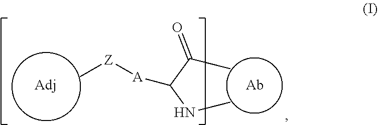
or a pharmaceutically acceptable salt thereof, wherein
Ab is the antibody with

being an amino acid residue of the antibody, wherein A is a modified or unmodified sidechain of the amino acid residue,
Z is the linker,
Adj is the adjuvant moiety, and
subscript r is an integer from 1 to 10.
14. The immunoconjugate of claim 4 , wherein the immunoconjugate has a structure according to Formula II:

or a pharmaceutically acceptable salt thereof, wherein
Ab is the antibody with

being an amino acid residue of the antibody, wherein A is a lysine sidechain, thiol-modified lysine sidechain, or cysteine sidechain of the amino acid residue,
Adj is the adjuvant moiety,
subscript r is an integer 1 to 10,
Z1 is selected from —C(O)—, —C(O)NH—, and —CH2—,
Z2 and Z4 are each independently selected from a bond, C1-30 alkylene, and 3- to 30-membered heteroalkylene, wherein
one or more groupings of adjacent atoms in the C1-30 alkylene and 3- to 30-membered heteroalkylene are optionally and independently replaced by —C(O)—, —NRaC(O)—, or —C(O)NRa—, wherein each Ra is independently selected from H and C1-6 alkyl,
one or more groupings of adjacent atoms in the C1-30 alkylene and 3- to 30-membered heteroalkylene are optionally and independently replaced by a 4- to 8-membered divalent carbocycle, and
one or more groupings of adjacent atoms in the C1-30 alkylene and 3- to 30-membered heteroalkylene are optionally and independently replaced by a 4- to 8-membered divalent heterocycle having one to four heteroatoms selected from O, S, and N,
Z3 is a bond, a divalent peptide moiety, or a divalent polymer moiety, and
Z5 is selected from an amine-bonded moiety and a thiol-bonded moiety.
15. The immunoconjugate of claim 4 , wherein the immunoconjugate has a structure according to Formula III:

or a pharmaceutically acceptable salt thereof, wherein
Ab is the antibody with

being a lysine residue of the antibody,
Adj is the adjuvant moiety,
G is CH2, C═O, or a bond,
L is a linking moiety, and
subscript r is an integer from 1 to 10.
16. The immunoconjugate of claim 15 , wherein L is selected from:

wherein R is optionally present and is a linear or branched, cyclic or straight, saturated or unsaturated alkyl, heteroalkyl, aryl, or heteroaryl chain comprising from 1 to 8 carbons, a is an integer from 1 to 40, each A is independently selected from any amino acid, subscript c is an integer from 1 to 20, the dashed line (“ ”) represents the point of attachment to
and the wavy line (“ ”) represents the point of attachment to

17. The immunoconjugate of claim 15 , wherein L is:

a is an integer from 1 to 40; the dashed line (“ ”) represents the point of attachment to
and the wavy line (“ ”) represents the point of attachment to

wherein each adjuvant moiety is covalently bonded to the antibody via the linker.
18. The immunoconjugate of claim 17 , wherein G is a bond.
19. The immunoconjugate of claim 18 , wherein a is an integer from 1 to 10.
20. The immunoconjugate of claim 19 , wherein is r is an integer from 1 to 5.
21. The immunoconjugate of claim 18 , wherein is r is an integer from 1 to 5.
22. A composition comprising a plurality of immunoconjugates according to claim 1 and one or more pharmaceutically acceptable excipients.
23. The composition of claim 22 , wherein the average number of adjuvant moieties per immunoconjugate ranges from about 1 to about 4.
24. A composition comprising a plurality of immunoconjugates according to claim 2 and one or more pharmaceutically acceptable excipients.
25. The composition of claim 24 , wherein the average number of adjuvant moieties per immunoconjugate ranges from about 1 to about 4.
26. A composition comprising a plurality of immunoconjugates according to claim 4 and one or more pharmaceutically acceptable excipients.
27. The composition of claim 26 , wherein the average number of adjuvant moieties per immunoconjugate ranges from about 1 to about 4.
28. A composition comprising a plurality of immunoconjugates according to claim 18 and one or more pharmaceutically acceptable excipients.
29. The composition of claim 28 , wherein the average number of adjuvant moieties per immunoconjugate ranges from about 1 to about 4.
30. A composition comprising a plurality of immunoconjugates according to claim 21 and one or more pharmaceutically acceptable excipients.
31. The composition of claim 30 , wherein the average number of adjuvant moieties per immunoconjugate ranges from about 1 to about 4.
32. A method for treating a HER2 expressing cancer comprising administering a therapeutically effective amount of an immunoconjugate according to claim 2 to a subject in need thereof.
33. The method of claim 32 , wherein the cancer is liver cancer, gall bladder cancer, or bile duct cancer.
34. The method of claim 32 , wherein the cancer is cervical cancer.
35. The method of claim 32 , wherein the cancer is esophageal cancer.
36. The method of claim 32 , wherein the cancer is melanoma.
37. The method of claim 32 , wherein the cancer is breast cancer.
38. The method of claim 32 , wherein the cancer is gastric cancer.
39. The method of claim 32 , wherein the cancer is ovarian cancer.
40. The method of claim 32 , wherein the cancer is uterine cancer.
41. The method of claim 32 , wherein the cancer is bladder cancer.
42. The method of claim 32 , wherein the cancer is lung cancer.
43. The method of claim 32 , wherein the cancer is head and neck cancer.
44. The method of claim 32 , wherein the cancer is colorectal cancer.
45. A method for treating a HER2 expressing cancer comprising administering a therapeutically effective amount of an immunoconjugate according to claim 4 to a subject in need thereof.
46. The method of claim 45 , wherein the cancer is liver cancer, gall bladder cancer, or bile duct cancer.
47. The method of claim 45 , wherein the cancer is cervical cancer.
48. The method of claim 45 , wherein the cancer is esophageal cancer.
49. The method of claim 45 , wherein the cancer is melanoma.
50. The method of claim 45 , wherein the cancer is breast cancer.
51. The method of claim 45 , wherein the cancer is gastric cancer.
52. The method of claim 45 , wherein the cancer is ovarian cancer.
53. The method of claim 45 , wherein the cancer is uterine cancer.
54. The method of claim 45 , wherein the cancer is bladder cancer.
55. The method of claim 45 , wherein the cancer is lung cancer.
56. The method of claim 45 , wherein the cancer is head and neck cancer.
57. The method of claim 45 , wherein the cancer is colorectal cancer.
58. A method for treating a HER2 expressing cancer comprising administering a therapeutically effective amount of an immunoconjugate according to claim 18 to a subject in need thereof.
59. The method of claim 58 , wherein the cancer is liver cancer, gall bladder cancer, or bile duct cancer.
60. The method of claim 58 , wherein the cancer is cervical cancer.
61. The method of claim 58 , wherein the cancer is esophageal cancer.
62. The method of claim 58 , wherein the cancer is melanoma.
63. The method of claim 58 , wherein the cancer is breast cancer.
64. The method of claim 58 , wherein the cancer is gastric cancer.
65. The method of claim 58 , wherein the cancer is ovarian cancer.
66. The method of claim 58 , wherein the cancer is uterine cancer.
67. The method of claim 58 , wherein the cancer is bladder cancer.
68. The method of claim 58 , wherein the cancer is lung cancer.
69. The method of claim 58 , wherein the cancer is head and neck cancer.
70. The method of claim 58 , wherein the cancer is colorectal cancer.
71. A method for treating a HER2 expressing cancer comprising administering a therapeutically effective amount of an immunoconjugate according to claim 21 to a subject in need thereof.
72. The method of claim 71 , wherein the cancer is liver cancer, gall bladder cancer, or bile duct cancer.
73. The method of claim 71 , wherein the cancer is cervical cancer.
74. The method of claim 71 , wherein the cancer is esophageal cancer.
75. The method of claim 71 , wherein the cancer is melanoma.
76. The method of claim 71 , wherein the cancer is breast cancer.
77. The method of claim 71 , wherein the cancer is gastric cancer.
78. The method of claim 71 , wherein the cancer is ovarian cancer.
79. The method of claim 71 , wherein the cancer is uterine cancer.
80. The method of claim 71 , wherein the cancer is bladder cancer.
81. The method of claim 71 , wherein the cancer is lung cancer.
82. The method of claim 71 , wherein the cancer is head and neck cancer.
83. The method of claim 71 , wherein the cancer is colorectal cancer.
84. A method for treating a HER2 expressing cancer comprising administering a therapeutically effective amount of a composition according to claim 24 to a subject in need thereof.
85. The method of claim 84 , wherein the cancer is liver cancer, gall bladder cancer, bile duct cancer, cervical cancer, esophageal cancer, melanoma, breast cancer, gastric cancer, ovarian cancer, uterine cancer, bladder cancer, lung cancer, head and neck cancer, or colorectal cancer.
86. A method for treating a HER2 expressing cancer comprising administering a therapeutically effective amount of a composition according to claim 26 to a subject in need thereof.
87. The method of claim 86 , wherein the cancer is liver cancer, gall bladder cancer, bile duct cancer, cervical cancer, esophageal cancer, melanoma, breast cancer, gastric cancer, ovarian cancer, uterine cancer, bladder cancer, lung cancer, head and neck cancer, or colorectal cancer.
88. A method for treating a HER2 expressing cancer comprising administering a therapeutically effective amount of a composition according to claim 28 to a subject in need thereof.
89. The method of claim 88 , wherein the cancer is liver cancer, gall bladder cancer, bile duct cancer, cervical cancer, esophageal cancer, melanoma, breast cancer, gastric cancer, ovarian cancer, uterine cancer, bladder cancer, lung cancer, head and neck cancer, or colorectal cancer.
90. A method for treating a HER2 expressing cancer comprising administering a therapeutically effective amount of a composition according to claim 30 to a subject in need thereof.
91. The method of claim 90 , wherein the cancer is liver cancer, gall bladder cancer, bile duct cancer, cervical cancer, esophageal cancer, melanoma, breast cancer, gastric cancer, ovarian cancer, uterine cancer, bladder cancer, lung cancer, head and neck cancer, or colorectal cancer.
Priority Applications (2)
| Application Number | Priority Date | Filing Date | Title |
|---|---|---|---|
| US17/466,873 US11547761B1 (en) | 2016-07-07 | 2021-09-03 | Antibody adjuvant conjugates |
| US17/818,176 US20230111340A1 (en) | 2016-07-07 | 2022-08-08 | Antibody adjuvant conjugates |
Applications Claiming Priority (10)
| Application Number | Priority Date | Filing Date | Title |
|---|---|---|---|
| US201662359627P | 2016-07-07 | 2016-07-07 | |
| US201662359626P | 2016-07-07 | 2016-07-07 | |
| US201662432530P | 2016-12-09 | 2016-12-09 | |
| US201662433742P | 2016-12-13 | 2016-12-13 | |
| US201762522623P | 2017-06-20 | 2017-06-20 | |
| US201762526306P | 2017-06-28 | 2017-06-28 | |
| PCT/US2017/041268 WO2018009916A1 (en) | 2016-07-07 | 2017-07-07 | Antibody adjuvant conjugates |
| US16/140,309 US10675358B2 (en) | 2016-07-07 | 2018-09-24 | Antibody adjuvant conjugates |
| US16/817,864 US11110178B2 (en) | 2016-07-07 | 2020-03-13 | Antibody adjuvant conjugates |
| US17/466,873 US11547761B1 (en) | 2016-07-07 | 2021-09-03 | Antibody adjuvant conjugates |
Related Parent Applications (1)
| Application Number | Title | Priority Date | Filing Date |
|---|---|---|---|
| US16/817,864 Continuation US11110178B2 (en) | 2015-11-02 | 2020-03-13 | Antibody adjuvant conjugates |
Related Child Applications (1)
| Application Number | Title | Priority Date | Filing Date |
|---|---|---|---|
| US17/818,176 Continuation US20230111340A1 (en) | 2016-07-07 | 2022-08-08 | Antibody adjuvant conjugates |
Publications (1)
| Publication Number | Publication Date |
|---|---|
| US11547761B1 true US11547761B1 (en) | 2023-01-10 |
Family
ID=60912313
Family Applications (4)
| Application Number | Title | Priority Date | Filing Date |
|---|---|---|---|
| US16/140,309 Active US10675358B2 (en) | 2016-07-07 | 2018-09-24 | Antibody adjuvant conjugates |
| US16/817,864 Active US11110178B2 (en) | 2015-11-02 | 2020-03-13 | Antibody adjuvant conjugates |
| US17/466,873 Active 2037-08-11 US11547761B1 (en) | 2016-07-07 | 2021-09-03 | Antibody adjuvant conjugates |
| US17/818,176 Pending US20230111340A1 (en) | 2016-07-07 | 2022-08-08 | Antibody adjuvant conjugates |
Family Applications Before (2)
| Application Number | Title | Priority Date | Filing Date |
|---|---|---|---|
| US16/140,309 Active US10675358B2 (en) | 2016-07-07 | 2018-09-24 | Antibody adjuvant conjugates |
| US16/817,864 Active US11110178B2 (en) | 2015-11-02 | 2020-03-13 | Antibody adjuvant conjugates |
Family Applications After (1)
| Application Number | Title | Priority Date | Filing Date |
|---|---|---|---|
| US17/818,176 Pending US20230111340A1 (en) | 2016-07-07 | 2022-08-08 | Antibody adjuvant conjugates |
Country Status (10)
| Country | Link |
|---|---|
| US (4) | US10675358B2 (en) |
| EP (1) | EP3484518A4 (en) |
| JP (3) | JP7011764B2 (en) |
| KR (2) | KR20230149857A (en) |
| CN (2) | CN115317603A (en) |
| AU (1) | AU2017292934B2 (en) |
| BR (1) | BR112019000071A2 (en) |
| CA (1) | CA3029902A1 (en) |
| IL (1) | IL263616A (en) |
| WO (1) | WO2018009916A1 (en) |
Families Citing this family (21)
| Publication number | Priority date | Publication date | Assignee | Title |
|---|---|---|---|---|
| EP3484518A4 (en) | 2016-07-07 | 2020-04-22 | The Board of Trustees of the Leland Stanford Junior University | Antibody adjuvant conjugates |
| CA3046790A1 (en) * | 2016-12-13 | 2018-06-21 | Bolt Biotherapeutics, Inc. | Antibody adjuvant conjugates |
| CA3100544A1 (en) * | 2018-05-17 | 2019-11-21 | Bolt Biotherapeutics, Inc. | Immunoconjugates |
| JP2022513306A (en) * | 2018-08-29 | 2022-02-07 | ボルト バイオセラピューティクス、インコーポレーテッド | Immune conjugates that target EGFR |
| CA3111784A1 (en) | 2018-09-12 | 2020-03-19 | Silverback Therapeutics, Inc. | Methods and compositions for the treatment of disease with immune stimulatory conjugates |
| KR20210136014A (en) * | 2019-02-12 | 2021-11-16 | 암브룩스, 인코포레이티드 | Compositions, methods and uses thereof containing antibody-TLR agonist conjugates |
| US20220143012A1 (en) * | 2019-03-15 | 2022-05-12 | Bolt Biotherapeutics, Inc. | Macromolecule-Supported TLR Agonists |
| WO2020190760A1 (en) * | 2019-03-15 | 2020-09-24 | Bolt Biotherapeutics, Inc. | Immunoconjugates targeting cea |
| US20220226491A1 (en) * | 2019-03-15 | 2022-07-21 | Bolt Biotherapeutics, Inc. | Immunoconjugates Targeting PD-L1 |
| KR20220004634A (en) | 2019-03-15 | 2022-01-11 | 볼트 바이오테라퓨틱스 인코퍼레이티드 | Immunoconjugates targeting HER2 |
| US20220226492A1 (en) * | 2019-03-15 | 2022-07-21 | Bolt Biotherapeutics, Inc. | Immunoconjugates Targeting HER2 |
| AU2020358726A1 (en) | 2019-10-01 | 2022-04-07 | Silverback Therapeutics, Inc. | Combination therapy with immune stimulatory conjugates |
| JP2023514727A (en) | 2020-02-21 | 2023-04-07 | シルバーバック セラピューティックス インコーポレイテッド | Nectin-4 antibody conjugates and uses thereof |
| AU2021226570A1 (en) * | 2020-02-25 | 2022-09-29 | Bolt Biotherapeutics, Inc. | Cancer treatment methods |
| WO2021245130A1 (en) | 2020-06-03 | 2021-12-09 | Ascendis Pharma Oncology Division A/S | Il-2 sequences and uses thereof |
| WO2022006327A1 (en) | 2020-07-01 | 2022-01-06 | Silverback Therapeutics, Inc. | Anti-asgr1 antibody conjugates and uses thereof |
| US20230263903A1 (en) * | 2020-08-13 | 2023-08-24 | Bolt Biotherapeutics, Inc. | Pyrazoloazepine immunoconjugates, and uses thereof |
| KR20230057447A (en) | 2020-08-28 | 2023-04-28 | 아센디스 파마 온콜로지 디비전 에이/에스 | Glycosylated IL-2 Proteins and Uses Thereof |
| IL303350A (en) | 2020-12-04 | 2023-08-01 | Macrogenics Inc | Pharmaceutical compositions of a her2/neu antibody and use of the same |
| WO2023076599A1 (en) | 2021-10-29 | 2023-05-04 | Bolt Biotherapeutics, Inc. | Tlr agonist immunoconjugates with cysteine-mutant antibodies, and uses thereof |
| WO2024030577A1 (en) * | 2022-08-03 | 2024-02-08 | Seagen Inc. | Immunostimulatory anti-pd-l1-drug conjugates |
Citations (366)
| Publication number | Priority date | Publication date | Assignee | Title |
|---|---|---|---|---|
| GB2097790A (en) | 1981-04-24 | 1982-11-10 | Delalande Sa | Piperidine piperazine and homopiperazine derivatives |
| US4689338A (en) | 1983-11-18 | 1987-08-25 | Riker Laboratories, Inc. | 1H-Imidazo[4,5-c]quinolin-4-amines and antiviral use |
| WO1990014844A2 (en) | 1989-06-06 | 1990-12-13 | Neorx Corporation | Sugars as cleavable linkers for the delivery and release of agents in native form |
| US6069149A (en) | 1997-01-09 | 2000-05-30 | Terumo Kabushiki Kaisha | Amide derivatives and intermediates for the synthesis thereof |
| WO2000075605A1 (en) | 1999-06-03 | 2000-12-14 | R. Rouvari Oy | System for measuring loadings in a structure, measuring unit and measuring sensor |
| WO2000076518A1 (en) | 1999-06-10 | 2000-12-21 | 3M Innovative Properties Company | Urea substituted imidazoquinolines |
| WO2000076519A1 (en) | 1999-06-10 | 2000-12-21 | 3M Innovative Properties Company | Sulfonamide and sulfamide substituted imidazoquinolines |
| WO2001097843A2 (en) | 2000-06-22 | 2001-12-27 | University Of Iowa Research Foundation | Methods for enhancing antibody-induced cell lysis and treating cancer |
| WO2002046749A2 (en) | 2000-12-08 | 2002-06-13 | 3M Innovative Properties Company | Screening method for identifying compounds that selectively induce interferon alpha |
| US6451810B1 (en) | 1999-06-10 | 2002-09-17 | 3M Innovative Properties Company | Amide substituted imidazoquinolines |
| US20020146388A1 (en) | 1990-11-09 | 2002-10-10 | Gillies Stephen D. | Cytokine immunoconjugates |
| US20020155108A1 (en) | 1998-05-04 | 2002-10-24 | Biocrystal, Ltd. | Method for ex vivo loading of antigen presenting cells with antigen, and a vaccine comprising the loaded cells |
| WO2003043572A2 (en) | 2001-11-16 | 2003-05-30 | 3M Innovative Properties Company | Methods and compositions related to irm compounds and toll-like receptor pathways |
| US6573273B1 (en) | 1999-06-10 | 2003-06-03 | 3M Innovative Properties Company | Urea substituted imidazoquinolines |
| US20030144283A1 (en) | 1999-06-10 | 2003-07-31 | Coleman Patrick L. | Amide substituted imidazoquinolines |
| US6677349B1 (en) | 2001-12-21 | 2004-01-13 | 3M Innovative Properties Company | Sulfonamide and sulfamide substituted imidazoquinolines |
| US20040038294A1 (en) | 2002-08-20 | 2004-02-26 | Quest Diagnostic Investments Incorporated | Hydrophilic chemilumescent acridinium labeling reagents |
| WO2004028539A2 (en) | 2002-09-26 | 2004-04-08 | 3M Innovative Properties Company | 1h-imidazo dimers |
| US20040091491A1 (en) | 2002-08-15 | 2004-05-13 | 3M Innovative Properties Company | Immunostimulatory compositions and methods of stimulating an immune response |
| WO2004053057A2 (en) | 2002-12-11 | 2004-06-24 | 3M Innovative Properties Company | Gene expression systems and recombinant cell lines |
| WO2004053452A2 (en) | 2002-12-11 | 2004-06-24 | 3M Innovative Properties Company | Assays relating to toll-like receptor activity |
| US20040131587A1 (en) | 2000-10-24 | 2004-07-08 | Thomas Elaine K. | Method for treatment of tumors using combination therapy |
| WO2004058759A1 (en) | 2002-12-20 | 2004-07-15 | 3M Innovative Properties Company | Aryl / hetaryl substituted imidazoquinolines |
| US20040171086A1 (en) | 2003-02-27 | 2004-09-02 | 3M Innovative Properties Company | Selective modulation of TLR-mediated biological activity |
| US20040191833A1 (en) | 2003-03-25 | 2004-09-30 | 3M Innovative Properties Company | Selective activation of cellular activities mediated through a common toll-like receptor |
| US20040202720A1 (en) | 2003-04-10 | 2004-10-14 | 3M Innovative Properties Company | Delivery of immune response modifier compounds using metal-containing particulate support materials |
| US20040214851A1 (en) | 2003-04-28 | 2004-10-28 | 3M Innovative Properties Company | Compositions and methods for induction of opioid receptors |
| US20040265351A1 (en) | 2003-04-10 | 2004-12-30 | Miller Richard L. | Methods and compositions for enhancing immune response |
| WO2005001022A2 (en) | 2003-04-10 | 2005-01-06 | 3M Innovative Properties Company | Methods and compositions for enhancing immune response |
| WO2005003065A2 (en) | 2003-06-27 | 2005-01-13 | 3M Innovative Properties Company | Sulfonamide substituted imidazoquinolines |
| WO2005016275A2 (en) | 2003-08-05 | 2005-02-24 | 3M Innovative Properties Company | Formulations containing an immune response modifier |
| WO2005018555A2 (en) | 2003-08-14 | 2005-03-03 | 3M Innovative Properties Company | Lipid-modified immune response modifiers |
| WO2005020999A1 (en) | 2003-08-27 | 2005-03-10 | 3M Innovative Properties Company | Aryloxy and arylalkyleneoxy substituted imidazoquinolines |
| US20050113297A1 (en) | 2003-08-22 | 2005-05-26 | Potentia Pharmaceuticals, Inc. | Compositions and methods for enhancing phagocytosis or phagocyte activity |
| WO2005051324A2 (en) | 2003-11-25 | 2005-06-09 | 3M Innovative Properties Company | Hydroxylamine and oxime substituted imidazoquinolines, imidazopyridines, and imidazonaphthyridines |
| US20050158325A1 (en) | 2003-12-30 | 2005-07-21 | 3M Innovative Properties Company | Immunomodulatory combinations |
| WO2005094531A2 (en) | 2004-03-24 | 2005-10-13 | 3M Innovative Properties Company | Amide substituted imidazopyridines, imidazoquinolines, and imidazonaphthyridines |
| WO2005110013A2 (en) | 2004-04-09 | 2005-11-24 | 3M Innovative Properties Company | Methods, compositions, and preparations for delivery of immune response modifiers |
| WO2005123080A2 (en) | 2004-06-15 | 2005-12-29 | 3M Innovative Properties Company | Nitrogen-containing heterocyclyl substituted imidazoquinolines and imidazonaphthyridines |
| US20060018911A1 (en) | 2004-01-12 | 2006-01-26 | Dana Ault-Riche | Design of therapeutics and therapeutics |
| WO2006028545A2 (en) | 2004-06-18 | 2006-03-16 | 3M Innovative Properties Company | Substituted imidazoquinolines, imidazopyridines, and imidazonaphthyridines |
| US20060142202A1 (en) | 2000-12-08 | 2006-06-29 | 3M Innovative Properties Company | Compositions and methods for targeted delivery of immune response modifiers |
| WO2006074003A2 (en) | 2004-12-30 | 2006-07-13 | 3M Innovative Properties Company | CHIRAL FUSED [1,2]IMIDAZO[4,5-c] RING COMPOUNDS |
| WO2006091394A2 (en) | 2005-02-11 | 2006-08-31 | Coley Pharmaceutical Group, Inc. | Substituted imidazoquinolines and imidazonaphthyridines |
| WO2007048122A2 (en) | 2005-10-21 | 2007-04-26 | Genzyme Corporation | Antibody-based therapeutics with enhanced adcc activity |
| WO2007100634A2 (en) | 2006-02-22 | 2007-09-07 | 3M Innovative Properties Company | Immune response modifier conjugates |
| WO2008009894A2 (en) | 2006-07-15 | 2008-01-24 | Summit Corporation Plc | Use of imino sugars in immunotherapy |
| US20080031887A1 (en) | 2006-06-30 | 2008-02-07 | Joseph Lustgarten | Conjugates for inducing targeted immune responses and methods of making and using same |
| US20080031900A1 (en) | 2006-06-30 | 2008-02-07 | Baylor Research Institute | Dendritic Cells Generated Using GM-CSF and Interferon Alpha and Loaded with Heat-Treated and Killed Cancer Cells |
| US7375180B2 (en) | 2003-02-13 | 2008-05-20 | 3M Innovative Properties Company | Methods and compositions related to IRM compounds and Toll-like receptor 8 |
| US20080233140A1 (en) | 2007-02-23 | 2008-09-25 | Baylor Research Institute | Therapeutic Applications of Activation of Human Antigen-Presenting Cells Through Dectin-1 |
| US20080254047A1 (en) | 2007-02-23 | 2008-10-16 | Baylor Research Institute | Activation of Human Antigen-Presenting Cells Through CLEC-6 |
| US20090004192A1 (en) | 2007-03-01 | 2009-01-01 | Mikkel Wandahl Pedersen | Recombinant anti-epidermal growth factor receptor antibody compositions |
| US20090004194A1 (en) | 2006-03-01 | 2009-01-01 | Regents Of The University Of Colorado | Tlr agonist (flagellin)/cd40 agonist/antigen protein and dna conjugates and use thereof for inducing synergistic enhancement in immunity |
| US20090123467A1 (en) | 2007-07-31 | 2009-05-14 | The Johns Hopkins University | Polypeptide-Nucleic Acid Conjugate for Immunoprophylaxis or Immunotherapy for Neoplastic or Infectious Disorders |
| US20090155289A1 (en) | 2007-11-01 | 2009-06-18 | Steve Roberts | Furin-cleavable peptide linkers for drug-ligand conjugates |
| WO2009085262A1 (en) | 2007-12-27 | 2009-07-09 | Celgene Corporation | Method of treating cancer with immunomodulatory compounds and igg |
| US20090181022A1 (en) | 2004-05-05 | 2009-07-16 | Merrimack Pharmaceuticals, Inc. | Bispecific binding agents for modulating biological activity |
| US20090325315A1 (en) | 2008-06-30 | 2009-12-31 | Arkray, Inc. | Detection method of target substance, detection reagent used for the same, and the uses thereof |
| US20100004156A1 (en) | 2005-07-27 | 2010-01-07 | Shalesh Kaushal | Small Compounds That Correct Protein Misfolding and Uses Thereof |
| US20100129383A1 (en) | 2008-10-03 | 2010-05-27 | The Governors Of The University Of Alberta | Bifunctional fusion molecules for the delivery of antigens to professional antigen-presenting cells |
| US20100158928A1 (en) | 2006-12-22 | 2010-06-24 | Doris Stoermer | Immune response modifier compositions and methods |
| WO2010132622A2 (en) | 2009-05-14 | 2010-11-18 | The Regents Of The University Of California | Anticd20-cpg conjugates and methods of treating b cell malignancies |
| US20100317111A1 (en) | 2006-05-03 | 2010-12-16 | Ross Kedl | Cd40 agonist antibody/type 1 interferon synergistic adjuvant combination, conjugates containing and use thereof as a therapeutic to enhance cellular immunity |
| WO2011022509A2 (en) | 2009-08-18 | 2011-02-24 | Ventirx Pharmaceuticals, Inc. | Substituted benzoazepines as toll-like receptor modulators |
| WO2011022508A2 (en) | 2009-08-18 | 2011-02-24 | Ventirx Pharmaceuticals, Inc. | Substituted benzoazepines as toll-like receptor modulators |
| US20110064752A1 (en) | 2009-09-09 | 2011-03-17 | Centrose, Llc | Extracellular targeted drug conjugates |
| US20110182847A1 (en) | 2007-06-15 | 2011-07-28 | Randolph Noelle | Use of tlr agonists and/or type 1 interferons to alleviate toxicity of tnf-r agonist therapeutic regimens |
| US20110274692A1 (en) | 2009-09-25 | 2011-11-10 | Xoma Technology Ltd. | Screening Methods |
| US20110274685A1 (en) | 2010-04-13 | 2011-11-10 | Celldex Therapeutics Inc. | Antibodies that bind human cd27 and uses thereof |
| US20110274653A1 (en) | 2010-05-07 | 2011-11-10 | Baylor Research Institute | Dendritic cell immunoreceptors (dcir)-mediated crosspriming of human cd8+ t cells |
| US20120034213A1 (en) | 1997-12-12 | 2012-02-09 | Hellmann Susan D | TREATMENT WITH ANTI-ErbB2 ANTIBODIES |
| WO2012021834A1 (en) | 2010-08-13 | 2012-02-16 | Baylor Research Institute | Novel vaccine adjuvants based on targeting adjuvants to antibodies directly to antigen-presenting cells |
| US20120045414A1 (en) | 2006-10-31 | 2012-02-23 | Delucia Dave | Adjuvant combinations comprising a microbial tlr agonist, a cd40 or 4-1bb agonist, and optionally an antigen and the use thereof for inducing a synergistic enhancement in cellular immunity |
| US20120064593A1 (en) | 1998-05-04 | 2012-03-15 | Innexus Biotechnology International Ltd. | Superantibody synthesis and use in detection, prevention and treatment of disease |
| WO2012092552A1 (en) | 2010-12-30 | 2012-07-05 | Selecta Biosciences, Inc. | Synthetic nanocarriers with reactive groups that release biologically active agents |
| US20120213771A1 (en) | 2010-04-13 | 2012-08-23 | Celldex Therapeutics Inc. | Antibodies that bind human cd27 and uses thereof |
| US20120219615A1 (en) | 2010-10-01 | 2012-08-30 | The Trustees Of The University Of Pennsylvania | Therapeutic Use of a TLR Agonist and Combination Therapy |
| US20120231023A1 (en) | 2011-03-08 | 2012-09-13 | Baylor Research Institute | Novel Vaccine Adjuvants Based on Targeting Adjuvants to Antibodies Directly to Antigen-Presenting Cells |
| US20120237518A1 (en) | 2010-06-10 | 2012-09-20 | Kyowa Hakko Kirin Co., Ltd | Anti-trop-2 antibody |
| US8277810B2 (en) | 2003-11-04 | 2012-10-02 | Novartis Vaccines & Diagnostics, Inc. | Antagonist anti-CD40 antibodies |
| US20120301465A1 (en) | 2011-03-25 | 2012-11-29 | Institut National De La Sante Et De La Recherche Medicale (Inserm) | Compositions and methods to immunize against hepatitis c virus |
| WO2012170072A1 (en) | 2011-06-06 | 2012-12-13 | Immungene, Inc. | Engineered antibody-tnfsf member ligand fusion molecules |
| US20120328605A1 (en) | 2010-10-27 | 2012-12-27 | Daniel Larocque | Compositions and uses |
| US20130039911A1 (en) | 2010-03-05 | 2013-02-14 | Atul Bedi | Compositions and Methods for Targeted Immunomodulatory Antibodies and Fusion Proteins |
| WO2013030378A1 (en) | 2011-09-01 | 2013-03-07 | Novartis Ag | Adjuvanted formulations of staphylococcus aureus antigens |
| EP1689846B1 (en) | 2003-10-10 | 2013-03-27 | ImmunoGen, Inc. | Conjugates of maytansinoid DM1 with antibody trastuzumab, linked through a non-cleavable linker, and its use in the treatment of tumours |
| US8435800B2 (en) | 2004-11-22 | 2013-05-07 | Biosight Ltd. | Activated labeling reagents and methods for preparing and using the same |
| WO2013067597A1 (en) | 2011-11-09 | 2013-05-16 | Ascend Biopharmaceuticals Pty Ltd | Immunomodulatory conjugates |
| US20130165455A1 (en) | 2005-08-22 | 2013-06-27 | The Regents Of The University Of California | Tlr agonists |
| US8481029B2 (en) | 2006-10-20 | 2013-07-09 | University Of Southampton | Human immune therapies using a CD27 agonist alone or in combination with other immune modulators |
| WO2013105013A1 (en) | 2012-01-09 | 2013-07-18 | Covx Technologies Ireland Limited | Mutant antibodies and conjugation thereof |
| US20130195794A1 (en) | 2010-02-26 | 2013-08-01 | Adjuvantix Limited | Cancer vaccine |
| US20130202596A1 (en) | 2010-07-09 | 2013-08-08 | Biogen Idec Hemophilia Inc. | Processable Single Chain Molecules and Polypeptides Made Using Same |
| US8518405B2 (en) | 2009-10-08 | 2013-08-27 | The University Of North Carolina At Charlotte | Tumor specific antibodies and uses therefor |
| US8574575B2 (en) | 2002-03-01 | 2013-11-05 | Immunomedics, Inc. | RS7 antibodies |
| WO2013166110A1 (en) | 2012-05-02 | 2013-11-07 | Yale University | Tlr-agonist-conjugated antibody recruiting molecules (tlr_arms) |
| WO2013180811A1 (en) | 2012-05-30 | 2013-12-05 | Life Technologies Corporation | FLUOROGENIC pH-SENSITIVE DYES AND THEIR METHODS OF USE |
| US20130330350A1 (en) | 2010-11-09 | 2013-12-12 | Medimmune, Llc | Antibody Scaffold For Homogenous Conjugation |
| EP2674170A1 (en) | 2012-06-15 | 2013-12-18 | Cayla | Novel compositions of TLR7 and/or TLR8 agonists conjugated to lipids |
| WO2014012479A1 (en) | 2012-07-18 | 2014-01-23 | Shanghai Birdie Biotech, Inc. | Compounds for targeted immunotherapy |
| US20140065096A1 (en) | 2012-09-05 | 2014-03-06 | Regen BioPharma, Inc. | Cancer therapy by ex vivo activated autologous immune cells |
| US8709418B2 (en) | 2010-02-04 | 2014-04-29 | Toray Industries, Inc. | Pharmaceutical composition for treating CAPRIN-1 expressing cancer |
| US8728486B2 (en) | 2011-05-18 | 2014-05-20 | University Of Kansas | Toll-like receptor-7 and -8 modulatory 1H imidazoquinoline derived compounds |
| US8741291B2 (en) | 2010-07-12 | 2014-06-03 | Covx Technologies Ireland, Limited | Multifunctional antibody conjugates |
| US20140179558A1 (en) | 2011-08-04 | 2014-06-26 | Toray Industries, Inc. | Method for detecting pancreatic cancer |
| US20140199293A1 (en) | 2009-04-17 | 2014-07-17 | Lpath, Inc. | Humanized antibody compositions and methods for binding lysophosphatidic acid |
| US8828398B2 (en) | 2010-02-04 | 2014-09-09 | Toray Industries, Inc. | Pharmaceutical composition for treating and/or preventing cancer |
| US8841417B2 (en) | 2010-05-14 | 2014-09-23 | Abbvie Inc. | IL-1 binding proteins |
| US20140294849A1 (en) | 2011-08-05 | 2014-10-02 | Glaxosmithkline Biologicals Sa | Compositions and uses |
| WO2014161887A1 (en) | 2013-04-02 | 2014-10-09 | Activartis Biotech Gmbh | Targeted cancer immune therapy |
| US8871908B2 (en) | 2011-11-11 | 2014-10-28 | Rinat Neuroscience Corp. | Antibodies specific for Trop-2 and their uses |
| WO2014177042A1 (en) | 2013-04-28 | 2014-11-06 | Qin Gang | Novel linker and preparation method thereof |
| US20140341978A1 (en) | 2011-09-19 | 2014-11-20 | The Johns Hopkins University | Cancer immunotherapy |
| US8911740B2 (en) | 2010-02-04 | 2014-12-16 | Toray Industries, Inc. | Pharmaceutical composition for treating and/or preventing cancer |
| US8937160B2 (en) | 2010-02-04 | 2015-01-20 | Toray Industries, Inc. | Pharmaceutical composition for treating and/or preventing cancer |
| US20150071948A1 (en) | 2003-09-26 | 2015-03-12 | Gregory Alan Lazar | Novel immunoglobulin variants |
| US20150158947A1 (en) | 2012-02-01 | 2015-06-11 | Compugen Ltd. | C10rf32 antibodies, and uses thereof for treatment of cancer |
| WO2015082905A1 (en) | 2013-12-02 | 2015-06-11 | Immune Targeting Systems (Its) Ltd | Immunogenic compound |
| WO2015095418A1 (en) | 2013-12-17 | 2015-06-25 | Genentech, Inc. | Methods of treating her2-positive cancers using pd-1 axis binding antagonists and anti-her2 antibodies |
| US20150191546A1 (en) | 2012-07-10 | 2015-07-09 | Board Of Regents, The University Of Texas System | Monoclonal antibodies for use in diagnosis and therapy of cancers and autoimmune disease |
| WO2015103989A1 (en) | 2014-01-10 | 2015-07-16 | Shanghai Birdie Biotech, Inc. | Compounds and compositions for immunotherapy |
| WO2015112749A2 (en) | 2014-01-22 | 2015-07-30 | The Board Of Trustees Of The Leland Stanford Junior University | Methods and compositions for antibody and antibody-loaded dendritic cell mediated therapy |
| US9115200B2 (en) | 2010-02-04 | 2015-08-25 | Toray Industries, Inc. | Pharmaceutical composition for treating cancer using a monoclonal antibody having immunological reactivity with CAPRIN-1 |
| WO2015143091A1 (en) | 2014-03-19 | 2015-09-24 | Genzyme Corporation | Site-specific glycoengineering of targeting moieties |
| WO2015151080A2 (en) | 2015-07-04 | 2015-10-08 | Suzhou M-Conj Biotech Co., Ltd | Specific conjugation of a cell-binding molecule |
| US20150284416A1 (en) | 2015-06-16 | 2015-10-08 | Suzhou M-Conj Biotech Co., Ltd | Novel linkers for conjugation of cell-binding molecules |
| WO2015151078A2 (en) | 2015-06-15 | 2015-10-08 | Suzhou M-Conj Biotech Co., Ltd | Hydrophilic linkers for conjugation |
| WO2015151081A2 (en) | 2015-07-12 | 2015-10-08 | Suzhou M-Conj Biotech Co., Ltd | Bridge linkers for conjugation of a cell-binding molecule |
| WO2015155753A2 (en) | 2015-08-10 | 2015-10-15 | Suzhou M-Conj Biotech Co., Ltd | Novel linkers and their uses in specific conjugation of drugs to a biological molecule |
| US20150299194A1 (en) | 2014-04-22 | 2015-10-22 | Hoffmann-La Roche Inc. | 4-amino-imidazoquinoline compounds |
| US9175074B2 (en) | 2011-08-04 | 2015-11-03 | Toray Industries, Inc. | Pharmaceutical composition for treatment and/or prophylaxis of cancer |
| WO2015165413A1 (en) | 2014-04-29 | 2015-11-05 | 秦刚 | New stable antibody-drug conjugate, preparation method therefor, and use thereof |
| US9181334B2 (en) | 2011-08-04 | 2015-11-10 | Toray Industries, Inc. | Pharmaceutical composition for treatment and/or prophylaxis of cancer |
| US9181348B2 (en) | 2011-08-04 | 2015-11-10 | Toray Industries, Inc. | Pharmaceutical composition for treatment and/or prophylaxis of cancer |
| US9180188B2 (en) | 2011-08-04 | 2015-11-10 | Toray Industries, Inc. | Pharmaceutical composition for treatment and/or prophylaxis of cancer |
| US20150322155A1 (en) | 2015-07-15 | 2015-11-12 | Sushou M-conj Biotech Co., Ltd | Acetylenedicarboxyl linkers and their uses in specific conjugation of a cell-binding molecule |
| US9192667B2 (en) | 2013-04-22 | 2015-11-24 | Hoffmann-La Roche Inc. | Method of treating cancer by administering CSF-1R antibodies and a TLR9 agonist |
| US9205148B2 (en) | 2011-04-20 | 2015-12-08 | Medimmune, Llc | Antibodies and other molecules that bind B7-H1 and PD-1 |
| WO2015187637A1 (en) | 2014-06-02 | 2015-12-10 | Baylor Research Institute | Methods and compositions for treating allergy and inflammatory diseases |
| WO2016004875A1 (en) | 2014-07-09 | 2016-01-14 | Shanghai Birdie Biotech, Inc. | Combination therapy compositions and methods for treating cancers |
| US20160008485A1 (en) | 2011-12-23 | 2016-01-14 | Pfizer Inc. | Engineered Antibody Constant Regions for Site-Specific Conjugation and Methods and Uses Therefor |
| WO2016004876A1 (en) | 2014-07-09 | 2016-01-14 | Shanghai Birdie Biotech, Inc. | Anti-pd-l1 combinations for treating tumors |
| US20160015821A1 (en) | 2014-02-21 | 2016-01-21 | Ecole Polytechnique Federale De Lausanne (Epfl) | Glycotargeting therapeutics |
| US20160015803A1 (en) | 2014-07-18 | 2016-01-21 | Ross Kedl | Immunostimulatory combinations and use thereof |
| US9243069B2 (en) | 2008-04-02 | 2016-01-26 | Macrogenics, Inc. | HER2/neu-specific antibodies and methods of using the same |
| US9248127B2 (en) | 2005-02-04 | 2016-02-02 | 3M Innovative Properties Company | Aqueous gel formulations containing immune response modifiers |
| US9260513B2 (en) | 2012-02-21 | 2016-02-16 | Toray Industries, Inc. | Pharmaceutical composition for treatment and/or prevention of cancer |
| US9266958B2 (en) | 2012-02-21 | 2016-02-23 | Toray Industries, Inc. | Pharmaceutical composition for treatment and/or prevention of cancer |
| US9273128B2 (en) | 2011-08-04 | 2016-03-01 | Toray Industries, Inc | Pharmaceutical composition for treatment and/or prophylaxis of cancer |
| US9273130B2 (en) | 2012-02-21 | 2016-03-01 | Toray Industries, Inc. | Pharmaceutical composition for treatment and/or prevention of cancer |
| WO2016032009A1 (en) | 2014-08-27 | 2016-03-03 | Kyushu University, National University Corporation | Adjuvant |
| US20160068533A1 (en) | 2014-09-08 | 2016-03-10 | Regents Of The University Of Minnesota | Immunomodulators and immunomodulator conjugates |
| WO2016034085A1 (en) | 2014-09-01 | 2016-03-10 | Birdie Biopharmaceuticals, Inc. | Anti-pd-l1 conjugates for treating tumors |
| US20160067351A1 (en) | 2013-02-08 | 2016-03-10 | Novartis Ag | Specific sites for modifying antibodies to make immunoconjugates |
| WO2016055812A1 (en) | 2014-10-10 | 2016-04-14 | Isis Innovation Limited | Polymer adjuvant |
| WO2016057618A1 (en) | 2014-10-09 | 2016-04-14 | Wake Forest University Health Sciences | Vaccine compositions and methods of use to treat neonatal subjects |
| US9314521B2 (en) | 2011-10-05 | 2016-04-19 | Isa Pharmaceuticals B.V. | Adjuvant compound |
| US20160108123A1 (en) | 2014-10-14 | 2016-04-21 | Novartis Ag | Antibody molecules to pd-l1 and uses thereof |
| US20160112466A1 (en) | 2010-02-19 | 2016-04-21 | Microsoft Technology Licensing, Llc | Distributed connectivity policy enforcement with ice |
| WO2016059622A2 (en) | 2016-02-04 | 2016-04-21 | Suzhou M-Conj Biotech Co., Ltd. | Specific conjugation linkers, specific immunoconjugates thereof, methods of making and uses such conjugates thereof |
| WO2016064749A2 (en) | 2014-10-20 | 2016-04-28 | Igenica Biotherapeutics, Inc. | Novel antibody-drug conjugates and related compounds, compositions, and methods of use |
| WO2016064899A1 (en) | 2014-10-21 | 2016-04-28 | The Board Of Trustees Of The Leland Stanford Junior University | Methods and compositions for antibody and antibody-loaded dendritic cell mediated therapy |
| US20160145350A1 (en) | 2014-11-21 | 2016-05-26 | Bristol-Myers Squibb Company | Antibodies against cd73 and uses thereof |
| WO2016085967A1 (en) | 2014-11-25 | 2016-06-02 | Endocyte, Inc. | Methods of treating cancer by targeting tumor-associated macrophages |
| US9358307B2 (en) | 2008-01-25 | 2016-06-07 | Gavish-Galilee Bio Applications Ltd. | Targeting of innate immune response to tumor site |
| US20160159901A1 (en) | 2013-07-31 | 2016-06-09 | Ganymed Pharmaceuticals Ag | Diagnosis and therapy of cancer involving cancer stem cells |
| US9382329B2 (en) | 2012-08-14 | 2016-07-05 | Ibc Pharmaceuticals, Inc. | Disease therapy by inducing immune response to Trop-2 expressing cells |
| US20160194399A1 (en) | 2014-07-25 | 2016-07-07 | Cytomx Therapeutics, Inc. | Anti-cd3 antibodies, activatable anti-cd3 antibodies, multispecific anti-cd3 antibodies, multispecific activatable anti-cd3 antibodies, and methods of using the same |
| US20160208021A1 (en) | 2015-01-16 | 2016-07-21 | Immunwork Inc. | Molecular constructs with targeting and effector moieties |
| WO2016118754A1 (en) | 2015-01-21 | 2016-07-28 | The Board Of Trustees Of The Leland Stanford Junior University | Macrophages eat cancer cells using their own calreticulin as a guide |
| US9409993B2 (en) | 2011-08-04 | 2016-08-09 | Toray Industries, Inc. | Pharmaceutical composition for treatment and/or prevention of pancreatic cancer |
| US9416192B2 (en) | 2008-08-05 | 2016-08-16 | Toray Industries, Inc. | Pharmaceutical composition for treatment and prevention of cancers |
| US9416193B2 (en) | 2012-03-30 | 2016-08-16 | Toray Industries, Inc. | Pharmaceutical composition for treatment and/or prevention of liver cancer |
| US9428581B2 (en) | 2012-03-30 | 2016-08-30 | Toray Industries, Inc. | Pharmaceutical composition for treatment and/or prevention of gallbladder cancer |
| WO2016141890A1 (en) | 2015-03-12 | 2016-09-15 | Sunshine Lake Pharma Co., Ltd. | Compounds as hepatitis c virus inhibitors and pharmaceutical uses thereof |
| WO2016149201A2 (en) | 2015-03-13 | 2016-09-22 | Cytomx Therapeutics, Inc. | Anti-pdl1 antibodies, activatable anti-pdl1 antibodies, and methods of use thereof |
| WO2016150899A2 (en) | 2015-03-23 | 2016-09-29 | Bayer Pharma Aktiengesellschaft | Anti-ceacam6 antibodies and uses thereof |
| WO2016161372A1 (en) | 2015-04-01 | 2016-10-06 | President And Fellows Of Harvard College | Immunoconjugates for programming or reprogramming of cells |
| US20160297889A1 (en) | 2013-08-09 | 2016-10-13 | Toray Industries, Inc. | Pharmaceutical composition for treatment and/or prevention of cancer |
| US9475804B2 (en) | 2011-06-03 | 2016-10-25 | 3M Innovative Properties Company | Heterobifunctional linkers with polyethylene glycol segments and immune response modifier conjugates made therefrom |
| US20160324981A1 (en) | 2015-05-08 | 2016-11-10 | The California Institute For Biomedical Research | Liver x receptor agonists and uses thereof |
| US9498541B2 (en) | 2008-04-30 | 2016-11-22 | Immunogen, Inc. | Cross-linkers and their uses |
| US20160340427A1 (en) | 2015-05-20 | 2016-11-24 | Immunwork Inc. | Molecular constructs for treating infectious diseases |
| WO2016187122A1 (en) | 2015-05-15 | 2016-11-24 | University Of Iowa Research Foundation | Methods for treating tumors in situ including intratumor injection of cytotoxic particles and immune checkpoint blockade therapy |
| WO2016187656A1 (en) | 2015-05-22 | 2016-12-01 | La Trobe University | Method of diagnosis of breast cancer |
| US20160347849A1 (en) | 2015-05-29 | 2016-12-01 | Bristol-Myers Squibb Company | Antibodies against ox40 and uses thereof |
| US20160355592A1 (en) | 2015-05-04 | 2016-12-08 | Cytomx Therapeutics, Inc. | Anti-itga3 antibodies, activatable anti-itga3 antibodies, and methods of use thereof |
| US20160355599A1 (en) | 2015-05-04 | 2016-12-08 | Cytomx Therapeutics, Inc. | Anti-cd71 antibodies, activatable anti-cd71 antibodies, and methods of use thereof |
| US20160355587A1 (en) | 2015-05-04 | 2016-12-08 | Cytomx Therapeutics, Inc. | Anti-cd166 antibodies, activatable anti-cd166 antibodies, and methods of use thereof |
| WO2016198470A2 (en) | 2015-06-08 | 2016-12-15 | Lophius Biosciences Gmbh | Composition for determination of cell-mediated immune responsiveness |
| US9522958B2 (en) | 2004-11-09 | 2016-12-20 | University Of Southern California | Cancer targeted innate immunity |
| WO2016203025A1 (en) | 2015-06-17 | 2016-12-22 | Curevac Ag | Vaccine composition |
| WO2016205551A2 (en) | 2015-06-16 | 2016-12-22 | The Regents Of The University Of California | Fzd7 specific antibodies and vaccines to treat cancer and control stem cell function |
| WO2016205566A1 (en) | 2015-06-16 | 2016-12-22 | The Regents Of The University Of California | Fzd7 specific antibodies and vaccines to treat cancer and control stem cell function |
| US20170021033A1 (en) | 2014-03-12 | 2017-01-26 | Novartis Ag | Specific sites for modifying antibodies to make immunoconjugates |
| WO2017019894A1 (en) | 2015-07-29 | 2017-02-02 | Novartis Ag | Combination therapies comprising antibody molecules to lag-3 |
| WO2017019896A1 (en) | 2015-07-29 | 2017-02-02 | Novartis Ag | Combination therapies comprising antibody molecules to pd-1 |
| WO2017019897A1 (en) | 2015-07-29 | 2017-02-02 | Novartis Ag | Combination therapies comprising antibody molecules to tim-3 |
| WO2017023779A1 (en) | 2015-07-31 | 2017-02-09 | Tarveda Therapeutics, Inc. | Compositions and methods for immuno-oncology therapies |
| WO2017021791A1 (en) | 2015-08-06 | 2017-02-09 | Glaxosmithkline Intellectual Property Development Limited | Combinations of an ox40 antibody and a tlr4 modulator and uses thereof |
| WO2017024296A1 (en) | 2015-08-06 | 2017-02-09 | Memorial Sloan Kettering Cancer Center | Methods and compositions for tumor therapy |
| US20170044259A1 (en) | 2015-07-13 | 2017-02-16 | Cytomx Therapeutics, Inc. | Anti-pd1 antibodies, activatable anti-pd1 antibodies, and methods of use thereof |
| US9573993B2 (en) | 2012-02-21 | 2017-02-21 | Toray Industries, Inc. | Pharmaceutical composition for treatment of cancer comprising an anti-CAPRIN-1 peptide antibody |
| WO2017040234A1 (en) | 2015-08-31 | 2017-03-09 | 3M Innovative Properties Company | IMIDAZO[4,5-c] RING COMPOUNDS CONTAINING SUBSTITUTED GUANIDINE GROUPS |
| WO2017040233A1 (en) | 2015-08-31 | 2017-03-09 | 3M Innovative Properties Company | GUANIDINE SUBSTITUTED IMIDAZO[4,5-c] RING COMPOUNDS |
| US20170073343A1 (en) | 2015-09-14 | 2017-03-16 | Pfizer Inc. | Novel Imidazo[4,5-c]Quinoline And Imidazo[4,5-c][1,5]Naphthyridine Derivatives As LRRK2 Inhibitors |
| US20170073415A1 (en) | 2014-05-12 | 2017-03-16 | Numab Ag | Novel multispecific molecules and novel treatment methods based on such multispecific molecules |
| WO2017044803A1 (en) | 2015-09-09 | 2017-03-16 | The United States Of America, As Represented By The Secretary Department Of Health And Human Service | Expression vector delivery system and use thereof for inducing an immune response |
| WO2017058996A1 (en) | 2015-09-29 | 2017-04-06 | The University Of Chicago | Polymer conjugate vaccines |
| US9623118B2 (en) | 2015-09-01 | 2017-04-18 | Immunwork Inc. | Multi-arm linker constructs for treating pathological blood clots |
| US20170121421A1 (en) | 2015-10-29 | 2017-05-04 | Novartis Ag | Antibody conjugates comprising toll-like receptor agonist |
| US20170119790A1 (en) | 2009-09-29 | 2017-05-04 | Cytoguide A/S | Agents, Uses and Methods |
| WO2017079283A1 (en) | 2015-11-02 | 2017-05-11 | Ventirx Pharmaceuticals, Inc. | Use of tlr8 agonists to treat cancer |
| WO2017083525A1 (en) | 2015-11-11 | 2017-05-18 | Opi Vi- Ip Holdco Llc | Composition and methods for anti-tnfr2 antibodies |
| US20170145104A1 (en) | 2014-06-06 | 2017-05-25 | Bristol-Myers Squibb Company | Antibodies against glucocorticoid-induced tumor necrosis factor receptor (gitr) and uses thereof |
| WO2017087280A1 (en) | 2015-11-16 | 2017-05-26 | Genentech, Inc. | Methods of treating her2-positive cancer |
| US20170158772A1 (en) | 2015-12-07 | 2017-06-08 | Opi Vi - Ip Holdco Llc | Compositions of antibody construct - agonist conjugates and methods of use thereof |
| US9676854B2 (en) | 2011-08-15 | 2017-06-13 | Medimmune, Llc | Anti-B7-H4 antibodies and their uses |
| US9676849B2 (en) | 2012-01-10 | 2017-06-13 | Biogen Ma Inc. | Enhancement of transport of therapeutic molecules across the blood brain barrier |
| WO2017100305A2 (en) | 2015-12-07 | 2017-06-15 | Opi Vi - Ip Holdco Llc | Composition of antibody construct-agonist conjugates and methods of use thereof |
| US20170173164A1 (en) | 2011-06-03 | 2017-06-22 | 3M Innovative Properties Company | Hydrazino 1h-imidazoquinolin-4-amines and conjugates made therefrom |
| WO2017106656A1 (en) | 2015-12-17 | 2017-06-22 | Novartis Ag | Antibody molecules to pd-1 and uses thereof |
| WO2017117269A1 (en) | 2015-12-29 | 2017-07-06 | The Board Of Trustees Of The Leland Stanford Junior University | Methods and compositions for dectin-2 stimulation and cancer immunotherapy |
| WO2017118406A1 (en) | 2016-01-07 | 2017-07-13 | Birdie Biopharmaceuticals, Inc. | Anti-egfr combinations for treating tumors |
| WO2017118405A1 (en) | 2016-01-07 | 2017-07-13 | Birdie Biopharmaceuticals, Inc. | Anti-her2 combinations for treating tumors |
| WO2017118407A1 (en) | 2016-01-07 | 2017-07-13 | Birdie Biopharmaceuticals, Inc. | Anti-cd20 combinations for treating tumors |
| US20170209574A1 (en) | 2014-10-03 | 2017-07-27 | Novartis Ag | Combination therapies |
| US20170216452A1 (en) | 2015-11-30 | 2017-08-03 | Pfizer Inc. | Antibodies and antibody fragments for site-specific conjugation |
| US9724426B2 (en) | 2009-09-29 | 2017-08-08 | Affinicon Aps | Agents, uses and methods |
| US20170233745A1 (en) | 2014-08-20 | 2017-08-17 | Tu Eindhoven | Ureidopyrimidone supramolecular complexes for compound delivery into cells |
| US9751945B2 (en) | 2012-04-13 | 2017-09-05 | Whitehead Institute For Biomedical Research | Sortase-modified VHH domains and uses thereof |
| WO2017165464A1 (en) | 2016-03-21 | 2017-09-28 | Elstar Therapeutics, Inc. | Multispecific and multifunctional molecules and uses thereof |
| WO2017180834A1 (en) | 2016-04-13 | 2017-10-19 | Tarveda Therapeutics, Inc. | Neurotensin receptor binding conjugates and formulations thereof |
| US9797907B2 (en) | 2015-04-22 | 2017-10-24 | Immunomedics, Inc. | Isolation, detection, diagnosis and/or characterization of circulating Trop-2-positive cancer cells |
| WO2017184746A1 (en) | 2016-04-19 | 2017-10-26 | Ifm Therapeutics, Inc | Nlrp3 modulators |
| US20170306038A1 (en) | 2014-10-08 | 2017-10-26 | Jennifer Brogdon | Compositions and methods of use for augmented immune response and cancer therapy |
| WO2017210246A2 (en) | 2016-05-31 | 2017-12-07 | Tarveda Therapeutics, Inc. | Penicillamine conjugates and particles and formulations thereof |
| WO2017223085A2 (en) | 2016-06-20 | 2017-12-28 | The Regents Of The University Of Michigan | Compositions and methods for delivery of biomacromolecule agents |
| WO2018009916A1 (en) | 2016-07-07 | 2018-01-11 | The Board Of Trustees Of The Leland Stanford Junior University | Antibody adjuvant conjugates |
| US20180037581A1 (en) | 2015-01-14 | 2018-02-08 | Bristol-Myers Squibb Company | Heteroarylene-bridged benzodiazepine dimers, conjugates thereof, and methods of making and using |
| US20180044429A1 (en) | 2015-03-09 | 2018-02-15 | Celldex Therapeutics, Inc. | Cd27 agonists |
| WO2018045058A1 (en) | 2016-08-30 | 2018-03-08 | Dana-Farber Cancer Institute, Inc. | Drug delivery compositions and uses thereof |
| US20180066053A1 (en) | 2016-04-18 | 2018-03-08 | Celldex Therapeutics, Inc. | Agonistic antibodies that bind human cd40 and uses thereof |
| WO2018046460A1 (en) | 2016-09-07 | 2018-03-15 | Glaxosmithkline Biologicals S.A. | Imidazoquinoline derivatives and their use in therapy |
| WO2018078620A1 (en) | 2016-10-25 | 2018-05-03 | Urogen Pharma Ltd. | Immunomodulating treatments of body cavities |
| WO2018086139A1 (en) | 2016-11-14 | 2018-05-17 | Hangzhou Dac Biotech Co., Ltd | Conjugation linkers, cell binding molecule-drug conjugates containing the likers, methods of making and uses such conjugates with the linkers |
| US10000539B2 (en) | 2015-07-01 | 2018-06-19 | Immatics Biotechnologies Gmbh | Peptides and combination of peptides for use in immunotherapy against ovarian cancer and other cancers |
| WO2018112108A1 (en) | 2016-12-13 | 2018-06-21 | Bolt Biotherapeutics, Inc. | Antibody adjuvant conjugates |
| WO2018119474A2 (en) | 2016-12-23 | 2018-06-28 | Remd Biotherapeutics, Inc. | Immunotherapy using antibodies that bind programmed death 1 (pd-1) |
| WO2018119475A1 (en) | 2016-12-23 | 2018-06-28 | Remd Biotherapeutics, Inc. | Immunotherapy using antibodies that bind programmed death ligand-1 (pd-l1) |
| WO2018132496A1 (en) | 2017-01-10 | 2018-07-19 | Nektar Therapeutics | Multi-arm polymer conjugates of tlr agonist compounds and related immunotherapeutic treatment methods |
| WO2018140831A2 (en) | 2017-01-27 | 2018-08-02 | Silverback Therapeutics, Inc. | Tumor targeting conjugates and methods of use thereof |
| WO2018144955A1 (en) | 2017-02-02 | 2018-08-09 | Silverback Therapeutics, Inc. | Construct-peptide compositions and methods of use thereof |
| WO2018152396A1 (en) | 2017-02-17 | 2018-08-23 | Innate Tumor Immunity, Inc. | Substituted imidazo-quinolines as nlrp3 modulators |
| WO2018156617A2 (en) | 2017-02-22 | 2018-08-30 | The Regents Of The University Of Michigan | Compositions and methods for delivery of polymer / biomacromolecule conjugates |
| US20180258048A1 (en) | 2017-03-15 | 2018-09-13 | Silverback Therapeutics, Inc. | Benzazepine compounds, conjugates, and uses thereof |
| US20180264133A1 (en) | 2017-03-16 | 2018-09-20 | Immunwork Inc. | Linker units and molecular constructs comprising the same |
| US20180273948A1 (en) | 2015-09-25 | 2018-09-27 | Tarveda Therapeutics, Inc. | RNAi CONJUGATES, PARTICLES AND FORMULATIONS THEREOF |
| WO2018175854A1 (en) | 2017-03-23 | 2018-09-27 | The Children's Medical Center Corporation | Methods and compositions relating to adjuvants |
| WO2018176159A1 (en) | 2017-03-31 | 2018-10-04 | Zymeworks Inc. | Tumor antigen presentation inducer constructs and uses thereof |
| WO2018187515A1 (en) | 2017-04-04 | 2018-10-11 | Avidea Technologies, Inc. | Peptide-based vaccines, methods of manufacturing, and uses thereof for inducing an immune response |
| WO2018185526A1 (en) | 2017-04-06 | 2018-10-11 | Hangzhou Dac Biotech Co., Ltd | Conjugation of a cytotoxic drug with bis-linkage |
| WO2018191746A1 (en) | 2017-04-14 | 2018-10-18 | Bolt Biotherapeutics, Inc. | Immunoconjugate synthesis method |
| US20180296685A1 (en) | 2017-04-13 | 2018-10-18 | Tarveda Therapeutics, Inc. | Targeted constructs and formulations thereof |
| US10105426B2 (en) | 2002-12-30 | 2018-10-23 | Trustees Of Dartmouth College | Immunostimulatory combinations |
| WO2018195283A1 (en) | 2017-04-19 | 2018-10-25 | Elstar Therapeutics, Inc. | Multispecific molecules and uses thereof |
| US20180303936A1 (en) | 2013-12-17 | 2018-10-25 | Genentech, Inc. | Combination therapy comprising ox40 binding agonists and pd-1 axis binding antagonists |
| WO2018196823A1 (en) | 2017-04-27 | 2018-11-01 | Birdie Biopharmaceuticals, Inc. | 2-amino-quinoline derivatives |
| WO2018198091A1 (en) | 2017-04-28 | 2018-11-01 | Novartis Ag | Antibody conjugates comprising toll-like receptor agonist and combination therapies |
| WO2018218215A1 (en) | 2017-05-26 | 2018-11-29 | The Johns Hopkins University | Multifunctional antibody-ligand traps to modulate immune tolerance |
| WO2018227018A1 (en) | 2017-06-07 | 2018-12-13 | Silverback Therapeutics, Inc. | Antibody conjugates of immune-modulatory compounds and uses thereof |
| WO2018227023A1 (en) | 2017-06-07 | 2018-12-13 | Silverback Therapeutics, Inc. | Antibody construct conjugates |
| WO2018232725A1 (en) | 2017-06-23 | 2018-12-27 | Birdie Biopharmaceuticals, Inc. | Pharmaceutical compositions |
| WO2019006038A1 (en) | 2017-06-28 | 2019-01-03 | The Board Of Trustees Of The Leland Stanford Junior University | Methods and compositions for dectin-2 stimulation and cancer immunotherapy |
| WO2019023622A1 (en) | 2017-07-27 | 2019-01-31 | The Board Of Trustees Of The Leland Stanford Junior University | Polymeric nanoparticles for enhanced cancer immunotherapy |
| US20190030182A1 (en) | 2011-05-27 | 2019-01-31 | Nektar Therapeutics | Water-soluble polymer-linked binding moiety and drug compounds |
| US20190055247A1 (en) | 2017-08-16 | 2019-02-21 | Bristol-Myers Squibb Company | Toll-like receptor 7 (tlr7) agonists having heteroatom-linked aromatic moieties, conjugates thereof, and methods and uses therefor |
| US20190055243A1 (en) | 2017-08-16 | 2019-02-21 | Bristol-Myers Squibb Company | Toll-like receptor 7 (tlr7) agonists having a benzotriazole moiety, conjugates thereof, and methods and uses therefor |
| US20190055244A1 (en) | 2017-08-16 | 2019-02-21 | Bristol-Myers Squibb Company | Toll-like receptor 7 (tlr7) agonists having a tricyclic moiety, conjugates thereof, and methods and uses therefor |
| US20190055246A1 (en) | 2017-08-16 | 2019-02-21 | Bristol-Myers Squibb Company | Toll-like receptor 7 (tlr7) agonists having a heterobiaryl moiety, conjugates thereof, and methods and uses therefor |
| US20190055245A1 (en) | 2017-08-16 | 2019-02-21 | Bristol-Myers Squibb Company | Toll-like receptor 7 (tlr7) agonists having a pyridine or pyrazine moiety, conjugates thereof, and methods and uses therefor |
| US10227417B2 (en) | 2013-12-25 | 2019-03-12 | Daiichi Sankyo Company, Limited | Anti-TROP2 antibody-drug conjugate |
| WO2019084060A1 (en) | 2017-10-24 | 2019-05-02 | Silverback Therapeutics, Inc. | Conjugates and methods of use thereof for selective delivery of immune-modulatory agents |
| US20190151462A1 (en) | 2017-11-14 | 2019-05-23 | Dynavax Technologies Corporation | Cleavable conjugates of tlr7/8 agonist compounds, methods for preparation, and uses thereof |
| WO2019118884A1 (en) | 2017-12-15 | 2019-06-20 | Silverback Therapeutics, Inc. | Antibody construct-drug conjugate for the treatment of hepatitis |
| WO2019126240A1 (en) | 2017-12-19 | 2019-06-27 | Blaze Bioscience, Inc. | Tumor homing and cell penetrating peptide-immuno-oncology agent complexes and methods of use thereof |
| WO2019192454A1 (en) | 2018-04-03 | 2019-10-10 | 深圳大学 | Novel small molecule immune agonists and immune targeting compounds and application thereof |
| US20190352468A1 (en) | 2016-07-06 | 2019-11-21 | University Of Maryland, College Park | Polyphosphazene polyelectrolytes and uses thereof |
| WO2019222676A1 (en) | 2018-05-17 | 2019-11-21 | Bolt Biotherapeutics, Inc. | Immunoconjugates |
| WO2019236567A2 (en) | 2018-06-04 | 2019-12-12 | Trustees Of Tufts College | Tumor microenvironment-activated drug-binder conjugated, and uses related thereto |
| US20190381126A1 (en) | 2014-05-23 | 2019-12-19 | Novartis Ag | Methods for making conjugates from disulfide-containing proteins |
| US20200009262A1 (en) | 2016-05-13 | 2020-01-09 | Tarveda Therapeutics, Inc. | Targeted constructs and formulations thereof |
| US10533007B2 (en) | 2016-04-19 | 2020-01-14 | Innate Tumor Immunity, Inc. | NLRP3 modulators |
| US20200016267A1 (en) | 2017-03-06 | 2020-01-16 | Merck Patent Gmbh | Aqueous anti-pd-l1 antibody formulation |
| WO2020016662A2 (en) | 2018-07-09 | 2020-01-23 | Abmart Inc. | Antibodies specific to trophoblast antigen 2 (trop2) |
| US20200031936A1 (en) | 2015-05-29 | 2020-01-30 | Genentech, Inc. | Therapeutic and diagnostic methods for cancer |
| WO2020023680A1 (en) | 2018-07-24 | 2020-01-30 | Torque Therapeutics, Inc. | Tlr7/8 agonists and liposome compositions |
| US20200030442A1 (en) | 2014-12-19 | 2020-01-30 | Novartis Ag | Combination therapies |
| US20200039986A1 (en) | 2018-08-03 | 2020-02-06 | Bristol-Myers Squibb Company | 2H-PYRAZOLO[4,3-d]PYRIMIDINE COMPOUNDS AS TOLL-LIKE RECEPTOR 7 (TLR7) AGONISTS AND METHODS AND USES THEREFOR |
| US20200054762A1 (en) | 2016-10-28 | 2020-02-20 | Toray Industries, Inc. | Pharmaceutical composition for cancer treatment and/or prevention |
| WO2020047187A1 (en) | 2018-08-29 | 2020-03-05 | Bolt Biotherapeutics, Inc. | Immunoconjugates targeting egfr |
| WO2020051356A1 (en) | 2018-09-07 | 2020-03-12 | Birdie Biopharmaceuticals, Inc. | Imidazoquinoline compounds and uses thereof |
| WO2020056198A2 (en) | 2018-09-12 | 2020-03-19 | Silverback Therapeutics, Inc. | Substituted benzazepine compounds, conjugates, and uses thereof |
| WO2020056008A1 (en) | 2018-09-12 | 2020-03-19 | Silverback Therapeutics, Inc. | Compositions for the treatment of disease with immune stimulatory conjugates |
| WO2020056192A1 (en) | 2018-09-12 | 2020-03-19 | Silverback Therapeutics, Inc. | Antibody conjugates of toll-like receptor agonists |
| WO2020056194A1 (en) | 2018-09-12 | 2020-03-19 | Silverback Therapeutics, Inc. | Benzazepine compounds, conjugates, and uses thereof |
| US10640499B2 (en) | 2016-09-02 | 2020-05-05 | Gilead Sciences, Inc. | Toll like receptor modulator compounds |
| WO2020089811A1 (en) | 2018-10-31 | 2020-05-07 | Novartis Ag | Dc-sign antibody drug conjugates |
| US10682365B2 (en) | 2014-12-31 | 2020-06-16 | Checkmate Pharmaceuticals, Inc. | Combination tumor immunotherapy |
| WO2020123425A2 (en) | 2018-12-12 | 2020-06-18 | Bristol-Myers Squibb Company | Antibodies modified for transglutaminase conjugation, conjugates thereof, and methods and uses |
| WO2020139618A1 (en) | 2018-12-26 | 2020-07-02 | Birdie Biopharmaceuticals, Inc. | Immune modulatory combinations and methods for treating cancers |
| WO2020142659A2 (en) | 2019-01-04 | 2020-07-09 | Trio Pharmaceuticals, Inc. | Multi-specific protein molecules and uses thereof |
| WO2020141221A1 (en) | 2019-01-04 | 2020-07-09 | Ascendis Pharma A/S | Conjugates of pattern recognition receptor agonists |
| US10744204B2 (en) | 2014-04-11 | 2020-08-18 | Medimmune, Llc | Conjugated compounds comprising cysteine-engineered antibodies |
| WO2020168017A1 (en) | 2019-02-12 | 2020-08-20 | Ambrx, Inc. | Compositions containing, methods and uses of antibody-tlr agonist conjugates |
| WO2020190731A1 (en) | 2019-03-15 | 2020-09-24 | Bolt Biotherapeutics, Inc. | Immunoconjugates targeting her2 |
| WO2020190762A1 (en) | 2019-03-15 | 2020-09-24 | Bolt Biotherapeutics, Inc. | Macromolecule-supported tlr agonists |
| WO2020190725A1 (en) | 2019-03-15 | 2020-09-24 | Bolt Biotherapeutics, Inc. | Immunoconjugates targeting her2 |
| WO2020190690A1 (en) | 2019-03-15 | 2020-09-24 | Birdie Biopharmaceuticals, Inc. | Immune modulatory compositions and methods for treating cancers |
| WO2020190760A1 (en) | 2019-03-15 | 2020-09-24 | Bolt Biotherapeutics, Inc. | Immunoconjugates targeting cea |
| WO2020190734A1 (en) | 2019-03-15 | 2020-09-24 | Bolt Biotherapeutics, Inc. | Immunoconjugates targeting pd-l1 |
| WO2020206354A1 (en) | 2019-04-05 | 2020-10-08 | Dren Bio, Inc. | Methods of depleting disease causing agents via antibody targeted phagocytosis |
| WO2020252254A1 (en) | 2019-06-13 | 2020-12-17 | Bolt Biotherapeutics, Inc. | Macromolecule-supported aminobenzazepine compounds |
| US20200390899A1 (en) | 2019-06-13 | 2020-12-17 | Bolt Biotherapeutics, Inc. | Aminobenzazepine compounds, immunoconjugates, and uses thereof |
| WO2020252015A1 (en) | 2019-06-10 | 2020-12-17 | Sutro Biopharma, Inc. | Immunomodulator antibody drug conjugates and uses thereof |
| WO2020257407A1 (en) | 2019-06-19 | 2020-12-24 | Silverback Therapeutics, Inc. | Anti-mesothelin antibodies and immunoconjugates thereof |
| WO2020257235A1 (en) | 2019-06-17 | 2020-12-24 | Sutro Biopharma, Inc. | 1-(4-(aminomethyl)benzyl)-2-butyl-2h-pyrazolo[3,4-c]quinolin-4-amine derivatives and related compounds as toll-like receptor (tlr) 7/8 agonists, as well as antibody drug conjugates thereof for use in cancer therapy and diagnosis |
| US20210009711A1 (en) | 2018-03-14 | 2021-01-14 | Elstar Therapeutics, Inc. | Multifunctional molecules and uses thereof |
| US20210017274A1 (en) | 2013-10-15 | 2021-01-21 | Sorrento Therapeutics, Inc. | Drug-Conjugates With a Targeting Molecule and Two Different Drugs |
| US20210017172A1 (en) | 2019-07-17 | 2021-01-21 | Pfizer Inc. | Toll-like receptor agonists |
| US20210040206A1 (en) | 2018-03-13 | 2021-02-11 | Ose Immunotherapeutics | Use of anti-human sirpa v1 antibodies and method for producing anti-sirpa v1 antibodies |
| WO2021026009A1 (en) | 2019-08-02 | 2021-02-11 | Mersana Therapeutics, Inc. | Bis-[n-((5-carbamoyl)-1h-benzo[d]imidazol-2-yl)-pyrazol-5-carboxamide] derivatives and related compounds as sting (stimulator of interferon genes) agonists for the treatment of cancer |
| WO2021030665A1 (en) | 2019-08-15 | 2021-02-18 | Silverback Therapeutics, Inc. | Formulations of benzazepine conjugates and uses thereof |
| US20210053979A1 (en) | 2018-04-24 | 2021-02-25 | Bristol-Myers Squibb Company | Macrocyclic toll-like receptor 7 (tlr7) agonists |
| WO2021046112A1 (en) | 2019-09-03 | 2021-03-11 | Bolt Biotherapeutics, Inc. | Aminoquinoline compounds, immunoconjugates, and uses thereof |
| WO2021046347A1 (en) | 2019-09-04 | 2021-03-11 | Bolt Biotherapeutics, Inc. | Immunoconjugate synthesis method |
| US10953073B2 (en) | 2012-02-27 | 2021-03-23 | Amunix Pharmaceuticals, Inc. | XTEN conjugate compositions and methods of making same |
| WO2021061867A1 (en) | 2019-09-23 | 2021-04-01 | Cytomx Therapeutics, Inc. | Anti-cd47 antibodies, activatable anti-cd47 antibodies, and methods of use thereof |
| WO2021067242A1 (en) | 2019-09-30 | 2021-04-08 | Bolt Biotherapeutics, Inc. | Amide-linked, aminobenzazepine immunoconjugates, and uses thereof |
| US20210101902A1 (en) | 2016-09-09 | 2021-04-08 | Novartis Ag | Compounds and compositions as inhibitors of endosomal toll-like receptors |
| WO2021067261A1 (en) | 2019-09-30 | 2021-04-08 | The Board Of Trustees Of The Leland Stanford Junior University | Knottin-immunostimulant conjugates and related compositions and methods |
| WO2021067644A1 (en) | 2019-10-01 | 2021-04-08 | Silverback Therapeutics, Inc. | Combination therapy with immune stimulatory conjugates |
| WO2021081402A1 (en) | 2019-10-25 | 2021-04-29 | Bolt Biotherapeutics, Inc. | Macromolecule-supported thienoazepine compounds, and uses thereof |
| WO2021081407A1 (en) | 2019-10-25 | 2021-04-29 | Bolt Biotherapeutics, Inc. | Thienoazepine immunoconjugates, and uses thereof |
| US20210130467A1 (en) | 2017-08-17 | 2021-05-06 | Nektar Therapeutics | Immunotherapeutic tumor treatment method |
| US20210130473A1 (en) | 2019-10-09 | 2021-05-06 | Silverback Therapeutics, Inc. | TGFßR1 INHIBITOR-ASGR ANTIBODY CONJUGATES AND USES THEREOF |
| US20210139477A1 (en) | 2019-07-16 | 2021-05-13 | Silverback Therapeutics, Inc. | Alk5 inhibitors, conjugates, and uses thereof |
| US20210154188A1 (en) | 2018-04-02 | 2021-05-27 | Silverback Therapeutics, Inc. | Alk5 inhibitors, conjugates, and uses thereof |
| US20210154214A1 (en) | 2017-05-19 | 2021-05-27 | Novartis Ag | Compounds and compositions for treating solid tumors by intratumoral administration |
| US20210163600A1 (en) | 2016-05-09 | 2021-06-03 | Igm Biosciences, Inc. | Anti-pd-l1 antibodies |
| WO2021113679A1 (en) | 2019-12-06 | 2021-06-10 | Mersana Therapeutics, Inc. | Dimeric compounds as sting agonists |
| US20210170043A1 (en) | 2018-10-31 | 2021-06-10 | Novartis Ag | Dc-sign antibody conjugates comprising sting agonists |
| US20210179728A1 (en) | 2017-10-13 | 2021-06-17 | Ose Immunotherapeutics | Modified anti-sirpa antibodies and uses thereof |
| US20210179716A1 (en) | 2019-12-17 | 2021-06-17 | Pfizer Inc. | Antibodies Specific for CD47, PD-L1, and Uses Thereof |
| WO2021136475A1 (en) | 2019-12-31 | 2021-07-08 | Genequantum Healthcare (Suzhou) Co., Ltd. | A drug conjugate and applications thereof |
| WO2021136483A1 (en) | 2019-12-31 | 2021-07-08 | Genequantum Healthcare (Suzhou) Co., Ltd. | Anti-trop2 antibodies, antibody-drug conjugates, and application of the same |
| WO2021150702A1 (en) | 2020-01-21 | 2021-07-29 | Bolt Biotherapeutics, Inc. | Anti-pd-l1 antibodies |
| WO2021150701A1 (en) | 2020-01-21 | 2021-07-29 | Bolt Biotherapeutics, Inc. | Anti-pd-l1 antibodies |
| WO2021168274A1 (en) | 2020-02-21 | 2021-08-26 | Silverback Therapeutics, Inc. | Nectin-4 antibody conjugates and uses thereof |
| WO2021173832A1 (en) | 2020-02-25 | 2021-09-02 | Bolt Biotherapeutics, Inc. | Cancer treatment methods |
| WO2021202921A1 (en) | 2020-04-01 | 2021-10-07 | Altimmune Uk Limited | Imidazoquinoline-type compounds and uses thereof |
| WO2021207701A1 (en) | 2020-04-10 | 2021-10-14 | Seagen Inc. | Charge variant linkers |
| WO2021222894A1 (en) | 2020-05-01 | 2021-11-04 | Bolt Biotherapeutics, Inc. | Anti-dectin-2 antibodies |
| WO2021226440A1 (en) | 2020-05-08 | 2021-11-11 | Bolt Biotherapeutics, Inc. | Elastase-substrate, peptide linker immunoconjugates, and uses thereof |
| US20220016260A1 (en) | 2018-11-30 | 2022-01-20 | Bristol-Myers Squibb Company | Antibody comprising a glutamine-containing light chain c-terminal extension, conjugates thereof, and methods and uses |
Family Cites Families (22)
| Publication number | Priority date | Publication date | Assignee | Title |
|---|---|---|---|---|
| DE3883899T3 (en) | 1987-03-18 | 1999-04-22 | Sb2 Inc | CHANGED ANTIBODIES. |
| US5389640A (en) | 1991-03-01 | 1995-02-14 | Minnesota Mining And Manufacturing Company | 1-substituted, 2-substituted 1H-imidazo[4,5-c]quinolin-4-amines |
| ES2237750T3 (en) * | 1993-12-09 | 2005-08-01 | Centro De Inmunologia Molecular | VACCINE THAT INCLUDES HUMAN AUTOLOGOUS EPIDERMIC GROWTH FACTOR AND ITS USE. |
| SE9902036L (en) | 1999-06-03 | 2000-07-31 | Jan Folke Wallenius | Method and apparatus for determining the friction between the roadway and the wheels of a vehicle during travel |
| DE19983445T1 (en) | 1999-06-04 | 2001-11-29 | Cheong Chuen Edmund Choi | Device and method for testing an impervious surface |
| JP2003531149A (en) | 2000-04-13 | 2003-10-21 | ザ・ロツクフエラー・ユニバーシテイ | Enhancement of antibody-derived immune response |
| EP1322326A4 (en) * | 2000-07-31 | 2005-08-17 | Univ Yale | Innate immune system-directed vaccines |
| KR100962751B1 (en) | 2001-11-29 | 2010-06-09 | 쓰리엠 이노베이티브 프로퍼티즈 컴파니 | Pharmaceutical Formulations Comprising an Immune Response Modifier |
| US20040101909A1 (en) | 2002-08-20 | 2004-05-27 | Hema-Quebec, 2535 Boul. Laurier, Ste-Foy, Quebec, Canada G1V 4M3 | Purification of polyreactive autoantibodies and uses thereof |
| NZ580115A (en) | 2004-09-23 | 2010-10-29 | Genentech Inc | Cysteine engineered antibody light chains and conjugates |
| EP1835935A4 (en) | 2004-12-30 | 2009-06-17 | Univ Rockefeller | Compositions and methods for enhanced dendritic cell maturation and function |
| US8354249B2 (en) | 2005-08-11 | 2013-01-15 | Omrix Biopharmaceuticals Ltd. | Intravenous immunoglobulin composition |
| AU2006312148B2 (en) | 2005-11-07 | 2012-04-12 | The Rockefeller University | Reagents, methods and systems for selecting a cytotoxic antibody or variant thereof |
| US20100150942A1 (en) | 2008-12-03 | 2010-06-17 | Cantor Thomas L | Affinity purified human polyclonal antibodies and methods of making and using them |
| KR101839163B1 (en) | 2010-06-08 | 2018-03-15 | 제넨테크, 인크. | Cysteine engineered antibodies and conjugates |
| US20130108619A1 (en) | 2011-11-02 | 2013-05-02 | Isaac Melamed | Intravenous immunoglobulin processing, diagnostic, and treatment systems and methods |
| US20130177574A1 (en) | 2012-01-11 | 2013-07-11 | Paul I. Terasaki Foundation Laboratory | ANTI-HLA CLASS-Ib ANTIBODIES MIMIC IMMUNOREACTIVITY AND IMMUNOMODULATORY FUNCTIONS OF INTRAVENOUS IMMUNOGLOBULIN (IVIg) USEFUL AS THERAPEUTIC IVIg MIMETICS AND METHODS OF THEIR USE |
| CN104861067A (en) * | 2014-01-10 | 2015-08-26 | 博笛生物科技有限公司 | Compound for immunotherapy, and composition |
| CN104861063A (en) * | 2014-01-10 | 2015-08-26 | 博笛生物科技有限公司 | HER2 positive tumor targeting compound and composition thereof |
| JP6357327B2 (en) | 2014-03-11 | 2018-07-11 | 株式会社クレハ | Vinylidene fluoride copolymer, process for producing the same, gel electrolyte, and nonaqueous battery |
| KR101795388B1 (en) | 2015-12-02 | 2017-11-09 | 현대자동차 주식회사 | Planetary gear train of automatic transmission for vehicles |
| JP2020500264A (en) | 2016-12-15 | 2020-01-09 | アーコニック インコーポレイテッドArconic Inc. | Corrosion resistant aluminum alloy |
-
2017
- 2017-07-07 EP EP17825059.3A patent/EP3484518A4/en active Pending
- 2017-07-07 AU AU2017292934A patent/AU2017292934B2/en active Active
- 2017-07-07 BR BR112019000071A patent/BR112019000071A2/en unknown
- 2017-07-07 JP JP2018565261A patent/JP7011764B2/en active Active
- 2017-07-07 WO PCT/US2017/041268 patent/WO2018009916A1/en unknown
- 2017-07-07 CN CN202210490449.5A patent/CN115317603A/en active Pending
- 2017-07-07 CA CA3029902A patent/CA3029902A1/en active Pending
- 2017-07-07 CN CN201780042417.6A patent/CN109843327B/en active Active
- 2017-07-07 KR KR1020237034958A patent/KR20230149857A/en not_active Application Discontinuation
- 2017-07-07 KR KR1020197001279A patent/KR102590454B1/en active IP Right Grant
-
2018
- 2018-09-24 US US16/140,309 patent/US10675358B2/en active Active
- 2018-12-10 IL IL263616A patent/IL263616A/en unknown
-
2020
- 2020-03-13 US US16/817,864 patent/US11110178B2/en active Active
-
2021
- 2021-09-03 US US17/466,873 patent/US11547761B1/en active Active
- 2021-12-20 JP JP2021205646A patent/JP2022037149A/en active Pending
-
2022
- 2022-08-08 US US17/818,176 patent/US20230111340A1/en active Pending
-
2023
- 2023-10-06 JP JP2023174099A patent/JP2024001193A/en active Pending
Patent Citations (612)
| Publication number | Priority date | Publication date | Assignee | Title |
|---|---|---|---|---|
| US4459296A (en) | 1981-04-24 | 1984-07-10 | Delalande S.A. | Piperazines and homopiperazines, N-substituted by an aromatic heterocyclic group, and their use in therapeutics |
| GB2097790A (en) | 1981-04-24 | 1982-11-10 | Delalande Sa | Piperidine piperazine and homopiperazine derivatives |
| US4689338A (en) | 1983-11-18 | 1987-08-25 | Riker Laboratories, Inc. | 1H-Imidazo[4,5-c]quinolin-4-amines and antiviral use |
| WO1990014844A2 (en) | 1989-06-06 | 1990-12-13 | Neorx Corporation | Sugars as cleavable linkers for the delivery and release of agents in native form |
| US20020146388A1 (en) | 1990-11-09 | 2002-10-10 | Gillies Stephen D. | Cytokine immunoconjugates |
| US6069149A (en) | 1997-01-09 | 2000-05-30 | Terumo Kabushiki Kaisha | Amide derivatives and intermediates for the synthesis thereof |
| US20120034213A1 (en) | 1997-12-12 | 2012-02-09 | Hellmann Susan D | TREATMENT WITH ANTI-ErbB2 ANTIBODIES |
| US20120064593A1 (en) | 1998-05-04 | 2012-03-15 | Innexus Biotechnology International Ltd. | Superantibody synthesis and use in detection, prevention and treatment of disease |
| US20020155108A1 (en) | 1998-05-04 | 2002-10-24 | Biocrystal, Ltd. | Method for ex vivo loading of antigen presenting cells with antigen, and a vaccine comprising the loaded cells |
| WO2000075605A1 (en) | 1999-06-03 | 2000-12-14 | R. Rouvari Oy | System for measuring loadings in a structure, measuring unit and measuring sensor |
| US6825350B2 (en) | 1999-06-10 | 2004-11-30 | 3M Innovative Properties Company | Sulfonamide and sulfamide substituted imidazoquinolines and methods for the treatment of periodontal disease using these and other immune response modifiers |
| US7393859B2 (en) | 1999-06-10 | 2008-07-01 | Coley Pharmaceutical Group, Inc. | Amide substituted imidazoquinolines |
| US6897221B2 (en) | 1999-06-10 | 2005-05-24 | 3M Innovative Properties Company | Urea substituted imidazoquinolines |
| US7030131B2 (en) | 1999-06-10 | 2006-04-18 | Crooks Stephen L | Sulfonamide substituted imidazoquinolines |
| US6541485B1 (en) | 1999-06-10 | 2003-04-01 | 3M Innovative Properties Company | Urea substituted imidazoquinolines |
| US7157453B2 (en) | 1999-06-10 | 2007-01-02 | 3M Innovation Properties Company | Urea substituted imidazoquinolines |
| US6573273B1 (en) | 1999-06-10 | 2003-06-03 | 3M Innovative Properties Company | Urea substituted imidazoquinolines |
| US20030144283A1 (en) | 1999-06-10 | 2003-07-31 | Coleman Patrick L. | Amide substituted imidazoquinolines |
| US6451810B1 (en) | 1999-06-10 | 2002-09-17 | 3M Innovative Properties Company | Amide substituted imidazoquinolines |
| WO2000076518A1 (en) | 1999-06-10 | 2000-12-21 | 3M Innovative Properties Company | Urea substituted imidazoquinolines |
| WO2000076519A1 (en) | 1999-06-10 | 2000-12-21 | 3M Innovative Properties Company | Sulfonamide and sulfamide substituted imidazoquinolines |
| US6800624B2 (en) | 1999-06-10 | 2004-10-05 | 3M Innovative Properties Company | Sulfonamide and sulfamide substituted imidazoquinolines |
| US6784188B2 (en) | 1999-06-10 | 2004-08-31 | 3M Innovative Properties Company | Urea substituted imidazoquinolines |
| US6780873B2 (en) | 1999-06-10 | 2004-08-24 | 3M Innovative Properties Company | Urea substituted imidazoquinolines |
| US6331539B1 (en) | 1999-06-10 | 2001-12-18 | 3M Innovative Properties Company | Sulfonamide and sulfamide substituted imidazoquinolines |
| US6756382B2 (en) | 1999-06-10 | 2004-06-29 | 3M Innovative Properties Company | Amide substituted imidazoquinolines |
| WO2001097843A2 (en) | 2000-06-22 | 2001-12-27 | University Of Iowa Research Foundation | Methods for enhancing antibody-induced cell lysis and treating cancer |
| JP2003535907A (en) | 2000-06-22 | 2003-12-02 | ユニバーシティ オブ アイオワ リサーチ ファウンデーション | Method for promoting antibody-induced cell lysis and treating cancer |
| US20040131587A1 (en) | 2000-10-24 | 2004-07-08 | Thomas Elaine K. | Method for treatment of tumors using combination therapy |
| WO2002046749A2 (en) | 2000-12-08 | 2002-06-13 | 3M Innovative Properties Company | Screening method for identifying compounds that selectively induce interferon alpha |
| US20060142202A1 (en) | 2000-12-08 | 2006-06-29 | 3M Innovative Properties Company | Compositions and methods for targeted delivery of immune response modifiers |
| WO2006091720A2 (en) | 2000-12-08 | 2006-08-31 | 3M Innovative Properties Company | Compositions and methods for targeted delivery of immune response modifiers |
| WO2003043572A2 (en) | 2001-11-16 | 2003-05-30 | 3M Innovative Properties Company | Methods and compositions related to irm compounds and toll-like receptor pathways |
| US6924293B2 (en) | 2001-12-21 | 2005-08-02 | 3M Innovative Properties Company | Sulfonamide and sulfamide substituted imidazoquinolines |
| US7199131B2 (en) | 2001-12-21 | 2007-04-03 | 3M Innovative Properties Company | Sulfonamide and sulfamide substituted imidazoquinolines |
| US6888000B2 (en) | 2001-12-21 | 2005-05-03 | 3M Innovative Properties Company | Sulfonamide and sulfamide substituted imidazoquinolines |
| US6677349B1 (en) | 2001-12-21 | 2004-01-13 | 3M Innovative Properties Company | Sulfonamide and sulfamide substituted imidazoquinolines |
| US9849176B2 (en) | 2002-03-01 | 2017-12-26 | Immunomedics, Inc. | RS7 antibodies |
| US8574575B2 (en) | 2002-03-01 | 2013-11-05 | Immunomedics, Inc. | RS7 antibodies |
| US9833511B2 (en) | 2002-03-01 | 2017-12-05 | Immunomedics, Inc. | RS7 antibodies |
| US9999668B2 (en) | 2002-03-01 | 2018-06-19 | Immunomedics, Inc. | RS7 antibodies |
| US8758752B2 (en) | 2002-03-01 | 2014-06-24 | Immunomedics, Inc. | RS7 antibodies |
| US7427629B2 (en) | 2002-08-15 | 2008-09-23 | 3M Innovative Properties Company | Immunostimulatory compositions and methods of stimulating an immune response |
| US20040091491A1 (en) | 2002-08-15 | 2004-05-13 | 3M Innovative Properties Company | Immunostimulatory compositions and methods of stimulating an immune response |
| US20040038294A1 (en) | 2002-08-20 | 2004-02-26 | Quest Diagnostic Investments Incorporated | Hydrophilic chemilumescent acridinium labeling reagents |
| WO2004028539A2 (en) | 2002-09-26 | 2004-04-08 | 3M Innovative Properties Company | 1h-imidazo dimers |
| US7112677B2 (en) | 2002-09-26 | 2006-09-26 | 3M Innovative Properties Company | 1H-imidazo dimers |
| US6818650B2 (en) | 2002-09-26 | 2004-11-16 | 3M Innovative Properties Company | 1H-imidazo dimers |
| WO2004053057A2 (en) | 2002-12-11 | 2004-06-24 | 3M Innovative Properties Company | Gene expression systems and recombinant cell lines |
| WO2004053452A2 (en) | 2002-12-11 | 2004-06-24 | 3M Innovative Properties Company | Assays relating to toll-like receptor activity |
| WO2004058759A1 (en) | 2002-12-20 | 2004-07-15 | 3M Innovative Properties Company | Aryl / hetaryl substituted imidazoquinolines |
| US10105426B2 (en) | 2002-12-30 | 2018-10-23 | Trustees Of Dartmouth College | Immunostimulatory combinations |
| US7375180B2 (en) | 2003-02-13 | 2008-05-20 | 3M Innovative Properties Company | Methods and compositions related to IRM compounds and Toll-like receptor 8 |
| US20040171086A1 (en) | 2003-02-27 | 2004-09-02 | 3M Innovative Properties Company | Selective modulation of TLR-mediated biological activity |
| US20040191833A1 (en) | 2003-03-25 | 2004-09-30 | 3M Innovative Properties Company | Selective activation of cellular activities mediated through a common toll-like receptor |
| US20170319712A1 (en) | 2003-04-10 | 2017-11-09 | 3M Innovative Properties Company | Methods and compositions for enhancing immune response |
| WO2005001022A2 (en) | 2003-04-10 | 2005-01-06 | 3M Innovative Properties Company | Methods and compositions for enhancing immune response |
| US20150044279A1 (en) | 2003-04-10 | 2015-02-12 | 3M Innovative Properties Company | Methods and compositions for enhancing immune response |
| US7923560B2 (en) | 2003-04-10 | 2011-04-12 | 3M Innovative Properties Company | Delivery of immune response modifier compounds |
| US20040265351A1 (en) | 2003-04-10 | 2004-12-30 | Miller Richard L. | Methods and compositions for enhancing immune response |
| US20040202720A1 (en) | 2003-04-10 | 2004-10-14 | 3M Innovative Properties Company | Delivery of immune response modifier compounds using metal-containing particulate support materials |
| US20040258698A1 (en) | 2003-04-10 | 2004-12-23 | Wightman Paul D. | Delivery of immune response modifier compounds |
| WO2004108072A2 (en) | 2003-04-10 | 2004-12-16 | 3M Innovative Properties Company | Delivery of immune response modifier compounds using metal-containing particulate support materials |
| US20040214851A1 (en) | 2003-04-28 | 2004-10-28 | 3M Innovative Properties Company | Compositions and methods for induction of opioid receptors |
| WO2005003065A2 (en) | 2003-06-27 | 2005-01-13 | 3M Innovative Properties Company | Sulfonamide substituted imidazoquinolines |
| WO2005003064A2 (en) | 2003-06-27 | 2005-01-13 | 3M Innovative Properties Company | Sulfonamide substituted imidazoquinolines |
| WO2005016275A2 (en) | 2003-08-05 | 2005-02-24 | 3M Innovative Properties Company | Formulations containing an immune response modifier |
| US7799800B2 (en) | 2003-08-14 | 2010-09-21 | 3M Innovative Properties Company | Lipid-modified immune response modifiers |
| WO2005018555A2 (en) | 2003-08-14 | 2005-03-03 | 3M Innovative Properties Company | Lipid-modified immune response modifiers |
| US8198020B2 (en) | 2003-08-22 | 2012-06-12 | Potentia Pharmaceuticals, Inc. | Compositions and methods for enhancing phagocytosis or phagocyte activity |
| US20050113297A1 (en) | 2003-08-22 | 2005-05-26 | Potentia Pharmaceuticals, Inc. | Compositions and methods for enhancing phagocytosis or phagocyte activity |
| WO2005020999A1 (en) | 2003-08-27 | 2005-03-10 | 3M Innovative Properties Company | Aryloxy and arylalkyleneoxy substituted imidazoquinolines |
| US20150071948A1 (en) | 2003-09-26 | 2015-03-12 | Gregory Alan Lazar | Novel immunoglobulin variants |
| EP1689846B1 (en) | 2003-10-10 | 2013-03-27 | ImmunoGen, Inc. | Conjugates of maytansinoid DM1 with antibody trastuzumab, linked through a non-cleavable linker, and its use in the treatment of tumours |
| US8277810B2 (en) | 2003-11-04 | 2012-10-02 | Novartis Vaccines & Diagnostics, Inc. | Antagonist anti-CD40 antibodies |
| US20170081416A1 (en) | 2003-11-04 | 2017-03-23 | Novartis Vaccines And Diagnostics, Inc. | Antagonist anti-cd40 monoclonal antibodies and methods for their use |
| US20140205602A1 (en) | 2003-11-04 | 2014-07-24 | Novartis Vaccines And Diagnostics, Inc. | Antagonist anti-cd40 monoclonal antibodies and methods for their use |
| US8637032B2 (en) | 2003-11-04 | 2014-01-28 | Novartis Vaccines And Diagnostics, Inc. | Antagonist anti-CD40 monoclonal antibodies and methods for their use |
| WO2005051324A2 (en) | 2003-11-25 | 2005-06-09 | 3M Innovative Properties Company | Hydroxylamine and oxime substituted imidazoquinolines, imidazopyridines, and imidazonaphthyridines |
| US20050158325A1 (en) | 2003-12-30 | 2005-07-21 | 3M Innovative Properties Company | Immunomodulatory combinations |
| US20060018911A1 (en) | 2004-01-12 | 2006-01-26 | Dana Ault-Riche | Design of therapeutics and therapeutics |
| WO2005094531A2 (en) | 2004-03-24 | 2005-10-13 | 3M Innovative Properties Company | Amide substituted imidazopyridines, imidazoquinolines, and imidazonaphthyridines |
| WO2005110013A2 (en) | 2004-04-09 | 2005-11-24 | 3M Innovative Properties Company | Methods, compositions, and preparations for delivery of immune response modifiers |
| US20070166384A1 (en) | 2004-04-09 | 2007-07-19 | Zarraga Isidro Angelo E | Methods , composition and preparations for delivery of immune response modifiers |
| US20090181022A1 (en) | 2004-05-05 | 2009-07-16 | Merrimack Pharmaceuticals, Inc. | Bispecific binding agents for modulating biological activity |
| US8124085B2 (en) | 2004-05-05 | 2012-02-28 | Merrimack Pharmaceuticals, Inc. | Bispecific binding agents for modulating biological activity |
| US20120177652A1 (en) | 2004-05-05 | 2012-07-12 | Merrimack Pharmaceuticals, Inc. | Bispecific binding agents for modulating biological activity |
| US8440192B2 (en) | 2004-05-05 | 2013-05-14 | Merrimack Pharmaceuticals, Inc. | Bispecific binding agents for modulating biological activity |
| US20130183311A1 (en) | 2004-05-05 | 2013-07-18 | Merrimack Pharmaceuticals, Inc. | Bispecific binding agents for modulating biological activity |
| WO2005123080A2 (en) | 2004-06-15 | 2005-12-29 | 3M Innovative Properties Company | Nitrogen-containing heterocyclyl substituted imidazoquinolines and imidazonaphthyridines |
| US8017779B2 (en) | 2004-06-15 | 2011-09-13 | 3M Innovative Properties Company | Nitrogen containing heterocyclyl substituted imidazoquinolines and imidazonaphthyridines |
| WO2006028545A2 (en) | 2004-06-18 | 2006-03-16 | 3M Innovative Properties Company | Substituted imidazoquinolines, imidazopyridines, and imidazonaphthyridines |
| US9522958B2 (en) | 2004-11-09 | 2016-12-20 | University Of Southern California | Cancer targeted innate immunity |
| US8435800B2 (en) | 2004-11-22 | 2013-05-07 | Biosight Ltd. | Activated labeling reagents and methods for preparing and using the same |
| WO2006074003A2 (en) | 2004-12-30 | 2006-07-13 | 3M Innovative Properties Company | CHIRAL FUSED [1,2]IMIDAZO[4,5-c] RING COMPOUNDS |
| US7943609B2 (en) | 2004-12-30 | 2011-05-17 | 3M Innovative Proprerties Company | Chiral fused [1,2]imidazo[4,5-C] ring compounds |
| US8546383B2 (en) | 2004-12-30 | 2013-10-01 | 3M Innovative Properties Company | Chiral fused [1,2]imidazo[4,5-c] ring compounds |
| US8207162B2 (en) | 2004-12-30 | 2012-06-26 | 3M Innovative Properties Company | Chiral fused [1,2]imidazo[4,5-c] ring compounds |
| US9248127B2 (en) | 2005-02-04 | 2016-02-02 | 3M Innovative Properties Company | Aqueous gel formulations containing immune response modifiers |
| WO2006091394A2 (en) | 2005-02-11 | 2006-08-31 | Coley Pharmaceutical Group, Inc. | Substituted imidazoquinolines and imidazonaphthyridines |
| US8658666B2 (en) | 2005-02-11 | 2014-02-25 | 3M Innovative Properties Company | Substituted imidazoquinolines and imidazonaphthyridines |
| US20100004156A1 (en) | 2005-07-27 | 2010-01-07 | Shalesh Kaushal | Small Compounds That Correct Protein Misfolding and Uses Thereof |
| US20130165455A1 (en) | 2005-08-22 | 2013-06-27 | The Regents Of The University Of California | Tlr agonists |
| WO2007048122A2 (en) | 2005-10-21 | 2007-04-26 | Genzyme Corporation | Antibody-based therapeutics with enhanced adcc activity |
| US20070092521A1 (en) | 2005-10-21 | 2007-04-26 | Mcpherson John M | Antibody-based therapeutics with enhanced adcc activity |
| US7700321B2 (en) | 2005-10-21 | 2010-04-20 | Genzyme Corporation | Antibody-based therapeutics with enhanced ADCC activity |
| US8071336B2 (en) | 2005-10-21 | 2011-12-06 | Genzyme Corporation | Antibody-based therapeutics with enhanced ADCC activity |
| US8951528B2 (en) | 2006-02-22 | 2015-02-10 | 3M Innovative Properties Company | Immune response modifier conjugates |
| US20090035323A1 (en) | 2006-02-22 | 2009-02-05 | Doris Stoermer | Immune response modifier conjugates |
| US20150141625A1 (en) | 2006-02-22 | 2015-05-21 | 3M Innovative Properties Company | Immune response modifier conjugates |
| US10472420B2 (en) | 2006-02-22 | 2019-11-12 | 3M Innovative Properties Company | Immune response modifier conjugates |
| WO2007100634A2 (en) | 2006-02-22 | 2007-09-07 | 3M Innovative Properties Company | Immune response modifier conjugates |
| US20090004194A1 (en) | 2006-03-01 | 2009-01-01 | Regents Of The University Of Colorado | Tlr agonist (flagellin)/cd40 agonist/antigen protein and dna conjugates and use thereof for inducing synergistic enhancement in immunity |
| US20100291109A1 (en) | 2006-03-01 | 2010-11-18 | Ross Kedl | Tlr agonist (flagellin)/cd40 agonist/antigen protein and dna conjugates and use thereof for inducing synergistic enhancement in immunity |
| US20100317111A1 (en) | 2006-05-03 | 2010-12-16 | Ross Kedl | Cd40 agonist antibody/type 1 interferon synergistic adjuvant combination, conjugates containing and use thereof as a therapeutic to enhance cellular immunity |
| US20080031900A1 (en) | 2006-06-30 | 2008-02-07 | Baylor Research Institute | Dendritic Cells Generated Using GM-CSF and Interferon Alpha and Loaded with Heat-Treated and Killed Cancer Cells |
| US20080031887A1 (en) | 2006-06-30 | 2008-02-07 | Joseph Lustgarten | Conjugates for inducing targeted immune responses and methods of making and using same |
| WO2008009894A2 (en) | 2006-07-15 | 2008-01-24 | Summit Corporation Plc | Use of imino sugars in immunotherapy |
| US9926374B2 (en) | 2006-10-20 | 2018-03-27 | University Of Southampton | Human immune therapies using a CD27 agonist in combination with another immune agonist to treat cancer |
| US8481029B2 (en) | 2006-10-20 | 2013-07-09 | University Of Southampton | Human immune therapies using a CD27 agonist alone or in combination with other immune modulators |
| US20160215056A1 (en) | 2006-10-20 | 2016-07-28 | University Of Southampton | Human Immune Therapies Using a CD27 Agonist Alone or in Combination with Other Immune Modulators |
| US20120045414A1 (en) | 2006-10-31 | 2012-02-23 | Delucia Dave | Adjuvant combinations comprising a microbial tlr agonist, a cd40 or 4-1bb agonist, and optionally an antigen and the use thereof for inducing a synergistic enhancement in cellular immunity |
| US10005772B2 (en) | 2006-12-22 | 2018-06-26 | 3M Innovative Properties Company | Immune response modifier compositions and methods |
| US20180305357A1 (en) | 2006-12-22 | 2018-10-25 | 3M Innovative Properties Company | Immune response modifier compositions and methdos |
| US20100158928A1 (en) | 2006-12-22 | 2010-06-24 | Doris Stoermer | Immune response modifier compositions and methods |
| US20080233140A1 (en) | 2007-02-23 | 2008-09-25 | Baylor Research Institute | Therapeutic Applications of Activation of Human Antigen-Presenting Cells Through Dectin-1 |
| US20080254047A1 (en) | 2007-02-23 | 2008-10-16 | Baylor Research Institute | Activation of Human Antigen-Presenting Cells Through CLEC-6 |
| US20090004192A1 (en) | 2007-03-01 | 2009-01-01 | Mikkel Wandahl Pedersen | Recombinant anti-epidermal growth factor receptor antibody compositions |
| US20110182847A1 (en) | 2007-06-15 | 2011-07-28 | Randolph Noelle | Use of tlr agonists and/or type 1 interferons to alleviate toxicity of tnf-r agonist therapeutic regimens |
| US20090123467A1 (en) | 2007-07-31 | 2009-05-14 | The Johns Hopkins University | Polypeptide-Nucleic Acid Conjugate for Immunoprophylaxis or Immunotherapy for Neoplastic or Infectious Disorders |
| US20090155289A1 (en) | 2007-11-01 | 2009-06-18 | Steve Roberts | Furin-cleavable peptide linkers for drug-ligand conjugates |
| WO2009085262A1 (en) | 2007-12-27 | 2009-07-09 | Celgene Corporation | Method of treating cancer with immunomodulatory compounds and igg |
| US10188741B2 (en) | 2008-01-25 | 2019-01-29 | Gavish-Galilee Bio Applications, Ltd. | Targeting of innate immune response to tumor site |
| US9358307B2 (en) | 2008-01-25 | 2016-06-07 | Gavish-Galilee Bio Applications Ltd. | Targeting of innate immune response to tumor site |
| US9243069B2 (en) | 2008-04-02 | 2016-01-26 | Macrogenics, Inc. | HER2/neu-specific antibodies and methods of using the same |
| US10131713B2 (en) | 2008-04-02 | 2018-11-20 | Macrogenics, Inc. | HER2/neu-specific antibodies and methods of using same |
| US9498541B2 (en) | 2008-04-30 | 2016-11-22 | Immunogen, Inc. | Cross-linkers and their uses |
| US20090325315A1 (en) | 2008-06-30 | 2009-12-31 | Arkray, Inc. | Detection method of target substance, detection reagent used for the same, and the uses thereof |
| US9416192B2 (en) | 2008-08-05 | 2016-08-16 | Toray Industries, Inc. | Pharmaceutical composition for treatment and prevention of cancers |
| US20100129383A1 (en) | 2008-10-03 | 2010-05-27 | The Governors Of The University Of Alberta | Bifunctional fusion molecules for the delivery of antigens to professional antigen-presenting cells |
| US20140199293A1 (en) | 2009-04-17 | 2014-07-17 | Lpath, Inc. | Humanized antibody compositions and methods for binding lysophosphatidic acid |
| WO2010132622A2 (en) | 2009-05-14 | 2010-11-18 | The Regents Of The University Of California | Anticd20-cpg conjugates and methods of treating b cell malignancies |
| EP2467377B1 (en) | 2009-08-18 | 2016-12-28 | Ventirx Pharmaceuticals, Inc. | Substituted benzoazepines as toll-like receptor modulators |
| WO2011022508A2 (en) | 2009-08-18 | 2011-02-24 | Ventirx Pharmaceuticals, Inc. | Substituted benzoazepines as toll-like receptor modulators |
| US8524702B2 (en) | 2009-08-18 | 2013-09-03 | Ventirx Pharmaceuticals, Inc. | Substituted benzoazepines as toll-like receptor modulators |
| US9126940B2 (en) | 2009-08-18 | 2015-09-08 | Ventirx Pharmaceuticals, Inc. | Substituted benzoazepines as toll-like receptor modulators |
| US20160137608A1 (en) | 2009-08-18 | 2016-05-19 | Ventirx Pharmaceuticals, Inc. | Substituted benzoazepines as toll-like receptor modulators |
| US9242964B2 (en) | 2009-08-18 | 2016-01-26 | Ventirx Pharmaceuticals, Inc. | Substituted benzoazepines as toll-like receptor modulators |
| EP2467380B1 (en) | 2009-08-18 | 2016-11-30 | Ventirx Pharmaceuticals, Inc. | Substituted benzoazepines as toll-like receptor modulators |
| WO2011022509A2 (en) | 2009-08-18 | 2011-02-24 | Ventirx Pharmaceuticals, Inc. | Substituted benzoazepines as toll-like receptor modulators |
| US8691809B2 (en) | 2009-08-18 | 2014-04-08 | Ventirx Pharmaceuticals, Inc. | Substituted benzoazepines as toll-like receptor modulators |
| US9655904B2 (en) | 2009-08-18 | 2017-05-23 | Ventirx Pharmaceuticals, Inc. | Substituted benzoazepines as toll-like receptor modulators |
| US8470980B2 (en) | 2009-09-09 | 2013-06-25 | Centrose, Llc | Extracellular targeted drug conjugates |
| US20130336994A1 (en) | 2009-09-09 | 2013-12-19 | Centrose, Llc | Extracellular targeted drug conjugates |
| US20160279248A1 (en) | 2009-09-09 | 2016-09-29 | Centrose, Llc | Extracellular targeted drug conjugates |
| US20110064752A1 (en) | 2009-09-09 | 2011-03-17 | Centrose, Llc | Extracellular targeted drug conjugates |
| US9364554B2 (en) | 2009-09-09 | 2016-06-14 | Centrose Llc | Extracellular targeted drug conjugates |
| US20110274692A1 (en) | 2009-09-25 | 2011-11-10 | Xoma Technology Ltd. | Screening Methods |
| US9724426B2 (en) | 2009-09-29 | 2017-08-08 | Affinicon Aps | Agents, uses and methods |
| US20170119790A1 (en) | 2009-09-29 | 2017-05-04 | Cytoguide A/S | Agents, Uses and Methods |
| US8518405B2 (en) | 2009-10-08 | 2013-08-27 | The University Of North Carolina At Charlotte | Tumor specific antibodies and uses therefor |
| US8709418B2 (en) | 2010-02-04 | 2014-04-29 | Toray Industries, Inc. | Pharmaceutical composition for treating CAPRIN-1 expressing cancer |
| US9115200B2 (en) | 2010-02-04 | 2015-08-25 | Toray Industries, Inc. | Pharmaceutical composition for treating cancer using a monoclonal antibody having immunological reactivity with CAPRIN-1 |
| US8828398B2 (en) | 2010-02-04 | 2014-09-09 | Toray Industries, Inc. | Pharmaceutical composition for treating and/or preventing cancer |
| US9416191B2 (en) | 2010-02-04 | 2016-08-16 | Toray Industries, Inc. | Pharmaceutical composition for treatment and/or prevention of cancer |
| US8911740B2 (en) | 2010-02-04 | 2014-12-16 | Toray Industries, Inc. | Pharmaceutical composition for treating and/or preventing cancer |
| US8937160B2 (en) | 2010-02-04 | 2015-01-20 | Toray Industries, Inc. | Pharmaceutical composition for treating and/or preventing cancer |
| US20160112466A1 (en) | 2010-02-19 | 2016-04-21 | Microsoft Technology Licensing, Llc | Distributed connectivity policy enforcement with ice |
| US20130195794A1 (en) | 2010-02-26 | 2013-08-01 | Adjuvantix Limited | Cancer vaccine |
| US9441044B2 (en) | 2010-03-05 | 2016-09-13 | The Johns Hopkins University | Compositions and methods for targeted immunomodulatory antibodies and fusion proteins |
| US20130039911A1 (en) | 2010-03-05 | 2013-02-14 | Atul Bedi | Compositions and Methods for Targeted Immunomodulatory Antibodies and Fusion Proteins |
| US8993524B2 (en) | 2010-03-05 | 2015-03-31 | The Johns Hopkins University | Compositions and methods for targeted immunomodulatory antibodies and fusion proteins |
| US20170158770A1 (en) | 2010-03-05 | 2017-06-08 | The Johns Hopkins University | Compositions and methods for targeted immunomodulatory antibodies and fusion proteins |
| US20150183881A1 (en) | 2010-03-05 | 2015-07-02 | The Johns Hopkins University | Compositions and methods for targeted immunomodulatory antibodies and fusion proteins |
| US20160340430A1 (en) | 2010-03-05 | 2016-11-24 | The Johns Hopkins University | Compositions and methods for targeted immunomodulatory antibodies and fusion proteins |
| US20120213771A1 (en) | 2010-04-13 | 2012-08-23 | Celldex Therapeutics Inc. | Antibodies that bind human cd27 and uses thereof |
| US9169325B2 (en) | 2010-04-13 | 2015-10-27 | Celldex Therapeutics, Inc. | Antibodies that bind human CD27 and uses thereof |
| US20110274685A1 (en) | 2010-04-13 | 2011-11-10 | Celldex Therapeutics Inc. | Antibodies that bind human cd27 and uses thereof |
| US20110274653A1 (en) | 2010-05-07 | 2011-11-10 | Baylor Research Institute | Dendritic cell immunoreceptors (dcir)-mediated crosspriming of human cd8+ t cells |
| US8841417B2 (en) | 2010-05-14 | 2014-09-23 | Abbvie Inc. | IL-1 binding proteins |
| US20120237518A1 (en) | 2010-06-10 | 2012-09-20 | Kyowa Hakko Kirin Co., Ltd | Anti-trop-2 antibody |
| US20130202596A1 (en) | 2010-07-09 | 2013-08-08 | Biogen Idec Hemophilia Inc. | Processable Single Chain Molecules and Polypeptides Made Using Same |
| US8741291B2 (en) | 2010-07-12 | 2014-06-03 | Covx Technologies Ireland, Limited | Multifunctional antibody conjugates |
| US20120039916A1 (en) | 2010-08-13 | 2012-02-16 | Baylor Research Institute | Novel vaccine adjuvants based on targeting adjuvants to antibodies directly to antigen-presenting cells |
| WO2012021834A1 (en) | 2010-08-13 | 2012-02-16 | Baylor Research Institute | Novel vaccine adjuvants based on targeting adjuvants to antibodies directly to antigen-presenting cells |
| CN103328005A (en) | 2010-08-13 | 2013-09-25 | 贝勒研究院 | Novel vaccine adjuvants based on targeting adjuvants to antibodies directly to antigen-presenting cells |
| JP2013535508A (en) | 2010-08-13 | 2013-09-12 | ベイラー リサーチ インスティテュート | Novel vaccine adjuvants based on targeting antibody-bearing adjuvants directly to antigen-presenting cells |
| US20120219615A1 (en) | 2010-10-01 | 2012-08-30 | The Trustees Of The University Of Pennsylvania | Therapeutic Use of a TLR Agonist and Combination Therapy |
| US20120328605A1 (en) | 2010-10-27 | 2012-12-27 | Daniel Larocque | Compositions and uses |
| US20130330350A1 (en) | 2010-11-09 | 2013-12-12 | Medimmune, Llc | Antibody Scaffold For Homogenous Conjugation |
| US20170183408A1 (en) | 2010-11-09 | 2017-06-29 | Medimmune, Llc | Antibody scaffold for homogenous conjugation |
| WO2012092552A1 (en) | 2010-12-30 | 2012-07-05 | Selecta Biosciences, Inc. | Synthetic nanocarriers with reactive groups that release biologically active agents |
| US20120231023A1 (en) | 2011-03-08 | 2012-09-13 | Baylor Research Institute | Novel Vaccine Adjuvants Based on Targeting Adjuvants to Antibodies Directly to Antigen-Presenting Cells |
| US20120301465A1 (en) | 2011-03-25 | 2012-11-29 | Institut National De La Sante Et De La Recherche Medicale (Inserm) | Compositions and methods to immunize against hepatitis c virus |
| US20140199763A1 (en) | 2011-03-25 | 2014-07-17 | Institut National De La Sante Et De La Recherche Medicale ( Inserm) | Compositions and methods to immunize against hepatitis c virus |
| US9205148B2 (en) | 2011-04-20 | 2015-12-08 | Medimmune, Llc | Antibodies and other molecules that bind B7-H1 and PD-1 |
| US20160130348A1 (en) | 2011-04-20 | 2016-05-12 | Medimmune, Llc | Antibodies and other molecules that bind b7-h1 and pd-1 |
| US10208037B2 (en) | 2011-05-18 | 2019-02-19 | The University Of Kansas | Toll-like receptor-7 and -8 modulatory 1H imidazoquinoline derived compounds |
| US9441005B2 (en) | 2011-05-18 | 2016-09-13 | University Of Kansas | Toll-like receptor-7 and -8 modulatory 1H imidazoquinoline derived compounds |
| US8728486B2 (en) | 2011-05-18 | 2014-05-20 | University Of Kansas | Toll-like receptor-7 and -8 modulatory 1H imidazoquinoline derived compounds |
| US20190030182A1 (en) | 2011-05-27 | 2019-01-31 | Nektar Therapeutics | Water-soluble polymer-linked binding moiety and drug compounds |
| US20180186792A1 (en) | 2011-06-03 | 2018-07-05 | 3M Innovative Properties Company | Heterobifunctional linkers with polyethylene glycol segments and immune response modifier conjugates made therefrom |
| US9475804B2 (en) | 2011-06-03 | 2016-10-25 | 3M Innovative Properties Company | Heterobifunctional linkers with polyethylene glycol segments and immune response modifier conjugates made therefrom |
| US9902724B2 (en) | 2011-06-03 | 2018-02-27 | 3M Innovative Properties Company | Heterobifunctional linkers with polyethylene glycol segments and immune response modifier conjugates made therefrom |
| US20170173164A1 (en) | 2011-06-03 | 2017-06-22 | 3M Innovative Properties Company | Hydrazino 1h-imidazoquinolin-4-amines and conjugates made therefrom |
| WO2012170072A1 (en) | 2011-06-06 | 2012-12-13 | Immungene, Inc. | Engineered antibody-tnfsf member ligand fusion molecules |
| US9180188B2 (en) | 2011-08-04 | 2015-11-10 | Toray Industries, Inc. | Pharmaceutical composition for treatment and/or prophylaxis of cancer |
| US9181334B2 (en) | 2011-08-04 | 2015-11-10 | Toray Industries, Inc. | Pharmaceutical composition for treatment and/or prophylaxis of cancer |
| US9181348B2 (en) | 2011-08-04 | 2015-11-10 | Toray Industries, Inc. | Pharmaceutical composition for treatment and/or prophylaxis of cancer |
| US9175074B2 (en) | 2011-08-04 | 2015-11-03 | Toray Industries, Inc. | Pharmaceutical composition for treatment and/or prophylaxis of cancer |
| US9409993B2 (en) | 2011-08-04 | 2016-08-09 | Toray Industries, Inc. | Pharmaceutical composition for treatment and/or prevention of pancreatic cancer |
| US20140179558A1 (en) | 2011-08-04 | 2014-06-26 | Toray Industries, Inc. | Method for detecting pancreatic cancer |
| US9273128B2 (en) | 2011-08-04 | 2016-03-01 | Toray Industries, Inc | Pharmaceutical composition for treatment and/or prophylaxis of cancer |
| US20140294849A1 (en) | 2011-08-05 | 2014-10-02 | Glaxosmithkline Biologicals Sa | Compositions and uses |
| US9676854B2 (en) | 2011-08-15 | 2017-06-13 | Medimmune, Llc | Anti-B7-H4 antibodies and their uses |
| US20140363461A1 (en) | 2011-09-01 | 2014-12-11 | Fabio Bagnoli | Adjuvanted formulations of staphylococcus aureus antigens |
| WO2013030378A1 (en) | 2011-09-01 | 2013-03-07 | Novartis Ag | Adjuvanted formulations of staphylococcus aureus antigens |
| US9308253B2 (en) | 2011-09-19 | 2016-04-12 | The Johns Hopkins University | Cancer immunotherapy |
| US20140341978A1 (en) | 2011-09-19 | 2014-11-20 | The Johns Hopkins University | Cancer immunotherapy |
| US9770506B2 (en) | 2011-10-05 | 2017-09-26 | ISA Pharmaceuticals, B.V. | Adjuvant compound |
| US9314521B2 (en) | 2011-10-05 | 2016-04-19 | Isa Pharmaceuticals B.V. | Adjuvant compound |
| WO2013067597A1 (en) | 2011-11-09 | 2013-05-16 | Ascend Biopharmaceuticals Pty Ltd | Immunomodulatory conjugates |
| US8871908B2 (en) | 2011-11-11 | 2014-10-28 | Rinat Neuroscience Corp. | Antibodies specific for Trop-2 and their uses |
| US20160008485A1 (en) | 2011-12-23 | 2016-01-14 | Pfizer Inc. | Engineered Antibody Constant Regions for Site-Specific Conjugation and Methods and Uses Therefor |
| WO2013105013A1 (en) | 2012-01-09 | 2013-07-18 | Covx Technologies Ireland Limited | Mutant antibodies and conjugation thereof |
| US9676849B2 (en) | 2012-01-10 | 2017-06-13 | Biogen Ma Inc. | Enhancement of transport of therapeutic molecules across the blood brain barrier |
| US20150158947A1 (en) | 2012-02-01 | 2015-06-11 | Compugen Ltd. | C10rf32 antibodies, and uses thereof for treatment of cancer |
| US9617336B2 (en) | 2012-02-01 | 2017-04-11 | Compugen Ltd | C10RF32 antibodies, and uses thereof for treatment of cancer |
| US9573993B2 (en) | 2012-02-21 | 2017-02-21 | Toray Industries, Inc. | Pharmaceutical composition for treatment of cancer comprising an anti-CAPRIN-1 peptide antibody |
| US9260513B2 (en) | 2012-02-21 | 2016-02-16 | Toray Industries, Inc. | Pharmaceutical composition for treatment and/or prevention of cancer |
| US9273130B2 (en) | 2012-02-21 | 2016-03-01 | Toray Industries, Inc. | Pharmaceutical composition for treatment and/or prevention of cancer |
| US9266958B2 (en) | 2012-02-21 | 2016-02-23 | Toray Industries, Inc. | Pharmaceutical composition for treatment and/or prevention of cancer |
| US10953073B2 (en) | 2012-02-27 | 2021-03-23 | Amunix Pharmaceuticals, Inc. | XTEN conjugate compositions and methods of making same |
| US9428581B2 (en) | 2012-03-30 | 2016-08-30 | Toray Industries, Inc. | Pharmaceutical composition for treatment and/or prevention of gallbladder cancer |
| US9416193B2 (en) | 2012-03-30 | 2016-08-16 | Toray Industries, Inc. | Pharmaceutical composition for treatment and/or prevention of liver cancer |
| US9751945B2 (en) | 2012-04-13 | 2017-09-05 | Whitehead Institute For Biomedical Research | Sortase-modified VHH domains and uses thereof |
| US20170087148A1 (en) | 2012-05-02 | 2017-03-30 | Yale University | TLR-AGONIST-CONJUGATED ANTIBODY RECRUITING MOLECULES (TLR-ARMs) |
| WO2013166110A1 (en) | 2012-05-02 | 2013-11-07 | Yale University | Tlr-agonist-conjugated antibody recruiting molecules (tlr_arms) |
| US20150110742A1 (en) | 2012-05-02 | 2015-04-23 | Yale University | Tlr-agonist-conjugated antibody recruiting molecules (tlr-arms) |
| US9556167B2 (en) | 2012-05-02 | 2017-01-31 | Yale University | TLR-agonist-conjugated antibody recruiting molecules (TLR-ARMs) |
| US10016412B2 (en) | 2012-05-02 | 2018-07-10 | Yale University | TLR-agonist-conjugated antibody recruiting molecules (TLR-ARMs) |
| WO2013180811A1 (en) | 2012-05-30 | 2013-12-05 | Life Technologies Corporation | FLUOROGENIC pH-SENSITIVE DYES AND THEIR METHODS OF USE |
| EP2674170A1 (en) | 2012-06-15 | 2013-12-18 | Cayla | Novel compositions of TLR7 and/or TLR8 agonists conjugated to lipids |
| US20150191546A1 (en) | 2012-07-10 | 2015-07-09 | Board Of Regents, The University Of Texas System | Monoclonal antibodies for use in diagnosis and therapy of cancers and autoimmune disease |
| US9926380B2 (en) | 2012-07-10 | 2018-03-27 | Board Of Regents, The University Of Texas System | Monoclonal antibodies for use in diagnosis and therapy of cancers and autoimmune disease |
| US20200246478A1 (en) | 2012-07-18 | 2020-08-06 | Birdie Biopharmaceuticals, Inc. | Compounds for targeted immunotherapy |
| US20200179527A1 (en) | 2012-07-18 | 2020-06-11 | Birdie Biopharmaceuticals, Inc. | Compounds for targeted immunotherapy |
| US20150174268A1 (en) | 2012-07-18 | 2015-06-25 | Shanghai Birdie Biotech, Inc. | Compounds for targeted immunotherapy |
| US20180177888A1 (en) | 2012-07-18 | 2018-06-28 | Birdie Biopharmaceuticals, Inc. | Compounds for targeted immunotherapy |
| US20180177887A1 (en) | 2012-07-18 | 2018-06-28 | Birdie Biopharmaceuticals, Inc. | Compounds for targeted immunotherapy |
| US9878052B2 (en) | 2012-07-18 | 2018-01-30 | Birdie Biopharmaceuticals Inc. | Compounds for targeted immunotherapy |
| WO2014012479A1 (en) | 2012-07-18 | 2014-01-23 | Shanghai Birdie Biotech, Inc. | Compounds for targeted immunotherapy |
| US10548988B2 (en) | 2012-07-18 | 2020-02-04 | Birdie Biopharmaceuticals, Inc. | Compounds for targeted immunotherapy |
| US10660971B2 (en) | 2012-07-18 | 2020-05-26 | Birdie Biopharmaceuticals, Inc. | Compounds for targeted immunotherapy |
| US10662252B2 (en) | 2012-08-14 | 2020-05-26 | Ibc Pharmaceuticals, Inc. | Disease therapy by inducing immune response to Trop-2 expressing cells |
| US9382329B2 (en) | 2012-08-14 | 2016-07-05 | Ibc Pharmaceuticals, Inc. | Disease therapy by inducing immune response to Trop-2 expressing cells |
| US9670286B2 (en) | 2012-08-14 | 2017-06-06 | Ibc Pharmaceuticals, Inc. | Disease therapy by inducing immune response to Trop-2 expressing cells |
| US20140065096A1 (en) | 2012-09-05 | 2014-03-06 | Regen BioPharma, Inc. | Cancer therapy by ex vivo activated autologous immune cells |
| US20200338207A1 (en) | 2013-02-08 | 2020-10-29 | Novartis Ag | Specific sites for modifying antibodies to make immunoconjugates |
| US20160067351A1 (en) | 2013-02-08 | 2016-03-10 | Novartis Ag | Specific sites for modifying antibodies to make immunoconjugates |
| WO2014161887A1 (en) | 2013-04-02 | 2014-10-09 | Activartis Biotech Gmbh | Targeted cancer immune therapy |
| US9192667B2 (en) | 2013-04-22 | 2015-11-24 | Hoffmann-La Roche Inc. | Method of treating cancer by administering CSF-1R antibodies and a TLR9 agonist |
| WO2014177042A1 (en) | 2013-04-28 | 2014-11-06 | Qin Gang | Novel linker and preparation method thereof |
| US20160159901A1 (en) | 2013-07-31 | 2016-06-09 | Ganymed Pharmaceuticals Ag | Diagnosis and therapy of cancer involving cancer stem cells |
| US20160297889A1 (en) | 2013-08-09 | 2016-10-13 | Toray Industries, Inc. | Pharmaceutical composition for treatment and/or prevention of cancer |
| US20210017274A1 (en) | 2013-10-15 | 2021-01-21 | Sorrento Therapeutics, Inc. | Drug-Conjugates With a Targeting Molecule and Two Different Drugs |
| US20200038519A1 (en) | 2013-12-02 | 2020-02-06 | Altimmune Uk Limited | Immunogenic compound |
| US10434183B2 (en) | 2013-12-02 | 2019-10-08 | Altimmune UK Ltd. | Immunogenic compound |
| WO2015082905A1 (en) | 2013-12-02 | 2015-06-11 | Immune Targeting Systems (Its) Ltd | Immunogenic compound |
| US10849984B2 (en) | 2013-12-02 | 2020-12-01 | Altimmune Uk Limited | Immunogenic compound |
| US20180303936A1 (en) | 2013-12-17 | 2018-10-25 | Genentech, Inc. | Combination therapy comprising ox40 binding agonists and pd-1 axis binding antagonists |
| WO2015095418A1 (en) | 2013-12-17 | 2015-06-25 | Genentech, Inc. | Methods of treating her2-positive cancers using pd-1 axis binding antagonists and anti-her2 antibodies |
| US20170008971A1 (en) | 2013-12-17 | 2017-01-12 | Genentech, Inc. | Methods of treating her2-positive cancers using pd-1 axis binding antagonists and anti-her2 antibodies |
| US10227417B2 (en) | 2013-12-25 | 2019-03-12 | Daiichi Sankyo Company, Limited | Anti-TROP2 antibody-drug conjugate |
| US20190269790A1 (en) | 2014-01-10 | 2019-09-05 | Birdie Biopharmaceuticals, Inc. | Compounds and compositions for immunotherapy |
| US20190269789A1 (en) | 2014-01-10 | 2019-09-05 | Birdie Biopharmaceuticals, Inc. | Compounds and compositions for immunotherapy |
| US20200155700A1 (en) | 2014-01-10 | 2020-05-21 | Lixin Li | Compounds and compositions for treating egfr expressing tumors |
| US10548985B2 (en) | 2014-01-10 | 2020-02-04 | Birdie Biopharmaceuticals, Inc. | Compounds and compositions for treating EGFR expressing tumors |
| US20200345860A1 (en) | 2014-01-10 | 2020-11-05 | Birdie Biopharmaceuticals, Inc. | Compounds and compositions for immunotherapy |
| US20200353093A1 (en) | 2014-01-10 | 2020-11-12 | Birdie Biopharmaceuticals, Inc. | Compounds and compositions for immunotherapy |
| WO2015103989A1 (en) | 2014-01-10 | 2015-07-16 | Shanghai Birdie Biotech, Inc. | Compounds and compositions for immunotherapy |
| US20180110874A1 (en) | 2014-01-10 | 2018-04-26 | Birdie Biopharmaceuticals, Inc. | Compounds and Compositions for Immunotherapy |
| US10780180B2 (en) | 2014-01-10 | 2020-09-22 | Birdie Biopharmaceuticals, Inc. | Compounds and compositions for immunotherapy |
| US10328158B2 (en) | 2014-01-10 | 2019-06-25 | Birdie Biopharmaceuticals, Inc. | Compounds and compositions for immunotherapy |
| US20160375148A1 (en) | 2014-01-10 | 2016-12-29 | Shanghai Birdie Biotech, Inc. | Compounds and Compositions for Treating EGFR Expressing Tumors |
| US20200276327A1 (en) | 2014-01-10 | 2020-09-03 | Birdie Biopharmaceuticals, Inc. | Compounds and compositions for treating her2 positive tumors |
| US10744206B2 (en) | 2014-01-10 | 2020-08-18 | Birdie Biopharmaceuticals, Inc. | Compounds and compositions for immunotherapy |
| US20160324983A1 (en) | 2014-01-10 | 2016-11-10 | Shanghai Birdie Biotech, Inc. | Compounds and Compositions for Immunotherapy |
| US9827329B2 (en) | 2014-01-10 | 2017-11-28 | Birdie Biopharmaceuticals, Inc. | Compounds and compositions for immunotherapy |
| WO2015103990A1 (en) | 2014-01-10 | 2015-07-16 | Shanghai Birdie Biotech, Inc. | Compounds and compositions for treating egfr expressing tumors |
| US20170028079A1 (en) | 2014-01-10 | 2017-02-02 | Shanghai Birdie Biotech, Inc. | Compounds and Compositions for Treating HER2 Positive Tumors |
| WO2015103987A1 (en) | 2014-01-10 | 2015-07-16 | Shanghai Birdie Biotech, Inc. | Compounds and compositions for treating her2 positive tumors |
| US20210024649A1 (en) | 2014-01-22 | 2021-01-28 | The Board Of Trustees Of The Leland Stanford Junior University | Methods and Compositions for Antibody and Antibody-loaded Dendritic Cell Mediated Therapy |
| WO2015112749A2 (en) | 2014-01-22 | 2015-07-30 | The Board Of Trustees Of The Leland Stanford Junior University | Methods and compositions for antibody and antibody-loaded dendritic cell mediated therapy |
| US20160340439A1 (en) | 2014-01-22 | 2016-11-24 | The Board Of Trustees Of The Leland Stanford Junior University | Methods and Compositions for Antibody and Antibody-loaded Dendritic Cell Mediated Therapy |
| US20160015821A1 (en) | 2014-02-21 | 2016-01-21 | Ecole Polytechnique Federale De Lausanne (Epfl) | Glycotargeting therapeutics |
| US20170021033A1 (en) | 2014-03-12 | 2017-01-26 | Novartis Ag | Specific sites for modifying antibodies to make immunoconjugates |
| WO2015143091A1 (en) | 2014-03-19 | 2015-09-24 | Genzyme Corporation | Site-specific glycoengineering of targeting moieties |
| US20210069341A1 (en) | 2014-04-11 | 2021-03-11 | Medimmune, Llc | Conjugated compounds comprising cysteine-engineered antibodies |
| US10744204B2 (en) | 2014-04-11 | 2020-08-18 | Medimmune, Llc | Conjugated compounds comprising cysteine-engineered antibodies |
| US20150299194A1 (en) | 2014-04-22 | 2015-10-22 | Hoffmann-La Roche Inc. | 4-amino-imidazoquinoline compounds |
| WO2015165413A1 (en) | 2014-04-29 | 2015-11-05 | 秦刚 | New stable antibody-drug conjugate, preparation method therefor, and use thereof |
| US20170073415A1 (en) | 2014-05-12 | 2017-03-16 | Numab Ag | Novel multispecific molecules and novel treatment methods based on such multispecific molecules |
| US20190381126A1 (en) | 2014-05-23 | 2019-12-19 | Novartis Ag | Methods for making conjugates from disulfide-containing proteins |
| US20170095573A1 (en) | 2014-06-02 | 2017-04-06 | Baylor Research Institute | Methods and compositions for treating allergy and inflammatory diseases |
| WO2015187637A1 (en) | 2014-06-02 | 2015-12-10 | Baylor Research Institute | Methods and compositions for treating allergy and inflammatory diseases |
| US20170145104A1 (en) | 2014-06-06 | 2017-05-25 | Bristol-Myers Squibb Company | Antibodies against glucocorticoid-induced tumor necrosis factor receptor (gitr) and uses thereof |
| US20170056391A1 (en) | 2014-07-09 | 2017-03-02 | Birdie Biopharmaceuticals, Inc. | Combination therapy compositions and methods for treating cancers |
| WO2016004875A1 (en) | 2014-07-09 | 2016-01-14 | Shanghai Birdie Biotech, Inc. | Combination therapy compositions and methods for treating cancers |
| US20170114137A1 (en) | 2014-07-09 | 2017-04-27 | Birdie Biopharmaceuticals, Inc. | Anti-pd-l1 combinations for treating tumors |
| US20180346572A1 (en) | 2014-07-09 | 2018-12-06 | Birdie Biopharmaceuticals, Inc. | Anti-pd-l1 combinations for treating tumors |
| US20190099415A1 (en) | 2014-07-09 | 2019-04-04 | Birdie Biopharmaceuticals, Inc. | Combination therapy compositions and methods for treating cancers |
| US20190048084A1 (en) | 2014-07-09 | 2019-02-14 | Birdie Biopharmaceuticals, Inc. | Anti-pd-l1 combinations for treating tumors |
| WO2016004876A1 (en) | 2014-07-09 | 2016-01-14 | Shanghai Birdie Biotech, Inc. | Anti-pd-l1 combinations for treating tumors |
| US20160015803A1 (en) | 2014-07-18 | 2016-01-21 | Ross Kedl | Immunostimulatory combinations and use thereof |
| US20160194399A1 (en) | 2014-07-25 | 2016-07-07 | Cytomx Therapeutics, Inc. | Anti-cd3 antibodies, activatable anti-cd3 antibodies, multispecific anti-cd3 antibodies, multispecific activatable anti-cd3 antibodies, and methods of using the same |
| US20170233745A1 (en) | 2014-08-20 | 2017-08-17 | Tu Eindhoven | Ureidopyrimidone supramolecular complexes for compound delivery into cells |
| WO2016032009A1 (en) | 2014-08-27 | 2016-03-03 | Kyushu University, National University Corporation | Adjuvant |
| WO2016034085A1 (en) | 2014-09-01 | 2016-03-10 | Birdie Biopharmaceuticals, Inc. | Anti-pd-l1 conjugates for treating tumors |
| US20170290923A1 (en) | 2014-09-01 | 2017-10-12 | Birdie Biopharmaceuticals, Inc. | Anti PD-L1 Conjugates for Treating Tumors |
| US11130812B2 (en) | 2014-09-01 | 2021-09-28 | Birdie Biopharmaceuticals, Inc. | Anti PD-L1 conjugates for treating tumors |
| US20160068533A1 (en) | 2014-09-08 | 2016-03-10 | Regents Of The University Of Minnesota | Immunomodulators and immunomodulator conjugates |
| US20170209574A1 (en) | 2014-10-03 | 2017-07-27 | Novartis Ag | Combination therapies |
| US20170306038A1 (en) | 2014-10-08 | 2017-10-26 | Jennifer Brogdon | Compositions and methods of use for augmented immune response and cancer therapy |
| WO2016057618A1 (en) | 2014-10-09 | 2016-04-14 | Wake Forest University Health Sciences | Vaccine compositions and methods of use to treat neonatal subjects |
| WO2016055812A1 (en) | 2014-10-10 | 2016-04-14 | Isis Innovation Limited | Polymer adjuvant |
| US20160108123A1 (en) | 2014-10-14 | 2016-04-21 | Novartis Ag | Antibody molecules to pd-l1 and uses thereof |
| WO2016064749A2 (en) | 2014-10-20 | 2016-04-28 | Igenica Biotherapeutics, Inc. | Novel antibody-drug conjugates and related compounds, compositions, and methods of use |
| WO2016064899A1 (en) | 2014-10-21 | 2016-04-28 | The Board Of Trustees Of The Leland Stanford Junior University | Methods and compositions for antibody and antibody-loaded dendritic cell mediated therapy |
| US20160145350A1 (en) | 2014-11-21 | 2016-05-26 | Bristol-Myers Squibb Company | Antibodies against cd73 and uses thereof |
| WO2016085967A1 (en) | 2014-11-25 | 2016-06-02 | Endocyte, Inc. | Methods of treating cancer by targeting tumor-associated macrophages |
| US20200030442A1 (en) | 2014-12-19 | 2020-01-30 | Novartis Ag | Combination therapies |
| US10682365B2 (en) | 2014-12-31 | 2020-06-16 | Checkmate Pharmaceuticals, Inc. | Combination tumor immunotherapy |
| US20180037581A1 (en) | 2015-01-14 | 2018-02-08 | Bristol-Myers Squibb Company | Heteroarylene-bridged benzodiazepine dimers, conjugates thereof, and methods of making and using |
| US20160208020A1 (en) | 2015-01-16 | 2016-07-21 | Immunwork Inc. | Peptide core-based multi-arm linkers |
| WO2016112870A1 (en) | 2015-01-16 | 2016-07-21 | Tse-Wen Chang | Molecular constructs with targeting and effector elements |
| US20160208021A1 (en) | 2015-01-16 | 2016-07-21 | Immunwork Inc. | Molecular constructs with targeting and effector moieties |
| US20170056518A1 (en) | 2015-01-16 | 2017-03-02 | Immunwork Inc. | Pharmaceuticals in joint-linker configurations for treating pathological blood clots |
| US20170056519A1 (en) | 2015-01-16 | 2017-03-02 | Immunwork Inc. | Fusion proteins for treating pathological blood clots |
| US20170152323A1 (en) | 2015-01-16 | 2017-06-01 | Immunwork Inc. | Anti-inflammatory molecules with tissue-targeting functions |
| US20160206754A1 (en) | 2015-01-16 | 2016-07-21 | Immunwork Inc. | Molecular constructs for treating tumors |
| US20210038643A1 (en) | 2015-01-21 | 2021-02-11 | The Board Of Trustees Of The Leland Stanford Junior University | Use of tlr agonist and anti-cd47 agent to enhance phagocytosis of cancer cells |
| WO2016118754A1 (en) | 2015-01-21 | 2016-07-28 | The Board Of Trustees Of The Leland Stanford Junior University | Macrophages eat cancer cells using their own calreticulin as a guide |
| US20180044429A1 (en) | 2015-03-09 | 2018-02-15 | Celldex Therapeutics, Inc. | Cd27 agonists |
| WO2016141890A1 (en) | 2015-03-12 | 2016-09-15 | Sunshine Lake Pharma Co., Ltd. | Compounds as hepatitis c virus inhibitors and pharmaceutical uses thereof |
| US20160311903A1 (en) | 2015-03-13 | 2016-10-27 | Cytomx Therapeutics, Inc. | Anti-pdl1 antibodies, activatable anti-pdl1 antibodies, and methods of use thereof |
| WO2016149201A2 (en) | 2015-03-13 | 2016-09-22 | Cytomx Therapeutics, Inc. | Anti-pdl1 antibodies, activatable anti-pdl1 antibodies, and methods of use thereof |
| WO2016150899A2 (en) | 2015-03-23 | 2016-09-29 | Bayer Pharma Aktiengesellschaft | Anti-ceacam6 antibodies and uses thereof |
| WO2016161372A1 (en) | 2015-04-01 | 2016-10-06 | President And Fellows Of Harvard College | Immunoconjugates for programming or reprogramming of cells |
| US9797907B2 (en) | 2015-04-22 | 2017-10-24 | Immunomedics, Inc. | Isolation, detection, diagnosis and/or characterization of circulating Trop-2-positive cancer cells |
| US20160355587A1 (en) | 2015-05-04 | 2016-12-08 | Cytomx Therapeutics, Inc. | Anti-cd166 antibodies, activatable anti-cd166 antibodies, and methods of use thereof |
| US20160355592A1 (en) | 2015-05-04 | 2016-12-08 | Cytomx Therapeutics, Inc. | Anti-itga3 antibodies, activatable anti-itga3 antibodies, and methods of use thereof |
| US20160355599A1 (en) | 2015-05-04 | 2016-12-08 | Cytomx Therapeutics, Inc. | Anti-cd71 antibodies, activatable anti-cd71 antibodies, and methods of use thereof |
| US20160324981A1 (en) | 2015-05-08 | 2016-11-10 | The California Institute For Biomedical Research | Liver x receptor agonists and uses thereof |
| WO2016187122A1 (en) | 2015-05-15 | 2016-11-24 | University Of Iowa Research Foundation | Methods for treating tumors in situ including intratumor injection of cytotoxic particles and immune checkpoint blockade therapy |
| US20160339110A1 (en) | 2015-05-20 | 2016-11-24 | Immunwork Inc. | Peptide core-based multi-arm linkers for treating central nervous system diseases |
| US20160340435A1 (en) | 2015-05-20 | 2016-11-24 | Immunwork Inc. | Molecular constructs for treating central nervous system diseases |
| US20160339111A1 (en) | 2015-05-20 | 2016-11-24 | Immunwork Inc. | Peptide core-based multi-arm linkers for treating infectious diseases |
| US20160339109A1 (en) | 2015-05-20 | 2016-11-24 | Immunwork Inc. | Peptide core-based multi-arm linkers and their applications |
| US20160339115A1 (en) | 2015-05-20 | 2016-11-24 | Immunwork Inc. | Molecular constructs with targeting and effector moieties for treating central nervous system diseases |
| US20160340427A1 (en) | 2015-05-20 | 2016-11-24 | Immunwork Inc. | Molecular constructs for treating infectious diseases |
| US20160339116A1 (en) | 2015-05-20 | 2016-11-24 | Immunwork Inc. | Molecular constructs with targeting and effector moieties for treating infectious diseases |
| WO2016187656A1 (en) | 2015-05-22 | 2016-12-01 | La Trobe University | Method of diagnosis of breast cancer |
| US20200031936A1 (en) | 2015-05-29 | 2020-01-30 | Genentech, Inc. | Therapeutic and diagnostic methods for cancer |
| US20160347849A1 (en) | 2015-05-29 | 2016-12-01 | Bristol-Myers Squibb Company | Antibodies against ox40 and uses thereof |
| WO2016198470A2 (en) | 2015-06-08 | 2016-12-15 | Lophius Biosciences Gmbh | Composition for determination of cell-mediated immune responsiveness |
| WO2015151078A2 (en) | 2015-06-15 | 2015-10-08 | Suzhou M-Conj Biotech Co., Ltd | Hydrophilic linkers for conjugation |
| US20150284416A1 (en) | 2015-06-16 | 2015-10-08 | Suzhou M-Conj Biotech Co., Ltd | Novel linkers for conjugation of cell-binding molecules |
| WO2016205551A2 (en) | 2015-06-16 | 2016-12-22 | The Regents Of The University Of California | Fzd7 specific antibodies and vaccines to treat cancer and control stem cell function |
| WO2016205566A1 (en) | 2015-06-16 | 2016-12-22 | The Regents Of The University Of California | Fzd7 specific antibodies and vaccines to treat cancer and control stem cell function |
| WO2016203025A1 (en) | 2015-06-17 | 2016-12-22 | Curevac Ag | Vaccine composition |
| US10000539B2 (en) | 2015-07-01 | 2018-06-19 | Immatics Biotechnologies Gmbh | Peptides and combination of peptides for use in immunotherapy against ovarian cancer and other cancers |
| WO2015151080A2 (en) | 2015-07-04 | 2015-10-08 | Suzhou M-Conj Biotech Co., Ltd | Specific conjugation of a cell-binding molecule |
| WO2015151081A2 (en) | 2015-07-12 | 2015-10-08 | Suzhou M-Conj Biotech Co., Ltd | Bridge linkers for conjugation of a cell-binding molecule |
| US20170044259A1 (en) | 2015-07-13 | 2017-02-16 | Cytomx Therapeutics, Inc. | Anti-pd1 antibodies, activatable anti-pd1 antibodies, and methods of use thereof |
| US20150322155A1 (en) | 2015-07-15 | 2015-11-12 | Sushou M-conj Biotech Co., Ltd | Acetylenedicarboxyl linkers and their uses in specific conjugation of a cell-binding molecule |
| WO2017019894A1 (en) | 2015-07-29 | 2017-02-02 | Novartis Ag | Combination therapies comprising antibody molecules to lag-3 |
| US20180207273A1 (en) | 2015-07-29 | 2018-07-26 | Novartis Ag | Combination therapies comprising antibody molecules to tim-3 |
| WO2017019897A1 (en) | 2015-07-29 | 2017-02-02 | Novartis Ag | Combination therapies comprising antibody molecules to tim-3 |
| US20180222982A1 (en) | 2015-07-29 | 2018-08-09 | Novartis Ag | Combination therapies comprising antibody molecules to pd-1 |
| WO2017019896A1 (en) | 2015-07-29 | 2017-02-02 | Novartis Ag | Combination therapies comprising antibody molecules to pd-1 |
| US20180221503A1 (en) | 2015-07-31 | 2018-08-09 | Tarveda Therapeutics, Inc. | Compositions and methods for immuno-oncology therapies |
| WO2017023779A1 (en) | 2015-07-31 | 2017-02-09 | Tarveda Therapeutics, Inc. | Compositions and methods for immuno-oncology therapies |
| US20200079860A1 (en) | 2015-08-06 | 2020-03-12 | Memorial Sloan Kettering Cancer Center | Methods and compositions for tumor therapy |
| WO2017021791A1 (en) | 2015-08-06 | 2017-02-09 | Glaxosmithkline Intellectual Property Development Limited | Combinations of an ox40 antibody and a tlr4 modulator and uses thereof |
| WO2017024296A1 (en) | 2015-08-06 | 2017-02-09 | Memorial Sloan Kettering Cancer Center | Methods and compositions for tumor therapy |
| WO2015155753A2 (en) | 2015-08-10 | 2015-10-15 | Suzhou M-Conj Biotech Co., Ltd | Novel linkers and their uses in specific conjugation of drugs to a biological molecule |
| WO2017040233A1 (en) | 2015-08-31 | 2017-03-09 | 3M Innovative Properties Company | GUANIDINE SUBSTITUTED IMIDAZO[4,5-c] RING COMPOUNDS |
| WO2017040234A1 (en) | 2015-08-31 | 2017-03-09 | 3M Innovative Properties Company | IMIDAZO[4,5-c] RING COMPOUNDS CONTAINING SUBSTITUTED GUANIDINE GROUPS |
| US9623118B2 (en) | 2015-09-01 | 2017-04-18 | Immunwork Inc. | Multi-arm linker constructs for treating pathological blood clots |
| WO2017044803A1 (en) | 2015-09-09 | 2017-03-16 | The United States Of America, As Represented By The Secretary Department Of Health And Human Service | Expression vector delivery system and use thereof for inducing an immune response |
| US20170073343A1 (en) | 2015-09-14 | 2017-03-16 | Pfizer Inc. | Novel Imidazo[4,5-c]Quinoline And Imidazo[4,5-c][1,5]Naphthyridine Derivatives As LRRK2 Inhibitors |
| US20180273948A1 (en) | 2015-09-25 | 2018-09-27 | Tarveda Therapeutics, Inc. | RNAi CONJUGATES, PARTICLES AND FORMULATIONS THEREOF |
| WO2017058996A1 (en) | 2015-09-29 | 2017-04-06 | The University Of Chicago | Polymer conjugate vaccines |
| WO2017072662A1 (en) | 2015-10-29 | 2017-05-04 | Novartis Ag | Antibody conjugates comprising toll-like receptor agonist |
| US10973826B2 (en) | 2015-10-29 | 2021-04-13 | Novartis Ag | Antibody conjugates comprising toll-like receptor agonist |
| US20210346387A1 (en) | 2015-10-29 | 2021-11-11 | Novartis Ag | Antibody conjugates comprising toll-like receptor agonist |
| US20170121421A1 (en) | 2015-10-29 | 2017-05-04 | Novartis Ag | Antibody conjugates comprising toll-like receptor agonist |
| EA201891040A1 (en) | 2015-10-29 | 2018-10-31 | Новартис Аг | ANTIBODY CONJUGATES CONTAINING AGENTISTS OF TALL-SIMILAR RECEPTOR |
| WO2017079283A1 (en) | 2015-11-02 | 2017-05-11 | Ventirx Pharmaceuticals, Inc. | Use of tlr8 agonists to treat cancer |
| US20180303845A1 (en) | 2015-11-02 | 2018-10-25 | Ventirx Pharmaceuticals, Inc. | Use of tlr8 agonists to treat cancer |
| US20190202925A1 (en) | 2015-11-11 | 2019-07-04 | OPI VI - HoldCo LLC | Composition and methods for anti-tnfr2 antibodies |
| WO2017083525A1 (en) | 2015-11-11 | 2017-05-18 | Opi Vi- Ip Holdco Llc | Composition and methods for anti-tnfr2 antibodies |
| US20180251557A1 (en) | 2015-11-16 | 2018-09-06 | Genentech, Inc. | Methods of treating her2-positive cancer |
| WO2017087280A1 (en) | 2015-11-16 | 2017-05-26 | Genentech, Inc. | Methods of treating her2-positive cancer |
| US20210040216A1 (en) | 2015-11-16 | 2021-02-11 | Genentech, Inc. | Methods of treating her2-positive cancer |
| US20170216452A1 (en) | 2015-11-30 | 2017-08-03 | Pfizer Inc. | Antibodies and antibody fragments for site-specific conjugation |
| US20170158772A1 (en) | 2015-12-07 | 2017-06-08 | Opi Vi - Ip Holdco Llc | Compositions of antibody construct - agonist conjugates and methods of use thereof |
| WO2017100305A2 (en) | 2015-12-07 | 2017-06-15 | Opi Vi - Ip Holdco Llc | Composition of antibody construct-agonist conjugates and methods of use thereof |
| US20170298139A1 (en) | 2015-12-07 | 2017-10-19 | Opi Vi - Ip Holdco Llc | Compositions of antibody construct-agonist conjugates and methods of use thereof |
| US20210139604A1 (en) | 2015-12-07 | 2021-05-13 | Opi Vi - Ip Holdco Llc | Compositions of antibody construct-agonist conjugates and methods of use thereof |
| WO2017106656A1 (en) | 2015-12-17 | 2017-06-22 | Novartis Ag | Antibody molecules to pd-1 and uses thereof |
| WO2017117269A1 (en) | 2015-12-29 | 2017-07-06 | The Board Of Trustees Of The Leland Stanford Junior University | Methods and compositions for dectin-2 stimulation and cancer immunotherapy |
| US20190010236A1 (en) | 2015-12-29 | 2019-01-10 | The Board Of Trustees Of The Leland Stanford Junior University | Methods and compositions for dectin-2 stimulation and cancer immunotherapy |
| WO2017118407A1 (en) | 2016-01-07 | 2017-07-13 | Birdie Biopharmaceuticals, Inc. | Anti-cd20 combinations for treating tumors |
| US20190016819A1 (en) | 2016-01-07 | 2019-01-17 | Birdie Biopharmaceuticals, Inc. | Anti-cd20 combinations for treating tumors |
| US20190002583A1 (en) | 2016-01-07 | 2019-01-03 | Birdie Biopharmaceuticals, Inc. | Anti-her2 combinations for treating tumors |
| US20210363271A1 (en) | 2016-01-07 | 2021-11-25 | Birdie Biopharmaceuticals, Inc. | Anti-her2 combinations for treating tumors |
| US11046781B2 (en) | 2016-01-07 | 2021-06-29 | Birdie Biopharmaceuticals, Inc. | Anti-HER2 combinations for treating tumors |
| WO2017118405A1 (en) | 2016-01-07 | 2017-07-13 | Birdie Biopharmaceuticals, Inc. | Anti-her2 combinations for treating tumors |
| US20190016808A1 (en) | 2016-01-07 | 2019-01-17 | Birdie Biopharmaceuticals, Inc. | Anti-egfr combinations for treating tumors |
| WO2017118406A1 (en) | 2016-01-07 | 2017-07-13 | Birdie Biopharmaceuticals, Inc. | Anti-egfr combinations for treating tumors |
| US20210347911A1 (en) | 2016-01-07 | 2021-11-11 | Birdie Biopharmaceuticals, Inc. | Anti-her2 combinations for treating tumors |
| US11136397B2 (en) | 2016-01-07 | 2021-10-05 | Birdie Pharmaceuticals, Inc. | Anti-EGFR combinations for treating tumors |
| WO2016059622A2 (en) | 2016-02-04 | 2016-04-21 | Suzhou M-Conj Biotech Co., Ltd. | Specific conjugation linkers, specific immunoconjugates thereof, methods of making and uses such conjugates thereof |
| US20200215206A1 (en) | 2016-02-04 | 2020-07-09 | Suzhou M-Conj Biotech Co., Ltd. | Specific conjugation linkers, specific immunoconjugates thereof, methods of making and uses such conjugates thereof |
| WO2017165464A1 (en) | 2016-03-21 | 2017-09-28 | Elstar Therapeutics, Inc. | Multispecific and multifunctional molecules and uses thereof |
| US20170368169A1 (en) | 2016-03-21 | 2017-12-28 | Elstar Therapeutics, Inc. | Multispecific and multifunctional molecules and uses thereof |
| WO2017180834A1 (en) | 2016-04-13 | 2017-10-19 | Tarveda Therapeutics, Inc. | Neurotensin receptor binding conjugates and formulations thereof |
| US20180066053A1 (en) | 2016-04-18 | 2018-03-08 | Celldex Therapeutics, Inc. | Agonistic antibodies that bind human cd40 and uses thereof |
| WO2017184746A1 (en) | 2016-04-19 | 2017-10-26 | Ifm Therapeutics, Inc | Nlrp3 modulators |
| US10556903B2 (en) | 2016-04-19 | 2020-02-11 | Innate Tumor Immunity, Inc. | NLRP3 modulators |
| US20190127367A1 (en) | 2016-04-19 | 2019-05-02 | Innate Tumor Immunity, Inc. | Nlrp3 modulators |
| US10533007B2 (en) | 2016-04-19 | 2020-01-14 | Innate Tumor Immunity, Inc. | NLRP3 modulators |
| US20210163600A1 (en) | 2016-05-09 | 2021-06-03 | Igm Biosciences, Inc. | Anti-pd-l1 antibodies |
| US20200009262A1 (en) | 2016-05-13 | 2020-01-09 | Tarveda Therapeutics, Inc. | Targeted constructs and formulations thereof |
| WO2017210246A2 (en) | 2016-05-31 | 2017-12-07 | Tarveda Therapeutics, Inc. | Penicillamine conjugates and particles and formulations thereof |
| WO2017223085A2 (en) | 2016-06-20 | 2017-12-28 | The Regents Of The University Of Michigan | Compositions and methods for delivery of biomacromolecule agents |
| US20190352468A1 (en) | 2016-07-06 | 2019-11-21 | University Of Maryland, College Park | Polyphosphazene polyelectrolytes and uses thereof |
| WO2018009916A1 (en) | 2016-07-07 | 2018-01-11 | The Board Of Trustees Of The Leland Stanford Junior University | Antibody adjuvant conjugates |
| US11110178B2 (en) * | 2016-07-07 | 2021-09-07 | The Board Of Trustees Of The Leland Standford Junior University | Antibody adjuvant conjugates |
| US20200206357A1 (en) | 2016-07-07 | 2020-07-02 | The Board Of Trustees Of The Leland Stanford Junior University | Antibody adjuvant conjugates |
| US10675358B2 (en) * | 2016-07-07 | 2020-06-09 | The Board Of Trustees Of The Leland Stanford Junior University | Antibody adjuvant conjugates |
| US20210246211A1 (en) | 2016-08-30 | 2021-08-12 | Dana-Farber Cancer Institute, Inc. | Drug delivery compositions and uses thereof |
| WO2018045058A1 (en) | 2016-08-30 | 2018-03-08 | Dana-Farber Cancer Institute, Inc. | Drug delivery compositions and uses thereof |
| US10640499B2 (en) | 2016-09-02 | 2020-05-05 | Gilead Sciences, Inc. | Toll like receptor modulator compounds |
| WO2018046460A1 (en) | 2016-09-07 | 2018-03-15 | Glaxosmithkline Biologicals S.A. | Imidazoquinoline derivatives and their use in therapy |
| US20210101902A1 (en) | 2016-09-09 | 2021-04-08 | Novartis Ag | Compounds and compositions as inhibitors of endosomal toll-like receptors |
| WO2018078620A1 (en) | 2016-10-25 | 2018-05-03 | Urogen Pharma Ltd. | Immunomodulating treatments of body cavities |
| US20190201334A1 (en) | 2016-10-25 | 2019-07-04 | Urogen Pharma Ltd. | Immunomodulating treatments of body cavities |
| US20200054762A1 (en) | 2016-10-28 | 2020-02-20 | Toray Industries, Inc. | Pharmaceutical composition for cancer treatment and/or prevention |
| WO2018086139A1 (en) | 2016-11-14 | 2018-05-17 | Hangzhou Dac Biotech Co., Ltd | Conjugation linkers, cell binding molecule-drug conjugates containing the likers, methods of making and uses such conjugates with the linkers |
| US20200108151A1 (en) | 2016-12-13 | 2020-04-09 | Bolt Biotherapeutics, Inc. | Antibody adjuvant conjugates |
| US20190015516A1 (en) | 2016-12-13 | 2019-01-17 | Bolt Biotherapeutics, Inc. | Antibody adjuvant conjugates |
| WO2018112108A1 (en) | 2016-12-13 | 2018-06-21 | Bolt Biotherapeutics, Inc. | Antibody adjuvant conjugates |
| WO2018119474A2 (en) | 2016-12-23 | 2018-06-28 | Remd Biotherapeutics, Inc. | Immunotherapy using antibodies that bind programmed death 1 (pd-1) |
| WO2018119475A1 (en) | 2016-12-23 | 2018-06-28 | Remd Biotherapeutics, Inc. | Immunotherapy using antibodies that bind programmed death ligand-1 (pd-l1) |
| US20200190197A1 (en) | 2016-12-23 | 2020-06-18 | Remd Biotherapeutics, Inc. | Immunotherapy using antibodies that bind programmed death ligand-1 (pd-l1) |
| US20210128737A1 (en) | 2017-01-10 | 2021-05-06 | Nektar Therapeutics | Multi-arm polymer conjugates of tlr agonist compounds and related immunotherapeutic treatment methods |
| WO2018132496A1 (en) | 2017-01-10 | 2018-07-19 | Nektar Therapeutics | Multi-arm polymer conjugates of tlr agonist compounds and related immunotherapeutic treatment methods |
| WO2018140831A2 (en) | 2017-01-27 | 2018-08-02 | Silverback Therapeutics, Inc. | Tumor targeting conjugates and methods of use thereof |
| US20190336615A1 (en) | 2017-01-27 | 2019-11-07 | Silverback Therapeutics, Inc. | Tumor targeting conjugates and methods of use thereof |
| WO2018144955A1 (en) | 2017-02-02 | 2018-08-09 | Silverback Therapeutics, Inc. | Construct-peptide compositions and methods of use thereof |
| US20200199242A1 (en) | 2017-02-02 | 2020-06-25 | Silverback Therapeutics, Inc. | Construct-peptide compositions and methods of use thereof |
| WO2018152396A1 (en) | 2017-02-17 | 2018-08-23 | Innate Tumor Immunity, Inc. | Substituted imidazo-quinolines as nlrp3 modulators |
| US20190374650A1 (en) | 2017-02-22 | 2019-12-12 | The Regents Of The University Of Michigan | Compositions and methods for delivery of polymer/biomacromolecule conjugates |
| WO2018156617A2 (en) | 2017-02-22 | 2018-08-30 | The Regents Of The University Of Michigan | Compositions and methods for delivery of polymer / biomacromolecule conjugates |
| US20200016267A1 (en) | 2017-03-06 | 2020-01-16 | Merck Patent Gmbh | Aqueous anti-pd-l1 antibody formulation |
| US20200031798A1 (en) | 2017-03-15 | 2020-01-30 | Silverback Therapeutics, Inc. | Benzazepine compounds, conjugates, and uses thereof |
| US10428045B2 (en) | 2017-03-15 | 2019-10-01 | Silverback Therapeutics, Inc. | Benzazepine compounds, conjugates, and uses thereof |
| US20190169164A1 (en) | 2017-03-15 | 2019-06-06 | Silverback Therapeutics, Inc. | Benzazepine compounds, conjugates, and uses thereof |
| US20190169165A1 (en) | 2017-03-15 | 2019-06-06 | Silverback Therapeutics, Inc. | Benzazepine compounds, conjugates, and uses thereof |
| US20180258048A1 (en) | 2017-03-15 | 2018-09-13 | Silverback Therapeutics, Inc. | Benzazepine compounds, conjugates, and uses thereof |
| WO2018170179A1 (en) | 2017-03-15 | 2018-09-20 | Silverback Therapeutics, Inc. | Benzazepine compounds, conjugates, and uses thereof |
| US10519131B2 (en) | 2017-03-15 | 2019-12-31 | Silverback Therapeutics, Inc. | Benzazepine compounds, conjugates, and uses thereof |
| US20190062306A1 (en) | 2017-03-15 | 2019-02-28 | Silverback Therapeutics, Inc. | Benzazepine compounds, conjugates, and uses thereof |
| US20180264133A1 (en) | 2017-03-16 | 2018-09-20 | Immunwork Inc. | Linker units and molecular constructs comprising the same |
| WO2018166529A1 (en) | 2017-03-16 | 2018-09-20 | Chang Tse Wen | Linker units and molecular constructs comprising same |
| WO2018175854A1 (en) | 2017-03-23 | 2018-09-27 | The Children's Medical Center Corporation | Methods and compositions relating to adjuvants |
| US20200108139A1 (en) | 2017-03-23 | 2020-04-09 | The Children's Medical Center Corporation | Methods and compositions relating to adjuvants |
| US20200048371A1 (en) | 2017-03-31 | 2020-02-13 | Zymeworks Inc. | Tumor antigen presentation inducer constructs and uses thereof |
| WO2018176159A1 (en) | 2017-03-31 | 2018-10-04 | Zymeworks Inc. | Tumor antigen presentation inducer constructs and uses thereof |
| WO2018187515A1 (en) | 2017-04-04 | 2018-10-11 | Avidea Technologies, Inc. | Peptide-based vaccines, methods of manufacturing, and uses thereof for inducing an immune response |
| WO2018185526A1 (en) | 2017-04-06 | 2018-10-11 | Hangzhou Dac Biotech Co., Ltd | Conjugation of a cytotoxic drug with bis-linkage |
| US20200069814A1 (en) | 2017-04-06 | 2020-03-05 | Hangzhou Dac Biotech Co., Ltd. | Conjugation of a cytotoxic drug with bis-linkage |
| US20180296685A1 (en) | 2017-04-13 | 2018-10-18 | Tarveda Therapeutics, Inc. | Targeted constructs and formulations thereof |
| US20210284750A1 (en) | 2017-04-14 | 2021-09-16 | Bolt Biotherapeutics, Inc. | Immunoconjugate Synthesis Method |
| WO2018191746A1 (en) | 2017-04-14 | 2018-10-18 | Bolt Biotherapeutics, Inc. | Immunoconjugate synthesis method |
| WO2018195283A1 (en) | 2017-04-19 | 2018-10-25 | Elstar Therapeutics, Inc. | Multispecific molecules and uses thereof |
| US11053240B2 (en) | 2017-04-27 | 2021-07-06 | Birdie Biopharmaceuticals, Inc. | 2-amino-quinoline derivatives |
| US20200055851A1 (en) | 2017-04-27 | 2020-02-20 | Birdie Biopharmaceuticals, Inc. | 2-amino-quinoline derivatives |
| WO2018196823A1 (en) | 2017-04-27 | 2018-11-01 | Birdie Biopharmaceuticals, Inc. | 2-amino-quinoline derivatives |
| US20210261548A1 (en) | 2017-04-27 | 2021-08-26 | Birdie Biopharmaceuticals, Inc. | 2-amino-quinoline derivatives |
| US20210261549A1 (en) | 2017-04-27 | 2021-08-26 | Birdie Biopharmaceuticals, Inc. | 2-amino-quinoline derivatives |
| US20200164084A1 (en) | 2017-04-28 | 2020-05-28 | Novartis Ag | Antibody conjugates comprising toll-like receptor agonist and combination therapies |
| WO2018198091A1 (en) | 2017-04-28 | 2018-11-01 | Novartis Ag | Antibody conjugates comprising toll-like receptor agonist and combination therapies |
| US20210154214A1 (en) | 2017-05-19 | 2021-05-27 | Novartis Ag | Compounds and compositions for treating solid tumors by intratumoral administration |
| US20200140547A1 (en) | 2017-05-26 | 2020-05-07 | The Johns Hopkins University | Multifunctional antibody-ligand traps to modulate immune tolerance |
| WO2018218215A1 (en) | 2017-05-26 | 2018-11-29 | The Johns Hopkins University | Multifunctional antibody-ligand traps to modulate immune tolerance |
| WO2018227018A1 (en) | 2017-06-07 | 2018-12-13 | Silverback Therapeutics, Inc. | Antibody conjugates of immune-modulatory compounds and uses thereof |
| WO2018227023A1 (en) | 2017-06-07 | 2018-12-13 | Silverback Therapeutics, Inc. | Antibody construct conjugates |
| US20200199247A1 (en) | 2017-06-07 | 2020-06-25 | Silverback Therapeutics, Inc. | Antibody conjugates of immune-modulatory compounds and uses thereof |
| WO2018232725A1 (en) | 2017-06-23 | 2018-12-27 | Birdie Biopharmaceuticals, Inc. | Pharmaceutical compositions |
| US20200140556A1 (en) | 2017-06-28 | 2020-05-07 | The Board Of Trustees Of The Leland Stanford Junior University | Methods and Compositions for Dectin-2 Stimulation and Cancer Immunotherapy |
| WO2019006038A1 (en) | 2017-06-28 | 2019-01-03 | The Board Of Trustees Of The Leland Stanford Junior University | Methods and compositions for dectin-2 stimulation and cancer immunotherapy |
| WO2019023622A1 (en) | 2017-07-27 | 2019-01-31 | The Board Of Trustees Of The Leland Stanford Junior University | Polymeric nanoparticles for enhanced cancer immunotherapy |
| US10457681B2 (en) | 2017-08-16 | 2019-10-29 | Bristol_Myers Squibb Company | Toll-like receptor 7 (TLR7) agonists having a tricyclic moiety, conjugates thereof, and methods and uses therefor |
| US20190055245A1 (en) | 2017-08-16 | 2019-02-21 | Bristol-Myers Squibb Company | Toll-like receptor 7 (tlr7) agonists having a pyridine or pyrazine moiety, conjugates thereof, and methods and uses therefor |
| US20200048255A1 (en) | 2017-08-16 | 2020-02-13 | Bristol-Myers Squibb Company | Toll-like receptor 7 (tlr7) agonists having a pyridine or pyrazine moiety, conjugates thereof, and methods and uses therefor |
| US20200048254A1 (en) | 2017-08-16 | 2020-02-13 | Bristol-Myers Squibb Company | Toll-like receptor 7 (tlr7) agonists having a heterobiaryl moiety, conjugates thereof, and methods and uses therefor |
| US10487084B2 (en) | 2017-08-16 | 2019-11-26 | Bristol-Myers Squibb Company | Toll-like receptor 7 (TLR7) agonists having a heterobiaryl moiety, conjugates thereof, and methods and uses therefor |
| US10494370B2 (en) | 2017-08-16 | 2019-12-03 | Bristol-Myers Squibb Company | Toll-like receptor 7 (TLR7) agonists having a pyridine or pyrazine moiety, conjugates thereof, and methods and uses therefor |
| US10508115B2 (en) | 2017-08-16 | 2019-12-17 | Bristol-Myers Squibb Company | Toll-like receptor 7 (TLR7) agonists having heteroatom-linked aromatic moieties, conjugates thereof, and methods and uses therefor |
| US20200095248A1 (en) | 2017-08-16 | 2020-03-26 | Bristol-Myers Squibb Company | Toll-like receptor 7 (tlr7) agonists having heteroatom-linked aromatic moieties, conjugates thereof, and methods and uses therefor |
| US20200255429A1 (en) | 2017-08-16 | 2020-08-13 | Bristol-Myers Squibb Company | Toll-like receptor 7 (tlr7) agonists having heteroatom-linked aromatic moieties, conjugates thereof, and methods and uses therefor |
| US20200317672A1 (en) | 2017-08-16 | 2020-10-08 | Bristol-Myers Squibb Company | Toll-like receptor 7 (tlr7) agonists having a heterobiaryl moiety, conjugates thereof, and methods and uses therefor |
| US20200017501A1 (en) | 2017-08-16 | 2020-01-16 | Bristol-Myers Squibb Company | Toll-like receptor 7 (tlr7) agonists having a tricyclic moiety, conjugates thereof, and methods and uses therefor |
| US10472361B2 (en) | 2017-08-16 | 2019-11-12 | Bristol-Myers Squibb Company | Toll-like receptor 7 (TLR7) agonists having a benzotriazole moiety, conjugates thereof, and methods and uses therefor |
| US20190055246A1 (en) | 2017-08-16 | 2019-02-21 | Bristol-Myers Squibb Company | Toll-like receptor 7 (tlr7) agonists having a heterobiaryl moiety, conjugates thereof, and methods and uses therefor |
| US20200291028A1 (en) | 2017-08-16 | 2020-09-17 | Bristol-Myers Squibb Company | Toll-like receptor 7 (tlr7) agonists having a benzotriazole moiety, conjugates thereof, and methods and uses therefor |
| US20190055244A1 (en) | 2017-08-16 | 2019-02-21 | Bristol-Myers Squibb Company | Toll-like receptor 7 (tlr7) agonists having a tricyclic moiety, conjugates thereof, and methods and uses therefor |
| US20190055243A1 (en) | 2017-08-16 | 2019-02-21 | Bristol-Myers Squibb Company | Toll-like receptor 7 (tlr7) agonists having a benzotriazole moiety, conjugates thereof, and methods and uses therefor |
| US20190055247A1 (en) | 2017-08-16 | 2019-02-21 | Bristol-Myers Squibb Company | Toll-like receptor 7 (tlr7) agonists having heteroatom-linked aromatic moieties, conjugates thereof, and methods and uses therefor |
| US20200308175A1 (en) | 2017-08-16 | 2020-10-01 | Bristol-Myers Squibb Company | Toll-like receptor 7 (tlr7) agonists having a pyridine or pyrazine moiety, conjugates thereof, and methods and uses therefor |
| US20200392137A1 (en) | 2017-08-16 | 2020-12-17 | Bristol-Myers Squibb Company | Toll-like receptor 7 (tlr7) agonists having heteroatom-linked aromatic moieties, conjugates thereof, and methods and uses therefor |
| US20200024279A1 (en) | 2017-08-16 | 2020-01-23 | Bristol-Myers Squibb Company | Toll-like receptor 7 (tlr7) agonists having a benzotriazole moiety, conjugates thereof, and methods and uses therefor |
| US20210130467A1 (en) | 2017-08-17 | 2021-05-06 | Nektar Therapeutics | Immunotherapeutic tumor treatment method |
| US20210179728A1 (en) | 2017-10-13 | 2021-06-17 | Ose Immunotherapeutics | Modified anti-sirpa antibodies and uses thereof |
| US20210115109A1 (en) | 2017-10-24 | 2021-04-22 | Silverback Therapeutics, Inc. | Conjugates and methods of use thereof for selective delivery of immune-modulatory agents |
| WO2019084060A1 (en) | 2017-10-24 | 2019-05-02 | Silverback Therapeutics, Inc. | Conjugates and methods of use thereof for selective delivery of immune-modulatory agents |
| US10722591B2 (en) | 2017-11-14 | 2020-07-28 | Dynavax Technologies Corporation | Cleavable conjugates of TLR7/8 agonist compounds, methods for preparation, and uses thereof |
| US20190151462A1 (en) | 2017-11-14 | 2019-05-23 | Dynavax Technologies Corporation | Cleavable conjugates of tlr7/8 agonist compounds, methods for preparation, and uses thereof |
| WO2019099412A1 (en) | 2017-11-14 | 2019-05-23 | Dynavax Technologies Corporation | Cleavable conjugates of tlr7/8 agonist compounds, methods for preparation, and uses thereof |
| WO2019118884A1 (en) | 2017-12-15 | 2019-06-20 | Silverback Therapeutics, Inc. | Antibody construct-drug conjugate for the treatment of hepatitis |
| US20210154317A1 (en) | 2017-12-15 | 2021-05-27 | Silverback Therapeutics, Inc. | Compositions and methods for the treatment of viral infections |
| US20210130419A1 (en) | 2017-12-19 | 2021-05-06 | Blaze Bioscience, Inc. | Tumor homing and cell penetrating peptide-immuno-oncology agent complexes and methods of use thereof |
| WO2019126240A1 (en) | 2017-12-19 | 2019-06-27 | Blaze Bioscience, Inc. | Tumor homing and cell penetrating peptide-immuno-oncology agent complexes and methods of use thereof |
| US20210040206A1 (en) | 2018-03-13 | 2021-02-11 | Ose Immunotherapeutics | Use of anti-human sirpa v1 antibodies and method for producing anti-sirpa v1 antibodies |
| US20210009711A1 (en) | 2018-03-14 | 2021-01-14 | Elstar Therapeutics, Inc. | Multifunctional molecules and uses thereof |
| US20210154188A1 (en) | 2018-04-02 | 2021-05-27 | Silverback Therapeutics, Inc. | Alk5 inhibitors, conjugates, and uses thereof |
| WO2019192454A1 (en) | 2018-04-03 | 2019-10-10 | 深圳大学 | Novel small molecule immune agonists and immune targeting compounds and application thereof |
| US20210053979A1 (en) | 2018-04-24 | 2021-02-25 | Bristol-Myers Squibb Company | Macrocyclic toll-like receptor 7 (tlr7) agonists |
| WO2019222676A1 (en) | 2018-05-17 | 2019-11-21 | Bolt Biotherapeutics, Inc. | Immunoconjugates |
| US20210154316A1 (en) | 2018-05-17 | 2021-05-27 | Bolt Biotherapeutics, Inc. | Immunoconjugates |
| WO2019236567A2 (en) | 2018-06-04 | 2019-12-12 | Trustees Of Tufts College | Tumor microenvironment-activated drug-binder conjugated, and uses related thereto |
| US20210353652A1 (en) | 2018-06-04 | 2021-11-18 | Trustees Of Tufts College | Tumor microenvironment-activated drug-binder conjugates, and uses related thereto |
| WO2020016662A2 (en) | 2018-07-09 | 2020-01-23 | Abmart Inc. | Antibodies specific to trophoblast antigen 2 (trop2) |
| US20210213010A1 (en) | 2018-07-24 | 2021-07-15 | Torque Therapeutics, Inc. | Tlr7/8 agonists and liposome compositions |
| WO2020023680A1 (en) | 2018-07-24 | 2020-01-30 | Torque Therapeutics, Inc. | Tlr7/8 agonists and liposome compositions |
| US20200039986A1 (en) | 2018-08-03 | 2020-02-06 | Bristol-Myers Squibb Company | 2H-PYRAZOLO[4,3-d]PYRIMIDINE COMPOUNDS AS TOLL-LIKE RECEPTOR 7 (TLR7) AGONISTS AND METHODS AND USES THEREFOR |
| US20200038403A1 (en) | 2018-08-03 | 2020-02-06 | Bristol-Myers Squibb Company | 1H-PYRAZOLO[4,3-d]PYRIMIDINE COMPOUNDS AS TOLL-LIKE RECEPTOR 7 (TLR7) AGONISTS AND METHODS AND USES THEREFOR |
| US20210187115A1 (en) | 2018-08-29 | 2021-06-24 | Bolt Biotherapeutics, Inc. | Immunoconjugates Targeting EGFR |
| WO2020047187A1 (en) | 2018-08-29 | 2020-03-05 | Bolt Biotherapeutics, Inc. | Immunoconjugates targeting egfr |
| WO2020051356A1 (en) | 2018-09-07 | 2020-03-12 | Birdie Biopharmaceuticals, Inc. | Imidazoquinoline compounds and uses thereof |
| US20210214354A1 (en) | 2018-09-07 | 2021-07-15 | Birdie Biopharmaceuticals, Inc. | Imidazoquinoline compounds and uses thereof |
| WO2020056192A1 (en) | 2018-09-12 | 2020-03-19 | Silverback Therapeutics, Inc. | Antibody conjugates of toll-like receptor agonists |
| US20200113912A1 (en) | 2018-09-12 | 2020-04-16 | Silverback Therapeutics, Inc. | Methods and Compositions for the Treatment of Disease with Immune Stimulatory Conjugates |
| WO2020056008A1 (en) | 2018-09-12 | 2020-03-19 | Silverback Therapeutics, Inc. | Compositions for the treatment of disease with immune stimulatory conjugates |
| WO2020056198A2 (en) | 2018-09-12 | 2020-03-19 | Silverback Therapeutics, Inc. | Substituted benzazepine compounds, conjugates, and uses thereof |
| WO2020056194A1 (en) | 2018-09-12 | 2020-03-19 | Silverback Therapeutics, Inc. | Benzazepine compounds, conjugates, and uses thereof |
| US20210170043A1 (en) | 2018-10-31 | 2021-06-10 | Novartis Ag | Dc-sign antibody conjugates comprising sting agonists |
| WO2020089811A1 (en) | 2018-10-31 | 2020-05-07 | Novartis Ag | Dc-sign antibody drug conjugates |
| US20220016260A1 (en) | 2018-11-30 | 2022-01-20 | Bristol-Myers Squibb Company | Antibody comprising a glutamine-containing light chain c-terminal extension, conjugates thereof, and methods and uses |
| WO2020123425A2 (en) | 2018-12-12 | 2020-06-18 | Bristol-Myers Squibb Company | Antibodies modified for transglutaminase conjugation, conjugates thereof, and methods and uses |
| US20220016152A1 (en) | 2018-12-26 | 2022-01-20 | Birdie Biopharmaceuticals, Inc. | Immune modulatory combinations and methods for treating cancers |
| WO2020139618A1 (en) | 2018-12-26 | 2020-07-02 | Birdie Biopharmaceuticals, Inc. | Immune modulatory combinations and methods for treating cancers |
| WO2020142659A2 (en) | 2019-01-04 | 2020-07-09 | Trio Pharmaceuticals, Inc. | Multi-specific protein molecules and uses thereof |
| WO2020141221A1 (en) | 2019-01-04 | 2020-07-09 | Ascendis Pharma A/S | Conjugates of pattern recognition receptor agonists |
| WO2020168017A1 (en) | 2019-02-12 | 2020-08-20 | Ambrx, Inc. | Compositions containing, methods and uses of antibody-tlr agonist conjugates |
| WO2020190725A1 (en) | 2019-03-15 | 2020-09-24 | Bolt Biotherapeutics, Inc. | Immunoconjugates targeting her2 |
| WO2020190762A1 (en) | 2019-03-15 | 2020-09-24 | Bolt Biotherapeutics, Inc. | Macromolecule-supported tlr agonists |
| WO2020190731A1 (en) | 2019-03-15 | 2020-09-24 | Bolt Biotherapeutics, Inc. | Immunoconjugates targeting her2 |
| WO2020190690A1 (en) | 2019-03-15 | 2020-09-24 | Birdie Biopharmaceuticals, Inc. | Immune modulatory compositions and methods for treating cancers |
| WO2020190760A1 (en) | 2019-03-15 | 2020-09-24 | Bolt Biotherapeutics, Inc. | Immunoconjugates targeting cea |
| WO2020190734A1 (en) | 2019-03-15 | 2020-09-24 | Bolt Biotherapeutics, Inc. | Immunoconjugates targeting pd-l1 |
| US20220001022A1 (en) | 2019-03-15 | 2022-01-06 | Bolt Biotherapeutics, Inc. | Immunoconjugates Targeting HER2 |
| WO2020206354A1 (en) | 2019-04-05 | 2020-10-08 | Dren Bio, Inc. | Methods of depleting disease causing agents via antibody targeted phagocytosis |
| WO2020252015A1 (en) | 2019-06-10 | 2020-12-17 | Sutro Biopharma, Inc. | Immunomodulator antibody drug conjugates and uses thereof |
| US20200405813A1 (en) | 2019-06-10 | 2020-12-31 | Sutro Biopharma, Inc. | Immunomodulator antibody drug conjugates and uses thereof |
| WO2020252294A1 (en) | 2019-06-13 | 2020-12-17 | Bolt Biotherapeutics, Inc. | Aminobenzazepine compounds, immunoconjugates, and uses thereof |
| WO2020252254A1 (en) | 2019-06-13 | 2020-12-17 | Bolt Biotherapeutics, Inc. | Macromolecule-supported aminobenzazepine compounds |
| US20200390899A1 (en) | 2019-06-13 | 2020-12-17 | Bolt Biotherapeutics, Inc. | Aminobenzazepine compounds, immunoconjugates, and uses thereof |
| WO2020257235A1 (en) | 2019-06-17 | 2020-12-24 | Sutro Biopharma, Inc. | 1-(4-(aminomethyl)benzyl)-2-butyl-2h-pyrazolo[3,4-c]quinolin-4-amine derivatives and related compounds as toll-like receptor (tlr) 7/8 agonists, as well as antibody drug conjugates thereof for use in cancer therapy and diagnosis |
| WO2020257407A1 (en) | 2019-06-19 | 2020-12-24 | Silverback Therapeutics, Inc. | Anti-mesothelin antibodies and immunoconjugates thereof |
| US20210139477A1 (en) | 2019-07-16 | 2021-05-13 | Silverback Therapeutics, Inc. | Alk5 inhibitors, conjugates, and uses thereof |
| US20210017172A1 (en) | 2019-07-17 | 2021-01-21 | Pfizer Inc. | Toll-like receptor agonists |
| WO2021026009A1 (en) | 2019-08-02 | 2021-02-11 | Mersana Therapeutics, Inc. | Bis-[n-((5-carbamoyl)-1h-benzo[d]imidazol-2-yl)-pyrazol-5-carboxamide] derivatives and related compounds as sting (stimulator of interferon genes) agonists for the treatment of cancer |
| WO2021030665A1 (en) | 2019-08-15 | 2021-02-18 | Silverback Therapeutics, Inc. | Formulations of benzazepine conjugates and uses thereof |
| US20210077632A1 (en) | 2019-08-15 | 2021-03-18 | Silverback Therapeutics, Inc. | Formulations of Benzazepine Conjugates and Uses Thereof |
| WO2021046112A1 (en) | 2019-09-03 | 2021-03-11 | Bolt Biotherapeutics, Inc. | Aminoquinoline compounds, immunoconjugates, and uses thereof |
| WO2021046347A1 (en) | 2019-09-04 | 2021-03-11 | Bolt Biotherapeutics, Inc. | Immunoconjugate synthesis method |
| WO2021061867A1 (en) | 2019-09-23 | 2021-04-01 | Cytomx Therapeutics, Inc. | Anti-cd47 antibodies, activatable anti-cd47 antibodies, and methods of use thereof |
| WO2021067242A1 (en) | 2019-09-30 | 2021-04-08 | Bolt Biotherapeutics, Inc. | Amide-linked, aminobenzazepine immunoconjugates, and uses thereof |
| WO2021067261A1 (en) | 2019-09-30 | 2021-04-08 | The Board Of Trustees Of The Leland Stanford Junior University | Knottin-immunostimulant conjugates and related compositions and methods |
| WO2021067644A1 (en) | 2019-10-01 | 2021-04-08 | Silverback Therapeutics, Inc. | Combination therapy with immune stimulatory conjugates |
| US20210130473A1 (en) | 2019-10-09 | 2021-05-06 | Silverback Therapeutics, Inc. | TGFßR1 INHIBITOR-ASGR ANTIBODY CONJUGATES AND USES THEREOF |
| WO2021081402A1 (en) | 2019-10-25 | 2021-04-29 | Bolt Biotherapeutics, Inc. | Macromolecule-supported thienoazepine compounds, and uses thereof |
| US20210128744A1 (en) | 2019-10-25 | 2021-05-06 | Bolt Biotherapeutics, Inc. | Thienoazepine immunoconjugates, and uses thereof |
| WO2021081407A1 (en) | 2019-10-25 | 2021-04-29 | Bolt Biotherapeutics, Inc. | Thienoazepine immunoconjugates, and uses thereof |
| WO2021113679A1 (en) | 2019-12-06 | 2021-06-10 | Mersana Therapeutics, Inc. | Dimeric compounds as sting agonists |
| US20210179716A1 (en) | 2019-12-17 | 2021-06-17 | Pfizer Inc. | Antibodies Specific for CD47, PD-L1, and Uses Thereof |
| WO2021136475A1 (en) | 2019-12-31 | 2021-07-08 | Genequantum Healthcare (Suzhou) Co., Ltd. | A drug conjugate and applications thereof |
| WO2021136483A1 (en) | 2019-12-31 | 2021-07-08 | Genequantum Healthcare (Suzhou) Co., Ltd. | Anti-trop2 antibodies, antibody-drug conjugates, and application of the same |
| WO2021150701A1 (en) | 2020-01-21 | 2021-07-29 | Bolt Biotherapeutics, Inc. | Anti-pd-l1 antibodies |
| WO2021150702A1 (en) | 2020-01-21 | 2021-07-29 | Bolt Biotherapeutics, Inc. | Anti-pd-l1 antibodies |
| US20210275683A1 (en) | 2020-02-21 | 2021-09-09 | Silverback Therapeutics, Inc. | Nectin-4 antibody conjugates and uses thereof |
| WO2021168274A1 (en) | 2020-02-21 | 2021-08-26 | Silverback Therapeutics, Inc. | Nectin-4 antibody conjugates and uses thereof |
| WO2021173832A1 (en) | 2020-02-25 | 2021-09-02 | Bolt Biotherapeutics, Inc. | Cancer treatment methods |
| WO2021202921A1 (en) | 2020-04-01 | 2021-10-07 | Altimmune Uk Limited | Imidazoquinoline-type compounds and uses thereof |
| WO2021207701A1 (en) | 2020-04-10 | 2021-10-14 | Seagen Inc. | Charge variant linkers |
| WO2021222894A1 (en) | 2020-05-01 | 2021-11-04 | Bolt Biotherapeutics, Inc. | Anti-dectin-2 antibodies |
| WO2021226440A1 (en) | 2020-05-08 | 2021-11-11 | Bolt Biotherapeutics, Inc. | Elastase-substrate, peptide linker immunoconjugates, and uses thereof |
Non-Patent Citations (156)
Also Published As
| Publication number | Publication date |
|---|---|
| EP3484518A4 (en) | 2020-04-22 |
| US20190076547A1 (en) | 2019-03-14 |
| CN109843327A (en) | 2019-06-04 |
| KR102590454B1 (en) | 2023-10-17 |
| BR112019000071A2 (en) | 2019-07-02 |
| CN115317603A (en) | 2022-11-11 |
| AU2017292934A1 (en) | 2019-01-17 |
| US20200206357A1 (en) | 2020-07-02 |
| JP2019520352A (en) | 2019-07-18 |
| KR20190026761A (en) | 2019-03-13 |
| KR20230149857A (en) | 2023-10-27 |
| US20230111340A1 (en) | 2023-04-13 |
| IL263616A (en) | 2019-01-31 |
| EP3484518A1 (en) | 2019-05-22 |
| JP7011764B2 (en) | 2022-01-27 |
| US11110178B2 (en) | 2021-09-07 |
| CN109843327B (en) | 2022-05-13 |
| CA3029902A1 (en) | 2018-01-11 |
| JP2024001193A (en) | 2024-01-09 |
| WO2018009916A1 (en) | 2018-01-11 |
| US10675358B2 (en) | 2020-06-09 |
| JP2022037149A (en) | 2022-03-08 |
| AU2017292934B2 (en) | 2024-04-04 |
Similar Documents
| Publication | Publication Date | Title |
|---|---|---|
| US11547761B1 (en) | Antibody adjuvant conjugates | |
| US20200108151A1 (en) | Antibody adjuvant conjugates | |
| US20210154316A1 (en) | Immunoconjugates | |
| EP3609540B1 (en) | Immunoconjugate synthesis method | |
| US11364303B2 (en) | Cysteine engineered antibody drug conjugates | |
| US20220347312A1 (en) | Immunoconjugate Synthesis Method | |
| US20230263903A1 (en) | Pyrazoloazepine immunoconjugates, and uses thereof | |
| WO2023076599A1 (en) | Tlr agonist immunoconjugates with cysteine-mutant antibodies, and uses thereof | |
| WO2022272039A1 (en) | Bis-benzimidazole sting agonist immunoconjugates, and uses thereof |
Legal Events
| Date | Code | Title | Description |
|---|---|---|---|
| FEPP | Fee payment procedure |
Free format text: ENTITY STATUS SET TO UNDISCOUNTED (ORIGINAL EVENT CODE: BIG.); ENTITY STATUS OF PATENT OWNER: LARGE ENTITY |
|
| FEPP | Fee payment procedure |
Free format text: ENTITY STATUS SET TO SMALL (ORIGINAL EVENT CODE: SMAL); ENTITY STATUS OF PATENT OWNER: LARGE ENTITY |
|
| FEPP | Fee payment procedure |
Free format text: ENTITY STATUS SET TO UNDISCOUNTED (ORIGINAL EVENT CODE: BIG.); ENTITY STATUS OF PATENT OWNER: LARGE ENTITY |
|
| FEPP | Fee payment procedure |
Free format text: PETITION RELATED TO MAINTENANCE FEES GRANTED (ORIGINAL EVENT CODE: PTGR); ENTITY STATUS OF PATENT OWNER: LARGE ENTITY |
|
| STCF | Information on status: patent grant |
Free format text: PATENTED CASE |