US11229701B2 - Methods for identifying beta-glucan binding to immune cells - Google Patents
Methods for identifying beta-glucan binding to immune cells Download PDFInfo
- Publication number
- US11229701B2 US11229701B2 US14/398,019 US201314398019A US11229701B2 US 11229701 B2 US11229701 B2 US 11229701B2 US 201314398019 A US201314398019 A US 201314398019A US 11229701 B2 US11229701 B2 US 11229701B2
- Authority
- US
- United States
- Prior art keywords
- glucan
- immune cells
- subject
- blood sample
- soluble
- Prior art date
- Legal status (The legal status is an assumption and is not a legal conclusion. Google has not performed a legal analysis and makes no representation as to the accuracy of the status listed.)
- Active, expires
Links
Images


Classifications
-
- A—HUMAN NECESSITIES
- A61—MEDICAL OR VETERINARY SCIENCE; HYGIENE
- A61K—PREPARATIONS FOR MEDICAL, DENTAL OR TOILETRY PURPOSES
- A61K39/00—Medicinal preparations containing antigens or antibodies
- A61K39/395—Antibodies; Immunoglobulins; Immune serum, e.g. antilymphocytic serum
- A61K39/39583—Antibodies; Immunoglobulins; Immune serum, e.g. antilymphocytic serum against materials not provided for elsewhere, e.g. haptens, coenzymes
-
- A—HUMAN NECESSITIES
- A61—MEDICAL OR VETERINARY SCIENCE; HYGIENE
- A61K—PREPARATIONS FOR MEDICAL, DENTAL OR TOILETRY PURPOSES
- A61K31/00—Medicinal preparations containing organic active ingredients
- A61K31/70—Carbohydrates; Sugars; Derivatives thereof
- A61K31/715—Polysaccharides, i.e. having more than five saccharide radicals attached to each other by glycosidic linkages; Derivatives thereof, e.g. ethers, esters
- A61K31/716—Glucans
-
- A—HUMAN NECESSITIES
- A61—MEDICAL OR VETERINARY SCIENCE; HYGIENE
- A61K—PREPARATIONS FOR MEDICAL, DENTAL OR TOILETRY PURPOSES
- A61K39/00—Medicinal preparations containing antigens or antibodies
- A61K39/395—Antibodies; Immunoglobulins; Immune serum, e.g. antilymphocytic serum
-
- A—HUMAN NECESSITIES
- A61—MEDICAL OR VETERINARY SCIENCE; HYGIENE
- A61K—PREPARATIONS FOR MEDICAL, DENTAL OR TOILETRY PURPOSES
- A61K2300/00—Mixtures or combinations of active ingredients, wherein at least one active ingredient is fully defined in groups A61K31/00 - A61K41/00
-
- A—HUMAN NECESSITIES
- A61—MEDICAL OR VETERINARY SCIENCE; HYGIENE
- A61K—PREPARATIONS FOR MEDICAL, DENTAL OR TOILETRY PURPOSES
- A61K39/00—Medicinal preparations containing antigens or antibodies
- A61K39/39—Medicinal preparations containing antigens or antibodies characterised by the immunostimulating additives, e.g. chemical adjuvants
-
- A—HUMAN NECESSITIES
- A61—MEDICAL OR VETERINARY SCIENCE; HYGIENE
- A61K—PREPARATIONS FOR MEDICAL, DENTAL OR TOILETRY PURPOSES
- A61K39/00—Medicinal preparations containing antigens or antibodies
- A61K39/395—Antibodies; Immunoglobulins; Immune serum, e.g. antilymphocytic serum
- A61K39/39533—Antibodies; Immunoglobulins; Immune serum, e.g. antilymphocytic serum against materials from animals
- A61K39/39558—Antibodies; Immunoglobulins; Immune serum, e.g. antilymphocytic serum against materials from animals against tumor tissues, cells, antigens
-
- A—HUMAN NECESSITIES
- A61—MEDICAL OR VETERINARY SCIENCE; HYGIENE
- A61K—PREPARATIONS FOR MEDICAL, DENTAL OR TOILETRY PURPOSES
- A61K39/00—Medicinal preparations containing antigens or antibodies
- A61K39/395—Antibodies; Immunoglobulins; Immune serum, e.g. antilymphocytic serum
- A61K39/39575—Antibodies; Immunoglobulins; Immune serum, e.g. antilymphocytic serum against materials from other living beings excluding bacteria and viruses, e.g. protozoa, fungi, plants
-
- A—HUMAN NECESSITIES
- A61—MEDICAL OR VETERINARY SCIENCE; HYGIENE
- A61K—PREPARATIONS FOR MEDICAL, DENTAL OR TOILETRY PURPOSES
- A61K45/00—Medicinal preparations containing active ingredients not provided for in groups A61K31/00 - A61K41/00
- A61K45/06—Mixtures of active ingredients without chemical characterisation, e.g. antiphlogistics and cardiaca
-
- A—HUMAN NECESSITIES
- A61—MEDICAL OR VETERINARY SCIENCE; HYGIENE
- A61K—PREPARATIONS FOR MEDICAL, DENTAL OR TOILETRY PURPOSES
- A61K47/00—Medicinal preparations characterised by the non-active ingredients used, e.g. carriers or inert additives; Targeting or modifying agents chemically bound to the active ingredient
- A61K47/50—Medicinal preparations characterised by the non-active ingredients used, e.g. carriers or inert additives; Targeting or modifying agents chemically bound to the active ingredient the non-active ingredient being chemically bound to the active ingredient, e.g. polymer-drug conjugates
- A61K47/51—Medicinal preparations characterised by the non-active ingredients used, e.g. carriers or inert additives; Targeting or modifying agents chemically bound to the active ingredient the non-active ingredient being chemically bound to the active ingredient, e.g. polymer-drug conjugates the non-active ingredient being a modifying agent
- A61K47/68—Medicinal preparations characterised by the non-active ingredients used, e.g. carriers or inert additives; Targeting or modifying agents chemically bound to the active ingredient the non-active ingredient being chemically bound to the active ingredient, e.g. polymer-drug conjugates the non-active ingredient being a modifying agent the modifying agent being an antibody, an immunoglobulin or a fragment thereof, e.g. an Fc-fragment
- A61K47/6835—Medicinal preparations characterised by the non-active ingredients used, e.g. carriers or inert additives; Targeting or modifying agents chemically bound to the active ingredient the non-active ingredient being chemically bound to the active ingredient, e.g. polymer-drug conjugates the non-active ingredient being a modifying agent the modifying agent being an antibody, an immunoglobulin or a fragment thereof, e.g. an Fc-fragment the modifying agent being an antibody or an immunoglobulin bearing at least one antigen-binding site
-
- A—HUMAN NECESSITIES
- A61—MEDICAL OR VETERINARY SCIENCE; HYGIENE
- A61P—SPECIFIC THERAPEUTIC ACTIVITY OF CHEMICAL COMPOUNDS OR MEDICINAL PREPARATIONS
- A61P35/00—Antineoplastic agents
-
- A—HUMAN NECESSITIES
- A61—MEDICAL OR VETERINARY SCIENCE; HYGIENE
- A61P—SPECIFIC THERAPEUTIC ACTIVITY OF CHEMICAL COMPOUNDS OR MEDICINAL PREPARATIONS
- A61P35/00—Antineoplastic agents
- A61P35/02—Antineoplastic agents specific for leukemia
-
- A—HUMAN NECESSITIES
- A61—MEDICAL OR VETERINARY SCIENCE; HYGIENE
- A61P—SPECIFIC THERAPEUTIC ACTIVITY OF CHEMICAL COMPOUNDS OR MEDICINAL PREPARATIONS
- A61P37/00—Drugs for immunological or allergic disorders
- A61P37/02—Immunomodulators
-
- A—HUMAN NECESSITIES
- A61—MEDICAL OR VETERINARY SCIENCE; HYGIENE
- A61P—SPECIFIC THERAPEUTIC ACTIVITY OF CHEMICAL COMPOUNDS OR MEDICINAL PREPARATIONS
- A61P37/00—Drugs for immunological or allergic disorders
- A61P37/02—Immunomodulators
- A61P37/04—Immunostimulants
-
- A—HUMAN NECESSITIES
- A61—MEDICAL OR VETERINARY SCIENCE; HYGIENE
- A61P—SPECIFIC THERAPEUTIC ACTIVITY OF CHEMICAL COMPOUNDS OR MEDICINAL PREPARATIONS
- A61P43/00—Drugs for specific purposes, not provided for in groups A61P1/00-A61P41/00
-
- C—CHEMISTRY; METALLURGY
- C07—ORGANIC CHEMISTRY
- C07K—PEPTIDES
- C07K16/00—Immunoglobulins [IGs], e.g. monoclonal or polyclonal antibodies
- C07K16/12—Immunoglobulins [IGs], e.g. monoclonal or polyclonal antibodies against material from bacteria
-
- C—CHEMISTRY; METALLURGY
- C07—ORGANIC CHEMISTRY
- C07K—PEPTIDES
- C07K16/00—Immunoglobulins [IGs], e.g. monoclonal or polyclonal antibodies
- C07K16/14—Immunoglobulins [IGs], e.g. monoclonal or polyclonal antibodies against material from fungi, algea or lichens
-
- G—PHYSICS
- G01—MEASURING; TESTING
- G01N—INVESTIGATING OR ANALYSING MATERIALS BY DETERMINING THEIR CHEMICAL OR PHYSICAL PROPERTIES
- G01N33/00—Investigating or analysing materials by specific methods not covered by groups G01N1/00 - G01N31/00
- G01N33/48—Biological material, e.g. blood, urine; Haemocytometers
- G01N33/50—Chemical analysis of biological material, e.g. blood, urine; Testing involving biospecific ligand binding methods; Immunological testing
- G01N33/5005—Chemical analysis of biological material, e.g. blood, urine; Testing involving biospecific ligand binding methods; Immunological testing involving human or animal cells
- G01N33/5008—Chemical analysis of biological material, e.g. blood, urine; Testing involving biospecific ligand binding methods; Immunological testing involving human or animal cells for testing or evaluating the effect of chemical or biological compounds, e.g. drugs, cosmetics
- G01N33/5044—Chemical analysis of biological material, e.g. blood, urine; Testing involving biospecific ligand binding methods; Immunological testing involving human or animal cells for testing or evaluating the effect of chemical or biological compounds, e.g. drugs, cosmetics involving specific cell types
- G01N33/5047—Cells of the immune system
-
- G—PHYSICS
- G01—MEASURING; TESTING
- G01N—INVESTIGATING OR ANALYSING MATERIALS BY DETERMINING THEIR CHEMICAL OR PHYSICAL PROPERTIES
- G01N33/00—Investigating or analysing materials by specific methods not covered by groups G01N1/00 - G01N31/00
- G01N33/48—Biological material, e.g. blood, urine; Haemocytometers
- G01N33/50—Chemical analysis of biological material, e.g. blood, urine; Testing involving biospecific ligand binding methods; Immunological testing
- G01N33/53—Immunoassay; Biospecific binding assay; Materials therefor
- G01N33/543—Immunoassay; Biospecific binding assay; Materials therefor with an insoluble carrier for immobilising immunochemicals
- G01N33/554—Immunoassay; Biospecific binding assay; Materials therefor with an insoluble carrier for immobilising immunochemicals the carrier being a biological cell or cell fragment, e.g. bacteria, yeast cells
-
- G—PHYSICS
- G01—MEASURING; TESTING
- G01N—INVESTIGATING OR ANALYSING MATERIALS BY DETERMINING THEIR CHEMICAL OR PHYSICAL PROPERTIES
- G01N33/00—Investigating or analysing materials by specific methods not covered by groups G01N1/00 - G01N31/00
- G01N33/48—Biological material, e.g. blood, urine; Haemocytometers
- G01N33/50—Chemical analysis of biological material, e.g. blood, urine; Testing involving biospecific ligand binding methods; Immunological testing
- G01N33/53—Immunoassay; Biospecific binding assay; Materials therefor
- G01N33/569—Immunoassay; Biospecific binding assay; Materials therefor for microorganisms, e.g. protozoa, bacteria, viruses
- G01N33/56966—Animal cells
-
- A—HUMAN NECESSITIES
- A61—MEDICAL OR VETERINARY SCIENCE; HYGIENE
- A61K—PREPARATIONS FOR MEDICAL, DENTAL OR TOILETRY PURPOSES
- A61K39/00—Medicinal preparations containing antigens or antibodies
- A61K2039/555—Medicinal preparations containing antigens or antibodies characterised by a specific combination antigen/adjuvant
- A61K2039/55511—Organic adjuvants
- A61K2039/55583—Polysaccharides
-
- G—PHYSICS
- G01—MEASURING; TESTING
- G01N—INVESTIGATING OR ANALYSING MATERIALS BY DETERMINING THEIR CHEMICAL OR PHYSICAL PROPERTIES
- G01N2400/00—Assays, e.g. immunoassays or enzyme assays, involving carbohydrates
- G01N2400/10—Polysaccharides, i.e. having more than five saccharide radicals attached to each other by glycosidic linkages; Derivatives thereof, e.g. ethers, esters
- G01N2400/12—Homoglycans, i.e. polysaccharides having a main chain consisting of one single sugar
- G01N2400/24—Homoglycans, i.e. polysaccharides having a main chain consisting of one single sugar beta-D-Glucans, i.e. having beta 1,n (n=3,4,6) linkages between saccharide units, e.g. xanthan
Definitions
- This disclosure describes, in one aspect, a method for identifying ⁇ -glucan binding to immune cells of a subject.
- the method includes obtaining a blood sample from the subject, the blood sample comprising immune cells, adding ⁇ -glucan to at least a portion of the blood sample and incubating the mixture under conditions allowing the ⁇ -glucan to bind to the immune cells, and detecting ⁇ -glucan bound to the immune cells.
- the method can further include obtaining a second blood sample from the subject, the blood sample comprising immune cells, adding ⁇ -glucan to at least a portion of the second blood sample and incubating the mixture under conditions allowing the ⁇ -glucan to bind to the immune cells, and detecting ⁇ -glucan bound to the immune cells.
- this disclosure describes a method of improving ⁇ -glucan immunotherapy for a subject.
- the method includes identifying the subject as a low binder, and co-administering to the subject a ⁇ -glucan and an antibody preparation capable of converting the subject from a low binder to a high binder.
- identifying the subject as a low binder can include obtaining a blood sample from the subject, the sample comprising an anti- ⁇ -glucan antibody titer, measuring the anti- ⁇ -glucan antibody titer of at least a portion of the blood sample, and identifying the subject as a low binder if the anti- ⁇ -glucan antibody titer is less than a predetermined level.
- identifying the subject as a low binder can include obtaining a blood sample from the subject, the blood sample comprising immune cells, adding ⁇ -glucan to at least a portion of the blood sample and incubating the mixture under conditions allowing the ⁇ -glucan to bind to the immune cells, detecting ⁇ -glucan bound to the immune cells, and identifying the subject as a low binder if ⁇ -glucan is bound to no more than 10% of the immune cells.
- the antibody preparation can include serum from a high binder.
- the antibody preparation can include a monoclonal antibody that specifically binds the ⁇ -glucan such as BfD I, BfD II, BfD III, or BfD IV.
- the antibody preparation can include intravenous immunoglobulin.
- the antibody preparation can include a ⁇ -glucan moiety conjugated to an antibody or antibody fragment such as the Fc portion.
- the ⁇ -glucan and the antibody preparation are co-administered simultaneously. In other embodiments, the ⁇ -glucan and the antibody preparation are co-administered at different times. In some embodiments, the ⁇ -glucan and the antibody preparation are co-administered at different sites.
- the ⁇ -glucan may be derived from yeast.
- the ⁇ -glucan can include ⁇ -1,3/1,6 glucan such as ⁇ (1,6)-[poly-(1,3)-D-glucopyranosyl]-poly- ⁇ (1,3)-D-glucopyranose.
- FIG. 1 Flow cytometry data showing differential ⁇ -glucan (PGG) binding to polymorphonuclear leukocytes in healthy human whole blood.
- FIG. 2 Data showing differential ⁇ -glucan binding to neutrophils in healthy human whole blood.
- FIG. 3 Data showing differential ⁇ -glucan binding to monocytes in healthy human whole blood.
- FIG. 4 Data comparing anti- ⁇ -glucan antibody titers of low binders and high binders.
- FIG. 5 Comparison of the average number of days on therapy for patients in control and investigational arms of two-armed, open-label, randomized, multi-center study.
- FIG. 7 Data showing the anti- ⁇ -glucan antibodies can increase ⁇ -glucan binding to PMNs from a low binder.
- FIG. 8 Data showing intravenous immunoglobulin can increase ⁇ -glucan binding to PMNs from a low binder.
- FIG. 9 Data showing binding of PGG-antibody conjugates to PMNs.
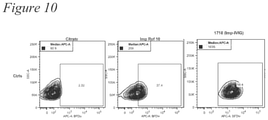
- FIG. 10 Data showing binding of PGG-IVIG conjugates to PMNs.
- This disclosure describes methods related to the use of soluble ⁇ -glucan as a component of immunotherapy.
- the methods described herein exploit the observation of differential binding of ⁇ -glucan by immune cells in different populations of healthy humans.
- “high binders” of ⁇ -glucan exhibit higher titers of anti- ⁇ -glucan antibodies than “low binders.”
- this disclosure describes methods of screening individuals to identify “high binders” and “low binders.”
- This disclosure also describes methods that generally include converting a “low binder” to a “high binder” and, thus, increase the population for whom ⁇ -glucan-based immunotherapy can be effective.
- treatment regimens that include periodically monitoring a subject for their current “high binder” or “low binder” status, and adjusting the therapy provided to the subject, if necessary, to promote achieving or maintaining “high binder” status.
- ⁇ -glucans are polymers of glucose derived from a variety of microbiological and plant sources including, for example, yeast, bacteria, algae, seaweed, mushroom, oats, and barley. Of these, yeast ⁇ -glucans have been extensively evaluated for their immunomodulatory properties. Yeast ⁇ -glucans can be present as various forms such as, for example, intact yeast, zymosan, purified whole glucan particles, solubilized zymosan polysaccharide, or highly-purified soluble ⁇ -glucans of different molecular weights.
- yeast ⁇ -glucans are composed of glucose monomers organized as ⁇ -(1,3)-linked glucopyranose backbone with periodic ⁇ -(1,3) glucopyranose branches linked to the backbone via ⁇ -(1,6) glycosidic linkages.
- the different forms of yeast ⁇ -glucans can function differently from one another.
- the mechanism through which yeast ⁇ -glucans exert their immunomodulatory effects can be influenced by the structural differences between different forms of the ⁇ -glucans such as, for example, its particulate or soluble nature, tertiary conformation, length of the main chain, length of the side chain, and frequency of the side chains.
- the immune stimulating functions of yeast ⁇ -glucans are also dependent upon the receptors engaged in different cell types in different species, which again, can be dependent on the structural properties of the ⁇ -glucans.
- ⁇ -glucan immunotherapies can include administering to a subject any suitable form of ⁇ -glucan or any combination of two or more forms of ⁇ -glucan.
- Suitable ⁇ -glucans and the preparation of suitable ⁇ -glucans from their natural sources are described in, for example, U.S. Patent Application Publication No. US2008/0103 112 A1.
- the ⁇ -glucan may be derived from a yeast such as, for example, Saccharomyces cerevisiae .
- the ⁇ -glucan may be or be derived from ⁇ (1,6)-[poly-(1,3)-D-glucopyranosyl]-poly- ⁇ (1,3)-D-glucopyranose, also referred to herein as PGG (IMPRIME PGG®, Biothera, Eagan, Minn.), a highly purified and well characterized form of soluble yeast-derived ⁇ -glucan.
- PGG PGG
- ⁇ -glucan-based immunotherapies can involve the use of, for example, a modified and/or derivatized ⁇ -glucan such as those described in International Patent Application No. PCT/US12/36795.
- ⁇ -glucan immunotherapy can involve administering, for example, a particulate-soluble ⁇ -glucan or a particulate-soluble ⁇ glucan preparation, each of which is described in, for example, U.S. Pat. No. 7,981,447.
- yeast ⁇ -glucans have been extensively evaluated for their immunomodulatory properties.
- PGG ⁇ -glucan has demonstrated preclinical activity against a variety of cancer types when administered in combination with tumor-related monoclonal antibodies (mAbs).
- mAbs tumor-related monoclonal antibodies
- Exemplary types of cancer and their associated tumor-related mAbs include, for example, T-cell lymphoma (anti-MUC1, anti-GD2), lung carcinoma (anti-MUC1), breast adenocarcinoma (anti-MMTV), ovarian carcinoma (bevacizumab), non-small-cell lung carcinoma (bevacizumab, cetuximab), colorectal cancer (cetuximab), chronic lymphocytic leukemia and non-Hodgkin's lymphoma (rituximab), and pancreatic carcinoma (cetuximab, anti-MUC1).
- T-cell lymphoma anti-MUC1, anti-GD2
- lung carcinoma anti-MUC
- FIG. 1 When exogenous ⁇ -glucan is administered, “low binders” exhibit a modest increase in the percentage of immune cells that bind ⁇ -glucan, while “high binders” exhibit a marked increase in the percentage of innate immune cells that bind ⁇ -glucan.
- FIG. 1 and FIG. 2 show data reflecting ⁇ -glucan binding to polymorphonuclear leukocytes (PMNs), and FIG. 3 (monocytes) shows that the differential binding applies to other innate immune cell populations as well.
- PMNs polymorphonuclear leukocytes
- FIG. 3 monoocytes
- “high binders” also tend to produce more chemoattractant cytokines and chemokines such as, for example, IL-8, MCP, MIP-1, etc.
- status as a “high binder” refers to an individual who exhibits a predetermined percentage of a particular immune cell population that binds exogenously provided ⁇ -glucan.
- the immune cell population used to determine whether an individual is a “high binder” or a “low binder” can be, for example, polymorphonuclear lymphocytes (PMNs) or monocytes.
- An individual can be considered a “high binder” if at least 10% of the PMNs or monocytes in a blood sample from the individual bind exogenously provided ⁇ -glucan.
- an individual may be a “high binder” if at least 10%, at least 12%, at least 15%, at least 20%, at least 15%, or at least 40% of PMNs or monocytes in a blood sample from the individual bind exogenously provided ⁇ -glucan.
- the exogenously provided ⁇ -glucan can include PGG provided to a final concentration of 10 ⁇ g/mL to 100 ⁇ g/mL.
- Status as a “low binder” refers to an individual who fails to exhibit “high binder” status.
- this disclosure describes screening an individual to identify whether the individual, at that point in time, exhibits “high binder” or “low binder” status.
- the method generally includes obtaining a blood sample from the individual, adding ⁇ -glucan to at least a portion of the sample, incubating the mixture under conditions allowing the ⁇ -glucan to bind to the immune cells, and detecting ⁇ -glucan bound to the immune cells.
- the blood sample includes a sufficient portion of the blood to include immune cells.
- the blood sample can be whole blood.
- the blood sample may be at least partially processed to remove one or more components of whole blood that are not necessary for the screening assay such as, for example, erythrocytes.
- the blood sample can include a blood product such as, for example, any blood fraction that includes at least a portion of the buffy coat.
- the ⁇ -glucan may be derived from yeast such as, for example, Saccharomyces cerevisiae .
- the ⁇ -glucan can include a ⁇ -1,3/1,6 glucan such as, for example, ⁇ (1,6)-[poly-(1,3)-D-glucopyranosyl]-poly- ⁇ (1,3)-D-glucopyranose.
- the ⁇ -glucan bound to the immune cells may be detected by contacting the sample with a monoclonal antibody that specifically binds to the ⁇ -glucan.
- the monoclonal antibody may be any monoclonal antibody that specifically binds to the ⁇ -glucan.
- “specific” and variations thereof refer to having a differential or a non-general (i.e., non-specific) affinity, to any degree, for a particular target.
- Exemplary monoclonal antibodies that specifically bind ⁇ -glucan include, for example, monoclonal antibodies identified as BfD I, BfD II, BfD III, and/or BfD IV (Biothera, Eagan, Minn.), each of which is described in U.S. Pat. No. 6,294,321.
- the anti- ⁇ -glucan antibody can include a detectable label.
- bound anti- ⁇ -glucan antibody may be detected using a labeled secondary antibody.
- suitable labels include, for example, a fluorescent label, an enzymatic label, a colorimetric label, or a radiolabel.
- the method can further include immobilizing the ⁇ -glucan-bound immune cells on a substrate.
- the immune cells may be immobilized prior to being contacted with the ⁇ -glucan—e.g., by contacting at least a portion of the blood sample with the substrate prior to contacting the blood sample with the ⁇ -glucan.
- the immune cells may be immobilized after being contacted with the ⁇ -glucan.
- the immune cells may be immobilized using any suitable material such as, for example, an immobilized antibody that specifically binds to the desired population of immune cells.
- the method can include a wash step to remove unbound assay components.
- some embodiments can include a step of washing unbound ⁇ -glucan and/or unbound blood sample components from the ⁇ -glucan-bound immune cells.
- some embodiments can include a step washing unbound immune cells from the substrate, if present.
- some embodiments can include washing unbound ⁇ -glucan-specific antibodies from the ⁇ -glucan bound to the immune cells.
- this disclosure describes a method of converting a “low binder” to a “high binder.”
- 795 subjects with recurrent/progressive colorectal cancer after at least two previous chemotherapeutic treatments were divided into a control arm and an investigational arm.
- Subjects in the control arm received treatment with cetuximab.
- Subjects in the investigational arm received treatment with cetuximab+4 mg/kg PGG ⁇ -glucan.
- FIG. 6 shows that “high binder” serum can increase ⁇ -glucan binding to immune cells (e.g., PMNs) of a “low binder.”
- immune cells e.g., PMNs
- Increasing amounts of anti- ⁇ -glucan monoclonal antibody also can increase ⁇ -glucan binding to immune cells (e.g., PMNs) in serum from a “low binder.”
- FIG. 7 shows that
- IVIG intravenous immunoglobulin
- PMNs immune cells
- FIG. 8 one can convert an individual from exhibiting “low binder” status to exhibiting “high binder” status by providing the individual, as a component of immunotherapy, with a combination of ⁇ -glucan and a preparation that includes anti- ⁇ -glucan antibodies.
- the conversion of an individual from “low binder” status to “high binder” status can improve clinical outcome of immunotherapy.
- FIG. 9 shows data illustrating relatively low PGG binding by PMNs in whole blood (Imp Ref, second panel) changing to high binding status by conjugating the PGG to either BTH1704 (anti-MUC1, U.S. Pat. No. 6,204,366, Biothera, Inc, Eagan, Minn., third panel) or ERBITUX® (cetuximab) (Eli Lilly and Co., Indianapolis, Ind., fourth panel) anti-tumor antibodies.
- FIG. 10 also illustrates relatively low PGG binding by PMNs in whole blood (Imp Ref, second panel) changing to high binding status by conjugating the PGG to intravenous immunoglobulin (IVIG, Biolegend, San Diego, Calif.).
- the method includes administering to a subject a composition that includes a ⁇ -glucan moiety conjugated to an antibody, a therapeutic antibody, an anti-tumor antibody, or an antibody fragment such as the Fc portion of an antibody.
- a composition that includes a ⁇ -glucan moiety conjugated to an antibody, a therapeutic antibody, an anti-tumor antibody, or an antibody fragment such as the Fc portion of an antibody.
- Modified and/or derivatized PGG including PGG conjugates of a PGG moiety and an antibody are described in International Patent Application No. PCT/US12/36795, which may also be applied to conjugates of antibody fragments.
- the PGG moiety may be, or be derived from a ⁇ -1,3/1,6 glucan.
- “derived from” acknowledges that a conjugate may necessarily be prepared by creating a covalent linkage that replaces one or more atoms of the PGG ⁇ -glucan.
- derived from a ⁇ -1,3/1,6 glucan refers to a portion of the PGG ⁇ -glucan that remains as part of a conjugate after replacing one or more atoms of the PGG to form the covalent linkage of the conjugate.
- “capable of converting a low binder to a high binder” refers to the property of modifying the binder status of an individual from “low binder” (e.g., below a predetermined threshold) to “high binder” (e.g., above a predetermined threshold).
- the status of a subject as a “low binder” or a “high binder” may be determined with reference to the extent to which ⁇ -glucan binds to immune cells in at least a portion of a blood sample obtained from the subject.
- the binder status of a subject may be determined with respect to the anti- ⁇ -glucan antibody titer measured in at least a portion of a blood sample obtained from the subject.
- the antibody preparation can include serum from a high binder.
- the antibody preparation can include intravenous immunoglobulin.
- the antibody preparation can include a monoclonal antibody that specifically binds the ⁇ -glucan.
- Exemplary monoclonal antibodies that specifically bind to ⁇ -glucan include, for example, BfD I, BfD II, BfD III, or BfD IV.
- the ⁇ -glucan, the antibody preparation, and/or the combination of both components may be formulated in a composition along with a “carrier.”
- carrier includes any solvent, dispersion medium, vehicle, coating, diluent, antibacterial agent and/or antifungal agent, isotonic agent, absorption delaying agent, buffer, carrier solution, suspension, colloid, and the like.
- carrier includes any solvent, dispersion medium, vehicle, coating, diluent, antibacterial agent and/or antifungal agent, isotonic agent, absorption delaying agent, buffer, carrier solution, suspension, colloid, and the like.
- the use of such media and/or agents for pharmaceutical active substances is well known in the art. Except insofar as any conventional media or agent is incompatible with the ⁇ -glucan or the antibody, its use in the therapeutic compositions is contemplated. Supplementary active ingredients also can be incorporated into the compositions.
- the ⁇ -glucan, the antibody preparation, and/or the combination of both components may be formulated into a pharmaceutical composition.
- the ⁇ -glucan and the antibody preparation may be provided in a single formulation.
- the ⁇ -glucan and the antibody preparation may be provided in separate formulations.
- a pharmaceutical composition may be formulated in a variety of and/or a plurality forms adapted to one or more preferred routes of administration.
- a formulation may be conveniently presented in unit dosage form and may be prepared by methods well known in the art of pharmacy.
- Methods of preparing a composition with a pharmaceutically acceptable carrier include the step of bringing the ⁇ -glucan and/or the antibody preparation into association with a carrier that constitutes one or more accessory ingredients.
- a formulation may be prepared by uniformly and/or intimately bringing the active compound into association with a liquid carrier, a finely divided solid carrier, or both, and then, if necessary, shaping the product into the desired formulations.
- the ⁇ -glucan may be derived from yeast such as, for example, Saccharomyces cerevisiae .
- the ⁇ -glucan can include ⁇ -1,3/1,6 glucan such as, for example, ⁇ (1,6)-[poly-(1,3)-D-glucopyranosyl]-poly- ⁇ (1,3)-D-glucopyranose.
- the method can include administering sufficient ⁇ -glucan to provide a dose of, for example, from about 100 ng/kg to about 50 mg/kg to the subject, although in some embodiments the methods may be performed by administering the ⁇ -glucan in a dose outside this range.
- the method includes administering sufficient ⁇ -glucan to provide a dose of from about 10 ⁇ g/kg to about 10 mg/kg to the subject such as, for example, a dose of about 1 mg/kg, about 2 mg/kg, about 3 mg/kg, about 4 mg/kg, about 5 mg/kg, about 6 mg/kg, about 7 mg/kg, about 8 mg/kg, about 9 mg/kg, or about 10 mg/kg.
- the method includes administering sufficient ⁇ -glucan to provide a dose of about 4 mg/kg.
- the dose may be calculated using actual body weight obtained just prior to the beginning of a treatment course.
- the method can include administering sufficient ⁇ -glucan to provide a dose of, for example, from about 0.01 mg/m 2 to about 10 mg/m 2 .
- the method can include administering sufficient antibody that specifically binds the ⁇ -glucan to provide a dose of, for example, from about 100 ng/kg to about 50 mg/kg to the subject, although in some embodiments the methods may be performed by administering the antibody in a dose outside this range. In some embodiments, the method includes administering sufficient antibody to provide a dose of from about 10 ⁇ g/kg to about 5 mg/kg to the subject, for example, a dose of from about 100 ⁇ g/kg to about 1 mg/kg.
- antibody that specifically binds the ⁇ -glucan can be administered in the form of intravenous immunoglobulin (IVIG), a blood product that contains pooled polyvalent IgG from many donors (typically many hundreds, even thousands, of donors and, thus, naturally containing anti- ⁇ -glucan antibodies).
- IVIG intravenous immunoglobulin
- IVIG may be administered in a dose of from about 0.1 g/kg to about 2.0 g/kg such as, for example, 0.1 g/kg, 0.2 g/kg, 0.3 g/kg, 0.4 g/kg, 0.5 g/kg, 0.6 g/kg, 0.7 g/kg, 0.8 g/kg, 0.9 g/kg, 1.0 g/kg, 1.1 g/kg, 1.2 g/kg, 1.3 g/kg, 1.4 g/kg, 1.5 g/kg, 1.6 g/kg, 1.7 g/kg, 1.8 g/kg, 1.9 g/kg, or 2.0 g/kg.
- IVIG may be administered to provide a dose of about 0.4 g/kg to about 1.0 g/kg.
- the dose may be calculated using actual body weight obtained just prior to the beginning of a treatment course.
- the method can include administering sufficient antibody to provide a dose of, for example, from about 0.01 mg/m 2 to about 10 mg/m 2 .
- the ⁇ -glucan and antibody preparation may be co-administered, for example, from a single dose to multiple doses per week, although in some embodiments the method may be performed by co-administering the ⁇ -glucan and antibody at a frequency outside this range. In certain embodiments, the ⁇ -glucan and antibody may be administered from about once per year to once per week.
- the method further includes providing immunotherapy to the subject.
- the immunotherapy can include administering one or more tumor-related antibodies (e.g., cetuximab, bevacizumab, anti-MUC1).
- tumor-related antibodies e.g., cetuximab, bevacizumab, anti-MUC1
- the co-administration of ⁇ -glucan and the antibody preparation can increase the efficacy of the immunotherapy.
- the term “and/or” means one or all of the listed elements or a combination of any two or more of the listed elements; the terms “comprises” and variations thereof do not have a limiting meaning where these terms appear in the description and claims; unless otherwise specified, “a,” “an,” “the,” and “at least one” are used interchangeably and mean one or more than one; and the recitations of numerical ranges by endpoints include all numbers subsumed within that range (e.g., 1 to 5 includes 1, 1.5, 2, 2.75, 3, 3.80, 4, 5, etc.).
- the steps may be conducted in any feasible order. And, as appropriate, any combination of two or more steps may be conducted simultaneously.
- Imprime PGG® ( ⁇ (1,6)-[poly-(1,3)-D-glucopyranosyl]-poly- ⁇ (1,3)-D-glucopyranose) (Biothera, Eagan, Minn.) was provided in a preservative-free, soluble ⁇ -glucan formulation prepared at a concentration of 1 mg/niL in 0.8% sodium chloride and 0.2% sodium citrate monobasic, at a pH of 6.4. The compound was stored at 4-8° C. until use.
- Fresh whole blood was obtained from healthy volunteers that had provided informed consent prior to donation (New England Institutional Review Board, May 2007). The blood was collected in a Vacutainer® containing 158 USP Units Freeze-Dried Sodium Heparin (BD Biosciences; San Jose, Calif.).
- a preliminary ELISA method modified from the monkey anti- ⁇ -glucan method (Noss et al., 2012 Int. Arch. Allergy Immunol., 157:98-108) was used to test the human sera samples.
- Costar universal binding plates were coated with 50 ⁇ L of ⁇ -glucan at 1 ⁇ g/mL purified ⁇ -glucan diluted in purified water and incubated at 37° C. for 30 minutes. The coated plate was then exposed to high intensity ultraviolet light at >1500 ⁇ W/cm 2 for 5 minutes at room temperature and placed in a 50° C. forced air oven until dry before a second exposure to ultraviolet light at >1500 ⁇ W/cm 2 for five minutes at room temperature.
- the plate was then blocked with a 0.5% solution of Bovine Serum Albumin for >30 minutes before washing with wash buffer (phosphate buffered saline [PBS] with 0.05% Tween-20).
- wash buffer phosphate buffered saline [PBS] with 0.05% Tween-20.
- Human serum samples were diluted into wash buffer added to the plate and subsequently serially diluted in wash buffer on the plate.
- Test samples diluted 1:400 were pipetted onto the test plate with seven additional serial 1:2 dilutions (serum dilutions between 1:400 and 1:12,800). Samples were incubated at room temperature for 30 minutes to permit human IgG to bind to the plate-bound ⁇ -glucan antigen.
- a value of 160 Arbitrary Units per mL (AU/mL) was assigned to the standard human anti ⁇ -glucan.
- AU/mL 160 Arbitrary Units per mL
- Assay controls were diluted 1:100 in ELISA wash buffer for testing.
- two dilutions of each control level were independently prepared for testing on each plate in parallel.
- Imprime PGG® ( ⁇ (1,6)-[poly-(1,3)-D-glucopyranosyl]-poly- ⁇ (1,3)-D-glucopyranose) (Biothera, Eagan, Minn.) was provided in a preservative-free, soluble ⁇ -glucan formulation prepared at a concentration of 1 mg/mL in 0.8% sodium chloride and 0.2% sodium citrate monobasic, at a pH of 6.4. The compound was stored at 4-8° C. until use.
- Fresh WB was obtained from healthy volunteers that had provided informed consent prior to donation (New England Institutional Review Board. Blood Donation Protocol No. 07-124).
- the blood was collected in a Vacutainer® containing 158 USP Units Freeze-Dried Sodium Heparin (BD Biosciences; San Jose, Calif.).
- Serum was collected in a Vacutainer® containing a thrombin-based clot activator (BD Biosciences; San Jose, Calif.).
- the whole blood binding assay was performed by incubating whole blood samples with Imprime PGG® ( ⁇ (1,6)-[poly-(1,3)-D-glucopyranosyl]-poly- ⁇ (1,3)-D-glucopyranose) for 30 minutes or two hours at 37° C. in a humidified incubator. After washing with 1 ⁇ Dulbecco's phosphate buffered saline (DPBS), BfDIV, a mouse anti- ⁇ -glucan antibody, was added and incubated with the WB for 30 minutes at room temperature. After more rounds of washing, an antibody cocktail including a goat anti-mouse detection antibody and antibodies to surface molecules were added and incubated at room temperature in the dark for 30 minutes.
- Imprime PGG® ⁇ (1,6)-[poly-(1,3)-D-glucopyranosyl]-poly- ⁇ (1,3)-D-glucopyranose
- DPBS 1 ⁇ Dulbecco's phosphate buffered saline
- Erythrocytes were lysed with BD Lyse and samples were resuspended in 1% paraformaldehyde. Samples were acquired on a flow cytometer and analyzed using FlowJo software (Ashland, Oreg.).
- the lyophilized antibody was resuspended to 1 mg/mL with 1 ⁇ DPBS and stored at ⁇ 80° C. or 4° C. as a stock solution. Before being added to blood samples, the stock was diluted 1:10 to 100 ⁇ g/mL and 10 ⁇ L of this solution was added to 100 ⁇ L of blood. For incubation with IVIG, 10% IVIG (100 mg/mL) (PRIVIGEN, CSL Behrling, King of Prussia, Pa.) was added to the whole blood sample at the indicated final concentrations.
- a method for identifying soluble ⁇ -glucan binding to immune cells of a subject comprising:
- the blood sample comprising immune cells
- Embodiment 1 wherein the soluble ⁇ -glucan is derived from yeast.
- Embodiment 1 The method of Embodiment 1 or Embodiment 2 wherein the soluble ⁇ -glucan comprises a ⁇ -1,3/1,6 glucan.
- the soluble ⁇ -glucan comprises ⁇ (1,6)-[poly-(1,3)-D-glucopyranosyl]-poly- ⁇ (1,3)-D-glucopyranose.
- detecting soluble ⁇ -glucan bound to the immune cells comprises contacting the sample with a monoclonal antibody that specifically binds to the ⁇ -glucan.
- the blood sample comprising immune cells
- a method of improving ⁇ -glucan immunotherapy for a subject comprising:
- identifying the subject as a low binder comprises:
- obtaining a blood sample from the subject the sample comprising an anti- ⁇ -glucan antibody titer
- the anti- ⁇ -glucan IgG antibody titer is less than 20,000.
- identifying the subject as a low binder comprises:
- the blood sample comprising immune cells
- the antibody preparation comprises a monoclonal antibody that specifically binds the soluble ⁇ -glucan.
- the method of Embodiment 12 wherein the monoclonal antibody comprises BfD I, BfD II, BfD III, or BfD IV.
- soluble ⁇ -glucan comprises ⁇ (1,6)-[poly-(1,3)-D-glucopyranosyl]-poly- ⁇ (1,3)-D-glucopyranose.
Abstract
Description
| TABLE 1 |
| Antibody Cocktail Used To Stain Whole Blood Samples |
| Dilution or Final | For identifica- | ||
| Antibody | Company; Clone # | Concentration | tion of: |
| Anti-CD15 | Biolegend; W6D3 | 0.2 | μg/mL | neutrophils |
| Anti-CD19 | Biolegend; HIB19 | 0.63 | μg/mL | B cells |
| Anti-CD14 | Biolegend; HCD14 | 5 | μg/mL | monocytes |
| Anti-CD14 | Invitrogen; TüK4 | 1:50 | monocytes |
| Anti-CD3 | Biolegend; HIT3a | 0.25 | μg/mL | T cells |
| Anti-CD45 | Biolegend; HI30 | 0.25 | μg/mL | hematopoietic |
| cells excluding | ||||
| erythrocytes and | ||||
| platelets | ||||
| Goat | Jackson Immunolab | 5 | μg/mL | mouse anti-β |
| F(ab′)2 anti- | glucan antibody | |||
| mouse IgM | ||||
| Proceeding incubation with the anti-β-glucan antibody BfD IV, the cells were incubated with the antibody cocktail which contains a secondary antibody for the recognition of BfD IV as well as antibodies for the recognition of various cell surface markers | ||||
Claims (6)
Priority Applications (1)
| Application Number | Priority Date | Filing Date | Title |
|---|---|---|---|
| US14/398,019 US11229701B2 (en) | 2012-04-30 | 2013-03-14 | Methods for identifying beta-glucan binding to immune cells |
Applications Claiming Priority (5)
| Application Number | Priority Date | Filing Date | Title |
|---|---|---|---|
| US201261640397P | 2012-04-30 | 2012-04-30 | |
| US201261640842P | 2012-05-01 | 2012-05-01 | |
| US201261640834P | 2012-05-01 | 2012-05-01 | |
| PCT/US2013/031606 WO2013165591A1 (en) | 2012-04-30 | 2013-03-14 | β-GLUCAN IMMUNOTHERAPEUTIC METHODS |
| US14/398,019 US11229701B2 (en) | 2012-04-30 | 2013-03-14 | Methods for identifying beta-glucan binding to immune cells |
Related Parent Applications (1)
| Application Number | Title | Priority Date | Filing Date |
|---|---|---|---|
| PCT/US2013/031606 A-371-Of-International WO2013165591A1 (en) | 2012-04-30 | 2013-03-14 | β-GLUCAN IMMUNOTHERAPEUTIC METHODS |
Related Child Applications (1)
| Application Number | Title | Priority Date | Filing Date |
|---|---|---|---|
| US17/552,137 Division US20220105181A1 (en) | 2012-04-30 | 2021-12-15 | Methods for identifying beta-glucan binding to immune cells |
Publications (2)
| Publication Number | Publication Date |
|---|---|
| US20150125461A1 US20150125461A1 (en) | 2015-05-07 |
| US11229701B2 true US11229701B2 (en) | 2022-01-25 |
Family
ID=49514726
Family Applications (4)
| Application Number | Title | Priority Date | Filing Date |
|---|---|---|---|
| US14/398,020 Active 2034-12-11 US10092646B2 (en) | 2012-04-30 | 2013-03-14 | Compositions and methods for beta-glucan immunotherapy |
| US14/398,019 Active 2034-02-27 US11229701B2 (en) | 2012-04-30 | 2013-03-14 | Methods for identifying beta-glucan binding to immune cells |
| US16/118,909 Abandoned US20180369376A1 (en) | 2012-04-30 | 2018-08-31 | Compositions and methods for beta-glucan immunotherapy |
| US17/552,137 Pending US20220105181A1 (en) | 2012-04-30 | 2021-12-15 | Methods for identifying beta-glucan binding to immune cells |
Family Applications Before (1)
| Application Number | Title | Priority Date | Filing Date |
|---|---|---|---|
| US14/398,020 Active 2034-12-11 US10092646B2 (en) | 2012-04-30 | 2013-03-14 | Compositions and methods for beta-glucan immunotherapy |
Family Applications After (2)
| Application Number | Title | Priority Date | Filing Date |
|---|---|---|---|
| US16/118,909 Abandoned US20180369376A1 (en) | 2012-04-30 | 2018-08-31 | Compositions and methods for beta-glucan immunotherapy |
| US17/552,137 Pending US20220105181A1 (en) | 2012-04-30 | 2021-12-15 | Methods for identifying beta-glucan binding to immune cells |
Country Status (10)
| Country | Link |
|---|---|
| US (4) | US10092646B2 (en) |
| EP (4) | EP2854530B1 (en) |
| JP (3) | JP6416751B2 (en) |
| KR (2) | KR102103366B1 (en) |
| CN (2) | CN104812396B (en) |
| AU (4) | AU2013257139B2 (en) |
| CA (2) | CA2872009A1 (en) |
| ES (2) | ES2661393T3 (en) |
| RU (3) | RU2018102251A (en) |
| WO (2) | WO2013165593A1 (en) |
Cited By (1)
| Publication number | Priority date | Publication date | Assignee | Title |
|---|---|---|---|---|
| US11815435B2 (en) | 2017-02-24 | 2023-11-14 | Hibercell, Inc. | Beta glucan immunopharmacodynamics |
Families Citing this family (9)
| Publication number | Priority date | Publication date | Assignee | Title |
|---|---|---|---|---|
| RU2018102251A (en) | 2012-04-30 | 2019-02-21 | Байотера, Инк. | METHODS OF β-GLUCANE IMMUNOTHERAPY |
| AU2014357446B2 (en) * | 2013-12-05 | 2020-05-07 | Biothera, Inc. | Beta-glucan assay methods |
| CN106687122B (en) * | 2014-07-10 | 2020-11-10 | 百奥赛诺公司 | Combination of beta-glucan and an anti-cancer agent affecting the tumor microenvironment |
| JP6887378B2 (en) * | 2014-11-06 | 2021-06-16 | バイオセラ,インク. | Beta-glucan methods and compositions that affect the intratumoral microenvironment |
| EP3400442A4 (en) * | 2016-01-08 | 2019-06-26 | Biothera, Inc. | Beta-glucan immunotherapies affecting the immune microenvironment |
| CN105749290A (en) * | 2016-04-11 | 2016-07-13 | 苏州康聚生物科技有限公司 | Protein medicinal preparation containing beta-glucan as auxiliary material |
| CN110038132A (en) * | 2019-01-29 | 2019-07-23 | 苏州杰纳生物科技有限公司 | Natural polymer-albumen composition and its preparation method and application |
| WO2022251123A1 (en) * | 2021-05-24 | 2022-12-01 | Frank Jordan | Alkaline extraction of beta glucan compounds for use in anti-viral and immune therapies |
| CN113249336B (en) * | 2021-07-15 | 2021-10-08 | 天津一瑞生物科技股份有限公司 | Mouse-resistant (1, 3) -beta-D glucan hybridoma cell strain, monoclonal antibody and application |
Citations (17)
| Publication number | Priority date | Publication date | Assignee | Title |
|---|---|---|---|---|
| WO1999031510A1 (en) | 1997-12-12 | 1999-06-24 | The Collaborative Group, Ltd. | β(1-3)-GLUCAN DIAGNOSTIC ASSAYS |
| US6204366B1 (en) | 1990-09-07 | 2001-03-20 | Unilever Patent Holdings B.V. | Specific binding agents |
| US20010051717A1 (en) | 1997-01-31 | 2001-12-13 | Collaborative Group, Ltd. | Beta (1-3) -glucan diagnostic assays |
| US6355625B1 (en) | 1998-09-14 | 2002-03-12 | Nabi | Compositions of β-glucans and specific IGIV |
| WO2003097091A2 (en) | 2002-05-15 | 2003-11-27 | Luciano Polonelli | Glucan-based vaccines |
| US20040014715A1 (en) | 2001-10-09 | 2004-01-22 | Ostroff Gary R. | Use of beta-glucans against biological warfare weapons and pathogens including anthrax |
| WO2004021994A2 (en) | 2002-09-04 | 2004-03-18 | Biopolymer Engineering, Inc. | Cancer therapy using whole glucan particles and antibodies |
| WO2005018544A2 (en) | 2003-07-16 | 2005-03-03 | Sloan-Kettering Institute For Cancer Research | Therapy-enhancing glucan |
| WO2006085895A2 (en) | 2004-05-10 | 2006-08-17 | Biopolymer Engineering, Inc. | Whole glucan particles in combination with antibiotics, vaccines and viral monoclonal antibodies |
| WO2007084661A2 (en) | 2006-01-17 | 2007-07-26 | Sloan-Kettering Institute For Cancer Research | Therapy-enhancing glucan |
| US20080103112A1 (en) | 2006-06-15 | 2008-05-01 | MAGEE Andrew | Glucan preparations |
| WO2009134891A2 (en) | 2008-04-29 | 2009-11-05 | Immunexcite, Inc. | Immunomodulating compositions and methods of use thereof |
| US20100029713A1 (en) | 2006-11-14 | 2010-02-04 | Astrazeneca Ab | Quiniclidine derivatives of (hetero) arylcycloheptanecarboxylic acid as muscarinic receptor antagonists |
| US7981447B2 (en) | 2007-05-08 | 2011-07-19 | Biothera, Inc. | Particulate-soluble glucan preparation |
| WO2012154818A1 (en) | 2011-05-09 | 2012-11-15 | Biothera, Inc. | B-glucan compounds, compositions, and methods |
| WO2012154680A2 (en) | 2011-05-06 | 2012-11-15 | Biothera, Inc. | MODIFIED AND DERIVATIZED β-GLUCAN COMPOUNDS, COMPOSITIONS, AND METHODS |
| WO2013165591A1 (en) | 2012-04-30 | 2013-11-07 | Biothera, Inc. | β-GLUCAN IMMUNOTHERAPEUTIC METHODS |
Family Cites Families (4)
| Publication number | Priority date | Publication date | Assignee | Title |
|---|---|---|---|---|
| GB0302218D0 (en) * | 2003-01-30 | 2003-03-05 | Chiron Sri | Vaccine formulation & Mucosal delivery |
| GB0420466D0 (en) * | 2004-09-14 | 2004-10-20 | Cassone Antonio | Anti-glucan antibodies |
| JP4666284B2 (en) * | 2005-11-30 | 2011-04-06 | 味の素株式会社 | Glucan sensitivity prediction method, glucan sensitivity enhancer and screening method thereof |
| AU2014357446B2 (en) * | 2013-12-05 | 2020-05-07 | Biothera, Inc. | Beta-glucan assay methods |
-
2013
- 2013-03-14 RU RU2018102251A patent/RU2018102251A/en not_active Application Discontinuation
- 2013-03-14 EP EP13784472.6A patent/EP2854530B1/en active Active
- 2013-03-14 ES ES13784589.7T patent/ES2661393T3/en active Active
- 2013-03-14 KR KR1020147033316A patent/KR102103366B1/en active IP Right Grant
- 2013-03-14 CN CN201380034978.3A patent/CN104812396B/en active Active
- 2013-03-14 AU AU2013257139A patent/AU2013257139B2/en active Active
- 2013-03-14 ES ES13784472.6T patent/ES2662969T3/en active Active
- 2013-03-14 KR KR20147033313A patent/KR20150013206A/en not_active Application Discontinuation
- 2013-03-14 US US14/398,020 patent/US10092646B2/en active Active
- 2013-03-14 EP EP17210808.6A patent/EP3363462A1/en not_active Withdrawn
- 2013-03-14 CN CN201380034980.0A patent/CN104812243B/en active Active
- 2013-03-14 JP JP2015510271A patent/JP6416751B2/en active Active
- 2013-03-14 WO PCT/US2013/031625 patent/WO2013165593A1/en active Application Filing
- 2013-03-14 EP EP18150764.1A patent/EP3373006A1/en not_active Withdrawn
- 2013-03-14 RU RU2014148153A patent/RU2643331C2/en active
- 2013-03-14 WO PCT/US2013/031606 patent/WO2013165591A1/en active Application Filing
- 2013-03-14 AU AU2013257141A patent/AU2013257141B2/en active Active
- 2013-03-14 JP JP2015510272A patent/JP6272305B2/en active Active
- 2013-03-14 RU RU2014148155A patent/RU2629334C2/en active
- 2013-03-14 US US14/398,019 patent/US11229701B2/en active Active
- 2013-03-14 CA CA 2872009 patent/CA2872009A1/en active Pending
- 2013-03-14 CA CA2872010A patent/CA2872010C/en active Active
- 2013-03-14 EP EP13784589.7A patent/EP2844262B1/en active Active
-
2018
- 2018-01-30 AU AU2018200706A patent/AU2018200706A1/en not_active Abandoned
- 2018-04-09 JP JP2018074753A patent/JP2018150304A/en active Pending
- 2018-05-16 AU AU2018203444A patent/AU2018203444B2/en active Active
- 2018-08-31 US US16/118,909 patent/US20180369376A1/en not_active Abandoned
-
2021
- 2021-12-15 US US17/552,137 patent/US20220105181A1/en active Pending
Patent Citations (32)
| Publication number | Priority date | Publication date | Assignee | Title |
|---|---|---|---|---|
| US6204366B1 (en) | 1990-09-07 | 2001-03-20 | Unilever Patent Holdings B.V. | Specific binding agents |
| US6294321B1 (en) | 1996-05-01 | 2001-09-25 | The Collaborative Group | β(1-3)-glucan diagnostic assays |
| US20010051717A1 (en) | 1997-01-31 | 2001-12-13 | Collaborative Group, Ltd. | Beta (1-3) -glucan diagnostic assays |
| JP2002508518A (en) | 1997-12-12 | 2002-03-19 | ザ コラボレイティブ グループ,リミテッド. | β (1-3) -glucan diagnostic assay |
| WO1999031510A1 (en) | 1997-12-12 | 1999-06-24 | The Collaborative Group, Ltd. | β(1-3)-GLUCAN DIAGNOSTIC ASSAYS |
| US6355625B1 (en) | 1998-09-14 | 2002-03-12 | Nabi | Compositions of β-glucans and specific IGIV |
| US20040014715A1 (en) | 2001-10-09 | 2004-01-22 | Ostroff Gary R. | Use of beta-glucans against biological warfare weapons and pathogens including anthrax |
| JP2005535298A (en) | 2002-05-15 | 2005-11-24 | ルシアーノ ポロネリ, | Glucan based vaccine |
| WO2003097091A2 (en) | 2002-05-15 | 2003-11-27 | Luciano Polonelli | Glucan-based vaccines |
| EP1891970A1 (en) | 2002-05-15 | 2008-02-27 | Luciano Polonelli | Beta glucan-based vaccines free of mannoprotein |
| EP1506009B1 (en) | 2002-05-15 | 2008-05-14 | Luciano Polonelli | Beta-glucan-based anti-fungal vaccines |
| WO2004021994A2 (en) | 2002-09-04 | 2004-03-18 | Biopolymer Engineering, Inc. | Cancer therapy using whole glucan particles and antibodies |
| CN1697659A (en) | 2002-09-04 | 2005-11-16 | 生物聚合物工程有限公司 | Cancer therapy using whole glucan particles and antibodies |
| CN1694715A (en) | 2002-09-04 | 2005-11-09 | 路易斯维尔大学研究基金会 | Cancer therapy using beta glucan particles and antibodies |
| US20060009419A1 (en) | 2002-09-04 | 2006-01-12 | Ross Gordon D | Therapy-enhancing glucan |
| WO2004030613A2 (en) | 2002-09-04 | 2004-04-15 | University Of Louisville Research Foundation, Inc. | Cancer therapy using beta glucan and antibodies |
| CN1723027A (en) | 2002-10-09 | 2006-01-18 | 生物高分子工程公司 | Use of beta-glucans against biological warfare weapons and pathogens including anthrax |
| WO2005018544A2 (en) | 2003-07-16 | 2005-03-03 | Sloan-Kettering Institute For Cancer Research | Therapy-enhancing glucan |
| CN1823092A (en) | 2003-07-16 | 2006-08-23 | 斯隆-凯特林癌症研究院 | Therapy-enhancing glucan |
| WO2006085895A2 (en) | 2004-05-10 | 2006-08-17 | Biopolymer Engineering, Inc. | Whole glucan particles in combination with antibiotics, vaccines and viral monoclonal antibodies |
| CN1964722A (en) | 2004-05-10 | 2007-05-16 | 生物聚合物工程有限公司 | Whole glucan particles in combination with antibiotics, vaccines and viral monoclonal antibodies |
| WO2007084661A2 (en) | 2006-01-17 | 2007-07-26 | Sloan-Kettering Institute For Cancer Research | Therapy-enhancing glucan |
| JP2009528267A (en) | 2006-01-17 | 2009-08-06 | スローン − ケッタリング インスティチュート フォー キャンサー リサーチ | Glucan to enhance treatment |
| US20080103112A1 (en) | 2006-06-15 | 2008-05-01 | MAGEE Andrew | Glucan preparations |
| US20100029713A1 (en) | 2006-11-14 | 2010-02-04 | Astrazeneca Ab | Quiniclidine derivatives of (hetero) arylcycloheptanecarboxylic acid as muscarinic receptor antagonists |
| US7981447B2 (en) | 2007-05-08 | 2011-07-19 | Biothera, Inc. | Particulate-soluble glucan preparation |
| WO2009134891A2 (en) | 2008-04-29 | 2009-11-05 | Immunexcite, Inc. | Immunomodulating compositions and methods of use thereof |
| WO2012154680A2 (en) | 2011-05-06 | 2012-11-15 | Biothera, Inc. | MODIFIED AND DERIVATIZED β-GLUCAN COMPOUNDS, COMPOSITIONS, AND METHODS |
| WO2012154818A1 (en) | 2011-05-09 | 2012-11-15 | Biothera, Inc. | B-glucan compounds, compositions, and methods |
| WO2013165591A1 (en) | 2012-04-30 | 2013-11-07 | Biothera, Inc. | β-GLUCAN IMMUNOTHERAPEUTIC METHODS |
| WO2013165593A1 (en) | 2012-04-30 | 2013-11-07 | Biothera, Inc. | COMPOSITIONS AND METHODS FOR β-GLUCAN IMMUNOTHERAPY |
| US20150125451A1 (en) | 2012-04-30 | 2015-05-07 | Biothera, Inc. | Compositions and methods for beta-glucan immunotherapy |
Non-Patent Citations (40)
| Title |
|---|
| Antonysamy et al., "Differential Neutrophil Binding of Imprime PGG®, a β-1,3/1,6 Immunomodulatory Glucan", Jun. 9-12, 2012, International Symposium the Neutrophil in Immunity, Research Abstract, 1 page. |
| Bose et al., "Binding of soluble yeast β-glucan to human neutrophils and monocytes is complement-dependent", Aug. 12, 2013, Frontiers in Immunology, 4(230):1-14. |
| Deslandes et al., "Triple-Helical Structure of (1→3)-β-D-Glucan", Nov. 1980, Macromolecules, 13(6):1466-1471. |
| Eberhard et al., "Mutations in the Epidermal Growth Factor Receptor and in KRAS Are Predictive and Prognostic Indicators in Patients With Non-Small-Cell Lung Cancer Treated With Chemotherapy Alone and in Combination With Erlotinib", Sep. 1, 2005, J Clin Oncol, 23(25):5900-5909. |
| Ensley et al., "NMR spectral analysis of a water-insoluble (1→3 )-β-glucan isolated from Saccharomyces cerevisiae", May 20, 1994, Carbohydrate Research, 258:307-311. |
| European Patent Application No. 13784472, filed Mar. 14, 2013; Extended European Search Report dated Nov. 27, 2015; 11 pages. |
| European U.S. Appl. No. 13/784,589, filed Mar. 14, 2013; Extended European Search Report dated Nov. 27, 2015; 13 pages. |
| Flow Cytometry Basics Guide https://www.bio-rad-antibodies.com/static/2017/flow/flow-cytometry-basics-guide.pdf Downloaded Apr. 1, 2018. * |
| Fraser, Callum, "Test result variation and the quality of evidence-based clinical guidelines," Clinica Chimica Acta, 2004;346:19-24. |
| Goodridge et al. Activation of the innate immune receptor Dectin-1 upon formation of a ‘phagocytic synapse.’ Nature 472:471-475, 2011. Published Apr. 27, 2011. * |
| Goodridge et al. Activation of the innate immune receptor Dectin-1 upon formation of a ‘phagocytic synapse’. Nature 472:471-476, 2011. * |
| Ishibashi, et al., "Influence of Anti-β-Glucan Antibody on Fungal Cell Wall β-Glucan Bioactivity", 2009, Journal of Japanese Society for Bacteriology, 64(1):168 (P1-240). English Translation provided. 4 pages. |
| Isoda et al., "Clinical Efficacy of Superfine Dispersed Lentinan (β1,3-glucan) in Patients with Hepatocellular Carcinoma", 2009, Hepatogastroenterology, 56(90):437-441. |
| Kaiser et al., "Synergism between Poly-(1-6)-β-D-Glucopyranosyl-(1-3)-β-D-Glucopyranose Glucan and Cefazolin in Prophylaxis of Staphylococcal Wound Infection in a Guinea Pig Model", Sep. 1998, Antimicrob. Agents Chemother., 42(9):2449-2451. Retrieved on Mar. 30, 2017. Retrieved from the Internet <http://aac.asm.org/content/42/9/2449.full.pdf+html>. |
| Lamm et al., "A Randomized Trial of Intravesical Doxorubicin and Immunotherapy with Bacille Calmette-Guérin for Transitional-Cell Carcinoma of the Bladder", Oct. 24, 1991, The New England Journal of Medicine, 325(17):1205-1209. |
| Li et al., "Combined Yeast β-Glucan and Antitumor Monoclonal Antibody Therapy Requires C5a-Mediated Neutrophil Chemotaxis via Regulation of Decay-Accelerating Factor CD55", Aug. 1, 2007, Cancer Research, 67:7421-7430. |
| Li et al., "Yeast β-Glucan Amplifies Phagocyte Killing of iC3b-Opsonized Tumor Cells via Complement Receptor 3-Syk-Phosphatidylinositol 3-Kinase Pathway", 2006, Journal of Immunology, 177:1661-1669. |
| Liu et al., "Combined yeast-derived β-glucan with anti-tumor monoclonal antivody for cancer immunotherapy," Exp and Mol Path, Jun. 2009;86(3):208-214. |
| Mathé et al., "Active Immuotherapy for Acute Lymphoblastic Leukemia", Apr. 5, 1969, The Lancet, 1(7597):697-699. |
| Meikle et al., "The location of (1→3)-β-glucans in the walls of pollen tubes of Nicotiana alata using a (1→3)-β-glucan-specific monoclonal antibody", Mar. 20, 1991, Planta, 185:1-8. |
| Morales et al., "Intracavitary Bacillus Calmette-Guerin in the treatment of superficial bladder tumors", Aug. 1976, The Journal of Urology, 116(2):180-183. |
| Noss et al., "IgG to Various Beta-Glucans in a Human Adult Population", 2012, Int. Arch. Allergy Immunol., 157:98-108. First published online Sep. 7, 2011. |
| Oka et al., "In Vitro and In Vivo Analysis of Human Leukocyte Binding by the Antitumor Polysaccharide, Lentinan", 1996, Int. J. Immunopharmac., 18(3):211-216. |
| PCT Patent Application No. PCT/2013/031625, filed Mar. 14, 2013; International Preliminary Report on Patentability dated Nov. 13, 2014; 8 pages. |
| PCT Patent Application No. PCT/2013/031625, filed Mar. 14, 2013; International Search Report/Written Opinion dated May 17, 2013; 10 pages. |
| PCT Patent Application No. PCT/US2013/031606, filed Mar. 14, 2013; International Preliminary Report on Patentability dated Nov. 13, 2014; 7 pages. |
| PCT Patent Application No. PCT/US2013/031606, filed Mar. 14, 2013; International Search Report/Written Opinion dated Jun. 4, 2013; 9 pages. |
| Pearl, "Cancer and Tuberculosis", 1929, American Journal of Hygiene, 9:97-159. |
| Qi et al., "Differential pathways regulating innate and adaptive antitumor immune responses by particulate and soluble yeast-derived β-glucans", Jun. 23, 2011, Blood, 117(25):6825-6836. |
| Roberts et al., "Targeting the Raf-MEK-ERK mitogen-activated protein kinase cascade for the treatment of cancer", 2007, Oncogene, 26:3291-3310. |
| Rubin-Bejerano et al., "Phagocytosis by Human Neutrophils Is Stimulated by a Unique Fungal Cell Wall Component", Jul. 2007, Cell Host & Microbe, 2:55-67. |
| Ruckdeschel et al., "Postoperative Empyema Improves Survival in Lung Cancer—Documentation and Analysis of a Natural Experiment", Nov. 16, 1972, The New England Journal of Medicine, 287(20):1013-1017. |
| Sakamoto, "Function of β-glucan", 2011, New Food Industry, 53(12):1-11. English translation of pp. 9-10 provided. 13 pages. |
| Salvador et al., "Yeast-Derived β-Glucan Augments the Therapeutic Efficacy Mediated by Anti-Vascular Endothelial Growth Factor Monoclonal Antibody in Human Carcinoma Xenograft Models", Feb. 15, 2008, Clinical Cancer Research, 14(4):1239-1247. |
| Shindo et al., "Is T1G3 Cancer Having a Definite Muscle Layer in TUR Specimens a Highly Progressive Disease?", 2010, Japanese Journal of Clinical Oncology, 40(2):153-156. |
| Thornton et al., "Analysis of the Sugar Specificity and Molecular Location of the β-Glucan-Binding Lectin Site of the Complement Receptor Type 3 (CD11b/CD18)", Feb. 1, 1996, J Immunol., 156(3):1235-46. |
| Tsikitis et al., "The Lectin-Like Domain of Complement Receptor 3 Protects Endothelial Barrier Function from Activated Neutrophils", Jul. 15, 2004, J Immunol., 173(2):1284-91. |
| Vasilakos et al. Human innate immune cells that engage soluble beat-1,3/1,6 glucans: Role for complement receptor 3 (CR3, CD11b/CD18). J. Immunol. 184(1) Suppl Abstract No. 89.53., 2010. * |
| Xia et al., "The β-Glucan-Binding Lectin Site of Mouse CR3 (CD11b/CD18) and Its Function in Generating a Primed State of the Receptor That Mediates Cytotoxic Activation in Response to iC3b-Opsonized Target Cells", Feb. 15, 1999, J Immunol., 162(4):2281-90. |
| Zhong et al., "Effect of Yeast-derived β-glucan in Conjunction With Bevacizumab for the Treatment of Human Lung Adenocarcinoma in Subcutaneous and Orthotopic Xenograft Models", Sep. 2009, Journal of Immunotherapy, 32(7):703-712. |
Cited By (1)
| Publication number | Priority date | Publication date | Assignee | Title |
|---|---|---|---|---|
| US11815435B2 (en) | 2017-02-24 | 2023-11-14 | Hibercell, Inc. | Beta glucan immunopharmacodynamics |
Also Published As
Similar Documents
| Publication | Publication Date | Title |
|---|---|---|
| US20220105181A1 (en) | Methods for identifying beta-glucan binding to immune cells | |
| US10114027B2 (en) | Beta-glucan assay methods | |
| US20240019444A1 (en) | Beta glucan immunopharmacodynamics |
Legal Events
| Date | Code | Title | Description |
|---|---|---|---|
| AS | Assignment |
Owner name: BIOTHERA, INC., MINNESOTA Free format text: ASSIGNMENT OF ASSIGNORS INTEREST;ASSIGNORS:GROSSMAN, WILLIAM J.;ANTONYSAMY, MARY A.;WALSH, RICHARD M.;AND OTHERS;SIGNING DATES FROM 20130801 TO 20130906;REEL/FRAME:034936/0156 |
|
| AS | Assignment |
Owner name: MIDCAP FINANCIAL TRUST, AS AGENT, MARYLAND Free format text: SECURITY INTEREST;ASSIGNORS:BIOTHERA, INC.;LIFE SOURCE BASICS INC.;BIOTHERA HEALTH INC.;REEL/FRAME:035361/0929 Effective date: 20150408 |
|
| STPP | Information on status: patent application and granting procedure in general |
Free format text: FINAL REJECTION MAILED |
|
| STPP | Information on status: patent application and granting procedure in general |
Free format text: DOCKETED NEW CASE - READY FOR EXAMINATION |
|
| STPP | Information on status: patent application and granting procedure in general |
Free format text: NON FINAL ACTION MAILED |
|
| STPP | Information on status: patent application and granting procedure in general |
Free format text: NON FINAL ACTION MAILED |
|
| AS | Assignment |
Owner name: LIFE SOURCE BASICS INC., MINNESOTA Free format text: RELEASE BY SECURED PARTY;ASSIGNOR:MIDCAP FINANCIAL SERVICES, LLC;REEL/FRAME:052297/0045 Effective date: 20150828 Owner name: BIOTHERA, INC., MINNESOTA Free format text: RELEASE BY SECURED PARTY;ASSIGNOR:MIDCAP FINANCIAL SERVICES, LLC;REEL/FRAME:052297/0045 Effective date: 20150828 Owner name: BIOTHERA HEALTH INC., MINNESOTA Free format text: RELEASE BY SECURED PARTY;ASSIGNOR:MIDCAP FINANCIAL SERVICES, LLC;REEL/FRAME:052297/0045 Effective date: 20150828 |
|
| STPP | Information on status: patent application and granting procedure in general |
Free format text: NON FINAL ACTION MAILED |
|
| STPP | Information on status: patent application and granting procedure in general |
Free format text: RESPONSE TO NON-FINAL OFFICE ACTION ENTERED AND FORWARDED TO EXAMINER |
|
| AS | Assignment |
Owner name: HIBERCELL, INC., NEW YORK Free format text: ASSIGNMENT OF ASSIGNORS INTEREST;ASSIGNOR:BIOTHERA PHARMACEUTICALS, INC.;REEL/FRAME:054857/0613 Effective date: 20200609 |
|
| STPP | Information on status: patent application and granting procedure in general |
Free format text: DOCKETED NEW CASE - READY FOR EXAMINATION |
|
| STPP | Information on status: patent application and granting procedure in general |
Free format text: NON FINAL ACTION MAILED |
|
| STPP | Information on status: patent application and granting procedure in general |
Free format text: RESPONSE TO NON-FINAL OFFICE ACTION ENTERED AND FORWARDED TO EXAMINER |
|
| STPP | Information on status: patent application and granting procedure in general |
Free format text: NOTICE OF ALLOWANCE MAILED -- APPLICATION RECEIVED IN OFFICE OF PUBLICATIONS |
|
| STPP | Information on status: patent application and granting procedure in general |
Free format text: PUBLICATIONS -- ISSUE FEE PAYMENT VERIFIED |
|
| STCF | Information on status: patent grant |
Free format text: PATENTED CASE |