US10898514B2 - Methods of treating chronic inflammatory diseases - Google Patents

Methods of treating chronic inflammatory diseases Download PDF

Info

Publication number
US10898514B2
US10898514B2 US16/942,757 US202016942757A US10898514B2 US 10898514 B2 US10898514 B2 US 10898514B2 US 202016942757 A US202016942757 A US 202016942757A US 10898514 B2 US10898514 B2 US 10898514B2
Authority
US
United States
Prior art keywords
calcium lactate
disease
pharmaceutical composition
weight ratio
polymer
Prior art date
Legal status (The legal status is an assumption and is not a legal conclusion. Google has not performed a legal analysis and makes no representation as to the accuracy of the status listed.)
Active
Application number
US16/942,757
Other versions
US20200352988A1 (en
Inventor
Chong-Hwan Chang
Keun-Yeong Jeong
Current Assignee (The listed assignees may be inaccurate. Google has not performed a legal analysis and makes no representation or warranty as to the accuracy of the list.)
Metimedi Pharmaceuticals Co Ltd
Original Assignee
Metimedi Pharmaceuticals Co Ltd
Priority date (The priority date is an assumption and is not a legal conclusion. Google has not performed a legal analysis and makes no representation as to the accuracy of the date listed.)
Filing date
Publication date
Application filed by Metimedi Pharmaceuticals Co Ltd filed Critical Metimedi Pharmaceuticals Co Ltd
Priority to US16/942,757 priority Critical patent/US10898514B2/en
Assigned to METIMEDI PHARMACEUTICALS CO., LTD. reassignment METIMEDI PHARMACEUTICALS CO., LTD. ASSIGNMENT OF ASSIGNORS INTEREST (SEE DOCUMENT FOR DETAILS). Assignors: CHANG, CHONG-HWAN, JEONG, Keun-Yeong
Publication of US20200352988A1 publication Critical patent/US20200352988A1/en
Priority to US17/131,820 priority patent/US11684635B2/en
Application granted granted Critical
Publication of US10898514B2 publication Critical patent/US10898514B2/en
Priority to US18/314,442 priority patent/US20230302046A1/en
Priority to US18/464,965 priority patent/US20240024356A1/en
Active legal-status Critical Current
Anticipated expiration legal-status Critical

Links

Images

Classifications

    • AHUMAN NECESSITIES
    • A61MEDICAL OR VETERINARY SCIENCE; HYGIENE
    • A61KPREPARATIONS FOR MEDICAL, DENTAL OR TOILETRY PURPOSES
    • A61K31/00Medicinal preparations containing organic active ingredients
    • A61K31/185Acids; Anhydrides, halides or salts thereof, e.g. sulfur acids, imidic, hydrazonic or hydroximic acids
    • A61K31/19Carboxylic acids, e.g. valproic acid
    • A61K31/194Carboxylic acids, e.g. valproic acid having two or more carboxyl groups, e.g. succinic, maleic or phthalic acid
    • AHUMAN NECESSITIES
    • A61MEDICAL OR VETERINARY SCIENCE; HYGIENE
    • A61KPREPARATIONS FOR MEDICAL, DENTAL OR TOILETRY PURPOSES
    • A61K33/00Medicinal preparations containing inorganic active ingredients
    • A61K33/06Aluminium, calcium or magnesium; Compounds thereof, e.g. clay
    • AHUMAN NECESSITIES
    • A61MEDICAL OR VETERINARY SCIENCE; HYGIENE
    • A61KPREPARATIONS FOR MEDICAL, DENTAL OR TOILETRY PURPOSES
    • A61K31/00Medicinal preparations containing organic active ingredients
    • A61K31/185Acids; Anhydrides, halides or salts thereof, e.g. sulfur acids, imidic, hydrazonic or hydroximic acids
    • A61K31/19Carboxylic acids, e.g. valproic acid
    • AHUMAN NECESSITIES
    • A61MEDICAL OR VETERINARY SCIENCE; HYGIENE
    • A61KPREPARATIONS FOR MEDICAL, DENTAL OR TOILETRY PURPOSES
    • A61K47/00Medicinal preparations characterised by the non-active ingredients used, e.g. carriers or inert additives; Targeting or modifying agents chemically bound to the active ingredient
    • A61K47/30Macromolecular organic or inorganic compounds, e.g. inorganic polyphosphates
    • A61K47/36Polysaccharides; Derivatives thereof, e.g. gums, starch, alginate, dextrin, hyaluronic acid, chitosan, inulin, agar or pectin
    • AHUMAN NECESSITIES
    • A61MEDICAL OR VETERINARY SCIENCE; HYGIENE
    • A61KPREPARATIONS FOR MEDICAL, DENTAL OR TOILETRY PURPOSES
    • A61K9/00Medicinal preparations characterised by special physical form
    • AHUMAN NECESSITIES
    • A61MEDICAL OR VETERINARY SCIENCE; HYGIENE
    • A61KPREPARATIONS FOR MEDICAL, DENTAL OR TOILETRY PURPOSES
    • A61K9/00Medicinal preparations characterised by special physical form
    • A61K9/0012Galenical forms characterised by the site of application
    • A61K9/0019Injectable compositions; Intramuscular, intravenous, arterial, subcutaneous administration; Compositions to be administered through the skin in an invasive manner
    • AHUMAN NECESSITIES
    • A61MEDICAL OR VETERINARY SCIENCE; HYGIENE
    • A61PSPECIFIC THERAPEUTIC ACTIVITY OF CHEMICAL COMPOUNDS OR MEDICINAL PREPARATIONS
    • A61P1/00Drugs for disorders of the alimentary tract or the digestive system
    • A61P1/16Drugs for disorders of the alimentary tract or the digestive system for liver or gallbladder disorders, e.g. hepatoprotective agents, cholagogues, litholytics
    • AHUMAN NECESSITIES
    • A61MEDICAL OR VETERINARY SCIENCE; HYGIENE
    • A61PSPECIFIC THERAPEUTIC ACTIVITY OF CHEMICAL COMPOUNDS OR MEDICINAL PREPARATIONS
    • A61P25/00Drugs for disorders of the nervous system
    • A61P25/14Drugs for disorders of the nervous system for treating abnormal movements, e.g. chorea, dyskinesia
    • A61P25/16Anti-Parkinson drugs
    • AHUMAN NECESSITIES
    • A61MEDICAL OR VETERINARY SCIENCE; HYGIENE
    • A61PSPECIFIC THERAPEUTIC ACTIVITY OF CHEMICAL COMPOUNDS OR MEDICINAL PREPARATIONS
    • A61P25/00Drugs for disorders of the nervous system
    • A61P25/28Drugs for disorders of the nervous system for treating neurodegenerative disorders of the central nervous system, e.g. nootropic agents, cognition enhancers, drugs for treating Alzheimer's disease or other forms of dementia
    • AHUMAN NECESSITIES
    • A61MEDICAL OR VETERINARY SCIENCE; HYGIENE
    • A61PSPECIFIC THERAPEUTIC ACTIVITY OF CHEMICAL COMPOUNDS OR MEDICINAL PREPARATIONS
    • A61P29/00Non-central analgesic, antipyretic or antiinflammatory agents, e.g. antirheumatic agents; Non-steroidal antiinflammatory drugs [NSAID]
    • AHUMAN NECESSITIES
    • A61MEDICAL OR VETERINARY SCIENCE; HYGIENE
    • A61PSPECIFIC THERAPEUTIC ACTIVITY OF CHEMICAL COMPOUNDS OR MEDICINAL PREPARATIONS
    • A61P9/00Drugs for disorders of the cardiovascular system
    • A61P9/10Drugs for disorders of the cardiovascular system for treating ischaemic or atherosclerotic diseases, e.g. antianginal drugs, coronary vasodilators, drugs for myocardial infarction, retinopathy, cerebrovascula insufficiency, renal arteriosclerosis

Definitions

  • Chronic inflammatory disease(s) underpins the pathological condition characterized by chronic inflammation, defined as a prolonged and persistent pro-inflammatory state (1).
  • CID includes many common and uncommon CID such as respiratory diseases (e.g., asthma, chronic obstructive pulmonary disease, pneumonitis, and pulmonary fibrosis) (2), ocular diseases (e.g., keratitis and age-related macular degeneration) (3), autoimmune diseases (4), nephropathy (5), neurodegenerative diseases (e.g., Alzheimer's disease, stroke, Parkinson's disease, and multiple sclerosis) (6), cardiovascular diseases (e.g., atherosclerosis, arteriosclerosis, and myocarditis) (7), metabolic disorders (e.g., diabetes and obesity) (8), musculoskeletal diseases (e.g., rheumatoid arthritis and osteoporosis) (9, 10), periodontal diseases (e.g., pulpitis and periodontitis) (11), digestive diseases (e
  • Disclosed herein is a method of treating a chronic inflammatory disease, the method comprising administering to a subject in need thereof a therapeutically effective amount of calcium lactate.
  • Disclosed herein is a method of treating Alzheimer's disease, Parkinson's disease, atherosclerosis, and/or stroke, the method comprising administering to a subject in need thereof a therapeutically effective amount of calcium lactate.
  • the method is for treating NAFLD and the NAFLD is nonalcoholic steatohepatitis (NASH).
  • IBD inflammatory bowel disease
  • the method comprising administering to a subject in need thereof a therapeutically effective amount of calcium lactate.
  • the IBD is Crohn's disease or ulcerative colitis.
  • a method of treating spinal cord injury, cerebral hemorrhage, myocardial infarction, keratitis, and/or diabetic retinopathy comprising administering to a subject in need thereof a therapeutically effective amount of calcium lactate.
  • Disclosed herein is a method of treating asthma, pulmonary fibrosis, obesity, gastroenteritis, chronic inflammatory bowel disease, and/or atopic dermatitis, the method comprising administering to a subject in need thereof a therapeutically effective amount of calcium lactate. Also disclosed herein is a method of treating chronic obstructive pulmonary disease, pneumonitis, keratitis, atherosclerosis, arteriosclerosis, myocarditis, diabetes, rheumatoid arthritis, pulpitis, periodontitis, and/or psoriasis, the method comprising administering to a subject in need thereof a therapeutically effective amount of calcium lactate.
  • Also disclosed herein is a method of treating Alzheimer's disease, stroke, Parkinson's disease, multiple sclerosis, age-related macular degeneration, non-alcoholic fatty liver disease, sepsis, and/or osteoporosis, the method comprising administering to a subject in need thereof a therapeutically effective amount of calcium lactate.
  • the calcium lactate is administered in a pharmaceutical composition comprising a pharmaceutically acceptable carrier, excipient, and/or diluent.
  • the pharmaceutical composition is formulated into liquid, powder, aerosol, injection, fluid transfusion, patch, capsule, pill, tablet, depot, or suppository.
  • the pharmaceutical composition comprises a therapeutically effective amount of calcium lactate as an active agent and a pharmaceutically acceptable polysaccharide, polymer, lipid, or combinations thereof.
  • the pharmaceutical composition comprises the calcium lactate and the polysaccharide.
  • a weight ratio of the calcium lactate to the polysaccharide is 1: ⁇ 0.2 to 1:5.
  • a weight ratio of the calcium lactate to the polysaccharide is 1: ⁇ 0.2.
  • a weight ratio of the calcium lactate to the polysaccharide is 1:0.2 to 1:5.
  • the pharmaceutical composition further comprises a polymer and/or lipid. In some embodiments, the pharmaceutical composition further comprises the polymer and lipid, wherein the weight ratio of the polymer to the lipid is 1:0.1 to 1:50. In some embodiments, the weight ratio of the calcium lactate to the polymer and/or lipid is at least 1:5. In some embodiments, a weight ratio of the calcium lactate to the polymer and/or lipid is 1:5 to 1:30.
  • the pharmaceutical composition comprises the calcium lactate and the polymer and/or lipid. In some embodiments, the pharmaceutical composition comprises the polymer and lipid, wherein the weight ratio of the polymer to the lipid is 1:0.1 to 1:50. In some embodiments, a weight ratio of the calcium lactate to the polymer and/or lipid is at least 1:5. In some embodiments, the weight ratio of the calcium lactate to the polymer and/or lipid is 1:5 to 1:30.
  • the composition is short-acting. In some embodiments, the composition is long-acting. In some embodiments, the composition is an injectable composition.
  • the polysaccharide is a cellulose derivative, pectin, hyaluronic acid, starch, guar gum, chitosan, gelatin, collagen, alginate, alginic acid or combinations thereof.
  • the polymer is a poloxamer, polyvinylpyrrolidone, polyethylene glycol (PEG), polyglycolic lactic acid (PLGA), or combinations thereof.
  • the lipid is a mono- or tri-fatty acid glycerin ester or polyethylene glycol, polyethylene glycol esters of vegetable oils, fatty acid propylene glycol esters, sesame oil, soybean oil, castor oil, corn oil, palm oil, peanut oil, cacao oil, cottonseed oil, sunflower seed oil, safflower oil, almond oil, olive oil, hydrogenated oil, oleic acid, linolenic acid, linoleic acid, palmitic acid, palmitoleic acid, arachadonic acid, myristic acid, capric acid, caprylic acid, lauric acid, stearic acid, ethyl oleate, isopropyl palmitate, octyldodecyl myristate, cetyl palmitate, lauryl alcohol, oleyl alcohol, cetyl alcohol, stearyl alcohol, or combinations thereof.
  • the pharmaceutical composition upon placement of the pharmaceutical composition in an in vitro dissolution test comprising an elution test method at 300 rpm in 200 ml aqueous medium having a pH of 6.8 at 37° C. using a nylon filter having a pore size of 45 ⁇ m, at least about 40% of the active agent is released after 6 hours.
  • the pharmaceutical composition upon placement of the pharmaceutical composition in an in vitro dissolution test comprising an elution test method at 300 rpm in 200 ml aqueous medium having a pH of 6.8 at 37° C. using a nylon filter having a pore size of 45 ⁇ m, at least about 60% of the active agent is released after 12 hours.
  • the pharmaceutical composition upon placement of the pharmaceutical composition in an in vitro dissolution test comprising an elution test method at 300 rpm in 200 ml aqueous medium having a pH of 6.8 at 37° C. using a nylon filter having a pore size of 45 ⁇ m, at least about 80% of the active agent is released after 24 hours.
  • the pharmaceutical composition upon placement of the pharmaceutical composition in an in vitro dissolution test comprising an elution test method at 300 rpm in 200 ml aqueous medium having a pH of 6.8 at 37° C. using a nylon filter having a pore size of 45 ⁇ m, at least about 90% of the active agent is released after 48 hours.
  • the pharmaceutical composition upon placement of the pharmaceutical composition in an in vitro dissolution test comprising an elution test method at 300 rpm in 200 ml aqueous medium having a pH of 6.8 at 37° C. using a nylon filter having a pore size of 45 ⁇ m, less than about 60% of the active agent is released after 48 hours.
  • the pharmaceutical composition upon placement of the pharmaceutical composition in an in vitro dissolution test comprising an elution test method at 300 rpm in 200 ml aqueous medium having a pH of 6.8 at 37° C. using a nylon filter having a pore size of 45 ⁇ m, less than about 80% of the active agent is released after 72 hours.
  • the pharmaceutical composition upon placement of the pharmaceutical composition in an in vitro dissolution test comprising an elution test method at 300 rpm in 200 ml aqueous medium having a pH of 6.8 at 37° C. using a nylon filter having a pore size of 45 ⁇ m, less than about 90% of the active agent is released after 144 hours.
  • the pharmaceutical composition is contained in a sterile glass or polyolefin container.
  • the calcium lactate is coated with a pharmaceutically acceptable enteric coating.
  • the enteric coating comprises hydroxypropyl methyl cellulose phthalate (HPMCP), cellulose acetate phthalate (CAP), polyvinyl acetate phthalate (PVAP), shellac, polymer of methacrylic acid and an ester thereof, or combinations thereof.
  • the weight ratio of the calcium lactate to the enteric coating is 10:0.5 to 1:1.5.
  • in an in vitro dissolution test comprising a USP Paddle method at a paddle speed of 50 rpm at 37° C.
  • when the composition is placed in 0.1 HCl for 120 minutes followed by adjusting to pH 6.8 with phosphate buffer less than about 20% of the active agent is released after 30 minutes.
  • in an in vitro dissolution test comprising a USP Paddle method at a paddle speed of 50 rpm at 37° C., when the composition is placed in 0.1 N HCl for 120 minutes followed by adjusting to pH 6.8 with phosphate buffer, less than 30% of the active agent is released after 60 minutes.
  • in an in vitro dissolution test comprising a USP Paddle method at a paddle speed of 50 rpm at 37° C., when the composition is placed in 0.1 N HCl for 120 minutes followed by adjusting to pH 6.8 with phosphate buffer, less than 50% of the active agent is released after 120 minutes.
  • in an in vitro dissolution test comprising a USP Paddle method at a paddle speed of 50 rpm at 37° C., when the composition is placed in 0.1 N HCl for 120 minutes followed by adjusting to pH 6.8 with phosphate buffer, less than 10% of the active agent is released after 120 minutes.
  • the calcium lactate is provided in a food or nutrient composition comprising calcium lactate.
  • the food or nutrient composition is an injectable nutritional supplement.
  • FIG. 1 Human leukemic monocyte in vitro experiment. Western blot results indicated that inflammatory factor (NF- ⁇ b) was decreased by 2.5 mM calcium lactate treatment in THP-1 cells (human leukemic monocyte) under hypoxia condition.
  • NF- ⁇ b inflammatory factor
  • FIG. 2 In vitro experiment of human leukemic monocytes differentiated into macrophages. Western blot results indicated that inflammatory factor (NF- ⁇ b) was decreased by 2.5 mM calcium lactate treatment in differentiated THP-1 cells with PMA (phorbol-12-myristate-13-acetate) (M0 macrophage) under normoxia and hypoxia conditions.
  • PMA phorbol-12-myristate-13-acetate
  • FIG. 3 Western blot results indicated that inflammatory factor (NF- ⁇ b) was decreased by 2.5 mM calcium lactate treatment in differentiated THP-1 cells with interferon gamma (IFN- ⁇ ) and Lipopolysaccharide (LPS) (M1 macrophage) under normoxia and hypoxia conditions.
  • IFN- ⁇ interferon gamma
  • LPS Lipopolysaccharide
  • FIG. 4 Western blot results indicated that inflammatory factor (NF- ⁇ b) was decreased by 2.5 mM calcium lactate treatment in differentiated THP-1 cells with interleukin-4 (IL-4) and interleukin-10 (IL-10) (M2 macrophage) under normoxia and hypoxia conditions.
  • IL-4 interleukin-4
  • IL-10 interleukin-10
  • FIG. 5 Liver fibrosis in vitro experiment.
  • Western blot results indicated that hypoxia inducible factor-1 ⁇ (HIF-1 ⁇ ) was decreased by 2.5 mM calcium lactate treatment in LX-2 cells (human hepatic stellate cell) under hypoxia condition.
  • HIF-1 ⁇ hypoxia inducible factor-1 ⁇
  • FIG. 6 Human endothelia cells in vitro experiment. Western blot results indicated that hypoxia-mediated factors (lactate dehydrogenase A) were decreased by 2.5 mM calcium lactate treatment in human umbilical vein endothelial cells under hypoxia condition.
  • hypoxia-mediated factors lactate dehydrogenase A
  • FIG. 7 Human fibroblast in vitro experiment. Western blot results indicated that lactate dehydrogenase B was increased by 2.5 mM calcium lactate treatment in CCD-18Co cells (human colon fibroblast) under hypoxia condition.
  • FIG. 8 Brain CID in vitro experiment. Western blot results indicated that hypoxia-mediated factors were decreased by 2.5 mM calcium lactate treatment in SK-N-SH cells (brain epithelium) under normoxia and hypoxia conditions.
  • FIG. 9 Liver CID in vitro experiment. Western blot results indicated that hypoxia-mediated factors were changed by 2.5 mM calcium lactate treatment in HepG2 cells (liver epithelium) under hypoxia condition.
  • FIG. 10 Western blot results indicated that inflammatory factor (TLR-4) was decreased by 2.5 mM calcium lactate treatment in HepG2 cells (liver epithelium) under hypoxia condition.
  • FIG. 11 Eye CID in vitro experiment. Western blot results indicated that hypoxia-mediated factors were decreased by 2.5 mM calcium lactate treatment in ARPE-19 cells (retinal pigment epithelium) under normoxia and hypoxia conditions.
  • FIG. 12 Immunocytochemistry results indicated that hypoxia inducible factor-la (HIF-1 ⁇ ) was decreased by 2.5 mM calcium lactate treatment in ARPE-19 cells (retinal pigment epithelium) under hypoxia condition.
  • HIF-1 ⁇ hypoxia inducible factor-la
  • FIG. 13 The immunocytochemical results indicated that lactate dehydrogenase A was decreased by 2.5 mM calcium lactate treatment in the ARPE-19 cells (retinal pigment epithelium) under normoxia condition.
  • FIG. 14 The immunocytochemical results indicated that inflammatory factor (nuclear factor-kappa B) was decreased by 2.5 mM calcium lactate treatment in the ARPE-19 cells (retinal pigment epithelium) under hypoxia condition.
  • FIG. 15 The immunocytochemical results indicated that inflammatory factor (toll like receptor 4) was decreased by 2.5 mM calcium lactate treatment in the ARPE-19 cells (retinal pigment epithelium) under hypoxia condition.
  • FIG. 16 The immunocytochemical results indicated that lactate dehydrogenase B was increased by 2.5 mM calcium lactate treatment in the ARPE-19 cells (retinal pigment epithelium) under hypoxia condition.
  • FIG. 17 The immunocytochemical results indicated that drusen marker (Apolipoprotein E) was decreased by 2.5 mM calcium lactate treatment in the ARPE-19 cells (retinal pigment epithelium) under hypoxia condition.
  • drusen marker Polipoprotein E
  • FIG. 18 Western blot results indicated that inflammatory factor (NF- ⁇ b) was decreased by 2.5 mM calcium lactate treatment in ARPE-19 cells (retinal pigment epithelium) under hypoxia condition.
  • FIG. 19 The photos show that there is no toxicity in ARPE-19 cells (retinal pigment epithelium) by 2.5 mM calcium lactate treatment. Calcium lactate only plays an important role in the metabolic change of injured epithelial cells.
  • FIG. 20 Liver CID (NASH) in vivo experiment.
  • the blood chemistry results indicated that serum aspartate aminotransferase (AST) and alanine aminotransferase (ALT) were significantly decreased by 2 mg/kg calcium lactate treatment. **P ⁇ 0.001 vs. methionine-choline deficient (MCD). Results are MEAN ⁇ SD.
  • FIG. 21 Immunohistochemical results show that in the liver steatosis was prevented by 2 mg/kg calcium lactate treatment.
  • FIGS. 22A, 22B, and 22C Immunohistochemical results show that in the liver lipid droplets are not present in the 2 mg/kg calcium lactate-treated group, similar to the control group, while they are found in the methionine-choline deficient (MCD) group.
  • MCD methionine-choline deficient
  • FIG. 23 Immunohistochemical results show that in the liver immune cell infiltration is decreased in the 2 mg/kg calcium lactate-treated group, similar to the control group, while immune cell infiltration is increased in the methionine-choline deficient (MCD) group.
  • MCD methionine-choline deficient
  • FIG. 24 Immunohistochemical results show that in the liver fibrosis is not present in the 2 mg/kg calcium lactate-treated group, similar to the control group, while it is present in the methionine-choline deficient (MCD) group.
  • MCD methionine-choline deficient
  • FIG. 25 Western blot results indicated that inflammatory factor (NF- ⁇ B) was decreased by 2.5 mM calcium lactate treatment in the tissue of retinal pigment epithelium.
  • FIG. 26 Immunohistochemical results showed that the retinal pigment epithelium was recovered in a similar morphology to the normal control epithelium by treatment with 2.5 mM calcium lactate.
  • FIG. 27 Fluorescence images for lesion of inner layer of choroid on flat-mount by laser-induced age-related macular degeneration (AMD).
  • AMD laser-induced age-related macular degeneration
  • FIG. 28 Fluorescence images for lesion of inner layer and neovascularization of choroid on flat-mount by laser-induced age-related macular degeneration (AMD).
  • AMD laser-induced age-related macular degeneration
  • FIG. 29 Experimental scheme for establishing Alzheimer's disease and stroke.
  • FIG. 30 Representative images for brain damage by lipopolysaccharide (LPS). Contralateral: normal region. Ipsilateral: LP S injection.
  • LPS lipopolysaccharide
  • FIG. 31 Recovery of damaged brain tissue and reduction of microglia infiltration that recruited upon brain damage.
  • FIG. 32 Experimental scheme for establishing Parkinson's disease.
  • FIG. 33 Recovery of dopaminergic neuron by treatment with calcium lactate.
  • FIG. 34 Experimental scheme for establishing multiple sclerosis.
  • FIG. 35 Recovery of demyelinated spinal cord and reduction of microglia infiltration by treatment with calcium lactate.
  • FIG. 36 Representative histological profiles of aorta tissues.
  • the tissues in left and middle side are hematoxylin and eosin (H&E) staining.
  • the tissues in right side are oil red o staining.
  • FIG. 37 Masson's trichrome staining for periodontal tissues.
  • FIG. 38 Masson's trichrome staining for upper gum.
  • FIG. 39 Masson's trichrome staining for foot and knee joint tissues.
  • FIG. 40 Experimental scheme for inducing Crohn's disease.
  • FIG. 41 Representative images for hematoxylin and eosin (H&E) staining of Crohn's disease.
  • FIG. 42 Experimental scheme for inducing colitis.
  • FIG. 43 Representative images for hematoxylin and eosin (H&E) staining of colitis.
  • CID is related to advancing age and suggested chronic inflammation as being associated with age-related immuno-senescence diseases (1-29).
  • Chronic inflammation was elucidated to occur via complex interactions between parenchymal cells and stromal cells, such as immune cells, endothelial cells, and fibroblasts.
  • myeloid-originated cells such as monocytes and activated macrophages, were identified as major effector cells in CID.
  • IFN-gamma Upon activation with bacterial lipopolysaccharide, IFN-gamma, and damaged cells, macrophages produced many inflammatory cytokines and cleared endogenous and exogenous inflammatory agents through transcriptional control by nuclear factor-kappa B (NF-kB).
  • NF-kB nuclear factor-kappa B
  • Macrophages were also further polarized to more specialized macrophage subtypes. However, their finely controlled differentiation and activation of myeloid cells were dysregulated in chronic inflammation.
  • Local tissue hypoxia caused the metabolic changes of cells, such as parenchymal and stromal cells, through induction of hypoxia inducible factors (HIFs).
  • HIFs especially HIF-1, contribute to the abnormal activation of macrophages and are a major contributor to chronic inflammatory response.
  • CID CID-associated inflammatory disorders
  • respiratory diseases e.g., asthma, chronic obstructive pulmonary disease, pneumonitis, and pulmonary fibrosis
  • ocular diseases e.g., keratitis and age-related macular degeneration
  • autoimmune diseases e.g., nephropathy, neurodegenerative diseases (e.g., Alzheimer's disease, stroke, Parkinson's disease, and multiple sclerosis)
  • cardiovascular diseases e.g., atherosclerosis, arteriosclerosis, and myocarditis
  • metabolic disorders e.g., diabetes and obesity
  • musculoskeletal diseases e.g., rheumatoid arthritis and osteoporosis
  • periodontal diseases e.g., pulpitis and periodontitis
  • vascular permeability is increased and monocytes, macrophages, platelets, mast cells, and other leukocytes are recruited under the attraction of chemokines. Therefore, disclosed herein are therapeutic approaches targeting activation of macrophages and regulation of extracellular matrix degradation by targeting HIF-1, NF-kB, and lactate dehydrogenase (LAD) A and B.
  • Disclosed herein is a method of treating a chronic inflammatory disease, the method comprising administering to a subject in need thereof a therapeutically effective amount of calcium lactate.
  • Disclosed herein is a method of treating Alzheimer's disease, Parkinson's disease, atherosclerosis, and/or stroke, the method comprising administering to a subject in need thereof a therapeutically effective amount of calcium lactate.
  • the method is for treating NAFLD and the NAFLD is nonalcoholic steatohepatitis (NASH).
  • IBD inflammatory bowel disease
  • the method comprising administering to a subject in need thereof a therapeutically effective amount of calcium lactate.
  • the IBD is Crohn's disease or ulcerative colitis.
  • a method of treating spinal cord injury, cerebral hemorrhage, myocardial infarction, keratitis, and/or diabetic retinopathy comprising administering to a subject in need thereof a therapeutically effective amount of calcium lactate.
  • subject or “patient” encompasses mammals and non-mammals.
  • mammals include, but are not limited to, any member of the mammalian class: humans, non-human primates such as chimpanzees, and other apes and monkey species; farm animals such as cattle, horses, sheep, goats, swine; domestic animals such as rabbits, dogs, and cats; laboratory animals including rodents, such as rats, mice and guinea pigs, and the like.
  • non-mammals include, but are not limited to, birds, fish and the like.
  • the mammal is a human or a nonhuman.
  • treat As used herein, “treat”, “treating”, or “treatment” of one or more of the diseases disclosed herein by administration of the pharmaceutical composition refers to any lessening of severity, delay in onset, slowing of progression, or shortening of duration of one or more of the diseases disclosed herein, whether permanent or temporary, lasting or transient that can be attributed to or associated with administration of the composition.
  • co-administration is meant to encompass administration of the selected two or more active agents to a single patient and is intended to include treatment regimens in which the agents are administered by the same or different route of administration or at the same or different time.
  • the pharmaceutical composition of the present disclosure can be administered in a therapeutically effective amount, and as used herein, the term “effective amount” or therapeutically effective amount” refers to an amount sufficient to treat or prevent diseases, at a reasonable benefit/risk ratio applicable to any medical treatment or prevention.
  • the effective dosage level can be determined depending on severity of the disease, activity of the drug or active agent, a patient's age, body weight, health and sex, sensitivity to the drug, administration time, administration route, and excretion rate of the composition of the present disclosure, duration of treatment, drugs used simultaneously or in combination with the composition of the present disclosure, and other factors known in the medical field.
  • the pharmaceutical composition of the present disclosure can be administered alone or in combination with other publicly-known drugs or components known as known for treating one or more of the diseases disclosed herein. It is important to administer the composition in the minimum amount that can exhibit the maximum effect without causing side effects, in consideration of all the above factors.
  • the pharmaceutical composition of the present disclosure can be administered such that the dosage per day of calcium lactate is, for example, about 10 mg/kg to about 1,000 mg/kg, about 10 mg/kg to about 500 mg/kg, about 10 mg/kg to about 250 mg/kg, about 10 mg/kg to about 200 mg/kg, about 10 mg/kg to about 100 mg/kg, about 1 mg/kg to about 100 mg/kg, about 1 mg/kg to about 75 mg/kg, about 1 mg/kg to about 50 mg/kg, about 1 mg/kg to about 25 mg/kg, or about 1 mg/kg to about 10 mg/kg.
  • the administration frequency of the composition can be, but is not particularly limited to, once, twice, three times, four times, etc. divided doses a day.
  • compositions containing the compound(s) described herein can be administered for prophylactic and/or therapeutic treatments.
  • the compositions are administered to a patient already suffering from a disease or condition, in an amount sufficient to cure or at least partially arrest the symptoms of the disease or condition. Amounts effective for this use will depend on the severity and course of the disease or condition, previous therapy, the patient's health status, weight, and response to the drugs, and the judgment of the treating physician. It is considered well within the skill of the art for one to determine such therapeutically effective amounts by routine experimentation (including, but not limited to, a dose escalation clinical trial).
  • compositions containing the active agent described herein are administered to a patient susceptible to or otherwise at risk of one or more of the diseases disclosed herein.
  • Such an amount is defined to be a “prophylactically effective amount or dose.”
  • prophylactically effective amounts In this use, the precise amounts also depend on the patient's state of health, weight, and the like. It is considered well within the skill of the art for one to determine such prophylactically effective amounts by routine experimentation (e.g., a dose escalation clinical trial).
  • effective amounts for this use will depend on the severity and course of the disease, disorder or condition, previous therapy, the patient's health status and response to the drugs, and the judgment of the treating physician.
  • the administration of the active agent described herein can be administered chronically, that is, for an extended period of time, including throughout the duration of the patient's life in order to ameliorate or otherwise control or limit the symptoms of the patient's disease or condition.
  • the amount of a given agent that will correspond to such an amount will vary depending upon factors such as the particular compound, disease condition and its severity, the identity (e.g., age, weight, gender, etc.) of the subject or host in need of treatment, but can nevertheless be routinely determined in a manner known in the art according to the particular circumstances surrounding the case, including, e.g., the specific agent being administered, the route of administration, the condition being treated, and the subject or host being treated.
  • calcium lactate refers to a type of lactate metal salt that can, for example, exist as a hydrate, represented by C 6 H 10 O 6 Ca.5H 2 O in which calcium ion is bonded to lactate.
  • Calcium lactate can be in the form of white powder or granules at room temperature, or anhydrous at a 120° C. heating condition, and has a solubility of 5% (w/v).
  • Calcium lactate can be formulated into pharmaceutical compositions for treating one or more of the diseases disclosed herein.
  • the invention provides a pharmaceutical composition
  • a pharmaceutical composition comprising a therapeutically effective amount of calcium lactate as an active agent for treating one or more of the diseases disclosed herein and a pharmaceutically acceptable polysaccharide, polymer, lipid, or combinations thereof.
  • the pharmaceutical composition comprises the calcium lactate and the polysaccharide.
  • the invention provides an enteric coating of the calcium lactate such that the active agent is protected from the acidic environment of the stomach and absorbed in the small intestine before it reaches the large intestine when the active agent is administered orally.
  • the present invention also provides short-acting and long-acting pharmaceutical compositions comprising calcium lactate.
  • the long-acting compositions comprise calcium lactated coated with at least one enteric-coating material such as hydroxypropyl methyl cellulose phthalate (HPMCP), cellulose acetate phthalate (CAP), polyvinyl acetate phthalate (PVAP), shellac and a polymer of methacrylic acid, and an ester thereof.
  • enteric-coating material such as hydroxypropyl methyl cellulose phthalate (HPMCP), cellulose acetate phthalate (CAP), polyvinyl acetate phthalate (PVAP), shellac and a polymer of methacrylic acid, and an ester thereof.
  • the pharmaceutical composition of the invention can be formulated into pharmaceutical preparations for oral administration.
  • examples of the preparation include powders, tablets, capsules, granules or syrups, tablets and capsules, but not limited thereto.
  • hydrogels particularly methylcellulose, poloxamer, pectin, and alginate hydrogel which can remain in a solution or nanoparticle form in vitro, and the gel can form when injected into the body and allow sustained release of calcium lactate.
  • the relatively short drug release time which is the weakness of the hydrogel has been improved by increasing the interaction between the drug and hydrogel or by delaying the diffusion of the drug in the hydrogel.
  • the weight ratio of the calcium lactate to the polysaccharide can be, e.g., 1: ⁇ 0.2 to 1:5, 1:0.01 to 1:5, 1:0.05 to 1:5, or 1:0.1 to 1:5.
  • the weight ratio of the calcium lactate to the polysaccharide can be 1: ⁇ 0.2.
  • the weight ratio of the calcium lactate to the polysaccharide can be 1:0.2 to 1:5.
  • the pharmaceutical composition further comprises a polymer or lipid.
  • the weight ratio of the calcium lactate to the polymer or lipid can be at least 1:5.
  • the weight ratio of the calcium lactate to the polymer or lipid can be 1:5 to 1:30, e.g., 1:5 to 1:30, 1:5 to 1:20, 1:5 to 1:10, 1:10 to 1:30, 1:10 to 1:20, or 1:20 to 1:30.
  • the pharmaceutical composition further comprises a polymer and lipid.
  • the weight ratio of the calcium lactate to the polymer and lipid can be at least 1:5.
  • the weight ratio of the calcium lactate to the polymer and lipid can be 1:5 to 1:30, e.g., 1:5 to 1:30, 1:5 to 1:20, 1:5 to 1:10, 1:10 to 1:30, 1:10 to 1:20, or 1:20 to 1:30.
  • the pharmaceutical composition comprises the calcium lactate and the polymer or lipid.
  • the weight ratio of the calcium lactate to the polymer or lipid can be at least 1:5.
  • the weight ratio of the calcium lactate to the polymer or lipid can be 1:5 to 1:30.
  • the weight ratio of the calcium lactate to the polymer or lipid can be 1:5 to 1:30, e.g., 1:5 to 1:30, 1:5 to 1:20, 1:5 to 1:10, 1:10 to 1:30, 1:10 to 1:20, or 1:20 to 1:30.
  • the pharmaceutical composition comprises the calcium lactate and the polymer and lipid.
  • the weight ratio of the calcium lactate to the polymer and lipid can be at least 1:5.
  • the weight ratio of the calcium lactate to the polymer and lipid can be 1:5 to 1:30.
  • the weight ratio of the calcium lactate to the polymer and lipid can be 1:5 to 1:30, e.g., 1:5 to 1:30, 1:5 to 1:20, 1:5 to 1:10, 1:10 to 1:30, 1:10 to 1:20, or 1:20 to 1:30.
  • the weight ratio of the polymer to the lipid can be 1:0.1 to 1:50, 1:0.1 to 1:20, 1:0.1 to 1:10, 1:0.1 to 1:5, 1:0.1 to 1:2, 1:0.1 to 1:1, 1:0.1 to 0.5, or 1:0.1 to 1:0.2.
  • Polysaccharides suitable for use in the composition can be a cellulose derivative (e.g., carboxymethyl cellulose (CMC), ethyl cellulose (EC), hydroxypropyl methyl cellulose (HPMC), methyl cellulose (MC)), pectin, hyaluronic acid, starch, guar gum, chitosan, gelatin, collagen, alginate, alginic acid, or combinations thereof.
  • CMC carboxymethyl cellulose
  • EC ethyl cellulose
  • HPMC hydroxypropyl methyl cellulose
  • MC methyl cellulose
  • pectin e.g., pectin, hyaluronic acid, starch, guar gum, chitosan, gelatin, collagen, alginate, alginic acid, or combinations thereof.
  • Polymers suitable for use in the composition can be a poloxamer, polyvinylpyrrolidone, polyethylene glycol (PEG), polyglycolic lactic acid (PLGA), or combinations thereof.
  • Lipids suitable for use in the composition can be a mono- or tri-fatty acid glycerin ester or polyethylene glycol, polyethylene glycol esters of vegetable oils, fatty acid propylene glycol esters, sesame oil, soybean oil, castor oil, corn oil, palm oil, peanut oil, cacao oil, cottonseed oil, sunflower seed oil, safflower oil, almond oil, olive oil, hydrogenated oil, oleic acid, linolenic acid, linoleic acid, palmitic acid, palmitoleic acid, arachadonic acid, myristic acid, capric acid, caprylic acid, lauric acid, stearic acid, ethyl oleate, isopropyl palmitate, octyldodecyl myristate, acetyl palmitate, lauryl alcohol, oleyl alcohol, acetyl alcohol, stearyl alcohol, or combinations thereof.
  • the invention is further directed to a pharmaceutical composition comprising a therapeutically effective amount of calcium lactate as an active agent for treating one or more of the diseases disclosed herein, wherein the calcium lactate is coated with a pharmaceutically acceptable enteric coating.
  • the enteric coating comprises hydroxypropyl methyl cellulose phthalate (HPMCP), cellulose acetate phthalate (CAP), polyvinyl acetate phthalate (PVAP), shellac, polymer of methacrylic acid and an ester thereof, or combinations thereof.
  • the weight ratio of the calcium lactate to the enteric coating is 10:0.5 to 10:15, 10:0.5 to 1:1, 10:0.5 to 10:5, 10:0.5 to 10:3, 10:0.5 to 10:2, 10:0.5 to 10:1, 10:0.5 to 1:0.8.
  • pharmaceutically acceptable refers a material, such as a carrier or diluent, which does not abrogate the biological activity or properties of the compound, and is relatively nontoxic, i.e., the material can be administered to an individual without causing undesirable biological effects or interacting in a deleterious manner with any of the components of the composition in which it is contained.
  • a pharmaceutical composition refers to a mixture of calcium lactate with other chemical components that are pharmaceutically acceptable, such as but not limited to carriers, stabilizers, diluents, disintegrants, suspending agents, thickening agents, binders, antimicrobial agents, antimicrobial preservatives, antioxidants, and/or buffering agents.
  • the pharmaceutical composition facilitates administration of the calcium lactate to a subject.
  • carrier refers to relatively nontoxic chemical compounds or agents that facilitate the incorporation of a compound into cells or tissues.
  • drug refers to chemical compounds that are used to dilute the compound of interest prior to delivery. Diluents can also be used to stabilize compounds because they can provide a more stable environment.
  • Pharmaceutically acceptable additives include diluents, binders, solubilizers, solubility enhancers, pore formers, osmotic agents, gas formers, lubricants and fluidizers well known in the art, but not limited thereto.
  • Diluents can include lactose, fructose, dextrose, sucrose, maltose, microcrystalline cellulose, starch, calcium hydrogen phosphate, mannitol or a mixture thereof, but not limited thereto.
  • Other diluents include microcrystalline cellulose, lactose, mannitol, calcium phosphate and the like.
  • binders can include povidone, hydroxypropylcellulose, polyvinylalcohol, hydroxypropylmethylcellulose, carboxymethyl-cellulose sodium and thereof.
  • Solubilizing agents include surfactants, cyclodextrins and derivatives thereof, lipophilic substances or mixtures thereof, but are not limited thereto.
  • Surfactants include water soluble or water dispersible nonionic, nonpolar nonionic, anionic, cationic, amphoteric or ionic surface activators or mixtures thereof, but are not limited thereto.
  • disintergrants examples include crospovidone, croscarmellose sodium, glycolic acid starch sodium, and examples of the lubricant include magnesium stearate, calcium stearate, sodium stearyl fumarate and thereof.
  • compositions of the invention can further include antimicrobial agents, such as benzyl alcohol, chlorobutanol, phenylethyl alcohol, phenyl-mercuric acetate, potassium sorbate, and sorbic acid.
  • Antifungal agents include such compounds as benzoic acid, butylparaben, ethylparaben, methylparaben, propylparaben, and sodium benzoate.
  • Antimicrobial preservatives can be added to the pharmaceutical compositions of the present invention in order to protect them against the growth of potentially harmful microorganisms, which usually invade the aqueous phase, but in some cases can also grow in the oil phase of a composition. Thus, preservatives with both aqueous and lipid solubility are desirable.
  • Suitable antimicrobial preservatives include, e.g., alkyl esters of p-hydroxybenzoic acid, propionate salts, phenoxyethanol, methylparaben sodium, propylparaben sodium, sodium dehydroacetate, benzalkonium chloride, benzethonium chloride, benzyl alcohol, hydantoin derivatives, quaternary ammonium compounds and cationic polymers, imidazolidinyl urea, diazolidinyl urea, and trisodium ethylenediamine tetracetate (EDTA).
  • alkyl esters of p-hydroxybenzoic acid propionate salts
  • phenoxyethanol methylparaben sodium, propylparaben sodium, sodium dehydroacetate
  • benzalkonium chloride benzethonium chloride
  • benzyl alcohol hydantoin derivatives
  • quaternary ammonium compounds and cationic polymers imidazolidiny
  • Antioxidants can be added to protect all of the ingredients of the pharmaceutical compositions from damage or degradation by oxidizing agents present in the composition itself or the use environment, e.g., anoxomer, ascorbyl palmitate, butylated hydroxyanisole, butylated hydroxytoluene, hypophosphorous acid, potassium metabisulfite, propyloctyl and dodecyl gallate, sodium metabisulfite, sulfur dioxide, and tocopherols.
  • oxidizing agents present in the composition itself or the use environment, e.g., anoxomer, ascorbyl palmitate, butylated hydroxyanisole, butylated hydroxytoluene, hypophosphorous acid, potassium metabisulfite, propyloctyl and dodecyl gallate, sodium metabisulfite, sulfur dioxide, and tocopherols.
  • Buffering agents can be used to maintain a desired pH of the pharmaceutical compositions once established, from the effects of outside agents and shifting equilibria of components of the composition.
  • compositions described herein can be prepared following techniques known in the art, for example, in Remington: The Science and Practice of Pharmacy, Nineteenth Ed (Easton, Pa.: Mack Publishing Company, 1995); Hoover, John E., Remington's Pharmaceutical Sciences, Mack Publishing Co., Easton, Pa. 1975; Liberman, H. A. and Lachman, L., Eds., Pharmaceutical Dosage Forms, Marcel Decker, New York, N.Y., 1980; and Pharmaceutical Dosage Forms and Drug Delivery Systems, Seventh Ed. (Lippincott Williams & Wilkins 1999), herein incorporated by reference in their entirety.
  • the pharmaceutical compositions of the invention are short-acting.
  • short-acting refers to a composition that releases substantially all of the active agent within 48 hours when tested in an in vitro dissolution test described herein, for example, from time of delivery, time 0, until about 1 hour to about 48 hours, until about 3 hours to about 24 hours, until about 6 hours to about 24 hours, or until about 12 hours to about 24 hours.
  • the composition upon placement of the composition in an in vitro dissolution test comprising an elution test method (e.g., from Labfine Co., Mumbai, India) at 300 rpm in 200 ml aqueous medium having a pH of 6.8 at 37° C. using a nylon filter having a pore size of 45 ⁇ m, at least about 40% of the active agent is released after 6 hours, at least about 60% of the active agent is released after 12 hours, at least about 80% of the active agent is released after 24 hours, and/or at least about 90% of the active agent is released after 48 hours.
  • an in vitro dissolution test method e.g., from Labfine Co., Mumbai, India
  • an in vitro dissolution test comprising an elution test method (e.g., from Labfine Co., Mumbai, India) at 300 rpm in 200 ml aqueous medium having a pH of 6.8 at 37° C. using a nylon filter having a pore size of 45 ⁇ m, at least about 40% to about 60% of the active agent is released after 6 hours, at least about 60% to about 80% of the active agent is released after 12 hours, at least about 80% to about 90% of the active agent is released after 24 hours, and/or at least about 90% to about 100% of the active agent is released after 48 hours.
  • an elution test method e.g., from Labfine Co., Mumbai, India
  • the pharmaceutical compositions of the invention are long-acting.
  • the term “long-acting” is intended to mean composition that releases the active agent slowly after the initial dosage, for example, from time of delivery, time 0, until about 48 hours to about 192 hours, until about 72 hours to about 192 hours, until about 96 hours to about 192 hours, until about 120 hours to about 192 hours, or until about 144 hours to about 192 hours.
  • an in vitro dissolution test comprising an elution test method (e.g., from Labfine Co., Mumbai, India) at 300 rpm in 200 ml aqueous medium having a pH of 6.8 at 37° C. using a nylon filter having a pore size of 45 ⁇ m, less than about 40% of the active agent is released after 24 hours, less than about 60% of the active agent is released after 48 hours, less than about 80% of the active agent is released after 72 hours, less than about 90% of the active agent is released after 144 hours.
  • an elution test method e.g., from Labfine Co., Mumbai, India
  • an in vitro dissolution test comprising an elution test method (e.g., from Labfine Co., Mumbai, India) at 300 rpm in 200 ml medium having a pH of 6.8 at 37° C.
  • an elution test method e.g., from Labfine Co., Mumbai, India
  • the composition upon placement of the composition in an in vitro dissolution test comprising a USP Paddle method at a paddle speed of 50 rpm at 37° C., when the composition is placed in 0.1 N HCl for 120 minutes followed by adjusting to pH 6.8 with phosphate buffer, less than about 20% of the active agent is released after 30 minutes, less than 30% of the active agent is released after 60 minutes, less than 50% of the active agent is released after 120 minutes, and/or less than 10% of the active agent is released after 120 minutes.
  • the pharmaceutical compositions of the invention are injectable dosage forms.
  • appropriate formulations can include aqueous or nonaqueous solutions, preferably with physiologically compatible carriers.
  • compositions for parenteral administration include aqueous solutions of the active compound(s) in water-soluble form. Additionally, suspensions of the active agent can be prepared as appropriate oily injection suspensions. Suitable lipids or lipophilic carriers include fatty oils such as sesame oil, or synthetic fatty acid esters, such as ethyl oleate or triglycerides, or liposomes.
  • the lipid can be a mono- or tri-fatty acid glycerin ester or polyethylene glycol, polyethylene glycol esters of vegetable oils, fatty acid propylene glycol esters, sesame oil, soybean oil, castor oil, corn oil, palm oil, peanut oil, cacao oil, cottonseed oil, sunflower seed oil, safflower oil, almond oil, olive oil, hydrogenated oil, oleic acid, linolenic acid, linoleic acid, palmitic acid, palmitoleic acid, arachadonic acid, myristic acid, capric acid, caprylic acid, lauric acid, stearic acid, ethyl oleate, isopropyl palmitate, octyldodecyl myristate, cetyl palmitate, lauryl alcohol, oleyl alcohol, cetyl alcohol, stearyl alcohol, or combinations thereof.
  • hydrogels Since the medical application of hydrogels, numerous hydrogels have been developed and studied in many fields, including the medical, pharmaceutical, and cosmetic industries. Although hydrogels are generally biocompatible, they have various problems that are limiting the delivery of a drug, and various efforts are made to solve the problems.
  • a hydrogel is a three-dimensional structure composed of a network of hydrophilic polymers. More than 90% of the components are composed of water.
  • Hydrogels have been actively studied in the biomedical field due to their similarity to bio-tissue such as high moisture content, porous structure, relatively soft properties, and biocompatibility. Hydrogels can exhibit various properties depending on the kind of polymer used as the main chain or the crosslinking method adopted.
  • cross-linking hydrogels can be broadly divided into physical and chemical methods. Physical crosslinking methods include ionic interaction, hydrophobic interaction, hydrogen bond, and reversible crosslinking by structural entanglement. These crosslinking methods can easily induce the formation of a three-dimensional network structure without the need for a separate chemical additive or complicated process. On the other hand, the chemical crosslinking method typically forms covalent bond that result an irreversible and stable network as compared with the physical crosslinking method.
  • Hydrogels with excellent biocompatibility and various physicochemical properties have been extensively studied in biomedical fields such as drug delivery and tissue engineering. Most hydrogels exhibit shorter drug release time than other drug delivery systems due to their high water content, and there is a need to develop a system with a longer drug release time.
  • the pharmaceutical compositions of the invention are oral dosage forms.
  • the compound described herein can be formulated readily by combining the active agent with pharmaceutically acceptable carriers or excipients well known in the art.
  • Such carriers enable the active agent described herein to be formulated as tablets, powders, pills, dragees, capsules, liquids, gels, syrups, elixirs, slurries, suspensions and the like, for oral ingestion by a patient to be treated.
  • compositions for oral use can be obtained by mixing one or more solid carriers with the compound described herein, optionally grinding the resulting mixture, and processing the mixture of granules, after adding suitable auxiliaries, if desired, to obtain tablets or dragee cores.
  • suitable excipients include, but are not limited to, fillers such as sugars, including lactose, sucrose, mannitol, or sorbitol; cellulose preparations such as: for example, maize starch, wheat starch, rice starch, potato starch, gelatin, gum tragacanth, methylcellulose, microcrystalline cellulose, hydroxypropylmethylcellulose, sodium carboxymethylcellulose; or others such as: polyvinylpyrrolidone (PVP or povidone) or calcium phosphate.
  • disintegrating agents can be added, such as the cross-linked croscarmellose sodium, polyvinylpyrrolidone, agar, or alginic acid or a salt thereof such as sodium alginate.
  • Dragee cores are provided with suitable coatings.
  • suitable coatings can be used, which can optionally contain gum arabic, talc, polyvinylpyrrolidone, carbopol gel, polyethylene glycol, and/or titanium dioxide, lacquer solutions, and suitable organic solvents or solvent mixtures.
  • Dyestuffs or pigments can be added to the tablets or dragee coatings for identification or to characterize different combinations of active compound doses.
  • compositions can be formulated in conventional manner using one or more pharmaceutically acceptable carriers comprising excipients and auxiliaries which facilitate processing of the active agent into preparations which can be used pharmaceutically. Proper formulation is dependent upon the route of administration chosen. Any of the well-known techniques can be used as suitable and as understood in the art. Pharmaceutical compositions described herein can be manufactured in a conventional manner, such as, by way of example only, by means of conventional mixing, dissolving, granulating, dragee-making, levigating, emulsifying, encapsulating, entrapping or compression processes.
  • Aqueous suspensions can also contain one or more polymers as suspending agents.
  • the polymer can be a poloxamer, polyvinylpyrrolidone, polyethylene glycol (PEG), polyglycolic lactic acid (PLGA), or combinations thereof.
  • Other useful polymers include water-soluble polymers such as cellulosic polymers, e.g., hydroxypropyl methylcellulose, and water-insoluble polymers such as cross-linked carboxyl-containing polymers.
  • compositions can also include a mucoadhesive polymer, selected from, for example, carboxymethylcellulose, carbomer (acrylic acid polymer), poly(methylmethacrylate), polyacrylamide, polycarbophil, acrylic acid/butyl acrylate copolymer, sodium alginate, and dextran.
  • a mucoadhesive polymer selected from, for example, carboxymethylcellulose, carbomer (acrylic acid polymer), poly(methylmethacrylate), polyacrylamide, polycarbophil, acrylic acid/butyl acrylate copolymer, sodium alginate, and dextran.
  • compositions comprising calcium lactate and a pharmaceutically acceptable enteric coating in order to control the release of the active agent.
  • the coating is a film and, in another embodiment, it is a membrane.
  • the enteric coating e.g., film or membrane, can serve to delay release until after the stomach and to protect the active agent from gastric fluid.
  • the enteric coating can comprise one or more substances preferably of a polymeric nature (e.g., methacrylates etc.; polysaccharides etc. as described in more detail below) or combination of more than one such substance, optionally including other excipients, such as, for example, plasticizers.
  • the enteric coating comprises hydroxypropyl methyl cellulose phthalate (HPMCP), cellulose acetate phthalate (CAP), polyvinyl acetate phthalate (PVAP), shellac, polymer of methacrylic acid and an ester thereof, or combinations thereof.
  • HPMCP hydroxypropyl methyl cellulose phthalate
  • CAP cellulose acetate phthalate
  • PVAP polyvinyl acetate phthalate
  • shellac polymer of methacrylic acid and an ester thereof, or combinations thereof.
  • the composition comprises a hydrogel-forming polymer and further polymers able to achieve a desired delay (or other change) in the release of the active agent.
  • carriers which are commonly used include lactose and corn starch.
  • Lubricating agents such as magnesium stearate, are also typically added.
  • useful diluents include lactose and dried corn starch.
  • the sterile injectable preparation can also be a sterile injectable suspension in a non-toxic parenterally acceptable carrier such as lipids.
  • the amount of calcium lactate included in the pharmaceutical compositions can be, but is not limited to, from 1 wt % to 50 wt %, from 1 wt % to 40 wt %, from 1 wt % to 35 wt %, from 1 wt % to 30 wt %, from 1 wt % to 20 wt %, from 1 wt % to 15 wt %, or from 1 wt % to 10 wt % based on the total weight of the final composition.
  • the concentration of the calcium lactate included in a single dose of the pharmaceutical composition can be, but is not limited to, 2.5 mM to 100 mM, 2.5 mM to 50 mM, 2.5 mM to 25 mM, 5 mM to 100 mM, 5 mM to 50 mM, 5 mM to 25 mM, 10 mM to 100 mM, 10 mM to 50 mM, or 10 mM to 25 mM.
  • Solid oral pharmaceutical compositions can be prepared by conventional techniques such as dry granulation, direct compression, wet granulation, extrusion spheronization, melt granulation or compression coating, but not limited thereto.
  • the coating can be applied as described below and can vary as to thickness and density.
  • the amount of coating is defined by the additional weight added to (gained by) the dry composition (e.g., bead or core containing the calcium lactate) of the invention. Weight gain can be in the range of 0.1% to 50%, 1% to 20%, 1% to 15%, 3% to 10%, 5% to 12%, or 8% to 12%.
  • the coating process can be carried out by any suitable means such as, for example, by use of a coating machine which applies a solution of a polymer coat (as described above in particular) to the composition.
  • a coating machine which applies a solution of a polymer coat (as described above in particular) to the composition.
  • Polymers for coating are either provided by the manufacturer in ready-made solutions for direct use or can be made up before use following manufacturers' instructions.
  • unit dosage form refers to a physically discrete unit suitable as a single dosage, each unit containing a predetermined quantity of active ingredient, in association with a suitable pharmaceutical excipient, by which one or more is used throughout the dosing regimen to produce a desired therapeutic effect, e.g., treating one or more of the diseases disclosed herein.
  • compositions described herein can be in unit dosage forms suitable for single administration of precise dosages.
  • unit dosage form the formulation is divided into unit doses containing appropriate quantities of one or more active agents.
  • the unit dosage can be in the form of a package containing discrete quantities of the formulation.
  • Non-limiting examples are packaged tablets or capsules, and powders in vials or ampoules.
  • Aqueous suspension compositions can be packaged in single-dose non-reclosable containers.
  • multiple-dose reclosable containers can be used, in which case it is typical to include a preservative in the composition.
  • formulations for parenteral injection can be presented in unit dosage form, which include, but are not limited to ampoules, or in multi-dose containers.
  • the daily dosages appropriate for calcium lactate can be from about 1 mg/kg to about 1000 mg/kg, about 10 mg/kg to about 750 mg/kg, about 10 mg/kg to about 500 mg/kg, or about 100 mg/kg to 500 mg/kg per body weight.
  • An indicated daily dosage in the larger mammal, including, but not limited to, humans, is in the range from about 5 mg to about 100,000 mg, conveniently administered in divided doses, including, but not limited to, up to four times a day or in long-acting form.
  • Suitable unit dosage forms for administration include from about 10 mg to about 1000 mg, about 100 mg to about 1000 mg, about 500 mg to about 750 mg, about 25 mg to about 250 mg, about 50 mg to about 100 mg, about 10 mg to about 200 mg, or about 10 mg to about 250 mg of the active agent.
  • the administration frequency of the composition of the present disclosure can be, but is not particularly limited to, once, twice, three times, four times, etc. divided doses a day.
  • An appropriate “effective” amount in any individual case can be determined using techniques, such as a dose escalation study.
  • Toxicity and therapeutic efficacy of such therapeutic regimens can be determined by standard pharmaceutical procedures in cell cultures or experimental animals, including, but not limited to, the determination of the LD 50 (the dose lethal to 50% of the population) and the ED 50 (the dose therapeutically effective in 50% of the population).
  • the dose ratio between the toxic and therapeutic effects is the therapeutic index and it can be expressed as the ratio between LD 50 and ED 50 .
  • Compounds exhibiting high therapeutic indices are preferred.
  • the data obtained from cell culture assays and animal studies can be used in formulating a range of dosage for use in human.
  • the dosage of the active agent lies preferably within a range of circulating concentrations that include the ED 50 with minimal toxicity.
  • the dosage can vary within this range depending upon the dosage form employed and the route of administration utilized.
  • the pharmaceutical compositions can be in the form of a sterile injectable preparation, for example a sterile injectable aqueous or oleaginous suspension.
  • the pharmaceutical compositions of the present invention can be orally administered in any orally acceptable dosage form including, but not limited to, capsules, tablets, aqueous suspensions or solutions.
  • the invention is also directed to a sterile glass or polyolefin container comprising a pharmaceutical composition disclosed herein.
  • the container is non-DEHP (Bis(2-ethylhexyl) phthalate (di-2-ethylhexyl phthalate, diethylhexyl phthalate, DEHP; dioctyl phthalate, DOP) or non-PVP (Polyvinylpyrrolidone).
  • a kit comprises suitable containers, such as boxes, individual bottles, bags or ampoules. Suspension compositions can be packaged in single-dose non-reclosable containers or multiple-dose reclosable containers.
  • ARPE-19 Human retinal pigment epithelium cell line, ARPE-19, was purchased from American Type Culture Collection (Manassas, Va., USA). The ARPE-19 cells were maintained in RPMI1640 containing 10% fetal bovine serum (Life Technologies, Gaithersburg, Md., USA), 100 IU/ml penicillin, and 100 ⁇ g/ml streptomycin (Welgene, Daegu, South Korea).
  • SK-N-SH Human Brain cell line, SK-N-SH, was purchased from American Type Culture Collection (Manassas, Va., USA). The SK-N-SH cells were maintained in RPMI1640 containing 10% fetal bovine serum (Life Technologies, Gaithersburg, Md., USA), 100 IU/ml penicillin, and 100 ⁇ g/ml streptomycin (Welgene, Daegu, South Korea).
  • CCD-18-Co Human Colon Fibroblast cell line, CCD-18-Co, was purchased from American Type Culture Collection (Manassas, Va., USA). The CCD-18Co cells were maintained in DMEM containing 10% fetal bovine serum (Life Technologies, Gaithersburg, Md., USA), 100 IU/ml penicillin, and 100 ⁇ g/ml streptomycin (Welgene, Daegu, South Korea).
  • HepG2 Human Hepatocellular carcinoma cell line, HepG2, was purchased from American Type Culture Collection (Manassas, Va., USA). The HepG2 cells were maintained in RPMI 1640 containing 10% fetal bovine serum (Life Technologies, Gaithersburg, Md., USA), 100 IU/ml penicillin, and 100 ⁇ g/ml streptomycin (Welgene, Daegu, South Korea).
  • HUVEC Human Umbilical Vein Endothelial cell line, HUVEC was purchased from American Type Culture Collection (Manassas, Va., USA). The HUVEC cells were Endothelial Cell Growth Medium 2 maintained in Endothelial Cell Growth Medium 2 Supplement MIX (PromoCell, Heidelberg, Germany)
  • LX-2 Human Hepatic Stellate cell line, LX-2, was purchased from Sigma-Aldrich (St Louis, Mo., USA). The LX-2 cells were maintained in DMEM containing 2% fetal bovine serum (Life Technologies, Gaithersburg, Md., USA), 100 IU/ml penicillin, and 100 ⁇ g/ml streptomycin (Welgene, Daegu, South Korea).
  • THP-1 Human Monocyte cell line
  • the THP-1 cells were maintained in RPMI1640 containing 10% fetal bovine serum (Life Technologies, Gaithersburg, Md., USA), 100 IU/ml penicillin, 100 ⁇ g/ml streptomycin (Welgene, Daegu, South Korea), and 0.55 uM 2-Mecaptpethanol.
  • Cell were cultured in both conditions, one condition is a humidified atmosphere at 37° C. containing 5% CO 2 , and another is hypoxia culture condition was maintained at 1% oxygen, 5% and 94%.
  • Calcium lactate (CaLa) and lipopolysaccharide (LPS) were purchased from Sigma-Aldrich (St Louis, Mo., USA). 2-mercaptoethanol was purchased from Gibco (Grand Island, N.Y., USA). Phorbol 12-myristate 13-acetate (PMA) was purchased from Enzo Lifesciences (Enzo Diagnostic, NY, USA). Human Interferon-gamma, Human Interleukin-4 (IL-4) and Human Interleukin-10 (IL-10) were purchased from BPS Bioscience (San Diego, Calif., USA). Paraformaldehyde (PFA) solution (4%) was purchased from Biosesang (Biosesang Inc., Gyeonggi, Korea).
  • the live cell movie analyzer JuLITM Br (NanoEnTek Inc., Seoul, Korea) in ARPE-19 cells, was used to confirm the toxicity of CaLa.
  • Cells were seeded into 6-well culture plates at a density of 2.5 ⁇ 10 5 cells/well in medium. After 24 h incubation, the cells treated with 2.5 mM CaLa for 12 hr and 24 hr. The cells were continuously monitored for 24 h during CaLa treatment time. (30, 31)
  • THP-1 The human monocytic cell line, THP-1, was purchased from American Type Culture Collection (Manassas, Va., USA). THP-1 cells were maintained in RPMI 1640 media (Hyclone Laboratories, Inc., Logan, Utah, USA) supplemented with 10% fetal bovine serum (Hyclone Laboratories, Inc., Logan, Utah, USA), 100 IU/ml penicillin (Welgene, Daegu, South Korea), 100 ⁇ g/ml streptomycin (Welgene, Daegu, South Korea), and 0.55 ⁇ M 2-mercaptoethanol (Gibco, Grand Island, N.Y., USA). THP-1 cell was maintained in a humidified atmosphere of 5% CO2, at 37° C.
  • THP-1 monocytes are differentiated into macrophages by 12 h incubation with Phorbol 12-myristate 13-acetate (PMA) and 100 ⁇ g/ml (Enzo Diagnostic, NY, USA) in RPMI 1640 medium. Macrophages were polarized in M1 macrophages by 24 h incubation with 20 ng/ml of IFN- ⁇ (BPS Bioscience, San Diego, Calif., USA) and 100 ⁇ g/ml of LPS (Sigma-Aldrich, St Louis, Mo., USA) in RPMI 1640 medium.
  • PMA Phorbol 12-myristate 13-acetate
  • IFN- ⁇ IFN- ⁇
  • LPS Sigma-Aldrich, St Louis, Mo., USA
  • Macrophage M2 polarization was obtained by 24 h incubation with 20 ng/ml of IL-4 (BPS Bioscience, San Diego, Calif., USA) and 20 ng/ml of IL-10 (BPS Bioscience, San Diego, Calif., USA) in RPMI 1640 medium. (32)
  • the live cell movie analyzer JuLITM Br (NanoEnTek Inc., Seoul, Korea) in ARPE-19 cells, was used to confirm the toxicity of CaLa.
  • Cells were seeded into 6-well culture plates at a density of 2.5 ⁇ 10 5 cells/well in medium. After a 24 h incubation, the cells treated with 2.5 mM CaLa for 12 h and 24 hr. The cells were continuously monitored for 24 h during CaLa treatment time.
  • HIF-1 ⁇ (1:1000), HIF-2 ⁇ (1:1000), LDH-A (1:1000), LDH-B (1:1000), TLR-4 (1:1000), NF- ⁇ B (1:1000), VEGF (1:1000), ⁇ -tubulin (1:1000), Actin (1:1000), and GAPDH (1:5000).
  • the membranes were washed with TBST and incubated for 2 h with anti-rabbit secondary antibody (1:10000). Immunoblots were developed using a western blot detection reagent (Abclone, Seoul, Korea) and exposed to x-ray film (Agfa, Leverkusen, Germany) according to the manufacturer's protocol.
  • ARPE-19 and SK-N-SH cells were fixed on bio-coated coverslips (BD bioscience, San Jose, Calif., USA) by 4% PFA for 20 min and then incubated with the primary antibodies for 15 h.
  • the primary antibodies were as follows: HIF-1 ⁇ (1:300, BD Biosciences, San Jose, Calif., USA); LDH-A (1:300, Santa Cruz Biotechnology, Santa Cruz, Calif., USA); LDH-B (1:300, Santa Cruz Biotechnology, Santa Cruz, Calif., USA); NF- ⁇ B (1:300, Santa Cruz Biotechnology, Santa Cruz, Calif., USA); TLR-4 (1:300, Santa Cruz Biotechnology, Santa Cruz, Calif., USA); apoE (1:200, Santa Cruz Biotechnology, Santa Cruz, Calif., USA).
  • mice Female 5-week-old C57BL/6 mice were purchased form KOATEC (Pyeongtaek, South Korea) and were fed Methionine-Choline diet (MCD) for 4 weeks. When the mice weighed 15 g each, the mice were autopsied randomly to identify nonalcoholic steatohepatitis (NASH). The mice were injected twice daily (B.I.D) with subcutaneous calcium lactate (2 mg/kg) for 4 weeks.
  • MCD Methionine-Choline diet
  • mice Six-week-old female C57BL/6 mice (20-25 g) were purchased from Orient (Charles River Korea, Seoul, Korea). All animals were maintained in a 12-hour light/dark cycle (light on, 08:00) at 22° C. ⁇ 25° C. with free access to food and water. The mice were placed individually in an induction chamber, and anesthesia was induced with 2% isoflurane (HANA PHARM CO., Seoul, Korea). The lesions were induced in each eye of the mice by laser photocoagulation (SDL405-700, SD Laser, China). The laser pulses were from blue laser (wavelength, 405 nm; SD Laser).
  • Laser parameters were 100 ⁇ m spot size, 700 mw power and 1 s exposure time.
  • CaLa treatment was started the days after laser photocoagulation and was treated subcutaneously for 14 days. (33, 34)
  • the histopathologic examination of retinal lesions was performed 2 weeks after 2 mg/kg of CaLa was administered subcutaneously.
  • the enucleated eyes were fixed with 4% paraformaldehyde (PFA) for 24 h at 4° C., and eyes obtained by removing the anterior segments were washed three times in phosphate-buffered saline.
  • the fixed flat-mounts were dehydrated in a graded series of ethanol and embedded in paraffin.
  • FIG. 1 is an in vitro experiment of human leukemic monocytes.
  • Western blot results show that inflammatory factor (NF- ⁇ b) was decreased by 2.5 mM calcium lactate treatment in THP-1 cells (human leukemic monocyte) hypoxia condition.
  • FIG. 2 is an in vitro experiment of human leukemic monocytes differentiated into macrophages.
  • Western blot results show that inflammatory factor (NF- ⁇ b) was decreased by 2.5 mM calcium lactate treatment in differentiated THP-1 cells with PMA (phorbol-12-myristate-13-acetate) (M0 macrophage) under normoxia and hypoxia conditions.
  • PMA phorbol-12-myristate-13-acetate
  • FIG. 3 provides Western blot results showing that inflammatory factor (NF- ⁇ b) was decreased by 2.5 mM calcium lactate treatment in differentiated THP-1 cells with interferon gamma (IFN- ⁇ ) and lipopolysaccharide (LPS) (M1 macrophage) under normoxia and hypoxia conditions.
  • IFN- ⁇ interferon gamma
  • LPS lipopolysaccharide
  • FIG. 4 provides Western blot results showing that inflammatory factor (NF- ⁇ b) was decreased by 2.5 mM calcium lactate treatment in differentiated THP-1 cells with interleukin-4 (IL-4) and interleukin-10 (IL-10) (M2 macrophage) under normoxia and hypoxia conditions.
  • IL-4 interleukin-4
  • IL-10 interleukin-10
  • FIG. 5 is a liver fibrosis in vitro experiment.
  • Western blot results show that hypoxia inducible factor-la (HIF-1 ⁇ ) was decreased by 2.5 mM calcium lactate treatment in LX-2 cells (human hepatic stellate cell) under hypoxia condition.
  • FIG. 6 is a human endothelial cell in vitro experiment.
  • Western blot results show that hypoxia-mediated factors (lactate dehydrogenase A) were decreased by 2.5 mM calcium lactate treatment in human umbilical vein endothelial cells under hypoxia condition.
  • FIG. 7 is a human fibroblast in vitro experiment.
  • Western blot results show that lactate dehydrogenase B was increased by 2.5 mM calcium lactate treatment in CCD-18Co cells (human colon fibroblast) under hypoxia condition.
  • FIG. 8 is a brain CID in vitro experiment. Western blot results show that hypoxia-mediated factors were decreased by 2.5 mM calcium lactate treatment in SK-N-SH cells (brain epithelium) under normoxia and hypoxia conditions.
  • FIG. 9 is a liver CID in vitro experiment. Western blot results show that hypoxia-mediated factors were changed by 2.5 mM calcium lactate treatment in HepG2 cells (liver epithelium) under hypoxia condition.
  • FIG. 10 provides Western blot results showing that inflammatory factor (TLR-4) was decreased by 2.5 mM calcium lactate treatment in HepG2 cells (liver epithelium) under hypoxia condition.
  • FIG. 11 is an eye CID in vitro experiment. Western blot results show that hypoxia-mediated factors were decreased by 2.5 mM calcium lactate treatment in ARPE-19 cells (retinal pigment epithelium) under normoxia and hypoxia conditions.
  • FIG. 12 provides immunocytochemistry results showing that hypoxia inducible factor-la (HIF-1 ⁇ ) was decreased by 2.5 mM calcium lactate treatment in ARPE-19 cells (retinal pigment epithelium) under hypoxia condition.
  • HIF-1 ⁇ hypoxia inducible factor-la
  • FIG. 13 provides immunocytochemical results showing that lactate dehydrogenase A was decreased by 2.5 mM calcium lactate treatment in the ARPE-19 cells (retinal pigment epithelium) under normoxia condition.
  • FIG. 14 provides immunocytochemical results showing that inflammatory factor (nuclear factor-kappa B) was decreased by 2.5 mM calcium lactate treatment in the ARPE-19 cells (retinal pigment epithelium) under hypoxia condition.
  • FIG. 15 provides immunocytochemical results showing that inflammatory factor (toll like receptor 4) was decreased by 2.5 mM calcium lactate treatment in the ARPE-19 cells (retinal pigment epithelium) under hypoxia condition.
  • FIG. 16 provides immunocytochemical results showing that lactate dehydrogenase B was increased by 2.5 mM calcium lactate treatment in the ARPE-19 cells (retinal pigment epithelium) under hypoxia condition.
  • FIG. 17 provides immunocytochemical results showing that drusen marker (Apolipoprotein E) was decreased by 2.5 mM calcium lactate treatment in the ARPE-19 cells (retinal pigment epithelium) under hypoxia condition.
  • drusen marker Polipoprotein E
  • FIG. 18 provides Western blot results showing that inflammatory factor (NF- ⁇ b) was decreased by 2.5 mM calcium lactate treatment in ARPE-19 cells (retinal pigment epithelium) under hypoxia condition.
  • NF- ⁇ b inflammatory factor
  • FIG. 19 shows that there is no toxicity in ARPE-19 cells (retinal pigment epithelium) by 2.5 mM calcium lactate treatment. Calcium lactate only plays an important role in the metabolic change of injured epithelial cells.
  • FIG. 25 provides Western blot results showing that inflammatory factor (NF- ⁇ b) was decreased by 2.5 mM calcium lactate treatment in the tissue of retinal pigment epithelium.
  • mice Female 5-week-old C57BL/6 mice were purchased form KOATEC (Pyeongtaek, South Korea) and were fed Methionine-Choline (MCD) diet for 4 weeks. The mice were injected twice daily (B.I.D) with subcutaneous calcium lactate (2 mg/kg) for 4 weeks. Histological analysis.
  • FIG. 20 is a liver CID (NASH) in vivo experiment.
  • NASH liver CID
  • mice were fed methionine-choline deficient (MCD) diet with polyunsaturated fat for 8 weeks.
  • MCD methionine-choline deficient
  • the blood chemistry results show that serum AST and ALT were significantly decreased by 2 mg/kg calcium lactate treatment. **P ⁇ 0.001 vs. methionine-choline deficient (MCD). Results are MEAN ⁇ SD.
  • FIG. 21 provides immunohistochemical results showing that lipid droplets (white pores in liver tissues) were not observed in the calcium lactate-treated group while the lipid droplets were well observed in the methionine-choline deficient (MCD) group. Wteatosis was prevented by 2 mg/kg calcium lactate treatment. The results show that steatosis is prevented by calcium lactate treatment.
  • FIGS. 22A, 22B, and 22C provide immunohistochemical results showing that in the liver lipid droplets (white pores in liver tissues) are not present in the 2 mg/kg calcium lactate-treated group, similar to the control group, while they are found in the methionine-choline deficient (MCD) group.
  • MCD methionine-choline deficient
  • FIG. 23 provides immunohistochemical results showing that in the liver immune cell infiltration is decreased in the 2 mg/kg calcium lactate-treated group, similar to the control group, while immune cell infiltration is increased in the methionine-choline deficient (MCD) group.
  • MCD methionine-choline deficient
  • FIG. 24 provides immunohistochemical results showing that in the liver fibrosis (collagen accumulation site; white arrows) is not present in the 2 mg/kg calcium lactate-treated group, similar to the control group, while it is present in the methionine-choline deficient (MCD) group.
  • MCD methionine-choline deficient
  • Example 20 Targeting Ocular Disease (Age-Related Macular Degeneration (AMD), Keratitis, and Diabetic Retinopathy)
  • ALD Alge-Related Macular Degeneration
  • Keratitis Keratitis
  • Diabetic Retinopathy Diabetic Retinopathy
  • Calcium lactate was purchased from Sigma Aldrich (St. Louis, Mo., USA). Two mg/kg of calcium lactate was dissolved in saline for daily subcutaneous injection (21 days).
  • mice Animal model for ocular disease.
  • the mice were placed individually in an induction chamber, and anesthesia was induced with 2% isoflurane (HANA PHARM CO., Seoul, Korea).
  • the laser-induced rupture of Bruch's membrane was induced in both eyes of mice by laser photocoagulation (SDL405-700, SD Laser, China).
  • the laser pulses were from green laser (wave length, 532 nm; Visulas, 532; SD Laser).
  • Laser parameters were 100 ⁇ m spot size, 700 mw power and 1 s exposure time.
  • mice in each group were anesthetized and perfused through the left ventricle with 1.0 mL PBS containing 25 mg fluorescein isothiocyanate-dextran (Sigma-Aldrich, St. Louis, Mo., USA). Eyes were enucleated and fixed in 4% paraformaldehyde for 1 hour. Retinal pigment epithelium-choroid-sclera eyecups were prepared after hemisecting the eye, with total removal of the lens, vitreous body, and retina. Retinal pigment epithelium-choroid-sclera eyecups were flattened by the creation of four or five radial incisions, from the edge to the equator, and flat-mounted in aqua-mount.
  • FIG. 26 provides representative images for H&E staining of lesion of inner layer of choroidal flat-mount.
  • An abnormal morphology of retinal pigment epithelium was observed after AMD induction (middle image).
  • the morphology of the retinal pigment epithelium was recovered in a similar manner to the normal control epithelium (left image) by treatment with 2.5 mM calcium lactate (right image).
  • FIG. 27 provides fluorescence images for lesion of inner layer of choroid on flat-mount by laser-induced AMD.
  • the choroidal lesion was observed after AMD induction (middle image).
  • the choroidal lesion was restored after treatment with calcium lactate (right image).
  • FIG. 28 provides fluorescence images for lesion of inner layer and neovascularization of choroid on flat-mount by laser-induced AMD.
  • the choroidal lesion and neovascularization were observed after AMD induction (middle image).
  • the choroidal lesion and neovascularization were restored after treatment with calcium lactate (right image), same with the normal control (left image).
  • LPS Lipopolysaccharides
  • Calcium Lactate was purchased from Sigma Aldrich (St. Louis, Mo., USA). Two mg/kg of calcium lactate were dissolved in saline for daily subcutaneous injection (21 days).
  • Intracranial injection of LPS The animals were placed in a small acrylic cage that was coupled with an anesthesia machine that allowed them to breathe freely under gaseous anesthesia (2% isoflurane, Spartanburg, S.C., USA). After full anesthesia of an animal was achieved, it was positioned on an operating table (while maintaining the gaseous anesthesia) with the superior aspect of the neck hyperextended to expose the posterior region. The animal fur of head was shaved with an electric clipper, and the skin was swabbed with iodine. LPS was kept it on ice before use. All the surgical materials disinfected with 70% ethanol. Hamilton syringe barrel inner space was cleaned with distilled water and 70% ethanol.
  • the Hamilton syringe was placed in the syringe holder.
  • the animal in the stereotaxic apparatus was placed on horizontal position.
  • a midline incision (about 1 cm) on the skin of head was made with the scalpel.
  • the needle of Hamilton syringe was placed on the bregma.
  • the LPS solution (2 ⁇ l) was released at an approximate flow rate of 0.5 ⁇ l/min. After the injection, the needle was removed very slowly. The wound area was closed by suturing, and then disinfectant was applied. The rat was marked for further identification.
  • FIG. 29 provides an experimental scheme for establishing Alzheimer and stroke diseases.
  • OMT-110 is calcium lactate.
  • FIG. 30 provides representative images for showing brain damage by LPS. Contralateral: normal region. Ipsilateral: LPS injection.
  • FIG. 31 shows the recovery of damaged brain tissue and the reduction of microglia infiltration that recruited upon brain damage. The results show that the inflammatory reaction, which exacerbates Alzheimer's and stroke, is greatly reduced.
  • Desipramine (12.5 mg/kg; noradrenaline transporter blocker; Sigma Aldrich, St. Louis, USA) was dissolved in saline for intraperitoneal injection.
  • 6-OHDA (20 ⁇ g/rat, Sigma Aldrich, St. Louis, USA) was dissolved in saline for intracranial injection.
  • 2 mg/kg of calcium lactate (Sigma Aldrich, St. Louis, USA) was dissolved in saline for daily subcutaneous injection (21 days).
  • Hamilton syringe barrel inner space was cleaned with distilled water and 70% ethanol.
  • the Hamilton syringe was placed in the syringe holder.
  • the animal in the stereotaxic apparatus was placed on horizontal position.
  • a midline incision (about 1 cm) on the skin of head was made with the scalpel.
  • the needle of Hamilton syringe was placed on the bregma.
  • the three coordinates (AP: ⁇ 5.6, LM: 2.0, and DV: ⁇ 7.6) on the manipulator (in mm) were read and used as the starting point.
  • the DV coordinate of the injection point was set slowly; this inserts the needle tip into the brain until the injection point.
  • the 6-OHDA solution (20 ⁇ g/4 ⁇ l) was released at an approximate flow rate of 1 ⁇ l/min. After the injection, the needle was removed very slowly. The wound area was closed by suturing, and then disinfectant was applied. The rat was marked for further identification.
  • FIG. 32 provides an experimental scheme for establishing Parkinson's disease.
  • OMT-110 is calcium lactate.
  • FIG. 33 shows the recovery of dopaminergic neuron by treatment with calcium lactate.
  • Parkinson's disease is a neurodegenerative disorder that affects dopamine-producing by disrupting dopaminergic neurons in a specific area of the brain (substantia nigra).
  • LPS Lipopolysaccharides
  • Calcium Lactate was purchased from Sigma Aldrich (St. Louis, USA). Two mg/kg of calcium lactate was dissolved in saline for daily subcutaneous injection (21 days).
  • Intraspinal injection of LPS The animals were placed in a small acrylic cage that was coupled with an anesthesia machine that allowed them to breathe freely under gaseous anesthesia (2% isoflurane, Spartanburg, S.C., USA). After full anesthesia of an animal was achieved, it was positioned on an operating table (while maintaining the gaseous anesthesia).
  • gaseous anesthesia 2% isoflurane, Spartanburg, S.C., USA
  • the laminectomy site of rats was exposed. Two small holes, 1 mm apart, were made in the dura over the left dorsal column.
  • a volume of 1 ⁇ l of LPS solution was carefully drawn up into a sterile Hamilton syringe, and the LPS solution was slowly injected into the spinal cord. The syringe maintained in place for an additional 2 min to prevent back-flux from the injection site. Then, the injection site was closed by surgical clips.
  • Immunohistochemistry Spinal cord tissue was sectioned at 5 ⁇ m on cryostat and stained for histological examination. Microscopic images were obtained at 200 ⁇ magnification. For immunohistochemistry, sections were blocked with 1% BSA and 10% NGS in 0.05 M PBS for 1 h at room temperature and then reacted with rabbit anti-Iba-1 (1:1000, Bioss, MA, USA) at 4° C. two overnights. ABC staining system was used to visualize microglia (Iba-1) in the sections (Vector Laboratories, CA, USA).
  • FIG. 34 provides an experimental scheme for establishing multiple sclerosis.
  • OMT-110 is calcium lactate.
  • FIG. 35 shows the recovery of demyelinate spinal cord and the reduction of microglia infiltration by treatment with calcium lactate. Demyelination of central nervous system and immune infiltration occur in association with inflammation in a number of disorders, including multiple sclerosis and spinal cord injury.
  • Example 24 Targeting Vascular Disease (Atherosclerosis, Cerebral Hemorrhage, and Myocardial Infarction)
  • Calcium lactate was purchased from Sigma Aldrich (St. Louis, Mo., USA). Two mg/kg of calcium lactate was dissolved in saline for daily subcutaneous injection (21 days).
  • BALB/c ApoE shi species is used for spontaneous induction of vascular disease.
  • a high fat (40%) diet was provided for 4 weeks to induce strong atherosclerosis.
  • Histological analysis The thoracic aorta was transected, and a half of the aorta was fixed in 10% neutral buffer formalin fixative solution. Then, histological analysis was performed. Histological analysis was performed with H&E and Oil red O staining. Aortic wall thickness, atherosclerotic plaque areas, foam cell numbers, and lipidated regions were analyzed.
  • G1 Normal control (Saline); G2: Vehicle control (Saline); G3: Calcium lactate; G2-G3: High cholesterol diet supplied ApoE-deficient mice (Atherosclerosis)
  • G2-G3 High cholesterol diet supplied ApoE-deficient mice
  • TCHO cholesterol
  • LDL cholesterol LDL cholesterol
  • TG triglycerides
  • HDL HDL-cholesterol
  • FIG. 36 provides representative histological profiles of the aorta tissues.
  • the tissues in left and middle side of pictures are a H&E staining.
  • the tissues in right side of pictures are an oil red staining for distinguishing the atherosclerotic lesions.
  • the atherosclerotic lesions were decreased by treatment with calcium lactate.
  • Example 25 Targeting Periodontal Disease (Pulpitis and Periodontitis)
  • LPS Lipopolysaccharides
  • Calcium Lactate was purchased from Sigma Aldrich (St. Louis, Mo., USA). Two mg/kg of calcium lactate was dissolved in saline for daily subcutaneous injection (21 days).
  • Periodontal disease was induced by intragingival injection of LPS (1 mg/100 ⁇ l) in sterile saline.
  • a fine hypodermic needle was inserted at the mesiolateral aspect of the first right mandibular molar and the tip moved distally so that the injection was made at the interdental papilla between the first and second molars.
  • the injection was slowly made, and the needle held in place for 10 seconds post injection to ensure that the LPS was not lost through the needle track.
  • Rat gingiva including tooth were dissected and fixed in 4% paraformaldehyde for 2 days at 4° C. Fixed tissues were embedded in paraffin and sectioned at 10 ⁇ m. Slides were incubated at 55° C. for 2 h, and subsequently deparaffinized in xylene and rehydrated in a graded ethanol series and re-fix in Bouin's solution for 1 h, remove the yellow color to rinse running tap water for 5 ⁇ 10 min and stained with Biebrich scarlet-acid fuchsin solution for 5 min than in phosphomolybdic-phosphotungstic acid solution (ratio 1:1) for 30 min, and aniline blue solution for 15 min.
  • FIG. 37 shows Masson's trichrome staining for periodontal tissues.
  • LPS treatment induces periodontal disease, which causes loss of ligaments and gingiva around the root of tooth (middle image).
  • the ligaments and gingiva were recovered after treatment with calcium lactate (right image), same with the normal control (left image).
  • FIG. 38 shows Masson's trichrome staining for the upper gum.
  • the upper gum around the periodontal tissue that was treated with LPS shows abnormal morphology by severe inflammation (middle image).
  • the morphology of the upper gum was recovered after treatment with calcium lactate (right image), same with the normal control (left image).
  • Type II collagen was mixed with the same volume of complete Freund's adjuvant (CFA) for primary induction of rheumatoid arthritis.
  • CFA complete Freund's adjuvant
  • IFA incomplete Freund's adjuvant
  • Two mg/kg of calcium lactate was dissolved in saline for daily subcutaneous injection (21 days).
  • mice foot and knee joint were dissected and fixed (4% paraformaldehyde) and decalcified for 2 days at 4° C.
  • the tissues were embedded in paraffin and sectioned at 15 ⁇ m.
  • Slides were incubated at 55° C. for 2 h, and subsequently deparaffinized in xylene and rehydrated in a graded ethanol series and stained with Biebrich scarlet-acid fuchsin solution for 5 min than in phosphomolybdic-phosphotungstic acid solution (ratio 1:1) for 30 min, and aniline blue solution for 15 min.
  • Dehydrate 95% ethyl alcohol, absolute ethyl alcohol and clear in xylene.
  • the stained tissues were Imaged using a Leica DM 1000 LED microscope (Leica Microsystems, Wetzlar, Germany).
  • FIG. 39 shows Masson's trichrome staining for foot and knee joint tissues. Inflammatory edema was observed in the toes and knee joint by rheumatoid arthritis (middle images). 21 days after treatment with CaLa, the inflammatory edema clearly decreased in the toes and knee joint (lower images), and it was morphologically recovered same with the normal control (upper images).
  • Indomethacin was purchased from Sigma Chemical (St. Louis, Mo., USA) and was dissolved in saline. Two mg/kg of calcium lactate was dissolved in saline for daily subcutaneous injection (21 days). Two models of inflammatory bowel disease were used. Crohn's disease was induced by oral administration of Indomethacin (7 mg/kg), and colitis was induced by subcutaneous injections of Indomethacin (7 mg/kg) daily at a 24 h intervals ( FIGS. 40 and 42 ).
  • FIG. 40 provides an experimental scheme for inducing Crohn's disease.
  • FIG. 41 provides representative images for H&E staining of Crohn's disease. Transmural inflammation and linear ulceration in intestine were observed after induction of Crohn's disease (middle image). Four-week after treatment with CaLa, the transmural inflammation and linear ulceration were clearly decreased in the intestinal tissues (lower images), and it was morphologically recovered same with the normal control (upper images).
  • FIG. 42 provides an experimental scheme for inducing colitis.
  • FIG. 43 provides representative images for H&E staining of colitis.
  • Superficial ulceration, polyp, and inflammation in intestine were observed after induction of colitis (middle image).
  • Four-week after treatment with CaLa the Superficial ulceration, polyp, and inflammation were clearly decreased in the intestinal tissues (lower images), and it was morphologically recovered same with the normal control (upper images).

Abstract

The invention relates to method of treating chronic inflammatory diseases in subjects in need thereof by administering calcium lactate as an active agent. The calcium lactate can be provided in a pharmaceutical, food, or nutrient composition.

Description

CROSS-REFERENCE TO RELATED APPLICATIONS
This application is a continuation of U.S. patent application Ser. No. 16/246,252 filed Jan. 11, 2019, which claims benefit of the filing date of U.S. Provisional Application No. 62/616,923, filed Jan. 12, 2018, the contents of each of which are incorporated herein by reference in its entirety.
BACKGROUND OF THE INVENTION
Chronic inflammatory disease(s) (CID) underpins the pathological condition characterized by chronic inflammation, defined as a prolonged and persistent pro-inflammatory state (1). CID includes many common and uncommon CID such as respiratory diseases (e.g., asthma, chronic obstructive pulmonary disease, pneumonitis, and pulmonary fibrosis) (2), ocular diseases (e.g., keratitis and age-related macular degeneration) (3), autoimmune diseases (4), nephropathy (5), neurodegenerative diseases (e.g., Alzheimer's disease, stroke, Parkinson's disease, and multiple sclerosis) (6), cardiovascular diseases (e.g., atherosclerosis, arteriosclerosis, and myocarditis) (7), metabolic disorders (e.g., diabetes and obesity) (8), musculoskeletal diseases (e.g., rheumatoid arthritis and osteoporosis) (9, 10), periodontal diseases (e.g., pulpitis and periodontitis) (11), digestive diseases (e.g., non-alcoholic fatty liver disease, gastroenteritis, and chronic inflammatory bowel disease) (12), and skin disorders (e.g., psoriasis and atopic dermatitis) (13, 14).
An epidemiological study showed that CID is a leading cause of death throughout the world and has been increasing over the last three decades. It has been estimated that by 2030, 171 million people will be affected by CID in the United States (15). There are various types of anti-inflammatory drugs to treat inflammation, such as aspirin, antihistamines, COX-2 inhibitors, corticosteroid, and nonsteroidal anti-inflammatory drugs (NSAIDs). However, there are limitations for long term administration of such existing drugs due to side effects and temporal efficacy.
BRIEF SUMMARY OF THE INVENTION
Disclosed herein is a method of treating a chronic inflammatory disease, the method comprising administering to a subject in need thereof a therapeutically effective amount of calcium lactate.
Disclosed herein is a method of treating Alzheimer's disease, Parkinson's disease, atherosclerosis, and/or stroke, the method comprising administering to a subject in need thereof a therapeutically effective amount of calcium lactate.
Also disclosed is a method of treating multiple sclerosis, age-related macular degeneration, nonalcoholic fatty liver disease (NAFLD), and/or rheumatoid arthritis, the method comprising administering to a subject in need thereof a therapeutically effective amount of calcium lactate. In some embodiments, the method is for treating NAFLD and the NAFLD is nonalcoholic steatohepatitis (NASH).
Also disclosed is a method of treating pulpitis and/or periodontitis, the method comprising administering to a subject in need thereof a therapeutically effective amount of calcium lactate.
Further disclosed is a method of treating inflammatory bowel disease (IBD), the method comprising administering to a subject in need thereof a therapeutically effective amount of calcium lactate. In some embodiments, the IBD is Crohn's disease or ulcerative colitis.
Further disclosed is a method of treating spinal cord injury, cerebral hemorrhage, myocardial infarction, keratitis, and/or diabetic retinopathy, the method comprising administering to a subject in need thereof a therapeutically effective amount of calcium lactate.
Disclosed herein is a method of treating asthma, pulmonary fibrosis, obesity, gastroenteritis, chronic inflammatory bowel disease, and/or atopic dermatitis, the method comprising administering to a subject in need thereof a therapeutically effective amount of calcium lactate. Also disclosed herein is a method of treating chronic obstructive pulmonary disease, pneumonitis, keratitis, atherosclerosis, arteriosclerosis, myocarditis, diabetes, rheumatoid arthritis, pulpitis, periodontitis, and/or psoriasis, the method comprising administering to a subject in need thereof a therapeutically effective amount of calcium lactate. Also disclosed herein is a method of treating Alzheimer's disease, stroke, Parkinson's disease, multiple sclerosis, age-related macular degeneration, non-alcoholic fatty liver disease, sepsis, and/or osteoporosis, the method comprising administering to a subject in need thereof a therapeutically effective amount of calcium lactate.
In some embodiments, the calcium lactate is administered in a pharmaceutical composition comprising a pharmaceutically acceptable carrier, excipient, and/or diluent. In some embodiments, the pharmaceutical composition is formulated into liquid, powder, aerosol, injection, fluid transfusion, patch, capsule, pill, tablet, depot, or suppository.
In some embodiments, the pharmaceutical composition comprises a therapeutically effective amount of calcium lactate as an active agent and a pharmaceutically acceptable polysaccharide, polymer, lipid, or combinations thereof. In some embodiments, the pharmaceutical composition comprises the calcium lactate and the polysaccharide. In some embodiments, a weight ratio of the calcium lactate to the polysaccharide is 1:<0.2 to 1:5. In some embodiments, a weight ratio of the calcium lactate to the polysaccharide is 1:<0.2. In some embodiments, a weight ratio of the calcium lactate to the polysaccharide is 1:0.2 to 1:5.
In some embodiments, the pharmaceutical composition further comprises a polymer and/or lipid. In some embodiments, the pharmaceutical composition further comprises the polymer and lipid, wherein the weight ratio of the polymer to the lipid is 1:0.1 to 1:50. In some embodiments, the weight ratio of the calcium lactate to the polymer and/or lipid is at least 1:5. In some embodiments, a weight ratio of the calcium lactate to the polymer and/or lipid is 1:5 to 1:30.
In some embodiments, the pharmaceutical composition comprises the calcium lactate and the polymer and/or lipid. In some embodiments, the pharmaceutical composition comprises the polymer and lipid, wherein the weight ratio of the polymer to the lipid is 1:0.1 to 1:50. In some embodiments, a weight ratio of the calcium lactate to the polymer and/or lipid is at least 1:5. In some embodiments, the weight ratio of the calcium lactate to the polymer and/or lipid is 1:5 to 1:30.
In some embodiments, the composition is short-acting. In some embodiments, the composition is long-acting. In some embodiments, the composition is an injectable composition.
In some embodiments, the polysaccharide is a cellulose derivative, pectin, hyaluronic acid, starch, guar gum, chitosan, gelatin, collagen, alginate, alginic acid or combinations thereof.
In some embodiments, the polymer is a poloxamer, polyvinylpyrrolidone, polyethylene glycol (PEG), polyglycolic lactic acid (PLGA), or combinations thereof.
In some embodiments, the lipid is a mono- or tri-fatty acid glycerin ester or polyethylene glycol, polyethylene glycol esters of vegetable oils, fatty acid propylene glycol esters, sesame oil, soybean oil, castor oil, corn oil, palm oil, peanut oil, cacao oil, cottonseed oil, sunflower seed oil, safflower oil, almond oil, olive oil, hydrogenated oil, oleic acid, linolenic acid, linoleic acid, palmitic acid, palmitoleic acid, arachadonic acid, myristic acid, capric acid, caprylic acid, lauric acid, stearic acid, ethyl oleate, isopropyl palmitate, octyldodecyl myristate, cetyl palmitate, lauryl alcohol, oleyl alcohol, cetyl alcohol, stearyl alcohol, or combinations thereof.
In some embodiments, upon placement of the pharmaceutical composition in an in vitro dissolution test comprising an elution test method at 300 rpm in 200 ml aqueous medium having a pH of 6.8 at 37° C. using a nylon filter having a pore size of 45 μm, at least about 40% of the active agent is released after 6 hours.
In some embodiments, upon placement of the pharmaceutical composition in an in vitro dissolution test comprising an elution test method at 300 rpm in 200 ml aqueous medium having a pH of 6.8 at 37° C. using a nylon filter having a pore size of 45 μm, at least about 60% of the active agent is released after 12 hours.
In some embodiments, upon placement of the pharmaceutical composition in an in vitro dissolution test comprising an elution test method at 300 rpm in 200 ml aqueous medium having a pH of 6.8 at 37° C. using a nylon filter having a pore size of 45 μm, at least about 80% of the active agent is released after 24 hours.
In some embodiments, upon placement of the pharmaceutical composition in an in vitro dissolution test comprising an elution test method at 300 rpm in 200 ml aqueous medium having a pH of 6.8 at 37° C. using a nylon filter having a pore size of 45 μm, at least about 90% of the active agent is released after 48 hours.
In some embodiments, wherein upon placement of the pharmaceutical composition in an in vitro dissolution test comprising an elution test method at 300 rpm in 200 ml aqueous medium having a pH of 6.8 at 37° C. using a nylon filter having a pore size of 45 μm, less than about 40% of the active agent is released after 24 hours.
In some embodiments, upon placement of the pharmaceutical composition in an in vitro dissolution test comprising an elution test method at 300 rpm in 200 ml aqueous medium having a pH of 6.8 at 37° C. using a nylon filter having a pore size of 45 μm, less than about 60% of the active agent is released after 48 hours.
In some embodiments, upon placement of the pharmaceutical composition in an in vitro dissolution test comprising an elution test method at 300 rpm in 200 ml aqueous medium having a pH of 6.8 at 37° C. using a nylon filter having a pore size of 45 μm, less than about 80% of the active agent is released after 72 hours.
In some embodiments, upon placement of the pharmaceutical composition in an in vitro dissolution test comprising an elution test method at 300 rpm in 200 ml aqueous medium having a pH of 6.8 at 37° C. using a nylon filter having a pore size of 45 μm, less than about 90% of the active agent is released after 144 hours.
In some embodiments, the pharmaceutical composition is contained in a sterile glass or polyolefin container.
In some embodiments, the calcium lactate is coated with a pharmaceutically acceptable enteric coating. In some embodiments, the enteric coating comprises hydroxypropyl methyl cellulose phthalate (HPMCP), cellulose acetate phthalate (CAP), polyvinyl acetate phthalate (PVAP), shellac, polymer of methacrylic acid and an ester thereof, or combinations thereof. In some embodiments, the weight ratio of the calcium lactate to the enteric coating is 10:0.5 to 1:1.5. In some embodiments, in an in vitro dissolution test comprising a USP Paddle method at a paddle speed of 50 rpm at 37° C., when the composition is placed in 0.1 HCl for 120 minutes followed by adjusting to pH 6.8 with phosphate buffer, less than about 20% of the active agent is released after 30 minutes. In some embodiments, in an in vitro dissolution test comprising a USP Paddle method at a paddle speed of 50 rpm at 37° C., when the composition is placed in 0.1 N HCl for 120 minutes followed by adjusting to pH 6.8 with phosphate buffer, less than 30% of the active agent is released after 60 minutes. In some embodiments, in an in vitro dissolution test comprising a USP Paddle method at a paddle speed of 50 rpm at 37° C., when the composition is placed in 0.1 N HCl for 120 minutes followed by adjusting to pH 6.8 with phosphate buffer, less than 50% of the active agent is released after 120 minutes. In some embodiments, in an in vitro dissolution test comprising a USP Paddle method at a paddle speed of 50 rpm at 37° C., when the composition is placed in 0.1 N HCl for 120 minutes followed by adjusting to pH 6.8 with phosphate buffer, less than 10% of the active agent is released after 120 minutes.
In some embodiments, the calcium lactate is provided in a food or nutrient composition comprising calcium lactate. In some embodiments, the food or nutrient composition is an injectable nutritional supplement.
BRIEF DESCRIPTION OF THE FIGURES
FIG. 1. Human leukemic monocyte in vitro experiment. Western blot results indicated that inflammatory factor (NF-κb) was decreased by 2.5 mM calcium lactate treatment in THP-1 cells (human leukemic monocyte) under hypoxia condition.
FIG. 2. In vitro experiment of human leukemic monocytes differentiated into macrophages. Western blot results indicated that inflammatory factor (NF-κb) was decreased by 2.5 mM calcium lactate treatment in differentiated THP-1 cells with PMA (phorbol-12-myristate-13-acetate) (M0 macrophage) under normoxia and hypoxia conditions.
FIG. 3. Western blot results indicated that inflammatory factor (NF-κb) was decreased by 2.5 mM calcium lactate treatment in differentiated THP-1 cells with interferon gamma (IFN-γ) and Lipopolysaccharide (LPS) (M1 macrophage) under normoxia and hypoxia conditions.
FIG. 4. Western blot results indicated that inflammatory factor (NF-κb) was decreased by 2.5 mM calcium lactate treatment in differentiated THP-1 cells with interleukin-4 (IL-4) and interleukin-10 (IL-10) (M2 macrophage) under normoxia and hypoxia conditions.
FIG. 5. Liver fibrosis in vitro experiment. Western blot results indicated that hypoxia inducible factor-1α (HIF-1α) was decreased by 2.5 mM calcium lactate treatment in LX-2 cells (human hepatic stellate cell) under hypoxia condition.
FIG. 6. Human endothelia cells in vitro experiment. Western blot results indicated that hypoxia-mediated factors (lactate dehydrogenase A) were decreased by 2.5 mM calcium lactate treatment in human umbilical vein endothelial cells under hypoxia condition.
FIG. 7. Human fibroblast in vitro experiment. Western blot results indicated that lactate dehydrogenase B was increased by 2.5 mM calcium lactate treatment in CCD-18Co cells (human colon fibroblast) under hypoxia condition.
FIG. 8. Brain CID in vitro experiment. Western blot results indicated that hypoxia-mediated factors were decreased by 2.5 mM calcium lactate treatment in SK-N-SH cells (brain epithelium) under normoxia and hypoxia conditions.
FIG. 9. Liver CID in vitro experiment. Western blot results indicated that hypoxia-mediated factors were changed by 2.5 mM calcium lactate treatment in HepG2 cells (liver epithelium) under hypoxia condition.
FIG. 10. Western blot results indicated that inflammatory factor (TLR-4) was decreased by 2.5 mM calcium lactate treatment in HepG2 cells (liver epithelium) under hypoxia condition.
FIG. 11. Eye CID in vitro experiment. Western blot results indicated that hypoxia-mediated factors were decreased by 2.5 mM calcium lactate treatment in ARPE-19 cells (retinal pigment epithelium) under normoxia and hypoxia conditions.
FIG. 12. Immunocytochemistry results indicated that hypoxia inducible factor-la (HIF-1α) was decreased by 2.5 mM calcium lactate treatment in ARPE-19 cells (retinal pigment epithelium) under hypoxia condition.
FIG. 13. The immunocytochemical results indicated that lactate dehydrogenase A was decreased by 2.5 mM calcium lactate treatment in the ARPE-19 cells (retinal pigment epithelium) under normoxia condition.
FIG. 14. The immunocytochemical results indicated that inflammatory factor (nuclear factor-kappa B) was decreased by 2.5 mM calcium lactate treatment in the ARPE-19 cells (retinal pigment epithelium) under hypoxia condition.
FIG. 15. The immunocytochemical results indicated that inflammatory factor (toll like receptor 4) was decreased by 2.5 mM calcium lactate treatment in the ARPE-19 cells (retinal pigment epithelium) under hypoxia condition.
FIG. 16. The immunocytochemical results indicated that lactate dehydrogenase B was increased by 2.5 mM calcium lactate treatment in the ARPE-19 cells (retinal pigment epithelium) under hypoxia condition.
FIG. 17. The immunocytochemical results indicated that drusen marker (Apolipoprotein E) was decreased by 2.5 mM calcium lactate treatment in the ARPE-19 cells (retinal pigment epithelium) under hypoxia condition.
FIG. 18. Western blot results indicated that inflammatory factor (NF-κb) was decreased by 2.5 mM calcium lactate treatment in ARPE-19 cells (retinal pigment epithelium) under hypoxia condition.
FIG. 19. The photos show that there is no toxicity in ARPE-19 cells (retinal pigment epithelium) by 2.5 mM calcium lactate treatment. Calcium lactate only plays an important role in the metabolic change of injured epithelial cells.
FIG. 20. Liver CID (NASH) in vivo experiment. The blood chemistry results indicated that serum aspartate aminotransferase (AST) and alanine aminotransferase (ALT) were significantly decreased by 2 mg/kg calcium lactate treatment. **P<0.001 vs. methionine-choline deficient (MCD). Results are MEAN±SD.
FIG. 21. Immunohistochemical results show that in the liver steatosis was prevented by 2 mg/kg calcium lactate treatment.
FIGS. 22A, 22B, and 22C Immunohistochemical results show that in the liver lipid droplets are not present in the 2 mg/kg calcium lactate-treated group, similar to the control group, while they are found in the methionine-choline deficient (MCD) group.
FIG. 23. Immunohistochemical results show that in the liver immune cell infiltration is decreased in the 2 mg/kg calcium lactate-treated group, similar to the control group, while immune cell infiltration is increased in the methionine-choline deficient (MCD) group.
FIG. 24. Immunohistochemical results show that in the liver fibrosis is not present in the 2 mg/kg calcium lactate-treated group, similar to the control group, while it is present in the methionine-choline deficient (MCD) group.
FIG. 25. Western blot results indicated that inflammatory factor (NF-κB) was decreased by 2.5 mM calcium lactate treatment in the tissue of retinal pigment epithelium.
FIG. 26. Immunohistochemical results showed that the retinal pigment epithelium was recovered in a similar morphology to the normal control epithelium by treatment with 2.5 mM calcium lactate.
FIG. 27. Fluorescence images for lesion of inner layer of choroid on flat-mount by laser-induced age-related macular degeneration (AMD).
FIG. 28. Fluorescence images for lesion of inner layer and neovascularization of choroid on flat-mount by laser-induced age-related macular degeneration (AMD).
FIG. 29. Experimental scheme for establishing Alzheimer's disease and stroke.
FIG. 30. Representative images for brain damage by lipopolysaccharide (LPS). Contralateral: normal region. Ipsilateral: LP S injection.
FIG. 31. Recovery of damaged brain tissue and reduction of microglia infiltration that recruited upon brain damage.
FIG. 32. Experimental scheme for establishing Parkinson's disease.
FIG. 33. Recovery of dopaminergic neuron by treatment with calcium lactate.
FIG. 34. Experimental scheme for establishing multiple sclerosis.
FIG. 35. Recovery of demyelinated spinal cord and reduction of microglia infiltration by treatment with calcium lactate.
FIG. 36. Representative histological profiles of aorta tissues. The tissues in left and middle side are hematoxylin and eosin (H&E) staining. The tissues in right side are oil red o staining.
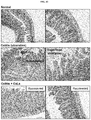
FIG. 37. Masson's trichrome staining for periodontal tissues.
FIG. 38. Masson's trichrome staining for upper gum.
FIG. 39. Masson's trichrome staining for foot and knee joint tissues.
FIG. 40. Experimental scheme for inducing Crohn's disease.
FIG. 41. Representative images for hematoxylin and eosin (H&E) staining of Crohn's disease.
FIG. 42. Experimental scheme for inducing colitis.
FIG. 43. Representative images for hematoxylin and eosin (H&E) staining of colitis.
DETAILED DESCRIPTION OF THE INVENTION
Recently, many studies showed that CID is related to advancing age and suggested chronic inflammation as being associated with age-related immuno-senescence diseases (1-29). Chronic inflammation was elucidated to occur via complex interactions between parenchymal cells and stromal cells, such as immune cells, endothelial cells, and fibroblasts. Among others, myeloid-originated cells, such as monocytes and activated macrophages, were identified as major effector cells in CID. Upon activation with bacterial lipopolysaccharide, IFN-gamma, and damaged cells, macrophages produced many inflammatory cytokines and cleared endogenous and exogenous inflammatory agents through transcriptional control by nuclear factor-kappa B (NF-kB). Macrophages were also further polarized to more specialized macrophage subtypes. However, their finely controlled differentiation and activation of myeloid cells were dysregulated in chronic inflammation. Local tissue hypoxia caused the metabolic changes of cells, such as parenchymal and stromal cells, through induction of hypoxia inducible factors (HIFs). HIFs, especially HIF-1, contribute to the abnormal activation of macrophages and are a major contributor to chronic inflammatory response.
Most CID are characterized by inflammation. Injured tissue become resistant to blood flow and delivery of oxygen, thus becoming a hypoxic environment. Proinflammatory mediators and a hypoxic condition can elicit an angiogenic response through the induction of HIF-1. Additionally, a local inflammatory reaction exists in most CID, including as respiratory diseases (e.g., asthma, chronic obstructive pulmonary disease, pneumonitis, and pulmonary fibrosis), ocular diseases (e.g., keratitis and age-related macular degeneration), autoimmune diseases, nephropathy, neurodegenerative diseases (e.g., Alzheimer's disease, stroke, Parkinson's disease, and multiple sclerosis), cardiovascular diseases (e.g., atherosclerosis, arteriosclerosis, and myocarditis), metabolic disorders (e.g., diabetes and obesity), musculoskeletal diseases (e.g., rheumatoid arthritis and osteoporosis), periodontal diseases (e.g., pulpitis and periodontitis), digestive diseases (e.g., non-alcoholic fatty liver disease, gastroenteritis, and chronic inflammatory bowel disease), and skin disorders (e.g., psoriasis and atopic dermatitis). During inflammation, vascular permeability is increased and monocytes, macrophages, platelets, mast cells, and other leukocytes are recruited under the attraction of chemokines. Therefore, disclosed herein are therapeutic approaches targeting activation of macrophages and regulation of extracellular matrix degradation by targeting HIF-1, NF-kB, and lactate dehydrogenase (LAD) A and B.
Methods of Treatment
Disclosed herein is a method of treating a chronic inflammatory disease, the method comprising administering to a subject in need thereof a therapeutically effective amount of calcium lactate.
Disclosed herein is a method of treating Alzheimer's disease, Parkinson's disease, atherosclerosis, and/or stroke, the method comprising administering to a subject in need thereof a therapeutically effective amount of calcium lactate.
Also disclosed is a method of treating multiple sclerosis, age-related macular degeneration, nonalcoholic fatty liver disease (NAFLD), and/or rheumatoid arthritis, the method comprising administering to a subject in need thereof a therapeutically effective amount of calcium lactate. In some embodiments, the method is for treating NAFLD and the NAFLD is nonalcoholic steatohepatitis (NASH).
Also disclosed is a method of treating pulpitis and/or periodontitis, the method comprising administering to a subject in need thereof a therapeutically effective amount of calcium lactate.
Further disclosed is a method of treating inflammatory bowel disease (IBD), the method comprising administering to a subject in need thereof a therapeutically effective amount of calcium lactate. In some embodiments, the IBD is Crohn's disease or ulcerative colitis.
Further disclosed is a method of treating spinal cord injury, cerebral hemorrhage, myocardial infarction, keratitis, and/or diabetic retinopathy, the method comprising administering to a subject in need thereof a therapeutically effective amount of calcium lactate.
Disclosed herein are methods of treating asthma, pulmonary fibrosis, obesity, gastroenteritis, chronic inflammatory bowel disease, and/or atopic dermatitis, the method comprising administering to a subject in need thereof a therapeutically effective amount of calcium lactate. Also disclosed herein are methods of treating chronic obstructive pulmonary disease, pneumonitis, keratitis, atherosclerosis, arteriosclerosis, myocarditis, diabetes, rheumatoid arthritis, pulpitis, periodontitis, and/or psoriasis, the method comprising administering to a subject in need thereof a therapeutically effective amount of calcium lactate. Also disclosed herein are methods of treating Alzheimer's disease, stroke, Parkinson's disease, multiple sclerosis, age-related macular degeneration, non-alcoholic fatty liver disease, and/or sepsis, the method comprising administering to a subject in need thereof a therapeutically effective amount of calcium lactate.
Also disclosed herein are methods of treating osteoporosis, the method comprising administering to a subject in need thereof a therapeutically effective amount of calcium lactate.
The term “subject” or “patient” encompasses mammals and non-mammals. Examples of mammals include, but are not limited to, any member of the mammalian class: humans, non-human primates such as chimpanzees, and other apes and monkey species; farm animals such as cattle, horses, sheep, goats, swine; domestic animals such as rabbits, dogs, and cats; laboratory animals including rodents, such as rats, mice and guinea pigs, and the like. Examples of non-mammals include, but are not limited to, birds, fish and the like. In some embodiments of the methods and compositions provided herein, the mammal is a human or a nonhuman.
As used herein, “treat”, “treating”, or “treatment” of one or more of the diseases disclosed herein by administration of the pharmaceutical composition refers to any lessening of severity, delay in onset, slowing of progression, or shortening of duration of one or more of the diseases disclosed herein, whether permanent or temporary, lasting or transient that can be attributed to or associated with administration of the composition.
The term “co-administration” or the like, as used herein, is meant to encompass administration of the selected two or more active agents to a single patient and is intended to include treatment regimens in which the agents are administered by the same or different route of administration or at the same or different time.
The pharmaceutical composition of the present disclosure can be administered in a therapeutically effective amount, and as used herein, the term “effective amount” or therapeutically effective amount” refers to an amount sufficient to treat or prevent diseases, at a reasonable benefit/risk ratio applicable to any medical treatment or prevention. The effective dosage level can be determined depending on severity of the disease, activity of the drug or active agent, a patient's age, body weight, health and sex, sensitivity to the drug, administration time, administration route, and excretion rate of the composition of the present disclosure, duration of treatment, drugs used simultaneously or in combination with the composition of the present disclosure, and other factors known in the medical field. The pharmaceutical composition of the present disclosure can be administered alone or in combination with other publicly-known drugs or components known as known for treating one or more of the diseases disclosed herein. It is important to administer the composition in the minimum amount that can exhibit the maximum effect without causing side effects, in consideration of all the above factors.
The pharmaceutical composition of the present disclosure can be administered such that the dosage per day of calcium lactate is, for example, about 10 mg/kg to about 1,000 mg/kg, about 10 mg/kg to about 500 mg/kg, about 10 mg/kg to about 250 mg/kg, about 10 mg/kg to about 200 mg/kg, about 10 mg/kg to about 100 mg/kg, about 1 mg/kg to about 100 mg/kg, about 1 mg/kg to about 75 mg/kg, about 1 mg/kg to about 50 mg/kg, about 1 mg/kg to about 25 mg/kg, or about 1 mg/kg to about 10 mg/kg. The administration frequency of the composition can be, but is not particularly limited to, once, twice, three times, four times, etc. divided doses a day.
The compositions containing the compound(s) described herein can be administered for prophylactic and/or therapeutic treatments. In therapeutic applications, the compositions are administered to a patient already suffering from a disease or condition, in an amount sufficient to cure or at least partially arrest the symptoms of the disease or condition. Amounts effective for this use will depend on the severity and course of the disease or condition, previous therapy, the patient's health status, weight, and response to the drugs, and the judgment of the treating physician. It is considered well within the skill of the art for one to determine such therapeutically effective amounts by routine experimentation (including, but not limited to, a dose escalation clinical trial).
In prophylactic applications, compositions containing the active agent described herein are administered to a patient susceptible to or otherwise at risk of one or more of the diseases disclosed herein. Such an amount is defined to be a “prophylactically effective amount or dose.” In this use, the precise amounts also depend on the patient's state of health, weight, and the like. It is considered well within the skill of the art for one to determine such prophylactically effective amounts by routine experimentation (e.g., a dose escalation clinical trial). When used in a patient, effective amounts for this use will depend on the severity and course of the disease, disorder or condition, previous therapy, the patient's health status and response to the drugs, and the judgment of the treating physician.
In the case wherein the patient's condition does not improve, upon the doctor's discretion the administration of the active agent described herein can be administered chronically, that is, for an extended period of time, including throughout the duration of the patient's life in order to ameliorate or otherwise control or limit the symptoms of the patient's disease or condition.
The amount of a given agent that will correspond to such an amount will vary depending upon factors such as the particular compound, disease condition and its severity, the identity (e.g., age, weight, gender, etc.) of the subject or host in need of treatment, but can nevertheless be routinely determined in a manner known in the art according to the particular circumstances surrounding the case, including, e.g., the specific agent being administered, the route of administration, the condition being treated, and the subject or host being treated.
Pharmaceutical Compositions
The term “calcium lactate” refers to a type of lactate metal salt that can, for example, exist as a hydrate, represented by C6H10O6Ca.5H2O in which calcium ion is bonded to lactate. Calcium lactate can be in the form of white powder or granules at room temperature, or anhydrous at a 120° C. heating condition, and has a solubility of 5% (w/v).
Calcium lactate can be formulated into pharmaceutical compositions for treating one or more of the diseases disclosed herein.
In various embodiments, the invention provides a pharmaceutical composition comprising a therapeutically effective amount of calcium lactate as an active agent for treating one or more of the diseases disclosed herein and a pharmaceutically acceptable polysaccharide, polymer, lipid, or combinations thereof. In some embodiments, the pharmaceutical composition comprises the calcium lactate and the polysaccharide.
In some embodiments, the invention provides an enteric coating of the calcium lactate such that the active agent is protected from the acidic environment of the stomach and absorbed in the small intestine before it reaches the large intestine when the active agent is administered orally.
The present invention also provides short-acting and long-acting pharmaceutical compositions comprising calcium lactate. In some embodiments, the long-acting compositions comprise calcium lactated coated with at least one enteric-coating material such as hydroxypropyl methyl cellulose phthalate (HPMCP), cellulose acetate phthalate (CAP), polyvinyl acetate phthalate (PVAP), shellac and a polymer of methacrylic acid, and an ester thereof.
The pharmaceutical composition of the invention can be formulated into pharmaceutical preparations for oral administration. Examples of the preparation include powders, tablets, capsules, granules or syrups, tablets and capsules, but not limited thereto.
Also provided herein are formulations of hydrogels particularly methylcellulose, poloxamer, pectin, and alginate hydrogel which can remain in a solution or nanoparticle form in vitro, and the gel can form when injected into the body and allow sustained release of calcium lactate. The relatively short drug release time which is the weakness of the hydrogel has been improved by increasing the interaction between the drug and hydrogel or by delaying the diffusion of the drug in the hydrogel.
The weight ratio of the calcium lactate to the polysaccharide can be, e.g., 1:<0.2 to 1:5, 1:0.01 to 1:5, 1:0.05 to 1:5, or 1:0.1 to 1:5. The weight ratio of the calcium lactate to the polysaccharide can be 1:<0.2. The weight ratio of the calcium lactate to the polysaccharide can be 1:0.2 to 1:5.
In some embodiments, the pharmaceutical composition further comprises a polymer or lipid. The weight ratio of the calcium lactate to the polymer or lipid can be at least 1:5. The weight ratio of the calcium lactate to the polymer or lipid can be 1:5 to 1:30, e.g., 1:5 to 1:30, 1:5 to 1:20, 1:5 to 1:10, 1:10 to 1:30, 1:10 to 1:20, or 1:20 to 1:30.
In some embodiments, the pharmaceutical composition further comprises a polymer and lipid. The weight ratio of the calcium lactate to the polymer and lipid can be at least 1:5. The weight ratio of the calcium lactate to the polymer and lipid can be 1:5 to 1:30, e.g., 1:5 to 1:30, 1:5 to 1:20, 1:5 to 1:10, 1:10 to 1:30, 1:10 to 1:20, or 1:20 to 1:30.
In some embodiments, the pharmaceutical composition comprises the calcium lactate and the polymer or lipid. The weight ratio of the calcium lactate to the polymer or lipid can be at least 1:5. The weight ratio of the calcium lactate to the polymer or lipid can be 1:5 to 1:30. The weight ratio of the calcium lactate to the polymer or lipid can be 1:5 to 1:30, e.g., 1:5 to 1:30, 1:5 to 1:20, 1:5 to 1:10, 1:10 to 1:30, 1:10 to 1:20, or 1:20 to 1:30.
In some embodiments, the pharmaceutical composition comprises the calcium lactate and the polymer and lipid. The weight ratio of the calcium lactate to the polymer and lipid can be at least 1:5. The weight ratio of the calcium lactate to the polymer and lipid can be 1:5 to 1:30. The weight ratio of the calcium lactate to the polymer and lipid can be 1:5 to 1:30, e.g., 1:5 to 1:30, 1:5 to 1:20, 1:5 to 1:10, 1:10 to 1:30, 1:10 to 1:20, or 1:20 to 1:30.
In some embodiments, the weight ratio of the polymer to the lipid can be 1:0.1 to 1:50, 1:0.1 to 1:20, 1:0.1 to 1:10, 1:0.1 to 1:5, 1:0.1 to 1:2, 1:0.1 to 1:1, 1:0.1 to 0.5, or 1:0.1 to 1:0.2.
Polysaccharides suitable for use in the composition can be a cellulose derivative (e.g., carboxymethyl cellulose (CMC), ethyl cellulose (EC), hydroxypropyl methyl cellulose (HPMC), methyl cellulose (MC)), pectin, hyaluronic acid, starch, guar gum, chitosan, gelatin, collagen, alginate, alginic acid, or combinations thereof.
Polymers suitable for use in the composition can be a poloxamer, polyvinylpyrrolidone, polyethylene glycol (PEG), polyglycolic lactic acid (PLGA), or combinations thereof.
Lipids suitable for use in the composition can be a mono- or tri-fatty acid glycerin ester or polyethylene glycol, polyethylene glycol esters of vegetable oils, fatty acid propylene glycol esters, sesame oil, soybean oil, castor oil, corn oil, palm oil, peanut oil, cacao oil, cottonseed oil, sunflower seed oil, safflower oil, almond oil, olive oil, hydrogenated oil, oleic acid, linolenic acid, linoleic acid, palmitic acid, palmitoleic acid, arachadonic acid, myristic acid, capric acid, caprylic acid, lauric acid, stearic acid, ethyl oleate, isopropyl palmitate, octyldodecyl myristate, acetyl palmitate, lauryl alcohol, oleyl alcohol, acetyl alcohol, stearyl alcohol, or combinations thereof.
The invention is further directed to a pharmaceutical composition comprising a therapeutically effective amount of calcium lactate as an active agent for treating one or more of the diseases disclosed herein, wherein the calcium lactate is coated with a pharmaceutically acceptable enteric coating. In some embodiments, the enteric coating comprises hydroxypropyl methyl cellulose phthalate (HPMCP), cellulose acetate phthalate (CAP), polyvinyl acetate phthalate (PVAP), shellac, polymer of methacrylic acid and an ester thereof, or combinations thereof. In some embodiments, the weight ratio of the calcium lactate to the enteric coating is 10:0.5 to 10:15, 10:0.5 to 1:1, 10:0.5 to 10:5, 10:0.5 to 10:3, 10:0.5 to 10:2, 10:0.5 to 10:1, 10:0.5 to 1:0.8.
By “pharmaceutically acceptable,” as used herein, refers a material, such as a carrier or diluent, which does not abrogate the biological activity or properties of the compound, and is relatively nontoxic, i.e., the material can be administered to an individual without causing undesirable biological effects or interacting in a deleterious manner with any of the components of the composition in which it is contained.
A pharmaceutical composition, as used herein, refers to a mixture of calcium lactate with other chemical components that are pharmaceutically acceptable, such as but not limited to carriers, stabilizers, diluents, disintegrants, suspending agents, thickening agents, binders, antimicrobial agents, antimicrobial preservatives, antioxidants, and/or buffering agents. The pharmaceutical composition facilitates administration of the calcium lactate to a subject.
The term “carrier,” as used herein, refers to relatively nontoxic chemical compounds or agents that facilitate the incorporation of a compound into cells or tissues. The term “diluent” refers to chemical compounds that are used to dilute the compound of interest prior to delivery. Diluents can also be used to stabilize compounds because they can provide a more stable environment. Pharmaceutically acceptable additives include diluents, binders, solubilizers, solubility enhancers, pore formers, osmotic agents, gas formers, lubricants and fluidizers well known in the art, but not limited thereto.
Diluents can include lactose, fructose, dextrose, sucrose, maltose, microcrystalline cellulose, starch, calcium hydrogen phosphate, mannitol or a mixture thereof, but not limited thereto. Other diluents include microcrystalline cellulose, lactose, mannitol, calcium phosphate and the like.
Examples of binders can include povidone, hydroxypropylcellulose, polyvinylalcohol, hydroxypropylmethylcellulose, carboxymethyl-cellulose sodium and thereof.
Solubilizing agents include surfactants, cyclodextrins and derivatives thereof, lipophilic substances or mixtures thereof, but are not limited thereto.
Surfactants include water soluble or water dispersible nonionic, nonpolar nonionic, anionic, cationic, amphoteric or ionic surface activators or mixtures thereof, but are not limited thereto.
Examples of disintergrants include crospovidone, croscarmellose sodium, glycolic acid starch sodium, and examples of the lubricant include magnesium stearate, calcium stearate, sodium stearyl fumarate and thereof.
The pharmaceutical compositions of the invention can further include antimicrobial agents, such as benzyl alcohol, chlorobutanol, phenylethyl alcohol, phenyl-mercuric acetate, potassium sorbate, and sorbic acid. Antifungal agents include such compounds as benzoic acid, butylparaben, ethylparaben, methylparaben, propylparaben, and sodium benzoate.
Antimicrobial preservatives can be added to the pharmaceutical compositions of the present invention in order to protect them against the growth of potentially harmful microorganisms, which usually invade the aqueous phase, but in some cases can also grow in the oil phase of a composition. Thus, preservatives with both aqueous and lipid solubility are desirable. Suitable antimicrobial preservatives include, e.g., alkyl esters of p-hydroxybenzoic acid, propionate salts, phenoxyethanol, methylparaben sodium, propylparaben sodium, sodium dehydroacetate, benzalkonium chloride, benzethonium chloride, benzyl alcohol, hydantoin derivatives, quaternary ammonium compounds and cationic polymers, imidazolidinyl urea, diazolidinyl urea, and trisodium ethylenediamine tetracetate (EDTA).
Antioxidants can be added to protect all of the ingredients of the pharmaceutical compositions from damage or degradation by oxidizing agents present in the composition itself or the use environment, e.g., anoxomer, ascorbyl palmitate, butylated hydroxyanisole, butylated hydroxytoluene, hypophosphorous acid, potassium metabisulfite, propyloctyl and dodecyl gallate, sodium metabisulfite, sulfur dioxide, and tocopherols.
Buffering agents can be used to maintain a desired pH of the pharmaceutical compositions once established, from the effects of outside agents and shifting equilibria of components of the composition.
The pharmaceutical compositions described herein can be prepared following techniques known in the art, for example, in Remington: The Science and Practice of Pharmacy, Nineteenth Ed (Easton, Pa.: Mack Publishing Company, 1995); Hoover, John E., Remington's Pharmaceutical Sciences, Mack Publishing Co., Easton, Pa. 1975; Liberman, H. A. and Lachman, L., Eds., Pharmaceutical Dosage Forms, Marcel Decker, New York, N.Y., 1980; and Pharmaceutical Dosage Forms and Drug Delivery Systems, Seventh Ed. (Lippincott Williams & Wilkins 1999), herein incorporated by reference in their entirety.
In some embodiments, the pharmaceutical compositions of the invention are short-acting. The term “short-acting” refers to a composition that releases substantially all of the active agent within 48 hours when tested in an in vitro dissolution test described herein, for example, from time of delivery, time 0, until about 1 hour to about 48 hours, until about 3 hours to about 24 hours, until about 6 hours to about 24 hours, or until about 12 hours to about 24 hours.
For example, upon placement of the composition in an in vitro dissolution test comprising an elution test method (e.g., from Labfine Co., Mumbai, India) at 300 rpm in 200 ml aqueous medium having a pH of 6.8 at 37° C. using a nylon filter having a pore size of 45 μm, at least about 40% of the active agent is released after 6 hours, at least about 60% of the active agent is released after 12 hours, at least about 80% of the active agent is released after 24 hours, and/or at least about 90% of the active agent is released after 48 hours. In some embodiments, upon placement of the composition in an in vitro dissolution test comprising an elution test method (e.g., from Labfine Co., Mumbai, India) at 300 rpm in 200 ml aqueous medium having a pH of 6.8 at 37° C. using a nylon filter having a pore size of 45 μm, at least about 40% to about 60% of the active agent is released after 6 hours, at least about 60% to about 80% of the active agent is released after 12 hours, at least about 80% to about 90% of the active agent is released after 24 hours, and/or at least about 90% to about 100% of the active agent is released after 48 hours.
In some embodiments, the pharmaceutical compositions of the invention are long-acting. The term “long-acting” is intended to mean composition that releases the active agent slowly after the initial dosage, for example, from time of delivery, time 0, until about 48 hours to about 192 hours, until about 72 hours to about 192 hours, until about 96 hours to about 192 hours, until about 120 hours to about 192 hours, or until about 144 hours to about 192 hours.
In some embodiments, upon placement of the composition in an in vitro dissolution test comprising an elution test method (e.g., from Labfine Co., Mumbai, India) at 300 rpm in 200 ml aqueous medium having a pH of 6.8 at 37° C. using a nylon filter having a pore size of 45 μm, less than about 40% of the active agent is released after 24 hours, less than about 60% of the active agent is released after 48 hours, less than about 80% of the active agent is released after 72 hours, less than about 90% of the active agent is released after 144 hours. In some embodiments, upon placement of the composition in an in vitro dissolution test comprising an elution test method (e.g., from Labfine Co., Mumbai, India) at 300 rpm in 200 ml medium having a pH of 6.8 at 37° C. using a nylon filter having a pore size of 45 μm, about 20% to about 50% of the active agent is released after 24 hours, about 20% to about 40% of the active agent is released after 24 hours, about 40% to about 70% of the active agent is released after 48 hours, about 40% to about 60% of the active agent is released after 48 hours, about 40% to about 80% of the active agent is released after 72 hours, about 50% to about 80% of the active agent is released after 72 hours, about 60% to about 90% of the active agent is released after 144 hours, or about 70% to about 90% of the active agent is released after 144 hours.
In some embodiments, upon placement of the composition in an in vitro dissolution test comprising a USP Paddle method at a paddle speed of 50 rpm at 37° C., when the composition is placed in 0.1 N HCl for 120 minutes followed by adjusting to pH 6.8 with phosphate buffer, less than about 20% of the active agent is released after 30 minutes, less than 30% of the active agent is released after 60 minutes, less than 50% of the active agent is released after 120 minutes, and/or less than 10% of the active agent is released after 120 minutes.
In some embodiments, the pharmaceutical compositions of the invention are injectable dosage forms. For parenteral injections, appropriate formulations can include aqueous or nonaqueous solutions, preferably with physiologically compatible carriers.
Pharmaceutical formulations for parenteral administration include aqueous solutions of the active compound(s) in water-soluble form. Additionally, suspensions of the active agent can be prepared as appropriate oily injection suspensions. Suitable lipids or lipophilic carriers include fatty oils such as sesame oil, or synthetic fatty acid esters, such as ethyl oleate or triglycerides, or liposomes. The lipid can be a mono- or tri-fatty acid glycerin ester or polyethylene glycol, polyethylene glycol esters of vegetable oils, fatty acid propylene glycol esters, sesame oil, soybean oil, castor oil, corn oil, palm oil, peanut oil, cacao oil, cottonseed oil, sunflower seed oil, safflower oil, almond oil, olive oil, hydrogenated oil, oleic acid, linolenic acid, linoleic acid, palmitic acid, palmitoleic acid, arachadonic acid, myristic acid, capric acid, caprylic acid, lauric acid, stearic acid, ethyl oleate, isopropyl palmitate, octyldodecyl myristate, cetyl palmitate, lauryl alcohol, oleyl alcohol, cetyl alcohol, stearyl alcohol, or combinations thereof.
Since the medical application of hydrogels, numerous hydrogels have been developed and studied in many fields, including the medical, pharmaceutical, and cosmetic industries. Although hydrogels are generally biocompatible, they have various problems that are limiting the delivery of a drug, and various efforts are made to solve the problems. A hydrogel is a three-dimensional structure composed of a network of hydrophilic polymers. More than 90% of the components are composed of water. Hydrogels have been actively studied in the biomedical field due to their similarity to bio-tissue such as high moisture content, porous structure, relatively soft properties, and biocompatibility. Hydrogels can exhibit various properties depending on the kind of polymer used as the main chain or the crosslinking method adopted. When a polymer of polyacrylic acid series polymer or a synthetic compound such as polyvinyl alcohol is used, the biocompatibility is low, but the chemical modification is easy, so that the engineering application is very easy. On the other hand, when natural compounds, especially pectin, alginate, collagen, fibrin and hyaluronic are used as the main chain, the chemical modification is difficult. Nevertheless, there are advantages of using these materials that are biologically derived components, as it is suitable for clinical application and there are few side effects such as immune inflammation reaction at the time of transplantation.
It is the cross-linking method that affects the properties of the hydrogel as well as the type of polymer used. Even if the same polymer is used as the main chain, the hydrogel having completely different characteristics can be obtained if the crosslinking method is different. The method of crosslinking hydrogels can be broadly divided into physical and chemical methods. Physical crosslinking methods include ionic interaction, hydrophobic interaction, hydrogen bond, and reversible crosslinking by structural entanglement. These crosslinking methods can easily induce the formation of a three-dimensional network structure without the need for a separate chemical additive or complicated process. On the other hand, the chemical crosslinking method typically forms covalent bond that result an irreversible and stable network as compared with the physical crosslinking method. Hydrogels with excellent biocompatibility and various physicochemical properties have been extensively studied in biomedical fields such as drug delivery and tissue engineering. Most hydrogels exhibit shorter drug release time than other drug delivery systems due to their high water content, and there is a need to develop a system with a longer drug release time.
In some embodiments, the pharmaceutical compositions of the invention are oral dosage forms. For oral administration, the compound described herein can be formulated readily by combining the active agent with pharmaceutically acceptable carriers or excipients well known in the art. Such carriers enable the active agent described herein to be formulated as tablets, powders, pills, dragees, capsules, liquids, gels, syrups, elixirs, slurries, suspensions and the like, for oral ingestion by a patient to be treated.
Pharmaceutical preparations for oral use can be obtained by mixing one or more solid carriers with the compound described herein, optionally grinding the resulting mixture, and processing the mixture of granules, after adding suitable auxiliaries, if desired, to obtain tablets or dragee cores. Suitable excipients include, but are not limited to, fillers such as sugars, including lactose, sucrose, mannitol, or sorbitol; cellulose preparations such as: for example, maize starch, wheat starch, rice starch, potato starch, gelatin, gum tragacanth, methylcellulose, microcrystalline cellulose, hydroxypropylmethylcellulose, sodium carboxymethylcellulose; or others such as: polyvinylpyrrolidone (PVP or povidone) or calcium phosphate. If desired, disintegrating agents can be added, such as the cross-linked croscarmellose sodium, polyvinylpyrrolidone, agar, or alginic acid or a salt thereof such as sodium alginate.
Dragee cores are provided with suitable coatings. For this purpose, concentrated sugar solutions can be used, which can optionally contain gum arabic, talc, polyvinylpyrrolidone, carbopol gel, polyethylene glycol, and/or titanium dioxide, lacquer solutions, and suitable organic solvents or solvent mixtures. Dyestuffs or pigments can be added to the tablets or dragee coatings for identification or to characterize different combinations of active compound doses.
Pharmaceutical compositions can be formulated in conventional manner using one or more pharmaceutically acceptable carriers comprising excipients and auxiliaries which facilitate processing of the active agent into preparations which can be used pharmaceutically. Proper formulation is dependent upon the route of administration chosen. Any of the well-known techniques can be used as suitable and as understood in the art. Pharmaceutical compositions described herein can be manufactured in a conventional manner, such as, by way of example only, by means of conventional mixing, dissolving, granulating, dragee-making, levigating, emulsifying, encapsulating, entrapping or compression processes.
Aqueous suspensions can also contain one or more polymers as suspending agents. The polymer can be a poloxamer, polyvinylpyrrolidone, polyethylene glycol (PEG), polyglycolic lactic acid (PLGA), or combinations thereof. Other useful polymers include water-soluble polymers such as cellulosic polymers, e.g., hydroxypropyl methylcellulose, and water-insoluble polymers such as cross-linked carboxyl-containing polymers. Useful compositions can also include a mucoadhesive polymer, selected from, for example, carboxymethylcellulose, carbomer (acrylic acid polymer), poly(methylmethacrylate), polyacrylamide, polycarbophil, acrylic acid/butyl acrylate copolymer, sodium alginate, and dextran.
In some embodiments, provided are pharmaceutical dosage forms comprising calcium lactate and a pharmaceutically acceptable enteric coating in order to control the release of the active agent. In some embodiments, the coating is a film and, in another embodiment, it is a membrane. The enteric coating, e.g., film or membrane, can serve to delay release until after the stomach and to protect the active agent from gastric fluid. The enteric coating can comprise one or more substances preferably of a polymeric nature (e.g., methacrylates etc.; polysaccharides etc. as described in more detail below) or combination of more than one such substance, optionally including other excipients, such as, for example, plasticizers. In some embodiments, the enteric coating comprises hydroxypropyl methyl cellulose phthalate (HPMCP), cellulose acetate phthalate (CAP), polyvinyl acetate phthalate (PVAP), shellac, polymer of methacrylic acid and an ester thereof, or combinations thereof. In some embodiments of the invention the composition comprises a hydrogel-forming polymer and further polymers able to achieve a desired delay (or other change) in the release of the active agent.
In the case of tablets for oral use, carriers which are commonly used include lactose and corn starch. Lubricating agents, such as magnesium stearate, are also typically added. For oral administration in a capsule form, useful diluents include lactose and dried corn starch.
The sterile injectable preparation can also be a sterile injectable suspension in a non-toxic parenterally acceptable carrier such as lipids.
The amount of calcium lactate included in the pharmaceutical compositions can be, but is not limited to, from 1 wt % to 50 wt %, from 1 wt % to 40 wt %, from 1 wt % to 35 wt %, from 1 wt % to 30 wt %, from 1 wt % to 20 wt %, from 1 wt % to 15 wt %, or from 1 wt % to 10 wt % based on the total weight of the final composition. The concentration of the calcium lactate included in a single dose of the pharmaceutical composition can be, but is not limited to, 2.5 mM to 100 mM, 2.5 mM to 50 mM, 2.5 mM to 25 mM, 5 mM to 100 mM, 5 mM to 50 mM, 5 mM to 25 mM, 10 mM to 100 mM, 10 mM to 50 mM, or 10 mM to 25 mM.
Solid oral pharmaceutical compositions can be prepared by conventional techniques such as dry granulation, direct compression, wet granulation, extrusion spheronization, melt granulation or compression coating, but not limited thereto. The coating can be applied as described below and can vary as to thickness and density. The amount of coating is defined by the additional weight added to (gained by) the dry composition (e.g., bead or core containing the calcium lactate) of the invention. Weight gain can be in the range of 0.1% to 50%, 1% to 20%, 1% to 15%, 3% to 10%, 5% to 12%, or 8% to 12%.
The coating process can be carried out by any suitable means such as, for example, by use of a coating machine which applies a solution of a polymer coat (as described above in particular) to the composition. Polymers for coating are either provided by the manufacturer in ready-made solutions for direct use or can be made up before use following manufacturers' instructions.
Unit Dosages and Kits
The term “unit dosage form” refers to a physically discrete unit suitable as a single dosage, each unit containing a predetermined quantity of active ingredient, in association with a suitable pharmaceutical excipient, by which one or more is used throughout the dosing regimen to produce a desired therapeutic effect, e.g., treating one or more of the diseases disclosed herein.
The pharmaceutical compositions described herein can be in unit dosage forms suitable for single administration of precise dosages. In unit dosage form, the formulation is divided into unit doses containing appropriate quantities of one or more active agents. The unit dosage can be in the form of a package containing discrete quantities of the formulation. Non-limiting examples are packaged tablets or capsules, and powders in vials or ampoules. Aqueous suspension compositions can be packaged in single-dose non-reclosable containers. Alternatively, multiple-dose reclosable containers can be used, in which case it is typical to include a preservative in the composition. By way of example only, formulations for parenteral injection can be presented in unit dosage form, which include, but are not limited to ampoules, or in multi-dose containers.
The daily dosages appropriate for calcium lactate can be from about 1 mg/kg to about 1000 mg/kg, about 10 mg/kg to about 750 mg/kg, about 10 mg/kg to about 500 mg/kg, or about 100 mg/kg to 500 mg/kg per body weight. An indicated daily dosage in the larger mammal, including, but not limited to, humans, is in the range from about 5 mg to about 100,000 mg, conveniently administered in divided doses, including, but not limited to, up to four times a day or in long-acting form. Suitable unit dosage forms for administration include from about 10 mg to about 1000 mg, about 100 mg to about 1000 mg, about 500 mg to about 750 mg, about 25 mg to about 250 mg, about 50 mg to about 100 mg, about 10 mg to about 200 mg, or about 10 mg to about 250 mg of the active agent. The administration frequency of the composition of the present disclosure can be, but is not particularly limited to, once, twice, three times, four times, etc. divided doses a day. An appropriate “effective” amount in any individual case can be determined using techniques, such as a dose escalation study.
The foregoing ranges are merely suggestive, as the number of variables in regard to an individual treatment regime is large, and considerable excursions from these recommended values are not uncommon. Such dosages can be altered depending on a number of variables, not limited to the activity of the compound used, the disease or condition to be treated, the mode of administration, the requirements of the individual subject, the severity of the disease or condition being treated, and the judgment of the practitioner.
Toxicity and therapeutic efficacy of such therapeutic regimens can be determined by standard pharmaceutical procedures in cell cultures or experimental animals, including, but not limited to, the determination of the LD50 (the dose lethal to 50% of the population) and the ED50 (the dose therapeutically effective in 50% of the population). The dose ratio between the toxic and therapeutic effects is the therapeutic index and it can be expressed as the ratio between LD50 and ED50. Compounds exhibiting high therapeutic indices are preferred. The data obtained from cell culture assays and animal studies can be used in formulating a range of dosage for use in human. The dosage of the active agent lies preferably within a range of circulating concentrations that include the ED50 with minimal toxicity. The dosage can vary within this range depending upon the dosage form employed and the route of administration utilized.
According to the present invention, the pharmaceutical compositions can be in the form of a sterile injectable preparation, for example a sterile injectable aqueous or oleaginous suspension. The pharmaceutical compositions of the present invention can be orally administered in any orally acceptable dosage form including, but not limited to, capsules, tablets, aqueous suspensions or solutions.
The invention is also directed to a sterile glass or polyolefin container comprising a pharmaceutical composition disclosed herein. In some embodiments, the container is non-DEHP (Bis(2-ethylhexyl) phthalate (di-2-ethylhexyl phthalate, diethylhexyl phthalate, DEHP; dioctyl phthalate, DOP) or non-PVP (Polyvinylpyrrolidone).
A kit comprises suitable containers, such as boxes, individual bottles, bags or ampoules. Suspension compositions can be packaged in single-dose non-reclosable containers or multiple-dose reclosable containers.
Incorporated herein by reference in their entirety are US2017/0360727A1, published Dec. 21, 2017, and PCT/M2017/054091, Int'l Filing Date: Jul. 7, 2017. Also incorporated herein by reference in their entirety are U.S. Appl. No. 62/616,923 filed Jan. 12, 2018.
EXAMPLES Example 1. Cell Culture for Age-Related Macular Disease
Human retinal pigment epithelium cell line, ARPE-19, was purchased from American Type Culture Collection (Manassas, Va., USA). The ARPE-19 cells were maintained in RPMI1640 containing 10% fetal bovine serum (Life Technologies, Gaithersburg, Md., USA), 100 IU/ml penicillin, and 100 μg/ml streptomycin (Welgene, Daegu, South Korea).
Example 2. Cell Culture for Alzheimer's Disease
Human Brain cell line, SK-N-SH, was purchased from American Type Culture Collection (Manassas, Va., USA). The SK-N-SH cells were maintained in RPMI1640 containing 10% fetal bovine serum (Life Technologies, Gaithersburg, Md., USA), 100 IU/ml penicillin, and 100 μg/ml streptomycin (Welgene, Daegu, South Korea).
Example 3. Cell Culture for Fibroblast
Human Colon Fibroblast cell line, CCD-18-Co, was purchased from American Type Culture Collection (Manassas, Va., USA). The CCD-18Co cells were maintained in DMEM containing 10% fetal bovine serum (Life Technologies, Gaithersburg, Md., USA), 100 IU/ml penicillin, and 100 μg/ml streptomycin (Welgene, Daegu, South Korea).
Example 4. Cell Culture for Non-Alcoholic Steatohepatitis (NASH)
Human Hepatocellular carcinoma cell line, HepG2, was purchased from American Type Culture Collection (Manassas, Va., USA). The HepG2 cells were maintained in RPMI 1640 containing 10% fetal bovine serum (Life Technologies, Gaithersburg, Md., USA), 100 IU/ml penicillin, and 100 μg/ml streptomycin (Welgene, Daegu, South Korea).
Example 5. Cell Culture for Vascular Disease
Human Umbilical Vein Endothelial cell line, HUVEC, was purchased from American Type Culture Collection (Manassas, Va., USA). The HUVEC cells were Endothelial Cell Growth Medium 2 maintained in Endothelial Cell Growth Medium 2 Supplement MIX (PromoCell, Heidelberg, Germany)
Example 6. Macrophage Culture
Human Hepatic Stellate cell line, LX-2, was purchased from Sigma-Aldrich (St Louis, Mo., USA). The LX-2 cells were maintained in DMEM containing 2% fetal bovine serum (Life Technologies, Gaithersburg, Md., USA), 100 IU/ml penicillin, and 100 μg/ml streptomycin (Welgene, Daegu, South Korea).
Human Monocyte cell line, THP-1, was purchased from American Type Culture Collection (Manassas, Va., USA). The THP-1 cells were maintained in RPMI1640 containing 10% fetal bovine serum (Life Technologies, Gaithersburg, Md., USA), 100 IU/ml penicillin, 100 μg/ml streptomycin (Welgene, Daegu, South Korea), and 0.55 uM 2-Mecaptpethanol. Cell were cultured in both conditions, one condition is a humidified atmosphere at 37° C. containing 5% CO2, and another is hypoxia culture condition was maintained at 1% oxygen, 5% and 94%.
Example 7. Regents
Calcium lactate (CaLa) and lipopolysaccharide (LPS) were purchased from Sigma-Aldrich (St Louis, Mo., USA). 2-mercaptoethanol was purchased from Gibco (Grand Island, N.Y., USA). Phorbol 12-myristate 13-acetate (PMA) was purchased from Enzo Lifesciences (Enzo Diagnostic, NY, USA). Human Interferon-gamma, Human Interleukin-4 (IL-4) and Human Interleukin-10 (IL-10) were purchased from BPS Bioscience (San Diego, Calif., USA). Paraformaldehyde (PFA) solution (4%) was purchased from Biosesang (Biosesang Inc., Gyeonggi, Korea).
Example 8. Cell Viability Assay
The live cell movie analyzer, JuLI™ Br (NanoEnTek Inc., Seoul, Korea) in ARPE-19 cells, was used to confirm the toxicity of CaLa. Cells were seeded into 6-well culture plates at a density of 2.5×10⁵ cells/well in medium. After 24 h incubation, the cells treated with 2.5 mM CaLa for 12 hr and 24 hr. The cells were continuously monitored for 24 h during CaLa treatment time. (30, 31)
Example 9. Macrophage Differentiation
The human monocytic cell line, THP-1, was purchased from American Type Culture Collection (Manassas, Va., USA). THP-1 cells were maintained in RPMI 1640 media (Hyclone Laboratories, Inc., Logan, Utah, USA) supplemented with 10% fetal bovine serum (Hyclone Laboratories, Inc., Logan, Utah, USA), 100 IU/ml penicillin (Welgene, Daegu, South Korea), 100 μg/ml streptomycin (Welgene, Daegu, South Korea), and 0.55 μM 2-mercaptoethanol (Gibco, Grand Island, N.Y., USA). THP-1 cell was maintained in a humidified atmosphere of 5% CO2, at 37° C. THP-1 monocytes are differentiated into macrophages by 12 h incubation with Phorbol 12-myristate 13-acetate (PMA) and 100 μg/ml (Enzo Diagnostic, NY, USA) in RPMI 1640 medium. Macrophages were polarized in M1 macrophages by 24 h incubation with 20 ng/ml of IFN-γ (BPS Bioscience, San Diego, Calif., USA) and 100 μg/ml of LPS (Sigma-Aldrich, St Louis, Mo., USA) in RPMI 1640 medium. Macrophage M2 polarization was obtained by 24 h incubation with 20 ng/ml of IL-4 (BPS Bioscience, San Diego, Calif., USA) and 20 ng/ml of IL-10 (BPS Bioscience, San Diego, Calif., USA) in RPMI 1640 medium. (32)
Example 10. Optical Observation for Cell Viability
The live cell movie analyzer, JuLI™ Br (NanoEnTek Inc., Seoul, Korea) in ARPE-19 cells, was used to confirm the toxicity of CaLa. Cells were seeded into 6-well culture plates at a density of 2.5×10⁵ cells/well in medium. After a 24 h incubation, the cells treated with 2.5 mM CaLa for 12 h and 24 hr. The cells were continuously monitored for 24 h during CaLa treatment time.
Example 11. Western Blot Analysis
To prepare whole cell lysate, cells were lysed with lysis buffer (1% NP-40, 0.25% sodium deoxycholate, 150 mM NaCl, 1 mM EDTA, 1% Triton X-100, 50 mM Tris-HCl (pH 7.4), 10% Glycerol) with protease inhibitor cocktail (Roche, Basel, Switzerland) and phosphatase inhibitors (NA3VO4 1 mM, NaF 100 mM). Then, protein concentration was measured using the Bicinchoninic acid (BCA) assay kit (Thermo Scientific, Waltham, Mass.). Whole cell or nuclear lysates were transferred onto polyvinylidene fluoride membranes (Merck Millipore, Billerica, Mass., USA). After blocking in 5% non-fat milk (Bio-Rad, Hercules, Calif., USA) for 1 h, the membranes were incubated overnight at 4° C. with primary antibody diluted in TBST containing 100 mM Tris-HCl (pH 7.5), 1.5 M NaCl, and 0.5% Tween-20 obtained from Sigma-Aldrich, to which 5% BSA and 0.1% sodium azide (Sigma-Aldrich) had been added. Specific primary antibodies to HIF-1α (1:1000), HIF-2α (1:1000), LDH-A (1:1000), LDH-B (1:1000), TLR-4 (1:1000), NF-κB (1:1000), VEGF (1:1000), α-tubulin (1:1000), Actin (1:1000), and GAPDH (1:5000). The next day, the membranes were washed with TBST and incubated for 2 h with anti-rabbit secondary antibody (1:10000). Immunoblots were developed using a western blot detection reagent (Abclone, Seoul, Korea) and exposed to x-ray film (Agfa, Leverkusen, Germany) according to the manufacturer's protocol.
Example 12. Immunocytochemistry
ARPE-19 and SK-N-SH cells were fixed on bio-coated coverslips (BD bioscience, San Jose, Calif., USA) by 4% PFA for 20 min and then incubated with the primary antibodies for 15 h. The primary antibodies were as follows: HIF-1α (1:300, BD Biosciences, San Jose, Calif., USA); LDH-A (1:300, Santa Cruz Biotechnology, Santa Cruz, Calif., USA); LDH-B (1:300, Santa Cruz Biotechnology, Santa Cruz, Calif., USA); NF-κB (1:300, Santa Cruz Biotechnology, Santa Cruz, Calif., USA); TLR-4 (1:300, Santa Cruz Biotechnology, Santa Cruz, Calif., USA); apoE (1:200, Santa Cruz Biotechnology, Santa Cruz, Calif., USA). After PBS wash, the cells were incubated with anti-mouse secondary biotinylated antibody (1:2000, Vector Laboratorie, Burlingame, Calif., USA) and visualized with streptavidin conjugated to Fluorescein (Vector Laboratorie, Burlingame, Calif., USA). Coverslips were mounted on microscope slides with VECTASHIELD® Hard Set™ Mounting Medium with DAPI (Vector Laboratories, Burlingame, Calif., USA). Confocal fluorescence images were obtained with confocal laser scanning microscopy (LSCM, Nikon A1+, Tokyo, Japan) under a 60× oil immersion lens.
Example 13. Animal Model for NASH
All experiments were performed under the institutional guidelines established by the Institutional Animal Care and Use Committee at Gachon University (IACUC-2017-0008). Female 5-week-old C57BL/6 mice were purchased form KOATEC (Pyeongtaek, South Korea) and were fed Methionine-Choline diet (MCD) for 4 weeks. When the mice weighed 15 g each, the mice were autopsied randomly to identify nonalcoholic steatohepatitis (NASH). The mice were injected twice daily (B.I.D) with subcutaneous calcium lactate (2 mg/kg) for 4 weeks.
Example 14. Animal Model for Age-Related Macular Degeneration
All experiments were performed under the institutional guidelines established by the Institutional Animal Care and Use Committee at Gachon University (IACUC-2017-0008). Six-week-old female C57BL/6 mice (20-25 g) were purchased from Orient (Charles River Korea, Seoul, Korea). All animals were maintained in a 12-hour light/dark cycle (light on, 08:00) at 22° C.−25° C. with free access to food and water. The mice were placed individually in an induction chamber, and anesthesia was induced with 2% isoflurane (HANA PHARM CO., Seoul, Korea). The lesions were induced in each eye of the mice by laser photocoagulation (SDL405-700, SD Laser, China). The laser pulses were from blue laser (wavelength, 405 nm; SD Laser). Laser parameters were 100 μm spot size, 700 mw power and 1 s exposure time. A total of 15 mice were randomized into three groups: (i) not subjected to laser induction (n=3); (ii) subjected to laser burns and injected with the vehicle buffer (n=5); and (iii) CaLa, 2 mg/kg/day (B.I.D) (n=7). CaLa treatment was started the days after laser photocoagulation and was treated subcutaneously for 14 days. (33, 34)
Example 15. Histological Analysis for NASH Model
Mouse liver tissues (n=5) were dissected and fixed in 4% paraformaldehyde PBS solution for 15 h at 4° C. Fixed liver tissues were embedded in paraffin and sectioned at 10 μm. Slides were incubated at 55° C. for 2 h, and subsequently deparaffinized in xylene and rehydrated in a graded ethanol series and used for hematoxylin (Merck, Darmstadt, Germany) and eosin (H&E) staining (35). Paraffin sections (10 μm thick) stained with H&E were imaged using a Leica DM 1000 LED microscope (Leica Microsystems, Wetzlar, Germany).
Example 16. Histological Analysis for Age-Related Macular Degeneration
The histopathologic examination of retinal lesions was performed 2 weeks after 2 mg/kg of CaLa was administered subcutaneously. The enucleated eyes were fixed with 4% paraformaldehyde (PFA) for 24 h at 4° C., and eyes obtained by removing the anterior segments were washed three times in phosphate-buffered saline. The fixed flat-mounts were dehydrated in a graded series of ethanol and embedded in paraffin. Paraffin sections (10 μm thick) were stained with hematoxylin (Merck, Darmstadt, Germany) and eosin (Sigma-Aldrich, St Louis, Mo., USA) (H&E) and were imaged using a Leica DM 1000 LED microscope (Leica Microsystems, Wetzlar, Germany). The choroidal lesion and neovascularization on the flat-mounts were observed by confocal microscopy (Carl Zeiss, Oberkochen, Germany).
Example 17. Masson Trichrome Stain
Mouse liver tissues (n=5) were dissected and fixed in 4% paraformaldehyde PBS solution for 15 h at 4° C. Fixed tissues were embedded in paraffin and sectioned at 5 mm. Slides were incubated at 55° C. for 2 h, and subsequently deparaffinized in xylene and rehydrated in a graded ethanol series and re-fix in Bouin's solution for 1 hr at 56° C., remove the yellow color to rinse running tap water for 5-10 min and stained Biebrich scarlet-acid fuchsin solution for 5 min then in phosphomolybdic-phosphotungstic acid solution (ratio 1:1) for 30 minutes, and aniline blue solution and stain for 15 minutes. Rinse briefly in distilled water and in 1% acetic acid solution for 2-5 min. Dehydrate 95% ethyl alcohol, absolute ethyl alcohol and clear in xylene. Mount with mounting medium. Imaged using a Leica DM 1000 LED microscope (Leica Microsystems, Wetzlar, Germany).
Example 18. Results
FIG. 1 is an in vitro experiment of human leukemic monocytes. Western blot results show that inflammatory factor (NF-κb) was decreased by 2.5 mM calcium lactate treatment in THP-1 cells (human leukemic monocyte) hypoxia condition.
FIG. 2 is an in vitro experiment of human leukemic monocytes differentiated into macrophages. Western blot results show that inflammatory factor (NF-κb) was decreased by 2.5 mM calcium lactate treatment in differentiated THP-1 cells with PMA (phorbol-12-myristate-13-acetate) (M0 macrophage) under normoxia and hypoxia conditions.
FIG. 3 provides Western blot results showing that inflammatory factor (NF-κb) was decreased by 2.5 mM calcium lactate treatment in differentiated THP-1 cells with interferon gamma (IFN-γ) and lipopolysaccharide (LPS) (M1 macrophage) under normoxia and hypoxia conditions.
FIG. 4 provides Western blot results showing that inflammatory factor (NF-κb) was decreased by 2.5 mM calcium lactate treatment in differentiated THP-1 cells with interleukin-4 (IL-4) and interleukin-10 (IL-10) (M2 macrophage) under normoxia and hypoxia conditions.
FIG. 5 is a liver fibrosis in vitro experiment. Western blot results show that hypoxia inducible factor-la (HIF-1α) was decreased by 2.5 mM calcium lactate treatment in LX-2 cells (human hepatic stellate cell) under hypoxia condition.
FIG. 6 is a human endothelial cell in vitro experiment. Western blot results show that hypoxia-mediated factors (lactate dehydrogenase A) were decreased by 2.5 mM calcium lactate treatment in human umbilical vein endothelial cells under hypoxia condition.
FIG. 7 is a human fibroblast in vitro experiment. Western blot results show that lactate dehydrogenase B was increased by 2.5 mM calcium lactate treatment in CCD-18Co cells (human colon fibroblast) under hypoxia condition.
FIG. 8 is a brain CID in vitro experiment. Western blot results show that hypoxia-mediated factors were decreased by 2.5 mM calcium lactate treatment in SK-N-SH cells (brain epithelium) under normoxia and hypoxia conditions.
FIG. 9 is a liver CID in vitro experiment. Western blot results show that hypoxia-mediated factors were changed by 2.5 mM calcium lactate treatment in HepG2 cells (liver epithelium) under hypoxia condition.
FIG. 10 provides Western blot results showing that inflammatory factor (TLR-4) was decreased by 2.5 mM calcium lactate treatment in HepG2 cells (liver epithelium) under hypoxia condition.
FIG. 11 is an eye CID in vitro experiment. Western blot results show that hypoxia-mediated factors were decreased by 2.5 mM calcium lactate treatment in ARPE-19 cells (retinal pigment epithelium) under normoxia and hypoxia conditions.
FIG. 12 provides immunocytochemistry results showing that hypoxia inducible factor-la (HIF-1α) was decreased by 2.5 mM calcium lactate treatment in ARPE-19 cells (retinal pigment epithelium) under hypoxia condition.
FIG. 13 provides immunocytochemical results showing that lactate dehydrogenase A was decreased by 2.5 mM calcium lactate treatment in the ARPE-19 cells (retinal pigment epithelium) under normoxia condition.
FIG. 14 provides immunocytochemical results showing that inflammatory factor (nuclear factor-kappa B) was decreased by 2.5 mM calcium lactate treatment in the ARPE-19 cells (retinal pigment epithelium) under hypoxia condition.
FIG. 15 provides immunocytochemical results showing that inflammatory factor (toll like receptor 4) was decreased by 2.5 mM calcium lactate treatment in the ARPE-19 cells (retinal pigment epithelium) under hypoxia condition.
FIG. 16 provides immunocytochemical results showing that lactate dehydrogenase B was increased by 2.5 mM calcium lactate treatment in the ARPE-19 cells (retinal pigment epithelium) under hypoxia condition.
FIG. 17 provides immunocytochemical results showing that drusen marker (Apolipoprotein E) was decreased by 2.5 mM calcium lactate treatment in the ARPE-19 cells (retinal pigment epithelium) under hypoxia condition.
FIG. 18 provides Western blot results showing that inflammatory factor (NF-κb) was decreased by 2.5 mM calcium lactate treatment in ARPE-19 cells (retinal pigment epithelium) under hypoxia condition.
FIG. 19 shows that there is no toxicity in ARPE-19 cells (retinal pigment epithelium) by 2.5 mM calcium lactate treatment. Calcium lactate only plays an important role in the metabolic change of injured epithelial cells.
FIG. 25 provides Western blot results showing that inflammatory factor (NF-κb) was decreased by 2.5 mM calcium lactate treatment in the tissue of retinal pigment epithelium.
Example 19. Targeting Digestive Disease (NFALD, NASH)
Materials and Methods
Animal Model for liver diseases. All experiments were performed under the institutional guidelines established by the Institutional Animal Care and Use Committee at Gachon University (IACUC-2017-0008). Female 5-week-old C57BL/6 mice were purchased form KOATEC (Pyeongtaek, South Korea) and were fed Methionine-Choline (MCD) diet for 4 weeks. The mice were injected twice daily (B.I.D) with subcutaneous calcium lactate (2 mg/kg) for 4 weeks. Histological analysis. Mouse Liver tissues (n=5) were dissected and fixed in 4% paraformaldehyde PBS solution for 15 h at 4° C. Fixed liver tissues were embedded in paraffin and sectioned at 10 Slides were incubated at 55° C. for 2 h, and subsequently deparaffinized in xylene and rehydrated in a graded ethanol series and used for hematoxylin (Merck, Darmstadt, Germany) and eosin (H&E) staining. Paraffin sections (10 μm thick) stained with H&E were imaged using a Leica DM 1000 LED microscope (Leica Microsystems, Wetzlar, Germany
Masson Trichrome Stain. Mouse Liver tissues (n=5) were dissected and fixed in 4% paraformaldehyde PBS solution for 15 h at 4° C. Fixed tissues were embedded in paraffin and sectioned at 5 mm. Slides were incubated at 55° C. for 2 h, and subsequently deparaffinized in xylene and rehydrated in a graded ethanol series and re-fix in Bouin's solution for 1 hr at 56° C., remove the yellow color to rinse running tap water at 5˜10 min and stained Biebrich scarlet-acid fuchsin solution for 5 min than in phosphomolybdic-phosphotungstic acid solution (ratio 1:1) for 30 minutes, and aniline blue solution and stain for 15 minutes. Rinse briefly in distilled water and in 1% acetic acid solution for 2-5 minutes. Dehydrate 95% ethyl alcohol, absolute ethyl alcohol and clear in xylene. Mount with mounting medium. Imaged using a Leica DM 1000 LED microscope (Leica Microsystems, Wetzlar, Germany).
FIG. 20 is a liver CID (NASH) in vivo experiment. To induce NASH, mice were fed methionine-choline deficient (MCD) diet with polyunsaturated fat for 8 weeks. The blood chemistry results show that serum AST and ALT were significantly decreased by 2 mg/kg calcium lactate treatment. **P<0.001 vs. methionine-choline deficient (MCD). Results are MEAN±SD.
FIG. 21 provides immunohistochemical results showing that lipid droplets (white pores in liver tissues) were not observed in the calcium lactate-treated group while the lipid droplets were well observed in the methionine-choline deficient (MCD) group. Wteatosis was prevented by 2 mg/kg calcium lactate treatment. The results show that steatosis is prevented by calcium lactate treatment.
FIGS. 22A, 22B, and 22C provide immunohistochemical results showing that in the liver lipid droplets (white pores in liver tissues) are not present in the 2 mg/kg calcium lactate-treated group, similar to the control group, while they are found in the methionine-choline deficient (MCD) group.
FIG. 23 provides immunohistochemical results showing that in the liver immune cell infiltration is decreased in the 2 mg/kg calcium lactate-treated group, similar to the control group, while immune cell infiltration is increased in the methionine-choline deficient (MCD) group.
FIG. 24 provides immunohistochemical results showing that in the liver fibrosis (collagen accumulation site; white arrows) is not present in the 2 mg/kg calcium lactate-treated group, similar to the control group, while it is present in the methionine-choline deficient (MCD) group.
Example 20. Targeting Ocular Disease (Age-Related Macular Degeneration (AMD), Keratitis, and Diabetic Retinopathy)
Materials and Methods
Animals. All experiments were performed under the institutional guidelines established by the Institutional Animal Care and Use Committee at Gachon University (IACUC-2017-0008). Six-week-old C57BL mice were purchased from Orient (Charles River Korea, Seoul, Korea). All animals were maintained in a 12-hour light/dark cycle (light on, 08:00) at 22-25° C. with free access to food and water.
Preparation of calcium lactate. Calcium lactate was purchased from Sigma Aldrich (St. Louis, Mo., USA). Two mg/kg of calcium lactate was dissolved in saline for daily subcutaneous injection (21 days).
Animal model for ocular disease. The mice were placed individually in an induction chamber, and anesthesia was induced with 2% isoflurane (HANA PHARM CO., Seoul, Korea). The laser-induced rupture of Bruch's membrane was induced in both eyes of mice by laser photocoagulation (SDL405-700, SD Laser, China). The laser pulses were from green laser (wave length, 532 nm; Visulas, 532; SD Laser). Laser parameters were 100 μm spot size, 700 mw power and 1 s exposure time.
Preparation of choroidal flat mounts. Twenty one days after laser injury, mice in each group were anesthetized and perfused through the left ventricle with 1.0 mL PBS containing 25 mg fluorescein isothiocyanate-dextran (Sigma-Aldrich, St. Louis, Mo., USA). Eyes were enucleated and fixed in 4% paraformaldehyde for 1 hour. Retinal pigment epithelium-choroid-sclera eyecups were prepared after hemisecting the eye, with total removal of the lens, vitreous body, and retina. Retinal pigment epithelium-choroid-sclera eyecups were flattened by the creation of four or five radial incisions, from the edge to the equator, and flat-mounted in aqua-mount.
Histological analysis. The fixed flat-mounts were dehydrated in a graded series of ethanol and embedded in paraffin. Paraffin sections (10 μm thick) were stained with hematoxylin (Merck, Darmstadt, Germany) and eosin (Sigma-Aldrich, St Louis, Mo., USA) (H&E) and were imaged using a Leica DM 1000 LED microscope (Leica Microsystems, Wetzlar, Germany). The choroidal lesion and neovascularization on the flat-mounts were observed by confocal microscopy (Carl Zeiss, Oberkochen, Germany).
Results
FIG. 26 provides representative images for H&E staining of lesion of inner layer of choroidal flat-mount. An abnormal morphology of retinal pigment epithelium was observed after AMD induction (middle image). The morphology of the retinal pigment epithelium was recovered in a similar manner to the normal control epithelium (left image) by treatment with 2.5 mM calcium lactate (right image).
FIG. 27 provides fluorescence images for lesion of inner layer of choroid on flat-mount by laser-induced AMD. The choroidal lesion was observed after AMD induction (middle image). The choroidal lesion was restored after treatment with calcium lactate (right image).
FIG. 28 provides fluorescence images for lesion of inner layer and neovascularization of choroid on flat-mount by laser-induced AMD. The choroidal lesion and neovascularization were observed after AMD induction (middle image). The choroidal lesion and neovascularization were restored after treatment with calcium lactate (right image), same with the normal control (left image).
Example 21. Targeting Alzheimer & Stroke
Materials and Methods
Animals. All experiments were carried out in accordance with animal care guidelines under Korean law (11737, rev′d Apr. 5, 2013) and the guidelines set by the Institutional Animal Care and Use Committee at Gachon university (LCDI-2018-0073). Eight-week old male Sprague-Dawley (SD) rats were obtained from Samtako Co. (Osan, Gyeonggi-do, Republic of Korea) and bred under specific-pathogen-free (SPF) conditions. All animals were maintained in a 12-hour light/dark cycle at 22-25° C. with free access to food and water.
Preparation of Lipopolysaccharides (LPS) and Calcium Lactate. Dissolve 1 mg of LPS powder in 1 ml of saline. Keep the tubes on ice if LPS is going to be used fresh. For long storage, screw cap vials are preferred. The aliquots were stored at −20° C. Calcium lactate was purchased from Sigma Aldrich (St. Louis, Mo., USA). Two mg/kg of calcium lactate were dissolved in saline for daily subcutaneous injection (21 days).
Intracranial injection of LPS. The animals were placed in a small acrylic cage that was coupled with an anesthesia machine that allowed them to breathe freely under gaseous anesthesia (2% isoflurane, Spartanburg, S.C., USA). After full anesthesia of an animal was achieved, it was positioned on an operating table (while maintaining the gaseous anesthesia) with the superior aspect of the neck hyperextended to expose the posterior region. The animal fur of head was shaved with an electric clipper, and the skin was swabbed with iodine. LPS was kept it on ice before use. All the surgical materials disinfected with 70% ethanol. Hamilton syringe barrel inner space was cleaned with distilled water and 70% ethanol. The Hamilton syringe was placed in the syringe holder. The animal in the stereotaxic apparatus was placed on horizontal position. A midline incision (about 1 cm) on the skin of head was made with the scalpel. The needle of Hamilton syringe was placed on the bregma. Read the three coordinates (AP: −3.6, LM: 2.0, and DV: −3.8) on the manipulator (in mm) and write them down; this was used for starting point. Slowly, set the DV coordinate of the injection point; this inserts the needle tip into the brain until the injection point. The LPS solution (2 μl) was released at an approximate flow rate of 0.5 μl/min. After the injection, the needle was removed very slowly. The wound area was closed by suturing, and then disinfectant was applied. The rat was marked for further identification.
Immunohistochemistry. Brain tissue was sectioned at 5 μm on cryostat and stained for histological examination. Microscopic images were obtained at 200× magnification. For immunohistochemistry, sections were blocked with 1% BSA and 10% NGS in 0.05 M PBS for 1 h at room temperature and then reacted with rabbit anti-Iba-1 (1:1000, Bioss, MA, USA) at 4° C. two overnights. ABC staining system was used to visualize microglia (Iba-1) in the sections (Vector Laboratories, CA, USA).
Results
FIG. 29 provides an experimental scheme for establishing Alzheimer and stroke diseases. OMT-110 is calcium lactate. FIG. 30 provides representative images for showing brain damage by LPS. Contralateral: normal region. Ipsilateral: LPS injection.
FIG. 31 shows the recovery of damaged brain tissue and the reduction of microglia infiltration that recruited upon brain damage. The results show that the inflammatory reaction, which exacerbates Alzheimer's and stroke, is greatly reduced.
Example 22. Targeting Parkinson's Disease
Materials and Methods
Animals. All experiments were carried out in accordance with animal care guidelines under Korean law (11737, rev′d Apr. 5, 2013) and the guidelines set by the Institutional Animal Care and Use Committee at Gachon university (LCDI-2018-0073). Eight-week old male Sprague-Dawley (SD) rats were obtained from Samtako Co. (Osan, Gyeonggi-do, Republic of Korea) and bred under specific-pathogen-free (SPF) conditions. All animals were maintained in a 12-hour light/dark cycle at 22-25° C. with free access to food and water.
Preparation of desipramine, 6-OHDA, and calcium lactate. Desipramine (12.5 mg/kg; noradrenaline transporter blocker; Sigma Aldrich, St. Louis, USA) was dissolved in saline for intraperitoneal injection. 6-OHDA (20 μg/rat, Sigma Aldrich, St. Louis, USA) was dissolved in saline for intracranial injection. 2 mg/kg of calcium lactate (Sigma Aldrich, St. Louis, USA) was dissolved in saline for daily subcutaneous injection (21 days).
Injection of desipramine and 6-OHDA. The animals were injected with desipramine into the abdominal cavity (intraperitoneal injection) 30 min before 6-OHDA injection. 30 min after Desipramine injection, the animals were placed in a small acrylic cage that was coupled with an anesthesia machine that allowed them to breathe freely under gaseous anesthesia (2% isoflurane, Spartanburg, S.C., USA). After full anesthesia of an animal was achieved, it was positioned on an operating table (while maintaining the gaseous anesthesia) with the superior aspect of the neck hyperextended to expose the posterior region. The animal fur of head was shaved with an electric clipper, and the skin was swabbed with iodine. All the surgical materials disinfected with 70% ethanol. Hamilton syringe barrel inner space was cleaned with distilled water and 70% ethanol. The Hamilton syringe was placed in the syringe holder. The animal in the stereotaxic apparatus was placed on horizontal position. A midline incision (about 1 cm) on the skin of head was made with the scalpel. The needle of Hamilton syringe was placed on the bregma. The three coordinates (AP: −5.6, LM: 2.0, and DV: −7.6) on the manipulator (in mm) were read and used as the starting point. The DV coordinate of the injection point was set slowly; this inserts the needle tip into the brain until the injection point. The 6-OHDA solution (20 μg/4 μl) was released at an approximate flow rate of 1 μl/min. After the injection, the needle was removed very slowly. The wound area was closed by suturing, and then disinfectant was applied. The rat was marked for further identification.
Immunohistochemistry. Brain tissue was sectioned at 5 μm on cryostat and stained for histological examination. Microscopic images were obtained at 200× magnification. For immunohistochemistry, sections were blocked with 1% BSA and 10% NGS in 0.05 M PBS for 1 h at room temperature and then reacted with rabbit anti-tyrosine hydroxylase (1:500, Chemicon, Tokyo, Japan) at 4° C. two overnights. ABC staining system was used to visualize dopaminergic neuron in the sections (Vector Laboratories, CA, USA).
Results
FIG. 32 provides an experimental scheme for establishing Parkinson's disease. OMT-110 is calcium lactate.
FIG. 33 shows the recovery of dopaminergic neuron by treatment with calcium lactate. Parkinson's disease is a neurodegenerative disorder that affects dopamine-producing by disrupting dopaminergic neurons in a specific area of the brain (substantia nigra).
Example 23. Targeting Multiple Sclerosis and Spinal Cord Injury
Materials and Methods
Animals. All experiments were carried out in accordance with animal care guidelines under Korean law (11737, rev′d Apr. 5, 2013) and the guidelines set by the Institutional Animal Care and Use Committee at Gachon university (LCDI-2018-0073). Eight-week old male Sprague-Dawley (SD) rats were obtained from Samtako Co. (Osan, Gyeonggi-do, Republic of Korea) and bred under specific-pathogen-free (SPF) conditions. All animals were maintained in a 12-hour light/dark cycle at 22-25° C. with free access to food and water.
Preparation of Lipopolysaccharides (LPS) and Calcium Lactate. Dissolve 1 mg of LPS powder in 1 ml of saline. Keep the tubes on ice if LPS is going to be used fresh. For long storage, use screw cap vials are preferred. The aliquots were stored at deep freezer (−20° C.). Calcium lactate was purchased from Sigma Aldrich (St. Louis, USA). Two mg/kg of calcium lactate was dissolved in saline for daily subcutaneous injection (21 days).
Intraspinal injection of LPS. The animals were placed in a small acrylic cage that was coupled with an anesthesia machine that allowed them to breathe freely under gaseous anesthesia (2% isoflurane, Spartanburg, S.C., USA). After full anesthesia of an animal was achieved, it was positioned on an operating table (while maintaining the gaseous anesthesia). For intraspinal injection of LPS, the laminectomy site of rats was exposed. Two small holes, 1 mm apart, were made in the dura over the left dorsal column. A volume of 1 μl of LPS solution was carefully drawn up into a sterile Hamilton syringe, and the LPS solution was slowly injected into the spinal cord. The syringe maintained in place for an additional 2 min to prevent back-flux from the injection site. Then, the injection site was closed by surgical clips.
Immunohistochemistry. Spinal cord tissue was sectioned at 5 μm on cryostat and stained for histological examination. Microscopic images were obtained at 200× magnification. For immunohistochemistry, sections were blocked with 1% BSA and 10% NGS in 0.05 M PBS for 1 h at room temperature and then reacted with rabbit anti-Iba-1 (1:1000, Bioss, MA, USA) at 4° C. two overnights. ABC staining system was used to visualize microglia (Iba-1) in the sections (Vector Laboratories, CA, USA).
Results
FIG. 34 provides an experimental scheme for establishing multiple sclerosis. OMT-110 is calcium lactate.
FIG. 35 shows the recovery of demyelinate spinal cord and the reduction of microglia infiltration by treatment with calcium lactate. Demyelination of central nervous system and immune infiltration occur in association with inflammation in a number of disorders, including multiple sclerosis and spinal cord injury.
Example 24. Targeting Vascular Disease (Atherosclerosis, Cerebral Hemorrhage, and Myocardial Infarction)
Materials and Methods
Animals. All experiments were carried out in accordance with animal care guidelines under Korean law (11737, rev′d Apr. 5, 2013) and the guidelines set by the Institutional Animal Care and Use Committee at Gyeonggi bio center (2017-11-0008, South Korea). Fibe-week old male BALB/c ApoPshi mice were obtained from Japan SLC Inc. (Shizuoka, Japan) and bred under specific-pathogen-free (SPF) conditions. All animals were maintained in a 12-hour light/dark cycle at 22-25° C. with free access to food and water.
Preparation of Calcium Lactate. Calcium lactate was purchased from Sigma Aldrich (St. Louis, Mo., USA). Two mg/kg of calcium lactate was dissolved in saline for daily subcutaneous injection (21 days).
Animal model for vascular disease. BALB/c ApoEshi species is used for spontaneous induction of vascular disease. In this investigation, a high fat (40%) diet was provided for 4 weeks to induce strong atherosclerosis.
Histological analysis. The thoracic aorta was transected, and a half of the aorta was fixed in 10% neutral buffer formalin fixative solution. Then, histological analysis was performed. Histological analysis was performed with H&E and Oil red O staining. Aortic wall thickness, atherosclerotic plaque areas, foam cell numbers, and lipidated regions were analyzed.
Statistical analysis. Statistical analysis was performed using SPSS statistics, and the results were analyzed using Student t-test. Histologic analysis was performed using the Mann-Whitney method. P<0.05 was considered statistically significant.
Results
TABLE 1
Quantitative analysis for histological analysis. All clinical indicators of
arteriosclerosis were significantly increased after disease induction.
However, the indicators were significantly decreased after treatment
with calcium lactate. *: significantly different between G2 and
G1, P < 0.05; ++: significantly different between G2 and G3,
P < 0.01; +: significantly different between G2 and G3, P < 0.05.
HISTOPATHOLOGICAL ANALYSIS MALE
MEAN ATHERO-
AORTA SCLEROTIC FOAM LIPID
WALL PLAQUE CELL DEPOSITED
THICKNESS AREAS NUMBERS REGIONS
Group (μm) (mm2) (cells/mm2) (mm2)
G1  47.71 ± 2.43 0.00 ± 0.00  0.00 ± 0.00 0.01 ± 0.00
G2 147.51 ± 23.58* 0.50 ± 0.08* 135.75 ± 31.76* 0.41 ± 0.06*
G3  85.83 ± 8.70+ 0.10 ± 0.03++  39.00 ± 10.13+ 0.09 ± 0.03++
Data were expressed as mean ± S.E.M. The results were statistically analyzed by Mann-Whitney methods.
G1: Normal control (Saline);
G2: Vehicle control (Saline);
G3: Calcium lactate;
G2-G3: High cholesterol diet supplied ApoE-deficient mice (Atherosclerosis)
Evidence for direct targeting on Atherosclerosis
TABLE 2
Clinical blood chemistry
CLINICAL BLOOD BIOCHEMISTRY (mg/dL) MALE
GROUP TCHO TG LDL HDL
G1  113.7 ± 3.0 75.7 ± 1.5  15.0 ± 2.9  87.3 ± 13.0
G2 2092.4 ± 349.2** 48.6 ± 6.8* 1541.6 ± 30.9** 220.4 ± 11.7**
G3 2328.0 ± 56.7 54.8 ± 2.8 1537.1 ± 14.2 197.2 ± 3.7
Data were expressed as mean ± S.E.M. statistically analyzed by student t-test methods.
**statistically different between G2 and G1, P < 0.01
*statistically different between G2 and G1, P < 0.05
G1: Normal control (Saline, n = 3)
G2: Vehicle control (Saline, n = 8)
G3: Test article (Calcium lactate pentahydrate, n = 8)
G2-G3: High cholesterol diet supplied ApoE-deficient mice
Pathogenic factors such as cholesterol (TCHO), LDL cholesterol (LDL), triglycerides (TG), and HDL-cholesterol (HDL) were not controlled by calcium lactate. The combined form of calcium with lactate acts as a single ingredient directly on the disease site (see FIG. 36).
FIG. 36 provides representative histological profiles of the aorta tissues. The tissues in left and middle side of pictures are a H&E staining. The tissues in right side of pictures are an oil red staining for distinguishing the atherosclerotic lesions. The atherosclerotic lesions were decreased by treatment with calcium lactate.
Example 25. Targeting Periodontal Disease (Pulpitis and Periodontitis)
Materials and Methods
Animals. All experiments were carried out in accordance with animal care guidelines under Korean law (11737, revised Apr. 5, 2013) and the guidelines set by the Institutional Animal Care and Use Committee at Gachon university (LCDI-2018-0073). Eight-week old male Sprague-Dawley (SD) rats were obtained from Samtako Co. (Osan, Gyeonggi-do, Republic of Korea) and bred under specific-pathogen-free (SPF) conditions. All animals were maintained in a 12-hour light/dark cycle at 22-25° C. with free access to food and water.
Preparation of Lipopolysaccharides (LPS) and Calcium Lactate. Dissolve 1 mg of LPS powder in 100 μl of saline. Keep the tubes on ice if LPS is going to be used fresh. For long storage, use screw cap vials are preferred. The aliquots were stored in a deep freezer (−20° C.). Calcium lactate was purchased from Sigma Aldrich (St. Louis, Mo., USA). Two mg/kg of calcium lactate was dissolved in saline for daily subcutaneous injection (21 days).
Animal model for periodontal disease. Periodontal disease was induced by intragingival injection of LPS (1 mg/100 μl) in sterile saline. A fine hypodermic needle was inserted at the mesiolateral aspect of the first right mandibular molar and the tip moved distally so that the injection was made at the interdental papilla between the first and second molars. The injection was slowly made, and the needle held in place for 10 seconds post injection to ensure that the LPS was not lost through the needle track.
Histological analysis. Rat gingiva including tooth were dissected and fixed in 4% paraformaldehyde for 2 days at 4° C. Fixed tissues were embedded in paraffin and sectioned at 10 μm. Slides were incubated at 55° C. for 2 h, and subsequently deparaffinized in xylene and rehydrated in a graded ethanol series and re-fix in Bouin's solution for 1 h, remove the yellow color to rinse running tap water for 5˜10 min and stained with Biebrich scarlet-acid fuchsin solution for 5 min than in phosphomolybdic-phosphotungstic acid solution (ratio 1:1) for 30 min, and aniline blue solution for 15 min. Rinse briefly in distilled water and in 1% acetic acid solution for 5 min. Dehydrate 95% ethyl alcohol, absolute ethyl alcohol and clear in xylene. Mount with mounting medium. The stained tissues were Imaged using a Leica DM 1000 LED microscope (Leica Microsystems, Wetzlar, Germany).
Results
FIG. 37 shows Masson's trichrome staining for periodontal tissues. LPS treatment induces periodontal disease, which causes loss of ligaments and gingiva around the root of tooth (middle image). The ligaments and gingiva were recovered after treatment with calcium lactate (right image), same with the normal control (left image).
FIG. 38 shows Masson's trichrome staining for the upper gum. The upper gum around the periodontal tissue that was treated with LPS shows abnormal morphology by severe inflammation (middle image). The morphology of the upper gum was recovered after treatment with calcium lactate (right image), same with the normal control (left image).
Example 26. Targeting Musculoskeletal Disease (Rheumatoid Arthritis)
Materials and Methods
Animals. All experiments were carried out in accordance with animal care guidelines under Korean law (11737, rev′d Apr. 5, 2013) and the guidelines set by the Institutional Animal Care and Use Committee at Gyeonggi bio center (2017-11-0003, South Korea). Six-week old male DBA1/J mice were obtained from Japan SLC Inc. (Shizuoka, Japan) and bred under specific-pathogen-free (SPF) conditions. All animals were maintained in a 12-hour light/dark cycle at 22-25° C. with free access to food and water.
Preparation of agents. Two mg/mL Type II collagen was mixed with the same volume of complete Freund's adjuvant (CFA) for primary induction of rheumatoid arthritis. Two mg/mL Type II collagen was mixed with the same volume of incomplete Freund's adjuvant (IFA) for secondary induction of rheumatoid arthritis. Two mg/kg of calcium lactate was dissolved in saline for daily subcutaneous injection (21 days).
Animal Model for Rheumatoid Arthritis
Primary induction: 0.1 mL of the mixed solution (Type II collagen+CFA) was injected subcutaneously into the area of 1.5 cm from the base of tail of mice. The administration site and depth were the same in all mice.
Secondary induction: 21 days after primary induction, 0.1 mL of the mixed solution (Type II collagen+IFA) was injected subcutaneously into the area of 1.5 cm from the base of tail of mice. The administration site and depth were the same in all mice.
Histological analysis. The mice foot and knee joint were dissected and fixed (4% paraformaldehyde) and decalcified for 2 days at 4° C. The tissues were embedded in paraffin and sectioned at 15 μm. Slides were incubated at 55° C. for 2 h, and subsequently deparaffinized in xylene and rehydrated in a graded ethanol series and stained with Biebrich scarlet-acid fuchsin solution for 5 min than in phosphomolybdic-phosphotungstic acid solution (ratio 1:1) for 30 min, and aniline blue solution for 15 min. Rinse briefly in distilled water and in 1% acetic acid solution for 5 min. Dehydrate 95% ethyl alcohol, absolute ethyl alcohol and clear in xylene. Mount with mounting medium. The stained tissues were Imaged using a Leica DM 1000 LED microscope (Leica Microsystems, Wetzlar, Germany).
Results
FIG. 39 shows Masson's trichrome staining for foot and knee joint tissues. Inflammatory edema was observed in the toes and knee joint by rheumatoid arthritis (middle images). 21 days after treatment with CaLa, the inflammatory edema clearly decreased in the toes and knee joint (lower images), and it was morphologically recovered same with the normal control (upper images).
Example 27. Targeting Digestive Disease (Inflammatory Bowel Disease: Crohn's and Colitis)
Materials and Methods
Animals. All experiments were carried out in accordance with animal care guidelines under Korean law (11737, rev′d Apr. 5, 2013) and the guidelines set by the Institutional Animal Care and Use Committee at Gachon university (LCDI-2018-0073). Six-week old male Sprague-Dawley (SD) rats were obtained from Samtako Co. (Osan, Gyeonggi-do, Republic of Korea) and bred under specific-pathogen-free (SPF) conditions. All animals were maintained in a 12-hour light/dark cycle at 22-25° C. with free access to food and water.
Agents and animal model. To induce Crohn's disease and colitis, Indomethacin was purchased from Sigma Chemical (St. Louis, Mo., USA) and was dissolved in saline. Two mg/kg of calcium lactate was dissolved in saline for daily subcutaneous injection (21 days). Two models of inflammatory bowel disease were used. Crohn's disease was induced by oral administration of Indomethacin (7 mg/kg), and colitis was induced by subcutaneous injections of Indomethacin (7 mg/kg) daily at a 24 h intervals (FIGS. 40 and 42).
Histological analysis. The fixed flat-mounts were dehydrated in a graded series of ethanol and embedded in paraffin. Paraffin sections (5 μm thick) were stained with hematoxylin (Merck, Darmstadt, Germany) and eosin (Sigma-Aldrich, St Louis, Mo., USA) (H&E) and were imaged using a Leica DM 1000 LED microscope (Leica Microsystems, Wetzlar, Germany).
Results
FIG. 40 provides an experimental scheme for inducing Crohn's disease.
FIG. 41 provides representative images for H&E staining of Crohn's disease. Transmural inflammation and linear ulceration in intestine were observed after induction of Crohn's disease (middle image). Four-week after treatment with CaLa, the transmural inflammation and linear ulceration were clearly decreased in the intestinal tissues (lower images), and it was morphologically recovered same with the normal control (upper images).
FIG. 42 provides an experimental scheme for inducing colitis.
FIG. 43 provides representative images for H&E staining of colitis. Superficial ulceration, polyp, and inflammation in intestine were observed after induction of colitis (middle image). Four-week after treatment with CaLa, the Superficial ulceration, polyp, and inflammation were clearly decreased in the intestinal tissues (lower images), and it was morphologically recovered same with the normal control (upper images).
Some additional embodiments disclosed herein include but are not limited to the following:
    • A. A method of treating asthma, pulmonary fibrosis, obesity, gastroenteritis, chronic inflammatory bowel disease, and/or atopic dermatitis, the method comprising administering to a subject in need thereof a therapeutically effective amount of calcium lactate.
    • B. A method of treating chronic obstructive pulmonary disease, pneumonitis, keratitis, atherosclerosis, arteriosclerosis, myocarditis, diabetes, rheumatoid arthritis, pulpitis, periodontitis, and/or psoriasis, the method comprising administering to a subject in need thereof a therapeutically effective amount of calcium lactate.
    • C. A method of treating Alzheimer's disease, stroke, Parkinson's disease, multiple sclerosis, age-related macular degeneration, non-alcoholic fatty liver disease, sepsis, and/or osteoporosis, the method comprising administering to a subject in need thereof a therapeutically effective amount of calcium lactate.
    • D. The method of any one of embodiments A-C, wherein the calcium lactate is administered in a pharmaceutical composition comprising a pharmaceutically acceptable carrier, excipient, or diluent.
    • E. The method of any one of embodiments A-D, wherein the pharmaceutical composition is formulated into liquid, powder, aerosol, injection, fluid transfusion, patch, capsule, pill, tablet, depot, or suppository.
    • F. The method of any one of embodiments A-D, wherein the pharmaceutical composition comprises a therapeutically effective amount of calcium lactate as an active agent and a pharmaceutically acceptable polysaccharide, polymer, lipid, or combinations thereof.
    • G. The method of embodiment F, wherein the pharmaceutical composition comprises the calcium lactate and the polysaccharide.
    • H. The method of embodiment G, wherein a weight ratio of the calcium lactate to the polysaccharide is 1:<0.2 to 1:5.
    • I. The method of embodiment G, wherein a weight ratio of the calcium lactate to the polysaccharide is 1:<0.2.
    • J. The method of embodiment G, wherein a weight ratio of the calcium lactate to the polysaccharide is 1:0.2 to 1:5.
    • K. The method of any one of embodiment F-J, further comprising a polymer and/or lipid.
    • L. The method of embodiment K, further comprising the polymer and lipid, wherein the weight ratio of the polymer to the lipid is 1:0.1 to 1:50.
    • M. The method of any one of embodiments K-L, wherein the weight ratio of the calcium lactate to the polymer and/or lipid is at least 1:5.
    • N. The method of embodiment M, wherein a weight ratio of the calcium lactate to the polymer and/or lipid is 1:5 to 1:30.
    • O. The method of embodiment F, comprising the calcium lactate and the polymer and/or lipid.
    • P. The method of embodiment O, comprising the polymer and lipid, wherein the weight ratio of the polymer to the lipid is 1:0.1 to 1:50.
    • Q. The method of any one of embodiments O-P, wherein a weight ratio of the calcium lactate to the polymer and/or lipid is at least 1:5.
    • R. The method of any one of embodiments O-Q, wherein the weight ratio of the calcium lactate to the polymer and/or lipid is 1:5 to 1:30.
    • S. The method of any one of embodiments O-R, wherein the composition is short-acting.
    • T. The method of any one of embodiments O-R, wherein the composition is long-acting.
    • U. The method of any one of embodiments F-T, wherein the composition is an injectable composition.
    • V. The method of any one of embodiments F-U, comprising the polysaccharide that is a cellulose derivative, pectin, hyaluronic acid, starch, guar gum, chitosan, gelatin, collagen, alginate, alginic acid or combinations thereof.
    • W. The method of any one of embodiments F-V, comprising the polymer that is a poloxamer, polyvinylpyrrolidone, polyethylene glycol (PEG), polyglycolic lactic acid (PLGA), or combinations thereof.
    • X. The method of any one of embodiments F-W, comprising the lipid that is a mono- or tri-fatty acid glycerin ester or polyethylene glycol, polyethylene glycol esters of vegetable oils, fatty acid propylene glycol esters, sesame oil, soybean oil, castor oil, corn oil, palm oil, peanut oil, cacao oil, cottonseed oil, sunflower seed oil, safflower oil, almond oil, olive oil, hydrogenated oil, oleic acid, linolenic acid, linoleic acid, palmitic acid, palmitoleic acid, arachadonic acid, myristic acid, capric acid, caprylic acid, lauric acid, stearic acid, ethyl oleate, isopropyl palmitate, octyldodecyl myristate, cetyl palmitate, lauryl alcohol, oleyl alcohol, cetyl alcohol, stearyl alcohol, or combinations thereof.
    • Y. The method of any one of embodiments F-S and U-X, wherein upon placement of the composition in an in vitro dissolution test comprising an elution test method at 300 rpm in 200 ml aqueous medium having a pH of 6.8 at 37° C. using a nylon filter having a pore size of 45 μm, at least about 40% of the active agent is released after 6 hours.
    • Z. The method of any one of embodiments F-S and U-Y, wherein upon placement of the composition in an in vitro dissolution test comprising an elution test method at 300 rpm in 200 ml aqueous medium having a pH of 6.8 at 37° C. using a nylon filter having a pore size of 45 μm, at least about 60% of the active agent is released after 12 hours.
    • AA. The method of any one of embodiments F-S and U-Z, wherein upon placement of the composition in an in vitro dissolution test comprising an elution test method at 300 rpm in 200 ml aqueous medium having a pH of 6.8 at 37° C. using a nylon filter having a pore size of 45 μm, at least about 80% of the active agent is released after 24 hours.
    • BB. The method of any one of embodiments F-S and U-AA, wherein upon placement of the composition in an in vitro dissolution test comprising an elution test method at 300 rpm in 200 ml aqueous medium having a pH of 6.8 at 37° C. using a nylon filter having a pore size of 45 μm, at least about 90% of the active agent is released after 48 hours.
    • CC. The method of any one of embodiments F-H, J-R, and T-X, wherein upon placement of the composition in an in vitro dissolution test comprising an elution test method at 300 rpm in 200 ml aqueous medium having a pH of 6.8 at 37° C. using a nylon filter having a pore size of 45 μm, less than about 40% of the active agent is released after 24 hours.
    • DD. The method of any one of embodiments F-H, J-R, and T-CC, wherein upon placement of the composition in an in vitro dissolution test comprising an elution test method at 300 rpm in 200 ml aqueous medium having a pH of 6.8 at 37° C. using a nylon filter having a pore size of 45 μm, less than about 60% of the active agent is released after 48 hours.
    • EE. The method of any one of embodiments F-H, J-R, and CC-DD, wherein upon placement of the composition in an in vitro dissolution test comprising an elution test method at 300 rpm in 200 ml aqueous medium having a pH of 6.8 at 37° C. using a nylon filter having a pore size of 45 μm, less than about 80% of the active agent is released after 72 hours.
    • FF. The method of any one of embodiments F-H, J-R, and CC-EE, wherein upon placement of the composition in an in vitro dissolution test comprising an elution test method at 300 rpm in 200 ml aqueous medium having a pH of 6.8 at 37° C. using a nylon filter having a pore size of 45 μm, less than about 90% of the active agent is released after 144 hours.
    • GG. The method of any one of embodiments F-FF, wherein the composition is contained in a sterile glass or polyolefin container.
    • HH. The method of any one of embodiments A-D, wherein the calcium lactate is coated with a pharmaceutically acceptable enteric coating.
    • II. The method of embodiment HH, wherein the enteric coating comprises hydroxypropyl methyl cellulose phthalate (HPMCP), cellulose acetate phthalate (CAP), polyvinyl acetate phthalate (PVAP), shellac, polymer of methacrylic acid and an ester thereof, or combinations thereof.
    • JJ. The method of any one of embodiments wherein the weight ratio of the calcium lactate to the enteric coating is 10:0.5 to 1:1.5.
    • KK. The method of any one of embodiments HH-JJ, wherein in an in vitro dissolution test comprising a USP Paddle method at a paddle speed of 50 rpm at 37° C., when the composition is placed in 0.1 HCl for 120 minutes followed by adjusting to pH 6.8 with phosphate buffer, less than about 20% of the active agent is released after 30 minutes.
    • LL. The method of any one of embodiments HH-KK, wherein in an in vitro dissolution test comprising a USP Paddle method at a paddle speed of 50 rpm at 37° C., when the composition is placed in 0.1 N HCl for 120 minutes followed by adjusting to pH 6.8 with phosphate buffer, less than 30% of the active agent is released after 60 minutes.
    • MM. The method of any one of embodiments HH-LL, wherein in an in vitro dissolution test comprising a USP Paddle method at a paddle speed of 50 rpm at 37° C., when the composition is placed in 0.1 N HCl for 120 minutes followed by adjusting to pH 6.8 with phosphate buffer, less than 50% of the active agent is released after 120 minutes.
    • NN. The method of any one of embodiments HH-MM, wherein in an in vitro dissolution test comprising a USP Paddle method at a paddle speed of 50 rpm at 37° C., when the composition is placed in 0.1 N HCl for 120 minutes followed by adjusting to pH 6.8 with phosphate buffer, less than 10% of the active agent is released after 120 minutes.
    • OO. The method of any one of embodiments A-B, wherein the calcium lactate is provided in a food or nutrient composition comprising calcium lactate.
    • PP. The method of embodiment OO, wherein the composition is an injectable nutritional supplement.
The foregoing description of the specific embodiments will so fully reveal the general nature of the invention that others can, by applying knowledge within the skill of the art, readily modify and/or adapt for various applications, without departing from the general concept of the invention. Therefore, such adaptations and modifications are intended to be within the meaning and range of equivalents of the disclosed embodiments, based on the teaching and guidance presented herein. It is to be understood that the phraseology or terminology herein is for the purpose of description and not of limitation, such that the terminology or phraseology of the present specification is to be interpreted by the skilled artisan in light of the teachings and guidance.
The breadth and scope of the present invention should not be limited by any of the above-described exemplary embodiments, but should be defined only in accordance with the following claims and their equivalents.
All of the various aspects, embodiments, and options described herein can be combined in any and all variations.
REFERENCES
  • 1. Shaw A C, Goldstein D R, Montgomery R R, “Age-dependent dysregulation of innate immunity,” Nature Reviews Immunol. 13:875-87 (2013).
  • 2. Sevenoaks M J, Stockley R A, “Chronic Obstructive Pulmonary Disease, inflammation and co-morbidity—a common inflammatory phenotype”, Respiratory Res. 7:70 (2006).
  • 3. Hessen M, Akpek E K, “Dry eye: an inflammatory ocular disease,” J. Ophthalmic & Vision Res. 9:240-50 (2014).
  • 4. Blanco P, Palucka A K, Pascual V, Banchereau J, “Dendritic cells and cytokines in human inflammatory and autoimmune diseases,” Cytokine & Growth Factor Rev. 19:41-52 (2008).
  • 5. Fornoni A, Ijaz A, Tejada T, Lenz 0, “Role of inflammation in diabetic nephropathy,” Current Diabetes Rev. 4:10-7 (2008).
  • 6. Minghetti L, “Role of inflammation in neurodegenerative diseases,” Current Opinion Neurology 18:315-21 (2005).
  • 7. Kofler S, Nickel T, Weis M, “Role of cytokines in cardiovascular diseases: a focus on endothelial responses to inflammation,” Clinical Sci. 108:205-13 (2005).
  • 8. Hotamisligil G S, “Inflammation and metabolic disorders,” Nature 444:860-7 (2006).
  • 9. Choy E H, Panayi G S, “Cytokine pathways and joint inflammation in rheumatoid arthritis,” New England J. Medicine 344:907-16 (2001).
  • 10. Mundy G R, “Osteoporosis and inflammation,” Nutrition Rev. 65:S147-51 (2007).
  • 11. Page R C, Schroeder H E, “Pathogenesis of inflammatory periodontal disease. A summary of current work,” Laboratory Investigation 34:235-49 (1976).
  • 12. Tilg H, Moschen A R, “Insulin resistance, inflammation, and non-alcoholic fatty liver disease,” Trends in Endocrinology and Metabolism: TEM 19:371-9 (2008).
  • 13. Hamid Q, Boguniewicz M, Leung D Y, “Differential in situ cytokine gene expression in acute versus chronic atopic dermatitis,” J. Clinical Investigation 94:870-6 (1994).
  • 14. Arican O, Aral M, Sasmaz S, Ciragil P, “Serum levels of TNF-alpha, IFN-gamma, IL-6, IL-8, IL-12, IL-17, and IL-18 in patients with active psoriasis and correlation with disease severity,” Mediators of Inflammation 2005:273-9 (2005).
  • 15. Gilroy D, De Maeyer R, “New insights into the resolution of inflammation,” Seminars in Immunology 27:161-8 (2015).
  • 16. Manabe I, “Chronic inflammation links cardiovascular, metabolic and renal diseases,” Circulation J. 75:2739-48 (2011).
  • 17. Fujiwara N, Kobayashi K, “Macrophages in inflammation. Current drug targets,” Inflammation and Allergy 4:281-6 (2005).
  • 18. Gilmore T D, “Introduction to NF-kappaB: players, pathways, perspectives,” Oncogene 25:6680-4 (2006).
  • 19. Karin M, “How NF-kappaB is activated: the role of the IkappaB kinase (IKK) complex,” Oncogene 18:6867-74 (1999).
  • 20. Taylor C T, “Interdependent roles for hypoxia inducible factor and nuclear factor-kappaB in hypoxic inflammation,” J. Physiology 586:4055-9 (2008).
  • 21. Eltzschig H K, “Carmeliet P. Hypoxia and inflammation,” New England J. Medicine 364:656-65 (2011).
  • 22. Taylor C T, McElwain J C, “Ancient atmospheres and the evolution of oxygen sensing via the hypoxia-inducible factor in metazoans,” Physiology 25:272-9 (2010).
  • 23. Benizri E, Ginouves A, Berra E, “The magic of the hypoxia-signaling cascade. Cellular and molecular life sciences,” CMLS 65:1133-49 (2008).
  • 24. Wang G L, Jiang B H, Rue E A, Semenza G L, “Hypoxia-inducible factor 1 is a basic-helix-loop-helix-PAS heterodimer regulated by cellular 02 tension,” PNAS USA 92:5510-4 (1995).
  • 25. Barnes P J, Karin M, “Nuclear factor-kappaB: a pivotal transcription factor in chronic inflammatory diseases,” New England J. Medicine 336:1066-71 (1997).
  • 26. Whyte M K, Walmsley S R, “The regulation of pulmonary inflammation by the hypoxia-inducible factor-hydroxylase oxygen-sensing pathway,” Annals of the American Thoracic Society 11:Supp. 5:S271-6 (2014).
  • 27. Vadlapatla R K, Vadlapudi A D, Mitra A K, “Hypoxia-inducible factor-1 (HIF-1): a potential target for intervention in ocular neovascular diseases,” Current Drug Targets 14:919-35 (2013).
  • 28. Mojsilovic-Petrovic J, Callaghan D, Cui H, Dean C, Stanimirovic D B, Zhang W, “Hypoxia-inducible factor-1 (HIF-1) is involved in the regulation of hypoxia-stimulated expression of monocyte chemoattractant protein-1 (MCP-1/CCL2) and MCP-5 (Ccl12) in astrocytes,” J. Neuroinflammation 4:12 (2007).
  • 29. Medina J, Arroyo A G, Sanchez-Madrid F, Moreno-Otero R, “Angiogenesis in chronic inflammatory liver disease,” Hepatology 39:1185-95 (2004).
  • 30. Jang J H, Cho Y C, Kim K H, Lee K S, Lee J, Kim D E, et al., “BAI, a novel Cdk inhibitor, enhances farnesyltransferase inhibitor LB42708-mediated apoptosis in renal carcinoma cells through the downregulation of Bcl-2 and c-FLIP (L),” Int. J. Oncology 45:1680-90 (2014).
  • 31. Narrima P, Paydar M, Looi C Y, Wong Y L, Taha H, Wong W F, et al, “Persea declinata (Bl.) kosterm bark crude extract induces apoptosis in MCF-7 cells via G0/G1 cell cycle arrest, Bcl-2/Bax/Bcl-xl signaling pathways, and ROS generation,” Evidence-Based Complementary and Alternative Medicine 2014: (2014).
  • 32. Genin M, Clement F, Fattaccioli A, Raes M, Michiels C, “M1 and M2 macrophages derived from THP-1 cells differentially modulate the response of cancer cells to etoposide,” BMC Cancer 15:577 (2015).
  • 33. Kang S, Park K C, Yang K J, Choi H S, Kim S H, Roh Y J, “Effect of cediranib, an inhibitor of vascular endothelial growth factor receptor tyrosine kinase, in a mouse model of choroidal neovascularization,” Clin. Exp. Ophthalmol. 41:63-72 (2013).
  • 34. Roh Y J, Rho C R, Cho W K, Kang S, “The Antiangiogenic Effects of Gold Nanoparticles on Experimental Choroidal Neovascularization in Mice,” Investig. Ophthalmol. Vis. Sci. 57:6561-67 (2016).
  • 35. Jiao J, Mo B, Wei H, Jiang Y R, “Comparative study of laser-induced choroidal neovascularization in rats by paraffin sections, frozen sections and high-resolution optical coherence tomography” Graefes Arch. Clin. Expt. Ophthalmol. 251:301-7 (2013).
All publications, patents, and patent applications mentioned in this specification are herein incorporated by reference to the same extent as if each individual publication, patent, or patent application was specifically and individually indicated to be incorporated by reference.

Claims (21)

The invention claimed is:
1. A method of treating inflammatory disease comprising administering to a subject in need thereof a pharmaceutical composition comprising a therapeutically effective amount of calcium lactate, and a polymer and/or lipid, wherein the inflammatory disease is selected from the group consisting of Alzheimer's disease, Parkinson's disease, atherosclerosis, and inflammatory bowel disease.
2. The method of claim 1, wherein the inflammatory bowel disease is Crohn's or colitis.
3. The method of claim 1, wherein the pharmaceutical composition further comprises a pharmaceutically acceptable carrier, excipient, or diluent.
4. The method of claim 1, wherein the pharmaceutical composition is a liquid, powder, aerosol, injection, fluid transfusion, patch, capsule, pill, tablet, depot, or suppository.
5. The method of claim 1, wherein the pharmaceutical composition further comprises a pharmaceutically acceptable polysaccharide.
6. The method of claim 5, wherein a weight ratio of the calcium lactate to the polysaccharide is 1:<0.2 to 1:5.
7. The method of claim 5, wherein a weight ratio of the calcium lactate to the polysaccharide is 1:<0.2.
8. The method of claim 5, wherein a weight ratio of the calcium lactate to the polysaccharide is 1:0.2 to 1:5.
9. The method of claim 1, wherein the weight ratio of the polymer to the lipid is 1:0.1 to 1:50.
10. The method of claim 1, wherein the weight ratio of the calcium lactate to the polymer and/or lipid is at least 1:5.
11. The method of claim 1, wherein a weight ratio of the calcium lactate to the polymer and/or lipid is 1:5 to 1:30.
12. The method of claim 1, wherein the pharmaceutical composition comprises a pharmaceutically acceptable enteric coating.
13. The method of claim 12, wherein the enteric coating comprises hydroxypropyl methyl cellulose phthalate (HPMCP), cellulose acetate phthalate (CAP), polyvinyl acetate phthalate (PVAP), shellac, polymer of methacrylic acid and an ester thereof, or combinations thereof.
14. The method of claim 12, wherein the weight ratio of the calcium lactate to the enteric coating is 10:0.5 to 1:1.5.
15. A method of treating inflammatory disease comprising administering to a subject in need thereof a pharmaceutical composition comprising a therapeutically effective amount of calcium lactate, wherein the inflammatory disease is selected from the group consisting of Alzheimer's disease, and Parkinson's disease.
16. The method of claim 15, wherein the pharmaceutical composition further comprises a pharmaceutically acceptable carrier, excipient, or diluent.
17. The method of claim 15, wherein the pharmaceutical composition is a liquid, powder, aerosol, injection, fluid transfusion, patch, capsule, pill, or tablet, depot, or suppository.
18. The method of claim 15, wherein the pharmaceutical composition further comprises a pharmaceutically acceptable polysaccharide, polymer, lipid, or combinations thereof.
19. The method of claim 18, wherein a weight ratio of the calcium lactate to the polysaccharide is 1:<0.2 to 1:5.
20. The method of claim 18, wherein a weight ratio of the calcium lactate to the polysaccharide is 1:<0.2.
21. The method of claim 15, wherein the pharmaceutical composition comprises a pharmaceutically acceptable enteric coating.
US16/942,757 2018-01-12 2020-07-29 Methods of treating chronic inflammatory diseases Active US10898514B2 (en)

Priority Applications (4)

Application Number Priority Date Filing Date Title
US16/942,757 US10898514B2 (en) 2018-01-12 2020-07-29 Methods of treating chronic inflammatory diseases
US17/131,820 US11684635B2 (en) 2018-01-12 2020-12-23 Methods of treating chronic inflammatory diseases
US18/314,442 US20230302046A1 (en) 2018-01-12 2023-05-09 Methods of treating chronic inflammatory diseases
US18/464,965 US20240024356A1 (en) 2018-01-12 2023-09-11 Methods of treating chronic inflammatory diseases

Applications Claiming Priority (3)

Application Number Priority Date Filing Date Title
US201862616923P 2018-01-12 2018-01-12
US16/246,252 US10751365B2 (en) 2018-01-12 2019-01-11 Methods of treating chronic inflammatory diseases
US16/942,757 US10898514B2 (en) 2018-01-12 2020-07-29 Methods of treating chronic inflammatory diseases

Related Parent Applications (1)

Application Number Title Priority Date Filing Date
US16/246,252 Continuation US10751365B2 (en) 2018-01-12 2019-01-11 Methods of treating chronic inflammatory diseases

Related Child Applications (1)

Application Number Title Priority Date Filing Date
US17/131,820 Continuation US11684635B2 (en) 2018-01-12 2020-12-23 Methods of treating chronic inflammatory diseases

Publications (2)

Publication Number Publication Date
US20200352988A1 US20200352988A1 (en) 2020-11-12
US10898514B2 true US10898514B2 (en) 2021-01-26

Family

ID=67219526

Family Applications (5)

Application Number Title Priority Date Filing Date
US16/246,252 Active US10751365B2 (en) 2018-01-12 2019-01-11 Methods of treating chronic inflammatory diseases
US16/942,757 Active US10898514B2 (en) 2018-01-12 2020-07-29 Methods of treating chronic inflammatory diseases
US17/131,820 Active 2039-08-04 US11684635B2 (en) 2018-01-12 2020-12-23 Methods of treating chronic inflammatory diseases
US18/314,442 Pending US20230302046A1 (en) 2018-01-12 2023-05-09 Methods of treating chronic inflammatory diseases
US18/464,965 Pending US20240024356A1 (en) 2018-01-12 2023-09-11 Methods of treating chronic inflammatory diseases

Family Applications Before (1)

Application Number Title Priority Date Filing Date
US16/246,252 Active US10751365B2 (en) 2018-01-12 2019-01-11 Methods of treating chronic inflammatory diseases

Family Applications After (3)

Application Number Title Priority Date Filing Date
US17/131,820 Active 2039-08-04 US11684635B2 (en) 2018-01-12 2020-12-23 Methods of treating chronic inflammatory diseases
US18/314,442 Pending US20230302046A1 (en) 2018-01-12 2023-05-09 Methods of treating chronic inflammatory diseases
US18/464,965 Pending US20240024356A1 (en) 2018-01-12 2023-09-11 Methods of treating chronic inflammatory diseases

Country Status (14)

Country Link
US (5) US10751365B2 (en)
EP (1) EP3737363A4 (en)
JP (2) JP7290870B2 (en)
KR (1) KR20200099172A (en)
CN (1) CN111587110A (en)
AU (1) AU2019206492A1 (en)
BR (1) BR112020013111A2 (en)
CA (1) CA3087953A1 (en)
EA (1) EA202091446A1 (en)
MX (2) MX2020007462A (en)
PH (1) PH12020551060A1 (en)
SG (1) SG11202006601VA (en)
TW (1) TWI827577B (en)
WO (1) WO2019138379A1 (en)

Families Citing this family (1)

* Cited by examiner, † Cited by third party
Publication number Priority date Publication date Assignee Title
KR20230149007A (en) * 2022-04-19 2023-10-26 (의) 삼성의료재단 Physiological active substance carrier

Citations (60)

* Cited by examiner, † Cited by third party
Publication number Priority date Publication date Assignee Title
PL168049B1 (en) 1992-07-06 1995-12-30 Lodzkie Zaklady Farma Method of obtaining a preparation having anti-inflammatory properties
US5916880A (en) 1987-12-21 1999-06-29 Bukh Meditec A/S Reduction of skin wrinkling using sulphated sugars
US5958436A (en) 1995-12-21 1999-09-28 Cosmederm Technologies Formulations and methods for reducing skin irritation
WO1999051114A1 (en) 1998-04-03 1999-10-14 Societe Des Produits Nestle S.A. Calcium complex and food fortified therewith
US5980863A (en) 1998-11-02 1999-11-09 Eagle Vision Pharmaceutical Corporation Manganese compositions and methods for MRI
WO2000067750A1 (en) 1999-05-05 2000-11-16 Unilever N.V. Food product
WO2001022838A1 (en) 1999-09-24 2001-04-05 Societe Des Produits Nestle S.A. Calcium/calcium-magnesium fortified water, juices, beverages and other liquid food products
EP1129715A1 (en) 2000-02-25 2001-09-05 Werner Dr. Reichen Use of magnesium, calcium and silicon for the healing of different diseases and to the general well-being
US6306902B1 (en) 1998-02-25 2001-10-23 Sanofi-Synthelabo Oxaliplatin formulations
WO2002024957A1 (en) 2000-09-21 2002-03-28 The Board Of Trustees Of The Leland Stanford Junior University Methods and compositions for modulating angiogenesis
US20020147296A1 (en) 1999-02-19 2002-10-10 Bioserv Ag Biodegradable composite material for the production of micro-capsules
WO2003006031A1 (en) 2001-07-12 2003-01-23 Bilton - Consultadoria E Marketing Lda. Calcium salts with cytotoxic activity
WO2003011309A2 (en) 2001-07-20 2003-02-13 S.K. Enterprise Gmbh Pharmaceutical composition for treating hypercholesterolaemia
WO2003047587A1 (en) 2001-12-06 2003-06-12 Pharmacia Italia S.P.A. Platinum derivative pharmaceutical formulations
WO2003059361A1 (en) 2002-01-14 2003-07-24 Centre Regional De Lutte Contre Le Cancer D'angers Protection against the neurotoxicity of oxaliplatin through the administration of calcium and magnesium
WO2003063836A1 (en) 2002-01-30 2003-08-07 Sanofi-Synthelabo Stable pharmaceutical composition comprising pravastatin sodium
US20030203040A1 (en) 1997-06-13 2003-10-30 Cleland Jeffrey L. Controlled release microencapsulated NGF formulation
US6812215B2 (en) 2000-06-02 2004-11-02 MERCK Patent Gesellschaft mit beschränkter Haftung Compositions for the treatment of inflammatory joint diseases and osteoporosis
US20040253323A1 (en) 2003-06-11 2004-12-16 Giles Brian C. Ionic cancer therapy and methods for using same in the treatment of tumors and metastasis
WO2005000205A2 (en) 2003-06-02 2005-01-06 Samaritan Pharmaceuticals, Inc. Methods and compositions for modulating serum cortisol levels
US6946151B2 (en) 2002-11-12 2005-09-20 Ayurvedic-Life International, Llc Therapeutic compositions
US20050245612A1 (en) 2004-05-03 2005-11-03 Blass John P Pharmaceutical compositions for metabolic insufficiencies
US20060115538A1 (en) 2002-07-22 2006-06-01 Jobst Krauskopf Calcium lactate and whey permeate for lowering the triglyceride level
WO2006072940A2 (en) 2005-01-03 2006-07-13 Calcident Active Ltd. Controlled long acting release pharmaceutical preparation for use in the oral cavity
US20060165784A1 (en) 1997-05-06 2006-07-27 Zhao Jin R Calcium supplement
WO2007065441A1 (en) 2005-12-07 2007-06-14 Nycomed Pharma As Film-coated and/or granulated calcium-containing compounds and use thereof in pharmaceutical compositions
US7309502B2 (en) 1998-05-15 2007-12-18 Chaoying Zhao Pharmaceutical compositions for treating and saving and the method for the preparation thereof
US20070292493A1 (en) 2006-06-15 2007-12-20 Brierre Barbara T Pharmaceutical composition and method for the transdermal delivery of calcium
WO2008005509A2 (en) 2006-07-06 2008-01-10 Massachusetts Institute Of Technology Methods and compositions for altering biological surfaces
US7358277B2 (en) 2001-07-20 2008-04-15 S.K. Enterprise Gmbh Use of a lactate salt for the treatment and prophylaxis of atherosclerosis
US20090285909A1 (en) 2006-04-03 2009-11-19 Leverve Xavier M Lactate and Calcium Containing Pharmaceutical Composition and Uses Thereof
WO2010111650A2 (en) 2009-03-26 2010-09-30 Pulmatrix, Inc. Calcium citrate and calcium lactate formulations for alteration of biophysical properties of mucosal lining
WO2011152810A1 (en) 2010-06-03 2011-12-08 Bilgic Mahmut Formulations comprising calcium, vitamin d and vitamin k for osteoporosis
US8105086B2 (en) 2007-02-22 2012-01-31 Saeed Asgary Medical and dental biomaterial and method of use for the same
US20120064178A1 (en) 2010-09-13 2012-03-15 Carolyn Flora Anne Dean Cell-8 Solution
KR20120121196A (en) 2011-04-26 2012-11-05 주식회사 글루칸 Treatment agent for osteoarthritis
US8444958B2 (en) 2001-02-28 2013-05-21 Ezaki Glico Co., Ltd. Compositions having anti-dental caries function
CN103110133A (en) 2012-11-15 2013-05-22 沈阳洪达信息科技有限公司 Ham sausage containing donkey-hide gelatins
EP2599477A1 (en) 2011-11-30 2013-06-05 Lunamed AG 4-Phenylbutyric acid sustained release formulation
CN103230409A (en) * 2013-05-10 2013-08-07 张晓春 Medicine for treating intestinal diseases and application
US8518459B2 (en) 2009-04-15 2013-08-27 Actigenomics S.A. Composition for regulating lipid metabolism
US20130296314A1 (en) 2010-12-13 2013-11-07 Thomas Julius Borody Gastric and colonic formulations and methods for making and using them
WO2013184943A1 (en) 2012-06-07 2013-12-12 Novabone Products, Llc Silica-coated calcium salt compositions
CN103535580A (en) 2013-10-22 2014-01-29 武汉哈福科技有限公司 Health-care product capable of supplementing multiple trace elements and preparation method thereof
EP2799060A1 (en) 2013-04-30 2014-11-05 Aprofol AG Stable high dose pharmaceutical composition comprising levoleucovorin
US20150057332A1 (en) 2012-04-10 2015-02-26 Inserm (Institut National De La Sante Et De La Recherche Medicale) Methods for the Treatment of Nonalcoholic Steatohepatitis
US20150366908A1 (en) 2012-06-07 2015-12-24 Novabone Products, Llc Silica-coated calcium salt compositions
US9295677B2 (en) 2008-02-26 2016-03-29 Qing Bile Therapeutics Inc. Polyhydroxylated bile acids for treatment of biliary disorders
WO2016081676A1 (en) 2014-11-19 2016-05-26 Biogen Ma Inc. Pharmaceutical bead formulations comprising dimethyl fumarate
US20160271085A1 (en) 2015-03-20 2016-09-22 Joel Steven Goldberg Treatment of amyotrophic lateral sclerosis with lactate
US20160374381A1 (en) 2011-04-13 2016-12-29 Ajinomoto Co., Inc. Nutritional Composition
US20170014366A1 (en) 2009-04-29 2017-01-19 Amarin Pharmaceuticals Ireland Limited Methods of treating mixed dyslipidemia
US9592202B2 (en) 2009-05-13 2017-03-14 Wyeth Llc Method to stabilize a dietary supplement to facilitate joint health in humans
WO2017085138A1 (en) 2015-11-20 2017-05-26 Nestec S.A. Methods using whey protein to improve or maintain muscle quality
US20170304564A1 (en) 2012-02-29 2017-10-26 Pulmatrix Operating Co. Inhalable dry powders
US9839644B2 (en) 2014-09-09 2017-12-12 ARKAY Therapeutics, LLC Formulations and methods for treatment of metabolic syndrome
US20170360727A1 (en) 2014-12-29 2017-12-21 Metimedi Pharmaceuticals Co., Ltd. Pharmaceutical composition for treating cancer, containing lactate metal salt
US20180008538A1 (en) 2016-07-07 2018-01-11 Laboratorios Salvat, S.A. Ophthalmic compositions
US9931365B2 (en) 2010-09-28 2018-04-03 Lisa Geng Methods for treating neurological disorders using nutrient compositions
WO2018100442A1 (en) 2016-11-30 2018-06-07 Metimedi Pharmaceuticals Co., Ltd. Calcium lactate compositions and methods of use

Family Cites Families (5)

* Cited by examiner, † Cited by third party
Publication number Priority date Publication date Assignee Title
RU2235534C1 (en) * 2003-10-28 2004-09-10 Киппер Светлана Николаевна Preparation for oral rinsing
CN101584889B (en) * 2009-07-10 2012-12-12 山东大学威海分校 External in-situ forming matrix used for treating periodontal diseases
KR20120012119A (en) * 2010-07-30 2012-02-09 주식회사 하이닉스반도체 Latency control circuit and operating method mhereof
AU2012213218C1 (en) * 2011-02-04 2017-04-20 Infirst Healthcare Limited Compostions and methods for treating chronic inflammation and inflammatory diseases
CN105687173A (en) * 2014-11-24 2016-06-22 光明乳业股份有限公司 Applications of lactic acid and/or salt of lactic acid in preparing monoamine oxidase inhibitors

Patent Citations (72)

* Cited by examiner, † Cited by third party
Publication number Priority date Publication date Assignee Title
US5916880A (en) 1987-12-21 1999-06-29 Bukh Meditec A/S Reduction of skin wrinkling using sulphated sugars
PL168049B1 (en) 1992-07-06 1995-12-30 Lodzkie Zaklady Farma Method of obtaining a preparation having anti-inflammatory properties
US5958436A (en) 1995-12-21 1999-09-28 Cosmederm Technologies Formulations and methods for reducing skin irritation
US20060165784A1 (en) 1997-05-06 2006-07-27 Zhao Jin R Calcium supplement
US20030203040A1 (en) 1997-06-13 2003-10-30 Cleland Jeffrey L. Controlled release microencapsulated NGF formulation
US6306902B1 (en) 1998-02-25 2001-10-23 Sanofi-Synthelabo Oxaliplatin formulations
WO1999051114A1 (en) 1998-04-03 1999-10-14 Societe Des Produits Nestle S.A. Calcium complex and food fortified therewith
US6036985A (en) 1998-04-03 2000-03-14 Nestec S.A. Calcium complex and food fortified therewith
US7309502B2 (en) 1998-05-15 2007-12-18 Chaoying Zhao Pharmaceutical compositions for treating and saving and the method for the preparation thereof
WO2000025671A1 (en) 1998-11-02 2000-05-11 Eagle Vision Pharmaceutical Corporation Manganese compositions and methods for mri
US5980863A (en) 1998-11-02 1999-11-09 Eagle Vision Pharmaceutical Corporation Manganese compositions and methods for MRI
US20020147296A1 (en) 1999-02-19 2002-10-10 Bioserv Ag Biodegradable composite material for the production of micro-capsules
WO2000067750A1 (en) 1999-05-05 2000-11-16 Unilever N.V. Food product
US20020106396A1 (en) 1999-05-05 2002-08-08 Herzog Leslie John Food product
WO2001022838A1 (en) 1999-09-24 2001-04-05 Societe Des Produits Nestle S.A. Calcium/calcium-magnesium fortified water, juices, beverages and other liquid food products
US6261610B1 (en) 1999-09-24 2001-07-17 Nestec S.A. Calcium-magnesium fortified water, juices, beverages and other liquid food products and process of making
EP1129715A1 (en) 2000-02-25 2001-09-05 Werner Dr. Reichen Use of magnesium, calcium and silicon for the healing of different diseases and to the general well-being
US6812215B2 (en) 2000-06-02 2004-11-02 MERCK Patent Gesellschaft mit beschränkter Haftung Compositions for the treatment of inflammatory joint diseases and osteoporosis
WO2002024957A1 (en) 2000-09-21 2002-03-28 The Board Of Trustees Of The Leland Stanford Junior University Methods and compositions for modulating angiogenesis
US7323439B2 (en) 2000-09-21 2008-01-29 The Board Of Trustees Of The Leland Stanford Junior University Methods and compositions for modulating angiogenesis
US8444958B2 (en) 2001-02-28 2013-05-21 Ezaki Glico Co., Ltd. Compositions having anti-dental caries function
WO2003006031A1 (en) 2001-07-12 2003-01-23 Bilton - Consultadoria E Marketing Lda. Calcium salts with cytotoxic activity
US7423063B2 (en) 2001-07-12 2008-09-09 Eureon Ag Calcium salts with cytotoxic activity
WO2003011309A2 (en) 2001-07-20 2003-02-13 S.K. Enterprise Gmbh Pharmaceutical composition for treating hypercholesterolaemia
US7358277B2 (en) 2001-07-20 2008-04-15 S.K. Enterprise Gmbh Use of a lactate salt for the treatment and prophylaxis of atherosclerosis
WO2003047587A1 (en) 2001-12-06 2003-06-12 Pharmacia Italia S.P.A. Platinum derivative pharmaceutical formulations
KR100941210B1 (en) 2001-12-06 2010-02-10 네르비아노 메디칼 사이언시스 에스.알.엘. Platinum Derivative Pharmaceutical Formulations
WO2003059361A1 (en) 2002-01-14 2003-07-24 Centre Regional De Lutte Contre Le Cancer D'angers Protection against the neurotoxicity of oxaliplatin through the administration of calcium and magnesium
US20050148661A1 (en) 2002-01-14 2005-07-07 Centre Regional De Lutte Contre Le Cancer Protection against the neurotoxicity of oxaliplatin through the administration of calcium and magnesium
WO2003063836A1 (en) 2002-01-30 2003-08-07 Sanofi-Synthelabo Stable pharmaceutical composition comprising pravastatin sodium
US20060115538A1 (en) 2002-07-22 2006-06-01 Jobst Krauskopf Calcium lactate and whey permeate for lowering the triglyceride level
US6946151B2 (en) 2002-11-12 2005-09-20 Ayurvedic-Life International, Llc Therapeutic compositions
WO2005000205A2 (en) 2003-06-02 2005-01-06 Samaritan Pharmaceuticals, Inc. Methods and compositions for modulating serum cortisol levels
US20040253323A1 (en) 2003-06-11 2004-12-16 Giles Brian C. Ionic cancer therapy and methods for using same in the treatment of tumors and metastasis
US20050245612A1 (en) 2004-05-03 2005-11-03 Blass John P Pharmaceutical compositions for metabolic insufficiencies
WO2006072940A2 (en) 2005-01-03 2006-07-13 Calcident Active Ltd. Controlled long acting release pharmaceutical preparation for use in the oral cavity
US20080085248A1 (en) 2005-01-03 2008-04-10 Yoram Sela Controlled Long Acting Release Pharmaceutical Preparation For Use In The Oral Cavity
WO2007065441A1 (en) 2005-12-07 2007-06-14 Nycomed Pharma As Film-coated and/or granulated calcium-containing compounds and use thereof in pharmaceutical compositions
KR20140037278A (en) 2005-12-07 2014-03-26 다케다 니코메드 에이에스 Film-coated and/or granulated calcium-containing compounds and use thereof in pharmaceutical compositions
US20090285909A1 (en) 2006-04-03 2009-11-19 Leverve Xavier M Lactate and Calcium Containing Pharmaceutical Composition and Uses Thereof
US20070292493A1 (en) 2006-06-15 2007-12-20 Brierre Barbara T Pharmaceutical composition and method for the transdermal delivery of calcium
WO2008005509A2 (en) 2006-07-06 2008-01-10 Massachusetts Institute Of Technology Methods and compositions for altering biological surfaces
US20100015068A1 (en) 2006-07-06 2010-01-21 Massachusetts Institute Of Technology Methods and Compositions For Altering Biological Surfaces
US8105086B2 (en) 2007-02-22 2012-01-31 Saeed Asgary Medical and dental biomaterial and method of use for the same
US9295677B2 (en) 2008-02-26 2016-03-29 Qing Bile Therapeutics Inc. Polyhydroxylated bile acids for treatment of biliary disorders
US20120083531A1 (en) 2009-03-26 2012-04-05 Clarke Robert W Calcium citrate and calcium lactate formulations for alteration of biophysical properties of mucosal lining
WO2010111650A2 (en) 2009-03-26 2010-09-30 Pulmatrix, Inc. Calcium citrate and calcium lactate formulations for alteration of biophysical properties of mucosal lining
US8518459B2 (en) 2009-04-15 2013-08-27 Actigenomics S.A. Composition for regulating lipid metabolism
US20170014366A1 (en) 2009-04-29 2017-01-19 Amarin Pharmaceuticals Ireland Limited Methods of treating mixed dyslipidemia
US9592202B2 (en) 2009-05-13 2017-03-14 Wyeth Llc Method to stabilize a dietary supplement to facilitate joint health in humans
WO2011152810A1 (en) 2010-06-03 2011-12-08 Bilgic Mahmut Formulations comprising calcium, vitamin d and vitamin k for osteoporosis
US20120064178A1 (en) 2010-09-13 2012-03-15 Carolyn Flora Anne Dean Cell-8 Solution
US9931365B2 (en) 2010-09-28 2018-04-03 Lisa Geng Methods for treating neurological disorders using nutrient compositions
US20130296314A1 (en) 2010-12-13 2013-11-07 Thomas Julius Borody Gastric and colonic formulations and methods for making and using them
US20160374381A1 (en) 2011-04-13 2016-12-29 Ajinomoto Co., Inc. Nutritional Composition
KR20120121196A (en) 2011-04-26 2012-11-05 주식회사 글루칸 Treatment agent for osteoarthritis
EP2599477A1 (en) 2011-11-30 2013-06-05 Lunamed AG 4-Phenylbutyric acid sustained release formulation
US20170304564A1 (en) 2012-02-29 2017-10-26 Pulmatrix Operating Co. Inhalable dry powders
US20150057332A1 (en) 2012-04-10 2015-02-26 Inserm (Institut National De La Sante Et De La Recherche Medicale) Methods for the Treatment of Nonalcoholic Steatohepatitis
WO2013184943A1 (en) 2012-06-07 2013-12-12 Novabone Products, Llc Silica-coated calcium salt compositions
US20150366908A1 (en) 2012-06-07 2015-12-24 Novabone Products, Llc Silica-coated calcium salt compositions
CN103110133A (en) 2012-11-15 2013-05-22 沈阳洪达信息科技有限公司 Ham sausage containing donkey-hide gelatins
EP2799060A1 (en) 2013-04-30 2014-11-05 Aprofol AG Stable high dose pharmaceutical composition comprising levoleucovorin
CN103230409A (en) * 2013-05-10 2013-08-07 张晓春 Medicine for treating intestinal diseases and application
CN103535580A (en) 2013-10-22 2014-01-29 武汉哈福科技有限公司 Health-care product capable of supplementing multiple trace elements and preparation method thereof
US9839644B2 (en) 2014-09-09 2017-12-12 ARKAY Therapeutics, LLC Formulations and methods for treatment of metabolic syndrome
WO2016081676A1 (en) 2014-11-19 2016-05-26 Biogen Ma Inc. Pharmaceutical bead formulations comprising dimethyl fumarate
US20170360727A1 (en) 2014-12-29 2017-12-21 Metimedi Pharmaceuticals Co., Ltd. Pharmaceutical composition for treating cancer, containing lactate metal salt
US20160271085A1 (en) 2015-03-20 2016-09-22 Joel Steven Goldberg Treatment of amyotrophic lateral sclerosis with lactate
WO2017085138A1 (en) 2015-11-20 2017-05-26 Nestec S.A. Methods using whey protein to improve or maintain muscle quality
US20180008538A1 (en) 2016-07-07 2018-01-11 Laboratorios Salvat, S.A. Ophthalmic compositions
WO2018100442A1 (en) 2016-11-30 2018-06-07 Metimedi Pharmaceuticals Co., Ltd. Calcium lactate compositions and methods of use

Non-Patent Citations (49)

* Cited by examiner, † Cited by third party
Title
Arican, 0. et al., "Serum levels ofTNF-a, IFN-y, IL-6 , IL-8 , IL-12 , IL-17, and IL-18 in Patients With Active Psoriasis And Correlation With Disease Severity," Mediators of Inflammation '12005:273-9 (2005), Hindawi Publishing Corporation.
Barnes, P.J. et al., "Nuclear factor-KB-A Pivotal Transcription Factor in Chronic Inflammatory Diseases," The New England Journal of Medicine 336: 1066-1071 (1997), Massachusetts Medical Society.
Barnes, P.J. et al., "Nuclear factor-KB—A Pivotal Transcription Factor in Chronic Inflammatory Diseases," The New England Journal of Medicine 336: 1066-1071 (1997), Massachusetts Medical Society.
Benizri, E. et al., "The magic of the hypoxia-signaling cascade," Cellular and Molecular Life Sciences 65: 1133-1149 (2008), SP Birkhauser Verlag Basel, Switzerland.
Blanco P. et al, "Dendritic cells and cytokines in human inflammatory and autoimmune diseases," Cytokine & Growth Factor Reviews 19:41-52 (2008), Elsevier Ltd., The Netherlands.
Bronner, F. et al., "Nutritional Aspects of Calcium Absorption," The Journal of Nutrition pp. 9-12 (1999), American Society for Nutritional Services.
Choy, E.H. et al., "Cytokine Pathways and Joint Inflammation in Rheumatoid Arthritis," The New England Journal of Medicine 344:907-916 (2001 ), Massachusetts Medical Society.
Cooper, D.J. et al., "Plasma ionized calcium and blood lactate concentrations are inversely associated in human lactic acidosis*," Intensive Care Medicine 18:286-289 (1992), Springer-Verlag.
Davies, C.W., "The Extent of Dissociation of Salts in Water. Part VI. Some Calcium Salts of Organic Acids", pp. 277-281 (1938) (downloaded by Northeastern University on Oct. 27, ?014), Royal Society of Chemistry, London, UK.
Dillion, E. L. et al., "Lactate Inhibits Citrulline and Arginine Synthesis from Proline in Pig Enterocytes", pp. GI079-GI086 (1998) (downloaded from http://ajpgi.physiology.org on Feb. 1, 2017), American Physiological Society.
Eltzschig, H.K. et al., "Hypoxia and Inflammation," The New England Journal of Medicine 364:656-665(Feb. 2011), Massachusetts Medical Society.
Fomoni, A. et al, "Role ofInflammation in Diabetic Nephropathy," Current Diabetes Reviews 4: 10-17 (2008), Bentham Science Publishers Ltd.
Fujiwara, N. et al., "Macrophages in Inflammation," Current Drug Targets-Inflammation and Allergy 4:281-286 (2005), Bentham Science Publishers Ltd.
Fujiwara, N. et al., "Macrophages in Inflammation," Current Drug Targets—Inflammation and Allergy 4:281-286 (2005), Bentham Science Publishers Ltd.
Genin, M. et al., "MI and M2 macrophages derived from THP-1 cells differentially modulate the esponse of cancer cells to etoposide," BMC Cancer 15:577 (2015), Bio Med Central.
Gilmore, T.D., "Introduction to NF-KB: players, pathways, perspectives," Oncogene 25:6680-6684 (2006), Springer Nature Publishing AG.
Gilroy, D. et al., "New insights into the resolution of inflammation," Seminars in Immunology 27:161-168 (2015), Elsevier Ltd, The Netherlands.
Hamid, Q. et al., "Differential in Situ Cytokine Gene Expression in Acute versus Chronic Atopic Dermatitis," The Journal of Clinical Investigation 94:870-876 (1994), The American Society of Clinical Investigation.
Hessen, M. et al., "Dry Eye: an Inflammatory Ocular Disease," Journal of Ophthalmic & Vision NPL17 Research, Ophthalmic Research Center 9:240-250 (2014), Wolters Kluwer-Medknow Publications, India.
Hessen, M. et al., "Dry Eye: an Inflammatory Ocular Disease," Journal of Ophthalmic & Vision NPL17 Research, Ophthalmic Research Center 9:240-250 (2014), Wolters Kluwer—Medknow Publications, India.
Hotamisligil, G.S., "Inflammation and metabolic disorders," Nature 444:860-867 (2006), Springer Nature Publishing AG.
Jang J.H. et al., "BAI, a novel Cdk inhibitor, enhances famesyltransferase inhibitor LB42708-1mediated apoptosis in renal carcinoma cells through the downregulation ofBcl-2 and c-FLIP (L)," International Journal of Oncology 45:1680-1690 (2014).
Jiao, J. et al., "Comparative study oflaser-induced choroidal neovascularization in rats by paraffin sections, frozen sections and high-resolution optical coherence tomography," Graefe's Archive for Clinical and Experimental Ophthalmology 251:301-307 (2013), Springer Nature Switzerland AG., Switzerland.
Kalman et al., Brain Research Bulletin (2005), 65(6), pp. 533-539. *
Kang, S. et al., "Effect of cediranib, an inhibitor of vascular endothelial growth factor receptor tyrosine kinase, in a mouse model of choroidal neovascularization," Clinical & Experimental Ophthalmology 41:63-72 (2013), The Authors.
Karin, M., "How NF-KB is activated: the role of the IKB kinase (IKK) complex," Oncogene 18:6867-6874 (1999), Springer Nature Publishing AG.
Kofler, S. et al., "Role of cytokines in cardiovascular diseases: a focus on endothelial responses to inflammation," Clinical Science 108:205-2 I 3 (2005), The Biochemical Society, Great Britain.
Kubantseva, N. et al., "Factors Affecting Solubility of Calcium Lactate in Aqueous Solutions," Journal of Dairy Science 87:863-867 (2004), Elsevier.
Lee, D.C. et al., "A Lactate-Induced Response to Hypoxia", Cel! 161:595-609 (2015), Cell Press, Cambridge, Massachusetts.
Manabe, I., "Chronic Inflammation Links Cardiovascular, Metabolic and Renal Diseases," Circulation Journal 75:2739-2748 (2011), The Japanese Circulation Society.
Medina, J. et al., "Angiogenesis in Chronic Inflammatory Liver Disease," Hepatology 39:1185-1 195(2004 ), American Association for the Study of Liver Diseases.
Minghetti, L., "Role of inflammation in neurodegenerative diseases," Current Opinion Neurology 18:315-321 (2005), Wolters Kluwer Health, Inc., Philadelphia, Pennsylvania.
Mojsilovic-Petrovic, J. et al., "Hypoxia-inducible factor-I (HIF-1) is involved in the regulation of hypoxia-stimulated expression ofmonocyte chemoattractant protein-I (MCP-I/CCL2) and MCP-5 (Ccl 12) in astrocytes," Journal of Neuroinflammation 4: 1-15(2007), Bio Med Central Ltd . . . .
Mundy, G.R., "Osteoporosis and Inflammation," Nutrition Reviews 65:SI47-SI51 (2007), Oxford Academic, Oxford, England.
Narrima, P. et al, "Persea Declinata (BI.) Kosterm Bark Crude Extract Induces Apoptosis in MCF-7 Cells Via Go/G1 Cell Cycle Arrest, Bcl-2/Bax/Bcl-XI Signaling Pathways, and ROS Generation," Evidence-Based Complementary and Alternative Medicine 20 I 4: 1-14 (2014 ), Hindawi Publishing Corporation.
Page, R.C. et al., "Pathogenesis of inflammatory periodontal disease. A summary of current work," Laboratory Investigation 34:235-249 (1976), Williams & Wilkins.
Roh, Y.J. et al., "The Antiangiogenic Effects of Gold Nanoparticles on Experimental Choroidal Neovascularization in Mice," Investigative Ophthalmology & Vision Science 57:6561-6567 (2016), ARVO Journals.
Rui, L., "Energy Metabolism in the Liver," Comprehensive Physiology 4: 177-197 (2014), American Physiological Society.
Sevenoaks, M.J. et al., "Chronic Obstructive Pulmonary Disease, inflammation and co-morbidity-a common inflammatory phenotype?" Respiratory Research 7: 1-9 (2006), Bio Med Central Ltd.
Sevenoaks, M.J. et al., "Chronic Obstructive Pulmonary Disease, inflammation and co-morbidity—a common inflammatory phenotype?" Respiratory Research 7: 1-9 (2006), Bio Med Central Ltd.
Shaw, A.C. et al., "Age-dependent dysregulation of innate immunity," Nature Reviews Immunology 13:1-27 (2013), Springer Nature Publishing AG.
Sheikh, M.D., M.S. et al., "Gastrointestinal Absorption of Calcium from Milk and Calcium Salts," The New English Journal of Medicine 317:532-536 ( 1987), Massachusetts Medical Society, Waltham, Massachusetts.
Stein, W.D. "Transport and Diffusion across Cell Membranes," Science 233:898 (1986), Academic Press.
Taylor, C.T. et al., "Ancient Atmospheres and the Evolution of Oxygen Sensing Via the Hypoxia-Inducible Factor in Metazoans," Physiology 25:272-279 (2010), American Physiological Society.
Taylor, C.T., "Interdependent roles for hypoxia inducible factor and nuclear factor-KB in hypoxic inflammation," The Journal of Physiology 586:4055-4059 (2008), The Physiological Society.
Tilg, H. et al., "Insulin resistance, inflammation, and non-alcoholic fatty liver disease," Trends in Endocrinology and Metabolism: Trends in Endocrinology & Metabolism 19:371-379 (2008), Elsevier Inc., The Netherlands.
Vadlapatla, R.K. et al., "Hypoxia-Inducible Factor-I (HIF-1): A Potential Target for Intervention in OcularNeovascular Diseases," Current Drug Targets 14:1-33 (2013), Bentham Science Publishers.
Wang, G.L. et al., "Hypoxia-inducible factor I is a basic-helix-loop-helix-PAS heterodimer regulated by cellular 02 tension," Proceedings of the National Academy of Sciences of the United States of America 92:5510-5514 (1995).
Whyte, M.K. et al., "The Regulation of Pulmonary Inflammation by the Hypoxia-Inducible Factor- Hydroxylase Oxygen-Sensing Pathway," Annals of the American Thoracic Society 5:S271-S276 (2014), ATS Journals 11:Supp.

Also Published As

Publication number Publication date
KR20200099172A (en) 2020-08-21
US20230302046A1 (en) 2023-09-28
EA202091446A1 (en) 2021-01-29
JP2021510368A (en) 2021-04-22
US11684635B2 (en) 2023-06-27
WO2019138379A1 (en) 2019-07-18
TW201940180A (en) 2019-10-16
BR112020013111A2 (en) 2020-12-01
TWI827577B (en) 2024-01-01
CN111587110A (en) 2020-08-25
JP2023078291A (en) 2023-06-06
EP3737363A1 (en) 2020-11-18
PH12020551060A1 (en) 2021-05-31
US20210106614A1 (en) 2021-04-15
US20200352988A1 (en) 2020-11-12
CA3087953A1 (en) 2019-07-18
MX2023005014A (en) 2023-05-16
AU2019206492A1 (en) 2020-07-16
US20190247426A1 (en) 2019-08-15
US10751365B2 (en) 2020-08-25
SG11202006601VA (en) 2020-08-28
US20240024356A1 (en) 2024-01-25
EP3737363A4 (en) 2021-10-20
MX2020007462A (en) 2020-09-14
JP7290870B2 (en) 2023-06-14

Similar Documents

Publication Publication Date Title
US20240024356A1 (en) Methods of treating chronic inflammatory diseases
ES2847883T3 (en) Use of LP-PLA2 inhibitors in the treatment and prevention of eye diseases
ES2813649T3 (en) Orally administrable pharmaceutical composition for the prevention or treatment of dry eye syndrome, comprising rebamipide or a prodrug thereof
CN106999543B (en) Ophthalmic compositions comprising cyclosporine and trehalose
EA034839B1 (en) Ophthalmic solution
CN113518619A (en) Pharmaceutical composition for treating ocular neovascularization
KR20200103680A (en) Composition and method for treatment of neurological disorders including dementia
US10993937B2 (en) Composition, for preventing or treating dry eye syndrome, containing polyethylene glycol and flavonoid nanocomposite as active ingredient
KR20100038424A (en) Pirenzepine and derivatives thereof as anti-amyloid agents
CN104780917B (en) The therapy of macular degeneration illness based on Baclofen and Acamprosate
Caine et al. Oxalate retinopathy: an experimental model of a flecked retina.
US11826340B1 (en) Method for treateing or preventing ischemic optic neuropathy
KR20230021613A (en) Ophthalmic composition containing recoflavone for dry eye syndrome and preparing method thereof
JP2020015707A (en) Pharmaceutical composition for treating bullous keratopathy

Legal Events

Date Code Title Description
FEPP Fee payment procedure

Free format text: ENTITY STATUS SET TO UNDISCOUNTED (ORIGINAL EVENT CODE: BIG.); ENTITY STATUS OF PATENT OWNER: SMALL ENTITY

AS Assignment

Owner name: METIMEDI PHARMACEUTICALS CO., LTD., KOREA, REPUBLIC OF

Free format text: ASSIGNMENT OF ASSIGNORS INTEREST;ASSIGNORS:CHANG, CHONG-HWAN;JEONG, KEUN-YEONG;REEL/FRAME:053348/0402

Effective date: 20190415

FEPP Fee payment procedure

Free format text: ENTITY STATUS SET TO SMALL (ORIGINAL EVENT CODE: SMAL); ENTITY STATUS OF PATENT OWNER: SMALL ENTITY

STPP Information on status: patent application and granting procedure in general

Free format text: PUBLICATIONS -- ISSUE FEE PAYMENT VERIFIED

STCF Information on status: patent grant

Free format text: PATENTED CASE

MAFP Maintenance fee payment

Free format text: PAYMENT OF MAINTENANCE FEE, 4TH YR, SMALL ENTITY (ORIGINAL EVENT CODE: M2551); ENTITY STATUS OF PATENT OWNER: SMALL ENTITY

Year of fee payment: 4