US10614547B2 - Methods of spatial normalization of positron emission tomography images - Google Patents
Methods of spatial normalization of positron emission tomography images Download PDFInfo
- Publication number
- US10614547B2 US10614547B2 US14/344,934 US201214344934A US10614547B2 US 10614547 B2 US10614547 B2 US 10614547B2 US 201214344934 A US201214344934 A US 201214344934A US 10614547 B2 US10614547 B2 US 10614547B2
- Authority
- US
- United States
- Prior art keywords
- image
- pet
- spect
- template image
- adaptive template
- Prior art date
- Legal status (The legal status is an assumption and is not a legal conclusion. Google has not performed a legal analysis and makes no representation as to the accuracy of the status listed.)
- Active
Links
- 238000002600 positron emission tomography Methods 0.000 title claims description 192
- 238000000034 method Methods 0.000 title claims description 69
- 238000010606 normalization Methods 0.000 title claims description 32
- 230000003044 adaptive effect Effects 0.000 claims abstract description 194
- 238000002603 single-photon emission computed tomography Methods 0.000 claims abstract description 155
- 239000012216 imaging agent Substances 0.000 claims description 67
- 230000009466 transformation Effects 0.000 claims description 29
- VYFYYTLLBUKUHU-UHFFFAOYSA-N dopamine Chemical compound NCCC1=CC=C(O)C(O)=C1 VYFYYTLLBUKUHU-UHFFFAOYSA-N 0.000 claims description 16
- 210000004884 grey matter Anatomy 0.000 claims description 12
- 238000003384 imaging method Methods 0.000 claims description 12
- 230000002159 abnormal effect Effects 0.000 claims description 10
- HXWLAJVUJSVENX-HFIFKADTSA-N ioflupane I(123) Chemical compound C1([C@H]2C[C@@H]3CC[C@@H](N3CCCF)[C@H]2C(=O)OC)=CC=C([123I])C=C1 HXWLAJVUJSVENX-HFIFKADTSA-N 0.000 claims description 9
- 229960004898 ioflupane i-123 Drugs 0.000 claims description 9
- 210000001577 neostriatum Anatomy 0.000 claims description 9
- 229960003638 dopamine Drugs 0.000 claims description 8
- 210000004556 brain Anatomy 0.000 claims description 7
- 230000001131 transforming effect Effects 0.000 claims description 6
- 230000001419 dependent effect Effects 0.000 claims description 5
- 238000005457 optimization Methods 0.000 claims description 5
- 230000008569 process Effects 0.000 claims description 4
- 201000010099 disease Diseases 0.000 claims description 2
- 208000037265 diseases, disorders, signs and symptoms Diseases 0.000 claims description 2
- 238000013519 translation Methods 0.000 claims description 2
- 238000011867 re-evaluation Methods 0.000 claims 8
- 238000009877 rendering Methods 0.000 claims 8
- 239000003795 chemical substances by application Substances 0.000 claims 7
- 230000000977 initiatory effect Effects 0.000 claims 3
- 238000000611 regression analysis Methods 0.000 claims 1
- 238000000844 transformation Methods 0.000 claims 1
- VVECGOCJFKTUAX-UHFFFAOYSA-N 2-[3-fluoro-4-(methylamino)phenyl]-1,3-benzothiazol-6-ol Chemical compound C1=C(F)C(NC)=CC=C1C1=NC2=CC=C(O)C=C2S1 VVECGOCJFKTUAX-UHFFFAOYSA-N 0.000 description 10
- 229940113298 flutemetamol Drugs 0.000 description 10
- 230000000694 effects Effects 0.000 description 5
- 238000012417 linear regression Methods 0.000 description 4
- 208000024827 Alzheimer disease Diseases 0.000 description 3
- 238000004458 analytical method Methods 0.000 description 3
- 239000002131 composite material Substances 0.000 description 3
- 238000007781 pre-processing Methods 0.000 description 3
- 210000004885 white matter Anatomy 0.000 description 3
- 238000013459 approach Methods 0.000 description 2
- VVECGOCJFKTUAX-HUYCHCPVSA-N flutemetamol ((18)F) Chemical compound C1=C([18F])C(NC)=CC=C1C1=NC2=CC=C(O)C=C2S1 VVECGOCJFKTUAX-HUYCHCPVSA-N 0.000 description 2
- 210000000056 organ Anatomy 0.000 description 2
- 230000008859 change Effects 0.000 description 1
- 230000001054 cortical effect Effects 0.000 description 1
- 238000013461 design Methods 0.000 description 1
- 238000011156 evaluation Methods 0.000 description 1
- 238000010191 image analysis Methods 0.000 description 1
- 238000012804 iterative process Methods 0.000 description 1
- 239000011159 matrix material Substances 0.000 description 1
- 230000007246 mechanism Effects 0.000 description 1
- 238000012986 modification Methods 0.000 description 1
- 230000004048 modification Effects 0.000 description 1
- 230000000926 neurological effect Effects 0.000 description 1
- 230000001936 parietal effect Effects 0.000 description 1
- 210000002442 prefrontal cortex Anatomy 0.000 description 1
- 238000004445 quantitative analysis Methods 0.000 description 1
- 238000005070 sampling Methods 0.000 description 1
- 238000001228 spectrum Methods 0.000 description 1
- 230000002123 temporal effect Effects 0.000 description 1
- 238000003325 tomography Methods 0.000 description 1
Images


Classifications
-
- G06T3/14—
-
- G—PHYSICS
- G06—COMPUTING; CALCULATING OR COUNTING
- G06T—IMAGE DATA PROCESSING OR GENERATION, IN GENERAL
- G06T3/00—Geometric image transformation in the plane of the image
- G06T3/0068—Geometric image transformation in the plane of the image for image registration, e.g. elastic snapping
-
- A—HUMAN NECESSITIES
- A61—MEDICAL OR VETERINARY SCIENCE; HYGIENE
- A61B—DIAGNOSIS; SURGERY; IDENTIFICATION
- A61B6/00—Apparatus for radiation diagnosis, e.g. combined with radiation therapy equipment
- A61B6/02—Devices for diagnosis sequentially in different planes; Stereoscopic radiation diagnosis
- A61B6/03—Computerised tomographs
- A61B6/037—Emission tomography
-
- A—HUMAN NECESSITIES
- A61—MEDICAL OR VETERINARY SCIENCE; HYGIENE
- A61B—DIAGNOSIS; SURGERY; IDENTIFICATION
- A61B6/00—Apparatus for radiation diagnosis, e.g. combined with radiation therapy equipment
- A61B6/50—Clinical applications
- A61B6/501—Clinical applications involving diagnosis of head, e.g. neuroimaging, craniography
-
- A—HUMAN NECESSITIES
- A61—MEDICAL OR VETERINARY SCIENCE; HYGIENE
- A61B—DIAGNOSIS; SURGERY; IDENTIFICATION
- A61B6/00—Apparatus for radiation diagnosis, e.g. combined with radiation therapy equipment
- A61B6/52—Devices using data or image processing specially adapted for radiation diagnosis
- A61B6/5211—Devices using data or image processing specially adapted for radiation diagnosis involving processing of medical diagnostic data
- A61B6/5217—Devices using data or image processing specially adapted for radiation diagnosis involving processing of medical diagnostic data extracting a diagnostic or physiological parameter from medical diagnostic data
-
- G—PHYSICS
- G06—COMPUTING; CALCULATING OR COUNTING
- G06T—IMAGE DATA PROCESSING OR GENERATION, IN GENERAL
- G06T7/00—Image analysis
- G06T7/30—Determination of transform parameters for the alignment of images, i.e. image registration
- G06T7/33—Determination of transform parameters for the alignment of images, i.e. image registration using feature-based methods
- G06T7/337—Determination of transform parameters for the alignment of images, i.e. image registration using feature-based methods involving reference images or patches
-
- G—PHYSICS
- G16—INFORMATION AND COMMUNICATION TECHNOLOGY [ICT] SPECIALLY ADAPTED FOR SPECIFIC APPLICATION FIELDS
- G16H—HEALTHCARE INFORMATICS, i.e. INFORMATION AND COMMUNICATION TECHNOLOGY [ICT] SPECIALLY ADAPTED FOR THE HANDLING OR PROCESSING OF MEDICAL OR HEALTHCARE DATA
- G16H50/00—ICT specially adapted for medical diagnosis, medical simulation or medical data mining; ICT specially adapted for detecting, monitoring or modelling epidemics or pandemics
- G16H50/30—ICT specially adapted for medical diagnosis, medical simulation or medical data mining; ICT specially adapted for detecting, monitoring or modelling epidemics or pandemics for calculating health indices; for individual health risk assessment
-
- G—PHYSICS
- G06—COMPUTING; CALCULATING OR COUNTING
- G06T—IMAGE DATA PROCESSING OR GENERATION, IN GENERAL
- G06T2207/00—Indexing scheme for image analysis or image enhancement
- G06T2207/10—Image acquisition modality
- G06T2207/10072—Tomographic images
- G06T2207/10104—Positron emission tomography [PET]
-
- G—PHYSICS
- G06—COMPUTING; CALCULATING OR COUNTING
- G06T—IMAGE DATA PROCESSING OR GENERATION, IN GENERAL
- G06T2207/00—Indexing scheme for image analysis or image enhancement
- G06T2207/10—Image acquisition modality
- G06T2207/10072—Tomographic images
- G06T2207/10108—Single photon emission computed tomography [SPECT]
-
- G—PHYSICS
- G06—COMPUTING; CALCULATING OR COUNTING
- G06T—IMAGE DATA PROCESSING OR GENERATION, IN GENERAL
- G06T2207/00—Indexing scheme for image analysis or image enhancement
- G06T2207/20—Special algorithmic details
- G06T2207/20004—Adaptive image processing
-
- G—PHYSICS
- G06—COMPUTING; CALCULATING OR COUNTING
- G06T—IMAGE DATA PROCESSING OR GENERATION, IN GENERAL
- G06T2207/00—Indexing scheme for image analysis or image enhancement
- G06T2207/30—Subject of image; Context of image processing
- G06T2207/30004—Biomedical image processing
- G06T2207/30016—Brain
Landscapes
- Health & Medical Sciences (AREA)
- Engineering & Computer Science (AREA)
- Life Sciences & Earth Sciences (AREA)
- Medical Informatics (AREA)
- Physics & Mathematics (AREA)
- Public Health (AREA)
- Pathology (AREA)
- General Health & Medical Sciences (AREA)
- Biomedical Technology (AREA)
- Optics & Photonics (AREA)
- Nuclear Medicine, Radiotherapy & Molecular Imaging (AREA)
- Heart & Thoracic Surgery (AREA)
- Molecular Biology (AREA)
- Surgery (AREA)
- Animal Behavior & Ethology (AREA)
- High Energy & Nuclear Physics (AREA)
- Biophysics (AREA)
- Veterinary Medicine (AREA)
- Radiology & Medical Imaging (AREA)
- Computer Vision & Pattern Recognition (AREA)
- General Physics & Mathematics (AREA)
- Theoretical Computer Science (AREA)
- Dentistry (AREA)
- Oral & Maxillofacial Surgery (AREA)
- Neurosurgery (AREA)
- Neurology (AREA)
- Physiology (AREA)
- Data Mining & Analysis (AREA)
- Databases & Information Systems (AREA)
- Epidemiology (AREA)
- Primary Health Care (AREA)
- Nuclear Medicine (AREA)
Abstract
Description
-
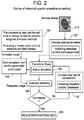
- Spatially normalizing an individual PET image to an adaptive template image located in the reference space;
- Comparing the spatially normalized individual PET image to the adaptive template image to determine whether both are sufficiently converged; and
- Altering the parameter controlling the template image and the parameters controlling the spatial transformation for the individual PET image so as render an altered template image and an updated spatially transformed individual PET image in the event that the comparing step does not determine that the spatially normalized individual PET image is sufficiently converged to the adaptive template image.
y i={circumflex over (α)}i+{circumflex over (β)}i COM SUVR+εi Equation 1
-
- Linear regression model
Applying this model over all input images regressed on the corresponding COMSUVR values provides an intercept image and a slope image according to Equation 2:
I 0=[{circumflex over (α)}1 {circumflex over (α)}2 . . . {circumflex over (α)}n]
I Slope=[{circumflex over (β)}1 β2 . . . {circumflex over (β)}n] Equation 2 - Intercept image, I0, and slope image, ISlope, created from linear regression, where n is the number of voxels in the images.
- Linear regression model
I template =I 0 +I slope x Equation 3
-
- Template image equation
A value of the scale factor x of 0.0 will correspond to I0 itself, which represents a pattern of an Aβ− subject, and a value of 1.0 will correspond to an Aβ+ subject in the high end of the COMSUVR scale. From this formula the present invention can create synthetic images for any x along the linear path defined by ISlope.FIG. 5 shows such synthetic images covering values of x from 0.0 to 1.0 in steps of 0.2.
- Template image equation
Claims (15)
I i template =I i 0 +I i slope x,
I i template =I i 0 +I i slope x,
I i template =I i 0 +I i slope x,
I i template =I i 0 +I i slope x,
I i template =I i 0 +I i slope x,
I i template =I i 0 +I i slope x,
I i template =I i 0 +I i slope x,
I i template =I i 0 +I i slope x,
Priority Applications (1)
| Application Number | Priority Date | Filing Date | Title |
|---|---|---|---|
| US14/344,934 US10614547B2 (en) | 2011-09-20 | 2012-09-20 | Methods of spatial normalization of positron emission tomography images |
Applications Claiming Priority (3)
| Application Number | Priority Date | Filing Date | Title |
|---|---|---|---|
| US201161536702P | 2011-09-20 | 2011-09-20 | |
| PCT/US2012/056182 WO2013043775A1 (en) | 2011-09-20 | 2012-09-20 | Methods of spatial normalization of positron emission tomography images |
| US14/344,934 US10614547B2 (en) | 2011-09-20 | 2012-09-20 | Methods of spatial normalization of positron emission tomography images |
Related Parent Applications (1)
| Application Number | Title | Priority Date | Filing Date |
|---|---|---|---|
| PCT/US2012/056182 A-371-Of-International WO2013043775A1 (en) | 2011-09-20 | 2012-09-20 | Methods of spatial normalization of positron emission tomography images |
Related Child Applications (1)
| Application Number | Title | Priority Date | Filing Date |
|---|---|---|---|
| US16/841,329 Continuation US11488282B2 (en) | 2011-09-20 | 2020-04-06 | Methods of spatial normalization of positron emission tomography images |
Publications (2)
| Publication Number | Publication Date |
|---|---|
| US20140350392A1 US20140350392A1 (en) | 2014-11-27 |
| US10614547B2 true US10614547B2 (en) | 2020-04-07 |
Family
ID=46964089
Family Applications (3)
| Application Number | Title | Priority Date | Filing Date |
|---|---|---|---|
| US14/344,934 Active US10614547B2 (en) | 2011-09-20 | 2012-09-20 | Methods of spatial normalization of positron emission tomography images |
| US16/841,329 Active 2033-07-11 US11488282B2 (en) | 2011-09-20 | 2020-04-06 | Methods of spatial normalization of positron emission tomography images |
| US17/954,034 Abandoned US20230026994A1 (en) | 2011-09-20 | 2022-09-27 | Methods of spatial normalization of positron emission tomography images |
Family Applications After (2)
| Application Number | Title | Priority Date | Filing Date |
|---|---|---|---|
| US16/841,329 Active 2033-07-11 US11488282B2 (en) | 2011-09-20 | 2020-04-06 | Methods of spatial normalization of positron emission tomography images |
| US17/954,034 Abandoned US20230026994A1 (en) | 2011-09-20 | 2022-09-27 | Methods of spatial normalization of positron emission tomography images |
Country Status (8)
| Country | Link |
|---|---|
| US (3) | US10614547B2 (en) |
| EP (1) | EP2757955B1 (en) |
| JP (1) | JP6106675B2 (en) |
| CN (1) | CN103930032B (en) |
| AU (1) | AU2012312482B2 (en) |
| CA (1) | CA2849119C (en) |
| ES (1) | ES2922280T3 (en) |
| WO (1) | WO2013043775A1 (en) |
Cited By (1)
| Publication number | Priority date | Publication date | Assignee | Title |
|---|---|---|---|---|
| US11475612B2 (en) * | 2018-02-28 | 2022-10-18 | Seoul National University R&Db Foundation | Device for spatial normalization of medical image using deep learning and method therefor |
Families Citing this family (12)
| Publication number | Priority date | Publication date | Assignee | Title |
|---|---|---|---|---|
| US10614547B2 (en) | 2011-09-20 | 2020-04-07 | Ge Healthcare Limited | Methods of spatial normalization of positron emission tomography images |
| CN108095761B (en) | 2012-03-07 | 2021-10-15 | 齐特奥股份有限公司 | Spatial alignment apparatus, spatial alignment system and method for guiding a medical procedure |
| EP3170145B1 (en) * | 2014-07-15 | 2020-03-25 | Koninklijke Philips N.V. | Imaging data statistical testing including a stereotactical normalization with a personalized template image |
| US10617401B2 (en) | 2014-11-14 | 2020-04-14 | Ziteo, Inc. | Systems for localization of targets inside a body |
| US10152519B2 (en) | 2015-03-05 | 2018-12-11 | Bio-Rad Laboratories, Inc. | Optimized spectral matching and display |
| TWI587841B (en) * | 2015-03-06 | 2017-06-21 | 國立陽明大學 | System and method of quantitative analysis nuclear medicine brain imaging |
| JP2021513054A (en) * | 2018-02-02 | 2021-05-20 | コーニンクレッカ フィリップス エヌ ヴェKoninklijke Philips N.V. | Correction of standard capture value (SUV) scaling differences in serial positron emission tomography (PET) examinations using image alignment and regression analysis |
| WO2019168310A1 (en) * | 2018-02-28 | 2019-09-06 | 서울대학교산학협력단 | Device for spatial normalization of medical image using deep learning and method therefor |
| WO2020210532A1 (en) * | 2019-04-09 | 2020-10-15 | Ziteo, Inc. | Methods and systems for high performance and versatile molecular imaging |
| KR102261111B1 (en) * | 2019-09-10 | 2021-06-04 | 인하대학교 산학협력단 | Generalization of intensity distribution of medical images using gans |
| CN113450314A (en) * | 2021-06-04 | 2021-09-28 | 迈格生命科技(深圳)有限公司 | Image processing method, device, storage medium and product |
| KR102603177B1 (en) * | 2022-06-03 | 2023-11-17 | 주식회사 브라이토닉스이미징 | System for spatial normalization of image, quantification using spatial normalization and method thereof |
Citations (6)
| Publication number | Priority date | Publication date | Assignee | Title |
|---|---|---|---|---|
| US5948384A (en) * | 1990-09-14 | 1999-09-07 | Syngenix Limited | Particulate agents |
| JP2006320387A (en) | 2005-05-17 | 2006-11-30 | Univ Of Tsukuba | Computer-aided diagnosis apparatus and method |
| WO2008093057A1 (en) | 2007-01-30 | 2008-08-07 | Ge Healthcare Limited | Tools for aiding in the diagnosis of neurodegenerative diseases |
| US20100049032A1 (en) * | 2006-07-12 | 2010-02-25 | Florian Steinke | Method for determining a property map of an object, particularly of a living being, based on at least a first image, particularly a magnetic resonance image |
| US20100221181A1 (en) * | 2007-09-26 | 2010-09-02 | Koninklijke Philips Electronics N.V. | Method of differentially diagnosing different types of dementia |
| US20110160543A1 (en) * | 2008-05-28 | 2011-06-30 | The Trustees Of Columbia University In The City Of New York | Voxel-based methods for assessing subjects using positron emission tomography |
Family Cites Families (1)
| Publication number | Priority date | Publication date | Assignee | Title |
|---|---|---|---|---|
| US10614547B2 (en) | 2011-09-20 | 2020-04-07 | Ge Healthcare Limited | Methods of spatial normalization of positron emission tomography images |
-
2012
- 2012-09-20 US US14/344,934 patent/US10614547B2/en active Active
- 2012-09-20 CN CN201280056787.2A patent/CN103930032B/en active Active
- 2012-09-20 WO PCT/US2012/056182 patent/WO2013043775A1/en active Application Filing
- 2012-09-20 JP JP2014530963A patent/JP6106675B2/en active Active
- 2012-09-20 EP EP12766824.2A patent/EP2757955B1/en active Active
- 2012-09-20 CA CA2849119A patent/CA2849119C/en active Active
- 2012-09-20 AU AU2012312482A patent/AU2012312482B2/en active Active
- 2012-09-20 ES ES12766824T patent/ES2922280T3/en active Active
-
2020
- 2020-04-06 US US16/841,329 patent/US11488282B2/en active Active
-
2022
- 2022-09-27 US US17/954,034 patent/US20230026994A1/en not_active Abandoned
Patent Citations (9)
| Publication number | Priority date | Publication date | Assignee | Title |
|---|---|---|---|---|
| US5948384A (en) * | 1990-09-14 | 1999-09-07 | Syngenix Limited | Particulate agents |
| JP2006320387A (en) | 2005-05-17 | 2006-11-30 | Univ Of Tsukuba | Computer-aided diagnosis apparatus and method |
| US20100049032A1 (en) * | 2006-07-12 | 2010-02-25 | Florian Steinke | Method for determining a property map of an object, particularly of a living being, based on at least a first image, particularly a magnetic resonance image |
| WO2008093057A1 (en) | 2007-01-30 | 2008-08-07 | Ge Healthcare Limited | Tools for aiding in the diagnosis of neurodegenerative diseases |
| US20100080432A1 (en) | 2007-01-30 | 2010-04-01 | Johan Axel Lilja | Tools for aiding in the diagnosis of neurodegenerative diseases |
| JP2010517030A (en) | 2007-01-30 | 2010-05-20 | ジーイー・ヘルスケア・リミテッド | Tools to help diagnose neurodegenerative diseases |
| EP2126609B1 (en) | 2007-01-30 | 2013-03-27 | GE Healthcare Limited | Tools for aiding in the diagnosis of neurodegenerative diseases |
| US20100221181A1 (en) * | 2007-09-26 | 2010-09-02 | Koninklijke Philips Electronics N.V. | Method of differentially diagnosing different types of dementia |
| US20110160543A1 (en) * | 2008-05-28 | 2011-06-30 | The Trustees Of Columbia University In The City Of New York | Voxel-based methods for assessing subjects using positron emission tomography |
Non-Patent Citations (3)
| Title |
|---|
| Fripp J. et. al., "Generative Atlases and Atlas Selection for C11-PIB-PET-PET Registration of Elderly, Mild Cognitive Impaired and Alzheimer Disease Patients," Biomedical Imaging: From Nano to Macro, 2008. ISBI 2008. 5th IEEE International Symposium. May 14, 2008, pp. 1155-1158. * |
| Fripp, et.al. Biomedical Imaging: From Nano to Macro, 2008, p. 1155-1158. |
| PCT/US2012/056182 ISRWO dated Feb. 13, 2013. |
Cited By (1)
| Publication number | Priority date | Publication date | Assignee | Title |
|---|---|---|---|---|
| US11475612B2 (en) * | 2018-02-28 | 2022-10-18 | Seoul National University R&Db Foundation | Device for spatial normalization of medical image using deep learning and method therefor |
Also Published As
| Publication number | Publication date |
|---|---|
| EP2757955B1 (en) | 2022-06-15 |
| US11488282B2 (en) | 2022-11-01 |
| EP2757955A1 (en) | 2014-07-30 |
| ES2922280T3 (en) | 2022-09-12 |
| JP6106675B2 (en) | 2017-04-05 |
| US20140350392A1 (en) | 2014-11-27 |
| JP2014530352A (en) | 2014-11-17 |
| WO2013043775A1 (en) | 2013-03-28 |
| CA2849119C (en) | 2022-06-07 |
| CA2849119A1 (en) | 2013-03-28 |
| CN103930032B (en) | 2018-10-12 |
| CN103930032A (en) | 2014-07-16 |
| US20200234400A1 (en) | 2020-07-23 |
| AU2012312482A1 (en) | 2014-04-17 |
| AU2012312482B2 (en) | 2017-06-01 |
| US20230026994A1 (en) | 2023-01-26 |
Similar Documents
| Publication | Publication Date | Title |
|---|---|---|
| US11488282B2 (en) | Methods of spatial normalization of positron emission tomography images | |
| JP7030050B2 (en) | Pseudo-CT generation from MR data using tissue parameter estimation | |
| JP6567179B2 (en) | Pseudo CT generation from MR data using feature regression model | |
| Schreibmann et al. | MR‐based attenuation correction for hybrid PET‐MR brain imaging systems using deformable image registration | |
| JP2014530352A5 (en) | ||
| US8774481B2 (en) | Atlas-assisted synthetic computed tomography using deformable image registration | |
| US10902597B2 (en) | Comparing medical images | |
| EP2965285B1 (en) | Scan region determining apparatus | |
| EP3268931B1 (en) | Method and apparatus for assessing image registration | |
| US20180085080A1 (en) | Method and apparatus for correction of a synthetic electron density map | |
| WO2019121103A2 (en) | Method and apparatus for medical imaging | |
| CA2850185A1 (en) | Variable-depth stereotactic surface projections | |
| Van Der Heyden et al. | Automatic multiatlas based organ at risk segmentation in mice | |
| DE102016215105A1 (en) | Method and data processing unit for determining an acquisition data processing algorithm | |
| Montin et al. | A multi-metric registration strategy for the alignment of longitudinal brain images in pediatric oncology | |
| Kang et al. | Prediction of standard-dose PET image by low-dose PET and MRI images | |
| Belzunce et al. | High-Resolution Heterogeneous Digital PET Brain Phantom based on the BigBrain Atlas | |
| Criscuolo et al. | A comprehensive lung CT landmark pair dataset for evaluating deformable image registration algorithms | |
| Bilgel et al. | Deformation field correction for spatial normalization of PET images using a population-derived partial least squares model | |
| Li | Multimodal intra-and inter-subject nonrigid registration of small animal images. | |
| GB2583555A (en) | Method and apparatus for medical imaging |
Legal Events
| Date | Code | Title | Description |
|---|---|---|---|
| AS | Assignment |
Owner name: GE HEALTHCARE LIMITED, UNITED KINGDOM Free format text: ASSIGNMENT OF ASSIGNORS INTEREST;ASSIGNORS:LUNDQVIST, ROGER;THURFJELL, NILS LENNART;LILJA, JOHAN AXEL;REEL/FRAME:032436/0934 Effective date: 20130204 |
|
| FEPP | Fee payment procedure |
Free format text: PAYOR NUMBER ASSIGNED (ORIGINAL EVENT CODE: ASPN); ENTITY STATUS OF PATENT OWNER: LARGE ENTITY |
|
| STPP | Information on status: patent application and granting procedure in general |
Free format text: DOCKETED NEW CASE - READY FOR EXAMINATION |
|
| STPP | Information on status: patent application and granting procedure in general |
Free format text: NON FINAL ACTION MAILED |
|
| STPP | Information on status: patent application and granting procedure in general |
Free format text: RESPONSE TO NON-FINAL OFFICE ACTION ENTERED AND FORWARDED TO EXAMINER |
|
| STPP | Information on status: patent application and granting procedure in general |
Free format text: NOTICE OF ALLOWANCE MAILED -- APPLICATION RECEIVED IN OFFICE OF PUBLICATIONS |
|
| STPP | Information on status: patent application and granting procedure in general |
Free format text: NOTICE OF ALLOWANCE MAILED -- APPLICATION RECEIVED IN OFFICE OF PUBLICATIONS |
|
| STPP | Information on status: patent application and granting procedure in general |
Free format text: PUBLICATIONS -- ISSUE FEE PAYMENT VERIFIED |
|
| STCF | Information on status: patent grant |
Free format text: PATENTED CASE |
|
| MAFP | Maintenance fee payment |
Free format text: PAYMENT OF MAINTENANCE FEE, 4TH YEAR, LARGE ENTITY (ORIGINAL EVENT CODE: M1551); ENTITY STATUS OF PATENT OWNER: LARGE ENTITY Year of fee payment: 4 |