US10188426B2 - Embryo transfer catheter and method - Google Patents
Embryo transfer catheter and method Download PDFInfo
- Publication number
- US10188426B2 US10188426B2 US14/760,351 US201414760351A US10188426B2 US 10188426 B2 US10188426 B2 US 10188426B2 US 201414760351 A US201414760351 A US 201414760351A US 10188426 B2 US10188426 B2 US 10188426B2
- Authority
- US
- United States
- Prior art keywords
- embryo
- lumen
- uterus
- catheter body
- balloon
- Prior art date
- Legal status (The legal status is an assumption and is not a legal conclusion. Google has not performed a legal analysis and makes no representation as to the accuracy of the status listed.)
- Active, expires
Links
Images


Classifications
-
- A—HUMAN NECESSITIES
- A61—MEDICAL OR VETERINARY SCIENCE; HYGIENE
- A61B—DIAGNOSIS; SURGERY; IDENTIFICATION
- A61B17/00—Surgical instruments, devices or methods, e.g. tourniquets
- A61B17/42—Gynaecological or obstetrical instruments or methods
- A61B17/425—Gynaecological or obstetrical instruments or methods for reproduction or fertilisation
- A61B17/435—Gynaecological or obstetrical instruments or methods for reproduction or fertilisation for embryo or ova transplantation
-
- A—HUMAN NECESSITIES
- A61—MEDICAL OR VETERINARY SCIENCE; HYGIENE
- A61M—DEVICES FOR INTRODUCING MEDIA INTO, OR ONTO, THE BODY; DEVICES FOR TRANSDUCING BODY MEDIA OR FOR TAKING MEDIA FROM THE BODY; DEVICES FOR PRODUCING OR ENDING SLEEP OR STUPOR
- A61M25/00—Catheters; Hollow probes
- A61M25/0017—Catheters; Hollow probes specially adapted for long-term hygiene care, e.g. urethral or indwelling catheters to prevent infections
-
- A—HUMAN NECESSITIES
- A61—MEDICAL OR VETERINARY SCIENCE; HYGIENE
- A61M—DEVICES FOR INTRODUCING MEDIA INTO, OR ONTO, THE BODY; DEVICES FOR TRANSDUCING BODY MEDIA OR FOR TAKING MEDIA FROM THE BODY; DEVICES FOR PRODUCING OR ENDING SLEEP OR STUPOR
- A61M25/00—Catheters; Hollow probes
- A61M25/0021—Catheters; Hollow probes characterised by the form of the tubing
- A61M25/0023—Catheters; Hollow probes characterised by the form of the tubing by the form of the lumen, e.g. cross-section, variable diameter
- A61M25/0026—Multi-lumen catheters with stationary elements
- A61M25/003—Multi-lumen catheters with stationary elements characterized by features relating to least one lumen located at the distal part of the catheter, e.g. filters, plugs or valves
-
- A—HUMAN NECESSITIES
- A61—MEDICAL OR VETERINARY SCIENCE; HYGIENE
- A61M—DEVICES FOR INTRODUCING MEDIA INTO, OR ONTO, THE BODY; DEVICES FOR TRANSDUCING BODY MEDIA OR FOR TAKING MEDIA FROM THE BODY; DEVICES FOR PRODUCING OR ENDING SLEEP OR STUPOR
- A61M25/00—Catheters; Hollow probes
- A61M25/10—Balloon catheters
- A61M25/1002—Balloon catheters characterised by balloon shape
-
- A—HUMAN NECESSITIES
- A61—MEDICAL OR VETERINARY SCIENCE; HYGIENE
- A61M—DEVICES FOR INTRODUCING MEDIA INTO, OR ONTO, THE BODY; DEVICES FOR TRANSDUCING BODY MEDIA OR FOR TAKING MEDIA FROM THE BODY; DEVICES FOR PRODUCING OR ENDING SLEEP OR STUPOR
- A61M25/00—Catheters; Hollow probes
- A61M25/0021—Catheters; Hollow probes characterised by the form of the tubing
- A61M25/0023—Catheters; Hollow probes characterised by the form of the tubing by the form of the lumen, e.g. cross-section, variable diameter
- A61M25/0026—Multi-lumen catheters with stationary elements
- A61M2025/0039—Multi-lumen catheters with stationary elements characterized by lumina being arranged coaxially
-
- A—HUMAN NECESSITIES
- A61—MEDICAL OR VETERINARY SCIENCE; HYGIENE
- A61M—DEVICES FOR INTRODUCING MEDIA INTO, OR ONTO, THE BODY; DEVICES FOR TRANSDUCING BODY MEDIA OR FOR TAKING MEDIA FROM THE BODY; DEVICES FOR PRODUCING OR ENDING SLEEP OR STUPOR
- A61M2202/00—Special media to be introduced, removed or treated
- A61M2202/0007—Special media to be introduced, removed or treated introduced into the body
-
- A—HUMAN NECESSITIES
- A61—MEDICAL OR VETERINARY SCIENCE; HYGIENE
- A61M—DEVICES FOR INTRODUCING MEDIA INTO, OR ONTO, THE BODY; DEVICES FOR TRANSDUCING BODY MEDIA OR FOR TAKING MEDIA FROM THE BODY; DEVICES FOR PRODUCING OR ENDING SLEEP OR STUPOR
- A61M2210/00—Anatomical parts of the body
- A61M2210/14—Female reproductive, genital organs
- A61M2210/1433—Uterus
-
- A—HUMAN NECESSITIES
- A61—MEDICAL OR VETERINARY SCIENCE; HYGIENE
- A61M—DEVICES FOR INTRODUCING MEDIA INTO, OR ONTO, THE BODY; DEVICES FOR TRANSDUCING BODY MEDIA OR FOR TAKING MEDIA FROM THE BODY; DEVICES FOR PRODUCING OR ENDING SLEEP OR STUPOR
- A61M31/00—Devices for introducing or retaining media, e.g. remedies, in cavities of the body
Definitions
- the present invention relates to a new embryo transfer catheter and relates particularly, though not exclusively, to such an embryo transfer catheter for use in an in-vitro fertilisation procedure.
- the invention also relates to an improved method of transferring embryos in an in-vitro fertilisation procedure.
- in-vitro fertilisation allows for the retrieval of male sperm & female oocytes by various methods. This is followed by immediate use or storage of these gametes. Fertilisation of the oocyte(s) under strictly controlled conditions occurs immediately or at a later date. Embryos formed by this process are grown in optimal surroundings for a variable time in the laboratory and assessed. The embryos are then replaced in the genital tract, most commonly the uterus, of the female where they will hopefully implant in the endometrium of the uterus, leading to an ongoing pregnancy.
- a further internal catheter is inserted through this outer catheter, and its distal end protrudes from the outer catheter as the outer catheter is possibly withdrawn slightly.
- the inner catheter is then positioned, optimally under ultrasound control, to a point near the fundus, or top, of the uterine cavity. Better results are obtained if this inner catheter is soft and passage is atraumatic.
- a further cannula containing embryos, culture fluid for nourishment, and sometimes air bubbles to aid ultrasound visualisation is rapidly deployed through the inner catheter, flushed to eject the contents, and then withdrawn with checks to confirm the embryos have been ejected into the uterus. When this is confirmed, all catheters are withdrawn and the patient rests for a variable amount of time.
- An older technique uses only two catheters, and the inner catheter is used for both positioning and transfer of embryos.
- the present invention was developed with a view to providing a new embryo transfer catheter which may be left securely in situ before and after embryo transfer for several days or longer and which minimises physical disturbance of the endometrium at the time of embryo transfer, and inhibits expulsion of embryos.
- the invention also provides an improved method of transferring an embryo into the uterine cavity.
- the embryo transfer catheter and method will be described with particular reference to human use, it will be understood that the device and method may also be used with animals.
- an embryo transfer catheter for transferring an embryo into a uterine cavity of a recipient, the catheter comprising:
- an outer catheter body having first and second lumens, the second lumen being adapted to receive an internal catheter which, in use, will allow passage of an embryo-containing cannula or direct passage of an embryo or embryos;
- an inflatable balloon provided at a distal end of the first lumen, the inflatable balloon being designed to be as small as possible in its inflated condition whilst still being retained in the uterus, the shape of the balloon being adapted in its inflated state to conform to just the lowest part of the uterus.
- the balloon is shaped to conform to the lowest part of the uterus both transversely and in an anterior-posterior direction.
- the balloon is shaped in a concave way transversely on its superior aspect, which provides the volume necessary for catheter retention and conforms to the transverse uterine shape, but leaves as much endometrium accessible as possible.
- the balloon is convex in an anterior-posterior dimension on its superior aspect (to encourage movement of the embryo to the endometrium rather than allow it to rest centrally on the balloon), taking up minimal room in the smaller anterior-posterior dimension of the flattened uterine cavity, whilst still being retained in the uterus.
- the balloon comprises first and second arms, which in an inflated state extend upwards transversely within the uterus, at an angle approximating the slope of the inner transverse walls of the flattened uterine cavity.
- the balloon may be augmented by inferior protuberances providing extra resistance to expulsion of the catheter.
- These protuberances typically deflate and become flaccid when the balloon is deflated allowing atraumatic removal.
- these protuberances also enhance the seal of the balloon, when inflated, to the lower part of the uterus, blocking embryo expulsion.
- the portion of the outer catheter body located on the vaginal aspect of the cervix is secured by means of a locking plastic device or an inflatable balloon device for holding the correctly positioned catheter in place more securely following embryo transfer.
- a locking plastic device or an inflatable balloon device for holding the correctly positioned catheter in place more securely following embryo transfer.
- Either one of these devices preferably slides up and down the outer catheter body prior to be being secured or inflated so it lies snugly adjacent to the external aspect of the cervix, securing it in a stable position without movement. This allows for good security, despite variation in the length of the cervix in different women.
- the locking device or sliding balloon device may have an additional flexible silicone or PVC cover, which envelops the distal end of the catheter, like a bag, preventing colonisation with infective organisms prior to the embryo transfer. This cover is then removed at the time of transfer.
- the length of the catheter is such that it limits that portion of its length protruding into the vagina to enhance comfort. However, it still remains accessible in order to allow inflation of the balloon using the first lumen, or passage of catheters through the second lumen.
- the length of the vaginal portion of the catheter can be longer, even long enough to protrude through the vaginal introitus. This variation would allow easy subsequent embryo transfer without need for another speculum examination. Similarly, removal could then take place without speculum examination.
- the outer catheter body may be coated with a polymer or membrane, which allows the controlled release of therapeutic substances, locally to the cervix and uterus.
- a polymer or membrane which allows the controlled release of therapeutic substances, locally to the cervix and uterus.
- therapeutic substances that could be utilised in embryo transfer include:
- an improved method of transferring an embryo into the uterus of a recipient comprising the steps of:
- an outer catheter body having first and second lumens, the first lumen having an inflatable balloon provided at its distal end;
- the balloon may be temporarily partially deflated during the embryo transfer to allow adjacent passage of the internal soft catheter into the uterine cavity. If so, the balloon is reinflated immediately after embryo transfer and removal of the embryo-bearing cannula or catheter.
- the step of closing off the second lumen in the outer catheter body is performed by inserting a removable flexible plastic stent that is lockable in position by a locking mechanism.
- the locking mechanism may be a Luer-Lok (Trademark).
- the step of opening the second lumen is performed by removing the flexible stent.
- the step of reclosing the second lumen is performed by inserting a new flexible stent in the second lumen to occlude it.
- the second lumen is closed by means of a self-closing lid at the distal aspect and a lid or cap at the proximal end.
- step of inserting the embryo transfer catheter through the cervix and into the uterine cavity includes placing a tight-fitting covering or sleeve received over the catheter to protect the catheter from contacting any cervical mucus or infection.
- the sleeve is preferably removed by simply pulling it distally.
- the sleeve is split at the top allowing its removal so as to leave the catheter in place.
- the method further comprises the step of controlled release of therapeutic substances via the outer catheter, locally to the cervix and uterus.
- FIG. 1 illustrates a first embodiment of an embryo transfer catheter inserted into a uterus through the cervix of the recipient
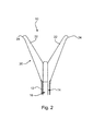
- FIG. 2 illustrates the shape of a balloon on the distal end of the embryo transfer catheter of FIG. 1 in its inflated state
- FIG. 3 is a cross-section view of the outer catheter body of the embryo transfer catheter of FIG. 1 ;
- FIG. 4 illustrates a sleeve for insertion of the embryo transfer catheter of FIG. 1 ;
- FIG. 5 is a top plan view of the embryo transfer catheter of FIG. 1 with the balloon in a semi-inflated state;
- FIG. 6 is a cross-section view of an alternative design for the outer catheter body of the embryo transfer catheter.
- a first embodiment of an embryo transfer catheter 10 in accordance with the invention comprises an outer catheter body 12 having first and second lumens 14 , 16 (see FIG. 3 ), the second lumen 16 being adapted to receive an internal catheter (not shown) with a single lumen which, in use, will allow passage of an embryo or embryo-containing cannula (not shown) into the uterus 18 of the recipient.
- An inflatable balloon 20 is provided at a distal end of the first lumen 14 .
- the inflatable balloon 20 is designed to be as small as possible in its inflated condition whilst still being retained in the uterus 18 .
- the shape of the balloon 20 is adapted in its inflated state to conform to just the lowest part of the uterus 18 .
- FIG. 5 shows the profile of the balloon 20 when viewed in top plan view, in a semi-inflated state.
- the second lumen 16 is occluded by a stent 30 (or alternatively a lid).
- the balloon 20 is shaped to conform to the lowest part of the uterus 18 both transversely and in an anterior-posterior direction.
- the balloon is shaped in a concave way transversely on its superior aspect, which provides the volume necessary for catheter retention and conforms to the transverse uterine shape, but leaves as much endometrium accessible as possible.
- the balloon is convex in an anterior-posterior dimension on its superior aspect (to encourage movement of the embryo to the endometrium rather than allow it to rest centrally on the balloon), taking up minimal room in the smaller anterior-posterior dimension of the flattened uterine cavity, whilst still being retained.
- the balloon 20 comprises first and second arms 22 , which in an inflated state extend upwards transversely within the uterus 18 , at an angle approximating the slope of the inner transverse walls of the flattened uterine cavity.
- the balloon 20 may be augmented by inferior protuberances 24 providing extra resistance to expulsion of the catheter body 12 .
- inferior protuberances 24 may be provided on each balloon to enhance tactile resistance to expulsion. These protuberances 24 typically deflate and become flaccid when the balloon 20 is deflated allowing atraumatic removal.
- these protuberances 24 also enhance the seal of the balloon 20 to the lower part of the uterus 18 , blocking embryo expulsion.
- protuberances on the balloon 20 is not the only way to secure the catheter within the uterus.
- an assembly of two similarly shaped plastic arms with distal hinges could be deployed.
- the plastic arms would be straight (continuous with the catheter) upon insertion, but could then be pushed transversely, articulating via their hinges, at a chosen time by using the stent, pressing on a plastic protuberance. Then when removal occurs, the stent can be pulled out, allowing the plastic arms to fold back vertically facilitating catheter extraction.
- the portion of the outer catheter body 12 on the vaginal aspect of the cervix is secured by means of a plastic locking device or an inflatable locking balloon device 26 for holding the correctly positioned catheter in place more securely following embryo transfer.
- a plastic locking device or an inflatable locking balloon device 26 for holding the correctly positioned catheter in place more securely following embryo transfer.
- Either one of these devices preferably slides up and down the outer catheter body 12 prior to be being secured or inflated, so that it lies snugly adjacent to the external aspect of the cervix 28 , securing it in a stable position without movement, as shown in FIG. 1 . This allows for secure retention of the catheter 10 , despite variation in the length of the cervix in different women.
- the locking device or sliding balloon device may have an additional flexible silicone or PVC cover, which envelops the distal end of the catheter, like a bag, preventing colonisation with infective organisms prior to the embryo transfer. This cover is then removed at the time of transfer.
- the portion of the outer catheter body 12 on the vaginal aspect of the cervix can also be secured by simply using an increased friction technique. Current devices all use this simple friction technique.
- the length of the catheter 10 is such that it limits that portion of its length protruding into the vagina to enhance comfort. However, it still remains accessible in order to allow inflation of the balloon 20 using the first lumen 14 , or passage of catheters through the second lumen 16 .
- the length of the vaginal portion of the catheter can be longer, even long enough to protrude through the vaginal introitus. This variation would allow easy subsequent embryo transfer without need for another speculum examination. Similarly, removal could then take place without speculum examination. This extra length would be advantageous when the catheter is only planned to be left for up to a few hours after embryo transfer.
- a removable flexible plastic stent 30 that is lockable by a locking mechanism, typically a Luer-Lok (Trademark) or similar, is provided to close off the second lumen 16 prior to embryo insertion, to prevent infection and/or blockage.
- the top of the stent 30 would typically be gently convex to match the adjacent parts of the balloon 20 .
- This stent 30 is removed for the embryo transfer, and subsequently replaced with a matching internal catheter with a single lumen, which then allows passage of the embryo or embryo-containing cannula.
- FIG. 3 it can be seen how the cross-sectional shape of the first lumen 14 (for balloon inflation) of the outer catheter body 12 is crescent shaped.
- FIG. 6 illustrates an alternative cross-sectional design for the outer catheter body, in which the first lumen 14 is of circular shape.
- the outer catheter body 12 may be coated with a polymer or membrane, which allows the controlled release of therapeutic substances, locally to the cervix 28 and uterus 18 .
- a polymer or membrane which allows the controlled release of therapeutic substances, locally to the cervix 28 and uterus 18 .
- therapeutic substances that could be utilised in embryo transfer include:
- the embryo transfer catheter 10 may be inserted through the cervix and into the uterine cavity within a tight-fitting covering or sleeve 40 (made of silicone or PVC or other non-embryo toxic flexible material), illustrated in FIG. 4 .
- the sleeve 40 has a split at the top with overlapping portions. Once the catheter 10 has been inserted into the uterus, this silicone sleeve 40 is removed by simply pulling it distally, the split at the top allowing its removal leaving the catheter 10 in place.
- the sleeve 40 protects the catheter 10 from contacting any cervical mucus or infection.
- the sleeve 40 may be slightly textured or absorbent. This texture or absorbency encourages adherence of any cervical mucus brought into the uterus during the insertion, and thus allows the removal of this mucus when the sleeve 40 is removed.
- the method firstly comprises the step of providing an outer catheter body 12 having first and second lumens 14 , 16 , the first lumen having an inflatable balloon 20 provided at its distal end, as described above.
- the outer catheter body 12 may be inserted into the uterus 18 through the cervix 28 several hours or preferably several days or weeks or longer before the actual embryo transfer, instead of immediately prior as in current practice.
- the balloon 20 is inflated with fluid (liquid or gas) so that the outer catheter body 12 is retained in the uterus and cervix 18 .
- the removable flexible plastic stent 30 is already placed to close off the second lumen 16 to prevent infection and/or blockage.
- the second lumen is closed by means of a lid at the distal aspect, and a lid or cap at the proximal end.
- the stent 30 is removed to open the second lumen in the outer catheter body for the embryo transfer.
- This stent is replaced with a matching internal catheter (not shown) with a single lumen, which then allows passage of the embryo, or alternatively an embryo-containing cannula to transfer an embryo into the uterus 18 .
- the balloon 20 may be temporarily partially deflated during the embryo transfer to allow adjacent passage of the internal soft catheter into the uterine cavity. If so, it is reinflated immediately after embryo transfer and removal of the embryo-bearing cannula or catheter.
- a new flexible stent 30 is placed in the second lumen 16 to occlude it following embryo transfer to reclose the second lumen 16 .
- the distal lid closes following removal of the passed catheter and a cap or lid occludes the lumen on its proximal aspect.
- the outer catheter body 12 may be retained in the uterus 18 for up to several minutes, hours or days after the embryo transfer wherein, in use, physical disturbance of the endometrium can be minimised.
- the catheter's design allows it to be left securely in situ after embryo transfer for several days, which potentially increases success rates, by several methods, most importantly, by blockage of the exit of the uterus preventing expulsion of the embryo, and by further local delivery of therapeutic substances from the catheter body.
- the shape of the balloon at the distal end of the outer catheter body may vary significantly from that shown in the illustrated embodiments.
- the balloon may be of any suitable shape that leaves as much endometrium accessible to the embryo for implantation as possible, occupies as little volume as possible within the uterine cavity and exerts as little pressure as possible to avoid causing contractions, whilst still being retained in the uterus. Therefore, it will be appreciated that the scope of the invention is not limited to the specific embodiments described, and is to be determined from the appended claims.
Applications Claiming Priority (3)
| Application Number | Priority Date | Filing Date | Title |
|---|---|---|---|
| AU2013900396 | 2013-02-06 | ||
| AU2013900396A AU2013900396A0 (en) | 2013-02-06 | Embryo Transfer Catheter and Method | |
| PCT/AU2014/000091 WO2014121333A1 (fr) | 2013-02-06 | 2014-02-06 | Cathéter et méthode de transfert d'embryon |
Publications (2)
| Publication Number | Publication Date |
|---|---|
| US20150342642A1 US20150342642A1 (en) | 2015-12-03 |
| US10188426B2 true US10188426B2 (en) | 2019-01-29 |
Family
ID=51299081
Family Applications (1)
| Application Number | Title | Priority Date | Filing Date |
|---|---|---|---|
| US14/760,351 Active 2035-09-22 US10188426B2 (en) | 2013-02-06 | 2014-02-06 | Embryo transfer catheter and method |
Country Status (4)
| Country | Link |
|---|---|
| US (1) | US10188426B2 (fr) |
| EP (1) | EP2953558B1 (fr) |
| AU (1) | AU2014214538B2 (fr) |
| WO (1) | WO2014121333A1 (fr) |
Families Citing this family (6)
| Publication number | Priority date | Publication date | Assignee | Title |
|---|---|---|---|---|
| US9107696B2 (en) * | 2008-08-06 | 2015-08-18 | Emory University | Method of embryo transfer that eliminates transferred air while hormonally inducing implantation and apparatus |
| EP2953678B1 (fr) | 2013-02-06 | 2020-03-25 | Sillender, Mark | Cathéter de transfer de substance thérapeutique |
| KR101851737B1 (ko) * | 2016-04-26 | 2018-04-24 | 주식회사 엔도비전 | 자궁경부 유착 방지 장치 |
| CN116421285A (zh) * | 2022-01-04 | 2023-07-14 | 上海市东方医院(同济大学附属东方医院) | 一种胚胎移植装置和系统 |
| WO2023203052A1 (fr) * | 2022-04-19 | 2023-10-26 | Premium Fertility S.L. | Système de transfert d'embryons |
| EP4265206A1 (fr) * | 2022-04-19 | 2023-10-25 | Premium Fertility S.L | Système de transfert d'embryons |
Citations (27)
| Publication number | Priority date | Publication date | Assignee | Title |
|---|---|---|---|---|
| USRE29207E (en) * | 1973-06-25 | 1977-05-10 | Population Research Incorporated | Dispensing method and apparatus |
| WO1982000754A1 (en) | 1980-09-09 | 1982-03-18 | Richard G Seed | Methods and apparatus for artificial embryonation and tissue recovery |
| EP0412664A1 (fr) * | 1989-08-10 | 1991-02-13 | C.R. Bard, Inc. | Dispositif d'accès utérin avec ajustement cervical automatique |
| US5150718A (en) | 1987-08-08 | 1992-09-29 | Akzo N.V. | Method of contraception |
| US5383873A (en) | 1992-12-09 | 1995-01-24 | Regents Of The University Of Minnesota | Smooth muscle chemical pacemaker |
| US5451232A (en) | 1991-10-07 | 1995-09-19 | Medrad, Inc. | Probe for MRI imaging and spectroscopy particularly in the cervical region |
| WO1995032756A1 (fr) | 1994-05-27 | 1995-12-07 | Medtronic, Inc. | Catheter a ballonnet |
| US5562654A (en) | 1994-10-28 | 1996-10-08 | University Of Kentucky Research Foundation | Time-released delivery system |
| US5613950A (en) | 1988-07-22 | 1997-03-25 | Yoon; Inbae | Multifunctional manipulating instrument for various surgical procedures |
| WO1997016217A1 (fr) | 1995-10-30 | 1997-05-09 | Debiotech S.A. | Dispositif d'angioplastie pour bifurcation arterielle |
| US5904665A (en) | 1995-03-07 | 1999-05-18 | Vance Products Inc. | Automated prolonged slow release intrauterine insemination using self retaining intrauterine insemination catheter |
| US6010448A (en) * | 1997-10-17 | 2000-01-04 | Medworks Corp | Embryo transfer arrangement |
| US6050935A (en) * | 1997-05-09 | 2000-04-18 | Biofertec | Container assembly for intravaginal fertilization and culture and embryo transfer and method of intravaginal fertilization and culture employing such a container |
| US6063395A (en) | 1998-11-12 | 2000-05-16 | Leiras Oy | Drug delivery device especially for the delivery of progestins and estrogens |
| WO2001021081A1 (fr) | 1999-09-24 | 2001-03-29 | Cook Urological Inc. | Catheter pour transfert d'embryon |
| US20020082635A1 (en) * | 2000-12-27 | 2002-06-27 | Kammerer Gene W. | Conformal surgical balloon |
| US6476079B1 (en) | 1999-12-23 | 2002-11-05 | Leiras Oy | Devices for the delivery of drugs having antiprogestinic properties |
| US20030229373A1 (en) | 2002-06-11 | 2003-12-11 | Lee Woo-Dong | Apparatus for preventing regeneration of endometrial synechia |
| US20040247674A1 (en) | 2001-08-31 | 2004-12-09 | Timo Haapakumpu | Drug delivery system |
| US20050021069A1 (en) | 2003-07-24 | 2005-01-27 | Gerald Feuer | Inflatable apparatus for accessing body cavity and methods of making |
| US20060058831A1 (en) | 2004-09-13 | 2006-03-16 | Jack Atad | Inflatable system for cervical dilation and labor induction |
| US20080146873A1 (en) * | 2006-11-07 | 2008-06-19 | Adams Ronald D | Methods for performing a medical procedure |
| US20090024108A1 (en) * | 2004-02-25 | 2009-01-22 | Kathy Lee-Sepsick | Methods and Devices for Delivery of Compositions to Conduits |
| US20090137970A1 (en) * | 2006-04-21 | 2009-05-28 | Samuel George | Uterine manipulators |
| WO2012013229A1 (fr) | 2010-07-28 | 2012-02-02 | Fondazione Irccs | Agent thérapeutique, composition comprenant ledit agent, dispositif implantable et procédé pour le traitement du cancer du col de l'utérus et/ou pour la prévention de la formation de néoplasmes en correspondance avec le col de l'utérus dans un système génital féminin humain |
| US20120143209A1 (en) * | 2010-12-06 | 2012-06-07 | Soulor Surgical Inc | Apparatus for treating a portion of a reproductive system and related methods of use |
| US20150359566A1 (en) | 2013-02-06 | 2015-12-17 | Mark SILLENDER | Therapeutic substance transfer catheter and method |
Family Cites Families (1)
| Publication number | Priority date | Publication date | Assignee | Title |
|---|---|---|---|---|
| US20030229372A1 (en) | 1994-01-26 | 2003-12-11 | Kyphon Inc. | Inflatable device for use in surgical protocols relating to treatment of fractured or diseased bone |
-
2014
- 2014-02-06 US US14/760,351 patent/US10188426B2/en active Active
- 2014-02-06 WO PCT/AU2014/000091 patent/WO2014121333A1/fr active Application Filing
- 2014-02-06 EP EP14748804.3A patent/EP2953558B1/fr active Active
- 2014-02-06 AU AU2014214538A patent/AU2014214538B2/en active Active
Patent Citations (27)
| Publication number | Priority date | Publication date | Assignee | Title |
|---|---|---|---|---|
| USRE29207E (en) * | 1973-06-25 | 1977-05-10 | Population Research Incorporated | Dispensing method and apparatus |
| WO1982000754A1 (en) | 1980-09-09 | 1982-03-18 | Richard G Seed | Methods and apparatus for artificial embryonation and tissue recovery |
| US5150718A (en) | 1987-08-08 | 1992-09-29 | Akzo N.V. | Method of contraception |
| US5613950A (en) | 1988-07-22 | 1997-03-25 | Yoon; Inbae | Multifunctional manipulating instrument for various surgical procedures |
| EP0412664A1 (fr) * | 1989-08-10 | 1991-02-13 | C.R. Bard, Inc. | Dispositif d'accès utérin avec ajustement cervical automatique |
| US5451232A (en) | 1991-10-07 | 1995-09-19 | Medrad, Inc. | Probe for MRI imaging and spectroscopy particularly in the cervical region |
| US5383873A (en) | 1992-12-09 | 1995-01-24 | Regents Of The University Of Minnesota | Smooth muscle chemical pacemaker |
| WO1995032756A1 (fr) | 1994-05-27 | 1995-12-07 | Medtronic, Inc. | Catheter a ballonnet |
| US5562654A (en) | 1994-10-28 | 1996-10-08 | University Of Kentucky Research Foundation | Time-released delivery system |
| US5904665A (en) | 1995-03-07 | 1999-05-18 | Vance Products Inc. | Automated prolonged slow release intrauterine insemination using self retaining intrauterine insemination catheter |
| WO1997016217A1 (fr) | 1995-10-30 | 1997-05-09 | Debiotech S.A. | Dispositif d'angioplastie pour bifurcation arterielle |
| US6050935A (en) * | 1997-05-09 | 2000-04-18 | Biofertec | Container assembly for intravaginal fertilization and culture and embryo transfer and method of intravaginal fertilization and culture employing such a container |
| US6010448A (en) * | 1997-10-17 | 2000-01-04 | Medworks Corp | Embryo transfer arrangement |
| US6063395A (en) | 1998-11-12 | 2000-05-16 | Leiras Oy | Drug delivery device especially for the delivery of progestins and estrogens |
| WO2001021081A1 (fr) | 1999-09-24 | 2001-03-29 | Cook Urological Inc. | Catheter pour transfert d'embryon |
| US6476079B1 (en) | 1999-12-23 | 2002-11-05 | Leiras Oy | Devices for the delivery of drugs having antiprogestinic properties |
| US20020082635A1 (en) * | 2000-12-27 | 2002-06-27 | Kammerer Gene W. | Conformal surgical balloon |
| US20040247674A1 (en) | 2001-08-31 | 2004-12-09 | Timo Haapakumpu | Drug delivery system |
| US20030229373A1 (en) | 2002-06-11 | 2003-12-11 | Lee Woo-Dong | Apparatus for preventing regeneration of endometrial synechia |
| US20050021069A1 (en) | 2003-07-24 | 2005-01-27 | Gerald Feuer | Inflatable apparatus for accessing body cavity and methods of making |
| US20090024108A1 (en) * | 2004-02-25 | 2009-01-22 | Kathy Lee-Sepsick | Methods and Devices for Delivery of Compositions to Conduits |
| US20060058831A1 (en) | 2004-09-13 | 2006-03-16 | Jack Atad | Inflatable system for cervical dilation and labor induction |
| US20090137970A1 (en) * | 2006-04-21 | 2009-05-28 | Samuel George | Uterine manipulators |
| US20080146873A1 (en) * | 2006-11-07 | 2008-06-19 | Adams Ronald D | Methods for performing a medical procedure |
| WO2012013229A1 (fr) | 2010-07-28 | 2012-02-02 | Fondazione Irccs | Agent thérapeutique, composition comprenant ledit agent, dispositif implantable et procédé pour le traitement du cancer du col de l'utérus et/ou pour la prévention de la formation de néoplasmes en correspondance avec le col de l'utérus dans un système génital féminin humain |
| US20120143209A1 (en) * | 2010-12-06 | 2012-06-07 | Soulor Surgical Inc | Apparatus for treating a portion of a reproductive system and related methods of use |
| US20150359566A1 (en) | 2013-02-06 | 2015-12-17 | Mark SILLENDER | Therapeutic substance transfer catheter and method |
Non-Patent Citations (8)
| Title |
|---|
| Cook Medical, 2009 Product Catalog, Assisted Reproductive Technology, Part VI: Embryo Transfer, Sydney IVF Embryo transfer Catheter Sets-3 pages www.medial.cz/data/files/medial/download/katalogy/Cook/Cook_ivf_katalog.pdf. |
| Cook Medical, 2009 Product Catalog, Assisted Reproductive Technology, Part VI: Embryo Transfer, Sydney IVF Embryo transfer Catheter Sets—3 pages www.medial.cz/data/files/medial/download/katalogy/Cook/Cook_ivf_katalog.pdf. |
| Medical Device Kitazato et Catheters 3FR 22CM-213322, published Sep. 28, 2012, 2 pages http://www.medicaldevices24.com/medical_devices/info/kitazato-et-catheters-3fr-22crn/419702. |
| Meriano, James, et al., "The Choice of Embryo Transfer Catheter Affects Embryo Implantation After IVF," Oct. 2010, Elsevier Science Inc., vol. 74, No. 4, pp. 678-682. * |
| Office Action issued in European Patent Application No. 14748804.3, dated Apr. 26, 2018, 4 pages. |
| Supplementary European Search Report issued in European Application, No. 14748804.3, dated Aug. 10, 2016, 9 pages. |
| Supplementary European Search Report issued in European Application, No. 14748819.1, dated Sep. 19, 2016, 5 pages. |
| Yanushpolsky, et al., "Transcervical placement of a Malecot catheter after hysteroscopic evaluation provides for easier entry into the endometrial cavity for women with histories of difficult intrauterine inseminations and/or embryo transfers: a prospective case series", Fertility and Sterility, vol. 73, No. 2, pp. 402-405, Feb. 2000. |
Also Published As
| Publication number | Publication date |
|---|---|
| AU2014214538B2 (en) | 2018-11-15 |
| US20150342642A1 (en) | 2015-12-03 |
| WO2014121333A1 (fr) | 2014-08-14 |
| EP2953558A4 (fr) | 2016-09-21 |
| EP2953558A1 (fr) | 2015-12-16 |
| EP2953558B1 (fr) | 2019-08-28 |
| AU2014214538A1 (en) | 2015-07-02 |
Similar Documents
| Publication | Publication Date | Title |
|---|---|---|
| US10188426B2 (en) | Embryo transfer catheter and method | |
| US20200222230A1 (en) | Intra uterine device | |
| EP0121571B1 (fr) | Catheter a demeure pour canal cervical | |
| US8573222B2 (en) | Intrauterine device and inserter for the same | |
| EP2271293B1 (fr) | Système intra-utérin | |
| US20090192542A1 (en) | Single balloon ripening device with novel inserter and inflator | |
| WO2013108249A1 (fr) | Dispositif cataménial à buts multiples | |
| US20220249275A1 (en) | Iud insertion device | |
| US20020059936A1 (en) | Method and device for vas occlusion | |
| Soichet | Ypsilon: a new silicone-covered stainless steel intrauterine contraceptive device | |
| EP4093348B1 (fr) | Un système intra-utérin avec une partie de verrouillage | |
| TW202142200A (zh) | 用於具有鎖定部件之子宮內系統的插入物 | |
| WO2023042204A1 (fr) | Dispositif tridimensionnel intra-utérin et système correspondant | |
| Mazza | Take a fresh look at IUDs |
Legal Events
| Date | Code | Title | Description |
|---|---|---|---|
| STCF | Information on status: patent grant |
Free format text: PATENTED CASE |
|
| MAFP | Maintenance fee payment |
Free format text: PAYMENT OF MAINTENANCE FEE, 4TH YR, SMALL ENTITY (ORIGINAL EVENT CODE: M2551); ENTITY STATUS OF PATENT OWNER: SMALL ENTITY Year of fee payment: 4 |