US10094835B2 - Treating patients based on immune subtypes - Google Patents
Treating patients based on immune subtypes Download PDFInfo
- Publication number
- US10094835B2 US10094835B2 US14/889,086 US201414889086A US10094835B2 US 10094835 B2 US10094835 B2 US 10094835B2 US 201414889086 A US201414889086 A US 201414889086A US 10094835 B2 US10094835 B2 US 10094835B2
- Authority
- US
- United States
- Prior art keywords
- cells
- level
- patients
- mammal
- therapy
- Prior art date
- Legal status (The legal status is an assumption and is not a legal conclusion. Google has not performed a legal analysis and makes no representation as to the accuracy of the status listed.)
- Active
Links
Images


Classifications
-
- G—PHYSICS
- G01—MEASURING; TESTING
- G01N—INVESTIGATING OR ANALYSING MATERIALS BY DETERMINING THEIR CHEMICAL OR PHYSICAL PROPERTIES
- G01N33/00—Investigating or analysing materials by specific methods not covered by groups G01N1/00 - G01N31/00
- G01N33/48—Biological material, e.g. blood, urine; Haemocytometers
- G01N33/50—Chemical analysis of biological material, e.g. blood, urine; Testing involving biospecific ligand binding methods; Immunological testing
- G01N33/53—Immunoassay; Biospecific binding assay; Materials therefor
- G01N33/574—Immunoassay; Biospecific binding assay; Materials therefor for cancer
- G01N33/57407—Specifically defined cancers
- G01N33/57426—Specifically defined cancers leukemia
-
- A—HUMAN NECESSITIES
- A61—MEDICAL OR VETERINARY SCIENCE; HYGIENE
- A61N—ELECTROTHERAPY; MAGNETOTHERAPY; RADIATION THERAPY; ULTRASOUND THERAPY
- A61N5/00—Radiation therapy
- A61N5/10—X-ray therapy; Gamma-ray therapy; Particle-irradiation therapy
-
- G—PHYSICS
- G01—MEASURING; TESTING
- G01N—INVESTIGATING OR ANALYSING MATERIALS BY DETERMINING THEIR CHEMICAL OR PHYSICAL PROPERTIES
- G01N33/00—Investigating or analysing materials by specific methods not covered by groups G01N1/00 - G01N31/00
- G01N33/48—Biological material, e.g. blood, urine; Haemocytometers
- G01N33/50—Chemical analysis of biological material, e.g. blood, urine; Testing involving biospecific ligand binding methods; Immunological testing
- G01N33/53—Immunoassay; Biospecific binding assay; Materials therefor
- G01N33/574—Immunoassay; Biospecific binding assay; Materials therefor for cancer
- G01N33/57407—Specifically defined cancers
-
- G—PHYSICS
- G01—MEASURING; TESTING
- G01N—INVESTIGATING OR ANALYSING MATERIALS BY DETERMINING THEIR CHEMICAL OR PHYSICAL PROPERTIES
- G01N2333/00—Assays involving biological materials from specific organisms or of a specific nature
- G01N2333/435—Assays involving biological materials from specific organisms or of a specific nature from animals; from humans
- G01N2333/705—Assays involving receptors, cell surface antigens or cell surface determinants
- G01N2333/70503—Immunoglobulin superfamily, e.g. VCAMs, PECAM, LFA-3
- G01N2333/70514—CD4
-
- G—PHYSICS
- G01—MEASURING; TESTING
- G01N—INVESTIGATING OR ANALYSING MATERIALS BY DETERMINING THEIR CHEMICAL OR PHYSICAL PROPERTIES
- G01N2333/00—Assays involving biological materials from specific organisms or of a specific nature
- G01N2333/435—Assays involving biological materials from specific organisms or of a specific nature from animals; from humans
- G01N2333/705—Assays involving receptors, cell surface antigens or cell surface determinants
- G01N2333/70596—Molecules with a "CD"-designation not provided for elsewhere in G01N2333/705
-
- G—PHYSICS
- G01—MEASURING; TESTING
- G01N—INVESTIGATING OR ANALYSING MATERIALS BY DETERMINING THEIR CHEMICAL OR PHYSICAL PROPERTIES
- G01N2800/00—Detection or diagnosis of diseases
- G01N2800/52—Predicting or monitoring the response to treatment, e.g. for selection of therapy based on assay results in personalised medicine; Prognosis
Definitions
- This document relates to methods and materials for treating patients following an assessment of immune subtypes such as an assessment of peripheral blood phenotypes.
- this document provides methods and materials for treating a mammal having a medical condition after assessing a mammal's level of CD14 + /DR ⁇ cells (e.g., CD14 + /DR ⁇ monocytes) and level of CD4 + cells (e.g., CD4 + T cells) and classifying the mammal has being likely to experience a favorable or unfavorable medical outcome based at least in part on the mammal's level of CD14 + /DR ⁇ cells and level of CD4 + cells.
- a mammal's level of CD14 + /DR ⁇ cells e.g., CD14 + /DR ⁇ monocytes
- level of CD4 + cells e.g., CD4 + T cells
- the immune system of a mammal is a system of biological structures and processes that helps protect the mammal from diseases by identifying and killing pathogens and tumor cells.
- a monocyte is one type of white blood cell that is part of the immune system. Monocytes can have several roles in the immune system. For example, monocytes can migrate to sites of infection and differentiate into macrophages and dendritic cells.
- Another type of cell that is part of the immune system is a CD4 + T cell.
- CD4 + T cells are a sub-group of lymphocytes that help activate and direct other cells of the immune system.
- This document provides methods and materials for treating patients following an assessment of immune subtypes such as an assessment of peripheral blood phenotypes.
- this document provides methods and materials for treating a mammal having a medical condition after assessing a mammal's level of CD14 + /DR ⁇ cells (e.g., CD14 + /DR ⁇ monocytes) and level of CD4 + cells (e.g., CD4 + T cells) and classifying the mammal has being likely to experience a favorable or unfavorable medical outcome based at least in part on the mammal's level of CD14 + /DR ⁇ cells and level of CD4 + cells.
- a mammal's level of CD14 + /DR ⁇ cells e.g., CD14 + /DR ⁇ monocytes
- level of CD4 + cells e.g., CD4 + T cells
- an appropriate treatment option for a particular medical condition can be selected following an assessment of the level of CD14 + /DR ⁇ cells within a mammal.
- Such an assessment can include, for example, determining the level of CD14 + /DR ⁇ cells within a mammal and comparing the determined values to values of CD14 + /DR ⁇ cells for that particular condition or to values from healthy volunteers to assess whether the level is an altered level (e.g., an increased level of CD14 + /DR ⁇ cells or a complete or substantially complete loss of all CD14 + /DR ⁇ cells).
- the level of CD4 + cells within the mammal can be determined and compared to cut-off values of CD4 + cells for that particular condition or from healthy volunteers to assess whether the level is a normal level, an elevated level (e.g., high level of CD4 + cells), or a reduced level (e.g., low level of CD4 + cells).
- a ratio of the levels of these cells can be calculated with ratios beyond that of normal being used to identify patients at risk. For example, a normal ratio of monocytes (CD14 + cells) to CD4 + T cells (e.g., CD14 + cells per 4 divided by the number of CD4 + T cells per 4) in healthy volunteers can be 0.59 ⁇ 0.29.
- An appropriate treatment option for a particular medical condition can be selected based, at least in part, on the normal, elevated, or reduced level of CD14 + /DR ⁇ cells and the normal, elevated, or reduced level of CD4 + cells or ratios thereof.
- a glioblastoma patient having a normal level of CD14 + /DR ⁇ cells (e.g., ⁇ 24.3% equal to the mean plus 1.5 standard deviations) and a normal level of CD4 + cells (e.g., >650 cells/4 equal to the mean minus one standard deviation) can be treated by surgery, radiation and/or chemotherapy (e.g., Temodar) under conditions wherein the glioblastoma patient experiences survival greater than 600 days, while a glioblastoma patient having an elevated level of CD14 + /DR ⁇ cells and a reduced level of CD4 + cells can be treated by administering an aggressive treatment (e.g., an investigational therapy) or a combination two or more treatments selected from the following: immunotherapy, treatment with a mon
- a glioblastoma patient having an elevated level of CD14 + /DR ⁇ cells and a reduced level of CD4 cells can be treated by administering an aggressive treatment or a combination of two or more treatments selected from the following: immunotherapy, treatment with a monocyte activation molecule (e.g., Imiquimod; polyI:C; or TLR ligands), treatment with a monocyte killing agent, a T cell enhancement therapy (allogeneic donor lymphocyte T cells, CAR technology, T cell activation or expansion cytokines such as IL-2, IL15, or IL-23), and treatment with an immune stimulating antibody (e.g., anti-CTLA-4/Ipilimumab or anti-PD1 alone).
- a monocyte activation molecule e.g., Imiquimod; polyI:C; or TLR ligands
- a monocyte killing agent e.g., a monocyte killing agent
- T cell enhancement therapy allogeneic donor lymphocyte T cells, CAR technology
- a glioblastoma patient having at least one factor abnormal can be treated by administering, to the glioblastoma patient, an aggressive treatment or a combination of two or more treatments selected from the following: immunotherapy, treatment with a monocyte activation molecule (e.g., Imiquimod; polyI:C; or TLR ligands), treatment with a monocyte killing agent, a T cell enhancement therapy (allogeneic donor lymphocyte T cells, CAR technology, T cell activation or expansion cytokines such as IL-2, IL15, or IL-23), and treatment with an immune stimulating antibody (e.g., anti-CTLA-4/Ipilimumab or anti-PD1 alone).
- a monocyte activation molecule e.g., Imiquimod; polyI:C; or TLR ligands
- a monocyte killing agent e.g., a monocyte killing agent
- T cell enhancement therapy allogeneic donor lymphocyte T cells, CAR technology, T cell activation
- a sepsis patient having a reduced level of CD14 + /DR ⁇ cells and an elevated level of CD4 cells can be treated by administering antibiotics, vasopressors, corticosteroids, and/or fluids to the sepsis patient under conditions wherein the sepsis patient is likely to survive, while a sepsis patient having an elevated level of CD14 + /DR ⁇ cells and a reduced level of CD4 cells can be treated by administering, to the sepsis patient, an aggressive treatment or a combination of two or more of the following treatments: a treatment with a monocyte activation molecule (e.g., Imiquimod; polyI:C; TLR ligands), treatment with a monocyte killing agent, a T cell enhancement therapy (e.g., allogeneic donor lymphocyte T cells, CAR technology, T cell activation or expansion cytokines such as IL-2, IL15, or IL-23), and treatment with an immune stimulating antibody (e.g., anti
- a sepsis patient having a reduced level of CD14 + /DR ⁇ cells and a reduced level of CD4 cells can be treated by administering, to the sepsis patient, an aggressive treatment or a combination of two or more of the following treatments: a treatment with a monocyte activation molecule (e.g., Imiquimod; polyI:C; TLR ligands), treatment with a monocyte killing agent, a T cell enhancement therapy (e.g., allogeneic donor lymphocyte T cells, CAR technology, T cell activation or expansion cytokines such as IL-2, IL15, or IL-23), and treatment with an immune stimulating antibody (e.g., anti-CTLA-4/Ipilimumab or anti-PD1 alone).
- a monocyte activation molecule e.g., Imiquimod; polyI:C; TLR ligands
- a monocyte killing agent e.g., a monocyte killing agent
- a T cell enhancement therapy e.g
- a lymphoma patient having a reduced level of CD14 + /DR ⁇ cells and either an elevated or reduced level of CD4 + cells
- a lymphoma patient having an elevated level of CD14 + /DR ⁇ cells and either an elevated or reduced level of CD4 + cells can be treated by administering an aggressive treatment or a combination of two or more of the following treatments: a treatment with a monocyte activation molecule (e.g., Imiquimod; polyI:C; TLR ligands), treatment with a monocyte killing agent, a T cell enhancement therapy (e.g., allogeneic donor lymphocyte T cells, CAR
- a lymphoma patient having an elevated level of CD14 + /DR ⁇ cells and an elevated level of CD4 + cells can be treated by administering (a) a less aggressive treatment to the lymphoma patient than a treatment used to treat a lymphoma patient having an elevated level of CD14 + /DR ⁇ cells and a reduced level of CD4 + cells or (b) a combination of two or more of the following treatments: a treatment with a monocyte activation molecule (e.g., Imiquimod; polyI:C; TLR ligands), treatment with a monocyte killing agent, a T cell enhancement therapy (e.g., allogeneic donor lymphocyte T cells, CAR technology, T cell activation or expansion cytokines such as IL-2, IL15, or IL-23), and treatment with an immune stimulating antibody (e.g., anti-CTLA-4/Ipilimumab or anti-PD1 alone).
- a monocyte activation molecule e.g., Imiquimod
- the number of CD14 + /DR ⁇ cells can correlate to increased numbers of circulating monocytes and circulating granulocytes.
- measuring CD14 + /DR ⁇ cells can be performed as a substitute for other subtypes.
- a mammal's likelihood of experiencing either a favorable or unfavorable medical outcome for a particular medical condition can be determined based, at least in part, on the normal, elevated, or reduced level of CD14 + /DR ⁇ cells and the normal, elevated, or reduced level of CD4 + cells or ratios thereof.
- a glioblastoma patient having a normal level of CD14 + /DR ⁇ cells (e.g., ⁇ 24.3% equal to the mean plus 1.5 standard deviations) and a normal level of CD4 + cells (e.g., >650 cells/4 equal to the mean minus one standard deviation) can be classified as being likely to experience a relatively favorable outcome (e.g., survival greater than 600 days), while a glioblastoma patient having a reduced level of CD14 + /DR ⁇ cells and a reduced level of CD4 + cells can be classified as being likely to experience a relatively unfavorable outcome (e.g., survival less than 400 days).
- a relatively favorable outcome e.g., survival greater than 600 days
- a glioblastoma patient having a reduced level of CD14 + /DR ⁇ cells and a reduced level of CD4 + cells can be classified as being likely to experience a relatively unfavorable outcome (e.g., survival less than 400 days).
- a glioblastoma patient having an elevated level of CD14 + /DR ⁇ cells and a reduced level of CD4 + cells can be classified as being likely to experience a relatively unfavorable outcome (e.g., survival less than 500 days). In some cases, a glioblastoma patient having at least one factor abnormal can be likely to experience an unfavorable outcome (e.g., survival of less than 600 days).
- a sepsis patient having a reduced level of CD14 + /DR ⁇ cells and an elevated level of CD4 + cells can be classified as being likely to experience a relatively favorable outcome (e.g., likely to survive), while a sepsis patient having an elevated level of CD14 + /DR ⁇ cells and a reduced level of CD4 + cells can be classified as being likely to experience a relatively unfavorable outcome (e.g., greater than 30 percent chance of death).
- a sepsis patient having a reduced level of CD14 + /DR ⁇ cells and a reduced level of CD4 + cells can be classified as being likely to experience a relatively unfavorable outcome (e.g., greater than 10 percent chance of death).
- a lymphoma patient e.g., B-cell non-Hodgkin's lymphoma patient
- a lymphoma patient having a reduced level of CD14 + /DR ⁇ cells and either an elevated or reduced level of CD4 + cells can be classified as being likely to experience a relatively favorable outcome (e.g., a likelihood of surviving longer than 750 days)
- a lymphoma patient having an elevated level of CD14 + /DR ⁇ cells and either an elevated or reduced level of CD4 + cells can be classified as being likely to experience a relatively unfavorable outcome (e.g., a likelihood of not surviving longer than 750 days).
- a lymphoma patient having an elevated level of CD14 + /DR ⁇ cells and an elevated level of CD4 + cells can be classified as being likely to experience an slightly more favorable outcome than a lymphoma patient having an elevated level of CD14 + /DR ⁇ cells and a reduced level of CD4 + cells.
- the methods and materials provided herein can allow clinicians to provide patients with appropriate treatment options and/or appropriate prognostic information about their medical condition.
- the methods and materials provided herein can be used to identify cancer patients who would potentially benefit from more aggressive forms of treatment when their prognostic subtype is that of a poor outcome.
- one aspect of this document features a method for treating cancer.
- the method comprises, or consists essentially of, (a) detecting the presence of a normal, elevated, or reduced level of CD14 + /DR ⁇ cells for the cancer in a mammal having the cancer, (b) detecting the presence of a normal, elevated, or reduced level of CD4 + cells for the cancer in the mammal, and (c) administering a non-immune-based therapy, radiation, chemotherapy, or a combination thereof with or without surgery to the mammal if the mammal is determined to be likely to experience a favorable outcome of the cancer based at least in part on the level of CD14 + /DR ⁇ cells and the level of CD4 + cells, or administering, to the mammal, (i) a non-immune-based therapy, radiation, chemotherapy, or a combination thereof with or without surgery and (ii) a T cell stimulating therapy, a CAR therapy, a DLI therapy, a T cell stimulating antibody therapy, an anti-CD14
- the mammal can be a human.
- the cancer can be glioblastoma or lymphoma.
- the mammal can have a normal level of CD14 + /DR ⁇ cells and a normal level of CD4 + cells, and the method can comprise administering, to the mammal, a non-immune-based therapy, radiation, chemotherapy, or a combination thereof with or without surgery.
- the mammal can have a reduced level of CD14 + /DR ⁇ cells and a reduced level of CD4 + cells, and the method can comprise administering, to the mammal, (i) a non-immune-based therapy, radiation, chemotherapy, or a combination thereof with or without surgery and (ii) a T cell stimulating therapy, a CAR therapy, a DLI therapy, a T cell stimulating antibody therapy, an anti-CD14 + /HLA-CR neg therapy, a CD14 + /DR neg stimulating therapy, a TLR stimulating ligand therapy, a Poly I:C therapy, a CpG therapy, a vaccine therapy, a dendritic cell infusion therapy, a cancer vaccine therapy, a CD14 + DR neg killing strategy therapy, a CD14 + /DR neg blocking strategy therapy, a treatment with IDO inhibitors (e.g., NLG8189), a treatment with arginase inhibitors, a treatment with MCSF-R or CSF-R blocking agents, or combinations thereof,
- this document features a method for assessing the likelihood that a mammal having a medical condition will experience a favorable or unfavorable outcome.
- the method comprises, or consists essentially of, (a) determining whether the mammal has a normal, elevated, or reduced level of CD14 + /DR ⁇ cells for the medical condition, (b) determining whether the mammal has a normal, elevated, or reduced level of CD4 + cells for the medical condition, and (c) classifying the mammal as being likely to experience a favorable or unfavorable outcome of the medical condition based at least in part on the level of CD14 + /DR ⁇ cells and the level of CD4 + cells.
- the mammal can be a human.
- the medical condition can be cancer.
- the cancer can be glioblastoma.
- the mammal can have a normal level of CD14 + /DR ⁇ cells and a normal level of CD4 + cells, and wherein the method can comprise classifying the mammal as being likely to experience a favorable outcome.
- the favorable outcome can comprise surviving the glioblastoma for more than 600 days.
- the mammal can have a reduced level of CD14 + /DR ⁇ cells and a reduced level of CD4 + cells, and wherein the method can comprise classifying the mammal as being likely to experience an unfavorable outcome.
- the unfavorable outcome can comprise surviving the glioblastoma for less than 400 days.
- the mammal can have an elevated level of CD14 + /DR ⁇ cells and a reduced level of CD4 + cells, and wherein the method can comprise classifying the mammal as being likely to experience an unfavorable outcome.
- the unfavorable outcome can comprise surviving the glioblastoma for less than 500 days.
- the cancer can be a lymphoma.
- the mammal can have a reduced level of CD14 + /DR ⁇ cells and an elevated or reduced level of CD4 + cells, and wherein the method can comprise classifying the mammal as being likely to experience a favorable outcome.
- the favorable outcome can comprise surviving the lymphoma for greater than 750 days.
- the mammal can have an elevated level of CD14 + /DR ⁇ cells and an elevated or reduced level of CD4 + cells, and wherein the method can comprise classifying the mammal as being likely to experience an unfavorable outcome.
- the unfavorable outcome can comprise a likelihood of not surviving the lymphoma for greater than 750 days.
- the medical condition can be sepsis.
- the mammal can have a reduced level of CD14 + /DR ⁇ cells and an elevated level of CD4 + cells, and wherein the method can comprise classifying the mammal as being likely to experience a favorable outcome.
- the favorable outcome can comprise surviving the sepsis.
- the mammal can have an elevated level of CD14 + /DR ⁇ cells and a reduced level of CD4 + cells, and wherein the method can comprise classifying the mammal as being likely to experience an unfavorable outcome.
- the unfavorable outcome can comprise between 20 and 60 percent chance of death from the sepsis.
- the mammal can have a reduced level of CD14 + /DR ⁇ cells and a reduced level of CD4 + cells, and wherein the method can comprise classifying the mammal as being likely to experience an unfavorable outcome.
- the unfavorable outcome can comprise having between 5 and 25 percent chance of death from the sepsis.
- FIG. 1A is a survival curve for glioblastoma patients having (a) normal levels of CD14 + /DR ⁇ cells and CD4 + cells, (b) a normal (low) level CD14 + /DR ⁇ cells and a reduced level of CD4 + cells, or (c) an elevated level of CD14 + /DR ⁇ cells and a reduced level of CD4 + cells.
- FIG. 1B is a survival curve for glioblastoma patients when plotted based on the CD4 + levels alone.
- FIG. 1C is a survival curve for glioblastoma comparing patients with either CD4 + or CD14 + /DR ⁇ levels at abnormal levels compared to patients with normal values of both.
- FIG. 2 is a graph plotting the level of CD4 + cells (cells/ ⁇ L) versus the level of CD14 + /DR ⁇ cells (percent of CD14 + cells that are DR ⁇ ) for sepsis patients. For sepsis patients, the cut off values were 63% DR ⁇ and 650 CD4 + cells/ ⁇ L.
- FIG. 3 is a graph plotting percent survival versus days for sepsis with patients grouped on peripheral immunity. Patients were grouped based on normal or elevated levels of CD14 + /DR ⁇ cells and normal or reduced levels of CD4 + cells.
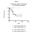
- FIG. 4 is a graph plotting the level of CD4 + cells (cells/ ⁇ L) versus the level of CD14 + /DR ⁇ cells (percent of CD14 + cells that are DR ⁇ ) for lymphoma patients.
- FIG. 5 is a graph plotting overall survival (percent) versus time (days) for lymphoma patients categorized by their peripheral blood phenotype. Patients were grouped according to normal or increased percent of CD14 + /DR ⁇ cells and reduced or normal levels of CD4 + cells. The normal (DR ( ⁇ 10%) and normal CD4 (>500 cells/ ⁇ L)) and the normal DR and low CD4 groups overlap on the survival curve.
- FIG. 6 is a graph plotting the overall survival (percent) versus time (days).
- FIG. 7 contains graphs plotting the levels of CD14 + HLA-DR neg monocytes in chronic lymphocytic leukemia (CLL).
- CLL chronic lymphocytic leukemia
- the percent of CD14 + cells with a loss of HLA-DR staining was determined and compared between CLL patients and healthy volunteers (HV; upper left).
- CLL patients with a sustained response to treatment had a decrease in the frequency of HLA ⁇ DR lo/neg CD14 + monocytes 12 months after completion of treatment (diamonds) compared to measurement prior to treatment (upper right).
- Kaplan-Meyer survival curve comparing CLL patients with elevated ratios (>2.5 standard deviations) of CD14 + HLA-DR lo/neg monocytes when compared to healthy volunteers (dashed line) or with ratios similar to those seen in healthy volunteers (solid line; bottom panel).
- FIG. 8 demonstrates the relationship of the presence of peripheral blood CD14 + HLA-DR lo/neg monocytes and intratumoral accumulation of CD14 + cells in renal cell carcinoma.
- FIG. 8A is a Kaplan-Meier curve of the survival of 375 patients with renal cell carcinoma based on the classification of hi, medium, lo, and no intratumoral staining.
- FIG. 8B is a graph plotting the correlation of intratumoral CD14 staining and the amount of peripheral blood CD14 + HLA-DR neg monocytes from the same patients.
- FIG. 9 is a graph plotting flow cytometry and Q-PCR data for HLA-DR expression.
- Blood samples from healthy volunteers and cancer patients were stained for CD14 and HLA-DR.
- RNA was isolated from CD14 + populations from the same individuals.
- Quantitative RT-PCR was performed to determine the level of expression by assessing the crossing point (Cp) of each PCR reaction. The percentage of CD14 + HLA-DR lo/neg monocytes were then plotted against the Cp of each PCR reaction.
- This document provides methods and materials for treating patients following an assessment of immune subtypes such as an assessment of peripheral blood phenotypes.
- this document provides methods and materials for treating a mammal having a medical condition after assessing a mammal's level of CD14 + /DR ⁇ cells (e.g., CD14 + /DR ⁇ monocytes) and level of CD4 + cells (e.g., CD4 + T cells) and classifying the mammal has being likely to experience a favorable or unfavorable medical outcome based at least in part on the mammal's level of CD14 + /DR ⁇ cells and level of CD4 + cells.
- a mammal's level of CD14 + /DR ⁇ cells e.g., CD14 + /DR ⁇ monocytes
- level of CD4 + cells e.g., CD4 + T cells
- an appropriate treatment option for a particular medical condition can be selected following an assessment of the level of CD14 + /DR ⁇ cells within a mammal.
- Such an assessment can include, for example, determining the level of CD14 + /DR ⁇ cells within a mammal and comparing the determined values to values of CD14 + /DR ⁇ cells for that particular condition or to values from healthy volunteers to assess whether the level is an altered level (e.g., an increased level of CD14 + /DR ⁇ cells or a complete or substantially complete loss of all CD14 + /DR ⁇ cells).
- healthy volunteers can have an average of about 6% DR ⁇ . In cases of immune suppression, this percentage can be higher.
- autoimmunity it can go very low (e.g., virtually all can be DR + ).
- the level of CD4 + cells within the mammal can be determined and compared to cut-off values of CD4 + cells for that particular condition or compared to healthy volunteers to assess whether the level is a normal level, an elevated level (e.g., high level of CD4 + cells), or a reduced level (e.g., low level of CD4 + cells).
- a single CD14 + /DR ⁇ cell cut-off value can be used to assess whether the level of CD14 + /DR ⁇ cells is an elevated level (e.g., a high level of CD14 + /DR ⁇ cells) or a reduced level (e.g., a low level of CD14 + /DR ⁇ cells) and/or a single CD4 + cell cut-off value can be used to assess whether the level of CD4 + cells is a normal level (e.g., a typical level of CD4 + cells) or a reduced level (e.g., a low level of CD4 + cells).
- the levels for either the CD14 + /DR ⁇ or CD4 + cells can be considered as high or extremely high.
- the mammal's likelihood of experiencing either a favorable or unfavorable medical outcome for that particular medical condition can be determined based, at least in part, on the normal, elevated, or reduced level of CD14 + /DR ⁇ cells by itself or in some cases by the addition of the categorization (e.g., normal, elevated, or reduced level) of CD4 + cells.
- the level of CD14 + /DR ⁇ cells and/or the level of CD4 + cells can be determined using a sample (e.g., a blood sample) obtained from the mammal to be assessed. Once obtained, the sample can be treated such that the level of CD14 + /DR ⁇ cells and the level of CD4 + cells present can be determined. Cell staining and immunofluorescence techniques (e.g., flow cytometry) can be used to determine the level of CD14 + /DR ⁇ cells and/or the level of CD4 + cells.
- anti-CD14, anti-HLA-DR, and anti-CD4 antibodies can be used to perform flow cytometry in order to determine the level of CD14 + /DR ⁇ cells and the level of CD4 + cells present within a sample.
- nucleic acid-based assays can be used to assess the level of CD14 + /DR ⁇ cells and/or the level of CD4 + cells.
- the amount of particular transcripts e.g., CD14, DR, and CD4 transcripts
- the ratios of cells can be used to assess a mammal's (e.g., a human's) peripheral blood phenotype.
- the ratio of CD14 + /DR ⁇ cells to CD4 + cells (or vice versa) can be used as described herein to assess a person's peripheral blood phenotype.
- the determined cell levels can be compared to a cut-off value provided herein to determine whether the determined levels are normal, elevated, or reduced. In such cases, determined normal, elevated, or reduced levels can be used as described herein to classify the patient as having a likelihood to experience a favorable or unfavorable outcome.
- the level of CD14 + /DR ⁇ cells and the level of CD4 + cells for a population of similarly afflicted patients with known medical outcomes can be used to identify appropriate cut-off values for the CD14 + /DR ⁇ cell and CD4 + cell levels.
- the population of patients with known medical outcomes can be used to determine which combinations of cell phenotypes indicate whether a patient is likely to experience a favorable or unfavorable outcome.
- Those patients determined to have a favorable or unfavorable outcome as described herein can be treated with a treatment option appropriate for their peripheral blood phenotype.
- a patient suffering from cancer, sepsis, an autoimmune condition, or trauma who has a normal level of CD4 + cells and a normal level of CD14 + /DR ⁇ cells can be treated with current medical therapies for the condition to be treated such as non-immune-based therapies, radiation, chemotherapies, and/or surgery.
- a patient e.g., cancer, sepsis, an autoimmune condition, or trauma patient
- a patient can be treated with both a current medical therapy for the condition to be treated plus a T cell stimulating therapy, a CAR therapy, a DLI therapy (e.g., an allogeneic donor lymphocyte infusion therapy), a T cell stimulating antibody therapy (e.g., treatment with an anti-CTLA-4 antibody, Ipilumimab, or an anti-PD1 antibody), or a combination thereof.
- a T cell stimulating therapy e.g., treatment with an anti-CTLA-4 antibody, Ipilumimab, or an anti-PD1 antibody
- a patient e.g., cancer, sepsis, an autoimmune condition, or trauma patient
- a patient can be treated with both a current medical therapy for the condition to be treated plus an anti-CD14 + /HLA-CR neg therapy, a CD14 + /DR neg stimulating therapy, TLR stimulating ligand therapy, a Poly I:C therapy, CpG therapy, vaccine therapy (e.g., BCG or flu vaccine therapy), an allogeneic donor lymphocyte infusion therapy, a dendritic cell infusion therapy, a cancer vaccine therapy, a CD14 + /DR neg killing strategy therapy, a CD14 + /DR neg blocking strategy therapy, treatment with IDO inhibitors (e.g., NLG8189), treatment with arginase inhibitors, treatment with MCSF-R or CSF-R blocking agents, or combinations thereof.
- IDO inhibitors e.g., NLG8189
- a patient e.g., cancer, sepsis, an autoimmune condition, or trauma patient
- a current medical therapy for the condition to be treated plus either (a) a T cell stimulating therapy, a CAR therapy, a DLI therapy (e.g., an allogeneic donor lymphocyte infusion therapy), a T cell stimulating antibody therapy (e.g., treatment with an anti-CTLA-4 antibody, Ipilumimab, or an anti-PD1 antibody), or a combination thereof or (b) an anti-CD14 + /HLA-CR neg therapy, a CD14 + /DR neg stimulating therapy, TLR stimulating ligand therapy, a Poly I:C therapy, CpG therapy, vaccine therapy (e.g., BCG or flu vaccine therapy), an allogeneic donor lymphocyte infusion therapy, a dendritic cell infusion therapy, a cancer vaccine therapy, a CD14 +
- a current medical therapy for the condition to be treated plus either (a) a T cell stimulating therapy, a CAR therapy,
- Example 1 The Levels of CD14 + /DR ⁇ Cells and CD4 + Cells in Glioblastoma Patients can Predict Clinical Outcomes
- Leukocytes from glioblastoma patients were analyzed by direct antibody staining of whole blood and flow cytometry.
- the protocol for whole blood staining was performed as described elsewhere (Gustafson et al., Neuro. Oncol., 12: 631-644 (2010)).
- To assess absolute cell count 50 ⁇ L of whole blood was added and stained in TrucountTM tubes according to the manufacturer's directions (BD Biosciences). Data was acquired on a BD FACSCalibur flow cytometer (Becton Dickinson, Franklin Lakes, N.J.) calibrated the day of use and analyzed with Cell Quest and Multiset (Becton Dickinson) software.
- the peripheral blood monocyte phenotype e.g., percent DR ⁇ cells of CD14 + cells
- the CD4 + cell count in cells/ ⁇ L
- glioblastoma patients were phenotyped.
- a level of CD14 + /DR ⁇ cells was classified as abnormal if there were two standard deviations above the mean of the normal donor pool (about 25 or greater percent DR ⁇ cells of CD14 + cells). This generated four categories:
- Normal Normal level of CD4 + cells/normal level of CD14 + /DR ⁇ cells (“Normal”; See, FIG. 1A );
- Example 2 The Levels of CD14 + /DR ⁇ Cells and CD4 + Cells in Sepsis Patients can Predict Clinical Outcomes
- Sepsis patients Patients admitted to the intensive care unit with suspicion of bacterial sepsis (“sepsis” patients) were analyzed by direct antibody staining of whole blood and flow cytometry. The protocol for whole blood staining was performed as described elsewhere (Gustafson et al., Neuro. Oncol., 12: 631-644 (2010)). To assess absolute cell count, 50 ⁇ L of whole blood was added and stained in TrucountTM tubes according to the manufacturer's directions (BD Biosciences). Data was acquired on a BD FACSCalibur flow cytometer (Becton Dickinson, Franklin Lakes, N.J.) calibrated the day of use and analyzed with Cell Quest and Multiset (Becton Dickinson) software.
- BD FACSCalibur flow cytometer Becton Dickinson, Franklin Lakes, N.J.
- the peripheral blood monocyte phenotype e.g., percent DR ⁇ cells of CD14 + cells
- the CD4 + cell count in cells/ ⁇ L
- the levels of CD14 + /DR ⁇ cells and CD4 + cells were classified as being elevated or reduced based on these cut-off values.
- peripheral blood typing of these two cell types can identify patients likely to survive using current therapy and can used as a tool to identify at risk patients.
- Example 3 The Levels of CD14 + /DR ⁇ Cells and CD4 + Cells in Lymphoma Patients can Predict Clinical Outcomes
- Leukocytes from lymphoma patients were analyzed by direct antibody staining of whole blood and flow cytometry.
- the protocol for whole blood staining was performed as described elsewhere (Gustafson et al., Neuro. Oncol., 12: 631-644 (2010)).
- To assess absolute cell count 50 ⁇ L of whole blood was added and stained in TrucountTM tubes according to the manufacturer's directions (BD Biosciences). Data was acquired on a BD FACSCalibur flow cytometer (Becton Dickinson, Franklin Lakes, N.J.) calibrated the day of use and analyzed with Cell Quest and Multiset (Becton Dickinson) software.
- the antibodies used included anti-CD14, anti-HLA-DR, and quantitative (Trucount) anti-CD4 antibodies.
- peripheral blood monocyte phenotype e.g., percent DR ⁇ cells of CD14 + cells
- CD4 + cell count in cells/ ⁇ L
- Example 4 An Increased Frequency of CD14 + /HLA-DR low/neg Monocytes Correlates with Decreased Time to Progression in Chronic Lymphocytic Leukemia (CLL)
- the expression of monocyte activation markers CD16, TNFR2, and CD80 were similar in the CLL patients and healthy volunteers.
- the diminished expression of HLA-DR and CD86 molecules suggested that the monocytes in CLL patients could have decreased antigen presenting capacity with reduced immune stimulatory capacity.
- CD14 + HLA-DR lo/neg cells mediate immunosuppression through secretion of IL-10 and/or TGF- ⁇ , can induce T regulatory populations, inhibit responder T cells, and have defects in dendritic cell maturation.
- ‘nurse-like cells’ are derived from CD14 + cells.
- Monocytes likely play a dual role in CLL of promoting CLL survival and mediating CLL-induced immunosuppression reflecting the importance of myeloid cell interaction on the pathogenesis of CLL.
- the elevated frequency of CD14 + HLA-DR lo/neg monocytes and their association with increased tumor burden and poorer prognosis suggested an important role of monocytes in the pathogenesis of CLL.
- Example 5 An Increased Frequency of Peripheral Blood CD14 + HLA-DR low/neg Monocytes Correlates with Increase CD14 + Tumor Infiltration and Poorer Prognosis in Renal Cell Carcinoma
- FIG. 8A The association of maximal intratumoral and peritumoral CD14 expression with cancer-specific survival is illustrated in FIG. 8A .
- 159 patients had died, including 100 who died from RCC at a mean of 2.3 years following surgery (median 1.7; range 0-9).
- the mean duration of follow-up was 7.1 years (median 7.3; range 0-10); only 5 (2.3%) patients had fewer than 2 years of follow-up.
- CP Crossing Point
Abstract
This document provides methods and materials for treating patients following an assessment of immune subtypes such as an assessment of peripheral blood phenotypes. For example, methods and materials for treating a mammal having a medical condition after assessing a mammal's level of CD14+/DR− cells (e.g., CD14+/DR− monocytes) and level of CD4+ cells (e.g., CD4+ T cells) and classifying the mammal has being likely to experience a favorable or unfavorable medical outcome based at least in part on the mammal's level of CD14+/DR− cells and level of CD4+ cells are provided.
Description
This application is a National Stage application under 35 U.S.C. § 371 of International Application No. PCT/US2014/037058, having an International Filing Date of May 7, 2014, which claims the benefit of U.S. Provisional Application Ser. No. 61/821,655, filed May 9, 2013. The disclosure of the prior applications are considered part of (and are incorporated by reference in) the disclosure of this application.
1. Technical Field
This document relates to methods and materials for treating patients following an assessment of immune subtypes such as an assessment of peripheral blood phenotypes. For example, this document provides methods and materials for treating a mammal having a medical condition after assessing a mammal's level of CD14+/DR− cells (e.g., CD14+/DR− monocytes) and level of CD4+ cells (e.g., CD4+ T cells) and classifying the mammal has being likely to experience a favorable or unfavorable medical outcome based at least in part on the mammal's level of CD14+/DR− cells and level of CD4+ cells.
2. Background Information
The immune system of a mammal is a system of biological structures and processes that helps protect the mammal from diseases by identifying and killing pathogens and tumor cells. A monocyte is one type of white blood cell that is part of the immune system. Monocytes can have several roles in the immune system. For example, monocytes can migrate to sites of infection and differentiate into macrophages and dendritic cells. Another type of cell that is part of the immune system is a CD4+ T cell. CD4+ T cells are a sub-group of lymphocytes that help activate and direct other cells of the immune system.
This document provides methods and materials for treating patients following an assessment of immune subtypes such as an assessment of peripheral blood phenotypes. For example, this document provides methods and materials for treating a mammal having a medical condition after assessing a mammal's level of CD14+/DR− cells (e.g., CD14+/DR− monocytes) and level of CD4+ cells (e.g., CD4+ T cells) and classifying the mammal has being likely to experience a favorable or unfavorable medical outcome based at least in part on the mammal's level of CD14+/DR− cells and level of CD4+ cells.
As described herein, an appropriate treatment option for a particular medical condition (e.g., cancer, sepsis, autoimmunity, or trauma) can be selected following an assessment of the level of CD14+/DR− cells within a mammal. Such an assessment can include, for example, determining the level of CD14+/DR− cells within a mammal and comparing the determined values to values of CD14+/DR− cells for that particular condition or to values from healthy volunteers to assess whether the level is an altered level (e.g., an increased level of CD14+/DR− cells or a complete or substantially complete loss of all CD14+/DR− cells). In some cases, the level of CD4+ cells within the mammal can be determined and compared to cut-off values of CD4+ cells for that particular condition or from healthy volunteers to assess whether the level is a normal level, an elevated level (e.g., high level of CD4+ cells), or a reduced level (e.g., low level of CD4+ cells). In some cases, a ratio of the levels of these cells can be calculated with ratios beyond that of normal being used to identify patients at risk. For example, a normal ratio of monocytes (CD14+ cells) to CD4+ T cells (e.g., CD14+ cells per 4 divided by the number of CD4+ T cells per 4) in healthy volunteers can be 0.59±0.29.
An appropriate treatment option for a particular medical condition can be selected based, at least in part, on the normal, elevated, or reduced level of CD14+/DR− cells and the normal, elevated, or reduced level of CD4+ cells or ratios thereof. For example, a glioblastoma patient having a normal level of CD14+/DR− cells (e.g., <24.3% equal to the mean plus 1.5 standard deviations) and a normal level of CD4+ cells (e.g., >650 cells/4 equal to the mean minus one standard deviation) can be treated by surgery, radiation and/or chemotherapy (e.g., Temodar) under conditions wherein the glioblastoma patient experiences survival greater than 600 days, while a glioblastoma patient having an elevated level of CD14+/DR− cells and a reduced level of CD4+ cells can be treated by administering an aggressive treatment (e.g., an investigational therapy) or a combination two or more treatments selected from the following: immunotherapy, treatment with a monocyte activation molecule (e.g., Imiquimod; polyI:C; or TLR ligands), treatment with a monocyte killing agent, a T cell enhancement therapy (allogeneic donor lymphocyte T cells, chimeric antigen receptor (CAR) technology, T cell activation or expansion cytokines such as IL-2, IL15, or IL-23), and treatment with an immune stimulating antibody (e.g., anti-CTLA-4/Ipilimumab or anti-PD1 alone). In some cases, a glioblastoma patient having an elevated level of CD14+/DR− cells and a reduced level of CD4 cells can be treated by administering an aggressive treatment or a combination of two or more treatments selected from the following: immunotherapy, treatment with a monocyte activation molecule (e.g., Imiquimod; polyI:C; or TLR ligands), treatment with a monocyte killing agent, a T cell enhancement therapy (allogeneic donor lymphocyte T cells, CAR technology, T cell activation or expansion cytokines such as IL-2, IL15, or IL-23), and treatment with an immune stimulating antibody (e.g., anti-CTLA-4/Ipilimumab or anti-PD1 alone). In some cases, a glioblastoma patient having at least one factor abnormal can be treated by administering, to the glioblastoma patient, an aggressive treatment or a combination of two or more treatments selected from the following: immunotherapy, treatment with a monocyte activation molecule (e.g., Imiquimod; polyI:C; or TLR ligands), treatment with a monocyte killing agent, a T cell enhancement therapy (allogeneic donor lymphocyte T cells, CAR technology, T cell activation or expansion cytokines such as IL-2, IL15, or IL-23), and treatment with an immune stimulating antibody (e.g., anti-CTLA-4/Ipilimumab or anti-PD1 alone).
As another example, a sepsis patient having a reduced level of CD14+/DR− cells and an elevated level of CD4 cells can be treated by administering antibiotics, vasopressors, corticosteroids, and/or fluids to the sepsis patient under conditions wherein the sepsis patient is likely to survive, while a sepsis patient having an elevated level of CD14+/DR− cells and a reduced level of CD4 cells can be treated by administering, to the sepsis patient, an aggressive treatment or a combination of two or more of the following treatments: a treatment with a monocyte activation molecule (e.g., Imiquimod; polyI:C; TLR ligands), treatment with a monocyte killing agent, a T cell enhancement therapy (e.g., allogeneic donor lymphocyte T cells, CAR technology, T cell activation or expansion cytokines such as IL-2, IL15, or IL-23), and treatment with an immune stimulating antibody (e.g., anti-CTLA-4/Ipilimumab or anti-PD1 alone). A sepsis patient having a reduced level of CD14+/DR− cells and a reduced level of CD4 cells can be treated by administering, to the sepsis patient, an aggressive treatment or a combination of two or more of the following treatments: a treatment with a monocyte activation molecule (e.g., Imiquimod; polyI:C; TLR ligands), treatment with a monocyte killing agent, a T cell enhancement therapy (e.g., allogeneic donor lymphocyte T cells, CAR technology, T cell activation or expansion cytokines such as IL-2, IL15, or IL-23), and treatment with an immune stimulating antibody (e.g., anti-CTLA-4/Ipilimumab or anti-PD1 alone).
In yet another example, a lymphoma patient (e.g., B-cell non-Hodgkin's lymphoma patient) having a reduced level of CD14+/DR− cells and either an elevated or reduced level of CD4+ cells can be treated by administering CHOP, R-CHOP, Zevalin, Retuximab, Bexxar, radiation, stem cell transplant, or other lymphoma treatments to the lymphoma patient under conditions wherein the lymphoma patient is likely to survive longer than 750 days, while a lymphoma patient having an elevated level of CD14+/DR− cells and either an elevated or reduced level of CD4+ cells can be treated by administering an aggressive treatment or a combination of two or more of the following treatments: a treatment with a monocyte activation molecule (e.g., Imiquimod; polyI:C; TLR ligands), treatment with a monocyte killing agent, a T cell enhancement therapy (e.g., allogeneic donor lymphocyte T cells, CAR technology, T cell activation or expansion cytokines such as IL-2, IL15, or IL-23), and treatment with an immune stimulating antibody (e.g., anti-CTLA-4/Ipilimumab or anti-PD1 alone). In some cases, a lymphoma patient having an elevated level of CD14+/DR− cells and an elevated level of CD4+ cells can be treated by administering (a) a less aggressive treatment to the lymphoma patient than a treatment used to treat a lymphoma patient having an elevated level of CD14+/DR− cells and a reduced level of CD4+ cells or (b) a combination of two or more of the following treatments: a treatment with a monocyte activation molecule (e.g., Imiquimod; polyI:C; TLR ligands), treatment with a monocyte killing agent, a T cell enhancement therapy (e.g., allogeneic donor lymphocyte T cells, CAR technology, T cell activation or expansion cytokines such as IL-2, IL15, or IL-23), and treatment with an immune stimulating antibody (e.g., anti-CTLA-4/Ipilimumab or anti-PD1 alone).
In another example, the number of CD14+/DR− cells can correlate to increased numbers of circulating monocytes and circulating granulocytes. Thus, measuring CD14+/DR− cells can be performed as a substitute for other subtypes.
In some cases, a mammal's likelihood of experiencing either a favorable or unfavorable medical outcome for a particular medical condition can be determined based, at least in part, on the normal, elevated, or reduced level of CD14+/DR− cells and the normal, elevated, or reduced level of CD4+ cells or ratios thereof. For example, a glioblastoma patient having a normal level of CD14+/DR− cells (e.g., <24.3% equal to the mean plus 1.5 standard deviations) and a normal level of CD4+ cells (e.g., >650 cells/4 equal to the mean minus one standard deviation) can be classified as being likely to experience a relatively favorable outcome (e.g., survival greater than 600 days), while a glioblastoma patient having a reduced level of CD14+/DR− cells and a reduced level of CD4+ cells can be classified as being likely to experience a relatively unfavorable outcome (e.g., survival less than 400 days). A glioblastoma patient having an elevated level of CD14+/DR− cells and a reduced level of CD4+ cells can be classified as being likely to experience a relatively unfavorable outcome (e.g., survival less than 500 days). In some cases, a glioblastoma patient having at least one factor abnormal can be likely to experience an unfavorable outcome (e.g., survival of less than 600 days).
As another example, a sepsis patient having a reduced level of CD14+/DR− cells and an elevated level of CD4+ cells can be classified as being likely to experience a relatively favorable outcome (e.g., likely to survive), while a sepsis patient having an elevated level of CD14+/DR− cells and a reduced level of CD4+ cells can be classified as being likely to experience a relatively unfavorable outcome (e.g., greater than 30 percent chance of death). A sepsis patient having a reduced level of CD14+/DR− cells and a reduced level of CD4+ cells can be classified as being likely to experience a relatively unfavorable outcome (e.g., greater than 10 percent chance of death).
In yet another example, a lymphoma patient (e.g., B-cell non-Hodgkin's lymphoma patient) having a reduced level of CD14+/DR− cells and either an elevated or reduced level of CD4+ cells can be classified as being likely to experience a relatively favorable outcome (e.g., a likelihood of surviving longer than 750 days), while a lymphoma patient having an elevated level of CD14+/DR− cells and either an elevated or reduced level of CD4+ cells can be classified as being likely to experience a relatively unfavorable outcome (e.g., a likelihood of not surviving longer than 750 days). In some cases, a lymphoma patient having an elevated level of CD14+/DR− cells and an elevated level of CD4+ cells can be classified as being likely to experience an slightly more favorable outcome than a lymphoma patient having an elevated level of CD14+/DR− cells and a reduced level of CD4+ cells.
The methods and materials provided herein can allow clinicians to provide patients with appropriate treatment options and/or appropriate prognostic information about their medical condition. For example, the methods and materials provided herein can be used to identify cancer patients who would potentially benefit from more aggressive forms of treatment when their prognostic subtype is that of a poor outcome.
In general, one aspect of this document features a method for treating cancer. The method comprises, or consists essentially of, (a) detecting the presence of a normal, elevated, or reduced level of CD14+/DR− cells for the cancer in a mammal having the cancer, (b) detecting the presence of a normal, elevated, or reduced level of CD4+ cells for the cancer in the mammal, and (c) administering a non-immune-based therapy, radiation, chemotherapy, or a combination thereof with or without surgery to the mammal if the mammal is determined to be likely to experience a favorable outcome of the cancer based at least in part on the level of CD14+/DR− cells and the level of CD4+ cells, or administering, to the mammal, (i) a non-immune-based therapy, radiation, chemotherapy, or a combination thereof with or without surgery and (ii) a T cell stimulating therapy, a CAR therapy, a DLI therapy, a T cell stimulating antibody therapy, an anti-CD14+/HLA-CRneg therapy, a CD14+/DRneg stimulating therapy, a TLR stimulating ligand therapy, a Poly I:C therapy, a CpG therapy, a vaccine therapy, a dendritic cell infusion therapy, a cancer vaccine therapy, a CD14+/DRneg killing strategy therapy, a CD14+/DRneg blocking strategy therapy, a treatment with IDO inhibitors (e.g., NLG8189), a treatment with arginase inhibitors, a treatment with MCSF-R or CSF-R blocking agents, or combinations thereof, if the mammal is determined to be likely to experience an unfavorable outcome of the cancer based at least in part on the level of CD14+/DR− cells and the level of CD4+ cells. The mammal can be a human. The cancer can be glioblastoma or lymphoma. The mammal can have a normal level of CD14+/DR− cells and a normal level of CD4+ cells, and the method can comprise administering, to the mammal, a non-immune-based therapy, radiation, chemotherapy, or a combination thereof with or without surgery. The mammal can have a reduced level of CD14+/DR− cells and a reduced level of CD4+ cells, and the method can comprise administering, to the mammal, (i) a non-immune-based therapy, radiation, chemotherapy, or a combination thereof with or without surgery and (ii) a T cell stimulating therapy, a CAR therapy, a DLI therapy, a T cell stimulating antibody therapy, an anti-CD14+/HLA-CRneg therapy, a CD14+/DRneg stimulating therapy, a TLR stimulating ligand therapy, a Poly I:C therapy, a CpG therapy, a vaccine therapy, a dendritic cell infusion therapy, a cancer vaccine therapy, a CD14+DRneg killing strategy therapy, a CD14+/DRneg blocking strategy therapy, a treatment with IDO inhibitors (e.g., NLG8189), a treatment with arginase inhibitors, a treatment with MCSF-R or CSF-R blocking agents, or combinations thereof, if the mammal is determined to be likely to experience an unfavorable outcome of the cancer based at least in part on the level of CD14+/DR− cells and the level of CD4+ cells.
In another aspect, this document features a method for assessing the likelihood that a mammal having a medical condition will experience a favorable or unfavorable outcome. The method comprises, or consists essentially of, (a) determining whether the mammal has a normal, elevated, or reduced level of CD14+/DR− cells for the medical condition, (b) determining whether the mammal has a normal, elevated, or reduced level of CD4+ cells for the medical condition, and (c) classifying the mammal as being likely to experience a favorable or unfavorable outcome of the medical condition based at least in part on the level of CD14+/DR− cells and the level of CD4+ cells. The mammal can be a human. The medical condition can be cancer. The cancer can be glioblastoma. The mammal can have a normal level of CD14+/DR− cells and a normal level of CD4+ cells, and wherein the method can comprise classifying the mammal as being likely to experience a favorable outcome. The favorable outcome can comprise surviving the glioblastoma for more than 600 days. The mammal can have a reduced level of CD14+/DR− cells and a reduced level of CD4+ cells, and wherein the method can comprise classifying the mammal as being likely to experience an unfavorable outcome. The unfavorable outcome can comprise surviving the glioblastoma for less than 400 days. The mammal can have an elevated level of CD14+/DR− cells and a reduced level of CD4+ cells, and wherein the method can comprise classifying the mammal as being likely to experience an unfavorable outcome. The unfavorable outcome can comprise surviving the glioblastoma for less than 500 days. The cancer can be a lymphoma. The mammal can have a reduced level of CD14+/DR− cells and an elevated or reduced level of CD4+ cells, and wherein the method can comprise classifying the mammal as being likely to experience a favorable outcome. The favorable outcome can comprise surviving the lymphoma for greater than 750 days. The mammal can have an elevated level of CD14+/DR− cells and an elevated or reduced level of CD4+ cells, and wherein the method can comprise classifying the mammal as being likely to experience an unfavorable outcome. The unfavorable outcome can comprise a likelihood of not surviving the lymphoma for greater than 750 days. The medical condition can be sepsis. The mammal can have a reduced level of CD14+/DR− cells and an elevated level of CD4+ cells, and wherein the method can comprise classifying the mammal as being likely to experience a favorable outcome. The favorable outcome can comprise surviving the sepsis. The mammal can have an elevated level of CD14+/DR− cells and a reduced level of CD4+ cells, and wherein the method can comprise classifying the mammal as being likely to experience an unfavorable outcome. The unfavorable outcome can comprise between 20 and 60 percent chance of death from the sepsis. The mammal can have a reduced level of CD14+/DR− cells and a reduced level of CD4+ cells, and wherein the method can comprise classifying the mammal as being likely to experience an unfavorable outcome. The unfavorable outcome can comprise having between 5 and 25 percent chance of death from the sepsis.
Unless otherwise defined, all technical and scientific terms used herein have the same meaning as commonly understood by one of ordinary skill in the art to which this invention pertains. Although methods and materials similar or equivalent to those described herein can be used to practice the invention, suitable methods and materials are described below. All publications, patent applications, patents, and other references mentioned herein are incorporated by reference in their entirety. In case of conflict, the present specification, including definitions, will control. In addition, the materials, methods, and examples are illustrative only and not intended to be limiting.
The details of one or more embodiments of the invention are set forth in the accompanying drawings and the description below. Other features, objects, and advantages of the invention will be apparent from the description and drawings, and from the claims.
This document provides methods and materials for treating patients following an assessment of immune subtypes such as an assessment of peripheral blood phenotypes. For example, this document provides methods and materials for treating a mammal having a medical condition after assessing a mammal's level of CD14+/DR− cells (e.g., CD14+/DR− monocytes) and level of CD4+ cells (e.g., CD4+ T cells) and classifying the mammal has being likely to experience a favorable or unfavorable medical outcome based at least in part on the mammal's level of CD14+/DR− cells and level of CD4+ cells.
As described herein, an appropriate treatment option for a particular medical condition (e.g., cancer, sepsis, autoimmunity, or trauma) can be selected following an assessment of the level of CD14+/DR− cells within a mammal. Such an assessment can include, for example, determining the level of CD14+/DR− cells within a mammal and comparing the determined values to values of CD14+/DR− cells for that particular condition or to values from healthy volunteers to assess whether the level is an altered level (e.g., an increased level of CD14+/DR− cells or a complete or substantially complete loss of all CD14+/DR− cells). It is noted that healthy volunteers can have an average of about 6% DR−. In cases of immune suppression, this percentage can be higher. In cases of autoimmunity, it can go very low (e.g., virtually all can be DR+).
In addition, the level of CD4+ cells within the mammal can be determined and compared to cut-off values of CD4+ cells for that particular condition or compared to healthy volunteers to assess whether the level is a normal level, an elevated level (e.g., high level of CD4+ cells), or a reduced level (e.g., low level of CD4+ cells). In some cases, a single CD14+/DR− cell cut-off value can be used to assess whether the level of CD14+/DR− cells is an elevated level (e.g., a high level of CD14+/DR− cells) or a reduced level (e.g., a low level of CD14+/DR− cells) and/or a single CD4+ cell cut-off value can be used to assess whether the level of CD4+ cells is a normal level (e.g., a typical level of CD4+ cells) or a reduced level (e.g., a low level of CD4+ cells). Within particular diseases, the levels for either the CD14+/DR− or CD4+ cells can be considered as high or extremely high. Once the level of CD14+/DR− cells and/or the level of CD4+ cells is determined for a mammal having a particular medical condition, the mammal's likelihood of experiencing either a favorable or unfavorable medical outcome for that particular medical condition can be determined based, at least in part, on the normal, elevated, or reduced level of CD14+/DR− cells by itself or in some cases by the addition of the categorization (e.g., normal, elevated, or reduced level) of CD4+ cells.
The level of CD14+/DR− cells and/or the level of CD4+ cells can be determined using a sample (e.g., a blood sample) obtained from the mammal to be assessed. Once obtained, the sample can be treated such that the level of CD14+/DR− cells and the level of CD4+ cells present can be determined. Cell staining and immunofluorescence techniques (e.g., flow cytometry) can be used to determine the level of CD14+/DR− cells and/or the level of CD4+ cells. For example, anti-CD14, anti-HLA-DR, and anti-CD4 antibodies can be used to perform flow cytometry in order to determine the level of CD14+/DR− cells and the level of CD4+ cells present within a sample. In some cases, nucleic acid-based assays can be used to assess the level of CD14+/DR− cells and/or the level of CD4+ cells. For example, the amount of particular transcripts (e.g., CD14, DR, and CD4 transcripts) can be determined in whole blood using techniques such as quantitative PCR. In some cases, the ratios of cells can be used to assess a mammal's (e.g., a human's) peripheral blood phenotype. For example, the ratio of CD14+/DR− cells to CD4+ cells (or vice versa) can be used as described herein to assess a person's peripheral blood phenotype.
In cases involving patients with glioblastoma, sepsis, or lymphoma, the determined cell levels can be compared to a cut-off value provided herein to determine whether the determined levels are normal, elevated, or reduced. In such cases, determined normal, elevated, or reduced levels can be used as described herein to classify the patient as having a likelihood to experience a favorable or unfavorable outcome. For patients with medical conditions other than glioblastoma, sepsis, or lymphoma, the level of CD14+/DR− cells and the level of CD4+ cells for a population of similarly afflicted patients with known medical outcomes can be used to identify appropriate cut-off values for the CD14+/DR− cell and CD4+ cell levels. In addition, the population of patients with known medical outcomes can be used to determine which combinations of cell phenotypes indicate whether a patient is likely to experience a favorable or unfavorable outcome.
Those patients determined to have a favorable or unfavorable outcome as described herein can be treated with a treatment option appropriate for their peripheral blood phenotype. For example, a patient suffering from cancer, sepsis, an autoimmune condition, or trauma who has a normal level of CD4+ cells and a normal level of CD14+/DR− cells can be treated with current medical therapies for the condition to be treated such as non-immune-based therapies, radiation, chemotherapies, and/or surgery. If a patient (e.g., cancer, sepsis, an autoimmune condition, or trauma patient) has a reduced level of CD4+ cells and a normal level of CD14+/DR− cells, then that patient can be treated with both a current medical therapy for the condition to be treated plus a T cell stimulating therapy, a CAR therapy, a DLI therapy (e.g., an allogeneic donor lymphocyte infusion therapy), a T cell stimulating antibody therapy (e.g., treatment with an anti-CTLA-4 antibody, Ipilumimab, or an anti-PD1 antibody), or a combination thereof.
If a patient (e.g., cancer, sepsis, an autoimmune condition, or trauma patient) has a normal or elevated level of CD4+ cells and an elevated level of CD14+/DR− cells, then that patient can be treated with both a current medical therapy for the condition to be treated plus an anti-CD14+/HLA-CRneg therapy, a CD14+/DRneg stimulating therapy, TLR stimulating ligand therapy, a Poly I:C therapy, CpG therapy, vaccine therapy (e.g., BCG or flu vaccine therapy), an allogeneic donor lymphocyte infusion therapy, a dendritic cell infusion therapy, a cancer vaccine therapy, a CD14+/DRneg killing strategy therapy, a CD14+/DRneg blocking strategy therapy, treatment with IDO inhibitors (e.g., NLG8189), treatment with arginase inhibitors, treatment with MCSF-R or CSF-R blocking agents, or combinations thereof.
If a patient (e.g., cancer, sepsis, an autoimmune condition, or trauma patient) has a reduced level of CD4+ cells and an elevated level of CD14+/DR− cells, then that patient can be treated with a current medical therapy for the condition to be treated plus either (a) a T cell stimulating therapy, a CAR therapy, a DLI therapy (e.g., an allogeneic donor lymphocyte infusion therapy), a T cell stimulating antibody therapy (e.g., treatment with an anti-CTLA-4 antibody, Ipilumimab, or an anti-PD1 antibody), or a combination thereof or (b) an anti-CD14+/HLA-CRneg therapy, a CD14+/DRneg stimulating therapy, TLR stimulating ligand therapy, a Poly I:C therapy, CpG therapy, vaccine therapy (e.g., BCG or flu vaccine therapy), an allogeneic donor lymphocyte infusion therapy, a dendritic cell infusion therapy, a cancer vaccine therapy, a CD14+/DRneg killing strategy therapy, a CD14+/DRneg blocking strategy therapy, treatment with IDO inhibitors (e.g., NLG8189), treatment with arginase inhibitors, treatment with MCSF-R or CSF-R blocking agents, or combinations thereof.
The invention will be further described in the following examples, which do not limit the scope of the invention described in the claims.
Leukocytes from glioblastoma patients were analyzed by direct antibody staining of whole blood and flow cytometry. The protocol for whole blood staining was performed as described elsewhere (Gustafson et al., Neuro. Oncol., 12: 631-644 (2010)). To assess absolute cell count, 50 μL of whole blood was added and stained in Trucount™ tubes according to the manufacturer's directions (BD Biosciences). Data was acquired on a BD FACSCalibur flow cytometer (Becton Dickinson, Franklin Lakes, N.J.) calibrated the day of use and analyzed with Cell Quest and Multiset (Becton Dickinson) software. The antibodies used included anti-CD14, anti-HLA-DR, and quantitative (Trucount) anti-CD4 antibodies. The peripheral blood monocyte phenotype (e.g., percent DR− cells of CD14+ cells) was plotted against the CD4+ cell count (in cells/μL) to identify cut-off values for glioblastoma patients. The patients were then categorized according to each of these values, and a survival curve was plotted.
In this example, twenty-three glioblastoma patients were phenotyped. Abnormal CD4 counts were defined as one standard deviation below the CD4 counts of normal donors (mean about 983 cells/4 minus one standard deviation of 300=683 cells/μL). A level of CD14+/DR− cells was classified as abnormal if there were two standard deviations above the mean of the normal donor pool (about 25 or greater percent DR− cells of CD14+ cells). This generated four categories:
Normal level of CD4+ cells/normal level of CD14+/DR− cells (“Normal”; See, FIG. 1A );
Low level of CD4+ cells/normal level of CD14+/DR− cells (“lo DRneg, lo CD4”; See, FIG. 1A );
Normal level of CD4+ cells/high level of CD14+/DR− cells (no patients were found with this condition); and
Low level of CD4+ cells/high level of CD14+/DR− cells (“hi DRneg lo CD4”; FIG. 1A ).
Glioblastoma patients having normal levels of CD14+/DR− cells and CD4+ cells experience longer survival times than glioblastoma patients in any of the other categories (FIG. 1A-C ). These results indicate that measuring immunity using these parameters predicted response to treatment, identified the best (and worst) responses to treatment, and was a useful measure of long term prognosis.
Patients admitted to the intensive care unit with suspicion of bacterial sepsis (“sepsis” patients) were analyzed by direct antibody staining of whole blood and flow cytometry. The protocol for whole blood staining was performed as described elsewhere (Gustafson et al., Neuro. Oncol., 12: 631-644 (2010)). To assess absolute cell count, 50 μL of whole blood was added and stained in Trucount™ tubes according to the manufacturer's directions (BD Biosciences). Data was acquired on a BD FACSCalibur flow cytometer (Becton Dickinson, Franklin Lakes, N.J.) calibrated the day of use and analyzed with Cell Quest and Multiset (Becton Dickinson) software. The antibodies used included anti-CD14, anti-HLA-DR, and quantitative (Trucount) anti-CD4 antibodies. The peripheral blood monocyte phenotype (e.g., percent DR− cells of CD14+ cells) was plotted against the CD4+ cell count (in cells/μL) to identify cut-off values for sepsis patients (FIG. 2 ). The patients were then categorized according to each of these values, and a survival curve was plotted (FIG. 3 ).
In this example, twenty-nine patients were phenotyped. A CD4+ cell cut-off value of around 650 (mean=541; median 429) and CD14+/DR− cell cut-off value of about 63% (which happens to be the mean) were used. The levels of CD14+/DR− cells and CD4+ cells were classified as being elevated or reduced based on these cut-off values.
Sepsis patients having normal levels (for this patient group) of CD14+/DR− cells and a normal level of CD4+ cells were likely to experience improved survival (“lo DRneg Hi CD4”; FIG. 3 ). Patients with normal CD14+/DR− cells and a reduced level of CD4+ cells experienced an increased chance of dying (“Lo DRneg Lo CD4”), while sepsis patients having an elevated level of CD14+/DR− cells and a reduced level of CD4+ cells (“HiDRneg LoCD4”) experienced an increased chance of dying (FIG. 3 ). There were only two patients with “HiDRneg and HiCD4”, and these patients were not included in the analysis.
These results indicate that peripheral blood typing of these two cell types can identify patients likely to survive using current therapy and can used as a tool to identify at risk patients.
Leukocytes from lymphoma patients were analyzed by direct antibody staining of whole blood and flow cytometry. The protocol for whole blood staining was performed as described elsewhere (Gustafson et al., Neuro. Oncol., 12: 631-644 (2010)). To assess absolute cell count, 50 μL of whole blood was added and stained in Trucount™ tubes according to the manufacturer's directions (BD Biosciences). Data was acquired on a BD FACSCalibur flow cytometer (Becton Dickinson, Franklin Lakes, N.J.) calibrated the day of use and analyzed with Cell Quest and Multiset (Becton Dickinson) software. The antibodies used included anti-CD14, anti-HLA-DR, and quantitative (Trucount) anti-CD4 antibodies. The peripheral blood monocyte phenotype (e.g., percent DR− cells of CD14+ cells) was plotted against the CD4+ cell count (in cells/μL) to identify cut-off values for lymphoma patients (FIG. 4 ). The patients were then categorized according to each of these values, and survival curves were plotted (FIGS. 5 and 6 ).
In this example, twenty-eight lymphoma patients were phenotyped. A CD4+ cell cut-off value of around 500 and CD14+/DR− cell cut-off value of about 10% were used. The levels of CD14+/DR− cells and CD4+ cells were classified as being elevated or reduced based on these cut-off values.
Lymphoma patients having an increased level of CD14+/DR− cells were likely to experience death sooner than patients with normal levels (“Normal” is normal levels of CD14+DR−; “decreased DR” means loss of DR expression on CD14 or equivalent to increased CD14+DR−; FIG. 6 ). When patients were subgrouped to include reduced or normal levels of CD4, the survival curves were only marginally changed.
These results indicate that in relapsed lymphoma patients, those patients most at risk of progression can be identified using peripheral blood phenotyping.
To identify potential monocyte alterations by CLL in patients, flow cytometric analysis of peripheral blood leukocytes in patients (Table 1) with previously untreated early to intermediate stage CLL prior to therapy on a clinical trial of a therapy consisting of a five-week regimen of alemtuzumab, rituximab, and GM-CSF was performed, and their progress was followed through twelve months (ClinicalTrials.gov NCT00562328). Antibody staining of whole blood and flow cytometry data collection were performed as described elsewhere (Gustafson et al., Neuro. Oncol., 12:631-644 (2010)). In healthy individuals, about 90% of circulating CD14+ monocytes were positive for surface HLA-DR. The percentage of leukocytes that were monocytes in CLL patients were similar to those seen in healthy volunteers. However, the CD14+ monocytes in CLL patients were more likely to have reduced staining for HLA-DR than age-matched healthy volunteers (16.8%±11.5% vs. 9.9%±6.4%, p=0.008) (FIG. 7 ). The expression of monocyte activation markers CD16, TNFR2, and CD80 were similar in the CLL patients and healthy volunteers. However, CD86 expression on monocytes was decreased in CLL patients versus healthy volunteers (83.9%±17.0%; n=9 vs. 94.9%±1.2%; n=5; p=0.0290). The diminished expression of HLA-DR and CD86 molecules suggested that the monocytes in CLL patients could have decreased antigen presenting capacity with reduced immune stimulatory capacity.
| TABLE 1 |
| Patient Characteristics. |
| Age, median (range) | 59.0 | (42.0-77.0) | ||
| Gender | ||||
| Female | 8 | (27.6%) | ||
| Male | 21 | (72.4%) | ||
| Follow up Status | ||||
| Alive | 28 | (96.5%) | ||
| Dead | 1 | (3.5%) | ||
| Median Follow-up (months) | 26.8 | (10.1-38.8) | ||
| Progression Status | ||||
| No progression | 11 | (37.9%) | ||
| Progression | 18 | (62.1%) | ||
| |
||||
| 0 | 2 | (6.9%) | ||
| 1 | 23 | (79.3%) | ||
| 2 | 4 | (13.8%) | ||
| CD38+ Expression | ||||
| Positive | 14 | (48.3%) | ||
| Negative | 15 | (51.7%) | ||
| IgVH Mutation | ||||
| Mutated | 3 | (10.3%) | ||
| Unmutated | 26 | (89.7%) | ||
| Zap-70 Expression | ||||
| Positive | 21 | (75%) | ||
| Negative | 7 | (25%) | ||
| FISH, abnormal (24, 82.8%) | ||||
| 13q14 | 4 | (13.8%) | ||
| 12+ | 6 | (20.7%) | ||
| 11q22 | 10 | (34.5%) | ||
| 17p13 | 3 | (10.3%) | ||
| Other | 1 | (3.5%) | ||
| Normal | 5 | (17.2%) | ||
The relationship between the pre-treatment monocytes, CD14+HLA-DRlo/neg cells, and CD19+ B cell (predominantly CLL cells) counts was also studied. For these comparisons, the percentage of CD14+HLA-DRlo/neg cells or monocytes was converted into cells/μL. A positive correlation between the numbers of B cell and monocyte (p<0.0001) and increased numbers of CD14+HLA-DRneg cells (p=0.0009) was identified. It was investigated whether a sustained reduction in the absolute B cell count after treatment would be associated with a normalization of the immune profile in terms of the monocyte pool. In patients who were in sustained remission at 12 months after completion of therapy (n=18), the median frequency of CD14+HLA-DRlo/neg monocytes decreased (17.9%±12.3% pretreatment vs. 9.9%±10.0% post-treatment, p=0.0046, FIG. 7 ; upper right) to a value similar to healthy volunteers. This was not due to selection bias as there was no difference between the pretreatment frequency of CD14+HLA-DRlo/neg monocytes in patients with a sustained 12 month response compared to patients without this response. These results supported a model also observed in glioblastoma of tumor mediated changes in monocyte phenotype and suggest that loss of the tumor signal may lead to reversion to a normal phenotype.
To determine if monocyte phenotypes were prognostic, the time to disease progression in patients with elevated pretreatment frequency of CD14+HLA-DRlo/neg monocytes (≥2.5 standard deviations above the healthy volunteer mean) was compared to those with lower levels of CD14+HLA-DRlo/neg monocytes. The 6 patients with higher CD14+HLA-DRlo/neg monocytes exhibited a shorter time to disease progression (median 6.9 months) compared to 19.1 months for patients with lower levels (n=22; p=0.024, FIG. 7 ; lower panel). The number of total monocytes did not influence time to disease progression. These data suggest loss of DR expression by CD14 cells is predictive of poorer prognosis.
CD14+HLA-DRlo/neg cells mediate immunosuppression through secretion of IL-10 and/or TGF-β, can induce T regulatory populations, inhibit responder T cells, and have defects in dendritic cell maturation. There is also growing evidence that ‘nurse-like cells’ are derived from CD14+ cells. Monocytes likely play a dual role in CLL of promoting CLL survival and mediating CLL-induced immunosuppression reflecting the importance of myeloid cell interaction on the pathogenesis of CLL. The elevated frequency of CD14+HLA-DRlo/neg monocytes and their association with increased tumor burden and poorer prognosis suggested an important role of monocytes in the pathogenesis of CLL. Taken together, these results demonstrate that the level of CD14+HLA-DRlo/neg monocytes can be used to determine prognosis and potentially classify patients into various risk groups.
The following was performed to assess the associations of tumor CD14 expression with clinical and pathologic features and patient outcome using a large cohort (n=375) of ccRCC patients. The association of maximal intratumoral and peritumoral CD14 expression with cancer-specific survival is illustrated in FIG. 8A . At last follow-up, 159 patients had died, including 100 who died from RCC at a mean of 2.3 years following surgery (median 1.7; range 0-9). Among the 216 patients who were still alive at last follow-up, the mean duration of follow-up was 7.1 years (median 7.3; range 0-10); only 5 (2.3%) patients had fewer than 2 years of follow-up. Estimated cancer-specific survival rates (95% CI, number still at risk) at 1, 3, 5, and 7 years following surgery were 91.0% (88.1-94.0; 328), 79.0% (74.9-83.4; 268), 75.3% (70.9-80.0; 232), and 71.9% (67.3-77.0; 136), respectively. Univariately, patients whose tumors contained moderate CD14 expression were nearly 5 times more likely to die from RCC compared with patients whose tumors contained absent/focal CD14 expression (HR 4.79; p<0.001). Patients whose tumors contained marked CD14 expression were over 11 times more likely to die from RCC compared with patients whose tumors contained absent/focal CD14 expression (HR 11.51; p<0.001). These associations also were evaluated in a multivariable setting adjusting for the SSIGN score. After accounting for the association between SSIGN score and death from RCC, the HRs for the associations of moderate and marked CD14 expression with death from RCC were 2.87 (p<0.001) and 3.03 (p<0.001), respectively. After further adjustment for age at surgery, gender, symptoms at presentation, sarcomatoid differentiation, and the SSIGN score, the HRs for the associations of moderate and marked CD14 expression with death from RCC were 2.53 (95% CI 1.45-4.42; p=0.001) and 2.69 (1.44-5.01; p=0.002), respectively. Since it is technically difficult to assess changes in HLA-DR levels by immunohistochemistry, the tumors were stained for CD14 from the 23 ccRCC patients that were immunophenotyped for CD14 and HLA-DR. It was found that the HLA-DR expression on peripheral blood CD14+ monocytes in ccRCC patients inversely correlated with the intensity of CD14 staining in their corresponding tumors (FIG. 8B ). Taken together, these results reveal that the level of peripheral blood CD14+HLA-DRlo/neg monocytes can be a surrogate measure of the amount of CD14+ monocytes in the tumor and that their presence in tumors predicts patient survival.
Paired blood samples from healthy volunteers and cancer patients were analyzed by flow cytometry and quantitative PCR (QPCR). RNA was isolated using the Qiagen RNeasy Plus kit. cDNA and QPCR with TAQMAN probes were performed and analyzed via the Roche Light Cycler 2.0 System. Primers were designed via Roche online tools. Crossing Point (CP) is the threshold in which a gene detected above background. For flow cytometry, cells were stained with appropriate antibodies at RT for 15 minutes, washed in PBS, and fixed in 4% paraformaldehyde. Data were acquired on a FACS Calibur Flow Cytometer and analyzed using CellQuest software. The percentage of CD14+HLA-DRlo/neg monocytes was plotted against the crossing point of HLA-DR expression from each individual (FIG. 9 ).
Taken together, these results demonstrate that there is a significant correlation between HLA-DR surface expression and transcript amounts suggesting that Q-PCR may be used as an interchangeable technique to flow cytometry in the understanding of the immune biology.
It is to be understood that while the invention has been described in conjunction with the detailed description thereof, the foregoing description is intended to illustrate and not limit the scope of the invention, which is defined by the scope of the appended claims. Other aspects, advantages, and modifications are within the scope of the following claims.
Claims (3)
1. A method for treating cancer, wherein said method comprises:
(a) detecting the presence of an elevated level of CD14+/DR− cells for said cancer in a mammal having said cancer,
(b) detecting the presence of a reduced level of CD4+ cells for said cancer in said mammal, and
(c) administering, to said mammal, (i) a chemotherapy with or without surgery and (ii) an anti-CTLA-4 antibody, Ipilumimab, or an anti-PD1 antibody.
2. The method of claim 1 , wherein said mammal is a human.
3. The method of claim 1 , wherein said cancer is glioblastoma or lymphoma.
Priority Applications (1)
| Application Number | Priority Date | Filing Date | Title |
|---|---|---|---|
| US14/889,086 US10094835B2 (en) | 2013-05-09 | 2014-05-07 | Treating patients based on immune subtypes |
Applications Claiming Priority (3)
| Application Number | Priority Date | Filing Date | Title |
|---|---|---|---|
| US201361821655P | 2013-05-09 | 2013-05-09 | |
| PCT/US2014/037058 WO2014182761A1 (en) | 2013-05-09 | 2014-05-07 | Treating patients based on immune subtypes |
| US14/889,086 US10094835B2 (en) | 2013-05-09 | 2014-05-07 | Treating patients based on immune subtypes |
Related Parent Applications (1)
| Application Number | Title | Priority Date | Filing Date |
|---|---|---|---|
| PCT/US2014/037058 A-371-Of-International WO2014182761A1 (en) | 2013-05-09 | 2014-05-07 | Treating patients based on immune subtypes |
Related Child Applications (1)
| Application Number | Title | Priority Date | Filing Date |
|---|---|---|---|
| US16/138,478 Continuation US20190086415A1 (en) | 2013-05-09 | 2018-09-21 | Treating patients based on immune subtypes |
Publications (2)
| Publication Number | Publication Date |
|---|---|
| US20160077096A1 US20160077096A1 (en) | 2016-03-17 |
| US10094835B2 true US10094835B2 (en) | 2018-10-09 |
Family
ID=51867702
Family Applications (2)
| Application Number | Title | Priority Date | Filing Date |
|---|---|---|---|
| US14/889,086 Active US10094835B2 (en) | 2013-05-09 | 2014-05-07 | Treating patients based on immune subtypes |
| US16/138,478 Abandoned US20190086415A1 (en) | 2013-05-09 | 2018-09-21 | Treating patients based on immune subtypes |
Family Applications After (1)
| Application Number | Title | Priority Date | Filing Date |
|---|---|---|---|
| US16/138,478 Abandoned US20190086415A1 (en) | 2013-05-09 | 2018-09-21 | Treating patients based on immune subtypes |
Country Status (2)
| Country | Link |
|---|---|
| US (2) | US10094835B2 (en) |
| WO (1) | WO2014182761A1 (en) |
Cited By (1)
| Publication number | Priority date | Publication date | Assignee | Title |
|---|---|---|---|---|
| US20190086415A1 (en) * | 2013-05-09 | 2019-03-21 | Mayo Foundation For Medical Education And Research | Treating patients based on immune subtypes |
Families Citing this family (1)
| Publication number | Priority date | Publication date | Assignee | Title |
|---|---|---|---|---|
| CN107580499A (en) * | 2015-03-17 | 2018-01-12 | 欧姆尼奥克斯公司 | Pass through the O2 delivering regulation tumor immunities of protein mediation |
Citations (9)
| Publication number | Priority date | Publication date | Assignee | Title |
|---|---|---|---|---|
| US6376202B1 (en) | 1998-12-03 | 2002-04-23 | Becton Dickinson And Company | Methods and reagents for quantitation of HLA-DR expression on peripheral blood cells |
| US20070031443A1 (en) | 2001-02-01 | 2007-02-08 | Biogen, Inc., A Massachusetts Corporation | Methods for treating or preventing skin disorders using CD2-binding agents |
| US20080057043A1 (en) | 2006-08-01 | 2008-03-06 | Luigi Naldini | Monocyte cell |
| US20090074787A1 (en) * | 2005-03-23 | 2009-03-19 | Pfizer, Inc., Pfizer Products, Inc. | Anti-CTLA4 Antibody and Indolinone Combination Therapy for Treatment of Cancer |
| US20120141514A1 (en) | 1997-10-06 | 2012-06-07 | Nuvo Research Ag | Use of a chemically-stabilized chlorite solution for inhibiting an antigen-specific immune response |
| US20120183575A1 (en) | 2009-07-02 | 2012-07-19 | Ith Immune Therapy Holdings Ab | Exosome based treatment of cancer |
| US20120230989A1 (en) | 2009-11-10 | 2012-09-13 | Mayo Foundation For Medical Education And Research | Methods and materials for treating renal cell carcinoma |
| WO2012138857A2 (en) | 2011-04-08 | 2012-10-11 | Mayo Foundation For Medical Education And Research | Methods and materials for reducing supression of immune function |
| US20120276004A1 (en) | 2011-04-28 | 2012-11-01 | University Of Southern California | Human myeloid derived suppressor cell cancer markers |
Family Cites Families (1)
| Publication number | Priority date | Publication date | Assignee | Title |
|---|---|---|---|---|
| US10094835B2 (en) * | 2013-05-09 | 2018-10-09 | Mayo Foundation For Medical Education And Research | Treating patients based on immune subtypes |
-
2014
- 2014-05-07 US US14/889,086 patent/US10094835B2/en active Active
- 2014-05-07 WO PCT/US2014/037058 patent/WO2014182761A1/en active Application Filing
-
2018
- 2018-09-21 US US16/138,478 patent/US20190086415A1/en not_active Abandoned
Patent Citations (9)
| Publication number | Priority date | Publication date | Assignee | Title |
|---|---|---|---|---|
| US20120141514A1 (en) | 1997-10-06 | 2012-06-07 | Nuvo Research Ag | Use of a chemically-stabilized chlorite solution for inhibiting an antigen-specific immune response |
| US6376202B1 (en) | 1998-12-03 | 2002-04-23 | Becton Dickinson And Company | Methods and reagents for quantitation of HLA-DR expression on peripheral blood cells |
| US20070031443A1 (en) | 2001-02-01 | 2007-02-08 | Biogen, Inc., A Massachusetts Corporation | Methods for treating or preventing skin disorders using CD2-binding agents |
| US20090074787A1 (en) * | 2005-03-23 | 2009-03-19 | Pfizer, Inc., Pfizer Products, Inc. | Anti-CTLA4 Antibody and Indolinone Combination Therapy for Treatment of Cancer |
| US20080057043A1 (en) | 2006-08-01 | 2008-03-06 | Luigi Naldini | Monocyte cell |
| US20120183575A1 (en) | 2009-07-02 | 2012-07-19 | Ith Immune Therapy Holdings Ab | Exosome based treatment of cancer |
| US20120230989A1 (en) | 2009-11-10 | 2012-09-13 | Mayo Foundation For Medical Education And Research | Methods and materials for treating renal cell carcinoma |
| WO2012138857A2 (en) | 2011-04-08 | 2012-10-11 | Mayo Foundation For Medical Education And Research | Methods and materials for reducing supression of immune function |
| US20120276004A1 (en) | 2011-04-28 | 2012-11-01 | University Of Southern California | Human myeloid derived suppressor cell cancer markers |
Non-Patent Citations (72)
Cited By (1)
| Publication number | Priority date | Publication date | Assignee | Title |
|---|---|---|---|---|
| US20190086415A1 (en) * | 2013-05-09 | 2019-03-21 | Mayo Foundation For Medical Education And Research | Treating patients based on immune subtypes |
Also Published As
| Publication number | Publication date |
|---|---|
| US20160077096A1 (en) | 2016-03-17 |
| WO2014182761A1 (en) | 2014-11-13 |
| US20190086415A1 (en) | 2019-03-21 |
Similar Documents
| Publication | Publication Date | Title |
|---|---|---|
| Davids et al. | A multicenter phase 1 study of nivolumab for relapsed hematologic malignancies after allogeneic transplantation | |
| van Bruggen et al. | Chronic lymphocytic leukemia cells impair mitochondrial fitness in CD8+ T cells and impede CAR T-cell efficacy | |
| Newell et al. | Advances in acute myeloid leukemia | |
| Reinart et al. | Delayed development of chronic lymphocytic leukemia in the absence of macrophage migration inhibitory factor | |
| Turtle et al. | CD19 CAR–T cells of defined CD4+: CD8+ composition in adult B cell ALL patients | |
| MacFarlane IV et al. | NK cell dysfunction in chronic lymphocytic leukemia is associated with loss of the mature cells expressing inhibitory killer cell Ig-like receptors | |
| JP6247253B2 (en) | Leukemia stem cell marker | |
| Samson et al. | Involvement and prognosis value of CD8+ T cells in giant cell arteritis | |
| Picarda et al. | Transient antibody targeting of CD45RC induces transplant tolerance and potent antigen-specific regulatory T cells | |
| Sullivan et al. | NK cell genotype and phenotype at diagnosis of acute lymphoblastic leukemia correlate with postinduction residual disease | |
| Marsh et al. | A rapid flow cytometric screening test for X‐linked lymphoproliferative disease due to XIAP deficiency | |
| KR20220022050A (en) | Cancer biomarkers for lasting clinical benefit | |
| Davis et al. | Immune recovery in patients with mantle cell lymphoma receiving long-term ibrutinib and venetoclax combination therapy | |
| US20190086415A1 (en) | Treating patients based on immune subtypes | |
| Galati et al. | Circulating dendritic cells deficiencies as a new biomarker in classical Hodgkin lymphoma | |
| Bellos et al. | Evaluation of flow cytometric assessment of myeloid nuclear differentiation antigen expression as a diagnostic marker for myelodysplastic syndromes in a series of 269 patients | |
| Jeong et al. | Tumor-infiltrating neutrophils and non-classical monocytes may be potential therapeutic targets for HER2negative gastric cancer | |
| Batra et al. | Autologous graft versus host disease: an emerging complication in patients with multiple myeloma | |
| Rai et al. | Successful allogeneic hematopoietic stem cell transplantation in a young patient with Richter syndrome presenting with chronic lymphocytic leukemia and diffuse large B-cell lymphoma with different cell origins | |
| Vishnyakova et al. | Distinct gene expression patterns for CD14++ and CD16++ monocytes in preeclampsia | |
| US20180017561A1 (en) | Th17-prone cd146+ccr5+ t-cell population as an early marker of intestinal graft-versus-host disease | |
| RU2702360C1 (en) | Method for prediction of early recurrence in patients with classical hodgkin's lymphoma | |
| Kato et al. | A phase 2 study of axicabtagene ciloleucel in relapsed or refractory large B-cell lymphoma in Japan: 1-year follow-up and biomarker analysis | |
| RU2580648C1 (en) | Method for predicting disease-free survival in patients with multiple myeloma after autologous hematopoietic stem cell transplantation | |
| Hollands et al. | Identification of cells of leukemic stem cell origin with non-canonical regenerative properties |
Legal Events
| Date | Code | Title | Description |
|---|---|---|---|
| AS | Assignment |
Owner name: MAYO FOUNDATION FOR MEDICAL EDUCATION AND RESEARCH Free format text: ASSIGNMENT OF ASSIGNORS INTEREST;ASSIGNORS:DIETZ, ALLAN B.;GUSTAFSON, MICHAEL P.;LIN, YI;SIGNING DATES FROM 20130606 TO 20130611;REEL/FRAME:037598/0147 |
|
| STCF | Information on status: patent grant |
Free format text: PATENTED CASE |
|
| CC | Certificate of correction | ||
| MAFP | Maintenance fee payment |
Free format text: PAYMENT OF MAINTENANCE FEE, 4TH YR, SMALL ENTITY (ORIGINAL EVENT CODE: M2551); ENTITY STATUS OF PATENT OWNER: SMALL ENTITY Year of fee payment: 4 |