US10092452B2 - Targeted delivery of magnetically tagged active agents in combination with negative pressure wound therapy - Google Patents
Targeted delivery of magnetically tagged active agents in combination with negative pressure wound therapy Download PDFInfo
- Publication number
- US10092452B2 US10092452B2 US14/579,450 US201414579450A US10092452B2 US 10092452 B2 US10092452 B2 US 10092452B2 US 201414579450 A US201414579450 A US 201414579450A US 10092452 B2 US10092452 B2 US 10092452B2
- Authority
- US
- United States
- Prior art keywords
- wound
- insert
- magnetically
- negative pressure
- drape
- Prior art date
- Legal status (The legal status is an assumption and is not a legal conclusion. Google has not performed a legal analysis and makes no representation as to the accuracy of the status listed.)
- Active, expires
Links
- 238000009581 negative-pressure wound therapy Methods 0.000 title abstract description 8
- 239000013543 active substance Substances 0.000 title abstract description 3
- 206010052428 Wound Diseases 0.000 claims abstract description 206
- 208000027418 Wounds and injury Diseases 0.000 claims abstract description 206
- 239000003795 chemical substances by application Substances 0.000 claims description 38
- 239000003814 drug Substances 0.000 claims description 33
- 239000012530 fluid Substances 0.000 claims description 23
- 229940124597 therapeutic agent Drugs 0.000 claims description 23
- 239000006249 magnetic particle Substances 0.000 claims description 19
- 239000006260 foam Substances 0.000 claims description 16
- 229920001247 Reticulated foam Polymers 0.000 claims description 7
- 239000000696 magnetic material Substances 0.000 claims description 7
- WTFXARWRTYJXII-UHFFFAOYSA-N iron(2+);iron(3+);oxygen(2-) Chemical compound [O-2].[O-2].[O-2].[O-2].[Fe+2].[Fe+3].[Fe+3] WTFXARWRTYJXII-UHFFFAOYSA-N 0.000 claims description 6
- 229940031182 nanoparticles iron oxide Drugs 0.000 claims description 3
- 238000000034 method Methods 0.000 abstract description 14
- 210000000130 stem cell Anatomy 0.000 abstract description 6
- 210000001519 tissue Anatomy 0.000 description 12
- 230000005291 magnetic effect Effects 0.000 description 11
- 239000003242 anti bacterial agent Substances 0.000 description 9
- 229940088710 antibiotic agent Drugs 0.000 description 8
- 230000003115 biocidal effect Effects 0.000 description 6
- 229920000747 poly(lactic acid) Polymers 0.000 description 6
- -1 polypropylenes Polymers 0.000 description 6
- 241000894006 Bacteria Species 0.000 description 5
- 210000004027 cell Anatomy 0.000 description 5
- 239000002122 magnetic nanoparticle Substances 0.000 description 5
- 238000002560 therapeutic procedure Methods 0.000 description 5
- 229920000181 Ethylene propylene rubber Polymers 0.000 description 4
- 244000043261 Hevea brasiliensis Species 0.000 description 4
- 229920002614 Polyether block amide Polymers 0.000 description 4
- 239000004698 Polyethylene Substances 0.000 description 4
- 239000004743 Polypropylene Substances 0.000 description 4
- 239000004372 Polyvinyl alcohol Substances 0.000 description 4
- 229920001971 elastomer Polymers 0.000 description 4
- 229920003052 natural elastomer Polymers 0.000 description 4
- 229920001194 natural rubber Polymers 0.000 description 4
- 229920002037 poly(vinyl butyral) polymer Polymers 0.000 description 4
- 229920001610 polycaprolactone Polymers 0.000 description 4
- 239000004632 polycaprolactone Substances 0.000 description 4
- 229920000728 polyester Polymers 0.000 description 4
- 229920000573 polyethylene Polymers 0.000 description 4
- 229920001155 polypropylene Polymers 0.000 description 4
- 229920002451 polyvinyl alcohol Polymers 0.000 description 4
- 239000004599 antimicrobial Substances 0.000 description 3
- 239000003102 growth factor Substances 0.000 description 3
- 230000035876 healing Effects 0.000 description 3
- QWPPOHNGKGFGJK-UHFFFAOYSA-N hypochlorous acid Chemical compound ClO QWPPOHNGKGFGJK-UHFFFAOYSA-N 0.000 description 3
- 230000001338 necrotic effect Effects 0.000 description 3
- 238000012360 testing method Methods 0.000 description 3
- 241000233866 Fungi Species 0.000 description 2
- XEEYBQQBJWHFJM-UHFFFAOYSA-N Iron Chemical compound [Fe] XEEYBQQBJWHFJM-UHFFFAOYSA-N 0.000 description 2
- UQSXHKLRYXJYBZ-UHFFFAOYSA-N Iron oxide Chemical compound [Fe]=O UQSXHKLRYXJYBZ-UHFFFAOYSA-N 0.000 description 2
- 239000004952 Polyamide Substances 0.000 description 2
- 229920000954 Polyglycolide Polymers 0.000 description 2
- 239000004721 Polyphenylene oxide Substances 0.000 description 2
- FAPWRFPIFSIZLT-UHFFFAOYSA-M Sodium chloride Chemical compound [Na+].[Cl-] FAPWRFPIFSIZLT-UHFFFAOYSA-M 0.000 description 2
- 208000024248 Vascular System injury Diseases 0.000 description 2
- 208000012339 Vascular injury Diseases 0.000 description 2
- DHKHKXVYLBGOIT-UHFFFAOYSA-N acetaldehyde Diethyl Acetal Natural products CCOC(C)OCC DHKHKXVYLBGOIT-UHFFFAOYSA-N 0.000 description 2
- 125000002777 acetyl group Chemical class [H]C([H])([H])C(*)=O 0.000 description 2
- 229920006397 acrylic thermoplastic Polymers 0.000 description 2
- BLAJPPMSXMTCLR-UHFFFAOYSA-N but-1-ene styrene Chemical compound CCC=C.C=CC1=CC=CC=C1.C=CC1=CC=CC=C1 BLAJPPMSXMTCLR-UHFFFAOYSA-N 0.000 description 2
- 230000002526 effect on cardiovascular system Effects 0.000 description 2
- 239000000806 elastomer Substances 0.000 description 2
- 239000005038 ethylene vinyl acetate Substances 0.000 description 2
- 208000015181 infectious disease Diseases 0.000 description 2
- 230000010355 oscillation Effects 0.000 description 2
- 230000002093 peripheral effect Effects 0.000 description 2
- 229920003229 poly(methyl methacrylate) Polymers 0.000 description 2
- 229920002647 polyamide Polymers 0.000 description 2
- 229920000570 polyether Polymers 0.000 description 2
- 229920002643 polyglutamic acid Polymers 0.000 description 2
- 239000004633 polyglycolic acid Substances 0.000 description 2
- 239000004626 polylactic acid Substances 0.000 description 2
- 229920000642 polymer Polymers 0.000 description 2
- 229920000098 polyolefin Polymers 0.000 description 2
- 229920002635 polyurethane Polymers 0.000 description 2
- 239000004814 polyurethane Substances 0.000 description 2
- 239000004800 polyvinyl chloride Substances 0.000 description 2
- 229920000915 polyvinyl chloride Polymers 0.000 description 2
- 230000008569 process Effects 0.000 description 2
- 239000005060 rubber Substances 0.000 description 2
- 229920005573 silicon-containing polymer Polymers 0.000 description 2
- 239000007921 spray Substances 0.000 description 2
- 229920003048 styrene butadiene rubber Polymers 0.000 description 2
- ISXSCDLOGDJUNJ-UHFFFAOYSA-N tert-butyl prop-2-enoate Chemical compound CC(C)(C)OC(=O)C=C ISXSCDLOGDJUNJ-UHFFFAOYSA-N 0.000 description 2
- 229920002725 thermoplastic elastomer Polymers 0.000 description 2
- 230000000699 topical effect Effects 0.000 description 2
- 229920002554 vinyl polymer Polymers 0.000 description 2
- 239000002699 waste material Substances 0.000 description 2
- 206010003162 Arterial injury Diseases 0.000 description 1
- 206010063560 Excessive granulation tissue Diseases 0.000 description 1
- 240000004808 Saccharomyces cerevisiae Species 0.000 description 1
- 241000700605 Viruses Species 0.000 description 1
- 239000000853 adhesive Substances 0.000 description 1
- 230000001070 adhesive effect Effects 0.000 description 1
- 230000000844 anti-bacterial effect Effects 0.000 description 1
- 239000007864 aqueous solution Substances 0.000 description 1
- 230000009286 beneficial effect Effects 0.000 description 1
- 239000006227 byproduct Substances 0.000 description 1
- 230000005465 channeling Effects 0.000 description 1
- 239000006071 cream Substances 0.000 description 1
- 210000002249 digestive system Anatomy 0.000 description 1
- 229940079593 drug Drugs 0.000 description 1
- 230000000694 effects Effects 0.000 description 1
- 230000003511 endothelial effect Effects 0.000 description 1
- 230000029142 excretion Effects 0.000 description 1
- 239000003302 ferromagnetic material Substances 0.000 description 1
- 238000009472 formulation Methods 0.000 description 1
- 210000001126 granulation tissue Anatomy 0.000 description 1
- 230000002209 hydrophobic effect Effects 0.000 description 1
- WQYVRQLZKVEZGA-UHFFFAOYSA-N hypochlorite Chemical compound Cl[O-] WQYVRQLZKVEZGA-UHFFFAOYSA-N 0.000 description 1
- 229910052742 iron Inorganic materials 0.000 description 1
- 230000002262 irrigation Effects 0.000 description 1
- 238000003973 irrigation Methods 0.000 description 1
- 210000004185 liver Anatomy 0.000 description 1
- 210000002751 lymph Anatomy 0.000 description 1
- 238000002595 magnetic resonance imaging Methods 0.000 description 1
- 230000014759 maintenance of location Effects 0.000 description 1
- 238000004519 manufacturing process Methods 0.000 description 1
- 230000005012 migration Effects 0.000 description 1
- 238000013508 migration Methods 0.000 description 1
- 239000000203 mixture Substances 0.000 description 1
- 238000012986 modification Methods 0.000 description 1
- 230000004048 modification Effects 0.000 description 1
- 238000012544 monitoring process Methods 0.000 description 1
- 239000002105 nanoparticle Substances 0.000 description 1
- 230000008816 organ damage Effects 0.000 description 1
- 230000010287 polarization Effects 0.000 description 1
- 239000011148 porous material Substances 0.000 description 1
- 230000028327 secretion Effects 0.000 description 1
- 239000002002 slurry Substances 0.000 description 1
- 239000011780 sodium chloride Substances 0.000 description 1
- 238000005507 spraying Methods 0.000 description 1
- 230000008685 targeting Effects 0.000 description 1
- 230000002792 vascular Effects 0.000 description 1
- XLYOFNOQVPJJNP-UHFFFAOYSA-N water Substances O XLYOFNOQVPJJNP-UHFFFAOYSA-N 0.000 description 1
Images


Classifications
-
- A—HUMAN NECESSITIES
- A61—MEDICAL OR VETERINARY SCIENCE; HYGIENE
- A61F—FILTERS IMPLANTABLE INTO BLOOD VESSELS; PROSTHESES; DEVICES PROVIDING PATENCY TO, OR PREVENTING COLLAPSING OF, TUBULAR STRUCTURES OF THE BODY, e.g. STENTS; ORTHOPAEDIC, NURSING OR CONTRACEPTIVE DEVICES; FOMENTATION; TREATMENT OR PROTECTION OF EYES OR EARS; BANDAGES, DRESSINGS OR ABSORBENT PADS; FIRST-AID KITS
- A61F13/00—Bandages or dressings; Absorbent pads
- A61F13/00051—Accessories for dressings
- A61F13/00063—Accessories for dressings comprising medicaments or additives, e.g. odor control, PH control, debriding, antimicrobic
-
- A—HUMAN NECESSITIES
- A61—MEDICAL OR VETERINARY SCIENCE; HYGIENE
- A61F—FILTERS IMPLANTABLE INTO BLOOD VESSELS; PROSTHESES; DEVICES PROVIDING PATENCY TO, OR PREVENTING COLLAPSING OF, TUBULAR STRUCTURES OF THE BODY, e.g. STENTS; ORTHOPAEDIC, NURSING OR CONTRACEPTIVE DEVICES; FOMENTATION; TREATMENT OR PROTECTION OF EYES OR EARS; BANDAGES, DRESSINGS OR ABSORBENT PADS; FIRST-AID KITS
- A61F13/00—Bandages or dressings; Absorbent pads
- A61F13/00051—Accessories for dressings
- A61F13/00068—Accessories for dressings specially adapted for application or removal of fluid, e.g. irrigation or drainage of wounds, under-pressure wound-therapy
-
- A61F13/05—
-
- A61M1/0088—
-
- A—HUMAN NECESSITIES
- A61—MEDICAL OR VETERINARY SCIENCE; HYGIENE
- A61M—DEVICES FOR INTRODUCING MEDIA INTO, OR ONTO, THE BODY; DEVICES FOR TRANSDUCING BODY MEDIA OR FOR TAKING MEDIA FROM THE BODY; DEVICES FOR PRODUCING OR ENDING SLEEP OR STUPOR
- A61M1/00—Suction or pumping devices for medical purposes; Devices for carrying-off, for treatment of, or for carrying-over, body-liquids; Drainage systems
- A61M1/90—Negative pressure wound therapy devices, i.e. devices for applying suction to a wound to promote healing, e.g. including a vacuum dressing
- A61M1/91—Suction aspects of the dressing
- A61M1/915—Constructional details of the pressure distribution manifold
-
- A—HUMAN NECESSITIES
- A61—MEDICAL OR VETERINARY SCIENCE; HYGIENE
- A61M—DEVICES FOR INTRODUCING MEDIA INTO, OR ONTO, THE BODY; DEVICES FOR TRANSDUCING BODY MEDIA OR FOR TAKING MEDIA FROM THE BODY; DEVICES FOR PRODUCING OR ENDING SLEEP OR STUPOR
- A61M1/00—Suction or pumping devices for medical purposes; Devices for carrying-off, for treatment of, or for carrying-over, body-liquids; Drainage systems
- A61M1/90—Negative pressure wound therapy devices, i.e. devices for applying suction to a wound to promote healing, e.g. including a vacuum dressing
- A61M1/92—Negative pressure wound therapy devices, i.e. devices for applying suction to a wound to promote healing, e.g. including a vacuum dressing with liquid supply means
-
- A—HUMAN NECESSITIES
- A61—MEDICAL OR VETERINARY SCIENCE; HYGIENE
- A61N—ELECTROTHERAPY; MAGNETOTHERAPY; RADIATION THERAPY; ULTRASOUND THERAPY
- A61N1/00—Electrotherapy; Circuits therefor
- A61N1/40—Applying electric fields by inductive or capacitive coupling ; Applying radio-frequency signals
-
- A—HUMAN NECESSITIES
- A61—MEDICAL OR VETERINARY SCIENCE; HYGIENE
- A61M—DEVICES FOR INTRODUCING MEDIA INTO, OR ONTO, THE BODY; DEVICES FOR TRANSDUCING BODY MEDIA OR FOR TAKING MEDIA FROM THE BODY; DEVICES FOR PRODUCING OR ENDING SLEEP OR STUPOR
- A61M1/00—Suction or pumping devices for medical purposes; Devices for carrying-off, for treatment of, or for carrying-over, body-liquids; Drainage systems
- A61M1/90—Negative pressure wound therapy devices, i.e. devices for applying suction to a wound to promote healing, e.g. including a vacuum dressing
- A61M1/91—Suction aspects of the dressing
- A61M1/916—Suction aspects of the dressing specially adapted for deep wounds
-
- A—HUMAN NECESSITIES
- A61—MEDICAL OR VETERINARY SCIENCE; HYGIENE
- A61M—DEVICES FOR INTRODUCING MEDIA INTO, OR ONTO, THE BODY; DEVICES FOR TRANSDUCING BODY MEDIA OR FOR TAKING MEDIA FROM THE BODY; DEVICES FOR PRODUCING OR ENDING SLEEP OR STUPOR
- A61M2202/00—Special media to be introduced, removed or treated
- A61M2202/04—Liquids
- A61M2202/0413—Blood
- A61M2202/0429—Red blood cells; Erythrocytes
- A61M2202/0437—Blood stem cells
-
- A—HUMAN NECESSITIES
- A61—MEDICAL OR VETERINARY SCIENCE; HYGIENE
- A61N—ELECTROTHERAPY; MAGNETOTHERAPY; RADIATION THERAPY; ULTRASOUND THERAPY
- A61N2/00—Magnetotherapy
- A61N2/06—Magnetotherapy using magnetic fields produced by permanent magnets
Definitions
- the present invention relates generally to healing of wounds and wound-treatment therapies. More particularly, but not by way of limitation, the present invention relates to fluid-instillation and negative-pressure wound therapies.
- reduced pressure is applied to tissue through a wound insert (e.g., a porous pad or other manifold device).
- the wound insert typically contains cells or pores that are capable of distributing reduced pressure to the tissue and channeling fluids that are drawn from the tissue.
- the wound insert can be incorporated into a wound dressing having other components that facilitate treatment, such as, for example, a drape (e.g., adhesive surgical drape). Instillation fluids may be delivered to the wound insert and held in place at the site of the wound, further improving the efficacy of treatment.
- a drape e.g., adhesive surgical drape
- a wound-treatment method comprising: obtaining a wound dressing comprising a drape coupled to the skin adjacent a wound of a patient such that the drape covers the wound and forms a space between the drape and the wound and a wound insert disposed within the space wherein the wound dressing is configured to be coupled to a negative pressure source; introducing a magnetically-tagged therapeutic agent into the body of the patient; and applying negative pressure to the wound dressing using the negative pressure source.
- the wound insert further comprises a magnetic material.
- the magnetic material may be iron oxide or SPION.
- the wound insert may comprise a surface and an interior portion.
- the surface of the wound insert may be coated with the magnetic material, or the magnetic material may be dispersed throughout the interior portion of the wound insert.
- Certain embodiments of the method further comprise the step of releasing negative pressure from the wound dressing. Still other embodiments of the method further comprise the step of reapplying negative pressure to remove accumulated fluid from the wound site. And other embodiments may comprise the step of modulating the negative pressure on the wound dressing. Some embodiments further comprise the step of comprising modulating the density of the wound insert.
- the wound dressing comprises an electromagnet configured to be coupled to a voltage source.
- the strength of the magnetic field generated by the electromagnet may be modulated in some embodiments.
- the electromagnet may be coupled to the drape adjacent and external to the wound in certain embodiments.
- the electromagnet may be disposed within a cuff configured to fit around a limb upon which the wound is located, or the electromagnet may be disposed within a pad.
- the method comprises the step of holding a magnet sufficiently close to the wound that the magnetically-tagged therapeutic agent is drawn toward the wound.
- the wound insert may be hydrophobic in some embodiments.
- the introducing step further comprises systemically injecting the therapeutic agent. In other embodiments, the introducing step further comprises applying a topical medicament comprising the therapeutic agent. In still other embodiments, the introducing step further comprises spraying the therapeutic agent on the wound.
- the therapeutic agent may be disposed within the wound dressing in some embodiments.
- the therapeutic agent further magnetic nanoparticles.
- the magnetic nanoparticles may be SPION in some embodiments. In certain embodiments, the magnetic poles of the magnetic nanoparticles are aligned in a first direction.
- the therapeutic agent may comprises stem cells, a growth factor, a debriding agent, or an antibiotic.
- a system is presented.
- a wound treatment system may comprise a wound dressing comprising a drape coupled to the skin adjacent a wound of a patient such that the drape covers the wound and forms a space between the drape and the wound; and a wound insert disposed within the space configured to attract a magnetically-tagged therapeutic agent, the wound insert having a surface and an interior portion; and a negative pressure source configured to be coupled to the wound dressing.
- the wound insert further comprises magnetic particles.
- the magnetic particles may comprise superparamagnetic iron oxide.
- the wound insert may comprise open-celled foam or open-celled reticulated foam.
- the wound insert may comprise a permanent magnet or an electromagnet.
- a wound insert is presented.
- a wound insert is presented that comprises a foam member comprising an exterior surface and an interior portion; and magnetic particles permanently coupled to the foam member; where the wound insert is configured to be placed in a wound and is further configured to attract magnetically-tagged therapeutic agents.
- the wound insert comprises a foam member comprising an exterior surface and an interior portion; and a magnet coupled to the foam member; where the wound insert is configured to be placed in a wound and is further configured to attract magnetically-tagged therapeutic agents.
- the foam member may comprises an open-celled foam or an open-celled reticulated foam.
- the magnetic particles may be coated on the exterior surface of the foam member, disposed within the interior portion of the foam member, or both.
- the magnetic particles may comprise superparamagnetic iron oxide in some embodiments.
- the wound insert may comprise a polyurethane, such as polyurethane-polyester or polyurethane-polyether; polyolefins, such as polypropylenes (PP) or polyethylenes (PE); silicone polymers; polyvinylchloride; polyamides; polyesters; acrylics; thermoplastic elastomers such as styrene-butene-styrene (SBS) or styrene-ethylene-butene-styrene (SEBS); polyether-amide block copolymers (PEBAX); elastomers such as styrene butadiene rubber (SBR); ethylene propylene rubber (EPR); ethylene propylene diene modified rubber (EPDM); natural rubber (NR); ethylene vinyl acetate (EVA); polyvinyl alcohol (PVOH); polyvinyl acetal; or polyvinyl butyral (PVB); bioabsorbable polymers including polylactic acid, polylactins, such
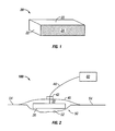
- FIG. 1 illustrates an embodiment of a wound insert.
- FIG. 2 illustrates an embodiment of a wound treatment system.
- FIGS. 3A-3D illustrate a process for treating a wound with a wound insert and a magnetically-active agent.
- Coupled is defined as connected, although not necessarily directly, and not necessarily mechanically; two items that are “coupled” may be integral with each other.
- the terms “a” and “an” are defined as one or more unless this disclosure explicitly requires otherwise.
- the terms “substantially,” “approximately,” and “about” are defined as largely but not necessarily wholly what is specified, as understood by a person of ordinary skill in the art.
- a wound dressing that “comprises,” “has,” “includes” or “contains” one or more elements possesses those one or more elements, but is not limited to possessing only those elements.
- the wound dressing in a wound dressing that comprises one of the present wound inserts and a drape, the wound dressing includes the specified elements but is not limited to having only those elements.
- such a wound dressing could also include a connection pad configured to be coupled to a negative pressure wound therapy (NPWT) apparatus (e.g., including a vacuum source and/or a fluid source).
- NGWT negative pressure wound therapy
- a device or structure that is configured in a certain way is configured in at least that way, but it can also be configured in other ways than those specifically described.
- FIG. 1 depicts a wound insert 20 that may be used in combination with the wound treatment system 100 shown in FIG. 2 .
- Wound insert 20 may comprise a foam member, which may be open-celled, close-celled, and/or reticulated.
- the wound insert comprises an open-celled reticulated foam.
- An open-celled reticulated foam has a netlike microstructure, with few if any closed cells.
- the porosity can range from 95%-98%, though less porous or more porous foams may be used.
- wound insert 20 may comprise a polyurethane, such as polyurethane-polyester or polyurethane-polyether; polyolefins, such as polypropylenes (PP) or polyethylenes (PE); silicone polymers; polyvinylchloride; polyamides; polyesters; acrylics; thermoplastic elastomers such as styrene-butene-styrene (SBS) or styrene-ethylene-butene-styrene (SEBS); polyether-amide block copolymers (PEBAX); elastomers such as styrene butadiene rubber (SBR); ethylene propylene rubber (EPR); ethylene propylene diene modified rubber (EPDM); natural rubber (NR); ethylene vinyl acetate (EVA); polyvinyl alcohol (PVOH); polyvinyl acetal; or polyvinyl butyral (PVB).
- polyurethane such as polyurethane-polyester or polyure
- wound insert 20 may comprise a bioabsorbable polymer, examples of which include polylactic acid, polylactide (PLA), polyglycolic acid, polyglycolide (PGA), and polycaprolactone (PCL).
- PLA polylactide
- PGA polyglycolide
- PCL polycaprolactone
- Wound insert 20 comprises surfaces 22 and an interior portion 24 .
- Wound insert 20 may be of any suitable shape having a depth dimension, including a sheet, a rectangular prism, a cone, a cylinder, a sphere, or any other suitable shape.
- wound insert 20 is capable of generating a magnetic field.
- superparamagnetic iron oxide nanoparticles SPION
- Wound insert 20 may be spray-coated with SPION such that magnetic nanoparticles are affixed to surfaces 22 , and are not disposed within interior portion 24 .
- surfaces 22 of wound insert 20 may be plasma coated with SPION or other suitable magnetic particles.
- magnetic particles may be disposed within interior portion 24 of wound insert 20 .
- wound insert 20 may be dipped into a slurry comprising SPION or other suitable magnetic particles, allowing the magnetic particles to become bonded or affixed to the cells within interior portion 24 of wound insert 20 .
- the strength of the magnetic field generated by wound insert 20 may be modulated, for example, by changing the density of the wound insert.
- wound insert 20 is configured to be used in conjunction with an electromagnet.
- the electromagnet may be located on surface 22 or in interior portion 24 of wound insert 20 .
- the electromagnet may be located within wound 50 , between drape 40 and wound surface 52 .
- electromagnet may be placed outside wound 50 and drape 40 , such as in a cuff worn by the patient or in a pad placed over drape 40 .
- wound insert 20 may comprise a macroscopic magnet.
- a cylindrical magnet, bar magnet, or strip magnet comprising ferromagnetic material may be coupled to a surface 22 of wound insert 20 .
- a cylindrical magnet, bar magnet, or strip magnet may be inserted into interior portion 24 of wound insert 20 such that the magnet is disposed within wound insert 20 .
- wound insert 20 is shown as part of a wound treatment system 100 .
- wound insert 20 is shown placed in wound 50 of a patient (not shown) having a wound surface 52 .
- a drape 40 is placed over wound 50 and wound insert 20 such that wound insert 20 is between drape 40 and wound 50 , creating space 30 .
- Drape 40 is coupled to the skin 54 of the patient.
- Wound insert 20 is coupled to a wound treatment apparatus 60 by conduit 44 .
- Apparatus 60 may comprise a vacuum source configured to apply negative pressure to wound insert 20 through conduit 44 .
- Apparatus 60 may further comprise a fluid source configured to deliver a fluid through conduit 44 to wound insert 20 . Examples of such fluids include a medicinal fluids, antibacterial fluids, or irrigation fluids.
- Various wound therapy systems and components are commercially available through KCI USA, Inc. of San Antonio, Tex., U.S.A.
- Conduit 44 can comprise a single lumen conduit (e.g., switched between a vacuum source and/or a fluid source) or can comprise multiple single-lumen conduits or a multi-lumen conduit such that, for example, fluid can be delivered and/or negative pressure can be applied to wound insert 20 individually or simultaneously.
- conduit 44 can comprise multiple lumens, for example, as in a single conduit with a central limit for application of negative pressure and/or fluid delivery and one or more peripheral lumens disposed adjacent or around the central lumen such that the peripheral lumens can be coupled to a pressure sensor to sense and/or detect a pressure or negative pressure between drape 40 and wound surface 52 .
- system 100 further comprises a wound dressing connection pad 42 configured to be coupled (and is shown coupled) to conduit 44 .
- a suitable connection pad 42 is the “V.A.C. T.R.A.C.® Pad,” commercially available from KCI USA, Inc. of San Antonio, Tex., U.S.A.
- a suitable drape 40 includes the “V.A.C.® Drape” commercially available from KCI USA, Inc. (and its affiliates) of San Antonio, Tex., U.S.A.
- apparatus 60 may be configured to deliver instillation fluid to wound 50 , to remove fluid from wound 50 , and to apply negative pressure to wound 50 through drape 40 and wound insert 20 .
- drape 40 and wound insert 20 may be referred to as a wound dressing 38 .
- a fluid source in apparatus 60 may be activated to deliver fluid, such as saline, to wound surface 52 through conduit 44 coupled to wound insert 20 through connection pad 42 .
- a vacuum source in apparatus 60 may be actuated to provide negative pressure to wound 50 and wound surface 52 through drape 40 and wound insert 20 .
- Example of instillation fluids that may be delivered to wound 50 are hypochlorous acid (HOCl) and hypochlorite ion (ClO—, which is also commonly referred to, generally understood to be synonymous with, and may be referred to interchangeably in this disclosure as, OCl—), which are examples of effective antimicrobial agents for biocidal action.
- HOCl is typically capable of killing a broad spectrum of microbes (e.g., fungus, bacteria, viruses, fungus, yeast, and the like); often in a relatively short period of time (e.g., is capable of killing greater than 99% of microbes within a period of less than 10 seconds).
- Such antimicrobial agents can be generated or formed by a combination of the present reactive agents and fluid (e.g., water and/or aqueous solution, such as, for example, saline solution) and may be more effective and/or more versatile than antibiotics and other commonly used antimicrobial agents used in wound treatment in the past.
- antibiotics may be bacteria-specific such that testing may be required to determine a suitable antibiotic to use for a specific wound or infection; and/or such that antibiotics may have only limited effectiveness for individual wounds and/or infections (e.g., where testing is not performed and/or where a wound is infected with a plurality of different bacteria). Such testing may take as long as several days to determine an appropriate antibiotic, delaying treatment or selection of an effective antibiotic.
- bacteria may develop resistance to antibiotics, such that antibiotics may have reduced effectiveness after an amount of time.
- antibiotics are typically administered intravenously (systemically) such that antibiotics may kill beneficial bacteria (e.g., in a patient's digestive system) and/or may cause organ damage (e.g., to a patient's liver).
- apparatus 60 may be configured to remove spent instillation fluids, secretions (e.g., pus), and/or infected tissue from wound 50 .
- Undesirable effluent may be removed by actuating the vacuum source in apparatus 60 ; effluent may flow into wound insert 20 , through conduit 44 , and into a waste chamber coupled to apparatus 60 .
- system 100 may also be configured to deliver magnetically-tagged therapeutic agents through the tissue of the patient to wound surface 52 of wound 50 .
- Similar wound treatment systems 100 are depicted in FIG. 2 and in FIGS. 3A-3D , except that certain elements have been omitted for clarity.
- Using magnetically-tagged therapeutic agents in combination with negative pressure wound therapy allows for specific targeting of wound sites, maximizing the efficacy of the therapeutic agents while keeping overall dosing low.
- the negative pressure wound therapy system may remove any effluent (e.g. excess or by-products of the magnetically-labeled agent and tissues, or any necrotic tissue or excretions).
- wound insert 20 is placed in wound 50 in close contact with wound surface 52 .
- Drape 40 is coupled to skin 54 such that wound insert 20 is between drape 40 and wound surface 52 .
- Negative pressure is applied using a vacuum source in apparatus 60 . The negative pressure collapses wound insert 20 .
- wound insert 20 is coated or impregnated with magnetic particles, increasing the density of the wound insert will increase the strength of the magnetic field.
- FIG. 3B shows the introduction of therapeutic agent 80 into the body of the patient.
- Agent 80 may comprise magnetically-tagged stem cells, magnetically-tagged antibiotics, magnetically-tagged debriding agents, or magnetically-tagged growth factors, or any other suitably magnetically-tagged medicament.
- the magnetic tag may comprise SPION or any other magnetically-active biocompatible nanoparticle.
- agent 80 may be tagged according to methods disclosed in Kyrtatos et al., “Magnetic Tagging Increases Delivery of Circulating Progenitors in Vascular Injury,” JACC Cardiovascular Interventions, 2009; 2(8); 794 DOI:10 1016/j.jcin.2009.05.014.
- agent 80 is systemically injected into the patient.
- Agent 80 can be injected intravenously or into skin 54 near wound 50 .
- a topical medicament e.g., a cream
- a bandage or patch comprising agent 80 is placed on skin 54 either separately or as part of the wound dressing.
- agent 80 may be delivered to the wound site via a conduit (e.g., conduit 44 ) coupled to the wound insert. And in yet other embodiments, agent 80 may be applied directly to wound surface 52 using a spray comprising agent 80 , then inserting wound insert 20 and applying drape 40 .
- Agent 80 is absorbed into the bloodstream or the lymph and migrates toward wound 50 , as shown in FIG. 3C .
- Agent 80 is drawn to wound insert 20 at the wound boundary between wound surface 52 and wound insert surface 22 .
- Agent 80 may magnetically bond to wound insert 20 , which increases the exposure of wound 50 to agent 80 .
- the negative pressure may be released to prevent agent 80 from migrating into interior portion 24 of wound insert 20 .
- agent 80 After agent 80 has been exposed to wound 50 for a desired period, negative pressure may once again be reapplied, as shown in FIG. 3D . Agent 80 is drawn into wound insert 20 , through conduit 44 , and into a waste receptacle coupled to conduit 44 . The process may be repeated as required.
- an electromagnet may be used in conjunction with wound insert 20 .
- the electromagnet may be placed within wound 50 , coupled to wound insert 20 , coupled to the outside of drape 40 in a pad or cuff, or placed in a wand held near the wound by an operator (e.g., doctor).
- the electromagnet may be made to give alternate polarization, such that the magnetically tagged medicaments within agent 80 are made to oscillate.
- the oscillation of magnetic particles increases their availability to wound surface 52 by ensuring that unused therapeutic agents are available.
- the magnetic particles coupled to the medicaments within agent 80 are uniformly aligned. That is, each medicament (e.g., stem cell, antibiotic, debriding agent, growth factor, etc.) within agent 80 is coupled to a magnetic nanoparticle whose poles are oriented in the same way relative to every other medicament. Such medicament orientation helps magnify the oscillation effect. Also, uniformly-oriented medicaments will aid in delaying the migration of agent 80 into wound insert 20 when negative pressure is applied.
- each medicament e.g., stem cell, antibiotic, debriding agent, growth factor, etc.
- arrival of agent 80 at wound 50 results in an increase in the magnetic field present at wound 50 , which indicates that the dose has arrived at the wound.
- the increase in magnetic field may be equated to amount of agent 80 present at the wound site, which may aid in monitoring the dosing received if agent 80 is systematically injected.
- a plurality of electromagnets may be disposed within wound insert 20 or in an external device such as a pad or wand.
- an external device such as a pad or wand.
- several electromagnets may be disposed in a pad external and adjacent to a long incisional wound.
- Agent 80 may be dosed at one end of the wound, and each electromagnet may be turned on and off in sequence to draw agent 80 along the full length of the wound.
- electromagnets may be disposed within wound insert 20 for deep wounds and therapy such as instillation.
- agent 80 may be delivered to the entirety of wound 50 , then controlled to exit wound 50 by modulating the strength of the magnetic field generated by the electromagnet.
- agent 80 may be a magnetically-tagged debriding agent.
- necrotic tissue may be located, and permanent or electromagnets may be placed in the necrotic tissue.
- the debriding agent may be drawn through wound 50 to these locations only.
Abstract
Description
Claims (17)
Priority Applications (1)
| Application Number | Priority Date | Filing Date | Title |
|---|---|---|---|
| US14/579,450 US10092452B2 (en) | 2010-12-15 | 2014-12-22 | Targeted delivery of magnetically tagged active agents in combination with negative pressure wound therapy |
Applications Claiming Priority (3)
| Application Number | Priority Date | Filing Date | Title |
|---|---|---|---|
| US42345910P | 2010-12-15 | 2010-12-15 | |
| US13/323,049 US8944067B2 (en) | 2010-12-15 | 2011-12-12 | Targeted delivery of magnetically tagged active agents in combination with negative pressure wound therapy |
| US14/579,450 US10092452B2 (en) | 2010-12-15 | 2014-12-22 | Targeted delivery of magnetically tagged active agents in combination with negative pressure wound therapy |
Related Parent Applications (2)
| Application Number | Title | Priority Date | Filing Date |
|---|---|---|---|
| US13/323,049 Continuation US8944067B2 (en) | 2010-12-15 | 2011-12-12 | Targeted delivery of magnetically tagged active agents in combination with negative pressure wound therapy |
| US13/323,049 Division US8944067B2 (en) | 2010-12-15 | 2011-12-12 | Targeted delivery of magnetically tagged active agents in combination with negative pressure wound therapy |
Publications (2)
| Publication Number | Publication Date |
|---|---|
| US20150112288A1 US20150112288A1 (en) | 2015-04-23 |
| US10092452B2 true US10092452B2 (en) | 2018-10-09 |
Family
ID=45390234
Family Applications (2)
| Application Number | Title | Priority Date | Filing Date |
|---|---|---|---|
| US13/323,049 Expired - Fee Related US8944067B2 (en) | 2010-12-15 | 2011-12-12 | Targeted delivery of magnetically tagged active agents in combination with negative pressure wound therapy |
| US14/579,450 Active 2033-03-14 US10092452B2 (en) | 2010-12-15 | 2014-12-22 | Targeted delivery of magnetically tagged active agents in combination with negative pressure wound therapy |
Family Applications Before (1)
| Application Number | Title | Priority Date | Filing Date |
|---|---|---|---|
| US13/323,049 Expired - Fee Related US8944067B2 (en) | 2010-12-15 | 2011-12-12 | Targeted delivery of magnetically tagged active agents in combination with negative pressure wound therapy |
Country Status (3)
| Country | Link |
|---|---|
| US (2) | US8944067B2 (en) |
| TW (1) | TW201236705A (en) |
| WO (1) | WO2012099659A1 (en) |
Cited By (1)
| Publication number | Priority date | Publication date | Assignee | Title |
|---|---|---|---|---|
| US20160310078A1 (en) * | 2015-04-24 | 2016-10-27 | Sports Medicine Sciences, LLC | Method and apparatus for directing therapeutic nanoparticle-labeled cells to selected locations within the body and/or for retaining therapeutic nanoparticle-labeled cells at selected locations within the body |
Families Citing this family (11)
| Publication number | Priority date | Publication date | Assignee | Title |
|---|---|---|---|---|
| CA2834702C (en) * | 2011-05-26 | 2019-03-26 | Kci Licensing, Inc. | Systems and methods of stimulation and activation of fluids for use with instillation therapy |
| EP2723286B2 (en) | 2011-06-24 | 2021-10-13 | KCI Licensing, Inc. | Reduced-pressure dressings employing tissue-fixation elements |
| US9237969B2 (en) | 2011-07-28 | 2016-01-19 | Matthew D. Antalek | Wound barrier pad |
| US10279157B2 (en) * | 2013-03-14 | 2019-05-07 | Rehabilitation Institute Of Chicago | Stress shield and infection control for a skin-implant interface |
| AU2014260424C1 (en) * | 2013-05-02 | 2017-02-16 | Vomaris Innovations, Inc. | Expandable wound dressings |
| WO2016154050A1 (en) * | 2015-03-20 | 2016-09-29 | The Trustees Of Dartmouth College | System and methods for enhancing uptake of therapeutic agent from bloodstream into disease site |
| US11577062B2 (en) * | 2017-04-11 | 2023-02-14 | University Of Florida Research Foundation, Inc. | Systems and methods for in-situ, bottom-up tissue generation |
| US10620335B2 (en) | 2017-05-02 | 2020-04-14 | Ascension Technology Corporation | Rotating frequencies of transmitters |
| US10779892B2 (en) | 2017-08-10 | 2020-09-22 | Northern Digital Inc. | Tracking a cylindrical opening |
| US11529193B2 (en) | 2017-08-10 | 2022-12-20 | Northern Digital Inc. | Tracking a sensor that includes a ferrofluid |
| US20200129339A1 (en) * | 2018-10-24 | 2020-04-30 | Hydrofera, Llc | Sterilization of medical devices with enhanced antimicrobial properties |
Citations (142)
| Publication number | Priority date | Publication date | Assignee | Title |
|---|---|---|---|---|
| US1355846A (en) | 1920-02-06 | 1920-10-19 | David A Rannells | Medical appliance |
| US2547758A (en) | 1949-01-05 | 1951-04-03 | Wilmer B Keeling | Instrument for treating the male urethra |
| US2632443A (en) | 1949-04-18 | 1953-03-24 | Eleanor P Lesher | Surgical dressing |
| GB692578A (en) | 1949-09-13 | 1953-06-10 | Minnesota Mining & Mfg | Improvements in or relating to drape sheets for surgical use |
| US2682873A (en) | 1952-07-30 | 1954-07-06 | Johnson & Johnson | General purpose protective dressing |
| US2910763A (en) | 1955-08-17 | 1959-11-03 | Du Pont | Felt-like products |
| US2969057A (en) | 1957-11-04 | 1961-01-24 | Brady Co W H | Nematodic swab |
| US3066672A (en) | 1960-09-27 | 1962-12-04 | Jr William H Crosby | Method and apparatus for serial sampling of intestinal juice |
| US3367332A (en) | 1965-08-27 | 1968-02-06 | Gen Electric | Product and process for establishing a sterile area of skin |
| GB1122796A (en) | 1964-10-14 | 1968-08-07 | Gustave Girardiere | Composition for making a coating on human tissue |
| GB1190733A (en) | 1966-04-16 | 1970-05-06 | Johnson & Johnson | Improvements in and relating to Surgical Swabs |
| US3520300A (en) | 1967-03-15 | 1970-07-14 | Amp Inc | Surgical sponge and suction device |
| US3568675A (en) | 1968-08-30 | 1971-03-09 | Clyde B Harvey | Fistula and penetrating wound dressing |
| US3648692A (en) | 1970-12-07 | 1972-03-14 | Parke Davis & Co | Medical-surgical dressing for burns and the like |
| US3682180A (en) | 1970-06-08 | 1972-08-08 | Coilform Co Inc | Drain clip for surgical drain |
| US3826254A (en) | 1973-02-26 | 1974-07-30 | Verco Ind | Needle or catheter retaining appliance |
| DE2640413A1 (en) | 1976-09-08 | 1978-03-09 | Wolf Gmbh Richard | CATHETER MONITORING DEVICE |
| US4080970A (en) | 1976-11-17 | 1978-03-28 | Miller Thomas J | Post-operative combination dressing and internal drain tube with external shield and tube connector |
| US4096853A (en) | 1975-06-21 | 1978-06-27 | Hoechst Aktiengesellschaft | Device for the introduction of contrast medium into an anus praeter |
| US4139004A (en) | 1977-02-17 | 1979-02-13 | Gonzalez Jr Harry | Bandage apparatus for treating burns |
| US4165748A (en) | 1977-11-07 | 1979-08-28 | Johnson Melissa C | Catheter tube holder |
| US4184510A (en) | 1977-03-15 | 1980-01-22 | Fibra-Sonics, Inc. | Valued device for controlling vacuum in surgery |
| WO1980002182A1 (en) | 1979-04-06 | 1980-10-16 | J Moss | Portable suction device for collecting fluids from a closed wound |
| US4233969A (en) | 1976-11-11 | 1980-11-18 | Lock Peter M | Wound dressing materials |
| US4245630A (en) | 1976-10-08 | 1981-01-20 | T. J. Smith & Nephew, Ltd. | Tearable composite strip of materials |
| US4256109A (en) | 1978-07-10 | 1981-03-17 | Nichols Robert L | Shut off valve for medical suction apparatus |
| US4261363A (en) | 1979-11-09 | 1981-04-14 | C. R. Bard, Inc. | Retention clips for body fluid drains |
| US4275721A (en) | 1978-11-28 | 1981-06-30 | Landstingens Inkopscentral Lic, Ekonomisk Forening | Vein catheter bandage |
| US4284079A (en) | 1979-06-28 | 1981-08-18 | Adair Edwin Lloyd | Method for applying a male incontinence device |
| US4297995A (en) | 1980-06-03 | 1981-11-03 | Key Pharmaceuticals, Inc. | Bandage containing attachment post |
| US4333468A (en) | 1980-08-18 | 1982-06-08 | Geist Robert W | Mesentery tube holder apparatus |
| US4373519A (en) | 1981-06-26 | 1983-02-15 | Minnesota Mining And Manufacturing Company | Composite wound dressing |
| US4382441A (en) | 1978-12-06 | 1983-05-10 | Svedman Paul | Device for treating tissues, for example skin |
| US4392853A (en) | 1981-03-16 | 1983-07-12 | Rudolph Muto | Sterile assembly for protecting and fastening an indwelling device |
| US4392858A (en) | 1981-07-16 | 1983-07-12 | Sherwood Medical Company | Wound drainage device |
| US4419097A (en) | 1981-07-31 | 1983-12-06 | Rexar Industries, Inc. | Attachment for catheter tube |
| EP0100148A1 (en) | 1982-07-06 | 1984-02-08 | Dow Corning Limited | Medical-surgical dressing and a process for the production thereof |
| US4465485A (en) | 1981-03-06 | 1984-08-14 | Becton, Dickinson And Company | Suction canister with unitary shut-off valve and filter features |
| EP0117632A2 (en) | 1983-01-27 | 1984-09-05 | Johnson & Johnson Products Inc. | Adhesive film dressing |
| US4475909A (en) | 1982-05-06 | 1984-10-09 | Eisenberg Melvin I | Male urinary device and method for applying the device |
| US4480638A (en) | 1980-03-11 | 1984-11-06 | Eduard Schmid | Cushion for holding an element of grafted skin |
| US4525166A (en) | 1981-11-21 | 1985-06-25 | Intermedicat Gmbh | Rolled flexible medical suction drainage device |
| US4525374A (en) | 1984-02-27 | 1985-06-25 | Manresa, Inc. | Treating hydrophobic filters to render them hydrophilic |
| US4540412A (en) | 1983-07-14 | 1985-09-10 | The Kendall Company | Device for moist heat therapy |
| US4543100A (en) | 1983-11-01 | 1985-09-24 | Brodsky Stuart A | Catheter and drain tube retainer |
| JPS60192738A (en) | 1984-03-14 | 1985-10-01 | Yasunori Sakuramoto | Magnetic foam material |
| US4548202A (en) | 1983-06-20 | 1985-10-22 | Ethicon, Inc. | Mesh tissue fasteners |
| US4551139A (en) | 1982-02-08 | 1985-11-05 | Marion Laboratories, Inc. | Method and apparatus for burn wound treatment |
| EP0161865A2 (en) | 1984-05-03 | 1985-11-21 | Smith and Nephew Associated Companies p.l.c. | Adhesive wound dressing |
| US4569348A (en) | 1980-02-22 | 1986-02-11 | Velcro Usa Inc. | Catheter tube holder strap |
| AU550575B2 (en) | 1981-08-07 | 1986-03-27 | Richard Christian Wright | Wound drainage device |
| US4605399A (en) | 1984-12-04 | 1986-08-12 | Complex, Inc. | Transdermal infusion device |
| US4608041A (en) | 1981-10-14 | 1986-08-26 | Frese Nielsen | Device for treatment of wounds in body tissue of patients by exposure to jets of gas |
| US4640688A (en) | 1985-08-23 | 1987-02-03 | Mentor Corporation | Urine collection catheter |
| EP0214368A1 (en) | 1985-09-07 | 1987-03-18 | Lacotherm Ag | Means for raising and stabilizing skin temperature |
| US4655754A (en) | 1984-11-09 | 1987-04-07 | Stryker Corporation | Vacuum wound drainage system and lipids baffle therefor |
| US4664662A (en) | 1984-08-02 | 1987-05-12 | Smith And Nephew Associated Companies Plc | Wound dressing |
| WO1987004626A1 (en) | 1986-01-31 | 1987-08-13 | Osmond, Roger, L., W. | Suction system for wound and gastro-intestinal drainage |
| US4710165A (en) | 1985-09-16 | 1987-12-01 | Mcneil Charles B | Wearable, variable rate suction/collection device |
| US4733659A (en) | 1986-01-17 | 1988-03-29 | Seton Company | Foam bandage |
| GB2195255A (en) | 1986-09-30 | 1988-04-07 | Vacutec Uk Limited | Method and apparatus for vacuum treatment of an epidermal surface |
| US4743232A (en) | 1986-10-06 | 1988-05-10 | The Clinipad Corporation | Package assembly for plastic film bandage |
| GB2197789A (en) | 1986-11-28 | 1988-06-02 | Smiths Industries Plc | Anti-foaming disinfectants used in surgical suction apparatus |
| US4758220A (en) | 1985-09-26 | 1988-07-19 | Alcon Laboratories, Inc. | Surgical cassette proximity sensing and latching apparatus |
| US4787888A (en) | 1987-06-01 | 1988-11-29 | University Of Connecticut | Disposable piezoelectric polymer bandage for percutaneous delivery of drugs and method for such percutaneous delivery (a) |
| US4826494A (en) | 1984-11-09 | 1989-05-02 | Stryker Corporation | Vacuum wound drainage system |
| US4838883A (en) | 1986-03-07 | 1989-06-13 | Nissho Corporation | Urine-collecting device |
| US4840187A (en) | 1986-09-11 | 1989-06-20 | Bard Limited | Sheath applicator |
| US4863449A (en) | 1987-07-06 | 1989-09-05 | Hollister Incorporated | Adhesive-lined elastic condom cathether |
| US4872450A (en) | 1984-08-17 | 1989-10-10 | Austad Eric D | Wound dressing and method of forming same |
| US4878901A (en) | 1986-10-10 | 1989-11-07 | Sachse Hans Ernst | Condom catheter, a urethral catheter for the prevention of ascending infections |
| GB2220357A (en) | 1988-05-28 | 1990-01-10 | Smiths Industries Plc | Medico-surgical containers |
| US4897081A (en) | 1984-05-25 | 1990-01-30 | Thermedics Inc. | Percutaneous access device |
| US4906240A (en) | 1988-02-01 | 1990-03-06 | Matrix Medica, Inc. | Adhesive-faced porous absorbent sheet and method of making same |
| US4906233A (en) | 1986-05-29 | 1990-03-06 | Terumo Kabushiki Kaisha | Method of securing a catheter body to a human skin surface |
| US4919654A (en) | 1988-08-03 | 1990-04-24 | Kalt Medical Corporation | IV clamp with membrane |
| CA2005436A1 (en) | 1988-12-13 | 1990-06-13 | Glenda G. Kalt | Transparent tracheostomy tube dressing |
| US4941882A (en) | 1987-03-14 | 1990-07-17 | Smith And Nephew Associated Companies, P.L.C. | Adhesive dressing for retaining a cannula on the skin |
| EP0380253A2 (en) | 1989-01-24 | 1990-08-01 | Minnesota Mining And Manufacturing Company | Alginate hydrogel foam wound dressing |
| US4953565A (en) | 1986-11-26 | 1990-09-04 | Shunro Tachibana | Endermic application kits for external medicines |
| WO1990010424A1 (en) | 1989-03-16 | 1990-09-20 | Smith & Nephew Plc | Absorbent devices and precursors therefor |
| US4969880A (en) | 1989-04-03 | 1990-11-13 | Zamierowski David S | Wound dressing and treatment method |
| US4985019A (en) | 1988-03-11 | 1991-01-15 | Michelson Gary K | X-ray marker |
| GB2235877A (en) | 1989-09-18 | 1991-03-20 | Antonio Talluri | Closed wound suction apparatus |
| US5037397A (en) | 1985-05-03 | 1991-08-06 | Medical Distributors, Inc. | Universal clamp |
| US5086170A (en) | 1989-01-16 | 1992-02-04 | Roussel Uclaf | Process for the preparation of azabicyclo compounds |
| US5092858A (en) | 1990-03-20 | 1992-03-03 | Becton, Dickinson And Company | Liquid gelling agent distributor device |
| US5100396A (en) | 1989-04-03 | 1992-03-31 | Zamierowski David S | Fluidic connection system and method |
| JPH04129536A (en) | 1990-09-19 | 1992-04-30 | Terumo Corp | Balance device |
| US5134994A (en) | 1990-02-12 | 1992-08-04 | Say Sam L | Field aspirator in a soft pack with externally mounted container |
| US5149331A (en) | 1991-05-03 | 1992-09-22 | Ariel Ferdman | Method and device for wound closure |
| US5167613A (en) | 1992-03-23 | 1992-12-01 | The Kendall Company | Composite vented wound dressing |
| US5176663A (en) | 1987-12-02 | 1993-01-05 | Pal Svedman | Dressing having pad with compressibility limiting elements |
| WO1993009727A1 (en) | 1991-11-14 | 1993-05-27 | Wake Forest University | Method and apparatus for treating tissue damage |
| US5215522A (en) | 1984-07-23 | 1993-06-01 | Ballard Medical Products | Single use medical aspirating device and method |
| US5232453A (en) | 1989-07-14 | 1993-08-03 | E. R. Squibb & Sons, Inc. | Catheter holder |
| US5261893A (en) | 1989-04-03 | 1993-11-16 | Zamierowski David S | Fastening system and method |
| US5278100A (en) | 1991-11-08 | 1994-01-11 | Micron Technology, Inc. | Chemical vapor deposition technique for depositing titanium silicide on semiconductor wafers |
| US5279550A (en) | 1991-12-19 | 1994-01-18 | Gish Biomedical, Inc. | Orthopedic autotransfusion system |
| US5298015A (en) | 1989-07-11 | 1994-03-29 | Nippon Zeon Co., Ltd. | Wound dressing having a porous structure |
| US5342376A (en) | 1993-05-03 | 1994-08-30 | Dermagraphics, Inc. | Inserting device for a barbed tissue connector |
| US5344415A (en) | 1993-06-15 | 1994-09-06 | Deroyal Industries, Inc. | Sterile system for dressing vascular access site |
| DE4306478A1 (en) | 1993-03-02 | 1994-09-08 | Wolfgang Dr Wagner | Drainage device, in particular pleural drainage device, and drainage method |
| WO1994020041A1 (en) | 1993-03-09 | 1994-09-15 | Wake Forest University | Wound treatment employing reduced pressure |
| US5358494A (en) | 1989-07-11 | 1994-10-25 | Svedman Paul | Irrigation dressing |
| USRE34866E (en) * | 1987-02-17 | 1995-02-21 | Kensey Nash Corporation | Device for sealing percutaneous puncture in a vessel |
| US5437622A (en) | 1992-04-29 | 1995-08-01 | Laboratoire Hydrex (Sa) | Transparent adhesive dressing with reinforced starter cuts |
| US5437651A (en) | 1993-09-01 | 1995-08-01 | Research Medical, Inc. | Medical suction apparatus |
| DE29504378U1 (en) | 1995-03-15 | 1995-09-14 | Mtg Medizinisch Tech Geraeteba | Electronically controlled low-vacuum pump for chest and wound drainage |
| WO1996005873A1 (en) | 1994-08-22 | 1996-02-29 | Kinetic Concepts Inc. | Wound drainage equipment |
| US5527293A (en) | 1989-04-03 | 1996-06-18 | Kinetic Concepts, Inc. | Fastening system and method |
| US5549584A (en) | 1994-02-14 | 1996-08-27 | The Kendall Company | Apparatus for removing fluid from a wound |
| US5556375A (en) | 1994-06-16 | 1996-09-17 | Hercules Incorporated | Wound dressing having a fenestrated base layer |
| US5607388A (en) | 1994-06-16 | 1997-03-04 | Hercules Incorporated | Multi-purpose wound dressing |
| WO1997018007A1 (en) | 1995-11-14 | 1997-05-22 | Kci Medical Limited | Portable wound treatment apparatus |
| WO1999013793A1 (en) | 1997-09-12 | 1999-03-25 | Kci Medical Limited | Surgical drape and suction head for wound treatment |
| US6071267A (en) | 1998-02-06 | 2000-06-06 | Kinetic Concepts, Inc. | Medical patient fluid management interface system and method |
| US6135116A (en) | 1997-07-28 | 2000-10-24 | Kci Licensing, Inc. | Therapeutic method for treating ulcers |
| US6146324A (en) | 1998-02-25 | 2000-11-14 | Engel; Peter H. | Magnetic analgesic therapeutic device |
| US6174544B1 (en) | 1999-09-16 | 2001-01-16 | Euromed, Inc. | Easy-release alginate wound healing device and method |
| US6241747B1 (en) | 1993-05-03 | 2001-06-05 | Quill Medical, Inc. | Barbed Bodily tissue connector |
| US6287316B1 (en) | 1999-03-26 | 2001-09-11 | Ethicon, Inc. | Knitted surgical mesh |
| US20020077661A1 (en) | 2000-12-20 | 2002-06-20 | Vahid Saadat | Multi-barbed device for retaining tissue in apposition and methods of use |
| US20020115951A1 (en) | 2001-02-22 | 2002-08-22 | Core Products International, Inc. | Ankle brace providing upper and lower ankle adjustment |
| US20020120185A1 (en) | 2000-05-26 | 2002-08-29 | Kci Licensing, Inc. | System for combined transcutaneous blood gas monitoring and vacuum assisted wound closure |
| US20020143286A1 (en) | 2001-03-05 | 2002-10-03 | Kci Licensing, Inc. | Vacuum assisted wound treatment apparatus and infection identification system and method |
| US6488643B1 (en) | 1998-10-08 | 2002-12-03 | Kci Licensing, Inc. | Wound healing foot wrap |
| US6493568B1 (en) | 1994-07-19 | 2002-12-10 | Kci Licensing, Inc. | Patient interface system |
| AU755496B2 (en) | 1997-09-12 | 2002-12-12 | Kci Licensing, Inc. | Surgical drape and suction head for wound treatment |
| US20040138552A1 (en) * | 2001-04-18 | 2004-07-15 | Alex Harel | Navigating and maneuvering of an in vivo vehicle by extracorporeal devices |
| EP1452155A2 (en) | 2003-02-27 | 2004-09-01 | Cho Kee Wong | Back support pad |
| US20060079852A1 (en) | 2002-12-31 | 2006-04-13 | Bubb Stephen K | Externally-applied patient interface system and method |
| WO2007046806A1 (en) | 2005-10-21 | 2007-04-26 | Argentum Medical, Llc | Medical device |
| US20070218285A1 (en) | 2004-05-21 | 2007-09-20 | Dr Suwelack Skin & Health Care Ag | Process For The Production Of Porous Moulded Articles Containing Alginate |
| JP4129536B2 (en) | 2000-02-24 | 2008-08-06 | ヴェネテック インターナショナル,インコーポレイテッド | Highly compatible catheter anchoring system |
| EP2031285A2 (en) | 2007-08-29 | 2009-03-04 | Carl Freudenberg KG | Valve with magnetic foam seal body |
| US20090287060A1 (en) * | 2008-05-14 | 2009-11-19 | Physcient, Inc. | Methods and devices to decrease tissue trauma during surgery |
| US20100030132A1 (en) * | 2006-09-28 | 2010-02-04 | Jeffrey Niezgoda | Apparatus and method for wound, cavity, and bone treatment |
| WO2010093420A2 (en) | 2009-02-11 | 2010-08-19 | University Of Houston | Ultrasmall superparamagnetic iron oxide nanoparticles and uses thereof |
| US20100215708A1 (en) | 2006-06-29 | 2010-08-26 | Andreas Zumbuehl | Coating of devices with effector compounds |
| US20100268176A1 (en) * | 2001-02-16 | 2010-10-21 | Royce Johnson | Biocompatible wound dressing |
| US20140257212A1 (en) * | 2001-08-24 | 2014-09-11 | Kci Licensing, Inc. | Negative pressure assisted tissue treatment system |
Family Cites Families (3)
| Publication number | Priority date | Publication date | Assignee | Title |
|---|---|---|---|---|
| US5320103A (en) * | 1987-10-07 | 1994-06-14 | Advanced Techtronics, Inc. | Permanent magnet arrangement |
| DE102008008522A1 (en) * | 2008-02-11 | 2009-08-13 | Magforce Nanotechnologies Ag | Implantable nanoparticle-containing products |
| WO2011087871A2 (en) * | 2009-12-22 | 2011-07-21 | Smith & Nephew, Inc. | Apparatuses and methods for negative pressure wound therapy |
-
2011
- 2011-12-12 US US13/323,049 patent/US8944067B2/en not_active Expired - Fee Related
- 2011-12-13 WO PCT/US2011/064597 patent/WO2012099659A1/en active Application Filing
- 2011-12-15 TW TW100146601A patent/TW201236705A/en unknown
-
2014
- 2014-12-22 US US14/579,450 patent/US10092452B2/en active Active
Patent Citations (152)
| Publication number | Priority date | Publication date | Assignee | Title |
|---|---|---|---|---|
| US1355846A (en) | 1920-02-06 | 1920-10-19 | David A Rannells | Medical appliance |
| US2547758A (en) | 1949-01-05 | 1951-04-03 | Wilmer B Keeling | Instrument for treating the male urethra |
| US2632443A (en) | 1949-04-18 | 1953-03-24 | Eleanor P Lesher | Surgical dressing |
| GB692578A (en) | 1949-09-13 | 1953-06-10 | Minnesota Mining & Mfg | Improvements in or relating to drape sheets for surgical use |
| US2682873A (en) | 1952-07-30 | 1954-07-06 | Johnson & Johnson | General purpose protective dressing |
| US2910763A (en) | 1955-08-17 | 1959-11-03 | Du Pont | Felt-like products |
| US2969057A (en) | 1957-11-04 | 1961-01-24 | Brady Co W H | Nematodic swab |
| US3066672A (en) | 1960-09-27 | 1962-12-04 | Jr William H Crosby | Method and apparatus for serial sampling of intestinal juice |
| GB1122796A (en) | 1964-10-14 | 1968-08-07 | Gustave Girardiere | Composition for making a coating on human tissue |
| US3367332A (en) | 1965-08-27 | 1968-02-06 | Gen Electric | Product and process for establishing a sterile area of skin |
| GB1190733A (en) | 1966-04-16 | 1970-05-06 | Johnson & Johnson | Improvements in and relating to Surgical Swabs |
| US3520300A (en) | 1967-03-15 | 1970-07-14 | Amp Inc | Surgical sponge and suction device |
| US3568675A (en) | 1968-08-30 | 1971-03-09 | Clyde B Harvey | Fistula and penetrating wound dressing |
| US3682180A (en) | 1970-06-08 | 1972-08-08 | Coilform Co Inc | Drain clip for surgical drain |
| US3648692A (en) | 1970-12-07 | 1972-03-14 | Parke Davis & Co | Medical-surgical dressing for burns and the like |
| US3826254A (en) | 1973-02-26 | 1974-07-30 | Verco Ind | Needle or catheter retaining appliance |
| US4096853A (en) | 1975-06-21 | 1978-06-27 | Hoechst Aktiengesellschaft | Device for the introduction of contrast medium into an anus praeter |
| DE2640413A1 (en) | 1976-09-08 | 1978-03-09 | Wolf Gmbh Richard | CATHETER MONITORING DEVICE |
| US4245630A (en) | 1976-10-08 | 1981-01-20 | T. J. Smith & Nephew, Ltd. | Tearable composite strip of materials |
| US4233969A (en) | 1976-11-11 | 1980-11-18 | Lock Peter M | Wound dressing materials |
| US4080970A (en) | 1976-11-17 | 1978-03-28 | Miller Thomas J | Post-operative combination dressing and internal drain tube with external shield and tube connector |
| US4139004A (en) | 1977-02-17 | 1979-02-13 | Gonzalez Jr Harry | Bandage apparatus for treating burns |
| US4184510A (en) | 1977-03-15 | 1980-01-22 | Fibra-Sonics, Inc. | Valued device for controlling vacuum in surgery |
| US4165748A (en) | 1977-11-07 | 1979-08-28 | Johnson Melissa C | Catheter tube holder |
| US4256109A (en) | 1978-07-10 | 1981-03-17 | Nichols Robert L | Shut off valve for medical suction apparatus |
| US4275721A (en) | 1978-11-28 | 1981-06-30 | Landstingens Inkopscentral Lic, Ekonomisk Forening | Vein catheter bandage |
| US4382441A (en) | 1978-12-06 | 1983-05-10 | Svedman Paul | Device for treating tissues, for example skin |
| WO1980002182A1 (en) | 1979-04-06 | 1980-10-16 | J Moss | Portable suction device for collecting fluids from a closed wound |
| US4284079A (en) | 1979-06-28 | 1981-08-18 | Adair Edwin Lloyd | Method for applying a male incontinence device |
| US4261363A (en) | 1979-11-09 | 1981-04-14 | C. R. Bard, Inc. | Retention clips for body fluid drains |
| US4569348A (en) | 1980-02-22 | 1986-02-11 | Velcro Usa Inc. | Catheter tube holder strap |
| US4480638A (en) | 1980-03-11 | 1984-11-06 | Eduard Schmid | Cushion for holding an element of grafted skin |
| US4297995A (en) | 1980-06-03 | 1981-11-03 | Key Pharmaceuticals, Inc. | Bandage containing attachment post |
| US4333468A (en) | 1980-08-18 | 1982-06-08 | Geist Robert W | Mesentery tube holder apparatus |
| US4465485A (en) | 1981-03-06 | 1984-08-14 | Becton, Dickinson And Company | Suction canister with unitary shut-off valve and filter features |
| US4392853A (en) | 1981-03-16 | 1983-07-12 | Rudolph Muto | Sterile assembly for protecting and fastening an indwelling device |
| US4373519A (en) | 1981-06-26 | 1983-02-15 | Minnesota Mining And Manufacturing Company | Composite wound dressing |
| US4392858A (en) | 1981-07-16 | 1983-07-12 | Sherwood Medical Company | Wound drainage device |
| US4419097A (en) | 1981-07-31 | 1983-12-06 | Rexar Industries, Inc. | Attachment for catheter tube |
| AU550575B2 (en) | 1981-08-07 | 1986-03-27 | Richard Christian Wright | Wound drainage device |
| US4608041A (en) | 1981-10-14 | 1986-08-26 | Frese Nielsen | Device for treatment of wounds in body tissue of patients by exposure to jets of gas |
| US4525166A (en) | 1981-11-21 | 1985-06-25 | Intermedicat Gmbh | Rolled flexible medical suction drainage device |
| US4551139A (en) | 1982-02-08 | 1985-11-05 | Marion Laboratories, Inc. | Method and apparatus for burn wound treatment |
| US4475909A (en) | 1982-05-06 | 1984-10-09 | Eisenberg Melvin I | Male urinary device and method for applying the device |
| EP0100148A1 (en) | 1982-07-06 | 1984-02-08 | Dow Corning Limited | Medical-surgical dressing and a process for the production thereof |
| EP0117632A2 (en) | 1983-01-27 | 1984-09-05 | Johnson & Johnson Products Inc. | Adhesive film dressing |
| US4548202A (en) | 1983-06-20 | 1985-10-22 | Ethicon, Inc. | Mesh tissue fasteners |
| US4540412A (en) | 1983-07-14 | 1985-09-10 | The Kendall Company | Device for moist heat therapy |
| US4543100A (en) | 1983-11-01 | 1985-09-24 | Brodsky Stuart A | Catheter and drain tube retainer |
| US4525374A (en) | 1984-02-27 | 1985-06-25 | Manresa, Inc. | Treating hydrophobic filters to render them hydrophilic |
| JPS60192738A (en) | 1984-03-14 | 1985-10-01 | Yasunori Sakuramoto | Magnetic foam material |
| EP0161865A2 (en) | 1984-05-03 | 1985-11-21 | Smith and Nephew Associated Companies p.l.c. | Adhesive wound dressing |
| US4897081A (en) | 1984-05-25 | 1990-01-30 | Thermedics Inc. | Percutaneous access device |
| US5215522A (en) | 1984-07-23 | 1993-06-01 | Ballard Medical Products | Single use medical aspirating device and method |
| US4664662A (en) | 1984-08-02 | 1987-05-12 | Smith And Nephew Associated Companies Plc | Wound dressing |
| US4872450A (en) | 1984-08-17 | 1989-10-10 | Austad Eric D | Wound dressing and method of forming same |
| US4655754A (en) | 1984-11-09 | 1987-04-07 | Stryker Corporation | Vacuum wound drainage system and lipids baffle therefor |
| US4826494A (en) | 1984-11-09 | 1989-05-02 | Stryker Corporation | Vacuum wound drainage system |
| US4605399A (en) | 1984-12-04 | 1986-08-12 | Complex, Inc. | Transdermal infusion device |
| US5037397A (en) | 1985-05-03 | 1991-08-06 | Medical Distributors, Inc. | Universal clamp |
| US4640688A (en) | 1985-08-23 | 1987-02-03 | Mentor Corporation | Urine collection catheter |
| EP0214368A1 (en) | 1985-09-07 | 1987-03-18 | Lacotherm Ag | Means for raising and stabilizing skin temperature |
| US4710165A (en) | 1985-09-16 | 1987-12-01 | Mcneil Charles B | Wearable, variable rate suction/collection device |
| US4758220A (en) | 1985-09-26 | 1988-07-19 | Alcon Laboratories, Inc. | Surgical cassette proximity sensing and latching apparatus |
| US4733659A (en) | 1986-01-17 | 1988-03-29 | Seton Company | Foam bandage |
| WO1987004626A1 (en) | 1986-01-31 | 1987-08-13 | Osmond, Roger, L., W. | Suction system for wound and gastro-intestinal drainage |
| US4838883A (en) | 1986-03-07 | 1989-06-13 | Nissho Corporation | Urine-collecting device |
| US4906233A (en) | 1986-05-29 | 1990-03-06 | Terumo Kabushiki Kaisha | Method of securing a catheter body to a human skin surface |
| US4840187A (en) | 1986-09-11 | 1989-06-20 | Bard Limited | Sheath applicator |
| GB2195255A (en) | 1986-09-30 | 1988-04-07 | Vacutec Uk Limited | Method and apparatus for vacuum treatment of an epidermal surface |
| US4743232A (en) | 1986-10-06 | 1988-05-10 | The Clinipad Corporation | Package assembly for plastic film bandage |
| US4878901A (en) | 1986-10-10 | 1989-11-07 | Sachse Hans Ernst | Condom catheter, a urethral catheter for the prevention of ascending infections |
| US4953565A (en) | 1986-11-26 | 1990-09-04 | Shunro Tachibana | Endermic application kits for external medicines |
| GB2197789A (en) | 1986-11-28 | 1988-06-02 | Smiths Industries Plc | Anti-foaming disinfectants used in surgical suction apparatus |
| USRE34866E (en) * | 1987-02-17 | 1995-02-21 | Kensey Nash Corporation | Device for sealing percutaneous puncture in a vessel |
| US4941882A (en) | 1987-03-14 | 1990-07-17 | Smith And Nephew Associated Companies, P.L.C. | Adhesive dressing for retaining a cannula on the skin |
| US4787888A (en) | 1987-06-01 | 1988-11-29 | University Of Connecticut | Disposable piezoelectric polymer bandage for percutaneous delivery of drugs and method for such percutaneous delivery (a) |
| US4863449A (en) | 1987-07-06 | 1989-09-05 | Hollister Incorporated | Adhesive-lined elastic condom cathether |
| US5176663A (en) | 1987-12-02 | 1993-01-05 | Pal Svedman | Dressing having pad with compressibility limiting elements |
| US4906240A (en) | 1988-02-01 | 1990-03-06 | Matrix Medica, Inc. | Adhesive-faced porous absorbent sheet and method of making same |
| US4985019A (en) | 1988-03-11 | 1991-01-15 | Michelson Gary K | X-ray marker |
| EP0358302A2 (en) | 1988-05-28 | 1990-03-14 | Smiths Industries Public Limited Company | Medico-surgical suction container |
| GB2220357A (en) | 1988-05-28 | 1990-01-10 | Smiths Industries Plc | Medico-surgical containers |
| US4919654A (en) | 1988-08-03 | 1990-04-24 | Kalt Medical Corporation | IV clamp with membrane |
| CA2005436A1 (en) | 1988-12-13 | 1990-06-13 | Glenda G. Kalt | Transparent tracheostomy tube dressing |
| US5086170A (en) | 1989-01-16 | 1992-02-04 | Roussel Uclaf | Process for the preparation of azabicyclo compounds |
| EP0380253A2 (en) | 1989-01-24 | 1990-08-01 | Minnesota Mining And Manufacturing Company | Alginate hydrogel foam wound dressing |
| WO1990010424A1 (en) | 1989-03-16 | 1990-09-20 | Smith & Nephew Plc | Absorbent devices and precursors therefor |
| US4969880A (en) | 1989-04-03 | 1990-11-13 | Zamierowski David S | Wound dressing and treatment method |
| US5100396A (en) | 1989-04-03 | 1992-03-31 | Zamierowski David S | Fluidic connection system and method |
| US5527293A (en) | 1989-04-03 | 1996-06-18 | Kinetic Concepts, Inc. | Fastening system and method |
| US5261893A (en) | 1989-04-03 | 1993-11-16 | Zamierowski David S | Fastening system and method |
| US5358494A (en) | 1989-07-11 | 1994-10-25 | Svedman Paul | Irrigation dressing |
| US5298015A (en) | 1989-07-11 | 1994-03-29 | Nippon Zeon Co., Ltd. | Wound dressing having a porous structure |
| US5232453A (en) | 1989-07-14 | 1993-08-03 | E. R. Squibb & Sons, Inc. | Catheter holder |
| GB2235877A (en) | 1989-09-18 | 1991-03-20 | Antonio Talluri | Closed wound suction apparatus |
| US5134994A (en) | 1990-02-12 | 1992-08-04 | Say Sam L | Field aspirator in a soft pack with externally mounted container |
| US5092858A (en) | 1990-03-20 | 1992-03-03 | Becton, Dickinson And Company | Liquid gelling agent distributor device |
| JPH04129536A (en) | 1990-09-19 | 1992-04-30 | Terumo Corp | Balance device |
| US5149331A (en) | 1991-05-03 | 1992-09-22 | Ariel Ferdman | Method and device for wound closure |
| US5278100A (en) | 1991-11-08 | 1994-01-11 | Micron Technology, Inc. | Chemical vapor deposition technique for depositing titanium silicide on semiconductor wafers |
| WO1993009727A1 (en) | 1991-11-14 | 1993-05-27 | Wake Forest University | Method and apparatus for treating tissue damage |
| US5645081A (en) | 1991-11-14 | 1997-07-08 | Wake Forest University | Method of treating tissue damage and apparatus for same |
| US5636643A (en) | 1991-11-14 | 1997-06-10 | Wake Forest University | Wound treatment employing reduced pressure |
| US5279550A (en) | 1991-12-19 | 1994-01-18 | Gish Biomedical, Inc. | Orthopedic autotransfusion system |
| US5167613A (en) | 1992-03-23 | 1992-12-01 | The Kendall Company | Composite vented wound dressing |
| US5437622A (en) | 1992-04-29 | 1995-08-01 | Laboratoire Hydrex (Sa) | Transparent adhesive dressing with reinforced starter cuts |
| DE4306478A1 (en) | 1993-03-02 | 1994-09-08 | Wolfgang Dr Wagner | Drainage device, in particular pleural drainage device, and drainage method |
| WO1994020041A1 (en) | 1993-03-09 | 1994-09-15 | Wake Forest University | Wound treatment employing reduced pressure |
| US5342376A (en) | 1993-05-03 | 1994-08-30 | Dermagraphics, Inc. | Inserting device for a barbed tissue connector |
| US6241747B1 (en) | 1993-05-03 | 2001-06-05 | Quill Medical, Inc. | Barbed Bodily tissue connector |
| US5344415A (en) | 1993-06-15 | 1994-09-06 | Deroyal Industries, Inc. | Sterile system for dressing vascular access site |
| US5437651A (en) | 1993-09-01 | 1995-08-01 | Research Medical, Inc. | Medical suction apparatus |
| US5549584A (en) | 1994-02-14 | 1996-08-27 | The Kendall Company | Apparatus for removing fluid from a wound |
| US5607388A (en) | 1994-06-16 | 1997-03-04 | Hercules Incorporated | Multi-purpose wound dressing |
| US5556375A (en) | 1994-06-16 | 1996-09-17 | Hercules Incorporated | Wound dressing having a fenestrated base layer |
| US6493568B1 (en) | 1994-07-19 | 2002-12-10 | Kci Licensing, Inc. | Patient interface system |
| WO1996005873A1 (en) | 1994-08-22 | 1996-02-29 | Kinetic Concepts Inc. | Wound drainage equipment |
| DE29504378U1 (en) | 1995-03-15 | 1995-09-14 | Mtg Medizinisch Tech Geraeteba | Electronically controlled low-vacuum pump for chest and wound drainage |
| WO1997018007A1 (en) | 1995-11-14 | 1997-05-22 | Kci Medical Limited | Portable wound treatment apparatus |
| US6135116A (en) | 1997-07-28 | 2000-10-24 | Kci Licensing, Inc. | Therapeutic method for treating ulcers |
| WO1999013793A1 (en) | 1997-09-12 | 1999-03-25 | Kci Medical Limited | Surgical drape and suction head for wound treatment |
| US6814079B2 (en) | 1997-09-12 | 2004-11-09 | Kci Licensing, Inc. | Surgical drape and suction head for wound treatment |
| US6553998B2 (en) | 1997-09-12 | 2003-04-29 | Kci Licensing, Inc. | Surgical drape and suction head for wound treatment |
| AU755496B2 (en) | 1997-09-12 | 2002-12-12 | Kci Licensing, Inc. | Surgical drape and suction head for wound treatment |
| EP1018967B1 (en) | 1997-09-12 | 2004-08-18 | KCI Licensing, Inc. | Suction head for wound treatment and combination with a surgical drape |
| GB2333965A (en) | 1997-09-12 | 1999-08-11 | Kci Medical Ltd | Surgical drape |
| US6345623B1 (en) | 1997-09-12 | 2002-02-12 | Keith Patrick Heaton | Surgical drape and suction head for wound treatment |
| AU745271B2 (en) | 1997-09-12 | 2002-03-14 | Kci Licensing, Inc. | Surgical drape and suction head for wound treatment |
| GB2329127B (en) | 1997-09-12 | 2000-08-16 | Kci Medical Ltd | Surgical drape and suction head for wound treatment |
| US6071267A (en) | 1998-02-06 | 2000-06-06 | Kinetic Concepts, Inc. | Medical patient fluid management interface system and method |
| US6146324A (en) | 1998-02-25 | 2000-11-14 | Engel; Peter H. | Magnetic analgesic therapeutic device |
| US6488643B1 (en) | 1998-10-08 | 2002-12-03 | Kci Licensing, Inc. | Wound healing foot wrap |
| US6287316B1 (en) | 1999-03-26 | 2001-09-11 | Ethicon, Inc. | Knitted surgical mesh |
| US6174544B1 (en) | 1999-09-16 | 2001-01-16 | Euromed, Inc. | Easy-release alginate wound healing device and method |
| JP4129536B2 (en) | 2000-02-24 | 2008-08-06 | ヴェネテック インターナショナル,インコーポレイテッド | Highly compatible catheter anchoring system |
| US20020120185A1 (en) | 2000-05-26 | 2002-08-29 | Kci Licensing, Inc. | System for combined transcutaneous blood gas monitoring and vacuum assisted wound closure |
| US20020077661A1 (en) | 2000-12-20 | 2002-06-20 | Vahid Saadat | Multi-barbed device for retaining tissue in apposition and methods of use |
| US20100268176A1 (en) * | 2001-02-16 | 2010-10-21 | Royce Johnson | Biocompatible wound dressing |
| US20020115951A1 (en) | 2001-02-22 | 2002-08-22 | Core Products International, Inc. | Ankle brace providing upper and lower ankle adjustment |
| US20020143286A1 (en) | 2001-03-05 | 2002-10-03 | Kci Licensing, Inc. | Vacuum assisted wound treatment apparatus and infection identification system and method |
| US20040138552A1 (en) * | 2001-04-18 | 2004-07-15 | Alex Harel | Navigating and maneuvering of an in vivo vehicle by extracorporeal devices |
| US20140257212A1 (en) * | 2001-08-24 | 2014-09-11 | Kci Licensing, Inc. | Negative pressure assisted tissue treatment system |
| US20060079852A1 (en) | 2002-12-31 | 2006-04-13 | Bubb Stephen K | Externally-applied patient interface system and method |
| EP1452155A2 (en) | 2003-02-27 | 2004-09-01 | Cho Kee Wong | Back support pad |
| US20070218285A1 (en) | 2004-05-21 | 2007-09-20 | Dr Suwelack Skin & Health Care Ag | Process For The Production Of Porous Moulded Articles Containing Alginate |
| WO2007046806A1 (en) | 2005-10-21 | 2007-04-26 | Argentum Medical, Llc | Medical device |
| US20100215708A1 (en) | 2006-06-29 | 2010-08-26 | Andreas Zumbuehl | Coating of devices with effector compounds |
| US20100030132A1 (en) * | 2006-09-28 | 2010-02-04 | Jeffrey Niezgoda | Apparatus and method for wound, cavity, and bone treatment |
| EP2031285A2 (en) | 2007-08-29 | 2009-03-04 | Carl Freudenberg KG | Valve with magnetic foam seal body |
| US20090287060A1 (en) * | 2008-05-14 | 2009-11-19 | Physcient, Inc. | Methods and devices to decrease tissue trauma during surgery |
| WO2010093420A2 (en) | 2009-02-11 | 2010-08-19 | University Of Houston | Ultrasmall superparamagnetic iron oxide nanoparticles and uses thereof |
Non-Patent Citations (41)
| Title |
|---|
| A.A. Safronov, Dissertation Abstract, Vacuum Therapy of Trophic Ulcers of the Lower Leg with Simultaneous Autoplasty of the Skin (Central Scientific Research Institute of Traumatology and Orthopedics, Moscow, U.S.S.R. 1967) (copy and certified translation). |
| Arnljots, Björn et al.: "Irrigation Treatment in Split-Thickness Skin Grafting of Intractable Leg Ulcers", Scand J. Plast Reconstr. Surg., vol. 19, 1985, pp. 211-213. |
| C.E. Tennant, "The Use of Hypermia in the Postoperative Treatment of Lesions of the Extremities and Thorax," Journal of the American Medical Association 64 (1915), pp. 1548-1549. |
| Chariker, Mark E., M.D., et al; "Effective Management of incisional and cutaneous fistulae with closed suction wound drainage"; Contemporary Surgery, vol. 34, Jun. 1989, pp. 59-63. |
| Chinn, Steven D. et al.: "Closed Wound Suction Drainage", The Journal of Foot Surgery, vol. 24, No. 1, 1985, pp. 76-81. |
| D.E. Tribble, "An Improved Sump Drain-Irrigation Device of Simple Construction," Archives of Surgery 105 (1972) pp. 511-513. |
| Dattilo, Philip P., Jr., et al; "Medical Textiles: Application of an Absorbable Barbed Bi-directional Surgical Suture"; Journal of Textile and Apparel, Technology and Management, vol. 2, Issue 2, Spring 2002, pp. 1-5. |
| Davydov, Yu. A., et al; "Bacteriological and Cytological Assessment of Vacuum Therapy for Purulent Wounds"; Vestnik Khirurgi, Oct. 1988, pp. 48-52, and 8 page English translation thereof. |
| Davydov, Yu. A., et al; "Concepts for the Clinical-Biological Management of the Wound Process in the Treatment of Purulent Wounds by Means of Vacuum Therapy"; Vestnik Khirurgi, Jul. 7, 1980, pp. 132-136, and 8 page English translation thereof. |
| Davydov, Yu. A., et al; "Vacuum Therapy in the Treatment of Purulent Lactation Mastitis"; Vestnik Khirurgi, May 14, 1986, pp. 66-70, and 9 page English translation thereof. |
| Egnell Minor, Instruction Book, First Edition, 300 7502, Feb. 1975, pp. 24. |
| Egnell Minor: Addition to the Users Manual Concerning Overflow Protection-Concerns all Egnell Pumps, Feb. 3, 1983, p. 1. |
| Egnell Minor: Addition to the Users Manual Concerning Overflow Protection—Concerns all Egnell Pumps, Feb. 3, 1983, p. 1. |
| F.E. Johnson, "An Improved Technique for Skin Graft Placement Using a Suction Drain," Surgery, Gynecology, and Obstetrics 159 (1984), pp. 584-585. |
| G. {hacek over (Z)}ivadinovic, V. ukić {hacek over (Z)} Maksimović, . Radak, and P. Pe{hacek over (s)}ka, "Vacuum Therapy in the Treatment of Peripheral Blood Vessels," Timok Medical Journal 11 (1986), pp. 161-164 (copy and certified translation). |
| George V. Letsou, MD., et al; "Stimulation of Adenylate Cyclase Activity in Cultured Endothelial Cells Subjected to Cyclic Stretch"; Journal of Cardiovascular Surgery, vol. 31, 1990, pp. 634-639. |
| International Search Report for PCT International Application PCT/GB95/01983; dated Nov. 23, 1995. |
| James H. Blackburn, II, MD, et al; "Negative-Pressure Dressings as a Bolster for Skin Grafts"; Annals of Plastic Surgery, vol. 40, No. 5, May 1998, pp. 453-457. |
| John Masters; "Reliable, Inexpensive and Simple Suction Dressings"; Letters to the Editor, British Journal of Plastic Surgery, 1998, vol. 51 (3), p. 267; Elsevier Science/The British Association of Plastic Surgeons, UK. |
| K.F. Jeter, T.E. Tintle, and M. Chariker, "Managing Draining Wounds and Fistulae: New and Established Methods," Chronic Wound Care, edited by D. Krasner (Health Management Publications, Inc., King of Prussia, PA 1990), pp. 240-246. |
| Kostyuchenok, B.M., et al; "Vacuum Treatment in the Surgical Management of Purulent Wounds"; Vestnik Khirurgi, Sep. 1986, pp. 18-21 and 6 page English translation thereof. |
| Louis C. Argenta, MD and Michael J. Morykwas, PhD; "Vacuum-Assisted Closure: A New Method for Wound Control and Treatment: Animal Studies & Basic Foundation"; Annals of Plastic Surgery, vol. 38, No. 6, Jun. 1997; pp. 553-562. |
| M. Schein, R. Saadia, J.R. Jamieson, and G.A.G. Decker, "The 'Sandwich Technique' in the Management of the Open Abdomen," British Journal of Surgery 73 (1986), pp. 369-370. |
| M. Schein, R. Saadia, J.R. Jamieson, and G.A.G. Decker, "The ‘Sandwich Technique’ in the Management of the Open Abdomen," British Journal of Surgery 73 (1986), pp. 369-370. |
| N.A. Bagautdinov, "Variant of External Vacuum Aspiration in the Treatment of Purulent Diseases of the Soft Tissues," Current Problems in Modern Clinical Surgery: Interdepartmental Collection, edited by V. Ye Volkov et al. (Chuvashia State University, Cheboksary, U.S.S.R. 1986);pp. 94-96 (copy and certified translation). |
| Orringer, Jay, et al; "Management of Wounds in Patients with Complex Enterocutaneous Fistulas"; Surgery, Gynecology & Obstetrics, Jul. 1987, vol. 165, pp. 79-80. |
| PCT International Examination and Search Report, PCT International Application PCT/GB96/02802; dated Jan. 15, 1998 & Apr. 29, 1997. |
| PCT International Search Report for PCT International Application PCT/GB98/02713; dated Jan. 8, 1999. |
| PCT Written Opinion, PCT International Application PCT/GB96/02802; dated Sep. 3, 1997. |
| PCT Written Opinion; PCT International Application PCT/GB98/02713; dated Jun. 8, 1999. |
| S.E. Greer, et al "The Use of Subatmospheric Pressure Dressing Therapy to Close Lymphocutaneous Fistulas of the Groin" British Journal of Plastic Surgery (2000), vol. 53, pp. 484-487. |
| Selections from W. Meyer and V. Schmieden, Bier's Hyperemic Treatment in Surgery, Medicine, and the Specialties: A Manual of Its Practical Application, (W.B. Saunders Co., Philadelphia, PA 1909), pp. 17-25, 44-64, 90-96, 167-170, and 210-211. |
| Susan Mendez-Eastmen, RN; "When Wounds Won't Heal" RN Jan. 1998, vol. 61 (1); Medical Economics Company, Inc., Montvale, NJ, USA; pp. 20-24. |
| Svedman, P. et al.: "A Dressing System Providing Fluid Supply and Suction Drainage Used for Continuous or Intermittent Irrigation", Annals of Plastic Surgery, vol. 17, No. 2, Aug. 1986, pp. 125-133. |
| Svedman, P.: "A Dressing Allowing Continuous Treatment of a Biosurface", IRCS Medical Science: Biomedical Technology, Clinical Medicine, Surgery and Transplantation, vol. 7, 1979, p. 221. |
| Svedman, P.: "Irrigation Treatment of Leg Ulcers", The Lancet, Sep. 3, 1983, pp. 532-534. |
| V.A. Kuznetsov & N.A. Bagautdinov, "Vacuum and Vacuum-Sorption Treatment of Open Septic Wounds," in II All-Union Conference on Wounds and Wound Infections: Presentation Abstracts, edited by B.M. Kostyuchenok et al. (Moscow, U.S.S.R. Oct. 28-29, 1986) pp. 91-92 ("Bagautdinov II"). |
| V.A. Solovev et al., Guidelines, The Method of Treatment of Immature External Fistulas in the Upper Gastrointestinal Tract, editor-in-chief Prov. V.I. Parahonyak (S.M. Kirov Gorky State Medical Institute, Gorky, U.S.S.R. 1987) ("Solovev Guidelines"). |
| V.A. Solovev, Dissertation Abstract, Treatment and Prevention of Suture Failures after Gastric Resection (S.M. Kirov Gorky State Medical Institute, Gorky, U.S.S.R. 1988) ("Solovev Abstract"). |
| V.A.C.® Therapy Clinical Guidelines: A Reference Source for Clinicians (Jul. 2007). |
| Yusupov. Yu. N., et al; "Active Wound Drainage", Vestnik Khirurgi, vol. 138, Issue 4, 1987, and 7 page English translation thereof. |
Cited By (2)
| Publication number | Priority date | Publication date | Assignee | Title |
|---|---|---|---|---|
| US20160310078A1 (en) * | 2015-04-24 | 2016-10-27 | Sports Medicine Sciences, LLC | Method and apparatus for directing therapeutic nanoparticle-labeled cells to selected locations within the body and/or for retaining therapeutic nanoparticle-labeled cells at selected locations within the body |
| US11511124B2 (en) * | 2015-04-24 | 2022-11-29 | Sports Medicine Sciences, LLC | Method and apparatus for directing therapeutic nanoparticle-labeled cells to selected locations within the body and/or for retaining therapeutic nanoparticle-labeled cells at selected locations within the body |
Also Published As
| Publication number | Publication date |
|---|---|
| WO2012099659A1 (en) | 2012-07-26 |
| TW201236705A (en) | 2012-09-16 |
| US8944067B2 (en) | 2015-02-03 |
| US20120157750A1 (en) | 2012-06-21 |
| US20150112288A1 (en) | 2015-04-23 |
Similar Documents
| Publication | Publication Date | Title |
|---|---|---|
| US10092452B2 (en) | Targeted delivery of magnetically tagged active agents in combination with negative pressure wound therapy | |
| US10406337B2 (en) | Systems and methods of stimulation and activation of fluids for use with instillation therapy | |
| US20210128802A1 (en) | Leak-resistant bandage systems and methods with hydrophilic foam wound insert for fluid-instillation and/or negative-pressure wound therapies | |
| US20120157826A1 (en) | Method for the Remote Detection of Wound Interface Materials Inside the Body | |
| JP4964869B2 (en) | Wound treatment device and method | |
| JP4339928B2 (en) | Medical equipment | |
| US11259528B2 (en) | System and methods for treatment of wounds with negative pressure and peroxy pyruvic acid | |
| US20110015619A1 (en) | Wound dressings for negative pressure therapy in deep wounds and method of using | |
| US20050186260A1 (en) | Medicated gel foam and method of use | |
| CN106604702B (en) | Medical belt | |
| AU2022339915A1 (en) | Devices and methods for treating infected tissue | |
| JPH04226671A (en) | Suction drain for suction of discharge |
Legal Events
| Date | Code | Title | Description |
|---|---|---|---|
| AS | Assignment |
Owner name: KCI LICENSING, INC., TEXAS Free format text: ASSIGNMENT OF ASSIGNORS INTEREST;ASSIGNORS:ROBINSON, TIMOTHY MARK;LOCKE, CHRISTOPHER BRIAN;REEL/FRAME:035118/0905 Effective date: 20111207 |
|
| AS | Assignment |
Owner name: WILMINGTON TRUST, NATIONAL ASSOCIATION, AS COLLATERAL AGENT, MINNESOTA Free format text: SECOND LIEN SECURITY AGREEMENT;ASSIGNORS:KCI USA, INC.;LIFECELL CORPORATION;KCI LICENSING, INC.;REEL/FRAME:040098/0268 Effective date: 20160920 Owner name: WILMINGTON TRUST, NATIONAL ASSOCIATION, AS COLLATE Free format text: SECOND LIEN SECURITY AGREEMENT;ASSIGNORS:KCI USA, INC.;LIFECELL CORPORATION;KCI LICENSING, INC.;REEL/FRAME:040098/0268 Effective date: 20160920 |
|
| AS | Assignment |
Owner name: WILMINGTON TRUST, NATIONAL ASSOCIATION, AS COLLATERAL AGENT, MINNESOTA Free format text: LIMITED THIRD LIEN INTELLECTUAL PROPERTY SECURITY AGREEMENT;ASSIGNORS:KCI USA, INC.;LIFECELL CORPORATION;KCI LICENSING, INC.;REEL/FRAME:040291/0237 Effective date: 20161006 Owner name: WILMINGTON TRUST, NATIONAL ASSOCIATION, AS COLLATE Free format text: LIMITED THIRD LIEN INTELLECTUAL PROPERTY SECURITY AGREEMENT;ASSIGNORS:KCI USA, INC.;LIFECELL CORPORATION;KCI LICENSING, INC.;REEL/FRAME:040291/0237 Effective date: 20161006 |
|
| AS | Assignment |
Owner name: KCI USA, INC., TEXAS Free format text: RELEASE OF SECURITY INTEREST REEL/FRAME 040098/0268;ASSIGNOR:WILMINGTON TRUST;REEL/FRAME:041666/0320 Effective date: 20170203 |
|
| STCF | Information on status: patent grant |
Free format text: PATENTED CASE |
|
| AS | Assignment |
Owner name: KCI LICENSING, INC., TEXAS Free format text: RELEASE OF SECURITY INTEREST IN PATENTS;ASSIGNOR:WILMINGTON TRUST, NATIONAL ASSOCIATION;REEL/FRAME:050966/0547 Effective date: 20191011 Owner name: KCI USA, INC., TEXAS Free format text: RELEASE OF SECURITY INTEREST IN PATENTS;ASSIGNOR:WILMINGTON TRUST, NATIONAL ASSOCIATION;REEL/FRAME:050966/0547 Effective date: 20191011 |
|
| MAFP | Maintenance fee payment |
Free format text: PAYMENT OF MAINTENANCE FEE, 4TH YEAR, LARGE ENTITY (ORIGINAL EVENT CODE: M1551); ENTITY STATUS OF PATENT OWNER: LARGE ENTITY Year of fee payment: 4 |