US10076669B2 - Admittance measurement for tuning bi-ventricular pacemakers - Google Patents
Admittance measurement for tuning bi-ventricular pacemakers Download PDFInfo
- Publication number
- US10076669B2 US10076669B2 US13/373,850 US201113373850A US10076669B2 US 10076669 B2 US10076669 B2 US 10076669B2 US 201113373850 A US201113373850 A US 201113373850A US 10076669 B2 US10076669 B2 US 10076669B2
- Authority
- US
- United States
- Prior art keywords
- heart
- pacing
- microcontroller
- volume
- admittance
- Prior art date
- Legal status (The legal status is an assumption and is not a legal conclusion. Google has not performed a legal analysis and makes no representation as to the accuracy of the status listed.)
- Active, expires
Links
Images



Classifications
-
- A—HUMAN NECESSITIES
- A61—MEDICAL OR VETERINARY SCIENCE; HYGIENE
- A61N—ELECTROTHERAPY; MAGNETOTHERAPY; RADIATION THERAPY; ULTRASOUND THERAPY
- A61N1/00—Electrotherapy; Circuits therefor
- A61N1/18—Applying electric currents by contact electrodes
- A61N1/32—Applying electric currents by contact electrodes alternating or intermittent currents
- A61N1/36—Applying electric currents by contact electrodes alternating or intermittent currents for stimulation
- A61N1/362—Heart stimulators
- A61N1/365—Heart stimulators controlled by a physiological parameter, e.g. heart potential
- A61N1/36514—Heart stimulators controlled by a physiological parameter, e.g. heart potential controlled by a physiological quantity other than heart potential, e.g. blood pressure
- A61N1/36571—Heart stimulators controlled by a physiological parameter, e.g. heart potential controlled by a physiological quantity other than heart potential, e.g. blood pressure controlled by blood flow rate, e.g. blood velocity or cardiac output
-
- A—HUMAN NECESSITIES
- A61—MEDICAL OR VETERINARY SCIENCE; HYGIENE
- A61B—DIAGNOSIS; SURGERY; IDENTIFICATION
- A61B5/00—Measuring for diagnostic purposes; Identification of persons
- A61B5/05—Detecting, measuring or recording for diagnosis by means of electric currents or magnetic fields; Measuring using microwaves or radio waves
- A61B5/053—Measuring electrical impedance or conductance of a portion of the body
- A61B5/0538—Measuring electrical impedance or conductance of a portion of the body invasively, e.g. using a catheter
-
- A—HUMAN NECESSITIES
- A61—MEDICAL OR VETERINARY SCIENCE; HYGIENE
- A61N—ELECTROTHERAPY; MAGNETOTHERAPY; RADIATION THERAPY; ULTRASOUND THERAPY
- A61N1/00—Electrotherapy; Circuits therefor
- A61N1/18—Applying electric currents by contact electrodes
- A61N1/32—Applying electric currents by contact electrodes alternating or intermittent currents
- A61N1/36—Applying electric currents by contact electrodes alternating or intermittent currents for stimulation
- A61N1/362—Heart stimulators
- A61N1/365—Heart stimulators controlled by a physiological parameter, e.g. heart potential
- A61N1/36514—Heart stimulators controlled by a physiological parameter, e.g. heart potential controlled by a physiological quantity other than heart potential, e.g. blood pressure
- A61N1/36521—Heart stimulators controlled by a physiological parameter, e.g. heart potential controlled by a physiological quantity other than heart potential, e.g. blood pressure the parameter being derived from measurement of an electrical impedance
-
- A—HUMAN NECESSITIES
- A61—MEDICAL OR VETERINARY SCIENCE; HYGIENE
- A61N—ELECTROTHERAPY; MAGNETOTHERAPY; RADIATION THERAPY; ULTRASOUND THERAPY
- A61N1/00—Electrotherapy; Circuits therefor
- A61N1/18—Applying electric currents by contact electrodes
- A61N1/32—Applying electric currents by contact electrodes alternating or intermittent currents
- A61N1/36—Applying electric currents by contact electrodes alternating or intermittent currents for stimulation
- A61N1/362—Heart stimulators
- A61N1/365—Heart stimulators controlled by a physiological parameter, e.g. heart potential
- A61N1/368—Heart stimulators controlled by a physiological parameter, e.g. heart potential comprising more than one electrode co-operating with different heart regions
- A61N1/3684—Heart stimulators controlled by a physiological parameter, e.g. heart potential comprising more than one electrode co-operating with different heart regions for stimulating the heart at multiple sites of the ventricle or the atrium
- A61N1/36843—Bi-ventricular stimulation
-
- A—HUMAN NECESSITIES
- A61—MEDICAL OR VETERINARY SCIENCE; HYGIENE
- A61N—ELECTROTHERAPY; MAGNETOTHERAPY; RADIATION THERAPY; ULTRASOUND THERAPY
- A61N1/00—Electrotherapy; Circuits therefor
- A61N1/18—Applying electric currents by contact electrodes
- A61N1/32—Applying electric currents by contact electrodes alternating or intermittent currents
- A61N1/36—Applying electric currents by contact electrodes alternating or intermittent currents for stimulation
- A61N1/362—Heart stimulators
- A61N1/365—Heart stimulators controlled by a physiological parameter, e.g. heart potential
- A61N1/368—Heart stimulators controlled by a physiological parameter, e.g. heart potential comprising more than one electrode co-operating with different heart regions
- A61N1/3682—Heart stimulators controlled by a physiological parameter, e.g. heart potential comprising more than one electrode co-operating with different heart regions with a variable atrioventricular delay
-
- A—HUMAN NECESSITIES
- A61—MEDICAL OR VETERINARY SCIENCE; HYGIENE
- A61N—ELECTROTHERAPY; MAGNETOTHERAPY; RADIATION THERAPY; ULTRASOUND THERAPY
- A61N1/00—Electrotherapy; Circuits therefor
- A61N1/18—Applying electric currents by contact electrodes
- A61N1/32—Applying electric currents by contact electrodes alternating or intermittent currents
- A61N1/36—Applying electric currents by contact electrodes alternating or intermittent currents for stimulation
- A61N1/362—Heart stimulators
- A61N1/365—Heart stimulators controlled by a physiological parameter, e.g. heart potential
- A61N1/368—Heart stimulators controlled by a physiological parameter, e.g. heart potential comprising more than one electrode co-operating with different heart regions
- A61N1/3684—Heart stimulators controlled by a physiological parameter, e.g. heart potential comprising more than one electrode co-operating with different heart regions for stimulating the heart at multiple sites of the ventricle or the atrium
Definitions
- the present invention is related to pacing the heart.
- references to the “present invention” or “invention” relate to exemplary embodiments and not necessarily to every embodiment encompassed by the appended claims.
- the present invention is related to pacing the heart by using admittance as feedback to control, such as maximize, heart volume ejected, as measured by stroke volume, and calculated values such as ejection fraction, and control, such as minimize, end-systolic volume, and/or triggering a first lead at different times from when a microcontroller triggers a second lead of a pacemaker.
- Heart failure is one of the leading causes of admission to the hospital. 1
- patients with dilated hearts have a reduction in the frequency of hospital admission and prolongation of life with the implantation of bi-ventricular pacemakers and automatic implantable cardiac defibrillators (AICDs).
- AICDs automatic implantable cardiac defibrillators
- 2-5 These benefits extend to the millions of patients with both ischemic and idiopathic cardiomyopathy.
- “piggybacking” technology onto AICDs and bi-ventricular pacemakers for sensing the progression of impending heart failure to reduce the number and length of stay of hospital admissions for CHF has been proposed. 6 -14
- multisite pacing has been shown to improve outcomes in patients receiving a multi-site pacing device. Additionally, optimization of multi-site pacing by maximizing stroke volume and ejection fraction, and minimizing end-systolic volume, has been shown to improve quality of life in patients using comprehensive echocardiography.
- proponents of this technology recognize that it is still too time, resource, and labor intensive.
- the new approach described in this invention will allow a measurement of real time stroke volume, end-systolic volume and ejection fraction coupled to existing bi-ventricular pacing leads already implanted within a patient. This new information will allow the use of a self-calibrating algorithm, which is simple, accurate, and shortens the doctor-patient interaction time.
- a self-calibrating pacemaker timing will outperform a one-time programmed pacemaker by requiring less trips to the doctor's office for calibration, saving the patient time and money, and also has the potential to perform better than a normal pacemaker, because it is tailored more closely to the patients needs.
- the present invention solves this problem by “piggybacking” an admittance measurement system onto bi-ventricular and AICD leads, and removing the myocardial contribution from the combined electrical blood/muscle signal to determine an increase in LV preload from baseline.
- Bi-ventricular and RV AICD leads are already located in ideal measurement locations—the lateral LV epicardium and the RV apex ( FIG. 1 a ). Since blood has 5 times lower resistivity than myocardium, the preferential path for a substantial fraction of the current flow is the LV blood volume ( FIG. 1 b ).
- the present invention pertains to an apparatus for treating a heart of a patient.
- the apparatus comprises a first lead and at least a second lead for pacing the heart adapted to be in electrical communication with the heart.
- the apparatus comprises a microcontroller in communication with the first and second leads which triggers the first lead at either different times or the same time from when the microcontroller triggers the second lead.
- the present invention pertains to an apparatus for treating a heart of a patient.
- the apparatus comprises a first lead and at least a second lead for pacing the heart adapted to be in electrical communication with the heart.
- the apparatus comprises a microcontroller in communication with the first and second leads that determines heart volume, including stroke volume, end-systolic volume, and calculated values including ejection fraction, from admittance from signals from the first and second leads and uses the admittance as feedback to control heart volume ejected, as measured by stroke volume, calculated values such as ejection fraction, and control end-systolic volume, with respect to the first and second leads.
- the present invention pertains to a method for treating a heart of a patient.
- the method comprises the steps of triggering with a microcontroller a first lead in electrical communication with the heart at various times for pacing the heart.
- the present invention pertains a method for treating the heart of a patient.
- the method comprises the steps of determining heart volume, including stroke volume, calculated values such as ejection fraction, and end-systolic volume, from admittance with a microcontroller from signals from a first lead and a second lead in electrical communication with the heart.
- FIG. 1 a shows a bi-plane left ventriculogram from a patient with CHF and a previously implanted AICD/bi-ventricular pacer demonstrating how the leads span the left ventricle (LV) blood from the RV apical septum to the lateral LV epicardium.
- LV left ventricle
- FIG. 1 b shows the admittance electric field lines from the RV apical septum to the lateral LV epicardium regarding the apparatus of the present invention.
- FIG. 2 shows a typical arrangement of three leads consisting of 2 electrodes each. This allows a normal pacemaker measurement to make 2 (or more) separate measurements of admittance across the heart.
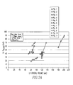
- FIG. 3 a shows the effect of Neosynephrine on LVEDV in Epicardial Lead Studies. Note that LV G blood tracks with the standard of LV Crystal Volume.
- FIG. 3 b shows the same data analyzed without subtracting the muscle component, demonstrating that dynamic removal of the myocardial signal using admittance is critical for detecting increasing LV Volume.
- the pig number is identified in the legend. There is variation in the absolute volumes in these hearts due to a large variation in porcine size.
- FIG. 4 is a flow chart of feedback loop of present invention.
- FIG. 1 b there is shown an apparatus 10 for treating a heart 16 of a patient.
- the apparatus 10 comprises a first lead 12 and at least a second lead 14 for pacing the heart 16 adapted to be in electrical communication with the heart 16 .
- the apparatus 10 comprises a microcontroller 18 in communication with the first and second leads 12 , 14 which triggers the first lead 12 at either different times or the same time from when the microcontroller 18 triggers the second lead 14 .
- the apparatus 10 may include a pacing circuit 20 in communication with microcontroller 18 which receives a signal from the microcontroller 18 to activate the pacing circuit 20 .
- the apparatus 10 may include an admittance circuit 22 which measures volume regarding the heart 16 in communication with the microcontroller 18 .
- the microcontroller 18 controls when the admittance circuit 22 measures volume.
- the microcontroller 18 determines from the admittance circuit 22 whether stroke volume, ejection fraction and end-systolic volume regarding the heart 16 are increasing or decreasing over time, and changes the pacing circuit's operation to maximize the stroke volume and ejection fraction and to minimize the end-systolic volume.
- the apparatus 10 may include a pacemaker 30 enclosure 24 and wherein the admittance circuit 22 is disposed in the pacemaker 30 enclosure 24 .
- the microcontroller 18 may be disposed in the enclosure 24 and including a wireless transmitter 26 disposed in the enclosure 24 which transmits a condition signal indicating whether the heart's condition is getting worse, better or is without change.
- the pacing circuit 20 may pace each ventricle of the heart 16 separately, where each ventricle is paced separately using a variable time delay between a first and a second pace, or takes into account delay between atrial pacing and ventricular pacing, or a combination thereof.
- the apparatus may include at least a third lead 15 for multiple admittance measurements adapted to be in electrical communication with the heart.
- the present invention pertains to an apparatus 10 for treating a heart 16 of a patient.
- the apparatus 10 comprises a first lead 12 and at least a second lead 14 for pacing the heart 16 adapted to be in electrical communication with the heart 16 .
- the apparatus 10 comprises a microcontroller 18 in communication with the first and second leads 12 , 14 that determines heart 16 volume, including stroke volume, end-systolic volume, and calculated values including ejection fraction, from admittance from signals from the first and second leads 12 , 14 and uses the admittance as feedback to control heart 16 volume ejected, as measured by stroke volume, calculated values such as ejection fraction, and control end-systolic volume, with respect to the first and second leads 12 , 14 .
- the present invention pertains to a method for treating a heart 16 of a patient.
- the method comprises the steps of triggering with a microcontroller 18 a first lead 12 in electrical communication with the heart 16 at various times for pacing the heart 16 .
- the present invention pertains to a method for treating the heart 16 of a patient.
- the method comprises the steps of determining heart 16 volume, including stroke volume, calculated values such as ejection fraction, and end-systolic volume, from admittance with a microcontroller 18 from signals from a first lead 12 and a second lead 14 in electrical communication with the heart 16 .
- the key elements of this invention include 1) the use of an epicardial admittance measurement to determine improvements in stroke volume and/or end systolic volume, and 2) the ability of this information to determine the best possible pacing algorithm to use based on which algorithm maximizes patient stroke volume, ejection fraction, and minimizes end-systolic volume from the first element.
- the invention will determine which pacing algorithm maximizes patient stroke volume, ejection fraction, and minimizing end-systolic volume by cycling through each available timing algorithm (or by changing the timing between stimulation of each site in a multisite pacemaker 30 ) and measuring the LV volume, and derived stroke volume, end-systolic volume and ejection fraction for each. Maximizing the stroke volume in these situations will directly affect the ejection fraction of the patient, moving the patient into a higher functional NYHA class, and improving quality of life both immediately, and over time.
- a pacemaker 30 (pacing) circuit is well known in the field, and little has been done to change its basic design since its invention.
- the method to control when the pacing circuit 20 fires varies with the type of technology.
- a microcontroller 18 will provide the signal to the pacing circuit 20 to control when each individual pacing site is activated.
- the microcontroller 18 will also control when the admittance circuit 22 measures volume.
- the microcontroller 18 will be able to determine from the admittance circuit 22 whether the stroke volume, ejection fraction and end-systolic volume are increasing or decreasing over time, and change the pacing algorithm to maximize the stroke volume and ejection fraction, and minimizing the end-systolic volume.
- the microcontroller 18 will signal process the results from the admittance circuit 22 within the pacemaker 30 enclosure, and then a signal indicating whether the patient condition is getting worse, better, or staying the same can be sent to the receiver 28 using the wireless transmitter 26 . No extra processing will be necessary external to the implanted device, although, if desired, the processing of the data could occur outside of the patient in an alternative embodiment.
- an additional embodiment of the current invention is for multiple admittance circuits to measure the volume of the LV from different locations, or for one admittance circuit to measure multiple locations in sequence. This redundancy can be used to increase accuracy in situations where implantation of the leads in their ideal locations is not possible.
- the key method for determining if a pacing algorithm is working correctly should be dependent on how effectively the heart 16 is pumping blood. Maximum effectiveness is therefore equivalent to maximum cardiac output.
- Atrio-ventricular delay is often set at the time of pacemaker 30 implantation to maximize stroke volume and EF, and minimize end-systolic volume (ESV).
- ESV end-systolic volume
- This type of pacing is currently beneficial to the patient, but it requires a doctor's visit to change the timing delay between atria and ventricle.
- Real-time feedback from epicardial Admittance can address this problem in real time, like the previous two pacing methods. With the power of the feedback loop and a microcontroller 18 in the pacemaker 30 , a doctor's visit will not be necessary. Doctors tune pacemakers using the same process outlined in the algorithm.
- Microcontroller 18 programming or an equivalent decision making circuit which provides feedback based on whether stroke volume and EF have been maximized, and ESV minimized.
- a variety of well known control algorithms can be used here, and their application is well known to one skilled in the art.
- the new feedback loop with the decision making process is shown in FIG. 4 .
- the admittance circuit 22 is described in U.S. patent application Ser. No. 12/657,832, incorporated by reference herein, but minimized to fit into the size of a pacemaker 30 enclosure.
- the miniaturization may be achieved by putting the entire admittance circuit 22 onto a chip if desired. See U.S. patent application Ser. No. 12/657,832, incorporated by reference herein. Such circuits can be made small enough without placement on a chip if desired.
- An additional pacing circuit 20 is installed in the case where each lead in a bi-ventricular pacemaker 30 will be paced independently. This only mildly increases complexity. Basically, there would be 2 of the flow charts in parallel, delivering pacing pulses at different times. The end result and control algorithm are still the same.
- any known pacemaker configuration could benefit from this design, with various lead implant locations. However, nothing external to the pacemaker enclosure would need to change.
- Conductance measurements have been available as an invasive tool to detect instantaneous LV volume since 1981.
- 23 Tetrapolar electrodes are usually placed on a catheter located within the LV chamber to determine instantaneous volume by electrical conductance measurement.
- Conductance systems generate an electric field using a current source, and volume is determined from the instantaneous returning voltage signal.
- Conductance is the preferred measurement over resistance (or impedance) because it has a direct (not inverse) relationship with volume.
- Conductance electrodes have not been previously placed on the LV epicardium to interrogate the LV blood volume, as done in the current invention.
- the relationship between measured blood conductance and LV blood volume is determined primarily by the shape of the current field in the blood pool and surrounding myocardium.
- the measured time-dependent resistance, R blood is proportional to the electrode separation distance, L(t), and inversely proportional to the cross-sectional area of the field, A(t); and the converse for the measured blood conductance, G blood , where ⁇ is blood resistivity:
- R blood ⁇ ⁇ ⁇ L ⁇ ( t ) A ⁇ ( t )
- G blood A ⁇ ( t ) ⁇ ⁇ ⁇ L ⁇ ( t )
- Some of the measurement electrode positions will produce a blood conductance primarily sensitive to the cross-sectional area change while others are primarily sensitive to the separation distance. As the cross-sectional area of the LV increases, there is a corresponding increase in blood conductance. Thus, for the purposes of the current invention, area-dependent electrode configurations only were focused on.
- Epicardial admittance is a measurement taken in the complex plane, and has both a magnitude,
- the values for admittance are a combination of both the muscle and blood conductance, and the muscle susceptance ( ⁇ C), for both are present in the current field.
- the process for separating admittance into blood and muscle conductance was outlined previously for the case of a tetrapolar catheter where the blood and muscle are in parallel. 17 However, there is no previously proposed model for the separation of blood and muscle components of a cross-chamber epicardial admittance measurement, where the blood and muscle components are in series. This new theory to separate blood and muscle components is presented below.
- R m - Im ⁇ ⁇ z ⁇ ⁇ ⁇ ( 1 + ( ⁇ m ⁇ m ) 2 ) ⁇ m ⁇ m
- R b Re ⁇ ⁇ z ⁇ ⁇ - R m 1 + ( ⁇ m ⁇ m ) 2
- R m is the resistance of muscle ( ⁇ )
- R b is the resistance of blood ( ⁇ )
- ⁇ m is the permittivity of muscle (F/m)
- ⁇ n is the conductivity of muscle (S/m).
- the complex impedance ⁇ right arrow over (Z) ⁇ can be separated into real (Re ⁇ right arrow over (Z) ⁇ ) and imaginary parts (Im ⁇ right arrow over (Z) ⁇ ).
- the properties of the myocardium ⁇ n , and ⁇ m can be calculated using a surface probe measurement. 17, 19 In an epicardial measurement the field generated is mostly transverse, while in a surface probe measurement (and in a traditional LV catheter measurement), the field is mostly longitudinal. This difference has been measured by others, and was corrected in the data by multiplying by a factor of 2. 24, 25
- the penetration depth for the surface probe used to measure the porcine myocardial properties was 3.6 mm, which does not extend into the LV blood volume beneath.
- the standard of volume measurement used to validate this invention is two dimensional (2D) sonomicrometry (endocardial) crystals.
- 2D endocardial crystals is an acceptable alternative to 3D echocardiography in large animal hearts when the two short axes are equal in distance, 26-28 and this distance is maintained during acute LV dilation. Additionally, the porcine myocardium is prone to arrhythmias, and there is significant trauma associated with placing the third crystal plane through the septum. Consequently, 2D endocardial crystals are the practical approach.
- the two crystal pairs were attached to a digital sonomicrometer (Sonometrics Corporation, London, Ontario, Canada) and the digital output was then transformed into volume by fitting the points to a prolate ellipsoid. 26, 28
- Electrode locations were chosen to simulate the approximate positions of an RV AICD endocardial lead, and the bi-ventricular lead on the lateral LV epicardium. Two sets of 4 electrodes spaced 1 cm apart were sewn onto a felt backing, and each were sewn onto the LV epicardium. The anterior electrodes were sewn parallel to the distal LAD to simulate the RV AICD lead, and the posterior electrodes were sewn parallel to a left marginal vein to simulate the bi-ventricular lead. Electrode pairs were chosen for a cross-ventricular chamber tetrapolar measurement with a current producing and a voltage-sensing electrode on both the anterior and the posterior epicardium.
- the pigs were also instrumented with two dimensional endocardial crystals as outlined above, a left carotid pressure sensor (3F, Scisense, London, ON), a 6F sheath in the jugular vein, an RV pressure sensor (inserted directly through the anterior RV wall (1.2 F, Scisense, London, ON), a descending thoracic saline filled balloon occluder (16 or 18 mm diameter, In Vivo Metric, Healdsburg, Calif.) to generate transient occlusion as an alternative method to produce LV dilation, and an aortic flow probe, which was placed in the descending thoracic aorta (Transonic, Ithaca, N.Y.) immediately distal to the balloon occluder to document that occlusion was obtained.
- a left carotid pressure sensor (3F, Scisense, London, ON)
- 6F sheath in the jugular vein inserted directly through the anterior RV wall (1.2 F, Scisense, London, ON
- Hematocrit was determined both at baseline and at the end of the experiment via capillary tubes and a Clay Adams Readacrit centrifuge (model CT-3400, Becton Dickinson, Sparks, Md.). Whole blood samples were spun for 5 minutes at 8,500 rpm, and the volume of packed red cells was expressed as a percentage of total volume.
- Neosynephrine (Phenylephrine) for LV Dilatation
- Neosynephrine was chosen to increase afterload and secondarily dilate the LV in a dose-response fashion to simulate LV dilation as would occur in a heart failure patient.
- Neosynephrine did not result in LV dilation and this varied in every pig.
- the doses given in the results section are the baseline, and mean low and high doses of Neosynephrine that achieved LV dilation.
- Each dose of Neosynephrine was infused for 10 minutes to achieve steady state, and then data were acquired over the subsequent 5 minutes for a total of 15 minutes per dose.
- Pleural effusions are common in heart failure patients, and are a common source of artifact in the measurement of lung conductance by systems such as OptivolTM 8, 11, 14 and CorVueTM, since the electrical fields generated by these devices extend into the lungs.
- Epicardial admittance generates an electric field, which extends across the LV and thus should not have this source of artifact.
- the chest cavity was filled with saline during the measurement of LV epicardial admittance (both on the right and left sides of the heart). The pericardium was used as a sling to keep the saline from coming into direct contact with the electrodes, similar to the final implementation of epicardial admittance.
- saline of conductivity close to blood (7941 ⁇ 124 ⁇ S/cm) was introduced into the chest.
- the slope relating blood conductance (mS) and endocardial crystal volume (mL) was determined in 11 animals at up to three doses of Neosynephrine in each of up to 6 vectors, yielding a total of 99 paired measures of blood conductance and crystal volume ( FIG. 3 ).
- Data were analyzed based on a repeated measures linear model with a compound symmetric autocorrelation matrix and fixed intercepts for each pig and vector.
- the significance of the relation between blood conductance and crystal volume was assessed using a Wald statistic for testing the null hypothesis that the coefficient of LV blood conductance in the repeated measures linear model is zero.
- Statistical testing was two-sided with a significance level of 5% using SAS Version 9.2 for Windows (SAS Institute, Cary, N.C.).
- the mean systolic aortic pressure was 80 ⁇ 13 mmHg, and the heart rate was 86 ⁇ 17 bpm.
- the mean LV end diastolic and end systolic long axis by crystals was 82 ⁇ 6 and 74 ⁇ 5 cm, respectively; and the mean LV end diastolic and end systolic short axis by crystals was 47 ⁇ 4 and 40 ⁇ 4 cm, respectively.
- the calculated mean LV EDV was 100 ⁇ 31 mL, and the mean LV ESV was 69 ⁇ 25 mL measured via endocardial crystals.
- the mean LVEF was 31 ⁇ 6%, consistent with a depressed but non-dilated LV preparation from the anesthetics and amiodarone given to minimize ventricular fibrillation.
- the mean descending thoracic flow was 8.0 ⁇ 1.8 L/min, and the mean RV systolic pressure was 19 ⁇ 3 mm Hg.
- Hematocrit at the initiation of the protocol was 30.0 ⁇ 2.3%, and at the end of the protocol 33.0 ⁇ 4.7%, consistent with hemo-concentration due to minimal fluid administration during the protocol to mitigate changes in blood and myocardial electrical properties as a source of artifact.
- the mean distance from the LV apex to the most apical epicardial admittance electrode on the anterior surface was 3.0 ⁇ 0.9 cm, and on the posterior surface was 3.0 ⁇ 0.5 cm, consistent with the admittance electrodes being parallel to one another on either side of the LV epicardium.
- the end diastolic and end systolic epicardial LV admittance magnitudes 1/
- Myocardial properties were determined in each pig for use in the equations to separate the blood and muscle admittance as published previously. 19
- the calculated ratio of a/E for myocardium was 1,923,320 ⁇ 179,592 S/F.
- Neo was 90 ⁇ 54 ⁇ g/min
- the high dose Neo was 180 ⁇ 107 ⁇ g/min.
- the blood conductance was computed from the epicardial impedance (
- LV end-diastolic volume (EDV) and complex impedance at end diastole were measured in each pig and the results were converted into the conductance of blood (LV G blood ) using the admittance technique, removing muscle contribution in real time ( FIG. 3A ).
- G blood The conductance of blood (G blood ), expressed in milli-Siemens (mS), and LV volume (V), expressed in milliliters (mL), were measured at multiple values of LV volume for between 1 and 6 vectors on each pigs and the within-animal mean slope was computed for FIG. 3A .
- the slope relating G blood (mS) and the adjusted volume (mL), 1.16 ⁇ 0.48 mS/mL, was significantly different from zero (p 0.017).
- Data were also analyzed using the traditional conductance technique, which does not remove muscle contribution and demonstrates that the ability to detect LV dilation was lost and is dependent upon having access to the imaginary component of the myocardial signal ( FIG. 3B ).
- Hemodynamic parameters are shown in Table 1 at baseline and following LAD occlusion.
- Acute LAD occlusion produced a significant decrease in aortic pressure and flow.
- the anticipated increase in LV volume was detected by both the standard (endocardial crystals) as well as blood conductance.
- the large standard deviations in the blood conductance are a result of the wide range of electrode positions, and an offset, which may be caused by variable surface electrode-myocardium contact.
- an increase of 17% from baseline LV blood conductance corresponded to an LV volume increase of 4%.
- Hemodynamic parameters are shown in Table 2 at baseline and peak transient Ao occlusion.
- AoP and endocardial crystal derived LV volume both increase.
- Ao flow decreased below 1 L/min since that was the definition for a successful aortic occlusion.
- the increased afterload increases the LV blood conductance by 7%, and the crystal LV volume by 9%.
- Hemodynamic parameters are shown in Table 3 at baseline and pleural effusion simulation. Neither the LV blood conductance nor the crystal derived LV volume changed with saline placed around the heart 16 .
- LV blood conductance can be determined from the epicardial position, despite the current generating and sensing electrodes being in constant motion with the heart 16 , and with dynamic removal of the myocardial component of the returning voltage signal.
- a physiologic LV blood conductance signal can be derived
- LV dilation in response to dose-response iv Neosynephrine can be detected by blood conductance in a similar fashion to the standard of endocardial crystals when admittance is used, but not when only traditional conductance is used
- the physiologic impact of acute LAD occlusion can be detected by blood conductance as LV dilation
- a pleural effusion simulated by placing saline outside the pericardium do not serve as a source of artifact for blood conductance measurements.
- Pleural effusions are frequent findings in patients with advanced heart failure.
- Devices which interrogate the lungs for pulmonary edema use a trans-thoracic resistance measurement from the tip of the RV AICD lead to the case of the battery pack for the pacemaker/defibrillator, and thus provide a wide electric field including the LV, left atrium, thoracic skeletal muscle, ipsilateral lung tissue, intravascular blood volume in the lung, pulmonary interstitial edema and finally pleural effusions to measure a changing fluid index.
- Having an electrical approach that is focused on the LV myocardium and blood volume would be preferable, but there still exists some electrical field extension into other sources of artifact such as pleural effusions.
- the admittance technique provides reliable tracking of LV preload similar to endocardial crystals, while traditional conductance does not ( FIG. 3B ).
- This increased sensitivity of the admittance technique is due to the use of a phase measurement in the calculation of the admittance, which allows the real-time separation of blood and muscle components.
- the high resistance of muscle normally swamps a measurement of blood and muscle in series, because the resistivity of muscle is nearly 5 times that of blood. This causes the total measured traditional conductance signal to be low and non-specific ( FIG. 3B ) when compared to the LV blood conductance determined using admittance ( FIG. 3A ).
- Stahl's invention is very different from the current invention for two reasons.
- the first major difference is that the stimulating electrodes span the LV, while in Stahl's they reside completely within the RV.
- the majority of current will be confined to the RV because the resistivity of muscle is 5 times higher than blood.
- This limitation is overcome in the current invention by forcing the current across the LV by maintaining the excitation and sink electrodes on opposite sides of the LV. The preferential path for current is therefore the low resistivity blood pool.
- the second major difference is that the current invention employs complex admittance for determining the relative muscle contribution to the measured signal. As seen in FIG.
- this separation of blood from muscle provides increased sensitivity to heart failure detection (increasing LV preload).
- the present invention represents the first evidence that a cross-chamber complex admittance measurement shows improved sensitivity to LV blood pool volume measurement over a traditional conductance (or resistance) measurement.
- a new electrical approach has been developed which can detect LV volume, taking advantage of pre-existing AICD and bi-ventricular pacing lead locations, and maturing the conductance approach to moving source and sensing electrodes while instantaneously removing the myocardial component to generate a final LV blood conductance signal.
- HR heart rate
- Aop systolic right carotid pressure
- Crystal Vol 2D endocardial crystal LV volume
- Ao Flow is descending thoracic aortic flow
- RVSP right ventricular systolic pressure
- G blood is LV blood conductance derived from epicardial admittance.
- HR heart rate
- Aop systolic right caotid pressure
- Crystal Vol 2D endocardial crystal LV volume
- Ao Flow is descending thoraic aortic flow
- RVSP right ventricular systlic pressure
- G blood is LV blood conductance derived from epicardial admittance.
Landscapes
- Health & Medical Sciences (AREA)
- Cardiology (AREA)
- Life Sciences & Earth Sciences (AREA)
- Heart & Thoracic Surgery (AREA)
- Engineering & Computer Science (AREA)
- Biophysics (AREA)
- Biomedical Technology (AREA)
- Nuclear Medicine, Radiotherapy & Molecular Imaging (AREA)
- Radiology & Medical Imaging (AREA)
- Animal Behavior & Ethology (AREA)
- General Health & Medical Sciences (AREA)
- Public Health (AREA)
- Veterinary Medicine (AREA)
- Hematology (AREA)
- Physiology (AREA)
- Physics & Mathematics (AREA)
- Pathology (AREA)
- Medical Informatics (AREA)
- Molecular Biology (AREA)
- Surgery (AREA)
- Fluid Mechanics (AREA)
- Electrotherapy Devices (AREA)
- Measurement And Recording Of Electrical Phenomena And Electrical Characteristics Of The Living Body (AREA)
Priority Applications (4)
| Application Number | Priority Date | Filing Date | Title |
|---|---|---|---|
| US13/373,850 US10076669B2 (en) | 2010-12-10 | 2011-12-02 | Admittance measurement for tuning bi-ventricular pacemakers |
| JP2014537116A JP2014530714A (ja) | 2011-10-20 | 2012-10-10 | 血行動態を用いて患者の心臓を治療する方法及び装置 |
| EP12842026.2A EP2768578B1 (de) | 2011-10-20 | 2012-10-10 | Vorrichtung zur behandlung des herzens eines patienten mit hämodynamik |
| PCT/US2012/059546 WO2013059048A2 (en) | 2011-10-20 | 2012-10-10 | Method and apparatus for treating a patient's heart using hemodynamics |
Applications Claiming Priority (2)
| Application Number | Priority Date | Filing Date | Title |
|---|---|---|---|
| US45928010P | 2010-12-10 | 2010-12-10 | |
| US13/373,850 US10076669B2 (en) | 2010-12-10 | 2011-12-02 | Admittance measurement for tuning bi-ventricular pacemakers |
Publications (2)
| Publication Number | Publication Date |
|---|---|
| US20120150252A1 US20120150252A1 (en) | 2012-06-14 |
| US10076669B2 true US10076669B2 (en) | 2018-09-18 |
Family
ID=46200125
Family Applications (1)
| Application Number | Title | Priority Date | Filing Date |
|---|---|---|---|
| US13/373,850 Active 2032-07-03 US10076669B2 (en) | 2010-12-10 | 2011-12-02 | Admittance measurement for tuning bi-ventricular pacemakers |
Country Status (4)
| Country | Link |
|---|---|
| US (1) | US10076669B2 (de) |
| EP (1) | EP2648801B1 (de) |
| JP (2) | JP2014504907A (de) |
| WO (1) | WO2012078187A2 (de) |
Cited By (7)
| Publication number | Priority date | Publication date | Assignee | Title |
|---|---|---|---|---|
| US11478646B2 (en) | 2016-07-14 | 2022-10-25 | Board Of Regents, The University Of Texas System | Method and apparatus for monitoring a patient |
| US20220339426A1 (en) * | 2021-04-22 | 2022-10-27 | Board Of Regents, The University Of Texas System | Method and Apparatus for Assisting a Heart |
| US11911602B2 (en) | 2021-04-22 | 2024-02-27 | Board Of Regents, The University Of Texas System | Method and apparatus for assisting a heart |
| US11918799B2 (en) | 2021-04-22 | 2024-03-05 | Board Of Regents, The University Of Texas System | Method and apparatus for assisting a heart |
| US11931563B2 (en) | 2021-04-22 | 2024-03-19 | Board Of Regents, The University Of Texas System | Method and apparatus for assisting a heart |
| EP4387708A1 (de) | 2021-08-19 | 2024-06-26 | Cardiovol, LLC | Verfahren und vorrichtung zur unterstützung eines herzens |
| US12318601B2 (en) | 2020-10-07 | 2025-06-03 | Abiomed Europe Gmbh | Electrode assembly patch for conductance and admittance measurements |
Families Citing this family (20)
| Publication number | Priority date | Publication date | Assignee | Title |
|---|---|---|---|---|
| US20070073354A1 (en) | 2005-09-26 | 2007-03-29 | Knudson Mark B | Neural blocking therapy |
| US9327121B2 (en) | 2011-09-08 | 2016-05-03 | Nevro Corporation | Selective high frequency spinal cord modulation for inhibiting pain, including cephalic and/or total body pain with reduced side effects, and associated systems and methods |
| US8255057B2 (en) | 2009-01-29 | 2012-08-28 | Nevro Corporation | Systems and methods for producing asynchronous neural responses to treat pain and/or other patient conditions |
| ES2624748T3 (es) | 2009-04-22 | 2017-07-17 | Nevro Corporation | Modulación de alta frecuencia selectiva de la médula espinal para la inhibición del dolor con efectos secundarios reducidos, y sistemas y métodos asociados |
| ES2942684T3 (es) | 2009-04-22 | 2023-06-05 | Nevro Corp | Sistemas de modulación de la médula espinal para inducir efectos parestésicos y anestésicos |
| US8649874B2 (en) | 2010-11-30 | 2014-02-11 | Nevro Corporation | Extended pain relief via high frequency spinal cord modulation, and associated systems and methods |
| US9833614B1 (en) | 2012-06-22 | 2017-12-05 | Nevro Corp. | Autonomic nervous system control via high frequency spinal cord modulation, and associated systems and methods |
| US10420952B2 (en) * | 2012-10-10 | 2019-09-24 | Board Of Regents, The University Of Texas System | Method and apparatus for treating a patient's heart using hemodynamics |
| US11985075B1 (en) * | 2013-02-04 | 2024-05-14 | C/Hca, Inc. | Data stream processing for dynamic resource scheduling |
| US10628180B1 (en) | 2018-08-20 | 2020-04-21 | C/Hca, Inc. | Disparate data aggregation for user interface customization |
| US9895539B1 (en) | 2013-06-10 | 2018-02-20 | Nevro Corp. | Methods and systems for disease treatment using electrical stimulation |
| US10149978B1 (en) | 2013-11-07 | 2018-12-11 | Nevro Corp. | Spinal cord modulation for inhibiting pain via short pulse width waveforms, and associated systems and methods |
| US11318310B1 (en) | 2015-10-26 | 2022-05-03 | Nevro Corp. | Neuromodulation for altering autonomic functions, and associated systems and methods |
| AU2017211121B2 (en) | 2016-01-25 | 2022-02-24 | Nevro Corp. | Treatment of congestive heart failure with electrical stimulation, and associated systems and methods |
| WO2019236497A1 (en) | 2018-06-03 | 2019-12-12 | Satz, Roseanne | Systems, methods, and devices for treating bradyarrhythmias, tachyarrhythmias and heart failure |
| WO2019241658A2 (en) * | 2018-06-14 | 2019-12-19 | Medtronic, Inc. | Delivery of cardiac pacing therapy for cardiac remodeling |
| US11908573B1 (en) | 2020-02-18 | 2024-02-20 | C/Hca, Inc. | Predictive resource management |
| US11590352B2 (en) | 2019-01-29 | 2023-02-28 | Nevro Corp. | Ramped therapeutic signals for modulating inhibitory interneurons, and associated systems and methods |
| US12558547B2 (en) | 2021-05-25 | 2026-02-24 | Nevro Corp. | Modified high frequency neuromodulation signals, and associated systems and methods |
| US12508428B2 (en) | 2023-01-26 | 2025-12-30 | Medtronic, Inc. | Respiratory-based cardiac remodeling pacing therapy |
Citations (4)
| Publication number | Priority date | Publication date | Assignee | Title |
|---|---|---|---|---|
| US20040220637A1 (en) * | 2003-01-24 | 2004-11-04 | Proteus Biomedical, Inc. | Method and apparatus for enhancing cardiac pacing |
| US20090210020A1 (en) * | 2005-12-22 | 2009-08-20 | Feldman Marc D | Method and Apparatus for Determining Cardiac Performance in a Patient |
| US20100004712A1 (en) * | 2008-07-07 | 2010-01-07 | Pacesetter, Inc. | Systems and methods for use by an implantable medical device for detecting heart failure based on the independent information content of immitance vectors |
| US20110184301A1 (en) * | 2008-09-30 | 2011-07-28 | St. Jude Medical Ab | Heart failure detector |
Family Cites Families (7)
| Publication number | Priority date | Publication date | Assignee | Title |
|---|---|---|---|---|
| US6915149B2 (en) * | 1996-01-08 | 2005-07-05 | Biosense, Inc. | Method of pacing a heart using implantable device |
| US6931280B1 (en) * | 2002-02-11 | 2005-08-16 | Pacesetter, Inc. | Apparatus and method for bi-ventricular pacing and sensing in an implantable device |
| US20070129762A1 (en) * | 2005-12-01 | 2007-06-07 | Seth Worley | Cardiac pacemaker with dynamic conduction time monitoring |
| US8706219B2 (en) * | 2005-12-22 | 2014-04-22 | Board Of Regents, The University Of Texas System | Method and apparatus for monitoring an organ of a patient |
| DE102007006229A1 (de) * | 2007-02-08 | 2008-08-14 | Biotronik Crm Patent Ag | Implantierbares medizinisches Gerät |
| US20110166472A1 (en) * | 2008-08-29 | 2011-07-07 | Bjoerling Anders | Implantable medical device and method for such a device for predicting hf status of a patient |
| US8050760B2 (en) * | 2008-11-13 | 2011-11-01 | Pacesetter, Inc. | System and method for evaluating mechanical cardiac dyssynchrony based on multiple impedance vectors using an implantable medical device |
-
2011
- 2011-12-02 JP JP2013543152A patent/JP2014504907A/ja active Pending
- 2011-12-02 EP EP11847105.1A patent/EP2648801B1/de active Active
- 2011-12-02 WO PCT/US2011/001953 patent/WO2012078187A2/en not_active Ceased
- 2011-12-02 US US13/373,850 patent/US10076669B2/en active Active
-
2016
- 2016-03-07 JP JP2016043772A patent/JP2016135266A/ja active Pending
Patent Citations (4)
| Publication number | Priority date | Publication date | Assignee | Title |
|---|---|---|---|---|
| US20040220637A1 (en) * | 2003-01-24 | 2004-11-04 | Proteus Biomedical, Inc. | Method and apparatus for enhancing cardiac pacing |
| US20090210020A1 (en) * | 2005-12-22 | 2009-08-20 | Feldman Marc D | Method and Apparatus for Determining Cardiac Performance in a Patient |
| US20100004712A1 (en) * | 2008-07-07 | 2010-01-07 | Pacesetter, Inc. | Systems and methods for use by an implantable medical device for detecting heart failure based on the independent information content of immitance vectors |
| US20110184301A1 (en) * | 2008-09-30 | 2011-07-28 | St. Jude Medical Ab | Heart failure detector |
Non-Patent Citations (3)
| Title |
|---|
| Bocchiardo, M., Meyer Zu Vilsendorf, D., Militello, C., Lippert, M., Czygan, G. Schauerte, P., Gaita, F., Stellbrink, C., "Resynchronization Therapy Optimization by Intracardiac Impedance," European Society of Cardiology, Europace, (vol. 12), (p. 1589-1595), (2010). |
| Bocchiardo, M., Meyer Zu Vilsendorf, D., Militello, C., Lippert, M., Czygan, G., Gaita, F., Schauerte, P., Stellbrink, C., "Intracardiac Impedance Monitors Stroke Volume in Resynchronization Therapy Patients," European Society of Cardiology, Europace, (Feb. 25, 2010). |
| Zima, E., Lippert, M., Czygan, G, Merkely, G., "Determination of Left Ventricular Volume Changes by Intracardiac Conductance Using a Biventricular Electrode Configuration," the Eurpean Society of Cardiology, Europace, (vol. 8), (p. 537-544), (2006). |
Cited By (9)
| Publication number | Priority date | Publication date | Assignee | Title |
|---|---|---|---|---|
| US11478646B2 (en) | 2016-07-14 | 2022-10-25 | Board Of Regents, The University Of Texas System | Method and apparatus for monitoring a patient |
| US12370370B2 (en) | 2016-07-14 | 2025-07-29 | Board Of Regents, The University Of Texas System | Method and apparatus for monitoring a patient |
| US12318601B2 (en) | 2020-10-07 | 2025-06-03 | Abiomed Europe Gmbh | Electrode assembly patch for conductance and admittance measurements |
| US20220339426A1 (en) * | 2021-04-22 | 2022-10-27 | Board Of Regents, The University Of Texas System | Method and Apparatus for Assisting a Heart |
| US11911602B2 (en) | 2021-04-22 | 2024-02-27 | Board Of Regents, The University Of Texas System | Method and apparatus for assisting a heart |
| US11918799B2 (en) | 2021-04-22 | 2024-03-05 | Board Of Regents, The University Of Texas System | Method and apparatus for assisting a heart |
| US11931563B2 (en) | 2021-04-22 | 2024-03-19 | Board Of Regents, The University Of Texas System | Method and apparatus for assisting a heart |
| US12290675B2 (en) * | 2021-04-22 | 2025-05-06 | Board Of Regents, The University Of Texas System | Method and apparatus for assisting a heart |
| EP4387708A1 (de) | 2021-08-19 | 2024-06-26 | Cardiovol, LLC | Verfahren und vorrichtung zur unterstützung eines herzens |
Also Published As
| Publication number | Publication date |
|---|---|
| JP2014504907A (ja) | 2014-02-27 |
| US20120150252A1 (en) | 2012-06-14 |
| EP2648801A4 (de) | 2014-09-10 |
| EP2648801B1 (de) | 2022-03-16 |
| JP2016135266A (ja) | 2016-07-28 |
| WO2012078187A2 (en) | 2012-06-14 |
| WO2012078187A3 (en) | 2013-10-24 |
| EP2648801A2 (de) | 2013-10-16 |
Similar Documents
| Publication | Publication Date | Title |
|---|---|---|
| US10076669B2 (en) | Admittance measurement for tuning bi-ventricular pacemakers | |
| US11589767B2 (en) | Low power apparatus and method to measure complex electrical admittance or impedance | |
| US10499858B2 (en) | Detection of worsening heart failure events using heart sounds | |
| US9820673B2 (en) | Method and apparatus for monitoring an organ of a patient | |
| CN100584270C (zh) | 采用会聚生物电导线场进行阻抗测量从而实现的病理评估 | |
| US12370370B2 (en) | Method and apparatus for monitoring a patient | |
| CN111065435A (zh) | 用于管理心力衰竭的系统和方法 | |
| JP2010533046A (ja) | スペクトラム拡散電気断層撮影 | |
| US7003348B1 (en) | Monitoring cardiac geometry for diagnostics and therapy | |
| EP1538979B1 (de) | Überwachungsgerät für dekompensierte herzinsuffizienz | |
| EP1937359B1 (de) | Vorrichtung zur optimierung von biventrikulärem pacing | |
| Larson et al. | Admittance to detect alterations in left ventricular stroke volume | |
| Porterfield et al. | Left ventricular epicardial admittance measurement for detection of acute LV dilation | |
| Valvano et al. | Articles in PresS. J Appl Physiol (December 9, 2010). doi: 10.1152/japplphysiol. 01047.2010 | |
| US8135455B2 (en) | System and method for monitoring synchrony between chambers of a heart by volume measurements and analysis | |
| Turcott et al. | Detection of hemodynamically unstable arrhythmias using subcutaneous photoplethysmography | |
| Wang et al. | Dynamics of pulmonary fluid retention prior to admission in heart failure patients |
Legal Events
| Date | Code | Title | Description |
|---|---|---|---|
| AS | Assignment |
Owner name: BOARD OF REGENTS, THE UNIVERSITY OF TEXAS SYSTEMS, Free format text: ASSIGNMENT OF ASSIGNORS INTEREST;ASSIGNORS:LARSON, ERIK;VALVANO, JONATHAN W.;PEARCE, JOHN A.;REEL/FRAME:027713/0694 Effective date: 20120201 Owner name: BOARD OF REGENTS, THE UNIVERSITY OF TEXAS SYSTEM, Free format text: ASSIGNMENT OF ASSIGNORS INTEREST;ASSIGNOR:FELDMAN, MARC D.;REEL/FRAME:027713/0706 Effective date: 20120201 Owner name: ADMITTANCE TECHNOLOGIES, INC., TEXAS Free format text: ASSIGNMENT OF ASSIGNORS INTEREST;ASSIGNOR:PORTERFIELD, JOHN;REEL/FRAME:027713/0709 Effective date: 20120131 |
|
| STCF | Information on status: patent grant |
Free format text: PATENTED CASE |
|
| FEPP | Fee payment procedure |
Free format text: MAINTENANCE FEE REMINDER MAILED (ORIGINAL EVENT CODE: REM.); ENTITY STATUS OF PATENT OWNER: SMALL ENTITY |
|
| FEPP | Fee payment procedure |
Free format text: SURCHARGE FOR LATE PAYMENT, SMALL ENTITY (ORIGINAL EVENT CODE: M2554); ENTITY STATUS OF PATENT OWNER: SMALL ENTITY |
|
| MAFP | Maintenance fee payment |
Free format text: PAYMENT OF MAINTENANCE FEE, 4TH YR, SMALL ENTITY (ORIGINAL EVENT CODE: M2551); ENTITY STATUS OF PATENT OWNER: SMALL ENTITY Year of fee payment: 4 |