TWI794616B - Drug-eluting stent - Google Patents
Drug-eluting stent Download PDFInfo
- Publication number
- TWI794616B TWI794616B TW109123057A TW109123057A TWI794616B TW I794616 B TWI794616 B TW I794616B TW 109123057 A TW109123057 A TW 109123057A TW 109123057 A TW109123057 A TW 109123057A TW I794616 B TWI794616 B TW I794616B
- Authority
- TW
- Taiwan
- Prior art keywords
- stent
- cilostazol
- layer
- mass
- polymer
- Prior art date
Links
Images

Classifications
-
- A—HUMAN NECESSITIES
- A61—MEDICAL OR VETERINARY SCIENCE; HYGIENE
- A61P—SPECIFIC THERAPEUTIC ACTIVITY OF CHEMICAL COMPOUNDS OR MEDICINAL PREPARATIONS
- A61P9/00—Drugs for disorders of the cardiovascular system
- A61P9/10—Drugs for disorders of the cardiovascular system for treating ischaemic or atherosclerotic diseases, e.g. antianginal drugs, coronary vasodilators, drugs for myocardial infarction, retinopathy, cerebrovascula insufficiency, renal arteriosclerosis
-
- A—HUMAN NECESSITIES
- A61—MEDICAL OR VETERINARY SCIENCE; HYGIENE
- A61F—FILTERS IMPLANTABLE INTO BLOOD VESSELS; PROSTHESES; DEVICES PROVIDING PATENCY TO, OR PREVENTING COLLAPSING OF, TUBULAR STRUCTURES OF THE BODY, e.g. STENTS; ORTHOPAEDIC, NURSING OR CONTRACEPTIVE DEVICES; FOMENTATION; TREATMENT OR PROTECTION OF EYES OR EARS; BANDAGES, DRESSINGS OR ABSORBENT PADS; FIRST-AID KITS
- A61F2/00—Filters implantable into blood vessels; Prostheses, i.e. artificial substitutes or replacements for parts of the body; Appliances for connecting them with the body; Devices providing patency to, or preventing collapsing of, tubular structures of the body, e.g. stents
- A61F2/82—Devices providing patency to, or preventing collapsing of, tubular structures of the body, e.g. stents
- A61F2/86—Stents in a form characterised by the wire-like elements; Stents in the form characterised by a net-like or mesh-like structure
- A61F2/88—Stents in a form characterised by the wire-like elements; Stents in the form characterised by a net-like or mesh-like structure the wire-like elements formed as helical or spiral coils
-
- A—HUMAN NECESSITIES
- A61—MEDICAL OR VETERINARY SCIENCE; HYGIENE
- A61K—PREPARATIONS FOR MEDICAL, DENTAL OR TOILETRY PURPOSES
- A61K31/00—Medicinal preparations containing organic active ingredients
- A61K31/33—Heterocyclic compounds
- A61K31/395—Heterocyclic compounds having nitrogen as a ring hetero atom, e.g. guanethidine or rifamycins
- A61K31/435—Heterocyclic compounds having nitrogen as a ring hetero atom, e.g. guanethidine or rifamycins having six-membered rings with one nitrogen as the only ring hetero atom
- A61K31/47—Quinolines; Isoquinolines
- A61K31/4709—Non-condensed quinolines and containing further heterocyclic rings
-
- A—HUMAN NECESSITIES
- A61—MEDICAL OR VETERINARY SCIENCE; HYGIENE
- A61L—METHODS OR APPARATUS FOR STERILISING MATERIALS OR OBJECTS IN GENERAL; DISINFECTION, STERILISATION OR DEODORISATION OF AIR; CHEMICAL ASPECTS OF BANDAGES, DRESSINGS, ABSORBENT PADS OR SURGICAL ARTICLES; MATERIALS FOR BANDAGES, DRESSINGS, ABSORBENT PADS OR SURGICAL ARTICLES
- A61L31/00—Materials for other surgical articles, e.g. stents, stent-grafts, shunts, surgical drapes, guide wires, materials for adhesion prevention, occluding devices, surgical gloves, tissue fixation devices
- A61L31/02—Inorganic materials
- A61L31/022—Metals or alloys
-
- A—HUMAN NECESSITIES
- A61—MEDICAL OR VETERINARY SCIENCE; HYGIENE
- A61L—METHODS OR APPARATUS FOR STERILISING MATERIALS OR OBJECTS IN GENERAL; DISINFECTION, STERILISATION OR DEODORISATION OF AIR; CHEMICAL ASPECTS OF BANDAGES, DRESSINGS, ABSORBENT PADS OR SURGICAL ARTICLES; MATERIALS FOR BANDAGES, DRESSINGS, ABSORBENT PADS OR SURGICAL ARTICLES
- A61L31/00—Materials for other surgical articles, e.g. stents, stent-grafts, shunts, surgical drapes, guide wires, materials for adhesion prevention, occluding devices, surgical gloves, tissue fixation devices
- A61L31/04—Macromolecular materials
- A61L31/06—Macromolecular materials obtained otherwise than by reactions only involving carbon-to-carbon unsaturated bonds
-
- A—HUMAN NECESSITIES
- A61—MEDICAL OR VETERINARY SCIENCE; HYGIENE
- A61L—METHODS OR APPARATUS FOR STERILISING MATERIALS OR OBJECTS IN GENERAL; DISINFECTION, STERILISATION OR DEODORISATION OF AIR; CHEMICAL ASPECTS OF BANDAGES, DRESSINGS, ABSORBENT PADS OR SURGICAL ARTICLES; MATERIALS FOR BANDAGES, DRESSINGS, ABSORBENT PADS OR SURGICAL ARTICLES
- A61L31/00—Materials for other surgical articles, e.g. stents, stent-grafts, shunts, surgical drapes, guide wires, materials for adhesion prevention, occluding devices, surgical gloves, tissue fixation devices
- A61L31/08—Materials for coatings
- A61L31/10—Macromolecular materials
-
- A—HUMAN NECESSITIES
- A61—MEDICAL OR VETERINARY SCIENCE; HYGIENE
- A61L—METHODS OR APPARATUS FOR STERILISING MATERIALS OR OBJECTS IN GENERAL; DISINFECTION, STERILISATION OR DEODORISATION OF AIR; CHEMICAL ASPECTS OF BANDAGES, DRESSINGS, ABSORBENT PADS OR SURGICAL ARTICLES; MATERIALS FOR BANDAGES, DRESSINGS, ABSORBENT PADS OR SURGICAL ARTICLES
- A61L31/00—Materials for other surgical articles, e.g. stents, stent-grafts, shunts, surgical drapes, guide wires, materials for adhesion prevention, occluding devices, surgical gloves, tissue fixation devices
- A61L31/12—Composite materials, i.e. containing one material dispersed in a matrix of the same or different material
-
- A—HUMAN NECESSITIES
- A61—MEDICAL OR VETERINARY SCIENCE; HYGIENE
- A61L—METHODS OR APPARATUS FOR STERILISING MATERIALS OR OBJECTS IN GENERAL; DISINFECTION, STERILISATION OR DEODORISATION OF AIR; CHEMICAL ASPECTS OF BANDAGES, DRESSINGS, ABSORBENT PADS OR SURGICAL ARTICLES; MATERIALS FOR BANDAGES, DRESSINGS, ABSORBENT PADS OR SURGICAL ARTICLES
- A61L31/00—Materials for other surgical articles, e.g. stents, stent-grafts, shunts, surgical drapes, guide wires, materials for adhesion prevention, occluding devices, surgical gloves, tissue fixation devices
- A61L31/14—Materials characterised by their function or physical properties, e.g. injectable or lubricating compositions, shape-memory materials, surface modified materials
- A61L31/148—Materials at least partially resorbable by the body
-
- A—HUMAN NECESSITIES
- A61—MEDICAL OR VETERINARY SCIENCE; HYGIENE
- A61L—METHODS OR APPARATUS FOR STERILISING MATERIALS OR OBJECTS IN GENERAL; DISINFECTION, STERILISATION OR DEODORISATION OF AIR; CHEMICAL ASPECTS OF BANDAGES, DRESSINGS, ABSORBENT PADS OR SURGICAL ARTICLES; MATERIALS FOR BANDAGES, DRESSINGS, ABSORBENT PADS OR SURGICAL ARTICLES
- A61L31/00—Materials for other surgical articles, e.g. stents, stent-grafts, shunts, surgical drapes, guide wires, materials for adhesion prevention, occluding devices, surgical gloves, tissue fixation devices
- A61L31/14—Materials characterised by their function or physical properties, e.g. injectable or lubricating compositions, shape-memory materials, surface modified materials
- A61L31/16—Biologically active materials, e.g. therapeutic substances
-
- A—HUMAN NECESSITIES
- A61—MEDICAL OR VETERINARY SCIENCE; HYGIENE
- A61F—FILTERS IMPLANTABLE INTO BLOOD VESSELS; PROSTHESES; DEVICES PROVIDING PATENCY TO, OR PREVENTING COLLAPSING OF, TUBULAR STRUCTURES OF THE BODY, e.g. STENTS; ORTHOPAEDIC, NURSING OR CONTRACEPTIVE DEVICES; FOMENTATION; TREATMENT OR PROTECTION OF EYES OR EARS; BANDAGES, DRESSINGS OR ABSORBENT PADS; FIRST-AID KITS
- A61F2/00—Filters implantable into blood vessels; Prostheses, i.e. artificial substitutes or replacements for parts of the body; Appliances for connecting them with the body; Devices providing patency to, or preventing collapsing of, tubular structures of the body, e.g. stents
- A61F2/82—Devices providing patency to, or preventing collapsing of, tubular structures of the body, e.g. stents
- A61F2/86—Stents in a form characterised by the wire-like elements; Stents in the form characterised by a net-like or mesh-like structure
-
- A—HUMAN NECESSITIES
- A61—MEDICAL OR VETERINARY SCIENCE; HYGIENE
- A61L—METHODS OR APPARATUS FOR STERILISING MATERIALS OR OBJECTS IN GENERAL; DISINFECTION, STERILISATION OR DEODORISATION OF AIR; CHEMICAL ASPECTS OF BANDAGES, DRESSINGS, ABSORBENT PADS OR SURGICAL ARTICLES; MATERIALS FOR BANDAGES, DRESSINGS, ABSORBENT PADS OR SURGICAL ARTICLES
- A61L2300/00—Biologically active materials used in bandages, wound dressings, absorbent pads or medical devices
- A61L2300/20—Biologically active materials used in bandages, wound dressings, absorbent pads or medical devices containing or releasing organic materials
- A61L2300/204—Biologically active materials used in bandages, wound dressings, absorbent pads or medical devices containing or releasing organic materials with nitrogen-containing functional groups, e.g. aminoxides, nitriles, guanidines
-
- A—HUMAN NECESSITIES
- A61—MEDICAL OR VETERINARY SCIENCE; HYGIENE
- A61L—METHODS OR APPARATUS FOR STERILISING MATERIALS OR OBJECTS IN GENERAL; DISINFECTION, STERILISATION OR DEODORISATION OF AIR; CHEMICAL ASPECTS OF BANDAGES, DRESSINGS, ABSORBENT PADS OR SURGICAL ARTICLES; MATERIALS FOR BANDAGES, DRESSINGS, ABSORBENT PADS OR SURGICAL ARTICLES
- A61L2300/00—Biologically active materials used in bandages, wound dressings, absorbent pads or medical devices
- A61L2300/40—Biologically active materials used in bandages, wound dressings, absorbent pads or medical devices characterised by a specific therapeutic activity or mode of action
- A61L2300/42—Anti-thrombotic agents, anticoagulants, anti-platelet agents
-
- A—HUMAN NECESSITIES
- A61—MEDICAL OR VETERINARY SCIENCE; HYGIENE
- A61L—METHODS OR APPARATUS FOR STERILISING MATERIALS OR OBJECTS IN GENERAL; DISINFECTION, STERILISATION OR DEODORISATION OF AIR; CHEMICAL ASPECTS OF BANDAGES, DRESSINGS, ABSORBENT PADS OR SURGICAL ARTICLES; MATERIALS FOR BANDAGES, DRESSINGS, ABSORBENT PADS OR SURGICAL ARTICLES
- A61L2300/00—Biologically active materials used in bandages, wound dressings, absorbent pads or medical devices
- A61L2300/40—Biologically active materials used in bandages, wound dressings, absorbent pads or medical devices characterised by a specific therapeutic activity or mode of action
- A61L2300/422—Anti-atherosclerotic agents
-
- A—HUMAN NECESSITIES
- A61—MEDICAL OR VETERINARY SCIENCE; HYGIENE
- A61L—METHODS OR APPARATUS FOR STERILISING MATERIALS OR OBJECTS IN GENERAL; DISINFECTION, STERILISATION OR DEODORISATION OF AIR; CHEMICAL ASPECTS OF BANDAGES, DRESSINGS, ABSORBENT PADS OR SURGICAL ARTICLES; MATERIALS FOR BANDAGES, DRESSINGS, ABSORBENT PADS OR SURGICAL ARTICLES
- A61L2300/00—Biologically active materials used in bandages, wound dressings, absorbent pads or medical devices
- A61L2300/60—Biologically active materials used in bandages, wound dressings, absorbent pads or medical devices characterised by a special physical form
- A61L2300/602—Type of release, e.g. controlled, sustained, slow
-
- A—HUMAN NECESSITIES
- A61—MEDICAL OR VETERINARY SCIENCE; HYGIENE
- A61L—METHODS OR APPARATUS FOR STERILISING MATERIALS OR OBJECTS IN GENERAL; DISINFECTION, STERILISATION OR DEODORISATION OF AIR; CHEMICAL ASPECTS OF BANDAGES, DRESSINGS, ABSORBENT PADS OR SURGICAL ARTICLES; MATERIALS FOR BANDAGES, DRESSINGS, ABSORBENT PADS OR SURGICAL ARTICLES
- A61L2300/00—Biologically active materials used in bandages, wound dressings, absorbent pads or medical devices
- A61L2300/60—Biologically active materials used in bandages, wound dressings, absorbent pads or medical devices characterised by a special physical form
- A61L2300/606—Coatings
-
- A—HUMAN NECESSITIES
- A61—MEDICAL OR VETERINARY SCIENCE; HYGIENE
- A61L—METHODS OR APPARATUS FOR STERILISING MATERIALS OR OBJECTS IN GENERAL; DISINFECTION, STERILISATION OR DEODORISATION OF AIR; CHEMICAL ASPECTS OF BANDAGES, DRESSINGS, ABSORBENT PADS OR SURGICAL ARTICLES; MATERIALS FOR BANDAGES, DRESSINGS, ABSORBENT PADS OR SURGICAL ARTICLES
- A61L2300/00—Biologically active materials used in bandages, wound dressings, absorbent pads or medical devices
- A61L2300/60—Biologically active materials used in bandages, wound dressings, absorbent pads or medical devices characterised by a special physical form
- A61L2300/63—Crystals
-
- A—HUMAN NECESSITIES
- A61—MEDICAL OR VETERINARY SCIENCE; HYGIENE
- A61L—METHODS OR APPARATUS FOR STERILISING MATERIALS OR OBJECTS IN GENERAL; DISINFECTION, STERILISATION OR DEODORISATION OF AIR; CHEMICAL ASPECTS OF BANDAGES, DRESSINGS, ABSORBENT PADS OR SURGICAL ARTICLES; MATERIALS FOR BANDAGES, DRESSINGS, ABSORBENT PADS OR SURGICAL ARTICLES
- A61L2420/00—Materials or methods for coatings medical devices
- A61L2420/08—Coatings comprising two or more layers
Abstract
本發明之支架具有支架骨架、及於支架骨架上堆疊了複數層之堆疊層,堆疊層之各層包含晶質西洛他唑,上述複數層之至少1層包含生物吸收性聚合物,且使支架於活體外與37℃之包含0.25質量%月桂基硫酸鈉之磷酸鹽緩衝氯化鈉溶液之溶出介質接觸後之24小時後,溶出5質量%以下之上述晶質西洛他唑。The stent of the present invention has a stent skeleton and a stacked layer stacked with multiple layers on the stent skeleton, each layer of the stacked layer contains crystalline cilostazol, at least one of the above-mentioned multiple layers contains a bioabsorbable polymer, and the stent After contacting in vitro with a 37° C. phosphate-buffered sodium chloride solution dissolution medium containing 0.25 mass % sodium lauryl sulfate for 24 hours, less than 5 mass % of the above crystalline cilostazol was dissolved.
Description
本發明係關於一種塗佈有西洛他唑之支架及其製造方法,進而詳細而言,係關於一種具有包含西洛他唑之複數層之支架及其製造方法。The present invention relates to a stent coated with cilostazol and its manufacturing method, and more specifically, to a stent with multiple layers containing cilostazol and its manufacturing method.
近年來,心肌梗塞、心絞痛、腦中風、末梢血管疾病等動脈硬化性疾病不斷增加。作為針對動脈硬化性疾病之確實之治療法,例如廣泛使用心臟之冠狀動脈中之經皮冠狀動脈成形術等以外科之方式撐開血管之狹窄部或阻塞部之經皮血管成形術(Percutaneous Transluminal Angioplasty,以下,簡稱為「PTA」)。PTA之中特別是針對冠狀動脈之狹窄部或阻塞部所進行之治療法被稱為經皮冠狀動脈成形術(Percutaneous Transluminal Coronary Angioplasty,以下,簡稱為「PTCA」)。In recent years, arteriosclerotic diseases such as myocardial infarction, angina pectoris, stroke, and peripheral vascular disease have been increasing. As a definitive treatment for arteriosclerotic diseases, percutaneous coronary angioplasty (Percutaneous Transluminal Angioplasty), which surgically expands the stenosis or obstruction of blood vessels, is widely used in the coronary arteries of the heart. Angioplasty, hereinafter referred to as "PTA"). Among the PTA, the treatment especially for the stenosis or blockage of the coronary arteries is called Percutaneous Transluminal Coronary Angioplasty (hereinafter referred to as "PTCA").
PTCA係藉由如下方式使血流恢復之方法,即,將前端帶氣球(balloon)之較細之管(氣球導管)或支架自手臂或大腿部之動脈插入,通至心臟冠狀動脈之狹窄部後,使前端之氣球膨脹而擴張狹窄之血管。藉此,病變部之血管內腔得以擴張,由此,通過血管內腔之血流增加。該PTCA除了用於治療動脈硬化性疾病以外,亦用於治療血液透析患者之手臂上形成之分路血管之狹窄等。PTCA is a method of restoring blood flow by inserting a thinner tube (balloon catheter) or stent with a balloon at the tip from an artery in the arm or thigh to the narrowed coronary artery of the heart Afterwards, the balloon at the front end is inflated to dilate narrowed blood vessels. As a result, the lumen of the blood vessel in the lesion is expanded, thereby increasing the blood flow through the lumen of the blood vessel. The PTCA is not only used for the treatment of arteriosclerotic diseases, but also for the treatment of stenosis of shunt blood vessels formed in the arms of hemodialysis patients.
一般而言,進行了PTCA之血管部位會受到內皮細胞之剝離或彈力板損傷等傷害,會發生血管壁之癒合反應即血管內膜之增生,因此藉由PTCA而成功撐開狹窄病變部之患者中約30~40%會發生再狹窄。Generally speaking, the vascular site undergoing PTCA will be damaged by the peeling of endothelial cells or elastic plate damage, and the healing reaction of the vascular wall will occur, that is, the hyperplasia of the vascular intima. Therefore, patients who have successfully expanded the stenotic lesion by PTCA Restenosis occurs in about 30-40% of patients.
人類之再狹窄之成因主要認為有PTCA之1~3天後所發生之見於單核球之黏著及/或浸潤之炎症過程、及約45天後增生性為最高峰之平滑肌細胞所引起之內膜肥厚形成過程。於發生再狹窄之情形時,由於需要再次進行PTCA,故而確立其預防法及治療法為當務之急。The causes of restenosis in humans are mainly considered to be the inflammatory process of adhesion and/or infiltration of mononuclear spheres that occurs 1 to 3 days after PTCA, and the intima caused by smooth muscle cells with the highest proliferation after about 45 days The process of hypertrophy formation. When restenosis occurs, PTCA needs to be performed again, so it is urgent to establish its preventive and therapeutic methods.
因此,盛行嘗試藉由使用於支架等之表面擔載有抗癌劑、免疫抑制劑、抗炎劑或平滑肌細胞之增生抑制劑的藥劑溶出型管腔內留置用醫療器件(支架),從而使藥劑於管腔內之留置部位局部地釋出數天左右,謀求再狹窄率之降低。Therefore, attempts are being made to use drug-eluting medical devices (stents) for intraluminal indwelling in which anticancer agents, immunosuppressants, anti-inflammatory agents, or smooth muscle cell proliferation inhibitors are loaded on the surface of stents, etc. The drug is released locally at the indwelling site in the lumen for several days to reduce the rate of restenosis.
作為塗佈於藥劑溶出型支架之藥劑,一般為可作為抗癌劑及免疫抑制劑發揮作用之limus系藥劑。該等藥劑具有藉由其較強之細胞毒性而強力地抑制作為再狹窄之主要原因之血管平滑肌細胞之增生、即所謂內膜肥厚的效果。但亦存在由於亦強力地抑制血管內皮細胞之再生,故而可能誘發遲發性支架內血栓症的臨床上之較大課題。The drug coated on the drug-eluting stent is generally a limus-based drug that acts as an anticancer agent and an immunosuppressant. These agents have the effect of strongly inhibiting the proliferation of vascular smooth muscle cells, which is the main cause of restenosis, that is, the so-called intimal hypertrophy, due to their strong cytotoxicity. However, since it also strongly inhibits the regeneration of vascular endothelial cells, there is also a large clinical problem that delayed stent thrombosis may be induced.
亦嘗試使用除了limus系以外之藥劑,例如雖預想到因難溶於水而難以製備但無細胞毒性之西洛他唑。例如,專利文獻1提出一種藥劑溶出型支架,其於包含金屬或高分子材料之支架本體之表面塗佈有包含具有40,000~600,000分子量之生物吸收性聚合物及西洛他唑之混合物而包含西洛他唑(參照申請專利範圍及第[0015]段等)。進而,專利文獻1揭示:該支架於支架留置後之炎症過程或內膜肥厚形成過程中發生再狹窄之時期使藥物溶出而作用於血管內細胞,從而具有有效之內膜肥厚抑制作用,可大幅度改善曾以大概率發生之支架留置後之再狹窄(參照第[0028]段)。
[先前技術文獻]
[專利文獻]Attempts have also been made to use agents other than limus, such as cilostazol, which is expected to be difficult to prepare due to poor water solubility, but is non-cytotoxic. For example,
[專利文獻1]WO2016/067994[Patent Document 1] WO2016/067994
[發明所欲解決之問題][Problem to be solved by the invention]
專利文獻1所揭示之支架用於心臟冠狀動脈等相對較粗之動脈等,要求作用於PTCA後之1~3天後所發生之見於單核球之黏著、浸潤之炎症過程。因此,要求專利文獻1所記載之支架自設置開始至數天之間釋出西洛他唑,發揮效果。The stent disclosed in
另一方面,近年來,由粗細度更細之末梢動脈之梗塞等所引起之末梢動脈疾病(PAD)正逐漸受到關注。例如有腳之血管發生動脈硬化,血管變細或堵塞,無法向腳流入充分之血液之疾病。因此,會出現步行時腳麻木、疼痛、寒冷等症狀。若疾病發展,則變得無法行走(間歇性跛行),或即便一動不動腳亦會疼痛。若進一步惡化,則亦存在腳會潰瘍或壞死之情形,於嚴重之情形時,亦存在必須對腳進行手術之情形。On the other hand, in recent years, peripheral arterial disease (PAD) caused by infarction of a thinner peripheral artery or the like has been attracting attention. For example, arteriosclerosis occurs in the blood vessels of the feet, and the blood vessels are narrowed or blocked, so that sufficient blood cannot flow into the feet. As a result, there will be symptoms such as numbness, pain, and coldness of the feet when walking. As the disease progresses, it becomes impossible to walk (intermittent claudication), or the foot hurts even when not moving. If it worsens further, there may be ulcers or necrosis of the feet, and in severe cases, surgery may be required on the feet.
即便PAD為僅顯示手腳之症狀者,動脈硬化亦可能不限於手腳而波及身體中之血管。若對PAD置之不理,則亦有引起心肌梗塞、心絞痛、及腦梗塞等之可能性。針對PAD,有藥物療法、理學療法、及手術等各種對應病狀之發展及治療目標之治療法。若可實現末梢動脈用之藥物留置型支架,則可提供低侵入地治療PAD之新穎之治療方法。Even if PAD only shows the symptoms of the hands and feet, arteriosclerosis may not be limited to the hands and feet but also affect the blood vessels in the body. If PAD is ignored, it may also cause myocardial infarction, angina pectoris, and cerebral infarction. For PAD, there are various treatment methods such as drug therapy, physical therapy, and surgery that correspond to the development of the disease and the goals of treatment. If a drug-indwelling stent for peripheral arteries can be realized, a novel therapeutic method for treating PAD with low invasiveness can be provided.
本發明者等人進行了各種研究,結果認為,需要可使有效成分相較心臟冠狀動脈用藥物留置型支架(例如,專利文獻1)更長期(例如,6~12個月)存在於病態血管動脈中之支架。As a result of various studies conducted by the inventors of the present invention, it is considered that the active ingredient needs to be present in diseased blood vessels for a longer period of time (for example, 6 to 12 months) than the drug-indwelling stent for coronary arteries (for example, Patent Document 1). Stents in arteries.
本發明之目的在於提供一種可使有效成分更長期(例如,6~12個月)存在於病態血管中之藥物留置型支架。此種藥物留置型支架可良好地用於末梢血管(例如,末梢動脈血管)之治療,可提供一種更加低侵入之治療方法。 [解決問題之技術手段]The purpose of the present invention is to provide a drug-indwelling stent that can make the active ingredient exist in the diseased blood vessel for a longer period of time (for example, 6-12 months). This drug-indwelling stent can be well used in the treatment of peripheral blood vessels (eg, peripheral arterial vessels), and can provide a less invasive treatment method. [Technical means to solve the problem]
本發明者等人反覆銳意研究,結果發現可獲得一種藥物留置型支架,其具有於支架骨架上堆疊了複數層之堆疊層,堆疊層之各層包含晶質西洛他唑,上述複數層之至少1層包含生物吸收性聚合物,且對於上述晶質西洛他唑於活體外對溶出率進行試驗,於24小時後溶出5質量%以下之晶質西洛他唑。進而發現,此種藥物留置型支架適合用於末梢血管,從而完成本發明。The inventors of the present invention have repeatedly studied hard, and found that a drug indwelling stent can be obtained, which has a stacked layer stacked with multiple layers on the stent skeleton, each layer of the stacked layer contains crystalline cilostazol, and at least one of the above multiple layers The first layer contains a bioabsorbable polymer, and the dissolution rate of the above-mentioned crystalline cilostazol was tested in vitro, and 5% by mass or less of the crystalline cilostazol was dissolved after 24 hours. Furthermore, it was found that this drug indwelling stent is suitable for use in peripheral blood vessels, thereby completing the present invention.
本說明書包括下述態樣。
1.一種支架,其具有支架骨架、及
於支架骨架上堆疊了複數層之堆疊層,
堆疊層之各層包含晶質西洛他唑(CLZ),
上述複數層之至少1層包含生物吸收性聚合物,且
使支架於活體外與37℃之包含0.25質量%月桂基硫酸鈉之磷酸鹽緩衝氯化鈉溶液之溶出介質接觸後之24小時後,溶出5質量%以下之上述晶質西洛他唑。
2.一種支架,其具有支架骨架、及
於支架骨架上堆疊了複數層之堆疊層,
堆疊層之各層包含晶質西洛他唑(CLZ),
上述複數層之至少1層包含生物吸收性聚合物,且
使支架於活體外於37℃下與包含0.25質量%月桂基硫酸鈉之磷酸鹽緩衝氯化鈉溶液之溶出介質接觸後之15天後,溶出20質量%以下之上述晶質西洛他唑。
3.如上述1或2所記載之支架,其中堆疊層具有至少2層,離支架較近之第1層之西洛他唑含量大於離支架較遠之第2層之西洛他唑含量。
4.如上述1至3中任一項所記載之支架,其中堆疊層具有至少2層,離支架較近之第1層之西洛他唑含量大於離支架較遠之第2層之西洛他唑含量,且2層皆包含生物吸收性聚合物。
5.如上述1至4中任一項所記載之支架,其中生物吸收性聚合物包含90質量%以上之聚乳酸。
6.如上述1至5中任一項所記載之支架,其中生物吸收性聚合物以6:4~8:2之質量比率包含L丙交酯與DL丙交酯,且具有1.8~4.5 dL/g之黏度。
7.如上述1至5中任一項所記載之支架,其中生物吸收性聚合物包含90質量%以上之L丙交酯,且具有0.6~1.4 dL/g之黏度。
8.如上述1至7中任一項所記載之支架,其用於末梢血管用。
9.一種支架,其包括支架骨架、
堆疊於支架骨架上之第1層、及堆疊於其上之第2層,
第1層及第2層分別包含西洛他唑及生物吸收性聚合物,
上述生物吸收性聚合物以6:4~8:2之質量比率包含L丙交酯與DL丙交酯,且具有1.8~4.5 dL/g之黏度,且
第1層包含470±47 μg之西洛他唑及313±31 μg之上述生物吸收性聚合物,第2層包含30±3 μg之西洛他唑及270±27 μg之上述生物吸收性聚合物。
[發明之效果]This specification includes the following aspects.
1. A bracket having a bracket skeleton, and
Multiple layers of stacked layers are stacked on the frame of the stent,
Each layer of the stack comprises crystalline cilostazol (CLZ),
at least one of the plurality of layers includes a bioabsorbable polymer, and
24 hours after the stent was contacted in vitro with a 37° C. phosphate-buffered sodium chloride solution dissolution medium containing 0.25 mass % sodium lauryl sulfate, less than 5 mass % of the above-mentioned crystalline cilostazol was dissolved.
2. A bracket having a bracket skeleton, and
Multiple layers of stacked layers are stacked on the frame of the stent,
Each layer of the stack comprises crystalline cilostazol (CLZ),
at least one of the plurality of layers includes a bioabsorbable polymer, and
After 15 days after the stent was contacted in vitro at 37° C. with a dissolution medium containing 0.25 mass % sodium lauryl sulfate in a phosphate-buffered sodium chloride solution, less than 20 mass % of the above-mentioned crystalline cilostazol was eluted.
3. The stent as described in 1 or 2 above, wherein the stacked layer has at least 2 layers, and the cilostazol content of the first layer closer to the stent is greater than the cilostazol content of the second layer farther away from the stent.
4. The stent as described in any one of the above 1 to 3, wherein the stacked layer has at least 2 layers, and the content of cilostazol in the first layer closer to the stent is greater than that of cilostazol in the second layer farther away from the stent. azole content, and both layers contain bioabsorbable polymers.
5. The stent according to any one of 1 to 4 above, wherein the bioabsorbable polymer contains 90% by mass or more of polylactic acid.
6. The stent according to any one of 1 to 5 above, wherein the bioabsorbable polymer contains L-lactide and DL-lactide in a mass ratio of 6:4 to 8:2, and has a mass ratio of 1.8 to 4.5 dL /g viscosity.
7. The stent according to any one of 1 to 5 above, wherein the bioabsorbable polymer contains 90% by mass or more of L-lactide and has a viscosity of 0.6 to 1.4 dL/g.
8. The stent according to any one of 1 to 7 above, which is used for peripheral blood vessels.
9. A support comprising a support skeleton,
The first layer stacked on the stent skeleton, and the second layer stacked on it,
本發明之實施形態之藥物留置型支架中,於活體外對晶質西洛他唑溶出率進行試驗,於24小時後可溶出5質量%以下。因此,本發明之實施形態之藥物留置型支架可使結晶性西洛他唑更長期地釋出,可更良好地用於末梢血管用。In the drug indwelling stent according to the embodiment of the present invention, the dissolution rate of crystalline cilostazol was tested in vitro, and less than 5% by mass could be dissolved after 24 hours. Therefore, the drug-indwelling stent according to the embodiment of the present invention can release crystalline cilostazol for a longer period of time, and can be better used for peripheral blood vessels.
本發明之一實施形態之支架具有 支架骨架、及 於支架骨架上堆疊了複數層之堆疊層, 堆疊層之各層包含晶質西洛他唑, 上述複數層之至少1層包含生物吸收性聚合物,且 使支架於活體外與37℃之包含0.25質量%月桂基硫酸鈉之磷酸鹽緩衝氯化鈉溶液之溶出介質接觸後之24小時後,溶出5質量%以下之上述晶質西洛他唑。The stent of one embodiment of the present invention has stent skeleton, and Multiple layers of stacked layers are stacked on the frame of the stent, Each layer of the stack comprises crystalline cilostazol, at least one of the plurality of layers includes a bioabsorbable polymer, and 24 hours after the stent was contacted in vitro with a 37° C. phosphate-buffered sodium chloride solution dissolution medium containing 0.25 mass % sodium lauryl sulfate, less than 5 mass % of the above-mentioned crystalline cilostazol was dissolved.
本發明之另一實施形態之支架具有 支架骨架、及 於支架骨架上堆疊了複數層之堆疊層, 堆疊層之各層包含晶質西洛他唑, 上述複數層之至少1層包含生物吸收性聚合物,且 使支架於活體外於37℃下與包含0.25質量%月桂基硫酸鈉之磷酸鹽緩衝氯化鈉溶液之溶出介質接觸後之15天後,溶出20質量%以下之上述晶質西洛他唑。The stent of another embodiment of the present invention has stent skeleton, and Multiple layers of stacked layers are stacked on the frame of the stent, Each layer of the stack comprises crystalline cilostazol, at least one of the plurality of layers includes a bioabsorbable polymer, and After 15 days after the stent was contacted in vitro at 37° C. with a dissolution medium containing 0.25 mass % sodium lauryl sulfate in a phosphate-buffered sodium chloride solution, less than 20 mass % of the above-mentioned crystalline cilostazol was eluted.
本發明之進一步之實施形態之支架包括
支架骨架、
堆疊於支架骨架上之第1層、及堆疊於其上之第2層,
第1層及第2層分別包含西洛他唑及生物吸收性聚合物,
上述生物吸收性聚合物以6:4~8:2之質量比率包含L丙交酯與DL丙交酯,且具有1.8~4.5 dL/g之黏度,且
第1層包含470±47 μg之西洛他唑及313±31 μg之上述生物吸收性聚合物,第2層包含30±3 μg之西洛他唑及270±27 μg之上述生物吸收性聚合物。A stent of a further embodiment of the present invention includes
bracket skeleton,
The first layer stacked on the stent skeleton, and the second layer stacked on it,
本發明之實施形態之支架具有支架骨架、及於該支架骨架上堆疊了複數層之堆疊層。 於本發明之實施形態中,「支架骨架」意指形成支架之骨架,通常例如使用金屬或高分子材料形成為粗孔之圓筒狀,只要可獲得本發明之目標支架即可,無特別限制。A stent according to an embodiment of the present invention has a stent skeleton and a stacked layer in which a plurality of layers are stacked on the stent skeleton. In the embodiment of the present invention, "stent skeleton" means the skeleton forming the scaffold, which is usually formed into a cylindrical shape with coarse pores by using metal or polymer materials, as long as the target scaffold of the present invention can be obtained, there is no special limitation .
作為金屬製之支架骨架,例如可例示鎳、鈷、鉻、鈦、及不鏽鋼等適當之合金製之支架骨架,較佳為以鈷鉻合金為主成分之金屬製支架骨架。Examples of the metal stent frame include those made of appropriate alloys such as nickel, cobalt, chromium, titanium, and stainless steel, preferably a metal stent frame composed mainly of cobalt-chromium alloy.
本發明之實施形態之支架具有於其支架骨架上堆疊了複數層之堆疊層。該堆疊層之各層包含晶質西洛他唑,上述複數層之至少1層包含生物吸收性聚合物。The stent according to the embodiment of the present invention has a stacked layer in which a plurality of layers are stacked on the stent skeleton. Each layer of the stacked layer contains crystalline cilostazol, and at least one of the above multiple layers contains a bioabsorbable polymer.
於本說明書中,「西洛他唑」係化學名為6-[4-(1-環己基-1H-四唑-5-基)丁氧基]-3,4-二氫喹諾酮。已知西洛他唑具有血小板凝集抑制作用、磷酸二酯酶(PDE)之阻礙作用、抗潰瘍作用、降壓作用及消炎作用,作為抗血栓症劑、腦循環改善劑、消炎劑、抗潰瘍劑、降壓劑、平喘劑、磷酸二酯酶阻礙劑等有用,只要可獲得本發明之目標支架即可,無特別限制。再者,西洛他唑亦包含其醫藥上可容許之鹽。 又,西洛他唑較佳為結晶。具有結晶結構之西洛他唑由於相較不具有結晶結構(非結晶)之西洛他唑可將溶出率抑制得較低,故而較佳。In this specification, the chemical name of "cilostazol" is 6-[4-(1-cyclohexyl-1H-tetrazol-5-yl)butoxy]-3,4-dihydroquinolone. Cilostazol is known to have inhibitory effect on platelet aggregation, inhibitory effect on phosphodiesterase (PDE), antiulcer effect, antihypertensive effect and anti-inflammatory effect. Drugs, antihypertensive agents, antiasthmatic agents, phosphodiesterase inhibitors, etc. are useful, as long as the target scaffold of the present invention can be obtained, there is no particular limitation. Furthermore, cilostazol also includes its pharmaceutically acceptable salts. Also, cilostazol is preferably crystal. Cilostazol having a crystalline structure is preferable because the dissolution rate can be suppressed lower than cilostazol having no crystalline structure (amorphous).
於本發明之實施形態中,生物吸收性聚合物只要可獲得本發明之目標支架即可,無特別限制。作為生物吸收性聚合物,例如可列舉包含丙交酯之聚乳酸等,其分子量(Mw:重量平均分子量)可為40,000至700,000。又,其黏度可為0.4~5.0 dL/g,亦可為0.4~4.2 dL/g。進而,生物吸收性聚合物包含DL丙交酯、L丙交酯等,亦可包含乙交酯、己內酯等。更具體而言,可例示:分子量為10,000~1,000,000之包含DL丙交酯之聚合物、以6:4~8:2之質量比率包含L丙交酯與DL丙交酯且分子量為300,000~650,000或黏度為1.8~4.5 dL/g之聚合物、分子量為50,000~150,000或包含90質量%以上之L丙交酯且黏度為0.6~1.4 dL/g之聚合物(包含100質量%之L丙交酯之聚L乳酸)。作為生物吸收性聚合物,可使用市售品,例如可例示LR704S(商品名)、L206S(商品名)、LR706S(商品名)等。生物吸收性聚合物可單獨使用或組合使用。In the embodiment of the present invention, the bioabsorbable polymer is not particularly limited as long as the target stent of the present invention can be obtained. As a bioabsorbable polymer, polylactic acid etc. which contain lactide are mentioned, for example, The molecular weight (Mw: weight average molecular weight) can be 40,000-700,000. Also, the viscosity may be 0.4-5.0 dL/g, or 0.4-4.2 dL/g. Furthermore, the bioabsorbable polymer includes DL lactide, L lactide, and the like, and may also include glycolide, caprolactone, and the like. More specifically, a polymer containing DL lactide having a molecular weight of 10,000 to 1,000,000, a polymer containing L lactide and DL lactide at a mass ratio of 6:4 to 8:2, and a molecular weight of 300,000 to 650,000 can be exemplified Or a polymer with a viscosity of 1.8-4.5 dL/g, a molecular weight of 50,000-150,000, or a polymer containing more than 90% by mass of L-lactide and a viscosity of 0.6-1.4 dL/g (containing 100% by mass of L-lactide ester of poly-L-lactic acid). As a bioabsorbable polymer, a commercial item can be used, for example, LR704S (trade name), L206S (trade name), LR706S (trade name) etc. are illustrated. Bioabsorbable polymers can be used alone or in combination.
生物吸收性聚合物較佳為包含90質量%以上之聚乳酸,更佳為包含93質量%以上,進而更佳為包含96質量%。於生物吸收性聚合物包含90質量%以上之聚乳酸之情形時,發揮能夠緩釋之有利效果。The bioabsorbable polymer preferably contains 90% by mass or more of polylactic acid, more preferably contains 93% by mass or more, and still more preferably contains 96% by mass. When the bioabsorbable polymer contains 90% by mass or more of polylactic acid, there is an advantageous effect of enabling sustained release.
較佳為堆疊層具有至少2層,離支架骨架較近之第1層之西洛他唑含量大於離支架骨架較遠之第2層之西洛他唑含量。於離支架骨架更近之層之西洛他唑之含量更大之情形時,發揮長時間緩釋之有利效果。Preferably, the stacked layer has at least 2 layers, and the cilostazol content of the first layer closer to the stent skeleton is greater than the cilostazol content of the second layer farther away from the stent skeleton. When the content of cilostazol in the layer closer to the skeleton of the stent is greater, the beneficial effect of sustained release for a long time is exerted.
第1層之西洛他唑之含量較佳為300~750 μg,更佳為350~550 μg,進而較佳為440~480 μg。進而,第1層較佳為包含470±47 μg之西洛他唑。 第2層之西洛他唑之含量較佳為0~100 μg,更佳為10~80 μg,進而較佳為20~60 μg。進而,第2層亦較佳為包含30±3 μg之西洛他唑。The content of cilostazol in the first layer is preferably from 300 to 750 μg, more preferably from 350 to 550 μg, still more preferably from 440 to 480 μg. Furthermore, the first layer preferably contains 470±47 μg of cilostazol. The content of cilostazol in the second layer is preferably from 0 to 100 μg, more preferably from 10 to 80 μg, still more preferably from 20 to 60 μg. Furthermore, the second layer also preferably contains 30±3 μg of cilostazol.
第1層之西洛他唑之含有率較佳為40~100質量%,更佳為50~70質量%,進而較佳為55~65質量%。 第2層之西洛他唑之含有率較佳為0~50質量%,更佳為2~20質量%,進而較佳為5~10質量%。The content of cilostazol in the first layer is preferably from 40 to 100% by mass, more preferably from 50 to 70% by mass, and still more preferably from 55 to 65% by mass. The content of cilostazol in the second layer is preferably from 0 to 50% by mass, more preferably from 2 to 20% by mass, and still more preferably from 5 to 10% by mass.
較佳為堆疊層具有至少2層,離支架較近之第1層之西洛他唑含量大於離支架較遠之第2層之西洛他唑含量,且2層皆包含生物吸收性聚合物。Preferably, the stack has at least 2 layers, the cilostazol content of the first layer closer to the stent is greater than the cilostazol content of the second layer farther from the stent, and both layers contain bioabsorbable polymers .
第1層之生物吸收性聚合物之含量較佳為0~500 μg,更佳為250~350 μg,進而較佳為300~320 μg。進而,第1層較佳為包含313±31 μg之生物吸收性聚合物。 第2層之生物吸收性聚合物之含量較佳為180~540 μg,更佳為200~300 μg,進而較佳為260~285 μg。進而,第2層較佳為包含270±27 μg之生物吸收性聚合物。The content of the bioabsorbable polymer in the first layer is preferably from 0 to 500 μg, more preferably from 250 to 350 μg, still more preferably from 300 to 320 μg. Furthermore, the first layer preferably contains 313±31 μg of bioabsorbable polymer. The content of the bioabsorbable polymer in the second layer is preferably from 180 to 540 μg, more preferably from 200 to 300 μg, still more preferably from 260 to 285 μg. Furthermore, the second layer preferably contains 270±27 μg of bioabsorbable polymer.
第1層之生物吸收性聚合物之含有率較佳為0~60質量%,更佳為25~50質量%,進而較佳為35~45質量%。 第2層之生物吸收性聚合物之含有率較佳為70質量%以上,更佳為80質量%以上,進而較佳為90質量%以上。The content of the bioabsorbable polymer in the first layer is preferably from 0 to 60% by mass, more preferably from 25 to 50% by mass, and still more preferably from 35 to 45% by mass. The content of the bioabsorbable polymer in the second layer is preferably at least 70% by mass, more preferably at least 80% by mass, and still more preferably at least 90% by mass.
於本發明之實施形態之支架中,於活體外使支架與37℃之包含0.25質量%月桂基硫酸鈉之磷酸鹽緩衝氯化鈉溶液之溶出介質接觸後之24小時後,溶出3質量%以下之上述晶質西洛他唑。In the stent according to the embodiment of the present invention, after 24 hours after ex vivo contacting the stent with a phosphate-buffered sodium chloride solution containing 0.25% by mass of sodium lauryl sulfate at 37°C, 3% by mass or less is eluted The above-mentioned crystalline cilostazol.
於本發明之實施形態之支架中,較佳為於活體外使支架於37℃下與包含0.25質量%月桂基硫酸鈉之磷酸鹽緩衝氯化鈉溶液之溶出介質接觸後之15天後,溶出20質量%以下之上述晶質西洛他唑,較佳為使支架接觸後之8天後,溶出7質量%以下之上述晶質西洛他唑,較佳為使支架接觸後之1天後,溶出5質量%以下之上述晶質西洛他唑,更佳為溶出3質量%以下之上述晶質西洛他唑。In the stent according to the embodiment of the present invention, it is preferable to dissolve the stent 15 days after ex vivo contact with a phosphate-buffered sodium chloride solution containing 0.25% by mass of sodium lauryl sulfate at 37°C. 20% by mass or less of the above-mentioned crystalline cilostazol, preferably 8 days after the stent is brought into contact, and 7% by mass or less of the above-mentioned crystalline cilostazol is dissolved, preferably 1 day after the stent is brought into contact , less than 5% by mass of the above-mentioned crystalline cilostazol is dissolved, more preferably less than 3% by mass of the above-mentioned crystalline cilostazol is dissolved.
於本發明之實施形態之支架中,於活體外使支架於37℃下與包含0.25質量%月桂基硫酸鈉之磷酸鹽緩衝氯化鈉溶液之溶出介質接觸後之15天後,可溶出例如1.0質量%以上之上述晶質西洛他唑,亦可溶出0.1質量%以上之上述晶質西洛他唑,使支架接觸後之8天後,可溶出例如0.5質量%以上之上述晶質西洛他唑,亦可溶出0.05質量%以上之上述晶質西洛他唑,使支架接觸後之1天後,可溶出例如0.1質量%以上之上述晶質西洛他唑,亦可溶出0.01質量%以上之上述晶質西洛他唑。In the stent according to the embodiment of the present invention, after 15 days after ex vivo contacting the stent with a phosphate-buffered sodium chloride solution containing 0.25% by mass of sodium lauryl sulfate at 37°C, the stent can dissolve, for example, 1.0 More than 0.1% by mass of the above-mentioned crystalline cilostazol by mass % can also be dissolved out, and after 8 days after contacting the stent, for example, more than 0.5% by mass of the above-mentioned crystalline cilostazol can be dissolved. Cilostazol, 0.05% by mass or more of the above-mentioned crystalline cilostazol can be eluted, and one day after contacting the stent, for example, 0.1% by mass or more of the above-mentioned crystalline cilostazol can be eluted, and 0.01% by mass can also be eluted. The above-mentioned crystalline cilostazol.
於在活體外使支架與37℃之包含0.25質量%月桂基硫酸鈉之磷酸鹽緩衝氯化鈉溶液之溶出介質接觸後之上述時間後,溶出上述比率以下之上述晶質西洛他唑之情形時,可釋出西洛他唑超過3個月,因此,可使西洛他唑極長時間地存在於生物內部。 因此,本發明之實施形態之支架可良好地用作例如末梢血管用,較佳為可良好地用作末梢動脈血管用。 進而,本發明之實施形態之支架亦可用於例如心臟之冠狀動脈、下肢動脈等先前使用支架之相對較粗之動脈等。The case where the above-mentioned crystalline cilostazol below the above-mentioned ratio is eluted after the above-mentioned period of time after the stent is contacted with a 37° C. phosphate-buffered sodium chloride solution containing 0.25% by mass sodium lauryl sulfate in vitro At this time, cilostazol can be released for more than 3 months, therefore, cilostazol can be kept inside the organism for a very long time. Therefore, the stent according to the embodiment of the present invention can be favorably used, for example, for peripheral blood vessels, and preferably can be favorably used for peripheral arterial vessels. Furthermore, the stent according to the embodiment of the present invention can also be used for relatively thick arteries such as the coronary arteries of the heart and arteries of the lower extremities where stents have been previously used.
只要可獲得本發明之目標支架,則本發明之實施形態之支架之製造方法無特別限制。 本發明之實施形態之支架例如可使用如下之製造方法進行製造,該製造方法包括:(i)準備支架骨架;(ii)準備包含西洛他唑之混合物;(iii)使用該混合物塗佈支架骨架;及反覆進行(ii)及(iii)(其中,調整西洛他唑之含量等)。The manufacturing method of the stent according to the embodiment of the present invention is not particularly limited as long as the target stent of the present invention can be obtained. The stent according to the embodiment of the present invention can be manufactured by, for example, the following manufacturing method, which includes: (i) preparing a scaffold skeleton; (ii) preparing a mixture containing cilostazol; (iii) coating the stent with the mixture skeleton; and repeating (ii) and (iii) (among them, adjusting the content of cilostazol, etc.).
包含西洛他唑之混合物除了包含西洛他唑以外,亦可包含上述生物吸收性聚合物。混合物進而可包含添加劑等溶劑。由於西洛他唑為難溶性,故而生物吸收性聚合物需要防止塗佈之剝離且維持高強度。 西洛他唑與生物吸收性聚合物例如聚乳酸之混合質量比率較佳為1:0.5~1:1.5。於為該比率之範圍內之情形時,可獲得更加良好之內膜肥厚效果。又,於混合質量比率為1:1.1~1:1.5之情形時,可更加提高塗佈強度及緩釋之效果。The mixture containing cilostazol may also contain the aforementioned bioabsorbable polymer in addition to cilostazol. The mixture may further contain solvents such as additives. Since cilostazol is poorly soluble, the bioabsorbable polymer needs to prevent peeling of the coating and maintain high strength. The mixing mass ratio of cilostazol to bioabsorbable polymers such as polylactic acid is preferably 1:0.5˜1:1.5. When it is within the range of this ratio, a more favorable effect of intimal hypertrophy can be obtained. In addition, when the mixing mass ratio is 1:1.1 to 1:1.5, the coating strength and the effect of sustained release can be further improved.
於本發明之實施形態中,將西洛他唑與生物吸收性聚合物之混合物塗佈於支架骨架之方法只要可獲得本發明之目標支架即可,無特別限制,可使用先前所使用之例如簡易噴霧法、浸漬法、電沈積法、超音波噴霧法等,就塗佈之方面而言,較佳為使用超音波噴霧法。 再者,上述實施形態只要可能,則可進行適當組合。In the embodiment of the present invention, the method of coating the mixture of cilostazol and bioabsorbable polymer on the stent skeleton is not particularly limited as long as the target stent of the present invention can be obtained. Previously used methods such as Simple spray method, dipping method, electrodeposition method, ultrasonic spray method, etc., in terms of coating, it is preferable to use ultrasonic spray method. In addition, the above-mentioned embodiment can be combined suitably as long as possible.
以下,對於本發明之實施形態,一面參照圖式,一面詳細地進行說明。Hereinafter, embodiments of the present invention will be described in detail with reference to the drawings.
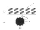
圖1(a)模式性地表示本發明之一形態之藥劑溶出型支架1。藥劑溶出型支架1具備具有長度方向軸線之圓筒狀之形態,具有管腔。關於圓筒狀之形態,藥劑溶出型支架1具有較粗之網眼狀之側面,形成為可向側方擴張。通常,網眼可藉由形成支架骨架之構件2(金屬或高分子材料等之線)形成。藥劑溶出型支架1通常係以未擴張形態插入至身體內,並於血管內之治療部位擴張而留置於該血管內。擴張亦可藉由氣球導管而於血管內達成。於圖1(a)中,模式性地記載有網眼。只要可獲得本發明之目標支架,則網眼狀之圖案無特別限制。Fig. 1(a) schematically shows a drug-
圖1(b)模式性地表示圖1(a)中之形成支架骨架之線之剖面(A-A剖面)。
本發明之一形態之藥劑溶出型支架1於支架骨架構件2上形成有堆疊層3。支架骨架構件2可使用任意之方法製作。例如,可藉由雷射、放電銑削加工、化學蝕刻或其他手段,由中空或形成之不鏽鋼管製作。支架骨架構件2可藉由鎳、鈷、鉻、鈦或不鏽鋼之適當之合金等而形成。
堆疊層3由至少兩層形成。於圖1(b)中,未顯示複數層。Fig. 1(b) schematically shows the cross-section (A-A cross-section) of the line forming the skeleton of the stent in Fig. 1(a).
A drug-
圖2模式性地表示於支架骨架構件2上塗佈形成堆疊層3之超音波噴霧塗佈裝置4。於塗佈步驟中,首先,較佳為藉由電漿處理裝置(未圖示)於塗佈步驟前對支架骨架構件2之表面進行電漿處理。電漿處理後,將支架骨架構件2安裝於心軸,安裝於超音波噴霧塗佈裝置4。於超音波噴霧塗佈裝置4中,塗佈液藉由注射泵通過配管6進行輸送,藉由超音波噴霧噴嘴5被霧化而噴射。於噴霧中,使支架骨架構件2於超音波噴嘴5下一面旋轉一面直線移動,藉此使堆疊層3堆疊於支架骨架構件2上。其後,一面使支架骨架構件2旋轉一面直線移動,同時於氮氣氣流下進行乾燥,進而於減壓下於乾燥器中進行乾燥,藉此可製作藥劑溶出型支架1。根據堆疊層3所包含之層數而變更塗佈液,支架骨架構件經複數次塗佈而形成堆疊層3。FIG. 2 schematically shows an ultrasonic
使用使西洛他唑與生物吸收性聚合物以與應形成之堆疊層3對應之比率溶於溶劑中而成的混合物,準備塗佈液。由於堆疊層3包含複數層,故而需要準備複數種塗佈液。作為塗佈溶劑,可使用沸點較低之揮發性溶劑以於塗佈後可容易去除。作為揮發性溶劑,例如可例示:甲醇、乙醇、三氟乙醇、六氟異丙醇、異戊醇、乙酸甲酯、乙酸乙酯、丙酮、甲基乙基酮、二氯甲烷、氯仿、二氯乙烷、及包含該等之中至少2種之混合溶劑。
[實施例]A coating solution is prepared using a mixture of cilostazol and a bioabsorbable polymer dissolved in a solvent at a ratio corresponding to the stacked
以下,藉由實施例及比較例,具體且詳細地對本發明進行說明,該等實施例僅為本發明之一態樣,本發明並不受該等例任何限定。Hereinafter, the present invention will be described specifically and in detail by means of examples and comparative examples. These examples are only one aspect of the present invention, and the present invention is not limited by these examples.
將本實施例中所使用之各種聚合物示於下述表1。
[表1]
上述聚合物之黏度意指極限黏度[η](dL/g),藉由毛細管黏度計法進行測定。極限黏度係準備濃度C(g/dL)之試樣溶液,根據該試樣溶液之流下時間(t)及溶劑之流下時間(t0
)之測定值,藉由下式而計算。
[數1]
實施例1
使用鈷鉻合金作為支架骨架構件2,準備將西洛他唑(CLZ)溶解於二氯甲烷而成之溶液。藉由超音波噴霧塗佈將該溶液塗佈於作為基底材料之鈷鉻合金上,形成藉由440 μg之西洛他唑形成之第1層。
其次,準備將西洛他唑與聚合物(a)以1:9(質量比)加以混合並溶解於二氯甲烷而成之溶液。藉由超音波噴霧塗佈將該溶液塗佈於第1層上,形成藉由540 μg之聚合物(a)與60 μg之西洛他唑形成之第2層,從而獲得實施例1之支架。Example 1
Cobalt-chromium alloy was used as the
實施例2~3
將聚合物(a)變更為聚合物(b)或(c),除此以外,使用與實施例1所記載之方法相同之方法,製造實施例2~3之支架。
比較例1~3 將聚合物(a)變更為聚合物(d)~(f),除此以外,使用與實施例1所記載之方法相同之方法,製造比較例1~3之支架。Comparative example 1-3 The stents of Comparative Examples 1 to 3 were produced by the same method as that described in Example 1 except that the polymer (a) was changed to polymers (d) to (f).
實施例4
使用鈷鉻合金作為支架骨架構件2,準備將西洛他唑與聚合物(b)以3:2之比率加以混合並溶解於二氯甲烷而成之溶液。藉由超音波噴霧塗佈將該溶液塗佈於支架骨架構件之鈷鉻合金上,形成藉由313 μg之聚合物(b)與470 μg之西洛他唑形成之第1層。
其次,準備將西洛他唑與聚合物(b)以1:9(質量比)之比率加以混合並溶解於二氯甲烷而成之溶液。藉由超音波噴霧塗佈將該溶液塗佈於第1層上,形成藉由270 μg之聚合物(b)與30 μg之西洛他唑形成之第2層,從而獲得實施例4之支架。Example 4
Cobalt-chromium alloy was used as the
實施例5
使用鈷鉻合金作為支架骨架構件2,準備將西洛他唑與聚合物(b)以3:2之比率加以混合並溶解於二氯甲烷而成之溶液。藉由超音波噴霧塗佈將該溶液塗佈於作為基底材料之鈷鉻合金上,形成藉由323 μg之聚合物(b)與485 μg之西洛他唑形成之第1層。
其次,準備將西洛他唑與聚合物(b)以1:19(質量比)之比率加以混合並溶解於二氯甲烷而成之溶液。藉由超音波噴霧塗佈將該溶液塗佈於第1層上,形成藉由285 μg之聚合物(b)與15 μg之西洛他唑形成之第2層,從而獲得實施例5之支架。Example 5
Cobalt-chromium alloy was used as the
實施例6
使用鈷鉻合金作為支架骨架構件2,準備將西洛他唑與聚合物(b)以3:2之比率加以混合並溶解於二氯甲烷而成之溶液。藉由超音波噴霧塗佈將該溶液塗佈於作為基底材料之鈷鉻合金上,形成藉由313 μg之聚合物(b)與470 μg之西洛他唑形成之第1層。
其次,準備將西洛他唑與聚合物(c)以1:9(質量比)之比率加以混合並溶解於二氯甲烷而成之溶液。藉由超音波噴霧塗佈將該溶液塗佈於第1層上,形成藉由270 μg之聚合物(c)與30 μg之西洛他唑形成之第2層,從而獲得實施例6之支架。Example 6
Cobalt-chromium alloy was used as the
實施例7
使用鈷鉻合金作為支架骨架構件2,準備將西洛他唑與聚合物(b)以3:2之比率加以混合並溶解於二氯甲烷而成之溶液。藉由超音波噴霧塗佈將該溶液塗佈於作為基底材料之鈷鉻合金上,形成藉由490 μg之聚合物(b)與735 μg之西洛他唑形成之第1層。
其次,準備將西洛他唑與聚合物(b)以1:19(質量比)之比率加以混合並溶解於二氯甲烷而成之溶液。藉由超音波噴霧塗佈將該溶液塗佈於第1層上而形成藉由285 μg之聚合物(b)與15 μg之西洛他唑形成之第2層,從而獲得實施例7之支架。Example 7
Cobalt-chromium alloy was used as the
實施例8 形成藉由180 μg之聚合物(b)與270 μg之西洛他唑而形成之第1層,除此以外,使用與實施例4所記載之方法相同之方法,獲得實施例8之支架。Example 8 The stent of Example 8 was obtained by the same method as described in Example 4 except that the first layer was formed of 180 μg of polymer (b) and 270 μg of cilostazol.
實施例9 形成藉由247 μg之聚合物(b)與370 μg之西洛他唑而形成之第1層,除此以外,使用與實施例4所記載之方法相同之方法,獲得實施例9之支架。Example 9 The stent of Example 9 was obtained by the same method as described in Example 4 except that the first layer was formed of 247 μg of polymer (b) and 370 μg of cilostazol.
西洛他唑之溶出試驗(活體外) 1.西洛他唑之溶出試驗方法 使用溶出試驗器400-DS(Apparatus 7),試驗液使用10 mL之包含0.25質量%月桂基硫酸鈉之磷酸鹽緩衝氯化鈉溶液,於將溶出試驗液之溫度設為37℃、浸漬速度10之條件下進行試驗。 於0.5、1、3、6、9、12、18、24小時後進行取樣,每個採取時間對試驗液進行全量更換。Dissolution test of cilostazol (in vitro) 1. Dissolution test method of cilostazol Dissolution tester 400-DS (Apparatus 7) was used, and 10 mL of phosphate-buffered sodium chloride solution containing 0.25% by mass of sodium lauryl sulfate was used as the test solution. The temperature of the dissolution test solution was set to 37°C and the immersion speed was 10 The test was carried out under the conditions. Samples were taken after 0.5, 1, 3, 6, 9, 12, 18, and 24 hours, and the test solution was replaced in full at each sampling time.
2.西洛他唑之溶出率測定(HPLC測定) 於以下之條件下對10 μL之各試樣溶液及標準溶液進行HPLC測定,根據西洛他唑之峰面積值AT 及As 計算溶出率。 溶出率之計算使用以下之式進行計算。2. Determination of the dissolution rate of cilostazol (HPLC determination) Perform HPLC determination on 10 μL of each sample solution and standard solution under the following conditions, and calculate the dissolution according to the peak area values AT and A s of cilostazol Rate. The dissolution rate was calculated using the following formula.
[數2]

測定條件 檢測器:紫外線吸光光度計(測定波長:254 nm) 管柱:於內徑4.6 mm×長度150 mm之不鏽鋼管中填充有5 μm之液相層析法用十八烷基矽烷基化矽膠 管柱溫度:25℃附近之固定溫度 移動相:水/乙腈/甲醇混液(10:7:3,v/v/v) 流量:以西洛他唑之保持時間約為9分鐘之方式調整Measurement conditions Detector: Ultraviolet absorption photometer (measurement wavelength: 254 nm) Column: A stainless steel tube with an inner diameter of 4.6 mm x a length of 150 mm is filled with 5 μm octadecyl silylated silica gel for liquid chromatography Column temperature: a fixed temperature around 25°C Mobile phase: water/acetonitrile/methanol mixture (10:7:3, v/v/v) Flow rate: adjusted in such a way that the retention time of cilostazol is about 9 minutes
藉由埋入兔髂骨動脈測得之動脈組織中之西洛他唑濃度及支架上之西洛他唑殘存量(活體內) 將實施例及比較例之各支架埋入至兔髂骨動脈。埋入係如下所述進行。 首先,切開兔之頸部,露出右頸動脈,留置導引器(introducer)。將氣球導管用導絲自導引器插入,於X射線透視下,移動至髂骨動脈之處理部位之遠端部。其後,沿著導絲插入造影用導管,進行髂骨動脈之處理部位之血管造影。處理部位之血管造影結束後,於X射線透視下,沿著氣球導管用導絲將檢體之氣球導管插入至處理部位。確認檢體之支架(標準徑擴張壓力為9 atm時支架徑為2.75 mm)位於髂骨動脈之處理部(預定血管徑為2.5 mm)後,使用充氣裝置(indeflator)以14 atm(過擴張,預定支架徑為3.0 mm,20質量%過擴張)以1次20秒使氣球保持擴張,確認支架擴張後,使氣球收縮,卸下充氣裝置,沿著氣球導管用導絲拔出氣球導管。使用相同之方法對左右之髂骨動脈進行處理。 其次,沿著氣球導管用導絲使造影用導管移動至處理部位之近前,使用稀釋造影劑進行血管造影。使用相同之方法對左右之髂骨動脈進行處理後,拔出造影用導管。最後,對鞘插入部位之血管進行結紮,對皮膚和肌肉層進行縫合。藉由以上而於兔之髂骨血管內留置支架。The concentration of cilostazol in the arterial tissue and the residual amount of cilostazol on the stent measured by embedding into the rabbit iliac artery (in vivo) The stents of Examples and Comparative Examples were embedded in the iliac arteries of rabbits. Embedding was carried out as follows. First, the neck of the rabbit was cut open to expose the right carotid artery, and an introducer was placed. The balloon catheter is inserted from the introducer with a guide wire, and moved to the distal end of the treated part of the iliac artery under X-ray fluoroscopy. Afterwards, a catheter for contrast was inserted along the guide wire, and angiography of the treated part of the iliac artery was performed. After the angiography of the treatment site, under X-ray fluoroscopy, insert the balloon catheter of the specimen into the treatment site with a guide wire along the balloon catheter. After confirming that the stent of the specimen (the stent diameter is 2.75 mm when the expansion pressure of the standard diameter is 9 atm) is located in the treated part of the iliac artery (the predetermined vessel diameter is 2.5 mm), use an inflator (indeflator) at 14 atm (over-expansion, The predetermined diameter of the stent is 3.0 mm, and 20% by weight is overexpanded) to keep the balloon inflated once for 20 seconds. After confirming the expansion of the stent, the balloon is deflated, the inflation device is removed, and the balloon catheter is pulled out along the guide wire along the balloon catheter. Use the same method to treat the left and right iliac arteries. Next, the catheter for contrast is moved along the guide wire of the balloon catheter to the front of the treatment site, and angiography is performed using diluted contrast medium. After treating the left and right iliac arteries in the same way, the angiography catheter was pulled out. Finally, the blood vessels at the sheath insertion site are ligated, and the skin and muscle layers are sutured. Based on the above, a stent was placed in the ilium vessel of the rabbit.
自埋入起90天後,對各支架之埋入部位之動脈組織中之西洛他唑濃度及支架殘存量進行分析。 作為預處理,將各支架之埋入部位之動脈組織分離成支架及動脈組織。對由經分離之各個試樣藉由液液萃取法所獲得之有機層進行乾燥,設為試樣。對於所獲得之試樣,藉由使用電灑游離法之LC/MS/MS法(liquid chromatography-tandem mass spectrometry,液相層析-串聯質譜法),對西洛他唑進行定量,計算動脈組織中之西洛他唑濃度(1 g之組織中之西洛他唑之μg數:μg/g組織)及西洛他唑殘存量(支架上之西洛他唑之殘存率(%))。After 90 days from the implantation, the concentration of cilostazol in the arterial tissue at the implantation site of each stent and the residual amount of the stent were analyzed. As a pretreatment, the arterial tissue at the implantation site of each stent was separated into the stent and the arterial tissue. The organic layer obtained by the liquid-liquid extraction method from each separated sample was dried and used as a sample. For the obtained sample, cilostazol was quantified by using the LC/MS/MS method (liquid chromatography-tandem mass spectrometry, liquid chromatography-tandem mass spectrometry) of the electrospray dissociation method, and the arterial tissue Concentration of cilostazol (μg of cilostazol in 1 g of tissue: μg/g tissue) and residual amount of cilostazol (residual rate of cilostazol on the stent (%)).
藉由埋入豬髂骨動脈測得之動脈組織中之西洛他唑濃度及支架上之西洛他唑殘存量(活體內) 藉由埋入豬髂骨動脈測得之動脈組織中之西洛他唑濃度及西洛他唑殘存量之評估係使用與對兔所進行方法相同之方法,對豬分析藉由埋入髂骨動脈測得之動脈組織中之西洛他唑濃度(1 g之組織中之西洛他唑之μg數:μg/g組織)及西洛他唑殘存量(支架上之西洛他唑之殘存率(%))。The concentration of cilostazol in the arterial tissue and the residual amount of cilostazol on the stent measured by embedding in the porcine iliac artery (in vivo) The concentration of cilostazol in the arterial tissue measured by implantation of the porcine iliac artery and the evaluation of the residual amount of cilostazol were performed using the same method as that performed on rabbits. Pigs were analyzed by implantation of the ilium The concentration of cilostazol in arterial tissue measured in arteries (the number of μg of cilostazol in 1 g of tissue: μg/g tissue) and the residual amount of cilostazol (the residual amount of cilostazol on the stent Rate(%)).
[表2]
[表3]
[表4]
實施例1~9之支架皆具有支架骨架、及於支架骨架上堆疊了複數層之堆疊層,堆疊層之各層包含晶質西洛他唑,上述複數層之至少1層包含生物吸收性聚合物,且使支架於活體外與37℃之包含0.25質量%月桂基硫酸鈉之磷酸鹽緩衝氯化鈉溶液之溶出介質接觸後之24小時後,溶出5質量%以下之上述晶質西洛他唑。The stents of Examples 1 to 9 all have a stent skeleton, and stacked layers of multiple layers stacked on the stent skeleton, each layer of the stacked layer contains crystalline cilostazol, and at least one of the above multiple layers contains a bioabsorbable polymer , and 24 hours after the stent was contacted in vitro with a dissolution medium of phosphate-buffered sodium chloride solution containing 0.25 mass % sodium lauryl sulfate at 37 ° C, less than 5 mass % of the above-mentioned crystalline cilostazol was dissolved .
又,實施例1~9之支架皆具有支架骨架、及於支架骨架上堆疊了複數層之堆疊層,堆疊層之各層包含晶質西洛他唑,上述複數層之至少1層包含生物吸收性聚合物,且使支架於活體外與37℃之包含0.25質量%月桂基硫酸鈉之磷酸鹽緩衝氯化鈉溶液之溶出介質接觸後之15天後,溶出20質量%以下之上述晶質西洛他唑。 因此,實施例1~9之支架可緩釋出西洛他唑超過3個月。 再者,作為複數層,當然並不限於2層,亦可應用3層以上。In addition, the stents of Examples 1 to 9 all have a stent skeleton and a stacked layer in which multiple layers are stacked on the stent skeleton, each layer of the stacked layer contains crystalline cilostazol, and at least one of the above-mentioned multiple layers contains a bioabsorbable polymer, and 15 days after the stent was contacted with a phosphate-buffered sodium chloride solution containing 0.25 mass% sodium lauryl sulfate at 37°C in vitro, 20 mass% or less of the above-mentioned crystalline siloxane was dissolved Tazol. Therefore, the stents of Examples 1-9 can sustain release of cilostazol for more than 3 months. In addition, it is needless to say that the plurality of layers is not limited to two layers, and three or more layers may be applied.
相對於此,比較例1~3之支架皆使支架於活體外與37℃之包含0.25質量%月桂基硫酸鈉之磷酸鹽緩衝氯化鈉溶液之溶出介質接觸後之24小時後,溶出超過5質量%之晶質西洛他唑。 因此,比較例1~3之支架無法緩釋出西洛他唑超過3個月。 [產業上之可利用性]In contrast, the stents of Comparative Examples 1 to 3 were all eluted for more than 5 hours after the stents were exposed to a dissolution medium containing 0.25% by mass sodium lauryl sulfate in a phosphate-buffered sodium chloride solution at 37°C in vitro for 24 hours. Mass % of crystalline cilostazol. Therefore, the stents of Comparative Examples 1-3 could not release cilostazol sustainably for more than 3 months. [Industrial availability]
本發明之實施形態之藥物留置型支架於活體外對晶質西洛他唑溶出率進行試驗,於24小時後可溶出5質量%以下之晶質西洛他唑。或,本發明之實施形態之藥物留置型支架於活體外對晶質西洛他唑溶出率進行試驗,於15天後可溶出20質量%以下之晶質西洛他唑。因此,本發明之實施形態之藥物留置型支架可使結晶性西洛他唑更長期地釋出,可更良好地用於末梢血管用。The drug indwelling stent according to the embodiment of the present invention was tested on the dissolution rate of crystalline cilostazol in vitro, and less than 5% by mass of crystalline cilostazol could be dissolved after 24 hours. Alternatively, the drug indwelling stent according to the embodiment of the present invention was tested on the dissolution rate of crystalline cilostazol in vitro, and less than 20% by mass of crystalline cilostazol could be dissolved after 15 days. Therefore, the drug-indwelling stent according to the embodiment of the present invention can release crystalline cilostazol for a longer period of time, and can be better used for peripheral blood vessels.
[相關申請]
再者,本申請主張以2019年7月9日於日本申請之申請號2019-127529為基礎申請之基於巴黎公約第4條之優先權。該基礎申請之內容藉由參照而引入至本說明書。[Related Application]
Furthermore, this application claims the priority right based on
1:支架 2:支架骨架構件 3:堆疊層 4:超音波噴霧塗佈裝置 5:超音波噴霧噴嘴 6:配管1: Bracket 2: Bracket frame components 3: stacked layers 4: Ultrasonic spray coating device 5: Ultrasonic spray nozzle 6: Piping
圖1模式性地表示本發明之一實施形態的支架之整個形狀(a)及支架之A-A間之剖面(b)。 圖2模式性地表示使用超音波噴霧器將塗佈劑塗佈於支架之情況。Fig. 1 schematically shows the overall shape (a) of a stent according to an embodiment of the present invention and the cross-section (b) between A-A of the stent. FIG. 2 schematically shows the situation where the coating agent is applied to the stent using an ultrasonic sprayer.
Claims (10)
Applications Claiming Priority (2)
| Application Number | Priority Date | Filing Date | Title |
|---|---|---|---|
| JP2019127529 | 2019-07-09 | ||
| JP2019-127529 | 2019-07-09 |
Publications (2)
| Publication Number | Publication Date |
|---|---|
| TW202116317A TW202116317A (en) | 2021-05-01 |
| TWI794616B true TWI794616B (en) | 2023-03-01 |
Family
ID=74113786
Family Applications (2)
| Application Number | Title | Priority Date | Filing Date |
|---|---|---|---|
| TW109123057A TWI794616B (en) | 2019-07-09 | 2020-07-08 | Drug-eluting stent |
| TW112105824A TW202322815A (en) | 2019-07-09 | 2020-07-08 | Drug-eluting stent |
Family Applications After (1)
| Application Number | Title | Priority Date | Filing Date |
|---|---|---|---|
| TW112105824A TW202322815A (en) | 2019-07-09 | 2020-07-08 | Drug-eluting stent |
Country Status (7)
| Country | Link |
|---|---|
| US (2) | US11806257B2 (en) |
| EP (1) | EP3998049A4 (en) |
| JP (2) | JP7033694B2 (en) |
| CN (1) | CN114051417A (en) |
| MX (2) | MX2022000389A (en) |
| TW (2) | TWI794616B (en) |
| WO (1) | WO2021006291A1 (en) |
Families Citing this family (1)
| Publication number | Priority date | Publication date | Assignee | Title |
|---|---|---|---|---|
| WO2023038112A1 (en) * | 2021-09-10 | 2023-03-16 | テルモ株式会社 | Drug-eluting medical device and production method therefor |
Citations (2)
| Publication number | Priority date | Publication date | Assignee | Title |
|---|---|---|---|---|
| CN204542477U (en) * | 2015-02-10 | 2015-08-12 | 东莞颠覆产品设计有限公司 | The expansible intravascular stent of a kind of multilamellar |
| CN107106309A (en) * | 2014-10-28 | 2017-08-29 | 株式会社Jimro | Bracket for eluting medicament |
Family Cites Families (23)
| Publication number | Priority date | Publication date | Assignee | Title |
|---|---|---|---|---|
| US5824048A (en) | 1993-04-26 | 1998-10-20 | Medtronic, Inc. | Method for delivering a therapeutic substance to a body lumen |
| AU2003280437A1 (en) * | 2002-06-27 | 2004-01-19 | Microport Medical (Shanghai) Co., Ltd. | Drug eluting stent |
| CN100471469C (en) | 2002-06-27 | 2009-03-25 | 微创医疗器械(上海)有限公司 | Drug-eluting stent (DES) with multicoating |
| US7318945B2 (en) | 2003-07-09 | 2008-01-15 | Medtronic Vascular, Inc. | Laminated drug-polymer coated stent having dipped layers |
| JP4894519B2 (en) | 2004-09-08 | 2012-03-14 | 株式会社カネカ | Indwelling stent |
| KR100511618B1 (en) | 2005-01-17 | 2005-08-31 | 이경범 | Multi-layer coating of drug release controllable coronary stent and method for manufacturing the same |
| KR101265625B1 (en) * | 2006-09-11 | 2013-05-22 | 엘지전자 주식회사 | The broadcasting receiver for processing broadcasting signals for determining multi channels, and the method for controlling the same |
| US20100076544A1 (en) | 2007-01-30 | 2010-03-25 | Erika Hoffmann | Biodegradable vascular support |
| CN101641059B (en) * | 2007-02-14 | 2011-10-19 | 山东瑞安泰医疗技术有限公司 | Intracoronary stent with asymmetric drug releasing controlled coating |
| US8409272B2 (en) | 2007-09-04 | 2013-04-02 | Japan Stent Technology Co., Ltd. | Sustained drug-releasing stent |
| US8067111B2 (en) * | 2008-06-30 | 2011-11-29 | Lg Chem, Ltd. | Battery module having battery cell assembly with heat exchanger |
| US20100241220A1 (en) | 2009-03-23 | 2010-09-23 | Mcclain James B | Peripheral Stents Having Layers |
| US20100280600A1 (en) * | 2009-04-30 | 2010-11-04 | Vipul Bhupendra Dave | Dual drug stent |
| US20120130481A1 (en) * | 2010-11-18 | 2012-05-24 | Robert Falotico | Local vascular delivery of adenosine a2a receptor agonists in combination with other agents to reduce myocardial injury |
| JP5784940B2 (en) | 2011-03-18 | 2015-09-24 | テルモ株式会社 | Drug eluting stent |
| KR101903443B1 (en) * | 2012-02-02 | 2018-10-02 | 삼성전자주식회사 | Apparatus and method for transmitting/receiving scene composition information |
| EP2911712B1 (en) | 2012-10-25 | 2017-11-29 | Boston Scientific Scimed, Inc. | Stent having a tacky silicone coating to prevent stent migration |
| TWI697337B (en) * | 2013-08-07 | 2020-07-01 | 學校法人近畿大學 | Process for produing nano particle or nano-particle composition, and process for manufacturing stent or balloon catheter |
| CN103948458A (en) * | 2014-05-05 | 2014-07-30 | 加奇生物科技(上海)有限公司 | Encephalic drug eluting stent |
| CN203829101U (en) * | 2014-05-05 | 2014-09-17 | 加奇生物科技(上海)有限公司 | Intracranial drug eluting stent |
| US9475326B2 (en) * | 2014-09-09 | 2016-10-25 | Trodat Gmbh | Removable die plate for self-inking stamps |
| PL3421253T3 (en) * | 2017-06-28 | 2020-01-31 | Hid Global Rastede Gmbh | Thermochromic window |
| JP7142405B2 (en) | 2018-01-24 | 2022-09-27 | 出光興産株式会社 | Lubricating oil composition and composition for refrigerator |
-
2020
- 2020-07-08 CN CN202080048831.XA patent/CN114051417A/en active Pending
- 2020-07-08 TW TW109123057A patent/TWI794616B/en active
- 2020-07-08 JP JP2021530714A patent/JP7033694B2/en active Active
- 2020-07-08 MX MX2022000389A patent/MX2022000389A/en unknown
- 2020-07-08 TW TW112105824A patent/TW202322815A/en unknown
- 2020-07-08 EP EP20837247.4A patent/EP3998049A4/en active Pending
- 2020-07-08 US US17/597,449 patent/US11806257B2/en active Active
- 2020-07-08 WO PCT/JP2020/026688 patent/WO2021006291A1/en unknown
-
2022
- 2022-01-07 MX MX2023001542A patent/MX2023001542A/en unknown
- 2022-02-28 JP JP2022029319A patent/JP7200412B2/en active Active
-
2023
- 2023-09-12 US US18/465,332 patent/US20230414383A1/en active Pending
Patent Citations (2)
| Publication number | Priority date | Publication date | Assignee | Title |
|---|---|---|---|---|
| CN107106309A (en) * | 2014-10-28 | 2017-08-29 | 株式会社Jimro | Bracket for eluting medicament |
| CN204542477U (en) * | 2015-02-10 | 2015-08-12 | 东莞颠覆产品设计有限公司 | The expansible intravascular stent of a kind of multilamellar |
Also Published As
| Publication number | Publication date |
|---|---|
| US20230414383A1 (en) | 2023-12-28 |
| JP2022078154A (en) | 2022-05-24 |
| TW202116317A (en) | 2021-05-01 |
| JP7033694B2 (en) | 2022-03-10 |
| JP7200412B2 (en) | 2023-01-06 |
| TW202322815A (en) | 2023-06-16 |
| JPWO2021006291A1 (en) | 2021-01-14 |
| EP3998049A1 (en) | 2022-05-18 |
| EP3998049A4 (en) | 2023-06-28 |
| MX2023001542A (en) | 2023-04-10 |
| WO2021006291A1 (en) | 2021-01-14 |
| US11806257B2 (en) | 2023-11-07 |
| CN114051417A (en) | 2022-02-15 |
| US20220160525A1 (en) | 2022-05-26 |
| MX2022000389A (en) | 2023-02-07 |
Similar Documents
| Publication | Publication Date | Title |
|---|---|---|
| EP1518517B1 (en) | Drug-delivery endovascular stent | |
| JP5636451B2 (en) | Drug sustained release stent | |
| US7682387B2 (en) | Drug-delivery endovascular stent and method for treating restenosis | |
| US20040024450A1 (en) | Drug-delivery endovascular stent and method for treating restenosis | |
| EP2111818B1 (en) | Intracoronary stent with asymmetric drug releasing controlled coating | |
| JPWO2006027994A1 (en) | Indwelling stent | |
| US20230414383A1 (en) | Drug-eluting stent including crystalline cilostazol | |
| CN101195048A (en) | Compound medicament washing bracket and method for preparing the same | |
| CN101081316A (en) | Novel medicine eluting supporting stand | |
| KR20050092757A (en) | Indwelling stent | |
| TWI721956B (en) | Stent of drug-dissolution type | |
| CN112263360A (en) | In vivo drug eluting stent and preparation method thereof | |
| KR101860877B1 (en) | Self-expandable Drug eluting scaffold | |
| JP2002193838A (en) | Medical material for implantation and medical appliance for implantation |