RU2768461C1 - Method for assessing the cytoprotective effect of immunoglobulin preparations for intravenous introduction on trophoblast cells under conditions of their interaction with natural killers - Google Patents
Method for assessing the cytoprotective effect of immunoglobulin preparations for intravenous introduction on trophoblast cells under conditions of their interaction with natural killers Download PDFInfo
- Publication number
- RU2768461C1 RU2768461C1 RU2021120799A RU2021120799A RU2768461C1 RU 2768461 C1 RU2768461 C1 RU 2768461C1 RU 2021120799 A RU2021120799 A RU 2021120799A RU 2021120799 A RU2021120799 A RU 2021120799A RU 2768461 C1 RU2768461 C1 RU 2768461C1
- Authority
- RU
- Russia
- Prior art keywords
- cells
- trophoblast
- trophoblast cells
- ivig
- peripheral blood
- Prior art date
Links
Images
Classifications
-
- C—CHEMISTRY; METALLURGY
- C12—BIOCHEMISTRY; BEER; SPIRITS; WINE; VINEGAR; MICROBIOLOGY; ENZYMOLOGY; MUTATION OR GENETIC ENGINEERING
- C12N—MICROORGANISMS OR ENZYMES; COMPOSITIONS THEREOF; PROPAGATING, PRESERVING, OR MAINTAINING MICROORGANISMS; MUTATION OR GENETIC ENGINEERING; CULTURE MEDIA
- C12N5/00—Undifferentiated human, animal or plant cells, e.g. cell lines; Tissues; Cultivation or maintenance thereof; Culture media therefor
- C12N5/06—Animal cells or tissues; Human cells or tissues
- C12N5/0602—Vertebrate cells
- C12N5/0608—Germ cells
- C12N5/0611—Primordial germ cells, e.g. embryonic germ cells [EG]
-
- C—CHEMISTRY; METALLURGY
- C12—BIOCHEMISTRY; BEER; SPIRITS; WINE; VINEGAR; MICROBIOLOGY; ENZYMOLOGY; MUTATION OR GENETIC ENGINEERING
- C12N—MICROORGANISMS OR ENZYMES; COMPOSITIONS THEREOF; PROPAGATING, PRESERVING, OR MAINTAINING MICROORGANISMS; MUTATION OR GENETIC ENGINEERING; CULTURE MEDIA
- C12N5/00—Undifferentiated human, animal or plant cells, e.g. cell lines; Tissues; Cultivation or maintenance thereof; Culture media therefor
- C12N5/06—Animal cells or tissues; Human cells or tissues
- C12N5/0602—Vertebrate cells
- C12N5/0634—Cells from the blood or the immune system
- C12N5/0646—Natural killers cells [NK], NKT cells
-
- G—PHYSICS
- G01—MEASURING; TESTING
- G01N—INVESTIGATING OR ANALYSING MATERIALS BY DETERMINING THEIR CHEMICAL OR PHYSICAL PROPERTIES
- G01N33/00—Investigating or analysing materials by specific methods not covered by groups G01N1/00 - G01N31/00
- G01N33/48—Biological material, e.g. blood, urine; Haemocytometers
Landscapes
- Health & Medical Sciences (AREA)
- Life Sciences & Earth Sciences (AREA)
- Engineering & Computer Science (AREA)
- Biomedical Technology (AREA)
- Chemical & Material Sciences (AREA)
- Biotechnology (AREA)
- Organic Chemistry (AREA)
- Bioinformatics & Cheminformatics (AREA)
- Genetics & Genomics (AREA)
- Wood Science & Technology (AREA)
- Zoology (AREA)
- General Health & Medical Sciences (AREA)
- Biochemistry (AREA)
- Cell Biology (AREA)
- Microbiology (AREA)
- General Engineering & Computer Science (AREA)
- Immunology (AREA)
- Hematology (AREA)
- Developmental Biology & Embryology (AREA)
- Urology & Nephrology (AREA)
- Molecular Biology (AREA)
- Food Science & Technology (AREA)
- Medicinal Chemistry (AREA)
- Physics & Mathematics (AREA)
- Analytical Chemistry (AREA)
- General Physics & Mathematics (AREA)
- Pathology (AREA)
- Measuring Or Testing Involving Enzymes Or Micro-Organisms (AREA)
Abstract
Description
Изобретение относится к области медицины, и может быть использовано для оценки цитопротективного эффекта препаратов в отношении клеток трофобласта в условиях их взаимодействия с естественными киллерами.The invention relates to medicine, and can be used to assess the cytoprotective effect of drugs on trophoblast cells under conditions of their interaction with natural killers.
Исследование функционального статуса естественных киллеров (NK-клеток) при бесплодии позволило установить повышение количества NK-клеток с фенотипом CD56dim у женщин с бесплодием по сравнению со здоровыми фертильными небеременными женщинами [1]. Потенциальными клетками-мишенями NK-клеток при бесплодии являются клетки трофэктодермы, дифференцирующиеся в клетки трофобласта.The study of the functional status of natural killer cells (NK cells) in infertility revealed an increase in the number of NK cells with the CD56dim phenotype in women with infertility compared to healthy fertile non-pregnant women [1]. Potential target cells for NK cells in infertility are trophectoderm cells that differentiate into trophoblast cells.
Для увеличения вероятности имплантации бластоцисты при бесплодии в циклах экстракорпорального оплодотворения (ЭКО) применяют различные варианты иммунотерапии, в том числе глюкокортикоиды (например, преднизолон) и иммуноглобулины для внутривенного введения (ВВИГ) [2]. Показано, что in vitro преднизолон подавлял цитотоксическую активность NK-клеток периферической крови женщин с бесплодием [3]. Однако, использование глюкокортикоидов в настоящее время оспаривается из-за сопутствующих побочных эффектов и возможного системного влияния [4]. Показано, что применение препаратов ВВИГ на прегравидарном этапе и в первом триместре беременности в цикле вспомогательных репродуктивных технологий (ВРТ) увеличивает вероятность успешной имплантации эмбриона [4-6]. Актуальным является решение вопроса об оценке потенциального эффекта препаратов ВВИГ в отношении клеток трофобласта в условиях их взаимодействия с NK-клетками.To increase the likelihood of blastocyst implantation in infertile in vitro fertilization (IVF) cycles, various immunotherapy options are used, including glucocorticoids (eg, prednisolone) and intravenous immunoglobulins (IVIG) [2]. Prednisolone has been shown to suppress the cytotoxic activity of peripheral blood NK cells in infertile women in vitro [3]. However, the use of glucocorticoids is currently disputed due to associated side effects and possible systemic effects [4]. It has been shown that the use of IVIG preparations at the preconception stage and in the first trimester of pregnancy in the cycle of assisted reproductive technologies (ART) increases the likelihood of successful embryo implantation [4–6]. It is important to address the issue of assessing the potential effect of IVIG preparations on trophoblast cells under conditions of their interaction with NK cells.
Известен способ (прототип) прогнозирования цитопротективного эффекта ВВИГ при терапии женщин с привычным невынашиванием беременности и диагностированным антифосфолипидным синдромом (Патент №2548754) [7], в котором оценивают цитопатическое действие сывороток периферической крови у пациенток и коррекцию этого эффекта с помощью препаратов ВВИГ. В качестве клеток, в отношении которых оценивают эффект препаратов ВВИГ, используют эндотелиальные клетки. Учет гибели клеток производится при помощи метода проточной цитофлюориметрии, позволяющем проанализировать большое количество клеток.There is a known method (prototype) for predicting the cytoprotective effect of IVIG in the treatment of women with recurrent miscarriage and diagnosed antiphospholipid syndrome (Patent No. 2548754) [7], which evaluates the cytopathic effect of peripheral blood sera in patients and the correction of this effect using IVIG preparations. Endothelial cells are used as cells for which the effect of IVIG preparations is evaluated. Cell death is recorded using the flow cytometry method, which allows analyzing a large number of cells.
Недостаток способа заключается в отсутствии конкретных клеток-эффекторов, реализующих цитотоксические эффекты (NK-клеток), что в значительной степени снижает оценку специфичности метода. Кроме того, недостатком способа является отсутствие использования клеток трофобласта, как потенциальных мишеней для NK-клеток в процессе внедрения бластоцисты в стенку матки.The disadvantage of this method is the absence of specific effector cells that implement cytotoxic effects (NK cells), which greatly reduces the assessment of the specificity of the method. In addition, the disadvantage of this method is the lack of use of trophoblast cells as potential targets for NK cells during the introduction of the blastocyst into the uterine wall.
Патент №2632109 [8] дает подробное описание способа определения цитотоксической активности NK-клеток в отношении клеток трофобласта линии Jeg-3, которые используются в качестве клеток-мишеней для естественных киллеров.Patent No. 2632109 [8] gives a detailed description of the method for determining the cytotoxic activity of NK cells against Jeg-3 trophoblast cells, which are used as target cells for natural killers.
Недостатком способа является отсутствие оценки влияния препаратов, используемых в терапии репродуктивных потерь, на предлагаемую систему клеточных взаимодействий, что существенно ограничивает его использование в клинической практике для прогнозирования эффективности терапии.The disadvantage of this method is the lack of evaluation of the effect of drugs used in the treatment of reproductive losses on the proposed system of cellular interactions, which significantly limits its use in clinical practice to predict the effectiveness of therapy.
В научных и патентных источниках не обнаружено способов оценки потенциального цитопротективного эффекта препаратов в отношении клеток трофобласта в условиях их взаимодействия с естественными киллерами.In scientific and patent sources, no methods have been found to assess the potential cytoprotective effect of drugs on trophoblast cells under conditions of their interaction with natural killers.
Техническим результатом изобретения является создание способа, позволяющего оценить эффективность цитопротективного эффекта препарата ВВИГ в отношении клеток трофобласта, в условиях in vitro в присутствии NK-клеток периферической крови.The technical result of the invention is the creation of a method that allows to evaluate the effectiveness of the cytoprotective effect of the IVIG preparation in relation to trophoblast cells, in vitro in the presence of peripheral blood NK cells.
Указанный технический результат достигается в способе оценки цитопротективного эффекта препаратов ВВИГ в отношении клеток трофобласта в условиях их взаимодействия с естественными киллерами, в котором используют культуру клеток трофобласта, часть из которых инкубируют без мононуклеаров, часть - в присутствии только монокулеаров периферической крови и часть - в присутствии монокулеаров периферической крови и препарата ВВИГ в концентрации 6 мг/мл, после чего определяют количество нежизнеспособных клеток трофобласта и рассчитывают коэффициент цитопротективного эффекта препаратов ВВИГ в отношении клеток трофобласта по формуле:The specified technical result is achieved in a method for assessing the cytoprotective effect of IVIG preparations against trophoblast cells under conditions of their interaction with natural killers, in which a culture of trophoblast cells is used, some of which are incubated without mononuclear cells, some in the presence of only peripheral blood monoculars and some in the presence of peripheral blood monoculars and IVIG preparation at a concentration of 6 mg/ml, after which the number of non-viable trophoblast cells is determined and the coefficient of the cytoprotective effect of IVIG preparations against trophoblast cells is calculated using the formula:
ЦПЭ = [(Х2) / (Х1)], гдеCPE = [(X2) / (X1)], where
X1 - разность количества нежизнеспособных клеток трофобласта, инкубированных с мононуклеарами периферической крови и инкубированных без мононуклеаров;X1 - the difference in the number of non-viable trophoblast cells incubated with peripheral blood mononuclear cells and incubated without mononuclear cells;
Х2 - разность количества нежизнеспособных клеток трофобласта, инкубированных с мононуклеарами периферической крови и препаратом ВВИГ и инкубированных без мононуклеаров;X2 - the difference in the number of non-viable trophoblast cells incubated with peripheral blood mononuclear cells and IVIG preparation and incubated without mononuclear cells;
при ЦПЭ <1 прогнозируют положительный цитопротективный эффект.with CPE <1, a positive cytoprotective effect is predicted.
Исследование, на котором основано изобретение, включало три этапа.The study on which the invention is based consisted of three stages.
На первом этапе проводили оценку возможного токсического действия препарата ВВИГ в отношении NK-клеток. На втором этапе исследования оценивали влияние препарата ВВИГ в системе контактного взаимодействия NK-клеток линии NK-92 и клеток трофобласта. На третьем этапе работы проводили оценку влияния препарата ВВИГ на клетки трофобласта в случае контактных взаимодействий с NK-клетками периферической крови небеременных женщин. Полученные данные статистически обработаны с помощью программы GraphPadPrism8. Для сравнения полученных данных использовали критерий Краскела - Уоллиса, являющийся непараметрическим критерием для множественных сравнений. Статистически значимыми признавались различия при р<0,05.At the first stage, the possible toxic effect of the IVIG preparation on NK cells was assessed. At the second stage of the study, the effect of the IVIG preparation in the system of contact interaction between NK cells of the NK-92 line and trophoblast cells was evaluated. At the third stage of the work, the influence of the IVIG preparation on trophoblast cells was evaluated in the case of contact interactions with NK cells of the peripheral blood of non-pregnant women. The obtained data were statistically processed using the GraphPadPrism8 program. To compare the obtained data, the Kruskal-Wallis test was used, which is a non-parametric test for multiple comparisons. Differences were considered statistically significant at p<0.05.
На первом этапе исследования были использованы NK-клетки линии NK-92, полученные из костного мозга человека со злокачественной неходжкинской лимфомой. Клеточная линия NK-92 воспроизводит все основные морфологические, фенотипические и функциональные характеристики, присущие NK-клеткам периферической крови.At the first stage of the study, NK-cells of the NK-92 line, obtained from the bone marrow of a person with malignant non-Hodgkin's lymphoma, were used. The NK-92 cell line reproduces all the main morphological, phenotypic and functional characteristics inherent in peripheral blood NK cells.
Для культивирования клеток линии NK-92 используют α-модификацию среды MEM («Альфа-MEM», Биолот, Россия) с добавлением 12,5% инактивированной эмбриональной телячьей сыворотки (ЭТС), 12,5% инактивированной лошадиной сыворотки, 100 мкг/мл стрептомицина, 100 ед/мл пенициллина, 2 мМ L-глутамина, 0,2 мМ миоинозитола, 0,02 мМ фолиевой кислоты, 20 мМ буфера HEPES, 0,1 мМ меркаптоэтанола (Sigma-Aldrich, США), 500 U/ml IL-2 («Ронколейкин», Биотех, Россия). Клетки прокультивировали при 37°С, 5% CO2 во влажной атмосфере. В работе использовали препарат внутривенных иммуноглобулинов («Интратект», Biotest, Германия).For cultivation of NK-92 cells, an α-modification of the MEM medium (Alfa-MEM, Biolot, Russia) with the addition of 12.5% inactivated fetal calf serum (ETS), 12.5% inactivated horse serum, 100 μg/ml is used. streptomycin, 100 U/ml penicillin, 2 mM L-glutamine, 0.2 mM myoinositol, 0.02 mM folic acid, 20 mM HEPES buffer, 0.1 mM mercaptoethanol (Sigma-Aldrich, USA), 500 U/ml IL -2 (Roncoleikin, Biotech, Russia). Cells were cultured at 37°C, 5% CO 2 in a humid atmosphere. We used a preparation of intravenous immunoglobulins (Intratekt, Biotest, Germany).
Клетки линии NK-92 были помещены в круглодонные 96-луночные планшеты для суспензионных культур (Sarstedt, Германия) и осаждены с помощью центрифугирования (100 g, 10 мин, 22°С), после чего надосадочная жидкость была удалена. Затем к клеткам добавляли препарат ВВИГ, разведенный в культуральной среде в различных концентрациях. К части клеток была добавлена культуральная среда без препаратов для определения спонтанной (базовой) гибели NK-клеток.NK-92 cells were placed in round bottom 96-well suspension culture plates (Sarstedt, Germany) and pelleted by centrifugation (100 g, 10 min, 22°C), after which the supernatant was removed. Then the IVIG preparation, diluted in the culture medium at various concentrations, was added to the cells. A culture medium without preparations was added to some of the cells to determine the spontaneous (basic) death of NK cells.
Клетки инкубировали в планшетах в течение 24 часов при 37°С, СО2 5% во влажной атмосфере. Затем к клеткам добавляли раствор пропидия йодида (Sigma-Aldrich, США), используемого для оценки количества клеточной гибели. Оценку проводили с помощью проточного цитофлуориметра FACSCanto II (BD, США).Cells were incubated in plates for 24 hours at 37°C, CO 2 5% in a humid atmosphere. Then, a solution of propidium iodide (Sigma-Aldrich, USA) was added to the cells, which is used to assess the amount of cell death. The evaluation was performed using a FACSCanto II flow cytometer (BD, USA).
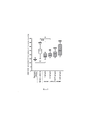
Препарат ВВИГ оказывал токсическое действие на NK-клетки в концентрации 24 мг/мл (Табл. 1). В более низких концентрациях, начиная с 12 мг/мл, гибель клеток NK-92 не отличалась от базовой. На основании полученных данных для экспериментов по оценке влияния препаратов в кокультуре NK-клеток линии NK-92 и клеток трофобласта использовали меньшие концентрации по сравнению с токсическими.The IVIG drug had a toxic effect on NK cells at a concentration of 24 mg/ml (Table 1). At lower concentrations, starting from 12 mg/ml, NK-92 cell death did not differ from the baseline. Based on the data obtained, for experiments to assess the effect of drugs in the coculture of NK cells of the NK-92 line and trophoblast cells, lower concentrations were used compared to toxic ones.
На втором этапе работы в качестве клеток-эффекторов также использовали клетки линии NK-92. В качестве клеток-мишеней использовали клетки трофобласта линии Jeg-3, которые представляют собой клетки инвазивного вневорсинчатого трофобласта первого триместра беременности. В силу экспрессии специфичных для клеток трофобласта поверхностных рецепторов и секреции цитокинов клетки линии Jeg-3 позволяют моделировать in vitro клеточные взаимодействия, происходящие при беременности. Специфичность клеток линии Jeg-3, обусловленная их профилем экспрессии, позволяет использовать эти клетки для оценки функциональных взаимодействий при патологиях беременности. Для использования в модельной системе первичных клеток трофобласта необходимо провести их выделение из материала биопсии, затем прокультивировать для получения необходимого количества клеток. Процедура получения биопсии инвазивна для пациента, а процесс выделения клеток трудоемок и не позволяет получить достаточное количество материала для анализа. По сравнению с первичными клетками трофобласта клетки линии Jeg-3 являются более стабильными, что позволяет стандартизовать методики оценки взаимодействия клеток трофобласта с другими клеточными популяциями.At the second stage of work, cells of the NK-92 line were also used as effector cells. As target cells, Jeg-3 trophoblast cells were used, which are cells of the invasive extravillous trophoblast of the first trimester of pregnancy. By virtue of the expression of surface receptors specific for trophoblast cells and the secretion of cytokines, Jeg-3 cells make it possible to simulate in vitro cellular interactions that occur during pregnancy. The specificity of the Jeg-3 cell line, due to their expression profile, makes it possible to use these cells to assess functional interactions in pathologies of pregnancy. To use primary trophoblast cells in the model system, it is necessary to isolate them from the biopsy material, then culture them to obtain the required number of cells. The procedure for obtaining a biopsy is invasive for the patient, and the process of cell isolation is laborious and does not allow obtaining a sufficient amount of material for analysis. Compared to primary trophoblast cells, the Jeg-3 cell line is more stable, which makes it possible to standardize methods for assessing the interaction of trophoblast cells with other cell populations.
За сутки до эксперимента пересевали клетки линии JEG-3, используя флаконы для адгезионных клеточных культур площадью 75 см2 (BD, США), в концентрации 3×10⁶ клеток в 10 мл. Через 24 часа клетки обрабатывали раствором CFSE (Sigma-Aldrich, США) в соответствии с рекомендациями производителя. Затем проводили дезинтеграцию монослоя обработанных CFSE клеток трофобласта с помощью смеси растворов версена и трипсина (3:1) (Biolot, Россия) и помещали клетки в лунки круглодонного 96-луночного планшета (BD, США) по 30.000 клеток линии JEG-3 в 50 мкл среды DMEM. В эти же лунки добавляли клетки линии NK-92 в 100 мкл, в соотношении эффектор:мишень 5:1. Затем к клеткам добавляли препарат ВВИГ, разведенные в культуральной среде в концентрациях: 6мг/мл, 1,5 мг/мл, 0,375 мг/мл, 0,009375 мг/мл. К части клеток была добавлена культуральная среда без препаратов для определения гибели клеток трфообласта в присутствии NK-клеток, также в каждом эксперименте проводили оценку гибели клеток линии Jeg-3, прокультивированных без NK-клеток и препаратов (базовая гибель). Затем планшет центрифугировали 3 минуты 100 g, после чего инкубировали 4 часа (37°С, 5% СО2). После инкубации клетки обрабатывали в течение 10 минут 2 мкг/мл раствором пропидия иодида (Sigma-Aldrich, США) при 4°С. Анализ флуоресценции проводили с помощью проточного цитофлуориметра FacsCantoII (BD, США).The day before the experiment, cells of the JEG-3 line were subcultured using flasks for adherent cell cultures with an area of 75 cm 2 (BD, USA) at a concentration of 3×10 6 cells per 10 ml. After 24 hours, the cells were treated with CFSE solution (Sigma-Aldrich, USA) according to the manufacturer's recommendations. Then, a monolayer of CFSE-treated trophoblast cells was disintegrated using a mixture of solutions of versene and trypsin (3:1) (Biolot, Russia) and the cells were placed in the wells of a round-bottom 96-well plate (BD, USA), 30,000 JEG-3 cells in 50 µl DMEM environments. The cells of the NK-92 line were added to the same wells in 100 μl, in the ratio of effector:target 5:1. Then the IVIG preparation was added to the cells, diluted in the culture medium at the following concentrations: 6 mg/ml, 1.5 mg/ml, 0.375 mg/ml, 0.009375 mg/ml. A culture medium without drugs was added to some of the cells to determine the death of trphooblast cells in the presence of NK cells; also, in each experiment, the death of Jeg-3 cells cultured without NK cells and drugs (baseline death) was assessed. Then the tablet was centrifuged for 3 minutes 100 g, after which it was incubated for 4 hours (37°C, 5% CO 2 ). After incubation, the cells were treated for 10 minutes with 2 μg/ml propidium iodide solution (Sigma-Aldrich, USA) at 4°C. Fluorescence analysis was performed using a FacsCantoII flow cytometer (BD, USA).
Установлено, что в присутствии препарата ВВИГ в концентрациях бмг/мл и 1,5 мг/мл относительная гибель клеток трофобласта в результате цитотоксической активности клеток линии NK-92 была снижена по сравнению с гибелью клеток трофобласта, прокультивированных только с NK-клетками. Более того, в присутствии препарата ВВИГ в концентрации 6мг/мл гибель клеток трофобласта была сопоставима с базовой гибелью клеток трофобласта (Фи. 1). Различий в гибели клеток линии Jeg-3 в присутствии NK-клеток между условиями культивирования с препаратом ВВИГ в разных концентрациях выявлено не было. В связи с этим, в экспериментах с мононуклеарами периферической крови использовали такие же концентрации препаратов ВВИГ.It was found that in the presence of IVIG at concentrations of bmg/ml and 1.5 mg/ml, the relative death of trophoblast cells as a result of the cytotoxic activity of NK-92 cells was reduced compared to the death of trophoblast cells cultivated only with NK cells. Moreover, in the presence of IVIG preparation at a concentration of 6 mg/ml, the death of trophoblast cells was comparable to the baseline death of trophoblast cells (Fig. 1). There were no differences in the death of Jeg-3 cells in the presence of NK cells between the culture conditions with the IVIG preparation at different concentrations. In this regard, in experiments with peripheral blood mononuclear cells, the same concentrations of IVIG preparations were used.
На третьем этапе работы проведена оценка цитопротективного влияния препарата ВВИГ в отношении клеток трофобласта линии Jeg-3 в условиях взаимодействия с NK-клетками фракции мононуклеаров периферической крови. Сформировано две группы обследуемых: здоровые небеременные женщины репродуктивного возраста с регулярным менструальным циклом без предшествующих беременностей (Группа 1, n=10) и здоровые небеременные женщины репродуктивного возраста с регулярным менструальным циклом, у которых ранее были беременности, закончившиеся родами в срок, и в анамнезе отсутствуют неразвивающиеся беременности и/или самопроизвольные выкидыши фертильные (Группа 2, n=12). Периферическую кровь собирали в секреторной фазе менструального цикла после УЗИ-контроля прохождения овуляции.At the third stage of the work, the cytoprotective effect of the IVIG preparation on the Jeg-3 trophoblast cells under conditions of interaction with NK cells of the peripheral blood mononuclear fraction was evaluated. Two groups of subjects were formed: healthy non-pregnant women of reproductive age with a regular menstrual cycle without previous pregnancies (Group 1, n=10) and healthy non-pregnant women of reproductive age with a regular menstrual cycle who had previously had pregnancies that ended in term birth and in history there are no non-developing pregnancies and/or fertile miscarriages (Group 2, n=12). Peripheral blood was collected in the secretory phase of the menstrual cycle after ultrasound monitoring of ovulation.
Критериями исключения для обеих групп были установленный диагноз антифосфолипидный синдром, наружный генитальный эндометриоз 3-4 стадии, аномалии развития половых органов, острые и обострения хронических заболеваний, ожирение 2-3 степени, наследственная форма тромбофилии высокого риска, сахарный диабет 1 и 2 типов, гестационный сахарный диабет на инсулинотерапии, гормональная терапия, в том числе комбинированные оральные контрацептивы, многоплодная беременность, отказ женщины от участия в программе исследования.The exclusion criteria for both groups were the established diagnosis of antiphospholipid syndrome, external genital endometriosis stage 3-4, anomalies in the development of the genital organs, acute and exacerbations of chronic diseases, obesity of 2-3 degrees, hereditary form of high-risk thrombophilia, diabetes mellitus types 1 and 2, gestational diabetes mellitus on insulin therapy, hormonal therapy, including combined oral contraceptives, multiple pregnancy, refusal of a woman to participate in the study program.
Для анализа цитотоксической активности выделены мононуклеары, содержащие NK-клетки, с помощью стандартного метода центрифугирования в градиенте плотности из периферической крови. Клетки линии Jeg-3 обрабатывали раствором CFSE и помещали в лунки планшета в концентрации 30.000 клеток в 50 мкл среды DMEM. Затем в часть лунок добавляли суспензию мононуклеаров в среде DMEM в концентрации 300000 клеток в 50 мкл. Для исследования цитотоксической активности NK-клеток фракции мононуклеаров использовали соотношение эффектор: мишень 1:10, согласно описанному [9] и запатентованному ранее протоколу [8]. В лунки, содержащие NK-клетки и клетки трофобласта, вносили препарат ВВИГ в концентрациях 6 мг/мл, 1,5 мг/мл, 0,375 мг/мл, 0,009375 мг/мл. Дополнительно было проанализировано влияние ВВИГ в концентрации 12 мг/мл на гибель клеток трофобласта в присутствии NK-клеток фракции мононуклеаров, так как на первом этапе исследования было установлено, что данная концентрация не токсична для клеток. Часть лунок с клетками линии Jeg-3 инкубировали без добавления мононуклеаров периферической крови и без препаратов ВВИГ для определения уровня базовой гибели клеток трофобласта. Часть лунок с клетками линии Jeg-3 инкубировали также без добавления мононуклеаров NK-клеток, но в присутствии препаратов ВВИГ для определения влияния этих препаратов на жизнеспособность клеток трофобласта. Затем клетки инкубировали в течение 4 часов (37°С, 5% СО2). После инкубации суспензию клеток обрабатывали раствором пропидия иодида и анализировали гибель клеток трофобласта с помощью проточного цитофлуориметра FacsCantoII (BD, США).For the analysis of cytotoxic activity, mononuclear cells containing NK cells were isolated using a standard density gradient centrifugation method from peripheral blood. Jeg-3 cells were treated with CFSE solution and plated at a concentration of 30,000 cells in 50 µl of DMEM medium in wells. Then, a suspension of mononuclear cells in DMEM medium was added to some of the wells at a concentration of 300,000 cells in 50 μl. To study the cytotoxic activity of NK cells of the mononuclear fraction, we used an effector:target ratio of 1:10, according to the protocol described [9] and previously patented [8]. The IVIG preparation at concentrations of 6 mg/ml, 1.5 mg/ml, 0.375 mg/ml, and 0.009375 mg/ml was added to the wells containing NK cells and trophoblast cells. In addition, the effect of IVIG at a concentration of 12 mg/ml on the death of trophoblast cells in the presence of NK cells of the mononuclear fraction was analyzed, since at the first stage of the study it was found that this concentration was not toxic to cells. Part of the wells with Jeg-3 cells were incubated without the addition of peripheral blood mononuclear cells and without IVIG preparations to determine the level of basic death of trophoblast cells. Part of the wells with Jeg-3 cells were also incubated without the addition of NK cell mononuclear cells, but in the presence of IVIG preparations to determine the effect of these preparations on the viability of trophoblast cells. Then the cells were incubated for 4 hours (37°C, 5% CO 2 ). After incubation, the cell suspension was treated with a solution of propidium iodide and the death of trophoblast cells was analyzed using a FacsCanto II flow cytometer (BD, USA).
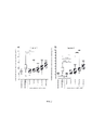
Установлено, что в присутствии мононуклеаров периферической крови женщин как группы 1, так и группы 2 гибель клеток трофобласта была выше, чем базовая гибель. После инкубации клеток трофобласта с мононуклеарами периферической крови женщин 1 группы и препаратом ВВИГ в концентрациях 6 мг/мл, 1,5 мг/мл, 0,375 мг/мл, 0,09 г/мл гибель клеток трофобласта была повышена по сравнению с базовой гибелью. В присутствии мононуклеаров женщин 1 группы и препарата ВВИГ в концентрациях 12 мг/мл и 6 мг/мл гибель клеток трофобласта была ниже, чем гибель клеток трофобласта в присутствии мононуклеаров, но без добавления препарата. В случае инкубации клеток трофобласта и NK-клеток в составе фракции мононуклеаров женщин 1 группы в присутствии препарата ВВИГ в концентрации 12 мг/мл гибель клеток трофобласта не отличалась от базовой (Фиг. 2а).It was found that in the presence of peripheral blood mononuclear cells of both group 1 and group 2 women, the death of trophoblast cells was higher than the baseline death. After incubation of trophoblast cells with peripheral blood mononuclear cells of women of the 1st group and IVIG preparation at concentrations of 6 mg/ml, 1.5 mg/ml, 0.375 mg/ml, 0.09 g/ml, the death of trophoblast cells was increased compared to the baseline death. In the presence of mononuclear cells of women of the 1st group and the IVIG preparation at concentrations of 12 mg/ml and 6 mg/ml, the death of trophoblast cells was lower than the death of trophoblast cells in the presence of mononuclear cells, but without the addition of the drug. In the case of incubation of trophoblast cells and NK cells in the mononuclear fraction of women of group 1 in the presence of IVIG preparation at a concentration of 12 mg/ml, the death of trophoblast cells did not differ from the baseline (Fig. 2a).
После инкубации клеток трофобласта с мононуклеарами периферической крови женщин 2 группы и препаратом ВВИГ гибель клеток трофобласта была повышена по сравнению с базовой гибелью в случае использования концентраций ВВИГ 1,5 мг/мл, 0,375 мг/мл, 0,09 г/мл. В присутствии мононуклеаров женщин 2 группы и препарата ВВИГ в концентрациях 12 мг/мл, 6 мг/мл и 1,5 мг/мл гибель клеток трофобласта была ниже, чем гибель клеток трофобласта в присутствии мононуклеаров, но без добавления препарата. Более того, в случае инкубации клеток трофобласта и NK-клеток в составе фракции мононуклеаров в присутствии препарата ВВИГ в концентрации 12 мг/мл и 6 мг/мл гибель клеток трофобласта не отличалась от базовой. При сравнении влияния разных концентраций препарата ВВИГ в используемой модельной системе установлено, что гибель клеток трофобласта в присутствии мононуклеаров была выше в случае использования концентрации 6 мг/мл по сравнению с концентрацией 12 мг/мл. Различий влияния препарата ВВИГ в концентрации 6 мг/мл и 1,5 мг/мл на гибель клеток трофобласта в присутствии мононуклеаров не выявлено, в тоже время при использовании концентрации 0,375 мг/мл гибель клеток трофобласта была выше, чем при использовании концентрации 1,5 мг/мл (Фиг. 2б).After incubation of trophoblast cells with peripheral blood mononuclear cells of women of the 2nd group and IVIG preparation, the death of trophoblast cells was increased compared with the baseline death in the case of using IVIG concentrations of 1.5 mg/ml, 0.375 mg/ml, 0.09 g/ml. In the presence of mononuclear cells from women of the 2nd group and IVIG preparation at concentrations of 12 mg/ml, 6 mg/ml and 1.5 mg/ml, the death of trophoblast cells was lower than the death of trophoblast cells in the presence of mononuclear cells, but without the addition of the drug. Moreover, in the case of incubation of trophoblast cells and NK cells as part of the mononuclear fraction in the presence of IVIG preparation at a concentration of 12 mg/ml and 6 mg/ml, the death of trophoblast cells did not differ from the baseline. When comparing the effect of different concentrations of the IVIG preparation in the model system used, it was found that the death of trophoblast cells in the presence of mononuclear cells was higher in the case of using a concentration of 6 mg/ml compared to a concentration of 12 mg/ml. No differences in the effect of IVIG preparation at a concentration of 6 mg/ml and 1.5 mg/ml on the death of trophoblast cells in the presence of mononuclear cells were found, at the same time, when using a concentration of 0.375 mg/ml, the death of trophoblast cells was higher than when using a concentration of 1.5 mg/ml (Fig. 2b).
Таким образом, в случае использования мононуклеаров периферической крови здоровых небеременных женщин без предшествовавших беременностей и родов (группа 1) цитопротективный эффект ВВИГ в отношении клеток трофобласта наблюдался только при использовании концентраций препарата 12 мг/мл и 6 мг/мл. Мононуклеары периферической крови здоровых небеременных фертильных женщин (группа 2) демонстрировали снижение цитотоксической активности к клеткам трофобласта в случае применения ВВИГ в концентрациях 12 мг/мл, 6 мг/мл и 1,5 мг/мл. Таким образом, целесообразно оценивать эффект препаратов ВВИГ в отношении коррекции цитотоксической активности NK-клеток периферической крови в концентрациях 12 мг/мл, 6 мг/мл и 1,5 мг/мл. Способ иллюстрируется фиг. 1-4, где:Thus, in the case of using peripheral blood mononuclear cells of healthy non-pregnant women without previous pregnancies and childbirth (group 1), the cytoprotective effect of IVIG on trophoblast cells was observed only when using drug concentrations of 12 mg/ml and 6 mg/ml. Peripheral blood mononuclear cells of healthy non-pregnant fertile women (group 2) showed a decrease in cytotoxic activity against trophoblast cells in the case of IVIG at concentrations of 12 mg/ml, 6 mg/ml and 1.5 mg/ml. Thus, it is advisable to evaluate the effect of IVIG preparations in relation to the correction of the cytotoxic activity of peripheral blood NK cells at concentrations of 12 mg/ml, 6 mg/ml, and 1.5 mg/ml. The method is illustrated in Fig. 1-4 where:
На фиг. 1 представлен график изменения цитотоксической активности клеток линии NK-92 в отношении клеток трофобласта линии JEG-3 в присутствии препаратов ВВИГ в различных концентрациях. Гибель клеток линии JEG-3 отличается от базовой гибели (без NK-клеток и препаратов ВВИГ): ••-р<0,01, •••-р<0,001. Достоверность различий: **-р<0,05, **-р<0,01; ns - различия не достоверны.In FIG. 1 shows a graph of changes in the cytotoxic activity of NK-92 cells against JEG-3 trophoblast cells in the presence of IVIG preparations at various concentrations. The death of JEG-3 cells differs from the basic death (without NK cells and IVIG preparations): ••-p<0.01, •••-p<0.001. Significance of differences: **-p<0.05, **-p<0.01; ns - differences are not significant.
На фиг. 2 представлен график изменения цитотоксической активности NK-клеток периферической крови в составе фракции мононуклеаров небеременных женщин (а) и фертильных небеременных женщин (б) в отношении клеток трофобласта линии JEG-3 в присутствии препаратов ВВИГ в различных концентрациях. Гибель клеток линии JEG-3 отличается от базовой гибели (без NK-клеток и препаратов ВВИГ): •••-р<0,001. Достоверность различий: *-р<0,05, **-р<0,01, ***-р<0,001; ns - различия не достоверны.In FIG. Figure 2 shows a graph of changes in the cytotoxic activity of peripheral blood NK cells in the mononuclear fraction of non-pregnant women (a) and fertile non-pregnant women (b) against JEG-3 trophoblast cells in the presence of IVIG preparations at various concentrations. The death of JEG-3 cells differs from the baseline death (without NK cells and IVIG preparations): •••-p<0.001. Significance of differences: *-p<0.05, **-p<0.01, ***-p<0.001; ns - differences are not significant.
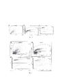
На фиг. 3 представлен способ оценки гибели клеток трофобласта в случае культивирования без мононуклеаров периферической крови и препаратов ВВИГ методом проточной цитофлюориметрии. а) Двумерная гистограмма распределения клеток трофобласта в координатах FSC-SSC. Регион Cells содержит клетки трофобласта. б) Двумерная гистограмма с координатами FSC-FITC, на которую отражены клетки из региона Cells. Регион Jeg + CFSE содержит клетки трофобласта. в) Двумерная гистограмма распределения клеток трофобласта в координатах FSC-PE. Квадрант DEAD CELLS содержит нежизнеспособные клетки трофобласта.In FIG. 3 shows a method for assessing the death of trophoblast cells in the case of cultivation without peripheral blood mononuclear cells and IVIG preparations by flow cytometry. a) Two-dimensional histogram of the distribution of trophoblast cells in FSC-SSC coordinates. The Cells region contains trophoblast cells. b) Two-dimensional histogram with FSC-FITC coordinates, on which cells from the Cells region are reflected. The Jeg + CFSE region contains trophoblast cells. c) Two-dimensional histogram of distribution of trophoblast cells in FSC-PE coordinates. The DEAD CELLS quadrant contains non-viable trophoblast cells.
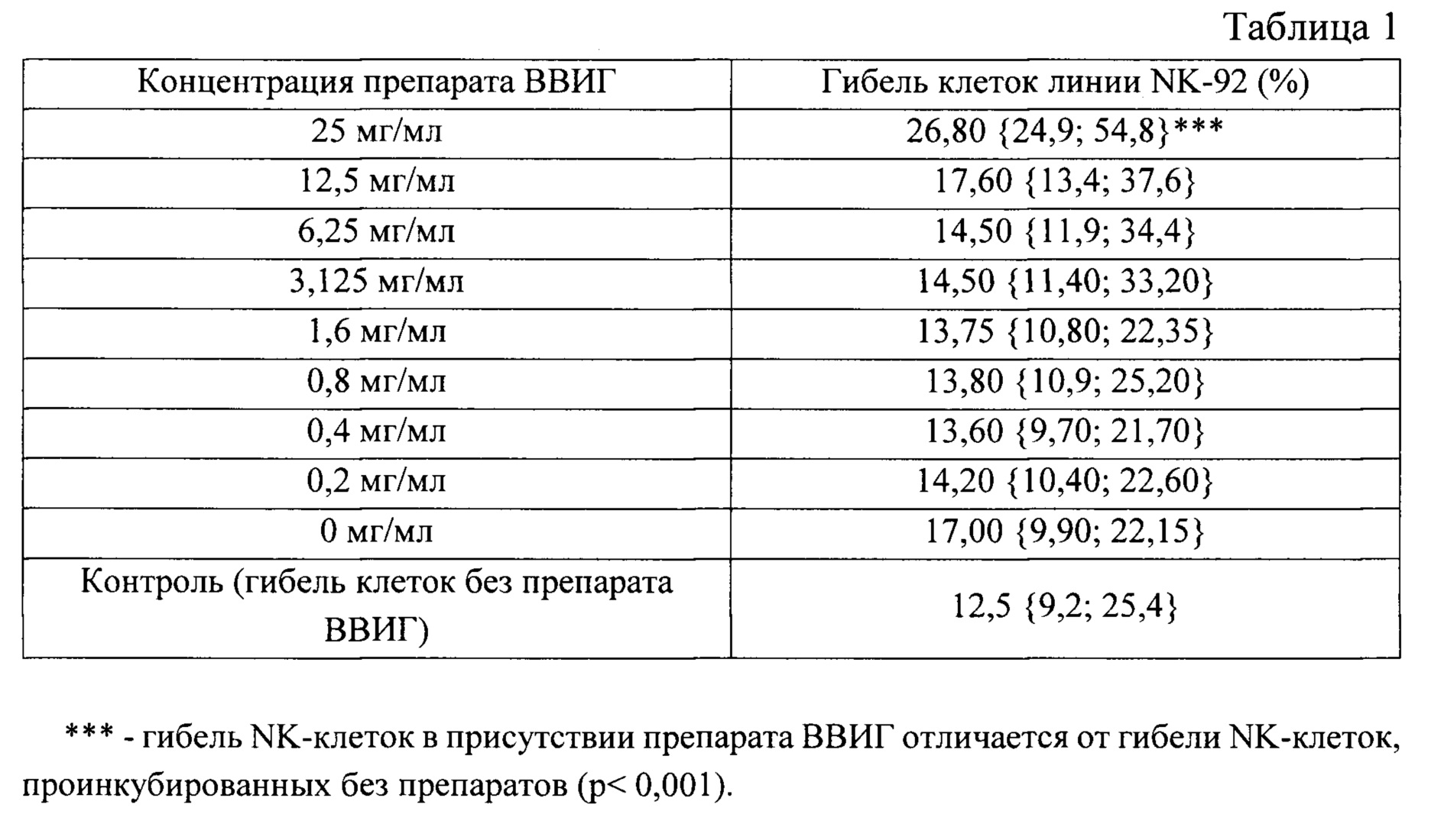
На фиг. 4 представлен способ оценки гибели клеток трофобласта в случае культивирования в присутствии мононуклеаров периферической крови и препаратов ВВИГ методом проточной цитофлюориметрии. а) Двумерная гистограмма распределения клеток трофобласта в координатах FSC-SSC. Регион Cells содержит клетки трофобласта. б) Двумерная гистограмма с координатами FSC-FITC, на которую отражены клетки из региона Cells. Регион Jeg + CFSE содержит клетки трофобласта. в-г) Двумерные гистограммы распределения клеток трофобласта в координатах FSC-PE. Квадрант DEAD CELLS содержит нежизнеспособные клетки трофобласта. Клетки трофобласта прокультивированы в присутствии мононуклеаров периферической крови (в) и в присутствии мононуклеаров периферической крови и препаратов ВВИГ (г).In FIG. 4 shows a method for assessing the death of trophoblast cells in the case of cultivation in the presence of peripheral blood mononuclear cells and IVIG preparations by flow cytometry. a) Two-dimensional histogram of the distribution of trophoblast cells in FSC-SSC coordinates. The Cells region contains trophoblast cells. b) Two-dimensional histogram with FSC-FITC coordinates, on which cells from the Cells region are reflected. The Jeg + CFSE region contains trophoblast cells. c-d) Two-dimensional histograms of distribution of trophoblast cells in FSC-PE coordinates. The DEAD CELLS quadrant contains non-viable trophoblast cells. Trophoblast cells were cultured in the presence of peripheral blood mononuclear cells (c) and in the presence of peripheral blood mononuclear cells and IVIG preparations (d).
Способ осуществляют, например, следующим образом: Для оценки цитопротективного эффекта препаратов ВВИГ в отношении клеток трофобласта используют клетки трофобласта линии Jeg-3 и мононуклеары периферической крови пациентки, которой планируется проводить лечение с использованием указанного препарата. Порядок исследования.The method is carried out, for example, as follows: To assess the cytoprotective effect of IVIG preparations against trophoblast cells, Jeg-3 trophoblast cells and peripheral blood mononuclear cells of a patient who is planned to be treated with the specified preparation are used. Research order.
1. Подготовка рабочих растворов.1. Preparation of working solutions.
1.1. Культуральная среда DMEM с добавлением 10% инактивированной эмбриональной телячьей сыворотки (ЭТС), 100 Ед/мл пенициллина и 100 мкг/мл стрептомицина, 2 мМ L-глютамина, 10 мМ пирувата натрия (полная культуральная среда).1.1. Culture medium DMEM supplemented with 10% inactivated fetal calf serum (FBS), 100 U/ml penicillin and 100 μg/ml streptomycin, 2 mM L-glutamine, 10 mM sodium pyruvate (complete culture medium).
1.2. Раствор Версена стерильный.1.2. Versen's solution is sterile.
1.3. Раствор Трипсина стерильный.1.3. Trypsin solution is sterile.
1.4. Раствор для культуральных работ ХЕПЕС1.4. HEPES culture solution
1.5 Раствор градиента плотности фиколл-верографин.1.5 Ficoll-verografin density gradient solution.
1.6 Раствор Хенкса стерильный.1.6 Hank's solution is sterile.
1.7 Раствор пропидия иодида 1 мкг/мл, приготовленный на PBS.1.7 Propidium iodide solution 1 µg/ml prepared in PBS.
2. Подготовка необходимых материалов.2. Preparation of the necessary materials.
2.1. Эппендорфы 0,6 мл, стерильнЫЕ, с крышкой, прозрачные.2.1. Eppendorf 0.6 ml, sterile, with lid, transparent.
2.2. Флакон для культивирования адгезивынх культур с вентилируемой синей крышкой 75 см2.2.2. Vial for the cultivation of adhesive cultures with a ventilated blue cap 75 cm 2 .
2.3. Камера Горяева, покровное стекло2.3. Goryaev's chamber, cover glass
2.4 Стерильные наконечники на 5 мл, 10 мкл, 300 мкл, 1000 мкл2.4 Sterile tips 5 ml, 10 µl, 300 µl, 1000 µl
2.5 Конические стерильные пробирки объемом 15 мл, 50 мл.2.5 Conical
3. Подготовка необходимого оборудования:3. Preparation of the necessary equipment:
3.1 Вертикальный ламинарно-потоковый шкаф.3.1 Vertical laminar flow cabinet.
3.2 Набор механических дозаторов переменного объема.3.2 Variable volume mechanical dispenser set.
3.3 Центрифуга.3.3 Centrifuge.
3.4 Термостат.3.4 Thermostat.
3.5 Вортекс.3.5 Vortex.
3.6 Холодильник 6+2°С.3.6
3.7 СО2 инкубатор.3.7 CO2 incubator.
3.8 Проточный цитофлюориметр оборудованный 488 нм лазером, датчиками прямого и прямого светорассеяния, датчиками учета флюоресценции FITC (519 нм), РЕ (578 нм).3.8 Flow cytometer equipped with a 488 nm laser, direct and direct light scattering sensors, FITC (519 nm), PE (578 nm) fluorescence sensors.
II. Культивирование клеток трофобласта линии Jeg-3.II. Cultivation of Jeg-3 trophoblast cells.
1. До начала процедуры прогреть стерильные контейнеры со средой для культивирования клеток трофобласта, со стерильным раствором Трипсина и стерильным раствором Версена при температуре 37°С.1. Before starting the procedure, heat sterile containers with a medium for culturing trophoblast cells, with a sterile solution of Trypsin and a sterile solution of Versen at a temperature of 37°C.
2. Все необходимые материалы, перечисленные в пункте 2 раздела I внести стерильно в вертикальный ламинарно-потоковый шкаф для работы с клеточными культурами.2. All the necessary materials listed in paragraph 2 of section I are sterile placed in a vertical laminar flow cabinet for working with cell cultures.
3. Смешать стерильно раствор Трипсина с раствором Версена (ТВ), содержащий равные части раствора Трипсина и раствора Версена.3. Sterile mix Trypsin solution with Versene solution (TB) containing equal parts of Trypsin solution and Versene solution.
4. Аккуратно слить через край среду из матраса. На противоположную стенку без клеток добавить 5 мл раствора ТВ для дезинтеграции монослоя, через 10 секунд и слить раствор. Повторить процедуру дважды, инкубировать с раствором ТВ последовательно 20 и 30 секунд. Слить раствор через край, затем добавить две капли раствора ТВ. Закрыть крышку флакона. Покачивать флакон в течение 5 минут так, чтобы клетки все время омывались раствором. Похлопать флакон по бокам, чтобы снять клетки со стенок флакона.4. Carefully drain the medium from the mattress over the edge. Add 5 ml of TB solution to the opposite wall without cells to disintegrate the monolayer, after 10 seconds and drain the solution. Repeat the procedure twice, incubate with TB solution successively for 20 and 30 seconds. Drain the solution over the edge, then add two drops of the TB solution. Close the vial cap. Shake the vial for 5 minutes so that the cells are washed with the solution all the time. Pat the sides of the vial to remove the cells from the walls of the vial.
5. Добавить 5 мл ранее приготовленной среды для клеток трофобласта во флакон 75 см2.5. Add 5 ml of the previously prepared medium for trophoblast cells to a 75 cm 2 vial.
6. Тщательно ресуспендировать. Перенести 700 мкл среды с клетками в новый флакон 75 см2 и долить в него 10 мл среды для культивирования.6. Carefully resuspend. Transfer 700 µl of medium with cells to a new 75 cm 2 vial and add 10 ml of culture medium to it.
7. Культивировать клеточную культуру во влажной атмосфере с 7% СО2 при 37°С в течение 72 часов или до образования клетками 50% монослоя.7. Cultivate the cell culture in a humid atmosphere with 7% CO2 at 37°C for 72 hours or until the cells form a 50% monolayer.
III. Выделение мононуклеаров из периферической крови.III. Isolation of mononuclear cells from peripheral blood.
1. Все необходимые материалы, перечисленные в пункте 2 раздела I, внести стерильно в вертикальный ламинарно-потоковый шкаф для работы с клеточными культурами.1. All the necessary materials listed in paragraph 2 of section I, sterile, add to a vertical laminar flow cabinet for working with cell cultures.
2. Подогреть раствор Хенкса и раствор градиента плотности в термостате 37°С.2. Warm up the Hank's solution and the density gradient solution in a 37°C thermostat.
3. Подписать и внести в ламинар (из расчета на анализ одного пациента): 1 пробирку на 50 мл, 6 пробирок на 15 мл.3. Sign and add to the laminar (based on the analysis of one patient): 1 tube per 50 ml, 6 tubes per 15 ml.
4. Внести в пробирки на 15 мл по 3 мл раствора градиента. В пробирки на 50 мл внести по 16 мл Хенкса, в пробирки для отмывки мононуклеаров внести по 10 мл раствора Хенкса.4. Add 3 ml of the gradient solution to 15 ml tubes. Add 16 ml of Hanks to 50 ml test tubes, add 10 ml of Hanks solution to test tubes for washing mononuclear cells.
5. Смешать 8 мл периферической крови с теплым раствором Хенкса в соотношении 1:2.5. Mix 8 ml of peripheral blood with warm Hank's solution in a ratio of 1:2.
6. Дозатором на 1 мл аккуратно наслоить разведенную периферическую кровь на раствор градиента из расчета на 4 мл градиента по 6 мл разведенной крови.6. With a 1 ml dispenser, carefully layer the diluted peripheral blood on the gradient solution at the rate of 4 ml gradient of 6 ml of diluted blood.
7. Центрифугировать 400g в течение 40 мин при 22°С.7. Centrifuge 400g for 40 min at 22°C.
8. Стерильно внести пробирки в ламинарно-потоковый шкаф. Отобрать 4-5 мл плазмы пипеткой на 1 мл и слить ее. Собрать мононуклеары.8. Place the tubes sterile in the laminar flow cabinet. Withdraw 4-5 ml of plasma with a 1 ml pipette and drain it. Collect mononuclear cells.
9. Перенести мононуклеары в пробирку с 10 мл раствора Хенкса.9. Transfer the mononuclear cells to a tube with 10 ml of Hank's solution.
10. Центрифугировать 200g в течение 10 мин при 22°С.10. Centrifuge 200g for 10 min at 22°C.
11. Аккуратно слить надосадочную жидкость, добавить к мононуклеарам 3 мл раствора Хенкса, ресуспендировать.11. Carefully discard the supernatant, add 3 ml of Hank's solution to the mononuclear cells, and resuspend.
12. Посчитать концентрацию мононуклеаров в камере Горяева. Добавить 10 мл раствора Хенкса.12. Calculate the concentration of mononuclear cells in the Goryaev chamber. Add 10 ml of Hank's solution.
13. Центрифугировать 350 g в течение 10 мин при 22°С.13. Centrifuge 350 g for 10 min at 22°C.
14. Аккуратно слить надосадочную жидкость. Развести клетки в культуральной среде на основе DMEM до концентрации 6 млн/мл.14. Carefully discard the supernatant. Dilute cells in DMEM-based culture medium to a concentration of 6 ppm.
15. Перенести клетки в эппендорфы.15. Transfer cells to eppendorfs.
16. Инкубировать в течение 96 часов при 37°С во влажной атмосфере и 5% СО2.16. Incubate for 96 hours at 37°C in a humid atmosphere and 5% CO 2 .
IV. Проведение эксперимента.IV. Conducting an experiment.
1. Подогреть культуральную среду DMEM, полную культуральную среду для клеток трофобласта линии JEG-3, раствор ТВ в термостате 37°С.1. Warm up the culture medium DMEM, complete culture medium for JEG-3 trophoblast cells, TB solution in a thermostat at 37°C.
2. Все необходимые материалы, перечисленные в пункте 2 раздела I, внести стерильно в вертикальный ламинарно-потоковый шкаф для работы с клеточными культурами.2. All the necessary materials listed in paragraph 2 of section I, sterile put into a vertical laminar flow cabinet for working with cell cultures.
3. Приготовить раствор CFSE, для этого в 1 пробирку теплого DMEM добавить 8 мкл CFSE (стоковый раствор: 4,48 ммоль/л) и ресуспендировать.3. Prepare CFSE solution by adding 8 µl of CFSE (stock solution: 4.48 mmol/l) to 1 tube of warm DMEM and resuspend.
4. Аккуратно слить среду из флакона для культивирования с клетками трофобласта, добавить 5 мл стерильного раствора, внести раствор красителя CFSE.4. Carefully drain the medium from the culture flask with trophoblast cells, add 5 ml of sterile solution, add the CFSE dye solution.
5. Инкубировать в течение 10 мин при 37°С, 7% СО2.5. Incubate for 10 min at 37°C, 7% CO 2 .
6. Слить раствор красителя, внести 10 мл холодной культуральной среды для JEG-3 без ЭТС. не касаясь стенки с клетками. Закрыть матрас, покачать 3 раза, слить среду. Повторить процедуру еще 1 раз.6. Discard the dye solution, add 10 ml of cold culture medium for JEG-3 without FTS. without touching the cell wall. Close the mattress,
7. Слить раствор культуральной среды.7. Discard the culture medium solution.
8. Добавить во флакон для культивирования клеток трофобласта 5 мл стерильного раствора ТВ, чтобы дезинтегрировать монослой клеток. Подождать 30 секунд, аккуратно потрясти флакон и слить раствор ТВ. Повторить операцию 2 раза, затем добавить две капли раствора ТВ. Закрыть крышку флакона. Покачивать флакон в течение 5 минут так, чтобы клетки все время омывались раствором. Похлопать флакон по бокам, чтобы снять клетки со стенок флакона.8. Add 5 ml of sterile TB solution to the trophoblast cell culture vial to disintegrate the cell monolayer. Wait 30 seconds, shake the vial gently and discard the TB solution. Repeat the operation 2 times, then add two drops of TB solution. Close the vial cap. Shake the vial for 5 minutes so that the cells are washed with the solution all the time. Pat the sides of the vial to remove the cells from the walls of the vial.
9. Внести 2 мл теплой среды для клеток трофобласта. Посчитать концентрацию клеток в камере Горяева. Развести клетки в культуральной среде до концентрации 0,6 млн/мл.9. Add 2 ml of warm medium for trophoblast cells. Calculate the concentration of cells in the Goryaev chamber. Dilute cells in culture medium to a concentration of 0.6 million/mL.
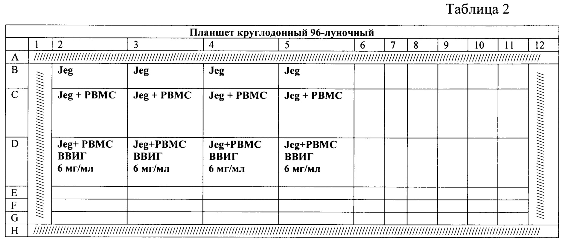
10. Стерильно внести в лунки планшета (табл. 2) согласно схеме (на 1 человека: РВМС- мононуклеары, Jeg - клетки трофобласта линии Jeg-3) по 50 мкл Jeg-3 (30000 кл.) + 50 мкл мононуклеаров (300000 кл.) В соответствующие лунки внести препарат ВВИГ в концентрации 6 мг/мл. Все варианты заполнения лунок проводить в 4 повторностях.10. Sterilely add to the wells of the plate (Table 2) according to the scheme (for 1 person: PBMC-mononuclear cells, Jeg - Jeg-3 trophoblast cells) 50 µl of Jeg-3 (30,000 cells) + 50 µl of mononuclear cells (300,000 cells .) Add IVIG preparation at a concentration of 6 mg/ml to the appropriate wells. All options for filling the wells should be carried out in 4 replications.
1. Центрифугировать планшет 2 мин 100 g 22°С.1. Centrifuge the plate for 2 min 100 g at 22°C.
2. Инкубировать пробы в течение 4 часов в инкубаторе при 37°С, 5% CO2.2. Incubate samples for 4 hours in an incubator at 37°C, 5% CO 2 .
3. Обработать пробы красителем пропидия иодида. Конечная концентрация пропидия иодида должна составлять 2 мкг/мл. Оставить неокрашенной 1 пробу с клетками JEG-3.3. Treat the samples with propidium iodide dye. The final concentration of propidium iodide should be 2 µg/ml. Leave unstained 1 sample with JEG-3 cells.
4. Инкубировать пробы 20 минут при 4°С.4. Incubate samples for 20 minutes at 4°C.
5. Добавить в пробы по 300 мкл стандартного фосфатно-солевого буфера.5. Add 300 µl of standard phosphate-buffered saline to the samples.
V. Проведение анализа и учет результатов.V. Conducting analysis and recording results.
Анализ клеток проводят на проточном цитофлюориметре, укомплектованном 488 нм лазером, по четырем параметрам: интенсивности прямого и бокового светорассеяния, флуоресценции по каналу FITC (спектр поглощения 488 нм, спектр эмиссии 519 нм), флуоресценции по каналу РЕ (спектр поглощения 488 нм, спектр эмиссии 578 нм). Анализируется 10000 событий. На двумерной гистограмме с координатами FSC-SSC выделяют регион Cells, содержащий клетки трофобласта (фиг. 3а). Клетки из региона Cells проецируют на двумерную гистограмму с координатами FSC-FITC (фиг. 3б). Определяют количество событий в квадранте Jeg + CFSE (фиг. 3б), данное количество соответствует количеству клеток трофобласта. Границу квадрантов на графике устанавливают предварительно, на основании предшествующего измерения контрольной пробы, необработанной раствором CFSE. Затем события из квадранта Jeg + CFSE проецируют на двумерную гистограмму с координатами FSC-PE. Границу квадрантов на графике устанавливают предварительно, на основании предшествующего измерения контрольной пробы, необработанной раствором пропидия иодида. При измерении опытной пробы обработанной растворами CFSE и пропидия иодида, на двумерной гистограмме с координатами FSC-PE анализируют относительное количество клеток трофобласта линии Jeg-3, содержащих пропидия иодид (клетки из квадранта DEAD CELLS) (фиг. 3в). Количество событий в квадранте DEAD CELLS соответствует нежизнеспособным клеткам трофобласта. Гибель клеток в контрольной лунке (после инкубации клеток трофобласта без мононуклеаров периферической крови) не должна превышать 35%.Cell analysis is carried out on a flow cytometer equipped with a 488 nm laser, according to four parameters: the intensity of direct and side light scattering, fluorescence through the FITC channel (absorption spectrum 488 nm, emission spectrum 519 nm), fluorescence through the PE channel (absorption spectrum 488 nm, emission spectrum 578 nm). 10,000 events are analyzed. On a two-dimensional histogram with FSC-SSC coordinates, the Cells region containing trophoblast cells is isolated (Fig. 3a). Cells from the Cells region are projected onto a 2D histogram with FSC-FITC coordinates (FIG. 3b). The number of events in the Jeg + CFSE quadrant is determined (Fig. 3b), this number corresponds to the number of trophoblast cells. The boundary of the quadrants on the graph is set previously, based on the previous measurement of the control sample, untreated with CFSE solution. The events from the Jeg + CFSE quadrant are then projected onto a 2D histogram with FSC-PE coordinates. The boundary of the quadrants on the graph is set previously, based on the previous measurement of the control sample, untreated with a solution of propidium iodide. When measuring an experimental sample treated with CFSE and propidium iodide solutions, the relative number of Jeg-3 trophoblast cells containing propidium iodide (cells from the DEAD CELLS quadrant) is analyzed on a two-dimensional histogram with FSC-PE coordinates (Fig. 3c). The number of events in the DEAD CELLS quadrant corresponds to non-viable trophoblast cells. Cell death in the control well (after incubation of trophoblast cells without peripheral blood mononuclear cells) should not exceed 35%.
Затем определяют количество событий в квадранте DEAD CELLS после инкубации клеток трофобласта с мононуклеарами периферической крови (фиг.4 а, б, в). Стратегия гейтирования событий аналогична представленной и описанной выше для фиг. 3. По количеству событий в квадрантах DEAD CELLS определяют процент нежизнеспособных клеток трофобласта (фиг. 4в). Затем оценивают количество событий в квадранте DEAD CELLS после инкубации клеток трофобласта с мононуклеарами периферической крови и препаратом ВВИГ в концентрации 6мг/мл (фиг. 4 г).Then determine the number of events in the quadrant DEAD CELLS after incubation of trophoblast cells with peripheral blood mononuclear cells (Fig.4 a, b, c). The event gating strategy is similar to that presented and described above for FIG. 3. The percentage of non-viable trophoblast cells is determined by the number of events in the DEAD CELLS quadrants (Fig. 4c). The number of events in the DEAD CELLS quadrant is then assessed after incubation of trophoblast cells with peripheral blood mononuclear cells and IVIG preparation at a concentration of 6 mg/ml (Fig. 4d).
Затем вычисляют коэффициент цитопротективного эффекта препаратов ВВИГ в отношении клеток трофобласта:Then the coefficient of the cytoprotective effect of IVIG preparations in relation to trophoblast cells is calculated:
![]()
![]()
параметр X1 соответствует разности количества клеток в гейте Dead cells после измерения пробы клеток трофобласта, инкубированных с мононуклеарами периферической крови (фиг. 4в) и инкубированных без мононуклеаров (фиг. 3в). Параметр Х2 соответствует разности количества клеток в гейте Dead cells после измерения пробы клеток трофобласта, инкубированных с мононуклеарами периферической крови и препаратом ВВИГ (фиг. 4г) и инкубированных без мононуклеаров (фиг. 3в).parameter X1 corresponds to the difference in the number of cells in the Dead cells gate after measuring a sample of trophoblast cells incubated with peripheral blood mononuclear cells (Fig. 4c) and incubated without mononuclear cells (Fig. 3c). The X2 parameter corresponds to the difference in the number of cells in the Dead cells gate after measuring a sample of trophoblast cells incubated with peripheral blood mononuclear cells and IVIG preparation (Fig. 4d) and incubated without mononuclear cells (Fig. 3c).
Цитопротективный эффект препаратов ВВИГ в отношении клеток трофобласта прогнозируют как положительный при значениях коэффициента ЦПЭ меньше 1. При значениях коэффициента ЦПЭ, равном или больше 1, цитопротективный эффект препарата ВВИГ в отношении клеток трофобласта - незначительный, применение ВВИГ для коррекции взаимодействия клеток трофобласта и NK-клеток в таком случае не целесообразно.The cytoprotective effect of IVIG preparations on trophoblast cells is predicted as positive if the CPE coefficient is less than 1. When the values of the CPE coefficient are equal to or greater than 1, the cytoprotective effect of the IVIG preparation on trophoblast cells is insignificant, the use of IVIG to correct the interaction of trophoblast cells and NK cells in this case it is not appropriate.
Способ подтверждается следующими клиническими примерами.The method is confirmed by the following clinical examples.
Пример 1. Участник исследования А. Количество клеток в гейте Dead cells после измерения пробы клеток трофобласта, инкубированных без мононуклеаров, соответствует 29,7%. Количество клеток в гейте Dead cells после измерения пробы клеток трофобласта, инкубированных с мононуклеарами периферической крови, составляет 51,8%. Параметр X1 равен 51,8-29,7=22,1%. Количество клеток в гейте Dead cells после измерения пробы клеток трофобласта, инкубированных с мононуклеарами периферической крови и препаратом ВВИГ, составляет 31%. Параметр Х2 равен 31-29,7=1,3%.Example 1 Study Participant A. The number of cells in the Dead cells gate after measuring a sample of trophoblast cells incubated without mononuclear cells corresponds to 29.7%. The number of cells in the Dead cells gate after measuring a sample of trophoblast cells incubated with peripheral blood mononuclear cells is 51.8%. Parameter X1 is equal to 51.8-29.7=22.1%. The number of cells in the Dead cells gate after measuring a sample of trophoblast cells incubated with peripheral blood mononuclear cells and IVIG preparation is 31%. Parameter X2 is equal to 31-29.7=1.3%.
Значение ЦПЭ по формуле составляет 1,3/22,1=0,06, что меньше 1. Следовательно, препарат ВВИГ осуществляет коррекцию цитотоксического действия мононуклеаров периферической крови в отношении клеток трофобласта in vitro.The CPE value according to the formula is 1.3/22.1=0.06, which is less than 1. Therefore, the IVIG preparation corrects the cytotoxic effect of peripheral blood mononuclear cells in relation to trophoblast cells in vitro.
Пример 2. Участник исследования Б. Количество клеток в гейте Dead cells после измерения пробы клеток трофобласта, инкубированных без мононуклеаров, соответствует 8,7%. Количество клеток в гейте Dead cells после измерения пробы клеток трофобласта, инкубированных с мононуклеарами периферической крови, составляет 26,0%. Параметр X1 равен 26,0-8,7=17,3%. Количество клеток в гейте Dead cells после измерения пробы клеток трофобласта, инкубированных с мононуклеарами периферической крови пациентки и препаратом ВВИГ, составляет 26,2%. Параметр Х2 равен 26,2-8,7=17,5%.Example 2. Study participant B. The number of cells in the Dead cells gate after measuring a sample of trophoblast cells incubated without mononuclear cells corresponds to 8.7%. The number of cells in the Dead cells gate after measuring a sample of trophoblast cells incubated with peripheral blood mononuclear cells is 26.0%. Parameter X1 is equal to 26.0-8.7=17.3%. The number of cells in the Dead cells gate after measuring a sample of trophoblast cells incubated with the patient's peripheral blood mononuclear cells and IVIG preparation is 26.2%. Parameter X2 is equal to 26.2-8.7=17.5%.
Значение ЦПЭ по формуле составляет 17,5/17,3=1,01, что больше 1. Следовательно, препарат ВВИГ не целесообразен для коррекции цитотоксического действия мононуклеаров периферической крови этого участника исследования в отношении клеток трофобласта in vitro.The CPE value according to the formula is 17.5/17.3=1.01, which is more than 1. Therefore, the IVIG preparation is not appropriate for correcting the cytotoxic effect of peripheral blood mononuclear cells of this study participant in relation to trophoblast cells in vitro.
Способ позволяет оценивать протективный эффект ВВИГ в отношении клеток трофобласта в условиях их взаимодействия с NK-клетками. Оценка цитопротективного эффекта ВВИГ в отношении клеток трофобласта может быть применена на этапе подбора терапии при различных формах репродуктивной патологии, при которых планируется использование указанных препаратов.The method allows to evaluate the protective effect of IVIG against trophoblast cells under conditions of their interaction with NK cells. Evaluation of the cytoprotective effect of IVIG in relation to trophoblast cells can be applied at the stage of selection of therapy for various forms of reproductive pathology, in which the use of these drugs is planned.
Источники информации:Sources of information:
1. Elabd, S.H., et al., Percentage of CD3-CD56 + dim and of CD3-CD56 + dim CD69 + Natural Killer Cells in the Peripheral Blood of Women with In Vitro Fertilization (IVF) Failure. Egypt J Immunol, 2016. 23(1): p. 39-44.1. Elabd, S.H., et al., Percentage of CD3-CD56 + dim and of CD3-CD56 + dim CD69 + Natural Killer Cells in the Peripheral Blood of Women with In Vitro Fertilization (IVF) Failure. Egypt J Immunol, 2016. 23(1): p. 39-44.
2. Kofod, L., A. Lindhard, and T.V.F. Hviid, Implications of uterine NK cells and regulatory T cells in the endometrium of infertile women. Hum Immunol, 2018. 79(9): p. 693-701.2. Kofod, L., A. Lindhard, and T.V.F. Hviid, Implications of uterine NK cells and regulatory T cells in the endometrium of infertile women. Hum Immunol, 2018. 79(9): p. 693-701.
3. Thum, M.Y., et al., Prednisolone suppresses NKcell cytotoxicity in vitro in women with a history of infertility and elevated NKcell cytotoxicity. Am J Reprod Immunol, 2008. 59(3): p.259-65.3. Thum, M.Y., et al., Prednisolone suppresses NKcell cytotoxicity in vitro in women with a history of infertility and elevated NKcell cytotoxicity. Am J Reprod Immunol, 2008. 59(3): p.259-65.
4. Polanski, L.T., et al., Interventions to improve reproductive outcomes in women with elevated natural killer cells undergoing assisted reproduction techniques: a systematic review of literature. Hum Reprod, 2014. 29(1): p.65-75.4. Polanski, L.T., et al., Interventions to improve reproductive outcomes in women with elevated natural killer cells undergoing assisted reproduction techniques: a systematic review of literature. Hum Reprod, 2014. 29(1): p.65-75.
5. Chemyshov, V.P., et al., Multiple immune deviations predictive for IVF failure as possible markers for IVIG therapy. Immunol Lett, 2016. 176: p.44-50.5. Chemyshov, V.P., et al., Multiple immune deviations predictive for IVF failure as possible markers for IVIG therapy. Immunol Lett, 2016. 176: p.44-50.
6. Ho, Y.K., et al., Peripheral CD56(+)CD16(+) NK Cell Populations in the Early Follicular Phase Are Associated With Successful Clinical Outcomes of Intravenous Immunoglobulin Treatment in Women With Repeated Implantation Failure. Front Endocrinol (Lausanne), 2019. 10: p.937.6. Ho, Y.K., et al., Peripheral CD56(+)CD16(+) NK Cell Populations in the Early Follicular Phase Are Associated With Successful Clinical Outcomes of Intravenous Immunoglobulin Treatment in Women With Repeated Implantation Failure. Front Endocrinol (Lausanne), 2019. 10: p.937.
7. Чепанов, С.В., et al., Способ прогнозирования цитопротективного эффекта иммуноглобулинов для внутривенного введения при терапии женщин с привычным невынашиванием беременности и диагностированным антифосфолипидным синдромом, ФГБУНИИ АиГ им. Д.О. Отта", Editor. 2015: Российская Федерация.7. Chepanov, S.V., et al., A method for predicting the cytoprotective effect of intravenous immunoglobulins in the treatment of women with recurrent miscarriage and a diagnosed antiphospholipid syndrome, Federal State Budgetary Research Institute of AiG im. BEFORE. Otta", Editor. 2015: Russian Federation.
8. Соколов, Д.И., et al., Способ определения цитотоксической активности NK-клеток, г.и.р.и. Д.О.О. Федеральное государственное научное учреждение "Научно-исследовательский институт акушерства, Editor. 2017: Российская Федерация.8. Sokolov, D.I., et al., Method for determining the cytotoxic activity of NK cells, h.i.r.i. D.O.O. Federal State Scientific Institution "Research Institute of Obstetrics, Editor. 2017: Russian Federation.
9. Sokolov, D.I., et al., NK and trophoblast cells interaction: cytotoxic activity on recurrent pregnancy loss. Gynecological Endocrinology, 2019. 35(sup 1): p.5-10.9. Sokolov, D.I., et al., NK and trophoblast cells interaction: cytotoxic activity on recurrent pregnancy loss. Gynecological Endocrinology, 2019. 35(sup 1): p.5-10.
Claims (5)
Priority Applications (1)
| Application Number | Priority Date | Filing Date | Title |
|---|---|---|---|
| RU2021120799A RU2768461C1 (en) | 2021-07-13 | 2021-07-13 | Method for assessing the cytoprotective effect of immunoglobulin preparations for intravenous introduction on trophoblast cells under conditions of their interaction with natural killers |
Applications Claiming Priority (1)
| Application Number | Priority Date | Filing Date | Title |
|---|---|---|---|
| RU2021120799A RU2768461C1 (en) | 2021-07-13 | 2021-07-13 | Method for assessing the cytoprotective effect of immunoglobulin preparations for intravenous introduction on trophoblast cells under conditions of their interaction with natural killers |
Publications (1)
| Publication Number | Publication Date |
|---|---|
| RU2768461C1 true RU2768461C1 (en) | 2022-03-24 |
Family
ID=80819428
Family Applications (1)
| Application Number | Title | Priority Date | Filing Date |
|---|---|---|---|
| RU2021120799A RU2768461C1 (en) | 2021-07-13 | 2021-07-13 | Method for assessing the cytoprotective effect of immunoglobulin preparations for intravenous introduction on trophoblast cells under conditions of their interaction with natural killers |
Country Status (1)
| Country | Link |
|---|---|
| RU (1) | RU2768461C1 (en) |
Citations (1)
| Publication number | Priority date | Publication date | Assignee | Title |
|---|---|---|---|---|
| RU2548754C1 (en) * | 2014-04-15 | 2015-04-20 | Федеральное государственное бюджетное учреждение "Научно-исследовательский институт акушерства и гинекологии им. Д.О. Отта" Северо-Западного отделения Российской академии медицинских наук | Method for predicting cytoprotective effect of immunoglobulins for intravenous administration in therapy of females with recurrent pregnancy loss and diagnosed anti-phospholipid syndrome |
-
2021
- 2021-07-13 RU RU2021120799A patent/RU2768461C1/en active
Patent Citations (1)
| Publication number | Priority date | Publication date | Assignee | Title |
|---|---|---|---|---|
| RU2548754C1 (en) * | 2014-04-15 | 2015-04-20 | Федеральное государственное бюджетное учреждение "Научно-исследовательский институт акушерства и гинекологии им. Д.О. Отта" Северо-Западного отделения Российской академии медицинских наук | Method for predicting cytoprotective effect of immunoglobulins for intravenous administration in therapy of females with recurrent pregnancy loss and diagnosed anti-phospholipid syndrome |
Non-Patent Citations (4)
| Title |
|---|
| CHEPANOV S. et al. Experimental rationale for the endothelial protective effect of intravenous immunoglobulins in obstetric disease. Akusherstvo i Ginekologiia. 2016 May; 5: 82-88. * |
| АГНАЕВА А.О. и др. Роль естественных киллеров (NK-клеток) в репродуктивных потерях. Журнал акушерства и женских болезней. 2017; 66(3): 143-156. * |
| АГНАЕВА А.О. и др. Роль естественных киллеров (NK-клеток) в репродуктивных потерях. Журнал акушерства и женских болезней. 2017; 66(3): 143-156. ПЛУЖНИКОВА Т.А. и др. Опыт применения иммуноглобулина для внутривенного введения у беременных с невынашиванием и хроническим эндометритом. Журнал акушерства и женских болезней. 2018; 67(5): 21-31. CHEPANOV S. et al. Experimental rationale for the endothelial protective effect of intravenous immunoglobulins in obstetric disease. Akusherstvo i Ginekologiia. 2016 May; 5: 82-88. * |
| ПЛУЖНИКОВА Т.А. и др. Опыт применения иммуноглобулина для внутривенного введения у беременных с невынашиванием и хроническим эндометритом. Журнал акушерства и женских болезней. 2018; 67(5): 21-31. * |
Similar Documents
| Publication | Publication Date | Title |
|---|---|---|
| Xu et al. | Innate lymphoid cells at the human maternal‐fetal interface in spontaneous preterm labor | |
| Pistoia et al. | Soluble HLA-G: Are they clinically relevant? | |
| Aghajanova et al. | Uterine receptivity to human embryonic implantation: histology, biomarkers, and transcriptomics | |
| Siewiera et al. | Human cytomegalovirus infection elicits new decidual natural killer cell effector functions | |
| Xiong et al. | Maternal uterine NK cell–activating receptor KIR2DS1 enhances placentation | |
| D Ly et al. | Evidence-based management of infertile couples with repeated implantation failure following IVF | |
| Quenby et al. | Different types of recurrent miscarriage are associated with varying patterns of adhesion molecule expression in endometrium | |
| JE˛ DRZEJCZAK et al. | Consequences of semen inflammation and lipid peroxidation on fertilization capacity of spermatozoa in in vitro conditions | |
| Shaikly et al. | Analysis of HLA-G in maternal plasma, follicular fluid, and preimplantation embryos reveal an asymmetric pattern of expression | |
| Boyer et al. | Clinical significance of amniotic fluid sludge in twin pregnancies with a short cervical length | |
| Huang et al. | Evaluation of in vitro fertilization outcomes using interleukin-8 in culture medium of human preimplantation embryos | |
| Antonucci et al. | Circulating neutrophils of nonalcoholic steatohepatitis patients show an activated phenotype and suppress T lymphocytes activity | |
| Mariee et al. | The correlation of autoantibodies and uNK cells in women with reproductive failure | |
| Berestoviy et al. | An overview of autoimmunity in implantation failure: a literature review | |
| Kniotek et al. | Differences in the expression of KIR, ILT inhibitory receptors, and VEGF production in the induced decidual NK cell cultures of fertile and RPL women | |
| RU2768461C1 (en) | Method for assessing the cytoprotective effect of immunoglobulin preparations for intravenous introduction on trophoblast cells under conditions of their interaction with natural killers | |
| WO2015169781A1 (en) | Predictive markers for successful cancer immunotherapy | |
| Chu et al. | Increased death of peripheral blood mononuclear cells after TLR4 inhibition in sepsis is not via TNF/TNF receptor-mediated apoptotic pathway | |
| Lapides et al. | When Less Is More–Pipelle Endometrial Sampling for Quantification of Uterine Natural Killer Cells in Patients With Recurrent Implantation Failure or Habitual Abortion | |
| CN110361534B (en) | Chemical markers for evaluating embryo and predicting success rate of in vitro fertilization and application thereof | |
| JP6751104B2 (en) | Use of soluble CD146 as a biomarker for selecting in vitro fertilized embryos for implantation in mammals | |
| Morris et al. | Ideal culture time for improvement in sperm motility from testicular sperm aspirates of men with azoospermia | |
| RU2657433C1 (en) | Method of assessing the risk of non-pregnancy in women with usual miscarriage | |
| Bauman et al. | Canine memory T-cell subsets in health and disease | |
| Hou et al. | The predictive value of NKG2C+ NK cells and LILRB1+ NK cells in recurrent spontaneous abortion |