RU2763178C2 - Antibodies for laboratory diagnostics of the concentration of interleukin-11 - Google Patents
Antibodies for laboratory diagnostics of the concentration of interleukin-11 Download PDFInfo
- Publication number
- RU2763178C2 RU2763178C2 RU2020111785A RU2020111785A RU2763178C2 RU 2763178 C2 RU2763178 C2 RU 2763178C2 RU 2020111785 A RU2020111785 A RU 2020111785A RU 2020111785 A RU2020111785 A RU 2020111785A RU 2763178 C2 RU2763178 C2 RU 2763178C2
- Authority
- RU
- Russia
- Prior art keywords
- antibodies
- seq
- antibody
- antigen
- concentration
- Prior art date
Links
Images


Classifications
-
- C—CHEMISTRY; METALLURGY
- C07—ORGANIC CHEMISTRY
- C07K—PEPTIDES
- C07K16/00—Immunoglobulins [IGs], e.g. monoclonal or polyclonal antibodies
- C07K16/18—Immunoglobulins [IGs], e.g. monoclonal or polyclonal antibodies against material from animals or humans
- C07K16/24—Immunoglobulins [IGs], e.g. monoclonal or polyclonal antibodies against material from animals or humans against cytokines, lymphokines or interferons
- C07K16/244—Interleukins [IL]
-
- C—CHEMISTRY; METALLURGY
- C07—ORGANIC CHEMISTRY
- C07K—PEPTIDES
- C07K16/00—Immunoglobulins [IGs], e.g. monoclonal or polyclonal antibodies
- C07K16/46—Hybrid immunoglobulins
-
- G—PHYSICS
- G01—MEASURING; TESTING
- G01N—INVESTIGATING OR ANALYSING MATERIALS BY DETERMINING THEIR CHEMICAL OR PHYSICAL PROPERTIES
- G01N33/00—Investigating or analysing materials by specific methods not covered by groups G01N1/00 - G01N31/00
- G01N33/48—Biological material, e.g. blood, urine; Haemocytometers
- G01N33/50—Chemical analysis of biological material, e.g. blood, urine; Testing involving biospecific ligand binding methods; Immunological testing
- G01N33/53—Immunoassay; Biospecific binding assay; Materials therefor
Abstract
Description
Область техники, к которой относится изобретениеThe field of technology to which the invention belongs
Настоящее изобретение относится к области биотехнологии, иммунологии и медицины и может найти применение для лабораторной диагностики концентрации интерлейкина-11 в биологических жидкостях.The present invention relates to the field of biotechnology, immunology and medicine and can be used for laboratory diagnosis of the concentration of interleukin-11 in biological fluids.
Уровень техникиState of the art
Интерлейкин-11 (ИЛ-11) является плейотропным цитокином, относящимся к семейству интерлейкина-6 (ИЛ-6). Зрелый белок состоит из 178 аминокислотных остатков и имеет небольшую молекулярную массу (около 19 кДа). Гомология последовательности аминокислот ИЛ 11 человека и яванской макаки (Macacafascicularis) составляет 93,8% (Sokolov A.S. et al., Molecules 2016, 21, 1632). Рецепторами ИЛ-11 выступают мультисубъединичные мембранные комплексы, состоящие из a- (IL-11RA) и β-субъединиц (glycoprotein 130, gp 130). Специфическое связывание лиганда с субъединицей IL-11RA вызывает конформационное изменение последней, приводящее к увеличению ее сродства к gp 130. Гомодимеризация молекул gp 130 в составе мультимерного комплекса запускает передачу сигнала внутрь клетки посредством активации сигнальных каскадов JAKs/STAT 1,3, ERK/RAS и mTor/PI3K (Negahdaripour М. et al., Cytokine Growth Factor Rev. 2016;32:41-61).Interleukin-11 (IL-11) is a pleiotropic cytokine belonging to the interleukin-6 (IL-6) family. The mature protein consists of 178 amino acid residues and has a small molecular weight (about 19 kDa). The amino acid sequence homology of human IL 11 and cynomolgus macaque (Macacafascicularis) is 93.8% (Sokolov A.S. et al., Molecules 2016, 21, 1632). IL-11 receptors are multisubunit membrane complexes consisting of a- (IL-11RA) and β-subunits (glycoprotein 130, gp 130). Specific binding of the ligand to the IL-11RA subunit causes a conformational change in the latter, leading to an increase in its affinity for gp 130. Homodimerization of gp 130 molecules in the multimeric complex triggers signal transduction into the cell via activation of the signaling cascades JAKs/
ИЛ-11 непосредственно стимулирует пролиферацию клеток-предшественников мегакариоцитов и индуцирует созревание мегакариоцитов, что приводит к увеличению продукции тромбоцитов. Кроме этого он стимулирует эритропоэз, миелопоэз, лимфопоэз и развитие остеокластов, активирует синтез белков острой фазы гепатоцитами и ингибирует адипогенез.IL-11 directly stimulates the proliferation of megakaryocyte progenitor cells and induces the maturation of megakaryocytes, resulting in increased platelet production. In addition, it stimulates erythropoiesis, myelopoiesis, lymphopoiesis and osteoclast development, activates the synthesis of acute phase proteins by hepatocytes and inhibits adipogenesis.
Помимо участия ИЛ-11 в физиологически важных процессах, он вовлечен в патогенез ряда заболеваний, в том числе онкологических. Необходимость поиска новых маркеров для проведения диагностики заболеваний на возможно более ранних сроках, особенно это касается онкологических заболеваний, не вызывает сомнений.In addition to the participation of IL-11 in physiologically important processes, it is involved in the pathogenesis of a number of diseases, including cancer. The need to search for new markers for diagnosing diseases as early as possible, especially for oncological diseases, is beyond doubt.
Показано, что в плазме больных раком поджелудочной железы достоверно наблюдается повышенный уровень ИЛ-11 (Ren C.L. et al., Tumor Biol. 2014,35:11467-11472).It has been shown that an elevated level of IL-11 is significantly observed in the plasma of patients with pancreatic cancer (Ren C.L. et al., Tumor Biol. 2014,35:11467-11472).
ИЛ-11 появляется в бронхоальвеолярной лаважной жидкости у больных раком легких, в том числе на фоне хронической обструктивной болезни легких (Pastor M.D. et al., J Thorac Oncol. 2016 Dec; 11 (12):2183-2192).IL-11 appears in bronchoalveolar lavage fluid in patients with lung cancer, including chronic obstructive pulmonary disease (Pastor M.D. et al., J Thorac Oncol. 2016 Dec; 11(12):2183-2192).
Спонтанные внутримозговые кровоизлияния сопровождаются повышением содержания ИЛ-11 в плазме крови и спинномозговой жидкости, более того уровень данного цитокина коррелирует со смертностью от данного заболевания (Fang H.Y. et al., Surg Neurol. 2005;64(6):511-7; Tu C.J. et al., J Int Med Res. 2011;39(4): 1265-74).Spontaneous intracerebral hemorrhages are accompanied by an increase in the content of IL-11 in blood plasma and cerebrospinal fluid, moreover, the level of this cytokine correlates with mortality from this disease (Fang HY et al., Surg Neurol. 2005;64(6):511-7; Tu CJ et al., J Int Med Res. 2011;39(4): 1265-74).
Концентрация ИЛ-11 сыворотке крови повышается у больных с приступами острого панкреатита и находится в прямой зависимости от их тяжести (Chen С.С. et al., Gut 1999;45:895-899).The concentration of IL-11 in the blood serum increases in patients with attacks of acute pancreatitis and is directly dependent on their severity (Chen C.C. et al., Gut 1999;45:895-899).
Исходя из вышеизложенного ИЛ-11 может рассматриваться в качестве нового независимого маркера, мониторинг уровня которого в биологических жидкостях позволит диагностировать и следить за течением заболеваний, ассоциированных с экспрессией данного цитокина.Based on the foregoing, IL-11 can be considered as a new independent marker, monitoring the level of which in biological fluids will allow diagnosing and monitoring the course of diseases associated with the expression of this cytokine.
Повышенный уровень ИЛ-11 в семенной жидкости увеличивает оплодотворяющую способность сперматозоидов, поэтому содержание ИЛ-11 может быть одним из контрольных параметров качества эякулята при проведении вспомогательных репродуктивных технологий (Seshadri S. et al., Andrologia. 2011 Dec;43(6):378-86).An increased level of IL-11 in the seminal fluid increases the fertilizing ability of spermatozoa, so the content of IL-11 can be one of the control parameters for the quality of the ejaculate during assisted reproductive technologies (Seshadri S. et al., Andrologia. 2011 Dec;43(6):378 -86).
Из патента РФ RU 2318829 С2 известны нейтрализующие химерные или гуманизированные анти-ИЛ-6-антитела, содержащие не менее одного гипервариабельного участка тяжелой или легкой цепи мышиного моноклонального антитела CLB-8. Предпочтительными антителами согласно изобретению являются антитела, которые связываются с участком молекулы ИЛ-6, отвечающим за связывание с gp 130 (сайт 1). Описаны последовательности ДНК, кодирующие гипервариабельные участки 1-3 легкой и тяжелой цепей мышиного моноклонального антитела CLB-8, а также их аминокислотные последовательности. Раскрыт состав лекарственных препаратов на основе указанных антител и их применение для лечения онкологических или иных заболеваний, опосредуемых ИЛ-6. Однако описанные в данном патенте антитела не являются специфичными к интерлейкину-11, и, следовательно, не могут использоваться для диагностики или лечения заболеваний, ассоциированных с ИЛ-11.From RF patent RU 2318829 C2, neutralizing chimeric or humanized anti-IL-6 antibodies containing at least one hypervariable region of the heavy or light chain of the mouse monoclonal antibody CLB-8 are known. Preferred antibodies according to the invention are antibodies that bind to the region of the IL-6 molecule responsible for binding to gp 130 (site 1). DNA sequences encoding hypervariable regions 1-3 of the light and heavy chains of the mouse monoclonal antibody CLB-8, as well as their amino acid sequences, are described. The composition of drugs based on these antibodies and their use for the treatment of oncological or other diseases mediated by IL-6 is disclosed. However, the antibodies described in this patent are not specific for interleukin-11, and therefore cannot be used to diagnose or treat diseases associated with IL-11.
В патенте США US 6998123 (В1) описан способ лечения или облегчения симптомов патологических состояний, при которых наблюдается снижение плотности костной ткани, при помощи введения пациенту эффективного количества анти-ИЛ-11 антител, которые блокируют образование мультимерного лиганд-рецепторного комплекса на поверхности клеток. Термин «анти- ИЛ-11 антитело» в данном патенте применяется для определения нейтрализующих химерных, гуманизированных или гибридных моноклональных антител, а также фрагментов антител, способных связываться с антигеном. В патенте приводятся результаты использования только одного типа молекул анти-ИЛ-11 антител на мышиной модели: у мышей с удаленными обоими яичниками после внутрибрюшинного введения афинно очищенных поликлональных антител козы против ИЛ-11 мыши наблюдается больший объем губчатой костной ткани в бедренной кости по сравнению с контрольной группой животных. В документе не приведено описание строения используемых антител, в том числе аминокислотной последовательности, а также деталей их получения. Указанный способ применения анти-ИЛ-11 антител относится к терапии заболеваний, при этом отсутствует информация о потенциальном использовании данных анти-ИЛ-11 антител в диагностике патологических состояний, при которых наблюдается снижение плотности костной ткани.US Pat. No. 6,998,123 (B1) describes a method for treating or alleviating the symptoms of pathological conditions in which there is a decrease in bone density by administering to the patient an effective amount of anti-IL-11 antibodies that block the formation of a multimeric ligand-receptor complex on the surface of cells. The term "anti-IL-11 antibody" is used in this patent to define neutralizing chimeric, humanized or hybrid monoclonal antibodies, as well as antibody fragments capable of binding to an antigen. The patent reports the results of using only one type of anti-IL-11 antibody molecule in a mouse model: in mice with both ovaries removed, after intraperitoneal injection of affinity-purified polyclonal goat anti-mouse IL-11 antibodies, a greater amount of cancellous bone tissue in the femur is observed compared to control group of animals. The document does not provide a description of the structure of the antibodies used, including the amino acid sequence, as well as the details of their production. This method of using anti-IL-11 antibodies refers to the therapy of diseases, while there is no information about the potential use of these anti-IL-11 antibodies in the diagnosis of pathological conditions in which there is a decrease in bone density.
Раскрытие изобретенияDisclosure of invention
Целью настоящего изобретения является устранение вышеуказанных недостатков аналогов анти-ИЛ-11 антител путем создания настоящего изобретения.The aim of the present invention is to overcome the above disadvantages of anti-IL-11 antibody analogs by providing the present invention.
Изобретение представляет собой антитела, специфичные к ИЛ-11 человека и модельных животных, таких как яванская макака, или их производные, или антигенсвязывающие фрагменты антител, в которых вариабельные домены тяжелой и легкой цепей содержат одну из следующих комбинаций аминокислотных последовательностей гипервариабельных участков:The invention provides antibodies specific for human IL-11 and model animals such as the cynomolgus monkey, or derivatives thereof, or antigen-binding fragments of antibodies, in which the variable domains of the heavy and light chains contain one of the following combinations of amino acid sequences of hypervariable regions:

Антитела, соответствующие изобретению, могут быть полной длины или могут содержать только антиген-связывающий фрагмент (например, фрагмент Fab, F(ab')2).Antibodies of the invention may be full length or may contain only an antigen-binding fragment (eg Fab fragment, F(ab')2).
Антитела могут быть моноклональными химерными, гуманизированными или гибридными, различных классов и подклассов, наиболее предпочтительно IgG1 или IgG4.Antibodies can be monoclonal chimeric, humanized or hybrid, of various classes and subclasses, most preferably IgG1 or IgG4.
Термин «антитело», используемый здесь, предназначен для определения полноразмерных молекул иммуноглобулина, состоящих из четырех полипептидных цепей (две тяжелые (Н) цепи и две легкие (L) цепи), связанных дисульфидными связями. Каждая тяжелая цепь содержит вариабельный участок (сокращенный здесь как VH) и константный участок, содержащий три домена CH1, СН2 и СН3. Каждая легкая цепь содержит вариабельный участок (сокращенный здесь как VL) и константный участок, содержащий один домен CL. Участки VH и VL состоят из трех гипервариабельных участков, которые окружены четырьмя более консервативными каркасными участками. Гипервариабельные участки называют также участками, определяющими комплементарность, и обозначают как CDR (complementarity determining regions), поскольку именно они образуют антигенсвязывающие центры молекулы антитела. Каркасные участки (FR, framework regions) обычно не принимают участия в связывании антигена, но имеют существенное значение для укладки V-домена, которая обеспечивает адекватную конформацию антигенсвязывающего центра.The term "antibody" as used herein is intended to refer to full length immunoglobulin molecules consisting of four polypeptide chains (two heavy (H) chains and two light (L) chains) linked by disulfide bonds. Each heavy chain contains a variable region (abbreviated here as VH) and a constant region containing the three domains CH1, CH2 and CH3. Each light chain contains a variable region (abbreviated here as VL) and a constant region containing a single CL domain. The VH and VL regions consist of three hypervariable regions surrounded by four more conserved framework regions. Hypervariable regions are also called complementarity determining regions and are referred to as CDRs (complementarity determining regions), since they form the antigen-binding centers of an antibody molecule. Framework regions (FRs) are usually not involved in antigen binding, but are essential for the V-domain folding, which provides an adequate conformation of the antigen-binding center.
Термин «антиген-связывающий фрагмент» антитела (или просто «фрагмент антитела»), используемый здесь, относится к одному или более фрагментам антитела, которые сохраняют способность специфически связывать антиген ИЛ-11 человека и модельных животных. Примеры связывающих фрагментов, охватываемые термином "антиген-связывающий фрагмент" антитела, включают (i) фрагмент Fab, моновалентный фрагмент, состоящий из доменов VL, VH, CL и CH1; (ii) фрагмент F(ab')2, бивалентный фрагмент, содержащий два фрагмента Fab, связанные дисульфидным мостиком в районе петли. Далее, антитело или его антиген-связывающий фрагмент могут быть частью более крупных молекул иммуноадгезии, образованных ковалентной или нековалентной связью антитела или фрагмента антитела с одним или более белком или пептидом.The term "antigen-binding fragment" of an antibody (or simply "antibody fragment") as used herein refers to one or more antibody fragments that retain the ability to specifically bind human and model animal IL-11 antigen. Examples of binding fragments encompassed by the term "antigen-binding fragment" of an antibody include (i) a Fab fragment, a monovalent fragment consisting of the VL, VH, CL, and CH1 domains; (ii) an F(ab')2 fragment, a bivalent fragment containing two Fab fragments linked by a disulfide bridge in the loop region. Further, an antibody or antigen-binding fragment thereof may be part of larger immunoadhesion molecules formed by covalent or non-covalent bonding of an antibody or antibody fragment to one or more proteins or peptides.
В одном варианте осуществления изобретение представляет собой полноразмерное антитело подкласса IgG1, обозначаемое U13, у которого аминокислотные последовательности гипервариабельных участков вариабельных доменов тяжелой и легкой цепей соответствуют комбинации # 1.In one embodiment, the invention is a full-length antibody of the IgG1 subclass, designated U13, in which the amino acid sequences of the hypervariable regions of the heavy and light chain variable domains correspond to
В другом варианте осуществления изобретение представляет собой полноразмерное антитело подкласса IgG1, обозначаемое L1, у которого аминокислотные последовательности гипервариабельных участков вариабельных доменов тяжелой и легкой цепей соответствуют комбинации # 2.In another embodiment, the invention is a full-length antibody of the IgG1 subclass, designated L1, in which the amino acid sequences of the hypervariable regions of the heavy and light chain variable domains correspond to
В еще одном варианте осуществления изобретение представляет собой Fab фрагмент, обозначаемый Fab3, у которого аминокислотные последовательности гипервариабельных участков вариабельных доменов тяжелой и легкой цепей соответствуют комбинации # 1.In yet another embodiment, the invention is a Fab fragment, designated Fab3, in which the amino acid sequences of the hypervariable regions of the heavy and light chain variable domains correspond to
В другом варианте осуществления изобретение представляет собой Fab фрагмент, обозначаемый Fab11, у которого аминокислотные последовательности гипервариабельных участков вариабельных доменов тяжелой и легкой цепей соответствуют комбинации # 2.In another embodiment, the invention is a Fab fragment, designated Fab11, in which the amino acid sequences of the hypervariable regions of the heavy and light chain variable domains correspond to
Еще одним объектом настоящего изобретения являются производные антител или их фрагментов, например, гибридные белки. В качестве молекул слияния могут выступать другие полипептиды, например, биотин и белки с ферментативной активностью, такие как пероксидаза хрена и щелочная фосфатаза, а также органические или неорганические молекулы, например, флуоресцентные метки (например, флуоресцеин, изотиоцианат флуоресцеина, родамин, хлорид 5-диметиламин-1-нафталинсульфонила, фикоэритрин и т.п.), радиоактивные метки (например, изотопы Р-32 и I-125) или хемилюминесцентные метки (например, люминол и его производные).Another object of the present invention are derivatives of antibodies or their fragments, for example, hybrid proteins. Fusion molecules can be other polypeptides, such as biotin and proteins with enzymatic activity, such as horseradish peroxidase and alkaline phosphatase, as well as organic or inorganic molecules, such as fluorescent labels (for example, fluorescein, fluorescein isothiocyanate, rhodamine, chloride 5- dimethylamine-1-naphthalenesulfonyl, phycoerythrin, etc.), radioactive labels (eg isotopes P-32 and I-125) or chemiluminescent labels (eg luminol and its derivatives).
В еще одном варианте осуществления изобретение представляет собой антитело U13, конъюгированное с молекулой биотина, обозначаемое U13-bio.In yet another embodiment, the invention is a U13 antibody conjugated to a biotin molecule, referred to as U13-bio.
В еще одном варианте осуществления изобретение представляет собой антитело L1, конъюгированное с молекулой биотина, обозначаемое L1-bio.In yet another embodiment, the invention is an L1 antibody conjugated to a biotin molecule, referred to as L1-bio.
Антитела, или их антигенсвязывающие фрагменты, или их производные, соответствующие изобретению, обладают хотя бы одним из следующих свойств:Antibodies, or antigen-binding fragments thereof, or derivatives thereof, according to the invention, have at least one of the following properties:
1) способны специфически связываться с ИЛ-11 человека и модельных животных, таких как яванская макака;1) are able to specifically bind to human IL-11 and model animals such as the cynomolgus monkey;
2) способны связываться с ИЛ-11 с высокой аффинностью: равновесная константа диссоциации (Kd) равна 1×10⁻⁸ М или меньше, и кинетическая константа диссоциации (Koff) 1×10⁻⁴c-1 или меньше, определенные, например, с помощью метода поверхностного плазмонного резонанса;2) able to bind to IL-11 with high affinity: the equilibrium dissociation constant (K d ) is 1×10 -8 M or less, and the kinetic dissociation constant (K off ) 1×10 -4 s -1 or less, determined, for example, using the method of surface plasmon resonance;
3) величина равновесной константы диссоциации при связывании ИЛ-11 человека отличается от величины Kd связывания ИЛ-11 модельных животных, таких как яванская макака, не более 10 раз;3) the value of the equilibrium dissociation constant when binding human IL-11 differs from the value of K d binding IL-11 model animals, such as cynomolgus macaque, no more than 10 times;
4) молекулы с разным набором гипервариабельных участков не конкурируют между собой за связывание с ИЛ-114) molecules with a different set of hypervariable regions do not compete with each other for binding to IL-11
Антитела U13 и L1 имеют следующие параметры связывания ИЛ-11 человека:U13 and L1 antibodies have the following human IL-11 binding parameters:
1) Kd, равная 1,0×10⁻¹¹ М и 5,0×10⁻¹⁰ М, соответственно;1) K d equal to 1.0×10 -11 M and 5.0×10 -10 M, respectively;
2) Koff, равная 2,0×10⁻⁶ с-1 и 8,0×10⁻⁵ с-1, соответственно.2) K off equal to 2.0×10 -6 s -1 and 8.0×10 -5 s -1 , respectively.
Антитела U13 и L1 имеют следующие параметры связывания ИЛ-11 яванской макаки:Antibodies U13 and L1 have the following binding parameters for cynomolgus macaque IL-11:
3) Kd, равная 7,0×10⁻¹¹ М и 2,0×10⁻¹⁰ М, соответственно;3) K d equal to 7.0×10 -11 M and 2.0×10 -10 M, respectively;
4) Koff, равная 7,0×10⁻⁶ с-1 и 2,0×10⁻⁵ с-1, соответственно.4) K off equal to 7.0×10 -6 s -1 and 2.0×10 -5 s -1 , respectively.
Антитела могут быть получены общепринятыми методами, например, иммунизацией соответствующим антигеном животных, или гибридомной технологией, или при помощи технологии генной инженерии. Методы получения антител описаны в литературе: Zola, Н. (CRC Press, 1987). Monoclonal Antibodies: A Manual of Techniques; John G.R. Hurrell (CRC Press, 1982). Monoclonal Hybridoma Antibodies: Techniques and Applications.Antibodies can be obtained by conventional methods, for example, immunization with the appropriate antigen of animals, or hybridoma technology, or using genetic engineering technology. Methods for obtaining antibodies are described in the literature: Zola, H. (CRC Press, 1987). Monoclonal Antibodies: A Manual of Techniques; John G.R. Hurrell (CRC Press, 1982). Monoclonal Hybridoma Antibodies: Techniques and Applications.
Фрагменты антител получают расщеплением антител соответствующими протеолитическими ферментами, например, папаином и пепсином, а также при помощи технологии рекомбинантной ДНК.Antibody fragments are obtained by cleavage of antibodies with appropriate proteolytic enzymes, such as papain and pepsin, as well as using recombinant DNA technology.
Для отбора рекомбинантных антител требуемой специфичности используют такие технологии, как фаговый дисплей. Чаще всего для фагового дисплея используют нитчатые фаги Ff семейства (М13, fd, f1). Нуклеотидные последовательности, кодирующие фрагменты антител, например, Fab или scFv, объединяют в составе фагового генома или фагмид с частью гена одного из пяти белков оболочки бактериофагов, наиболее часто используют рШ и pVIII. В результате фаговые частицы экспрессируют в составе капсида копии корового белка с пришитой к ним полипептидной последовательностью фрагмента антител. Отбор как правило осуществляют в виде нескольких последовательных раундов, каждый из которых включает сорбцию фаговых частиц, экспрессирующих соответствующие фрагменты антител, на носителях с иммобилизованным антигеном, отмывку несвязавшихся частиц, элюирование оставшихся связанными и размножение полученных фагов в бактериальных клетках. Выделяют и секвенируют фаговую ДНК или фагмиды. Фаговый дисплей широко описан в литературе: Bradbury A.R., Marks J.D. Antibodies from phage antibody libraries. J. Immunol. Methods 2004, 290, 29-49; Ledsgaard, L. et al. Basics of Antibody Phage Display Technology. Toxins 2018, 10, 236; John M. Walker. The Protein Protocols Handbook. Springer, 2007.Techniques such as phage display are used to select recombinant antibodies of the required specificity. Most often, filamentous phages of the Ff family (M13, fd, f1) are used for phage display. Nucleotide sequences encoding fragments of antibodies, for example, Fab or scFv, are combined as part of the phage genome or phagemid with a part of the gene of one of the five bacteriophage envelope proteins; pIII and pVIII are most often used. As a result, phage particles express in the capsid copies of the core protein with the polypeptide sequence of the antibody fragment attached to them. The selection is usually carried out in the form of several successive rounds, each of which includes the adsorption of phage particles expressing the corresponding antibody fragments on carriers with immobilized antigen, washing off unbound particles, elution of the remaining bound ones, and propagation of the resulting phages in bacterial cells. Phage DNA or phagemids are isolated and sequenced. Phage display is widely described in the literature: Bradbury A.R., Marks J.D. Antibodies from phage antibody libraries. J. Immunol. Methods 2004, 290, 29-49; Ledsgaard, L. et al. Basics of Antibody Phage Display Technology. Toxins 2018, 10, 236; John M. Walker. The Protein Protocols Handbook. Springer, 2007.
Технология рекомбинантной ДНК подразумевает получение экспрессионных генетических конструкций, кодирующих цепи антитела под контролем соответствующих регуляторных элементов в одном или разных несущих векторах. Регуляторные элементы представляют собой промоторные последовательности и сигнальные последовательности, например, сигналы полиаденилирования, которые контролируют транскрипцию или трансляцию генов цепи антитела. Такие регуляторные последовательности описаны, например, в работе Gene Expression Technology: Methods in Enzymology, vol. 185, Edited by David V. Goeddel, Academic Press, San Diego, CA (1990).Recombinant DNA technology involves the production of expression genetic constructs encoding antibody chains under the control of appropriate regulatory elements in one or different carrier vectors. Regulatory elements are promoter sequences and signal sequences, such as polyadenylation signals, that control transcription or translation of antibody chain genes. Such regulatory sequences are described, for example, in Gene Expression Technology: Methods in Enzymology, vol. 185, Edited by David V. Goeddel, Academic Press, San Diego, CA (1990).
Экспрессионные конструкции одну или более переносят общепринятыми методами в клетки, пригодные для экспрессии и предпочтительно секреции антител в культуральную среду. Клетками продуцентами полноразмерных антител предпочтительно являются клетки млекопитающих линий СНО (клетки яичников китайского хомячка), SP2/0 (клетки мышиной миеломы) или HEK293 (клетки человеческой эмбриональной почки). Данные клеточные линии пригодны как для получения линий, стабильно экспрессирующих целевое антитело, так и для наработки антител лишь некоторое время (транзиентная экспрессия). Фрагменты антител, например, Fab, производят в бактериях, дрожжах, нитчатых грибах и линиях клеток насекомых. Трансформированные клетки культивируют общепринятыми методами и выделяют антитела, их фрагменты из культуральной среды. Стандартные способы с применением рекомбинантной ДНК используются для получения генов тяжелой и легкой цепей, включения этих генов в рекомбинантные экспрессионные векторы и введения векторов в клетки-хозяина так, как описано Sambrook, J., Fritsch Е. F., and Т. Maniatis. Molecular cloning: A laboratory manual, Second Edition, Cold Spring Harbor, N.Y. (1989); Ausubel F., Brent R. et al. Current Protocols in Molecular Biology, John Wiley & Sons (2003).One or more expression constructs are transferred by conventional methods into cells suitable for expression and preferably secretion of antibodies into the culture medium. Cells producing full-length antibodies are preferably mammalian cell lines CHO (Chinese hamster ovary cells), SP2/0 (mouse myeloma cells) or HEK293 (human embryonic kidney cells). These cell lines are suitable both for obtaining lines that stably express the target antibody, and for producing antibodies only for a short time (transient expression). Antibody fragments such as Fab are produced in bacteria, yeast, filamentous fungi and insect cell lines. Transformed cells are cultivated by conventional methods and antibodies and their fragments are isolated from the culture medium. Standard recombinant DNA techniques are used to obtain heavy and light chain genes, incorporate these genes into recombinant expression vectors, and introduce the vectors into host cells as described by Sambrook, J., Fritsch, E. F., and T. Maniatis. Molecular cloning: A laboratory manual, Second Edition, Cold Spring Harbor, N.Y. (1989); Ausubel F., Brent R. et al. Current Protocols in Molecular Biology, John Wiley & Sons (2003).
Производные антител и их фрагментов получают в том числе в результате химической модификации с использованием активированных производных молекул слияния. Например, самым распространенным и универсальным методом биотинилирования является химическая модификация с использованием активированных производных биотина, таких как биотинил-N-гидроксисукцинимид (BNHS), при этом биотин присоединяется по ε - аминогруппам остатков лизина в молекуле антител и их фрагментов. Подробное описание биотинилирования приведено в литературе, например, Diamandis Е. Р and Christopoulos Т K (1991) The biotin-(strept)avidin system principles and applications in biotechnology Chn Chem 37, 625-636.Derivatives of antibodies and their fragments are also obtained as a result of chemical modification using activated derivatives of fusion molecules. For example, the most common and versatile biotinylation method is chemical modification using activated biotin derivatives, such as biotinyl-N-hydroxysuccinimide (BNHS), while biotin is attached to the ε-amino groups of lysine residues in the antibody molecule and their fragments. A detailed description of biotinylation is given in the literature, for example, Diamandis E. P and Christopoulos T K (1991) The biotin-(strept)avidin system principles and applications in biotechnology Chn Chem 37, 625-636.
Следующий аспект изобретения касается способа определения в пробах наличия и концентрации ИЛ-11 человека и модельных животных, таких как яванская макака, заключающийся в том, что 1) исследуемый образец, содержащей ИЛ-11, приводят в контакт с антителами, или их фрагментами, или производными, являющимися объектами настоящего изобретения, и 2) оценивают наличие и концентрацию иммунных комплексов антител, или их фрагментов, или производных с антигеном (ИЛ-11).Another aspect of the invention relates to a method for determining the presence and concentration of human IL-11 and model animals in samples, such as the cynomolgus macaque, which consists in the fact that 1) the test sample containing IL-11 is brought into contact with antibodies, or fragments thereof, or derivatives that are the objects of the present invention, and 2) assess the presence and concentration of immune complexes of antibodies, or fragments or derivatives with antigen (IL-11).
В качестве проб, в которых необходимо определить наличие или концентрацию ИЛ-11, могут быть образцы биологических жидкостей (например, кровь, плазма, сыворотка) и кусочки ткани, полученные в результате биопсии, организма человека и модельных животных, например, яванской макаки, а также образцы супернатантов клеточных культур, растворы очищенных рекомбинантных ИЛ-11 человека и животных, например, яванской макаки.Samples in which it is necessary to determine the presence or concentration of IL-11 can be samples of biological fluids (for example, blood, plasma, serum) and pieces of tissue obtained as a result of biopsy, human body and model animals, for example, cynomolgus macaque, and also samples of cell culture supernatants, solutions of purified recombinant human and animal IL-11, for example, cynomolgus macaque.
Для лабораторной диагностики наличия и концентрации ИЛ-11 в образцах биологических жидкостей, супернатантах клеточных культур и растворах очищенного ИЛ-11 может быть использован широкий круг методов иммуноанализа, среди которых иммуноблоттинг, иммуноферментный, радиоиммунный и иммунофлуоресцентный анализ, иммунотурбодиметрия. В образцах тканей для определения наличия ИЛ-11 используют метод иммуногистохимии.For laboratory diagnosis of the presence and concentration of IL-11 in samples of biological fluids, cell culture supernatants and solutions of purified IL-11, a wide range of immunoassay methods can be used, including immunoblotting, enzyme immunoassay, radioimmunoassay and immunofluorescence analysis, immunoturbodimetry. In tissue samples, immunohistochemistry is used to determine the presence of IL-11.
В зависимости от дизайна эксперимента антитела, их производные или фрагменты, соответствующие настоящему изобретению, могут использоваться по отдельности или попарно друг с другом.Depending on the design of the experiment, the antibodies, their derivatives or fragments of the present invention can be used alone or in pairs with each other.
В еще одном варианте осуществления изобретение представляет собой сэндвич-вариант иммуноанализа с использованием пар антител U13 и L1, а также их конъюгатов, например, U13-bio или L1-bio. Антитела U13 и L1 и их конъюгаты специфически распознают разные неперекрывающиеся участки молекулы ИЛ-11 (эпитопы), благодаря чему между ними отсутствует конкуренция за связывание с антигеном. Данный способ анализа позволяет добиться высокой чувствительности, точности и специфичности при определении антигена даже в гетерогенных образцах, таких как биологические жидкости и ткани, супернатанты клеточных культур.In yet another embodiment, the invention is a sandwich immunoassay using pairs of antibodies U13 and L1, as well as their conjugates, for example, U13-bio or L1-bio. Antibodies U13 and L1 and their conjugates specifically recognize different non-overlapping regions of the IL-11 molecule (epitopes), due to which there is no competition between them for binding to the antigen. This method of analysis makes it possible to achieve high sensitivity, accuracy and specificity in the determination of antigen even in heterogeneous samples, such as biological fluids and tissues, cell culture supernatants.
Биологический образец может быть подвергнут предварительной обработке или же непосредственно приведен в контакт, по меньшей мере, с одним захватывающим антителом в условиях, способствующих контакту с эпитопом.The biological sample may be pretreated or directly contacted with at least one capture antibody under conditions conducive to contact with the epitope.
В соответствии с предпочтительным способом реализации захватывающие антитела подвергают иммобилизации на твердой фазе. В качестве неограничивающих примеров твердой фазы можно использовать микропланшеты, в особенности микропланшеты из полистирола типа тех, что выпускают фирмы Thermo scientific NUNC (Дания) и Corning-Costar (CIIIA). Использование твердой фазы позволяет увеличить чувствительность метода благодаря удалению веществ, не участвующих в реакции, за счет иммобилизации на твердой фазе одного из компонентов реакционной смеси.In accordance with the preferred method of implementation of the capture antibodies are subjected to immobilization on the solid phase. As non-limiting examples of the solid phase, microplates can be used, especially polystyrene microplates such as those available from Thermo scientific NUNC (Denmark) and Corning-Costar (CIIIA). The use of a solid phase makes it possible to increase the sensitivity of the method due to the removal of substances that do not participate in the reaction due to the immobilization of one of the components of the reaction mixture on the solid phase.
В качестве детектирующего антитела, как правило, используют конъюгированные антитела, направленные против другого эпитопа ИЛ-11.As a detecting antibody, as a rule, conjugated antibodies directed against another epitope of IL-11 are used.
Предлагаемый способ ИФА включает в себя фиксацию на поверхности лунок пластикового планшета захватывающих антител против ИЛ-11; блокирование неспецифических мест связывания; внесение в лунки исследуемого материала и инкубирование планшета; отмывку буферным раствором от молекул антигена, несвязавшихся с захватывающими антителами; внесение в лунки детектирующих антител и инкубирование планшета; отмывку буферным раствором от молекул детектирующих антител, несвязавшихся с комплексом антиген-антитело на поверхности твердой фазы; определение концентрации иммунных комплексов.The proposed method of ELISA includes fixing on the surface of the wells of a plastic tablet capturing antibodies against IL-11; blocking non-specific binding sites; adding test material to the wells and incubating the plate; washing with a buffer solution from antigen molecules that have not bound to the capturing antibodies; adding detection antibodies to the wells and incubating the plate; washing with a buffer solution from molecules of detecting antibodies that have not bound to the antigen-antibody complex on the surface of the solid phase; determination of the concentration of immune complexes.
Если детектирующие антитела представляют собой конъюгат с ферментом, то концентрацию иммунных комплексов определяют по активности ферментной метки: вносят в реакционную смесь соответствующий хромогенный субстрат и определяют содержание окрашенных продуктов ферментативных реакций по изменению оптической плотности раствора при определенной длине волны. Благодаря способности одной молекулы фермента катализировать превращение большого числа молекул субстрата достигается высокая чувствительность метода.If the detecting antibodies are a conjugate with an enzyme, then the concentration of immune complexes is determined by the activity of the enzyme label: an appropriate chromogenic substrate is added to the reaction mixture and the content of colored products of enzymatic reactions is determined by changing the optical density of the solution at a certain wavelength. Due to the ability of one enzyme molecule to catalyze the conversion of a large number of substrate molecules, a high sensitivity of the method is achieved.
Если детектирующие антитела представляют собой конъюгат с биотином, то иммунные комплексы детектируют, например, при помощи биотинсвязывающих белков, например, авидина яичного белка или стрептавидина, сшитых с флуоресцентной, хемилюминесцентной или ферментной меткой.If the detection antibodies are a biotin conjugate, then the immune complexes are detected, for example, using biotin-binding proteins, for example egg white avidin or streptavidin, linked to a fluorescent, chemiluminescent or enzymatic label.
Экспериментальные данные иммуноанализа сэндвич-типа преимущественно представляют в виде графика зависимости величины сигнала от концентрации антигена (ИЛ-11) в пробе, построенному при помощи калибровочных растворов с известной концентрацией антигена. Для изготовления калибровочных растворов может быть использован раствор высокоочищенного рекомбинантного ИЛ-11 человека или модельных животных, или референсный препарат ИЛ-11 NIBSC 92/788, рекомендованный ВОЗ в качестве стандарта. При отсутствии лимитирующих факторов в рабочем диапазоне метода должна существовать прямая линейная зависимость между величиной сигнала и концентрацией антигена. Однако реальные кривые сэндвич-анализов могут иметь в разной степени искаженный вид, поэтому проводят линеаризующие преобразования, например, используют логарифмический масштаб величин по оси абсцисс и/или ординат.The experimental data of the sandwich-type immunoassay are preferably presented as a plot of signal magnitude versus antigen (IL-11) concentration in a sample constructed using calibration solutions with a known antigen concentration. For the manufacture of calibration solutions, a solution of highly purified recombinant human or animal IL-11, or a reference preparation of IL-11 NIBSC 92/788, recommended by WHO as a standard, can be used. In the absence of limiting factors in the working range of the method, there should be a direct linear relationship between the signal magnitude and the antigen concentration. However, real sandwich analysis curves can be distorted to varying degrees, so linearizing transformations are carried out, for example, a logarithmic scale of values along the abscissa and/or ordinate axis is used.
Концентрацию антигена в исследуемой пробе определяют по калибровочному графику, исходя из измеренной величины сигнала.The concentration of the antigen in the test sample is determined according to the calibration curve, based on the measured signal value.
Нижеследующие рисунки и примеры раскрывают изобретение, не ограничивая его.The following drawings and examples disclose the invention without limiting it.
Краткое описание рисунковBrief description of the drawings
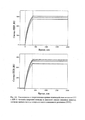
На Рис. 1А представлен график зависимости среднего значения оптической плотности при длине волны 492 нм от логарифма по основанию 2 концентрации ИЛ-11 человека в калибровочных растворах (в нг/мл) для определения концентрации ИЛ-11 человека в пробах методом твердофазного иммуноферментного анализа с использованием сэндвич-пары анти-ИЛ-11: захватывающих антител U13, адсорбированных на пластике в лунках 96-луночного микропланшета, и детектирующих антител L1-bio, конъюгированных с биотином.On Fig. Figure 1A is a plot of mean optical density at 492 nm versus
На Рис. 1Б представлен график зависимости среднего значения оптической плотности при длине волны 492 нм от логарифма по основанию 2 концентрации ИЛ-11 человека в калибровочных растворах (в нг/мл) для определения концентрации ИЛ-11 человека в пробах методом твердофазного иммуноферментного анализа с использованием сэндвич-пары анти-ИЛ-11: захватывающих антител L1, адсорбированных на пластике в лунках 96-луночного микропланшета, и детектирующих антител U13-bio, конъюгированных с биотином.On Fig. Figure 1B shows a plot of the average optical density at a wavelength of 492 nm versus the
На Рис. 2А представлен сенсограммы и теоретические кривые взаимодействия антитела U13 с ИЛ-11 человека (верхняя панель) и яванской макаки (нижняя панель), согласно данным метода поверхностного плазмонного резонанса (НИР). Лиганды ковалентно пришиты на поверхности сенсорного чипа за аминогруппы. Концентрации нанесения антител в рабочем буфере: 15, 12.9, 8.57 нМ. Анализ данных проведен в программе ProteOn Manager 3.0 в рамках схемы бивалентного аналита.On Fig. 2A shows sensorograms and theoretical curves of the interaction of antibody U13 with human IL-11 (upper panel) and cynomolgus macaque (lower panel) according to surface plasmon resonance (SPR) data. The ligands are covalently attached to the surface of the sensor chip by amino groups. Application concentrations of antibodies in working buffer: 15, 12.9, 8.57 nM. Data analysis was carried out in the ProteOn Manager 3.0 program within the framework of the bivalent analyte scheme.
На Рис. 2Б представлен сенсограммы и теоретические кривые взаимодействия антитела L1 с ИЛ-11 человека (верхняя панель) и яванской макаки (нижняя панель), согласно данным метода поверхностного плазмонного резонанса (ППР). Лиганды ковалентно пришиты на поверхности сенсорного чипа за аминогруппы. Концентрации нанесения антител в рабочем буфере: 10, 8.57, 2.58 нМ. Анализ данных проведен в программе ProteOn Manager 3.0 в рамках схемы бивалентного аналита.On Fig. 2B shows sensorograms and theoretical curves of the interaction of antibody L1 with human IL-11 (upper panel) and cynomolgus macaque (lower panel), according to the surface plasmon resonance (SPR) method. The ligands are covalently attached to the surface of the sensor chip by amino groups. Application concentrations of antibodies in working buffer: 10, 8.57, 2.58 nM. Data analysis was carried out in the ProteOn Manager 3.0 program within the framework of the bivalent analyte scheme.
Пример 1Example 1
Определение концентрации ИЛ-11 человека методом иммуноферментного анализа с использованием сэндвич-пары антител L1 и U13-bio, или U13 и L1-bioDetermination of the concentration of human IL-11 by enzyme immunoassay using a sandwich pair of antibodies L1 and U13-bio, or U13 and L1-bio
I. Сорбция захватывающих антител на твердой фазе В лунки 96-луночного планшета из полистерола помещают по 100 мкл фосфатно-солевого буфера рН 7,2-7,4 (1xPBS), содержащего 1-2 мкг антител L1 или U13. Закрытый крышкой планшет выдерживают в течение 16-18 часов при температуре +(5±3)°С или в течение 2-6 часов при температуре +(37±2)°С при постоянном встряхивании/перемешивании. Содержимое лунок удаляют без промывания или лунки промывают от несвязавшихся молекул антител 1-3 раза буфером 1xPBS, содержащим 0,05-0,1% полисорбата 20 (1xPBST), по 250-300 мкл.I. Sorption of capture antibodies on the solid phase Into the wells of a 96-well polystyrene plate, 100 µl of phosphate-buffered saline pH 7.2-7.4 (1xPBS) containing 1-2 µg of L1 or U13 antibodies are placed. The tablet closed with a lid is kept for 16-18 hours at a temperature of +(5±3)°C or for 2-6 hours at a temperature of +(37±2)°C with constant shaking/mixing. The contents of the wells are removed without washing or the wells are washed from unbound antibody molecules 1-3 times with 1xPBS buffer containing 0.05-0.1% polysorbate 20 (1xPBST), 250-300 μl.
II. Блокировка мест неспецифичного связывания на твердой фазе В лунки планшета помещают по 250 мкл буфера 1xPBST, содержащего 0,5-1,0% бычьего сывороточного альбумина или яичного овальбумина, или обезжиренного молока. Закрытый крышкой планшет выдерживают в течение 1-2 часов при температуре +(37±2)°С или при комнатной температуре при постоянном встряхивании/перемешивании.II. Blocking non-specific binding sites on the solid phase 250 μl of 1xPBST buffer containing 0.5-1.0% bovine serum albumin or egg ovalbumin, or skim milk is placed in the wells of the tablet. The tablet, closed with a lid, is kept for 1-2 hours at a temperature of + (37 ± 2) ° C or at room temperature with constant shaking / stirring.
Содержимое лунок удаляют и лунки промывают 1-3 раза буфером 1xPBST по 250-300 мкл.The contents of the wells are removed and the wells are washed 1-3 times with 1xPBST buffer, 250-300 µl.
III. Инкубация с антигеномIII. Incubation with antigen
В лунки планшета помещают по 100 мкл проб биологических жидкостей, либо двукратных разведений стандарта ИЛ-11 человека. Разведения антигена должны быть приготовлены на основе 1xPBST, содержащего 0,1% бычьего сывороточного альбумина или яичного овальбумина, или нежирного молока, поскольку полисорбат 20 снижает неспецифичное связывание белковых молекул друг с другом и с поверхностью планшета, а белок препятствует протеолизу антигена. Исследуемый раствор и стандартные разведения антигена вносят по 2-3 повторности, используя по две (три) лунки на каждое разведение белка. Закрытый крышкой планшет выдерживают в течение 0,5-2 часов при температуре +(37±2)°С или при комнатной температуре при постоянном встряхивании/перемешивании. Содержимое лунок удаляют и лунки промывают 3-5 раза буфером 1xPBST по 250-300 мкл.100 μl samples of biological fluids or two-fold dilutions of the human IL-11 standard are placed in the wells of the tablet. Antigen dilutions should be prepared with 1xPBST containing 0.1% bovine serum albumin or ovalbumin, or non-fat milk, since polysorbate 20 reduces non-specific binding of protein molecules to each other and to the surface of the plate, and the protein interferes with antigen proteolysis. The test solution and standard dilutions of the antigen are added in 2-3 replications, using two (three) wells for each dilution of the protein. The tablet closed with a lid is kept for 0.5-2 hours at a temperature of +(37±2)°C or at room temperature with constant shaking/mixing. The contents of the wells are removed and the wells are washed 3-5 times with 1xPBST buffer, 250-300 µl.
IV. Инкубация с детектирующими антителамиIV. Incubation with detection antibodies
В лунки планшета помещают по 100 мкл буфера 1xPBST, содержащего 0,1% бычьего сывороточного альбумина или яичного овальбумина, или нежирного молока, и не менее 2 мкг/мл антител U13-bio или L1-bio, конъюгированных с биотином (в паре с адсорбированными антителами L1 используют антитела U13-bio, а с антителами U13 - антитела L1-bio). Закрытый крышкой планшет выдерживают в течение 30-60 мин при температуре +(37±2)°С или при комнатной температуре при постоянном встряхивании/перемешивании. Содержимое лунок удаляют и лунки промывают 3-5 раз буфером 1xPBST по 250-300 мкл.100 μl of 1xPBST buffer containing 0.1% bovine serum albumin or egg ovalbumin, or non-fat milk, and at least 2 μg / ml of U13-bio or L1-bio antibodies conjugated with biotin (paired with adsorbed with L1 antibodies, U13-bio antibodies are used, and with U13 antibodies, L1-bio antibodies). The tablet, closed with a lid, is kept for 30-60 minutes at a temperature of +(37±2)°C or at room temperature with constant shaking/mixing. The contents of the wells are removed and the wells are washed 3-5 times with 1xPBST buffer, 250-300 µl.
V. Детектирование иммунных комплексов на твердой фазеV. Detection of immune complexes on the solid phase
В лунки планшета помещают по 100 мкл буфера 1xPBST, содержащего конъюгат стрептавидина с пероксидазой хрена (например, Thermo Fisher, кат. № N100). Оптимальная концентрация конъюгированного стрептавидина как правило указывается производителем данного реагента (обычно составляет не менее 1/4 000). Закрытый крышкой планшет выдерживают в течение 30-45 мин при температуре +(37±2)°С или при комнатной температуре при постоянном встряхивании/перемешивании. Содержимое лунок удаляют и лунки промывают 3-5 раз буфером 1xPBST по 250-300 мкл.Place 100 µl of 1xPBST buffer containing streptavidin-horseradish peroxidase conjugate (for example, Thermo Fisher, cat. No. N100) into the wells of the tablet. The optimal concentration of conjugated streptavidin is usually specified by the manufacturer of this reagent (usually at least 1/4,000). The tablet, closed with a lid, is kept for 30-45 minutes at a temperature of + (37 ± 2) ° C or at room temperature with constant shaking / stirring. The contents of the wells are removed and the wells are washed 3-5 times with 1xPBST buffer, 250-300 µl.
В лунки вносят по 100 мкл раствора субстрата пероксидазы хрена и инкубируют при комнатной температуре и постоянном перемешивании 5-15 мин в защищенном от света месте. В качестве субстрата пероксидазы хрена может быть использован OPD (o-phenylenediamine dihydrochloride), например, Thermo Fisher, кат. №34006, в буфере, содержащем пероксид водорода, например, Thermo Fisher, кат. №34062.100 μl of horseradish peroxidase substrate solution are added to the wells and incubated at room temperature with constant stirring for 5-15 minutes in a place protected from light. As a substrate for horseradish peroxidase, OPD (o-phenylenediamine dihydrochloride) can be used, for example, Thermo Fisher, cat. No. 34006, in a buffer containing hydrogen peroxide, such as Thermo Fisher, cat. No. 34062.
Проводят остановку реакции внесением в каждую лунку по 50 мкл 10% раствора серной кислоты. После этого сразу приступают к измерению оптической плотности раствора при соответствующей длине волны (для субстрата OPD 492 нм) с использованием планшетного спектрофотометра.The reaction is stopped by adding 50 µl of 10% sulfuric acid solution to each well. After that, immediately proceed to the measurement of the optical density of the solution at the appropriate wavelength (for the substrate OPD 492 nm) using a plate spectrophotometer.
Интерпретацию результатов проводят следующим образом. Строится график зависимости средних значений оптической плотности калибровочных растворов, рассчитанных по нескольким повторным измерениям, от логарифма концентрации ИЛ-11. Определяется формула области линейного участка (формула линии тренда), с помощью которой определяется численное значение концентрации ИЛ-11 в исследуемых пробах в зависимости от значений их оптической плотности.The interpretation of the results is carried out as follows. A graph is constructed of the dependence of the average values of the optical density of the calibration solutions, calculated from several repeated measurements, on the logarithm of the concentration of IL-11. The formula for the area of the linear section (trend line formula) is determined, with the help of which the numerical value of the concentration of IL-11 in the samples under study is determined depending on the values of their optical density.
Полученные результаты приведены на Рис. 1, где на Рис. 1А представлены результаты, полученные с использованием сэндвич-пары антител U13 и L1-bio, а на Рис. 1Б - с использованием сэндвич-пары антител L1 и U13-bio.The results obtained are shown in Fig. 1, where in Fig. 1A shows the results obtained using a sandwich pair of antibodies U13 and L1-bio, and Fig. 1B - using a sandwich pair of antibodies L1 and U13-bio.
Под «правильностью» методики понимают степень близости среднего значения концентрации ИЛ-11 в исследуемых пробах, полученного на основании серии результатов измерений, к принятому опорному значению. В качестве опорного значения используют действительное значение концентрации аналита (ИЛ-11) в пробе, рассчитанное с учетом концентрации аналита в исходном растворе, измеренной одним из фармакопейных методов определения концентрации белка (например, спектрофотометрическим анализом) или указанной в паспорте, и степени разведения исходного раствора в процессе приготовления пробы.The “correctness” of the technique is understood as the degree of closeness of the average value of the concentration of IL-11 in the studied samples, obtained on the basis of a series of measurement results, to the accepted reference value. As a reference value, the actual value of the concentration of the analyte (IL-11) in the sample is used, calculated taking into account the concentration of the analyte in the initial solution, measured by one of the pharmacopoeial methods for determining the protein concentration (for example, spectrophotometric analysis) or indicated in the passport, and the degree of dilution of the initial solution during sample preparation.
Среднее значение концентрации аналита ![]()
![]()
где Xi - результат i-го измерения концентрации,where X i is the result of the i-th concentration measurement,
n - количество измерений концентрации аналита в пробах одного и того же однородного образца.n is the number of analyte concentration measurements in samples of the same homogeneous sample.
Правильность (Т) выражают в процентах среднего значения концентрации ИЛ-11 в пробах от опорного значения, для расчета используют формулу:Correctness (T) is expressed as a percentage of the average concentration of IL-11 in samples from the reference value, the formula is used to calculate:
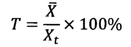
где Xt - действительное значение концентрации аналита (ИЛ-11) в пробах одного и того же однородного образца.where X t is the actual value of the concentration of the analyte (IL-11) in samples of the same homogeneous sample.
Истинное значение величины правильности методики иммуноферментного анализа, описанной в Примере 1, с вероятностью 95% лежит в промежутке от 80 до 115%) с использованием сэндвич-пары антител U13 и L1-bio, и в промежутке от 90 до 110%. с использованием сэндвич-пары антител L1 и U13-bio.The true value of the accuracy value of the ELISA method described in Example 1, with a probability of 95%, lies in the range from 80 to 115%) using a sandwich pair of antibodies U13 and L1-bio, and in the range from 90 to 110%. using a sandwich pair of antibodies L1 and U13-bio.
Для расчета доверительного интервала, в который попадают величины правильности, рассчитанные для концентраций аналита в рабочем диапазоне конкретной методики иммуноанализа, используют методы математической статистики, например, через критическое значение критерий Стьюдента.To calculate the confidence interval, which includes the correctness values calculated for analyte concentrations in the operating range of a particular immunoassay technique, methods of mathematical statistics are used, for example, through the critical value of Student's t-test.
Пример 2Example 2
Измерение величин кинетических и равновесных констант ассоциации/диссоциации комплекса ИЛ-11 человека или модельных животных с антителами U13 и L1 методом поверхностного плазмонного резонансаMeasurement of the kinetic and equilibrium association/dissociation constants of the human or model animal IL-11 complex with antibodies U13 and L1 by surface plasmon resonance
Измерение проводят при температуре +(25±1)°С, используя спектрометр Bio-Rad ProteOn™ XPR36. Для этого лиганд (ИЛ-11 человека или яванской макаки) иммобилизуют на поверхности чипа ProteOn™ GLH #176-5013 через аминогруппы. Контроль степени завершенности фазы иммобилизации лиганда на чипе проводят по кинетике сигнала ППР.The measurement is carried out at +(25±1)°C using a Bio-Rad ProteOn™ XPR36 spectrometer. To do this, the ligand (human IL-11 or cynomolgus macaque) is immobilized on the surface of the ProteOn™ GLH #176-5013 chip through amino groups. The degree of completion of the ligand immobilization phase on the chip is controlled by the kinetics of the SPR signal.
В перпендикулярном направлении наносят на чип раствор аналита (антител U13 или L1) в рабочем буфере (10 мМ HEPES, 150 мМ NaCl, 0,05% Tween 20, рН 7.4) в пяти различных концентрациях белка от 2,0 нМ до 20 нМ, а на одну дорожку - рабочий буфер в качестве контроля. Контроль степени завершенности фазы диссоциации комплекса проводят по величине снижения сигнала ППР.In a perpendicular direction, a solution of the analyte (antibodies U13 or L1) is applied to the chip in working buffer (10 mM HEPES, 150 mM NaCl, 0.05% Tween 20, pH 7.4) in five different protein concentrations from 2.0 nM to 20 nM, and on one track - a working buffer as a control. Control of the degree of completeness of the dissociation phase of the complex is carried out by the magnitude of the decrease in the PPR signal.
Поверхность чипа регенерируют после цикла связывания с аналитом пропусканием через чип 10 мМ глицин рН 2.0 в течение 300 сек.The chip surface is regenerated after the analyte binding cycle by passing 10 mM glycine pH 2.0 through the chip for 300 sec.
Расчет констант взаимодействия лиганда с аналитом проводят в стандартной программе Bio-Rad ProteOn Manager 3.0 с использованием простейшей схемы бивалентного аналита для не менее трех концентраций аналита.Calculation of the constants of interaction of the ligand with the analyte is carried out in the standard program Bio-Rad ProteOn Manager 3.0 using the simplest scheme of the bivalent analyte for at least three concentrations of the analyte.
Полученные результаты приведены на Рис. 2.The results obtained are shown in Fig. 2.
Пример 3Example 3
Получение полноразмерных антителObtaining full-length antibodies
Получение Fab-фрагментов антител проводили с использованием технологии на основе трех фаговых библиотек компании ООО «Антерикс», г. Пущино: Fab2h-library, Fab5h-library, Fab7h-library.Obtaining Fab-fragments of antibodies was carried out using the technology based on three phage libraries of LLC "Anteriks", Pushchino: Fab2h-library, Fab5h-library, Fab7h-library.
Технология фагового дисплея описана в литературе, например, Bazan, J., I. Calkosinski, and A. Gamian, Phage display-a powerful technique for immunotherapy: 1. Introduction and potential of therapeutic applications. Hum Vaccin Immunother, 2012. 8(12): p. 1817-28.Phage display technology is described in the literature, for example, Bazan, J., I. Calkosinski, and A. Gamian, Phage display-a powerful technique for immunotherapy: 1. Introduction and potential of therapeutic applications. Hum Vaccin Immunother, 2012. 8(12): p. 1817-28.
Из исходного разнообразия фаговых библиотек в ходе трех раундов селекции были отобраны фаговые частицы, содержащие на своей поверхности Fab-фрагменты антител, специфически связывающихся с ИЛ-11 человека и макаки. Использованы две схемы селекции, отличающиеся порядком презентации антигенов (ИЛ-11 человека - ИЛ-11 макаки - ИЛ-11 человека и ИЛ-11 макаки - ИЛ-11 человека - ИЛ-11 человека).From the initial variety of phage libraries during three rounds of selection, phage particles containing on their surface Fab-fragments of antibodies that specifically bind to human and macaque IL-11 were selected. Two selection schemes were used, differing in the order of antigen presentation (human IL-11 - macaque IL-11 - human IL-11 and macaque IL-11 - human IL-11 - human IL-11).
Фагмиды из бактериальных клеток, инфицированных отобранными вирионами, выделяли при помощи набора QIAprep Spin Miniprep kit (Qiagen, #27106) и секвенировали.Phagemids from bacterial cells infected with the selected virions were isolated using the QIAprep Spin Miniprep kit (Qiagen, #27106) and sequenced.
Последовательности, кодирующие домены VL, VH, CL и СН1 антител, из отобранных фагмид были переклонированы в вектор pLL4 (Addgene plasmid #107208) для экспрессии в клетках Е. coli штамма BL-21(DE3) Fab-фрагментов, несущих 8XHis метку на С-конце. Выделение и очистку фрагментов антител из клеточного лизата проводили с помощью металл-хелатной хроматографии. Скрининг Fab-фрагментов на предмет их специфичности к ИЛ-11 человека и макаки проводили при помощи ИФА.The sequences encoding the VL, VH, CL, and CH1 domains of antibodies from the selected phagemids were recloned into the pLL4 vector (Addgene plasmid #107208) for expression in E. coli strain BL-21(DE3) of Fab fragments bearing the 8XHis tag on C -end. Isolation and purification of antibody fragments from the cell lysate was performed using metal chelate chromatography. Screening of Fab fragments for their specificity to human and macaque IL-11 was performed using ELISA.
Для создания полноразмерных антител последовательность, кодирующую VH домен, клонировали в экспрессионный вектор рТТ5, содержащий последовательности, кодирующие константные области IgG1 человека (CH1, СН2, СН3), в то время, как последовательность, кодирующую VL домен, клонировали в экспрессионный вектор рТТ5, содержащий последовательности, кодирующие константные области k-легкой цепи человека (CL).To create full-length antibodies, the sequence encoding the VH domain was cloned into the pTT5 expression vector containing sequences encoding human IgG1 constant regions (CH1, CH2, CH3), while the sequence encoding the VL domain was cloned into the pTT5 expression vector containing sequences encoding human k-light chain (CL) constant regions.
Наработку антител проводили в системе транзиентной экспрессии в эукариотических клетках линии СНО-3Е7 (RRID:CVCL_JY74), адаптированных для продукции белков млекопитающих. Клетки СНО-3Е7 котрансфицировали полученными плазмидами с использованием полиэтиленимина (PEI, polyethylenimine). Выделение и очистку антител из культуральной среды проводили с помощью аффинной хроматографии с использованием сорбента с иммобилизованным белком A (HiTrap rProtein A FF, GE Healthcare Life Sciences), имеющим высокое сродство к иммуноглобулинам класса G. Полученные антитела были диализованы и стерилизованы фильтрацией через 0,22 мкм фильтр. Сродство полученных антител к ИЛ-11 было проверено с помощью ИФА и спектроскопии поверхностного плазмонного резонанса.The production of antibodies was carried out in a system of transient expression in eukaryotic cells of the CHO-3E7 line (RRID: CVCL_JY74) adapted for the production of mammalian proteins. CHO-3E7 cells were co-transfected with the obtained plasmids using polyethylenimine (PEI). Isolation and purification of antibodies from the culture medium was performed using affinity chromatography using a sorbent with immobilized protein A (HiTrap rProtein A FF, GE Healthcare Life Sciences), which has a high affinity for class G immunoglobulins. The obtained antibodies were dialyzed and sterilized by filtration through 0.22 micron filter. The affinity of the obtained antibodies to IL-11 was tested using ELISA and surface plasmon resonance spectroscopy.
Использованные в работе рутинные методы широко описаны в литературе (Barbas CF, Wagner J (1995). Synthetic Human Antibodies: Selecting and Evolving Functional Proteins. Methods. 8 (2): 94-103. doi: 1.0.1006/meth. 1995.9997; Barbas CF (1995). Synthetic human antibodies. Nat. Med. 1 (8): 837-839. doi:10.1038/nm0895-837. PMID 7585190; Wild D. The Immunoassay Handbook (4th edition). Theory and applications of ligand binding, ELISA and related techniques. 2013; Greenfield E.A., Antibodies. A laboratory manual. Second edition. 2014).The routine methods used in this work are widely described in the literature (Barbas CF, Wagner J (1995). Synthetic Human Antibodies: Selecting and Evolving Functional Proteins. Methods. 8 (2): 94-103. doi: 1.0.1006/meth. 1995.9997; Barbas CF (1995) Synthetic human antibodies Nat Med 1 (8): 837-839 doi:10.1038/nm0895-837 PMID 7585190 Wild D The Immunoassay Handbook (4th edition) Theory and applications of ligand binding, ELISA and related techniques 2013; Greenfield EA, Antibodies A laboratory manual Second edition 2014).
Последовательность тяжелой цепи HCDR1Heavy chain sequence of HCDR1
SEQ ID № 1:Phe Thr Phe Thr Gly Tyr Trp Met Asn Trp Val Arg; SEQ ID NO 1: Phe Thr Phe Thr Gly Tyr Trp Met Asn Trp Val Arg;
Последовательность тяжелой цепи HCDR2HCDR2 heavy chain sequence
SEQ ID № 2:Val Ser Ser Ile Ser Ser Tyr Gly Gly Gly Thr Tyr Tyr Ala Asp Ser; SEQ ID No. 2: Val Ser Ser Ile Ser Ser Tyr Gly Gly Gly Thr Tyr Tyr Ala Asp Ser;
Последовательность тяжелой цепи HCDR3HCDR3 heavy chain sequence
SEQ ID № 3:Cys Ala Arg Glu Ser Leu His Gln Leu Phe Asp Tyr Trp Gly Gln; SEQ ID NO: 3: Cys Ala Arg Glu Ser Leu His Gln Leu Phe Asp Tyr Trp Gly Gln;
Последовательность легкой цепи LCDR1LCDR1 light chain sequence
SEQ ID № 4:Gln Ser Val Ser Ser Ser; SEQ ID No. 4: Gln Ser Val Ser Ser Ser;
Последовательность легкой цепи LCDR2LCDR2 light chain sequence
SEQ ID № 5:Gly Ala Ser Ser Arg Ala Thr; SEQ ID NO: 5: Gly Ala Ser Ser Arg Ala Thr;
Последовательность легкой цепи LCDR3LCDR3 light chain sequence
SEQ ID № 6:Cys Gln Gln Ser Tyr Ser Tyr Ala Pro Ile Thr Phe Gly Gln;SEQ ID NO: 6: Cys Gln Gln Ser Tyr Ser Tyr Ala Pro Ile Thr Phe Gly Gln;
Последовательность тяжелой цепи HCDR1Heavy chain sequence of HCDR1
SEQ ID № 7:Phe Thr Phe Thr Asp Tyr Trp Met Asn Trp Val Arg;SEQ ID NO: 7: Phe Thr Phe Thr Asp Tyr Trp Met Asn Trp Val Arg;
Последовательность тяжелой цепи HCDR2HCDR2 heavy chain sequence
SEQ ID № 8:Val Ser Thr Ile Ala Ser Ser Asn Ser Tyr Thr Asp Tyr Ala Asp Ser; SEQ ID NO: 8: Val Ser Thr Ile Ala Ser Ser Asn Ser Tyr Thr Asp Tyr Ala Asp Ser;
Последовательность тяжелой цепи HCDR3HCDR3 heavy chain sequence
SEQ ID № 9:Cys Ala Arg Glu Ser Trp Ser Glu Gly Trp Trp Pro Ser Gly Phe Asp Tyr Trp Gly Gln;SEQ ID NO:Cys Ala Arg Glu Ser Trp Ser Glu Gly Trp Trp Pro Ser Gly Phe Asp Tyr Trp Gly Gln;
Последовательность легкой цепи LCDR3 LCDR3 light chain sequence
SEQ ID № 10:Cys Gln Gln Tyr Glu Thr Ser Pro Ile Thr Phe Gly Gln.SEQ ID NO: 10: Cys Gln Gln Tyr Glu Thr Ser Pro Ile Thr Phe Gly Gln.
Claims (20)
Priority Applications (1)
| Application Number | Priority Date | Filing Date | Title |
|---|---|---|---|
| RU2020111785A RU2763178C2 (en) | 2020-03-23 | 2020-03-23 | Antibodies for laboratory diagnostics of the concentration of interleukin-11 |
Applications Claiming Priority (1)
| Application Number | Priority Date | Filing Date | Title |
|---|---|---|---|
| RU2020111785A RU2763178C2 (en) | 2020-03-23 | 2020-03-23 | Antibodies for laboratory diagnostics of the concentration of interleukin-11 |
Related Parent Applications (1)
| Application Number | Title | Priority Date | Filing Date |
|---|---|---|---|
| RU2016151730A Previously-Filed-Application RU2016151730A (en) | 2016-12-28 | 2016-12-28 | Antibodies for laboratory diagnosis of interleukin-11 concentration |
Publications (3)
| Publication Number | Publication Date |
|---|---|
| RU2020111785A RU2020111785A (en) | 2021-09-23 |
| RU2020111785A3 RU2020111785A3 (en) | 2021-09-23 |
| RU2763178C2 true RU2763178C2 (en) | 2021-12-28 |
Family
ID=77836461
Family Applications (1)
| Application Number | Title | Priority Date | Filing Date |
|---|---|---|---|
| RU2020111785A RU2763178C2 (en) | 2020-03-23 | 2020-03-23 | Antibodies for laboratory diagnostics of the concentration of interleukin-11 |
Country Status (1)
| Country | Link |
|---|---|
| RU (1) | RU2763178C2 (en) |
Citations (3)
| Publication number | Priority date | Publication date | Assignee | Title |
|---|---|---|---|---|
| US6407218B1 (en) * | 1997-11-10 | 2002-06-18 | Cytimmune Sciences, Inc. | Method and compositions for enhancing immune response and for the production of in vitro mabs |
| US7094402B2 (en) * | 1994-12-22 | 2006-08-22 | Genetics Institute, Llc | Antibodies to human interleukin-11 receptor |
| RU2515108C2 (en) * | 2005-08-19 | 2014-05-10 | Эббви Инк | Immunoglobulin with double variable domains and its applications |
-
2020
- 2020-03-23 RU RU2020111785A patent/RU2763178C2/en active
Patent Citations (3)
| Publication number | Priority date | Publication date | Assignee | Title |
|---|---|---|---|---|
| US7094402B2 (en) * | 1994-12-22 | 2006-08-22 | Genetics Institute, Llc | Antibodies to human interleukin-11 receptor |
| US6407218B1 (en) * | 1997-11-10 | 2002-06-18 | Cytimmune Sciences, Inc. | Method and compositions for enhancing immune response and for the production of in vitro mabs |
| RU2515108C2 (en) * | 2005-08-19 | 2014-05-10 | Эббви Инк | Immunoglobulin with double variable domains and its applications |
Non-Patent Citations (1)
| Title |
|---|
| HAM et al., Critical role of interleukin-11 in isoflurane-mediated protection against ischemic acute kidney injury in mice, Anesthesiology, 2013, Vol. 119, N. 6, pp. 1389-1401. * |
Also Published As
| Publication number | Publication date |
|---|---|
| RU2020111785A (en) | 2021-09-23 |
| RU2020111785A3 (en) | 2021-09-23 |
Similar Documents
| Publication | Publication Date | Title |
|---|---|---|
| AU2010309931B2 (en) | Non-cross-reactive anti IgG antibodies | |
| CA2733497C (en) | Anti-hepcidin-25 selective antibodies and uses thereof | |
| CN110167967B (en) | Idiotypic antibodies directed against anti-PD-L1 antibodies and uses thereof | |
| US6998241B2 (en) | Antibody pair screening methods | |
| JP4964957B2 (en) | Anti-drug antibody assay | |
| KR20150041801A (en) | Complex-specific antibodies and antibody fragments and its use | |
| US9766251B2 (en) | Anti-human IgG1 antibody | |
| DK2383296T3 (en) | Antibody of the thyrotropin receptor and its applications | |
| CN109596839A (en) | People and peptide element fast quantitative measurement method for detecting and kit | |
| JP2020510841A (en) | Human erythroferon antibodies and uses thereof | |
| WO2010024271A1 (en) | Modified anti-heparin/pf4 complex antibody and hit antibody standard | |
| RU2763178C2 (en) | Antibodies for laboratory diagnostics of the concentration of interleukin-11 | |
| US7517663B2 (en) | Rabbit monoclonal antibody against Id1 protein | |
| CN112625129B (en) | Anti-human interleukin 23, kit comprising same and detection method thereof | |
| CN116143929B (en) | Antibody against recombinant human coagulation factor VIIa-Fc fusion protein and application thereof | |
| CN117264072B (en) | anti-SN 38 monoclonal antibody and application thereof | |
| EP4194054A1 (en) | Camelid antibodies for use in therapy and diagnosis | |
| CN112724253B (en) | Antibody of anti-human vault protein and application thereof | |
| CN111208307B (en) | Method for screening molecules having the same or different target protein binding to reference molecule | |
| CN108727493B (en) | anti-Stathmin monoclonal antibody and application thereof | |
| WO2023203101A1 (en) | Antibody against kynurenine | |
| CN116333139A (en) | anti-FDA 0128 antibody, preparation method and application thereof | |
| CN110997727A (en) | Method for determining anti-drug antibodies in miniature pig samples | |
| CN113366021A (en) | Glycosylated Apo J specific antibodies and uses thereof |