RU2743409C2 - Methods of treating conditions associated with masp-2 dependent complement activation - Google Patents
Methods of treating conditions associated with masp-2 dependent complement activation Download PDFInfo
- Publication number
- RU2743409C2 RU2743409C2 RU2018125514A RU2018125514A RU2743409C2 RU 2743409 C2 RU2743409 C2 RU 2743409C2 RU 2018125514 A RU2018125514 A RU 2018125514A RU 2018125514 A RU2018125514 A RU 2018125514A RU 2743409 C2 RU2743409 C2 RU 2743409C2
- Authority
- RU
- Russia
- Prior art keywords
- masp
- complement
- activation
- seq
- subject
- Prior art date
Links
Images


Classifications
-
- C—CHEMISTRY; METALLURGY
- C07—ORGANIC CHEMISTRY
- C07K—PEPTIDES
- C07K16/00—Immunoglobulins [IGs], e.g. monoclonal or polyclonal antibodies
- C07K16/40—Immunoglobulins [IGs], e.g. monoclonal or polyclonal antibodies against enzymes
-
- A—HUMAN NECESSITIES
- A61—MEDICAL OR VETERINARY SCIENCE; HYGIENE
- A61K—PREPARATIONS FOR MEDICAL, DENTAL OR TOILETRY PURPOSES
- A61K39/00—Medicinal preparations containing antigens or antibodies
- A61K39/395—Antibodies; Immunoglobulins; Immune serum, e.g. antilymphocytic serum
-
- A—HUMAN NECESSITIES
- A61—MEDICAL OR VETERINARY SCIENCE; HYGIENE
- A61K—PREPARATIONS FOR MEDICAL, DENTAL OR TOILETRY PURPOSES
- A61K39/00—Medicinal preparations containing antigens or antibodies
- A61K39/395—Antibodies; Immunoglobulins; Immune serum, e.g. antilymphocytic serum
- A61K39/39533—Antibodies; Immunoglobulins; Immune serum, e.g. antilymphocytic serum against materials from animals
- A61K39/3955—Antibodies; Immunoglobulins; Immune serum, e.g. antilymphocytic serum against materials from animals against proteinaceous materials, e.g. enzymes, hormones, lymphokines
-
- A—HUMAN NECESSITIES
- A61—MEDICAL OR VETERINARY SCIENCE; HYGIENE
- A61K—PREPARATIONS FOR MEDICAL, DENTAL OR TOILETRY PURPOSES
- A61K45/00—Medicinal preparations containing active ingredients not provided for in groups A61K31/00 - A61K41/00
- A61K45/06—Mixtures of active ingredients without chemical characterisation, e.g. antiphlogistics and cardiaca
-
- A—HUMAN NECESSITIES
- A61—MEDICAL OR VETERINARY SCIENCE; HYGIENE
- A61P—SPECIFIC THERAPEUTIC ACTIVITY OF CHEMICAL COMPOUNDS OR MEDICINAL PREPARATIONS
- A61P1/00—Drugs for disorders of the alimentary tract or the digestive system
-
- A—HUMAN NECESSITIES
- A61—MEDICAL OR VETERINARY SCIENCE; HYGIENE
- A61P—SPECIFIC THERAPEUTIC ACTIVITY OF CHEMICAL COMPOUNDS OR MEDICINAL PREPARATIONS
- A61P1/00—Drugs for disorders of the alimentary tract or the digestive system
- A61P1/08—Drugs for disorders of the alimentary tract or the digestive system for nausea, cinetosis or vertigo; Antiemetics
-
- A—HUMAN NECESSITIES
- A61—MEDICAL OR VETERINARY SCIENCE; HYGIENE
- A61P—SPECIFIC THERAPEUTIC ACTIVITY OF CHEMICAL COMPOUNDS OR MEDICINAL PREPARATIONS
- A61P1/00—Drugs for disorders of the alimentary tract or the digestive system
- A61P1/12—Antidiarrhoeals
-
- A—HUMAN NECESSITIES
- A61—MEDICAL OR VETERINARY SCIENCE; HYGIENE
- A61P—SPECIFIC THERAPEUTIC ACTIVITY OF CHEMICAL COMPOUNDS OR MEDICINAL PREPARATIONS
- A61P11/00—Drugs for disorders of the respiratory system
-
- A—HUMAN NECESSITIES
- A61—MEDICAL OR VETERINARY SCIENCE; HYGIENE
- A61P—SPECIFIC THERAPEUTIC ACTIVITY OF CHEMICAL COMPOUNDS OR MEDICINAL PREPARATIONS
- A61P13/00—Drugs for disorders of the urinary system
-
- A—HUMAN NECESSITIES
- A61—MEDICAL OR VETERINARY SCIENCE; HYGIENE
- A61P—SPECIFIC THERAPEUTIC ACTIVITY OF CHEMICAL COMPOUNDS OR MEDICINAL PREPARATIONS
- A61P13/00—Drugs for disorders of the urinary system
- A61P13/02—Drugs for disorders of the urinary system of urine or of the urinary tract, e.g. urine acidifiers
-
- A—HUMAN NECESSITIES
- A61—MEDICAL OR VETERINARY SCIENCE; HYGIENE
- A61P—SPECIFIC THERAPEUTIC ACTIVITY OF CHEMICAL COMPOUNDS OR MEDICINAL PREPARATIONS
- A61P13/00—Drugs for disorders of the urinary system
- A61P13/12—Drugs for disorders of the urinary system of the kidneys
-
- A—HUMAN NECESSITIES
- A61—MEDICAL OR VETERINARY SCIENCE; HYGIENE
- A61P—SPECIFIC THERAPEUTIC ACTIVITY OF CHEMICAL COMPOUNDS OR MEDICINAL PREPARATIONS
- A61P15/00—Drugs for genital or sexual disorders; Contraceptives
-
- A—HUMAN NECESSITIES
- A61—MEDICAL OR VETERINARY SCIENCE; HYGIENE
- A61P—SPECIFIC THERAPEUTIC ACTIVITY OF CHEMICAL COMPOUNDS OR MEDICINAL PREPARATIONS
- A61P17/00—Drugs for dermatological disorders
- A61P17/02—Drugs for dermatological disorders for treating wounds, ulcers, burns, scars, keloids, or the like
-
- A—HUMAN NECESSITIES
- A61—MEDICAL OR VETERINARY SCIENCE; HYGIENE
- A61P—SPECIFIC THERAPEUTIC ACTIVITY OF CHEMICAL COMPOUNDS OR MEDICINAL PREPARATIONS
- A61P17/00—Drugs for dermatological disorders
- A61P17/14—Drugs for dermatological disorders for baldness or alopecia
-
- A—HUMAN NECESSITIES
- A61—MEDICAL OR VETERINARY SCIENCE; HYGIENE
- A61P—SPECIFIC THERAPEUTIC ACTIVITY OF CHEMICAL COMPOUNDS OR MEDICINAL PREPARATIONS
- A61P17/00—Drugs for dermatological disorders
- A61P17/16—Emollients or protectives, e.g. against radiation
-
- A—HUMAN NECESSITIES
- A61—MEDICAL OR VETERINARY SCIENCE; HYGIENE
- A61P—SPECIFIC THERAPEUTIC ACTIVITY OF CHEMICAL COMPOUNDS OR MEDICINAL PREPARATIONS
- A61P25/00—Drugs for disorders of the nervous system
- A61P25/28—Drugs for disorders of the nervous system for treating neurodegenerative disorders of the central nervous system, e.g. nootropic agents, cognition enhancers, drugs for treating Alzheimer's disease or other forms of dementia
-
- A—HUMAN NECESSITIES
- A61—MEDICAL OR VETERINARY SCIENCE; HYGIENE
- A61P—SPECIFIC THERAPEUTIC ACTIVITY OF CHEMICAL COMPOUNDS OR MEDICINAL PREPARATIONS
- A61P25/00—Drugs for disorders of the nervous system
- A61P25/30—Drugs for disorders of the nervous system for treating abuse or dependence
-
- A—HUMAN NECESSITIES
- A61—MEDICAL OR VETERINARY SCIENCE; HYGIENE
- A61P—SPECIFIC THERAPEUTIC ACTIVITY OF CHEMICAL COMPOUNDS OR MEDICINAL PREPARATIONS
- A61P27/00—Drugs for disorders of the senses
- A61P27/02—Ophthalmic agents
- A61P27/06—Antiglaucoma agents or miotics
-
- A—HUMAN NECESSITIES
- A61—MEDICAL OR VETERINARY SCIENCE; HYGIENE
- A61P—SPECIFIC THERAPEUTIC ACTIVITY OF CHEMICAL COMPOUNDS OR MEDICINAL PREPARATIONS
- A61P29/00—Non-central analgesic, antipyretic or antiinflammatory agents, e.g. antirheumatic agents; Non-steroidal antiinflammatory drugs [NSAID]
-
- A—HUMAN NECESSITIES
- A61—MEDICAL OR VETERINARY SCIENCE; HYGIENE
- A61P—SPECIFIC THERAPEUTIC ACTIVITY OF CHEMICAL COMPOUNDS OR MEDICINAL PREPARATIONS
- A61P31/00—Antiinfectives, i.e. antibiotics, antiseptics, chemotherapeutics
-
- A—HUMAN NECESSITIES
- A61—MEDICAL OR VETERINARY SCIENCE; HYGIENE
- A61P—SPECIFIC THERAPEUTIC ACTIVITY OF CHEMICAL COMPOUNDS OR MEDICINAL PREPARATIONS
- A61P31/00—Antiinfectives, i.e. antibiotics, antiseptics, chemotherapeutics
- A61P31/04—Antibacterial agents
-
- A—HUMAN NECESSITIES
- A61—MEDICAL OR VETERINARY SCIENCE; HYGIENE
- A61P—SPECIFIC THERAPEUTIC ACTIVITY OF CHEMICAL COMPOUNDS OR MEDICINAL PREPARATIONS
- A61P35/00—Antineoplastic agents
-
- A—HUMAN NECESSITIES
- A61—MEDICAL OR VETERINARY SCIENCE; HYGIENE
- A61P—SPECIFIC THERAPEUTIC ACTIVITY OF CHEMICAL COMPOUNDS OR MEDICINAL PREPARATIONS
- A61P37/00—Drugs for immunological or allergic disorders
-
- A—HUMAN NECESSITIES
- A61—MEDICAL OR VETERINARY SCIENCE; HYGIENE
- A61P—SPECIFIC THERAPEUTIC ACTIVITY OF CHEMICAL COMPOUNDS OR MEDICINAL PREPARATIONS
- A61P37/00—Drugs for immunological or allergic disorders
- A61P37/02—Immunomodulators
-
- A—HUMAN NECESSITIES
- A61—MEDICAL OR VETERINARY SCIENCE; HYGIENE
- A61P—SPECIFIC THERAPEUTIC ACTIVITY OF CHEMICAL COMPOUNDS OR MEDICINAL PREPARATIONS
- A61P37/00—Drugs for immunological or allergic disorders
- A61P37/02—Immunomodulators
- A61P37/06—Immunosuppressants, e.g. drugs for graft rejection
-
- A—HUMAN NECESSITIES
- A61—MEDICAL OR VETERINARY SCIENCE; HYGIENE
- A61P—SPECIFIC THERAPEUTIC ACTIVITY OF CHEMICAL COMPOUNDS OR MEDICINAL PREPARATIONS
- A61P39/00—General protective or antinoxious agents
-
- A—HUMAN NECESSITIES
- A61—MEDICAL OR VETERINARY SCIENCE; HYGIENE
- A61P—SPECIFIC THERAPEUTIC ACTIVITY OF CHEMICAL COMPOUNDS OR MEDICINAL PREPARATIONS
- A61P39/00—General protective or antinoxious agents
- A61P39/02—Antidotes
-
- A—HUMAN NECESSITIES
- A61—MEDICAL OR VETERINARY SCIENCE; HYGIENE
- A61P—SPECIFIC THERAPEUTIC ACTIVITY OF CHEMICAL COMPOUNDS OR MEDICINAL PREPARATIONS
- A61P43/00—Drugs for specific purposes, not provided for in groups A61P1/00-A61P41/00
-
- A—HUMAN NECESSITIES
- A61—MEDICAL OR VETERINARY SCIENCE; HYGIENE
- A61P—SPECIFIC THERAPEUTIC ACTIVITY OF CHEMICAL COMPOUNDS OR MEDICINAL PREPARATIONS
- A61P7/00—Drugs for disorders of the blood or the extracellular fluid
-
- A—HUMAN NECESSITIES
- A61—MEDICAL OR VETERINARY SCIENCE; HYGIENE
- A61P—SPECIFIC THERAPEUTIC ACTIVITY OF CHEMICAL COMPOUNDS OR MEDICINAL PREPARATIONS
- A61P7/00—Drugs for disorders of the blood or the extracellular fluid
- A61P7/02—Antithrombotic agents; Anticoagulants; Platelet aggregation inhibitors
-
- A—HUMAN NECESSITIES
- A61—MEDICAL OR VETERINARY SCIENCE; HYGIENE
- A61P—SPECIFIC THERAPEUTIC ACTIVITY OF CHEMICAL COMPOUNDS OR MEDICINAL PREPARATIONS
- A61P7/00—Drugs for disorders of the blood or the extracellular fluid
- A61P7/04—Antihaemorrhagics; Procoagulants; Haemostatic agents; Antifibrinolytic agents
-
- A—HUMAN NECESSITIES
- A61—MEDICAL OR VETERINARY SCIENCE; HYGIENE
- A61P—SPECIFIC THERAPEUTIC ACTIVITY OF CHEMICAL COMPOUNDS OR MEDICINAL PREPARATIONS
- A61P7/00—Drugs for disorders of the blood or the extracellular fluid
- A61P7/06—Antianaemics
-
- A—HUMAN NECESSITIES
- A61—MEDICAL OR VETERINARY SCIENCE; HYGIENE
- A61P—SPECIFIC THERAPEUTIC ACTIVITY OF CHEMICAL COMPOUNDS OR MEDICINAL PREPARATIONS
- A61P9/00—Drugs for disorders of the cardiovascular system
-
- A—HUMAN NECESSITIES
- A61—MEDICAL OR VETERINARY SCIENCE; HYGIENE
- A61K—PREPARATIONS FOR MEDICAL, DENTAL OR TOILETRY PURPOSES
- A61K39/00—Medicinal preparations containing antigens or antibodies
- A61K2039/505—Medicinal preparations containing antigens or antibodies comprising antibodies
-
- A—HUMAN NECESSITIES
- A61—MEDICAL OR VETERINARY SCIENCE; HYGIENE
- A61K—PREPARATIONS FOR MEDICAL, DENTAL OR TOILETRY PURPOSES
- A61K39/00—Medicinal preparations containing antigens or antibodies
- A61K2039/54—Medicinal preparations containing antigens or antibodies characterised by the route of administration
-
- A—HUMAN NECESSITIES
- A61—MEDICAL OR VETERINARY SCIENCE; HYGIENE
- A61K—PREPARATIONS FOR MEDICAL, DENTAL OR TOILETRY PURPOSES
- A61K39/00—Medicinal preparations containing antigens or antibodies
- A61K2039/545—Medicinal preparations containing antigens or antibodies characterised by the dose, timing or administration schedule
-
- C—CHEMISTRY; METALLURGY
- C07—ORGANIC CHEMISTRY
- C07K—PEPTIDES
- C07K2317/00—Immunoglobulins specific features
- C07K2317/20—Immunoglobulins specific features characterized by taxonomic origin
- C07K2317/21—Immunoglobulins specific features characterized by taxonomic origin from primates, e.g. man
-
- C—CHEMISTRY; METALLURGY
- C07—ORGANIC CHEMISTRY
- C07K—PEPTIDES
- C07K2317/00—Immunoglobulins specific features
- C07K2317/20—Immunoglobulins specific features characterized by taxonomic origin
- C07K2317/24—Immunoglobulins specific features characterized by taxonomic origin containing regions, domains or residues from different species, e.g. chimeric, humanized or veneered
-
- C—CHEMISTRY; METALLURGY
- C07—ORGANIC CHEMISTRY
- C07K—PEPTIDES
- C07K2317/00—Immunoglobulins specific features
- C07K2317/30—Immunoglobulins specific features characterized by aspects of specificity or valency
- C07K2317/34—Identification of a linear epitope shorter than 20 amino acid residues or of a conformational epitope defined by amino acid residues
-
- C—CHEMISTRY; METALLURGY
- C07—ORGANIC CHEMISTRY
- C07K—PEPTIDES
- C07K2317/00—Immunoglobulins specific features
- C07K2317/50—Immunoglobulins specific features characterized by immunoglobulin fragments
- C07K2317/54—F(ab')2
-
- C—CHEMISTRY; METALLURGY
- C07—ORGANIC CHEMISTRY
- C07K—PEPTIDES
- C07K2317/00—Immunoglobulins specific features
- C07K2317/50—Immunoglobulins specific features characterized by immunoglobulin fragments
- C07K2317/55—Fab or Fab'
-
- C—CHEMISTRY; METALLURGY
- C07—ORGANIC CHEMISTRY
- C07K—PEPTIDES
- C07K2317/00—Immunoglobulins specific features
- C07K2317/70—Immunoglobulins specific features characterized by effect upon binding to a cell or to an antigen
- C07K2317/71—Decreased effector function due to an Fc-modification
-
- C—CHEMISTRY; METALLURGY
- C07—ORGANIC CHEMISTRY
- C07K—PEPTIDES
- C07K2317/00—Immunoglobulins specific features
- C07K2317/70—Immunoglobulins specific features characterized by effect upon binding to a cell or to an antigen
- C07K2317/76—Antagonist effect on antigen, e.g. neutralization or inhibition of binding
-
- C—CHEMISTRY; METALLURGY
- C07—ORGANIC CHEMISTRY
- C07K—PEPTIDES
- C07K2317/00—Immunoglobulins specific features
- C07K2317/90—Immunoglobulins specific features characterized by (pharmaco)kinetic aspects or by stability of the immunoglobulin
- C07K2317/92—Affinity (KD), association rate (Ka), dissociation rate (Kd) or EC50 value
-
- C—CHEMISTRY; METALLURGY
- C07—ORGANIC CHEMISTRY
- C07K—PEPTIDES
- C07K2317/00—Immunoglobulins specific features
- C07K2317/90—Immunoglobulins specific features characterized by (pharmaco)kinetic aspects or by stability of the immunoglobulin
- C07K2317/94—Stability, e.g. half-life, pH, temperature or enzyme-resistance
Abstract
Description
ПЕРЕКРЕСТНЫЕ ССЫЛКИ НА СООТВЕТСТВУЮЩИЕ ПРИЛОЖЕНИЯCROSS-REFERING TO RELEVANT APPLICATIONS
Эта заявка заявляет приоритет предварительной заявки № 61/473698, поданной 8 апреля 2011 года, которая включена в настоящий документ посредством ссылки в полном объеме.This application claims priority from provisional application No. 61/473698, filed April 8, 2011, which is incorporated herein by reference in its entirety.
ЗАЯВЛЕНИЕ, КАСАЮЩЕЕСЯ ПЕРЕЧНЯ ПОСЛЕДОВАТЕЛЬНОСТЕЙSTATEMENT REGARDING SEQUENCE LIST
Перечень последовательностей, связанный с этой заявкой, предоставляется в текстовом формате вместо бумажной копии и настоящим включен посредством ссылки в эту спецификацию. Имя текстового файла, содержащего перечень последовательностей, представляет собой MP_1_0126_US2_SequenceListingasFiled.txt. Текстовый файл представляет собой файл размером 110 кB; был создан 30 марта 2012 года; и представляется с помощью EFS-Web с подачей спецификации.The sequence listing associated with this application is provided in text format instead of paper copy and is hereby incorporated by reference into this specification. The name of the text file containing the sequence listing is MP_1_0126_US2_SequenceListingasFiled.txt. The text file is a 110 kB file; was created on March 30, 2012; and submitted via EFS-Web with specification submission.
ПРЕДПОСЫЛКИ ИЗОБРЕТЕНИЯBACKGROUND OF THE INVENTION
Система комплемента обеспечивает механизм раннего действия для инициирования, усиления и регулирования иммунного ответа на микробную инфекцию и другие острые поражения (M.К. Liszewski and J. P. Atkinson, 1993, в Fundamental Immunology, Third Edition, edited by W.E. Paul, Raven Press, Ltd., Нью-Йорк) в организме человека и других позвоночных. Несмотря на то, что активация комплемента обеспечивает чрезвычайно важную первую линию обороны против возможных возбудителей заболеваний, активности комплемента, которые стимулируют защитный иммунный ответ, также могут представлять потенциальную угрозу для хозяина (K.R. Kalli, et al., Springer Semin. Imrnunopathol. 15:417-431, 1994; B.P. Morgan, Eur. J. Clinical Investig. 24: 219-228, 1994). Например, протеолитические продукты C3 и C5 привлекают и активируют нейтрофилы. Активированные нейтрофилы, несмотря на то, что являются совершенно необходимыми для защиты организма, действуют неизбирательно при своем высвобождении деструктивных ферментов, и могут вызывать повреждения органов. Кроме того, активация комплемента может привести к депозиции литических компонентов комплемента на близлежащих клетках-хозяевах, как и на микробных мишенях, приводя в результате к лизису клеток-хозяев.The complement system provides an early-acting mechanism for initiating, enhancing and regulating the immune response to microbial infection and other acute lesions (M.K. Liszewski and JP Atkinson, 1993, in Fundamental Immunology , Third Edition, edited by WE Paul, Raven Press, Ltd. , New York) in humans and other vertebrates. Although complement activation provides an extremely important first line of defense against potential pathogens, complement activities that stimulate a protective immune response may also pose a potential host threat (KR Kalli, et al., Springer Semin. Imrnunopathol . 15: 417 -431, 1994; BP Morgan, Eur. J. Clinical Investig. 24: 219-228, 1994). For example, proteolytic products C3 and C5 attract and activate neutrophils. Activated neutrophils, while absolutely essential for the body's defense, act indiscriminately in the release of destructive enzymes and can cause organ damage. In addition, activation of complement can lead to the deposition of lytic components of complement on nearby host cells, as well as on microbial targets, resulting in lysis of host cells.
Система комплемента также вовлечена в патогенез многих острых и хронических заболеваний, в том числе: инфаркт миокарда, инсульт, синдром острой дыхательной недостаточности (ARDS), реперфузионное повреждение, септический шок, подтекание капилляров, следующее за термическими ожогами, послекардиопульмональное обходное воспаление, отторжение трансплантата, ревматоидный артрит, рассеянный склероз, миастения gravis и болезнь Альцгеймера. Почти при всех этих состояниях комплемент является не причиной, а одним из нескольких факторов, вовлеченных в патогенез. Тем не менее, активация комплемента может являться основным патологическим механизмом и представляет эффективный пункт для клинического управления при многих из этих болезненных состояний. Растущее понимание важности комплемент-опосредованного повреждения тканей при различных болезненных состояниях подчеркивает необходимость в эффективных комплемент-ингибирующих лекарственных препаратах. На сегодняшний день, экулизумаб (Solaris®), антитело против C5, является единственным комплемент-направленным лекарственным препаратом, который был одобрен для применения на людях. Кроме того, C5 представляет собой одну из нескольких эффекторных молекул, расположенных «ниже по течению» в системе комплемента, и блокада C5 не ингибирует активацию системы комплемента. Поэтому, ингибитор начальных стадий инициации активации комплемента мог бы иметь существенные преимущества по сравнению с ингибитором комплемента, действующим «ниже по течению».The complement system is also implicated in the pathogenesis of many acute and chronic diseases, including: myocardial infarction, stroke, acute respiratory distress syndrome (ARDS), reperfusion injury, septic shock, capillary leakage following thermal burns, postcardiopulmonary bypass inflammation, graft rejection, rheumatoid arthritis, multiple sclerosis, myasthenia gravis and Alzheimer's disease. In almost all of these conditions, complement is not the cause, but one of several factors involved in pathogenesis. Nonetheless, complement activation may be a major pathological mechanism and is an effective clinical management point for many of these disease states. The growing understanding of the importance of complement-mediated tissue damage in a variety of disease states underscores the need for effective complement-inhibiting drugs. To date, eculizumab (Solaris®), an anti-C5 antibody, is the only complement-targeted drug that has been approved for use in humans. In addition, C5 is one of several downstream effector molecules in the complement system, and C5 blockade does not inhibit the activation of the complement system. Therefore, an inhibitor of the initial stages of initiation of complement activation could have significant advantages over a downstream complement inhibitor.
В настоящее время общепризнано, что система комплемента может быть активирована при помощи трех различных путей: классического пути, лектинового пути и альтернативного пути. Классический путь обычно инициируется комплексом, который состоит из антител организма-хозяина, связанных с инородной частицей (т.е. антигеном) и, таким образом, требует предварительного воздействия антигена для формирования специфического гуморального ответа. Поскольку активация классического пути зависит от предварительного адаптивного иммунного ответа организма-хозяина, классический путь представляет собой часть приобретенной иммунной системы. В противоположность этому, как лектиновый, так и альтернативные пути являются независимыми от адаптивного иммунитета и являются частью естественной иммунной системы.It is now generally accepted that the complement system can be activated via three different pathways: the classical pathway, the lectin pathway, and the alternative pathway. The classical pathway is usually initiated by a complex that consists of host antibodies bound to a foreign particle (ie, antigen) and thus requires prior exposure to the antigen to generate a specific humoral response. Since the activation of the classical pathway depends on the preliminary adaptive immune response of the host organism, the classical pathway is part of the acquired immune system. In contrast, both lectin and alternative pathways are independent of adaptive immunity and are part of the natural immune system.
Активация системы комплемента приводит к последовательной активации зимогенов сериновой протеазы. Первой стадией при активации классического пути является связывание специфической узнающей молекулы, C1q, с антигенсвязывающими молекулами IgG и IgM. C1q связывается с проферментами сериновой протеазы C1r и C1s в виде комплекса, называемого C1. После связывания C1q с иммунным комплексом, происходит автопротеолитическое расщепление участка Arg-Ile в C1r, а затем C1r-опосредованное расщепление и активация С1s, который, таким образом, приобретает способность расщеплять C4 и C2. C4 расщепляется на два фрагмента, обозначенные как C4a и C4b, и, аналогично, C2 расщепляется на C2a и C2b. Фрагменты C4b способны образовывать ковалентные связи с соседними гидроксильными или аминными группами и образовывать C3-конвертазу (C4b2a) через нековалентное взаимодействие с C2a-фрагментом активированного C2. Конвертаза C3 (C4b2a) активирует С3 путем протеолитического расщепления на субкомпоненты C3a и C3b, приводя к образованию С5-конвертазы (C4b2a3b), который, путем расщепления C5, приводит к образованию мембраноатакующего комплекса (C5b в сочетании с С6, С7, С8 и C-9, также известного как «MAC»), который может нарушать клеточные мембраны, приводя к клеточному лизису. Активированные формы C3 и C4 (C3b и C4b) ковалентно размещаются на внешних поверхностях мишеней, которые распознаются рецепторами комплемента на многочисленных фагоцитах.The activation of the complement system leads to the sequential activation of serine protease zymogens. The first step in the activation of the classical pathway is the binding of a specific recognition molecule, C1q, to antigen-binding IgG and IgM molecules. C1q binds to the serine protease proenzymes C1r and C1s in a complex called C1. After binding of C1q to the immune complex, auto-proteolytic cleavage of the Arg-Ile site in C1r occurs, followed by C1r-mediated cleavage and activation of C1s, which thus acquires the ability to cleave C4 and C2. C4 is cleaved into two fragments designated C4a and C4b, and, similarly, C2 is cleaved into C2a and C2b. C4b fragments are able to form covalent bonds with adjacent hydroxyl or amine groups and form a C3 convertase (C4b2a) through non-covalent interaction with the C2a fragment of activated C2. C3 convertase (C4b2a) activates C3 by proteolytic cleavage into subcomponents C3a and C3b, leading to the formation of C5 convertase (C4b2a3b), which, by cleaving C5, leads to the formation of a membrane-attacking complex (C5b in combination with C6, C7, C8 and C- 9, also known as "MAC"), which can disrupt cell membranes, leading to cell lysis. The activated forms C3 and C4 (C3b and C4b) are covalently located on the outer surfaces of targets, which are recognized by complement receptors on numerous phagocytes.
Независимо, первая стадия в активации системы комплемента через лектиновый путь также представляет собой связывание специфических распознающих молекул, которое следует за активацией ассоциированных проферментов сериновой протеазы. Однако, вместо связывания иммунных комплексов при помощи C1q, узнающие молекулы в лектиновом пути включают группу углевод-связывающих белков (маннан-связывающий лектин (MBL), H-фиколин, М-фиколин, L-фиколин и C-типа лектин CL-11), в совокупности именуемые лектинами. Смотри J. Lu et al., Biochim. Biophys. Acta 1572:387-400, (2002); Holmskov et al., Annu. Rev. Immunol. 21:547-578 (2003); Teh et al., Immunology 101:225-232 (2000)). Смотри также J. Luеt et al., Biochim Biophys Acta 1572:387-400 (2002); Holmskov et al., Annu Rev Immunol 21:547-578 (2003); Teh et al., Immunology 101:225-232 (2000); Hansen et al., J. Immunol 185(10):6096-6104 (2010).Independently, the first step in the activation of the complement system via the lectin pathway is also the binding of specific recognition molecules, which follows the activation of the associated serine protease zoenzymes. However, instead of binding immune complexes by C1q, recognition molecules in the lectin pathway include a group of carbohydrate-binding proteins (mannan-binding lectin (MBL), H-ficolin, M-ficolin, L-ficolin, and C-type lectin CL-11) , collectively referred to as lectins. See J. Lu et al., Biochim. Biophys. Acta 1572: 387-400, (2002); Holmskov et al., Annu. Rev. Immunol . 21: 547-578 (2003); Teh et al., Immunology 101: 225-232 (2000)). See also J. Luet et al., Biochim Biophys Acta 1572: 387-400 (2002); Holmskov et al., Annu Rev Immunol 21: 547-578 (2003); Teh et al., Immunology 101: 225-232 (2000); Hansen et al., J. Immunol 185 (10): 6096-6104 (2010).
Ikeda et al. впервые продемонстрировали, что MBL, подобно C1q, может активировать систему комплемента через связывание с эритроцитами, покрытыми маннаном дрожжей, C4-зависимым образом (Ikeda et al., J. Biol. Chem. 2.52:7451-7454, (3987)). MBL, член белкового семейства коллектинов, является кальций-зависимым лектином, который связывает углеводы с 3- и 4-гидроксильными группами, ориентированными в экваториальной плоскости пиранозного кольца. Выступающими лигандами для MBL, таким образом, являются D-манноза и N-ацетил-D-глюкозамин, в то время как углеводы, не удовлетворяющие этому стерическому требованию, обладают недетектируемой для MBL аффинностью (Wets et al., Nature 360: 127-134, (1992)). Взаимодействие между MBL и моновалентными сахарами является крайне слабым, с константами диссоциации, как правило, в одноразрядном миллимолярном диапазоне. MBL достигает плотного, специфического связывания с гликановыми лигандами за счет авидности, т.е. за счет одновременного взаимодействия с несколькими моносахаридными остатками, расположенными в непосредственной близости друг от друга (Lee et al., Archiv. Biochem. Biophys. 299: 129-136, (3992)). MBL распознает углеводные структуры, которые обычно декорируют микроорганизмы, такие как бактерии, дрожжи, паразиты и некоторые вирусы. И наоборот, MBL не узнает D-галактозу и сиаловую кислоту, предпоследние и последние сахара, которые обычно декорируют «зрелые» сложные гликоконъюгаты, присутствующие на гликопротеинах плазматических и клеточных поверхностей клеток млекопитающих. Считается, что эта специфичность связывания стимулирует узнавание «чужеродных» поверхностей и помогает в защите от «самоактивации». Однако MBL не связывается с высокой аффинностью с кластерами высокоманнозных «прекурсорных» гликанов на N-связанных гликопротеинах и гликолипидах, изолированных в эндоплазматическом ретикулуме и комплексе Гольджи клеток млекопитающих (Maynard et al., J. Biol. Chem 257:3788-3794, (1982)). Поэтому, поврежденные клетки являются потенциальными мишенями для лектинового пути активации через MBL-связывание.Ikeda et al. first demonstrated that MBL, like C1q, can activate the complement system through binding to yeast mannan coated erythrocytes in a C4-dependent manner (Ikeda et al., J. Biol. Chem . 2.52: 7451-7454, (3987)). MBL, a member of the colletin protein family, is a calcium-dependent lectin that binds carbohydrates to 3- and 4-hydroxyl groups oriented in the equatorial plane of the pyranose ring. Ligands for MBL are thus D-mannose and N-acetyl-D-glucosamine, while carbohydrates that do not meet this steric requirement have an affinity undetectable for MBL (Wets et al., Nature 360: 127-134 , (1992)). The interaction between MBL and monovalent sugars is extremely weak, with dissociation constants typically in the one-digit millimolar range. MBL achieves tight, specific binding to glycan ligands through avidity, i.e. due to the simultaneous interaction with several monosaccharide residues located in close proximity to each other (Lee et al., Archiv. Biochem. Biophys. 299: 129-136, (3992)). MBL recognizes carbohydrate structures that microorganisms usually decorate, such as bacteria, yeast, parasites, and some viruses. Conversely, MBL does not recognize D-galactose and sialic acid, the penultimate and last sugars that usually decorate the "mature" complex glycoconjugates present on the glycoproteins of the plasma and cell surfaces of mammalian cells. It is believed that this binding specificity stimulates the recognition of "foreign" surfaces and helps in protecting against "self-activation". However, MBL does not bind with high affinity to clusters of high-mannose “precursor” glycans on N-linked glycoproteins and glycolipids isolated in the endoplasmic reticulum and Golgi complex of mammalian cells (Maynard et al., J. Biol. Chem 257: 3788-3794, (1982 )). Therefore, damaged cells are potential targets for the lectin activation pathway through MBL binding.
Фиколины обладают типом лектинового домена, отличного от такового для MBL, который называется фибриноген-подобный домен. Фиколины связывают сахарные остатки Ca++-независимым образом. В организме человека было выявлено три вида фиколинов (L-фиколин, М-фиколини и H-фиколин). Два сывороточных фиколина, L-фиколин и H-фиколин, совместно обладают специфичностью к N-ацетил-D-глюкозамину; при этом, H-фиколин также связывает N-ацетил-D-галактозамин. Различие в сахароспецифичности L-фиколина, H-фиколина, CL-11 и MBL означает, что разные лектины могут быть комплементарны и ориентированы на разные, хотя и перекрывающиеся, гликоконъюгаты. Эта концепция поддерживается недавним сообщением о том, что из известных лектинов лектинового пути лишь L-фиколин специфически связывается с липотейхоевой кислотой, глюкоконьюгатом клеточной стенки, обнаруженным во всех грамположительных бактериях (Lynch et al., J. Immunol. 172: 1198-1202, (2004)). Коллектины (т.е., MBL) и фиколины не обладают значительным сходством аминокислотных последовательностей. Однако две группы белков обладают похожей доменной организацией и, подобно C1q, собраны в олигомерные структуры, которые максимально увеличивают возможности многосайтного связывания.Ficolins have a different type of lectin domain from that of MBL, called a fibrinogen-like domain. Ficolins bind sugar residues in a Ca ++ -independent manner. Three types of ficolins have been identified in the human body (L-ficolin, M-ficolini and H-ficolin). Two serum ficolins, L-ficolin and H-ficolin, jointly have specificity for N-acetyl-D-glucosamine; however, H-ficolin also binds N-acetyl-D-galactosamine. The difference in sugar specificity of L-ficolin, H-ficolin, CL-11 and MBL means that different lectins can be complementary and target different, albeit overlapping, glycoconjugates. This concept is supported by the recent report that of the known lectins in the lectin pathway, only L-ficolin binds specifically to lipoteichoic acid, a cell wall glucoconjugate found in all gram-positive bacteria (Lynch et al., J. Immunol . 172: 1198-1202, ( 2004)). Collecins (i.e., MBL) and ficolins do not share significant amino acid sequence similarities. However, the two groups of proteins have a similar domain organization and, like C1q, are assembled into oligomeric structures that maximize the possibilities of multisite binding.
Концентрации MBL в сыворотке сильно варьируется в популяции здоровых людей, и это генетически контролируется полиморфизмом/мутациями как в промоторной, так и в кодирующей областях гена MBL. Как для белка острой фазы, экспрессия MBL также увеличивается во время воспаления. L-фиколин присутствует в сыворотке в концентрациях, аналогичных таковым для MBL. Таким образом, L-фиколиновая ветвь лектинового пути по силе сопоставима с плечом MBL. MBL и фиколины также могут функционировать в качестве опсонинов, которые позволяют фагоцитам нацеливаться на поверхности, декорированные MBL и фиколином (смотри Jack et al., J Leukoc Biol., 77(3):328-36 (2004), Matsushita and Fujita, Immunohiology, 205(4-5):490-7 (2002), Aoyagi et al., J. Immunol, 174(1):418-25 (2005). Эта опсонизация требует взаимодействия этих белков с фагоцитарными рецепторами (Kuhlman et al., J. Exp. Med. 169: 1733, (1989); Matsushita et al., J. Biol. Chem. 271:2448-54, (1996)), тождественность которых не была установлена.Serum MBL concentrations vary greatly in the healthy population and this is genetically controlled by polymorphisms / mutations in both the promoter and coding regions of the MBL gene. As for the acute phase protein, MBL expression also increases during inflammation. L-ficolin is present in serum at concentrations similar to those for MBL. Thus, the L-ficolin branch of the lectin pathway is comparable in strength to the MBL arm. MBL and ficolins can also function as opsonins that allow phagocytes to target surfaces decorated with MBL and ficolin (see Jack et al., J Leukoc Biol ., 77 (3): 328-36 (2004), Matsushita and Fujita, Immunohiology , 205 (4-5): 490-7 (2002), Aoyagi et al., J. Immunol , 174 (1): 418-25 (2005) This opsonization requires the interaction of these proteins with phagocytic receptors (Kuhlman et al. , J. Exp. Med . 169: 1733, (1989); Matsushita et al., J. Biol. Chem . 271: 2448-54, (1996)), the identity of which has not been established.
MBL человека формирует специфическое и высокоаффинное взаимодействие через свой коллагено-подобный домен с уникальными C1r/C1s-подобными сериновыми протеиназами, называемыми MBL-связанными сериновыми протеазами (MASP). На сегодняшний день, было описано три MASP. Во-первых, один фермент «MASP» был выделен и охарактеризован в качестве фермента, ответственного за инициирование каскада комплемента (т.е., расщепление C2 и C4) (Matsushita et al., J. Exp. Med. 176(6): 1497-1502 (1992); Ji et al., J. Immunol 750:571-578, (1993)). Впоследствии было установлено, что активность МASP представляет собой, по сути, смесь двух протеаз: MASP-1 и MASP-2 (Tiel et al., Nature 386:506-510, (1997)). Однако было показано, что комплекс MBL-MASP-2 сам по себе достаточен для активации комплемента (Vorup-Jensen et al., J. Immunol 165:2093-2100, (2000)). Кроме того, только MASP-2 расщеплял C2 и C4 с высокой эффективностью (Ambrus et al., J. Immunol, 770: 1374-1382, (2003)). Поэтому MASP-2 представляет собой протеазу, ответственную за активирование C2 и C4 для образования C3-конвертазы, C4b2a. В этом заключается существенное отличие от C1-комплекса классического пути, где скоординированное действие двух специфических сериновых протеаз (C1r и C1s) приводит к активации системы комплемента. Кроме того, была изолирована третья новая протеаза, MASP-3 (Dahl, М.Р., et al., Immunity 75: 127-35, 2001). MASP-1 и MASP-3 представляют собой продукты альтернативного сплайсинга одного и того же гена.Human MBL forms a specific and high affinity interaction through its collagen-like domain with unique C1r / C1s-like serine proteinases called MBL-linked serine proteases (MASP). To date, three MASPs have been described. First, one MASP enzyme has been isolated and characterized as the enzyme responsible for initiating the complement cascade (i.e., cleavage of C2 and C4) (Matsushita et al., J. Exp. Med. 176 (6): 1497-1502 (1992); Ji et al., J. Immunol 750: 571-578, (1993)). Subsequently, it was found that the activity of MASP is, in fact, a mixture of two proteases: MASP-1 and MASP-2 (Tiel et al., Nature 386: 506-510, (1997)). However, it has been shown that the MBL-MASP-2 complex alone is sufficient for complement activation (Vorup-Jensen et al., J. Immunol 165: 2093-2100, (2000)). In addition, only MASP-2 cleaves C2 and C4 with high efficiency (Ambrus et al., J. Immunol, 770: 1374-1382, (2003)). Therefore, MASP-2 is the protease responsible for activating C2 and C4 to form the C3 convertase, C4b2a. This is a significant difference from the C1 complex of the classical pathway, where the coordinated action of two specific serine proteases (C1r and C1s) leads to the activation of the complement system. In addition, a third novel protease, MASP-3, has been isolated (Dahl, M. P., et al., Immunity 75: 127-35, 2001). MASP-1 and MASP-3 are alternative splicing products of the same gene.
MASP разделяет идентичные доменные организации с таковыми C1r и C1s, ферментативными компонентами C1-комплекса (Sim et al., Biochem. Soc. Trans. 28:545, (2000)). Эти домены включают N-концевой домен C1r/C1s/VEGF морского ежа/морфогенетического белка костей (CUB), домен, подобный эпидермальному фактору роста, второй CUB-домен, домены тандема комплемент-контролирующих белков и домен сериновой протеазы. Как и в случае C1-протеазы, активация MASP-2 происходит через расщепление связи Arg-Ile, прилегающей к домену сериновой протеазы, которая расщепляет фермент на дисульфидсвязанные цепи A и B, последняя состоит из домена сериновой протеазы.MASP shares identical domain organizations with those of C1r and C1s, the enzymatic components of the C1 complex (Sim et al., Biochem. Soc. Trans . 28: 545, (2000)). These domains include the sea urchin C1r / C1s / VEGF / bone morphogenetic protein (CUB) N-terminal domain, an epidermal growth factor like domain, a second CUB domain, tandem domains of complement control proteins, and a serine protease domain. As in the case of the C1-protease, the activation of MASP-2 occurs through cleavage of the Arg-Ile bond adjacent to the serine protease domain, which cleaves the enzyme into disulfide-linked A and B chains, the latter consisting of the serine protease domain.
MBL также может связываться с альтернативно-сплайсированной формой MASP-2, известной как MBL-ассоциированный белок массой 19 кДа (MAp19) или небольшой MBL-ассоциированный белок (sMAP), в которой отсутствует каталитическая активность MASP-2 (Stover, J. Immunol, 752:3481-90, (1999); Takahashi et al., Int. Immunol. 11:859-863, (1999)). Map19 включает в себя первые два домена MASP-2, следующие после дополнительной последовательности из четырех уникальных аминокислот. Функция Map 19 непонятна (Degn et al., J. Immunol. Methods, 2011). Гены MASP-1 и MASP-2 расположены на 3 и 1 хромосомах человека, соответственно (Schwaeble et al., Immunobiology 205:455-466, (2002)).MBL can also bind to an alternative spliced form of MASP-2, known as 19 kDa MBL-associated protein (MAp19) or small MBL-associated protein (sMAP), which lacks the catalytic activity of MASP-2 (Stover, J. Immunol, 752: 3481-90, (1999); Takahashi et al., Int. Immunol . 11: 859-863, (1999)). Map19 includes the first two domains of MASP-2, following an additional sequence of four unique amino acids. The
Несколько линий доказательства свидетельствуют о том, что существуют различные комплексы MBL-MASP, и большая часть MASP в сыворотке крови не образует комплексы с MBL (Thiel, et al., J. Immunol. 165:878-887, (2000)). Как H-, так и L-фиколин связывается со всеми MASP и активирует лектиновый путь комплемента, как это делает MBL (Dahl et al., Immunity 15:127-35, (2001); Matsushita et al., J. Immunol 168: 3502-3506, (2002)). Как лектиновый, так и классический пути образуют общую C3-конвертазу (C4b2a), и два пути сходятся на этой стадии.Several lines of evidence suggest that there are different MBL-MASP complexes and most of the MASP in serum does not complex with MBL (Thiel, et al., J. Immunol. 165: 878-887, (2000)). Both H- and L-ficolin bind to all MASPs and activate the lectin complement pathway, as MBL does (Dahl et al., Immunity 15: 127-35, (2001); Matsushita et al., J. Immunol 168: 3502-3506, (2002)). Both the lectin and classical pathways form a common C3 convertase (C4b2a), and the two pathways converge at this stage.
Лектиновый путь, по общему мнению, играет большую роль в защите организма от инфекции в наивном организме-хозяине. Убедительные доказательства причастности MBL к защите организма исходят из анализа пациентов со сниженными сывороточными уровнями функциональных MBL (Kilpatrick, Biochim. Biophys. Acta 1572:401-413, (2002)). Такие пациенты проявляют восприимчивость к рецидивирующим бактериальным и грибковым инфекциям. Эти симптомы, как правило, являются выраженными в начале жизни, во время наблюдаемого окна восприимчивости, поскольку титр производимых по материнской линии антител убывает, но развивается до полного спектра реакций антител. Этот синдром часто является результатом мутаций в нескольких сайтах в коллагеновой части MBL, которые препятствуют правильному формированию MBL-олигомеров. Однако, поскольку MBL может функционировать в качестве опсонина, независимого от комплемента, неизвестно, в какой степени повышенная восприимчивость к инфекции обусловлена нарушением активации комплемента.The lectin pathway is generally believed to play a large role in protecting the body from infection in the naive host. Strong evidence for the role of MBL in body defense comes from analyzes of patients with reduced serum levels of functional MBL (Kilpatrick, Biochim. Biophys. Acta 1572: 401-413, (2002)). Such patients are susceptible to recurrent bacterial and fungal infections. These symptoms are usually pronounced early in life, during the observed window of susceptibility, as the titer of maternally produced antibodies declines but develops to the full spectrum of antibody responses. This syndrome is often the result of mutations at multiple sites in the collagen portion of the MBL that prevent the correct formation of MBL oligomers. However, since MBL may function as a complement-independent opsonin, it is not known to what extent the increased susceptibility to infection is due to impaired complement activation.
В отличие от классического и лектинового путей, никаких инициаторов альтернативного пути не было обнаружено для осуществления функций распознавания, которые в двух других путях выполняют C1q и лектины. В настоящее время общепризнано, что альтернативный путь спонтанно подвергается низкому уровню оборотной активации, которая может легко усиливаться на чужеродных или других аномальных поверхностях (бактериях, дрожжах, клетках, зараженных вирусом, или поврежденной ткани), у которых отсутствуют надлежащие молекулярные элементы, которые удерживают спонтанную активацию комплемента под контролем. Существует четыре белка плазмы, непосредственно участвующих в активации альтернативного пути: C3, факторы B и D и пропердин.In contrast to the classical and lectin pathways, no initiators of the alternative pathway were found to carry out the recognition functions that C1q and lectins perform in the other two pathways. It is now generally accepted that the alternative pathway spontaneously undergoes a low level of reversal activation, which can easily be amplified on foreign or other abnormal surfaces (bacteria, yeast, virus-infected cells, or damaged tissue) that lack the proper molecular elements that hold spontaneous complement activation under control. There are four plasma proteins directly involved in the activation of the alternative pathway: C3, factors B and D, and properdin.
Несмотря на то, что существуют многочисленные свидетельства причастности классического и альтернативного путей комплемента к патогенезу неинфекционных заболеваний человека, роль лектинового пути только начинают оценивать. Недавние исследования предоставляют доказательства того, что активация лектинового пути может быть ответственна за активацию комплемента и связанного с этим воспаления при ишемии/реперфузионном повреждении. Collard et al., (2000) сообщили, что культивируемые клетки эндотелия, подвергнутые окислительному стрессу, связывают MBL и демонстрируют депозицию C3 при экспозиции с человеческой сывороткой (Collard et al., Am. J. Pathol 156: 1549-1556, (2000)). Кроме того, обработка человеческой сыворотки блокирующими моноклональными антителами против MBL подавляла MBL-связывание и активацию комплемента. Это обнаружение было распространено на крысиной модели ишемии миокарда-реперфузии, в которой крысы, подвергнутые лечению блокирующим антителом, направленным против MBL крысы, показали значительно меньше повреждения миокарда при окклюзии коронарных артерий, чем у крыс, подвергнутых лечению контрольным антителом (Jordan et al., Circulation 104: 1413-1418, (2001)). Молекулярный механизм связывания MBL с васкулярным эндотелием после окислительного стресса неясен; недавние исследования позволили предположить, что активация лектинового пути после окислительного стресса может быть опосредована связыванием MBL с васкулярными эндотелиальными цитокератинами, а не с гликоконьюгатами (Collard et al., Am. J. Pathol, 159: 1045-1054, (2001)). Другие исследования непосредственно связали классический и альтернативный пути с патогенезом ишемии/реперфузионного повреждения, и роль лектинового пути в этой болезни остается спорной (Riedermann, N.C., et al., Am. J. Pathol. 162:363-367, 2003).Despite the fact that there is ample evidence of the involvement of the classical and alternative complement pathways in the pathogenesis of non-infectious diseases in humans, the role of the lectin pathway is just beginning to be assessed. Recent studies provide evidence that activation of the lectin pathway may be responsible for the activation of complement and associated inflammation in ischemia / reperfusion injury. Collard et al., (2000) reported that cultured endothelial cells exposed to oxidative stress bind MBL and exhibit C3 deposition when exposed to human serum (Collard et al., Am. J. Pathol 156: 1549-1556, (2000) ). In addition, treatment of human serum with blocking anti-MBL monoclonal antibodies inhibited MBL binding and complement activation. This finding was extended to the rat model of myocardial reperfusion ischemia, in which rats treated with a blocking antibody directed against rat MBL showed significantly less myocardial damage from coronary artery occlusion than in rats treated with a control antibody (Jordan et al., Circulation 104: 1413-1418, (2001)). The molecular mechanism of MBL binding to vascular endothelium after oxidative stress is unclear; Recent studies have suggested that activation of the lectin pathway after oxidative stress may be mediated by MBL binding to vascular endothelial cytokeratins rather than glycoconjugates (Collard et al., Am. J. Pathol, 159: 1045-1054, (2001)). Other studies have directly linked the classical and alternative pathways to the pathogenesis of ischemia / reperfusion injury, and the role of the lectin pathway in this disease remains controversial (Riedermann, NC, et al., Am. J. Pathol . 162: 363-367, 2003).
Недавнее исследование показало, что MASP-1 (и, возможно, MASP-3) требуется для преобразования фактора D, фермента активациии альтернативного пути, из его зимогенной формы в его ферментативно активную форму (смотри Takahashi M. et al., J. Exp. Med 207(1):29-37 (2010)). Физиологическое значение этого процесса подчеркивается отсутствием функциональной активности альтернативного пути в плазме мышей с недостатком MASP-1/3. Протеолитическая генерация C3b из натавного C3 требуется для функционирования альтернативного пути. Поскольку C3-конвертаза (C3bBb) альтернативного пути содержит C3b в качестве неотъемлемой субъединицы, вопрос о происхождении первых C3b через альтернативный путь представлялся головоломной проблемой и стимулировал значительное количество исследований.Recent research has shown that MASP-1 (and possibly MASP-3) is required to convert factor D, an activating enzyme and an alternative pathway, from its zymogenic form to its enzymatically active form (see Takahashi M. et al., J. Exp. Med 207 (1): 29-37 (2010)). The physiological significance of this process is emphasized by the lack of functional activity of the alternative pathway in the plasma of mice lacking MASP-1/3. Proteolytic generation of C3b from natural C3 is required for the alternative pathway to function. Since the C3 convertase (C3bBb) of the alternative pathway contains C3b as an integral subunit, the question of the origin of the first C3b via the alternative pathway was a puzzling problem and stimulated a significant amount of research.
C3 принадлежит к семейству белков (наряду с C4 и α-2 макроглобулином), которые содержат редкую посттрансляционную модификацию, известную как тиоэфирная связь. Тиоэфирная группа состоит из глутамина, чья концевая карбонильная группа образует ковалентную тиоэфирную связь с сульфгидрильной группой цистеина, расположенного на расстоянии трех аминокислот. Эта связь является нестабильной, и электрофильный глутамилтиоэфир может реагировать с нуклеофильными частями, такими как гидрокси- или аминогруппы и, таким образом, образовывать ковалентную связь с другими молекулами. Тиоэфирная связь является достаточно стабильной при изоляции в гидрофобном кармане интактного C3. При этом протеолитическое расщепление С3 до C3a и C3b приводит в результате к экспозиции высокоактивной тиоэфирной связи на C3b и, после нуклеофильной атаки соседних частей, содержащих гидрокси- или аминогруппы, C3b становится ковалентно связанным с мишенью. В дополнение к хорошо задокументированной роли в ковалентном присоединении C3b к мишеням комплемента, также полагают, что тиоэфир C3 играет ключевую роль в инициировании альтернативного пути. Согласно общепринятой «теории топтания на месте», альтернативный путь инициируется формированием конвертазы жидкой фазы, iC3Bb, которая образуется из C3 с гидролизованным тиоэфиром (iC3; C3(H2O)) и фактором B (Lachmann, P.J., et al., Springer Semin. Immunopatol. 7: 143-162, (1984)). C3b-подобный C3(H2O) образуется из нативного C3 путем медленного спонтанного гидролиза внутреннего тиоэфира в белке (Pangburn, M.K., et al., J. Exp, Med. 154:856-867, 1981). Благодаря активности конвертазы C3(H2O)Bb, молекулы C3b депонируются на поверхности мишени, инициируя тем самым альтернативный путь.C3 belongs to a family of proteins (along with C4 and α-2 macroglobulin) that contain a rare post-translational modification known as a thioether bond. The thioether group consists of glutamine, whose terminal carbonyl group forms a covalent thioether bond with the sulfhydryl group of cysteine, located three amino acids apart. This bond is unstable, and the electrophilic glutamylthioester can react with nucleophilic moieties such as hydroxy or amino groups and thus form a covalent bond with other molecules. The thioether bond is fairly stable when isolated in the hydrophobic pocket of intact C3. In this case, proteolytic cleavage of C3 to C3a and C3b results in exposure to a highly active thioether bond on C3b and, after nucleophilic attack of adjacent parts containing hydroxy or amino groups, C3b becomes covalently bound to the target. In addition to the well-documented role in the covalent attachment of C3b to complement targets, thioether C3 is also believed to play a key role in initiating an alternative pathway. According to the generally accepted treadmill theory, an alternative pathway is initiated by the formation of a liquid phase convertase, iC3Bb, which is formed from C3 with hydrolyzed thioether (iC3; C3 (H 2 O)) and factor B (Lachmann, PJ, et al., Springer Semin Immunopatol 7: 143-162 (1984)). C3b-like C3 (H 2 O) is formed from native C3 by slow spontaneous hydrolysis of the internal thioester in the protein (Pangburn, MK, et al., J. Exp, Med . 154: 856-867, 1981). Due to the activity of C3 (H 2 O) Bb convertase, C3b molecules are deposited on the target surface, thereby initiating an alternative pathway.
Об инициаторах активизации альтернативного пути известно очень мало. Считается, что активаторы включают клеточные стенки дрожжей (зимозан), много очищенных полисахаридов, эритроциты кролика, некоторые иммуноглобулины, вирусы, грибки, бактерии, опухолевые клетки животных, паразиты и поврежденные клетки. Единственное свойство, общее у этих активаторов, является наличие углеводов, однако сложность и разнообразие углеводных структур затрудняет установление общих молекулярных детерминант, которые распознаются. Общепризнано, что активация альтернативного пути контролируется с помощью тонкого баланса между ингибирующими регуляторными компонентами этого пути, такими как Фактор Н, Фактор I, DAF, и CR1 и пропердин, который представляет собой единственный положительный регулятор альтернативного пути (см. Schwaebie W.J. and Reid K.B., Immunol Today 20(1): 17-21 (1999)).Very little is known about the initiators of the activation of the alternative path. Activators are thought to include yeast cell walls (zymosan), many purified polysaccharides, rabbit erythrocytes, certain immunoglobulins, viruses, fungi, bacteria, animal tumor cells, parasites, and damaged cells. The only property these activators have in common is the presence of carbohydrates, but the complexity and diversity of carbohydrate structures makes it difficult to establish common molecular determinants that are recognized. It is generally accepted that activation of the alternative pathway is controlled by a delicate balance between the inhibitory regulatory components of this pathway, such as Factor H, Factor I, DAF, and CR1 and properdin, which is the only positive regulator of the alternative pathway (see Schwaebie WJ and Reid KB, Immunol Today 20 (1): 17-21 (1999)).
В добавление к явно нерегулируемому механизму активации, описанному выше, альтернативный путь также может обеспечить эффективное усиление петли для C3-конвертазы (C4b2a) лектинового/классического пути, поскольку любой образованный C3b может принимать участие вместе с фактором B в формировании C3-конвертазы (C3bBb) дополнительного альтернативного пути. C3-конвертаза альтернативного пути стабилизируется за счет связывания с пропердином. Пропердин увеличивает время полужизни C3-конвертазы альтернативного пути в шесть-десять раз. Добавление C3b к C3-конвертазе альтернативного пути приводит к формированию C5-конвертазы альтернативного пути.In addition to the explicitly unregulated activation mechanism described above, the alternative pathway can also provide an effective loop amplification for the C3 convertase (C4b2a) of the lectin / classical pathway, since any C3b formed can participate with factor B in the formation of C3 convertase (C3bBb) additional alternative path. The alternative pathway C3 convertase is stabilized by binding to properdin. Properdine increases the half-life of the alternative pathway C3 convertase by six to ten times. The addition of C3b to the alternative pathway C3 convertase leads to the formation of the alternative pathway C5 convertase.
Все три пути (т.е. классический, лектиновый и альтернативный), как полагают, сходятся в C5, который расщепляется с образованием продуктов с несколькими провоспалительными действиями. Конвергентный путь был назван как терминальным путем комплемента. C5a представляет собой самый эффективный анафилатоксин, вызывающий изменения в гладких мышцах и сосудистом тонусе, а также сосудистой проницаемости, он также является эффективным хемотаксином и активатором как нейтрофилов, так и моноцитов. С5а-опосредованная клеточная активация может значительно усиливать воспалительные реакции путем индуцирования высвобождения нескольких дополнительных воспалительных медиаторов, включая цитокины, гидролитические ферменты, метаболиты арахидоновой кислоты и активные формы кислорода. Расщепление C5 приводит к образованию C5b-9, также известному как мембраноатакующий комплекс (MAC). Сейчас имеются убедительные доказательства того, что сублитическое депонирование MAC может играть важную роль в воспалении, в дополнение к своей роли в качестве литического порообразующего комплекса.All three pathways (i.e. classical, lectin and alternative) are thought to converge at C5, which is cleaved to form products with multiple pro-inflammatory actions. The convergent pathway has been named as the terminal complement pathway. C5a is the most potent anaphylatoxin in causing changes in smooth muscle and vascular tone as well as vascular permeability, it is also an effective chemotaxin and activator of both neutrophils and monocytes. C5a-mediated cellular activation can significantly enhance inflammatory responses by inducing the release of several additional inflammatory mediators, including cytokines, hydrolytic enzymes, arachidonic acid metabolites, and reactive oxygen species. Cleavage of C5 leads to the formation of C5b-9, also known as membrane attack complex (MAC). There is now compelling evidence that MAC sublimation may play an important role in inflammation, in addition to its role as a lytic pore-forming complex.
В дополнение к своей важной роли в иммунной защите, система комплемента способствует повреждению тканей при многих клинических состояниях. Таким образом, существует настоятельная необходимость разработки терапевтически эффективных ингибиторов комплемента для предотвращения этих неблагоприятных эффектов.In addition to its important role in immune defense, the complement system contributes to tissue damage in many clinical conditions. Thus, there is an urgent need to develop therapeutically effective complement inhibitors to prevent these adverse effects.
КРАТКОЕ СОДЕРЖАНИЕSUMMARY
Это краткое изложение приводится для введения набора понятий в упрощенной форме, которые также приводятся ниже в Подробном описании. Это краткое изложение не предназначено для определения ключевых характеристик заявленного предмета рассмотрения, как не предназначено и для использования в качестве вспомогательного средства при определении объема заявленного предмета рассмотрения.This summary is provided to introduce a set of concepts in a simplified form, which are also listed below in the Detailed Description. This summary is not intended to identify the key characteristics of the claimed subject matter, nor is it intended to be used as an aid in determining the scope of the claimed subject matter.
В одном аспекте, настоящее изобретение предоставляет способ ингибирования неблагоприятных эффектов MASP-2-зависимой активации комплемента у живого субъекта. Способ включает в себя стадию введения субъекту, который в этом нуждается, количества MASP-2 ингибирующего агента, эффективного для ингибирования MASP-2-зависимой активации комплемента. В другом аспекте изобретения, MASP-2-ингибирующий агент ингибирует активацию комплемента через лектин-зависимую MASP-2-систему без значительного ингибирования активации комплемента через классическую или C1q-зависимую систему, так, что C1q-зависимая система остается функциональной.In one aspect, the present invention provides a method for inhibiting the adverse effects of MASP-2 dependent complement activation in a living subject. The method includes the step of administering to a subject in need thereof an amount of a MASP-2 inhibitory agent effective to inhibit MASP-2 dependent complement activation. In another aspect of the invention, the MASP-2 inhibitory agent inhibits complement activation via the lectin-dependent MASP-2 system without significantly inhibiting complement activation via the classical or C1q-dependent system, so that the C1q-dependent system remains functional.
В некоторых вариантах осуществления этих аспектов изобретения, MASP-2-ингибирующий агент представляет собой антитело анти-MASP-2 или его фрагмент. В дополнительных вариантах осуществления, антитело анти-MASP-2 обладает ослабленными эффекторными функциями. В некоторых вариантах осуществления, MASP-2-ингибирующий агент представляет собой MASP-2-ингибирующий пептид или непептидный MASP-2-ингибитор.In some embodiments of these aspects of the invention, the MASP-2 inhibitory agent is an anti-MASP-2 antibody or fragment thereof. In additional embodiments, the anti-MASP-2 antibody has impaired effector functions. In some embodiments, the MASP-2 inhibitory agent is a MASP-2 inhibitory peptide or a non-peptide MASP-2 inhibitor.
В другом аспекте, настоящее изобретение предоставляет композиции для ингибирования неблагоприятных эффектов MASP-2-зависимой активации комплемента, содержащие терапевтически эффективное количество MASP-2-ингибирующего агента и фармацевтически приемлемый носитель. Также предоставляются способы изготовления лекарственного средства для применения при ингибировании неблагоприятных эффектов MASP-2-зависимой активации комплемента у живых субъектов, которые в этом нуждаются, содержащего терапевтически эффективное количество MASP-2-ингибирующего агента в фармацевтически приемлемом носителе. Также предоставляются способы изготовления лекарственных препаратов для применения при ингибировании MASP-2-зависимой активации комплемента для лечения каждого из состояний, болезней и расстройств, описанных в настоящем документе ниже.In another aspect, the present invention provides compositions for inhibiting the adverse effects of MASP-2 dependent complement activation comprising a therapeutically effective amount of a MASP-2 inhibitory agent and a pharmaceutically acceptable carrier. Also provided are methods of making a medicament for use in inhibiting the adverse effects of MASP-2 dependent complement activation in living subjects in need thereof, comprising a therapeutically effective amount of a MASP-2 inhibitory agent in a pharmaceutically acceptable carrier. Also provided are methods of making medicaments for use in inhibiting MASP-2 dependent complement activation for treating each of the conditions, diseases, and disorders described herein below.
Способы, композиции и лекарственные препараты по изобретению полезны для ингибирования неблагоприятных эффектов MASP-2-зависимой активации комплемента in vivo у субъектов-млекопитающих, включая людей, страдающих от острого или хронического патологического состояния или травмы, как изложено далее в настоящем документе. The methods, compositions, and medicaments of the invention are useful for inhibiting the adverse effects of MASP-2 dependent complement activation in vivo in mammalian subjects, including humans, suffering from an acute or chronic condition or trauma, as set forth hereinbelow.
В другом аспекте изобретения представляются способы ингибирования MASP-2-зависимой активации комплемента у субъекта, страдающего от или подверженного риску пароксизмальной ночной гемоглобинурии, включающие введение субъекту композиции, содержащей количество MASP-2-ингибирующего агента, эффективное для ингибирования MASP-2 зависимой активации комплемента.In another aspect of the invention, there are provided methods for inhibiting MASP-2 dependent complement activation in a subject suffering from or at risk of paroxysmal nocturnal hemoglobinuria, comprising administering to the subject a composition comprising an amount of MASP-2 inhibitory agent effective to inhibit MASP-2 dependent complement activation.
В другом аспекте, изобретение предоставляет способ ингибирования MASP-2-зависимой активации комплемента у субъекта, страдающего от или подверженного риску развития фактор H-независимого атипичного гемолитического уремического синдрома (aHUS), включающий введение субъекту композиции, содержащей количество MASP-2-ингибирующего агента, эффективное для ингибирования MASP-2 зависимой активации комплемента.In another aspect, the invention provides a method of inhibiting MASP-2-dependent complement activation in a subject suffering from or at risk of developing factor H-independent atypical hemolytic uremic syndrome (aHUS), comprising administering to the subject a composition comprising an amount of a MASP-2 inhibitory agent, effective for inhibiting MASP-2 dependent complement activation.
В другом аспекте, изобретение предоставляет способ снижения вероятности того, что субъект с риском развития атипичного гемолитического уремического синдрома (aHUS) будет страдать от клинических симптомов, связанных с aHUS, включающий: (a) определение наличия у субъекта генетического маркера, как известно, связанного с aHUS; (b) периодический мониторинг субъекта на наличие или отсутствие по крайней мере одного симптома, выбранного из группы, включающей анемию, тромбоцитопению, почечную недостаточность и повышение уровня креатинина; и (c) введение субъекту композиции, содержащей количество MASP-2-ингибирующего агента, эффективное для ингибирования MASP-2-зависимой активации комплемента при определении наличия по крайней мере одного из анемии, тромбоцитопении, почечной недостаточности или повышения уровня креатинина, где композицию вводят в эффективном количестве и в течение достаточного периода времени для улучшения указанного одного или более симптомов.In another aspect, the invention provides a method for reducing the likelihood that a subject at risk of developing atypical hemolytic uremic syndrome (aHUS) will suffer from clinical symptoms associated with aHUS, comprising: (a) determining whether the subject has a genetic marker known to be associated with aHUS; (b) periodically monitoring the subject for the presence or absence of at least one symptom selected from the group consisting of anemia, thrombocytopenia, renal failure, and increased creatinine levels; and (c) administering to the subject a composition containing an amount of MASP-2 inhibitory agent effective to inhibit MASP-2-dependent complement activation in determining the presence of at least one of anemia, thrombocytopenia, renal failure, or increased creatinine, wherein the composition is administered in an effective amount and for a sufficient period of time to improve the specified one or more symptoms.
В другом аспекте, изобретение предоставляет способ ингибирования MASP-2-зависимой активации комплемента у субъекта, страдающего от или подверженного риску развития атипичного гемолитического уремического синдрома (aHUS), на фоне инфекции, включающий введение субъекту композиции, содержащей количество MASP-2-ингибирующего агента, эффективное для ингибирования MASP-2-активации комплемента.In another aspect, the invention provides a method of inhibiting MASP-2-dependent complement activation in a subject suffering from or at risk of developing atypical hemolytic uremic syndrome (aHUS) in the presence of an infection, comprising administering to the subject a composition comprising an amount of a MASP-2 inhibitory agent, effective for inhibiting MASP-2-complement activation.
В другом аспекте, изобретение предоставляет способ лечения субъекта, страдающего от атипичного гемолитического уремического синдрома (aHUS), включающий введение субъекту композиции, содержащей количество MASP-2-ингибирующего агента, эффективное для ингибирования MASP-2-зависимой активации комплемента, где введение MASP-2-ингибирующего агента осуществляется через внутривенный катетер или другим способом доставки при помощи катетера.In another aspect, the invention provides a method for treating a subject suffering from atypical hemolytic uremic syndrome (aHUS), comprising administering to the subject a composition comprising an amount of MASP-2 inhibitory agent effective to inhibit MASP-2 dependent complement activation, wherein the administration of MASP-2 -inhibitory agent is delivered through an intravenous catheter or other delivery method using a catheter
В другом аспекте, изобретение предоставляет способ уменьшения вероятности развития нарушения функции почек у субъекта с риском развития гемолитического уремического синдрома (HUS), включающий введение субъекту композиции, содержащей количество MASP-2-ингибирующего агента, эффективное для ингибирования MASP-2-зависимой активации комплемента.In another aspect, the invention provides a method of reducing the likelihood of developing renal impairment in a subject at risk of developing hemolytic uremic syndrome (HUS), comprising administering to the subject a composition comprising an amount of a MASP-2 inhibitory agent effective to inhibit MASP-2 dependent complement activation.
В другом аспекте, изобретение предоставляет способ лечения субъекта, страдающего от гемолитического уремического синдрома (HUS), включающий введение субъекту композиции, содержащей количество MASP-2-ингибирующего агента, эффективное для ингибирования MASP-2-зависимой активации комплемента, где введение MASP-2-ингибирующего агента субъекту осуществляется через внутривенный катетер или при помощи другого способа доставки с использованием катетера.In another aspect, the invention provides a method of treating a subject suffering from hemolytic uremic syndrome (HUS), comprising administering to the subject a composition comprising an amount of MASP-2 inhibitory agent effective to inhibit MASP-2 dependent complement activation, wherein the administration of MASP-2- the inhibitory agent is administered to a subject via an intravenous catheter or other delivery method using a catheter.
В другом аспекте, изобретение предоставляет способ лечения субъекта, страдающего от тромботической тромбоцитопенической пурпуры (ТТР), или демонстрирует симптомы, согласующиеся с диагнозом ТТР, включающий введение субъекту композиции, содержащей количество MASP-2-ингибирующего агента, эффективное для ингибирования MASP-2-зависимой активации комплемента, где введение MASP-2-ингибирующего агента субъекту осуществляется через внутривенный катетер или при помощи другого способа доставки с использованием катетера.In another aspect, the invention provides a method of treating a subject suffering from thrombotic thrombocytopenic purpura (TTP) or exhibiting symptoms consistent with a diagnosis of TTP, comprising administering to the subject a composition comprising an amount of MASP-2 inhibitory agent effective to inhibit MASP-2-dependent complement activation, wherein the administration of the MASP-2 inhibitory agent to the subject is via an intravenous catheter or other delivery method using a catheter.
В другом аспекте, изобретение предоставляет способ лечения субъекта, страдающего от рефракторной тромботической тромбоцитопенической пурпуры (ТТР), включающий введение субъекту композиции, содержащей количество MASP-2-ингибирующего агента, эффективное для ингибирования MASP-2-зависимой активации комплемента.In another aspect, the invention provides a method of treating a subject suffering from refractory thrombotic thrombocytopenic purpura (TTP), comprising administering to the subject a composition comprising an amount of a MASP-2 inhibitory agent effective to inhibit MASP-2 dependent complement activation.
В другом аспекте изобретения, представлены способы ингибирования MASP-2-зависимой активации комплемента у субъекта, страдающего криоглобулинемией, включающий введение субъекту композиции, содержащей количество MASP-2-ингибирующего агента, эффективное для ингибирования MASP-2-зависимой активации комплемента.In another aspect of the invention, methods are provided for inhibiting MASP-2 dependent complement activation in a subject suffering from cryoglobulinemia, comprising administering to the subject a composition comprising an amount of MASP-2 inhibitory agent effective to inhibit MASP-2 dependent complement activation.
В другом аспекте изобретения, представлены способы ингибирования MASP-2-зависимой активации комплемента у субъекта, страдающего от синдрома холодовой агглютинации, включающий введение субъекту композиции, содержащей количество MASP-2-ингибирующего агента, эффективное для ингибирования MASP-2-зависимой активации комплемента.In another aspect of the invention, methods are provided for inhibiting MASP-2 dependent complement activation in a subject suffering from cold agglutination syndrome, comprising administering to the subject a composition comprising an amount of MASP-2 inhibitory agent effective to inhibit MASP-2 dependent complement activation.
В другом аспекте изобретения, представлены способы ингибирования MASP-2-зависимой активации комплемента у субъекта, страдающего глаукомой, включающие введение субъекту композиции, содержащей количество MASP-2-ингибирующего агента, эффективное для ингибирования MASP-2-зависимой активации комплемента.In another aspect of the invention, methods are provided for inhibiting MASP-2 dependent complement activation in a subject suffering from glaucoma, comprising administering to the subject a composition comprising an amount of MASP-2 inhibitory agent effective to inhibit MASP-2 dependent complement activation.
В другом аспекте изобретения, представлены способы ингибирования MASP-2-зависимой активации комплемента у субъекта с риском развития или страдающего от острого лучевого синдрома, включающие введение субъекту композиции, содержащей количество MASP-2-ингибирующего агента, эффективное для ингибирования MASP-2-зависимой активации комплемента. В некоторых вариантах осуществления, анти-MASP-2-ингибирующий агент представляет собой антитело анти-MASP-2. В некоторых вариантах осуществления, MASP-2-ингибирующий агент вводят профилактически субъекту перед воздействием облучения (например, перед началом лечения радиацией, или перед ожидаемым воздействием облучения). В некоторых вариантах осуществления, MASP-2-ингибирующий агент вводят в течение от 24 до 48 часов после облучения. В некоторых вариантах осуществления, MASP-2-ингибирующий агент вводят перед и/или после облучения в количестве, достаточном для улучшения одного или более симптомов, связанных с острым лучевым синдромом.In another aspect of the invention, there are provided methods for inhibiting MASP-2-dependent complement activation in a subject at risk of developing or suffering from acute radiation syndrome, comprising administering to the subject a composition comprising an amount of MASP-2 inhibitory agent effective to inhibit MASP-2-dependent activation complement. In some embodiments, the anti-MASP-2 inhibitory agent is an anti-MASP-2 antibody. In some embodiments, the MASP-2 inhibitory agent is administered prophylactically to the subject prior to exposure to radiation (eg, prior to initiation of radiation therapy, or prior to anticipated exposure to radiation). In some embodiments, the MASP-2 inhibitory agent is administered within 24 to 48 hours after irradiation. In some embodiments, the MASP-2 inhibitory agent is administered before and / or after radiation in an amount sufficient to improve one or more symptoms associated with acute radiation syndrome.
ОПИСАНИЕ ЧЕРТЕЖЕЙDESCRIPTION OF DRAWINGS
Вышеупомянутые аспекты и многие присутствующие достоинства данного изобретения станут более легкими для оценки, так как становятся более понятными путем отсылки к нижеприведенному подробному описанию, когда рассматриваются вместе с сопровождающими чертежами, где:The foregoing aspects and many of the advantages of the present invention will become easier to appreciate as they become more understandable by reference to the detailed description below when considered in conjunction with the accompanying drawings, where:
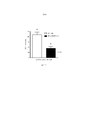
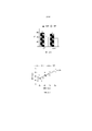
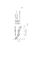
ФИГУРА 1 представляет собой диаграмму, иллюстрирующую геномную структуру МASP-2 человека;FIGURE 1 is a diagram illustrating the genomic structure of human MASP-2;
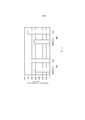
ФИГУРА 2А представляет собой принципиальную схему, иллюстрирующую доменную структуру белка MASP-2 человека;FIGURE 2A is a schematic diagram illustrating the domain structure of the human MASP-2 protein;
ФИГУРА 2В представляет собой принципиальную схему, иллюстрирующую доменную структуру белка Мар19 человека;FIGURE 2B is a schematic diagram illustrating the domain structure of the human Mar19 protein;
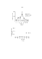
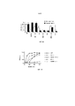
ФИГУРА 3 представляет собой диаграмму, иллюстрирующую стратегию нокаута MASP-2 мыши;FIGURE 3 is a diagram illustrating a mouse MASP-2 knockout strategy;
ФИГУРА 4 представляет собой диаграмму, иллюстрирующую конструирование минигена MASP-2 человека;FIGURE 4 is a diagram illustrating the construction of the human MASP-2 minigene;
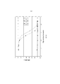
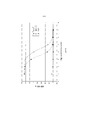
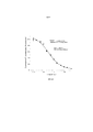
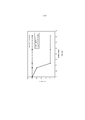
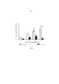
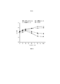
ФИГУРА 5A представляет результаты, которые демонстрируют, что MASP-2-недостаточность приводит к потере лектиновый-путь-опосредованной C4-активации, измеряемой по отсутствию C4b-депозиции на маннане, как описано в Примере 2;FIGURE 5A presents the results that demonstrate that MASP-2 deficiency results in loss of lectin pathway-mediated C4 activation, as measured by the absence of C4b deposition on mannan, as described in Example 2;
ФИГУРА 5B представляет результаты, которые демонстрируют, что MASP-2-недостаточность приводит к потере лектиновый-путь-опосредованной C4-активации, измеряемой по отсутствию C4b-депозиции на зимозане, как описано в Примере 2;FIGURE 5B presents the results that demonstrate that MASP-2 deficiency results in loss of lectin pathway-mediated C4 activation, as measured by the absence of C4b deposition on zymosan, as described in Example 2;
ФИГУРА 5С представляет результаты, которые демонстрируют относительные уровни C4-активации образцов сыворотки, полученных от штаммов MASP-2+/-; MASP-2-/- и дикого типа, как определено по C4b-депозиции на маннане и на зимозане, как описано в Примере 2;FIGURE 5C presents the results that demonstrate the relative levels of C4 activation of serum samples obtained from MASP-2 +/- strains; MASP-2 - / - and wild type, as determined by C4b deposition on mannan and on zymosan, as described in Example 2;
ФИГУРА 6 представляет результаты, которые демонстрируют, что добавление мышиного рекомбинантного MASP-2 к образцам сыворотки MASP-2-/- восстанавливает лектиновый-путь-опосредованную C4-активацию зависимым от концентрации белка образом, как определяют по C4b-депозиции на маннане, как описано в Примере 2;FIGURE 6 presents the results that demonstrate that the addition of murine recombinant MASP-2 to serum samples of MASP-2 - / - restores lectin pathway-mediated C4 activation in a protein concentration-dependent manner as determined by C4b deposition on mannan as described in Example 2;
ФИГУРА 7 представляет результаты, которые демонстрируют, что классический путь является функциональным в штамме MASP-2-/-, как описано в Примере 8;FIGURE 7 presents the results that demonstrate that the classical pathway is functional in the MASP-2 - / - strain as described in Example 8;
ФИГУРА 8А представляет результаты, которые демонстрируют, что Fab2-антитело анти-MASP2 # 11 ингибирует образование С3-конвертазы, как описано в Примере 10;FIGURE 8A presents the results that demonstrate that an
ФИГУРА 8B представляет результаты, которые демонстрируют, что Fab2-антитело анти-MASP2 # 11 связывается к нативным MASP-2 крысы, как описано в Примере 10;FIGURE 8B presents the results that demonstrate that an
ФИГУРА 8С представляет результаты, которые демонстрируют, что Fab2-антитело анти-MASP-2 # 41 ингибирует расщепление C4, как описано в Примере 10;FIGURE 8C presents the results that demonstrate that an anti-MASP-2 # 41 Fab2 antibody inhibits C4 cleavage as described in Example 10;
ФИГУРА 9 представляет результаты, которые демонстрируют, что каждое из протестированных Fab2-антител анти-MASP-2, которые ингибировали образование C3-конвертазы, также, как было обнаружено, ингибируют расщепление C4, как описано в Примере 10;FIGURE 9 presents the results which demonstrate that each of the anti-MASP-2 Fab2 antibodies tested that inhibited the formation of C3 convertase were also found to inhibit C4 cleavage as described in Example 10;
На ФИГУРЕ 10 приведена диаграмма, иллюстрирующая рекомбинантные полипептиды, полученные из MASP-2 крысы, которые использовались для эпитопного картирования блокирующих Fab2-антител анти-MASP-2, как описано в Примере 11;FIGURE 10 is a diagram illustrating recombinant polypeptides derived from rat MASP-2 that were used to epitope mapping of anti-MASP-2 Fab2 blocking antibodies as described in Example 11;
ФИГУРА 11 представляет результаты, которые демонстрируют связывание Fab2 анти-MASP-2 № 40 и № 60 с полипептидами MASP-2 крысы, как описано в Примере 11;FIGURE 11 presents the results that demonstrate the binding of anti-MASP-2
ФИГУРА 12 представляет результаты, которые демонстрируют освобождение от азота мочевины крови у мышей дикого типа (+/+) и MASP-2 (-/-) через 24 и 48 часов после реперфузии в модели ишемии почек/реперфузионного повреждения, как описано в Примере 12;FIGURE 12 presents the results that demonstrate the release of blood urea nitrogen in wild type (+ / +) and MASP-2 (- / -) mice at 24 and 48 hours after reperfusion in the renal ischemia / reperfusion injury model as described in Example 12. ;
ФИГУРА 13А представляет результаты, показывающие базовые уровни белка VEGF в RPE-хороидальном комплексе, изолированном из мышей дикого типа (+/+) и MASP-2 (-/-), как описано в Примере 13;FIGURE 13A presents results showing baseline VEGF protein levels in RPE-choroidal complex isolated from wild-type (+ / +) and MASP-2 (- / -) mice as described in Example 13;
ФИГУРА 13B представляет результаты, показывающие базовые уровни белка VEGF в RPE-хороидальном комплексе у мышей дикого типа (+/+) и MASP-2 (-/-) на 3 день после лазерно- индуцированного повреждения в модели макулярной дегенерации, как описано в Примере 13;FIGURE 13B presents results showing baseline levels of VEGF protein in RPE-choroidal complex in wild-type (+ / +) and MASP-2 (- / -) mice on
ФИГУРА 14 представляет результаты, показывающие средний объем хориоидальной неоваскуляризации (CNV) на седьмой день после лазерно-индуцированного повреждения у мышей дикого типа (+/+) и MASP-2 (-/-), как описано в Примере 13;FIGURE 14 presents the results showing the mean choroidal neovascularization (CNV) volume on the seventh day after laser-induced injury in wild-type (+ / +) and MASP-2 (- / -) mice as described in Example 13;
ФИГУРЫ 15A и 15B представляют кривые дозового эффекта для ингибирования C4b-депозиции (ФИГУРА 15А) и ингибирования активации тромбина (ФИГУРА 15В) после введения Fab2-антитела MASP-2 в нормальной сыворотке крови крыс, как описано в Примере 14;FIGURES 15A and 15B represent dose response curves for inhibition of C4b deposition (FIGURE 15A) and inhibition of thrombin activation (FIGURE 15B) following administration of Fab2 antibody MASP-2 in normal rat serum as described in Example 14;
ФИГУРЫ 16A и 16B представляют измеренную агрегацию тромбоцитов (выраженную в виде суммарной площади) у мышей MASP-2 (-/-) (ФИГУРА 16В) по сравнению с агрегацией тромбоцитов у мышей дикого типа, не получавших лечения, и мышей дикого типа, у которых путь комплемента ингибируется при помощи истощающего агента фактора яда кобры (CVF) и ингибитора терминального пути (C5aR-антагонист) (ФИГУРА 16А) в модели локализованной реакции Шварцмана диссеминированного внутрисосудистого свертывания, как описано в Примере 15;FIGURES 16A and 16B represent the measured platelet aggregation (expressed as total area) in MASP-2 (- / -) mice (FIGURE 16B) compared with platelet aggregation in untreated wild-type mice and wild-type mice in which the complement pathway is inhibited by a cobra venom factor (CVF) depleting agent and a terminal pathway inhibitor (C5aR antagonist) (FIGURE 16A) in a localized Schwarzmann response model of disseminated intravascular coagulation as described in Example 15;
ФИГУРА 17 графически иллюстрирует уровни азота мочевины крови (BUN), определенные у трансплантат-реципиентных WT-мышей (+/+) (B6) или трансплантат-реципиентных мышей MASP-2 (-/-), с почками от WT(+/+) доноров, как описано в Примере 16;FIGURE 17 graphically illustrates blood urea nitrogen (BUN) levels determined in graft-recipient WT mice (+ / +) (B6) or graft-recipient MASP-2 mice (- / -), with kidneys from WT (+ / + ) donors, as described in Example 16;
ФИГУРА 18 графически иллюстрирует процент выживания WT-мышей (+/+) и мышей MASP-2 (-/-) как функцию числа дней после микробной инфекции в модели перевязки слепой кишки и пункции (CLP), как описано в Примере 17;FIGURE 18 graphically illustrates the percent survival of WT mice (+ / +) and MASP-2 (- / -) mice as a function of days post microbial infection in a cecal ligation and puncture (CLP) model as described in Example 17;
ФИГУРА 19 графически иллюстрирует количество бактерий, определенных в WT-мышах (+/+) и мышах MASP-2 (-/-) после микробной инфекции в модели перевязки слепой кишки и пункции (CLP), как описано в Примере 17;FIGURE 19 graphically illustrates the number of bacteria determined in WT mice (+ / +) and MASP-2 (- / -) mice after microbial infection in a cecum ligation and puncture (CLP) model as described in Example 17;
ФИГУРА 20 представляет собой график Каплана-Майера, иллюстрирующий процент выживания мышей WT (+/+), MASP-2 (-/-) и C3 (-/-) через шесть дней после интраназального введения Pseudomonas aeruginosa, как описано в Примере 18;FIGURE 20 is a Kaplan-Meier graph illustrating the percent survival of WT (+ / +), MASP-2 (- / -) and C3 (- / -) mice six days after intranasal administration of Pseudomonas aeruginosa as described in Example 18;
ФИГУРА 21 графически иллюстрирует уровень C4b-депозиции, определенный в % от контроля, в пробах, взятых в разные моменты времени после подкожного введения WT-мышам либо 0,3 мг/кг или 1,0 мг/кг мышиных моноклональных антител анти-MASP-2, как описано в Примере 19;FIGURE 21 graphically illustrates the level of C4b deposition, determined as% of control, in samples taken at different time points after subcutaneous administration to WT mice with either 0.3 mg / kg or 1.0 mg / kg mouse anti-MASP-α
ФИГУРА 22 графически иллюстрирует уровень C4b-депозиции, определенный в % от контроля, в пробах, взятых в разные моменты времени после ip-дозирования WT-мышам 0,6 мг/кг мышиного моноклонального антитела анти-MASP-2, как описано в Примере 19;FIGURE 22 graphically illustrates the level of C4b deposition, determined as% of control, in samples taken at different time points after ip dosing in WT mice with 0.6 mg / kg mouse anti-MASP-2 monoclonal antibody, as described in Example 19. ;
ФИГУРА 23 графически иллюстрирует средний объем хороидальной неоваскуляризации (CNV) в день семь после лазер-индуцированного повреждения WT-мышей (+/+), предварительно получавших однократную ip-инъекцию 0,3 мг/кг или 1,0 мг/кг мышиного моноклонального антитела анти-MASP-2, как описано в Примере 20;FIGURE 23 graphically illustrates the mean choroidal neovascularization (CNV) volume on day seven following laser-induced injury in WT mice (+ / +) pre-treated with a single ip injection of 0.3 mg / kg or 1.0 mg / kg mouse monoclonal antibody. anti-MASP-2, as described in Example 20;
ФИГУРА 24А графически иллюстрирует процент выживания мышей MASP-2 (-/-) и WT (+/+) после заражения с 5×10⁸ кое/100 мкл N. meningitidis, как описано в Примере 21;FIGURE 24A graphically illustrates the percent survival of MASP-2 (- / -) and WT (+ / +) mice after challenge with 5 x 10 8 cfu / 100 μl N. meningitidis as described in Example 21;
ФИГУРА 24В графически иллюстрирует log кое/мл N. meningitides, полученных в разные моменты времени в пробах крови, взятых от мышей MASP-2 КО (-/-) и WT (+/+), инфицированных 5×10⁸ кое/100 мкл N. meningitidis, как описано в Примере 21;FIGURE 24B graphically illustrates the log cfu / ml of N. meningitides obtained at different time points in blood samples taken from MASP-2 KO (- / -) and WT (+ / +) mice infected with 5 x 10 8 cfu / 100 μl N. meningitidis, as described in Example 21;
ФИГУРА 25А графически иллюстрирует процент выживания MASP-2 КО (-/-) и WT (+/+) после заражения мышей с 2×10⁸ кое/100 мкл N. meningitidis, как описано в Примере 21;FIGURE 25A graphically illustrates the percent survival of MASP-2 KO (- / -) and WT (+ / +) after challenging mice with 2 x 10 8 cfu / 100 μl N. meningitidis as described in Example 21;
ФИГУРА 25B графически иллюстрирует log кое/мл N. meningitides, полученных в разные моменты времени из образцов крови, взятых из мышей WT (+/+), инфицированных 2×10⁸ кое/100 мкл N. meningitidis, как описано в Примере 21;FIGURE 25B graphically illustrates log cfu / ml N. meningitides obtained at different time points from blood samples taken from WT (+ / +) mice infected with 2 x 10 8 cfu / 100 μl N. meningitidis, as described in Example 21;
ФИГУРА 25C графически иллюстрирует log кое/мл N. meningitides, полученных в разные моменты времени из образцов крови, взятых из мышей MASP-2 (-/-), инфицированных 2×10⁸ кое/100 мкл N. meningitidis, как описано в Примере 21;FIGURE 25C graphically illustrates the log cfu / ml N. meningitides obtained at different time points from blood samples taken from MASP-2 (- / -) mice infected with 2 x 10 8 cfu / 100 μl N. meningitidis as described in Example 21;
ФИГУРА 26А графически иллюстрирует результаты анализа C3b-депозиции, демонстрирующие, что у мышей MASP-2 (-/-) сохранен функциональный классический путь, как описано в Примере 22;FIGURE 26A graphically illustrates the results of a C3b deposition assay demonstrating that MASP-2 (- / -) mice retain the functional classical pathway as described in Example 22;
ФИГУРА 26В графически иллюстрирует результаты анализа C3b-депозиции на покрытых зимозаном планшетах, демонстрирующие, что у мышей MASP-2 (-/-) сохранен функциональный альтернативный путь, как описано в Примере 22;FIGURE 26B graphically illustrates the results of a C3b deposition assay on zymosan-coated plates, demonstrating that MASP-2 (- / -) mice retain a functional alternative pathway as described in Example 22;
ФИГУРА 27А графически иллюстрирует миокардиальную ишемию/реперфузионное повреждение (MIRI)-индуцированную потерю ткани после перевязки левой передней нисходящей ветви коронарной артерии (LAD) и реперфузии у мышей C4 (-/-) (n=6) и соответствующих однопометных WT-контролей (n=7), показывая зону риска (AAR) и размер инфаркта (INF), как описано в Примере 22;FIGURE 27A graphically illustrates myocardial ischemia / reperfusion injury (MIRI) -induced tissue loss after left anterior descending coronary artery ligation (LAD) ligation and reperfusion in C4 (- / -) mice (n = 6) and corresponding litterm WT controls (n = 7), showing the area at risk (AAR) and the size of the infarction (INF), as described in Example 22;
ФИГУРА 27В графически иллюстрирует определение размера инфаркта (INF) как функцию зоны риска (AAR) у мышей C4 (-/-) и WT, как указано на ФИГУРЕ 42, демонстрируя, что мыши C4 (-/-) настолько же восприимчивы к MIRI, как и WT контроли (прерывистая линия), как описано в Примере 22;FIGURE 27B graphically illustrates infarction size (INF) as a function of area at risk (AAR) in C4 (- / -) and WT mice as indicated in FIGURE 42, demonstrating that C4 (- / -) mice are equally susceptible to MIRI. as well as WT controls (dashed line), as described in Example 22;
ФИГУРА 28А графически иллюстрирует результаты теста на C3b-депозицию с помощью сыворотки от мышей WT, сыворотки от мышей C4 (-/-) и сыворотки от мышей C4 (-/-), предварительно инкубированной с маннаном, как описано в Примере 22;FIGURE 28A graphically illustrates the results of a C3b deposition test using serum from WT mice, serum from C4 (- / -) mice and serum from C4 (- / -) mice preincubated with mannan as described in Example 22;
ФИГУРА 28В графически иллюстрирует результаты анализа C3b-депозиции в сыворотках от мышей WT, C4 (-/-) и MASP-2 (-/-), смешанных с различными концентрациями mAb против MASP-2 мыши (mAbM11), как описано в Пример 22;FIGURE 28B graphically illustrates the results of a C3b deposition assay in sera from WT mice, C4 (- / -) and MASP-2 (- / -) mixed with various concentrations of anti-mouse MASP-2 mAb (mAbM11) as described in Example 22. ;
ФИГУРА 28С графически иллюстрирует результаты анализа C3b-депозиции на сыворотке человека из WT (C4-достаточной) и C4-дефектной сыворотки и сыворотки от C4-дефектных субъектов, предварительно инкубированных с маннаном, как описано в Примере 22;FIGURE 28C graphically illustrates the results of a C3b deposition assay on human serum from WT (C4-sufficient) and C4-defective serum and sera from C4-defective subjects preincubated with mannan as described in Example 22;
ФИГУРА 28D графически иллюстрирует результаты анализа C3b-депозиции на сыворотке человека от WT (C4-достаточных) и C4 дефектных субъектов, смешанной с mAb против MASP-2 человека (mAbH3), как описано в Примере 22;FIGURE 28D graphically illustrates the results of a C3b deposition assay in human serum from WT (C4-sufficient) and C4 defective subjects mixed with an anti-human MASP-2 mAb (mAbH3) as described in Example 22;
ФИГУРА 29А графически иллюстрирует сравнительный анализ активности C3-конвертазы в плазме крови от протестированных штаммов мышей с различными дефектами комплемента, либо при условиях тестирования, специфических для лектинового пути активации, или при условиях тестирования, специфических для классического пути активации, как описано в Примере 22;FIGURE 29A graphically illustrates a comparative analysis of C3 convertase activity in blood plasma from tested strains of mice with various complement defects, either under test conditions specific to the lectin activation pathway or under testing conditions specific to the classical pathway activation, as described in Example 22;
ФИГУРА 29В графически иллюстрирует кинетику временного разрешения активности C3-конвертазы в плазме крови штаммов мышей с различными дефектами комплемента, при условиях тестирования, специфических для лектинового пути активации, как описано в Примере 22;FIGURE 29B graphically illustrates the kinetics of temporal resolution of C3 convertase activity in blood plasma of strains of mice with various complement defects, under test conditions specific for the lectin activation pathway, as described in Example 22;
ФИГУРА 30 иллюстрирует результаты Вестерн-блот анализа, демонстрирующие активизацию C3 человека, показанные при помощи наличия а'-цепи, при помощи субстратов тромбина FXIa и FXa, как описано в Примере 23;FIGURE 30 illustrates the results of Western blot analysis demonstrating the activation of human C3, shown by the presence of the a'-chain using thrombin substrates FXIa and FXa, as described in Example 23;
ФИГУРА 31 показывает результаты анализа C3-депозиции на образцах сыворотки, полученных от WT, MASP-2 (-/-), F11 (-/-), F11(-/-)/C4(-/-) и C4 (-/-), как описано в Примере 23;FIGURE 31 shows the results of C3 deposition analysis on serum samples obtained from WT, MASP-2 (- / -), F11 (- / -), F11 (- / -) / C4 (- / -) and C4 (- / -), as described in Example 23;
ФИГУРА 32А представляет собой график выживания Каплана-Мейера, показывающий процент выживания с течением времени после воздействия 7,0 Гр-излучения для контрольных мышей и мышей, получавших антитело против MASP-2 мыши (mAbM11) или антитело против MASP-2 человека (mAbH6), как описано в Примере 29;FIGURE 32A is a Kaplan-Meier survival graph showing the percentage of survival over time after exposure to 7.0 Gy radiation for control mice and mice treated with anti-mouse MASP-2 antibody (mAbM11) or anti-human MASP-2 antibody (mAbH6) as described in Example 29;
ФИГУРА 32В представляет собой график выживания Каплана-Мейера, показывающий процент выживания с течением времени после воздействия 6,5 Гр-излучения для контрольных мышей и мышей, получавших антитело против MASP-2 мыши (mAbM11) или антитело против MASP-2 человека (mAbH6), как описано в Примере 29;FIGURE 32B is a Kaplan-Meier survival graph showing the percentage of survival over time after exposure to 6.5 Gy radiation for control mice and mice treated with anti-mouse MASP-2 antibody (mAbM11) or anti-human MASP-2 antibody (mAbH6) as described in Example 29;
ФИГУРА 33 представляет собой график Каплана-Мейера, графически иллюстрирующий процент выживания мышей MASP-2 КО и WT после приема инфицирующий дозы 2,6×10⁷ кое N. meningitidis серогруппы А Z2491, демонстрируя, что MASP-2-дефектные мыши защищены от N. meningitides-индуцированной смертности, как описано в Примере 30;FIGURE 33 is a Kaplan-Meier plot graphically illustrating the percent survival of MASP-2 KO and WT mice following an infectious dose of 2.6 x 10 7 cfu of N. meningitidis serogroup A Z2491, demonstrating that MASP-2-deficient mice are protected from N meningitides -induced mortality as described in Example 30;
ФИГУРА 34 представляет собой график Каплана-Мейера, графически иллюстрирующий процент выживания мышей MASP-2 КО и WT после приема инфицирующей дозы 6×10⁶ кое N. meningitidis серогруппы B штамма MC58, демонстрируя, что MASP-2-дефектные мыши защищены от смертности, индуцированной N. meningitides серогруппы B штамма MC58, как это описано в Примере 30;FIGURE 34 is a Kaplan-Meier plot graphically illustrating the percent survival of MASP-2 KO and WT mice following an infectious dose of 6 x 10 6 CFU of N. meningitidis serogroup B strain MC58, demonstrating that MASP-2 deficient mice are protected from mortality. N. meningitides- induced serogroup B strain MC58, as described in Example 30;
ФИГУРА 35 графически иллюстрирует log кое/мл N. meningitidis серогруппы B штамма MC58, полученные в разные моменты времени из проб крови, которые берутся из мышей MASP-2 КО и WT после i.p.-инфицирования 6×10⁶ кое N. meningitidis серогруппы B штамма MC58 (n=3 в разные моменты времени, для обеих групп мышей, результаты выражены в виде среднего ± SEM), демонстрируя, что хотя мыши MASP-2 КО были заражены той же дозой N. meningitidis серогруппы B штамма C58, что и мыши WT, мыши MASP-2 КО имеют повышенное освобождение от бактериемии по сравнению с WT, как описано в Примере 30;FIGURE 35 graphically illustrates the log cfu / ml of N. meningitidis serogroup B strain MC58 obtained at different time points from blood samples taken from MASP-2 KO and WT mice after ip-infection with 6 × 10 6 cfu N. meningitidis serogroup B strain MC58 (n = 3 at different time points, for both groups of mice, results are expressed as mean ± SEM), demonstrating that although MASP-2 KO mice were infected with the same dose of N. meningitidis serogroup B strain C58 as in WT mice , MASP-2 KO mice have an increased clearance of bacteremia compared to WT, as described in Example 30;
ФИГУРА 36 графически иллюстрирует среднюю балльную оценку заболеваемости мышей MASP-2 и WT через 3, 6, 12 и 24 часа после заражения 6 × 10⁶ кое/100 мкл N. meningitidis серогруппы B штамма MC58, демонстрируя, что MASP-2 дефектные мыши показали высокую устойчивость к инфекции, со значительно более низкой балльной оценкой заболеваемости через 6 часов, как описано в Примере 30;FIGURE 36 graphically illustrates an average score of mice morbidity MASP-2 and
ФИГУРА 37 представляет собой график Каплана-Мейера, графически иллюстрирующий процент выживания мышей после введения инфицирующей дозы 4 × 10⁶ кое/100 мкл N. meningitidis серогруппы B штамма MC58, после введении через 3 часа после заражения либо ингибирующего антитела анти-MASP-2 (1 мг/кг), или контрольного изотипического антитела, демонстрируя, что антитело анти-MASP-2 является эффективным для лечения и улучшения выживаемости у субъектов, инфицированных N. meningitidis, как описано в Примере 31;FIGURE 37 is a Kaplan-Meier plot graphically illustrating the percent survival of mice after administration of an infectious dose of 4 × 10 6 cfu / 100 μl N. meningitidis serogroup B strain MC58, after
ФИГУРА 38 графически иллюстрирует log кое/мл жизнеспособных счетов N. meningitidis серогруппы B-MC58, полученных в разные моменты времени при 20% концентрации сыворотки человека после i.p. инфицирования 6,5 × 10⁶ кое/100 мкл N. meningitidis серогруппы B штамма MC58, через 0, 30, 60 и 90 минут после инкубации в присутствии: (A) нормальной сыворотки человека (NHS) плюс антитела анти-MASP-2 человека: (В) нормальной сыворотки человека (NHS) плюс изотипического контрольного антитела; (C) MBL-/- сыворотка человека; (D) нормальной сыворотки человека (NHS) и (E) инактивированной прогреванием нормальной сыворотки человека (NHS), показывая, что комплемент-зависимое уничтожение N. meningitidis в сыворотке крови человека значительно усилилось за счет добавления антитела анти-MASP-2 человека, как описано в Примере 32.FIGURE 38 graphically illustrates the log cfu / ml viable counts of N. meningitidis serogroup B-MC58 obtained at different time points at 20% human serum concentration after ip infection 6.5 x 10 6 cfu / 100 μl N. meningitidis serogroup B strain MC58, 0, 30, 60 and 90 minutes after incubation in the presence of: (A) normal human serum (NHS) plus anti-human MASP-2 antibodies: (B) normal human serum (NHS) plus isotypic control antibody; (C) MBL - / - human serum; (D) normal human serum (NHS) and (E) heat-inactivated normal human serum (NHS), showing that complement-dependent killing of N. meningitidis in human serum was significantly enhanced by the addition of anti-MASP-2 antibody, both described in Example 32.
ФИГУРА 39 графически иллюстрирует log кое/мл жизнеспособных счетов N. meningitidis серогруппы B-MC58, полученных в разные моменты времени из образцов мышиной сыворотки, демонстрируя, что сыворотки мышей MASP-2 -/- обладают более высокой бактерицидной активностью в отношении N. meningitidis, чем сыворотки WT-мышей, как описано в Примере 32;FIGURE 39 graphically illustrates the log cfu / ml viable counts of N. meningitidis serogroup B-MC58 obtained at different time points from mouse serum samples, demonstrating that sera from MASP-2 - / - mice have a higher bactericidal activity against N. meningitidis . than the sera of WT mice, as described in Example 32;
ФИГУРА 40 графически иллюстрирует гемолиз (определенный по высвобождению гемоглобина лизированными мышиными эритроцитами (Crry/C3-/-) в надосадочной жидкости, измеряемому при помощи фотометрии) покрытых маннаном мышиных эритроцитов при помощи сыворотки человека в диапазоне сывороточных концентраций. Тестируемые сыворотки включали инактивированную прогреванием (HI) NHS, MBL-/-, NHS + антитело анти-MASP-2 и контрольный NHS, как описано в Примере 33;FIGURE 40 graphically illustrates hemolysis (determined by the release of hemoglobin by lysed mouse erythrocytes (Crry / C3 - / -) in the supernatant, as measured by photometry) of mannan-coated mouse erythrocytes using human serum over a serum concentration range. Sera tested included heat inactivated (HI) NHS, MBL - / -, NHS + anti-MASP-2 antibody and control NHS as described in Example 33;
ФИГУРА 41 графически иллюстрирует гемолиз (определенный по высвобождению гемоглобина лизированными эритроцитами WT-мышей в надосадочной жидкости, измеряемому при помощи фотометрии) непокрытых мышиных эритроцитов при помощи сыворотки человека в диапазоне сывороточных концентраций. Тестируемые сыворотки включали инактивированную прогреванием (HI) NHS, MBL-/-, NHS + антитело анти-MASP-2 и контрольный NHS, демонстрируя, что ингибирующее анти-MASP-2 ингибирует комплемент-опосредованный лизис эритроцитов несенсибилизированных WT-мышей, как описано в Примере 33;FIGURE 41 graphically illustrates hemolysis (determined by the release of hemoglobin by lysed WT mouse erythrocytes in the supernatant, as measured by photometry) of uncoated mouse erythrocytes with human serum in a serum concentration range. Sera tested included heat-inactivated (HI) NHS, MBL - / -, NHS + anti-MASP-2 antibody, and control NHS, demonstrating that inhibitory anti-MASP-2 inhibits complement-mediated red blood cell lysis of non-sensitized WT mice as described in Example 33;
ФИГУРА 42 графически иллюстрирует гемолиз (определенный по высвобождению гемоглобина лизированными эритроцитами мышей (CDS 55/59-/-) в надосадочной жидкости, измеряемому при помощи фотометрии) непокрытых мышиных эритроцитов при помощи сыворотки человека в диапазоне сывороточных концентраций. Тестируемые сыворотки включали инактивированную прогреванием (HI) NHS, MBL-/-, NHS + антитело анти-MASP-2 и контрольную NHS, как описано в Примере 33;FIGURE 42 graphically illustrates the hemolysis (determined by the release of hemoglobin by lysed mouse erythrocytes (
ФИГУРА 43 графически иллюстрирует процент выживания с течением времени (дни) после воздействия 8,0 Гр-облучения в контрольных мышах и мышах, получавших антитело против MASP-2 человека (mAbH6), как описано в Примере 34;FIGURE 43 graphically illustrates the percentage of survival over time (days) after exposure to 8.0 Gy irradiation in control mice and mice treated with antibody against human MASP-2 (mAbH6), as described in Example 34;
ФИГУРА 44 графически иллюстрирует время начала микрососудистой окклюзии после LPS инъекции мышам MASP-2-/- и WT, показывающей процент мышей с тромбообразованием, измеренным в течение 60 минут, демонстрируя, что тромбообразование детектируется через 15 минут у WT-мышей, при этом до 80% WT-мышей демонстрировали тромбообразование в течение 60 минут; в отличие от этого, ни одна из мышей MASP-2 -/- не показала какое-либо тромбообразование в течение 60-минутного периода (логарифмический ранговый критерий: p=0,0005), как описано в Примере 35; иFIGURE 44 graphically illustrates the time of onset of microvascular occlusion after LPS injection in MASP-2 - / - and WT mice, showing the percentage of mice with thrombus formation measured within 60 minutes, demonstrating that thrombus formation was detected after 15 minutes in WT mice, with up to 80 % Of WT mice showed thrombus formation within 60 minutes; in contrast, none of the MASP-2 - / - mice showed any thrombus formation within a 60 minute period (log rank test: p = 0.0005) as described in Example 35; and
ФИГУРА 45 графически иллюстрирует процент выживания контрольных мышей, получавших физиологический раствор, (n=5) и мышей, получавших антитело анти-MASP-2, (n=5) в STX/LPS-индуцированной модели HUS с течением временем (в часах), демонстрируя, что все контрольные мыши погибли в течение 42 часов, тогда как, напротив, 100% мышей, получавших антитело анти-MASP-2, выживали на протяжении времени в ходе эксперимента, как это описано в Примере 36.FIGURE 45 graphically illustrates the percentage of survival of control mice treated with saline (n = 5) and mice treated with anti-MASP-2 antibody (n = 5) in an STX / LPS-induced HUS model over time (in hours). demonstrating that all control mice died within 42 hours, whereas, in contrast, 100% of mice treated with anti-MASP-2 antibody survived for the duration of the experiment, as described in Example 36.
ОПИСАНИЕ ПОСЛЕДОВАТЕЛЬНОСТЕЙDESCRIPTION OF SEQUENCES
SEQ ID NO: 1, кДНК Map19 человекаSEQ ID NO: 1 Human Map19 cDNA
SEQ ID NO:2 белок Map19 человека (с лидерной областью)SEQ ID NO: 2 Human Map19 protein (with leader region)
SEQ ID NO:3 белок Map19 человека (зрелый)SEQ ID NO: 3 Human Map19 protein (mature)
SEQ ID NO:4 кДНК MASP-2 человекаSEQ ID NO: 4 human MASP-2 cDNA
SEQ ID NO:5 белок MASP-2 человека (с лидерной областью)SEQ ID NO: 5 Human MASP-2 protein (with leader region)
SEQ ID NO:6 белок MASP-2 человека (зрелый)SEQ ID NO: 6 Human MASP-2 protein (mature)
SEQ ID NO:7 геномная ДНК MASP-2 человека (экзоны 1-6)SEQ ID NO: 7 Human MASP-2 genomic DNA (exons 1-6)
АНТИГЕНЫ (В ОТНОШЕНИИ ЗРЕЛОГО БЕЛКА MASP-2)ANTIGENS (REGARDING THE MATURE PROTEIN MASP-2)
SEQ ID NO:8 последовательность CUBI (a.к. 1-121)SEQ ID NO: 8 sequence CUBI (a.k. 1-121)
SEQ ID NO:9 последовательность CUBEGF (а.к. 1-166)SEQ ID NO: 9 sequence CUBEGF (a.k. 1-166)
SEQ ID NO:10 CUBEGFCUBII (а.к. 1-293)SEQ ID NO: 10 CUBEGFCUBII (a.k. 1-293)
SEQ ID NO:11 EGF-область (а.к. 122-166)SEQ ID NO: 11 EGF region (a.k. 122-166)
SEQ ID NO:12 домен сериновой протеазы (а.к. 429-671)SEQ ID NO: 12 Serine Protease Domain (a.s. 429-671)
SEQ ID NO:13 неактивный домен сериновой протеазы (а.к. 610-625 с мутацией Ser618 Ala)SEQ ID NO: 13 Serine protease inactive domain (a.k. 610-625 with Ser618 Ala mutation)
SEQ ID NO:34 TPLGPKWPEРVFGRL (пептида CUBI)SEQ ID NO: 34 TPLGPKWPEPERVFGRL (CUBI peptide)
SEQ ID NO:15SEQ ID NO: 15
TAPPGYRLRLYFTHFDLELSHLCEYDFVKLSSGAKVLATLCGQ (пептид CUBI)TAPPGYRLRLYFTHFDLELSHLCEYDFVKLSSGAKVLATLCGQ (CUBI peptide)
SEQ ID NO:16 TFRSDYSN (ядро МВL-области связывания)SEQ ID NO: 16 TFRSDYSN (MBL binding region core)
SEQ ID NO:17 FYSLGSSLDITFRSDYSNEPFTGF (MBL-область связывания)SEQ ID NO: 17 FYSLGSSLDITFRSDYSNEPFTGF (MBL binding region)
SEQ ID NO:18 IDECQVAPG (ПЕПТИД EGF)SEQ ID NO: 18 IDECQVAPG (EGF PEPTIDE)
SEQ ID NO:19 ANMLCAGLESGGDSCRGDSGGALV (ядро связывания сериновой протеазы) Подробное ОписаниеSEQ ID NO: 19 ANMLCAGLESGGDSCRGDSGGALV (Serine Protease Binding Core) Detailed Description
ПЕПТИДНЫЕ ИНГИБИТОРЫ:PEPTIDE INHIBITORS:
SEQ ID NO:20 полноразмерная кДНК MBLSEQ ID NO: 20 full length MBL cDNA
SEQ ID NO:21 полноразмерный белок MBLSEQ ID NO: 21 MBL full length protein
SEQ ID NO:22 OGК-Х-GP (консенсус связывания)SEQ ID NO: 22 OGK-X-GP (binding consensus)
SEQ ID NO:23 OGКLGSEQ ID NO: 23 OGKLG
SEQ ID NO:24 GLR GLQ GPO GKL GPO GSEQ ID NO: 24 GLR GLQ GPO GKL GPO G
SEQ ID NO:25 GPO GPO GLR GLQ GPO GKL GPO GPO GPOSEQ ID NO: 25 GPO GPO GLR GLQ GPO GKL GPO GPO GPO
SEQ ID NO:26 GKDGRDGTKGEKGEPGQGLRGLQGPOGKLGPOGSEQ ID NO: 26 GKDGRDGTKGEKGEPGQGLRGLQGPOGKLGPOG
SEQ ID NO:27 GAOGSOGEKGAOGPQGPOGPOGKMGPKGEOGDOSEQ ID NO: 27 GAOGSOGEKGAOGPQGPOGPOGKMGPKGEOGDO
(h-фиколин человека)(human h-ficolin)
SEQ ID NO:28SEQ ID NO: 28
GCOGLOGAOGDKGEAGTNGKRGERGPOGPOGKAGPOGPNGCOGLOGAOGDKGEAGTNGKRGERGPOGPOGKAGPOGPN
GAOGEO (фиколин p35 человека)GAOGEO (human ficolin p35)
SEQ ID NO:29 LQRALEILPNRVTIKANRPFLVFI (участок C4-расщепления)SEQ ID NO: 29 LQRALEILPNRVTIKANRPFLVFI (C4 cleavage site)
ЭКСПРЕССИОННЫЕ ИНГИБИТОРЫ:EXPRESSION INHIBITORS:
SEQ ID NO:30 кДНК домена CUBI-EGF (нуклеотиды 22-680 SEQ ID NO:4)SEQ ID NO: 30 cDNA CUBI-EGF Domain (nt 22-680 SEQ ID NO: 4)
SEQ ID NO:31SEQ ID NO: 31
5' CGGGCACACCATGAGGCTGCTGACCCTCCTGGGC 3'5 'CGGGCACACCATGAGGCTGCTGACCCTCCTGGGC 3'
Нуклеотиды 12-45 SEQ ID NO:4, в том числе сайт начала трансляции MASP-2 (смысловые)Nucleotides 12-45 SEQ ID NO: 4, including MASP-2 translation start site (sense)
SEQ ID NO:32SEQ ID NO: 32
5' GACATTACCTTCCGCTCCGACTCCAACGAGAAG 3'5 'GACATTACCTTCCGCTCCGACTCCAACGAGAAG 3'
Нуклеотиды 361-396 SEQ ID NO:4, кодирующие область, включающую сайт связывания MBL на MASP-2 (смысловые)Nucleotides 361-396 SEQ ID NO: 4 encoding a region including the MBL binding site on MASP-2 (sense)
SEQ ID NO:33SEQ ID NO: 33
5' AGCAGCCCTGAATACCCACGGCCGTATCCCAAA 3'5 'AGCAGCCCTGAATACCCACGGCCGTATCCCAAA 3'
Нуклеотиды 610-642 SEQ ID NO:4, кодирующие область, включающую домена CUBIINucleotides 610-642 SEQ ID NO: 4, encoding a region comprising a CUBII domain
ПРАЙМЕРЫ ДЛЯ КЛОНИРОВАНИЯ:PRIMERS FOR CLONING:
SEQ ID NO:34 CGGGATCCATGAGGCTGCTGACCCTC (5'-PCR для CUB)SEQ ID NO: 34 CGGGATCCATGAGGCTGCTGACCCTC (5'-PCR for CUB)
SEQ ID NO:35 GGAATTCCTAGGCTGCATA (3'-PCR ДЛЯ CUB)SEQ ID NO: 35 GGAATTCCTAGGCTGCATA (3'-PCR FOR CUB)
SEQ ID NO:36 GGAATTCCTACAGGGCGCT (3'-PCR ДЛЯ CUBIEGF)SEQ ID NO: 36 GGAATTCCTACAGGGCGCT (3'-PCR FOR CUBIEGF)
SEQ ID NO:37 GGAATTCCTAGTAGTGGAT (3'-PCR ДЛЯ CUBIEGFCUBII)SEQ ID NO: 37 GGAATTCCTAGTAGTGGAT (3'-PCR FOR CUBIEGFCUBII)
SEQ ID NO:38-47 представляют собой праймеры для клонирования для гуманизированного антителаSEQ ID NO: 38-47 are cloning primers for humanized antibody
SEQ ID NO:48 представляет собой 9 a.к.-пептидную связьSEQ ID NO: 48 is a 9 a.c.-peptide bond
ВЕКТОР ЭКСПРЕССИИ:EXPRESSION VECTOR:
SEQ ID NO:49 представляет собой вставку минигена MASP-2SEQ ID NO: 49 is a MASP-2 minigene insert
SEQ ID NO:50 представляет собой кДНК MASP-2 мышиSEQ ID NO: 50 is mouse MASP-2 cDNA
SEQ ID NO:51 представляет собой белок MASP-2 мыши (с лидером)SEQ ID NO: 51 is mouse MASP-2 protein (with leader)
SEQ ID NO:52 представляет собой зрелый белок MASP-2 мышиSEQ ID NO: 52 is the mature mouse MASP-2 protein
SEQ ID NO:53 представляет собой кДНК MASP-2 крысыSEQ ID NO: 53 is rat MASP-2 cDNA
SEQ ID NO:54 представляет собой белок MASP-2 крысы (с лидером)SEQ ID NO: 54 is rat MASP-2 protein (with leader)
SEQ ID NO:55 представляет собой зрелый белок MASP-2 крысыSEQ ID NO: 55 is a mature rat MASP-2 protein
SEQ ID NO:56-59 представляют собой олигонуклеотиды для сайт-направленного мутагенеза MASP-2 человека, используемые для создания MASP-2А человекаSEQ ID NO: 56-59 are oligonucleotides for site directed mutagenesis of human MASP-2 used to create human MASP-2A
SEQ ID NO:60-63 представляют собой олигонуклеотиды для сайт-направленного мутагенеза MASP-2 мыши, используемые для создания MASP-2А мышиSEQ ID NO: 60-63 are oligonucleotides for site-directed mutagenesis of mouse MASP-2 used to create mouse MASP-2A
SEQ ID NO:64-65 представляют собой олигонуклеотиды для сайт-направленного мутагенеза MASP-2 крысы, используемые для создания MASP-2А крысыSEQ ID NO: 64-65 are oligonucleotides for site-directed mutagenesis of rat MASP-2 used to create rat MASP-2A
ПОДРОБНОЕ ОПИСАНИЕDETAILED DESCRIPTION
Настоящее изобретение основывается на удивительном открытии авторов настоящего изобретения, что ингибирование лектинового пути, опосредованного MASP-2, возможно при сохранении классического пути интактным. Настоящее изобретение также описывает использование MASP-2 в качестве терапевтической мишени для ингибирования клеточного повреждения, связанного с лектин-опосредованной активацией комплемента, при сохранении компоненты классического (C1q-зависимого) пути иммунной системы интактной.The present invention is based on the surprising discovery of the present inventors that inhibition of the lectin pathway mediated by MASP-2 is possible while keeping the classical pathway intact. The present invention also describes the use of MASP-2 as a therapeutic target for inhibiting cellular damage associated with lectin-mediated complement activation while keeping the classical (C1q-dependent) pathway component of the immune system intact.
I. ОПРЕДЕЛЕНИЯI. DEFINITIONS
Если специально не определено в настоящем документе, все термины, используемые в настоящем документе, имеют тот же смысл, чтобы быть понятыми специалистами в области настоящего изобретения. Следующие определения приводятся для того, чтобы обеспечить ясность в отношении терминов, как они используются в спецификации и пунктах формулы изобретения для описания настоящего изобретения.Unless specifically defined herein, all terms used in this document have the same meaning to be understood by those skilled in the art of the present invention. The following definitions are provided in order to provide clarity with respect to terms as they are used in the specification and claims to describe the present invention.
В настоящем документе термин «MASP-2-зависимая активация комплемента» включает в себя MASP-2-зависимую активацию лектинового пути, которая возникает при физиологических состояниях (т.е. в присутствии Ca++), приводя к формированию C3-конвертазы C4b2a лектинового пути, и после накопления продукта С3-расщепления C3b, затем к C5-конвертазе C4b2a(C3b)n, которая, как было определено, первоначально вызывает опсонизацию. As used herein, the term "MASP-2-dependent complement activation" includes MASP-2-dependent activation of the lectin pathway that occurs under physiological conditions (i.e., in the presence of Ca ++ ), leading to the formation of C3-convertase C4b2a lectin pathway, and after the accumulation of the C3 cleavage product of C3b, then to the C5 convertase C4b2a (C3b) n, which was determined to initially cause opsonization.
В настоящем документе термин «альтернативный путь» относится к активации комплемента, которая инициируется, например, при помощи зимозана из клеточных стенок грибков и дрожжей, липополисахарида (LPS) из наружной оболочки грамотрицательных микроорганизмов и эритроцитов кролика, а также из многих очищенных полисахаридов, эритроцитов кролика, вирусов, бактерий, опухолевых клеток животных, паразитов и поврежденных клеток, и которая, как традиционно считалось, возникает от спонтанного протеолитического генерирования C3b из фактора комплемента С3.As used herein, the term “alternative pathway” refers to complement activation that is initiated, for example, by zymosan from the cell walls of fungi and yeast, lipopolysaccharide (LPS) from the outer membrane of gram-negative microorganisms and rabbit erythrocytes, and from many purified polysaccharides, rabbit erythrocytes , viruses, bacteria, animal tumor cells, parasites and damaged cells, and which, as traditionally believed, arises from the spontaneous proteolytic generation of C3b from the complement factor C3.
В настоящем документе термин «лектиновый путь» относится к активации комплемента, которая происходит вследствие специфического связывания сывороточных и несывороточных углевод-связывающих белков, в том числе маннан-связывающий лектин (MBL), CL-11 и фиколины (H-фиколин, М-фиколин или L-фиколин).As used herein, the term "lectin pathway" refers to complement activation that occurs due to the specific binding of serum and non-serum carbohydrate-binding proteins, including mannan-binding lectin (MBL), CL-11 and ficolins (H-ficolin, M-ficolin or L-ficolin).
В настоящем документе термин «классический путь» относится к активации комплемента, которая инициируется антителом, связанным с инородной частицей и требует связывания узнающей молекулы C1q.As used herein, the term "classical pathway" refers to complement activation that is initiated by an antibody bound to a foreign particle and requires binding of a C1q recognition molecule.
В настоящем документе термин «MASP-2-ингибирующий агент» относится к любому агенту, который связывается с или непосредственно взаимодействует с MASP-2 и эффективно ингибирует MASP-2-зависимую активацию комплемента, в том числе антитела анти-MASP-2 и их MASP-2-связывающие фрагменты, природные и синтетические пептиды, малые молекулы, растворимые MASP-2-рецепторы, ингибиторы экспрессии и изолированные природные ингибиторы, и также включает в себя пептиды, которые конкурируют с MASP-2 за связывание с другими узнающими молекулами (например, MBL, H-фиколин, М-фиколин или L-фиколин) в лектиновом пути, но не охватывает антитела, которые связываются с другими распознающими молекулами. MASP-2-ингибирующие агенты, пригодные в способе по изобретению, могут снижать MASP-2-зависимую активацию комплемента более чем на 20%, как например, более чем на 50%, как например, более чем на 90%. В одном варианте осуществления, MASP-2-ингибирующий агент снижает MASP-2-зависимую активацию комплемента более чем на 90% (т.е. приводит в результате только к 10%-й или менее MASP-2-активации комплемента).As used herein, the term "MASP-2 inhibitory agent" refers to any agent that binds to or directly interacts with MASP-2 and effectively inhibits MASP-2-dependent complement activation, including anti-MASP-2 antibodies and their MASP -2-binding fragments, natural and synthetic peptides, small molecules, soluble MASP-2 receptors, expression inhibitors and isolated natural inhibitors, and also includes peptides that compete with MASP-2 for binding to other recognition molecules (for example, MBL, H-ficolin, M-ficolin, or L-ficolin) in the lectin pathway, but does not cover antibodies that bind to other recognition molecules. MASP-2 inhibitory agents useful in the method of the invention can reduce MASP-2-dependent complement activation by more than 20%, such as more than 50%, such as more than 90%. In one embodiment, the MASP-2 inhibitory agent reduces MASP-2-dependent complement activation by more than 90% (i.e., results in only 10% or less MASP-2 complement activation).
В настоящем документе термин «антитело» охватывает антитела и их фрагменты антител, полученные от любого антитело-продуцирующего млекопитающего (например, мыши, крысы, кролика и примата, включая человека), или из гибридомы, фаговой селекции, рекомбинантной экспрессии или трансгенных животных (или других способов получения антител или фрагментов антител), которые специфически связываются с полипептидом-мишенью, таким как, например, MASP-2, полипептидами или их частями. Не предполагается, что термин «антитело» ограничен в отношении источника антитела или способа, с помощью которого оно изготовлено (например, при помощи гибридомы, фаговой селекции, рекомбинантной экспрессии, трансгенного животного, пептидного синтеза и т.д.). Типичные антитела включают поликлональные, моноклональные и рекомбинантные антитела; панспецифические, мультиспецифические антитела (например, биспецифические антитела, триспецифические антитела); гуманизированные антитела; мышиные антитела; химерные антитела, моноклональные антитела мышь-человек, мышь-примат, примат-человек; и антиидиотипические антитела, и, может быть, любое интактное антитело или его фрагмент. Как используется в настоящем документе, термин «антитело» включает в себя не только интактные поликлональные и моноклональные антитела, но также и фрагменты этого (например, dAb, Fab, Fab', F(ab')2, Fv), одноцепочечные (ScFv), их синтетические варианты, варианты, встречающиеся в природе, слитые белки, содержащие часть антитела с антиген-связывающим фрагментом требуемой специфичности, гуманизированные антитела, химерные антитела, и любую другую модифицированную конфигурацию молекулы иммуноглобулина, которая содержит антиген-связывающий участок или фрагмент (эпитоп-узнающий участок) требуемой специфичности.As used herein, the term "antibody" encompasses antibodies and antibody fragments thereof obtained from any antibody-producing mammal (e.g., mouse, rat, rabbit, and primate, including humans), or from hybridoma, phage selection, recombinant expression, or transgenic animals (or other methods of producing antibodies or antibody fragments) that specifically bind to a target polypeptide, such as, for example, MASP-2, polypeptides or portions thereof. The term "antibody" is not intended to be limited in relation to the source of the antibody or the method by which it is made (eg, by hybridoma, phage selection, recombinant expression, transgenic animal, peptide synthesis, etc.). Typical antibodies include polyclonal, monoclonal, and recombinant antibodies; panspecific, multispecific antibodies (eg, bispecific antibodies, trispecific antibodies); humanized antibodies; murine antibodies; chimeric antibodies, monoclonal antibodies mouse-human, mouse-primate, primate-human; and anti-idiotypic antibodies, and may be any intact antibody or fragment thereof. As used herein, the term "antibody" includes not only intact polyclonal and monoclonal antibodies, but also fragments of these (e.g. dAb, Fab, Fab ', F (ab') 2 , Fv), single chain (ScFv) , their synthetic variants, variants found in nature, fusion proteins containing a part of an antibody with an antigen-binding fragment of the required specificity, humanized antibodies, chimeric antibodies, and any other modified configuration of an immunoglobulin molecule that contains an antigen-binding site or fragment (epitope recognition site) of the required specificity.
«Моноклональное антитело» относится к популяции однородных антител, где моноклональное антитело содержит аминокислоты (встречающиеся в природе и не встречающиеся в природе), которые участвуют в избирательном связывании эпитопа. Моноклональные антитела являются весьма специфическими для антигена-мишени. Термин «моноклональное антитело» включает не только интактные моноклональные антитела и полноразмерные моноклональные антитела, но и их фрагменты (например, Fab, Fab', F(ab')2, Fv), одноцепочечные (ScFv), их варианты, слитые белки, содержащие антиген-связывающую часть, гуманизированные моноклональные антитела, химерные моноклональные антитела и любую другую модифицированную конфигурацию молекулы иммуноглобулина, которая содержит антиген-связывающий фрагмент (эпитоп-узнающий участок) требуемой специфичности и с возможностью связывания с эпитопом. Не подразумевается ограничение в отношении источника антитела или способ, с помощью которого оно изготовлено (например, при помощи гибридомы, фаговой селекции, рекомбинантной экспрессии, трансгенного животного и др.). Этот термин включает в себя все иммуноглобулины, а также фрагменты и др., описанные выше под определением «антитело»."Monoclonal antibody" refers to a population of homogeneous antibodies, where the monoclonal antibody contains amino acids (naturally occurring and not naturally occurring) that are involved in the selective binding of an epitope. Monoclonal antibodies are highly specific for the target antigen. The term "monoclonal antibody" includes not only intact monoclonal antibodies and full-length monoclonal antibodies, but also fragments thereof (for example, Fab, Fab ', F (ab') 2 , Fv), single-chain (ScFv), variants thereof, fusion proteins containing antigen-binding portion, humanized monoclonal antibodies, chimeric monoclonal antibodies and any other modified configuration of an immunoglobulin molecule that contains an antigen-binding fragment (epitope-recognition site) of the required specificity and with the ability to bind to an epitope. No limitation is intended as to the source of the antibody or the method by which it is made (eg, by hybridoma, phage selection, recombinant expression, transgenic animal, etc.). This term includes all immunoglobulins, as well as fragments, etc., described above under the definition of "antibody".
В настоящем документе термин «фрагмент антитела» относится к части, полученной из или относящейся к полноразмерному антителу, такому как, например, антитело анти-MASP-2, как правило, включающей антиген-связывающую или вариабельную область этого. Иллюстративные примеры фрагментов антител включают фрагменты Fab, Fab', F(ab)2, F(ab')2 и Fv, фрагменты scFv, диатела, линейные антитела, молекулы одноцепочечных антител и мультиспецифические антитела, образованные из фрагментов антител.As used herein, the term "antibody fragment" refers to a portion derived from or related to a full-length antibody, such as, for example, an anti-MASP-2 antibody, typically comprising an antigen-binding or variable region thereof. Illustrative examples of antibody fragments include Fab, Fab ', F (ab) 2 , F (ab') 2 and Fv fragments, scFv fragments, diabodies, linear antibodies, single chain antibody molecules, and multispecific antibodies derived from antibody fragments.
Как используется в настоящем документе, фрагмент «одноцепочечного Fv» или «scFv» антитела включает в себя VН и VL домены антитела, где эти домены присутствуют в единой полипептидной цепи. Как правило, полипептид Fv дополнительно содержит полипептидный линкер между доменами VН и VL, который позволяет scFv формировать нужную структуру для связывания антигена.As used herein, a "single chain Fv" or "scFv" antibody fragment includes the V H and V L domains of an antibody, where these domains are present in a single polypeptide chain. Typically, the Fv polypeptide further comprises a polypeptide linker between the V H and V L domains that allows the scFv to form the desired structure for antigen binding.
Как используется в настоящем документе, «химерное антитело» представляет собой рекомбинантный белок, который содержит вариабельные домены и гипервариабельные участки, полученные из антитела биологических видов, отличных от человека (например, грызун), тогда как остаток молекулы антитела является производным от антитела человека.As used herein, a "chimeric antibody" is a recombinant protein that contains variable domains and hypervariable regions derived from an antibody of a non-human species (eg, a rodent), while the remainder of the antibody molecule is derived from a human antibody.
Как используется в настоящем документе, «гуманизированное антитело» представляет собой химерное антитело, которое включает в себя минимальную последовательность, соответствующую определенным гипервариабельным участкам, полученным из иммуноглобулина, отличного от иммуноглобулина человека, которую трансплантируют в рамке антитела человека. Гуманизированные антитела обычно представляют собой рекомбинантные белки, в которых только гипервариабельные участки антитела не имеют происхождения от человека.As used herein, a "humanized antibody" is a chimeric antibody that includes a minimum sequence corresponding to defined hypervariable regions derived from an immunoglobulin other than human immunoglobulin that is transplanted in a human antibody frame. Humanized antibodies are usually recombinant proteins in which only the hypervariable regions of the antibody are not of human origin.
Как используется в настоящем документе, термин «маннан-связывающий лектин» («MBL») эквивалентен маннан-связывающему белку («MBP»).As used herein, the term "mannan binding lectin" ("MBL") is equivalent to mannan binding protein ("MBP").
Как используется в настоящем документе, «мембраноатакующий комплекс» («MAC») относится к комплексу терминальных пяти компонентов комплемента (C5b в сочетании с С6, С7, С8 и C-9), который встраивается в мембраны и нарушает их (также называется C5b-9).As used herein, "membrane attack complex" ("MAC") refers to a complex of terminal five components of complement (C5b combined with C6, C7, C8, and C-9) that integrates into membranes and disrupts them (also called C5b- 9).
Как используется в настоящем документе, «субъект» включает в себя всех млекопитающих, включая, в качестве неограничивающих примеров, людей, отличных от человека приматов, собак, кошек, лошадей, овец, коз, коров, кроликов, свиней и грызунов.As used herein, "subject" includes all mammals, including, but not limited to, humans, non-human primates, dogs, cats, horses, sheep, goats, cows, rabbits, pigs, and rodents.
Как используется в настоящем документе, аминокислотные остатки сокращены следующим образом: аланин (Ala;А), аспарагин (Asn;N), аспарагиновая кислота (Asр;D), аргинин (Arg;R), цистеин (Cys;C), глутаминовая кислота (Glu;E), глутамин (Gln;Q), глицин (Gly;G), гистидин (His;H), изолейцин (Ile;I), лейцин (Leu;L), лизин (Lys;K), метионин (Мет;M), фенилаланин (Phe;E), пролин (Pro;P), Серин (Ser;S), треонин (Thr;T), триптофан (Trp;W), тирозин (Tyr;Y) и валин (Val;V).As used herein, amino acid residues are abbreviated as follows: alanine (Ala; A), asparagine (Asn; N), aspartic acid (Asp; D), arginine (Arg; R), cysteine (Cys; C), glutamic acid (Glu; E), glutamine (Gln; Q), glycine (Gly; G), histidine (His; H), isoleucine (Ile; I), leucine (Leu; L), lysine (Lys; K), methionine ( Met; M), phenylalanine (Phe; E), proline (Pro; P), Serine (Ser; S), threonine (Thr; T), tryptophan (Trp; W), tyrosine (Tyr; Y) and valine (Val ; V).
В широком смысле, встречающиеся в природе аминокислоты могут быть разделены на группы в зависимости от химической характеристики боковой цепи соответствующих аминокислот. Под «гидрофобной» аминокислотой подразумевается Ile, Leu, Мет, Phe, Trp, Tyr, Val, Ala, Cys или Pro. Под «гидрофильной» аминокислотой подразумевается Gly, Asn, Gln, Ser, Thr, Asp, Glu, Lys, Arg или His. Это группирование аминокислот может быть далее разделено на подклассы следующим образом. Под «незаряженной гидрофильной» аминокислотой подразумевается Ser, Thr, Asn или Gln. Под «кислой» аминокислотой подразумевается Glu или Asp. Под «основной» аминокислотой подразумевается Lys, Arg или His.In a broad sense, naturally occurring amino acids can be classified into groups depending on the chemical characteristics of the side chain of the corresponding amino acids. By "hydrophobic" amino acid is meant Ile, Leu, Met, Phe, Trp, Tyr, Val, Ala, Cys, or Pro. By "hydrophilic" amino acid is meant Gly, Asn, Gln, Ser, Thr, Asp, Glu, Lys, Arg, or His. This grouping of amino acids can be further subdivided as follows. By "uncharged hydrophilic" amino acid is meant Ser, Thr, Asn or Gln. By "acidic" amino acid is meant Glu or Asp. By "basic" amino acid is meant Lys, Arg, or His.
Как используется в настоящем документе, термин «консервативная аминокислотная замена» иллюстрируется замещением между аминокислотами в каждой из следующих групп: (1) глицин, аланин, валин, лейцин, изолейцин, (2) фенилаланин, тирозин и триптофан, (3) серин и треонин, (4) аспартат и глутамат, (5) глутамин и аспарагин, и (6) лизин, аргинин и гистидин.As used herein, the term "conservative amino acid substitution" is illustrated by a substitution between amino acids in each of the following groups: (1) glycine, alanine, valine, leucine, isoleucine, (2) phenylalanine, tyrosine and tryptophan, (3) serine and threonine , (4) aspartate and glutamate, (5) glutamine and asparagine, and (6) lysine, arginine and histidine.
Термин «олигонуклеотид», используемый в настоящем документе, относится к олигомеру или полимеру рибонуклеиновой кислоты (РНК) или дезоксирибонуклеиновой кислоты (ДНК), или их миметикам. Этот термин также охватывает те олигонуклеиновые основания, которые состоят из природных нуклеотидов, сахаров и ковалентных межнуклеозидных (главная цепь) связей, а также олигонуклеотидов с модификациями, отсутствующими в природе.The term "oligonucleotide" as used herein refers to an oligomer or polymer of ribonucleic acid (RNA) or deoxyribonucleic acid (DNA), or mimetics thereof. The term also encompasses those oligonucleotides that are composed of naturally occurring nucleotides, sugars and covalent internucleoside (backbone) bonds, as well as oligonucleotides with modifications not found in nature.
Как используется в настоящем документе, «эпитоп» относится к участку на белке (например, белок MASP-2 человека), который связывается с антителом. «Перекрывающиеся эпитопы» включают по крайней мере один (например, два, три, четыре, пять или шесть) общих аминокислотных остатков, в том числе неразветвленные и разветвленные эпитопы.As used herein, "epitope" refers to a region on a protein (eg, human MASP-2 protein) that binds to an antibody. "Overlapping epitopes" include at least one (eg, two, three, four, five, or six) common amino acid residues, including unbranched and branched epitopes.
Как используется в настоящем документе, термин «полипептид», «пептид» и «белок» используются взаимозаменяемо и означают любые цепочки аминокислот с пептидными связями, независимо от их длины или посттрансляционной модификации. Белок MASP-2, описанный в настоящем документе, может содержать или представлять собой белки дикого типа или может представлять собой варианты, которые содержат не более чем 50 (например, не более чем один, два, три, четыре, пять, шесть, семь, восемь, девять, десять, 12, 15, 20, 25, 30, 35, 40 или 50) консервативных аминокислотных замен. Консервативные замены, как правило, включают замены в пределах следующих групп: глицин и аланин; валин, изолейцин и лейцин; аспарагиновая и глутаминовая кислота, аспарагин, глютамин, серин и треонин; лизин, гистидин и аргинин; и фенилаланин и тирозин.As used herein, the terms "polypeptide", "peptide" and "protein" are used interchangeably and mean any peptide-bonded amino acid chains, regardless of their length or post-translational modification. The MASP-2 protein described herein can contain or be wild-type proteins, or can be variants that contain no more than 50 (e.g., no more than one, two, three, four, five, six, seven, eight, nine, ten, 12, 15, 20, 25, 30, 35, 40 or 50) conservative amino acid substitutions. Conservative substitutions generally include substitutions within the following groups: glycine and alanine; valine, isoleucine and leucine; aspartic and glutamic acid, asparagine, glutamine, serine and threonine; lysine, histidine and arginine; and phenylalanine and tyrosine.
В некоторых вариантах осуществления, белок MASP-2 человека может иметь последовательность аминокислот, которая на, или больше чем на 70 (например, 71, 72, 73, 74, 75, 76, 77, 78, 79, 80, 81, 82, 83, 84, 85, 86, 87, 88, 89, 90, 91, 92, 93, 94, 95, 96, 97, 98, 99 или 100)% идентична белку MASP-2 человека, имеющего аминокислотную последовательность, изложенную в SEQ ID NO:5.In some embodiments, the human MASP-2 protein may have an amino acid sequence that is, or greater than, 70 (e.g., 71, 72, 73, 74, 75, 76, 77, 78, 79, 80, 81, 82, 83, 84, 85, 86, 87, 88, 89, 90, 91, 92, 93, 94, 95, 96, 97, 98, 99 or 100)% identical to the human MASP-2 protein having the amino acid sequence set forth in SEQ ID NO: 5.
В некоторых вариантах осуществления, пептидные фрагменты могут составлять не менее 6 (например, по крайней мере 7, 8, 9, 10, 11, 12, 13, 14, 15, 16, 17, 18, 19, 20, 21, 22, 23, 24, 25, 26, 27, 28, 29, 30, 31, 32, 33, 34, 35, 36, 37, 38, 39, 40, 41, 42, 43, 44, 45, 46, 47, 48, 49, 50, 55, 60, 65, 70, 75, 80, 85, 90, 95, 100, 110, 120, 130, 140, 150, 160, 170, 180, 190, 200, 250, 300, 350, 400, 450, 500 или 600 или более) аминокислотных остатков в длину (например, по крайней мере 6 смежных аминокислотных остатков SEQ ID NO:5). В некоторых вариантах осуществления, антигенный пептидый фрагмент белка MASP-2 человека составляет менее 500 (например, меньше чем 450, 400, 350, 325, 300, 275, 250, 225, 200, 190, 180, 170, 160, 150, 140, 130, 120, 110, 100, 95, 90, 85, 80, 75, 70, 65, 60, 55, 50, 49, 48, 47, 46, 45, 44, 43, 42, 41, 40, 39, 38, 37, 36, 35, 34, 33, 32, 31, 30, 29, 28, 27, 26, 25, 24, 23, 22, 21, 20, 19, 18, 17, 16, 15, 14, 13, 12, 11, 10, 9, 8, 7 или 6) аминокислотных остатков в длину (например, меньше, чем 500 смежных аминокислотных остатков в любой из SEQ ID NO:5).In some embodiments, the peptide fragments can be at least 6 (e.g., at least 7, 8, 9, 10, 11, 12, 13, 14, 15, 16, 17, 18, 19, 20, 21, 22, 23, 24, 25, 26, 27, 28, 29, 30, 31, 32, 33, 34, 35, 36, 37, 38, 39, 40, 41, 42, 43, 44, 45, 46, 47, 48, 49, 50, 55, 60, 65, 70, 75, 80, 85, 90, 95, 100, 110, 120, 130, 140, 150, 160, 170, 180, 190, 200, 250, 300, 350, 400, 450, 500, or 600 or more) amino acid residues in length (for example, at least 6 contiguous amino acid residues of SEQ ID NO: 5). In some embodiments, the antigenic peptide fragment of the human MASP-2 protein is less than 500 (e.g., less than 450, 400, 350, 325, 300, 275, 250, 225, 200, 190, 180, 170, 160, 150, 140 , 130, 120, 110, 100, 95, 90, 85, 80, 75, 70, 65, 60, 55, 50, 49, 48, 47, 46, 45, 44, 43, 42, 41, 40, 39 , 38, 37, 36, 35, 34, 33, 32, 31, 30, 29, 28, 27, 26, 25, 24, 23, 22, 21, 20, 19, 18, 17, 16, 15, 14 , 13, 12, 11, 10, 9, 8, 7, or 6) amino acid residues in length (eg, less than 500 contiguous amino acid residues in any of SEQ ID NO: 5).
Процент (%) идентичности аминокислотной последовательности определяется как процентное соотношение аминокислот в кандидатной последовательности, которая является идентичной по аминокислотам референсной последовательности, после выравнивания последовательностей и внедрения пробелов, в случае необходимости, для достижения максимального процента идентичности последовательностей. Выравнивание для целей определения процента идентичности последовательностей может быть достигнуто различными способами, известными специалистам в рассматриваемой области, например, с помощью общедоступных компьютерных программ, таких как программное обеспечение BLAST, BLAST-2, ALIGN, ALIGN-2 или Megalign (DNASTAR). Соответствующие параметры для измерения выравнивания, включая любые алгоритмы, необходимые для достижения максимального выравнивания по всей длине сравниваемых последовательностей, могут быть определены по известным методикам.Percentage (%) amino acid sequence identity is defined as the percentage of amino acids in a candidate sequence that is amino acid identical to the reference sequence, after alignment of the sequences and insertion of spaces, if necessary, to achieve the maximum percentage of sequence identity. Alignment for the purpose of determining percent sequence identity can be achieved in a variety of ways known to those skilled in the art, for example, using publicly available computer programs such as BLAST, BLAST-2, ALIGN, ALIGN-2 or Megalign (DNASTAR) software. Appropriate parameters for measuring alignment, including any algorithms needed to achieve maximum alignment over the entire length of the sequences being compared, can be determined using known techniques.
II. Обзор изобретенияII. Overview of the invention
Лектины (MBL, M-фиколин, H-фиколин, L-фиколин и CL-11) представляют собой специфические распознающие молекулы, которые инициируют природную систему комплемента, и система включает в себя инициацию лектинового пути и связанного цикла усиления терминального пути, который усиливает лектин-инициированную активизацию эффекторных молекул терминального комплемента. C1q представляют собой специфическую узнающую молекулу, которая инициирует нужную систему комплемента, и система индуцирует классический путь инициации и связанный цикл усиления терминального пути, который усиливает C1q-инициированную активацию эффекторных молекул терминального комплемента. Мы называем эти две основных системы активации комплемента как лектин-зависимая система комплемента и C1q-зависимая система комплемента, соответственно.Lectins (MBL, M-ficolin, H-ficolin, L-ficolin and CL-11) are specific recognition molecules that initiate the natural complement system, and the system involves the initiation of the lectin pathway and the associated terminal pathway amplification cycle that enhances lectin -initiated activation of terminal complement effector molecules. C1q is a specific recognition molecule that initiates the desired complement system, and the system induces the classical initiation pathway and associated terminal pathway amplification cycle that enhances the C1q-initiated activation of terminal complement effector molecules. We refer to these two major complement activation systems as the lectin-dependent complement system and the C1q-dependent complement system, respectively.
В дополнение к ее важной роли в иммунной защите, система комплемента принимает участие в повреждении тканей при многих клинических состояниях. Таким образом, существует насущная необходимость разработки терапевтически эффективных ингибиторов комплемента для предотвращения этих неблагоприятных эффектов. С признанием того, что можно заблокировать лектин-опосредованный MASP-2 путь при сохранении классического пути интактным, приходит осознание того, что было бы весьма желательно специфически ингибировать только систему активации комплемента, вызывающую конкретную патологию, без полного выключения возможностей иммунной защиты комплемента. Например, при болезненных состояниях, при которых активация комплемента опосредуется преимущественно лектин-зависимой системой комплемента, было бы выгодно специфически ингибировать только эту систему. Это сохранило бы C1q-зависимую систему активации комплемента интактной для оперирования подготовкой иммунного комплекса и помощи в защите организма-хозяина от инфекции.In addition to its important role in immune defense, the complement system is involved in tissue damage in many clinical conditions. Thus, there is an urgent need to develop therapeutically effective complement inhibitors to prevent these adverse effects. With the recognition that it is possible to block the lectin-mediated MASP-2 pathway while keeping the classical pathway intact, it is realized that it would be highly desirable to specifically inhibit only the complement activation system causing a particular pathology, without completely shutting down the complement immune defense capabilities. For example, in disease states in which the activation of complement is mediated predominantly by the lectin-dependent complement system, it would be beneficial to specifically inhibit only this system. This would keep the C1q-dependent complement activation system intact to handle the preparation of the immune complex and help protect the host from infection.
Предпочтительным белковым компонентом в качестве цели при разработке терапевтических агентов для специфического ингибирования лектин-зависимой системы комплемента является MASP-2. Из всех известных белковых компонентов лектин-зависимой системы комплемента (MBL, H-фиколин, М-фиколин, L-фиколин, MASP-2, C2-C9, Фактор B, Фактор D и пропердин), только MASP-2 является как уникальным для лектин-зависимой системы комплемента, так и необходимым для функционирования системы. Лектины (MBL, H-фиколин, М-фиколин, L-фиколин и CL-11) также представляют собой уникальные компоненты в лектин-зависимой системе комплемента. Тем не менее, потеря любого из лектиновых компонентов не обязательно должна ингибировать активацию системы из-за лектиновой избыточности. Было бы необходимо ингибировать все пять лектинов, чтобы гарантированно ингибировать лектин-зависимую систему активации комплемента. Кроме того, поскольку также известно, что MBL и фиколины обладают опсонической активностью, независимой oт комплемента, ингибирование лектиновой функции могло бы привести к потере этого благотворного механизм защиты организма-хозяина от инфекции. В отличие от этого, такая комплемент-независимая лектиновая опсоническая активность оставалась бы интактной, если бы MASP-2 представлял собой мишень для ингибирования. Дополнительным преимуществом MASP-2 в качестве терапевтической мишени для ингибирования лектин-зависимой системы активации комплемента является то, что концентрация MASP-2 в плазме крови является одной из самых низких из каких-либо белков комплемента (≈500 нг/мл), поэтому, соответственно, низкие концентрации высокоаффинных ингибиторов MASP-2 могут быть достаточны для получения полного ингибирования (Moller-Kristensen, M., et al., J. Immunol Methods 282: 159-167, 2003).A preferred protein component as a target in the development of therapeutic agents for specifically inhibiting the lectin-dependent complement system is MASP-2. Of all the known protein components of the lectin-dependent complement system (MBL, H-ficolin, M-ficolin, L-ficolin, MASP-2, C2-C9, Factor B, Factor D and properdin), only MASP-2 is unique to lectin-dependent complement system, and necessary for the functioning of the system. Lectins (MBL, H-ficolin, M-ficolin, L-ficolin and CL-11) are also unique components in the lectin-dependent complement system. However, the loss of any of the lectin components need not necessarily inhibit system activation due to lectin excess. It would be necessary to inhibit all five lectins to ensure that the lectin-dependent complement activation system is inhibited. In addition, since MBL and ficolins are also known to have opsonic activity independent of complement, inhibition of lectin function could result in the loss of this beneficial host defense mechanism against infection. In contrast, such complement-independent lectin opsonic activity would remain intact if MASP-2 were a target for inhibition. An additional advantage of MASP-2 as a therapeutic target for inhibiting the lectin-dependent complement activation system is that the plasma concentration of MASP-2 is one of the lowest of any complement protein (≈500 ng / ml), therefore, accordingly , low concentrations of high affinity MASP-2 inhibitors may be sufficient to obtain complete inhibition (Moller-Kristensen, M., et al., J. Immunol Methods 282: 159-167, 2003).
III. РОЛЬ MASP-2 В РАЗЛИЧНЫХ ЗАБОЛЕВАНИЯХ И СОСТОЯНИЯХ И ТЕРАПЕВТИЧЕСКИЕ СПОСОБЫ, ИСПОЛЬЗУЮЩИЕ MASP-2-ИНГИБИРУЮЩИЕ АГЕНТЫIII. ROLE OF MASP-2 IN VARIOUS DISEASES AND CONDITIONS AND THERAPEUTIC METHODS USING MASP-2-INHIBITING AGENTS
ПОЧЕЧНЫЕ СОСТОЯНИЯRENAL CONDITIONS
Активация системы комплемента вовлечена в патогенез различных заболеваний почек, включая мезангиопролиферативный гломерулонефрит (IgA-нефропатия, болезнь Берже) (Endo, M., et al., Clin. Nephrology 55: 185-191, 2001), мембранный гломерулонефрит (Kerjashki, D., Arch B Cell Pathol. 58:253-71, 1990; Brenchlcy, P.E., et al., Kidney Int., 41:933-7, 1992; Salant, D.J., et al., Kidney Int. 55:976-84, 1989), мембранозно-пролиферативный гломерулонефрит (мезангиокапиллярный гломерулонефрит) (Bartlow, B.G., et al., Kidney Int. 15:294-300, 1979; Meri, S., et al., J. Exp. Med. 175:939-50, 1992), острый послеинфекционный гломерулонефрит (постстрептококковый гломерулонефрит), криоглобулинемический гломерулонефрит (Ohsawa, I., et al., Clin Immunol, 101:59-66, 2001), волчаночный нефрит (Gatenby, P.A., Autoimmunity 11:61-6, 1991) и пурпурный нефрит Henoch-Schonlein (Endo, M., et al., Am. J. Kidney Dis. 35:401-407, 2000). Участие комплемента в болезни почек оценивали на протяжении нескольких десятилетий, но по-прежнему существует значительное обсуждение его точной роли в возникновении, развитии и стадии разрешения почечной болезни. При нормальных состояниях вклад комплемента выгоден организму-хозяину, но неадекватная активация и депозиция комплемента может способствовать повреждению тканей.Activation of the complement system has been implicated in the pathogenesis of various renal diseases, including mesangioproliferative glomerulonephritis (IgA nephropathy, Berger's disease) (Endo, M., et al., Clin. Nephrology 55: 185-191, 2001), membrane glomerulonephritis (Kerjashki, D. , Arch B Cell Pathol. 58: 253-71, 1990; Brenchlcy, PE, et al., Kidney Int ., 41: 933-7, 1992; Salant, DJ, et al., Kidney Int . 55: 976-84 1989), membranoproliferative glomerulonephritis (mesangiocapillary glomerulonephritis) (Bartlow, BG, et al., Kidney Int . 15: 294-300, 1979; Meri, S., et al., J. Exp. Med. 175: 939 -50, 1992), acute post-infectious glomerulonephritis (post-streptococcal glomerulonephritis), cryoglobulinemic glomerulonephritis (Ohsawa, I., et al., Clin Immunol , 101: 59-66, 2001), lupus nephritis (Gatenby, PA, Autoimmunity 11: 61- 6, 1991) and Henoch-Schonlein purple nephritis (Endo, M., et al., Am. J. Kidney Dis. 35: 401-407, 2000). The role of complement in kidney disease has been evaluated for several decades, but there is still considerable discussion about its exact role in the onset, progression, and resolution of kidney disease. Under normal conditions, the contribution of complement is beneficial to the host, but inadequate activation and deposition of complement can contribute to tissue damage.
Существует значительное доказательство того, что гломерулонефрит, воспаление почечных клубочков, часто начинается с депозиции иммунных комплексов на гломерулярных или трубчатых структурах, что затем инициирует активацию комплемента, воспаление и повреждение тканей. Kahn и Sinniah продемонстрировали увеличение депозиции C5b-9 в тубулярных базальных мембранах в биопсиях, взятых у пациентов с различными формами гломерулонефрита (Kahn. T.N., et al., Histopath. 26:351-6, 1995). При исследовании пациентов с IgA нефрологии (Alexopouios, A., et al., Nephrol. Dial. Transplant 10: 1166-1172, 1995), депозиция C5b-9 в трубчатых эпителиальных/базальных мембранных структурах коррелировала с уровнями креатинина в плазме. Другое исследование мембранной нефропатии продемонстрировало наличие связи между клиническим результатом и уринарными уровнями C5b-9 (Кon, S. P., et al., Kidney Int. 48: 1953-58, 1995). Повышенные уровни C5b-9 положительно коррелировали с плохим прогнозом. Lehto et al., измеряли повышенные уровни CD59, комплемент-регулирующего фактора, который ингибирует мембраноатакующий комплекс в плазматических мембранах, а также C5b-9 в моче у пациентов с пленчатым гломерулонефритом (Lehto, T., et al., Kidney Int. 47; 1403-11, 1995). Патологогистологический анализ образцов биопсии, взятых у этих же пациентов, продемонстрировал депозицию белков C3 и С9 в почечных клубочках, в то время как экспрессия CD59 в этих тканях была снижена по сравнению с нормальной почечной тканью. Эти различные исследования показывают, что постоянный комплемент-опосредованный гломерулонефрит приводит к уринарной экскреции белков комплемента, что коррелирует со степенью повреждения тканей и прогнозом заболевания.There is considerable evidence that glomerulonephritis, an inflammation of the renal glomeruli, often begins with the deposition of immune complexes on glomerular or tubular structures, which then initiates complement activation, inflammation and tissue damage. Kahn and Sinniah demonstrated an increase in C5b-9 deposition in tubular basement membranes in biopsies from patients with various forms of glomerulonephritis (Kahn. TN, et al., Histopath . 26: 351-6, 1995). In a study of patients with IgA nephrology (Alexopouios, A., et al., Nephrol. Dial. Transplant 10: 1166-1172, 1995), C5b-9 deposition in tubular epithelial / basal membrane structures correlated with plasma creatinine levels. Another study of membrane nephropathy has demonstrated an association between clinical outcome and urinary C5b-9 levels (Kon, SP, et al., Kidney Int. 48: 1953-58, 1995). Elevated C5b-9 levels were positively correlated with poor prognosis. Lehto et al., Measured elevated levels of CD59, a complement-regulating factor that inhibits the membrane-attacking complex in plasma membranes, as well as urinary C5b-9 in patients with membranous glomerulonephritis (Lehto, T., et al., Kidney Int . 47; 1403-11, 1995). Histopathological analysis of biopsy samples taken from the same patients showed the deposition of C3 and C9 proteins in the renal glomeruli, while the expression of CD59 in these tissues was reduced compared to normal renal tissue. These various studies show that persistent complement-mediated glomerulonephritis results in urinary excretion of complement proteins, which correlates with the degree of tissue damage and disease prognosis.
Ингибирование активации комплемента в различных животных моделях гломерулонефрита также продемонстрировало важность активации комплемента в этиологии заболевания. В модели мембранозно-пролиферативного гломерулонефрита (MPGN), инфузия антисыворотки анти-Thy1 в C6-дефектных крыс (которые не могут образовывать C5b-9), привела к 90%-ому уменьшению клубочковой клеточной пролиферации, 80%-ому снижению инфильтрации тромбоцитов и макрофагов, сниженному синтезу коллагена IV типа (маркер расширения мезангиального матрикса) и к 50%-му уменьшению протеинурии, по сравнению с нормальными крысами C6+ (Brandt, J., et al, Kidney Int. 49:335-343, 1996). Эти результаты позволяют полагать C5b-9 в качестве основного медиатора повреждения тканей при помощи комплемента в этой анти-тимоцитарной сывороточной модели на крысах. В другой модели гломерулонефрита, инфузия градуированных доз кроличьего антитела против клубочковой базальной мембраны крысы продуцировала дозо-зависимый приток полиморфоядерных лейкоцитов (PMN), который ослаблялся предварительным лечением при помощи фактора яда кобры (чтобы израсходовать комплемент) (Scandrett, A.L., et al., Am. J. Physiol 268: F256-F265, 1995). Крысы, получавшие фактор яда кобры, также показали ослабление гистопаталогии, снижение долговременной протеинурии и более низкие уровни креатинина, чем контрольные крысы. Применяя три модели GN на крысах (анти-тимоцитная сыворотка, Con А анти-Con А и пассивный нефрит Хейманна), Couser et al. продемонстрировали потенциальную терапевтическую эффективность подходов по ингибированию комплемента с помощью рекомбинантного белка sCR1 (Couser, W.G., et al., J. Am. Soc. Nephwi. 5: 1888-94, 1995). Крысы, получавшие sCR1, показали значительное уменьшение притока полиморфоядерных нейтрофилов, тромбоцитов и макрофагов, снижение месангиолизиса и протеинурии по сравнению с контрольной группой животных. Дальнейшие доказательства необходимости активации комплемента при гломерулонефрите было предоставлено при использовании анти-C5 МоAb в модели NZB/W F1 на мышах. Анти-C5 МоAb ингибирует расщепление C5, блокируя таким образом образование C5a и C5b-9. Непрерывная терапия с анти-C5 МоAb в течение 6 месяцев привела к значительному улучшению ходагломерулонефрита. Гуманизированное моноклональное антитело анти-C5 МоAb (5G1.1), которое предотвращает расщепление компоненты C5 комплемента человека в его провоспалительные компоненты, находится в стадии разработки Alexion Pharmaceuticals, Inc., Нью-Хейвен, штат Коннектикут, в качестве потенциального лечения гломерулонефрита.The inhibition of complement activation in various animal models of glomerulonephritis has also demonstrated the importance of complement activation in the etiology of the disease. In a model of membranous proliferative glomerulonephritis (MPGN), infusion of anti-Thy1 antiserum into C6-defective rats (which cannot form C5b-9) resulted in a 90% decrease in glomerular cell proliferation, an 80% decrease in platelet and macrophage infiltration , decreased synthesis of type IV collagen (a marker of mesangial matrix expansion), and a 50% reduction in proteinuria compared to normal C6 + rats (Brandt, J., et al, Kidney Int. 49: 335-343, 1996). These results suggest C5b-9 as a major mediator of tissue damage by complement in this anti-thymocyte serum model in rats. In another glomerulonephritis model, infusion of graduated doses of rabbit antibodies against the glomerular basement membrane of the rat produced a dose-dependent influx of polymorphonuclear leukocytes (PMN) that was attenuated by pretreatment with cobra venom factor (to deplete complement) (Scandrett, AL, et al., Am J. Physiol 268: F256-F265, 1995). Rats treated with cobra venom factor also showed reduced histopathology, decreased long-term proteinuria, and lower creatinine levels than control rats. Using three GN models in rats (anti-thymocyte serum, Con A anti-Con A and passive Heimann's nephritis), Couser et al. demonstrated the potential therapeutic efficacy of recombinant sCR1 protein inhibition approaches (Couser, WG, et al., J. Am. Soc. Nephwi . 5: 1888-94, 1995). Rats receiving sCR1 showed a significant decrease in the influx of polymorphonuclear neutrophils, platelets and macrophages, a decrease in mesangiolysis and proteinuria compared with the control group of animals. Further evidence for the need for complement activation in glomerulonephritis was provided by the use of anti-C5 MoAb in the NZB / W F1 mouse model. Anti-C5 MoAb inhibits the cleavage of C5, thus blocking the formation of C5a and C5b-9. Continuous therapy with anti-C5 MoAb for 6 months resulted in a significant improvement in the course of the glomerulonephritis. A humanized anti-C5 MoAb monoclonal antibody (5G1.1) that prevents the degradation of the C5 component of human complement into its pro-inflammatory components is under development by Alexion Pharmaceuticals, Inc., New Haven, CT, as a potential treatment for glomerulonephritis.
Прямое доказательство патологической роли комплемента в почечном повреждении предоставлено при исследованиях пациентов с генетическими дефектами в специфических компонентах комплемента. В ряде сообщений задокументирована связь почечной недостаточности с нехваткой регуляторного фактора H комплемента (Ault, B.H., Nephrol. 14: 1045-1053, 2000; Levy, M.L., et al., Kidney Int. 30:949-56, 1986; Pickering, M.C., el al., Nat. Genet, 31:424-8, 2002). Нехватка фактора H приводит к низким уровням фактора B и C3 в плазме и потреблению C5b-9. Как атипичный мембранозно-пролиферативный гломерулонефрит (MPGN), так и идиопатический гемолитический уремический синдром (HUS) связаны с нехваткой фактора H. Свиньи с нехваткой фактора H (Jansen, J.H., et al., Kidney Int. 55:331-49, 1998) и мыши с нокаутом фактора H (Pickering, М.С., 2002) демонстрировали MPGN-подобные симптомы, подтверждающие важность фактора H в регулировании комплемента. Нехватки других компонентов комплемента связаны с заболеванием почек на фоне развития системной красной волчанки (SLE) (Walport, M.J., Davies, et al., Ann. N. Y., Acad. Sci. 815:267-81, 1997). Нехватка C1q, C4 и C2 сильно предрасполагает к развитию SLE через механизмы, связанные с дефектным освобождением иммунных комплексов и апоптотического материала. У многих из этих больных SLE имеет место волчаночный нефрит, характеризующийся депозицией иммунных комплексов по всему клубочку.Direct evidence for the pathological role of complement in kidney damage has been provided by studies of patients with genetic defects in specific complement components. Several reports have documented the association of renal failure with a deficiency of the regulatory factor H complement (Ault, BH, Nephrol . 14: 1045-1053, 2000; Levy, ML, et al., Kidney Int . 30: 949-56, 1986; Pickering, MC , el al., Nat. Genet, 31: 424-8, 2002). Deficiency of factor H results in low plasma levels of factor B and C3 and consumption of C5b-9. Both atypical membranous proliferative glomerulonephritis (MPGN) and idiopathic hemolytic uremic syndrome (HUS) are associated with factor H deficiency. Factor H-deficient pigs (Jansen, JH, et al., Kidney Int . 55: 331-49, 1998) and factor H knockout mice (Pickering, MC, 2002) showed MPGN-like symptoms, confirming the importance of factor H in complement regulation. Deficiencies of other complement components are associated with kidney disease associated with the development of systemic lupus erythematosus (SLE) (Walport, MJ, Davies, et al., Ann. NY, Acad. Sci. 815: 267-81, 1997). Deficiencies in C1q, C4, and C2 strongly predispose to the development of SLE through mechanisms associated with defective release of immune complexes and apoptotic material. Many of these SLE patients have lupus nephritis, characterized by deposits of immune complexes throughout the glomerulus.
Дополнительное доказательство, связывающее активацию комплемента с почечным заболеванием, было предоставлено при помощи идентификации у пациентов аутоантител, направленных против компонентов комплемента, некоторые из которых были непосредственно связаны с почечной недостаточностью (Trouw, L.A., et al., Mol. Immunol. 38: 199-206, 2001). Ряд этих аутоантител показывает такую высокую степень корреляции с заболеваниями почек, что для обозначения этой активности был введен термин почечной фактор (NeF). В клинических исследованиях у около 50% больных, положительных по почечному фактору, развивался MPGN (Spitzer, R.E. et al., Clin. Immunol. Immunopathol. 64: 177-83, 1992). C3NeF представляет собой аутоантитело, направленное против C3-конвертазы альтернативного пути (C3bBb), и оно стабилизирует эту конвертазу, способствуя, таким образом, альтернативному пути активации (Daha, M. R., et al., J. Immunol. 116: 1-7, 1976). Аналогичным образом, аутоантитело со специфичностью к C3-конвертазе (C4b2a) классического пути, называемое C4NeF, стабилизирует эту конвертазу и тем самым способствует активации классического пути (Daha М.Р. et al., J. Immunol. 725:2051-2054, 1980; Halhwachs, L., et al., J. Clin. Invest. 65: 1249-56, 1980). Было описано, что аутоантитела анти-C1q связаны с нефритом у пациентов с SLE (Hovath, L., et al., Clin. Exp. Rheumatol, 19:667-72, 2001; Siegert, C, et al., J. Rheumatol 75:230-34, 1993; Siegert, C., et al., Clin. Exp. Rheumatol. 70: 19-23, 1992), и повышение титра этих аутоантител анти-C1q, как сообщалось, прогнозировало вспышку нефрита (Coremans, I.E., et al, Am. J. Kidney Dis. 26:595-601, 1995). Иммунные депозиты, элюированные из посмертных почек SLE-пациентов, показали накопление этих аутоантител анти-C1q (Marmiek, M, et al., Arthritis Rheumatol. 40: 1504-11, 1997). Все эти факты подтверждают патологическую роль этих аутоантител. Однако не у всех пациентов с аутоантителами анти-C1q развивалась почечная недостаточность, а также некоторые здоровые люди имеют низкий титр аутоантител анти-C1q (Siegert, C.E., et al., Clin. Immunol. Immunopathol. 67:204-9, 1993).Additional evidence linking complement activation to renal disease has been provided by the identification in patients of autoantibodies directed against complement components, some of which have been directly associated with renal failure (Trouw, LA, et al., Mol. Immunol. 38: 199- 206, 2001). A number of these autoantibodies show such a high correlation with kidney disease that the term renal factor (NeF) has been coined to denote this activity. In clinical studies, about 50% of patients positive for renal factor developed MPGN (Spitzer, RE et al., Clin. Immunol. Immunopathol. 64: 177-83, 1992). C3NeF is an autoantibody directed against an alternative pathway C3 convertase (C3bBb) and it stabilizes this convertase, thus facilitating an alternative pathway of activation (Daha, MR, et al., J. Immunol . 116: 1-7, 1976 ). Similarly, an autoantibody with specificity for the C3 convertase (C4b2a) of the classical pathway, called C4NeF, stabilizes this convertase and thereby promotes the activation of the classical pathway (Daha M.P. et al., J. Immunol . 725: 2051-2054, 1980 ; Halhwachs, L., et al., J. Clin Invest 65: 1249-56, 1980). Anti-C1q autoantibodies have been described to be associated with nephritis in patients with SLE (Hovath, L., et al., Clin. Exp. Rheumatol , 19: 667-72, 2001; Siegert, C, et al., J. Rheumatol 75: 230-34, 1993; Siegert, C., et al., Clin. Exp. Rheumatol . 70: 19-23, 1992), and an increase in the titer of these anti-C1q autoantibodies has been reported to predict an outbreak of nephritis (Coremans, IE, et al, Am J. Kidney Dis . 26: 595-601,1995). Immune deposits eluted from the postmortem kidneys of SLE patients showed accumulation of these anti-C1q autoantibodies (Marmiek, M, et al ., Arthritis Rheumatol . 40: 1504-11, 1997). All these facts confirm the pathological role of these autoantibodies. However, not all patients with anti-C1q autoantibodies develop renal failure, and some healthy individuals have low titer of anti-C1q autoantibodies (Siegert, CE, et al., Clin. Immunol. Immunopathol . 67: 204-9, 1993).
В дополнение к альтернативному и классическому пути активации комплемента, лектиновый путь может также играть важную патологическую роль в почечном заболевании. Повышенные уровни MBL, МBL-связанной сериновой протеазы и продуктов активации комплемента были обнаружены с помощью иммуногистохимических способов на материале биопсии почек, полученном от пациентов, диагностированных с различными заболеваниями почек, включая пурпурный нефрит Henoch-Schonlein (Endo, M., et al., Am. J. Kidney Dis. 35:401-407, 2000), криоглобулинемический гломерулонефрит (Ohsawa, I., et al., Clin. Immunol. 101:59-66, 2001) и IgA-нейропатию (Endo, M., et al., Clin. Nephrology 55: 185-191, 2001). Поэтому, несмотря на то, что связь между комплементом и болезнями почек известна на протяжении нескольких десятилетий, данные о том, как именно комплемент влияет на эти заболевания почек, являются далеко не полными.In addition to the alternative and classical pathway of complement activation, the lectin pathway may also play an important pathological role in renal disease. Elevated levels of MBL, MBL-linked serine protease, and complement activation products were detected by immunohistochemical methods on kidney biopsy material obtained from patients diagnosed with various kidney diseases, including Henoch-Schonlein purpuric nephritis (Endo, M., et al., Am. J. Kidney Dis. 35: 401-407, 2000), cryoglobulinemic glomerulonephritis (Ohsawa, I., et al., Clin. Immunol . 101: 59-66, 2001) and IgA neuropathy (Endo, M., et al., Clin. Nephrology 55: 185-191, 2001). Therefore, although the link between complement and kidney disease has been known for several decades, the data on exactly how complement affects these kidney diseases is far from complete.
Один аспект изобретения, таким образом, направлен на лечение почечного состояния, включая, в качестве неограничивающих примеров, мезангиопролиферативный гломерулонефрит, мембранный гломерулонефрит, мембранозно-пролиферативный гломерулонефрит (мезангиокапиллярный гломерулонефрит), острый постинфекционный гломерулонефрит (постстрептококковый гломерулонефрит), криоглобулинемийный гломерулонефрит, волчаночный нефрит, пурпурный нефрит Henoch-Schonlein или IgA-нефропатию, путем введения композиции, содержащей терапевтически эффективное количество MASP-2-ингибирующего агента в фармацевтическом носителе субъекту, страдающему от такого расстройства. MASP-2-ингибирующий агент может быть введен субъекту системно, как например, при помощи внутриартериального, внутривенного, внутримышечного, подкожного или другого парентерального введения или потенциально путем приема внутрь для непептидергических агентов. MASP-2-ингибирующий агент можно вводить периодически в течение длительного периода времени, для лечения или контроля хронического состояния, или может представлять собой одно- или многократное введение в период до, во время или после острого повреждения или увечья.One aspect of the invention is thus directed to the treatment of a renal condition, including, but not limited to, mesangioproliferative glomerulonephritis, membrane glomerulonephritis, membranoproliferative glomerulonephritis (mesangiocapillary glomerulonephritis), acute postinfectious post-infective glomerulonephritis glomerulonephritis, acute postinfective glomerulonephritis Henoch-Schonlein nephritis or IgA nephropathy, by administering a composition containing a therapeutically effective amount of a MASP-2 inhibitory agent in a pharmaceutical carrier to a subject suffering from such a disorder. The MASP-2 inhibitory agent can be administered to a subject systemically, such as by intra-arterial, intravenous, intramuscular, subcutaneous or other parenteral administration, or potentially by oral administration for non-peptidergic agents. The MASP-2 inhibitory agent can be administered intermittently over an extended period of time to treat or control a chronic condition, or can be administered once or repeatedly before, during, or after an acute injury or injury.
ЗАБОЛЕВАНИЯ КРОВИDISEASES OF THE BLOOD
Сепсис вызывается подавляющей реакцией пациента на вторгшиеся микроорганизмы. Важнейшей функцией системы комплемента является организация воспалительной реакции на вторжение бактерий и других болезнетворных микроорганизмов. В соответствии с этой физиологической ролью, активация комплемента, как было показано в многочисленных исследованиях, играет важную роль в патогенезе сепсиса (Bone, R.C., Annals. Internal, Med. 175:457-469, 1991). Определение клинических проявлений сепсиса постоянно развивается. Сепсис обычно определяется как системный иммунный ответ на инфекцию. Однако во многих случаях отсутствуют клинические данные для инфекции (например, положительные бактериальные культуры крови) у больных с септическими симптомами. Это противоречие было впервые рассмотрено на Консенсус-конференции в 1992 году, когда был создан термин «синдром системного воспалительного ответа» (SIRS), и для которого не требовалось наличие поддающейся определению бактериальной инфекции (Bone. R.C., et al., Crit. Care Med. 20:724-726, 1992). Теперь существует общее согласие, что сепсис и SIRS сопровождаются неспособностью регулировать воспалительную реакцию. Для целей данного краткого обзора авторы рассмотрят клиническое определение сепсиса, также включающего тяжелый сепсис, септический шок и SIRS.Sepsis is caused by a patient's overwhelming response to invading microorganisms. The most important function of the complement system is the organization of an inflammatory response to the invasion of bacteria and other pathogens. Consistent with this physiological role, complement activation has been shown in numerous studies to play an important role in the pathogenesis of sepsis (Bone, RC, Annals. Internal, Med. 175: 457-469, 1991). The definition of the clinical manifestations of sepsis is constantly evolving. Sepsis is usually defined as a systemic immune response to an infection. However, in many cases, there are no clinical data for infection (eg, positive bacterial blood cultures) in patients with septic symptoms. This controversy was first addressed at the 1992 Consensus when the term systemic inflammatory response syndrome (SIRS) was coined and did not require a detectable bacterial infection (Bone. RC, et al., Crit. Care Med . 20: 724-726, 1992). There is now general agreement that sepsis and SIRS are accompanied by an inability to regulate the inflammatory response. For the purposes of this brief review, the authors will review the clinical definition of sepsis, which also includes severe sepsis, septic shock, and SIRS.
Преобладающим источником инфекции у больных сепсисом до конца 1980-х годов были грамотрицательные бактерии. Липополисахарид (LPS), основной компонент клеточной стенки грамотрицательных бактерий, как было известно, стимулирует высвобождение медиаторов воспаления из различных типов клеток и вызывает острые инфекционные симптомы при попадании в животных (Haeney, M.R., et al., Antimicrobial Chemotherapy 41 (Suppl. A):41-6, 1998). Интересно, что спектр ответственных за это микроорганизмов, как оказалось, переместился из преимущественно грамотрицательных бактерий в конце 1970-х и в 1980-е годы в преимущественно грамположительные бактерии в настоящее время, по причинам, которые в настоящее время неясны (Martin, G.S., et al., N. Eng.J. Med, 348: 1546-54, 2003).Until the late 1980s, Gram-negative bacteria were the predominant source of infection in patients with sepsis. Lipopolysaccharide (LPS), a major component of the cell wall of gram-negative bacteria, has been known to stimulate the release of inflammatory mediators from various cell types and cause acute infectious symptoms when introduced into animals (Haeney, MR, et al., Antimicrobial Chemotherapy 41 (Suppl. A) : 41-6, 1998). Interestingly, the spectrum of microorganisms responsible for this appears to have shifted from predominantly gram-negative bacteria in the late 1970s and 1980s to predominantly gram-positive bacteria today, for reasons that are currently unclear (Martin, GS, et al., N. Eng. J. Med, 348: 1546-54, 2003).
Многие исследования показали важность активации комплемента в опосредовании воспаления и содействии особенностей шока, особенно септического и геморрагического шока. Как грамположительные, так и грамотрицательные организмы обычно преципитируют септический шок. LPS представляет собой эффективный активатор комплемента, преимущественно через альтернативный путь, хотя классический путь активации, опосредуемый антителами, также имеет место (Fearon, D.T., et al., N. Engl. J. Med. 292:937-400, 1975). Основными компонентами клеточной стенки грамположительных бактерий являются пептидогликан и липотехоевая кислота, оба компонента являются мощными активаторами альтернативного пути комплемента, хотя при наличии специфических антител они также могут активировать классический путь комплемента (Joiner, К.A., et al, Ann. Rev. Immunol. 2:461-2, 1984).Many studies have shown the importance of complement activation in mediating inflammation and contributing to shock characteristics, especially septic and hemorrhagic shock. Both gram-positive and gram-negative organisms usually precipitate septic shock. LPS is an effective complement activator, predominantly via an alternative pathway, although the classic antibody-mediated pathway also occurs (Fearon, DT, et al., N. Engl. J. Med . 292: 937-400, 1975). The main components of the cell wall of gram-positive bacteria are peptidoglycan and lipotechoic acid, both components are potent activators of the alternative complement pathway, although in the presence of specific antibodies they can also activate the classical complement pathway (Joiner, K.A., et al, Ann. Rev. Immunol . 2: 461-2, 1984).
Систему комплемента изначально считали вовлеченной в патогенез сепсиса, когда исследователями отмечалось, что анафилатоксины C3a и C5a опосредуют множество воспалительных реакций, которые могут возникнуть во время сепсиса. Эти анафилатоксины вызывают расширение сосудов и повышение проницаемости микрососудов, события, которые играют центральную роль в септическом шоке (Schumacher, W.A., et al., Agents Actions 34:345-349, 1991). Кроме того, анафилатоксины вызывают бронхоспазм, выделение гистамина из тучных клеток и агрегацию тромбоцитов. Более того, они оказывают многочисленные эффекты на гранулоциты, такие как хемотаксис, агрегация, адгезия, высвобождение лизосомальных ферментов, образование токсичного супероксиданиона и формирование лейкотриенов (Shin, H.S., et al., Science 162:361-363, 1968; Vogt, W., Complement 3: 177-86, 1986). Эти биологические эффекты, как полагают, играют определенную роль в развитии осложнений, таких как септический шок или синдром острой дыхательной недостаточности (ARDS) (Hammerschmidt, D.E., et al, Lancet 1:947-949, 1980; Slotman, G.T., et al, Surgery 99:744-50, 1986). Кроме того, повышенные уровни анафилатоксина C3a связаны со смертельным исходом при сепсисе (Hack, C.E., et al, Am. J. Med. 86:20-26, 1989). В некоторых животных моделях шока, определенные комплемент-дефектные штаммы (например, C5-дефектные) являются более устойчивыми к воздействию LPS-инфузий (Hseuh, W, et al., Immunol. 70:309-14, 1990).The complement system was initially thought to be involved in the pathogenesis of sepsis when researchers noted that anaphylatoxins C3a and C5a mediate a variety of inflammatory responses that can occur during sepsis. These anaphylatoxins cause vasodilation and increased microvascular permeability, events that play a central role in septic shock (Schumacher, WA, et al., Agents Actions 34: 345-349, 1991). In addition, anaphylatoxins cause bronchospasm, the release of histamine from mast cells, and platelet aggregation. Moreover, they exert numerous effects on granulocytes, such as chemotaxis, aggregation, adhesion, release of lysosomal enzymes, formation of toxic superoxidanion and formation of leukotrienes (Shin, HS, et al., Science 162: 361-363, 1968; Vogt, W. , Complement 3: 177-86, 1986). These biological effects are believed to play a role in the development of complications such as septic shock or acute respiratory distress syndrome (ARDS) (Hammerschmidt, DE, et al, Lancet 1: 947-949, 1980; Slotman, GT, et al. Surgery 99: 744-50, 1986). In addition, elevated levels of anaphylatoxin C3a are associated with death in sepsis (Hack, CE, et al, Am. J. Med . 86: 20-26, 1989). In some animal models of shock, certain complement-deficient strains (eg, C5-deficient) are more resistant to the effects of LPS infusions (Hseuh, W, et al., Immunol . 70: 309-14, 1990).
Блокада образования C5a антителами во время начала сепсиса у грызунов, как было показано, значительно улучшала выживаемость (Czermak, B.J., et al., Nat. Med. 5:788-792, 1999). Аналогичные выводы были сделаны, когда C5a-рецептор (C5aR) был блокирован либо антителами, либо ингибитором-малой молекулой (Huber-Lang, M.S., et al., FASEB J, 16: 1567-74, 2002; Riedemann, N.C., et al, J. Clin. Invest. 110: 101-8, 2002). Ранее экспериментальные исследования на обезьянах показали, что блокада антителами C5a ослабляет септический шок, индуцированный E. coli, и респираторный дистресс-синдром у взрослых (Hangen, DTI, et al., J. Surg. Res. 46: 195-9, 1989; Stevens, J.H., et al., J. Clin. Invest. 77: 1812-16, 1986). У людей с сепсисом, C5a был увеличен и связан с существенно сниженными показателями выживаемости вместе с полиорганной недостаточностью, при сравнении с таковыми у септических больных с меньшей степенью тяжести и выживших (Nakae, H., et al., Res. Commun. Chem. Pathol. Pharmacol. 54: 189-95, 1994; Nakae, et al., Surg. Today 26:225-29, 1996; Bengtson, A., et al., Arch. Surg. 123:645-649, 1988). Механизмы, с помощью которых C5a оказывает вредные воздействия во время сепсиса, еще предстоит изучить более подробно, но недавние данные, свидетельствующие об образовании C5a во время сепсиса, значительно компрометируют врожденную иммунную функцию нейтрофилов крови (Huber-Lang, М.С., et al., J. Immunol. 169: 3223-31, 2002), их способность экспрессировать дыхательный всплеск и их способность генерировать цитокины (Riedernann, N.C., et al., Immunity 19: 193-202, 2003). Кроме того, образование С5а во время сепсиса, как оказалось, оказывает прокоагулянтое действие (Laudes, I.J., et al., Am. J. Pathol. 160: 1867-75, 2002). Комплемент-модулирующий белок CI INH также показал эффективность в животных моделях сепсиса и ARDS (Dickneite, G., Behring Ins. Mitt. 93:299-305, 1993).Blockade of C5a production by antibodies during the onset of sepsis in rodents has been shown to significantly improve survival (Czermak, BJ, et al., Nat. Med . 5: 788-792, 1999). Similar conclusions were made when the C5a receptor (C5aR) was blocked by either antibodies or a small molecule inhibitor (Huber-Lang, MS, et al., FASEB J , 16: 1567-74, 2002; Riedemann, NC, et al. , J. Clin Invest 110: 101-8, 2002). Earlier experimental studies in monkeys have shown that blockade with C5a antibodies attenuates E. coli- induced septic shock and respiratory distress syndrome in adults (Hangen, DTI, et al., J. Surg. Res . 46: 195-9, 1989; Stevens, JH, et al., J. Clin Invest 77: 1812-16, 1986). In people with sepsis, C5a was increased and associated with significantly reduced survival rates with multiple organ failure when compared with those in septic patients with less severity and survivors (Nakae, H., et al., Res. Commun. Chem. Pathol Pharmacol. 54: 189-95,1994; Nakae, et al., Surg. Today 26: 225-29, 1996; Bengtson, A., et al., Arch. Surg . 123: 645-649, 1988). The mechanisms by which C5a is harmful during sepsis remain to be explored in more detail, but recent evidence of C5a formation during sepsis significantly compromises the innate immune function of blood neutrophils (Huber-Lang, M.C., et al. ., J. Immunol. 169: 3223-31, 2002), their ability to express the respiratory burst and their ability to generate cytokines (Riedernann, NC, et al., Immunity 19: 193-202, 2003). In addition, the production of C5a during sepsis appears to have a procoagulant effect (Laudes, IJ, et al., Am. J. Pathol . 160: 1867-75, 2002). The complement modulating protein CI INH has also been shown to be effective in animal models of sepsis and ARDS (Dickneite, G., Behring Ins. Mitt . 93: 299-305, 1993).
Лектиновый путь также может играть определенную роль в патогенезе сепсиса. Было показано, что MBL связывается с рядом клинически значимых микроорганизмов, в том числе как грамотрицательных, так и грамположительных бактерий, и активирует лектиновый путь (Neth, О., et al., Infect. Immun, 68:688, 2000). Липотехоевая кислота (LTA) все чаще рассматривается в качестве грамположительного партнера LPS. Она является мощным иммуностимулятором, который вызывает высвобождение цитокинов из мононуклеарных фагоцитов и цельной крови (Morath, S., et al., J. Exp. Med. 195: 1635, 2002: Morath, S., et al., Infect. Immun, 70:938, 2002). Недавно было показано, что L-фиколин специфически связывается с LTA, выделенной из многочисленных грамположительных бактерий, в том числе Staphylococcus aureus, и активизирует лектиновый путь (Lynch, N.J., et al, J. Immunol. 172: 1198-02, 2004). Также было показано, что MBL связывается с LTA из Enterococcus spp, в котором полиглицерофосфатная цепь замещается гликозильными группами), но не с LTA из девяти других видов, в том числе S. Aureus (Polotsky, V.Y., et al., Infect. Immun. 64:380, 1996).The lectin pathway may also play a role in the pathogenesis of sepsis. It has been shown that MBL binds to a number of clinically significant microorganisms, including both gram-negative and gram-positive bacteria, and activates the lectin pathway (Neth, O., et al., Infect. Immun, 68: 688, 2000). Lipotehoic acid (LTA) is increasingly seen as the gram-positive partner of LPS. It is a potent immunostimulant that induces the release of cytokines from mononuclear phagocytes and whole blood (Morath, S., et al., J. Exp. Med . 195: 1635, 2002: Morath, S., et al., Infect. Immun , 70: 938, 2002). Recently, L-ficolin has been shown to specifically bind to LTA isolated from numerous gram-positive bacteria, including Staphylococcus aureus , and activates the lectin pathway (Lynch, NJ, et al, J. Immunol . 172: 1198-02, 2004). It has also been shown that MBL binds to LTA from Enterococcus spp , in which the polyglycerophosphate chain is replaced by glycosyl groups), but not to LTA from nine other species, including S. Aureus (Polotsky, VY, et al., Infect. Immun . 64: 380,1996).
Аспект изобретения, таким образом, предоставляет способ лечения сепсиса или состояния, возникающего в результате сепсиса, путем введения композиции, содержащей терапевтически эффективное количество MASP-2-ингибирующего агента в фармацевтическом носителе субъекту, страдающему от сепсиса или состояния, возникающего в результате сепсиса, включая, в качестве неограничивающих примеров, тяжелый сепсис, септический шок, синдром острой дыхательной недостаточности, возникающий в результате сепсиса, и синдром системного воспалительного ответа. Связанные способы предоставлены для лечения других заболеваний крови, включая геморрагический шок, гемолитическую анемию, аутоиммунную тромботическую тромбоцитопеническую пурпуру (ТТР), гемолитический уремический синдром (HUS), атипичный гемолитический уремический синдром (aHUS) или другие состояния, разрушительные для костного мозга/крови, путем введения композиции, содержащей терапевтически эффективное количество MASP-2-ингибирующего агента в фармацевтическом носителе субъекту, страдающему от такого состояния. MASP-2-ингибирующий агент вводят субъекту системно, как например, внутриартериально, внутривенно, внутримышечно, ингаляционно (особенно в случае с ARDS), подкожно или другим парентеральным введением, или потенциально путем перорального введения для непептидергических агентов. Композиция с MASP-2-ингибирующим агентом может быть в сочетании с одним или несколькими дополнительными терапевтическими агентами для борьбы с последствиями сепсиса и/или шока. Для развитого сепсиса или шока или вытекающего из этого мучительного состояния, композицию с MASP-2-ингибитором можно соответствующим образом вводить в быстродействующей лекарственной форме, такой, как путем внутривенной или внутриартериальной доставки болюса раствора, содержащего композицию с MASP-2-ингибирующим агентом. Повторное введение может осуществляться, как определено врачом, пока состояние не будет разрешено.An aspect of the invention thus provides a method of treating sepsis or a sepsis-resulting condition by administering a composition comprising a therapeutically effective amount of a MASP-2 inhibitory agent in a pharmaceutical carrier to a subject suffering from sepsis or a sepsis-resulting condition, including, as non-limiting examples, severe sepsis, septic shock, acute respiratory distress syndrome resulting from sepsis, and systemic inflammatory response syndrome. Related methods are provided for the treatment of other blood disorders, including hemorrhagic shock, hemolytic anemia, autoimmune thrombotic thrombocytopenic purpura (TTP), hemolytic uremic syndrome (HUS), atypical hemolytic uremic syndrome (aHUS), or other bone marrow / blood destructive conditions, by administering a composition containing a therapeutically effective amount of a MASP-2 inhibitory agent in a pharmaceutical carrier to a subject suffering from such a condition. The MASP-2 inhibitory agent is administered to a subject systemically, such as intra-arterial, intravenous, intramuscular, inhalation (especially in the case of ARDS), subcutaneous or other parenteral administration, or potentially by oral administration for non-peptidergic agents. The MASP-2 inhibitory agent composition can be combined with one or more additional therapeutic agents to combat the effects of sepsis and / or shock. For advanced sepsis or shock, or resulting from this distressing condition, the MASP-2 inhibitor composition can be suitably administered in a rapid-acting dosage form, such as by intravenous or intra-arterial bolus delivery of a solution containing the MASP-2 inhibitory agent composition. Re-introduction can be carried out as determined by the physician until the condition is resolved.
РОЛЬ MASP-2 В ПАРОКСИЗМАЛЬНОЙ НОЧНОЙ ГЕМОГЛОБУЛИНУРИИ И ТЕРАПЕВТИЧЕСКИЕ СПОСОБЫ, ИСПОЛЬЗУЮЩИЕ MASP-2-ИНГИБИРУЮЩИЕ АГЕНТЫROLE OF MASP-2 IN PAROXISMAL NIGHT HEMOGLOBULINURIA AND THERAPEUTIC METHODS USING MASP-2-INHIBITING AGENTS
Обзор PNHPNH overview
Пароксизмальная ночная гемоглобинурия (PNH), которую иногда также называют болезнью Маркиафавы-Микели, представляет собой приобретенное, потенциально угрожающее жизни заболевание крови. PNH может развиваться самостоятельно, называемое «первичная PNH», или в контексте других расстройств костного мозга, таких как апластическая анемия, называемое «вторичная PNH». Большинство случаев представляют собой первичную PNH. PNH характеризуется комплемент-индуцированным разрушением эритроцитов (гемолиз), низким уровнем красных кровяных телец (анемия), тромбозом и поражением костного мозга. Результаты лабораторных исследований по PNH демонстрируют изменения в соответствии с внутрисосудистой гемолитической анемией: низкий гемоглобин, увеличенная лактатдегидрогеназа, увеличенное количество ретикулоцитов (незрелые красные клетки, высвобождаемые костным мозгом для замены разрушенных клеток), увеличенный билирубин (продукт распада гемоглобина), в отсутствие аутореактивных RBC-связывающих антител в качестве возможной причины.Paroxysmal nocturnal hemoglobinuria (PNH), sometimes also called Markiafava-Micheli disease, is an acquired, potentially life-threatening blood disorder. PNH can develop on its own, called "primary PNH", or in the context of other bone marrow disorders such as aplastic anemia, called "secondary PNH". Most cases represent primary PNH. PNH is characterized by complement-induced destruction of red blood cells (hemolysis), low red blood cell counts (anemia), thrombosis, and bone marrow damage. PNH laboratory results show changes consistent with intravascular hemolytic anemia: low hemoglobin, increased lactate dehydrogenase, increased reticulocyte count (immature red cells released by the bone marrow to replace destroyed cells), increased bilirubin (a breakdown product of hemoglobin), in the absence of autoreactive RBCs binding antibodies as a possible cause.
Отличительной чертой PNH является хронический комплемент-опосредованный гемолиз, вызванный нерегулируемой активацией терминальных компонентов комплемента, в том числе мембранатакующего комплекса, на поверхности циркулирующих RBC (эритроцитов). RBC при PNH являются предметом неконтролируемой активации комплемента и гемолиза из-за отсутствия регуляторов комплемента CD55 и CD59 на их поверхности (Lindorfer, M.A., et al., Blood 115(11):2283-91 (2010), Risitano, et al., Mini-Reviews in Medicinal Chemistry, 11:528-535 (2011)). CD55 и CD59 обильно экспрессируются на нормальных RBC и контролируют активацию комплемента. CD55 действует как негативный регулятор альтернативного пути, ингибируя сборку комплекса C3-конвертазы (C3bBb) альтернативного пути и ускоряя распад преформированной конвертазы, блокируя, таким образом, образование мембраноатакующего комплекса (МАС). CD59 ингибирует мембраноатакующий комплекс комплемента, непосредственно путем связывания C5b678-комплекса и предохраняя C9 от связывания и полимеризации.A distinctive feature of PNH is chronic complement-mediated hemolysis caused by unregulated activation of terminal complement components, including the membrane-attacking complex, on the surface of circulating RBCs (erythrocytes). RBCs in PNH are subject to uncontrolled complement activation and hemolysis due to the lack of complement regulators CD55 and CD59 on their surface (Lindorfer, MA, et al., Blood 115 (11): 2283-91 (2010), Risitano, et al., Mini-Reviews in Medicinal Chemistry , 11: 528-535 (2011)). CD55 and CD59 are abundantly expressed on normal RBCs and control complement activation. CD55 acts as a negative regulator of the alternative pathway, inhibiting the assembly of the C3 convertase complex (C3bBb) of the alternative pathway and accelerating the degradation of the preformed convertase, thus blocking the formation of the membrane attack complex (MAC). CD59 inhibits the membrane-attacking complement complex by directly binding the C5b678 complex and preventing C9 from binding and polymerizing.
Несмотря на то что гемолиз и анемия являются доминирующими клиническими особенностями PNH, болезнь представляет собой сложное гематологическое заболевание, которое также включает тромбоз и нарушение костного мозга, как часть клинических данных (Risitano et al., Mini Reviews in Med Chem 11:528-535 (2011)). На молекулярном уровне, PNH обусловлена аномальной клональной экспансией гемопоэтических стволовых клеток, утративших функциональный ген PIG A. PIG A представляет собой Х-связанный ген, кодирующий гликозилфосфатидилинозитол (GPI) трансферазу, необходимую для стабильной поверхностной экспрессии GPI-заякоренных гликопротеинов класса А, включая CD55 и CD59. По причинам, которые в настоящее время исследуются, кроветворные стволовые клетки с нефункциональным геном PIG A, который возникает как результат спонтанных соматических мутаций, могут подвергаться клональной экспансии в точке, где их потомки составляют значительную часть пула периферийных гемопоэтических клеток. Несмотря на то, что как эритроцитарное, так и лимфоцитарное потомство клона мутантной стволовой клетки не содержит CDS5 и CD59, только RBC подвергаются явному лизису после того, как они поступают в кровообращение.Although hemolysis and anemia are the dominant clinical features of PNH, the disease is a complex hematologic disorder that also includes thrombosis and bone marrow abnormalities as part of the clinical evidence (Risitano et al., Mini Reviews in Med Chem 11: 528-535 ( 2011)). At the molecular level, PNH is caused by abnormal clonal expansion of hematopoietic stem cells that have lost functional gene PIG A. PIG A is an X-linked gene encoding a glycosylphosphatidylinositol (GPI) transferase necessary for stable surface expression of GPI-anchored glycoprotein of class A, including CD55 and CD59. For reasons currently under investigation, hematopoietic stem cells with a dysfunctional PIG A gene that occurs as a result of spontaneous somatic mutations may undergo clonal expansion at the point where their descendants constitute a significant part of the peripheral hematopoietic cell pool. Although both the erythrocyte and lymphocytic offspring of the mutant stem cell clone do not contain CDS5 and CD59, only the RBCs undergo explicit lysis after they enter the circulation.
В настоящее время лечение PNH включает переливания крови при анемии, антикоагулянтную терапию при тромбозах и использование моноклонального антитела экулизумаб (Soliris®), которое защищает клетки крови от разрушения иммунной системой путем ингибирования системы комплемента (Hillmen P. et al., N. Engl. J Med. 350(6):552-559 (2004)). Экулизумаб (Soliris®) представляет собой гуманизированное моноклональное антитело, которое направлено на компонент C5 системы комплемента, блокирующий его расщепление C5-конвертазами, предотвращая, тем самым, продуцирование C5a и сборку MAC. Лечение PNH-пациентов экулизумабом привело к снижению внутрисосудистого гемолиза, что определялось по лактатдегидрогеназе (ЛДГ), что приводило к стабилизации гемоглобина и независимости от переливания примерно у половины больных (Risitano et al., Mini-Reviews in Medicinal Chemistry, 11(6) (2011)). В то время как почти для всех пациентов, проходящих терапию экулизумабом, добивались нормального или почти нормального уровня LDH (из-за контроля внутрисосудистого гемолиза), лишь у около одной трети пациентов был достигнут уровень гемоглобина около 11 г/дл, а остальные пациенты с экулизумабом продолжают демонстрировать анемию от умеренной до тяжелой степени (то есть, зависимость от переливаний крови), примерно в равной пропорции (Risitano A.M. et al., Blood 113:4094-100 (2009)). Как описано в Risitano et al., Mini-Reviews in Medicinal Chemistry 11:528-335 (2011), было показано, что PNH-пациенты на экулизумабе содержали в больших количествах C3-фрагменты, связанные с их PNH эритроцитами (хотя не получавших лечение пациентов не было). Это открытие привело к признанию того, что у PNH-пациентов, получавших лечение экулизумабом, RBС при PNH, которые больше не гемолизируются из-за C5-блокады, теперь могут накапливать большое количество мембранных C3-фрагментов, которые действуют как опсонины, что в результате приводит к их попаданию в ретикулоэндотелиальные клетки через специфические C3-рецепторы и последующему экстравазальному гемолизу. Таким образом, несмотря на предотвращение внутрисосудистого гемолиза и вытекающие последствия, терапия экулизумабом просто отвлекает депозицию этих RBС от внутрисосудистого к экстравазального гемолиза, приводя в результате к значительной остаточной нелеченой анемии у многих пациентов (Risitano A.M. et al., Blood 113:4094-100 (2009)). Таким образом, терапевтические стратегии, в добавление к использованию экулизумаба, необходимы для тех больных, у которых развивается C3-фрагмент опосредованный экстравазальный гемолиз, поскольку им по-прежнему требуются переливания красных клеток. Такие подходы, связанные с направленностью на C3-фрагмент, продемонстрировали полезность экспериментальных систем (Lindorfer et al., Blood 115:2283-91, 2010).Current PNH treatments include blood transfusions for anemia, anticoagulant therapy for thrombosis, and the use of the monoclonal antibody eculizumab (Soliris®), which protects blood cells from destruction by the immune system by inhibiting the complement system (Hillmen P. et al., N. Engl. J Med. 350 (6): 552-559 (2004)). Eculizumab (Soliris®) is a humanized monoclonal antibody that targets the C5 component of the complement system, blocking its cleavage by C5 convertases, thereby preventing C5a production and MAC assembly. Treatment of PNH patients with eculizumab resulted in a decrease in intravascular hemolysis as determined by lactate dehydrogenase (LDH), which led to hemoglobin stabilization and transfusion independence in about half of patients (Risitano et al., Mini-Reviews in Medicinal Chemistry, 11 (6) ( 2011)). While nearly all patients treated with eculizumab achieved normal or near normal LDH levels (due to control of intravascular hemolysis), only about one third of patients achieved hemoglobin levels of about 11 g / dL, and the rest of patients with eculizumab continue to exhibit moderate to severe anemia (i.e., dependence on blood transfusions) in approximately equal proportions (Risitano AM et al., Blood 113: 4094-100 (2009)). As described in Risitano et al., Mini-Reviews in Medicinal Chemistry 11: 528-335 (2011), PNH patients on eculizumab have been shown to contain high levels of C3 fragments associated with their PNH erythrocytes (although not treated there were no patients). This discovery led to the recognition that in PNH patients treated with eculizumab, PNH RBCs that no longer hemolyze due to C5 blockade can now accumulate large amounts of membrane C3 fragments that act as opsonins, resulting in leads to their entry into reticuloendothelial cells through specific C3-receptors and subsequent extravasal hemolysis. Thus, despite the prevention of intravascular hemolysis and the ensuing consequences, eculizumab therapy simply diverts the deposition of these RBCs from intravascular to extravasal hemolysis, resulting in significant residual untreated anemia in many patients (Risitano AM et al., Blood 113: 4094-100 ( 2009)). Thus, therapeutic strategies, in addition to the use of eculizumab, are necessary for those patients who develop C3-mediated extravasal hemolysis, as they still require red cell transfusions. These C3 targeting approaches have demonstrated the usefulness of experimental systems (Lindorfer et al., Blood 115: 2283-91, 2010).
Механизм инициации комплемента при PNHComplement initiation mechanism in PNH
Причинно-следственная связь между нарушенной экспрессией негативных регуляторов комплемента CD55 и CD59 в PNH, в сочетании с эффективностью экулизумаба в предотвращении внутрисосудистого гемолиза, четко определяет PNH как состояние, вызванное системой комплемента. Несмотря на то что эта парадигма является общепринятой, природа события, инициирующего активацию комплемента, и вовлеченный путь(пути) активации комплемента остаются неразрешенными. Поскольку CD55 и CO59 негативно регулируют терминальные стадии амплификации в каскаде комплемента, общего для всех путей инициации комплемента, нехватка этих молекул приведет к увеличенной терминальной активации комплемента независимо от того, инициируется ли активация комплемента при помощи лектинового пути, классического пути или спонтанного оборота альтернативного пути. Таким образом, у PNH-пациентов, любые события активации комплемента, которые ведут к депозиции C3b на поверхности RBC, могут инициировать последующие амплификацию и патологический гемолиз (внутрисосудистый и/или экстравазальный) и ускорять гемолитический криз. Ясное механистическое понимание молекулярных событий, вызывающих гемолитический криз у PNH-пациентов, остается труднодостижимым. Из-за того что у PNH-больных, перенесших гемолитический криз, явно не наблюдается комплемент-инициирующих событий, преобладает мнение, что активация комплемента при PNH может возникнуть спонтанно, благодаря остаточной активации альтернативного пути на низком уровне, которая впоследствии будет усилена в результате ненадлежащего контроля терминальной активации комплемента из-за отсутствия CD55 и CD59. A causal relationship between impaired expression of negative complement regulators CD55 and CD59 in PNH, combined with eculizumab's efficacy in preventing intravascular hemolysis, clearly identifies PNH as a condition caused by the complement system. Although this paradigm is generally accepted, the nature of the event triggering complement activation and the pathway (s) involved in complement activation remain unresolved. Since CD55 and CO59 negatively regulate the terminal amplification steps in the complement cascade common to all complement initiation pathways, a deficiency of these molecules will result in increased terminal complement activation, regardless of whether the complement activation is initiated by the lectin pathway, the classical pathway, or spontaneous turnover of the alternative pathway. Thus, in PNH patients, any complement activation events that lead to the deposition of C3b on the RBC surface may initiate subsequent amplification and pathological hemolysis (intravascular and / or extravasal) and accelerate the hemolytic crisis. A clear mechanistic understanding of the molecular events causing hemolytic crisis in PNH patients remains elusive. Due to the fact that complement-initiating events are clearly not observed in PNH patients who have undergone a hemolytic crisis, the prevailing opinion is that complement activation in PNH can occur spontaneously, due to residual activation of the alternative pathway at a low level, which will subsequently be enhanced as a result of inappropriate control of terminal complement activation due to lack of CD55 and CD59.
Однако важно отметить, что в своей естественной динамике PNH обычно развивается или усугубляется после определенных событий, таких как инфекции или травмы (Risitano, Biologies 2:205-222 (2008)), которые, как было показано, инициируют активацию комплемента. Этот ответ активации комплемента не зависит от предшествующего иммунитета хозяина по отношению к патогену-возбудителю, и, следовательно, классический путь, скорее всего, не вовлечен. Скорее, похоже на то, что этот ответ активации комплемента инициируется путем связывания лектинов с чужеродными углеводными структурами или углеводными структурами «измененное свое», экспрессированных на поверхности микробных агентов или поврежденных тканей организма-хозяина. Таким образом, события, ускоряющие гемолитический криз при PNH, тесно связаны с активацией комплемента, инициированной через лектины. Это делает очень вероятным, что лектиновые пути активации обеспечивают сигнал, инициирующий инициацию, что, в конечном счете, приводит к гемолизу у PNH-пациентов.However, it is important to note that, in its natural dynamics, PNH usually develops or worsens after certain events, such as infections or trauma (Risitano, Biologies 2: 205-222 (2008)), that have been shown to initiate complement activation. This complement activation response is independent of the host's antecedent immunity to the pathogen, and therefore the classical pathway is likely not involved. Rather, it appears that this complement activation response is initiated by the binding of lectins to foreign or self-altered carbohydrate structures expressed on the surface of microbial agents or damaged host tissues. Thus, events that accelerate the hemolytic crisis in PNH are closely related to lectin-initiated complement activation. This makes it highly likely that lectin activation pathways provide a signal to initiate initiation, which ultimately leads to hemolysis in PNH patients.
MASP-2-ингибиторы для блокирования опсонизации и экстравазального гемолиза PNH-эритроцитов через ретикулоэндотелиальную системуMASP-2 inhibitors for blocking opsonization and extravasal hemolysis of PNH erythrocytes through the reticuloendothelial system
В этом разделе описывается ингибирующее действие MASP-2-ингибирующих агентов на гемолиз в in vitro-модели PNH. Факты подтверждают полезность MASP-2-блокаторов (включая, в качестве неограничивающих примеров, антитела, которые связываются с и блокируют функцию MASP-2) для лечения субъектов, страдающих от аспектов PNH, а также использования ингибиторов MASP-2 для смягчения последствий C3-фрагмент-опосредованного экстравазального гемолиза у PNH-пациентов, проходящих курс терапии с C5-ингибитором, таким как экулизумаб.This section describes the inhibitory effect of MASP-2 inhibiting agents on hemolysis in the in vitro PNH model. Evidence supports the utility of MASP-2 blockers (including, but not limited to, antibodies that bind to and block MASP-2 function) for treating subjects suffering from PNH aspects, and the use of MASP-2 inhibitors to mitigate the effects of the C3 fragment -mediated extravasal hemolysis in PNH patients treated with a C5 inhibitor such as eculizumab.
Как подробно описано выше, PNH-пациенты становятся анемичными вследствие двух различных механизмов исключения RBC из циркуляции: внутрисосудистого гемолиза с помощью активации мембраноатакующего комплекса (МАС), и экстравазального гемолиза с последующей опсонизацией с C3b и последующим исключением после связывания с рецептором комплемента и поглощением ретикулоэндотелиальной системой. От внутрисосудистого гемолиза в значительной степени предохраняются, если пациент лечится экулизумабом. Из-за того что экулизумаб блокирует терминальный литический эффекторный механизм, который имеет место вниз по течению как по отношению к событиям, инициирующим активацию комплемента, так и последующей опсонизации, экулизумаб не блокирует экстравазальный гемолиз (Risitano A.M. et. al., Blood 113:4094-100 (2009)). Вместо этого, RBC, которые подвергались бы гемолизу в не подвергавшихся лечению PNH-пациентах, теперь могут накапливать активированные белки C3b на своей поверхности, что увеличивает поглощение ретикулоэндотелиальной системой и усиливает их экстравазальный гемолиз. Таким образом, лечение экулизумабом эффективно отводит депозицию RBC от внутрисосудистого гемолиза к экстравазальному гемолизу. Как результат, некоторые PNH-пациенты, получавшие лечение экулизумабом, оставались с анемией. Отсюда следует, что агенты, которые блокируют активацию комплемента вверх по течению и предотвращают опсонизацию RBC при PNH, могут быть особенно подходящими для блокирования экстравазального гемолиза, не предотвращенного экулизумабом.As detailed above, PNH patients become anemic due to two different mechanisms for excluding RBCs from circulation: intravascular hemolysis by activation of the membrane attack complex (MAC), and extravasal hemolysis followed by opsonization with C3b and subsequent exclusion after binding to the complement receptor and uptake by the reticuloendothelial system. ... Intravascular hemolysis is largely protected if the patient is treated with eculizumab. Due to the fact that eculizumab blocks the terminal lytic effector mechanism that occurs downstream of both events initiating complement activation and subsequent opsonization, eculizumab does not block extravasal hemolysis (Risitano AM et.al., Blood 113: 4094 -100 (2009)). Instead, RBCs that would have undergone hemolysis in untreated PNH patients can now accumulate activated C3b proteins on their surface, which increases uptake by the reticuloendothelial system and enhances their extravasal hemolysis. Thus, eculizumab treatment effectively diverts RBC deposition from intravascular hemolysis to extravasal hemolysis. As a result, some PNH patients treated with eculizumab remained anemic. It follows that agents that block upstream complement activation and prevent RBC opsonization in PNH may be particularly useful for blocking extravasal hemolysis not prevented by eculizumab.
Данные, представленные здесь, показывают, что MASP-2 зависимая активация комплемента является доминирующим маршрутом для лектин-зависимой опсонизации. Поэтому MASP-2-ингибирующие агенты, как ожидается, будут эффективными при ограничении опсонизации и ингибировании экстравазального гемолиза при PNH.The data presented here show that MASP-2 dependent complement activation is the dominant route for lectin-dependent opsonization. Therefore, MASP-2 inhibitory agents are expected to be effective in limiting opsonization and inhibiting extravasal hemolysis in PNH.
С помощью in vitro-модели PNH, авторы продемонстрировали, что активация комплемента и полученный в результате гемолиз при PNH действительно инициируется по крайней мере частично путем MASP-2-зависимой активации комплемента, и что она не является самостоятельной функцией альтернативного пути. В этих исследованиях использовали маннан-сенсибилизированные RBC разных линий мышей, в том числе RBC от Crry-дефектных мышей (важный негативный регулятор терминального пути комплемента у мышей), а также RBC от CD55/CD59-дефектных мышей, у которых недостает таких же регуляторов комплемента, которые отсутствуют у PNH-пациентов). Когда маннан-сенсибилизированные Crry-дефектные RBC подвергали действию комплемент-достаточной сыворотки крови человека, RBC эффективно гемолизировали при концентрации сыворотки 3% (ФИГУРА 40), тогда как комплемент-дефектная сыворотка (HI: инактивированная теплом) не была гемолитической. Замечательно, что комплемент-достаточная сыворотка крови в присутствии антитела анти-MASP-2 обладала ослабленной гемолитической активностью, а 6%-я сыворотка была необходима для эффективного гемолиза (ФИГУРА 40). Аналогичные наблюдения были сделаны, когда были протестированы CD55/CD59-дефектные RBC (ФИГУРА 42). Комплемент-достаточная сыворотка крови человека с добавлением моноклонального антитела анти-MASP-2 была примерно в два раза менее эффективной, чем непроцессированная сыворотка при поддерживающем гемолизе. Кроме того, более высокие концентрации сыворотки, обработанной моноклональным антителом анти-MASP-2, были необходимы для вызывания эффективного гемолиза непроцессированных WT RBC по сравнению с непроцессированной сывороткой крови (ФИГУРА 40). В совокупности, эти данные указывают на то, что MASP-2-зависимая активация комплемента вносит значительный вклад в гемолизный ответ. Данные, представленные в настоящем документе, свидетельствуют о следующих патогенетических механизмах анемии при PNH: внутрисосудистый гемолиз из-за нерегулируемой активации терминальных компонентов комплемента и лизиса RBC путем формирования MAC, и экстравазальный гемолиз, вызванный опсонизацией RBC при помощи C3b, который инициируется преимущественно MASP-2-зависимой активацией комплемента. Таким образом, MASP-2-ингибирующие агенты, как ожидается, существенно снижают внутрисосудистый гемолиз у PNH-пациентов.Using an in vitro PNH model, we demonstrated that complement activation and resulting hemolysis in PNH is indeed initiated, at least in part, by MASP-2-dependent complement activation, and that it is not an independent function of the alternative pathway. These studies used mannan-sensitized RBCs from different strains of mice, including RBCs from Crry-defective mice (an important negative regulator of the terminal complement pathway in mice), as well as RBCs from CD55 / CD59-defective mice that lack the same complement regulators which are absent in PNH patients). When mannan-sensitized Crry-defective RBCs were exposed to complement-sufficient human serum, the RBCs were effectively hemolyzed at a serum concentration of 3% (FIGURE 40), whereas complement-defective serum (HI: heat inactivated) was not hemolytic. Remarkably, complement-sufficient blood serum in the presence of anti-MASP-2 antibody had weakened hemolytic activity, and 6% serum was required for effective hemolysis (FIGURE 40). Similar observations were made when CD55 / CD59 defective RBCs were tested (FIGURE 42). Complement-sufficient human serum supplemented with anti-MASP-2 monoclonal antibody was approximately half as effective as full-length serum in maintenance hemolysis. In addition, higher concentrations of serum treated with anti-MASP-2 monoclonal antibody were required to induce effective hemolysis of full-length WT RBCs compared to full-length serum (FIGURE 40). Taken together, these data indicate that MASP-2-dependent complement activation contributes significantly to the hemolysis response. The data presented in this document suggest the following pathogenetic mechanisms of anemia in PNH: intravascular hemolysis due to unregulated activation of terminal complement components and lysis of RBCs by MAC formation, and extravasal hemolysis caused by RBC opsonization by C3b, which is initiated predominantly by MASP-2 -dependent activation of complement. Thus, MASP-2 inhibitory agents are expected to significantly reduce intravascular hemolysis in PNH patients.
Экстравазальный гемолиз, менее драматичный, но не менее важный механизм разрушения RBC, который приводит к анемии при PNH, является, в первую очередь, следствием опсонизации при помощи C3b, что, главным образом, опосредуется MASP-2-зависимой активацией комплемента. Таким образом, MASP-2-ингибирующие агенты должны предпочтительно блокировать опсонизацию RBC и C3b и последующий экстравазальный гемолиз при PNH. Эта уникальная терапевтическая активность MASP-2-ингибирующих агентов, как ожидается, обеспечит значительную лечебную пользу для всех PNH-пациентов, поскольку в настоящее время не существует лечения этого патогенного процесса.Extravasal hemolysis, a less dramatic but no less important mechanism of RBC degradation that leads to anemia in PNH, is primarily a consequence of C3b opsonization, which is mainly mediated by MASP-2-dependent complement activation. Thus, MASP-2 inhibitory agents should preferably block RBC and C3b opsonization and subsequent extravasal hemolysis in PNH. This unique therapeutic activity of MASP-2 inhibiting agents is expected to provide significant therapeutic benefits for all PNH patients as there is currently no cure for this pathogenic process.
Ингибиторы MASP-2 в качестве вспомогательного лечения к блокирующим агентам терминального комплемента:MASP-2 inhibitors as adjunctive treatment to terminal complement blocking agents:
Данные, представленные в настоящем документе, подробно описывают два патогенных механизма исключения RBC и анемии при PNH: внутрисосудистый гемолиз, инициированный, по крайней мере частично, путем MASP-2-зависимой активации комплемента, и который поэтому, как ожидается, будет эффективно ингибироваться MASP-2-ингибирующим агентом, и экстравазальный гемолиз вследствие C3b-опсонизации, обусловленной MASP-2, и поэтому эффективно предупреждаемый при помощи MASP-2-ингибирующего агента.The data presented herein detail two pathogenic mechanisms of RBC exclusion and anemia in PNH: intravascular hemolysis, initiated at least in part by MASP-2-dependent complement activation, and which is therefore expected to be effectively inhibited by MASP- 2 inhibitory agent, and extravasal hemolysis due to C3b opsonization mediated by MASP-2, and therefore effectively prevented by the MASP-2 inhibitory agent.
Документально подтверждено, что как внутрисосудистый, так и экстравазальный механизмы гемолиза приводят к анемии у PNH-пациентов (Risitano et al., Blood 113:4094-4100 (2009)). Поэтому в параметрах PNH, ингибирование MASP-2, как ожидается, будет направлено как на внутрисосудистый, так и экстравазальный гемолиз, что обеспечивает значительное преимущество по сравнению с C5-ингибитором экулизумабом. Соответственно, ожидается, что MASP-2-блокатор, который ингибирует внутрисосудистый гемолиз и предотвращает экстравазальный гемолиз, как ожидается, будет эффективным в предотвращении степени анемии, развивающейся у PNH-пациентов.It has been documented that both intravascular and extravasal hemolysis mechanisms lead to anemia in PNH patients (Risitano et al., Blood 113: 4094-4100 (2009)). Therefore, in PNH parameters, inhibition of MASP-2 is expected to target both intravascular and extravasal hemolysis, which provides a significant advantage over the C5 inhibitor eculizumab. Accordingly, the MASP-2 blocker, which inhibits intravascular hemolysis and prevents extravasal hemolysis, is expected to be effective in preventing the degree of anemia developing in PNH patients.
Известно также, что C5-блокаторы (такие как экулизумаб) эффективно блокируют внутрисосудистый гемолиз, но не препятствуют опсонизации. Это оставляет PNH-пациентов, подвергшихся лечению с анти-C5, со значительной остаточной анемией вследствие экстравазального гемолиза, обусловленного MASP-2-зависимой активацией комплемента, которая остается без терапии. Таким образом, ожидается, что C5-блокатор (например, экулизумаб), который предотвращает внутрисосудистый гемолиз, в сочетании с MASP-2-ингибирующим агентом, который предотвращает экстравазальный гемолиз, будет более эффективным, чем любой из агентов по-отдельности в предотвращении анемии, развивающейся у PNH-пациентов. В самом деле, комбинация анти-C5 и MASP-2-ингибирующего агента, как ожидается, будет предотвращать все соответствующие механизмы разрушения RBC при PNH и, таким образом, ослаблять или блокировать все симптомы анемии у PNH.It is also known that C5 blockers (such as eculizumab) effectively block intravascular hemolysis, but do not interfere with opsonization. This leaves PNH patients treated with anti-C5 with significant residual anemia due to extravasal hemolysis due to MASP-2-dependent complement activation, which remains untreated. Thus, it is expected that a C5 blocker (e.g. eculizumab), which prevents intravascular hemolysis, in combination with a MASP-2 inhibitory agent, which prevents extravasal hemolysis, will be more effective than either agent alone in preventing anemia. developing in PNH patients. Indeed, the combination of an anti-C5 and a MASP-2 inhibitory agent is expected to prevent all relevant RBC degradation mechanisms in PNH and thus reduce or block all symptoms of PNH anemia.
Другие агенты, которые блокируют терминальный амплификационный шунт системы комплемента, приводящие к C5-активации и MAC-депозиции (включая, в качестве неограничивающих примеров, агенты, которые блокируют пропердин, фактор B или фактор D или повышают ингибирующую активность фактора I, фактора H или других факторов, ингибирующих комплемент), также должны, как ожидается, ингибировать внутрисосудистый гемолиз. Однако эти агенты, как ожидается, не должны препятствовать MASP-2-опосредованной опсонизации у PNH-пациентов. Это оставляет PNH-пациентов, получавших такие агенты, со значительной остаточной анемией вследствие экстравазального гемолиза, опосредованного MASP-2-зависимой активацией комплемента, которая остается без терапии. Таким образом, ожидается, что лечение такими препаратами, которые предотвращают внутрисосудистый гемолиз в сочетании с MASP-2-ингибирующим агентом, который предотвращает экстравазальный гемолиз, будет более эффективным, чем лечение любым из агентов по-отдельности в предотвращении анемии, которая развивается у PNH-пациентов. В самом деле, комбинация таких агентов и MASP-2-ингибирующего агента, как ожидается, будет предотвращать все или подавляющее большинство соответствующих механизмов разрушения RBC у PNH и, тем самым, блокировать все или подавляющее большинство симптомов анемии у PNH.Other agents that block the terminal amplification shunt of the complement system leading to C5 activation and MAC deposition (including, but not limited to, agents that block properdin, factor B, or factor D or increase the inhibitory activity of factor I, factor H, or others complement inhibiting factors) should also be expected to inhibit intravascular hemolysis. However, these agents are not expected to interfere with MASP-2-mediated opsonization in PNH patients. This leaves PNH patients treated with such agents with significant residual anemia due to extravasal hemolysis mediated by MASP-2-dependent complement activation, which remains without therapy. Thus, treatment with drugs that prevent intravascular hemolysis in combination with a MASP-2 inhibitory agent that prevents extravasal hemolysis is expected to be more effective than treatment with either agent alone in preventing anemia that develops in PNH- patients. Indeed, the combination of such agents and a MASP-2 inhibitory agent is expected to prevent all or the vast majority of the respective RBC degradation mechanisms in PNH and thereby block all or the vast majority of PNH anemic symptoms.
Ингибирование MASP-2 повышает выживаемость у субъектов, инфицированных Inhibition of MASP-2 improves survival in subjects infected with Neisseria meningitidisNeisseria meningitidis
Как описано в Примерах 30-32 и показано на ФИГУРАХ 33-37, ингибирование MASP-2 не снижает выживаемость после инфицирования Neisseria meningitides. Наоборот, с удивлением было обнаружено, что MASP-2-ингибирование значительно улучшало выживание (ФИГУРА 33 и 34), а также балльную оценку заболевания (ФИГУРА 36) в этих исследованиях. Введение антитела анти-MASP2 приводило к тому же результату (ФИГУРА 37), устраняя вторичные или компенсационные эффекты у линий нокаутных мышей в качестве возможной причины. Эти благоприятные результаты на животных с удаленным MASP-2 были связаны с более быстрой ликвидацией Neisseria из крови (ФИГУРА 35). Также, как описано в настоящем документе, инкубация Neisseria с человеческой сывороткой убивала Neisseria (ФИГУРА 38). Кроме того, добавление функционального моноклонального антитела, специфичного для MASP-2 человека, которое блокирует MASP-2-зависимый лектиновый путь активации комплемента, а не введение изотипического контрольного моноклонального антитела, усиливало эту реакцию убийства. В контексте лектин-зависимой активации комплемента при помощи Neisseria, находится блокада MASP-2-усиленного литического разрушения организма in vitro (ФИГУРА 38). Поскольку лизис Neisseria является основным защитным механизмом у наивного хозяина, блокада MASP-2 in vivo увеличивает устранение Neisseria и приводит к усилению уничтожения. Эти результаты удивительны и обеспечивают значительное преимущество по сравнению с лечением экулизумабом, который, как было показано, увеличивает восприимчивость к опасным для жизни и плода менингококковым инфекциям (Dmytrijuk А., et al., The Оncologist 13:993-1000 (2008)).As described in Examples 30-32 and shown in FIGURES 33-37, inhibition of MASP-2 does not decrease survival after infection with Neisseria meningitides . On the contrary, it was surprisingly found that MASP-2 inhibition significantly improved survival (FIGS. 33 and 34) as well as disease score (FIGURE 36) in these studies. Administration of anti-MASP2 antibody produced the same result (FIGURE 37), eliminating secondary or compensatory effects in knockout mouse strains as a possible cause. These favorable results in animals with removed MASP-2 were associated with faster elimination of Neisseria from the blood (FIGURE 35). Also, as described herein, incubation of Neisseria with human serum killed Neisseria (FIGURE 38). In addition, the addition of a functional monoclonal antibody specific for human MASP-2 that blocks the MASP-2-dependent lectin pathway for complement activation, rather than the introduction of an isotypic control monoclonal antibody, enhanced this killing response. In the context of lectin-dependent complement activation by Neisseria , the blockade of MASP-2-enhanced lytic destruction of the organism in vitro is found (FIGURE 38). Since Neisseria lysis is the main defense mechanism in the naive host, blockade of MASP-2 in vivo increases Neisseria elimination and results in enhanced killing. These results are surprising and provide a significant advantage over eculizumab treatment, which has been shown to increase susceptibility to life-threatening and fetal meningococcal infections (Dmytrijuk A., et al., The Oncologist 13: 993-1000 (2008)).
В соответствии с вышесказанным, в одном аспекте, изобретение предоставляет способ лечения пароксизмальной ночной гемоглобинурии (PNH) путем введения композиции, содержащей терапевтически эффективное количество MASP-2-ингибирующего агента в фармацевтическом носителе субъекту, страдающему от PNH или состояния, возникающего в результате PNH (например, анемия, гемоглобин в моче и тромбоз). MASP-2-ингибирующий агент вводится системно субъекту, страдающему от PNH или состояния, возникающего в результате PNH, как например, путем внутриартериального, внутривенного, внутримышечного, ингаляционного, подкожного или другого парентерального введения, или потенциально путем перорального введения для непептидергических агентов.In accordance with the above, in one aspect, the invention provides a method for treating paroxysmal nocturnal hemoglobinuria (PNH) by administering a composition comprising a therapeutically effective amount of a MASP-2 inhibitory agent in a pharmaceutical carrier to a subject suffering from PNH or a condition resulting from PNH (e.g. , anemia, urinary hemoglobin and thrombosis). The MASP-2 inhibitory agent is administered systemically to a subject suffering from PNH or a condition resulting from PNH, such as by intra-arterial, intravenous, intramuscular, inhalation, subcutaneous, or other parenteral administration, or potentially by oral administration for non-peptidergic agents.
РОЛЬ MASP-2 В ТРОМБОТИЧЕСКИХ МИКРОАНГИОПАТИЯХ, ВКЛЮЧАЯ ГЕМОЛИТИЧЕСКИЙ УРЕМИЧЕСКИЙ СИНДРОМ (HUS), АТИПИЧНЫЙ ГЕМОЛИТИЧЕСКИЙ УРЕМИЧЕСКИЙ СИНДРОМ (AHUS) И ТРОМБОТИЧЕСКУЮ ТРОМБОЦИТОПЕНИЧЕСКУЮ ПУРПУРУ (ТТР), И ТЕРАПЕВТИЧЕСКИЕ СПОСОБЫ, ИСПОЛЬЗУЮЩИЕ MASP-2-ИНГИБИРУЮЩИЕ АГЕНТЫROLE OF MASP-2 In thrombotic microangiopathy INCLUDING hemolytic uremic syndrome (HUS), atypical hemolytic uremic syndrome (AHUS) and thrombotic thrombocytopenic purpura (TTP), AND THERAPEUTIC METHODS USING MASP-2 inhibitory agents
ОбзорOverview
Тромботическая микроангиопатия (ТМА) представляет собой патологию, характеризующуюся сгустками крови в мелких кровеносных сосудах (Benz, K.; et al., Curr Opin Nephrol Hyperiens 19(3):242-7 (2010)). Стресс или повреждение эндотелия сосудистой стенки, лежащие в основе, по-видимому, является основным фактором. Клинические и лабораторные обнаружения ТМА включают тромбоцитопению, анемию, пурпуру и почечную недостаточность. Классическими ТМА являются гемолитический уремический синдром (HUS) и тромботическая тромбоцитопеническая пурпура (TТP). Характеристики, лежащие в основе патологических особенностей ТМА, представляют собой активацию тромбоцитов и образование микротромбов в мелких артериолах и венулах.Thrombotic microangiopathy (TMA) is a pathology characterized by blood clots in small blood vessels (Benz, K .; et al., Curr Opin Nephrol Hyperiens 19 (3): 242-7 (2010)). Underlying stress or damage to the vascular endothelium appears to be the main factor. Clinical and laboratory findings for TMA include thrombocytopenia, anemia, purpura, and renal failure. The classic TMAs are hemolytic uremic syndrome (HUS) and thrombotic thrombocytopenic purpura (TTP). The characteristics underlying the pathological features of TMA are platelet activation and microthrombus formation in small arterioles and venules.
Прямое доказательство патологической роли комплемента в нефритах организма-хозяина предоставлено в исследованиях пациентов с генетическими дефектами в специфических компонентах комплемента. Ряд сообщений документировали связь почечного повреждения с недостатком регуляторного фактора H комплемента (Ault, B.H., Nephrol. 74: 1045-1053, 2000; Levy, M., et al., Kidney Int. 30:949-56, 1986; Pickering, M.C., et al., Nat. Genet, 31:424-8, 2002). Нехватка фактора H приводит к низким уровням фактора B и C3 в плазме крови из-за связанного с активацией потребления этих компонентов. Уровни циркулирующего C5b-9 также повышены в сыворотке крови этих пациентов, что подразумевает активацию комплемента. Мембранозно-пролиферативный гломерулонефрит (MPGN) и идиопатический гемолитический уремический синдром (HUS) связаны с нехваткой фактора H или мутациями фактора Н. Свиньи с нехваткой фактора H (Jansen, J.H., et al., Kidney Int. 53:331-49, 1998) и мыши с нокаутом фактора H (Pickering, М.C., 2002) демонстрировали симптомы наподобие MPGN, подтверждающие важность фактора H в регулировании комплемента. Недостаточность других компонентов комплемента связаны с заболеваниями почек на фоне развития системной красной волчанки (SLE) (Walport, M.J., Davies, et al., Am. N. Y. Acad Sci. 815:267-81, 1997). Недостаток C1q, C4 и C2 сильно предрасполагает к развитию SLE через механизмы, относящиеся к дефектному исключению иммунных комплексов и апоптотического материала. У многих из этих SLE-пациентов имеет место волчаночный нефрит, характеризующийся депозицией иммунных комплексов по всему клубочку.Direct evidence for the pathological role of complement in host nephritis has been provided in studies of patients with genetic defects in specific complement components. A number of reports have documented the association of kidney damage with a deficiency of the regulatory factor H complement (Ault, BH, Nephrol . 74: 1045-1053, 2000; Levy, M., et al., Kidney Int . 30: 949-56, 1986; Pickering, MC , et al., Nat. Genet , 31: 424-8, 2002). Factor H deficiency results in low plasma levels of factor B and C3 due to activation-related consumption of these components. Circulating C5b-9 levels are also elevated in the serum of these patients, implying complement activation. Membranous proliferative glomerulonephritis (MPGN) and idiopathic hemolytic uremic syndrome (HUS) are associated with factor H deficiency or factor H mutations. Factor H deficient pigs (Jansen, JH, et al., Kidney Int . 53: 331-49, 1998) and factor H knockout mice (Pickering, M.C., 2002) showed MPGN-like symptoms, suggesting the importance of factor H in complement regulation. Deficiencies of other complement components are associated with kidney disease associated with the development of systemic lupus erythematosus (SLE) (Walport, MJ, Davies, et al., Am. NY Acad Sci . 815: 267-81, 1997). Deficiencies in C1q, C4, and C2 strongly predispose to the development of SLE through mechanisms related to defective exclusion of immune complexes and apoptotic material. Many of these SLE patients have lupus nephritis, characterized by deposits of immune complexes throughout the glomerulus.
aHUSaHUS
Атипичный гемолитический уремический синдром (aHUS) является частью группы состояний, называемых «Тромботические микроангиопатии». В атипичной форме HUS (aHUS) болезнь связана с дефектной регуляцией комплемента и может быть либо спорадической, или семейной. Семейные случаи aHUS связаны с мутациями в генах, кодирующих белки активации комплемента или регуляции комплемента, в том числе фактор комплемента H, фактор I, фактор B, мембранный кофактор CD46, а также фактор комплемента H-родственный белок 1 (CFHR1) и фактор комплемента H-родственный белок 3 (CFHR3). (Zipfel, P.F., et al., PloS Genetics 3(3):e41 (2007)). Объединяющей особенностью этого многообразия генетических мутаций, связанных с aHUS, является предрасположенность к усиленной активации комплемента на поверхностях клеток или тканей. Поэтому один аспект данного изобретения включает лечение пациента, страдающего от aHUS, связанного с нехваткой фактора H, с помощью введения эффективного количества MASP-2-ингибирующего агента. Другой аспект изобретения включает в себя лечение пациента, страдающего от HUS, связанного с нехваткой фактора I, фактора B, мембранного кофактора CD46, CFHR1 или CFHR3, с помощью введения эффективного количества MASP-2-ингибирующего агента.Atypical hemolytic uremic syndrome (aHUS) is part of a group of conditions called Thrombotic Microangiopathies. In the atypical form of HUS (aHUS), the disease is associated with defective complement regulation and can be either sporadic or familial. Familial cases of aHUS are associated with mutations in genes encoding complement activation or complement regulation proteins, including complement factor H, factor I, factor B, membrane cofactor CD46, and complement factor H-related protein 1 (CFHR1) and complement factor H -related protein 3 (CFHR3). (Zipfel, P.F., et al., PloS Genetics 3 (3): e41 (2007)). The unifying feature of this variety of genetic mutations associated with aHUS is a predisposition to enhanced complement activation on cell or tissue surfaces. Therefore, one aspect of the present invention includes treating a patient suffering from factor H deficient aHUS by administering an effective amount of a MASP-2 inhibitory agent. Another aspect of the invention includes treating a patient suffering from factor I, factor B, membrane cofactor CD46, CFHR1 or CFHR3 deficiency HUS by administering an effective amount of a MASP-2 inhibitory agent.
В последнее время был достигнут значительный прогресс в отношении понимания молекулярной патофизиологии, лежащей в основе повышенной активации комплемента при aHUS, вызванной разнообразием мутантных факторов комплемента. Этот механизм лучше всего осознан для мутаций фактора H. Фактор H представляет собой имеющийся в изобилии сывороточный белок, состоящий из 20 коротких консенсусных повторяющихся (SCR) доменов, который действует в качестве негативного регулятора активации комплемента, как в растворе, так и на клеточных поверхностях хозяина. Он нацелен на активную форму C3 и, вместе с фактором I и другими кофакторами, вызывает его инактивацию, предотвращая дальнейшую активацию комплемента. Для эффективного контроля активации комплемента на поверхности клетки-хозяина, требуется взаимодействие фактора H с клетками организма-хозяина, которое опосредуется SCR-доменами 16-20. Все мутации фактора H, связанные с aHUS, описанные на сегодняшний день, сгруппированы в C-концевой области, включающей (SCR) домены 16-20. Эти белки мутантного фактора H являются полностью функциональными при контролировании C3-активации в растворе, но не в состоянии взаимодействовать с клеточными поверхностями организма-хозяина и, следовательно, не могут контролировать C3-активацию на клеточной поверхности (Exp Med 204(6): 1249-56 (2007)). Таким образом, определенные мутации фактора H связаны с aHUS, потому что белок мутантного фактора H не взаимодействует с клеточными поверхностями организма-хозяина и, таким образом, не может эффективно подавлять активацию комплемента на клеточных поверхностях организма-хозяина, в том числе эндотелия микрососудов. В результате, как только произошла начальная активация C3, последующая активация комплемента на эндотелиальных поверхностях микрососудов продолжается без ослабления у пациентов с мутациями фактора H. Эта контролируемая активация комплемента, в конечном счете, ведет к прогрессирующему повреждению эндотелия сосудов, последующей агрегации тромбоцитов и микрососудистой коагуляции, и гемолизу, вызванному легким стрессом прохождения RBC через частично закупоренные микрососуды. Таким образом, проявления болезни aHUS и клинические и лабораторные показатели напрямую связаны с дефектом в негативном регулировании комплемента на поверхности эндотелия микрососудов.Recently, significant progress has been made in understanding the molecular pathophysiology underlying the increased complement activation in aHUS caused by a variety of mutant complement factors. This mechanism is best understood for factor H mutations.Factor H is an abundant whey protein composed of 20 short consensus repeat (SCR) domains that acts as a negative regulator of complement activation, both in solution and on host cell surfaces ... It targets the active form C3 and, together with factor I and other cofactors, causes it to inactivate, preventing further complement activation. To effectively control the activation of complement on the surface of the host cell, the interaction of factor H with the cells of the host organism is required, which is mediated by the SCR domains 16-20. All factor H mutations associated with aHUS, described to date, are grouped in the C-terminal region, comprising (SCR) domains 16-20. These mutant factor H proteins are fully functional in controlling C3 activation in solution, but are unable to interact with the cell surfaces of the host organism and, therefore, cannot control C3 activation on the cell surface (Exp Med 204 (6): 1249- 56 (2007)). Thus, certain factor H mutations are associated with aHUS, because the mutant factor H protein does not interact with the cell surfaces of the host and thus cannot effectively suppress complement activation on the cell surfaces of the host, including the microvascular endothelium. As a result, once the initial activation of C3 has occurred, the subsequent activation of complement on the endothelial surfaces of the microvessels continues without attenuation in patients with factor H mutations. This controlled activation of complement ultimately leads to progressive damage to the vascular endothelium, followed by platelet aggregation and microvascular coagulation. and hemolysis caused by the slight stress of passage of RBCs through partially occluded microvessels. Thus, manifestations of aHUS disease and clinical and laboratory parameters are directly related to a defect in negative regulation of complement on the surface of microvascular endothelium.
Аналогично мутации фактора Н, мутации потери функции в негативных модуляторах комплемента - факторе I и мембранном кофакторном белке (CD46) - также связаны с aHUS. Обратное наблюдали для фактора B и белка C3 в том, что aHUS, как было обнаружено, связан с мутациями усиления функции в этих белках (Pediatr Nephrol 25(12):2431-42 (2010)). Таким образом, множество сходящихся данных подразумевает активацию комплемента в патогенезе HUS. Это представление наиболее убедительно поддерживается терапевтической эффективностью офекулизумаба, моноклонального антитела, которое блокирует терминальный белок C5 комплемента при лечении aHUS.Similar to factor H mutations, loss of function mutations in negative complement modulators - factor I and membrane cofactor protein (CD46) - are also associated with aHUS. The opposite was observed for factor B and protein C3 in that aHUS was found to be associated with enhancement of function mutations in these proteins ( Pediatr Nephrol 25 (12): 2431-42 (2010)). Thus, much converging evidence suggests complement activation in HUS pathogenesis. This view is most strongly supported by the therapeutic efficacy of opeculizumab, a monoclonal antibody that blocks the complement terminal protein C5 in the treatment of aHUS.
В то время как центральная роль комплемента в качестве эффекторного механизма при aHUS широко распространена, пусковые сигналы, инициирующие активацию комплемента, и вовлеченные молекулярные пути являются невыясненными. Не у всех индивидуумов, несущих вышеописанные мутации, развивается aHUS. В самом деле, семейные исследования показали, что пенетрантность aHUS составляет только ~50% (Ann Hum Genet 74(1): 17-26 (2010)). Естественное развитие болезни предполагает, что aHUS наиболее часто развивается после инициирующего события, такого как инфекционный эпизод или травма. Инфекционные агенты, как известно, активируют систему комплемента. В отсутствие предсуществующего адаптивного иммунитета, активация комплемента инфекционными агентами может первоначально инициироваться через лектиновый путь. Таким образом, лектиновый путь активации, вызванной инфекцией, может представлять инициирующий пусковой сигнал для последующей патологической амплификации активации комплемента у индивидуумов, предрасположенных к aHUS, которая может, в конечном итоге, привести к прогрессированию заболевания. Соответственно, еще один аспект изобретения включает в себя лечение пациента, страдающего от aHUS, на фоне инфекции, с помощью введения эффективного количества MASP-2-ингибирующего агента.While the central role of complement as an effector mechanism in aHUS is widespread, the triggers that initiate complement activation and the molecular pathways involved are unclear. Not all individuals carrying the above mutations develop aHUS. Indeed, family studies have shown that the penetrance of aHUS is only ~ 50% (Ann Hum Genet 74 (1): 17-26 (2010)). The natural progression of the disease suggests that aHUS most often develops after an initiating event such as an infectious episode or trauma. Infectious agents are known to activate the complement system. In the absence of preexisting adaptive immunity, complement activation by infectious agents may initially be initiated via the lectin pathway. Thus, the infection-induced lectin activation pathway may provide an initiating trigger for the subsequent pathological amplification of complement activation in individuals predisposed to aHUS, which may eventually lead to disease progression. Accordingly, another aspect of the invention includes treating a patient suffering from aHUS in the presence of an infection by administering an effective amount of a MASP-2 inhibitory agent.
Другие формы повреждения ткани организма-хозяина активируют комплемент через лектиновый путь, в частности, повреждение эндотелия сосудов. Клетки сосудистого эндотелия человека, подвергнутые, например, окислительному стрессу, реагируют путем экспрессирования молекул на поверхности, которые связывают лектины и активируют лектиновый путь комплемента (Am J. Pathol 156(6): 1549-56 (2000)). Повреждение сосудов после ишемии/реперфузии также активирует комплемент через лектиновый путь in vivo (Scand J Immunol 61(5):426-34 (2005)). Лектиновый путь активации этого параметра имеет патологические последствия для организма-хозяина, и ингибирование лектинового пути путем блокирования MASP-2 предотвращает дальнейшее повреждение тканей организма-хозяина и неблагоприятные исходы (Sehwaebie PNAS 2011).Other forms of tissue damage in the host organism activate complement via the lectin pathway, in particular, damage to the vascular endothelium. Human vascular endothelial cells exposed to, for example, oxidative stress react by expressing surface molecules that bind lectins and activate the lectin complement pathway (Am J. Pathol 156 (6): 1549-56 (2000)). Vascular injury after ischemia / reperfusion also activates complement via the in vivo lectin pathway (Scand J Immunol 61 (5): 426-34 (2005)). The lectin pathway activating this parameter has pathological consequences for the host, and inhibition of the lectin pathway by blocking MASP-2 prevents further host tissue damage and adverse outcomes (Sehwaebie PNAS 2011).
Таким образом, другие процессы, которые преципитируют aHUS, как известно, также активируют лектиновый путь комплемента. Поэтому вполне вероятно, что лектиновый путь может представлять первичный механизм активации комплемента, который несоответствующим образом усиливается разрегулированным способом у индивидуумов, генетически предрасположенных к aHUS, инициируя тем самым патогенез aHUS. Можно сделать вывод, что агенты, которые блокируют активацию комплемента через лектиновый путь, в том числе антитела анти-MASP-2, как ожидается, будут предотвращать прогрессирование заболевания или ослаблять обострения у индивидуумов, подверженных aHUS.Thus, other processes that precipitate aHUS are known to also activate the lectin complement pathway. Therefore, it is likely that the lectin pathway may represent the primary mechanism of complement activation, which is inappropriately amplified in a misregulated manner in individuals genetically predisposed to aHUS, thereby initiating the pathogenesis of aHUS. It can be concluded that agents that block the activation of complement via the lectin pathway, including anti-MASP-2 antibodies, are expected to prevent disease progression or relieve exacerbations in aHUS-prone individuals.
В дополнительную поддержку этой концепции, недавние исследования выявили S. pneumonia в качестве важного этиологического агента в педиатрических случаях aHUS. (Nephrology (Carlton), 17:48-52 (2012): Pediar Infect Dis J. 30(9):736-9 (2011)). Эта конкретная этиология, по-видимому, имеет неблагоприятный прогноз, со значительным уровнем смертности и долговременную заболеваемость. В частности, эти случаи включали некишечные инфекции, приводящие к проявлениям микроангиопатии, уремии и гемолиза без свидетельства одновременного наличия мутаций в генах комплемента, которые, как известно, предрасполагают к aHUS. Важно отметить, что S. pneumonia особенно эффективны при активации комплемента, и осуществляет это преимущественно через лектиновый путь. Таким образом, в случае некишечного HUS, связанного с пневмококковой инфекцией, проявления микроангиопатии, уремии и гемолиза, как ожидается, будет обусловлено, в основном, через активацию лектинового пути, и агенты, которые блокируют лектиновый путь, в том числе антитела анти-MASP-2, как ожидается, будут препятствовать прогрессированию aHUS или уменьшать тяжесть заболевания у этих пациентов. Соответственно, еще один аспект изобретения включает в себя лечение пациентов, страдающих от некишечного aHUS, связанного с инфекцией S. рneumonia, с помощью введения эффективного количества MASP-2-ингибирующего агента.To further support this concept, recent studies have identified S. pneumonia as an important etiologic agent in pediatric cases of aHUS. (Nephrology (Carlton) 17: 48-52 (2012): Pediar Infect Dis J. 30 (9): 736-9 (2011)). This particular etiology appears to have a poor prognosis, with significant mortality and long-term morbidity. In particular, these cases included non-intestinal infections leading to manifestations of microangiopathy, uremia, and hemolysis without evidence of concurrent mutations in the complement genes known to predispose to aHUS. It is important to note that S. pneumonia is particularly effective in activating complement, and it does so primarily through the lectin pathway. Thus, in the case of non-intestinal HUS associated with pneumococcal infection, the manifestations of microangiopathy, uremia, and hemolysis are expected to be mediated mainly through activation of the lectin pathway, and agents that block the lectin pathway, including anti-MASP antibodies. 2 is expected to inhibit the progression of aHUS or reduce disease severity in these patients. Accordingly, another aspect of the invention includes treating patients suffering from non-intestinal aHUS associated with S. pneumonia infection by administering an effective amount of a MASP-2 inhibitory agent.
В соответствии с вышесказанным, в некоторых вариантах осуществления, при определении субъекта с риском развития почечной недостаточности, связанной с aHUS, предоставляется способ уменьшения вероятности развития aHUS, или развития почечной недостаточности, связанной с aHUS, включающий введение количества MASP-2-ингибирующего агента в течение периода времени, эффективного для ослабления или предотвращения почечной недостаточности у субъекта. В некоторых вариантах осуществления способ дополнительно включает стадию определении того, существует ли риск развития aHUS у субъекта до появления каких-либо симптомов, связанных с aHUS. В других вариантах осуществления, способа включает определение того, существует ли риск развития aHUS у субъекта до появления по крайней мере одного или более симптомов, указывающих на aHUS (например, наличие анемии, тромбоцитопении и/или почечной недостаточности), и/или наличие тромботической микроангиопатии в биопсии, полученной от субъекта. Определение того, существует ли риск развития aHUS у субъекта, включает в себя определение того, имеет ли субъект генетическую предрасположенность развития aHUS, которое может быть проведено путем оценки генетической информации (например, из базы данных, содержащих генотип субъекта) или выполнения у субъекта по крайней мере одного генетического скрининг-теста, для определения наличия или отсутствия генетического маркера, связанного с aHUS (т.е. определение наличия или отсутствия генетической мутации, связанной с aHUS, в генах, кодирующих фактор комплемента H (CFH), фактор I (CFI), фактор B (CFB), мембранный кофактор CD46, C3, родственный фактору комплемента H белок 1 (CFHR1), или THBD (кодирующий антикоагулянтный белок тромбодулин) или родственный фактору комплемента H белок 3 (CFHR3), или родственный фактору комплемента H белок 4 (CFFIR4)), либо с помощью геномов или геномного секвенирования или ген-специфического анализа (например, ПЦР-анализа), и/или определения того, имеет ли субъект семейную историю aHUS. Способы генетического скрининга на наличие или отсутствие генетической мутации, связанной с aHUS, хорошо известны, например, смотри Moris M. et al. «Atypical Hemolytic-Uremic Syndrome», 2007 Nov 16 [Updated 2011 Mar 10]. В: Pagon RA, Bird TD, Dolan CR, et al., editors. GeneReviews™, Сиэтл (WA): Университет штата Вашингтон, СиэтлIn accordance with the above, in some embodiments, when determining a subject at risk of developing renal failure associated with aHUS, there is provided a method of decreasing the likelihood of developing aHUS, or developing renal failure associated with aHUS, comprising administering an amount of MASP-2 inhibitory agent for a period of time effective to alleviate or prevent renal failure in the subject. In some embodiments, the method further includes the step of determining whether the subject is at risk of developing aHUS prior to the onset of any symptoms associated with aHUS. In other embodiments, the method comprises determining whether a subject is at risk of developing aHUS prior to the onset of at least one or more symptoms indicative of aHUS (e.g., having anemia, thrombocytopenia, and / or renal failure) and / or having thrombotic microangiopathy in a biopsy obtained from a subject. Determining whether a subject is at risk of developing aHUS includes determining whether the subject has a genetic predisposition to developing aHUS, which can be determined by evaluating genetic information (e.g., from a database containing the subject's genotype) or performing at least at least one genetic screening test to determine the presence or absence of a genetic marker associated with aHUS (i.e. determining the presence or absence of aHUS-associated genetic mutation in the genes encoding complement factor H (CFH), factor I (CFI) , factor B (CFB), membrane cofactor CD46, C3, complement factor H-related protein 1 (CFHR1), or THBD (encoding the anticoagulant protein thrombodulin) or complement factor H-related protein 3 (CFHR3), or complement factor H-related protein 4 ( CFFIR4)), either using genomes or genomic sequencing or gene-specific analysis (e.g., PCR analysis), and / or determining whether the subject has ct family history aHUS. Methods for genetic screening for the presence or absence of aHUS-associated genetic mutation are well known, eg see Moris M. et al. "Atypical Hemolytic-Uremic Syndrome", 2007 Nov 16 [Updated 2011 Mar 10]. In: Pagon RA, Bird TD, Dolan CR, et al., Editors. GeneReviews ™, Seattle (WA): Washington State University, Seattle
Например, в целом пенетрантность заболевания у лиц с мутациями фактора комплемента H (CFH) составляет 48%, а пенетрантность для мутаций в CD46 составляет 53%, для мутаций в CFI составляет 50%, для мутаций в С3 составляет 56%, а для мутаций в THBD составляет 64% (Caprioli J. et al., Blood, 108: 1267-79 (2006); Noris et al., Clin J Am Soc Nephrol 5: 1844-59 (2010)). Как описано в Caprioli et al., (2006), выше, у значительного числа индивидуумов, имеющих мутации в Факторе комплемента H (CFH), может никогда не развиться aHUS, и постулируется, что недостаточная активность CFH в этих индивидуумах достаточна, чтобы защитить хозяина от последствий активации комплемента в физиологических состояниях, однако, недостаточной активности CFH не достаточно, чтобы предотвратить C3b от депозиции на клетки сосудистого эндотелия при воздействии агента, который активирует комплемент, продуцирует большее, чем в норме, количество C3b. Соответственно, в одном варианте осуществления, предоставляется способ ингибирования MASP-2-зависимой активации комплемента у субъекта, страдающего от, или подверженного риску развития Фактор H-независимого атипичного гемолитического уремического синдрома, включающий введение субъекту композиции, содержащей количество MASP-2-ингибирующего агента, эффективное для ингибирования MASP-2-зависимой активации комплемента. В другом варианте осуществления предоставляется способ ингибирования MASP-2-зависимой активации комплемента у субъекта с риском развития Фактор H-зависимого атипичного гемолитического уремического синдрома, включающий периодический мониторинг субъекта на определение наличия или отсутствия анемии, тромбоцитопении или повышение креатинина, и лечение MASP-2-ингибирующим агентом при определении наличия анемии, тромбоцитопении или повышения креатинина. В другом варианте осуществления предоставляется способ уменьшения вероятности того, что субъект с риском развития Фактор Н-независимого aHUS будет страдать от клинических симптомов, связанных с aHUS, включающий введение MASP-2-ингибирующего агента до или во время, или после события, как известно, связанного с запуском клинических симптомов aHUS, например, воздействие лекарственного препарата (например, химиотерапия), инфекции (например, бактериальная инфекция), злокачественное новообразование, травма, трансплантат органа или ткани или беременность.For example, in general, the penetrance of the disease in individuals with complement factor H (CFH) mutations is 48%, and the penetrance for mutations in CD46 is 53%, for mutations in CFI is 50%, for mutations in C3 is 56%, and for mutations in THBD is 64% (Caprioli J. et al., Blood , 108: 1267-79 (2006); Noris et al., Clin J Am Soc Nephrol 5: 1844-59 (2010)). As described in Caprioli et al., (2006), supra, a significant number of individuals with mutations in Complement Factor H (CFH) may never develop aHUS, and it is postulated that insufficient CFH activity in these individuals is sufficient to protect the host. from the consequences of complement activation in physiological states, however, insufficient CFH activity is not sufficient to prevent C3b from being deposited on vascular endothelial cells when exposed to an agent that activates complement, produces more than normal amounts of C3b. Accordingly, in one embodiment, there is provided a method of inhibiting MASP-2-dependent complement activation in a subject suffering from or at risk of developing Factor H-independent atypical hemolytic uremic syndrome, comprising administering to the subject a composition comprising an amount of a MASP-2 inhibitory agent, effective for inhibiting MASP-2-dependent complement activation. In another embodiment, there is provided a method of inhibiting MASP-2-dependent complement activation in a subject at risk of developing Factor H-dependent atypical hemolytic uremic syndrome, comprising periodically monitoring the subject for the presence or absence of anemia, thrombocytopenia, or increased creatinine, and treating MASP-2- an inhibitory agent in determining the presence of anemia, thrombocytopenia, or increased creatinine. In another embodiment, there is provided a method of decreasing the likelihood that a subject at risk of developing Factor H-independent aHUS will suffer from clinical symptoms associated with aHUS, comprising administering a MASP-2 inhibitory agent before or during or after an event as known associated with the onset of clinical symptoms of aHUS, for example, exposure to a drug (eg, chemotherapy), infections (eg, bacterial infection), malignancy, trauma, organ or tissue transplant, or pregnancy.
В одном варианте осуществления, предоставляется способ уменьшения вероятности того, что субъект с риском развития aHUS будет страдать от клинических симптомов, связанных с aHUS, включающий периодический мониторинг субъекта на субъект наличия или отсутствия анемии, тромбоцитопении или повышения креатинина, а также лечение MASP-2-ингибирующим агентом при определении наличия анемии, тромбоцитопении или повышения уровня креатинина.In one embodiment, there is provided a method of reducing the likelihood that a subject at risk of developing aHUS will suffer from clinical symptoms associated with aHUS, comprising periodically monitoring the subject for the presence or absence of anemia, thrombocytopenia, or creatinine elevation, and treating MASP-2- an inhibitory agent in determining the presence of anemia, thrombocytopenia or increased creatinine levels.
В другом варианте осуществления, предоставляется способ уменьшения вероятности того, что субъект с риском развития aHUS будет страдать от клинических симптомов, связанных с aHUS, включающий введение MASP-2-ингибирующего агента до или во время, или после события, как известно, связанного с включением клинических симптомов aHUS, например, воздействие лекарственного препарата (например, химиотерапия), инфекция (например, бактериальная инфекция), злокачественное новообразование, травма, трансплантат органа или ткани или беременность.In another embodiment, there is provided a method of reducing the likelihood that a subject at risk of developing aHUS will suffer from clinical symptoms associated with aHUS, comprising administering a MASP-2 inhibitory agent before or during or after an event known to be associated with inclusion clinical symptoms of aHUS, for example, drug exposure (eg, chemotherapy), infection (eg, bacterial infection), cancer, trauma, organ or tissue transplant, or pregnancy.
В некоторых вариантах осуществления, MASP-2-ингибирующий агент вводят в течение периода по крайней мере одного, двух, трех, четырех или более дней до, в ходе или после события, связанного с включением клинических симптомов aHUS, и может быть повторено, как определяется врачом, до тех пор, пока состояние не будет разрешено или контролироваться. В обстановке, предшествующей aHUS, MASP-2-ингибирующий агент может вводиться субъекту системно, как например, путем внутриартериального, внутривенного, внутримышечного, ингаляционного, назального, подкожного или другого парентерального введения.In some embodiments, the MASP-2 inhibitory agent is administered for a period of at least one, two, three, four, or more days before, during, or after an event associated with the onset of clinical symptoms of aHUS, and may be repeated as determined a doctor until the condition is resolved or controlled. In a pre-aHUS setting, the MASP-2 inhibitory agent can be administered to a subject systemically, such as by intra-arterial, intravenous, intramuscular, inhalation, nasal, subcutaneous, or other parenteral administration.
В некоторых вариантах осуществления, в обстановке первичной диагностики aHUS, или у субъекта, проявляющего один или несколько симптомов, совместимых с диагнозом aHUS (например, наличие анемии, тромбоцитопении и/или почечной недостаточности), субъект получает эффективное количество MASP-2-ингибирующего агента (например, антитело анти-MASP-2) в качестве терапии первой линии, при отсутствии плазмафереза или в комбинации с плазмаферезом. В качестве терапии первой линии, MASP-2-ингибирующий агент может вводиться субъекту системно, как например, путем внутриартериального, внутривенного, внутримышечного, ингаляционного, назального, подкожного или другого парентерального введения. В некоторых вариантах осуществления, MASP-2-ингибирующий агент вводят субъекту в качестве терапии первой линии в отсутствие плазмафереза, чтобы избежать возможных осложнений от плазмафереза, включая кровотечения, инфекцию и воздействия от расстройств и/или аллергий, присущих плазме донора, или у субъекта, которому противопоказан плазмаферез, или в обстановке, где плазмаферез недоступен.In some embodiments, in the setting of the initial diagnosis of aHUS, or in a subject exhibiting one or more symptoms consistent with a diagnosis of aHUS (e.g., having anemia, thrombocytopenia, and / or renal failure), the subject receives an effective amount of a MASP-2 inhibitory agent ( for example, anti-MASP-2 antibody) as first-line therapy in the absence of plasmapheresis or in combination with plasmapheresis. As a first line therapy, the MASP-2 inhibitory agent can be administered to a subject systemically, such as by intra-arterial, intravenous, intramuscular, inhalation, nasal, subcutaneous, or other parenteral administration. In some embodiments, the MASP-2 inhibitory agent is administered to a subject as first-line therapy in the absence of plasmapheresis to avoid possible complications from plasmapheresis, including bleeding, infection, and exposure to disorders and / or allergies inherent in the donor's plasma, or in the subject, for whom plasmapheresis is contraindicated, or in an environment where plasmapheresis is not available.
В некоторых вариантах осуществления, способ включает в себя введение MASP-2-ингибирующего агента субъекту, страдающему от aHUS, через катетер (например, внутривенно) в течение первого периода времени (например, по крайней мере один день в неделю или в две недели), с последующим введением MASP-2-ингибирующего агента субъекту подкожно в течение второго периода (например, в хронической фазе по крайней мере две недели или дольше). В некоторых вариантах осуществления, введение в первый и/или второй период времени происходит в отсутствие плазмафереза. В некоторых вариантах осуществления, способ дополнительно включает определение уровня по крайней мере одного фактора комплемента (например, C3, C5) у субъекта до начала лечения, и, при необходимости, во время лечения, где определение уменьшенного уровня по крайней мере одного фактора комплемента по сравнению со стандартным значением или здоровым контрольным субъектом, свидетельствует о необходимости продолжения лечения MASP-2-ингибирующим агентом.In some embodiments, the method comprises administering a MASP-2 inhibitory agent to a subject suffering from aHUS via a catheter (e.g., intravenously) for a first period of time (e.g., at least one day a week or two weeks), followed by subcutaneous administration of the MASP-2 inhibitory agent to the subject for a second period (eg, in the chronic phase for at least two weeks or longer). In some embodiments, the introduction in the first and / or second time period occurs in the absence of plasmapheresis. In some embodiments, the method further comprises determining the level of at least one complement factor (e.g., C3, C5) in the subject prior to initiation of treatment, and, if necessary, during treatment, wherein determining a reduced level of at least one complement factor relative to with a standard value or a healthy control subject indicates the need for continued treatment with the MASP-2 inhibitory agent.
В некоторых вариантах осуществления, способ включает в себя введение MASP-2-ингибирующего агента, такого как антитело анти-MASP-2, субъекту, страдающему от или подверженному риску развития aHUS, либо внутривенно, внутримышечно или, предпочтительно, подкожно. Лечение может стать постоянным, и введение может представлять собой от ежедневного до ежемесячного, но желательно каждые две недели. Антитело анти-MASP-2 можно вводить по-отдельности или в сочетании с C5-ингибитором, таким как экулизамаб.In some embodiments, the method comprises administering a MASP-2 inhibitory agent, such as an anti-MASP-2 antibody, to a subject suffering from or at risk of developing aHUS, either intravenously, intramuscularly, or preferably subcutaneously. The treatment can become permanent, and the administration can be from daily to monthly, but preferably every two weeks. The anti-MASP-2 antibody can be administered alone or in combination with a C5 inhibitor such as eculizamab.
HUSHUS
Подобно атипичной HUS, типичная форма HUS отображает все клинические и лабораторные результаты ™А. Типичная HUS, однако, часто является детской болезнью, и, как правило, не имеет родственного компонента или прямой ассоциации с мутациями в генах комплемента. Этиология типичной HUS тесно связана с инфицированием определенными патогенными кишечными микроорганизмами. Пациенты обычно присутствуют с острой почечной недостаточностью, гемоглобинурией, и тромбоцитопенией, которая обычно следует за случаем кровавого поноса. Состояние вызывается кишечной инфекцией Shigella dissenteria, Salmonella или энтерогеморрагическими штаммами E. сoli, продуцирующими Шига-подобный токсин, такими как E. coli 0157:H7. Патогенные микроорганизмы приобретаются через загрязненную пищу или воду. HUS требует неотложной медицинской помощи и приводит к 5-10%-й летальности. У значительной части выживших развиваются хронические заболевания почек (Corrigan and Boineau, Pediatr Rev 22 (11): 365-9 (2011)) и может потребоваться трансплантация почки.Similar to the atypical HUS, the typical HUS form displays all clinical and laboratory results ™ A. Typical HUS, however, is often a childhood disease and generally has no related component or direct association with mutations in the complement genes. The etiology of typical HUS is closely related to infection with certain pathogenic gut microorganisms. Patients are usually present with acute renal failure, hemoglobinuria, and thrombocytopenia that usually follows a case of bloody diarrhea. The condition is caused by an intestinal infection with Shigella dissenteria, Salmonella, or enterohemorrhagic strains of E. coli producing a Shiga-like toxin, such as E. coli 0157: H7. Pathogens are acquired through contaminated food or water. HUS is a medical emergency and is associated with a 5-10% mortality rate. A significant proportion of survivors develop chronic kidney disease (Corrigan and Boineau, Pediatr Rev 22 (11): 365-9 (2011)) and may require kidney transplantation.
Микрососудистая коагуляция при типичном HUS происходит, преимущественно, если не исключительно, в ренальном микроциркуляторном русле. Базовая патофизиология опосредуется токсином Шига (STX). Экскретируемый энтеропатическим микробом в просвет кишечника, STX пересекает кишечный барьер, проникает в кровоток и связывается с клетками сосудистого эндотелия, через блоботриаозильный керамидный рецептор CD77 (Boyd and Lingwood Nephron 51:207 (1989)), который, преимущественно, экспрессируется на клубочковом эндотелии и опосредует токсический эффект STX. После связывания с эндотелием, STX индуцирует ряд событий, которые повреждают эндотелий сосудов, активируют лейкоциты и вызывают vWF-зависимое тромбообразование (Forsyth et al., Lancet 2: 411-414 (1989); Zoja et al., Kidney Int. 62: 846-856 (2002); Zanchi et al., J. Immunol 181: 1460-1469 (2008); Morigi et al., Blood 98: 1828-1835 (2001); Guessou et al., Infect. Immun., 73: 8306-8316 (2005)). Эти микротромбы заслоняют или закупоривают артериолы и капилляры почек и других органов. Обтурация кровотока в артериолах и капиллярах при помощи микротромбов увеличивает силу трения, прикладываемую к RBC, поскольку они стискиваются при прохождении через суженные сосуды. Это может привести к разрушению RBC за счет силы трения и формированию фрагментов RBC, называемых шизоцитами. Наличие шизоцитов является характерным обнаружением HUS. Этот механизм известен как микроангиопатический гемолиз. Кроме того, нарушение кровотока приводит к ишемии, инициируя комплемент-опосредованную воспалительную реакцию, что наносит дополнительный ущерб пораженному органу.Microvascular coagulation in typical HUS occurs predominantly, if not exclusively, in the renal microvasculature. The underlying pathophysiology is mediated by Shiga toxin (STX). Excreted by the enteropathic microbe into the intestinal lumen, STX crosses the intestinal barrier, enters the bloodstream and binds to vascular endothelial cells via the blobotriosyl ceramide receptor CD77 (Boyd and Lingwood Nephron 51: 207 (1989)), which is predominantly expressed on the glomerular endothelium and mediates toxic effect of STX. After binding to the endothelium, STX induces a series of events that damage the vascular endothelium, activate leukocytes and induce vWF-dependent thrombus formation (Forsyth et al., Lancet 2: 411-414 (1989); Zoja et al., Kidney Int. 62: 846 -856 (2002); Zanchi et al., J. Immunol 181: 1460-1469 (2008); Morigi et al., Blood 98: 1828-1835 (2001); Guessou et al., Infect. Immun ., 73: 8306-8316 (2005)). These microthrombi obstruct or clog the arterioles and capillaries of the kidneys and other organs. Obturation of blood flow in arterioles and capillaries with microthrombi increases the frictional force applied to the RBCs as they constrict as they pass through the narrowed vessels. This can lead to frictional destruction of the RBC and the formation of RBC fragments called schizocytes. The presence of schizocytes is a characteristic finding of HUS. This mechanism is known as microangiopathic hemolysis. In addition, impaired blood flow leads to ischemia, initiating a complement-mediated inflammatory response, which causes additional damage to the affected organ.
Лектиновый путь комплемента способствует патогенезу HUS через два основных механизма: 1) MASP-2-обусловленная непосредственная активация каскада свертывания, вызванная повреждением эндотелия, и 2) лектин-опосредованная последующая активация комплемента, вызванная ишемией, образующейся в результате первичной окклюзии микрососудистого кровообращения. The lectin complement pathway contributes to the pathogenesis of HUS through two main mechanisms: 1) MASP-2-mediated direct activation of the coagulation cascade caused by endothelial damage, and 2) lectin-mediated subsequent complement activation caused by ischemia resulting from primary occlusion of the microvascular circulation.
STX повреждает эндотелиальные клетки микрососудов человека, а поврежденные эндотелиальные клетки, как известно, активируют систему комплемента. Как описано выше, активация комплемента, следующая за повреждением эндотелиальных клеток, обусловлена преимущественно лектиновым путем. Клетки сосудистого эндотелия, подвергнутые окислительному стрессу, реагируют экспрессированием молекул на поверхности, которые связывают лектины и активируют лектиновый путь комплемента (Collard et al., Am J Pathol. 156(5): 1549-56 (2000)). Сосудистые поражения, следующие за ишемической реперфузией, также активируют комплемент через лектиновый путь in vivo (Sсand J Immunol 61(5):426-34 (2005)). Лектиновый путь активации в этой обстановке имеет патологические последствия для организма-хозяина, и ингибирование лектинового пути блокадой MASP-2 предотвращает дальнейшее повреждение тканей организма-хозяина и неблагоприятные исходы (Schwaeble et al., PNAS (2011)). В дополнение к активации комплемента, лектин-зависимая активация MASP-2, как было показано, приводит в результате к расщеплению протромбина с образованием тромбина и способствует свертыванию крови. Таким образом, активация лектинового пути комплемента с помощью поврежденных эндотелиальных клеток может непосредственно активировать систему свертывания крови. Лектиновый путь комплемента, благодаря MASP-2-опосредованной активации протромбина, вследствие этого является, по-видимому, доминирующим молекулярным путем, связывающим первоначальное повреждение эндотелия STX с коагуляцией и микрососудистым тромбозом, что происходит при HUS. Поэтому ожидается, что ингибиторы лектинового пути, в том числе, но без ограничения, антитела, которые блокируют функцию MASP-2, будут предотвращать или смягчать микрососудистую коагуляцию, тромбоз и гемолиз у пациентов, страдающих HUS. Действительно, введение антитела анти-MASP-2 основательно защищает мышей в модели типичной HUS. Как описано в Примере 36 и показано на ФИГУРЕ 45, у всех контрольных мышей, подвергавшихся воздействию STX и LPS, развилась HUS в тяжелой степени, и они становились умирающими или умирали в течение 48 часов. С другой стороны, как далее показано на ФИГУРЕ 45, все мыши, получавшие антитело анти-MASP-2, а затем подвергшиеся действию STX и LPS, выжили (точность по Фишеру p<0,01; N=5). Таким образом, анти-MASP-2 терапия основательно защищает мышей в этой модели HUS. Ожидается, что введение MASP-2-ингибирующего агента, такого как антитело к MASP-2, будет эффективным при лечении пациентов с HUS и обеспечит защиту от микрососудистой коагуляции, тромбоза и гемолиза, вызванного инфекцией энтеропатичных E. coli или других STX-продуцирующих патогенных микроорганизмов.STX damages human microvascular endothelial cells, and damaged endothelial cells are known to activate the complement system. As described above, complement activation following endothelial cell damage is predominantly mediated by the lectin pathway. Vascular endothelial cells exposed to oxidative stress respond by expressing surface molecules that bind lectins and activate the lectin complement pathway (Collard et al ., Am J Pathol . 156 (5): 1549-56 (2000)). Vascular lesions following ischemic reperfusion also activate complement via the lectin pathway in vivo (Scand J Immunol 61 (5): 426-34 (2005)). The lectin pathway of activation in this setting has pathological consequences for the host, and inhibition of the lectin pathway by blockade of MASP-2 prevents further damage to host tissues and adverse outcomes (Schwaeble et al., PNAS (2011)). In addition to activating complement, lectin-dependent activation of MASP-2 has been shown to result in the degradation of prothrombin to form thrombin and promote blood clotting. Thus, activation of the complement lectin pathway by damaged endothelial cells can directly activate the blood coagulation system. The lectin complement pathway, due to MASP-2-mediated prothrombin activation, therefore appears to be the dominant molecular pathway linking initial STX endothelial injury to coagulation and microvascular thrombosis that occurs in HUS. Therefore, inhibitors of the lectin pathway, including but not limited to antibodies that block the function of MASP-2, are expected to prevent or mitigate microvascular coagulation, thrombosis, and hemolysis in HUS patients. Indeed, administration of anti-MASP-2 antibody profoundly protects mice in a typical HUS model. As described in Example 36 and shown in FIGURE 45, all control mice exposed to STX and LPS developed severe HUS and became dying or died within 48 hours. On the other hand, as further shown in FIGURE 45, all mice treated with anti-MASP-2 antibody and then exposed to STX and LPS survived (Fisher precision p <0.01; N = 5). Thus, anti-MASP-2 therapy profoundly protects mice in this HUS model. Administration of a MASP-2 inhibitory agent, such as an anti-MASP-2 antibody, is expected to be effective in treating patients with HUS and provide protection against microvascular coagulation, thrombosis, and hemolysis caused by infection with enteropathic E. coli or other STX-producing pathogens. ...
Несмотря на то, что было показано в настоящем документе для HUS, вызванного STX, ожидается, что терапия с анти-MASP-2 будет выгодной для HUS-подобных синдромов вследствие повреждения эндотелия, вызванного другими токсическими агентами. Это включает агенты, такие как митомицин, тиклопидин, цикплатин, хинин, циклоспорин, блеомицин, а также другие химиотерапевтические препараты и иммунодепрессивные средства. Таким образом, ожидается, что терапия с антителом анти-MASP-2, или другими средствами, которые препятствуют активности MASP-2, будет эффективно предотвращать или ограничивать свертывание крови, образование тромбов и разрушение RBC и предотвращать почечную недостаточность при HUS и других связанных с ТМА заболеваниях (т.е. aHUS и ТТР).Despite what has been shown herein for STX-induced HUS, anti-MASP-2 therapy is expected to be beneficial for HUS-like syndromes due to endothelial damage caused by other toxic agents. This includes agents such as mitomycin, ticlopidine, cycplatin, quinine, cyclosporine, bleomycin, as well as other chemotherapy drugs and immunosuppressive agents. Thus, therapy with anti-MASP-2 antibody, or other agents that interfere with MASP-2 activity, is expected to be effective in preventing or limiting blood clotting, blood clotting, and RBC breakdown, and to prevent renal failure in HUS and other associated TMAs. diseases (i.e. aHUS and TTP).
Пациенты, страдающих от HUS, часто представлены с диареей и рвотой, у них обычно уменьшенное количество тромбоцитов (тромбоцитопения) и RBC (анемия). Предшествующая HUS фаза поноса обычно длится около четырех дней, в течение которой субъекты с риском развития HUS обычно проявляют один или более из следующих симптомов в добавление к тяжелой диарее: уровень гематокрита ниже 30% с положительным результатом микроскопии мокроты, доказывающим внутрисосудистое разрушение эритроцитов, тромбоцитопении (число тромбоцитов <150×103/мМ3), и/или наличие нарушения функции почек (концентрация сывороточного креатинина больше, чем верхний предел диапазона для данного возраста). Наличие олигоанурии (диурез ≤0,5 мл/кг/час в течение >1 дня) может быть использовано в качестве меры для продвижения в направлении развития HUS (смотри C. Hickey et al., Arch Pediatr Adolesc Med 165(10):884-889 (2011)). Тестирование, как правило, осуществляется на наличие инфекции бактериями E. coli (E.coli 0157:Н7), или видами Shigella или Salmonella. У субъекта, положительного при тестировании на инфекцию энтерогенной E. coli (например, E. coli 0157:H7), применение антибактериальных препаратов противопоказано, так как использование антибиотиков может увеличить риск развития HUS за счет увеличения производства STX (Смотри Wong C. et al., N Engl J. Med 342: 1930-1936 (2000)). Для субъектов, положительных при тестировании на Shigella или Salmonella, антибиотики, как правило, вводят для освобождения от инфекции. Другая хорошо обоснованная терапия первой линии для HUS включает в себя объемное расширение, гемодиализ и плазмаферез.Patients suffering from HUS are often presented with diarrhea and vomiting and usually have a decreased platelet count (thrombocytopenia) and RBC (anemia). The pre-HUS phase of diarrhea usually lasts about four days, during which subjects at risk of developing HUS typically exhibit one or more of the following symptoms in addition to severe diarrhea: hematocrit level below 30% with positive sputum microscopy showing intravascular erythrocyte destruction, thrombocytopenia ( platelet count <150 × 10 3 / mM 3 ), and / or the presence of impaired renal function (serum creatinine concentration is greater than the upper limit of the range for a given age). The presence of oligoanuria (urine output ≤0.5 ml / kg / h for> 1 day) can be used as a measure to move towards the development of HUS (see C. Hickey et al., Arch Pediatr Adolesc Med 165 (10): 884 -889 (2011)). Testing is usually done for infection with E. coli (E. coli 0157: H7), Shigella or Salmonella spp . In a subject who tests positive for an infection with enterogenic E. coli (e.g. E. coli 0157: H7), antibiotic use is contraindicated as antibiotic use may increase the risk of developing HUS by increasing STX production (See Wong C. et al. , N Engl J. Med 342: 1930-1936 (2000)). For subjects who test positive for Shigella or Salmonella , antibiotics are usually administered to clear the infection. Other well-established first-line therapies for HUS include volume expansion, hemodialysis, and plasmapheresis.
В соответствии с вышесказанным, в некоторых вариантах осуществления, в окружении субъекта, страдающего от одного или более симптомов, связанных с фазой, предшествующей HUS, и риском развития HUS (т.е., субъект проявляет один или более из следующего: диарея, уровень гематокрита менее 30% с положительным результатом микроскопии мокроты, доказывающим внутрисосудистое разрушение эритроцитов, тромбоцитопения (число тромбоцитов <150×103/мМ3), и/или наличие нарушения функции почек (концентрация сывороточного креатинина больше, чем верхний предел диапазона для данного возраста), предоставляется способ снижения риска развития HUS, или снижения вероятности возникновения почечной недостаточности у субъекта, включающий введение количества МASP-2-ингибирующего агента за период времени, эффективно ослабляющего или предотвращающего нарушение функции почек. В некоторых вариантах осуществления, MASP-2-ингибирующий агент вводят в течение периода времени не менее одного, двух, трех, четырех или более дней, и введение может быть повторено, что определяется врачом, до тех пор, пока состояние не будет разрешено или контролироваться. В предшествующей HUS установке, MASP-2-ингибирующий агент может быть введен субъекту системно, как, например, при помощи внутриартериального, внутривенного, внутримышечного, ингаляционного, назального, перорального, подкожного введения или других парентеральных введений.In accordance with the above, in some embodiments, the implementation, surrounded by a subject suffering from one or more symptoms associated with the phase prior to HUS, and the risk of developing HUS (i.e., the subject exhibits one or more of the following: diarrhea, hematocrit less than 30% with a positive sputum microscopy showing intravascular erythrocyte destruction, thrombocytopenia (platelet count <150 × 10 3 / mM 3 ), and / or impaired renal function (serum creatinine concentration greater than the upper end of the range for a given age), there is provided a method of reducing the risk of developing HUS, or reducing the likelihood of developing renal failure in a subject, comprising administering an amount of a MASP-2 inhibitory agent over a period of time that effectively reduces or prevents renal dysfunction. for a period of time of at least one, two, three, four or more days, and the introduction can be repeated, as determined by the doctor, until the condition is resolved or controlled. In a prior HUS setup, the MASP-2 inhibitory agent can be administered to a subject systemically, such as by intra-arterial, intravenous, intramuscular, inhalation, nasal, oral, subcutaneous, or other parenteral administration.
Лечение инфекции E. coli 0157:Н7 бактерицидными антибиотиками, в частности, β-лактамами, ассоциируется с повышенным риском развития HUS (Smith et al., Pediatr Infect Dis J 31(1):37-41 (2012). В некоторых вариантах осуществления, в окружении субъекта, страдающего от симптомов, связанных с фазой, предшествующей HUS, где субъект, как известно, инфицирован энтерогенными E. coli, для которых применение антибиотиков противопоказано (например, E. Coli 0157:H7), предоставляется способ снижения риска развития HUS, или снижения вероятности возникновения почечной недостаточности у субъекта, включающий введение количества MASP-2-ингибирующего агента в течение первого периода времени, эффективного для ингибирования или предотвращения наличия олигоанурии у субъекта (например, по крайней мере один, два, три или четыре дня), где введение MASP-2-ингибирующего агента в течение первого периода времени имеет место в отсутствие антибиотика. В некоторых вариантах осуществления, способ дополнительно включает в себя введение MASP-2-ингибирующего агента субъекту, в комбинации с антибиотиком в течение второго периода времени (например, по крайней мере одной-двух недель).Treatment of E. coli 0157: H7 infection with bactericidal antibiotics, in particular β-lactams, has been associated with an increased risk of developing HUS (Smith et al., Pediatr Infect Dis J 31 (1): 37-41 (2012). In some embodiments in the environment of a subject suffering from symptoms associated with a pre-HUS phase where the subject is known to be infected with enterogenic E. coli for which antibiotic use is contraindicated (e.g. E. coli 0157: H7), a method is provided to reduce the risk of developing HUS , or reducing the likelihood of developing renal failure in a subject, comprising administering an amount of MASP-2 inhibitory agent for a first period of time effective to inhibit or prevent oligoanuria in the subject (e.g., at least one, two, three, or four days), wherein the administration of the MASP-2 inhibitory agent during the first period of time takes place in the absence of the antibiotic In some embodiments, the method further comprises involves administering a MASP-2 inhibitory agent to a subject, in combination with an antibiotic, for a second period of time (eg, at least one to two weeks).
В других вариантах осуществления, в окружении субъекта, страдающего от симптомов, связанных с фазой, предшествующей HUS, где субъект, как известно, имеет инфекцию Shigella или Salmonella, предоставляется способ снижения риска развития HUS, или снижения вероятности почечной недостаточности у субъекта, включающий введение количества MASP-2-ингибирующего агента и в течение периода времени, эффективного для ингибирования или предотвращения присутствия олигоанурии у субъекта, где введение MASP-2-ингибирующего агента происходит либо в присутствии, или в отсутствие подходящего антибиотика.In other embodiments, surrounded by a subject suffering from pre-HUS phase symptoms, where the subject is known to have a Shigella or Salmonella infection, a method is provided to reduce the risk of developing HUS, or reduce the likelihood of renal failure in a subject, comprising administering an amount MASP-2 inhibitory agent and for a period of time effective to inhibit or prevent the presence of oligoanuria in a subject, where administration of the MASP-2 inhibitory agent occurs either in the presence or absence of a suitable antibiotic.
В некоторых вариантах осуществления, в состоянии первоначального диагноза HUS, или у субъекта, проявляющего один или несколько симптомов, совместимых с диагнозом: HUS (например, наличие почечной недостаточности, или микроангиопатической гемолитической анемией при отсутствии низкого фибриногена, или тромбоцитопении) субъект получает эффективное количество MASP-2-ингибирующего агента (например, антитело анти-MASP-2) в качестве терапии первой линии, в отсутствие плазмафереза или в сочетании с плазмаферезом. В качестве терапии первой линии, MASP-2-ингибирующий агент может быть введен субъекту системно, как например, путем внутриартериального, внутривенного, внутримышечного, ингаляционного, назального, подкожного введения или другого парентерального введения. В некоторых вариантах осуществления, MASP-2-ингибирующий агент вводят субъекту в качестве терапии первой линии в отсутствие плазмафереза, чтобы избежать осложнений плазмафереза, таких как кровотечение, инфекция, и воздействия расстройства и/или аллергий, присущих плазме доноров, или субъекту, отвергающему плазмаферез по другим причинам, или в обстановке, где плазмаферез является недоступным.In some embodiments, in a state of initial diagnosis of HUS, or in a subject exhibiting one or more symptoms consistent with a diagnosis of HUS (e.g., having renal failure, or microangiopathic hemolytic anemia in the absence of low fibrinogen, or thrombocytopenia), the subject receives an effective amount of MASP -2-inhibitory agent (eg, anti-MASP-2 antibody) as first-line therapy, in the absence of plasmapheresis, or in combination with plasmapheresis. As a first line therapy, the MASP-2 inhibitory agent can be administered to a subject systemically, such as by intra-arterial, intravenous, intramuscular, inhalation, nasal, subcutaneous, or other parenteral administration. In some embodiments, the MASP-2 inhibitory agent is administered to the subject as first-line therapy in the absence of plasmapheresis to avoid plasmapheresis complications such as bleeding, infection, and exposure to a donor-specific plasma disorder and / or allergy, or to a subject who rejects plasmapheresis for other reasons, or in an environment where plasmapheresis is not available.
В некоторых вариантах осуществления, способ включает в себя введение MASP-2-ингибирующего агента субъекту, страдающему от HUS, через катетер (например, внутривенно) в течение первого периода времени (например, острая фаза, длящаяся по крайней мере от одного дня до недели или двух недель), затем следует введение MASP-2-ингибирующего агента субъекту подкожно в течение второго периода (например, в хронической фазе по крайней мере две недели или дольше). В некоторых вариантах осуществления, введение во время первого и/или второго периода происходит в отсутствие плазмафереза. В некоторых вариантах осуществления, способ дополнительно включает определение уровня по крайней мере одного фактора комплемента (например, C3, C5) у субъекта до начала лечения, и, при необходимости, во время лечения, где определение уменьшенного уровня по крайней мере одного фактора комплемента в сравнении со стандартным значением или со здоровым контрольным субъектом является указанием на необходимость лечения, и где определение нормального уровня является показателем улучшения.In some embodiments, the method comprises administering a MASP-2 inhibitory agent to a subject suffering from HUS via a catheter (e.g., intravenously) for a first period of time (e.g., an acute phase lasting at least one day to a week, or two weeks), followed by subcutaneous administration of the MASP-2 inhibitory agent to the subject for a second period (e.g., in the chronic phase for at least two weeks or longer). In some embodiments, the administration during the first and / or second period occurs in the absence of plasmapheresis. In some embodiments, the method further comprises determining the level of at least one complement factor (e.g., C3, C5) in the subject prior to initiation of treatment, and, if necessary, during treatment, wherein determining a reduced level of at least one complement factor in comparison with a standard value or with a healthy control subject is an indication of the need for treatment, and where the determination of the normal level is an indicator of improvement.
В некоторых вариантах осуществления, способ включает в себя введение MASP-2-ингибирующего агент, такого как антитело анти-MASP-2, субъекту, страдающему от, или подверженного риску развития HUS, либо подкожно, или внутривенно. Предпочтительно ежедневное лечение, но может быть и нечасто случающимся, как например, еженедельным или ежемесячным. Лечение следует продолжать в течение по крайней мере одной недели и до 3 месяцев. Антитела анти-MASP-2 можно применять самостоятельно или в сочетании с C5-ингибитором, таким как экулизамаб.In some embodiments, the method comprises administering a MASP-2 inhibitory agent, such as an anti-MASP-2 antibody, to a subject suffering from, or at risk of developing HUS, either subcutaneously or intravenously. Daily treatment is preferred, but may be infrequent, such as weekly or monthly. Treatment should be continued for at least one week and up to 3 months. Anti-MASP-2 antibodies can be used alone or in combination with a C5 inhibitor such as eculizamab.
TTP: TTP :
Тромботическая тромбоцитопеническая пурпура (ТТР) является угрожающим для жизни расстройством системы свертываемости крови, обусловленным аутоиммунными или наследственными нарушениями, которые активируют коагуляционный каскад или систему комплемента (George, JN, N Engl J Med; 354: 1927-35 (2006)). Это приводит к многочисленным микроскопическим сгусткам, или томбозам, в мелких кровеносных сосудах по всему организму. Красные кровяные клетки подвергаются напряжению сдвига, которое наносит ущерб их мембране, приводя к внутрисосудистому гемолизу. Получающееся снижение кровотока и повреждения эндотелия приводят к повреждению органов, включая мозг, сердце и почки. ТТР клинически характеризуется тромбоцитопенией, микроангиопатической гемолитической анемией, неврологическими изменениями, почечной недостаточностью и лихорадкой. В эпоху до появления плазмафереза частота фатальных исходов составляла 90% во время острых эпизодов. Даже с плазмаферезом выживание за шесть месяцев составляет около 80%.Thrombotic thrombocytopenic purpura (TTP) is a life-threatening bleeding disorder caused by autoimmune or inherited disorders that activate the coagulation cascade or complement system (George, JN, N Engl J Med ; 354: 1927-35 (2006)). This leads to numerous microscopic clots, or thromboses, in small blood vessels throughout the body. Red blood cells undergo shear stress that damages their membrane, leading to intravascular hemolysis. The resulting decreased blood flow and endothelial damage result in organ damage, including the brain, heart, and kidneys. TTP is clinically characterized by thrombocytopenia, microangiopathic hemolytic anemia, neurologic changes, renal failure, and fever. In the pre-plasmapheresis era, the fatality rate was 90% during acute episodes. Even with plasmapheresis, the six-month survival rate is about 80%.
ТТР может возникать в результате генетически обусловленного или приобретенного ингибирования фермента ADAMTS-13, металлопротеиназы, ответственной за расщепление больших мультимеров фактора von Willebrand (vWF) на более мелкие единицы. Ингибирование или недостаток ADAMTS-13, в конечном итоге, приводит в повышенной свертываемости (Tsai, Н. J Am Soc Nephrol 14: 1072-1081, (2003)). ADAMTS-13 регулирует активность vWF; в его отсутствие vWF образует крупные мультимеры, которые склонны связывать тромбоциты и создает предрасположенность пациентов к агрегации тромбоцитов и тромбообразованию в микроциркуляторном русле.TTP may result from genetically determined or acquired inhibition of the ADAMTS-13 enzyme, a metalloproteinase responsible for cleaving large multimers of von Willebrand factor (vWF) into smaller units. Inhibition or lack of ADAMTS-13 ultimately results in increased clotting (Tsai, H. J Am Soc Nephrol 14: 1072-1081, (2003)). ADAMTS-13 regulates vWF activity; in its absence, vWF forms large multimers that tend to bind platelets and predispose patients to platelet aggregation and thrombus formation in the microvasculature.
Многочисленные мутации в ADAMTS 13 были выявлены у индивидуумов с ТТР. Болезнь может также развиваться за счет ауто-антител против ADAMTS-13. Кроме того, ТТP может развиться во время рака молочной железы, желудочно-кишечного тракта или предстательной железы (George I.N., Oncology (Williston Park). 25:908-14 (2011)), беременности (во втором триместре беременности или после родов), George JN., Curr Opin Hematol 10:339-344 (2003)), или связана с заболеваниями, такими как ВИЧ, или аутоиммунными заболеваниями, такими как системная красная волчанка (Hamasaki K, et al., Clin Rheumatol. 22:355-8 (2003)). ТТP также могут быть следствием некоторых способов лекарственной терапии, в том числе гепарина, хинина, аутоиммунного ингредиента, противораковых химиотерапевтических агентов (блеомицин, цисплатин, цитозин-арабинозид, дауномицина гемцитабин, митомицин С и тамоксифен), циклоспорина А, оральных контрацептивов, пенициллина, рифампина и антитромбоцитных препаратов, в том числе тиклопидина и клопидогрела (Azarm, T. et al., J Res Med Sci, 16: 353-357 (2011)). Другие факторы или состояния, связанные с ТТР, представляют собой токсины, такие, как пчелиный яд, сепсис, секвестрация селезенки, трансплантацию, васкулит, сосудистую хирургию и инфекции, такие как Streptococcus pneumoniae и цитомегаловирус (Moake X.L., N Engl J Med., 347:589-600 (2002)). ТТР из-за временной функциональной недостаточности ADAMTS-13 может возникать как следствие повреждения эндотелиальных клеток, связанного с инфекцией S. pneumonia (Pediair Nephrol., 26:631-5 (2011)).Numerous mutations in
Плазмаферез представляет собой стандартное лечение ТТР (Rock GA, et al., N Engl J Med 325:393-397 (1991)). Плазмаферез заменяет активность ADAMTS-13 у пациентов с генетическими дефектами и удаляет аутоантитела к ADAMTS-13 у больных с приобретенной аутоиммунной ТТР (Tsai, H-M, Hematol Oncol Clin North Am., 21(4): 609-v (2007)). К терапии обычно добавляются дополнительные агенты, такие как иммуносупрессивные препараты (George, JM, N Engl J Med, 354: 1927-35 (2006)). Однако плазмаферез не является успешным для около 20% пациентов, рецидивы встречаются более чем у трети пациентов, и плазмаферез является дорогостоящим и технически сложным. Кроме того, многие пациенты не в состоянии терпеть плазмаферез. Следовательно, остается острая необходимость в дополнительных и более эффективных способах лечения ТТР.Plasmapheresis is the standard treatment for TTP (Rock GA, et al., N Engl J Med 325: 393-397 (1991)). Plasmapheresis replaces ADAMTS-13 activity in patients with genetic defects and removes autoantibodies to ADAMTS-13 in patients with acquired autoimmune TTP (Tsai, HM, Hematol Oncol Clin North Am., 21 (4): 609-v (2007)). Additional agents such as immunosuppressive drugs are usually added to therapy (George, JM, N Engl J Med , 354: 1927-35 (2006)). However, plasmapheresis is not successful in about 20% of patients, relapses occur in more than a third of patients, and plasmapheresis is expensive and technically difficult. In addition, many patients are unable to tolerate plasmapheresis. Hence, there remains an urgent need for additional and more effective treatments for TTP.
Поскольку ТТР представляет собой расстройство каскада свертывания крови, лечение антагонистами системы комплемента может оказывать помощь в стабилизации и коррекции заболевания. Несмотря на то, что патологическая активация альтернативного пути комплемента связана с aHUS, роль активации комплемента в ТТР менее ясна. Функциональный дефицит ADAMTS 13 важен для восприимчивости к ТТР, однако это не является достаточной причиной острых эпизодов. Факторы окружающей среды и/или другие генетические вариации могут способствовать проявлению ТТР. Например, гены, кодирующие белки, участвующие в регуляции каскада свертывания, vWF, функции тромбоцитов, компоненты эндотелиальной поверхности сосудов или системы комплемент могут быть вовлечены в развитие острой тромботической микроангиопатии (Galbusera, M. et al., Haematologica, 94: 166-170 (2009)). В частности, активация комплемента, как было показано, играет решающую роль; сыворотка от тромботической микроангиопатии, связанной с дефицитом ADAMTS-13, как было показано, вызывает депозицию C3 и MAC и последующую активацию нейтрофилов, которую можно отменить инактивацией комплемента (Ruiz-Torres MP, et al, Thromb Haemost, 93:443-52 (2005)). Кроме того, недавно было показано, что во время острых эпизодов ТТР наблюдаются повышенные уровни C4d, C3bBbP и C3a (M. Reti et al., J Thromb Haemost. Feb 28, (2012) doi: 10.1111/j.1538-7836.2012.04674.x. [Препринт]), в соответствии с активацией классического/лектинового и альтернативного пути. Это увеличенная степень активации комплемента в острых эпизодах может инициировать терминальный путь активации и нести ответственность за дальнейшее обострение ТТР.Since TTP is a disorder of the blood coagulation cascade, treatment with antagonists of the complement system can help stabilize and correct the disease. Although abnormal activation of the alternative complement pathway is associated with aHUS, the role of complement activation in TTP is less clear. A functional deficiency in
Роль ADAMTS-13 и vWF в ТТР четко отвечает за активацию и агрегацию тромбоцитов и их последующую роль в сдвиговом напряжении и депозиции при микроангиопатиях. Активированные тромбоциты взаимодействуют с и инициируют как классический, так и альтернативный пути комплемента. Тромбоцит-опосредованная активация комплемента увеличивает медиаторы воспаления C3a и C5a (Peerschke E. et al., Mol Immunol, 47:2170-5 (2010)). Тромбоциты, таким образом, могут служить мишенями классической активации комплемента при наследственной или аутоиммунной ТТР.The role of ADAMTS-13 and vWF in TTP is clearly responsible for platelet activation and aggregation and their subsequent role in shear stress and deposition in microangiopathies. Activated platelets interact with and initiate both classical and alternative complement pathways. Platelet-mediated complement activation increases the inflammatory mediators C3a and C5a (Peerschke E. et al., Mol Immunol , 47: 2170-5 (2010)). Platelets, therefore, can serve as targets for classical complement activation in hereditary or autoimmune TTP.
Как описано выше, лектиновый путь комплемента, в силу MASP-2-опосредованной активации протромбина, представляет собой доминантный молекулярный путь, связывающий повреждение эндотелия с коагуляцией и микрососудистым тромбозом, которые происходят при HUS. Аналогичным образом, активация лектинового пути комплемента может непосредственно управлять системой свертываемости крови при ТТР. Лектиновый путь активации может быть инициирован в ответ на первоначальное повреждение эндотелия, вызванное нехваткой ADAMTS-13 при ТТР. Поэтому ожидается, что ингибиторы лектинового пути, включающие, в качестве неограничивающего примера, антитела, которые блокируют функцию MASP-2, позволят смягчить микроангиопатии, связанные с микрососудистой коагуляцией, тромбозом и гемолизом у пациентов, страдающих от ТТР.As described above, the lectin complement pathway, by virtue of MASP-2-mediated prothrombin activation, is the dominant molecular pathway linking endothelial injury to coagulation and microvascular thrombosis that occurs in HUS. Likewise, activation of the complement lectin pathway can directly control the blood clotting system in TTP. The lectin activation pathway can be initiated in response to initial endothelial damage caused by a lack of ADAMTS-13 in TTP. Therefore, inhibitors of the lectin pathway, including, by way of non-limiting example, antibodies that block MASP-2 function are expected to mitigate microangiopathies associated with microvascular coagulation, thrombosis and hemolysis in patients suffering from TTP.
Пациенты, страдающие от ТТР, как правило, находятся в реанимационном отделении с одним или более из следующего: пурпура, почечная недостаточность, низкий уровень тромбоцитов, анемия и/или тромбоз, в т.ч. инсульт. Современный стандарт медицинской помощи при ТТР предполагает внутрикатетерную доставку (например, внутривенный катетер или катетер иной формы) замещающего плазмафереза в течение двух недель или дольше, как правило, три раза в неделю, но вплоть до ежедневной. В случае если субъект показывает положительный результат на наличие ингибитора ADAMTS 13 (т.е., эндогенные антитела против ADAMTS13), то плазмаферез может быть проведен в сочетании с иммуносупрессивной терапией (например, кортикостероиды, ритуксан или циклоспорин). Субъекты с рефракторной ТТР (около 20% от ТТР-пациентов) не реагируют в течение по крайней мере двух недель терапии с плазмаферезом.Patients suffering from TTP tend to be admitted to the intensive care unit with one or more of the following: purpura, renal failure, low platelet count, anemia and / or thrombosis, incl. stroke. The current standard of care for TTP is intracatheter delivery (eg, intravenous or other catheter) of replacement plasmapheresis for two weeks or longer, usually three times a week, but up to daily. If a subject tests positive for an ADAMTS 13 inhibitor (i.e., endogenous anti-ADAMTS13 antibodies), plasmapheresis can be performed in conjunction with immunosuppressive therapy (e.g., corticosteroids, rituxan, or cyclosporin). Subjects with refractory TTP (about 20% of TTP patients) do not respond within at least two weeks of plasmapheresis therapy.
В соответствии с вышесказанным, в одном варианте осуществления, в обстановке первичного диагноза ТТР, или у субъекта, проявляющего один или несколько симптомов, согласующихся с диагнозом ТТР (например, участие центральной нервной системы, тяжелая тромбоцитопения (число тромбоцитов менее или равно 5000/мкл, при отсутствии назначения аспирина, менее чем или равным 20000/мкл, при назначении аспирина), тяжелое поражение сердечной деятельности, тяжелое поражение легких, желудочно-кишечный инфаркт или гангрена), предоставляется способ лечения субъекта эффективным количеством MASP-2-ингибирующего агента (например, антитело анти-MASP-2) в качестве терапии первой линии, в отсутствие плазмафереза или в сочетании с плазмаферезом. В качестве терапии первой линии, MASP-2-ингибирующий агент можно вводить субъекту системно, как, например, при помощи внутриартериального, внутривенного, внутримышечного, ингаляционного, назального, подкожного или другого парентерального введения. В некоторых вариантах осуществления, MASP-2 ингибирующий агент вводят субъекту в качестве терапии первой линии в отсутствие плазмафереза, чтобы избежать осложнений плазмафереза, таких как кровотечение, инфекция, и воздействия расстройства и/или аллергий, присущих плазме доноров, или субъекту, отвергающему плазмаферез по другим причинам, или в обстановке, где плазмаферез является недоступным. В некоторых вариантах осуществления, MASP-2-ингибирующий агент вводят субъекту, страдающему от ТТР, в комбинации (в том числе совместное введение) с иммунодепрессивным средством (например, кортикостероиды, ритуксан или циклоспорин) и/или в сочетании с концентрированным ADAMTS-13.In accordance with the above, in one embodiment, in the setting of the initial diagnosis of TTP, or in a subject exhibiting one or more symptoms consistent with the diagnosis of TTP (e.g., central nervous system involvement, severe thrombocytopenia (platelet count less than or equal to 5000 / μL, if no aspirin is prescribed less than or equal to 20,000 / μL when aspirin is prescribed), severe cardiac injury, severe lung injury, gastrointestinal infarction, or gangrene), a method of treating a subject with an effective amount of a MASP-2 inhibitory agent is provided (e.g. anti-MASP-2 antibody) as first-line therapy, in the absence of plasmapheresis or in combination with plasmapheresis. As a first line therapy, the MASP-2 inhibiting agent can be administered to a subject systemically, such as by intra-arterial, intravenous, intramuscular, inhalation, nasal, subcutaneous, or other parenteral administration. In some embodiments, the MASP-2 inhibitory agent is administered to a subject as first-line therapy in the absence of plasmapheresis to avoid plasmapheresis complications such as bleeding, infection, and the effects of a donor-specific plasma disorder and / or allergy, or to a subject rejecting plasmapheresis by for other reasons, or in an environment where plasmapheresis is not available. In some embodiments, the MASP-2 inhibitory agent is administered to a subject suffering from TTP in combination (including coadministration) with an immunosuppressive agent (eg, corticosteroids, rituxan, or cyclosporin) and / or in combination with concentrated ADAMTS-13.
В некоторых вариантах осуществления, способ включает в себя введение MASP-2-ингибирующего агента субъекту, страдающему от ТТР, через катетер (например, внутривенно) в течение первого периода времени (например, острая фаза, длящаяся по крайней мере от одного дня до недели или двух недель), затем следует введение MASP-2-ингибирующего агента субъекту подкожно в течение второго периода времени (например, в хронической фазе по крайней мере две недели или дольше). В некоторых вариантах осуществления, введении во время первого и/или второго периода происходит в отсутствие плазмафереза. В некоторых вариантах осуществления, применяется способ для поддержания субъекта, чтобы предохранить субъекта от страдания одним или более симптомов, связанных с ТТР.In some embodiments, the method comprises administering a MASP-2 inhibitory agent to a subject suffering from TTP via a catheter (e.g., intravenously) for a first period of time (e.g., an acute phase lasting at least one day to a week, or two weeks), followed by subcutaneous administration of the MASP-2 inhibitory agent to the subject for a second period of time (e.g., in the chronic phase at least two weeks or longer). In some embodiments, the administration during the first and / or second period occurs in the absence of plasmapheresis. In some embodiments, a method for maintaining a subject is used to prevent the subject from suffering one or more symptoms associated with TTP.
В другом варианте осуществления, предоставляется способ лечения субъекта, страдающего от рефракторной ТТР (т.е. субъект, который не реагирует на по крайней мере двухнедельную терапию с использованием плазмафереза), путем введения количества ингибитора MASP-2, эффективного для снижения одного или более симптомов ТТР. В одном варианте осуществления, ингибитор MASP-2 (например, антитело анти-MASP-2) вводят субъекту с рефракторной ТТР на постоянной основе, в течение по крайней мере двух недель или дольше, посредством подкожного или другого парентерального введения. Введение можно повторять, что определяется врачом, до тех пор, пока состояние не будет разрешено или контролироваться.In another embodiment, there is provided a method of treating a subject suffering from refractory TTP (i.e., a subject who does not respond to at least two weeks of plasmapheresis therapy) by administering an amount of a MASP-2 inhibitor effective to reduce one or more symptoms TTR. In one embodiment, a MASP-2 inhibitor (eg, an anti-MASP-2 antibody) is administered to a subject with refractory TTP on an ongoing basis for at least two weeks or longer via subcutaneous or other parenteral administration. The introduction can be repeated, as determined by the physician, until the condition is resolved or controlled.
В некоторых вариантах осуществления, способ дополнительно включает определение уровня по крайней мере одного фактора комплемента (например, C3, C5) у субъекта до начала лечения, и, при необходимости, во время лечения, где определение уменьшенного уровня по крайней мере одного фактора комплемента, в сравнении со стандартным значением или здоровым контрольным субъектом, свидетельствует о необходимости продолжения лечения MASP-2-ингибирующим агентом.In some embodiments, the method further comprises determining the level of at least one complement factor (e.g., C3, C5) in the subject prior to initiation of treatment, and, if necessary, during treatment, wherein determining a reduced level of at least one complement factor, in comparison with a standard value or a healthy control subject indicates the need for continued treatment with the MASP-2 inhibitory agent.
В некоторых вариантах осуществления, способ включает в себя введение MASP-2-ингибирующего агента, такого как антитела анти-MASP-2, субъекту, страдающему от, или подверженному риску развития ТТР, либо подкожно, или внутривенно. Лечение желательно ежедневное, но может быть редким, как например, раз в две недели. Лечение продолжается до тех пор, пока число тромбоцитов субъекта составляет больше, чем 150000/мл в течение не менее двух дней подряд. Антитело анти-MASP-2 можно применять самостоятельно или в сочетании с C5-ингибитором, таким как экулизамаб.In some embodiments, the method comprises administering a MASP-2 inhibitory agent, such as anti-MASP-2 antibodies, to a subject suffering from, or at risk of developing TTP, either subcutaneously or intravenously. Treatment is desirable daily, but may be infrequent, such as once every two weeks. Treatment continues until the subject's platelet count is greater than 150,000 / ml for at least two consecutive days. The anti-MASP-2 antibody can be used alone or in combination with a C5 inhibitor such as eculizamab.
Другой аспект изобретения предоставляет способы лечения криоглобулинемии путем введения композиции, содержащей терапевтически эффективное количество MASP-2-ингибирующего агента в фармацевтическом носителе субъекту, страдающему от криоглобулинемии или состояния, возникающего в результате криоглобулинемии. Криоглобулинемия характеризуется наличием криоглобулинов в сыворотке крови, которые являются единственным иммуноглобулином или смешанными иммуноглобулинами (обычно, IgM-антитела), которые подвергаются обратимой агрегации при низких температурах. Состояния, сложившиеся в результате криоглобулинемии, включают васкулит, гломерулонефрит и системное воспаление. МASP-2-ингибирующий агент вводят системно субъекту, страдающему от криоглобулинемии или состояния, возникающего в результате криоглобулинемии, как, например, путем внутриартериального, внутривенного, внутримышечного, ингаляционного, подкожного или другого парентерального введения или потенциально путем перорального введения не пептидергических агентов.Another aspect of the invention provides methods for treating cryoglobulinemia by administering a composition comprising a therapeutically effective amount of a MASP-2 inhibitory agent in a pharmaceutical carrier to a subject suffering from cryoglobulinemia or a condition resulting from cryoglobulinemia. Cryoglobulinemia is characterized by the presence of serum cryoglobulins, which are the only immunoglobulins or mixed immunoglobulins (usually IgM antibodies) that undergo reversible aggregation at low temperatures. Conditions resulting from cryoglobulinemia include vasculitis, glomerulonephritis, and systemic inflammation. The MASP-2 inhibitory agent is administered systemically to a subject suffering from cryoglobulinemia or a condition resulting from cryoglobulinemia, such as by intra-arterial, intravenous, intramuscular, inhalation, subcutaneous or other parenteral administration, or potentially by oral administration of non-peptidergic agents.
В другом аспекте, изобретение предоставляет способы лечения синдрома холодовой агглютинации (CAD) путем введения композиции, содержащей терапевтически эффективное количество MASP-2-ингибирующего агент в фармацевтическом носителе субъекту, страдающему от CAD или состояния, возникающего в результате CAD. Заболевание CAD проявляется как анемия, и может быть вызвано заболеванием или расстройством, называемым «вторичный CAD», таким, как инфекционное заболевание, лимфопролиферативная болезнь или расстройство соединительной ткани. У этих пациентов обнаруживаются IgM-антитела против своих красных кровяных клеток, которые вызывают реакцию агглютинации при низких температурах. MASP-2-ингибирующий агент вводят системно субъекту, страдающему от CAD или состояния, возникающего в результате CAD, как, например, путем внутриартериального, внутривенного, внутримышечного, ингаляционного, подкожного или другого парентерального введения или потенциально путем перорального введения не пептидергических агентов.In another aspect, the invention provides methods of treating cold agglutination syndrome (CAD) by administering a composition comprising a therapeutically effective amount of a MASP-2 inhibitory agent in a pharmaceutical carrier to a subject suffering from CAD or a condition resulting from CAD. CAD disease manifests as anemia and can be caused by a disease or disorder called "secondary CAD" such as an infectious disease, lymphoproliferative disease, or a connective tissue disorder. These patients have IgM antibodies against their red blood cells, which trigger an agglutination reaction at low temperatures. The MASP-2 inhibitory agent is administered systemically to a subject suffering from CAD or a condition resulting from CAD, such as by intra-arterial, intravenous, intramuscular, inhalation, subcutaneous, or other parenteral administration, or potentially by oral administration of non-peptidergic agents.
КОАГУЛОПАТИИCOAGULOPATHY
Были обнаружены доказательства роли системы комплемента в синдроме диссеминированной внутрисосудистой коагуляции («DIС»), такого как DIС на фоне значительной телесной травмы.Evidence was found for a role for the complement system in disseminated intravascular coagulation syndrome (“DIC”) such as DIC in the setting of significant trauma.
Предыдущие исследования показали, что мыши C4-/- не защищены от почечного реперфузионного повреждения (Zhou, W., et al., «Predominant role for C5b-9 in renal ischemia/reperfusion injury,» J Clin Invest 105: 1363-1371 (2000)). Для того, чтобы выяснить, могут ли мыши C4-/- по-прежнему активировать комплемент посредством классического или лектинового пути, оборот C3 в плазме мышей C4-/- определяли в тестах, специфических для маршрута классического или лектинового пути активации. Несмотря на то, что расщепление С3 не может наблюдаться при запуске активации через классический путь, наблюдалась высокоэффективная, зависимая от лектинового пути активация C3 в C4-дефектной сыворотке крови (ФИГУРА 30). Видно, что депозиция C3b на маннане и зимозане серьезно снижена у мышей MASP-2-/-, даже в экспериментальных условиях, что, в соответствии с многими ранее опубликованными работами по альтернативному пути активации, должно быть разрешительным для всех трех путей. При использовании той же сыворотки в лунках, покрытых иммуноглобулиновыми комплексами вместо маннана или зимозана, депозиция C3b и расщепление Фактора B наблюдается в сыворотке мышей MASP-2+/+ и в сыворотке MASP-2-/-, но не в C1q-обедненной сыворотке. Это означает, что альтернативный путь активации облегчен в сыворотке MASP-2-/-, когда исходный C3b предоставляется через классическую активность. ФИГУРА 30С демонстрирует удивительное открытие, что C3 может быть эффективно активирован способом, зависящим от лектинового пути, в C4-дефектной плазме.Previous studies have shown that C4 - / - mice are not protected from renal reperfusion injury (Zhou, W., et al., "Predominant role for C5b-9 in renal ischemia / reperfusion injury," J Clin Invest 105: 1363-1371 ( 2000)). In order to determine whether C4 - / - mice can still activate complement via the classical or lectin pathway, C3 turnover in the plasma of C4 - / - mice was determined in tests specific to the pathway of the classical or lectin pathway of activation. Although C3 cleavage could not be observed when triggering activation via the classical pathway, highly efficient lectin pathway-dependent C3 activation was observed in C4-defective serum (FIGURE 30). It can be seen that the C3b deposition on mannan and zymosan is seriously reduced in MASP-2 - / - mice, even under experimental conditions, which, in accordance with many previously published works on an alternative activation pathway, should be permissive for all three pathways. When the same serum is used in wells coated with immunoglobulin complexes instead of mannan or zymosan, C3b deposition and Factor B degradation are observed in the serum of MASP-2 + / + mice and in the serum of MASP-2 - / -, but not in the C1q-depleted serum. This means that an alternative pathway of activation is facilitated in serum MASP-2 - / - when the parent C3b is provided through classical activity. FIGURE 30C demonstrates the surprising discovery that C3 can be efficiently activated in a lectin pathway dependent manner in C4-deficient plasma.
Этот «C4-обход» устраняется за счет ингибирования активации лектинового пути через прединкубацию плазмы с растворимым маннаном или маннозой.This "C4 bypass" is eliminated by inhibiting the activation of the lectin pathway through plasma preincubation with soluble mannan or mannose.
Аберрантная, неиммунная активация системы комплемента является потенциально опасной для человека и может также играть важную роль в гематологическом пути активации, особенно в ситуациях с тяжелой травмой, где активируются как воспалительный, так и гематологический пути. У нормальных здоровых людей C3-конверсия составляет <5% от общего плазменного белка C3. При угрожающей инфекции, включая септицемию и болезнь иммунного комплекса, C3-конверсия сама заново устанавливает на уровне около 30%, с уровнями комплемента, часто более низкими, чем обычно, из-за увеличенного использования и изменений в распределение пула. Непосредственная активация C3-пути более чем на 30%, как правило, производит явное клиническое доказательство вазодилатации и потери жидкости в тканях. При C3-конверсии выше 30%, инициирующие механизмы являются, преимущественно, неиммунными, и получающиеся в результате клинические проявления опасны для пациента. Уровни C5 комплемента у здоровых и при контрольной болезни оказываются гораздо более стабильными, чем C3. Значительное понижение и/или конверсия уровней C5 связаны с реакцией пациента на аномальные множественные травмы (например, дорожно-транспортные происшествия) и вероятное развитие шоковых синдромов легких. Таким образом, какие-либо свидетельства либо C3-активации комплемента, превышающей 30% васкулярного пула, или какого-либо участия C5, или и то, и другое, могут рассматриваться, скорее всего, как предвестники вредных патологических изменений в организме больного.Aberrant, non-immune activation of the complement system is potentially dangerous to humans and may also play an important role in the hematological pathway of activation, especially in severe trauma situations where both inflammatory and hematological pathways are activated. In normal healthy individuals, C3 conversion is <5% of total plasma C3 protein. In threatening infections, including septicemia and immune complex disease, C3 conversion itself resets itself to around 30%, with complement levels often lower than normal due to increased utilization and changes in pool distribution. Immediate activation of the C3 pathway by more than 30% typically produces clear clinical evidence of vasodilation and tissue loss. At C3 conversions above 30%, the initiating mechanisms are predominantly non-immune and the resulting clinical manifestations are hazardous to the patient. C5 levels of complement in healthy individuals and in control disease appear to be much more stable than C3 levels. Significant decreases and / or conversion of C5 levels are associated with the patient's response to abnormal multiple injuries (eg, road traffic accidents) and the likely development of shock pulmonary syndromes. Thus, any evidence of either C3 activation of complement exceeding 30% of the vascular pool, or any involvement of C5, or both, can most likely be considered harbingers of harmful pathological changes in the patient's body.
Как C3, так и C5 освобождают анафилатоксины (C3a и C5a), которые действуют на тучные клетки и базофилы, высвобождая сосудорасширяющие химические вещества. Они создают хемотаксические градиенты, чтобы направлять полиморфоядерные клетки (PMN) к центру иммунологических нарушений (благоприятный ответ), но здесь они отличаются тем, что C5a оказывает специфический комкующий (агрегирующий) эффект на эти фагоцитарные клетки, предотвращая их случайное движение прочь от участка реакции. При нормальном контроле инфекции, C3 активирует C5. Однако при множественной травме, C5, по-видимому, в значительной степени активирован, системно генерируя анафилатоксины C5a. Эта неконтролируемая активность вызывает полиморфы, с образованием скоплений в сосудистой системе, и эти скопления затем смываются в капилляры легких, которые их поглощают и создают локальные разрушающие эффекты в результате освобождения супероксидов. Кроме того, не желая ограничиваться теорией, механизм, вероятно, играет важную роль в патогенезе синдрома острой дыхательной недостаточности (ARDS), хотя эта точка зрения была недавно оспорена. Может быть показано, что анафилатоксины C3a in vitro являются мощными агрегаторами тромбоцитов, но их участие in vivo является менее определенным, и высвобождение веществ тромбоцитов и плазмина при заживлении раны может только во вторую очередь связано с комплементом С3. Возможно, что длительное повышение C3-активации необходимо для создания DIC.Both C3 and C5 release anaphylatoxins (C3a and C5a), which act on mast cells and basophils to release vasodilating chemicals. They create chemotactic gradients to direct polymorphonuclear cells (PMNs) to the center of immunological disorders (favorable response), but here they differ in that C5a has a specific clumping (aggregating) effect on these phagocytic cells, preventing them from accidentally moving away from the reaction site. With normal infection control, C3 activates C5. However, in multiple trauma, C5 appears to be highly activated, generating systemically C5a anaphylatoxins. This uncontrolled activity causes polymorphs to form clumps in the vascular system, and these clumps are then flushed into the capillaries of the lungs, which absorb them and create local damaging effects as a result of the release of superoxides. In addition, without wishing to be limited by theory, the mechanism probably plays an important role in the pathogenesis of acute respiratory distress syndrome (ARDS), although this point of view has recently been challenged. It can be shown that C3a anaphylatoxins in vitro are potent platelet aggregators, but their participation in vivo is less certain, and the release of platelet substances and plasmin during wound healing can only secondarily be associated with C3 complement. It is possible that a prolonged increase in C3 activation is necessary for DIC creation.
В дополнение к клеточным и сосудистым эффектам активированных компонентов системы комплемента, описанным выше, которые могли бы объяснить связь между травматическим повреждением и DIC, новые научные открытия позволили определить прямые молекулярные связи и функциональное взаимное влияние между комплементом и системой свертываемости крови. Дополнительные данные были получены в результате исследований на C3-дефектных мышах. Из-за того, что C3 является общим компонентом для каждого из путей комплемента, у C3-дефектных мышей, как прогнозируется, должны отсутствовать все функции комплемента. Удивительно, однако, что C3-дефектные мыши вполне способны активировать терминальные компоненты комплемента. (Huber-Lang, M., et al., «Generation of C5a in the absence of C3: a new complement activation pathway» Nat. Med 12:682-687 (2006)). Углубленные исследования показали, что C3-независимая активация терминальных компонентов комплемента опосредуется тромбином, ферментом, огранивающим скорость коагуляционного каскада (Huber et al., 2006). Молекулярные компоненты, опосредующие активацию тромбина, следующую за первоначальной активацией комплемента, оставались неясными.In addition to the cellular and vascular effects of the activated components of the complement system described above, which could explain the relationship between traumatic injury and DIC, new scientific discoveries have made it possible to determine direct molecular connections and functional interactions between complement and the blood clotting system. Additional data came from studies in C3-deficient mice. Because C3 is a common component for each of the complement pathways, C3-deficient mice are predicted to lack all complement functions. Surprisingly, however, C3-deficient mice are quite capable of activating terminal complement components. (Huber-Lang, M., et al., "Generation of C5a in the absence of C3: a new complement activation pathway" Nat. Med 12: 682-687 (2006)). In-depth studies have shown that C3-independent activation of terminal complement components is mediated by thrombin, an enzyme that limits the rate of the coagulation cascade (Huber et al., 2006). The molecular components mediating thrombin activation following the initial complement activation remained unclear.
Авторы настоящего изобретения выявили, что является, как полагают, молекулярной основой взаимного влияния между комплементом и каскадами свертывания, и идентифицировали MASP-2 в качестве центральной контрольной точки, связывающей эти две системы. Биохимические исследования субстратной специфичности MASP-2 определили протромбин в качестве возможного субстрата, в дополнение к хорошо известным белкам комплемента, C2 и C4. MASP-2 специфически расщепляет протромбин в функционально значимых участках, образуя тромбин, фермент, ограничивающий скорость коагуляционного каскада. (Krarup, A., et al., «Simultaneous Activation of Complement and Coagulation by MBL-Associated Serine Protease 2» PLoS. ONE. 2; e623 (2007)). MASP-2-порожденный тромбин способен вызывать депозицию фибрина в определенных реконструированных in vitro-системах, демонстрирующих функциональную актуальность расщепления MASP-2. (Krarup et al., 2007). Как описано в примерах ниже в настоящем документе, авторы изобретения также подтвердили физиологическое значение этого открытия путем документирования активации тромбина в нормальной сыворотке грызунов после активации лектинового пути, и показали, что этот процесс блокируется нейтрализующими MASP-2 моноклональными антителами.The inventors have identified what is believed to be the molecular basis for the mutual influence between complement and coagulation cascades, and identified MASP-2 as a central reference point linking the two systems. Biochemical studies of the substrate specificity of MASP-2 have identified prothrombin as a possible substrate, in addition to the well-known complement proteins, C2 and C4. MASP-2 specifically cleaves prothrombin at functionally significant sites to form thrombin, an enzyme that limits the rate of the coagulation cascade. (Krarup, A., et al., "Simultaneous Activation of Complement and Coagulation by MBL-
MASP-2 может представлять центральную точку разветвления лектинового пути, способную вызывать активизацию как комплемента, так и системы свертывания крови. Поскольку лектиновый путь активации представляет собой физиологический ответ на многие виды травматических повреждений, авторы настоящего изобретения считают, что системное воспаление (опосредуемое компонентами комплемента) одновременно с диссеминированным свертыванием (опосредованное через путь свертывания) могут быть объяснены способностью MASP-2 активировать оба пути. Эти данные наглядно показывают роль MASP-2 в образовании DIC и терапевтическую пользу ингибирования MASP-2 в лечении или профилактике DIC. MASP-2 может обеспечивать молекулярную связь между комплементом и системой свертывания крови, и активация лектинового пути, как это происходит в окружении повреждения, может непосредственно инициировать активацию системы свертывания крови через ось MASP-2 - тромбин, обеспечивая механистическую связь между травматическим повреждением и DIC. В соответствии с аспектом настоящего изобретения, ингибирование MASP-2 могло бы ингибировать лектиновый путь активации и уменьшать образование как анафилатоксина C3a, так и анафилатоксина C5a. Считается, что длительное повышение C3-активации необходимо для формирования DIC.MASP-2 may represent a central branching point of the lectin pathway capable of activating both complement and the blood coagulation system. Since the lectin activation pathway is a physiological response to many types of traumatic injury, the present inventors believe that systemic inflammation (mediated by complement components) concurrently with disseminated coagulation (mediated through the coagulation pathway) may be explained by the ability of MASP-2 to activate both pathways. These data clearly demonstrate the role of MASP-2 in DIC formation and the therapeutic benefit of inhibition of MASP-2 in the treatment or prevention of DIC. MASP-2 can provide a molecular link between complement and the blood coagulation system, and activation of the lectin pathway, as it does in the environment of the injury, can directly initiate the activation of the blood coagulation system through the MASP-2-thrombin axis, providing a mechanistic link between traumatic injury and DIC. In accordance with an aspect of the present invention, inhibition of MASP-2 could inhibit the lectin activation pathway and reduce the production of both anaphylatoxin C3a and anaphylatoxin C5a. It is believed that a long-term increase in C3 activation is required for DIC formation.
Таким образом, аспект изобретения поэтому предоставляет способ ингибирования MASP-2-зависимой активации комплемента для лечения диссеминированного внутрисосудистого свертывания крови или другое комплемент-опосредованное нарушение свертываемости крови путем введения композиции, содержащей терапевтически эффективное количество MASP-2-ингибирующего агента (например, антитело анти-MASP-2 или его фрагмент, пептидные ингибиторы или малые молекулы-ингибиторы) в фармацевтическом носителе субъекту, страдающему от или подверженного риску развития такого состояния. В некоторых вариантах осуществления, MASP-2-ингибирующие агенты могут блокировать MASP-2, который уже был активирован. MASP-2-ингибирующую композицию, соответственно, вводят субъекту системно, как, например, путем внутриартериального, внутривенного, внутримышечного, ингаляционного, подкожного или другого не парентерального введения или потенциально путем перорального введения пептидергических агентов. Введение можно повторять, что определяется врачом, до тех пор, пока состояние не будет разрешено или будет контролироваться. Способы данного аспекта настоящего изобретения могут быть использованы для лечения DIC на фоне сепсиса, тяжелой травмы, в том числе неврологической травмы (например, острая черепно-мозговая травма, смотри Kumura, E., et al. Acta Neurochirurgica 85:23-28 (1987), инфекции (бактериальной, вирусной, грибковой, паразитарной), рака, послеродового осложнения, заболевания печени, тяжелой токсической реакции (например, укус змеи, укус насекомого, трансфузионная реакция), шока, теплового удара, отторжения трансплантата, аневризмы сосудов, печеночной недостаточности, лечения рака химиотерапией или лучевой терапией, ожога, случайного радиационного облучения и других причин. Смотри, например, Becker J.U. and Wira C.R. "Disseminated Intravascular Coagulation" emedicine.medscape.com/9/10/2009. Для DIC на фоне травмы или других острых событий, MASP-2-ингибирующую композицию можно применять сразу после травматического повреждения или профилактически перед этим, во время, сразу после или в течение одного-семи дней или дольше, как, например, в течение от 24 часов до 72 часов, после повреждения, вызванного травмой, или ситуациями, такими, как операции у больных, подверженных риску развития DIC. В некоторых вариантах осуществления, MASP-2-ингибирующую композицию можно соответствующим образом вводить в быстродействующую лекарственную форму, такую, как внутривенная или внутриартериальная доставка болюсного раствора, содержащего композицию с MASP-2-ингибирующим агентом.Thus, an aspect of the invention therefore provides a method of inhibiting MASP-2-dependent complement activation for the treatment of disseminated intravascular coagulation or other complement-mediated bleeding disorder by administering a composition containing a therapeutically effective amount of a MASP-2 inhibitory agent (e.g., an anti- MASP-2 or a fragment thereof, peptide inhibitors or small molecule inhibitors) in a pharmaceutical carrier to a subject suffering from or at risk of developing such a condition. In some embodiments, MASP-2 inhibitory agents can block MASP-2 that has already been activated. The MASP-2 inhibitory composition is suitably administered to a subject systemically, such as by intra-arterial, intravenous, intramuscular, inhalation, subcutaneous or other non-parenteral administration, or potentially by oral administration of peptidergic agents. The introduction can be repeated, as determined by the doctor, until the condition is resolved or controlled. The methods of this aspect of the present invention can be used to treat DIC in the setting of sepsis, severe trauma, including neurological injury (e.g., acute traumatic brain injury, see Kumura, E., et al. Acta Neurochirurgica 85: 23-28 (1987 ), infection (bacterial, viral, fungal, parasitic), cancer, postpartum complications, liver disease, severe toxic reaction (eg, snake bite, insect bite, transfusion reaction), shock, heat stroke, graft rejection, vascular aneurysm, liver failure , cancer treatment with chemotherapy or radiation therapy, burns, accidental radiation exposure, etc. See, for example, Becker JU and Wira CR "Disseminated Intravascular Coagulation" emedicine.medscape.com/9/10/2009 For DIC due to trauma or other acute events, the MASP-2 inhibitory composition can be used immediately after traumatic injury or prophylactically before, during, immediately after or during one days or longer, such as within 24 hours to 72 hours, after injury caused by trauma or situations such as surgery in patients at risk of developing DIC. In some embodiments, the MASP-2 inhibitory composition can be suitably administered in a rapid-acting dosage form, such as intravenous or intra-arterial delivery of a bolus solution containing the MASP-2 inhibitory agent composition.
В другом аспекте, изобретение предоставляет способы лечения субъекта, страдающего от или подверженного риску развития тромбоза, микроциркуляторной коагуляции или полиорганной недостаточности после микроциркуляторной коагуляции. Физиологический тромб (сгусток крови) формируется в ответ на повреждение сосуда для предотвращения утечки крови из поврежденного сосуда.In another aspect, the invention provides methods for treating a subject suffering from or at risk of developing thrombosis, microcirculatory coagulation, or multiple organ failure after microcirculatory coagulation. A physiological thrombus (blood clot) forms in response to a damaged vessel to prevent blood from leaking from the damaged vessel.
Лектиновый путь может играть роль в патологическом тромбозе, вызванном исходным сосудистым воспалением, связанным с различными этиологиями. Например, тромб может образоваться вокруг атеросклеротических бляшек, что, как известно, является активатором лектинового пути. Таким образом, лечение ингибитором MASP-2 может быть использовано для блокирования тромбообразования у пациентов с сопутствующим атеросклерозом.The lectin pathway may play a role in pathological thrombosis caused by underlying vascular inflammation associated with various etiologies. For example, a blood clot can form around atherosclerotic plaques, which is known to be an activator of the lectin pathway. Thus, treatment with a MASP-2 inhibitor can be used to block thrombus formation in patients with concomitant atherosclerosis.
Микроциркуляторная коагуляция (кляксообразные тромбы в капиллярах и мелких сосудах) возникает при состояниях, таких как септический шок. Роль лектинового пути в септическом шоке установлена, что подтверждено при помощи защищенного фенотипа в модели сепсиса на мышах MASP-2 (-/-), описанной в Примере 17 и ФИГУРАХ 18 и 19. Кроме того, как показано в Примере 15 и ФИГУРАХ 16А и 16B, мыши MASP-2 (-/-) защищены в модели локализованной реакции Шварцмана диссеминированного внутрисосудистого свертывания (DIC), модели локализованной коагуляции в микрососудах.Microcirculatory coagulation (blotchy blood clots in capillaries and small vessels) occurs in conditions such as septic shock. The role of the lectin pathway in septic shock has been established, as evidenced by the protected phenotype in the MASP-2 (- / -) mouse sepsis model described in Example 17 and FIGURES 18 and 19. In addition, as shown in Example 15 and FIGURES 16A and 16B, MASP-2 (- / -) mice are protected in the localized Schwarzmann response model of disseminated intravascular coagulation (DIC), a localized coagulation model in microvessels.
ПЕРИХЕМОТЕРАПЕВТИЧЕСКОЕ ВВЕДЕНИЕ И ЛЕЧЕНИЕ ЗЛОКАЧЕСТВЕННЫХ НОВООБРАЗОВАНИЙPERICHEMOTHERAPEUTIC ADMINISTRATION AND TREATMENT OF MALIGNANT TESTS
Активация системы комплемента также может быть вовлечена в патогенез злокачественных новообразований. Недавно неоантигены из C5b-9-комплекса комплемента IgG, C3, C4, S-протеин/витронектин, фибронектин и макрофаги были локализованы в 17 образцах молочной железы и 6 образцах доброкачественных опухолей молочной железы с использованием поликлональных или моноклональных антител и стрептавидин-биотин-пероксидазной методики. Все образцы тканей с карциномой в каждой из TNM-стадий, представляли C5b-9-депозиты на мембранах опухолевых клеток, тонких гранулах на остатках клеток, и диффузные депозиты в некротических участках (Niculescu, F. et al., Am. J. Palhol. 140: 1039-1043, 1992).The activation of the complement system can also be involved in the pathogenesis of malignant neoplasms. Recently, neoantigens from C5b-9 IgG complement complex, C3, C4, S-protein / vitronectin, fibronectin and macrophages were localized in 17 breast samples and 6 samples of benign breast tumors using polyclonal or monoclonal antibodies and streptavidin-biosotin techniques. All tissue samples with carcinoma at each of the TNM stages showed C5b-9 deposits on tumor cell membranes, fine granules on cell debris, and diffuse deposits in necrotic sites (Niculescu, F. et al., Am. J. Palhol . 140: 1039-1043,1992).
Кроме того, активация комплемента может быть следствием химиотерапии или лучевой терапии, и, таким образом, ингибирование активации комплемента было бы полезно как вспомогательное средство при лечении злокачественных новообразований, чтобы уменьшить интрогенное воспаление. В случае, когда химио- и лучевой терапии предшествует хирургическая операция, депозиты C5b-9 были более интенсивными и распространенными. Депозиты C5b-9 отсутствовали во всех пробах с доброкачественными поражениями. S-белок/витронектин присутствовал в качестве фибриллярных депозитов в матриксе соединительной ткани и в качестве диффузных депозитов вокруг опухолевых клеток, менее интенсивных и распространенных, чем фибронектин. Депозиты IgG, C3 и C4 присутствовали только в образцах карциномы. Наличие депозитов C5b-9 свидетельствует об активации комплемента и его последующем патогенетическом воздействии на заболеваемость раком молочной железы (Niculescu, F., et al., Am. J. Pathol. 140: 1039-1043, 1992).In addition, the activation of complement can be a consequence of chemotherapy or radiation therapy, and thus inhibition of complement activation would be useful as an adjunct in the treatment of malignant neoplasms to reduce introgenic inflammation. When chemotherapy and radiation therapy was preceded by surgery, C5b-9 deposits were more intense and widespread. C5b-9 deposits were absent in all samples with benign lesions. S-protein / vitronectin was present as fibrillar deposits in the connective tissue matrix and as diffuse deposits around tumor cells, less intense and widespread than fibronectin. IgG, C3 and C4 deposits were only present in carcinoma samples. The presence of C5b-9 deposits indicates the activation of complement and its subsequent pathogenetic effect on the incidence of breast cancer (Niculescu, F., et al., Am. J. Pathol . 140: 1039-1043, 1992).
Терапия с использованием импульсного перестраиваемого жидкостного лазера (577 нм) (PTDL) вызывает коагуляцию гемоглобина и некроз тканей, который в основном ограничивается кровеносными сосудами. При исследовании PTDL-облученной нормальной кожи основными выводами были следующие: 1) C3-фрагменты, C8, C9 и MAC осаждались на стенках сосудов; 2) эти депозиты были не из-за денатурации белков, так как они стали очевидными только через 7 мин после облучения, в отличие от непосредственной депозиции трансферрина на участки эритроцитных коагулятов; 3) C3-депозиты, как было показано, усиливают активацию комплемента по альтернативному пути, реакция, которая была специфической, поскольку некроз тканей сам по себе не приводил к такому усилению; и 4) этим реакциям предшествовало местное накопление полиморфноядерных лейкоцитов. Некроз тканей был более выраженным в гемангиомах. Более крупные ангиоматозные сосуды в центре некроза не фиксировали существенно комплемент. Наоборот, депозиция комплемента в сосудах, расположенных на периферии, была подобна той, которая наблюдается в нормальной коже, с одним исключением: C8, C9 и МАС были обнаружены в некоторых кровеносных сосудах сразу после лазерного лечения, обнаружение, согласующееся со сборкой MAC, происходящей непосредственно без образования C5-конвертазы. Эти результаты показывают, что комплемент активируется в PTDL-индуцированном сосудистом некрозе и может быть ответственным за возникающий воспалительный ответ.Therapy using a pulsed tunable liquid laser (577 nm) (PTDL) induces hemoglobin coagulation and tissue necrosis, which is mainly confined to the blood vessels. In the study of PTDL-irradiated normal skin, the main conclusions were the following: 1) C3 fragments, C8, C9 and MAC were deposited on the vessel walls; 2) these deposits were not due to protein denaturation, as they became evident only 7 min after irradiation, in contrast to the direct deposition of transferrin on the areas of erythrocyte coagulates; 3) C3 deposits have been shown to enhance complement activation via an alternative pathway, a response that was specific because tissue necrosis by itself did not result in such an increase; and 4) these reactions were preceded by local accumulation of polymorphonuclear leukocytes. Tissue necrosis was more pronounced in hemangiomas. Larger angiomatous vessels in the center of necrosis did not significantly fix complement. In contrast, the deposition of complement in the vessels located at the periphery was similar to that observed in normal skin, with one exception: C8, C9 and MAC were found in some blood vessels immediately after laser treatment, a finding consistent with MAC assembly occurring directly without the formation of C5-convertase. These results indicate that complement is activated in PTDL-induced vascular necrosis and may be responsible for the resulting inflammatory response.
Фотодинамическая терапия (PDT) опухоли вызывает сильный иммунный ответ, и одним из ее проявлений является выраженная нейтрофилия. В дополнение к фрагментам комплемента (непосредственные медиаторы), высвобождаемые как следствие PDT-индуцированной активации комплемента, существует, по меньшей мере, десяток вторичных медиаторов, каждый из которых возникает в результате активности комплемента. К последним относятся цитокины IL-1 бета, TNF-альфа, IL-6, IL-10, G-CSF и KC, тромбоксан, простагландины, лейкотриены, гистамин и факторы свертывания крови (Cecic, I. et al., Сancer Lett. 183:43-51, 2002).Photodynamic therapy (PDT) of a tumor induces a strong immune response, and one of its manifestations is severe neutrophilia. In addition to complement fragments (direct mediators) released as a consequence of PDT-induced complement activation, there are at least a dozen secondary mediators, each of which arises from complement activity. The latter include the cytokines IL-1 beta, TNF-alpha, IL-6, IL-10, G-CSF and KC, thromboxane, prostaglandins, leukotrienes, histamine and blood coagulation factors (Cecic, I. et al., Cancer Lett . 183: 43-51, 2002).
И, наконец, использование ингибиторов MASP-2-зависимой активации комплемента может быть предусмотрено в сочетании со стандартной терапевтической схемой лечения рака. Например, лечение ритуксимабом, химерным моноклональным антителом анти-CD20, может быть связано с умеренными и тяжелыми побочными эффектами от первой дозы, особенно у пациентов с высоким числом циркулирующих опухолевых клеток. В недавних исследованиях во время первой инъекции ритуксимаба определяли продукты активации комплемента (C3b/c и C4b/c) и цитокины (фактор некроза опухоли альфа (TNF-альфа), интерлейкин 6 (IL-6) и IL-8) у пяти пациентов с рецидивирующей низкосортной неходжкинской лимфомой (NHL). Инфузия ритуксимаба индуцировала быструю активацию комплемента, предшествующую высвобождению TNF-альфа, IL-6 и IL-8. Несмотря на то, что обследованная группа была небольшой, уровень активации комплемента, по-видимому, коррелировал как с числом циркулирующих B-клеток до инфузии (r=0,85; P=0,07), так и со степенью тяжести неблагоприятных эффектов. Результаты показали, что комплемент играет ключевую роль в патогенезе неблагоприятных эффектов лечения ритуксимабом. Поскольку активация комплемента не может быть предотвращена с помощью кортикостероидов, может быть актуальным изучение возможной роли ингибиторов комплемента во время первого введения ритуксимаба (van der Kolk, L.E. et al., Br. J. Haematol. 115:807-811, 2001).Finally, the use of inhibitors of MASP-2 dependent complement activation can be envisaged in conjunction with standard cancer therapy. For example, treatment with rituximab, a chimeric anti-CD20 monoclonal antibody, may be associated with moderate to severe side effects from the first dose, especially in patients with high circulating tumor cells. In recent studies, complement activation products (C3b / c and C4b / c) and cytokines (tumor necrosis factor alpha (TNF-alpha), interleukin 6 (IL-6) and IL-8) were determined during the first injection of rituximab in five patients with recurrent low-grade non-Hodgkin's lymphoma (NHL). Rituximab infusion induced rapid complement activation preceding the release of TNF-alpha, IL-6 and IL-8. Although the study group was small, the level of complement activation appeared to correlate with both the number of circulating B cells before infusion (r = 0.85; P = 0.07) and the severity of adverse effects. The results indicated that complement plays a key role in the pathogenesis of adverse effects of rituximab treatment. Since complement activation cannot be prevented by corticosteroids, it may be relevant to investigate the possible role of complement inhibitors during the first administration of rituximab (van der Kolk, LE et al., Br. J. Haematol . 115: 807-811, 2001).
В другом аспекте изобретения, представлены способы ингибирования MASP-2-зависимой активации комплемента у субъекта, подвергающегося лечению химиотерапией и/или лучевой терапией, в том числе, в качестве неограничивающего примера, лечению раковых состояний. Этот способ включает в себя введение композиции, содержащей терапевтически эффективное количество ингибитора MASP-2 в фармацевтическом носителе, пациенту перихемотерапевтически, т.е. до и/или во время, и/или после введения химиотерапевтического средства(средств) и/или лучевой терапии. Например, введение композиции с ингибитором MASP-2 по настоящему изобретению может быть начато раньше или одновременно с введением химио- или лучевой терапии, и продолжаться на протяжении всего курса терапии, для уменьшения отрицательных последствий химио- и/или лучевой терапии в непредназначенных для лечения здоровых тканях. Кроме того, композицию с ингибитором MASP-2 можно вводить после химио- и/или лучевой терапии. Следует понимать, что схемы химио- и лучевой терапии часто влекут за собой повторные курсы лечения и, таким образом, возможно, что введение композиции с ингибитором MASP-2 также могло бы быть повторяющимся и относительно совпадающим с химиотерапевтической и лучевой терапиями. Кроме того, считается, что MASP-2-ингибирующие агенты, которые могут быть использованы в качестве химиотерапевтических агентов, самостоятельно или в комбинации с другими химиотерапевтическими препаратами и/или лучевой терапией, для лечения пациентов, страдающих от злокачественных опухолей. Введение можно осуществлять соответственно с помощью перорального (для непептидергических), внутривенного, внутримышечного или другого парентерального способа.In another aspect of the invention, methods are provided for inhibiting MASP-2 dependent complement activation in a subject undergoing chemotherapy and / or radiation therapy, including, but not limited to, cancer treatment. This method comprises administering a composition comprising a therapeutically effective amount of a MASP-2 inhibitor in a pharmaceutical carrier to a patient perichemotherapeutically, i. E. before and / or during and / or after administration of the chemotherapeutic agent (s) and / or radiation therapy. For example, the administration of the composition with the MASP-2 inhibitor according to the present invention can be started earlier or simultaneously with the administration of chemotherapy or radiation therapy, and continue throughout the course of therapy, to reduce the negative effects of chemotherapy and / or radiation therapy in unintended healthy individuals. tissues. In addition, the MASP-2 inhibitor composition can be administered following chemotherapy and / or radiation therapy. It should be understood that chemotherapy and radiation therapy regimens often entail repeated courses of treatment, and thus it is possible that administration of the MASP-2 inhibitor composition could also be repetitive and relatively congruent with chemotherapy and radiation therapy. In addition, it is believed that MASP-2 inhibitory agents that can be used as chemotherapeutic agents, alone or in combination with other chemotherapeutic drugs and / or radiation therapy, to treat patients suffering from malignant tumors. Administration can be carried out, respectively, by oral (for non-peptidergic), intravenous, intramuscular, or other parenteral route.
В другом варианте осуществления, MASP-2-ингибирующие агенты могут быть использованы для лечения субъекта с острым лучевым синдромом (также известным как лучевая болезнь или отравление радиацией), чтобы уменьшить вредное воздействие ионизирующего излучения (случайного или нет). К симптомам, связанным с острым лучевым синдромом, относятся тошнота, рвота, диарея, повреждения кожи, выпадение волос, утомляемость, лихорадка, судороги и кома. Для лечения острого лучевого синдрома композицию, ингибирующую MASP-2, можно вводить сразу же после облучения или профилактически до, во время, сразу после или в течение одного-семи дней или дольше, как, например, в течение от 24 часов до 72 часов после облучения. В некоторых вариантах осуществления, способы можно применять для лечения субъекта до или после облучения в дозе ионизирующего излучения, достаточной, чтобы вызвать острый лучевой синдром (т.е. доза ионизирующего излучения всего тела по крайней мере 1 Гр или по крайней мере 2 Гр, или по крайней мере 3 Гр, или по крайней мере 4 Гр, или по крайней мере 5 Гр, или по крайней мере 6 Гр, или по крайней мере 7 Гр и выше). В некоторых вариантах осуществления, композицию, ингибирующую MASP-2, можно соответствующим образом вводить в быстродействующую лекарственную форму, такую как внутривенная или внутриартериальная доставка болюсного раствора, содержащего композицию с MASP-2-ингибирующим агентом.In another embodiment, MASP-2 inhibitory agents can be used to treat a subject with acute radiation syndrome (also known as radiation sickness or radiation poisoning) to reduce the harmful effects of ionizing radiation (accidental or not). Symptoms associated with acute radiation syndrome include nausea, vomiting, diarrhea, skin damage, hair loss, fatigue, fever, seizures, and coma. For the treatment of acute radiation syndrome, the MASP-2 inhibiting composition can be administered immediately after irradiation or prophylactically before, during, immediately after, or for one to seven days or longer, such as within 24 hours to 72 hours after irradiation. In some embodiments, the methods can be used to treat a subject before or after irradiation with a dose of ionizing radiation sufficient to induce acute radiation syndrome (i.e., a dose of whole body ionizing radiation of at least 1 Gy or at least 2 Gy, or at least 3 Gy, or at least 4 Gy, or at least 5 Gy, or at least 6 Gy, or at least 7 Gy and above). In some embodiments, the MASP-2 inhibiting composition can be suitably administered in a rapid-acting dosage form, such as intravenous or intra-arterial delivery of a bolus solution containing the MASP-2 inhibitory agent composition.
ОФТАЛЬМОЛОГИЧЕСКИЕ СОСТОЯНИЯOPHTHALMIC CONDITIONS
Возрастная макулярная дегенерация (AMD) представляет собой болезнь ослепления, которая поражает миллионы взрослых, но последствия биохимических, клеточных и/или молекулярных событий, ведущих к развитию AMD, плохо изучены. AMD приводит к постепенному разрушению макулы, которое коррелировало с образованием внеклеточных депозитов, называемых друзой, расположенных в районе макулы, позади сетчатки и между пигментным эпителием сетчатки (RPE) и сосудистой оболочкой. Недавние исследования показали, что белки, связанные с воспалением и аутоиммунными процессами, являются наиболее распространенными среди составляющих, связанных с друзой. Транскрипты, которые кодируют число этих молекул, были обнаружены в ретинальных, RPE- и хориоидальных клетках. Эти данные также показывают, что дендритные клетки, которые являются эффективными антиген-представляющими клетками, тесно связаны с развитием друзы, и что активация комплемента является ключевым путем, который активен как внутри друзы, так и вдоль RPE-хориоидной границы (Hageman, G.S. et al., Prog. Retin. Eye Res. 20:705-732, 2001).Age-related macular degeneration (AMD) is a blinding disease that affects millions of adults, but the consequences of biochemical, cellular and / or molecular events leading to the development of AMD are poorly understood. AMD leads to a gradual destruction of the macula, which has been correlated with the formation of extracellular deposits called druses, located in the macular region, behind the retina, and between the retinal pigment epithelium (RPE) and the choroid. Recent studies have shown that proteins associated with inflammation and autoimmune processes are the most abundant among the constituents associated with drusen. Transcripts that encode a number of these molecules have been found in retinal, RPE and choroidal cells. These data also indicate that dendritic cells, which are effective antigen-presenting cells, are closely associated with drusen development, and that complement activation is a key pathway that is active both within the drusen and along the RPE-choroid border (Hageman, GS et al. ., Prog. Retin. Eye Res . 20: 705-732, 2001).
Несколько независимых исследований показали тесную связь между AMD и генетическим полиморфизмом в гене фактора комплемента H (CFH), в которых вероятность AMD увеличена в 7,4 раза у индивидуумов, гомозиготных по аллели риска (Klein, R.J. et al., Science 305:362-364, 2005; Haines et al., Science 305:362-364, 2005; Edwards et al., Science 308:263-264, 2005). Ген CFH был картирован на хромосоме в районе lq31, который был вовлечен в AMD при помощи шести независимых связывающих сканирований (смотри, например, Schultz, D.W., et al., Hum. Mol. Genet. 12:3315, 2003). CFH, как известно, является ключевым регулятором системы комплемента. Было показано, что CFH на клетках и в кровообращении регулирует активность комплемента путем ингибирования активации C3 в C3a и C3b, и за счет инактивации существующего C3b. Депозицию C5b-9 наблюдали в мембране Браша, интеркапиллярных пиллерах и в друзах у пациентов с AMD (Klein et al). Эксперименты по иммунофлюоресценции показывают, что при AMD, полиморфизм CFH может привести к депозиции комплемента в хориоидальных капиллярах и хориоидальных сосудах (Klein et al).Several independent studies have shown a close relationship between AMD and genetic polymorphisms in the complement factor H (CFH) gene, in which the likelihood of AMD is increased by 7.4 times in individuals homozygous for the risk allele (Klein, RJ et al., Science 305: 362- 364, 2005; Haines et al., Science 305: 362-364, 2005; Edwards et al., Science 308: 263-264, 2005). The CFH gene has been mapped to the chromosome in the lq31 region, which has been implicated in AMD by six independent binding scans (see, eg, Schultz, DW, et al., Hum. Mol. Genet . 12: 3315, 2003). CFH is known to be a key regulator of the complement system. It has been shown that CFH on cells and in circulation regulates complement activity by inhibiting the activation of C3 in C3a and C3b, and by inactivating existing C3b. Deposition of C5b-9 has been observed in Brush's membrane, intercapillary pillars, and in druses in patients with AMD (Klein et al). Immunofluorescence experiments indicate that in AMD, CFH polymorphism can lead to the deposition of complement in choroidal capillaries and choroidal vessels (Klein et al).
Мембранно-связанный ингибитор комплемента, рецептор 1 комплемента, также локализован в друзах, но не детектируется в RPE-клетках иммуногистохимически. В отличие от этого, второй мембранно-связанный ингибитор комплемента, мембранный кофакторный белок, присутствует в связанных с друзами RPE-клетках, а также в мелких, шаровидных подструктурных элементах в пределах друзы. Эти ранее неизвестные элементы также показывают сильную иммунореактивность для протеолитических фрагментов C3-компонентов комплемента, которые характерно размещаются на участках активации комплемента. Предполагается, что эти структуры представляют остаточный дебрис из вырождающихся RPE-клеток, которые являются мишенями атаки комплемента (Johnson, L.V., et al., Exp. Eye Res. 73:887-896, 2001).The membrane-bound complement inhibitor, complement
Идентификация и локализация этих многочисленных регуляторов комплемента, а также продуктов активации комплемента (C3a, C5a, C3b, C5b-9) привели исследователей к выводу, что хроническая активация комплемента играет важную роль в процессе биогенеза друзы и этиологии AMD (Hageman et al., Progress Retinal Eye Res. 20:705-32, 2001). Идентификация продуктов активации C3 и C5 в друзе не дает понимания того, активируется ли комплемент по классическому пути, лектиновому пути или альтернативному амплификационному шунту, как это понимается в соответствии с настоящим изобретением, поскольку C3 и C5 являются общими для всех трех путей. При этом в ходе двух исследований определяли иммуно-меченые друзы с помощью антител, специфичных к C1q, важнейшему компоненту для распознавания активации классического пути (Mullins et al., FASEB J. 14: 835-846, 2000; Johnson et al., Exp. Eye Res. 70:441-449, 2000). Оба исследования позволили сделать вывод, что иммуно-меченый C1q в друзах, как правило, не наблюдается. Эти отрицательные результаты с C1q позволили предположить, что активации комплемента в друзах не происходит по классическому пути. Кроме того, иммуно-мечение друз для составляющих иммунного комплекса (легкие цепи IgG, IgM) описано в исследовании Мullins et al., 2000, как представляющее собой от слабого до неустойчивого, что дополнительно указывает на то, что классический путь играет незначительную роль в активации комплемента, которая происходит в процессе этого заболевания.The identification and localization of these multiple complement regulators, as well as complement activation products (C3a, C5a, C3b, C5b-9), led researchers to conclude that chronic complement activation plays an important role in drusen biogenesis and AMD etiology (Hageman et al., Progress Retinal Eye Res . 20: 705-32, 2001). The identification of the C3 and C5 activation products in druse does not provide an understanding of whether complement is activated by the classical pathway, the lectin pathway, or the alternative amplification shunt as understood in accordance with the present invention, since C3 and C5 are common to all three pathways. At the same time, in the course of two studies, immunolabeled drusen were determined using antibodies specific for C1q, an essential component for recognizing activation of the classical pathway (Mullins et al., FASEB J. 14: 835-846, 2000; Johnson et al., Exp. Eye Res. 70: 441-449,2000). Both studies led to the conclusion that immuno-labeled C1q is usually not observed in druses. These negative results with C1q suggested that complement activation in drusen does not occur in the classical way. In addition, immunolabeling of drusen for the constituents of the immune complex (IgG, IgM light chains) is described in a study by Mullins et al., 2000 as being weak to unstable, further indicating that the classical pathway plays a minor role in the activation complement, which occurs during this disease.
Два недавно опубликованных исследования оценивали роль комплемента в развитии лазер-индуцированной хориоидальной неоваскуляризации (CNV) на мышах, модели CNV человека. С помощью иммуногистологических способов Bora c коллегами (2005) обнаружили значительную депозицию продуктов активации комплемента C3b и C5b-9 (MAC) в неоваскулярном комплексе после лазерного лечения (Bora et al., J. Immunol. 174:491-7, 2005). Важно, что CNV не развивается у мышей, генетически дефектных по C3 (мыши C3-/-), существенному компоненту, необходимому во всех путях активации комплемента. Уровни информационной РНК для VEGF, TGF-β2 и β-FGF, трех ангиогенных факторов, задействованных в CNV, были повышены в ткани глаза от мышей после лазерно-индуцированной CNV. Существенно, что истощение комплемента приводит к значительному снижению концентрации РНК этих ангиогенных факторов.Two recently published studies evaluated the role of complement in the development of laser-induced choroidal neovascularization (CNV) in mice, a human CNV model. Using immunohistological methods, Bora and colleagues (2005) found a significant deposition of complement activation products C3b and C5b-9 (MAC) in the neovascular complex after laser treatment (Bora et al., J. Immunol . 174: 491-7, 2005). Importantly, CNV does not develop in mice genetically defective in C3 (C3 - / - mice), an essential component required in all complement activation pathways. Messenger RNA levels for VEGF, TGF-β 2 and β-FGF, three angiogenic factors involved in CNV, were elevated in eye tissue from mice after laser-induced CNV. Significantly, depletion of complement results in a significant decrease in the RNA concentration of these angiogenic factors.
С помощью иммуноферментного анализа (ELISA) Nozaki и коллеги показали, что эффективные анафилатоксины C3a и C5a формируются на ранних стадиях лазерно-индуцированной CNV (Nozaki et al., Proc. Natl. Acad Sci. USA 103:2328-33, 2006). Более того, эти два биологически активных фрагмента C3 и C5 индуцировали экспрессии VEGF после инъекции в стекловидное тело мышей дикого типа. В соответствии с этими результатами Nozaki и коллеги показали, что генетическое удаление рецепторов для C3a и C5a снижает экспрессию VEGF и формирование CNV после лазерного повреждения, и что антитело-опосредованная нейтрализация C3a или C5a или фармакологическая блокада их рецепторов также снижает CNV. В предыдущих исследованиях было установлено, что привлечение лейкоцитов и макрофагов, в частности, играет ключевую роль в лазерно-индуцированной CNV (Sakurai et al., Invest. Opthomoi. Vis. Sci. 44:3578-85, 2003; Espinosa-Heidmann, et al., Invest. Opthomol Vis. Sci. 44:3586-92, 2003). В своей статье 2006 года, Nozaki и его коллеги сообщают о том, что привлечение лейкоцитов заметно снижается в мышах C3aR(-/-) и C5aR(-/-) после лазерного повреждения.Using enzyme-linked immunosorbent assay (ELISA), Nozaki and colleagues have shown that effective anaphylatoxins C3a and C5a are formed in the early stages of laser-induced CNV (Nozaki et al., Proc. Natl. Acad Sci. USA 103: 2328-33, 2006). Moreover, these two biologically active fragments C3 and C5 induced VEGF expression after injection into the vitreous humor of wild-type mice. Consistent with these results, Nozaki and colleagues showed that genetic deletion of receptors for C3a and C5a decreases VEGF expression and CNV formation after laser injury, and that antibody-mediated neutralization of C3a or C5a or pharmacological blockade of their receptors also decreases CNV. Previous studies have found that leukocyte and macrophage recruitment, in particular, plays a key role in laser-induced CNV (Sakurai et al., Invest. Opthomoi. Vis. Sci . 44: 3578-85, 2003; Espinosa-Heidmann, et al. Invest Opthomol Vis Sci 44: 3586-92, 2003). In a 2006 paper, Nozaki and colleagues report that leukocyte recruitment is markedly reduced in C3aR (- / -) and C5aR (- / -) mice following laser injury.
Аспект изобретения, таким образом, предоставляет способ ингибирования MASP-2-зависимой активации комплемента для лечения возрастной макулярной дегенерации или других комплемент-опосредованных офтальмологических состояний путем введения композиции, содержащей терапевтически эффективное количество MASP-2-ингибирующего агента в фармацевтическом носителе субъекту, страдающему от такого состояния или другого комплемент-опосредованного офтальмологического состоянии. MASP-2 ингибирующую композицию можно вводить локально в глаз, как например, при помощи опрыскивания или применения композиции в виде геля, мази или капель. Кроме того, MASP-2-ингибирующий агент можно вводить субъекту системно, как, например, путем внутриартериального, внутривенного, внутримышечного, ингаляционного, подкожного или другого парентерального введения или потенциально путем перорального введения не пептидергических агентов. Композиция с MASP-2-ингибирующим агентом может быть в сочетании с одним или более дополнительными терапевтическими агентами, такими, как раскрыто в Патентной публикации США № 2004-0072809-A1. Введение можно повторять, как определяется врачом, до тех пор, пока состояние не будет разрешено или будет контролироваться.An aspect of the invention thus provides a method of inhibiting MASP-2-dependent complement activation for the treatment of age-related macular degeneration or other complement-mediated ophthalmic conditions by administering a composition comprising a therapeutically effective amount of a MASP-2 inhibitory agent in a pharmaceutical carrier to a subject suffering from such condition or other complement-mediated ophthalmic condition. The MASP-2 inhibitory composition can be administered locally to the eye, such as by spraying or applying the composition as a gel, ointment, or drops. In addition, the MASP-2 inhibitory agent can be administered to a subject systemically, such as by intra-arterial, intravenous, intramuscular, inhalation, subcutaneous, or other parenteral administration, or potentially by oral administration of non-peptidergic agents. The MASP-2 inhibitory agent composition can be combined with one or more additional therapeutic agents such as disclosed in US Patent Publication No. 2004-0072809-A1. The introduction can be repeated, as determined by the physician, until the condition is resolved or controlled.
В другом аспекте, изобретение предоставляет способ ингибирования MASP-2-зависимой активации комплемента для лечения субъекта, страдающего от или подверженного риску развития глаукомы. Было показано, что неконтролируемая активация комплемента способствует прогрессированию дегенеративных повреждений ретинальных ганглиозных клеток (RGS), их синапсов и аксонов при глаукоме. Смотри Tezel G. et al., Invest Ophthalmol Vis Sci 51: 5071-5082 (2010). Например, гистопатологические исследования человеческих тканей и in vivo исследования с использованием различных моделей на животных показали, что компоненты комплемента, включая C1q и С3, синтезируются, и терминальный комплекс комплемента формируется в глаукоматозной сетчатке (смотри Stasi K. et al., Invest Ophthalmol Vis Sci 47: 1024-1029 (2006), Kuehn M.H. et al., Exp Eye Res 83:620-628 (2006)). Как описано в Tezel G. et al., было установлено, что, в дополнение к классическому пути, лектиновый путь, вероятно, вовлечен в активацию комплемента при глаукоматозной нейродегенерации, способствуя, таким образом, развитию нейродегенеративных повреждений путем коллатерального клеточного лизиса, воспаления и аутоиммунности. Как описано в Tezel G. et al., протеомный анализ образцов человеческой сетчатки, полученных из глаза доноров с наличием или отсутствием глаукомы, детектировал экспрессию и дифференцированное регулирование нескольких компонентов комплемента. В частности, уровни экспрессии компонентов комплемента лектинового пути были выше, или только детектировались, в глаукоматозных образцах, чем в контролях, включая MASP-1 и MASP-2 и лектин C-типа. Как более подробно описано в Kuehn M.H. et al., Experimental Eye Research 87:89-95 (2008), синтез комплемента и депозиция индуцируется ретинальным I/R, и нарушение каскада комплемента замедляет скорость дегенерации RGC. В этом исследовании мыши, несущие направленное нарушение компоненты С3 комплемента, как было обнаружено, проявляли замедление скорости дегенерации RGC после временного ретинального I/R, по сравнению с нормальными животными.In another aspect, the invention provides a method of inhibiting MASP-2 dependent complement activation for treating a subject suffering from or at risk of developing glaucoma. It has been shown that uncontrolled activation of complement promotes the progression of degenerative damage to retinal ganglion cells (RGS), their synapses and axons in glaucoma. See Tezel G. et al. Invest Ophthalmol Vis Sci 51: 5071-5082 (2010). For example, histopathological studies of human tissues and in vivo studies using various animal models have shown that complement components, including C1q and C3, are synthesized and the terminal complement complex is formed in the glaucomatous retina (see Stasi K. et al., Invest Ophthalmol Vis Sci 47: 1024-1029 (2006), Kuehn MH et al., Exp Eye Res 83: 620-628 (2006)). As described in Tezel G. et al., It has been found that, in addition to the classical pathway, the lectin pathway is likely to be involved in complement activation in glaucomatous neurodegeneration, thus contributing to the development of neurodegenerative damage through collateral cell lysis, inflammation and autoimmunity ... As described in Tezel G. et al., Proteomic analysis of human retina samples obtained from eye donors with or without glaucoma detected the expression and differential regulation of several complement components. In particular, the levels of expression of complement components of the lectin pathway were higher, or only detectable, in glaucomatous samples than in controls, including MASP-1 and MASP-2 and C-type lectin. As described in more detail in Kuehn MH et al., Experimental Eye Research 87: 89-95 (2008), complement synthesis and deposition are induced by retinal I / R, and disruption of the complement cascade slows down the rate of RGC degeneration. In this study, mice carrying a directional disruption of the complement C3 component were found to exhibit a slowdown in the rate of RGC degeneration after transient retinal I / R compared to normal animals.
Результаты этих исследований свидетельствуют о том, что изменения в физиологическом балансе между активацией комплемента и внутренней регуляцией при состояниях глаукоматозного стресса может оказать серьезное влияние на развитие нейродегенеративных повреждений, что свидетельствует о том, что ингибирование активации комплемента, например, посредством введения антитела анти-MASP-2 может быть использовано в качестве терапевтического средства для пациентов с глаукомой.The results of these studies indicate that changes in the physiological balance between complement activation and internal regulation in states of glaucomatous stress can have a serious impact on the development of neurodegenerative damage, suggesting that inhibition of complement activation, for example, through the introduction of anti-MASP- 2 can be used as a therapeutic agent for glaucoma patients.
Аспект изобретения, таким образом, предоставляет способ ингибирования MASP-2-зависимой активации комплемента для лечения глаукомы с помощью введения композиции, содержащей терапевтически эффективное количество MASP-2-ингибирующего агента в фармацевтическом носителе субъекту, страдающему глаукомой. MASP-2-ингибирующую композицию можно вводить локально в глаз, например, путем спринцевания или применения композиции в виде геля, мази или капель. Кроме того, MASP-2-ингибирующий агент может быть введен субъекту системно, как, например, путем внутриартериального, внутривенного, внутримышечного, ингаляционного, подкожного или другого парентерального введения или потенциально путем перорального введения не пептидергических агентов. Введение можно повторять, как определяется врачом, до тех пор, пока состояние не будет разрешено или будет контролироваться.An aspect of the invention thus provides a method for inhibiting MASP-2-dependent complement activation for the treatment of glaucoma by administering a composition comprising a therapeutically effective amount of a MASP-2 inhibitory agent in a pharmaceutical carrier to a subject suffering from glaucoma. The MASP-2 inhibitory composition can be administered locally to the eye, for example, by douching or by applying the composition as a gel, ointment, or drops. In addition, the MASP-2 inhibitory agent can be administered to a subject systemically, such as by intra-arterial, intravenous, intramuscular, inhalation, subcutaneous, or other parenteral administration, or potentially by oral administration of non-peptidergic agents. The introduction can be repeated, as determined by the physician, until the condition is resolved or controlled.
IV. MASP-2-ИНГИБИРУЮЩИЕ АГЕНТЫIV. MASP-2-INHIBITING AGENTS
В одном аспекте, настоящее изобретение предоставляет способы ингибирования негативных последствий MASP-2-зависимой активации комплемента. MASP-2-ингибирующие агенты вводят в количестве, эффективном для ингибирования MASP-2-зависимой активации комплемента у живого субъекта. В практике этого аспекта изобретения, типичные MASP-2-ингибирующие агенты включают: молекулы, которые подавляют биологическую активность MASP-2 (например, малые молекулы-ингибиторы, антитела анти-MASP-2 или блокирующие пептиды, которые взаимодействуют с MASP-2 или мешают белок-белковому взаимодействию), и молекулы, которые снижают экспрессию MASP-2 (например, MASP-2-антисмысловые молекулы нуклеиновой кислоты, MASP-2-специфические молекулы РНК-интерференции и рибозимы MASP-2), тем самым предохраняя MASP-2 от активации лектинового пути комплемента. MASP-2-ингибирующие агенты могут использоваться по-отдельности, в качестве первичной терапии или в комбинации с другими терапевтическими средствами в качестве адъювантной терапии для усиления терапевтического эффекта других медицинских процедур.In one aspect, the present invention provides methods for inhibiting the negative effects of MASP-2 dependent complement activation. MASP-2 inhibitory agents are administered in an amount effective to inhibit MASP-2-dependent complement activation in a living subject. In the practice of this aspect of the invention, typical MASP-2 inhibitory agents include: molecules that inhibit the biological activity of MASP-2 (for example, small molecule inhibitors, anti-MASP-2 antibodies, or blocking peptides that interact with or interfere with MASP-2 protein-protein interaction), and molecules that reduce the expression of MASP-2 (for example, MASP-2 antisense nucleic acid molecules, MASP-2-specific RNA interference molecules, and MASP-2 ribozymes), thereby protecting MASP-2 from activation of the lectin pathway of complement. MASP-2 inhibitory agents can be used alone, as primary therapy, or in combination with other therapeutic agents as adjuvant therapy to enhance the therapeutic effect of other medical procedures.
Ингибирование MASP-2-зависимой активации комплемента характеризуется по крайней мере одним из следующих изменений в компоненте системы комплемента, которое возникает в результате введения MASP-2-ингибирующего агента в соответствии со способами по изобретению: ингибирование образования или производства продуктов MASP-2-зависимой активации системы комплемента C4b, C3a, C5a и/или С5b-9 (MAC) (определяется, например, как это описано в Примере 2), снижение активации комплемента оценивается в гемолитическом тесте с использованием красных кровяных клеток несенсибилизированного кролика или морской свинки (определяется, например, как описано в Примере 33), снижение С4-расщепления и C4b-депозиции (определяется, например, как описано в Примере 2), или снижение С3-расщепления и C3b-депозиции (определяется, например, как описано в Примере 2).Inhibition of MASP-2-dependent complement activation is characterized by at least one of the following changes in a component of the complement system that occurs as a result of administration of a MASP-2 inhibitory agent in accordance with the methods of the invention: inhibition of the formation or production of MASP-2-dependent activation products complement systems C4b, C3a, C5a and / or C5b-9 (MAC) (defined, for example, as described in Example 2), the reduction in complement activation is assessed in a hemolytic test using red blood cells from an unsensitized rabbit or guinea pig (defined, for example as described in Example 33), a decrease in C4 cleavage and C4b deposition (determined, for example, as described in Example 2), or a decrease in C3 cleavage and C3b deposition (determined, for example, as described in Example 2).
В соответствии с настоящим изобретением, используются MASP-2-ингибирующие агенты, которые эффективны при ингибировании MASP-2-зависимой активации системы комплемента. MASP-2-ингибирующие агенты, полезные в практике этого аспекта изобретения, включают, например, антитела анти-MASP-2 и их фрагменты, MASP-2-ингибирующие пептиды, малые молекулы, растворимые рецепторы MASP-2 и ингибиторы экспрессии. MASP-2-ингибирующие агенты могут препятствовать MASP-2-зависимой активации системы комплемента, блокируя биологическую функцию MASP-2. Например, ингибирующий агент может эффективно блокировать MASP-2 белок-белковые взаимодействия, препятствовать димеризации или сборке MASP-2, блокировать связывание Ca++, быть помехой MASP-2 активному сайту сериновой протеазы или может снижать экспрессию белка MASP-2.In accordance with the present invention, MASP-2 inhibitory agents are used that are effective in inhibiting MASP-2-dependent activation of the complement system. MASP-2 inhibitory agents useful in the practice of this aspect of the invention include, for example, anti-MASP-2 antibodies and fragments thereof, MASP-2 inhibitory peptides, small molecules, soluble MASP-2 receptors, and expression inhibitors. MASP-2 inhibitory agents can interfere with MASP-2-dependent complement system activation by blocking the biological function of MASP-2. For example, an inhibitory agent can effectively block MASP-2 protein-protein interactions, interfere with dimerization or assembly of MASP-2, block Ca ++ binding, interfere with MASP-2 serine protease active site, or can reduce MASP-2 protein expression.
В некоторых вариантах осуществления, MASP-2-ингибирующие агенты избирательно подавляют MASP-2 активацию комплемента, оставляя C1q-зависимую активацию системы комплемента функционально интактной.In some embodiments, MASP-2 inhibitory agents selectively suppress MASP-2 complement activation, leaving C1q-dependent complement system activation functionally intact.
В одном варианте осуществления, MASP-2 ингибирующий агент, пригодный в способах по изобретению, представляет собой специфический MASP-2-ингибирующий агент, который специфически связывается с полипептидом, содержащим SEQ ID NO:6, с аффинностью по крайней мере в десять раз большей, чем для других антигенов системы комплемента. В другом варианте осуществления, MASP-2-ингибирующий агент специфически связывается с полипептидом, содержащим SEQ ID NO:6, с аффинностью по крайней мере в 100 раз большей, чем для других антигенов системы комплемента. Аффинность связывания MASP-2-ингибирующего агента может быть определена, используя подходящий тест связывания.In one embodiment, the MASP-2 inhibitory agent useful in the methods of the invention is a specific MASP-2 inhibitory agent that specifically binds to a polypeptide comprising SEQ ID NO: 6 with an affinity of at least ten times that. than for other antigens of the complement system. In another embodiment, the MASP-2 inhibitory agent specifically binds to the polypeptide comprising SEQ ID NO: 6 with an affinity of at least 100 times that of other antigens of the complement system. The binding affinity of the MASP-2 inhibitory agent can be determined using a suitable binding assay.
Полипептид MASP-2 проявляет молекулярную структуру, аналогичную MASP-1, MASP-3, и С1r и С1s, протеазам C1-системы комплемента. Молекула кДНК, приведенная в SEQ ID NO:4, кодирует типичный пример MASP-2 (состоящий из последовательности аминокислот, приведенных в SEQ ID NO:5) и предоставляет полипептид MASP-2 человека с лидерной последовательностью (а.к. 1-15), которая отщепляется после секреции, образуя в результате зрелую форму MASP-2 человека (SEQ ID NO:6). Как показано на ФИГУРЕ 2, ген MASP-2 человека включает в себя двенадцать экзонов. кДНК MASP-2 человека кодируется экзононами B, C, D, F, G, H, 1, J, K и L. Альтернативный сплайсинг приводит к образованию 20 кДа белка, называемого MBL-связанный белок 19 («MApl9», также называемый «sMAP») (SEQ ID NO:2), кодируемый (SEQ ID NO:1), вытекающей из экзонов B, C, D и E, как показано на ФИГУРЕ 2. Молекула кДНК, приведенная в SEQ ID NO:50, кодирует MASP-2 мыши (состоящий из последовательности аминокислот, изложенной в SEQ ID NO:51) и предоставляет полипептид MASP-2 мыши с лидерной последовательностью, которая отщепляется после секреции, образуя в результате зрелый MASP-2 мыши (SEQ ID NO:52). Молекула кДНК, изложенная в SEQ ID NO:53, кодирует MASP-2 крысы (состоящий из последовательности аминокислот, изложенных в SEQ ID NO:54) и предоставляет полипептид MASP-2 крысы с лидерной последовательностью, которая отщепляется после секреции, образуя в результате зрелую форму MASP-2 крысы (SEQ ID NO:55).The MASP-2 polypeptide exhibits a molecular structure similar to MASP-1, MASP-3, and C1r and C1s, the complement C1 proteases. The cDNA molecule shown in SEQ ID NO: 4 encodes an exemplary MASP-2 (consisting of the amino acid sequence shown in SEQ ID NO: 5) and provides a human MASP-2 polypeptide with a leader sequence (a.s. 1-15) , which is cleaved after secretion, resulting in the mature form of human MASP-2 (SEQ ID NO: 6). As shown in FIGURE 2, the human MASP-2 gene includes twelve exons. The human MASP-2 cDNA is encoded by exonons B, C, D, F, G, H, 1, J, K, and L. Alternative splicing results in a 20 kDa protein called MBL-linked protein 19 ("MApl9", also called " sMAP ") (SEQ ID NO: 2) encoded (SEQ ID NO: 1) derived from exons B, C, D and E as shown in FIGURE 2. The cDNA molecule shown in SEQ ID NO: 50 encodes MASP -2 mouse (consisting of the amino acid sequence set forth in SEQ ID NO: 51) and provides a mouse MASP-2 polypeptide with a leader sequence that is cleaved after secretion, resulting in mature mouse MASP-2 (SEQ ID NO: 52). The cDNA molecule set forth in SEQ ID NO: 53 encodes rat MASP-2 (consisting of the amino acid sequence set forth in SEQ ID NO: 54) and provides a rat MASP-2 polypeptide with a leader sequence that cleaves off after secretion, resulting in a mature rat MASP-2 form (SEQ ID NO: 55).
Специалисты в рассматриваемой области поймут, что последовательности, раскрытые в SEQ ID NO:4, SEQ ID NO:50 и SEQ ID NO:53, представляют отдельные аллели MASP-2 человека, мыши и крысы, соответственно, и что, как ожидается, произойдет аллельная вариация и альтернативный сплайсинг. Аллельные варианты нуклеотидных последовательностей, показанных в SEQ ID NO:4, SEQ ID NO:50 и SEQ ID NO:53, в том числе такие, которые содержат молчащие мутации и такие, в которых мутации приводят к заменам в аминокислотной последовательности, входят в сферу применения настоящего изобретения. Аллельные варианты последовательности MASP-2 могут быть клонированы путем зондирования кДНК или геномных библиотек различных индивидуумов в соответствии со стандартными процедурами.Those of skill in the art will understand that the sequences disclosed in SEQ ID NO: 4, SEQ ID NO: 50 and SEQ ID NO: 53 represent separate alleles of human, mouse and rat MASP-2, respectively, and that is expected to occur allelic variation and alternative splicing. Allelic variants of the nucleotide sequences shown in SEQ ID NO: 4, SEQ ID NO: 50 and SEQ ID NO: 53, including those that contain silent mutations and those in which mutations result in substitutions in the amino acid sequence are within the scope of applications of the present invention. Allelic variants of the MASP-2 sequence can be cloned by probing cDNA or genomic libraries from different individuals according to standard procedures.
Домены белка MASP-2 человека (SEQ ID NO:6) показаны на ФИГУРЕ 1 и 2А и включают N-концевой домен C1r/C1s/VEGF морского ежа/морфогенетического белка костей (CUBI) (а.к. 1-123 последовательности SEQ ID NO:6), домен, подобный эпидермальному фактору роста (а.к. 122-166), второй CUBI домен (а.к. 167-293), а также тандем доменов контролирующего белка комплемента и домена сериновой протеазы. Альтернативный сплайсинг гена MASP 2 приводит к образованию MАp19, показанного на ФИГУРЕ 1. Map19 представляет собой неферментный белок, содержащий N-концевую CUB1-EGF область MASP-2 с четырьмя дополнительными остатками (EQSL), полученными из экзона E, как показано на ФИГУРЕ 1.Human MASP-2 protein domains (SEQ ID NO: 6) are shown in FIGURES 1 and 2A and include the N-terminal domain C1r / C1s / sea urchin VEGF / bone morphogenetic protein (CUBI) (a.k. 1-123 of SEQ ID NO: 6), an epidermal growth factor-like domain (a.k. 122-166), a second CUBI domain (a.k. 167-293), as well as a tandem of the domains of the complement control protein and the serine protease domain. Alternative splicing of the
Некоторые белки, как было показано, связываются или взаимодействуют с MASP-2 через белок-белковые взаимодействия. Например, MASP-2, как известно, связывается с и формирует Ca2+-зависимые комплексы с лектиновыми белками MBL, H-фиколином и L-фиколином. Каждый MASP-2/лектиновый комплекс, как было показано, активирует комплемент через MASP-2-зависимое расщепление белков C2 и C4 (Ikeda, K., et al., J. Biol Chem. 262:7451-7454, 1987; Matsushita, M., et al, J. Exp. Med 176: 1497-2284, 2000; Matsushita, M., et al., J. Immunol 165:3502-3506, 2002). Исследования показали, что CUB1-EGF домены MASP-2 необходимы для ассоциации MASP-2 с MBL (Thieiens, N.M., et al., J. Immunol, 166:5068, 2001). Кроме того, было показано, что домены CUBIEGFCUII опосредуют димеризацию MASP-2, которая необходима для формирования активного MBL-комплекса (Wallis, R., et al, J. Biol. Chem. 275:30962-30969. 2000). Поэтому могут быть идентифицированы MASP-2-ингибирующие агенты, которые связываются с или препятствуют областям-мишеням MASP-2, как известно, важным для MASP-2-зависимой активации комплемента.Several proteins have been shown to bind or interact with MASP-2 through protein-protein interactions. For example, MASP-2 is known to bind to and form Ca 2+ -dependent complexes with lectin proteins MBL, H-ficolin and L-ficolin. Each MASP-2 / lectin complex has been shown to activate complement through MASP-2-dependent cleavage of C2 and C4 proteins (Ikeda, K., et al., J. Biol Chem . 262: 7451-7454, 1987; Matsushita, M., et al, J. Exp. Med 176: 1497-2284, 2000; Matsushita, M., et al., J. Immunol 165: 3502-3506, 2002). Studies have shown that the CUB1-EGF domains of MASP-2 are required for the association of MASP-2 with MBL (Thieiens, NM, et al., J. Immunol , 166: 5068, 2001). In addition, the CUBIEGFCUII domains have been shown to mediate the dimerization of MASP-2, which is required for the formation of an active MBL complex (Wallis, R., et al, J. Biol. Chem . 275: 30962-30969. 2000). Therefore, MASP-2 inhibitory agents can be identified that bind to or interfere with MASP-2 target regions known to be important for MASP-2-dependent complement activation.
АНТИТЕЛА АНТИ-MASP-2ANTIBODIES ANTI-MASP-2
В некоторых вариантах осуществления этого аспекта изобретения, MASP-2-ингибирующий агент содержит анти-MASP-2 антитело, которое ингибирует MASP-2-зависимую активацию системы комплемента. Антитела анти-MASP-2, полезные в этом аспекте изобретения, включают поликлональные, моноклональные или рекомбинантные антитела, полученные из любого антитела, продуцируемого млекопитающим, и могут быть мультиспецифическими, химерными, гуманизированными, анти-идиотипическими и фрагментами антител. Фрагменты антител включают фрагменты Fab, Fab', F(ab)2, F(ab')2, Fv, фрагменты scFv и одноцепочечные антитела, как изложено далее в настоящем документе.In some embodiments of this aspect of the invention, the MASP-2 inhibitory agent comprises an anti-MASP-2 antibody that inhibits MASP-2-dependent activation of the complement system. Anti-MASP-2 antibodies useful in this aspect of the invention include polyclonal, monoclonal, or recombinant antibodies derived from any antibody produced by a mammal, and may be multispecific, chimeric, humanized, anti-idiotypic, and antibody fragments. Antibody fragments include Fab, Fab ', F (ab) 2 , F (ab') 2 , Fv fragments, scFv fragments, and single chain antibodies, as set forth hereinbelow.
Некоторые антитела анти-MASP-2 были описаны в литературе, некоторые из которых перечислены ниже в ТАБЛИЦЕ 1. Эти ранее описанные антитела анти-MASP-2 могут быть проверены на способность ингибировать MASP-2-зависимую активацию системы комплемента с помощью тестов, описанных в настоящем документе. Например, были выявлены Fab2-антитела против MASP-2 крысы, которые блокировали MASP-2 зависимую активацию комплемента, как более подробно описано в Примерах 10 и 11 настоящего документа. Как только устанавливается, что антитело анти-MASP-2 функционирует как MASP-2-ингибирующий агент, оно может быть использовано для производства анти-идиотипических антител, и использовано для идентификации других MASP-2-связывающих молекул, как дополнительно описано ниже.Several anti-MASP-2 antibodies have been described in the literature, some of which are listed below in TABLE 1. These previously described anti-MASP-2 antibodies can be tested for their ability to inhibit MASP-2-dependent complement system activation using the tests described in this document. For example, anti-rat MASP-2 Fab2 antibodies have been identified that blocked MASP-2 dependent complement activation, as described in more detail in Examples 10 and 11 herein. Once an anti-MASP-2 antibody is established to function as a MASP-2 inhibitory agent, it can be used to produce anti-idiotypic antibodies, and used to identify other MASP-2 binding molecules, as further described below.
MASP-2 СПЕЦИФИЧЕСКИЕ АНТИТЕЛА, ОПИСАННЫЕ В ЛИТЕРАТУРЕTABLE 1
MASP-2 SPECIFIC ANTIBODIES DESCRIBED IN THE LITERATURE
(подкласс IgG1)Rat MoAb
(IgG1 subclass)
(подкласс IgG1)Rat MoAb
(IgG1 subclass)
Мышиное MoAb (N-конец)Mouse MoAb (S / P)
Mouse MoAb (N-terminus)
NimoAb104, продуцируемое гибридомной клеточной линией M0545YM035 (DSMZ)
NimoAb108, продуцируемое гибридомной клеточной линией M0545YM029 (DSMZ)
NimoAb109 продуцируемое гибридомной клеточной линией M0545YM046 (DSMZ)
NimoAb110 продуцируемое гибридомной клеточной линией M0545YM048 (DSMZ)Mouse MoAbs:
NimoAb104 produced by hybridoma cell line M0545YM035 (DSMZ)
NimoAb108 produced by hybridoma cell line M0545YM029 (DSMZ)
NimoAb109 produced by hybridoma cell line M0545YM046 (DSMZ)
NimoAb110 produced by hybridoma cell line M0545YM048 (DSMZ)
АНТИТЕЛА АНТИ-MASP-2 С УМЕНЬШЕННОЙ ЭФФЕКТОРНОЙ ФУНКЦИЕЙANTI-MASP-2 ANTIBODIES WITH REDUCED EFFECTIVE FUNCTION
В некоторых вариантах осуществления этого аспекта изобретения, антитело анти-MASP-2 обладает уменьшенной эффекторной функцией для уменьшения воспаления, которое может возникнуть в результате активации классического пути комплемента. Способность молекул IgG инициировать классический путь комплемента, как было показано, находится в пределах Fc-части молекулы (Duncan, A.R., et al., Nature 332:738-740 1988). Молекулы IgG, в которых Fc-часть молекулы была удалена путем ферментативного расщепления, лишены этой эффекторной функции (смотри: Harlow, Antibodies: A Laboratory Manual, Cold Spring Harbor Laboratory, New York, 1988). Соответственно, антитела с уменьшенной эффекторной функцией могут быть получены как результат утраты Fc-части молекул путем получения генноинженерно модифицированной последовательности Fc, которая сводит к минимуму эффекторную функцию, или являются изотипом либо IgG2, или IgG4 человека.In some embodiments of this aspect of the invention, the anti-MASP-2 antibody has a decreased effector function to reduce inflammation that may result from activation of the classical complement pathway. The ability of IgG molecules to initiate the classical complement pathway has been shown to be within the Fc portion of the molecule (Duncan, AR, et al., Nature 332: 738-740 1988). IgG molecules in which the Fc portion of the molecule has been removed by enzymatic cleavage lacks this effector function (see: Harlow, Antibodies: A Laboratory Manual , Cold Spring Harbor Laboratory, New York, 1988). Accordingly, antibodies with reduced effector function can be obtained as a result of the loss of the Fc portion of the molecules by obtaining a genetically engineered Fc sequence that minimizes effector function, or is of the human IgG2 or IgG4 isotype.
Антитела с уменьшенной эффекторной функцией могут быть произведены с помощью стандартных молекулярно-биологических манипуляций с Fc-частью тяжелой цепи IgG, как описано в Примере 9 настоящего документа, и также описано в Jolliffe et al., Int'l Rev. Immunol, 10:241-250, 1993, и Rodrigues et al., J. Immunol 151:6954-6961, 1998. Антитела с уменьшенной эффекторной функцией также включают в себя изотипы IgG2 и IgG4 человека, которые имеют пониженную способность к активации комплемента и/или взаимодействию с Fc-рецепторами (Ravetch, J. V., et al., Annu. Rev. Immunol. 9:457-492, 1991 ; Isaacs, J.D., et al., J. Immunol 148:3062-3071 , 1992; van de Winkel, J.G., et al., Immunol Today 14:215-221, 1993). Гуманизированные или полностью человеческие антитела, специфические к MASP-2 человека, состоящие из изотипов IgG2 или IgG4, можно производить на основе одного из нескольких способов, известных любому специалисту в данной области, как описано в Vaughan, T.J., et al., Nature Bioiechnical 16:535-539, 1998.Antibodies with reduced effector function can be produced by standard molecular biology manipulation of the Fc portion of the IgG heavy chain as described in Example 9 herein and also described in Jolliffe et al., Int'l Rev. Immunol, 10: 241-250, 1993, and Rodrigues et al., J. Immunol 151: 6954-6961, 1998. Antibodies with reduced effector function also include the human IgG2 and IgG4 isotypes that have reduced complement activation capacity and / or interaction with Fc receptors (Ravetch, JV, et al., Annu. Rev. Immunol . 9: 457-492, 1991; Isaacs, JD, et al., J. Immunol 148: 3062-3071, 1992; van de Winkel, JG, et al., Immunol Today 14: 215-221, 1993). Humanized or fully human antibodies specific for human MASP-2, consisting of the IgG2 or IgG4 isotypes, can be produced using one of several methods known to one of ordinary skill in the art, as described in Vaughan, TJ, et al., Nature Bioiechnical 16 : 535-539,1998.
ПРОИЗВОДСТВО АНТИТЕЛ АНТИ-MASP-2PRODUCTION OF ANTI-MASP-2 ANTIBODIES
Антитела анти-MASP-2 могут быть получены с использованием полипептидов MASP-2 (например, полноразмерного MASP-2) или с помощью антигенных пептидов, несущих эпитоп MASP-2 (например, часть полипептида MASP-2). Иммуногенные пептиды могут быть в пределах пяти аминокислотных остатков. Например, полипептид MASP-2, включащий всю аминокислотную последовательность SEQ ID NO:6, можно использовать, чтобы индуцировать антитела анти-MASP-2, пригодные в способе по изобретению. Специфические домены MASP-2, вовлеченные, как известно, в белок-белковые взаимодействия, как, например, CUBI и домены CUBIEGF, а также, область, охватывающую активный участок сериновой протеазы, можно экспрессировать в виде рекомбинантных полипептидов, как описано в Примере 3, и использовать в качестве антигенов. Кроме того, пептиды, содержащие часть по крайней мере из 6 аминокислот полипептида MASP-2 (SEQ ID NO:6), также пригодны, чтобы индуцировать MASP-2-антитела. Дополнительные примеры антигенов-производных MASP-2, пригодных для индуцирования MASP-2-антител, приводятся ниже в ТАБЛИЦЕ 2. Пептиды и полипептиды MASP-2, используемые для увеличения антител, могут быть выделены в качестве природных полипептидов, или рекомбинантных или синтетических пептидов и каталитически неактивных рекомбинантных полипептидов, таких как MASP-2A, как это описано далее в Примерах 5-7. В некоторых вариантах осуществления этого аспекта изобретения, антитела анти-MASP-2 получают с помощью линии трансгенных мышей, как описано в Примерах 8 и 9 и более подробно описано ниже.Anti-MASP-2 antibodies can be generated using MASP-2 polypeptides (eg, full-length MASP-2) or using antigenic peptides carrying an MASP-2 epitope (eg, part of a MASP-2 polypeptide). Immunogenic peptides can be within five amino acid residues. For example, a MASP-2 polypeptide comprising the entire amino acid sequence of SEQ ID NO: 6 can be used to induce anti-MASP-2 antibodies useful in the method of the invention. Specific MASP-2 domains known to be involved in protein-protein interactions, such as CUBI and CUBIEGF domains, as well as the region encompassing the serine protease active site, can be expressed as recombinant polypeptides as described in Example 3, and used as antigens. In addition, peptides containing a portion of at least 6 amino acids of a MASP-2 polypeptide (SEQ ID NO: 6) are also useful to induce MASP-2 antibodies. Additional examples of MASP-2 derivative antigens useful for inducing MASP-2 antibodies are provided in TABLE 2 below. MASP-2 peptides and polypeptides used to enhance antibodies can be isolated as natural polypeptides, or recombinant or synthetic peptides, and catalytically inactive recombinant polypeptides such as MASP-2A, as described below in Examples 5-7. In some embodiments of this aspect of the invention, anti-MASP-2 antibodies are generated from a transgenic mouse strain as described in Examples 8 and 9 and described in more detail below.
Антигены, пригодные для производства антител анти-MASP-2, также включают в себя слитые полипептиды, таких как слияние MASP-2 или его части с полипептидом иммуноглобулина или с мальтозо-связывающим белком. Полипептидный иммуноген может быть полноразмерной молекулой или ее частью. Если полипептидная часть является гаптеноподобной, то такую часть можно успешно объединять с или присоединять к макромолекулярному носителю (например, гемоцианин лимфы улитки (KLH), бычий сывороточный альбумин (БСА) или анатоксин столбняка) для иммунизации.Antigens useful for the production of anti-MASP-2 antibodies also include fusion polypeptides such as fusion of MASP-2 or a portion thereof to an immunoglobulin polypeptide or maltose binding protein. The polypeptide immunogen can be a full-length molecule or a portion thereof. If the polypeptide moiety is hapten-like, then such moiety can be successfully combined with or attached to a macromolecular carrier (eg, snail lymph hemocyanin (KLH), bovine serum albumin (BSA), or tetanus toxoid) for immunization.
АНТИГЕНЫ-ПРОИЗВОДНЫЕ MASP-2TABLE 2
ANTIGENS-DERIVATIVES MASP-2
(а.к. 1-121 SEQ ID NO:6)Human CUBI domain MASP-2
(a.k. 1-121 SEQ ID NO: 6)
(а.к. 1-166 SEQ ID NO:6)Human CUBIEGF domain MASP-2
(a.k. 1-166 SEQ ID NO: 6)
(а.к. 1-293 SEQ ID NO:6)Human CUBIEGFCUBII domain MASP-2
(a.k. 1-293 SEQ ID NO: 6)
(а.к. 122-166 SEQ ID NO:6)EGF domain of human MASP-2
(a.k. 122-166 SEQ ID NO: 6)
(а.к. 429-671 SEQ ID NO:6)Human serine protease domain MASP-2
(a.k. 429-671 SEQ ID NO: 6)
GKDSCRGDAGGALVFLSEQ ID NO: 13
GKDSCRGDAGGALVFL
(а.к. 610-625 SEQ ID NO:6 с мутантным Ser 618)Inactive mutant form of serine protease
(a.k. 610-625 SEQ ID NO: 6 with mutant Ser 618)
TPLGPKWPEPVFGRLSEQ ID NO: 14
TPLGPKWPEPVFGRL
TAPPGYRLRLYFTHFDLELSHLCEYDFVKLSSGAKVLATLCGQSEQ ID NO: 15:
TAPPGYRLRLYFTHFDLELSHLCEYDFVKLSSGAKVLATLCGQ
FYSLGSSLDITFRSDYSNEKPFTGFSEQ ID NO: 17:
FYSLGSSLDITFRSDYSNEKPFTGF
IDECQVAPGSEQ ID NO: 18
IDECQVAPG
ANMLCAGLESGGKDSCRGDSGGALVSEQ ID NO: 19
ANMLCAGLESGGKDSCRGDSGGALV
ПОЛИКЛОНАЛЬНЫЕ АНТИТЕЛАPOLYCLONAL ANTIBODIES
Поликлональные антитела против MASP-2 можно подготовить при помощи иммунизации животного полипептидом MASP-2 или иммуногенной его частью с помощью способов, которые хорошо известны любому специалисту в рассматриваемой области. Смотри, например, Green et al., "Production of Polyclonal Antisera," in Immunochemical Protocols (Мanson, ed.), page 105, и также, как описано в Примере 6. Иммуногенность полипептида MASP-2 может быть увеличена за счет использования адъювантов, в том числе минеральных гелей, таких, как гидроокись алюминия или адъювант Фрейнда (полный или неполный), поверхностно-активные вещества, такие как лизолецитин, полиолы плуроника, полианионы, масляные эмульсии, гемоцианин лимфы улитки и динитрофенол. Поликлональные антитела обычно индуцируются в животных, таких как лошади, коровы, собаки, курицы, крысы, мыши, кролики, морские свинки, козы или овцы. Кроме того, антитело анти-MASP-2, пригодное в настоящем изобретении, также можно получать от приматов, отличных от человека. Общие способы индуцирования диагностически и терапевтически полезных антител в бабуинах можно найти, например, в Goldenberg et аl., международная патентная заявка № WO 91/11465, и в Losman, M.J., et al., Int. J, Cancer 46:310, 1990. Сыворотки, содержащие иммунологически активные антитела, получают из крови таких иммунизированных животных с использованием стандартных процедур, известных в рассматриваемой области.Polyclonal antibodies against MASP-2 can be prepared by immunizing an animal with a MASP-2 polypeptide or an immunogenic portion thereof using methods that are well known to those of skill in the art. See, for example, Green et al., "Production of Polyclonal Antisera," in Immunochemical Protocols (Manson, ed.),
МОНОКЛОНАЛЬНЫЕ АНТИТЕЛАMONOCLONAL ANTIBODIES
В некоторых вариантах осуществления, MASP-2-ингибирующий агент представляет собой моноклональное антитело анти-MASP-2. Моноклональные антитела анти-MASP-2 являются весьма специфическими, будучи направлены против одного эпитопа MASP-2. Как используется здесь, модификатор «моноклональное» указывает на характер антитела как полученное из существенно однородной популяции антител, и не может толковаться как требующее производства антитела каким-либо специфическим способом. Моноклональные антитела можно получать, используя любой способ, который обеспечивает производства молекул антитела при помощи непрерывной клеточной линии в культуре, например, гибридомный способ, описанный в Kohler, G., et al., Nature 256:495, 1975, или их можно получить при помощи способов рекомбинантной ДНК (смотри, например, Патент США No 4,816,567 к Cabilly), Моноклональные антитела можно изолировать из фаговых библиотек антител, используя способы, описанные в Clackson, T., et al., Nature 352:624-628, 1991, и Marks, J.D., et al., J. Mol Biol. 222:581-597, 1991. Такие антитела могут представлять собой иммуноглобулины любого иммуноглобулинового класса, включая IgG, IgM, IgE, IgA, IgD и любой их подкласс.In some embodiments, the MASP-2 inhibitory agent is an anti-MASP-2 monoclonal antibody. Monoclonal antibodies anti-MASP-2 are very specific, being directed against a single epitope of MASP-2. As used herein, the modifier "monoclonal" indicates the character of the antibody as being derived from a substantially homogeneous population of antibodies, and should not be construed as requiring the antibody to be produced in any specific manner. Monoclonal antibodies can be produced using any method that allows the production of antibody molecules by a continuous cell line in culture, for example, the hybridoma method described in Kohler, G., et al., Nature 256: 495, 1975, or can be obtained by using recombinant DNA techniques (see, for example, U.S. Patent No. 4,816,567 to Cabilly), Monoclonal antibodies can be isolated from phage antibody libraries using the methods described in Clackson, T., et al., Nature 352: 624-628, 1991, and Marks, JD, et al., J. Mol Biol . 222: 581-597, 1991. Such antibodies can be immunoglobulins of any immunoglobulin class, including IgG, IgM, IgE, IgA, IgD, and any subclass thereof.
Например, моноклональные антитела могут быть получены при помощи инъецирования подходящему млекопитающему (например, в мыши BALB/c) композиции, содержащей полипептид MASP-2 или его части. По истечении заданного периода времени, спленоциты отбирают у мыши и суспендируют в среде для клеточного культивирования. Спленоциты затем сливают с бессмертной клеточной линией, чтобы образовать гибридому. Образовавшиеся гибриды выращивают в культуре клеток и проверяют на их способность производить моноклональное антитело против MASP-2. Пример, в котором подробно описывается производство моноклональных антител анти-MASP-2, приводится в Примере 7. (Смотри также Current Protocols in Immunology, Vol. 1., John Wiley & Sons, pages 2.5.1-2.6.7, 1991.)For example, monoclonal antibodies can be produced by injecting into a suitable mammal (eg, a BALB / c mouse) a composition comprising a MASP-2 polypeptide or portion thereof. After a predetermined period of time, splenocytes are harvested from the mouse and suspended in cell culture medium. The splenocytes are then fused with the immortal cell line to form a hybridoma. The resulting hybrids are grown in cell culture and tested for their ability to produce an anti-MASP-2 monoclonal antibody. An example detailing the production of anti-MASP-2 monoclonal antibodies is provided in Example 7. (See also Current Protocols in Immunology , Vol. 1., John Wiley & Sons, pages 2.5.1-2.6.7, 1991.)
Моноклональные антитела человека могут быть получены в результате использования трансгенных мышей, которые были разработаны для производства специфических антител человека в ответ на антигенную провокационную пробу. В этой методике, элементы локуса тяжелой и легкой цепи человеческого иммуноглобулина вводят в линии мышей, полученных из линии эмбриональных стволовых клеток, которые содержат направленное нарушение эндогенного локуса тяжелой цепи и легкой цепи иммуноглобулина. Трансгенные мыши могут синтезировать человеческие антитела, специфические для антигенов человека, таких как антигены MASP-2, описанные в настоящем документе, и мыши могут быть использованы для производства гибридом, секретирующих антитело к MASP-2 человека путем слияния B-клеток таких животных с подходящими миеломными клеточными линиями с использованием обычных технологий Кохлера-Мильштейна, как описано в Примере 7. Трансгенные мыши с геномом иммуноглобулинов человека имеются в продаже (например, от Abgenix, inc., Fremont, CA и Medarex, Inc., Annandale, N.J.). Способы получения антител человека от трансгенных мышей описаны, например, Green, L.L., et al., Nature Genet. 7: 13, 1994; Lonberg, N., et al., Nature 368:856, 1994; и Taylor, L.D., et al., Int. Immun. 6:579, 3994.Human monoclonal antibodies can be obtained by using transgenic mice that have been designed to produce specific human antibodies in response to an antigen challenge. In this technique, human immunoglobulin heavy and light chain locus elements are introduced into mouse strains derived from an embryonic stem cell line that contain targeted disruption of the endogenous immunoglobulin heavy chain and light chain locus. Transgenic mice can synthesize human antibodies specific for human antigens, such as the MASP-2 antigens described herein, and mice can be used to produce anti-human MASP-2 antibody-secreting hybridomas by fusing B cells of such animals with suitable myeloma cell lines using conventional Kohler-Milstein technologies as described in Example 7. Transgenic mice with human immunoglobulin genome are commercially available (eg, from Abgenix, inc., Fremont, CA and Medarex, Inc., Annandale, NJ). Methods for obtaining human antibodies from transgenic mice are described, for example, Green, LL, et al., Nature Genet . 7: 13,1994; Lonberg, N., et al., Nature 368: 856, 1994; and Taylor, LD, et al., Int. Immun . 6: 579, 3994.
Моноклональные антитела могут быть выделены и очищены из гибридомных культур при помощи множества хорошо зарекомендовавших себя способов. Такие способы выделения включают аффинную хроматографию с Protein A-Sepharose, эксклюзионную хроматографию и ионообменную хроматографию (смотри, например, Coligan at pages 2.7.1-2.7.12 and pages 2.9.1-2.9.3; Baines et al., "Purification of immunoglobulin G (IgG)," в Methods in Molecular Biology, The Humana Press, Inc., Vol, 10, pages 79-104, 1992).Monoclonal antibodies can be isolated and purified from hybridoma cultures using a variety of well established methods. Such isolation methods include Protein A-Sepharose affinity chromatography, size exclusion chromatography, and ion exchange chromatography (see, for example, Coligan at pages 2.7.1-2.7.12 and pages 2.9.1-2.9.3; Baines et al., "Purification of immunoglobulin G (IgG), "in Methods in Molecular Biology , The Humana Press, Inc., Vol, 10, pages 79-104, 1992).
После получения поликлональные, моноклональные или фаго-производные антитела сначала тестируют на специфичность связывания с MASP-2. Различные анализы, известные специалистам в рассматриваемой области, могут быть использованы для обнаружения антител, которые специфически связываются с MASP-2. Типичные тесты включают: Вестерн-блоттинг или иммунопреципитационный анализ с помощью стандартных способов (например, как описано в Ausubel et al.), иммуноэлектрофорез, фермент-связанный иммуно-сорбентный анализ, дот-блоттинг, тесты по ингибированию или конкурентные анализы и сэндвич-анализы (как описано в Harlow and Land, Antibodies: A Laboratory Manual, Cold Spring Harbor Laboratory Press, 1988). После выявления того, что антитела специфически связываются с MASP-2, антитела анти-MASP-2 тестируют на способность функционировать в качестве MASP-2-ингибирующего агента в одном из нескольких анализов, таких как, например, тест на лектин-специфическое C4-расщепление (описан в Примере 2), анализ C3b-депозиции (описан в Примере 2) или анализ C4b-депозиции (описан в Примере 2).Once obtained, polyclonal, monoclonal or phage-derived antibodies are first tested for binding specificity to MASP-2. Various assays known to those skilled in the art can be used to detect antibodies that specifically bind to MASP-2. Typical tests include: Western blotting or immunoprecipitation assays using standard methods (e.g., as described in Ausubel et al.), Immunoelectrophoresis, enzyme-linked immunosorbent assay, dot blotting, inhibition tests or competitive assays, and sandwich assays (as described in Harlow and Land, Antibodies: A Laboratory Manual , Cold Spring Harbor Laboratory Press, 1988). After detecting that antibodies specifically bind to MASP-2, anti-MASP-2 antibodies are tested for their ability to function as a MASP-2 inhibitory agent in one of several assays, such as, for example, a lectin-specific C4 cleavage test (described in Example 2), a C3b deposition assay (described in Example 2) or a C4b deposit assay (described in Example 2).
Аффинность моноклональных антител анти-MASP-2 может без труда определить любой специалист в рассматриваемой области (смотри, например, Scatchard, А., NY Acad. Sci. 51:660-672, 1949). В одном варианте осуществления моноклональные антитела анти-MASP-2, пригодные в способах по изобретению, связывают MASP-2 с аффинностью связывания <100 нМ, предпочтительно <10 нМ и наиболее предпочтительно <2 нМ.The affinity of anti-MASP-2 monoclonal antibodies can be readily determined by any person skilled in the art (see, eg, Scatchard, A., NY Acad. Sci . 51: 660-672, 1949). In one embodiment, anti-MASP-2 monoclonal antibodies useful in the methods of the invention bind MASP-2 with a binding affinity of <100 nM, preferably <10 nM and most preferably <2 nM.
ХИМЕРНЫЕ/ГУМАНИЗИРОВАННЫЕ АНТИТЕЛАCHIMERIC / HUMANIZED ANTIBODIES
Моноклональные антитела, пригодные в способе по изобретению, включают химерные антитела, в которых часть тяжелой и/или легкой цепи идентичны или гомологичны соответствующим последовательностям в антителах, полученных из определенных видов или принадлежащих к определенному классу или подклассу антител, в то время как остаток цепи(ей) идентичен или гомологичен соответствующим последовательностям в антителах, полученных из другого вида или принадлежащих к другому классу или подклассу антител, а также фрагментам таких антител (Патент США No 4816567, к Cabilly; и Morrison, S.L., et al., Proc. Natl Acad Sci. USA 81:-6851-6855, 1984).Monoclonal antibodies useful in the method of the invention include chimeric antibodies in which a portion of the heavy and / or light chain is identical or homologous to the corresponding sequences in antibodies derived from a particular species or belonging to a particular class or subclass of antibodies, while the remainder of the chain ( e) is identical or homologous to the corresponding sequences in antibodies obtained from another species or belonging to another class or subclass of antibodies, as well as fragments of such antibodies (US Patent No. 4,816,567, to Cabilly; and Morrison, SL, et al., Proc. Natl Acad Sci . USA 81: -6851-6855, 1984).
Одной из форм химерного антитела, полезного в изобретении, является гуманизированное моноклональное антитело анти-MASP-2. Гуманизированные формы антител, отличных от антител человека (например, мышиные), представляют собой химерные антитела, которые содержат минимальную последовательность, полученных из иммуноглобулина, отличного от иммуноглобулина человека. Гуманизированные моноклональные антитела получают путем передачи отличных от человеческих (например, мышиных) гипервариабельных участков (CDR) из тяжелых и легких вариабельных цепей иммуноглобулина мыши в вариабельную область человека. Как правило, остатки антител человека затем замещают в каркасных областях аналогов, отличных от таковых человека. Кроме того, гуманизированные антитела могут содержать остатки, которые не встречаются в реципиентном антителе или в донорском антителе. Эти изменения вносятся в целях дальнейшего усовершенствования эффективности антитела. В общем, гуманизированное антитело будет включать практически все из по крайней мере одной, и, как правило, двух вариабельных областей, в которых все или практически все гипервариабельные петли соответствуют таковым иммуноглобулина, отличного от иммуноглобулина человека, и все или практически все Fv каркасные области являются таковыми последовательности иммуноглобулина человека. Гуманизированное антитело также, при необходимости, будет включать по крайней мере часть константной области (Fc) иммуноглобулина, как правило, иммуноглобулина человека. Для получения дополнительной информации смотри Jones, P.T, et al., Nature 321:522-525, 1986; Reicbmann, L., et al., Nature 332:323-329, 1988; и Presta, Curr. Op. Struct. Biol. 2:593-596, 1992.One form of chimeric antibody useful in the invention is a humanized anti-MASP-2 monoclonal antibody. Humanized forms of non-human antibodies (eg, murine) are chimeric antibodies that contain minimal sequence derived from a non-human immunoglobulin. Humanized monoclonal antibodies are produced by transferring non-human (eg, murine) hypervariable regions (CDRs) from the heavy and light variable chains of mouse immunoglobulin to the human variable region. Typically, residues of human antibodies are then substituted in the framework regions of non-human analogs. In addition, humanized antibodies may contain residues that are not found in the recipient antibody or in the donor antibody. These changes are made to further improve antibody performance. In general, a humanized antibody will comprise substantially all of at least one, and typically two, variable regions in which all or substantially all of the hypervariable loops correspond to those of an immunoglobulin other than human immunoglobulin, and all or substantially all of the Fv framework regions are those of the sequence of human immunoglobulin. A humanized antibody will also, if necessary, include at least a portion of an immunoglobulin constant region (Fc), typically a human immunoglobulin. For more information see Jones, PT, et al., Nature 321: 522-525, 1986; Reicbmann, L., et al., Nature 332: 323-329, 1988; and Presta, Curr. Op. Struct. Biol . 2: 593-596,1992.
Гуманизированные антитела, полезные для изобретения, включают в себя моноклональные антитела человека, включая по крайней мере MASP-2-связывающую CDR3-область. Кроме того, Fc-части могут быть заменены таким образом, чтобы произвести IgA или IgM, а также IgG-антитела человека. Такие гуманизированные антитела будут иметь особую клиническую полезность, поскольку они должны специфически узнавать MASP-2 человека, но сами по себе не вызывают иммунную реакцию на антитело в организме человека. Следовательно, они лучше подходят для in vivo введения в организмы людей, особенно когда необходимо повторное или длительное применение.Humanized antibodies useful for the invention include human monoclonal antibodies, including at least the MASP-2 binding CDR3 region. In addition, the Fc portions can be replaced so as to produce IgA or IgM as well as human IgG antibodies. Such humanized antibodies will be of particular clinical utility because they must specifically recognize human MASP-2, but do not by themselves elicit an immune response to the antibody in the human body. Therefore, they are better suited for in vivo administration to humans, especially when repeated or long-term use is required.
Пример образования гуманизированного антитела анти-MASP-2 из мышиного моноклонального антитела анти-MASP-2 предоставлен в Примере 6 настоящего документа. Технологии производства гуманизированных моноклональных антител также описаны, например, Jones, P.T, et al., Nature 321:522, 1986; Carter, P., et al., Proc. Natl Acad Sci. USA 89:4285, 1992; Sandhu, J.S., Crit. Rev. Biotech. 12:437, 1992; Singer, I.I., et al., J. Immun. 150:2844, 1993; Sudhir (ed.), Antibody Engineering Protocols, Humana Press, inc., 1995; Kelley, "Engineering Therapeutic Antibodies," in Protein Engineering: Principles and Practice, Cleland et al. (eds.), John Wiley & Sons, Inc., pages 399-434, 1996; и патентом США № 5,693,762, к Queen, 1997. Кроме того, существуют коммерческие организации, которые синтезируют гуманизированные антитела от специфических участков мышиных антител, такие как Protein Design Labs (Mountain View, CA).An example of the formation of a humanized anti-MASP-2 antibody from a mouse anti-MASP-2 monoclonal antibody is provided in Example 6 of this document. Techniques for the production of humanized monoclonal antibodies are also described, for example, Jones, PT, et al., Nature 321: 522, 1986; Carter, P., et al., Proc. Natl Acad Sci. USA 89: 4285,1992; Sandhu, JS, Crit. Rev. Biotech . 12: 437,1992; Singer, II, et al., J. Immun . 150: 2844,1993; Sudhir (ed.), Antibody Engineering Protocols , Humana Press, inc., 1995; Kelley, "Engineering Therapeutic Antibodies," in Protein Engineering: Principles and Practice , Cleland et al. (eds.), John Wiley & Sons, Inc., pages 399-434, 1996; and US Patent No. 5,693,762 to Queen, 1997. In addition, there are commercial organizations that synthesize humanized antibodies from specific regions of mouse antibodies, such as Protein Design Labs (Mountain View, CA).
РЕКОМБИНАНТНЫЕ АНТИТЕЛАRECOMBINANT ANTIBODIES
Антитела анти-MASP-2 можно также создавать с использованием рекомбинантных способов. Например, человеческие антитела можно сделать с помощью экспрессионных библиотек иммуноглобулинов человека (доступно, например, от Stratagene, Corp., La Jolla, CA), чтобы произвести фрагменты антител человека (VН, VL, Fv, Fd, Fab или F(ab')2). Эти фрагменты затем используются для конструирования целых антител человека при помощи способов, аналогичных способам для получения химерных антител.Anti-MASP-2 antibodies can also be generated using recombinant techniques. For example, human antibodies can be made using human immunoglobulin expression libraries (available, for example, from Stratagene, Corp., La Jolla, Calif.) To generate human antibody fragments (V H , V L , Fv, Fd, Fab, or F (ab ') 2 ). These fragments are then used to construct whole human antibodies using methods similar to those for producing chimeric antibodies.
АНТИ-ИДИОТИПИЧЕСКИЕ АНТИТЕЛАANTI-IDIOTYPICAL ANTIBODIES
После того, как у антитела анти-MASP-2 выявляется желаемая ингибирующая активность, такие антитела могут быть использованы для создания анти-идиотипических антител, которые напоминают часть MASP-2 с помощью способов, которые хорошо известны в рассматриваемой области. Смотри, например, Greenspan, N.S., et al., FASEB J. 7:437, 1993. Например, антитела, которые связываются с MASP-2 и конкурентно ингибируют MASP-2-белковое взаимодействие, необходимое для активации комплемента, могут быть использованы для создания анти-идиотипов, которые напоминают сайт связывания MBL на белке MASP-2 и, следовательно, связывают и нейтрализуют связывающий лиганд из MASP-2, такой как, например, MBL.Once the desired inhibitory activity is detected in an anti-MASP-2 antibody, such antibodies can be used to generate anti-idiotypic antibodies that resemble a portion of MASP-2 using methods that are well known in the art. See, for example, Greenspan, NS, et al., FASEB J. 7: 437, 1993. For example, antibodies that bind to MASP-2 and competitively inhibit the MASP-2 protein interactions required for complement activation can be used to creating anti-idiotypes that resemble the MBL binding site on the MASP-2 protein and therefore bind and neutralize the binding ligand from MASP-2, such as, for example, MBL.
ФРАГМЕНТЫ ИММУНОГЛОБУЛИНОВFRAGMENTS OF IMMUNOGLOBULINS
MASP-2-ингибирующие агенты, полезные в способе по изобретению, включают в себя не только целые молекулы иммуноглобулинов, но также хорошо известные фрагменты, в том числе фрагменты Fab, Fab', F(ab)2, F(ab')2 и Fv, фрагменты scFv, диатела, линейные антитела, молекулы одноцепочечных антител и мультиспецифические антитела, образованные из фрагментов антител.MASP-2 inhibitory agents useful in the method of the invention include not only whole immunoglobulin molecules, but also well-known fragments, including Fab, Fab ', F (ab) 2 , F (ab') 2 and Fv, scFv fragments, diabodies, linear antibodies, single-chain antibody molecules, and multispecific antibodies formed from antibody fragments.
В рассматриваемой области хорошо известно, что только малая часть молекулы антитела, паратоп, участвует в связывании антитела со своим эпитопом (смотри, например, Clark, W.R., The Experimental Foundations of Modern Immunology, Wiley & Sons, Inc., NY, 1986). Области pFc' и Fc антитела представляют собой эффекторы классического пути комплемента, но не участвуют в связывании антигена. Антитело, из которого ферментативно вырезали область pFc', или которое было произведено без области pFc', обозначается как фрагмент F(ab')2 и сохраняет оба сайта связывания антигена интактного антитела. Изолированный фрагмент F(ab')2 называется бивалентным моноклональным фрагментом из двух его сайтов связывания антигена. Аналогично, антитело, из которого ферментативно вырезали область Fc, или который был произведен без области Fc, обозначают Fab-фрагментом, и сохраняет один из сайтов связывания антигена интактной молекулы антитела.It is well known in the art that only a small portion of an antibody molecule, the paratope, is involved in antibody binding to its epitope (see, for example, Clark, WR, The Experimental Foundations of Modern Immunology , Wiley & Sons, Inc., NY, 1986). The pFc 'and Fc regions of the antibody are effectors of the classical complement pathway, but are not involved in antigen binding. An antibody from which the pFc 'region has been enzymatically excised, or which has been produced without the pFc' region, is designated as an F (ab ') 2 fragment and retains both antigen binding sites of the intact antibody. An isolated fragment of F (ab ') 2 is called a bivalent monoclonal fragment from its two antigen binding sites. Likewise, an antibody from which an Fc region was enzymatically excised, or which was produced without an Fc region, is designated a Fab fragment, and retains one of the antigen binding sites of an intact antibody molecule.
Фрагменты антител могут быть получены путем протеолитического гидролиза, такого как расщепление пепсином или папаином целых антител с помощью традиционных способов. Например, фрагменты антител можно производить путем ферментативного расщепления антител пепсином, чтобы предоставить 5S фрагмент, обозначаемый как F(ab')2. Данный фрагмент можно в дальнейшем расщеплять с помощью тиол-восстанавливающего агента для производства 3,5S Fab' моновалентных фрагментов. При необходимости, реакцию расщепления можно выполнять с помощью блокирующей группы для сульфгидрильных групп, которые получаются в результате разрыва дисульфидных связей. В качестве альтернативы, ферментативное расщепление с помощью пепсина производит два моновалентных Fab-фрагмента и Fc-фрагмент непосредственно. Эти способы описаны, например, в патенте США № 4,331,647 к Goldenberg; Nisonoff, A., et al., Arch. Biochem. Biophys. 89:230, 1960; Porter, R.R., Biochem. J. 73: 119, 1959; Edelman, et al., в Methods in Enzymology 1:422, Academic Press, 1967; и в Coligan на страницах 2.8.1-2.8.10 и 2.10.-2.10.4.Fragments of antibodies can be obtained by proteolytic hydrolysis, such as pepsin or papain digestion of whole antibodies using conventional methods. For example, antibody fragments can be produced by enzymatic digestion of antibodies with pepsin to provide a 5S fragment, designated F (ab ') 2 . This fragment can be further cleaved with a thiol reducing agent to produce 3,5S Fab 'monovalent fragments. If necessary, the cleavage reaction can be carried out using a blocking group for sulfhydryl groups that result from cleavage of disulfide bonds. Alternatively, enzymatic digestion with pepsin produces two monovalent Fab fragments and an Fc fragment directly. These methods are described, for example, in US Pat. No. 4,331,647 to Goldenberg; Nisonoff, A., et al., Arch. Biochem. Biophys. 89: 230, 1960; Porter, RR, Biochem. J. 73: 119, 1959; Edelman, et al., Methods in Enzymology 1: 422, Academic Press, 1967; and in Coligan on pages 2.8.1-2.8.10 and 2.10.-2.10.4.
В некоторых вариантах осуществления, использование фрагментов антител с отсутствием Fc-области предпочтительно для того, чтобы избежать активации классического пути комплемента, которая инициируется при связывании Fс с Fcγ-рецептором. Существует несколько способов, с помощью которых можно производить MoАb, что позволяет избежать взаимодействий с Fcγ-рецептором. Например, Fc-область моноклонального антитела может быть удалена частичным перевариванием с помощью протеолитических ферментов (например, расщепление фицином), тем самым образуя, например, фрагменты антигенсвязывающего антитела, такие как Fab или F(ab)2-фрагменты (Mariani, M., et al., Mol. Immunol. 28:69-71, 1991). Кроме того, изотип γ4 IgG человека, который не связывается с Fcγ-рецепторами, может использоваться при конструировании гуманизированных антител, как это описано в настоящем документе. Антитела, одноцепочечные антитела и антиген-связывающие домены, у которых отсутствует Fc-домен, также могут быть сконструированы с использованием рекомбинантных технологий, описанных в настоящем документе.In some embodiments, the use of antibody fragments lacking the Fc region is preferred to avoid activation of the classical complement pathway that is initiated by binding of Fc to the Fcγ receptor. There are several ways in which you can produce MoAb, which avoids interactions with the Fcγ receptor. For example, the Fc region of a monoclonal antibody can be removed by partial digestion with proteolytic enzymes (e.g., ficin digestion), thereby forming, for example, antigen-binding antibody fragments such as Fab or F (ab) 2 fragments (Mariani, M., et al., Mol. Immunol. 28: 69-71, 1991). In addition, the γ4 human IgG isotype that does not bind to Fcγ receptors can be used in the construction of humanized antibodies as described herein. Antibodies, single chain antibodies, and antigen binding domains that lack an Fc domain can also be constructed using the recombinant techniques described herein.
ФРАГМЕНТЫ ОДНОЦЕПОЧЕЧНЫХ АНТИТЕЛFRAGMENTS OF SINGLE-CHAINED ANTIBODIES
Альтернативно, можно создать связывающие молекулы с единой пептидной цепью, специфичные к МASP-2, в которой Fv-области тяжелой и легкой цепи связаны. Fv-фрагменты могут быть соединены пептидным линкером с образованием одноцепочечного антиген-связывающего белка (scFv). Эти одноцепочечные антиген-связывающие белки готовят путем конструирования структурного гена, включающего последовательности ДНК, кодирующие Vн и VL домены, которые соединены олигонуклеотидом. Структурный ген встраивают в вектор экспрессии, который впоследствии вводят в хозяйскую клетку, такую как E. сoli. Рекомбинантные клетки-хозяева синтезируют единую полипептидную цепь с линкерным пептидом, соединяющим два V-домена. Способы получения scFvs описаны, например, Whitlow, et al., "Methods: A Companion to Methods in Enzyrnology" 2:97, 1991; Bird, et al., Science 242:423, 1988; Патент США № 4946778, к Ladner; Pack, P., et al., Bio/Technology 11: 1271, 1993.Alternatively, you can create binding molecules with a single peptide chain, specific for MASP-2, in which the Fv regions of the heavy and light chains are linked. Fv fragments can be linked by a peptide linker to form a single chain antigen binding protein (scFv). These single-stranded antigen-binding proteins are prepared by constructing a structural gene comprising DNA sequences encoding the VH and V L domains that are linked by an oligonucleotide. The structural gene is inserted into an expression vector, which is subsequently introduced into a host cell such as E. coli . Recombinant host cells synthesize a single polypeptide chain with a linker peptide connecting two V domains. Methods for preparing scFvs are described, for example, Whitlow, et al., "Methods: A Companion to Methods in Enzyrnology" 2:97, 1991; Bird, et al., Science 242: 423, 1988; US Patent No. 4,946,778 to Ladner; Pack, P., et al., Bio / Technology 11: 1271,1993.
В качестве иллюстративного примера, MASP-2-специфический scFv может быть получен путем воздействия лимфоцитов на полипептид MASP-2 in vitro и селектирования дисплейных библиотек антител в фаговых векторах или векторах, аналогичных этому (например, посредством использования иммобилизованного или меченого белка или пептида MASP-2). Гены, кодирующие полипептиды, имеющие потенциальные MASP-2-полипептид связывающие домены, могут быть получены путем скринирования случайных пептидных библиотек, отображенных на фаге или на бактериях, таких как E. coli. Эти дисплейные библиотеки случайных пептидов могут быть использованы для скринирования пептидов, которые взаимодействуют с MASP-2. Способы создания и скрининга таких дисплейных библиотек случайных пептидов известны в рассматриваемой области (Патент США № 5223409, к Ladner; Патент США № 4946778, к Ladner; Патент США № 5403484, к Ladner; Патент США № 5571698, к Ladner; и Kay et al., Phage Display of Peptides and Proteins, Academic Press, Inc., 1996) и дисплейные библиотеки случайных пептидов и наборы для проверки таких библиотек имеются в продаже, например, от CLONTECH Laboratories, Inc. (Пало-Альто, Калифорния), Invitrogen Inc. (Сан-Диего, Калифорния), New England Biolabs, Inc. (Беверли, Массачусетс), и Pharmacia LKB Biotechnology Inc. (Piscataway, штат Нью-Джерси).As an illustrative example, MASP-2-specific scFv can be obtained by exposing lymphocytes to the MASP-2 polypeptide in vitro and selecting display libraries of antibodies in phage vectors or vectors similar to this (for example, by using an immobilized or labeled protein or peptide MASP- 2). Genes encoding polypeptides having potential MASP-2 polypeptide binding domains can be generated by screening random peptide libraries displayed on phage or bacteria such as E. coli . These display libraries of random peptides can be used to screen for peptides that interact with MASP-2. Methods for generating and screening such display libraries of random peptides are known in the art (U.S. Patent 5,223,409 to Ladner; U.S. Patent 4,946,778 to Ladner; U.S. Patent 5,403,484 to Ladner; U.S. Patent 5,571,698 to Ladner; and Kay et al ., Phage Display of Peptides and Proteins , Academic Press, Inc., 1996) and random peptide display libraries and kits for verifying such libraries are commercially available, for example, from CLONTECH Laboratories, Inc. (Palo Alto, California), Invitrogen Inc. (San Diego, California), New England Biolabs, Inc. (Beverly, Massachusetts), and Pharmacia LKB Biotechnology Inc. (Piscataway, NJ).
Другая форма фрагмента антитела анти-MASP-2, пригодного в этом аспекте изобретения, представляет собой пептид, кодирующий отдельный гипервариабельный участок (CDR), который связывается с эпитопом на антигене MASP-2 и ингибирует MASP-2-зависимую активацию комплемента. CDR-пептиды («минимальные узнающие единицы») могут быть получены путем конструирования генов, кодирующих CDR представляющего интерес антитела. Такие гены подготавливают, например, с помощью полимеразной цепной реакции для синтеза вариабельной области от РНК антитело-продуцирующих клеток (смотри, например, Larrick et al., Methods: A Companion to Methods in Enzymology 2: 106, 1991; Courtenay-Luck, "Genetic Manipulation of Monoclonal Antibodies," в Monoclonal Antibodies: Production, Engineering and Clinical Application, Ritter et al. (eds.), page 166, Cambridge University Press, 1995; и Ward et al, "Genetic Manipulation and Expression of Antibodies," в Monoclonal Antibodies: Principles and Applications, Birch et al, (eds,), page 137, Wiley-Liss, Inc., 1995).Another form of an anti-MASP-2 antibody fragment useful in this aspect of the invention is a peptide encoding a single hypervariable region (CDR) that binds to an epitope on the MASP-2 antigen and inhibits MASP-2 dependent complement activation. CDR peptides ("minimum recognition units") can be obtained by constructing genes encoding the CDRs of an antibody of interest. Such genes are prepared, for example, by polymerase chain reaction to synthesize the variable region from RNA of antibody-producing cells (see, for example, Larrick et al., Methods: A Companion to Methods in Enzymology 2: 106, 1991; Courtenay-Luck, " Genetic Manipulation of Monoclonal Antibodies, "in Monoclonal Antibodies: Production, Engineering and Clinical Application , Ritter et al. (Eds.), Page 166, Cambridge University Press, 1995; and Ward et al," Genetic Manipulation and Expression of Antibodies, " in Monoclonal Antibodies: Principles and Applications , Birch et al, (eds,), page 137, Wiley-Liss, Inc., 1995).
MASP-2 антитела, описанные в данном документе, вводят субъекту, которые в этом нуждаются, чтобы ингибировать MASP-2-зависимую активацию комплемента. В некоторых вариантах осуществления, MASP-2-ингибирующий агент является высокоаффинным человеческим или гуманизированным моноклональным антителом анти-MASP-2 с уменьшенной эффекторной функцией.The MASP-2 antibodies described herein are administered to a subject in need thereof in order to inhibit MASP-2-dependent complement activation. In some embodiments, the MASP-2 inhibitory agent is a high affinity human or humanized anti-MASP-2 monoclonal antibody with reduced effector function.
ПЕПТИДНЫЕ ИНГИБИТОРЫPEPTIDE INHIBITORS
В некоторых вариантах осуществления этого аспекта изобретения, MASP-2-ингибирующий агент содержит изолированные пептидные ингибиторы MASP-2, в том числе изолированные природные пептидные ингибиторы и синтетические пептидные ингибиторы, которые ингибируют MASP-2-зависимую активацию системы комплемента. В настоящем документе термин «изолированные пептидные ингибиторы MASP-2» относится к пептидам, которые ингибируют MASP-2-зависимую активацию комплемента путем связывания с, конкурируя с MASP-2 за связывание с другой узнающей молекулой (например, MBL, H-фиколин, М-фиколин, или L-фиколин) в лектиновом пути, и/или непосредственно взаимодействующих с MASP-2 для ингибирования MASP-2-зависимой активации комплемента, которые в существенной степени очищены и по существу свободны от других веществ, с которыми они могут находиться в природе, до степени, целесообразной и соответствующей их использованию по назначению.In some embodiments of this aspect of the invention, the MASP-2 inhibitory agent comprises isolated peptide MASP-2 inhibitors, including isolated natural peptide inhibitors and synthetic peptide inhibitors that inhibit the MASP-2-dependent complement system activation. As used herein, the term "isolated peptide MASP-2 inhibitors" refers to peptides that inhibit MASP-2-dependent complement activation by binding to, competing with MASP-2 for binding to another recognition molecule (e.g. MBL, H-ficolin, M -phikolin, or L-ficolin) in the lectin pathway, and / or directly interacting with MASP-2 to inhibit MASP-2-dependent complement activation, which are substantially purified and substantially free of other substances with which they may be present nature, to the extent appropriate and appropriate to their intended use.
Пептидные ингибиторы успешно используются in vivo для создания препятствия белок-белковым взаимодействиям и каталитическим участкам. Например, пептидные ингибиторы для молекул адгезии, структурно родственных LFA-1, недавно были одобрены для применения в клинической практике при коагулопатиях (Ohman, Е.М., et al., European Heart J. 16:50-55, 1995). Были описаны короткие линейные пептиды (<30 аминокислот), которые препятствуют или мешают интегрин-зависимой адгезии (Murayama, O. et al., J. Biochem. 120:445-51, 1996). Более длинные пептиды, длиной от 25 до 200 аминокислотных остатков, успешно применяются для блокирования интегрин-зависимой адгезии (Zhang, L., et al., J. Biol. Chem. 271 (47):29953-57, 1996). В общем, более длинные пептидные ингибиторы имеют большие аффинности и/или более низкие скорости диссоциации, чем короткие пептиды, и могут представлять собой, следовательно, более эффективные ингибиторы. Циклические пептидные ингибиторы, как было показано, также являются эффективными ингибиторами интегринов in vivo для лечения воспалительного заболевания человека (Jackson, D.Y., et al., J. Med. Chem. 40:3359-68, 1997). Один из способов получения циклических пептидов предполагает синтез пептидов, у которых концевыми аминокислотами пептида являются цистеины, позволяющие тем самым пептиду существовать в циклической форме за счет дисульфидных связей между концевыми аминокислотами, которые, как было показано, улучшают аффинность и время полужизни in vivo для лечения гемопоэтических новообразований (например, патент США № 6649592, к Larson).Peptide inhibitors have been successfully used in vivo to interfere with protein-protein interactions and catalytic sites. For example, peptide inhibitors for adhesion molecules structurally related to LFA-1 have recently been approved for use in clinical practice for coagulopathies (Ohman, EM, et al., European Heart J. 16: 50-55, 1995). Short linear peptides (<30 amino acids) have been described that interfere with or interfere with integrin-dependent adhesion (Murayama, O. et al., J. Biochem . 120: 445-51, 1996). Longer peptides, 25 to 200 amino acid residues in length, have been successfully used to block integrin-dependent adhesion (Zhang, L., et al., J. Biol. Chem . 271 (47): 29953-57, 1996). In general, longer peptide inhibitors have higher affinities and / or lower dissociation rates than shorter peptides, and can therefore be more effective inhibitors. Cyclic peptide inhibitors have also been shown to be effective inhibitors of integrins in vivo for the treatment of human inflammatory disease (Jackson, DY, et al., J. Med. Chem. 40: 3359-68, 1997). One method of obtaining cyclic peptides involves the synthesis of peptides in which the terminal amino acids of the peptide are cysteines, thereby allowing the peptide to exist in a cyclic form due to disulfide bonds between the terminal amino acids, which have been shown to improve the affinity and half-life in vivo for the treatment of hematopoietic neoplasms (eg, US patent No. 6649592, to Larson).
СИНТЕТИЧЕСКИЕ ПЕПТИДНЫЕ ИНГИБИТОРЫ MASP-2SYNTHETIC PEPTIDE INHIBITORS MASP-2
MASP-2-ингибирующие пептиды, пригодные в способах этого аспекта изобретения, иллюстрируются последовательностями аминокислот, которые имитируют области-мишени, важные для функции MASP-2. Ингибирующие пептиды, полезные в практике способов по изобретению, варьируют по размеру от около 5 аминокислот до около 300 аминокислот. В ТАБЛИЦЕ 3 приводится список типичных ингибирующих пептидов, которые могут быть полезны в практике этого аспекта изобретения. Кандидатный MASP-2-ингибирующий пептид может быть проверен на способность функционировать в качестве MASP-2-ингибирующего агента в одном из нескольких анализов, в том числе, например, анализа лектин-специфического C4-расщепления (описан в Примере 2), и анализа C3b-депозиции (описан в Примере 2).MASP-2 inhibitory peptides useful in the methods of this aspect of the invention are illustrated by amino acid sequences that mimic target regions important for MASP-2 function. Inhibitory peptides useful in the practice of the methods of the invention range in size from about 5 amino acids to about 300 amino acids. TABLE 3 provides a list of typical inhibitory peptides that may be useful in the practice of this aspect of the invention. A candidate MASP-2 inhibitory peptide can be tested for its ability to function as a MASP-2 inhibitory agent in one of several assays, including, for example, a lectin-specific C4 cleavage assay (described in Example 2), and a C3b assay. -depositions (described in Example 2).
В некоторых вариантах осуществления, MASP-2-ингибирующие пептиды получают из полипептидов MASP-2 и селектируют из полноразмерного зрелого белка MASP-2 (SEQ ID NO:6), или из специфического домена белка MASP-2, такого как, например, домена CUBI (SEQ ID NO:8), домена CUBIEGF (SEQ ID NO:9), домена EGF (SEQ ID NO:11) и домена сериновой протеазы (SEQ ID NO:12). Как описано ранее, области CUBEGFCUBII, как было показано, необходимы для димеризации и связывания с MBL (Thielens et al. выше). В частности, пептидная последовательность TFRSDYN (SEQ ID NO:16) в CUBI-домене MASP-2, как было показано, вовлечена в связывание с MBL в исследовании, которое позволило выявить человека, несущего гомозиготную мутацию замены Asp-105 на Gly-105, что приводит к потере MASP-2 из MBL-комплекса (Stengaard-Pedersen, K., et at. New England J. Med. 349:554-560, 2003).In some embodiments, MASP-2 inhibitory peptides are derived from MASP-2 polypeptides and are selected from the full-length mature MASP-2 protein (SEQ ID NO: 6), or from a specific domain of the MASP-2 protein, such as, for example, the CUBI domain (SEQ ID NO: 8), a CUBIEGF domain (SEQ ID NO: 9), an EGF domain (SEQ ID NO: 11), and a serine protease domain (SEQ ID NO: 12). As previously described, the CUBEGFCUBII regions have been shown to be required for dimerization and binding to MBL (Thielens et al. Supra). In particular, the peptide sequence TFRSDYN (SEQ ID NO: 16) in the CUBI domain of MASP-2 has been shown to be involved in binding to MBL in a study that identified a human being homozygous for the Asp-105 to Gly-105 substitution mutation. resulting in the loss of MASP-2 from the MBL complex (Stengaard-Pedersen, K., et at. New England J. Med . 349: 554-560, 2003).
В некоторых вариантах осуществления, MASP-2 ингибирующие пептиды являются производными от лектиновых белков, которые связываются с MASP-2 и участвуют в лектиновом пути комплемента. Было идентифицировано несколько различных лектинов, которые принимают участие в этом пути, в том числе маннан-связываний лектин (MBL), L-фиколин, М-фиколин и H-фиколин, (Ikeda, K., et al., J. Biol. Chem. 262:7451-7454, 1987; Matsushita, M. et al., J. Exp. Med. 176: 1497-2284, 2000; Matsushita, M. et al., J. Immunol, 168: 3502-3506, 2002). Эти лектины присутствуют в сыворотке крови в качестве олигомеров гомотримерных субъединиц, каждая из которых имеет N-концевые коллаген-подобные волокна с углеводными узнающими доменами. Эти различные лектины, как было показано, связываются с MASP-2, и комплекс лектин/MASP-2 активирует комплемент через расщепление белков C2 и C4. H-фиколин имеет амино-концевую область в 24 аминокислоты, коллаген-подобный домен с 11 повторами Gly-Хаа-Yaa, перешеечный домен в 12 аминокислот и фибриноген-подобный домен в 207 аминокислот (Matsushita, M., et al., J. Immunol 168:3502-3506, 2002). H-фиколин связывается с GlcNAc и агглютинирует эритроциты человека, покрытые LPS, полученным из S. typhimurium, S. minnesota и E. coli. H-фиколин, как было показано, связан с MASP-2 и Map19 и активирует лектиновый путь. Id, L-фиколин/P35 также связывается с GlcNAc и, как было показано, связан с MASP-2 и MAp19 в сыворотке крови человека, и данный комплекс, как было показано, активирует лектиновый путь (Matsushita, М., et al., J. Immunol. 164:2281, 2000). Соответственно, MASP-2 ингибирующие пептиды, полезные в данном изобретении, могут содержать область не менее 5 аминокислот, выбранных из белка MBL (SEQ ID NO:21), белка H-фиколина (Инвентаризационный номер Genbank NM_173452), белка М-фиколина (Инвентаризационный номер Genbank O00602) и белка L-фиколина (Инвентаризационный номер Genbank NM_015838).In some embodiments, the MASP-2 inhibitory peptides are derived from lectin proteins that bind to MASP-2 and are involved in the lectin complement pathway. Several different lectins have been identified that are involved in this pathway, including mannan-binding lectin (MBL), L-ficolin, M-ficolin, and H-ficolin, (Ikeda, K., et al., J. Biol. Chem 262: 7451-7454, 1987; Matsushita, M. et al., J. Exp. Med . 176: 1497-2284, 2000; Matsushita, M. et al., J. Immunol, 168: 3502-3506, 2002). These lectins are present in blood serum as oligomers of homotrimeric subunits, each of which has N-terminal collagen-like fibers with carbohydrate recognition domains. These different lectins have been shown to bind to MASP-2, and the lectin / MASP-2 complex activates complement through the cleavage of C2 and C4 proteins. H-ficolin has an amino terminal region of 24 amino acids, a collagen-like domain with 11 Gly-Haa-Yaa repeats, an isthmus domain of 12 amino acids, and a fibrinogen-like domain of 207 amino acids (Matsushita, M., et al., J. Immunol 168: 3502-3506, 2002). H-ficolin binds to GlcNAc and agglutinates human erythrocytes coated with LPS derived from S. typhimurium , S. minnesota and E. coli . H-ficolin has been shown to bind to MASP-2 and Map19 and activate the lectin pathway. Id , L-ficolin / P35 also binds to GlcNAc and has been shown to bind to MASP-2 and MAp19 in human serum, and this complex has been shown to activate the lectin pathway (Matsushita, M., et al., J. Immunol. 164: 2281,2000). Accordingly, MASP-2 inhibitory peptides useful in the present invention may contain a region of at least 5 amino acids selected from MBL protein (SEQ ID NO: 21), H-ficolin protein (Genbank inventory number NM_173452), M-ficolin protein (Inventory Genbank number O00602) and L-ficolin protein (Genbank inventory number NM_015838).
Конкретнее, ученые определили, что MASP-2-связывающий сайт MBL находится в пределах 12 триплетов Gly-X-Y «GKD GRD GTK GЕК GEP GQG LRG LQG POG KLG POG NOG PSG SOG PKG QKG DOG КS» (SEQ ID NO:26), который лежит между шарниром и перешейком в C-концевой части коллаген-подобного домена MBP (Wallis, R. et al., J. BiolChem, 279: 14065, 2004). Эта область MASP-2-связывающего сайта также очень консервативна в H-фиколине человека и L-фиколине человека. Был описан консенсусный сайта связывания, который присутствует во всех трех лектиновых белках, содержащий аминокислотную последовательность «ОGК-Х-GP» (SEQ ID NO:22), где буква «O» представляет гидроксипролин, а буква «X» представляет собой гидрофобный остаток (Wallis et al., 2004 г., см. выше). Соответственно, в некоторых вариантах осуществления MASP-2-ингибирующие пептиды, полезные в этом аспекте изобретения, составляют в длину по крайней мере 6 аминокислот и включают SEQ ID NO:22. Пептиды, полученные из MBL, которые включают последовательность аминокислот «GLR GLQ GPO GKL GPO G» (SEQ ID NO:24), как было показано, связывают MASP-2 in vitro (Wallis, et al., 2004, см. выше). Для повышения связывания с MASP-2 могут быть синтезированы пептиды, которые фланкированы двумя триплетами GPO на каждом конце («GPO GPO GLR GLQ GPO GKL GPO GGP OGP O» SEQ ID NO:25) для усиления образования тройной спирали, как обнаруживается в нативном белке MBL (как описано далее в Wallis, R., et al., J. Biol. Chem. 279: 14065, 2004).More specifically, the scientists determined that the MASP-2 binding site of MBL is within 12 Gly-XY triplets “GKD GRD GTK GEK GEP GQG LRG LQG POG KLG POG NOG PSG SOG PKG QKG DOG KS” (SEQ ID NO: 26), which lies between the hinge and the isthmus at the C-terminal end of the collagen-like MBP domain (Wallis, R. et al., J. BiolChem , 279: 14065, 2004). This region of the MASP-2 binding site is also very conserved in human H-ficolin and human L-ficolin. A consensus binding site has been described that is present in all three lectin proteins containing the amino acid sequence "OGK-X-GP" (SEQ ID NO: 22), where the letter "O" represents hydroxyproline and the letter "X" represents a hydrophobic residue ( Wallis et al., 2004, supra). Accordingly, in some embodiments, the MASP-2 inhibitory peptides useful in this aspect of the invention are at least 6 amino acids in length and include SEQ ID NO: 22. Peptides derived from MBL, which include the amino acid sequence “GLR GLQ GPO GKL GPO G” (SEQ ID NO: 24), have been shown to bind MASP-2 in vitro (Wallis, et al., 2004, supra). To enhance binding to MASP-2, peptides can be synthesized that are flanked by two GPO triplets at each end (“GPO GPO GLR GLQ GPO GKL GPO GGP OGP O” SEQ ID NO: 25) to enhance triple helix formation as found in the native protein MBL (as described further in Wallis, R., et al., J. Biol. Chem . 279: 14065, 2004).
MASP-2-ингибирующие пептиды могут быть также получены из H-фиколина человека, который включает последовательность «GAO GSO GEK GAO GPQ GPO GPO GKM GPK GEO GDO» (SEQ ID NO:27) из консенсуса MASP-2-связывающей области в H-фиколине. Также включены пептиды, полученные из L-фиколина человека, которые включают последовательность «GCO GLO GAO GDK GEA GTN GKR GER GPO GPO GKA GPO GPN GAO GEO» (SEQ ID NO:28) из консенсуса MASP-2-связывающей области в L-фиколине.MASP-2 inhibitory peptides can also be obtained from human H-ficolin, which includes the sequence “GAO GSO GEK GAO GPQ GPO GPO GKM GPK GEO GDO” (SEQ ID NO: 27) from the consensus MASP-2 binding region in H- ficoline. Also included are peptides derived from human L-ficolin which include the sequence “GCO GLO GAO GDK GEA GTN GKR GER GPO GPO GKA GPO GPN GAO GEO” (SEQ ID NO: 28) from the consensus MASP-2 binding region in L-ficolin ...
MASP-2-ингибирующие пептиды могут быть также получены из сайта расщепления C4, такого как «LQRALEILPNRVTIKANRPFLVFI» (SEQ ID NO:29), который представляет собой сайт расщепления C4, связанный с C-концевой частью антитромбина III (Glover, G.I., et al., Mol. Immunol. 25: 1261 (1988)).MASP-2 inhibitory peptides can also be obtained from a C4 cleavage site, such as "LQRALEILPNRVTIKANRPFLVFI" (SEQ ID NO: 29), which is a C4 cleavage site linked to the C-terminus of antithrombin III (Glover, GI, et al ., Mol. Immunol . 25: 1261 (1988)).
ТИПИЧНЫЕ MASP-2-ИНГИБИРУЮЩИЕ ПЕПТИДЫTABLE 3
TYPICAL MASP-2-INHIBITING PEPTIDES
OGK-X-GP,
Где "O" = гидроксипролин, а "X" представляет собой гидрофобный аминокислотный остатокSEQ ID NO: 22
OGK-X-GP,
Where "O" = hydroxyproline and "X" is a hydrophobic amino acid residue
OGKLGSEQ ID NO: 23
OGKLG
GLR GLQ GPO GKL GPO GSEQ ID NO: 24
GLR GLQ GPO GKL GPO G
GPOGPOGLRGLQGPOGKLGPOGGPOGPOSEQ ID NO: 25
GPOGPOGLRGLQGPOGKLGPOGGPOGPO
GKDGRDGTKGEKGEPGQGLRGLQGPOGKLGPOGNOGPSGSOGPKGQKGDOGKSSEQ ID NO: 26
GKDGRDGTKGEKGEPGQGLRGLQGPOGKLGPOGNOGPSGSOGPKGQKGDOGKS
GAOGSOGEKGAOGPQGPOGPOGKMGPKGEOGDOSEQ ID NO: 27
GAOGSOGEKGAOGPQGPOGPOGKMGPKGEOGDO
GCOGLOGAOGDKGEAGTNGKRGERGPOGPOGKAGPOGPNGAOGEOSEQ ID NO: 28
GCOGLOGAOGDKGEAGTNGKRGERGPOGPOGKAGPOGPNGAOGEO
LQRALEILPNRVTIKANRPFLVFISEQ ID NO: 29
LQRALEILPNRVTIKANRPFLVFI
Пептиды, полученные из сайта расщепления С4, а также другие пептиды, которые ингибируют MASP-2-сайт сериновой протеазы, могут быть химически модифицированы, так что они становятся необратимыми протеазными ингибиторами. Например, соответствующие модификации могут включать, в качестве неограничивающих примеров, галометилкетоны (Br, Cl, I, F) на C-конце, Asp или Glu, или прилагаемы к функциональной боковой цепи; галоацетильные (или другие α-галоацетильные) группы на аминогруппах или других функциональных боковых цепях; эпоксидные или имин-содержащие группы на амино- или карбокси-конце или на функциональных боковых цепях; или имидированные эфиры на амино- или карбокси-конце или на функциональных боковых цепях. Такие модификации давали бы преимущество постоянного ингибирования фермента путем ковалентного присоединения пептида. Это могло бы привести к снижению эффективных доз и/или к необходимости в менее частом введении пептидного ингибитора.Peptides derived from the C4 cleavage site, as well as other peptides that inhibit the MASP-2 site of the serine protease, can be chemically modified so that they become irreversible protease inhibitors. For example, appropriate modifications may include, but are not limited to, halomethyl ketones (Br, Cl, I, F) at the C-terminus, Asp or Glu, or attached to a functional side chain; haloacetyl (or other α-haloacetyl) groups on amino groups or other functional side chains; epoxy or imine-containing groups at the amino or carboxy terminus or functional side chains; or imidated esters at the amino or carboxy terminus or functional side chains. Such modifications would give the advantage of permanent inhibition of the enzyme by covalent attachment of the peptide. This could lead to a decrease in effective doses and / or to the need for less frequent administration of the peptide inhibitor.
Помимо ингибирующих пептидов, описанных выше, MASP-2-ингибирующие пептиды, полезные в способе по изобретению, включают пептиды, содержащие MASP-2-связывающую CDR3-область MoAb анти-MASP-2, полученного, как описано в настоящем документе. Последовательность CDR3-областей для использования в синтезе пептидов может быть определена способами, известными в рассматриваемой области. Вариабельная область тяжелой цепи представляет собой пептид, который, как правило, варьирует от 100 до 150 аминокислоты в длину. Вариабельная область легкой цепи представляет собой пептид, который, как правило, варьирует от 80 до 130 аминокислот в длину. CDR-последовательности в вариабельных областях тяжелой и легкой цепи включают только последовательности приблизительно в 3-25 аминокислот, которые могут быть легко секвенированы любым специалистом в рассматриваемой области.In addition to the inhibitory peptides described above, MASP-2 inhibitory peptides useful in the method of the invention include peptides containing the MASP-2 binding CDR3 CDR3 region of an anti-MASP-2 MoAb prepared as described herein. The sequence of CDR3 regions for use in peptide synthesis can be determined by methods known in the art. The variable region of the heavy chain is a peptide that typically ranges from 100 to 150 amino acids in length. The variable region of a light chain is a peptide that typically ranges from 80 to 130 amino acids in length. CDR sequences in the variable regions of the heavy and light chain include only sequences of about 3-25 amino acids, which can be easily sequenced by one of ordinary skill in the art.
Специалисты в рассматриваемой области должны признать, что в существенной степени гомологичные вариации MASP-2 ингибирующих пептидов, описанных выше, будут также проявлять MASP-2-ингибирующую активность. Типичные варианты включают, в качестве неограничивающих примеров, пептиды, имеющие вставки, делеции, замены и/или дополнительные аминокислоты на карбокси-концевых или амино-концевых частях рассматриваемых пептидов и их смесей. Соответственно, те гомологичные пептиды, которые обладают MASP-2-ингибирующей активностью, считаются пригодными в способах этого изобретения. Описанные пептиды могут также включать в себя мотивы дублирования и другие модификации с консервативными заменами. Консервативные варианты описаны в других местах настоящего документа и включают замену аминокислоты на другую с таким же зарядом, размером или гидрофобностью и т.п.Those of skill in the art will recognize that substantially homologous variations of the MASP-2 inhibitory peptides described above will also exhibit MASP-2 inhibitory activity. Typical embodiments include, but are not limited to, peptides having insertions, deletions, substitutions, and / or additional amino acids at the carboxy-terminal or amino-terminal portions of the contemplated peptides and mixtures thereof. Accordingly, those homologous peptides that have MASP-2 inhibitory activity are considered useful in the methods of this invention. The disclosed peptides may also include duplication motifs and other modifications with conservative substitutions. Conservative variants are described elsewhere herein and include substitution of an amino acid for another with the same charge, size, or hydrophobicity, and the like.
MASP-2 ингибирующие пептиды могут быть модифицированы с целью увеличения растворимости, и/или чтобы максимизировать положительный или отрицательный заряд для того, чтобы больше напоминать сегмент в интактном белке. Производная может иметь или не иметь точную первичную аминокислотную структуру пептида, описанного в настоящем документе, при условии, что производная функционально сохраняет нужную характерную особенность MASP-2- ингибирования. Модификации могут включать в себя аминокислотную замену одной из широко известных двадцати аминокислот или другой аминокислотой, производной или замещающей аминокислотой с вспомогательными желательными характеристиками, такими как устойчивость к ферментативной деградации, или D-аминокислотой или заменой на другую молекулу или соединение, такое как углевод, который имитирует природное подтверждение и функцию аминокислоты, аминокислот или пептида; аминокислотную делецию; аминокислотную вставку одной из широко известных двадцати аминокислот или другой аминокислоты, производной или замещающей аминокислотой с вспомогательными желательными характеристиками, такими как устойчивость к ферментативной деградации, или D-аминокислотой или заменой на другую молекулу или соединение, такое как углевод, который имитирует природное подтверждение и функцию аминокислоты, аминокислот или пептида; или замена на другую молекулу или соединение, такое как мономер углевода и нуклеиновой кислоты, который имитирует природную конформацию, распределение заряда и функцию родительского пептида. Пептиды также могут быть модифицированы путем ацетилирования или амидирования.MASP-2 inhibitory peptides can be modified to increase solubility and / or to maximize a positive or negative charge to more closely resemble a segment in an intact protein. The derivative may or may not have the exact primary amino acid structure of the peptide described herein, provided that the derivative functionally retains the desired MASP-2 inhibition characteristic. Modifications may include amino acid substitution with one of the well-known twenty amino acids or with another amino acid, derivative or substitute amino acid with additional desirable characteristics, such as resistance to enzymatic degradation, or with a D-amino acid or substitution with another molecule or compound, such as a carbohydrate, which mimics the natural confirmation and function of an amino acid, amino acid or peptide; amino acid deletion; amino acid insertion of one of the well-known twenty amino acids or another amino acid, a derivative or substitute amino acid with auxiliary desirable characteristics, such as resistance to enzymatic degradation, or a D-amino acid or substitution with another molecule or compound, such as a carbohydrate, which mimics natural confirmation and function amino acid, amino acid or peptide; or substitution with another molecule or compound, such as a carbohydrate and nucleic acid monomer, that mimics the natural conformation, charge distribution, and function of the parent peptide. The peptides can also be modified by acetylation or amidation.
Синтез производных ингибирующих пептидов может основываться на известных способах пептидного биосинтеза, биосинтеза углеводов и тому подобное. В качестве отправной точки, специалист может опираться на подходящую компьютерную программу для определения конформации представляющего интерес пептида. После установления конформации пептида, описанного в настоящем документе, специалист может определить способом рационального дизайна, какой вид замен может быть произведен в одном или нескольких участках, чтобы смоделировать производное, которое сохраняет основную конформацию и распределение зарядов родительского пептида, но которое может обладать характеристиками, которые не присутствуют или усилены по сравнению с теми, которые обнаруживаются у родительского пептида. Как только кандидатная молекула-производное будет определена, производные могут быть проверены, чтобы определить, функционируют ли они в качестве MASP-2-ингибирующих агентов с использованием способов, описанных в настоящем документе.The synthesis of derivatives of inhibitory peptides can be based on known methods of peptide biosynthesis, carbohydrate biosynthesis, and the like. As a starting point, one skilled in the art can rely on a suitable computer program to determine the conformation of the peptide of interest. Once the conformation of the peptide described herein has been established, one skilled in the art can determine by rational design what kind of substitutions can be made at one or more sites in order to model a derivative that retains the basic conformation and charge distribution of the parent peptide, but which can have characteristics that not present or enhanced compared to those found in the parent peptide. Once a candidate derivative molecule has been identified, the derivatives can be tested to determine if they function as MASP-2 inhibitory agents using the methods described herein.
СКРИНИНГ MASP-2-ИНГИБИРУЮЩИХ ПЕПТИДОВSCREENING OF MASP-2-INHIBITING PEPTIDES
Можно также использовать молекулярное моделирование и рациональный молекулярный дизайн для создания и скрининга пептидов, которые имитируют молекулярные структуры ключевых областей связывания MASP-2 и подавляют MASP-2-активности комплемента. Молекулярные структуры, используемые для моделирования, включают в себя CDR-области моноклональных антител анти-MASP-2, а также области-мишени, как известно, важные для функции MASP-2, в том числе область, необходимую для димеризации, область, участвующую в связывании с MBL, и активный сайт сериновой протеазы, как описано ранее. Способы идентификации пептидов, которые связываются со специфической мишенью, хорошо известны в рассматриваемой области. Например, молекулярный импринтинг может быть использован для конструирования de novo макромолекулярных структур, таких как пептиды, которые связываются со специфической молекулой. Смотри, например, Shea, K.J., "Molecular Imprinting of Synthetic Network Polymers: The De Novo synthesis of Macromolecular Binding and Catalytic Sties," TRIP 2(5) 1994.Molecular modeling and rational molecular design can also be used to create and screen peptides that mimic the molecular structures of key MASP-2 binding regions and inhibit MASP-2 complement activities. Molecular structures used for modeling include the CDR regions of anti-MASP-2 monoclonal antibodies, as well as target regions known to be important for the function of MASP-2, including the region required for dimerization, the region involved in binding to MBL, and a serine protease active site as previously described. Methods for identifying peptides that bind to a specific target are well known in the art. For example, molecular imprinting can be used to construct de novo macromolecular structures, such as peptides, that bind to a specific molecule. See, for example, Shea, KJ, "Molecular Imprinting of Synthetic Network Polymers: The De Novo synthesis of Macromolecular Binding and Catalytic Sties," TRIP 2 (5) 1994.
В качестве наглядного примера, один способ подготовки имитаторов MASP-2-связывающих пептидов представляет собой следующее. Полимеризуют функциональные мономеры известного MASP-2-связывающего пептида или области связывания антитела анти-MASP-2, которое демонстрирует ингибирование MASP-2 (матрица). Матрицу затем удаляют, после чего следует полимеризация второго класса мономеров в пробел, оставленный матрицей, чтобы предоставить новую молекулу, которая проявляет одно или более необходимых свойств, сходных с таковыми матрицы. В дополнение к подготавливаемым таким образом пептидам, можно также подготовить другие MASP-2-связывающие молекулы, которые представляют собой MASP-2-ингибирующие агенты, такие, как полисахариды, нуклеозиды, лекарственные препараты, нуклеопротеиды, липопротеины, углеводы, гликопротеины, стероиды, липиды и другие биологически активные вещества. Этот способ полезен для проектирования широкого спектра биологических имитаторов, которые более устойчивы, чем их природные аналоги, поскольку они, как правило, подготовлены путем свободно-радикальной полимеризации функциональных мономеров, в результате чего образуется соединение с не поддающейся биохимическому разложению основой.As an illustrative example, one method for preparing mimics of MASP-2 binding peptides is as follows. Functional monomers of a known MASP-2 binding peptide or binding region of an anti-MASP-2 antibody that exhibits MASP-2 inhibition (template) are polymerized. The matrix is then removed, followed by polymerization of the second class of monomers into the gap left by the matrix to provide a new molecule that exhibits one or more desirable properties similar to those of the matrix. In addition to the peptides thus prepared, other MASP-2 binding molecules can also be prepared that are MASP-2 inhibitory agents, such as polysaccharides, nucleosides, drugs, nucleoproteins, lipoproteins, carbohydrates, glycoproteins, steroids, lipids and other biologically active substances. This method is useful for the design of a wide range of biological simulants that are more stable than their natural counterparts, as they are usually prepared by free radical polymerization of functional monomers, resulting in a compound with a non-biodegradable base.
СИНТЕЗ ПЕПТИДОВSYNTHESIS OF PEPTIDES
MASP-2-ингибирующие пептиды можно подготовить с использованием способов, известных в рассматриваемой области, как, например, твердофазный способ синтеза, первоначально описанный Merrifield в J. Amer. Chem. Soc. 85:2149-2154, 1963. Автоматизированного синтеза можно достичь, например, с помощью пептидного синтезатора Applied Biosystems 431А (Фостер-Сити, Калифорния), в соответствии с инструкциями производителя. Другие способы могут быть найдены, например, в Bodanszky, M., et al., Peptide Synthesis, second edition, John Wiley & Sons, 1976, а также в других ссылочных работах, известных специалистам в данной области.MASP-2 inhibitory peptides can be prepared using methods known in the art, such as, for example, the solid phase synthesis method originally described by Merrifield in J. Amer. Chem. Soc. 85: 2149-2154, 1963. Automated synthesis can be achieved, for example, using an Applied Biosystems 431A peptide synthesizer (Foster City, Calif.) According to the manufacturer's instructions. Other methods can be found, for example, in Bodanszky, M., et al., Peptide Synthesis , second edition, John Wiley & Sons, 1976, as well as in other references known to those skilled in the art.
Пептиды могут быть подготовлены с использованием стандартных способов генной инженерии, известных специалистам в рассматриваемой области. Например, пептид может быть произведен ферментативно путем встраивания нуклеиновой кислоты, кодирующей пептид, в вектор экспрессии, экспрессирующий ДНК, и трансляции ДНК в пептид в присутствии необходимых аминокислот. Пептид затем очищают с помощью хроматографических или электрофоретических способов, или с помощью белка-носителя, который может быть слит с, а впоследствии отщеплен от пептида, путем встраивания в вектор экспрессии в рамке с пептид-кодирующей последовательностью нуклеотидной последовательности, кодирующей белок-носитель. Слитый белок-пептид можно изолировать с помощью хроматографических, электрофоретических или иммунологических способов (таких как связывание со смолой с помощью антител к белку-носителю). Пептид может быть отщеплен с использованием химической методологии или ферментативно, как, например, при помощи гидролазы.Peptides can be prepared using standard genetic engineering techniques known to those of skill in the art. For example, a peptide can be produced enzymatically by inserting a nucleic acid encoding the peptide into an expression vector expressing DNA and translating the DNA into the peptide in the presence of the required amino acids. The peptide is then purified by chromatographic or electrophoretic methods, or by using a carrier protein that can be fused to and subsequently cleaved from the peptide by insertion into an expression vector in frame with the peptide-coding sequence of the nucleotide sequence encoding the carrier protein. The fusion protein-peptide can be isolated by chromatographic, electrophoretic, or immunological methods (such as binding to a resin using antibodies to a carrier protein). The peptide can be cleaved using chemical methodology or enzymatically, such as using hydrolase.
MASP-2-ингибирующие пептиды, которые являются полезными в способе по изобретению, могут быть произведены в рекомбинантных клетках-хозяевах, следуя традиционным способам. Чтобы экспрессировать последовательность, кодирующую MASP-2-ингибирующий пептид, молекула нуклеиновой кислоты, кодирующей пептид, должна быть функционально связана с регуляторными последовательностями, которые контролируют транскрипционную экспрессию, в экспрессионном векторе и затем вводится в клетку-хозяина. В дополнение к транскрипционным регуляторным последовательностям, таким как промоторы и энхансеры, векторы экспрессии могут включать транскрипционные регуляторные последовательности и маркерный ген, которые подходят для селекции клеток, которые несут экспрессионный вектор.MASP-2 inhibitory peptides that are useful in the method of the invention can be produced in recombinant host cells following conventional methods. In order to express the sequence encoding the MASP-2 inhibitory peptide, the nucleic acid molecule encoding the peptide must be operably linked to regulatory sequences that control transcriptional expression in an expression vector and then introduced into a host cell. In addition to transcriptional regulatory sequences, such as promoters and enhancers, expression vectors may include transcriptional regulatory sequences and a marker gene that are suitable for selecting cells that carry the expression vector.
Молекулы нуклеиновых кислот, которые кодируют MASP-2 ингибирующий пептид, можно синтезировать при помощи «генных машин» с использованием протоколов, таких как фосфорамидитный способ. Если химически синтезированная двуцепочечная ДНК требуется для приложения, такого как синтез гена или фрагмента гена, то каждая комплементарная цепь производится отдельно. Производство коротких генов (от 60 до 80 пар оснований) технически просто, и может быть выполнено путем синтеза комплементарных цепей, а затем их отжига. Для производства более длинных генов, синтетические гены (двухцепочечные) собираются в модульной форме из одноцепочечных фрагментов, которые составляют от 20 до 100 нуклеотидов в длину. В качестве обзоров по полинуклеотидному синтезу, смотри, например, Glick and Pasternak, "Molecular Biotechnology, Principles and Applications of Recombinant DNA", ASM Press, 1994; Itakura, K., et al., Annu. Rev. Biochem. 53:323, 1984; и Climie, S., et al., Proc. Natl Acad. Sci. USA 87:633, 1990,Nucleic acid molecules that encode the MASP-2 inhibitory peptide can be synthesized by "gene machines" using protocols such as the phosphoramidite method. If chemically synthesized double-stranded DNA is required for an application, such as the synthesis of a gene or gene fragment, then each complementary strand is produced separately. The production of short genes (60 to 80 base pairs) is technically simple, and can be accomplished by synthesizing complementary strands and then firing them. To produce longer genes, synthetic genes (double-stranded) are assembled in modular form from single-stranded fragments that are 20 to 100 nucleotides in length. For reviews on polynucleotide synthesis, see, for example, Glick and Pasternak, " Molecular Biotechnology, Principles and Applications of Recombinant DNA ", ASM Press, 1994; Itakura, K., et al., Annu. Rev. Biochem. 53: 323, 1984; and Climie, S., et al., Proc. Natl Acad. Sci. USA 87: 633, 1990,
МАЛЫЕ МОЛЕКУЛЫ - ИНГИБИТОРЫSMALL MOLECULES - INHIBITORS
В некоторых вариантах осуществления, MASP-2-ингибирующие агенты представляют собой малые молекулы-ингибиторы, в том числе природные и синтетические вещества, которые обладают низкой молекулярной массой, такие как, например, пептиды, пептидомиметики и непептидные ингибиторы (в том числе олигонуклеотиды и органические соединения). Малые молекулы-ингибиторы MASP-2 могут быть созданы на основе молекулярной структуры вариабельных областей антитела анти-MASP-2.In some embodiments, the MASP-2 inhibitory agents are small molecule inhibitors, including natural and synthetic substances that have low molecular weight, such as, for example, peptides, peptidomimetics, and non-peptide inhibitors (including oligonucleotides and organic connections). Small molecule inhibitors of MASP-2 can be created based on the molecular structure of the variable regions of anti-MASP-2 antibody.
Небольшие молекулы ингибиторов можно также разрабатывать и создавать на основе кристаллической структуры MASP-2, используя компьютерное проектирование лекарственных препаратов (Kuntz I.D., et al., Science 257: 1078, 1992). Кристаллическая структура MASP-2 крысы была описана (Feinberg, H., et al., EMBO J. 22:2348-2359, 2003). Используя способ, описанный Kuntz et al., координаты кристаллической структуры MASP-2 используются в качестве входных данных для компьютерной программы, такой как DOCK, которая выводит список структур малых молекул, которые, как ожидается, связываются с MASP-2. Использование таких компьютерных программ хорошо известно специалистам в рассматриваемой области. Например, кристаллическая структура ингибитора протеазы ВИЧ-1 была использована для выявления уникальных непептидных лигандов, которые представляли собой ингибиторы протеазы ВИЧ-1 по оценке соответствия соединений, которые содержатся в Cambridge Crystallographic database для сайта связывания фермента при помощи программы DOCK (Kuntz, I.D., et al., J. Mol. Biol. 161:269-288, 1982; DesJariais, R.L., et al., PNAS 87:6644-6648, 1990).Small molecule inhibitors can also be designed and constructed from the crystal structure of MASP-2 using computer-aided drug design (Kuntz ID, et al., Science 257: 1078, 1992). The crystal structure of rat MASP-2 has been described (Feinberg, H., et al., EMBO J. 22: 2348-2359, 2003). Using the method described by Kuntz et al., The coordinates of the crystal structure of MASP-2 are used as input to a computer program, such as DOCK, which outputs a list of small molecule structures that are expected to bind to MASP-2. The use of such computer programs is well known to those skilled in the art. For example, the crystal structure of the HIV-1 protease inhibitor was used to identify unique non-peptide ligands, which were HIV-1 protease inhibitors by evaluating the conformity of compounds contained in the Cambridge Crystallographic database for the enzyme binding site using the DOCK program (Kuntz, ID, et al., J. Mol. Biol. 161: 269-288, 1982; DesJariais, RL, et al., PNAS 87: 6644-6648, 1990).
Список структур малых молекул, которые идентифицируются вычислительным способом в качестве потенциальных MASP-2 ингибиторов, проверяется с помощью анализа MASP-2-связывания, такого, как описано в Примере 10. Малые молекулы, которые, как обнаружено, связываются с MASP-2, затем анализируют в функциональном тесте, таком как описано в Примере 2, чтобы определить, ингибируют ли они MASP-2-зависимую активацию комплемента.A list of small molecule structures that are computationally identified as potential MASP-2 inhibitors is verified using a MASP-2 binding assay such as described in Example 10. Small molecules found to bind to MASP-2 are then analyzed in a functional test, such as described in Example 2, to determine if they inhibit MASP-2-dependent complement activation.
РАСТВОРИМЫЕ MASP-2-РЕЦЕПТОРЫSOLUBLE MASP-2-RECEPTORS
Другие подходящие MASP-2-ингибирующие агенты, как полагают, включают растворимые MASP-2-рецепторы, которые могут быть получены с использованием способов, известных любому специалисту в рассматриваемой области.Other suitable MASP-2 inhibitory agents are believed to include soluble MASP-2 receptors, which can be prepared using methods known to one of ordinary skill in the art.
ИНГИБИТОРЫ ЭКСПРЕССИИ MASP-2MASP-2 EXPRESSION INHIBITORS
В другом варианте осуществления этого аспекта изобретения, MASP-2-ингибирующий агент представляет собой ингибитор экспрессии MASP-2, способный ингибировать MASP-2-зависимую активацию комплемента. В практике этого аспекта изобретения, типичные ингибиторы экспрессии включают антисмысловые к MASP-2 молекулы нуклеиновых кислот (например, антисмысловые мРНК, антисмысловые ДНК или антисмысловые олигонуклеотиды), рибозимы MASP-2 и молекулы РНК-интерференции MASP-2.In another embodiment of this aspect of the invention, the MASP-2 inhibitory agent is an inhibitor of MASP-2 expression capable of inhibiting MASP-2-dependent complement activation. In the practice of this aspect of the invention, typical inhibitors of expression include anti-MASP-2 antisense nucleic acid molecules (eg, antisense mRNA, antisense DNA, or antisense oligonucleotides), MASP-2 ribozymes, and MASP-2 RNA interference molecules.
Антисмысловые молекулы РНК и ДНК действовуют, чтобы непосредственно блокировать трансляцию мРНК MASP-2 при помощи гибридизации с мРНК MASP-2 и предотвратить трансляцию белка MASP-2. Антисмысловая молекула нуклеиновой кислоты может быть сконструирована различными способами, при условии, что она может подавлять экспрессию MASP-2. Например, антисмысловая молекула нуклеиновой кислоты может быть построена путем инвертирования кодирующей области (или части этого) кДНК MASP-2 (SEQ ID NO:4) относительно ее нормальной ориентации для транскрипции для того, чтобы разрешить транскрипцию ее комплементарной цепи.Antisense RNA and DNA molecules act to directly block translation of MASP-2 mRNA by hybridizing to MASP-2 mRNA and prevent translation of the MASP-2 protein. An antisense nucleic acid molecule can be constructed in various ways, provided that it can suppress the expression of MASP-2. For example, an antisense nucleic acid molecule can be constructed by inverting the coding region (or a portion thereof) of the MASP-2 cDNA (SEQ ID NO: 4) relative to its normal orientation for transcription in order to permit transcription of its complementary strand.
Молекула антисмысловой нуклеиновой кислоты, как правило, практически идентична по крайней мере части гена-мишени или генов-мишеней. Нуклеиновая кислота, однако, не должна быть совершенно идентична для ингибирования экспрессии. Обычно, более высокая гомология может быть использована для компенсации для использования более короткой молекулы антисмысловой нуклеиновой кислоты. Минимальный процент идентичности, как правило, составляет больше чем приблизительно на 65%, но более высокий процент идентичности может оказать более эффективное подавление экспрессии эндогенной последовательности. Значительно больший процент идентичности в более чем около 80%, как правило, является предпочтительным, хотя идентичность от около 95% до абсолютной является, как правило, наиболее предпочтительной.An antisense nucleic acid molecule is typically substantially identical to at least a portion of the target gene or target genes. The nucleic acid, however, does not have to be completely identical to inhibit expression. Typically, higher homology can be used to compensate for the use of a shorter antisense nucleic acid molecule. The minimum percentage of identity is typically greater than about 65%, but a higher percentage of identity may be more effective in suppressing endogenous sequence expression. A significantly higher percent identity of more than about 80% is generally preferred, although about 95% to absolute identity is generally most preferred.
Молекула антисмысловой нуклеиновой кислоты не обязательно должна иметь тот же профиль интронов или экзонов, что и ген-мишень, и некодирующие сегменты целевого гена-мишени могут быть одинаково эффективны в достижении антисмыслового подавления экспрессии гена-мишени, что и кодирующие сегменты. Последовательность ДНК по крайней мере из 8 или около этого нуклеотидов может быть использована в качестве молекулы антисмысловой нуклеиновой кислоты, несмотря на то, что более длинная последовательность является предпочтительной. В настоящем изобретении, репрезентативный пример полезного MASP-2-ингибирующего агента представляет собой молекулу MASP-2-антисмысловой нуклеиновой кислоты, которая по крайней мере на девяносто процентов идентична комплементарной к кДНК MASP-2, состоящей из нуклеотидной последовательности, изложенной в SEQ ID NO:4. Нуклеотидная последовательность, изложенная в SEQ ID NO:4, кодирует белок MASP-2, состоящий из последовательности аминокислот, изложенной в SEQ ID NO:5.An antisense nucleic acid molecule does not need to have the same intron or exon profile as the target gene, and non-coding segments of the target gene may be equally effective in achieving antisense suppression of expression of the target gene as the coding segments. A DNA sequence of at least 8 or so nucleotides can be used as the antisense nucleic acid molecule, although a longer sequence is preferred. In the present invention, a representative example of a useful MASP-2 inhibitory agent is a MASP-2 antisense nucleic acid molecule that is at least ninety percent identical to the complementary MASP-2 cDNA consisting of the nucleotide sequence set forth in SEQ ID NO: four. The nucleotide sequence set forth in SEQ ID NO: 4 encodes the MASP-2 protein consisting of the amino acid sequence set forth in SEQ ID NO: 5.
Нацеливание антисмысловых олигонуклеотидов для связывания с мРНК MASP-2 является еще одним механизмом, который может быть использован для снижения уровня синтеза белка MASP-2. Например, синтез полигалактуроназы и ацетилхолинового рецептора мускарина 2-го типа ингибируется антисмысловыми олигонуклеотидами, направленными на их соответствующие последовательности мРНК (Патент США № 5739119 к Cheng и Патент США № 5759829 к Shewmaker). Кроме того, примеры антисмыслового ингибирования были продемонстрированы с ядерным белком циклином, геном множественной лекарственной устойчивости (MDG1), ICAM-1, E-селектином, STK-1, стриарному GABAA рецептору и EGF человека (смотри, например, Патент США № 5,801,154 к Baracchini; Патент США № 5,789,573 к Baker; Патент США № 5,718,709 к Considine; и Патент США № 5,610,288 к Reubenstein).Targeting antisense oligonucleotides to bind to MASP-2 mRNA is another mechanism that can be used to reduce the level of MASP-2 protein synthesis. For example, synthesis of polygalacturonase and
Была описана система, которая позволяет любому специалисту определить, какие олигонуклеотиды являются полезными по изобретению, которое включает в себя поиск подходящих участков в мРНК-мишени с помощью расщепления РНКазой H в качестве индикатора доступности в пределах последовательностей внутри транскрипта. Scherr, M., et al., Nucleic Acids Res. 25:5079-5085, 1998; Lloyd, et al., Nucleic Acids Res. 29:3665-3673, 2001. Смесь антисмысловых олигонуклеотидов, которые являются комплементарными к определенным областям транскриптов MASP-2, добавляют к экстрактам клеток, экспрессирующих MASP-2, таких как гепатоциты, и гибридизуют для того, чтобы создать сайт, чувствительный к действию RNAseH. Этот способ можно сочетать с выбором последовательности при помощи компьютера, что позволяет прогнозировать оптимальную последовательность выбора для антисмысловых композиций на основе их относительной способности образовывать димеры, шпильки или другие вторичные структуры, которые могут снизить или запретить специфическое связывание с мРНК-мишенью в клетке-хозяине. Анализ этих вторичных структур и оценки выбора сайта-мишени может быть выполнен при помощи программного обеспечения для анализа праймеров OLIGO (Rychlik, I., 1997) и программного обеспечения алгоритма BLASTN 2.0.5 (Altschul, S.F., et al., Nucl Acids Res. 25:3389-3402, 1997). Антисмысловые соединения, направленные на последовательности-мишени, желательно, составляют примерно от 8 до 50 нуклеотидов в длину. Антисмысловые олигонуклеотиды, содержащие от около 9 до 35 или около этого нуклеотидов, являются особенно предпочтительными. Изобретатели рассматривают, что все олигонуклеотидные композиции в диапазоне от 9 до 35 нуклеотидов (т.е., таковые из 9, 10, 11, 12, 13, 14, 15, 16, 17, 18, 19, 20, 21, 22, 23, 24, 25, 26, 27, 28, 29, 30, 31, 32, 33, 34 или 35, или около этого пар в длину) являются в высокой степени предпочтительными для практики способов по изобретению, основанных на антисмысловых олигонуклеотидах. Весьма предпочтительными областями-мишенями мРНК MASP-2 являются те, которые находятся на уровне или вблизи от инициирующего кодона трансляции AUG, и те последовательности, которые в существенной степени комплементарны 5'-областям мРНК, например, областям от -10 до +10 нуклеотидной последовательности гена MASP-2 (SEQ ID NO:4). Типичные ингибиторы экспрессии MASP-2 представлены в ТАБЛИЦЕ 4.A system has been described that allows any person skilled in the art to determine which oligonucleotides are useful according to the invention, which involves searching for suitable regions in a target mRNA using RNase H digestion as an indicator of availability within sequences within a transcript. Scherr, M., et al., Nucleic Acids Res . 25: 5079-5085,1998; Lloyd, et al., Nucleic Acids Res . 29: 3665-3673,1001 ... This method can be combined with computer assisted sequence selection to predict the optimal selection sequence for antisense compositions based on their relative ability to form dimers, hairpins, or other secondary structures that can reduce or inhibit specific binding to the target mRNA in the host cell. Analysis of these secondary structures and assessment of target site selection can be performed using OLIGO primer analysis software (Rychlik, I., 1997) and BLASTN 2.0.5 algorithm software (Altschul, SF, et al., Nucl Acids Res. 25: 3389-3402,1997). Antisense compounds directed to the target sequences are desirably about 8 to 50 nucleotides in length. Antisense oligonucleotides containing from about 9 to 35 or so nucleotides are particularly preferred. The inventors contemplate that all oligonucleotide compositions in the range of 9 to 35 nucleotides (i.e., those of 9, 10, 11, 12, 13, 14, 15, 16, 17, 18, 19, 20, 21, 22, 23, 24, 25, 26, 27, 28, 29, 30, 31, 32, 33, 34 or 35, or so pairs in length) are highly preferred for practicing the methods of the invention based on antisense oligonucleotides. Highly preferred target regions of MASP-2 mRNA are those that are at or near the translation start codon AUG and those sequences that are substantially complementary to the 5 'regions of the mRNA, for example, the -10 to +10 nucleotide sequence regions gene MASP-2 (SEQ ID NO: 4). Typical inhibitors of MASP-2 expression are shown in TABLE 4.
ТИПИЧНЫЕ ИНГИБИТОРЫ ЭКСПРЕССИИ MASP-2TABLE 4
TYPICAL MASP-2 EXPRESSION INHIBITORS
5'CGGGCACACCATGAGGCTGCTGACCCTCCTGGGC3SEQ ID NO: 31
5'CGGGCACACCATGAGGCTGCTGACCCTCCTGGGC3
5'GACATTACCTTCCGCTCCGACTCCAACGAGAAG3'SEQ ID NO: 32
5'GACATTACCTTCCGCTCCGACTCCAACGAGAAG3 '
5'AGCAGCCCTGAATACCCACGGCCGTATCCCAAA3'SEQ ID NO: 33
5'AGCAGCCCTGAATACCCACGGCCGTATCCCAAA3 '
Как отмечалось выше, термин «олигонуклеотид», используемый в настоящем документе, относится к олигомеру или полимеру рибонуклеиновой кислоты (РНК) или дезоксирибонуклеиновой кислоты (ДНК) или миметиков этого. Этот термин распространяется также и на те олигонуклеиновые основания, которые состоят из встречающихся в природе нуклеотидов, сахаров и ковалентных межнуклеозидных (основных) связей, а также олигонуклеотидов с модификациями, не встречающимися в природе. Эти модификации позволяют вводить некоторые желательные свойства, которые не предлагаются в природных олигонуклеотидах, такие как сниженные токсичные свойства, повышенная устойчивость к нуклеазной деградации и усиленное поглощение клетками. В иллюстративных вариантах осуществления, антисмысловые соединения по изобретению отличаются от нативной ДНК модификацией фосфодиэфирного остова, чтобы продлить жизнь антисмыслового олигонуклеотида, в котором фосфатные заместители заменяются фосфоротиоатными. Аналогичным образом, один или оба конца олигонуклеотида могут быть заменены одним или более производных акридина, которые встраиваются между соседними парами оснований в цепи нуклеиновой кислоты.As noted above, the term "oligonucleotide" as used herein refers to an oligomer or polymer of ribonucleic acid (RNA) or deoxyribonucleic acid (DNA), or mimetics thereof. This term also extends to those oligonucleotides that are composed of naturally occurring nucleotides, sugars and covalent internucleoside (backbone) bonds, as well as oligonucleotides with modifications not found in nature. These modifications allow the introduction of some desirable properties that are not offered in naturally occurring oligonucleotides, such as reduced toxicity, increased resistance to nuclease degradation, and enhanced cellular uptake. In illustrative embodiments, the antisense compounds of the invention differ from native DNA by modifying the phosphodiester backbone to prolong the life of the antisense oligonucleotide in which phosphate substituents are replaced by phosphorothioate substituents. Likewise, one or both ends of the oligonucleotide can be replaced with one or more acridine derivatives that are inserted between adjacent base pairs in the nucleic acid chain.
Другой альтернативой антисмысловым РНК является использование «РНК-интерференции» (RNAi). Двухцепочечная РНК (dsRNA) может спровоцировать сайленсинг генов у млекопитающих in vivo. Природная функция RNAi и coвместное подавление представляется защитой генома от инвазии мобильных генетических элементов, таких как ретротранспозоны и вирусы, которые производят аномальными РНК или dsRNA в клетке-хозяине, когда они становятся активными (см., например, Jensen, J., et al., Nat, Genet. 27:209-12, 1999). Двуцепочечная молекула РНК может быть подготовлена путем синтеза двух нитей РНК, способных образовывать двуцепочечную молекулу РНК, каждая из которых составляет в длину примерно от 19 до 25 (например, 19-23 нуклеотидов). Например, молекула dsRNA, полезная в способах по изобретению, может содержать РНК, соответствующую последовательности и комплементарной ей, указанные в ТАБЛИЦЕ 4. Желательно, чтобы по крайней мере одна цепь РНК имела 3'-выступание в 1-5 нуклеотидов. Синтезированные нити РНК объединяют при условиях, которые формируют двуцепочечную молекулу. Последовательность РНК может включать часть в, как минимум, 8 нуклеотидов последовательности SEQ ID NO:4 общей протяженностью 25 нуклеотидов или менее. Проектирование последовательностей siRNA для данной мишени находится в компетенции специалистов в рассматриваемой области. Доступны коммерческие услуги, которые позволяют проектировать последовательности siRNA и гарантирующие по крайней мере 70%-е падение экспрессии (Qiagen, Валенсия, штат Калифорния).Another alternative to antisense RNA is the use of "RNA interference" (RNAi). Double-stranded RNA (dsRNA) can induce gene silencing in mammals in vivo. The natural function of RNAi and co-suppression appears to protect the genome from invasion of mobile genetic elements such as retrotransposons and viruses that produce abnormal RNA or dsRNA in the host cell when they become active (see, for example, Jensen, J., et al. , Nat, Genet. 27: 209-12, 1999). A double-stranded RNA molecule can be prepared by synthesizing two strands of RNA capable of forming a double-stranded RNA molecule, each of which is approximately 19 to 25 in length (eg, 19-23 nucleotides). For example, a dsRNA molecule useful in the methods of the invention may contain RNA corresponding to and complementary to the sequence indicated in TABLE 4. Desirably, at least one strand of RNA has a 3 'overhang of 1-5 nucleotides. The synthesized RNA strands are combined under conditions that form a double-stranded molecule. The RNA sequence may include a portion of at least 8 nucleotides of SEQ ID NO: 4 with a total length of 25 nucleotides or less. Designing siRNA sequences for a given target is within the skill in the art. Commercial services are available that allow the design of siRNA sequences and guarantee at least a 70% drop in expression (Qiagen, Valencia, CA).
dsRNA можно вводить в качестве фармацевтической композиции и проводить известными способами, где нуклеиновая кислота вводится в требуемую клетку-мишень. Часто используемые способы переноса генов включают кальцийфосфатный, DEAE-декстрановый, электропорационный, микроинъекционный и вирусный способы. Такие способы изложены в Ausubel et al., Current Protocols in Molecular Biology, John Wiley & Sons, Inc., 1993.dsRNA can be administered as a pharmaceutical composition and carried out by known methods, wherein the nucleic acid is introduced into the desired target cell. Frequently used gene transfer methods include calcium phosphate, DEAE-dextran, electroporation, microinjection, and viral methods. Such methods are set forth in Ausubel et al., Current Protocols in Molecular Biology , John Wiley & Sons, Inc., 1993.
Рибозимы также могут быть использованы для сокращения количества и/или биологической активности MASP-2, такие как рибозимы, которые нацелены на мРНК MASP-2. Рибозимы являются каталитическими молекулами РНК, которые могут расщеплять молекулы нуклеиновых кислот, имеющие последовательности, полностью или частично гомологичные последовательности рибозима. Возможно проектирование рибозимных трансгенов, которые кодируют РНК-рибозимы, которые специфически соединяются попарно с РНК-мишенью и расщепляют фосфодиэфирный остов в определенном месте, тем самым функционально инактивируя РНК-мишень. В проведении этого расщепления, рибозим сам по себе не изменяется, и, таким образом, способен к рециклингу и расщеплению других молекул. Включение последовательностей рибозимов в антисмысловые РНК наделяет их РНК-расщепляющей активностью, тем самым увеличивая активность антисмысловых конструкций.Ribozymes can also be used to reduce the amount and / or biological activity of MASP-2, such as ribozymes that target MASP-2 mRNA. Ribozymes are catalytic RNA molecules that can cleave nucleic acid molecules having sequences that are wholly or partially homologous to those of a ribozyme. It is possible to design ribozyme transgenes that encode RNA ribozymes that specifically pair with the target RNA and cleave the phosphodiester backbone at a specific site, thereby functionally inactivating the target RNA. In carrying out this cleavage, the ribozyme itself does not change, and thus is capable of recycling and cleaving other molecules. Incorporation of ribozyme sequences into antisense RNAs endows them with RNA-cleaving activity, thereby increasing the activity of antisense constructs.
Рибозимы, полезные в практике изобретения, обычно содержат область гибридизации в по крайней мере около девяти нуклеотидов, которая комплементарна нуклеотидной последовательности по крайней мере части мРНК-мишени МASP-2, и каталитическую область, которая адаптирована для расщепления мРНК-мишени MASP-2 (смотри, в частности, EPA № 0 321 201; WO88/04300; Haseloff, J., et al., Nature 534:585-591, 1988; Fedor, M.J., et al., Proc. Natl. Acad. Sci. USA 87: 1668-1672, 1990; Cech, T.R., et al., Ann. Rev. Biochem. 55:599-629, 1986).Ribozymes useful in the practice of the invention typically contain a hybridization region of at least about nine nucleotides that is complementary to the nucleotide sequence of at least a portion of the MASP-2 target mRNA and a catalytic region that is adapted to cleave the MASP-2 target mRNA (see in particular EPA No. 0 321 201; WO88 / 04300; Haseloff, J., et al., Nature 534: 585-591, 1988; Fedor, MJ, et al., Proc. Natl. Acad. Sci. USA 87 : 1668-1672, 1990; Cech, TR, et al., Ann Rev. Biochem . 55: 599-629, 1986).
Рибозимы могут либо быть непосредственно направлены на клетки в виде РНК-олигонуклеотидов, включающих последовательности рибозимов, или вводиться в клетку в качестве экспрессионного вектора, кодирующего нужную рибозимную РНК. Рибозимы могут использоваться и применяться в значительной степени таким же образом, как описано для антисмысловых полинуклеотидов.Ribozymes can either be directed directly to cells in the form of RNA oligonucleotides containing ribozyme sequences, or introduced into the cell as an expression vector encoding the ribozyme RNA of interest. Ribozymes can be used and used in much the same manner as described for antisense polynucleotides.
Молекулы антисмысловых РНК и ДНК, рибозимов и RNAi, полезные в способах по изобретению, могут быть подготовлены с помощью любого способа, известного в рассматриваемой области для синтеза ДНК и РНК. Они включают в себя способы химического синтеза олигодезоксирибонуклеотидов и олигорибонуклеотидов, известных в рассматриваемой области, такие как, например, твердофазный фосфорамидитный химический синтез. Кроме того, молекулы РНК можно создавать путем транскрипции in vitro и in vivo последовательностей ДНК, кодирующих антисмысловые молекулы РНК. Такие последовательности ДНК могут быть встроены в различные векторы, которые включают подходящие РНК-полимеразные промоторы, такие как промоторы полимераз T7 или SP6. Кроме того, конструкции антисмысловых кДНК, которые синтезируют антисмысловую РНК конститутивно или индуцибельно, в зависимости от использованного промотора, можно стабильно вводить в клеточные линии.Antisense RNA and DNA molecules, ribozymes and RNAi useful in the methods of the invention can be prepared using any method known in the art for DNA and RNA synthesis. These include methods for the chemical synthesis of oligodeoxyribonucleotides and oligoribonucleotides known in the art, such as, for example, solid phase phosphoramidite chemical synthesis. In addition, RNA molecules can be generated by in vitro and in vivo transcription of DNA sequences encoding antisense RNA molecules. Such DNA sequences can be inserted into a variety of vectors, which include suitable RNA polymerase promoters, such as the T7 or SP6 polymerase promoters. In addition, antisense cDNA constructs that synthesize antisense RNA constitutively or inducibly, depending on the promoter used, can be stably introduced into cell lines.
Различные хорошо известные модификации молекул ДНК могут вводиться как средство повышения стабильности и времени полужизни. Полезные модификации включают, в качестве неограничивающих примеров, добавление фланкирующей последовательности рибонуклеотидов или дезоксирибонуклеотидов к 5'- и/или 3'- концам молекулы или использование фосфоротиоатных или 2' O-метильных, а не фосфодиэстеразных связей в пределах олигодезоксирибонуклеотидного остова.Various well-known modifications of DNA molecules can be introduced as a means of increasing stability and half-life. Useful modifications include, but are not limited to, the addition of a flanking ribonucleotide or deoxyribonucleotide sequence to the 5 'and / or 3' ends of the molecule, or the use of phosphorothioate or 2 'O-methyl rather than phosphodiesterase bonds within the oligodeoxyribonucleotide backbone.
V. ФАРМАЦЕВТИЧЕСКИЕ КОМПОЗИЦИИ И СПОСОБЫ ДОСТАВКИV. PHARMACEUTICAL COMPOSITIONS AND METHODS OF DELIVERY
ДОЗИРОВАНИЕDosage
В другом аспекте, изобретение предоставляет композиции для ингибирования неблагоприятных эффектов MASP-2-зависимой активации комплемента у субъекта, страдающего заболеванием или от состояния, как указано в настоящем документе, включающий введение субъекту композиции, содержащей терапевтически эффективное количество MASP-2-ингибирующего агента и фармацевтически приемлемый носитель. MASP-2-ингибирующие агенты могут вводиться субъекту, который в этом нуждается, в терапевтически эффективных дозах для лечения или улучшения состояния, связанного с MASP-2-зависимой активацией комплемента. Терапевтически эффективная доза относится к количеству MASP-2-ингибирующего агента, достаточному для улучшения симптомов, связанных с заболеванием или состоянием.In another aspect, the invention provides compositions for inhibiting the adverse effects of MASP-2 dependent complement activation in a subject suffering from a disease or condition as described herein, comprising administering to the subject a composition comprising a therapeutically effective amount of a MASP-2 inhibitory agent and a pharmaceutically acceptable medium. MASP-2 inhibitory agents can be administered to a subject in need at therapeutically effective dosages to treat or ameliorate a condition associated with MASP-2 dependent complement activation. A therapeutically effective dose refers to an amount of MASP-2 inhibitory agent sufficient to improve symptoms associated with a disease or condition.
Токсичность и терапевтическая эффективность MASP-2 ингибирующих агентов могут быть определены с помощью стандартных фармацевтических процедур, используя экспериментальные модели на животных, таких как мышиная модель на мышах MASP-2 -/-, экспрессирующих трансген MASP-2 человека, описанная в Примере 1. С помощью таких животных моделей можно определить NOAEL (уровень ненаблюдаемого вредного воздействия) и MED (минимально эффективная доза) с использованием стандартных способов. Дозовое соотношение между NOAEL и MED представляет собой терапевтическое соотношение, которое экспрессируется как отношение NOAEL/MED. MASP-2-ингибирующие агенты, которые обладают большими терапевтическими отношениями и индексами, являются наиболее предпочтительными. Данные, полученные в результате анализов клеточных культур и исследованиях на животных, могут быть использованы при составлении диапазона дозировок для применения на людях. Дозировка MASP-2-ингибирующего агента, предпочтительно, находится в диапазоне концентраций в кровотоке, которые включают MED с небольшой токсичностью или ее отсутствием. Дозировка может варьироваться в пределах этого диапазона, в зависимости от применяемой лекарственной формы и используемых способов введения.The toxicity and therapeutic efficacy of MASP-2 inhibitory agents can be determined by standard pharmaceutical procedures using experimental animal models such as the MASP-2 - / - mouse model expressing the human MASP-2 transgene described in Example 1.C these animal models can be used to determine NOAEL (no observed hazard level) and MED (minimum effective dose) using standard methods. The dose ratio between NOAEL and MED is the therapeutic ratio, which is expressed as the NOAEL / MED ratio. MASP-2 inhibitory agents that have large therapeutic ratios and indices are most preferred. Data obtained from cell culture analyzes and animal studies can be used to formulate a dosage range for use in humans. The dosage of the MASP-2 inhibitory agent is preferably in the range of circulating concentrations that include MEDs with little or no toxicity. The dosage can vary within this range, depending on the formulation used and the routes of administration used.
Для любого состава соединений, терапевтически эффективную дозу можно оценить с помощью моделей на животных. Например, доза может быть сформулирована в животной модели, чтобы достичь диапазоне концентраций в циркулирующей плазме, которые включает в себя MED. Количественный уровень MASP-2-ингибирующего агент в плазме крови также может быть измерен, например, способом высокоэффективной жидкостной хроматографии.For any formulation of compounds, the therapeutically effective dose can be estimated using animal models. For example, a dose can be formulated in an animal model to achieve the range of circulating plasma concentrations that MED includes. The quantitative level of the MASP-2 inhibitory agent in blood plasma can also be measured, for example, by high performance liquid chromatography.
В дополнение к исследованиям токсичности, эффективная доза препарата также может быть рассчитана на основе количества белка MASP-2, присутствующего в живом субъекте, и аффинности связывания MASP-2-ингибирующего агента. Показано, что уровни MASP-2, в нормальных субъектах-людях присутствуют в сыворотке крови на низких уровнях в районе от 500 нг/мл, и уровни MASP-2 в отдельном субъекте могут быть определены с помощью количественного анализа для MASP-2, описанного в Moller- Kristensen M., et al., J. Immunol Methods 282: 159-167, 2003.In addition to toxicity studies, the effective dose of a drug can also be calculated based on the amount of MASP-2 protein present in a living subject and the binding affinity of the MASP-2 inhibitory agent. It has been shown that MASP-2 levels in normal human subjects are present in serum at low levels in the region of 500 ng / ml, and MASP-2 levels in an individual subject can be determined using the quantitative analysis for MASP-2 described in Moller- Kristensen M., et al., J. Immunol Methods 282: 159-167, 2003.
Как правило, доза вводимых композиций, содержащих MASP-2-ингибирующие агенты, варьирует в зависимости от таких факторов, как возраст субъекта, вес, рост, пол, общее состояние здоровья и история болезни. В качестве иллюстрации, MASP-2-ингибирующие агенты, такие как антитела анти-MASP-2, можно вводить в диапазонах дозировок от около 0,010 до 10,0 мг/кг, желательно от 0,010 до 1,0 мг/кг, более предпочтительно, от 0,010 до 0,1 мг/кг массы тела субъекта. В некоторых вариантах осуществления, композиция включает в себя сочетание антитела анти-MASP-2 и MASP-2-ингибирующих пептидов.In general, the dosage of compositions containing MASP-2 inhibitory agents administered will vary depending on factors such as the subject's age, weight, height, sex, general health, and medical history. By way of illustration, MASP-2 inhibitory agents such as anti-MASP-2 antibodies can be administered at dosage ranges of about 0.010 to 10.0 mg / kg, desirably 0.010 to 1.0 mg / kg, more preferably 0.010 to 0.1 mg / kg body weight of the subject. In some embodiments, the composition comprises a combination of an anti-MASP-2 antibody and MASP-2 inhibitory peptides.
Терапевтическая эффективность MASP-2 ингибирующих композиций и способы по настоящему изобретению в данном субъекте, и соответствующие дозировки, могут быть определены в соответствии с анализами на комплемент, хорошо известными специалистам в рассматриваемой области. Комплемент порождает многочисленные специфические продукты. В течение последнего десятилетия были разработаны чувствительные и специфические тесты, которые коммерчески доступны для большинства этих продуктов активации, в том числе малые фрагменты активации C3, C4a и C5a и большие фрагменты активации iC3b, C4d, Bb и sC5b-9. Большинство этих способов используют моноклональные антитела, которые реагируют с новыми антигенами (неоантигенами), экспонированными на фрагменте, а не на нативных белках, из которых они формируются, что делает эти способы очень простыми и специфическими. Большинство полагается на технологии ELISA (иммуноферментный анализ), хотя радиоиммунный анализ до сих пор иногда используется для C3a и C5a. В этих последних тестах определяются как непроцессированные фрагменты, так и их «desArg» фрагменты, которые являются основными формами, обнаруживаемыми в кровообращении. Непроцессированные фрагменты и C5adеsArg быстро удаляются за счет связывания с рецепторами клеточной поверхности, и, следовательно, присутствуют в очень низких концентрациях, в то время как C3adеsArg не связывается с клетками и накапливается в плазме крови. Измерение C3a обеспечивает чувствительный путь-независимый индикатор активации комплемента. Активация альтернативного пути может быть оценена путем определения фрагмента Bb. Обнаружение жидкофазного продукта активации пути мембранной атаки, sC5b-9, служит доказательством того, что комплемент активизируется в завершение. Поскольку как лектиновый, так и классический пути создают одинаковые продукты активации, C4a и C4d, измерение этих двух фрагментов не предоставляет никакой информации о том, какой из этих двух путей породил продукты активации.The therapeutic efficacy of MASP-2 inhibitory compositions and methods of the present invention in a given subject, and appropriate dosages, can be determined according to complement assays well known to those skilled in the art. Complement gives rise to numerous specific foods. During the last decade, sensitive and specific assays have been developed that are commercially available for most of these activation products, including small activation fragments C3, C4a and C5a and large activation fragments iC3b, C4d, Bb and sC5b-9. Most of these methods use monoclonal antibodies that react with new antigens (neoantigens) displayed on the fragment, rather than the native proteins from which they are formed, which makes these methods very simple and specific. Most rely on ELISA (enzyme-linked immunosorbent assay) technology, although radioimmunoassay is still sometimes used for C3a and C5a. These latter tests identify both unprocessed fragments and their "desArg" fragments, which are the main forms found in the circulation. Unprocessed fragments and C5a desArg are quickly removed by binding to cell surface receptors, and, therefore, are present in very low concentrations, while C3a desArg does not bind to cells and accumulates in blood plasma. C3a measurement provides a sensitive pathway-independent indicator of complement activation. The activation of the alternative pathway can be assessed by determining the Bb fragment. The discovery of the liquid-phase activation product of the membrane attack pathway, sC5b-9, provides evidence that complement is activated at the end. Since both the lectin and classical pathways create the same activation products, C4a and C4d, measuring these two fragments does not provide any information about which of the two pathways generated the activation products.
Ингибирование MASP-2-зависимой активации комплемента характеризуется по крайней мере одним из следующих изменений в компоненте системы комплемента, который возникает в результате введения MASP-2-ингибирующего агент в соответствии со способами по изобретению: ингибирование генерации или продуцирования продуктов MASP-2-зависимой активации системы комплемента C4b, C3a, C5a и/или C5b-9 (MAC) (определенные, например, как описано в Примере 2), снижение расщепления C4 и C4b-депозиции (определенные, например, как описано в Примере 10), или снижение расщепления С3 и C3b-депозиции (определенные, например, как описано в Примере 10).Inhibition of MASP-2-dependent complement activation is characterized by at least one of the following changes in a component of the complement system that occurs as a result of administration of a MASP-2 inhibitory agent in accordance with the methods of the invention: inhibition of the generation or production of MASP-2-dependent activation products complement systems C4b, C3a, C5a and / or C5b-9 (MAC) (defined, for example, as described in Example 2), reduced cleavage of C4 and C4b deposition (defined, for example, as described in Example 10), or reduced cleavage C3 and C3b deposits (defined, for example, as described in Example 10).
ДОПОЛНИТЕЛЬНЫЕ АГЕНТЫADDITIONAL AGENTS
Композиции и способы, включающие MASP-2-ингибирующие агенты, могут дополнительно содержать один или более дополнительных терапевтических агентов, которые могут увеличить активность MASP-2-ингибирующего агента или обеспечивать соответствующие терапевтические функции аддитивным или синергическим образом. Например, в контексте лечения субъекта, страдающего от ТТР, где субъект является положительным на ингибирование ADAМTS-13, один или более MASP-2-ингибирующих агентов можно вводить в комбинации (в том числе и совместное введение) с одним или несколькими иммуносупрессивными препаратами. К подходящим иммуносупрессивным препаратам относятся: кортикостероиды, ритуксан, циклоспорин и тому подобное. В контексте лечения субъекта, страдающего от, или подверженного риску развития HUS или аHUS, один или более MASP-2-ингибирующих агентов можно вводить в комбинации (в том числе и совместное введение) с соответствующим антибиотиком. В контексте лечения субъекта, страдающего от, или подверженного риску развития aHUS, один или более MASP-2-ингибирующих агентов можно вводить в комбинации (в том числе и совместное введение) с другими комплемент-ингибирующими веществами, такими как экулизумаб (Soliris), TT-30, антитело к фактору B или другие вещества, которые ингибируют компоненты терминального комплемента или амплификацию альтернативного пути.Compositions and methods comprising MASP-2 inhibitory agents may further comprise one or more additional therapeutic agents that can increase the activity of the MASP-2 inhibitory agent or provide appropriate therapeutic functions in an additive or synergistic manner. For example, in the context of treating a subject suffering from TTP, where the subject is positive for inhibition of ADAMTS-13, one or more MASP-2 inhibitory agents can be administered in combination (including coadministration) with one or more immunosuppressive drugs. Suitable immunosuppressive drugs include corticosteroids, rituxan, cyclosporine, and the like. In the context of treating a subject suffering from, or at risk of developing HUS or aHUS, one or more MASP-2 inhibitory agents can be administered in combination (including coadministration) with an appropriate antibiotic. In the context of treating a subject suffering from, or at risk of developing aHUS, one or more MASP-2 inhibitory agents can be administered in combination (including coadministration) with other complement inhibitory agents such as eculizumab (Soliris), TT -30, anti-factor B antibody, or other substances that inhibit terminal complement components or alternative pathway amplification.
Включение и выбор дополнительного агента(ов) должны определяться для достижения желаемого терапевтического результата. В некоторых вариантах осуществления, MASP-2-ингибирующий агент может назначаться в комбинации с одним или более противовоспалительным и/или болеутоляющим средствами. Подходящие противовоспалительные и/или болеутоляющие средства включают: антагонисты серотониновых рецепторов; агонисты серотониновых рецепторов; антагонисты гистаминового рецептора; антагонисты брадикининового рецептора; ингибиторы калликреина; антагонисты тахикининового рецептора, в том числе антагонисты подтипных рецепторов гонадолиберина1 и гонадолиберина2; антагонисты рецептора ген кальцитонина-родственного пептида (CGRP); антагонисты рецептора интерлейкина; ингибиторы ферментов, активных в пути синтеза метаболитов арахидоновой кислоты, в том числе ингибиторы фосфолипазы, в том числе ингибиторы изоформы PLA2 и ингибиторы изоформы PLCγ, ингибиторы циклооксигеназы (СОХ), (которые могут представлять собой либо СОХ-1, СОХ-2 или неселективные СОХ-1 и СОХ-2-ингибиторы), ингибиторы липоксигеназы; антагонисты простаноидного рецептора, в том числе антагонисты подтиповых эйкозаноидных рецепторов ЕР-1 и ЕP-4 и антагонисты подтипового рецептора тромбоксана; антагонисты лейкотриенового рецептора, в том числе антагонисты подтипового рецептора лейкотриена B и подтипового рецептора лейкотриена D4; агонисты опиоидных рецепторов, в том числе агонисты μ-опиоидных, δ-опиоидных и κ-опиоидных подтипных рецепторов; агонистов и антагонистов пуринергического рецептора, в том числе антагонисты Р2Х-рецептора и агонисты P2Y-рецептора; открыватели аденозинтрифосфат (АТФ)-чувствительных калиевых каналов; ингибиторы МАР-киназ; никотиновые ацетилхолиновые ингибиторы; и агонисты альфа-адренергических рецепторов (в том числе альфа -1, альфа-2 и неселективные альфа-1 и 2-агонисты).The inclusion and selection of additional agent (s) must be determined to achieve the desired therapeutic result. In some embodiments, the MASP-2 inhibitory agent can be administered in combination with one or more anti-inflammatory and / or pain relievers. Suitable anti-inflammatory and / or analgesic agents include: serotonin receptor antagonists; serotonin receptor agonists; histamine receptor antagonists; antagonists of the bradykinin receptor; kallikrein inhibitors; antagonists of the tachykinin receptor, including antagonists of the subtype receptors for gonadoliberin 1 and gonadoliberin 2 ; calcitonin-related peptide (CGRP) gene receptor antagonists; interleukin receptor antagonists; inhibitors of enzymes active in the pathway of synthesis of metabolites of arachidonic acid, including phospholipase inhibitors, including inhibitors of the PLA 2 isoform and inhibitors of the PLCγ isoform, cyclooxygenase (COX) inhibitors, (which can be either COX-1, COX-2 or non-selective COX-1 and COX-2 inhibitors), lipoxygenase inhibitors; prostanoid receptor antagonists including EP-1 and EP-4 subtype eicosanoid receptor antagonists and thromboxane subtype receptor antagonists; leukotriene receptor antagonists, including leukotriene B receptor subtype and leukotriene D4 receptor subtype antagonists; opioid receptor agonists, including agonists of the μ-opioid, δ-opioid, and κ-opioid subtype receptors; purinergic receptor agonists and antagonists, including P 2X receptor antagonists and P 2Y receptor agonists; discoverers of adenosine triphosphate (ATP) -sensitive potassium channels; MAP kinase inhibitors; nicotinic acetylcholine inhibitors; and alpha-adrenergic receptor agonists (including alpha-1, alpha-2, and non-selective alpha-1 and 2-agonists).
MASP-2-ингибирующие агенты по изобретению могут также назначаться в комбинации с одним или несколькими другими ингибиторами комплемента, такими как ингибитор C5. На сегодняшний день экулизумаб (Solaris®), антитело против C5, является единственным комплемент-направленным препаратом, который был одобрен для применения на людях. Однако некоторые фармакологические препараты, как было показано, блокируют комплемент in vivo. K76COOH и нафамстатмесилат представляют собой два препарата, которые показали определенную эффективность в животных моделях трансплантации (Miyagawa, S. et al., Transplant Proc, 24:483-484, 1992). Низкомолекулярные гепарины, как было показано, также являются эффективными в регулировании активности комплемента (Edens, R.E., et al., Complement Today, pp. 96-120, Basel: Karger, 1993). Считается, что эти малые молекулы-ингибиторы могут быть полезным в качестве агентов для использования в сочетании с MASP-2-ингибирующими агентами по изобретению.The MASP-2 inhibiting agents of the invention can also be administered in combination with one or more other complement inhibitors, such as a C5 inhibitor. To date, eculizumab (Solaris®), an anti-C5 antibody, is the only complement-targeted drug that has been approved for use in humans. However, some pharmacological agents have been shown to block complement in vivo . K76COOH and nafamstatmesylate are two drugs that have shown some efficacy in animal transplant models (Miyagawa, S. et al., Transplant Proc , 24: 483-484, 1992). Low molecular weight heparins have also been shown to be effective in regulating complement activity (Edens, RE, et al., Complement Today , pp. 96-120, Basel: Karger, 1993). It is believed that these small molecule inhibitors may be useful as agents for use in combination with the MASP-2 inhibitory agents of the invention.
Другие встречающиеся в природе ингибиторы комплемента могут быть полезны в сочетании с MASP-2-ингибирующими агентами по изобретению. Биологические ингибиторы комплемента включают в себя растворимый фактор комплемента 1 (sCR1). Он представляет собой природный ингибитор, который может обнаруживаться на наружной мембране клеток человека. Другие мембранные ингибиторы включают DAF, MCP и CD59. Рекомбинантные формы были проверены на их анти-комплементную активность in vitro и in vivo. Как было показано, sCR1 является эффективным при ксенотрансплантации, где система комплемента (как альтернативный, так и классический) обеспечивает запуск гиперактивного синдрома отторжения, в течение нескольких минут перфузии крови через недавно пересаженный орган (Piatt, J.L., et al, Immunol. Today 11 :450-6, 1990; Marino, I.R., et al, Transplant Proc. 1071:6, 1990; Johnstone, P.S., et al, Transplantation 54:573-6, 1992). Использование sCRl защищает и продлевает время выживания пересаженного органа, означая путь комплемента в патогенезе выживания органа (Leventhal, J.R., et al., Transplantation 55:857-66, 1993; Pruitt, S.K., et al., Transplantation 57:363-70, 1994).Other naturally occurring complement inhibitors may be useful in combination with the MASP-2 inhibitory agents of the invention. Biological complement inhibitors include soluble complement factor 1 (sCR1). It is a natural inhibitor that can be found on the outer membrane of human cells. Other membrane inhibitors include DAF, MCP, and CD59. The recombinant forms have been tested for their anti-complement activity in vitro and in vivo . SCR1 has been shown to be effective in xenotransplantation, where the complement system (both alternative and classical) triggers overactive rejection syndrome, within minutes of perfusing blood through a newly transplanted organ (Piatt, JL, et al, Immunol. Today 11: 450-6, 1990; Marino, IR, et al, Transplant Proc. 1071: 6, 1990; Johnstone, PS, et al, Transplantation 54: 573-6, 1992). The use of sCRl protects and prolongs the survival time of the transplanted organ, implying the complement pathway in organ survival pathogenesis (Leventhal, JR, et al., Transplantation 55: 857-66, 1993; Pruitt, SK, et al., Transplantation 57: 363-70. 1994).
Соответствующие дополнительные ингибиторы комплемента для использования в сочетании с композициями по настоящему изобретению также включают, например, MoAb, такие как антитело анти-C5 (например, экулизумаб), которые разрабатываются Alexion Pharmaceuticals, Inc., Нью-Хейвен, штат Коннектикут, и MoAb анти-пропердин.Suitable additional complement inhibitors for use in combination with the compositions of the present invention also include, for example, MoAbs, such as anti-C5 antibody (e.g., eculizumab), which are being developed by Alexion Pharmaceuticals, Inc., New Haven, CT, and MoAb anti -properdin.
ФАРМАЦЕВТИЧЕСКИЕ НОСИТЕЛИ И СРЕДСТВА ДОСТАВКИPHARMACEUTICAL MEDIA AND DELIVERY MEDIA
В общем, композиции с MASP-2-ингибирующим агентом, по изобретению, в сочетании с любыми другими выбранными терапевтическими агентами, соответственно, содержатся в фармацевтически приемлемом носителе. Носитель не токсичен, биосовместим и выбирается так, чтобы не оказывать неблагоприятного воздействия на биологическую активность MASP-2-ингибирующего агента (и любых других терапевтических агентов в сочетании с этим). Типичные фармацевтически приемлемые носители для пептидов описаны в патенте США № 5,211,657, к Yamada. Антитела анти-MASP-2 и ингибиторные пептиды, полезные в изобретении, могут быть сформулированы в препаратах в твердых, полутвердых, гелевых, жидких или газообразной формах, таких как таблетки, капсулы, порошки, гранулы, мази, растворы, депозитории, ингалянты и инъекции, предполагающих пероральное, парентеральное или операционное введение. Изобретение также предусматривает местное введение композиций путем покрытия медицинских приборов и т.п.In general, the MASP-2 inhibitory agent compositions of the invention, in combination with any other selected therapeutic agents, are suitably contained in a pharmaceutically acceptable carrier. The carrier is non-toxic, biocompatible and selected so as not to adversely affect the biological activity of the MASP-2 inhibitory agent (and any other therapeutic agents in combination therewith). Typical pharmaceutically acceptable carriers for peptides are described in US Pat. No. 5,211,657 to Yamada. Anti-MASP-2 antibodies and inhibitory peptides useful in the invention can be formulated in solid, semi-solid, gel, liquid, or gaseous forms such as tablets, capsules, powders, granules, ointments, solutions, depositories, inhalants and injections. , suggesting oral, parenteral or surgical administration. The invention also provides for topical administration of the compositions by coating medical devices and the like.
Подходящие носители для парентеральной доставки посредством инъекционных, инфузионной или промывающей и местной доставки включают дистиллированную воду, физиологический забуферизованный фосфатом солевой раствор, нормальный или лактированный растворы Рингера, раствор декстрозы, раствор Хэнкса или пропандиол. Кроме того, стерильные нелетучие масла могут быть использованы в качестве растворителя или суспендирующей среды. Для этой цели любое биосовместимое масло может быть использовано, в том числе синтетические моно- или диглицериды. Кроме того, жирные кислоты, такие как олеиновая кислота, находят применение для приготовления инъекций. Носители и агенты могут быть составлены в качестве жидкости, суспензии, полимеризующегося или неполимеризующегося геля, пасты или мази.Suitable carriers for parenteral delivery by injection, infusion, or lavage and topical delivery include distilled water, physiological phosphate buffered saline, normal or lactated Ringer's solution, dextrose solution, Hanks solution, or propanediol. In addition, sterile, non-volatile oils can be used as a solvent or suspending medium. For this purpose, any biocompatible oil can be used, including synthetic mono- or diglycerides. In addition, fatty acids such as oleic acid find use in the preparation of injections. Carriers and agents can be formulated as a liquid, suspension, polymerizing or non-polymerizing gel, paste or ointment.
Носитель может также включать в себя средство доставки для поддержания (т.е. продления, задержки или регулирования) агента (ов) доставки или, чтобы увеличить доставку, поглощение, стабильность или фармакокинетику терапевтического агента(ов). Такое средство доставки может включать в себя, в качестве неограничивающих примеров, микрочастицы, микросферы, наносферы или наночастицы, состоящие из белков, липосом, углеводов, синтетических органических соединений, неорганических соединений, полимерных или сополимерных гидрогелей и полимерных мицелл. Подходящие гидрогельные и мицеллярные системы доставки включают сополимеры PEO:PHB:PEO и комплексы сополимер/циклодекстрин, раскрытые в WO 2004/009664 A2, и PEO и комплексы PEO/циклодекстрин, раскрытые в Патентной публикации США № 2002/0019369 А1. Такие гидрогели можно вводить локально в участок предполагаемого действия, или подкожно или внутримышечно, чтобы сформировать продолжительное высвобождение запаса.The carrier can also include a delivery vehicle for maintaining (i.e., prolonging, delaying, or regulating) the delivery agent (s) or to increase delivery, absorption, stability, or pharmacokinetics of the therapeutic agent (s). Such a delivery vehicle may include, but are not limited to, microparticles, microspheres, nanospheres, or nanoparticles composed of proteins, liposomes, carbohydrates, synthetic organic compounds, inorganic compounds, polymeric or copolymer hydrogels, and polymeric micelles. Suitable hydrogel and micellar delivery systems include PEO: PHB: PEO copolymers and copolymer / cyclodextrin complexes disclosed in WO 2004/009664 A2 and PEO and PEO / cyclodextrin complexes disclosed in US Patent Publication No. 2002/0019369 A1. Such hydrogels can be administered locally at the site of intended action, or subcutaneously or intramuscularly, to form a sustained release of the store.
Для внутрисуставной доставки, МASP-2-ингибирующий агент может переноситься в описанных выше жидких или гелевых носителях, которые являются инъекционными, вышеописанными средствами доставки с продолжительным высвобождением, которые являются инъекционными, или в гиалуроновой кислоте или производном гиалуроновой кислоты.For intra-articular delivery, the MASP-2 inhibitory agent can be carried in the above described liquid or gel vehicles that are injectable, the sustained release delivery devices described above that are injectables, or in hyaluronic acid or a hyaluronic acid derivative.
Для перорального введения непептидергических агентов, MASP-2-ингибирующий агент может переноситься в качестве инертного наполнителя или разбавителя, таких, как сахароза, кукурузный крахмал или целлюлоза.For oral administration of non-peptidergic agents, the MASP-2 inhibitory agent can be carried as an excipient or diluent such as sucrose, corn starch, or cellulose.
Для местного введения, MASP-2-ингибирующий агент может переноситься в мази, лосьоне, креме, геле, каплях, суппозитории, спрее, жидкости или порошке, или в гелевой или микрокапсульной системе доставки через трансдермальный пластырь.For topical administration, the MASP-2 inhibitory agent can be carried in an ointment, lotion, cream, gel, drop, suppository, spray, liquid, or powder, or in a gel or microcapsule delivery system via a transdermal patch.
Различные назальные и пульмональные системы доставки, в том числе аэрозоли, дозированные ингаляторы, сухие порошковые ингаляторы и небулайзеры разрабатываются и могут быть соответствующим образом адаптированы для доставки настоящего изобретения в аэрозольном, ингалянтном или распыляемом средстве доставки, соответственно.Various nasal and pulmonary delivery systems, including aerosols, metered dose inhalers, dry powder inhalers, and nebulizers, are being developed and may be suitably adapted to deliver the present invention in an aerosol, inhalant, or nebulized delivery device, respectively.
Для интратекальной (IT) или интрацеребровентрикулярной (IСV) доставки, соответственные стерильные системы доставки (например, жидкости; гели, суспензии и др.) могут использоваться для введения данного изобретения. For intrathecal (IT) or intracerebroventricular (ICV) delivery, appropriate sterile delivery systems (eg, fluids; gels, suspensions, etc.) can be used to administer the present invention.
Композиции по настоящему изобретению могут также включать биосовместимые наполнители, такие как диспергирующие или смачивающие агенты, суспендирующие агенты, разбавители, буферы, способствующие всасыванию вещества, эмульгаторы, связующие вещества, загустители, ароматизаторы (для перорального применения).Compositions of the present invention may also include biocompatible excipients such as dispersing or wetting agents, suspending agents, diluents, absorption buffers, emulsifiers, binders, thickeners, fragrances (for oral use).
ФАРМАЦЕВТИЧЕСКИЕ НОСИТЕЛИ ДЛЯ АНТИТЕЛ И ПЕПТИДОВPHARMACEUTICAL CARRIERS FOR ANTIBODIES AND PEPTIDES
Конкретнее, в отношении антител анти-MASP-2 и ингибиторных пептидов, типичные составы можно парентерально вводить в виде инъецируемых дозировок раствора или суспензии соединения в физиологически приемлемых разбавителях с фармацевтическим носителем, которые могут представлять собой стерильную жидкость, такую как вода, масла, солевой раствор, глицерин или этанол. Кроме того, вспомогательные вещества, такие как смачивающие или эмульсирующие агенты, поверхностно-активные вещества, забуферивающие вещества для поддержания pH и т.д., могут присутствовать в композициях, содержащих антитела анти-MASP-2 и ингибиторные пептиды. Дополнительные компоненты фармацевтических композиций включают смазочный материал (такой, как например, растительного, животного или синтетического происхождения), например, соевое масло и минеральное масло. В целом, гликоли, например, пропиленгликоль или полиэтиленгликоль, являются предпочтительными жидкими носителями для растворов для инъекций.More specifically, with respect to anti-MASP-2 antibodies and inhibitory peptides, typical formulations can be parenterally administered as injectable dosages of a solution or suspension of the compound in physiologically acceptable diluents with a pharmaceutical carrier, which can be a sterile liquid such as water, oils, saline. , glycerin or ethanol. In addition, adjuvants such as wetting or emulsifying agents, surfactants, pH buffering agents, etc., may be present in compositions containing anti-MASP-2 antibodies and inhibitory peptides. Additional components of the pharmaceutical compositions include a lubricant (such as, for example, vegetable, animal or synthetic), for example, soybean oil and mineral oil. In general, glycols such as propylene glycol or polyethylene glycol are preferred liquid carriers for injection solutions.
Антитела анти-MASP-2 и ингибиторные пептиды можно также вводить в виде инъекции вещества замедленного всасывания или имплантатного препарата, которые могут быть составлены таким образом, чтобы дать возможность устойчивому или пульсирующему высвобождению активного вещества.Anti-MASP-2 antibodies and inhibitory peptides can also be administered as an injection of a sustained release agent or an implant formulation, which can be formulated to allow sustained or pulsatile release of the active agent.
ФАРМАЦЕВТИЧЕСКИ ПРИЕМЛЕМЫЕ НОСИТЕЛИ ДЛЯ ИНГИБИТОРОВ ЭКСПРЕССИИPHARMACEUTICALLY ACCEPTABLE CARRIERS FOR EXPRESSION INHIBITORS
Конкретнее, в отношении ингибиторов экспрессии, полезных для способов по изобретению, предоставляются композиции, которые включают в себя ингибиторы экспрессии, как описано выше, и фармацевтически приемлемый носитель или растворитель. Композиция может дополнительно содержать коллоидно-дисперсную систему.More specifically, with respect to expression inhibitors useful for the methods of the invention, compositions are provided that include expression inhibitors as described above and a pharmaceutically acceptable carrier or diluent. The composition may additionally contain a colloidal disperse system.
Фармацевтические композиции, которые включают в себя ингибиторы экспрессии, могут включать, в качестве неограничивающих примеров, растворы, эмульсии и липосомо-содержащие составы. Эти композиции могут быть созданы из различных компонентов, которые включают, в качестве неограничивающих примеров, заранее сформированные жидкости, самоэмульгирующие сухие вещества и самоэмульгирующие полутвердые вещества. Подготовка таких композиций, как правило, включает в себя сочетание ингибитора экспрессии с одним или несколькими из следующего: буферы, антиоксиданты, низкомолекулярные полипептиды, белки, аминокислоты, углеводы, в том числе глюкоза, сахароза или декстрины, хелатирующие агенты, такие как ЭДТА, глутатион и другие стабилизаторы и наполнители. Нейтральный буферизованный солевой раствор или солевой раствор, смешанный с неспецифическим сывороточным альбумином, являются примерами подходящих разбавителей.Pharmaceutical compositions that include expression inhibitors can include, but are not limited to, solutions, emulsions, and liposome-containing formulations. These compositions can be formed from various components, which include, but are not limited to, preformed liquids, self-emulsifying solids, and self-emulsifying semi-solids. The preparation of such compositions typically includes the combination of an expression inhibitor with one or more of the following: buffers, antioxidants, low molecular weight polypeptides, proteins, amino acids, carbohydrates, including glucose, sucrose or dextrins, chelating agents such as EDTA, glutathione and other stabilizers and fillers. Neutral buffered saline or saline mixed with non-specific serum albumin are examples of suitable diluents.
В некоторых вариантах осуществления, композиции могут быть подготовлены и составлены в виде эмульсий, которые, как правило, представляют собой гетерогенные системы одной жидкости, диспергированной в другой в виде капель (смотри, Idson, в Pharmaceutical Dosage Forms, Vol, 1, Rieger and Banker (eds.), Marcek Dekker, inc., N.Y., 1988). Примеры встречающихся в природе эмульсификаторов, используемых в эмульсионных составах, включают акацию, пчелиный воск, ланолин, лецитин и фосфатиды.In some embodiments, the compositions can be prepared and formulated as emulsions, which are typically heterogeneous systems of one liquid dispersed in another as droplets (see, Idson, in Pharmaceutical Dosage Forms , Vol. 1, Rieger and Banker (eds.), Marcek Dekker, inc., NY, 1988). Examples of naturally occurring emulsifiers used in emulsion formulations include acacia, beeswax, lanolin, lecithin, and phosphatides.
В одном варианте осуществления, композиции, включающие нуклеиновые кислоты, могут быть составлены в виде микроэмульсий. Микроэмульсия, как используется в настоящем документе, относится к системе воды, масла и амфифильного вещества, которая является единым оптически изотропным и термодинамически стабильным жидким раствором (смотри Rosoff в Pharmaceutical Dosage Forms, Vol. 1). В способе по изобретению могут также использоваться липосомы для передачи и доставки антисмысловых олигонуклеотидов в нужный участок.In one embodiment, compositions comprising nucleic acids can be formulated as microemulsions. Microemulsion, as used herein, refers to a system of water, oil and amphiphilic material that is a single optically isotropic and thermodynamically stable liquid solution (see Rosoff in Pharmaceutical Dosage Forms, Vol. 1). Liposomes can also be used in the method of the invention to transfer and deliver antisense oligonucleotides to the desired site.
Фармацевтические композиции и составы ингибиторов экспрессии для местного введения могут включать трансдермальные пластыри, мази, лосьоны, кремы, гели, капли, суппозитории, спреи, жидкости и порошки. Традиционные фармацевтические носители, а также водные, порошковые или жирные основы и загустители и т.д. могут быть использованы.Pharmaceutical compositions and formulations of expression inhibitors for topical administration may include transdermal patches, ointments, lotions, creams, gels, drops, suppositories, sprays, liquids, and powders. Traditional pharmaceutical carriers, as well as aqueous, powdery or fatty bases and thickeners, etc. can be used.
РЕЖИМЫ ВВЕДЕНИЯINTRODUCTION MODES
Фармацевтические композиции, содержащие MASP-2-ингибирующие агенты, можно вводить несколькими способами в зависимости от того, местный или системный режим введения является наиболее соответствующим для состояния, подвергаемого лечению. Кроме того, как описано выше в настоящем документе в отношении процедур экстракорпоральной реперфузии, MASP-2 ингибирующие агенты можно назначать через введение композиций по настоящему изобретению для рециркуляции крови или плазмы. Кроме того, композиции по настоящему изобретению можно доставлять путем покрытия композициями имплантируемых медицинских устройств или включения композиций в имплантируемые медицинские устройства.Pharmaceutical compositions containing MASP-2 inhibitory agents can be administered in several ways depending on whether the local or systemic regimen is most appropriate for the condition being treated. In addition, as described hereinabove in relation to extracorporeal reperfusion procedures, MASP-2 inhibitory agents can be administered via the administration of the compositions of the present invention for recirculating blood or plasma. In addition, the compositions of the present invention can be delivered by coating the compositions with implantable medical devices or incorporating the compositions into implantable medical devices.
СИСТЕМНАЯ ДОСТАВКАSYSTEM DELIVERY
Как используемый здесь термин «системная доставка» и «системное введение» включает, в качестве неограничивающих примеров, пероральные и парентеральные способы, включая внутримышечные (IM), подкожные, внутривенные (IV), внутриартериальные, ингаляционные, подъязычные, трансбуккальные, местные, трансдермальные, назальные, ректальные, вагинальные и другие пути введения, которые эффективно приводят к диспергированию доставленного агента в один или несколько участков, предназначенных для терапевтического воздействия. Предпочтительные способы системной доставки для настоящего изобретения включают внутривенные, внутримышечные, подкожные и ингаляционные. Следует учесть, что точный способ системного введения отдельных агентов, используемых в конкретных композициях по изобретению, будет определен в части учета чувствительности веществ к трансформации в метаболических путях, связанной с данным способом введения. Например, пептидергические агенты могут быть приятнее при ведении при помощи способов, отличных от перорального.As used herein, the terms "systemic delivery" and "systemic administration" include, but are not limited to, oral and parenteral routes, including intramuscular (IM), subcutaneous, intravenous (IV), intraarterial, inhalation, sublingual, buccal, topical, transdermal, nasal, rectal, vaginal, and other routes of administration that effectively result in dispersion of the delivered agent into one or more therapeutic sites. Preferred systemic delivery methods for the present invention include intravenous, intramuscular, subcutaneous, and inhalation. It will be appreciated that the exact route of systemic administration of the individual agents used in specific compositions of the invention will be determined in terms of the sensitivity of substances to transformation in metabolic pathways associated with this route of administration. For example, peptidergic agents may be more enjoyable when administered in a manner other than oral administration.
MASP-2-ингибиторующие антитела и полипептиды могут доставляться в субъекта, нуждающегося в этом, при помощи любых подходящих способов. Способы доставки антител MASP-2 и полипептидов включают введение при помощи перорального, пульмонального, парентерального (например, внутримышечная, внутрибрюшинная, внутривенная (IV) или подкожная инъекция), ингаляционного (например, посредством состава мелкодисперсного порошка), трансдермального, назального, вагинального, ректального или сублингвального способов введения, и могут быть сформулированы в лекарственных формах, соответствующих каждому способу введения.MASP-2 inhibitory antibodies and polypeptides can be delivered to a subject in need thereof using any suitable means. Methods for delivering MASP-2 antibodies and polypeptides include administration via oral, pulmonary, parenteral (e.g., intramuscular, intraperitoneal, intravenous (IV), or subcutaneous injection), inhalation (e.g., via a fine powder formulation), transdermal, nasal, vaginal, rectal or sublingual routes of administration, and can be formulated in dosage forms corresponding to each route of administration.
Путем репрезентативных примеров, MASP-2-ингибирующие антитела и пептиды могут быть введены в живой организм с применением телесной мембраны, способной абсорбировать полипептиды, например, назальная, гастроинтестинальная и ректальная мембраны. Полипептиды, как правило, применяют к абсорбирующей мембране в сочетании с усилителем проникновения (См., например, Lee, V.H.L., Crit. Rev. Ther. Drug Carrier Sys. 5:69, 1988; Lee, V.H.L., J. Controlled Release 13:213, 1990; Lee, V.H.L., Ed., Peptide and Protein Drug Delivery, Marcel Dekker, New York (1991); DeBoer, A. G., et al., J. Controlled Release 13:241, 1990.) Например, STDHF является синтетическим производным фузидовой кислоты, стероидным поверхностно активным веществом, которое аналогично по структуре желчным солям, и было использовано в качестве усилителя проникновения при назальной доставке. (Lee, W.A., Biopharm. 22, Nov./Dec. 1990).By way of representative examples, MASP-2 inhibitory antibodies and peptides can be administered to a living organism using a bodily membrane capable of absorbing polypeptides, for example, nasal, gastrointestinal and rectal membranes. Polypeptides are typically applied to an absorbent membrane in combination with a penetration enhancer (See, e.g., Lee, VHL, Crit. Rev. Ther. Drug Carrier Sys . 5:69, 1988; Lee, VHL, J. Controlled Release 13: 213, 1990; Lee, VHL, Ed., Peptide and Protein Drug Delivery , Marcel Dekker, New York (1991); DeBoer, AG, et al., J. Controlled Release 13: 241, 1990.) For example, STDHF is synthetic a fusidic acid derivative, a steroidal surfactant that is similar in structure to bile salts, and has been used as a penetration enhancer in nasal delivery. (Lee, WA,
MASP-2 ингибиторные антитела и полипептиды могут быть введены вместе с другой молекулой, такой как липид, чтобы защитить полипептиды от ферментативного расщепления. Например, ковалентное присоединение полимеров, особенно полиэтиленгликоля (ПЭГ), был использовано для защиты определенных белков от ферментативного гидролиза в организме и, таким образом, продлить время полужизни (Fuertges, P., et al., J. Controlled Release 11: 139, 1990). Сообщалось о многих полимерных системах для доставки белков (Bae, Y.H., et al., J. Controlled Release 9:271, 1989; Hori R., et al., Pharm. Res. 6:813, 1989: Yamakawa, L. et al., J. Pharm. Sci. 79:505, 1990; Yoshihiro, I., et al, J. Controlled Release 10: 195, 1989; Asano, M., et al., J. Controlled Release 9: 111, 1989; Rosenblatt, J., et al., J. Controlled Release 9: 195, 1989; Makino, K., J. Controlled Release 12:235, 1990; Takakura, Y., et al., J. Pharm. Sci. 78: 117, 1989; Takakura, Y., et al., J. Pharm. Sci. 78:219, 1989).MASP-2 inhibitory antibodies and polypeptides can be co-administered with another molecule, such as a lipid, to protect the polypeptides from enzymatic degradation. For example, the covalent attachment of polymers, especially polyethylene glycol (PEG), has been used to protect certain proteins from enzymatic hydrolysis in the body and thus extend half-life (Fuertges, P., et al., J. Controlled Release 11: 139, 1990 ). Many polymer systems for protein delivery have been reported (Bae, YH, et al., J. Controlled Release 9: 271, 1989; Hori R., et al., Pharm. Res . 6: 813, 1989: Yamakawa, L. et al., J. Pharm Sci 79: 505, 1990; Yoshihiro, I., et al, J. Controlled Release 10: 195, 1989; Asano, M., et al., J. Controlled Release 9: 111, 1989; Rosenblatt, J., et al., J. Controlled Release 9: 195, 1989; Makino, K., J. Controlled Release 12: 235, 1990; Takakura, Y., et al., J. Pharm. Sci. 78: 117, 1989; Takakura, Y., et al., J. Pharm . Sci . 78: 219, 1989).
Недавно были разработаны липосомы с улучшенной стабильностью в сыворотке и увеличенным временем полужизни в кровотоке (смотри, например, Патент США № 5,741,516, к Webb). Кроме того, были рассмотрены различные способы применения липосомных и липосомо-подобных препаратов в качестве потенциальных носителей лекарственных средств (см., например, Патент США № 5567434, к Szoka; Патент США № 5552 157, к Yagi; Патент США № 5,565,213, к Nakamori; Патент США № 5738868, к Шинкаренко; и Патент США № 5795587, к Gao).Liposomes have recently been developed with improved serum stability and increased circulating half-life (see, eg, US Patent No. 5,741,516 to Webb). In addition, various ways of using liposomal and liposome-like formulations as potential drug carriers have been considered (see, for example, U.S. Patent No. 5,567,434 to Szoka; U.S. Patent No. 5,552,157 to Yagi; U.S. Patent No. 5,565,213 to Nakamori ; US Patent No. 5,738,868 to Shinkarenko; and US Patent No. 5,795,587 to Gao).
Для трансдермальных применений, MASP-2 ингибиторные антитела и полипептиды могут быть объединены с другими соответствующими компонентами, такими как носители и/или адъюванты. Нет никаких ограничений на природу таких других ингредиентов, за исключением того, что они должны быть фармацевтически приемлемыми для их целевого введения, и не могут приводить к деградации активности активных ингредиентов композиции. Примеры подходящих средств доставки включают мази, кремы, гели или суспензии, с очищенным коллагеном или без него. MASP-2 ингибиторные антитела и полипептиды могут также пропитывать трансдермальные пластыри, пластыри и бинты, предпочтительно, в жидком или полужидком виде.For transdermal applications, MASP-2 inhibitory antibodies and polypeptides can be combined with other appropriate components such as carriers and / or adjuvants. There is no limitation on the nature of such other ingredients, except that they must be pharmaceutically acceptable for their intended administration and cannot degrade the activity of the active ingredients of the composition. Examples of suitable delivery vehicles include ointments, creams, gels, or suspensions, with or without purified collagen. MASP-2 inhibitory antibodies and polypeptides can also be impregnated into transdermal patches, patches and bandages, preferably in liquid or semi-liquid form.
Композиции по настоящему изобретению можно системно вводить на регулярной основе, проводимой с периодичностью, установленной для поддержания желаемого уровня терапевтического эффекта. Например, композицию можно вводить, например, путем подкожной инъекции каждые две-четыре недели или реже. Режим дозирования определяется врачом с учетом различных факторов, которые могут влиять на действие комбинации активных веществ. Эти факторы включают степень прогрессирования состояния, подвергаемого лечению, возраст пациента, пол и массу, и другие клинические факторы. Дозировка для каждого отдельного агента будет изменяться как функция MASP-2-ингибирующего агента, который входит в композицию, а также от наличия и характера любого лекарственного средства доставки лекарственного средства (например, средство доставки с продолжительным высвобождением). Кроме того, дозовое количество может быть скорректировано, чтобы учесть различия в частоте введения и фармакокинетическом поведении доставляемого агента(ов).The compositions of the present invention can be systemically administered on a regular basis at intervals determined to maintain the desired level of therapeutic effect. For example, the composition can be administered, for example, by subcutaneous injection every two to four weeks or less. The dosage regimen is determined by the doctor, taking into account various factors that may affect the action of the combination of active substances. These factors include the degree of progression of the condition being treated, the patient's age, sex and weight, and other clinical factors. The dosage for each individual agent will vary as a function of the MASP-2 inhibitory agent that is included in the composition, as well as the presence and nature of any drug delivery vehicle (eg, sustained release delivery vehicle). In addition, the dosage amount can be adjusted to account for differences in frequency of administration and pharmacokinetic behavior of the delivered agent (s).
МЕСТНАЯ ДОСТАВКАLOCAL DELIVERY
В настоящем документе термин «местный» включает применение препарата в или вокруг участка, предназначенного для локализованного действия, и может включать, например, местное введение на коже или других пораженных тканях, офтальмологическая доставка, интратекальное (IT), интрацеребральновентрикулярное (ICV), внутрисуставное, внутреннеполостное, внутричерепное или внутривестибулярное введение, размещение или промывание. Местное введении может быть предпочтительным для создания возможности введения пониженной дозы, чтобы избежать системных неблагоприятных эффектов, и для более точного контроля сроков доставки и концентрации активных веществ на участке местной доставки. Местное введение обеспечивает известную концентрацию на участке-мишени, независимо от индивидуальной вариабельности у пациентов в обмене веществ, кровообращении, и т.д. Улучшение контроля дозировки также предоставляется режимом прямой доставки.As used herein, the term "topical" includes the use of a drug in or around a site intended for localized action, and may include, for example, local administration to the skin or other affected tissues, ophthalmic delivery, intrathecal (IT), intracerebral ventricular (ICV), intra-articular, intracavitary, intracranial, or intravestibular administration, placement, or lavage. Topical administration may be preferable to allow for reduced dosage to avoid systemic adverse effects and to more accurately control delivery times and concentration of actives at the local delivery site. Local administration provides a known concentration at the target site, regardless of individual patient variability in metabolism, blood circulation, etc. Improved dosage control is also provided by the drop shipping regimen.
Местная доставка MASP-2-ингибирующего агента может быть достигнута в контексте хирургических способов лечения заболевания или состояния, такого как, например, во время процедуры, такой как артериальное шунтирование, атерэктомия, лазерные процедуры, ультразвуковые процедуры, баллонная ангиопластика и установка стента. Например, MASP-2 ингибитор может вводиться субъекту в сочетании с баллонной ангиопластической процедурой. Баллонная ангиопластическая процедура предусматривает введение катетера, имеющего сдувшийся надувной баллон, в артерию. Сдувшийся надувной баллон располагается в непосредственной близости от атеросклеротической бляшки и надувается, так что бляшка сплющивается сосудистой стенкой. В результате, поверхность баллона находится в контакте со слоем эндотелиальных сосудистых клеток на поверхности кровеносных сосудов. MASP-2-ингибирующий агент может быть присоединен к баллонному ангиопластическому катетеру таким способом, который позволяет освободить агент в участке атеросклеротической бляшки. Агент может быть прикреплен к балонному катетеру в соответствии со стандартными процедурами, известными в рассматриваемой области. Например, агент может храниться в отсеке баллонного катетера до тех пор, пока баллон надувается, и в этот момент он высвобождается в окружающую среду. Кроме того, агент может пропитывать поверхность баллона, так что он контактирует с клетками артериальной стенки, когда баллон надувается. Агент может также доставляться в перфорированном баллонном катетере, таком, как таковые, раскрытые в Flugelman, M.Y., et al., Circulation 85: 1110-1117, 1992. Смотри также опубликованную Заявку РСТ WO 95/23161 для типичной процедуры присоединения терапевтического белка к баллонному ангиопластическому катетеру. Аналогичным образом, MASP-2-ингибирующий агент может быть включен в гель или полимерное покрытие, нанесенное на стент, или может быть включен в материал стента, так, что стент элюирует MASP-2-ингибирующий агент после сосудистого размещения.Local delivery of the MASP-2 inhibitory agent can be achieved in the context of surgical treatments for a disease or condition such as, for example, during a procedure such as arterial bypass grafting, atherectomy, laser procedures, ultrasound procedures, balloon angioplasty, and stent placement. For example, the MASP-2 inhibitor can be administered to a subject in combination with a balloon angioplasty procedure. A balloon angioplasty procedure involves inserting a catheter, which has a deflated inflatable balloon, into an artery. A deflated inflatable balloon is positioned in the immediate vicinity of the atherosclerotic plaque and inflates so that the plaque is flattened by the vascular wall. As a result, the surface of the balloon is in contact with the layer of endothelial vascular cells on the surface of the blood vessels. The MASP-2 inhibitory agent can be attached to the balloon angioplastic catheter in a manner that releases the agent at the site of the atherosclerotic plaque. The agent can be attached to a balloon catheter in accordance with standard procedures known in the art. For example, the agent can be stored in the compartment of a balloon catheter until the balloon is inflated, at which point it is released into the environment. In addition, the agent can saturate the surface of the balloon so that it contacts the cells of the arterial wall when the balloon is inflated. The agent can also be delivered in a perforated balloon catheter, such as those disclosed in Flugelman, MY, et al., Circulation 85: 1110-1117, 1992. See also published PCT Application WO 95/23161 for a typical procedure for attaching a therapeutic protein to a balloon. angioplastic catheter. Likewise, the MASP-2 inhibitory agent may be incorporated into a gel or polymer coating applied to the stent, or may be incorporated into the stent material such that the stent elutes the MASP-2 inhibiting agent after vascular placement.
MASP-2 ингибирующие композиции, используемые при лечении артритов и других заболеваний опорно-двигательного аппарата, могут доставляться локально при помощи внутрисуставных инъекций. Такие композиции могут соответственно включать средство доставки для продолжительного высвобождения. В качестве дополнительного примера случаев, при которых местная доставка может быть желаема, MASP-2 ингибирующие композиции, используемые при лечении урогенитальных состояний, могут надлежащим образом вводиться малыми дозами внутрипузырно или в другую мочеполовую структуру.MASP-2 inhibitory compositions used in the treatment of arthritis and other diseases of the musculoskeletal system can be delivered locally by intra-articular injection. Such compositions may suitably include a sustained release delivery vehicle. As an additional example of cases in which local delivery may be desired, MASP-2 inhibitory compositions used in the treatment of urogenital conditions can be appropriately administered in small doses intravesical or other urogenital structure.
ПОКРЫТИЯ НА МЕДИЦИНСКОМ УСТРОЙСТВЕMEDICAL DEVICE COATINGS
MASP-2 ингибирующие вещества, такие как антитела и ингибиторные пептиды, могут быть иммобилизованы на (или в) поверхности имплантируемого или присоединяемого медицинского устройства. Модифицированная поверхность, как правило, будет находиться в контакте с живой тканью после имплантации в тело животного. Под «имплантируемым или присоединяемым медицинским устройством» подразумевается любое устройство, которое вживляется в, или присоединяется к ткани тела животного, в ходе нормальной эксплуатации устройства (например, стенты и имплантируемые устройства для доставки лекарственных препаратов). Такие имплантируемые или присоединяемые устройства медицинского назначения могут быть изготовлены, например, из нитроцеллюлозы, диазоцеллюлозы, стекла, полистирола, поливинилхлорида, полипропилена, полиэтилена, декстрана, Сефарозы, агара, крахмала, нейлона, нержавеющей стали, титана и биоразлагаемых и/или биосовместимых полимеров. Присоединение белка к устройству можно выполнять в любой технике, которая не уничтожает биологическую активность присоединенного белка, например, путем присоединения одного или обоих из N- и C-концевых остатков белка к устройству. Присоединение также может быть осуществлено по одному или нескольким внутренним сайтам в белке. Также можно использовать несколько присоединений (как внутренних, так и на концах белка). Поверхность имплантируемого или присоединяемого медицинского прибора может быть модифицирована для включения функциональных групп (например, карбоксильной, амидной, аминной, эфирной, гидроксильной, циановой, нитридной, сульфаниламидной, ацетиленовой, эпоксидной, силановой, ангидридной, сукцинимидной, азидной) для иммобилизации белка на этом. Химические реактивы для соединения включают, в качестве неограничивающих примеров, формирование сложных эфиров, простых эфиров, амидов, азидных и сульфаниламидных производных, цианата и других связей с функциональными группами, доступными на MASP-2-антителах или ингибирующих пептидах. MASP-2-антитела или ингибирующие фрагменты также могут прикрепляться нековалентно путем добавления аффинной тег-последовательности к белку, такой как GST (D.B. Smith and K.S. Johnson, Gene 67:31, 1988), полигистидина (E. Hochuli et al., J. Chromayog. 411:77, 1987) или биотина. Такие аффинные теги могут быть использованы для обратимого присоединения белка к устройству.MASP-2 inhibitory substances, such as antibodies and inhibitory peptides, can be immobilized on (or in) the surface of the implantable or attached medical device. The modified surface will typically be in contact with living tissue after implantation into the animal's body. By "implantable or attachable medical device" is meant any device that is implanted into, or attached to, animal body tissue during normal use of the device (eg, stents and implantable drug delivery devices). Such implantable or attachable medical devices can be made, for example, from nitrocellulose, diazocellulose, glass, polystyrene, polyvinyl chloride, polypropylene, polyethylene, dextran, Sepharose, agar, starch, nylon, stainless steel, titanium, and biodegradable and / or biocompatible polymers. The attachment of the protein to the device can be performed in any technique that does not destroy the biological activity of the attached protein, for example, by attaching one or both of the N- and C-terminal residues of the protein to the device. The attachment can also be carried out at one or more internal sites in the protein. You can also use multiple attachments (both internal and at the ends of the protein). The surface of the implantable or attachable medical device can be modified to include functional groups (for example, carboxyl, amide, amine, ether, hydroxyl, cyanic, nitride, sulfanilamide, acetylenic, epoxy, silane, anhydride, succinimide, azide) to immobilize the protein on it. Coupling chemicals include, but are not limited to, the formation of esters, ethers, amides, azide and sulfanilamide derivatives, cyanate, and other linkages with functional groups available on MASP-2 antibodies or inhibitory peptides. MASP-2 antibodies or inhibitory fragments can also attach non-covalently by adding an affinity tag sequence to a protein such as GST (DB Smith and KS Johnson, Gene 67:31, 1988), polyhistidine (E. Hochuli et al., J. Chromayog . 411: 77, 1987) or biotin. Such affinity tags can be used to reversibly attach a protein to a device.
Белки также могут ковалентно присоединяться к поверхности корпуса устройства, например, при помощи ковалентной активации поверхности медицинского устройства. Путем репрезентативного примера, матриклеточный белок(ки) может присоединяться к корпусу устройства при помощи любой из следующих пар реакционноспособных групп (один из членов пары, присутствующий на поверхности корпуса устройства, а другой член пары, присутствующий на матриклеточном белке(ах)): гидроксила/карбоновой кислоты с образованием эфирной связи; гидроксила/ангидрида с образованием эфирной связи; гидроксила/изоцианата с образованием уретановой связи. Поверхность корпуса устройства, которая не обладает полезными реакционноспособными группами, можно обрабатывать с помощью травления плазмой радиочастотного разряда (RFGD) для создания реактивной группы, чтобы дать возможность депозиции матриклеточного белка(ов) (например, обработка кислородной плазмой для введения кислородсодержащих групп; обработка с пропиламино плазмой для введения аминных групп).Proteins can also covalently attach to the surface of the device housing, for example by covalent activation of the surface of a medical device. By way of a representative example, the matrix cell protein (s) can be attached to the body of the device by any of the following pairs of reactive groups (one of the pair members present on the surface of the body of the device and the other member of the pair present on the matrix cell protein (s)): hydroxyl / carboxylic acid to form an ester bond; hydroxyl / anhydride to form an ether bond; hydroxyl / isocyanate to form a urethane bond. The surface of the device housing that does not have useful reactive groups can be treated with radio frequency discharge (RFGD) plasma etching to create a reactive group to allow deposition of matrix cell protein (s) (for example, oxygen plasma treatment to introduce oxygen-containing groups; treatment with propylamino plasma for the introduction of amine groups).
MASP-2-ингибирующие агенты, содержащие молекулы нуклеиновых кислот, такие как антисмысловые, RNAi- или ДНК-кодируемые пептидные ингибиторы, могут быть встроены в пористые матрицы, присоединенные к корпусу устройства. Образцовые пористые матрицы, применимые для образования поверхностного слоя, представляют собой таковые, подготовленные из сухожильного или кожного коллагена, и могут быть получены из множества коммерческих источников (например, Sigma и Сollagen Corporation), или коллагеновые матрицы подготавливают, как описано в патентах США № 4394370 к Jefferies и № 4975527, к Кoezuka. Один коллагеновый материал называется UltraFiber™, и его можно получить от Norian Corp. (Mountain View, Калифорния).MASP-2 inhibitory agents containing nucleic acid molecules such as antisense, RNAi, or DNA-encoded peptide inhibitors can be incorporated into porous matrices attached to the body of the device. Exemplary porous matrices useful for surface formation are those prepared from tendon or dermal collagen and can be obtained from a variety of commercial sources (e.g., Sigma and Collagen Corporation), or collagen matrices are prepared as described in US Pat. Nos. 4,394,370. to Jefferies and no. 4975527 to Koezuka. One collagen material is called UltraFiber ™ and can be obtained from Norian Corp. (Mountain View, California).
При желании могут быть также использованы некоторые полимерные матрицы, которые включают полимеры эфиров акриловой кислоты и полимеры молочной кислоты, как описано, например, в патентах США № 4526909 и 4563489 к Urist. Конкретные примеры полезных полимеров представляют собой таковые ортоэфиров, ангидридов, пропилен-кофумаратов или полимера одного или более мономеров α-гидроксикарбоновой кислоты (например, α-гидроксиуксусной кислоты (гликолевой кислоты) и/или α-гидроксипропионовой кислоты (молочной кислоты)).Certain polymeric matrices may also be used if desired, which include acrylic ester polymers and lactic acid polymers, as described, for example, in US Pat. Nos. 4,526,909 and 4,563,489 to Urist. Specific examples of useful polymers are those of orthoesters, anhydrides, propylene cofumarates, or a polymer of one or more α-hydroxycarboxylic acid monomers (eg, α-hydroxyacetic acid (glycolic acid) and / or α-hydroxypropionic acid (lactic acid)).
СХЕМЫ ЛЕЧЕНИЯTREATMENT SCHEMES
В профилактических приложениях, фармацевтические композиции вводят субъекту, подверженному, или, в ином случае, подверженному риску состояния, связанного с MASP-2-зависимой активацией комплемента в количестве, достаточном, чтобы исключить или уменьшить риск развития симптомов заболевания. В терапевтических применениях, фармацевтические композиции вводят субъекту, подозреваемому в или уже страдающему от состояния, связанного с MASP-2-зависимой активацией комплемента, в терапевтически эффективном количестве, достаточном, чтобы снять или по крайней мере частично уменьшить симптомы заболевания. Как при профилактических, так и при терапевтических схемах, композиции, содержащие MASP-2-ингибирующие агенты, можно вводить в нескольких дозах до тех пор, пока у субъекта не будет достигнут достаточный терапевтическоий результат. Применение MASP-2 ингибирующих композиций по настоящему изобретению может проводиться с помощью однократного введения композиции, или ограниченной последовательности введений, для лечения острого состояния, например, реперфузионного повреждения или другого травматического повреждения. Кроме того, композицию можно вводить через определенные интервалы времени в течение продолжительного периода времени для лечения хронических заболеваний, например, артритов или псориаза.In prophylactic applications, the pharmaceutical compositions are administered to a subject susceptible to, or otherwise at risk for a condition associated with MASP-2 dependent complement activation in an amount sufficient to eliminate or reduce the risk of developing disease symptoms. In therapeutic applications, the pharmaceutical compositions are administered to a subject suspected of or already suffering from a condition associated with MASP-2 dependent complement activation in a therapeutically effective amount sufficient to relieve or at least partially reduce the symptoms of the disease. In both prophylactic and therapeutic regimens, compositions containing MASP-2 inhibitory agents can be administered in multiple doses until a sufficient therapeutic benefit is achieved in the subject. The use of MASP-2 inhibitory compositions of the present invention can be carried out with a single administration of the composition, or a limited sequence of administrations, for the treatment of an acute condition such as reperfusion injury or other traumatic injury. In addition, the composition can be administered at regular intervals over an extended period of time to treat chronic diseases such as arthritis or psoriasis.
Способы и композиции по настоящему изобретению могут быть использованы для подавления воспаления и сопутствующих процессов, как правило, возникающих в результате диагностических и терапевтических медицинских и хирургических процедур. Для подавления таких процессв, MASP-2 ингибирующая композиция по настоящему изобретению может применяться перипроцедурально. В настоящем документе «перипроцедурально» относится к введению ингибирующей композиции препроцедурально и/или интрапроцедурально, и/или постпроцедурально, т.е. перед процедурой, перед и во время процедуры, перед и после процедуры, до, во время и после процедуры, в ходе процедуры, во время и после процедуры, или после процедуры. Перипроцедуральное применение может осуществляться с помощью местного введения композиции в хирургический или процессуальный участок, например, путем инъекции или непрерывного или периодического промывания участка, или при помощи системного введения. Подходящие способы для местной периоперационной доставки раствора MASP-2-ингибирующего агента раскрыты в Патентах США № 6420432 к Dernopulos и 6645168, к Demopulos. Подходящие способы локальной доставки хондропротекторных композиций, содержащих MASP-2-ингибирующий агент(ы) раскрыты в Международной патентной заявке PCT WO 01/07067 A2. Подходящие способы и композиции для направленной системной доставки хондропротекторных композиций, содержащих MASP-2-ингибирующий агент(ы), раскрыты в Международной патентной заявке PCT WO 03/063799 A2.The methods and compositions of the present invention can be used to suppress inflammation and related processes typically resulting from diagnostic and therapeutic medical and surgical procedures. To suppress such processes, the MASP-2 inhibitory composition of the present invention can be applied periprocedurally. As used herein, “periprocedurally” refers to the administration of an inhibitory composition preprocedurally and / or intraprocedurally and / or postprocedurally, i. E. before the procedure, before and during the procedure, before and after the procedure, before, during and after the procedure, during the procedure, during and after the procedure, or after the procedure. Periprocedural administration can be accomplished by topically administering the composition to a surgical or procedural site, for example by injection or continuous or intermittent flushing of the site, or by systemic administration. Suitable methods for the local perioperative delivery of a solution of MASP-2 inhibitory agent are disclosed in US Pat. Nos. 6,420,432 to Dernopulos and 6,645,168 to Demopulos. Suitable methods for the local delivery of chondroprotective compositions containing MASP-2 inhibitory agent (s) are disclosed in PCT International Patent Application WO 01/07067 A2. Suitable methods and compositions for targeted systemic delivery of chondroprotective compositions containing MASP-2 inhibitory agent (s) are disclosed in PCT International Patent Application WO 03/063799 A2.
В одном аспекте изобретения, фармацевтические композиции вводят субъекту, подверженному PNH, или, в ином случае, подверженному риску PNH, в количестве, достаточном, чтобы исключить или уменьшить риск развития симптомов заболевания. В терапевтических применениях, фармацевтические композиции вводят субъекту, подверженному PNH, или уже от страдающему от PNH, в терапевтически эффективном количестве, достаточном, чтобы снять или, по крайней мере частично, уменьшить симптомы заболевания. In one aspect of the invention, the pharmaceutical compositions are administered to a subject exposed to PNH, or otherwise at risk of PNH, in an amount sufficient to eliminate or reduce the risk of developing disease symptoms. In therapeutic applications, the pharmaceutical compositions are administered to a subject susceptible to PNH, or already suffering from PNH, in a therapeutically effective amount sufficient to relieve or at least partially reduce the symptoms of the disease.
В одном варианте осуществления красные кровяные клетки субъекта опсонизируют фрагментами C3 в отсутствие композиции, и введение композиции, содержащей MASP-2-ингибирующий агент, субъекту повышает выживаемость клеток крови у субъекта. В одном варианте осуществления, у субъекта проявляется один или более симптомов, в отсутствие композиции, выбранных из группы, состоящей из: (i) уровни гемоглобина ниже нормальных, (ii) уровни тромбоцитов ниже нормальных; (iii) уровни ретикулоцитов выше нормальных и (iv) уровни билирубина выше нормальных, и введение композиции субъекту улучшает по крайней мере один или более симптомов, приводя в результате к (i) повышенным, нормальным или почти нормальным уровням гемоглобина, (ii) повышенным, нормальным или почти нормальным уровням тромбоцитов, (iii) сниженным, нормальным или почти нормальным уровням ретикулоцитов, и/или (iv) сниженным, нормальным или почти нормальным уровням билирубина.In one embodiment, the subject's red blood cells are opsonized with C3 fragments in the absence of the composition, and administration of the composition comprising the MASP-2 inhibitory agent to the subject enhances blood cell survival in the subject. In one embodiment, the subject exhibits one or more symptoms in the absence of a composition selected from the group consisting of: (i) hemoglobin levels below normal, (ii) platelet levels below normal; (iii) reticulocyte levels are above normal, and (iv) bilirubin levels are above normal, and administration of the composition to a subject improves at least one or more symptoms, resulting in (i) elevated, normal, or near-normal hemoglobin levels, (ii) elevated, normal or near-normal platelet levels, (iii) decreased, normal or near-normal reticulocyte levels, and / or (iv) decreased, normal or near-normal bilirubin levels.
Как в профилактических, так и в терапевтических схемах, композиции, содержащие MASP-2-ингибирующего агенты, можно вводить в нескольких дозы до тех пор, пока у субъекта не будет достигнут достаточный терапевтический результат. В одном из вариантов осуществления изобретения, MASP-2-ингибирующий агент включает в себя антитело анти-MASP-2, которое соответствующим образом можно вводить взрослому пациенту (например, среднестатистическому взрослому человеку массой 70 кг) в дозе от 0,1 мг до 10000 мг, более соответственно, от 1,0 мг до 5000 мг, более соответственно, от 30,0 мг до 2000 мг, более соответственно, от 10,0 мг до 1 000 мг, и еще более соответственно, от 50,0 мг до 500 мг. Для пациентов детского возраста, дозировка может быть скорректирована в пропорции к массе пациента. Применение MASP-2 ингибирующих композиций по настоящему изобретению можно проводить при помощи однократного введения композиции или ограниченной последовательности введений, для лечения PNH. Кроме того, композицию можно вводить через периодические интервалы времени, например, ежедневно, два раз в неделю, еженедельно, раз в две недели, ежемесячно или раз в два месяца в течение длительного периода времени для лечения PNH.In both prophylactic and therapeutic regimens, compositions containing MASP-2 inhibitory agents can be administered in multiple doses until a sufficient therapeutic benefit is achieved in the subject. In one embodiment, the MASP-2 inhibitory agent comprises an anti-MASP-2 antibody that can be suitably administered to an adult patient (e.g., an average 70 kg adult) at a dose of 0.1 mg to 10,000 mg , more, respectively, from 1.0 mg to 5000 mg, more, respectively, from 30.0 mg to 2000 mg, more, respectively, from 10.0 mg to 1000 mg, and even more, respectively, from 50.0 mg to 500 mg. For pediatric patients, the dosage can be adjusted in proportion to the patient's weight. The use of MASP-2 inhibitory compositions of the present invention can be carried out using a single administration of the composition or a limited sequence of administrations for the treatment of PNH. In addition, the composition can be administered at periodic intervals, for example, daily, bi-weekly, weekly, bi-weekly, monthly, or bi-monthly for an extended period of time to treat PNH.
В некоторых вариантах осуществления, субъект, страдающий от PNH, ранее перенес, или в настоящее время проходит лечение ингибитором терминального комплемента, который ингибирует расщепление белка комплемента C5. В некоторых вариантах осуществления, способ включает в себя введение субъекту композиции по изобретению, содержащей MASP-2-ингибитор, и дополнительное введение к субъекту ингибитора терминального комплемента, который ингибирует расщепление белка комплемента C5. В некоторых вариантах осуществления, ингибитор терминального комплемента представляет собой гуманизированное антитело анти-C5 или антигенсвязывающий его фрагмент. В некоторых вариантах осуществления, ингибитором терминального комплемента является экулизумаб.In some embodiments, a subject suffering from PNH has previously suffered, or is currently being treated with a terminal complement inhibitor that inhibits the cleavage of the C5 complement protein. In some embodiments, the method comprises administering to the subject a composition of the invention comprising a MASP-2 inhibitor, and further administering to the subject a terminal complement inhibitor that inhibits cleavage of the C5 complement protein. In some embodiments, the terminal complement inhibitor is a humanized anti-C5 antibody or antigen-binding fragment thereof. In some embodiments, the terminal complement inhibitor is eculizumab.
В одном аспекте изобретения, фармацевтические композиции вводят субъекту, подверженному aHUS, или, в ином случае, подверженному риску aHUS, в количестве, достаточной для снятия или снижения риска развития симптомов заболевания. В терапевтических применений, фармацевтические композиции вводят субъекту, подозреваемому в или уже страдающему от aHUS, в терапевтически эффективном количестве, достаточном, чтобы снять или, по крайней мере частично, уменьшить симптомы заболевания. В одном аспекте изобретения, перед введением, субъект может быть изучен, чтобы определить, проявляет ли субъект один или более симптомов aHUS, в том числе (i) анемию, (ii), тромбоцитопению (iii) почечную недостаточность и (iv) повышение креатинина, и композицию по настоящему изобретению затем вводят в эффективном количестве и в течение достаточного периода времени для улучшения этих симптомов(а).In one aspect of the invention, the pharmaceutical compositions are administered to a subject susceptible to aHUS, or otherwise at risk of aHUS, in an amount sufficient to relieve or reduce the risk of developing disease symptoms. In therapeutic applications, the pharmaceutical compositions are administered to a subject suspected of or already suffering from aHUS in a therapeutically effective amount sufficient to alleviate or at least partially reduce the symptoms of the disease. In one aspect of the invention, prior to administration, the subject can be examined to determine if the subject exhibits one or more symptoms of aHUS, including (i) anemia, (ii) thrombocytopenia (iii) renal failure, and (iv) increased creatinine, and the composition of the present invention is then administered in an effective amount and for a sufficient period of time to improve these symptoms (a).
В другом аспекте изобретения, MASP-2 ингибирующие композиции по настоящему изобретению могут быть использованы для профилактического лечения субъекта, который имеет повышенный риск развития aHUS, и тем самым уменьшать вероятность того, что субъекту будет поставлен aHUS. Наличие у субъекта генетического маркера, связанного, как известно, с aHUS, сначала определяют путем проведения генетического скрининг-тест по образцу, полученному от субъекта, и выявления наличия хотя бы одного из генетических маркеров, связанных с aHUS: фактора комплемента H (CFH), фактора 1 (CFI), фактора B (CFB), мембранного кофактора CD46, C3, фактор H комплемента-родственного белка 1 (CFHR1), антикоагулянтного белка тромбодулина (THBD), фактор H комплемента-родственного белка 3 (CFHR3) или фактор H комплемента-родственного белка 4 (CFHR4). Субъекта затем периодически контролируют (например, ежемесячно, поквартально, два раза в год или ежегодно), чтобы определить наличие или отсутствие хотя бы одного симптома aHUS, такого, как анемия, тромбоцитопения, почечная недостаточность и повышенный уровень креатинина. При определении наличия хотя бы одного из этих симптомов, субъекту можно вводить количество MASP-2-ингибирующего агента, эффективно ингибирующего MASP-2-зависимую активацию комплемента, в эффективном количестве и в течение достаточного периода времени для улучшения указанного одного или более симптомов. В еще одном аспекте настоящего изобретения, субъект с повышенным риском развития aHUS вследствие того, что он был проверен и у него был выявлен один из генетических маркеров, связанных с aHUS, может быть проверен на предмет наступления события, связанного с клиническими симптомами инициирования aHUS, в том числе медикаментозное воздействие, инфекция (например, бактериальная инфекция), злокачественное новообразование, травматическое повреждение, трансплантация органов и тканей и беременность.In another aspect of the invention, the MASP-2 inhibitory compositions of the present invention can be used to prophylactically treat a subject who is at increased risk of developing aHUS, and thereby reduce the likelihood that the subject will receive aHUS. The presence in a subject of a genetic marker known to be associated with aHUS is first determined by performing a genetic screening test on a sample obtained from the subject and detecting the presence of at least one of the genetic markers associated with aHUS: complement factor H (CFH), factor 1 (CFI), factor B (CFB), membrane cofactor CD46, C3, complement-related protein factor H 1 (CFHR1), thrombodulin anticoagulant protein (THBD), complement-related protein factor H 3 (CFHR3), or complement factor H -related protein 4 (CFHR4). The subject is then monitored periodically (eg, monthly, quarterly, biannually, or annually) to determine the presence or absence of at least one symptom of aHUS, such as anemia, thrombocytopenia, renal failure, and elevated creatinine levels. Upon determining the presence of at least one of these symptoms, the subject can be administered an amount of MASP-2 inhibitory agent that effectively inhibits MASP-2 dependent complement activation in an effective amount and for a sufficient period of time to improve the specified one or more symptoms. In yet another aspect of the present invention, a subject at increased risk of developing aHUS due to being screened for one of the genetic markers associated with aHUS can be screened for the occurrence of an event associated with clinical symptoms of aHUS initiation in including drug exposure, infection (eg, bacterial infection), malignant neoplasm, traumatic injury, organ and tissue transplantation, and pregnancy.
В еще одном аспекте настоящего изобретения, композиция, содержащая количество MASP-2-ингибирующего агента, эффективное для ингибирования MASP-2-зависимой активации комплемента, может вводиться субъекту, страдающему от или подверженного риску развития атипичного гемолитического уремического синдрома (aHUS) на фоне инфекции. Например, пациента, страдающего от или подверженного риску развития некишечного aHUS, связанного с инфекцией S. pneumonia, можно лечить композициями по настоящему изобретению.In yet another aspect of the present invention, a composition comprising an amount of MASP-2 inhibitory agent effective to inhibit MASP-2 dependent complement activation can be administered to a subject suffering from or at risk of developing atypical hemolytic uremic syndrome (aHUS) during infection. For example, a patient suffering from or at risk of developing non-intestinal aHUS associated with S. pneumonia infection can be treated with the compositions of the present invention.
В еще одном аспекте настоящего изобретения, субъекта, страдающего от aHUS, можно первоначально лечить MASP-2 ингибирующей композицией по настоящему изобретению, которую вводят через катетерную линию, такую как внутривенная катетерная линия или подкожная катетерная линия, в течение первого периода времени (например, один час, двенадцать часов, один день, два дня или три дня. Субъекта затем можно лечить в течение второго периода времени MASP-2 ингибирующей композицией, вводимой посредством регулярных подкожных инъекций, например, инъекций, проводимых ежедневно, два раза в неделю, еженедельно, раз в две недели, ежемесячно или раз в два месяца.In yet another aspect of the present invention, a subject suffering from aHUS can be initially treated with the MASP-2 inhibitory composition of the present invention, which is administered via a catheter line, such as an intravenous line or a subcutaneous line, for a first period of time (e.g., one hour, twelve hours, one day, two days, or three days The subject can then be treated for a second period of time with the MASP-2 inhibitory composition administered by regular subcutaneous injections, for example, injections given daily, twice a week, weekly, once every two weeks, monthly or every two months.
В еще одном аспекте изобретения, MASP-2 ингибирующую композицию по настоящему изобретению можно вводить субъекту, страдающему от aHUS, в отсутствие плазмафереза (т.е. субъекту, чьи aHUS-симптомы еще не подвергались лечению плазмаферезом и они не подвергаются лечению плазмаферезом во время лечения MASP-2 ингибирующей композицией), для предотвращения потенциальных осложнений плазмафереза, включая кровотечения, инфекции и воздействие расстройств и/или аллергий, присущих плазме доноров, или субъекту, отвергающему плазмаферез по другим причинам, или в обстановке, где плазмаферез является недоступным.In yet another aspect of the invention, the MASP-2 inhibitory composition of the present invention can be administered to a subject suffering from aHUS in the absence of plasmapheresis (i.e., a subject whose aHUS symptoms have not yet been treated with plasmapheresis and are not being treated with plasmapheresis during treatment MASP-2 inhibitory composition), to prevent potential complications of plasmapheresis, including bleeding, infections and exposure to disorders and / or allergies inherent in donor plasma, or in a subject rejecting plasmapheresis for other reasons, or in an environment where plasmapheresis is not available.
В еще одном аспекте настоящего изобретения, MASP-2 ингибирующую композицию по настоящему изобретению можно вводить субъекту, страдающему от aHUS, совместно с лечением пациента плазмаферезом. Например, субъекту, получающему лечение плазмаферезом, затем можно вводить MASP-2-ингибирующую композицию после плазмафереза или попеременно с ним.In yet another aspect of the present invention, the MASP-2 inhibitory composition of the present invention can be administered to a subject suffering from aHUS in conjunction with treating the patient with plasmapheresis. For example, a subject receiving plasmapheresis treatment can then be administered the MASP-2 inhibitory composition after or alternately with plasmapheresis.
В еще одном аспекте настоящего изобретения, субъект, страдающий от или подверженный риску развития aHUS и подвергаемый лечению MASP-2 ингибирующей композицией по настоящему изобретению, может контролироваться путем периодического определения, например, каждые двенадцать часов или ежедневно, уровня по крайней мере одного фактора комплемента, где определение уменьшенного уровня по крайней мере одного фактора комплемента по сравнению со стандартным значением или здоровым субъектом свидетельствует о необходимости продолжения лечения композицией.In yet another aspect of the present invention, a subject suffering from or at risk of developing aHUS and being treated with a MASP-2 inhibitory composition of the present invention can be monitored by periodically determining, for example, every twelve hours or daily, the level of at least one complement factor, where the determination of a reduced level of at least one complement factor in comparison with a standard value or a healthy subject indicates the need to continue treatment with the composition.
Как при профилактической, так и терапевтической схеме, композиции, содержащие MASP-2-ингибирующие агенты, можно вводить в нескольких дозах до тех пор, пока у субъекта не будет достигнут достаточный терапевтический результат. В одном из вариантов осуществления изобретения, MASP-2-ингибирующий агент включает в себя антитела анти-MASP-2, которые соответствующим образом можно вводить взрослому пациенту (например, среднестатистическому взрослому человеку массой 70 кг) в дозе от 0,1 мг до 10000 мг, более соответственно, от 1,0 мг до 5000 мг, более соответственно, от 10,0 мг до 2000 мг, более соответственно, от 10,0 мг до 1000 мг, и еще более соответственно, от 50,0 мг до 500 мг. Для пациентов детского возраста, дозировка может быть скорректирована в пропорции к массе пациента. Применение MASP-2-ингибирующих композиций по изобретению может проводиться путем однократного введения композиции, или ограниченной последовательности введений, для лечения aHUS. Кроме того, композицию можно вводить через периодические интервалы времени (например, ежедневно, два раза в недели, еженедельно, раз в две недели, ежемесячно или раз в два месяца, в течение длительного периода времени для лечения aHUS.In both prophylactic and therapeutic regimens, compositions containing MASP-2 inhibitory agents can be administered in multiple doses until a sufficient therapeutic benefit is achieved in the subject. In one embodiment of the invention, the MASP-2 inhibitory agent comprises anti-MASP-2 antibodies, which can be suitably administered to an adult patient (e.g., an average 70 kg adult) at a dose of 0.1 mg to 10,000 mg , more, respectively, from 1.0 mg to 5000 mg, more, respectively, from 10.0 mg to 2000 mg, more, respectively, from 10.0 mg to 1000 mg, and even more, respectively, from 50.0 mg to 500 mg ... For pediatric patients, the dosage can be adjusted in proportion to the patient's weight. The use of MASP-2 inhibitory compositions of the invention can be carried out by a single administration of the composition, or a limited sequence of administrations, for the treatment of aHUS. In addition, the composition can be administered at periodic intervals (eg, daily, bi-weekly, weekly, biweekly, monthly, or bi-monthly for an extended period of time to treat aHUS.
В некоторых вариантах осуществления, субъект, страдающий от aHUS, ранее подвергался или в настоящее время проходит лечение ингибитором комплемента, который ингибирует расщепление белка комплемента C5. В некоторых вариантах осуществления, способ включает в себя введение субъекту композиции по изобретению, содержащей MASP-2-ингибитор, и дальнейшее введение субъекту ингибитора терминального комплемента, который ингибирует расщепление белка комплемента C5. В некоторых вариантах осуществления, ингибитор терминального комплемента представляет собой гуманизированное антитело анти-C5 или антигенсвязывающий его фрагмент. В некоторых вариантах осуществления, ингибитор терминального комплемента представляет собой экулизумаб.In some embodiments, the subject suffering from aHUS has previously been or is currently being treated with a complement inhibitor that inhibits the cleavage of the C5 complement protein. In some embodiments, the method comprises administering to a subject a composition of the invention comprising a MASP-2 inhibitor and further administering to the subject a terminal complement inhibitor that inhibits cleavage of the C5 complement protein. In some embodiments, the terminal complement inhibitor is a humanized anti-C5 antibody or antigen-binding fragment thereof. In some embodiments, the terminal complement inhibitor is eculizumab.
В одном аспекте изобретения, фармацевтические композиции вводят субъекту, подверженному, или, в ином случае, подверженному риску AHUS, в количестве, достаточном, чтобы исключить или уменьшить риск развития симптомов заболевания. В терапевтических применениях, фармацевтические композиции вводят субъекту, подверженному или уже страдающему от AHUS, в терапевтически эффективном количестве, достаточном, чтобы снять или по крайней мере частично уменьшить симптомы состояния.In one aspect of the invention, the pharmaceutical compositions are administered to a subject susceptible to, or otherwise at risk of AHUS, in an amount sufficient to eliminate or reduce the risk of developing disease symptoms. In therapeutic applications, the pharmaceutical compositions are administered to a subject susceptible to or already suffering from AHUS in a therapeutically effective amount sufficient to relieve or at least partially reduce the symptoms of the condition.
В другом аспекте настоящего изобретения, вероятность развития нарушения функции почек у субъекта с риском развития AHUS может быть уменьшена путем введения субъекту MASP-2 ингибирующей композиции по настоящему изобретению в количестве, эффективно ингибирующем MASP-2 зависимую активацию комплемента. Например, субъект с риском развития HUS и подвергаемый лечению MASP-2 ингибирующей композицией по изобретению, может проявлять один или более симптомов, связанных с HUS, в том числе диарею, гематокрит на уровне менее 30% с положительным результатом микроскопии мокроты, доказывающим внутрисосудистое разрушение эритроцитов, тромбоцитопению и повышение уровня креатинина. В качестве еще одного примера, субъект с риском развития HUS и подвергаемый лечению MASP-2 ингибирующими композициями по изобретению, может быть заражен E. coli, shigella или salmonella. Такие субъекты, зараженные E. coli, shigella или salmonella, могут подвергаться лечению MASP-2 ингибирующей композицией по настоящему изобретению одновременно с лечением антибиотиками, или попеременно могут подвергаться лечению MASP-2 ингибирующей композицией без одновременного лечения антибиотиками, особенно для энтерогенных E. coli, для которых лечение антибиотиками противопоказано. Субъект, инфицированный энтерогенными E. coli, который подвергался лечению антибиотиками, может быть подвержен повышенному риску развития HUS, и может соответствующим образом подвергаться лечению MASP-2 ингибирующей композиции по настоящему изобретению, чтобы уменьшить риск. Субъект, инфицированный энтерогенными E. coli, может подвергаться лечению в первый промежуток времени MASP-2 ингибирующей композицией по настоящему изобретению в отсутствие антибиотика, и затем, для второго промежутка времени, - MASP-2 ингибирующей композицией по настоящему изобретению и антибиотиком.In another aspect of the present invention, the likelihood of developing renal impairment in a subject at risk of developing AHUS can be reduced by administering to the subject an amount of the MASP-2 inhibitory composition of the present invention that effectively inhibits MASP-2 dependent complement activation. For example, a subject at risk of developing HUS and being treated with a MASP-2 inhibitory composition of the invention may exhibit one or more symptoms associated with HUS, including diarrhea, a hematocrit of less than 30% with a positive sputum microscopy showing intravascular erythrocyte destruction. , thrombocytopenia and increased creatinine levels. As another example, a subject at risk of developing HUS and being treated with the MASP-2 inhibitory compositions of the invention may be infected with E. coli , shigella, or salmonella. Such subjects infected with E. coli , shigella or salmonella can be treated with the MASP-2 inhibitory composition of the present invention concurrently with antibiotic treatment, or alternatively can be treated with the MASP-2 inhibitory composition without concurrent antibiotic treatment, especially for enterogenic E. coli . for whom antibiotic treatment is contraindicated. A subject infected with enterogenic E. coli who has been treated with antibiotics may be at increased risk of developing HUS, and may be appropriately treated with the MASP-2 inhibitory composition of the present invention to reduce the risk. A subject infected with enterogenic E. coli can be treated for a first period of time with the MASP-2 inhibitory composition of the present invention in the absence of an antibiotic, and then, for a second period, with the MASP-2 inhibitory composition of the present invention and an antibiotic.
В еще одном аспекте настоящего изобретения, субъект, страдающий от HUS, изначально может подвергаться лечению MASP-2 ингибирующей композицией по настоящему изобретению, которую вводят через катетерную линии, такую как линию через внутривенный катетер или катетерную линию подкожного введения, в течение первого периода времени, как например, один час, двенадцать часов, один день, два дня или три дня. Субъекта может принимать лечение в течение второго периода времени MASP-2 ингибирующей композицией, вводимой посредством регулярных подкожных инъекций, например, инъекций, проводимых ежедневно, два раза в неделю, еженедельно, раз в две недели, ежемесячно или раз в два месяца.In yet another aspect of the present invention, a subject suffering from HUS may initially be treated with a MASP-2 inhibitory composition of the present invention that is administered via a catheter line, such as an intravenous line or a subcutaneous line, for a first period of time, such as one hour, twelve hours, one day, two days, or three days. The subject can be treated for a second period of time with the MASP-2 inhibitory composition administered by regular subcutaneous injections, eg, injections given daily, twice a week, weekly, biweekly, monthly, or bi-monthly.
В еще одном аспект изобретения, MASP-2 ингибирующую композицию по изобретению могут вводить субъекту, страдающему от HUS, в отсутствие плазмафереза (т.е., субъект, симптомы HUS которого не подвергались лечению плазмаферезом и они не подвергается лечению плазмаферезом во время лечения MASP-2 ингибирующей композицией), для предотвращения потенциальных осложнений от плазмафереза, включая кровотечения, инфекцию и воздействия расстройств и/или аллергий, присущих плазме доноров, или субъекту, отвергающему плазмаферез по другим причинам, или в обстановке, где плазмаферез является недоступным.In yet another aspect of the invention, the MASP-2 inhibitory composition of the invention can be administered to a subject suffering from HUS in the absence of plasmapheresis (i.e., a subject whose HUS symptoms have not been treated with plasmapheresis and are not treated with plasmapheresis during MASP- 2 inhibitory composition) to prevent potential complications from plasmapheresis, including bleeding, infection and exposure to disorders and / or allergies inherent in donor plasma, or in a subject rejecting plasmapheresis for other reasons, or in an environment where plasmapheresis is not available.
В еще одном аспекте изобретения, MASP-2 ингибирующую композицию по настоящему изобретению можно вводить субъекту, страдающему от HUS, одновременно с лечением пациента плазмаферезом. Например, субъекту, получающему лечение плазмаферезом, затем можно вводить MASP-2 ингибирующую композицию после плазмафереза или попеременно с ним.In yet another aspect of the invention, the MASP-2 inhibitory composition of the present invention can be administered to a subject suffering from HUS concurrently with treating the patient with plasmapheresis. For example, a subject receiving plasmapheresis treatment may then be administered the MASP-2 inhibitory composition after or alternately with plasmapheresis.
В еще одном аспекте настоящего изобретения, субъект, страдающий от или подверженный риску развития HUS и подвергаемый лечению MASP-2 ингибирующей композицией по настоящему изобретению, может контролироваться периодическим определением, таким, как каждые двенадцать часов или ежедневно, уровня, как минимум, одного фактора комплемента, где определение уменьшенного уровня по крайней мере одного фактора комплемента в сравнении со стандартным значением или со здоровым субъектом свидетельствует о необходимости продолжения лечения композицией.In yet another aspect of the present invention, a subject suffering from or at risk of developing HUS and being treated with a MASP-2 inhibitory composition of the present invention can be monitored by periodic determination, such as every twelve hours or daily, of the level of at least one complement factor where the determination of a reduced level of at least one complement factor in comparison with a standard value or with a healthy subject indicates the need to continue treatment with the composition.
Как в профилактических, так и терапевтических схемах, композиции, содержащие MASP-2-ингибирующие агенты, можно вводить в нескольких дозах до тех пор, пока у субъекта не будет достигнут достаточный терапевтический результат. В одном варианте осуществления изобретения, MASP-2-ингибирующий агент включает в себя антитела анти-MASP-2, которые соответствующим образом можно вводить взрослому пациенту (например, среднестатистическому взрослому человеку массой 70 кг) в дозе от 0,1 мг до 10000 мг, более соответственно, от 1,0 мг до 5000 мг, более соответственно, от 10,0 мг до 2000 мг, более соответственно, от 10,0 мг до 1000 мг и еще более соответственно, от 50,0 мг до 500 мг. Для пациентов детского возраста, дозировка может быть скорректирована в пропорции к массе пациента. Применение MASP-2 ингибирующих композиций изобретения можно проводить гут однократным введением композиции, или ограниченной последовательностью введений, для лечения HUS. Кроме того, композицию можно осуществляться через периодические интервалы времени (например, ежедневно, два раза в неделю, еженедельно, раз в две недели, ежемесячно или раз в два месяца, в течение длительного периода времени для лечения HUS. In both prophylactic and therapeutic regimens, compositions containing MASP-2 inhibitory agents can be administered in multiple doses until a sufficient therapeutic benefit is achieved in the subject. In one embodiment of the invention, the MASP-2 inhibitory agent comprises anti-MASP-2 antibodies, which can be suitably administered to an adult patient (e.g., an average 70 kg adult) at a dose of 0.1 mg to 10,000 mg, more, respectively, from 1.0 mg to 5000 mg, more, respectively, from 10.0 mg to 2000 mg, more, respectively, from 10.0 mg to 1000 mg and even more, respectively, from 50.0 mg to 500 mg. For pediatric patients, the dosage can be adjusted in proportion to the patient's weight. The use of MASP-2 inhibitory compositions of the invention can be carried out with a single administration of the composition, or a limited sequence of administrations, for the treatment of HUS. In addition, the composition can be administered at periodic intervals (eg, daily, bi-weekly, weekly, bi-weekly, monthly, or bi-monthly for an extended period of time to treat HUS.
В некоторых вариантах осуществления, субъект, страдающий от HUS, ранее проходил, или в настоящее время проходит лечение ингибитором комплемента, который ингибирует расщепление белка комплемента С5. В некоторых вариантах осуществления, способ включает в себя введение субъекту композиции по изобретению, содержащей ингибитор MASP-2, и дальнейшее введение субъекту ингибитора терминального комплемента, который ингибирует расщепление белка комплемента С5. В некоторых вариантах осуществления, ингибитор терминального комплемента представляет собой гуманизированное антитело анти-C5 или антигенсвязывающий его фрагмент. В некоторых вариантах осуществления, ингибитор терминального комплемента представляет собой экулизумаб.In some embodiments, a subject suffering from HUS has previously received, or is currently being treated with a complement inhibitor that inhibits the cleavage of the C5 complement protein. In some embodiments, the method comprises administering to the subject a composition of the invention comprising a MASP-2 inhibitor and further administering to the subject a terminal complement inhibitor that inhibits cleavage of the C5 complement protein. In some embodiments, the terminal complement inhibitor is a humanized anti-C5 antibody or antigen-binding fragment thereof. In some embodiments, the terminal complement inhibitor is eculizumab.
В одном аспекте изобретения, фармацевтическую композицию вводят субъекту, подверженному ТТР или, в ином случае, подверженного риску ТТР в количестве, достаточном, чтобы исключить или уменьшить риск развития симптомов заболевания. В терапевтических применениях, фармацевтические композиции вводят субъекту, подверженному или уже от страдающему от ТТР, в терапевтически эффективное количестве, достаточном, чтобы снять или, по крайней мере частично, уменьшить симптомы заболевания. In one aspect of the invention, the pharmaceutical composition is administered to a subject susceptible to TTP or otherwise at risk of TTP in an amount sufficient to eliminate or reduce the risk of developing symptoms of the disease. In therapeutic applications, the pharmaceutical compositions are administered to a subject susceptible to, or already suffering from, TTP in a therapeutically effective amount sufficient to alleviate or at least partially reduce the symptoms of the disease.
В другом аспекте настоящего изобретения, субъект, проявляющий один или более симптомов ТТР, в том числе поражение центральной нервной системы, тромбоцитопению, сильное поражение сердца, тяжелое поражение легких, желудочно-кишечного инфаркт и гангрена, может быть подвергнут лечению MASP-2 ингибирующей композицией по настоящему изобретению. В другом аспекте настоящего изобретения, субъект с установленным снижением уровня ADAMTS13, а также давший положительный результат при тестировании на наличие ингибитора ADAMTS 13 (т.е., антитела), может подвергаться лечению MASP-2 ингибирующей композицией по настоящему изобретению. В еще одном аспекте изобретения, субъектом, давший положительный результат при тестировании на наличие ингибитора ADAMTS13, может подвергаться лечению иммунодепрессантом (например, кортикостероиды, ритуксан или циклоспорины) одновременно с лечением MASP-2 ингибирующей композицией по настоящему изобретению. В еще одном аспекте изобретения, субъект с установленным снижением уровня ADAMTS13 и давший положительный результат при тестировании на наличие ингибитора ADAMTS13, может подвергаться лечению ADATS13 одновременно с лечением MASP-2 ингибирующей композицией по настоящему изобретению.In another aspect of the present invention, a subject exhibiting one or more symptoms of TTP, including central nervous system damage, thrombocytopenia, severe heart disease, severe lung injury, gastrointestinal infarction, and gangrene, can be treated with the MASP-2 inhibitory composition according to the present invention. In another aspect of the present invention, a subject with an established decrease in ADAMTS13 levels, as well as testing positive for an ADAMTS 13 inhibitor (i.e., an antibody), may be treated with a MASP-2 inhibitory composition of the present invention. In yet another aspect of the invention, a subject who tests positive for an ADAMTS13 inhibitor can be treated with an immunosuppressive agent (e.g., corticosteroids, rituxan, or cyclosporins) concurrently with treatment with a MASP-2 inhibitory composition of the present invention. In yet another aspect of the invention, a subject with an established decrease in ADAMTS13 levels and who tests positive for an ADAMTS13 inhibitor can be treated with ADATS13 concurrently with treatment with a MASP-2 inhibitor composition of the present invention.
В еще одном аспекте изобретения, субъект, страдающий от ТТР, может изначально подвергаться лечению MASP-2 ингибирующей композицией по настоящему изобретению, которую вводят через катетерную линию, такую как внутривенная катетерная линия или катетерная линия для подкожного введения, в течение первого промежутка времени, такого как один час, двенадцать часов, один день, два дня или три дня. Субъект может принимать лечение в течение второго периода времени MASP-2 ингибирующей композицией, вводимой посредством регулярных подкожных инъекций, например, инъекций, проводимых ежедневно, два раза в неделю, еженедельно, раз в две недели, ежемесячно или раз в два месяца.In yet another aspect of the invention, a subject suffering from TTP may initially be treated with a MASP-2 inhibitory composition of the present invention that is administered via a catheter line, such as an intravenous line or a subcutaneous line, for a first period of time such as like one hour, twelve hours, one day, two days, or three days. The subject can be treated for a second period of time with the MASP-2 inhibitory composition administered by regular subcutaneous injections, eg, injections given daily, twice a week, weekly, biweekly, monthly, or bi-monthly.
В еще одном аспекте изобретения, MASP-2-ингибирующая композиция по изобретению может вводиться субъекту, страдающему от HUS, в отсутствие плазмафереза (т.е., субъекту, чьи симптомы ТТР не подвергались лечению плазмаферезом и не подвергаются лечению плазмаферезом во время лечения MASP-2-ингибирующей композицией), для предотвращения потенциальных осложнений плазмафереза, включая кровотечения, инфекции и воздействия расстройств и/или аллергий, присущих плазме доноров, или субъекту, отвергающему плазмаферез по другим причинам, или в обстановке, где плазмаферез является недоступным.In yet another aspect of the invention, the MASP-2 inhibitory composition of the invention can be administered to a subject suffering from HUS in the absence of plasmapheresis (i.e., a subject whose TTP symptoms have not been treated with plasmapheresis and are not treated with plasmapheresis during MASP- 2-inhibiting composition), to prevent potential complications of plasmapheresis, including bleeding, infection and exposure to disorders and / or allergies inherent in donor plasma, or in a subject rejecting plasmapheresis for other reasons, or in an environment where plasmapheresis is not available.
В еще одном аспекте изобретения, МАSP-2 ингибирующую композицию по изобретению можно вводить субъекту, страдающему от ТТР, одновременно с лечением пациента плазмаферезом. Например, субъекту, получающему лечение плазмаферезом, затем можно вводить MASP-2-ингибирующую композицию после плазмафереза или попеременно с ним.In yet another aspect of the invention, the MSP-2 inhibitory composition of the invention can be administered to a subject suffering from TTP concurrently with treating the patient with plasmapheresis. For example, a subject receiving plasmapheresis treatment can then be administered the MASP-2 inhibitory composition after or alternately with plasmapheresis.
В еще одном аспекте изобретения, субъект, страдающий от рефракторного ТТР, т.е., симптомов ТТР, не реагировали адекватно на другие лечебные процедуры, такие, как плазмаферез, может подвергаться лечению MASP-2 ингибирующей композицией по настоящему изобретению, с дополнительным плазмаферезом или без него.In yet another aspect of the invention, a subject suffering from refractory TTP, i.e., symptoms of TTP, has not responded adequately to other therapeutic procedures, such as plasmapheresis, may be treated with a MASP-2 inhibitory composition of the present invention, with additional plasmapheresis, or without him.
В еще одном аспекте изобретения, у субъекта, страдающего от или подверженного риску развития ТТР, и подвергаемого лечению MASP-2 ингибирующей композицией по изобретению, можно контролировать периодическим определением, например, каждые двенадцать часов или ежедневно, уровня, как минимум, одного фактора комплемента, где определение уменьшенного уровня по крайней мере одного фактора комплемента по сравнению со стандартным значением или со здоровым субъектом свидетельствует о необходимости продолжения лечения композицией.In yet another aspect of the invention, a subject suffering from or at risk of developing TTP and being treated with a MASP-2 inhibitory composition of the invention can be monitored by periodically determining, for example, every twelve hours or daily, the level of at least one complement factor, where the determination of a reduced level of at least one complement factor in comparison with a standard value or with a healthy subject indicates the need to continue treatment with the composition.
Как в профилактических, так и терапевтических схемах, композиции, содержащие MASP-2-ингибирующие агенты, можно вводить в нескольких дозах до тех пор, пока у субъекта не будет достигнут достаточный терапевтический результат. В одном из вариантов изобретения, MASP-2-ингибирующий агент включает в себя антитела анти-MASP-2, которые соответствующим образом могут вводиться взрослому пациенту (например, среднестатистическому взрослому человеку массой 70 кг) в дозе от 0,1 мг до 10000 мг, более соответственно, от 1,0 мг до 5000 мг, более соответственно, от 10,0 мг до 2000 мг, более соответственно, от 10,0 мг до 1000 мг, и еще более соответственно, от 50,0 мг до 500 мг. Для пациентов детского возраста, дозировка может быть скорректирована пропорционально массе пациента. Применение MASP-2 ингибирующих композиций по настоящему изобретению можно проводить при помощи однократного введения композиции, или ограниченной последовательности введений, для лечения ТТР. Кроме того, композицию может осуществляться через периодические интервалы времени (например, ежедневно, два раза в неделю, еженедельно, раз в две недели, ежемесячно или раз в два месяца, в течение длительного периода времени для лечения ТТР. In both prophylactic and therapeutic regimens, compositions containing MASP-2 inhibitory agents can be administered in multiple doses until a sufficient therapeutic benefit is achieved in the subject. In one embodiment, the MASP-2 inhibitory agent comprises anti-MASP-2 antibodies that can be suitably administered to an adult patient (e.g., an average 70 kg adult) at a dose of 0.1 mg to 10,000 mg, more, respectively, from 1.0 mg to 5000 mg, more, respectively, from 10.0 mg to 2000 mg, more, respectively, from 10.0 mg to 1000 mg, and even more, respectively, from 50.0 mg to 500 mg. For pediatric patients, the dosage may be adjusted in proportion to the patient's weight. The use of MASP-2 inhibitory compositions of the present invention can be carried out with a single administration of the composition, or a limited sequence of administrations, for the treatment of TTP. In addition, the composition can be administered at periodic intervals (eg, daily, bi-weekly, weekly, biweekly, monthly, or bi-monthly for an extended period of time to treat TTP.
В некоторых вариантах осуществления, субъект, страдающий от ТТР, ранее проходил, или в настоящее время проходит лечение ингибитором комплемента, который ингибирует расщепление белка С5 комплемента. В некоторых вариантах осуществления способ включает в себя введение субъекту композиции по изобретению, содержащей MASP-2 ингибитор, и дополнительное введение субъекту ингибитора терминального комплемента, который ингибирует расщепление белка С5 комплемента. В некоторых вариантах осуществления, ингибитор терминального комплемента представляет собой гуманизированное антитело анти-C5 или антигенсвязывающий его фрагмент. В некоторых вариантах осуществления, ингибитор терминального комплемента представляет собой экулизумаб.In some embodiments, the subject suffering from TTP has previously received, or is currently being treated with a complement inhibitor that inhibits the cleavage of the complement protein C5. In some embodiments, the method comprises administering to the subject a composition of the invention comprising a MASP-2 inhibitor, and further administering to the subject a terminal complement inhibitor that inhibits cleavage of the complement protein C5. In some embodiments, the terminal complement inhibitor is a humanized anti-C5 antibody or antigen-binding fragment thereof. In some embodiments, the terminal complement inhibitor is eculizumab.
В одном аспекте изобретения, фармацевтические композиции вводят субъекту, подверженному или, в ином случае, подверженному риску синдрома холодовой агглютинации или криоглобулинемии в количестве, достаточном, чтобы исключить или уменьшить риск развития симптомов заболевания. В терапевтических применениях, фармацевтические композиции вводят субъекту, подозреваемому в или уже страдающему от синдрома холодовой агглютинации или криоглобулинемии, в терапевтически эффективном количестве, достаточном, чтобы снять или, по крайней мере частично, уменьшить симптомы заболевания.In one aspect of the invention, the pharmaceutical compositions are administered to a subject susceptible to, or otherwise at risk of cold agglutination syndrome or cryoglobulinemia, in an amount sufficient to eliminate or reduce the risk of developing disease symptoms. In therapeutic applications, the pharmaceutical compositions are administered to a subject suspected of or already suffering from cold agglutination syndrome or cryoglobulinemia in a therapeutically effective amount sufficient to relieve or at least partially reduce the symptoms of the disease.
В еще одном аспекте изобретения, субъект, страдающий от или подверженный риску развития синдрома холодовой агглютинации или криоглобулинемии и подвергаемый лечению MASP-2 ингибирующей композицией по настоящему изобретению, может контролироваться путем периодического определения, например, каждые двенадцать часов или ежедневно, уровня по крайней мере одного фактора комплемента, где определение уменьшенного уровня по крайней мере одного фактора комплемента по сравнению со стандартным значением или здоровым субъектом свидетельствует о необходимости продолжения лечения композицией.In yet another aspect of the invention, a subject suffering from or at risk of developing cold agglutination syndrome or cryoglobulinemia and being treated with a MASP-2 inhibitory composition of the present invention can be monitored by periodically determining, for example, every twelve hours or daily, the level of at least one factor of complement, where the determination of a reduced level of at least one factor of complement in comparison with the standard value or a healthy subject indicates the need to continue treatment with the composition.
Как в профилактических, так и терапевтических схемах, композиции, содержащие MASP-2-ингибирующие агенты, могут проводиться в несколько доз до тех пор, пока у субъекта не будет достигнут достаточный терапевтический результат. В одном из вариантов изобретения, MASP-2-ингибирующий агент включает в себя антитела анти-MASP-2, которые соответствующим образом можно вводить взрослому пациенту (например, среднестатистическому взрослому человеку массой 70 кг) в дозе от 0,1 до 10000 мг, более соответственно, от 1,0 мг до 5000 мг, более соответственно, от 10,0 мг до 2000 мг, более соответственно, от 10,0 мг до 1000 мг, и еще более соответственно, от 50,0 мг до 500 мг. Для пациентов детского возраста, дозировка может быть скорректирована в пропорции к массе пациента. Применение MASP-2 ингибирующих композиций по настоящему изобретению можно проводить при помощи однократного введения композиции, или ограниченной последовательности введений, для лечения синдром холодовой агглютинации или криоглобулинемии. Альтернативно, композицию может вводить через периодические интервалы времени (например, ежедневно, два раза в неделю, еженедельно, раз в две недели, ежемесячно или раз в два месяца, в течение длительного периода времени для лечения синдрома холодовой агглютинации или криоглобулинемии.In both prophylactic and therapeutic regimens, compositions containing MASP-2 inhibitory agents can be administered in multiple doses until a sufficient therapeutic benefit is achieved in the subject. In one embodiment, the MASP-2 inhibitory agent comprises anti-MASP-2 antibodies, which can be suitably administered to an adult patient (e.g., an average 70 kg adult) at a dose of 0.1 to 10,000 mg, more respectively, from 1.0 mg to 5000 mg, more, respectively, from 10.0 mg to 2000 mg, more, respectively, from 10.0 mg to 1000 mg, and even more, respectively, from 50.0 mg to 500 mg. For pediatric patients, the dosage can be adjusted in proportion to the patient's weight. The use of MASP-2 inhibitory compositions of the present invention can be carried out using a single administration of the composition, or a limited sequence of administrations, for the treatment of cold agglutination syndrome or cryoglobulinemia. Alternatively, the composition can be administered at periodic intervals (eg, daily, bi-weekly, weekly, bi-weekly, monthly, or bi-monthly for an extended period of time to treat cold agglutination syndrome or cryoglobulinemia.
В некоторых вариантах осуществления, субъект, страдающий от синдрома холодовой агглютинации или криоглобулинемии, ранее проходил, или в настоящее время проходит лечение ингибитором комплемента, который ингибирует расщепление белка С5 комплемента. В некоторых вариантах осуществления, способ включает в себя введение субъекту композиции по изобретению, содержащей MASP-2-ингибитор, и дальнейшее введение субъекту ингибитора терминального комплемента, который ингибирует расщепление белка комплемента С5. В некоторых вариантах осуществления, ингибитор терминального комплемента представляет собой гуманизированное антитело анти-C5 или антигенсвязывающий его фрагмент. В некоторых вариантах осуществления, ингибитор терминального комплемента представляет собой экулизумаб.In some embodiments, the subject suffering from cold agglutination syndrome or cryoglobulinemia has previously received or is currently being treated with a complement inhibitor that inhibits the cleavage of the complement protein C5. In some embodiments, the method comprises administering to the subject a composition of the invention comprising a MASP-2 inhibitor and further administering to the subject a terminal complement inhibitor that inhibits the cleavage of the C5 complement protein. In some embodiments, the terminal complement inhibitor is a humanized anti-C5 antibody or antigen-binding fragment thereof. In some embodiments, the terminal complement inhibitor is eculizumab.
В одном аспекте изобретения, фармацевтические композиции вводят субъекту, подверженному глаукоме или, в ином случае, подверженному риску глаукомы, в количестве, достаточном, чтобы исключить или уменьшить риск развития симптомов заболевания. В терапевтических применениях, фармацевтические композиции вводят субъекту, подозреваемому в или уже страдающему от глаукомы, в терапевтически эффективном количестве, достаточном, чтобы снять или, по крайней мере частично, уменьшить симптомы заболевания.In one aspect of the invention, the pharmaceutical compositions are administered to a subject suffering from glaucoma, or otherwise at risk of glaucoma, in an amount sufficient to eliminate or reduce the risk of developing symptoms of the disease. In therapeutic applications, the pharmaceutical compositions are administered to a subject suspected of or already suffering from glaucoma in a therapeutically effective amount sufficient to alleviate or at least partially reduce the symptoms of the disease.
В еще одном аспекте изобретения, субъект, страдающий от или подверженный риску развития глаукомы, и подвергаемый лечению MASP-2 ингибирующей композицией по настоящему изобретению, может контролироваться путем периодического определения, например, каждые двенадцать часов или ежедневно, уровня по крайней мере одного фактора комплемента, где определение уменьшенного уровня по крайней мере одного фактора комплемента по сравнению со стандартным значением или здоровым субъектом свидетельствует о необходимости продолжения лечения композицией.In yet another aspect of the invention, a subject suffering from or at risk of developing glaucoma and being treated with a MASP-2 inhibitory composition of the present invention can be monitored by periodically determining, for example, every twelve hours or daily, the level of at least one complement factor, where the determination of a reduced level of at least one complement factor in comparison with a standard value or a healthy subject indicates the need to continue treatment with the composition.
Как в профилактических, так и терапевтических схемах, композиции, содержащие MASP-2-ингибирующие агенты, могут проводиться в несколько доз до тех пор, пока достаточный терапевтический результат был достигнут у субъекта. В одном из вариантов изобретения, MASP-2-ингибирующий агент включает антитело анти-MASP-2, которое соответствующим образом можно вводить взрослому пациенту (например, среднестатистическому взрослому человеку массой 70 кг) в дозе от 0,1 мг до 10000 мг, более соответственно, от 1,0 мг до 5000 мг, более соответственно, от 10,0 мг до 2000 мг, более соответственно, от 10,0 мг до 1000 мг, а еще более соответственно, от 50,0 мг до 500 мг. Для пациентов детского возраста, дозировка может быть скорректирована в пропорции к массе пациента. Применение MASP-2 ингибирующих композиций по настоящему изобретению можно проводить при помощи однократного введения композиции, или ограниченной последовательности введений, для лечения глаукомы. Кроме того, композицию можно вводить через периодические интервалы времени, например, ежедневно, два раза в неделю, еженедельно, раз в две недели, ежемесячно или раз в два месяца, в течение длительного периода времени для лечения синдрома холодовой агглютинации.In both prophylactic and therapeutic regimens, compositions containing MASP-2 inhibitory agents can be administered in multiple doses until sufficient therapeutic benefit is achieved in the subject. In one embodiment, the MASP-2 inhibitory agent comprises an anti-MASP-2 antibody that can be suitably administered to an adult patient (e.g., an average 70 kg adult) at a dose of 0.1 mg to 10,000 mg, more, respectively , from 1.0 mg to 5000 mg, more, respectively, from 10.0 mg to 2000 mg, more, respectively, from 10.0 mg to 1000 mg, and even more, respectively, from 50.0 mg to 500 mg. For pediatric patients, the dosage can be adjusted in proportion to the patient's weight. The use of MASP-2 inhibitory compositions of the present invention can be carried out using a single administration of the composition, or a limited sequence of administrations, for the treatment of glaucoma. In addition, the composition can be administered at periodic intervals, for example, daily, bi-weekly, weekly, bi-weekly, monthly, or bi-monthly for an extended period of time to treat cold agglutination syndrome.
В некоторых вариантах осуществления, субъект, страдающий глаукомой, ранее проходил, или в настоящее время проходит лечение ингибитором комплемента, который ингибирует расщепление белка комплемента С5. В некоторых вариантах осуществления, способ включает в себя введение субъекту композиции по изобретению, содержащей ингибитор MASP-2, и дополнительное введение субъекту ингибитора терминального комплемента, который ингибирует расщепление белка С5 комплемент. В некоторых вариантах осуществления, ингибитор терминального комплемента представляет собой гуманизированное антитело анти-C5 или антигенсвязывающий его фрагмент. В некоторых вариантах осуществления, ингибитор терминального комплемента представляет собой экулизумаб.In some embodiments, the glaucoma subject has previously had, or is currently being treated with a complement inhibitor that inhibits the cleavage of the C5 complement protein. In some embodiments, the method comprises administering to the subject a composition of the invention comprising a MASP-2 inhibitor, and further administering to the subject a terminal complement inhibitor that inhibits the cleavage of the complement C5 protein. In some embodiments, the terminal complement inhibitor is a humanized anti-C5 antibody or antigen-binding fragment thereof. In some embodiments, the terminal complement inhibitor is eculizumab.
В одном аспекте изобретения, фармацевтические композиции вводят субъекту, страдающему от или подверженного риску развития острого лучевого синдрома, в количестве, достаточном, чтобы исключить или уменьшить риск развития симптомов состояния. В терапевтических применениях, фармацевтические композиции вводят субъекту, подозреваемому в или уже страдающему от острого лучевого синдрома в терапевтически эффективном количестве, достаточном, чтобы снять или, по крайней мере частично, уменьшить симптомы заболевания. Субъект может быть подвергнут лечению MASP-2-ингибирующей композицией по настоящему изобретению до или после воздействия радиоактивного облучения, такого как радиоактивное облучение для лечения раковых состояния, при очистке территории радиоактивного загрязнения, при работе с радиоактивными материалами на атомной электростанции или в лаборатории, или из-за воздействия радиации в результате аварии ядерного реактора, террористического акта или военных действий. В одном из вариантов осуществления настоящего изобретения, MASP-2-ингибирующую композицию вводят в течение от 24 до 48 часов после облучения.In one aspect of the invention, the pharmaceutical compositions are administered to a subject suffering from or at risk of developing acute radiation syndrome in an amount sufficient to eliminate or reduce the risk of developing symptoms of the condition. In therapeutic applications, the pharmaceutical compositions are administered to a subject suspected of or already suffering from acute radiation syndrome in a therapeutically effective amount sufficient to relieve or at least partially reduce the symptoms of the disease. A subject can be treated with a MASP-2 inhibitory composition of the present invention before or after exposure to radiation, such as radiation to treat cancer conditions, when cleaning up contaminated areas, when handling radioactive materials in a nuclear power plant or laboratory, or from - for exposure to radiation as a result of a nuclear reactor accident, terrorist act or military action. In one embodiment of the present invention, the MASP-2 inhibitory composition is administered within 24 to 48 hours after irradiation.
В еще одном аспекте изобретения, субъект, страдающий от или подверженный риску развития острой лучевой болезни и проходящий лечение MASP-2-ингибирующей композицией по изобретению, может контролироваться путем периодического определения, например, каждые двенадцать часов или ежедневно, уровня, как минимум, одного фактора комплемента, где определение и уменьшение уровня по крайней мере одного фактора комплемента в сравнении со стандартным значением или со здоровым субъектом свидетельствует о необходимости продолжения лечения композицией.In yet another aspect of the invention, a subject suffering from or at risk of developing acute radiation sickness and being treated with a MASP-2 inhibitory composition of the invention can be monitored by periodically determining, for example, every twelve hours or daily, the level of at least one factor complement, where the determination and decrease in the level of at least one complement factor in comparison with the standard value or with a healthy subject indicates the need to continue treatment with the composition.
Как в профилактических, так и терапевтических режимах, композиции, содержащие MASP-2-ингибирующие агенты, можно вводить в нескольких дозах до тех пор, пока у субъекта не будет достигнут достаточный терапевтический результат. В одном варианте осуществления изобретения, MASP-2-ингибирующий агент содержит антитело анти-MASP-2, которое соответствующим образом можно вводить взрослому пациенту (например среднестатистическому взрослому человеку массой 70 кг) в дозе от 0,1 мг до 10000 мг мг, более соответственно, от 1,0 мг до 5000 мг, более соответственно, от 10,0 мг до 2000 мг, более соответственно, от 10,0 мг 1000 мг, и еще более соответственно, от 50,0 мг до 500 мг. Для пациентов детского возраста, дозировка может быть скорректирована в пропорции к массе пациента. Применение MASP-2 ингибирующих композиций по изобретению может осуществляться при помощи однократного введения композиции, или ограниченной последовательности введений, для лечения глаукомы. Кроме того, композицию можно вводить через периодические интервалы времени (например, ежедневно, два раза в неделю, еженедельно, раз в две недели, ежемесячно или раз в два месяца, в течение длительного периода времени для лечения острого лучевого синдрома.In both prophylactic and therapeutic regimens, compositions containing MASP-2 inhibitory agents can be administered in multiple doses until a sufficient therapeutic benefit is achieved in the subject. In one embodiment, the MASP-2 inhibitory agent comprises an anti-MASP-2 antibody that can be suitably administered to an adult patient (e.g., an average 70 kg adult) at a dose of 0.1 mg to 10,000 mg mg, more, respectively , from 1.0 mg to 5000 mg, more, respectively, from 10.0 mg to 2000 mg, more, respectively, from 10.0 mg to 1000 mg, and even more, respectively, from 50.0 mg to 500 mg. For pediatric patients, the dosage can be adjusted in proportion to the patient's weight. The use of MASP-2 inhibitory compositions according to the invention can be carried out with a single administration of the composition, or a limited sequence of administrations, for the treatment of glaucoma. In addition, the composition can be administered at periodic intervals (eg, daily, bi-weekly, weekly, bi-weekly, monthly, or bi-monthly for an extended period of time to treat acute radiation syndrome.
В некоторых вариантах осуществления, субъект, страдающий от острого лучевого синдрома, ранее проходил, или в настоящее время проходит лечение ингибитором комплемента, который ингибирует расщепление белка С5 комплемента. В некоторых вариантах осуществления, способ включает в себя введение субъекту композиции по изобретение, содержащей ингибитор MASP-2, и дополнительное введение субъекту ингибитора терминального комплемента, который ингибирует расщепление белка С5 комплемента. В некоторых вариантах осуществления, ингибитор терминального комплемента представляет собой гуманизированное антитело анти-C5 или его антигенсвязывающий фрагмент. В некоторых вариантах осуществления, ингибитор терминального комплемента представляет собой экулизумаб.In some embodiments, the subject suffering from acute radiation syndrome has previously received or is currently being treated with a complement inhibitor that inhibits the cleavage of the complement protein C5. In some embodiments, the method comprises administering to the subject a composition of the invention comprising a MASP-2 inhibitor and further administering to the subject a terminal complement inhibitor that inhibits the cleavage of the complement protein C5. In some embodiments, the terminal complement inhibitor is a humanized anti-C5 antibody or antigen binding fragment thereof. In some embodiments, the terminal complement inhibitor is eculizumab.
VI. ПРИМЕРЫVi. EXAMPLES
Следующие примеры просто демонстрируют лучший режим, предусмотренный в настоящее время для осуществления изобретения, но не должны быть истолкованы как ограничивающие данное изобретение. Все литературные цитирования настоящим документом в явной форме включены посредством ссылки.The following examples merely demonstrate the best mode currently contemplated for carrying out the invention, but should not be construed as limiting the invention. All literary citations herein are expressly incorporated by reference.
ПРИМЕР 1EXAMPLE 1
В этом примере описывается создание штамма мышей, дефектных по MASP-2 (MASP-2 -/-), но достаточных по Map19 (Map19+/+).This example describes the creation of a strain of mice defective in MASP-2 (MASP-2 - / -), but sufficient in Map19 (Map19 + / +).
Материалы и способы: был разработан адресный вектор pКO-NTKV 1901 году для дизрупции трех экзонов, кодирующих C-конец MASP-2 мыши, в том числе экзон, который кодирует домена сериновой протеазы, как показано на ФИГУРЕ 3. PKO-NTKV 1901 был использован для трансфекции мышиной ES клеточной линии E14.1а (SV 129 O1a). Были отобраны неомицин-устойчивые и тимидин киназа-устойчивые клоны. 600 ES-клонов были скринированы, и из них четыре различных клона были выявлены и подтверждены саузерн-блотом на содержание ожидаемого направленного и рекомбинантого события, как показано на ФИГУРЕ 3. Химеры были получены из этих четырех положительных клонов путем трансплантации эмбрионов. Химеры были затем обратно скрещены с генетическим фоном C57/BL6 для создания трансгенных самцов. Трансгенных самцов скрещивали с самками для генерирования F1-поколения с 50% потомства, показывающих гетерозиготность по дизруптированному гену MASP-2. Гетерозиготные мыши были перекрестно скрещены для создания гомозиготного MASP-2 дефектного потомства, в результате чего были образованы гетерозиготные мыши и мыши дикого типа в соотношении 1:2:1, соответственно. Materials and Methods : The 1901 pKO-NTKV targeting vector was developed to disrupt three exons encoding the C-terminus of mouse MASP-2, including an exon that encodes a serine protease domain, as shown in FIGURE 3 . PKO-NTKV 1901 was used to transfect the mouse ES cell line E14.1a (SV 129 O1a). Were selected neomycin-resistant and thymidine kinase-resistant clones. 600 ES clones were screened and of these four different clones were identified and confirmed by Southern blot for the expected targeting and recombinant event as shown in FIGURE 3 . Chimeras were obtained from these four positive clones by embryo transfer. The chimeras were then back-crossed with the C57 / BL6 genetic background to create transgenic males. Transgenic males were crossed with females to generate the F1 generation with 50% offspring showing heterozygosity for the disrupted MASP-2 gene. Heterozygous mice were cross-crossed to create homozygous MASP-2 defective offspring, resulting in heterozygous and wild-type mice in a 1: 2: 1 ratio, respectively.
Результаты и фенотип: полученные в результате гомозиготные MASP-2-/- дефектные мыши оказались жизнеспособными и фертильными, и были проверены на MASP-2-дефектность по саузерн-блоттингу, чтобы подтвердить правильность направленного события, нозерн-блоттингом, чтобы подтвердить отсутствие мРНК MASP-2, и с помощью Вестерн-блоттинга, чтобы подтвердить отсутствие белка MASP-2 (данные не представлены). Наличие мРНК MAр19 и отсутствие мРНК MASP-2 также было подтверждено с помощью RT-PCR с временным разрешением на приборе LightCyсler. Мыши MASP-2-/- продолжали экспрессировать мРНК и белок Map19, MASP-1 и MASP-3, как и ожидалось (данные не представлены). Наличие и обилие мРНК у мышей MASP-2-/- для пропердина, Фактора B, Фактора D, C4, C2 и C3 оценивали при помощи LightCyeier-анализа, которые, как оказалось, идентичны таковым у однопометных контролей дикого типа (данные не представлены). Плазма от гомозиготных мышей MASP-2-/- совершенно отлична от таковой при опосредованной лектиновым путем активации комплемента, как описано в Примере 2. Results and Phenotype : The resulting homozygous MASP-2 - / - defective mice were viable and fertile, and were tested for MASP-2 defectiveness by Southern blotting to confirm the targeting event, Northern blotting to confirm the absence of MASP mRNA -2, and by Western blotting to confirm the absence of MASP-2 protein (data not shown). The presence of MAp19 mRNA and the absence of MASP-2 mRNA were also confirmed by time-resolved RT-PCR on a LightCycler instrument. MASP-2 - / - mice continued to express Map19 mRNA and protein, MASP-1 and MASP-3 as expected (data not shown). The presence and abundance of mRNA in MASP-2 - / - mice for properdin, Factor B, Factor D, C4, C2 and C3 was assessed using LightCyeier analysis, which turned out to be identical to those in wild-type littermates (data not shown) ... Plasma from homozygous MASP-2 - / - mice is completely different from that of lectin-mediated complement activation, as described in Example 2 .
Генерация штамма MASP-2-/- напряжение на чистом фоне C57BL6: мышей MASP-2-/- возвратно скрещивали с чистой линией C57BL6 в течение девяти поколений до использования штамма MASP-2-/- в качестве экспериментальной животной модели.Generation of strain MASP-2 - / - strain against pure background C57BL6: MASP-2 - / - mice were backcrossed with pure strain C57BL6 for nine generations prior to using strain MASP-2 - / - as an experimental animal model.
Трансгенный мышиный штамм, который представляет собой мышей MASP-2-/-, Map19+/+ и который экспрессирует MASP-2-трансген человека (нокаут мышиного MASP-2 и встройка MASP-2 человека), был также генерирован следующим образом:A transgenic mouse strain that is MASP-2 - / -, Map19 + / + mice and which expresses the human MASP-2 transgene (mouse MASP-2 knockout and human MASP-2 insertion) was also generated as follows:
Материалы и способы: Был сконструирован миниген, кодирующий MASP-2 человека, названный «мини hMASP-2» (SEQ ID NO:49), как показано на ФИГУРЕ 4, в который входит промоторная область гена MASP 2 человека, в том числе первые 3 экзона (с экзона 1 по экзон 3), за которым следует последовательность кДНК, которая представляет кодирующую последовательность следующих 8 экзонов, таким образом, кодирующий полноразмерный белок MASP 2 под своим эндогенным промотором. Конструкцию мини hMASP-2 ввели в оплодотворенные яйца MASP-2 -/-, чтобы заменить недостающий мышиный ген MASP 2 трансгенно экспрессируемым MASP-2 человека. Materials and Methods : A minigene encoding human MASP-2 was constructed, named "mini hMASP-2" (SEQ ID NO: 49), as shown in FIGURE 4 , which includes the promoter region of the
ПРИМЕР 2EXAMPLE 2
Этот пример показывает, что MASP-2 необходим для активации комплемента через лектиновый путь.This example demonstrates that MASP-2 is required for complement activation via the lectin pathway.
Материалы и способы:Materials and methods:
Анализ специфического для лектинового пути С4-расщепления: Анализ C4-расщепления был описан в Petersen, et al., J. Immunol. Methods 257: 107 (2001), который определяет активацию лектинового пути как результат действия липотехоевой кислоты (LTA) из S. aureus, которая связывает L-фиколин. Анализ, описанный в Petersen et al., (2001), был адаптирован для измерения активации лектинового пути через MBL путем покрытия планшета LPS и маннаном или зимозаном перед добавлением сыворотки от мышей MASP-2 -/-, как описано ниже. Тест был также модифицирован, чтобы исключить возможность C4-расщепления через классический путь. Это было достигнуто с помощью буфера для разбавления образцов, содержащего 1М NaCl, который дает возможность высокоаффинного связывания распознающих компонентов лектинового пути со своими лигандами, но и предотвращает активацию эндогенного C4, тем самым исключая участие классического пути путем диссоциации C1-комплекса. Вкратце, в модифицированном тесте тестируемые образцы сыворотки (разведенный в высокое содержание соли в воде (1 М NaCl) буфер) добавляют к покрытым лигандом планшетам, с последующим добавлением постоянного количества очищенного C4 в буфере с физиологической концентрацией соли. Связывающие узнающие комплексы, содержащие MASP-2, расщепляют C4, что в результате приводит к C4b-депозиции.Lectin-Specific C4 Cleavage Assay: A C4 cleavage assay was described in Petersen, et al., J. Immunol. Methods 257: 107 (2001), which defines the activation of the lectin pathway as a result of the action of lipotechoic acid (LTA) from S. aureus , which binds L-ficolin. The assay described by Petersen et al., (2001) was adapted to measure the activation of the lectin pathway through MBL by coating the plate with LPS and mannan or zymosan before adding serum from MASP-2 - / - mice as described below. The test was also modified to exclude the possibility of C4 cleavage via the classical route. This was achieved using a sample dilution buffer containing 1M NaCl, which allows high-affinity binding of recognizing components of the lectin pathway to its ligands, but also prevents the activation of endogenous C4, thereby excluding the involvement of the classical pathway by dissociation of the C1 complex. Briefly, in a modified assay, test serum samples (diluted in high salt in water (1 M NaCl) buffer) are added to ligand-coated plates, followed by the addition of a constant amount of purified C4 in physiological saline buffer. Binding recognition complexes containing MASP-2 cleave C4, resulting in C4b deposition.
Способы анализа:Analysis methods:
1) Планшеты для микротитрования NuncMaxisorb (Maxisorb, Nunc, кат. № 442404, Fisher Scientific) были покрыты 1 мкг/мл маннана (M7504 Sigma) или любым другим лигандом (как, например, перечислено ниже), растворенным в буфере для покрытия (15 мМ Na2CО3, 35 мМ NaHCO3, рН 9,6).1) NuncMaxisorb microtiter plates (Maxisorb, Nunc, cat # 442404, Fisher Scientific) were coated with 1 μg / ml mannan (M7504 Sigma) or any other ligand (such as listed below) dissolved in coating buffer (15 mM Na 2 CO 3 , 35 mM NaHCO 3 , pH 9.6).
Следующие реагенты были использованы в тесте:The following reagents were used in the test:
a. маннан (1 мкг/лунку маннана (M750 Sigma) в 100 мкл буфера для покрытия);a. mannan (1 μg / well mannan (M750 Sigma) in 100 μl coating buffer);
b. зимозан (1 мкг/лунку зимозана (Sigma) в 100 мкл буфера для покрытия);b. zymosan (1 μg / well of zymosan (Sigma) in 100 μl of coating buffer);
c. LTA (1 мкг/лунку 100 мкл буфера для покрытия или 2 мкг/лунку в 20 мкл метанола)c. LTA (1 μg / well 100 μl Coating Buffer or 2 μg / well in 20 μl methanol)
d. 1 мкг H-фиколинспецифических Мab 4H5 в буфере для покрытияd. 1 μg H-ficolin specific Mab 4H5 in coating buffer
e. PSA из Aerococcus viridans (2 мкг/лунку в 100 мкл буфера для покрытия)e. PSA from Aerococcus viridans (2 μg / well in 100 μL Coating Buffer)
f. 100 мкл/лунку фиксированных в формалине S. aureus DSM20233 (OD550=0,5) в буфере для покрытия.f. 100 μl / well formalin-fixed S. aureus DSM20233 (OD 550 = 0.5) in coating buffer.
2) Планшеты инкубировали ночь при 4°С.2) The plates were incubated overnight at 4 ° C.
3) После ночной инкубации, остаточных сайты связывания белка были насыщены путем инкубирования планшетов с 0,1% HSA-TBS блокировочным буфером (0,1% (масс./об.) HSA в 10 мМ Трис-НCl, 140 мМ NaCl, 1,5 мМ NaN3, рН 7,4) в течение 1-3 часов, затем 3X промывка планшетов TBS/tween/Ca2+ (TBS с 0,05%, Tween 20 и 5 мМ CaCl2, 1 мМ MgCl2, рН 7,4).3) After overnight incubation, residual protein binding sites were saturated by incubating the plates with 0.1% HSA-TBS blocking buffer (0.1% (w / v) HSA in 10 mM Tris-HCl, 140 mM NaCl, 1 , 5 mM NaN 3 , pH 7.4) for 1-3 hours, then 3X wash plates with TBS / tween / Ca 2+ (TBS with 0.05%,
4) Образцы сыворотки для тестирования разводили в MBL-связывающем буфер (1 М NaCl), и разбавленные образцы были добавлены к планшетам и инкубировали в ночь при 4°С. Лунки, содержавшие только буфер, использовались в качестве отрицательного контроля.4) Serum samples for testing were diluted in MBL-binding buffer (1 M NaCl) and diluted samples were added to the plates and incubated overnight at 4 ° C. Wells containing only buffer were used as negative controls.
5) После инкубации в течение ночи при 4°С, планшеты промывали 3 раза TBS/tween/Са2+. C4 человека (100 мкл/лунку 1 мкг/мл, разбавленной в BBS (4 мМ барбитал, 145 мМ NaCl, 2 мМ CaСl2, 1 мМ MgСl2, рН 7,4)) затем добавляли к планшетам и инкубировали в течение 90 минут при 37°С. Планшеты промывали снова 3 раза TBS/tween/Са2+.5) After incubation overnight at 4 ° C, the plates were washed 3 times with TBS / tween / Ca 2+ . Human C4 (100 μl / well 1 μg / ml diluted in BBS (4 mM barbital, 145 mM NaCl, 2 mM CaCl 2 , 1 mM MgCl 2 , pH 7.4)) was then added to the plates and incubated for 90 minutes at 37 ° C. The plates were washed again 3 times with TBS / tween / Ca 2+ .
6) C4b-депозицию детектировали с помощью конъюгированных с щелочной фосфатазой куриных против C4c человека (в соотношении 1:1000 в ТBS/teen/Са2+), которые добавляли к планшетам и инкубировали в течение 90 минут при комнатной температуре. Планшеты затем снова промывали 3 раза TBS/tween/Са2+.6) C4b deposition was detected using alkaline phosphatase-conjugated chickens against human C4c (1: 1000 in TBS / teen / Ca 2+ ) which were added to the plates and incubated for 90 minutes at room temperature. The plates were then washed again 3 times with TBS / tween / Ca 2+ .
7) Щелочную фосфатазу детектировали путем добавления 100 мкл субстратного раствора п-нитрофенилфосфата, инкубации при комнатной температуре в течение 20 минут и считывания OD405 в ридере для планшетов для микротитрования.7) Alkaline phosphatase was detected by adding 100 μl of p- nitrophenyl phosphate substrate solution, incubating at room temperature for 20 minutes and reading OD 405 in a microtiter plate reader.
Результаты: ФИГУРЫ 5A-B показывают количество C4b-депозиции на маннан (ФИГУРА 5) и зимозан (ФИГУРА 5B) в разведениях сыворотки от MASP-2+/+ (крестики), MASP-2+/- (темные кружки) и MASP-2-/- (темные треугольники). ФИГУРА 5C показывает относительную активность C4-конвертазы на планшетах, покрытых зимозаном («белые столбцы») или маннаном (затененные столбцы) от мышей MASP-2-/+ (n=5) и мышей MASP-2-/- (n=4) относительно мышей дикого типа (n=5), основанную на определении количества C4b-депозиции, нормированную на сыворотку дикого типа. Интервалы ошибки представляют собой стандартное отклонение. Как показано на ФИГУРЕ 5A-C, плазма от мышей MASP-2-/- является полностью дефектной по опосредованной лектиновым путем активации комплемента на планшетах, покрытых маннаном и зимозаном. Эти результаты наглядно демонстрируют, что MASP-2 является эффекторным компонентом лектинового пути. Results : FIGURES 5A-B show the amount of C4b deposition per mannan (FIGURE 5) and zymosan (FIGURE 5B) in serum dilutions from MASP-2 + / + (crosses), MASP-2 +/- (closed circles) and MASP- 2 - / - (dark triangles). FIGURE 5C shows the relative C4 convertase activity on plates coated with zymosan ("white bars") or mannan (shaded bars) from MASP-2 - / + mice (n = 5) and MASP-2 - / - mice (n = 4 ) relative to wild-type mice (n = 5), based on the determination of the amount of C4b-deposition, normalized to wild-type serum. Error intervals represent standard deviation. As shown in FIGURES 5A-C, plasma from MASP-2 - / - mice is completely defective in lectin mediated complement activation on mannan and zymosan coated plates. These results clearly demonstrate that MASP-2 is an effector component of the lectin pathway.
Рекомбинантный MASP-2 восстанавливает зависящую от лектинового пути C4-активацию в сыворотке мышей MASP-2 -/-Recombinant MASP-2 Restores Lectin Pathway-Dependent C4 Activation in MASP-2 - / - Mouse Serum
Для того, чтобы установить, что отсутствие MASP-2 является непосредственной причиной потери зависимой от лектинового пути C4-активации у мышей MASP-2-/-, проверяли эффект добавления рекомбинантного белка MASP-2 к образцам сыворотки в анализе C4-расщепления, который был описан выше. Рекомбинантные белки функционально активного мышиного MASP-2 и каталитически неактивного мышиного MASP-2А (в котором остаток серина активного центра в домене сериновой протеазы был заменен на остаток аланина) были продуцированы и очищены, как описано ниже в Примере 3. Объединенную сыворотку от 4 мышей MASP-2 -/- предварительно инкубировали с увеличенными концентрациями белков рекомбинантного мышиного MASP-2 или неактивного рекомбинантного мышиного MASP-2A, и активность C4-конвертазы анализировали, как описано выше.To establish that the absence of MASP-2 is the direct cause of the loss of lectin-dependent C4 activation in MASP-2 - / - mice, the effect of adding recombinant MASP-2 protein to serum samples was tested in a C4 cleavage assay that was described above. Recombinant proteins of functionally active murine MASP-2 and catalytically inactive murine MASP-2A (in which the active site serine residue in the serine protease domain has been replaced by an alanine residue) were produced and purified as described below in Example 3 . Pooled sera from 4 MASP-2 - / - mice were preincubated with increased concentrations of recombinant murine MASP-2 or inactive recombinant murine MASP-2A proteins, and C4 convertase activity was assayed as described above.
Результаты: Как показано на ФИГУРЕ 6, добавление белка функционально активного мышиного рекомбинантного MASP-2 (показано, как открытые треугольники) к сыворотке, полученной от мышей MASP-2 -/-, восстанавливало зависимую от лектинового пути C4-активацию, зависимым от концентрации белка образом, тогда как белок каталитически неактивного мышиного MASP-2A (отображается в виде звездочек) не восстанавливал C4-активацию. Результаты, показанные на ФИГУРЕ 6, нормализованы на C4-активацию, наблюдаемую с объединенной сывороткой от мышей дикого типа (показано пунктиром). Results : As shown in FIGURE 6 , addition of functionally active murine recombinant MASP-2 protein (shown as open triangles) to serum obtained from MASP-2 - / - mice restored lectin pathway dependent C4 activation in a protein concentration dependent manner thus, while the catalytically inactive murine MASP-2A protein (displayed as asterisks) did not restore C4 activation. The results shown in FIGURE 6 are normalized to C4 activation observed with pooled serum from wild-type mice (shown by dashed lines).
ПРИМЕР 3EXAMPLE 3
В этом примере описывается рекомбинантная экспрессии и белковая продукция рекомбинантных полноразмерных MASP-2 человека, крысы и мыши, полипептидов MASP-2-производных и каталитически инактивированных мутантных форм MASP-2.This example describes the recombinant expression and protein production of recombinant full-length human, rat and mouse MASP-2, MASP-2-derived polypeptides, and catalytically inactivated MASP-2 mutants.
Экспрессия полноразмерных MASP-2 человека, крысы и мыши:Expression of full-length human, rat and mouse MASP-2:
Последовательность кДНК полноразмерного MASP-2 человека (SEQ ID NO:4) была также субклонирована в экспрессионный вектор млекопитающих pCI-Nео (Promega), который управляет эукариотической экспрессией под контролем энхансерной/промоторной области CMV (описано в Kaufman R.J. et al., Nucleic Acids Research 9:4485-90, 1991; Kaufman, Methods in Emymology, 185:537-66 (1991)). Каждую из кДНК полноразмерных MASP-2 мыши (SEQ ID NO:50) и крысы (SEQ ID NO:53) субклонировали в вектор экспрессии pED. Векторы MASP-2-экспрессии затем трансфицировали в линию присоединенных клеток яичника китайского хомячка DXB1 с помощью стандартной процедуры трансфекции на основе фосфата кальция, описанной в Maniatis et al., 1989. Клетки, трансфицированные этими конструкциями, росли очень медленно, свидетельствуя о том, что кодированная протеаза является цитостатической.The cDNA sequence of full length human MASP-2 (SEQ ID NO: 4) was also subcloned into the mammalian expression vector pCI-Neo (Promega), which directs eukaryotic expression under the control of the CMV enhancer / promoter region (described in Kaufman RJ et al., Nucleic Acids Research 9: 4485-90, 1991; Kaufman , Methods in Emymology , 185: 537-66 (1991)). Each of the mouse (SEQ ID NO: 50) and rat (SEQ ID NO: 53) full-length MASP-2 cDNAs were subcloned into the pED expression vector. The MASP-2 expression vectors were then transfected into the attached Chinese hamster ovary cell line DXB1 using the standard calcium phosphate transfection procedure described in Maniatis et al., 1989. Cells transfected with these constructs grew very slowly, indicating that the encoded protease is cytostatic.
В другом подходе, конструкцию минигена (SEQ ID NO:49), содержащую кДНК MASP-2 человека, управляемую своим эндогенным промотором, транзиентно трансфицировали в клетки яичников китайского хомячка (CHO). Белок MASP-2 человека секретировался в культуральную среду, и его выделяли, как это описано ниже.In another approach, a minigene construct (SEQ ID NO: 49) containing human MASP-2 cDNA driven by its endogenous promoter was transiently transfected into Chinese hamster ovary (CHO) cells. The human MASP-2 protein was secreted into the culture medium and isolated as described below.
Экспрессия полноразмерного каталитически неактивного MASP-2:Expression of full length catalytically inactive MASP-2:
Обоснование: MASP-2 активируется автокаталитическим расщеплением после узнавания субкомпонентов MBL или фиколинов (L-фиколин, H-фиколин или М-фиколин), связанных с их соответствующим углеводным паттерном. Автокаталитическое расщепление, приводящее в результате к активации MASP-2, часто происходит во время процедуры выделения MASP-2 из сыворотки крови, или при очистке, следующей за рекомбинантной экспрессией. Чтобы получить более стабильный белковый препарат для использования в качестве антигена, каталитически неактивная форма MASP-2, обозначенная как MASP-2A, была создана путем замены остатка серина, который присутствует в каталитической триаде протеазного домена, остатком аланина в MASP-2 крысы (SEQ ID NO:55 Ser617 на Ala617); в MASP-2 мыши (SEQ ID NO:52 Ser617 на Ala617); или в MASP-2 человека (SEQ ID NO:3 Ser618 на Ala618).Rationale: MASP-2 is activated by autocatalytic cleavage after recognition of the MBL or ficolin subcomponents (L-ficolin, H-ficolin or M-ficolin) associated with their respective carbohydrate pattern. Autocatalytic cleavage resulting in the activation of MASP-2 often occurs during the procedure for isolating MASP-2 from serum or during purification following recombinant expression. To obtain a more stable protein preparation for use as an antigen, a catalytically inactive form of MASP-2, designated MASP-2A, was created by replacing a serine residue that is present in the catalytic triad of the protease domain with an alanine residue in rat MASP-2 (SEQ ID NO: 55 Ser617 on Ala617); in mouse MASP-2 (SEQ ID NO: 52 Ser617 on Ala617); or in human MASP-2 (SEQ ID NO: 3 Ser618 on Ala618).
В целях создания каталитически неактивных белков MASP-2A человека и мыши, осуществляли сайт-направленный мутагенез с помощью олигонуклеотидов, показанных в ТАБЛИЦЕ 5. Олигонуклеотиды в ТАБЛИЦЕ 5 были спроектированы для отжига на область кДНК человека и мыши, кодирующую ферментативно активный серин, и олигонуклеотиды содержат несоответствия для того, чтобы заменить кодон серина на кодон аланина. Например, ПЦР-олигонуклеотиды SEQ ID NO:56-59 были использованы в сочетании с кДНК MASP-2 человека (SEQ ID NO:4), чтобы амплифицировать область от стартового кодона до ферментативно активного серина и от серина до стоп-кодона для создания полной открытой рамки считывания из мутантного MASP-2A, содержащего мутантную замену Ser618 на Ala618. ПЦР-продукты очищали после агарозного гель-электрофореза и подготовки полос, и перекрывание единичного аденозина было создано с помощью стандартной тейлинг-процедуры. MASP-2A с тейлом аденозина затем клонировали в pGEM-T легкий вектор, трансформированный в E. coli.In order to generate catalytically inactive human and mouse MASP-2A proteins, site-directed mutagenesis was performed using the oligonucleotides shown in TABLE 5. The oligonucleotides in TABLE 5 were designed to be annealed to a region of human and mouse cDNA encoding an enzymatically active serine, and oligonucleotides mismatch in order to replace the serine codon with the alanine codon. For example, the PCR oligonucleotides SEQ ID NO: 56-59 were used in combination with human MASP-2 cDNA (SEQ ID NO: 4) to amplify the region from the start codon to enzymatically active serine and from serine to the stop codon to create a complete open reading frame from mutant MASP-2A containing mutant Ser618 to Ala618. The PCR products were purified after agarose gel electrophoresis and strip preparation, and single adenosine overlap was generated using a standard tailing procedure. MASP-2A with adenosine tail was then cloned into pGEM-T light vector transformed into E. coli .
Каталитически неактивный белок MASP-2A крысы создали путем обработки киназой и отжига SEQ ID NO:64 и SEQ ID NO:65 путем объединения этих двух олигонуклеотидов в эквимолярных количествах, нагревания при 100°С в течение 2 минут и медленного охлаждения до комнатной температуры. Образующийся в результате отожженный фрагмент имеет РstI- и XbaI-совместимые концы и был встроен вместо фрагмента PstI-XbaI кДНК MASP-2 крысы дикого типа (SEQ ID NO:53) для создания MASP-2A крысы.The catalytically inactive rat MASP-2A protein was generated by kinase treatment and annealing of SEQ ID NO: 64 and SEQ ID NO: 65 by combining the two oligonucleotides in equimolar amounts, heating at 100 ° C for 2 minutes and slowly cooling to room temperature. The resulting annealed fragment has PstI and XbaI compatible ends and was inserted in place of the PstI-XbaI fragment of the wild-type rat MASP-2 cDNA (SEQ ID NO: 53) to create rat MASP-2A.
5 'GAGGTGACGCAGGAGGGGCAITAGTGTTT 3' (SEQ ID NO:64)5 'GAGGTGACGCAGGAGGGGCAITAGTGTTT 3' (SEQ ID NO: 64)
5' CTAGAAACACTAATGCCCCTCCTGCGTCACCTCTGCA 3' (SEQ ID NO:65)5 'CTAGAAACACTAATGCCCCTCCTGCGTCACCTCTGCA 3' (SEQ ID NO: 65)
Каждая из MASP-2А человека, мыши и крысы были затем субклонированы в один из векторов экспрессии для млекопитающих pED или pCI-Nео и трансфицированы в клетки яичника китайского хомячка линии DXB1, как описано ниже.Each of the human, mouse, and rat MASP-2A was then subcloned into one of the mammalian expression vectors pED or pCI-Neo and transfected into DXB1 Chinese hamster ovary cells as described below.
В другом подходе, каталитически неактивную форму MASP-2 конструируют с использованием способа, описанного в Chen et al., J. Biol. Chem, 276(28):25894-25902, 2001. Вкратце, плазмиду, содержащую кДНК полноразмерного MASP-2 человека (описано в Thiel et al., Nature 386:506, 1997), переваривают XhoI и EcoRI, и кДНК MASP-2 (описанная в настоящем документе как SEQ ID NО:4) клонируют в соответствующие рестрикционные сайты бакуловирусного передающего вектора pFastBac1 (Life Technologies, NY). Активный участок сериновой протеазы MASP-2 по Ser618 затем меняют на Ala618 путем замены двуцепочечных олигонуклеотидов, кодирующих аминокислот 610-625 (SEQ ID NO:13) пептида области, на аминокислоты с 610 по 625 нативной области, чтобы создать полноразмерный полипептид MASP-2 с неактивным протеазным доменом.In another approach, a catalytically inactive form of MASP-2 is constructed using the method described in Chen et al., J. Biol. Chem , 276 (28): 25894-25902, 2001. Briefly, a plasmid containing the full length human MASP-2 cDNA (described in Thiel et al., Nature 386: 506, 1997) is digested with Xho I and EcoR I, and the MASP cDNA -2 (described herein as SEQ ID NO: 4) is cloned into the corresponding restriction sites of the baculovirus transfer vector pFastBac1 (Life Technologies, NY). The active region of the MASP-2 serine protease at Ser618 is then changed to Ala618 by replacing the double-stranded oligonucleotides encoding amino acids 610-625 (SEQ ID NO: 13) of the region peptide with amino acids 610-625 of the native region to create a full-length MASP-2 polypeptide with inactive protease domain.
Конструирование экспрессионных плазмид, содержащих полипептидные области, полученных от MASP-2 человека.Construction of expression plasmids containing polypeptide regions derived from human MASP-2.
Следующие конструкции были произведены с использованием сигнального пептида MASP-2 (остатки 1-15 SEQ ID NO:5) для секретирования различных доменов MASP-2. Конструкции, экспрессирующие домен CUBI MASP-2 человека (SEQ ID NO:8), производятся ПЦР-амплификацией области, кодирующей остатки 1-121 из MASP-2 (SEQ ID NO:6) (соответствующие N-концевому домену CUBI). Конструкцию, экспрессирующая домен CUBIEGF MASP-2 человека (SEQ ID NO:9), получают ПЦР-амплификацией области, кодирующей остатки 1-166 из MASP-2 (SEQ ID NO:6) (соответствующего N-концевому домену CUBIEGF). Конструкцию, экспрессирующую домен CUBIEGFCUBII MASP-2 человека (SEQ ID NO:10), получают ПЦР-амплификацией области, кодирующей остатки 1-293 из MASP-2 (SEQ ID NO:6) (соответствующего N-концевому домену CUBIEGFCUBII). Вышеуказанные домены амплифицируют при помощи ПЦР с использованием полимеразы VentR и pBS-MASP-2 в качестве матрица, в соответствии с установленными способами ПЦР. 5'-праймерная последовательность смыслового праймера (5'-CGGGATCCATGAGGCTGCTGACCCTC-3' SEQ ID NO:34) вводит сайт рестрикции BamHI (подчеркнуто) в 5'-конец ПЦР-продуктов. Антисмысловые праймеры для каждого из MASP-2-доменов, как показано ниже в ТАБЛИЦЕ 5, предназначены для введения стоп-кодона (шрифт жирный), за которыми следует сайт EcoRI (подчеркнуто), в конце каждого ПЦР-продукта. После амплификации фрагменты ДНК переваривают BamHI и EcoRI и клонируют в соответствующие сайты вектор pFastBae1. Получившиеся конструкции характеризуют рестрикционным картированием и подтверждают дцДНК-секвенированием.The following constructs were made using the MASP-2 signal peptide (residues 1-15 of SEQ ID NO: 5) to secrete different domains of MASP-2. Constructs expressing the human CUBI domain MASP-2 (SEQ ID NO: 8) are produced by PCR amplification of the region coding for residues 1-121 of MASP-2 (SEQ ID NO: 6) (corresponding to the N-terminal domain of CUBI). The construct expressing the human CUBIEGF domain of MASP-2 (SEQ ID NO: 9) is obtained by PCR amplification of the region coding for residues 1-166 of MASP-2 (SEQ ID NO: 6) (corresponding to the N-terminal domain of CUBIEGF). A construct expressing the human CUBIEGFCUBII domain of MASP-2 (SEQ ID NO: 10) is obtained by PCR amplification of the region encoding residues 1-293 of MASP-2 (SEQ ID NO: 6) (corresponding to the N-terminal domain of CUBIEGFCUBII). The above domains are amplified by PCR using Vent R polymerase and pBS-MASP-2 as a template, according to established PCR methods. The 5'-primer sequence of the sense primer (5'-CGGGATCCATGAGGCTGCTGACCCTC-3 'SEQ ID NO: 34) introduces a Bam HI restriction site (underlined) at the 5' end of the PCR products. Antisense primers for each of the MASP-2 domains, as shown in TABLE 5 below, are designed to introduce a stop codon (bold) followed by an Eco RI site (underlined) at the end of each PCR product. After amplification, the DNA fragments are digested with Bam HI and Eco RI and cloned into the appropriate sites in the pFastBae1 vector. The resulting constructs are characterized by restriction mapping and confirmed by dsDNA sequencing.
ПЦР-ПРАЙМЕРЫ К MASP-2TABLE 5
PCR PRIMERS FOR MASP-2
CUBI (а.к. 1-121 SEQ ID NO:6) SEQ ID NO: 8
CUBI (a.k. 1-121 SEQ ID NO: 6)
CUBIEGF (а.к. 1-166 SEQ ID NO:6)SEQ ID NO: 9
CUBIEGF (a.k. 1-166 SEQ ID NO: 6)
CUBIEGFCUBII (а.к. 1-293 SEQ ID NO:6)SEQ ID NO: 10
CUBIEGFCUBII (a.k. 1-293 SEQ ID NO: 6)
MASP-2 человекаSEQ ID NO: 4
MASP-2 people
кДНК MASP-2 человекаSEQ ID NO: 4
cDNA MASP-2 human
кДНК MASP-2 мышиSEQ ID NO: 50
cDNA MASP-2 mouse
кДНК MASP-2 мышиSEQ ID NO: 50
cDNA MASP-2 mouse
Рекомбинантная эукариотическая экспрессия MASP-2 и продуцирование ферментативно неактивных белков MASP-2А мыши, крысы и человека.Recombinant eukaryotic expression of MASP-2 and production of enzymatically inactive MASP-2A proteins from mouse, rat, and human.
Конструкции для экспрессии MASP-2 и MASP-2A, описанные выше, трансфицировали в клетки DXB1 с помощью стандартной процедуры трансфекции с использованием фосфата кальция (Maniatis et al., 1989). MASP-2A продуцировали в бессывороточной среде, для гарантии того, что препараты не загрязнены другими белками сыворотки крови. Среду отбирали от сливающихся клеток каждый второй день (четыре раза в общей сложности). Уровень рекомбинантного MASP-2A в среднем составлял около 1,5 мг /литр питательной среды для каждого из трех видов.The constructs for expressing MASP-2 and MASP-2A described above were transfected into DXB1 cells using a standard calcium phosphate transfection procedure (Maniatis et al., 1989). MASP-2A was produced in a serum-free medium to ensure that the preparations were not contaminated with other serum proteins. The medium was taken from the confluent cells every second day (four times in total). Recombinant MASP-2A level averaged about 1.5 mg / L culture medium for each of the three species.
Очистка белка MASP-2A: MASP-2A (мутант Ser-Ala, описанный выше) очищали аффинной хроматографией на колонках с МВР-агарозой. Эта стратегия позволяла проводить быструю очистку без использования чужеродных тегов. MASP-2A (100-200 мл среды, разбавленной равным объемом нагружающего буфера (50 мМ Tris-Cl, pH 7,5, содержащий 150 мМ NaCl и 25 мМ СaСl2) нагружали на аффинную колонку с MBP-агарозой (4 мл), предварительно уравновешенной 10 мл нагружающего буфера. После промывки дополнительными 10 мл нагружающего буфера, белки элюировали фракциями по 1 мл с 50 мМ Tris-Cl, pH 7,5, содержащего 1,25 М NaCl и 10 мМ ЭДТК. Фракции, содержащие MASP-2А, определяли при помощи электрофореза в полиакриламидном геле в присутствии SDS. В случае необходимости, MASP-2A дополнительно очищали при помощи ионообменной хроматографии на колонке MonoQ (HR 5/5). Белок диализовали против 50 мМ Tris-Cl рН 7,5, содержащего 50 мM NaCl, и нагружали на колонку, уравновешенную тем же буфером. После промывки, связанный MASP-2A элюировали градиентом NaCl (0,05-1 М) более 10 мл. Purification of MASP-2A Protein: MASP-2A (Ser-Ala mutant, described above) was purified by affinity chromatography on MBP agarose columns. This strategy allowed for quick cleanup without the use of foreign tags. MASP-2A (100-200 ml of medium diluted with an equal volume of loading buffer (50 mM Tris-Cl, pH 7.5, containing 150 mM NaCl and 25 mM CaCl 2 ) was loaded onto an affinity column with MBP-agarose (4 ml), pre-equilibrated with 10 ml loading buffer After washing with additional 10 ml loading buffer, proteins were eluted in 1 ml fractions with 50 mM Tris-Cl, pH 7.5, containing 1.25 M NaCl and 10 mM EDTA Fractions containing MASP-2A , was determined by electrophoresis in polyacrylamide gel in the presence of SDS. If necessary, MASP-2A was further purified by ion exchange chromatography on a MonoQ column (
Результаты: Выход 0,25-0,5 мг белка MASP-2A были получены с 200 мл среды. Молекулярная масса 77,5 кДа, определенная при помощи MALDI-MS, превышает расчетное значение немодифицированного полипептида (73,5 кДа) из-за гликозилирования. Присоединение гликанов к каждому из участков N-гликозилирования объясняет наблюдаемую массу. MASP-2A перемещается отдельной полосой в SDS-полиакриламидном геле, демонстрируя, что она не подверглась протеолизу в процессе биосинтеза. Усредненная молекулярная масса, определенная равновесным центрифугированием, соответствует расчетному значению для гомодимеров гликозилированных полипептидов. Results : Yield of 0.25-0.5 mg of MASP-2A protein was obtained with 200 ml of medium. The molecular weight of 77.5 kDa, determined by MALDI-MS, exceeds the calculated value of the unmodified polypeptide (73.5 kDa) due to glycosylation. The attachment of glycans to each of the N- glycosylation sites explains the observed mass. MASP-2A travels as a separate lane in SDS-polyacrylamide gel, demonstrating that it has not undergone proteolysis during biosynthesis. The average molecular weight, determined by equilibrium centrifugation, corresponds to the calculated value for homodimers of glycosylated polypeptides.
ПРОИЗВОДСТВО РЕКОМБИНАНТЫХ ПОЛИПЕПТИДОВ MASP-2 ЧЕЛОВЕКАPRODUCTION OF HUMAN RECOMBINANT POLYPEPTIDES MASP-2
Другой способ получения рекомбинантных полипептидов MASP-2 и производного MASP2A описано в Thieiens, Н.М., et al., J. Immunol 166:5068-5077, 2001. Вкратце, клетки насекомого Spadopiera frugiperda (клетки Ready-Plaque Sf9, полученные от Novagen, Мэдисон, WI) выращивают и поддерживают в бессывороточной среде Sf900II (Life Technologies) с добавлением 50 ед./мк пенициллина и 50 мг/мл стрептомицина (Life Technologies). Клетки насекомого Trichoplusia ni (High Five) (предоставленные Jadwiga Chroboczek, Институт биологических структур, Гренобль, Франция) поддерживают в среде ТС100 (Life Technologies), содержащей 10% FCS (Dominique Dutscher, Брюмат, Франция) с добавлением 50 ед./мл пенициллина и 50 мг/мл стрептомицина. Рекомбинантные бакуловирусы создают с помощью системы Baс-to-Bac (Life Technologies). Бакмидную ДНК очищают с помощью система очистки Qiagen midiprep (Qiagen) и используют для трансфекции клеток насекомого Sf9 с помощью целфектина в среде Sf900 II SFM (Life Technologies), как описано в инструкции производителя. Рекомбинантные вирусные частицы собирают спустя 4 дня, титруют при помощи теста на вирусные бляшки, и размножают, как описано в King and Possee, in The Baculovims Expression System: A Laboratory Guide, Chapman and Hall Ltd., London, pp. 111 -114, 1992.Another method for preparing recombinant MASP-2 polypeptides and a MASP2A derivative is described in Thieiens, H.M., et al., J. Immunol 166: 5068-5077, 2001. Briefly, Spadopiera frugiperda insect cells (Ready-Plaque Sf9 cells obtained from Novagen, Madison, WI) are grown and maintained in Sf900II serum-free medium (Life Technologies) supplemented with 50 U / μl penicillin and 50 mg / ml streptomycin (Life Technologies). Insect cells Trichoplusia ni (High Five) (provided by Jadwiga Chroboczek, Institute for Biological Structures, Grenoble, France) are maintained in TC100 medium (Life Technologies) containing 10% FCS (Dominique Dutscher, Brumat, France) supplemented with 50 U / ml penicillin and 50 mg / ml streptomycin. Recombinant baculoviruses are generated using the Bac-to-Bac system (Life Technologies). Bacmid DNA was purified using a Qiagen midiprep purification system (Qiagen) and used to transfect Sf9 insect cells with celfectin in Sf900 II SFM medium (Life Technologies) as described in the manufacturer's instructions. Recombinant viral particles are harvested after 4 days, titrated with a viral plaque assay, and propagated as described in King and Possee, in The Baculovims Expression System: A Laboratory Guide , Chapman and Hall Ltd., London, pp. 111-114, 1992.
Клетки High Five (1,75 × 10⁷ клеток/175 см2-флакон для культуры тканей) инфицировали рекомбинантными вирусами, содержащими полипептиды MASP-2, при множественности заражения 2 в среде Sf900 II SFM при 28°С в течение 96 ч. Супернатант собирали путем центрифугирования, и добавляли диизопропилфосфорофторидат до конечной концентрации 1 мМ.High Five cells (1.75 × 10 7 cells / 175 cm 2 tissue culture flask) were infected with recombinant viruses containing MASP-2 polypeptides at a multiplicity of infection of 2 in Sf900 II SFM medium at 28 ° C for 96 hours. Supernatant was collected by centrifugation, and diisopropyl phosphorofluoridate was added to a final concentration of 1 mM.
Полипептиды MASP-2 секретируются в культурную среду. Культурные супернатанты диализуют против 50 мМ NaCl, 1 мМ CaCl2, 50 мМ триэтаноламингидрохлорид, pH 8,1, и нагружают при 1,5 мл/мин на колонку Q-Sepharose Fast Flow (Amersham Pharmacia Biotech) (2,8 × 12 см), уравновешенную в том же буфере. Элюция проводится применением 1,2-литрового линейного градиента до 350 мМ NaCl в том же буфере. Фракции, содержащие рекомбинантные полипептиды MASP-2, определяют Вестерн-блот-анализом, преципитируют добавлением (NH4)2SO4 до 60% (масс./об.), и оставляют на ночь при 4°С. Осадок ресуспендировали в 145 мМ NaCl, 1 мМ CaCl2, 50 мМ триэтаноламингидрохлориде, рН 7,4, и наносили на колонку TSG3000 SWG (7,5 × 600 мм) (Tosohaas, Montgomeryville, PA), уравновешенную тем же буфером. Очищенные полипептиды затем концентрируют до 0,3 мг/мл при помощи ультрафильтрации на микроконцентраторах Microsep (молекулярная масса исключения составляет 10,000) (Filtron, Karistein, Германия).MASP-2 polypeptides are secreted into the culture medium. The culture supernatants are dialyzed against 50 mM NaCl, 1 mM CaCl 2 , 50 mM triethanolamine hydrochloride, pH 8.1, and loaded at 1.5 ml / min on a Q-Sepharose Fast Flow column (Amersham Pharmacia Biotech) (2.8 x 12 cm ) balanced in the same buffer. Elution is performed using a 1.2 liter linear gradient to 350 mM NaCl in the same buffer. Fractions containing the recombinant MASP-2 polypeptides are determined by Western blot analysis, precipitated by the addition of (NH 4 ) 2 SO 4 to 60% (w / v), and left overnight at 4 ° C. The pellet was resuspended in 145 mM NaCl, 1 mM CaCl 2 , 50 mM triethanolamine hydrochloride, pH 7.4, and loaded onto a TSG3000 SWG column (7.5 x 600 mm) (Tosohaas, Montgomeryville, PA) equilibrated with the same buffer. The purified polypeptides are then concentrated to 0.3 mg / ml by ultrafiltration on Microsep microconcentrators (molecular weight exclusion 10,000) (Filtron, Karistein, Germany).
ПРИМЕР 4EXAMPLE 4
Этот пример описывает способ получения поликлональных антител против полипептидов MASP-2.This example describes a method for producing polyclonal antibodies against MASP-2 polypeptides.
Материалы и способы:Materials and methods:
Антигены MASP-2: Антисыворотку поликлональных антител против MASP-2 человека производит путем иммунизации кроликов следующими изолированными полипептидами MASP-2: MASP-2 человека (SEQ ID NO:6), изолированный из сыворотки крови; рекомбинантный MASP-2 человека (SEQ ID NO:6), MASP-2A, содержащий неактивный протеазный домен (SEQ ID NO:13), как описано в Примере 3; и рекомбинантные CUBI (SEQ ID NO:8), CUBEGFT (SEQ ID NO:9) и CUBEGFCUBII (SEQ ID NO:10), экспрессированные, как описано выше в Примере 3. MASP-2 Antigens : Antiserum of polyclonal antibodies against human MASP-2 is produced by immunizing rabbits with the following isolated MASP-2 polypeptides: Human MASP-2 (SEQ ID NO: 6) isolated from blood serum; recombinant human MASP-2 (SEQ ID NO: 6), MASP-2A containing an inactive protease domain (SEQ ID NO: 13) as described in Example 3; and recombinant CUBI (SEQ ID NO: 8), CUBEGFT (SEQ ID NO: 9) and CUBEGFCUBII (SEQ ID NO: 10), expressed as described in Example 3 above.
Поликлональные антитела: Шестинедельные кролики, подвергнутые первичному воздействию БЦЖ (вакцина бациллы Кальметта-Герена) иммунизировали путем инъекции 100 мкг полипептида MASP-2 в 100 мкг/мл стерильного физиологического раствора. Инъекции проводятся каждые 4 недели, с титром антител, контролируемым при помощи ИФА, как описано в Примере 5. Культуральные супернатанты собирают для очистки антител при помощи белок А-аффинной хроматографии. Polyclonal antibodies: Six week old rabbits exposed to primary BCG (Bacillus Calmette-Guerin vaccine) were immunized by injecting 100 μg of MASP-2 polypeptide in 100 μg / ml sterile saline. Injections are performed every 4 weeks, with antibody titer monitored by ELISA, as described in Example 5. Culture supernatants are harvested for antibody purification using protein A affinity chromatography.
ПРИМЕР 5EXAMPLE 5
Этот пример описывает способ получения мышиных моноклональных антител против полипептидов MASP-2 крысы и человека.This example describes a method for producing murine monoclonal antibodies against rat and human MASP-2 polypeptides.
Материалы и способы:Materials and methods:
Самцам мышей А/j (Harlan, Хьюстон, штат Техас) с возрастом 8-12 недель, вводят подкожно по 100 мкг полипептидов rMASP-2 или rMASP-2А человека или крысы (подготовленных, как описано в Примере 3) в полном адъюванте Фрейда (Difco Laboratories, Detroit, штат Мичиган) в 200 мкл буферизованного фосфатом солевого раствора (PBS) с pH 7,4. С двухнедельными интервалами мышам дважды вводят подкожно 50 мкг полипептидов rMASP-2 или rМASP-2A человека или крыса в неполном адъюванте Фрейда. На четвертой неделе мышам вводят 50 мкг полипептидов rMASP-2 или rMASP-2 человека или крыс в PBS и сливали спустя 4 дня.Male A / j mice (Harlan, Houston, TX) 8-12 weeks old are injected subcutaneously with 100 μg of human or rat rMASP-2 or rMASP-2A polypeptides (prepared as described in Example 3 ) in complete Freud's adjuvant ( Difco Laboratories, Detroit, Michigan) in 200 μl of phosphate buffered saline (PBS) pH 7.4. At two weekly intervals, mice are injected twice subcutaneously with 50 μg of human or rat rMASP-2 or rMASP-2A polypeptides in incomplete Freud's adjuvant. At the fourth week, mice are injected with 50 μg of human or rat rMASP-2 or rMASP-2 polypeptides in PBS and decanted after 4 days.
Для каждого слияния, подготавливали отдельные клеточные суспензии из селезенки в иммунизированной мыши и использовали для слияния с миеломными клетками Sp2/0. 5×10⁸ из Sp2/0 и 5×10⁸ клеток селезенки сливают в среде, содержащей 50% полиэтиленгликоля (мол. масса 1450) (Kodak, Рочестер, Нью-Йорк) и 5% диметилсульфоксида (Sigma Chemical Co., Сент-Луис, Mo). Клетки затем доводят до концентрации 1,5×10⁵ клеток селезенки в 200 мкл суспензии в среде Iscove (Gibco, Гранд-Айленд, N.Y.), с добавлением 10% эмбриональной сыворотки крупного рогатого скота, 100 ед./мл пенициллина, 100 мкг/мл стрептомицина, 0,1 мМ гипоксантина, 0,4 мкМ аминоптерина и 16 мкМ тимидина. Двести микролитров суспензии клеток добавляют в каждую лунку около двадцати 96-луночных планшетов для микротитрования. Примерно через десять дней культуральные супернатанты отбирают для скрининга на реакционноспособность с очищенным фактором MASP-2 в иммуноферментном анализе.For each fusion, separate cell suspensions were prepared from the spleen in an immunized mouse and used for fusion with Sp2 / 0 myeloma cells. 5 × 10 8 from Sp2 / 0 and 5 × 10 8 spleen cells were decanted in medium containing 50% polyethylene glycol (MW 1450) (Kodak, Rochester, NY) and 5% dimethyl sulfoxide (Sigma Chemical Co., St. Louis, Mo). The cells are then adjusted to a concentration of 1.5 x 10 5 spleen cells in 200 μl suspension in Iscove medium (Gibco, Grand Island, NY) supplemented with 10% fetal bovine serum, 100 U / ml penicillin, 100 μg / ml of streptomycin, 0.1 mM hypoxanthine, 0.4 μM aminopterin and 16 μM thymidine. Two hundred microliters of cell suspension is added to each well of about twenty 96-well microtiter plates. After about ten days, culture supernatants are selected for screening for reactivity with purified MASP-2 factor in an enzyme immunoassay.
Иммуноферментный анализ (ИФА): Лунки планшетов для микротестов Immulon 2 (Dynatech Laboratories, Chantilly, Va.) покрывают путем добавления 50 мкл очищенного hMASP-2 при 50 нг/мл или rMASP-2 (или rMSP-2А) крысы в течение ночи при комнатной температуре. Низкая концентрация MASP-2 для покрытия дает возможность отбора высокоаффинных антител. После удаления покрывающего раствора путем стряхивания планшетов, 200 мкл BLOTTO (обезжиренное сухое молоко) в PBS добавляют в каждую лунку на один час, чтобы блокировать неспецифические участки. Час спустя, лунки промывают буфером PBST (PBS, содержащие 0,05% Tween 20). Пятьдесят микролитров культурального супернатанта для каждой лунки со слиянием собирают и смешивают с 50 мкл BLOTTO, а затем добавляют к отдельным лункам планшетов для микротитрования. После одного часа инкубации лунки промывают PBST. Связанные мышиные антитела затем детектируют при помощи реакции с пероксидазой хрена (HRP), конъюгированной с козлиными анти-мышиными IgG (Fc специфические) (Jackson ImmunoResearch Laboratories, West Grove, штат Пенсильвания), и разводят в 1:2000 в BLOTTO. Раствор субстрата пероксидазы, содержащий 0,1% 3,3,5,5 тетраметилбензидин (Sigma, St. Louis, Mo.) и 0,0003% пероксида водорода (Sigma), добавляют к лункам для развития окраски в течение 30 минут. Реакция прекращается при добавлении 50 мкл 2М H2SO4 на лунку. Оптическую плотность при 450 нм реакционной смеси считывают при помощи ридера BioTek ELISA (BioTek instruments, Winooski, Vt).Enzyme-linked immunosorbent assay (ELISA): Wells of
Тест на MASP-2-связываниеMASP-2 binding test
Культуральные супернатанты, которые дают положительный результат в MASP-2 ИФА-тесте, описанном выше, могут быть протестированы в анализе связывания для определения аффинности связывания, которой обладают MASP-2-ингибирующие агенты по отношению к MASP-2. Подобный анализ может также использоваться, чтобы определить, связываются ли ингибирующие агенты с другими антигенам в системе комплемента.Culture supernatants that are positive in the MASP-2 ELISA test described above can be tested in a binding assay to determine the binding affinity of MASP-2 inhibiting agents for MASP-2. A similar assay can also be used to determine if inhibitory agents bind to other antigens in the complement system.
Лунки полистиролового планшета для микротитрования (96-луночные планшеты со средним связыванием, Corning Costar, Cambridge, MA) покрывают MASP-2 (20 нг/100 мкл/лунку, Advanced Research Technology, San Diego, CA) в буферизованном фосфатом солевом растворе (PBS) с рН 7,4 в течение ночи при 4°С. После аспирации раствора MASP-2, лунки блокируют PBS, содержащим 1% бычьего сывороточного альбумина (БСА; Sigma Chemical) в течение 2 ч при комнатной температуре. Лунки без MASP-2-покрытия служат в качестве фонового контроля. Аликвоты гибридомных супернатантов или очищенных анти-MASP-2 MoAb, при различных концентрациях в блокирующем растворе, добавляют в лунки. После 2 ч инкубации при комнатной температуре лунки тщательно промывали PBS. MASP-2-связанные анти-MASP-2 MoAb определяют добавлением коньюгированных с пероксидазой козлиных анти-мышиных IgG (Sigma Chemical) в блокирующем растворе, что позволяет инкубировать 1 час при комнатной температуре. Планшет тщательно промывают еще раз с PBS, и добавляют 100 мкл субстрата 3,3',5,5-тетраметилбензидина (ТМВ) (Kirkegaard and Perry Laboratories, Gaithersburg, MD). Реакцию ТМВ гасится добавлением 100 мкл 1 М ортофосфорной кислоты, и планшет считывается при 450 нм в ридере для микропланшетов (SPECTRA MAX 250, Molecular Devices, Sunnyvale, CA).Wells of a polystyrene microtiter plate (96-well medium binding plates, Corning Costar, Cambridge, MA) are coated with MASP-2 (20 ng / 100 μl / well, Advanced Research Technology, San Diego, CA) in phosphate buffered saline (PBS ) with a pH of 7.4 overnight at 4 ° C. After aspiration of the MASP-2 solution, the wells are blocked with PBS containing 1% bovine serum albumin (BSA; Sigma Chemical) for 2 hours at room temperature. Wells without MASP-2 coating serve as background controls. Aliquots of hybridoma supernatants or purified anti-MASP-2 MoAb, at various concentrations in blocking solution, are added to the wells. After 2 h incubation at room temperature, the wells were thoroughly washed with PBS. MASP-2-bound anti-MASP-2 MoAb is determined by the addition of peroxidase-conjugated goat anti-mouse IgG (Sigma Chemical) in blocking solution, which is allowed to incubate for 1 hour at room temperature. The plate is washed thoroughly once more with PBS and 100 μl of 3,3 ', 5,5-tetramethylbenzidine (TMB) substrate (Kirkegaard and Perry Laboratories, Gaithersburg, MD) is added. The TMB reaction is quenched by the addition of 100 μl of 1 M phosphoric acid and the plate is read at 450 nm in a microplate reader (SPECTRA MAX 250, Molecular Devices, Sunnyvale, Calif.).
Культуральные супернатанты из положительных лунок затем проверяют на способность ингибировать активацию комплемента в функциональном анализе, таком как анализ C4-расщепления, как описано в Примере 2. Клетки в положительных лунках затем клонируют последовательным разведением. MoAb тестируются снова на реактивность с hMASP-2 с помощью ИФА-теста, как описано выше. Отобранные гибриды выращивают в покачивающихся флаконах, и использованный культуральный супернатант собирают для очистки антитела при помощи протеин А-аффинной хроматографии.The culture supernatants from the positive wells are then tested for their ability to inhibit complement activation in a functional assay such as a C4 cleavage assay as described in Example 2. Cells in the positive wells are then cloned by serial dilution. MoAbs are tested again for reactivity with hMASP-2 using ELISA as described above. Selected hybrids are grown in rocking flasks and the used culture supernatant is collected for antibody purification using protein A-affinity chromatography.
ПРИМЕР 6EXAMPLE 6
Этот пример описывает создание и продуцирование гуманизированных мышиных антител анти-MASP-2 и фрагменты антител.This example describes the creation and production of humanized mouse anti-MASP-2 antibodies and antibody fragments.
Мышиное моноклональное антитело анти-MASP-2 получают в самцах мышей A/J, как описано в Примере 5. Мышиное антитело затем гуманизируют, как описано ниже, чтобы уменьшить его иммуногенность путем замены константных областей мыши на их человеческие аналоги для создания химерного IgG и Fab-фрагмента антитела, который пригоден для ингибирования неблагоприятных эффектов MASP-2-зависимой активации комплемента у субъекта-человека в соответствии с настоящим изобретением.Mouse monoclonal antibody anti-MASP-2 is produced in male A / J mice as described in Example 5 . The mouse antibody is then humanized, as described below, to reduce its immunogenicity by replacing the mouse constant regions with their human counterparts to generate a chimeric IgG and Fab antibody fragment that is useful to inhibit the adverse effects of MASP-2 dependent complement activation in a human subject in according to the present invention.
1. Клонирование генов переменной области анти-MASP-2 из мышиных гибридомных клеток. Тотальную РНК выделяют из гибридомных клеток, секретирующих анти-MASP-2 MoAb (полученных, как описано в Примере 7) с помощью RNAzol, следуя протоколу производителя (Biotech, Хьюстон, штат Техас.). Первую цепь кДНК синтезируют из тотальной РНК с помощью олиго dT в качестве праймера. ПЦР осуществляется с помощью 3'-праймеров, полученных к константной C-области иммуноглобулина, и генерированных наборов праймеров, производных от лидерного пептида или первой рамочной области мышиных генов Vн или Vk, в качестве 5'-праймеров. Заякоренный ПЦР осуществляется, как описано Chen и Platsucas (Chen, P.F., Scand. J. Immunol. 35:539-549, 1992). Для клонирования Vk-генов, двухцепочечную кДНК подготавливают с использованием праймера NotI-MAK1 (5'-TGCGGCCGCTGTAGGTOCTGTC-3' SEQ ID NO:38). Отожженные адапторы AD1 (5'-GGAAТTCACTCGTTArCTCGGA-3' SEQ ID NO:39) и AD2 (5-TCCGAGAATACGAGTG-3' SEQ ID NO:40) лигировали как с 5'-, так и с 3'-концом двухцепочечной кДНК. Адапторы на 3'- концах удаляют при помощи расщепления NotI. Расщепленный продукт затем используются в качестве матрица при ПЦР с олигонуклеотидом AD1 в качестве 5'-праймера и MAK2 (5'-CATTGAAAGCTTTGGGGTAGAAGТTGTTC-3' SEQ ID NO:41) в качестве 3'-праймера. Фрагменты ДНК примерно в 500 п.н. клонируют в pUC19. Несколько клонов отбирают для анализа последовательности, чтобы убедиться, что клонированная последовательность включает в себя ожидаемую константную область иммуноглобулина мыши. Олигонуклеотиды NotI-MAK1 и MAK2 являются производными от Vk-области и составляют 182 и 84 п.н., соответственно, ниже по течению от первой пары оснований гена C каппа. Выбирают клоны, которые включают полную Vk и лидерный пептид.1. Cloning of anti-MASP-2 variable region genes from murine hybridoma cells . Total RNA was isolated from hybridoma cells secreting anti-MASP-2 MoAb (prepared as described in Example 7) using RNAzol following the manufacturer's protocol (Biotech, Houston, TX). The first strand of cDNA is synthesized from total RNA using oligo dT as a primer. PCR is carried out using 3'-primers obtained to the constant C-region of immunoglobulin, and generated sets of primers derived from the leader peptide or the first frame region of the murine Vn or Vk genes, as 5'-primers. Anchored PCR is performed as described by Chen and Platsucas (Chen, PF, Scand. J. Immunol . 35: 539-549, 1992). For cloning V k genes, double-stranded cDNA was prepared using the Not I-MAK1 primer (5'-TGCGGCCGCTGTAGGTOCTGTC-3 'SEQ ID NO: 38). Annealed adapters AD1 (5'-GGAATTCACTCGTTArCTCGGA-3 'SEQ ID NO: 39) and AD2 (5-TCCGAGAATACGAGTG-3' SEQ ID NO: 40) were ligated to both the 5'- and 3'-end of the double-stranded cDNA. The adapters at the 3 'ends are removed by Not I digestion. The digested product is then used as a template in PCR with oligonucleotide AD1 as 5' primer and MAK2 (5'-CATTGAAAGCTTTGGGGGTAGAAGTTGTTC-3 'SEQ ID NO: 41) as 3 '-primer. DNA fragments of about 500 bp. cloned into pUC19. Several clones are selected for sequence analysis to ensure that the cloned sequence includes the expected mouse immunoglobulin constant region. Oligonucleotides Not I-MAK1 and MAK2 are derived from the V k region and are 182 and 84 bp, respectively, downstream of the first base pair of the C kappa gene. Clones are selected that include the full V k and leader peptide.
Для клонирования VН генов, двухцепочечную кДНК подготавливают с использованием праймера NotI-MAK1 (5'-CGCGGCCGCAGCTGCTCAGAGTGTAGA-3' SEQ ID NO:42). Отожженные адапторы AD1 и AD2 лигируют как с 5'-, так и с 3'-концом двухцепочечной кДНК. Адаптеры на 3'-концах удаляют путем расщепления NotI. Расщепленный продукт используется в качестве матрицы при ПЦР с олигонуклеотидом AD1 и MAG2 (5'- CGGTAAGCTTCACTGGCTCAGGGAAATA-3' SEQ ID NO:43) в качестве праймеров. Фрагменты ДНК от 500 до 600 п.н. в длину клонируют в pUC19. Олигонуклеотиды NotI-MAG1 и MAG2 являются производными от области Cγ.7.1 мыши, и составляют 180 и 93 п.н., соответственно, ниже по течению от первой пары нуклеотидов гена Cγ.7.1 мыши. Выбирают клоны, которые охватывают полную VН и лидерный пептид.For cloning VH genes, double-stranded cDNA is prepared using a primer NotI-MAK1 (5'-CGCGGCCGCAGCTGCTCAGAGTGTAGA-3 'SEQ ID NO: 42). Annealed adapters AD1 and AD2 ligate to both the 5 'and 3' ends of the double-stranded cDNA. The adapters at the 3 'ends are removed by splittingNotI. The digested product is used as a template in PCR with oligonucleotide AD1 and MAG2 (5'-CGGTAAGCTTCACTGGCTCAGGGAAATA-3 'SEQ ID NO: 43) as primers. DNA fragments from 500 to 600 bp lengthwise cloned into pUC19. Oligonucleotides NotI-MAG1 and MAG2 are derived from the mouse Cγ.7.1 region and are 180 and 93 bp, respectively, downstream of the first pair of mouse Cγ.7.1 gene. Select clones that span the full VH and a leader peptide.
2. Конструирование векторов экспрессии для химерных MASP-2 IgG и Fab. Клонированные гены VН и VК, описанные выше, используются в качестве матриц в ПЦР-реакции для добавления консенсусной последовательности Kozak на 5' -конец и сплайс-донора на 3' -конце нуклеотидной последовательности. После того, как последовательности проанализируют с целью подтверждения отсутствия ПЦР-ошибок, гены VН и VК встраивают в кассеты вектора экспрессии, содержащие Cγ.1 и C. каппа человека, соответственно, чтобы дать pSV2neoVН-huCγ1 и pSV2neoV-huCγ. Плазмидные ДНК векторов тяжелой и легкой цепи, очищенные в градиенте CsCl, используются для трансфекции COS-клеток с помощью электропорации. После 48 часов культуральный супернатант проверяют способом ИФА с целью подтвердить присутствие около 200 нг/мл химерного IgG. Клетки собирают и подготавливают тотальную РНК. Первую цепь кДНК синтезируют на тотальной РНК с помощью олиго-dT праймера. Эту кДНК используют в качестве матрица при ПЦР для создания фрагментов ДНК Fd и каппа. Для гена Fd, ПЦР проводится с использованием 5'-AAGAAGCTTGCCGCCACCATGGATTGGCTGTGGAACT-3' (SEQ ID NO:44) в качестве 5'-праймера и CH1-производного 3'-праймера (5'- CGGGATCCTCAAACTTTCTTGTCCACCTTGG-3' SEQ ID NO:45). В последовательности ДНК подтверждают содержание полного VН и CH1-домена IgG1 человека. После расщепления нужными ферментами, Fd-фрагменты ДНК встраивают в сайты рестрикции HindIII и BamHI кассеты вектора экспрессии pSV2dhfr-TUS c образованием pSV2dhfrFd. Плазмида pSV2 является коммерчески доступной и состоит из сегментов ДНК из различных источников: ДНК pBR322 (тонкая линия) содержит ориджин репликации ДНК pBR322 (pBR ori) и лактамазный ген устойчивости к ампициллину (Amp); ДНК SV40, представленная более широкой штриховкой и помеченная, содержит ориджин репликации ДНК SV40 (SV40 ori), ранний промотор (5' к dhfr, и гены nео), и сигнал полиаденилирования (3' к dhfr, и гены nео). Полученный от SV40 сигнал полиаденилирования (рА) также размещается на 3' конце Fd-гена. 2. Construction of expression vectors for chimeric MASP-2 IgG and Fab. The cloned V H and V K genes described above are used as templates in the PCR reaction to add the Kozak consensus sequence at the 5 'end and the splice donor at the 3' end of the nucleotide sequence. After sequences are analyzed to confirm the absence of PCR errors, the V H and V K genes are inserted into expression vector cassettes containing human Cγ.1 and C. kappa, respectively, to give pSV2neoV H- huCγ1 and pSV2neoV-huCγ. Plasmid DNA of heavy and light chain vectors, purified in a CsCl gradient, are used to transfect COS cells by electroporation. After 48 hours, the culture supernatant is checked by ELISA to confirm the presence of about 200 ng / ml chimeric IgG. Cells are harvested and total RNA is prepared. The first strand of cDNA is synthesized on total RNA using an oligo-dT primer. This cDNA is used as a template in PCR to generate Fd and kappa DNA fragments. For the Fd gene, PCR is performed using 5'-AAGAAGCTTGCCGCCACCATGGATTGGCTGTGGAACT-3 '(SEQ ID NO: 44) as the 5'-primer and a CH1-derived 3'-primer (5'-CGGGATCCTCAAACTTTCTTGGTCC-3') ... The DNA sequence confirms the content of the complete V H and CH1 domain of human IgG1. After cleavage with the desired enzymes, the Fd DNA fragments are inserted into the Hin dIII and Bam HI restriction sites of the pSV2dhfr-TUS expression vector cassette to form pSV2dhfrFd. Plasmid pSV2 is commercially available and consists of DNA segments from various sources: DNA pBR322 (thin line) contains the origin of DNA replication pBR322 (pBR ori) and the lactamase gene for ampicillin resistance (Amp); SV40 DNA, represented by wider shading and labeled, contains the SV40 DNA origin of replication (SV40 ori), an early promoter (5 'to dhfr, and neo genes), and a polyadenylation signal (3' to dhfr, and neo genes). The SV40-derived polyadenylation signal (pA) is also located at the 3 'end of the Fd gene.
Для гена каппа, ПЦР осуществляется с помощью 5'-AAGAAAGCTTGCCGCCACCATGTTCTCACTAOCTCT-3' (SEQ ID NO:46), в качестве 5'-праймера, и CK-производного 3'-праймера (5'-CGGGATCCTTCTCCCTCTAACACTCT-3' SEQ ID NO;47). Секвенирование ДНК подтверждает содержание полных областей Vk и Ck человека. После расщепления нужными ферментами рестрикции, ДНК-фрагменты каппа встраивают в сайты рестрикции HindIII и BamHI кассеты экспрессионного вектора pSV2neo-TUS c образованием pSV2neoK. Экспрессия как гена Fd, так и гена kappa управляется полученными от HCMV энхансерными и промоторными элементами. Поскольку ген Fd не включает аминокислотный остаток цистеина, участвующий в образовании дисульфидных связей между цепями, этот рекомбинантный химерный Fab содержит нековалентно связанные тяжелые и легкие цепи. Это химерный Fab обозначается как cFab.For the kappa gene, PCR is carried out using 5'-AAGAAAGCTTGCCGCCACCATGTTCTCACTAOCTCT-3 '(SEQ ID NO: 46) as 5' primer and C K is the derivative of 3'-primer (5'-CGGGATCCTTCTCCCTCTAACACTCT-3 'SEQ ID NO ; 47). DNA sequencing confirms the presence of complete human V k and C k regions. After cleavage with the desired restriction enzymes, kappa DNA fragments are inserted into the HindIII and BamHI restriction sites of the pSV2neo-TUS expression vector cassette to form pSV2neoK. Expression of both the Fd gene and the kappa gene is driven by HCMV-derived enhancer and promoter elements. Since the Fd gene does not include the cysteine amino acid residue involved in the formation of interchain disulfide bonds, this recombinant chimeric Fab contains non-covalently linked heavy and light chains. This chimeric Fab is referred to as cFab.
Для получения рекомбинантного Fab с дисульфидной связью между тяжелой и легкой цепями, вышеописанный ген Fd может быть расширен и включать кодирующую последовательность для дополнительных 9 аминокислот (EPKSCDKTH SEQ ID NO:48) от области петли IgG1 человека. Сегмент ДНК BstEII-BamHI, кодирующий 30 аминокислот на 3' конце гена Fd, может быть заменен участками ДНК, кодирующими продленный Fd, с образованием в результате pSV2dhfrFd/9aa.To produce a recombinant Fab with a disulfide bond between the heavy and light chains, the above-described Fd gene can be extended to include the coding sequence for an additional 9 amino acids (EPKSCDKTH SEQ ID NO: 48) from the human IgG1 loop region. The BstEII-BamHI DNA segment, encoding 30 amino acids at the 3 'end of the Fd gene, can be replaced with DNA regions encoding an extended Fd, resulting in pSV2dhfrFd / 9aa.
3. Экспрессия и очистка химерного IgG анти-MASP-23. Expression and purification of chimeric IgG anti-MASP-2
Для создания линии клеток, секретирующих химерный IgG анти-MASP-2, клетки NSО трансфицировали очищенными плазмидными ДНК pSV2neoVН-huC.γ1 и pSV2neoVН-huC.kappa при помощи электропорации. Трансфицированные клетки отбирали в присутствии 0,7 мг/мл G418. Клетки выращивали в роллерной колбе на 250 мл с помощью среды, содержащей сыворотку.To create a cell line secreting chimeric anti-MASP-2 IgG, NSO cells were transfected with purified plasmid DNA pSV2neoV H- huC.γ1 and pSV2neoV H- huC.kappa by electroporation. Transfected cells were selected in the presence of 0.7 mg / ml G418. The cells were grown in a 250 ml roller flask with serum containing medium.
Культуральный супернатант от 100 мл роллерной культуры нагружают на 10-мл колонку PROSEP-А (Bioprocessing, Inc., Princeton, NJ). Колонку промывают 10 объемами колонки PBS. Связанное антитело элюируют 50 мM цитратного буфера, pH 3,0. Равный объем 1 М Hepes, рН 8,0 добавляют к фракции, содержащей очищенное антитело, для доведения рН до 7,0. Остаточные соли удаляют с помощью замены буфера на PBS при помощи мембранной ультрафильтрации Millipore (мол. масса исключения составляет 3000). Концентрацию белка очищенных антител определяют способом ВСА (Pierce).The culture supernatant from a 100 ml roller culture was loaded onto a 10 ml PROSEP-A column (Bioprocessing, Inc., Princeton, NJ). The column is washed with 10 column volumes of PBS. The bound antibody is eluted with 50 mM citrate buffer, pH 3.0. An equal volume of 1 M Hepes, pH 8.0 is added to the fraction containing the purified antibody to adjust the pH to 7.0. Residual salts are removed by buffer exchange with PBS using Millipore membrane ultrafiltration (molecular weight exclusion is 3000). The protein concentration of the purified antibodies is determined by the BCA method (Pierce).
4.four. Экспрессия и очистка химерного Fab анти-MASP-2Expression and Purification of Anti-MASP-2 Chimeric Fab
Для создания линий клеток, секретирующих химерный Fab анти-MASP-2, клетки СНО трансфицируют очищенными плазмидными ДНК pSV2dhfrFd (или pSV2dhfrFd/9aa) и pSV2neokappa при помощи электропорации. Трансфицированные клетки отбирают в присутствии G418 и метотрексата. Отобранные линии клеток размножают при повышенных концентрациях метотрексата. Клетки представляют собой отдельную клетку, субклонированную при помощи последовательных разведений. Высокопродуктивные клеточные линии, субклонированные из одной клетки, затем выращивают в 100 мл роллерной культуры, используя бессывороточную среду.To create cell lines secreting chimeric Fab anti-MASP-2, CHO cells are transfected with purified plasmid DNA pSV2dhfrFd (or pSV2dhfrFd / 9aa) and pSV2neokappa by electroporation. Transfected cells are selected in the presence of G418 and methotrexate. Selected cell lines are propagated at increased concentrations of methotrexate. The cells are a single cell subcloned by serial dilutions. Highly productive cell lines, subcloned from a single cell, are then grown in 100 ml roller culture using serum free medium.
Химерный Fab анти-MASP-2 очищают при помощи аффинной хроматографией с помощью мышиного антиидиотипического MoAb к MASP-2 MoAb. Антиидиотипическое MASP-2 MoAb может быть получено путем иммунизации мышей мышиным MoAb анти-MASP-2, конъюгированным с гемоцианином лимфы улитки (KLH) и скринирования специфического MoAb-связывания, которое может конкурировать с MASP-2 человека. Для очистки, 100 мл супернатанта роллерной культуры CHО клеток, продуцирующих cFab или cFab/9aa, загружают на аффинную колонку, в сочетании с антиидиотипическим MASP-2 MoAb. Колонку затем тщательно промывают PBS перед тем, как связанный Fab элюируют 50 мМ диэтиламином, pH 11,5. Остаточные соли удаляют с использованием замены буфера, как описано выше. Концентрацию белка очищенного Fab определяют способом ВСА (Pierce).The chimeric Fab anti-MASP-2 is purified by affinity chromatography using a murine anti-idiotypic MoAb to MASP-2 MoAb. Anti-idiotypic MASP-2 MoAb can be obtained by immunizing mice with anti-MASP-2 mouse MoAb conjugated to keyhole limpet hemocyanin (KLH) and screening for specific MoAb binding that can compete with human MASP-2. For purification, 100 ml of roller culture supernatant of CHO cells producing cFab or cFab / 9aa is loaded onto an affinity column in combination with an anti-idiotypic MASP-2 MoAb. The column is then washed thoroughly with PBS before the bound Fab is eluted with 50 mM diethylamine, pH 11.5. Residual salts are removed using buffer exchange as described above. The protein concentration of the purified Fab is determined by the BCA method (Pierce).
Способность химерных MASP-2 IgG, cFab, и eFAb/9aa подавлять MASP-2-зависимый путь комплемента можно определить с помощью ингибиторных тестов, описанных в Примере 2 или Примере 7.The ability of chimeric MASP-2 IgG, cFab, and eFAb / 9aa to suppress the MASP-2-dependent complement pathway can be determined using the inhibitory tests described in Example 2 or Example 7 .
ПРИМЕР 7EXAMPLE 7
Этот пример описывает in vitro анализ C4-расщепления, используемого в качестве функционального скрининга, чтобы определить MASP-2-ингибирующие агенты, способные блокировать MASP-2-зависимую активацию комплемента через L-фиколин/P35, H-фиколин, М-фиколин или маннан.This example describes an in vitro C4 cleavage assay used as a functional screen to identify MASP-2 inhibitory agents capable of blocking MASP-2-dependent complement activation via L-ficolin / P35, H-ficolin, M-ficolin or mannan ...
Анализ C4-расщепления: анализ C4-расщепления был описан в Petersen, S.V., et al., J. Immunol Methods 257: 107, 2001, который определяет активацию лектинового пути, обусловленного липотехоевой кислотой (LTA) из S. Aureus, которая связывает L-фиколин. C4 cleavage assay: A C4 cleavage assay has been described in Petersen, SV, et al., J. Immunol Methods 257: 107, 2001, which determines the activation of the lectin pathway mediated by lipotechoic acid (LTA) from S. aureus that binds L -phikoline.
Реагенты: Зафиксированные в формалине S. aureous (DSM20233) готовятся следующим образом: бактерии выращивают в течение ночи при 37°С в триптической соевой среде с добавлением крови, промывают три раза PBS, затем фиксируют в течение 1 ч при комнатной температуре в PBS/0,5%-растворе формалина, и промывают еще три раза PBS, прежде чем ресуспендировать в буфере для покрытия (15 мМ Na2CО3, 35 мМ NaHCО3, pH 9,6). Reagents : Formalin-fixed S. aureous (DSM20233) is prepared as follows: the bacteria are grown overnight at 37 ° C in tryptic soy medium supplemented with blood, washed three times with PBS, then fixed for 1 hour at room temperature in PBS / 0 , 5% formalin solution, and washed three more times with PBS before resuspending in coating buffer (15 mM Na 2 CO 3 , 35 mM NaHCO 3 , pH 9.6).
Анализ: Лунки планшета для микротитрования Nunc MaxiSorb (Nalgene Nunc International, Рочестер, Нью-Йорк) покрывают: 100 мкл зафиксированных в формалине S. aureous DSM20233 (0D550 = 0,5) в буфере для покрытия и 1 мкг L-фиколина в буфере для покрытия. После инкубации в течение ночи лунки блокируют 0,1% сывороточным альбумином человека (HSA) в TBS (10 мМ трис-HCl, 140 мМ NaCl, рН 7,4), затем промывают TBS, содержащим 0,05%, Tween 20 и 5 мМ CaCl2 (промывочный буфер). Образцы сыворотки человека разводят в 20 мМ Tris-HCl, 1 M NaCl, 10 мМ CaCl2, 0,05% Triton X-100, 0,1% HSA, pH 7,4, что предотвращает активацию эндогенного C4 и диссоциирует C1-комплекс (состоящий из C1q, Clr и C1s). MASP-2 ингибирующие агенты, в том числе MoAb анти-MASP-2 и ингибирующие пептиды, добавляют к образцам сыворотки в различных концентрациях. Разбавленные образцы добавляют к планшету и инкубируют в течение ночи при 4°C. Через 24 часа, планшеты тщательно промывают промывочным буфером, потом 0,1 мкг очищенного C4 человека (полученного, как описано в Dodds, A.W., Methods Enzymol. 223:46, 1993) в 100 мкл 4 мМ барбитала, 145 мМ NaCl, 2 мМ CaCl2,1 мМ MgCl2, рН 7,4, добавляют в каждую лунку. Через 1,5 ч при 37°С, планшеты промывают снова и C4b-депозицию детектируют с помощью конъюгированных с щелочной фосфатазой куриных антител против C4c человека (полученные от Immunsystem, Уппсала, Швеция) и измеряют с помощью колориметрического субстрата p-нитрофенилфосфата. Assay : The wells of a Nunc MaxiSorb microtiter plate (Nalgene Nunc International, Rochester, NY) are coated with: 100 μl formalin-fixed S. aureous DSM20233 (0D 550 = 0.5) in coating buffer and 1 μg L-ficolin in buffer to cover. After incubation overnight, the wells are blocked with 0.1% human serum albumin (HSA) in TBS (10 mM Tris-HCl, 140 mM NaCl, pH 7.4), then washed with TBS containing 0.05%,
Анализ C4 на маннане: Описанный выше анализ адаптируют для измерения активации лектинового пути через MBL путем покрытия планшета LSP и маннаном перед добавлением сыворотки крови, смешанной с различными MASP-2-ингибирующими агентами. Mannan C4 Assay : The assay described above is adapted to measure the activation of the lectin pathway through MBL by coating the plate with LSP and mannan before adding serum mixed with various MASP-2 inhibiting agents.
Анализ C4 на Н-фиколин (Hakata Ag): Описанный выше анализ адаптируют для измерения активации лектинового пути через Н-фиколин путем покрытия планшета LSP и Н-фиколин перед добавлением сыворотки крови, смешанной с различными MASP-2-ингибирующими агентами. C4 assay for N-ficolin (Hakata Ag): The assay described above is adapted to measure the activation of the lectin pathway via N-ficolin by coating a plate with LSP and N-ficolin before adding serum mixed with various MASP-2 inhibitory agents.
ПРИМЕР 8EXAMPLE 8
Следующий анализ свидетельствует о наличии классического пути активации у мышей дикого типа и мышей MASP-2-/-.The following analysis indicates the presence of a classic pathway of activation in wild-type and MASP-2 - / - mice.
Способы: Иммунные комплексы были созданы in situ путем покрытия планшетов для микротитрования (Maxisorb, Nunc, Кат. № 442404, Fisher Scientific) 0,1% сывороточным альбумином человека в 10 мМ трис, 140 мМ NaCl, рН 7,4 в течение 1 часа при комнатной температуре, затем инкубации в течение ночи при температуре 4°С с овечьей антисывороткой против цельной сыворотки (Scottish Antibody Production Unit, Карлюк, Шотландия), разведенной 1:1000 в TBS/tween/Ca2+. Образцы сыворотки получали от мышей дикого типа и мышей MASP-2-/-, и добавляли к планшетам с покрытием. Подготавливали контрольные образцы, в которых С1q был исчерпан в образцах сыворотки дикого типа и MASP-2-/-. C1q-обедненная мышиная сыворотка была подготовлена с использованием Dynabead, связанных с протеином А (Dynal Biotech, Осло, Норвегия), покрытых кроличьими IgG против C1q человека (Dako, Glostrup, Дания) в соответствии с инструкциям поставщика. Планшеты инкубировали в течение 90 минут при 37°С. Связанный C3b детектировали поликлональным антителом против C3c человека (Dako В 062), разведенным в TBS/tw/Ca2+ 1:1000. Вторичное антитело представляет собой козлиный IgG против кроличьих. Methods : Immune complexes were created in situ by coating microtiter plates (Maxisorb, Nunc, Cat. # 442404, Fisher Scientific) with 0.1% human serum albumin in 10 mM Tris, 140 mM NaCl, pH 7.4 for 1 hour at room temperature, then incubation overnight at 4 ° C with sheep anti-whole serum antiserum (Scottish Antibody Production Unit, Karluk, Scotland) diluted 1: 1000 in TBS / tween / Ca 2+ . Serum samples were obtained from wild type and MASP-2 - / - mice and added to coated plates. Control samples were prepared in which C1q was depleted in wild-type and MASP-2 - / - serum samples. C1q-depleted mouse serum was prepared using Dynabead bound to protein A (Dynal Biotech, Oslo, Norway) coated with rabbit anti-human C1q IgG (Dako, Glostrup, Denmark) according to the supplier's instructions. The plates were incubated for 90 minutes at 37 ° C. Bound C3b was detected with an anti-human C3c polyclonal antibody (Dako B 062) diluted in TBS / tw / Ca 2+ 1: 1000. The secondary antibody is goat anti-rabbit IgG.
Результаты: ФИГУРА 7 показывает относительные уровни C3b-депозиции на планшетах, покрытых IgG, в сыворотке дикого типа, сыворотке MASP-2 -/-, C1q-обедненной сыворотке дикого типа и C1q -обедненной сыворотке MASP-2-/-. Эти результаты показывают, что классический путь является интактным в штамме мыши MASP-2-/-. Results : FIGURE 7 shows the relative levels of C3b deposition on IgG coated plates in wild-type serum, MASP-2 - / - serum, C1q-depleted wild-type serum and C1q-depleted MASP-2 - / - serum. These results indicate that the classical pathway is intact in the MASP-2 - / - mouse strain.
ПРИМЕР 9EXAMPLE 9
Следующий анализ используется для проверки того, блокирует ли MASP-2-ингибирующий агент классический путь, путем анализа эффекта MASP-2-ингибирующего агент при состояниях, когда классический путь инициируется иммунными комплексами.The following assay is used to test whether the MASP-2 inhibitory agent blocks the classical pathway by analyzing the effect of the MASP-2 inhibitory agent under conditions where the classical pathway is initiated by immune complexes.
Способы: Для тестирования эффекта MASP-2-ингибирующего агента на состояния активации комплемента, где классический путь инициируется иммунными комплексами, три повторности по 50 мкл образцов, содержащих 90% NHS, инкубируют при 37°С в присутствии 10 мкг/мл иммунных комплексов (IC) или PBS, и параллельно включены образцы в трех повторностях (+/-IC), которые содержат 200 нМ моноклонального антитела к пропердину в течение инкубации при 37°С. После двух часов инкубации при 37°С, 13 мМ ЭДТК добавляют ко всем образцам, чтобы остановить дальнейшую активацию комплемента, и образцы немедленно охлаждают до 5°С. Образцы хранятся при температуре -70°С до того, как будут проанализированы на продукты активации комплемента (C3a и sC5b-9) с помощью ИФА-наборов (Quidel, Кат. № А015 и A009), следуя инструкциям производителя. Methods : To test the effect of the MASP-2 inhibitory agent on complement activation states where the classic pathway is initiated by immune complexes, three replicates of 50 μl samples containing 90% NHS are incubated at 37 ° C in the presence of 10 μg / ml immune complexes (IC ) or PBS, and samples in triplicate (+/- IC) containing 200 nM anti-properdin monoclonal antibody were included in parallel during incubation at 37 ° C. After two hours incubation at 37 ° C, 13 mM EDTA is added to all samples to stop further complement activation, and the samples are immediately cooled to 5 ° C. Samples are stored at -70 ° C until assayed for complement activation products (C3a and sC5b-9) using ELISA kits (Quidel, Cat. Nos. A015 and A009) following the manufacturer's instructions.
ПРИМЕР 10EXAMPLE 10
В этом примере описывается идентификация высокоаффинных Fab2-фрагментов антитела анти-MASP-2, блокирующих активность MASP-2.This example describes the identification of high affinity Fab2 fragments of an anti-MASP-2 antibody that block MASP-2 activity.
Предпосылки и обоснование: MASP-2 представляет собой сложный белок с множеством отдельных функциональных доменов, в том числе: сайт(ы) связывания для MBL и фиколинов, каталитический сайт сериновой протеазы, сайт связывания для протеолитического субстрата C2, сайт связывания для протеолитического субстрата C4, сайт расщепления MASP-2 для аутоактивации MASP-2-зимогена и две сайта связывания Ca2+. Были выявлены Fab2-фрагменты антитела, которые связываются с высокой аффинностью с MASP-2, и выявленные Fab2-фрагменты были протестированы в функциональном анализе, чтобы определить, способны ли они блокировать функциональную активность MASP-2. Background and Rationale : MASP-2 is a complex protein with many distinct functional domains, including: binding site (s) for MBL and ficolins, catalytic site for serine protease, binding site for proteolytic substrate C2, binding site for proteolytic substrate C4, MASP-2 cleavage site for MASP-2 zymogen autoactivation; and two Ca 2+ binding sites. Antibody Fab2 fragments were identified that bind with high affinity to MASP-2, and the identified Fab2 fragments were tested in a functional assay to determine if they were able to block the functional activity of MASP-2.
Для блокирования функциональной активности MASP-2, антитело или Fab2-фрагмент антитела должны связываться с и служить препятствием структурному эпитопу на MASP-2, который необходим для функциональной активности MASP-2. Поэтому многие, если не все высокоаффинные связывающие Fab2 анти-MASP-2 могут не препятствовать функциональной активности MASP-2, если они связываются со структурными эпитопами на MASP-2, которые принимают непосредственное участие в функциональной активности MASP-2.To block the functional activity of MASP-2, the antibody or Fab2 fragment of the antibody must bind to and interfere with a structural epitope on MASP-2 that is required for the functional activity of MASP-2. Therefore, many, if not all, high affinity anti-MASP-2 binding Fab2s may not interfere with the functional activity of MASP-2 if they bind to structural epitopes on MASP-2 that are directly involved in the functional activity of MASP-2.
Функциональный анализ, в котором определяется ингибирование формирования C3-конвертазы лектинового пути, был использован для оценки «блокирования активности» Fab2 анти-MASP-2. Известно, что основная физиологическая роль MASP-2 в лектиновом пути представляет собой образование следующего функционального компонента лектин-опосредованного пути комплемента, а именно: C3-конвертаза лектинового пути. C3-конвертаза лектинового пути является критическим ферментативным комплексом (C4bC2a), который протеолитически расщепляет C3 на C3a и C3b. MASP-2 не является структурным компонентом C3-конвертазы лектинового пути (C4bC2a); однако, функциональная активность MASP-2 необходима для образования двух белковых компонентов (C4b, C2a), которые содержат C3-конвертазу лектинового пути. Кроме того, каждая из отдельных функциональных активностей MASP-2, перечисленных выше, по-видимому, необходима для того, чтобы MASP-2 образовывала C3-конвертазу лектинового пути. По этим причинам, предпочтительным для использования анализом при оценке «блокирующей активности» Fab2 анти-MASP-2 считается функциональный анализ, который позволяет определять ингибирование образования C3-конвертазы лектинового пути.A functional assay, which detects inhibition of C3 convertase formation in the lectin pathway, was used to assess the “blocking activity” of anti-MASP-2 Fab2. It is known that the main physiological role of MASP-2 in the lectin pathway is the formation of the next functional component of the lectin-mediated complement pathway, namely, the C3-convertase of the lectin pathway. The lectin pathway C3-convertase is a critical enzymatic complex (C4bC2a) that proteolytically cleaves C3 into C3a and C3b. MASP-2 is not a structural component of the lectin pathway C3 convertase (C4bC2a); however, the functional activity of MASP-2 is required for the formation of two protein components (C4b, C2a) that contain the C3 convertase of the lectin pathway. In addition, each of the individual MASP-2 functional activities listed above appears to be required for MASP-2 to form the C3-convertase of the lectin pathway. For these reasons, a functional assay that detects inhibition of C3 convertase formation in the lectin pathway is considered a preferred assay for evaluating the “blocking activity” of anti-MASP-2 Fab2.
Создание высокоаффинных Fab2: Библиотека фагового дисплея последовательностей вариабельных легких и тяжелых цепей антител человека и технология автоматизированного отбора антител для идентификации Fab2, которые реагируют с выбранными лигандами, представляющими интерес, была использована для создания высокоаффинных Fab2 к белку MASP-2 крысы (SEQ ID NO:55). Известное количество белка MASP-2 крысы (около 1 мг, степень очистки >85%) используют для скрининга антител. Три раунда амплификация были использованы для отбора антител с наилучшей аффинностью. Примерно 250 различных хитов, экспрессирующих фрагменты антител, были выбраны для ИФА-скрининга. Высокоаффинные хиты впоследствии были секвенированы для определения уникальности различных антител. Generation of High Affinity Fab2s : A phage display library of human variable light and heavy chain sequences and a technology for automated selection of antibodies to identify Fab2s that react with selected ligands of interest were used to generate high affinity Fab2s for the rat MASP-2 protein (SEQ ID NO: 55). A known amount of rat MASP-2 protein (about 1 mg, purification> 85%) is used for antibody screening. Three rounds of amplification were used to select the antibodies with the best affinity. Approximately 250 different hits expressing antibody fragments were selected for ELISA screening. The high-affinity hits were subsequently sequenced to determine the uniqueness of the various antibodies.
Пятьдесят уникальных антител анти-MASP-2 были очищены, и 250 мкл каждого очищенного Fab2 антитела было использовано для характеристики аффинности связывания MASP-2 и функционального тестирования пути комплемента, как описано более подробно ниже.Fifty unique anti-MASP-2 antibodies were purified, and 250 μl of each purified Fab2 antibody was used to characterize MASP-2 binding affinity and functional testing of the complement pathway, as described in more detail below.
Тесты, используемые для оценки ингибирующей (блокирующей) активности Fab2 анти-MASP-2Tests used to evaluate the inhibitory (blocking) activity of anti-MASP-2 Fab2
1. Анализа для определения ингибирования образования C3-конвертазы лектинового пути:1. Analysis to determine the inhibition of the formation of C3-convertase of the lectin pathway:
Предпосылки: C3-конвертаза лектинового пути представляет собой ферментативный комплекс (C4bC2a), который протеолитически расщепляет C3 в два эффективных провоспалительных фрагмента, анафилатоксин C3a и опсонический C3b. Формирование C3-конвертазы, по-видимому, является ключевой стадией в лектиновом пути в плане опосредования воспаления. MASP-2 не является структурным компонентом C3-конвертазы лектинового пути (C4bC2a); поэтому антитела анти-MASP-2 (или Fab2) не должны напрямую ингибировать активность уже существующей C3-конвертазы. При этом активность сериновой протеазы у MASP-2 необходима для создания двух белковых компонентов (C4b, C2a), которые составляют C3-конвертазу лектинового пути. Таким образом, Fab2 анти-MASP-2, который ингибирует функциональную активность MASP-2 (т.е., блокирующий Fab2 анти-MASP-2) будет препятствовать формированию de novo C3-конвертазы лектинового пути. C3 содержит необычную и очень реактивную тиоэфирную группу как часть своей структуры. После расщепления C3 при помощи C3-конвертазы в этом анализе, тиоэфирная группа на C3b может образовывать ковалентную связь с гидроксильными или аминными группами макромолекул, иммобилизованных на дне пластиковых лунок, через эфирные или амидные связи, способствуя, таким образом, детектированию C3b в ИФА.Background: The lectin pathway C3-convertase is an enzymatic complex (C4bC2a) that proteolytically cleaves C3 into two effective pro-inflammatory fragments, anaphylatoxin C3a and opsonic C3b. The formation of C3 convertase appears to be a key step in the lectin pathway in terms of mediating inflammation. MASP-2 is not a structural component of the lectin pathway C3 convertase (C4bC2a); therefore, anti-MASP-2 (or Fab2) antibodies should not directly inhibit the activity of pre-existing C3 convertase. In this case, the activity of the serine protease in MASP-2 is necessary for the creation of two protein components (C4b, C2a), which make up the C3-convertase of the lectin pathway. Thus, an anti-MASP-2 Fab2 that inhibits the functional activity of MASP-2 (i.e., blocks an anti-MASP-2 Fab2) will prevent the de novo formation of a C3 convertase in the lectin pathway. C3 contains an unusual and highly reactive thioether group as part of its structure. After cleavage of C3 by C3 convertase in this assay, the thioether group on C3b can form a covalent bond with the hydroxyl or amine groups of macromolecules immobilized at the bottom of the plastic wells through ester or amide bonds, thus facilitating the detection of C3b in ELISA.
Маннан дрожжей является известным активатором лектинового пути. В следующем способе измерения формирования C3-конвертазы, пластиковые лунки, покрытые маннаном, инкубировали в течение 30 мин при 37°С с разбавленной сывороткой крови крысы, чтобы активировать лектиновый путь. Лунки затем промывали и анализировали на C3b, иммобилизованном на лунках, используя стандартные способы твердофазного иммуноферментного анализа. Количество C3b, образующееся в этом анализе, является прямым отражением формирования de novo C3-конвертазы лектинового пути, Fab2 анти-MASP-2 в выбранных концентрациях были протестированы в этом анализе на их способность подавлять формирование C3-конвертазы и последующее образование C3b.Yeast mannan is a known activator of the lectin pathway. In the following method for measuring the formation of C3 convertase, plastic wells coated with mannan were incubated for 30 min at 37 ° C with diluted rat serum to activate the lectin pathway. The wells were then washed and assayed for C3b immobilized on the wells using standard ELISA methods. The amount of C3b generated in this assay is a direct reflection of the de novo formation of C3 convertase in the lectin pathway, Anti-MASP-2 Fab2 at selected concentrations were tested in this assay for their ability to inhibit C3 convertase formation and subsequent C3b formation.
Способы:Methods:
96-луночные планшеты Costar Medium Binding инкубировали в течение ночи при 5°С с маннаном, разведенным в 50 мМ карбонатном буфере, pH 9,5 с 1 мкг/50 TI/лунку. После инкубации течение ночи каждую лунку промывали три раза 200 TI PBS. Лунки затем блокировали 100 TI/лунку 1% бычьим сывороточным альбумином в PBS и инкубировали в течение одного часа при комнатной температуре с осторожным перемешиванием. Каждую лунку затем промывали три раза 200 TI PBS, образцы Fab2 анти-MASP-2 разбавляли до выбранной концентрации в буфере CFP, содержащем Ca2+ и Mg++, (4,0 мМ барбитал, 141 мМ NaCl, 1,0 мМ Mg Cl2, 2,0 мМ CaCl2, 0,1% желатина, рН 7,4) при 5°C. 0,5% сыворотку крысы добавляли в вышеописанные образцы при 5°C, и 100 TI переносили в каждую лунку. Планшеты покрывали и инкубировали 30 минут в 37°C-водной бане, чтобы позволить активировать комплемент. Реакцию останавливали путем перестановки планшетов из 37°C-водной бани в контейнер, содержащий смесь льда с водой. Каждую лунку промывали пять раз 200 TI с PBS-Tween 20 (0,05% Tween 20 в PBS), затем промывали два раза 200 TI PBS. 100 TI /лунку первичного антитела в разведении 1:10000 (кроличьи против C3c человека, DAKOA0062) добавляли в PBS, содержащих 2,0 мг/мл бычьего сывороточного альбумина и инкубировали 1 час при комнатной температуре с осторожным смешиванием. Каждую лунку промывали 5 × 200 TI PBS. 100 TI/лунку вторичного антитела в разведении 1:10000 (конъюгированные с пероксидазой козлиные анти-кроличьи IgG, American Qualex A102PU) добавляли в PBS, содержащим 2,0 мг/мл бычьего сывороточного альбумина и инкубировали в течение одного часа при комнатной температуре на шейкере с осторожным перемешиванием. Каждую лунку промывали в пять раз 200 TI с PBS. 100 TI/лунку субстрата пероксидазы ТМВ (Kirkegaard & Perry Laboratories) добавляли и инкубировали при комнатной температуре в течение 10 мин. Пероксидазная реакция была остановлена путем добавления 100 TI/лунку 1,0 М H3PO4, и определяли OD450.Costar Medium Binding 96-well plates were incubated overnight at 5 ° C with mannan diluted in 50 mM carbonate buffer, pH 9.5 with 1 μg / 50 TI / well. After incubation overnight, each well was washed three times with 200 TI PBS. The wells were then blocked with 100 TI / well with 1% bovine serum albumin in PBS and incubated for one hour at room temperature with gentle agitation. Each well was then washed three times with 200 TI PBS, anti-MASP-2 Fab2 samples were diluted to the selected concentration in CFP buffer containing Ca 2+ and Mg ++ (4.0 mM barbital, 141 mM NaCl, 1.0 mM Mg Cl 2 , 2.0 mM CaCl 2 , 0.1% gelatin, pH 7.4) at 5 ° C. 0.5% rat serum was added to the above samples at 5 ° C, and 100 TI was transferred to each well. The plates were covered and incubated for 30 minutes in a 37 ° C water bath to allow complement to be activated. The reaction was stopped by transferring the plates from the 37 ° C water bath to a container containing an ice-water mixture. Each well was washed five times with 200 TI with PBS-Tween 20 (0.05
2. Тест на определение ингибирования MASP-2-зависимого C4-расщепления2. Test to determine the inhibition of MASP-2-dependent C4-cleavage
Предпосылки: активность сериновой протеазы MASP-2 является высокоспецифичной, и только два белковых субстратов для MASP-2 были выявлены: C2 и C4. Расщепление С4 генерирует C4a и C4b. Fab2 анти-MASP-2 может связываться со структурными эпитопами на MASP-2, которые напрямую участвуют в C4-расщеплении (например, сайт связывания MASP-2 для C4; каталитический сайт сериновой протеазы MASP-2), и тем самым ингибировать функциональную активность MASP-2 по C4-расщеплению.Background: The activity of the serine protease MASP-2 is highly specific, and only two protein substrates for MASP-2 have been identified: C2 and C4. Cleavage of C4 generates C4a and C4b. Anti-MASP-2 Fab2 can bind to structural epitopes on MASP-2 that are directly involved in C4 cleavage (e.g., MASP-2 binding site for C4; catalytic site of MASP-2 serine protease), thereby inhibiting the functional activity of MASP -2 by C4 splitting.
Маннан дрожжей, как известно, является активатором лектинового пути. В следующем способе для измерения C4-расщепляющей активности MASP-2, пластиковые лунки, покрытые маннаном, инкубировали в течение 30 минут при 37°C с разбавленной сывороткой крови крысы, чтобы активировать лектиновый путь. Поскольку первичное антитело, используемое в этом ИФА-анализе, узнает только C4 человека, к разбавленной сыворотке крови крысы также был добавлен C4 человека (1,0 Tг/мл). Лунки затем промывали и анализировали на C4b человека, иммобилизованного на лунках, с использованием стандартных способов твердофазного иммуноферментного анализа. Количество C4b, образующихся в этом анализе, представляет собой меру MASP-2-зависимой C4-расщепляющей активности. Fab2 анти-MASP-2 в выбранных концентрациях были протестированы в этом анализе на их способность ингибировать C4-расщепление.Yeast mannan is known to be an activator of the lectin pathway. In the following method for measuring the C4-degrading activity of MASP-2, plastic wells coated with mannan were incubated for 30 minutes at 37 ° C with diluted rat serum to activate the lectin pathway. Since the primary antibody used in this ELISA assay recognizes only human C4, human C4 (1.0 Tg / ml) was also added to the diluted rat serum. The wells were then washed and assayed for human C4b immobilized on the wells using standard ELISA methods. The amount of C4b generated in this assay is a measure of the MASP-2 dependent C4 cleavage activity. Anti-MASP-2 Fab2 at selected concentrations were tested in this assay for their ability to inhibit C4 degradation.
Способы: 96-луночные планшеты Costar Medium Binding инкубировали в течение ночи при 5°С с маннаном, разведенным в 50 мМ карбонатном буфере, pH 9,5 с 1,0 Tг /50 TI/лунку. Каждую лунку промывают 3× с 200 TI PBS. Лунки затем блокировали 100 TI/лунку 1% бычьим сывороточным альбумином в PBS и инкубировали в течение одного часа при комнатной температуре с осторожным перемешиванием. Каждую лунку промывают 3 раза с 200 TI PBS. Образцы Fab2 анти-MASP-2 разбавляли до выбранной концентрации в Ca++ и Mg++-содержащем CFP-буфере (4,0 мМ барбитал, 141 мМ NaCl, 1,0 мМ MgCl2, 2,0 мМ CaCl2, 0,1% желатин, рН 7,4) при 5°C. 1,0 Tg/мл C4 человека (Quidel) также был включен в эти образцы. 0,5% сыворотку крысы добавляли к вышеописанным образцам при 5°C и 100 TI переносили в каждую лунку. Планшеты покрывали и инкубировали 30 минут в 37°C-водной бане, чтобы позволить активации комплемента. Реакцию останавливали перенесением планшетов из 37°C-водной бани в контейнер, содержащий смесь воды со льдом. Каждую лунку промывали 5× TI 200 с PBS-Tween 20 (0,05% Tween 20 в PBS), затем каждую лунку промывали 2× с 200 TI PBS. 100 TI/лунку в разбавлении 1:700 биотин-конъюгированных куриных антител против C4c человека (Immunsystem AB, Уппсала, Швеция) добавляли в PBS, содержащем 2,0 мг/мл бычьего сывороточного альбумина (БСА), и инкубировали один час при комнатной температуре с осторожным перемешиванием. Каждую лунку промывали 5× TI 200 PBS. 100 TI/лунку 0,1 Tг/мл коньюгированного с пероксидазой стрептавидина (Pierce Сhemicals #21126) добавляли в PBS, содержащий 2,0 мг/мл BSA и инкубируют в течение одного часа при комнатной температуре на шейкере с осторожным перемешиванием. Каждую лунку промывали 5× TI 200 с PBS. 100 TI/лунку субстрата пероксидазы ТМВ (Kirkegaard & Perry Laboratories) добавляли и инкубировали при комнатной температуре в течение 16 минут. Пероксидазную реакцию останавливали путем добавления 100 TI/лунку 1,0 М H3PO4 и определяли OD450. Methods : Costar Medium Binding 96-well plates were incubated overnight at 5 ° C with mannan diluted in 50 mM carbonate buffer, pH 9.5 with 1.0 Tg / 50 TI / well. Each well is washed 3x with 200 TI PBS. The wells were then blocked with 100 TI / well with 1% bovine serum albumin in PBS and incubated for one hour at room temperature with gentle agitation. Each well is washed 3 times with 200 TI PBS. Anti-MASP-2 Fab2 samples were diluted to the selected concentration in Ca ++ and Mg ++ -containing CFP buffer (4.0 mM barbital, 141 mM NaCl, 1.0 mM MgCl 2 , 2.0 mM CaCl 2 , 0 , 1% gelatin, pH 7.4) at 5 ° C. 1.0 Tg / ml C4 human (Quidel) was also included in these samples. 0.5% rat serum was added to the above samples at 5 ° C and 100 TI was transferred to each well. The plates were covered and incubated for 30 minutes in a 37 ° C water bath to allow complement activation. The reaction was stopped by transferring the plates from the 37 ° C water bath to a container containing ice water. Each well was washed 5 × TI 200 with PBS-Tween 20 (0.05
3. Анализ связывания Fab2 анти-MASP-2 крысы с «нативным» MASP-2 крысы3. Assay of binding of rat anti-MASP-2 Fab2 to "native" rat MASP-2
Предпосылки: MASP-2, как правило, присутствует в плазме крови в качестве MASP-2-димерного комплекса, который также включает в себя специфические лектиновые молекулы (маннозо-связывающий белок (MBL) и фиколины). Поэтому, если существует заинтересованность в изучении связывания Fab2 анти-MASP-2 с физиологически значимой формой MASP-2, то важно разработать анализ связывания, в котором используется взаимодействие между Fab2 и «нативным» MASP-2 в плазме, а не с очищенным рекомбинантным MASP-2. В этом анализе связывания, комплекс «нативного» MASP-2-MBL из 10% сыворотке крови крысы сначала иммобилизовали на покрытых маннаном лунках. Аффинность связывания различных Fab2 анти-MASP-2 с иммобилизованным «нативным» MASP-2 затем изучали, используя стандартную методологию ИФА.Background: MASP-2 is typically present in blood plasma as a MASP-2 dimeric complex that also includes specific lectin molecules (mannose binding protein (MBL) and ficolins). Therefore, if there is an interest in studying the binding of anti-MASP-2 Fab2 to a physiologically significant form of MASP-2, then it is important to develop a binding assay that uses the interaction between Fab2 and "native" MASP-2 in plasma, rather than purified recombinant MASP -2. In this binding assay, a "native" MASP-2-MBL complex from 10% rat serum was first immobilized on mannan-coated wells. The binding affinity of the various anti-MASP-2 Fab2s to the immobilized "native" MASP-2 was then studied using standard ELISA methodology.
Способы: 96-луночные планшеты Costar Medium Binding инкубировали в течение ночи при 5°С с маннаном, разведенным в 50 мМ карбонатном буфере, pH 9,5 с 1 Тг/50 TI/лунку. Каждую лунку промывали 3 раза 200 TI PBS. Лунки блокировали 100 TI/лунку 0,5% обезжиренным сухим молоком в PBSТ (PBS с 0,05% Tween 20) и инкубировали в течение одного часа при комнатной температуре с осторожным перемешиванием. Каждую лунку промывали 3 раза 200 TI TBS/Tween/Ca++. Промывочный буфер (Трис-буферизованный солевой раствор, 0,05% Tween 20, содержащий 5,0 мМ CaCl2, pH 7,4, 10% сыворотку крови крысы в высокосолевом буфере для связывания (20 мМ Трис, 1,0 М NaCl, 10 мМ CaCl2, 0,05% Тритон-X100, 0,1% (масс./об.) бычий сывороточный альбумин, рН 7,4) был подготовлен на льду. 100 TI/лунку добавляют и инкубируют в течение ночи при 5°С. Лунки промывали с 3 раза 200 TI TBS/Tween/C++-промывочным буфером. Лунки промывали 2 раза 200 TI TBS. 100 TI/лунку выбранной концентрации Fab2 анти-MASP-2, разводненные в Ca++ и Mg++-содержащем CFP-буфере (4,0 мМ барбитал, 141 мМ NaCl, 1,0 мМ Mg Cl2, 2,0 мМ CaCl2, 0,1% желатина, рН 7,4), добавляли и инкубировали в течение одного часа при комнатной температуре с осторожным перемешиванием. Каждую лунку промывают 5 × 200 TI PBS. 100 TI/лунку HRP-конъюгированных козлиных анти-Fab2 (Вiogenesis Кат. № 0500-0099), разведенного 1:5000 в 2,0 мг/мл бычьего сывороточного альбумина в PBS, добавляли и инкубировали в течение одного часа при комнатной температуре с осторожным перемешиванием. Каждую лунку промывали 5 × 200 TI PBS. 100 TI/лунку субстрата пероксидазы ТМВ (Kirkegaard & Perry Laboratories) добавляли и инкубировали при комнатной температуре в течение 70 мин. Пероксидазную реакцию останавливали путем добавления 100 TI/лунку 1,0 М H3P04 и определяли OD450. Methods : Costar Medium Binding 96-well plates were incubated overnight at 5 ° C with mannan diluted in 50 mM carbonate buffer, pH 9.5 with 1 Tg / 50 TI / well. Each well was washed 3 times with 200 TI PBS. Wells were blocked with 100 TI / well 0.5% non-fat dry milk in PBST (PBS with 0.05% Tween 20) and incubated for one hour at room temperature with gentle agitation. Each well was washed 3 times with 200 TI TBS / Tween / Ca ++ . Wash Buffer (Tris-buffered saline, 0.05
РЕЗУЛЬТАТЫ:RESULTS:
Около 250 различных Fab2, которые с высокой аффинностью реагировали с белком MASP-2 крысы, выбрали для ИФА-скрининга. Эти высокоаффинные Fab2 были секвенированы для определения уникальности различных антител, и 50 уникальных антител анти-MASP-2 были очищены для дальнейшего анализа. 250 мкг каждого очищенного Fab2-антитела было использовано для характеристики аффинности связывания MASP-2 и функционального тестирования пути комплемента. Результаты этого анализа показаны ниже, в ТАБЛИЦЕ 6. About 250 different Fab2s that reacted with high affinity with the rat MASP-2 protein were selected for screening ELISA. These high affinity Fab2s were sequenced to determine the uniqueness of the various antibodies, and 50 unique anti-MASP-2 antibodies were purified for further analysis. 250 μg of each purified Fab2 antibody was used to characterize the binding affinity of MASP-2 and functional testing of the complement pathway. The results of this analysis are shown below in TABLE 6.
FAB2 АНТИ-MASP-2, КОТОРЫЕ БЛОКИРУЮТ ЛЕКТИНОВЫЙ ПУТЬ АКТИВАЦИИ КОМПЛЕМЕНТАTABLE 6
FAB2 ANTI-MASP-2 THAT BLOCK THE LECTIN PATH OF THE COMPLETE ACTIVATION
(IC50 (нМ)C3-convertase
(IC50 (nM)
IC50 (нМ)C4 cleavage
IC50 (nM)
Как показано выше, в ТАБЛИЦЕ 6, из 50 проверенных Fab2 анти-MASP-2, семнадцать Fab2 были определены как Fab2, блокирующие MASP-2, которые эффективно ингибируют образование C3-конвертазы с IC50, равным или меньшим, чем 10 нМ Fab2 (коэффициент эффективности поиска - 34% положительных). Восемь из семнадцати идентифицированных Fab2 имеют IC50 в субнаномолярном диапазоне. Кроме того, все семнадцать MASP-2-блокирующих Fab2, показанных в ТАБЛИЦЕ 6, дали, по сути, полное ингибирование формирования C3-конвертазы в лектиновый путь C3 конвертаза анализа. ФИГУРА 8A графически иллюстрирует результаты анализа формирования C3-конвертазы для Fab2-антитела #11, который является характерным для другого тестирования Fab2-антител, результаты которого приведены в ТАБЛИЦЕ 6. Это является важным фактором, поскольку теоретически возможно, что «блокирующий» Fab2 может только незначительно ингибировать функцию MASP-2 даже тогда, когда каждая молекула MASP-2 связана при помощи Fab2.As shown above in TABLE 6, of the 50 tested Fab2 anti-MASP-2, seventeen Fab2 were identified as Fab2, blocking MASP-2, which potently inhibit C3-convertase formation with an IC 50 equal to or less than 10 nM Fab2 ( search efficiency ratio - 34% positive). Eight of the seventeen Fab2s identified have IC 50s in the subnanomolar range. In addition, all seventeen MASP-2 blocking Fab2s shown in TABLE 6 resulted in essentially complete inhibition of C3 convertase formation in the C3 lectin convertase pathway assay. FIGURE 8A graphically illustrates the results of the C3 convertase formation assay for
Несмотря на то, что маннан, как известно, является активатором лектинового пути, теоретически возможно, что присутствие антител против маннана в сыворотке крови крысы может также активировать классический путь и генерировать C3b через C3-конвертазу классического пути. Однако, каждый из семнадцати блокирующих Fab2 анти-MASP-2, перечисленных в данном примере, эффективно ингибирует образование C3b (>95%), демонстрируя, таким образом, специфичность этого анализа для C3-конвертазы лектинового пути.Although mannan is known to be an activator of the lectin pathway, it is theoretically possible that the presence of anti-mannan antibodies in rat serum could also activate the classical pathway and generate C3b via the classical pathway C3 convertase. However, each of the seventeen anti-MASP-2 Fab2 blocking Fab2s listed in this example effectively inhibits the formation of C3b (> 95%), thus demonstrating the specificity of this assay for the C3-convertase of the lectin pathway.
Анализы связывания также были выполнены для всех семнадцати блокирующих Fab2 в целях вычисления наблюдаемой Kd для каждого. Результаты анализов связывания Fab2 против MASP-2 крысы с нативным MASP-2 крыса для шести из блокирующих Fab2 также показано в ТАБЛИЦЕ 6. ФИГУРА 8B графически иллюстрирует результаты анализа связывания с Fab2-антителом # 11. Подобные анализы связывания были проведены и для других Fab2, результаты которых приведены в ТАБЛИЦЕ 6. Как правило, наблюдаемые Kd, полученные для связывания каждого из шести Fab2 с «нативным» MASP-2, соответствуют достаточно хорошо с IC50 для Fab2 в функциональном анализе для C3-конвертазы. Имеются свидетельства того, что MASP-2 подвергается конформационному переходу от «неактивной» к «активной» форме по активизации его протеазной активности (Feinberg et al., EMBO J 22:2348-59 (2003); Gal et al., J. Biol. Chem. 250:33435-44 (2005)). В нормальной плазме крови крысы, используемой в анализа формирования C3-конвертазы, MASP-2 присутствует в основном в «неактивной» зимогенной конформации. В отличие от этого, в анализе связывания MASP-2 присутствует как часть комплекса с MBL, связанного с иммобилизованным маннаном, поэтому, MASP-2 мог бы быть в активной конформации (Petersen et al., J. Immunol Methods 257: 107-16, 2001). Следовательно, не обязательно ожидать точного соответствия между IC50 и Kd для каждого из семнадцати блокирующих Fab2, протестированных в этих двух функциональных анализах, поскольку в каждом анализе Fab2 будет связываться с различной конформационной формой MASP-2. Тем не менее за исключением Fab2 #88, существует, по-видимому, достаточно близкое соответствие между IC50 и наблюдаемой Kd для каждого из шестнадцати Fab2, протестированных в двух тестах (см. ТАБЛИЦУ 6).Binding assays were also performed on all seventeen blocking Fab2s in order to calculate the observed K d for each. The results of the binding assays of anti-rat MASP-2 Fab2 to native rat MASP-2 for six of the blocking Fab2s are also shown in TABLE 6. FIGURE 8B graphically illustrates the results of the binding assay to Fab2
Несколько блокирующих Fab2 оценивались по ингибированию MASP-2-опосредованного C4-расщепления. ФИГУРА 8В графически иллюстрирует результаты анализа C4-расщепления, демонстрируя ингибирование Fab2 #41, с IC50 = 0,81 нм (смотри ТАБЛИЦУ 6). Как показано на ФИГУРЕ 9, каждый из протестированных Fab2, как было обнаружено, ингибировал C4-расщепление с IC50, сходным с таковым, полученным в анализе на C3-конвертазу (смотри ТАБЛИЦУ 6).Several blocking Fab2s were evaluated for inhibition of MASP-2-mediated C4 cleavage. FIGURE 8B graphically illustrates the results of a C4 cleavage assay demonstrating inhibition of
Несмотря на то, что маннан, как известно, является активатором лектинового пути, теоретически возможно, что присутствие антител против маннана в сыворотке крови крысы может также активировать классический путь и тем самым генерировать C4b путем C1s-опосредованного расщепления C4. Однако было идентифицировано несколько Fab2 анти-MASP-2, которые эффективно ингибируют образование C4b (>95%), что свидетельствует о специфичности этого анализа для MASP-2-опосредованного C4-расщепления. C4, как и C3, содержит необычную и очень реактивную тиоэфирную группу как часть своей структуры. После расщепления C4 при помощи MASP-2 в этом анализе, тиоэфирная группа на C4b может образовывать ковалентную связь с гидроксильными или аминными группами на макромолекулах, иммобилизованных на дне пластиковых лунок через эфирные или амидные связи, способствуя, таким образом, обнаружению C4b в ИФА-анализе.Although mannan is known to be an activator of the lectin pathway, it is theoretically possible that the presence of anti-mannan antibodies in rat serum could also activate the classical pathway and thereby generate C4b by C1s-mediated C4 cleavage. However, several anti-MASP-2 Fab2s have been identified that effectively inhibit C4b formation (> 95%), suggesting that this assay is specific for MASP-2 mediated C4 cleavage. C4, like C3, contains an unusual and highly reactive thioether group as part of its structure. After cleavage of C4 with MASP-2 in this assay, the thioether group on C4b can form a covalent bond with hydroxyl or amine groups on macromolecules immobilized at the bottom of plastic wells through ester or amide bonds, thus facilitating the detection of C4b in ELISA analysis. ...
Эти исследования наглядно демонстрируют создание высокоаффинных FAB2 к белку MASP-2 крысы, которые функционально блокируют конвертазную активность как С4, так и С3, предотвращая тем самым активацию лектинового пути.These studies clearly demonstrate the creation of high-affinity FAB2s for the rat MASP-2 protein, which functionally block the convertase activity of both C4 and C3, thereby preventing the activation of the lectin pathway.
ПРИМЕР 11EXAMPLE 11
В этом Примере описывается эпитопное картирование для некоторых из блокирующих Fab2-антител против MASP-2 крысы, которые были созданы, как описано в Примере 10. This Example describes epitope mapping for some of the anti-rat MASP-2 Fab2 blocking antibodies that were generated as described in Example 10.
СпособыThe ways ::
Как показано на ФИГУРЕ 10, следующие белки, все с N-концевыми 6X His тегами, были экспрессированы в СНО-клетках с помощью вектора pED4:As shown in FIGURE 10 , the following proteins, all with N-terminal 6X His tags, were expressed in CHO cells using the pED4 vector:
MASP-2A крысы, полноразмерный белок MASP-2, инактивированный при помощи изменения серина в активном центре на аланин (S613A);Rat MASP-2A, full length MASP-2 protein inactivated by changing serine at the active site to alanine (S613A);
MASP-2K крысы, полноразмерный белок MASP-2, измененный для уменьшения автоактивации (R424К);Rat MASP-2K, full-length MASP-2 protein modified to reduce autoactivation (R424K);
CUBI-II, N-концевой фрагмент MASP-2s, который содержит только домены CUBI, EGF-подобный и CUBII; иCUBI-II, an N-terminal fragment of MASP-2s, which contains only the CUBI, EGF-like and CUBII domains; and
CUBI/EGF-подобный, N-концевой фрагмент MASP-2 крысы, который содержит только домены CUBI и EGF-подобный.CUBI / EGF-like, N-terminal fragment of rat MASP-2, which contains only the CUBI and EGF-like domains.
Эти белки были очищены из культуральных супернатантов активированных клеток при помощи никель-аффинной хроматографии, как описано ранее (Chen, et al., J. Biol. Chem. 276:25894-02 (2001)).These proteins were purified from culture supernatants of activated cells by nickel affinity chromatography as previously described (Chen, et al., J. Biol. Chem . 276: 25894-02 (2001)).
C-концевой полипептид (CCPII-SP), содержащий CCPII и домен сериновой протеазы MASP-2 крысы, был экспрессирован в E. coli в виде слитого с тиоредоксином белка с помощью pTrxFus (Invitrogen). Белок был очищен из клеточных лизатов помощью аффинной смолы Thiobond. Тиоредоксин - партнер для слияния - экспрессировали с пустого pTrxFus в качестве отрицательного контроля.The C-terminal polypeptide (CCPII-SP) containing CCPII and the rat MASP-2 serine protease domain was expressed in E. coli as a thioredoxin fusion protein using pTrxFus (Invitrogen). Protein was purified from cell lysates using Thiobond affinity resin. Thioredoxin, a fusion partner, was expressed from empty pTrxFus as a negative control.
Все рекомбинантные белки диализовали в буфере TBS, и их концентрации определяли путем измерения ОD при 280 нм.All recombinant proteins were dialyzed in TBS buffer and their concentrations were determined by measuring OD at 280 nm.
ДОТ-БЛОТ-АНАЛИЗ:DOT-BLOT-ANALYSIS:
Серийные разведения пяти рекомбинантных полипептидов MASP-2, описанных выше и показанных на ФИГУРЕ 10 (и полипептид тиоредоксина в качестве отрицательного контроля для полипептида CCPII-сериновая протеаза), были раскапаны на нитроцеллюлозную мембрану. Количество белка в пятнах колебалось от 100нг до 6,4, с пятикратным шагом разведения. В последующих экспериментах количество белка в пятнах колебалось от 50 нг до 16 пг, снова с пятикратным шагом разведения. Мембраны блокировали 5%-ым сухим обезжиренным молоком в TBS (блокирующий буфер), затем инкубировали с 1,0 мкг/мл Fab2 анти-MASP-2 в блокирующем буфере (содержащем 5,0 мМ Ca2+). Связанные Fab2s детектировали с помощью HRP-коньюгированного анти-человеческого Fab (AbD/Serotec; в разведении 1 к 10 000) и детектирующего набора ECL (Amersham). Одну мембрану инкубировали с поликлональными кроличьими Ab анти-MASP-2 человека (описано в Stover et al., J. Immunol 76:6848-59 (1999)) в качестве положительного контроля. В этом случае связанные Ab детектировали с помощью HRP-конъюгированного козлиные антитела против кроличьих IgG (Dako; в разведении 1/2,000).Serial dilutions of the five recombinant MASP-2 polypeptides described above and shown in FIGURE 10 (and the thioredoxin polypeptide as a negative control for the CCPII serine protease polypeptide) were pipetted onto a nitrocellulose membrane. The amount of protein in the spots ranged from 100ng to 6.4, with a fivefold dilution step. In subsequent experiments, the amount of protein in the spots ranged from 50 ng to 16 pg, again with a five-fold dilution step. Membranes were blocked with 5% dry skim milk in TBS (blocking buffer), then incubated with 1.0 μg / ml anti-MASP-2 Fab2 in blocking buffer (containing 5.0 mM Ca 2+ ). Bound Fab2s were detected with HRP-conjugated anti-human Fab (AbD / Serotec; 1 in 10,000 dilution) and ECL detection kit (Amersham). One membrane was incubated with human polyclonal rabbit Ab anti-MASP-2 (described in Stover et al., J. Immunol 76: 6848-59 (1999)) as a positive control. In this case, the bound Ab was detected using HRP-conjugated goat anti-rabbit IgG (Dako;
Анализ связывания MASP-2MASP-2 binding assay
ИФА-планшеты покрывали 1,0 мкг/лунку рекомбинантного MASP-2А или полипептида CUBI-II в карбонатном буфере (рН 9,0) в течение ночи при 4 °C. Лунки блокировали 1% BSА в TBS, затем серийные разведения Fab2 анти-MASP-2 были добавлены в TBS, содержащий 5,0 мМ Ca2+, планшеты инкубировали в течение одного часа при комнатной температуре. После промывки три раза с TBS/tween/Ca2+, HRP-коньюгированный анти-человеческий Fab (AbD/Serotec) в разведении 1 к 10000 в TBS/Ca2+ добавляли, и планшеты инкубировали в течение еще одного часа при комнатной температуре. Связанные антитела детектировали с помощью набора с субстратом пероксидазы ТМВ (Biorad).ELISA plates were coated with 1.0 μg / well of recombinant MASP-2A or CUBI-II polypeptide in carbonate buffer (pH 9.0) overnight at 4 ° C. Wells were blocked with 1% BSA in TBS, then serial dilutions of anti-MASP-2 Fab2 were added to TBS containing 5.0 mM Ca 2+ , the plates were incubated for one hour at room temperature. After washing three times with TBS / tween / Ca 2+ , HRP-conjugated anti-human Fab (AbD / Serotec) at a 1 in 10,000 dilution in TBS / Ca 2+ was added and the plates incubated for an additional hour at room temperature. Bound antibodies were detected using the TMB peroxidase substrate kit (Biorad).
РЕЗУЛЬТАТЫ:RESULTS:
Результаты дот-блот-анализа, демонстрирующие реактивность Fab2 с различными полипептидами MASP-2, приводятся ниже в ТАБЛИЦЕ 7. Числовые значения, представленные в ТАБЛИЦЕ 7, показывают количество раскапанного белка, необходимого для проявления примерно половины от максимальной силы сигнала. Как показано, каждый из полипептидов (за исключением тиоредоксина самого по себе, являющегося партнером по сливанию) узнавался положительным контрольным Ab (поликлональной сывороткой против MASP-2 человека, индуцированной в кроликах).Dot blot analysis results showing the reactivity of Fab2 with various MASP-2 polypeptides are shown below in TABLE 7 . The numbers shown in TABLE 7 indicate the amount of dispensed protein required to develop approximately half of the maximum signal strength. As shown, each of the polypeptides (with the exception of thioredoxin itself, which is a fusion partner) was recognized by a positive control Ab (anti-human MASP-2 polyclonal serum, induced in rabbits).
РЕАКТИВНОСТЬ С РАЗЛИЧНЫМИ РЕКОМБИНАНТНЫМИ ПОЛИПЕПТИДАМИ MASP-2 КРЫСЫ НА ДОТ-БЛОТАХTABLE 7
REACTIVITY WITH DIFFERENT RECOMBINANT POLYPEPTIDES OF RAT MASP-2 ON DOT BLOTS
Все Fab2 реагировали с MASP-2, а также с MASP-2K (данные не приведены). Большинство Fab2 узнавали полипептид CCPII-SP, но не N-концевые фрагменты. Исключениями являются Fab2 № 60 и Fab2 #57. Fab2 #60 распознает MASP-2A и фрагмент CUBI-II, но не полипептид CUBI/EGF-подобный или полипептид CCPII-SP, из чего предполагается, что он связывается с эпитопом в CUBII, или, охватывая CUBII и EGF-подобный домен. Fab2 # 57 распознает MASP-2, но не какой-либо из проверенных фрагментов MASP-2, что указывает на то, что это Fab2 распознает эпитоп в CCP1. Fab2 #40 и # 49 связывались только с полным MASP-2А. В анализе связывания ИФА, показанном на ФИГУРЕ 11, Fab2 #60 также связывается с полипептидом CUBI-II, хотя и с чуть пониженной наблюдаемой аффинностью.All Fab2s reacted with MASP-2 as well as MASP-2K (data not shown). Most Fab2s recognized the CCPII-SP polypeptide, but not the N-terminal fragments. Exceptions are
Это обнаружение демонстрирует идентификацию уникальных блокирующих Fab2 к нескольким областям белка MASP-2This finding demonstrates the identification of unique blocking Fab2s to multiple regions of the MASP-2 protein
ПРИМЕР 12EXAMPLE 12
В этом Примере описывается анализ мышей MASP-2-/- в мышиной почечной модели ишемии/реперфузии.This Example describes the analysis of MASP-2 - / - mice in a murine renal ischemia / reperfusion model.
Предпосылки/Обоснование: Ишемическое-реперфузионное (I/R) поражение почек при температуре тела имеет актуальность при ряде клинических состояний, в том числе гиповолемическом шоке, почечной артериальной окклюзии и процедурах пережатия. Background / Rationale: Ischemic-reperfusion (I / R) kidney injury at body temperature is relevant in a number of clinical conditions, including hypovolemic shock, renal arterial occlusion, and clamping procedures.
Почечная ишемия-реперфузия (I/R) является важной причиной острой почечной недостаточности, связанной с процентом смертности до 50% (Levy et al., JAMA 275: 1489-94, 1996; Thadhani et al., N. Engl. J, Med. 334: 1448-60, 1996). Посттрансплантационная ренальная недостаточность является распространенным и угрожающим осложнением после трансплантации почки (Николсон et al., Kidney Int. 55:2585-91, 2000). Эффективное средство для лечения почечного I/R повреждения в настоящее время недоступно, и гемодиализ является единственным способом лечения. Патофизиология почечного I/R повреждения сложная. Недавние исследования показали, что лектиновый путь активации комплемента может играть важную роль в патогенезе почечного I/R повреждения (deVries et al., Am. J. Path. 165: 1677-88, 2004).Renal ischemia-reperfusion (I / R) is an important cause of acute renal failure associated with mortality rates up to 50% (Levy et al., JAMA 275: 1489-94, 1996; Thadhani et al., N. Engl. J, Med . 334: 1448-60,1996). Post-transplant renal failure is a common and threatening complication after kidney transplantation (Nicholson et al., Kidney Int . 55: 2585-91, 2000). An effective treatment for renal I / R injury is not currently available and hemodialysis is the only treatment. The pathophysiology of renal I / R injury is complex. Recent studies have shown that the lectin pathway of complement activation may play an important role in the pathogenesis of renal I / R injury (deVries et al., Am. J. Path. 165: 1677-88, 2004).
Способы: Methods :
Мышь MASP-2(-/-) была генерирована, как описано в Примере 1, и было проведено не менее 10 поколений возвратных скрещиваний с C57BS/6. Шести самцам MASP-2(-/-) и шести мышам дикого типа (+/+) с массой 22-25 г вводили внутрибрюшинные инъекции Hypnovel (6,64 мг/кг; Roche products Ltd., Weiwyn Garden City, Великобритания), а затем давали наркоз путем ингаляции изофлюрана (Abbott Laboratories Ltd., Кент, Великобритания). Изофлуран был выбран потому, что он представляет собой мягкий ингаляционный анестетик с минимальной токсичностью для печени: концентрации производятся точно, и животное быстро восстанавливается даже после длительного наркоза. Hypnovel вводили, поскольку он производит состояние нейролептаналгезии у животного, и это означает, что нужно вводить меньше изофлурана. Под животное помещали теплый коврик, чтобы поддерживать постоянную температуру тела. Далее, был выполнен разрез брюшной полости по средней линии, и полость тела держали открытой с помощью пары ранорасширителей. Соединительная ткань была очищена выше и ниже почечных вены и артерии, как у правой, так и у левой почки, и почечная ножка была зажата при помощи микроаневризмальных зажимов в течение периода времени 55 минут. Этот период ишемии изначально был основан на предыдущем исследовании, выполненном в этой лаборатории (Zhou et al., J. Clin. Invest. 105: 1363-71 (2000)). Кроме того, стандартное ишемическое время в 55 минут было выбрано после ишемического титрования, и было обнаружено, что 55 минут давали последовательное повреждение, которое было также обратимым, с низкой смертностью, менее чем 5%. После окклюзии, 0,4 мл теплого физиологического раствора (37°С) помещали в брюшную полость, а затем живот был закрыт на период ишемии. После удаления микроаневризмальных зажимов, за почками наблюдали до изменения цвета, что является признаком возобновлением тока крови в почках. Дополнительные 0,4 мл теплого физиологического раствора помещали в брюшной полости, и открытие было зашито, после чего животные были возвращены в свои клетки. Хвостовые пробы крови были взяты через 24 часа после снятия зажимов, и через 48 часов мыши были забиты, и были собраны дополнительные образцы крови.MASP-2 (- / -) mice were generated as described in Example 1, and at least 10 generations of backcrosses were performed with C57BS / 6. Six MASP-2 (- / -) males and six wild-type (+ / +) mice weighing 22-25 g were injected intraperitoneally with Hypnovel (6.64 mg / kg; Roche products Ltd., Weiwyn Garden City, UK), and then anesthetized by inhalation of isoflurane (Abbott Laboratories Ltd., Kent, UK). Isoflurane was chosen because it is a mild inhalation anesthetic with minimal liver toxicity: concentrations are accurate and the animal recovers quickly even after prolonged anesthesia. Hypnovel was administered because it produces a state of neuroleptanalgesia in the animal, which means that less isoflurane needs to be administered. A warm mat was placed under the animal to maintain a constant body temperature. Next, a midline abdominal incision was made, and the body cavity was kept open with a pair of retractors. Connective tissue was cleaned above and below the renal vein and artery in both the right and left kidney, and the renal pedicle was clamped with microaneurysmal forceps for a period of 55 minutes. This period of ischemia was originally based on previous research carried out in this laboratory (Zhou et al., J. Clin. Invest. 105: 1363-71 (2000)). In addition, a standard ischemic time of 55 minutes was chosen after ischemic titration, and 55 minutes was found to give sequential damage that was also reversible, with a low mortality rate of less than 5%. After occlusion, 0.4 ml of warm saline (37 ° C) was placed in the abdominal cavity, and then the abdomen was closed for the period of ischemia. After removal of the microaneurysmal clips, the kidneys were observed until the color changed, which is a sign of renewed blood flow in the kidneys. An additional 0.4 ml of warm saline was placed in the abdominal cavity and the opening was sutured, after which the animals were returned to their cages. Tail blood samples were taken 24 hours after the clamps were removed, and after 48 hours the mice were sacrificed and additional blood samples were collected.
Оценка почечного повреждения: Функцию почек оценивали через 24 и 48 часов после реперфузии у шести самцов MASP-2(-/-) и шести WT-мышей(+/+). Измерение креатинина в крови определяли способом масс-спектрометрии, которая предоставляет индекс воспроизводимости функции почек (чувствительность <1,0 (мкмоль/л). ФИГУРА 12 графически иллюстрирует удаление азота мочевины крови для контролей дикого типа C57BI/6 и MASP-2 (-/-) через 24 часа и 48 часов после реперфузии. Как показано на ФИГУРЕ 12, мыши MASP-2(-/-) показали значительное снижение количества мочевины крови через 24 и 48 ч, по сравнению с контрольными мышами дикого типа, что указывает на защитный функциональный эффект от повреждения почек в модели ишемического реперфузионного повреждения. Renal Damage Assessment : Renal function was assessed 24 and 48 hours after reperfusion in six male MASP-2 (- / -) and six WT mice (+ / +). Blood creatinine measurement was determined by a mass spectrometry method that provides an index of reproducibility of renal function (sensitivity <1.0 (μmol / L). FIGURE 12 graphically illustrates the removal of blood urea nitrogen for wild-type C57BI / 6 and MASP-2 controls (- / -) 24 hours and 48 hours after reperfusion As shown in FIGURE 12 , MASP-2 (- / -) mice showed significant reductions in blood urea at 24 and 48 hours compared to wild-type control mice, indicating a protective functional effect of kidney damage in the model of ischemic reperfusion injury.
В общем, повышение мочевины крови наблюдалось как у WT-мышей (+/+), так и у MASP-2(-/-) через 24 и 48 часов после хирургического вмешательства и ишемического инсульта. Уровни мочевины в крови у неишемического WT (+/+) оперированного животного, как было отдельно определено, составляют 5,8 ммоль/л. В дополнение к данным, представленным в таблице 12, одно MASP-2 (-/-) животное показало почти полную защиту от ишемического инсульта, со значениями 6,8 до 9,6 ммоль/л через 24 и 48 ч, соответственно. Это животное было исключено из анализируемой группы как потенциальный выброс, при котором не может присутствовать никакого ишемического повреждения. Поэтому конечный анализ, показанный на ФИГУРЕ 12, включал 5 мышей MASP-2(-/-) и 6 WТ-мышей(+/+), и статистически значимое снижение концентрации мочевины в крови был замечено через 24 и 48 часов у мышей MASP-2 (-/-) (t-критерий Стьюдента, p<0,05). Эти результаты указывают на то, что ингибирование активности MASP-2, как ожидается, будет иметь защитный или терапевтический эффект от повреждения почек вследствие ишемического повреждения.In general, an increase in blood urea was observed in both WT mice (+ / +) and MASP-2 (- / -) at 24 and 48 hours after surgery and ischemic stroke. Blood urea levels in a non-ischemic WT (+ / +) operated animal were separately determined to be 5.8 mmol / L. In addition to the data presented in Table 12, one MASP-2 (- / -) animal showed almost complete protection against ischemic stroke, with values of 6.8 to 9.6 mmol / L at 24 and 48 hours, respectively. This animal was excluded from the analysis group as a potential outburst in which no ischemic injury may be present. Therefore, the final analysis shown in FIGURE 12 included 5 MASP-2 mice (- / -) and 6 WT mice (+ / +), and a statistically significant decrease in blood urea concentration was seen at 24 and 48 hours in MASP- mice. 2 (- / -) (Student's t-test, p <0.05). These results indicate that inhibition of MASP-2 activity is expected to have a protective or therapeutic effect against renal damage due to ischemic injury.
ПРИМЕР 13EXAMPLE 13
Этот Пример описывает результаты MASP-2-/- в мышиной модели макулярной дегенерации.This Example describes the results of MASP-2 - / - in a mouse model of macular degeneration.
Предпосылки/Обоснование: Возрастная макулярная дегенерация (AMD) является основной причиной слепоты после 55 лет в промышленно развитых странах. AMD возникает в двух основных формах: неоваскулярная (влажная) AMD и атрофическая (сухая) AMD. Неоваскулярная (влажная) форма составляет 90% тяжелых потерь зрения, связанных с AMD, несмотря на то, что только у 20% людей с AMD развивается влажная форма. Клинические признаки AMD включают множественные друзы, географическую атрофию и хориоидальную неоваскуляризацию (CNV). В декабре 2004 года FDA одобрила Macugen (пегаптаниб), новый класс офтальмологических препаратов для специфической направленности и блокирования действия фактора роста эндотелия сосудов (VEGF), для лечения влажной (неоваскулярной) формы AMD (Ng et al., Nat Rev. Drug Discov 5: 123-32 (2006)). Хотя Macugen представляет собой перспективный новый терапевтический выбор для подгруппы AMD-пациентов, остается насущная необходимость разработки дополнительных процедур для этого сложного заболевания. Многочисленные независимые направления исследований означают центральную роль активации комплемента в патогенезе AMD. Патогенез хороидальной неоваскуляризации (CNV), наиболее серьезная форма AMD, может включать активацию путей комплемента. Background / Rationale: Age-related macular degeneration (AMD) is the leading cause of blindness after 55 years in industrialized countries. AMD occurs in two main forms: neovascular (wet) AMD and atrophic (dry) AMD. The neovascular (wet) form accounts for 90% of severe vision loss associated with AMD, despite the fact that only 20% of people with AMD develop the wet form. Clinical features of AMD include multiple drusen, geographic atrophy, and choroidal neovascularization (CNV). In December 2004, the FDA approved Macugen (pegaptanib), a new class of ophthalmic drugs specifically targeting and blocking the action of vascular endothelial growth factor (VEGF), for the treatment of wet (neovascular) AMD (Ng et al., Nat Rev. Drug Discov 5: 123-32 (2006)). While Macugen represents a promising new therapeutic choice for a subset of AMD patients, there remains an urgent need to develop complementary treatments for this complex disease. Numerous independent lines of research indicate the central role of complement activation in the pathogenesis of AMD. The pathogenesis of choroidal neovascularization (CNV), the most serious form of AMD, may involve activation of the complement pathways.
Более двадцати пяти лет назад Райан описал модель CNV на основе лазерно-индуцированного повреждения на животных (Ryan, S.J., Tr. Am. Opth. Soc. LXXVII: 707-745, 1979). Модель была первоначально разработана с использованием макак-резусов, однако та же технология с тех пор была использована для разработки аналогичных моделей CNV у множества экспериментальных животных, включая мышь (Tobe et al., Am. J. Pathol. 153: 1641-46, 1998). В этой модели лазерная фотокоагуляция применяется для разрушения мембраны Бруха, действие, которое приводит к образованию CNV-подобных мембран. Лазерно-индуцированная модель захватывает многие важные черты человеческого состояния (в качестве недавнего обзора, смотри Ambati et al., Survey Ophthalmology 48:251-293, 2003). Модель на лазерно-индуцированных мышах в настоящее время хорошо разработана и используются в качестве экспериментальной основы в большом и постоянно растущем ряду исследовательских проектов. Общепринято, что лазерно-индуцированная модель имеет достаточное биологическое сходство с CNV у людей, и доклинические исследования патогенеза и лекарственного ингибирования с помощью этой модели имеют отношение к CNV у людей.Over twenty-five years ago, Ryan described a laser-induced injury model of CNV in animals (Ryan, SJ, Tr. Am. Opth. Soc. LXXVII: 707-745, 1979). The model was originally developed using rhesus monkeys, however the same technology has since been used to develop similar CNV models in a variety of experimental animals, including the mouse (Tobe et al., Am. J. Pathol . 153: 1641-46, 1998 ). In this model, laser photocoagulation is used to disrupt Bruch's membrane, an action that leads to the formation of CNV-like membranes. The laser-induced model captures many important features of the human condition (for a recent review, see Ambati et al. Survey Ophthalmology 48: 251-293, 2003). The laser-induced mouse model is now well developed and used as an experimental basis in a large and growing number of research projects. It is generally accepted that the laser-induced model has sufficient biological similarity to CNV in humans, and preclinical pathogenesis and drug inhibition studies using this model are relevant to CNV in humans.
Способы:Methods:
Мышь MASP-2-/- генерировали, как описано в Примере 1, и возвратно скрещивали на протяжении 10 поколений с С57В1/6. В текущем исследовании сравнивали результаты оценки самцов мышей MASP-2 (-/-) и MASP-2 (+/+) в ходе лазерно-индуцированной CNV, ускоренной модели неоваскулярной AMD, ориентируясь на объем лазерно-индуцированной CNV, при помощи способа сканирующей лазерной конфокальной микроскопии в качестве меры повреждения тканей, и определение уровня VEGF, мощного ангиогенного фактора, вовлеченного в CNV, в ретинальном пигментном эпителии (RPE)/хориоидах при помощи ИФА после лазерного повреждения.MASP-2 - / - mice were generated as described in Example 1 and backcrossed for 10 generations with C57B1 / 6. The current study compared the results of evaluating male MASP-2 (- / -) and MASP-2 (+ / +) mice in a laser-induced CNV, accelerated model of neovascular AMD, focusing on the volume of laser-induced CNV, using a scanning laser technique. confocal microscopy as a measure of tissue damage; and determination of the level of VEGF, a potent angiogenic factor involved in CNV, in the retinal pigment epithelium (RPE) / choroid by ELISA after laser injury.
Индукция хороидальной неоваскуларизации (CNV): Лазерную фотокоагуляцию (532 нм, 200 мВт, 100 мс, 75 мкм; Oculight GL, Iridex, Mountain View, CA) выполняли на обоих глазах каждого животного в день ноль при помощи одного человека в маске для группы препаратов по назначению. Лазерные пятна применяли в стандартизованном виде вокруг зрительного нерва, с помощью системы доставки на основе щелевой лампы и покровного стекла в качестве контактной линзы. Морфологическая конечная точка лазерного повреждения представляла собой появление кавитационного пузырька, знак, как полагают, корреляции с нарушением мембраны Бруха. Подробные способы и конечные точки, которые были оценены, представляют собой следующее. Choroidal neovascularization (CNV) induction: Laser photocoagulation (532 nm, 200 mW, 100 ms, 75 μm; Oculight GL, Iridex, Mountain View, CA) was performed on both eyes of each animal on day zero using one masked person for the drug group by appointment. Laser spots were applied in a standardized manner around the optic nerve using a slit lamp delivery system and a cover glass as a contact lens. The morphological endpoint of laser injury was the appearance of a cavitation bubble, a sign believed to correlate with Bruch's membrane disruption. The detailed methods and endpoints that were evaluated are as follows.
Флюоресцентная ангиография: Флюоресцентную ангиографию выполняли с камеры и системы визуализации (камера TRC 50 1А; система ImageNet 2,01; Topcon, Paramus, NJ) через 1 неделю после лазерной коагуляции. Фотографии были получены с объективом 20-D в контакте с объективом фундус-камеры после внутрибрюшинной инъекции 0,1 мл 2,5% флуоресцеина натрия. Эксперт по сетчатке, не участвующий в лазерной фотокоагуляции или ангиографии, оценивал ангиограммы флуоресцеина в отдельной обстановке с надетой маской. Fluorescence Angiography : Fluorescence angiography was performed with a camera and imaging system (
Объем хороидальной неоваскуларизации (CNV): Через одну неделю после лазерного повреждения, глаза были энуклеированы и фиксировались в 4% параформальдегиде в течение 30 минут при 4°С. Глазные чашки были получены путем снятия передних сегментов и промывали три раза PBS, с последующим обезвоживанием и регидратацией через серии обработки метанолом. После блокирования дважды буфером (PBS, содержащий 1% бычий сывороточный альбумин и 0,5% Triton X-100) в течение 30 минут при комнатной температуре, глазные чашки инкубировали в ночь при 4°С с 0,5% FITC-изолектином B4 (Vector laboratories, Burlingame, CA), разбавленным PBS, содержащим 0,2% BSA и 0,1% Triton X-100, который связывает концевые остатки β-D-галактозы на поверхности эндотелиальных клеток и выборочно маркирует сосудистую систему мыши. После двух промывок PBS, содержащего 0,1% Triton X-100, нейросенсорную сетчатку осторожно отделяли и отрывали от зрительного нерва. Четыре ослабляющих радиальных надреза было сделано, и оставшийся комплекс RPE-хороид-склеры был подготовлен для микроскопического исследования в среде против обесцвечивания (Immu-Mount Veсtashield Mounting Medium; Vector Laboratories) и помещен под покровное стекло. Choroidal neovascularization volume (CNV) : One week after laser injury, the eyes were enucleated and fixed in 4% paraformaldehyde for 30 minutes at 4 ° C. Eye cups were obtained by removing the anterior segments and rinsed three times with PBS, followed by dehydration and rehydration through a series of methanol treatments. After blocking twice with buffer (PBS containing 1% bovine serum albumin and 0.5% Triton X-100) for 30 minutes at room temperature, the eye dishes were incubated overnight at 4 ° C with 0.5% FITC-isolectin B4 ( Vector laboratories, Burlingame, Calif.) Diluted with PBS containing 0.2% BSA and 0.1% Triton X-100, which binds terminal β-D-galactose residues on the surface of endothelial cells and selectively marks the vascular system of the mouse. After two washes with PBS containing 0.1% Triton X-100, the sensorineural retina was carefully detached and detached from the optic nerve. Four weakening radial incisions were made and the remaining RPE-choroid-scleral complex was prepared for microscopic examination in anti-discoloration medium (Immu-Mount Vectashield Mounting Medium; Vector Laboratories) and placed under a coverslip.
Подготовленные образцы обследовали с помощью сканирующего лазерного конфокального микроскопа (TCS SP; Leica, Гейдельберг, Германия). Сосуды были визуализированы с помощью возбуждения голубого аргонового света с длиной волны (488 нм) и улавливающей эмиссии от 545 до 515 нм. 40X масло-иммерсионный объектив использовали для всех визуализационных исследований. Горизонтальные оптические срезы (с шагом 1 мкл) были получены с поверхности комплекса RPE-хороид-склера. Самая глубокая фокальной плоскости, в которой окружающая хориоидальная сосудистая сеть, соединенная с подключением, может быть идентифицирована, был признана в качестве дна поражения. Любой сосуд, находящийся в лазерно-целевые области и внешний по отношению к этой референсной плоскости, оценивался как CNV. Изображения каждого среза хранили в цифровом виде. Площадь CNV-связанной флуоресценции измеряли с помощью компьютеризированного анализа изображений с программным обеспечением для микроскопа (TCS SP; Leica). Суммирование всей люминесцентной площади каждого горизонтального участка использовали в качестве индекса объема CNV. Съемки выполняются оператором в масках для обработки группы назначения.The prepared samples were examined using a scanning laser confocal microscope (TCS SP; Leica, Heidelberg, Germany). The vessels were visualized by excitation of blue argon light with a wavelength (488 nm) and trapping emission from 545 to 515 nm. A 40X oil immersion objective was used for all imaging studies. Horizontal optical sections (in 1 μL increments) were obtained from the surface of the RPE-choroid-sclera complex. The deepest focal plane in which the surrounding choroidal vasculature connected to the junction can be identified was recognized as the floor of the lesion. Any vessel located in the laser-targeted area and external to this reference plane was scored as CNV. Images of each section were digitally stored. The area of CNV-associated fluorescence was measured by computerized image analysis with microscope software (TCS SP; Leica). The summation of the entire luminescent area of each horizontal section was used as the CNV volume index. Surveys are performed by the operator wearing masks to process the target group.
Поскольку вероятность каждого лазерного поражения, приводящего к развитию CNV, находится под влиянием группы, к которой оно относится (мышь, глаз и лазерное пятно), усредненные объемы поражения сравнивали с использованием линейной смешанной модели по схеме повторно-измеряемых расщепленных делянок. Фактор полной площади представлял собой генетическую группу, к которой животное принадлежит, где фактором разделенного участка являлся глаз. Статистическая значимость была определена на уровне 0,05. Post hoc-сравнения средних были построены с настройками Bonferroni для множественных сравнений.Since the likelihood of each laser lesion leading to the development of CNV is influenced by the group to which it belongs (mouse, eye and laser spot), the average lesion volumes were compared using a linear mixed model using a re-measured split plot design. The total area factor was the genetic group to which the animal belongs, where the eye was the factor of the split area. Statistical significance was determined at the 0.05 level. Post hoc average comparisons were built with Bonferroni settings for multiple comparisons.
ИФА VEGF. В течение трех дней после получения повреждения при помощи 12 лазерных пятен, RPE-хороидальный комплекс озвучивали в лизис-буфере (20 мМ имидазол HCl, 10 мM КCl, 1 мМ MgCl2, 10 мМ EGTA, 1% Triton X-100, 10 мМ NaF, 1 мМ молибдат Na, и 1 мМ ЭДТК и с ингибитором протеазы) на льду в течение 15 мин. Уровни белка VEGF в надосадочной жидкости определяли при помощи набора ELISA (R&D Systems, Minneapolis, MN), который распознает все соединенные варианты, при длине волны от 450 до 570 нм (Emax; Molecular Devices, Sunnyvale, CA), и нормировали на общий белок. Повторяющиеся измерения проводили помощи оператора в маске, не участвующего в фотокоагуляции, получении изображений или ангиографии. Значения VEGF были представлены в виде среднего +/- стандартная погрешность среднего от не менее трех независимых экспериментов и сравнивали с использованием теста Mann-Whitney U. Нулевая гипотеза была отвергнута на уровне P<0,05. ELISA VEGF . Within three days after injury with 12 laser spots, the RPE-choroidal complex was sonicated in lysis buffer (20 mM imidazole HCl, 10 mM KCl, 1 mM MgCl 2 , 10 mM EGTA, 1% Triton X-100, 10 mM NaF, 1 mM Na molybdate, and 1 mM EDTA and with protease inhibitor) on ice for 15 min. VEGF protein levels in the supernatant were determined using an ELISA kit (R&D Systems, Minneapolis, MN), which recognizes all coupled variants, at wavelengths of 450 to 570 nm (Emax; Molecular Devices, Sunnyvale, Calif.) And normalized to total protein ... Repetitive measurements were performed by a masked operator not involved in photocoagulation, imaging, or angiography. VEGF values were presented as mean +/- standard error of the mean from at least three independent experiments and compared using the Mann-Whitney U test. The null hypothesis was rejected at P <0.05.
РЕЗУЛЬТАТЫ:RESULTS:
Оценка уровней VEGF:Assessment of VEGF levels:
ФИГУРА 13A графически иллюстрирует уровни белка VEGF в RPE-хороидном комплексе, изолированном из мышей дикого типа C57b1 и MASP-2(-/-) в день ноль. Как показано на ФИГУРЕ 13А, оценка уровней VEGF указывает на снижение базовых уровней VEGF у мышей MASP-2 (-/-) по сравнению с контрольными мышами дикого типа C57b1. ФИГУРА 13B графически иллюстрирует уровни белка VEGF, измеренные на третий день после лазер-индуцированного повреждения. Как показано на ФИГУРЕ 13B, уровни VEGF были значительно увеличены у мышей дикого типа (+/+) через три дня после лазер-индуцированного повреждения, в соответствии с опубликованными исследованиями (Nozaki et al., Proc. Natl. Acad. ScL USA 103:2328-33 (2006)). Однако, на удивление, очень низкие уровни VEGF были отмечены для мышей MASP-2 (-/-). FIGURE 13A graphically illustrates the levels of VEGF protein in RPE-choroid complex isolated from C57b1 and MASP-2 (- / -) wild-type mice at day zero. As shown in FIGURE 13A , the assessment of VEGF levels indicates a decrease in baseline VEGF levels in MASP-2 (- / -) mice compared to C57b1 wild type control mice. FIGURE 13B graphically illustrates the VEGF protein levels measured on the third day after laser-induced injury. As shown in FIGURE 13B , VEGF levels were significantly increased in wild-type (+ / +) mice three days after laser-induced injury, in accordance with published studies (Nozaki et al., Proc. Natl. Acad. ScL USA 103: 2328-33 (2006)). However, surprisingly, very low VEGF levels were noted in MASP-2 (- / -) mice.
Оценка хориоидальной неоваскуляризации (CNV):Choroidal Neovascularization (CNV) Assessment:
В дополнение к снижению уровней VEGF после лазерно-индуцированной макулярной дегенерации, область CNV определяли до и после лазерного повреждения. ФИГУРА 14 графически иллюстрирует объем CNV, определенный у мышей дикого типа C57b1 и мышей MASP-2(-/-) на седьмой день после лазерно-индуцированного повреждения. Как показано на ФИГУРЕ 14, мыши MASP-2 (-/-) проявляли около 30%-ое уменьшение CNV области после лазерно-индуцированного повреждения на день седьмой, по сравнению с контрольными мышами дикого типа.In addition to the decrease in VEGF levels after laser-induced macular degeneration, the CNV region was determined before and after laser injury. FIGURE 14 graphically illustrates the CNV volume determined in C57b1 wild-type and MASP-2 (- / -) mice on day seven after laser-induced injury. As shown in FIGURE 14, MASP-2 (- / -) mice exhibited about 30% reduction in the CNV region after laser-induced damage on day seven, compared to wild-type control mice.
Эти выводы указывают на снижение VEGF и CNV, как это видно у мышей MASP-2(-/-) против контрольных мышей дикого типа (+/+), и что блокада MASP-2 ингибитором могла бы иметь профилактический и терапевтический эффект при лечении макулярной дегенерации.These findings indicate a decrease in VEGF and CNV as seen in MASP-2 (- / -) mice versus wild-type (+ / +) control mice, and that blockade of MASP-2 with an inhibitor could have prophylactic and therapeutic effects in macular degeneration.
ПРИМЕР 14EXAMPLE 14
Этот Пример показывает, что активация тромбина может иметь место после активации лектинового пути при физиологических состояниях, и демонстрирует степень участия MASP-2. В нормальной сыворотке крови крысы, активация лектинового пути приводит к активации тромбина (оценивается как депозиция тромбина) одновременно с активации комплемента (оценивается как C4 депозиция). Как видно из рисунков 15A и 15B, активация тромбин в этой системе ингибируется при помощи MASP-2-блокирующих антител (в формате Fab2), демонстрируя кривую ингибирующая концентрация-реакция (ФИГУРА 15В), что соответствует таковому для активации комплемента (ФИГУРА 15а). Эти данные позволяют предположить, что активация лектинового пути, как это происходит при травматическом повреждении, приведет к активации как системы комплемента, так и системы свертывания крови в процесс, который целиком зависит от MASP-2. Путем умозаключений, MASP2-блокирующие антитела могут оказаться эффективными в смягченных случаях чрезмерной системной коагуляции, например, диссеминированное внутрисосудистое свертывание крови, которое является одним из признаков, ведущих к смертности в случаях серьезных травм.This Example demonstrates that thrombin activation can occur after activation of the lectin pathway under physiological conditions, and demonstrates the extent of MASP-2 involvement. In normal rat serum, activation of the lectin pathway results in thrombin activation (measured as thrombin deposition) concurrently with complement activation (estimated as C4 deposition). As seen in Figures 15A and 15B, thrombin activation in this system is inhibited by MASP-2 blocking antibodies (in Fab2 format), showing an inhibitory concentration-response curve (FIGURE 15B) that corresponds to that for complement activation (FIGURE 15a). These data suggest that activation of the lectin pathway, as in traumatic injury, will result in the activation of both the complement system and the blood coagulation system in a process that is entirely dependent on MASP-2. By inference, MASP2-blocking antibodies may be effective in mild cases of excessive systemic coagulation, such as disseminated intravascular coagulation, which is one of the leading fatalities in serious injury.
ПРИМЕР 15EXAMPLE 15
В этом Примере приведены результаты, полученные с использованием локализованной модели реакции Schwartzman диссеминированного внутрисосудистого свертывания («DIC») в MASP-2-дефектных и MASP-2-+/+-достаточных мышах для оценки роли лектинового пути в DIC.This Example shows the results obtained using a localized Schwartzman response model of disseminated intravascular coagulation ("DIC") in MASP-2-defective and MASP-2 - + / + - sufficient mice to assess the role of the lectin pathway in DIC.
Предпосылки/обоснование:Background / rationale:
Как описано выше, блокада MASP-2 ингибирует активацию лектинового пути и снижает образование как анафилатоксина C3a, так и анафилатоксина C5a. C3a-анафилатоксин, как можно показать, является эффективным агрегатором тромбоцитов in vitro, но их вовлеченность при состояниях in vivo недостаточно четко определена, и высвобождение веществ тромбоцитов и плазмина при заживлении ран может только во вторую очередь включать комплемента С3. В этом Примере, роль лектинового пути была проанализирована у мышей MASP-2 (-/-) и WТ (+/+) в целях изучения того, необходимо ли длительное повышение активации C3 для порождения диссеминированного внутрисосудистого свертывания крови.As described above, blockade of MASP-2 inhibits the activation of the lectin pathway and reduces the production of both anaphylatoxin C3a and anaphylatoxin C5a. C3a-anaphylatoxin can be shown to be an effective platelet aggregator in vitro , but their involvement in in vivo states is not well defined, and the release of platelet and plasmin substances during wound healing can only secondarily include C3 complement. In this Example, the role of the lectin pathway was analyzed in MASP-2 (- / -) and WT (+ / +) mice in order to investigate whether a prolonged increase in C3 activation is necessary to induce disseminated intravascular coagulation.
Способы:Methods:
Мыши MASP-2 (-/-), использованные в данном исследовании, были созданы, как описано в Примере 1, и их возвратно скрещивали не менее 10 поколений с С57В1/6.The MASP-2 (- / -) mice used in this study were created as described in Example 1 and were backcrossed for at least 10 generations with C57B1 / 6.
Модель локализованной реакции Шварцмана была использована в этом эксперименте. Локализованная реакция Шварцмана (LSR) представляет собой липосахарид (LPS)-индуцированный ответ с хорошо изученными вкладами клеточных и гуморальных элементов врожденной иммунной системы. Зависимость LSR от комплемента является твердо установленной (Polak, L., et al., Nature 225:738-739 (1969); Fong IS, et al., J Exp Med 134:642-655 (1971)). В LSR-модели мышей в течение 4 часов подвергали первичному воздействию TNF-альфа (500 нг, внутримошоночно), затем мышей анестезировали и подготавливали для прижизненной микроскопии кремастерной мышцы. Сети пост-капиллярных венул (15-60 мкл в диаметре) с хорошим кровотоком (1-4 мм/сек) были выбраны для наблюдения. Животным вводили флюоресцирующие антитела для избирательного мечения нейтрофилов, или тромбоцитов. Сеть сосудов последовательно сканировали, и изображения всех сосудов из последующего анализа были оцифрованы. После записи базального состояния микроциркуляции, мыши получали однократную внутривенную инъекцию LPS (100 мкг), либо самого по себе, либо с веществами, перечисленными ниже. Ту же сеть сосудов затем сканировали каждые 10 минут в течение 1 часа. Специфическое накопление флуорофоров идентифицировали путем вычитания фоновой флуоресценции и усиливали за счет обработки пороговых значений изображения. Величину реакции определяли по полученным изображениям. Первичное определение реакций Шварцмана представляло собой статистически обработанные данные.A localized Schwarzmann reaction model was used in this experiment. The Localized Schwarzmann Response (LSR) is a liposaccharide (LPS) -induced response with well-studied contributions from the cellular and humoral elements of the innate immune system. The dependence of LSR on complement is well established (Polak, L., et al., Nature 225: 738-739 (1969); Fong IS, et al., J Exp Med 134: 642-655 (1971)). In the LSR model, mice were initially exposed to TNF-alpha (500 ng, intrascrotum) for 4 hours, then the mice were anesthetized and prepared for intravital microscopy of the cremaster muscle. Networks of post-capillary venules (15-60 µL in diameter) with good blood flow (1-4 mm / sec) were selected for observation. The animals were injected with fluorescent antibodies to selectively label neutrophils, or platelets. The vascular network was sequentially scanned, and images of all vessels from subsequent analysis were digitized. After recording the basal state of microcirculation, the mice received a single intravenous injection of LPS (100 μg), either alone or with the substances listed below. The same vascular network was then scanned every 10 minutes for 1 hour. The specific accumulation of fluorophores was identified by subtraction of background fluorescence and enhanced by processing the threshold values of the image. The magnitude of the reaction was determined from the images obtained. The primary definition of Schwarzman reactions was statistically processed data.
В исследованиях сравнивали MASP-2+/+ достаточных мышей, или мышей дикого типа, подвергавшихся воздействию любого из известных комплемент-ослабляющих агентов, фактора яда кобры (CVF) или ингибитора терминального пути (C5aR-антагониста). Результаты (ФИГУРА 16А) показывают, что как CVF, так и C5aR-антагонист предотвращали появление агрегатов в сосудистой системе. Кроме того, MASP-2 -/- дефектные мыши (ФИГУРА 16В) также продемонстрировали полное подавление локализованной реакции Шварцмана, подтверждая участие лектинового пути. Эти результаты явно демонстрируют роль MASP-2 в появлении DIC и поддерживают использование MASP-2-ингибиторов для профилактики и лечения DIC-синдрома.The studies compared MASP-2 + / + sufficient mice, or wild-type mice, exposed to any of the known complement-attenuating agents, cobra venom factor (CVF) or terminal pathway inhibitor (C5aR antagonist). The results ( FIGURE 16A ) show that both CVF and C5aR antagonist prevented the formation of aggregates in the vascular system. In addition, MASP-2 - / - defective mice ( FIGURE 16B ) also showed complete suppression of the localized Schwarzmann response, confirming the involvement of the lectin pathway. These results clearly demonstrate the role of MASP-2 in the emergence of DIC and support the use of MASP-2 inhibitors for the prevention and treatment of DIC syndrome.
ПРИМЕР 16EXAMPLE 16
В этом Примере описывается анализ мышей MASP-2 (-/-) в мышиной модели почечной трансплантации.This Example describes the analysis of MASP-2 (- / -) mice in a mouse kidney transplant model.
Предпосылки/обоснование:Background / rationale:
Роль MASP-2 в функциональном исходе трансплантации почки оценивали с помощью мышиной модели.The role of MASP-2 in the functional outcome of kidney transplantation was assessed using a mouse model.
Способы:Methods:
Функциональный исход трансплантации почки оценивали с помощью единственного почечного изотрансплантата в uninephrecomized реципиентных мышах, у шести WТ (+/+)-реципиентов трансплантанта (B6), и шести MASP-2 (-/-)-реципиентов трансплантанта. Для оценки функции трансплантированной почки, оставшуюся нативную почку удаляли у реципиента через 5 дней после трансплантации, и функцию почки оценивали через 24 часа при помощи измерения уровней азота мочевины в крови (BUN).The functional outcome of kidney transplantation was assessed using a single renal isograft in uninephrecomized recipient mice, six WT (+ / +) - transplant recipients (B6), and six MASP-2 (- / -) - transplant recipients. To assess the function of the transplanted kidney, the remaining native kidney was removed from the
Результаты:Results:
ФИГУРА 17 графически иллюстрирует уровни азота мочевины в крови (BUN) почки на 6 день после операции по пересадке почки в WТ (+/+)-реципиентах и MASP-2 (-/-)-реципиентах. Как показано на ФИГУРЕ 17, сильно повышенные уровни BUN наблюдались у WТ (+/+) (B6) реципиентов (нормальные уровни BUN у мышей составляют <5 мМ), что указывает на почечную недостаточность. В отличие от этого, MASP-2 (-/-)-изотрансплантатные реципиентные мыши показали значительно более низкие уровни BUN, что предполагает улучшенную функцию почки. Следует отметить, что эти результаты были получены с помощью трансплантатов от WT (+/+)-доноров почек, что предполагает, что отсутствие функционального лектинового пути у реципиента трансплантата само по себе достаточно для достижения терапевтического эффекта. FIGURE 17 graphically illustrates kidney blood urea nitrogen (BUN) levels on
Взятые вместе, эти результаты указывают на то, что транзиентное ингибирование лектинового пути через MASP-2-ингибирование предоставляет способ снижения заболеваемости и задержки функции трансплантата при трансплантации почки, и что этот подход может оказаться полезным и при других состояниях транспланта.Taken together, these results indicate that transient inhibition of the lectin pathway through MASP-2 inhibition provides a way to reduce morbidity and delay in graft function in kidney transplantation, and that this approach may be useful in other transplant conditions.
ПРИМЕР 17EXAMPLE 17
Этот Пример показывает, что мыши MASP-2 (-/-) устойчивы к септическому шоку в мышиной модели полимикробного септического перитонитаThis Example shows that MASP-2 (- / -) mice are resistant to septic shock in a mouse model of polymicrobial septic peritonitis
Предпосылки/обоснование:Background / rationale:
Для оценки потенциального воздействия MASP-2 (-/-) на инфекции, оценивали модель перевязки слепой кишки и пункции (CLP), модель полимикробного септического перитонита. Эта модель, как считается, наиболее точно имитирует ход септического перитонита у людей. Модель перевязки и пункции слепой кишки (CLP) представляет собой модель, в которой слепую кишку перевязывают и протыкают иглой, что приводит к постоянной утечке бактерий в брюшную полость, которые попадают в кровь через дренаж лимфы и затем распределяются во все органы брюшной полости, что приводит к полиорганной недостаточности и септическому шоку (Eskandari et al., J. Immunol 148(9):2724-2730 (1992)). Модель CLP имитирует течение сепсиса, наблюдаемое у больных, и приводит к ранней гипервоспалительной реакции с последующей выраженной гиповоспалительной фазой. Во время этой фазы, животные очень чувствительны к бактериальным воздействиям (Wichterman et al., J. Surg. Res. 29(2): 189-201 (1980)).To assess the potential impact of MASP-2 (- / -) on infections, a caecum ligation and puncture (CLP) model, a polymicrobial septic peritonitis model, was evaluated. This model is believed to most closely mimic the course of septic peritonitis in humans. The caecum ligation and puncture (CLP) model is a model in which the cecum is ligated and pierced with a needle, resulting in a continuous leak of bacteria into the abdominal cavity, which enters the bloodstream through the lymph drainage and is then distributed to all abdominal organs, resulting in to multiple organ failure and septic shock (Eskandari et al., J. Immunol 148 (9): 2724-2730 (1992)). The CLP model mimics the course of sepsis observed in patients and leads to an early hyperinflammatory response followed by a pronounced hypoinflammatory phase. During this phase, animals are highly susceptible to bacterial influences (Wichterman et al., J. Surg. Res. 29 (2): 189-201 (1980)).
Способы:Methods:
Мортальность от полимикробной инфекции, используемой в модели перевязки и пункции слепой кишки (CLP), определяли у WТ-мышей (+/+) (n= 8) и мышей MASP-2 (-/-) (n 16). Вкратце, MASP-2-дефектные мыши и их однопометные животные дикого типа были анестезированы, и слепую кишку экстериоризировали и перетягивали на 30% выше дистального конца. После этого, слепую кишку прокалывали иглой диаметром 0,4 мм. Слепую кишку затем возвращали в брюшную полость, и кожу закрывали с помощью зажимов. Выживаемость мышей, подвергшихся CLP, контролировалась в течение 14 дней после CLP. Перитонеальный лаваж собирали у мышей 16 часов после CLP для измерения бактериальной нагрузки. Серийные разведения перитонеального лаважа подготавливали в PBS и инокулировали в планшеты Мюллер-Хинтона с последующей инкубацией при 37°С в анаэробных условиях в течение 24 часов, после чего определяли бактериальную нагрузку.Mortality from polymicrobial infection used in the cecum ligation and puncture (CLP) model was determined in WT mice (+ / +) (n = 8) and MASP-2 (- / -) mice (n 16). Briefly, MASP-2-deficient mice and their wild-type littermates were anesthetized, and the cecum exteriorized and pulled 30% above the distal end. Thereafter, the cecum was punctured with a 0.4 mm needle. The cecum was then returned to the abdominal cavity and the skin was closed with clamps. The survival of CLP-exposed mice was monitored for 14 days after CLP. Peritoneal lavage was collected from
TNF-альфа цитокиновый ответ на бактериальную инфекцию также определяли у мышей WТ (+/+) и MASP-2 (-/-) через 16 часов после CLP в легких и селезенки посредством количественной полимеразной цепной реакции в режиме реального времени (qRT-PC). Сывороточный уровень TNF-альфа через 16 часов после CLP у мышей WТ (+/+) и MASP-2 (-/-) также количественно оценивали при помощи многослойного ИФА.TNF-alpha cytokine response to bacterial infection was also determined in WT (+ / +) and MASP-2 (- / -)
Результатыresults
ФИГУРА 18 графически иллюстрирует процент выживания животных, подвергнутых CLP, как функцию дней после процедуры CLP. Как показано на ФИГУРА 18, дефектность лектинового пути у мышей MASP-2 (-/-) не увеличивает смертность мышей после полимикробной инфекции, с использованием модели перевязки и пункции слепой кишки, по сравнению с мышами WТ (+/+). Однако, как показано на ФИГУРЕ 19, мыши MASP-2 (-/-) показали существенно более высокую бактериальную нагрузку (примерно 1000-кратное увеличение числа бактерий) в перитонеальном лаваже после CLP, по сравнению с их однопометными WТ (+/+). Эти результаты показывают, что MASP-2 (-/-) дефектные мыши устойчивы к септическому шоку. Снижение удаления бактерий у MASP-2-дефектных мышей в этой модели может быть обусловлено нарушенным C3b-опосредованным фагоцитозом, поскольку было продемонстрировано, что C3-депозиция является MASP-2-зависимой. FIGURE 18 graphically illustrates the percentage of survival of animals exposed to CLP as a function of days after the CLP procedure. As shown in FIGURE 18 , the defective lectin pathway in MASP-2 (- / -) mice did not increase mortality in mice after polymicrobial infection using the cecum ligation and puncture model compared to WT (+ / +) mice. However, as shown in FIGURE 19 , MASP-2 (- / -) mice showed a significantly higher bacterial load (approximately 1000-fold increase in the number of bacteria) in the peritoneal lavage after CLP, compared to their litterm WT (+ / +). These results indicate that MASP-2 (- / -) defective mice are resistant to septic shock. The decrease in bacterial clearance in MASP-2-defective mice in this model may be due to impaired C3b-mediated phagocytosis, since C3-deposition has been demonstrated to be MASP-2-dependent.
Было установлено, что TNF-альфа цитокиновый ответ на бактериальные инфекции не был увеличен у мышей MASP-2 (-/-) по сравнению с WТ (+/+) контролями (данные не приведены). Было также установлено, что существует значительно более высокий уровень концентрации TNF-альфа у мышей WТ (+/+) через 16 часов после CLP, в отличие от мышей MASP-2 (-/-), где сывороточный уровень TNF-альфа остался почти без изменений. Эти результаты позволяют предположить, что интенсивная воспалительная реакция на септическое состояние была смягченной у мышей MASP-2 (-/-), и животным удалось выжить в присутствии более высокого количества бактерий.It was found that the TNF-alpha cytokine response to bacterial infections was not increased in MASP-2 (- / -) mice compared to WT (+ / +) controls (data not shown). It was also found that there is a significantly higher level of TNF-alpha concentration in WT (+ / +)
Взятые вместе, эти результаты свидетельствуют о потенциальных вредных последствиях активации лектинового пути комплемента в случае сепсиса и повышенной смертности у больных с подавляющим сепсисом. Эти результаты также показывают, что MASP-2 дефицит модулирует воспалительный иммунный ответ и снижает уровни экспрессии медиаторов воспаления во время сепсиса. Поэтому считается, что ингибирование MASP-2 (-/-) путем введения ингибирующих моноклональных антител против MASP-2 будет эффективным для уменьшения воспалительного ответа у субъекта, страдания от септического шока.Taken together, these results suggest the potential deleterious consequences of activation of the lectin complement pathway in the event of sepsis and increased mortality in patients with overwhelming sepsis. These results also indicate that MASP-2 deficiency modulates the inflammatory immune response and decreases the expression levels of inflammatory mediators during sepsis. Therefore, it is believed that inhibition of MASP-2 (- / -) by administration of inhibitory anti-MASP-2 monoclonal antibodies will be effective in reducing the inflammatory response in a subject suffering from septic shock.
ПРИМЕР 18EXAMPLE 18
В этом Примере описывается анализ MASP-2 (-/-) мышей в мышиной модели интраназальной инфекции.This Example describes the analysis of MASP-2 (- / -) mice in a murine model of intranasal infection.
Предпосылки/обоснование:Background / rationale:
Pseudomonas aeruginosa является грамотрицательным условно-патогенным для человека бактериальным микроорганизмом, который вызывает широкий спектр инфекций, особенно у индивидуумов с ослабленным иммунитетом. Он является основным источником приобретенных внутрибольничных инфекций, в частности госпитальной пневмонии. Он также ответственен за значительную заболеваемость и смертность пациентов c муковисцидозом (CF). Легочная инфекция P. aeruginosa характеризуется сильным привлечением нейтрофилов и значительным воспалением легких, приводящим в результате к обширному повреждению тканей (Palanki M.S, et al., J. Med. Chem 51: 1546- 1559 (2008)). Pseudomonas aeruginosa is a gram-negative opportunistic human bacterial microorganism that causes a wide range of infections, especially in immunocompromised individuals. It is the main source of acquired nosocomial infections, in particular nosocomial pneumonia. It is also responsible for significant morbidity and mortality in cystic fibrosis (CF) patients. P. aeruginosa pulmonary infection is characterized by strong neutrophil recruitment and significant lung inflammation resulting in extensive tissue damage (Palanki MS, et al., J. Med. Chem 51: 1546-1559 (2008)).
В этом Примере было проведено исследование, чтобы определить, увеличивает ли удаление лектинового пути у мышей MASP-2 (-/-) восприимчивость мышей к бактериальным инфекциям.In this Example, a study was conducted to determine whether the removal of the lectin pathway in MASP-2 (- / -) mice increases the susceptibility of mice to bacterial infections.
Способы:Methods:
Двадцать две мыши WT (+/+), двадцать две мыши MASP-2 (-/-)и одиннадцать мышей C3 (-/-) стимулировали интраназальным введением бактериального штамма P. aeruginosa. Мышей контролировали в течение шести дней после инфицирования, и были построены графики Каплана-Майера, показывающие процент выживания.Twenty-two WT (+ / +) mice, twenty-two MASP-2 (- / -) mice, and eleven C3 (- / -) mice were stimulated by intranasal administration of the bacterial strain P. aeruginosa . Mice were monitored for six days after infection and Kaplan-Meier plots were plotted showing the percentage of survival.
Результаты:Results:
ФИГУРА 20 представляет собой график Каплана-Майера процента выживания мышей WT (+/+), MASP -2 (-/-) или C3 (-/-) через шесть дней после инфицирования. Как показано на ФИГУРЕ 20, различия наблюдались при сравнении мышей MASP-2 (-/-) и мышей WT (+/+). Однако, удаление классического (Clq) пути у мышей C3 (-/-) приводило к серьезной восприимчивости к бактериальной инфекции. Эти результаты показывают, что MASP-2-ингибирование не увеличивает восприимчивость к бактериальной инфекции, указывая, что у пациентов, перенесших травмы, возможно уменьшение нежелательных воспалительных осложнений при помощи ингибирования MASP-2 без ущерба способности пациента бороться с инфекциями при помощи классического пути комплемента. FIGURE 20 is a Kaplan-Meier plot of the percent survival of WT (+ / +), MASP -2 (- / -) or C3 (- / -) mice six days after infection. As shown in FIGURE 20, differences were observed when comparing MASP-2 (- / -) mice and WT (+ / +) mice. However, removal of the classical (Clq) pathway in C3 (- / -) mice resulted in a severe susceptibility to bacterial infection. These results indicate that MASP-2 inhibition does not increase susceptibility to bacterial infection, indicating that trauma patients may be able to reduce unwanted inflammatory complications by inhibiting MASP-2 without compromising the patient's ability to fight infections using the classical complement pathway.
ПРИМЕР 19EXAMPLE 19
В этом Примере описывается анализ фармакодинамики типичных высокоаффинных анти-MASP-2 Fab2-антител, которые были идентифицированы, как описано в Примере 10.This Example describes a pharmacodynamic analysis of typical high affinity anti-MASP-2 Fab2 antibodies that were identified as described in Example 10.
Предпосылки/обоснование:Background / rationale:
Как описано в Примере 10, в целях идентификации высокоаффинных антител, которые блокируют лектиновый путь у крысы, был использован белок MASP-2 крысы, используемых для панорамирования библиотеки фагового дисплея. Данная библиотека была разработана для обеспечения высокого иммунологического разнообразия и была сконструирована с использованием полностью человеческих последовательностей генов иммуноглобулинов. Как описано в Примере 10, было идентифицировано примерно 250 индивидуальных фаговых клонов, которые с высокой аффинностью связывались с белком MASP-2 крысы в скрининге ELISA. Секвенирование этих клонов позволило идентифицировать 50 уникальных фагов, кодирующих MASP-2-антитело. Белок Fab2 экспрессировали в этих клонах, очищали и анализировали аффинность связывания с MASP-2 и функциональное ингибирование лектинового пути комплемента.As described in Example 10 , in order to identify high affinity antibodies that block the rat lectin pathway, the rat MASP-2 protein was used to pan the phage display library. This library was designed to provide high immunological diversity and was constructed using fully human immunoglobulin gene sequences. As described in Example 10 , approximately 250 individual phage clones were identified that bind with high affinity to the rat MASP-2 protein in an ELISA screen. Sequencing of these clones made it possible to identify 50 unique phages encoding the MASP-2 antibody. Fab2 protein was expressed in these clones, purified and analyzed for binding affinity to MASP-2 and functional inhibition of the lectin complement pathway.
Как показано в ТАБЛИЦЕ 6 Примера 10, 17 Fab2 анти-MASP-2 с функциональной блокирующей активностью было идентифицировано в результате этого анализа (34% попаданий для блокирующих антител). Функциональное ингибирование лектинового пути комплемента при помощи Fab2 было очевидно на уровне С4-депозиции, которая является прямой мерой C4-расщепления при помощи MASP-2. Важно, что ингибирование было столь же очевидным, когда была оценена C3-конвертазная активность, что демонстрирует функциональную блокаду лектинового пути комплемента. 17 MASP-2-блокирующих Fab2, определенных, как описано в Примере 10, эффективно ингибируют образование C3-конвертазы со значением IC50, равным или меньшим, чем 10 нМ. Восемь из 17 выявленных Fab2 имеют значения IC50 в субнаномолярном диапазоне. Кроме того, все 17 MASP-2-блокирующих Fab2 дали, по существу, полное ингибирование формирование C3-конвертазы при анализе на C3-конвертазу лектинового пути, как показано на ФИГУРАХ 8A-C и обобщено в ТАБЛИЦЕ 6 Примера 10. Кроме того, каждое из 17 анти-MASP-2-блокирующих Fab2, показанное в ТАБЛИЦЕ 6, эффективно ингибирует образование C3b (>95%), что свидетельствует о специфичности этого анализа для C3-конвертазы лектинового пути.As shown in TABLE 6 of Example 10 , 17 anti-MASP-2 Fab2 with functional blocking activity were identified as a result of this assay (34% hits for blocking antibodies). Functional inhibition of the lectin complement pathway by Fab2 was evident at the level of C4 deposition, which is a direct measure of C4 cleavage by MASP-2. Importantly, inhibition was just as evident when C3-convertase activity was assessed, demonstrating functional blockade of the complement lectin pathway. 17 MASP-2 blocking Fab2s, defined as described in Example 10, effectively inhibit the formation of C3 convertase with an IC 50 value equal to or less than 10 nM. Eight of the 17 detected Fab2s have IC 50 values in the subnanomolar range. In addition, all 17 MASP-2 blocking Fab2s resulted in substantially complete inhibition of C3 convertase formation when assayed for C3 convertase of the lectin pathway as shown in FIGURES 8A-C and summarized in TABLE 6 of Example 10 . In addition, each of the 17 anti-MASP-2 blocking Fab2s shown in TABLE 6 effectively inhibits the formation of C3b (> 95%), indicating that this assay is specific for the C3 convertase of the lectin pathway.
Изотипные варианты полноразмерных антител IgG2c крысы и IgG2a мыши представляли собой производное от Fab2 # 11. В этом Примере описывается характеризация in vivo этих изотипов для фармакодинамических параметров.The isotype variants of the full-length rat IgG2c and mouse IgG2a were derived from
Способы:Methods:
Как описано в Примере 10, белок MASP-2 крысы, используемых для панорамирования Fab библиотеки фагового дисплея, из которой был идентифицирован Fab2# 11. Изотипные варианты полноразмерных антител IgG2c крысы и IgG2a мыши представляли собой производное от Fab2 # 11. Изотипы полноразмерных антител как IgG2c крысы, так и IgG2a мыши характеризовали in vivo для фармакодинамических параметров следующим образом.As described in Example 10, the rat MASP-2 protein used to pan the Fab of the phage display library from which
In vivo исследование на мышах:In vivo study in mice:
Фармакодинамическое исследование проводились на мышах, чтобы исследовать эффект дозирования антитела анти-MASP-2 на плазматическую активность лектинового пути in vivo. В этом исследовании, C4-депозиция измерялась ex vivo в анализе лектинового пути в различные моменты времени после подкожного (sс) и внутрибрюшинного (ip) введения 0,3 мг /кг или 1,0 мг/кг мышиного MoAb анти-MASP-2 (изотип полноразмерного антитела IgG2a мыши, полученный из Fab2 # 11).A pharmacodynamic study was performed in mice to investigate the effect of dosing anti-MASP-2 antibody on the plasma activity of the lectin pathway in vivo . In this study, C4 deposition was measured ex vivo in a lectin pathway assay at various time points after subcutaneous (sc) and intraperitoneal (ip) administration of 0.3 mg / kg or 1.0 mg / kg murine MoAb anti-MASP-2 ( isotype of full length mouse IgG2a antibody derived from Fab2 # 11).
ФИГУРА 21 графически иллюстрирует специфическую для лектинового пути C4b-депозицию, измеренную ex vivo в неразбавленных образцах сыворотки, взятых у мышей (n=3 мыши/группу) в различные моменты времени после подкожного введения либо 0,3 мг/кг, или 1,0 мг/кг мышиного MoAb анти-MASP-2. Образцы сыворотки от мышей, собранные до дозирования антитела, служили в качестве отрицательного контроля (100% активности), в то время как добавление in vitro 100 нм того же блокирующего антитела анти-MASP-2 было использовано в качестве положительного контроля (0% активности). FIGURE 21 graphically illustrates lectin pathway-specific C4b deposition measured ex vivo in undiluted serum samples from mice (n = 3 mice / group) at various time points after subcutaneous administration of either 0.3 mg / kg or 1.0 mg / kg murine MoAb anti-MASP-2. Serum samples from mice collected prior to antibody dosing served as a negative control (100% activity), while in vitro addition of 100 nm of the same anti-MASP-2 blocking antibody was used as a positive control (0% activity) ...
Результаты, приведенные на ФИГУРЕ 21, демонстрируют быстрое и полное ингибирование C4b-депозиции после подкожного введения дозы 1,0 мг/кг мышиного MoAb анти-MASP-2. Частичное ингибирование C4b-депозиция был отмечено после подкожного введения дозы 0,3 мг/кг мышиного MoAb анти-MASP-2.The results shown in FIGURE 21 demonstrate the rapid and complete inhibition of C4b deposition following a subcutaneous dose of 1.0 mg / kg murine anti-MASP-2 MoAb. Partial inhibition of C4b deposition was noted after subcutaneous administration of a 0.3 mg / kg dose of murine anti-MASP-2 MoAb.
Временной курс восстановления лектинового пути последовал через три недели после однократного ip-введения мыши 0,6 мг /кг мышиного MoAb анти-MASP-2. Как показано на ФИГУРЕ 22, резкое падение активности лектинового путь произошло после дозирования антитела, за которым последовало полное ингибирование лектинового путь, которое продолжалось в течение 7 дней после ip-введения. Медленное восстановление активности лектинового пути наблюдалось в течение второй и третьей недели с полным восстановлением лектинового пути у мышей на 17 день после введения MoAb анти-MASP-2.A temporary course of lectin pathway restoration followed three weeks after a single ip injection of 0.6 mg / kg mouse MoAb anti-MASP-2 into the mouse. As shown in FIGURE 22, an abrupt drop in lectin pathway activity occurred after antibody dosing, followed by complete inhibition of the lectin pathway, which continued for 7 days after ip administration. A slow recovery of lectin pathway activity was observed during the second and third weeks, with complete recovery of the lectin pathway in mice on
Эти результаты показывают, что мышиные MoAb анти-MASP-2, полученные из Fab2 # 11, ингибирует лектиновый путь у мышей дозо-чувствительным образом, при системной доставке.These results indicate that murine anti-MASP-2 MoAb derived from
ПРИМЕР 20EXAMPLE 20
В этом Примере описывается анализ мышиного MoAb анти-MASP-2, полученного из Fab2 #11 на эффективность в мышиной модели возрастной макулярной дегенерации.This Example describes the analysis of mouse MoAb anti-MASP-2 derived from
Предпосылки/обоснование:Background / rationale:
Как описано в Примере 10, белок MASP-2 крысы был использован для панорамирования Fab-библиотеки фагового дисплея, из которой Fab2 # 11 был идентифицирован как функционально активное антитело. Изотипы полноразмерных антител IgG2c крысы и IgG2a мыши были получены из Fab2 # 11. Полноразмерное мышиное антитело анти-MASP-2 мышиного IgG2a-изотипа характеризовали для фармакодинамических параметров, как описано в Примере 19. В этом Примере мышиное полноразмерное антитело анти-MASP-2, полученное из Fab2 #11, анализировали в мышиной модели возрастной макулярной дегенерации (AMD), описанной в Bora P.S. et al., J Immunol, 174:491-497 (2005).As described in Example 10 , a rat MASP-2 protein was used to pan a phage display Fab library from which
Способы:Methods:
Изотип мышиного полноразмерного антитела анти-MASP-2, полученный из Fab2 # 11, как описано в Примере 19, протестировали в мышиной модели возрастной макулярной дегенерации (AMD), как описано в Примере 13, со следующими модификациями.An anti-MASP-2 mouse full length antibody isotype derived from
Введение мышиных MoAb анти-MASP-2Administration of mouse MoAb anti-MASP-2
Две разные дозы (0,3 мг/кг и 1,0 мг /кг) мышиного MoAb анти-MASP-2 самого по себе с лечением изотипным контрольным MoAb инъецировали ip в мышей WT (+/+) (n=8 мышей в каждой группе) за 16 часов до индукции CNV.Two different doses (0.3 mg / kg and 1.0 mg / kg) of mouse MoAb anti-MASP-2 alone treated with isotype control MoAb were injected ip into WT (+ / +) mice (n = 8 mice each group) 16 hours before the induction of CNV.
Индукция хориоидальной неоваскуляризации (CNV)Choroidal neovascularization (CNV) induction
Индукция хориоидальной неоваскуляризации (CNV) и измерение объема CNV осуществлялись с помощью лазерной фотокоагуляции, как описано в Примере 13. Choroidal neovascularization (CNV) induction and CNV volume measurement were performed using laser photocoagulation as described in Example 13.
Результаты:Results:
ФИГУРА 23 графически иллюстрирует площадь CNV, измеренную через 7 дней после лазерного травматического повреждения у мышей, получавших либо изотипное контрольное MoAb, или мышиное MoAb анти-MASP-2 (0,3 мг /кг и 1,0 мг/кг). Как показано на ФИГУРЕ 23, у мышей, предварительно получавших 1,0 мг/кг MoAb анти-MASP-2, статистически значимое (p<0,01) примерно 50%-ное снижение CNV было отмечено через семь дней после лазерного лечения. Как показано далее на ФИГУРЕ 23, было отмечено, что доза 0,3 мг/кг MoAb анти-MASP-2 была неэффективна для снижения CNV. Было отмечено, что доза 0,3 мг/кг MoAb анти-MASP-2, как было показано, обладает частичным и временным ингибированием C4b-депозиции после подкожного введения, как описано в Примере 19 и ФИГУРЕ 21. FIGURE 23 graphically illustrates the CNV area measured 7 days after laser trauma injury in mice treated with either isotype control MoAb or mouse anti-MASP-2 MoAb (0.3 mg / kg and 1.0 mg / kg). As shown in FIGURE 23 , in mice pretreated with 1.0 mg / kg anti-MASP-2 MoAb, a statistically significant (p <0.01) approximately 50% reduction in CNV was observed seven days after laser treatment. As shown further in FIGURE 23 , it was noted that the dose of 0.3 mg / kg MoAb anti-MASP-2 was ineffective in reducing CNV. It was noted that a dose of 0.3 mg / kg MoAb anti-MASP-2 was shown to have partial and temporary inhibition of C4b deposition after subcutaneous administration, as described in Example 19 and FIGURE 21 .
Результаты, описанные в данном Примере, продемонстрируют, что блокада MASP-2 ингибитором, таким как MoAb анти-MASP-2, имеет профилактический и/или терапевтический эффект при лечении макулярной дегенерации. Следует отметить, что эти результаты согласуются с результатами, наблюдаемыми в исследовании, проведенном на мышах MASP-2 (-/-), описанном в Примере 13, в котором 30%-ное снижение CNV через 7 дней после лазерной обработки наблюдалось у мышей MASP-2 (-/-), по сравнению с контрольными мышами дикого типа. Кроме того, результаты в этом Примере дополнительно показывают, что системно доставленное антитело анти-MASP-2 обеспечивает местный терапевтический эффект в глазе, тем самым подчеркивая потенциал для системного способа введения при лечении AMD-пациентов. В целом, эти результаты представляют доказательства, поддерживающие применение MoAb MASP-2 при лечении AMD.The results described in this Example will demonstrate that blockade of MASP-2 with an inhibitor such as MoAb anti-MASP-2 has a prophylactic and / or therapeutic effect in the treatment of macular degeneration. It should be noted that these results are consistent with those observed in the MASP-2 (- / -) mice study described in Example 13, in which a 30% reduction in
ПРИМЕР 21EXAMPLE 21
Этот Пример показывает, что MASP-2 дефектные мыши защищены от смертности, индуцированной Neisseria meningitidis, после инфицирования N. meningitidis и обладают увеличенным освобождением от бактериемии, по сравнению с контрольными мышами дикого типа.This Example demonstrates that MASP-2 defective mice are protected from Neisseria meningitidis- induced mortality following infection with N. meningitidis, and have increased bacteremia clearance compared to wild-type control mice.
Обоснование: Neisseria meningitidis представляет собой гетеротрофную грамотрицательную диплококковую бактерию, известную по своей роли в менингите и других формах менингококковой инфекции, такой как менингококкемия. N. meningitidis является основной причиной заболеваемости и смертности в детском возрасте. Серьезные осложнения включают в себя септицемию, синдром Уотерхауза-Фридериксена, недостаточности надпочечников и диссеминированное внутрисосудистое свертывание (DIC). Смотри, например, Rintala E, et al., Critical Care Medicine 28(7):2373-2378 (2000). В этом Примере, роль лектинового пути анализировали у мышей MASP-2 (-/-) и WT (+/+), чтобы установить, будут ли MASP-2 дефектные мыши подвержены N. Meningitidis-индуцированной смертности. Rationale : Neisseria meningitidis is a heterotrophic gram-negative diplococcal bacterium known for its role in meningitis and other forms of meningococcal infection such as meningococcemia. N. meningitidis is the leading cause of childhood morbidity and mortality. Serious complications include septicemia, Waterhouse-Friderichsen syndrome, adrenal insufficiency, and disseminated intravascular coagulation (DIC). See, for example, Rintala E, et al., Critical Care Medicine 28 (7): 2373-2378 (2000). In this Example, the role of the lectin pathway was analyzed in MASP-2 (- / -) and WT (+ / +) mice to establish whether MASP-2 defective mice would be subject to N. Meningitidis- induced mortality.
Способы:Methods:
MASP-2-нокаутные мыши были созданы, как описано в Примере 1, и возвратно скрещивали не менее 10 поколений с C57B1/6. 10-недельные мыши MASP-2 KO (n=10) и мыши дикого типа C57B1/6 (n=10) были инокулированы путем внутривенной инъекции одной из дозировок 5×10⁸ кое/100 мкл, 2×10⁸ кое/100 мкл или 3×10⁷ кое/100 мкл Neisseria meningitidis серогруппы А Z2491 в 400 мг/кг декстрана железа. Выживаемость мышей после заражения контролировали в течение 72-часового периода времени. Пробы крови брали от мышей с интервалами в 1 час после заражения и анализировали, чтобы определить уровень N. meningitidis в сыворотке крови (log кое/мл), чтобы проверить инфекцию и определить скорость исчезновения бактерий из сыворотки.MASP-2 knockout mice were created as described in Example 1 and backcrossed for at least 10 generations with C57B1 / 6. 10 week old MASP-2 KO mice (n = 10) and wild-type C57B1 / 6 mice (n = 10) were inoculated by intravenous injection of one of the doses of 5 × 10 8 cfu / 100 μl, 2 × 10 8 cfu / 100 μl or 3 x 10 7 cfu / 100 μl Neisseria meningitidis serogroup A Z2491 in 400 mg / kg iron dextran. Survival of mice after challenge was monitored over a 72 hour period. Blood samples were taken from mice at 1 hour intervals after infection and analyzed to determine the serum level of N. meningitidis (log cfu / ml) to check for infection and to determine the rate of elimination of bacteria from the serum.
Результаты:Results:
ФИГУРА 24а графически иллюстрирует процент выживания мышей MASP-2 KO и WT-мышей после введения инфицирующей дозы 5×10⁸ кое/100 мкл N. meningitidis. Как показано на ФИГУРЕ 24А, после инфекции самой высокой 5×10⁸ кое/100 мкл N. meningitidis, 100% MASP-2 KO мышей выживали на протяжении периода в 72 часа после заражения. В отличие от этого, только 20% WT-мышей были еще живы через 24 часа после заражения. Эти результаты показывают, что MASP-2 дефектные мыши защищены от N. Meningitidis-индуцированной смертности. FIGURE 24a graphically illustrates the percent survival of MASP-2 KO and WT mice following an infectious dose of 5 x 10 8 cfu / 100 μl N. meningitidis . As shown in FIGURE 24A , after infection of the highest 5 × 10 8 cfu / 100 μl N. meningitidis , 100% of the MASP-2 KO mice survived for a period of 72 hours after infection. In contrast, only 20% of WT mice were still alive 24 hours after infection. These results indicate that MASP-2 defective mice are protected from N. meningitidis- induced mortality.
ФИГУРА 24В графически иллюстрирует log кое/мл N. Meningitidis, полученных из образцов крови, взятых в разные моменты времени из мышей MASP-2 2 и WT-мышей, инфицированных 5×10⁸ кое/100 мкл N. meningitidis. Как показано на ФИГУРЕ 24В, WT-мышей уровень N. meningitidis в крови достиг пика около 6,5 log кое/мл через 24 часа после заражения и упал до нуля через 48 часов после заражения. В отличие от этого, у мышей MASP-2 KO, уровень N. meningitidis достиг пика примерно в 3,5 log кое/мл через 6 часов после заражения и упал до нуля через 36 часов после заражения. FIGURE 24B graphically illustrates the log cfu / ml of N. meningitidis obtained from blood samples taken at different time points from MASP-2 2 and WT mice infected with 5 x 10 8 cfu / 100 μl N. meningitidis . As shown in FIGURE 24B , blood levels of N. meningitidis peaked at about 6.5 log cfu / ml in WT mice at 24 hours post challenge and dropped to zero at 48 hours post challenge. In contrast, in MASP-2 KO mice, N. meningitidis levels peaked at about 3.5 log cfu /
ФИГУРА 25A графически иллюстрирует процент выживших мышей MASP-2 КО и WT-мышей после инфицирования 2×10⁸ кое/100 мкл N. meningitidis. Как показано на ФИГУРЕ 25А, после инфекции в дозе 2×10⁸ кое/100 мкл N. meningitidis, 100% MASP-2 КО мышей выжили на протяжении 72 часов после заражения, напротив, только 80% МАС мышей были еще живы через 24 часа после заражения. В соответствии с результатами, показанными на ФИГУРЕ 24А, эти результаты демонстрируют, что MASP-2-дефектные мыши защищены от N. Meningitidis-индуцированной смертности. FIGURE 25A graphically illustrates the percentage of surviving MASP-2 KO and WT mice following infection with 2 x 10 8 cfu / 100 μl N. meningitidis . As shown in FIGURE 25A, after infection at a dose of 2 x 10 8 cfu / 100 μl N. meningitidis , 100% of MASP-2 KO mice survived 72 hours after infection, in contrast, only 80% of MAS mice were still alive at 24 hours. after infection. Consistent with the results shown in FIGURE 24A, these results demonstrate that MASP-2- deficient mice are protected from N. Meningitidis- induced mortality.
ФИГУРА 25B графически иллюстрирует log кое/мл N. Meningitidis, полученных из образцов крови, взятых в разные моменты времени из WT-мышей, инфицированных 2×10⁸ кое/100 мкл N. meningitidis. Как показано на ФИГУРЕ 25B, уровень N. meningitidis в крови WT-мышей, инфицированных 2×10⁸ кое, достигал пика в около 4 log кое/мл через 12 часов после заражения и падал до нуля через 24 часа после заражения. ФИГУРА 25С графически иллюстрирует log кое/мл N. meningitides, полученных из образцов крови, взятых в разные моменты времени из мышей MASP-2 КО, инфицированных 2×10⁸ кое/100 мкл N. meningitidis. Как показано на ФИГУРЕ 25С, уровень N. meningitidis в крови мышей MASP-2 КО, инфицированных 2×10⁸ кое, достигал пика на уровне около 3,5 log кое/мл через 2 часа после заражения и падал до нуля в течение 3 часов после заражения. В соответствии с результатами, показанными на ФИГУРЕ 24В, эти результаты показывают, что, хотя мыши MASP-2 КО были заражены той же дозой N. meningitidis, что и WT-мыши, мыши MASP-2 КО обладают повышенным освобождением от бактериемии по сравнению с WT-мышами. FIGURE 25B graphically illustrates the log cfu / ml of N. meningitidis obtained from blood samples taken at different time points from WT mice infected with 2 x 10 8 cfu / 100 μl N. meningitidis . As shown in FIGURE 25B , blood levels of N. meningitidis in WT mice infected with 2 x 10 8 cfu peaked at about 4 log cfu /
Процент выживания мышей MASP-2 КО и WT-мышей после заражения наименьшей дозой 3×10⁷ кое/100 мкл N. meningitidis составлял 100% в течение 72-часового периода времени (данные не приведены).The survival rate of MASP-2 KO and WT mice after challenge with the lowest dose of 3 × 10 7 cfu / 100 μl N. meningitidis was 100% over a 72 hour period (data not shown).
ОбсуждениеDiscussion
Эти результаты показывают, что MASP-2-дефектные мыши защищены от N. meningitidis-индуцированной смертности и имеют повышенное освобождение от бактериемии по сравнению с WT-мышами. Поэтому, с учетом этих результатов, ожидается, что терапевтическое применение MASP-2-ингибиторов, таких как MoAb MASP-2, как ожидается, будет эффективным для лечения и предотвращения или смягчения последствий заражения бактериями N. meningitidis (т.е. сепсис и DiC). Кроме того, эти результаты указывают на то, что терапевтическое применение MASP-2-ингибиторов, таких как MoAb MASP-2, не будет провоцировать субъекта к повышенному риску приобретения инфекций N. meningitidis. These results indicate that MASP-2-deficient mice are protected from N. meningitidis- induced mortality and have an increased clearance of bacteremia compared to WT mice. Therefore, given these results, the therapeutic use of MASP-2 inhibitors such as MoAb MASP-2 is expected to be effective in treating and preventing or mitigating the effects of N. meningitidis contamination (i.e. sepsis and DiC ). In addition, these results indicate that the therapeutic use of MASP-2 inhibitors, such as MoAb MASP-2, will not expose the subject to an increased risk of acquiring N. meningitidis infections.
ПРИМЕР 22EXAMPLE 22
В этом Примере описывается открытие новой лектиновый путь-опосредованной и MASP-2-зависимой C4-обходной активации комплемента С3.This Example describes the discovery of a novel lectin pathway-mediated and MASP-2-dependent C4-complement bypass activation of C3.
Обоснование:Rationale:
Основной терапевтический эффект использования ингибиторов активации комплемента по ограничению миокардиальной ишемии/реперфузионного повреждения (MIRI) был убедительно продемонстрирован в экспериментах на крысиной модели инфаркта миокарда два десятилетия назад: Рекомбинантный sCR1, растворимая укороченная производная рецептора комплемента типа-1 клеточной поверхности (CR1), давали внутривенно, и его эффект оценивали в крысиной in vivo модели MIRI. Лечение sCR1 снижало объем инфаркта более чем при на 40% (Weisman, H.F., et al., Science 249: 146-151 (1990)). Терапевтический потенциал этого рекомбинантного ингибитора впоследствии было показано в клиническом исследовании, показывающем, что введение sCR1 больным с MI препятствовало сократительному отказу при пост-ишемической болезни сердца (Shandelya, S. et al., Circulation 87:536-546 (1993)). Основной механизм, приводящий к активации системы комплемента в ишемизированных тканях, однако, не был, в конечном счете, определен, в основном из-за отсутствия соответствующих экспериментальных моделей, ограниченного понимания молекулярных процессов, которые приводят к активации комплемента в лишенных кислорода клетках, и взаимное влияние и синергизмы между различными путями активации комплемента.The main therapeutic effect of the use of complement activation inhibitors in limiting myocardial ischemia / reperfusion injury (MIRI) was convincingly demonstrated in experiments in a rat myocardial infarction model two decades ago: Recombinant sCR1, a soluble truncated derivative of the cell surface complement receptor type-1 (CR1), was given intravenously , and its effect was evaluated in the rat in vivo MIRI model. SCR1 treatment reduced infarct volume by more than 40% (Weisman, HF, et al., Science 249: 146-151 (1990)). The therapeutic potential of this recombinant inhibitor was subsequently shown in a clinical study showing that administration of sCR1 to MI patients prevented contractile failure in post-ischemic heart disease (Shandelya, S. et al., Circulation 87: 536-546 (1993)). The main mechanism leading to the activation of the complement system in ischemic tissues, however, was not ultimately determined, mainly due to the lack of appropriate experimental models, a limited understanding of the molecular processes that lead to the activation of complement in oxygen-deprived cells, and mutual influence and synergies between different pathways of complement activation.
В качестве основного компонента иммунного ответа, система комплемента обеспечивает защиту от вторжения микроорганизмов как через антитело-зависимый, так и антитело-независимый механизмы. Она организует многие клеточные и гуморальные взаимодействия в рамках иммунного ответа, в том числе хемотаксис, фагоцитоз, клеточная адгезия и B-клеточная дифференциация. Три различных пути инициируют каскад комплемента: классический путь, альтернативный путь и лектиновый путь. Узнающий подкомпонент классического путь C1q связывается с различными мишенями - наиболее заметными иммунными комплексами - для начала постадийной активации связанных сериновых протеиназ, C1r и C1s, обеспечивая основной механизм удаления патогена и иммунного комплекса после захвата адаптивной иммунной системой. Связывание C1q c иммунными комплексами преобразует димер зимогена C1r в его активную форму для расщепления и, тем самым, активирования C1s. C1s транслирует C1q связывание в активацию комплемента в две стадии расщепления: на первой C4 преобразуется в C4a и C4b, а затем расщепляется C4b-связанный С2 с образованием C3-конвертазы C4b2a. Этот комплекс преобразует присутствующий в изобилии компонент плазмы С3 в C3a и C3b. Накопление C3b в непосредственной близости от C4b2a-комплекса сдвигает субстратную специфичность для C3 к C5, с образованием C5-конвертазы C4b2a(C3b)n. Конвертазные комплексы C3 и C5, сформированные с помощью классического пути активации, идентичны таковым, полученным с помощью способа активации лектинового пути. В альтернативном пути, спонтанный низкого уровня гидролиз компонента C3 приводит к депозиции белковых фрагментов на поверхностях клеток, вызывая активацию комплемента на инородные клетки, а связанные с клетками регуляторные белки в тканях хозяина предотвращают активацию, предотвращая, таким образом, самоповреждение. Подобно альтернативному пути, лектиновый путь может быть активирован в отсутствие иммунных комплексов. Активация инициируется путем связывания мультимолекулярного комплекса активации лектинового пути с патоген-связанными молекулярными структурами (PAMP), в основном углеводными структурами, присутствующими на бактериальных, грибковых или вирусных патогенах, или аномальными гликозилированными структурами на апоптотических, некротических, злокачественных или лишенных кислорода клетках, (Collard, C.D., et al., Am. J. Pathol. 156: 1549-1556 (2000); Walport, M.J., N. Engl. J. Med. 344: 1058-1066 (2001); Schwaeble, W. et al., Immunobiology 205:455-466 (2002); and Fujita, T., Nat. Rev. Immunol 2:346-353 (2002)).As a major component of the immune response, the complement system provides protection against invasion by microorganisms through both antibody-dependent and antibody-independent mechanisms. It organizes many cellular and humoral interactions within the immune response, including chemotaxis, phagocytosis, cell adhesion, and B-cell differentiation. Three different pathways initiate the complement cascade: the classical pathway, the alternative pathway, and the lectin pathway. The recognizing subcomponent of the classical C1q pathway binds to various targets - the most prominent immune complexes - to initiate the stepwise activation of linked serine proteinases, C1r and C1s, providing the main mechanism for removing the pathogen and immune complex after capture by the adaptive immune system. Binding of C1q to immune complexes converts the C1r zymogen dimer to its active form for cleavage and, thus, C1s activation. C1s translates C1q binding into complement activation in two cleavage steps: first, C4 is converted to C4a and C4b, and then C4b-linked C2 is cleaved to form C3 convertase C4b2a. This complex converts the abundant plasma component C3 to C3a and C3b. Accumulation of C3b in the immediate vicinity of the C4b2a complex shifts the substrate specificity for C3 to C5, with the formation of C5 convertase C4b2a (C3b) n . The C3 and C5 convertase complexes formed using the classical activation pathway are identical to those obtained using the lectin pathway activation method. Alternatively, spontaneous low-level hydrolysis of the C3 component leads to the deposition of protein fragments on the cell surfaces, inducing the activation of complement on foreign cells, while the cell-bound regulatory proteins in the host tissues prevent activation, thus preventing self-harm. Like the alternative pathway, the lectin pathway can be activated in the absence of immune complexes. Activation is initiated by the binding of a multimolecular complex of lectin pathway activation to pathogen-related molecular structures (PAMP), mainly carbohydrate structures present on bacterial, fungal or viral pathogens, or abnormal glycosylated structures on apoptotic, necrotic, malignant or oxygen-deprived cells, (Collard , CD, et al., Am. J. Pathol. 156: 1549-1556 (2000); Walport, MJ, N. Engl. J. Med . 344: 1058-1066 (2001); Schwaeble, W. et al. , Immunobiology 205: 455-466 (2002); and Fujita, T., Nat. Rev. Immunol 2: 346-353 (2002)).
Маннан-связыванный лектин (MBL) был первым углевод-узнающим подкомпонентом, образующим, как было показано, комплексы с группой новых сериновых протеиназ, названных MBL-связанных сериновых протеаз (MASP) и нумеруются по порядку их открытия (т.е., MASP-1, MASP-2 и MASP-3). У человека, комплексы активации лектинового пути могут быть образованы с четырьмя альтернативными углевод-узнающими субкомпонентами с разных специфичностями связывания углеводов, т.е., MBL 2, и три разных членов семейства фиколинов, а именно L-фиколин, H-фиколин и М-фиколин и MASP. Две формы MBL, MBL А и MBL C, и фиколин-А форма комплексов лектинового пути активации с MASP в плазмы мыши и крысы. Авторы ранее клонировали и характеризовали MASP-2 и дополнительный сеченный продукт гена MASP-2 размером 19 кДа, названный MAp19 или sMAP, у человека, мыши и крысы (Thiel, S., et al., Nature 556:506-510 (1997); Stover, C.M. et al., J. Immunol 162:3481-3490 (1999); Takahashi, M., et al., Int. Immunol 77:859-863 (1999): и Stover, C.M., et al., J. Immunol 763:6848-6859 (1999)). MAp19/sMAP лишен протеазной активности, но может регулировать активацию лектинового пути конкурирования за связывание MASP с углевод-узнающими комплексами (Jwaki, D. et al., J. Immunol 77:8626-8632 (2006)).Mannan-coupled lectin (MBL) was the first carbohydrate-recognizing subcomponent to form complexes with a group of novel serine proteinases called MBL-linked serine proteases (MASP) and are numbered in the order of their discovery (i.e., MASP- 1, MASP-2 and MASP-3). In humans, lectin pathway activation complexes can be formed with four alternative carbohydrate-recognizing subcomponents with different carbohydrate binding specificities, i.e.,
Имеются данные, указывающие на то, что из трех MASP, требуется только MASP-2, чтобы транслировать связывание комплексов, узнающих лектиновый путь, в активацию комплемента (Thiel, S., et al. (1997); Vorup-Jensen, T., et al., J. Immunol. 765:2093-2100 (2000); Thiel, S., et al., J. Immunol. 765:878-887 (2000); Rossi, V., et al., J. Biol Chem. 276:40880-40887 (2001)). Этот вывод подтверждается и фенотипом недавно описанного штамма мыши, дефектного по MASP-1 и MASP-3. За исключением задержки в наступлении активации комплемента, опосредованной лектиновым путем, in vitro-MASР-1/3 дефектные мыши сохраняют функциональную активность лектинового пути. Воссоздание MASP-1 и MASP-3-дефектной сыворотки рекомбинантным MASP-1 преодолевает эту задержку активации лектинового пути, подразумевая, что MASP-1 может способствовать активации MASP-2 (Takahashi, М., et al., J. Immunol 750:6132-6138 (2008)). Самое последние исследование показало, что MASP-1 (а также, вероятно, MASP-3) необходим для преобразования активационного ферментного Фактора D альтернативного пути из его зимогенной формы в ферментативно активную форму (Takahashi, M. et al., J. Exp. Med. 207:29-37 (2010)). О физиологическом значении этого процесса свидетельствует и отсутствие функциональной активности альтернативного пути в плазме MASP - 1/3 дефектных мышей.There is evidence that of the three MASPs, only MASP-2 is required to translate the binding of complexes recognizing the lectin pathway into complement activation (Thiel, S., et al. (1997); Vorup-Jensen, T., et al., J. Immunol. 765: 2093-2100 (2000); Thiel, S., et al., J. Immunol . 765: 878-887 (2000); Rossi, V., et al., J. Biol Chem . 276: 40880-40887 (2001)). This conclusion is confirmed by the phenotype of the recently described mouse strain defective in MASP-1 and MASP-3. With the exception of a delay in the onset of lectin-mediated complement activation, in vitro -MASP-1/3 defective mice retain functional activity of the lectin pathway. Reconstruction of MASP-1 and MASP-3-defective sera with recombinant MASP-1 overcomes this delay in lectin pathway activation, implying that MASP-1 may promote MASP-2 activation (Takahashi, M., et al., J. Immunol 750: 6132 -6138 (2008)). The most recent research has shown that MASP-1 (and probably also MASP-3) is required to convert the alternative pathway activating enzyme factor D from its zymogenic form to an enzymatically active form (Takahashi, M. et al., J. Exp. Med . 207: 29-37 (2010)). The physiological significance of this process is also evidenced by the lack of functional activity of the alternative pathway in the plasma MASP - 1/3 of defective mice.
Недавно созданные штаммы мышей с комбинированной направленной недостаточностью углевод-узнающих субкомпонентов лектинового пути MBL А и MBL C по-прежнему могут активировать активацию лектинового пути через оставшийся узнающий подкомпонент лектинового пути у мыши - фиколин А (Takahashi, K. et al., Microbes Infect. 4:773-784 (2002)). Отсутствие каких-либо остаточной функциональной активности лектинового пути у MASP-2 дефектных мышей обеспечивает окончательную модель для изучения роли этого эффекторного плеча врожденного гуморального иммунитета в норме и патологии.Recently developed strains of mice with combined targeting deficiency of carbohydrate-recognizing subcomponents of the lectin pathway MBL A and MBL C can still activate the activation of the lectin pathway through the remaining recognizing subcomponent of the lectin pathway in mice, ficolin A (Takahashi, K. et al., Microbes Infect . 4: 773-784 (2002)). The absence of any residual functional activity of the lectin pathway in MASP-2 defective mice provides a definitive model for studying the role of this effector arm of innate humoral immunity in health and disease.
Наличие штаммов мышей, дефектных по C4 и MASP-2, позволило нам определить новый лектиновый путь-специфический, но MASP-2-зависимый, C4-обходной маршрут активации комплемента С3. Существенный вклад этого нового лектиновый путь-опосредованного C4-обходного маршрута активации в отношении пост-ишемической потери тканей подчеркивается заметным защитным фенотипом MASP-2 -недостаточности в МIRI, несмотря на то, что C4-дефектные мыши, проверенные в той же модели, демонстрируют отсутствие защиты.The presence of strains of mice defective in C4 and MASP-2 allowed us to identify a new lectin-specific, but MASP-2-dependent, C4-bypass route of C3 complement activation. The significant contribution of this new lectin pathway-mediated C4-bypass pathway of activation to post-ischemic tissue loss is underscored by the marked protective phenotype of MASP-2 deficiency in MIRI, despite the fact that C4-defective mice tested in the same model show no protection.
В этом Примере авторы описывают новую лектиновый путь-опосредованную и MASP-2-зависимую C4-обходную активацию комплемента С3. Физиологическое значение этого нового способа активации устанавливается при помощи защитного фенотипа недостатка MASP-2 в экспериментальной модели ишемии миокарда/реперфузионного поражения (MIRI), где C4-дефектные животные не были защищены.In this Example, the authors describe a novel lectin pathway-mediated and MASP-2-dependent C4-bypass complement C3 activation. The physiological significance of this new mode of activation is established by the protective phenotype of MASP-2 deficiency in an experimental model of myocardial ischemia / reperfusion injury (MIRI), where C4-defective animals were not protected.
Способы:Methods:
MASP-2-дефектные мыши демонстрируют отсутствие грубых нарушений. MASP-2 дефектные мыши были созданы, как описано в Примере 1. Как гетерозиготные(+/-), так и гомозиготные (-/-) MASP-2 дефектные мыши являются здоровыми и фертильными, и демонстрируют отсутствие грубых нарушений. Продолжительность их жизни сходна с таковой у их однопометных WT (более 18 месяцев). Перед изучением фенотипа этих мышей в экспериментальных моделях заболевания, нашу линию MASP-2-/- возвратно скрещивали одиннадцать поколений с линией C57BL/6. Полное отсутствие MASP-2 мРНК было подтверждено Нозерн-блоттингом поли А+-селектированных препаратов РНК печени, в то время как 1,2 т.п.н.-мРНК, кодирующая Map19 или sMAP (усеченный продукт альтернативного сплайсинга гена MASP2), обильно экспрессировалась. MASP-2 defective mice show no gross abnormalities . MASP-2 defective mice were created as described in Example 1 . Both heterozygous (+/-) and homozygous ( - / - ) MASP-2 defective mice are healthy and fertile, and show no gross abnormalities. Their lifespan is similar to that of their WT littermates (over 18 months). Before studying the phenotype of these mice in experimental disease models, our MASP-2 - / - line was backcrossed for eleven generations with the C57BL / 6 line. The complete absence of MASP-2 mRNA was confirmed by Northern blotting of poly A + -selected liver RNA preparations, while 1.2 kb mRNA encoding Map19 or sMAP (truncated product of the MASP2 gene alternative splicing) abundantly was expressed.
qRT-ПЦР-анализа с использованием пар праймеров, специфичных либо для последовательности, кодирующей домен сериновой протеазы MASP-2 (B-цепь), или для оставшейся части, кодирующей последовательность A-цепи, показал, что мРНК, кодирующая B-цепь, не обнаруживается у мышей MASP-2-/-, хотя обилие мРНК-транскрипта дизруптированной А-цепи было значительно увеличено. Аналогично, обилие мРНК, кодирующей МАр19/sMAP, увеличено у мышей MASP-2+/- и MASP-2-/-. Уровни MASP-2 в плазме, определеннные способом ИФА у 5 животных каждого генотипа, составляли 300 нг/мл для WT-контролей (диапазон 260-33 нг /мл), 360 нг/мл для гетерозиготных мышей (в диапазоне 330-395 нг/мл) и неопределяемый у MASP-2-/--мышей. Используя qRT-ПЦР-анализ, были получены профили экспрессии мРНК, демонстрирующие, что MASP-2 -/--мыши экспрессируют мРНК для MBL А, MBL C, фиколина А, MASP-1, MASP-3, C1q, C1rА, C1sA, Фактора B, Факторa D, С4 и С3 в количестве, сходном с таковым для их MASP-2 достаточных однопометных животных (данные не приведены).qRT-PCR analysis using primer pairs specific for either the sequence encoding the serine protease domain MASP-2 (B chain) or the remainder encoding the A chain sequence showed that the mRNA encoding the B chain was not is found in MASP-2 - / - mice, although the abundance of the mRNA transcript of the disrupted A-chain was significantly increased. Similarly, the abundance of mRNA encoding MAP19 / sMAP is increased in MASP-2 +/- and MASP-2 - / - mice. Plasma MASP-2 levels, determined by ELISA in 5 animals of each genotype, were 300 ng / ml for WT controls (range 260-33 ng / ml), 360 ng / ml for heterozygous mice (range 330-395 ng / ml) and undetectable in MASP-2 - / - -mice. Using qRT-PCR analysis, mRNA expression profiles were obtained, demonstrating that MASP-2 - / - mice express mRNA for MBL A, MBL C, ficolin A, MASP-1, MASP-3, C1q, C1rA, C1sA, Factor B, Factor D, C4 and C3 in numbers similar to those for their MASP-2 sufficient littermates (data not shown).
C3-уровни в плазме MASP-2 -/- (n=8) и MASP-2+/+ (n=7) однопометных животных были измерены с использованием коммерчески доступного набора mouse C3 ELISA (Kamiya, Biomedical, Seattle, WA). C3-уровни MASP-2 дефектных мышей (в среднем 0,84 мг/мл, +/- 0,34) были сходны с таковыми для WT-контролей (в среднем 0,92, +/- 0,37).Plasma C3 levels of MASP-2 - / - (n = 8) and MASP-2 + / + (n = 7) littermates were measured using a commercially available mouse C3 ELISA kit (Kamiya, Biomedical, Seattle, WA). C3 levels of MASP-2 defective mice (mean 0.84 mg / ml, +/- 0.34) were similar to those of WT controls (mean 0.92, +/- 0.37).
Результаты:Results:
MASP-2 необходим для функциональной активности лектинового пути.MASP-2 is required for the functional activity of the lectin pathway.
Как описано в Примере 2 и показано на ФИГУРЕ 5, in vitro -анализ плазмы MASP-2 -/- показал полное отсутствие функциональной активности лектинового пути на активированных маннан- и зимозан-покрытых поверхностях для активации C4. Аналогичным образом, ни лектиновый путь-зависимое C4-расщепление, ни С3-расщепление не были обнаружены в MASP-2-/--плазме на поверхностях, покрытых N-ацетилглюкозамином, которая связывает и инициирует активацию через MBL А, MBL C и фиколин (данные не приведены).As described in Example 2 and shown in FIGURE 5 , an in vitro plasma assay of MASP-2 - / - showed a complete lack of functional activity of the lectin pathway on activated mannan and zymosan-coated surfaces for C4 activation. Likewise, neither lectin pathway-dependent C4 cleavage nor C3 cleavage was detected in MASP-2 - / - plasma on N-acetylglucosamine coated surfaces, which binds and initiates activation via MBL A, MBL C and ficolin ( data not shown).
Анализ сыворотки и плазмы MASP-2-/-мышей наглядно показал, что MASP-2, в существенной степени, необходим для активации комплемента через лектиновый путь. Общая нехватка функциональной активности лектинового пути, однако, оставляет другие пути активации комплемента интактными: в MASP-2-/-плазме все еще можно активировать комплемент по классическому (ФИГУРА 26А) и альтернативному пути (ФИГУРА 26В). На ФИГУРАХ 26А и 26В, символ «*» указывает сыворотку от WT (MASP-2 (+ /+)), символ «•» обозначает сыворотку от WT (C1q обедненную): символ «□» указывает на сыворотку от MASP-2 (-/-); и символ ![]()
![]()
ФИГУРА 26А наглядно демонстрирует, что мыши MASP-2-/- сохраняют функциональный классический путь: C3b-депозицию анализировали в планшетах для микротитрования, покрытых иммунными комплексами (созданных in situ путем покрытия BSA, затем добавляя козлиный IgG анти-BSA). ФИГУРА 26В графически иллюстрирует, что у MASP-2 дефектных мышей сохраняется функциональный альтернативный путь: C3b-депозицию анализировали на покрытых Зимозаном планшетах для микротитрования при состояниях, которые разрешают активации только альтернативного пути (буфер, содержащий Мg2+ и EGTA). Результаты, показанные на ФИГУРЕ 26А и ФИГУРЕ 26В, представляют собой средние от дубликатов и являются типичными для трех независимых экспериментов. Для источников плазмы использовались те же символы. Эти результаты показывают, что функциональный альтернативный путь присутствует в MASP-2 дефектных мышах, как об этом свидетельствуют результаты, приведенные на ФИГУРЕ 26В, по экспериментальным состояниям, спланированным для непосредственного инициирования альтернативного пути, при инактивации классического пути и лектинового пути. FIGURE 26A clearly demonstrates that MASP-2 - / - mice retain the functional classical pathway: C3b deposition was assayed in immune complex coated microtiter plates (created in situ by coating with BSA, then adding anti-BSA goat IgG). FIGURE 26B graphically illustrates that MASP-2 defective mice retain a functional alternate pathway: C3b deposition was assayed on Zymosan coated microtiter plates under conditions that permit activation of the alternate pathway only (buffer containing Mg 2+ and EGTA). The results shown in FIGURE 26A and FIGURE 26B are averages of duplicates and are typical of three independent experiments. The same symbols were used for plasma sources. These results indicate that a functional alternative pathway is present in MASP-2 defective mice, as evidenced by the results shown in FIGURE 26B, for experimental conditions designed to directly initiate the alternative pathway by inactivating the classical pathway and the lectin pathway.
Лектиновый путь активации комплемента критически способствует потере воспаления тканей при ишемии миокарда/реперфузионном повреждении (MIRI).The lectin pathway of complement activation critically contributes to the loss of tissue inflammation in myocardial ischemia / reperfusion injury (MIRI).
В целях изучения вклада функциональной активности лектинового пути в MIRI, авторы сравнивали мышей MASP-2 -/- и однопометные WT-контроли в модели MIRI после транзиентной перевязки и реперфузии левой передней нисходящей ветви коронарной артерии (LAD). Наличие или отсутствие комплемента С4 не имеет никакого влияния на степень потери ишемизированной ткани при MIRI. Авторы провели оценку воздействия недостатка C4 на размеры инфаркта после экспериментальной MIRI. Как показано на ФИГУРЕ 27А и ФИГУРЕ 27В, идентичные размеры инфаркта наблюдались как у C4-дефектных мышей, так и их однопометных WT-животных. ФИГУРА 27А графически иллюстрирует MIRI-индуцированную потерю ткани после LAD-перевязки и реперфузии у мышей C4-/- (n=6) и соответствующих однопометных WT-контролей (n=7). ФИГУРА 27В графически иллюстрирует INF как функция AAR, ясно демонстрируя, что мыши C4 -/- настолько же восприимчивы к MIRI, как их WT-контроли (прерывистая линия).In order to study the contribution of the functional activity of the lectin pathway to MIRI, the authors compared MASP-2 - / - mice and littermate WT controls in the MIRI model after transient ligation and reperfusion of the left anterior descending coronary artery (LAD). The presence or absence of C4 complement has no effect on the degree of ischemic tissue loss in MIRI. The authors evaluated the impact of C4 deficiency on infarct size after experimental MIRI. As shown in FIGURE 27A and FIGURE 27B , identical infarct sizes were observed in both C4-deficient mice and their WT littermates. FIGURE 27A graphically illustrates MIRI-induced tissue loss after LAD ligation and reperfusion in C4 - / - mice (n = 6) and corresponding WT littermates (n = 7). FIGURE 27B graphically illustrates INF as a function of AAR, clearly demonstrating that C4 - / - mice are as susceptible to MIRI as their WT controls (dashed line).
Эти результаты показывают, что C4-дефектные мыши не защищены от MIRL. Этот результат оказался неожиданным, так как он вступает в противоречие с общепринятой точки зрения, что основной фрагмент C4-активации, C4b, является неотъемлемой частью C3-конвертазы классического и лектинового пути C4b2a. Поэтому авторы оценивали, можно ли обнаружить остаточную лектиновый путь-специфическую активацию комплемента С3 у C4-дефектных мышей и плазме человека.These results indicate that C4-deficient mice are not immune to MIRL. This result was unexpected, since it conflicts with the generally accepted view that the main C4 activation fragment, C4b, is an integral part of the C3 convertase of the classical and lectin pathways C4b2a. Therefore, the authors evaluated whether residual lectin pathway-specific C3 complement activation could be detected in C4-deficient mice and human plasma.
Лектиновый путь может активировать комплемент С3 при отсутствии С4 через новый MASP-2-зависимых C4-обходной маршрут активации.The lectin pathway can activate C3 complement in the absence of C4 via a novel MASP-2-dependent C4-bypass activation route.
Ободренные исторические сообщениями, свидетельствующими о существовании C4-обходного маршрута активации в C4-дефектной сыворотке крови морской свинки (May, J.E. and M Frank, J. Immunol 111:1671-1677 (1973)), авторы анализировали, могут ли C4-дефектные мыши иметь остаточную функциональную активность классического или лектинового пути, и контролировали активизацию C3 при пути-специфических условиях тестирования, исключающих вклад альтернативного пути.Encouraged by historical reports suggesting the existence of a C4-bypass activation route in C4-defective guinea pig serum (May, JE and M Frank, J. Immunol 111: 1671-1677 (1973)), the authors analyzed whether C4-defective mice could have residual functional activity of the classical or lectin pathway, and monitored C3 activation under pathway-specific testing conditions that exclude the contribution of the alternative pathway.
C3b-депозицию анализировали в покрытых маннаном планшетах для микротитрования с использованием повторно кальцинированной плазмы при плазменных концентрациях, запретительных для альтернативного пути активации (1, 25% и ниже). Несмотря на то, что расщепления С3 не было обнаружено в C4-дефектной плазме, протестированной на классический путь активации (данные не приведены), сильная остаточная активность расщепления С3 наблюдалась в плазме C4-дефектной мыши при инициировании активации комплемента через лектиновый путь. Зависимость от лектинового пути демонстрируется путем конкурентного ингибирования расщепления С3 с последующей преинкубацией C4-дефектных разведений плазмы с растворимым маннаном (смотри ФИГУРУ 28). Как показано на ФИГУРЕ 28А-D, MASP-2-зависимая активация C3 наблюдалось в отсутствие C4. ФИГУРА 28А графически иллюстрирует C3b-депозицию мышиной плазмой C4+/+ (кресты) и С4-/- (белые кружки). Прединкубации C4-плазмы с избытком (1 мкг/мл) маннана в жидкой фазе перед анализом полностью подавляет C3-депозицию (заполненные кружки). Результаты являются типичными из 3 независимых экспериментов. ФИГУРА 28В графически иллюстрирует результаты эксперимента, в котором плазмы мыши дикого типа, MASP-2-дефектной (открытые квадраты) и C4-/- (1%) смешивали с различными концентрациями mAbM11 анти-MASP-2 крысы (абсцисса) и C3b-депозицию анализировали в покрытых маннаном планшетах. Результаты представляют собой среднее (±SD) 4 анализов (дубликаты 2 из каждого типа плазмы). ФИГУРА 28С графически иллюстрирует результаты эксперимента, в котором плазму человека: объединяли с NHS (кресты), плазмой C4-/- (открытые кружки) и плазмой С4-/-, предварительно инкубированной с 1 мкг/мл маннана (заполненные кружки). Результаты являются характерными для трех независимых экспериментов. ФИГУРА 28D графически показывает такое ингибирование C3b-депозиции в C4-достаточно и C4-дефицитной человеческой плазме (1%) при помощи mAbН3 анти-MASP-2 человека (Средние ± SD троекратных повторностей). Как показано на ФИГУРЕ 28В, не было обнаружено никакой лектиновый путь-зависимой C3-активации в MASP-2-/-плазме, проанализированной параллельно, что подразумевает, что этот C4-обходной маршрут активации C3 является MASP-2-зависимым.C3b deposition was analyzed in mannan-coated microtiter plates using recalcified plasma at plasma concentrations prohibitive for the alternative pathway (1.25% and below). Although no C3 cleavage was detected in C4-defective plasma tested for the classical activation pathway (data not shown), strong residual C3 cleavage activity was observed in the plasma of C4-defective mice when complement activation was initiated via the lectin pathway. Dependence on the lectin pathway is demonstrated by competitive inhibition of C3 cleavage followed by preincubation of C4-defective plasma dilutions with soluble mannan (see FIGURE 28 ). As shown in FIGURE 28A-D , MASP-2-dependent C3 activation was observed in the absence of C4. FIGURE 28A graphically illustrates C3b mouse plasma C4 + / + (crosses) and C4 - / - (white circles). Preincubation of C4 plasma with an excess (1 μg / ml) of mannan in the liquid phase before analysis completely suppresses C3 deposition (filled circles). Results are typical from 3 independent experiments. FIGURE 28B graphically illustrates the results of an experiment in which wild-type, MASP-2-defective (open squares) and C4 - / - (1%) mouse plasma was mixed with various concentrations of rat anti-MASP-2 mAbM11 (abscissa) and C3b-deposited analyzed in mannan-coated plates. Results represent the mean (± SD) of 4 analyzes (duplicates of 2 from each plasma type). FIGURE 28C graphically illustrates the results of an experiment in which human plasma was pooled with NHS (crosses), C4 - / - plasma (open circles) and C4 - / - plasma, preincubated with 1 μg / ml mannan (filled circles). The results are representative of three independent experiments. FIGURE 28D graphically shows such inhibition of C3b deposition in C4-sufficient and C4-deficient human plasma (1%) by mAbH3 anti-MASP-2 human (Mean ± SD triples). As shown in FIGURE 28B, no lectin-dependent C3 activation pathway was detected in MASP-2 - / - plasma analyzed in parallel, implying that this C4 bypass pathway of C3 activation is MASP-2 dependent.
Для дополнительного подтверждения этих выводов, авторы создали серии рекомбинантных ингибирующих mAb, изолированных из антительных библиотек фагового дисплея при помощи аффинного скрининга против рекомбинантных MASP-2A человека и крысы (где остаток серина активного протеазного домена был заменен на остаток аланина при помощи сайт-направленного мутагенеза, чтобы предотвратить аутолитическую деградацию антигена). Рекомбинантные антитела против MASP-2 (AbH3 и AbM11) были изолированы от комбинаторных библиотек Антител (Knappik, A., et al., J. Mol. Biol. 296:57-86 (2000)), с использованием рекомбинантного MASP-2А человека и крысы в качестве антигена (Chen, C.B. and Wallis, J. Biol. Chem. 276:25894-25902 (2001)). Анти-крысиный Fab2-фрагмент, который эффективно подавлял лектиновый путь-опосредованную активацию С4 и С3 в плазме мыши (IC50 ≈1 нМ), был преобразован в полноразмерное антитело IgG2a. Поликлональная антисыворотка против MASP-2A мыши была поднята в крысе. Эти инструменты позволили нам подтвердить MASP-2-зависимость этого нового лектиновый путь-специфического C4-обходного маршрут активации C3, как описано ниже.To further confirm these findings, the authors created a series of recombinant inhibitory mAbs isolated from antibody phage display libraries by affinity screening against recombinant human and rat MASP-2A (where the serine residue of the active protease domain was replaced with an alanine residue by site-directed mutagenesis, to prevent autolytic antigen degradation). Recombinant MASP-2 antibodies (AbH3 and AbM11) were isolated from combinatorial Antibody libraries (Knappik, A., et al., J. Mol. Biol. 296: 57-86 (2000)) using recombinant human MASP-2A and rat as antigen (Chen, CB and Wallis, J. Biol. Chem . 276: 25894-25902 (2001)). The anti-rat Fab2 fragment, which effectively suppressed the lectin pathway-mediated activation of C4 and C3 in mouse plasma (IC 50 ≈ 1 nM), was converted into a full-length IgG2a antibody. Polyclonal anti-mouse MASP-2A antiserum was raised in the rat. These tools allowed us to validate the MASP-2 dependence of this novel lectin-specific C4-C3 activation bypass pathway, as described below.
Как показано на ФИГУРЕ 28B, М211, ингибирующее моноклональное антитело, которые избирательно связывается с MASP-2 мыши и крысы, подавляло C4-обходную активацию C3 у C4-дефектной мыши, а также C3-активацию плазмы WT-мыши через лектиновый путь концентрационно-зависимым образом со сходным значением IC50. Все анализы были проведены при высоких разведениях плазмы, что делало нефункциональным маршрут альтернативного пути активации (с максимальной концентрацией плазмы в 1,25%).As shown in FIGURE 28B , M211, an inhibitory monoclonal antibody that selectively binds to mouse and rat MASP-2, suppressed C4 bypass activation of C3 in C4 defective mice, as well as C3 activation of WT mouse plasma via the lectin pathway in a concentration dependent way with a similar IC 50 value. All analyzes were performed at high plasma dilutions, which rendered the alternative pathway of activation non-functional (with a maximum plasma concentration of 1.25%).
Для того чтобы исследовать наличие аналогичной лектиновый путь-специфической C4-обходной активации C3 у людей, авторы проанализировали плазму донора с наследственным дефектом обоих C4-генов человека (т.е., C4A и C4B), что привело к полному отсутствию C4 (Yang, Y., et al., J. Immunol, 173:2803-2814 (2004)). На ФИГУРЕ 28С показано, что плазма этого пациента эффективно активирует C3 в высоких плазменных разведениях (что делает нефункциональным маршрут альтернативного пути активации). Лектиновый путь-специфический режим C3-активации на покрытых маннаном планшетах демонстрируется в C4-дефектной плазме мыши (ФИГУРА 28А) и C4-дефектной плазме человека (ФИГУРА 28С) при помощи добавления избыточной концентрации маннана в жидкой фазе. MASP-2 зависимость этого механизма активации С3 в C4-дефектной плазме человека оценивали с помощью AbH3, моноклонального антитела, которое специфически связывается с MASP-2 человека и снимает функциональную активность MASP-2. Как показано на ФИГУРЕ 28D, AbH3 подавляет депозицию C3b (и C3dg) как в C4-достаточной, так и в C4-дефектной плазме человека с сопоставимой эффективностью.In order to investigate the presence of a similar lectin-specific C4-bypass activation of C3 in humans, the authors analyzed the plasma of a donor with an inherited defect in both human C4 genes (i.e., C4A and C4B), resulting in a complete absence of C4 (Yang, Y., et al., J. Immunol , 173: 2803-2814 (2004)). FIGURE 28C shows that this patient's plasma effectively activates C3 at high plasma dilutions (rendering the alternate activation pathway non-functional). The lectin pathway-specific mode of C3 activation on mannan coated plates is demonstrated in C4 defective mouse plasma ( FIGURE 28A ) and C4 defective human plasma ( FIGURE 28C ) by the addition of an excess concentration of mannan in the liquid phase. MASP-2 dependence of this mechanism of C3 activation in C4-defective human plasma was assessed using AbH3, a monoclonal antibody that specifically binds to human MASP-2 and removes the functional activity of MASP-2. As shown in FIGURE 28D, AbH3 suppresses the deposition of C3b (and C3dg) in both C4-sufficient and C4-defective human plasma with comparable efficacy.
Для оценки возможной роли других компонентов комплемента в C4-обходной активации C3, авторы протестировали плазму мышей MASP-1/3-/-и Bf/C2-/- наряду с плазмой MASP-2-/-, С4- и C1q-/- (в качестве контроля) как при условиях тестирования, специфических для лектинового пути, так и при условиях, специфических для классического пути. Относительное количество C3-расщеплени было отложено против количества депонированного C3 при использовании WT-плазмы.To assess the possible role of other complement components in C4-bypass activation of C3, the authors tested the plasma of MASP-1/3 - / - and Bf / C2 - / - mice along with plasma MASP-2 - / -, C4- and C1q - / - (as a control) both under test conditions specific to the lectin pathway and under conditions specific to the classical pathway. The relative amount of C3 cleavage was plotted against the amount of C3 deposited using WT plasma.
ФИГУРА 29А графически иллюстрирует сравнительный анализ активности C3-конвертазы в протестированной плазме крови штаммов мышей с различными дефектами комплемента как при условиях тестирования, специфических для лектинового пути активации, так и при условиях, специфических для классического пути активации. Разбавленные образцы плазмы (1%) WT-мышей (n=6), мышей MASP-2-/- (n=4), мышей MASP-1/3-/- (n=2), мышей C4-/- (n=8), мышей C4/MASР-1/3-/- (11=8), мыши Bf/C2-/- (n=2) и C1q-/- (n=2), были протестированы в параллелях. Реконституция плазмы Bf/C2 -/- с 2,5 мкг/мл рекомбинантного крысиного C2 (Bf/C2-/- +C2) восстанавливала C3b-депозицию. Результаты представляют собой среднее (±SD), **p<0,01 (по сравнению с WT-плазмой). Как показано на ФИГУРЕ 29А, существенная C3-депозиция наблюдается в C4-/- плазме, тестированной при условиях, специфических для лектинового пути, но не при условиях, специфических для классического пути. Опять же, C3-депозицию не наблюдали в MASP-2-дефектной плазме по маршруту активации через лектиновый путь, в то же время как та же плазма осаждала C3 через классический путь. В плазме MASP-1/3-/-, C3-депозиция наблюдается при анализируемых состояниях, специфических как для лектинового, так и для классического пути. C3-депозиция не наблюдалась в плазме крови в сочетании с дефектом С4 и MASP-1/3, с использованием состояний, специфических либо для лектинового пути, или классического пути. C3-депозиция не детектировалась в плазмы C2/Bf-/-, либо через лектиновый путь, или посредством классического пути. Реконституция плазмы C2/Bf-/-мыши плазмы рекомбинантным C2, однако, восстанавливала расщепление С3, опосредованное как лектиновым путем, так и классическим путем. Тестируемые состояния были проверены с использованием плазмы C1q-/-. FIGURE 29A graphically illustrates a comparative analysis of the activity of C3-convertase in tested blood plasma of strains of mice with various complement defects, both under test conditions specific to the lectin activation pathway, and under conditions specific to the classical pathway of activation. Diluted plasma samples (1%) from WT mice (n = 6), MASP-2 - / - mice (n = 4), MASP-1/3 - / - mice (n = 2), C4 - / - mice ( n = 8), C4 / MASP-1/3 - / - mice (11 = 8), Bf / C2 - / - (n = 2) and C1q - / - mice (n = 2) were tested in parallel. Reconstitution of Bf / C2 - / - plasma with 2.5 μg / ml of recombinant rat C2 (Bf / C2 - / - + C2) restored C3b deposition. Results are mean (± SD), ** p <0.01 (versus WT plasma). As shown in FIGURE 29A , significant C3 deposition is observed in C4 - / - plasma tested under conditions specific to the lectin pathway, but not under conditions specific to the classical pathway. Again, no C3 deposition was observed in MASP-2-defective plasma along the activation pathway via the lectin pathway, while the same plasma precipitated C3 through the classical pathway. In plasma, MASP-1/3 - / -, C3-deposition is observed under the analyzed conditions, which are specific for both the lectin and the classical pathway. C3 deposition was not observed in plasma in combination with C4 defect and MASP-1/3, using conditions specific to either the lectin pathway or the classical pathway. C3 deposition was not detected in C2 / Bf - / - plasma, either via the lectin pathway or the classical pathway. Reconstitution of C2 / Bf - / - mouse plasma with recombinant C2, however, restored C3 cleavage mediated by both the lectin pathway and the classical pathway. The conditions tested were verified using C1q - / - plasma.
ФИГУРА 29В графически иллюстрирует кинетику с временным разрешением C3-конвертазной активности в плазме крови штаммов мышей с различными дефектами комплемента WT, fB-/-, С4-/-, MASP-1/3-/- и MASP-2 -/-плазмы, проверенных при состояниях тестирования, специфических для лектинового пути активации (1%-я плазма, результаты являются типичными из трех независимых экспериментов). Как показано на ФИГУРЕ 29В, хотя C3-расщепление не наблюдали в плазме MASP-2-/-, плазма fB-/- расщепляла C3 с кинетикой, аналогичной таковой для WT-плазмы. Значительная задержка в лектиновый путь-зависимом преобразовании С3 в C3b (и C3dg) наблюдали в C4-/-, а также в MASP -1/3 дефектной плазме. Эта задержка С3-активации в MASP-1/3-/- плазме, как было недавно показано, является зависимой от MASP-1, а не от MASP-3 (Takahashi, М., et al., J. Immunol. 730:6132-6138 (2008)). FIGURE 29B graphically illustrates the time resolved kinetics of C3 convertase activity in blood plasma of mice strains with various complement defects WT, fB - / -, C4 - / -, MASP-1/3 - / - and MASP-2 - / - plasma, tested under test conditions specific to the lectin pathway of activation (1% plasma, results are typical from three independent experiments). As shown in FIGURE 29B , although C3 cleavage was not observed in MASP-2 - / - plasma, fB - / - plasma cleaved C3 with kinetics similar to that of WT plasma. A significant delay in the lectin pathway-dependent conversion of C3 to C3b (and C3dg) was observed in C4 - / - as well as in MASP -1/3 defective plasma. This delayed C3 activation in MASP-1/3 - / - plasma has recently been shown to be MASP-1 dependent rather than MASP-3 dependent (Takahashi, M., et al., J. Immunol . 730: 6132-6138 (2008)).
Обсуждение:Discussion:
Результаты, описанные в данном Примере, убедительно свидетельствуют о том, что функциональная активность MASP-2 является существенной для активации C3 через лектиновый путь, как при наличии, так и отсутствии C4. Кроме того, C2 и MASP-1 требуются для работы этого нового лектиновый путь-специфического C4-обходного маршрута активации C3. Сравнительный анализ функциональной активности лектинового пути в плазме MASP-2 -/-, а также C4-/- обнаружил существование ранее нераспознанного C4-независимого, но MASP-2-зависимого маршрута активации комплемента С3 и показал, что С3 может быть активирован лектиновый путь-зависимым образом при полном отсутствии C4. Хотя детальная молекулярная композиция и последовательность событий активации этой новой MASP-2-зависимой C3-конвертазы остаются до конца не изученными, наши результаты предполагают, что этот C4-обходной маршрут активации дополнительно требует наличие комплемента C2, а также MASP-1. Потеря лектиновый путь-опосредованной активности расщепления С3 в плазме крови мышей с комбинированной дефектом C4 и MASP-1/3 может быть объяснена недавно описанной ролью MASP-1 в усилении MASP-2 зависимой активации комплемента через прямое расщепление и активацию MASP-2 (Takahashi, M. et al. J. Immunol. 180:6132-6138 (2008)). Аналогичным образом, MASP-1 может помогать функциональной активности MASP-2 через его способность расщеплять C2 (Moiler-Kristensen, et al., Int. Immunol. 19: 141-149 (2007)). Обе активности могут объяснить пониженную скорость, с которой MASP-1/3 дефектная плазма расщепляет C3 через лектиновый путь активации и почему MASP-1 может потребоваться для поддержания преобразования С3 через C4-обходной маршрут активации.The results described in this Example strongly suggest that the functional activity of MASP-2 is essential for the activation of C3 through the lectin pathway, both in the presence and absence of C4. In addition, C2 and MASP-1 are required for this novel lectin-specific C4 activation bypass pathway to function. Comparative analysis of the functional activity of the lectin pathway in plasma MASP-2 - / -, as well as C4 - / - revealed the existence of a previously unrecognized C4-independent but MASP-2-dependent pathway for C3 complement activation and showed that C3 can be activated by the lectin pathway. dependent way in the complete absence of C4. Although the detailed molecular composition and sequence of activation events for this novel MASP-2-dependent C3 convertase remain unexplained, our results suggest that this C4-bypass activation route additionally requires the presence of C2 complement as well as MASP-1. The loss of lectin pathway-mediated C3 cleavage activity in the blood plasma of mice with combined C4 and MASP-1/3 defect can be explained by the recently described role of MASP-1 in enhancing MASP-2 dependent complement activation through direct cleavage and MASP-2 activation (Takahashi, M. et al J. Immunol 180: 6132-6138 (2008)). Likewise, MASP-1 can aid the functional activity of MASP-2 through its ability to degrade C2 (Moiler-Kristensen, et al., Int. Immunol . 19: 141-149 (2007)). Both activities may explain the reduced rate at which MASP-1/3 defective plasma degrades C3 via the lectin activation pathway and why MASP-1 may be required to maintain C3 conversion via the C4 bypass activation pathway.
Неспособность плазмы C2/fB-/- активировать C3 через лектиновый путь является, как было показано, C2-зависимымой, поскольку добавление рекомбинантного C2 крысы к плазме C2/fB-/- восстанавливает способность реконституированной плазмы активировать C3 на покрытых маннаном планшетах.The inability of C2 / fB - / - plasma to activate C3 via the lectin pathway has been shown to be C2-dependent, since the addition of recombinant rat C2 to C2 / fB - / - plasma restores the ability of reconstituted plasma to activate C3 on mannan-coated plates.
Обнаружение, что недостаток C4 специфически нарушает классический путь активации комплемента, несмотря на то, что лектиновый путь сохраняет физиологически критический уровень активности C3-конвертазы через MASP-2-зависимый C4-обходной маршрут активации, требует переоценки роли лектинового пути в моделях разных заболеваний, в том числе экспериментальной инфекции S. pneumoniae (Brown, J. S., et al., Proc. Natl. Acad. Sci. U. S. A. PP: 16969-16974 (2002); экспериментальный аллергический энцефаломиелит (Boos, L.A., et al., Glia 49: 158-160 (2005); а также модели C3-зависимых регенерации печени на мышах (Clark A., et al., Mol Immunol. 45:3125-3132 (2008)). Последняя группа продемонстрировала, что C4 - дефектные мыши могут активировать C3 в альтернативный путь-независимым образом, поскольку ингибирование in vivo альтернативного пути при помощи антитело-опосредованного истощения функциональной активности фактора B не оказывает эффекта на зависимую от С3-расщепления регенерацию печени у мышей C4-/- (Clark, A. еt al. (2008)). Этот лектиновый путь-опосредованный C4-обходной маршрут активации C3 может также объяснить и отсутствие защитного фенотипа C4-недостаточности в нашей модели MIRI, а также в ранее описанной модели отторжения почечного аллотрансплантата (Lin, T. et al, Am. J. Pathol. 168: 1241-1248 (2006)). В отличие от этого, последние результаты авторов независимо продемонстрировали значительный защитный фенотип мышей MASP-2-/- в модели трансплантации почки (Farrar, C.A., et al., Mol. Immunol. 40:2832 (2009)).The discovery that C4 deficiency specifically disrupts the classical complement pathway, despite the fact that the lectin pathway maintains a physiologically critical level of C3 convertase activity through the MASP-2-dependent C4 pathway activation, requires reevaluation of the role of the lectin pathway in different disease models, in including experimental S. pneumoniae infection (Brown, JS, et al., Proc. Natl. Acad. Sci. USA PP: 16969-16974 (2002); experimental allergic encephalomyelitis (Boos, LA, et al., Glia 49: 158 -160 (2005); as well as models of C3-dependent liver regeneration in mice (Clark A., et al., Mol Immunol . 45: 3125-3132 (2008)). The latter group demonstrated that C4 - defective mice can activate C3 in an alternative pathway-independent manner, since in vivo inhibition of the alternative pathway by antibody-mediated factor B depletion of functional activity has no effect on C3-dependent liver regeneration in C4 - / - mice (Cl ark, A. et al. (2008)). This lectin pathway-mediated C4 bypass pathway of C3 activation may also explain the absence of the protective phenotype of C4 deficiency in our MIRI model as well as in the previously described model of renal allograft rejection (Lin, T. et al, Am. J. Pathol . 168 : 1241-1248 (2006)). In contrast, the authors' recent results independently demonstrated a significant protective phenotype of MASP-2 - / - mice in a kidney transplant model (Farrar, CA, et al., Mol. Immunol . 40: 2832 (2009)).
В целом, результаты данного Примера поддерживают мнение, что MASP-2 зависимая C4-обходная активация C3 представляет собой физиологически соответствующий механизм, который может быть важен при состояниях, когда наличие C4 ограничивает C3-активацию.Overall, the results of this Example support the notion that MASP-2 dependent C4 bypass activation of C3 is a physiologically appropriate mechanism that may be important in conditions where the presence of C4 limits C3 activation.
ПРИМЕР 23EXAMPLE 23
В этом Примере описывается активация С3 при помощи субстратов тромбина и C3-депозиция на маннане у мышей WT (+/+), MASP-2 (-/-), F11 (-/-), F11/C4 (-/-) и C4 (-/-).This Example describes C3 activation by thrombin substrates and C3 deposition on mannan in WT (+ / +), MASP-2 (- / -), F11 (- / -), F11 / C4 (- / -) and C4 (- / -).
Обоснование:Rationale:
Как описано в Примере 14, было установлено, что активация тромбина может возникать после активации лектинового пути при физиологических состояниях, и демонстрирует степень участия MАSP-2. C3 играет центральную роль в активации системы комплемента. C3-активация требуется как для классического, так и для альтернативного пути активации комплемента. Эксперимент был проведен с целью определить, активируется ли C3 субстратами тромбина.As described in Example 14 , it has been found that thrombin activation can occur following activation of the lectin pathway in physiological conditions, and demonstrates the extent of MASP-2 involvement. C3 plays a central role in activating the complement system. C3 activation is required for both the classical and alternative pathways of complement activation. An experiment was carried out to determine if C3 is activated by thrombin substrates.
Способы:Methods:
Активация С3 субстратами тромбинаC3 activation by thrombin substrates
Активацию C3 измерялась в присутствии следующих активированных форм субстратов тромбина; FCXIa человека, FVIIa человека, бычьего FXa, FXa человека, активированного белка С человека и тромбина человека. C3 инкубировали с различными субстратами тромбина, затем разделяли при восстановительных условиях в 10%-ом SDS-полиакриламидном геле. После электрофоретического переноса с использованием целлюлозной мембраны, мембрану инкубировали с моноклональными биотин-связанными крысиными антителами против C3 мыши, детектировали при помощи набора стрептавидин-HRP и проявляли с использованием ECL-реагента.C3 activation was measured in the presence of the following activated forms of thrombin substrates; Human FCXIa, human FVIIa, bovine FXa, human FXa, human activated protein C and human thrombin. C3 was incubated with various thrombin substrates, then separated under reducing conditions on a 10% SDS-polyacrylamide gel. After electrophoretic transfer using a cellulose membrane, the membrane was incubated with monoclonal biotin-linked rat anti-mouse C3 antibody, detected using a streptavidin-HRP kit, and developed using an ECL reagent.
Результаты:Results:
Активация C3 предполагает расщепление интактной цепи на усеченную цепь и растворимый C3a (не показано на ФИГУРА 30). ФИГУРА 30 показывает результаты Вестерн-блот анализа активизации С3 человека при помощи субстратов тромбина, где нерасщепленная альфа-цепь C3 и цепь продукта активации показаны стрелками. Как показано на ФИГУРЕ 30, инкубация C3 с активированными формами фактора и свертывания крови XI и фактора X человека, а также c активированным бычьим фактором свертывания крови X, может приводить к расщеплению C3 in vitro в отсутствие каких-либо протеаз комплемента.C3 activation involves cleavage of the intact strand into a truncated strand and soluble C3a (not shown in FIGURE 30). FIGURE 30 shows the results of a Western blot analysis of human C3 activation using thrombin substrates, where the uncleaved C3 alpha chain and the activation product chain are indicated by arrows. As shown in FIGURE 30 , incubation of C3 with activated forms of human factor and coagulation XI and factor X, as well as with activated bovine coagulation factor X, can result in C3 cleavage in vitro in the absence of any complement proteases.
C3-депозиция на маннанеC3-deposit on mannan
Анализы C3-депозиции проводились на образцах сыворотки крови, полученных от WT, MASP-2 (-/-), F11 (-/-), F11(-/-)/C4(-/-) и C4(-/-). F11 представляет собой ген, кодирующий фактор свертывания крови XI. Для измерения C3-активации, планшеты для микротитрования покрывали маннаном (1 мкг/лунку), затем добавляли овечью сыворотку анти-HSA (2 мкг/мл) в TBS/tween/Ca2+. Планшеты блокировали 0,1% HSA в TBS и промывали, как описано выше. Образцы плазмы разводили в 4 мМ барбитале, 145 мМ NaCl, 2 мМ CaCl2, 1 мМ MgCl2, pН 7,4, добавляли в планшеты и инкубировали 1,5 ч при 37°С. После промывки, связывание C3b детектировали с помощью кроличьих антител против C3c человека (Dako), а затем - с помощью конъюгированных с щелочной фосфатазой козлиных антител против IgG кролика и pNPP.C3 deposition analyzes were performed on serum samples obtained from WT, MASP-2 (- / -), F11 (- / -), F11 (- / -) / C4 (- / -) and C4 (- / -) ... F11 is the gene encoding for coagulation factor XI. To measure C3 activation, microtiter plates were coated with mannan (1 μg / well), then anti-HSA sheep serum (2 μg / ml) in TBS / tween / Ca 2+ was added. Plates were blocked with 0.1% HSA in TBS and washed as described above. Plasma samples were diluted in 4 mM barbital, 145 mM NaCl, 2 mM CaCl 2 , 1 mM MgCl 2 , pH 7.4, added to the plates and incubated for 1.5 h at 37 ° C. After washing, C3b binding was detected with rabbit anti-human C3c (Dako), followed by alkaline phosphatase-conjugated goat anti-rabbit IgG and pNPP.
Результаты:Results:
ФИГУРА 31 демонстрирует результаты анализа C3-депозиция на образцах сыворотки, полученной от WT, MASP-2 (-/-), F11 (-/-), F11(-/-)/C4 (-/-) и C4 (-/-). Как показано на ФИГУРЕ 31, существует функциональный лектиновый путь, даже при полном отсутствии C4. Как далее показано на ФИГУРЕ 31, эта новая лектиновый путь-зависимая активация комплемента требует фактора свертывания крови XI. FIGURE 31 shows the results of a C3 deposition assay on serum samples obtained from WT, MASP-2 (- / -), F11 (- / -), F11 (- / -) / C4 (- / -) and C4 (- / -). As shown in FIGURE 31, there is a functional lectin pathway, even in the complete absence of C4. As further shown in FIGURE 31, this new lectin pathway-dependent complement activation requires coagulation factor XI.
Обсуждение:Discussion:
До получения результатов этого эксперимента у специалистов в рассматриваемой области считалось, что для лектинового пути комплемента требуется активность C4. Поэтому данные, полученные на C4-нокаутных мышах (и людях с недостатком C4), были интерпретированы с предположением, что такие организмы были дефектными по лектиновому пути (в дополнение к недостаточности классического пути). Настоящие результаты показывают, что эта идея является ложной. Таким образом, выводы последних исследований, предполагающие, на основании фенотипа C4-дефектных животные, что лектиновый путь не является важным при некоторых состояниях заболеваний, могут быть ложными. Данных, описанных в данном Примере, также показывают, что в физиологическом контексте цельной сыворотки лектиновый путь может активировать компоненты коагуляционного каскада. Таким образом, показано, что существует перекрестное взаимодействие между комплементом и коагуляцией с участием MASP-2.Prior to the results of this experiment, it was believed by those in the field that the lectin complement pathway required C4 activity. Therefore, the data obtained in C4 knockout mice (and humans with C4 deficiencies) were interpreted with the assumption that such organisms were defective in the lectin pathway (in addition to the failure of the classical pathway). The present results show that this idea is false. Thus, the conclusions of recent studies suggesting, based on the phenotype of C4-deficient animals, that the lectin pathway is not important in some disease states, may be false. The data described in this Example also show that in the physiological context of whole serum, the lectin pathway can activate components of the coagulation cascade. Thus, it has been shown that there is a cross-talk between complement and coagulation involving MASP-2.
ПРИМЕР 24EXAMPLE 24
В этом Примере дается описание способов оценки влияния антитела анти-MASP-2 на лизис красных кровяных клеток из образцов крови, полученные от пациентов с пароксизмальной ночной гемоглобинурией (PNH).This Example describes methods for assessing the effect of anti-MASP-2 antibody on lysis of red blood cells from blood samples obtained from patients with paroxysmal nocturnal hemoglobinuria (PNH).
Предпосылки/обоснованиеBackground / rationale
Пароксизмальная ночная гемоглобинурия (PNH), также известный как синдром Маркиафава-Микели, представляет собой приобретенное, потенциально опасное для жизни заболевание крови, характеризующееся комплемент-индуцированной внутрисосудистой гемолитической анемией. Отличительной чертой PNH является хронический внутрисосудистый гемолиз, который является следствием нерегулируемой активации альтернативного пути комплемента, Lindorfеr, М.А., et al., Blood 115(11) (2010). Анемия при PNH возникает из-за разрушения красных кровяных клеток крови. Симптомы PNH включают покраснение мочи, из-за появления гемоглобина в моче, и тромбоз. PNH может развиваться самостоятельно, так называемый «первичной PNH», или в контексте других расстройств костного мозга, таких как апластическая анемия, так называемый «вторичный PNH». Лечение PNH включает переливание крови при анемии, антикоагулянтную терапию при тромбозах и использование моноклонального антитела экулизумаб (Soliris), которое защищает клетки крови против иммунного разрушения путем ингибирования системы комплемента (Hillmen P. et al., N. Engl. J. Med. 350(6): 552-9 (2004)). Однако, значительная часть PNH-пациентов, получавших экулизумаб, остается с клинически значимой аутоиммунной гемолитической анемией, потому что антитело не блокирует активацию альтернативного пути комплемента.Paroxysmal nocturnal hemoglobinuria (PNH), also known as Markiafava-Micheli syndrome, is an acquired, potentially life-threatening blood disorder characterized by complement-induced intravascular hemolytic anemia. A distinctive feature of PNH is chronic intravascular hemolysis, which is a consequence of unregulated activation of the alternative complement pathway, Lindorfer, MA, et al., Blood 115 (11) (2010). PNH anemia is caused by the destruction of red blood cells in the blood. Symptoms of PNH include redness of the urine due to the appearance of hemoglobin in the urine, and thrombosis. PNH can develop on its own, so called "primary PNH", or in the context of other bone marrow disorders such as aplastic anemia, so called "secondary PNH". Treatment for PNH includes blood transfusion for anemia, anticoagulant therapy for thrombosis, and the use of the monoclonal antibody eculizumab (Soliris), which protects blood cells against immune destruction by inhibiting the complement system (Hillmen P. et al., N. Engl. J. Med . 350 ( 6): 552-9 (2004)). However, a significant proportion of eculizumab-treated PNH patients remain with clinically significant autoimmune hemolytic anemia because the antibody does not block the activation of the alternative complement pathway.
В этом Примере дается описание способов оценки влияния антитела анти-MASP-2 на лизис красных кровяных клеток в образцах крови, полученных от PNH-больных (не получавших Soliris), которые инкубируют с АВО-соответствующей подкисленной нормальной сывороткой человека.This Example describes methods for evaluating the effect of anti-MASP-2 antibody on lysis of red blood cells in blood samples obtained from PNH patients (not treated with Soliris) that are incubated with ABO-corresponding acidified normal human serum.
Способы:Methods:
Реагенты:Reagents:
Эритроциты от здоровых доноров и от пациентов, страдающих PNH (не получавших Soliris), получают способом венепункции, и подготавливают, как описано в Wilcox, L.A., et al., Blood 75:820-829 (1991), включенном в настоящий документ посредством ссылки. Антитела анти -MASP-2 с функциональной блокирующей активностью лектинового пути, могут быть созданы, как описано в Примере 10.Erythrocytes from healthy donors and from PNH patients (not treated with Soliris) are obtained by venipuncture and prepared as described in Wilcox, LA, et al., Blood 75: 820-829 (1991), incorporated herein by reference ... Anti-MASP-2 antibodies with functional blocking activity of the lectin pathway can be generated as described in Example 10.
Анализ гемолиза:Hemolysis analysis:
Способ определения эффекта антител анти-MASP-2 на способность блокировать гемолиз эритроцитов от PNH-больных осуществляется с использованием способов, описанных в Lindorfer, M.A., et al., Blood 15(11):2283-91 (2010) и Wilcox, L.A., et al., Blood 78:820-829 (1991), обе ссылки включены в настоящий документ посредством ссылки. Как описано в Lindorfer et al., эритроциты из образцов PNH -пациентов центрифугировали, лейкоцитарную пленку аспирировали и клетки промывали в желатиновом верональном буфере (CFP) перед каждым экспериментом. Эритроциты тестируют на предрасположенность к APC-опосредованному лизису следующим образом. АВО-соответствующую нормальную сыворотку человека разбавляют GVB, содержащим 0,15 мМ CaCl2 и 0,5 мМ MgCl2 (GVB+2) и обрабатывают кислотой до рН 6,4 (подкисленной NHS, aNHS) и используют для восстановления эритроцитов до гематокрита 1,6% в 50% aNHS. Смеси затем инкубируют при 37°С, и через 1 час эритроциты осаждают путем центрифугирования. Оптическую плотность аликвоты восстановленного отстоявшегося слоя жидкости измеряют при 405 нм и используют для расчета процента лизиса. Образцы, восстановленные в подкисленной сыворотке крови-ЭДТК, обрабатываются аналогично и используются для определения фона некомплемент-опосредованного лизиса (как правило, менее 3%). Полный лизис (100%) определяется после инкубации эритроцитов в дистиллированной воде.A method for determining the effect of anti-MASP-2 antibodies on the ability to block erythrocyte hemolysis from PNH patients is carried out using the methods described in Lindorfer, MA, et al., Blood 15 (11): 2283-91 (2010) and Wilcox, LA. et al., Blood 78: 820-829 (1991), both links are incorporated herein by reference. As described by Lindorfer et al., Erythrocytes from PNH patient samples were centrifuged, buffy coats were aspirated, and cells were washed in gelatin veronal buffer (CFP) prior to each experiment. Erythrocytes are tested for APC-mediated lysis susceptibility as follows. ABO-corresponding normal human serum is diluted with GVB containing 0.15 mM CaCl 2 and 0.5 mM MgCl 2 (GVB +2 ) and treated with acid to pH 6.4 (acidified NHS, aNHS) and used to restore erythrocytes to
Для того, чтобы определить влияние антител анти-MASP-2 на гемолиз PNH-эритроцитов, эритроциты от PNH-пациентов инкубируют в aNHS в присутствии добавочной концентрации антител анти-MASP-2, и наличие/количество гемолиза впоследствии количественно определяется.In order to determine the effect of anti-MASP-2 antibodies on PNH erythrocyte hemolysis, erythrocytes from PNH patients are incubated in aNHS in the presence of an additional concentration of anti-MASP-2 antibodies, and the presence / amount of hemolysis is subsequently quantified.
Ввиду того, что антитела анти-MASP-2, как было показано, блокируют дальнейшую активизацию альтернативного пути комплемента, ожидается, что антитела анти-MASP-2 будут эффективно блокировать альтернативный путь-опосредованный гемолиз PNH-эритроцитов, и будет полезен в качестве терапевтического препарата для лечения пациентов, страдающих от PNH.In view of the fact that anti-MASP-2 antibodies have been shown to block further activation of the alternative complement pathway, anti-MASP-2 antibodies are expected to effectively block the alternative pathway-mediated hemolysis of PNH erythrocytes, and will be useful as a therapeutic agent for the treatment of patients suffering from PNH.
ПРИМЕР 25EXAMPLE 25
В этом Примере дается описание способов оценки влияния блокирующих антител анти-MASP-2 на активацию комплемента при помощи криоглобулинов образцов крови, полученных от пациентов, страдающих криоглобулинемией.This Example describes how to assess the effect of anti-MASP-2 blocking antibodies on complement activation using cryoglobulins in blood samples obtained from patients suffering from cryoglobulinemia.
Предпосылки/обоснование:Background / rationale:
Криоглобулинемия характеризуется наличием криоглобулинов в сыворотке крови. Криоглобулины представляют собой одиночные или смешанные иммуноглобулины (обычно IgM-антитела), которые подвергаются обратимой агрегации при низких температурах. Агрегация приводит к классическому пути активации комплемента и воспалению в сосудистом русле, особенно на периферии. Клинические проявления криоглобулинемии включают васкулит и гломерулонефрит.Cryoglobulinemia is characterized by the presence of cryoglobulins in the blood serum. Cryoglobulins are single or mixed immunoglobulins (usually IgM antibodies) that undergo reversible aggregation at low temperatures. Aggregation leads to the classic pathway of complement activation and inflammation in the vascular bed, especially in the periphery. The clinical manifestations of cryoglobulinemia include vasculitis and glomerulonephritis.
Криоглобулинемия может быть классифицирована следующим образом на основе композиции криоглобулинов: криоглобулинемия Типа I, или простая криоглобулинемия, является результатом моноклонального иммуноглобулина, как правило, иммуноглобулина М (IgM); криоглобулинемия Типов II и III (смешанная криоглобулинемия) содержит ревматоидные факторы (RF), которые обычно представляют собой IgM в комплексах с Fc-частью поликлонального IgG.Cryoglobulinemia can be classified as follows based on the composition of the cryoglobulins: Type I cryoglobulinemia, or simple cryoglobulinemia, results from a monoclonal immunoglobulin, typically immunoglobulin M (IgM); Cryoglobulinemia Types II and III (mixed cryoglobulinemia) contain rheumatoid factors (RF), which are usually IgM in complexes with the Fc portion of polyclonal IgG.
Состояния, связанные с криоглобулинемией, включают инфекцию гепатита С, лимфопролиферативные расстройства и другие аутоиммунные заболевания. Криоглобулин-содержащие иммунные комплексы приводят к клиническому синдрому системного воспаления, возможно, из-за их способности к активации комплемента. Несмотря на то, что IgG-иммунные комплексы, как правило, активируют классический путь комплемента, IgM-содержащие комплексы могут также активировать комплемент через лектиновый путь (Zhang, M. et al., Mol Immunol 44(1-3): 103-110 (2007) и Zhang. M., et al., J. Immunol. 777(7):4727-34 (2006)).Conditions associated with cryoglobulinemia include hepatitis C infection, lymphoproliferative disorders, and other autoimmune diseases. Cryoglobulin-containing immune complexes lead to the clinical syndrome of systemic inflammation, possibly due to their ability to activate complement. Although IgG immune complexes tend to activate the classical complement pathway, IgM-containing complexes can also activate complement via the lectin pathway (Zhang, M. et al., Mol Immunol 44 (1-3): 103-110 (2007) and Zhang M., et al., J. Immunol . 777 (7): 4727-34 (2006)).
Иммуногистохимические исследования также продемонстрировали, что криоглобулиновые иммунные комплексы содержат компоненты лектинового пути, и биопсии от пациентов с криоглобулинемическим гломерулонефритом продемонстрировали иммуногистохимические доказательства активации лектинового пути in situ (Ohsawa, I. еt al., Clin Immunol 101(1):59-66 (2001)). Эти результаты позволяют предположить, что лектиновый путь может способствовать воспалению и неблагоприятным исходам при криоглобулинемических заболеваниях.Immunohistochemical studies have also demonstrated that cryoglobulin immune complexes contain components of the lectin pathway, and biopsies from patients with cryoglobulinemic glomerulonephritis have demonstrated immunohistochemical evidence of in situ activation of the lectin pathway (Ohsawa, I. et al., Clin Immunol 101 (1): 59-66 (2001 )). These results suggest that the lectin pathway may contribute to inflammation and poor outcomes in cryoglobulinemic diseases.
Способы:Methods:
Способ определения эффекта антител анти-MASP-2 на способность блокировать нежелательные эффекты криоглобулинемии проводится с помощью анализа C3-преобразования в жидкой фазе, как описано в Ng Y.C. et al., Arthritis and Rheumatism 31(l):99- 107 (1988), включенными в настоящий документ посредством ссылки. Как описано в Ng et al., в существенной смешанной криоглобулинемии (EMC), моноклональный ревматоидный фактор (mRF), как правило, IgM, образует комплекс с поликлональным IgG с образованием характерных криопреципитатных иммунных комплексов (IC) (криоглобул типа II). Иммуноглобулины и C3 были продемонстрированы в стенках сосудов в пораженных тканях, таких как кожная, нервная и почечная. Как описано в Ng et al., 125-I-меченый mRF добавляют к сыворотке (нормальной человеческой сыворотке крови и сыворотки крови, полученной от больных, страдающих криоглобулинемией), инкубировали при 37°С и измеряли связывание с эритроцитами.A method for determining the effect of anti-MASP-2 antibodies on the ability to block the unwanted effects of cryoglobulinemia is carried out using a liquid phase C3 conversion assay as described in Ng YC et al., Arthritis and Rheumatism 31 (l): 99-107 (1988). incorporated herein by reference. As described in Ng et al., In substantial mixed cryoglobulinemia (EMC), monoclonal rheumatoid factor (mRF), typically IgM, complexed with polyclonal IgG to form characteristic cryoprecipitate immune complexes (ICs) (type II cryoglobules). Immunoglobulins and C3 have been demonstrated in vascular walls in diseased tissues such as cutaneous, nervous and renal. As described in Ng et al., 125 β-I-labeled mRF is added to serum (normal human serum and serum obtained from cryoglobulinemia patients), incubated at 37 ° C., and erythrocyte binding measured.
Жидкофазное C3-преобразование определяется в сыворотке крови (нормальной человеческой сыворотке крови и сыворотке крови, полученной от больных, страдающих криоглобулинемией) в присутствии или отсутствие следующих IC: BSA-анти BSA, mRF, mRF плюс IgG, или криоглобулинов, в присутствие или отсутствие антител анти-MASP-2. Фиксация C3 и C4 с IC измеряют с помощью копреципитационного анализа с F(ab')2 анти-C3 и F(ab')2 анти-C4.Liquid phase C3 conversion is determined in serum (normal human serum and serum obtained from patients with cryoglobulinemia) in the presence or absence of the following ICs: BSA anti BSA, mRF, mRF plus IgG, or cryoglobulins, in the presence or absence of antibodies anti-MASP-2. The fixation of C3 and C4 to IC is measured by coprecipitation assay with F (ab ') 2 anti-C3 and F (ab') 2 anti-C4.
В виду того, что антитела анти-MASP-2, как было показано, блокируют активацию лектинового пути, ожидается, что антитела анти-MASP-2 будут эффективно блокировать опосредованные комплементом неблагоприятные последствия, связанные с криоглобулинемией, и будут полезны в качестве терапевтического препарата для лечения пациентов, страдающих от криоглобулинемии.Since anti-MASP-2 antibodies have been shown to block activation of the lectin pathway, anti-MASP-2 antibodies are expected to effectively block the complement-mediated adverse effects associated with cryoglobulinemia and will be useful as a therapeutic agent for treatment of patients suffering from cryoglobulinemia.
ПРИМЕР 26EXAMPLE 26
В этом Примере дается описание способов оценки влияния антитела анти-MASP-2 на образцы крови, полученные от пациентов с синдромом холодовой агглютинации, который проявляется в виде анемии.This Example describes methods for assessing the effect of anti-MASP-2 antibody on blood samples obtained from patients with cold agglutination syndrome, which manifests itself as anemia.
Предпосылки/обоснование:Background / rationale:
Синдром холодовой агглютинации (CAD) представляет собой тип аутоиммунной гемолитической анемии. Антитела холодовой агглютинации (как правило, IgM) активируются низкими температурами и связываются с агрегатами красных кровяных клеток. Антитела холодовой агглютинации объединяются с комплементом и атакуют антиген на поверхности красных кровяных клеток. Это приводит к опсонизации эритроцитов (гемолиз), которая вызывает их клиренс при помощи ретикулоэндотелиальной системы. Температура, при которой происходит агглютинация, варьируется от пациента к пациенту.Cold agglutination syndrome (CAD) is a type of autoimmune hemolytic anemia. Cold agglutination antibodies (usually IgM) are activated by low temperatures and bind to red blood cell aggregates. Cold agglutination antibodies combine with complement and attack the antigen on the surface of red blood cells. This leads to opsonization of erythrocytes (hemolysis), which causes their clearance by the reticuloendothelial system. The temperature at which agglutination occurs varies from patient to patient.
CAD проявляется как анемия. Когда скорость разрушения красных кровяных клеток превышает способность костного мозга вырабатывать достаточное количество кислород-переносящих клеток, то возникает анемия. CAD может быть вызвана основным заболеванием или расстройством, так называемое «Вторичный CAD», таким как инфекционное заболевание (микоплазменная пневмония, эпидемический паротит, мононуклеоз), лимфопролиферативное заболевание (лимфома, хронический лимфоцитарный лейкоз) или расстройство соединительной ткани. Больные с первичным CAD, как считается, имеют слабо выраженное лимфопролиферативное расстройство костного мозга. Как первичный, так и вторичный CAD являются приобретенными состояниями.CAD manifests itself as anemia. When the rate of destruction of red blood cells exceeds the ability of the bone marrow to produce enough oxygen-carrying cells, anemia occurs. CAD can be caused by an underlying disease or disorder called "Secondary CAD" such as an infectious disease (mycoplasma pneumonia, mumps, mononucleosis), lymphoproliferative disease (lymphoma, chronic lymphocytic leukemia), or a connective tissue disorder. Patients with primary CAD are thought to have mild lymphoproliferative bone marrow disorder. Both primary and secondary CAD are acquired conditions.
Способы:Methods:
Реагенты:Reagents:
Эритроциты от здоровых доноров и пациентов, страдающих CAD, получали при помощи венепункции. Антитела анти-MASP-2 с функциональной блокирующей активностью лектинового пути, могут быть созданы, как описано в Примере 10.Erythrocytes from healthy donors and CAD patients were obtained by venipuncture. Anti-MASP-2 antibodies with functional blocking activity of the lectin pathway can be generated as described in Example 10.
Эффект антител анти-MASP-2 на блокирование холодовой агглютинин-опосредованной активации лектинового пути можно определить следующим образом. Эритроциты крови положительных пациентов первой группы сенсибилизировали холодовыми агглютининами (т.е., антителами IgM), в присутствие или отсутствие антител анти-MASP-2. Эритроциты затем проверяются на возможность активировать лектинового пути при помощи измерения C3-связывания.The effect of anti-MASP-2 antibodies on blocking cold agglutinin-mediated activation of the lectin pathway can be determined as follows. Erythrocytes of blood of positive patients of the first group were sensitized with cold agglutinins (i.e., IgM antibodies), in the presence or absence of anti-MASP-2 antibodies. Erythrocytes are then tested for their ability to activate the lectin pathway by measuring C3 binding.
В виду того, что антитела анти-MASP-2, как было показано, блокируют активацию лектинового пути, ожидается, что антитела анти-MASP-2 будут эффективно блокировать опосредованные комплементом неблагоприятные последствия, связанные с синдромом холодовой агглютинации, и будут полезны в качестве терапевтического препарата для лечения пациентов, страдающих синдрома холодовой агглютинации.Since anti-MASP-2 antibodies have been shown to block activation of the lectin pathway, anti-MASP-2 antibodies are expected to effectively block the complement-mediated adverse effects associated with cold agglutination syndrome and will be useful as a therapeutic drug for the treatment of patients suffering from cold agglutination syndrome.
ПРИМЕР 27EXAMPLE 27
В этом Примере дается описание способов оценки влияния антитела анти-MASP-2 на лизис красных кровяных клеток в образцах крови, полученных от мышей с атипичным гемолитическим уремическим синдромом (aHUS).This Example describes methods for assessing the effect of anti-MASP-2 antibody on lysis of red blood cells in blood samples obtained from mice with atypical hemolytic uremic syndrome (aHUS).
Предпосылки/обоснование:Background / rationale:
Атипичный гемолитический уремический синдром (aHUS) характеризуется гемолитической анемией, тромбоцитопенией и почечной недостаточностью, вызванной тромбоцитными тромбами в микроциркуляции почек и других органов. aHUS связан с нарушением регуляции комплемента и может быть либо спорадическим, или наследственным. aHUS связан с мутациями в генах, ответственных за активацию комплемента, в том числе фактор Н комплемента, мембранный кофактор B и фактор I, а также фактор H комплемента-родственный белок 1 (CFHR1) и фактор H комплемента-родственный белок 3 (CFHR3). Zipfel, P.F., et al., PloS Genetics 3(3):e41 (2007). В этом Примере дается описание способов оценки влияния антитела анти-MASP-2 на лизис красных кровяных клеток из образцов крови, полученных от aHUS-мышей.Atypical hemolytic uremic syndrome (aHUS) is characterized by hemolytic anemia, thrombocytopenia, and renal failure caused by platelet clots in the microcirculation of the kidneys and other organs. aHUS is associated with dysregulated complement and can be either sporadic or hereditary. aHUS is associated with mutations in genes responsible for complement activation, including complement factor H, membrane cofactor B, and factor I, as well as complement factor H-related protein 1 (CFHR1) and complement-related factor H protein 3 (CFHR3). Zipfel, PF, et al., PloS Genetics 3 (3): e41 (2007). This Example describes methods for evaluating the effect of anti-MASP-2 antibody on lysis of red blood cells from blood samples obtained from aHUS mice.
Способы:Methods:
Эффект антител анти-MASP-2 на лечение aHUS можно определить в мышиной модели этого заболевания, в которой эндогенный ген fH мыши был заменен на гомолог человека, кодирующий мутантную форму fH, часто встречающуюся у aHUS-пациентов. Смотри Pickering M.C. et al., J. Exp. Med. 204(6): 1249-1256 (2007), включенную в настоящий документ посредством ссылки. Как описано в Pickering et al., такие мыши проявляют aHUS-подобную патологию. Для того, чтобы оценить эффект антитела анти-MASP-2 для лечения aHUS, антитела анти-MASP-2 вводят мутантным aHUS-мышам, и сравнивают лизис красных кровяных клеток, полученные от мышей, получавших ab анти-MASP-2, и от неполучавших контролей. В виду того, что антитела анти-MASP-2, как было показано, блокируют активацию лектинового пути, ожидается, что антитела анти-MASP-2 будет эффективно блокировать лизис красных кровяных клеток у субъектов-млекопитающих, страдающих aHUS.The effect of anti-MASP-2 antibodies on the treatment of aHUS can be determined in a mouse model of this disease, in which the endogenous mouse fH gene has been replaced with a human homolog encoding a mutant form of fH commonly found in aHUS patients. See Pickering MC et al., J. Exp. Med . 204 (6): 1249-1256 (2007), incorporated herein by reference. As described in Pickering et al., Such mice exhibit aHUS-like pathology. In order to evaluate the effect of anti-MASP-2 antibody for the treatment of aHUS, anti-MASP-2 antibodies were administered to mutant aHUS mice, and the lysis of red blood cells obtained from mice that received anti-MASP-2 ab and those that did not receive controls. Since anti-MASP-2 antibodies have been shown to block activation of the lectin pathway, it is expected that anti-MASP-2 antibodies will effectively block red blood cell lysis in mammalian subjects with aHUS.
ПРИМЕР 28EXAMPLE 28
В этом Примере дается описание способов оценки влияния анти-MASP-2 антитела для лечения глаукомы.This Example describes methods for evaluating the effect of an anti-MASP-2 antibody for the treatment of glaucoma.
Предпосылки/обоснование:Background / rationale:
Было показано, что неконтролируемая активация комплемента способствует прогрессированию дегенеративного повреждения ганглиозных клеток сетчатки (RGC), их синапсов и аксонов в глаукоме. Смотри Tezel G. et al., Invest Ophthalmol Vis Sci 57:5071-5082 (2010). Например, гистопатологические исследования человеческих тканей и исследования in vivo с использованием различных моделей на животных показали, что компоненты комплемента, в том числе C1q и C3, синтезируются и терминальный комплекс комплемента формируется в глаукоматозной сетчатке (см. Stasi K. et al., Invest Ophthalmol Vis Sci 47: 1024-1029 (2006), Kuehn M.H. et al., Exp Eye Res 83:620-628 (2006)). Как более подробно описано в Kuehn M.H. et al., Experimental Eye Research 87:89-95 (2008), синтез комплемента и депозиция индуцируется ретинальным I/R, и нарушение каскада комплемента задерживает RGC-дегенерацию. В этом исследовании мыши, несущие направленное нарушение компонента С3 комплемента, как было обнаружено, проявляют задержку RGC-дегенерации после транзиентного ретинального I/R, по сравнению с нормальными животными.It has been shown that uncontrolled complement activation promotes the progression of degenerative damage to retinal ganglion cells (RGCs), their synapses and axons in glaucoma. See Tezel G. et al. Invest Ophthalmol Vis Sci 57: 5071-5082 (2010). For example, histopathological studies of human tissues and in vivo studies using various animal models have shown that complement components, including C1q and C3, are synthesized and the terminal complement complex is formed in the glaucomatous retina (see Stasi K. et al., Invest Ophthalmol Vis Sci 47: 1024-1029 (2006), Kuehn MH et al. Exp Eye Res 83: 620-628 (2006)). As described in more detail in Kuehn MH et al., Experimental Eye Research 87: 89-95 (2008), complement synthesis and deposition are induced by retinal I / R, and disruption of the complement cascade delays RGC degeneration. In this study, mice carrying directional disruption of the complement C3 component were found to exhibit delayed RGC degeneration after transient retinal I / R compared to normal animals.
Способы:Methods:
Способ определения эффекта антитела анти - ASP-2 на RGC-дегенерацию осуществляется в животной модели ретинальным I/R, как описано в Kuehn M.H. et al., Experimental Eye Research 57:89-95 (2008), включенной в настоящий документ посредством ссылки. Как описано в Kuehn et al., ретинальная ишемия индуцируется анестезированием животных, затем вставкой иглы 30-го размера, соединенной с емкостью, содержащей буферизованный фосфатом солевой раствор, через роговицу в переднюю камеру глаза. Сосуд с физраствором затем поднимают, чтобы получить внутриглазное давление 104 мм рт.ст., достаточное, чтобы полностью предотвратить циркуляцию через сосудистую систему сетчатки. Повышенная внутриглазная ишемия подтверждается побледнением радужной оболочки и сетчатки глаза, и ишемию поддерживают в течение 45 минут в левом глазе только, правый глаз служит в качестве контроля и не принимает катетеризации. Мышей потом усыпляют, либо через 1 или 3 недели после ишемического инсульта. Антитела анти-MASP-2 вводят мышам либо локально в глаз, или системно для оценки эффекта антитела анти-MASP, введенного до ишемического инсульта.A method for determining the effect of anti-ASP-2 antibody on RGC degeneration is carried out in an animal model by retinal I / R as described in Kuehn MH et al., Experimental Eye Research 57: 89-95 (2008), incorporated herein by reference. As described in Kuehn et al., Retinal ischemia is induced by anesthetizing the animals, then inserting a 30 gauge needle connected to a container containing phosphate buffered saline through the cornea into the anterior chamber of the eye. The saline vessel is then raised to obtain an intraocular pressure of 104 mmHg, sufficient to completely prevent circulation through the retinal vasculature. Increased intraocular ischemia is confirmed by pallor of the iris and retina, and ischemia is maintained for 45 minutes in the left eye only, the right eye serves as a control and does not accept catheterization. The mice are then euthanized, either 1 or 3 weeks after the ischemic stroke. Anti-MASP-2 antibodies are administered to mice either locally in the eye or systemically to assess the effect of anti-MASP antibodies administered prior to ischemic stroke.
Иммуногистохимия глаз осуществляется с использованием антител против qCl и C3, чтобы детектировать депозицию комплемента. Повреждение оптического нерва также можно оценить, используя стандартные способы электронной микроскопии. Определение количества сохранившихся ретинальных RGC осуществляется с помощью мечения гамма-синкулеина.Immunohistochemistry of the eye is performed using antibodies against qCl and C3 to detect complement deposition. Optic nerve damage can also be assessed using standard electron microscopy techniques. Determination of the amount of retained retinal RGCs is carried out using gamma-synculein labeling.
Результаты:Results:
Как описано в Kuehn et al., у нормальных контрольных мышей транзиентная ретинальная ишемия приводит к дегенеративным изменениям зрительного нерва и ретинальным депозициям C1q и C3, детектируемым иммуногистохимически. В отличие от этого, C3-дефектные мыши показали заметное снижение дегенерации аксонов, демонстрируя лишь незначительные уровни повреждения зрительного нерва через 1 неделю после индукции. Основываясь на этих результатах, ожидается, что аналогичные результаты можно было бы наблюдать, при проведении этого анализа в MASP-2-нокаутной мыши, и, когда антитела анти -MASP-2 вводят нормальной мыши до ишемического инсульта.As described in Kuehn et al., In normal control mice, transient retinal ischemia results in optic nerve degenerative changes and retinal C1q and C3 deposits detectable immunohistochemically. In contrast, C3-deficient mice showed a marked reduction in axonal degeneration, showing only minor levels of
ПРИМЕР 29EXAMPLE 29
Этот Пример демонстрирует, что MASP-2-ингибитор, такой как анти-MASP-2 антитело, является эффективным для лечения радиационного облучения и/или для лечения, улучшения или профилактика острого лучевого синдрома.This Example demonstrates that a MASP-2 inhibitor, such as an anti-MASP-2 antibody, is effective for treating radiation exposure and / or for treating, ameliorating, or preventing acute radiation syndrome.
Обоснование:Rationale:
Воздействие высоких доз ионизирующей радиации вызывает смертность при помощи двух основных механизмов: токсичность костного мозга и желудочно-кишечный синдром. Токсичность костного мозга приводит в результате к падению количества всех гематологических клеток, предрасполагая организм к смерти от инфекции и кровотечения. Желудочно-кишечный синдром является более тяжелым и направляется потерей барьерной функции кишечника вследствие распада эпителиального слоя кишечника и потери эндокринной функции кишечника. Это приводит к сепсису и связанному синдрому системного воспалительного ответа, который может привести к смерти.Exposure to high doses of ionizing radiation causes mortality through two main mechanisms: bone marrow toxicity and gastrointestinal syndrome. Bone marrow toxicity results in a drop in all hematological cells, predisposing the body to death from infection and bleeding. Gastrointestinal syndrome is more severe and is directed by loss of intestinal barrier function due to disintegration of the intestinal epithelial layer and loss of intestinal endocrine function. This leads to sepsis and an associated systemic inflammatory response syndrome that can lead to death.
Лектиновый путь комплемента представляет собой врожденный иммунный механизм, который инициирует воспаление в ответ на повреждение тканей, травмы и воздействия на внешние поверхности (т.е. бактерии). Блокада этого пути приводит к улучшению показателей в мышиных моделях ишемического повреждения ткани кишечника или септического шока. Было высказано предположение, что лектиновый путь может вызвать чрезмерное и вредное воспаление в ответ на радиационно-индуцированное повреждение тканей. Блокада лектинового пути, таким образом, может уменьшить вторичное повреждение и увеличить выживаемость при остром радиационном облучении.The lectin complement pathway is an innate immune mechanism that initiates inflammation in response to tissue damage, trauma, and exposure to external surfaces (ie, bacteria). Blockage of this pathway leads to improved performance in mouse models of ischemic damage to intestinal tissue or septic shock. It has been suggested that the lectin pathway can cause excessive and harmful inflammation in response to radiation-induced tissue damage. Blockade of the lectin pathway can thus reduce secondary damage and increase survival from acute radiation exposure.
Целью исследования, проведенного, как описано, в этом Примере было оценить влияние блокады лектинового пути на выживание в мышиной модели радиационного поражения путем введения антитела против MASP-2 мыши.The aim of the study carried out as described in this Example was to evaluate the effect of blockade of the lectin pathway on survival in a mouse model of radiation injury by administering an anti-mouse MASP-2 antibody.
Способы и материалы:Methods and materials:
Материалы. Тестируемые продукты, которые использовались в этом исследовании, представляли собой (i) высокоаффинное антитело против MASP-2 мыши (mAbM11) и (ii) высокоаффинное антитело против MASP-2 человека (mAbH6), которые блокируют MASP-2-белковый компонент лектинового пути комплемента, которые были произведены в трансфицированных клетках млекопитающих. Дозовые концентрации составляли 1 мг/кг антитела против MASP-2 мыши (mAbM11), 5мг/кг антитела против MASP-2 человека (mAbH6) или стерильный физиологический раствор. Для каждого сессии дозирования, были подготовлены адекватные объемы свежего раствора для дозирования. Materials . The test products used in this study were (i) a high-affinity anti-mouse MASP-2 antibody (mAbM11) and (ii) a high-affinity anti-human MASP-2 antibody (mAbH6), which blocks the MASP-2 protein component of the lectin complement pathway that have been produced in transfected mammalian cells. Dose concentrations were 1 mg / kg anti-mouse MASP-2 antibody (mAbM11), 5 mg / kg anti-human MASP-2 antibody (mAbH6), or sterile saline. For each dosing session, adequate volumes of fresh dosing solution were prepared.
Животные. Молодые взрослые самцы мышей Swiss-Webster были получены от Harlan Laboratories (Хьюстон, Техас). Животные находились в твердодонных клетках с подстилкой Alpha-Dri, и им предоставлялись сертифицированный рацион для грызунов PM1 5002 (Animal Specialties, Inc., Hubbard OR) и вода в неограниченном количестве. Температуру контролировали, и в комнате содержания животных поддерживали цикл освещения: 12 часов - свет/12 часов - ночной свет. Animals . Young adult male Swiss-Webster mice were obtained from Harlan Laboratories (Houston, TX). The animals were housed in solid bottom cages with Alpha-Dri bedding and were provided with PM1 5002 Certified Rodent Diet (Animal Specialties, Inc., Hubbard OR) and unlimited water. The temperature was controlled and the lighting cycle was maintained in the animal housing room: 12 hours light / 12 hours night light.
Облучение. После 2-недельной акклиматизации в лаборатории, мышей облучали 6,5 и 7,0 Гр путем облучения всего тела в группах по 10 в дозировке 0.78 Гр/мин с помощью системы TherapaxX-RAD 320, оснащенной 320 кВ высокостабильным рентгеновским генератором, металлокерамической рентгеновской трубкой, коллиматором переменного пучка рентгеновского излучения и фильтром (Precision X-ray Incorporated, East Haven, CT). Дозовые уровни были подобраны на основании предварительных исследований, проведенных с одним и тем же штаммом мышей с указанием, что LD50/30 было между 6,5 и 7,0 Гр (данные не приведены). Irradiation . After 2 weeks of acclimatization in the laboratory, mice were irradiated with 6.5 and 7.0 Gy by irradiation of the whole body in groups of 10 at a dosage of 0.78 Gy / min using the TherapaxX-RAD 320 system equipped with a 320 kV highly stable X-ray generator, a sintered X-ray tube , a variable X-ray beam collimator and filter (Precision X-ray Incorporated, East Haven, CT). Dose levels were adjusted on the basis of preliminary studies performed with the same strain of mice, indicating that the LD 50/30 was between 6.5 and 7.0 Gy (data not shown).
Составление препарата и введение. Соответствующий объем концентрированного стокового раствора был разбавлен физиологическим раствором изо льда для подготовки дозирующих растворов 0,2 мг/мл антитела против MASP-2 мыши (mAbM11) или 0,5 мг/мл антитела против MASP-2 человека (mAbH6) в соответствии с протоколом. Введение антитела анти-MASP-2 mAbM11 и mAbH6 производили посредством IP-инъекции с помощью иглы 25-го размера, исходя из массы животного, чтобы доставить 1 мг/кг mAbM11, 5 мг/кг mAbH6 или солевой носитель. Preparation and administration . An appropriate volume of concentrated stock solution was diluted with saline from ice to prepare dosing solutions of 0.2 mg / ml anti-mouse MASP-2 antibody (mAbM11) or 0.5 mg / ml anti-human MASP-2 antibody (mAbH6) according to the protocol ... Administration of anti-MASP-2 antibody mAbM11 and mAbH6 was performed by IP injection using a 25 gauge needle based on animal weight to deliver 1 mg / kg mAbM11, 5 mg / kg mAbH6 or saline vehicle.
Схема исследования. Мыши были случайным образом распределены в группы, как указано в Таблице 8. Массу тела и температуру измеряли и регистрировали ежедневно. Мыши в Группах 7, 11 и 13 были умерщвлены на 7 день после облучения, и кровь собирали при помощи сердечной пункции под глубоким наркозом. Выжившие животные на 30 день после облучения были умерщвлены таким же образом, и кровь собирали. Плазму подготавливали из собранных образцов крови в соответствии с протоколом и возвращали спонсору для анализа. Study design . The mice were randomly assigned to groups as indicated in Table 8. Body weight and temperature were measured and recorded daily. The mice in
Группы исследованияTABLE 8
Study groups
IDGroup
ID
(mAbM11)anti-mouse MASP-2 antibody
(mAbM11)
(mAbM11)anti-mouse MASP-2 antibody
(mAbM11)
(mAbM11)anti-mouse MASP-2 antibody
(mAbM11)
(mAbH6)anti-human MASP-2 antibody
(mAbH6)
(mAbM11)anti-mouse MASP-2 antibody
(mAbM11)
(mAbM11)anti-mouse MASP-2 antibody
(mAbM11)
(mAbM11)anti-mouse MASP-2 antibody
(mAbM11)
(mAbM11)anti-mouse MASP-2 antibody
(mAbM11)
(mAbH6)anti-human MASP-2 antibody
(mAbH6)
Статистический анализ. Кривые выживания Kaplan-Meier были построены и использованы для сравнения среднего времени выживания между группами лечения, используя способы log-Rank и Вилкоксона. Средние со стандартными отклонениями, или средние со стандартной ошибкой среднего представлены. Статистические сравнения были сделаны с помощью двустороннего непарного t-теста между контрольными облученными животными и отдельными лечебными группами. Statistical analysis . Kaplan-Meier survival curves were constructed and used to compare mean survival times between treatment groups using the log-Rank and Wilcoxon methods. Means with standard deviations, or means with standard error of the mean, are presented. Statistical comparisons were made using a two-sided unpaired t-test between irradiated control animals and individual treatment groups.
Результатыresults
Графики выживания Kaplan-Meier для групп с экспозициями 7,0 и 6,5 Гр предоставлены на ФИГУРАХ 32A и 32В, соответственно, и обобщены ниже в Таблице 9. В целом лечение антителом против MASP-2 мыши (mAbM11) перед облучением увеличивало выживаемость облученных мышей, по сравнению с облученными контрольными животными, получавшими наполнитель, при уровнях экспозиции как в 6,5 (20% прироста), так и в 7,0 Гр (прирост на 30%). При уровне экспозиции 6,5 Гр, лечение после облучения антителом против MASP-2 мыши приводило к незначительному росту выживания (15%) по сравнению с контрольными облученными животными, получавшими наполнитель.Kaplan-Meier survival plots for the 7.0 and 6.5 Gy exposure groups are provided in FIGURES 32A and 32B , respectively, and are summarized below in Table 9. In general, treatment with anti-mouse MASP-2 antibody (mAbM11) before irradiation increased the survival mice versus vehicle-treated control animals at exposure levels of both 6.5 (20% gain) and 7.0 Gy (30% gain). At an exposure level of 6.5 Gy, post-irradiation treatment with anti-mouse MASP-2 antibody resulted in a modest increase in survival (15%) compared to vehicle-treated control irradiated animals.
В сравнении, все получавшие лечение животные с уровнем экспозиции 7,0 Гр излучения показали повышение выживаемости по сравнению с облученными контрольными животными, получавшими наполнитель. Наибольшие изменения в выживания произошло у животных, получающих mAbH6, с 45%-м увеличением, по сравнению с контрольными животными. Кроме того, при уровне экспозиции 7,0 Гр, смертельные исходы в mAbH6-получающей группе впервые произошли на 15 день после облучения, по сравнению с 8 днем после облучения для облученных контрольных животных, получавшими наполнитель, увеличившись на 7 дней по сравнению с контрольными животными. Среднее время смертности для мышей, получавших mAbH6 (27,3±1,3 дней) была значительно увеличена (р=0,0087) по сравнению с контрольными животными (20,7±2,0 дней) при уровне экспозиции 7,0 Гр.In comparison, all treated animals with an exposure level of 7.0 Gy of radiation showed an increase in survival compared to the irradiated vehicle control animals. The greatest changes in survival occurred in animals receiving mAbH6, with a 45% increase over control animals. In addition, at an exposure level of 7.0 Gy, deaths in the mAbH6-receiving group occurred for the
Процентное изменение массы тела по сравнению с массой в день перед облучением (день -1) регистрировали в течение всего исследования. Транзиентная потеря массы происходила у всех облученных животных, с отсутствием свидетельств дифференциальных изменений из-за лечения mAbM11 или mAbH6, по сравнению с контрольной группой (данные не приведены). В окончания исследования, все выжившие животные показали увеличение массы тела, по сравнению с начальной массой тела (в день -1).The percentage change in body weight compared to weight on the day before irradiation (day -1) was recorded throughout the study. Transient weight loss occurred in all irradiated animals, with no evidence of differential changes due to treatment with mAbM11 or mAbH6 compared to controls (data not shown). At the end of the study, all surviving animals showed an increase in body weight compared to the initial body weight (day -1).
Показатели выживаемости исследуемых животных, подвергшихся воздействию радиацииTABLE 9
Survival rates of studied animals exposed to radiation
ОбсуждениеDiscussion
Острый лучевой синдром состоит из трех определенных подсидромов: гемопоэтического, гастроинтестинального и цереброваскулярного. Наблюдаемый синдром зависит от дозы облучения, с гемопоэтическими эффектами, наблюдаемыми у людей со значительными радиационным облучением части или всего тела, превышающем 1 Гр. Гемопоэтический синдром характеризуется сильным подавлением функции костного мозга, приводящим к панцитопении с изменениями в формуле крови, красных и белых кровяных тельцах и тромбоцитах, происходящими одновременно с повреждением иммунной системы. При наступлении надира, с несколькими нейтрофилами и тромбоцитами, присутствующими в периферической крови, нейтропения, лихорадка, септические осложнения и неконтролируемое кровотечение приводят к смерти. Acute radiation syndrome consists of three distinct subsydromes: hematopoietic, gastrointestinal, and cerebrovascular. The syndrome observed is dose-dependent, with hematopoietic effects seen in people with significant body or whole-body radiation exposure exceeding 1 Gy. Hematopoietic syndrome is characterized by severe suppression of bone marrow function resulting in pancytopenia with changes in blood count, red and white blood cells, and platelets occurring concurrently with damage to the immune system. On the onset of nadir, with several neutrophils and platelets present in the peripheral blood, neutropenia, fever, septic complications, and uncontrolled bleeding result in death.
В настоящем исследовании, введение mAbH6, как было обнаружено, увеличивает выживаемость при рентгеновском облучении всего организма самцов мышей Swiss-Webster, облученных в 7,0 Гр. Примечательно, что при уровне экспозиции 7,0 Гр 80% животных, получавших mAbH6, дожили до 30 дней, по сравнению с 35% для контрольных облученных животных, получавших наполнитель. Важно, что первый день смерти в этой группе, получавшей лечение, не наступал до 15-го дня после облучения, 7-дневный рост по сравнению с таковым, который наблюдается для контрольных облученных животных, получавших наполнитель. Любопытно, что при пониженном рентгеновском облучении (6,5 Гр), введение mAbH6, по всей видимости, не влияет на выживаемость или задержку смертности, по сравнению контрольными облученными животными, получавшими наполнитель. Может существовать несколько причин этой разницы в реакции между уровнями воздействия, хотя для проверки любой гипотезы могут потребоваться дополнительные исследования, в том числе промежуточный сбор проб на микробиологические культуры и гематологические показатели. Одним из объяснений может быть просто то, что число животных, отнесенных к группам, может нарушаться в связи с какими-либо тонкими отличиями, связанными с лечением. Например, при размерах групп n=20, разница в выживаемости между 65% (mAbH6 при экспозиции 6,5 Гр) и 80% (mAbH6 при экспозиции 7,0 Гр) составляет 3 животных. С другой стороны, разница между 35% (контроль с носителем при уровне экспозиции 7,0 Гр) и 80% (mAbH6 при экспозиции 7,0 Гр) составляет 9 животных, и предоставляет обоснованное доказательство отличия, связанного с лечением.In the present study, administration of mAbH6 was found to increase whole body X-ray survival in male Swiss-Webster mice irradiated at 7.0 Gy. Notably, at an exposure level of 7.0 Gy, 80% of the mAbH6 treated animals survived to 30 days, compared to 35% for vehicle control irradiated animals. Importantly, the first day of death in this treated group did not occur until
Эти результаты показывают, что антитела анти-MASP-2 являются эффективными в лечении субъектов-млекопитающих, подверженных риску или страдающего от вредного воздействия острого лучевого синдрома.These results indicate that anti-MASP-2 antibodies are effective in treating mammalian subjects at risk of or suffering from the deleterious effects of acute radiation syndrome.
ПРИМЕР 30EXAMPLE 30
Этот Пример показывает, что MASP-2 дефектные мыши защищены от смертности, индуцированной Neisseria meningitides, после заражения либо N. meningitidis серогруппы A, или Neisseria meningitidis серогруппы В.This Example demonstrates that MASP-2 defective mice are protected from Neisseria meningitides- induced mortality following infection with either N. meningitidis serogroup A or Neisseria meningitidis serogroup B.
Способы:Methods:
MASP-2-нокаутные мыши (мыши MASP-2 КО) были созданы, как описано в Примере 1. 10 недельные мыши MASP-2 КО (n=10) и мыши дикого типа (WT) C57/BL6 (n=10) были инокулированы путем внутрибрюшинной (i.p.) инъекции дозы 2,6 × 10⁷ кое Neisseria meningitidis серогруппы А Z2491 в объеме 100 мкл. Инфицирующие дозы вводили мышам в сочетании с декстраном железа с конечной концентрацией 400 мг/кг. Выживаемость мышей после инфицирования контролировали в течение 72-часового периода времени.MASP-2 knockout mice (MASP-2 KO mice) were created as described in Example 1. 10 week old MASP-2 KO mice (n = 10) and wild type (WT) C57 / BL6 mice (n = 10) were were inoculated by intraperitoneal (ip) injection of a dose of 2.6 × 10 7 cfu of Neisseria meningitidis serogroup A Z2491 in a volume of 100 μl. Infectious doses were administered to mice in combination with iron dextran at a final concentration of 400 mg / kg. Survival of mice after infection was monitored over a 72 hour period.
В отдельном эксперименте, 10 недельные мыши MASP-2 КО (n=10) и мыши дикого типа (WT) C57/BL6 (n=10) были инокулированы путем внутрибрюшинной (i.p.) инъекции дозы 6 × 10⁶ кое Neisseria meningitidis серогруппы B штамма MC58 в объеме 100 мкл. Инфицирующие дозы вводили мышам в сочетании с декстраном железа с конечной концентрацией 400 мг/кг. Выживаемость мышей после инфицирования контролировали в течение 72-часового периода времени. Балльную оценку заболевания также определяли для мышей WT и MASP-2 КO в течение 72-часового периода времени после инфицирования, на основе оценочных параметров заболевания, приведенных ниже в ТАБЛИЦЕ 10, которая основывается на схеме Fransen et al., (2010), но с небольшими изменениями.In a separate experiment, 10 week old MASP-2 KO mice (n = 10) and wild type (WT) C57 / BL6 mice (n = 10) were inoculated by intraperitoneal (ip) injection with a dose of 6 × 10 6 cfu Neisseria meningitidis serogroup B strain MC58 in a volume of 100 μl. Infectious doses were administered to mice in combination with iron dextran at a final concentration of 400 mg / kg. Survival of mice after infection was monitored over a 72 hour period. Disease scores were also determined for WT and MASP-2 KO mice within a 72-hour period post-infection, based on the disease scores outlined below in TABLE 10 , which is based on the Fransen et al. (2010) scheme, but with small changes.
Балльная оценка заболевания, связанная с клиническими признаками инфицированных мышейDisease Score Associated with Clinical Signs of Infected Mice
Брали пробы крови от мышей с интервалами в 1 час после заражения и анализировали для определения уровня в сыворотке крови (log кое/мл) N. meningitidis, чтобы проверить инфекции и определить скорость оформления бактерий из сыворотки. Blood samples were taken from mice at 1 hour intervals after infection and analyzed for serum levels (log cfu / ml) of N. meningitidis to check for infections and to determine the rate of bacterial clearance from serum.
Результаты:Results:
ФИГУРА 33 представляет собой график Каплана-Мейера, графически иллюстрирующий процент выживания мышей MASP-2 КО и WT после введения инфицирующей дозы 2,6 × 10⁷ кое N. meningitidis серогруппы А Z2491. Как показано на ФИГУРЕ 33, 100% мышей MASP-2 КО выживали на протяжении 72 часов после заражения. В отличие от этого, только 80% WT-мышей (р=0,012) были еще живы через 24 часа после заражения, и только 50% WT-мышей были живы через 72 часа после заражения. Эти результаты показывают, что MASP-2 - дефектные мыши защищены от смертности, индуцированной N. meningitidis серогруппы А Z2491. FIGURE 33 is a Kaplan-Meier plot graphically illustrating the percent survival of MASP-2 KO and WT mice after administration of an infectious dose of 2.6 x 10⁷somethingN. meningitidis serogroup A Z2491. As shown
ФИГУРА 34 представляет собой график Каплана-Мейера, графически иллюстрирующий процент выживания мышей MASP-2 КО и WT после введения инфицирующей дозы 6×10⁶ кое N. meningitidis серогруппы B штамма MC58. Как показано на ФИГУРЕ 34, 90% мышей MASP-2 KO выживали на протяжении 72 часов после заражения. В отличие от этого, только 20% WT-мышей (p=0,0022) были еще живы через 24 часов после заражения. Эти результаты показывают, что MASP-2-дефектные мыши защищены от смертности, индуцированной N. meningitidis серогруппы B штамма MC58. FIGURE 34 is a Kaplan-Meier plot graphically illustrating the percent survival of MASP-2 KO and WT mice following an infectious dose of 6 x 10 6 CFU of N. meningitidis serogroup B strain MC58. As shown in FIGURE 34, 90% of MASP-2 KO mice survived 72 hours after infection. In contrast, only 20% of WT mice (p = 0.0022) were still alive 24 hours after infection. These results indicate that MASP-2-deficient mice are protected from mortality induced by N. meningitidis serogroup B strain MC58.
ФИГУРА 35 графически иллюстрирует log кое/мл N. meningitidis серогруппы B штамма MC58, полученные в разные моменты времени из проб крови, взятых от МASP-2 нокаутных мышей и WT-мышей после i.p. инфецирования с 6×10⁶ кое N. meningitidis серогруппы B штамм MC58 (n=3 в разные моменты времени, для обеих групп мышей). Результаты выражены как средние ± SEM. Как показано на ФИГУРЕ 35, у WT-мышей уровень N. meningitidis в крови достигал пика в примерно 6,0 log кое/мл через 24 часа после заражения и падал примерно до 4,0 log кое/мл через 36 часов после заражения. В отличие от этого, у мышей MASP-2 КО уровень N. meningitidis достигал пика в примерно 4,0 log кое/мл через 12 часов после заражения и падал примерно до 1,0 log кое/мл через 36 часов после заражения (символ «*» указывает p < 0,05; символ «**» указывает p=0,0043). Эти результаты показывают, что хотя мыши MASP-2 КО были заражены той же дозой N. meningitidis серогруппы B штамма MC58, что и WT-мыши, мыши MASP-2 КО имеют повышенное освобождение от бактериемии по сравнению с WT-мышами. FIGURE 35 graphically illustrates the log cfu / ml of N. meningitidis serogroup B strain MC58 obtained at different time points from blood samples taken from MASP-2 knockout mice and WT mice after ip infection with 6 × 10 6 cfu N. meningitidis serogroup B strain MC58 (n = 3 at different time points, for both groups of mice). Results are expressed as means ± SEM. As shown in FIGURE 35 , in WT mice, blood levels of N. meningitidis peaked at about 6.0 log cfu / ml at 24 hours post challenge and dropped to about 4.0 log cfu / ml at 36 hours post challenge. In contrast, in MASP-2 KO mice, N. meningitidis levels peaked at about 4.0 log cfu /
ФИГУРА 36 графически иллюстрирует среднюю балльную оценку заболевания мышей MASP-2 КО и WT через 3, 6, 12 и 24 часа после заражения 6× 10⁵ кое N. meningitidis серогруппы B штамма MC58. Как показано на ФИГУРЕ 36, MASP-2-дефектные мыши показали высокую устойчивость к инфекции, со значительно более низкой балльной оценки заболевания через 6 часов (символ «*» указывает p=0,0411), 12 часов (символ «**» указывает p=0,0049) и 24 часа (символ указывает p=0,0049) после заражения, по сравнению с WT-мышами. Результаты на ФИГУРЕ 36 выражены в виде среднего ± SEM. FIGURE 36 graphically illustrates the mean disease score of mice MASP-2 KO and
В целом, результаты в этом Примере демонстрируют, что MASP-2-дефектные мыши защищены от смертности, индуцированной N. meningitides, после заражения либо N. meningitidis серогруппы A, или N. meningitidis серогруппы В.Overall, the results in this Example demonstrate that MASP-2-deficient mice are protected from N. meningitides- induced mortality following infection with either N. meningitidis serogroup A or N. meningitidis serogroup B.
ПРИМЕР 31EXAMPLE 31
Этот Пример показывает, что введение антитела анти-MASP-2 после инфицирования N. meningitidis увеличивает выживаемость мышей, инфицированных N. meningitidis.This Example demonstrates that administration of anti-MASP-2 antibody following infection with N. meningitidis increases the survival rate of N. meningitidis infected mice.
Предпосылки/обоснование:Background / rationale:
Как описано в Примере 10, белок MASP-2 крысы был использован для панорамирования Fab-библиотеки фагового дисплея, из которой Fab2 # 11 был идентифицирован как функционально активное антитело. Полноразмерные антитела изотипов IgG2c крысы и IgG2а мыши были генерированы из Fab2 # 11. Полноразмерное антитело анти - ASP-2 изотипа IgG2a мыши было охарактеризовано по фармакодинамическим параметрам (как описано в Примере 19).As described in Example 10, a rat MASP-2 protein was used to pan a phage display Fab library from which
В этом Примере мышиное полноразмерное антитело анти-MASP-2, полученное из Fab2 # 11, анализировали в мышиной модели инфекции N. meningitides. In this Example, a mouse full length anti-MASP-2 antibody derived from
Способы:Methods:
Изотип IgG2a мышиного полноразмерного антитела анти-MASP-2, полученный из Fab2 #11, созданный, как описано выше, анализировали в мышиной модели инфекции N. meningitides следующим образом.The IgG2a isotype of the mouse anti-MASP-2 full length antibody, derived from
Введение мышиных моноклональных антител анти-MASP-2 (MoAb) после инфицированияAdministration of mouse monoclonal antibodies anti-MASP-2 (MoAb) after infection
9-недельные мыши Charles River C57/BL6 получали ингибирующее мышиное антитело анти-MASP-2 (1,0 мг/кг) (n=l2) или контрольное изотипическое антитело (n=10) через 3 ч после i.p. инъекции высокой дозы (4×10⁶ кое) N. meningitidis серогруппы B штамма MC58.9-week-old Charles River C57 / BL6 mice received an inhibitory mouse anti-MASP-2 antibody (1.0 mg / kg) (n = l2) or an isotypic control antibody (n = 10) 3 h after high dose ip injection (4 × 10 6 CFU) N. meningitidis serogroup B strain MC58.
Результаты:Results:
ФИГУРА 37 представляет собой график Каплана-Мейера, графически иллюстрирующий процент выживаемости мышей после введения инфицирующей дозы 4×10⁶ кое N. meningitidis серогруппы B штамма MC58, с последующим введением через 3 часа после заражения либо ингибирующего антитела анти-MASP-2 (1,0 мг/кг) или контрольного изотипического антитела. Как показано на ФИГУРЕ 37, 90% мышей, получавших антитело анти-MASP-2, выживали на протяжении 72 часов после заражения. В отличие от этого, только 50% мышей, получавших контрольное изотипическое антитело, выживали на протяжении 72 часов после заражения. Символ «*» указывает p-0,0301, как определено путем сравнения двух кривых выживания.FIGURE 37 is a Kaplan-Meier plot graphically illustrating the survival rate of mice after administration of an infectious dose of 4 × 10 6 cfu of N. meningitidis serogroup B strain MC58, followed by
Эти результаты свидетельствуют о том, что введение антитела анти-MASP-2 является эффективным для лечения и улучшения выживания субъектов, инфицированных N. meningitidis. These results indicate that administration of the anti-MASP-2 antibody is effective in treating and improving the survival of N. meningitidis infected subjects.
Как показано в настоящем документе, использование антитела анти-MASP-2 при лечении субъекта, инфицированного N. meningitides, эффективно при введении внутрь в течение 3 часов после инфицирования, и, как ожидается, будет действовать в течение от 24 часов до 48 часов после заражения. Менингококковое заболевание (либо менингококкемия, или менингит) требует неотложной медицинской помощи, и терапии, как правило, должна начинаться немедленно, если есть подозрение на менингококковую инфекцию (т.е. до того, как N. meningitidis положительно определены в качестве этиологического агента).As shown herein, the use of anti-MASP-2 antibody in the treatment of a subject infected with N. meningitides is effective when administered orally within 3 hours of infection, and is expected to be effective within 24 hours to 48 hours after infection. ... Meningococcal disease (either meningococcemia or meningitis) is a medical emergency, and therapy should generally be started immediately if meningococcal infection is suspected (i.e., before N. meningitidis is positively identified as an etiologic agent).
В свете результатов на мышах MASP-2 КO, продемонстрированных в ПРИМЕРЕ 30, считается, что введение антитела анти-MASP-2 до заражения N. meningitidis также будет эффективным для предотвращения или смягчения тяжести инфекции.In light of the results in MASP-2 KO mice shown in EXAMPLE 30, it is believed that administration of anti-MASP-2 antibody prior to infection with N. meningitidis will also be effective in preventing or mitigating the severity of infection.
ПРИМЕР 32EXAMPLE 32
Этот Пример показывает, что введение антитела анти-MASP-2 является эффективным для лечения инфекции N. meningitidis в сыворотке крови человека.This Example demonstrates that administration of an anti-MASP-2 antibody is effective for treating N. meningitidis infection in human serum.
Обоснование:Rationale:
Больным со сниженными сывороточными уровнями функциональных MBL проявляли повышенную восприимчивость к рецидивирующим бактериальным и грибковым инфекциям (Kilpatrick et al., Biochim Biophys Acta 1572:401-413 (2002)). Известно, что N. meningitidis узнается MBL, и показано, что MBL-недостаточная сыворотка не лизирует Neisseria.Patients with decreased serum levels of functional MBL exhibited increased susceptibility to recurrent bacterial and fungal infections (Kilpatrick et al., Biochim Biophys Acta 1572: 401-413 (2002)). N. meningitidis is known to be recognized by MBL and MBL-deficient serum has been shown not to lyse Neisseria.
Учитывая результаты, описанные в Примерах 30 и 31, была проведена серия экспериментов с целью определения эффективности введения антитела анти-MASP-2 для лечения инфекции N. meningitidis в комплемент-дефектной и контрольной сыворотке человека.Considering the results described in Examples 30 and 31, a series of experiments were performed to determine the effectiveness of the administration of anti-MASP-2 antibody for treating N. meningitidis infection in complement-deficient and control human sera.
Эксперименты проводили при высокой концентрации сыворотки (20%) в целях сохранения пути комплемента.Experiments were performed at high serum concentration (20%) in order to preserve the complement pathway.
Способы:Methods:
1. Сывороточная бактериоцидная активность в различных комплемент-дефектных сыворотках человека и в сыворотках человека, обработанных человеческим антителом анти-MASP-21. Serum bactericidal activity in various complement-defective human sera and in human sera treated with human anti-MASP-2 antibody
Следующие комплемент-дефектные сыворотки человека и контрольные сыворотки человека были использованы в этом эксперименте:The following human complement-deficient sera and human control sera were used in this experiment:
Тестируемые образцы сыворотки человека Human serum tested
(как показано в ФИГУРЕ 38)(as shown in FIGURE 38)
Рекомбинантное антитело против MASP-2 человека было изолировано из комбинаторной библиотеки антител (Knappik, А., et al., J. Mol Biol. 296:57-86 (2000)), с использованием рекомбинантного MASP-2A человека в качестве антигена (Chen, C.B. and Wallis, J. Biol. Chem. 276:25894-25902 (2001)). Анти-человеческий scFv-фрагмент, который эффективно подавлял лектиновый путь-опосредованную активацию С4 и С3 в плазме человека (IC50 около 20 нМ), был идентифицирован и преобразован в полноразмерное антитело IgG4 человека.Recombinant anti-human MASP-2 antibody was isolated from a combinatorial antibody library (Knappik, A., et al., J. Mol Biol . 296: 57-86 (2000)), using recombinant human MASP-2A as antigen (Chen , CB and Wallis, J. Biol Chem 276: 25894-25902 (2001)). An anti-human scFv fragment that effectively suppressed the lectin pathway-mediated activation of C4 and C3 in human plasma (IC50 about 20 nM) was identified and converted to a full-length human IgG4 antibody.
N. meningitidis серогруппы B-MC58 инкубировали с разными сыворотками, представленными в ТАБЛИЦЕ 11, каждая концентрацией сыворотки 20%, с добавлением или без добавлением ингибирующего антитела анти-MASP-2 человека (3 мкг в 100 мкл общего объема) при 37 °C со встряхиванием. Пробы были взяты в следующие моменты времени: 0-, 30-, 60 - и 90-минутные интервалы, высевались, и затем было определено количество жизнеспособных. Инактивированная нагреванием сыворотка человека была использована в качестве отрицательного контроля. N. meningitidis serogroup B-MC58 was incubated with different sera shown in TABLE 11 , each with a serum concentration of 20%, with or without added human anti-MASP-2 inhibitory antibody (3 μg in 100 μl total volume) at 37 ° C with shaking. Samples were taken at the following time points: 0-, 30-, 60- and 90-minute intervals, plated, and then viable counts were determined. Heat-inactivated human serum was used as a negative control.
Результаты:Results:
ФИГУРА 38 графически иллюстрирует log кое/мл жизнеспособных cчетов N. meningitidis серогруппы B-MC58, полученных в разные моменты времени в образцах сыворотки человека, как показано в ТАБЛИЦЕ 11. В ТАБЛИЦЕ 12 представлены результаты t-критерия Стьюдента для ФИГУРЫ 38. FIGURE 38 graphically illustrates the log cfu / ml viable counts of N. meningitidis serogroup B-MC58 obtained at different time points in human serum samples as shown in TABLE 11 . TABLE 12 presents the results of Student's t-test for FIGURE 38 .
Результаты t-критерия Стьюдента для ФИГУРЫ 38 (временная точка 60 минут) TABLE 12
Results of Student's t-test for FIGURE 38 (
Как показано на ФИГУРЕ 38 и в ТАБЛИЦЕ 12, комплемент-зависимое уничтожение N. meningitidis в 20%-ой сыворотке человека было значительно усилено за счет добавления ингибиторного антитела анти-MASP-2 человека.As shown in FIGURE 38 and TABLE 12 , the complement-dependent killing of N. meningitidis in 20% human serum was significantly enhanced by the addition of the human anti-MASP-2 inhibitory antibody.
2. Комплемент-зависимое уничтожение N. meningitidis в 20%-ой (об/об) сыворотке мыши, дефектной по MASP-2.2. Complement-dependent killing of N. meningitidis in 20% (v / v) serum from MASP-2-deficient mice.
Следующие комплемент-дефектные сыворотки мыши и контрольные сыворотки мыши были использованы в этом эксперименте:The following complement-deficient mouse sera and control mouse sera were used in this experiment:
Протестированные образцы сыворотки мыши (как показано на ФИГУРЕ 39)Tested Mouse Serum Samples (as shown in FIGURE 39)
N. meningitides серогруппы B -МC58 инкубировали с различными комплемент-дефектными сыворотками мыши, каждая с концентрацией сыворотки крови 20%, при 37°С со встряхиванием. Пробы были взяты в следующие моменты времени: 0-, 30-, 60- и 90-минутные интервалы, высевались, и затем было определено количество жизнеспособных. Инактивированная нагреванием сыворотка человека была использована в качестве отрицательного контроля. N. meningitides serogroup B -MC58 was incubated with various complement-deficient mouse sera, each with a serum concentration of 20%, at 37 ° C. with shaking. Samples were taken at the following time points: 0-, 30-, 60- and 90-minute intervals, plated, and then viable counts were determined. Heat-inactivated human serum was used as a negative control.
Результаты:Results:
ФИГУРА 39 графически иллюстрирует log кое/мл жизнеспособных cчетов N. meningitidis серогруппы B-MC58, полученных в разные моменты времени в образцах мышиной сыворотки, показанных в ТАБЛИЦЕ 13. Как показано на ФИГУРЕ 39, сыворотка мыши MASP-2 -/- имеют более высокий уровень бактерицидной активности против N. meningitidis, чем сыворотка WT-мыши. Cимвол «**» указывает p-0,0058, символ «***» указывает p=0,001. ТАБЛИЦА 14 предоставляет результаты t-критерия Стьюдента на ФИГУРЕ 39. FIGURE 39 graphically illustrates the log cfu / ml viable counts of N. meningitidis serogroup B-MC58 obtained at different time points in the mouse serum samples shown in TABLE 13 . As shown in FIGURE 39 , the MASP-2 - / - mouse serum has a higher level of bactericidal activity against N. meningitidis than the WT mouse serum. Symbol "**" indicates p-0.0058, symbol "***" indicates p = 0.001. TABLE 14 provides the results of the Student's t-test in FIGURE 39.
Результаты t-критерия Стьюдента для ФИГУРЫ 39 TABLE 14
Results of Student's t-test for FIGURE 39
В целом, результаты в этом Примере демонстрируют, что сыворотка MASP-2 -/- имеет более высокий уровень бактерицидной активности против N. meningitidis, чем WT-сыворотка.Overall, the results in this Example demonstrate that MASP-2 - / - serum has a higher level of bactericidal activity against N. meningitidis than WT serum.
ПРИМЕР 33EXAMPLE 33
Этот Пример демонстрирует ингибирующий эффект MASP-2-недостаточности на лизис красных кровяных клеток из образцы крови, полученных из мышиной модели пароксизмальной ночной гемоглобинурии (PNH).This Example demonstrates the inhibitory effect of MASP-2 deficiency on lysis of red blood cells from blood samples obtained from a murine model of paroxysmal nocturnal hemoglobinuria (PNH).
Предпосылки/обоснованиеBackground / rationale
Пароксизмальная ночная гемоглобинурия (PNH), также известная как синдром Маркиафава-Микели, представляет собой приобретенное, потенциально угрожающее жизни заболевание крови, характеризующееся комплемент-индуцированной внутрисосудистой гемолитической анемией. Отличительной чертой PNH является хронической комплемент-опосредованной внутрисосудистый гемолиз, который является следствием неурегулированности активации альтернативного путь комплемента из-за отсутствия регуляторов комплемента CD55 и CD59 на PNH-эритроцитах, с последующей гемоглобинурией и анемией. Lindorfer, M.A., et al., Blood 115(11) (2010), Risitano, A.M, Mini-Reviews in Medicinal Chemistry, 11:528-535 (2011). Анемия при PNH происходит из-за разрушения красных кровяных клеток крови. Симптомы PNH включают покраснение мочи, из-за появления гемоглобина в моче, боль в спине, усталость, одышку и тромбоз. PNH может развиваться самостоятельно, так называемая «первичная PNH», или в контексте других расстройств костного мозга, таких как апластическая анемия, так называемая «вторичная PNH». Лечение PNH включает переливания крови при анемии, антикоагулянтную терапию при тромбозах и использование моноклонального антитела экулизумаб (Soliris®), которое защищает клетки крови от иммунного разрушения путем ингибирования системы комплемента (Hillmen P. et al., N. Engl, J. Med. 350(6) 552-9 (2004)). Экулизумаб (Soliris®) представляет собой гуманизированные моноклональные антитело, которое направлено на компонент C5 системы комплемента, блокируя его расщепление C5-конвертазами, тем самым, предотвращения производство С5а и сборку MAC. Лечение PNH-пациентов экулизумабом привело к уменьшению внутрисосудистого гемолиза, как измеряется по лактатдегидрогеназе (LDH), приводя к стабилизации гемоглобина и независимости от переливания примерно у половины больных (Hillmen P. et al., N. Engl, J. Med. 350(6) 552-9 (2004)). В то время как почти у всех пациентов, проходящих терапию с экулизумабом, достигаются нормальные или почти нормальные уровни LDH (из-за контроля внутрисосудистого гемолиза), лишь у около одной трети пациентов достигается уровень гемоглобина выше 11 г/дл, а остальные больные на экулизумабе продолжают демонстрировать умеренную и тяжелую (т.е., зависящую от переливаний крови) анемию, примерно в равной пропорции (Risitano A.M. et al., Blood 113:4094-100 (2009)). Как описано в Risitano et al, Mini- Reviews in Medicinal Chemistry 11:528-535 (2011), было показано, что PNH-пациенты на экулизумабе содержали C3- фрагменты, связанные со значительной частью своих PNH-эритроцитов (хотя в случае неполучавших пациентов связывания не было), что приводит к выводу, что связанные с мембраной C3-фрагменты работают как опсонины на PNH-эритроцитах, что приводит к их захвату в ретикулоэндотелиальных клетках через специфические C3-рецепторы и последующий экстравазальный гемолиз. Таким образом, терапевтические стратегии в дополнение к использованию экулизумаба необходимы для тех пациентов, у которых развивается C3 фрагмент-опосредованный экстравазальный гемолиз, поскольку они по-прежнему нуждаются в переливаниях эритроцитарных клеток.Paroxysmal nocturnal hemoglobinuria (PNH), also known as Markiafava-Micheli syndrome, is an acquired, potentially life-threatening blood disorder characterized by complement-induced intravascular hemolytic anemia. A distinctive feature of PNH is chronic complement-mediated intravascular hemolysis, which is a consequence of unregulated activation of the alternative complement pathway due to the absence of complement regulators CD55 and CD59 on PNH erythrocytes, with subsequent hemoglobinuria and anemia. Lindorfer, MA, et al., Blood 115 (11) (2010), Risitano, AM, Mini-Reviews in Medicinal Chemistry , 11: 528-535 (2011). Anemia in PNH occurs due to the destruction of red blood cells in the blood. Symptoms of PNH include redness of the urine due to the appearance of hemoglobin in the urine, back pain, fatigue, shortness of breath, and thrombosis. PNH can develop on its own, so-called "primary PNH", or in the context of other bone marrow disorders such as aplastic anemia, so-called "secondary PNH". Treatment for PNH includes blood transfusions for anemia, anticoagulant therapy for thrombosis, and the use of the monoclonal antibody eculizumab (Soliris®), which protects blood cells from immune destruction by inhibiting the complement system (Hillmen P. et al., N. Engl, J. Med . 350 (6) 552-9 (2004)). Eculizumab (Soliris®) is a humanized monoclonal antibody that targets the C5 component of the complement system, blocking its cleavage by C5 convertases, thereby preventing C5a production and MAC assembly. Treatment of PNH patients with eculizumab resulted in a decrease in intravascular hemolysis, as measured by lactate dehydrogenase (LDH), leading to hemoglobin stabilization and transfusion independence in about half of patients (Hillmen P. et al., N. Engl, J. Med . 350 (6 ) 552-9 (2004)). While nearly all patients treated with eculizumab achieve normal or near-normal LDH levels (due to control of intravascular hemolysis), only about one third of patients achieve hemoglobin levels above 11 g / dL, and the rest of patients on eculizumab continue to exhibit moderate to severe (i.e., transfusion-dependent) anemia, in approximately equal proportions (Risitano AM et al., Blood 113: 4094-100 (2009)). As described in Risitano et al, Mini-Reviews in Medicinal Chemistry 11: 528-535 (2011), PNH patients on eculizumab have been shown to contain C3 fragments associated with a significant proportion of their PNH erythrocytes (although in the case of non-received patients no binding), which leads to the conclusion that membrane-bound C3 fragments act as opsonins on PNH erythrocytes, which leads to their uptake in reticuloendothelial cells through specific C3 receptors and subsequent extravasal hemolysis. Thus, therapeutic strategies in addition to the use of eculizumab are necessary for those patients who develop C3 fragment-mediated extravasal hemolysis, as they still require erythrocyte cell transfusions.
Этот Пример описывает способы оценки эффекта MASP-2 - дефектной сыворотки и сыворотки от получавших MASP-2-ингибирующий агент на лизис красных кровяных клеток образцов крови, полученные из мышиной модели PNH, и демонстрирует эффективность MASP-2-ингибирования при лечении субъектов, страдающих от PNH, а также поддерживает использование ингибиторов MASP-2 для смягчения последствий C3-фрагмент-опосредованного экстравазального гемолиза у PNH-субъектов, проходящих курс лечения C5-ингибитором, таким как экулизумаб.This Example describes methods for evaluating the effect of MASP-2 - defective serum and serum from MASP-2 inhibitory agent treated blood samples obtained from a PNH mouse model and demonstrates the efficacy of MASP-2 inhibition in treating subjects suffering from PNH, and also supports the use of MASP-2 inhibitors to mitigate the effects of C3-fragment-mediated extravasal hemolysis in PNH subjects treated with a C5 inhibitor such as eculizumab.
Способы:Methods:
Модель PNH на животных:PNH Animal Model:
Были собраны образцы крови, полученные от ген-направленных мышей с недостатками Crry и C3 (Crry/C3-/-) и CD55/CD59-дефектных мышей. У этих мышей отсутствуют соответствующие поверхностные регуляторы комплемента, и их эритроциты, следовательно, подвержены спонтанному комплементному автолизу, поскольку представляют собой клетки крови человека с PNH.Blood samples were collected from gene-targeted Crry and C3 deficient mice (Crry / C3 - / -) and CD55 / CD59-deficient mice. These mice lack the corresponding surface complement regulators and their erythrocytes are therefore subject to spontaneous complement autolysis as they are human blood cells with PNH.
Для того чтобы даже больше сенсибилизировать эти эритроциты, эти клетки использовали с покрытием маннана и без него, а затем проверяли на гемолиз в WT-плазме C56/BL6, плазме MBL ноль, плазме MASP-2 -/-, NHS человека, плазме MBL-/- человека, и NHS после обработке антителом анти-MASP-2 человека.In order to sensitize these erythrocytes even more, these cells were used with and without mannan coating and then tested for hemolysis in C56 / BL6 WT plasma, MBL zero plasma, MASP-2 - / - plasma, human NHS, MBL- plasma / - human, and NHS after treatment with anti-MASP-2 antibody.
1. Проверка мышиных эритроцитов с двойным дефектом Crry/C3 и CD55/CD59 на гемолиз в MASP-2-дефектных/обедненных сыворотках и контролях1. Testing of mouse erythrocytes with double defect Crry / C3 and CD55 / CD59 for hemolysis in MASP-2-defective / depleted sera and controls
День 1. Подготовка мышиных RBC (покрытие и непокрытие маннаном)
Включенные материалы: свежая кровь мыши, BBS/Mg2+/Ca2+ (4,4 мМ барбитуровой кислоты, 1,8 мМ барбитал натрия, 145 мМ NaCl, pH 7,4, 5 мМ Мg2+, 5мМ Ca2+), хлорид хрома, CrCl3·6H2O (0,5 мг/мл в BBS/Mg2+/Са2+) и маннан, 100 мкг/мл в BBS/Mg2+, Ca2+. Materials included : fresh mouse blood, BBS / Mg 2+ / Ca 2+ (4.4 mM barbituric acid, 1.8 mM barbital sodium, 145 mM NaCl, pH 7.4, 5 mM Mg 2+ , 5 mM Ca 2+ ), chromium chloride, CrCl 3 6H 2 O (0.5 mg / ml in BBS / Mg 2+ / Ca 2+ ) and mannan, 100 μg / ml in BBS / Mg 2+ , Ca 2+ .
Цельную кровь (2 мл) откручивали в течение 1-2 минуты при 2000×g в охлажденной центрифуге при 4°C. Плазма и лейкоцитная пленка были аспирированы. Затем образец промывали три раза путем повторного ресуспендирования осадка RBC в 2 мл ледяного BBS/желатин/Mg2+/Са2+ и повторной стадия центрифугирования. После третьей промывки, осадок ресуспендировали в 4мл BBS/Mg2+/Са2+. 2 мл-аликвота RBC была отобрана в качестве контроля без покрытия. К оставшимся 2 мл добавляли 2 мл CrCl3 и 2 мл маннана, и пробы инкубировали с осторожным перемешиванием при комнатной температуре в течение 5 минут. Реакцию прекращали путем добавления 7,5 мл BBS/желатин/Mg2+/Са2++. Образец осаждали, как описано выше, ресуспендировали в 2 мл BBS/желатин/Mg2+/Са2+ и промывали еще два раза, как описано выше, а затем хранили при 4 °C.Whole blood (2 ml) was spun for 1-2 minutes at 2000 × g in a refrigerated centrifuge at 4 ° C. Plasma and buffy coat were aspirated. The sample was then washed three times by resuspending the RBC pellet in 2 ml ice cold BBS / gelatin / Mg 2+ / Ca 2+ and repeating the centrifugation step. After the third wash, the pellet was resuspended in 4ml BBS / Mg 2+ / Ca 2+ . A 2 ml aliquot of RBC was taken as an uncoated control. To the remaining 2 ml, 2 ml of CrCl 3 and 2 ml of mannan were added and the samples were incubated with gentle stirring at room temperature for 5 minutes. The reaction was stopped by adding 7.5 ml BBS / gelatin / Mg 2+ / Ca 2 ++ . The sample was precipitated as described above, resuspended in 2 ml BBS / gelatin / Mg 2+ / Ca 2+ and washed two more times as described above and then stored at 4 ° C.
День 2. Анализ гемолиза
Материалы включали BBS/желатин/Mg2+/Са2+ (смотри выше), анализируемые сыворотки, 96-луночные планшеты с круглым дном и плоским дном и спектрофотометр, который считывает 96-луночные планшеты при 410-414 нм.Materials included BBS / gelatin / Mg 2+ / Ca 2+ (see above), assay sera, round bottom flat bottom 96 well plates, and a spectrophotometer that reads 96 well plates at 410-414 nm.
Сначала определяли концентрацию RBC, и клетки доводили до 109/мл и хранили при данной концентрации. Перед использованием, буфер для тестирования разбавляли до 108/мл, а затем использовали с 100 мкл на лунку. Гемолиз оценивали при 430-414 нм (принимая во внимание большую чувствительность, чем 541 нм). Разведения тестируемых сывороток были подготовлены в ледяном BBS/желатин/Mg2+/Са2+. 100 мкл каждого разбавления сыворотки отбирали пипеткой в планшет с круглодонными лунками (смотри план расположения в планшете). 100 мкл соответствующим образом разбавленного RBC-препарата добавляли (т.е., 108/мл) (смотри план расположения в планшете), инкубировали при 37°С в течение около 1 часа и наблюдали за лизисом. (Планшеты можно фотографировать в этой точке.) Планшеты затем откручивали на максимальной скорости в течение 5 минут. 100 мкл аспирировали из жидкой фазы, переносили в планшеты с плоским дном и записывали ОD при 410-414 нм. Осадки RBC сохраняли (они могут быть впоследствии лизированы в воде, чтобы получить обратный результат).First, the concentration of RBC was determined, and the cells were adjusted to 10 9 / ml and stored at this concentration. Before use, the test buffer was diluted to 10 8 / ml and then used with 100 μl per well. Hemolysis was evaluated at 430-414 nm (taking into account greater sensitivity than 541 nm). Dilutions of the tested sera were prepared in ice cold BBS / gelatin / Mg 2+ / Ca 2+ . 100 μl of each serum dilution was pipetted into a round bottom plate (see plate layout). 100 μl of an appropriately diluted RBC preparation was added (i.e. 10 8 / ml) (see plate layout), incubated at 37 ° C. for about 1 hour and lysis was monitored. (Plates can be photographed at this point.) Plates were then spun at maximum speed for 5 minutes. 100 μl was aspirated from the liquid phase, transferred to flat bottom plates and the OD was recorded at 410-414 nm. The RBC pellets were retained (they can be subsequently lysed in water to reverse the result).
Эксперимент #1: Свежую кровь получали от дважды дефектных мышей CD55/CD59 и кровь от дважды дефектных мышей Crry/C3, и эритроцитов подготавливали, как это подробно описано выше в протоколе. Клетки были разделены, и половина клеток была покрыта маннаном, а другая половина остались необработанной, с доведением конечной концентрации до 1×10⁸ /мл, из которых 100 мкл использовали в анализе гемолиза, который был проведен, как описано выше. Experiment # 1: Fresh blood was obtained from doubly defective CD55 / CD59 mice and blood from doubly defective Crry / C3 mice, and erythrocytes were prepared as detailed above in the protocol. The cells were separated, and half of the cells were covered with mannan, and the other half remained untreated, bringing the final concentration to 1 × 10⁸ / ml, of which 100 μl was used in the hemolysis assay, which was carried out as described above.
Результаты эксперимента #1: Лектиновый путь принимает участие в лизисе эритроцитов в животной модели PNHExperiment Results # 1: Lectin Pathway Involved in RBC Lysis in Animal Model PNH
В начальном эксперименте было установлено, что непокрытые эритроциты WT-мыши не лизировались в любой сыворотке мыши. Было также установлено, что покрытые маннаном эритроциты мыши Crry-/- медленно лизировались (более 3 часов при 37 градусов) в сыворотке крови WT-мыши, но они не лизировались в сыворотке MBL ноль. (Данные не приведены).In the initial experiment, it was found that uncoated WT mouse erythrocytes were not lysed in any mouse serum. It was also found that mannan-coated erythrocytes of Crry - / - mice were slowly lysed (over 3 hours at 37 degrees) in WT mouse serum, but they were not lysed in MBL zero serum. (Data not shown).
Было установлено, что покрытые маннаном эритроциты мыши Crry-/- быстро лизировались в сыворотке крови человека, но не в инактивированной теплом NHS. Важно, что покрытые маннаном эритроциты мыши Crry-/- лизировались в NHS, разбавленной до 1/640 (т.е., лизировались в каждом из разведений 1/40, 3/80, 1/160, 1/320 и 1/640). (Данные не приведены). В таком разведении, альтернативный путь не работает (Функциональная активность AP значительно снижается при концентрации сыворотки ниже 8%).It was found that mannan-coated erythrocytes of Crry - / - mice were rapidly lysed in human serum, but not in heat-inactivated NHS. Importantly, mannan-coated Crry - / - mouse erythrocytes were lysed in NHS diluted to 1/640 (i.e., lysed in each of the 1/40, 3/80, 1/160, 1/320, and 1/640 ). (Data not shown). In this dilution, the alternative route does not work (AP functional activity is significantly reduced at serum concentrations below 8%).
Заключение из эксперимента #1Conclusion from
Покрытые маннаном эритроциты мыши Crry-/- мыши хорошо лизировались в сильно разбавленной сыворотке крови человека с MBL, но не в таковой, не содержащей MBL. Эффективный лизис при каждой тестируемой концентрации сыворотке подразумевает, что альтернативный путь не участвует или не является необходимым для этого лизиса. Неспособность сыворотки MBL-дефектной мыши и сыворотки крови человека лизировать покрытые маннаном эритроциты мыши Crry-/- показывает, что классический путь также не имеет ничего общего с наблюдаемым лизисом. Поскольку необходимы узнающие молекулы лектинового пути (т.е. MBL), это лизис опосредуется лектиновым путем.Mannan-coated erythrocytes of Crry - / - mice lysed well in highly diluted human serum with MBL, but not one without MBL. Effective lysis at each serum concentration tested implies that an alternative pathway is not involved or necessary for this lysis. The inability of MBL-defective mouse serum and human serum to lyse mannan-coated erythrocytes from Crry - / - mice indicates that the classical pathway also has nothing to do with the observed lysis. Since recognizing molecules of the lectin pathway (ie MBL) are required, this lysis is mediated by the lectin pathway.
Эксперимент № 2:Experiment # 2:
Свежую кровь получали от дважды дефектных мышей Crry/C3 и CD55/CD59, и покрытые маннаном эритроциты мыши Crry-/- анализировали в тесте на гемолиз, как описано выше, в присутствии сыворотки крови человека: MBL ноль; WT; NHS, предварительно обработанные антителом анти-MASP-2 человека; и инактивированный прогреванием NHS в качестве контроля.Fresh blood was obtained from doubly defective Crry / C3 and CD55 / CD59 mice, and mannan-coated Crry - / - mouse erythrocytes were analyzed in the hemolysis test as described above in the presence of human serum: MBL zero; WT; NHS pretreated with human anti-MASP-2 antibody; and heat-inactivated NHS as a control.
Результаты эксперимента #2: MASP-2-ингибиторы предотвращают лизис эритроцитов в животной модели РNH
С покрытыми маннаном эритроцитами мыши Crry-/- NHS инкубировали в разведениях вплоть до 1/640 (т.е. 1/40, 1/80, 1/160, 1/320 и 1/640) сыворотки MBL-/- человека, NHS, предварительно обработанного mAb анти-MASP-2 и инактивированного прогреванием NHS в качестве контроля.With mannan-coated erythrocytes, Crry - / - NHS mice were incubated at dilutions up to 1/640 (i.e. 1/40, 1/80, 1/160, 1/320 and 1/640) of human MBL - / - serum, NHS pretreated with anti-MASP-2 mAb and inactivated by heating the NHS as a control.
Микротитровальный планшет для ELISA откручивали, и нелизировавшиеся эритроциты собирали на дне планшета с круглодонными лунками. Супернатант из каждой лунки собирали, и количество гемоглобина, высвобожденного из лизированных эритроцитов, определяли путем прочтения при OD415 нм в ридере ELISA.The ELISA microtiter plate was unscrewed and non-lysed erythrocytes were harvested at the bottom of the round bottom plate. The supernatant from each well was collected, and the amount of hemoglobin released from the lysed erythrocytes was determined by reading at OD415 nm in an ELISA reader.
В контрольном инактивированном прогреванием NHS (отрицательный контроль), как и ожидалось, лизис не наблюдали. MBL-/- сыворотка человека лизировала покрытые маннаном эритроциты мыши в разведениях 1/8 и 1/16. NHS, предварительно обработанный антителом анти-МАSP-2, лизировал покрытые маннаном эритроциты мыши в разведениях 1/8 и 1/16, тогда как WT-сыворотка человека лизировала покрытые маннаном эритроциты мыши в разведениях вплоть до разведения 1/32.In the heat-inactivated NHS control (negative control), as expected, no lysis was observed. MBL - / - human serum lysed mannan-coated mouse erythrocytes at 1/8 and 1/16 dilutions. NHS pretreated with anti-MASP-2 antibody lysed mannan-coated mouse erythrocytes at 1/8 and 1/16 dilutions, while human WT serum lysed mannan-coated mouse red blood cells at dilutions up to 1/32 dilutions.
ФИГУРА 40 графически иллюстрирует гемолиз (измеряемый по высвобождению гемоглобина лизированными эритроцитами мыши (Cryy/C3-/-) в надосадочную жидкость, измеряемый по фотометрии) покрытых маннаном эритроцитов мыши сывороткой человека в диапазоне сывороточных концентраций в сыворотке от инактивированного прогреванием (HI) NHS, MBL-/-, NHS, предварительно обработанного антителом анти-MASP-2 и NHS-контроля.FIGURE 40 graphically illustrates hemolysis (as measured by the release of hemoglobin by lysed mouse erythrocytes (Cryy / C3 - / -) into the supernatant, as measured by photometry) of mannan-coated mouse erythrocytes with human serum in the range of serum serum concentrations from heat-inactivated (HI) NHS, MBL - / -, NHS pretreated with anti-MASP-2 antibody and NHS control.
Результаты, показанных на ФИГУРА 40, демонстрируют, что MASP-2-ингибирование антителом анти-MASP-2 значительно смещало CH50 и подавляло комплемент-опосредованный лизис сенсибилизированных эритроцитов с недостаточной защитой от аутогенной активации комплемента.The results shown in FIGURE 40 demonstrate that MASP-2 inhibition by anti-MASP-2 antibody significantly shifted CH 50 and inhibited complement mediated lysis of sensitized erythrocytes with insufficient protection against autogenous complement activation.
Эксперимент № 3
Свежую кровь, полученную от дважды дефектных мышей Crry/C3 и CD55/CD59 в непокрытых эритроцитах мыши Crry-/-, анализировали в тесте на гемолиз, как описано выше, в присутствии следующих сывороток: MBL -/-; WT-сыворотки; NHS, предварительно обработанного антителом анти-MASP-2 и инактивированного прогреванием NHS в качестве контроля.Fresh blood obtained from doubly defective Crry / C3 and CD55 / CD59 mice in uncoated erythrocytes of Crry - / - mice was analyzed in the hemolysis test as described above in the presence of the following sera: MBL - / -; WT serum; NHS pretreated with anti-MASP-2 antibody and inactivated by heating the NHS as a control.
Результаты:Results:
ФИГУРА 41 графически иллюстрирует гемолиз (измеряемый по высвобождению гемоглобина лизированными эритроцитами WT-мыши в надосадочную жидкость, измеряемый по фотометрии) непокрытых эритроцитов мыши при помощи сыворотки человека в диапазоне сывороточной концентрации в сыворотке от инактивированного прогреванием (HI) NHS, MBL-/-, NHS, предварительно обработанного антителом анти-MASP-2 и контрольным NHS. Как показано на ФИГУРЕ 41, продемонстрировано, что ингибирование MASP-2 ингибирует комплемент-опосредованный лизис несенсибилизированных эритроцитов WT-мыши.FIGURE 41 graphically illustrates hemolysis (as measured by the release of hemoglobin by lysed WT mouse erythrocytes into the supernatant, as measured by photometry) of uncoated mouse erythrocytes using human serum in the range of serum serum concentration from heat-inactivated (HI) NHS, MBL - / -, NHS pretreated with anti-MASP-2 antibody and control NHS. As shown in FIGURE 41, inhibition of MASP-2 was shown to inhibit complement mediated lysis of non-sensitized WT mouse erythrocytes.
ФИГУРА 42 графически иллюстрирует гемолиз (измеряемый по высвобождению гемоглобина лизированными эритроцитами мыши (CD55/59) в надосадочную жидкость, измеряемый по фотометрии) непокрытых эритроцитов мыши при помощи сыворотки человека в диапазоне сывороточной концентрации в сыворотке от инактивированного прогреванием (HI) NHS, MBL-/-, NHS, предварительно обработанного антителом анти-MASP-2 и контрольным NHS.FIGURE 42 graphically illustrates hemolysis (as measured by the release of hemoglobin by lysed mouse erythrocytes (CD55 / 59) into the supernatant, as measured by photometry) of uncoated mouse erythrocytes using human serum in the range of serum serum concentration from heat-inactivated (HI) NHS, MBL- / -, NHS pre-treated with anti-MASP-2 antibody and control NHS.
CН50-значения, выраженные в виде концентраций в сыворотке крови TABLE 15
CH 50 - values expressed as serum concentrations
(MBL дефектный)MBL AO / XX donor
(MBL defective)
В целом, результаты в этом Примере демонстрируют, что ингибирование MASP-2 ингибирует комплемент-опосредованный лизис сенсибилизированных и несенсибилизированных эритроцитов с недостаточной защитой от аутогенной активации комплемента. Поэтому, MASP-2 ингибиторы могут быть использованы для лечения субъектов, страдающих PNH, а также могут быть использованы для улучшения (т.е., ингибирования, предотвращения или уменьшения тяжести) экстравазального гемолиза у PNH-пациентов, проходящих курс лечения C5-ингибитором, таким как экулизумаб (Soliris®).Overall, the results in this Example demonstrate that inhibition of MASP-2 inhibits complement-mediated lysis of sensitized and non-sensitized erythrocytes with insufficient protection against autogenous complement activation. Therefore, MASP-2 inhibitors can be used to treat subjects with PNH, and can also be used to improve (i.e., inhibit, prevent, or reduce the severity) of extravasal hemolysis in PNH patients undergoing treatment with a C5 inhibitor. such as eculizumab (Soliris®).
ПРИМЕР 34EXAMPLE 34
Этот Пример описывает следовать по результатам исследования, исследования, описанные выше в Примере 29, предоставляющее дополнительные доказательства, подтверждающие, что MASP-2-ингибитор, такой как MASP-2-антитело, эффективен для лечения радиационного воздействия и/или для лечения, облегчения или профилактики острого лучевого синдрома.This Example describes to follow the study results, the studies described in Example 29 above, providing additional evidence to support that a MASP-2 inhibitor, such as a MASP-2 antibody, is effective for treating radiation exposure and / or for treating, relieving or prevention of acute radiation syndrome.
Обоснование : В первоначальном исследовании, описанном в Примере 29, было показано, что получение антитела анти-MASP-2 перед облучения у мышей увеличивала выживаемость облученных мышей по сравнению с получавшими наполнитель облученными контрольными животными, как для 6,5 Гр, так и для 7,0 Гр- уровнем экспозиции. Далее в Примере 29 было продемонстрировано, что при уровне экспозиции 6,5 Гр, получение антитела анти-MASP-2 после облучения приводило к незначительному росту выживания по сравнению с получавшими наполнитель облученными контрольными животными. Этот Пример описывает второе исследование по облучению, которое было проведено, чтобы подтвердить результаты первого исследования. Justification : In the original study described in Example 29, it was shown that obtaining anti-MASP-2 antibody before irradiation in mice increased the survival rate of irradiated mice compared to vehicle-treated irradiated control animals for both 6.5 Gy and 7. 0 Gy - exposure level. Further in Example 29, it was demonstrated that at an exposure level of 6.5 Gy, post-irradiation anti-MASP-2 antibody production resulted in a modest increase in survival compared to vehicle-treated irradiated control animals. This Example describes a second exposure study that was conducted to confirm the results of the first study.
Способы:Methods:
Схема исследования А:Study design A:
Мыши Swiss Webster (n=50) подверглись воздействию ионизирующего излучения (8,0 Гр). Оценивался эффект антитела анти-MASP-2 (mAbH6 5мг/кг), которое вводили за 18 часов до и через 2 часа после облучения, и еженедельно в дальнейшем, на смертность.Swiss Webster mice (n = 50) were exposed to ionizing radiation (8.0 Gy). The effect of anti-MASP-2 antibody (mAbH6 5mg / kg), which was administered 18 hours before and 2 hours after irradiation, and weekly thereafter, on mortality was evaluated.
Результаты исследования А:Study A Results:
Как показано на ФИГУРЕ 43, было установлено, что введение антитела анти-MASP-2 mAbH6 увеличивало выживаемость мышей, подвергавшихся воздействию 8,0 Гр, с увеличением скорректированной усредненной степени выживаемости с 4 до 6 дней, по сравнению с мышами, которые получали контроль с наполнителем, и смертность снизилась на 12% по сравнению с мышами, которые получали контроль с наполнителем (логарифмический ранговый критерий, p=0,040).As shown in FIGURE 43, it was found that administration of the anti-MASP-2 mAbH6 antibody increased the survival of mice exposed to 8.0 Gy, with an increase in the adjusted median survival rate from 4 to 6 days, compared to mice that received control with vehicle, and mortality was reduced by 12% compared to mice that received vehicle control (log rank test, p = 0.040).
Схема исследования B:Study Design B:
Мыши Swiss Webster (n=50) подверглись воздействию ионизирующего излучения (8,0 Гр) в следующих группах: (I: наполнитель) контроль с физраствором; (II: низкая) антитело анти-MASP-2 mAbH6 (5 мг/кг), введенное за 18 часов до и через 2 часа после облучения; (III; высокая) mAbH6 (10 мг/кг) вводят за 18 часов до и через 2 часа после облучения; и (IV: высокая после) mAbH6 (10 мг/кг) вводят только через 2 часа после облучения.Swiss Webster mice (n = 50) were exposed to ionizing radiation (8.0 Gy) in the following groups: (I: vehicle) control with saline; (II: low) anti-MASP-2 mAbH6 antibody (5 mg / kg) administered 18 hours before and 2 hours after irradiation; (III; high) mAbH6 (10 mg / kg) is administered 18 hours before and 2 hours after irradiation; and (IV: high after) mAbH6 (10 mg / kg) was administered only 2 hours after irradiation.
Результаты исследования B:Study B results:
Введение антитела анти-MASP-2 до и после облучения корректировало среднюю выживаемость с 4 до 5 дней, по сравнению с животными, которые получали контроль с наполнителем. Смертность у мышей, получавших антитело анти-MASP-2, была снижена на 6-12% по сравнению с контрольными мышами, получавшими наполнитель. Следует также отметить, что значительные вредные эффекты лечения не наблюдалось (данные не приведены).Administration of anti-MASP-2 antibody before and after irradiation corrected the mean survival from 4 to 5 days, compared to animals that received vehicle controls. Mortality in mice treated with anti-MASP-2 antibody was reduced by 6-12% compared to vehicle-treated control mice. It should also be noted that no significant adverse effects of the treatment were observed (data not shown).
В целом, результаты, приведенные в этом Примере, согласуются с результатами, приведенными в Примере 29, дополнительно демонстрируют, что антитела анти-MASP-2 являются эффективными при лечении субъекта-млекопитающего, подверженного риску или страдающего от отрицательных последствий острого лучевого синдрома.Overall, the results reported in this Example are consistent with those reported in Example 29, further demonstrating that anti-MASP-2 antibodies are effective in treating a mammalian subject at risk of or suffering from the adverse effects of acute radiation syndrome.
ПРИМЕР 35EXAMPLE 35
Данное исследование изучает влияние недостатка MASP-2 в мышиной модели LPS (липополисахарид)-индуцированного тромбоза.This study investigates the effect of MASP-2 deficiency in a murine model of LPS (lipopolysaccharide) -induced thrombosis.
Обоснование:Rationale:
Гемолитический уремический синдром (HUS), который вызывается инфекцией E. coli, продуцирующими Шига-токсин, является основной причиной острой почечной недостаточности у детей. В этом Примере модель Шварцмана LPS-индуцированного тромбоза (микрососудистой коагуляции) была выполнена на мышах MASP-2-/- (КО), чтобы определить, является ли MASP-2-ингибирование эффективным для ограничения или препятствия образованию внутрисосудистых тромбов.Hemolytic uremic syndrome (HUS), which is caused by an infection with E. coli producing Shiga toxin, is a major cause of acute renal failure in children. In this Example, a Schwarzman model of LPS-induced thrombosis (microvascular coagulation) was performed in MASP-2 - / - (KO) mice to determine whether MASP-2 inhibition is effective in limiting or preventing intravascular thrombus formation.
Способы:Methods:
Мыши MASP-2-/- (n=9) и WT-мыши (n=10) были проанализированы в модели Шварцмана LPS-индуцированного тромбоза (микрососудистой коагуляции). Мышам вводили LPS Serratia, и проводили мониторинг образования тромбов с течением времени. Проводилось сравнение частоты возникновения микротромбов и LPS-индуцированной микрососудистой коагуляции.MASP-2 - / - mice (n = 9) and WT mice (n = 10) were analyzed in the Schwarzman model of LPS-induced thrombosis (microvascular coagulation). Mice were injected with LPS Serratia and monitored for thrombus formation over time. A comparison was made between the incidence of microthrombi and LPS-induced microvascular coagulation.
Результаты:Results:
Примечательно, что все проверенные мыши MASP-2 -/- (9/9) не образовывали внутрисосудистых тромбов после введения LPS Serratia. В отличие от этого, микротромбы были выявлены у 7 из 10 WT-мышей, протестированы параллельно (p=0,0031, точность Фишера). Как показано на ФИГУРЕ 44, время начала микрососудистой окклюзии после LPS-инфекции измеряли у мышей MASP-2-/- и WT-мышей, что показывает процент WT-мышей с тромбообразованием, измеренным в течение 60 минут, с тромбообразованием, детектированным еще в районе 15 минут. До 80% WT-мышей показали тромбообразование за 60 минут. В отличие от этого, как показано на ФИГУРЕ 44, ни у одной из мышей MASP-2 -/- не было тромбообразования за 60 минут (логарифмический ранговый критерий: p=0,0005).Notably, all MASP-2 - / - (9/9) mice tested did not develop intravascular thrombi after administration of LPS Serratia. In contrast, microthrombi were detected in 7 out of 10 WT mice, tested in parallel (p = 0.0031, Fisher's accuracy). As shown in FIGURE 44, the time to onset of microvascular occlusion after LPS infection was measured in MASP-2 - / - mice and WT mice, which shows the percentage of WT mice with thrombus formation, measured within 60 minutes, with thrombus formation detected in the
Эти результаты показывают, что MASP-2 ингибирование защищает от развития внутрисосудистых тромбов в модели HUS.These results indicate that MASP-2 inhibition protects against the development of intravascular thrombi in the HUS model.
ПРИМЕР 36EXAMPLE 36
Этот Пример описывает эффект антител анти-MASP-2 в мышиной модели HUS, с использованием совместной инъекции очищенного Шига-токсина 2 (STX2) плюс LPS в брюшную полость.This Example describes the effect of anti-MASP-2 antibodies in a HUS mouse model using co-injection of purified Shiga-toxin 2 (STX2) plus LPS into the abdominal cavity.
Предпосылки:Prerequisites:
Была разработана мышиная модель HUS с использованием coвместной внутрибрюшинной инъекции очищенного Шига-токсина 2 (STX2) плюс LPS. Биохимический анализа и анализа при помощи микрочипов почек мыши выявлено, что STX2 плюс LPS-провокация отличается от эффектов обоих агентов по-отдельности. Анализ крови и сыворотки этих мышах показал нейтрофилез, тромбоцитопению, гемолиз эритроцитов и повышенный сывороточный уровень креатинина и азота мочевины крови. Кроме того, гистологический анализ и электронная микроскопия почек мыши продемонстрировали депозицию клубочковой фибрина, скопление красных клеток, образование микротромбов и клубочковые ультраструктурные изменения. Было установлено, что в этой модели HUS индуцируются все клинические симптомы HUS-патологии человека у мышей линии C57BL/6, в том числе тромбоцитопения, гемолитическая анемия и почечная недостаточность, что определяет болезнь у человека. (J. Immunol 187(1): 172-80 (2011))A mouse model of HUS was developed using co-injection of purified Shiga-toxin 2 (STX2) plus LPS by intraperitoneal injection. Biochemical analysis and microarray assay of mouse kidney revealed that STX2 plus LPS challenge differs from the effects of both agents separately. Blood and serum analysis of these mice showed neutrophilia, thrombocytopenia, erythrocyte hemolysis, and elevated serum creatinine and blood urea nitrogen. In addition, histological analysis and electron microscopy of the mouse kidney showed deposition of glomerular fibrin, accumulation of red cells, microthrombus formation, and glomerular ultrastructural changes. It was found that this HUS model induces all clinical symptoms of human HUS pathology in C57BL / 6 mice, including thrombocytopenia, hemolytic anemia and renal failure, which determines the disease in humans. (J. Immunol 187 (1): 172-80 (2011))
Способы:Methods:
Cамки мыши C57BL/6, которые весили от 18 до 20 г, были приобретены у Charles River Laboratories и разделены на 2 группы (по 5 мышей в каждой группе). Одна группа мышей была предварительно обработана путем внутрибрюшинной (i.p.) инъекции рекомбинантного антитела анти-MASP-2 mAbM11 (100 мкг на мышь; с соответствующей конечной концентрацией 5 мг/кг массы тела), разведенного в общем объеме 150 мкл физиологического раствора. Контрольная группа получала физиологический раствор без антитела. Через шесть часов после i.p.-инъекции антитела анти-MASP-2 mAbM11, все мыши получали комбинированную i.p.-инъекцию сублетальной дозы (3 мкг/животное; соответствующие 150 мкг/кг массы тела) LPS Serratia marcescens (L6136; Sigma-Aldrich, St. Louis, MO) и дозе 4,5 нг/животное (соответствующее 225 нг/кг) STX2 (два раза дозу LD50) в общем объеме 150 мкл. Инъекцию физиологического раствора использовали в качестве контроля.Female C57BL / 6 mice, which weighed 18 to 20 g, were purchased from Charles River Laboratories and were divided into 2 groups (5 mice in each group). One group of mice was pretreated by intraperitoneal (ip) injection of the recombinant anti-MASP-2 antibody mAbM11 (100 μg per mouse; with an appropriate final concentration of 5 mg / kg body weight) diluted in a total volume of 150 μl saline. The control group received saline without antibody. Six hours after ip injection of the anti-MASP-2 antibody mAbM11, all mice received a combined sublethal dose ip injection (3 μg / animal; corresponding to 150 μg / kg bw) LPS Serratia marcescens (L6136; Sigma-Aldrich, St. Louis, MO) and 4.5 ng / animal (corresponding to 225 ng / kg) STX2 (twice the LD 50 dose) in a total volume of 150 μL. Saline injection was used as a control.
Выживаемость мышей контролировали каждые 6 ч после приема препарата. Мышей забивали, как только они достигали летаргической стадии HUS-патологии. Через 36 часов все мыши были забиты, и обе почки были удалены для иммуногистохимии и сканирующей электронной микроскопии. Образцы крови были взяты в конце эксперимента при помощи сердечной пункции. Сыворотку отделяли и держали замороженной при -80°С для измерения уровней содержания мочевины и креатинина в сыворотке крови как для получавшей лечение, так и для контрольной групп.The survival rate of mice was monitored every 6 hours after taking the drug. Mice were sacrificed as soon as they reached the lethargic stage of HUS pathology. After 36 hours, all mice were sacrificed and both kidneys were removed for immunohistochemistry and scanning electron microscopy. Blood samples were taken at the end of the experiment by cardiac puncture. Serum was separated and kept frozen at -80 ° C to measure serum urea and creatinine levels for both treated and control groups.
ИммуногистохимияImmunohistochemistry
Одну треть от каждой почки мыши фиксировали в 4% параформальдегиде в течение 24 ч, обрабатывали и помещали в парафин. Срезы трехмикронной толщины срезали и помещали на заряженные стекла для последующего окрашивания с красителем H&Е.One third of each mouse kidney was fixed in 4% paraformaldehyde for 24 h, processed and embedded in paraffin. Sections of 3 micron thickness were cut and placed on charged slides for subsequent staining with H&E dye.
Электронная микроскопияElectron microscopy
Среднюю часть почек нарезали на кусочки примерно от 1 до 2 мм3, и фиксированной ночь при 4°С в 2,5%-ом глутаровом альдегиде в 1×PBS. Фиксированную ткань впоследствии обрабатывали в комплексе электронной микроскопии университета Лестера.The middle part of the buds was cut into pieces from about 1 to 2 mm 3 , and fixed overnight at 4 ° C in 2.5% glutaraldehyde in 1 × PBS. The fixed tissue was subsequently processed at the University of Leicester electron microscopy complex.
Криостатные срезыCryostat sections
Оставшуюся треть почек нарезали на кусочки примерно от 1 до 2 мм3, и быстро замораживали в жидком азоте и хранили при -80°С для криостатных срезов и анализа мРНК. The remaining third of the buds were cut into pieces of about 1 to 2 mm 3 , and quickly frozen in liquid nitrogen and stored at -80 ° C for cryostat sections and mRNA analysis.
Результаты:Results:
ФИГУРА 45 графически иллюстрирует процент выживания получавших физиологический раствор контрольных мышей (n=5) и мышей, получавших антитело анти-MASP-2 (n=5) в STX/LPS-индуцированной модели с течением времени (часы). Примечательно, как показано на ФИГУРЕ 45, что все контрольные мыши погибли в течение 42 часов. В резком контрасте, 100% мышей, получавших антитело анти-MASP-2, выжили на протяжении всего времени проведения эксперимента. В соответствии с результатами, показанными на ФИГУРЕ 45, было отмечено, что все не получавшие лечение мыши, которые либо погибли, либо были забиты по признакам тяжелого заболевания, имели значительные клубочковые повреждения, в то время как клубочки всех мышей, получавших антитело анти-MASP-2, выглядели нормальными (данные не приведены). Эти результаты показывают, что MASP-2-ингибиторы, такие как антитела анти-MASP-2, могут быть использованы для лечения субъектов, страдающих от или подверженных риску развития тромботической микроангиопатии (ТМА), такой как гемолитический уремический синдром (HUS), атипичныый HUS (aHUS) или тромботическая тромбоцитопеническая пурпура (ТТР).FIGURE 45 graphically illustrates the percent survival of saline-treated control mice (n = 5) and mice receiving anti-MASP-2 antibody (n = 5) in an STX / LPS-induced model over time (hours). Remarkably, as shown in FIG. 45, all control mice died within 42 hours. In stark contrast, 100% of the mice treated with anti-MASP-2 antibody survived the entire duration of the experiment. Consistent with the results shown in FIGURE 45, it was noted that all untreated mice that either died or were sacrificed for signs of severe illness had significant glomerular lesions, while the glomeruli of all mice receiving anti-MASP antibody -2 looked normal (data not shown). These results indicate that MASP-2 inhibitors such as anti-MASP-2 antibodies can be used to treat subjects suffering from or at risk of developing thrombotic microangiopathy (TMA), such as hemolytic uremic syndrome (HUS), atypical HUS (aHUS) or thrombotic thrombocytopenic purpura (TTP).
Несмотря на то, что были проиллюстрированы и описаны типичные варианты осуществления изобретения, следует понимать, что различные изменения могут быть сделаны в нем без отступления от объема и сущности данного изобретения.While typical embodiments of the invention have been illustrated and described, it should be understood that various changes may be made therein without departing from the scope and spirit of the invention.
--->--->
СПИСОК ПОСЛЕДОВАТЕЛЬНОСТЕЙSEQUENCE LIST
<110> Demopulos, G.A.<110> Demopulos, G.A.
Dudler, T. Dudler, T.
Schwaeble, H.-W. Schwaeble, H.-W.
<120> СПОСОБЫ ЛЕЧЕНИЯ СОСТОЯНИЙ, СВЯЗАННЫХ С MASP-2-ЗАВИСИМОЙ <120> METHODS FOR TREATMENT OF CONDITIONS RELATED TO MASP-2-DEPENDENT
АКТИВАЦИЕЙ КОМПЛЕМЕНТА ACTIVATING THE COMPLETE
<130> MP.1.0126.PCT<130> MP.1.0126.PCT
<140> PCT/US2012/032650<140> PCT / US2012 / 032650
<141> 2012-04-06<141> 2012-04-06
<150> US 61/473,698<150> US 61 / 473,698
<151> 2011-04-08<151> 2011-04-08
<160> 65 <160> 65
<170> PatentIn version 3.5<170> PatentIn version 3.5
<210> 1<210> 1
<211> 725<211> 725
<212> ДНК<212> DNA
<213> Homo sapiens<213> Homo sapiens
<220><220>
<221> CDS<221> CDS
<222> (27)..(584)<222> (27) .. (584)
<400> 1<400> 1
ggccaggcca gctggacggg cacacc atg agg ctg ctg acc ctc ctg ggc ctt 53ggccaggcca gctggacggg cacacc atg agg ctg ctg acc ctc ctg ggc ctt 53
Met Arg Leu Leu Thr Leu Leu Gly Leu Met Arg Leu Leu Thr Leu Leu Gly Leu
1 5 15
ctg tgt ggc tcg gtg gcc acc ccc ttg ggc ccg aag tgg cct gaa cct 101ctg tgt ggc tcg gtg gcc acc ccc ttg ggc ccg aag tgg cct gaa cct 101
Leu Cys Gly Ser Val Ala Thr Pro Leu Gly Pro Lys Trp Pro Glu Pro Leu Cys Gly Ser Val Ala Thr Pro Leu Gly Pro Lys Trp Pro Glu Pro
10 15 20 25 10 15 20 25
gtg ttc ggg cgc ctg gca tcc ccc ggc ttt cca ggg gag tat gcc aat 149gtg ttc ggg cgc ctg gca tcc ccc ggc ttt cca ggg gag tat gcc aat 149
Val Phe Gly Arg Leu Ala Ser Pro Gly Phe Pro Gly Glu Tyr Ala Asn Val Phe Gly Arg Leu Ala Ser Pro Gly Phe Pro Gly Glu Tyr Ala Asn
30 35 40 30 35 40
gac cag gag cgg cgc tgg acc ctg act gca ccc ccc ggc tac cgc ctg 197gac cag gag cgg cgc tgg acc ctg act gca ccc ccc ggc tac cgc ctg 197
Asp Gln Glu Arg Arg Trp Thr Leu Thr Ala Pro Pro Gly Tyr Arg Leu Asp Gln Glu Arg Arg Trp Thr Leu Thr Ala Pro Pro Gly Tyr Arg Leu
45 50 55 45 50 55
cgc ctc tac ttc acc cac ttc gac ctg gag ctc tcc cac ctc tgc gag 245cgc ctc tac ttc acc cac ttc gac ctg gag ctc tcc cac ctc tgc gag 245
Arg Leu Tyr Phe Thr His Phe Asp Leu Glu Leu Ser His Leu Cys Glu Arg Leu Tyr Phe Thr His Phe Asp Leu Glu Leu Ser His Leu Cys Glu
60 65 70 60 65 70
tac gac ttc gtc aag ctg agc tcg ggg gcc aag gtg ctg gcc acg ctg 293tac gac ttc gtc aag ctg agc tcg ggg gcc aag gtg ctg gcc acg ctg 293
Tyr Asp Phe Val Lys Leu Ser Ser Gly Ala Lys Val Leu Ala Thr Leu Tyr Asp Phe Val Lys Leu Ser Ser Gly Ala Lys Val Leu Ala Thr Leu
75 80 85 75 80 85
tgc ggg cag gag agc aca gac acg gag cgg gcc cct ggc aag gac act 341tgc ggg cag gag agc aca gac acg gag cgg gcc cct ggc aag gac act 341
Cys Gly Gln Glu Ser Thr Asp Thr Glu Arg Ala Pro Gly Lys Asp Thr Cys Gly Gln Glu Ser Thr Asp Thr Glu Arg Ala Pro Gly Lys Asp Thr
90 95 100 105 90 95 100 105
ttc tac tcg ctg ggc tcc agc ctg gac att acc ttc cgc tcc gac tac 389ttc tac tcg ctg ggc tcc agc ctg gac att acc ttc cgc tcc gac tac 389
Phe Tyr Ser Leu Gly Ser Ser Leu Asp Ile Thr Phe Arg Ser Asp Tyr Phe Tyr Ser Leu Gly Ser Ser Leu Asp Ile Thr Phe Arg Ser Asp Tyr
110 115 120 110 115 120
tcc aac gag aag ccg ttc acg ggg ttc gag gcc ttc tat gca gcc gag 437tcc aac gag aag ccg ttc acg ggg ttc gag gcc ttc tat gca gcc gag 437
Ser Asn Glu Lys Pro Phe Thr Gly Phe Glu Ala Phe Tyr Ala Ala Glu Ser Asn Glu Lys Pro Phe Thr Gly Phe Glu Ala Phe Tyr Ala Ala Glu
125 130 135 125 130 135
gac att gac gag tgc cag gtg gcc ccg gga gag gcg ccc acc tgc gac 485gac att gac gag tgc cag gtg gcc ccg gga gag gcg ccc acc tgc gac 485
Asp Ile Asp Glu Cys Gln Val Ala Pro Gly Glu Ala Pro Thr Cys Asp Asp Ile Asp Glu Cys Gln Val Ala Pro Gly Glu Ala Pro Thr Cys Asp
140 145 150 140 145 150
cac cac tgc cac aac cac ctg ggc ggt ttc tac tgc tcc tgc cgc gca 533cac cac tgc cac aac cac ctg ggc ggt ttc tac tgc tcc tgc cgc gca 533
His His Cys His Asn His Leu Gly Gly Phe Tyr Cys Ser Cys Arg Ala His His Cys His Asn His Leu Gly Gly Phe Tyr Cys Ser Cys Arg Ala
155 160 165 155 160 165
ggc tac gtc ctg cac cgt aac aag cgc acc tgc tca gag cag agc ctc 581ggc tac gtc ctg cac cgt aac aag cgc acc tgc tca gag cag agc ctc 581
Gly Tyr Val Leu His Arg Asn Lys Arg Thr Cys Ser Glu Gln Ser Leu Gly Tyr Val Leu His Arg Asn Lys Arg Thr Cys Ser Glu Gln Ser Leu
170 175 180 185 170 175 180 185
tag cctcccctgg agctccggcc tgcccagcag gtcagaagcc agagccagcc 634tag cctcccctgg agctccggcc tgcccagcag gtcagaagcc agagccagcc 634
tgctggcctc agctccgggt tgggctgaga tggctgtgcc ccaactccca ttcacccacc 694tgctggcctc agctccgggt tgggctgaga tggctgtgcc ccaactccca ttcacccacc 694
atggacccaa taataaacct ggccccaccc c 725atggacccaa taataaacct ggccccaccc c 725
<210> 2<210> 2
<211> 185<211> 185
<212> PRT<212> PRT
<213> Homo sapiens<213> Homo sapiens
<400> 2<400> 2
Met Arg Leu Leu Thr Leu Leu Gly Leu Leu Cys Gly Ser Val Ala Thr Met Arg Leu Leu Thr Leu Leu Gly Leu Leu Cys Gly Ser Val Ala Thr
1 5 10 15 1 5 10 15
Pro Leu Gly Pro Lys Trp Pro Glu Pro Val Phe Gly Arg Leu Ala Ser Pro Leu Gly Pro Lys Trp Pro Glu Pro Val Phe Gly Arg Leu Ala Ser
20 25 30 20 25 30
Pro Gly Phe Pro Gly Glu Tyr Ala Asn Asp Gln Glu Arg Arg Trp Thr Pro Gly Phe Pro Gly Glu Tyr Ala Asn Asp Gln Glu Arg Arg Trp Thr
35 40 45 35 40 45
Leu Thr Ala Pro Pro Gly Tyr Arg Leu Arg Leu Tyr Phe Thr His Phe Leu Thr Ala Pro Pro Gly Tyr Arg Leu Arg Leu Tyr Phe Thr His Phe
50 55 60 50 55 60
Asp Leu Glu Leu Ser His Leu Cys Glu Tyr Asp Phe Val Lys Leu Ser Asp Leu Glu Leu Ser His Leu Cys Glu Tyr Asp Phe Val Lys Leu Ser
65 70 75 80 65 70 75 80
Ser Gly Ala Lys Val Leu Ala Thr Leu Cys Gly Gln Glu Ser Thr Asp Ser Gly Ala Lys Val Leu Ala Thr Leu Cys Gly Gln Glu Ser Thr Asp
85 90 95 85 90 95
Thr Glu Arg Ala Pro Gly Lys Asp Thr Phe Tyr Ser Leu Gly Ser Ser Thr Glu Arg Ala Pro Gly Lys Asp Thr Phe Tyr Ser Leu Gly Ser Ser
100 105 110 100 105 110
Leu Asp Ile Thr Phe Arg Ser Asp Tyr Ser Asn Glu Lys Pro Phe Thr Leu Asp Ile Thr Phe Arg Ser Asp Tyr Ser Asn Glu Lys Pro Phe Thr
115 120 125 115 120 125
Gly Phe Glu Ala Phe Tyr Ala Ala Glu Asp Ile Asp Glu Cys Gln Val Gly Phe Glu Ala Phe Tyr Ala Ala Glu Asp Ile Asp Glu Cys Gln Val
130 135 140 130 135 140
Ala Pro Gly Glu Ala Pro Thr Cys Asp His His Cys His Asn His Leu Ala Pro Gly Glu Ala Pro Thr Cys Asp His His Cys His Asn His Leu
145 150 155 160 145 150 155 160
Gly Gly Phe Tyr Cys Ser Cys Arg Ala Gly Tyr Val Leu His Arg Asn Gly Gly Phe Tyr Cys Ser Cys Arg Ala Gly Tyr Val Leu His Arg Asn
165 170 175 165 170 175
Lys Arg Thr Cys Ser Glu Gln Ser Leu Lys Arg Thr Cys Ser Glu Gln Ser Leu
180 185 180 185
<210> 3<210> 3
<211> 170<211> 170
<212> PRT<212> PRT
<213> Homo sapiens<213> Homo sapiens
<400> 3<400> 3
Thr Pro Leu Gly Pro Lys Trp Pro Glu Pro Val Phe Gly Arg Leu Ala Thr Pro Leu Gly Pro Lys Trp Pro Glu Pro Val Phe Gly Arg Leu Ala
1 5 10 15 1 5 10 15
Ser Pro Gly Phe Pro Gly Glu Tyr Ala Asn Asp Gln Glu Arg Arg Trp Ser Pro Gly Phe Pro Gly Glu Tyr Ala Asn Asp Gln Glu Arg Arg Trp
20 25 30 20 25 30
Thr Leu Thr Ala Pro Pro Gly Tyr Arg Leu Arg Leu Tyr Phe Thr His Thr Leu Thr Ala Pro Pro Gly Tyr Arg Leu Arg Leu Tyr Phe Thr His
35 40 45 35 40 45
Phe Asp Leu Glu Leu Ser His Leu Cys Glu Tyr Asp Phe Val Lys Leu Phe Asp Leu Glu Leu Ser His Leu Cys Glu Tyr Asp Phe Val Lys Leu
50 55 60 50 55 60
Ser Ser Gly Ala Lys Val Leu Ala Thr Leu Cys Gly Gln Glu Ser Thr Ser Ser Gly Ala Lys Val Leu Ala Thr Leu Cys Gly Gln Glu Ser Thr
65 70 75 80 65 70 75 80
Asp Thr Glu Arg Ala Pro Gly Lys Asp Thr Phe Tyr Ser Leu Gly Ser Asp Thr Glu Arg Ala Pro Gly Lys Asp Thr Phe Tyr Ser Leu Gly Ser
85 90 95 85 90 95
Ser Leu Asp Ile Thr Phe Arg Ser Asp Tyr Ser Asn Glu Lys Pro Phe Ser Leu Asp Ile Thr Phe Arg Ser Asp Tyr Ser Asn Glu Lys Pro Phe
100 105 110 100 105 110
Thr Gly Phe Glu Ala Phe Tyr Ala Ala Glu Asp Ile Asp Glu Cys Gln Thr Gly Phe Glu Ala Phe Tyr Ala Ala Glu Asp Ile Asp Glu Cys Gln
115 120 125 115 120 125
Val Ala Pro Gly Glu Ala Pro Thr Cys Asp His His Cys His Asn His Val Ala Pro Gly Glu Ala Pro Thr Cys Asp His His Cys His Asn His
130 135 140 130 135 140
Leu Gly Gly Phe Tyr Cys Ser Cys Arg Ala Gly Tyr Val Leu His Arg Leu Gly Gly Phe Tyr Cys Ser Cys Arg Ala Gly Tyr Val Leu His Arg
145 150 155 160 145 150 155 160
Asn Lys Arg Thr Cys Ser Glu Gln Ser Leu Asn Lys Arg Thr Cys Ser Glu Gln Ser Leu
165 170 165 170
<210> 4<210> 4
<211> 2460<211> 2460
<212> ДНК<212> DNA
<213> Homo sapiens<213> Homo sapiens
<220><220>
<221> CDS<221> CDS
<222> (22)..(2082)<222> (22) .. (2082)
<400> 4<400> 4
ggccagctgg acgggcacac c atg agg ctg ctg acc ctc ctg ggc ctt ctg 51ggccagctgg acgggcacac c atg agg ctg ctg acc ctc ctg ggc ctt ctg 51
Met Arg Leu Leu Thr Leu Leu Gly Leu Leu Met Arg Leu Leu Thr Leu Leu Gly Leu Leu
1 5 10 1 5 10
tgt ggc tcg gtg gcc acc ccc ttg ggc ccg aag tgg cct gaa cct gtg 99tgt ggc tcg gtg gcc acc ccc ttg ggc ccg aag tgg cct gaa cct gtg 99
Cys Gly Ser Val Ala Thr Pro Leu Gly Pro Lys Trp Pro Glu Pro Val Cys Gly Ser Val Ala Thr Pro Leu Gly Pro Lys Trp Pro Glu Pro Val
15 20 25 15 20 25
ttc ggg cgc ctg gca tcc ccc ggc ttt cca ggg gag tat gcc aat gac 147ttc ggg cgc ctg gca tcc ccc ggc ttt cca ggg gag tat gcc aat gac 147
Phe Gly Arg Leu Ala Ser Pro Gly Phe Pro Gly Glu Tyr Ala Asn Asp Phe Gly Arg Leu Ala Ser Pro Gly Phe Pro Gly Glu Tyr Ala Asn Asp
30 35 40 30 35 40
cag gag cgg cgc tgg acc ctg act gca ccc ccc ggc tac cgc ctg cgc 195cag gag cgg cgc tgg acc ctg act gca ccc ccc ggc tac cgc ctg cgc 195
Gln Glu Arg Arg Trp Thr Leu Thr Ala Pro Pro Gly Tyr Arg Leu Arg Gln Glu Arg Arg Trp Thr Leu Thr Ala Pro Pro Gly Tyr Arg Leu Arg
45 50 55 45 50 55
ctc tac ttc acc cac ttc gac ctg gag ctc tcc cac ctc tgc gag tac 243ctc tac ttc acc cac ttc gac ctg gag ctc tcc cac ctc tgc gag tac 243
Leu Tyr Phe Thr His Phe Asp Leu Glu Leu Ser His Leu Cys Glu Tyr Leu Tyr Phe Thr His Phe Asp Leu Glu Leu Ser His Leu Cys Glu Tyr
60 65 70 60 65 70
gac ttc gtc aag ctg agc tcg ggg gcc aag gtg ctg gcc acg ctg tgc 291gac ttc gtc aag ctg agc tcg ggg gcc aag gtg ctg gcc acg ctg tgc 291
Asp Phe Val Lys Leu Ser Ser Gly Ala Lys Val Leu Ala Thr Leu Cys Asp Phe Val Lys Leu Ser Ser Gly Ala Lys Val Leu Ala Thr Leu Cys
75 80 85 90 75 80 85 90
ggg cag gag agc aca gac acg gag cgg gcc cct ggc aag gac act ttc 339ggg cag gag agc aca gac acg gag cgg gcc cct ggc aag gac act ttc 339
Gly Gln Glu Ser Thr Asp Thr Glu Arg Ala Pro Gly Lys Asp Thr Phe Gly Gln Glu Ser Thr Asp Thr Glu Arg Ala Pro Gly Lys Asp Thr Phe
95 100 105 95 100 105
tac tcg ctg ggc tcc agc ctg gac att acc ttc cgc tcc gac tac tcc 387tac tcg ctg ggc tcc agc ctg gac att acc ttc cgc tcc gac tac tcc 387
Tyr Ser Leu Gly Ser Ser Leu Asp Ile Thr Phe Arg Ser Asp Tyr Ser Tyr Ser Leu Gly Ser Ser Leu Asp Ile Thr Phe Arg Ser Asp Tyr Ser
110 115 120 110 115 120
aac gag aag ccg ttc acg ggg ttc gag gcc ttc tat gca gcc gag gac 435aac gag aag ccg ttc acg ggg ttc gag gcc ttc tat gca gcc gag gac 435
Asn Glu Lys Pro Phe Thr Gly Phe Glu Ala Phe Tyr Ala Ala Glu Asp Asn Glu Lys Pro Phe Thr Gly Phe Glu Ala Phe Tyr Ala Ala Glu Asp
125 130 135 125 130 135
att gac gag tgc cag gtg gcc ccg gga gag gcg ccc acc tgc gac cac 483att gac gag tgc cag gtg gcc ccg gga gag gcg ccc acc tgc gac cac 483
Ile Asp Glu Cys Gln Val Ala Pro Gly Glu Ala Pro Thr Cys Asp His Ile Asp Glu Cys Gln Val Ala Pro Gly Glu Ala Pro Thr Cys Asp His
140 145 150 140 145 150
cac tgc cac aac cac ctg ggc ggt ttc tac tgc tcc tgc cgc gca ggc 531cac tgc cac aac cac ctg ggc ggt ttc tac tgc tcc tgc cgc gca ggc 531
His Cys His Asn His Leu Gly Gly Phe Tyr Cys Ser Cys Arg Ala Gly His Cys His Asn His Leu Gly Gly Phe Tyr Cys Ser Cys Arg Ala Gly
155 160 165 170 155 160 165 170
tac gtc ctg cac cgt aac aag cgc acc tgc tca gcc ctg tgc tcc ggc 579tac gtc ctg cac cgt aac aag cgc acc tgc tca gcc ctg tgc tcc ggc 579
Tyr Val Leu His Arg Asn Lys Arg Thr Cys Ser Ala Leu Cys Ser Gly Tyr Val Leu His Arg Asn Lys Arg Thr Cys Ser Ala Leu Cys Ser Gly
175 180 185 175 180 185
cag gtc ttc acc cag agg tct ggg gag ctc agc agc cct gaa tac cca 627cag gtc ttc acc cag agg tct ggg gag ctc agc agc cct gaa tac cca 627
Gln Val Phe Thr Gln Arg Ser Gly Glu Leu Ser Ser Pro Glu Tyr Pro Gln Val Phe Thr Gln Arg Ser Gly Glu Leu Ser Ser Pro Glu Tyr Pro
190 195 200 190 195 200
cgg ccg tat ccc aaa ctc tcc agt tgc act tac agc atc agc ctg gag 675cgg ccg tat ccc aaa ctc tcc agt tgc act tac agc atc agc ctg gag 675
Arg Pro Tyr Pro Lys Leu Ser Ser Cys Thr Tyr Ser Ile Ser Leu Glu Arg Pro Tyr Pro Lys Leu Ser Ser Cys Thr Tyr Ser Ile Ser Leu Glu
205 210 215 205 210 215
gag ggg ttc agt gtc att ctg gac ttt gtg gag tcc ttc gat gtg gag 723gag ggg ttc agt gtc att ctg gac ttt gtg gag tcc ttc gat gtg gag 723
Glu Gly Phe Ser Val Ile Leu Asp Phe Val Glu Ser Phe Asp Val Glu Glu Gly Phe Ser Val Ile Leu Asp Phe Val Glu Ser Phe Asp Val Glu
220 225 230 220 225 230
aca cac cct gaa acc ctg tgt ccc tac gac ttt ctc aag att caa aca 771aca cac cct gaa acc ctg tgt ccc tac gac ttt ctc aag att caa aca 771
Thr His Pro Glu Thr Leu Cys Pro Tyr Asp Phe Leu Lys Ile Gln Thr Thr His Pro Glu Thr Leu Cys Pro Tyr Asp Phe Leu Lys Ile Gln Thr
235 240 245 250 235 240 245 250
gac aga gaa gaa cat ggc cca ttc tgt ggg aag aca ttg ccc cac agg 819gac aga gaa gaa cat ggc cca ttc tgt ggg aag aca ttg ccc cac agg 819
Asp Arg Glu Glu His Gly Pro Phe Cys Gly Lys Thr Leu Pro His Arg Asp Arg Glu Glu His Gly Pro Phe Cys Gly Lys Thr Leu Pro His Arg
255 260 265 255 260 265
att gaa aca aaa agc aac acg gtg acc atc acc ttt gtc aca gat gaa 867att gaa aca aaa agc aac acg gtg acc atc acc ttt gtc aca gat gaa 867
Ile Glu Thr Lys Ser Asn Thr Val Thr Ile Thr Phe Val Thr Asp Glu Ile Glu Thr Lys Ser Asn Thr Val Thr Ile Thr Phe Val Thr Asp Glu
270 275 280 270 275 280
tca gga gac cac aca ggc tgg aag atc cac tac acg agc aca gcg cag 915tca gga gac cac aca ggc tgg aag atc cac tac acg agc aca gcg cag 915
Ser Gly Asp His Thr Gly Trp Lys Ile His Tyr Thr Ser Thr Ala Gln Ser Gly Asp His Thr Gly Trp Lys Ile His Tyr Thr Ser Thr Ala Gln
285 290 295 285 290 295
cct tgc cct tat ccg atg gcg cca cct aat ggc cac gtt tca cct gtg 963cct tgc cct tat ccg atg gcg cca cct aat ggc cac gtt tca cct gtg 963
Pro Cys Pro Tyr Pro Met Ala Pro Pro Asn Gly His Val Ser Pro Val Pro Cys Pro Tyr Pro Met Ala Pro Pro Asn Gly His Val Ser Pro Val
300 305 310 300 305 310
caa gcc aaa tac atc ctg aaa gac agc ttc tcc atc ttt tgc gag act 1011caa gcc aaa tac atc ctg aaa gac agc ttc tcc atc ttt tgc gag act 1011
Gln Ala Lys Tyr Ile Leu Lys Asp Ser Phe Ser Ile Phe Cys Glu Thr Gln Ala Lys Tyr Ile Leu Lys Asp Ser Phe Ser Ile Phe Cys Glu Thr
315 320 325 330 315 320 325 330
ggc tat gag ctt ctg caa ggt cac ttg ccc ctg aaa tcc ttt act gca 1059ggc tat gag ctt ctg caa ggt cac ttg ccc ctg aaa tcc ttt act gca 1059
Gly Tyr Glu Leu Leu Gln Gly His Leu Pro Leu Lys Ser Phe Thr Ala Gly Tyr Glu Leu Leu Gln Gly His Leu Pro Leu Lys Ser Phe Thr Ala
335 340 345 335 340 345
gtt tgt cag aaa gat gga tct tgg gac cgg cca atg ccc gcg tgc agc 1107gtt tgt cag aaa gat gga tct tgg gac cgg cca atg ccc gcg tgc agc 1107
Val Cys Gln Lys Asp Gly Ser Trp Asp Arg Pro Met Pro Ala Cys Ser Val Cys Gln Lys Asp Gly Ser Trp Asp Arg Pro Met Pro Ala Cys Ser
350 355 360 350 355 360
att gtt gac tgt ggc cct cct gat gat cta ccc agt ggc cga gtg gag 1155att gtt gac tgt ggc cct cct gat gat cta ccc agt ggc cga gtg gag 1155
Ile Val Asp Cys Gly Pro Pro Asp Asp Leu Pro Ser Gly Arg Val Glu Ile Val Asp Cys Gly Pro Pro Asp Asp Leu Pro Ser Gly Arg Val Glu
365 370 375 365 370 375
tac atc aca ggt cct gga gtg acc acc tac aaa gct gtg att cag tac 1203tac atc aca ggt cct gga gtg acc acc tac aaa gct gtg att cag tac 1203
Tyr Ile Thr Gly Pro Gly Val Thr Thr Tyr Lys Ala Val Ile Gln Tyr Tyr Ile Thr Gly Pro Gly Val Thr Thr Tyr Lys Ala Val Ile Gln Tyr
380 385 390 380 385 390
agc tgt gaa gag acc ttc tac aca atg aaa gtg aat gat ggt aaa tat 1251agc tgt gaa gag acc ttc tac aca atg aaa gtg aat gat ggt aaa tat 1251
Ser Cys Glu Glu Thr Phe Tyr Thr Met Lys Val Asn Asp Gly Lys Tyr Ser Cys Glu Glu Thr Phe Tyr Thr Met Lys Val Asn Asp Gly Lys Tyr
395 400 405 410 395 400 405 410
gtg tgt gag gct gat gga ttc tgg acg agc tcc aaa gga gaa aaa tca 1299gtg tgt gag gct gat gga ttc tgg acg agc tcc aaa gga gaa aaa tca 1299
Val Cys Glu Ala Asp Gly Phe Trp Thr Ser Ser Lys Gly Glu Lys Ser Val Cys Glu Ala Asp Gly Phe Trp Thr Ser Ser Lys Gly Glu Lys Ser
415 420 425 415 420 425
ctc cca gtc tgt gag cct gtt tgt gga cta tca gcc cgc aca aca gga 1347ctc cca gtc tgt gag cct gtt tgt gga cta tca gcc cgc aca aca gga 1347
Leu Pro Val Cys Glu Pro Val Cys Gly Leu Ser Ala Arg Thr Thr Gly Leu Pro Val Cys Glu Pro Val Cys Gly Leu Ser Ala Arg Thr Thr Gly
430 435 440 430 435 440
ggg cgt ata tat gga ggg caa aag gca aaa cct ggt gat ttt cct tgg 1395ggg cgt ata tat gga ggg caa aag gca aaa cct ggt gat ttt cct tgg 1395
Gly Arg Ile Tyr Gly Gly Gln Lys Ala Lys Pro Gly Asp Phe Pro Trp Gly Arg Ile Tyr Gly Gly Gln Lys Ala Lys Pro Gly Asp Phe Pro Trp
445 450 455 445 450 455
caa gtc ctg ata tta ggt gga acc aca gca gca ggt gca ctt tta tat 1443caa gtc ctg ata tta ggt gga acc aca gca gca ggt gca ctt tta tat 1443
Gln Val Leu Ile Leu Gly Gly Thr Thr Ala Ala Gly Ala Leu Leu Tyr Gln Val Leu Ile Leu Gly Gly Thr Thr Ala Ala Gly Ala Leu Leu Tyr
460 465 470 460 465 470
gac aac tgg gtc cta aca gct gct cat gcc gtc tat gag caa aaa cat 1491gac aac tgg gtc cta aca gct gct cat gcc gtc tat gag caa aaa cat 1491
Asp Asn Trp Val Leu Thr Ala Ala His Ala Val Tyr Glu Gln Lys His Asp Asn Trp Val Leu Thr Ala Ala His Ala Val Tyr Glu Gln Lys His
475 480 485 490 475 480 485 490
gat gca tcc gcc ctg gac att cga atg ggc acc ctg aaa aga cta tca 1539gat gca tcc gcc ctg gac att cga atg ggc acc ctg aaa aga cta tca 1539
Asp Ala Ser Ala Leu Asp Ile Arg Met Gly Thr Leu Lys Arg Leu Ser Asp Ala Ser Ala Leu Asp Ile Arg Met Gly Thr Leu Lys Arg Leu Ser
495 500 505 495 500 505
cct cat tat aca caa gcc tgg tct gaa gct gtt ttt ata cat gaa ggt 1587cct cat tat aca caa gcc tgg tct gaa gct gtt ttt ata cat gaa ggt 1587
Pro His Tyr Thr Gln Ala Trp Ser Glu Ala Val Phe Ile His Glu Gly Pro His Tyr Thr Gln Ala Trp Ser Glu Ala Val Phe Ile His Glu Gly
510 515 520 510 515 520
tat act cat gat gct ggc ttt gac aat gac ata gca ctg att aaa ttg 1635tat act cat gat gct ggc ttt gac aat gac ata gca ctg att aaa ttg 1635
Tyr Thr His Asp Ala Gly Phe Asp Asn Asp Ile Ala Leu Ile Lys Leu Tyr Thr His Asp Ala Gly Phe Asp Asn Asp Ile Ala Leu Ile Lys Leu
525 530 535 525 530 535
aat aac aaa gtt gta atc aat agc aac atc acg cct att tgt ctg cca 1683aat aac aaa gtt gta atc aat agc aac atc acg cct att tgt ctg cca 1683
Asn Asn Lys Val Val Ile Asn Ser Asn Ile Thr Pro Ile Cys Leu Pro Asn Asn Lys Val Val Ile Asn Ser Asn Ile Thr Pro Ile Cys Leu Pro
540 545 550 540 545 550
aga aaa gaa gct gaa tcc ttt atg agg aca gat gac att gga act gca 1731aga aaa gaa gct gaa tcc ttt atg agg aca gat gac att gga act gca 1731
Arg Lys Glu Ala Glu Ser Phe Met Arg Thr Asp Asp Ile Gly Thr Ala Arg Lys Glu Ala Glu Ser Phe Met Arg Thr Asp Asp Ile Gly Thr Ala
555 560 565 570 555 560 565 570
tct gga tgg gga tta acc caa agg ggt ttt ctt gct aga aat cta atg 1779tct gga tgg gga tta acc caa agg ggt ttt ctt gct aga aat cta atg 1779
Ser Gly Trp Gly Leu Thr Gln Arg Gly Phe Leu Ala Arg Asn Leu Met Ser Gly Trp Gly Leu Thr Gln Arg Gly Phe Leu Ala Arg Asn Leu Met
575 580 585 575 580 585
tat gtc gac ata ccg att gtt gac cat caa aaa tgt act gct gca tat 1827tat gtc gac ata ccg att gtt gac cat caa aaa tgt act gct gca tat 1827
Tyr Val Asp Ile Pro Ile Val Asp His Gln Lys Cys Thr Ala Ala Tyr Tyr Val Asp Ile Pro Ile Val Asp His Gln Lys Cys Thr Ala Ala Tyr
590 595 600 590 595 600
gaa aag cca ccc tat cca agg gga agt gta act gct aac atg ctt tgt 1875gaa aag cca ccc tat cca agg gga agt gta act gct aac atg ctt tgt 1875
Glu Lys Pro Pro Tyr Pro Arg Gly Ser Val Thr Ala Asn Met Leu Cys Glu Lys Pro Pro Tyr Pro Arg Gly Ser Val Thr Ala Asn Met Leu Cys
605 610 615 605 610 615
gct ggc tta gaa agt ggg ggc aag gac agc tgc aga ggt gac agc gga 1923gct ggc tta gaa agt ggg ggc aag gac agc tgc aga ggt gac agc gga 1923
Ala Gly Leu Glu Ser Gly Gly Lys Asp Ser Cys Arg Gly Asp Ser Gly Ala Gly Leu Glu Ser Gly Gly Lys Asp Ser Cys Arg Gly Asp Ser Gly
620 625 630 620 625 630
ggg gca ctg gtg ttt cta gat agt gaa aca gag agg tgg ttt gtg gga 1971ggg gca ctg gtg ttt cta gat agt gaa aca gag agg tgg ttt gtg gga 1971
Gly Ala Leu Val Phe Leu Asp Ser Glu Thr Glu Arg Trp Phe Val Gly Gly Ala Leu Val Phe Leu Asp Ser Glu Thr Glu Arg Trp Phe Val Gly
635 640 645 650 635 640 645 650
gga ata gtg tcc tgg ggt tcc atg aat tgt ggg gaa gca ggt cag tat 2019gga ata gtg tcc tgg ggt tcc atg aat tgt ggg gaa gca ggt cag tat 2019
Gly Ile Val Ser Trp Gly Ser Met Asn Cys Gly Glu Ala Gly Gln Tyr Gly Ile Val Ser Trp Gly Ser Met Asn Cys Gly Glu Ala Gly Gln Tyr
655 660 665 655 660 665
gga gtc tac aca aaa gtt att aac tat att ccc tgg atc gag aac ata 2067gga gtc tac aca aaa gtt att aac tat att ccc tgg atc gag aac ata 2067
Gly Val Tyr Thr Lys Val Ile Asn Tyr Ile Pro Trp Ile Glu Asn Ile Gly Val Tyr Thr Lys Val Ile Asn Tyr Ile Pro Trp Ile Glu Asn Ile
670 675 680 670 675 680
att agt gat ttt taa cttgcgtgtc tgcagtcaag gattcttcat ttttagaaat 2122att agt gat ttt taa cttgcgtgtc tgcagtcaag gattcttcat ttttagaaat 2122
Ile Ser Asp Phe Ile Ser Asp Phe
685 685
gcctgtgaag accttggcag cgacgtggct cgagaagcat tcatcattac tgtggacatg 2182gcctgtgaag accttggcag cgacgtggct cgagaagcat tcatcattac tgtggacatg 2182
gcagttgttg ctccacccaa aaaaacagac tccaggtgag gctgctgtca tttctccact 2242gcagttgttg ctccacccaa aaaaacagac tccaggtgag gctgctgtca tttctccact 2242
tgccagttta attccagcct tacccattga ctcaagggga cataaaccac gagagtgaca 2302tgccagttta attccagcct tacccattga ctcaagggga cataaaccac gagagtgaca 2302
gtcatctttg cccacccagt gtaatgtcac tgctcaaatt acatttcatt accttaaaaa 2362gtcatctttg cccacccagt gtaatgtcac tgctcaaatt acatttcatt accttaaaaa 2362
gccagtctct tttcatactg gctgttggca tttctgtaaa ctgcctgtcc atgctctttg 2422gccagtctct tttcatactg gctgttggca tttctgtaaa ctgcctgtcc atgctctttg 2422
tttttaaact tgttcttatt gaaaaaaaaa aaaaaaaa 2460tttttaaact tgttcttatt gaaaaaaaaa aaaaaaaa 2460
<210> 5<210> 5
<211> 686<211> 686
<212> PRT<212> PRT
<213> Homo sapiens<213> Homo sapiens
<400> 5<400> 5
Met Arg Leu Leu Thr Leu Leu Gly Leu Leu Cys Gly Ser Val Ala Thr Met Arg Leu Leu Thr Leu Leu Gly Leu Leu Cys Gly Ser Val Ala Thr
1 5 10 15 1 5 10 15
Pro Leu Gly Pro Lys Trp Pro Glu Pro Val Phe Gly Arg Leu Ala Ser Pro Leu Gly Pro Lys Trp Pro Glu Pro Val Phe Gly Arg Leu Ala Ser
20 25 30 20 25 30
Pro Gly Phe Pro Gly Glu Tyr Ala Asn Asp Gln Glu Arg Arg Trp Thr Pro Gly Phe Pro Gly Glu Tyr Ala Asn Asp Gln Glu Arg Arg Trp Thr
35 40 45 35 40 45
Leu Thr Ala Pro Pro Gly Tyr Arg Leu Arg Leu Tyr Phe Thr His Phe Leu Thr Ala Pro Pro Gly Tyr Arg Leu Arg Leu Tyr Phe Thr His Phe
50 55 60 50 55 60
Asp Leu Glu Leu Ser His Leu Cys Glu Tyr Asp Phe Val Lys Leu Ser Asp Leu Glu Leu Ser His Leu Cys Glu Tyr Asp Phe Val Lys Leu Ser
65 70 75 80 65 70 75 80
Ser Gly Ala Lys Val Leu Ala Thr Leu Cys Gly Gln Glu Ser Thr Asp Ser Gly Ala Lys Val Leu Ala Thr Leu Cys Gly Gln Glu Ser Thr Asp
85 90 95 85 90 95
Thr Glu Arg Ala Pro Gly Lys Asp Thr Phe Tyr Ser Leu Gly Ser Ser Thr Glu Arg Ala Pro Gly Lys Asp Thr Phe Tyr Ser Leu Gly Ser Ser
100 105 110 100 105 110
Leu Asp Ile Thr Phe Arg Ser Asp Tyr Ser Asn Glu Lys Pro Phe Thr Leu Asp Ile Thr Phe Arg Ser Asp Tyr Ser Asn Glu Lys Pro Phe Thr
115 120 125 115 120 125
Gly Phe Glu Ala Phe Tyr Ala Ala Glu Asp Ile Asp Glu Cys Gln Val Gly Phe Glu Ala Phe Tyr Ala Ala Glu Asp Ile Asp Glu Cys Gln Val
130 135 140 130 135 140
Ala Pro Gly Glu Ala Pro Thr Cys Asp His His Cys His Asn His Leu Ala Pro Gly Glu Ala Pro Thr Cys Asp His His Cys His Asn His Leu
145 150 155 160 145 150 155 160
Gly Gly Phe Tyr Cys Ser Cys Arg Ala Gly Tyr Val Leu His Arg Asn Gly Gly Phe Tyr Cys Ser Cys Arg Ala Gly Tyr Val Leu His Arg Asn
165 170 175 165 170 175
Lys Arg Thr Cys Ser Ala Leu Cys Ser Gly Gln Val Phe Thr Gln Arg Lys Arg Thr Cys Ser Ala Leu Cys Ser Gly Gln Val Phe Thr Gln Arg
180 185 190 180 185 190
Ser Gly Glu Leu Ser Ser Pro Glu Tyr Pro Arg Pro Tyr Pro Lys Leu Ser Gly Glu Leu Ser Ser Pro Glu Tyr Pro Arg Pro Tyr Pro Lys Leu
195 200 205 195 200 205
Ser Ser Cys Thr Tyr Ser Ile Ser Leu Glu Glu Gly Phe Ser Val Ile Ser Ser Cys Thr Tyr Ser Ile Ser Leu Glu Glu Gly Phe Ser Val Ile
210 215 220 210 215 220
Leu Asp Phe Val Glu Ser Phe Asp Val Glu Thr His Pro Glu Thr Leu Leu Asp Phe Val Glu Ser Phe Asp Val Glu Thr His Pro Glu Thr Leu
225 230 235 240 225 230 235 240
Cys Pro Tyr Asp Phe Leu Lys Ile Gln Thr Asp Arg Glu Glu His Gly Cys Pro Tyr Asp Phe Leu Lys Ile Gln Thr Asp Arg Glu Glu His Gly
245 250 255 245 250 255
Pro Phe Cys Gly Lys Thr Leu Pro His Arg Ile Glu Thr Lys Ser Asn Pro Phe Cys Gly Lys Thr Leu Pro His Arg Ile Glu Thr Lys Ser Asn
260 265 270 260 265 270
Thr Val Thr Ile Thr Phe Val Thr Asp Glu Ser Gly Asp His Thr Gly Thr Val Thr Ile Thr Phe Val Thr Asp Glu Ser Gly Asp His Thr Gly
275 280 285 275 280 285
Trp Lys Ile His Tyr Thr Ser Thr Ala Gln Pro Cys Pro Tyr Pro Met Trp Lys Ile His Tyr Thr Ser Thr Ala Gln Pro Cys Pro Tyr Pro Met
290 295 300 290 295 300
Ala Pro Pro Asn Gly His Val Ser Pro Val Gln Ala Lys Tyr Ile Leu Ala Pro Pro Asn Gly His Val Ser Pro Val Gln Ala Lys Tyr Ile Leu
305 310 315 320 305 310 315 320
Lys Asp Ser Phe Ser Ile Phe Cys Glu Thr Gly Tyr Glu Leu Leu Gln Lys Asp Ser Phe Ser Ile Phe Cys Glu Thr Gly Tyr Glu Leu Leu Gln
325 330 335 325 330 335
Gly His Leu Pro Leu Lys Ser Phe Thr Ala Val Cys Gln Lys Asp Gly Gly His Leu Pro Leu Lys Ser Phe Thr Ala Val Cys Gln Lys Asp Gly
340 345 350 340 345 350
Ser Trp Asp Arg Pro Met Pro Ala Cys Ser Ile Val Asp Cys Gly Pro Ser Trp Asp Arg Pro Met Pro Ala Cys Ser Ile Val Asp Cys Gly Pro
355 360 365 355 360 365
Pro Asp Asp Leu Pro Ser Gly Arg Val Glu Tyr Ile Thr Gly Pro Gly Pro Asp Asp Leu Pro Ser Gly Arg Val Glu Tyr Ile Thr Gly Pro Gly
370 375 380 370 375 380
Val Thr Thr Tyr Lys Ala Val Ile Gln Tyr Ser Cys Glu Glu Thr Phe Val Thr Thr Tyr Lys Ala Val Ile Gln Tyr Ser Cys Glu Glu Thr Phe
385 390 395 400 385 390 395 400
Tyr Thr Met Lys Val Asn Asp Gly Lys Tyr Val Cys Glu Ala Asp Gly Tyr Thr Met Lys Val Asn Asp Gly Lys Tyr Val Cys Glu Ala Asp Gly
405 410 415 405 410 415
Phe Trp Thr Ser Ser Lys Gly Glu Lys Ser Leu Pro Val Cys Glu Pro Phe Trp Thr Ser Ser Lys Gly Glu Lys Ser Leu Pro Val Cys Glu Pro
420 425 430 420 425 430
Val Cys Gly Leu Ser Ala Arg Thr Thr Gly Gly Arg Ile Tyr Gly Gly Val Cys Gly Leu Ser Ala Arg Thr Thr Gly Gly Arg Ile Tyr Gly Gly
435 440 445 435 440 445
Gln Lys Ala Lys Pro Gly Asp Phe Pro Trp Gln Val Leu Ile Leu Gly Gln Lys Ala Lys Pro Gly Asp Phe Pro Trp Gln Val Leu Ile Leu Gly
450 455 460 450 455 460
Gly Thr Thr Ala Ala Gly Ala Leu Leu Tyr Asp Asn Trp Val Leu Thr Gly Thr Thr Ala Ala Gly Ala Leu Leu Tyr Asp Asn Trp Val Leu Thr
465 470 475 480 465 470 475 480
Ala Ala His Ala Val Tyr Glu Gln Lys His Asp Ala Ser Ala Leu Asp Ala Ala His Ala Val Tyr Glu Gln Lys His Asp Ala Ser Ala Leu Asp
485 490 495 485 490 495
Ile Arg Met Gly Thr Leu Lys Arg Leu Ser Pro His Tyr Thr Gln Ala Ile Arg Met Gly Thr Leu Lys Arg Leu Ser Pro His Tyr Thr Gln Ala
500 505 510 500 505 510
Trp Ser Glu Ala Val Phe Ile His Glu Gly Tyr Thr His Asp Ala Gly Trp Ser Glu Ala Val Phe Ile His Glu Gly Tyr Thr His Asp Ala Gly
515 520 525 515 520 525
Phe Asp Asn Asp Ile Ala Leu Ile Lys Leu Asn Asn Lys Val Val Ile Phe Asp Asn Asp Ile Ala Leu Ile Lys Leu Asn Asn Lys Val Val Ile
530 535 540 530 535 540
Asn Ser Asn Ile Thr Pro Ile Cys Leu Pro Arg Lys Glu Ala Glu Ser Asn Ser Asn Ile Thr Pro Ile Cys Leu Pro Arg Lys Glu Ala Glu Ser
545 550 555 560 545 550 555 560
Phe Met Arg Thr Asp Asp Ile Gly Thr Ala Ser Gly Trp Gly Leu Thr Phe Met Arg Thr Asp Asp Ile Gly Thr Ala Ser Gly Trp Gly Leu Thr
565 570 575 565 570 575
Gln Arg Gly Phe Leu Ala Arg Asn Leu Met Tyr Val Asp Ile Pro Ile Gln Arg Gly Phe Leu Ala Arg Asn Leu Met Tyr Val Asp Ile Pro Ile
580 585 590 580 585 590
Val Asp His Gln Lys Cys Thr Ala Ala Tyr Glu Lys Pro Pro Tyr Pro Val Asp His Gln Lys Cys Thr Ala Ala Tyr Glu Lys Pro Pro Tyr Pro
595 600 605 595 600 605
Arg Gly Ser Val Thr Ala Asn Met Leu Cys Ala Gly Leu Glu Ser Gly Arg Gly Ser Val Thr Ala Asn Met Leu Cys Ala Gly Leu Glu Ser Gly
610 615 620 610 615 620
Gly Lys Asp Ser Cys Arg Gly Asp Ser Gly Gly Ala Leu Val Phe Leu Gly Lys Asp Ser Cys Arg Gly Asp Ser Gly Gly Ala Leu Val Phe Leu
625 630 635 640 625 630 635 640
Asp Ser Glu Thr Glu Arg Trp Phe Val Gly Gly Ile Val Ser Trp Gly Asp Ser Glu Thr Glu Arg Trp Phe Val Gly Gly Ile Val Ser Trp Gly
645 650 655 645 650 655
Ser Met Asn Cys Gly Glu Ala Gly Gln Tyr Gly Val Tyr Thr Lys Val Ser Met Asn Cys Gly Glu Ala Gly Gln Tyr Gly Val Tyr Thr Lys Val
660 665 670 660 665 670
Ile Asn Tyr Ile Pro Trp Ile Glu Asn Ile Ile Ser Asp Phe Ile Asn Tyr Ile Pro Trp Ile Glu Asn Ile Ile Ser Asp Phe
675 680 685 675 680 685
<210> 6<210> 6
<211> 671<211> 671
<212> PRT<212> PRT
<213> Homo sapiens<213> Homo sapiens
<400> 6<400> 6
Thr Pro Leu Gly Pro Lys Trp Pro Glu Pro Val Phe Gly Arg Leu Ala Thr Pro Leu Gly Pro Lys Trp Pro Glu Pro Val Phe Gly Arg Leu Ala
1 5 10 15 1 5 10 15
Ser Pro Gly Phe Pro Gly Glu Tyr Ala Asn Asp Gln Glu Arg Arg Trp Ser Pro Gly Phe Pro Gly Glu Tyr Ala Asn Asp Gln Glu Arg Arg Trp
20 25 30 20 25 30
Thr Leu Thr Ala Pro Pro Gly Tyr Arg Leu Arg Leu Tyr Phe Thr His Thr Leu Thr Ala Pro Pro Gly Tyr Arg Leu Arg Leu Tyr Phe Thr His
35 40 45 35 40 45
Phe Asp Leu Glu Leu Ser His Leu Cys Glu Tyr Asp Phe Val Lys Leu Phe Asp Leu Glu Leu Ser His Leu Cys Glu Tyr Asp Phe Val Lys Leu
50 55 60 50 55 60
Ser Ser Gly Ala Lys Val Leu Ala Thr Leu Cys Gly Gln Glu Ser Thr Ser Ser Gly Ala Lys Val Leu Ala Thr Leu Cys Gly Gln Glu Ser Thr
65 70 75 80 65 70 75 80
Asp Thr Glu Arg Ala Pro Gly Lys Asp Thr Phe Tyr Ser Leu Gly Ser Asp Thr Glu Arg Ala Pro Gly Lys Asp Thr Phe Tyr Ser Leu Gly Ser
85 90 95 85 90 95
Ser Leu Asp Ile Thr Phe Arg Ser Asp Tyr Ser Asn Glu Lys Pro Phe Ser Leu Asp Ile Thr Phe Arg Ser Asp Tyr Ser Asn Glu Lys Pro Phe
100 105 110 100 105 110
Thr Gly Phe Glu Ala Phe Tyr Ala Ala Glu Asp Ile Asp Glu Cys Gln Thr Gly Phe Glu Ala Phe Tyr Ala Ala Glu Asp Ile Asp Glu Cys Gln
115 120 125 115 120 125
Val Ala Pro Gly Glu Ala Pro Thr Cys Asp His His Cys His Asn His Val Ala Pro Gly Glu Ala Pro Thr Cys Asp His His Cys His Asn His
130 135 140 130 135 140
Leu Gly Gly Phe Tyr Cys Ser Cys Arg Ala Gly Tyr Val Leu His Arg Leu Gly Gly Phe Tyr Cys Ser Cys Arg Ala Gly Tyr Val Leu His Arg
145 150 155 160 145 150 155 160
Asn Lys Arg Thr Cys Ser Ala Leu Cys Ser Gly Gln Val Phe Thr Gln Asn Lys Arg Thr Cys Ser Ala Leu Cys Ser Gly Gln Val Phe Thr Gln
165 170 175 165 170 175
Arg Ser Gly Glu Leu Ser Ser Pro Glu Tyr Pro Arg Pro Tyr Pro Lys Arg Ser Gly Glu Leu Ser Ser Pro Glu Tyr Pro Arg Pro Tyr Pro Lys
180 185 190 180 185 190
Leu Ser Ser Cys Thr Tyr Ser Ile Ser Leu Glu Glu Gly Phe Ser Val Leu Ser Ser Cys Thr Tyr Ser Ile Ser Leu Glu Glu Gly Phe Ser Val
195 200 205 195 200 205
Ile Leu Asp Phe Val Glu Ser Phe Asp Val Glu Thr His Pro Glu Thr Ile Leu Asp Phe Val Glu Ser Phe Asp Val Glu Thr His Pro Glu Thr
210 215 220 210 215 220
Leu Cys Pro Tyr Asp Phe Leu Lys Ile Gln Thr Asp Arg Glu Glu His Leu Cys Pro Tyr Asp Phe Leu Lys Ile Gln Thr Asp Arg Glu Glu His
225 230 235 240 225 230 235 240
Gly Pro Phe Cys Gly Lys Thr Leu Pro His Arg Ile Glu Thr Lys Ser Gly Pro Phe Cys Gly Lys Thr Leu Pro His Arg Ile Glu Thr Lys Ser
245 250 255 245 250 255
Asn Thr Val Thr Ile Thr Phe Val Thr Asp Glu Ser Gly Asp His Thr Asn Thr Val Thr Ile Thr Phe Val Thr Asp Glu Ser Gly Asp His Thr
260 265 270 260 265 270
Gly Trp Lys Ile His Tyr Thr Ser Thr Ala Gln Pro Cys Pro Tyr Pro Gly Trp Lys Ile His Tyr Thr Ser Thr Ala Gln Pro Cys Pro Tyr Pro
275 280 285 275 280 285
Met Ala Pro Pro Asn Gly His Val Ser Pro Val Gln Ala Lys Tyr Ile Met Ala Pro Pro Asn Gly His Val Ser Pro Val Gln Ala Lys Tyr Ile
290 295 300 290 295 300
Leu Lys Asp Ser Phe Ser Ile Phe Cys Glu Thr Gly Tyr Glu Leu Leu Leu Lys Asp Ser Phe Ser Ile Phe Cys Glu Thr Gly Tyr Glu Leu Leu
305 310 315 320 305 310 315 320
Gln Gly His Leu Pro Leu Lys Ser Phe Thr Ala Val Cys Gln Lys Asp Gln Gly His Leu Pro Leu Lys Ser Phe Thr Ala Val Cys Gln Lys Asp
325 330 335 325 330 335
Gly Ser Trp Asp Arg Pro Met Pro Ala Cys Ser Ile Val Asp Cys Gly Gly Ser Trp Asp Arg Pro Met Pro Ala Cys Ser Ile Val Asp Cys Gly
340 345 350 340 345 350
Pro Pro Asp Asp Leu Pro Ser Gly Arg Val Glu Tyr Ile Thr Gly Pro Pro Pro Asp Asp Leu Pro Ser Gly Arg Val Glu Tyr Ile Thr Gly Pro
355 360 365 355 360 365
Gly Val Thr Thr Tyr Lys Ala Val Ile Gln Tyr Ser Cys Glu Glu Thr Gly Val Thr Thr Tyr Lys Ala Val Ile Gln Tyr Ser Cys Glu Glu Thr
370 375 380 370 375 380
Phe Tyr Thr Met Lys Val Asn Asp Gly Lys Tyr Val Cys Glu Ala Asp Phe Tyr Thr Met Lys Val Asn Asp Gly Lys Tyr Val Cys Glu Ala Asp
385 390 395 400 385 390 395 400
Gly Phe Trp Thr Ser Ser Lys Gly Glu Lys Ser Leu Pro Val Cys Glu Gly Phe Trp Thr Ser Ser Lys Gly Glu Lys Ser Leu Pro Val Cys Glu
405 410 415 405 410 415
Pro Val Cys Gly Leu Ser Ala Arg Thr Thr Gly Gly Arg Ile Tyr Gly Pro Val Cys Gly Leu Ser Ala Arg Thr Thr Gly Gly Arg Ile Tyr Gly
420 425 430 420 425 430
Gly Gln Lys Ala Lys Pro Gly Asp Phe Pro Trp Gln Val Leu Ile Leu Gly Gln Lys Ala Lys Pro Gly Asp Phe Pro Trp Gln Val Leu Ile Leu
435 440 445 435 440 445
Gly Gly Thr Thr Ala Ala Gly Ala Leu Leu Tyr Asp Asn Trp Val Leu Gly Gly Thr Thr Ala Ala Gly Ala Leu Leu Tyr Asp Asn Trp Val Leu
450 455 460 450 455 460
Thr Ala Ala His Ala Val Tyr Glu Gln Lys His Asp Ala Ser Ala Leu Thr Ala Ala His Ala Val Tyr Glu Gln Lys His Asp Ala Ser Ala Leu
465 470 475 480 465 470 475 480
Asp Ile Arg Met Gly Thr Leu Lys Arg Leu Ser Pro His Tyr Thr Gln Asp Ile Arg Met Gly Thr Leu Lys Arg Leu Ser Pro His Tyr Thr Gln
485 490 495 485 490 495
Ala Trp Ser Glu Ala Val Phe Ile His Glu Gly Tyr Thr His Asp Ala Ala Trp Ser Glu Ala Val Phe Ile His Glu Gly Tyr Thr His Asp Ala
500 505 510 500 505 510
Gly Phe Asp Asn Asp Ile Ala Leu Ile Lys Leu Asn Asn Lys Val Val Gly Phe Asp Asn Asp Ile Ala Leu Ile Lys Leu Asn Asn Lys Val Val
515 520 525 515 520 525
Ile Asn Ser Asn Ile Thr Pro Ile Cys Leu Pro Arg Lys Glu Ala Glu Ile Asn Ser Asn Ile Thr Pro Ile Cys Leu Pro Arg Lys Glu Ala Glu
530 535 540 530 535 540
Ser Phe Met Arg Thr Asp Asp Ile Gly Thr Ala Ser Gly Trp Gly Leu Ser Phe Met Arg Thr Asp Asp Ile Gly Thr Ala Ser Gly Trp Gly Leu
545 550 555 560 545 550 555 560
Thr Gln Arg Gly Phe Leu Ala Arg Asn Leu Met Tyr Val Asp Ile Pro Thr Gln Arg Gly Phe Leu Ala Arg Asn Leu Met Tyr Val Asp Ile Pro
565 570 575 565 570 575
Ile Val Asp His Gln Lys Cys Thr Ala Ala Tyr Glu Lys Pro Pro Tyr Ile Val Asp His Gln Lys Cys Thr Ala Ala Tyr Glu Lys Pro Pro Tyr
580 585 590 580 585 590
Pro Arg Gly Ser Val Thr Ala Asn Met Leu Cys Ala Gly Leu Glu Ser Pro Arg Gly Ser Val Thr Ala Asn Met Leu Cys Ala Gly Leu Glu Ser
595 600 605 595 600 605
Gly Gly Lys Asp Ser Cys Arg Gly Asp Ser Gly Gly Ala Leu Val Phe Gly Gly Lys Asp Ser Cys Arg Gly Asp Ser Gly Gly Ala Leu Val Phe
610 615 620 610 615 620
Leu Asp Ser Glu Thr Glu Arg Trp Phe Val Gly Gly Ile Val Ser Trp Leu Asp Ser Glu Thr Glu Arg Trp Phe Val Gly Gly Ile Val Ser Trp
625 630 635 640 625 630 635 640
Gly Ser Met Asn Cys Gly Glu Ala Gly Gln Tyr Gly Val Tyr Thr Lys Gly Ser Met Asn Cys Gly Glu Ala Gly Gln Tyr Gly Val Tyr Thr Lys
645 650 655 645 650 655
Val Ile Asn Tyr Ile Pro Trp Ile Glu Asn Ile Ile Ser Asp Phe Val Ile Asn Tyr Ile Pro Trp Ile Glu Asn Ile Ile Ser Asp Phe
660 665 670 660 665 670
<210> 7<210> 7
<211> 4900<211> 4900
<212> ДНК<212> DNA
<213> Homo sapiens<213> Homo sapiens
<400> 7<400> 7
cctgtcctgc ctgcctggaa ctctgagcag gctggagtca tggagtcgat tcccagaatc 60cctgtcctgc ctgcctggaa ctctgagcag gctggagtca tggagtcgat tcccagaatc 60
ccagagtcag ggaggctggg ggcaggggca ggtcactgga caaacagatc aaaggtgaga 120ccagagtcag ggaggctggg ggcaggggca ggtcactgga caaacagatc aaaggtgaga 120
ccagcgtagg actgcagacc aggccaggcc agctggacgg gcacaccatg aggtaggtgg 180ccagcgtagg actgcagacc aggccaggcc agctggacgg gcacaccatg aggtaggtgg 180
gcgccacagc ctccctgcag ggtgtggggt gggagcacag gcctgggcct caccgcccct 240gcgccacagc ctccctgcag ggtgtggggt gggagcacag gcctgggcct caccgcccct 240
gccctgccca taggctgctg accctcctgg gccttctgtg tggctcggtg gccaccccct 300gccctgccca taggctgctg accctcctgg gccttctgtg tggctcggtg gccaccccct 300
taggcccgaa gtggcctgaa cctgtgttcg ggcgcctggc atcccccggc tttccagggg 360taggcccgaa gtggcctgaa cctgtgttcg ggcgcctggc atcccccggc tttccagggg 360
agtatgccaa tgaccaggag cggcgctgga ccctgactgc accccccggc taccgcctgc 420agtatgccaa tgaccaggag cggcgctgga ccctgactgc accccccggc taccgcctgc 420
gcctctactt cacccacttc gacctggagc tctcccacct ctgcgagtac gacttcgtca 480gcctctactt cacccacttc gacctggagc tctcccacct ctgcgagtac gacttcgtca 480
aggtgccgtc agacgggagg gctggggttt ctcagggtcg gggggtcccc aaggagtagc 540aggtgccgtc agacgggagg gctggggttt ctcagggtcg gggggtcccc aaggagtagc 540
cagggttcag ggacacctgg gagcaggggc caggcttggc caggagggag atcaggcctg 600cagggttcag ggacacctgg gagcaggggc caggcttggc caggagggag atcaggcctg 600
ggtcttgcct tcactccctg tgacacctga ccccacagct gagctcgggg gccaaggtgc 660ggtcttgcct tcactccctg tgacacctga ccccacagct gagctcgggg gccaaggtgc 660
tggccacgct gtgcgggcag gagagcacag acacggagcg ggcccctggc aaggacactt 720tggccacgct gtgcgggcag gagagcacag acacggagcg ggcccctggc aaggacactt 720
tctactcgct gggctccagc ctggacatta ccttccgctc cgactactcc aacgagaagc 780tctactcgct gggctccagc ctggacatta ccttccgctc cgactactcc aacgagaagc 780
cgttcacggg gttcgaggcc ttctatgcag ccgagggtga gccaagaggg gtcctgcaac 840cgttcacggg gttcgaggcc ttctatgcag ccgagggtga gccaagaggg gtcctgcaac 840
atctcagtct gcgcagctgg ctgtgggggt aactctgtct taggccaggc agccctgcct 900atctcagtct gcgcagctgg ctgtgggggt aactctgtct taggccaggc agccctgcct 900
tcagtttccc cacctttccc agggcagggg agaggcctct ggcctgacat catccacaat 960tcagtttccc cacctttccc agggcagggg agaggcctct ggcctgacat catccacaat 960
gcaaagacca aaacagccgt gacctccatt cacatgggct gagtgccaac tctgagccag 1020gcaaagacca aaacagccgt gacctccatt cacatgggct gagtgccaac tctgagccag 1020
ggatctgagg acagcatcgc ctcaagtgac gcagggactg gccgggcgcg gcagctcacg 1080ggatctgagg acagcatcgc ctcaagtgac gcagggactg gccgggcgcg gcagctcacg 1080
cctgtaattc cagcactttg ggaggccgag gctggcttga taatttgagg gtcaggagtt 1140cctgtaattc cagcactttg ggaggccgag gctggcttga taatttgagg gtcaggagtt 1140
caaggccagc cagggcaaca cggtgaaact ctatctccac taaaactaca aaaattagct 1200caaggccagc cagggcaaca cggtgaaact ctatctccac taaaactaca aaaattagct 1200
gggcgtggtg gtgcgcacct ggaatcccag ctactaggga ggctgaggca ggagaattgc 1260gggcgtggtg gtgcgcacct ggaatcccag ctactaggga ggctgaggca ggagaattgc 1260
ttgaacctgc gaggtggagg ctgcagtgaa cagagattgc accactacac tccacctggg 1320ttgaacctgc gaggtggagg ctgcagtgaa cagagattgc accactacac tccacctggg 1320
cgacagacta gactccgtct caaaaaacaa aaaacaaaaa ccacgcaggg ccgagggccc 1380cgacagacta gactccgtct caaaaaacaa aaaacaaaaa ccacgcaggg ccgagggccc 1380
atttacaagc tgacaaagtg ggccctgcca gcgggagcgc tgcaggatgt ttgattttca 1440atttacaagc tgacaaagtg ggccctgcca gcgggagcgc tgcaggatgt ttgattttca 1440
gatcccagtc cctgcagaga ccaactgtgt gacctctggc aagtggctca atttctctgc 1500gatcccagtc cctgcagaga ccaactgtgt gacctctggc aagtggctca atttctctgc 1500
tccttagaag ctgctgcaag ggttcagcgc tgtagccccg ccccctgggt ttgattgact 1560tccttagaag ctgctgcaag ggttcagcgc tgtagccccg ccccctgggt ttgattgact 1560
cccctcatta gctgggtgac ctcggccgga cactgaaact cccactggtt taacagaggt 1620cccctcatta gctgggtgac ctcggccgga cactgaaact cccactggtt taacagaggt 1620
gatgtttgca tctttctccc agcgctgctg ggagcttgca gcgaccctag gcctgtaagg 1680gatgtttgca tctttctccc agcgctgctg ggagcttgca gcgaccctag gcctgtaagg 1680
tgattggccc ggcaccagtc ccgcacccta gacaggacct aggcctcctc tgaggtccac 1740tgattggccc ggcaccagtc ccgcacccta gacaggacct aggcctcctc tgaggtccac 1740
tctgaggtca tggatctcct gggaggagtc caggctggat cccgcctctt tccctcctga 1800tctgaggtca tggatctcct gggaggagtc caggctggat cccgcctctt tccctcctga 1800
cggcctgcct ggccctgcct ctcccccaga cattgacgag tgccaggtgg ccccgggaga 1860cggcctgcct ggccctgcct ctcccccaga cattgacgag tgccaggtgg ccccgggaga 1860
ggcgcccacc tgcgaccacc actgccacaa ccacctgggc ggtttctact gctcctgccg 1920ggcgcccacc tgcgaccacc actgccacaa ccacctgggc ggtttctact gctcctgccg 1920
cgcaggctac gtcctgcacc gtaacaagcg cacctgctca ggtgagggag gctgcctggg 1980cgcaggctac gtcctgcacc gtaacaagcg cacctgctca ggtgagggag gctgcctggg 1980
ccccaacgca ccctctcctg ggatacccgg ggctcctcag ggccattgct gctctgccca 2040ccccaacgca ccctctcctg ggatacccgg ggctcctcag ggccattgct gctctgccca 2040
ggggtgcgga gggcctgggc ctggacactg ggtgcttcta ggccctgctg cctccagctc 2100ggggtgcgga gggcctgggc ctggacactg ggtgcttcta ggccctgctg cctccagctc 2100
cccttctcag ccctgcttcc cctctcagca gccaggctca tcagtgccac cctgccctag 2160cccttctcag ccctgcttcc cctctcagca gccaggctca tcagtgccac cctgccctag 2160
cactgagact aattctaaca tcccactgtg tacctggttc cacctgggct ctgggaaccc 2220cactgagact aattctaaca tcccactgtg tacctggttc cacctgggct ctgggaaccc 2220
ctcatgtagc cacgggagag tcggggtatc taccctcgtt ccttggactg ggttcctgtt 2280ctcatgtagc cacgggagag tcggggtatc taccctcgtt ccttggactg ggttcctgtt 2280
ccctgcactg ggggacgggc cagtgctctg gggcgtgggc agccccaccc tgtggcgctg 2340ccctgcactg ggggacgggc cagtgctctg gggcgtgggc agccccaccc tgtggcgctg 2340
accctgctcc cccgactcgg tttctcctct cggggtctct ccttgcctct ctgatctctc 2400accctgctcc cccgactcgg tttctcctct cggggtctct ccttgcctct ctgatctctc 2400
ttccagagca gagcctctag cctcccctgg agctccggct gcccagcagg tcagaagcca 2460ttccagagca gagcctctag cctcccctgg agctccggct gcccagcagg tcagaagcca 2460
gagccaggct gctggcctca gctccgggtt gggctgagat gctgtgcccc aactcccatt 2520gagccaggct gctggcctca gctccgggtt gggctgagat gctgtgcccc aactcccatt 2520
cacccaccat ggacccaata ataaacctgg ccccacccca cctgctgccg cgtgtctctg 2580cacccaccat ggacccaata ataaacctgg ccccacccca cctgctgccg cgtgtctctg 2580
gggtgggagg gtcgggaggc ggtggggcgc gctcctctct gcctaccctc ctcacagcct 2640gggtgggagg gtcgggaggc ggtggggcgc gctcctctct gcctaccctc ctcacagcct 2640
catgaacccc aggtctgtgg gagcctcctc catggggcca cacggtcctt ggcctcaccc 2700catgaacccc aggtctgtgg gagcctcctc catggggcca cacggtcctt ggcctcaccc 2700
cctgttttga agatggggca ctgaggccgg agaggggtaa ggcctcgctc gagtccaggt 2760cctgttttga agatggggca ctgaggccgg agaggggtaa ggcctcgctc gagtccaggt 2760
ccccagaggc tgagcccaga gtaatcttga accaccccca ttcagggtct ggcctggagg 2820ccccagaggc tgagcccaga gtaatcttga accaccccca ttcagggtct ggcctggagg 2820
agcctgaccc acagaggaga caccctggga gatattcatt gaggggtaat ctggtccccc 2880agcctgaccc acagaggaga caccctggga gatattcatt gaggggtaat ctggtccccc 2880
gcaaatccag gggtgattcc cactgcccca taggcacagc cacgtggaag aaggcaggca 2940gcaaatccag gggtgattcc cactgcccca taggcacagc cacgtggaag aaggcaggca 2940
atgttggggc tcctcacttc ctagaggcct cacaactcaa atgcccccca ctgcagctgg 3000atgttggggc tcctcacttc ctagaggcct cacaactcaa atgcccccca ctgcagctgg 3000
gggtggggtg gtggtatggg atggggacca agccttcctt gaaggataga gcccagccca 3060gggtggggtg gtggtatggg atggggacca agccttcctt gaaggataga gcccagccca 3060
acaccccgcc ccgtggcagc agcatcacgt gttccagcga ggaaggagag caccagactc 3120acaccccgcc ccgtggcagc agcatcacgt gttccagcga ggaaggagag caccagactc 3120
agtcatgatc actgttgcct tgaacttcca agaacagccc cagggcaagg gtcaaaacag 3180agtcatgatc actgttgcct tgaacttcca agaacagccc cagggcaagg gtcaaaacag 3180
gggaaagggg gtgatgagag atccttcttc cggatgttcc tccaggaacc agggggctgg 3240gggaaagggg gtgatgagag atccttcttc cggatgttcc tccaggaacc agggggctgg 3240
ctggtcttgg ctgggttcgg gtaggagacc catgatgaat aaacttggga atcactgggg 3300ctggtcttgg ctgggttcgg gtaggagacc catgatgaat aaacttggga atcactgggg 3300
tggctgtaag ggaatttagg ggagctccga aggggccctt aggctcgagg agatgctcct 3360tggctgtaag ggaatttagg ggagctccga aggggccctt aggctcgagg agatgctcct 3360
ctcttttccc gaattcccag ggacccagga gagtgtccct tcttcctctt cctgtgtgtc 3420ctcttttccc gaattcccag ggacccagga gagtgtccct tcttcctctt cctgtgtgtc 3420
catccacccc cgccccccgc cctggcagag ctggtggaac tcagtgctct agcccctacc 3480catccacccc cgccccccgc cctggcagag ctggtggaac tcagtgctct agcccctacc 3480
ctggggttgc gactctggct caggacacca ccacgctccc tgggggtgtg agtgagggcc 3540ctggggttgc gactctggct caggacacca ccacgctccc tgggggtgtg agtgagggcc 3540
tgtgcgctcc atcccgagtg ctgcctgttt cagctaaagc ctcaaagcaa gagaaacccc 3600tgtgcgctcc atcccgagtg ctgcctgttt cagctaaagc ctcaaagcaa gagaaacccc 3600
ctctctaagc ggcccctcag ccatcgggtg ggtcgtttgg tttctgggta ggcctcaggg 3660ctctctaagc ggcccctcag ccatcgggtg ggtcgtttgg tttctgggta ggcctcaggg 3660
gctggccacc tgcagggccc agcccaaccc agggatgcag atgtcccagc cacatccctg 3720gctggccacc tgcagggccc agcccaaccc agggatgcag atgtcccagc cacatccctg 3720
tcccagtttc ctgctcccca aggcatccac cctgctgttg gtgcgagggc tgatagaggg 3780tcccagtttc ctgctcccca aggcatccac cctgctgttg gtgcgagggc tgatagaggg 3780
cacgccaagt cactcccctg cccttccctc cttccagccc tgtgctccgg ccaggtcttc 3840cacgccaagt cactcccctg cccttccctc cttccagccc tgtgctccgg ccaggtcttc 3840
acccagaggt ctggggagct cagcagccct gaatacccac ggccgtatcc caaactctcc 3900acccagaggt ctggggagct cagcagccct gaatacccac ggccgtatcc caaactctcc 3900
agttgcactt acagcatcag cctggaggag gggttcagtg tcattctgga ctttgtggag 3960agttgcactt acagcatcag cctggaggag gggttcagtg tcattctgga ctttgtggag 3960
tccttcgatg tggagacaca ccctgaaacc ctgtgtccct acgactttct caaggtctgg 4020tccttcgatg tggagacaca ccctgaaacc ctgtgtccct acgactttct caaggtctgg 4020
ctcctgggcc cctcatcttg tcccagatcc tcccccttca gcccagctgc accccctact 4080ctcctgggcc cctcatcttg tcccagatcc tcccccttca gcccagctgc accccctact 4080
tcctgcagca tggcccccac cacgttcccg tcaccctcgg tgaccccacc tcttcaggtg 4140tcctgcagca tggcccccac cacgttcccg tcaccctcgg tgaccccacc tcttcaggtg 4140
ctctatggag gtcaaggctg gggcttcgag tacaagtgtg ggaggcagag tggggagggg 4200ctctatggag gtcaaggctg gggcttcgag tacaagtgtg ggaggcagag tggggagggg 4200
caccccaatc catggcctgg gttggcctca ttggctgtcc ctgaaatgct gaggaggtgg 4260caccccaatc catggcctgg gttggcctca ttggctgtcc ctgaaatgct gaggaggtgg 4260
gttacttccc tccgcccagg ccagacccag gcagctgctc cccagctttc atgagcttct 4320gttacttccc tccgcccagg ccagacccag gcagctgctc cccagctttc atgagcttct 4320
ttctcagatt caaacagaca gagaagaaca tggcccattc tgtgggaaga cattgcccca 4380ttctcagatt caaacagaca gagaagaaca tggcccattc tgtgggaaga cattgcccca 4380
caggattgaa acaaaaagca acacggtgac catcaccttt gtcacagatg aatcaggaga 4440caggattgaa acaaaaagca acacggtgac catcaccttt gtcacagatg aatcaggaga 4440
ccacacaggc tggaagatcc actacacgag cacagtgagc aagtgggctc agatccttgg 4500ccacacaggc tggaagatcc actacacgag cacagtgagc aagtgggctc agatccttgg 4500
tggaagcgca gagctgcctc tctctggagt gcaaggagct gtagagtgta gggctcttct 4560tggaagcgca gagctgcctc tctctggagt gcaaggagct gtagagtgta gggctcttct 4560
gggcaggact aggaagggac accaggttta gtggtgctga ggtctgaggc agcagcttct 4620gggcaggact aggaagggac accaggttta gtggtgctga ggtctgaggc agcagcttct 4620
aaggggaagc acccgtgccc tcctcagcag cacccagcat cttcaccact cattcttcaa 4680aaggggaagc acccgtgccc tcctcagcag cacccagcat cttcaccact cattcttcaa 4680
ccacccattc acccatcact catcttttac ccacccaccc tttgccactc atccttctgt 4740ccacccattc acccatcact catcttttac ccacccaccc tttgccactc atccttctgt 4740
ccctcatcct tccaaccatt catcaatcac ccacccatcc atcctttgcc acacaaccat 4800ccctcatcct tccaaccatt catcaatcac ccacccatcc atcctttgcc acacaaccat 4800
ccacccattc ttctacctac ccatcctatc catccatcct tctatcagca tccttctacc 4860ccacccattc ttctacctac ccatcctatc catccatcct tctatcagca tccttctacc 4860
acccatcctt cgttcggtca tccatcatca tccatccatc 4900acccatcctt cgttcggtca tccatcatca tccatccatc 4900
<210> 8<210> 8
<211> 136<211> 136
<212> PRT<212> PRT
<213> Homo sapiens<213> Homo sapiens
<400> 8<400> 8
Met Arg Leu Leu Thr Leu Leu Gly Leu Leu Cys Gly Ser Val Ala Thr Met Arg Leu Leu Thr Leu Leu Gly Leu Leu Cys Gly Ser Val Ala Thr
1 5 10 15 1 5 10 15
Pro Leu Gly Pro Lys Trp Pro Glu Pro Val Phe Gly Arg Leu Ala Ser Pro Leu Gly Pro Lys Trp Pro Glu Pro Val Phe Gly Arg Leu Ala Ser
20 25 30 20 25 30
Pro Gly Phe Pro Gly Glu Tyr Ala Asn Asp Gln Glu Arg Arg Trp Thr Pro Gly Phe Pro Gly Glu Tyr Ala Asn Asp Gln Glu Arg Arg Trp Thr
35 40 45 35 40 45
Leu Thr Ala Pro Pro Gly Tyr Arg Leu Arg Leu Tyr Phe Thr His Phe Leu Thr Ala Pro Pro Gly Tyr Arg Leu Arg Leu Tyr Phe Thr His Phe
50 55 60 50 55 60
Asp Leu Glu Leu Ser His Leu Cys Glu Tyr Asp Phe Val Lys Leu Ser Asp Leu Glu Leu Ser His Leu Cys Glu Tyr Asp Phe Val Lys Leu Ser
65 70 75 80 65 70 75 80
Ser Gly Ala Lys Val Leu Ala Thr Leu Cys Gly Gln Glu Ser Thr Asp Ser Gly Ala Lys Val Leu Ala Thr Leu Cys Gly Gln Glu Ser Thr Asp
85 90 95 85 90 95
Thr Glu Arg Ala Pro Gly Lys Asp Thr Phe Tyr Ser Leu Gly Ser Ser Thr Glu Arg Ala Pro Gly Lys Asp Thr Phe Tyr Ser Leu Gly Ser Ser
100 105 110 100 105 110
Leu Asp Ile Thr Phe Arg Ser Asp Tyr Ser Asn Glu Lys Pro Phe Thr Leu Asp Ile Thr Phe Arg Ser Asp Tyr Ser Asn Glu Lys Pro Phe Thr
115 120 125 115 120 125
Gly Phe Glu Ala Phe Tyr Ala Ala Gly Phe Glu Ala Phe Tyr Ala Ala
130 135 130 135
<210> 9<210> 9
<211> 181<211> 181
<212> PRT<212> PRT
<213> Homo sapiens<213> Homo sapiens
<400> 9<400> 9
Met Arg Leu Leu Thr Leu Leu Gly Leu Leu Cys Gly Ser Val Ala Thr Met Arg Leu Leu Thr Leu Leu Gly Leu Leu Cys Gly Ser Val Ala Thr
1 5 10 15 1 5 10 15
Pro Leu Gly Pro Lys Trp Pro Glu Pro Val Phe Gly Arg Leu Ala Ser Pro Leu Gly Pro Lys Trp Pro Glu Pro Val Phe Gly Arg Leu Ala Ser
20 25 30 20 25 30
Pro Gly Phe Pro Gly Glu Tyr Ala Asn Asp Gln Glu Arg Arg Trp Thr Pro Gly Phe Pro Gly Glu Tyr Ala Asn Asp Gln Glu Arg Arg Trp Thr
35 40 45 35 40 45
Leu Thr Ala Pro Pro Gly Tyr Arg Leu Arg Leu Tyr Phe Thr His Phe Leu Thr Ala Pro Pro Gly Tyr Arg Leu Arg Leu Tyr Phe Thr His Phe
50 55 60 50 55 60
Asp Leu Glu Leu Ser His Leu Cys Glu Tyr Asp Phe Val Lys Leu Ser Asp Leu Glu Leu Ser His Leu Cys Glu Tyr Asp Phe Val Lys Leu Ser
65 70 75 80 65 70 75 80
Ser Gly Ala Lys Val Leu Ala Thr Leu Cys Gly Gln Glu Ser Thr Asp Ser Gly Ala Lys Val Leu Ala Thr Leu Cys Gly Gln Glu Ser Thr Asp
85 90 95 85 90 95
Thr Glu Arg Ala Pro Gly Lys Asp Thr Phe Tyr Ser Leu Gly Ser Ser Thr Glu Arg Ala Pro Gly Lys Asp Thr Phe Tyr Ser Leu Gly Ser Ser
100 105 110 100 105 110
Leu Asp Ile Thr Phe Arg Ser Asp Tyr Ser Asn Glu Lys Pro Phe Thr Leu Asp Ile Thr Phe Arg Ser Asp Tyr Ser Asn Glu Lys Pro Phe Thr
115 120 125 115 120 125
Gly Phe Glu Ala Phe Tyr Ala Ala Glu Asp Ile Asp Glu Cys Gln Val Gly Phe Glu Ala Phe Tyr Ala Ala Glu Asp Ile Asp Glu Cys Gln Val
130 135 140 130 135 140
Ala Pro Gly Glu Ala Pro Thr Cys Asp His His Cys His Asn His Leu Ala Pro Gly Glu Ala Pro Thr Cys Asp His His Cys His Asn His Leu
145 150 155 160 145 150 155 160
Gly Gly Phe Tyr Cys Ser Cys Arg Ala Gly Tyr Val Leu His Arg Asn Gly Gly Phe Tyr Cys Ser Cys Arg Ala Gly Tyr Val Leu His Arg Asn
165 170 175 165 170 175
Lys Arg Thr Cys Ser Lys Arg Thr Cys Ser
180 180
<210> 10<210> 10
<211> 293<211> 293
<212> PRT<212> PRT
<213> Homo sapiens<213> Homo sapiens
<400> 10<400> 10
Met Arg Leu Leu Thr Leu Leu Gly Leu Leu Cys Gly Ser Val Ala Thr Met Arg Leu Leu Thr Leu Leu Gly Leu Leu Cys Gly Ser Val Ala Thr
1 5 10 15 1 5 10 15
Pro Leu Gly Pro Lys Trp Pro Glu Pro Val Phe Gly Arg Leu Ala Ser Pro Leu Gly Pro Lys Trp Pro Glu Pro Val Phe Gly Arg Leu Ala Ser
20 25 30 20 25 30
Pro Gly Phe Pro Gly Glu Tyr Ala Asn Asp Gln Glu Arg Arg Trp Thr Pro Gly Phe Pro Gly Glu Tyr Ala Asn Asp Gln Glu Arg Arg Trp Thr
35 40 45 35 40 45
Leu Thr Ala Pro Pro Gly Tyr Arg Leu Arg Leu Tyr Phe Thr His Phe Leu Thr Ala Pro Pro Gly Tyr Arg Leu Arg Leu Tyr Phe Thr His Phe
50 55 60 50 55 60
Asp Leu Glu Leu Ser His Leu Cys Glu Tyr Asp Phe Val Lys Leu Ser Asp Leu Glu Leu Ser His Leu Cys Glu Tyr Asp Phe Val Lys Leu Ser
65 70 75 80 65 70 75 80
Ser Gly Ala Lys Val Leu Ala Thr Leu Cys Gly Gln Glu Ser Thr Asp Ser Gly Ala Lys Val Leu Ala Thr Leu Cys Gly Gln Glu Ser Thr Asp
85 90 95 85 90 95
Thr Glu Arg Ala Pro Gly Lys Asp Thr Phe Tyr Ser Leu Gly Ser Ser Thr Glu Arg Ala Pro Gly Lys Asp Thr Phe Tyr Ser Leu Gly Ser Ser
100 105 110 100 105 110
Leu Asp Ile Thr Phe Arg Ser Asp Tyr Ser Asn Glu Lys Pro Phe Thr Leu Asp Ile Thr Phe Arg Ser Asp Tyr Ser Asn Glu Lys Pro Phe Thr
115 120 125 115 120 125
Gly Phe Glu Ala Phe Tyr Ala Ala Glu Asp Ile Asp Glu Cys Gln Val Gly Phe Glu Ala Phe Tyr Ala Ala Glu Asp Ile Asp Glu Cys Gln Val
130 135 140 130 135 140
Ala Pro Gly Glu Ala Pro Thr Cys Asp His His Cys His Asn His Leu Ala Pro Gly Glu Ala Pro Thr Cys Asp His His Cys His Asn His Leu
145 150 155 160 145 150 155 160
Gly Gly Phe Tyr Cys Ser Cys Arg Ala Gly Tyr Val Leu His Arg Asn Gly Gly Phe Tyr Cys Ser Cys Arg Ala Gly Tyr Val Leu His Arg Asn
165 170 175 165 170 175
Lys Arg Thr Cys Ser Ala Leu Cys Ser Gly Gln Val Phe Thr Gln Arg Lys Arg Thr Cys Ser Ala Leu Cys Ser Gly Gln Val Phe Thr Gln Arg
180 185 190 180 185 190
Ser Gly Glu Leu Ser Ser Pro Glu Tyr Pro Arg Pro Tyr Pro Lys Leu Ser Gly Glu Leu Ser Ser Pro Glu Tyr Pro Arg Pro Tyr Pro Lys Leu
195 200 205 195 200 205
Ser Ser Cys Thr Tyr Ser Ile Ser Leu Glu Glu Gly Phe Ser Val Ile Ser Ser Cys Thr Tyr Ser Ile Ser Leu Glu Glu Gly Phe Ser Val Ile
210 215 220 210 215 220
Leu Asp Phe Val Glu Ser Phe Asp Val Glu Thr His Pro Glu Thr Leu Leu Asp Phe Val Glu Ser Phe Asp Val Glu Thr His Pro Glu Thr Leu
225 230 235 240 225 230 235 240
Cys Pro Tyr Asp Phe Leu Lys Ile Gln Thr Asp Arg Glu Glu His Gly Cys Pro Tyr Asp Phe Leu Lys Ile Gln Thr Asp Arg Glu Glu His Gly
245 250 255 245 250 255
Pro Phe Cys Gly Lys Thr Leu Pro His Arg Ile Glu Thr Lys Ser Asn Pro Phe Cys Gly Lys Thr Leu Pro His Arg Ile Glu Thr Lys Ser Asn
260 265 270 260 265 270
Thr Val Thr Ile Thr Phe Val Thr Asp Glu Ser Gly Asp His Thr Gly Thr Val Thr Ile Thr Phe Val Thr Asp Glu Ser Gly Asp His Thr Gly
275 280 285 275 280 285
Trp Lys Ile His Tyr Trp Lys Ile His Tyr
290 290
<210> 11<210> 11
<211> 41<211> 41
<212> PRT<212> PRT
<213> Homo sapiens<213> Homo sapiens
<400> 11<400> 11
Glu Asp Ile Asp Glu Cys Gln Val Ala Pro Gly Glu Ala Pro Thr Cys Glu Asp Ile Asp Glu Cys Gln Val Ala Pro Gly Glu Ala Pro Thr Cys
1 5 10 15 1 5 10 15
Asp His His Cys His Asn His Leu Gly Gly Phe Tyr Cys Ser Cys Arg Asp His His Cys His Asn His Leu Gly Gly Phe Tyr Cys Ser Cys Arg
20 25 30 20 25 30
Ala Gly Tyr Val Leu His Arg Asn Lys Ala Gly Tyr Val Leu His Arg Asn Lys
35 40 35 40
<210> 12<210> 12
<211> 242<211> 242
<212> PRT<212> PRT
<213> Homo sapiens<213> Homo sapiens
<400> 12<400> 12
Ile Tyr Gly Gly Gln Lys Ala Lys Pro Gly Asp Phe Pro Trp Gln Val Ile Tyr Gly Gly Gln Lys Ala Lys Pro Gly Asp Phe Pro Trp Gln Val
1 5 10 15 1 5 10 15
Leu Ile Leu Gly Gly Thr Thr Ala Ala Gly Ala Leu Leu Tyr Asp Asn Leu Ile Leu Gly Gly Thr Thr Ala Ala Gly Ala Leu Leu Tyr Asp Asn
20 25 30 20 25 30
Trp Val Leu Thr Ala Ala His Ala Val Tyr Glu Gln Lys His Asp Ala Trp Val Leu Thr Ala Ala His Ala Val Tyr Glu Gln Lys His Asp Ala
35 40 45 35 40 45
Ser Ala Leu Asp Ile Arg Met Gly Thr Leu Lys Arg Leu Ser Pro His Ser Ala Leu Asp Ile Arg Met Gly Thr Leu Lys Arg Leu Ser Pro His
50 55 60 50 55 60
Tyr Thr Gln Ala Trp Ser Glu Ala Val Phe Ile His Glu Gly Tyr Thr Tyr Thr Gln Ala Trp Ser Glu Ala Val Phe Ile His Glu Gly Tyr Thr
65 70 75 80 65 70 75 80
His Asp Ala Gly Phe Asp Asn Asp Ile Ala Leu Ile Lys Leu Asn Asn His Asp Ala Gly Phe Asp Asn Asp Ile Ala Leu Ile Lys Leu Asn Asn
85 90 95 85 90 95
Lys Val Val Ile Asn Ser Asn Ile Thr Pro Ile Cys Leu Pro Arg Lys Lys Val Val Ile Asn Ser Asn Ile Thr Pro Ile Cys Leu Pro Arg Lys
100 105 110 100 105 110
Glu Ala Glu Ser Phe Met Arg Thr Asp Asp Ile Gly Thr Ala Ser Gly Glu Ala Glu Ser Phe Met Arg Thr Asp Asp Ile Gly Thr Ala Ser Gly
115 120 125 115 120 125
Trp Gly Leu Thr Gln Arg Gly Phe Leu Ala Arg Asn Leu Met Tyr Val Trp Gly Leu Thr Gln Arg Gly Phe Leu Ala Arg Asn Leu Met Tyr Val
130 135 140 130 135 140
Asp Ile Pro Ile Val Asp His Gln Lys Cys Thr Ala Ala Tyr Glu Lys Asp Ile Pro Ile Val Asp His Gln Lys Cys Thr Ala Ala Tyr Glu Lys
145 150 155 160 145 150 155 160
Pro Pro Tyr Pro Arg Gly Ser Val Thr Ala Asn Met Leu Cys Ala Gly Pro Pro Tyr Pro Arg Gly Ser Val Thr Ala Asn Met Leu Cys Ala Gly
165 170 175 165 170 175
Leu Glu Ser Gly Gly Lys Asp Ser Cys Arg Gly Asp Ser Gly Gly Ala Leu Glu Ser Gly Gly Lys Asp Ser Cys Arg Gly Asp Ser Gly Gly Ala
180 185 190 180 185 190
Leu Val Phe Leu Asp Ser Glu Thr Glu Arg Trp Phe Val Gly Gly Ile Leu Val Phe Leu Asp Ser Glu Thr Glu Arg Trp Phe Val Gly Gly Ile
195 200 205 195 200 205
Val Ser Trp Gly Ser Met Asn Cys Gly Glu Ala Gly Gln Tyr Gly Val Val Ser Trp Gly Ser Met Asn Cys Gly Glu Ala Gly Gln Tyr Gly Val
210 215 220 210 215 220
Tyr Thr Lys Val Ile Asn Tyr Ile Pro Trp Ile Glu Asn Ile Ile Ser Tyr Thr Lys Val Ile Asn Tyr Ile Pro Trp Ile Glu Asn Ile Ile Ser
225 230 235 240 225 230 235 240
Asp Phe Asp Phe
<210> 13<210> 13
<211> 16<211> 16
<212> PRT<212> PRT
<213> Искусственная последовательность<213> Artificial sequence
<220><220>
<223> Синтетическая<223> Synthetic
<400> 13<400> 13
Gly Lys Asp Ser Cys Arg Gly Asp Ala Gly Gly Ala Leu Val Phe Leu Gly Lys Asp Ser Cys Arg Gly Asp Ala Gly Gly Ala Leu Val Phe Leu
1 5 10 15 1 5 10 15
<210> 14<210> 14
<211> 15<211> 15
<212> PRT<212> PRT
<213> Искусственная последовательность<213> Artificial sequence
<220><220>
<223> Синтетическая<223> Synthetic
<400> 14<400> 14
Thr Pro Leu Gly Pro Lys Trp Pro Glu Pro Val Phe Gly Arg Leu Thr Pro Leu Gly Pro Lys Trp Pro Glu Pro Val Phe Gly Arg Leu
1 5 10 15 1 5 10 15
<210> 15<210> 15
<211> 43<211> 43
<212> PRT<212> PRT
<213> Искусственная последовательность<213> Artificial sequence
<220><220>
<223> Синтетическая<223> Synthetic
<400> 15<400> 15
Thr Ala Pro Pro Gly Tyr Arg Leu Arg Leu Tyr Phe Thr His Phe Asp Thr Ala Pro Pro Gly Tyr Arg Leu Arg Leu Tyr Phe Thr His Phe Asp
1 5 10 15 1 5 10 15
Leu Glu Leu Ser His Leu Cys Glu Tyr Asp Phe Val Lys Leu Ser Ser Leu Glu Leu Ser His Leu Cys Glu Tyr Asp Phe Val Lys Leu Ser Ser
20 25 30 20 25 30
Gly Ala Lys Val Leu Ala Thr Leu Cys Gly Gln Gly Ala Lys Val Leu Ala Thr Leu Cys Gly Gln
35 40 35 40
<210> 16<210> 16
<211> 8<211> 8
<212> PRT<212> PRT
<213> Искусственная последовательность<213> Artificial sequence
<220><220>
<223> Синтетическая<223> Synthetic
<400> 16<400> 16
Thr Phe Arg Ser Asp Tyr Ser Asn Thr Phe Arg Ser Asp Tyr Ser Asn
1 5 15
<210> 17<210> 17
<211> 25<211> 25
<212> PRT<212> PRT
<213> Искусственная последовательность<213> Artificial sequence
<220><220>
<223> Синтетическая<223> Synthetic
<400> 17<400> 17
Phe Tyr Ser Leu Gly Ser Ser Leu Asp Ile Thr Phe Arg Ser Asp Tyr Phe Tyr Ser Leu Gly Ser Ser Leu Asp Ile Thr Phe Arg Ser Asp Tyr
1 5 10 15 1 5 10 15
Ser Asn Glu Lys Pro Phe Thr Gly Phe Ser Asn Glu Lys Pro Phe Thr Gly Phe
20 25 20 25
<210> 18<210> 18
<211> 9<211> 9
<212> PRT<212> PRT
<213> Искусственная последовательность<213> Artificial sequence
<220><220>
<223> Синтетическая<223> Synthetic
<400> 18<400> 18
Ile Asp Glu Cys Gln Val Ala Pro Gly Ile Asp Glu Cys Gln Val Ala Pro Gly
1 5 15
<210> 19<210> 19
<211> 25<211> 25
<212> PRT<212> PRT
<213> Искусственная последовательность<213> Artificial sequence
<220><220>
<223> Синтетическая<223> Synthetic
<400> 19<400> 19
Ala Asn Met Leu Cys Ala Gly Leu Glu Ser Gly Gly Lys Asp Ser Cys Ala Asn Met Leu Cys Ala Gly Leu Glu Ser Gly Gly Lys Asp Ser Cys
1 5 10 15 1 5 10 15
Arg Gly Asp Ser Gly Gly Ala Leu Val Arg Gly Asp Ser Gly Gly Ala Leu Val
20 25 20 25
<210> 20<210> 20
<211> 960<211> 960
<212> ДНК<212> DNA
<213> Homo sapiens<213> Homo sapiens
<220><220>
<221> CDS<221> CDS
<222> (51)..(797)<222> (51) .. (797)
<400> 20<400> 20
attaactgag attaaccttc cctgagtttt ctcacaccaa ggtgaggacc atg tcc 56attaactgag attaaccttc cctgagtttt ctcacaccaa ggtgaggacc atg tcc 56
Met Ser Met Ser
1 one
ctg ttt cca tca ctc cct ctc ctt ctc ctg agt atg gtg gca gcg tct 104ctg ttt cca tca ctc cct ctc ctt ctc ctg agt atg gtg gca gcg tct 104
Leu Phe Pro Ser Leu Pro Leu Leu Leu Leu Ser Met Val Ala Ala Ser Leu Phe Pro Ser Leu Pro Leu Leu Leu Leu Ser Met Val Ala Ala Ser
5 10 15 5 10 15
tac tca gaa act gtg acc tgt gag gat gcc caa aag acc tgc cct gca 152tac tca gaa act gtg acc tgt gag gat gcc caa aag acc tgc cct gca 152
Tyr Ser Glu Thr Val Thr Cys Glu Asp Ala Gln Lys Thr Cys Pro Ala Tyr Ser Glu Thr Val Thr Cys Glu Asp Ala Gln Lys Thr Cys Pro Ala
20 25 30 20 25 30
gtg att gcc tgt agc tct cca ggc atc aac ggc ttc cca ggc aaa gat 200gtg att gcc tgt agc tct cca ggc atc aac ggc ttc cca ggc aaa gat 200
Val Ile Ala Cys Ser Ser Pro Gly Ile Asn Gly Phe Pro Gly Lys Asp Val Ile Ala Cys Ser Ser Pro Gly Ile Asn Gly Phe Pro Gly Lys Asp
35 40 45 50 35 40 45 50
ggg cgt gat ggc acc aag gga gaa aag ggg gaa cca ggc caa ggg ctc 248ggg cgt gat ggc acc aag gga gaa aag ggg gaa cca ggc caa ggg ctc 248
Gly Arg Asp Gly Thr Lys Gly Glu Lys Gly Glu Pro Gly Gln Gly Leu Gly Arg Asp Gly Thr Lys Gly Glu Lys Gly Glu Pro Gly Gln Gly Leu
55 60 65 55 60 65
aga ggc tta cag ggc ccc cct gga aag ttg ggg cct cca gga aat cca 296aga ggc tta cag ggc ccc cct gga aag ttg ggg cct cca gga aat cca 296
Arg Gly Leu Gln Gly Pro Pro Gly Lys Leu Gly Pro Pro Gly Asn Pro Arg Gly Leu Gln Gly Pro Pro Gly Lys Leu Gly Pro Pro Gly Asn Pro
70 75 80 70 75 80
ggg cct tct ggg tca cca gga cca aag ggc caa aaa gga gac cct gga 344ggg cct tct ggg tca cca gga cca aag ggc caa aaa gga gac cct gga 344
Gly Pro Ser Gly Ser Pro Gly Pro Lys Gly Gln Lys Gly Asp Pro Gly Gly Pro Ser Gly Ser Pro Gly Pro Lys Gly Gln Lys Gly Asp Pro Gly
85 90 95 85 90 95
aaa agt ccg gat ggt gat agt agc ctg gct gcc tca gaa aga aaa gct 392aaa agt ccg gat ggt gat agt agc ctg gct gcc tca gaa aga aaa gct 392
Lys Ser Pro Asp Gly Asp Ser Ser Leu Ala Ala Ser Glu Arg Lys Ala Lys Ser Pro Asp Gly Asp Ser Ser Leu Ala Ala Ser Glu Arg Lys Ala
100 105 110 100 105 110
ctg caa aca gaa atg gca cgt atc aaa aag tgg ctc acc ttc tct ctg 440ctg caa aca gaa atg gca cgt atc aaa aag tgg ctc acc ttc tct ctg 440
Leu Gln Thr Glu Met Ala Arg Ile Lys Lys Trp Leu Thr Phe Ser Leu Leu Gln Thr Glu Met Ala Arg Ile Lys Lys Trp Leu Thr Phe Ser Leu
115 120 125 130 115 120 125 130
ggc aaa caa gtt ggg aac aag ttc ttc ctg acc aat ggt gaa ata atg 488ggc aaa caa gtt ggg aac aag ttc ttc ctg acc aat ggt gaa ata atg 488
Gly Lys Gln Val Gly Asn Lys Phe Phe Leu Thr Asn Gly Glu Ile Met Gly Lys Gln Val Gly Asn Lys Phe Phe Leu Thr Asn Gly Glu Ile Met
135 140 145 135 140 145
acc ttt gaa aaa gtg aag gcc ttg tgt gtc aag ttc cag gcc tct gtg 536acc ttt gaa aaa gtg aag gcc ttg tgt gtc aag ttc cag gcc tct gtg 536
Thr Phe Glu Lys Val Lys Ala Leu Cys Val Lys Phe Gln Ala Ser Val Thr Phe Glu Lys Val Lys Ala Leu Cys Val Lys Phe Gln Ala Ser Val
150 155 160 150 155 160
gcc acc ccc agg aat gct gca gag aat gga gcc att cag aat ctc atc 584gcc acc ccc agg aat gct gca gag aat gga gcc att cag aat ctc atc 584
Ala Thr Pro Arg Asn Ala Ala Glu Asn Gly Ala Ile Gln Asn Leu Ile Ala Thr Pro Arg Asn Ala Ala Glu Asn Gly Ala Ile Gln Asn Leu Ile
165 170 175 165 170 175
aag gag gaa gcc ttc ctg ggc atc act gat gag aag aca gaa ggg cag 632aag gag gaa gcc ttc ctg ggc atc act gat gag aag aca gaa ggg cag 632
Lys Glu Glu Ala Phe Leu Gly Ile Thr Asp Glu Lys Thr Glu Gly Gln Lys Glu Glu Ala Phe Leu Gly Ile Thr Asp Glu Lys Thr Glu Gly Gln
180 185 190 180 185 190
ttt gtg gat ctg aca gga aat aga ctg acc tac aca aac tgg aac gag 680ttt gtg gat ctg aca gga aat aga ctg acc tac aca aac tgg aac gag 680
Phe Val Asp Leu Thr Gly Asn Arg Leu Thr Tyr Thr Asn Trp Asn Glu Phe Val Asp Leu Thr Gly Asn Arg Leu Thr Tyr Thr Asn Trp Asn Glu
195 200 205 210 195 200 205 210
ggt gaa ccc aac aat gct ggt tct gat gaa gat tgt gta ttg cta ctg 728ggt gaa ccc aac aat gct ggt tct gat gaa gat tgt gta ttg cta ctg 728
Gly Glu Pro Asn Asn Ala Gly Ser Asp Glu Asp Cys Val Leu Leu Leu Gly Glu Pro Asn Asn Ala Gly Ser Asp Glu Asp Cys Val Leu Leu Leu
215 220 225 215 220 225
aaa aat ggc cag tgg aat gac gtc ccc tgc tcc acc tcc cat ctg gcc 776aaa aat ggc cag tgg aat gac gtc ccc tgc tcc acc tcc cat ctg gcc 776
Lys Asn Gly Gln Trp Asn Asp Val Pro Cys Ser Thr Ser His Leu Ala Lys Asn Gly Gln Trp Asn Asp Val Pro Cys Ser Thr Ser His Leu Ala
230 235 240 230 235 240
gtc tgt gag ttc cct atc tga agggtcatat cactcaggcc ctccttgtct 827gtc tgt gag ttc cct atc tga agggtcatat cactcaggcc ctccttgtct 827
Val Cys Glu Phe Pro Ile Val Cys Glu Phe Pro Ile
245 245
ttttactgca acccacaggc ccacagtatg cttgaaaaga taaattatat caatttcctc 887ttttactgca acccacaggc ccacagtatg cttgaaaaga taaattatat caatttcctc 887
atatccagta ttgttccttt tgtgggcaat cactaaaaat gatcactaac agcaccaaca 947atatccagta ttgttccttt tgtgggcaat cactaaaaat gatcactaac agcaccaaca 947
aagcaataat agt 960aagcaataat agt 960
<210> 21<210> 21
<211> 248<211> 248
<212> PRT<212> PRT
<213> Homo sapiens<213> Homo sapiens
<400> 21<400> 21
Met Ser Leu Phe Pro Ser Leu Pro Leu Leu Leu Leu Ser Met Val Ala Met Ser Leu Phe Pro Ser Leu Pro Leu Leu Leu Leu Ser Met Val Ala
1 5 10 15 1 5 10 15
Ala Ser Tyr Ser Glu Thr Val Thr Cys Glu Asp Ala Gln Lys Thr Cys Ala Ser Tyr Ser Glu Thr Val Thr Cys Glu Asp Ala Gln Lys Thr Cys
20 25 30 20 25 30
Pro Ala Val Ile Ala Cys Ser Ser Pro Gly Ile Asn Gly Phe Pro Gly Pro Ala Val Ile Ala Cys Ser Ser Pro Gly Ile Asn Gly Phe Pro Gly
35 40 45 35 40 45
Lys Asp Gly Arg Asp Gly Thr Lys Gly Glu Lys Gly Glu Pro Gly Gln Lys Asp Gly Arg Asp Gly Thr Lys Gly Glu Lys Gly Glu Pro Gly Gln
50 55 60 50 55 60
Gly Leu Arg Gly Leu Gln Gly Pro Pro Gly Lys Leu Gly Pro Pro Gly Gly Leu Arg Gly Leu Gln Gly Pro Pro Gly Lys Leu Gly Pro Pro Gly
65 70 75 80 65 70 75 80
Asn Pro Gly Pro Ser Gly Ser Pro Gly Pro Lys Gly Gln Lys Gly Asp Asn Pro Gly Pro Ser Gly Ser Pro Gly Pro Lys Gly Gln Lys Gly Asp
85 90 95 85 90 95
Pro Gly Lys Ser Pro Asp Gly Asp Ser Ser Leu Ala Ala Ser Glu Arg Pro Gly Lys Ser Pro Asp Gly Asp Ser Ser Leu Ala Ala Ser Glu Arg
100 105 110 100 105 110
Lys Ala Leu Gln Thr Glu Met Ala Arg Ile Lys Lys Trp Leu Thr Phe Lys Ala Leu Gln Thr Glu Met Ala Arg Ile Lys Lys Trp Leu Thr Phe
115 120 125 115 120 125
Ser Leu Gly Lys Gln Val Gly Asn Lys Phe Phe Leu Thr Asn Gly Glu Ser Leu Gly Lys Gln Val Gly Asn Lys Phe Phe Leu Thr Asn Gly Glu
130 135 140 130 135 140
Ile Met Thr Phe Glu Lys Val Lys Ala Leu Cys Val Lys Phe Gln Ala Ile Met Thr Phe Glu Lys Val Lys Ala Leu Cys Val Lys Phe Gln Ala
145 150 155 160 145 150 155 160
Ser Val Ala Thr Pro Arg Asn Ala Ala Glu Asn Gly Ala Ile Gln Asn Ser Val Ala Thr Pro Arg Asn Ala Ala Glu Asn Gly Ala Ile Gln Asn
165 170 175 165 170 175
Leu Ile Lys Glu Glu Ala Phe Leu Gly Ile Thr Asp Glu Lys Thr Glu Leu Ile Lys Glu Glu Ala Phe Leu Gly Ile Thr Asp Glu Lys Thr Glu
180 185 190 180 185 190
Gly Gln Phe Val Asp Leu Thr Gly Asn Arg Leu Thr Tyr Thr Asn Trp Gly Gln Phe Val Asp Leu Thr Gly Asn Arg Leu Thr Tyr Thr Asn Trp
195 200 205 195 200 205
Asn Glu Gly Glu Pro Asn Asn Ala Gly Ser Asp Glu Asp Cys Val Leu Asn Glu Gly Glu Pro Asn Asn Ala Gly Ser Asp Glu Asp Cys Val Leu
210 215 220 210 215 220
Leu Leu Lys Asn Gly Gln Trp Asn Asp Val Pro Cys Ser Thr Ser His Leu Leu Lys Asn Gly Gln Trp Asn Asp Val Pro Cys Ser Thr Ser His
225 230 235 240 225 230 235 240
Leu Ala Val Cys Glu Phe Pro Ile Leu Ala Val Cys Glu Phe Pro Ile
245 245
<210> 22<210> 22
<211> 6<211> 6
<212> PRT<212> PRT
<213> Искусственная последовательность<213> Artificial sequence
<220><220>
<223> Синтетическая<223> Synthetic
<220><220>
<221> MISC_FEATURE<221> MISC_FEATURE
<222> (1)..(1)<222> (1) .. (1)
<223> Где Х в положении 1 представляет гидроксипролин<223> Where the X at
<220><220>
<221> MISC_FEATURE<221> MISC_FEATURE
<222> (4)..(4)<222> (4) .. (4)
<223> Где Х в положении 4 представляет гидрофобный остаток<223> Where X at
<400> 22<400> 22
Xaa Gly Lys Xaa Gly Pro Xaa Gly Lys Xaa Gly Pro
1 5 15
<210> 23<210> 23
<211> 5<211> 5
<212> PRT<212> PRT
<213> Искусственная последовательность<213> Artificial sequence
<220><220>
<223> Синтетическая<223> Synthetic
<220><220>
<221> MISC_FEATURE<221> MISC_FEATURE
<222> (1)..(1)<222> (1) .. (1)
<223> Где X представляет гидроксипролин<223> Where X represents hydroxyproline
<400> 23<400> 23
Xaa Gly Lys Leu Gly Xaa Gly Lys Leu Gly
1 5 15
<210> 24<210> 24
<211> 16<211> 16
<212> PRT<212> PRT
<213> Искусственная последовательность<213> Artificial sequence
<220><220>
<223> Синтетическая<223> Synthetic
<220><220>
<221> MISC_FEATURE<221> MISC_FEATURE
<222> (9)..(15)<222> (9) .. (15)
<223> Где Х в положениях 9 и 15 представляет гидроксипролин<223> Where the X at
<400> 24<400> 24
Gly Leu Arg Gly Leu Gln Gly Pro Xaa Gly Lys Leu Gly Pro Xaa Gly Gly Leu Arg Gly Leu Gln Gly Pro Xaa Gly Lys Leu Gly Pro Xaa Gly
1 5 10 15 1 5 10 15
<210> 25<210> 25
<211> 27<211> 27
<212> PRT<212> PRT
<213> Искусственная последовательность<213> Artificial sequence
<220><220>
<223> Синтетическая<223> Synthetic
<220><220>
<221> MISC_FEATURE<221> MISC_FEATURE
<222> (3)..(27)<222> (3) .. (27)
<223> Где Х в положениях 3, 6, 15, 21, 24, 27 представляет <223> Where X at
гидроксипролин hydroxyproline
<400> 25<400> 25
Gly Pro Xaa Gly Pro Xaa Gly Leu Arg Gly Leu Gln Gly Pro Xaa Gly Gly Pro Xaa Gly Pro Xaa Gly Leu Arg Gly Leu Gln Gly Pro Xaa Gly
1 5 10 15 1 5 10 15
Lys Leu Gly Pro Xaa Gly Pro Xaa Gly Pro Xaa Lys Leu Gly Pro Xaa Gly Pro Xaa Gly Pro Xaa
20 25 20 25
<210> 26<210> 26
<211> 53<211> 53
<212> PRT<212> PRT
<213> Искусственная последовательность<213> Artificial sequence
<220><220>
<223> Синтетическая<223> Synthetic
<220><220>
<221> misc_feature<221> misc_feature
<222> (26)..(26)<222> (26) .. (26)
<223> Хаа может представлять собой любую природную аминокислоту<223> Haa can be any naturally occurring amino acid
<220><220>
<221> misc_feature<221> misc_feature
<222> (32)..(32)<222> (32) .. (32)
<223> Хаа может представлять собой любую природную аминокислоту<223> Haa can be any naturally occurring amino acid
<220><220>
<221> misc_feature<221> misc_feature
<222> (35)..(35)<222> (35) .. (35)
<223> Хаа может представлять собой любую природную аминокислоту<223> Haa can be any naturally occurring amino acid
<220><220>
<221> misc_feature<221> misc_feature
<222> (41)..(41)<222> (41) .. (41)
<223> Хаа может представлять собой любую природную аминокислоту<223> Haa can be any naturally occurring amino acid
<220><220>
<221> misc_feature<221> misc_feature
<222> (50)..(50)<222> (50) .. (50)
<223> Хаа может представлять собой любую природную аминокислоту<223> Haa can be any naturally occurring amino acid
<400> 26<400> 26
Gly Lys Asp Gly Arg Asp Gly Thr Lys Gly Glu Lys Gly Glu Pro Gly Gly Lys Asp Gly Arg Asp Gly Thr Lys Gly Glu Lys Gly Glu Pro Gly
1 5 10 15 1 5 10 15
Gln Gly Leu Arg Gly Leu Gln Gly Pro Xaa Gly Lys Leu Gly Pro Xaa Gln Gly Leu Arg Gly Leu Gln Gly Pro Xaa Gly Lys Leu Gly Pro Xaa
20 25 30 20 25 30
Gly Asn Xaa Gly Pro Ser Gly Ser Xaa Gly Pro Lys Gly Gln Lys Gly Gly Asn Xaa Gly Pro Ser Gly Ser Xaa Gly Pro Lys Gly Gln Lys Gly
35 40 45 35 40 45
Asp Xaa Gly Lys Ser Asp Xaa Gly Lys Ser
50 fifty
<210> 27<210> 27
<211> 33<211> 33
<212> PRT<212> PRT
<213> Искусственная последовательность<213> Artificial sequence
<220><220>
<223> Синтетическая<223> Synthetic
<220><220>
<221> MISC_FEATURE<221> MISC_FEATURE
<222> (3)..(33)<222> (3) .. (33)
<223> Где Х в положениях 3, 6, 12, 18, 21, 30, 33 представляет <223> Where X at
гидроксипролин hydroxyproline
<400> 27<400> 27
Gly Ala Xaa Gly Ser Xaa Gly Glu Lys Gly Ala Xaa Gly Pro Gln Gly Gly Ala Xaa Gly Ser Xaa Gly Glu Lys Gly Ala Xaa Gly Pro Gln Gly
1 5 10 15 1 5 10 15
Pro Xaa Gly Pro Xaa Gly Lys Met Gly Pro Lys Gly Glu Xaa Gly Asp Pro Xaa Gly Pro Xaa Gly Lys Met Gly Pro Lys Gly Glu Xaa Gly Asp
20 25 30 20 25 30
Xaa Xaa
<210> 28<210> 28
<211> 45<211> 45
<212> PRT<212> PRT
<213> Искусственная последовательность<213> Artificial sequence
<220><220>
<223> Синтетическая<223> Synthetic
<220><220>
<221> MISC_FEATURE<221> MISC_FEATURE
<222> (3)..(45)<222> (3) .. (45)
<223> Где Х в положениях 3, 6, 9, 27, 30, 36, 42, 45 представляет <223> Where the X at
гидроксипролин hydroxyproline
<400> 28<400> 28
Gly Cys Xaa Gly Leu Xaa Gly Ala Xaa Gly Asp Lys Gly Glu Ala Gly Gly Cys Xaa Gly Leu Xaa Gly Ala Xaa Gly Asp Lys Gly Glu Ala Gly
1 5 10 15 1 5 10 15
Thr Asn Gly Lys Arg Gly Glu Arg Gly Pro Xaa Gly Pro Xaa Gly Lys Thr Asn Gly Lys Arg Gly Glu Arg Gly Pro Xaa Gly Pro Xaa Gly Lys
20 25 30 20 25 30
Ala Gly Pro Xaa Gly Pro Asn Gly Ala Xaa Gly Glu Xaa Ala Gly Pro Xaa Gly Pro Asn Gly Ala Xaa Gly Glu Xaa
35 40 45 35 40 45
<210> 29<210> 29
<211> 24<211> 24
<212> PRT<212> PRT
<213> Искусственная последовательность<213> Artificial sequence
<220><220>
<223> Синтетическая<223> Synthetic
<400> 29<400> 29
Leu Gln Arg Ala Leu Glu Ile Leu Pro Asn Arg Val Thr Ile Lys Ala Leu Gln Arg Ala Leu Glu Ile Leu Pro Asn Arg Val Thr Ile Lys Ala
1 5 10 15 1 5 10 15
Asn Arg Pro Phe Leu Val Phe Ile Asn Arg Pro Phe Leu Val Phe Ile
20 20
<210> 30<210> 30
<211> 559<211> 559
<212> ДНК<212> DNA
<213> Homo sapiens<213> Homo sapiens
<400> 30<400> 30
atgaggctgc tgaccctcct gggccttctg tgtggctcgg tggccacccc cttgggcccg 60atgaggctgc tgaccctcct gggccttctg tgtggctcgg tggccacccc cttgggcccg 60
aagtggcctg aacctgtgtt cgggcgcctg gcatcccccg gctttccagg ggagtatgcc 120aagtggcctg aacctgtgtt cgggcgcctg gcatcccccg gctttccagg ggagtatgcc 120
aatgaccagg agcggcgctg gaccctgact gcaccccccg gctaccgcct gcgcctctac 180aatgaccagg agcggcgctg gaccctgact gcaccccccg gctaccgcct gcgcctctac 180
ttcacccact tcgacctgga gctctcccac ctctgcgagt acgacttcgt caagctgagc 240ttcacccact tcgacctgga gctctcccac ctctgcgagt acgacttcgt caagctgagc 240
tcgggggcca aggtgctggc cacgctgtgc gggcaggaga gcacagacac ggagcgggcc 300tcgggggcca aggtgctggc cacgctgtgc gggcaggaga gcacagacac ggagcgggcc 300
cctggcaagg acactttcta ctcgctgggc tccagcctgg acattacctt ccgctccgac 360cctggcaagg acactttcta ctcgctgggc tccagcctgg acattacctt ccgctccgac 360
tactccaacg agaagccgtt cacggggttc gaggccttct atgcagccga ggacattgac 420tactccaacg agaagccgtt cacggggttc gaggccttct atgcagccga ggacattgac 420
gagtgccagg tggccccggg agaggcgccc acctgcgacc accactgcca caaccacctg 480gagtgccagg tggccccggg agaggcgccc acctgcgacc accactgcca caaccacctg 480
ggcggtttct actgctcctg ccgcgcaggc tacgtcctgc accgtaacaa gcgcacctgc 540ggcggtttct actgctcctg ccgcgcaggc tacgtcctgc accgtaacaa gcgcacctgc 540
tcagccctgt gctccggcc 559tcagccctgt gctccggcc 559
<210> 31<210> 31
<211> 34<211> 34
<212> ДНК<212> DNA
<213> Искусственная последовательность<213> Artificial sequence
<220><220>
<223> Синтетическая<223> Synthetic
<400> 31<400> 31
cgggcacacc atgaggctgc tgaccctcct gggc 34cgggcacacc atgaggctgc tgaccctcct gggc 34
<210> 32<210> 32
<211> 33<211> 33
<212> ДНК<212> DNA
<213> Искусственная последовательность<213> Artificial sequence
<220><220>
<223> Синтетическая<223> Synthetic
<400> 32<400> 32
gacattacct tccgctccga ctccaacgag aag 33gacattacct tccgctccga ctccaacgag aag 33
<210> 33<210> 33
<211> 33<211> 33
<212> ДНК<212> DNA
<213> Искусственная последовательность<213> Artificial sequence
<220><220>
<223> Синтетическая<223> Synthetic
<400> 33<400> 33
agcagccctg aatacccacg gccgtatccc aaa 33agcagccctg aatacccacg gccgtatccc aaa 33
<210> 34<210> 34
<211> 26<211> 26
<212> ДНК<212> DNA
<213> Искусственная последовательность<213> Artificial sequence
<220><220>
<223> Синтетическая<223> Synthetic
<400> 34<400> 34
cgggatccat gaggctgctg accctc 26cgggatccat gaggctgctg accctc 26
<210> 35<210> 35
<211> 19<211> 19
<212> ДНК<212> DNA
<213> Искусственная последовательность<213> Artificial sequence
<220><220>
<223> Синтетическая<223> Synthetic
<400> 35<400> 35
ggaattccta ggctgcata 19ggaattccta ggctgcata 19
<210> 36<210> 36
<211> 19<211> 19
<212> ДНК<212> DNA
<213> Искусственная последовательность<213> Artificial sequence
<220><220>
<223> Синтетическая<223> Synthetic
<400> 36<400> 36
ggaattccta cagggcgct 19
<210> 37<210> 37
<211> 19<211> 19
<212> ДНК<212> DNA
<213> Искусственная последовательность<213> Artificial sequence
<220><220>
<223> Синтетическая<223> Synthetic
<400> 37<400> 37
ggaattccta gtagtggat 19ggaattccta gtagtggat 19
<210> 38<210> 38
<211> 25<211> 25
<212> ДНК<212> DNA
<213> Искусственная последовательность<213> Artificial sequence
<220><220>
<223> Синтетическая<223> Synthetic
<400> 38<400> 38
tgcggccgct gtaggtgctg tcttt 25tgcggccgct gtaggtgctg tcttt 25
<210> 39<210> 39
<211> 23<211> 23
<212> ДНК<212> DNA
<213> Искусственная последовательность<213> Artificial sequence
<220><220>
<223> Синтетическая<223> Synthetic
<400> 39<400> 39
ggaattcact cgttattctc gga 23ggaattcact cgttattctc gga 23
<210> 40<210> 40
<211> 17<211> 17
<212> ДНК<212> DNA
<213> Искусственная последовательность<213> Artificial sequence
<220><220>
<223> Синтетическая<223> Synthetic
<400> 40<400> 40
tccgagaata acgagtg 17
<210> 41<210> 41
<211> 29<211> 29
<212> ДНК<212> DNA
<213> Искусственная последовательность<213> Artificial sequence
<220><220>
<223> Синтетическая<223> Synthetic
<400> 41<400> 41
cattgaaagc tttggggtag aagttgttc 29cattgaaagc tttggggtag aagttgttc 29
<210> 42<210> 42
<211> 27<211> 27
<212> ДНК<212> DNA
<213> Искусственная последовательность<213> Artificial sequence
<220><220>
<223> Синтетическая<223> Synthetic
<400> 42<400> 42
cgcggccgca gctgctcaga gtgtaga 27cgcggccgca gctgctcaga gtgtaga 27
<210> 43<210> 43
<211> 28<211> 28
<212> ДНК<212> DNA
<213> Искусственная последовательность<213> Artificial sequence
<220><220>
<223> Синтетическая<223> Synthetic
<400> 43<400> 43
cggtaagctt cactggctca gggaaata 28cggtaagctt cactggctca gggaaata 28
<210> 44<210> 44
<211> 37<211> 37
<212> ДНК<212> DNA
<213> Искусственная последовательность<213> Artificial sequence
<220><220>
<223> Синтетическая<223> Synthetic
<400> 44<400> 44
aagaagcttg ccgccaccat ggattggctg tggaact 37aagaagcttg ccgccaccat ggattggctg tggaact 37
<210> 45<210> 45
<211> 31<211> 31
<212> ДНК<212> DNA
<213> Искусственная последовательность<213> Artificial sequence
<220><220>
<223> Синтетическая<223> Synthetic
<400> 45<400> 45
cgggatcctc aaactttctt gtccaccttg g 31cgggatcctc aaactttctt gtccaccttg g 31
<210> 46<210> 46
<211> 36<211> 36
<212> ДНК<212> DNA
<213> Искусственная последовательность<213> Artificial sequence
<220><220>
<223> Синтетическая<223> Synthetic
<400> 46<400> 46
aagaaagctt gccgccacca tgttctcact agctct 36aagaaagctt gccgccacca tgttctcact agctct 36
<210> 47<210> 47
<211> 26<211> 26
<212> ДНК<212> DNA
<213> Искусственная последовательность<213> Artificial sequence
<220><220>
<223> Синтетическая<223> Synthetic
<400> 47<400> 47
cgggatcctt ctccctctaa cactct 26cgggatcctt ctccctctaa cactct 26
<210> 48<210> 48
<211> 9<211> 9
<212> PRT<212> PRT
<213> Искусственная последовательность<213> Artificial sequence
<220><220>
<223> Синтетическая<223> Synthetic
<400> 48<400> 48
Glu Pro Lys Ser Cys Asp Lys Thr His Glu Pro Lys Ser Cys Asp Lys Thr His
1 5 15
<210> 49<210> 49
<211> 4960<211> 4960
<212> ДНК<212> DNA
<213> Homo Sapiens<213> Homo Sapiens
<400> 49<400> 49
ccggacgtgg tggcgcatgc ctgtaatccc agctactcgg gaggctgagg caggagaatt 60ccggacgtgg tggcgcatgc ctgtaatccc agctactcgg gaggctgagg caggagaatt 60
gctcgaaccc cggaggcaga ggtttggtgg ctcacacctg taatcccagc actttgcgag 120gctcgaaccc cggaggcaga ggtttggtgg ctcacacctg taatcccagc actttgcgag 120
gctgaggcag gtgcatcgct ttggctcagg agttcaagac cagcctgggc aacacaggga 180gctgaggcag gtgcatcgct ttggctcagg agttcaagac cagcctgggc aacacaggga 180
gacccccatc tctacaaaaa acaaaaacaa atataaaggg gataaaaaaa aaaaaaagac 240gacccccatc tctacaaaaa acaaaaacaa atataaaggg gataaaaaaa aaaaaaagac 240
aagacatgaa tccatgagga cagagtgtgg aagaggaagc agcagcctca aagttctgga 300aagacatgaa tccatgagga cagagtgtgg aagaggaagc agcagcctca aagttctgga 300
agctggaaga acagataaac aggtgtgaaa taactgcctg gaaagcaact tctttttttt 360agctggaaga acagataaac aggtgtgaaa taactgcctg gaaagcaact tctttttttt 360
tttttttttt tttgaggtgg agtctcactc tgtcgtccag gctggagtgc agtggtgcga 420tttttttttt tttgaggtgg agtctcactc tgtcgtccag gctggagtgc agtggtgcga 420
tctcggatca ctgcaacctc cgcctcccag gctcaagcaa ttctcctgcc tcagcctccc 480tctcggatca ctgcaacctc cgcctcccag gctcaagcaa ttctcctgcc tcagcctccc 480
gagtagctgg gattataagt gcgcgctgcc acacctggat gatttttgta tttttagtag 540gagtagctgg gattataagt gcgcgctgcc acacctggat gatttttgta tttttagtag 540
agatgggatt tcaccatgtt ggtcaggctg gtctcaaact cccaacctcg tgatccaccc 600agatgggatt tcaccatgtt ggtcaggctg gtctcaaact cccaacctcg tgatccaccc 600
accttggcct cccaaagtgc tgggattaca ggtataagcc accgagccca gccaaaagcg 660accttggcct cccaaagtgc tgggattaca ggtataagcc accgagccca gccaaaagcg 660
acttctaagc ctgcaaggga atcgggaatt ggtggcacca ggtccttctg acagggttta 720acttctaagc ctgcaaggga atcgggaatt ggtggcacca ggtccttctg acagggttta 720
agaaattagc cagcctgagg ctgggcacgg tggctcacac ctgtaatccc agcactttgg 780agaaattagc cagcctgagg ctgggcacgg tggctcacac ctgtaatccc agcactttgg 780
gaggctaagg caggtggatc acctgagggc aggagttcaa gaccagcctg accaacatgg 840gaggctaagg caggtggatc acctgagggc aggagttcaa gaccagcctg accaacatgg 840
agaaacccca tccctaccaa aaataaaaaa ttagccaggt gtggtggtgc tcgcctgtaa 900agaaacccca tccctaccaa aaataaaaaa ttagccaggt gtggtggtgc tcgcctgtaa 900
tcccagctac ttgggaggct gaggtgggag gattgcttga acacaggaag tagaggctgc 960tcccagctac ttgggaggct gaggtgggag gattgcttga acacaggaag tagaggctgc 960
agtgagctat gattgcagca ctgcactgaa gccggggcaa cagaacaaga tccaaaaaaa 1020agtgagctat gattgcagca ctgcactgaa gccggggcaa cagaacaaga tccaaaaaaa 1020
agggaggggt gaggggcaga gccaggattt gtttccaggc tgttgttacc taggtccgac 1080agggaggggt gaggggcaga gccaggattt gtttccaggc tgttgttacc taggtccgac 1080
tcctggctcc cagagcagcc tgtcctgcct gcctggaact ctgagcaggc tggagtcatg 1140tcctggctcc cagagcagcc tgtcctgcct gcctggaact ctgagcaggc tggagtcatg 1140
gagtcgattc ccagaatccc agagtcaggg aggctggggg caggggcagg tcactggaca 1200gagtcgattc ccagaatccc agagtcaggg aggctggggg caggggcagg tcactggaca 1200
aacagatcaa aggtgagacc agcgtagggc tgcagaccag gccaggccag ctggacgggc 1260aacagatcaa aggtgagacc agcgtagggc tgcagaccag gccaggccag ctggacgggc 1260
acaccatgag gtaggtgggc gcccacagcc tccctgcagg gtgtggggtg ggagcacagg 1320acaccatgag gtaggtgggc gcccacagcc tccctgcagg gtgtggggtg ggagcacagg 1320
cctgggccct caccgcccct gccctgccca taggctgctg accctcctgg gccttctgtg 1380cctgggccct caccgcccct gccctgccca taggctgctg accctcctgg gccttctgtg 1380
tggctcggtg gccaccccct tgggcccgaa gtggcctgaa cctgtgttcg ggcgcctggc 1440tggctcggtg gccaccccct tgggcccgaa gtggcctgaa cctgtgttcg ggcgcctggc 1440
atcccccggc tttccagggg agtatgccaa tgaccaggag cggcgctgga ccctgactgc 1500atcccccggc tttccagggg agtatgccaa tgaccaggag cggcgctgga ccctgactgc 1500
accccccggc taccgcctgc gcctctactt cacccacttc gacctggagc tctcccacct 1560accccccggc taccgcctgc gcctctactt cacccacttc gacctggagc tctcccacct 1560
ctgcgagtac gacttcgtca aggtgccgtc aggacgggag ggctggggtt tctcagggtc 1620ctgcgagtac gacttcgtca aggtgccgtc aggacgggag ggctggggtt tctcagggtc 1620
ggggggtccc caaggagtag ccagggttca gggacacctg ggagcagggg ccaggcttgg 1680ggggggtccc caaggagtag ccagggttca gggacacctg ggagcagggg ccaggcttgg 1680
ccaggaggga gatcaggcct gggtcttgcc ttcactccct gtgacacctg accccacagc 1740ccaggaggga gatcaggcct gggtcttgcc ttcactccct gtgacacctg accccacagc 1740
tgagctcggg ggccaaggtg ctggccacgc tgtgcgggca ggagagcaca gacacggagc 1800tgagctcggg ggccaaggtg ctggccacgc tgtgcgggca ggagagcaca gacacggagc 1800
gggcccctgg caaggacact ttctactcgc tgggctccag cctggacatt accttccgct 1860gggcccctgg caaggacact ttctactcgc tgggctccag cctggacatt accttccgct 1860
ccgactactc caacgagaag ccgttcacgg ggttcgaggc cttctatgca gccgagggtg 1920ccgactactc caacgagaag ccgttcacgg ggttcgaggc cttctatgca gccgagggtg 1920
agccaagagg ggtcctgcaa catctcagtc tgcgcagctg gctgtggggg taactctgtc 1980agccaagagg ggtcctgcaa catctcagtc tgcgcagctg gctgtggggg taactctgtc 1980
ttaggccagg cagccctgcc ttcagtttcc ccacctttcc cagggcaggg gagaggcctc 2040ttaggccagg cagccctgcc ttcagtttcc ccacctttcc cagggcaggg gagaggcctc 2040
tggcctgaca tcatccacaa tgcaaagacc aaaacagccg tgacctccat tcacatgggc 2100tggcctgaca tcatccacaa tgcaaagacc aaaacagccg tgacctccat tcacatgggc 2100
tgagtgccaa ctctgagcca gggatctgag gacagcatcg cctcaagtga cgcagggact 2160tgagtgccaa ctctgagcca gggatctgag gacagcatcg cctcaagtga cgcagggact 2160
ggccgggcgc agcagctcac gcctgtaatt ccagcacttt gggaggccga ggctggctga 2220ggccgggcgc agcagctcac gcctgtaatt ccagcacttt gggaggccga ggctggctga 2220
tcatttgagg tcaggagttc aaggccagcc agggcaacac ggtgaaactc tatctccact 2280tcatttgagg tcaggagttc aaggccagcc agggcaacac ggtgaaactc tatctccact 2280
aaaactacaa aaattagctg ggcgtggtgg tgcgcacctg gaatcccagc tactagggag 2340aaaactacaa aaattagctg ggcgtggtgg tgcgcacctg gaatcccagc tactagggag 2340
gctgaggcag gagaattgct tgaacctgcg aggtggaggc tgcagtgaac agagattgca 2400gctgaggcag gagaattgct tgaacctgcg aggtggaggc tgcagtgaac agagattgca 2400
ccactacact ccagcctggg cgacagagct agactccgtc tcaaaaaaca aaaaacaaaa 2460ccactacact ccagcctggg cgacagagct agactccgtc tcaaaaaaca aaaaacaaaa 2460
acgacgcagg ggccgagggc cccatttaca gctgacaaag tggggccctg ccagcgggag 2520acgacgcagg ggccgagggc cccatttaca gctgacaaag tggggccctg ccagcgggag 2520
cgctgccagg atgtttgatt tcagatccca gtccctgcag agaccaactg tgtgacctct 2580cgctgccagg atgtttgatt tcagatccca gtccctgcag agaccaactg tgtgacctct 2580
ggcaagtggc tcaatttctc tgctccttag gaagctgctg caagggttca gcgctgtagc 2640ggcaagtggc tcaatttctc tgctccttag gaagctgctg caagggttca gcgctgtagc 2640
cccgccccct gggtttgatt gactcccctc attagctggg tgacctcggg ccggacactg 2700cccgccccct gggtttgatt gactcccctc attagctggg tgacctcggg ccggacactg 2700
aaactcccac tggtttaaca gaggtgatgt ttgcatcttt ctcccagcgc tgctgggagc 2760aaactcccac tggtttaaca gaggtgatgt ttgcatcttt ctcccagcgc tgctgggagc 2760
ttgcagcgac cctaggcctg taaggtgatt ggcccggcac cagtcccgca ccctagacag 2820ttgcagcgac cctaggcctg taaggtgatt ggcccggcac cagtcccgca ccctagacag 2820
gacgaggcct cctctgaggt ccactctgag gtcatggatc tcctgggagg agtccaggct 2880gacgaggcct cctctgaggt ccactctgag gtcatggatc tcctgggagg agtccaggct 2880
ggatcccgcc tctttccctc ctgacggcct gcctggccct gcctctcccc cagacattga 2940ggatcccgcc tctttccctc ctgacggcct gcctggccct gcctctcccc cagacattga 2940
cgagtgccag gtggccccgg gagaggcgcc cacctgcgac caccactgcc acaaccacct 3000cgagtgccag gtggccccgg gagaggcgcc cacctgcgac caccactgcc acaaccacct 3000
gggcggtttc tactgctcct gccgcgcagg ctacgtcctg caccgtaaca agcgcacctg 3060gggcggtttc tactgctcct gccgcgcagg ctacgtcctg caccgtaaca agcgcacctg 3060
ctcagccctg tgctccggcc aggtcttcac ccagaggtct ggggagctca gcagccctga 3120ctcagccctg tgctccggcc aggtcttcac ccagaggtct ggggagctca gcagccctga 3120
atacccacgg ccgtatccca aactctccag ttgcacttac agcatcagcc tggaggaggg 3180atacccacgg ccgtatccca aactctccag ttgcacttac agcatcagcc tggaggaggg 3180
gttcagtgtc attctggact ttgtggagtc cttcgatgtg gagacacacc ctgaaaccct 3240gttcagtgtc attctggact ttgtggagtc cttcgatgtg gagacacacc ctgaaaccct 3240
gtgtccctac gactttctca agattcaaac agacagagaa gaacatggcc cattctgtgg 3300gtgtccctac gactttctca agattcaaac agacagagaa gaacatggcc cattctgtgg 3300
gaagacattg ccccacagga ttgaaacaaa aagcaacacg gtgaccatca cctttgtcac 3360gaagacattg ccccacagga ttgaaacaaa aagcaacacg gtgaccatca cctttgtcac 3360
agatgaatca ggagaccaca caggctggaa gatccactac acgagcacag cgcacgcttg 3420agatgaatca ggagaccaca caggctggaa gatccactac acgagcacag cgcacgcttg 3420
cccttatccg atggcgccac ctaatggcca cgtttcacct gtgcaagcca aatacatcct 3480cccttatccg atggcgccac ctaatggcca cgtttcacct gtgcaagcca aatacatcct 3480
gaaagacagc ttctccatct tttgcgagac tggctatgag cttctgcaag gtcacttgcc 3540gaaagacagc ttctccatct tttgcgagac tggctatgag cttctgcaag gtcacttgcc 3540
cctgaaatcc tttactgcag tttgtcagaa agatggatct tgggaccggc caatgcccgc 3600cctgaaatcc tttactgcag tttgtcagaa agatggatct tgggaccggc caatgcccgc 3600
gtgcagcatt gttgactgtg gccctcctga tgatctaccc agtggccgag tggagtacat 3660gtgcagcatt gttgactgtg gccctcctga tgatctaccc agtggccgag tggagtacat 3660
cacaggtcct ggagtgacca cctacaaagc tgtgattcag tacagctgtg aagagacctt 3720cacaggtcct ggagtgacca cctacaaagc tgtgattcag tacagctgtg aagagacctt 3720
ctacacaatg aaagtgaatg atggtaaata tgtgtgtgag gctgatggat tctggacgag 3780ctacacaatg aaagtgaatg atggtaaata tgtgtgtgag gctgatggat tctggacgag 3780
ctccaaagga gaaaaatcac tcccagtctg tgagcctgtt tgtggactat cagcccgcac 3840ctccaaagga gaaaaatcac tcccagtctg tgagcctgtt tgtggactat cagcccgcac 3840
aacaggaggg cgtatatatg gagggcaaaa ggcaaaacct ggtgattttc cttggcaagt 3900aacaggaggg cgtatatatg gagggcaaaa ggcaaaacct ggtgattttc cttggcaagt 3900
cctgatatta ggtggaacca cagcagcagg tgcactttta tatgacaact gggtcctaac 3960cctgatatta ggtggaacca cagcagcagg tgcactttta tatgacaact gggtcctaac 3960
agctgctcat gccgtctatg agcaaaaaca tgatgcatcc gccctggaca ttcgaatggg 4020agctgctcat gccgtctatg agcaaaaaca tgatgcatcc gccctggaca ttcgaatggg 4020
caccctgaaa agactatcac ctcattatac acaagcctgg tctgaagctg tttttataca 4080caccctgaaa agactatcac ctcattatac acaagcctgg tctgaagctg tttttataca 4080
tgaaggttat actcatgatg ctggctttga caatgacata gcactgatta aattgaataa 4140tgaaggttat actcatgatg ctggctttga caatgacata gcactgatta aattgaataa 4140
caaagttgta atcaatagca acatcacgcc tatttgtctg ccaagaaaag aagctgaatc 4200caaagttgta atcaatagca acatcacgcc tatttgtctg ccaagaaaag aagctgaatc 4200
ctttatgagg acagatgaca ttggaactgc atctggatgg ggattaaccc aaaggggttt 4260ctttatgagg acagatgaca ttggaactgc atctggatgg ggattaaccc aaaggggttt 4260
tcttgctaga aatctaatgt atgtcgacat accgattgtt gaccatcaaa aatgtactgc 4320tcttgctaga aatctaatgt atgtcgacat accgattgtt gaccatcaaa aatgtactgc 4320
tgcatatgaa aagccaccct atccaagggg aagtgtaact gctaacatgc tttgtgctgg 4380tgcatatgaa aagccaccct atccaagggg aagtgtaact gctaacatgc tttgtgctgg 4380
cttagaaagt gggggcaagg acagctgcag aggtgacagc ggaggggcac tggtgtttct 4440cttagaaagt gggggcaagg acagctgcag aggtgacagc ggaggggcac tggtgtttct 4440
agatagtgaa acagagaggt ggtttgtggg aggaatagtg tcctggggtt ccatgaattg 4500agatagtgaa acagagaggt ggtttgtggg aggaatagtg tcctggggtt ccatgaattg 4500
tggggaagca ggtcagtatg gagtctacac aaaagttatt aactatattc cctggatcga 4560tggggaagca ggtcagtatg gagtctacac aaaagttatt aactatattc cctggatcga 4560
gaacataatt agtgattttt aacttgcgtg tctgcagtca aggattcttc atttttagaa 4620gaacataatt agtgattttt aacttgcgtg tctgcagtca aggattcttc atttttagaa 4620
atgcctgtga agaccttggc agcgacgtgg ctcgagaagc attcatcatt actgtggaca 4680atgcctgtga agaccttggc agcgacgtgg ctcgagaagc attcatcatt actgtggaca 4680
tggcagttgt tgctccaccc aaaaaaacag actccaggtg aggctgctgt catttctcca 4740tggcagttgt tgctccaccc aaaaaaacag actccaggtg aggctgctgt catttctcca 4740
cttgccagtt taattccagc cttacccatt gactcaaggg gacataaacc acgagagtga 4800cttgccagtt taattccagc cttacccatt gactcaaggg gacataaacc acgagagtga 4800
cagtcatctt tgcccaccca gtgtaatgtc actgctcaaa ttacatttca ttaccttaaa 4860cagtcatctt tgcccaccca gtgtaatgtc actgctcaaa ttacatttca ttaccttaaa 4860
aagccagtct cttttcatac tggctgttgg catttctgta aactgcctgt ccatgctctt 4920aagccagtct cttttcatac tggctgttgg catttctgta aactgcctgt ccatgctctt 4920
tgtttttaaa cttgttctta ttgaaaaaaa aaaaaaaaaa 4960tgtttttaaa cttgttctta ttgaaaaaaa aaaaaaaaaa 4960
<210> 50<210> 50
<211> 2090<211> 2090
<212> ДНК<212> DNA
<213> Мыши<213> mice
<220><220>
<221> CDS<221> CDS
<222> (33)..(2090)<222> (33) .. (2090)
<400> 50<400> 50
ggcgctggac tgcagagcta tggtggcaca cc atg agg cta ctc atc ttc ctg 53ggcgctggac tgcagagcta tggtggcaca cc atg agg cta ctc
Met Arg Leu Leu Ile Phe Leu Met Arg Leu Leu Ile Phe Leu
1 5 15
ggt ctg ctg tgg agt ttg gtg gcc aca ctt ctg ggt tca aag tgg cct 101ggt ctg ctg tgg agt ttg gtg gcc aca ctt ctg ggt tca aag tgg cct 101
Gly Leu Leu Trp Ser Leu Val Ala Thr Leu Leu Gly Ser Lys Trp Pro Gly Leu Leu Trp Ser Leu Val Ala Thr Leu Leu Gly Ser Lys Trp Pro
10 15 20 10 15 20
gaa cct gta ttc ggg cgc ctg gtg tcc cct ggc ttc cca gag aag tat 149gaa cct gta ttc ggg cgc ctg gtg tcc cct ggc ttc cca gag aag tat 149
Glu Pro Val Phe Gly Arg Leu Val Ser Pro Gly Phe Pro Glu Lys Tyr Glu Pro Val Phe Gly Arg Leu Val Ser Pro Gly Phe Pro Glu Lys Tyr
25 30 35 25 30 35
gct gac cat caa gat cga tcc tgg aca ctg act gca ccc cct ggc tac 197gct gac cat caa gat cga tcc tgg aca ctg act gca ccc cct ggc tac 197
Ala Asp His Gln Asp Arg Ser Trp Thr Leu Thr Ala Pro Pro Gly Tyr Ala Asp His Gln Asp Arg Ser Trp Thr Leu Thr Ala Pro Pro Gly Tyr
40 45 50 55 40 45 50 55
cgc ctg cgc ctc tac ttc acc cac ttt gac ctg gaa ctc tct tac cgc 245cgc ctg cgc ctc tac ttc acc cac ttt gac ctg gaa ctc tct tac cgc 245
Arg Leu Arg Leu Tyr Phe Thr His Phe Asp Leu Glu Leu Ser Tyr Arg Arg Leu Arg Leu Tyr Phe Thr His Phe Asp Leu Glu Leu Ser Tyr Arg
60 65 70 60 65 70
tgc gag tat gac ttt gtc aag ttg agc tca ggg acc aag gtg ctg gcc 293tgc gag tat gac ttt gtc aag ttg agc tca ggg acc aag gtg ctg gcc 293
Cys Glu Tyr Asp Phe Val Lys Leu Ser Ser Gly Thr Lys Val Leu Ala Cys Glu Tyr Asp Phe Val Lys Leu Ser Ser Gly Thr Lys Val Leu Ala
75 80 85 75 80 85
aca ctg tgt ggg cag gag agt aca gac act gag cag gca cct ggc aat 341aca ctg tgt ggg cag gag agt aca gac act gag cag gca cct ggc aat 341
Thr Leu Cys Gly Gln Glu Ser Thr Asp Thr Glu Gln Ala Pro Gly Asn Thr Leu Cys Gly Gln Glu Ser Thr Asp Thr Glu Gln Ala Pro Gly Asn
90 95 100 90 95 100
gac acc ttc tac tca ctg ggt ccc agc cta aag gtc acc ttc cac tcc 389gac acc ttc tac tca ctg ggt ccc agc cta aag gtc acc ttc cac tcc 389
Asp Thr Phe Tyr Ser Leu Gly Pro Ser Leu Lys Val Thr Phe His Ser Asp Thr Phe Tyr Ser Leu Gly Pro Ser Leu Lys Val Thr Phe His Ser
105 110 115 105 110 115
gac tac tcc aat gag aag ccg ttc aca ggg ttt gag gcc ttc tat gca 437gac tac tcc aat gag aag ccg ttc aca ggg ttt gag gcc ttc tat gca 437
Asp Tyr Ser Asn Glu Lys Pro Phe Thr Gly Phe Glu Ala Phe Tyr Ala Asp Tyr Ser Asn Glu Lys Pro Phe Thr Gly Phe Glu Ala Phe Tyr Ala
120 125 130 135 120 125 130 135
gcg gag gat gtg gat gaa tgc aga gtg tct ctg gga gac tca gtc cct 485gcg gag gat gtg gat gaa tgc aga gtg tct ctg gga gac tca gtc cct 485
Ala Glu Asp Val Asp Glu Cys Arg Val Ser Leu Gly Asp Ser Val Pro Ala Glu Asp Val Asp Glu Cys Arg Val Ser Leu Gly Asp Ser Val Pro
140 145 150 140 145 150
tgt gac cat tat tgc cac aac tac ttg ggc ggc tac tat tgc tcc tgc 533tgt gac cat tat tgc cac aac tac ttg ggc ggc tac tat tgc tcc tgc 533
Cys Asp His Tyr Cys His Asn Tyr Leu Gly Gly Tyr Tyr Cys Ser Cys Cys Asp His Tyr Cys His Asn Tyr Leu Gly Gly Tyr Tyr Cys Ser Cys
155 160 165 155 160 165
aga gcg ggc tac att ctc cac cag aac aag cac acg tgc tca gcc ctt 581aga gcg ggc tac att ctc cac cag aac aag cac acg tgc tca gcc ctt 581
Arg Ala Gly Tyr Ile Leu His Gln Asn Lys His Thr Cys Ser Ala Leu Arg Ala Gly Tyr Ile Leu His Gln Asn Lys His Thr Cys Ser Ala Leu
170 175 180 170 175 180
tgt tca ggc cag gtg ttc aca gga aga tct ggg tat ctc agt agc cct 629tgt tca ggc cag gtg ttc aca gga aga tct ggg tat ctc agt agc cct 629
Cys Ser Gly Gln Val Phe Thr Gly Arg Ser Gly Tyr Leu Ser Ser Pro Cys Ser Gly Gln Val Phe Thr Gly Arg Ser Gly Tyr Leu Ser Ser Pro
185 190 195 185 190 195
gag tac ccg cag cca tac ccc aag ctc tcc agc tgc acc tac agc atc 677gag tac ccg cag cca tac ccc aag ctc tcc agc tgc acc tac agc atc 677
Glu Tyr Pro Gln Pro Tyr Pro Lys Leu Ser Ser Cys Thr Tyr Ser Ile Glu Tyr Pro Gln Pro Tyr Pro Lys Leu Ser Ser Cys Thr Tyr Ser Ile
200 205 210 215 200 205 210 215
cgc ctg gag gac ggc ttc agt gtc atc ctg gac ttc gtg gag tcc ttc 725cgc ctg gag gac ggc ttc agt gtc atc ctg gac ttc gtg gag tcc ttc 725
Arg Leu Glu Asp Gly Phe Ser Val Ile Leu Asp Phe Val Glu Ser Phe Arg Leu Glu Asp Gly Phe Ser Val Ile Leu Asp Phe Val Glu Ser Phe
220 225 230 220 225 230
gat gtg gag acg cac cct gaa gcc cag tgc ccc tat gac tcc ctc aag 773gat gtg gag acg cac cct gaa gcc cag tgc ccc tat gac tcc ctc aag 773
Asp Val Glu Thr His Pro Glu Ala Gln Cys Pro Tyr Asp Ser Leu Lys Asp Val Glu Thr His Pro Glu Ala Gln Cys Pro Tyr Asp Ser Leu Lys
235 240 245 235 240 245
att caa aca gac aag ggg gaa cac ggc cca ttt tgt ggg aag acg ctg 821att caa aca gac aag ggg gaa cac ggc cca ttt tgt ggg aag acg ctg 821
Ile Gln Thr Asp Lys Gly Glu His Gly Pro Phe Cys Gly Lys Thr Leu Ile Gln Thr Asp Lys Gly Glu His Gly Pro Phe Cys Gly Lys Thr Leu
250 255 260 250 255 260
cct ccc agg att gaa act gac agc cac aag gtg acc atc acc ttt gcc 869cct ccc agg att gaa act gac agc cac aag gtg acc atc acc ttt gcc 869
Pro Pro Arg Ile Glu Thr Asp Ser His Lys Val Thr Ile Thr Phe Ala Pro Pro Arg Ile Glu Thr Asp Ser His Lys Val Thr Ile Thr Phe Ala
265 270 275 265 270 275
act gac gag tcg ggg aac cac aca ggc tgg aag ata cac tac aca agc 917act gac gag tcg ggg aac cac aca ggc tgg aag ata cac tac aca agc 917
Thr Asp Glu Ser Gly Asn His Thr Gly Trp Lys Ile His Tyr Thr Ser Thr Asp Glu Ser Gly Asn His Thr Gly Trp Lys Ile His Tyr Thr Ser
280 285 290 295 280 285 290 295
aca gca cgg ccc tgc cct gat cca acg gcg cca cct aat ggc agc att 965aca gca cgg ccc tgc cct gat cca acg gcg cca cct aat ggc agc att 965
Thr Ala Arg Pro Cys Pro Asp Pro Thr Ala Pro Pro Asn Gly Ser Ile Thr Ala Arg Pro Cys Pro Asp Pro Thr Ala Pro Pro Asn Gly Ser Ile
300 305 310 300 305 310
tca cct gtg caa gcc acg tat gtc ctg aag gac agg ttt tct gtc ttc 1013tca cct gtg caa gcc acg tat gtc ctg aag gac agg ttt tct gtc ttc 1013
Ser Pro Val Gln Ala Thr Tyr Val Leu Lys Asp Arg Phe Ser Val Phe Ser Pro Val Gln Ala Thr Tyr Val Leu Lys Asp Arg Phe Ser Val Phe
315 320 325 315 320 325
tgc aag aca ggc ttc gag ctt ctg caa ggt tct gtc ccc ctg aaa tca 1061tgc aag aca ggc ttc gag ctt ctg caa ggt tct gtc ccc ctg aaa tca 1061
Cys Lys Thr Gly Phe Glu Leu Leu Gln Gly Ser Val Pro Leu Lys Ser Cys Lys Thr Gly Phe Glu Leu Leu Gln Gly Ser Val Pro Leu Lys Ser
330 335 340 330 335 340
ttc act gct gtc tgt cag aaa gat gga tct tgg gac cgg ccg atg cca 1109ttc act gct gtc tgt cag aaa gat gga tct tgg gac cgg ccg atg cca 1109
Phe Thr Ala Val Cys Gln Lys Asp Gly Ser Trp Asp Arg Pro Met Pro Phe Thr Ala Val Cys Gln Lys Asp Gly Ser Trp Asp Arg Pro Met Pro
345 350 355 345 350 355
gag tgc agc att att gat tgt ggc cct ccc gat gac cta ccc aat ggc 1157gag tgc agc att att gat tgt ggc cct ccc gat gac cta ccc aat ggc 1157
Glu Cys Ser Ile Ile Asp Cys Gly Pro Pro Asp Asp Leu Pro Asn Gly Glu Cys Ser Ile Ile Asp Cys Gly Pro Pro Asp Asp Leu Pro Asn Gly
360 365 370 375 360 365 370 375
cat gtg gac tat atc aca ggc cct caa gtg act acc tac aaa gct gtg 1205cat gtg gac tat atc aca ggc cct caa gtg act acc tac aaa gct gtg 1205
His Val Asp Tyr Ile Thr Gly Pro Gln Val Thr Thr Tyr Lys Ala Val His Val Asp Tyr Ile Thr Gly Pro Gln Val Thr Thr Tyr Lys Ala Val
380 385 390 380 385 390
att cag tac agc tgt gaa gag act ttc tac aca atg agc agc aat ggt 1253att cag tac agc tgt gaa gag act ttc tac aca atg agc agc aat ggt 1253
Ile Gln Tyr Ser Cys Glu Glu Thr Phe Tyr Thr Met Ser Ser Asn Gly Ile Gln Tyr Ser Cys Glu Glu Thr Phe Tyr Thr Met Ser Ser Asn Gly
395 400 405 395 400 405
aaa tat gtg tgt gag gct gat gga ttc tgg acg agc tcc aaa gga gaa 1301aaa tat gtg tgt gag gct gat gga ttc tgg acg agc tcc aaa gga gaa 1301
Lys Tyr Val Cys Glu Ala Asp Gly Phe Trp Thr Ser Ser Lys Gly Glu Lys Tyr Val Cys Glu Ala Asp Gly Phe Trp Thr Ser Ser Lys Gly Glu
410 415 420 410 415 420
aaa ctc ccc ccg gtt tgt gag cct gtt tgt ggg ctg tcc aca cac act 1349aaa ctc ccc ccg gtt tgt gag cct gtt tgt ggg ctg tcc aca cac act 1349
Lys Leu Pro Pro Val Cys Glu Pro Val Cys Gly Leu Ser Thr His Thr Lys Leu Pro Pro Val Cys Glu Pro Val Cys Gly Leu Ser Thr His Thr
425 430 435 425 430 435
ata gga gga cgc ata gtt gga ggg cag cct gca aag cct ggt gac ttt 1397ata gga gga cgc ata gtt gga ggg cag cct gca aag cct ggt gac ttt 1397
Ile Gly Gly Arg Ile Val Gly Gly Gln Pro Ala Lys Pro Gly Asp Phe Ile Gly Gly Arg Ile Val Gly Gly Gln Pro Ala Lys Pro Gly Asp Phe
440 445 450 455 440 445 450 455
cct tgg caa gtc ttg ttg ctg ggt caa act aca gca gca gca ggt gca 1445cct tgg caa gtc ttg ttg ctg ggt caa act aca gca gca gca ggt gca 1445
Pro Trp Gln Val Leu Leu Leu Gly Gln Thr Thr Ala Ala Ala Gly Ala Pro Trp Gln Val Leu Leu Leu Gly Gln Thr Thr Ala Ala Ala Gly Ala
460 465 470 460 465 470
ctt ata cat gac aat tgg gtc cta aca gcc gct cat gct gta tat gag 1493ctt ata cat gac aat tgg gtc cta aca gcc gct cat gct gta tat gag 1493
Leu Ile His Asp Asn Trp Val Leu Thr Ala Ala His Ala Val Tyr Glu Leu Ile His Asp Asn Trp Val Leu Thr Ala Ala His Ala Val Tyr Glu
475 480 485 475 480 485
aaa aga atg gca gcg tcc tcc ctg aac atc cga atg ggc atc ctc aaa 1541aaa aga atg gca gcg tcc tcc ctg aac atc cga atg ggc atc ctc aaa 1541
Lys Arg Met Ala Ala Ser Ser Leu Asn Ile Arg Met Gly Ile Leu Lys Lys Arg Met Ala Ala Ser Ser Leu Asn Ile Arg Met Gly Ile Leu Lys
490 495 500 490 495 500
agg ctc tca cct cat tac act caa gcc tgg ccc gag gaa atc ttt ata 1589agg ctc tca cct cat tac act caa gcc tgg ccc gag gaa atc ttt ata 1589
Arg Leu Ser Pro His Tyr Thr Gln Ala Trp Pro Glu Glu Ile Phe Ile Arg Leu Ser Pro His Tyr Thr Gln Ala Trp Pro Glu Glu Ile Phe Ile
505 510 515 505 510 515
cat gaa ggc tac act cac ggt gct ggt ttt gac aat gat ata gca ttg 1637cat gaa ggc tac act cac ggt gct ggt ttt gac aat gat ata gca ttg 1637
His Glu Gly Tyr Thr His Gly Ala Gly Phe Asp Asn Asp Ile Ala Leu His Glu Gly Tyr Thr His Gly Ala Gly Phe Asp Asn Asp Ile Ala Leu
520 525 530 535 520 525 530 535
att aaa ctc aag aac aaa gtc aca atc aac gga agc atc atg cct gtt 1685att aaa ctc aag aac aaa gtc aca atc aac gga agc atc atg cct gtt 1685
Ile Lys Leu Lys Asn Lys Val Thr Ile Asn Gly Ser Ile Met Pro Val Ile Lys Leu Lys Asn Lys Val Thr Ile Asn Gly Ser Ile Met Pro Val
540 545 550 540 545 550
tgc cta ccg cga aaa gaa gct gca tcc tta atg aga aca gac ttc act 1733tgc cta ccg cga aaa gaa gct gca tcc tta atg aga aca gac ttc act 1733
Cys Leu Pro Arg Lys Glu Ala Ala Ser Leu Met Arg Thr Asp Phe Thr Cys Leu Pro Arg Lys Glu Ala Ala Ser Leu Met Arg Thr Asp Phe Thr
555 560 565 555 560 565
gga act gtg gct ggc tgg ggg tta acc cag aag ggg ctt ctt gct aga 1781gga act gtg gct ggc tgg ggg tta acc cag aag ggg ctt ctt gct aga 1781
Gly Thr Val Ala Gly Trp Gly Leu Thr Gln Lys Gly Leu Leu Ala Arg Gly Thr Val Ala Gly Trp Gly Leu Thr Gln Lys Gly Leu Leu Ala Arg
570 575 580 570 575 580
aac cta atg ttt gtg gac ata cca att gct gac cac caa aaa tgt acc 1829aac cta atg ttt gtg gac ata cca att gct gac cac caa aaa tgt acc 1829
Asn Leu Met Phe Val Asp Ile Pro Ile Ala Asp His Gln Lys Cys Thr Asn Leu Met Phe Val Asp Ile Pro Ile Ala Asp His Gln Lys Cys Thr
585 590 595 585 590 595
acc gtg tat gaa aag ctc tat cca gga gta aga gta agc gct aac atg 1877acc gtg tat gaa aag ctc tat cca gga gta aga gta agc gct aac atg 1877
Thr Val Tyr Glu Lys Leu Tyr Pro Gly Val Arg Val Ser Ala Asn Met Thr Val Tyr Glu Lys Leu Tyr Pro Gly Val Arg Val Ser Ala Asn Met
600 605 610 615 600 605 610 615
ctc tgt gct ggc tta gag act ggt ggc aag gac agc tgc aga ggt gac 1925ctc tgt gct ggc tta gag act ggt ggc aag gac agc tgc aga ggt gac 1925
Leu Cys Ala Gly Leu Glu Thr Gly Gly Lys Asp Ser Cys Arg Gly Asp Leu Cys Ala Gly Leu Glu Thr Gly Gly Lys Asp Ser Cys Arg Gly Asp
620 625 630 620 625 630
agt ggg ggg gca tta gtg ttt cta gat aat gag aca cag cga tgg ttt 1973agt ggg ggg gca tta gtg ttt cta gat aat gag aca cag cga tgg ttt 1973
Ser Gly Gly Ala Leu Val Phe Leu Asp Asn Glu Thr Gln Arg Trp Phe Ser Gly Gly Ala Leu Val Phe Leu Asp Asn Glu Thr Gln Arg Trp Phe
635 640 645 635 640 645
gtg gga gga ata gtt tcc tgg ggt tcc att aat tgt ggg gcg gca ggc 2021gtg gga gga ata gtt tcc tgg ggt tcc att aat tgt ggg gcg gca ggc 2021
Val Gly Gly Ile Val Ser Trp Gly Ser Ile Asn Cys Gly Ala Ala Gly Val Gly Gly Ile Val Ser Trp Gly Ser Ile Asn Cys Gly Ala Ala Gly
650 655 660 650 655 660
cag tat ggg gtc tac aca aaa gtc atc aac tat att ccc tgg aat gag 2069cag tat ggg gtc tac aca aaa gtc atc aac tat att ccc tgg aat gag 2069
Gln Tyr Gly Val Tyr Thr Lys Val Ile Asn Tyr Ile Pro Trp Asn Glu Gln Tyr Gly Val Tyr Thr Lys Val Ile Asn Tyr Ile Pro Trp Asn Glu
665 670 675 665 670 675
aac ata ata agt aat ttc taa 2090aac ata ata agt aat ttc taa 2090
Asn Ile Ile Ser Asn Phe Asn Ile Ile Ser Asn Phe
680 685 680685
<210> 51<210> 51
<211> 685<211> 685
<212> PRT<212> PRT
<213> Мыши<213> mice
<400> 51<400> 51
Met Arg Leu Leu Ile Phe Leu Gly Leu Leu Trp Ser Leu Val Ala Thr Met Arg Leu Leu Ile Phe Leu Gly Leu Leu Trp Ser Leu Val Ala Thr
1 5 10 15 1 5 10 15
Leu Leu Gly Ser Lys Trp Pro Glu Pro Val Phe Gly Arg Leu Val Ser Leu Leu Gly Ser Lys Trp Pro Glu Pro Val Phe Gly Arg Leu Val Ser
20 25 30 20 25 30
Pro Gly Phe Pro Glu Lys Tyr Ala Asp His Gln Asp Arg Ser Trp Thr Pro Gly Phe Pro Glu Lys Tyr Ala Asp His Gln Asp Arg Ser Trp Thr
35 40 45 35 40 45
Leu Thr Ala Pro Pro Gly Tyr Arg Leu Arg Leu Tyr Phe Thr His Phe Leu Thr Ala Pro Pro Gly Tyr Arg Leu Arg Leu Tyr Phe Thr His Phe
50 55 60 50 55 60
Asp Leu Glu Leu Ser Tyr Arg Cys Glu Tyr Asp Phe Val Lys Leu Ser Asp Leu Glu Leu Ser Tyr Arg Cys Glu Tyr Asp Phe Val Lys Leu Ser
65 70 75 80 65 70 75 80
Ser Gly Thr Lys Val Leu Ala Thr Leu Cys Gly Gln Glu Ser Thr Asp Ser Gly Thr Lys Val Leu Ala Thr Leu Cys Gly Gln Glu Ser Thr Asp
85 90 95 85 90 95
Thr Glu Gln Ala Pro Gly Asn Asp Thr Phe Tyr Ser Leu Gly Pro Ser Thr Glu Gln Ala Pro Gly Asn Asp Thr Phe Tyr Ser Leu Gly Pro Ser
100 105 110 100 105 110
Leu Lys Val Thr Phe His Ser Asp Tyr Ser Asn Glu Lys Pro Phe Thr Leu Lys Val Thr Phe His Ser Asp Tyr Ser Asn Glu Lys Pro Phe Thr
115 120 125 115 120 125
Gly Phe Glu Ala Phe Tyr Ala Ala Glu Asp Val Asp Glu Cys Arg Val Gly Phe Glu Ala Phe Tyr Ala Ala Glu Asp Val Asp Glu Cys Arg Val
130 135 140 130 135 140
Ser Leu Gly Asp Ser Val Pro Cys Asp His Tyr Cys His Asn Tyr Leu Ser Leu Gly Asp Ser Val Pro Cys Asp His Tyr Cys His Asn Tyr Leu
145 150 155 160 145 150 155 160
Gly Gly Tyr Tyr Cys Ser Cys Arg Ala Gly Tyr Ile Leu His Gln Asn Gly Gly Tyr Tyr Cys Ser Cys Arg Ala Gly Tyr Ile Leu His Gln Asn
165 170 175 165 170 175
Lys His Thr Cys Ser Ala Leu Cys Ser Gly Gln Val Phe Thr Gly Arg Lys His Thr Cys Ser Ala Leu Cys Ser Gly Gln Val Phe Thr Gly Arg
180 185 190 180 185 190
Ser Gly Tyr Leu Ser Ser Pro Glu Tyr Pro Gln Pro Tyr Pro Lys Leu Ser Gly Tyr Leu Ser Ser Pro Glu Tyr Pro Gln Pro Tyr Pro Lys Leu
195 200 205 195 200 205
Ser Ser Cys Thr Tyr Ser Ile Arg Leu Glu Asp Gly Phe Ser Val Ile Ser Ser Cys Thr Tyr Ser Ile Arg Leu Glu Asp Gly Phe Ser Val Ile
210 215 220 210 215 220
Leu Asp Phe Val Glu Ser Phe Asp Val Glu Thr His Pro Glu Ala Gln Leu Asp Phe Val Glu Ser Phe Asp Val Glu Thr His Pro Glu Ala Gln
225 230 235 240 225 230 235 240
Cys Pro Tyr Asp Ser Leu Lys Ile Gln Thr Asp Lys Gly Glu His Gly Cys Pro Tyr Asp Ser Leu Lys Ile Gln Thr Asp Lys Gly Glu His Gly
245 250 255 245 250 255
Pro Phe Cys Gly Lys Thr Leu Pro Pro Arg Ile Glu Thr Asp Ser His Pro Phe Cys Gly Lys Thr Leu Pro Pro Arg Ile Glu Thr Asp Ser His
260 265 270 260 265 270
Lys Val Thr Ile Thr Phe Ala Thr Asp Glu Ser Gly Asn His Thr Gly Lys Val Thr Ile Thr Phe Ala Thr Asp Glu Ser Gly Asn His Thr Gly
275 280 285 275 280 285
Trp Lys Ile His Tyr Thr Ser Thr Ala Arg Pro Cys Pro Asp Pro Thr Trp Lys Ile His Tyr Thr Ser Thr Ala Arg Pro Cys Pro Asp Pro Thr
290 295 300 290 295 300
Ala Pro Pro Asn Gly Ser Ile Ser Pro Val Gln Ala Thr Tyr Val Leu Ala Pro Pro Asn Gly Ser Ile Ser Pro Val Gln Ala Thr Tyr Val Leu
305 310 315 320 305 310 315 320
Lys Asp Arg Phe Ser Val Phe Cys Lys Thr Gly Phe Glu Leu Leu Gln Lys Asp Arg Phe Ser Val Phe Cys Lys Thr Gly Phe Glu Leu Leu Gln
325 330 335 325 330 335
Gly Ser Val Pro Leu Lys Ser Phe Thr Ala Val Cys Gln Lys Asp Gly Gly Ser Val Pro Leu Lys Ser Phe Thr Ala Val Cys Gln Lys Asp Gly
340 345 350 340 345 350
Ser Trp Asp Arg Pro Met Pro Glu Cys Ser Ile Ile Asp Cys Gly Pro Ser Trp Asp Arg Pro Met Pro Glu Cys Ser Ile Ile Asp Cys Gly Pro
355 360 365 355 360 365
Pro Asp Asp Leu Pro Asn Gly His Val Asp Tyr Ile Thr Gly Pro Gln Pro Asp Asp Leu Pro Asn Gly His Val Asp Tyr Ile Thr Gly Pro Gln
370 375 380 370 375 380
Val Thr Thr Tyr Lys Ala Val Ile Gln Tyr Ser Cys Glu Glu Thr Phe Val Thr Thr Tyr Lys Ala Val Ile Gln Tyr Ser Cys Glu Glu Thr Phe
385 390 395 400 385 390 395 400
Tyr Thr Met Ser Ser Asn Gly Lys Tyr Val Cys Glu Ala Asp Gly Phe Tyr Thr Met Ser Ser Asn Gly Lys Tyr Val Cys Glu Ala Asp Gly Phe
405 410 415 405 410 415
Trp Thr Ser Ser Lys Gly Glu Lys Leu Pro Pro Val Cys Glu Pro Val Trp Thr Ser Ser Lys Gly Glu Lys Leu Pro Pro Val Cys Glu Pro Val
420 425 430 420 425 430
Cys Gly Leu Ser Thr His Thr Ile Gly Gly Arg Ile Val Gly Gly Gln Cys Gly Leu Ser Thr His Thr Ile Gly Gly Arg Ile Val Gly Gly Gln
435 440 445 435 440 445
Pro Ala Lys Pro Gly Asp Phe Pro Trp Gln Val Leu Leu Leu Gly Gln Pro Ala Lys Pro Gly Asp Phe Pro Trp Gln Val Leu Leu Leu Gly Gln
450 455 460 450 455 460
Thr Thr Ala Ala Ala Gly Ala Leu Ile His Asp Asn Trp Val Leu Thr Thr Thr Ala Ala Ala Gly Ala Leu Ile His Asp Asn Trp Val Leu Thr
465 470 475 480 465 470 475 480
Ala Ala His Ala Val Tyr Glu Lys Arg Met Ala Ala Ser Ser Leu Asn Ala Ala His Ala Val Tyr Glu Lys Arg Met Ala Ala Ser Ser Leu Asn
485 490 495 485 490 495
Ile Arg Met Gly Ile Leu Lys Arg Leu Ser Pro His Tyr Thr Gln Ala Ile Arg Met Gly Ile Leu Lys Arg Leu Ser Pro His Tyr Thr Gln Ala
500 505 510 500 505 510
Trp Pro Glu Glu Ile Phe Ile His Glu Gly Tyr Thr His Gly Ala Gly Trp Pro Glu Glu Ile Phe Ile His Glu Gly Tyr Thr His Gly Ala Gly
515 520 525 515 520 525
Phe Asp Asn Asp Ile Ala Leu Ile Lys Leu Lys Asn Lys Val Thr Ile Phe Asp Asn Asp Ile Ala Leu Ile Lys Leu Lys Asn Lys Val Thr Ile
530 535 540 530 535 540
Asn Gly Ser Ile Met Pro Val Cys Leu Pro Arg Lys Glu Ala Ala Ser Asn Gly Ser Ile Met Pro Val Cys Leu Pro Arg Lys Glu Ala Ala Ser
545 550 555 560 545 550 555 560
Leu Met Arg Thr Asp Phe Thr Gly Thr Val Ala Gly Trp Gly Leu Thr Leu Met Arg Thr Asp Phe Thr Gly Thr Val Ala Gly Trp Gly Leu Thr
565 570 575 565 570 575
Gln Lys Gly Leu Leu Ala Arg Asn Leu Met Phe Val Asp Ile Pro Ile Gln Lys Gly Leu Leu Ala Arg Asn Leu Met Phe Val Asp Ile Pro Ile
580 585 590 580 585 590
Ala Asp His Gln Lys Cys Thr Thr Val Tyr Glu Lys Leu Tyr Pro Gly Ala Asp His Gln Lys Cys Thr Thr Val Tyr Glu Lys Leu Tyr Pro Gly
595 600 605 595 600 605
Val Arg Val Ser Ala Asn Met Leu Cys Ala Gly Leu Glu Thr Gly Gly Val Arg Val Ser Ala Asn Met Leu Cys Ala Gly Leu Glu Thr Gly Gly
610 615 620 610 615 620
Lys Asp Ser Cys Arg Gly Asp Ser Gly Gly Ala Leu Val Phe Leu Asp Lys Asp Ser Cys Arg Gly Asp Ser Gly Gly Ala Leu Val Phe Leu Asp
625 630 635 640 625 630 635 640
Asn Glu Thr Gln Arg Trp Phe Val Gly Gly Ile Val Ser Trp Gly Ser Asn Glu Thr Gln Arg Trp Phe Val Gly Gly Ile Val Ser Trp Gly Ser
645 650 655 645 650 655
Ile Asn Cys Gly Ala Ala Gly Gln Tyr Gly Val Tyr Thr Lys Val Ile Ile Asn Cys Gly Ala Ala Gly Gln Tyr Gly Val Tyr Thr Lys Val Ile
660 665 670 660 665 670
Asn Tyr Ile Pro Trp Asn Glu Asn Ile Ile Ser Asn Phe Asn Tyr Ile Pro Trp Asn Glu Asn Ile Ile Ser Asn Phe
675 680 685 675 680 685
<210> 52<210> 52
<211> 670<211> 670
<212> PRT<212> PRT
<213> Мыши<213> mice
<400> 52<400> 52
Thr Leu Leu Gly Ser Lys Trp Pro Glu Pro Val Phe Gly Arg Leu Val Thr Leu Leu Gly Ser Lys Trp Pro Glu Pro Val Phe Gly Arg Leu Val
1 5 10 15 1 5 10 15
Ser Pro Gly Phe Pro Glu Lys Tyr Ala Asp His Gln Asp Arg Ser Trp Ser Pro Gly Phe Pro Glu Lys Tyr Ala Asp His Gln Asp Arg Ser Trp
20 25 30 20 25 30
Thr Leu Thr Ala Pro Pro Gly Tyr Arg Leu Arg Leu Tyr Phe Thr His Thr Leu Thr Ala Pro Pro Gly Tyr Arg Leu Arg Leu Tyr Phe Thr His
35 40 45 35 40 45
Phe Asp Leu Glu Leu Ser Tyr Arg Cys Glu Tyr Asp Phe Val Lys Leu Phe Asp Leu Glu Leu Ser Tyr Arg Cys Glu Tyr Asp Phe Val Lys Leu
50 55 60 50 55 60
Ser Ser Gly Thr Lys Val Leu Ala Thr Leu Cys Gly Gln Glu Ser Thr Ser Ser Gly Thr Lys Val Leu Ala Thr Leu Cys Gly Gln Glu Ser Thr
65 70 75 80 65 70 75 80
Asp Thr Glu Gln Ala Pro Gly Asn Asp Thr Phe Tyr Ser Leu Gly Pro Asp Thr Glu Gln Ala Pro Gly Asn Asp Thr Phe Tyr Ser Leu Gly Pro
85 90 95 85 90 95
Ser Leu Lys Val Thr Phe His Ser Asp Tyr Ser Asn Glu Lys Pro Phe Ser Leu Lys Val Thr Phe His Ser Asp Tyr Ser Asn Glu Lys Pro Phe
100 105 110 100 105 110
Thr Gly Phe Glu Ala Phe Tyr Ala Ala Glu Asp Val Asp Glu Cys Arg Thr Gly Phe Glu Ala Phe Tyr Ala Ala Glu Asp Val Asp Glu Cys Arg
115 120 125 115 120 125
Val Ser Leu Gly Asp Ser Val Pro Cys Asp His Tyr Cys His Asn Tyr Val Ser Leu Gly Asp Ser Val Pro Cys Asp His Tyr Cys His Asn Tyr
130 135 140 130 135 140
Leu Gly Gly Tyr Tyr Cys Ser Cys Arg Ala Gly Tyr Ile Leu His Gln Leu Gly Gly Tyr Tyr Cys Ser Cys Arg Ala Gly Tyr Ile Leu His Gln
145 150 155 160 145 150 155 160
Asn Lys His Thr Cys Ser Ala Leu Cys Ser Gly Gln Val Phe Thr Gly Asn Lys His Thr Cys Ser Ala Leu Cys Ser Gly Gln Val Phe Thr Gly
165 170 175 165 170 175
Arg Ser Gly Tyr Leu Ser Ser Pro Glu Tyr Pro Gln Pro Tyr Pro Lys Arg Ser Gly Tyr Leu Ser Ser Pro Glu Tyr Pro Gln Pro Tyr Pro Lys
180 185 190 180 185 190
Leu Ser Ser Cys Thr Tyr Ser Ile Arg Leu Glu Asp Gly Phe Ser Val Leu Ser Ser Cys Thr Tyr Ser Ile Arg Leu Glu Asp Gly Phe Ser Val
195 200 205 195 200 205
Ile Leu Asp Phe Val Glu Ser Phe Asp Val Glu Thr His Pro Glu Ala Ile Leu Asp Phe Val Glu Ser Phe Asp Val Glu Thr His Pro Glu Ala
210 215 220 210 215 220
Gln Cys Pro Tyr Asp Ser Leu Lys Ile Gln Thr Asp Lys Gly Glu His Gln Cys Pro Tyr Asp Ser Leu Lys Ile Gln Thr Asp Lys Gly Glu His
225 230 235 240 225 230 235 240
Gly Pro Phe Cys Gly Lys Thr Leu Pro Pro Arg Ile Glu Thr Asp Ser Gly Pro Phe Cys Gly Lys Thr Leu Pro Pro Arg Ile Glu Thr Asp Ser
245 250 255 245 250 255
His Lys Val Thr Ile Thr Phe Ala Thr Asp Glu Ser Gly Asn His Thr His Lys Val Thr Ile Thr Phe Ala Thr Asp Glu Ser Gly Asn His Thr
260 265 270 260 265 270
Gly Trp Lys Ile His Tyr Thr Ser Thr Ala Arg Pro Cys Pro Asp Pro Gly Trp Lys Ile His Tyr Thr Ser Thr Ala Arg Pro Cys Pro Asp Pro
275 280 285 275 280 285
Thr Ala Pro Pro Asn Gly Ser Ile Ser Pro Val Gln Ala Thr Tyr Val Thr Ala Pro Pro Asn Gly Ser Ile Ser Pro Val Gln Ala Thr Tyr Val
290 295 300 290 295 300
Leu Lys Asp Arg Phe Ser Val Phe Cys Lys Thr Gly Phe Glu Leu Leu Leu Lys Asp Arg Phe Ser Val Phe Cys Lys Thr Gly Phe Glu Leu Leu
305 310 315 320 305 310 315 320
Gln Gly Ser Val Pro Leu Lys Ser Phe Thr Ala Val Cys Gln Lys Asp Gln Gly Ser Val Pro Leu Lys Ser Phe Thr Ala Val Cys Gln Lys Asp
325 330 335 325 330 335
Gly Ser Trp Asp Arg Pro Met Pro Glu Cys Ser Ile Ile Asp Cys Gly Gly Ser Trp Asp Arg Pro Met Pro Glu Cys Ser Ile Ile Asp Cys Gly
340 345 350 340 345 350
Pro Pro Asp Asp Leu Pro Asn Gly His Val Asp Tyr Ile Thr Gly Pro Pro Pro Asp Asp Leu Pro Asn Gly His Val Asp Tyr Ile Thr Gly Pro
355 360 365 355 360 365
Gln Val Thr Thr Tyr Lys Ala Val Ile Gln Tyr Ser Cys Glu Glu Thr Gln Val Thr Thr Tyr Lys Ala Val Ile Gln Tyr Ser Cys Glu Glu Thr
370 375 380 370 375 380
Phe Tyr Thr Met Ser Ser Asn Gly Lys Tyr Val Cys Glu Ala Asp Gly Phe Tyr Thr Met Ser Ser Asn Gly Lys Tyr Val Cys Glu Ala Asp Gly
385 390 395 400 385 390 395 400
Phe Trp Thr Ser Ser Lys Gly Glu Lys Leu Pro Pro Val Cys Glu Pro Phe Trp Thr Ser Ser Lys Gly Glu Lys Leu Pro Pro Val Cys Glu Pro
405 410 415 405 410 415
Val Cys Gly Leu Ser Thr His Thr Ile Gly Gly Arg Ile Val Gly Gly Val Cys Gly Leu Ser Thr His Thr Ile Gly Gly Arg Ile Val Gly Gly
420 425 430 420 425 430
Gln Pro Ala Lys Pro Gly Asp Phe Pro Trp Gln Val Leu Leu Leu Gly Gln Pro Ala Lys Pro Gly Asp Phe Pro Trp Gln Val Leu Leu Leu Gly
435 440 445 435 440 445
Gln Thr Thr Ala Ala Ala Gly Ala Leu Ile His Asp Asn Trp Val Leu Gln Thr Thr Ala Ala Ala Gly Ala Leu Ile His Asp Asn Trp Val Leu
450 455 460 450 455 460
Thr Ala Ala His Ala Val Tyr Glu Lys Arg Met Ala Ala Ser Ser Leu Thr Ala Ala His Ala Val Tyr Glu Lys Arg Met Ala Ala Ser Ser Leu
465 470 475 480 465 470 475 480
Asn Ile Arg Met Gly Ile Leu Lys Arg Leu Ser Pro His Tyr Thr Gln Asn Ile Arg Met Gly Ile Leu Lys Arg Leu Ser Pro His Tyr Thr Gln
485 490 495 485 490 495
Ala Trp Pro Glu Glu Ile Phe Ile His Glu Gly Tyr Thr His Gly Ala Ala Trp Pro Glu Glu Ile Phe Ile His Glu Gly Tyr Thr His Gly Ala
500 505 510 500 505 510
Gly Phe Asp Asn Asp Ile Ala Leu Ile Lys Leu Lys Asn Lys Val Thr Gly Phe Asp Asn Asp Ile Ala Leu Ile Lys Leu Lys Asn Lys Val Thr
515 520 525 515 520 525
Ile Asn Gly Ser Ile Met Pro Val Cys Leu Pro Arg Lys Glu Ala Ala Ile Asn Gly Ser Ile Met Pro Val Cys Leu Pro Arg Lys Glu Ala Ala
530 535 540 530 535 540
Ser Leu Met Arg Thr Asp Phe Thr Gly Thr Val Ala Gly Trp Gly Leu Ser Leu Met Arg Thr Asp Phe Thr Gly Thr Val Ala Gly Trp Gly Leu
545 550 555 560 545 550 555 560
Thr Gln Lys Gly Leu Leu Ala Arg Asn Leu Met Phe Val Asp Ile Pro Thr Gln Lys Gly Leu Leu Ala Arg Asn Leu Met Phe Val Asp Ile Pro
565 570 575 565 570 575
Ile Ala Asp His Gln Lys Cys Thr Thr Val Tyr Glu Lys Leu Tyr Pro Ile Ala Asp His Gln Lys Cys Thr Thr Val Tyr Glu Lys Leu Tyr Pro
580 585 590 580 585 590
Gly Val Arg Val Ser Ala Asn Met Leu Cys Ala Gly Leu Glu Thr Gly Gly Val Arg Val Ser Ala Asn Met Leu Cys Ala Gly Leu Glu Thr Gly
595 600 605 595 600 605
Gly Lys Asp Ser Cys Arg Gly Asp Ser Gly Gly Ala Leu Val Phe Leu Gly Lys Asp Ser Cys Arg Gly Asp Ser Gly Gly Ala Leu Val Phe Leu
610 615 620 610 615 620
Asp Asn Glu Thr Gln Arg Trp Phe Val Gly Gly Ile Val Ser Trp Gly Asp Asn Glu Thr Gln Arg Trp Phe Val Gly Gly Ile Val Ser Trp Gly
625 630 635 640 625 630 635 640
Ser Ile Asn Cys Gly Ala Ala Gly Gln Tyr Gly Val Tyr Thr Lys Val Ser Ile Asn Cys Gly Ala Ala Gly Gln Tyr Gly Val Tyr Thr Lys Val
645 650 655 645 650 655
Ile Asn Tyr Ile Pro Trp Asn Glu Asn Ile Ile Ser Asn Phe Ile Asn Tyr Ile Pro Trp Asn Glu Asn Ile Ile Ser Asn Phe
660 665 670 660 665 670
<210> 53<210> 53
<211> 2091<211> 2091
<212> ДНК<212> DNA
<213> Крысы<213> Rats
<220><220>
<221> CDS<221> CDS
<222> (10)..(2067)<222> (10) .. (2067)
<400> 53<400> 53
tggcacaca atg agg cta ctg atc gtc ctg ggt ctg ctt tgg agt ttg gtg 51tggcacaca atg agg cta ctg atc gtc ctg ggt ctg ctt tgg agt ttg gtg 51
Met Arg Leu Leu Ile Val Leu Gly Leu Leu Trp Ser Leu Val Met Arg Leu Leu Ile Val Leu Gly Leu Leu Trp Ser Leu Val
1 5 10 1 5 10
gcc aca ctt ttg ggc tcc aag tgg cct gag cct gta ttc ggg cgc ctg 99gcc aca ctt ttg ggc tcc aag tgg cct gag cct gta ttc ggg cgc ctg 99
Ala Thr Leu Leu Gly Ser Lys Trp Pro Glu Pro Val Phe Gly Arg Leu Ala Thr Leu Leu Gly Ser Lys Trp Pro Glu Pro Val Phe Gly Arg Leu
15 20 25 30 15 20 25 30
gtg tcc ctg gcc ttc cca gag aag tat ggc aac cat cag gat cga tcc 147gtg tcc ctg gcc ttc cca gag aag tat ggc aac cat cag gat cga tcc 147
Val Ser Leu Ala Phe Pro Glu Lys Tyr Gly Asn His Gln Asp Arg Ser Val Ser Leu Ala Phe Pro Glu Lys Tyr Gly Asn His Gln Asp Arg Ser
35 40 45 35 40 45
tgg acg ctg act gca ccc cct ggc ttc cgc ctg cgc ctc tac ttc acc 195tgg acg ctg act gca ccc cct ggc ttc cgc ctg cgc ctc tac ttc acc 195
Trp Thr Leu Thr Ala Pro Pro Gly Phe Arg Leu Arg Leu Tyr Phe Thr Trp Thr Leu Thr Ala Pro Pro Gly Phe Arg Leu Arg Leu Tyr Phe Thr
50 55 60 50 55 60
cac ttc aac ctg gaa ctc tct tac cgc tgc gag tat gac ttt gtc aag 243cac ttc aac ctg gaa ctc tct tac cgc tgc gag tat gac ttt gtc aag 243
His Phe Asn Leu Glu Leu Ser Tyr Arg Cys Glu Tyr Asp Phe Val Lys His Phe Asn Leu Glu Leu Ser Tyr Arg Cys Glu Tyr Asp Phe Val Lys
65 70 75 65 70 75
ttg acc tca ggg acc aag gtg cta gcc acg ctg tgt ggg cag gag agt 291ttg acc tca ggg acc aag gtg cta gcc acg ctg tgt ggg cag gag agt 291
Leu Thr Ser Gly Thr Lys Val Leu Ala Thr Leu Cys Gly Gln Glu Ser Leu Thr Ser Gly Thr Lys Val Leu Ala Thr Leu Cys Gly Gln Glu Ser
80 85 90 80 85 90
aca gat act gag cgg gca cct ggc aat gac acc ttc tac tca ctg ggt 339aca gat act gag cgg gca cct ggc aat gac acc ttc tac tca ctg ggt 339
Thr Asp Thr Glu Arg Ala Pro Gly Asn Asp Thr Phe Tyr Ser Leu Gly Thr Asp Thr Glu Arg Ala Pro Gly Asn Asp Thr Phe Tyr Ser Leu Gly
95 100 105 110 95 100 105 110
ccc agc cta aag gtc acc ttc cac tcc gac tac tcc aat gag aag cca 387ccc agc cta aag gtc acc ttc cac tcc gac tac tcc aat gag aag cca 387
Pro Ser Leu Lys Val Thr Phe His Ser Asp Tyr Ser Asn Glu Lys Pro Pro Ser Leu Lys Val Thr Phe His Ser Asp Tyr Ser Asn Glu Lys Pro
115 120 125 115 120 125
ttc aca gga ttt gag gcc ttc tat gca gcg gag gat gtg gat gaa tgc 435ttc aca gga ttt gag gcc ttc tat gca gcg gag gat gtg gat gaa tgc 435
Phe Thr Gly Phe Glu Ala Phe Tyr Ala Ala Glu Asp Val Asp Glu Cys Phe Thr Gly Phe Glu Ala Phe Tyr Ala Ala Glu Asp Val Asp Glu Cys
130 135 140 130 135 140
aga aca tcc ctg gga gac tca gtc cct tgt gac cat tat tgc cac aac 483aga aca tcc ctg gga gac tca gtc cct tgt gac cat tat tgc cac aac 483
Arg Thr Ser Leu Gly Asp Ser Val Pro Cys Asp His Tyr Cys His Asn Arg Thr Ser Leu Gly Asp Ser Val Pro Cys Asp His Tyr Cys His Asn
145 150 155 145 150 155
tac ctg ggc ggc tac tac tgc tcc tgc cga gtg ggc tac att ctg cac 531tac ctg ggc ggc tac tac tgc tcc tgc cga gtg ggc tac att ctg cac 531
Tyr Leu Gly Gly Tyr Tyr Cys Ser Cys Arg Val Gly Tyr Ile Leu His Tyr Leu Gly Gly Tyr Tyr Cys Ser Cys Arg Val Gly Tyr Ile Leu His
160 165 170 160 165 170
cag aac aag cat acc tgc tca gcc ctt tgt tca ggc cag gtg ttc act 579cag aac aag cat acc tgc tca gcc ctt tgt tca ggc cag gtg ttc act 579
Gln Asn Lys His Thr Cys Ser Ala Leu Cys Ser Gly Gln Val Phe Thr Gln Asn Lys His Thr Cys Ser Ala Leu Cys Ser Gly Gln Val Phe Thr
175 180 185 190 175 180 185 190
ggg agg tct ggc ttt ctc agt agc cct gag tac cca cag cca tac ccc 627ggg agg tct ggc ttt ctc agt agc cct gag tac cca cag cca tac ccc 627
Gly Arg Ser Gly Phe Leu Ser Ser Pro Glu Tyr Pro Gln Pro Tyr Pro Gly Arg Ser Gly Phe Leu Ser Ser Pro Glu Tyr Pro Gln Pro Tyr Pro
195 200 205 195 200 205
aaa ctc tcc agc tgc gcc tac aac atc cgc ctg gag gaa ggc ttc agt 675aaa ctc tcc agc tgc gcc tac aac atc cgc ctg gag gaa ggc ttc agt 675
Lys Leu Ser Ser Cys Ala Tyr Asn Ile Arg Leu Glu Glu Gly Phe Ser Lys Leu Ser Ser Cys Ala Tyr Asn Ile Arg Leu Glu Glu Gly Phe Ser
210 215 220 210 215 220
atc acc ctg gac ttc gtg gag tcc ttt gat gtg gag atg cac cct gaa 723atc acc ctg gac ttc gtg gag tcc ttt gat gtg gag atg cac cct gaa 723
Ile Thr Leu Asp Phe Val Glu Ser Phe Asp Val Glu Met His Pro Glu Ile Thr Leu Asp Phe Val Glu Ser Phe Asp Val Glu Met His Pro Glu
225 230 235 225 230 235
gcc cag tgc ccc tac gac tcc ctc aag att caa aca gac aag agg gaa 771gcc cag tgc ccc tac gac tcc ctc aag att caa aca gac aag agg gaa 771
Ala Gln Cys Pro Tyr Asp Ser Leu Lys Ile Gln Thr Asp Lys Arg Glu Ala Gln Cys Pro Tyr Asp Ser Leu Lys Ile Gln Thr Asp Lys Arg Glu
240 245 250 240 245 250
tac ggc ccg ttt tgt ggg aag acg ctg ccc ccc agg att gaa act gac 819tac ggc ccg ttt tgt ggg aag acg ctg ccc ccc agg att gaa act gac 819
Tyr Gly Pro Phe Cys Gly Lys Thr Leu Pro Pro Arg Ile Glu Thr Asp Tyr Gly Pro Phe Cys Gly Lys Thr Leu Pro Pro Arg Ile Glu Thr Asp
255 260 265 270 255 260 265 270
agc aac aag gtg acc att acc ttt acc acc gac gag tca ggg aac cac 867agc aac aag gtg acc att acc ttt acc acc gac gag tca ggg aac cac 867
Ser Asn Lys Val Thr Ile Thr Phe Thr Thr Asp Glu Ser Gly Asn His Ser Asn Lys Val Thr Ile Thr Phe Thr Thr Asp Glu Ser Gly Asn His
275 280 285 275 280 285
aca ggc tgg aag ata cac tac aca agc aca gca cag ccc tgc cct gat 915aca ggc tgg aag ata cac tac aca agc aca gca cag ccc tgc cct gat 915
Thr Gly Trp Lys Ile His Tyr Thr Ser Thr Ala Gln Pro Cys Pro Asp Thr Gly Trp Lys Ile His Tyr Thr Ser Thr Ala Gln Pro Cys Pro Asp
290 295 300 290 295 300
cca acg gcg cca cct aat ggt cac att tca cct gtg caa gcc acg tat 963cca acg gcg cca cct aat ggt cac att tca cct gtg caa gcc acg tat 963
Pro Thr Ala Pro Pro Asn Gly His Ile Ser Pro Val Gln Ala Thr Tyr Pro Thr Ala Pro Pro Asn Gly His Ile Ser Pro Val Gln Ala Thr Tyr
305 310 315 305 310 315
gtc ctg aag gac agc ttt tct gtc ttc tgc aag act ggc ttc gag ctt 1011gtc ctg aag gac agc ttt tct gtc ttc tgc aag act ggc ttc gag ctt 1011
Val Leu Lys Asp Ser Phe Ser Val Phe Cys Lys Thr Gly Phe Glu Leu Val Leu Lys Asp Ser Phe Ser Val Phe Cys Lys Thr Gly Phe Glu Leu
320 325 330 320 325 330
ctg caa ggt tct gtc ccc ctg aag tca ttc act gct gtc tgt cag aaa 1059ctg caa ggt tct gtc ccc ctg aag tca ttc act gct gtc tgt cag aaa 1059
Leu Gln Gly Ser Val Pro Leu Lys Ser Phe Thr Ala Val Cys Gln Lys Leu Gln Gly Ser Val Pro Leu Lys Ser Phe Thr Ala Val Cys Gln Lys
335 340 345 350 335 340 345 350
gat gga tct tgg gac cgg ccg ata cca gag tgc agc att att gac tgt 1107gat gga tct tgg gac cgg ccg ata cca gag tgc agc att att gac tgt 1107
Asp Gly Ser Trp Asp Arg Pro Ile Pro Glu Cys Ser Ile Ile Asp Cys Asp Gly Ser Trp Asp Arg Pro Ile Pro Glu Cys Ser Ile Ile Asp Cys
355 360 365 355 360 365
ggc cct ccc gat gac cta ccc aat ggc cac gtg gac tat atc aca ggc 1155ggc cct ccc gat gac cta ccc aat ggc cac gtg gac tat atc aca ggc 1155
Gly Pro Pro Asp Asp Leu Pro Asn Gly His Val Asp Tyr Ile Thr Gly Gly Pro Pro Asp Asp Leu Pro Asn Gly His Val Asp Tyr Ile Thr Gly
370 375 380 370 375 380
cct gaa gtg acc acc tac aaa gct gtg att cag tac agc tgt gaa gag 1203cct gaa gtg acc acc tac aaa gct gtg att cag tac agc tgt gaa gag 1203
Pro Glu Val Thr Thr Tyr Lys Ala Val Ile Gln Tyr Ser Cys Glu Glu Pro Glu Val Thr Thr Tyr Lys Ala Val Ile Gln Tyr Ser Cys Glu Glu
385 390 395 385 390 395
act ttc tac aca atg agc agc aat ggt aaa tat gtg tgt gag gct gat 1251act ttc tac aca atg agc agc aat ggt aaa tat gtg tgt gag gct gat 1251
Thr Phe Tyr Thr Met Ser Ser Asn Gly Lys Tyr Val Cys Glu Ala Asp Thr Phe Tyr Thr Met Ser Ser Asn Gly Lys Tyr Val Cys Glu Ala Asp
400 405 410 400 405 410
gga ttc tgg acg agc tcc aaa gga gaa aaa tcc ctc ccg gtt tgc aag 1299gga ttc tgg acg agc tcc aaa gga gaa aaa tcc ctc ccg gtt tgc aag 1299
Gly Phe Trp Thr Ser Ser Lys Gly Glu Lys Ser Leu Pro Val Cys Lys Gly Phe Trp Thr Ser Ser Lys Gly Glu Lys Ser Leu Pro Val Cys Lys
415 420 425 430 415 420 425 430
cct gtc tgt gga ctg tcc aca cac act tca gga ggc cgt ata att gga 1347cct gtc tgt gga ctg tcc aca cac act tca gga ggc cgt ata att gga 1347
Pro Val Cys Gly Leu Ser Thr His Thr Ser Gly Gly Arg Ile Ile Gly Pro Val Cys Gly Leu Ser Thr His Thr Ser Gly Gly Arg Ile Ile Gly
435 440 445 435 440 445
gga cag cct gca aag cct ggt gac ttt cct tgg caa gtc ttg tta ctg 1395gga cag cct gca aag cct ggt gac ttt cct tgg caa gtc ttg tta ctg 1395
Gly Gln Pro Ala Lys Pro Gly Asp Phe Pro Trp Gln Val Leu Leu Leu Gly Gln Pro Ala Lys Pro Gly Asp Phe Pro Trp Gln Val Leu Leu Leu
450 455 460 450 455 460
ggt gaa act aca gca gca ggt gct ctt ata cat gac gac tgg gtc cta 1443ggt gaa act aca gca gca ggt gct ctt ata cat gac gac tgg gtc cta 1443
Gly Glu Thr Thr Ala Ala Gly Ala Leu Ile His Asp Asp Trp Val Leu Gly Glu Thr Thr Ala Ala Gly Ala Leu Ile His Asp Asp Trp Val Leu
465 470 475 465 470 475
aca gcg gct cat gct gta tat ggg aaa aca gag gcg atg tcc tcc ctg 1491aca gcg gct cat gct gta tat ggg aaa aca gag gcg atg tcc tcc ctg 1491
Thr Ala Ala His Ala Val Tyr Gly Lys Thr Glu Ala Met Ser Ser Leu Thr Ala Ala His Ala Val Tyr Gly Lys Thr Glu Ala Met Ser Ser Leu
480 485 490 480 485 490
gac atc cgc atg ggc atc ctc aaa agg ctc tcc ctc att tac act caa 1539gac atc cgc atg ggc atc ctc aaa agg ctc tcc ctc att tac act caa 1539
Asp Ile Arg Met Gly Ile Leu Lys Arg Leu Ser Leu Ile Tyr Thr Gln Asp Ile Arg Met Gly Ile Leu Lys Arg Leu Ser Leu Ile Tyr Thr Gln
495 500 505 510 495 500 505 510
gcc tgg cca gag gct gtc ttt atc cat gaa ggc tac act cac gga gct 1587gcc tgg cca gag gct gtc ttt atc cat gaa ggc tac act cac gga gct 1587
Ala Trp Pro Glu Ala Val Phe Ile His Glu Gly Tyr Thr His Gly Ala Ala Trp Pro Glu Ala Val Phe Ile His Glu Gly Tyr Thr His Gly Ala
515 520 525 515 520 525
ggt ttt gac aat gat ata gca ctg att aaa ctc aag aac aaa gtc aca 1635ggt ttt gac aat gat ata gca ctg att aaa ctc aag aac aaa gtc aca 1635
Gly Phe Asp Asn Asp Ile Ala Leu Ile Lys Leu Lys Asn Lys Val Thr Gly Phe Asp Asn Asp Ile Ala Leu Ile Lys Leu Lys Asn Lys Val Thr
530 535 540 530 535 540
atc aac aga aac atc atg ccg att tgt cta cca aga aaa gaa gct gca 1683atc aac aga aac atc atg ccg att tgt cta cca aga aaa gaa gct gca 1683
Ile Asn Arg Asn Ile Met Pro Ile Cys Leu Pro Arg Lys Glu Ala Ala Ile Asn Arg Asn Ile Met Pro Ile Cys Leu Pro Arg Lys Glu Ala Ala
545 550 555 545 550 555
tcc tta atg aaa aca gac ttc gtt gga act gtg gct ggc tgg ggg tta 1731tcc tta atg aaa aca gac ttc gtt gga act gtg gct ggc tgg ggg tta 1731
Ser Leu Met Lys Thr Asp Phe Val Gly Thr Val Ala Gly Trp Gly Leu Ser Leu Met Lys Thr Asp Phe Val Gly Thr Val Ala Gly Trp Gly Leu
560 565 570 560 565 570
acc cag aag ggg ttt ctt gct aga aac cta atg ttt gtg gac ata cca 1779acc cag aag ggg ttt ctt gct aga aac cta atg ttt gtg gac ata cca 1779
Thr Gln Lys Gly Phe Leu Ala Arg Asn Leu Met Phe Val Asp Ile Pro Thr Gln Lys Gly Phe Leu Ala Arg Asn Leu Met Phe Val Asp Ile Pro
575 580 585 590 575 580 585 590
att gtt gac cac caa aaa tgt gct act gcg tat aca aag cag ccc tac 1827att gtt gac cac caa aaa tgt gct act gcg tat aca aag cag ccc tac 1827
Ile Val Asp His Gln Lys Cys Ala Thr Ala Tyr Thr Lys Gln Pro Tyr Ile Val Asp His Gln Lys Cys Ala Thr Ala Tyr Thr Lys Gln Pro Tyr
595 600 605 595 600 605
cca gga gca aaa gtg act gtt aac atg ctc tgt gct ggc cta gac cgc 1875cca gga gca aaa gtg act gtt aac atg ctc tgt gct ggc cta gac cgc 1875
Pro Gly Ala Lys Val Thr Val Asn Met Leu Cys Ala Gly Leu Asp Arg Pro Gly Ala Lys Val Thr Val Asn Met Leu Cys Ala Gly Leu Asp Arg
610 615 620 610 615 620
ggt ggc aag gac agc tgc aga ggt gac agc gga ggg gca tta gtg ttt 1923ggt ggc aag gac agc tgc aga ggt gac agc gga ggg gca tta gtg ttt 1923
Gly Gly Lys Asp Ser Cys Arg Gly Asp Ser Gly Gly Ala Leu Val Phe Gly Gly Lys Asp Ser Cys Arg Gly Asp Ser Gly Gly Ala Leu Val Phe
625 630 635 625 630 635
cta gac aat gaa aca cag aga tgg ttt gtg gga gga ata gtt tcc tgg 1971cta gac aat gaa aca cag aga tgg ttt gtg gga gga ata gtt tcc tgg 1971
Leu Asp Asn Glu Thr Gln Arg Trp Phe Val Gly Gly Ile Val Ser Trp Leu Asp Asn Glu Thr Gln Arg Trp Phe Val Gly Gly Ile Val Ser Trp
640 645 650 640 645 650
ggt tct att aac tgt ggg ggg tca gaa cag tat ggg gtc tac acg aaa 2019ggt tct att aac tgt ggg ggg tca gaa cag tat ggg gtc tac acg aaa 2019
Gly Ser Ile Asn Cys Gly Gly Ser Glu Gln Tyr Gly Val Tyr Thr Lys Gly Ser Ile Asn Cys Gly Gly Ser Glu Gln Tyr Gly Val Tyr Thr Lys
655 660 665 670 655 660 665 670
gtc acg aac tat att ccc tgg att gag aac ata ata aat aat ttc taa 2067gtc acg aac tat att ccc tgg att gag aac ata ata aat aat ttc taa 2067
Val Thr Asn Tyr Ile Pro Trp Ile Glu Asn Ile Ile Asn Asn Phe Val Thr Asn Tyr Ile Pro Trp Ile Glu Asn Ile Ile Asn Asn Phe
675 680 685 675 680 685
tttgcaaaaa aaaaaaaaaa aaaa 2091tttgcaaaaa aaaaaaaaaa aaaa 2091
<210> 54<210> 54
<211> 685<211> 685
<212> PRT<212> PRT
<213> Крысы<213> Rats
<400> 54<400> 54
Met Arg Leu Leu Ile Val Leu Gly Leu Leu Trp Ser Leu Val Ala Thr Met Arg Leu Leu Ile Val Leu Gly Leu Leu Trp Ser Leu Val Ala Thr
1 5 10 15 1 5 10 15
Leu Leu Gly Ser Lys Trp Pro Glu Pro Val Phe Gly Arg Leu Val Ser Leu Leu Gly Ser Lys Trp Pro Glu Pro Val Phe Gly Arg Leu Val Ser
20 25 30 20 25 30
Leu Ala Phe Pro Glu Lys Tyr Gly Asn His Gln Asp Arg Ser Trp Thr Leu Ala Phe Pro Glu Lys Tyr Gly Asn His Gln Asp Arg Ser Trp Thr
35 40 45 35 40 45
Leu Thr Ala Pro Pro Gly Phe Arg Leu Arg Leu Tyr Phe Thr His Phe Leu Thr Ala Pro Pro Gly Phe Arg Leu Arg Leu Tyr Phe Thr His Phe
50 55 60 50 55 60
Asn Leu Glu Leu Ser Tyr Arg Cys Glu Tyr Asp Phe Val Lys Leu Thr Asn Leu Glu Leu Ser Tyr Arg Cys Glu Tyr Asp Phe Val Lys Leu Thr
65 70 75 80 65 70 75 80
Ser Gly Thr Lys Val Leu Ala Thr Leu Cys Gly Gln Glu Ser Thr Asp Ser Gly Thr Lys Val Leu Ala Thr Leu Cys Gly Gln Glu Ser Thr Asp
85 90 95 85 90 95
Thr Glu Arg Ala Pro Gly Asn Asp Thr Phe Tyr Ser Leu Gly Pro Ser Thr Glu Arg Ala Pro Gly Asn Asp Thr Phe Tyr Ser Leu Gly Pro Ser
100 105 110 100 105 110
Leu Lys Val Thr Phe His Ser Asp Tyr Ser Asn Glu Lys Pro Phe Thr Leu Lys Val Thr Phe His Ser Asp Tyr Ser Asn Glu Lys Pro Phe Thr
115 120 125 115 120 125
Gly Phe Glu Ala Phe Tyr Ala Ala Glu Asp Val Asp Glu Cys Arg Thr Gly Phe Glu Ala Phe Tyr Ala Ala Glu Asp Val Asp Glu Cys Arg Thr
130 135 140 130 135 140
Ser Leu Gly Asp Ser Val Pro Cys Asp His Tyr Cys His Asn Tyr Leu Ser Leu Gly Asp Ser Val Pro Cys Asp His Tyr Cys His Asn Tyr Leu
145 150 155 160 145 150 155 160
Gly Gly Tyr Tyr Cys Ser Cys Arg Val Gly Tyr Ile Leu His Gln Asn Gly Gly Tyr Tyr Cys Ser Cys Arg Val Gly Tyr Ile Leu His Gln Asn
165 170 175 165 170 175
Lys His Thr Cys Ser Ala Leu Cys Ser Gly Gln Val Phe Thr Gly Arg Lys His Thr Cys Ser Ala Leu Cys Ser Gly Gln Val Phe Thr Gly Arg
180 185 190 180 185 190
Ser Gly Phe Leu Ser Ser Pro Glu Tyr Pro Gln Pro Tyr Pro Lys Leu Ser Gly Phe Leu Ser Ser Pro Glu Tyr Pro Gln Pro Tyr Pro Lys Leu
195 200 205 195 200 205
Ser Ser Cys Ala Tyr Asn Ile Arg Leu Glu Glu Gly Phe Ser Ile Thr Ser Ser Cys Ala Tyr Asn Ile Arg Leu Glu Glu Gly Phe Ser Ile Thr
210 215 220 210 215 220
Leu Asp Phe Val Glu Ser Phe Asp Val Glu Met His Pro Glu Ala Gln Leu Asp Phe Val Glu Ser Phe Asp Val Glu Met His Pro Glu Ala Gln
225 230 235 240 225 230 235 240
Cys Pro Tyr Asp Ser Leu Lys Ile Gln Thr Asp Lys Arg Glu Tyr Gly Cys Pro Tyr Asp Ser Leu Lys Ile Gln Thr Asp Lys Arg Glu Tyr Gly
245 250 255 245 250 255
Pro Phe Cys Gly Lys Thr Leu Pro Pro Arg Ile Glu Thr Asp Ser Asn Pro Phe Cys Gly Lys Thr Leu Pro Pro Arg Ile Glu Thr Asp Ser Asn
260 265 270 260 265 270
Lys Val Thr Ile Thr Phe Thr Thr Asp Glu Ser Gly Asn His Thr Gly Lys Val Thr Ile Thr Phe Thr Thr Asp Glu Ser Gly Asn His Thr Gly
275 280 285 275 280 285
Trp Lys Ile His Tyr Thr Ser Thr Ala Gln Pro Cys Pro Asp Pro Thr Trp Lys Ile His Tyr Thr Ser Thr Ala Gln Pro Cys Pro Asp Pro Thr
290 295 300 290 295 300
Ala Pro Pro Asn Gly His Ile Ser Pro Val Gln Ala Thr Tyr Val Leu Ala Pro Pro Asn Gly His Ile Ser Pro Val Gln Ala Thr Tyr Val Leu
305 310 315 320 305 310 315 320
Lys Asp Ser Phe Ser Val Phe Cys Lys Thr Gly Phe Glu Leu Leu Gln Lys Asp Ser Phe Ser Val Phe Cys Lys Thr Gly Phe Glu Leu Leu Gln
325 330 335 325 330 335
Gly Ser Val Pro Leu Lys Ser Phe Thr Ala Val Cys Gln Lys Asp Gly Gly Ser Val Pro Leu Lys Ser Phe Thr Ala Val Cys Gln Lys Asp Gly
340 345 350 340 345 350
Ser Trp Asp Arg Pro Ile Pro Glu Cys Ser Ile Ile Asp Cys Gly Pro Ser Trp Asp Arg Pro Ile Pro Glu Cys Ser Ile Ile Asp Cys Gly Pro
355 360 365 355 360 365
Pro Asp Asp Leu Pro Asn Gly His Val Asp Tyr Ile Thr Gly Pro Glu Pro Asp Asp Leu Pro Asn Gly His Val Asp Tyr Ile Thr Gly Pro Glu
370 375 380 370 375 380
Val Thr Thr Tyr Lys Ala Val Ile Gln Tyr Ser Cys Glu Glu Thr Phe Val Thr Thr Tyr Lys Ala Val Ile Gln Tyr Ser Cys Glu Glu Thr Phe
385 390 395 400 385 390 395 400
Tyr Thr Met Ser Ser Asn Gly Lys Tyr Val Cys Glu Ala Asp Gly Phe Tyr Thr Met Ser Ser Asn Gly Lys Tyr Val Cys Glu Ala Asp Gly Phe
405 410 415 405 410 415
Trp Thr Ser Ser Lys Gly Glu Lys Ser Leu Pro Val Cys Lys Pro Val Trp Thr Ser Ser Lys Gly Glu Lys Ser Leu Pro Val Cys Lys Pro Val
420 425 430 420 425 430
Cys Gly Leu Ser Thr His Thr Ser Gly Gly Arg Ile Ile Gly Gly Gln Cys Gly Leu Ser Thr His Thr Ser Gly Gly Arg Ile Ile Gly Gly Gln
435 440 445 435 440 445
Pro Ala Lys Pro Gly Asp Phe Pro Trp Gln Val Leu Leu Leu Gly Glu Pro Ala Lys Pro Gly Asp Phe Pro Trp Gln Val Leu Leu Leu Gly Glu
450 455 460 450 455 460
Thr Thr Ala Ala Gly Ala Leu Ile His Asp Asp Trp Val Leu Thr Ala Thr Thr Ala Ala Gly Ala Leu Ile His Asp Asp Trp Val Leu Thr Ala
465 470 475 480 465 470 475 480
Ala His Ala Val Tyr Gly Lys Thr Glu Ala Met Ser Ser Leu Asp Ile Ala His Ala Val Tyr Gly Lys Thr Glu Ala Met Ser Ser Leu Asp Ile
485 490 495 485 490 495
Arg Met Gly Ile Leu Lys Arg Leu Ser Leu Ile Tyr Thr Gln Ala Trp Arg Met Gly Ile Leu Lys Arg Leu Ser Leu Ile Tyr Thr Gln Ala Trp
500 505 510 500 505 510
Pro Glu Ala Val Phe Ile His Glu Gly Tyr Thr His Gly Ala Gly Phe Pro Glu Ala Val Phe Ile His Glu Gly Tyr Thr His Gly Ala Gly Phe
515 520 525 515 520 525
Asp Asn Asp Ile Ala Leu Ile Lys Leu Lys Asn Lys Val Thr Ile Asn Asp Asn Asp Ile Ala Leu Ile Lys Leu Lys Asn Lys Val Thr Ile Asn
530 535 540 530 535 540
Arg Asn Ile Met Pro Ile Cys Leu Pro Arg Lys Glu Ala Ala Ser Leu Arg Asn Ile Met Pro Ile Cys Leu Pro Arg Lys Glu Ala Ala Ser Leu
545 550 555 560 545 550 555 560
Met Lys Thr Asp Phe Val Gly Thr Val Ala Gly Trp Gly Leu Thr Gln Met Lys Thr Asp Phe Val Gly Thr Val Ala Gly Trp Gly Leu Thr Gln
565 570 575 565 570 575
Lys Gly Phe Leu Ala Arg Asn Leu Met Phe Val Asp Ile Pro Ile Val Lys Gly Phe Leu Ala Arg Asn Leu Met Phe Val Asp Ile Pro Ile Val
580 585 590 580 585 590
Asp His Gln Lys Cys Ala Thr Ala Tyr Thr Lys Gln Pro Tyr Pro Gly Asp His Gln Lys Cys Ala Thr Ala Tyr Thr Lys Gln Pro Tyr Pro Gly
595 600 605 595 600 605
Ala Lys Val Thr Val Asn Met Leu Cys Ala Gly Leu Asp Arg Gly Gly Ala Lys Val Thr Val Asn Met Leu Cys Ala Gly Leu Asp Arg Gly Gly
610 615 620 610 615 620
Lys Asp Ser Cys Arg Gly Asp Ser Gly Gly Ala Leu Val Phe Leu Asp Lys Asp Ser Cys Arg Gly Asp Ser Gly Gly Ala Leu Val Phe Leu Asp
625 630 635 640 625 630 635 640
Asn Glu Thr Gln Arg Trp Phe Val Gly Gly Ile Val Ser Trp Gly Ser Asn Glu Thr Gln Arg Trp Phe Val Gly Gly Ile Val Ser Trp Gly Ser
645 650 655 645 650 655
Ile Asn Cys Gly Gly Ser Glu Gln Tyr Gly Val Tyr Thr Lys Val Thr Ile Asn Cys Gly Gly Ser Glu Gln Tyr Gly Val Tyr Thr Lys Val Thr
660 665 670 660 665 670
Asn Tyr Ile Pro Trp Ile Glu Asn Ile Ile Asn Asn Phe Asn Tyr Ile Pro Trp Ile Glu Asn Ile Ile Asn Asn Phe
675 680 685 675 680 685
<210> 55<210> 55
<211> 670<211> 670
<212> PRT<212> PRT
<213> Крысы<213> Rats
<400> 55<400> 55
Thr Leu Leu Gly Ser Lys Trp Pro Glu Pro Val Phe Gly Arg Leu Val Thr Leu Leu Gly Ser Lys Trp Pro Glu Pro Val Phe Gly Arg Leu Val
1 5 10 15 1 5 10 15
Ser Leu Ala Phe Pro Glu Lys Tyr Gly Asn His Gln Asp Arg Ser Trp Ser Leu Ala Phe Pro Glu Lys Tyr Gly Asn His Gln Asp Arg Ser Trp
20 25 30 20 25 30
Thr Leu Thr Ala Pro Pro Gly Phe Arg Leu Arg Leu Tyr Phe Thr His Thr Leu Thr Ala Pro Pro Gly Phe Arg Leu Arg Leu Tyr Phe Thr His
35 40 45 35 40 45
Phe Asn Leu Glu Leu Ser Tyr Arg Cys Glu Tyr Asp Phe Val Lys Leu Phe Asn Leu Glu Leu Ser Tyr Arg Cys Glu Tyr Asp Phe Val Lys Leu
50 55 60 50 55 60
Thr Ser Gly Thr Lys Val Leu Ala Thr Leu Cys Gly Gln Glu Ser Thr Thr Ser Gly Thr Lys Val Leu Ala Thr Leu Cys Gly Gln Glu Ser Thr
65 70 75 80 65 70 75 80
Asp Thr Glu Arg Ala Pro Gly Asn Asp Thr Phe Tyr Ser Leu Gly Pro Asp Thr Glu Arg Ala Pro Gly Asn Asp Thr Phe Tyr Ser Leu Gly Pro
85 90 95 85 90 95
Ser Leu Lys Val Thr Phe His Ser Asp Tyr Ser Asn Glu Lys Pro Phe Ser Leu Lys Val Thr Phe His Ser Asp Tyr Ser Asn Glu Lys Pro Phe
100 105 110 100 105 110
Thr Gly Phe Glu Ala Phe Tyr Ala Ala Glu Asp Val Asp Glu Cys Arg Thr Gly Phe Glu Ala Phe Tyr Ala Ala Glu Asp Val Asp Glu Cys Arg
115 120 125 115 120 125
Thr Ser Leu Gly Asp Ser Val Pro Cys Asp His Tyr Cys His Asn Tyr Thr Ser Leu Gly Asp Ser Val Pro Cys Asp His Tyr Cys His Asn Tyr
130 135 140 130 135 140
Leu Gly Gly Tyr Tyr Cys Ser Cys Arg Val Gly Tyr Ile Leu His Gln Leu Gly Gly Tyr Tyr Cys Ser Cys Arg Val Gly Tyr Ile Leu His Gln
145 150 155 160 145 150 155 160
Asn Lys His Thr Cys Ser Ala Leu Cys Ser Gly Gln Val Phe Thr Gly Asn Lys His Thr Cys Ser Ala Leu Cys Ser Gly Gln Val Phe Thr Gly
165 170 175 165 170 175
Arg Ser Gly Phe Leu Ser Ser Pro Glu Tyr Pro Gln Pro Tyr Pro Lys Arg Ser Gly Phe Leu Ser Ser Pro Glu Tyr Pro Gln Pro Tyr Pro Lys
180 185 190 180 185 190
Leu Ser Ser Cys Ala Tyr Asn Ile Arg Leu Glu Glu Gly Phe Ser Ile Leu Ser Ser Cys Ala Tyr Asn Ile Arg Leu Glu Glu Gly Phe Ser Ile
195 200 205 195 200 205
Thr Leu Asp Phe Val Glu Ser Phe Asp Val Glu Met His Pro Glu Ala Thr Leu Asp Phe Val Glu Ser Phe Asp Val Glu Met His Pro Glu Ala
210 215 220 210 215 220
Gln Cys Pro Tyr Asp Ser Leu Lys Ile Gln Thr Asp Lys Arg Glu Tyr Gln Cys Pro Tyr Asp Ser Leu Lys Ile Gln Thr Asp Lys Arg Glu Tyr
225 230 235 240 225 230 235 240
Gly Pro Phe Cys Gly Lys Thr Leu Pro Pro Arg Ile Glu Thr Asp Ser Gly Pro Phe Cys Gly Lys Thr Leu Pro Pro Arg Ile Glu Thr Asp Ser
245 250 255 245 250 255
Asn Lys Val Thr Ile Thr Phe Thr Thr Asp Glu Ser Gly Asn His Thr Asn Lys Val Thr Ile Thr Phe Thr Thr Asp Glu Ser Gly Asn His Thr
260 265 270 260 265 270
Gly Trp Lys Ile His Tyr Thr Ser Thr Ala Gln Pro Cys Pro Asp Pro Gly Trp Lys Ile His Tyr Thr Ser Thr Ala Gln Pro Cys Pro Asp Pro
275 280 285 275 280 285
Thr Ala Pro Pro Asn Gly His Ile Ser Pro Val Gln Ala Thr Tyr Val Thr Ala Pro Pro Asn Gly His Ile Ser Pro Val Gln Ala Thr Tyr Val
290 295 300 290 295 300
Leu Lys Asp Ser Phe Ser Val Phe Cys Lys Thr Gly Phe Glu Leu Leu Leu Lys Asp Ser Phe Ser Val Phe Cys Lys Thr Gly Phe Glu Leu Leu
305 310 315 320 305 310 315 320
Gln Gly Ser Val Pro Leu Lys Ser Phe Thr Ala Val Cys Gln Lys Asp Gln Gly Ser Val Pro Leu Lys Ser Phe Thr Ala Val Cys Gln Lys Asp
325 330 335 325 330 335
Gly Ser Trp Asp Arg Pro Ile Pro Glu Cys Ser Ile Ile Asp Cys Gly Gly Ser Trp Asp Arg Pro Ile Pro Glu Cys Ser Ile Ile Asp Cys Gly
340 345 350 340 345 350
Pro Pro Asp Asp Leu Pro Asn Gly His Val Asp Tyr Ile Thr Gly Pro Pro Pro Asp Asp Leu Pro Asn Gly His Val Asp Tyr Ile Thr Gly Pro
355 360 365 355 360 365
Glu Val Thr Thr Tyr Lys Ala Val Ile Gln Tyr Ser Cys Glu Glu Thr Glu Val Thr Thr Tyr Lys Ala Val Ile Gln Tyr Ser Cys Glu Glu Thr
370 375 380 370 375 380
Phe Tyr Thr Met Ser Ser Asn Gly Lys Tyr Val Cys Glu Ala Asp Gly Phe Tyr Thr Met Ser Ser Asn Gly Lys Tyr Val Cys Glu Ala Asp Gly
385 390 395 400 385 390 395 400
Phe Trp Thr Ser Ser Lys Gly Glu Lys Ser Leu Pro Val Cys Lys Pro Phe Trp Thr Ser Ser Lys Gly Glu Lys Ser Leu Pro Val Cys Lys Pro
405 410 415 405 410 415
Val Cys Gly Leu Ser Thr His Thr Ser Gly Gly Arg Ile Ile Gly Gly Val Cys Gly Leu Ser Thr His Thr Ser Gly Gly Arg Ile Ile Gly Gly
420 425 430 420 425 430
Gln Pro Ala Lys Pro Gly Asp Phe Pro Trp Gln Val Leu Leu Leu Gly Gln Pro Ala Lys Pro Gly Asp Phe Pro Trp Gln Val Leu Leu Leu Gly
435 440 445 435 440 445
Glu Thr Thr Ala Ala Gly Ala Leu Ile His Asp Asp Trp Val Leu Thr Glu Thr Thr Ala Ala Gly Ala Leu Ile His Asp Asp Trp Val Leu Thr
450 455 460 450 455 460
Ala Ala His Ala Val Tyr Gly Lys Thr Glu Ala Met Ser Ser Leu Asp Ala Ala His Ala Val Tyr Gly Lys Thr Glu Ala Met Ser Ser Leu Asp
465 470 475 480 465 470 475 480
Ile Arg Met Gly Ile Leu Lys Arg Leu Ser Leu Ile Tyr Thr Gln Ala Ile Arg Met Gly Ile Leu Lys Arg Leu Ser Leu Ile Tyr Thr Gln Ala
485 490 495 485 490 495
Trp Pro Glu Ala Val Phe Ile His Glu Gly Tyr Thr His Gly Ala Gly Trp Pro Glu Ala Val Phe Ile His Glu Gly Tyr Thr His Gly Ala Gly
500 505 510 500 505 510
Phe Asp Asn Asp Ile Ala Leu Ile Lys Leu Lys Asn Lys Val Thr Ile Phe Asp Asn Asp Ile Ala Leu Ile Lys Leu Lys Asn Lys Val Thr Ile
515 520 525 515 520 525
Asn Arg Asn Ile Met Pro Ile Cys Leu Pro Arg Lys Glu Ala Ala Ser Asn Arg Asn Ile Met Pro Ile Cys Leu Pro Arg Lys Glu Ala Ala Ser
530 535 540 530 535 540
Leu Met Lys Thr Asp Phe Val Gly Thr Val Ala Gly Trp Gly Leu Thr Leu Met Lys Thr Asp Phe Val Gly Thr Val Ala Gly Trp Gly Leu Thr
545 550 555 560 545 550 555 560
Gln Lys Gly Phe Leu Ala Arg Asn Leu Met Phe Val Asp Ile Pro Ile Gln Lys Gly Phe Leu Ala Arg Asn Leu Met Phe Val Asp Ile Pro Ile
565 570 575 565 570 575
Val Asp His Gln Lys Cys Ala Thr Ala Tyr Thr Lys Gln Pro Tyr Pro Val Asp His Gln Lys Cys Ala Thr Ala Tyr Thr Lys Gln Pro Tyr Pro
580 585 590 580 585 590
Gly Ala Lys Val Thr Val Asn Met Leu Cys Ala Gly Leu Asp Arg Gly Gly Ala Lys Val Thr Val Asn Met Leu Cys Ala Gly Leu Asp Arg Gly
595 600 605 595 600 605
Gly Lys Asp Ser Cys Arg Gly Asp Ser Gly Gly Ala Leu Val Phe Leu Gly Lys Asp Ser Cys Arg Gly Asp Ser Gly Gly Ala Leu Val Phe Leu
610 615 620 610 615 620
Asp Asn Glu Thr Gln Arg Trp Phe Val Gly Gly Ile Val Ser Trp Gly Asp Asn Glu Thr Gln Arg Trp Phe Val Gly Gly Ile Val Ser Trp Gly
625 630 635 640 625 630 635 640
Ser Ile Asn Cys Gly Gly Ser Glu Gln Tyr Gly Val Tyr Thr Lys Val Ser Ile Asn Cys Gly Gly Ser Glu Gln Tyr Gly Val Tyr Thr Lys Val
645 650 655 645 650 655
Thr Asn Tyr Ile Pro Trp Ile Glu Asn Ile Ile Asn Asn Phe Thr Asn Tyr Ile Pro Trp Ile Glu Asn Ile Ile Asn Asn Phe
660 665 670 660 665 670
<210> 56<210> 56
<211> 28<211> 28
<212> ДНК<212> DNA
<213> Искусственная последовательность<213> Artificial sequence
<220><220>
<223> Homo Sapiens<223> Homo Sapiens
<400> 56<400> 56
atgaggctgc tgaccctcct gggccttc 28atgaggctgc tgaccctcct gggccttc 28
<210> 57<210> 57
<211> 23<211> 23
<212> ДНК<212> DNA
<213> Искусственная последовательность<213> Artificial sequence
<220><220>
<223> Homo Sapiens<223> Homo Sapiens
<400> 57<400> 57
gtgcccctcc tgcgtcacct ctg 23gtgcccctcc tgcgtcacct ctg 23
<210> 58<210> 58
<211> 23<211> 23
<212> ДНК<212> DNA
<213> Искусственная последовательность<213> Artificial sequence
<220><220>
<223> Homo Sapiens<223> Homo Sapiens
<400> 58<400> 58
cagaggtgac gcaggagggg cac 23cagaggtgac gcaggagggg
<210> 59<210> 59
<211> 27<211> 27
<212> ДНК<212> DNA
<213> Искусственная последовательность<213> Artificial sequence
<220><220>
<223> Homo Sapiens<223> Homo Sapiens
<400> 59<400> 59
ttaaaatcac taattatgtt ctcgatc 27ttaaaatcac taattatgtt ctcgatc 27
<210> 60<210> 60
<211> 22<211> 22
<212> ДНК<212> DNA
<213> Искусственная последовательность<213> Artificial sequence
<220><220>
<223> Мыши<223> Mouse
<400> 60<400> 60
atgaggctac tcatcttcct gg 22atgaggctac tcatcttcct gg 22
<210> 61<210> 61
<211> 23<211> 23
<212> ДНК<212> DNA
<213> Искусственная последовательность<213> Artificial sequence
<220><220>
<223> Мыши<223> Mouse
<400> 61<400> 61
ctgcagaggt gacgcagggg ggg 23ctgcagaggt gacgcagggg ggg 23
<210> 62<210> 62
<211> 23<211> 23
<212> ДНК<212> DNA
<213> Искусственная последовательность<213> Artificial sequence
<220><220>
<223> Мыши<223> Mouse
<400> 62<400> 62
ccccccctgc gtcacctctg cag 23ccccccctgc gtcacctctg
<210> 63<210> 63
<211> 29<211> 29
<212> ДНК<212> DNA
<213> Искусственная последовательность<213> Artificial sequence
<220><220>
<223> Мыши<223> Mouse
<400> 63<400> 63
ttagaaatta cttattatgt tctcaatcc 29ttagaaatta cttattatgt tctcaatcc 29
<210> 64<210> 64
<211> 29<211> 29
<212> ДНК<212> DNA
<213> Искусственная последовательность<213> Artificial sequence
<220><220>
<223> Крысы<223> Rats
<400> 64<400> 64
gaggtgacgc aggaggggca ttagtgttt 29gaggtgacgc aggaggggca ttagtgttt 29
<210> 65<210> 65
<211> 37<211> 37
<212> ДНК<212> DNA
<213> Искусственная последовательность<213> Artificial sequence
<220><220>
<223> Крысы<223> Rats
<400> 65<400> 65
ctagaaacac taatgcccct cctgcgtcac ctctgca 37ctagaaacac taatgcccct cctgcgtcac ctctgca 37
<---<---
Claims (11)
Applications Claiming Priority (2)
| Application Number | Priority Date | Filing Date | Title |
|---|---|---|---|
| US201161473698P | 2011-04-08 | 2011-04-08 | |
| US61/473,698 | 2011-04-08 |
Related Parent Applications (1)
| Application Number | Title | Priority Date | Filing Date |
|---|---|---|---|
| RU2013149792A Division RU2662563C2 (en) | 2011-04-08 | 2012-04-06 | Methods for treating conditions associated with masp-2 dependent complement activation |
Related Child Applications (1)
| Application Number | Title | Priority Date | Filing Date |
|---|---|---|---|
| RU2021103424A Division RU2021103424A (en) | 2011-04-08 | 2021-02-12 | METHODS FOR THE TREATMENT OF CONDITIONS ASSOCIATED WITH MASP-2 DEPENDENT COMPLEMENT ACTIVATION |
Publications (3)
| Publication Number | Publication Date |
|---|---|
| RU2018125514A RU2018125514A (en) | 2019-03-12 |
| RU2018125514A3 RU2018125514A3 (en) | 2019-10-30 |
| RU2743409C2 true RU2743409C2 (en) | 2021-02-18 |
Family
ID=46966283
Family Applications (2)
| Application Number | Title | Priority Date | Filing Date |
|---|---|---|---|
| RU2018125514A RU2743409C2 (en) | 2011-04-08 | 2012-04-06 | Methods of treating conditions associated with masp-2 dependent complement activation |
| RU2013149792A RU2662563C2 (en) | 2011-04-08 | 2012-04-06 | Methods for treating conditions associated with masp-2 dependent complement activation |
Family Applications After (1)
| Application Number | Title | Priority Date | Filing Date |
|---|---|---|---|
| RU2013149792A RU2662563C2 (en) | 2011-04-08 | 2012-04-06 | Methods for treating conditions associated with masp-2 dependent complement activation |
Country Status (18)
| Country | Link |
|---|---|
| US (5) | US8951522B2 (en) |
| EP (3) | EP2694108B1 (en) |
| JP (4) | JP5937197B2 (en) |
| KR (6) | KR101870915B1 (en) |
| CN (3) | CN103781492A (en) |
| AU (1) | AU2012239889B2 (en) |
| BR (1) | BR112013025917A2 (en) |
| CA (3) | CA3076975A1 (en) |
| CL (1) | CL2013002874A1 (en) |
| DK (2) | DK3287142T3 (en) |
| ES (2) | ES2683307T3 (en) |
| HK (1) | HK1250342A1 (en) |
| IL (3) | IL228758B (en) |
| MX (2) | MX355648B (en) |
| NZ (5) | NZ717517A (en) |
| PL (1) | PL3287142T3 (en) |
| RU (2) | RU2743409C2 (en) |
| WO (1) | WO2012139081A2 (en) |
Families Citing this family (51)
| Publication number | Priority date | Publication date | Assignee | Title |
|---|---|---|---|---|
| US20090010920A1 (en) | 2003-03-03 | 2009-01-08 | Xencor, Inc. | Fc Variants Having Decreased Affinity for FcyRIIb |
| US9644035B2 (en) | 2011-04-08 | 2017-05-09 | Omeros Corporation | Methods for treating conditions associated with MASP-2 dependent complement activation |
| ES2683307T3 (en) | 2011-04-08 | 2018-09-26 | University Of Leicester | Methods to treat conditions associated with MASP-2-dependent complement activation |
| EP2794656B1 (en) | 2011-12-21 | 2019-02-27 | Novartis AG | Compositions comprising antibodies targeting factor p and c5 |
| IN2014KN02324A (en) * | 2012-04-06 | 2015-05-01 | Omeros Corp | |
| CA3214532A1 (en) * | 2012-06-18 | 2013-12-27 | Omeros Corporation | Compositions and methods of inhibiting masp-1 and/or masp-2 and/or masp-3 for the treatment of various diseases and disorders |
| CN110818798A (en) | 2012-10-25 | 2020-02-21 | 美国比奥维拉迪维股份有限公司 | Anti-complement C1s antibodies and uses thereof |
| LT2914291T (en) | 2012-11-02 | 2022-06-10 | Bioverativ Usa Inc. | Anti-complement c1s antibodies and uses thereof |
| CA2906096C (en) * | 2013-03-15 | 2022-03-15 | Omeros Corporation | Methods of generating bioactive peptide-bearing antibodies and compositions comprising the same |
| US20140363433A1 (en) | 2013-03-15 | 2014-12-11 | Omeros Corporation | Methods of Generating Bioactive Peptide-bearing Antibodies and Compositions Comprising the Same |
| CN105683219B (en) | 2013-10-17 | 2020-07-14 | 奥默罗斯公司 | Methods for treating conditions associated with MASP-2 dependent complement activation |
| WO2015070041A1 (en) * | 2013-11-08 | 2015-05-14 | Icahn School Of Medicine At Mount Sinai | Methods for monitoring kidney dysfunction |
| BR112016029076B1 (en) | 2014-06-12 | 2024-02-15 | Ra Pharmaceuticals, Inc | POLYPEPTIDE AND COMPOSITION COMPRISING THE SAME FOR INHIBITING CLEAVAGE OF C5 IN A CELLULAR SYSTEM |
| SI3250230T1 (en) | 2015-01-28 | 2022-01-31 | Ra Pharmaceuticals, Inc. | Modulators of complement activity |
| BR112017021289A2 (en) | 2015-04-06 | 2018-06-26 | Bioverativ Usa Inc | humanized anti-c1s antibodies and methods of use |
| MX2018005165A (en) * | 2015-11-09 | 2020-11-11 | Omeros Corp | Methods for treating conditions associated with masp-2 dependent complement activation. |
| AU2016370210A1 (en) | 2015-12-16 | 2018-06-21 | Ra Pharmaceuticals, Inc. | Modulators of complement activity |
| AU2016375183B2 (en) * | 2015-12-23 | 2021-01-21 | eleva GmbH | Polypeptides for inhibiting complement activation |
| CA3010593C (en) * | 2016-01-05 | 2024-02-13 | University Of Leicester | Methods for inhibiting fibrosis in a subject in need thereof |
| CA3018774C (en) * | 2016-03-31 | 2023-02-28 | Omeros Corporation | Methods for inhibiting angiogenesis in a subject in need thereof |
| JOP20170170B1 (en) | 2016-08-31 | 2022-09-15 | Omeros Corp | Highly concentrated low viscosity masp-2 inhibitory antibody formulations, kits, and methods |
| JOP20190068A1 (en) * | 2016-10-13 | 2019-04-01 | Omeros Corp | Methods for reducing proteinuria in a human subject suffering from immunoglobulin a nephropathy |
| CN110087668A (en) | 2016-12-07 | 2019-08-02 | Ra制药公司 | The regulator of complement activity |
| WO2018124132A1 (en) * | 2016-12-26 | 2018-07-05 | 日本ビーシージー製造株式会社 | Oil-in-water emulsion preparation containing bacterial cell wall skeleton component |
| KR20190128674A (en) * | 2017-03-09 | 2019-11-18 | 쿄와 기린 가부시키가이샤 | Nucleic Acids That Inhibit Expression of MASP2 |
| CN116898870A (en) * | 2017-07-28 | 2023-10-20 | 雷莫内克斯生物制药有限公司 | Pharmaceutical composition for preventing or treating liver cancer |
| BR112020010916A2 (en) | 2017-12-04 | 2020-11-17 | Ra Pharmaceuticals, Inc | modulators of complement activity |
| GB201800620D0 (en) | 2018-01-15 | 2018-02-28 | Univ Manchester | C3b Binding Polypeptide |
| CN112867706A (en) * | 2018-05-29 | 2021-05-28 | 奥默罗斯公司 | MASP-2 inhibitors and methods of use |
| RU2699040C1 (en) * | 2018-07-23 | 2019-09-03 | Федеральное государственное бюджетное учреждение науки "Уральский научно-практический центр радиационной медицины Федерального медико-биологического агентства" (ФГБУН УНПЦ РМ ФМБА России) | Method for emergency prevention and treatment of acute radiation sickness (versions) |
| WO2020038377A1 (en) | 2018-08-21 | 2020-02-27 | 苏州瑞博生物技术有限公司 | Nucleic acid, pharmaceutical composition and conjugate containing nucleic acid, and use thereof |
| EP3628735A1 (en) | 2018-09-25 | 2020-04-01 | Centre National De La Recherche Scientifique | Antisense rna targeting pmp22 for the treatment of charcot-marie-tooth 1a disease |
| EP3862024A4 (en) | 2018-09-30 | 2022-08-17 | Suzhou Ribo Life Science Co., Ltd. | Sirna conjugate, preparation method therefor and use thereof |
| AU2019380940A1 (en) * | 2018-11-15 | 2021-06-03 | Ionis Pharmaceuticals, Inc. | Modulators of IRF5 expression |
| WO2020132844A1 (en) * | 2018-12-25 | 2020-07-02 | 中国医学科学院基础医学研究所 | Small rna medicament for prevention and treatment of inflammation-related diseases and combination thereof |
| WO2020185541A2 (en) | 2019-03-08 | 2020-09-17 | Ra Pharmaceuticals, Inc. | Modulators of complement activity |
| JPWO2020184700A1 (en) * | 2019-03-14 | 2020-09-17 | ||
| CA3134614A1 (en) | 2019-03-29 | 2020-10-08 | Ra Pharmaceuticals, Inc. | Complement modulators and related methods |
| SG11202109837SA (en) | 2019-04-24 | 2021-10-28 | Ra Pharmaceuticals Inc | Compositions and methods for modulating complement activity |
| JP2022533419A (en) * | 2019-05-22 | 2022-07-22 | スーチョウ リボ ライフ サイエンス カンパニー、リミテッド | Nucleic acids, drug compositions and conjugates and methods of preparation and uses |
| CN111024947A (en) * | 2019-11-19 | 2020-04-17 | 江苏美克医学技术有限公司 | Candida albicans fluorescence immunochromatography assay kit and preparation method thereof |
| WO2021107057A1 (en) | 2019-11-27 | 2021-06-03 | 京セラ株式会社 | Communication control method and user device |
| KR20220110530A (en) | 2019-12-04 | 2022-08-08 | 오메로스 코포레이션 | MASP-2 Inhibitors and Methods of Use |
| BR112022010881A2 (en) | 2019-12-04 | 2022-08-23 | Omeros Corp | COMPOUND, PHARMACEUTICAL COMPOSITION, AND METHOD TO TREAT A DISEASE OR DISORDER ASSOCIATED WITH SERINE PROTEASE-2 ASSOCIATED WITH MANAN-BINDING LECTIN |
| EP4069676A1 (en) | 2019-12-04 | 2022-10-12 | Omeros Corporation | Masp-2 inhibitors and methods of use |
| EP4069238A1 (en) | 2019-12-04 | 2022-10-12 | Omeros Corporation | Masp-2 inhibitors and methods of use |
| WO2021168148A1 (en) * | 2020-02-19 | 2021-08-26 | Alnylam Pharmaceuticals, Inc. | Mannan binding lectin serine peptidase 2 (masp2) irna compositions and methods of use thereof |
| KR102314642B1 (en) * | 2020-04-17 | 2021-10-20 | 재단법인 아산사회복지재단 | Biomarkers for diagnosing Fabry disease and the use thereof |
| CN113674860A (en) * | 2020-05-15 | 2021-11-19 | 北京大学人民医院 | Refractory iTTP risk prediction device, system and application thereof |
| KR20240000543A (en) * | 2021-04-25 | 2024-01-02 | 지앙수 헨그루이 파마슈티컬스 컴퍼니 리미티드 | Anti-MASP2 antibodies, antigen-binding fragments thereof and medical uses thereof |
| US20240084301A1 (en) * | 2022-07-25 | 2024-03-14 | Amgen Inc. | Rnai constructs and methods for inhibiting fam13a expression |
Citations (2)
| Publication number | Priority date | Publication date | Assignee | Title |
|---|---|---|---|---|
| WO2004032973A2 (en) * | 2002-10-09 | 2004-04-22 | Aurelio Maggio | Use of sns insulator of sea urchin for the gene therapy of erythroid cell diseases |
| RU2008143559A (en) * | 2006-04-03 | 2010-05-10 | ОМЕРОС КОРПОРЕЙШЕН, Соединенные Штаты Америки (US) | METHOD FOR TREATING DISEASES ASSOCIATED WITH MASP-2-DEPENDENT KIT ACTIVATION (OPTIONS) |
Family Cites Families (72)
| Publication number | Priority date | Publication date | Assignee | Title |
|---|---|---|---|---|
| US4331647A (en) | 1980-03-03 | 1982-05-25 | Goldenberg Milton David | Tumor localization and therapy with labeled antibody fragments specific to tumor-associated markers |
| US4394370A (en) | 1981-09-21 | 1983-07-19 | Jefferies Steven R | Bone graft material for osseous defects and method of making same |
| US4816567A (en) | 1983-04-08 | 1989-03-28 | Genentech, Inc. | Recombinant immunoglobin preparations |
| US4526909A (en) | 1984-01-09 | 1985-07-02 | Regents Of The University Of California | Polymethylmethacrylate delivery system for bone morphogenetic protein |
| US4563489A (en) | 1984-02-10 | 1986-01-07 | University Of California | Biodegradable organic polymer delivery system for bone morphogenetic protein |
| JPH0662679B2 (en) | 1985-06-21 | 1994-08-17 | 新田ゼラチン株式会社 | Tissue-friendly collagen and its manufacturing method |
| US5453566A (en) | 1986-03-28 | 1995-09-26 | Calgene, Inc. | Antisense regulation of gene expression in plant/cells |
| US4946778A (en) | 1987-09-21 | 1990-08-07 | Genex Corporation | Single polypeptide chain binding molecules |
| US4987071A (en) | 1986-12-03 | 1991-01-22 | University Patents, Inc. | RNA ribozyme polymerases, dephosphorylases, restriction endoribonucleases and methods |
| JP3046318B2 (en) | 1987-12-15 | 2000-05-29 | ジーン・シアーズ・ピーティーワイ・リミテッド | Ribozyme |
| US5223409A (en) | 1988-09-02 | 1993-06-29 | Protein Engineering Corp. | Directed evolution of novel binding proteins |
| GB8822492D0 (en) | 1988-09-24 | 1988-10-26 | Considine J | Apparatus for removing tumours from hollow organs of body |
| US5211657A (en) | 1988-11-07 | 1993-05-18 | The United States Government As Represented By The Secretary Of The Department Of Health And Human Services | Laminin a chain deduced amino acid sequence, expression vectors and active synthetic peptides |
| US5530101A (en) | 1988-12-28 | 1996-06-25 | Protein Design Labs, Inc. | Humanized immunoglobulins |
| US5549910A (en) | 1989-03-31 | 1996-08-27 | The Regents Of The University Of California | Preparation of liposome and lipid complex compositions |
| US6075181A (en) * | 1990-01-12 | 2000-06-13 | Abgenix, Inc. | Human antibodies derived from immunized xenomice |
| ATE149841T1 (en) | 1990-01-26 | 1997-03-15 | Immunomedics Inc | VACCINES AGAINST CANCER AND INFECTIOUS DISEASES |
| JP3218637B2 (en) | 1990-07-26 | 2001-10-15 | 大正製薬株式会社 | Stable aqueous liposome suspension |
| US5789573A (en) | 1990-08-14 | 1998-08-04 | Isis Pharmaceuticals, Inc. | Antisense inhibition of ICAM-1, E-selectin, and CMV IE1/IE2 |
| JP2958076B2 (en) | 1990-08-27 | 1999-10-06 | 株式会社ビタミン研究所 | Multilamellar liposome for gene transfer and gene-captured multilamellar liposome preparation and method for producing the same |
| LU91067I2 (en) | 1991-06-14 | 2004-04-02 | Genentech Inc | Trastuzumab and its variants and immunochemical derivatives including immotoxins |
| IL108367A0 (en) | 1993-01-27 | 1994-04-12 | Hektoen Inst For Medical Resea | Antisense polynzcleotide inhibition of human growth factor-sensitive cancer cells |
| US5801154A (en) | 1993-10-18 | 1998-09-01 | Isis Pharmaceuticals, Inc. | Antisense oligonucleotide modulation of multidrug resistance-associated protein |
| US5856121A (en) | 1994-02-24 | 1999-01-05 | Case Western Reserve University | Growth arrest homebox gene |
| JPH07238100A (en) | 1994-02-25 | 1995-09-12 | Sumitomo Electric Ind Ltd | Monoclonal antibody to human masp |
| US5741516A (en) | 1994-06-20 | 1998-04-21 | Inex Pharmaceuticals Corporation | Sphingosomes for enhanced drug delivery |
| JPH10510540A (en) | 1994-12-12 | 1998-10-13 | オメロス メディカル システムズ,インコーポレーテッド | Irrigation solutions and methods for controlling pain, inflammation and convulsions |
| US5795587A (en) | 1995-01-23 | 1998-08-18 | University Of Pittsburgh | Stable lipid-comprising drug delivery complexes and methods for their production |
| US5738868A (en) | 1995-07-18 | 1998-04-14 | Lipogenics Ltd. | Liposome compositions and kits therefor |
| US5739119A (en) | 1996-11-15 | 1998-04-14 | Galli; Rachel L. | Antisense oligonucleotides specific for the muscarinic type 2 acetylcholine receptor MRNA |
| US6969601B2 (en) | 1997-04-03 | 2005-11-29 | Jensenius Jens Chr | MASP-2, a complement-fixing enzyme, and uses for it |
| US6297024B1 (en) | 1998-10-15 | 2001-10-02 | Cell Activation, Inc. | Methods for assessing complement activation |
| US7273925B1 (en) | 1998-12-15 | 2007-09-25 | Brigham And Women's Hospital, Inc. | Methods and products for regulating lectin complement pathway associated complement activation |
| WO2000035483A1 (en) | 1998-12-15 | 2000-06-22 | The Brigham And Women's Hospital, Inc. | Methods and products for regulating lectin complement pathway associated complement activation |
| US6235494B1 (en) | 1999-02-08 | 2001-05-22 | The Scripps Research Institute | Substrates for assessing mannan-binding protein-associated serine protease activity and methods using the substrates |
| EP1797869A3 (en) | 1999-07-21 | 2008-07-23 | Omeros Corporation | Solutions and methods for inhibition of pain, inflammation and cartilage degradation |
| US6794132B2 (en) | 1999-10-02 | 2004-09-21 | Biosite, Inc. | Human antibodies |
| AU785149B2 (en) | 1999-12-02 | 2006-10-05 | Jens Christian Jensenius | MASP-3, a complement-fixing enzyme, and uses for it |
| US6649592B1 (en) | 2000-01-14 | 2003-11-18 | Science & Technology Corporation @ Unm | Peptide inhibitors of LFA-1/ICAM-1 interaction |
| SG98393A1 (en) | 2000-05-19 | 2003-09-19 | Inst Materials Research & Eng | Injectable drug delivery systems with cyclodextrin-polymer based hydrogels |
| NZ523900A (en) | 2000-07-13 | 2004-02-27 | Jens Christian Jensenius | Isolation and characterisation of a mannan-binding lectin (MBL) associated serine protease (MASP-2) and uses thereof to treat infections |
| WO2003009803A2 (en) | 2001-07-26 | 2003-02-06 | Alexion Pharmaceuticals Inc. | Method of improving cognitive function |
| EP1496835A4 (en) | 2002-02-01 | 2006-10-18 | Omeros Corp | Compositions and methods for systemic inhibition of cartilage degradation |
| AU2003223288A1 (en) | 2002-03-18 | 2003-10-08 | Alexion Pharmaceuticals, Inc. | Stratification of patient populations having or suspected of having rheumatoid arthritis |
| US7297348B2 (en) | 2002-07-19 | 2007-11-20 | Omeros Corporation | Biodegradable triblock copolymers, synthesis methods therefore, and hydrogels and biomaterials made there from |
| PT1534313E (en) | 2002-07-30 | 2013-01-25 | Omeros Corp | Ophthalmologic irrigation solutions and method |
| AU2003283217A1 (en) | 2002-12-03 | 2004-06-23 | Aarhus Amt | Method for determing predisposition to manifestation of immune system related diseases |
| JP2006518749A (en) | 2003-02-21 | 2006-08-17 | タノックス インコーポレーテッド | Method for preventing and treating tissue damage associated with ischemia-reperfusion injury |
| CA2519258C (en) * | 2003-03-17 | 2014-04-22 | Juridical Foundation The Chemo-Sero-Therapeutic Research Institute | Construct comprising region recognized by antibody against von willebrand factor-specific cleaving protease |
| ES2629344T3 (en) | 2003-05-12 | 2017-08-08 | Helion Biotech Aps | Antibodies against MASP-2 |
| US20050169921A1 (en) * | 2004-02-03 | 2005-08-04 | Leonard Bell | Method of treating hemolytic disease |
| WO2005123776A1 (en) | 2004-06-10 | 2005-12-29 | Omeros Corporation | Methods for treating conditions associated with lectin-dependent complement activation |
| GB0412966D0 (en) | 2004-06-10 | 2004-07-14 | Univ Leicester | Genetically modified non-human mammals and cells |
| EP2386316B1 (en) * | 2004-06-10 | 2018-02-28 | Omeros Corporation | Methods for treating conditions associated with MASP-2 dependent complement activation |
| US8840893B2 (en) | 2004-06-10 | 2014-09-23 | Omeros Corporation | Methods for treating conditions associated with MASP-2 dependent complement activation |
| ATE544866T1 (en) | 2005-06-17 | 2012-02-15 | Baxter Int | COMPOSITIONS CONTAINING ADAMTS13 WITH THROMBOLYTIC ACTION |
| CA2640167A1 (en) | 2006-01-27 | 2007-08-02 | Rappaport Family Institute For Research In The Medical Sciences | Methods and kits for determining blood coagulation |
| EP1991275B1 (en) | 2006-03-08 | 2014-11-05 | Archemix LLC | Complement binding aptamers and anti-c5 agents useful in the treatment of ocular disorders |
| HRP20230167T1 (en) * | 2008-11-10 | 2023-03-31 | Alexion Pharmaceuticals, Inc. | Methods and compositions for treating complement-associated disorders |
| MX2011013748A (en) * | 2009-06-23 | 2012-04-20 | Alexion Pharma Inc | Bispecific antibodies that bind to complement proteins. |
| EP3395828A1 (en) | 2009-07-17 | 2018-10-31 | Rigshospitalet | Inhibitors of complement activation |
| CA2971474C (en) | 2009-10-16 | 2022-11-22 | Omeros Corporation | Methods for treating disseminated intravascular coagulation by inhibiting masp-2 dependent complement activation |
| NZ600393A (en) * | 2009-11-05 | 2014-08-29 | Alexion Cambridge Corp | Treatment of paroxysmal nocturnal hemoglobinuria, hemolytic anemias and disease states involving intravascular and extravascular hemolysis |
| JP5852021B2 (en) | 2010-03-01 | 2016-02-03 | アレクシオン ファーマシューティカルズ, インコーポレイテッド | Methods and compositions for treating dogo disease |
| US9644035B2 (en) | 2011-04-08 | 2017-05-09 | Omeros Corporation | Methods for treating conditions associated with MASP-2 dependent complement activation |
| US20150166676A1 (en) | 2011-04-08 | 2015-06-18 | Omeros Corporation | Methods for Treating Conditions Associated with MASP-2 Dependent Complement Activation |
| ES2683307T3 (en) | 2011-04-08 | 2018-09-26 | University Of Leicester | Methods to treat conditions associated with MASP-2-dependent complement activation |
| MX347691B (en) | 2011-05-04 | 2017-05-09 | Omeros Corp | Compositions for inhibiting masp-2 dependent complement acitivation. |
| CA3214532A1 (en) | 2012-06-18 | 2013-12-27 | Omeros Corporation | Compositions and methods of inhibiting masp-1 and/or masp-2 and/or masp-3 for the treatment of various diseases and disorders |
| CN105683219B (en) | 2013-10-17 | 2020-07-14 | 奥默罗斯公司 | Methods for treating conditions associated with MASP-2 dependent complement activation |
| MX2018005165A (en) | 2015-11-09 | 2020-11-11 | Omeros Corp | Methods for treating conditions associated with masp-2 dependent complement activation. |
| WO2018070521A1 (en) * | 2016-10-14 | 2018-04-19 | 公立大学法人福島県立医科大学 | Fusion polypeptide for inhibiting complement activation pathway |
-
2012
- 2012-04-06 ES ES12767899T patent/ES2683307T3/en active Active
- 2012-04-06 CA CA3076975A patent/CA3076975A1/en active Pending
- 2012-04-06 CN CN201280028263.2A patent/CN103781492A/en active Pending
- 2012-04-06 CA CA2832187A patent/CA2832187C/en active Active
- 2012-04-06 AU AU2012239889A patent/AU2012239889B2/en active Active
- 2012-04-06 NZ NZ717517A patent/NZ717517A/en unknown
- 2012-04-06 EP EP12767899.3A patent/EP2694108B1/en active Active
- 2012-04-06 KR KR1020177007747A patent/KR101870915B1/en active IP Right Grant
- 2012-04-06 CA CA2977009A patent/CA2977009C/en active Active
- 2012-04-06 RU RU2018125514A patent/RU2743409C2/en active
- 2012-04-06 DK DK17191208.2T patent/DK3287142T3/en active
- 2012-04-06 MX MX2016006039A patent/MX355648B/en unknown
- 2012-04-06 KR KR1020137029501A patent/KR101720562B1/en active IP Right Grant
- 2012-04-06 CN CN201910000825.6A patent/CN110075294A/en active Pending
- 2012-04-06 EP EP21188935.7A patent/EP3964233A1/en active Pending
- 2012-04-06 KR KR1020227010108A patent/KR20220044616A/en not_active Application Discontinuation
- 2012-04-06 EP EP17191208.2A patent/EP3287142B1/en active Active
- 2012-04-06 RU RU2013149792A patent/RU2662563C2/en active
- 2012-04-06 CN CN201710646474.7A patent/CN107638565B/en active Active
- 2012-04-06 BR BR112013025917A patent/BR112013025917A2/en not_active Application Discontinuation
- 2012-04-06 ES ES17191208T patent/ES2894342T3/en active Active
- 2012-04-06 NZ NZ746139A patent/NZ746139A/en unknown
- 2012-04-06 US US13/441,827 patent/US8951522B2/en active Active
- 2012-04-06 PL PL17191208T patent/PL3287142T3/en unknown
- 2012-04-06 KR KR1020197037437A patent/KR102217658B1/en active IP Right Grant
- 2012-04-06 KR KR1020187017317A patent/KR20180072851A/en active Application Filing
- 2012-04-06 DK DK12767899.3T patent/DK2694108T3/en active
- 2012-04-06 KR KR1020217004368A patent/KR20210021101A/en not_active Application Discontinuation
- 2012-04-06 NZ NZ617298A patent/NZ617298A/en unknown
- 2012-04-06 JP JP2014504058A patent/JP5937197B2/en active Active
- 2012-04-06 WO PCT/US2012/032650 patent/WO2012139081A2/en active Application Filing
- 2012-04-06 NZ NZ731596A patent/NZ731596A/en unknown
- 2012-04-06 MX MX2013011721A patent/MX339002B/en active IP Right Grant
- 2012-04-06 NZ NZ709997A patent/NZ709997A/en unknown
-
2013
- 2013-10-06 IL IL228758A patent/IL228758B/en active IP Right Grant
- 2013-10-07 CL CL2013002874A patent/CL2013002874A1/en unknown
-
2014
- 2014-12-23 US US14/581,191 patent/US20150239985A1/en not_active Abandoned
-
2016
- 2016-05-11 JP JP2016095034A patent/JP6239030B2/en active Active
- 2016-12-07 US US15/371,726 patent/US10202465B2/en active Active
-
2017
- 2017-10-31 JP JP2017209894A patent/JP6584476B2/en active Active
-
2018
- 2018-07-30 HK HK18109793.0A patent/HK1250342A1/en unknown
- 2018-12-17 US US16/222,188 patent/US20190292272A1/en not_active Abandoned
-
2019
- 2019-01-09 IL IL264172A patent/IL264172B/en unknown
- 2019-09-03 JP JP2019160040A patent/JP6893539B2/en active Active
-
2020
- 2020-04-23 IL IL274206A patent/IL274206A/en unknown
-
2021
- 2021-09-09 US US17/470,386 patent/US20220089781A1/en active Pending
Patent Citations (3)
| Publication number | Priority date | Publication date | Assignee | Title |
|---|---|---|---|---|
| WO2004032973A2 (en) * | 2002-10-09 | 2004-04-22 | Aurelio Maggio | Use of sns insulator of sea urchin for the gene therapy of erythroid cell diseases |
| US7919094B2 (en) * | 2004-06-10 | 2011-04-05 | Omeros Corporation | Methods for treating conditions associated with MASP-2 dependent complement activation |
| RU2008143559A (en) * | 2006-04-03 | 2010-05-10 | ОМЕРОС КОРПОРЕЙШЕН, Соединенные Штаты Америки (US) | METHOD FOR TREATING DISEASES ASSOCIATED WITH MASP-2-DEPENDENT KIT ACTIVATION (OPTIONS) |
Non-Patent Citations (1)
| Title |
|---|
| PARKER C. Complement and Hematology//The Hematologist, January-February 2011, Volume 8, Issue 1 он лайн, найдено 18.10.2019, найдено в Интернет (https://www.hematology.org/Thehematologist/Mini-Review/6324.aspx). * |
Also Published As
Similar Documents
| Publication | Publication Date | Title |
|---|---|---|
| US20220089781A1 (en) | Methods for Treating Conditions Associated with MASP-2 Dependent Complement Activation | |
| US20220002439A1 (en) | Methods for Treating Conditions Associated with MASP-2 Dependent Complement Activation | |
| RU2718850C2 (en) | Methods of treating conditions associated with masp-2-dependent complement activation | |
| AU2015271989B2 (en) | Methods for treating conditions associated with MASP-2 dependent complement activation | |
| AU2018200437A1 (en) | Methods for treating conditions associated with MASP-2 dependent complement activation |