RU2729503C1 - Method of biopsy in patients with suspected cancer of throat - Google Patents
Method of biopsy in patients with suspected cancer of throat Download PDFInfo
- Publication number
- RU2729503C1 RU2729503C1 RU2020111476A RU2020111476A RU2729503C1 RU 2729503 C1 RU2729503 C1 RU 2729503C1 RU 2020111476 A RU2020111476 A RU 2020111476A RU 2020111476 A RU2020111476 A RU 2020111476A RU 2729503 C1 RU2729503 C1 RU 2729503C1
- Authority
- RU
- Russia
- Prior art keywords
- mucous membrane
- larynx
- area
- biopsy
- laryngeal
- Prior art date
Links
- 238000001574 biopsy Methods 0.000 title claims abstract description 27
- 238000000034 method Methods 0.000 title claims abstract description 25
- 206010028980 Neoplasm Diseases 0.000 title description 23
- 201000011510 cancer Diseases 0.000 title description 6
- 210000004400 mucous membrane Anatomy 0.000 claims abstract description 41
- 210000001519 tissue Anatomy 0.000 claims abstract description 27
- 210000000867 larynx Anatomy 0.000 claims abstract description 22
- 239000000523 sample Substances 0.000 claims abstract description 21
- 230000005855 radiation Effects 0.000 claims abstract description 18
- 210000004393 laryngeal mucosa Anatomy 0.000 claims abstract description 10
- 206010023841 laryngeal neoplasm Diseases 0.000 claims abstract description 7
- 238000002189 fluorescence spectrum Methods 0.000 claims abstract description 5
- 206010023825 Laryngeal cancer Diseases 0.000 claims abstract description 3
- 238000009434 installation Methods 0.000 claims description 5
- 238000002690 local anesthesia Methods 0.000 claims description 3
- 238000001506 fluorescence spectroscopy Methods 0.000 claims description 2
- 210000004877 mucosa Anatomy 0.000 abstract description 11
- 210000000056 organ Anatomy 0.000 abstract description 5
- 238000013399 early diagnosis Methods 0.000 abstract description 4
- 201000004959 laryngeal benign neoplasm Diseases 0.000 abstract description 4
- 239000000126 substance Substances 0.000 abstract description 3
- 201000004962 larynx cancer Diseases 0.000 abstract description 2
- 239000003814 drug Substances 0.000 abstract 1
- 230000002062 proliferating effect Effects 0.000 description 10
- 230000006020 chronic inflammation Effects 0.000 description 8
- 238000003745 diagnosis Methods 0.000 description 8
- 238000004611 spectroscopical analysis Methods 0.000 description 8
- 210000000981 epithelium Anatomy 0.000 description 7
- 230000003595 spectral effect Effects 0.000 description 6
- 238000001228 spectrum Methods 0.000 description 6
- 210000001260 vocal cord Anatomy 0.000 description 6
- 238000004458 analytical method Methods 0.000 description 5
- 208000035269 cancer or benign tumor Diseases 0.000 description 5
- 238000011160 research Methods 0.000 description 5
- 208000037976 chronic inflammation Diseases 0.000 description 4
- 208000037265 diseases, disorders, signs and symptoms Diseases 0.000 description 4
- 238000010562 histological examination Methods 0.000 description 4
- 230000002792 vascular Effects 0.000 description 4
- 230000001720 vestibular Effects 0.000 description 4
- 206010002091 Anaesthesia Diseases 0.000 description 3
- 230000037005 anaesthesia Effects 0.000 description 3
- 201000010099 disease Diseases 0.000 description 3
- 238000001839 endoscopy Methods 0.000 description 3
- 238000005516 engineering process Methods 0.000 description 3
- 239000000835 fiber Substances 0.000 description 3
- 239000000463 material Substances 0.000 description 3
- 238000012795 verification Methods 0.000 description 3
- 206010020565 Hyperaemia Diseases 0.000 description 2
- NNJVILVZKWQKPM-UHFFFAOYSA-N Lidocaine Chemical compound CCN(CC)CC(=O)NC1=C(C)C=CC=C1C NNJVILVZKWQKPM-UHFFFAOYSA-N 0.000 description 2
- 210000000205 arytenoid cartilage Anatomy 0.000 description 2
- 210000000845 cartilage Anatomy 0.000 description 2
- 238000011161 development Methods 0.000 description 2
- 206010020718 hyperplasia Diseases 0.000 description 2
- 230000002390 hyperplastic effect Effects 0.000 description 2
- 238000001727 in vivo Methods 0.000 description 2
- 230000004054 inflammatory process Effects 0.000 description 2
- 229960004194 lidocaine Drugs 0.000 description 2
- 210000004379 membrane Anatomy 0.000 description 2
- 239000012528 membrane Substances 0.000 description 2
- 230000003287 optical effect Effects 0.000 description 2
- 210000003800 pharynx Anatomy 0.000 description 2
- 208000022159 squamous carcinoma in situ Diseases 0.000 description 2
- 230000001755 vocal effect Effects 0.000 description 2
- 206010058314 Dysplasia Diseases 0.000 description 1
- 102000001554 Hemoglobins Human genes 0.000 description 1
- 108010054147 Hemoglobins Proteins 0.000 description 1
- 206010061218 Inflammation Diseases 0.000 description 1
- 201000008197 Laryngitis Diseases 0.000 description 1
- 238000009825 accumulation Methods 0.000 description 1
- 230000001684 chronic effect Effects 0.000 description 1
- 238000003759 clinical diagnosis Methods 0.000 description 1
- 238000004590 computer program Methods 0.000 description 1
- 230000007850 degeneration Effects 0.000 description 1
- 238000001514 detection method Methods 0.000 description 1
- 238000002405 diagnostic procedure Methods 0.000 description 1
- 208000035475 disorder Diseases 0.000 description 1
- 230000002497 edematous effect Effects 0.000 description 1
- 210000002409 epiglottis Anatomy 0.000 description 1
- 210000005081 epithelial layer Anatomy 0.000 description 1
- 238000002695 general anesthesia Methods 0.000 description 1
- 230000000544 hyperemic effect Effects 0.000 description 1
- 210000003026 hypopharynx Anatomy 0.000 description 1
- 230000001965 increasing effect Effects 0.000 description 1
- 230000002757 inflammatory effect Effects 0.000 description 1
- 208000014674 injury Diseases 0.000 description 1
- 230000003902 lesion Effects 0.000 description 1
- 230000007774 longterm Effects 0.000 description 1
- 230000036210 malignancy Effects 0.000 description 1
- 230000002503 metabolic effect Effects 0.000 description 1
- 230000000394 mitotic effect Effects 0.000 description 1
- 238000012544 monitoring process Methods 0.000 description 1
- 230000000877 morphologic effect Effects 0.000 description 1
- 210000003928 nasal cavity Anatomy 0.000 description 1
- 230000009826 neoplastic cell growth Effects 0.000 description 1
- 230000000771 oncological effect Effects 0.000 description 1
- 150000004032 porphyrins Chemical class 0.000 description 1
- 238000012545 processing Methods 0.000 description 1
- 230000000069 prophylactic effect Effects 0.000 description 1
- 210000002784 stomach Anatomy 0.000 description 1
- 238000001356 surgical procedure Methods 0.000 description 1
- 238000012360 testing method Methods 0.000 description 1
- 230000001225 therapeutic effect Effects 0.000 description 1
- 230000008733 trauma Effects 0.000 description 1
- 238000012800 visualization Methods 0.000 description 1
Images

Classifications
-
- A—HUMAN NECESSITIES
- A61—MEDICAL OR VETERINARY SCIENCE; HYGIENE
- A61B—DIAGNOSIS; SURGERY; IDENTIFICATION
- A61B6/00—Apparatus or devices for radiation diagnosis; Apparatus or devices for radiation diagnosis combined with radiation therapy equipment
-
- B—PERFORMING OPERATIONS; TRANSPORTING
- B82—NANOTECHNOLOGY
- B82B—NANOSTRUCTURES FORMED BY MANIPULATION OF INDIVIDUAL ATOMS, MOLECULES, OR LIMITED COLLECTIONS OF ATOMS OR MOLECULES AS DISCRETE UNITS; MANUFACTURE OR TREATMENT THEREOF
- B82B1/00—Nanostructures formed by manipulation of individual atoms or molecules, or limited collections of atoms or molecules as discrete units
Landscapes
- Engineering & Computer Science (AREA)
- Chemical & Material Sciences (AREA)
- Health & Medical Sciences (AREA)
- Life Sciences & Earth Sciences (AREA)
- Nanotechnology (AREA)
- Crystallography & Structural Chemistry (AREA)
- Medical Informatics (AREA)
- Radiology & Medical Imaging (AREA)
- Surgery (AREA)
- Pathology (AREA)
- Physics & Mathematics (AREA)
- Biomedical Technology (AREA)
- Heart & Thoracic Surgery (AREA)
- Molecular Biology (AREA)
- Optics & Photonics (AREA)
- Animal Behavior & Ethology (AREA)
- General Health & Medical Sciences (AREA)
- Public Health (AREA)
- Veterinary Medicine (AREA)
- Nuclear Medicine, Radiotherapy & Molecular Imaging (AREA)
- High Energy & Nuclear Physics (AREA)
- Biophysics (AREA)
- Investigating, Analyzing Materials By Fluorescence Or Luminescence (AREA)
Abstract
Description
Область техникиTechnology area
Изобретение относится к оториноларингологии, в частности к онкологии ЛОР-органов, и может быть использовано в целях ранней диагностики опухолей гортани.The invention relates to otorhinolaryngology, in particular to oncology of the ENT organs, and can be used for the early diagnosis of laryngeal tumors.
Уровень техникиState of the art
Проблема ранней диагностики злокачественных новообразований (ЗНО) гортани занимает первостепенное значение. В 2018 году более 60% пациентов имели III-IV стадию заболевания на момент верификации диагноза [Состояние онкологической помощи населению России в 2018 году, под ред. А.Д. Каприна, В.В. Старинского, Г.В. Петровой].The problem of early diagnosis of malignant neoplasms (MNO) of the larynx is of paramount importance. In 2018, more than 60% of patients had stage III-IV disease at the time of diagnosis verification [The state of cancer care to the population of Russia in 2018, ed. HELL. Kaprina, V.V. Starinsky, G.V. Petrova].
Среди причин развития ЗНО гортани следует выделить бессимптомное течение на ранних стадиях заболевания и его развитие на фоне хронического воспаления, что может привести к тактическим ошибкам (длительное наблюдение и лечение хронических воспалительных процессов без верификации диагноза). Верификация диагноза у пациентов с ЗНО, развившимися на фоне хронического воспаления, технически сложна, высок процент ложноотрицательных результатов гистологического исследования. Это обусловлено тем, что взятие биопсийного материала может быть осуществлено не из опухолевой ткани (особенно при малых размерах опухоли), а из участков воспаления.Among the reasons for the development of malignant neoplasm of the larynx, it is necessary to highlight the asymptomatic course in the early stages of the disease and its development against the background of chronic inflammation, which can lead to tactical errors (long-term observation and treatment of chronic inflammatory processes without verification of the diagnosis). Verification of the diagnosis in patients with malignant neoplasm, developed against the background of chronic inflammation, is technically difficult, the percentage of false-negative results of histological examination is high. This is due to the fact that the taking of biopsy material can be carried out not from tumor tissue (especially when the tumor is small), but from areas of inflammation.
В оториноларингологии и онкологии ЛОР-органов, с целью улучшения визуализации опухолевого процесса широко применяются различные способы диагностики. Наиболее распространенным из них является фиброназофаринголарингоскопия [Дайхес Н.А., Давудов X.Ш., Акопян К.В. - 2002; Гаращенко Т.И., Астахова Е.С., Радциг - 2002; Проскурин А.И. 2002. Черемисина О.В., Чойнзонов Е.Л. - 2007]. Фиброназофаринголариноскопия проводится натощак, под местной аппликационной анестезией 10,0% раствором лидокаина. В положении пациента сидя или лежа через общий носовой ход в просвет глотки и затем гортани вводят фиброскоп. Меняя положение дистального конца фиброскопа, производят осмотр слизистой оболочки вестибулярного, голосового и подскладочного отделов гортани и гортаноглотки. При выявлении подозрительных в отношении неоплазии участков выполняется биопсия с использованием биопсийных щипцов, введенных через дополнительный канал фиброскопа.In otorhinolaryngology and oncology of ENT organs, various diagnostic methods are widely used in order to improve visualization of the tumor process. The most common of them is fibronasopharyngolaryngoscopy [Daihes NA, Davudov Kh.Sh., Akopyan KV. - 2002; Garashchenko T.I., Astakhova E.S., Radzig - 2002; Proskurin A.I. 2002. Cheremisina OV, Choinzonov EL. - 2007]. Fibronasopharyngolaryngoscopy is performed on an empty stomach, under local application anesthesia with 10.0% lidocaine solution. In a sitting or lying position, a fibroscope is inserted through the common nasal passage into the lumen of the pharynx and then the larynx. By changing the position of the distal end of the fibroscope, the mucous membrane of the vestibular, vocal and sub-laryngeal parts of the larynx and hypopharynx is examined. If areas suspicious of neoplasia are identified, a biopsy is performed using biopsy forceps inserted through the additional channel of the fibroscope.
Недостатками данного метода являются:The disadvantages of this method are:
1. Только обзорный осмотр пораженного участка гортани,1. Only a general examination of the affected area of the larynx,
2. Сложности при определении истинных размеров опухолевого процесса,2. Difficulties in determining the true size of the tumor process,
3. Отсутствие четких различий между опухолевой тканью и хроническим воспалением,3. Lack of clear differences between tumor tissue and chronic inflammation,
4. Невозможность проведения анализа пораженного участка слизистой оболочки invivo,4. Inability to analyze the affected area of the mucous membrane in vivo,
5. Субъективный характер оценки полученного результата невозможность проведения технического анализа данных исследования.5. The subjective nature of the assessment of the result obtained is the impossibility of conducting a technical analysis of the research data.
Известен способ диагностики опухолей гортани путем проведения контактной эндоскопии с использованием световых фильтров [Современные аспекты диагностики и хирургического лечения гиперпластических процессов гортани.- Нажмудинов И.И., Гаращенко Т.И., Серебрякова И.Ю. и др., 2017]. Контактная эндоскопия проводится в условиях общего обезболивания при прямой опорной микроларингоскопии с использованием 0- или 30-градусного ригидного эндоскопа с возможностью 60- и 150-кратного увеличения. Рабочая поверхность эндоскопа приводится в контакт с поверхностью слизистой оболочки, при этом удается визуализировать сосудистый рисунок исследуемой области. Путем изменения стандартного светового спектра за счет фильтра, встроенного в источник света, который поглощает все длины волн, кроме двух: 412 и 540 нм, световые волны определенного спектра абсорбируются исключительно гемоглобином, таким образом, сеть капилляров на поверхности слизистой оболочки окрашена в коричневый цвет, а венозная сеть подслизистого слоя окрашена в голубой цвет. Это дает возможность детально оценить сосудистый рисунок и, при наличии характерных аномалий, заподозрить наличие опухолевого и воспалительного процесса.A known method for the diagnosis of tumors of the larynx by contact endoscopy using light filters [Modern aspects of diagnosis and surgical treatment of hyperplastic processes of the larynx. - Nazhmudinov II, Garashchenko TI, Serebryakova I.Yu. et al., 2017]. Contact endoscopy is performed under general anesthesia with direct supporting microlaryngoscopy using a 0- or 30-degree rigid endoscope with the possibility of 60- and 150-fold magnification. The working surface of the endoscope is brought into contact with the surface of the mucous membrane, while it is possible to visualize the vascular pattern of the area under study. By changing the standard light spectrum by means of a filter built into the light source that absorbs all but two wavelengths: 412 and 540 nm, light waves of a certain spectrum are absorbed exclusively by hemoglobin, thus, the capillary network on the mucosal surface is colored brown. and the venous network of the submucosal layer is colored blue. This makes it possible to assess in detail the vascular pattern and, in the presence of characteristic anomalies, to suspect the presence of a tumor and inflammatory process.
Недостатками метода являются:The disadvantages of this method are:
1. Исследование проводится в условиях наркоза и требует госпитализации в стационар,1. The study is carried out under anesthesia and requires hospitalization in a hospital,
2. Невозможность проведения исследования у пациентов с высоким индексом Маллампати (технические сложности при установке системы прямой опорной микроларингоскопии),2. The impossibility of conducting a study in patients with a high Mallampati index (technical difficulties when installing a direct supporting microlaryngoscopy system),
3. Проблемы исследования труднодоступных для визуализации отделов гортани,3. Problems of the study of hard-to-visualize parts of the larynx,
4. Отсутствие четких различий между опухолевой тканью и хроническим воспалением,4. Lack of clear differences between tumor tissue and chronic inflammation,
5. Субъективный характер оценки полученного результата невозможность проведения технического анализа данных исследования.5. The subjective nature of the assessment of the result obtained is the impossibility of conducting a technical analysis of the research data.
Наиболее близким по технической сущности к заявляемому изобретению является способ проведения биопсии для диагностики очаговых и диффузных новообразований путем проведения тонкоигольной пункционно-аспирационной биопсии (ТПАБ) с использованием устройства флуоресцентно-отражательной спектроскопии (патент RU 2709830 С1, опубл. 23.12.2019). Согласно способу, волоконно-оптический зонд помещают в полость аспирационной иглы. Зонд имеет 10 волокон: девять передающих, три из которых подключены к источнику полихроматического излучения с диапазоном длин волн 360-2400 нм, три к лазерному излучателю с длиной волны 450 нм и еще три к светодиоду с длиной волны 365 нм, расположенных вокруг одного считывающего, проводящего свет к анализатору спектров.The closest in technical essence to the claimed invention is a method of biopsy for the diagnosis of focal and diffuse neoplasms by performing fine-needle puncture-aspiration biopsy (TPAB) using a fluorescence-reflective spectroscopy device (patent RU 2709830 C1, publ. 23.12.2019). According to the method, the fiber optic probe is placed in the cavity of the aspiration needle. The probe has 10 fibers: nine transmitting fibers, three of which are connected to a source of polychromatic radiation with a wavelength range of 360-2400 nm, three to a laser emitter with a wavelength of 450 nm, and three more to an LED with a wavelength of 365 nm, located around one reader. conducting light to a spectrum analyzer.
Хирург проводит ТПАБ и вводит в новообразование медицинскую иглу для аспирационной биопсии, внутри которой находится зонд. По команде от компьютера блок управления источниками излучения включает необходимый источник излучения. Способ позволяет одновременно регистрировать спектры собственной флуоресценции в УФ или видимом диапазонах спектра. Полученные данные отражают метаболическую активность биологических тканей и несут информацию о морфологической структуре и оптических характеристиках биотканей в практически одном диагностическом объеме.The surgeon conducts TPAB and inserts a medical aspiration biopsy needle into the neoplasm, which contains a probe. On command from the computer, the radiation source control unit switches on the required radiation source. The method allows simultaneous recording of intrinsic fluorescence spectra in the UV or visible spectral ranges. The data obtained reflect the metabolic activity of biological tissues and carry information about the morphological structure and optical characteristics of biological tissues in practically the same diagnostic volume.
Недостатком данного способа является его травматичность, поскольку получение указанных характеристик возможно только при погружении биопсиийной иглы в изучаемую ткань.The disadvantage of this method is its invasiveness, since obtaining these characteristics is possible only when the biopsy needle is immersed in the tissue under study.
Вышеописанные методики позволяют повысить информативность исследования гортани, однако обладают рядом недостатков, что и обуславливает необходимость поиска новых методов диагностики опухолей гортани, в том числе с применением современных флуоресцентных технологий.The above-described techniques make it possible to increase the information content of the study of the larynx, however, they have a number of disadvantages, which necessitates the search for new methods for diagnosing laryngeal tumors, including using modern fluorescent technologies.
Задачей, решаемой с помощью предлагаемого нами метода, является разработка способа проведения ранней диагностики опухолей гортани путем проведения биопсии с применением флуоресцентных технологий.The problem to be solved with the help of the proposed method is to develop a method for early diagnosis of laryngeal tumors by biopsy using fluorescent technologies.
Для решения этой задачи мы предлагаем разработанный нами способ проведения биопсии у больных с подозрением на рак гортани, включающий флуоресцентную спектроскопию участков ее слизистой оболочки и последующую биопсию тканей гортани, отличающийся тем, что под местной анестезией выполняют фиброназофаринголарингоскопию с одномоментной установкой в канал фиброскопа диагностического зонда аппаратно-программного комплекса «ИнСпектр-М», выполненного с возможностью локального контактного подведения возбуждающего излучения к участку ткани и последующего приема сигнала собственной флуоресценции ткани, после чего контактно облучают ткани гортани лазерным излучением с длиной волны 350 нм и регистрируют интенсивность излучения в их спектрах флуоресценции в диапазоне длины волны от 600 до 650 нм, причем диагностический зонд сначала устанавливают на три произвольные точки в области интактной слизистой оболочки гортани, затем на точку в области видимой границы измененной слизистой оболочки гортани, затем на точки по всей площади измененной слизистой оболочки, причем расстояние между исследуемыми точками в области измененной слизистой оболочки составляет 1 мм, определяют точку с наибольшей интенсивностью флуоресценции и проводят прицельную биопсию слизистой оболочки гортани в этой точке.To solve this problem, we propose a biopsy method developed by us in patients with suspected laryngeal cancer, which includes fluorescence spectroscopy of parts of its mucous membrane and subsequent biopsy of the laryngeal tissues, characterized in that fibronasopharyngolaryngoscopy is performed under local anesthesia with simultaneous installation of a diagnostic probe into the fibroscope channel -program complex "InSpektr-M", made with the possibility of local contact supply of exciting radiation to the tissue site and the subsequent reception of the signal of the tissue's own fluorescence, after which the laryngeal tissues are exposed to contact with laser radiation with a wavelength of 350 nm and the radiation intensity is recorded in their fluorescence spectra in the wavelength range from 600 to 650 nm, and the diagnostic probe is first set at three arbitrary points in the region of the intact mucous membrane of the larynx, then on a point in the region of the visible border of the altered mucous membrane of the larynx, then We take points over the entire area of the altered mucous membrane, and the distance between the test points in the area of the altered mucous membrane is 1 mm, determine the point with the highest fluorescence intensity and conduct a targeted biopsy of the laryngeal mucosa at this point.
Технический результат заявляемого изобретения состоит в следующем.The technical result of the claimed invention is as follows.
1. Исследование производится в амбулаторных условиях.1. The study is performed on an outpatient basis.
2. Возможность не только обзорного, но и точечного анализа любого пораженного участка гортани, в том числе и труднодоступных для обзора обычным фиброскопом, с оценкой возможной его малигнизации in vivo.2. Possibility of not only an overview, but also a point analysis of any affected area of the larynx, including those that are difficult to view with a conventional fiberscope, with an assessment of its possible malignancy in vivo.
3. Объективизация полученных результатов исследования за счет компьютерной обработки результатов спектроскопии программным обеспечением аппаратно-программного комплекса «ИнСпектр-М».3. Objectification of the obtained research results due to computer processing of spectroscopy results by software of the hardware-software complex "InSpektr-M".
4. Обеспечение проведения прицельной биопсии в областях, наиболее подозрительных в отношении опухолевого поражения, определяемых с помощью аппаратно-программного комплекса «ИнСпектр-М», возможность избежать излишней травматизации слизистой оболочки гортани, ухудшающей течение хронического воспалительного процесса.4. Ensuring targeted biopsy in areas most suspicious of tumor lesions, determined using the hardware and software complex "InSpektr-M", the ability to avoid unnecessary trauma to the mucous membrane of the larynx, worsening the course of the chronic inflammatory process.
5. Используемая в предлагаемом способе длина волны 350 нм подобрана нами опытным путем как обеспечивающая высокое качество получаемых спектров собственной флуоресценции тканей, облучение с данной длиной волны обеспечивает обнаружение изменений в химическом составе клетки при ее раковом перерождении.5. Used in the proposed method, the wavelength of 350 nm was selected by us empirically as providing high quality of the obtained spectra of intrinsic fluorescence of tissues, irradiation with this wavelength ensures the detection of changes in the chemical composition of the cell during its cancerous degeneration.
6. Предлагаемый нами в изобретении алгоритм исследования точек в области интактных и измененных тканей гортани позволяет получить спектральные характеристики интактных тканей и патологически измененных тканей. При этом частота расположения точек исследования не позволит «пропустить» наиболее измененные области, и появляется возможность проводить биопсию не в случайных участках измененных тканей, а в тех, где биохимические сдвиги наиболее выражены.6. Proposed by us in the invention, an algorithm for examining points in the area of intact and altered tissues of the larynx makes it possible to obtain spectral characteristics of intact tissues and pathologically altered tissues. At the same time, the frequency of the location of the research points will not allow "skipping" the most changed areas, and it becomes possible to conduct biopsy not in random areas of altered tissues, but in those where biochemical changes are most pronounced.
7. Регистрация интенсивности собственной флуоресценции эпителия гортани происходит в диапазоне длин волн 600-650 нм, поскольку эпителиальный пласт отличается высокой митотической и пролиферативной активностью, повышенным накоплением протопорфиринов и порфиринов, флуоресценция которых наиболее интенсивна в указанном диапазоне частот.7. Registration of the intensity of intrinsic fluorescence of the laryngeal epithelium occurs in the wavelength range of 600-650 nm, since the epithelial layer is characterized by high mitotic and proliferative activity, increased accumulation of protoporphyrins and porphyrins, the fluorescence of which is most intense in the specified frequency range.
Способ осуществляется следующим образом: после предварительной обработки слизистой оболочки полости носа, глотки, гортани 10,0% раствором лидокаина через носовой ход вводится фиброскоп, производится осмотр гортани. Через дополнительный канал фиброскопа вводится диагностический зонд аппаратно-программного комплекса «ИнСпектр-М», испускающий лазерное излучение с длиной волны 350 нм, плотность мощности излучения 25 мВт/см2, в течение времени, достаточного для возбуждения собственной флуоресценции тканей и составляющего от 10-6 до 10-9 мс, и содержащий систему, регистрирующую собственную флуоресценцию от исследуемой области в ответ на подаваемое излучение.The method is carried out as follows: after pretreatment of the mucous membrane of the nasal cavity, pharynx, larynx with 10.0% lidocaine solution, a fibroscope is inserted through the nasal passage, and the larynx is examined. Through the additional channel of the fiberscope, a diagnostic probe of the hardware-software complex "InSpectr-M" is introduced, which emits laser radiation with a wavelength of 350 nm, a radiation power density of 25 mW / cm 2 , for a time sufficient to excite the intrinsic fluorescence of tissues and ranging from 10 - 6 to 10 -9 ms, and comprising a system that detects intrinsic fluorescence from the region of interest in response to supplied radiation.
Аппаратно-программный комплекс «ИнСпектр-М» состоит из источника лазерного излучения, системы зеркал и линз, системы, собирающей сигналы, исходящие от исследуемого объекта, спектрометра, персонального компьютера, на который устанавливается программное обеспечение, управляющее параметрами исходящего лазерного сигнала и отображающее полученный сигнал флуоресценции в виде спектров и числовых значений. С данным комплексом соединен оптико-волоконный диагностический зонд, позволяющий подводить возбуждающее лазерное излучение непосредственно к участку ткани и затем осуществлять прием сигнала собственной флуоресценции ткани на этом участке.The hardware and software complex "EnSpektr-M" consists of a laser radiation source, a system of mirrors and lenses, a system that collects signals emanating from the object under study, a spectrometer, a personal computer on which software is installed that controls the parameters of the outgoing laser signal and displays the received signal fluorescence in the form of spectra and numerical values. A fiber-optic diagnostic probe is connected to this complex, which makes it possible to supply exciting laser radiation directly to a tissue site and then receive a signal of the tissue's own fluorescence in this area.
Аппаратно-программный комплекс «ИнСпектр-М» предназначен для быстрого определения функционального состояния тканей органов человека. Спектрометр регистрирует спектры оптического отклика тканей органов человека при диагностике и мониторинге лечения воспалительных, дистрофических и функциональных расстройств, а также предраковых и онкологических заболеваний. Программное обеспечение данного прибора позволяет проводить необходимый анализ получаемых спектральных данных.The hardware and software complex "InSpektr-M" is designed to quickly determine the functional state of tissues of human organs. The spectrometer records the optical response spectra of tissues of human organs during the diagnosis and monitoring of the treatment of inflammatory, dystrophic and functional disorders, as well as precancerous and oncological diseases. The software of this device allows the necessary analysis of the obtained spectral data.
Аппаратно-программный комплекс «ИнСпектр-М» предназначен для использования в лечебно-диагностических, лечебно-профилактических и научно-исследовательских медицинских целях, может использоваться в любых учреждениях данного профиля, не требует от оператора специальных навыков.The hardware and software complex "InSpektr-M" is intended for use in medical diagnostic, therapeutic and prophylactic and research medical purposes, can be used in any institution of this profile, does not require special skills from the operator.
Спектральный диапазон аппарата покрывает область молекулярных колебаний органических веществ, что позволяет в течение нескольких секунд производить измерение флуоресцентного спектра исследуемого объекта, определять спектральное положение и относительные интенсивности флуоресцентных спектральных линий [Александров М.Т., Кукушкин В.И., Маргарян Э.Г. - 2017].The spectral range of the apparatus covers the area of molecular vibrations of organic substances, which allows within a few seconds to measure the fluorescence spectrum of the object under study, to determine the spectral position and relative intensities of fluorescent spectral lines [Aleksandrov MT, Kukushkin VI, Margaryan EG. - 2017].
Под контролем фиброскопа зонд устанавливается строго перпендикулярно на различные участки неизмененной слизистой оболочки гортани, в следующей последовательности: сначала диагностический зонд устанавливают на три произвольные точки в области интактной слизистой оболочки гортани, затем на точку в области видимой границы измененной слизистой оболочки гортани, затем на точки в области измененной слизистой оболочки, причем расстояние между исследуемыми точками в области измененной слизистой оболочки составляет 1 мм. В каждой точке регистрируют интенсивность флуоресценции в диапазоне длины волны от 600 до 650 нм, которая нами была условно обозначена как «индекс пролиферативной активности эпителия».Under the control of a fiberscope, the probe is installed strictly perpendicularly to various areas of the unchanged laryngeal mucosa, in the following sequence: first, the diagnostic probe is placed on three arbitrary points in the area of the intact laryngeal mucosa, then on a point in the region of the visible border of the altered laryngeal mucosa, then on points in the area of the altered mucous membrane, and the distance between the examined points in the area of the altered mucosa is 1 mm. At each point, the intensity of fluorescence is recorded in the wavelength range from 600 to 650 nm, which we have conditionally designated as the "index of the proliferative activity of the epithelium."
При проводимом нами спектрометрическом анализе получаются следующие данные: при проведении спектроскопии в интактных точках определяется показатель нормы индекса пролиферативной активности эпителия для конкретного пациента, проведение спектроскопии на участках измененной слизистой оболочки позволяет выделить участок с наибольшим увеличением индекса пролиферативной активности эпителия. На этом участке проводят прицельную биопсию с использованием биопсийных щипцов, введенных через дополнительный канал фиброскопа.During our spectrometric analysis, the following data are obtained: when spectroscopy is performed in intact points, the index of the norm of the index of proliferative activity of the epithelium for a particular patient is determined; conducting spectroscopy in areas of the altered mucosa allows us to identify the area with the greatest increase in the index of proliferative activity of the epithelium. A targeted biopsy is performed at this site using biopsy forceps inserted through the additional channel of the fibroscope.
В частном случае, когда измененный участок локализуется на голосовой складке, алгоритм выглядит так: диагностический зонд диаметром 1,0 мм устанавливается на интактный участок слизистой оболочки вестибулярной складки (фиг. 3, точка 1) под углом 90 градусов, после чего выполняется спектрометрия участка слизистой оболочки, находящегося непосредственно в контакте с зондом, далее по аналогичной методике выполняется спектрометрия интактной слизистой оболочки черпало-надгортанного хряща (фиг. 3, точка 2), интактной слизистой оболочки в области передней комиссуры (фиг. 3, точка 3), интактного участка слизистой оболочки, находящегося на расстоянии 2 мм от видимой границы новообразования (фиг. 3 точка 4), в области видимой границы измененной слизистой оболочки (фиг. 3, точка 5), и затем измененная слизистая оболочка (фиг. 3, точки 6, 7, 8, 9,…, n, n+1 находятся в пределах площади визуально измененной слизистой оболочки на расстоянии 1 мм друг от друга, количество этих точек зависит от площади исследуемой измененной слизистой оболочки), затем проводят прицельную биопсию слизистой оболочки гортани в точке наибольшего индекса пролиферативной активности эпителия.In a particular case, when the altered area is localized on the vocal fold, the algorithm looks like this: a diagnostic probe with a diameter of 1.0 mm is placed on an intact area of the mucous membrane of the vestibular fold (Fig. 3, point 1) at an angle of 90 degrees, after which spectrometry of the area of the mucosa is performed of the shell, which is directly in contact with the probe, then, according to a similar technique, spectrometry of the intact mucous membrane of the scooped-supraglottic cartilage (Fig. 3, point 2), the intact mucous membrane in the region of the anterior commissure (Fig. 3, point 3), the intact portion of the mucosa is performed the shell, located at a distance of 2 mm from the visible border of the neoplasm (Fig. 3 point 4), in the area of the visible border of the altered mucosa (Fig. 3, point 5), and then the altered mucosa (Fig. 3, points 6, 7, 8, 9, ..., n, n + 1 are within the area of the visually changed mucous membrane at a distance of 1 mm from each other, the number of these points depends on the area and with the studied altered mucous membrane), then a targeted biopsy of the laryngeal mucosa is performed at the point of the highest index of the proliferative activity of the epithelium.
Краткое описание чертежей и иных поясняющих материалов.Brief description of drawings and other explanatory materials.
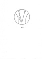
Фигура 1: проведение фиброназофаринголарингоскопии и введение диагностического зонда аппаратно-программного комплекса «ИнСпектр-М» через дополнительный канал фиброскопаFigure 1: carrying out fibronasopharyngolaryngoscopy and introduction of the diagnostic probe of the hardware-software complex "InSpektr-M" through the additional channel of the fibroscope
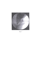
Фигура 2: установка диагностического зонда аппаратно-программного комплекса «ИнСпектр-М» на неизмененную (контроль) слизистую оболочкуFigure 2: installation of the diagnostic probe of the hardware-software complex "InSpektr-M" on the unchanged (control) mucous membrane
Фигура 3: Алгоритм проведения диагностики с использованием аппаратно-программного комплекса «ИнСпектр-М» (точка 1 - Интактная слизистая оболочка в области вестибулярной складки, точка 2 - Интактная слизистая оболочка в области черпаловидного хряща, точка 3 - Интактная слизистая оболочка в области передней комиссуры, точка 4 - Интактная слизистая оболочка в области голосовой складки, точка 5 - Видимая граница опухоли, точки 6- n+1 - Измененная слизистая оболочка).Figure 3: Algorithm for diagnostics using the hardware and software complex "InSpectr-M" (point 1 - Intact mucous membrane in the region of the vestibular fold, point 2 - Intact mucous membrane in the area of the arytenoid cartilage, point 3 - Intact mucous membrane in the area of the anterior commissure , point 4 - Intact mucous membrane in the area of the vocal fold, point 5 - Visible border of the tumor, points 6- n + 1 - Altered mucous membrane).
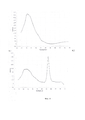
Фигура 4: Фиксация полученных данных компьютерной программой «ИнСпектр-М», где 4.1 - график, полученный при проведении диагностики в интактной точке, 4.2 - в центре новообразования (по оси Ох - длина волны в нм, по оси Оу - интенсивность излучения, индекс пролиферативной активности эпителия определяется в диапазоне длины волны от 625 до 650 нм)Figure 4: Fixation of the obtained data by the computer program "InSpektr-M", where 4.1 is a graph obtained during diagnostics at an intact point, 4.2 - in the center of the neoplasm (along the Ox axis - the wavelength in nm, along the Oy axis - the radiation intensity, index the proliferative activity of the epithelium is determined in the wavelength range from 625 to 650 nm)
Фигура 5: установка диагностического зонда аппаратно-программного комплекса «ИнСпектр-М» на измененную слизистую оболочку.Figure 5: installation of the diagnostic probe of the hardware-software complex "InSpektr-M" on the altered mucous membrane.
Способ поясняется следующими примерами.The method is illustrated by the following examples.
Пример 1. Больной В., 56 лет. Клинический диагноз: Хронический гиперпластический ларингит. При проведении фиброназофарингоскопии получены следующие результаты: левая голосовая складки умеренно гиперемирована, отечна, от уровня голосового отростка черпаловидного хряща, до передней комиссуры, свободный край голосовой складки неровный, область гиперемии слизистой оболочки локально покрыта белым налетом, подвижность гортани при фонации в полном объеме. Выполнена биопсия из средних отделов голосовой складки. Гистологическое исследование: хронический воспалительный процесс.Example 1. Patient C., 56 years old. Clinical diagnosis: Chronic hyperplastic laryngitis. When carrying out fibronasopharyngoscopy, the following results were obtained: the left vocal fold is moderately hyperemic, edematous, from the level of the vocal process of the arytenoid cartilage to the anterior commissure, the free edge of the vocal fold is uneven, the area of hyperemia of the mucous membrane is locally covered with a white bloom, the mobility of the larynx during phonation is in full. A biopsy was performed from the middle sections of the vocal fold. Histological examination: chronic inflammatory process.
По данным контактной эндоскопии с использованием световых фильтров для визуализации сосудистого рисунка, проведенной в условиях эндотрахеального наркоза при прямой опорной микроларингоскопии: патологически измененная сосудистая сеть визуализирована на всем участке гиперемии левой голосовой складки, обзор в области передней комиссуры технически затруднен из-за нависания надгортанника. Выполнена мультифокальная биопсия новообразования. Гистологическое исследование: хронический воспалительный процесс. Дисплазия 1-2 степени тяжести.According to the data of contact endoscopy using light filters to visualize the vascular pattern, carried out under endotracheal anesthesia with direct supporting microlaryngoscopy: the pathologically altered vascular network was visualized throughout the entire area of hyperemia of the left vocal fold, the view in the anterior commissure region is technically difficult due to the overhanging of the epiglottis. A multifocal biopsy of the neoplasm was performed. Histological examination: chronic inflammatory process. Dysplasia of 1-2 degrees of severity.
Затем была выполнена фиброназофаринголарингоскопия под местной анестезией с одномоментной установкой в канал фиброскопа диагностического зонда аппаратно-программного комплекса «ИнСпектр-М» выполненного с возможностью локального контактного подведения возбуждающего излучения к участку ткани, в том числе к трудновизуализируемым участкам слизистой оболочки гортани, и последующего приема сигнала собственной флуоресценции ткани. С помощью диагностического зонда была проведена флуоресцентная диагностика в области патологически измененных и неизмененных (контрольных) участков слизистой оболочки с последующей оценкой индекса пролиферативной активности эпителия по алгоритму: диагностический зонд диаметром 1,0 мм устанавливался на интактный участок слизистой оболочки вестибулярной складки (точка 1) под углом 90 градусов, после чего выполнялась спектрометрия участка слизистой оболочки, находящегося непосредственно в контакте в сечением зонда, далее выполнялась спектрометрия интактной слизистой оболочки черпало-надгортанного хряща (точка 2), интактной слизистой оболочки в области передней комиссуры (точка 3), интактного участка слизистой оболочки, находящегося на расстоянии 2 мм от видимой границы новообразования (точка 4), в области видимой границы измененной слизистой оболочки (точка 5), и затем измененная слизистая оболочка (точки 6, 7, 8, 9, 10, которые находятся в пределах площади визуально измененной слизистой оболочки на расстоянии 1 мм друг от друга, количество этих точек зависит от площади измененной слизистой оболочки, в данном случае диагностика проводилась в 5 точках в пределах площади визуально измененной слизистой оболочки), точка выполнения прицельной биопсии определяется в месте определения наибольшего индекса пролиферативной активности эпителия (интенсивности собственной флуоресценции) в области измененной слизистой оболочки. Полученный результат фиксирован программным обеспечением аппаратно-программного комплекса «ИнСпектр-М». В конкретном случае точкой с наибольшим индексом пролиферативной активности оказалась точка 7, эта точка выбрана для проведения с прицельной биопсии слизистой оболочки. По данным гистологического исследования обнаружен плоскоклеточный рак in situ.Then, fibronasopharyngolaryngoscopy was performed under local anesthesia with the simultaneous installation of a diagnostic probe of the hardware-software complex "InSpektr-M" into the fibroscope canal, made with the possibility of local contact supply of exciting radiation to a tissue site, including difficult to visualize areas of the laryngeal mucosa, and subsequent signal reception intrinsic fluorescence of the tissue. With the help of a diagnostic probe, fluorescence diagnostics was carried out in the area of pathologically altered and unchanged (control) areas of the mucous membrane, followed by an assessment of the index of proliferative activity of the epithelium according to the algorithm: a diagnostic probe with a diameter of 1.0 mm was installed on an intact area of the mucous membrane of the vestibular fold (point 1) under an angle of 90 degrees, after which spectrometry was performed on the area of the mucous membrane that was directly in contact in the section of the probe, then spectrometry was performed on the intact mucous membrane of the scooped-supraglottic cartilage (point 2), intact mucous membrane in the area of the anterior commissure (point 3), intact area of the mucosa membrane located at a distance of 2 mm from the visible border of the neoplasm (point 4), in the area of the visible border of the changed mucous membrane (point 5), and then the changed mucous membrane (points 6, 7, 8, 9, 10, which are within the area visually altered mucosa membranes at a distance of 1 mm from each other, the number of these points depends on the area of the altered mucosa, in this case, the diagnosis was carried out at 5 points within the area of the visually altered mucous membrane), the point of performing targeted biopsy is determined at the place where the highest index of epithelial proliferative activity is determined ( intensity of its own fluorescence) in the area of the altered mucous membrane. The obtained result is fixed by the software of the hardware-software complex "InSpektr-M". In a specific case, the point with the highest index of proliferative activity turned out to be point 7, this point was chosen for conducting with targeted biopsy of the mucous membrane. According to histological examination, squamous cell carcinoma in situ was found.
С помощью заявленного способа нами было обследовано 20 пациентов. После взятия биопсийного материала согласно заявляемому способу из точки с наибольшим индексом пролиферативной активности эпителия, был выявлен плоскоклеточный рак in situ.Using the claimed method, we examined 20 patients. After taking a biopsy material according to the claimed method from the point with the highest index of proliferative activity of the epithelium, squamous cell carcinoma in situ was detected.
Claims (1)
Priority Applications (1)
| Application Number | Priority Date | Filing Date | Title |
|---|---|---|---|
| RU2020111476A RU2729503C1 (en) | 2020-03-19 | 2020-03-19 | Method of biopsy in patients with suspected cancer of throat |
Applications Claiming Priority (1)
| Application Number | Priority Date | Filing Date | Title |
|---|---|---|---|
| RU2020111476A RU2729503C1 (en) | 2020-03-19 | 2020-03-19 | Method of biopsy in patients with suspected cancer of throat |
Publications (1)
| Publication Number | Publication Date |
|---|---|
| RU2729503C1 true RU2729503C1 (en) | 2020-08-07 |
Family
ID=72085886
Family Applications (1)
| Application Number | Title | Priority Date | Filing Date |
|---|---|---|---|
| RU2020111476A RU2729503C1 (en) | 2020-03-19 | 2020-03-19 | Method of biopsy in patients with suspected cancer of throat |
Country Status (1)
| Country | Link |
|---|---|
| RU (1) | RU2729503C1 (en) |
Cited By (1)
| Publication number | Priority date | Publication date | Assignee | Title |
|---|---|---|---|---|
| RU2782465C1 (en) * | 2022-04-14 | 2022-10-27 | Федеральное Государственное Бюджетное Учреждение "Национальный Медицинский Исследовательский Центр Оториноларингологии Федерального Медико-Биологического Агентства" (Фгбу Нмицо Фмба России) | Method for performing a puncture biopsy of neoplasms of the larynx |
Citations (3)
| Publication number | Priority date | Publication date | Assignee | Title |
|---|---|---|---|---|
| RU2129273C1 (en) * | 1997-11-26 | 1999-04-20 | Московский научно-исследовательский онкологический институт им.П.А.Герцена | Method for endoscopic fluorescent examination of malign swellings of hollow organs |
| UA18143U (en) * | 2006-06-22 | 2006-10-16 | Oleksandr Vasyliovy Kovtunenko | Method for predicting clinical course of laryngeal cancer |
| RU2709830C1 (en) * | 2018-12-21 | 2019-12-23 | Федеральное государственное бюджетное образовательное учреждение высшего образования "ОРЛОВСКИЙ ГОСУДАРСТВЕННЫЙ УНИВЕРСИТЕТ имени И.С. ТУРГЕНЕВА" (ОГУ им. И.С. Тургенева) | Device for fluorescent-reflective spectroscopy for diagnosing focal and diffuse new growths in a fine-needle puncture-aspiration biopsy |
-
2020
- 2020-03-19 RU RU2020111476A patent/RU2729503C1/en active
Patent Citations (3)
| Publication number | Priority date | Publication date | Assignee | Title |
|---|---|---|---|---|
| RU2129273C1 (en) * | 1997-11-26 | 1999-04-20 | Московский научно-исследовательский онкологический институт им.П.А.Герцена | Method for endoscopic fluorescent examination of malign swellings of hollow organs |
| UA18143U (en) * | 2006-06-22 | 2006-10-16 | Oleksandr Vasyliovy Kovtunenko | Method for predicting clinical course of laryngeal cancer |
| RU2709830C1 (en) * | 2018-12-21 | 2019-12-23 | Федеральное государственное бюджетное образовательное учреждение высшего образования "ОРЛОВСКИЙ ГОСУДАРСТВЕННЫЙ УНИВЕРСИТЕТ имени И.С. ТУРГЕНЕВА" (ОГУ им. И.С. Тургенева) | Device for fluorescent-reflective spectroscopy for diagnosing focal and diffuse new growths in a fine-needle puncture-aspiration biopsy |
Non-Patent Citations (4)
| Title |
|---|
| . АЛЕКСАНДРОВ М.Т. РАМАН-Флуоресцентная диагностика состояния тканей человека в норме и при патологии и ее аппаратно-программное решение. Российский стоматологический журнал 21 (5) 2017, стр. 228-232. * |
| CSANADY M. ALA (5-aminolevulinic acid)-induced protoporphyrin IX fluorescence in the endoscopic diagnostic and control of pharyngo-laryngeal cancer. Eur Arch Otorhinolaryngol 2004 May;261(5):262-6. * |
| НАЖМУДИНОВ И.И. Современные аспекты диагностики и хирургического лечения гиперпластических процессов гортани. Consilium medicum N 11 2017, стр.29-33. * |
| НАЖМУДИНОВ И.И. Современные аспекты диагностики и хирургического лечения гиперпластических процессов гортани. Consilium medicum N 11 2017, стр.29-33. АЛЕКСАНДРОВ М.Т. РАМАН-Флуоресцентная диагностика состояния тканей человека в норме и при патологии и ее аппаратно-программное решение. Российский стоматологический журнал 21 (5) 2017, стр. 228-232. CSANADY M. ALA (5-aminolevulinic acid)-induced protoporphyrin IX fluorescence in the endoscopic diagnostic and control of pharyngo-laryngeal cancer. Eur Arch Otorhinolaryngol 2004 May;261(5):262-6. * |
Cited By (1)
| Publication number | Priority date | Publication date | Assignee | Title |
|---|---|---|---|---|
| RU2782465C1 (en) * | 2022-04-14 | 2022-10-27 | Федеральное Государственное Бюджетное Учреждение "Национальный Медицинский Исследовательский Центр Оториноларингологии Федерального Медико-Биологического Агентства" (Фгбу Нмицо Фмба России) | Method for performing a puncture biopsy of neoplasms of the larynx |
Similar Documents
| Publication | Publication Date | Title |
|---|---|---|
| Haka et al. | In vivo margin assessment during partial mastectomy breast surgery using Raman spectroscopy | |
| US6975899B2 (en) | Multi-modal optical tissue diagnostic system | |
| CA2658811C (en) | Multi modal spectroscopy | |
| US9788728B2 (en) | Endoscopic polarized multispectral light scattering scanning method | |
| US9820655B2 (en) | Systems and methods for spectral analysis of a tissue mass using an instrument, an optical probe, and a Monte Carlo or a diffusion algorithm | |
| US6537211B1 (en) | Flourescence imaging endoscope | |
| US6922583B1 (en) | Method for measuring tissue morphology | |
| US20090326385A1 (en) | Obtaining optical tissue properties | |
| Crow et al. | Optical diagnostics in urology: current applications and future prospects. | |
| JP2007505645A (en) | Automated endoscope device, diagnostic method and usage | |
| JP2006508358A (en) | Use of high wave number Raman spectroscopy to measure tissue | |
| WO2003087793A1 (en) | Systems and methods for spectroscopy of biological tissue | |
| EP1495309A1 (en) | Systems and methods for spectroscopy of biological tissue | |
| US20100249607A1 (en) | Quantitative spectroscopic imaging | |
| Benboujja et al. | Intraoperative imaging of pediatric vocal fold lesions using optical coherence tomography | |
| Glover et al. | A review of new and emerging techniques for optical diagnosis of colonic polyps | |
| RU2729503C1 (en) | Method of biopsy in patients with suspected cancer of throat | |
| Tozar et al. | Laser induced autofluorescence lifetime to identify larynx squamous cell carcinoma: short series ex vivo study | |
| RU2184486C2 (en) | Method and device for diagnosing oncological diseases | |
| Szeto et al. | Contact endoscopy as a novel technique in the detection and diagnosis of mucosal lesions in the head and neck: a brief review | |
| RU2317009C1 (en) | Method for detecting the volume of urinary bladder resection at endoscopic therapy of urinary bladder cancer | |
| Lim et al. | A feasibility study of photoacoustic imaging of ex vivo endoscopic mucosal resection tissues from Barrett’s esophagus patients | |
| RU2761469C1 (en) | Method for combined endoscopic assessment of radiotherapy efficiency for primary larynx cancer | |
| RU2819641C1 (en) | Method for combined endoscopic diagnosis of chronic inflammatory and precancerous processes and primary oropharyngeal cancers | |
| RU2152162C1 (en) | Method for increasing accuracy in detecting malignant neoplasms and determining their localization boundaries |