RU2724895C2 - Antimicrobial immunomodulation - Google Patents
Antimicrobial immunomodulation Download PDFInfo
- Publication number
- RU2724895C2 RU2724895C2 RU2016147048A RU2016147048A RU2724895C2 RU 2724895 C2 RU2724895 C2 RU 2724895C2 RU 2016147048 A RU2016147048 A RU 2016147048A RU 2016147048 A RU2016147048 A RU 2016147048A RU 2724895 C2 RU2724895 C2 RU 2724895C2
- Authority
- RU
- Russia
- Prior art keywords
- cells
- macrophages
- pathogenic
- infection
- antigenic
- Prior art date
Links
Images


Classifications
-
- A—HUMAN NECESSITIES
- A61—MEDICAL OR VETERINARY SCIENCE; HYGIENE
- A61K—PREPARATIONS FOR MEDICAL, DENTAL OR TOILETRY PURPOSES
- A61K39/00—Medicinal preparations containing antigens or antibodies
- A61K39/02—Bacterial antigens
-
- A—HUMAN NECESSITIES
- A61—MEDICAL OR VETERINARY SCIENCE; HYGIENE
- A61K—PREPARATIONS FOR MEDICAL, DENTAL OR TOILETRY PURPOSES
- A61K39/00—Medicinal preparations containing antigens or antibodies
- A61K39/02—Bacterial antigens
- A61K39/025—Enterobacteriales, e.g. Enterobacter
- A61K39/0266—Klebsiella
-
- A—HUMAN NECESSITIES
- A61—MEDICAL OR VETERINARY SCIENCE; HYGIENE
- A61K—PREPARATIONS FOR MEDICAL, DENTAL OR TOILETRY PURPOSES
- A61K39/00—Medicinal preparations containing antigens or antibodies
- A61K39/02—Bacterial antigens
- A61K39/025—Enterobacteriales, e.g. Enterobacter
- A61K39/0258—Escherichia
-
- A—HUMAN NECESSITIES
- A61—MEDICAL OR VETERINARY SCIENCE; HYGIENE
- A61P—SPECIFIC THERAPEUTIC ACTIVITY OF CHEMICAL COMPOUNDS OR MEDICINAL PREPARATIONS
- A61P11/00—Drugs for disorders of the respiratory system
-
- A—HUMAN NECESSITIES
- A61—MEDICAL OR VETERINARY SCIENCE; HYGIENE
- A61P—SPECIFIC THERAPEUTIC ACTIVITY OF CHEMICAL COMPOUNDS OR MEDICINAL PREPARATIONS
- A61P17/00—Drugs for dermatological disorders
-
- A—HUMAN NECESSITIES
- A61—MEDICAL OR VETERINARY SCIENCE; HYGIENE
- A61P—SPECIFIC THERAPEUTIC ACTIVITY OF CHEMICAL COMPOUNDS OR MEDICINAL PREPARATIONS
- A61P31/00—Antiinfectives, i.e. antibiotics, antiseptics, chemotherapeutics
- A61P31/04—Antibacterial agents
-
- A—HUMAN NECESSITIES
- A61—MEDICAL OR VETERINARY SCIENCE; HYGIENE
- A61P—SPECIFIC THERAPEUTIC ACTIVITY OF CHEMICAL COMPOUNDS OR MEDICINAL PREPARATIONS
- A61P31/00—Antiinfectives, i.e. antibiotics, antiseptics, chemotherapeutics
- A61P31/12—Antivirals
-
- A—HUMAN NECESSITIES
- A61—MEDICAL OR VETERINARY SCIENCE; HYGIENE
- A61P—SPECIFIC THERAPEUTIC ACTIVITY OF CHEMICAL COMPOUNDS OR MEDICINAL PREPARATIONS
- A61P37/00—Drugs for immunological or allergic disorders
- A61P37/02—Immunomodulators
-
- A—HUMAN NECESSITIES
- A61—MEDICAL OR VETERINARY SCIENCE; HYGIENE
- A61P—SPECIFIC THERAPEUTIC ACTIVITY OF CHEMICAL COMPOUNDS OR MEDICINAL PREPARATIONS
- A61P37/00—Drugs for immunological or allergic disorders
- A61P37/02—Immunomodulators
- A61P37/04—Immunostimulants
-
- A—HUMAN NECESSITIES
- A61—MEDICAL OR VETERINARY SCIENCE; HYGIENE
- A61K—PREPARATIONS FOR MEDICAL, DENTAL OR TOILETRY PURPOSES
- A61K39/00—Medicinal preparations containing antigens or antibodies
- A61K2039/51—Medicinal preparations containing antigens or antibodies comprising whole cells, viruses or DNA/RNA
- A61K2039/52—Bacterial cells; Fungal cells; Protozoal cells
- A61K2039/521—Bacterial cells; Fungal cells; Protozoal cells inactivated (killed)
-
- A—HUMAN NECESSITIES
- A61—MEDICAL OR VETERINARY SCIENCE; HYGIENE
- A61K—PREPARATIONS FOR MEDICAL, DENTAL OR TOILETRY PURPOSES
- A61K39/00—Medicinal preparations containing antigens or antibodies
- A61K2039/54—Medicinal preparations containing antigens or antibodies characterised by the route of administration
- A61K2039/541—Mucosal route
-
- A—HUMAN NECESSITIES
- A61—MEDICAL OR VETERINARY SCIENCE; HYGIENE
- A61K—PREPARATIONS FOR MEDICAL, DENTAL OR TOILETRY PURPOSES
- A61K39/00—Medicinal preparations containing antigens or antibodies
- A61K2039/545—Medicinal preparations containing antigens or antibodies characterised by the dose, timing or administration schedule
-
- A—HUMAN NECESSITIES
- A61—MEDICAL OR VETERINARY SCIENCE; HYGIENE
- A61K—PREPARATIONS FOR MEDICAL, DENTAL OR TOILETRY PURPOSES
- A61K39/00—Medicinal preparations containing antigens or antibodies
- A61K2039/55—Medicinal preparations containing antigens or antibodies characterised by the host/recipient, e.g. newborn with maternal antibodies
-
- A—HUMAN NECESSITIES
- A61—MEDICAL OR VETERINARY SCIENCE; HYGIENE
- A61K—PREPARATIONS FOR MEDICAL, DENTAL OR TOILETRY PURPOSES
- A61K39/00—Medicinal preparations containing antigens or antibodies
- A61K2039/57—Medicinal preparations containing antigens or antibodies characterised by the type of response, e.g. Th1, Th2
-
- A—HUMAN NECESSITIES
- A61—MEDICAL OR VETERINARY SCIENCE; HYGIENE
- A61K—PREPARATIONS FOR MEDICAL, DENTAL OR TOILETRY PURPOSES
- A61K39/00—Medicinal preparations containing antigens or antibodies
- A61K2039/58—Medicinal preparations containing antigens or antibodies raising an immune response against a target which is not the antigen used for immunisation
-
- Y—GENERAL TAGGING OF NEW TECHNOLOGICAL DEVELOPMENTS; GENERAL TAGGING OF CROSS-SECTIONAL TECHNOLOGIES SPANNING OVER SEVERAL SECTIONS OF THE IPC; TECHNICAL SUBJECTS COVERED BY FORMER USPC CROSS-REFERENCE ART COLLECTIONS [XRACs] AND DIGESTS
- Y02—TECHNOLOGIES OR APPLICATIONS FOR MITIGATION OR ADAPTATION AGAINST CLIMATE CHANGE
- Y02A—TECHNOLOGIES FOR ADAPTATION TO CLIMATE CHANGE
- Y02A50/00—TECHNOLOGIES FOR ADAPTATION TO CLIMATE CHANGE in human health protection, e.g. against extreme weather
- Y02A50/30—Against vector-borne diseases, e.g. mosquito-borne, fly-borne, tick-borne or waterborne diseases whose impact is exacerbated by climate change
Abstract
Description
Область техникиTechnical field
Различные аспекты изобретения относятся к иммунологическим лечебным средствам для лечения или профилактики патологий, ассоциированных с микробными инфекциями у позвоночного, включая применение микробных вакцин.Various aspects of the invention relate to immunological therapeutic agents for treating or preventing pathologies associated with microbial infections in a vertebrate, including the use of microbial vaccines.
Уровень техникиState of the art
Врожденная иммунная система и адаптивная иммунная система у позвоночных работают согласованно с обеспечением, в ряду множества других действий, защиты от инфекций патогенными микроорганизмами. Противомикробные вакцины можно формулировать для стимуляции врожденной и адаптивной иммунных систем, но общеизвестно, что эффективный ответ на вакцинацию включает специфичный адаптивный ответ на один или несколько иммуногенов, находящихся в вакцине. Таким образом, для вызова специфичного адаптивного ответа на более чем один серовар можно использовать поливалентные вакцины, такие как некоторые пневмококковые вакцины. Также описаны вакцины, которые обеспечивают определенную степень перекрестного иммунитета, при котором перекрестная реактивность на антиген, отличный от иммуногена, обеспечивает определенную степень защитного иммунитета к гетерологичным микроорганизмам.The innate immune system and adaptive immune system in vertebrates work in concert with the provision, among many other actions, of protection against infections by pathogenic microorganisms. Antimicrobial vaccines can be formulated to stimulate the innate and adaptive immune systems, but it is well known that an effective vaccination response includes a specific adaptive response to one or more immunogens present in the vaccine. Thus, multivalent vaccines, such as some pneumococcal vaccines, can be used to elicit a specific adaptive response to more than one serovar. Vaccines are also described that provide a certain degree of cross-immunity, in which cross-reactivity to an antigen other than an immunogen provides a certain degree of protective immunity to heterologous microorganisms.
Сущность изобретенияSUMMARY OF THE INVENTION
В одном из аспектов изобретение относится к способам и композициям для лечения патологического состояния у позвоночного, характеризуемого патологиями, ассоциированными с микробной инфекцией, включающим применение микробных вакцин, получаемых из одного из патогенных организмов, для лечения инфекций, обуславливаемых гетерологичным патогенным организмом.In one aspect, the invention relates to methods and compositions for treating a pathological condition in a vertebrate characterized by pathologies associated with a microbial infection, including the use of microbial vaccines derived from one of the pathogenic organisms, for treating infections caused by a heterologous pathogenic organism.
Краткое описание чертежейBrief Description of the Drawings
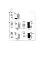
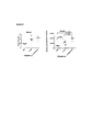
На фигуре 1 представлено количество воспалительных моноцитов и дендритных клеток в дренирующих лимфоузлах, легких и селезенке мышей после обработки антигенной композицией K. pneumoniae или PBS, как описано в примере 1А в настоящем документе.The figure 1 shows the number of inflammatory monocytes and dendritic cells in the draining lymph nodes, lungs and spleen of mice after treatment with the K. pneumoniae or PBS antigenic composition as described in Example 1A herein.
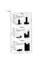
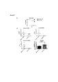
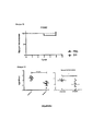
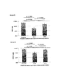
На фигуре 2 представлено общее количество моноцитов и дендритных клеток в легких, брюшине и селезенке мышей после обработки антигенной композицией K. pneumoniae, антигенной композицией Е. coli или PBS, как описано в примере 1В в настоящем документе.Figure 2 shows the total number of monocytes and dendritic cells in the lungs, peritoneum and spleen of mice after treatment with the K. pneumoniae antigenic composition, E. coli antigenic composition or PBS, as described in Example 1B herein.
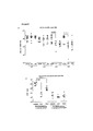
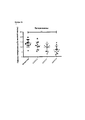
На фигуре 3 представлено общее количество CD4+-T-клеток, CD8+-T-клеток и NK клеток у мышей, обработанных антигенной композицией К. pneumoniae, антигенной композицией Е. coli или PBS, как описано в примере 1В в настоящем документе.Figure 3 shows the total number of CD4 + T cells, CD8 + T cells and NK cells in mice treated with the K. pneumoniae antigenic composition, E. coli antigenic composition or PBS, as described in Example 1B herein.
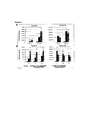
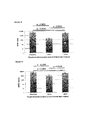
На фигуре 4 представлено общее количество (А) воспалительных моноцитов и дендритных клеток и (В) CD4+-T-клеток, CD8+-Т-клеток и NK клеток у мышей, обработанных антигенной композицией инактивированных нагреванием K. pneumoniae, антигенной композицией инактивированных фенолом K. pneumoniae или PBS, как описано в примере 1C в настоящем документе.The figure 4 shows the total number of (A) inflammatory monocytes and dendritic cells and (B) CD4 + T cells, CD8 + T cells and NK cells in mice treated with an antigenic composition heat-inactivated K. pneumoniae antigenic composition inhibited by phenol K. pneumoniae or PBS, as described in Example 1C herein.
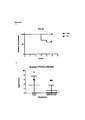
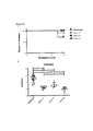
На фигуре 5 представлена относительная частота CD11b+Gr-1+-клеток, детектируемых в толстой кишке мышей, обработанных антигенными композициями K. pneumoniae или Е. coli или контрольным PBS.Figure 5 shows the relative frequency of CD11b + Gr-1 + cells detected in the colon of mice treated with antigenic compositions of K. pneumoniae or E. coli or control PBS.
На фигуре 6 представлена относительная частота CD11b+Gr-1+-клеток, детектируемых в легких мышей, обработанных антигенными композициями K. pneumoniae или Е. coli или контрольным PBS.Figure 6 shows the relative frequency of CD11b + Gr-1 + cells detected in the lungs of mice treated with antigenic compositions of K. pneumoniae or E. coli or control PBS.
На фигуре 7 проиллюстрирована противомикробная профилактика, как описано в примере 3, при которой обработка композицией, содержащей полностью убитые клетки K. pneumoniae, обеспечивает защитный иммунитет против последующего заражения S. pneumoniae. Фигура 7А представляет собой кривую выживаемости через 5 суток после заражения. На фигуре 7В проиллюстрирован количественный анализ S. pneumoniae в назальном смыве, гомогенате легких и селезенки. Мегакариоциты подсчитывали в десяти соседних полях зрения при большом увеличении (увеличение 400×) гистологических срезов селезенки. Статистическую значимость определяли посредством двухстороннего критерия Манна-Уитни.The figure 7 illustrates antimicrobial prophylaxis, as described in example 3, in which treatment with a composition containing completely killed K. pneumoniae cells provides protective immunity against subsequent infection with S. pneumoniae. Figure 7A is a
На фигуре 8 проиллюстрировано гетерологичное противомикробное лечебное средство в модели на мышах, как описано в примере 4, в котором обработка композицией, содержащей полностью убитые клетки Е. coli, эффективна для подавления инфекции, обуславливаемой гетерологичным штаммом адгезивно-инвазивных Е. coli.Figure 8 illustrates a heterologous antimicrobial agent in a mouse model as described in Example 4, in which treatment with a composition containing completely killed E. coli cells is effective in suppressing the infection caused by the heterologous strain of adhesive-invasive E. coli.
На фигуре 9 проиллюстрировано гетерологичное противомикробное лечебное средство в модели на мышах, как описано в примере 3, в котором обработка композицией, содержащей полностью убитые клетки Klebsiella pneumonia, эффективна для подавления легочной инфекции, обуславливаемой Pseudomonas aeruginosa (PA14).Figure 9 illustrates a heterologous antimicrobial agent in a mouse model, as described in Example 3, in which treatment with a composition containing completely killed Klebsiella pneumonia cells is effective in suppressing the pulmonary infection caused by Pseudomonas aeruginosa (PA14).
На фигуре 10 проиллюстрировано гетерологичное противомикробное лечебное средство в модели на мышах, как описано в примере 3, в котором обработка композицией, содержащей полностью убитые клетки Klebsiella pneumonia, эффективна для подавления легочной инфекции, обуславливаемой Streptococcus pneumonias (P1542).Figure 10 illustrates a heterologous antimicrobial agent in a mouse model, as described in Example 3, in which treatment with a composition containing completely killed Klebsiella pneumonia cells is effective in suppressing the pulmonary infection caused by Streptococcus pneumonias (P1542).
На фигуре 11 проиллюстрировано гетерологичное противомикробное лечебное средство в модели на мышах, как описано в примере 5, в котором обработка композицией, содержащей полностью убитые клетки Е. coli, эффективна для подавления перитонеальной инфекции, обуславливаемой S. enterica.Figure 11 illustrates a heterologous antimicrobial agent in a mouse model as described in Example 5, in which treatment with a composition containing completely killed E. coli cells is effective in suppressing peritoneal infection caused by S. enterica.
На фигуре 12 проиллюстрировано гетерологичное противомикробное лечебное средство в модели на мышах, как описано в примере 5, в которой эффективность обработки двумя различными композициями, содержащими альтернативные штаммы полностью убитых клеток Е. coli, сравнивают с обработкой антигенной композицией S. enterica для подавления перитонеальной инфекции, обуславливаемой S. enterica.Figure 12 illustrates a heterologous antimicrobial agent in a mouse model, as described in Example 5, in which the efficacy of treatment with two different compositions containing alternative strains of completely killed E. coli cells is compared with treatment with S. enterica antigenic composition to suppress peritoneal infection. conditioned by S. enterica.
Фигура 13 представляет собой столбчатую диаграмму, иллюстрирующую гетерологичное противомикробное лечебное средство в модели на мышах, как описано в примере 6, в котором представлена эффективность полученного из S. aureus SSI (QBSAU), направленного против заражения кожи Р. aeruginosa, со значительно сниженным количеством бактерий Р. aeruginosa после предварительной обработки QBSAU.Figure 13 is a bar graph illustrating a heterologous antimicrobial agent in a mouse model as described in Example 6, which shows the efficacy of S. aerureus S.I. aureus SSI (QBSAU) against skin infection with a significantly reduced bacterial count. P. aeruginosa after pretreatment with QBSAU.
Фигура 14 представляет собой столбчатую диаграмму, иллюстрирующую гетерологичное противомикробное лечебное средство в модели на мышах, как описано в примере 6, повторяющую и подтверждающую данные, представленные на фигуре 13, на которой представлена эффективность полученного из S. aureus SSI (QBSAU) против заражения кожи Р. aeruginosa, со значительным сниженным количеством бактерий Р. aeruginosa после предварительной обработки QBSAU.Figure 14 is a bar graph illustrating a heterologous antimicrobial drug in a mouse model as described in Example 6, repeating and confirming the data presented in Figure 13, which shows the efficacy of S. aureus SSI (QBSAU) against skin infection P aeruginosa, with a significant reduced number of P. aeruginosa bacteria after pre-treatment with QBSAU.
Фигура 15 представляет собой столбчатую диаграмму, иллюстрирующую гетерологичное противомикробное лечебное средство в модели на мышах, как описано в примере 3с, иллюстрирующем SSI на основе Klebsiella pneumoniae (QBKPN), демонстрирующее статистически превосходящую эффективность профилактики по сравнению с SSI на основе Е. coli (QBECO), в отношении защиты от заражения легких Р. aeruginosa.Figure 15 is a bar graph illustrating a heterologous antimicrobial drug in a mouse model as described in Example 3c, illustrating Klebsiella pneumoniae-based SSI (QBKPN), showing a statistically superior prophylaxis compared to E. coli-based SSI (QBECO) , regarding protection against infection of lungs of P. aeruginosa.
Фигура 16 представляет собой столбчатую диаграмму, иллюстрирующую гетерологичное противомикробное лечебное средство в модели на мышах, как описано в примере 3с, иллюстрирующем SSI на основе Klebsiella pneumoniae (QBKPN), демонстрирующее статистически превосходящую эффективность профилактики по сравнению с SSI на основе Е. coli (QBECO), в отношении защиты от заражения легких S. pneumoniae.Figure 16 is a bar graph illustrating a heterologous antimicrobial drug in a mouse model as described in Example 3c, illustrating Klebsiella pneumoniae-based SSI (QBKPN), showing a statistically superior prophylaxis compared to E. coli-based SSI (QBECO) , regarding protection against infection of the lungs S. pneumoniae.
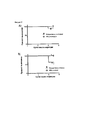
Фигура 17 представляет собой два графика, демонстрирующих направленное гетерологичное противомикробное лечебное средство в модели старческой легочной инфекции на мышах, демонстрирующей, что SSI Klebsiella pneumoniae (QBKPN) защищает от заражения легких S. pneumoniae у старых мышей. Положительное влияние на выживаемость для старых мышей, фигура 17В, было более выраженным, чем для молодых мышей, фигура 17А.Figure 17 is two graphs showing a targeted heterologous antimicrobial agent in a mouse model of senile pulmonary infection, demonstrating that SSI Klebsiella pneumoniae (QBKPN) protects S. pneumoniae from lung infection in old mice. The positive effect on survival for old mice, Figure 17B, was more pronounced than for young mice, Figure 17A.
Фигура 18 представляет собой график, иллюстрирующий, что SSI QBKPN уменьшает потерю массы у старых мышей после заражения S. pneumoniae, и этот положительное влияние выше для старых мышей по сравнению с молодыми мышами.Figure 18 is a graph illustrating that SSI QBKPN reduces weight loss in old mice after infection with S. pneumoniae, and this positive effect is higher for older mice compared to young mice.
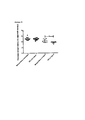
На фигуре 19 проиллюстрирована противомикробная профилактика в брюшной полости с графиком, иллюстрирующим, что SSI QBECO и QBKPN были протективными, как определено посредством бактериальной нагрузки в селезенке, где у мышей, обработанных QBECO, количества были значимо ниже, чем у мышей, обработанных QBKPN (данные представляют собой КОЕ/мл (среднее ± ст. откл.).Figure 19 illustrates antimicrobial prophylaxis in the abdominal cavity with a graph illustrating that SSI QBECO and QBKPN were protective, as determined by bacterial loading in the spleen, where the numbers in QBECO treated mice were significantly lower than in QBKPN treated mice (data represent CFU / ml (mean ± ST. off).
Подробное описание изобретенияDETAILED DESCRIPTION OF THE INVENTION
Различные аспекты изобретения относится к неожиданному открытию, что введение составов, содержащих антигенные детерминанты патогенных микроорганизмов, которые патогенны для определенных ткани или органа, эффективно при лечении патологий, ассоциированных с гетерологичными микробными инфекциями в этих конкретных ткани или органе. Например, композиции по изобретению можно вводить в участке, который удален от участка инфицирования. Таким образом, изобретение относится к антигенным композициям, получаемым из этих патогенных микроорганизмов, включая полностью убитые виды бактерий, вирусов или грибов или их компоненты, для терапевтического или профилактического лечения гетерологичных микробных инфекций, и к способам их применения. Например, композиции можно получать из эндогенных патогенных организмов или экзогенных патогенных организмов, как более подробно описано ниже.Various aspects of the invention relate to the unexpected discovery that the administration of formulations containing antigenic determinants of pathogenic microorganisms that are pathogenic to certain tissue or organ is effective in treating pathologies associated with heterologous microbial infections in these specific tissues or organs. For example, the compositions of the invention can be administered at a site that is remote from the site of infection. Thus, the invention relates to antigenic compositions obtained from these pathogenic microorganisms, including completely killed species of bacteria, viruses or fungi or their components, for therapeutic or prophylactic treatment of heterologous microbial infections, and to methods for their use. For example, compositions can be obtained from endogenous pathogenic organisms or exogenous pathogenic organisms, as described in more detail below.
Можно получать антигенные композиции по изобретению, которые содержат антигенные детерминанты, которые совместно специфичны к патогенному микроорганизму или характерны для него. В этом контексте, под "специфичными" подразумевают, что антигенные детерминанты в достаточной степени характерны для патогенного организма так, что их можно использовать для индукции у пациента иммунного ответа, такого как адаптивный иммунный ответ, против патогенного организма, если вводить антигенные детерминанты соответствующим образом, чтобы они могли произвести это действие. Понятно, что антигенные детерминанты не должны быть настолько специфичными, чтобы они были характерны только для одного конкретного штамма или вида патогенных организмов, так как даже специфичный иммунный ответ против конкретного патогенного организма может быть перекрестным с другими близкородственными организмами, которые в природных условиях также патогенны в ткани или органе, в которых происходит гетерологичная инфекция и для направления в которые формулируют или отбирают антигенную композицию.You can get antigenic compositions according to the invention, which contain antigenic determinants that are jointly specific to the pathogenic microorganism or characteristic of it. In this context, “specific” means that the antigenic determinants are sufficiently characteristic of the pathogenic organism so that they can be used to induce an immune response in the patient, such as an adaptive immune response, against the pathogenic organism, if antigenic determinants are introduced accordingly, so that they can perform this action. It is clear that antigenic determinants should not be so specific that they are characteristic of only one specific strain or species of pathogenic organisms, since even a specific immune response against a particular pathogenic organism can be cross-linked with other closely related organisms, which are also pathogenic in nature the tissue or organ in which the heterologous infection occurs and for which the antigenic composition is formulated or selected.
"Клетка" является основной структурной и функциональной единицей живого организма. У высших организмов, например, животных, клетки со сходной структурой и функцией, как правило, объединены в "ткани", которые выполняют конкретные функции. Таким образом, ткань включает совокупность сходных клеток и окружающего межклеточного вещества, например, эпителиальную ткань, соединительную ткань, мышцы, нервы. "Орган" представляет собой полностью дифференцированная структурная и функциональная единица высшего организма, которая может состоять из различных типов тканей и специализируется на определенной конкретной функции, например, почка, сердце, головной мозг, печень и т.д. Таким образом, подразумевают, что "конкретные орган, ткань или клетка" в настоящем документе включают любой конкретный орган и включают клетки и ткани, находящиеся в этом органе.A “cell” is the basic structural and functional unit of a living organism. In higher organisms, for example, animals, cells with a similar structure and function are usually combined into "tissues" that perform specific functions. Thus, tissue includes a combination of similar cells and the surrounding intercellular substance, for example, epithelial tissue, connective tissue, muscles, nerves. An "organ" is a completely differentiated structural and functional unit of a higher organism, which may consist of various types of tissues and specializes in a specific specific function, for example, kidney, heart, brain, liver, etc. Thus, it is understood that “a particular organ, tissue, or cell” as used herein includes any particular organ and includes cells and tissues located in that organ.
"Патогенные" факторы представляют собой такие факторы, как микроорганизмы, такие как бактерии или вирусы, которые известны, как обуславливающие инфекцию у хозяина в природных условиях, и в этом смысле в контексте настоящего изобретения "патогенный" используют для обозначения "патогенного в природных условиях". Хотя в искусственных условиях, таких как искусственная инокуляция микроорганизма в ткань, обуславливать инфекцию может широкий спектр микроорганизмов, спектр микроорганизмов, которые обуславливают инфекцию в природных условиях всегда ограничен и хорошо установлен медицинской практикой."Pathogenic" factors are factors such as microorganisms, such as bacteria or viruses, which are known to cause an infection in a host under natural conditions, and in this sense, in the context of the present invention, "pathogenic" is used to mean "pathogenic in natural conditions" . Although in artificial conditions, such as artificial inoculation of a microorganism into a tissue, a wide range of microorganisms can cause infection, the spectrum of microorganisms that cause an infection in natural conditions is always limited and well established by medical practice.
"Инфекция" представляет собой состояние, при котором организм или его часть проникает патогенный фактор (например, микроорганизм, такой как бактерия), который в благоприятствующих условиях размножается и производит действия, которые являются повреждающими (Taber's Cyclopedic Medical Dictionary, 14th Ed., C.L. Thomas, Ed., F.A. Davis Company, PA, USA). Инфекция не всегда проявляется клинически и может приводить только к локализованному повреждению клеток. Если защитные механизмы организма являются эффективными, инфекции могут оставаться субклиническими и временными. Инфекции могут распространяться локально, проявляясь клинически в виде острой, подострой или хронической клинически выраженной инфекцией или болезненным состоянием. Когда патогенный фактор получает доступ к лимфатической или сосудистой системе, местная инфекция также может становиться системной. Как правило, инфекции сопутствует воспаление, но воспаление может происходить и без инфекции.“Infection” is a condition in which an organism or part of it penetrates a pathogenic factor (for example, a microorganism such as a bacterium) that multiplies under favorable conditions and produces actions that are harmful (Taber's Cyclopedic Medical Dictionary, 14th Ed., CL Thomas , Ed., FA Davis Company, PA, USA). Infection does not always manifest clinically and can only lead to localized cell damage. If the body's defense mechanisms are effective, infections can remain subclinical and transient. Infections can spread locally, manifesting themselves clinically in the form of an acute, subacute or chronic clinically severe infection or painful condition. When a pathogenic factor gains access to the lymphatic or vascular system, a local infection can also become systemic. Infection is usually accompanied by inflammation, but inflammation can occur without infection.
"Воспаление" представляет собой характерную реакцию такни на повреждение (характеризующуюся отеком, покраснением, повышением температуры и болью) и включает последовательные изменения, которые происходят в живой ткань при ее повреждении. Инфекция и воспаление являются различными состояниями, хотя одно может являться следствием другого (Taber's Cyclopedic Medical Dictionary, выше). Таким образом, воспаление может происходить без инфекции, а инфекция может происходить без воспаления (хотя, как правило, воспаление является результатом инфекции патогенными бактериями или вирусами). Воспаление характеризуется следующими симптомами: покраснение (rubor), повышение температуры (calor), отеком (инфекция), болью (dolor). Локализованное видимое воспаление на коже может проявляться комбинацией этих симптомов, в частности покраснением в участке введения.“Inflammation” is a characteristic response of a tactic to damage (characterized by swelling, redness, fever and pain) and includes the sequential changes that occur in living tissue when it is damaged. Infection and inflammation are different conditions, although one may be the result of the other (Taber's Cyclopedic Medical Dictionary, supra). Thus, inflammation can occur without infection, and infection can occur without inflammation (although, as a rule, inflammation is the result of infection by pathogenic bacteria or viruses). Inflammation is characterized by the following symptoms: redness (rubor), fever (calor), swelling (infection), pain (dolor). Localized visible inflammation on the skin can be manifested by a combination of these symptoms, in particular, redness at the injection site.
По альтернативным аспектам изобретения лечить можно различных индивидуумов. Как используют в настоящем документе, "индивидуум" представляет собой животное, например, позвоночное, такое как млекопитающее, которому можно вводить конкретные патогенные бактерии, бактериальные антигены, вирусы, вирусные антигены или их композиции по изобретению. Таким образом, индивидуум может представлять собой пациента, например, человека, страдающего микробной инфекцией, или с подозрением на микробную инфекцию, или с риском развития микробной инфекции. Также индивидуум может являться экспериментальным животным, например, моделью инфекции на животном. В определенных вариантах осуществления термины "индивидуум" и "пациент" можно использовать взаимозаменяемо, и они могут включать человека, не являющегося человеком млекопитающего, не являющегося человеком примата, крысу, мышь, собаку и т.д. Здоровый индивидуум может представлять собой человека, не страдающего от инфекции, или без подозрения на инфекцию, или индивидуума, не страдающего хроническим нарушением или патологическим состоянием. "Здоровый индивидуум" также может представлять собой индивидуума без иммунной недостаточности. Под "иммунной недостаточностью" или "иммуносупрессией" подразумевают любое состояние, при котором иммунная система функционирует аномальным или недостаточным образом, например, где хозяин представляет собой пациента, не способного адекватно реагировать на инфекцию вследствие нарушенной или ослабленной иммунной системы. Иммунная недостаточность или иммуносупрессия могут являться следствием заболевания, применения определенных лекарственных средств (таких как химиотерапевтические средства, используемые при лечении злокачественных опухолей), или патологических состояний, присутствующих от рождения. Индивидуумов с иммунной недостаточностью с большей частотой можно найти среди грудных детей, пожилых индивидуумов и индивидуумов, подвергаемых интенсивному лечению лекарственным средством или лучевой терапией. Таким образом, аспекты изобретения включают лечение педиатрических и гериатрических пациентов, или пациентов с риском нозокомиальной инфекции. Например, конкретные группы пациентов могут включать пациентов с ослабленной иммунной системой вследствие инфекции ВИЧ или СПИД, злокачественной опухоли, трансплантацией паренхиматозного органа, трансплантацией стволовых клеток, серповидноклеточной анемии или асплении, врожденных иммунодефицитов, хронических воспалительных состояний, улитковых имплантатов или вытекания цереброспинальной жидкости.In alternative aspects of the invention, various individuals can be treated. As used herein, an “individual” is an animal, for example, a vertebrate, such as a mammal, to which specific pathogenic bacteria, bacterial antigens, viruses, viral antigens or compositions of the invention can be administered. Thus, the individual may be a patient, for example, a person suffering from a microbial infection, or with a suspicion of a microbial infection, or with a risk of developing a microbial infection. Also, the individual may be an experimental animal, for example, a model of infection in an animal. In certain embodiments, the terms “individual” and “patient” may be used interchangeably and may include a non-human mammal, non-human primate, rat, mouse, dog, etc. A healthy individual can be a person who does not suffer from infection, or without suspicion of infection, or an individual who does not suffer from a chronic disorder or pathological condition. A “healthy individual” can also be an individual without immune deficiency. By "immune deficiency" or "immunosuppression" is meant any condition in which the immune system is functioning abnormally or insufficiently, for example, where the host is a patient who is unable to adequately respond to infection due to a compromised or weakened immune system. Immune deficiency or immunosuppression may result from a disease, the use of certain drugs (such as chemotherapeutic agents used in the treatment of malignant tumors), or pathological conditions present from birth. Individuals with immune deficiency are more likely to be found among infants, elderly individuals, and individuals undergoing intensive treatment with a drug or radiation therapy. Thus, aspects of the invention include the treatment of pediatric and geriatric patients, or patients at risk of nosocomial infection. For example, specific patient groups may include patients with a weakened immune system due to HIV or AIDS infection, a malignant tumor, transplantation of a parenchymal organ, stem cell transplantation, sickle cell anemia or asplenia, congenital immunodeficiencies, chronic inflammatory conditions, cochlear implants or cerebrospinal fluid leakage.
"Иммунный ответ" в качестве неограничивающих примеров включает один или несколько из следующих видов ответа у млекопитающего: индукция или активация антител, нейтрофилов, моноцитов, макрофагов (включая M1-подобные макрофаги и М2-подобные макрофаги, как описано в настоящем документе), В-клеток, Т-клеток (включая хелперные Т-клетки, клетки естественных киллеров, цитотоксические Т-клетки, γδ-Т-клетки), например, индукцию или активацию антигеном(ами) в антигенной композиции после введения композиции. Таким образом, иммунный ответ на композицию, как правило, включает развитие у животного-хозяина клеточного и/или опосредуемого антителами ответа на представляющую интерес композицию. В определенных вариантах осуществления иммунный ответ заключается в том, что он также приводит к замедлению или остановке прогрессирования инфекции у животного. Иммунный ответ включает клеточный иммунный ответ и гуморальный иммунный ответ врожденной и адаптивной иммунных систем.An "immune response" includes, but is not limited to, one or more of the following types of response in a mammal: the induction or activation of antibodies, neutrophils, monocytes, macrophages (including M1-like macrophages and M2-like macrophages, as described herein), B- cells, T cells (including helper T cells, natural killer cells, cytotoxic T cells, γδ-T cells), for example, induction or activation by antigen (s) in an antigenic composition after administration of the composition. Thus, an immune response to a composition typically includes the development in a host animal of a cellular and / or antibody-mediated response to a composition of interest. In certain embodiments, the immune response is that it also slows down or stops the progression of infection in the animal. The immune response includes a cellular immune response and a humoral immune response of the innate and adaptive immune systems.
В избранных вариантах осуществления способы по изобретению могут включать определение предшествующего инфицирования индивидуума патогенным организмом, который патогенен для конкретных органа или ткани; и введение индивидууму терапевтической композиции, содержащей антигенные детерминанты, которые выбраны или сформулированы так, чтобы вместе они были специфичны по меньшей мере для одного патогенного организма.In selected embodiments, the methods of the invention may include determining a previous infection of an individual with a pathogenic organism that is pathogenic to a particular organ or tissue; and administering to the individual a therapeutic composition comprising antigenic determinants that are selected or formulated so that together they are specific for at least one pathogenic organism.
В другом аспекте изобретение относится к способам формулирования композиций по изобретению для лечения у индивидуума патологического состояния, характеризуемого обусловленного микроорганизмами заболевания или инфекции в конкретные орган или ткань. Способы могут включать определение предшествующего инфицирования индивидуума по меньшей мере одним патогенным организмом, патогенным для конкретных органа или ткани; получение антигенной композиции, содержащей антигенные детерминанты, которые вместе специфичны по меньшей мере для одного патогенного организма; и формулирование антигенной композиции для введения в качестве терапевтической или противомикробной композиции, способной к стимуляции иммунного или противомикробного ответа на гетерологичный микроорганизм в конкретных органе или ткани.In another aspect, the invention relates to methods for formulating the compositions of the invention for treating an individual in a pathological condition characterized by a microorganism-related disease or infection in a specific organ or tissue. The methods may include determining a previous infection of an individual with at least one pathogenic organism pathogenic for a particular organ or tissue; obtaining an antigenic composition containing antigenic determinants that together are specific for at least one pathogenic organism; and formulating an antigenic composition for administration as a therapeutic or antimicrobial composition capable of stimulating an immune or antimicrobial response to a heterologous microorganism in a specific organ or tissue.
Подробно представленные в настоящем документе способы для определения предшествующего заражения индивидуума патогенным организмом могут включать идентификацию присутствия по меньшей мере одного вида антител, распознающих патогенный организм. Также или альтернативно способы могут включать идентификацию по меньшей мере одного вида В-клеток памяти, которые распознают патогенный организм. Также или альтернативно способы могут включать идентификацию по меньшей мере одного вида Т-клеток памяти, которые распознают патогенный организм. Например, способы могут включать получение антител, В-клеток памяти или Т-клеток памяти из периферической крови или из конкретных являющихся мишенями органа или ткани индивидуума.The methods presented in detail herein for determining a previous infection of an individual with a pathogenic organism may include identifying the presence of at least one type of antibody recognizing the pathogenic organism. Also or alternatively, the methods may include identifying at least one type of memory B cell that recognizes a pathogenic organism. Also or alternatively, the methods may include identifying at least one type of memory T cell that recognizes a pathogenic organism. For example, methods may include obtaining antibodies, memory B cells, or memory T cells from peripheral blood or from specific targets of an individual organ or tissue.
В другом аспекте изобретение относится к способам профилактического лечения у индивидуума инфекции конкретных органа или ткани, включающим введение инфекционного микроорганизма для вызова стимулирующей инфекции в этих органе или ткани. Например, способ может включать введение индивидууму инфекционной дозы по меньшей мере одного патогенного организма, патогенного для конкретных органа или ткани, такого как аттенуированный патогенный организм; и введение индивидууму противомикробной композиции, содержащей антигенные детерминанты, где антигенные детерминанты выбраны или сформулированы так, чтобы вместе они были специфичны по меньшей мере для одного патогенного организма, такой как композиция, содержащая убитые целые патогенные организмы так, чтобы обеспечивать лечение или профилактику инфекции гетерологичным микроорганизмом. Способ может включать проведение этих двух этапов введения одновременно. Способ может включать проведение второго этапа в пределах периода от 1 часа до 30 суток после первого этапа.In another aspect, the invention relates to methods for prophylactically treating an individual with an infection of a specific organ or tissue, comprising administering an infectious microorganism to induce a stimulating infection in the organ or tissue. For example, the method may include administering to the individual an infectious dose of at least one pathogenic organism pathogenic for a particular organ or tissue, such as an attenuated pathogenic organism; and administering to the individual an antimicrobial composition containing antigenic determinants, where antigenic determinants are selected or formulated so that together they are specific for at least one pathogenic organism, such as a composition containing killed whole pathogenic organisms so as to provide treatment or prevention of infection with a heterologous microorganism . The method may include carrying out these two stages of administration simultaneously. The method may include conducting a second step within a period of 1 hour to 30 days after the first step.
Например, композиции по изобретению можно формулировать или применять для введения в участок, отличающийся от конкретных органа или ткани, которые являются объектом лечения, например, посредством подкожной инъекции или интрадермальной инъекции. Например, композиции можно формулировать для повторного введения, например, посредством подкожной или интрадермальной инъекции. В выбранных вариантах осуществления композиции по изобретению можно формулировать или применять так, чтобы стимулировать локализованных иммунный ответ в участке введения, например, в участке инъекции в кожу.For example, the compositions of the invention can be formulated or used for administration to a site other than the specific organ or tissue that is the subject of treatment, for example, by subcutaneous injection or intradermal injection. For example, the compositions can be formulated for repeated administration, for example, by subcutaneous or intradermal injection. In selected embodiments, the compositions of the invention can be formulated or applied to stimulate a localized immune response at the site of administration, for example, at the site of injection into the skin.
В выбранных вариантах осуществления патогенные организмы для применения в способах и композициях по изобретению можно выбирать на основе того, что патогенный организм является эндогенным для конкретных органа или ткани, предназначенных для лечения. Альтернативно, патогенный организм можно выбирать экзогенным для конкретных органа или ткани. Патогенный организм можно формулировать в виде аттенуированного или убитого патогенного организма, например, для предоставления антигенной композиции полностью аттенуированных или убитых патогенных организмов. Например, патогенный организм может представлять собой бактерию, вирус, простейшего, гриб или гельминта.In selected embodiments, pathogenic organisms for use in the methods and compositions of the invention may be selected based on the fact that the pathogenic organism is endogenous to a particular organ or tissue to be treated. Alternatively, the pathogenic organism can be selected exogenous for a particular organ or tissue. A pathogen can be formulated as an attenuated or killed pathogen, for example, to provide an antigenic composition of a fully attenuated or killed pathogen. For example, the pathogenic organism may be a bacterium, virus, protozoa, fungus or helminth.
В дополнительном аспекте предоставлен способ формулирования противомикробной композиции для лечения патологического состояния, характеризуемого инфекцией в конкретных органе или ткани. Способ включает отбор по меньшей мере одного патогенного организма, патогенного для конкретных органа или ткани; получение антигенной композиции, содержащей антигенные детерминанты, которые вместе специфичны для патогенного организма; и формулирование антигенной композиции для введения в качестве противомикробной композиции, способной к стимуляции противомикробного ответа на гетерологичный микроорганизм в конкретных органе или ткани.In a further aspect, a method is provided for formulating an antimicrobial composition for treating a pathological condition characterized by infection in a particular organ or tissue. The method includes selecting at least one pathogenic organism pathogenic for a particular organ or tissue; obtaining an antigenic composition containing antigenic determinants that together are specific for the pathogenic organism; and formulating an antigenic composition for administration as an antimicrobial composition capable of stimulating an antimicrobial response to a heterologous microorganism in a specific organ or tissue.
Дополнительно способ до получения антигенной композиции может включать диагностический этап определения конкретных органа или ткани, для которых характерна инфекция.Additionally, the method before obtaining the antigenic composition may include a diagnostic step for determining specific organ or tissue for which infection is characteristic.
Необязательно антигенную композицию можно формулировать для подкожной инъекции или интрадермальной инъекции. Необязательно, антигенную композицию можно формулировать для инъекции с получением локализованного в коже иммунного ответа в участке введения. Необязательно, предоставлен подробно описанный в настоящем документе способ, такой что, когда определены конкретная ткань или орган, патогенный организм выбирают из конкретной группы патогенных организмов, как описано в настоящем документе. В одном из аспектов патогенный организм представляет собой патогенный организм, который представляет собой эндогенный организм, который в природных условиях обуславливает инфекцию рассматриваемых ткани или органа. Необязательно, патогенный организм представляет собой экзогенный организм, который в природных условиях обуславливает инфекцию рассматриваемых ткани или органа и может включать, например, бактерии, вирусы, гельминты или грибы.Optionally, the antigenic composition may be formulated for subcutaneous injection or intradermal injection. Optionally, the antigenic composition can be formulated for injection to produce a localized skin immune response at the site of administration. Optionally, a method described in detail herein is provided, such that when a particular tissue or organ is defined, the pathogenic organism is selected from a specific group of pathogenic organisms, as described herein. In one aspect, a pathogenic organism is a pathogenic organism, which is an endogenous organism, which under natural conditions causes an infection of the tissue or organ in question. Optionally, the pathogenic organism is an exogenous organism, which under natural conditions causes an infection of the tissue or organ in question and may include, for example, bacteria, viruses, helminths or fungi.
Необязательно, антигенную композицию можно формулировать для повторного подкожного или интрадермального введения. Необязательно, антигенную композицию можно формулировать для введения маршрутом, который не является энтеральным. Необязательно, подробно описанные в настоящем документе патогенные организмы представляет собой бактерии, вирусы, простейшие, грибы или гельминты. Кроме того, способ может включать умерщвление или аттенуацию патогенного организма с формулированием антигенной композиции в виде композиции полностью убитого или аттенуированного патогенного организма. Патогенный организм может являться представителем вида эндогенной флоры, которая в природных условиях обуславливает инфекцию конкретных органа или ткани. Патогенный организм может являться экзогенным видом, который в природных условиях обуславливает инфекцию конкретных органа или ткани.Optionally, the antigenic composition may be formulated for repeated subcutaneous or intradermal administration. Optionally, an antigenic composition may be formulated for administration by a route that is not enteral. Optionally, the pathogens described in detail herein are bacteria, viruses, protozoa, fungi, or helminths. In addition, the method may include killing or attenuation of the pathogenic organism with the formulation of the antigenic composition in the form of a composition of a completely killed or attenuated pathogenic organism. A pathogenic organism can be a representative of the species of endogenous flora, which under natural conditions causes an infection of a specific organ or tissue. A pathogenic organism may be an exogenous species, which under natural conditions causes an infection of a specific organ or tissue.
В другом аспекте предоставлен способ лечения у индивидуума патологического состояния, характеризуемого инфекцией, или патологического состояния, ассоциированного с микробной инфекцией конкретных органа или ткани. Способ включает введение индивидууму противомикробной композиции, содержащей антигенные детерминанты. Антигенные детерминанты выбраны или сформулированы так, чтобы вместе они были специфичны по меньшей мере для одного патогенного организма, патогенного для конкретных органа или ткани. Необязательно, противомикробную композиция можно вводить в участок введения последовательными дозами, вводимыми с интервалом дозирования в пределах периода от одного часа до одного месяца при длительности действия дозы по меньшей мере две недели. Кроме того и без ограничения, дозирование может включать две или более доз (или 10 или более, или 100 или более) в течение периода, например, от 1, 2, 3, 4, 5 или 6 суток до 1, 2, 3, 4, 5 или 6 недель.In another aspect, a method is provided for treating an individual with a pathological condition characterized by an infection or a pathological condition associated with a microbial infection of a specific organ or tissue. The method comprises administering to an individual an antimicrobial composition comprising antigenic determinants. Antigenic determinants are selected or formulated so that together they are specific for at least one pathogenic organism pathogenic to a particular organ or tissue. Optionally, the antimicrobial composition may be administered to the administration site in successive doses, administered at a dosage interval ranging from one hour to one month with a duration of at least two weeks. In addition and without limitation, the dosage may include two or more doses (or 10 or more, or 100 or more) during the period, for example, from 1, 2, 3, 4, 5 or 6 days to 1, 2, 3, 4, 5 or 6 weeks.
В другом аспекте описано применение противомикробной композиции для лечения у индивидуума патологического состояния, характеризуемого воспалением в конкретных органе или ткани. Например, противомикробная композиция может содержать антигенные детерминанты, выбранные или сформулированные так, чтобы вместе они были специфичны по меньшей мере для одного патогенного микроорганизма, патогенного для конкретных органа или ткани.In another aspect, the use of an antimicrobial composition for treating an individual with a pathological condition characterized by inflammation in a particular organ or tissue is described. For example, an antimicrobial composition may contain antigenic determinants selected or formulated so that together they are specific for at least one pathogenic microorganism pathogenic for a particular organ or tissue.
В другом аспекте описано применение противомикробной композиции для формулирования лекарственного средства для лечения у индивидуума патологического состояния, характеризуемого патологиями, ассоциированными с инфекцией в конкретных органе или ткани. Например, противомикробная композиция может содержать антигенные детерминанты, выбранные или сформулированные так, чтобы вместе они были специфичны по меньшей мере для одного патогенного микроорганизма, патогенного для конкретных органа или ткани.In another aspect, the use of an antimicrobial composition for formulating a medicament for treating an individual with a pathological condition characterized by pathologies associated with an infection in a particular organ or tissue is described. For example, an antimicrobial composition may contain antigenic determinants selected or formulated so that together they are specific for at least one pathogenic microorganism pathogenic for a particular organ or tissue.
В одном из аспектов предоставлен способ сравнения иммунных ответов. Способ включает введение животному, имеющему орган или ткань, лекарственного средства, содержащего антигенную композицию с антигенными детерминантами, выбранными и сформулированными так, чтобы вместе антигенные детерминанты были специфичны по меньшей мере для одного патогенного микроорганизма, патогенного для органа или ткани, выделение поддающегося количественному определению иммунного образца из органа или ткани, определение характеристик иммунного ответа в органе или ткани в поддающемся количественному определению иммунном образце после введения лекарственного средства и сравнение характеристик иммунного ответа в поддающемся количественному определению иммунном образце с соответствующими характеристиками иммунного ответа в контрольном иммунном образце, полученном из соответствующих органа или ткани. Необязательно, контрольный иммунный образец можно получать из соответствующих органа или ткани у животного до этапа введения лекарственного средства. Необязательно, контрольный иммунный образец можно получать из соответствующих органа или ткани у второго животного. Необязательно, у животного может происходить инфекция органа или ткани.In one aspect, a method for comparing immune responses is provided. The method includes administering to an animal having an organ or tissue a medicament containing an antigenic composition with antigenic determinants selected and formulated so that together the antigenic determinants are specific for at least one pathogenic microorganism pathogenic for the organ or tissue, isolating a quantifiable immune a sample from an organ or tissue, determining the characteristics of the immune response in an organ or tissue in a quantifiable immune sample after drug administration and comparing the characteristics of the immune response in a quantifiable immune sample with the corresponding characteristics of the immune response in a control immune sample obtained from the corresponding organ or tissue. Optionally, a control immune sample can be obtained from an appropriate organ or tissue in an animal prior to the drug administration step. Optionally, a control immune sample can be obtained from an appropriate organ or tissue in a second animal. Optionally, an organ or tissue infection may occur in the animal.
Сравнение характеристик иммунного ответа может включать сравнение в поддающемся количественному определению и контрольном иммунном образцах, количественных показателей любых одного или нескольких из следующих видов клеток: воспалительные моноциты, макрофаги, CD11b+Gr-1+-клетки, дендритные клетки, клетки CD11c+ MHC класса II+, CD4+-T-клетки, CD8+-Т-клетки или NK клетки. Необязательно, макрофаги могут включать любой один или несколько видов из следующих: M1-подобные макрофаги или М2-подобные макрофаги. Кроме того, сравнение характеристик иммунного ответа может включать сравнение сдвига состояния активации макрофагов. Необязательно, у макрофагов может происходить сдвиг от М2-подобных макрофагов к M1-подобным макрофагам. Кроме того и необязательно, у макрофагов может происходить сдвиг от M1-подобных макрофагов к М2-подобным макрофагам.Comparison of the characteristics of the immune response may include a comparison in a quantifiable and control immune samples, quantitative indicators of any one or more of the following types of cells: inflammatory monocytes, macrophages, CD11b + Gr-1 + cells, dendritic cells, CD11c + MHC class II + cells , CD4 + T cells, CD8 + T cells, or NK cells. Optionally, macrophages may include any one or more of the following: M1-like macrophages or M2-like macrophages. In addition, comparing the characteristics of the immune response may include comparing the shift in the state of activation of macrophages. Optionally, macrophages may shift from M2-like macrophages to M1-like macrophages. In addition and optionally, macrophages may shift from M1-like macrophages to M2-like macrophages.
Необязательно, сравнение характеристик иммунного ответа может включать определение в поддающемся количественному определению и контрольном иммунных образцах клеточных маркеров на любых одном или нескольких из следующих видов клеток: воспалительные моноциты, макрофаги, CD11b+Gr-1+-клетки, дендритные клетки, CD11c+MHC класса II+-клетки, CD4+-T-клетки, CD8+-Т-клетки или NK клетки. Макрофаги могут включать любой один или несколько видов из следующих: M1-подобные макрофаги или М2-подобные макрофаги.Optionally, a comparison of the characteristics of the immune response may include the determination of quantifiable and control immune samples of cell markers on any one or more of the following types of cells: inflammatory monocytes, macrophages, CD11b + Gr-1 + cells, dendritic cells, CD11c + MHC class II + cells, CD4 + T cells, CD8 + T cells, or NK cells. Macrophages can include any one or more of the following: M1-like macrophages or M2-like macrophages.
Необязательно, сравнение характеристик иммунного ответа может включать определение в поддающемся количественному определению и контрольном иммунных образцах цитокинов, продуцируемых любыми одним или несколькими из следующих видов клеток: воспалительные моноциты, макрофаги, CD11b+Gr-1+-клетки, дендритные клетки, CD11c+MHC класса II+-клетки, CD4+-T-клетки, CD8+-Т-клетки или NK клетки. Как подробно описано в настоящем документе макрофаги могут включать любой один или несколько видов из следующих: M1-подобные макрофаги или М2-подобные макрофаги. Необязательно, продукция цитокинов происходит в результате сдвига состояния активации макрофагов. Необязательно, у макрофагов происходит сдвиг от М2-подобных макрофагов к M1-подобным макрофагам. Кроме того и необязательно, у макрофагов происходит сдвиг от M1-подобных макрофагов к М2-подобным макрофагам.Optionally, a comparison of the characteristics of the immune response may include the determination of quantifiable and control immune samples of cytokines produced by any one or more of the following cell types: inflammatory monocytes, macrophages, CD11b + Gr-1 + cells, dendritic cells, CD11c + MHC class II + cells, CD4 + T cells, CD8 + T cells, or NK cells. As described in detail herein, macrophages can include any one or more of the following: M1-like macrophages or M2-like macrophages. Optionally, cytokine production occurs as a result of a shift in macrophage activation state. Optionally, macrophages shift from M2-like macrophages to M1-like macrophages. In addition and not necessarily, macrophages shift from M1-like macrophages to M2-like macrophages.
Необязательно, сравнение характеристик иммунного ответа может включать определение в поддающемся количественному определению и контрольном иммунных образцах дифференциальной экспрессии генов, продуцируемых любыми одним или несколькими из следующих видов клеток: воспалительные моноциты, макрофаги, CD11b+Gr-1+-клетки, дендритные клетки, CD11c+MHC класса II+-клетки, CD4+-T-клетки, CD8+-Т-клетки или NK клетки. Макрофаги могут включать любой один или несколько видов из следующих: M1-подобные макрофаги или М2-подобные макрофаги. Необязательно, дифференциальная экспрессия генов происходит в результате сдвига состояния активации макрофагов. Необязательно, у макрофагов может происходить сдвиг от М2-подобных макрофагов к M1-подобным макрофагам. Кроме того и необязательно, у макрофагов происходит сдвиг от M1-подобных макрофагов к М2-подобным макрофагам.Optionally, comparing the characteristics of the immune response may include determining in a quantifiable and control immune sample the differential expression of genes produced by any one or more of the following cell types: inflammatory monocytes, macrophages, CD11b + Gr-1 + cells, dendritic cells, CD11c + MHC class II + cells, CD4 + T cells, CD8 + T cells, or NK cells. Macrophages can include any one or more of the following: M1-like macrophages or M2-like macrophages. Optionally, differential gene expression occurs as a result of a shift in macrophage activation state. Optionally, macrophages may shift from M2-like macrophages to M1-like macrophages. In addition and not necessarily, macrophages shift from M1-like macrophages to M2-like macrophages.
Необязательно, лекарственное средство можно вводить в участок введения последовательными дозами, вводимыми с интервалом дозирования в пределах периода от оного часа до одного месяца при длительности действия дозы по меньшей мере одну неделю. Необязательно, лекарственное средство можно вводить интрадермально или подкожно. Необязательно, лекарственное средство можно вводить в такой дозе, что каждая доза является эффективной для вызова видимого локализованного воспалительного иммунного ответа в участке введения. Необязательно, лекарственное средство можно вводить так, что видимое локализованное воспаление в участке введения происходит в пределах периода от 1 до 48 часов. Кроме того и необязательно, животное может представлять собой млекопитающее. Необязательно, животное может представлять собой человека или мышь.Optionally, the drug can be administered at the injection site in successive doses, administered at a dosage interval within a period of one hour to one month, with a dose duration of at least one week. Optionally, the drug can be administered intradermally or subcutaneously. Optionally, the drug can be administered at a dose such that each dose is effective to elicit a visible localized inflammatory immune response at the site of administration. Optionally, the drug can be administered so that a visible localized inflammation at the injection site occurs within a period of 1 to 48 hours. In addition and optionally, the animal may be a mammal. Optionally, the animal may be a human or a mouse.
В другом аспекте предоставлен способ выбора терапевтического препарата, подходящего для лечения у индивидуума инфекции в конкретных органе или ткани. Способ включает получение животного с инфекцией, происходящей в конкретных органе или ткани, получение тестируемого препарата с одной или несколькими антигенными детерминантами патогенного микроорганизма, патогенного для соответствующих конкретных органе или ткани здорового индивидуума, определение характеристик иммунного ответа в контрольном иммунном образце, полученном из органа или ткани животного, введение тестируемого препарата животному, определение характеристик иммунного ответа в поддающемся количественному определению иммунном образце, полученном из соответствующих органа или ткани животного, сравнение характеристик иммунного ответа в контрольном и поддающемся количественному определению иммунных образцах и рассмотрение улучшенных характеристик иммунного ответа в поддающемся количественному определению иммунном образце по сравнению с контрольным иммунным образцом как свидетельство пригодности тестируемого препарата в качестве терапевтического препарата. Необязательно, перед получением поддающегося количественному определению иммунного образца животное умерщвляют.In another aspect, a method for selecting a therapeutic drug suitable for treating an individual in an organ or tissue infection is provided. The method includes obtaining an animal with an infection occurring in a specific organ or tissue, obtaining a test preparation with one or more antigenic determinants of a pathogenic microorganism pathogenic for the corresponding specific organ or tissue of a healthy individual, determining the characteristics of the immune response in a control immune sample obtained from an organ or tissue an animal, administering a test drug to an animal, characterizing the immune response in a quantifiable immune sample obtained from the corresponding organ or tissue of the animal, comparing the characteristics of the immune response in the control and quantifiable immune samples and reviewing the improved characteristics of the immune response in a quantifiable immune sample compared with a control immune sample as evidence of the suitability of the test drug as a therapeutic drug. Optionally, the animal is sacrificed before obtaining a quantifiable immune sample.
Необязательно, сравнение характеристик иммунного ответа может включать сравнение в поддающемся количественному определению и контрольном иммунном образцах количественных показателей любых одного или нескольких из следующих видов клеток: воспалительные моноциты, макрофаги, CD11b+Gr-1+-клетки, дендритные клетки, CD11c+MHC класса II+-клетки, CD4+-T-клетки, CD8+-Т-клетки или NK клетки. Необязательно, макрофаги могут включать любой один или несколько видов из следующих: M1-подобные макрофаги или М2-подобные макрофаги. Необязательно, сравнение характеристик иммунного ответа может включать сравнение сдвига состояния активации макрофагов. Необязательно, у макрофагов может происходить сдвиг от М2-подобных макрофагов к M1-подобным макрофагам. Кроме того и необязательно, у макрофагов может происходить сдвиг от M1-подобных макрофагов к М2-подобным макрофагам.Optionally, comparing the characteristics of the immune response may include comparing, in a quantifiable and control immune sample, the quantitative indicators of any one or more of the following cell types: inflammatory monocytes, macrophages, CD11b + Gr-1 + cells, dendritic cells, CD11c + MHC class II + cells, CD4 + T cells, CD8 + T cells, or NK cells. Optionally, macrophages may include any one or more of the following: M1-like macrophages or M2-like macrophages. Optionally, comparing the characteristics of the immune response may include comparing the shift in macrophage activation state. Optionally, macrophages may shift from M2-like macrophages to M1-like macrophages. In addition and optionally, macrophages may shift from M1-like macrophages to M2-like macrophages.
Необязательно, сравнение характеристик иммунного ответа может включать определение в поддающемся количественному определению и контрольном иммунных образцах клеточных маркеров на любых одном или нескольких из следующих видов клеток: воспалительные моноциты, макрофаги, CD11b+Gr-1+-клетки, дендритные клетки, CD11c+MHC класса II+-клетки, CD4+-T-клетки, CD8+-Т-клетки или NK клетки. Необязательно, макрофаги могут включать любой один или несколько видов из следующих: M1-подобные макрофаги или М2-подобные макрофаги.Optionally, a comparison of the characteristics of the immune response may include the determination of quantifiable and control immune samples of cell markers on any one or more of the following types of cells: inflammatory monocytes, macrophages, CD11b + Gr-1 + cells, dendritic cells, CD11c + MHC class II + cells, CD4 + T cells, CD8 + T cells, or NK cells. Optionally, macrophages may include any one or more of the following: M1-like macrophages or M2-like macrophages.
Необязательно, сравнение характеристик иммунного ответа может включать определение в поддающемся количественному определению и контрольном иммунных образцах цитокинов, продуцируемых любыми одним или несколькими из следующих видов клеток: воспалительные моноциты, макрофаги, CD11b+Gr-1+-клетки, дендритные клетки, CD11c+MHC класса II+-клетки, CD4+-T-клетки, CD8+-Т-клетки или NK клетки. Макрофаги могут включать любой один или несколько видов из следующих: M1-подобные макрофаги или М2-подобные макрофаги. Необязательно, продукция цитокинов происходит в результате сдвига состояния активации макрофагов. Необязательно, у макрофагов может происходить сдвиг от М2-подобных макрофагов к M1-подобным макрофагам. Кроме того, у макрофагов может происходить сдвиг от M1-подобных макрофагов к М2-подобным макрофагам.Optionally, a comparison of the characteristics of the immune response may include the determination of quantifiable and control immune samples of cytokines produced by any one or more of the following cell types: inflammatory monocytes, macrophages, CD11b + Gr-1 + cells, dendritic cells, CD11c + MHC class II + cells, CD4 + T cells, CD8 + T cells, or NK cells. Macrophages can include any one or more of the following: M1-like macrophages or M2-like macrophages. Optionally, cytokine production occurs as a result of a shift in macrophage activation state. Optionally, macrophages may shift from M2-like macrophages to M1-like macrophages. In addition, a shift from M1-like macrophages to M2-like macrophages can occur in macrophages.
Кроме того и необязательно, сравнение характеристик иммунного ответа может включать определение в поддающемся количественному определению и контрольном иммунных образцах дифференциальной экспрессии генов, продуцируемых любыми одним или несколькими из следующих видов клеток: воспалительные моноциты, макрофаги, CD11b+Gr-1+-клетки, дендритные клетки, CD11c+MHC класса II+-клетки, CD4+-T-клетки, CD8+-Т-клетки или NK клетки. Необязательно, макрофаги могут включать любой один или несколько видов из следующих: M1-подобные макрофаги или М2-подобные макрофаги. Необязательно, дифференциальная экспрессия генов может происходить в результате сдвига состояния активации макрофагов. Необязательно, у макрофагов может происходить сдвиг от М2-подобных макрофагов к M1-подобным макрофагам. Кроме того и необязательно, у макрофагов может происходить сдвиг от M1-подобных макрофагов к М2-подобным макрофагам.In addition and optionally, comparing the characteristics of the immune response may include determining in a quantifiable and control immune samples the differential expression of genes produced by any one or more of the following types of cells: inflammatory monocytes, macrophages, CD11b + Gr-1 + cells, dendritic cells , CD11c + MHC class II + cells, CD4 + T cells, CD8 + T cells, or NK cells. Optionally, macrophages may include any one or more of the following: M1-like macrophages or M2-like macrophages. Optionally, differential gene expression may occur as a result of a shift in macrophage activation state. Optionally, macrophages may shift from M2-like macrophages to M1-like macrophages. In addition and optionally, macrophages may shift from M1-like macrophages to M2-like macrophages.
В другом аспекте предоставлен способ селективного направления иммунного ответа на инфицированные ткань или орган человека. Способ включает введение индивидууму лекарственного средства с эффективным количеством антигенной композиции патогенного микроорганизма, где патогенный микроорганизм может быть патогенным для конкретных органа или ткани индивидуума с инфекцией, обуславливаемой гетерологичным микроорганизмом, и антигенная композиция содержит антигенные детерминанты, которые вместе специфичны для патогенного микроорганизма. Необязательно, антигенная композиция может включать композицию полностью убитых бактериальных клеток. Необязательно, лекарственное средство можно вводить индивидууму в количество и в течение времени, которые эффективны для активации противомикробного иммунного ответа в органе или ткани индивидуума с инфекцией, обуславливаемой гетерологичным микроорганизмом. Необязательно, способ дополнительно может включать определение характеристик иммунного ответа. Способ также включает профилактическое лечение инфекций иммунопротективной вакцинацией.In another aspect, a method is provided for selectively directing an immune response to infected human tissue or organ. The method includes administering to an individual a drug with an effective amount of an antigenic composition of a pathogenic microorganism, where the pathogenic microorganism may be pathogenic for a particular organ or tissue of an individual with an infection caused by a heterologous microorganism, and the antigenic composition contains antigenic determinants that together are specific for the pathogenic microorganism. Optionally, the antigenic composition may include a composition of completely killed bacterial cells. Optionally, the drug can be administered to an individual in an amount and over time that are effective for activating an antimicrobial immune response in an organ or tissue of an individual with an infection caused by a heterologous microorganism. Optionally, the method may further include characterizing the immune response. The method also includes the prophylactic treatment of infections with immunoprotective vaccination.
В другом аспекте предоставлен способ лечения у человека инфекции или патологии, ассоциированных с микробной инфекцией, происходящей в ткани или органе. Способ включает введение индивидууму лекарственного средства с эффективным количеством антигенной композиции, содержащей патогенный микроорганизм, такой как композиции полностью убитых бактериальных клеток или вирусов композиции, где патогенный микроорганизм патогенен для конкретных органа или ткани индивидуума, у которого происходит гетерологичная микробная инфекция или у которого необходимо предотвратить будущую инфекцию. Лекарственное средство можно вводить индивидууму в количестве и в течение времени, которые эффективны для модуляции иммунного ответа в являющихся мишенью органе или ткани. Таким образом, изобретение относится к локальным иммуномодуляторам (SSI), которые вызывают иммунный ответ в являющихся мишенью органе или ткани. В выбранных вариантах осуществления, являющиеся мишенью орган или ткань могут отличаться или находиться на отдалении от участка введения. Необязательно, модуляция иммунного ответа может включать сдвиг состояния активации макрофагов. Необязательно, модуляция иммунного ответа может включать сдвиг от ответа М2-подобных макрофагов к ответу M1-подобных макрофагов. Модуляция иммунного ответа может включать сдвиг от M1-подобных макрофагов к М2-подобным макрофагам, как эти термины определены в настоящем документе. Необязательно и без ограничения, способ дополнительно может включать определение характеристик иммунного ответа.In another aspect, a method is provided for treating a human infection or pathology associated with a microbial infection occurring in a tissue or organ. The method includes administering to the individual a medicament with an effective amount of an antigenic composition containing a pathogenic microorganism, such as a composition of completely killed bacterial cells or viruses of a composition, where the pathogenic microorganism is pathogenic to a specific organ or tissue of an individual who has a heterologous microbial infection or who needs to prevent future an infection. The drug can be administered to an individual in an amount and over time that are effective for modulating the immune response in a target organ or tissue. Thus, the invention relates to local immunomodulators (SSIs) that elicit an immune response in a target organ or tissue. In selected embodiments, the target organ or tissue may be different or remote from the site of administration. Optionally, modulation of the immune response may include a shift in macrophage activation state. Optionally, modulation of the immune response may include a shift from the response of M2-like macrophages to the response of M1-like macrophages. Modulation of the immune response may include a shift from M1-like macrophages to M2-like macrophages, as these terms are defined herein. Optionally and without limitation, the method may further include characterizing the immune response.
Необязательно, сравнение характеристик иммунного ответа может включать сравнение в поддающемся количественному определению и контрольном иммунном образцах количественных показателей любых одного или нескольких из следующих видов клеток: воспалительные моноциты, макрофаги, CD11b+Gr-1+-клетки, дендритные клетки, CD11c+MHC класса II+-клетки, CD4+-T-клетки, CD8+-T-клетки или NK клетки. Необязательно, макрофаги могут включать любой один или несколько видов из следующих: M1-подобные макрофаги или М2-подобные макрофаги. Необязательно, сравнение характеристик иммунного ответа может включать сравнение сдвига состояния активации макрофагов. Кроме того и необязательно, у макрофагов может происходить сдвиг от М2-подобных макрофагов к M1-подобным макрофагам. Необязательно, у макрофагов может происходить сдвиг от M1-подобных макрофагов к М2-подобным макрофагам.Optionally, comparing the characteristics of the immune response may include comparing, in a quantifiable and control immune sample, the quantitative indicators of any one or more of the following cell types: inflammatory monocytes, macrophages, CD11b + Gr-1 + cells, dendritic cells, CD11c + MHC class II + cells, CD4 + T cells, CD8 + T cells, or NK cells. Optionally, macrophages may include any one or more of the following: M1-like macrophages or M2-like macrophages. Optionally, comparing the characteristics of the immune response may include comparing the shift in macrophage activation state. In addition and optionally, macrophages may shift from M2-like macrophages to M1-like macrophages. Optionally, macrophages may shift from M1-like macrophages to M2-like macrophages.
Кроме того и без ограничения, сравнение характеристик иммунного ответа может включать определение в поддающемся количественному определению и контрольном иммунных образцах клеточных маркеров на любых одном или нескольких из следующих видов клеток: воспалительные моноциты, макрофаги, CD11b+Gr-1+-клетки, дендритные клетки, CD11c+MHC класса II+-клетки, CD4+-T-клетки, CD8+-T-клетки или NK клетки. Макрофаги могут включать любой один или несколько видов из следующих: M1-подобные макрофаги или М2-подобные макрофаги. Необязательно, сравнение характеристик иммунного ответа может включать определение в поддающемся количественному определению и контрольном иммунных образцах цитокинов, продуцируемых любыми одним или несколькими из следующих видов клеток: воспалительные моноциты, макрофаги, CD11b+Gr-1+-клетки, дендритные клетки, CD11c+MHC класса II+-клетки, CD4+-T-клетки, CD8+-Т-клетки или NK клетки. Необязательно, макрофаги могут включать любой один или несколько видов из следующих: M1-подобные макрофаги или М2-подобные макрофаги. Кроме того, цитокины можно получать в результате сдвига состояния активации макрофагов. У макрофагов может происходить сдвиг от М2-подобных макрофагов к M1-подобным макрофагам. Необязательно, у макрофагов может происходить сдвиг от M1-подобных макрофагов к М2-подобным макрофагам.In addition, and without limitation, a comparison of the characteristics of the immune response may include the determination of quantifiable and control immune samples of cell markers on any one or more of the following types of cells: inflammatory monocytes, macrophages, CD11b + Gr-1 + cells, dendritic cells, CD11c + MHC class II + cells, CD4 + T cells, CD8 + T cells, or NK cells. Macrophages can include any one or more of the following: M1-like macrophages or M2-like macrophages. Optionally, a comparison of the characteristics of the immune response may include the determination of quantifiable and control immune samples of cytokines produced by any one or more of the following cell types: inflammatory monocytes, macrophages, CD11b + Gr-1 + cells, dendritic cells, CD11c + MHC class II + cells, CD4 + T cells, CD8 + T cells, or NK cells. Optionally, macrophages may include any one or more of the following: M1-like macrophages or M2-like macrophages. In addition, cytokines can be obtained as a result of a shift in the activation state of macrophages. In macrophages, a shift from M2-like macrophages to M1-like macrophages can occur. Optionally, macrophages may shift from M1-like macrophages to M2-like macrophages.
Кроме того и необязательно, сравнение характеристик иммунного ответа может включать определение в поддающемся количественному определению и контрольном иммунных образцах дифференциальной экспрессии генов, продуцируемых любыми одним или несколькими из следующих видов клеток: воспалительные моноциты, макрофаги, CD11b+Gr-1+-клетки, дендритные клетки, CD11c+MHC класса II+-клетки, CD4+-T-клетки, CD8+-Т-клетки или NK клетки. Макрофаги могут включать любой один или несколько видов из следующих: M1-подобные макрофаги или М2-подобные макрофаги. Необязательно, дифференциальная экспрессия генов может происходить в результате сдвига состояния активации макрофагов. Кроме того и необязательно, у макрофагов может происходить сдвиг от М2-подобных макрофагов к M1-подобным макрофагам. У макрофагов может происходить сдвиг от M1-подобных макрофагов к М2-подобным макрофагам.In addition and optionally, comparing the characteristics of the immune response may include determining in a quantifiable and control immune samples the differential expression of genes produced by any one or more of the following types of cells: inflammatory monocytes, macrophages, CD11b + Gr-1 + cells, dendritic cells , CD11c + MHC class II + cells, CD4 + T cells, CD8 + T cells, or NK cells. Macrophages can include any one or more of the following: M1-like macrophages or M2-like macrophages. Optionally, differential gene expression may occur as a result of a shift in macrophage activation state. In addition and optionally, macrophages may shift from M2-like macrophages to M1-like macrophages. In macrophages, a shift from M1-like macrophages to M2-like macrophages can occur.
В другом аспекте предоставлен способ мониторинга эффективности схемы лечения у индивидуума, подвергаемого лечению инфекции в конкретных органе или ткани. Способ включает определение характеристик иммунного ответа в иммунном образце после лечения, получаемом из конкретных органа или ткани после применения у индивидуума схемы лечения в течение определенного периода времени, где наличие характеристик иммунного ответа превышающих по величине то, что можно ожидать, когда у индивидуума не применяют схему лечения, является показателем эффективности схемы лечения; и схема лечения включает введение препарата, содержащего одну или несколько антигенных детерминант патогенного микроорганизма, патогенного в соответствующих конкретных органе или ткани здорового индивидуума.In another aspect, a method is provided for monitoring the effectiveness of a treatment regimen in an individual who is treating an infection in a particular organ or tissue. The method includes determining the characteristics of the immune response in an immune sample after treatment, obtained from a specific organ or tissue after applying an individual treatment regimen for a certain period of time, where the presence of the characteristics of the immune response is greater than what can be expected when the individual does not apply the scheme treatment, is an indicator of the effectiveness of the treatment regimen; and a treatment regimen includes administering a preparation containing one or more antigenic determinants of a pathogenic microorganism pathogenic in the corresponding specific organ or tissue of a healthy individual.
Подробно описанный в настоящем документе способ может дополнительно включать определение характеристик иммунного ответа в контрольном образце до лечения, где контрольный образец до лечения получали из конкретных органа или ткани перед, одновременно или после начала применения схемы лечения, но до получения иммунного образца после лечения, и сравнение характеристик иммунного ответа в образцах до лечения и после лечения, где увеличение величины иммунного ответа в иммунном образце после лечения по сравнению с контрольным образцом до лечения является показателем эффективности схемы лечения. Необязательно, определение характеристик иммунного ответа может включать определение количественного показателя воспалительных моноцитов в образце органа или ткани. Необязательно, определение характеристик иммунного ответа может включать определение количественного показателя макрофагов в образце органа или ткани. Макрофаги могут включать любой один или несколько видов из следующих: M1-подобные макрофаги или М2-подобные макрофаги.The method described in detail herein may further include characterizing the immune response in the pre-treatment control sample, where the pre-treatment control sample was obtained from a specific organ or tissue before, simultaneously with or after the start of the treatment regimen, but before receiving the immune sample after treatment, and comparison characteristics of the immune response in the samples before treatment and after treatment, where an increase in the magnitude of the immune response in the immune sample after treatment compared with the control sample before treatment is an indicator of the effectiveness of the treatment regimen. Optionally, characterization of the immune response may include quantification of inflammatory monocytes in an organ or tissue sample. Optionally, characterization of the immune response may include quantifying macrophages in an organ or tissue sample. Macrophages can include any one or more of the following: M1-like macrophages or M2-like macrophages.
Как подробно описано в настоящем документе в другом аспекте изобретение также относится к способам формулирования иммунногенной композиции для лечения инфекции, происходящей в конкретных органе или ткани у млекопитающего, такого как являющийся человеком пациент. Способ может включать отбор по меньшей мере одного патогенного микроорганизма, который в природных условиях патогенен в органе или ткани млекопитающего, у которого происходит гетерологичная микробная инфекция. Можно получать антигенную композицию, которая содержит антигенные детерминанты, которые вместе специфичны или характерны для патогенного микроорганизма.As described in detail herein in another aspect, the invention also relates to methods of formulating an immunogenic composition for treating an infection occurring in a particular organ or tissue in a mammal, such as a human patient. The method may include selecting at least one pathogenic microorganism that is naturally pathogenic in the organ or tissue of a mammal in which a heterologous microbial infection occurs. You can get an antigenic composition that contains antigenic determinants that together are specific or characteristic of a pathogenic microorganism.
Для определения конкретных органа или ткани, в которых происходит инфекция, до получения антигенной композиции, направленной к участку инфекции, можно использовать диагностический этап. Участок инфекции может представлять собой первичный участок или вторичный участок метастазирования. Антигенная композиция может являться в достаточной степени специфичной, чтобы она была способной вызывать у млекопитающего иммунный ответ, специфичный к патогенному микроорганизму. Антигенная композиция может представлять собой бактериальную композицию, например, получаемую из видов бактерий или видов, которые являются эндогенными для флоры пациента, или из экзогенного вида или видов. В альтернативных вариантах осуществления антигенную композицию можно получать из вируса или вирусов. Таким образом, патогенный микроорганизм, из которого получают антигенную композицию, может представлять собой вирус. Патогенный микроорганизм может быть убитым. В альтернативных вариантах осуществления патогенный микроорганизм может быть живым или аттенуированным. Иммуногенные композиции по изобретению также можно формулировать или вводить с противомикробными средствами, такими как NSAID. Участок введения может представлять собой участок, удаленный от участка инфекции, например в органе или ткани, которые не являются органом или тканью, в которых происходит гетерологичная инфекция, например, кожу или подкожную ткань.To determine the specific organ or tissue in which the infection occurs, a diagnostic step can be used to obtain an antigenic composition directed to the site of infection. The site of infection may be a primary site or a secondary site of metastasis. The antigenic composition may be sufficiently specific to be capable of eliciting in a mammal an immune response specific to a pathogenic microorganism. The antigenic composition may be a bacterial composition, for example, obtained from species of bacteria or species that are endogenous to the patient's flora, or from an exogenous species or species. In alternative embodiments, the antigenic composition may be derived from a virus or viruses. Thus, the pathogenic microorganism from which the antigenic composition is derived may be a virus. The pathogen may be killed. In alternative embodiments, the pathogen may be live or attenuated. The immunogenic compositions of the invention can also be formulated or administered with antimicrobial agents such as an NSAID. The injection site may be a site remote from the site of infection, for example, in an organ or tissue that is not an organ or tissue in which a heterologous infection occurs, such as skin or subcutaneous tissue.
Например, антигенную композиция можно формулировать для подкожной инъекции, интрадермальной инъекции или перорального введения. В вариантах осуществления для подкожной или интрадермальной инъекции дозирование или состав антигенной композиции можно корректировать для обеспечения локализованного иммунного ответа, наблюдаемого в коже в участке введения, например, появление области воспаления диаметром от 2 мм до 100 мм, например, в пределах 2-48 часов после введения, и длящегося, например, 2-72 часов или более. Антигенную композицию можно формулировать для повторяемого подкожного или интрадермального введения, например, в поочередно меняющиеся участки.For example, an antigenic composition may be formulated for subcutaneous injection, intradermal injection, or oral administration. In embodiments for subcutaneous or intradermal injection, the dosage or composition of the antigenic composition can be adjusted to provide a localized immune response observed in the skin at the injection site, for example, the appearance of an area of inflammation with a diameter of 2 mm to 100 mm, for example, within 2-48 hours after administration, and lasting, for example, 2-72 hours or more. The antigenic composition can be formulated for repeated subcutaneous or intradermal administration, for example, in alternating areas.
В определенных вариантах осуществления изобретение включает способы лечения у млекопитающего инфекции, происходящей в ткани или органе. В альтернативных вариантах осуществления лечение может предупреждать развитие гетерологичной инфекции в ткани, например, если участок первичной инфекции допускает вероятность распространения инфекции в конкретные ткани или орган, тогда пациента можно лечить профилактически с предотвращением или снижением метастазирования в эти ткань или орган. Способ может включать введение индивидууму эффективного количества антигенной композиции, содержащей антигенные детерминанты, которые вместе специфичны по меньшей мере для одного патогенного микроорганизма. Один из аспектов изобретения включает применение патогенного микроорганизма, патогенного для конкретных органа или ткани млекопитающего, у которого происходит гетерологичная инфекция. Антигенную композицию можно вводить, например, посредством подкожной или интрадермальной инъекции в участок введения последовательными дозами, вводимыми с интервалом дозирования, например, в пределах периода от одного часа до одного месяца, при длительности действия дозы, например, по меньшей мере 1 неделю, 2 недели, 2 месяца, 6 месяцев, 1, 2, 3, 4 или 5 лет или более. Например, каждую инъецируемую дозу можно определять так, чтобы она была эффективной для вызова видимого локализованного воспаления в участке введения, появляющегося, например, через 1-48 часов после инъекции.In certain embodiments, the invention includes methods for treating a mammal with an infection that occurs in a tissue or organ. In alternative embodiments, the treatment can prevent the development of a heterologous infection in the tissue, for example, if the site of the primary infection allows the spread of infection to specific tissues or organs, then the patient can be treated prophylactically with the prevention or reduction of metastasis in these tissues or organs. The method may include administering to the individual an effective amount of an antigenic composition comprising antigenic determinants that together are specific for at least one pathogenic microorganism. One aspect of the invention includes the use of a pathogenic microorganism pathogenic for a particular organ or tissue of a mammal in which a heterologous infection occurs. The antigenic composition can be administered, for example, by subcutaneous or intradermal injection into the injection site in successive doses, administered at a dosing interval, for example, within a period of from one hour to one month, with a dose duration of, for example, at least 1 week, 2 weeks , 2 months, 6 months, 1, 2, 3, 4 or 5 years or more. For example, each injectable dose can be determined so that it is effective for causing visible localized inflammation at the injection site, which appears, for example, 1-48 hours after injection.
В другом аспекте предоставлены способы лечения гетерологичных инфекций конкретных органа или ткани у индивидуума посредством введения одного или нескольких антигенов одного или нескольких патогенных микроорганизмов, таких как виды бактерий, вирусов или грибов, которые патогенны для конкретных органа или ткани.In another aspect, methods are provided for treating heterologous infections of a specific organ or tissue in an individual by administering one or more antigens of one or more pathogenic microorganisms, such as species of bacteria, viruses or fungi that are pathogenic to a particular organ or tissue.
В альтернативных вариантах осуществления патогенные виды микроорганизмов могут обуславливать инфекцию конкретных органа или ткани у здорового индивидуума в природных условиях (например, без вмешательства человека), или могли обуславливать инфекцию конкретных органа или ткани у здорового индивидуума. В альтернативных вариантах осуществления антиген можно вводить посредством введения препаратов, получаемых из целых клеток определенных видов микроорганизмов. В альтернативных вариантах осуществления способ может включать введение, например, по меньшей мере двух или более видов микроорганизмов, или введение по меньшей мере трех или более видов микроорганизмов, и микроорганизмы могут представлять собой бактерии или вирусы. В альтернативных вариантах осуществления способ может дополнительно включать введение вспомогательного средства или адъюванта. Один из аспектов изобретения включает введение антигенных композиций так, чтобы вызывать у указанного индивидуума иммунный ответ.In alternative embodiments, the implementation of pathogenic species of microorganisms can cause infection of a specific organ or tissue in a healthy individual in natural conditions (for example, without human intervention), or could cause infection of a specific organ or tissue in a healthy individual. In alternative embodiments, the antigen can be administered by administering preparations obtained from whole cells of certain types of microorganisms. In alternative embodiments, the method may include administering, for example, at least two or more kinds of microorganisms, or administering at least three or more kinds of microorganisms, and the microorganisms may be bacteria or viruses. In alternative embodiments, the method may further include administering an adjuvant or adjuvant. One aspect of the invention includes the administration of antigenic compositions so as to elicit an immune response in said individual.
В альтернативных вариантах осуществления патогенный микроорганизм в антигенной композиции может быть убитым и, таким образом, преобразованным в неинфекционный. В определенных вариантах осуществления антигенную композицию вводят в участок, удаленный от участка гетерологичной инфекции, и в выбранных вариантах осуществления этого характера способы по изобретению можно проводить так, чтобы они не приводили к инфекции в участке гетерологичной инфекции.In alternative embodiments, the pathogen in the antigenic composition may be killed and thus converted to non-infectious. In certain embodiments, the antigenic composition is administered to a site remote from the heterologous infection site, and in selected embodiments of this nature, the methods of the invention can be carried out so that they do not lead to infection in the heterologous infection site.
Как подробно описано в настоящем документе различные аспекты изобретения включают лечение гетерологичных инфекций. В этом контексте лечение можно проводить так, чтобы обеспечивать ряд результатов. Например, лечение может: вызывать иммунный ответ, который эффективен для ингибирования или снижения роста или пролиферации инфекции; ингибировать рост или пролиферацию гетерологичных микроорганизмов; вызывать ремиссию инфекции; повышать качество жизни; снижать риск повторной инфекции; подавлять распространение инфекции или улучшать коэффициенты выживаемости пациентов в группе пациентов. В этом контексте увеличение средней продолжительности жизни пациента или группы пациентов означает увеличение количества пациентов, остающихся живыми в течение заданного периода времени после постановки конкретного диагноза. В определенных вариантах осуществления можно проводить лечение пациентов, не поддающихся лечению другими лечебными средствами, таких, как пациенты у которых не эффективно лечение посредством химиотерапии или хирургического вмешательства. В альтернативных вариантах осуществления лечение можно проводить, например, до или после начала гетерологичной инфекции. Например, можно проводить профилактическое лечение, например, пациентов, у которых диагностирован риск конкретной гетерологичной инфекции.As described in detail herein, various aspects of the invention include the treatment of heterologous infections. In this context, treatment can be carried out so as to provide a series of results. For example, treatment may: elicit an immune response that is effective in inhibiting or reducing the growth or proliferation of an infection; inhibit the growth or proliferation of heterologous microorganisms; cause remission of the infection; improve the quality of life; reduce the risk of reinfection; suppress the spread of infection or improve survival rates of patients in the patient group. In this context, an increase in the average life expectancy of a patient or group of patients means an increase in the number of patients remaining alive for a given period of time after a specific diagnosis has been made. In certain embodiments, it is possible to treat patients who cannot be treated with other therapeutic agents, such as patients whose treatment with chemotherapy or surgery is not effective. In alternative embodiments, treatment can be carried out, for example, before or after the onset of a heterologous infection. For example, prophylactic treatment can be carried out, for example, for patients who are diagnosed with the risk of a specific heterologous infection.
Бактерии и бактериальная колонизация и инфекцииBacteria and bacterial colonization and infections
Большинство животных в определенной степени колонизированы другими организмами, такими как бактерии, которые, как правило, существуют с симбиотическими или комменсальными связями с животным-хозяином. Таким образом, у здоровых животных находится множество видов безвредных в норме бактерий, и, как правило, они локализованы на поверхности конкретных органов и тканей. Часто эти бактерии способствуют нормальному функционированию организма. Например, у людей можно в кишечнике найти симбиотических бактерий Escherichia coli, где они способствуют иммунитету и снижают риск инфекций более вирулентных патогенных организмов.Most animals are colonized to some extent by other organisms, such as bacteria, which typically exist with symbiotic or commensal connections with the host animal. Thus, in healthy animals there are many types of normally harmless bacteria, and, as a rule, they are localized on the surface of specific organs and tissues. Often these bacteria contribute to the normal functioning of the body. For example, in humans, Escherichia coli symbiotic bacteria can be found in the intestines, where they promote immunity and reduce the risk of infections from more virulent pathogens.
Как правило, безвредные бактерии, такие как Escherichia coli, могут обуславливать у здоровых индивидуумов инфекцию с результатами в диапазоне от умеренной до тяжелой инфекции и гибели. Является ли бактерия патогенной (например, обуславливает инфекцию) или нет, в определенной степени зависит от таких факторов, как маршрут проникновения и доступ к конкретным клеткам-хозяевам, тканям или органам; свойственная бактериям вирулентность; количество бактерий, находящихся в участке потенциальной инфекции; или состояние здоровья животного-хозяина. Таким образом, бактерии, которые в нормальных условиях являются безвредными, могут становиться патогенными в благоприятствующих инфекции условиях, и даже наиболее вирулентным бактериям для начала инфекции необходимы определенные условия. Таким образом, те виды микроорганизмов, которые являются представителями нормальной флоры, когда они выходят за рамки их нормальной экологической роли в эндогенной флоре, могут являться патогенными организмами. Например, эндогенные виды могут обуславливать инфекцию за пределами их экологической ниши в анатомически близких областях, посредством контактного распространения. Когда это происходит, эти в норме безвредные эндогенные бактерии считают патогенными.In general, harmless bacteria, such as Escherichia coli, can cause infection in healthy individuals with results ranging from moderate to severe infection and death. Whether the bacterium is pathogenic (for example, causes an infection) or not depends to some extent on factors such as the route of entry and access to specific host cells, tissues or organs; bacterial virulence; the number of bacteria in the area of potential infection; or the health status of the host animal. Thus, bacteria that are normally harmless can become pathogenic under conditions that are favorable for the infection, and even the most virulent bacteria need certain conditions to start the infection. Thus, those types of microorganisms that are representatives of the normal flora, when they go beyond their normal ecological role in the endogenous flora, can be pathogenic organisms. For example, endogenous species can cause infection outside their ecological niche in anatomically close areas through contact spread. When this happens, these normally harmless endogenous bacteria are considered pathogenic.
Известно, что конкретные виды бактерий и вирусов обуславливают инфекции в конкретных клетках, тканях или органах у здоровых по остальным параметрам индивидуумов. Примеры бактерий и вирусов, которые часто обуславливают инфекции в конкретных органах и тканях организма, приведены ниже; следует понимать, что эти примеры не предназначены для ограничения и что специалист может легко определить и идентифицировать инфекционные или патогенные бактерии, которые обуславливают инфекции или часто обуславливают инфекции в различных органах и тканях у здоровых взрослых (и определить относительную частоту инфекций каждым из видов бактерий) на основе знаний в данной области, как представлено, например, в следующих публикациях: Manual of Clinical Microbiology 8th Edition, Patrick Murray, Ed., 2003, ASM Press American Society for Microbiology, Washington DC, USA; Mandell, Douglas, and Bennett's Principles and Practice of Infectious Diseases 5th Edition, G.L. Mandell, J.E. Bennett, R. Dolin, Eds., 2000, Churchill Livingstone, Philadelphia, PA, USA, которые все включены в настоящий документ в качестве ссылки.It is known that specific types of bacteria and viruses cause infections in specific cells, tissues or organs in healthy individuals with respect to other parameters. Examples of bacteria and viruses that often cause infections in specific organs and tissues of the body are given below; it should be understood that these examples are not intended to be limiting and that a specialist can easily identify and identify infectious or pathogenic bacteria that cause infections or often cause infections in various organs and tissues in healthy adults (and determine the relative frequency of infections by each type of bacterium) based on knowledge in this field, as presented, for example, in the following publications: Manual of Clinical Microbiology 8th Edition, Patrick Murray, Ed., 2003, ASM Press American Society for Microbiology, Washington DC, USA; Mandell, Douglas, and Bennett's Principles and Practice of Infectious Diseases 5th Edition, G.L. Mandell, J.E. Bennett, R. Dolin, Eds., 2000, Churchill Livingstone, Philadelphia, PA, USA, all of which are incorporated herein by reference.
Инфекции кожи часто обуславливают следующие виды бактерий: Staphylococcus aureus, бета-гемолитические стрептококки группа А, В, С или G, Corynebacterium diptheriae, Corynebacterium ulcerans или Pseudomonas aeruginosa; или патогенные вирусы: вирусы кори, краснухи, ветряной оспы, эховирусы, вирусы Коксаки, аденовирусы, вирусы коровьей оспы, простого герпеса или парвовирус В19.Skin infections are often caused by the following types of bacteria: Staphylococcus aureus, beta-hemolytic streptococci group A, B, C or G, Corynebacterium diptheriae, Corynebacterium ulcerans or Pseudomonas aeruginosa; or pathogenic viruses: measles, rubella, chickenpox, echoviruses, Coxsackie viruses, adenoviruses, vaccinia viruses, herpes simplex viruses or Parvovirus B19.
Инфекции мягких тканей (например, жировой и мышечной) часто обуславливают следующие виды бактерий: Streptococcus pyogenes, Staphylococcus aureus, Clostridium perfringens или другие виды Clostridium; или патогенные вирусы: вирусы гриппа или Коксаки.Soft tissue infections (eg, adipose and muscle) are often caused by the following types of bacteria: Streptococcus pyogenes, Staphylococcus aureus, Clostridium perfringens or other types of Clostridium; or pathogenic viruses: influenza or Coxsackie viruses.
Инфекции молочной железы часто обуславливают следующие виды бактерий: Staphylococcus aureus или Streptococcus pyogenes.The following types of bacteria often cause breast infections: Staphylococcus aureus or Streptococcus pyogenes.
Инфекции лимфоузлов головы и шеи часто обуславливают следующие виды бактерий: Staphylococcus aureus или Streptococcus pyogenes; или патогенные вирусы: вирус Эпштейна-Барр, цитомегаловирус, аденовирусы, вирусы кори, краснухи, простого герпеса, Коксаки или ветряной оспы.Infections of the lymph nodes of the head and neck are often caused by the following types of bacteria: Staphylococcus aureus or Streptococcus pyogenes; or pathogenic viruses: Epstein-Barr virus, cytomegalovirus, adenoviruses, measles, rubella, herpes simplex, Coxsackie or chickenpox viruses.
Инфекции лимфоузлов рук/подмышек часто обуславливают следующие виды бактерий: Staphylococcus aureus или Streptococcus pyogenes; или патогенные вирусы: вирусы кори, краснухи, Эпштейна-Барр, цитомегаловирус, аденовирусы или вирус ветряной оспы.Infections of the lymph nodes of the hands / armpits are often caused by the following types of bacteria: Staphylococcus aureus or Streptococcus pyogenes; or pathogenic viruses: measles, rubella, Epstein-Barr, cytomegalovirus, adenoviruses or chickenpox viruses.
Инфекции лимфоузлов средостения часто обуславливают следующие виды бактерий: Streptococcus viridans, виды Peptococcus, виды Peptostreptococcus, виды Bacteroides, виды Fusobacterium или Mycobacterium tuberculosis; или патогенные вирусы: вирусы кори, краснухи, Эпштейна-Барр, цитомегаловирус, вирус ветряной оспы или аденовирусы.Infections of the mediastinal lymph nodes are often caused by the following bacterial species: Streptococcus viridans, Peptococcus species, Peptostreptococcus species, Bacteroides species, Fusobacterium or Mycobacterium tuberculosis species; or pathogenic viruses: measles, rubella, Epstein-Barr, cytomegalovirus, chickenpox or adenoviruses.
Инфекции прикорневых легочных лимфоузлов часто обуславливают следующие виды бактерий: Streptococcus pneumoniae, Moraxella catarrhalis, Mycoplasma pneumoniae, Klebsiella pneumoniae, Haemophilus influenza, Chlamydophila pneumoniae, Bordetella pertussis или Mycobacterium tuberculosis; или патогенные вирусы: вирусы гриппа, аденовирусы, риновирусы, коронавирусы, вирусы парагриппа, респираторно-синцитиальный вирус, метапневмовирус человека или вирус Коксаки.Infections of the basal pulmonary lymph nodes are often caused by the following bacteria: Streptococcus pneumoniae, Moraxella catarrhalis, Mycoplasma pneumoniae, Klebsiella pneumoniae, Haemophilus influenza, Chlamydophila pneumoniae, Bordetella pertussis or Mycobacterium tuberculosis; or pathogenic viruses: influenza viruses, adenoviruses, rhinoviruses, coronaviruses, parainfluenza viruses, respiratory syncytial virus, human metapneumovirus or Coxsackie virus.
Инфекции внутрибрюшинных лимфоузлов часто обуславливают следующие виды бактерий: Yersinia enterocolitica, Yersinia pseudotuberculosis, виды Salmonella, Streptococcus pyogenes, Escherichia coli, Staphylococcus aureus или Mycobacterium tuberculosis; или патогенные вирусы: вирусы кори, краснухи, Эпштейна-Барр, цитомегаловирус, вирус ветряной оспы, аденовирусы, вирусы гриппа или Коксаки.Infections of intraperitoneal lymph nodes are often caused by the following types of bacteria: Yersinia enterocolitica, Yersinia pseudotuberculosis, Salmonella, Streptococcus pyogenes, Escherichia coli, Staphylococcus aureus or Mycobacterium tuberculosis; or pathogenic viruses: measles, rubella, Epstein-Barr, cytomegalovirus, chickenpox virus, adenoviruses, influenza viruses or Coxsackie viruses.
Инфекции лимфоузлов ног/паховой области часто обуславливают следующие виды бактерий: Staphylococcus aureus или Streptococcus pyogenes; или патогенные вирусы: вирусы кори, краснухи, Эпштейна-Барр, цитомегаловирус или вирус простого герпеса.Infections of the lymph nodes of the legs / inguinal region often cause the following types of bacteria: Staphylococcus aureus or Streptococcus pyogenes; or pathogenic viruses: measles, rubella, Epstein-Barr, cytomegalovirus or herpes simplex viruses.
Инфекции крови (например, септицемию) часто обуславливают следующие виды бактерий: Staphylococcus aureus, Streptococcus pyogenes, коагулазонегативные стафилококки, виды Enterococcus, Escherichia coli, виды Klebsiella, виды Enterobacter, виды Proteus, Pseudomonas aeruginosa, Bacteroides fragilis, Streptococcus pneumoniae или стрептококки группы В; или патогенные вирусы: вирусы кори, краснухи, ветряной оспы, эховирусы, вирусы Коксаки, аденовирусы, вирусы Эпштейна-Барр, простого герпеса или цитомегаловирус.Blood infections (for example, septicemia) are often caused by the following bacterial species: Staphylococcus aureus, Streptococcus pyogenes, coagulase-negative staphylococci, Enterococcus, Escherichia coli, Klebsiella spp., Enterobacter spp., Proteus spp., Pseudomonas aerugococcus tufenosa, Streptococcus bromecus, Streptococcus pneumoniae; or pathogenic viruses: measles, rubella, chickenpox, echoviruses, Coxsackie viruses, adenoviruses, Epstein-Barr viruses, herpes simplex or cytomegalovirus.
Инфекции костей часто обуславливают следующие виды бактерий: Staphylococcus aureus, коагулазонегативные стафилококки, Streptococcus pyogenes, Streptococcus pneumoniae, Streptococcus agalactiae, другие виды Streptococcus, Escherichia coli, виды Pseudomonas, виды Enterobacter, виды Proteus или виды Serratia; или патогенные вирусы: парвовирус В19, вирусы краснухи или гепатита В.Bone infections are often caused by the following bacterial species: Staphylococcus aureus, coagulase-negative staphylococci, Streptococcus pyogenes, Streptococcus pneumoniae, Streptococcus agalactiae, other Streptococcus species, Escherichia coli, Pseudomonas species, Enterobus species, Enterobacter species; or pathogenic viruses: parvovirus B19, rubella or hepatitis B viruses.
Инфекции суставов часто обуславливают следующие виды бактерий: Staphylococcus aureus, коагулазонегативные стафилококки, Streptococcus pyogenes, Streptococcus pneumoniae, Streptococcus agalactiae, другие виды Streptococcus, Escherichia coli, виды Pseudomonas, виды Enterobacter, виды Proteus, виды Serratia, Neisseria gonorrhea, виды Salmonella, Mycobacterim tuberculosis, Hemophilus influenza; или патогенные вирусы: парвовирус В19, вирусы краснухи, гепатита В; или патогенные грибы: Scedosporium prolificansJoint infections are often caused by the following bacterial species: Staphylococcus aureus, coagulase-negative staphylococci, Streptococcus pyogenes, Streptococcus pneumoniae, Streptococcus agalactiae, other species of Streptococcus, Escherichia coli, species of Pseudomonas, species of Proteobactererratum, tuberculeraerata, tuberobacterium tuberocerium, species of Pobudobacterium tuberculer, Hemophilus influenza; or pathogenic viruses: parvovirus B19, rubella viruses, hepatitis B; or pathogenic fungi: Scedosporium prolificans
Инфекции оболочек головного мозга часто обуславливают следующие виды бактерий: Haemophilus influenzae, Neisseria meningitidis, Streptococcus pneumoniae, Streptococcus agalactiae или Listeria monocytogenes; или патогенные вирусы: эховирусы, вирусы Коксаки, другие энтеровирусы или вирус эпидемического паротита.Shell infections are often caused by the following bacteria: Haemophilus influenzae, Neisseria meningitidis, Streptococcus pneumoniae, Streptococcus agalactiae or Listeria monocytogenes; or pathogenic viruses: echoviruses, Coxsackie viruses, other enteroviruses or mumps virus.
Инфекции головного мозга часто обуславливают следующие виды бактерий: виды Streptococcus (включая S. anginosus, S. constellatus, S. intermedius), Staphylococcus aureus, виды Bacteroides, виды Prevotella, виды Proteus, Escherichia coli, виды Klebsiella, виды Pseudomonas, виды Enterobacter или Borrelia burgdorferi; или патогенные вирусы: вирусы Коксаки, эховирусы, полиовирусы, другие энтеровирусы, вирусы эпидемического паротита, простого герпеса, ветряной оспы, флавивирусы или буниавирусы.Brain infections often result from the following bacterial species: Streptococcus spp. (Including S. anginosus, S. constellatus, S. intermedius), Staphylococcus aureus, Bacteroides spp., Prevotella spp., Proteus spp., Escherichia coli, Klebsiella spp., Pseudomonas spp., Enterobacter spp. Borrelia burgdorferi; or pathogenic viruses: Coxsackie viruses, echoviruses, polioviruses, other enteroviruses, mumps, herpes simplex, chickenpox, flaviviruses or buniaviruses.
Инфекции спинного мозга часто обуславливают следующие виды бактерий: Haemophilus influenzae, Neisseria meningitidis, Streptococcus pneumoniae, Streptococcus agalactiae, Listeria monocytogenes или Borrelia burgdorferi; или патогенные вирусы: вирусы Коксаки, эховирусы, полиовирусы, другие энтеровирусы, вирусы эпидемического паротита, простого герпеса, ветряной оспы, флавивирусы или буниавирусы.Spinal cord infections are often caused by the following types of bacteria: Haemophilus influenzae, Neisseria meningitidis, Streptococcus pneumoniae, Streptococcus agalactiae, Listeria monocytogenes or Borrelia burgdorferi; or pathogenic viruses: Coxsackie viruses, echoviruses, polioviruses, other enteroviruses, mumps, herpes simplex, chickenpox, flaviviruses or buniaviruses.
Инфекции глаз/глазных орбит часто обуславливают следующие виды бактерий: Staphylococcus aureus, Streptococcus pyogenes, Streptococcus pneumoniae, Streptococcus milleri, Escherichia coli, Bacillus cereus, Chlamydia trachomatis, Haemophilus influenza, виды Pseudomonas, виды Klebsiella или Treponema pallidum; или патогенные вирусы: аденовирусы, вирусы простого герпеса, ветряной оспы или цитомегаловирус.Eye / eye orbit infections are often caused by the following bacterial species: Staphylococcus aureus, Streptococcus pyogenes, Streptococcus pneumoniae, Streptococcus milleri, Escherichia coli, Bacillus cereus, Chlamydia trachomatis, Haemophilus influenza, species Pseudomoniasum, Trelems Klems, Pseudomoniasllum, Treme or pathogenic viruses: adenoviruses, herpes simplex viruses, chickenpox or cytomegalovirus.
Инфекции слюнных желез часто обуславливают следующие виды бактерий: Staphylococcus aureus, Streptococcus viridans (например, Streptococcus salivarius, Streptococcus sanguis, Streptococcus mutans), виды Peptostreptococcus или виды Bacteroides или другие оральные анаэробы; или патогенные вирусы: вирусы эпидемического паротита, гриппа, энтеровирусы или вирус бешенства.Salivary gland infections are often caused by the following bacterial species: Staphylococcus aureus, Streptococcus viridans (eg Streptococcus salivarius, Streptococcus sanguis, Streptococcus mutans), Peptostreptococcus species or Bacteroides species or other oral anaerobes; or pathogenic viruses: mumps, influenza viruses, enteroviruses or rabies virus.
Инфекции ротовой полости часто обуславливают следующие виды бактерий: Prevotella melaninogenicus, анаэробные стрептококки, Streptococcus viridans, виды Actinomyces, виды Peptostreptococcus или виды Bacteroides или другие оральные анаэробы; или патогенные вирусы: вирусы простого герпеса, Коксаки или Эпштейна-Барр.Oral infections are often caused by the following types of bacteria: Prevotella melaninogenicus, anaerobic streptococci, Streptococcus viridans, Actinomyces spp., Peptostreptococcus spp. Or Bacteroides spp. Or other oral anaerobes; or pathogenic viruses: herpes simplex, Coxsackie or Epstein-Barr viruses.
Инфекции миндалин часто обуславливают следующие виды бактерий: Streptococcus pyogenes или В-гемолитические стрептококки групп С или G; или патогенные вирусы: риновирусы, вирусы гриппа, коронавирусы, аденовирусы, вирусы парагриппа, респираторно-синцитиальный вирус или вирус простого герпеса.Tonsil infections are often caused by the following types of bacteria: Streptococcus pyogenes or B-hemolytic streptococcus groups C or G; or pathogenic viruses: rhinoviruses, influenza viruses, coronaviruses, adenoviruses, parainfluenza viruses, respiratory syncytial virus or herpes simplex virus.
Инфекции синусов часто обуславливают следующие виды бактерий: Streptococcus pneumoniae, Haemophilus influenza, Moraxella catarrhalis, α-стрептококки, анаэробные бактерии (например, виды Prevotella) или Staphylococcus aureus; или патогенные вирусы: риновирусы, вирусы гриппа, аденовирусы или вирусы парагриппа.Sinus infections are often caused by the following types of bacteria: Streptococcus pneumoniae, Haemophilus influenza, Moraxella catarrhalis, α-streptococcus, anaerobic bacteria (eg, Prevotella spp.) Or Staphylococcus aureus; or pathogenic viruses: rhinoviruses, influenza viruses, adenoviruses or parainfluenza viruses.
Инфекции носоглотки часто обуславливают следующие виды бактерий: Streptococcus pyogenes или В-гемолитические стрептококки групп С или G; или патогенные вирусы: риновирусы, вирусы гриппа, коронавирусы, аденовирусы, вирусы парагриппа, респираторно-синцитиальный вирус или вирус простого герпеса.The following types of bacteria often cause nasopharyngeal infections: Streptococcus pyogenes or B-hemolytic streptococci of groups C or G; or pathogenic viruses: rhinoviruses, influenza viruses, coronaviruses, adenoviruses, parainfluenza viruses, respiratory syncytial virus or herpes simplex virus.
Инфекции щитовидной железы часто обуславливают следующие виды бактерий: Staphylococcus aureus, Streptococcus pyogenes или Streptococcus pneumoniae; или патогенные вирусы: вирусы эпидемического паротита или гриппа.Thyroid infections are often caused by the following bacteria types: Staphylococcus aureus, Streptococcus pyogenes or Streptococcus pneumoniae; or pathogenic viruses: mumps or influenza viruses.
Инфекции гортани часто обуславливают следующие виды бактерий: Mycoplasma pneumoniae, Chlamydophila pneumoniae или Streptococcus pyogenes; или патогенные вирусы: риновирусы, вирусы гриппа, вирусы парагриппа, аденовирусы, коронавирусы или метапневмовирус человека.The following types of bacteria often cause laryngeal infections: Mycoplasma pneumoniae, Chlamydophila pneumoniae, or Streptococcus pyogenes; or pathogenic viruses: rhinoviruses, influenza viruses, parainfluenza viruses, adenoviruses, coronaviruses or human metapneumovirus.
Инфекции трахеи часто обуславливают следующие виды бактерий: Mycoplasma pneumoniae; или патогенные вирусы: вирусы парагриппа, гриппа, респираторно-синцитиальный вирус или аденовирусы.Tracheal infections often cause the following types of bacteria: Mycoplasma pneumoniae; or pathogenic viruses: parainfluenza, influenza, respiratory syncytial viruses or adenoviruses.
Инфекции бронхов часто обуславливают следующие виды бактерий: Mycoplasma pneumoniae, Chlamydophila pneumoniae, Bordetella pertussis, Streptococcus pneumoniae или Haemophilus influenzae; или патогенные вирусы: вирусы гриппа, аденовирусы, риновирусы, коронавирусы, вирусы парагриппа, респираторно-синцитиальный вирус, метапневмовирус человека или вирус Коксаки.Bacterial infections are often caused by the following types of bacteria: Mycoplasma pneumoniae, Chlamydophila pneumoniae, Bordetella pertussis, Streptococcus pneumoniae or Haemophilus influenzae; or pathogenic viruses: influenza viruses, adenoviruses, rhinoviruses, coronaviruses, parainfluenza viruses, respiratory syncytial virus, human metapneumovirus or Coxsackie virus.
Инфекции легких часто обуславливают следующие виды бактерий: Streptococcus pneumoniae, Moraxella catarrhalis, Mycoplasma pneumoniae, Klebsiella pneumoniae или Haemophilus influenza; или патогенные вирусы: вирусы гриппа, аденовирусы, респираторно-синцитиальный вирус или вирусы парагриппа.The following types of bacteria often cause lung infections: Streptococcus pneumoniae, Moraxella catarrhalis, Mycoplasma pneumoniae, Klebsiella pneumoniae or Haemophilus influenza; or pathogenic viruses: influenza viruses, adenoviruses, respiratory syncytial virus or parainfluenza viruses.
Инфекции плевры часто обуславливают следующие виды бактерий: Staphylococcus aureus, Streptococcus pyogenes, Streptococcus pneumoniae, Haemophilus influenzae, Bacteroides fragilis, виды Prevotella, Fusobacterium nucleatum, виды Peptostreptococcus или Mycobacterium tuberculosis; или патогенные вирусы: вирусы гриппа, аденовирусы, респираторно-синцитиальный вирус или вирусы парагриппа.Pleural infections are often caused by the following bacterial species: Staphylococcus aureus, Streptococcus pyogenes, Streptococcus pneumoniae, Haemophilus influenzae, Bacteroides fragilis, Prevotella, Fusobacterium nucleatum, Peptostreptococcus or Mycobacterium tuberculosis; or pathogenic viruses: influenza viruses, adenoviruses, respiratory syncytial virus or parainfluenza viruses.
Инфекции средостения часто обуславливают следующие виды бактерий: Streptococcus viridans, виды Peptococcus, виды Peptostreptococcus, виды Bacteroides, виды Fusobacterium или Mycobacterium tuberculosis; или патогенные вирусы: вирусы кори, краснухи, Эпштейна-Барр или цитомегаловирус.Mediastinal infections are often caused by the following bacterial species: Streptococcus viridans, Peptococcus spp., Peptostreptococcus spp., Bacteroides spp., Fusobacterium spp. Or Mycobacterium tuberculosis; or pathogenic viruses: measles, rubella, Epstein-Barr or cytomegalovirus viruses.
Инфекции сердца часто обуславливают следующие виды бактерий: виды Streptococcus (включая S. mitior, S. bovis, S. sanguis, S. mutans, S. anginosus), виды Enterococcus, виды Staphylococcus, Corynebacterium diptheriae, Clostridium perfringens, Neisseria meningitidis или виды Salmonella; или патогенные вирусы: энтеровирусы, вирусы Коксаки, эховирусы, полиовирусы, аденовирусы, вирусы эпидемического паротита, кори или гриппа.Heart infections are often caused by the following bacterial species: Streptococcus spp. (Including S. mitior, S. bovis, S. sanguis, S. mutans, S. anginosus), Enterococcus spp., Staphylococcus spp., Corynebacterium diptheriae, Clostridium perfringens, Neisseria meningitidis or Salmonella spp. ; or pathogenic viruses: enteroviruses, Coxsackie viruses, echoviruses, polioviruses, adenoviruses, mumps, measles or influenza viruses.
Инфекции пищевода часто обуславливают следующие виды бактерий: виды Actinomyces, Mycobacterium avium, Mycobacterium tuberculosis или виды Streptococcus; или патогенные вирусы: цитомегаловирус, вирусы простого герпеса или ветряной оспы.Esophageal infections are often caused by the following bacterial species: Actinomyces, Mycobacterium avium, Mycobacterium tuberculosis, or Streptococcus spp .; or pathogenic viruses: cytomegalovirus, herpes simplex or chickenpox viruses.
Инфекции желудка часто обуславливают следующие виды бактерий: Streptococcus pyogenes или Helicobacter pylori; или патогенные вирусы: цитомегаловирус, вирусы простого герпеса, Эпштейна-Барр, ротавирусы, норовирусы или аденовирусы.Infections of the stomach are often caused by the following types of bacteria: Streptococcus pyogenes or Helicobacter pylori; or pathogenic viruses: cytomegalovirus, herpes simplex, Epstein-Barr viruses, rotaviruses, noroviruses or adenoviruses.
Инфекции тонкой кишки часто обуславливают следующие виды бактерий: Escherichia coli, Clostridium difficile, Bacteroides fragilis, Bacteroides vulgatus, Bacteroides thetaiotaomicron, Clostridium perfringens, Salmonella enteriditis, Yersinia enterocolitica или Shigella flexneri; или патогенные вирусы: аденовирусы, астровирусы, калицивирусы, норовирусы, ротавирусы или цитомегаловирус.Small bowel infections are often caused by the following bacteria: Escherichia coli, Clostridium difficile, Bacteroides fragilis, Bacteroides vulgatus, Bacteroides thetaiotaomicron, Clostridium perfringens, Salmonella enteriditis, Yersinia enterocolitica or Shigella flexneri; or pathogenic viruses: adenoviruses, astroviruses, caliciviruses, noroviruses, rotaviruses or cytomegalovirus.
Инфекции толстой/прямой кишки часто обуславливают следующие виды бактерий: Escherichia coli, Clostridium difficile, Bacteroides fragilis, Bacteroides vulgatus, Bacteroides thetaiotaomicron, Clostridium perfringens, Salmonella enteriditis, Yersinia enterocolitica или Shigella flexneri, или патогенные вирусы: аденовирусы, астровирусы, калицивирусы, норовирусы, ротавирусы или цитомегаловирус.Colon / rectal infections are often caused by the following bacterial species: Escherichia coli, Clostridium difficile, Bacteroides fragilis, Bacteroides vulgatus, Bacteroides thetaiotaomicron, Clostridium perfringens, Salmonella enteriditis, Yersinia enterocolitica or Shigella flexneri viruses, or virus viruses, or pathogenic viruses, or pathogenic viruses rotaviruses or cytomegalovirus.
Инфекции ануса часто обуславливают следующие виды бактерий: Streptococcus pyogenes, виды Bacteroides, виды Fusobacterium, анаэробные стрептококки, виды Clostridium, Escherichia coli, виды Enterobacter, Pseudomonas aeruginosa или Treponema pallidum; или патогенные вирусы: вирус простого герпеса.Anus infections are often caused by the following bacterial species: Streptococcus pyogenes, Bacteroides species, Fusobacterium species, anaerobic streptococci, Clostridium species, Escherichia coli, Enterobacter species, Pseudomonas aeruginosa or Treponema pallidum; or pathogenic viruses: herpes simplex virus.
Инфекции промежности часто обуславливают следующие виды бактерий: Escherichia coli, виды Klebsiella, виды Enterococcus, виды Bacteroides, виды Fusobacterium, виды Clostridium, Pseudomonas aeruginosa, анаэробные стрептококки, виды Clostridium или виды Enterobacter; или патогенные вирусы: вирус простого герпеса.Perineal infections are often caused by the following bacterial species: Escherichia coli, Klebsiella spp., Enterococcus spp., Bacteroides spp., Fusobacterium spp., Clostridium spp., Pseudomonas aeruginosa spp., Anaerobic streptococcus, Clostridium spp. Or Enterobacter spp. or pathogenic viruses: herpes simplex virus.
Инфекции печени часто обуславливают следующие виды бактерий: Escherichia coli, виды Klebsiella, Streptococcus (группы anginosus), Enterococcus, другие виды Streptococcus viridans или виды Bacteroides; или патогенные вирусы: вирусы гепатита А, Эпштейна-Барр, простого герпеса, эпидемического паротита, краснухи, кори, ветряной оспы, Коксаки или аденовирусы.The following types of bacteria often cause liver infections: Escherichia coli, Klebsiella spp., Streptococcus (anginosus group), Enterococcus, other Streptococcus viridans spp. Or Bacteroides spp. or pathogenic viruses: hepatitis A, Epstein-Barr, herpes simplex, mumps, rubella, measles, chicken pox, Coxsackie or adenoviruses.
Инфекции желчного пузыря часто обуславливают следующие виды бактерий: Escherichia coli, виды Klebsiella, виды Enterobacter, энтерококки, виды Bacteroides, виды Fusobacterium, виды Clostridium, Salmonella enteriditis, Yersinia enterocolitica или Shigella flexneri.Gallbladder infections often result from the following bacterial species: Escherichia coli, Klebsiella spp., Enterobacter spp., Enterococci, Bacteroides spp., Fusobacterium spp., Clostridium spp., Salmonella enteriditis, Yersinia enterocolitica or Shigella flexneri.
Инфекции желчных протоков часто обуславливают следующие виды бактерий: Escherichia coli, виды Klebsiella, виды Enterobacter, энтерококки, виды Bacteroides, виды Fusobacterium, виды Clostridium, Salmonella enteriditis, Yersinia enterocolitica или Shigella flexneri; или патогенные вирусы: вирусы гепатита А, Эпштейна-Барр, простого герпеса, эпидемического паротита, краснухи, кори, ветряной оспы, Коксаки или аденовирусы.Bile duct infections are often caused by the following bacterial species: Escherichia coli, Klebsiella spp., Enterobacter spp., Enterococci, Bacteroides spp., Fusobacterium spp., Clostridium spp., Salmonella enteriditis, Yersinia enterocolitica or Shigella flexneri; or pathogenic viruses: hepatitis A, Epstein-Barr, herpes simplex, mumps, rubella, measles, chicken pox, Coxsackie or adenoviruses.
Инфекции поджелудочной железа часто обуславливают следующие виды бактерий: Escherichia coli, виды Klebsiella, виды Enterococcus, виды Pseudomonas, виды Staphylococcus, виды Mycoplasma, Salmonella typhi, виды Leptospirosis или виды Legionella; или патогенные вирусы: вирус эпидемического паротита, вирусы Коксаки, гепатита В, цитомегаловирус, вирусы простого герпеса 2 или ветряной оспы.Pancreatic infections are often caused by the following bacterial species: Escherichia coli, Klebsiella spp., Enterococcus spp., Pseudomonas spp., Staphylococcus spp., Mycoplasma spp., Salmonella typhi spp., Leptospirosis spp. Or Legionella spp. or pathogenic viruses: mumps virus, Coxsackie viruses, hepatitis B viruses, cytomegalovirus,
Инфекции селезенки часто обуславливают следующие виды бактерий: виды Streptococcus, виды Staphylococcus, виды Salmonella, виды Pseudomonas, Escherichia coli или виды Enterococcus; или патогенные вирусы: вирус Эпштейна-Барр, цитомегаловирус, аденовирусы, вирусы кори, краснухи, вирусы Коксаки или ветряной оспы.Spleen infections are often caused by the following bacterial species: Streptococcus spp., Staphylococcus spp., Salmonella spp., Pseudomonas, Escherichia coli spp. Or Enterococcus spp. or pathogenic viruses: Epstein-Barr virus, cytomegalovirus, adenoviruses, measles, rubella, Coxsackie or chickenpox viruses.
Инфекции надпочечников часто обуславливают следующие виды бактерий: виды Streptococcus, виды Staphylococcus, виды Salmonella, виды Pseudomonas, Escherichia coli или виды Enterococcus; или патогенные вирусы: вирус ветряной оспы.Adrenal infections are often caused by the following bacterial species: Streptococcus spp., Staphylococcus spp., Salmonella spp., Pseudomonas spp., Escherichia coli or Enterococcus spp.; or pathogenic viruses: chickenpox virus.
Инфекции почек часто обуславливают следующие виды бактерий: Escherichia coli, Proteus mirabilis, Proteus vulgatus, виды Providentia, виды Morganella, Enterococcus faecalis или Pseudomonas aeruginosa; или патогенные вирусы: вирусы BK или эпидемического паротита.Kidney infections are often caused by the following bacterial species: Escherichia coli, Proteus mirabilis, Proteus vulgatus, Providentia spp., Morganella spp., Enterococcus faecalis or Pseudomonas aeruginosa; or pathogenic viruses: BK or mumps viruses.
Инфекции мочеточника часто обуславливают следующие виды бактерий: Escherichia coli, Proteus mirabilis, Proteus vulgatus, виды Providentia, виды Morganella или виды EnterococcusThe following bacterial species often cause ureteral infections: Escherichia coli, Proteus mirabilis, Proteus vulgatus, Providentia spp., Morganella spp. Or Enterococcus spp.
Инфекции мочевого пузыря часто обуславливают следующие виды бактерий: Escherichia coli, Proteus mirabilis, Proteus vulgatus, виды Providentia, виды Morganella, Enterococcus faecalis или Corynebacterium jekeum; или патогенные вирусы: аденовирусы или цитомегаловирус.Bladder infections are often caused by the following bacterial species: Escherichia coli, Proteus mirabilis, Proteus vulgatus, Providentia spp., Morganella spp., Enterococcus faecalis or Corynebacterium jekeum; or pathogenic viruses: adenoviruses or cytomegalovirus.
Инфекции брюшины часто обуславливают следующие виды бактерий: Staphylococcus aureus, Streptococcus pyogenes, Streptococcus pneumonia, Escherichia coli, виды Klebsiella, виды Proteus, энтерококки, Bacteroides fragilis, Prevotella melaninogenica, виды Peptococcus, виды Peptostreptococcus, виды Fusobacterium или виды ClostridiumPeritoneal infections are often caused by the following bacterial species: Staphylococcus aureus, Streptococcus pyogenes, Streptococcus pneumonia, Escherichia coli, Klebsiella spp., Proteus spp., Enterococci, Bacteroides fragilis, Prevotella melaninogenica, Peptococcus spp., Peptosterocobus species, or Peptosterocobus species, Peptosterocobus or Peptosterocobus species, Peptosterocobus, or Peptosterocobus species, Peptosterocobus, or Peptosterocobus species, Peptosterocobus, or Peptosterocobus species, Peptosterocobus, or Peptosterocobus species
Инфекции забрюшинной области часто обуславливают следующие виды бактерий: Escherichia coli или Staphylococcus aureus.Infections of the retroperitoneal region are often caused by the following types of bacteria: Escherichia coli or Staphylococcus aureus.
Инфекции предстательной железы часто обуславливают следующие виды бактерий: Escherichia coli, виды Klebsiella, виды Enterobacter, Proteus mirabilis, виды Enterococcus, виды Pseudomonas, виды Corynebacterium или Neisseria gonorrhoeae; или патогенные вирусы: вирус простого герпеса.Prostate infections are often caused by the following bacterial species: Escherichia coli, Klebsiella spp., Enterobacter spp., Proteus mirabilis, Enterococcus spp., Pseudomonas spp., Corynebacterium spp. Or Neisseria gonorrhoeae spp. or pathogenic viruses: herpes simplex virus.
Инфекции семенников часто обуславливают следующие виды бактерий: Escherichia coli, Klebsiella pneumoniae, Pseudomonas aeruginosa, виды Staphylococcus, виды Streptococcus или Salmonella enteriditis; или патогенные вирусы: вирусы эпидемического паротита, Коксаки или лимфоцитарного хориоменингита.Testis infections are often caused by the following bacterial species: Escherichia coli, Klebsiella pneumoniae, Pseudomonas aeruginosa, Staphylococcus spp., Streptococcus spp. Or Salmonella enteriditis spp .; or pathogenic viruses: mumps, Coxsackie, or lymphocytic choriomeningitis viruses.
Инфекции полового члена часто обуславливают следующие виды бактерий: Staphylococcus aureus, Streptococcus pyogenes, Neisseria gonorrhoeae или Treponema pallidum; или патогенные вирусы: вирус простого герпеса.Penile infections often result from the following bacteria: Staphylococcus aureus, Streptococcus pyogenes, Neisseria gonorrhoeae or Treponema pallidum; or pathogenic viruses: herpes simplex virus.
Инфекции яичника/придатков часто обуславливают следующие виды бактерий: Neisseria gonorrhoeae, Chlamydia trachomatis, Gardenerella vaginalis, виды Prevotella, виды Bacteroides, виды Peptococcus виды Streptococcus или Escherichia coli.Ovarian / appendage infections are often caused by the following bacterial species: Neisseria gonorrhoeae, Chlamydia trachomatis, Gardenerella vaginalis, Prevotella spp., Bacteroides spp., Peptococcus spp. Streptococcus spp. Or Escherichia coli spp.
Инфекции матки часто обуславливают следующие виды бактерий: Neisseria gonorrhoeae, Chlamydia trachomatis, Gardenerella vaginalis, виды Prevotella, виды Bacteroides, виды Peptococcus, виды Streptococcus или Escherichia coli.The following bacterial species often cause uterine infections: Neisseria gonorrhoeae, Chlamydia trachomatis, Gardenerella vaginalis, Prevotella spp., Bacteroides spp., Peptococcus spp., Streptococcus spp. Or Escherichia coli spp.
Инфекции шейки матки часто обуславливают следующие виды бактерий: Neisseria gonorrhoeae, Chlamydia trachomatis или Treponema pallidum; или патогенные вирусы: вирус простого герпеса.Infections of the cervix uteri are often caused by the following types of bacteria: Neisseria gonorrhoeae, Chlamydia trachomatis or Treponema pallidum; or pathogenic viruses: herpes simplex virus.
Инфекции влагалища часто обуславливают следующие виды бактерий: Gardenerella vaginalis, виды Prevotella, виды Bacteroides, виды Peptococcus, Escherichia coli, Neisseria gonorrhoeae, Chlamydia Trachomatis или Treponema pallidum; или патогенные вирусы: вирус простого герпеса.Vaginal infections are often caused by the following types of bacteria: Gardenerella vaginalis, Prevotella species, Bacteroides species, Peptococcus, Escherichia coli, Neisseria gonorrhoeae, Chlamydia Trachomatis or Treponema pallidum; or pathogenic viruses: herpes simplex virus.
Инфекции наружных женских половых органов часто обуславливают следующие виды бактерий: Staphylococcus aureus, Streptococcus pyogenes или Treponema pallidum; или патогенные вирусы: вирус простого герпеса.Infections of the external female genital organs are often caused by the following types of bacteria: Staphylococcus aureus, Streptococcus pyogenes or Treponema pallidum; or pathogenic viruses: herpes simplex virus.
Штаммы бактерий/подтипы вирусовBacterial strains / virus subtypes
Специалисту в данной области следует понимать, что виды бактерий классифицируют фактически как совокупность сходных штаммов (которые, как правило, относится к группам с предполагаемым общим происхождением с определяемыми физиологическими но, как правило, не морфологическими различиями, и которые можно идентифицировать с использованием серологических способов в отношении бактериальных поверхностных антигенов). Таким образом, каждый из видов бактерий (например, Streptococcus pneumoniae) содержит множество штаммов (или серотипов), которые могут различаться по их способности обуславливать инфекцию или различаться по их способности обуславливать инфекцию в конкретном органе/участке. Например, хотя существует по меньшей мере 90 серотипов Streptococcus pneumoniae, наиболее часто ответственными за пневмококковые заболевания у людей являются серотипы 1, 3, 4, 7, 8 и 12.One skilled in the art should understand that bacterial species are actually classified as a collection of similar strains (which, as a rule, belong to groups with an assumed common origin with determined physiological but, as a rule, non-morphological differences, and which can be identified using serological methods in against bacterial surface antigens). Thus, each bacterial species (e.g. Streptococcus pneumoniae) contains many strains (or serotypes) that may vary in their ability to cause infection or in their ability to cause infection in a specific organ / site. For example, although there are at least 90 serotypes of Streptococcus pneumoniae, serotypes 1, 3, 4, 7, 8, and 12 are most often responsible for pneumococcal diseases in humans.
В качестве второго примера, определенные штаммы Escherichia coli, обозначаемые как внекишечные патогенные Е. coli (ЕхРЕС), с большей вероятностью обуславливают инфекции мочевыводящих путей или другие внекишечные инфекции, такие как неонатальный менингит, тогда как другие штаммы, включая энтеротоксигенные Е. coli (ЕТЕС), энтеропатогенные Е. coli (EPEC), энтерогеморрагические Е. coli (EHEC), продуцирующие шига-токсин-Е. coli (STEC), энтероагрегативные Е. coli (ЕАЕС), энтероинвазивные Е. coli (EIEC) и диффузно-адгезивные Е. coli (DAEC) с большей вероятностью обуславливают желудочно-кишечные инфекции/диарею. Даже в подкатегории штаммов ЕхРЕС конкретные факторы вирулентности (например, продукция фимбрий 1 типа) обеспечивает определенным штаммам большую способность обуславливать инфекции мочевого пузыря, тогда как другие факторы вирулентности (например, продукция фимбрий Р) обеспечивает другим штаммам большую способность обуславливать инфекции почек. В соответствии с настоящим изобретением, штамм(ы) ЕхРЕС, с большей вероятностью обуславливающие инфекции мочевого пузыря, можно выбирать для состава для борьбы с инфекциями мочевого пузыря, тогда как штамм(ы) ЕхРЕС, с большей вероятностью обуславливающие инфекции почек, можно выбирать для состава для борьбы с инфекциями почек. Подобным образом, один или несколько из штаммов ЕТЕС, EPEC, EHEC, STEC, ЕАЕС, EIEC или DAEC Е. coli (например, штаммы, которые обуславливают инфекции толстого кишечника) можно выбирать для состава для борьбы с инфекциями толстого кишечника.As a second example, certain strains of Escherichia coli designated as extra-intestinal pathogenic E. coli (ExPEC) are more likely to cause urinary tract infections or other extra-intestinal infections, such as neonatal meningitis, while other strains, including enterotoxigenic E. coli (ETEC ), enteropathogenic E. coli (EPEC), enterohemorrhagic E. coli (EHEC) producing Shiga-toxin-E. coli (STEC), enteroaggregative E. coli (EAEC), enteroinvasive E. coli (EIEC) and diffuse adhesive E. coli (DAEC) are more likely to cause gastrointestinal infections / diarrhea. Even in the subcategory of the EXPEC strains, specific virulence factors (e.g., type 1 fimbriae production) provide certain strains with greater ability to cause bladder infections, while other virulence factors (such as Fimbriae production P) provide other strains with greater ability to cause kidney infections. In accordance with the present invention, the ExPEC strain (s) that are more likely to cause bladder infections can be selected for a composition for combating bladder infections, while the ExPEC strain (s) that are more likely to cause kidney infections can be selected for to fight kidney infections. Similarly, one or more of the strains of ETEC, EPEC, EHEC, STEC, EAEC, EIEC or DAEC of E. coli (for example, strains that cause colon infections) can be selected for the composition to fight colon infections.
Подобным образом, существует множество подтипов конкретных вирусов. Например, существует три типа вирусов гриппа, вирусы гриппа А, гриппа В и гриппа С, которые отличаются по эпидемиологии, кругу хозяев и клиническим характеристикам. Например, грипп А с большей вероятностью ассоциирован с легочной вирусной инфекцией, тогда как грипп В с большей вероятностью ассоциирован с миозитом (например, мышечная инфекция). Кроме того, у каждого из этих трех типов вирусов гриппа существуют многочисленные подтипы, которые также могут отличаться по эпидемиологии, кругу хозяев и клиническим характеристикам. В соответствии с настоящим изобретением подтип гриппа А, более часто ассоциированный с легочной инфекцией, можно выбирать для борьбы с гетерологичными легочными инфекциями, тогда как штамм гриппа В, более часто ассоциированный с миозитом, можно выбирать для борьбы с инфекциями мышечных/мягких тканей.Similarly, there are many subtypes of specific viruses. For example, there are three types of influenza viruses, influenza A, influenza B, and influenza C viruses, which differ in epidemiology, host range, and clinical characteristics. For example, influenza A is more likely to be associated with pulmonary viral infection, while influenza B is more likely to be associated with myositis (for example, muscle infection). In addition, each of these three types of influenza viruses has numerous subtypes, which may also differ in epidemiology, host range, and clinical characteristics. In accordance with the present invention, the influenza A subtype more often associated with pulmonary infection can be selected to fight heterologous pulmonary infections, while the influenza B strain, more often associated with myositis, can be chosen to fight muscle / soft tissue infections.
Таким образом, следует понимать, что клинический микробиолог, квалифицированный в данной области, на основе настоящего описания и основ области, относящейся к бактериальным штаммам каждого вида бактерий (и подтипов вирусов каждого типа вирусов), может выбирать штаммы конкретных видов бактерий (или подтип конкретного вируса) для направления в конкретные орган или ткань. Таким образом, изобретение относится к локальным иммуномодуляторам (SSI) в том смысле, что составы и лечебные средства по изобретению вызывают иммунный ответ в являющемся мишенью органе или ткани, и что мишень может отличаться или располагаться удаленно от участка введения.Thus, it should be understood that a clinical microbiologist qualified in this field, based on the present description and the basics of the field related to bacterial strains of each type of bacteria (and virus subtypes of each type of virus), can select strains of specific types of bacteria (or a subtype of a specific virus ) for directing to a specific organ or tissue. Thus, the invention relates to local immunomodulators (SSI) in the sense that the compositions and therapeutic agents of the invention elicit an immune response in a target organ or tissue, and that the target may differ or be located remotely from the site of administration.
Микробные композиции, дозировки и введениеMicrobial compositions, dosages and administration
Композиции по изобретению содержат антигены патогенных видов микроорганизмов (бактерий или вирусов), которые патогенны в конкретных ткани или органе. Композиции могут содержать целые бактерии определенных видов или могут содержать экстракты или препараты патогенных видов бактерий по изобретению, такие как экстракты клеточных стенок или клеточных мембран, или целые клетки, или экзотоксины, или целые клетки и экзотоксины. Композиции также могут содержать один или несколько выделенных антигенов одного или нескольких патогенных видов бактерий по изобретению; в определенных вариантах осуществления такие композиции могут быть пригодными в случаях, когда необходимым может являться точное введение конкретной дозы конкретного антигена, или могут быть пригодными, если введение целых бактерий определенных видов или их компонентов (например, токсинов) может являться вредоносным. Патогенные виды бактерий могут быть доступны коммерчески (например, в АТСС (Manassas, VA, USA) или могут представлять собой клинические изоляты, полученные у индивидуумов с бактериальными инфекциями ткани или органа (например, с пневмонией).The compositions of the invention comprise antigens of pathogenic microorganisms (bacteria or viruses) that are pathogenic in a particular tissue or organ. The compositions may contain whole bacteria of certain species, or may contain extracts or preparations of the pathogenic bacteria of the invention, such as extracts of cell walls or cell membranes, or whole cells, or exotoxins, or whole cells and exotoxins. The composition may also contain one or more selected antigens of one or more pathogenic bacterial species according to the invention; in certain embodiments, such compositions may be suitable in cases where it may be necessary to accurately administer a specific dose of a particular antigen, or may be suitable if the administration of whole bacteria of certain species or their components (e.g., toxins) may be harmful. Pathogenic bacterial species may be available commercially (for example, in ATCC (Manassas, VA, USA) or may be clinical isolates obtained from individuals with bacterial infections of a tissue or organ (e.g., pneumonia).
Микробные композиции по изобретению можно предоставлять отдельно или в комбинации с другими соединениями (например, молекулами нуклеиновых кислот, низкомолекулярными соединениями, пептидами или аналогами пептидов) в присутствии липосом, адъюванта или любого фармацевтически приемлемого носителя в форме, подходящей для введения млекопитающим, например, людям. Как используют в настоящем документе "фармацевтически приемлемый носитель" или "эксципиент" включают любой и все растворители, диспергирующие среды, покрытия, антибактериальные и противогрибковые средства, изотонические и задерживающие всасывание средства и т.п., которые являются физиологически совместимыми. Носитель может являться подходящим для любой подходящей формы введения, включая подкожное, интрадермальное, внутривенное, парентеральное, интраперитонеальное, внутримышечное, сублингвальное, ингаляционное, внутриопухолевое или пероральное введение. Фармацевтически приемлемые носители включают стерильные водные растворы или дисперсии и стерильные порошки для полученных для немедленного приема препаратов стерильных инъецируемых растворов или дисперсий. Применение таких носителей и средств для фармацевтически активных веществ хорошо известно в данной области. За исключением случаев, когда любой общепринятый носитель или средство несовместимо с активным соединением (например, конкретными бактериями, бактериальными антигенами или их композициями по изобретению) их использование в фармацевтических композициях по изобретению предусмотрено. Также в композиции можно вводить вспомогательные активные соединения.The microbial compositions of the invention can be provided alone or in combination with other compounds (e.g., nucleic acid molecules, low molecular weight compounds, peptides or peptide analogs) in the presence of liposomes, adjuvant or any pharmaceutically acceptable carrier in a form suitable for administration to mammals, e.g., humans. As used herein, a “pharmaceutically acceptable carrier” or “excipient” includes any and all solvents, dispersing media, coatings, antibacterial and antifungal agents, isotonic and absorption delaying agents, and the like, which are physiologically compatible. The carrier may be suitable for any suitable form of administration, including subcutaneous, intradermal, intravenous, parenteral, intraperitoneal, intramuscular, sublingual, inhalation, intratumoral, or oral administration. Pharmaceutically acceptable carriers include sterile aqueous solutions or dispersions and sterile powders for the instant preparation of sterile injectable solutions or dispersions. The use of such carriers and agents for pharmaceutically active substances is well known in the art. Unless any conventional carrier or agent is compatible with the active compound (for example, specific bacteria, bacterial antigens or their compositions of the invention), their use in pharmaceutical compositions of the invention is contemplated. Also, auxiliary active compounds may be added to the composition.
Для предоставления подходящих составов или композиций для введения соединений индивидуумам, страдающим от инфекции можно использовать общепринятые фармацевтические частицы. Можно использовать любой подходящий маршрут введения, например, парентеральное, внутривенное, интрадермальное, подкожное, внутримышечное, интракраниальное, внутриглазное, глазное, внутрижелудочковое, внутрикапсулярное, интраспинальное, интратекальное, интрацистернальное, интраперитонеальное, интраназальное, ингаляционное, аэрозоль, местное, внутриопухолевое, сублингвальное или пероральное введение. Терапевтические составы могут находиться в форме жидких растворов или суспензий; для перорального введения составы могут находиться в форме таблеток или капсул; для интраназальных составов в форме порошков, назальных капель или аэрозолей; и для сублингвальных составов в форме капель, аэрозолей или таблеток.Conventional pharmaceutical particles may be used to provide suitable formulations or compositions for administering the compounds to individuals suffering from infection. You can use any suitable route of administration, for example, parenteral, intravenous, intradermal, subcutaneous, intramuscular, intracranial, intraocular, ophthalmic, intraventricular, intracapsular, intraspinal, intrathecal, intracisternal, intraperitoneal, intranasal, inhalational, aerosol, aerosol, aerosol, aerosol, introduction. Therapeutic formulations may be in the form of liquid solutions or suspensions; for oral administration, the compositions may be in the form of tablets or capsules; for intranasal formulations in the form of powders, nasal drops or aerosols; and for sublingual formulations in the form of drops, aerosols or tablets.
Хорошо известные в данной области способы получения составов находятся, например, в "Remington's Pharmaceutical Sciences" (20th edition), ed. A. Gennaro, 2000, Mack Publishing Company, Easton, PA. Например, составы для парентерального введения могут содержать эксципиенты, стерильную воду или солевой раствор, полиалкиленгликоли, такие как полиэтиленгликоль, масла растительного происхождения или гидрогенизированные нафталены. Для контроля высвобождения соединений можно использовать биосовместимый, биоразлагаемый лактидный полимер, сополимер лактида с гликолидом или сополимеры полиоксиэтилена-полиоксипропилена. Другие потенциально пригодные системы парентеральной доставки включают частицы сополимера этилена-винилацетата, осмотические насосы, имплантируемые инфузионные системы и липосомы. Составы для ингаляции могут содержать эксципиенты, например, лактозу или могут представлять собой водные растворы, содержащие, например, простой полиоксиэтилен-9-лауриловый эфир, гликохолат и дезоксихолат, или могут представлять собой масляные растворы для введения в форме назальных капель или в виде геля. В случае терапевтических или профилактических композиций патогенные виды бактерий вводят индивидууму в количестве, эффективном для остановки или замедления прогрессирования инфекции или для продления увеличения продолжительности жизни индивидуума.Well-known in the art methods for preparing the compositions are, for example, in Remington's Pharmaceutical Sciences (20th edition), ed. A. Gennaro, 2000, Mack Publishing Company, Easton, PA. For example, parenteral formulations may contain excipients, sterile water or saline, polyalkylene glycols such as polyethylene glycol, vegetable oils, or hydrogenated naphthalenes. A biocompatible, biodegradable lactide polymer, a lactide-glycolide copolymer or polyoxyethylene-polyoxypropylene copolymers can be used to control the release of compounds. Other potentially suitable parenteral delivery systems include ethylene vinyl acetate copolymer particles, osmotic pumps, implantable infusion systems, and liposomes. Formulations for inhalation may contain excipients, for example, lactose, or may be aqueous solutions containing, for example, polyoxyethylene-9-lauryl ether, glycocholate and deoxycholate, or may be oily solutions for administration in the form of nasal drops or in the form of a gel. In the case of therapeutic or prophylactic compositions, pathogenic bacterial species are administered to the individual in an amount effective to stop or slow the progression of the infection or to prolong the increase in life expectancy of the individual.
"Эффективное количество" патогенных видов микроорганизмов или их антигенов по изобретению включает терапевтически эффективное количество или профилактически эффективное количество. "Терапевтически эффективное количество" относится к количеству, эффективному в дозах и в течение периодов времени, необходимых для достижения необходимого терапевтического результата, такого как уменьшение или устранение гетерологичной инфекции, предотвращение развития микробной инфекции, замедление роста опухоли или увеличение продолжительности жизни сверх того, что ожидается с использованием, например, базы данных SEER. Терапевтически эффективное количество патогенных видов микроорганизмов (бактерий или вирусов) или их антигена(ов) может варьировать в зависимости от таких факторов, как болезненное состояние, возраст, пол и масса индивидуума и способности соединения вызывать у индивидуума необходимый ответ. Для обеспечения оптимального терапевтического ответа схемы лечения можно корректировать. Терапевтически эффективное количество также может представлять собой количество, при котором любое токсическое или вредоносное действие патогенных видов бактерий или вирусов или их антигенов перевешивается терапевтически положительным воздействием. "Профилактически эффективное количество" относится к количеству, эффективному в дозах и в течение периодов времени, необходимых для достижения необходимого профилактического результата, такого как предотвращение инфекции, замедление прогрессирования инфекции, уменьшение количества или устранение гетерологичных микробных клеток.An "effective amount" of pathogenic species of microorganisms or their antigens according to the invention includes a therapeutically effective amount or a prophylactically effective amount. A “therapeutically effective amount” refers to an amount effective in dosages and for periods of time necessary to achieve the desired therapeutic result, such as reducing or eliminating a heterologous infection, preventing the development of a microbial infection, slowing the growth of a tumor, or increasing the life expectancy beyond what is expected using, for example, the SEER database. The therapeutically effective amount of pathogenic species of microorganisms (bacteria or viruses) or their antigen (s) may vary depending on factors such as the disease state, age, gender and weight of the individual and the ability of the compound to elicit the desired response in the individual. Treatment regimens can be adjusted to provide an optimal therapeutic response. A therapeutically effective amount may also be an amount in which any toxic or harmful effect of pathogenic bacteria or viruses or their antigens is outweighed by the therapeutically positive effect. A “prophylactically effective amount” refers to an amount effective in dosages and for the periods of time necessary to achieve the desired prophylactic result, such as preventing infection, slowing the progression of infection, reducing the number or eliminating heterologous microbial cells.
В случае введения посредством подкожной или интрадермальной инъекции иллюстративный диапазон для терапевтически или профилактически эффективных количеств одного или нескольких патогенных видов бактерий может составлять приблизительно от 1 миллиона до 100000 миллионов организмов на мл, или может составлять от 100 миллионов до 7000 миллионов организмов на мл, или может составлять от 500 миллионов до 6000 миллионов организмов на мл, или может составлять от 1000 миллионов до 5000 миллионов организмов на мл, или может составлять от 2000 миллионов до 4000 миллионов организмов на мл, или любое число в пределах этих диапазонов. Общая концентрация бактерий на мл может находиться в диапазоне от 1 миллиона до 100000 миллионов организмов на мл, или может составлять от 50 миллион до 7000 миллионов организмов на мл, или может составлять от 100 миллионов до 6000 миллионов организмов на мл, или может составлять от 500 миллионов до 5000 миллионов организмов на мл, или может составлять от 1000 миллионов до 4000 миллионов организмов на мл, или любое число в пределах этих диапазонов. Диапазон терапевтически или профилактически эффективных количеств антигенов патогенных видов бактерий может составлять любое число из 0,1 нМ - 0,1 М, 0,1 нМ - 0,05 М, 0,05 нМ - 15 мкМ или 0,01 нМ - 10 мкМ.When administered by subcutaneous or intradermal injection, the illustrative range for therapeutically or prophylactically effective amounts of one or more pathogenic bacteria can be from about 1 million to 100,000 million organisms per ml, or can be from 100 million to 7000 million organisms per ml, or can make up from 500 million to 6,000 million organisms per ml, or may comprise from 1,000 million to 5,000 million organisms per ml, or may comprise from 2,000 million to 4,000 million organisms per ml, or any number within these ranges. The total concentration of bacteria per ml can range from 1 million to 100,000 million organisms per ml, or can range from 50 million to 7,000 million organisms per ml, or can range from 100 million to 6,000 million organisms per ml, or can range from 500 million to 5000 million organisms per ml, or may be from 1000 million to 4000 million organisms per ml, or any number within these ranges. The range of therapeutically or prophylactically effective amounts of antigens of pathogenic bacteria can be any number from 0.1 nM - 0.1 M, 0.1 nM - 0.05 M, 0.05 nM - 15 μM or 0.01 nM - 10 μM .
Следует отметить, что концентрации и диапазоны дозировок могут варьировать в зависимости от тяжести патологического состояния, подлежащего облегчению, или могут варьировать в зависимости от иммунного ответа индивидуума. Как правило, целью является достижение адекватного иммунного ответа. Для введения посредством подкожной или интрадермальной инфекции уровень иммунного ответа можно определять, например, по размеру замедленной местной кожной иммунной реакции в участке инъекции (например, диаметром от 6,35 мм до 10,16 см). Доза, необходимая для достижения надлежащего иммунного ответа может варьировать в зависимости от индивидуума (и его иммунной системы) и необходимого ответа. Также можно использовать стандартизированные дозы. В случае подкожного или интрадермального введения, если целью является достижение местной кожной реакции 5,08 см, общая доза бактериальной композиции может находиться в диапазоне, например, от 2миллионов бактерий (например, 0,001 мл композиции с концентрацией 2000 миллионов организмов на мл) до более 20000 миллионов бактерий (например, 1 мл композиции с концентрация 20000 миллионов организмов на мл). Также можно рассматривать концентрации в композиции отдельных видов бактерий или их антигенов. Например, если концентрация одного конкретного вида патогенных бактерий, размер клеток этого вида или его антигенная нагрузка являются намного большими, чем у других патогенных видов бактерий в композиции, тогда местная кожная иммунная реакция индивидуума с большой вероятностью может происходить вследствие ответа на этот конкретный вид бактерий. В определенных вариантах осуществления иммунная система индивидуума может более сильно отвечать на один из видов бактерий в композиции, чем на другой, например, в зависимости от предыстории воздействия инфекции конкретными видами так, что, таким образом, для этого индивидуума дозировку или композицию можно корректировать. Однако в определенных вариантах осуществления, подробно описанных в настоящем документе, иммунный ответ посредством кожной реакции не контролируют. Например, в определенных моделях на мышах, используемых в настоящем исследовании, эффективная обработка таких животных антигенными композициями может не приводить к соответствующим кожным реакциям. Специалист в данной области понимает, что кроме учета наличия или отсутствия кожной реакции существуют альтернативные способы контроля иммунного ответа.It should be noted that the concentration and dosage ranges may vary depending on the severity of the pathological condition to be alleviated, or may vary depending on the immune response of the individual. Typically, the goal is to achieve an adequate immune response. For administration by subcutaneous or intradermal infection, the level of the immune response can be determined, for example, by the size of the delayed local skin immune response at the injection site (for example, from 6.35 mm to 10.16 cm in diameter). The dose required to achieve an appropriate immune response may vary depending on the individual (and his immune system) and the required response. You can also use standardized doses. In the case of subcutaneous or intradermal administration, if the goal is to achieve a local skin reaction of 5.08 cm, the total dose of the bacterial composition can range from, for example, from 2 million bacteria (for example, 0.001 ml of a composition with a concentration of 2000 million organisms per ml) to more than 20,000 million bacteria (for example, 1 ml of a composition with a concentration of 20,000 million organisms per ml). You can also consider the concentration in the composition of certain types of bacteria or their antigens. For example, if the concentration of one particular type of pathogenic bacteria, the size of the cells of this species or its antigenic load is much larger than that of other pathogenic bacteria in the composition, then the local skin immune response of an individual is likely to occur as a result of a response to this particular type of bacteria. In certain embodiments, an individual’s immune system may respond more strongly to one of the types of bacteria in the composition than to another, for example, depending on the history of the exposure of the infection to specific species so that, for that individual, the dosage or composition can be adjusted. However, in certain embodiments described in detail herein, the immune response through a skin reaction is not controlled. For example, in certain mouse models used in this study, effective treatment of such animals with antigenic compositions may not lead to corresponding skin reactions. The person skilled in the art understands that in addition to taking into account the presence or absence of a skin reaction, there are alternative ways to control the immune response.
Для каждого конкретного индивидуума временной режим и дозы лечебных средств с течением времени можно корректировать (например, временной режим может быть ежедневным, через сутки, раз в неделю, раз в месяц) в соответствии с необходимостью для индивидуума и профессионального решения лица, осуществляющего введение или контроль за введением композиций. Например, в случае подкожного или интрадермального введения композиции можно вводить каждые вторые сутки. Начальную дозу приблизительно 0,05 мл можно вводить подкожно с последующими увеличениями по 0,01-0,02 мл каждые вторые сутки до достижения адекватной кожной реакции в участке введения (например, замедленной реакции с видимым покраснением в участке введения с диаметром от 2,54 см до 5,08 см). После достижения этого адекватного иммунного ответа это дозирование продолжают в качестве поддерживающей дозы. Поддерживающую дозу периодически можно корректировать для достижения желаемой видимой кожной реакции (воспаления) в участке введения. Дозирование можно осуществлять для длительность действия дозы, например, по меньшей мере 1 неделю, 2 недели, 2 месяца, 6 месяцев, 1, 2, 3, 4 или 5 года или дольше.For each individual individual, the time regimen and doses of medications can be adjusted over time (for example, the time regimen can be daily, every other day, once a week, once a month) in accordance with the need for the individual and the professional decision of the person administering or monitoring for the introduction of the compositions. For example, in the case of subcutaneous or intradermal administration, the composition can be administered every second day. An initial dose of approximately 0.05 ml can be administered subcutaneously with subsequent increases of 0.01-0.02 ml every second day until an adequate skin reaction is achieved at the injection site (for example, a delayed reaction with visible redness at the injection site with a diameter of 2.54 or more cm to 5.08 cm). After achieving this adequate immune response, this dosage is continued as a maintenance dose. The maintenance dose can be periodically adjusted to achieve the desired visible skin reaction (inflammation) at the injection site. Dosing can be carried out for the duration of the dose, for example, at least 1 week, 2 weeks, 2 months, 6 months, 1, 2, 3, 4 or 5 years or longer.
Например, пероральная доза может находиться в диапазоне от 10 миллионов до 1000000 миллионов организмов на дозу, содержащую антигенные детерминанты одного или нескольких видов. Пероральные дозы можно вводить, например, от 4 раз в сутки, ежедневно или раз в неделю. Дозирование можно осуществлять для длительность действия дозы, например, по меньшей мере 1 неделю, 2 недели, 2 месяца, 6 месяцев, 1, 2, 3, 4 или 5 лет или дольше.For example, an oral dose may range from 10 million to 1,000,000 million organisms per dose containing antigenic determinants of one or more species. Oral doses may be administered, for example, from 4 times a day, daily or once a week. Dosing can be carried out for the duration of the dose, for example, at least 1 week, 2 weeks, 2 months, 6 months, 1, 2, 3, 4 or 5 years or longer.
В определенных вариантах осуществления изобретение может включать антигенные композиции, вводимые сублингвально или посредством ингаляции, или вводимые в одну или несколько эпителиальных тканей (например, кожа посредством интрадермальной или подкожной инъекции; легочный эпителий посредством ингаляции; слизистую желудочно-кишечного тракта посредством перорального потребления; слизистую рта посредством сублингвального введения) одновременно или последовательно. Таким образом, в определенных вариантах осуществления антигенные композиции по изобретению вводят так, чтобы вызвать иммунный ответ в эпителиальной ткани. В определенных вариантах осуществления один или несколько эпителиальных маршрутов введения можно комбинировать с одним или несколькими дополнительными маршрутами введения, такими как внутриопухолевое, внутримышечное или внутривенное введение.In certain embodiments, the invention may include antigenic compositions administered sublingually or by inhalation, or administered to one or more epithelial tissues (e.g., skin by intradermal or subcutaneous injection; pulmonary epithelium by inhalation; gastrointestinal mucosa by oral consumption; oral mucosa by sublingual administration) simultaneously or sequentially. Thus, in certain embodiments, antigenic compositions of the invention are administered to elicit an immune response in epithelial tissue. In certain embodiments, one or more epithelial routes of administration can be combined with one or more additional routes of administration, such as intratumoral, intramuscular, or intravenous administration.
В различных аспектах изобретения антигенные композиции, которые вводят пациенту, можно охарактеризовать, как обладающие антигенной сигнатурой, например, комбинацией антигенов или эпитопов, которые в достаточной степени специфичны, чтобы антигенная композиция могла вызывать иммунный ответ, который специфичен для конкретного патогенного организма, такой как адаптивный иммунный ответ. Необычным и неожиданным аспектом изобретения стало то, что неадаптивная или неспецифическая активация иммунного ответа, который опосредован этими специфичными антигенными композициями, эффективна для лечения гетерологичных инфекций, происходящих в тканях, для которых патогенен конкретный патогенный организм.In various aspects of the invention, antigenic compositions that are administered to a patient can be characterized as having an antigenic signature, for example, a combination of antigens or epitopes that are sufficiently specific so that the antigenic composition can elicit an immune response that is specific for a particular pathogenic organism, such as adaptive immune response. An unusual and unexpected aspect of the invention is that non-adaptive or non-specific activation of the immune response, which is mediated by these specific antigenic compositions, is effective in treating heterologous infections that occur in tissues for which a particular pathogenic organism is pathogenic.
Маршруты введения и диапазоны доз, указываемые в настоящем документе, являются исключительно иллюстративными и не ограничивают маршрут введения и диапазоны доз, которые могут выбирать практикующие врачи. Количество активного соединения в композиции (например, патогенных видов бактерий или вирусов или их антигенов) может варьировать в зависимости от таких факторов, как болезненное состояние, возраст, пол и масса индивидуума. Для обеспечения оптимального терапевтического ответа схемы лечения можно корректировать. Например, можно вводить одноразовую болюсную дозу, можно вводить несколько дробных доз в течение определенного времени или дозу можно пропорционально снижать или увеличивать в зависимости от требований терапевтических условий. Предпочтительным может являться формулирование парентеральных композиций в стандартной лекарственной форме для простоты введения и постоянства дозы.Routes of administration and dose ranges indicated herein are illustrative only and do not limit the route of administration and dose ranges that practitioners can choose. The amount of active compound in the composition (for example, pathogenic species of bacteria or viruses or their antigens) may vary depending on factors such as the disease state, age, gender and weight of the individual. Treatment regimens can be adjusted to provide an optimal therapeutic response. For example, a single bolus dose may be administered, several fractional doses may be administered over a period of time, or the dose may be proportionally reduced or increased depending on the requirements of the therapeutic conditions. Formulation of parenteral compositions in unit dosage form may be preferred for ease of administration and constant dosage.
В случае антигенных составов (т.е. составов, которые вызывают иммунный ответ) можно предоставлять иммуногенно эффективное количество соединения или композиции по изобретению, отдельно или в комбинации с другими соединениями, такими как иммунологический адъювант. Для увеличения иммуногенности соединение также можно связывать с молекулой носителя, такой как бычий сывороточный альбумин или гемоцианин морского блюдца. Антигенная композиция представляет собой композицию, которая содержит продукты, которые вызывают необходимый иммунный ответ. Антигенной композицией можно отбирать, активировать или наращивать, без ограничения: В-, Т-клетки памяти, нейтрофилы, моноциты или макрофаги иммунной системы, например, для снижения или устранения роста или пролиферации гетерологичных микроорганизмов. В определенных вариантах осуществления конкретные патогенные микроорганизм, вирус, вирусные антигены, бактерии, бактериальные антигены или их композиции по изобретению способны к вызову необходимого иммунного ответа в отсутствие любого другого средства, и, таким образом, их можно считать антигенной композицией. В определенных вариантах осуществления антигенная композиция содержит подходящий носитель, такой как адъювант, который представляет собой средство, которое действует неспецифически с усилением иммунного ответа на специфический антиген или на группу антигенов, обеспечивая снижение количество антигена в любой конкретной дозе или снижение частоты дозирования, необходимой для получения необходимого иммунного ответа. Бактериальная антигенная композиция может содержать живые или мертвые бактерии, способные к индукции иммунного ответа против антигенных детерминант, в норме ассоциированных с бактериями. В определенных вариантах осуществления антигенная композиция может содержать живые бактерии, которые являются менее вирулентными штаммами (аттенуированными) и, таким образом, обуславливают менее тяжелую инфекцию. В определенных вариантах осуществления антигенная композиция может содержать живые, аттенуированные или мертвые вирусы, способные к индукции иммунного ответа против антигенных детерминант, в норме ассоциированных с вирусом.In the case of antigenic formulations (i.e., formulations that elicit an immune response), an immunogenically effective amount of a compound or composition of the invention can be provided, alone or in combination with other compounds, such as an immunological adjuvant. To increase immunogenicity, the compound can also be coupled to a carrier molecule, such as bovine serum albumin or hemocyanin of a marine saucer. An antigenic composition is a composition that contains products that elicit a necessary immune response. The antigenic composition can be selected, activated or increased, without limitation: B-, T-cells, neutrophils, monocytes or macrophages of the immune system, for example, to reduce or eliminate the growth or proliferation of heterologous microorganisms. In certain embodiments, the particular pathogenic microorganism, virus, viral antigens, bacteria, bacterial antigens, or compositions thereof of the invention are capable of eliciting the necessary immune response in the absence of any other agent, and thus can be considered an antigenic composition. In certain embodiments, the antigenic composition comprises a suitable carrier, such as an adjuvant, which is an agent that acts nonspecifically to enhance the immune response to a specific antigen or group of antigens, providing a reduction in the amount of antigen at any particular dose or a decrease in the dosage frequency necessary to obtain necessary immune response. A bacterial antigenic composition may contain live or dead bacteria capable of inducing an immune response against antigenic determinants normally associated with bacteria. In certain embodiments, the antigenic composition may contain live bacteria that are less virulent strains (attenuated) and thus cause a less severe infection. In certain embodiments, the antigenic composition may comprise live, attenuated, or dead viruses capable of inducing an immune response against antigenic determinants normally associated with the virus.
Антигенную композицию, содержащую убитые бактерии для введения посредством инъекции, можно получать указанным далее способом. Бактерии можно выращивать в подходящей среде и промывать физиологическим раствором. Затем бактерии можно центрифугировать, ресуспендировать в солевом растворе и убивать нагреванием. Суспензии можно стандартизировать посредством прямого подсчета под микроскопом, смешивать в требуемых количествах и хранить в подходящих контейнерах, которые одобренным способом можно тестировать на безопасность, срок хранения и стерильность. В дополнение к патогенным видам бактерий и/или их антигенам, композиция убитых бактерий, подходящая для введения людям, может содержать 0,4% консерванта фенола и/или 0,9% хлорида натрия. Бактериальная композиция также может содержать следовые количества сердечно-мозгового экстракта (бычьего), пептонов, дрожжевого экстракта, агара, овечьей крови, декстрозы, фосфата натрия и/или других компонентов среды.An antigenic composition comprising killed bacteria for administration by injection can be prepared by the following method. Bacteria can be grown in a suitable medium and washed with saline. Then the bacteria can be centrifuged, resuspended in saline and killed by heating. Suspensions can be standardized by direct counting under a microscope, mixed in the required quantities and stored in suitable containers that can be tested for safety, shelf life and sterility in an approved manner. In addition to pathogenic bacterial species and / or their antigens, a killed bacteria composition suitable for administration to humans may contain 0.4% phenol preservative and / or 0.9% sodium chloride. The bacterial composition may also contain trace amounts of cardiac (bovine) extract, peptones, yeast extract, agar, sheep’s blood, dextrose, sodium phosphate and / or other components of the medium.
В определенных вариантах осуществления бактериальную или микробную композицию можно использовать в форме таблеток или капсул или капель для перорального потребления, в виде аэрозоля для ингаляции или в форме капель, аэрозоля или таблеток для сублингвального введение.In certain embodiments, the bacterial or microbial composition may be used in the form of tablets or capsules or drops for oral consumption, in the form of an aerosol for inhalation, or in the form of drops, aerosol or tablets for sublingual administration.
В антигенных композициях, содержащих бактерии, концентрации конкретных видов бактерий в композициях для подкожной или интрадермальной инъекции может составлять приблизительно от 1 миллиона до 100000 миллионов организмов на мл, или может составлять от 100 миллионов до 7000 миллионов организмов на мл, или может составлять от 500 миллионов до 6000 миллионов организмов на мл, или может составлять от 1000 миллионов до 5000 миллионов организмов на мл, или может составлять от 2000 миллионов до 4000 миллионов организмов на мл, или любое число в пределах этих диапазонов. Общая концентрация бактерий на мл может находиться в диапазоне от 1 миллиона до 100000 миллионов организмов на мл, или может составлять от 50 миллионов до 7000 миллионов организмов на мл, или может составлять от 100 миллионов до 6000 миллионов организмов на мл, или может составлять от 500 миллионов до 5000 миллионов организмов на мл, или может составлять от 1000 миллионов до 4000 миллионов организмов на мл, или любое число в пределах этих диапазонов.In antigenic compositions containing bacteria, the concentration of specific types of bacteria in the compositions for subcutaneous or intradermal injection can be from about 1 million to 100,000 million organisms per ml, or can be from 100 million to 7000 million organisms per ml, or can be from 500 million up to 6000 million organisms per ml, or may be from 1000 million to 5000 million organisms per ml, or may be from 2000 million to 4000 million organisms per ml, or any number within these ranges. The total concentration of bacteria per ml can range from 1 million to 100,000 million organisms per ml, or can range from 50 million to 7,000 million organisms per ml, or can range from 100 million to 6,000 million organisms per ml, or can range from 500 million to 5000 million organisms per ml, or may be from 1000 million to 4000 million organisms per ml, or any number within these ranges.
В определенных вариантах осуществления антигенная микробная композиция для лечения инфекции в конкретном участке (например, инфекции ткани легкого) может содержать патогенные микроорганизмы, которые часто, более часто или наиболее часто обуславливают инфекцию в этих ткани или органе (например, инфекцию в ткани легкого, например, пневмонию).In certain embodiments, an antigenic microbial composition for treating an infection in a particular site (e.g., lung tissue infection) may contain pathogens that often, more often or most often cause an infection in these tissues or organs (e.g., infection in the lung tissue, e.g. pneumonia).
Как правило, патогенные виды бактерий и их антигены по изобретению следует использовать, не вызывая значительной токсичности. Токсичность соединений по изобретению можно определять стандартными способами, например, тестируя в культурах клеток или на экспериментальных животных и определяя терапевтический индекс, например, соотношение между LD50 (дозой, летальной для 50% популяции) и LD100 (дозой, летальной для 100% популяции).Typically, pathogenic bacterial species and their antigens according to the invention should be used without causing significant toxicity. The toxicity of the compounds of the invention can be determined by standard methods, for example, by testing in cell cultures or in experimental animals and determining a therapeutic index, for example, the ratio between LD 50 (dose lethal for 50% of the population) and LD 100 (dose lethal for 100% of the population )
Как подробно описано в настоящем документе и в одном из аспектов изобретения предоставлен способ сравнения или вызова специфичных иммунных ответов. Способ включает введение животному, имеющему орган или ткань, лекарственного средства, содержащего антигенную композицию, как определено в настоящем документе. Антигенная композиция может содержать антигенные детерминанты, выбранные и сформулированные так, чтобы вместе антигенные детерминанты были специфичны по меньшей мере для одного патогенного микроорганизма, патогенного в органе или ткани, выделение поддающегося количественному определению иммунного образца из органа или ткани, определение характеристик иммунного ответа в органе или ткани в поддающемся количественному определению иммунном образце после введения лекарственного средства и сравнение характеристик иммунного ответа в поддающемся количественному определению иммунном образце с соответствующими характеристиками иммунного ответа в контрольном иммунном образце, полученном из соответствующих органа или ткани. Как используют в настоящем документе, иммунный образец должен содержать достаточное для определения характеристик иммунного ответа количество биологического материала. Как используют в настоящем документе, "характеристики" иммунного ответа в качестве неограничивающих примеров могут включать конкретное количество конкретного типа иммунных клеток (например, макрофагов), или конкретного клеточного маркера (например, повышение экспрессии интегрина), или продукта гена (например, цитокина). Указанное выше приведено в качестве примера и является неограничивающим.As described in detail herein and in one aspect of the invention, a method for comparing or invoking specific immune responses is provided. The method includes administering to an animal having an organ or tissue a medicament containing an antigenic composition as defined herein. The antigenic composition may contain antigenic determinants selected and formulated so that together the antigenic determinants are specific for at least one pathogenic microorganism pathogenic in the organ or tissue, isolating a quantifiable immune sample from the organ or tissue, characterizing the immune response in the organ or tissue in a quantifiable immune sample after drug administration and comparing the characteristics of the immune response in a quantifiable immune sample with corresponding immune response characteristics in a control immune sample obtained from the corresponding organ or tissue. As used herein, the immune sample must contain sufficient biological material to characterize the immune response. As used herein, “characteristics” of the immune response as non-limiting examples may include a specific amount of a specific type of immune cells (eg, macrophages), or a specific cell marker (eg, increased expression of integrin), or a gene product (eg, cytokine). The above is given as an example and is non-limiting.
Необязательно, контрольный иммунный образец можно получать у животного из соответствующих органа или ткани до этапа введения лекарственного средства. В другом аспекте контрольный иммунный образец можно получать из соответствующих органа или ткани у второго животного так, что конкретно предусмотрено, что в способах, описываемых в настоящем документе, можно использовать по меньшей мере двух животных (например, животное у которого получают контрольный иммунный образец, и второе животное, у которого получают поддающийся количественному определению иммунный образец). Необязательно, у животного в органе или ткани может происходить инфекция.Optionally, a control immune sample can be obtained from an animal from an appropriate organ or tissue prior to the drug administration step. In another aspect, a control immune sample can be obtained from an appropriate organ or tissue in a second animal so that it is specifically contemplated that at least two animals can be used in the methods described herein (e.g., an animal from which a control immune sample is obtained, and second animal from which a quantifiable immune sample is obtained). Optionally, an infection may occur in an animal in an organ or tissue.
Сравнение характеристик иммунного ответа может включать сравнение в поддающемся количественному определению и контрольном иммунном образцах количественных показателей любых одного или нескольких из следующих видов клеток как эти клетки известны специалистам в данной области: воспалительные моноциты, макрофаги, CD11b+Gr-1+-клетки, дендритные клетки, CD11c+MHC класса II+-клетки, CD4+-T-клетки, CD8+-Т-клетки или NK клетки. Необязательно, макрофаги могут включать любой один или несколько видов из следующих: M1-подобные макрофаги или М2-подобные макрофаги.Comparison of the characteristics of the immune response may include a comparison in quantifiable and control immune samples of the quantitative indicators of any one or more of the following types of cells as these cells are known to specialists in this field: inflammatory monocytes, macrophages, CD11b + Gr-1 + cells, dendritic cells , CD11c + MHC class II + cells, CD4 + T cells, CD8 + T cells, or NK cells. Optionally, macrophages may include any one or more of the following: M1-like macrophages or M2-like macrophages.
Макрофаги можно определять как "M1-подобные макрофаги" или "М2-подобные макрофаги". Например, как общеизвестно специалистам в данной области, M1-подобные макрофаги стимулируют ответ, опосредуемый Th1 CD4+-T-клетками (см., например, Biswas and Mantovani (2010), Nature Immunology 10:889-96). Кроме того, как общеизвестно, M1-подобные макрофаги обладают эффективной антигенпрезентирующей способностью и компетентны в уничтожении внутриклеточных патогенных организмов (например, вирусов). Кроме того, как общеизвестно, M1-подобные макрофаги компетентны, по меньшей мере, по сравнению с М2-подобными макрофагами в исполнении иммунологической роли в разрушении опухолей. Специалистам в данной области понятно, что существует множество биологических маркеров, которые можно использовать для различения M1-подобных макрофагов и М2-подобных макрофагов. Например и как подробно описано в настоящем документе, общеизвестно, что с M1-подобными макрофагами по сравнению с М2-подобными макрофагами коррелирует экспрессия Nos2 (см., например, Laskin et al. (2010) Annual Rev. Pharmacol. Toxicol. 51: 267-288). Кроме того и например, общеизвестно, что M1-подобные макрофаги продуцируют IL-12, и их эффективно активирует IFN-γ посредством IFN-γR (Biswas and Mantovain, выше).Macrophages can be defined as "M1-like macrophages" or "M2-like macrophages." For example, as is well known to those skilled in the art, M1-like macrophages stimulate Th1 CD4 + T-cell mediated responses (see, for example, Biswas and Mantovani (2010), Nature Immunology 10: 889-96). In addition, as is well known, M1-like macrophages have an effective antigen-presenting ability and are competent in killing intracellular pathogens (e.g. viruses). In addition, as is well known, M1-like macrophages are competent, at least in comparison with M2-like macrophages, in playing an immunological role in the destruction of tumors. Those skilled in the art will recognize that there are many biological markers that can be used to distinguish between M1-like macrophages and M2-like macrophages. For example, and as described in detail herein, it is well known that Nos2 expression correlates with M1-like macrophages compared to M2-like macrophages (see, for example, Laskin et al. (2010) Annual Rev. Pharmacol. Toxicol. 51: 267 -288). In addition, and for example, it is well known that M1-like macrophages produce IL-12 and are effectively activated by IFN-γ via IFN-γR (Biswas and Mantovain, supra).
В отличие от M1-подобных макрофагов, М2-подобные макрофаги стимулируют ответ, опосредуемый Th2 CD4+-T-клетками (см., в основном: Biswas and Mantovani (2010), Nature Immunology 10:889-96). Кроме того, как общеизвестно, М2-подобные макрофаги эффективны и инкапсулируют и устраняют внеклеточных паразитов и т.д. Кроме того, и по сравнению с M1-подобными макрофагами М2-подосостояния активации макрофаги, как общеизвестно специалистам в данной области, играют более значительную роль в иммунорегуляции в отношении Treg и В-клеток (Biswas and Mantovain, выше). Специалистам в данной области понятно, что существует множество биологических маркеров, которые можно использовать для различения М2-подобных макрофагов и M1-подобных макрофагов. Например и как описано в настоящем документе, общеизвестно, что с М2-подобными макрофагами коррелирует сниженная экспрессия Nos2 по сравнению с повышенной экспрессией, как правило, выявляемой в M1-подобных макрофагах. Кроме того и как подробно описано в экспериментах в настоящем документе, общеизвестно, что с М2-подобными макрофагами коррелирует экспрессия CD206 (см., например, Choi et al. (2010) Gastroenterology 138(7) 2399-409). Кроме того и как подробно описано в экспериментах в настоящем документе, общеизвестно, что с М2-подобными макрофагами коррелирует экспрессия F4/80. Кроме того и например, общеизвестно, что М2-подобные макрофаги эффективно активируют IL-4 или IL-13 посредством IL-4Rα (Biswas and Mantovain, выше).Unlike M1-like macrophages, M2-like macrophages stimulate the response mediated by Th2 CD4 + T cells (see mainly: Biswas and Mantovani (2010), Nature Immunology 10: 889-96). In addition, as is well known, M2-like macrophages are effective and encapsulate and eliminate extracellular parasites, etc. In addition, and compared with M1-like macrophages of the M2-substate activation, macrophages, as is well known to specialists in this field, play a more significant role in immunoregulation against T reg and B cells (Biswas and Mantovain, supra). Those of skill in the art will recognize that there are many biological markers that can be used to distinguish between M2-like macrophages and M1-like macrophages. For example, and as described herein, it is well known that M2-like macrophages are correlated with reduced Nos2 expression compared to increased expression, typically detected in M1-like macrophages. Furthermore, and as described in detail in the experiments herein, it is well known that expression of CD206 correlates with M2-like macrophages (see, for example, Choi et al. (2010) Gastroenterology 138 (7) 2399-409). In addition, and as described in detail in the experiments herein, it is well known that expression of F4 / 80 correlates with M2-like macrophages. In addition and for example, it is well known that M2-like macrophages efficiently activate IL-4 or IL-13 via IL-4Rα (Biswas and Mantovain, supra).
Кроме того, сравнение характеристик иммунного ответа может включать сравнение сдвига состояния активации макрофагов. Сдвиг состояния активации макрофагов необязательно можно характеризовать как сдвиг от М2-подобных макрофагов к M1-подобным макрофагам или наоборот. Специалистам в данной области понятно, что для определения активации макрофагов существует множество биологических маркеров. Как подробно описано в настоящем документе, специалистам в данной области понятно, что определение макрофага, как активированного в направлении M1-подобного фенотипа или М2-подобного фенотипа можно проводить, выбирая маркеры, для которых известно, что они ассоциированы с любым из соответствующих фенотипов, описываемых в настоящем документе.In addition, comparing the characteristics of the immune response may include comparing the shift in macrophage activation state. The shift of the activation state of macrophages can optionally be characterized as a shift from M2-like macrophages to M1-like macrophages or vice versa. Specialists in this field it is clear that to determine the activation of macrophages there are many biological markers. As described in detail herein, those skilled in the art will recognize that the definition of a macrophage as activated towards the M1-like phenotype or M2-like phenotype can be carried out by selecting markers that are known to be associated with any of the corresponding phenotypes described in this document.
Необязательно, сравнение характеристик иммунного ответа может включать определение в поддающемся количественному определению и контрольном иммунных образцах клеточных маркеров на любых одном или нескольких из следующих видов клеток, как они общеизвестны специалистам в данной области: воспалительные моноциты, макрофаги, CD11b+Gr-1+-клетки, дендритные клетки, CD11c+MHC класса II+-клетки, CD4+-T-клетки, CD8+-T-клетки или NK клетки. Макрофаги могут включать любой один или несколько видов из следующих: M1-подобные макрофаги или М2-подобные макрофаги. Специалисту в данной области понятно, что существует множество клеточных маркеров (внеклеточных и внутриклеточных), которые можно выбирать для идентификации иммунного ответа. Например, как описано в настоящем документе, общеизвестно, что с М2-подобными макрофагами коррелирует маркер CD206 (см., например, Choi et al. (2010) Gastroenterology 138(7) 2399-409).Optionally, comparing the characteristics of the immune response may include determining, in a quantifiable and control immune sample, cell markers on any one or more of the following types of cells, as they are well known to those skilled in the art: inflammatory monocytes, macrophages, CD11b + Gr-1 + cells , dendritic cells, CD11c + MHC class II + cells, CD4 + T cells, CD8 + T cells, or NK cells. Macrophages can include any one or more of the following: M1-like macrophages or M2-like macrophages. One skilled in the art will recognize that there are many cellular markers (extracellular and intracellular) that can be selected to identify an immune response. For example, as described herein, it is well known that marker CD206 correlates with M2-like macrophages (see, for example, Choi et al. (2010) Gastroenterology 138 (7) 2399-409).
Необязательно, сравнение характеристик иммунного ответа может включать определение в поддающемся количественному определению и контрольном иммунных образцах цитокинов, продуцируемых любыми одним или несколькими из следующих видов клеток, как они общеизвестны специалистам в данной области: воспалительные моноциты, макрофаги, CD11b+Gr-1+-клетки, дендритные клетки, CD11c+MHC класса II+-клетки, CD4+-T-клетки, CD8+-T-клетки или NK клетки. Специалистам в данной области понятно, что цитокины представляют собой малые белковые молекулы для передачи сигнала между клетками, и что в данной области известно множество цитокинов. Например, цитокины на основе их роли в иммунном ответе разделены по группам 1 типа и 2 типа. Типичные цитокины 1 типа включают IFN-γ и TGF-β. Типичные цитокины 2 типа в качестве неограничивающих примеров включают IL-4 и IL-13. Цитокины можно определять множеством способов, известных специалистам в данной области. Например и как подробно описано в настоящем документе, для определения продукции цитокинов из ткани легкого использовали эксперименты ELISA (см., например, фигуру 27).Optionally, comparing the characteristics of the immune response may include determining, in a quantifiable and control immune sample, cytokines produced by any one or more of the following types of cells, as they are well known to those skilled in the art: inflammatory monocytes, macrophages, CD11b + Gr-1 + cells , dendritic cells, CD11c + MHC class II + cells, CD4 + T cells, CD8 + T cells, or NK cells. Those skilled in the art will recognize that cytokines are small protein molecules for signal transmission between cells, and that many cytokines are known in the art. For example, cytokines based on their role in the immune response are divided into groups of type 1 and
Как подробно описано в настоящем документе, макрофаги могут включать любой один или несколько видов из следующих: M1-подобные макрофаги или М2-подобные макрофаги, как определено в настоящем документе. Необязательно, продукция цитокинов происходит в результате сдвига состояния активации макрофагов. Необязательно, у макрофагов происходит сдвиг от М2-подобных макрофагов к M1-подобным макрофагам. Кроме того и необязательно, у макрофагов происходит сдвиг от M1-подобных макрофагов к М2-подобным макрофагам.As described in detail herein, macrophages can include any one or more of the following: M1-like macrophages or M2-like macrophages, as defined herein. Optionally, cytokine production occurs as a result of a shift in macrophage activation state. Optionally, macrophages shift from M2-like macrophages to M1-like macrophages. Additionally and optionally, macrophages shift from M1-like macrophages to M2-like macrophages.
Необязательно, сравнение характеристик иммунного ответа может включать определение в поддающемся количественному определению и контрольном иммунных образцах дифференциальной экспрессии генов, продуцируемых любыми одним или несколькими из следующих видов клеток, как они общеизвестны специалистам в данной области: воспалительные моноциты, макрофаги, CD11b+Gr-1+-клетки, дендритные клетки, CD11c+MHC класса II+-клетки, CD4+-T-клетки, CD8+-T-клетки или NK клетки. Макрофаги могут включать любой один или несколько видов из следующих: M1-подобные макрофаги или М2-подобные макрофаги. Термин "дифференциальная экспрессия гена" следует понимать, как обозначение значимого различия в экспрессии конкретного рассматриваемого гена на основании по меньшей мере двух экспериментальных условий. Например, если в первых экспериментальных условиях конкретный ген обладает определенным уровнем экспрессии, как определяют способами определения экспрессии генов, используемыми специалистами в данной области, и если во вторых экспериментальных условиях у того же гена наблюдают значимое отличие его уровня экспрессии, тогда у рассматриваемого гена существует дифференциальная экспрессия. Специалистам в данной области понятно, что для определения дифференциальной экспрессии генов существует множество способов. Например, коммерчески доступные способы количественной ПЦР можно использовать как подробно описано в настоящем документе в отношении определения относительных показателей Nos2/Arg1 (см., например, фигуру 29). Необязательно, дифференциальная экспрессия генов происходит в результате сдвига состояния активации макрофагов. Необязательно, у макрофагов может происходить сдвиг от М2-подобных макрофагов к M1-подобным макрофагам, как эти термины определены в настоящем документе.Optionally, comparing the characteristics of the immune response may include determining, in a quantifiable and control immune sample, the differential expression of genes produced by any one or more of the following cell types, as they are well known to those skilled in the art: inflammatory monocytes, macrophages, CD11b + Gr-1 + cells, dendritic cells, CD11c + MHC class II + cells, CD4 + T cells, CD8 + T cells, or NK cells. Macrophages can include any one or more of the following: M1-like macrophages or M2-like macrophages. The term "differential gene expression" should be understood to mean a significant difference in the expression of a particular gene of interest based on at least two experimental conditions. For example, if in the first experimental conditions a particular gene has a certain expression level, as determined by the methods for determining gene expression used by specialists in this field, and if in the second experimental conditions the same gene shows a significant difference in its expression level, then the gene under consideration has a differential expression. Specialists in this field it is clear that to determine the differential expression of genes, there are many ways. For example, commercially available methods for quantitative PCR can be used as described in detail herein with respect to determining relative Nos2 / Arg1 values (see, for example, Figure 29). Optionally, differential gene expression occurs as a result of a shift in macrophage activation state. Optionally, macrophages may shift from M2-like macrophages to M1-like macrophages, as these terms are defined herein.
В другом варианте осуществления лекарственное средство можно вводить в участок введения последовательными дозами, вводимыми с интервалом дозирования в пределах периода от оного часа до одного месяца при длительности действия дозы по меньшей мере одну неделю. Необязательно, лекарственное средство можно вводить интрадермально или подкожно. Необязательно, лекарственное средство можно вводить в такой дозе, что каждая доза является эффективной для вызова видимого локализованного воспалительного иммунного ответа в участке введения. Необязательно, лекарственное средство можно вводить так, что видимое локализованное воспаление в участке введения происходит в пределах периода от 1 до 48 часов. Однако, несмотря на инициациацию иммунного ответа, видимый локализованный воспалительный иммунный ответ не всегда может присутствовать во всех условиях. Специалистам в данной области понятно, что существуют другие способы, которыми можно определять повышение иммунного ответа. Например, можно сравнивать профиль (и относительное изменение характеристик) иммунных клеток индивидуума, у которого развивается иммунный ответ, с профилем иммунных клеток у индивидуума, у которого иммунный ответ на развивается.In another embodiment, the drug can be administered at the injection site in successive doses, administered at a dosage interval within a period of one hour to one month, with a dose duration of at least one week. Optionally, the drug can be administered intradermally or subcutaneously. Optionally, the drug can be administered at a dose such that each dose is effective to elicit a visible localized inflammatory immune response at the site of administration. Optionally, the drug can be administered so that a visible localized inflammation at the injection site occurs within a period of 1 to 48 hours. However, despite the initiation of an immune response, a visible localized inflammatory immune response may not always be present in all conditions. Those skilled in the art will recognize that there are other ways in which an increase in the immune response can be determined. For example, you can compare the profile (and relative change in characteristics) of the immune cells of an individual who develops an immune response with the profile of immune cells in an individual who develops an immune response.
Кроме того и необязательно, в отношении способов, описываемых в настоящем документе, животное может представлять собой позвоночное, такое как млекопитающее. Необязательно, животное может представлять собой человека или мышь.Additionally and optionally, with respect to the methods described herein, the animal may be a vertebrate, such as a mammal. Optionally, the animal may be a human or a mouse.
В другом аспекте предоставлен способ выбора терапевтического препарата, подходящего для лечения у индивидуума инфекции в конкретных органе или ткани. Способ включает получение животного с инфекцией, происходящей в конкретных органе или ткани, получение тестируемого препарата с одной или несколькими антигенными детерминантами патогенного микроорганизма, патогенного для соответствующих конкретных органе или ткани здорового индивидуума, определение характеристик иммунного ответа в контрольном иммунном образце, полученном из органа или ткани животного, введение тестируемого препарата животному, определение характеристик иммунного ответа в поддающемся количественному определению иммунном образце, полученном из соответствующих органа или ткани животного, сравнение характеристик иммунного ответа в контрольном и поддающемся количественному определению иммунных образцах и рассмотрение улучшенных характеристик иммунного ответа в поддающемся количественному определению иммунном образце по сравнению с контрольным иммунным образцом как свидетельство пригодности тестируемого препарата в качестве терапевтического препарата. Необязательно, перед получением поддающегося количественному определению иммунного образца животное умерщвляют.In another aspect, a method for selecting a therapeutic drug suitable for treating an individual in an organ or tissue infection is provided. The method includes obtaining an animal with an infection occurring in a specific organ or tissue, obtaining a test preparation with one or more antigenic determinants of a pathogenic microorganism pathogenic for the corresponding specific organ or tissue of a healthy individual, determining the characteristics of the immune response in a control immune sample obtained from an organ or tissue an animal, administering a test drug to an animal, characterizing the immune response in a quantifiable immune sample obtained from the corresponding organ or tissue of the animal, comparing the characteristics of the immune response in the control and quantifiable immune samples and reviewing the improved characteristics of the immune response in a quantifiable immune sample compared with a control immune sample as evidence of the suitability of the test drug as a therapeutic drug. Optionally, the animal is sacrificed before obtaining a quantifiable immune sample.
Необязательно, сравнение характеристик иммунного ответа может включать сравнение в поддающемся количественному определению и контрольном иммунном образцах количественных показателей любых одного или нескольких из следующих видов клеток, как они общеизвестны специалистам в данной области: воспалительные моноциты, макрофаги, CD11b+Gr-1+-клетки, дендритные клетки, CD11c+MHC класса II+-клетки, CD4+-T-клетки, CD8+-Т-клетки или NK клетки. Необязательно, макрофаги могут включать любой один или несколько видов из следующих: M1-подобные макрофаги или М2-подобные макрофаги, как эти термины определены в настоящем документе. Необязательно, сравнение характеристик иммунного ответа может включать сравнение сдвига состояния активации макрофагов. Необязательно, у макрофагов может происходить сдвиг от М2-подобных макрофагов к M1-подобным макрофагам. Кроме того и необязательно, у макрофагов может происходить сдвиг от M1-подобных макрофагов к М2-подобным макрофагам.Optionally, comparing the characteristics of the immune response may include comparing in a quantifiable and control immune sample the quantitative indicators of any one or more of the following types of cells, as they are well known to specialists in this field: inflammatory monocytes, macrophages, CD11b + Gr-1 + cells, dendritic cells, CD11c + MHC class II + cells, CD4 + T cells, CD8 + T cells, or NK cells. Optionally, macrophages may include any one or more of the following: M1-like macrophages or M2-like macrophages, as these terms are defined herein. Optionally, comparing the characteristics of the immune response may include comparing the shift in macrophage activation state. Optionally, macrophages may shift from M2-like macrophages to M1-like macrophages. In addition and optionally, macrophages may shift from M1-like macrophages to M2-like macrophages.
Необязательно, сравнение характеристик иммунного ответа может включать определение в поддающемся количественному определению и контрольном иммунных образцах клеточных маркеров на любых одном или нескольких из следующих видов клеток, как они общеизвестны специалистам в данной области: воспалительные моноциты, макрофаги, CD11b+Gr-1+-клетки, дендритные клетки, CD11c+MHC класса II+-клетки, CD4+-T-клетки, CD8+-Т-клетки или NK клетки. Необязательно, макрофаги могут включать любой один или несколько видов из следующих: M1-подобные макрофаги или М2-подобные макрофаги, как эти термины определены в настоящем документе.Optionally, comparing the characteristics of the immune response may include determining, in a quantifiable and control immune sample, cell markers on any one or more of the following types of cells, as they are well known to those skilled in the art: inflammatory monocytes, macrophages, CD11b + Gr-1 + cells , dendritic cells, CD11c + MHC class II + cells, CD4 + T cells, CD8 + T cells, or NK cells. Optionally, macrophages may include any one or more of the following: M1-like macrophages or M2-like macrophages, as these terms are defined herein.
Необязательно, сравнение характеристик иммунного ответа может включать определение в поддающемся количественному определению и контрольном иммунных образцах цитокинов, продуцируемых любыми одним или несколькими из следующих видов клеток: воспалительные моноциты, макрофаги, CD11b+Gr-1+-клетки, дендритные клетки, CD11c+MHC класса II+-клетки, CD4+-T-клетки, CD8+-Т-клетки или NK клетки. Макрофаги могут включать любой один или несколько видов из следующих: M1-подобные макрофаги или М2-подобные макрофаги, как эти термины определены в настоящем документе. Необязательно, продукция цитокинов происходит в результате сдвига состояния активации макрофагов. Необязательно, у макрофагов может происходить сдвиг от М2-подобных макрофагов к M1-подобным макрофагам.Optionally, a comparison of the characteristics of the immune response may include the determination of quantifiable and control immune samples of cytokines produced by any one or more of the following cell types: inflammatory monocytes, macrophages, CD11b + Gr-1 + cells, dendritic cells, CD11c + MHC class II + cells, CD4 + T cells, CD8 + T cells, or NK cells. Macrophages can include any one or more of the following: M1-like macrophages or M2-like macrophages, as these terms are defined herein. Optionally, cytokine production occurs as a result of a shift in macrophage activation state. Optionally, macrophages may shift from M2-like macrophages to M1-like macrophages.
Кроме того и необязательно, сравнение характеристик иммунного ответа может включать определение в поддающемся количественному определению и контрольном иммунных образцах дифференциальной экспрессии генов, продуцируемых любыми одним или несколькими из следующих видов клеток: воспалительные моноциты, макрофаги, CD11b+Gr-1+-клетки, дендритные клетки, CD11c+MHC класса II+-клетки, CD4+-T-клетки, CD8+-Т-клетки или NK клетки. Необязательно, макрофаги могут включать любой один или несколько видов из следующих: M1-подобные макрофаги или М2-подобные макрофаги, как эти термины определены в настоящем документе. Необязательно, дифференциальная экспрессия генов может происходить в результате сдвига состояния активации макрофагов. Необязательно, у макрофагов может происходить сдвиг от М2-подобных макрофагов к M1-подобным макрофагам. Кроме того и необязательно, у макрофагов может происходить сдвиг от M1-подобных макрофагов к М2-подобным макрофагам.In addition and optionally, comparing the characteristics of the immune response may include determining in a quantifiable and control immune samples the differential expression of genes produced by any one or more of the following types of cells: inflammatory monocytes, macrophages, CD11b + Gr-1 + cells, dendritic cells , CD11c + MHC class II + cells, CD4 + T cells, CD8 + T cells, or NK cells. Optionally, macrophages may include any one or more of the following: M1-like macrophages or M2-like macrophages, as these terms are defined herein. Optionally, differential gene expression may occur as a result of a shift in macrophage activation state. Optionally, macrophages may shift from M2-like macrophages to M1-like macrophages. In addition and optionally, macrophages may shift from M1-like macrophages to M2-like macrophages.
В другом аспекте предоставлен способ селективного направления иммунного ответа на инфицированные ткань или орган человека. Способ включает введение индивидууму лекарственного средства с эффективным количеством антигенной композиции патогенного микроорганизма, где патогенный микроорганизм может быть патогенным для конкретных инфицированных органа или ткани индивидуума, и антигенная композиция содержит антигенные детерминанты, которые вместе специфичны для патогенного микроорганизма. Необязательно, антигенная композиция может включать композицию полностью убитых бактериальных клеток. Необязательно, лекарственное средство можно вводить индивидууму в количество и в течение времени, которые эффективны для активации иммунного ответа в инфицированных органе или ткани индивидуума. Необязательно, способ дополнительно может включать определение характеристик иммунного ответа.In another aspect, a method is provided for selectively directing an immune response to infected human tissue or organ. The method includes administering to the individual a medicament with an effective amount of an antigenic composition of a pathogenic microorganism, where the pathogenic microorganism may be pathogenic for specific infected organ or tissue of the individual, and the antigenic composition contains antigenic determinants that together are specific for the pathogenic microorganism. Optionally, the antigenic composition may include a composition of completely killed bacterial cells. Optionally, the drug can be administered to an individual in an amount and over time that are effective for activating an immune response in an infected organ or tissue of an individual. Optionally, the method may further include characterizing the immune response.
В другом аспекте предоставлен способ лечения у человека инфекции, происходящей в ткани или органе. Способ включает введение индивидууму лекарственного средства с эффективным количеством антигенной композиции патогенного микроорганизма, содержащей композицию полностью убитых бактериальных клеток, где патогенный микроорганизм патогенен для конкретных органа или ткани индивидуума, у которого происходит инфекция. Лекарственное средство можно вводить индивидууму в количестве и в течение времени, которые эффективны для модуляции иммунного ответа. Необязательно, модуляция иммунного ответа может включать сдвиг состояния активации макрофагов. Необязательно, модуляция иммунного ответа может включать сдвиг от ответа М2-подобных макрофагов к ответу M1-подобных макрофагов. Модуляция иммунного ответа может включать сдвиг от ответа M1-подобных макрофагов к ответу М2-подобных макрофагов. Необязательно, способ дополнительно может включать определение характеристик иммунного ответа.In another aspect, a method is provided for treating a human infection in a tissue or organ. The method includes administering to the individual a medicament with an effective amount of an antigenic composition of a pathogenic microorganism containing a composition of completely killed bacterial cells, where the pathogenic microorganism is pathogenic to a specific organ or tissue of the individual in which the infection occurs. The drug can be administered to an individual in an amount and over time that are effective for modulating the immune response. Optionally, modulation of the immune response may include a shift in macrophage activation state. Optionally, modulation of the immune response may include a shift from the response of M2-like macrophages to the response of M1-like macrophages. Modulation of the immune response may include a shift from the response of M1-like macrophages to the response of M2-like macrophages. Optionally, the method may further include characterizing the immune response.
Необязательно, сравнение характеристик иммунного ответа может включать сравнение в поддающемся количественному определению и контрольном иммунном образцах количественных показателей любых одного или нескольких из следующих видов клеток, как они общеизвестны специалистам в данной области: воспалительные моноциты, макрофаги, CD11b+Gr-1+-клетки, дендритные клетки, CD11c+MHC класса II+-клетки, CD4+-T-клетки, CD8+-Т-клетки или NK клетки. Необязательно, макрофаги могут включать любой один или несколько видов из следующих: M1-подобные макрофаги или М2-подобные макрофаги, как эти термины определены в настоящем документе. Необязательно, сравнение характеристик иммунного ответа может включать сравнение сдвига состояния активации макрофагов. Кроме того и необязательно, у макрофагов может происходить сдвиг от М2-подобных макрофагов к M1-подобным макрофагам. Необязательно, у макрофагов может происходить сдвиг от M1-подобных макрофагов к М2-подобным макрофагам.Optionally, comparing the characteristics of the immune response may include comparing in a quantifiable and control immune sample the quantitative indicators of any one or more of the following types of cells, as they are well known to specialists in this field: inflammatory monocytes, macrophages, CD11b + Gr-1 + cells, dendritic cells, CD11c + MHC class II + cells, CD4 + T cells, CD8 + T cells, or NK cells. Optionally, macrophages may include any one or more of the following: M1-like macrophages or M2-like macrophages, as these terms are defined herein. Optionally, comparing the characteristics of the immune response may include comparing the shift in macrophage activation state. In addition and optionally, macrophages may shift from M2-like macrophages to M1-like macrophages. Optionally, macrophages may shift from M1-like macrophages to M2-like macrophages.
Кроме того и необязательно, сравнение характеристик иммунного ответа может включать определение в поддающемся количественному определению и контрольном иммунных образцах клеточных маркеров на любых одном или нескольких из следующих видов клеток, как они общеизвестны специалистам в данной области: воспалительные моноциты, макрофаги, CD11b+Gr-1+-клетки, дендритные клетки, CD11c+MHC класса II+-клетки, CD4+-T-клетки, CD8+-Т-клетки или NK клетки. Макрофаги могут включать любой один или несколько видов из следующих: M1-подобные макрофаги или М2-подобные макрофаги, как эти термины определены в настоящем документе. Необязательно, сравнение характеристик иммунного ответа может включать определение в поддающемся количественному определению и контрольном иммунных образцах цитокинов, продуцируемых любыми одним или несколькими из следующих видов клеток, как они общеизвестны специалистам в данной области: воспалительные моноциты, макрофаги, CD11b+Gr-1+-клетки, дендритные клетки, CD11c+МНС класса II+-клетки, CD4+-T-клетки, CD8+-Т-клетки или NK клетки. Необязательно, макрофаги могут включать любой один или несколько видов из следующих: M1-подобные макрофаги или М2-подобные макрофаги. Кроме того, цитокины можно получать в результате сдвига состояния активации макрофагов. У макрофагов может происходить сдвиг от М2-подобных макрофагов к M1-подобным макрофагам. Необязательно, у макрофагов может происходить сдвиг от M1-подобных макрофагов к М2-подобным макрофагам.In addition and optionally, a comparison of the characteristics of the immune response may include the determination in a quantifiable and control immune samples of cell markers on any one or more of the following types of cells, as they are well known to specialists in this field: inflammatory monocytes, macrophages, CD11b + Gr-1 + cells, dendritic cells, CD11c + MHC class II + cells, CD4 + T cells, CD8 + T cells or NK cells. Macrophages can include any one or more of the following: M1-like macrophages or M2-like macrophages, as these terms are defined herein. Optionally, comparing the characteristics of the immune response may include determining, in a quantifiable and control immune sample, cytokines produced by any one or more of the following types of cells, as they are well known to those skilled in the art: inflammatory monocytes, macrophages, CD11b + Gr-1 + cells , dendritic cells, CD11c + MHC class II + cells, CD4 + T cells, CD8 + T cells, or NK cells. Optionally, macrophages may include any one or more of the following: M1-like macrophages or M2-like macrophages. In addition, cytokines can be obtained as a result of a shift in the activation state of macrophages. In macrophages, a shift from M2-like macrophages to M1-like macrophages can occur. Optionally, macrophages may shift from M1-like macrophages to M2-like macrophages.
Кроме того и необязательно, сравнение характеристик иммунного ответа может включать определение в поддающемся количественному определению и контрольном иммунных образцах дифференциальной экспрессии генов, продуцируемых любыми одним или несколькими из следующих видов клеток, как они общеизвестны специалистам в данной области: воспалительные моноциты, макрофаги, CD11b+Gr-1+-клетки, дендритные клетки, CD11c+MHC класса II+-клетки, CD4+-T-клетки, CD8+-T-клетки или NK клетки. Макрофаги могут включать любой один или несколько видов из следующих: M1-подобные макрофаги или М2-подобные макрофаги. Необязательно, дифференциальная экспрессия генов может происходить в результате сдвига состояния активации макрофагов. Кроме того и необязательно, у макрофагов может происходить сдвиг от М2-подобных макрофагов к M1-подобным макрофагам. У макрофагов может происходить сдвиг от M1-подобных макрофагов к М2-подобным макрофагам.In addition and optionally, a comparison of the characteristics of the immune response may include the determination in quantifiable and control immune samples of the differential expression of genes produced by any one or more of the following types of cells, as they are well known to specialists in this field: inflammatory monocytes, macrophages, CD11b + Gr -1 + cells, dendritic cells, CD11c + MHC class II + cells, CD4 + T cells, CD8 + T cells, or NK cells. Macrophages can include any one or more of the following: M1-like macrophages or M2-like macrophages. Optionally, differential gene expression may occur as a result of a shift in macrophage activation state. In addition and optionally, macrophages may shift from M2-like macrophages to M1-like macrophages. In macrophages, a shift from M1-like macrophages to M2-like macrophages can occur.
В другом аспекте предоставлен способ мониторинга эффективности схемы лечения у индивидуума, подвергаемого лечению инфекции в конкретных органе или ткани. Способ включает определение характеристик иммунного ответа в иммунном образце после лечения, получаемом из конкретных органа или ткани после применения у индивидуума схемы лечения в течение определенного периода времени, где наличие характеристик иммунного ответа превышающих по величине то, что можно ожидать, когда у индивидуума не применяют схему лечения, является показателем эффективности схемы лечения; и схема лечения включает введение препарата, содержащего одну или несколько антигенных детерминант патогенного микроорганизма, патогенного в соответствующих конкретных органе или ткани здорового индивидуума.In another aspect, a method is provided for monitoring the effectiveness of a treatment regimen in an individual who is treating an infection in a particular organ or tissue. The method includes determining the characteristics of the immune response in an immune sample after treatment, obtained from a specific organ or tissue after applying an individual treatment regimen for a certain period of time, where the presence of the characteristics of the immune response is greater than what can be expected when the individual does not apply the scheme treatment, is an indicator of the effectiveness of the treatment regimen; and a treatment regimen includes administering a preparation containing one or more antigenic determinants of a pathogenic microorganism pathogenic in the corresponding specific organ or tissue of a healthy individual.
Подробно описанный в настоящем документе способ может дополнительно включать определение характеристик иммунного ответа в контрольном образце до лечения, где контрольный образец до лечения получали из конкретных органа или ткани перед, одновременно или после начала применения схемы лечения, но до получения иммунного образца после лечения, и сравнение характеристик иммунного ответа в образцах до лечения и после лечения, где увеличение величины иммунного ответа в иммунном образце после лечения по сравнению с контрольным образцом до лечения является показателем эффективности схемы лечения. Необязательно, определение характеристик иммунного ответа может включать определение количественного показателя воспалительных моноцитов в образце органа или ткани. Необязательно, определение характеристик иммунного ответа может включать определение количественного показателя макрофагов в образце органа или ткани. Макрофаги могут включать любой один или несколько видов из следующих: M1-подобные макрофаги или М2-подобные макрофаги.The method described in detail herein may further include characterizing the immune response in the pre-treatment control sample, where the pre-treatment control sample was obtained from a specific organ or tissue before, simultaneously with or after the start of the treatment regimen, but before receiving the immune sample after treatment, and comparison characteristics of the immune response in the samples before treatment and after treatment, where an increase in the magnitude of the immune response in the immune sample after treatment compared with the control sample before treatment is an indicator of the effectiveness of the treatment regimen. Optionally, characterization of the immune response may include quantification of inflammatory monocytes in an organ or tissue sample. Optionally, characterization of the immune response may include quantifying macrophages in an organ or tissue sample. Macrophages can include any one or more of the following: M1-like macrophages or M2-like macrophages.
Необязательно, определение характеристик иммунного ответа может включать определение количественного показателя CD11b+Gr-1+-клеток в образце органа или ткани или определение количественного показателя дендритных клеток в образце органа или ткани. Кроме того и необязательно, определение характеристик иммунного ответа может включать определение количественного показателя CD11c+MHC класса II+-клеток в образце органа или ткани, или определение количественного показателя CD4+-T-клеток в образце органа или ткани, или определение количественного показателя CD8+-Т-клеток в образце органа или ткани.Optionally, characterizing the immune response may include quantifying the CD11b + Gr-1 + cells in a sample of an organ or tissue, or quantifying a dendritic cell in a sample of an organ or tissue. Additionally and optionally, characterizing the immune response may include determining a quantitative indicator of CD11c + MHC class II + cells in an organ or tissue sample, or determining a quantitative indicator of CD4 + T cells in an organ or tissue sample, or determining a quantitative indicator of CD8 + T cells in an organ or tissue sample.
Необязательно, определение величины иммунного ответа может включать определение количественного показателя NK клеток в образце органа или ткани. Кроме того и необязательно, сравнение характеристик иммунного ответа может включать определение в контрольном и иммунном образцах, клеточных маркеров на любых одном или нескольких из следующих видов клеток, как они общеизвестны специалистам в данной области: воспалительные моноциты, макрофаги, CD11b+Gr-1+-клетки, дендритные клетки, CD11c+MHC класса II+-клетки, CD4+-T-клетки, CD8+-T-клетки или NK клетки. Необязательно, макрофаги могут включать любой один или несколько видов из следующих: M1-подобные макрофаги или М2-подобные макрофаги.Optionally, determining the magnitude of the immune response may include quantifying NK cells in an organ or tissue sample. In addition and optionally, comparing the characteristics of the immune response may include determining in the control and immune samples, cell markers on any one or more of the following types of cells, as they are well known to specialists in this field: inflammatory monocytes, macrophages, CD11b + Gr-1 + - cells, dendritic cells, CD11c + MHC class II + cells, CD4 + T cells, CD8 + T cells, or NK cells. Optionally, macrophages may include any one or more of the following: M1-like macrophages or M2-like macrophages.
Кроме того и необязательно, сравнение характеристик иммунного ответа может включать определение в контрольном и иммунном образцах цитокинов, продуцируемых любыми одним или несколькими из следующих видов клеток, как они общеизвестны специалистам в данной области: воспалительные моноциты, макрофаги, CD11b+Gr-1+-клетки, дендритные клетки, CD11c+MHC класса II+-клетки, CD4+-T-клетки, CD8+-Т-клетки или NK клетки. Макрофаги могут включать любой один или несколько видов из следующих: M1-подобные макрофаги или М2-подобные макрофаги. Необязательно, цитокины можно получать в результате сдвига состояния активации макрофагов. У макрофагов может происходить сдвиг от М2-подобных макрофагов к M1-подобным макрофагам. Кроме того и необязательно, у макрофагов может происходить сдвиг от M1-подобных макрофагов к М2-подобным макрофагам.In addition and optionally, comparing the characteristics of the immune response may include determining in the control and immune samples cytokines produced by any one or more of the following types of cells, as they are well known to specialists in this field: inflammatory monocytes, macrophages, CD11b + Gr-1 + cells , dendritic cells, CD11c + MHC class II + cells, CD4 + T cells, CD8 + T cells, or NK cells. Macrophages can include any one or more of the following: M1-like macrophages or M2-like macrophages. Optionally, cytokines can be obtained as a result of a shift in macrophage activation state. In macrophages, a shift from M2-like macrophages to M1-like macrophages can occur. In addition and optionally, macrophages may shift from M1-like macrophages to M2-like macrophages.
Необязательно, сравнение характеристик иммунного ответа может включать определение в контрольном и иммунном образцах, дифференциальной экспрессии генов, продуцируемых любыми одним или несколькими из следующих видов клеток, как они общеизвестны специалистам в данной области: воспалительные моноциты, макрофаги, CD11b+Gr-1+-клетки, дендритные клетки, CD11c+MHC класса II+-клетки, CD4+-T-клетки, CD8+-T-клетки или NK клетки. Макрофаги могут включать любой один или несколько видов из следующих: M1-подобные макрофаги или М2-подобные макрофаги. Дифференциальная экспрессия генов может происходить в результате сдвига состояния активации макрофагов. У макрофагов может происходить сдвиг от М2-подобных макрофагов к M1-подобным макрофагам. Необязательно, у макрофагов может происходить сдвиг от M1-подобных макрофагов к М2-подобным макрофагам.Optionally, comparing the characteristics of the immune response may include determining in the control and immune samples the differential expression of genes produced by any one or more of the following types of cells, as they are well known to those skilled in the art: inflammatory monocytes, macrophages, CD11b + Gr-1 + cells , dendritic cells, CD11c + MHC class II + cells, CD4 + T cells, CD8 + T cells, or NK cells. Macrophages can include any one or more of the following: M1-like macrophages or M2-like macrophages. Differential gene expression can occur as a result of a shift in macrophage activation state. In macrophages, a shift from M2-like macrophages to M1-like macrophages can occur. Optionally, macrophages may shift from M1-like macrophages to M2-like macrophages.
Патогенн вирус, используемый по настоящему документу, может представлять собой без ограничения: вирус гриппа, аденовирусы, респираторно-синцитиальный вирус, вирусы парагриппа, вирус оспы обезьян, вирус простого герпеса (1 и 2), вирус ветряной оспы, цитомегаловирус, вирус Эпштейна-Барр, коронавирусы, метапневмовирус человека, вирус Хендра, вирус Нипах, хантавирус, вирус Ласса, Т-лимфотропный вирус человека, вирус Коксаки, эховирус, энтеровирусы или риновирусы или любой вирус, патогенный в легких.The pathogenic virus used herein can be without limitation: influenza virus, adenoviruses, respiratory syncytial virus, parainfluenza viruses, monkeypox virus, herpes simplex virus (1 and 2), chickenpox virus, cytomegalovirus, Epstein-Barr virus , coronaviruses, human metapneumovirus, Hendra virus, Nipah virus, hantavirus, Lass virus, human T-lymphotropic virus, Coxsackie virus, echovirus, enteroviruses or rhinoviruses or any virus pathogenic in the lungs.
Бактериальный патогенный организм, используемый по настоящему документу, может представлять собой без ограничения: Streptococcus pneumoniae, Moraxella catarrhalis, Mycoplasma pneumoniae, Klebsiella pneumoniae, HaemophUus influenza, Staphylococcus aureus, Chlamydia pneumoniae, Legionella pneumophila или Bordatella pertussis или любую бактерию, патогенную в легких.The bacterial pathogen used herein can be, without limitation: Streptococcus pneumoniae, Moraxella catarrhalis, Mycoplasma pneumoniae, Klebsiella pneumoniae, HaemophUus influenza, Staphylococcus aureus, Chlamydia pneumoniae, Legionella pneumophila, or Bordatella peritoneum.
Патогенные грибы, используемые по настоящему документу, могут представлять собой без ограничения: Aspergillus fumigatus, вид Blastomyces, Coccidiodes immitis, Coccidiodes posadasii, Cryptococcus neoformans, Cryptococcus gattii, вид Fusarium, Histoplasma capsulatum, вид Paecilomyces, Paracoccidiodes brasiliensis, Penicillium marneffei, Pneumocystis jiroveci, Pseudallescheria boydii, Scedosporium apiospermum, вид Rhizopus, вид Mucor, вид Absidia, вид Cunninghamella, Scedosporium prolificans, Stachybotrys chartarum, Trichoderma longibrachiatium, вид Trichosporon или любой гриб, патогенный в легких.The pathogenic fungi used herein may be, without limitation: Aspergillus fumigatus, Blastomyces species, Coccidiodes immitis, Coccidiodes posadasii, Cryptococcus neoformans, Cryptococcus gattii, Fusarium species, Histoplasma capsiatis, Pecilomyidisiceciocioci braces, Paraecilomyisi cerecium braces Pseudallescheria boydii, Scedosporium apiospermum, Rhizopus species, Mucor species, Absidia species, Cunninghamella species, Scedosporium prolificans, Stachybotrys chartarum, Trichoderma longibrachiatium, Trichosporon species or any fungus pathogenic to the lungs.
В различных аспектах, варианты осуществления изобретения относятся к композициям, содержащим компоненты организмов, которые могут обуславливать инфекции желудочно-кишечного тракта так, что организмы можно охарактеризовать, как патогенные организмы. Однако организм, который в определенных случаях является патогенным, не всегда может вызывать заболевание. Большинство животных в определенной степени колонизировано другими организмами, такими как бактерии, которые, как правило, существуют с симбиотическими или комменсальными связями с животным-хозяином. Таким образом, у здоровых животных находится множество видов безвредных в норме бактерий, и, как правило, они локализованы на поверхности конкретных органов и тканей. Часто эти бактерии способствуют нормальному функционированию организма. Например, у людей можно в кишечнике найти симбиотических бактерий Escherichia coli, где они способствуют иммунитету и снижают риск инфекций более вирулентных патогенных организмов.In various aspects, embodiments of the invention relate to compositions containing components of organisms that can cause infections of the gastrointestinal tract so that organisms can be characterized as pathogenic organisms. However, an organism that is pathogenic in certain cases cannot always cause a disease. Most animals are colonized to some extent by other organisms, such as bacteria, which tend to exist with symbiotic or commensal connections with the host animal. Thus, in healthy animals there are many types of normally harmless bacteria, and, as a rule, they are localized on the surface of specific organs and tissues. Often these bacteria contribute to the normal functioning of the body. For example, in humans, Escherichia coli symbiotic bacteria can be found in the intestines, where they promote immunity and reduce the risk of infections from more virulent pathogens.
Как правило, безвредные бактерии, такие как Escherichia coli, могут обуславливать у здоровых индивидуумов инфекцию с результатами в диапазоне от умеренной до тяжелой инфекции и гибели. Является ли организм, такой как бактерия, патогенным (например, обуславливает инфекцию) или нет, в определенной степени зависит от таких факторов, как маршрут проникновения и доступ к конкретным клеткам-хозяевам, тканям или органам; свойственная бактериям вирулентность; количество бактерий, находящихся в участке потенциальной инфекции; или состояние здоровья животного-хозяина. Таким образом, организмы, которые в нормальных условиях являются безвредными, могут становиться патогенными в благоприятствующих инфекции условиях, и даже вирулентным организмам для начала инфекции необходимы определенные условия. Таким образом, те организмы, которые являются представителями нормальной флоры, когда они выходят за рамки их нормальной экологической роли в эндогенной флоре, могут являться патогенными организмами. Например, эндогенные виды могут обуславливать инфекцию за пределами их экологической ниши в анатомически близких областях, посредством контактного распространения. Когда это происходит, и в контексте настоящего изобретения, эти в норме безвредные эндогенные организмы считают патогенными.In general, harmless bacteria, such as Escherichia coli, can cause infection in healthy individuals with results ranging from moderate to severe infection and death. Whether an organism, such as a bacterium, is pathogenic (for example, causes an infection) or not, to a certain extent depends on factors such as the route of entry and access to specific host cells, tissues or organs; bacterial virulence; the number of bacteria in the area of potential infection; or the health status of the host animal. Thus, organisms that are normally harmless can become pathogenic under conditions favorable to infection, and even virulent organisms require certain conditions to start infection. Thus, those organisms that are representatives of the normal flora, when they go beyond their normal ecological role in the endogenous flora, can be pathogenic organisms. For example, endogenous species can cause infection outside their ecological niche in anatomically close areas through contact spread. When this happens, and in the context of the present invention, these normally harmless endogenous organisms are considered pathogenic.
Известно, что конкретные организмы, такие как виды бактерий, вирусов, глистов и простейших, обуславливают инфекции в конкретных областях ЖКТ у здоровых по остальным параметрам индивидуумов. Примеры организмов, которые часто обуславливают инфекции в конкретных областях ЖКТ, приведены ниже; следует понимать, что эти примеры не предназначены для ограничения и что специалист может легко определить и идентифицировать инфекционные или патогенные организмы, которые обуславливают инфекции или часто обуславливают инфекции в различных областях ЖКТ у здоровых взрослых, например, на основе знаний о конкретных группах пациентов, как представлено в следующих публикациях: Manual of Clinical Microbiology 8th Edition, Patrick Murray, Ed., 2003, ASM Press American Society for Microbiology, Washington DC, USA; Mandell, Douglas, and Bennett's Principles and Practice of Infectious Diseases 5th Edition, G.L. Mandell, J.E. Bennett, R. Dolin, Eds., 2000, Churchill Livingstone, Philadelphia, PA, USA, которые все включены в настоящий документ в качестве ссылки.It is known that specific organisms, such as species of bacteria, viruses, worms and protozoa, cause infections in specific areas of the gastrointestinal tract in healthy individuals with respect to other parameters. Examples of organisms that often cause infections in specific areas of the gastrointestinal tract are given below; it should be understood that these examples are not intended to be limiting and that one skilled in the art can easily identify and identify infectious or pathogenic organisms that cause infections or often cause infections in various areas of the gastrointestinal tract in healthy adults, for example, based on knowledge of specific patient groups, as presented in the following publications: Manual of Clinical Microbiology 8th Edition, Patrick Murray, Ed., 2003, ASM Press American Society for Microbiology, Washington DC, USA; Mandell, Douglas, and Bennett's Principles and Practice of Infectious Diseases 5th Edition, G.L. Mandell, J.E. Bennett, R. Dolin, Eds., 2000, Churchill Livingstone, Philadelphia, PA, USA, all of which are incorporated herein by reference.
Инфекции ротовой полости часто обуславливают следующие виды бактерий: Prevotella melaninogenicus, анаэробные стрептококки, Streptococcus viridans, виды Actinomyces, виды Peptostreptococcus или виды Bacteroides или другие оральные анаэробы; или патогенные вирусы: вирусы простого герпеса, Коксаки или Эпштейна-Барр.Oral infections are often caused by the following types of bacteria: Prevotella melaninogenicus, anaerobic streptococci, Streptococcus viridans, Actinomyces spp., Peptostreptococcus spp. Or Bacteroides spp. Or other oral anaerobes; or pathogenic viruses: herpes simplex, Coxsackie or Epstein-Barr viruses.
Инфекции пищевода часто обуславливают следующие виды бактерий: виды Actinomyces, Mycobacterium avium, Mycobacterium tuberculosis или виды Streptococcus; или патогенные вирусы: цитомегаловирус, вирусы простого герпеса или ветряной оспы.Esophageal infections are often caused by the following bacterial species: Actinomyces, Mycobacterium avium, Mycobacterium tuberculosis, or Streptococcus spp .; or pathogenic viruses: cytomegalovirus, herpes simplex or chickenpox viruses.
Инфекции желудка часто обуславливают следующие виды бактерий: Streptococcus pyogenes или Helicobacter pylori; или патогенные вирусы: цитомегаловирус, вирусы простого герпеса, Эпштейна-Барр, ротавирусы, норовирусы или аденовирусы.Infections of the stomach are often caused by the following types of bacteria: Streptococcus pyogenes or Helicobacter pylori; or pathogenic viruses: cytomegalovirus, herpes simplex, Epstein-Barr viruses, rotaviruses, noroviruses or adenoviruses.
Инфекции тонкой кишки часто обуславливают следующие виды бактерий: Escherichia coli, Clostridium difficile, Bacteroides fragilis, Bacteroides vulgatus, Bacteroides thetaiotaomicron, Clostridium perfringens, Salmonella enteriditis, Yersinia enterocolitica или Shigella flexneri; или патогенные вирусы: аденовирусы, астровирусы, калицивирусы, норовирусы, ротавирусы или цитомегаловирус.Small bowel infections are often caused by the following bacteria: Escherichia coli, Clostridium difficile, Bacteroides fragilis, Bacteroides vulgatus, Bacteroides thetaiotaomicron, Clostridium perfringens, Salmonella enteriditis, Yersinia enterocolitica or Shigella flexneri; or pathogenic viruses: adenoviruses, astroviruses, caliciviruses, noroviruses, rotaviruses or cytomegalovirus.
Инфекции толстой кишки/прямой кишки часто обуславливают следующие виды бактерий: Escherichia coli, Clostridium difficile, Bacteroides fragilis, Bacteroides vulgatus, Bacteroides thetaiotaomicron, Clostridium perfringens, Salmonella enteriditis, Yersinia enterocolitica или Shigella flexneri; или патогенные вирусы: аденовирусы, астровирусы, калицивирусы, норовирусы, ротавирусы или цитомегаловирус.Infections of the colon / rectum are often caused by the following bacteria: Escherichia coli, Clostridium difficile, Bacteroides fragilis, Bacteroides vulgatus, Bacteroides thetaiotaomicron, Clostridium perfringens, Salmonella enteriditis, Yersinia enterocolitica or Shigella flexneri; or pathogenic viruses: adenoviruses, astroviruses, caliciviruses, noroviruses, rotaviruses or cytomegalovirus.
Инфекции ануса часто обуславливают следующие виды бактерий: Streptococcus pyogenes, виды Bacteroides, виды Fusobacterium, анаэробные стрептококки, виды Clostridium, Escherichia coli, виды Enterobacter, Pseudomonas aeruginosa или Treponema pallidum; или патогенные вирусы: вирус простого герпеса.Anus infections are often caused by the following bacterial species: Streptococcus pyogenes, Bacteroides species, Fusobacterium species, anaerobic streptococci, Clostridium species, Escherichia coli, Enterobacter species, Pseudomonas aeruginosa or Treponema pallidum; or pathogenic viruses: herpes simplex virus.
Организмы, такие как бактерии, часто классифицируют фактически как совокупность сходных штаммов (которые, как правило, относится к группам с предполагаемым общим происхождением с определяемыми физиологическими но, как правило, не морфологическими различиями, и которые можно идентифицировать с использованием серологических способов в отношении бактериальных поверхностных антигенов). Таким образом, каждый из видов бактерий (например, Escherichia coli) содержит множество штаммов (или серотипов), которые могут различаться по их способности обуславливать инфекцию или различаться по их способности обуславливать инфекцию в конкретном органе/участке. Определенные штаммы Escherichia coli, включая энтеротоксигенные Е. coli (ЕТЕС), энтеропатогенные Е. coli (ЕРЕС), энтерогеморрагические Е. coli (ЕНЕС), продуцирующие шига-токсин Е. coli (STEC), энтероагрегативные Е. coli (ЕАЕС), энтероинвазивные Е. coli (EIEC) и диффузно-адгезивные Е. coli (DAEC), с большей вероятностью обуславливают желудочно-кишечные инфекции/диарею. В соответствии с настоящим изобретением для состава для лечения гетерологичной микробной инфекции, такой как инфекция ЖКТ, например, связанная с IBD микробная инфекция можно выбирать один или несколько из штаммов ЕТЕС, ЕРЕС, ЕНЕС, STEC, ЕАЕС, EIEC или DAEC Е. coli (т.е., штаммов, которые обуславливают инфекцию толстого кишечника). Например, для лечения инфекции штаммом ЕТЕС Е. coli для получения антигенного состава можно использовать не являющийся ЕТЕС штамм Е. coli. Подобным образом, для формулирования антигенного состава для лечения инфекции штаммами ЕРЕС, ЕНЕС, STEC, ЕАЕС, EIEC или DAEC Е. coli можно соответственно использовать не являющиеся ЕРЕС, ЕНЕС, STEC, ЕАЕС, EIEC или DAEC штаммы Е. coli.Organisms, such as bacteria, are often actually classified as a collection of similar strains (which usually refers to groups of supposed common origin with detectable physiological but usually not morphological differences, and which can be identified using serological methods for bacterial surface antigens). Thus, each type of bacterium (for example, Escherichia coli) contains many strains (or serotypes), which may vary in their ability to cause infection or in their ability to cause infection in a specific organ / site. Certain Escherichia coli strains, including enterotoxigenic E. coli (ETEC), enteropathogenic E. coli (EPEC), enterohemorrhagic E. coli (EEC), E. coli shiga toxin (STEC), enteroaggregative E. coli (EAEC), enteroinvasive E. coli (EIEC) and diffuse adhesive E. coli (DAEC) are more likely to cause gastrointestinal infections / diarrhea. In accordance with the present invention, for the composition for treating a heterologous microbial infection, such as a gastrointestinal infection, for example, an IBD-related microbial infection, one or more of the ETEC, EPEC, EENC, STEC, EAEC, EIEC or DAEC E. coli strains can be selected (t .e., strains that cause an infection of the large intestine). For example, to treat an infection with E. coli ETEC strain, a non-ETEC E. coli strain can be used to produce an antigenic composition. Similarly, non-EPEC, EENC, STEC, EAEC, EIEC or DAEC E. coli strains, respectively, can be used to formulate an antigenic composition for treating infection with EPEC, EENC, STEC, EAEC, EIEC or DAEC E. coli strains.
Подобным образом, может существовать множество подтипов конкретных вирусов, глистов или простейших, которые ассоциированы с заболеванием в конкретной популяции и, таким образом, подлежат использованию в настоящем изобретении.Similarly, there may be many subtypes of specific viruses, worms, or protozoa that are associated with a disease in a particular population and are thus to be used in the present invention.
Композиции по изобретению содержат антигены организмов, которые патогенны в конкретной области организма, такой как ЖКТ. Композиции могут содержать компоненты из целых организмов, целых клеток или целых вирионов или могут содержать экстракты или препараты из организмов, таких как экстракты клеточных стенок или клеточных мембран, или экзотоксины. Композиции также могут содержать один или несколько выделенных из этих организмов антигенов. Патогенные организмы могут быть доступны коммерчески (например, в American Type Culture Collection, Manassas, VA, USA), или могут представлять собой клинические изоляты, полученные у индивидуумов с инфекцией.The compositions of the invention contain antigens of organisms that are pathogenic in a specific area of the body, such as the gastrointestinal tract. The compositions may contain components from whole organisms, whole cells or whole virions, or may contain extracts or preparations from organisms, such as extracts of cell walls or cell membranes, or exotoxins. Compositions may also contain one or more antigens isolated from these organisms. Pathogens may be available commercially (for example, in the American Type Culture Collection, Manassas, VA, USA), or may be clinical isolates obtained from individuals with infection.
Композиции по изобретению, полученные из патогенных организмов, можно предоставлять отдельно или в комбинации с другими соединениями (например, молекулами нуклеиновых кислот, низкомолекулярными соединениями, пептидами или аналогами пептидов) в присутствии липосом, адъюванта или любого фармацевтически приемлемого носителя в форме, подходящей для введения млекопитающим, например, людям. Как используют в настоящем документе "фармацевтически приемлемый носитель" или "эксципиент" включают любой и все растворители, диспергирующие среды, покрытия, антибактериальные и противогрибковые средства, изотонические и задерживающие всасывание средства и т.п., которые являются физиологически совместимыми. Носитель может являться подходящим для любой подходящей формы введения, включая подкожное, интрадермальное, внутривенное, парентеральное, интраперитонеальное, внутримышечное, сублингвальное, ингаляционное или пероральное введение. Фармацевтически приемлемые носители включают стерильные водные растворы или дисперсии и стерильные порошки для полученных для немедленного приема препаратов стерильных инъецируемых растворов или дисперсий. Применение таких носителей и средств для фармацевтически активных веществ хорошо известно в данной области. За исключением случаев, когда любой общепринятый носитель или средство несовместимо с активным соединением (например, конкретными бактериями, бактериальными антигенами или их композициями по изобретению) их использование в фармацевтических композициях по изобретению предусмотрено. Также в композиции можно вводить вспомогательные активные соединения.Compositions of the invention obtained from pathogenic organisms may be provided alone or in combination with other compounds (e.g., nucleic acid molecules, low molecular weight compounds, peptides or peptide analogs) in the presence of liposomes, adjuvant or any pharmaceutically acceptable carrier in a form suitable for administration to mammals for example to people. As used herein, a “pharmaceutically acceptable carrier” or “excipient” includes any and all solvents, dispersing media, coatings, antibacterial and antifungal agents, isotonic and absorption delaying agents, and the like, which are physiologically compatible. The carrier may be suitable for any suitable administration form, including subcutaneous, intradermal, intravenous, parenteral, intraperitoneal, intramuscular, sublingual, inhalation or oral administration. Pharmaceutically acceptable carriers include sterile aqueous solutions or dispersions and sterile powders for the instant preparation of sterile injectable solutions or dispersions. The use of such carriers and agents for pharmaceutically active substances is well known in the art. Unless any conventional carrier or agent is compatible with the active compound (for example, specific bacteria, bacterial antigens or their compositions of the invention), their use in pharmaceutical compositions of the invention is contemplated. Also, auxiliary active compounds may be added to the composition.
Хорошо известные в данной области способы получения составов находятся, например, в "Remington's Pharmaceutical Sciences" (20th edition), ed. A. Gennaro, 2000, Mack Publishing Company, Easton, PA. Например, составы для парентерального введения могут содержать эксципиенты, стерильную воду или солевой раствор, полиалкиленгликоли, такие как полиэтиленгликоль, масла растительного происхождения или гидрогенизированные нафталены. Для контроля высвобождения соединений можно использовать биосовместимый, биоразлагаемый лактидный полимер, сополимер лактида с гликолидом или сополимеры полиоксиэтилена-полиоксипропилена. Другие потенциально пригодные системы парентеральной доставки включают частицы сополимера этилена-винилацетата, осмотические насосы, имплантируемые инфузионные системы и липосомы. Составы для ингаляции могут содержать эксципиенты, например, лактозу или могут представлять собой водные растворы, содержащие, например, простой полиоксиэтилен-9-лауриловый эфир, гликохолат и дезоксихолат, или могут представлять собой масляные растворы для введения в форме назальных капель или в виде геля. В случае терапевтических или профилактических композиций составы можно вводить индивидууму в количестве, эффективном для профилактики, остановки или замедления прогрессирования микробной инфекции.Well-known in the art methods for preparing the compositions are, for example, in Remington's Pharmaceutical Sciences (20th edition), ed. A. Gennaro, 2000, Mack Publishing Company, Easton, PA. For example, parenteral formulations may contain excipients, sterile water or saline, polyalkylene glycols such as polyethylene glycol, vegetable oils, or hydrogenated naphthalenes. A biocompatible, biodegradable lactide polymer, a lactide-glycolide copolymer or polyoxyethylene-polyoxypropylene copolymers can be used to control the release of compounds. Other potentially suitable parenteral delivery systems include ethylene vinyl acetate copolymer particles, osmotic pumps, implantable infusion systems, and liposomes. Formulations for inhalation may contain excipients, for example, lactose, or may be aqueous solutions containing, for example, polyoxyethylene-9-lauryl ether, glycocholate and deoxycholate, or may be oily solutions for administration in the form of nasal drops or in the form of a gel. In the case of therapeutic or prophylactic compositions, the compositions can be administered to an individual in an amount effective to prevent, stop or slow the progression of the microbial infection.
"Эффективное количество" патогенных видов или их антигенов по изобретению включает терапевтически эффективное количество или профилактически эффективное количество. "Терапевтически эффективное количество" относится к количеству, эффективному в дозах и в течение периодов времени, необходимых для достижения необходимого терапевтического результата, такого как уменьшение или устранение симптомов микробной инфекции. Терапевтически эффективное количество патогенных видов или их антигена(ов) может варьировать в зависимости от таких факторов, как болезненное состояние, возраст, пол и масса индивидуума и способности соединения вызывать у индивидуума необходимый ответ. Для обеспечения оптимального терапевтического ответа схемы лечения можно корректировать. Терапевтически эффективное количество также может представлять собой количество, при котором любое токсическое или вредоносное действие патогенных видов или их антигенов перевешивается терапевтически положительным воздействием. "Профилактически эффективное количество" относится к количеству, эффективному в дозах и в течение периодов времени, необходимых для достижения необходимого профилактического результата, такого как предотвращение микробной инфекции. Как правило, профилактическую дозу используют у индивидуумов до или на ранней стадии микробной инфекции так, что профилактически эффективное количество может быть меньшим чем терапевтически эффективное количество.An "effective amount" of pathogenic species or antigens thereof according to the invention includes a therapeutically effective amount or a prophylactically effective amount. A “therapeutically effective amount” refers to an amount effective in dosages and for periods of time necessary to achieve the desired therapeutic result, such as reducing or eliminating symptoms of a microbial infection. The therapeutically effective amount of pathogenic species or their antigen (s) may vary depending on factors such as the disease state, age, gender and weight of the individual and the ability of the compound to elicit the desired response in the individual. Treatment regimens can be adjusted to provide an optimal therapeutic response. A therapeutically effective amount may also be an amount in which any toxic or harmful effect of the pathogenic species or their antigens is outweighed by the therapeutically positive effect. A "prophylactically effective amount" refers to an amount effective in dosages and for the periods of time necessary to achieve the desired prophylactic result, such as the prevention of microbial infection. Typically, a prophylactic dose is used in individuals before or at an early stage of a microbial infection so that the prophylactically effective amount may be less than the therapeutically effective amount.
В случае введения посредством подкожной или интрадермальной инъекции иллюстративный диапазон для терапевтически или профилактически эффективных количеств одного или нескольких патогенных видов бактерий может составлять приблизительно от 1 миллиона до 100000 миллионов организмов на мл, или может составлять от 100 миллионов до 7000 миллионов организмов на мл, или может составлять от 500 миллионов до 6000 миллионов организмов на мл, или может составлять от 1000 миллионов до 5000 миллионов организмов на мл, или может составлять от 2000 миллионов до 4000 миллионов организмов на мл, или любое число в пределах этих диапазонов. Общая концентрация бактерий на мл может находиться в диапазоне от 1 миллиона до 100000 миллионов организмов на мл, или может составлять от 50 миллион до 7000 миллионов организмов на мл, или может составлять от 100 миллионов до 6000 миллионов организмов на мл, или может составлять от 500 миллионов до 5000 миллионов организмов на мл, или может составлять от 1000 миллионов до 4000 миллионов организмов на мл, или любое число в пределах этих диапазонов. Диапазон терапевтически или профилактически эффективных количеств антигенов патогенных видов бактерий может составлять любое число из 0,1 нМ - 0,1 М, 0,1 нМ - 0,05 М, 0,05 нМ - 15 мкМ или 0,01 нМ - 10 мкМ.When administered by subcutaneous or intradermal injection, the illustrative range for therapeutically or prophylactically effective amounts of one or more pathogenic bacteria can be from about 1 million to 100,000 million organisms per ml, or can be from 100 million to 7000 million organisms per ml, or can make up from 500 million to 6,000 million organisms per ml, or may comprise from 1,000 million to 5,000 million organisms per ml, or may comprise from 2,000 million to 4,000 million organisms per ml, or any number within these ranges. The total concentration of bacteria per ml can range from 1 million to 100,000 million organisms per ml, or can range from 50 million to 7,000 million organisms per ml, or can range from 100 million to 6,000 million organisms per ml, or can range from 500 million to 5000 million organisms per ml, or may be from 1000 million to 4000 million organisms per ml, or any number within these ranges. The range of therapeutically or prophylactically effective amounts of antigens of pathogenic bacteria can be any number from 0.1 nM - 0.1 M, 0.1 nM - 0.05 M, 0.05 nM - 15 μM or 0.01 nM - 10 μM .
Следует отметить, что концентрации и диапазоны дозировок могут варьировать в зависимости от тяжести патологического состояния, подлежащего облегчению, или могут варьировать в зависимости от иммунного ответа индивидуума. Как правило, целью является достижение адекватного иммунного ответа. Для введения посредством подкожной или интрадермальной инфекции уровень иммунного ответа можно определять, например, по размеру замедленной местной кожной иммунной реакции в участке инъекции (например, диаметром от 6,35 мм до 10,16 см). Доза, необходимая для достижения надлежащего иммунного ответа может варьировать в зависимости от индивидуума (и его иммунной системы) и необходимого ответа. Также можно использовать стандартизированные дозы.It should be noted that the concentration and dosage ranges may vary depending on the severity of the pathological condition to be alleviated, or may vary depending on the immune response of the individual. Typically, the goal is to achieve an adequate immune response. For administration by subcutaneous or intradermal infection, the level of the immune response can be determined, for example, by the size of the delayed local skin immune response at the injection site (for example, from 6.35 mm to 10.16 cm in diameter). The dose required to achieve an appropriate immune response may vary depending on the individual (and his immune system) and the required response. You can also use standardized doses.
В случае подкожного или интрадермального введения, если целью использования бактериальной композиции является достижение местной кожной реакции 5,08 см, суммарная доза может находиться в диапазоне, например, от 2 миллионов бактерий (например, 0,001 мл композиции с концентрацией 2000 миллионов организмов на мл) до более 20000 миллионов бактерий (например, 1 мл антигенной композиции с концентрация 20000 миллионов организмов на мл). Также можно рассматривать концентрации в композиции отдельных видов бактерий или их антигенов. Например, если концентрация одного конкретного вида патогенных бактерий, размер клеток этого вида или его антигенная нагрузка являются намного большими, чем у других патогенных видов бактерий в антигенной композиции, тогда местная кожная иммунная реакция индивидуума с большой вероятностью может происходить вследствие ответа на этот конкретный вид бактерий. В определенных вариантах осуществления иммунная система индивидуума может более сильно отвечать на один из видов бактерий в композиции, чем на другой, например, в зависимости от предыстории воздействия инфекции конкретными видами так, что, таким образом, для этого индивидуума дозировку или композицию можно корректировать.In the case of subcutaneous or intradermal administration, if the purpose of using the bacterial composition is to achieve a local skin reaction of 5.08 cm, the total dose can range from, for example, 2 million bacteria (for example, 0.001 ml of a composition with a concentration of 2000 million organisms per ml) to more than 20,000 million bacteria (for example, 1 ml of antigenic composition with a concentration of 20,000 million organisms per ml). You can also consider the concentration in the composition of certain types of bacteria or their antigens. For example, if the concentration of one particular type of pathogenic bacterium, the size of the cells of this species or its antigenic load is much larger than that of other pathogenic bacterial species in the antigenic composition, then the local skin immune response of an individual is likely to occur as a result of a response to this particular type of bacterium . In certain embodiments, an individual’s immune system may respond more strongly to one of the types of bacteria in the composition than to another, for example, depending on the history of the exposure of the infection to specific species so that, for that individual, the dosage or composition can be adjusted.
Для каждого конкретного индивидуума временной режим и дозы лечебных средств с течением времени можно корректировать (например, временной режим может быть ежедневным, через сутки, раз в неделю, раз в месяц) в соответствии с необходимостью для индивидуума и профессионального решения лица, осуществляющего введение или контроль за введением композиций. Например, в случае подкожного или интрадермального введения композиции можно вводить каждые вторые сутки. Начальную дозу приблизительно 0,05 мл можно вводить подкожно с последующими увеличениями по 0,01-0,02 мл каждые вторые сутки до достижения адекватной кожной реакции в участке введения (например, замедленной реакции с видимым покраснением в участке введения с диаметром от 2,54 см до 5,08 см). После достижения этого адекватного иммунного ответа это дозирование продолжают в качестве поддерживающей дозы. Поддерживающую дозу периодически можно корректировать для достижения желаемой видимой кожной реакции (воспаления) в участке введения. Дозирование можно осуществлять для длительность действия дозы, например, по меньшей мере 2 недели, 2 месяца, 6 месяцев, 1, 2, 3, 4 или 5 года или дольше.For each individual individual, the time regimen and doses of medications can be adjusted over time (for example, the time regimen can be daily, every other day, once a week, once a month) in accordance with the need for the individual and the professional decision of the person administering or monitoring for the introduction of the compositions. For example, in the case of subcutaneous or intradermal administration, the composition can be administered every second day. An initial dose of approximately 0.05 ml can be administered subcutaneously with subsequent increases of 0.01-0.02 ml every second day until an adequate skin reaction is achieved at the injection site (for example, a delayed reaction with visible redness at the injection site with a diameter of 2.54 or more cm to 5.08 cm). After achieving this adequate immune response, this dosage is continued as a maintenance dose. The maintenance dose can be periodically adjusted to achieve the desired visible skin reaction (inflammation) at the injection site. Dosing can be carried out for the duration of the dose, for example, at least 2 weeks, 2 months, 6 months, 1, 2, 3, 4 or 5 years or longer.
В определенных вариантах осуществления изобретение может включать антигенные композиции, вводимые в одну или несколько эпителиальных тканей посредством неэнтерального маршрута. Например: в кожу посредством интрадермальной или подкожной инъекции; в эпителий легких посредством ингаляции. Таким образом, в определенных вариантах осуществления антигенные композиции по изобретению вводят так, чтобы вызвать иммунный ответ в ткани, не являющейся энтеральной тканью, такой как эпителиальная ткань. В определенных вариантах осуществления один или несколько неэнтеральных маршрутов введения можно комбинировать с одним или несколькими дополнительными маршрутами введения, такими как внутримышечное или внутривенное введение.In certain embodiments, the invention may include antigenic compositions administered to one or more epithelial tissues via a non-enteral route. For example: into the skin through intradermal or subcutaneous injection; to the epithelium of the lung through inhalation. Thus, in certain embodiments, antigenic compositions of the invention are administered to elicit an immune response in non-enteric tissue, such as epithelial tissue. In certain embodiments, one or more non-enteral routes of administration can be combined with one or more additional routes of administration, such as intramuscular or intravenous administration.
В различных аспектах изобретения антигенные композиции, которые вводят пациенту, можно охарактеризовать, как обладающие антигенной сигнатурой, т.е., комбинацией антигенов или эпитопов, которые в достаточной степени специфичны, чтобы антигенная композиция могла вызывать иммунный ответ, который специфичен для конкретного патогенного организма, такой как адаптивный иммунный ответ.In various aspects of the invention, antigenic compositions that are administered to a patient can be characterized as having an antigenic signature, i.e., a combination of antigens or epitopes that are sufficiently specific so that the antigenic composition can elicit an immune response that is specific for a particular pathogenic organism, such as an adaptive immune response.
Количество активного соединения в композициях по изобретению (например, видов бактерий, вирусов, простейшие или гельминтов или их антигенов) может варьировать в зависимости от таких факторов, как болезненное состояние, возраст, пол и масса индивидуума. Для обеспечения оптимального терапевтического ответа схемы лечения можно корректировать. Например, можно вводить одноразовую болюсную дозу, можно вводить несколько дробных доз в течение определенного времени или дозу можно пропорционально снижать или увеличивать в зависимости от требований терапевтических условий. Предпочтительным может являться формулирование парентеральных композиций в стандартной лекарственной форме для простоты введения и постоянства дозы.The amount of active compound in the compositions of the invention (for example, species of bacteria, viruses, protozoa or helminths or their antigens) may vary depending on factors such as the disease state, age, gender and weight of the individual. Treatment regimens can be adjusted to provide an optimal therapeutic response. For example, a single bolus dose may be administered, several fractional doses may be administered over a period of time, or the dose may be proportionally reduced or increased depending on the requirements of the therapeutic conditions. Formulation of parenteral compositions in unit dosage form may be preferred for ease of administration and constant dosage.
В случае антигенных составов можно предоставлять иммуногенно эффективное количество соединения или композиции по изобретению, отдельно или в комбинации с другими соединениями, с иммунологическим адъювантом. Для увеличения иммуногенности соединение также можно связывать с молекулой носителя, такой как бычий сывороточный альбумин или гемоцианин морского блюдца. Антигенная композиция представляет собой композицию, которая содержит продукты, которые вызывают необходимый иммунный ответ. Антигенной композицией можно отбирать, активировать или наращивать: В-, Т-клетки памяти, нейтрофилы, моноциты или макрофаги иммунной системы, например, для снижения или устранения симптомов микробной инфекции. В определенных вариантах осуществления конкретные патогенные микроорганизм, вирус, вирусные антигены, бактерии, бактериальные антигены или их композиции по изобретению способны к вызову необходимого иммунного ответа в отсутствие любого другого средства, и, таким образом, их можно считать антигенной композицией. В определенных вариантах осуществления антигенная композиция содержит подходящий носитель, такой как адъювант, который представляет собой средство, которое действует неспецифически с усилением иммунного ответа на специфический антиген или на группу антигенов, обеспечивая снижение количество антигена в любой конкретной дозе или снижение частоты дозирования, необходимой для получения необходимого иммунного ответа. Бактериальная антигенная композиция может содержать живые или мертвые бактерии, способные к индукции иммунного ответа против антигенных детерминант, в норме ассоциированных с бактериями. В определенных вариантах осуществления антигенная композиция может содержать живые бактерии, которые являются менее вирулентными штаммами (аттенуированными) и, таким образом, обуславливают менее тяжелую инфекцию. В определенных вариантах осуществления антигенная композиция может содержать живые, аттенуированные или мертвые вирусы, способные к индукции иммунного ответа против антигенных детерминант, в норме ассоциированных с вирусом.In the case of antigenic formulations, an immunogenically effective amount of a compound or composition of the invention can be provided, alone or in combination with other compounds, with an immunological adjuvant. To increase immunogenicity, the compound can also be coupled to a carrier molecule, such as bovine serum albumin or hemocyanin of a marine saucer. An antigenic composition is a composition that contains products that elicit a necessary immune response. The antigenic composition can be selected, activated or increased: B-, T-memory cells, neutrophils, monocytes or macrophages of the immune system, for example, to reduce or eliminate the symptoms of microbial infection. In certain embodiments, the particular pathogenic microorganism, virus, viral antigens, bacteria, bacterial antigens, or compositions thereof of the invention are capable of eliciting the necessary immune response in the absence of any other agent, and thus can be considered an antigenic composition. In certain embodiments, the antigenic composition comprises a suitable carrier, such as an adjuvant, which is an agent that acts nonspecifically to enhance the immune response to a specific antigen or group of antigens, providing a reduction in the amount of antigen at any particular dose or a decrease in the dosage frequency necessary to obtain necessary immune response. A bacterial antigenic composition may contain live or dead bacteria capable of inducing an immune response against antigenic determinants normally associated with bacteria. In certain embodiments, the antigenic composition may contain live bacteria that are less virulent strains (attenuated) and thus cause a less severe infection. In certain embodiments, the antigenic composition may comprise live, attenuated, or dead viruses capable of inducing an immune response against antigenic determinants normally associated with the virus.
Антигенную композицию, содержащую убитые организмы для введения посредством инъекции, можно получать указанным далее способом. Организмы можно выращивать в подходящей среде и промывать физиологическим раствором. Затем бактерии можно центрифугировать, ресуспендировать в солевом растворе и убивать нагреванием. Суспензии можно стандартизировать посредством прямого подсчета под микроскопом, смешивать в требуемых количествах и хранить в подходящих контейнерах, которые одобренным способом можно тестировать на безопасность, срок хранения и стерильность. В дополнение к организму и/или его антигенам, препарат убитого организма, подходящий для введения людям, может содержать консервант фенола (например, 0,4%) и/или хлорид натрия (например, порядка 0,9%). Композиция также может содержать, например, следовые количества сердечно-мозгового экстракта (бычьего), пептонов, дрожжевого экстракта, агара, овечьей крови, декстрозы, фосфата натрия и/или других компонентов среды.An antigenic composition comprising killed organisms for administration by injection can be prepared by the following method. Organisms can be grown in a suitable medium and washed with saline. Then the bacteria can be centrifuged, resuspended in saline and killed by heating. Suspensions can be standardized by direct counting under a microscope, mixed in the required quantities and stored in suitable containers that can be tested for safety, shelf life and sterility in an approved manner. In addition to the body and / or its antigens, a killed body preparation suitable for administration to humans may contain a phenol preservative (e.g., 0.4%) and / or sodium chloride (e.g., about 0.9%). The composition may also contain, for example, trace amounts of cardiac (bovine) extract, peptones, yeast extract, agar, sheep’s blood, dextrose, sodium phosphate and / or other components of the medium.
В определенных вариантах осуществления антигенную композицию можно использовать в качестве аэрозоля для ингаляции.In certain embodiments, the antigenic composition may be used as an aerosol for inhalation.
Как правило, композиции по изобретению следует использовать, не вызывая значительной токсичности. Токсичность соединений по изобретению можно определять стандартными способами, например, тестируя в культурах клеток или на экспериментальных животных и определяя терапевтический индекс, например, соотношение между LD50 (дозой, летальной для 50% популяции) и LD100 (дозой, летальной для 100% популяции).Typically, the compositions of the invention should be used without causing significant toxicity. The toxicity of the compounds of the invention can be determined by standard methods, for example, by testing in cell cultures or in experimental animals and determining a therapeutic index, for example, the ratio between LD 50 (dose lethal for 50% of the population) and LD 100 (dose lethal for 100% of the population )
В определенных вариантах осуществления для формулирования антигенных композиций по изобретению можно использовать бактерии, которые являются представителями эндогенной флоры в конкретной области ЖКТ. В строках таблицы 1 перечислен ряд видов бактерий вместе с биологическими областями, в которых каждый вид может формировать часть эндогенной флоры. Например, виды Abiotrophia, как правило, являются представителями эндогенной флоры ротовой полости.In certain embodiments, bacteria that are representative of the endogenous flora in a particular GIT can be used to formulate antigenic compositions of the invention. The rows in Table 1 list a number of bacterial species along with biological regions in which each species can form part of the endogenous flora. For example, species of Abiotrophia, as a rule, are representatives of the endogenous flora of the oral cavity.


Эндогенная микрофлора, такая как бактерии, имеет доступ к тканям для осуществления патогенного процесса посредством контактного распространения или бактериемического распространения. В благоприятствующих условиях все эндогенные организмы могут становиться патогенными и приобретать способность к местной инвазии и распространяться посредством контактного распространение в близлежащие ткани и органы. Следует понимать, что эндогенная бактериальная флора кожи, ротовой полости и толстой кишки также является видами, которые также способны к бактериемическому распространению. Таким образом, бактерии, являющиеся представителями эндогенной флоры конкретного домена, могут обуславливать инфекцию тканей или органов, в которые эти бактерии могут распространяться. Таким образом, один из аспектов изобретения включает использование эндогенных патогенных микроорганизмов для лечения микробной инфекции с симптомами, локализованными в области ЖКТ, где могут распространяться эндогенные бактерии, обуславливая инфекцию. В столбцах таблицы 2 перечислены домены эндогенной флоры. В строках таблицы 2 перечислены области ЖКТ в которых может происходить микробная инфекция. Таким образом, один из аспектов изобретения включает использование эндогенных патогенных микроорганизмов для формулирования антигенной композиции или выбора существующих составов с патогенными организмами для лечения микробной инфекции, происходящей в области ЖКТ, в которой могут распространяться патогенные организмы, обуславливая инфекцию. Таким образом, в альтернативных вариантах осуществления микробную инфекцию, которая симптоматична для областей, перечисленных в первом столбце таблицы 2, можно лечить антигенными композициями, содержащими антигенные детерминанты, которые специфичны для патогенных микроорганизмов, являющихся представителями эндогенной флоры одного или нескольких доменов эндогенной флоры, перечисленных в первом столбце таблицы 2 и указанных с использованием Х или пометки в соответствующей строке.Endogenous microflora, such as bacteria, have access to tissues for the implementation of a pathogenic process through contact spread or bacteremic spread. In favorable conditions, all endogenous organisms can become pathogenic and acquire the ability to local invasion and spread through contact distribution to nearby tissues and organs. It should be understood that the endogenous bacterial flora of the skin, oral cavity and colon are also species that are also capable of bacteriemic spread. Thus, bacteria that are representatives of the endogenous flora of a particular domain can cause infection of tissues or organs into which these bacteria can spread. Thus, one aspect of the invention includes the use of endogenous pathogenic microorganisms for the treatment of microbial infections with symptoms localized in the gastrointestinal tract, where endogenous bacteria can spread, causing the infection. The columns of Table 2 list the domains of endogenous flora. The rows in Table 2 list the gastrointestinal tract in which microbial infection can occur. Thus, one aspect of the invention includes the use of endogenous pathogenic microorganisms to formulate an antigenic composition or to select existing formulations with pathogenic organisms for treating a microbial infection occurring in the gastrointestinal tract in which pathogenic organisms can spread, causing the infection. Thus, in alternative embodiments, a microbial infection that is symptomatic of the areas listed in the first column of Table 2 can be treated with antigenic compositions containing antigenic determinants that are specific for pathogenic microorganisms that are representative of the endogenous flora of one or more domains of the endogenous flora listed in the first column of table 2 and indicated using X or marked in the corresponding row.

В соответствии с объединенной информацией из таблиц 1 и 2 микробную инфекцию, манифестирующую в конкретной области ЖКТ, указанной в столбце 1 таблицы 2, можно лечить антигенными композициями, содержащими антигенные детерминанты соответствующих видов бактерий из таблицы 1 так, что заголовки столбцов в таблице 2 фактически замещаются видами бактерий из таблицы 1.In accordance with the combined information from Tables 1 and 2, a microbial infection manifesting in a specific gastrointestinal tract indicated in column 1 of table 2 can be treated with antigenic compositions containing antigenic determinants of the corresponding bacteria species from table 1 so that the column headings in table 2 are actually replaced types of bacteria from table 1.
В определенных вариантах осуществления патогенные организмы для применения по изобретению могут представлять собой экзогенные бактериальные патогенные организмы. Например, организмы, перечисленные в таблице 3, можно использовать в качестве патогенных микроорганизмов для формулирования антигенных композиций, или антигенные композиции с этими патогенными организмами можно выбирать, для использования при лечении микробной инфекции, происходящей в области ЖКТ, указанной с соответствующим организмом в таблице 3. В определенных вариантах осуществления антигенные детерминанты эндогенных и экзогенных видов бактерий, направленных к конкретным ткани или органу, можно использовать в комбинации. Например, для лечения микробной инфекции, происходящей в толстой кишке, можно использовать антигенную композицию, получаемую из Clostridium difficile или специфичную для нее.In certain embodiments, the pathogens for use in the invention may be exogenous bacterial pathogens. For example, the organisms listed in table 3 can be used as pathogenic microorganisms to formulate antigenic compositions, or antigenic compositions with these pathogenic organisms can be selected for use in the treatment of microbial infection occurring in the gastrointestinal tract indicated with the corresponding organism in table 3. In certain embodiments, antigenic determinants of endogenous and exogenous bacterial species directed to a particular tissue or organ can be used in combination. For example, to treat a microbial infection occurring in the colon, you can use an antigenic composition obtained from Clostridium difficile or specific for it.


В определенных вариантах осуществления патогенные организмы для применения по изобретению могут представлять собой патогенные вирусы. В таблице 4 приведен иллюстративный список патогенных вирусов вместе с участками в тканях и органах для которых каждый из этих видов вирусов согласно имеющейся информации является патогенным организмом. Таким образом, один из аспектов изобретения включает использование иммуногенных композиций, которые специфичны для указываемых вирусов, для лечения патологии, ассоциированной с гетерологичной инфекцией, происходящей в области ЖКТ, которая указана рядом с названием вируса в таблице 4.In certain embodiments, the pathogenic organisms for use in the invention may be pathogenic viruses. Table 4 shows an illustrative list of pathogenic viruses, together with areas in tissues and organs for which each of these types of viruses, according to available information, is a pathogenic organism. Thus, one aspect of the invention includes the use of immunogenic compositions that are specific for the indicated viruses for the treatment of a pathology associated with a heterologous infection occurring in the gastrointestinal tract, which is indicated next to the name of the virus in table 4.


Обобщенная информация из таблиц 1-4 обеспечивает широкое определение патогенных организмов, которые можно использовать в составе антигенных композиций по изобретению, вместе с определением области ЖКТ, в которой эти организмы патогенны, и, таким образом, обеспечивает определение области ЖКТ в которой происходит инфекция, которую можно лечить антигенным противомикробным составом по изобретению. Патогенные организмы можно выбирать из эндогенных патогенных организмов или экзогенных патогенных организмов.The summarized information from Tables 1-4 provides a broad definition of pathogenic organisms that can be used as part of the antigenic compositions of the invention, together with a determination of the gastrointestinal region in which these organisms are pathogenic, and thus provides a definition of the gastrointestinal region in which the infection occurs, which can be treated with the antigenic antimicrobial composition of the invention. Pathogenic organisms can be selected from endogenous pathogenic organisms or exogenous pathogenic organisms.
В определенных вариантах осуществления патогенный организм, выбранный для применения в антигенных композициях по изобретению, может представлять собой организм, который часто обуславливает острую инфекцию в области ЖКТ, в которой происходит микробная инфекция, подлежащая лечению. В таблице 5 указаны бактериальные и вирусные патогенные организмы этого разряда вместе с областью ЖКТ, в которой они часто обуславливают инфекцию. Таким образом, в выбранных вариантах осуществления микробную инфекцию, такую как связанная с IBD инфекция, происходящую в области ЖКТ, указанной в первом столбце таблицы 5, можно лечить антигенной композицией, содержащей антигенные детерминанты одного или нескольких патогенных организмов, перечисленных во втором столбце таблицы 5.In certain embodiments, the pathogen selected for use in the antigenic compositions of the invention may be one that often causes acute infection in the gastrointestinal tract in which the microbial infection to be treated occurs. Table 5 shows the bacterial and viral pathogenic organisms of this category, together with the gastrointestinal tract, in which they often cause infection. Thus, in selected embodiments, a microbial infection, such as an IBD-related infection, occurring in the gastrointestinal tract indicated in the first column of Table 5 can be treated with an antigenic composition containing antigenic determinants of one or more pathogenic organisms listed in the second column of Table 5.


Конкретные организмы, которые часто обуславливают инфекцию в конкретной области ЖКТ, в зависимости от географического положения могут варьировать. Таким образом, таблица 5 не является исчерпывающим списком характерных для всех географических положений и групп населения патогенных организмов. Следует понимать, что клинический микробиолог, специализирующийся в данной области, может определить частые в конкретных географической области или группе населения в конкретной области ЖКТ патогенные виды по изобретению.Specific organisms that often cause infection in a particular area of the gastrointestinal tract may vary depending on geographical location. Thus, Table 5 is not an exhaustive list of pathogenic organisms characteristic of all geographical locations and populations. It should be understood that a clinical microbiologist specializing in this field can determine the pathogenic species of the invention that are frequent in a specific geographical area or population group in a particular gastrointestinal tract.
Люди являются хозяевами широкого диапазона желудочно-кишечных паразитов, включая различных простейших и гельминтов, которые для целей настоящего изобретения составляют патогенные организмы ЖКТ (Schafer, T.W., Skopic, A. Parasites of the small intestine. Curr Gastroenterol Reports 2006; 8:312-20; Jernigan, J., Guerrant, R.L., Pearson, R.D. Parasitic infections of the small intestine. Gut 1994; 35:289-93; Sleisenger & Fordtran's Gastrointestinal and liver disease. 8th ed. 2006; Garcia, L.S. Diagnostic medical parasitology. 5th ed. 2007). Таким образом, композиции по изобретению могут содержать антигенные компоненты различных простейших, включая, например: Giardia lamblia, Cryptosporidium parvum, Cryptosporidium hominus, Isospora belli, виры Sarcocystis, организмы подобные Coccidian (виды Cyclospord), Enterocytozoon bieneusi, Entamoeba histolytica, Entamoeba dispar, Entamoeba coli, Entamoeba hartmanni, Endolimax nana, Iodamoeba ![]()
![]()
В соответствии с указанным выше, различные аспекты изобретения могут включать лечение микробной инфекции, такой как микробная инфекция ЖКТ, или связанная с IBD микробная инфекция, составами патогенных организмов, выбраных из группы, состоящей из: Acidaminococcus fermentans; видов Acinetobacter; видов Actinobaculum; видов Actinomyces; видов Aeromonas; Anaerorhabdus furcosus; Anaerococcus hydrogenalis; Anaerococcus lactolyticus; Anaerococcus prevotii; видов Atopobium; видов Bacillus; Bacteroides caccae; Bacteroides distasonis; Bacteroides eggerthii; Bacteroides fragilis; Bacteroides merdae; Bacteroides ovatus; Bacteroides splanchnicus; Bacteroides thetaiotaomicron; Bacteroides vulgatus; Bifidobacterium adolescentis; Bifidobacterium bifidum; Bifidobacterium breve; Bifidobacterium catenulatum; Bifidobacterium dentium; Bifidobacterium longum; Bilophila wadsworthia; Burkholderia cepacia; Butyrivibrio fibrisolvens; Campylobacter concisus; Campylobacter curvus; Campylobacter gracilis; Campylobacter jejuni; Campylobacter rectus; Campylobacter showae; Capnocytophaga ochracea; видов Cedecea; Citrobacter freundii; Citrobacter koseri; видов Clostridium; Desulfomonas pigra; видов Dysgonomonas; Eikenella corrodens; Enterobacter aerogenes; Enterobacter cloacae; Enterobacter gergoviae; Enterobacter sakazakii; Enterobacter taylorae; видов Enterococcus; Escherichia coli; Escherichia fergusonii; Escherichia hermannii; Escherichia vulneris; видов Eubacterium; Finegoldia magnus; Fusobacterium gonidiaformans; Fusobacterium mortiferum; Fusobacterium naviforme; Fusobacterium necrophorum; Fusobacterium nucleatum; Fusobacterium russii; Fusobacterium varium; Gardnerella vaginalis; Gemella morbillorum; видов Globicatella; Hafnia alvei; видов Helicobacter; видов Klebsiella; Lactobacillus acidophilus; Lactobacillus fermentum; Lactobacillus reuteri; Lactobacillus salivarius; Leclercia adecarboxylata; видов Leminorella; Megasphaera elsdenii; Mitsuokella multiacidus; Mobiluncus curisii; Mobiluncus mulieris; Moellerella wisconsensis; Morganella morganii; Pantoea agglomerans; видов Pediococcus; Peptoniphilus asaccharolyticus; Peptostreptococcus anaerobus; Peptostreptococcus productus; Porphyromonas asaccharolytica; Proteus mirabilis; Proteus penneri; Proteus vulgaris; Providencia rettgeri; Providencia stuartii; Pseudomonas aeruginosa; Retortamonas intestinalis; Ruminococcus productus; Serratia liquefaciens; Serratia marcescens; Serratia odorifera; Streptococcus agalactiae; Streptococcus anginosus; Streptococcus bovis; Streptococcus constellatus; Streptococcus intermedius; стрептококков группы С+G; Succinivibrio dextrinosolvens; видов Sutterella; Tissierella praeacuta; видов Veillonella; видов Aerobacter; Bacillus anthracis; Bacillus cereus; других видов Bacillus; Borrelia recurrentis; видов Brucella; Campylobacter coli; Campylobacter fetus; Campylobacter jejuni; Campylobacter sputorum; Clostridium bifermentans; Clostridium botulinum; Clostridium difficile; Clostridium indolis; Clostridium mangenolii; Clostridium perfringens; Clostridium sordellii; Clostridium sporogenes; Clostridium subterminale; Edwarsiella tarda; Francisella tularensis; Listeria monocytogenes; Mycobacterium bovis; Mycobacterium tuberculosis; видов Pediococcus; Plesiomonas shigelloides; Rickettsia rickettsiae; видов Salmonella; Shigella boydii; Shigella dysenteriae; Shigella flexneri; Shigella sonnei; других видов Spirillum; Streptococcus zooepidemicus; Tropheryma whipplei; Vibrio cholerae; Vibrio fluvialis; Vibrio furnissii; Vibrio hollisae; Vibrio parahaemolyticus; Yersinia enterocolitica; Yersinia pseudotuberculosis; вируса простого герпеса (1 и 2); цитомегаловируса; аденовирусов; ортореовирусов; ротавирусов; альфавирусов; коронавирусов; торовирусов; метапневмовируса человека; вируса везикулярного стоматита; вируса Мачупо; вируса Джунин; полиовирусов; вирусов Коксаки; эховирусов; вируса гепатита А; норовирусов и других калицивирусов; астровирусов; пикобирнавирусов и вируса гепатита Е.In accordance with the foregoing, various aspects of the invention may include the treatment of a microbial infection, such as a microbial infection of the gastrointestinal tract, or an IBD-related microbial infection, by compositions of pathogenic organisms selected from the group consisting of: Acidaminococcus fermentans; Acinetobacter species; Actinobaculum species; Actinomyces species; Aeromonas species; Anaerorhabdus furcosus; Anaerococcus hydrogenalis; Anaerococcus lactolyticus; Anaerococcus prevotii; Atopobium species; species of Bacillus; Bacteroides caccae; Bacteroides distasonis; Bacteroides eggerthii; Bacteroides fragilis; Bacteroides merdae; Bacteroides ovatus; Bacteroides splanchnicus; Bacteroides thetaiotaomicron; Bacteroides vulgatus; Bifidobacterium adolescentis; Bifidobacterium bifidum; Bifidobacterium breve; Bifidobacterium catenulatum; Bifidobacterium dentium; Bifidobacterium longum; Bilophila wadsworthia; Burkholderia cepacia; Butyrivibrio fibrisolvens; Campylobacter concisus; Campylobacter curvus; Campylobacter gracilis; Campylobacter jejuni; Campylobacter rectus; Campylobacter showae; Capnocytophaga ochracea; Cedecea species; Citrobacter freundii; Citrobacter koseri; species of Clostridium; Desulfomonas pigra; species of Dysgonomonas; Eikenella corrodens; Enterobacter aerogenes; Enterobacter cloacae; Enterobacter gergoviae; Enterobacter sakazakii; Enterobacter taylorae; Enterococcus species; Escherichia coli; Escherichia fergusonii; Escherichia hermannii; Escherichia vulneris; Eubacterium species; Finegoldia magnus; Fusobacterium gonidiaformans; Fusobacterium mortiferum; Fusobacterium naviforme; Fusobacterium necrophorum; Fusobacterium nucleatum; Fusobacterium russii; Fusobacterium varium; Gardnerella vaginalis; Gemella morbillorum; Globicatella species; Hafnia alvei; Helicobacter species; Klebsiella species; Lactobacillus acidophilus; Lactobacillus fermentum; Lactobacillus reuteri; Lactobacillus salivarius; Leclercia adecarboxylata; Leminorella species; Megasphaera elsdenii; Mitsuokella multiacidus; Mobiluncus curisii; Mobiluncus mulieris; Moellerella wisconsensis; Morganella morganii; Pantoea agglomerans; species of Pediococcus; Peptoniphilus asaccharolyticus; Peptostreptococcus anaerobus; Peptostreptococcus productus; Porphyromonas asaccharolytica; Proteus mirabilis; Proteus penneri; Proteus vulgaris; Providencia rettgeri; Providencia stuartii; Pseudomonas aeruginosa; Retortamonas intestinalis; Ruminococcus productus; Serratia liquefaciens; Serratia marcescens; Serratia odorifera; Streptococcus agalactiae; Streptococcus anginosus; Streptococcus bovis; Streptococcus constellatus; Streptococcus intermedius; Streptococcus group C + G; Succinivibrio dextrinosolvens; Sutterella species; Tissierella praeacuta; Veillonella species; Aerobacter species; Bacillus anthracis; Bacillus cereus; other species of Bacillus; Borrelia recurrentis; species of Brucella; Campylobacter coli; Campylobacter fetus; Campylobacter jejuni; Campylobacter sputorum; Clostridium bifermentans; Clostridium botulinum; Clostridium difficile; Clostridium indolis; Clostridium mangenolii; Clostridium perfringens; Clostridium sordellii; Clostridium sporogenes; Clostridium subterminale; Edwarsiella tarda; Francisella tularensis; Listeria monocytogenes; Mycobacterium bovis; Mycobacterium tuberculosis; species of Pediococcus; Plesiomonas shigelloides; Rickettsia rickettsiae; Salmonella species; Shigella boydii; Shigella dysenteriae; Shigella flexneri; Shigella sonnei; other species of Spirillum; Streptococcus zooepidemicus; Tropheryma whipplei; Vibrio cholerae; Vibrio fluvialis; Vibrio furnissii; Vibrio hollisae; Vibrio parahaemolyticus; Yersinia enterocolitica; Yersinia pseudotuberculosis; herpes simplex virus (1 and 2); cytomegalovirus; adenoviruses; orthoreoviruses; rotaviruses; Alphaviruses coronaviruses; toroviruses; human metapneumovirus; vesicular stomatitis virus; Machupo virus; Junin virus; polioviruses; Coxsackie viruses; echoviruses; hepatitis A virus; noroviruses and other caliciviruses; astroviruses; picobirnaviruses and hepatitis E.
В альтернативных аспектах изобретение может включать лечение так микробной инфекции, как инфекция ЖКТ, например, связанной с IBD инфекции, составами, где патогенный организм выбран из группы частых патогенных организмов тонкого и толстого кишечника, например, группы, состоящей из: Escherichia coli, Clostridium difficile, Bacteroides fragilis, Bacteroides vulgatus, Bacteroides thetaiotaomicron, Clostridium perfringens, Salmonella enteriditis, Yersinia enterocolitica, Shigella flexneri; аденовирусов, астровирусов, калицивирусов, норовирусов, ротавирусов и цитомегаловируса.In alternative aspects, the invention may include treating a microbial infection such as a gastrointestinal infection, for example, associated with an IBD infection, in compositions wherein the pathogenic organism is selected from the group of frequent pathogenic organisms of the small and large intestine, for example, the group consisting of: Escherichia coli, Clostridium difficile , Bacteroides fragilis, Bacteroides vulgatus, Bacteroides thetaiotaomicron, Clostridium perfringens, Salmonella enteriditis, Yersinia enterocolitica, Shigella flexneri; adenoviruses, astroviruses, caliciviruses, noroviruses, rotaviruses and cytomegalovirus.
В выбранных вариантах осуществления изобретение включает диагностические этапы для оценки предшествующего контакта пациента с организмом. Например, диагностические этапы могут включать анализ истории болезни (анамнеза) контакта с выбранными патогенными организмами и/или оценку иммунного ответа пациента на выбранный патогенный организм. Например, можно проводить серологический тест в сыворотке пациента для детекции антител на выбранные патогенные организмы. По этому аспекту изобретения антигенные детерминанты выбранного патогенного организма можно выбирать для применения в иммуногенной композиции для выбранного пациента на основе диагностических показаний, что у пациента был один или несколько предшествующих контактов с патогенным организмом, например, на основании присутствия антител к антигенным детерминантам этого патогенного организма в сыворотке пациента.In selected embodiments, the invention includes diagnostic steps for evaluating the patient's previous contact with the body. For example, the diagnostic steps may include analyzing the medical history (history) of contact with the selected pathogenic organisms and / or evaluating the patient's immune response to the selected pathogenic organism. For example, a serum test in a patient's serum can be performed to detect antibodies to selected pathogens. In this aspect of the invention, the antigenic determinants of a selected pathogenic organism can be selected for use in an immunogenic composition for a selected patient based on diagnostic indications that the patient has had one or more previous contacts with the pathogenic organism, for example, based on the presence of antibodies to the antigenic determinants of this pathogenic organism in patient serum.
В дополнительных выбранных вариантах осуществления изобретение включает диагностические этапы для оценки иммунного ответа пациента на лечение выбранной иммуногенной композицией. Например, диагностические этапы могут включать оценку иммунного ответа пациента на антигенные детерминанты этой иммуногенной композиции, например, с использованием серологического теста с детекцией антител к этим антигенным детерминантам. По этому аспекту изобретения лечение выбранной иммуногенной композицией можно продолжать, если оценка указывает на то, что присутствует активный иммунный ответ на антигенные детерминанты этой композиции, и лечение можно прерывать и можно начинать альтернативное лечение другой иммуногенной композицией, если оценка указывает на то, что существенного активного иммунного ответа на антигенные детерминанты иммуногенной композиции нет.In further selected embodiments, the invention includes diagnostic steps for evaluating a patient's immune response to treatment with a selected immunogenic composition. For example, diagnostic steps may include evaluating a patient's immune response to the antigenic determinants of this immunogenic composition, for example, using a serological test to detect antibodies to these antigenic determinants. In this aspect of the invention, treatment with a selected immunogenic composition can be continued if the assessment indicates that there is an active immune response to the antigenic determinants of the composition, and treatment can be discontinued and alternative treatment with another immunogenic composition can be started if the assessment indicates that a substantial active there is no immune response to antigenic determinants of an immunogenic composition.
Один из аспектов изобретения включает лечение патологий, ассоциированных с легочными микробными инфекциями, антигенными композициями, которые содержат антигенные детерминанты патогенных микроорганизмов, которые известны как патогенн организмы легких, такие как экзогенные патогенные организмы легких или патогенные организмы, которые являются представителями эндогенной флоры дыхательной системы. Например, для лечения инфекций, происходящих в легких, можно использовать антигенные детерминанты видов эндогенной бактериальной флоры дыхательных путей, которые наиболее часто обуславливают инфекцию легких (см. таблицу 5): Streptococcus pneumoniae, Moraxella catarrhalis, Mycoplasma pneumoniae, Klebsiella pneumoniae, Haemophilus influenza. Подобным образом, для использования в определенных вариантах осуществления из таблицы 5 можно выбирать частые вирусные патогенные организмы легких. Альтернативно, более исчерпывающий список эндогенных патогенных организмов легких можно выбирать из таблицы 1 на основе информации о патогенности, предоставленной в таблице 2. В дополнительных альтернативных вариантах осуществления можно использовать вирусные патогенные организмы легких, перечисленные в таблице 4. И в дополнительных альтернативных вариантах осуществления для формулирования антигенных композиций по изобретению можно использовать экзогенные бактериальные патогенные организмы легких из таблицы 3, т.е. выбранные из группы, состоящей из: видов Achromobacter, видов Actinomadura, видов Alcaligenes, видов Anaplasma, Bacillus anthracis, других видов Bacillus, видов Balneatrix, Bartonella henselae, Bergeyella zoohelcum, Bordetella holmesii, Bordetella parapertussis, Bordetella pertussis, Borrelia burgdorferi, Borrelia recurrentis, видов Brucella, Burkholderia gladioli, Burkholderia mallei, Burkholderia pseudomallei, Campylobacter fetus, Capnoctyophaga canimorsus, Capnoctyophaga cynodegmi, Chlamydia pneumoniae, Chlamydia psittaci, Chlamydophila pneumoniae, Chromobacterium violaceum, Chlamydophila psittaci, видов Chryseobacterium, Corynebacterium pseudotuberculosis, Coxiella burnetii, Francisella tularensis, видов Gordonia, видов Legionella, видов Leptospirosis, Mycobacterium avium, Mycobacterium kansasii, Mycobacterium tuberculosis, других видов Mycobacterium, видов Nocardia, Orientia tsutsugamushi, видов Pandoraea, Pseudomonas aeruginosa, других видов Pseudomonas, видов Rhodococcus, Rickettsia conorii, Rickettsia prowazekii, Rickettsia rickettsiae, Rickettsia typhi.One aspect of the invention includes the treatment of pathologies associated with pulmonary microbial infections, antigenic compositions that contain antigenic determinants of pathogenic microorganisms, which are known as pathogenic lung organisms, such as exogenous pathogenic lung organisms or pathogenic organisms that are representative of the endogenous flora of the respiratory system. For example, to treat infections in the lungs, you can use antigenic determinants of the species of the endogenous bacterial flora of the respiratory tract that most often causes an infection of the lungs (see table 5): Streptococcus pneumoniae, Moraxella catarrhalis, Mycoplasma pneumoniae, Klebsiella pneumoniae, Haemophilus influenza. Similarly, for use in certain embodiments, frequent viral lung pathogens can be selected from Table 5. Alternatively, a more exhaustive list of endogenous pathogenic lung organisms can be selected from table 1 based on the pathogenicity information provided in table 2. In further alternative embodiments, the viral pathogenic lung organisms listed in table 4 can be used. And in further alternative embodiments for formulating of the antigenic compositions of the invention, exogenous bacterial pathogenic lung organisms from Table 3 can be used, i.e. selected from the group consisting of: Achromobacter spp., Actinomadura spp., Alcaligenes spp., Anaplasma spp., Bacillus anthracis spp., other Bacillus spp., Balneatrix spp., Bartonella henselae, Bergeyella zoohelcum, Bordetella holmesii, Bordetella parapertussisrellis burgerfer species of Brucella, Burkholderia gladioli, Burkholderia mallei, Burkholderia pseudomallei, Campylobacter fetus, Capnoctyophaga canimorsus, Capnoctyophaga cynodegmi, Chlamydia pneumoniae, Chlamydia psittaci, Chlamydophila pneumoniae, Chromobacterium violaceum, Chlamydophila psittaci, species Chryseobacterium, Corynebacterium pseudotuberculosis, Coxiella burnetii, Francisella tularensis, species Gordonia , species of Legionella, species of Leptospirosis, Mycobacterium avium, Mycobacterium kansasii, Mycobacterium tuberculosis, other species of Mycobacterium, species of Nocardia, Orientia tsutsugamushi, species of Pandoraea, Pseudomonas aeruginosa, other species of Pseudomocascus, Rseudoconascus, tsia conorii, Rickettsia prowazekii, Rickettsia rickettsiae, Rickettsia typhi.
Также инфекции могут происходить в бронхиальной ткани, и, таким образом, в определенных вариантах осуществления для лечения пациентов с инфекциями, происходящими в бронхиальной ткани, можно использовать антигенные композиции, содержащие антигенные детерминанты патогенных микроорганизмов, известных как обуславливающих бронхиальную инфекцию, включая, например, следующие частые причины бронхиальных инфекций: Mycoplasma pneumoniae, Chlamydophila pneumoniae, Bordetella pertussis, Streptococcus pneumoniae, Haemophilus influenzae, вирусы гриппа, аденовирусы, риновирусы, коронавирусы, вирусы парагриппа, респираторно-синцитиальный вирус, метапневмовирус человека или вирус Коксаки. Инфекции, которые происходят в легких и бронхиальной ткани, можно лечить антигенными композициями, содержащими антигенные детерминанты патогенных микроорганизмов, известных как обуславливающих легочные и бронхиальные инфекции (например, все из Streptococcus pneumoniae, Haemophilus influenza и Mycoplasma pneumoniae являются частыми патогенными организмами легких и бронхов), или, альтернативно, антигенными композициями, содержащими антигенные детерминанты патогенных микроорганизмов, известных как обуславливающих легочные инфекции, и антигенные детерминанты патогенных микроорганизмов, известных как обуславливающих бронхиальные инфекции.Infections may also occur in bronchial tissue, and thus, in certain embodiments, for the treatment of patients with infections occurring in the bronchial tissue, antigenic compositions containing antigenic determinants of pathogenic microorganisms known as causing bronchial infection can be used, including, for example, the following common causes of bronchial infections: Mycoplasma pneumoniae, Chlamydophila pneumoniae, Bordetella pertussis, Streptococcus pneumoniae, Haemophilus influenzae, influenza viruses, adenoviruses, rhinoviruses, coronaviruses, parainfluenza viruses, human respiratory syncytial virus, metapnevirus. Infections that occur in the lungs and bronchial tissue can be treated with antigenic compositions containing antigenic determinants of pathogenic microorganisms known as causing lung and bronchial infections (for example, all of Streptococcus pneumoniae, Haemophilus influenza and Mycoplasma pneumoniae are common pathogenic organisms of the lungs and bronchi) or alternatively, antigenic compositions containing antigenic determinants of pathogenic microorganisms known as causing lung infections, and antigenic determinants of pathogenic microorganisms known as causing bronchial infections.
Один из аспектов изобретения включает лечение патологий, ассоциированных с микробными инфекциями толстой кишки, антигенными композициями, содержащими антигенные детерминанты гетерологичных патогенных микроорганизмов, известных как патогенные организмы толстой кишки, такими как патогенные организмы, которые являются представителями эндогенной флоры толстой кишки или экзогенными патогенными организмами толстой кишки. Например, для лечения гетерологичных инфекций, происходящих в толстой кишке, можно использовать антигенные детерминанты следующих видов микроорганизмов: Escherichia coli, Clostridium difficile, Bacteroides fragilis, Bacteroides vulgatus, Bacteroides thetaiotaomicron, Clostridium perfringens, Salmonella enteriditis, Yersinia enterocolitica, Shigella flexneri; аденовирусы, астровирусы, калицивирусы, норовирусы, ротавирусы или цитомегаловирус. В выбранных вариантах осуществления для лечения патологий, ассоциированных с инфекциями толстой кишки, таких как патологии, ассоциированные с инфекциями, обуславливаемыми гетерологичными штаммами Е. coli, можно использовать антигенные детерминанты Е. coli, наиболее частой бактериальной причины инфекции толстого кишечника, отдельно или с антигенными детерминантами других частых патогенных организмов толстой кишки.One aspect of the invention includes the treatment of pathologies associated with microbial infections of the colon, antigenic compositions containing antigenic determinants of heterologous pathogenic microorganisms, known as pathogenic colon organisms, such as pathogenic organisms that are representative of the endogenous colon flora or exogenous colon colon pathogens. . For example, for the treatment of heterologous infections occurring in the large intestine, antigenic determinants of the following types of microorganisms can be used: Escherichia coli, Clostridium difficile, Bacteroides fragilis, Bacteroides vulgatus, Bacteroides thetaiotaomicron, Clostridium perfringens, Salmonella enteriditis, Yeriditis flexi ericaitiserseritis flexi flexi, Yericitis flexi ynerinitis, Yersiditis flexerin flexibilis adenoviruses, astroviruses, caliciviruses, noroviruses, rotaviruses or cytomegalovirus. In selected embodiments, for the treatment of pathologies associated with colon infections, such as those associated with infections caused by heterologous E. coli strains, antigenic determinants of E. coli, the most common bacterial cause of colon infection, alone or with antigenic determinants can be used other frequent pathogenic colon organisms.
В альтернативных аспектах в изобретении используют микробные антигены, такие как бактериальные или вирусные антигены, для формулирования антигенных композиций, где виды микроорганизмов выбраны на основе ткани или органа, в которых микроорганизм известен, как обуславливающий инфекции. Наиболее частыми бактериальными патогенными организмами является резидентная бактериальная флора, составляя абсолютное большинство бактериальных инфекций у большинства животных, включая людей. Например, резидентная флора может инфицировать посредством первичного прикрепления или прикрепления и инвазии после повреждения слизистой, например, являющейся результатом повреждения сосудов, травматического повреждения, химического повреждения или повреждения, являющегося результатом первичной инфекции.In alternative aspects, the invention uses microbial antigens, such as bacterial or viral antigens, to formulate antigenic compositions, wherein the types of microorganisms are selected on the basis of tissue or organ in which the microorganism is known to cause infection. The most common bacterial pathogens are resident bacterial flora, accounting for the vast majority of bacterial infections in most animals, including humans. For example, the resident flora may infect by primary attachment or attachment and invasion after mucosal damage, for example, resulting from vascular damage, traumatic injury, chemical damage, or damage resulting from a primary infection.
Для патогенных микроорганизмов вирулентность и инфекционный потенциал являются комбинацией способностей микроорганизма к прикреплению, к продукции ферментов, к преодолению воздействия продуктов иммунной системы (комплемент, антитела) и к преодолению воздействия микробиоцидной активности макрофагов и нейтрофилов. Некоторые бактерии, включая эндогенные бактерии, могут быть в достаточной степени вирулентными, чтобы обуславливать мономикробные инфекции, тогда как другие являются более эффективными при синергии при полимикробной инфекции. Как правило, при смешанной инфекции часто является невозможным точно определить конкретную роль конкретных микроорганизмов. Так как острая инфекция, в некоторых случаях, может обеспечивать более оптимальную стимуляцию иммунной системы, то в определенных вариантах осуществления в изобретении используют виды микроорганизмов, которые вовлечены в острую инфекцию.For pathogenic microorganisms, virulence and infectious potential are a combination of the ability of the microorganism to attach, to produce enzymes, to overcome the effects of the products of the immune system (complement, antibodies) and to overcome the effects of the microbiocidal activity of macrophages and neutrophils. Some bacteria, including endogenous bacteria, may be virulent enough to cause monomicrobial infections, while others are more effective in synergistic polymicrobial infections. As a rule, with mixed infections it is often impossible to pinpoint the specific role of specific microorganisms. Since acute infection, in some cases, can provide more optimal stimulation of the immune system, in certain embodiments, the invention uses types of microorganisms that are involved in acute infection.
В определенных вариантах осуществления для формулирования антигенных композиций по изобретению можно использовать бактерии, которые являются представителями эндогенной флоры конкретной области. В строках таблицы 6 перечислен ряд видов бактерий вместе с биологическими областями, в которых каждый из видов может формировать часть эндогенной флоры. Например, виды Abiotrophia, как правило, являются представителями эндогенной флоры дыхательных путей и ротовой полости. Кроме того и например, организмы, перечисленные в таблице 6, можно использовать в качестве патогенных микроорганизмов для формулирования антигенных композиций, или антигенные композиции с этими патогенными организмами можно выбирать, для использования при лечении гетерологичных инфекций, например, в качестве противомикробного лечения гетерологичных инфекций, происходящих в тканях или органах, приведенных в таблице 6 с соответствующим организмом.In certain embodiments, bacteria that are representative of the endogenous flora of a particular region can be used to formulate antigenic compositions of the invention. The rows in Table 6 list a number of bacterial species along with biological regions in which each species can form part of the endogenous flora. For example, Abiotrophia species are typically representatives of the endogenous flora of the airways and oral cavity. In addition, and for example, the organisms listed in Table 6 can be used as pathogenic microorganisms to formulate antigenic compositions, or antigenic compositions with these pathogenic organisms can be selected for use in the treatment of heterologous infections, for example, as an antimicrobial treatment for heterologous infections that occur in the tissues or organs shown in table 6 with the corresponding organism.


Эндогенная микрофлора, такая как бактерии, имеет доступ к тканям для осуществления патогенного процесса посредством контактного распространения или бактериемического распространения. В благоприятствующих условиях все эндогенные организмы могут становиться патогенными и приобретать способность к местной инвазии и распространяться посредством контактного распространение в близлежащие ткани и органы. Следует понимать, что эндогенная бактериальная флора кожи, ротовой полости и толстой кишки также является видами, которые также способны к бактериемическому распространению. Таким образом, бактерии, являющиеся представителями эндогенной флоры конкретного домена, могут обуславливать инфекцию тканей или органов, в которые эти бактерии могут распространяться. Таким образом, один из аспектов изобретения включает использование эндогенных патогенных микроорганизмов для лечения инфекций ткани или органа, в которые эндогенные бактерии могут распространяться, обуславливая инфекцию. В колонках таблицы 7 перечислены 9 доменов для эндогенной флоры: кожа, дыхательная система, гениталии, МП система, ротовая полость, желудок, двенадцатиперстная кишка/тонкая кишка, подвздошная кишка и толстая кишка. В строках таблицы 7 перечислены органы или ткани, в которых могут происходить гетерологичные микробные инфекции. Таким образом, один из аспектов изобретения включает использование эндогенных патогенных микроорганизмов для формулирования антигенных композиций или выбор существующих составов с патогенными организмами для лечения гетерологичных микробных инфекций, происходящих в тканях или органах, в которые могут распространяться патогенные организмы, обуславливая инфекцию. Таким образом, в альтернативных вариантах осуществления инфекции, происходящие в тканях или органах, перечисленных в пером столбце таблицы 7, можно лечить антигенными композициями, содержащими антигенные детерминанты, которые специфичны для патогенных микроорганизмов, которые являются представителями эндогенной флоры одного или нескольких доменов эндогенной флоры, перечисленных в первой строке таблицы 7 и указанных с использованием Х или пометки в соответствующей строке. Например, инфекции, происходящие в предстательной железе можно лечить антигенной композицией с антигенными детерминантами, специфичными для патогенного микроорганизма или патогенных организмов, эндогенных для МП системы и/или половой системы. Ряд видов бактерий, которые являются эндогенными для доменов эндогенной флоры, перечисленных в таблице 7, приведены с соответствующими доменами эндогенной флоры в таблице 6. Таким образом, один из аспектов изобретения включает лечение инфекций, происходящих в тканях, перечисленных в таблице 7, антигенными композициями, содержащими антигенные детерминанты видов бактерий, перечисленных в таблице 6, где области эндогенной флоры, связанные с участком инфекции в таблице 7, соответствуют областям эндогенной флоры, связанных с видами бактерий в таблице 6. Примеры, приводимые в таблицах 6 и 7, можно использовать для формулирования антигенных композиций для применения при лечении гетерологичных микробных инфекций, например, в качестве противомикробного лечения гетерологичных микробных инфекций в органах, указанных в таблицах 6 и 7.Endogenous microflora, such as bacteria, have access to tissues for the implementation of a pathogenic process through contact spread or bacteremic spread. In favorable conditions, all endogenous organisms can become pathogenic and acquire the ability to local invasion and spread through contact distribution to nearby tissues and organs. It should be understood that the endogenous bacterial flora of the skin, oral cavity and colon are also species that are also capable of bacteriemic spread. Thus, bacteria that are representatives of the endogenous flora of a particular domain can cause infection of tissues or organs into which these bacteria can spread. Thus, one aspect of the invention includes the use of endogenous pathogenic microorganisms to treat infections of a tissue or organ into which endogenous bacteria can spread, causing an infection. The columns of Table 7 list 9 domains for the endogenous flora: skin, respiratory system, genitals, MP system, oral cavity, stomach, duodenum / small intestine, ileum, and colon. The rows in table 7 list the organs or tissues in which heterologous microbial infections can occur. Thus, one aspect of the invention includes the use of endogenous pathogenic microorganisms to formulate antigenic compositions or the selection of existing compositions with pathogenic organisms for the treatment of heterologous microbial infections occurring in tissues or organs into which pathogenic organisms can spread, causing the infection. Thus, in alternative embodiments, infections occurring in the tissues or organs listed in the first column of Table 7 can be treated with antigenic compositions containing antigenic determinants that are specific for pathogenic microorganisms that are representative of the endogenous flora of one or more domains of the endogenous flora listed in the first row of table 7 and indicated using X or marked in the corresponding row. For example, infections in the prostate can be treated with an antigenic composition with antigenic determinants specific for the pathogen or pathogenic organisms endogenous to the MP system and / or reproductive system. A number of types of bacteria that are endogenous to the domains of endogenous flora listed in table 7 are shown with the corresponding domains of endogenous flora in table 6. Thus, one aspect of the invention includes the treatment of infections occurring in the tissues listed in table 7 with antigenic compositions, containing antigenic determinants of the bacterial species listed in table 6, where the endogenous flora areas associated with the infection site in table 7 correspond to the endogenous flora areas associated with the bacterial species in table 6. The examples given in tables 6 and 7 can be used to formulate antigenic compositions for use in the treatment of heterologous microbial infections, for example, as an antimicrobial treatment of heterologous microbial infections in the organs indicated in tables 6 and 7.


* Бактерии имеют доступ к тканям/органам посредством: контактного распространения (X) или бактериемического распространения: ![]()
![]()
В соответствии с объединенной информацией из таблиц 6 и 7, инфекции, происходящие в тканях или органах, указанных в столбце 1 таблицы 7, можно лечить антигенными композициями, содержащими антигенные детерминанты соответствующих, но гетерологичных видов бактерий из таблицы 6 так, что заголовки столбцов в таблице 7 фактически замещаются видами бактерий из таблицы 6.According to the combined information from tables 6 and 7, infections occurring in the tissues or organs indicated in column 1 of table 7 can be treated with antigenic compositions containing antigenic determinants of the corresponding but heterologous bacterial species from table 6 so that the column headings in the table 7 are actually replaced by the types of bacteria from table 6.
В определенных вариантах осуществления патогенные микроорганизмы для применения по изобретению могут представлять собой экзогенные бактериальные патогенные организмы. Например, организмы, перечисленные в таблице 8, можно использовать в качестве патогенных микроорганизмов для формулирования антигенных композиций или антигенные композиции с этими патогенными организмами можно выбирать для применения при лечении патологий, ассоциированных с гетерологичными инфекциями, происходящими в тканях или органах, перечисленными с соответствующим организмом в таблице 8. В определенных вариантах осуществления антигенные детерминанты эндогенных и экзогенных видов бактерий, направленных к конкретным ткани или органу, можно использовать в комбинации. Например, для лечения патологий, ассоциированных с гетерологичными микробными инфекциями толстой кишки, можно использовать антигенную композицию, получаемую из Clostridium difficile или специфичную для нее.In certain embodiments, the pathogenic microorganisms for use in the invention may be exogenous bacterial pathogenic organisms. For example, the organisms listed in Table 8 can be used as pathogenic microorganisms to formulate antigenic compositions, or antigenic compositions with these pathogenic organisms can be selected for use in the treatment of pathologies associated with heterologous infections that occur in tissues or organs listed with the corresponding organism in Table 8. In certain embodiments, antigenic determinants of endogenous and exogenous bacterial species directed to a particular tissue or organ may be used in combination. For example, to treat pathologies associated with heterologous microbial infections of the colon, an antigenic composition derived from or specific to Clostridium difficile can be used.


В определенных вариантах осуществления патогенные микроорганизмы для применения по изобретению могут представлять собой патогенные вирусы. В таблице 4 приведен иллюстративный список патогенных вирусов вместе с участками в тканях и органах для которых каждый из этих видов вирусов согласно имеющейся информации является патогенным организмом. Таким образом, один из аспектов изобретения включает использование иммуногенных композиций, которые специфичны для указываемых вирусов, для лечения патологий, ассоциированной с инфекциями, обуславливаемыми гетерологичными микроорганизмами в органах или тканях, которые указаны рядом с названием вируса в таблице 9. Например, антигенную композицию, получаемую из специфичную для вируса коровьей оспы или для него, можно использовать для лечения состояния, характеризуемого инфекцией кожи, гематологических тканей, лимфоузлов, головного мозга, спинного мозга, глаз или сердца гетерологичным микроорганизмом.In certain embodiments, the pathogens for use in the invention may be pathogenic viruses. Table 4 shows an illustrative list of pathogenic viruses, together with areas in tissues and organs for which each of these types of viruses, according to available information, is a pathogenic organism. Thus, one aspect of the invention includes the use of immunogenic compositions that are specific for the indicated viruses for the treatment of pathologies associated with infections caused by heterologous microorganisms in the organs or tissues that are indicated next to the name of the virus in table 9. For example, an antigenic composition obtained from specific for or for the vaccinia virus can be used to treat a condition characterized by an infection of the skin, hematological tissues, lymph nodes, brain, spinal cord, eyes or heart with a heterologous microorganism.


Обобщенная информация из таблиц 6-9 обеспечивает широкое определение патогенных организмов, которые можно использовать в составе антигенных композиций по изобретению, вместе с определением тканей или органов, в которых эти организмы патогенны, и, таким образом, обеспечивает соответствие между выбираемыми тканями или органами, в которых происходит инфекция гетерологичным организмом, и организмами, которые можно использовать для получения антигенных составов для лечения патологического состояния.The summarized information from tables 6-9 provides a broad definition of pathogenic organisms that can be used as part of the antigenic compositions of the invention, together with the determination of the tissues or organs in which these organisms are pathogenic, and thus provides a match between the selected tissues or organs, in which infection occurs with a heterologous organism, and organisms that can be used to obtain antigenic compositions for the treatment of a pathological condition.
В определенных вариантах осуществления патогенный организм, выбранный для применения в антигенных композициях по изобретению, может представлять собой организм, который часто обуславливает острую инфекцию ткани или органа, в которых необходимо лечить гетерологичную инфекцию. В таблице 10 указаны бактериальные и вирусные патогенные организмы этого разряда вместе с тканями и органами, в которых они часто обуславливают инфекцию. Таким образом, в выбранных вариантах осуществления инфекцию, происходящую в ткани, указанной в первом столбце таблицы 10, можно лечить антигенной композицией, содержащей антигенные детерминанты для одного или нескольких гетерологичных патогенных организмов, перечисленных во втором столбце таблицы 10. Например, инфекцию кожи можно лечить антигенной композицией, содержащей антигенные детерминанты одного или нескольких из следующих гетерологичных организмов: Staphylococcus aureus, бета-гемолитические стрептококки групп А, В, С и G, Corynebacterium diptheriae, Corynebacterium ulcerans, Pseudomonas aeruginosa, вирусы кори, краснухи, ветряной оспы, эховирусы, вирусы Коксаки, аденовирусы, вирусы коровьей оспы, простого герпеса или парвовирус В19.In certain embodiments, the pathogen selected for use in the antigenic compositions of the invention may be one that often causes an acute infection of a tissue or organ in which a heterologous infection needs to be treated. Table 10 shows the bacterial and viral pathogenic organisms of this category, together with the tissues and organs in which they often cause infection. Thus, in selected embodiments, the infection occurring in the tissue indicated in the first column of Table 10 can be treated with an antigenic composition containing antigenic determinants for one or more heterologous pathogenic organisms listed in the second column of Table 10. For example, a skin infection can be treated with antigenic a composition containing antigenic determinants of one or more of the following heterologous organisms: Staphylococcus aureus, beta-hemolytic streptococci of groups A, B, C and G, Corynebacterium diptheriae, Corynebacterium ulcerans, Pseudomonas aeruginosa, measles, rubella, rubella, rubella virus , adenoviruses, vaccinia, herpes simplex or parvovirus B19 viruses.


В выбранных вариантах осуществления конкретные патогенные микроорганизмы подходят для лечения конкретных патологий, ассоциированных с гетерологичными микробными инфекциями, происходящими в ткани или органе, в которых организм является патогенным, примеры выбранных вариантов осуществления приведены в таблице 10. Они представляют собой иллюстративные варианты осуществления и не являются исчерпывающим списком альтернативных составов для применения по изобретению.In selected embodiments, specific pathogenic microorganisms are suitable for treating specific pathologies associated with heterologous microbial infections occurring in a tissue or organ in which the body is pathogenic, examples of selected embodiments are shown in Table 10. They are illustrative embodiments and are not exhaustive. a list of alternative formulations for use according to the invention.
Конкретные организмы, которые часто обуславливают инфекцию в конкретных ткани или органе, в зависимости от географического положения могут варьировать. Например, Mycobacterium tuberculosis является более частой причиной легочной инфекции в определенных географических положениях и популяциях, чем в других, и, таким образом, несмотря на то, что М. tuberculosis в определенных географических положениях и у определенных групп населения может не являться частым патогенным организмом легких, он может являться частым патогенным организмом легких для других. Таким образом, таблица 10 не является исчерпывающим списком частых патогенных организмов для всех географических положений и групп населения. Следует понимать, что клинический микробиолог, специализирующийся в данной области, может определить частые в конкретных географической области или группы населения в конкретных участках тканей или органах патогенные виды по изобретению. Для применения в ветеринарии, конечно, существуют конкретные патогенные организмы, которые являются частыми в выбранных тканях выбранных видов, и это также может варьировать в зависимости от географического положения.Specific organisms that often cause infection in a particular tissue or organ may vary depending on geographic location. For example, Mycobacterium tuberculosis is a more common cause of pulmonary infection in certain geographical locations and populations than in others, and thus, despite the fact that M. tuberculosis in certain geographical locations and in certain population groups may not be a frequent pathogenic organism of the lungs , it can be a frequent pathogenic organism of the lungs to others. Thus, Table 10 is not an exhaustive list of common pathogens for all geographical locations and populations. It should be understood that a clinical microbiologist specializing in this field can identify the pathogenic species of the invention that are frequent in a specific geographical area or population group in specific tissue sites or organs. For use in veterinary medicine, of course, there are specific pathogenic organisms that are frequent in selected tissues of selected species, and this may also vary depending on geographic location.
В выбранных вариантах осуществления изобретение включает диагностические этапы для оценки предшествующего контакта пациента с патогенными микроорганизмами. Например, диагностические этапы могут включать анализ истории болезни (анамнеза) контакта с выбранными патогенными организмами и/или оценку иммунного ответа пациента на выбранный патогенный организм. Например, можно проводить серологический тест в сыворотке пациента для детекции антител на выбранные патогенные организмы. По этому аспекту изобретения антигенные детерминанты выбранного патогенного микроорганизма можно выбирать для применения в иммуногенной композиции для выбранного пациента на основе диагностических показаний, что у пациента был один или несколько предшествующих контактов с патогенным организмом, например, на основании присутствия антител к антигенным детерминантам этого патогенного организма в сыворотке пациента.In selected embodiments, the invention includes diagnostic steps for evaluating a patient's previous contact with pathogens. For example, the diagnostic steps may include analyzing the medical history (history) of contact with the selected pathogenic organisms and / or evaluating the patient's immune response to the selected pathogenic organism. For example, a serum test in a patient's serum can be performed to detect antibodies to selected pathogens. In this aspect of the invention, antigenic determinants of a selected pathogenic microorganism can be selected for use in an immunogenic composition for a selected patient based on diagnostic indications that the patient has had one or more previous contacts with the pathogenic organism, for example, based on the presence of antibodies to the antigenic determinants of this pathogenic organism in patient serum.
В дополнительных выбранных вариантах осуществления изобретение включает диагностические этапы для оценки иммунного ответа пациента на лечение выбранной иммуногенной композицией. Например, диагностические этапы могут включать оценку иммунного ответа пациента на антигенные детерминанты этой иммуногенной композиции, например, с использованием серологического теста с детекцией антител к этим антигенным детерминантам. По этому аспекту изобретения лечение выбранной иммуногенной композицией можно продолжать, если оценка указывает на то, что присутствует активный иммунный ответ на антигенные детерминанты этой композиции, и лечение можно прерывать и можно начинать альтернативное лечение другой иммуногенной композицией, если оценка указывает на то, что существенного активного иммунного ответа на антигенные детерминанты иммуногенной композиции нет.In further selected embodiments, the invention includes diagnostic steps for evaluating a patient's immune response to treatment with a selected immunogenic composition. For example, diagnostic steps may include evaluating a patient's immune response to the antigenic determinants of this immunogenic composition, for example, using a serological test to detect antibodies to these antigenic determinants. In this aspect of the invention, treatment with a selected immunogenic composition can be continued if the assessment indicates that there is an active immune response to the antigenic determinants of the composition, and treatment can be discontinued and alternative treatment with another immunogenic composition can be started if the assessment indicates that a substantial active there is no immune response to antigenic determinants of an immunogenic composition.
В выбранных вариантах осуществления патогенный микроорганизм, выбираемый для применения в антигенных композициях по изобретению, может представлять собой патогенный микроорганизм, являющейся наиболее частой причиной острой инфекции в ткани или органе, в которых необходимо лечить гетерологичную инфекцию. Например, для лечения патологий, ассоциированных с инфекциями кости, видами бактерий, выбираемыми при лечении инфекций, обуславливаемых гетерологичными организмами, могут являться Staphylococcus aureus; при лечении инфекций в ткани легкого, для лечения инфекций, обуславливаемых гетерологичными организмами, можно выбирать Streptococcus pneumoniae; при лечении инфекций молочной железы, для лечения инфекций, обуславливаемых гетерологичными организмами, можно выбирать Staphylococcus aureus; при лечении инфекций почек или мочевого пузыря, для лечения инфекций, обуславливаемых гетерологичными организмами, можно выбирать Escherichia coli; и при лечении инфекций толстой кишки, видами бактерий, выбираемыми для лечения инфекций, обуславливаемых гетерологичными организмами, могут являться Escherichia coli. Следует понимать, что клинический микробиолог, квалифицированный в данной области, может определить наиболее частые патогенные виды, бактериальные или вирусные, для каждых конкретных ткани или органа по изобретению. В выбранных вариантах осуществления для лечения гетерологичных инфекций конкретных ткани или органа используют антигенные детерминанты только наиболее частых для этих ткани или органа патогенных организмов. В альтернативных вариантах осуществления антигенные детерминанты наиболее частых для конкретных ткани или органа патогенных организмов можно использовать в комбинации с антигенными детерминантами других патогенных организмов, известных, как патогенные в этих конкретных ткани или органе, предпочтительно выбирая из более частых патогенных организмов.In selected embodiments, the pathogen selected for use in the antigenic compositions of the invention may be a pathogen, which is the most common cause of acute infection in a tissue or organ in which a heterologous infection needs to be treated. For example, for the treatment of pathologies associated with bone infections, the types of bacteria selected in the treatment of infections caused by heterologous organisms may be Staphylococcus aureus; in the treatment of infections in lung tissue, Streptococcus pneumoniae can be selected for the treatment of infections caused by heterologous organisms; in the treatment of breast infections, for the treatment of infections caused by heterologous organisms, you can choose Staphylococcus aureus; in the treatment of infections of the kidneys or bladder, for the treatment of infections caused by heterologous organisms, you can choose Escherichia coli; and in the treatment of colon infections, the bacterial species selected for the treatment of infections caused by heterologous organisms may be Escherichia coli. It should be understood that a clinical microbiologist qualified in this field can determine the most common pathogenic species, bacterial or viral, for each particular tissue or organ of the invention. In selected embodiments, antigenic determinants of only the most common pathogenic organisms for these tissues or organs are used to treat heterologous infections of a particular tissue or organ. In alternative embodiments, the antigenic determinants of the most common pathogenic organisms for a particular tissue or organ can be used in combination with the antigenic determinants of other pathogenic organisms known as pathogenic in those specific tissues or organs, preferably selected from more frequent pathogenic organisms.
В определенных вариантах осуществления изобретение относится к антигенным композициям, в которых используют пороговую долю антигенных детерминант, выбираемых по изобретению, относительно любых других антигенных детерминант в композиции. Например, антигенные композиции могут содержать более Х% антигенных детерминант, получаемых из патогенных видов (или частых патогенных видов или наиболее частых патогенных видов), где Х может представлять собой, например, 10, 30, 40, 50, 60, 70, 80, 90, 95 или 100 (или любое числовое значение от 10 до 100). Например, в антигенной композиции для патогенных микроорганизмов, которые патогенны (или часто патогенны или наиболее часто патогенны) для конкретных органа или ткани пациента, в которых происходит гетерологичная инфекция могут быть специфичными по меньшей мере Х% антигенных детерминант. Используя альтернативную систему измерений общего количества патогенных микроорганизмов в антигенной композиции, можно выбрать, чтобы по меньшей мере Х% являлись патогенными микроорганизмами, которые патогенны (или часто патогенны или наиболее часто патогенны) для конкретных органа или ткани пациента, в которых происходит гетерологичная микробная инфекция. Таким образом, в определенных вариантах осуществления антигенная композиция может состоять по существу из антигенных детерминант одного или нескольких патогенных микроорганизмов, каждый из которых патогенен (или часто патогенен или наиболее часто патогенее) для конкретных органа или ткани пациента, в которых происходит гетерологичная инфекция.In certain embodiments, the invention relates to antigenic compositions that utilize a threshold proportion of the antigenic determinants of the invention relative to any other antigenic determinants in the composition. For example, antigenic compositions may contain more than X% of antigenic determinants derived from pathogenic species (or frequent pathogenic species or the most frequent pathogenic species), where X may be, for example, 10, 30, 40, 50, 60, 70, 80, 90, 95 or 100 (or any numerical value from 10 to 100). For example, in an antigenic composition for pathogenic microorganisms that are pathogenic (or often pathogenic or most often pathogenic) to a particular organ or tissue of a patient in which a heterologous infection occurs, at least X% of antigenic determinants may be specific. Using an alternative system for measuring the total number of pathogenic microorganisms in an antigenic composition, it can be chosen that at least X% are pathogenic microorganisms that are pathogenic (or often pathogenic or most often pathogenic) to a particular organ or tissue of the patient in which heterologous microbial infection occurs. Thus, in certain embodiments, an antigenic composition may consist essentially of antigenic determinants of one or more pathogenic microorganisms, each of which is pathogenic (or often pathogenic or most often pathogenous) to a particular organ or tissue of a patient in which a heterologous infection occurs.
В определенных вариантах осуществления изобретение для применения в качестве лечебных средств против инфекций, обуславливаемых гетерологичными микроорганизмами, включает использование бактериальных или вирусных вакцин или составов, которые одобрены для других целей (например, вакцины против полиомиелита, вакцины против Н. influenza, вакцины против менингококков, вакцины против пневмококков, вакцины против гриппа, вакцины против гепатита В, вакцины против гепатита А, вакцины против дифтерии, вакцины против столбняка, вакцины против коклюша, вакцины против кори, вакцины против эпидемического паротита, вакцины против краснухи, вакцины против ветряной оспы, вакцины BCG, вакцины против холеры, вакцины против японского энцефалита, вакцины против бешенства, тифозной вакцины, вакцины против желтой лихорадки, вакцины против оспы и т.д.), выбирая вакцину, содержащую патогенный организм (или антигенную составляющую патогенного организма), патогенный для конкретных органа или ткани пациента, у которого происходит гетерологичная инфекция, учитывая таблицы 6-10. Например, вакцину против S. pneumoniae, или вакцину из целых клеток, или вакцину, содержащую один или несколько антигенных компонентов S. pneumoniae (например, 23-валентную пневмококковую полисахаридную вакцину), можно использовать для лечения гетерологичной инфекции в любом из указанных ниже участков, для которых S. pneumoniae в таблице 10 приведена как частый патогенный организм: прикорневые легочные лимфоузлы, кость, оболочки головного мозга, спинной мозг, глаз/орбита, синус, щитовидная железа, бронхи, легкие, плевра или брюшина. В качестве дополнительного примера вакцину против гепатита В можно использовать для лечения гетерологичной инфекции в любом из приведенных ниже участков, для которых вирус гепатита В в таблице 9 указан как патогенный организм, а именно: инфекции печени, поджелудочной железы или гематологические инфекции.In certain embodiments, the invention for use as a therapeutic agent for infections caused by heterologous microorganisms includes the use of bacterial or viral vaccines or formulations that are approved for other purposes (e.g., polio vaccine, H. influenza vaccine, meningococcal vaccine, vaccine against pneumococci, influenza vaccines, hepatitis B vaccines, hepatitis A vaccines, diphtheria vaccines, tetanus vaccines, pertussis vaccines, measles vaccines, mumps vaccines, rubella vaccines, chickenpox vaccines, BCG vaccines, cholera vaccines, Japanese encephalitis vaccines, rabies vaccines, typhoid vaccines, yellow fever vaccines, smallpox vaccines, etc.) by choosing a vaccine containing a pathogenic organism (or an antigenic component of the pathogenic organism) pathogenic for a specific organ or patient tissue A heterologous infection occurs, given Tables 6-10. For example, a vaccine against S. pneumoniae, or a whole cell vaccine, or a vaccine containing one or more antigenic components of S. pneumoniae (for example, a 23-valent pneumococcal polysaccharide vaccine) can be used to treat heterologous infection in any of the following areas, for which S. pneumoniae in table 10 is shown as a common pathogenic organism: radical pulmonary lymph nodes, bone, meninges, spinal cord, eye / orbit, sinus, thyroid gland, bronchi, lungs, pleura or peritoneum. As an additional example, the hepatitis B vaccine can be used to treat a heterologous infection in any of the areas below for which the hepatitis B virus in table 9 is indicated as a pathogenic organism, namely, liver, pancreas, or hematological infections.
В определенных вариантах осуществления выбранные композиции и способы конкретно исключены из объема изобретения. Например, использование состава антигенов конкретного патогенного микроорганизма при лечении патологии, ассоциированной с инфекцией этого организма. Например, выбранные варианты осуществления исключают использование вакцин PVF или MRV для лечения легочных инфекций, обуславливаемых организмами, которые присутствуют в этих составах.In certain embodiments, selected compositions and methods are specifically excluded from the scope of the invention. For example, the use of the composition of antigens of a particular pathogenic microorganism in the treatment of pathology associated with infection of this organism. For example, selected embodiments preclude the use of PVF or MRV vaccines for the treatment of pulmonary infections caused by organisms that are present in these formulations.
Пример 1: Исследования на мышахExample 1: Studies on mice
Пример 1а: Иллюстрация влияния антигенной композиции инактивированных нагреванием Klebsiella pneumoniae на популяции моноцитов/макрофагов и дендритных клеток у мышей.Example 1a: Illustration of the effect of the antigenic composition of heat-inactivated Klebsiella pneumoniae on populations of monocytes / macrophages and dendritic cells in mice.
В этом примере использовали следующие способы и материалы:In this example, the following methods and materials were used:
Мыши. Самок мышей C57BL/6 в возрасте 7-8 недель для этих исследований заказывали в Harlan Labs (Livermore, CA).Mice Female C57BL / 6 mice aged 7-8 weeks for these studies were ordered from Harlan Labs (Livermore, CA).
Антитела и реагенты. В этом примере использовали следующие антитела:Antibodies and reagents. The following antibodies were used in this example:
антитело к I-A/I-E с FITC (МНС класса II; М5/114,15,2); антитело к Gr-1 с РЕ (RB6-8С5); антитело к CD11b с PerCP-Cy5 (M1/70); антитело к CD11c с АРС (N418); антитело к CD4 с FITC (GK1,5); антитело к NK1.1 с РЕ (PK136); антитело к CD8a с eFluor780 (53-6,7); антитело к CD44 с АРС (IM7). Все антитела приобретали в eBioscience (San Diego, CA). Либеразу ТМ и ДНКазу I приобретали в Roche. Все среды приобретали в HyClone (Fisher).anti-I-A / I-E antibody with FITC (MHC class II; M5 / 114.15.2); Gr-1 antibody with PE (RB6-8C5); anti-CD11b antibody with PerCP-Cy5 (M1 / 70); anti-CD11c antibody with APC (N418); anti-CD4 antibody with FITC (GK1.5); anti-NK1.1 antibody with PE (PK136); anti-CD8a antibody with eFluor780 (53-6.7); anti-CD44 antibody with APC (IM7). All antibodies were purchased from eBioscience (San Diego, CA). Liberase TM and DNase I were purchased from Roche. All media were purchased in HyClone (Fisher).
Обработка антигенными композициями. Убитые нагреванием K. pneumoniae с фенолом (KO12 [OD600 5,0 единиц]) разбавляли 1/10 в PBS, содержащем 0,4% фенола, и на сутки 0, 2, 4 и 6 4 мышам подкожно инъецировали 100 мкл. Контрольным мышам (n=5) на сутки 0, 2, 4 и 6 инъецировали PBS.Treatment with antigenic compositions. Killed by heating K. pneumoniae with phenol (KO12 [OD 600 5.0 units]) was diluted 1/10 in PBS containing 0.4% phenol, and on
Бронхоалъвеолярный лаваж. На сутки 7 мышей умерщвляли и проводили бронхоальвеолярный лаваж (BAL), обрабатывая трахею после вставки катетера 22G, с прикрепленным 1 мл шприцом. В легкие инъецировали 1 мл PBS, и их выделяли, и помещали в 1,5 мл микроцентрифужные пробирки. Затем легкие еще 3 раза промывали 1 мл PBS и жидкости объединяли. Первый смыв каждой мыши центрифугировали при 400×g и супернатант замораживали для анализа цитокинов. Последние 3 мл смывной жидкости центрифугировали и клетки объединяли с клеточным осадком после первого смыва. Клетки подсчитывали и окрашивали антителами, специфичными к МНС класса II, Ly6G/C, CD11b и CD11c. После окрашивания клетки промывали и анализировали на проточном цитометре FACS Calibur.Bronchoalveolar lavage. On day 7, the mice were sacrificed and bronchoalveolar lavage (BAL) was performed by treating the trachea after insertion of a 22G catheter with a 1 ml syringe attached. 1 ml of PBS was injected into the lungs and they were isolated and placed in 1.5 ml microcentrifuge tubes. Then the lungs were washed 3 more times with 1 ml of PBS and the fluids were combined. The first washout of each mouse was centrifuged at 400 × g and the supernatant was frozen to analyze cytokines. The last 3 ml of flushing fluid was centrifuged and the cells were combined with the cell pellet after the first flush. Cells were counted and stained with antibodies specific for MHC class II, Ly6G / C, CD11b and CD11c. After staining, the cells were washed and analyzed on a FACS Calibur flow cytometer.
Ферментативное расщепление легких. После проведения BAL легкие помещали в 5 мл RPMI, содержащей 417,5 мкг/мл либеразы TL (Roche) и 200 мкг/мл ДНКазы I (Roche). Затем легкие расщепляли при 37°С в течение 30 минут. После расщепления легкие пропускали через 70 мкм клеточный фильтр с получением суспензии единичных клеток. Затем клетки центрифугировали, промывали, ресуспендировали в буфере FACS (PBS с 2% ЭТС и 5 мМ ЭДТА) и подсчитывали. После подсчета клетки окрашивали и анализировали посредством FACS с использованием тех же антител, что и для клеток после BAL.Enzymatic cleavage of the lungs. After BAL, the lungs were placed in 5 ml of RPMI containing 417.5 μg / ml TL (Roche) liberala and 200 μg / ml DNase I (Roche). Then the lungs were cleaved at 37 ° C for 30 minutes. After cleavage, the lungs were passed through a 70 μm cell filter to give a single cell suspension. Then the cells were centrifuged, washed, resuspended in FACS buffer (PBS with 2% ETS and 5 mM EDTA) and counted. After counting, the cells were stained and analyzed by FACS using the same antibodies as for the cells after BAL.
Перитонеальный лаваж. После BAL в брюшину мышей инъецировали 1 мл PBS с использованием 1 мл шприца с прикрепленной 25G иглой. Брюшко массировали в течение 1 минуты и из брюшины выделяли 0,5 мл PBS с использованием пипетки 1 мл. Промывную жидкость помещали в 1,5 мл центрифужную пробирку, центрифугировали при 400×g в течение 5 минут и до окрашивания и анализа FACS ресуспендировали в буфере для FACS.Peritoneal lavage. After BAL, 1 ml PBS was injected into the peritoneum of mice using a 1 ml syringe with a 25G needle attached. The abdomen was massaged for 1 minute and 0.5 ml of PBS was isolated from the peritoneum using a 1 ml pipette. The wash liquid was placed in a 1.5 ml centrifuge tube, centrifuged at 400 × g for 5 minutes, and resuspended in FACS buffer until staining and analysis of FACS.
Анализ селезенки и лимфоузлов. После BAL и перитонеального лаважа выделяли селезенку и дренирующие лимфоузлы и помещали в PBS. Селезенку was разрушали посредством продавливания через 70 мкм клеточный фильтр (Fisher), а лимфоузлы разрушали с использованием резинового конца плунжера из 1 мл шприца. После разрушения суспензию единичных клеток из селезенки и лимфоузлов центрифугировали, однократно промывали буфером для FACS и перед подсчетом, окрашиванием и анализом FACS ресуспендировали в буфере для FACS.Analysis of the spleen and lymph nodes. After BAL and peritoneal lavage, the spleen and draining lymph nodes were isolated and placed in PBS. The spleen was destroyed by squeezing through a 70 μm cell filter (Fisher), and the lymph nodes were destroyed using the rubber end of the plunger from a 1 ml syringe. After destruction, the suspension of single cells from the spleen and lymph nodes was centrifuged, washed once with FACS buffer and resuspended in FACS buffer before counting, staining and analyzing FACS.
Анализ FACS. Клетки окрашивали на льду в течение 20 минут в 96-луночных планшетах с использованием 50 мкл антител, разведенных в буфере для FACS. Через 20 минут в лунки добавляли 100 мкл буфера для FACS и планшеты центрифугировали при 400×g в течение 5 минут. Затем среды удаляли и клетки промывали еще 1 раз буфером для FACS. После последней промывки клетки ресуспендировали в 200 мкл буфера для FACS и получали данные с использованием проточного цитометра FACS Calibur (BD). Для всех образцов получали минимум 20000 живых объектов за исключением BAL, где получали минимум 5000 объектов.FACS analysis. Cells were stained on ice for 20 minutes in 96-well plates using 50 μl of antibodies diluted in FACS buffer. After 20 minutes, 100 μl of FACS buffer was added to the wells, and the plates were centrifuged at 400 × g for 5 minutes. Then the medium was removed and the cells were washed 1 more time with FACS buffer. After the last wash, cells were resuspended in 200 μl FACS buffer and data were obtained using a FACS Calibur flow cytometer (BD). For all samples, a minimum of 20,000 living objects were obtained, with the exception of BAL, where a minimum of 5,000 objects were obtained.
В этом примере получали указанные далее результаты.In this example, the following results were obtained.
Нормальных мышей на сутки 0, 2, 4 и 6 обрабатывали антигенной композицией K. pneumoniae. На сутки 7 мышей умерщвляли и смыв после бронхоальвеолярного лаважа, ткань легких, жидкость после перитонеального лаважа, лимфоузлы и селезенку анализировали на изменения моноцитов и макрофагов. Наблюдали увеличение количества ассоциированных с острым воспалением моноцитов/макрофагов крови, определяемых по высокой экспрессии CD11b и Gr-1 (тот же маркер, что и Ly6c) и F4/80 в лимфоузлах, дренирующих участок инъекции антигенной композиции K. pneumoniae (см.: фигуру 1А). Эти ассоциированные с острым воспалением моноциты/макрофаги также экспрессируют очень высокие уровни молекул МНС класса II, что позволяет предполагать экспонирование бактериальных антигенов. Важно, что обработка мышей антигенной композицией K. pneumoniae в течение одной недели приводила к заметному увеличению частоты ассоциированных с острым воспалением моноцитов в смыве после бронхоальвеолярного лаважа и в легких (например, являющийся мишенью орган), но не в селезенке или брюшине обработанных мышей, что позволяет предполагать, что обработка может индуцировать специфический хоминг моноцитов в легкие без воздействия на другие органы (см.: фигуру 1В). Моноциты в легких могут дифференцироваться в дендритные клетки (DC) и в соответствии с наблюдениями авторов изобретения заметного увеличения накопления моноцитов, также наблюдалось заметное увеличение частоты клеток с маркерами зрелых DC (см.: фигуру 1C).Normal mice on
Как представлено на фигуре 1, обработка антигенной композицией K. pneumoniae в течение 7 суток приводила к заметному увеличению в легких мышей (по сравнению с обработкой плацебо = PBS) ассоциированных с острым воспалением моноцитов и дендритных клетках. Как проиллюстрировано на фигуре 1, мышей обрабатывали антигенной композицией K. pneumoniae или PBS на сутки 0, 2, 4 и 6. На сутки 7 мышей умерщвляли и посредством проточной цитометрии определяли общее количество А) и В) воспалительных моноцитов (CD11b+Gr-1+-клетки) и С) дендритных клеток (CD11c+ MHC класса II+ клетки) в легких и селезенке. Величины ошибок, приведенные на А), представляют среднее 4-5 мышей в группе.As shown in FIG. 1, treatment with the K. pneumoniae antigenic composition for 7 days resulted in a marked increase in the lungs of mice (compared with placebo = PBS) associated with acute inflammation of monocytes and dendritic cells. As illustrated in FIG. 1, mice were treated with K. pneumoniae or PBS antigenic composition on
Пример 1В. Иллюстрация влияния антигенной композиции инактивированных нагреванием Klebsiella pneumoniae и антигенной композиции инактивированных нагреванием Е. coli на популяции моноцитов/макрофагов, дендритных клеток и эффекторных клеток у мышейExample 1B Illustration of the influence of the antigenic composition of heat-inactivated Klebsiella pneumoniae and the antigenic composition of heat-inactivated E. coli on populations of monocytes / macrophages, dendritic cells and effector cells in mice
В этом примере использовали следующие способы и материалы:In this example, the following methods and materials were used:
Мыши. Самок мышей C57BL/6 в возрасте 7-8 недель для этих исследований заказывали в Harlan Labs (Livermore, CA).Mice Female C57BL / 6 mice aged 7-8 weeks for these studies were ordered from Harlan Labs (Livermore, CA).
Антитела и реагенты. Использовали следующие антитела: антитело к I-A/I-E с FITC (MHC класса II; М5/114,15,2); антитело к Gr-1 с РЕ (RB6-8C5); антитело к CD11b с PerCP-Cy5 (M1/70); антитело к CD11c с АРС (N418); антитело к CD4 с FITC (GK1,5); антитело к NK1.1 с РЕ (PK136); антитело к CD8a с eFluor780 (53-6,7); антитело к CD44 с АРС (IM7). Все антитела приобретали в eBioscience (San Diego, CA). Либеразу ТМ и ДНКазу I приобретали в Roche. Все среды приобретали в HyClone (Fisher).Antibodies and reagents. The following antibodies were used: anti-I-A / I-E antibody with FITC (MHC class II; M5 / 114.15.2); Gr-1 antibody with PE (RB6-8C5); anti-CD11b antibody with PerCP-Cy5 (M1 / 70); anti-CD11c antibody with APC (N418); anti-CD4 antibody with FITC (GK1.5); anti-NK1.1 antibody with PE (PK136); anti-CD8a antibody with eFluor780 (53-6.7); anti-CD44 antibody with APC (IM7). All antibodies were purchased from eBioscience (San Diego, CA). Liberase TM and DNase I were purchased from Roche. All media were purchased in HyClone (Fisher).
Обработка антигенными композициями. Убитые нагреванием K. pneumoniae с фенолом (K. pneumoniae; партия KO12; OD600 5,0 единиц) разбавляли 1/10 в PBS, содержащем 0,4% фенола, и на сутки 0, 2, 4 и 6 5 мышам подкожно инъецировали 100 мкл. Убитые нагреванием Е. coli (партия; OD600 5,0 единиц) разбавляли 1/10 в PBS, содержащем 0,4% фенола, и на сутки 0, 2, 4 и 6 5 мышам подкожно инъецировали 100 мкл. Контрольным мышам (n=5) на сутки 0, 2, 4 и 6 инъецировали PBS.Treatment with antigenic compositions. Killed by heating K. pneumoniae with phenol (K. pneumoniae; batch KO12; OD 600 5.0 units) was diluted 1/10 in PBS containing 0.4% phenol, and on
Бронхоалъвеолярный лаваж. На сутки 7 мышей умерщвляли и проводили бронхоальвеолярный лаваж (BAL), обрабатывая трахею после вставки катетера 22G, с прикрепленным 1 мл шприцом. В легкие инъецировали 1 мл PBS, и их выделяли, и помещали в 1,5 мл микроцентрифужные пробирки. Затем легкие еще 3 раза промывали 1 мл PBS и жидкости объединяли. Первый смыв каждой мыши центрифугировали при 400×g и супернатант замораживали для анализа цитокинов. Последние 3 мл смывной жидкости центрифугировали и клетки объединяли с клеточным осадком после первого смыва. Клетки подсчитывали и окрашивали антителами, специфичными к МНС класса II, Ly6G/C, CD11b и CD11c. После окрашивания клетки промывали и анализировали на проточном цитометре FACS Calibur.Bronchoalveolar lavage. On day 7, the mice were sacrificed and bronchoalveolar lavage (BAL) was performed by treating the trachea after insertion of a 22G catheter with a 1 ml syringe attached. 1 ml of PBS was injected into the lungs and they were isolated and placed in 1.5 ml microcentrifuge tubes. Then the lungs were washed 3 more times with 1 ml of PBS and the fluids were combined. The first washout of each mouse was centrifuged at 400 × g and the supernatant was frozen to analyze cytokines. The last 3 ml of flushing fluid was centrifuged and the cells were combined with the cell pellet after the first flush. Cells were counted and stained with antibodies specific for MHC class II, Ly6G / C, CD11b and CD11c. After staining, the cells were washed and analyzed on a FACS Calibur flow cytometer.
Ферментативное расщепление легких. После проведения BAL легкие помещали в 5 мл RPMI, содержащей 417,5 мкг/мл либеразы TL (Roche) и 200 мкг/мл ДНКазы I (Roche). Затем легкие расщепляли при 37°С в течение 30 минут. После расщепления легкие пропускали через 70 мкм клеточный фильтр с получением суспензии единичных клеток. Затем клетки центрифугировали, промывали, ресуспендировали в буфере FACS (PBS с 2% ЭТС и 5 мМ ЭДТА) и подсчитывали. После подсчета клетки окрашивали и анализировали посредством FACS с использованием тех же антител, что и для клеток после BAL.Enzymatic cleavage of the lungs. After BAL, the lungs were placed in 5 ml of RPMI containing 417.5 μg / ml TL (Roche) liberala and 200 μg / ml DNase I (Roche). Then the lungs were cleaved at 37 ° C for 30 minutes. After cleavage, the lungs were passed through a 70 μm cell filter to give a single cell suspension. Then the cells were centrifuged, washed, resuspended in FACS buffer (PBS with 2% ETS and 5 mM EDTA) and counted. After counting, the cells were stained and analyzed by FACS using the same antibodies as for the cells after BAL.
Перитонеальный лаваж. После BAL в брюшину мышей инъецировали 1 мл PBS с использованием 1 мл шприца с прикрепленной 25G иглой. Брюшко массировали в течение 1 минуты и из брюшины выделяли 0,5 мл PBS с использованием пипетки 1 мл. Промывную жидкость помещали в 1,5 мл центрифужную пробирку, центрифугировали при 400×g в течение 5 минут и до окрашивания и анализа FACS ресуспендировали в буфере для FACS.Peritoneal lavage. After BAL, 1 ml PBS was injected into the peritoneum of mice using a 1 ml syringe with a 25G needle attached. The abdomen was massaged for 1 minute and 0.5 ml of PBS was isolated from the peritoneum using a 1 ml pipette. The wash liquid was placed in a 1.5 ml centrifuge tube, centrifuged at 400 × g for 5 minutes, and resuspended in FACS buffer until staining and analysis of FACS.
Анализ селезенки и лимфоузлов. После BAL и перитонеального лаважа выделяли селезенку и дренирующие лимфоузлы и помещали в PBS. Селезенку was разрушали посредством продавливания через 70 мкм клеточный фильтр (Fisher), а лимфоузлы разрушали с использованием резинового конца плунжера из 1 мл шприца. После разрушения суспензию единичных клеток из селезенки и лимфоузлов центрифугировали, однократно промывали буфером для FACS и перед подсчетом, окрашиванием и анализом FACS ресуспендировали в буфере для FACS.Analysis of the spleen and lymph nodes. After BAL and peritoneal lavage, the spleen and draining lymph nodes were isolated and placed in PBS. The spleen was destroyed by squeezing through a 70 μm cell filter (Fisher), and the lymph nodes were destroyed using the rubber end of the plunger from a 1 ml syringe. After destruction, the suspension of single cells from the spleen and lymph nodes was centrifuged, washed once with FACS buffer and resuspended in FACS buffer before counting, staining and analyzing FACS.
Анализ FACS. Клетки окрашивали на льду в течение 20 минут в 96-луночных планшетах с использованием 50 мкл антител, разведенных в буфере для FACS. Через 20 минут в лунки добавляли 100 мкл буфера для FACS и планшеты центрифугировали при 400×g в течение 5 минут. Затем среды удаляли и клетки промывали еще 1 раз буфером для FACS. После последней промывки клетки ресуспендировали в 200 мкл буфера для FACS и получали данные с использованием проточного цитометра FACS Calibur (BD). Для всех образцов получали минимум 20000 живых объектов за исключением BAL, где получали минимум 5000 объектов.FACS analysis. Cells were stained on ice for 20 minutes in 96-well plates using 50 μl of antibodies diluted in FACS buffer. After 20 minutes, 100 μl of FACS buffer was added to the wells, and the plates were centrifuged at 400 × g for 5 minutes. Then the medium was removed and the cells were washed 1 more time with FACS buffer. After the last wash, cells were resuspended in 200 μl FACS buffer and data were obtained using a FACS Calibur flow cytometer (BD). For all samples, a minimum of 20,000 living objects were obtained, with the exception of BAL, where a minimum of 5,000 objects were obtained.
В этом примере получали указанные далее результаты.In this example, the following results were obtained.
Как проиллюстрировано на фигуре 2, мышей обрабатывали на сутки 0, 2, 4 и 6 антигенной композицией K. pneumoniae, антигенной композицией Е. coli или PBS. На сутки 7 мышей умерщвляли и посредством проточной цитометрии в смыве после перитонеального лаважа, легких, лимфоузлах и селезенке определяли общее количество воспалительных моноцитов (CD11b+Gr-1+-клетки) и дендритных клеток (CD11c+MHC класса II+-клетки). Величины ошибок на фигуре 18 представляют стандартное отклонение 5 мышей. *3начение р<0,05 с использованием критерия Стьюдента.As illustrated in FIG. 2, mice were treated on
На фигуре 2 проиллюстрировано, что обработка антигенной композицией K. pneumoniae, но не обработка антигенной композицией E. coli, значительно увеличивала количество моноцитов и DC в легких мышей. В отличие от легких, K. pneumoniae не приводили к увеличению количества моноцитов в брюшине мышей, тогда как Е. coli приводили. Важно отметить, что в селезенках мышей, обработанных K. pneumoniae или E. coli, наблюдалось только незначительное увеличение количества воспалительных моноцитов и не наблюдалось увеличения количества DC, что позволяет предполагать, что действие лечения не является системным и, фактически, специфично для конкретного участка органа. В дополнение к наблюдению за действием обработки на воспалительные моноциты и DC в легких мышей, авторы также наблюдали за изменениями других лейкоцитов, таких как цитотоксические CD8+-T-клетки, хелперные CD4+-T-клетки, и естественные клетки-киллеры (NK).Figure 2 illustrates that treatment with the K. pneumoniae antigenic composition, but not E. coli antigenic composition, significantly increased the number of monocytes and DC in the lungs of mice. In contrast to the lungs, K. pneumoniae did not lead to an increase in the number of monocytes in the peritoneum of mice, whereas E. coli did. It is important to note that in the spleens of mice treated with K. pneumoniae or E. coli, there was only a slight increase in the number of inflammatory monocytes and no increase in the number of DC was observed, which suggests that the effect of the treatment is not systemic and, in fact, specific to a specific organ site . In addition to observing the effect of the treatment on inflammatory monocytes and DCs in the lungs of mice, we also observed changes in other white blood cells, such as cytotoxic CD8 + T cells, helper CD4 + T cells, and natural killer cells (NK) .
На фигуре 3 проиллюстрировано, что антигенная композиция K. pneumoniae, но не PBS или антигенная композиция Е. coli, приводила к заметному увеличению частоты и общих количеств NK клеток, CD4+- и CD8+-T-клеток в легких у обработанных мышей. Этот пример демонстрирует, что подкожная инъекция убитых видов бактерий, которые в норме обуславливают легочную инфекцию, может способствовать накоплению лейкоцитов в легких без наличия воспаления в этом участке. Кроме того, он демонстрирует, что это действие специфично для указанного участка и что оно специфично для бактериальных составляющих используемого лечебного средства.Figure 3 illustrates that the K. pneumoniae antigenic composition, but not PBS or E. coli antigenic composition, resulted in a marked increase in the frequency and total number of NK cells, CD4 + and CD8 + T cells in the lungs of the treated mice. This example demonstrates that subcutaneous injection of killed species of bacteria that normally cause pulmonary infection can contribute to the accumulation of white blood cells in the lungs without inflammation in this area. In addition, he demonstrates that this action is specific for the indicated area and that it is specific for the bacterial components of the therapeutic agent used.
Как проиллюстрировано на фигуре 3, мышей обрабатывали на сутки 0, 2, 4 и 6 антигенной композицией K. pneumoniae, антигенной композицией Е. coli или PBS, На сутки 7 мышей умерщвляли и посредством проточной цитометрии определяли количество CD4+-T-клеток, CD8+-Т-клеток и естественных клеток-киллеров (NK). Величины ошибок представляют стандартное отклонение для значений, получаемых у 5 мышей в группе. *3начение p<0,05 с использованием критерия Стьюдента.As illustrated in FIG. 3, mice were treated on
Пример 1C. Иллюстрация действия нагревания, облучения и инактивации фенолом на антигенные композиции K. pneumoniae, включая накопление лейкоцитов в легких мышей и действие фенола в качестве консерванта.Example 1C Illustration of the effects of heating, irradiation, and phenol inactivation on K. pneumoniae antigenic compositions, including the accumulation of leukocytes in mouse lungs and the action of phenol as a preservative.
В этом примере использовали следующие способы и материалы:In this example, the following methods and materials were used:
Мыши. Самок мышей C57BL/6 в возрасте 7-8 недель для этих исследований заказывали в Harlan Labs (Livermore, CA).Mice Female C57BL / 6 mice aged 7-8 weeks for these studies were ordered from Harlan Labs (Livermore, CA).
Антигенные композиции. В настоящем исследовании использовали антигенную композицию убитых нагреванием K. pneumoniae с фенолом (KO12), антигенную композицию убитых нагреванием K. pneumoniae без фенола (KO25), антигенную композицию облученных K. pneumoniae без фенол (KO24) и антигенную композицию убитых фенолом K. pneumoniae без фенола (KO25). Концентрации всех бактериальных составов составляли 5,0 единиц оптической плотности в солевом растворе. Для разведения 1/10 1 мл бактериального состава добавляли к 9 мл DPBS и перемешивали сразу же, а затем еще раз перед инъекцией. Для разведения 1/100 0,1 мл бактериального состава добавляли к 9,9 мл DPBS и перемешивали сразу же, а затем еще раз перед инъекцией. Для разведения антигенной композиции убитых нагреванием Klebsiella pneumoniae с фенолом разведения проводили, как указано выше, с использованием раствора DPBS, содержащего 0,4% фенола (масс./об.). Для получения 0,4% фенола в DPBS сначала получали 5% раствор фенола, добавляя 0,5 г твердого фенола (Sigma Aldrich, St. Louis, МО) в 10 мл DPBS (Hyclone, Logan, UT). Этот раствор фильтровали через 0,22 мкм фильтр (Millipore, Billerica, MA) и хранили при 4°С. Непосредственно перед применением разбавляли 1 мл 5% раствора фенол в 12,5 мл DPBS и использовали для получения бактериальных составов.Antigenic compositions. In this study, we used the antigenic composition of heat-killed K. pneumoniae with phenol (KO12), the antigenic composition of heat-killed K. pneumoniae without phenol (KO25), the antigenic composition of irradiated K. pneumoniae without phenol (KO24) and the antigenic composition of K. pneumoniae-killed phenol without phenol (KO25). The concentration of all bacterial compositions was 5.0 units of optical density in saline. For dilution, 1/10, 1 ml of the bacterial composition was added to 9 ml of DPBS and mixed immediately and then again before injection. To dilute 1/100, 0.1 ml of the bacterial composition was added to 9.9 ml of DPBS and mixed immediately and then again before injection. To dilute the antigenic composition of heat-killed Klebsiella pneumoniae with phenol, dilutions were performed as described above using a DPBS solution containing 0.4% phenol (w / v). To obtain 0.4% phenol in DPBS, a 5% phenol solution was first prepared by adding 0.5 g of solid phenol (Sigma Aldrich, St. Louis, MO) in 10 ml of DPBS (Hyclone, Logan, UT). This solution was filtered through a 0.22 μm filter (Millipore, Billerica, MA) and stored at 4 ° C. Immediately before use, 1 ml of a 5% phenol solution in 12.5 ml of DPBS was diluted and used to prepare bacterial formulations.
Обработка антигенными композициями. По 5 мыши в группе подкожно обрабатывали на сутки 0, 2, 4 и 6 0,1 мл антигенной композиции убитых нагреванием K. pneumoniae, разбавленной 1/10 в PBS или PBS с 0,4% фенолом, 0,1 мл антигенной композиции облученных K. pneumoniae, разбавленной 1/10 в PBS или антигенной композицией инактивированных фенолом K. pneumoniae, разбавленной 1/10 в PBS или PBS с 0,4% фенолом. На сутки 7 мышей умерщвляли и анализировали накопление лейкоцитов в легких как в примере 1В.Treatment with antigenic compositions. 5 mice in the group were subcutaneously treated for 0, 2, 4 and 6 days with 0.1 ml of antigenic composition killed by heating K. pneumoniae diluted 1/10 in PBS or PBS with 0.4% phenol, 0.1 ml of irradiated antigenic composition K. pneumoniae diluted 1/10 in PBS or phenol inactivated antigenic composition K. pneumoniae diluted 1/10 in PBS or PBS with 0.4% phenol. On the day 7 mice were killed and analyzed the accumulation of leukocytes in the lungs as in example 1B.
В этом примере получали следующие результаты:In this example, the following results were obtained:
В этом примере для сравнения эффективности антигенных композиции K. pneumoniae, инактивированных различивши способами, авторы в качестве идентификатора эффективности использовали накопление лейкоцитов в легких. Фигура 4 иллюстрирует, что для антигенных композиций убитых нагреванием и убитых фенолом K. pneumoniae добавление фенола (0,4%) в качестве консерванта увеличивало эффективность, как измеряли по накоплению клеток. В определенных вариантах осуществления небольшое количество фенола (т.е., 0,4% в качестве консерванта) может стабилизировать компонент бактериальной клеточной стенки, например, компонент, который важен для распознавания антигенного профиля и активации оптимального направленного ответа. При сравнении 3 составов, содержащих в качестве консерванта фенол (т.е., убитые нагреванием, убитые фенолом и убитые радиацией), антигенная композиция облученных K. pneumoniae приводила к наибольшему накоплению в легких ассоциированных с острым воспалением моноцитов, DC, NK-клеток и Т-клеток с последующей антигенной композицией убитых фенолом K. pneumoniae, с антигенной композицией убитых нагреванием K. pneumoniae, приводящей к наименьшему накоплению клеток.In this example, to compare the efficacy of K. pneumoniae antigenic compositions inactivated by discriminating methods, the authors used leukocyte accumulation in the lungs as an identifier of effectiveness. Figure 4 illustrates that for antigenic compositions killed by heat and killed by phenol K. pneumoniae, the addition of phenol (0.4%) as a preservative increased the efficiency as measured by cell accumulation. In certain embodiments, a small amount of phenol (i.e., 0.4% as a preservative) can stabilize a component of the bacterial cell wall, for example, a component that is important for recognizing an antigenic profile and activating an optimal directional response. When comparing 3 formulations containing phenol as a preservative (i.e., those killed by heat, killed by phenol and killed by radiation), the antigenic composition of irradiated K. pneumoniae led to the greatest accumulation in the lungs of monocytes associated with acute inflammation, DC, NK cells and T cells followed by an antigenic composition killed by phenol K. pneumoniae, with an antigenic composition killed by heating K. pneumoniae, resulting in the least accumulation of cells.
Как проиллюстрировано на фигуре 4, мышей на сутки 0, 2, 4 и 6 обрабатывали антигенной композицией K. pneumoniae, инактивированных нагреванием с консервантом фенолом (HKWP) или без (HKnp), инактивированных фенолом с консервантом фенолом (PKWP) или без (PKnp) или антигенной композицией K. pneumoniae, инактивированных облучением с консервантом фенолом (IRWP). На сутки 7 мышей умерщвляли и посредством проточной цитометрии определяли общие количества (А) воспалительных моноцитов (CD11b+ Gr-1+) и DC (CD11c+ Iab+) или (В) CD4+-T-клеток, CD8+-T-клеток и естественный клеток-киллеров (NK). Величины ошибок представляют стандартные отклонений значений для 5 мышей в группе. *3начение р<0,05 по сравнению с мышами, обрабатываемыми IRWP с использованием критерия Стьюдента.As illustrated in FIG. 4, mice on
Пример 2: Исследования локализацииExample 2: Localization Studies
Фокусируясь на исследовании фенотипов М1/М2 в модели in vivo, описываемой в настоящем документе, которую использовали в сочетании с антигенными композициями, описываемыми в настоящем документе, проводили описываемые ниже эксперименты. В кратком изложении, по 5 мыши в группе на сутки 0, 2, 4 и 6 обрабатывали PBS, антигенными композициями Е. coli для толстой кишки или антигенными композициями K. pneumoniae. На сутки 7 эксперимента мышей умерщвляли и проводили бронхоальвеолярный лаваж. Затем легкие и проксимальный отдел толстой кишки выделяли и ферментативно расщепляли. После расщепления полученные клетки промывали и окрашивали антителами, специфичными к I-A/I-E FITC (МНС класса II; М5/114,15,2); к Gr-1 РЕ (RB6-8C5_; к CD11b PerCP-Cy5 (M1/70); к CD11c АРС (N418). Все антитела приобретали в eBioscience (San Diego, CA). Для определения общего количества клеток подсчитывали клетки легких (для толстой кишки подсчетов не проводили, так как авторы не выделили равных количеств образцов толстой кишки). После окрашивания в течение 20 минут клетки отмывали и анализировали FACS. Каждая точка данных, приведенная на соответствующей фигуре 5, представляет собой частоту CD11b+Gr-1+-клеток в окне живых клеток одной мыши. Как представлено на фигуре 5, обработка антигенными композициями Е. coli приводила к увеличенной частоте воспалительных моноцитов в толстой кишке обработанных мышей.Focusing on the study of M1 / M2 phenotypes in the in vivo model described herein, which was used in combination with the antigenic compositions described herein, the experiments described below were performed. Briefly, 5 mice per day, 0, 2, 4, and 6 were treated with PBS, antigenic compositions of E. coli for the colon, or antigenic compositions of K. pneumoniae. On day 7 of the experiment, the mice were sacrificed and bronchoalveolar lavage was performed. Then the lungs and the proximal colon were isolated and enzymatically digested. After cleavage, the obtained cells were washed and stained with antibodies specific for FITC IA / IE (MHC class II; M5 / 114.15, 5); to Gr-1 PE (RB6-8C5_; to CD11b PerCP-Cy5 (M1 / 70); to CD11c APC (N418). All antibodies were purchased from eBioscience (San Diego, CA). To determine the total number of cells, lung cells were counted (for the colon was not counted because the authors did not isolate equal amounts of colon samples.) After staining for 20 minutes, the cells were washed and FACS analyzed. Each data point shown in the corresponding figure 5 represents the frequency of CD11b + Gr-1 + - cells in the living cell window of one mouse As shown in Figure 5, treatment with E. coli antigenic compositions resulted in an increased frequency of inflammatory monocytes in the colon of the treated mice.
Кроме того и как представлено на фигуре 6, когда моноциты в легких исследовали экспериментальными способами, подробно описанными в настоящем документе, выявлено, что хотя частоту моноцитов в легких мышей увеличивают и антигенные композиции Е. coli, и антигенные композиции K. pneumoniae, антигенные композиции К. pneumoniae при подсчете общих количеств являлись более эффективными. В отношении фигуры 6 крайняя левая панель демонстрирует частоту CD11b+Gr-1+-клеток (воспалительные моноциты) в легких; крайняя правая панель демонстрирует общее количество CD11b+Gr-1+-клеток в легких.In addition, and as shown in FIG. 6, when monocytes in the lungs were studied by experimental methods described in detail herein, it was found that although the antigenic compositions of E. coli and antigenic compositions of K. pneumoniae, antigenic compositions of K increase both the frequency of monocytes in the lungs of mice . pneumoniae in calculating the total quantities were more effective. In relation to figure 6, the leftmost panel shows the frequency of CD11b + Gr-1 + cells (inflammatory monocytes) in the lungs; the far right panel shows the total number of CD11b + Gr-1 + cells in the lungs.
Пример 3: Противомикробная профилактика в легкихExample 3: Antimicrobial prophylaxis in the lungs
Пример 3а: Получаемый из Klebsiella pneumoniae антигенный состав (SSI) защищает от заражения легких S. pneumoniae.Example 3a: The antigenic composition (SSI) obtained from Klebsiella pneumoniae protects against S. pneumoniae lung infection.
Этот пример иллюстрирует, что препарат SSI полностью убитых Klebsiella pneumonia, подкожно вводимый мышам, индуцирует увеличение количества циркулирующих моноцитов и обеспечивает защиту от последующего заражения бактериями Streptococcus pneumoniae, вводимыми в носоглотку. Streptococcus pneumonias или пневмококк является грамположительным, альфа-гемолитическим, аэробным представителем порядка Lactobacillales. В отличие от этого Klebsiella pneumoniae представляет собой грамотрицательный, неподвижный, инкапсулированный, ферментирующий лактозу, факультативный анаэробный представитель порядка Enterobacteriales. Эти организмы классифицируются в различных таксономических группах.This example illustrates that the SSI preparation of completely killed Klebsiella pneumonia, subcutaneously administered to mice, induces an increase in the number of circulating monocytes and provides protection against subsequent infection by Streptococcus pneumoniae bacteria introduced into the nasopharynx. Streptococcus pneumonias or pneumococcus is a gram-positive, alpha hemolytic, aerobic representative of the Lactobacillales order. In contrast, Klebsiella pneumoniae is a gram-negative, immobile, encapsulated, fermenting lactose, facultative anaerobic representative of the order Enterobacteriales. These organisms are classified in various taxonomic groups.
Мышей C57BL/6 предварительно обрабатывали посредством подкожной инъекции плацебо или состава Klebsiella pneumonia раз в двое суток в течение трех недель, затем заражали 1×10⁹ КОЕ S. pneumonia P1547 и проводили наблюдение в течение 5 суток на признаки клинически выраженной инфекции. На сутки 5 мышей умерщвляли (умирающих мышей умерщвляли до наступления суток 5) и оценивали бактериальную нагрузку и иммунные параметры.C57BL / 6 mice were pretreated with a subcutaneous injection of placebo or Klebsiella pneumonia every two days for three weeks, then they were infected with 1 × 10 9 CFU of S. pneumonia P1547 and a 5-day observation was made for signs of a clinically pronounced infection. On
Как проиллюстрировано на фигуре 7А, мыши, обработанный составом Klebsiella pneumonia были защищены от инфекции S. pneumonia (5/5), тогда как обработанные плацебо мыши были только частично защищены (3/5). Кроме того, экспрессия генов TNF и МСР-1 в легких устойчивых мышей (обработанных SSI и плацебо) была выше, чему у обработанных плацебо мышей, которые поддавались инфекции. На фигуре 7В проиллюстрированы уменьшенные количества бактерий в носовой полости, легких и селезенке при обработке SSI.As illustrated in FIG. 7A, mice treated with Klebsiella pneumonia were protected against S. pneumonia infection (5/5), while placebo-treated mice were only partially protected (3/5). In addition, the expression of TNF and MCP-1 genes in lung resistant mice (treated with SSI and placebo) was higher than that of placebo-treated mice that succumbed to infection. Figure 7B illustrates reduced bacteria in the nasal cavity, lungs, and spleen when treated with SSI.
Пример 3b: Полученный из Klebsiella pneumoniae антигенный состав (SSI) защищает от заражения легких Р. aeruginosa или S. pneumoniae.Example 3b: Antigenic composition (SSI) derived from Klebsiella pneumoniae protects P. aeruginosa or S. pneumoniae from lung infection.
Pseudomonas aeruginosa (PA14, 7,8×10⁸ КОЕ/мышь) капельно вводили в легкие мышей С57В1/6 в возрасте 8-10 недель, предварительно обработанных 30 мкл PBS или SSI, сформулированным из полностью убитых Klebsiella pneumoniae раз в двое суток в течение 3 недель, с выживанием survival, как представлено на фигуре 9А. Как представлено на фигуре 9В, из 6 выживших на сутки 5 мышей в обработанной PBS группе бактерии в легких содержались у 3/6, а у обработанных SSI мышей бактерии в легких содержались только у 2/10.Pseudomonas aeruginosa (PA14, 7.8 × 10 8 CFU / mouse) was dropwise injected into the lungs of C57B1 / 6 mice aged 8-10 weeks, pretreated with 30 μl of PBS or SSI formulated from completely killed Klebsiella pneumoniae once every two days for 3 weeks, with survival survival, as shown in figure 9A. As shown in Figure 9B, out of 6 5-day surviving mice in the PBS-treated group, bacteria in the lungs were found in 3/6, and in SSI-treated mice, bacteria in the lungs were only in 2/10.
Streptococcus pneumoniae (P1542, 4,1×10⁶ КОЕ/мышь) капельно вводили в легкие мышей C57Bl/6 в возрасте 8-10 недель, предварительно обработанных 30 мкл PBS или SSI, сформулированным из полностью убитых Klebsiella pneumoniae раз в двое суток в течение 3 недель, с выживанием survival, как представлено на фигуре 10.Streptococcus pneumoniae (P1542, 4.1 × 10 6 CFU / mouse) was dropwise injected into the lungs of C57Bl / 6 mice aged 8-10 weeks, pretreated with 30 μl of PBS or SSI formulated from completely killed Klebsiella pneumoniae once every two days for 3 weeks, with survival survival, as shown in figure 10.
Этот пример иллюстрирует, что препарат SSI полностью убитых Klebsiella pneumonia, вводимый подкожно мышам, индуцирует профилактическую противомикробную активность против инфекция легких Pseudomonas aeruginosa или S. pneumoniae. Фигура 9 иллюстрирует значимо увеличенное выживание при заражении Pseudomonas aeruginosa, а фигура 10 иллюстрирует увеличенное выживание при заражении S. pneumoniae. Эти данные иллюстрируют профилактику, опосредуемую антигенной композицией одного из патогенных организмов легких, эффективной против двух гетерологичных патогенных организмов легких.This example illustrates that the SSI preparation of completely killed Klebsiella pneumonia, administered subcutaneously to mice, induces prophylactic antimicrobial activity against lung infection with Pseudomonas aeruginosa or S. pneumoniae. Figure 9 illustrates significantly increased survival upon infection with Pseudomonas aeruginosa, and Figure 10 illustrates the increased survival upon infection with S. pneumoniae. These data illustrate prophylaxis mediated by the antigenic composition of one of the pathogenic organisms of the lung, effective against two heterologous pathogenic organisms of the lung.
Пример 3с: Сравнение полученного из Klebsiella pneumoniae антигенного состава (SSI) и полученного из Е. coli антигенного состава (SSI) в отношении защиты от заражения легких S. pneumoniae или Р. aeruginosa.Example 3c: Comparison of the antigenic composition (SSI) obtained from Klebsiella pneumoniae and the antigenic composition (SSI) obtained from E. coli with respect to lung infection protection S. pneumoniae or P. aeruginosa.
В этом примере SSI Klebsiella pneumoniae (QBKPN) продемонстрировал статистически превосходящую эффективность профилактики по сравнению с SSI Е. coli (QBECO) в отношении защиты от заражения легких S. pneumoniae или Р. aeruginosa.In this example, SSI Klebsiella pneumoniae (QBKPN) showed a statistically superior prophylactic efficacy compared to E. coli SSI (QBECO) for protection against lung infection with S. pneumoniae or P. aeruginosa.
Для модельного лечения Р. aeruginosa мышей обрабатывали указанными SSI (0,03 мл/инъекция) в течение 14 суток, затем заражали Pseudomonas aeruginosa (PA14) посредством интраназального капельного введения 6,0×10⁸ КОЕ бактерий. Через трое суток легкие асептически выделяли, гомогенизировали и в них оценивали бактериальную нагрузку с использованием планшетов с агаром для отбора Pseudomonas. На фигуре 15 представлены результаты (данные представляют собой КОЕ/г легких (среднее ± ст. откл.), иллюстрирующие, что SSI Klebsiella pneumoniae (QBKPN) продемонстрировал статистически превосходящую эффективность профилактики по сравнению с SSI Е. coli (QBECO) в отношении защиты против заражения легких Р. aeruginosa.For model treatment of P. aeruginosa, mice were treated with the indicated SSI (0.03 ml / injection) for 14 days, then Pseudomonas aeruginosa (PA14) was infected by intranasal drip of 6.0 × 10 8 CFU of bacteria. After three days, the lungs were aseptically isolated, homogenized, and the bacterial load was evaluated in them using agar plates for Pseudomonas selection. Figure 15 presents the results (data are CFU / g lung (mean ± SD), illustrating that SSI Klebsiella pneumoniae (QBKPN) showed a statistically superior prophylactic efficacy compared to E. coli SSI (QBECO) with respect to protection against P. aeruginosa lung infection.
Для модельного лечения S. pneumoniae мыши обрабатывали указанными SSI SSI (0,03 мл/инъекция) в течение 14 суток, затем заражали Streptococcus pneumoniae (PA14) посредством интраназального капельного введения 5,0×10⁵ КОЕ бактерий. Через трое суток легкие асептически выделяли, гомогенизировали и в них оценивали бактериальную нагрузку с использованием планшетов с агаром для отбора Pseudomonas. На фигуре 16 представлены результаты (данные представляют собой КОЕ/г легких (среднее ±∈ ст. откл.), иллюстрирующие, что SSI Klebsiella pneumoniae (QBKPN) продемонстрировал статистически превосходящую эффективность профилактики по сравнению с SSI Е. coli (QBECO) в отношении защиты против заражения легких S. pneumoniae.For model treatment of S. pneumoniae, mice were treated with the indicated SSI SSI (0.03 ml / injection) for 14 days, then they were infected with Streptococcus pneumoniae (PA14) by intranasal drip of 5.0 × 10 5 CFU bacteria. After three days, the lungs were aseptically isolated, homogenized, and the bacterial load was evaluated in them using agar plates for Pseudomonas selection. Figure 16 shows the results (data are CFU / g lung (mean ± ∈ st. Off), illustrating that SSI Klebsiella pneumoniae (QBKPN) showed a statistically superior prophylactic efficacy compared to E. coli SSI (QBECO) with respect to protection against lung infection S. pneumoniae.
Пример 4: Противомикробное лечение в ЖКТExample 4: Antimicrobial treatment in the digestive tract
Этот пример иллюстрирует гетерологичное противомикробное лечение в модели воспалительного заболевания кишечника на мышах, в которой используют конкретный вид (NRG 857) адгезивно-инвазивных Е. coli (AIEC) для индукции хронической IBD-подобной инфекции в толстой кишке в линии мышей 129е со сниженной способностью к устранению AIEC.This example illustrates heterologous antimicrobial treatment in a mouse inflammatory bowel disease model that uses the specific type (NRG 857) of adhesive-invasive E. coli (AIEC) to induce chronic IBD-like infection in the colon in mouse 129e line with reduced ability to eliminate AIEC.
Мышей 129е инфицировали NRG 857 и лечили (1) плацебо, (2) составом полностью убитых клеток Е. coli (SSI-1 на фигуре 8) или (3) составом полностью убитых клеток S. enterica, который является экзогенным патогенным организмом, который может обуславливать желудочно-кишечную инфекцию у мышей (обозначаемым SSI-2 на фигуре 8). Результаты иллюстрируют, что состав полностью убитых Е. coli был эффективен в отношении снижения количеств NRG 857 в ткани толстой кишки (в 300 раз меньше, чем у обработанных плацебо мышей - следует принять во внимание, что график построен в логарифмическом масштабе) и кале мышей 129е. Кроме того, составы S. enterica также продемонстрировали определенную противомикробную активность против инфекции гетерологичных AIEC.129e mice were infected with NRG 857 and treated with (1) a placebo, (2) a composition of completely killed E. coli cells (SSI-1 in Figure 8) or (3) a composition of completely killed S. enterica cells, which is an exogenous pathogen that can cause gastrointestinal infection in mice (denoted by SSI-2 in figure 8). The results illustrate that the composition of the completely killed E. coli was effective in reducing the number of NRG 857 in the colon tissue (300 times less than in placebo-treated mice - it should be noted that the graph is plotted on a logarithmic scale) and the feces of 129e mice . In addition, S. enterica formulations also showed a certain antimicrobial activity against heterologous AIEC infections.
По одному из аспектов изобретения обусловливающим инициирующим фактором для определенных патологий, ассоциированных с микробными инфекциями, может являться дефект или недостаточность макрофагов, приводящие к сниженной способности к устранению бактериальной инфекции и некротического дебриса. Например, постулировано что это происходит при болезни Крона. Таким образом аспекты изобретения включают индукцию накопления и активации органоспецифичных макрофагов, приводящую к устранению бактериальной инфекции.In one aspect of the invention, a defect or deficiency of macrophages, leading to a reduced ability to eliminate bacterial infection and necrotic debris, can be a determining initiating factor for certain pathologies associated with microbial infections. For example, it has been postulated that this occurs in Crohn’s disease. Thus, aspects of the invention include the induction of the accumulation and activation of organ-specific macrophages leading to the elimination of bacterial infection.
Пример 5: Противомикробная профилактика в брюшной полостиExample 5: Antimicrobial prophylaxis in the abdominal cavity
В первом аспекте этот пример иллюстрирует, что состав SSI полностью убитых Е. coli эффективен в отношении защиты против S. enterica. Как проиллюстрировано на фигуре 11, у обработанных SSI мышей наблюдали значимо меньше бактерий в селезенке, чем у обработанных солевым раствором мышей (p<0,0001), и у обработанных SSI мышей наблюдали меньшую пиковую потерю массы, чем у обработанных солевым раствором мышей (p<0,02).In a first aspect, this example illustrates that the SSI composition of completely killed E. coli is effective against S. enterica. As illustrated in FIG. 11, significantly fewer bacteria in the spleen were observed in SSI-treated mice than saline-treated mice (p <0.0001), and less peak weight loss was observed in SSI-treated mice than saline-treated mice (p <0.02).
В альтернативном аспекте этот пример иллюстрирует, что два альтернативных состава SSI полностью убитых Е. coli (QBECO и QBECP), полученных из различных штаммов Е. coli, эффективны в модели на мышах в отношении защиты против заражения S. enterica, с уровнями профилактики, которые выгодно отличаются от предварительного введения антигенного состава S. enterica (QBSEN). В этом примере, мышей посредством кожной инъекции предварительно обрабатывали альтернативными составами, QBECO, QBECP (SSI из урологических Е. coli) или QBSEN (каждый вводили раз в двое суток в течение 3 недель), а затем инфицировали S. enterica. Как проиллюстрировано на фигуре 12А при всех обработках выживаемость была улучшена, при этом QBECP обеспечивал сравнимый с QBSEN положительный эффект. Мышей умерщвляли и определяли количество колоний в селезенке и выявили, что оно было меньшим во всех обработанных группах, как проиллюстрировано на фигуре 12В. Подобным образом, выявлено, что потеря массы во всех обработанных группах была меньшей, как проиллюстрировано на фигуре 12С.In an alternative aspect, this example illustrates that two alternative SSI formulations of completely killed E. coli (QBECO and QBECP) obtained from different E. coli strains are effective in a mouse model for protection against S. enterica infection, with levels of prophylaxis that compares favorably with prior administration of the S. enterica antigenic composition (QBSEN). In this example, mice were pretreated by skin injection with alternative formulations, QBECO, QBECP (SSI from urological E. coli) or QBSEN (each administered every two days for 3 weeks), and then infected with S. enterica. As illustrated in FIG. 12A, survival was improved with all treatments, with QBECP providing a comparable benefit to QBSEN. Mice were sacrificed and the number of colonies in the spleen was determined and revealed that it was smaller in all treated groups, as illustrated in Figure 12B. Similarly, it was found that the weight loss in all treated groups was less, as illustrated in Figure 12C.
Для дальнейшей иллюстрации направленной и оптимизированной интраперитонеальной (IP) профилактики мышей обрабатывали альтернативными SSI, QBKPN и QBECO, или носителем; затем заражали Salmonella enterica (typhimurium) посредством IP капельного введения 1,0×10⁶ КОЕ бактерий. Через трое суток асептически выделяли селезенку, гомогенизировали и оценивали бактериальную нагрузку на планшетах с агаром "Гектоен энтерик". Фигура 19 иллюстрирует, что протективными являются и QBECO, и QBKPN, как определено по бактериальной нагрузке в селезенке, а количества у мышей, обработанных QBECO, были значимо меньшими, чем у мышей, обработанных QBKPN (данные представляют собой КОЕ/мл (среднее ± ст. откл.)).To further illustrate targeted and optimized intraperitoneal (IP) prophylaxis, mice were treated with alternative SSI, QBKPN and QBECO, or vehicle; then infected with Salmonella enterica (typhimurium) by IP drip of 1.0 × 10 6 CFU of bacteria. After three days, the spleen was aseptically isolated, homogenized, and the bacterial load was evaluated on Hectoenteric agar plates. Figure 19 illustrates that both QBECO and QBKPN are protective, as determined by the bacterial load in the spleen, and the numbers in mice treated with QBECO were significantly lower than in mice treated with QBKPN (data are CFU / ml (mean ± st . off.)).
Пример 6: Противомикробная профилактика для кожиExample 6: Antimicrobial prophylaxis for the skin
Этот пример иллюстрирует направленное гетерологичное противомикробное лечение в модели инфекция кожи на мышах, иллюстрирующей улучшенную эффективность антигенных составов, получаемых из патогенных микроорганизмов ткани-мишени. Мышей обрабатывали выбранными антигенными составами по изобретению (0,03 мл/инъекция) в течение 14 суток, затем заражали Pseudomonas aeruginosa (PA14) посредством интрадермальной инъекции 6,5×10⁵ КОЕ. Через трое суток кожу асептически выделяли, гомогенизировали и оценивали бактериальную нагрузку с использованием планшеты с агаром для отбора Pseudomonas. Фигура 13 иллюстрирует, что QBSAU (полученный из S. aureus SSI) защищает от заражения кожи P. aeruginosa со значимо сниженным количеством бактерий после QBSAU по сравнению с QBKPN (который не является патогенным организмом кожи в модели на мышах). Как представлено на фигуре 14, повторение указанной выше процедуры воспроизводило результаты (n=8 мышей/группу).This example illustrates targeted heterologous antimicrobial treatment in a mouse skin infection model, illustrating the improved efficacy of antigenic formulations obtained from pathogenic microorganisms of target tissue. Mice were treated with the selected antigenic compositions of the invention (0.03 ml / injection) for 14 days, then infected with Pseudomonas aeruginosa (PA14) by intradermal injection of 6.5 × 10 5 CFU. After three days, the skin was aseptically isolated, homogenized, and the bacterial load was assessed using a Pseudomonas agar plate. Figure 13 illustrates that QBSAU (derived from S. aureus SSI) protects against P. aeruginosa skin infection with a significantly reduced bacterial count after QBSAU compared to QBKPN (which is not a skin pathogen in a mouse model). As shown in figure 14, a repetition of the above procedure reproduced the results (n = 8 mice / group).
Пример 7: Противомикробная профилактика у старых мышей в старческой моделиExample 7: Antimicrobial prophylaxis in old mice in the senile model
Этот пример иллюстрирует направленное гетерологичное противомикробное лечение в модели старческой легочной инфекции на мышах, демонстрирующей, что SSI Klebsiella pneumoniae (QBKPN) защищает от заражения легких старых мышей S. pneumoniae. На фигуре 17 проиллюстрировано, что положительное влияние на выживание старых мышей, фигура 17В, было более выражено, чем у молодых мышей, фигура 17А. Фигура 18 иллюстрирует подтверждающие данные, демонстрирующие, что QBKPN уменьшал потерю массы у старых мышей после заражения S. pneumoniae, и этот положительный эффект был более выражен у старых мышей, чем у молодых мышей.This example illustrates targeted heterologous antimicrobial treatment in a mouse model of senile pulmonary infection demonstrating that SSI Klebsiella pneumoniae (QBKPN) protects against lung infection in old S. pneumoniae mice. Figure 17 illustrates that the positive effect on the survival of old mice, Figure 17B, was more pronounced than in young mice, Figure 17A. Figure 18 illustrates supporting data demonstrating that QBKPN reduced weight loss in old mice after infection with S. pneumoniae, and this positive effect was more pronounced in older mice than in young mice.
Пример 8: Противовирусная профилактика в легкихExample 8: Antiviral Prophylaxis in the Lungs
В этом примере мышам инъецировали SSI QBKPN или QBECO или носитель в течение 14 суток в соответствии с протоколом, приводимым в других примерах. Затем мышей интраназально заражали вирусом герпеса мышей 68 (MHV68), вирусом, который генетически модифицирован так, чтобы нести репортер люциферазу. MHV68 представляет собой модельный патогенный вирус для исследования легочной инфекции. Через трое суток после заражения мышам инъецировали 300 микрограмм люциферина; через 10 минут мышей умерщвляли, легкие разрезали и получали их изображения на устройстве для получения изображений Xenogen IVIS. Люминесценция в легких является индикатором активных инфекции и репликации вируса.In this example, mice were injected with SSI QBKPN or QBECO or vehicle for 14 days in accordance with the protocol provided in other examples. The mice were then intranasally infected with the mouse herpes virus 68 (MHV68), a virus that is genetically modified to carry the luciferase reporter. MHV68 is a model pathogenic virus for the study of pulmonary infection. Three days after infection, mice were injected with 300 micrograms of luciferin; after 10 minutes, the mice were sacrificed, the lungs were cut and their images were obtained on an Xenogen IVIS imaging device. Luminescence in the lungs is an indicator of active infection and virus replication.
Предварительная обработка мышей SSI QBKPN существенно ослабляла люминесцентный сигнал в легких мышей, предоставляя доказательства противовирусной профилактики в легких с использованием направленных антигенных бактериальных составов. Такого же значительного результата для QBECO не наблюдали.Pretreatment of SSI QBKPN mice significantly attenuated the luminescent signal in the lungs of mice, providing evidence of antiviral prophylaxis in the lungs using targeted antigenic bacterial formulations. The same significant result for QBECO was not observed.
Другие варианты осуществленияOther options for implementation
Хотя в настоящем документе описаны различные варианты осуществления, можно провести множество адаптации и модификаций в объеме изобретения в соответствии с общедоступными для специалистов в данной области сведениями. Такие модификации включают замену известных эквивалентов в любом из аспектов изобретения с достижением такого же результата по существу тем же способом. В определенных вариантах осуществления из изобретения исключены этапы, которые включают лекарственное или хирургическое лечение. Числовые диапазоны включают числа, определяющие диапазоны. Слово "содержащий" в описании используют в качестве неограничивающего термина, по существу эквивалентного фразе "включая в качестве неограничивающих примеров", и слово "содержит" имеет соответствующее значение. Цитирование ссылок в настоящем документе не следует рассматривать как допущение того, что такие ссылки представляют собой предшествующий настоящему изобретению уровень техники. Все публикации включены в настоящий документ в качестве ссылки так, как если каждая отдельная публикация была конкретно и отдельно указана, как включенная в настоящий документ в качестве ссылки и как полностью приведенная в настоящем документе. Изобретение включает все варианты осуществления и вариации по существу, как описано выше в настоящем документе и со ссылкой на примеры и чертежи.Although various embodiments have been described herein, many adaptations and modifications can be made within the scope of the invention in accordance with information generally available to those skilled in the art. Such modifications include replacing known equivalents in any aspect of the invention to achieve the same result in substantially the same manner. In certain embodiments, steps that include drug or surgical treatment are excluded from the invention. Numeric ranges include numbers that define ranges. The word “comprising” in the description is used as a non-limiting term, essentially equivalent to the phrase “including as non-limiting examples,” and the word “contains” has the corresponding meaning. The citation of references in this document should not be construed as the assumption that such links represent the prior art. All publications are incorporated herein by reference, as if each individual publication was specifically and separately indicated, as incorporated herein by reference and as fully cited herein. The invention includes all embodiments and variations essentially as described above herein and with reference to examples and drawings.
Claims (21)
Applications Claiming Priority (3)
| Application Number | Priority Date | Filing Date | Title |
|---|---|---|---|
| US201461988117P | 2014-05-02 | 2014-05-02 | |
| US61/988,117 | 2014-05-02 | ||
| PCT/CA2015/050377 WO2015164979A1 (en) | 2014-05-02 | 2015-05-01 | Anti-microbial immunomodulation |
Related Child Applications (1)
| Application Number | Title | Priority Date | Filing Date |
|---|---|---|---|
| RU2020120075A Division RU2020120075A (en) | 2014-05-02 | 2015-05-01 | ANTI-MICROBIAL IMMUNOMODULATION |
Publications (3)
| Publication Number | Publication Date |
|---|---|
| RU2016147048A RU2016147048A (en) | 2018-06-05 |
| RU2016147048A3 RU2016147048A3 (en) | 2019-02-22 |
| RU2724895C2 true RU2724895C2 (en) | 2020-06-26 |
Family
ID=54357960
Family Applications (2)
| Application Number | Title | Priority Date | Filing Date |
|---|---|---|---|
| RU2020120075A RU2020120075A (en) | 2014-05-02 | 2015-05-01 | ANTI-MICROBIAL IMMUNOMODULATION |
| RU2016147048A RU2724895C2 (en) | 2014-05-02 | 2015-05-01 | Antimicrobial immunomodulation |
Family Applications Before (1)
| Application Number | Title | Priority Date | Filing Date |
|---|---|---|---|
| RU2020120075A RU2020120075A (en) | 2014-05-02 | 2015-05-01 | ANTI-MICROBIAL IMMUNOMODULATION |
Country Status (8)
| Country | Link |
|---|---|
| US (4) | US10251946B2 (en) |
| EP (1) | EP3137108A4 (en) |
| JP (1) | JP6632607B2 (en) |
| CN (2) | CN106456740B (en) |
| AU (2) | AU2015252726B2 (en) |
| CA (1) | CA2947631A1 (en) |
| RU (2) | RU2020120075A (en) |
| WO (1) | WO2015164979A1 (en) |
Families Citing this family (4)
| Publication number | Priority date | Publication date | Assignee | Title |
|---|---|---|---|---|
| JP6632607B2 (en) * | 2014-05-02 | 2020-01-22 | クー バイオロジックス インク.Qu Biologics Inc. | Antimicrobial immune modification |
| EP3448397A4 (en) | 2016-04-26 | 2020-01-08 | Qu Biologics Inc. | Therapeutically triggering an innate immune response in a target tissue |
| WO2020081742A2 (en) * | 2018-10-17 | 2020-04-23 | SciBac Inc. | Live biotherapeutics to treat and prevent lung conditions |
| US20230364162A1 (en) * | 2020-09-28 | 2023-11-16 | The Regents Of The University Of Michigan | Methods and compositions for intestinal inflammation |
Citations (1)
| Publication number | Priority date | Publication date | Assignee | Title |
|---|---|---|---|---|
| WO2012012874A1 (en) * | 2010-07-26 | 2012-02-02 | Qu Biologics Inc. | Immunogenic anti-inflammatory compositions |
Family Cites Families (59)
| Publication number | Priority date | Publication date | Assignee | Title |
|---|---|---|---|---|
| US3928565A (en) | 1971-10-19 | 1975-12-23 | Yuzuru Homma | Pharmaceutical preparation of pseudomonas aeruginosa bacterial component possessing anti-tumor and anti-infection properties |
| JPS5946493B2 (en) | 1978-03-31 | 1984-11-13 | 株式会社三和化学研究所 | Antitumor agent containing a mixture of different types of dead bacteria as an active ingredient |
| JPS568320A (en) | 1979-07-04 | 1981-01-28 | Chisato Maruyama | Drug for tumor immunotherapy comprising lipopolysaccharide as active constituent |
| JPS56108716A (en) | 1980-01-30 | 1981-08-28 | Sanwa Kagaku Kenkyusho:Kk | Antiviral agent containing different kind dead cell mixture as effective component |
| JPS5839624A (en) | 1981-09-02 | 1983-03-08 | Meiji Seika Kaisha Ltd | Antitumor agent |
| JPS6012071A (en) | 1983-06-30 | 1985-01-22 | 東レ株式会社 | Body fluid treating agent having anti-tumor action |
| US4880626A (en) | 1985-01-18 | 1989-11-14 | Mcmichael John | Immunotherapeutic methods and compositions for the treatment of diseases of viral origin, including acquired immune deficiency syndrome |
| US5292513A (en) | 1992-05-18 | 1994-03-08 | Anthony G. Gristina | Method for nonspecific cellular immune stimulation |
| US5652332A (en) | 1993-03-12 | 1997-07-29 | Xoma | Biologically active peptides from functional domains of bactericidal/permeability-increasing protein and uses thereof |
| GB9406301D0 (en) | 1994-03-30 | 1994-05-25 | Univ London | Immunotherapeutic agent and its use |
| US6902743B1 (en) | 1995-05-22 | 2005-06-07 | The United States Of America As Represented By The Secretary Of The Army | Therapeutic treatment and prevention of infections with a bioactive material(s) encapuslated within a biodegradable-bio-compatable polymeric matrix |
| US5869057A (en) | 1995-06-07 | 1999-02-09 | Rock; Edwin P. | Recombinant vaccines to break self-tolerance |
| AU2429397A (en) | 1996-03-29 | 1997-10-22 | David S. Terman | Polymerized staphylococcal protein a for treatment of diseases |
| US6348586B1 (en) | 1996-07-25 | 2002-02-19 | The Trustees Of Columbia University In The City Of New York | Unique associated Kaposi's sarcoma virus sequences and uses thereof |
| US20020128240A1 (en) | 1996-12-30 | 2002-09-12 | Bone Care International, Inc. | Treatment of hyperproliferative diseases using active vitamin D analogues |
| US6028060A (en) | 1997-01-29 | 2000-02-22 | Vanderbilt University | Treatment of chronic inflammatory diseases with CM101/GBS toxin |
| AU762481C (en) | 1998-03-27 | 2004-08-19 | Oregon Health Sciences University | Vitamin D and its analogs in the treatment of tumors and other hyperproliferative disorders |
| WO2001056387A1 (en) | 2000-02-01 | 2001-08-09 | Donnell Michael A O | Immunotherapeutic treatment methodology for patients afflicted with superficial bladder cancer who previously failed at least one immunostimulatory therapeutic treatment regimen |
| US20020044948A1 (en) | 2000-03-15 | 2002-04-18 | Samir Khleif | Methods and compositions for co-stimulation of immunological responses to peptide antigens |
| AUPQ761200A0 (en) * | 2000-05-19 | 2000-06-15 | Hunter Immunology Limited | Compositions and methods for treatment of mucosal infections |
| TWI306760B (en) | 2000-08-07 | 2009-03-01 | Abbott Lab | Use of an endothelin et-a receptor antagonist and orally delivered pharmaceutical composition comprising the same |
| US7118740B1 (en) | 2000-09-22 | 2006-10-10 | Mayo Foundation For Medical Education And Research | Method for limiting the growth of cancer cells using an attenuated measles virus |
| FR2816844A1 (en) | 2000-11-17 | 2002-05-24 | Pf Medicament | Composition containing enterobacterial outer membrane peptides, useful e.g. as vaccine carrier or adjuvant, derived from the protein's periplasmic domain |
| GB2370770B (en) | 2001-01-03 | 2005-06-01 | Simon Connolly | Uses of Streptococcus Vaccines |
| FR2822071B1 (en) | 2001-03-15 | 2005-07-01 | Pf Medicament | USE OF A MEMBRANE FRACTION OF GRAM NEGATIVE BACTERIA TO INDUCE THE MATURATION OF DENDRITIC CELLS |
| EP2944648B1 (en) | 2001-03-29 | 2019-11-13 | Bausch Health Ireland Limited | Guanylate cyclase receptor agonists for the treatment of organ inflammation |
| JP4859341B2 (en) | 2001-07-19 | 2012-01-25 | 昭 林 | Human immunotherapy |
| EP1281767A3 (en) | 2001-07-31 | 2003-05-28 | Aladar A. Szalay | Light emitting microorganisms and cells for diagnosis and therapy of tumors |
| WO2003049751A1 (en) | 2001-12-10 | 2003-06-19 | Bakulesh Mafatlal Khamar | The process of manufacturing a pharmaceutical composition useful for management of cancer |
| KR101048798B1 (en) | 2001-12-11 | 2011-07-15 | 앵스티띠 나셔날 드 라 쌍뜨 에 드 라 르셰르슈 메디깔 | Gram-positive bacteria preparations for the treatment of diseases including immunomodulatory disorder |
| WO2003063593A1 (en) | 2002-01-28 | 2003-08-07 | Vion Pharmaceuticals, Inc. | Methods for treating cancer by administering tumor-targetted bacteria and an immunomodulatory agent |
| EP1476461A2 (en) | 2002-02-15 | 2004-11-17 | AgeLab Pharma GmbH | Immune-modulating peptide made of s. aureus enterotoxin b |
| JP2006512047A (en) | 2002-06-11 | 2006-04-13 | グラクソスミスクライン バイオロジカルズ ソシエテ アノニム | Immunogenic composition |
| JP4618970B2 (en) | 2002-08-02 | 2011-01-26 | マルホ株式会社 | Anal pressure reduction external preparation |
| IL152127A0 (en) | 2002-10-06 | 2003-05-29 | Bio Balance Corp | Probiotic compositions for the treatment of inflammatory bowel disease |
| SI1592384T1 (en) | 2003-01-21 | 2013-03-29 | Yeda Research And Development Co., Ltd. The Weizmann Institute Of Science | Cop 1 for treatment of inflammatory bowel diseases |
| AR043692A1 (en) | 2003-02-06 | 2005-08-10 | Novartis Ag | 2-CYANOPIRROLOPIRIMIDINAS AND ITS PHARMACEUTICAL USES |
| DK1631312T3 (en) | 2003-04-23 | 2009-01-19 | Medarex Inc | Composition and method of treatment for inflammatory bowel disease |
| JP4875494B2 (en) | 2003-11-14 | 2012-02-15 | ユーシーエル バイオメディカ ピーエルシー | Immunomodulator |
| US20050175630A1 (en) | 2003-12-23 | 2005-08-11 | Eyal Raz | Immunogenic compositions and methods of use thereof |
| EP1586330A1 (en) | 2004-04-16 | 2005-10-19 | Georg-August-Universität Göttingen | Vaccination against malignant melanoma |
| US8034359B2 (en) * | 2004-06-07 | 2011-10-11 | Qu Biologics Inc. | Tissue targeted antigenic activation of the immune response to cancers |
| US9107864B2 (en) | 2004-06-07 | 2015-08-18 | Qu Biologics Inc. | Tissue targeted antigenic activation of the immune response to treat cancers |
| JP5070051B2 (en) * | 2004-06-07 | 2012-11-07 | クー バイオロジックス インク. | Bacterial composition for treating cancer |
| WO2008049231A1 (en) | 2006-10-27 | 2008-05-02 | Harold David Gunn | Tissue targeted antigenic activation of the immune response to treat cancers |
| US8501198B2 (en) * | 2004-06-07 | 2013-08-06 | Qu Biologics Inc. | Tissue targeted antigenic activation of the immune response to treat cancers |
| US8029777B2 (en) | 2004-08-13 | 2011-10-04 | Marshall Barry J | Helicobacter system and uses thereof |
| JP5234445B2 (en) | 2004-10-05 | 2013-07-10 | 源一郎 杣 | Drug |
| CA2652559A1 (en) | 2006-05-18 | 2007-11-29 | Biobalance Llc | Biotherapeutic compositions and uses thereof |
| DK2056864T3 (en) | 2006-08-28 | 2014-03-10 | Omnio Healer Ab | CANDIDATES AGAINST INFECTION |
| CA2997459C (en) | 2006-10-27 | 2020-09-22 | Qu Biologics Inc. | Tissue targeted antigenic activation of the immune response to cancers |
| JP5249326B2 (en) | 2007-07-23 | 2013-07-31 | ワクチン リサーチ インターナショナル ピーエルシー | Inactivated staphylococcal whole cell vaccine |
| WO2009021977A1 (en) | 2007-08-16 | 2009-02-19 | INSERM (Institut National de la Santé et de la Recherche Médicale) | Human specific escherichia coli strains |
| US20110027348A1 (en) | 2007-08-27 | 2011-02-03 | Janos Feher | Composition and method inhibiting inflammation |
| WO2010068413A1 (en) | 2008-11-25 | 2010-06-17 | Emergent Product Development Gaithersburg Inc. | Chlamydia vaccine comprising htra polypeptides |
| US20100310656A1 (en) * | 2009-04-27 | 2010-12-09 | Immunitor USA, Inc. | Immunotherapy and prevention of autoimmune hepatitis |
| US8980279B2 (en) * | 2010-07-26 | 2015-03-17 | Qu Biologics | Personalized site-specific immunomodulation |
| WO2015073801A1 (en) * | 2013-11-14 | 2015-05-21 | Duke University | Tetanus toxoid and ccl3 improve dc vaccines |
| JP6632607B2 (en) * | 2014-05-02 | 2020-01-22 | クー バイオロジックス インク.Qu Biologics Inc. | Antimicrobial immune modification |
-
2015
- 2015-05-01 JP JP2017508718A patent/JP6632607B2/en active Active
- 2015-05-01 EP EP15786308.5A patent/EP3137108A4/en active Pending
- 2015-05-01 CN CN201580030591.XA patent/CN106456740B/en active Active
- 2015-05-01 RU RU2020120075A patent/RU2020120075A/en unknown
- 2015-05-01 RU RU2016147048A patent/RU2724895C2/en active
- 2015-05-01 WO PCT/CA2015/050377 patent/WO2015164979A1/en active Application Filing
- 2015-05-01 CN CN202110547299.2A patent/CN113855791A/en active Pending
- 2015-05-01 US US15/308,302 patent/US10251946B2/en active Active
- 2015-05-01 AU AU2015252726A patent/AU2015252726B2/en active Active
- 2015-05-01 CA CA2947631A patent/CA2947631A1/en active Pending
-
2019
- 2019-01-31 US US16/264,040 patent/US10946083B2/en active Active
-
2020
- 2020-12-01 AU AU2020281030A patent/AU2020281030A1/en active Pending
-
2021
- 2021-01-21 US US17/154,536 patent/US11819543B2/en active Active
-
2023
- 2023-10-12 US US18/379,422 patent/US20240091330A1/en active Pending
Patent Citations (1)
| Publication number | Priority date | Publication date | Assignee | Title |
|---|---|---|---|---|
| WO2012012874A1 (en) * | 2010-07-26 | 2012-02-02 | Qu Biologics Inc. | Immunogenic anti-inflammatory compositions |
Non-Patent Citations (4)
| Title |
|---|
| Cross AS et al. Active immunization with a detoxified Escherichia coli J5 lipopolysaccharide group B meningococcal outer membrane protein complex vaccine protects animals from experimental sepsis //J Infect Dis. 2001 Apr 1;183(7):1079-86. * |
| Cross AS et al. Active immunization with a detoxified Escherichia coli J5 lipopolysaccharide group B meningococcal outer membrane protein complex vaccine protects animals from experimental sepsis //J Infect Dis. 2001 Apr 1;183(7):1079-86. Leen Moens et al. Cross-Protective Immunity against Heterologous Streptococcus pneumoniae // Infect Immun. 2012 May; 80(5): 1944-1945. Wang E et al. Pulmonary and systemic host response to Streptococcus pneumoniae and Klebsiella pneumoniae bacteremia in normal and immunosuppressed mice //Infect Immun. 2001 Sep;69(9):5294-304. * |
| Leen Moens et al. Cross-Protective Immunity against Heterologous Streptococcus pneumoniae // Infect Immun. 2012 May; 80(5): 1944-1945. * |
| Wang E et al. Pulmonary and systemic host response to Streptococcus pneumoniae and Klebsiella pneumoniae bacteremia in normal and immunosuppressed mice //Infect Immun. 2001 Sep;69(9):5294-304. * |
Also Published As
| Publication number | Publication date |
|---|---|
| AU2020281030A1 (en) | 2021-01-07 |
| RU2016147048A (en) | 2018-06-05 |
| AU2015252726B2 (en) | 2020-12-24 |
| AU2015252726A1 (en) | 2016-11-10 |
| EP3137108A1 (en) | 2017-03-08 |
| US20170087237A1 (en) | 2017-03-30 |
| CA2947631A1 (en) | 2015-11-05 |
| RU2016147048A3 (en) | 2019-02-22 |
| WO2015164979A1 (en) | 2015-11-05 |
| EP3137108A4 (en) | 2017-09-27 |
| US10946083B2 (en) | 2021-03-16 |
| CN113855791A (en) | 2021-12-31 |
| CN106456740A (en) | 2017-02-22 |
| JP6632607B2 (en) | 2020-01-22 |
| US11819543B2 (en) | 2023-11-21 |
| CN106456740B (en) | 2021-06-08 |
| US20210138057A1 (en) | 2021-05-13 |
| US10251946B2 (en) | 2019-04-09 |
| RU2020120075A (en) | 2020-07-03 |
| US20190247484A1 (en) | 2019-08-15 |
| JP2017514895A (en) | 2017-06-08 |
| US20240091330A1 (en) | 2024-03-21 |
Similar Documents
| Publication | Publication Date | Title |
|---|---|---|
| EP3235506B1 (en) | Immunogenic anti-inflammatory compositions | |
| AU2011200580B2 (en) | Bacterial compositions for the treatment of cancer | |
| US11819543B2 (en) | Anti-microbial immunomodulation | |
| US20200121784A1 (en) | Tissue Targeted Antigenic Activation of the Immune Response to Treat Cancers | |
| CN105816865A (en) | Immunogenic anti-inflammatory compositions | |
| Adrenalin et al. | Virulence Characteristic of Avian Pathogenic Escherichia coli (APEC) Isolates Karakteristik Virulensi Isolat Avian Pathogenic Escherichia coli (APEC) | |
| BR112013001794B1 (en) | USE OF AN ANTI-INFLAMMATORY COMPOSITION |