RU2723373C1 - Thermal dissociation method for selection of dna-aptamers - Google Patents
Thermal dissociation method for selection of dna-aptamers Download PDFInfo
- Publication number
- RU2723373C1 RU2723373C1 RU2019140693A RU2019140693A RU2723373C1 RU 2723373 C1 RU2723373 C1 RU 2723373C1 RU 2019140693 A RU2019140693 A RU 2019140693A RU 2019140693 A RU2019140693 A RU 2019140693A RU 2723373 C1 RU2723373 C1 RU 2723373C1
- Authority
- RU
- Russia
- Prior art keywords
- dna
- afp
- aptamers
- thermal dissociation
- selection
- Prior art date
Links
Images

Classifications
-
- C—CHEMISTRY; METALLURGY
- C12—BIOCHEMISTRY; BEER; SPIRITS; WINE; VINEGAR; MICROBIOLOGY; ENZYMOLOGY; MUTATION OR GENETIC ENGINEERING
- C12N—MICROORGANISMS OR ENZYMES; COMPOSITIONS THEREOF; PROPAGATING, PRESERVING, OR MAINTAINING MICROORGANISMS; MUTATION OR GENETIC ENGINEERING; CULTURE MEDIA
- C12N15/00—Mutation or genetic engineering; DNA or RNA concerning genetic engineering, vectors, e.g. plasmids, or their isolation, preparation or purification; Use of hosts therefor
- C12N15/09—Recombinant DNA-technology
- C12N15/11—DNA or RNA fragments; Modified forms thereof; Non-coding nucleic acids having a biological activity
- C12N15/115—Aptamers, i.e. nucleic acids binding a target molecule specifically and with high affinity without hybridising therewith ; Nucleic acids binding to non-nucleic acids, e.g. aptamers
-
- G—PHYSICS
- G01—MEASURING; TESTING
- G01N—INVESTIGATING OR ANALYSING MATERIALS BY DETERMINING THEIR CHEMICAL OR PHYSICAL PROPERTIES
- G01N33/00—Investigating or analysing materials by specific methods not covered by groups G01N1/00 - G01N31/00
- G01N33/48—Biological material, e.g. blood, urine; Haemocytometers
- G01N33/50—Chemical analysis of biological material, e.g. blood, urine; Testing involving biospecific ligand binding methods; Immunological testing
-
- G—PHYSICS
- G01—MEASURING; TESTING
- G01N—INVESTIGATING OR ANALYSING MATERIALS BY DETERMINING THEIR CHEMICAL OR PHYSICAL PROPERTIES
- G01N33/00—Investigating or analysing materials by specific methods not covered by groups G01N1/00 - G01N31/00
- G01N33/48—Biological material, e.g. blood, urine; Haemocytometers
- G01N33/50—Chemical analysis of biological material, e.g. blood, urine; Testing involving biospecific ligand binding methods; Immunological testing
- G01N33/53—Immunoassay; Biospecific binding assay; Materials therefor
- G01N33/575—Immunoassay; Biospecific binding assay; Materials therefor for cancer
- G01N33/5758—Immunoassay; Biospecific binding assay; Materials therefor for cancer involving compounds serving as markers for tumours, cancers or neoplasias, e.g. cellular determinants, receptors, heat shock/stress proteins, A-protein, oligosaccharides or metabolites
- G01N33/57585—Immunoassay; Biospecific binding assay; Materials therefor for cancer involving compounds serving as markers for tumours, cancers or neoplasias, e.g. cellular determinants, receptors, heat shock/stress proteins, A-protein, oligosaccharides or metabolites involving compounds identifiable in body fluids
Landscapes
- Health & Medical Sciences (AREA)
- Life Sciences & Earth Sciences (AREA)
- Engineering & Computer Science (AREA)
- Biomedical Technology (AREA)
- Chemical & Material Sciences (AREA)
- Molecular Biology (AREA)
- Immunology (AREA)
- Biotechnology (AREA)
- Genetics & Genomics (AREA)
- Hematology (AREA)
- Urology & Nephrology (AREA)
- Physics & Mathematics (AREA)
- Biochemistry (AREA)
- General Health & Medical Sciences (AREA)
- Microbiology (AREA)
- Cell Biology (AREA)
- General Engineering & Computer Science (AREA)
- Medicinal Chemistry (AREA)
- General Physics & Mathematics (AREA)
- Pathology (AREA)
- Food Science & Technology (AREA)
- Organic Chemistry (AREA)
- Analytical Chemistry (AREA)
- Zoology (AREA)
- Bioinformatics & Cheminformatics (AREA)
- Wood Science & Technology (AREA)
- Plant Pathology (AREA)
- Biophysics (AREA)
- Measuring Or Testing Involving Enzymes Or Micro-Organisms (AREA)
Abstract
Description
Область техники, к которой относится изобретениеFIELD OF THE INVENTION
Изобретение относится к области молекулярной биологии, энзимологии, биотехнологии и медицины, и обеспечивает способ термической диссоциации связавшихся с белковой мишенью обогащенных комбинаторных ДНК-библиотек для проведения селекции ДНК-аптамеров.The invention relates to the field of molecular biology, enzymology, biotechnology and medicine, and provides a method for thermal dissociation of enriched combinatorial DNA libraries bound to a protein target for the selection of DNA aptamers.
Аптамерами называют специальным образом отобранные фрагменты нуклеиновых кислот (олигонуклеотиды), проявляющие аффинность к индивидуальным молекулярным мишеням, что позволяет рассматривать такие молекулы как функциональные аналоги моноклональных антител. Совокупность молекулярно-биологических методов, позволяющих производить отбор высокоспецифичных аптамеров к их молекулярным мишеням, носит название SELEX. В общем случае, методологию SELEX можно разбить на несколько стандартных этапов:Aptamers are called specially selected nucleic acid fragments (oligonucleotides) that show affinity for individual molecular targets, which allows us to consider such molecules as functional analogs of monoclonal antibodies. The set of molecular biological methods allowing the selection of highly specific aptamers to their molecular targets is called SELEX. In general, the SELEX methodology can be divided into several standard steps:
- взаимодействие ДНК-библиотеки, представляющей собой комбинацию из множества индивидуальных ДНК-олигонуклеотидов с различными последовательностями, с молекулярной мишенью;- the interaction of the DNA library, which is a combination of many individual DNA oligonucleotides with different sequences, with a molecular target;
- отмывка несвязавшихся с мишенью ДНК-олигонуклеотидов;- washing of DNA oligonucleotides not bound to the target;
- снятие с молекулы-мишени связавшихся ДНК-олигонуклеотидов;- removal from the target molecule of the bound DNA oligonucleotides;
- амплификацию связавшихся ДНК-олигонуклеотидов;- amplification of bound DNA oligonucleotides;
- повторное вовлечение связавшихся ДНК-олигонуклеотидов в раунды селекции с молекулярной мишенью с целью обогащения комбинаторной ДНК-библитеки олигонуклеотидами с последовательностями, наиболее специфичными к заданной мишени.- re-involvement of the bound DNA oligonucleotides in the selection rounds with a molecular target in order to enrich the combinatorial DNA library with oligonucleotides with sequences that are most specific to a given target.
В общепринятом варианте селекции аптамеров принцип отмывки заключается в разбавлении раствора целевой мишени и ДНК-библиотеки специальными буферными растворами. При этом как несвязавшиеся вовсе, так и связавшиеся ДНК-олигонуклеотиды библиотеки, но имеющие низкое сродство к мишени, отмываются.In the generally accepted variant of aptamer selection, the washing principle consists in diluting the target solution and the DNA library with special buffer solutions. At the same time, both unbound and bound DNA oligonucleotides of the library, but having a low affinity for the target, are washed off.
Настоящее изобретение рассматривает метод удаления молекул ДНК комбинаторной библиотеки с целевой мишени после стадий отмывки и снятия с мишени специфичных ДНК-олигонуклеотидов, и проведение дополнительного этапа - термической диссоциации комплексов ДНК/мишень. Проведение термической диссоциации позволяет выявить высокоспецифичные к мишени ДНК-олигонуклеотиды, оставшиеся в форме комплекса с мишенью после проведения стадии отмывки и стадии снятия с мишени специфичных ДНК-последовательностей. Выявленная совокупность высокоспецифичных ДНК-олигонуклеотидов, полученных методом термодиссоциации, может быть использована для дальнейших раундов селекции.The present invention considers a method for removing DNA molecules of a combinatorial library from a target target after the steps of washing and removing specific DNA oligonucleotides from the target, and an additional step is the thermal dissociation of DNA / target complexes. Carrying out thermal dissociation makes it possible to identify highly specific target oligonucleotides that remain in the form of a complex with the target after the stage of washing and the stage of removal of specific DNA sequences from the target. The identified set of highly specific DNA oligonucleotides obtained by thermal dissociation can be used for further selection rounds.
Уровень техникиState of the art
Аптамеры являются одним из вариантов биодетекторов, их применение основано на способности фрагментов нуклеиновых кислот взаимодействовать с субстратом (мишенью) за счет образования трехмерных структур, проявляющих сродство к определенным пространственным формам различных белков или других макромолекул (а также низкомолекулярных соединений) путем образования водородных связей, электростатических и ван-дер-ваальсовых взаимодействий [1]. Таким образом, аптамеры на основе нуклеиновых кислот являются функциональными аналогами белковых антител, но характеризуются рядом преимуществ, такими как: возможность применения рутинных методов получения аптамеров без использования лабораторных животных; возможность автоматизации процесса получения аптамеров; сравнительная дешевизна; уникальные физико-химические характеристики, такие как возможность многократной денатурации-ренатурации [2].Aptamers are one of the options for biodetectors, their use is based on the ability of nucleic acid fragments to interact with the substrate (target) due to the formation of three-dimensional structures that show affinity for certain spatial forms of various proteins or other macromolecules (as well as low molecular weight compounds) through the formation of hydrogen bonds, electrostatic and van der Waals interactions [1]. Thus, nucleic acid aptamers are functional analogues of protein antibodies, but are characterized by a number of advantages, such as: the possibility of using routine methods for obtaining aptamers without the use of laboratory animals; the ability to automate the process of obtaining aptamers; comparative cheapness; unique physicochemical characteristics, such as the possibility of multiple denaturation-renaturation [2].
Метод селекции из библиотеки олигонуклеотидов различных последовательностей аптамеров, имеющих сродство к определенному субстрату, получил название SELEX (Systematic Evolution of Ligands by EXponential Enrichment) и в упрощенном виде представляет собой ряд циклов связывания олигонуклеотидов с мишенью, выделение связавшейся фракции и амплификацию связавшейся фракции [3, 4].Selection method of oligonucleotide libraries of different sequences of aptamers having affinity to a specific substrate called SELEX (S ystematic E volution of L igands by EX ponential Enrichment) and in simplified form consists of a series of oligonucleotides binding loops with the target, the selection of bound fraction and amplification of bound fractions [3, 4].
Стадия отмывки несвязавшихся с субстратом аптамеров весьма важна, потому что от корректности ее проведения зависит эффективность обогащения библиотеки высокоспецифичными аптамерами.The stage of washing aptamers that did not bind to the substrate is very important, because the efficiency of enriching the library with highly specific aptamers depends on its correctness.
Приведено описание и сравнение различных техник, основанных на методе капиллярного электрофореза для селекции аптамеров в формате «микрофлюидик» [5]. В работе [6] описано применение капиллярного электрофореза для отбора аптамеров к человеческому иммуноглобулину Е (IgE). Описано разделение комплексов от несвязавшихся олигонуклеотидов по разнице в подвижности в электрическом поле.A description and comparison of various techniques based on the method of capillary electrophoresis for the selection of aptamers in the microfluidic format is given [5]. [6] described the use of capillary electrophoresis for the selection of aptamers for human immunoglobulin E (IgE). The separation of complexes from unbound oligonucleotides by the difference in mobility in an electric field is described.
Авторы [7] описывают разработанный ими неравновесный капиллярный электрофорез равновесных смесей (NECEEM), который применим для анализа кинетических и термодинамических параметров пула нуклеиновых кислот. Путем подачи свободного от мишени буфера на вход капилляра мигрирующие комплексы медленно освобождались в зависимости от равновесной константы диссоциации аптамера и констант скорости (kd и koff, соответственно), которые анализировались в электрофореграмме разделения.The authors of [7] describe the nonequilibrium capillary electrophoresis of equilibrium mixtures (NECEEM) that they developed, which is applicable for the analysis of kinetic and thermodynamic parameters of a nucleic acid pool. By supplying a target-free buffer to the capillary inlet, the migrating complexes were slowly released depending on the equilibrium aptamer dissociation constant and rate constants (k d and k off , respectively), which were analyzed in the separation electrophoregram.
Авторами [8] предложен электрохимический метод селекции аптамеров. Отбор проводили с использованием золотого электрода в качестве твердой матрицы для иммобилизации белковой мишени (DOCK8). Это позволило проводить вольтамперометрический мониторинг связанной ДНК после каждого цикла отбора. После восьми раундов отбора аптамеры ДНК с высоким сродством для DOCK8 были идентифицированы с константами диссоциации в диапазоне от 3,3 до 66 нм.The authors of [8] proposed an electrochemical method for the selection of aptamers. The selection was carried out using a gold electrode as a solid matrix for immobilization of a protein target (DOCK8). This allowed voltammetric monitoring of bound DNA after each selection cycle. After eight rounds of selection, DNA aptamers with high affinity for DOCK8 were identified with dissociation constants in the range from 3.3 to 66 nm.
В работе [9] предложен интересный метод отмывки связавшихся аптамеров, использующий реакцию вытеснения цепи (strand displacement reaction). Авторы позиционируют предложенный подход вытеснения аптамера как для обнаружения целевых белковых мишеней, так и для повышения эффективности клеточного SELEX (cell-SELEX).An interesting method of washing bound aptamers using the strand displacement reaction was proposed in [9]. The authors position the proposed aptamer displacement approach both for detecting target protein targets and for increasing the efficiency of cell SELEX (cell-SELEX).
Описан способ быстрого отбора аптамеров с использованием магнитного поля (MARAS) для скрининга ДНК-аптамеров (патент на изобретение США №20150119285). Описанный способ MARAS способен эффективно генерировать аптамеры с высокой аффинностью и специфичностью. Вращающееся магнитное поле или переменное магнитное поле использовали в сочетании с магнитными микрочастицами или наночастицами, связанными с мишенью, для отбора аптамеров ДНК, обладающих искомой степенью сродства и специфичности к мишени и разделения таковых от олигонуклеотидов, не обладающих требуемым сродством к мишени.A method for the rapid selection of aptamers using a magnetic field (MARAS) for screening DNA aptamers is described (US patent No. 20150119285). The described MARAS method is capable of efficiently generating aptamers with high affinity and specificity. A rotating magnetic field or an alternating magnetic field was used in combination with magnetic microparticles or nanoparticles bound to the target to select DNA aptamers with the desired degree of affinity and specificity for the target and to separate them from oligonucleotides that do not have the required affinity for the target.
Способ по патенту на изобретение США №7745607 обеспечивает выбор аптамеров посредством иммобилизации каждого олигонуклеотида библиотеки путем образования дуплекса с иммобилизованным антисмысловым олигонуклеотидом, реакцию дуплексной молекулы с мишенью и сбор олигонуклеотидных молекул, которые диссоциируют от дуплексной структуры благодаря переходу в более энергетически выгодную форму комплекса с мишенью. Таким образом осуществляется отмывка специфичных аптамеров, при этом аптамеры, не обладающие достаточным сродством к мишени, остаются в иммобилизованной фазе.The method of US Patent No. 7,745,607 provides the choice of aptamers by immobilizing each library oligonucleotide by forming a duplex with an immobilized antisense oligonucleotide, reacting the duplex molecule with the target and collecting oligonucleotide molecules that dissociate from the duplex structure due to the transition to a more energetically beneficial form. In this way, specific aptamers are washed, while aptamers that do not have sufficient affinity for the target remain in the immobilized phase.
Отечественное изобретение (патент на изобретение РФ № RU 2618872) описывает способ направленного истощения олигонуклеотидных библиотек в отношении водорастворимых белковых мишеней глутатион-S-трансферазы и стрептавидина. После связывания мишени с подготовленным пулом исходной олигонуклеотидной библиотеки проводят разделение полученной смеси нуклеиновая кислота/белок-мишень гель-фильтрационной хроматографией на фракции. Изобретение позволяет элиминировать из пула олигонуклеотидов максимально возможное количество аффинных к заданной мишени молекул, а повторение раундов такой селекции гарантирует отсутствие в финальном пуле молекул олигонуклеотидов, имеющих значимую для дальнейшего применения аффинность к мишени.The domestic invention (patent for the invention of the Russian Federation No. RU 2618872) describes a method for the directed depletion of oligonucleotide libraries in relation to water-soluble protein targets of glutathione-S-transferase and streptavidin. After binding of the target to the prepared pool of the source oligonucleotide library, the resulting nucleic acid / target protein mixture is separated into fractions by gel filtration chromatography. The invention allows the elimination of the maximum possible number of molecules affinity for a given target from a pool of oligonucleotides, and the repetition of rounds of such selection ensures the absence in the final pool of molecules of oligonucleotides having significant affinity for the target.
Изобретение (патент на изобретение РФ № RU 2518368) относится к области биохимии, в частности к способу селекции аптамеров к клеточным рецепторам и поверхностным белкам, который включает проведение раундов селекции, каждый из которых включает стадии: позитивной селекции с дальнейшим удалением несвязавшаяся ДНК; негативной селекции с последующим отделением связавшихся с негативной мишенью последовательностей; амплификацию полученных в ходе селекции аптамеров с получением ПЦР-продукта. Второй и все последующие раунды селекции начинают с негативной селекции аптамеров, в четвертом раунде селекции после проведения стадий негативной и позитивной селекции аптамеров к комплексам аптамер-клетка добавляют избыток моноклональных антител, специфичных к белковой мишени на поверхности клетки. Аптамеры, перешедшие в раствор, отделяют и амплифицируют. Изобретение позволяет получать аптамеры, специфичные к нативным белкам.The invention (patent for the invention of the Russian Federation No. RU 2518368) relates to the field of biochemistry, in particular to a method for selecting aptamers for cell receptors and surface proteins, which includes conducting rounds of selection, each of which includes the stages of: positive selection with further removal of unbound DNA; negative selection followed by separation of sequences associated with the negative target; amplification of aptamers obtained during selection to obtain a PCR product. The second and all subsequent rounds of selection begin with a negative selection of aptamers, in the fourth round of selection after the stages of negative and positive selection of aptamers, an excess of monoclonal antibodies specific for the protein target on the cell surface are added to the aptamer-cell complexes. Aptamers that have passed into the solution are separated and amplified. The invention allows to obtain aptamers specific for native proteins.
Российский патент на изобретение РФ № RU 2699522 описывает способ ферментативного получения модифицированных ДНК для создания реагентов, специфично связывающихся с гидрофобными участками высокомолекулярных органических соединений. Изобретение ориентировано на получение высокоспецифичных модифицированных аптамеров к белковым мишеням. В описанном способе используется стандартный вариант отмывки несвязавшихся олигонуклеотидов от целевой мишени.Russian patent for the invention of the Russian Federation No. RU 2699522 describes a method for the enzymatic production of modified DNA to create reagents that specifically bind to hydrophobic regions of high molecular weight organic compounds. The invention is aimed at obtaining highly specific modified aptamers for protein targets. The described method uses a standard variant of washing unbound oligonucleotides from the target.
Заявляемое изобретение отличается от вышеописанных аналогов тем, что разработанный способ отбора специфичных к мишени аптамеров заключается в применении термодиссоциации образующихся комплексов ДНК/мишень. При этом, заведомо неспецифичные ДНК-олигонуклеотиды предварительно отделяются от комплексов методом электрофореза в акриламидном геле, а ДНК-олигонуклеотиды, обладающие недосточным сродством к мишени, удаляются методом экстракции из геля перед проведением термической диссоциации.The claimed invention differs from the above analogues in that the developed method for the selection of target-specific aptamers consists in the use of thermal dissociation of the resulting DNA / target complexes. In this case, obviously non-specific DNA oligonucleotides are preliminarily separated from the complexes by electrophoresis in an acrylamide gel, and DNA oligonucleotides with a poor affinity for the target are removed by gel extraction before thermal dissociation.
Раскрытие сущности изобретенияDisclosure of the invention
Проведение селекции ДНК-аптамеров из комбинаторных библиотек является сложной задачей, требующей комплексного подхода для успешного решения. Каждая из стадий селекции требует тщательного конструирования, и в настоящее время существует множество разновидностей методологии, направленных на оптимизацию процесса с учетом выбранной природы исходной библиотеки (ДНК или РНК), мишени (малые молекулы, макромолекулы, живые клетки), применяемых ферментативных реакций (ПЦР, реакция удлинения праймера), доступной приборной базы и др.Selection of DNA aptamers from combinatorial libraries is a complex task requiring an integrated approach for a successful solution. Each of the selection stages requires careful design, and currently there are many varieties of methodology aimed at optimizing the process, taking into account the selected nature of the source library (DNA or RNA), target (small molecules, macromolecules, living cells), enzymatic reactions used (PCR, primer extension reaction), available instrumentation, etc.
Целью настоящего изобретения явилась разработка способа обогащения ДНК-библиотек для селекции аптамеров с применением термической диссоциации комплексов лиганд - мишень на примере ДНК-библиотек и мишеней белковой природы.The aim of the present invention was to develop a method of enrichment of DNA libraries for the selection of aptamers using thermal dissociation of ligand-target complexes using DNA libraries and targets of protein nature as examples.
Поставленная цель достигается методом термической диссоциации обогащенных библиотек аптамеров (ТДА) из электрофорезного геля после коньюгации с их белковой мишенью. В качестве целевого белка используется альфа-фетопротеин человека, являющийся маркером пре- и неонатальных заболеваний, а также онкомаркером некоторых видов рака. В предлагаемом методе изначально проводится пять раундов селекции с иммобилизованным целевым белком (альфа-фетопротеином), включающих стадии: образование комплекса с белком, отмывка, амплификация специфичных к белку ДНК-олигонуклеотидов. После этого обогащенная библиотека смешивается со свободным целевым белком в микромолярных концентрациях и полученный комплекс подвергается электрофоретическому разделению в акриламидном геле. Данный этап позволяет исключить несвзязавшиеся с белком аптамеры. Затем осуществляется элюция из соответствующего участка геля связавшихся аптамеров в водный раствор 2%-ного перхлората лития с сохранением микромолярных концентраций аптамеров и их мишени. Это позволяет сохранить неразрушенные комплексы наиболее специфичных аптамеров с целевым белком, но удалить аптамеры с низким сродством к мишени. После этого производится термическая диссоциация комплексов при температуре 60°С и последующая элюция наиболее высокоспецифичных аптамеров. Обогащенная библиотека амплифицируется и подвергается термической диссоциации после образования комплекса с мишенью еще дважды.This goal is achieved by thermal dissociation of enriched aptamer libraries (TDA) from electrophoresis gel after conjugation with their protein target. As the target protein, human alpha-fetoprotein is used, which is a marker of pre- and neonatal diseases, as well as an oncomarker of some types of cancer. In the proposed method, five rounds of selection are initially carried out with the immobilized target protein (alpha-fetoprotein), which includes the stages: complex formation with the protein, washing, amplification of protein-specific DNA oligonucleotides. After this, the enriched library is mixed with the free target protein in micromolar concentrations and the resulting complex is subjected to electrophoretic separation in acrylamide gel. This stage allows us to exclude aptamers that do not interfere with the protein. Then elution is carried out from the corresponding gel portion of the bound aptamers into an aqueous solution of 2% lithium perchlorate while maintaining micromolar concentrations of the aptamers and their target. This allows preserving undestructed complexes of the most specific aptamers with the target protein, but removing aptamers with low affinity for the target. After this, thermal dissociation of the complexes is carried out at a temperature of 60 ° C and subsequent elution of the most highly specific aptamers. The enriched library amplifies and undergoes thermal dissociation after complexing with the target twice more.
В состоянии равновесия диссоциация комплексов ДНК и мишени (T) описывается как уравнение химического равновесия:In equilibrium, the dissociation of DNA and target complexes (T) is described as the equation of chemical equilibrium:

Где kon и koff - константы скорости связывания и диссоциации, соответственно. Эти две константы скорости определяют равновесную константу диссоциации как Kd=koff/kon. C ростом температуры равновесная константа диссоциации эндотермической реакции всегда увеличивается, следовательно, увеличивается степень диссоциации. Нами предложено использовать это свойство при проведении селекции аптамеров и обогащения библиотек последовательностями олигонуклеотидов, характеризующимися большими показателями сродства к использованной белковой мишени.Where k on and k off are the rate constants of binding and dissociation, respectively. These two rate constants determine the equilibrium dissociation constant as K d = k off / k on . With increasing temperature, the equilibrium dissociation constant of the endothermic reaction always increases, therefore, the degree of dissociation increases. We proposed to use this property in the selection of aptamers and enrichment of libraries with sequences of oligonucleotides, characterized by high affinity for the used protein target.
В результате получается обогащенная высокоспецифичными аптамерами библиотека, готовая к проведению секвенирования для определения индивидуальных нуклеотидных последовательностей. Разработанный способ применим для проведения SELEX к белковым мишеням, в том числе с использованием модифицированных нуклеотидов.The result is a library enriched with highly specific aptamers, ready for sequencing to determine individual nucleotide sequences. The developed method is applicable for SELEX to protein targets, including using modified nucleotides.
Краткое описание фигур и таблицBrief description of figures and tables
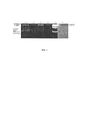
Фиг 1. - Электрофореграмма, показывающая разделение комплексов ДНК-олигонуклеотидов с белком от несвязавшихся ДНК-олигонуклеотидов.Fig 1. - Electrophoregram showing the separation of complexes of DNA oligonucleotides with protein from unbound DNA oligonucleotides.
1 - комплекс одноцепочечной ДНК-библиотеки №1 (химически модифицированная) c альфа-фетопротеином человека (АФП);1 - a complex of single-stranded DNA library No. 1 (chemically modified) with human alpha-fetoprotein (AFP);
2 - одноцепочечная ДНК-библиотека №1;2 - single-stranded DNA library No. 1;
3 - комплекс одноцепочечной ДНК-библиотеки №2 (природная) с АФП,3 - complex single-stranded DNA library No. 2 (natural) with AFP,
4 - одноцепочечная ДНК-библиотека №2;4 - single-stranded DNA library No. 2;
5 - маркер длин ДНК «GeneRuler 50 bp»;5 - DNA length marker “GeneRuler 50 bp”;
6 - АФП. Лунки 1-5, окрашивание бромидом этидия. Лунка 6 - окрашивание «кумасси блю».6 - AFP. Wells 1-5, ethidium bromide staining. Well 6 — Coomassie Blue Staining.
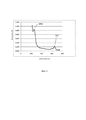
Фиг 2. - Спектр поглощения флуоресцентно-меченных ДНК-олигонуклеотидов после термической диссоциации комплексов ДНК/АФП.Fig 2. - Absorption spectrum of fluorescently-labeled DNA oligonucleotides after thermal dissociation of DNA / AFP complexes.
Фиг 3. - Производное 5-аминоаллил-2'-дезоксиуридин-5'-трифосфата, модифицированное по аминоаллильному фрагменту фенилпропионовой кислотой.Fig 3. - Derived 5-aminoallyl-2'-deoxyuridine-5'-triphosphate, modified at the aminoallyl fragment of phenylpropionic acid.
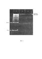
Фиг 4. - Электрофореграмма комплексов АФП с ДНК-библиотеками.Fig 4. - Electrophoregram complexes of AFP with DNA libraries.
Лунка №1 - молекулярный маркер длин ДНК «GeneRuler 50bp», лунка №2 - обогащенная ДНК-библиотека, полученная с применением способа термической диссоциации, лунка №3 - обогащенная ДНК-библиотека, полученная без применения способа термической диссоциации. Видно, что оптическая плотность комплекса ДНК/АФП выше в лунке №2.Well No. 1 is a molecular marker of DNA lengths “GeneRuler 50bp”, Well No. 2 is an enriched DNA library obtained using the thermal dissociation method, Well No. 3 is an enriched DNA library obtained without using a thermal dissociation method. It is seen that the optical density of the DNA / AFP complex is higher in well No. 2.
Осуществление изобретенияThe implementation of the invention
В работе использованы полимеразы Taq ДНК (“Евроген”, Россия) и Deep Vent (“BioLabs”, Великобритания) с буферными растворами соответствующих производителей; трифосфаты дезоксинуклеозидов производства “Fermentas” (США); флуоресцентный краситель SYBR Green (“Thermo Scientific”, США); магнитные частицы MagnaBind Streptavidin Beads (“Thermo Scientific”, США). Остальные реактивы и растворители марок "ОСЧ", "ХЧ" или "ЧДА" приобретены в фирмах Aldrich, Alfa Aesar, Fluka, Химмед. Воду для приготовления растворов получали на установке Milli-Q (EMD Millipore, США).We used Taq DNA polymerases (Eurogen, Russia) and Deep Vent (BioLabs, UK) with buffer solutions of the corresponding manufacturers; deoxynucleoside triphosphates manufactured by Fermentas (USA); SYBR Green fluorescent dye (Thermo Scientific, USA); MagnaBind Streptavidin Beads magnetic particles (Thermo Scientific, USA). The remaining reagents and solvents of the “OSCh”, “ChCh” or “ChDA” brands were purchased from Aldrich, Alfa Aesar, Fluka, and Chemmed. Water for the preparation of solutions was obtained using a Milli-Q apparatus (EMD Millipore, USA).
Амплификацию ДНК-библиотек после каждого цикла SELEX производили методом ПЦР. Реакционная смесь для проведения ПЦР содержала 1,5 мМ MgCl2, 10 мМ KCl, 10 mM Tris-HCl (pH 8,3) и 2,5 единицы (на реакционный объем 50 мкл) TaqDNA полимеразы (ThermoFisher) для немодифицированных библиотек либо 0,5 единиц Vent (exo-) полимеразы (NEB) в случае модифицированных библиотек. Смесь содержала трифосфаты (dATP, dCTP, dGTP и dTTP) в концентрации 0,2 mM каждого. В случае модифицированных библиотек смесь вместо dTTP содержала модифицированный трифосфат дезоксиуридина. Рабочая концентрация праймеров составляла 100 nM каждого. Один из праймеров был окрашен цианиновым красителем Cy5 для формирования окрашенной цепи аптамера, второй праймер нес биотиновую метку. Реакцию проводили на ДНК-амплификаторе Rotor-Gene Q5 plex HRM по следующей программе: 4 мин при 95°C; 20 циклов по 30 сек при 95°C, 30 сек при 66°C и 40 сек при 72°C; завершали реакцию инкубированием в течение 3 мин при 72°C.Amplification of DNA libraries after each SELEX cycle was performed by PCR. The PCR reaction mixture contained 1.5 mM MgCl 2 , 10 mM KCl, 10 mM Tris-HCl (pH 8.3) and 2.5 units (per
Полученную в результате амплификации двухцепочечную ДНК (дцДНК) использовали для разделения на магнитных стрептавидиновых частицах. ПЦР-продукт (50 мкл) смешивали с эквивалентным объемом 10х ПЦР-буфера, инкубировали при 95°С в течение 3 мин. Полученную смесь добавляли к магнитным частицам, предварительно суспендированным в 10х ПЦР-буфере, инкубировали при комнатной температуре 30 мин. За это время биотинилированная цепь ДНК ковалентно связывается со стрептавидином. Магнитные частицы трижды промывали буфером, а затем инкубировали 2 мин с 20% раствором гидроксида аммония при комнатной температуре. Далее супернатант сливали в чистую пробирку и переосаждали 5-ю объемами спиртового раствора ацетата аммония. Осадок промывали 96% этиловым спиртом, высушивали и растворяли в деионизованной воде (MQ-H2O).The double-stranded DNA (dsDNA) obtained as a result of amplification was used for separation on magnetic streptavidin particles. The PCR product (50 μl) was mixed with an equivalent volume of 10x PCR buffer, incubated at 95 ° C for 3 min. The resulting mixture was added to magnetic particles previously suspended in 10x PCR buffer, incubated at room temperature for 30 minutes. During this time, the biotinylated DNA chain covalently binds to streptavidin. The magnetic particles were washed three times with buffer, and then incubated for 2 min with a 20% solution of ammonium hydroxide at room temperature. Then the supernatant was poured into a clean tube and reprecipitated with 5 volumes of an alcoholic solution of ammonium acetate. The precipitate was washed with 96% ethanol, dried and dissolved in deionized water (MQ-H 2 O).
Магнитные частицы промывали гидроксидом аммония, а затем инкубировали с ним в течение 8 мин при 60°С. Полученный раствор сливали в пробирку и переосаждали биотинилированную цепь спиртовым раствором ацетата аммония. Осадок промывали 96% этиловым спиртом, высушивали от спирта и растворяли в MQ-H2O.The magnetic particles were washed with ammonium hydroxide, and then incubated with it for 8 min at 60 ° C. The resulting solution was poured into a test tube and the biotinylated chain was reprecipitated with an alcoholic solution of ammonium acetate. The precipitate was washed with 96% ethanol, dried from alcohol and dissolved in MQ-H 2 O.
Первые пять раундов образования комплекса проводили с использованием иммобилизованного препарата альфа-фетопротеина (АФП), гликопротеина с молекулярным весом около 69000 Да, состоящего из одной полипептидной цепи, включающей около 600 аминокислот.220 pmol библиотеки инкубировали с необходимым количеством иммобилизованного препарата АФП. Одноцепочечную библиотеку нагревали до 95°С в течение 2 мин и медленно охлаждали до 37°C. АФП, иммобилизированный на магнитных частицах с концентрацией 15 пмоль/мг, промывали 1х SELEX-буфером, приготовленном из 2х SELEX-буфера, имеющего pH 7.5 и содержащего 0.30 M NaCl, 60 mM HEPES, 20 mM KCl, 20 mM MgCl2, 0.1% TWEEN-20. Частицы разбавляли в 100 мкл 2х SELEX-буфере. Добавляли 100 мкл одноцепочечной библиотеки. Инкубировали 1 час при 37°С, перемешивая. Отбирали супернатант - несвязавшиеся олигонуклеотиды. Микрочастицы, содержащие АФП со связавшимися последовательностями, промывали 1x SELEX-буфером при комнатной температуре, растворяли в 100 мкл H2O качества MQ и инкубировали 10 мин при 95°С, супернатант отделяли, определяли его концентрацию на спектрофотометре NanoDrop 1000 и использовали в дальнейших раундах SELEX.The first five rounds of complex formation were carried out using an immobilized preparation of alpha-fetoprotein (AFP), a glycoprotein with a molecular weight of about 69,000 Da, consisting of one polypeptide chain including about 600 amino acids. 220 pmol libraries were incubated with the required amount of an immobilized AFP preparation. The single chain library was heated to 95 ° C for 2 minutes and slowly cooled to 37 ° C. AFP immobilized on magnetic particles with a concentration of 15 pmol / mg was washed with 1x SELEX buffer prepared from 2x SELEX buffer having a pH of 7.5 and containing 0.30 M NaCl, 60 mM HEPES, 20 mM KCl, 20 mM MgCl2, 0.1% TWEEN -twenty. The particles were diluted in 100 μl of 2x SELEX buffer. 100 μl single chain library was added. Incubated for 1 hour at 37 ° C, stirring. The supernatant was selected - unbound oligonucleotides. Microparticles containing AFPs with bound sequences were washed with 1x SELEX buffer at room temperature, dissolved in 100 μl of MQ quality H 2 O and incubated for 10 min at 95 ° C, the supernatant was separated, its concentration was determined on a NanoDrop 1000 spectrophotometer and used in further rounds SELEX.
Допустимо производить пять предварительных раундов селекции без использования магнитных частиц, путем создания комплексов ДНК/АФП в растворе (с нативным неиммобилизованным АФП) с их последующим разделением методом электрофореза и элюцией связавшихся с мишенью ДНК-олигонуклеотидов из геля.It is permissible to carry out five preliminary rounds of selection without the use of magnetic particles, by creating DNA / AFP complexes in solution (with native non-immobilized AFP) with their subsequent separation by electrophoresis and elution of the DNA oligonucleotides bound to the target from the gel.
Последующие три цикла образования комплекса проводили в буфере с нативным неиммобилизованным АФП при 37°С в течение 60 мин в 1х SELEX-буфере, затем производили электрофоретическое разделение комплекса от несвязавшихся ДНК-олигонуклеотидов и последующую термическую диссоциацию.The next three cycles of complex formation were carried out in a buffer with native non-immobilized AFP at 37 ° C for 60 min in a 1x SELEX buffer, then the complex was electrophoretically separated from unbound DNA oligonucleotides and subsequent thermal dissociation.
С учетом молекулярной массы АФП и длины модифицированных олигонуклеотидов, входящих в состав библиотеки, был выбран следующий состав геля: 12% акриламид/бис-акриламид (19:1) в 1х TBE, содержащий 10 mM KCl и 3 mM MgCl2. Наличие в буфере солей калия и магния обусловлено необходимостью стабилизировать неканонические типы вторичной структуры ДНК, которые могут присутствовать в аптамерах: G-квадруплексы и шпилечные структуры ДНК. Анализ проводили при контролируемой температуре 20°С, что предотвращает неконтролируемую термическую диссоциацию комплексов в процессе электрофореза. С этой же целью в электрофоретической ячейке поддерживали относительное невысокое напряжение (порядка 120-150 вольт на 20 см геля). Контроль миграции связанной и несвязанной ДНК осуществляли с использованием 45-мерного флуоресцентно-меченного олигонуклеотида. Длина пробега олигонуклеотидного контроля составляла 5-7 см. Гель визуализировали окрашиванием SYBR Gold I. Результаты электрофореза представлены на Фиг. 1.Given the molecular weight of the AFP and the length of the modified oligonucleotides that make up the library, the following gel composition was selected: 12% acrylamide / bis-acrylamide (19: 1) in 1x TBE containing 10 mM KCl and 3 mM MgCl 2 . The presence of potassium and magnesium salts in the buffer is due to the need to stabilize noncanonical types of the secondary structure of DNA, which may be present in aptamers: G-quadruplexes and hairpin DNA structures. The analysis was carried out at a controlled temperature of 20 ° C, which prevents uncontrolled thermal dissociation of complexes during electrophoresis. For the same purpose, a relatively low voltage was maintained in the electrophoretic cell (about 120-150 volts per 20 cm of gel). Control of migration of bound and unbound DNA was carried out using a 45-mer fluorescently-labeled oligonucleotide. The path length of the oligonucleotide control was 5-7 cm. The gel was visualized by staining with SYBR Gold I. The results of electrophoresis are shown in FIG. 1.
По электрофоретической картине оценивали количество связавшейся с АФП и несвязавшейся ДНК.According to the electrophoretic picture, the amount of AFP bound and unbound DNA was estimated.
Связавшуюся ДНК в комплексе с АФП вырезали из геля и элюировали 100 мкл раствора LiClO4 в H2O в течение 60 мин при 37°С. В классическом варианте SELEX именно такая связавшаяся ДНК вовлекается в последующие раунды селекции. В предлагаемом варианте полученный элюат не используется.The bound DNA in complex with AFP was excised from the gel and 100 μl of a solution of LiClO 4 in H 2 O was eluted for 60 min at 37 ° C. In the classic SELEX variant, it is precisely such bound DNA that is involved in subsequent rounds of selection. In the proposed embodiment, the resulting eluate is not used.
После проведения элюции при 37°C проводили термическую диссоциацию оставшихся комплексов. Для этого к гелю добавляли 100 мкл 1x SELEX-буфера, нагревали до 60°С и инкубировали в течение 20 мин. Супернатант отделяли, определяли концентрацию ДНК на спектрофотометре NanoDrop 1000 (Фиг. 2) и использовали в дальнейших (дополнительных двух) раундах SELEX с последующей термической диссоциацией.After elution at 37 ° C, thermal dissociation of the remaining complexes was performed. For this, 100 μl of 1x SELEX buffer was added to the gel, heated to 60 ° C and incubated for 20 min. The supernatant was separated, the DNA concentration was determined on a NanoDrop 1000 spectrophotometer (Fig. 2) and used in further (two additional) SELEX rounds followed by thermal dissociation.
Теоретически допустимо проведение термодиссоциации с использованием магнитных частиц и иммобилизованного АФП.It is theoretically acceptable to conduct thermal dissociation using magnetic particles and immobilized AFP.
Получали как библиотеку с применением природных нуклеотидов, так и с модифицированным производным дезоксиуридина, структура которого приведена на Фиг. 3. После проведения трех раундов селекции с применением термической диссоциации - получали обогащенную высокоспецифичными аптамерами ДНК-библиотеку, готовую к проведению секвенирования для определения индивидуальных нуклеотидных последовательностей аптамеров.Both a library using natural nucleotides and a modified deoxyuridine derivative, the structure of which is shown in FIG. 3. After conducting three rounds of selection using thermal dissociation, a DNA library enriched with highly specific aptamers was obtained, ready for sequencing to determine individual nucleotide sequences of aptamers.
Сравнивали эффективность способа термодиссоциации с базовым вариантом селекции аптамеров методом электрофоретического разделения комплексов ДНК/АФП (Фиг. 4). Видно, что оптическая плотность больше в случае обогащения библиотеки способом термической диссоциации.The effectiveness of the thermal dissociation method was compared with the basic variant of aptamer selection by electrophoretic separation of DNA / AFP complexes (Fig. 4). It can be seen that the optical density is greater in the case of enrichment of the library by thermal dissociation.
ПримерыExamples
Пример 1. Получение биотинилированного праймераExample 1. Obtaining biotinylated primer
Для последующего разделения цепей комбинаторной ДНК-библиотеки в одну из образующихся в ходе ПЦР цепей вводили биотин путем применения биотинилированного праймера. Праймер синтезировали, использую С6-аминомодификатор со свободной NH2-группой по 5'-концу с последующим введением биотиновой метки. К раствору гидоксисукцинимидного эфира биотина (2,0 мг) в ДМФА (590 мкл) добавляли праймер в соотношении 10/1 соответственно. Реакция проходила в течение 30 минут при комнатной температуре. Далее проводили хроматографическую очистку полученного продукта в градиенте ацетонитрила.For subsequent separation of the combinatorial DNA library chains, biotin was introduced into one of the chains formed during PCR by using a biotinylated primer. The primer was synthesized using a C6 amino modifier with a free NH 2 group at the 5'end, followed by the introduction of a biotin label. To a solution of biotin hydroxysuccinimide ester (2.0 mg) in DMF (590 μl) was added a primer in a ratio of 10/1, respectively. The reaction took place for 30 minutes at room temperature. Next, chromatographic purification of the obtained product in an acetonitrile gradient was carried out.
Пример 2. Получение праймера, меченного цианиновым красителем Cy5Example 2. Obtaining primer labeled with cyanine dye Cy5
Праймер синтезировали со свободной NH2-группой по 5'-концу. Краситель Cy5 в виде гидроксисукцинимидного эфира в ДМСО добавляли к праймеру в соотношении 10/1. Проводили инкубацию 2 часа при комнатной температуре. Далее проводили хроматографическую очистку полученного продукта в градиенте ацетонитрила.The primer was synthesized with a free NH 2 group at the 5 ′ end. Dye Cy5 in the form of hydroxysuccinimide ether in DMSO was added to the primer in a ratio of 10/1. An incubation of 2 hours was carried out at room temperature. Next, chromatographic purification of the obtained product in an acetonitrile gradient was carried out.
Пример 3. Полимеразная цепная реакция для амплификации комбинаторных (вырожденных) ДНК-библиотекExample 3. Polymerase chain reaction for the amplification of combinatorial (degenerate) DNA libraries
Реакционная смесь для проведения ПЦР содержала 1,5 мМ MgCl2, 10 мМ KCl, 10 mM Tris-HCl (pH 8,3) и 2,5 единицы (на реакционный объем 50 мкл) Taq DNA полимеразы (ThermoFisher) для немодифицированных библиотек либо 0,5 единиц Vent (exo-) полимеразы (NEB) в случае модифицированных библиотек, трифосфаты (dATP, dCTP, dGTP, а также dTTP для немодифицированных библиотек или модифицированное производное дезоксиуридинтрифосфата в концентрации 2 mM (Фиг. 3) в случае модифицированных библиотек. Использовали праймеры: прямой биотинилированный и обратный флуоресцентно-меченный, рабочая концентрация каждого праймера составляла 10 mM. В качестве матрицы использовали синтетическую вырожденную комбинаторную ДНК-библиотеку с последовательностьюThe PCR reaction mixture contained 1.5 mM MgCl 2 , 10 mM KCl, 10 mM Tris-HCl (pH 8.3) and 2.5 units (per
CTGTCAGCTCCATACTGGTAGCC-(N)40-GCGTTCGAATCTAGACGGTACGA,CTGTCAGCTCCATACTGGTAGCC- (N) 40-GCGTTCGAATCTAGACGGTACGA,
где N - комбинация нуклеотидов A, T, G, C.where N is the combination of nucleotides A, T, G, C.
Реакцию проводили на ДНК-амплификаторе MiniCycler (MJResearch, Waltham, Mass.) по следующей программе:The reaction was carried out on a MiniCycler DNA Amplifier (MJResearch, Waltham, Mass.) According to the following program:
1) 4 мин при 95°C,1) 4 min at 95 ° C,
2) 30 сек при 95°C,2) 30 sec at 95 ° C,
3)30 сек при 66°C,3) 30 sec at 66 ° C,
4) 40 сек при 72°C,4) 40 sec at 72 ° C,
5) повтор с 2 пункта по 4 пункт 19 раз,5) repeat from 2 points to 4 points 19 times,
6) завершали реакцию инкубированием в течение 3 мин при 72°C.6) completed the reaction by incubation for 3 min at 72 ° C.
При использовании в качестве субстратов модифицированного производного дезоксиуридинтрифосфата при необходимости температурно-временной профиль ПЦР, а также состав реакционной смеси модифицировали (оптимизировали) с целью увеличения выхода целевого продукта реакции. Результат прохождения ПЦР оценивали визуально, электрофоретически.When using a modified derivative of deoxyuridine triphosphate as substrates, if necessary, the temperature-time profile of PCR, as well as the composition of the reaction mixture, was modified (optimized) in order to increase the yield of the target reaction product. The result of PCR was evaluated visually, electrophoretically.
Пример 4. Разделение биотинилированной и небиотинилированной цепей ДНК-библиотеки после проведения ПЦР с использованием магнитных частицExample 4. The separation of biotinylated and non-biotinylated DNA library chains after PCR using magnetic particles
Для получения однонитевой ДНК с красителем Cy5 на 5'-конце проводили разделение цепей на магнитных частицах Dynabeads™ M-270 Streptavidin (InvitrogenDynalAS, Norway) с ковалентно пришитыми к их поверхности молекулами стрептавидина. Магнитные частицы промывали три раза буфером 10х (pH 8.8 при 25°С), содержащим 100 mM Трис-HCl и 500 mM KCl, и ресуспендировали в растворе, содержащем 100 мкл 5х ПЦР-буфер. Биотинилированную по 5’-концу двухцепочечную ДНК (ПЦР-продукт, объем 50 мкл) смешивали с эквивалентным объемом 10х ПЦР-буфера, нагревали до 95°С, инкубировали 3 мин и охлаждали до комнатной температуры, после чего смешивали с подготовленными магнитными частицами. Иммобилизацию проводили при комнатной температуре в течение 30 мин при постоянном перемешивании. С помощью магнитосепаратора магнитные частицы с иммобилизованной на них двухцепочечной ДНК отделяли от реакционной смеси и промывали трижды 5Х буфером, содержащим Трис-HCl и KCl.To obtain single-stranded DNA with Cy5 dye at the 5'-end, chains were separated on magnetic particles of Dynabeads ™ M-270 Streptavidin (InvitrogenDynalAS, Norway) with streptavidin molecules covalently attached to their surface. The magnetic particles were washed three times with 10x buffer (pH 8.8 at 25 ° C) containing 100 mM Tris-HCl and 500 mM KCl and resuspended in a solution containing 100 μl of 5x PCR buffer. The 5 ′ end biotinylated double-stranded DNA (PCR product, 50 μl volume) was mixed with an equivalent volume of 10x PCR buffer, heated to 95 ° C, incubated for 3 min and cooled to room temperature, after which it was mixed with prepared magnetic particles. Immobilization was carried out at room temperature for 30 minutes with constant stirring. Using a magnetoseparator, magnetic particles with double-stranded DNA immobilized on them were separated from the reaction mixture and washed three times with 5X buffer containing Tris-HCl and KCl.
Небиотинилированную цепь элюировали добавлением к магнитным частицам 20% раствора гидроксида аммония, инкубировали 2 мин, затем частицы отделяли на магнитном сепараторе, а элюат переносили в чистую пробирку, где к нему добавляли 5 объемов спиртового раствора ацетата аммония, содержащего 0,125 М ацетата аммония и 70% этанола. Одноцепочечную небиотинилированную ДНК осаждали цетрифугированием (16000 g, 2 мин). Осадок промывали спиртовым раствором ацетата аммония, затем 96% этиловым спиртом и высушивали на воздухе. К высушенному осадку добавляли 20 мкл деионизованной воды (MQ-H2O) и определяли концентрацию ДНК на спектрофотометре NanoDrop 1000.The non-biotinylated chain was eluted by adding 20% ammonium hydroxide solution to the magnetic particles, incubated for 2 min, then the particles were separated on a magnetic separator, and the eluate was transferred to a clean tube where 5 volumes of an alcoholic solution of ammonium acetate containing 0.125 M ammonium acetate and 70% were added to it. ethanol. Single-stranded non-biotinylated DNA was precipitated by centrifugation (16000 g, 2 min). The precipitate was washed with an alcoholic solution of ammonium acetate, then with 96% ethyl alcohol and dried in air. To the dried precipitate was added 20 μl of deionized water (MQ-H 2 O) and the DNA concentration was determined on a NanoDrop 1000 spectrophotometer.
Магнитные частицы с биотинилированной цепью промывали 20% гидроксидом аммония 1 мин при комнатной температуре, затем супернатант удаляли, а частицы инкубировали с 50 мкл 30% гидроксида аммония при 60°С при эпизодическом ручном перемешивании в течение 8 мин. Элюат переносили в чистую пробирку, где переосаждали биотинилированную цепь добавлением 5 объемов спиртового раствора ацетата аммония, содержащего 0,125 М ацетата аммония и 70% этанола. Осадок промывали спиртовым раствором ацетата аммония, затем 96% этиловым спиртом и высушивали на воздухе. К высушенному осадку добавляли 20 мкл деионизованной (MQ) H2O и определяли концентрацию ДНК на спектрофотометре NanoDrop 1000. Эффективность разделения цепей определяли электрофоретическим методом.Biotinylated chain magnetic particles were washed with 20% ammonium hydroxide for 1 min at room temperature, then the supernatant was removed, and the particles were incubated with 50 μl of 30% ammonium hydroxide at 60 ° C with occasional manual stirring for 8 min. The eluate was transferred to a clean tube where the biotinylated chain was reprecipitated by adding 5 volumes of an alcoholic solution of ammonium acetate containing 0.125 M ammonium acetate and 70% ethanol. The precipitate was washed with an alcoholic solution of ammonium acetate, then with 96% ethyl alcohol and dried in air. To the dried precipitate was added 20 μl of deionized (MQ) H 2 O and the DNA concentration was determined on a NanoDrop 1000 spectrophotometer. The separation efficiency of the chains was determined by electrophoretic method.
Эффективность элюции ДНК контролировали, добавляя к магнитным частицам 40 мкл раствора, содержащего 8 М мочевины и 2 М тиомочевины, что приводило к разрушению комплексов стрептавидин-биотин и элюции всей ДНК, иммобилизованной на магнитных частицах. Элюат анализировали методом ПААГ-электрофореза.The efficiency of DNA elution was controlled by adding 40 μl of a solution containing 8 M urea and 2 M thiourea to magnetic particles, which led to the destruction of streptavidin-biotin and elution of all DNA immobilized on magnetic particles complexes. The eluate was analyzed by PAAG electrophoresis.
Пример 5. Образование комплекса одноцепочечной ДНК-библиотеки с альфа-фетопротеином (АФП) в иммобилизованной фазеExample 5. The complexation of a single-stranded DNA library with alpha-fetoprotein (AFP) in the immobilized phase
Модифицированную одноцепочечную библиотеку нагревали до 95°С в течение 2 мин и охлаждали до 37°C. АФП, иммобилизированный на магнитных частицах с концентрацией 15 пмоль/мг, промывали 1х SELEX-буфером, приготовленном из 2х SELEX-буфера, имеющего pH 7.5 и содержащего 0.30 M NaCl, 60 mM HEPES, 20 mM KCl, 20 mM MgCl2, 0.1% TWEEN-20. Магнитные частицы разбавляли в 100 мкл 2х SELEX-буфера. Добавляли 100 мкл модифицированной одноцепочечной библиотеки. Инкубировали 1 час при перемешивании, 37°С. Отбирали супернатант - несвязавшиеся олигонуклеотиды. Магнитные частицы, содержащие АФП со связавшимися последовательностями, промывали 1x SELEX-буфером при комнатной температуре, растворяли в 100 мкл H2O качества MQ и инкубировали 10 мин при 95°С, супернатант отделяли, определяли его концентрацию на спектрофотометре NanoDrop 1000 и использовали в дальнейших раундах SELEX.The modified single chain library was heated to 95 ° C for 2 minutes and cooled to 37 ° C. AFP immobilized on magnetic particles with a concentration of 15 pmol / mg was washed with 1x SELEX buffer prepared from 2x SELEX buffer having a pH of 7.5 and containing 0.30 M NaCl, 60 mM HEPES, 20 mM KCl, 20 mM MgCl2, 0.1% TWEEN -twenty. Magnetic particles were diluted in 100 μl of 2x SELEX buffer. 100 μl of a modified single chain library was added. Incubated 1 hour with stirring, 37 ° C. The supernatant was selected - unbound oligonucleotides. Magnetic particles containing AFPs with bound sequences were washed with 1x SELEX buffer at room temperature, dissolved in 100 μl of MQ quality H 2 O and incubated for 10 min at 95 ° C, the supernatant was separated, its concentration was determined on a NanoDrop 1000 spectrophotometer and used in further SELEX rounds.
Пример 6. Образование комплекса одноцепочечной ДНК-библиотеки с альфа-фетопротеином (АФП) в раствореExample 6. The formation of the complex single-stranded DNA library with alpha-fetoprotein (AFP) in solution
Модифицированную одноцепочечную библиотеку нагревали до 95°С в течение 2 мин и медленно охлаждали до 37°C. К 50 мкл водного раствора АФП с концентрацией 30 пмоль/мкл добавляли 50 мкл 2х SELEX-буфера. Добавляли 100 мкл модифицированной одноцепочечной библиотеки. Инкубировали 1 час при перемешивании при 37°С.The modified single chain library was heated to 95 ° C for 2 min and slowly cooled to 37 ° C. To 50 μl of an aqueous solution of AFP with a concentration of 30 pmol / μl was added 50 μl of 2x SELEX buffer. 100 μl of a modified single chain library was added. Incubated for 1 hour with stirring at 37 ° C.
Пример 7. Электрофоретическое разделение комплексов ДНК/АФП и не связавшихся олигонуклеотидовExample 7. Electrophoretic separation of DNA / AFP complexes and unbound oligonucleotides
Готовили электрофорезный гель следующего состава: 12% акриламид/бис-акриламид (19:1) в 1х TBE, содержащий 10 mM KCl и 3 mM MgCl2. В лунки вносили по 20 мкл комплекса ДНК/АФП, свободную библиотеку, а также контроли. Электрофорез проводили при контролируемой температуре 20°С для предотвращения неконтролируемой термической диссоциации комплексов. С этой же целью в электрофоретической ячейке поддерживали относительное невысокое напряжение (порядка 120-150 вольт на 20 см геля). Контроль миграции связанной и несвязанной ДНК осуществляли с использованием 45-мерного флуоресцентно-меченного олигонуклеотида. Электрофорез проводили до длины пробега олигонуклеотидного контроля - 5-7 см. В качестве дополнительных контролей были использованы: несвязанная с АФП модифицированная библиотека после 5-го раунда, ДНК-лестница GeneRuler 50bp (ThermoFisher, США) и АФП и/или белковый маркер. Визуализацию полос в геле проводили с использованием SYBR Gold I для ДНК и кумасси для белков. Результаты электрофореза представлены на Фиг. 1.An electrophoresis gel of the following composition was prepared: 12% acrylamide / bis-acrylamide (19: 1) in 1x TBE containing 10 mM KCl and 3 mM MgCl 2 . 20 μl of the DNA / AFP complex, a free library, and controls were added to the wells. Electrophoresis was carried out at a controlled temperature of 20 ° C to prevent uncontrolled thermal dissociation of complexes. For the same purpose, a relatively low voltage was maintained in the electrophoretic cell (about 120-150 volts per 20 cm of gel). Control of migration of bound and unbound DNA was carried out using a 45-mer fluorescently-labeled oligonucleotide. Electrophoresis was carried out up to the path length of the oligonucleotide control - 5-7 cm. As additional controls were used: an AFP-unmodified modified library after the 5th round, GeneRuler 50bp DNA ladder (ThermoFisher, USA) and AFP and / or protein marker. The bands in the gel were visualized using SYBR Gold I for DNA and Coomassie for proteins. Electrophoresis results are shown in FIG. 1.
По электрофоретической картине оценивали количество связавшейся с АФП и несвязавшейся ДНК.According to the electrophoretic picture, the amount of AFP bound and unbound DNA was estimated.
Пример 8. Элюция ДНК из геляExample 8. Elution of DNA from the gel
Полосы геля, содержащие комплекс ДНК/АФП вырезали с помощью хирургического скальпеля, помещали в пробирку типа «Эппендорф» объемом 1.5 мл и проводили элюцию связавшихся с белком ДНК-олигонуклеотидов, с помощью 100 мкл раствора LiClO4 в H2O (Milli-Q). Смесь инкубировали 60 мин при 37°С, центрифугировали пробирки при 2000 об/мин в течение 2 мин, супернатант отбирали и измеряли концентрацию ДНК на спектрофотометре NanoDrop 1000. ДНК, содержащуюся в полученном супернатанте, не использовали для дальнейших раундов селекции. Оставшийся в пробирках гель использовали для проведения термической диссоциации ДНК-олигонуклеотидов, обладающих повышенным сродством к АФП.The gel strips containing the DNA / AFP complex were excised with a surgical scalpel, placed in a 1.5 ml Eppendorf tube and eluted protein-bound DNA oligonucleotides were eluted with 100 μl of a solution of LiClO 4 in H 2 O (Milli-Q) . The mixture was incubated for 60 min at 37 ° C, the tubes were centrifuged at 2000 rpm for 2 min, the supernatant was taken and the DNA concentration was measured on a NanoDrop 1000 spectrophotometer. The DNA contained in the obtained supernatant was not used for further selection rounds. The gel remaining in the tubes was used to conduct thermal dissociation of DNA oligonucleotides with increased affinity for AFP.
Пример 9. Термическая диссоциация ДНК-олигонуклеотидов, обладающих повышенным сродством к АФПExample 9. Thermal dissociation of DNA oligonucleotides with increased affinity for AFP
К гелю после элюции добавляли 100 мкл 1x SELEX-буфера, нагревали до 60°С и инкубировали в течение 20 мин. Супернатант отделяли, определяли концентрацию ДНК на спектрофотометре NanoDrop 1000 (Фиг. 2). Элюат после термической диссоциации вовлекали в последующий из трех раундов селекции, в состав которого входит этап термической диссоциации. После проведения трех раундов SELEX с термической диссоциацией получали обогащенную ДНК-библиотеку, пригодную для определения индивидуальных последовательностей олигонуклеотидов, входящих в ее состав.After elution, 100 μl of 1x SELEX buffer was added to the gel, heated to 60 ° C and incubated for 20 min. The supernatant was separated, the DNA concentration was determined on a NanoDrop 1000 spectrophotometer (Fig. 2). After thermal dissociation, the eluate was involved in the next of three rounds of selection, which includes the stage of thermal dissociation. After conducting three rounds of SELEX with thermal dissociation, an enriched DNA library was obtained that was suitable for determining the individual sequences of oligonucleotides that make up its composition.
Пример 10. Электрофорез комплексов АФП с ДНК-библиотеками, полученными с применением способа термической диссоциации и без него.Example 10. Electrophoresis of AFP complexes with DNA libraries obtained using and without thermal dissociation.
Электрофоретическое разделение проводили аналогично тому, как описано в Примере 7. Эффективность связывания ДНК с АФП оценивали визуально по оптической плотности полосы, соответствующей образовавшемуся комплексу ДНК/АФП (Фиг. 4).Electrophoretic separation was carried out similarly as described in Example 7. The efficiency of DNA binding with AFP was evaluated visually by the optical density of the band corresponding to the resulting DNA / AFP complex (Fig. 4).
ЛИТЕРАТУРАLITERATURE
1. Patel D.J., Suri A.K. Structure, recognition and discrimination in RNA aptamer complexes with cofactors, amino acids, drugs and aminoglycoside antibiotics. J. Biotechnol. 2000. V. 74. №1. P. 39-60.1. Patel D.J., Suri A.K. Structure, recognition and discrimination in RNA aptamer complexes with cofactors, amino acids, drugs and aminoglycoside antibiotics. J. Biotechnol. 2000. V. 74. No. 1. P. 39-60.
2. Радько С.П., Рахметова С.Ю., Бодоев Н.В., Арчаков А.Н. Аптамеры как перспективные аффинные реагенты для клинической протеомики. Биомедицинская химия. 2007. Т. 53. №1. с. 5-24.2. Radko S.P., Rakhmetova S.Yu., Bodoev N.V., Archakov A.N. Aptamers as promising affinity reagents for clinical proteomics. Biomedical chemistry. 2007.V. 53. No. 1. with. 5-24.
3. Tuerk C., Gold L. Systematic evolution of ligands by exponential enrichment: RNA ligands to bacteriophage T4 DNA polymerase. Science. 1990. V. 249. №4968. P. 505-510.3. Tuerk C., Gold L. Systematic evolution of ligands by exponential enrichment: RNA ligands to bacteriophage T4 DNA polymerase. Science. 1990. V. 249. No. 4968. P. 505-510.
4. Ellington A.D., Szostak J.W. In vitro selection of RNA molecules that bind specific ligands. Nature. 1990. V. 346. №6287. P. 818-822.4. Ellington A.D., Szostak J.W. In vitro selection of RNA molecules that bind specific ligands. Nature. 1990. V. 346. No. 6287. P. 818-822.
5. Dembowski SK, Bowser MT. Microfluidic methods for aptamer selection and characterization. Analyst. 2017. V 143. №1. P. 21-32.5. Dembowski SK, Bowser MT. Microfluidic methods for aptamer selection and characterization. Analyst. 2017. V 143. No. 1. P. 21-32.
6. Mendonsa SD, Bowser MT. In vitro evolution of functional DNA using capillary electrophoresis. J Am Chem Soc. 2004. V 126. №1. P 20-21.6. Mendonsa SD, Bowser MT. In vitro evolution of functional DNA using capillary electrophoresis. J Am Chem Soc. 2004. V 126. No. 1. P 20-21.
7. Berezovski M1, Drabovich A, Krylova SM, Musheev M, Okhonin V, Petrov A, Krylov SN. Nonequilibrium capillary electrophoresis of equilibrium mixtures: a universal tool for development of aptamers. J Am Chem Soc. 2005. V 127. №9. P 3165 - 3171.7. Berezovski M1, Drabovich A, Krylova SM, Musheev M, Okhonin V, Petrov A, Krylov SN. Nonequilibrium capillary electrophoresis of equilibrium mixtures: a universal tool for development of aptamers. J Am Chem Soc. 2005. V 127. No. 9. P 3165 - 3171.
8. Eissa S, Siddiqua A, Chinnappan R, Zourob M. Electrochemical selection of a DNA aptamer, and an impedimetric method for determination of the dedicator of cytokinesis 8 by self-assembly of a thiolated aptamer on a gold electrode. Mikrochim Acta. 2019. V 186. P. 828.8. Eissa S, Siddiqua A, Chinnappan R, Zourob M. Electrochemical selection of a DNA aptamer, and an impedimetric method for determination of the dedicator of cytokinesis 8 by self-assembly of a thiolated aptamer on a gold electrode. Mikrochim Acta. 2019.V 186.P. 828.
9. Li L, Chen X, Cui C, Pan X, Li X, Yazd HS, Wu Q, Qiu L, Li J, Tan W. Aptamer Displacement Reaction from Live-Cell Surfaces and Its Applications. J Am Chem Soc. 2019. V 141. №. 43. P. 17174-17179.9. Li L, Chen X, Cui C, Pan X, Li X, Yazd HS, Wu Q, Qiu L, Li J, Tan W. Aptamer Displacement Reaction from Live-Cell Surfaces and Its Applications. J Am Chem Soc. 2019. V 141. No. 43. P. 17174-17179.
Claims (12)
Priority Applications (1)
| Application Number | Priority Date | Filing Date | Title |
|---|---|---|---|
| RU2019140693A RU2723373C1 (en) | 2019-12-10 | 2019-12-10 | Thermal dissociation method for selection of dna-aptamers |
Applications Claiming Priority (1)
| Application Number | Priority Date | Filing Date | Title |
|---|---|---|---|
| RU2019140693A RU2723373C1 (en) | 2019-12-10 | 2019-12-10 | Thermal dissociation method for selection of dna-aptamers |
Publications (1)
| Publication Number | Publication Date |
|---|---|
| RU2723373C1 true RU2723373C1 (en) | 2020-06-10 |
Family
ID=71067488
Family Applications (1)
| Application Number | Title | Priority Date | Filing Date |
|---|---|---|---|
| RU2019140693A RU2723373C1 (en) | 2019-12-10 | 2019-12-10 | Thermal dissociation method for selection of dna-aptamers |
Country Status (1)
| Country | Link |
|---|---|
| RU (1) | RU2723373C1 (en) |
Citations (2)
| Publication number | Priority date | Publication date | Assignee | Title |
|---|---|---|---|---|
| US20150119285A1 (en) * | 2013-10-28 | 2015-04-30 | Chin-Yih Hong | Magnetic-assisted rapid aptamer selection method for generating high affinity dna aptamer |
| RU2618872C1 (en) * | 2015-12-09 | 2017-05-11 | Федеральное бюджетное учреждение науки Государственный научный центр прикладной микробиологии и биотехнологии (ФБУН ГНЦ ПМБ) | Method of directed deposit of oligonucleotid libraries for the reduction of non-specific adsorption in the solid-phase selection of aptamers on the basis of nucleic acids |
-
2019
- 2019-12-10 RU RU2019140693A patent/RU2723373C1/en active
Patent Citations (2)
| Publication number | Priority date | Publication date | Assignee | Title |
|---|---|---|---|---|
| US20150119285A1 (en) * | 2013-10-28 | 2015-04-30 | Chin-Yih Hong | Magnetic-assisted rapid aptamer selection method for generating high affinity dna aptamer |
| RU2618872C1 (en) * | 2015-12-09 | 2017-05-11 | Федеральное бюджетное учреждение науки Государственный научный центр прикладной микробиологии и биотехнологии (ФБУН ГНЦ ПМБ) | Method of directed deposit of oligonucleotid libraries for the reduction of non-specific adsorption in the solid-phase selection of aptamers on the basis of nucleic acids |
Non-Patent Citations (3)
| Title |
|---|
| DONG L. et al. Screening and Identifying a Novel ssDNA Aptamer against Alpha-fetoprotein Using CE-SELEX. Sci Rep. 2015;5:15552. * |
| ЦИМБРЕЗ Ф.М. и др. Аптамеры: новые молекулярные диагностические маркеры бактериальных и вирусных инфекций // Инфекционные болезни: Новости. Мнения. Обучение. 2015. * |
| ЦИМБРЕЗ Ф.М. и др. Аптамеры: новые молекулярные диагностические маркеры бактериальных и вирусных инфекций // Инфекционные болезни: Новости. Мнения. Обучение. 2015. DONG L. et al. Screening and Identifying a Novel ssDNA Aptamer against Alpha-fetoprotein Using CE-SELEX. Sci Rep. 2015;5:15552. * |
Similar Documents
| Publication | Publication Date | Title |
|---|---|---|
| Yang et al. | Aptamers 101: aptamer discovery and in vitro applications in biosensors and separations | |
| Dembowski et al. | Microfluidic methods for aptamer selection and characterization | |
| US11841371B2 (en) | Proteomics and spatial patterning using antenna networks | |
| US20230045556A1 (en) | Nucleic acid complexes for screening barcoded compounds | |
| JP6525872B2 (en) | Increasing dynamic range to identify multiple epitopes in cells | |
| JP6994198B2 (en) | Methods for Screening Nucleic Acid Aptamers | |
| EP2589657A9 (en) | Method for detection of target molecule | |
| US8034569B2 (en) | Methods for molecular detection | |
| WO2018168895A1 (en) | Method for detecting target molecule in which rolling circle amplification is used | |
| WO2018051580A1 (en) | Nucleoside derivative or salt thereof, reagent for synthesizing polynucleotide, method for producing polynucleotide, polynucleotide, and method for producing binding nucleic acid molecule | |
| CA3222176A1 (en) | Isolation of target nucleic acids | |
| Li et al. | Selection and preliminary application of a single stranded DNA aptamer targeting colorectal cancer serum | |
| Lee et al. | Development of DNA aptamers specific for small therapeutic peptides using a modified SELEX method | |
| RU2699522C2 (en) | Method for enzymatic production of modified dna for producing reagents which specifically bind to hydrophobic areas of high molecular weight organic compounds | |
| RU2518368C1 (en) | Method of selection aptamers to specified protein targets on cell surface | |
| WO2017218881A1 (en) | Method for aptamer pair selection | |
| WO2011157617A1 (en) | Complex set of mirna libraries | |
| Lapa et al. | The use of thermal dissociation for selection of DNA aptamers | |
| Dausse et al. | In vitro selection procedures for identifying DNA and RNA aptamers targeted to nucleic acids and proteins | |
| RU2731739C1 (en) | Method for determining substrate efficiency of deoxyuridine triphosphates derivatives for dna polymerases by primer completion reaction | |
| KR101069590B1 (en) | Single-stranded DNA aptamers specifically binding to CEA | |
| KR101069591B1 (en) | Single-stranded DNA aptamers specifically binding to CEA | |
| KR20100122223A (en) | Single-stranded dna aptamers specifically binding to cea | |
| JP2016171795A (en) | Method for selecting aptamer | |
| US20120094875A1 (en) | Production process of high affinity and high specificity oligonucleotides for organic and inorganic molecules |
Legal Events
| Date | Code | Title | Description |
|---|---|---|---|
| QB4A | Licence on use of patent |
Free format text: LICENCE FORMERLY AGREED ON 20200727 Effective date: 20200727 |