RU2706373C1 - Method for localizing tumor obstruction of biliary tract and degree of its prevalence - Google Patents
Method for localizing tumor obstruction of biliary tract and degree of its prevalence Download PDFInfo
- Publication number
- RU2706373C1 RU2706373C1 RU2019105909A RU2019105909A RU2706373C1 RU 2706373 C1 RU2706373 C1 RU 2706373C1 RU 2019105909 A RU2019105909 A RU 2019105909A RU 2019105909 A RU2019105909 A RU 2019105909A RU 2706373 C1 RU2706373 C1 RU 2706373C1
- Authority
- RU
- Russia
- Prior art keywords
- biliary tract
- tumor
- obstruction
- contrast agent
- prevalence
- Prior art date
Links
- 210000003445 biliary tract Anatomy 0.000 title claims abstract description 38
- 206010028980 Neoplasm Diseases 0.000 title claims abstract description 32
- 238000000034 method Methods 0.000 title claims abstract description 30
- 239000002872 contrast media Substances 0.000 claims abstract description 12
- 238000002591 computed tomography Methods 0.000 claims abstract description 9
- 230000004807 localization Effects 0.000 claims abstract description 6
- 239000003814 drug Substances 0.000 abstract description 7
- 230000005855 radiation Effects 0.000 abstract description 4
- NTHXOOBQLCIOLC-UHFFFAOYSA-N iohexol Chemical compound OCC(O)CN(C(=O)C)C1=C(I)C(C(=O)NCC(O)CO)=C(I)C(C(=O)NCC(O)CO)=C1I NTHXOOBQLCIOLC-UHFFFAOYSA-N 0.000 abstract 1
- 239000000126 substance Substances 0.000 abstract 1
- 210000000013 bile duct Anatomy 0.000 description 15
- 230000015572 biosynthetic process Effects 0.000 description 12
- 238000005755 formation reaction Methods 0.000 description 12
- 238000003745 diagnosis Methods 0.000 description 7
- 238000011470 radical surgery Methods 0.000 description 7
- 238000002595 magnetic resonance imaging Methods 0.000 description 6
- 230000002792 vascular Effects 0.000 description 6
- 210000000232 gallbladder Anatomy 0.000 description 5
- 238000002271 resection Methods 0.000 description 5
- 238000002604 ultrasonography Methods 0.000 description 5
- 206010023129 Jaundice cholestatic Diseases 0.000 description 4
- 208000007666 Klatskin Tumor Diseases 0.000 description 4
- 201000005267 Obstructive Jaundice Diseases 0.000 description 4
- 208000018060 hilar cholangiocarcinoma Diseases 0.000 description 4
- 230000003902 lesion Effects 0.000 description 4
- 210000000496 pancreas Anatomy 0.000 description 4
- 210000000683 abdominal cavity Anatomy 0.000 description 3
- 230000003872 anastomosis Effects 0.000 description 3
- 210000001953 common bile duct Anatomy 0.000 description 3
- 210000001096 cystic duct Anatomy 0.000 description 3
- 230000002440 hepatic effect Effects 0.000 description 3
- 210000003228 intrahepatic bile duct Anatomy 0.000 description 3
- 210000004185 liver Anatomy 0.000 description 3
- 238000001356 surgical procedure Methods 0.000 description 3
- 238000012800 visualization Methods 0.000 description 3
- BPYKTIZUTYGOLE-IFADSCNNSA-N Bilirubin Chemical compound N1C(=O)C(C)=C(C=C)\C1=C\C1=C(C)C(CCC(O)=O)=C(CC2=C(C(C)=C(\C=C/3C(=C(C=C)C(=O)N\3)C)N2)CCC(O)=O)N1 BPYKTIZUTYGOLE-IFADSCNNSA-N 0.000 description 2
- 206010016717 Fistula Diseases 0.000 description 2
- 238000008050 Total Bilirubin Reagent Methods 0.000 description 2
- 230000003187 abdominal effect Effects 0.000 description 2
- 208000009956 adenocarcinoma Diseases 0.000 description 2
- 210000000941 bile Anatomy 0.000 description 2
- 238000004820 blood count Methods 0.000 description 2
- 208000012106 cystic neoplasm Diseases 0.000 description 2
- 238000003748 differential diagnosis Methods 0.000 description 2
- 229940079593 drug Drugs 0.000 description 2
- 238000005516 engineering process Methods 0.000 description 2
- 210000003743 erythrocyte Anatomy 0.000 description 2
- 210000002603 extrahepatic bile duct Anatomy 0.000 description 2
- 230000003890 fistula Effects 0.000 description 2
- 210000001165 lymph node Anatomy 0.000 description 2
- 230000035945 sensitivity Effects 0.000 description 2
- 238000011477 surgical intervention Methods 0.000 description 2
- 230000004614 tumor growth Effects 0.000 description 2
- 206010004593 Bile duct cancer Diseases 0.000 description 1
- 241001671700 Bilia Species 0.000 description 1
- 206010008635 Cholestasis Diseases 0.000 description 1
- 206010051914 Cholesterosis Diseases 0.000 description 1
- 206010014096 Echinococciasis Diseases 0.000 description 1
- 208000009366 Echinococcosis Diseases 0.000 description 1
- 208000037487 Endotoxemia Diseases 0.000 description 1
- 208000007882 Gastritis Diseases 0.000 description 1
- 206010019663 Hepatic failure Diseases 0.000 description 1
- 206010020772 Hypertension Diseases 0.000 description 1
- 206010030113 Oedema Diseases 0.000 description 1
- 208000035965 Postoperative Complications Diseases 0.000 description 1
- 210000001367 artery Anatomy 0.000 description 1
- 208000026900 bile duct neoplasm Diseases 0.000 description 1
- 201000011510 cancer Diseases 0.000 description 1
- 238000002512 chemotherapy Methods 0.000 description 1
- 238000013189 cholangiography Methods 0.000 description 1
- 231100000359 cholestasis Toxicity 0.000 description 1
- 230000007870 cholestasis Effects 0.000 description 1
- 238000010276 construction Methods 0.000 description 1
- 230000007547 defect Effects 0.000 description 1
- 230000003111 delayed effect Effects 0.000 description 1
- 230000002183 duodenal effect Effects 0.000 description 1
- 230000003628 erosive effect Effects 0.000 description 1
- 230000002349 favourable effect Effects 0.000 description 1
- 239000012530 fluid Substances 0.000 description 1
- 238000002594 fluoroscopy Methods 0.000 description 1
- 206010020718 hyperplasia Diseases 0.000 description 1
- 238000003384 imaging method Methods 0.000 description 1
- 238000001990 intravenous administration Methods 0.000 description 1
- 230000009545 invasion Effects 0.000 description 1
- 238000002350 laparotomy Methods 0.000 description 1
- 210000000265 leukocyte Anatomy 0.000 description 1
- 210000003041 ligament Anatomy 0.000 description 1
- 231100000835 liver failure Toxicity 0.000 description 1
- 208000007903 liver failure Diseases 0.000 description 1
- 210000004072 lung Anatomy 0.000 description 1
- 230000001394 metastastic effect Effects 0.000 description 1
- 206010061289 metastatic neoplasm Diseases 0.000 description 1
- 210000000056 organ Anatomy 0.000 description 1
- 210000004923 pancreatic tissue Anatomy 0.000 description 1
- 230000007170 pathology Effects 0.000 description 1
- 206010034674 peritonitis Diseases 0.000 description 1
- 210000003281 pleural cavity Anatomy 0.000 description 1
- 230000008092 positive effect Effects 0.000 description 1
- 230000002980 postoperative effect Effects 0.000 description 1
- 230000002685 pulmonary effect Effects 0.000 description 1
- 238000011084 recovery Methods 0.000 description 1
- XZNXVSDNACTASG-RZNNTOFGSA-M sodium;3,5-diacetamido-2,4,6-triiodobenzoate;3,5-diacetamido-2,4,6-triiodobenzoic acid;(2r,3r,4r,5s)-6-(methylamino)hexane-1,2,3,4,5-pentol Chemical compound [Na+].CNC[C@H](O)[C@@H](O)[C@H](O)[C@H](O)CO.CC(=O)NC1=C(I)C(NC(C)=O)=C(I)C(C(O)=O)=C1I.CC(=O)NC1=C(I)C(NC(C)=O)=C(I)C(C([O-])=O)=C1I XZNXVSDNACTASG-RZNNTOFGSA-M 0.000 description 1
- 230000004083 survival effect Effects 0.000 description 1
- 210000001519 tissue Anatomy 0.000 description 1
- 230000007704 transition Effects 0.000 description 1
- 230000005740 tumor formation Effects 0.000 description 1
Images
Classifications
-
- A—HUMAN NECESSITIES
- A61—MEDICAL OR VETERINARY SCIENCE; HYGIENE
- A61B—DIAGNOSIS; SURGERY; IDENTIFICATION
- A61B6/00—Apparatus or devices for radiation diagnosis; Apparatus or devices for radiation diagnosis combined with radiation therapy equipment
Landscapes
- Health & Medical Sciences (AREA)
- Life Sciences & Earth Sciences (AREA)
- Medical Informatics (AREA)
- Engineering & Computer Science (AREA)
- Radiology & Medical Imaging (AREA)
- Biomedical Technology (AREA)
- Biophysics (AREA)
- Nuclear Medicine, Radiotherapy & Molecular Imaging (AREA)
- Optics & Photonics (AREA)
- Pathology (AREA)
- Physics & Mathematics (AREA)
- High Energy & Nuclear Physics (AREA)
- Heart & Thoracic Surgery (AREA)
- Molecular Biology (AREA)
- Surgery (AREA)
- Animal Behavior & Ethology (AREA)
- General Health & Medical Sciences (AREA)
- Public Health (AREA)
- Veterinary Medicine (AREA)
- Medicines Containing Antibodies Or Antigens For Use As Internal Diagnostic Agents (AREA)
Abstract
Description
Изобретение относится к медицине, а именно к области лучевой диагностики и может быть использовано для определения резектабельности опухолей желчных протоков, головки поджелудочной железы и фатерова сосочка.The invention relates to medicine, namely to the field of radiation diagnostics and can be used to determine the resectability of tumors of the bile ducts, pancreatic head and Vater papilla.
Для определения локалзации опухолевой обструкции билиарного тракта и распространенности опухолевого процесса гепатопанкреатобилиарной системы используются такие картиноформирующие инструментальные методы исследования, как: ультразвуковое исследование (далее УЗИ), магнитно-резонансная томография (далее МРТ), мультиспиральная компьютерная томография (далее МСКТ), ретроградная холангиопанкреатография (далее РХПГ), чрескожная чреспеченочная холангиостомия (далее ЧЧХС).To determine the localization of the tumor obstruction of the biliary tract and the prevalence of the tumor process of the hepatopancreatobiliary system, such picture-forming instrumental methods are used as ultrasound (hereinafter referred to as ultrasound), magnetic resonance imaging (hereinafter MRI), multispiral computed tomography (hereinafter MSCT), retrograde cholangiopancreatography (hereinafter RCP), percutaneous transhepatic cholangiostomy (hereinafter ChChHS).
АналогиAnalogs
Известны способы определения уровня опухолевой обструкции (Бурякина С.А., Кармазоновский Г.Г., Опухоль Клацкина: современные аспекты дифференциальной диагностики // Анналы хирургической гепатологии, 2012. - Т.17. №1 - С. 100-109).Known methods for determining the level of tumor obstruction (Buryakina S.A., Karmazonovsky G.G., Klatskin tumor: modern aspects of differential diagnosis // Annals of surgical hepatology, 2012. - T.17. No. 1 - S. 100-109).
1. УЗИ - признаками обструкции БТ являются дилатация желчных протоков выше места окклюзии билиарного тракта и на уровне блока определяется очаговое образование различных размеров (клиническая ультразвуковая диагностика под редакцией проф. Н.М. Мухарлямова, М. «Медицина», 1987. - С. 273-276).1. Ultrasound - signs of BT obstruction are dilatation of the bile ducts above the occlusion of the biliary tract and focal formation of various sizes is determined at the block level (clinical ultrasound diagnosis edited by Prof. N.M. Mukharlyamova, M. "Medicine", 1987. - P. 273-276).
2. МРТ также позволяет определить уровень обструкции билиарного тракта, степень дилатации желчных путей выше места обструкции и размеры опухолевого образования (Кармазановский Г.Г. МсКТ и МРТ диагностика кистозных опухолей поджелудочной железы // Анналы хирургической гепатологии, 2013. - Т.17. - №1. - С. 11-16).2. MRI also allows you to determine the level of biliary tract obstruction, the degree of dilatation of the biliary tract above the site of obstruction and the size of the tumor formation (Karmazanovsky G.G. MSCT and MRI diagnosis of pancreatic cystic tumors // Annals of Surgical Hepatology, 2013. - T.17. - No. 1. - S. 11-16).
3. ЧЧХС проводится путем пункции внутрипеченочного желчного протока под лучевой навигацией и при введении контрастного вещества в билиарный тракт определяется локализация обструкции и степень дилатации желчных протоков выше места обструкции (Забавина Н.И., Плотников А.Ф., Колпощиков И.Е. Малоинвазивные методы лучевой диагностики механической желтухи опухолевого генеза // Современные технологии в медицине, 2009. - №1. - С. 57-62)3. ChCHS is performed by puncture of the intrahepatic bile duct under beam navigation and, when a contrast medium is injected into the biliary tract, the localization of obstruction and the degree of dilatation of the bile ducts above the site of obstruction are determined (Zabavina NI, Plotnikov AF, Kolposhchikov I.E. Minimally invasive methods of radiation diagnostics of obstructive jaundice of tumor origin // Modern technologies in medicine, 2009. - No. 1. - P. 57-62)
4. РХПГ - под эндоскопической навигацией канюлируют большой дуоденальный сосочек и в билиарный тракт вводят контрастное вещество. При этом определяется состояние желчного протока до места окклюзии (Choi J.R., Kim M.J., Lee J.M. et al. Hilar cholangiocarcinoma: role of preoperative imaging with sonography, MRT, NRJ and direct cholangiography// Am. J. Raentgenol., 2008. - 191 (5))4. RCHP - under endoscopic navigation, a large duodenal papilla is cannulated and a contrast agent is introduced into the biliary tract. In this case, the state of the bile duct to the place of occlusion is determined (Choi JR, Kim MJ, Lee JM et al. Hilar cholangiocarcinoma: role of preoperative imaging with sonography, MRT, NRJ and direct cholangiography // Am. J. Raentgenol., 2008. - 191 (5))
5. Уровень обструкции билиарного тракта, степень дилатации желчных путей выше места окклюзии, распространенность опухолевого процесса, состояние сосудистой системы в гепатопанкреатодуоденальной зоне определяют также путем проведения МСКТ (Прокоп М., Галански М., Спиральная и многослойная компьютерная томография. М.: Медпресс, 2009. - Т.1 - С. 102-118).5. The level of obstruction of the biliary tract, the degree of dilatation of the biliary tract above the site of occlusion, the prevalence of the tumor process, the state of the vascular system in the hepatopancreatoduodenal zone is also determined by MSCT (Prokop M., Galanski M., Spiral and multilayer computed tomography. M .: Medpress, 2009.- T.1 - S. 102-118).
Критика аналоговCriticism of analogues
При опухолевом процессе гепатопанкреатодуоденальной зоны чрезвычайно важное значение имеет определение резектабельности опухоли, то есть возможности проведения радикальной операции. В не резектабельных случаях также имеет значение знание состояния билиарного тракта для выбора эндобилиарного паллиативного вмешательства. Основными критериями в выборе радикального либо паллиативного вмешательства служат: локализация обструкции билиарного тракта, варианты формирования билиарных конфлюенсов, характер распространения опухолевого процесса по стенке желчного протока, топографическое взаимоотношение места окклюзии и сосудистых структур, прорастание опухоли в сосудистые структуры гепатопанкреатодуоденальной зоны. Всем этим требованиям в полной мере выше перечисленные аналоги не отвечают. Наиболее близким к предполагаемому способу является МСКТ с контрастированием (прототип).In the tumor process of the hepatopancreatoduodenal zone, determining the resectability of the tumor, that is, the possibility of a radical operation, is extremely important. In non-resectable cases, knowledge of the state of the biliary tract is also important for the selection of endobiliary palliative intervention. The main criteria in choosing a radical or palliative intervention are: localization of obstruction of the biliary tract, options for the formation of biliary confluences, the nature of the spread of the tumor process along the wall of the bile duct, the topographic relationship of the occlusion site and vascular structures, tumor growth into the vascular structures of the hepatopancreatoduodenal zone. The above listed analogues do not fully meet all these requirements. Closest to the proposed method is MSCT with contrast (prototype).
ПрототипPrototype
В качестве прототипа взят способ МСКТ с контрастированием (Прокоп М., Галански М., Спиральная и многослойная компьютерная томография. М.: Медпресс, 2009. - Т.1 - С. 102-118). Компьютерная томография (далее КТ) брюшной полости проводится на мультидетекторном компьютерном томографе. Выполняется исследование, затем вводят внутривенно контрастное вещество. Для получения артериальной и венозной фаз сканирования начинают исследование на 10-й и 30-й секундах с момента достижения порогового контрастирования аорты. Отсроченную фазу проводят на 4-6-й минуте после введения контрастного препарата. Для визуализации опухолевидного образования, место обструкции биларного тракта и степень дилатации желчных протоков проводится оценка изображений во все фазы контрастного усиления посредством измерения ширины окна, построения MRP-реконструкции, криволинейных реконструкций, а также MIP, MinP и 3D-реконструкции. Оценка сосудистой архитектоники проводят с помощью построения 3 D-реконструкций.As a prototype, the method of MSCT with contrast was taken (Prokop M., Galanski M., Spiral and multilayer computed tomography. M: Medpress, 2009. - T.1 - S. 102-118). Computed tomography (hereinafter CT) of the abdominal cavity is performed on a multi-detector computed tomograph. A study is performed, then a contrast agent is administered intravenously. To obtain the arterial and venous phases of the scan, the study begins at the 10th and 30th seconds from the moment the threshold aortic contrast is reached. The delayed phase is carried out at 4-6 minutes after the administration of a contrast drug. To visualize the tumor-like formation, the place of obstruction of the biliary tract and the degree of dilatation of the bile ducts, images are evaluated in all phases of contrast enhancement by measuring the window width, constructing the MRP reconstruction, curvilinear reconstruction, as well as MIP, MinP and 3D reconstruction. Assessment of vascular architectonics is carried out using the construction of 3 D-reconstructions.
Критика прототипаPrototype criticism
К недостаткам прототипа можно отнести следующие:The disadvantages of the prototype include the following:
1. невозможность определения состояния стенки желчного протока непосредственно выше места окклюзии, что имеет существенное значение для достижения отрицательного резекционного края при радикальных операциях.1. the impossibility of determining the state of the wall of the bile duct directly above the site of occlusion, which is essential to achieve a negative resection edge during radical surgery.
2. Отсутствие одновременной визуализации билиарного тракта и магистральных сосудов гепатобилиарной системы, что чрезвычайно важно при опухолях холедоха для выбора вида радикальной операции.2. The lack of simultaneous visualization of the biliary tract and the major vessels of the hepatobiliary system, which is extremely important for choledochus tumors to select the type of radical surgery.
3. невозможность определения точного количества долевых и сегментарных протоков участвующих в формировании конфлюенса билиарного тракта, что крайне важно для завершения реконструктивно-восстановительного этапа радикальной операции по поводу хиатальной опухоли.3. the impossibility of determining the exact number of lobar and segmental ducts involved in the formation of biliary tract confluence, which is extremely important for completing the reconstructive and recovery phase of a radical operation for a hiatal tumor.
Цель изобретенияThe purpose of the invention
Целью изобретения является улучшение результатов диагностики и определения границ хирургического вмешательства при лечении опухолей внепеченочных желчных протоков, определения локализации опухолевой обструкции билиарного тракта и степени ее распространенности.The aim of the invention is to improve the results of diagnosis and determine the boundaries of surgical intervention in the treatment of tumors of the extrahepatic bile ducts, to determine the location of the tumor obstruction of the biliary tract and its extent.
Сущность изобретенияSUMMARY OF THE INVENTION
Важными моментами при определении локализации опухолевой обструкции билиарного тракта и степени ее распространенности являются определение уровня его окклюзии, его протяженности, состояние билиарного тракта выше места окклюзии, взаимоотношения окклюзированного участка билиарного тракта с магистральными сосудами данной области. Более того, при проксимальной окклюзии (опухоль Клацкина) чрезвычайно важным является определение количества долевых и сегментарных протоков, участвующих в формировании конфлюенса. От этого зависит объем оперативного вмешательства в резектабельных случаях, то есть количество формируемых билиодигестивных анастомозов. В случаях опухолевого поражения холедоха важным является также определение варианта радикальной операции, то есть выполнить резекцию холедоха с формированием гепатикохоледохоеюноанастомоза либо панкреатодуоденальную резекцию (далее ПДР). Сущность изобретения состоит в том, что непосредственно перед проведением МСКТ вводят 20 мл водорастворимого контрастного вещества через холангиостомический катетер.Important points in determining the location of the tumor obstruction of the biliary tract and its extent are the determination of its level of occlusion, its length, the state of the biliary tract above the site of occlusion, the relationship of the occluded section of the biliary tract with the main vessels of this area. Moreover, with proximal occlusion (Klackin's tumor), it is extremely important to determine the number of lobar and segmental ducts involved in the formation of confluence. The volume of surgical intervention in resectable cases, that is, the number of biliodigestive anastomoses formed, depends on this. In cases of tumor lesion of the common bile duct, it is also important to determine the variant of radical surgery, that is, to perform resection of the bile duct with the formation of hepatic choledochojejunoanastomosis or pancreatoduodenal resection (hereinafter PDR). The essence of the invention lies in the fact that immediately before the MSCT, 20 ml of a water-soluble contrast medium is administered through a cholangiostomy catheter.
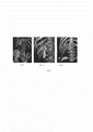
Сущность предлагаемого способа иллюстрирована на фиг. 1, где поз. 1-гепатопанкреатодуоденальная зона при дистальной обструкции билиарного тракта, поз. 2-гепатопанкреатодуоденальная зона при центральной обструкции билиарного тракта; поз. 3-гепатопанкреатодуоденальная зона при проксимальной обструкции билиарного тракта. Ключевыми моментами являются определение локализации обструкции билиарного тракта, ее протяженность, состояние билиарного тракта выше места окклюзии, взаимоотношения окклюзированного участка билиарного тракта с магистральными сосудами данной области, форма конфлюенсов желчных протоков, количество долевых и сегментарных протоков, участвующих в их образовании.The essence of the proposed method is illustrated in FIG. 1, where pos. 1-hepatopancreatoduodenal zone with distal obstruction of the biliary tract, pos. 2-hepatopancreatoduodenal zone with central obstruction of the biliary tract; pos. 3-hepatopancreatoduodenal zone with proximal obstruction of the biliary tract. The key points are determining the localization of the obstruction of the biliary tract, its length, the state of the biliary tract above the place of occlusion, the relationship of the occluded section of the biliary tract with the main vessels of this area, the shape of the bile duct confluences, the number of lobar and segmental ducts involved in their formation.
Показаниями для применения данного способа являются: опухоли желчных протоков, периампулярная опухоль, наружные желчные свищи, свищи при эхинококкозе печени, кистозные образования головки поджелудочной железы.Indications for the use of this method are: bile duct tumors, periampular tumor, external bile fistula, fistula with echinococcosis of the liver, cystic formations of the pancreatic head.
Технический результат достигается за счет более четкой визуализации билиарного тракта, сопоставления его структуры со структурой сосудистых систем гепатопанкреатодуоденальной зоны, их реконструкции в 3D режиме, что связано с введением контрастного раствора через холангиостомическую трубку непосредственно перед МСКТ.The technical result is achieved due to a clearer visualization of the biliary tract, comparison of its structure with the structure of the vascular systems of the hepatopancreatoduodenal zone, their reconstruction in 3D mode, which is associated with the introduction of a contrast solution through the cholangiostomy tube immediately before the MSCT.
Пример конкретного выполнения способа.An example of a specific implementation of the method.
Пациент Г., 64 лет, поступил в хирургическое отделение №2 ГБУ «РКБ» МЗ РД 18.05.2018 г. с диагнозом «Периампулярная опухоль, дистальная обструкция билиарного тракта, механическая желтуха тяжелой степени, эндотоксикоз тяжелой степени, печеночная недостаточность, холестероз желчного пузыря.Patient G., 64 years old, was admitted to the surgical department No. 2 of GBU “RKB” MH RD 18.05.2018 with the diagnosis “Periampular tumor, distal obstruction of the biliary tract, severe obstructive jaundice, severe endotoxemia, liver failure, gall bladder cholesterosis .
Общий анализ крови: Нв - 132 г/л, эритроциты - 4,3×10¹²/л, лейкоциты - 12,6×10⁹/л. Общий билирубин - 377 мкмоль/л.Complete blood count: HB - 132 g / l, red blood cells - 4.3 × 10 12 / l, white blood cells - 12.6 × 10 9 / l. Total bilirubin - 377 μmol / L.
УЗИ брюшной полости: печень - 16,0 см, контуры ровные, паренхима повышенной эхогенности, неоднородная. Внутрипеченочные желчные ходы расширены, холедох - 1,7 см, свободен. Внутрипеченочные желчные протоки расширены умеренно, желчный пузырь размерами 8,0×3,0 см, стенка 0,4 см, на передней и задней стенках полипы - 0,3-0,5 см. Поджелудочная железа (далее ПЖ) - головка 3,8 см, тело - 2,7 см, хвост - 2,7 см, контуры волнистые, размытые, повышенной эхогенности, неоднородная. В проекции головки ПЖ гипоэхогенное образование с неровными нечеткими контурами - 3,0 см. В брюшной и плевральных полостях жидкости не визуализируются.Ultrasound of the abdominal cavity: liver - 16.0 cm, smooth contours, parenchyma of increased echogenicity, heterogeneous. Intrahepatic bile ducts are dilated, common bile duct - 1.7 cm, free. Intrahepatic bile ducts are moderately dilated, the gallbladder is 8.0 × 3.0 cm in size, the wall is 0.4 cm, the polyps on the front and back walls are 0.3-0.5 cm. The pancreas (hereinafter referred to as the pancreas) is head 3, 8 cm, body - 2.7 cm, tail - 2.7 cm, contours wavy, blurry, increased echogenicity, heterogeneous. In the projection of the pancreatic head, a hypoechoic formation with uneven fuzzy contours is 3.0 cm. In the abdominal and pleural cavities, fluids are not visualized.
МРТ органов брюшной полости: признаки дистальной обструкции билиарного тракта, гепатоспиномегалия. фиброгастродуоденоскопия: поверхностный гастрит, бульбит.Abdominal MRI: signs of distal obstruction of the biliary tract, hepatospinomegaly. fibrogastroduodenoscopy: superficial gastritis, bulbitis.
Рентгеноскопия легких: легочные поля без очаговых и инфильтративных теней.Fluoroscopy of the lungs: pulmonary fields without focal and infiltrative shadows.
Электрокардиограмма - без патологии.Electrocardiogram - without pathology.
С целью декомпрессии билиарного тракта первым этапом выполнено оперативное лечение: рентгенохирургическое транспеченочное антеградное дренирование желчных протоков. Через 10 дней после снятия явлений холестаза пациент выписан в удовлетворительном состоянии с рекомендациями явиться на второй этап операции через 20 дней при билирубине 40-50 мкмоль/л.In order to decompress the biliary tract, the first step was surgical treatment: X-ray transhepatic antegrade drainage of the bile ducts. 10 days after the removal of the phenomena of cholestasis, the patient was discharged in satisfactory condition with recommendations to come to the second stage of the operation after 20 days with bilirubin 40-50 μmol / L.
На повторную консультацию пациент в клинику обратился через 4 месяца.The patient turned to the clinic for a second consultation after 4 months.
Общий анализ крови: Нв - 127 г/л, эритроциты - 4,0×10¹²/л, Л - 8,6×10⁹/л, СОЭ - 38 мм/час. Общий билирубин 24,6 ммоль/л. Онкомаркеры: РЭА - 2,75; СА - 19,9 - 11, АФП - 1,52.Complete blood count: HB - 127 g / l, red blood cells - 4.0 × 10 12 / l, L - 8.6 × 10 9 / l, ESR - 38 mm / hour. Total bilirubin 24.6 mmol / L. Oncomarkers: CEA - 2.75; CA - 19.9 - 11, AFP - 1.52.
В клинике пациенту выполнена МСКТ с внутривенным контрастированием (омнипак 350-100,0 мл) и введением урографина 20,0 мл в дренажную трубку, установленный в билиарный тракт согласно описанию предлагаемого способа.In the clinic, the patient underwent MSCT with intravenous contrast (omnipack 350-100.0 ml) and the introduction of urografin 20.0 ml into the drainage tube installed in the biliary tract according to the description of the proposed method.
При этом определяется окклюзия билиарного тракта на уровне ретродуоденальной части холедоха, желчный пузырь не визуализируется, то есть имеет место низкое впадении пузырного протока и вовлечение его устья в окклюзионный процесс. Уровень окклюзии ниже места отхождения желудочно-двенадцатиперстной артерии. Следовательно, опухолевый процесс расположен почти в ткани головки поджелудочной железы. Признаки панкреатической гипертензии не определяются.In this case, the occlusion of the biliary tract is determined at the level of the retroduodenal part of the common bile duct, the gall bladder is not visualized, that is, there is a low confluence of the cystic duct and the involvement of its mouth in the occlusion process. The level of occlusion below the site of discharge of the gastro-duodenal artery. Therefore, the tumor process is located almost in the tissue of the head of the pancreas. Signs of pancreatic hypertension are not detected.
14.09.2018 г. пациенту выполнена операция - лапаротомия, гастропанкреатодуоденальная резекция. При этом печень несколько увеличена, сероватого цвета, плотной консистенции, желчный пузырь размерами 10,2×3,7 см, напряжен. При пункции получена «светлая желчь» (водянка желчного пузыря). Пузырный проток впадает низко, нижний конфлюенс в ретродуоденальной части и в области слияния пузырного протока и гепатикохоледоха имеется плотное образование размерами 1,5-1,7 см. В этом участке перихоледохеальные лимфатические узлы плотной консистенции и увеличены. Чувствительность предлагаемого способа по сравнению с интраоперацонными данными почти 100%.September 14, 2018 the patient underwent surgery - laparotomy, gastropancreatoduodenal resection. In this case, the liver is slightly enlarged, grayish in color, of dense consistency, the gallbladder 10.2 × 3.7 cm in size, is tense. During puncture, “light bile” (dropsy of the gallbladder) was obtained. The cystic duct flows low, the lower confluence in the retroduodenal part and in the area of confluence of the cystic duct and hepatic choledochus has a dense formation 1.5-1.7 cm in size. In this area, pericholedochal lymph nodes are of a dense consistency and are enlarged. The sensitivity of the proposed method compared with intraoperative data is almost 100%.
Гистозаключение: умеренно дифференцированная аденокарцинома стенки желчного протока с эрозивным дефектом и инвазией в стенку. Аденоматозная гиперплазия экзокринного аппарата с фибротизацией стромы ткани поджелудочной железы, по-видимому, реактивного характера. В лимфоузлах опухолевый рост не отмечен. Течение послеоперационного периода без осложнений. Пациент выписан на 14-е сутки после операции с диагнозом: «Аденокарцинома желчного протока (T2N0M0), дистальная обструкция билиарного тракта, механическая желтуха.Histological conclusion: moderately differentiated adenocarcinoma of the bile duct wall with an erosive defect and invasion of the wall. Adenomatous hyperplasia of the exocrine apparatus with fibrotization of the stroma of the pancreatic tissue, apparently of a reactive nature. No tumor growth was noted in the lymph nodes. The course of the postoperative period without complications. The patient was discharged on the 14th day after surgery with a diagnosis of “Adenocarcinoma of the bile duct (T 2 N 0 M 0 ), distal obstruction of the biliary tract, obstructive jaundice.
Рекомендовано проведение химиотерапии в условиях Республиканского онкологического диспансера.Chemotherapy in the conditions of the Republican Oncology Center is recommended.
Признаки изобретения, отличительные от прототипаFeatures of the invention, distinctive from the prototype
- непосредственно перед проведением МСКТ вводят 20 мл водорастворимого контрастного вещества через холангиостомический катетер.- immediately before the MSCT, 20 ml of a water-soluble contrast medium is administered through a cholangiostomy catheter.
Признаки прототипаSigns of the prototype
- Проводят МСКТ- Conduct MSCT
- внутривенно вводят контрастное вещество 100 мл Омнипак 350- contrast medium 100 ml Omnipack 350 is injected intravenously
Положительный эффект от применения изобретенияThe positive effect of the application of the invention
Предлагаемый способ позволит определить точные показания к выполнению радикального хирургического вмешательства.The proposed method will allow you to determine the exact indications for radical surgery.
Способ позволит уменьшить число рецидивов после радикальных операций по поводу опухолевого процесса желчных протоков и тем самым увеличить процент 5 - летней выживаемости онкологических пациентов.The method will reduce the number of relapses after radical surgery for the tumor process of the bile ducts and thereby increase the percentage of 5 - year survival of cancer patients.
Используемый способ предупреждает развитие послеоперационных осложнений в виде несостоятельности билиодигестивных анастомозов с развитием билом и желчного перитонита за счет определения «благоприятных» и «неблагоприятных» условий со стороны билиарного тракта для формирования билиодигестивных анастомозов. Применяемый способ визуализации билиарного тракта с высокой вероятностью определяет степень распространенности опухолевого процесса, протяженность поражения по желчным протокам из-за трехмерности изображения.The method used prevents the development of postoperative complications in the form of insolvency of biliodigestive anastomoses with the development of bilia and biliary peritonitis by determining the “favorable” and “unfavorable” conditions on the part of the biliary tract for the formation of biliodigestive anastomoses. The applied method for visualizing the biliary tract with high probability determines the degree of prevalence of the tumor process, the extent of the lesion along the bile ducts due to the three-dimensionality of the image.
Одновременная визуализация билиарного тракта и сосудистой системы обеспечивает определение взаимоотношения пораженного участка к соседним органам и структурам, тем самым позволяет определить объем радикальной операции.Simultaneous visualization of the biliary tract and vascular system ensures the determination of the relationship of the affected area to neighboring organs and structures, thereby allowing to determine the volume of radical surgery.
Трехмерность изображения, получаемое при применении предлагаемого способа позволяет с высокой точностью определить количество протоков, участвующих в образовании конфлюенсов желчных протоков, переход опухолевого процесса на долевые и сегментарные протоки, что чрезвычайно важно для достижения отрицательного резекционного края в условиях проведения радикальной операцииThe three-dimensionality of the image obtained by application of the proposed method allows to determine with high accuracy the number of ducts involved in the formation of bile duct confluences, the transition of the tumor process to the lobar and segmental ducts, which is extremely important to achieve a negative resection edge under radical surgery
Предлагаемый способ использован для визуализации билиарного тракта 123 пациентам с опухолевыми поражениями гепатопанкреатодуоденальной зоны. Из них периампулярные опухоли имелись у 96 пациентов, хилярная холангиокарцинома - 22, опухоли гепатикохоледоха у 2 и у 3 метастатическое поражение гепатодуоденальной связки с окклюзией внепеченочных желчных протоков. Чувствительность способа по сравнению с прототипом составила 97,4%.The proposed method is used to visualize the biliary tract in 123 patients with tumor lesions of the hepatopancreatoduodenal zone. Of these, periampicular tumors were present in 96 patients, 22 hilar cholangiocarcinoma, hepatic choledochus tumors in 2 and 3 metastatic lesions of the hepatoduodenal ligament with extrahepatic bile duct occlusion. The sensitivity of the method compared to the prototype was 97.4%.
Информация, принятая во вниманиеInformation taken into account
Бурякина С.А., Кармазоновский Г.Г., Опухоль Клацкина: современные аспекты дифференциальной диагностики // Анналы хирургической гепатологии, 2012. - Т.17. №1 - С. 100-109.Buryakina S.A., Karmazonovsky G.G., Klatskin tumor: modern aspects of differential diagnosis // Annals of surgical hepatology, 2012. - V. 17. No. 1 - S. 100-109.
Кармазановский Г.Г. МсКТ и МРТ диагностика кистозных опухолей поджелудочной железы // Анналы хирургической гепатологии, 2013. - Т.П. - №1. - С.11-16. Забавина Н.И., Плотников А.Ф., Колпощиков И.Е. Малоинвазивные методы лучевой диагностики механической желтухи опухолевого генеза // Современные технологии в медицине, 2009, - №1. - С. 57-62.Karmazanovsky G.G. MSCT and MRI diagnosis of cystic tumors of the pancreas // Annals of surgical hepatology, 2013. - T.P. - No. 1. - S.11-16. Zabavina N.I., Plotnikov A.F., Kolposhchikov I.E. Minimally invasive methods of radiation diagnosis of obstructive jaundice of tumor origin // Modern technologies in medicine, 2009, No. 1. - S. 57-62.
Прокоп М., Галански М., Спиральная и многослойная компьютерная томография. М.: Медпресс, 2009. - Т.1 - С. 102-118) - прототип.Prokop M., Galanski M., Spiral and multilayer computed tomography. M .: Medpress, 2009. - T.1 - S. 102-118) - prototype.
Claims (1)
Priority Applications (1)
| Application Number | Priority Date | Filing Date | Title |
|---|---|---|---|
| RU2019105909A RU2706373C1 (en) | 2019-03-01 | 2019-03-01 | Method for localizing tumor obstruction of biliary tract and degree of its prevalence |
Applications Claiming Priority (1)
| Application Number | Priority Date | Filing Date | Title |
|---|---|---|---|
| RU2019105909A RU2706373C1 (en) | 2019-03-01 | 2019-03-01 | Method for localizing tumor obstruction of biliary tract and degree of its prevalence |
Publications (1)
| Publication Number | Publication Date |
|---|---|
| RU2706373C1 true RU2706373C1 (en) | 2019-11-18 |
Family
ID=68580017
Family Applications (1)
| Application Number | Title | Priority Date | Filing Date |
|---|---|---|---|
| RU2019105909A RU2706373C1 (en) | 2019-03-01 | 2019-03-01 | Method for localizing tumor obstruction of biliary tract and degree of its prevalence |
Country Status (1)
| Country | Link |
|---|---|
| RU (1) | RU2706373C1 (en) |
Cited By (1)
| Publication number | Priority date | Publication date | Assignee | Title |
|---|---|---|---|---|
| RU2737579C1 (en) * | 2020-04-15 | 2020-12-01 | Федеральное Государственное Бюджетное Учреждение «Российский Научный Центр Радиологии И Хирургических Технологий Имени Академика А.М. Гранова» Министерства Здравоохранения Российской Федерации | Method of treating chronic occlusions of main arteries |
Citations (2)
| Publication number | Priority date | Publication date | Assignee | Title |
|---|---|---|---|---|
| US20160008496A1 (en) * | 2007-06-22 | 2016-01-14 | Mivenion Gmbh | Imaging diagnostics by combining contrast agents |
| WO2017197342A1 (en) * | 2016-05-13 | 2017-11-16 | Teclison Limited | Methods for treating liver tissue |
-
2019
- 2019-03-01 RU RU2019105909A patent/RU2706373C1/en active
Patent Citations (2)
| Publication number | Priority date | Publication date | Assignee | Title |
|---|---|---|---|---|
| US20160008496A1 (en) * | 2007-06-22 | 2016-01-14 | Mivenion Gmbh | Imaging diagnostics by combining contrast agents |
| WO2017197342A1 (en) * | 2016-05-13 | 2017-11-16 | Teclison Limited | Methods for treating liver tissue |
Non-Patent Citations (2)
| Title |
|---|
| ДОЛГУШИН Б.И. и др., ЧРЕСКОЖНАЯ ЧРЕСПЕЧЕНОЧНАЯ ПУНКЦИОННАЯ ХОЛАНГИОСТОМИЯ: СИСТЕМАТИЗАЦИЯ ПРЕДСТАВЛЕНИЙ, ДИАГНОСТИЧЕСКАЯ И ИНТЕРВЕНЦИОННАЯ РАДИОЛОГИЯ, ТОМ 6, N3,2012, cc.31-60. * |
| ПРОКОП М. и др. СПИРАЛЬНАЯ И МНОГОСЛОЙНАЯ КОМПЬЮТЕРНАЯ ТОМОГРАФИЯ. М.: МЕДПРЕСС-ИНФОРМ, 2006. -Т.1. * |
Cited By (1)
| Publication number | Priority date | Publication date | Assignee | Title |
|---|---|---|---|---|
| RU2737579C1 (en) * | 2020-04-15 | 2020-12-01 | Федеральное Государственное Бюджетное Учреждение «Российский Научный Центр Радиологии И Хирургических Технологий Имени Академика А.М. Гранова» Министерства Здравоохранения Российской Федерации | Method of treating chronic occlusions of main arteries |
Similar Documents
| Publication | Publication Date | Title |
|---|---|---|
| Kaptein et al. | Pulmonary infarction in acute pulmonary embolism | |
| Bruzzi et al. | Multi–detector row CT of hemoptysis | |
| Song et al. | Cavernous transformation of the portal vein secondary to tumor thrombosis of hepatocellular carcinoma: spiral CT visualization of the collateral vessels | |
| Mori et al. | Dilated posterior superior pancreaticoduodenal vein: recognition with CT and clinical significance in patients with pancreaticobiliary carcinomas. | |
| Sai et al. | MRCP: early diagnosis of pancreatobiliary diseases | |
| Lau et al. | Resection of hepatocellular carcinoma with diaphragmatic invasion | |
| Hafezi-Nejad et al. | Surgical approaches to chronic pancreatitis: indications and imaging findings | |
| RU2706373C1 (en) | Method for localizing tumor obstruction of biliary tract and degree of its prevalence | |
| Krishnan et al. | Current techniques and clinical applications of computed tomography urography | |
| Badea et al. | Ultrasonography of the biliary tract –up to date. The importance of correlation between imaging methods and patients’ signs and symptoms. | |
| Chung et al. | Multiple biliary papillomatosis: comparison of MR cholangiography with endoscopic retrograde cholangiography | |
| Lipson | MDCT and 3D workstations: a practical how-to guide and teaching file | |
| Laghi | MDCT protocols: whole body and emergencies | |
| Washburn et al. | Computed tomographic urography update: an evolving urinary tract imaging modality | |
| Castrillón et al. | Malignant biliary obstruction: usual and recent imaging findings | |
| Darby et al. | Oxford handbook of medical imaging | |
| Brink et al. | Helical/spiral computed body tomography | |
| RU2661097C1 (en) | Method of catheterization and method of endovascular occlusion of bronchial and intercostal arteries | |
| RU2647141C1 (en) | Method of intraoperative detection of pancreatic tumor insulation invasion in the wall of the approaching vessel | |
| Sanders et al. | Combined angiography and mediastinoscopy in bronchogenic carcinoma | |
| Rustgi et al. | Hepatic imaging and advanced endoscopic techniques | |
| Harvey et al. | Radiological investigations and applications | |
| Białek et al. | TRUS-guided drainage of the ectopic ureter entering the prostatic urethra and TRUS-guided transurethral neo-orifice formation using holmium laser | |
| Brejt et al. | Interventional radiology | |
| Nanni et al. | Abdomen |