RU2688462C2 - Genomic engineering - Google Patents
Genomic engineering Download PDFInfo
- Publication number
- RU2688462C2 RU2688462C2 RU2016106649A RU2016106649A RU2688462C2 RU 2688462 C2 RU2688462 C2 RU 2688462C2 RU 2016106649 A RU2016106649 A RU 2016106649A RU 2016106649 A RU2016106649 A RU 2016106649A RU 2688462 C2 RU2688462 C2 RU 2688462C2
- Authority
- RU
- Russia
- Prior art keywords
- dna
- cell
- nucleic acid
- enzyme
- cells
- Prior art date
Links
Images


Classifications
-
- C—CHEMISTRY; METALLURGY
- C12—BIOCHEMISTRY; BEER; SPIRITS; WINE; VINEGAR; MICROBIOLOGY; ENZYMOLOGY; MUTATION OR GENETIC ENGINEERING
- C12N—MICROORGANISMS OR ENZYMES; COMPOSITIONS THEREOF; PROPAGATING, PRESERVING, OR MAINTAINING MICROORGANISMS; MUTATION OR GENETIC ENGINEERING; CULTURE MEDIA
- C12N15/00—Mutation or genetic engineering; DNA or RNA concerning genetic engineering, vectors, e.g. plasmids, or their isolation, preparation or purification; Use of hosts therefor
- C12N15/09—Recombinant DNA-technology
- C12N15/87—Introduction of foreign genetic material using processes not otherwise provided for, e.g. co-transformation
- C12N15/90—Stable introduction of foreign DNA into chromosome
- C12N15/902—Stable introduction of foreign DNA into chromosome using homologous recombination
- C12N15/907—Stable introduction of foreign DNA into chromosome using homologous recombination in mammalian cells
-
- C—CHEMISTRY; METALLURGY
- C12—BIOCHEMISTRY; BEER; SPIRITS; WINE; VINEGAR; MICROBIOLOGY; ENZYMOLOGY; MUTATION OR GENETIC ENGINEERING
- C12N—MICROORGANISMS OR ENZYMES; COMPOSITIONS THEREOF; PROPAGATING, PRESERVING, OR MAINTAINING MICROORGANISMS; MUTATION OR GENETIC ENGINEERING; CULTURE MEDIA
- C12N15/00—Mutation or genetic engineering; DNA or RNA concerning genetic engineering, vectors, e.g. plasmids, or their isolation, preparation or purification; Use of hosts therefor
- C12N15/09—Recombinant DNA-technology
- C12N15/11—DNA or RNA fragments; Modified forms thereof; Non-coding nucleic acids having a biological activity
- C12N15/113—Non-coding nucleic acids modulating the expression of genes, e.g. antisense oligonucleotides; Antisense DNA or RNA; Triplex- forming oligonucleotides; Catalytic nucleic acids, e.g. ribozymes; Nucleic acids used in co-suppression or gene silencing
-
- C—CHEMISTRY; METALLURGY
- C12—BIOCHEMISTRY; BEER; SPIRITS; WINE; VINEGAR; MICROBIOLOGY; ENZYMOLOGY; MUTATION OR GENETIC ENGINEERING
- C12N—MICROORGANISMS OR ENZYMES; COMPOSITIONS THEREOF; PROPAGATING, PRESERVING, OR MAINTAINING MICROORGANISMS; MUTATION OR GENETIC ENGINEERING; CULTURE MEDIA
- C12N15/00—Mutation or genetic engineering; DNA or RNA concerning genetic engineering, vectors, e.g. plasmids, or their isolation, preparation or purification; Use of hosts therefor
- C12N15/09—Recombinant DNA-technology
- C12N15/63—Introduction of foreign genetic material using vectors; Vectors; Use of hosts therefor; Regulation of expression
- C12N15/79—Vectors or expression systems specially adapted for eukaryotic hosts
- C12N15/85—Vectors or expression systems specially adapted for eukaryotic hosts for animal cells
- C12N15/86—Viral vectors
-
- C—CHEMISTRY; METALLURGY
- C12—BIOCHEMISTRY; BEER; SPIRITS; WINE; VINEGAR; MICROBIOLOGY; ENZYMOLOGY; MUTATION OR GENETIC ENGINEERING
- C12N—MICROORGANISMS OR ENZYMES; COMPOSITIONS THEREOF; PROPAGATING, PRESERVING, OR MAINTAINING MICROORGANISMS; MUTATION OR GENETIC ENGINEERING; CULTURE MEDIA
- C12N15/00—Mutation or genetic engineering; DNA or RNA concerning genetic engineering, vectors, e.g. plasmids, or their isolation, preparation or purification; Use of hosts therefor
- C12N15/09—Recombinant DNA-technology
- C12N15/87—Introduction of foreign genetic material using processes not otherwise provided for, e.g. co-transformation
- C12N15/90—Stable introduction of foreign DNA into chromosome
-
- C—CHEMISTRY; METALLURGY
- C12—BIOCHEMISTRY; BEER; SPIRITS; WINE; VINEGAR; MICROBIOLOGY; ENZYMOLOGY; MUTATION OR GENETIC ENGINEERING
- C12N—MICROORGANISMS OR ENZYMES; COMPOSITIONS THEREOF; PROPAGATING, PRESERVING, OR MAINTAINING MICROORGANISMS; MUTATION OR GENETIC ENGINEERING; CULTURE MEDIA
- C12N5/00—Undifferentiated human, animal or plant cells, e.g. cell lines; Tissues; Cultivation or maintenance thereof; Culture media therefor
- C12N5/06—Animal cells or tissues; Human cells or tissues
- C12N5/0602—Vertebrate cells
- C12N5/0696—Artificially induced pluripotent stem cells, e.g. iPS
-
- C—CHEMISTRY; METALLURGY
- C12—BIOCHEMISTRY; BEER; SPIRITS; WINE; VINEGAR; MICROBIOLOGY; ENZYMOLOGY; MUTATION OR GENETIC ENGINEERING
- C12N—MICROORGANISMS OR ENZYMES; COMPOSITIONS THEREOF; PROPAGATING, PRESERVING, OR MAINTAINING MICROORGANISMS; MUTATION OR GENETIC ENGINEERING; CULTURE MEDIA
- C12N5/00—Undifferentiated human, animal or plant cells, e.g. cell lines; Tissues; Cultivation or maintenance thereof; Culture media therefor
- C12N5/10—Cells modified by introduction of foreign genetic material
-
- C—CHEMISTRY; METALLURGY
- C12—BIOCHEMISTRY; BEER; SPIRITS; WINE; VINEGAR; MICROBIOLOGY; ENZYMOLOGY; MUTATION OR GENETIC ENGINEERING
- C12N—MICROORGANISMS OR ENZYMES; COMPOSITIONS THEREOF; PROPAGATING, PRESERVING, OR MAINTAINING MICROORGANISMS; MUTATION OR GENETIC ENGINEERING; CULTURE MEDIA
- C12N9/00—Enzymes; Proenzymes; Compositions thereof; Processes for preparing, activating, inhibiting, separating or purifying enzymes
- C12N9/14—Hydrolases (3)
- C12N9/16—Hydrolases (3) acting on ester bonds (3.1)
- C12N9/22—Ribonucleases RNAses, DNAses
-
- C—CHEMISTRY; METALLURGY
- C07—ORGANIC CHEMISTRY
- C07H—SUGARS; DERIVATIVES THEREOF; NUCLEOSIDES; NUCLEOTIDES; NUCLEIC ACIDS
- C07H21/00—Compounds containing two or more mononucleotide units having separate phosphate or polyphosphate groups linked by saccharide radicals of nucleoside groups, e.g. nucleic acids
- C07H21/02—Compounds containing two or more mononucleotide units having separate phosphate or polyphosphate groups linked by saccharide radicals of nucleoside groups, e.g. nucleic acids with ribosyl as saccharide radical
-
- C—CHEMISTRY; METALLURGY
- C07—ORGANIC CHEMISTRY
- C07H—SUGARS; DERIVATIVES THEREOF; NUCLEOSIDES; NUCLEOTIDES; NUCLEIC ACIDS
- C07H21/00—Compounds containing two or more mononucleotide units having separate phosphate or polyphosphate groups linked by saccharide radicals of nucleoside groups, e.g. nucleic acids
- C07H21/04—Compounds containing two or more mononucleotide units having separate phosphate or polyphosphate groups linked by saccharide radicals of nucleoside groups, e.g. nucleic acids with deoxyribosyl as saccharide radical
-
- C—CHEMISTRY; METALLURGY
- C07—ORGANIC CHEMISTRY
- C07K—PEPTIDES
- C07K14/00—Peptides having more than 20 amino acids; Gastrins; Somatostatins; Melanotropins; Derivatives thereof
- C07K14/195—Peptides having more than 20 amino acids; Gastrins; Somatostatins; Melanotropins; Derivatives thereof from bacteria
- C07K14/315—Peptides having more than 20 amino acids; Gastrins; Somatostatins; Melanotropins; Derivatives thereof from bacteria from Streptococcus (G), e.g. Enterococci
-
- C—CHEMISTRY; METALLURGY
- C12—BIOCHEMISTRY; BEER; SPIRITS; WINE; VINEGAR; MICROBIOLOGY; ENZYMOLOGY; MUTATION OR GENETIC ENGINEERING
- C12N—MICROORGANISMS OR ENZYMES; COMPOSITIONS THEREOF; PROPAGATING, PRESERVING, OR MAINTAINING MICROORGANISMS; MUTATION OR GENETIC ENGINEERING; CULTURE MEDIA
- C12N15/00—Mutation or genetic engineering; DNA or RNA concerning genetic engineering, vectors, e.g. plasmids, or their isolation, preparation or purification; Use of hosts therefor
- C12N15/09—Recombinant DNA-technology
- C12N15/63—Introduction of foreign genetic material using vectors; Vectors; Use of hosts therefor; Regulation of expression
-
- C—CHEMISTRY; METALLURGY
- C12—BIOCHEMISTRY; BEER; SPIRITS; WINE; VINEGAR; MICROBIOLOGY; ENZYMOLOGY; MUTATION OR GENETIC ENGINEERING
- C12N—MICROORGANISMS OR ENZYMES; COMPOSITIONS THEREOF; PROPAGATING, PRESERVING, OR MAINTAINING MICROORGANISMS; MUTATION OR GENETIC ENGINEERING; CULTURE MEDIA
- C12N2310/00—Structure or type of the nucleic acid
- C12N2310/10—Type of nucleic acid
- C12N2310/20—Type of nucleic acid involving clustered regularly interspaced short palindromic repeats [CRISPRs]
-
- C—CHEMISTRY; METALLURGY
- C12—BIOCHEMISTRY; BEER; SPIRITS; WINE; VINEGAR; MICROBIOLOGY; ENZYMOLOGY; MUTATION OR GENETIC ENGINEERING
- C12N—MICROORGANISMS OR ENZYMES; COMPOSITIONS THEREOF; PROPAGATING, PRESERVING, OR MAINTAINING MICROORGANISMS; MUTATION OR GENETIC ENGINEERING; CULTURE MEDIA
- C12N2510/00—Genetically modified cells
-
- C—CHEMISTRY; METALLURGY
- C12—BIOCHEMISTRY; BEER; SPIRITS; WINE; VINEGAR; MICROBIOLOGY; ENZYMOLOGY; MUTATION OR GENETIC ENGINEERING
- C12N—MICROORGANISMS OR ENZYMES; COMPOSITIONS THEREOF; PROPAGATING, PRESERVING, OR MAINTAINING MICROORGANISMS; MUTATION OR GENETIC ENGINEERING; CULTURE MEDIA
- C12N2740/00—Reverse transcribing RNA viruses
- C12N2740/00011—Details
- C12N2740/10011—Retroviridae
- C12N2740/15011—Lentivirus, not HIV, e.g. FIV, SIV
- C12N2740/15041—Use of virus, viral particle or viral elements as a vector
- C12N2740/15043—Use of virus, viral particle or viral elements as a vector viral genome or elements thereof as genetic vector
-
- C—CHEMISTRY; METALLURGY
- C12—BIOCHEMISTRY; BEER; SPIRITS; WINE; VINEGAR; MICROBIOLOGY; ENZYMOLOGY; MUTATION OR GENETIC ENGINEERING
- C12N—MICROORGANISMS OR ENZYMES; COMPOSITIONS THEREOF; PROPAGATING, PRESERVING, OR MAINTAINING MICROORGANISMS; MUTATION OR GENETIC ENGINEERING; CULTURE MEDIA
- C12N2800/00—Nucleic acids vectors
- C12N2800/90—Vectors containing a transposable element
-
- C—CHEMISTRY; METALLURGY
- C12—BIOCHEMISTRY; BEER; SPIRITS; WINE; VINEGAR; MICROBIOLOGY; ENZYMOLOGY; MUTATION OR GENETIC ENGINEERING
- C12N—MICROORGANISMS OR ENZYMES; COMPOSITIONS THEREOF; PROPAGATING, PRESERVING, OR MAINTAINING MICROORGANISMS; MUTATION OR GENETIC ENGINEERING; CULTURE MEDIA
- C12N2830/00—Vector systems having a special element relevant for transcription
- C12N2830/001—Vector systems having a special element relevant for transcription controllable enhancer/promoter combination
- C12N2830/002—Vector systems having a special element relevant for transcription controllable enhancer/promoter combination inducible enhancer/promoter combination, e.g. hypoxia, iron, transcription factor
- C12N2830/003—Vector systems having a special element relevant for transcription controllable enhancer/promoter combination inducible enhancer/promoter combination, e.g. hypoxia, iron, transcription factor tet inducible
Abstract
Description
Данные связанных заявокRelated Claim Data
Данная заявка испрашивает приоритет предварительной заявки на патент США No. 61/858866, поданной 26 июля 2013 г. и включенной в данный документ ссылкой в полном объеме и для всех целей. This application claims the priority of provisional patent application US No. 61/858866, filed July 26, 2013 and incorporated by reference in full and for all purposes.
Заявление государственных интересовState interest statement
Это изобретение было осуществлено при государственной поддержке согласно P50 HG003170 от Национального научно-исследовательского центра генома человека за выдающиеся достижения в геномике. Правительство имеет определенные права на это изобретение. This invention was implemented with state support according to P50 HG003170 from the National Human Genome Research Center for outstanding achievements in genomics. The government has certain rights to this invention.
Предшествующий уровень техникиPrior art
Известно редактирование генома с помощью специфических к последовательности нуклеаз. Смотрите источники 1, 2 и 3, которые включены в данный документ ссылкой в полном объеме. Опосредованный нуклеазой разрыв двухцепочечной ДНК (дцДНК) в геноме может быть устранен с помощью двух основных механизмов: негомологичного соединения концов (NHEJ), который часто приводит к введению неспецифических вставок и делеций (indels), или направляемой гомологией репарации (HDR), которая использует гомологичную цепь в качестве шаблона для репарации. См. источник 4 включенный ссылкой в полном объеме. Когда специфичная к последовательности нуклеаза доставляется вместе с гомологичной донорной ДНК-конструкцией, содержащей искомые мутации, эффективности направленного воздействия на ген повышаются в 1000 раз по сравнению только с доставкой донорной конструкции отдельно. См. источник 5, включенный в данный документ ссылкой в полном объеме. Сообщалось об использовании одноцепочечных олигодезоксирибонуклеотидов («ssODNs») в качестве ДНК-доноров. См. источники 21 и 22, включенные в данный документ ссылкой в полном объеме. Known editing of the genome using specific nuclease sequences. See
Несмотря на большие достижения в области технологий редактирования генов, остаются нерешенными многие проблемы и многие вопросы относительно применения специально сконструированных нуклеаз при конструировании человеческих индуцированных плюрипотентных стволовых клеток («hiPSC»). Во-первых, несмотря на простоту своей конструкции TALEN (эффекторные нуклеазы, подобные активаторам траснкрипции) нацеливаются на определенные последовательности ДНК тандемными копиями доменов RVD (домены вариабельных двух остатков). См. источник 6, включенный в данный документ ссылкой в полном объеме. В то время как модульный характер RVDs упрощает дизайн TALEN, их повторяющиеся последовательности усложняют способы синтеза ДНК-конструкций на их основе (см. источники 2, 9 и 15-19, включенные в данный документ ссылкой в полном объеме), а также делает практически невозможным их совместное применение вместе со средствами доставки генов на основе лентивирусов. См. источник 13, включенный в данный документ ссылкой в полном объеме. Despite the great advances in gene editing technologies, many problems and many questions remain about the use of specially designed nucleases in the design of human induced pluripotent stem cells (“hiPSC”). First, despite the simplicity of its design, TALEN (effector nucleases, similar to activators of transcription) target specific DNA sequences with tandem copies of RVD domains (domains of two variable residues). See
В современной практике NHEJ и HDR часто оцениваются с помощью отдельных анализов. Тесты с эндонуклеазой, чувствительной к некомлементарности (см. источник 14, включенный в данный документ ссылкой в полном объеме) часто используются для оценки NHEJ, но количественная точность этого способа переменчива, и чувствительность ограничивается частотами NHEJ выше ~ 3%. См. источник 15, включенный в данный документ ссылкой в полном объеме. HDR часто оценивается с помощью клонирования и секвенирования, совершенно иной и часто громоздкой процедуры. Чувствительность по-прежнему является проблемой, поскольку, несмотря на высокие частоты редактирования порядка 50% о которых часто сообщалось в случае некоторых типов клеток, таких как U20S и К562 (см. источники 12 и 14, включенные в данный документ ссылкой в полном объеме), в случае hiPSC частоты, как правило, ниже. См. Источник 10, включенный в данный документ ссылкой в полном объеме. В последнее время высокие частоты редактирования были зарегистрированы в hiPSC и hESC, достигнутые с помощью TALENs (см. источник 9, включенный в данный документ ссылкой в полном объеме), и еще более высокие частоты были достигнуты с помощью системы CRISPR Cas9-гидРНК (см. источники 16-19, включенные в данный документ ссылкой в полном объеме. Тем не менее, показатели редактирования на различных участках по всей видимости, сильно отличаются (см. источник 17, включенный в данный документ ссылкой в полном объеме), а в некоторых участках редактирование иногда не обнаруживается вообще (см. источник 20, включенный в данный документ ссылкой в полном объеме). In modern practice, NHEJ and HDR are often evaluated using separate analyzes. Tests with endonuclease sensitive to non-complementarity (see
Бактериальные и архейные системы CRISPR-Cas основываются на коротких направляющих РНК (гидРНК) в комплексе с Cas-белками, которые управляют деградацией комплементарных последовательностей, присутствующих во вторгающихся чужеродных нуклеиновых кислотах. См. Deltcheva, E. et al. CRISPR RNA maturation by trans-encoded small RNA and host factor RNase III. Nature 471, 602-607 (2011); Gasiunas, G., Barrangou, R., Horvath, P. & Siksnys, V. Cas9-crRNA ribonucleoprotein complex mediates specific DNA cleavage for adaptive immunity in bacteria. Proceedings of the National Academy of Sciences of the United States of America 109, E2579-2586 (2012); Jinek, M. et al. A programmable dual-RNA-guided DNA endonuclease in adaptive bacterial immunity. Science 337, 816-821 (2012); Sapranauskas, R. et al. The Streptococcus thermophilus CRISPR/Cas system provides immunity in Escherichia coli. Nucleic acids research 39, 9275-9282 (2011); and Bhaya, D., Davison, M. & Barrangou, R. CRISPR-Cas systems in bacteria and archaea: versatile small RNAs for adaptive defense and regulation. Annual review of genetics 45, 273-297 (2011). Недавнее воссоздание in vitro системы CRISPR II типа из S. pyogenes показало, что crRNA («CRISPR РНК»), слитая, как правило, с транс-кодируемой tracrRNA («транс-активированная CRISPR РНК») достаточны для направления белка Cas9 к специфически расщепляемой последовательности ДНК-мишени, соответствующей crRNA. Экспрессия гидРНК гомологичных сайту-мишени приводит к рекрутированию Cas9 и деградации ДНК-мишени. См. H. Deveau et al., Phage response to CRISPR-encoded resistance in Streptococcus thermophilus. Journal of Bacteriology 190, 1390 (Feb, 2008). The CRISPR-Cas bacterial and archaeal systems are based on short guiding RNAs (hydRNAs) in combination with Cas proteins that control the degradation of the complementary sequences present in invading foreign nucleic acids. See Deltcheva, E. et al. RNA RNA Matrix RNase III. Nature 471, 602-607 (2011); Gasiunas, G., Barrangou, R., Horvath, P., & Siksnys, V. cas9-crRNA ribonucleoprotein complex mediates for adaptive immunity in bacteria. Proceedings of the National Academy of Sciences of the United States of America 109, E2579-2586 (2012); Jinek, M. et al. A programmable dual-RNA-guided DNA endonuclease in adaptive bacterial immunity. Science 337, 816-821 (2012); Sapranauskas, R. et al. The Streptococcus thermophilus CRISPR / Cas system provides immunity in Escherichia coli.
Сущность изобретенияSummary of Invention
Аспекты настоящего описания относятся к применению модифицированных нуклеаз TALEN (эффекторные нуклеазы, подобные активатору транскрипции) для генетической модификации клетки, такой как соматическая клетка или стволовая клетка. Как известно, TALEN включают повторяющиеся последовательности. Аспекты настоящего раскрытия относятся к способу изменения ДНК-мишени в клетке, в том числе введением в клетку TALEN, утратившую последовательности повторов в 100 п.о. или длиннее, где TALEN расщепляет ДНК-мишени и клетка подвергается негомологичному соединению концов с получением измененной ДНК в клетке. В соответствии с некоторыми аспектами, повторяющиеся последовательности искомой длины были удалены из TALEN. В соответствии с некоторыми аспектами, TALEN была лишена повторяющихся последовательностей определенной искомой длины. В соответствии с некоторыми аспектами, TALEN предоставляется с удаленными повторяющимися последовательностями искомой длины. В соответствии с некоторыми аспектами, TALEN модифицируется, для удаления повторяющихся последовательностей искомой длины. В соответствии с некоторыми аспектами, TALEN сконструирована с удалением повторяющихся последовательностей искомой длины.Aspects of the present disclosure relate to the use of modified TALEN nucleases (effector nucleases similar to a transcription activator) for genetic modification of a cell, such as a somatic cell or a stem cell. As you know, TALEN include repeating sequences. Aspects of the present disclosure relate to a method for altering a target DNA in a cell, including the introduction of a TALEN into a cell that has lost 100 pb repetition sequences. or longer, where TALEN cleaves the target DNA and the cell undergoes non-homologous connection of the ends to produce altered DNA in the cell. In accordance with some aspects, duplicate sequences of the desired length have been removed from TALEN. In accordance with some aspects, TALEN was devoid of repetitive sequences of a certain desired length. In accordance with some aspects, TALEN is provided with repeating sequences of the desired length. In accordance with some aspects, TALEN is modified to remove duplicate sequences of the desired length. In accordance with some aspects, TALEN is designed to remove duplicate sequences of the desired length.
Аспекты настоящего изобретения включают способы изменения ДНК-мишени в клетке, в том числе объединением в клетке TALEN, утратившей повторяющиеся последовательности в 100 п.о. или длиннее, и последовательности донорной нуклеиновой кислоты, где TALEN расщепляет ДНК-мишень и донорная последовательность нуклеиновой кислоты встраивается в ДНК клетки. Аспекты настоящего раскрытия направлены на вирус, включающий последовательность нуклеиновой кислоты, кодирующую TALEN, утратившую повторяющиеся последовательности в 100 п.о. или длиннее. Аспекты настоящего раскрытия относятся к клетке, включающей последовательность нуклеиновой кислоты, кодирующую TALEN, утратившую повторяющиеся последовательности в 100 п. н. или длиннее. В соответствии с некоторыми аспектами, описанными в данном документе, TALEN утратила повторяющиеся последовательности в 100 п.о. или длиннее, 90 п.о. или длиннее, 80 п.о. или длиннее, 70 п.о. или длиннее, 60 п.о. или длиннее, 50 п.о. или длиннее, 40 п.о. или длиннее, 30 п.о. или длиннее, 20 п.о. или длиннее, 19 п.о. или длиннее, 18 п.о. или длиннее, 17 п.о. или длиннее, 16 п.о. или длиннее, 15 п.о. или длиннее, 14 п.о. или длиннее, 13 п.о. или длиннее, 12 п.о. или длиннее, на 11 п.о. или длиннее, или 10 пар или длиннее. Aspects of the present invention include methods for altering a target DNA in a cell, including by combining a TALEN in a cell that has lost repetitive sequences of 100 bp. or longer, and donor nucleic acid sequences, where TALEN cleaves the target DNA and the donor nucleic acid sequence is inserted into the cell's DNA. Aspects of the present disclosure are directed to a virus comprising a nucleic acid sequence encoding a TALEN that has lost repetitive sequences of 100 bp. or longer. Aspects of the present disclosure relate to a cell comprising a nucleic acid sequence encoding a TALEN that has lost duplicate sequences of 100 bp. or longer. In accordance with some aspects described in this document, TALEN lost duplicate sequences of 100 bp. or longer, 90 p. or longer, 80 p. or longer, 70 p. or longer, 60 bp or longer, 50 p. or longer, 40 p. or longer, 30 p. or longer, 20 bp or longer, 19 bp or longer, 18 bp or longer, 17 bp or longer, 16 bp or longer, 15 bp or longer, 14 bp or longer, 13 bp or longer, 12 bp or longer, by 11 p. or longer, or 10 pairs or longer.
Аспекты настоящего раскрытия направлены на изготовление TALE, включающего объединение эндонуклеазы, ДНК-полимеразы, ДНК-лигазы, экзонуклеазы, множество блоков нуклеотидных димеров, кодирующих RVD-домены (домены вариабельных двух остатков) и каркасного вектора TALE-N/TF, включающего сайт расщепления эндонуклеазой, активацию эндонуклеазы для разрезания каркасного вектора TALE-N/TF по сайту разрезания эндонуклеазой, с получением первого конца и второго конца, активацию экзонуклеазы, для создания 3' и 5' выступов на каркасном векторе TALE-N/TF и множестве блоков нуклеотидных димеров и соединения каркасного вектора TALE-N/TF и множества блоков нуклеотидных димеров в искомом порядке, активации ДНК-полимеразы и ДНК-лигазы для соединения каркасного вектора TALE-N/TF и множества блоков нуклеотидных димеров. Специалист в данной области техники легко поймет на основании настоящего описания, как идентифицировать подходящие эндонуклеазы, ДНК-полимеразы, ДНК-лигазы, экзонуклеазы, блоки нуклеотидных димеров, кодирующих RVD-домены и каркасные векторы TALE-N/TF.Aspects of the present disclosure are directed to the manufacture of TALE, including combining endonuclease, DNA polymerase, DNA ligase, exonuclease, multiple nucleotide dimer units encoding RVD domains (domains of variable two residues) and the TALE-N / TF framework vector, including the endonuclease cleavage site , activation of the endonuclease for cutting the TALE-N / TF framework vector at the endonuclease cutting site, to obtain the first end and the second end, activation of the exonuclease, to create 3 'and 5' projections on the TALE-N / TF frame vector and locks nucleotide dimers and carcass compounds vector TALE-N / TF dimers and a plurality of nucleotide units in the desired order, activation of DNA polymerase and DNA ligase to the vector skeleton compound TALE-N / TF dimers and a plurality of nucleotide units. One skilled in the art will readily understand from the present description how to identify suitable endonucleases, DNA polymerases, DNA ligases, exonucleases, nucleotide dimer units encoding RVD domains, and TALE-N / TF framework vectors.
Аспекты настоящего описания относятся к способу изменения ДНК-мишени в стволовых клетках, экспрессирующих фермент, который образует комплекс колокализации с РНК, комплементарной ДНК-мишени и который расщепляет ДНК-мишень сайт-специфическим образом, включающий (а) введение в стволовую клетку первой чужеродной нуклеиновой кислоты, кодирующей РНК, комплементарную ДНК-мишени, который направляет фермент на ДНК-мишень, где РНК и фермент являются членами комплекса колокализации на ДНК-мишени, введение в стволовую клетку второй чужеродной нуклеиновой кислоты, кодирующей последовательность донорной нуклеиновой кислоты, где РНК и донорная нуклеотидная последовательность экспрессируются, где РНК и фермент колокализуются с ДНК-мишенью, фермент расщепляет ДНК-мишени и донорная нуклеиновая кислота встраивается в ДНК-мишени, с получением измененной ДНК в стволовой клетке. Aspects of the present disclosure relate to a method for altering a target DNA in stem cells expressing an enzyme that forms a colocalization complex with RNA complementary to the target DNA and which cleaves the target DNA in a site-specific manner, including (a) introducing the first foreign nucleic acid into the stem cell acid, coding for RNA, complementary to the target DNA, which directs the enzyme to the target DNA, where RNA and the enzyme are members of the colocalization complex on the target DNA, introduction to the stem cell of a second foreign nucleus The donor acid coding sequence of the donor nucleic acid, where RNA and the donor nucleotide sequence are expressed, where RNA and the enzyme are colocalized with the target DNA, the enzyme cleaves the target DNA, and the donor nucleic acid is inserted into the target DNA to produce an altered DNA in the stem cell.
Аспекты настоящего раскрытия направлены на стволовую клетку, включающую первую чужеродную нуклеиновую кислоту, кодирующую фермент, который образует комплекс колокализации с РНК, комплементарной ДНК-мишени и который расщепляет ДНК-мишень сайт-специфическим образом. Aspects of the present disclosure are directed to a stem cell comprising a first foreign nucleic acid encoding an enzyme that forms a colocalization complex with RNA complementary to the target DNA and which cleaves the target DNA in a site-specific manner.
Аспекты настоящего раскрытия относятся к клетке, включающую первую чужеродную нуклеиновую кислоту, кодирующую фермент, который образует комплекс колокализации с РНК, комплементарной ДНК-мишени и который расщепляет ДНК-мишень сайт-специфическим образом и включающую индуцируемый промотор для содействия экспрессии фермента. Таким образом, экспрессия может регулироваться, например, она может быть запущена и может быть остановлена.Aspects of the present disclosure relate to a cell comprising a first foreign nucleic acid encoding an enzyme that forms a colocalization complex with RNA complementary to the target DNA and which cleaves the target DNA in a site-specific manner and includes an inducible promoter to promote expression of the enzyme. Thus, expression can be regulated, for example, it can be started and can be stopped.
Аспекты настоящего раскрытия нацелены на клетку, включающую первую чужеродную нуклеиновую кислоту, кодирующей фермент, который образует комплекс колокализации с РНК, комплементарной ДНК-мишени и который расщепляет ДНК-мишень сайт-специфическим образом, где первая чужеродная нуклеиновая кислота является удаляемой из геномной ДНК клетки с помощью удаляющего фермента, такого как транспозаза. Aspects of the present disclosure target a cell comprising a first foreign nucleic acid encoding an enzyme that forms a colocalization complex with RNA complementary to the target DNA and which cleaves the target DNA in a site-specific manner where the first foreign nucleic acid is removed from using a removing enzyme such as transposase.
Аспекты настоящего раскрытия относятся к способу изменения ДНК-мишени в клетке, экспрессирующей фермент, который образует комплекс колокализации с РНК, комплементарной ДНК-мишени и который расщепляет ДНК-мишень сайт-специфическим образом, который включает (а) введение в клетку первой чужеродной нуклеиновой кислоты, кодирующей последовательность донорной нуклеиновой кислоты, введение в клетку из среды, окружающей клетку, РНК, комплементарной ДНК-мишени, которая направляет фермент на ДНК-мишень, где и РНК и фермент являются членами комплекса колокализации на ДНК-мишени, где последовательность донорной нуклеиновой кислоты экспрессируется, где РНК и фермент колокализованы на ДНК-мишени, фермент расщепляет ДНК-мишень и донорная нуклеиновая кислота встраивается в ДНК-мишень с получением измененной ДНК в клетке. Aspects of the present disclosure relate to a method for altering a target DNA in a cell expressing an enzyme that forms a colocalization complex with RNA complementary to the target DNA and that cleaves the target DNA in a site-specific manner that includes (a) introducing the first foreign nucleic acid into the cell coding the donor nucleic acid sequence, introducing into the cell from the environment surrounding the cell, RNA, a complementary target DNA, which directs the enzyme to the target DNA, where both the RNA and the enzyme are members of the sets EX colocalization on target DNA, where the donor nucleic acid sequence is expressed, where RNA and the enzyme are colocalized on the target DNA, the enzyme cleaves the target DNA, and the donor nucleic acid is inserted into the target DNA to produce altered DNA in the cell.
Аспекты настоящего раскрытия направлены на использование направляемого РНК ДНК-связывающего белка для генетической модификации стволовых клеток. В одном аспекте, стволовая клетка генетически модифицирована для включения нуклеиновой кислоты, кодирующей направляемый РНК ДНК-связывающий белок и стволовая клетка экспрессирует направляемый РНК ДНК-связывающий белок. Согласно конкретному аспекту, донорные нуклеиновые кислоты для введения специфических мутаций оптимизированы для редактирования генома с использованием либо модифицированных TALEN, либо направляемого РНК ДНК-связывающего белка. Aspects of the present disclosure are directed to the use of a directed RNA of a DNA-binding protein for genetic modification of stem cells. In one aspect, a stem cell is genetically modified to include a nucleic acid that encodes a directed RNA DNA binding protein and a stem cell expresses a directed RNA DNA binding protein. According to a specific aspect, donor nucleic acids for the introduction of specific mutations are optimized for editing the genome using either modified TALEN or a directed RNA of a DNA-binding protein.
Аспекты настоящего раскрытия направлены на модификацию ДНК, например, мультиплексную модификацию ДНК, в стволовой клетке с помощью одной или нескольких направляющих РНК (рибонуклеиновых кислот), для того, чтобы направить фермент, обладающий нуклеазной активностью, экспрессируемый стволовой клеткой, например, ДНК-связывающий белок, имеющий активность нуклеазы, в указанное положение на ДНК (дезоксирибонуклеиновой кислоте), где фермент разрезает ДНК и экзогенная донорная нуклеиновая кислота встраивается в ДНК, например, путем гомологичной рекомбинации. Аспекты настоящего изобретения включают циклические или повторяющиеся стадии модификации ДНК стволовой клетки для создания стволовой клетки, имеющей множество модификаций ДНК внутри клетки. Модификации могут включать введение экзогенных донорных нуклеиновых кислот. Aspects of the present disclosure are directed to DNA modification, for example, multiplex DNA modification, in a stem cell using one or more directing RNA (ribonucleic acids) in order to direct an enzyme with nuclease activity expressed by a stem cell, for example, a DNA-binding protein having nuclease activity at the indicated position on DNA (deoxyribonucleic acid), where the enzyme cuts DNA and exogenous donor nucleic acid is inserted into DNA, for example, by homologous th recombination. Aspects of the present invention include cyclic or repeating steps of modifying stem cell DNA to create a stem cell having many DNA modifications within the cell. Modifications may include the administration of exogenous donor nucleic acids.
Множественные экзогенные вставки нуклеиновых кислот могут быть осуществлены одиночной стадией введения в стволовую клетку, которая экспрессирует фермент, нуклеиновых кислот, кодирующих множество РНК и множество экзогенных донорных нуклеиновых кислот, например, путем котрансформации, где РНК экспрессируются и где каждая РНК в совокупности направляет фермент на конкретный участок ДНК, фермент разрезает ДНК и одна из множества экзогенных нуклеиновых кислот встраивается в ДНК в месте разреза. Согласно этому аспекту, многие изменения или модификации ДНК в клетке создаются в одиночном цикле. Multiple exogenous insertions of nucleic acids can be carried out by a single injection into a stem cell that expresses an enzyme, nucleic acids encoding multiple RNA and many exogenous donor nucleic acids, for example, by co-transformation, where RNA is expressed and where each RNA collectively directs the enzyme to a specific a DNA segment, an enzyme cuts DNA, and one of the many exogenous nucleic acids is inserted into the DNA at the site of the cut. According to this aspect, many changes or modifications of the DNA in the cell are created in a single cycle.
Множественные экзогенные вставки нуклеиновых кислот в стволовую клетку могут быть осуществлены путем повторных стадий или циклов введения в стволовую клетку, которая экспрессирует фермент, одной или нескольких нуклеиновых кислот, кодирующих одну или несколько РНК или множество РНК и одну или несколько экзогенных нуклеиновых кислот или множество экзогенных нуклеиновых кислот, где РНК экспрессируется и направляет фермент на конкретный участок ДНК, фермент разрезает ДНК и экзогенная нуклеиновая кислота встраивается в ДНК в месте разреза, при этом получается клетка, обладающая несколькими изменениями или вставками экзогенной ДНК в ДНК внутри стволовой клетки. Согласно одному аспекту, стволовая клетка, экспрессирующая фермент, была генетически изменена для экспрессии фермента, например, путем введения в клетку нуклеиновой кислоты, кодирующей фермент, которая может экспрессироваться стволовой клеткой. Таким образом, аспекты настоящего изобретения включают циклические стадии введения РНК в стволовую клетку, которая экспрессирует фермент, введения экзогенной донорной нуклеиновой кислоты в стволовую клетку, которая экспрессирует РНК, образования комплекса колокализации РНК, фермента и ДНК, ферментативное разрезание ДНК ферментом, и вставку донорной нуклеиновой кислоты в ДНК. Цикл или повторение вышеуказанных стадий приводит к мультиплексной генетической модификации стволовых клеток во множестве локусов, т. е. приводит к образованию стволовой клетки, имеющей множество генетических модификаций. Multiple exogenous insertions of nucleic acids into a stem cell can be accomplished by repeated steps or cycles of introducing into a stem cell that expresses an enzyme, one or more nucleic acids encoding one or more RNA or multiple RNA, and one or more exogenous nucleic acids or many exogenous nucleic acids acids, where RNA is expressed and directs the enzyme to a specific section of DNA, the enzyme cuts DNA and exogenous nucleic acid is inserted into the DNA at the site and, thus obtained cell having multiple changes or insertions of exogenous DNA in the DNA within the stem cell. According to one aspect, the stem cell expressing the enzyme has been genetically modified to express the enzyme, for example, by introducing into the cell a nucleic acid encoding an enzyme that can be expressed by the stem cell. Thus, aspects of the present invention include the cyclic steps of introducing RNA into a stem cell that expresses an enzyme, introducing an exogenous donor nucleic acid into a stem cell that expresses RNA, forming a complex of colocalization of RNA, enzyme and DNA, enzymatic cutting the DNA with an enzyme, and inserting a donor nucleic acid acids in DNA. The cycle or repetition of the above stages leads to multiplex genetic modification of stem cells in a multitude of loci, i.e. it leads to the formation of a stem cell having many genetic modifications.
В соответствии с некоторыми аспектами, ДНК-связывающие белки или ферменты в пределах объема настоящего изобретения включают белок, который образует комплекс с направляющей РНК, и с помощью направляющей РНК комплекс направляется к двухцепочечной последовательности ДНК, где комплекс связывается с последовательностью ДНК. Согласно одному аспекту, фермент может быть РНК направляемым ДНК-связывающим белком, таким как РНК направляемый ДНК-связывающий белок системы CRISPR II типа, который связывается с ДНК и направляется РНК. Согласно одному аспекту, направляемый РНК ДНК-связывающий белок представляет собой белок Cas9. In accordance with some aspects, DNA-binding proteins or enzymes within the scope of the present invention include a protein that forms a complex with a guide RNA, and with a guide RNA complex is directed to a double-stranded DNA sequence, where the complex binds to the DNA sequence. According to one aspect, the enzyme may be an RNA directed DNA binding protein, such as an RNA directed DNA binding protein of the CRISPR type II system, which binds to DNA and is directed to RNA. According to one aspect, the RNA directed DNA binding protein is Cas9 protein.
Этот аспект настоящего изобретения может быть отнесен к колокализации РНК и ДНК-связывающего белка с двухцепочечной ДНК. Таким образом, комплекс ДНК-связывающего белка-направляющей РНК может быть использован для расщепления множества участков на двухцепочечной ДНК, с образованием таким образом стволовой клетки с несколькими генетическими модификациями, например, множественными вставками экзогенной донорной ДНК. This aspect of the present invention can be attributed to the colocalization of RNA and DNA-binding protein with double-stranded DNA. Thus, a complex of a DNA-binding protein-guide RNA can be used to cleave multiple sites on double-stranded DNA, thus forming a stem cell with several genetic modifications, for example, multiple inserts of exogenous donor DNA.
В соответствии с некоторыми аспектами, предлагается способ внесения нескольких изменений в ДНК-мишень в стволовой клетке, экспрессирующей фермент, который образует комплекс колокализации с РНК, комплементарной ДНК-мишени и который расщепляет ДНК-мишень сайт-специфическим образом, который включает (а) введение в стволовую клетку первой чужеродной нуклеиновой кислоты, кодирующей одну или несколько РНК, комплементарных ДНК-мишени, которые направляют фермент к ДНК-мишени, где одна или несколько РНК и фермент являются членами комплекса колокализации с ДНК-мишенью, введение в стволовую клетку второй чужеродной нуклеиновой кислоты, кодирующей одну или несколько последовательностей донорной нуклеиновой кислоты, где одна или несколько РНК и одна или несколько последовательностей донорной нуклеиновой кислоты экспрессируются, где одна или несколько РНК и фермент колокализуются с ДНК-мишенью, фермент расщепляет ДНК и донорная нуклеиновая кислота встраивается в ДНК-мишень, с получением таким образом измененной ДНК в стволовой клетке, и повторение стадии (а) несколько раз, для внесения множества изменений в ДНК стволовой клетки. In accordance with some aspects, a method is proposed for making several changes to a target DNA in a stem cell expressing an enzyme that forms a colocalization complex with RNA complementary to the target DNA and which cleaves the target DNA in a site-specific manner that includes (a) introduction in the stem cell of the first foreign nucleic acid that encodes one or more RNA, complementary target DNA, which direct the enzyme to the target DNA, where one or more RNA and the enzyme are members of the complex target DNA, introducing into the stem cell a second foreign nucleic acid encoding one or several donor nucleic acid sequences, where one or several RNA and one or several donor nucleic acid sequences are expressed, where one or several RNA and the enzyme are colocalized with DNA target, the enzyme cleaves DNA and the donor nucleic acid is inserted into the target DNA, thus obtaining altered DNA in the stem cell, and repeating step (a) several times, for Eseniya many changes in the DNA of stem cells.
Согласно одному аспекту, длина РНК составляет от около 10 до около 500 нуклеотидов. Согласно одному аспекту, длина РНК составляет от около 20 до около 100 нуклеотидов. According to one aspect, the length of the RNA is from about 10 to about 500 nucleotides. According to one aspect, the length of the RNA is from about 20 to about 100 nucleotides.
Согласно одному аспекту, одна или несколько РНК представляют собой направляющую РНК. Согласно одному аспекту, одна или несколько РНК представляют собой химеру tracrRNA-crRNA. According to one aspect, one or more RNAs are directing RNA. According to one aspect, one or more of the RNAs is a tracrRNA-crRNA chimera.
Согласно одному аспекту, ДНК представляет собой геномную ДНК, митохондриальную ДНК, вирусную ДНК или экзогенную ДНК. According to one aspect, the DNA is genomic DNA, mitochondrial DNA, viral DNA or exogenous DNA.
Согласно одному аспекту, клетка может быть генетически модифицирована для обратимого включения нуклеиновой кислоты, кодирующей ДНК-связывающий фермент, с использованием вектора, который может быть легко удален с помощью фермента. Полезные векторы и способы известны специалистам в данной области техники и включают лентивирусы, аденоассоциированный вирус, опосредованные нуклеазами и интегразами способы направленной вставки и способы вставки, опосредованные транспозонами. Согласно одному аспекту, нуклеиновая кислота, кодирующая ДНК-связывающую фермент, который был добавлен, например, с помощью кассеты или вектора, к примеру, может быть удалена полностью вместе с кассетой и вектором, не оставляя часть такой нуклеиновой кислоты, кассеты или вектора в геномной ДНК. Такое удаление упоминается в данной области техники как «не оставляющее шрамов» удаление, поскольку геном становится таким же, каким он был до добавления нуклеиновой кислоты, кассеты или вектора. In one aspect, a cell can be genetically modified to reversibly incorporate a nucleic acid that encodes a DNA binding enzyme using a vector that can be easily removed with an enzyme. Useful vectors and methods are known to those skilled in the art and include lentiviruses, adeno-associated virus, nuclease and integrase-mediated directional insertion methods and transposon-mediated insertion methods. According to one aspect, a nucleic acid encoding a DNA-binding enzyme that was added, for example, using a cassette or vector, for example, can be removed completely together with the cassette and vector, without leaving part of such nucleic acid, cassette or vector in the genomic DNA Such deletion is referred to in the art as “non-scarring” removal, since the genome becomes the same as it was before the addition of the nucleic acid, cassette or vector.
Одним примерным воплощением вставки и удаления без шрамов является вектор PiggyBac, коммерчески доступный у System Biosciences. One exemplary embodiment of scarless insertion and removal is the PiggyBac vector, commercially available from System Biosciences.
Дополнительные признаки и преимущества определенных воплощений настоящего изобретения станут более очевидными в следующем описании воплощений и чертежей, и из формулы изобретения. Additional features and advantages of certain embodiments of the present invention will become more apparent in the following description of the embodiments and drawings, and from the claims.
Краткое описание чертежейBrief Description of the Drawings
Вышеуказанные и другие признаки и преимущества настоящих воплощений будут более понятны из нижеследующего подробного описания иллюстративных воплощений, рассматриваемых вместе с сопроводительными чертежами, где:The above and other features and advantages of the present embodiments will be more clearly understood from the following detailed description of the illustrative embodiments, taken in conjunction with the accompanying drawings, where:
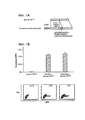
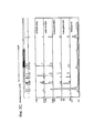
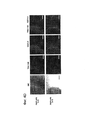
Фиг. 1 относится к функциональным тестам re-TALENs в соматических и стволовых клетках человека. FIG. 1 relates to functional tests of re-TALENs in human somatic and stem cells.
(а) Схематическое изображение экспериментального проекта для тестирования эффективности направленного воздействия на геном. Интегрированная в геном последовательность, кодирующая GFP, нарушается вставкой стоп-кодона и 68 п.о. геномного фрагмента, полученного из локуса AAVS1 (внизу). Восстановление последовательности GFP с помощью нуклеазо-опосредованной гомологичной рекомбинации с помощью донора tGFP (вверху) приводит к образованию клеток GFP+, которые могут быть количественно оценены с помощью FACS. Re-TALENs и TALENs направлено воздействуют на одинаковые последовательности в пределах фрагментов AAVS1 .(a) A schematic depiction of a pilot project for testing the effectiveness of targeting the genome. Integrated into the genome, the sequence encoding GFP is violated by the insertion of a stop codon and 68 bp. genomic fragment obtained from the AAVS1 locus (below). Restoration of the GFP sequence using nuclease-mediated homologous recombination using the tGFP donor (top) results in the formation of GFP + cells, which can be quantified by FACS. Re-TALENs and TALENs effect the same sequences within the AAVS1 fragments.
(b) Столбиковая диаграмма, демонстрирующая процент GFP+ клеток, полученных с помощью только tGFP-донора отдельно, с помощью TALENs с tGFP-донором и re-TALENs с tGFP-донором в локусе-мишени, согласно измерениям с помощью FACS. (N = 3, планки погрешности = SD). Репрезентативные графики FACS показаны ниже.(b) A bar chart showing the percentage of GFP + cells obtained using tGFP donor alone, using TALENs with tGFP donor and re-TALENs with tGFP donor in the target locus, as measured by FACS. (N = 3, error bars = SD). Representative FACS charts are shown below.
(с) Схематический обзор, изображающий стратегию направленного воздействия, для нативного локуса AAVS1. Плазмида-донор содержит акцептор сплайсинга (SA) - 2A (саморасщепляющиеся пептиды), ген устойчивости к пуромицину (PURO) и GFP (см. источник 10, включенный в данный документ ссылкой в полном объеме). Места расположения ПЦР-праймеров, используемых для обнаружения успешных событий редактирования, изображены в виде голубых стрелок.(c) A schematic overview depicting a targeting strategy for the native AAVS1 locus. The donor plasmid contains a splice acceptor (SA) - 2A (self-cleaving peptides), puromycin resistance gene (PURO) and GFP (see
(d) Подвергнутые успешному направленному воздействию клоны PGP1 hiPSCs отбирали на пуромицине (0,5 мкг/мл) в течение 2 недель. Показаны микроскопические изображения трех представительных GFP+ клонов. Клетки также окрашивали по маркерам плюрипотентности TRA-1-60. Измерительная линейка: 200 мкм.(d) Subjected to successful targeting, clones of PGP1 hiPSCs were selected on puromycin (0.5 μg / ml) for 2 weeks. Microscopic images of three representative GFP + clones are shown. Cells were also stained for the pluripotency markers TRA-1-60. Measuring range: 200 microns.
(e) Анализы ПЦР, выполненные на этих моноклональных GFP+ hiPSC клонах, продемонстрировали успешное встраивание донорных кассет в участок AAVS1 (дорожки 1,2,3), в то время как обычные hiPSCs не демонстрировали никаких признаков успешного встраивания (дорожка C). (e) PCR assays performed on these monoclonal GFP + hiPSC clones demonstrated successful insertion of donor cassettes into the AAVS1 region (tracks 1, 2, 3), while conventional hiPSCs showed no signs of successful incorporation (lanes C).
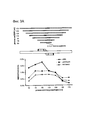
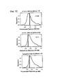
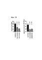
Фиг. 2 относится к сравнению эффективности направленного воздействия на геном с помощью reTALENs и Cas9-гидРНК на CCR5 в iPSCs. FIG. 2 relates to a comparison of the efficiency of targeting the genome with reTALENs and Cas9 hydRNA on CCR5 in iPSCs.
(а) Схематическое изображение эксперимента по геномной инженерии. В целевой участок пары re-TALEN или Cas9-гидРНК в PGP1 hiPSCs доставляли 90-мер ssODN, несущий 2 п.о. несоответствие относительно геномной ДНК, вместе с конструкциями reTALEN или Cas9-гидРНК. Участки расщепления нуклеазами изображены на фигуре в виде красных стрелок. (a) Schematic representation of a genomic engineering experiment. The target region of the re-TALEN or Cas9-hydRNA pair in PGP1 hiPSCs was delivered with 90-mer ssODN carrying 2 bp. genomic DNA mismatch, together with reTALEN or Cas9-hydRNA constructs. Nuclease cleavage sites are depicted in the figure as red arrows.
(b) Анализ глубоким секвенированием эффективностей HDR и NHEJ для пар re-TALEN (CCR5 #3) и ssODN, или Cas9-гидРНК и ssODN. Изменения в геноме hiPSCs анализировали на основе данных о последовательности, полученной высокопроизводительными способами, с помощью GEAS. Вверху: HDR количественно оценивали из фракции ридов, содержащих 2 п.о. точечные мутации, встроенных в центр ssODN (синий), а активность NHEJ количественно оценивали из фракции делеций (серые)/вставок (красные) в каждом конкретном положении в геноме. Для графиков reTALEN и ssODN, зеленый пунктир нанесен для того, чтобы отметить внешнюю границу участков связывания пар re-TALENs, которые находятся в положениях -26 п.о. и + 26 п.о. относительно центра двух участков связывания re-TALEN. Для графиков Cas9-гидРНК и ssODN, зеленый пунктир отмечает внешнюю границу участка направленного воздействия гидРНК, который находится в положениях -20 и -1 п.о. по отношению к последовательности РАМ. Внизу: распределение по размерам Делеция/Вставка в hiPSCs анализировали по всей популяции NHEJ с обработкой, указанной выше.(b) Deep sequencing analysis of the efficiencies of HDR and NHEJ for the re-TALEN (CCR5 # 3) and ssODN, or Cas9-hydRNA and ssODN pairs. Changes in the genome of hiPSCs were analyzed on the basis of sequence data obtained by high-throughput methods using GEAS. Above: HDR was quantified from a fraction of reads containing 2 bp. point mutations inserted into the ssODN center (blue), and NHEJ activity was quantified from the deletion fraction (gray) / inserts (red) at each specific position in the genome. For the reTALEN and ssODN charts, the green dotted line is marked in order to mark the outer border of the binding sites of the re-TALENs pairs, which are located in the -26 bp positions. and + 26 bp relative to the center of the two binding sites re-TALEN. For the Cas9-hydRNA and ssODN plots, the green dotted line marks the outer boundary of the hydrnc targeting site, which is located at –20 and –1 p. in relation to the PAM sequence. Bottom: The deletion / insert size distribution in hiPSCs was analyzed over the entire NHEJ population with the treatment indicated above.
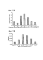
(с) Эффективность редактирования генома re-TALENs и Cas9-гидРНК, направленно воздействовавших на CCR5 в PGP1 hiPSCs. (c) Efficiency of editing the genome of re-TALENs and Cas9-hydRNA, targeting CCR5 in PGP1 hiPSCs.
Вверху: схематическое представление участков, подвергшихся направленному редактированию геномов в CCR5. 15 участков для направленного воздействия показаны синими стрелками снизу. Для каждого участка клетки котрансфецировали парой re-TALENs и их соответствующего донорного ssODN, несущего 2 п.о. несовпадение относительно геномной ДНК. Эффективность редактирования генома анализировали через 6 дней после трансфекции. Аналогичным образом, 15 Cas9-гидРНК трансфецировали с соответствующими ssODNs индивидуально в PGP1-hiPSCs для направленного воздействия не те же 15 участков и анализировали эффективность через 6 дней после трансфекции. Внизу: эффективность редактирования генома re-TALENs и Cas9-гидРНК при направленном целевом воздействии на CCR5 в PGP1 hiPSCs. Панели 1 и 2 демонстрируют эффективности NHEJ и HDR, опосредованные reTALENs. Панель 3 и 4 демонстрируют эффективности NHEJ и HDR, опосредованные Cas9-гидРНК. Показатели NHEJ рассчитывали по частоте геномных аллелей, несущих делеции или вставки в подвергнутом направленному воздействию участке; показатели HDR рассчитывали по частоте геномных аллелей, несущих 2 п.о. несоответствия. Панель 5, профиль DNAseI HS для клеточной линии hiPSC из базы данных ENCODE (Duke DNase HS, iPS NIHi7 DS). Следует отметить, что масштабы панелей отличаются.Above: a schematic representation of the areas that have undergone genome-directed editing in CCR5. 15 sites for directional exposure are shown with blue arrows below. For each site, the cells were co-transfected with a pair of re-TALENs and their corresponding donor ssODN carrying 2 bp. mismatch regarding genomic DNA. The efficiency of genome editing was analyzed 6 days after transfection. Similarly, 15 Cas9-hydRNAs were transfected with the corresponding ssODNs individually into PGP1-hiPSCs for targeting not the same 15 plots and the effectiveness was analyzed 6 days after transfection. Bottom: the efficiency of editing the genome of re-TALENs and Cas9-hydRNA with targeted targeting to CCR5 in PGP1 hiPSCs.
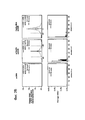
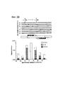
Фиг. 3 направлена на изучение функциональных параметров, регулирующих ssODN-опосредованную HDR с re-TALENs или Cas9-гидРНК в PGP1 hiPSCs. FIG. 3 aims to study the functional parameters that regulate ssODN-mediated HDR with re-TALENs or Cas9 hydRNA in PGP1 hiPSCs.
(а) PGP1 hiPSCs котрансфецировали с помощью пары re-TALENs (#3) и ssODNs различной длины (50, 70, 90,110, 130 150 170 нуклеотидов). Все ssODNs обладали идентичным 2 п.о. несовпадением относительно геномной ДНК в середине своей последовательности. 90-мерный ssODN достиг оптимального уровня HDR в подвергнутом направленному воздействию геноме. Оценка HDR-, NHEJ-опосредованной эффективности делеции и вставки описана в данном документе.(a) PGP1 hiPSCs were cotransfected using a pair of re-TALENs (# 3) and ssODNs of various lengths (50, 70, 90, 110, 130, 150, 170 nucleotides). All ssODNs had an identical 2 bp. mismatch regarding genomic DNA in the middle of its sequence. The 90-dimensional ssODN has reached the optimal HDR level in the targeted genome. Evaluation of HDR, NHEJ-mediated deletion and insertion efficiency is described in this document.
(b) 90-мерные ssODNs, соответствующие паре re-TALEN # 3, каждая из которых содержит 2 п.о. несовпадение (А) в центре и дополнительное 2 п.о. несовпадение (B) в разных положениях, смещенных от А (где смещения варьировали от -30 п.о. -> 30 п.о.), были использованы для тестирования последствий отклонения от гомологии вдоль ssODN. Эффективность редактирования генома каждого ssODN оценивали в PGP1 hiPSCs. Нижняя столбиковая диаграмма демонстрирует частоту включения только А, только В и А + В в подвергнутом направленному воздействию геноме. Показатели HDR уменьшались по мере увеличения расстояния от центра участка с отклонением по гомологии. (b) 90-dimensional ssODNs corresponding to a
(с) ssODNs, нацеленные на участки с варьирующими расстояниями (-620п.о. ~ 480 п.о.) от участка-мишени пары #3 re-TALEN, были испытаны для оценки максимального расстояния, в пределах которого ssODNs могут быть помещены для введения мутаций. Все ssODNs несли 2 п.о. несоответствие в середине своих последовательностей. Наблюдали минимальную эффективность HDR (<= 0,06%), когда несовпадение в ssODN располагалось на расстоянии 40 п.о. от середины участка связывания пары re-TALEN. (c) ssODNs aimed at areas with varying distances (-620 ppm ~ 480 bp) from the target area of the # 3 re-TALEN pair were tested to estimate the maximum distance within which ssODNs can be placed for introducing mutations. All ssODNs carried 2 bp. mismatch in the middle of their sequences. Observed the minimum HDR efficiency (<= 0.06%) when the mismatch in ssODN was located at a distance of 40 p. from the middle of the binding site of the re-TALEN pair.
(d) PGP1 hiPSCs котрансфецировали Cas9-гидРНК (AAVS1) и ssODNs различной ориентации (Ос: комлпементарная гидРНК; On: некомплементарная гидРНК) и разной длины (30, 50, 70, 90, 110 нуклеотидов). Все ssODNs обладали идентичными 2 п.о. несовпадениями относительно геномной ДНК в середине своих последовательностей. 70-мерный Ос достигал оптимального значения HDR в подвергнутом направленному воздействию геноме. (d) PGP1 hiPSCs have co-transfected Cas9-hydRNA (AAVS1) and ssODNs of various orientations (OC: complex hydRNA; On: non-complementary hydRNA) and different length (30, 50, 70, 90, 110 nucleotides). All ssODNs had identical 2 bp. mismatches about genomic DNA in the middle of their sequences. The 70-dimensional OC achieved the optimal HDR value in the targeted genome.
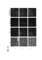
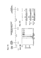
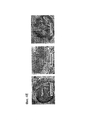
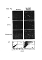
Фиг. 4 направлен на использование re-TALENs и ssODNs для получения моноклональных hiPSCс отредактированным геномом без селекции.FIG. 4 aims to use re-TALENs and ssODNs to produce monoclonal hiPSCs with an edited genome without selection.
(а) Временная шкала эксперимента.(a) The timeline of the experiment.
(b) Эффективность редактирования генома пары re-TALENs и ssODN (# 3), оценивали с помощью платформы NGS, описанной на фигуре 2b. (b) The editing efficiency of the genome of the re-TALENs pair and ssODN (# 3), was assessed using the NGS platform described in Figure 2b.
(с) Результаты секвенирования по Сенгеру колоний моноклональных hiPSC после редактирования генома. 2 п.о. гетерогенный генотип (CT/CT->TA/CT) был успешно внедрен в геном колоний PGP 1-IPS-3-11, PGPl-IPS-3-13. (c) Sanger sequencing results of monoclonal hiPSC colonies after genome editing. 2 bp The heterogeneous genotype (CT / CT-> TA / CT) was successfully introduced into the genome of the colonies PGP 1-IPS-3-11, PGPl-IPS-3-13.
(d) Иммунофлуоресцентное окрашивание подвергнутых направленному воздействию PGPl-IPS-3-11. Клетки окрашивали на маркеры плюрипотентности TRA-1-60 и SSEA4. (d) Immunofluorescent staining of targeted PGPl-IPS-3-11. Cells were stained for TRA-1-60 and SSEA4 pluripotency markers.
(e) Гематоксилин-эозиновое окрашивание срезов тератом, полученных из моноклональных клеток PGPl-IPS-3-11. (e) Hematoxylin-eosin staining of teratome sections obtained from monoclonal cells PGPl-IPS-3-11.
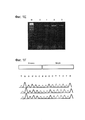
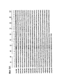
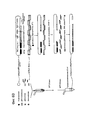
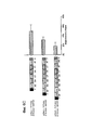
Фиг. 5. Дизайн reTALE. (a) Выравнивание последовательности исходного мономера TALE RVD относительно мономеров в re-TALE-16.5 (re-TALE-M1>re-TALE-M17). Нуклеотидные изменения относительно исходной последовательности выделены серым, (b) Тест повторяемости re-TALE с помощью ПЦР. Верхняя панель показывает структуру re-TALE/TALE и позиции праймеров для ПЦР-реакции. Нижняя панель показывает полосы ПЦР в условиях, указанных ниже. Электрофоретическая картина фрагментов ПЦР представлена с помощью исходного шаблона TALE (правая дорожка).FIG. 5. reTALE design. (a) Alignment of the sequence of the original TALE RVD monomer relative to the monomers in re-TALE-16.5 (re-TALE-M1> re-TALE-M17). Nucleotide changes relative to the initial sequence are highlighted in gray, (b) Re-TALE repeatability test by PCR. The top panel shows the re-TALE / TALE structure and primer positions for the PCR reaction. The bottom panel shows the PCR bands under the conditions indicated below. The electrophoretic pattern of the PCR fragments is presented using the original TALE template (right track).
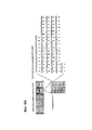
Фиг. 6. Дизайн и практическое осуществление сборки TASA (TALE Single-incubation Assembly), (а) Схематическое представление библиотеки димерных блоков re-TALE для сборки TASA. Представлена библиотека 10 димерных блоков re-TALE, кодирующих два RVDs. Внутри каждого блока, все 16 димеров имеют одну и ту же последовательность, за исключением последовательностей, кодирующих RVD; Димеры в различных блоках имеют отличающиеся последовательности, но спроектированы таким образом, чтобы они имели общие 32 п.о. перекрывания с соседними блоками. Нуклеотидная и аминокислотная последовательности одного димера (Блок 6_AC) представлены справа.FIG. 6. Design and implementation of the TASA assembly (TALE Single-incubation Assembly), (a) Schematic representation of the re-TALE dimeric block library for the TASA assembly. A library of 10 re-TALE dimer blocks encoding two RVDs is presented. Within each block, all 16 dimers have the same sequence, with the exception of sequences encoding RVD; Dimers in different blocks have different sequences, but are designed so that they have a total of 32 bp. overlap with adjacent blocks. The nucleotide and amino acid sequences of one dimer (Block 6_AC) are shown on the right.
(b) Схематическое представление сборки TASA. На левой панели показан способ сборки TASA: однореакторная реакция инкубации проводится с ферментной смесью/re-TALE блок s/re-TALE-N/TF каркасными векторами. Продукт реакции может быть использован непосредственно для трансформации бактерий. Правая панель демонстрирует механизм TASA. Вектор назначения линеаризуется эндонуклеазой при 37°С для того, чтобы отрезать контрселективную кассету ccdB; экзонуклеаза, которая обрабатывает конец блоков и линеаризованных векторов, экспонирует оцДНК выступы на конце фрагментов, что позволяет блокам и векторным каркасам соединяться в назначенном порядке. При повышении температуры до 50°С, полимеразы и лигазы работают вместе для того, чтобы заделать разрыв, что позволяет получить конечные конструкции, готовые к трансформации. (b) Schematic representation of the TASA assembly. The left panel shows the TASA assembly method: the one-pot incubation reaction is carried out with the enzyme mixture / re-TALE s / re-TALE-N / TF block frame vectors. The reaction product can be used directly to transform bacteria. The right panel shows the TASA mechanism. The destination vector is linearized with an endonuclease at 37 ° C in order to cut off the ccdB counterselective cassette; The exonuclease, which processes the end of the blocks and linearized vectors, exhibits ssDNA protrusions at the end of the fragments, which allows the blocks and vector frameworks to join in the designated order. When the temperature rises to 50 ° C, polymerases and ligases work together to bridge the gap, which allows to obtain final structures ready for transformation.
(с) Эффективность сборки TASA для re-TALEs, обладающих разными длинами мономеров. Используемые для сборки блоки показаны слева, а эффективность сборки представлена справа. (c) TASA assembly efficiency for re-TALEs with different monomer lengths. The blocks used for the assembly are shown on the left, and the assembly efficiency is shown on the right.
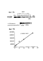
Фиг. 7. Функциональность и целостность последовательности Lenti-reTALEs. Клетки 293T, трансдуцированные lenti-re-TALE-TF показали 36x активацию экспрессии репортера по сравнению с только отрицательным репортером.FIG. 7. The functionality and integrity of the sequence Lenti-reTALEs. 293T cells transduced with lenti-re-TALE-TF showed a 36x activation of the reporter expression compared to only the negative reporter.
Фиг. 8. Чувствительность и воспроизводимость GEAS.FIG. 8. Sensitivity and reproducibility of GEAS.
(А) Информационный анализ предела обнаружения HDR. С учетом набора данных re-TALENs (# 10)/ssODN, были определены «риды», содержащие ожидаемое редактирование (HDR) и эти HDR-«риды» систематически удалялись для создания различных искусственных наборов данных с «разведенным» редактирующим сигналом. Наборы данных с 100, 99,8, 99,9, 98,9, 97,8, 89,2, 78,4, 64,9, 21,6, 10,8, 2,2, 1,1, 0,2, 0,1, 0,02 и 0% удалением HDR-«ридов» были сгенерированы для получения искусственных наборов с эффективностью HR в диапазоне 0-0,67%. Для каждого отдельного набора данных, была оценена взаимная информация (mutual information, MI) фонового сигнала (обозначено фиолетовым) и сигнала, полученного в участке направленного воздействия (обозначено зеленым). MI в участке направленного воздействия заметно выше, чем фон, при этом эффективность HDR выше 0,0014%. По оценке предел обнаружения HDR находится в диапазоне от 0,0014% до 0,0071%. Расчет MI описан в данном документе. (A) Information analysis of the HDR detection limit. Given the re-TALENs data set (# 10) / ssODN, “reads” were identified that contain the expected editing (HDR) and these HDR “reads” were systematically removed to create various artificial data sets with a “diluted” editing signal. Datasets with 100, 99.8, 99.9, 98.9, 97.8, 89.2, 78.4, 64.9, 21.6, 10.8, 2.2, 1.1, 0 , 2, 0.1, 0.02, and 0% removal of HDR “reads” were generated to produce artificial kits with an HR efficiency in the range of 0-0.67%. For each individual data set, the mutual information (mutual information, MI) of the background signal (indicated in purple) and the signal received in the directional field (indicated in green) was evaluated. The MI in the directional exposure region is noticeably higher than the background, while the HDR efficiency is higher than 0.0014%. The estimated HDR detection limit is in the range of 0.0014% to 0.0071%. The calculation of MI is described in this document.
(b) Тест воспроизводимости системы оценки редактирования генома. Пары графиков (верхний и нижний) демонстрируют результаты оценки HDR и NHEJ в двух повторах с парой re-TALENs и типом клеток, указанным выше. Для каждого эксперимента независимо были проведены нуклеофекция, направленная амплификация генома, глубокое секвенирование и анализ данных. Разброс повторов при оценке редактирования генома рассчитывали как V2 (|HDRl-HDR2|)/((HDR+HDR2)/2) =AHDR/HDR и V2 (|NHEJ1-NHEJ2|) /((NHEJl+NHEJ2)/2) =ANHEJ/NHEJ, а результаты по разбросу приведены ниже графиков. Средний разброс системы составил (19% + 11% + 4% + 9% + 10% + 35%)/6 = 15%. Факторы, которые могут способствовать разбросу, включают статус клеток при нуклеофекции, эффективность нуклеофекции, охват и качество секвенирования.(b) A test of the reproducibility of the genome editing evaluation system. Pairs of graphs (top and bottom) show the results of HDR and NHEJ evaluation in two repetitions with a pair of re-TALENs and the cell type indicated above. For each experiment, nucleofection, directed genome amplification, deep sequencing, and data analysis were carried out independently. The repetition spread in the evaluation of genome editing was calculated as V2 (| HDRl-HDR2 |) / ((HDR + HDR2) / 2) = AHDR / HDR and V2 (| NHEJ1-NHEJ2 |) / ((NHEJl + NHEJ2) / 2) = ANHEJ / NHEJ, and the results for the spread are shown below the graphs. The average spread of the system was (19% + 11% + 4% + 9% + 10% + 35%) / 6 = 15%. Factors that can contribute to dispersion include cell status during nucleofection, nucleofection efficiency, coverage, and sequencing quality.
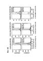
Фиг. 9. Статистический анализ эффективностей NHEJ и HDR при использовании reTALENs и Cas9-гидРНК на CCR5. FIG. 9. Statistical analysis of the efficiencies of NHEJ and HDR using reTALENs and Cas9-hydRNAs on CCR5.
(а) Корреляция эффективностей HR и NHEJ при использовании reTALENs на одинаковых участках в iPSCs (i= 0,91, P <IX 10-5). (a) Correlation of HR and NHEJ efficiencies when using reTALENs in the same areas in iPSCs (i = 0.91, P <IX 10-5).
(b) Корреляция эффективностей HR и NHEJ, опосредованных Cas9-гидРНК на одинаковых участках в iPSCs (i= 0,74, р = 0,002). (b) Correlation of the efficiencies of HR and NHEJ mediated by Cas9-hydRNA at the same sites in iPSCs (i = 0.74, p = 0.002).
(с) Корреляция эффективности NHEJ, опосредованной Cas9-гидРНК и температуры Tm целевого участка для гидРНК в iPSCs (г = 0,52, р = 0,04)(c) Correlation of the efficiency of NHEJ mediated by Cas9-hydRNA and the temperature Tm of the target region for hydRNA in iPSCs (g = 0.52, p = 0.04)
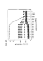
Фиг. 10. Корреляционный анализ эффективности редактирования генома и эпигенетического состояния. Корреляции Пирсона использовали для изучения возможных связей между чувствительностью ДНКазы I и эффективностью геномной инженерии (HR, NHEJ). Наблюдаемую корреляцию сравнивали с рандомизированным набором (N = 100000). Наблюдаемые корреляции выше 95-го процентиля или ниже 5-го процентиля моделируемого распределения считались потенциальными ассоциациями. Не наблюдали заметной корреляции между чувствительностью ДНКазы 1 и эффективностью NHEJ/HR. FIG. 10. Correlation analysis of the effectiveness of editing the genome and the epigenetic state. Pearson correlations were used to study the possible links between the sensitivity of DNase I and the efficiency of genomic engineering (HR, NHEJ). The observed correlation was compared with a randomized set (N = 100,000). Observable correlations above the 95th percentile or below the 5th percentile of the modeled distribution were considered potential associations. No significant correlation was observed between the sensitivity of
Фиг. 11. Влияние гомологичного спаривания при ssODN-опосредованном редактировании генома. FIG. 11. The influence of homologous mating in ssODN-mediated genome editing.
(а) В эксперименте, описанном на рисунке 3b, общая HDR снижалось, что измерялось по темпам, с которыми происходило включение среднего 2 п.о. несовпадения (А), по мере того, как росла дистанция на которую вторичные несовпадения В удалялись от А (относительное положение В по отношению к А, к варьирует от -30 до 30 п.о.). Более высокие темпы включения, когда В находится всего лишь в 10 п.о. от A (-10 п.о. и + 10 п.о.) могут отражать меньшую потребность в спаривании ssODN против геномной ДНК, проксимальной к двухцепочечному разрыву ДНК.(a) In the experiment described in Figure 3b, the total HDR decreased, as measured by the rate at which the average 2 bp was turned on. mismatches (A), as the distance to which the secondary mismatches B moved away from A grew (the relative position B in relation to A, varies from -30 to 30 bp). Higher rates of incorporation when B is only 10 bp from A (-10 bp and + 10 bp) may reflect less need for ssODN mating against genomic DNA proximal to double-stranded DNA breaks.
(b) Распределение длин генной конверсии вдоль ssODN. На каждом расстоянии В от А, доля событий HDR включает только А, тогда как друга доля включает как А, так и В. Эти два события могут интерпретироваться в терминах трактов конверсии генов (Elliott et al., 1998), в результате чего A+B-события представляют длинные конверсионные тракты, которые выходят за рамки только В и только А событий, представляющие более короткие пути, которые не достигают В. Согласно данной интерпретации, может быть оценено распределение трактов генной конверсии в обоих направлениях вдоль олигонуклеотида (середина ssODN определяется как 0, конверсионные тракты по направлению к 5'-концу ssODN - как «-» направление, а к 3'-концу - как «+» направление). Тракты генной конверсии постепенно уменьшаются по мере увеличения их длин, результат очень похожий на распределение трактов генной конверсии, наблюдаемое с дцДНК-донорами, но на сильно сжатой шкала дистанций в десятки п.о. для одноцепочечных олигодезоксинуклеотидов по сравнению с сотнями пар оснований для дцДНК-доноров. (b) The distribution of gene conversion lengths along ssODN. At each distance B from A, the share of HDR events includes only A, while the other share includes both A and B. These two events can be interpreted in terms of gene conversion paths (Elliott et al., 1998), resulting in A + B-events represent long conversion paths that go beyond B and A only events, representing shorter paths that do not reach B. According to this interpretation, the distribution of gene conversion paths in both directions along the oligonucleotide can be estimated (the middle of ssODN to 0, the conversion paths towards the 5'-end of ssODN - as “-” direction, and to the 3'-end - as “+” direction). Gene conversion paths gradually decrease as their length increases, the result is very similar to the distribution of gene conversion paths observed with dsDNA donors, but at a highly compressed distance scale of tens of p. for single-stranded oligodeoxynucleotides compared with hundreds of base pairs for dzanc donors.
(с) Анализы трактов генной конверсии с использованием одиночного ssODN, который содержит ряд мутаций, и измерение непрерывных последовательностей включений. Использовали ssODN-донор с тремя парами 2 п.о. несовпадений (оранжевый), разнесенных с интервалом в 10 нуклеотидов в обе стороны от центрального 2 п.о. несовпадения (вверху). Было обнаружено незначительное количество «ридов» геномного секвенирования (см. источник 62, включенный в данный документ ссылкой в полном объеме), несущих >=1 несовпадения, определенных с помощью ssODN среди> 300 000 «ридов» при секвенировании этой области. Все эти «риды» были нанесены на график (внизу), а последовательность «ридов» была закодирована условной окраской. Оранжевый: определенные несоответствия; зеленый: последовательность дикого типа. Геномное редактирование с этими ssODN дало паттерн, в котором средняя мутация отдельно была включена в 85% (53/62) моментов времени, с множеством В- несовпадений, включенных в другие моменты. Хотя количество событий В-включений было слишком низким для оценки распределения длин путей > 10 п. о., ясно, что преобладает область коротких путей от -10 до 10 п.о.(c) Analyzes of gene conversion paths using a single ssODN, which contains a number of mutations, and measurement of continuous sequences of inclusions. Used ssODN-donor with three pairs of 2 p. mismatches (orange), spaced at intervals of 10 nucleotides in both directions from the central 2 p. mismatch (above). A small number of reads of genomic sequencing was found (see source 62, incorporated by reference in this document) carrying> = 1 mismatches identified by ssODN among> 300,000 reads during sequencing of this region. All these “reads” were plotted (below), and the sequence of “reads” was coded with a conditional color. Orange: certain inconsistencies; green: wild type sequence. Genomic editing with these ssODNs gave a pattern in which the average mutation was separately included in 85% (53/62) of the time points, with many B-inconsistencies included in other moments. Although the number of B-inclusions events was too low to estimate the distribution of path lengths> 10 bp, it is clear that the region of short paths from -10 to 10 bp prevails.
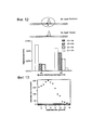
Фиг. 12. Эффективности редактирования генома Cas9-гидРНК нуклеазой и никазами.FIG. 12. Efficiency of editing the genome of Cas9-hydRNA by nuclease and nick.
PGP1 iPSCs котрансфецировали комбинацией нуклеазы (С2) (Cas9-гидРНК) или никазы (Cс) (Cas9D10A-гидРНК) и ssODNs различной ориентации (Oс и On). Все ssODNs обладали идентичным 2 п.о. несовпадением относительно геномной ДНК в середине своей последовательности. Оценка HDR описана в данном документе. PGP1 iPSCs were co-transfected with a combination of nuclease (C2) (Cas9-hydRNA) or nickase (Cc) (Cas9D10A-hydRNA) and ssODNs of different orientation (Os and On). All ssODNs had an identical 2 bp. mismatch regarding genomic DNA in the middle of its sequence. HDR evaluation is described in this document.
Фиг. 13. Дизайн и оптимизация последовательности re-TALE.FIG. 13. Design and optimization of the re-TALE sequence.
Последовательность re-TALE подвергали эволюции в течение нескольких циклов дизайна для устранения повторов. В каждом цикле оценивали синонимичные последовательности из каждого повтора. Отбирали те из них, которые имели наибольшее расстояние Хэмминга к эволюционирующей ДНК. Окончательная последовательность имела cai = 0,59 ΔG = -9,8 ккал/моль. Для выполнения общей основы для дизайна синтетического белка использовали пакет R.The re-TALE sequence was subjected to evolution over several design cycles to eliminate duplication. In each cycle, synonymous sequences from each repetition were evaluated. We selected those that had the greatest Hamming distance to the evolving DNA. The final sequence had cai = 0.59 ΔG = -9.8 kcal / mol. To perform a common basis for the design of synthetic protein used package R.
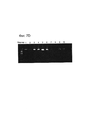
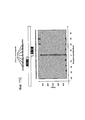
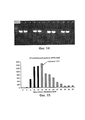
Фиг. 14 представляет собой изображение геля, демонстрирующее проверку с помощью ПЦР геномной вставки Cas9 в клетки PGP1. Дорожки 3, 6, 9, 12 являются продуктами ПЦР обычных клеточных линий PGP1. FIG. 14 is a gel image demonstrating PC9 genomic insertion of Cas9 into PGP1 cells.
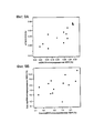
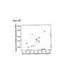
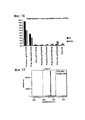
Фиг. 15 представляет собой график уровня экспрессии мРНК Cas9 при индукции.FIG. 15 is a graph showing the expression level of Cas9 mRNA upon induction.
Фиг. 16 представляет собой график, показывающий эффективность направленного воздействия на геном РНК с различным дизайном. FIG. 16 is a graph showing the effectiveness of targeting the RNA genome with a different design.
Фиг. 17 представляет собой график, показывающий эффективность направленного воздействия на геном 44% гомологичной рекомбинации, достигнутой с помощью химеры направляющей РНК- донорной ДНК. FIG. 17 is a graph showing the efficacy of targeting the genome of 44% homologous recombination achieved using the chimera of the RNA-donor DNA guide.
Фиг. 18 является схемой, демонстрирующей генотип изогенных клеточных линий PGP 1, полученных с помощью системы, описанной в данном документе. PGPl-IPS-BTHH имеет фенотип с однонуклеотидными делециями, как у пациентов с BTHH. PGP 1-NHEJ имеет 4 п.о. делеции, которые образуют мутации со сдвигом рамки считывания другим образом.FIG. 18 is a diagram showing the genotype of
Фиг. 19 представляет собой график, показывающий, что кардиомиоциты, полученные из изогенных PGP1 iPS, воспроизводят дефектную выработку АТФ и специфическую активность F1F0 АТФазы, как продемонстрировано в пациент-специфических клетках.FIG. 19 is a graph showing that cardiomyocytes derived from isogenic PGP1 iPS reproduce defective ATP production and the specific F1F0 ATPase activity, as demonstrated in patient-specific cells.
Фиг. 20 показывает последовательности для последовательности re-TALEN-основной и основной последовательности re-TALE-TF.FIG. 20 shows the sequences for the re-TALEN-main sequence and the main re-TALE-TF sequence.
Подробное описаниеDetailed description
Аспекты настоящего изобретения относятся к применению TALEN, которая утратила определенные повторяющиеся последовательности, для инженерии нуклеиновых кислот, например, путем расщепления двухцепочечной нуклеиновой кислоты. Применение TALEN для расщепления двухцепочечной нуклеиновой кислоты может привести к негомологичному соединению концов (NHEJ) или к гомологичной рекомбинации (HR). Аспекты настоящего раскрытия также предусматривают применение TALEN, которая утратила повторяющиеся последовательности, для инженерии нуклеиновых кислот, например, путем расщепления двухцепочечной нуклеиновой кислоты в присутствии донорной нуклеиновой кислоты и введения донорной нуклеиновой кислоты в двухцепочечную нуклеиновую кислоту, например, путем негомологичного соединения концов (NHEJ) или гомологичной рекомбинации (HR). Aspects of the present invention relate to the use of TALEN, which has lost certain repetitive sequences, for nucleic acid engineering, for example, by cleaving a double-stranded nucleic acid. The use of TALEN for cleavage of a double-stranded nucleic acid can lead to non-homologous linking of the ends (NHEJ) or to homologous recombination (HR). Aspects of the present disclosure also contemplate the use of TALEN, which has lost duplicate sequences, for nucleic acid engineering, for example, by cleaving a double-stranded nucleic acid in the presence of donor nucleic acid and introducing the donor nucleic acid into a double-stranded nucleic acid, for example, by non-homologous terminus (NHEJ) or homologous recombination (HR).
Эфеекторные нуклеазы, подобные активаторам транскрипции (TALEN) известны в данной области техники и включают искусственные ферменты рестрикции, полученные слиянием ДНК-связывающего домена TAL-эффектора с ДНК-расщепляющим доменом. Ферменты рестрикции являются ферментами, которые разрезают цепи ДНК в определенной последовательности. Эффекторы, подобные активаторам транскрипции (TALE) могут быть сконструированы для связывания с искомой последовательностью ДНК. См. Boch, Jens (February 2011). "TALEs of genome targeting". Nature Biotechnology 29 (2): 135-6, включенный в данный документ ссылкой в полном объеме. Путем объединения такого сконструированного TALE с ДНК-расщепляющим доменом (который разрезает нити ДНК), получается TALEN, которая является ферментом рестрикции, специфичным к любой искомой последовательности ДНК. В соответствии с некоторыми аспектами, TALEN вводят в клетку для целевого редактирования нуклеиновой кислоты in situ, например, для редактирования генома in situ. Ephector nucleases similar to transcription activators (TALEN) are known in the art and include artificial restriction enzymes obtained by fusing the DNA-binding domain of the TAL-effector with the DNA-cleaving domain. Restriction enzymes are enzymes that cut DNA chains in a specific sequence. Effector like transcription activators (TALE) can be designed to bind to the desired DNA sequence. See Boch, Jens (February 2011). "TALEs of genome targeting". Nature Biotechnology 29 (2): 135-6, incorporated herein by reference in its entirety. By combining such a constructed TALE with a DNA-cleaving domain (which cuts DNA strands), TALEN is obtained, which is a restriction enzyme specific for any desired DNA sequence. In accordance with some aspects, TALEN is introduced into a cell for targeted editing of nucleic acid in situ, for example, for editing the genome in situ.
Согласно одному аспекту, неспецифический ДНК-расщепляющий домен с конца эндонуклеазы FokI может быть использован для создания гибридных нуклеаз, которые активны в клетках дрожжей, растений и животных. Домен FokI функционирует в виде димера, требующего две конструкции с уникальными ДНК-связывающими доменами для участков в геноме-мишени с правильной ориентацией и расстоянием. Как число аминокислотных остатков между TALE ДНК-связывающим доменом и расщепляющим доменом Fokl, так и количества оснований между двумя отдельными участками связывания TALEN оказывают влияние на активность. According to one aspect, a non-specific DNA cleavage domain from the end of the FokI endonuclease can be used to create hybrid nucleases that are active in yeast, plant and animal cells. The FokI domain functions as a dimer, requiring two constructs with unique DNA-binding domains for sites in the target genome with the correct orientation and distance. As the number of amino acid residues between the TALE DNA-binding domain and the cleaving domain Fokl, and the number of bases between two separate binding sites TALEN affect the activity.
Взаимоотношение между аминокислотной последовательностью и распознаванием ДНК связывающим доменом TALE позволяет создавать конструируемые белки. Компьютерные программы, такие как «DNA Works» могут быть использованы для разработки TALE-конструкций. Другие способы разработки TALE-конструкций известны специалистам в данной области техники. См. Cermak, Т. ; Doyle, E. L. ; Christian, M. ; Wang, L. ; Zhang, Y. ; Schmidt, C; Bailer, J. A. ; Somia, N. V. et al. (2011). "Efficient design and assembly of custom TALEN and other TAL effector-based constructs for DNA targeting". Nucleic Acids Research. doi:10. 1093/nar/gkr218; Zhang, Feng; et. al. (February 2011). "Efficient construction of sequence-specific TAL effectors for modulating mammalian transcription", Nature Biotechnology 29 (2): 149-53; Morbitzer, R. ; Elsaesser, J. ; Hausner, J. ; Lahaye, T. (2011). "Assembly of custom TALE-type DNA binding domains by modular cloning". Nucleic Acids Research. doi:10'1093/nar/gkrl51; Li, Т. ; Huang, S. ; Zhao, X. ; Wright, D. A. ; Carpenter, S. ; Spalding, M. H. ; Weeks, D. P. ; Yang, B. (2011). "Modularly assembled designer TAL effector nucleases for targeted gene knockout and gene replacement in eukaryotes". Nucleic Acids Research. doi"10. 1093/nar/gkrl88; GeipTer, R. ; Scholze, H. ; Hahn, S. ; Streubel, J. ; Bonas, U. ; Behrens, S. E. ; Boch, J. (2011). "Transcriptional Activators of Human Genes with Programmable DNA-Specificity". In Shiu, Shin-Han. PLoS ONE 6 (5): el9509; Weber, E. ; Graetzner, R. ; Werner, S. ; Engler, C; Marillonnet, S. (2011). "Assembly of Designer TAL Effectors by Golden Gate Cloning". In Bendahmane, Mohammed. PLoS One 6 (5): el9722, которые включены в данный документ ссылкой в полном объеме. The relationship between the amino acid sequence and DNA recognition by the TALE binding domain allows the creation of constructed proteins. Computer programs such as DNA Works can be used to develop TALE constructs. Other methods for developing TALE constructs are known to those skilled in the art. See Cermak, T.; Doyle, E. L.; Christian, M.; Wang, L.; Zhang, Y.; Schmidt, C; Bailer, J. A.; Somia, N.V. et al. (2011). "TALEN Efficient design and assembly of TALEN and other TAL effector-based constructs for DNA targeting". Nucleic Acids Research. doi: 10. 1093 / nar / gkr218; Zhang, Feng; et. al. (February 2011). "Efficient construction of sequence-specific TAL effectors for modulating mammalian transcription", Nature Biotechnology 29 (2): 149-53; Morbitzer, R.; Elsaesser, J.; Hausner, J.; Lahaye, T. (2011). "Assembly of custom TALE-type DNA binding domains by modular cloning". Nucleic Acids Research. doi: 10'1093 / nar / gkrl51; Li, T.; Huang, S.; Zhao, X.; Wright, D. A.; Carpenter, S.; Spalding, M. H.; Weeks, D. P.; Yang, B. (2011). "Modularly assembled designer TAL effector nucleases for the target gene knockout and replacement in eukaryotes". Nucleic Acids Research. doi "10. 1093 / nar / gkrl88; GeipTer, R.; Scholze, H.; Hahn, S.; Streubel, J.; Bonas, U.; Behrens, SE; Boch, J. (2011)." Transcriptional Activators Of Human Genes with Programmable DNA-Specificity ". In Shiu, Shin-Han. PLoS ONE 6 (5): el9509; Weber, E.; Graetzner, R.; Werner, S.; Engler, C; Marillonnet, S. ( 2011). "Assembly of Designer TAL Effectors by Golden Gate Cloning". In Bendahmane, Mohammed. PLoS One 6 (5): el9722, which are included in this document by reference in full.
В соответствии с типичным аспектом, сразу после сборки гены TALEN можно вставлять в плазмиды, в соответствии с определенными воплощениями; плазмиды затем используются для трансфекции клетки-мишени, при этом генные продукты экспрессируются и входят в ядро для доступа к геному. Согласно типичным аспектам, TALEN, как описано в данном документе, могут быть использованы для редактирования целевой нуклеиновой кислоты, например, генома, с помощью индукции двухцепочечных разрывов (DSB), на которые клетки реагируют посредством механизмов репарации. Примерные механизмы репарации включают негомологичное соединение концов (NHEJ), который восстанавливает ДНК по обе стороны от двухцепочечного разрыва, где имеется весьма незначительное перекрывание для отжига, или такое перекрывание отсутствует вовсе. Этот репаративный механизм индуцирует ошибки в геноме, через вставку или делецию (indel), или хромосомную перестройку; такие ошибки могут сделать генные продукты, кодируемые в данном месте, нефункциональными. См. Miller, Jeffrey; et. al. (February 2011). "A TALE nuclease architecture for efficient genome editing". Nature Biotechnology 29 (2): 143-8, включенный в данный документ ссылкой в полном объеме. Поскольку эта активность может изменяться в зависимости от используемых вида, типа клеток, гена-мишени, и нуклеазы, активность можно контролировать с помощью анализа гетеродуплексного расщепления, который не обнаруживает никакой разницы между двумя аллелеями, амплифицированными с помощью ПЦР. Продукты расщепления могут быть визуализированы на простом агарозном геле или блочной гелевой системе. In accordance with a typical aspect, immediately after assembly, TALEN genes can be inserted into plasmids, in accordance with certain embodiments; The plasmids are then used to transfect the target cell, with the gene products being expressed and entering the nucleus to access the genome. According to typical aspects, TALEN, as described herein, can be used to edit a target nucleic acid, for example, the genome, by inducing double-stranded breaks (DSB), to which cells respond through repair mechanisms. Exemplary repair mechanisms include a non-homologous end connection (NHEJ), which restores DNA on both sides of a double-stranded break, where there is very little overlap for annealing, or there is no such overlap at all. This reparative mechanism induces errors in the genome through an insertion or deletion (indel) or chromosomal rearrangement; such errors can make the gene products encoded in a given place non-functional. See Miller, Jeffrey; et. al. (February 2011). "A TALE nuclease architecture for efficient genome editing". Nature Biotechnology 29 (2): 143-8, incorporated herein by reference in its entirety. Since this activity may vary depending on the species used, cell type, target gene, and nuclease, activity can be monitored using heteroduplex cleavage analysis, which does not detect any difference between the two alleles amplified by PCR. Cleavage products can be visualized on a simple agarose gel or block gel system.
В ином случае, ДНК может быть введена в геном посредством NHEJ в присутствии экзогенных фрагментов двухцепочечной ДНК. Управляемая гомологией репарация также может ввести чужеродную ДНК в DSB, поскольку трансфецированные двухцепочечные последовательности используются в качестве шаблонов для ферментов репарации. В соответствии с некоторыми аспектами, TALEN, описанные в данном документе, могут быть использованы для получения стабильно модифицированных человеческих эмбриональных стволовых клеток и клонов индуцированных плюрипотентных стволовых клеток (iPSCs). В соответствии с некоторыми аспектами, TALEN, описанные в данном документе, могут быть использованы для получения нокаутных видов, таких как C. elegans, нокаутные крысы, нокаутные мыши или нокаутные данио-рерио. Alternatively, DNA can be introduced into the genome by NHEJ in the presence of exogenous double-stranded DNA fragments. Homology-driven repair can also introduce foreign DNA into the DSB, since the transfected double-stranded sequences are used as templates for repair enzymes. In accordance with some aspects, the TALEN described herein can be used to produce stably modified human embryonic stem cells and induced pluripotent stem cell clones (iPSCs). In accordance with some aspects, the TALEN described in this document can be used to produce knockout species, such as C. elegans, knockout rats, knockout mice, or knockout danios.
Согласно одному аспекту настоящего раскрытия, воплощения нацелены на применение экзогенной ДНК, нуклеазных ферментов, таких как ДНК-связывающие белки и направляющие РНК, для колокализации с ДНК в стволовой клетке и гидролиза или разрезания ДНК с вставкой экзогенной ДНК. Такие ДНК-связывающие белки хорошо известны специалистам в данной области техники, для связывания с ДНК для различных целей. Такие ДНК-связывающие белки могут быть природными. ДНК-связывающие белки, включенные в объем настоящего изобретения, включают те, которые могут направляться РНК, которые называются в данном документе как направляющие РНК. Согласно этому аспекту, направляющая РНК и РНК-направляемый ДНК-связывающий белок образуют комплекс колокализации на ДНК. Такие ДНК-связывающие белки, обладающие нуклеазной активностью, известны специалистам в данной области техники, и включают природные ДНК-связывающие белки, имеющие нуклеазную активность, такие как белки Cas9, присутствующие, например, в системах CRISPR II типа. Такие белки Cas9 и системы CRISPR типа II хорошо описаны в данной области техники. См. Makarova et al., Nature Reviews, Microbiology, Vol. 9, June 2011, pp. 467-477 включая всю дополнительную информацию, включенный в данный документ ссылкой в полном объеме.According to one aspect of the present disclosure, embodiments aim to use exogenous DNA, nuclease enzymes, such as DNA binding proteins and RNA targeting, for colocalization with stem cell DNA and hydrolysis or cutting of DNA with exogenous DNA insertion. Such DNA binding proteins are well known to those skilled in the art to bind to DNA for various purposes. Such DNA binding proteins can be natural. DNA-binding proteins included in the scope of the present invention include those that can be directed to RNA, which are referred to herein as guiding RNA. According to this aspect, the guide RNA and the RNA-directed DNA-binding protein form a colocalization complex on the DNA. Such DNA-binding proteins with nuclease activity are known to those skilled in the art, and include natural DNA-binding proteins with nuclease activity, such as Cas9 proteins, present, for example, in type II CRISPR systems. Such Cas9 proteins and type II CRISPR systems are well described in the art. See Makarova et al., Nature Reviews, Microbiology, Vol. 9, June 2011, pp. 467-477 including all additional information included in this document by reference in full.
Типичные ДНК-связывающие белки имеют функцию нуклеазной активности для внесения одноцепочечных разрывов (ников) или разрезания двухцепочечной ДНК. Такая нуклеазная активность может быть результатом действия ДНК-связывающего белка, имеющего одну или несколько полипептидных последовательностей, демонстрирующих нуклеазную активность. Такие типичные ДНК-связывающие белки могут иметь два отдельных нуклеазных домена, при этом каждый домен отвечает за разрезание или внесение одноцепочечных разрывов конкретной цепи двухцепочечной ДНК. Типичные полипептидные последовательности, имеющие нуклеазную активность, известные специалистам в данной области, включают домен, родственный нуклеазе McrA-HNH и домен RuvC-подобной нуклеазы. Соответственно, типичными ДНК-связывающими белками являются те, которые по своей природе содержат один или несколько из числа домена, родственного нуклеазе McrA-HNH и домен RuvC-подобной нуклеазы. Typical DNA-binding proteins have the function of nuclease activity for introducing single-stranded breaks (nicks) or cutting double-stranded DNA. Such nuclease activity may result from the action of a DNA-binding protein having one or more polypeptide sequences that demonstrate nuclease activity. Such typical DNA-binding proteins may have two separate nuclease domains, with each domain responsible for cutting or inserting single-stranded breaks of a particular double-stranded DNA chain. Typical polypeptide sequences having nuclease activity known to those skilled in the art include the domain related to the McrA-HNH nuclease and the domain of the RuvC-like nuclease. Accordingly, typical DNA-binding proteins are those that, by their nature, contain one or more of a number of domains related to the McrA-HNH nuclease and a RuvC-like nuclease domain.
Типичный ДНК-связывающий белок представляет собой РНК управляемый ДНК-связывающий белок системы CRISPR II типа. Типичный ДНК-связывающий белок представляет собой белок Cas9. A typical DNA-binding protein is an RNA managed DNA-binding protein of the CRISPR type II system. A typical DNA binding protein is Cas9 protein.
В S. pyogenes, Cas9Сas9 образует двухцепочечной разрыв с тупыми концами на 3 п.о. выше мотива PAM (прилегающий к протоспейсеру мотив) с помощью процесса, опосредованного двумя каталитическими доменами в белке: доменом HNH, который расщепляет комплементарную нить ДНК и доменом подобного RuvC, который расщепляет некомплементарную цепь. См. Jinke et al., Science, 337, 816-821 (2012), включенный в данный документ ссылкой в полном объеме. Белки Cas9, как известно, существуют во многих системах CRISPR типа II, включая те, которые определены в дополнительных документах к Makarova et al., Nature Reviews, Microbiology, Vol. 9, June 2011, pp. 467-477: Methanococcus maripaludis C7; Corynebacterium diphtheriae; Corynebacterium efficiens YS-314; Corynebacterium glutamicum ATCC 13032 Kitasato; Corynebacterium glutamicum ATCC 13032 Bielefeld; Corynebacterium glutamicum R; Corynebacterium kroppenstedtii DSM 44385; Mycobacterium abscessus ATCC 19977; Nocardia farcinica IFM10152; Rhodococcus erythropolis PR4; Rhodococcus jostii RHA1; Rhodococcus opacus B4 uid36573; Acidothermus cellulolyticus 11B; Arthrobacter chlorophenolicus A6; Kribbella flavida DSM 17836 uid43465; Thermomonospora curvata DSM 43183; Bifidobacterium dentium Bdl; Bifidobacterium longum DJO10A; Slackia heliotrinireducens DSM 20476; Persephonella marina EX HI; Bacteroides fragilis NCTC 9434; Capnocytophaga ochracea DSM 7271; Flavobacterium psychrophilum JIP02 86; Akkermansia muciniphila ATCC BAA 835; Roseiflexus castenholzii DSM 13941; Roseiflexus RSI; Synechocystis PCC6803; Elusimicrobium minutum Peil91; бактериальный филотип Rs D17 некультивированной группы 1 Termite; Fibrobacter succinogenes S85; Bacillus cereus ATCC 10987; Listeria innocua;Lactobacillus casei; Lactobacillus rhamnosus GG; Lactobacillus salivarius UCC118; Streptococcus agalactiae A909; Streptococcus agalactiae NEM316; Streptococcus agalactiae 2603; Streptococcus dysgalactiae equisimilis GGS 124; Streptococcus equi zooepidemicus MGCS10565; Streptococcus gallolyticus UCN34 uid46061; Streptococcus gordonii Challis subst CHI; Streptococcus mutans NN2025 uid46353; Streptococcus mutans; Streptococcus pyogenes Ml GAS; Streptococcus pyogenes MGAS5005; Streptococcus pyogenes MGAS2096; Streptococcus pyogenes MGAS9429; Streptococcus pyogenes MGAS10270; Streptococcus pyogenes MGAS6180; Streptococcus pyogenes MGAS315; Streptococcus pyogenes SSI-1; Streptococcus pyogenes MGAS 10750; Streptococcus pyogenes NZ131; Streptococcus thermophiles CNRZ1066; Streptococcus thermophiles LMD-9; Streptococcus thermophiles LMG 18311; Clostridium botulinum A3 Loch Maree; Clostridium botulinum В Eklund 17B; Clostridium botulinum Ba4 657; Clostridium botulinum F Langeland; Clostridium cellulolyticum H10; Finegoldia magna ATCC 29328; Eubacterium rectale ATCC 33656; Mycoplasma gallisepticum; Mycoplasma mobile 163K; Mycoplasma penetrans; Mycoplasma synoviae 53; Streptobacillus moniliformis DSM 12112; Bradyrhizobium BTAil; Nitrobacter hamburgensis X14; Rhodopseudomonas palustris BisB18; Rhodopseudomonas palustris BisB5; Parvibaculum lavamentivorans DS-1; Dinoroseobacter shibae DFL 12; Gluconacetobacter diazotrophicus Pal 5 FAPERJ; Gluconacetobacter diazotrophicus Pal 5 JGI; Azospirillum B510 uid46085; Rhodospirillum rabram ATCC 11170; Diaphorobacter TPSY uid29975; Verminephrobacter eiseniae EF01-2; Neisseria meningitides 053442; Neisseria meningitides alphal4; Neisseria meningitides Z2491; Desulfovibrio salexigens DSM 2638; Campylobacter jejuni doylei 269 97; Campylobacter jejuni 81116; Campylobacter jejuni; Campylobacter lari RM2100; Helicobacter hepaticus; Wolinella succinogenes; Tolumonas auensis DSM 9187; Pseudoalteromonas atlantica T6c; Shewanella pealeana ATCC 700345; Legionella pneumophila Paris; Actinobacillus succinogenes 130Z; Pasteurella multocida; Francisella tularensis novicida U112; Francisella tularensis holarctica; Francisella tularensis FSC 198; Francisella tularensis tularensis; Francisella tularensis WY96-3418; и Treponema denticola ATCC 35405. Соответственно, аспекты настоящего раскрытия направлены на белок Cas9, присутствующий в системе CRISPR II типа. In S. pyogenes, Cas9Cas9 forms a double-stranded gap with blunt ends at 3 bp. above the PAM motif (the motif adjacent to the protospeiser) through a process mediated by two catalytic domains in a protein: an HNH domain that cleaves the complementary DNA strand and a domain like RuvC that cleaves the non-complementary strand. See Jinke et al., Science, 337, 816-821 (2012), incorporated herein by reference in its entirety. Cas9 proteins are known to exist in many CRISPR type II systems, including those defined in additional documents to Makarova et al., Nature Reviews, Microbiology, Vol. 9, June 2011, pp. 467-477: Methanococcus maripaludis C7; Corynebacterium diphtheriae; Corynebacterium efficiens YS-314; Corynebacterium glutamicum ATCC 13032 Kitasato; Corynebacterium glutamicum ATCC 13032 Bielefeld; Corynebacterium glutamicum R; Corynebacterium kroppenstedtii DSM 44385; Mycobacterium abscessus ATCC 19977; Nocardia farcinica IFM10152; Rhodococcus erythropolis PR4; Rhodococcus jostii RHA1; Rhodococcus opacus B4 uid36573; Acidothermus cellulolyticus 11B; Arthrobacter chlorophenolicus A6; Kribbella flavida DSM 17836 uid43465; Thermomonospora curvata DSM 43183; Bifidobacterium dentium Bdl; Bifidobacterium longum DJO10A; Slackia heliotrinireducens DSM 20476; Persephonella marina EX HI; Bacteroides fragilis NCTC 9434; Capnocytophaga ochracea DSM 7271; Flavobacterium psychrophilum JIP02 86; Akkermansia muciniphila ATCC BAA 835; Roseiflexus castenholzii DSM 13941; Roseiflexus RSI; Synechocystis PCC6803; Elusimicrobium minutum Peil91; bacterial phylotype Rs D17 of the non-cultured group 1 Termite; Fibrobacter succinogenes S85; Bacillus cereus ATCC 10987; Listeria innocua; Lactobacillus casei; Lactobacillus rhamnosus GG; Lactobacillus salivarius UCC118; Streptococcus agalactiae A909; Streptococcus agalactiae NEM316; Streptococcus agalactiae 2603; Streptococcus dysgalactiae equisimilis GGS 124; Streptococcus equi zooepidemicus MGCS10565; Streptococcus gallolyticus UCN34 uid46061; Streptococcus gordonii Challis subst CHI; Streptococcus mutans NN2025 uid46353; Streptococcus mutans; Streptococcus pyogenes Ml GAS; Streptococcus pyogenes MGAS5005; Streptococcus pyogenes MGAS2096; Streptococcus pyogenes MGAS9429; Streptococcus pyogenes MGAS10270; Streptococcus pyogenes MGAS6180; Streptococcus pyogenes MGAS315; Streptococcus pyogenes SSI-1; Streptococcus pyogenes MGAS 10750; Streptococcus pyogenes NZ131; Streptococcus thermophiles CNRZ1066; Streptococcus thermophiles LMD-9; Streptococcus thermophiles LMG 18311; Clostridium botulinum A3 Loch Maree; Clostridium botulinum B Eklund 17B; Clostridium botulinum Ba4 657; Clostridium botulinum F Langeland; Clostridium cellulolyticum H10; Finegoldia magna ATCC 29328; Eubacterium rectale ATCC 33656; Mycoplasma gallisepticum; Mycoplasma mobile 163K; Mycoplasma penetrans; Mycoplasma synoviae 53; Streptobacillus moniliformis DSM 12112; Bradyrhizobium BTAil; Nitrobacter hamburgensis X14; Rhodopseudomonas palustris BisB18; Rhodopseudomonas palustris BisB5; Parvibaculum lavamentivorans DS-1; Dinoroseobacter shibae DFL 12; Gluconacetobacter diazotrophicus Pal 5 FAPERJ; Gluconacetobacter diazotrophicus Pal 5 JGI; Azospirillum B510 uid46085; Rhodospirillum rabram ATCC 11170; Diaphorobacter TPSY uid29975; Verminephrobacter eiseniae EF01-2; Neisseria meningitides 053442; Neisseria meningitides alphal4; Neisseria meningitides Z2491; Desulfovibrio salexigens DSM 2638; Campylobacter jejuni doylei 269 97; Campylobacter jejuni 81116; Campylobacter jejuni; Campylobacter lari RM2100; Helicobacter hepaticus; Wolinella succinogenes; Tolumonas auensis DSM 9187; Pseudoalteromonas atlantica T6c; Shewanella pealeana ATCC 700345; Legionella pneumophila Paris; Actinobacillus succinogenes 130Z; Pasteurella multocida; Francisella tularensis novicida U112; Francisella tularensis holarctica; Francisella tularensis FSC 198; Francisella tularensis tularensis; Francisella tularensis WY96-3418; and Treponema denticola ATCC 35405. Accordingly, aspects of the present disclosure are directed to the Cas9 protein present in a CRISPR type II system.
Белок Cas9 может быть назван специалистом в данной области литературы как Csn1. Белок Cas9 из S. pyogenes показан ниже. См. Deltcheva et al., Nature 471, 602-607 (2011), включенный в данный документ ссылкой в полном объеме.Protein Cas9 may be called a specialist in the field of literature as Csn1. Cas9 protein from S. pyogenes is shown below. See Deltcheva et al., Nature 471, 602-607 (2011), incorporated herein by reference in its entirety.

Согласно одному аспекту, управляемый РНК ДНК-связывающий белок включает гомологи и ортологи Cas9, которые сохраняют способность белка связываться с ДНК, быть управляемыми РНК и разрезать ДНК. Согласно одному аспекту, белок Cas9 включает последовательность, которая представлена в естественном белке Cas9 из S. pyogenes и белковых последовательностях, гомологичных, по меньшей мере, на 30%, 40%, 50%, 60%, 70%, 80%, 90%, 95 %, 98% или 99% к этому белку, и, являющихся ДНК-связывающим белком, таким как направляемый РНК ДНК-связывающий белок. In one aspect, the RNA driven DNA binding protein includes Cas9 homologs and orthologs that retain the ability of the protein to bind to DNA, be driven by RNA, and cut DNA. According to one aspect, the Cas9 protein comprises a sequence that is present in the natural Cas9 protein from S. pyogenes and protein sequences homologous to at least 30%, 40%, 50%, 60%, 70%, 80%, 90% , 95%, 98% or 99% of this protein, and being a DNA-binding protein, such as a directed RNA DNA-binding protein.
Согласно одному аспекту предлагается сконструированная система Cas9-гидРНК, которая позволяет осуществить направляемое РНК разрезание генома сайт-специфическим образом в стволовой клетке, при необходимости, и модификацию генома стволовой клетки путем вставки экзогенных донорных нуклеиновых кислот. Направляющие РНК являются комплементарными к участкам-мишеням или локусам-мишеням на ДНК. Направляющие РНК могут быть химерами crRNA-tracrRNA. Направляющие РНК могут быть введены из среды, окружающей клетки. Таким образом, предлагается способ непрерывной модификации клетки, которая осуществляется при внесении направляющих РНК в окружающую среду и при поглощении данных направляющих РНК и при обогащении среды дополнительными направляющими РНК. Обогащение может идти непрерывно. Cas9 связывается с или около ДНК-мишени в геноме. Одна или несколько направляющих РНК связываются с или около ДНК-мишени в геноме. Cas9 разрезает ДНК-мишень в геноме и экзогенная донорская ДНК встраивается в ДНК в месте разреза. According to one aspect, a Cas9-hydRNA engineered system is proposed that allows guided RNA cutting of the genome in a site-specific manner in the stem cell, if necessary, and modification of the stem cell genome by inserting exogenous donor nucleic acids. Directing RNAs are complementary to target sites or target loci on DNA. RNA guides may be crRNA-tracrRNA chimeras. RNA guides can be introduced from the cell environment. Thus, a method is proposed for continuous modification of a cell, which is carried out when introducing RNA guides into the environment and when absorbing RNA guide data and enriching the medium with additional RNA guides. Enrichment can go continuously. Cas9 binds to or near the target DNA in the genome. One or more of the targeting RNAs binds to or near the target DNA in the genome. Cas9 cuts target DNA in the genome and exogenous donor DNA is inserted into the DNA at the site of the cut.
Соответственно, способы направлены на применение направляющей РНК с белком Cas9 и экзогенной донорной нуклеиновой кислотой для мультиплексных вставок экзогенных донорных нуклеиновых кислот в ДНК стволовой клетки, экспрессирующей Cas9, посредством циклической вставки нуклеиновой кислоты, кодирующей РНК (или путем предоставления РНК из окружающей среды) и экзогенной донорной нуклеиновой кислоты, экспрессирующей РНК (или путем поглощения РНК), колокализации РНК, Cas9 и ДНК таким образом, чтобы происходило разрезание ДНК, и вставки экзогенной донорной нуклеиновой кислоты. Стадии способа могут быть циклически повторены любое необходимое количество раз для получения необходимого количества модификаций ДНК. Способы настоящего описания соответственно направлены на редактирование генов-мишеней с помощью белков Cas9 и направляющих РНК, описанных в данном документе, с целью осуществления мультиплексной генетической и эпигенетической инженерии стволовых клеток. Accordingly, the methods are directed to the use of guide RNA with Cas9 protein and exogenous donor nucleic acid for multiplex inserts of exogenous donor nucleic acids in stem cell DNA expressing Cas9, by cyclic insertion of nucleic acid encoding RNA (or by providing environmental RNA) and exogenous a donor nucleic acid expressing RNA (or by uptake of RNA), colocalization of RNA, Cas9 and DNA in such a way that DNA is cut, and the insertion of exogenous DNA onoric nucleic acid. The steps of the method can be repeated cyclically any necessary number of times to obtain the required number of DNA modifications. The methods of the present disclosure are accordingly directed at editing target genes using the Cas9 proteins and RNA guides described in this document in order to carry out multiplex genetic and epigenetic engineering of stem cells.
Дальнейшие аспекты настоящего описания направлены на применение ДНК-связывающих белков или систем (например, модифицированных TALENs или Cas9, описанных в данном документе) в целом для мультиплексной вставки экзогенных донорных нуклеиновых кислот в ДНК, такую как геномная ДНК стволовой клетки, например, человеческой стволовой клетки. Специалист в данной области техники легко определит типичные ДНК-связывающие системы на основании настоящего описания. Further aspects of the present disclosure are directed to the use of DNA-binding proteins or systems (eg, modified TALENs or Cas9, described herein) in general for multiplex insertion of exogenous donor nucleic acids into DNA, such as stem cell genomic DNA, eg, human stem cells. . Specialist in the art will easily identify typical DNA-binding systems based on the present description.
Клетки в соответствии с настоящим описанием, если не указано иное, включают любую клетку, в которую могут быть введены и в которой могут быть экспрессированы чужеродные нуклеиновые кислоты, как описано в данном документе. Следует понимать, что основные принципы настоящего раскрытия, описанные в данном документе, не ограничиваются типом клеток. Клетки, согласно настоящему изобретению, включают соматические клетки, стволовые клетки, эукариотические клетки, прокариотические клетки, клетки животных, растительные клетки, клетки грибов, клетки, клетки архей, клетки эубактерий и т. п. Клетки включают эукариотические клетки, такие как дрожжевые клетки, растительные клетки и клетки животных. Конкретные клетки включают клетки млекопитающих, такие как клетки человека. Кроме того, клетки включают любую клетку, в которой было бы выгодно или желательно модифицировать ДНК. Cells in accordance with the present disclosure, unless otherwise indicated, include any cell into which foreign nucleic acids can be introduced and which can be expressed, as described in this document. It should be understood that the basic principles of the present disclosure described herein are not limited to the cell type. Cells according to the present invention include somatic cells, stem cells, eukaryotic cells, prokaryotic cells, animal cells, plant cells, fungal cells, cells, archaeal cells, eubacterial cells, etc. The cells include eukaryotic cells, such as yeast cells, plant cells and animal cells. Specific cells include mammalian cells, such as human cells. In addition, cells include any cell in which it would be beneficial or desirable to modify the DNA.
Нуклеиновой кислоты включают любую последовательность нуклеиновой кислоты, в случае которой TALEN или направляемый РНК ДНК-связывающий белок, обладающий нуклеазной активностью, как описано в данном документе, могут быть полезны для внесения одноцепочечных разрывов или разрезания. Нуклеиновые кислоты-мишени включают любую последовательность нуклеиновой кислоты, в случае которой комплекс колокализации, как описано в данном документе, может быть полезен для внесения одноцепочечных разрывов или разрезания. Нуклеиновые кислоты-мишени включают гены. Для целей настоящего описания, ДНК, такая как двухцепочечная ДНК, может включать нуклеиновую кислоту-мишень, а комплекс колокализации может связываться или иным образом колокализоваться с ДНК или TALEN может в ином случае связываться с ДНК в или в непосредственной близости или вблизи нуклеиновой кислоты-мишени таким образом, чтобы комплекс колокализации или TALEN оказывали искомый эффект на нуклеиновую кислоту-мишень. Такие нуклеиновые кислоты-мишени могут включать эндогенную (или естественную) нуклеиновую кислоту и экзогенную (или чужеродную) нуклеиновую кислоту. Специалист на основе настоящего описания легко идентифицирует или разработает направляющие РНК и белки Cas9, которые колокализуются с ДНК или TALEN, которые связываются с ДНК, включающей нуклеиновую кислоту-мишень. Специалист также будет способен идентифицировать транскрипционные регуляторные белки или домены, такие как транскрипционные активаторы или транскрипционные репрессоры, которые аналогичным образом колокализуются с ДНК, включающей нуклеиновую кислоту-мишень. ДНК включает геномную ДНК, митохондриальную ДНК, вирусную ДНК или экзогенную ДНК. Согласно одному аспекту, материалы и способы, используемые при практическом осуществлении настоящего изобретения, включают те, которые описаны в Di Carlo, et al., Nucleic Acids Research, 2013, vol. 41, No. 7 4336-4343, включенном в данный документ ссылкой в полном объеме и для всех целей, включая типичные штаммы и среды, плазмидные конструкции, трансформацию плазмид, электропорацию транзиторной гидРНК-кассеты и донорных нуклеиновых кислот, трансформацию гидРНК-плазмиды с донорной ДНК в Cas9-экспрессирующие клетки, индукцию Cas9 галактозой, определение мишеней CRISPR-Cas в геноме дрожжей, и т. д. Дополнительные источники, включая информацию, материалы и методы, полезные для специалиста в данной области при осуществлении изобретения, приведены в Mali,P., Yang,L., Esvelt,K. M., Aach,J., Guell,M., DiCarlo,J. E., Norville,J. E. and Church,G. M. (2013) RNA-Guided human genome engineering via Cas9. Science, 10. 1126fscience. 1232033; Storici,F., Durham,C. L., Gordenin,D. A. and Resnick,M. A. (2003) Chromosomal site-specific double-strand breaks are efficiently targeted for repair by oligonucleotides in yeast. PNAS, 100, 14994-14999 and Jinek,M., Chylinski,K., Fonfara,l., Hauer,M., Doudna,J. A. and Charpentier,E. (2012) A programmable dual-RNA-Guided DNA endonuclease in adaptive bacterial immunity. Science, 337, 816-821, каждый из которых включен в данное описание ссылкой в полном объеме и для всех целей. Nucleic acids include any nucleic acid sequence, in which case TALEN or a directed RNA DNA-binding protein with nuclease activity, as described in this document, can be useful for introducing single-stranded breaks or cutting. Target nucleic acids include any nucleic acid sequence, in which case the colocalization complex, as described in this document, can be useful for introducing single-stranded breaks or cutting. Target nucleic acids include genes. For the purposes of the present disclosure, DNA, such as double-stranded DNA, may include a target nucleic acid, and the colocalization complex may bind or otherwise colocalize with DNA, or TALEN may otherwise bind to DNA in or in close proximity to or near the target nucleic acid. so that the colocalization complex or TALEN has the desired effect on the target nucleic acid. Such target nucleic acids may include endogenous (or natural) nucleic acid and exogenous (or foreign) nucleic acid. The specialist on the basis of the present description easily identifies or develops RNA targeting and Cas9 proteins that colocalize with DNA or TALEN, which bind to DNA comprising the target nucleic acid. The specialist will also be able to identify transcriptional regulatory proteins or domains, such as transcriptional activators or transcriptional repressors, which similarly colocalize with DNA, including target nucleic acid. DNA includes genomic DNA, mitochondrial DNA, viral DNA or exogenous DNA. According to one aspect, the materials and methods used in practicing the present invention include those described in Di Carlo, et al., Nucleic Acids Research, 2013, vol. 41, No. 7 4336-4343, incorporated herein by reference in full and for all purposes, including typical strains and media, plasmid constructs, plasmid transformation, electroporation of transient hydRNA cassette and donor nucleic acids, transformation of hydRNA plasmid from donor DNA to Cas9- expressing cells, induction of Cas9 by galactose, determination of CRISPR-Cas targets in the yeast genome, etc. Additional sources, including information, materials and methods useful to a person skilled in the art in carrying out the invention, are given in Mali, P., Yang, L., Esvelt, K. M., Aach, J., Guell, M., DiCarlo, J. E., Norville, J. E. and Church, G. M. (2013) RNA-Guided human genome engineering via Cas9. Science, 10. 1126fscience. 1232033; Storici, F., Durham, C. L., Gordenin, D. A. and Resnick, M. A. (2003) Chromosomal site-specific double-strand breaks. PNAS, 100, 14994-14999 and Jinek, M., Chylinski, K., Fonfara, l., Hauer, M., Doudna, J. A. and Charpentier, E. (2012) A programmable dual-RNA-Guided DNA endonuclease in adaptive bacterial immunity. Science, 337, 816-821, each of which is included in this description by reference in full and for all purposes.
Чужеродные нуклеиновые кислоты (т. е. те, которые не являются частью природной композиции нуклеиновых кислот клетки) могут быть введены в клетку с помощью любого способа, известного специалистам в данной области техники для такого введения. Такие способы включают трансфекцию, трансдукцию, вирусную трансдукцию, микроинъекции, липофекцию, нуклеофекцию, бомбардировку наночастицами, трансформацию, конъюгацию и т. п. Специалисту в данной области техники легко понять и адаптировать такие способы, с помощью легко идентифицируемых литературных источников. Foreign nucleic acids (i.e., those that are not part of the cell's natural nucleic acid composition) can be introduced into the cell using any method known to those skilled in the art for such administration. Such methods include transfection, transduction, viral transduction, microinjections, lipofection, nucleofection, nanoparticle bombardment, transformation, conjugation, etc. It is easy for a person skilled in the art to understand and adapt such methods using readily identifiable literature.
Донорные нуклеиновые кислоты включают любую нуклеиновую кислоту, которая будет вставлена в последовательность нуклеиновой кислоты, как описано в данном документе. Donor nucleic acids include any nucleic acid that will be inserted into a nucleic acid sequence, as described in this document.
Следующие примеры приведены в качестве типичных образцов настоящего описания. Эти примеры не должны быть истолкованы как ограничивающие объем настоящего описания, поскольку они и другие эквивалентные воплощения будут очевидны в свете настоящего описания, чертежей и сопровождающей формулы изобретения.The following examples are given as typical samples of the present description. These examples should not be construed as limiting the scope of the present description, as they and other equivalent embodiments will be apparent in the light of the present description, drawings and accompanying claims.
Пример IExample I
Сборка направляющей РНКRNA guide assembly
19 п.о. выбранной последовательности-мишени (т. е. 5'-N19 5'-N19-NGG-3 ') были включены в два комплементарных олигонуклеотида 100-мера 19 b.p. the selected target sequence (i.e. 5'-N19 5'-N19-NGG-3 ') were included in two complementary oligonucleotides

Пример IIExample II
Дизайн и Сборка перекодированных TALETALE Design and Build Recoding
re-TALEs были оптимизированы на разных уровнях для облечения сборки, а также для улучшения экспрессии. ДНК-последовательности вначале вместе оптимизировали для приближения к частоте использования кодонов у человека, и низкой энергии сворачивания мРНК на 5'-конце (GeneGA, Bioconductor). Полученную последовательность подвергали эволюции в течение нескольких циклов для устранения повторов (прямых или инвертированных) длиннее, чем 11 п.о. (см. Фиг. 12). В каждом цикле оценивали синонимичные последовательности для каждого повтора. Отбирали те, у которых было наибольшее расстояние Хэмминга к эволюционирующей ДНК. Последовательность одного из re-TALE, обладающего 16,5 мономерами представлена нижеre-TALEs have been optimized at different levels for investment of the assembly, as well as to improve expression. DNA sequences were first optimized together to approximate the frequency of codon use in humans, and the low folding energy of mRNA at the 5'-end (GeneGA, Bioconductor). The resulting sequence was subjected to evolution over several cycles to eliminate repeats (direct or inverted) longer than 11 bp. (see Fig. 12). In each cycle, synonymous sequences for each repetition were evaluated. Those that had the largest Hamming distance to the evolving DNA were selected. The sequence of one of re-TALE with 16.5 monomers is shown below.


В соответствии с некоторыми аспектами, могут быть использованы TALEs, имеющие, по меньшей мере, 80% идентичность последовательности, по меньшей мере, 85% идентичность последовательности, по меньшей мере, 90% идентичность последовательности, по меньшей мере, 95% идентичность последовательности, по меньшей мере, 98% идентичность последовательности, или, по меньшей мере, 99% идентичность последовательности с приведенной выше последовательностью. Специалист легко поймет, где указанная последовательность может варьировать при сохранении ДНК-связывающей активности TALE. In accordance with some aspects, TALEs having at least 80% sequence identity, at least 85% sequence identity, at least 90% sequence identity, at least 95% sequence identity, according to at least 98% sequence identity, or at least 99% sequence identity with the above sequence. The specialist will easily understand where this sequence may vary while maintaining the TALE DNA-binding activity.
Димерные блоки re-TALE кодирующие два RVDs (см. Фиг. 6A) получали с помощью двух раундов ПЦР в стандартных условиях Кара HIFI (KPAP), при которых первый раунд ПЦР вводил RVD-кодирующую последовательность, а второй раунд ПЦР приводил к образованию целых димерных блоков с 36 п.о. перекрытием с соседними блоками. ПЦР-продукты очищали с помощью QIAquick 96 PCR Purification Kit (QIAGEN), а концентрации измеряли с помощью Nano-drop. Последовательности праймеров и матриц перечислены в таблице 1 и таблице 2 ниже. The re-TALE dimeric units encoding two RVDs (see Fig. 6A) were obtained using two rounds of PCR under standard Kara HIFI conditions (KPAP), in which the first round of PCR introduced the RVD coding sequence and the second round of PCR resulted in the formation of whole dimeric blocks with 36 p. overlap with adjacent blocks. PCR products were purified using QIAquick 96 PCR Purification Kit (QIAGEN), and concentrations were measured using Nano-drop. Sequences of primers and matrices are listed in Table 1 and Table 2 below.
Таблица 1. последовательности блоков re-TALETable 1. Sequences of re-TALE blocks
Block 5
Таблица 2. Последовательности праймеров блоков re-TALETable 2. Sequences of primers of re-TALE blocks
Re-TALENs и векторы назначения re-TALE-TF были сконструированы путем модификации TALE-TF и клонирующего каркаса TALEN (см. источник 24, включенный в данный документ ссылкой в полном объеме). 0,5 RVD-области на векторах были повторно закодированы, а в назначенный участок для клонирования re-TALE был включен сайт разрезания SapI. Последовательности re-TALENs и каркасы re-TALE-TF представлены на Фиг. 20. Плазмиды могут быть предварительно обработаны с помощью SapI (New England Biolabs) в рекомендованных производителем условиях, и очищены с помощью набора для очистки ПЦР QIAquick (QIAGEN). Re-TALENs and re-TALE-TF assignment vectors were constructed by modifying TALE-TF and the TALEN cloning framework (see
Однореакторную (10 мкл) реакцию сборки TASA проводили с 200 нг каждого блока, 500 нг каркаса назначения, IX ферментной смеси TASA (2 Ед. SapI, 100 Ед. амплигазы (Epicentre), 2,5 Ед. ДНК-полимеразы (New England Biolabs)) и IX буфер для изотермической реакции сборки, как описано ранее (см. источник 25, включенный в данный документ ссылкой в полном объеме) (5% PEG-8000, 100 мМ Трис-HCl, рН 7,5, 10 мМ MgC12, 10 мМ DTT, 0,2 мМ каждого из четырех dNTP и 1 мМ NAD). Инкубирование проводили при 37 °С в течение 5 мин и 50 °С в течение 30 мин. Реакция может быть подвергнута непосредственно бактериальной трансформации для получения колоний отдельных сборок. При таком подходе эффективность получения полноразмерного конструкции составила -20% . В ином случае эффективность > 90% может быть достигнута трехступенчатой сборкой. Во-первых, 10 мкл реакции сборки re-TALE осуществляли с 200 нг каждого блока, 1X ферментной смеси re-TALE, (100 Ед. амплигазы, 12,5 мЕд. T5-экзонуклеазы, 2,5 Ед. ДНК-полимеразы Phusion) и IX изотермического буфера для сборки при 50°С в течение 30 мин, а затем проводили стандартизированные реакции ПЦР Кара HIFI, электрофореза в агарозном геле и экстракции из геля QIAquick Gel extraction (Qiagen) для обогащения полноразмерных re-TALEs. 200 нг ампликонов re-TALE затем могут быть смешаны с 500 нг каркаса назначения, предварительно обработанного SapI, 1X изотермического реакционного буфера для сборки re-TALE и инкубировали при 50°С в течение 30 мин. Конечная реакция сборки re-TALE может быть подвергнута непосредственно бактериальной трансформации для получения колоний отдельных сборок. Специалист в данной области техники легко сможет выбрать эндонуклеазы, экзонуклеазы, полимеразы и лигазы из числа тех, которые известны для практического осуществления способов, описанных в данном документе. Например, можно использовать эндонуклеазы типа IIs, такие как: Fok 1, Bts I, Ear I, Sap I. Могут быть использованы титруемые экзонуклеазы, такие как лямбда-экзонуклеаза, Т5-экзонуклеаза и экзонуклеаза III. Могут быть использованы полимеразы, применяемые без горячего старта, такие как, ДНК-полимераза Phusion, ДНК-полимераза Taq и ДНК-полимераза VentR. В данной реакции могут быть использованы термостойкие лигазы, такие как амплигаза, ДНК-лигаза pfu, ДНК-лигаза Taq. Кроме того, в зависимости от конкретного вида используемых ферментов для активации таких эндонуклеаз, экзонуклеаз, полимераз и лигаз могут быть использованы различные условия реакции.A single-reactor (10 μl) TASA assembly reaction was performed with 200 ng of each block, 500 ng of the target cage, IX TASA enzyme mixture (2 U SapI, 100 U amplificase (Epicenter), 2.5 U DNA polymerase (New England Biolabs )) and IX buffer for isothermal reaction of the assembly, as previously described (see source 25, incorporated herein by reference in full) (5% PEG-8000, 100 mM Tris-HCl, pH 7.5, 10 mM MgC12, 10 mM DTT, 0.2 mM each of four dNTPs and 1 mM NAD). Incubation was carried out at 37 ° C for 5 min and 50 ° C for 30 min. The reaction can be directly subjected to bacterial transformation to obtain colonies of individual assemblies. With this approach, the efficiency of obtaining a full-sized structure was -20%. Otherwise, efficiency> 90% can be achieved with a three-stage assembly. First, 10 μl of the re-TALE assembly reaction was performed with 200 ng of each block, 1X re-TALE enzyme mixture, (100 U ampligases, 12.5 honey. T5 exonuclease, 2.5 U. Phusion DNA polymerase) and IX isothermal buffer for assembly at 50 ° C for 30 minutes, followed by standardized PCR Kara HIFI reactions, agarose gel electrophoresis, and extraction from QIAquick Gel extraction (Qiagen) gel to enrich full-length re-TALEs. The 200 ng re-TALE amplicons can then be mixed with 500 ng of the destination framework, pretreated with SapI, 1X isothermal re-TALE assembly buffer, and incubated at 50 ° C for 30 minutes. The final assembly reaction of re-TALE can be directly subjected to bacterial transformation to obtain colonies of individual assemblies. The skilled artisan can easily select endonucleases, exonucleases, polymerases, and ligases from among those known to practice the methods described herein. For example, type IIs endonucleases can be used, such as:
Пример IIIExample III
Клеточная линия и Клеточная культураCell line and Cell culture
Клетки PGP1 iPS поддерживали в покрытых матригелем (BD Biosciences) планшетах в mTeSRl (StemCell Technologies). Культуры пересевали каждые 5-7 дней с TrypLE Express (Invitrogen). Клетки 293T и 293FT выращивали и поддерживали в модифицированной Дульбекко среде Игла (DMEM, Invitrogen) с высоким содержанием глюкозы с добавлением 10% фетальной бычьей сыворотки (FBS, Invitrogen), пенициллина/стрептомицина (pen/strep, Invitrogen), и незаменимых аминокислот (NEAA, Invitrogen). Клетки К562 выращивали и поддерживали в среде RPMI (Invitrogen) с добавлением 10% фетальной бычьей сыворотки (FBS, Invitrogen 15%) и пенициллина/стрептомицина (pen/strep, Invitrogen). Все клетки поддерживали при 37 °С и 5% CO2 в увлажненном инкубаторе.PGP1 iPS cells were maintained in matrigel-coated (BD Biosciences) tablets in mTeSRl (StemCell Technologies). Cultures were replanted every 5-7 days with TrypLE Express (Invitrogen). 293T and 293FT cells were grown and maintained in Dulbecco's modified Eagle medium (DMEM, Invitrogen) with a high content of glucose supplemented with 10% fetal bovine serum (FBS, Invitrogen), penicillin / streptomycin (pen / strep, Invitrogen), and essential amino acids (NEAA) Invitrogen). K562 cells were grown and maintained in RPMI medium (Invitrogen) supplemented with 10% fetal bovine serum (FBS,
Была создана стабильная клеточная линия 293Т для детекции эффективности HDR, как описано в источнике 26, включенном в данное описание ссылкой в полном объеме. В частности, репортерную клеточную линию, несущую интегрированные в геном последовательности, кодирующие GFP, разрушали вставкой стоп-кодона и 68 п.о. геномного фрагмента, полученного из локуса AAVS1. A stable 293T cell line was created to detect the effectiveness of HDR, as described in
Пример IVExample IV
Тест активности re-TALENsRe-TALENs activity test
Репортерные клетки 293Т высевали при плотности 2 х 105 клеток на лунку в 24-луночный планшет и трансфецировали их 1 мкг каждой плазмиды re-TALENs и 2 мкг плазмиды с донорной ДНК с использованием Lipofectamine 2000 согласно протоколам производителя. Клетки собирали с помощью TrypLE Express (Invitrogen) через ~ 18 ч после трансфекции и ресуспендировали в 200 мкл среды для анализа проточной цитометрией с использованием анализатора клеток LSRFortessa (BD Biosciences). Данные проточной цитометрии анализировали с использованием FlowJo (FlowJo). По меньшей мере, 25 000 событий анализировали для каждого трансфецированного образца. Для экспериментов по направленному воздействию на эндогенный локус AAVS1 в клетках 293T, процедуры трансфекции были идентичны описанным выше, и отбор на пуромицине был проведен с концентрацией лекарственного средства 3 мкг/мл через 1 неделю после трансфекции. 293T reporter cells were seeded at a density of 2 x 10⁵ cells per well in a 24-well plate and transfected with 1 μg of each re-TALENs plasmid and 2 μg of the donor DNA
Пример VExample V
Оценка образования функциональных лентивирусовEvaluation of the formation of functional lentiviruses
Лентивирусные векторы были созданы с помощью стандартных методов ПЦР и клонирования. Лентивирусные плазмиды трансфецировали Lipofectamine 2000 с помощью лентивирусной упаковочной смеси (Invitrogen) в культивируемые клетки 293FT (Invitrogen), с целью получения лентивируса. Надосадочную жидкость собирали через 48 и 72 ч после трансфекции, стерильно фильтровали, и 100 мкл фильтрованной надосадочной жидкости добавляли к 5 х 105 свежих клеток 293Т с полибреном. Титр лентивируса рассчитывали на основании следующей формулы: титр вируса = (процент клеток GFP+ 293T * исходное количество клеток при трансдукции)/(объем исходной вирус-содержащей надосадочной жидкости, используемой в эксперименте трансдукции). Чтобы проверить работоспособность лентивирусов, через 3 дня после трансдукции, лентивирус-трансдуцированные клетки 293T трансфецировали 30 нг плазмиды, несущей репортер mCherry и 500 нг плазмиды pUC19 с помощью Lipofectamine 2000 (Invitrogen). Изображения клеток анализировали с помощью Axio Observer Zl (Zeiss) через 18 часов после трансфекции, после чего клетки собирали с помощью TrypLE Express (Invitrogen) и ресуспендировали в 200 мкл среды для анализа проточной цитометрией с использованием анализатора клеток LSRFortessa (BD Biosciences). Данные проточной цитометрии анализировали с использованием BD FACSDiva (BD Biosciences). Lentiviral vectors were created using standard PCR and cloning methods. Lentiviral plasmids were transfected with
Пример VIExample VI
Тест эффективности редактирования генома с помощью re-TALENs и Cas9-гидРНКGenome editing efficiency test using re-TALENs and Cas9-hydRNA
PGP1 iPSCs культивировали с ингибитором Rho-киназы (ROCK) Y-27632 (Calbiochem) за 2 ч перед нуклеофекцией. Трансфекцию проводили с помощью P3 Primary Cell 4D-Nucleofector X Kit (Lonza). В частности, клетки собирали с помощью TrypLE Express (Invitrogen) и ресуспендировали 2x10⁶ клеток в 20 мкл смеси для нуклеофекции, содержащей 16,4 мкл раствора Р3 Nucleofector, 3,6 мкл добавки, 1 мкг каждой плазмиды re-TALENs или 1 мкг Cas9 и 1 мкг гидРНК-конструкции, 2мкл 100 мкМ ssODN. Затем смеси переносили в 20мкл полоски Nucleocuvette и проводили нуклеофекцию с помощью программы CB150. Клетки высевали в планшеты, покрытые матригелем в среде mTeSR1 среде с добавлением ингибитора ROCK в течение первых 24 часов. Для эксперимента направленного воздействия на эндогенный локус AAVS1 с помощью донорной двухцепочечной ДНК, проводили такую же процедуру за исключением того, что было использовано 2 мкг донорной двухцепочечной ДНК, а среда mTeSRl была дополнена пуромицином в концентрации 0,5 мкг/мл через 1 неделю после трансфекции. PGP1 iPSCs were cultured with an Rho-kinase inhibitor (ROCK) Y-27632 (Calbiochem) 2 h before nucleofection. Transfection was performed using the P3 Primary Cell 4D-Nucleofector X Kit (Lonza). In particular, cells were harvested using TrypLE Express (Invitrogen) and resuspended 2x10⁶ cells in 20 μl of the nucleofection mixture containing 16.4 μl of the P3 Nucleofector solution, 3.6 μl of the additive, 1 μg of each plasmid re-TALENs or 1 μg of Cas9 and 1 μg of hydRNA construct, 2 μl of 100 μM ssODN. Then the mixtures were transferred to 20 μl Nucleocuvette strips and nucleofectured using the CB150 program. Cells were seeded into plates coated with Matrigel in mTeSR1 medium with the addition of a ROCK inhibitor for the first 24 hours. For the experiment targeting the endogenous AAVS1 locus using donor double-stranded DNA, the same procedure was performed with the exception that 2 μg of double-stranded DNA was used, and mTeSRl medium was supplemented with puromycin at a concentration of 0.5 μg /
Информация о reTALENs, гидРНК и ssODNs, используемых в данном примере, приведена в Таблице 3 и Таблице 4 ниже. Information about reTALENs, hydRNAs and ssODNs used in this example are shown in Table 3 and Table 4 below.


Пример VIIExample VII
Получение библиотеки ампликонов целевых участковGetting the amplicon library of target sites
Собирали клетки через 6 дней после нуклеофекции и 0,1 мкл фермента-тканевой протеазы prepGEM (ZyGEM) и 1 мкл буфера prepGEM gold (ZyGEM) добавляли к 8,9 мкл 2~5 X 10⁵ клеток в среде, затем по 1 мкл реакций добавляли в 9 мкл ПЦР-смеси, содержащей 5 мкл готовой смеси 2X КАРА Hifi Hotstart Readymix (КАРА Biosystems) и 100 нМ соответствующих пар праймеров для амплификации. Реакционные смеси инкубировали при 95°С в течение 5 мин с последующими 15 циклами 98°С, 20 с; 65°С, 20 с и 72°С, 20 с. Для добавления адаптерной последовательности Illumina, 5 мкл продуктов реакции затем добавляли в 20 мкл ПЦР-смеси, содержащей 12,5 мкл готовой смеси 2X КАРА HIFI Hotstart Readymix (КАРА Biosystems) и 200 нМ праймеров, несущих адаптерные последовательности Illumina. Реакционные смеси инкубировали при 95°С в течение 5 мин с последующими 25 циклами 98°С, 20 с; 65°C, 20 с и 72°C, 20 с. ПЦР-продукты очищали с помощью набора для очистки ПЦР QIAquick, смешивали приблизительно в одинаковых концентрациях, и секвенировали с помощью персонального секвенатора MiSeq. ПЦР-праймеры перечислены ниже в Таблице 5. The cells were harvested 6 days after nucleofection and 0.1 μl of the enzyme-tissue protease prepGEM (ZyGEM) and 1 μl of prepGEM gold buffer (ZyGEM) were added to 8.9 μl of 2 ~ 5
Таблица 5 Последовательности ПЦР-праймеров для участка направленного воздействия в CCR5Table 5 Sequences of PCR primers for the directional exposure site in CCR5
CTCGGCATTCCTGCTGAACCGCTCTTCCGATCTCCAAGCAACTAAGTCACAGCAACACTCTTTCCCTACACGACGCTCTTCCGATCTTGGTCATTT GCAGTGTGCGTTACTCC
CTCGGCATTCCTGCTGAACCGCTCTTCCGATCTCCAAGCAACTAAGTCACAGCA
адаптерIllumina
adapter
* index-PCR - праймеры приобретены в Epicentre (праймеры ScriptSeq™ Index PCR Primers)* index-PCR - primers purchased from Epicenter (primers ScriptSeq ™ Index PCR Primers)
Пример VIIIExample VIII
Система оценки редактирования генома (GEAS)Genome Editing Assessment System (GEAS)
Для обнаружения редких геномных изменений использовали секвенирование нового поколения. См. источники 27-30, включенные в данный документ ссылкой в полном объеме. Для того, чтобы сделать возможным широкое применение данного подхода для быстрой оценки эффективности HDR и NHEJ в hiPSCS, было создано программное обеспечение, названное «pipeline», для анализа данных инженерии генома. «Pipeline» интегрирована в одиночный модуль Unix, который использует различные инструменты, такие как R, BLAT и FASTX Toolkit. For the detection of rare genomic changes, sequencing of a new generation was used. See sources 27-30, incorporated herein by reference in full. In order to make possible the widespread use of this approach to quickly assess the effectiveness of HDR and NHEJ in hiPSCS, a software, called “pipeline”, was created to analyze genome engineering data. Pipeline is integrated into a single Unix module that uses various tools, such as R, BLAT and FASTX Toolkit.
Разбивка по штрих-коду: Группы образцов объединяли вместе и секвенировали по технологии спаренных концов с длиной прочтения 150 п.о. на секвенаторе MiSeq (PE150) (Illumina Next Gen Sequencing), а далее разделяли на основе ДНК-штрихкодирования с помощью пакета программ FASTX. The breakdown of the bar code: Groups of samples were combined together and sequenced using the technology of paired ends with a reading length of 150 bp. on the MiSeq sequencer (PE150) (Illumina Next Gen Sequencing), and then separated based on DNA barcoding using the FASTX software package.
Качество фильтрации: Нуклеотиды с низким качеством последовательности (балл Phred <20) были обрезаны. После обрезки, «риды» короче 80 нуклеотидов были отброшены. Filtration quality: Low quality nucleotides (Phred score <20) were cropped. After pruning, reads shorter than 80 nucleotides were discarded.
Картирование: BLAT использовали для независимого картирования «ридов», полученных секвенированием спаренных концов, на эталонном геноме и на выходе получали файлы *.psl.Mapping: BLAT was used for independent mapping of “reads” obtained by sequencing of paired ends, and * .psl files were obtained on the reference genome and output.
Определение вставок/делеций: вставки/делеции были определены как полноразмерные «риды», содержащие 2 блока совпадений при выравнивании. В расчет принимались только «риды», соответствующие этому паттерну в обоих «ридах» спаренных концов. В качестве контроля качества требовались «риды» вставок/делеций, которые обладали минимальным 70 нуклеотидным совпадением с эталонным геномом, а оба блока должны иметь длину, по меньшей мере, 20 нуклеотидов. Размер и положение вставок/делеций рассчитывали по положению каждого блока в эталонном геноме. Негомологичное соединение концов (NHEJ) оценивали как процент «ридов», содержащих вставки/делеции (см. уравнение 1 ниже). Большинство событий NHEJ были обнаружены в непосредственной близости от участка направленного воздействия.Definition of insertions / deletions: insertions / deletions were defined as full-length “reads” containing 2 alignment blocks in alignment. Only “reads” were taken into account, corresponding to this pattern in both “reads” of paired ends. As a quality control, “reads” of insertions / deletions that had a minimum 70 nucleotide coincidence with the reference genome were required, and both blocks should have a length of at least 20 nucleotides. The size and position of insertions / deletions was calculated from the position of each block in the reference genome. A non-homologous compound of the ends (NHEJ) was estimated as a percentage of reads containing insertions / deletions (see
Эффективность рекомбинации, направляемой гомологией (HDR): Сравнение с паттерном (grep) в окне 12 п.о. центрированном по DSB, использовали для подсчета специфических сигнатур, соответствующих «ридам», содержащим эталонную последовательность, модификации эталонной последовательности (предполагаемые 2 п.о. несовпадения), и «риды», содержащие только 1 п.о. мутацию в пределах предполагаемых 2 п.о. несовпадений (2 п.о. предполагаемые несовпадения) (см. уравнение 1 ниже). The efficiency of recombination, directed by homology (HDR): Comparison with the pattern (grep) in the window of 12 p. centered on the DSB, used to calculate the specific signatures corresponding to the “reads” containing the reference sequence, modifications of the reference sequence (estimated 2 bp mismatches), and “reads” containing only 1 bp. mutation within the estimated 2 p. mismatches (2 bp assumed mismatches) (see
Уравнение 1. Оценка NHEJ и HDR
А = риды идентичные эталону: XXXXXABXXXXX A = reads identical to standard: XXXXXABXXXXX
B = риды, содержащие 2 п.о. несовпадения, которые кодируются ssODN: XXXXXabXXXXX B = reads containing 2 bp mismatches that are encoded by ssODN: XXXXXabXXXXX
C = риды, содержащие только 1 п.о. мутацию в участке-мишени: например, XXXXXaBXXXXX или XXXXXAbXXXXXC = reads containing only 1 bp. target mutation: for example, XXXXXaBXXXXX or XXXXAbXXXXX
D = риды, содержащие вставки/делеции, как описано вышеD = reads containing insertions / deletions as described above.

Пример IXExample IX
Скрининг Генотипа колонизированных hiPSCsGenotype Screening of Colonized HiPSCs
Человеческие клетки iPS на безфидерных культурах предварительно обрабатывали средой mTesr-1, обогащенной SMC4 (5 мкМ тиазовивин, 1 мкМ CHIR99021, 0,4 мкМ PD0325901, 2 мкМ SB431542) (см. источник 23, включенный в данный документ ссылкой в полном объеме), по меньшей мере, за 2 часа до сортировки FACS. Культуры диссоциировали с помощью аккутазы (Millipore) и ресуспендировали в среде mTesr-1, обогащенной SMC4 и красителем ToPro-3 для проверки жизнеспособности (Invitrogen) в концентрации 1-2 X10⁷/мл. Живые клетки hiPS сортировали по одной клетке с помощью BD FACSAria II SORP UV (BD Biosciences) со 100 мкм соплом в стерильных условиях в 96-луночные планшеты, покрытые облученными мышиными эмбриональными фибробластами CF-1 (Global Stem). Каждая лунка содержала клеточную среду для hES (см. источник 31, включенный в данный документ ссылкой в полном объеме) со 100 нг/мл рекомбинантного человеческого основного фактора роста фибробластов (bFGF) (Millipore) с добавлением SMC4 и 5 мкг/мл фибронектина (Sigma). После сортировки планшеты центрифугировали при 70 x g в течение 3 мин. Образование колоний было отмечено через 4 дня после сортировки, а культуральную среду заменили клеточной средой для hES с SMC4. SMC4 может быть удалена из клеточной среды для hES через 8 дней после сортировки.Human iPS cells on feederless cultures were pretreated with mTesr-1 enriched with SMC4 (5 μM thiazovivin, 1 μM CHIR99021, 0.4 μM PD0325901, 2 μM SB431542) (see
Собирали несколько тысяч клеток через 8 дней после флуоресцентно-активируемой сортировки клеток и (FACS) и добавляли 0,1 мкл фермента-тканевой протеазы prepGEM (ZyGEM) и 1 мкл буфера prepGEM gold (ZyGEM) к 8,9 мкл клеток в среде. Реакции затем добавляли к 40 мкл ПЦР-смеси, содержащей 35,5 мл platinum 1,1X Supermix (Invitrogen), 250 нМ каждого dNTP и 400 нМ праймеров. Реакционные смеси инкубировали при 95°С в течение 3 мин с последующими 30 циклами 95°С, 20 с; 65°C, 30 и 72°С, 20 с. Продукты секвенировали по Сенгеру с использованием любого из ПЦР-праймеров таблицы 5 и анализировали последовательности с помощью DNASTAR (DNASTAR). Several thousand cells were harvested 8 days after the fluorescently activated cell sorting (FACS) and 0.1 μl of the prepGEM tissue protease enzyme prepGEM (ZyGEM) and 1 μl of prepGEM gold buffer (ZyGEM) were added to 8.9 μl of cells in the medium. The reactions were then added to 40 μl of a PCR mixture containing 35.5 ml of platinum 1.1X Supermix (Invitrogen), 250 nM of each dNTP and 400 nM of primers. The reaction mixtures were incubated at 95 ° C for 3 min, followed by 30 cycles of 95 ° C, 20 s; 65 ° C, 30 and 72 ° C, 20 s. The products were sequenced by Sanger using any of the PCR primers of Table 5 and analyzed for sequences using DNASTAR (DNASTAR).
Пример XExample X
Иммунноокрашивание и анализы тератом, полученных из hiPSCsImmunostaining and teratome assays derived from hiPSCs
Клетки инкубировали в нокаутной среде DMEM/F-12 при 37°С в течение 60 минут с использованием следующих антител: анти-SSEA-4 PE (Millipore) (разбавленное 1: 500); Tra-1-60 (BD Pharmingen) (разбавленное 1: 100). После инкубации клетки промывали три раза нокаутной DMEM/F-12 и фотографировали на на Axio Observer Z.1 (ZIESS). Cells were incubated in DMEM / F-12 knockout medium at 37 ° C for 60 minutes using the following antibodies: anti-SSEA-4 PE (Millipore) (diluted 1: 500); Tra-1-60 (BD Pharmingen) (diluted 1: 100). After incubation, the cells were washed three times with a knockout DMEM / F-12 and photographed on Axio Observer Z.1 (ZIESS).
Для проведения анализа образования тератом, человеческие iPSCs собирали с помощью коллагеназы IV типа (Invitrogen), ресуспендировали клетки в 200 мкл матригеля и вводили внутримышечно в подушечки задних конечностей мышей с нокаутом по Rag2gamma. Тератомы выделяли и фиксировали в формалине через 4-8 недель после инъекции. Тератомы затем анализировали после окрашивания гематоксилином и эозином. For the analysis of teratoma formation, human iPSCs were harvested using type IV collagenase (Invitrogen), resuspended cells in 200 μl matrigel and injected intramuscularly into the hind pads of Rag2gamma knockout mice. Teratomas were isolated and fixed in
Пример XIExample XI
Ориентация локусов генома в соматических клетках человека и стволовых клетках человека с помощью reTALENSOrientation of genome loci in human somatic cells and human stem cells with reTALENS
Согласно некоторым аспектам, TALEs, известные специалистам в данной области техники, модифицированы или перекодированы для устранения повторяющихся последовательностей. Такие TALEs, подходящие для модификации и применения в способах редактирования генома в вирусных средствах доставки и в различных клеточных линиях и микроорганизмах, описанных в данном документе, описаны в источниках 2, 7-12, включенных в данных документ ссылкой в полном объеме. Было разработано несколько стратегий для сборки массива повторяющихся TALE RVD (см. источники 14 и 32-34, включенные в данных документ ссылкой в полном объеме. Однако после сборки повторяющиеся последовательности TALE остаются нестабильными, что ограничивает широкую применимость этого инструмента, особенно для вирусных средств доставки гена (см. источники 13 и 35 включенные в данных документ ссылкой в полном объеме. Соответственно, один аспект настоящего изобретения относится к TALEs, лишенных повторов, например, полностью лишенных повторов. Такие перекодированные TALE является эффективными, поскольку позволяют быстрее и проще синтезировать расширенные массивы TALE RVD. According to some aspects, TALEs, known to those skilled in the art, are modified or recoded to eliminate duplicate sequences. Such TALEs, suitable for modification and use in genome editing methods in viral delivery vehicles and in various cell lines and microorganisms described in this document, are described in
Для устранения повторов, нуклеотидные последовательности массивов TALE RVD эволюционировали вычислительным образом с тем, чтобы минимизировать количество последовательностей-повторов при сохранении аминокислотной композиции. Перекодированные TALE (Re-TALEs), кодирующие 16 тандемных мономеров RVD, распознающих ДНК, плюс конечный полуповтор RVD, лишены любых 12 п.о. повторов (см. Фигуру 5а). Примечательно, что этот уровень перекодирования достаточен для того, чтобы провести амплификацию ПЦР какого-либо конкретного мономера или подраздела из полноразмерного конструкции с re-TALE (см. Фиг. 5b). Усовершенствованный дизайн re-TALEs может быть синтезирован с помощью стандартных технологий синтеза ДНК (см. источник 36, включенный в данный документ ссылкой в полном объеме, без каких дополнительных расходов или процедур, связанных с обогащенными повтрами последовательностями. Кроме того, дизайн перекодированных последовательностей позволяет эффективно собирать конструкции с re-TALE с использованием модифицированной изотермической реакции сборки, как описано в способах данного документа и со ссылкой на Фиг. 6. To eliminate the repeats, the nucleotide sequences of the TALE RVD arrays were computed in order to minimize the number of repeat sequences while maintaining the amino acid composition. Transcoded TALE (Re-TALEs), encoding 16 tandem monomers RVD, recognizing DNA, plus the final RVD half-repeat, are free from any 12 bp. repetitions (see Figure 5a). It is noteworthy that this transcoding level is sufficient to carry out PCR amplification of any particular monomer or sub-section from the full-size construction with re-TALE (see Fig. 5b). The improved design of re-TALEs can be synthesized using standard DNA synthesis technologies (see
NGS-данные по редактированию генома статистически анализировали следующим образом. Для анализа специфичности HDR, использовали точный биномиальный тест для вычисления вероятности наблюдения различных количеств «ридов», содержащих 2 п.о. несовпадения. На основании результатов секвенирования 10 п.о. окон до и после участка направленного воздействия, оценивали максимальные скорости замены оснований двух окон (PI и Р2). С помощью нулевой гипотезы о том, что изменения каждой из двух целевых п.о. были независимыми, была вычислена ожидаемая вероятность обнаружения 2 п.о. несоответствия в участке направленного воздействия случайно как произведение этих двух вероятностей (Р1 * Р2). Учитывая набор данных, содержащих N количество общих ридов и n количество HDR-ридов, мы рассчитали p-значение наблюдаемой эффективности HDR. Для анализа чувствительности HDR, донорные ssODN содержали 2 п.о. несовпадение относительно генома-мишени, что делает вероятной одновременную замену оснований в двух п. о., являющихся мишенями при включении ssODN в геном-мишень. Прочие нецелевые наблюдаемые изменения последовательностей, вероятно, не будут изменяться в это же время. Соответственно, нецелевые изменения были гораздо менее взаимозависимы. Исходя из этих предположений, взаимная информация (MI) была использована для измерения взаимной зависимости одновременных двух изменений пар оснований во всех других парах позиций, и предел обнаружения HDR оценивали как наименьшее HDR, где MI целевого 2 п.о. сайта выше, чем MI всех пар в других позициях. Для данного эксперимента, в оригинальном файле fastq были идентифицированы риды HDR с целевым 2 п.о. несовпадением и набор файлов fastq с разбавленными эффективностями HDR был смоделирован посредством систематического удаления различных количеств ридов HDR из исходного набора данных. Взаимную информацию (MI) вычисляли между всеми парами положений в пределах 20 п.о. окна с центром на участке направленного воздействия. В этих расчетах вычисляется взаимная информация о композиции оснований между любыми двумя позициями. В отличие от меры специфичности HDR, описанной выше, эта мера не оценивает тенденцию пар позиций изменять какие-либо конкретные пары целевых оснований, а только оценивает их тенденцию изменяться в одно и тоже время (см. Фиг. 8А). В таблице 6 приведены эффективности HDR и NHEJ для направленного воздействия re-TALEN/ssODN на CCR5 и эффективность NHEL для Cas9-гидРНК. Мы запрограммировали наш анализ в R и рассчитали MI при помощи пакета infotheo.NGS data for editing the genome were statistically analyzed as follows. To analyze the specificity of HDR, an accurate binomial test was used to calculate the probability of observing various amounts of “reads” containing 2 bp. mismatch. Based on sequencing results of 10 p. windows before and after the directional impact section, estimated the maximum rates of replacement of the bases of two windows (PI and P2). Using the null hypothesis that changes each of the two target p. were independent, the expected probability of detecting 2 bp was calculated. discrepancies in the directional impact region are randomly as the product of these two probabilities (Р1 * Р2). Given a data set containing N number of total reads and n number of HDR reads, we calculated the p-value of the observed HDR efficiency. For the HDR sensitivity analysis, donor ssODN contained 2 bp. non-coincidence with respect to the target genome, which makes it possible to simultaneously replace bases in two bp that are targets when ssODN is included in the target genome. Other non-targeted observed sequence changes are not likely to change at the same time. Accordingly, inappropriate changes were much less interdependent. Based on these assumptions, mutual information (MI) was used to measure the mutual dependence of simultaneous two base-pair changes in all other pairs of positions, and the HDR detection limit was estimated as the smallest HDR, where MI is the
Таблица 6Table 6
участок направленного воздействия#
target area
(ReTALEN) (%)HDR
(Retalen) (%)
(%)NHEJ (reTALE)
(%)
основе информациионного анализа HDR detection limit on
based on information analysis
(Cas9-гидРНК)HDR
(Cas9-hydrnc)
* Группа, в которой предел обнаружения HDR превышает реальное детектируемое значение HDR* A group in which the HDR detection limit exceeds the actual detected HDR value
Корреляцию между эффективностью редактирования генома и эпигенетическим состоянием урегулировали следующим образом. Коэффициенты корреляции Пирсона рассчитывали для изучения возможных связей между эпигенетическими параметрами (заполненность ДНКазой I HS или нуклеосомами) и эффективностями генной инженерии (HDR, NHEJ). Набор данных по гиперчувствительности к ДНКазе I был загружен из геномного браузера UCSC. ДНКаза I HS из hiPSCs:The correlation between the efficiency of genome editing and the epigenetic state was resolved as follows. Pearson correlation coefficients were calculated to study the possible links between epigenetic parameters (fullness of DNAse I HS or nucleosomes) and the efficiencies of genetic engineering (HDR, NHEJ). The DNase I hypersensitivity data set was downloaded from the UCSC genomic browser. DNAse I HS from hiPSCs:
/gbdb/hgl9/bbi/wgEncodeOpenChromDnaseIpsnihi7Sig. bigWig/ gbdb / hgl9 / bbi / wgEncodeOpenChromDnaseIpsnihi7Sig. bigWig
Для вычисления значения р, наблюдаемую корреляцию сравнивали с моделируемым распределением, которая была построена рандомизацией положения эпигенетического параметра (N = 100 000). Наблюдаемые корреляции выше, чем 95-й процентиль, или ниже, чем 5-й процентиль моделируемого распределения, считались потенциальными ассоциациями.To calculate the value of p, the observed correlation was compared with the simulated distribution, which was constructed by randomizing the position of the epigenetic parameter (N = 100,000). The observed correlations higher than the 95th percentile, or lower than the 5th percentile of the modeled distribution, were considered potential associations.
Определяли функцию re-TALEN по сравнению с соответствующими не перекодироваными TALEN в клетках человека. Линию клеток НЕК 293, содержащую репортерную кассету с GFP, и несущую вставку, смещающую рамку считывания, использовали, как описано в источнике 37, включенном в данное описание ссылкой в полном объеме. Смотрите также Фиг. 1а. Доставка TALENs или reTALENs, направленно воздействующих на последовательность вставки, вместе с беспромоторной донорной конструкцией с GFP, приводит к DSB-индуцированному HDR-восстановлению кассеты с GFP, при этом эффективность восстановления GFP может быть использована для оценки эффективности разрезания нуклеазой. См. источник 38, включенный в данный документ ссылкой в полном объеме. Индуцированное reTALENs восстановление GFP в 1,4% трансфецированных клеток, было аналогично полученному с помощью TALENs (1,2%) (см. Фиг. 1b). Активность reTALENs была протестирована в локусе AAVS1 в PGP1 hiPSCs (см. Фиг. 1с), при этом успешно восстановленные клеточные клоны, содержащие специфические вставки (см. Фиг. 1d, e), подтвердили, что reTALENs активны как в соматических, так и в плюрипотентных клетках человека. The re-TALEN function was determined in comparison with the corresponding non-recoded TALEN in human cells. The HEK 293 cell line containing the reporter cassette with GFP and the carrier insert displacing the reading frame were used as described in source 37 included in this description by reference in full. See also FIG. 1a Delivery of TALENs or reTALENs, which directly affect the insertion sequence, together with a donorless GFP donor construct, results in a DSB-induced HDR recovery of a GFP cassette, while the effectiveness of GFP recovery can be used to evaluate the efficiency of cutting with nuclease. See source 38, which is incorporated by reference in its entirety. The reTALENs-induced recovery of GFP in 1.4% of the transfected cells was similar to that obtained with TALENs (1.2%) (see Fig. 1b). The reTALENs activity was tested at the AAVS1 locus in PGP1 hiPSCs (see Fig. 1c), with successfully recovered cell clones containing specific inserts (see Fig. 1d, e), confirmed that reTALENs are active in both somatic and human iPS cells.
Устранение повторов позволяет получить функциональный лентивирирус, нагруженный re-TALE. В частности, лентивирусные частицы, в которые были упакованы фрагменты, кодирующие re-TALE-2A-GFP, были протестированы на активность re-TALE-TF, кодируемых вирусными частицами, путем трансфекции репортера mCherry в пул клеток 293Т, инфецированных lenti-reTALE-2A-GFP. Клетки 293T, трансдуцированные lenti-re-TALE-TF показали 36x активацию экспрессии репортера по сравнению с только отрицательным репортером (см. Фиг. 7a,b,c). Проверяли целостность последовательности re-TALE-TF в лентивирус-инфицированных клетках и обнаружили полноразмерные reTALEs во всех 10 протестированных клонах (см. Фиг. 1d).Removing duplicates allows you to get a functional lentivirus loaded with re-TALE. In particular, the lentiviral particles in which the re-TALE-2A-GFP coding fragments were packaged were tested for the activity of re-TALE-TF encoded by viral particles by transfecting the mCherry reporter into a pool of lenti-reTALE-2A cells -GFP 293T cells transduced with lenti-re-TALE-TF showed a 36x activation of the reporter expression compared to only the negative reporter (see Fig. 7a, b, c). The integrity of the re-TALE-TF sequence in lentivirus-infected cells was checked and full-length reTALEs were detected in all 10 clones tested (see Fig. 1d).
Пример XIIExample XII
Сравнение эффективности ReTALEs и Cas9-гидРНК в hiPSCs с к помощью системы оценки редактирования генома (GEAS)Comparison of the effectiveness of ReTALEs and Cas9-hydRNA in hiPSCs with the help of the genome editing evaluation system (GEAS)
Для сравнения эффективности редактирования re-TALENs относительно Cas9-гидРНК в hiPSCs, была разработана платформа секвенирования следующего поколения (Система оценки Редактирования Генома) для выявления и количественной оценки как NHEJ, так и HDR событий генного редактирования. Пары re-TALEN и Cas9-гидРНК были спроектированы и сконструированы как направленно воздействующие на область, лежащую выше CCR5 (re-TALEN, Cas9-гидРНК пара # 3 в Таблице 3), вместе с 90 нуклеотидным ssODN-донором, идентичным участку направленного воздействия за исключением 2 п.о. несовпадения (см. Фиг. 2а). Нуклеазные конструкции и донорный ssODN трансфецировали в hiPSCs. Для количественной оценки эффективности генного редактирования, через 3 дня после трансфекции проводили глубокое секвенирование с использованием спаренных концевых фрагментов на целевой геномной области. Эффективность HDR измеряли по проценту «ридов», содержащих точное 2 п.о. несовпадение. Эффективность NHEJ измеряли как процент «ридов», несущих вставки/делеции.To compare the effectiveness of re-TALENs editing with respect to Cas9-hydRNA in hiPSCs, a next-generation sequencing platform (Genome Editing Evaluation System) was developed to detect and quantify both NHEJ and HDR gene-editing events. The re-TALEN and Cas9-hydRNA pairs were designed and engineered as targeting the region above CCR5 (re-TALEN, Cas9-
Доставка ssODN отдельно в hiPSCs давала минимальные показатели HDR и NHEJ, в то время как доставка re-TALENs и ssODN давала эффективности 1,7% по HDR и 1,2% по NHEJ (см. Фиг. 2b). Введение Cas9-гидРНК с ssODN давало эффективности 1,2% по HDR и 3,4% по NHEJ. Примечательно, что показатель геномных делеций и вставок достиг своего пика в середине спейсерной области между двумя участками связывания reTALENs, но своего пика достиг на 3-4 п.о. выше последовательности Protospacer Associated Motif (РАМ) участка направленного воздействия Cas9-гидРНК (см. Фиг. 2b), как и следовало ожидать, поскольку двухцепочечные разрывы происходят в этих областях. В случае re-TALENs наблюдали средний геномный размер делеции 6 п.о. и размер вставки 3 п.о., а в случае Cas9-гидРНК средний размер делеции 7 п.о., вставки - 1 п.о. (см. Фиг. 2b), в соответствии с паттернами повреждений ДНК обычно образованных NHEJ (см. источник 4, включенный в данный документ ссылкой в полном объеме). Несколько анализов на платформе секвенирования следующего поколения показало, что GEAS может обнаружить уровень обнаружения HDR до 0,007%, который является одновременно высоко воспроизводимым (коэффициент вариации между повторами = ± 15% * измеренная эффективность) и в 400X более чувствительным, чем наиболее часто используемые тесты с чувствительной к несовпадениям эндонуклеазой (см. Фиг. 8). Delivery of ssODN separately to hiPSCs gave minimal HDR and NHEJ, while delivery of re-TALENs and ssODN gave an efficiency of 1.7% for HDR and 1.2% for NHEJ (see Fig. 2b). The introduction of Cas9-hydRNA with ssODN gave an efficiency of 1.2% for HDR and 3.4% for NHEJ. It is noteworthy that the indicator of genomic deletions and insertions reached its peak in the middle of the spacer region between the two binding sites of reTALENs, but reached a peak by 3-4 bp. above the Protospacer Associated Motif sequence (PAM) of the Cas9-hydRNA targeting site (see Fig. 2b), as one would expect, since double-stranded breaks occur in these areas. In the case of re-TALENs, an average genomic deletion size of 6 bp was observed. and the size of the insert is 3 bp, and in the case of Cas9-hydRNA, the average size of the deletion is 7 bp; (see Fig. 2b), in accordance with the patterns of DNA damage usually formed by NHEJ (see
Пары re-TALEN и Cas9-гидРНК, нацеленные на пятнадцать участков в геномном локусе CCR5, были созданы для определения эффективности редактирования (см. Фиг. 2с, таблица 3). Эти участки были выбраны для того, чтобы представлять широкий спектр чувствительностей ДНКазы I (см. источник 39, включенный в данный документ ссылкой в полном объеме). Нуклеазные конструкции трансфецировали вместе с соответствующими донорными ssODNs (см. таблицу 3) в PGP1 hiPSCs. Через шесть дней после трансфекции профилировали эффективности редактирования генома в этих участках (таблица 6). Для 13 из 15 пар re-TALEN с донорным ssODN, NHEJ и HDR детектировали на уровнях выше статистического порога обнаружения, со средней эффективностью NHEJ 0,4% и средней эффективностью HDR 0,6% (см. Фиг. 2с). Кроме того, статистически значимую положительную корреляцию (R2 = 0,81) обнаружили между эффективностями HR и NHEJ в одних и тех же целевых локусах (P <1 х 10-4) (см. Фиг. 9а), что предполагает, что образование DSB, общей вышестоящей стадии как для HDR, так и для NHEJ, является скорость-лимитирующей стадией для reTALEN-опосредованного редактирования генома. Pairs of re-TALEN and Cas9-hydRNA, targeting fifteen sites in the CCR5 genomic locus, were created to determine the effectiveness of editing (see Fig. 2c, table 3). These sites were chosen to represent a wide range of DNAse I sensitivities (see
В противоположность этому, все 15 пар Cas9-гидРНК показали значительные уровни NHEJ и HR, со средней эффективностью NHEJ 3% и средней эффективностью HDR 1,0% (см. Фиг. 2с). Кроме того, положительная корреляция была также обнаружена между эффективностью NHEJ и HDR, вызванных Cas9-гидРНК (см. Фиг. 9b) (R2 = 0,52, р = 0,003), что согласуется с наблюдениями для reTALENs. Эффективность NHEJ, достигаемая Cas9-гидРНК была значительно выше, чем та, что достигается с помощью reTALENs (t-тест, спаренные концы, P = 0,02). Наблюдалось умеренная, но статистически значимая корреляция между эффективностью NHEJ и температурой плавления целевой последовательности гидРНК (см. Фиг. 9c) (R2 = 0,28, р = 0,04), что позволяет предположить, что сила спаривания оснований между гидРНК и его геномной мишенью может объяснить вплоть до 28% вариации в эффективности Cas9-гидРНК-опосредованного образования DSB. Даже если Cas9-гидРНК продуцируют уровни NHEJ в среднем в 7 раз выше, чем соответствующие reTALEN, Cas9-гидРНК достигает уровней HDR (среднее = 1,0%), схожих с уровнями соответствующих reTALENs (среднее = 0,6%). Без связи с научной теорией, эти результаты позволяют предложить либо то, что концентрация ssODN при DSB является ограничивающим фактором для HDR, либо то, что структура геномного разрыва, образованного Cas9-гидРНК не выгодна для эффективного HDR. Никакой корреляции между ДНКазой I HS и эффективностями направленного воздействия на геном не наблюдали ни для одного из способов (см. Фиг. 10). In contrast, all 15 pairs of Cas9-hydRNA showed significant levels of NHEJ and HR, with an average NHEJ efficiency of 3% and an average HDR efficiency of 1.0% (see Fig. 2c). In addition, a positive correlation was also found between the efficiency of NHEJ and HDR caused by Cas9-hydRNA (see Fig. 9b) (R2 = 0.52, p = 0.003), which is consistent with the observations for reTALENs. The efficiency of NHEJ achieved by Cas9-hydRNA was significantly higher than that achieved by reTALENs (t-test, twin ends, P = 0.02). A moderate but statistically significant correlation was observed between the NHEJ efficiency and the melting point of the target hydRNA sequence (see Fig. 9c) (R2 = 0.28, p = 0.04), which suggests that the mating strength of bases between hydRNA and its genomic The target can explain up to 28% variation in the efficiency of Cas9-hydRNA-mediated DSB formation. Even if Cas9-hydRNAs produce NHEJ levels on average 7 times higher than the corresponding reTALEN, Cas9-hydRNA reaches HDR levels (mean = 1.0%), similar to the levels of the corresponding reTALENs (mean = 0.6%). Without being connected to scientific theory, these results suggest either that the concentration of ssODN in DSB is a limiting factor for HDR, or that the structure of the genomic gap formed by Cas9-hydRNA is not beneficial for efficient HDR. No correlation between DNase I HS and genome targeting efficacy was observed for any of the methods (see Fig. 10).
Пример XIIIExample XIII
Оптимизация Дизайна донорных ssODN для HDRDesign Optimization of donor ssODN for HDR
Высоко эффективные в hiPSCs ssODNs были разработаны следующим образом. Был разработан набор донорных ssODNs разных длин (50-170 нуклеотидов), которые все несут одинаковое 2 п.о. несовпадение в середине спейсерной области участков направленного воздействия пары re-TALEN #3 для CCR5. Наблюдаемая эффективность HDR варьировала в зависимости от длины ssODN, и оптимальная эффективность HDR -1,8% наблюдалась с 90 нуклеотидным ssODN, тогда как более длинные ssODNs имели пониженную эффективность HDR (см. Фиг. 3а). Поскольку более длинные участки гомологии улучшают показатели HDR, когда донорные дцДНК использовались с нуклеазами (см. источник 40, включенный ссылкой в полном объеме), возможные причины такого результата могут заключаться в том, что ssODNs используются в альтернативном процессе восстановления генома; более длинные ssODNs менее доступны для аппарата восстановления генома; или в том, что более длинные ssODNs привносят отрицательные эффекты, которые нейтрализуют любые улучшения, придаваемые более длинной гомологией, по сравнению с дцДНК-донорами (см. источник 41, включенный в данный документ ссылкой в полном объеме). Тем не менее, если дело было в любой из первых двух причин, то показатели NHEJ должны быть либо неизменными, либо увеличиваться с более длинными ssODNs, потому что репарация NHEJ не включает донорный ssODN. Тем не менее, наблюдали снижение показателей NHEJ вместе с HDR (см. Фиг. 3а), что позволяет предположить, что более длинные ssODNs демонстрируют компенсирующие эффекты. Возможные гипотезы могут заключаться в том, что более длинные ssODNs являются токсичными для клетки (см. источник 42, включенный в данный документ ссылкой в полном объеме), или в том, что трансфекция длинных ssODNs насыщает машинерию процессирования ДНК, тем самым вызывая уменьшение молярного поглощения ДНК и уменьшая емкость клеток поглощать и экспрессировать плазмиды с re-TALEN. Highly efficient hiPSCs ssODNs have been developed as follows. A set of donor ssODNs of different lengths (50-170 nucleotides) was developed, all of which carry the same 2 bp. the discrepancy in the middle of the spacer region of the directional action regions of the
Была проведена проверка вариабельности показателя включения несовпадений, которые несут донорные ssODN, в зависимости от дистанции от двухцепочечного разрыва («DSB»). Для этого разработали серию 90 нуклеотидных ssODNs, которые все обладают одинаковым 2. п.о. несовпадением (А) в центре спейсерной области пары re-TALEN # 3. Каждый ssODN также содержала второе 2 п.о. несовпадение (B) на различных расстояниях от центра (см. Фиг. 3b). ssODN, обладающий лишь одним 2 п.о. несовпадением использовали в качестве контроля. Каждый из этих ssODNs вводили в индивидуальном порядке с парой re-TALEN #3 и анализировали результаты с помощью GEAS. Мы обнаружили, что общий HDR - если измерять по показателю, при котором включение несовпадения А (только А или A + B) - уменьшается по мере того, как несовпадения В располагаются дальше от центра (см. Фиг. 3b, см. Фиг. 11а). Более высокий показатель HDR, наблюдаемый, когда В находится всего лишь в 10 п.о. от А, может отражать меньшую потребность в отжиге ssODN против геномной ДНК в непосредственной близости от разрыва двухцепочечной ДНК. A check was made on the variability of the inclusiveness indicator that donor ssODNs carry, depending on the distance from the double-stranded gap (“DSB”). For this, a series of 90 nucleotide ssODNs have been developed, all of which have the same 2. bp. mismatch (A) in the center of the spacer region of the
Для каждого расстояния В от А, часть событий HDR относится к включению только несовпадения А, тогда как другая часть относится к включению как А, так и В несовпадений (см. Фиг. 3b (только A и A + B)). Эти два результата могут быть связаны с трактами генной конверсии (см. источник 43, включенный в данный документ ссылкой в полном объеме) по длине одноцепочечного ДНК-олигонуклеотида, в результате чего включение несовпадений А+В являющегося результатом длинных конверсионных трактов, которые простираются за пределы несовпадения В, а включение только А несовпадения является результатом более коротких путей, которые не достигают В. в соответствии с этой интерпретацией, оценивали распределение трактов генной конверсии в обоих направлениях вдоль ssODN (см. Фиг. 11b). Оцененное распределение подразумевает, что тракты генной конверсии постепенно становятся менее частыми, по мере увеличения их длин, результат, очень похожий на распределения трактов генной конверсии, наблюдаемых с донорными дцДНК, но на сильносжатой шкале дистанций в десятки основания для оцДНК донора относительно сотен оснований для дцДНК доноров. В соответствии с этим результатом, эксперимент с ssODN, включающим три пары 2 п.о. несовпадений с интервалом 10 нуклеотидов в обе стороны от центрального 2 п.о. несовпадения «А», позволил получить паттерн, в котором А отдельно был включен в 85% случаев, с множественными несовпадениями В, включенными в других случаях (см. Фиг. 11с). Хотя количество событий с включением только В было слишком низким для оценки распределения длин путей менее 1 п. о., ясно, что область с коротким путем в пределах 10 п.о. от нуклеазного сайта является преобладающей (см. Фиг. 11,b). Наконец, во всех экспериментах с одиночными несовпадениями В, наблюдалась небольшая часть событий включения только B (0,04% ~ 0,12%), что приблизительно постоянно для всех расстояний от А до В. For each distance B from A, some of the HDR events relate to the inclusion of only A discrepancies, while the other part refers to the inclusion of both A and B discrepancies (see Fig. 3b (only A and A + B)). These two results may be associated with gene conversion paths (see source 43, incorporated herein by reference in full) along the length of a single stranded DNA oligonucleotide, resulting in the inclusion of A + B mismatches resulting from long conversion paths that extend beyond mismatch B, and the inclusion of only A mismatch is the result of shorter paths that do not reach V. according to this interpretation, evaluated the distribution of gene conversion paths in both directions lengthwise ssODN (see. FIG. 11b). The estimated distribution implies that gene conversion paths gradually become less frequent, as their length increases, a result very similar to the distribution of gene conversion paths observed with donor dsDNA, but on a strongly compressed distance scale of tens of bases for the ssDNA of the donor relative to hundreds of bases for dsDNA donors. In accordance with this result, an experiment with ssODN involving three 2 bp pairs. mismatches with an interval of 10 nucleotides in both directions from the central 2 p. mismatch "A", allowed to get a pattern in which A was separately included in 85% of cases, with multiple B mismatches included in other cases (see Fig. 11c). Although the number of events with only B included was too low to estimate the distribution of path lengths less than 1 bp, it is clear that the area with a short path is within 10 bp from the nuclease site is predominant (see Fig. 11, b). Finally, in all experiments with single B inconsistencies, a small portion of the inclusion events was observed only in B (0.04% ~ 0.12%), which is approximately constant for all distances from A to B.
Кроме того, был проведен анализ того, как далеко может быть расположен донор ssODN от re-TALEN-индуцированного разрыва двухцепочечной ДНК, при сохранении его включения. Протестировали набор 90-нуклеотидных ssODNs с центральными 2 п.о. несовпадениями, нацеленными на диапазон больших дистанций (-600 п.о. до + 400 п.о.) от re-TALEN-индуцированного участка разрыва дцДНК. Когда ssODNs совпадает с последовательностью в > 40 п. о., мы наблюдали > 30x более низкие эффективности HDR по сравнению с контрольным ssODN, расположенным по центру над областью разрезания (Фиг. 3с). Наблюдавшийся низкий уровень включения может быть результатом процессов, несвязанных с разрезанием дцДНК, как видно в экспериментах, в которых геномы изменены отдельно оцДНК донором, см. источник 42, включенный в данный документ ссылкой в полном объеме. Между тем, низкий уровень HDR, который имеет место, когда ssODN расположен в ~ 40 п. о., может быть связан с комбинацией ослабленной гомологии на содержащей несовпадение стороне разреза дцДНК, вместе с недостаточной длиной оцДНК олигонуклеотида на другой стороне дцДНК разрыва. In addition, an analysis was conducted of how far the ssODN donor could be located from the re-TALEN-induced double-stranded DNA break, while maintaining its inclusion. We tested a set of 90-nucleotide ssODNs with central 2 bp. mismatches aimed at a range of large distances (-600 bp to + 400 bp) from the re-TALEN-induced region of the dsDNA break. When ssODNs coincides with a sequence of> 40 bp, we observed> 30x lower HDR efficiencies compared to a control ssODN located centrally above the cutting area (Fig. 3c). The observed low level of inclusion may be the result of processes unrelated to dsDNA cutting, as can be seen in experiments in which the genomes are modified separately by the ssDNA donor, see source 42, which is included in this document by reference in full. Meanwhile, the low HDR level, which occurs when ssODN is located at ~ 40 bp, may be associated with a combination of weakened homology on the misalignment side of the dsDNA cut, together with the insufficient ssDNA length of the oligonucleotide on the other side of the dsDNA break.
Было проведено тестирование дизайна донора ssODNs ДНК для опосредованного Cas9-гидРНК направленного воздействия. Сконструировали Cas9-гидРНК (С2), направленно воздействующий на локус AAVS 1, и ssODN-доноры различных ориентаций (Ос: комплементарная к гидРНК и On: некомплементарная к гидРНК) и длин (30, 50, 70, 90, 110 нуклеотидов). Ос были более эффективны, чем On, а 70-мерный Oc достиг оптимального показателя HDR 1,5% (см. Фиг. 3d.) Такую же тенденциозность цепи ssODN детектировали с помощью никазы из Cas9 (CC: Cas9_D10A), несмотря на тот факт, что эффективности HDR, опосредованные Cc с ssODN были значительно меньше, чем С2 (t-тест, cпаренные концы, Р = 0,02). (см. Фиг. 12). Testing of the DNA ssODNs donor design for Cas9-mediated targeting was carried out. Cas9-hydRNA (C2) was designed, targeting the
Пример XIVExample XIV
Клональное Выделение Скорректированных Клеток hiPSCHiPSC Clonal Selection of Adjusted Cells
GEAS показало, что re-TALEN пара #3 достигла точного редактирования генома в hiPSCsс эффективностью ~ 1%, уровня, при котором правильно отредактированные клетки обычно можно выделить скринингом клонов. HiPSCs имеют плохую жизнеспособность в виде отдельных клеток. Оптимизированные протоколы, описанные в источнике 23, включенном в данный документ ссылкой в полном объеме, вместе с процедурой сортировки одноклеточной FACS использовали для создания надежной платформы сортировки одиночных hiPSCs и их поддержания, где клоны hiPSC могут быть восстановлены с показателями выживания > 25%. Этот способ был объединен с быстрой и эффективной системой генотипирования, для проведения экстракции хромосомной ДНК и целевой амплификации генома в 1 ч реакциях в одной пробирке, что позволяет осуществить генотипирование hiPSCs в большом масштабе. Вместе эти способы представляют собой конвейер для надежного получения hiPSCs с отредактированным геномом без отбора. GEAS showed that
Чтобы продемонстрировать эту систему (см. Фиг. 4а), PGP1 hiPSCs трансфецировали парой reTALENs и ssODN, предназначенных для направленного воздействия на CCR5 в участке #3 (таблица 3). GEAS проводили на части трансфецированных клеток, при этом обнаружили частоту HDR 1,7% (см. Фиг. 4b). Эта информация, наряду с 25% выходом отсортированных клонов единичных клеток, позволяют оценить получение, по меньшей мере, одного правильно отредактированного клона из пяти 96-луночных планшетов с пуассоновской вероятностью 98% (при условии, что μ = 0,017 * 0,25 * 96 * 5 * 2). Через шесть дней после трансфекции, hiPSCs сортировали с помощью FACS, а через восемь дней после сортировки, 100 клонов hiPSC подвергли скринингу. Секвенирование по Сенгеру показало, что 2 из этих 100 не подвергнутых отбору колоний hiPSC содержали гетерозиготный генотип, обладающий 2 п.о. мутацией, введенной донорным ssODN (см. Фиг. 4с). Эффективность направленного воздействия 1% (1% = 2/2 * 100, 2 моноаллельных скорректированных клона из 100 клеток, подвергнутых скринингу) была сопоставима с данными, полученными при анализе секвенированием следующего поколения (1,7%) (см. Фиг. 4b). Плюрипотентность полученных hiPSCs подтвердили иммуногистохимическое окрашивание SSEA4 и TRA-1-60 (см. Фиг. 4d). Подвергнутые успешному направленному воздействию клоны hiPSCs были способны образовывать зрелые тератомы с признаками всех трех зародышевых листков (см. Фиг. 4E). To demonstrate this system (see Fig. 4a), PGP1 hiPSCs were transfected with a pair of reTALENs and ssODN intended for targeting CCR5 in region # 3 (table 3). GEAS was performed on a portion of the transfected cells, and a HDR rate of 1.7% was found (see Fig. 4b). This information, along with a 25% yield of sorted clones of single cells, allows to evaluate the production of at least one correctly edited clone from five 96-well plates with a Poisson probability of 98% (provided that μ = 0.017 * 0.25 * 96 * 5 * 2). Six days after transfection, hiPSCs were sorted using FACS, and eight days after sorting, 100 hiPSC clones were screened. Sanger sequencing showed that 2 of these 100 non-sampled hiPSC colonies contained a heterozygous genotype with 2 bp. a mutation introduced by donor ssODN (see Fig. 4c). The efficiency of targeting 1% (1% = 2/2 * 100, 2 monoallel corrected clones from 100 cells screened) was comparable to the data obtained from the next generation sequencing analysis (1.7%) (see Fig. 4b) . The pluripotency of the obtained hiPSCs was confirmed by immunohistochemical staining of SSEA4 and TRA-1-60 (see Fig. 4d). Successfully targeted hiPSCs were able to form mature teratomas with signs of all three germ layers (see Fig. 4E).
Пример XVExample XV
Способ непрерывного редактирования генома клеткиContinuous editing of cell genome
Согласно некоторым аспектам, предлагается способ редактирования генома в клетках, включающий человеческую клетку, например, человеческую стволовую клетку, где клетка генетически модифицирована для включения нуклеиновой кислоты, кодирующей фермент, который образует комплекс колокализации с РНК, комплементарной ДНК-мишени, и который расщепляет ДНК-мишень сайт-специфическим образом. Такой фермент включает направляемый РНК ДНК-связывающий белок, такой как РНК-направляемый ДНК-связывающий белок системы CRISPR II типа. Типичным ферментом является Cas9. Согласно этому аспекту, клетка экспрессирует фермент, а направляющая РНК вносится в клетку из среды, окружающей клетку. Направляющая РНК и фермент образуют комплекс колокализации на ДНК-мишени, где указанный фермент разрезает ДНК. Необязательно, донорная нуклеиновая кислота может присутствовать для вставки в ДНК в месте разреза, например, путем негомологичного соединения концов или гомологичной рекомбинации. Согласно одному аспекту, нуклеиновая кислота, кодирующая фермент, который образует комплекс колокализации с РНК, комплементарной ДНК-мишени и который расщепляет ДНК-мишени сайт-специфическим образом, такой как Cas9, находится под управлением промотора, так что нуклеиновая кислота может быть активирована и подавлена. Такие промоторы, хорошо известны специалистам в данной области техники. Одним примерным промотором является промотор, индуцируемый DOX. Согласно одному аспекту, клетка генетически модифицирована обратимой вставкой в ее геном нуклеиновой кислоты, кодирующей фермент, который образует комплекс колокализации с РНК, комплементарной ДНК-мишени и который расщепляет ДНК-мишень сайт-специфическим образом. После вставки нуклеиновая кислота может быть удалена с помощью реактива, такого как транспозаза. Таким образом, нуклеиновая кислота может быть легко удалена после применения.According to some aspects, a method for editing a genome in cells is proposed, comprising a human cell, for example, a human stem cell, where the cell is genetically modified to include a nucleic acid encoding an enzyme that forms a colocalization complex with RNA complementary to the target DNA, and which cleaves the DNA target in a site-specific manner. Such an enzyme includes an RNA-directed DNA-binding protein, such as an RNA-directed DNA-binding protein of the CRISPR type II system. A typical enzyme is Cas9. According to this aspect, the cell expresses the enzyme, and the guide RNA is introduced into the cell from the environment surrounding the cell. The guide RNA and the enzyme form a complex of colocalization on the target DNA, where the specified enzyme cuts the DNA. Optionally, the donor nucleic acid may be present for insertion into DNA at the site of the incision, for example, by non-homologous linking of the ends or homologous recombination. According to one aspect, a nucleic acid encoding an enzyme that forms a colocalization complex with RNA complementary to the target DNA and which cleaves the target DNA in a site-specific manner, such as Cas9, is under the control of the promoter, so that the nucleic acid can be activated and suppressed . Such promoters are well known to those skilled in the art. One exemplary promoter is a DOX inducible promoter. According to one aspect, the cell is genetically modified by reversing the insertion into its genome of a nucleic acid that encodes an enzyme that forms a colocalization complex with RNA complementary to the target DNA and which cleaves the target DNA in a site-specific manner. After insertion, the nucleic acid can be removed with a reagent such as transposase. Thus, the nucleic acid can be easily removed after use.
Согласно одному аспекту, предлагается система непрерывного редактирования генома человеческих индуцированных плюрипотентных стволовых клеток (hiPSCs) с помощью системы CRISPR. В соответствии с типичным аспектом способ включает применение линии hiPSC с Cas9, обратимо вставленным в геном (Cas9-hiPSCs); и гидРНК, которые были модифицированы из их нативной формы с тем, чтобы позволить им переход из среды, окружающей клетки, в клетки для применения вместе с Cas9. Такие гидРНК обрабатывали фосфатазой, для удаления фосфатных групп. Редактирование генома в клетки осуществляется с помощью Cas9 путем обогащения обработанной фосфатазой гидРНК среды для культивирования тканей. Такой подход позволяет редактировать геном HiPSCs без «шрамов» с эффективностями вплоть до 50% при однодневных обработках, которые в 2-10 раз более эффективны, чем наилучшие эффективности, известные к настоящему времени. Кроме того, способ прост в использовании и имеет значительно меньшую клеточную токсичность. Воплощения настоящего изобретения включают одиночное редактирование hiPSCs для биологических исследований и терапевтических применений, мультиплексное редактирование hiPSCs для биологических исследований и терапевтических применений, направленную эволюцию hiPSCs и скрининг hiPSCs по фенотипу и клеток, производных от них. According to one aspect, a system for continuously editing the genome of human induced pluripotent stem cells (hiPSCs) is proposed using a CRISPR system. In accordance with a typical aspect, the method includes the use of a hiPSC line with Cas9 reversibly inserted into the genome (Cas9-hiPSCs); and hydRNAs that have been modified from their native form in order to allow them to transition from the environment surrounding the cells to the cells for use with Cas9. Such hydRNAs were treated with phosphatase to remove phosphate groups. Editing of the genome into cells is carried out using Cas9 by enriching phosphatase-treated hydRNA medium for tissue culture. This approach allows you to edit the HiPSCs genome without scars, with efficiencies up to 50% with one-day treatments that are 2-10 times more effective than the best efficiencies known so far. In addition, the method is easy to use and has a significantly lower cellular toxicity. Embodiments of the present invention include single editing of hiPSCs for biological research and therapeutic applications, multiplex editing of hiPSCs for biological research and therapeutic applications, directional evolution of hiPSCs, and screening of hiPSCs for phenotype and cells derived from them.
В соответствии с некоторыми аспектами, другие клеточные линии и организмы, описанные в данном документе, могут быть использованы в дополнение к стволовым клеткам. Например, описанный в данном документе способ может быть использован для клеток животных, таких как мышиные или крысиные клетки, с получением мышиных и крысиных клеток со стабильной интеграцией Cas9, и может быть проведено тканеспецифичное редактирование генома местным введением обработанной фосфатазой направляющей РНК из среды, окружающей клетки. Более того, другие производные Cas9 могут быть вставлены во множество клеточных линий и микроорганизмов, и могут быть проведены геномные манипуляции направленного воздействия, такие как внесение одноцепочечных разрывов, специфичное к последовательности, активация генов, супрессия и эпигенетическая модификация. In accordance with some aspects, other cell lines and organisms described herein can be used in addition to stem cells. For example, the method described in this document can be used for animal cells, such as mouse or rat cells, to obtain mouse and rat cells with stable Cas9 integration, and tissue-specific genome editing can be carried out by topical administration of the phosphatase-treated RNA from the cell environment . Moreover, other Cas9 derivatives can be inserted into a variety of cell lines and microorganisms, and genomic manipulations of directional effects can be performed, such as inserting single-stranded breaks, sequence-specific, gene activation, suppression, and epigenetic modification.
Аспекты настоящего описания направлены на получение стабильных hiPSCs с вставленным в геном Cas9. Аспекты настоящего описания направлены на модификацию РНК, так чтобы позволить ей проникать в клетку через клеточную стенку и колокализоваться с Cas9, при этом избегая иммунного ответа клетки. Такая модифицированная направляющая РНК может достичь оптимальных эффективностей трансфекции с минимальной токсичностью. Аспекты настоящего раскрытия направлены на оптимизированное редактирование генома в Cas9-hiPSCs с использованием гидРНК, обработанных фосфатазой. Аспекты настоящего изобретения включают устранение Cas9 из hiPSCs для достижения редактирования генома без «шрамов», где нуклеиновая кислота, кодирующая Cas9, была обратимо помещена в клеточный геном. Аспекты настоящего изобретения включают биомедицинскую инженерию с использованием hiPSCs с вставленным в геном Cas9 в для создания искомых генетических мутаций. Такие подвергнутые инженерии hiPSCs поддерживают плюрипотентность и могут успешно дифференцироваться в различные типы клеток, в том числе кардиомиоциты, которые полностью воспроизводят фенотип клеточных линий пациента.Aspects of the present disclosure are directed towards obtaining stable hiPSCs inserted into the Cas9 genome. Aspects of the present disclosure are directed at modifying RNA so as to allow it to enter the cell through the cell wall and colocalize with Cas9, while avoiding the immune response of the cell. Such modified guide RNA can achieve optimal transfection efficiencies with minimal toxicity. Aspects of the present disclosure are directed to optimized editing of the genome in Cas9-hiPSCs using phosphatase-treated hydRNA. Aspects of the present invention include the elimination of Cas9 from hiPSCs to achieve genome editing without “scars”, where the nucleic acid encoding Cas9 has been reversibly placed in the cellular genome. Aspects of the present invention include biomedical engineering using hiPSCs inserted into the Cas9 genome to create the desired genetic mutations. Such engineered hiPSCs support pluripotency and can successfully differentiate into different cell types, including cardiomyocytes, which fully reproduce the phenotype of the patient's cell lines.
Аспекты настоящего изобретения включают библиотеки направляющих РНК, обработанных фосфатазой, для мультиплексного редактирования генома. Аспекты настоящего изобретения включают получение библиотеки клеточных линий PGP, в которых каждая клеточная линия несет несколько обозначенных мутаций в геном, которые служат в качестве ресурса для скрининга лекарственных средств. Аспекты настоящего изобретения включают получение клеточных линий PGP1 со всеми ретроэлементами, подвергнутыми штрихкодированию различными последовательностями, для отслеживания расположений и активности этого элемента. Aspects of the present invention include libraries of phosphatase-treated RNA guides for multiplex editing of the genome. Aspects of the present invention include the production of a library of PGP cell lines, in which each cell line carries several designated mutations into the genome, which serve as a resource for screening drugs. Aspects of the present invention include the production of PGP1 cell lines with all retroelements subjected to barcoding with different sequences to track the locations and activity of this element.
Пример XVIExample XVI
Получение стабильных hiPSCs со вставленным в геном Cas9Obtaining stable hiPSCs inserted into the Cas9 genome
Конструкт с Cas9, экспрессируемый под DOX-индуцируемым промотором, помещали в вектор PiggyBac, который может быть вставлен в геном и удален из него с помощью транспозазы PiggyBac. Реакция ПЦР подтверждала стабильное введение вектора (см. Фиг 14). Индуцируемую экспрессию Cas9 определяли с помощью количественной ПЦР в реальном времени. Уровень мРНК Cas9 увеличился в 1000x после 8 часов 1 мкг/мл DOX в культуральной среде и упал до нормального уровня через ~ 20 часов после удаления DOX (См. Фиг. 15). The construct with Cas9, expressed under the DOX-inducible promoter, was inserted into the PiggyBac vector, which can be inserted into and removed from the genome using PiggyBac transposase. The PCR reaction confirmed the stable introduction of the vector (see Fig. 14). Induced expression of Cas9 was determined by quantitative real-time PCR. The level of Cas9 mRNA increased 1000x after 8 hours with 1 μg / ml DOX in culture medium and fell to normal levels after ~ 20 hours after DOX removal (See. Fig. 15).
Согласно одному аспекту, редактирование генома на основе системы Cas9-hiPSC обходит процедуру трансфекции плазмиды с Cas9/РНК, большой конструкции обычно с эффективностью трансфекции в hiPSCs <1%. Настоящая система Cas9-hiPSC может служить в качестве платформы для осуществления высоко эффективной геномной инженерии стволовых клеток человека. Кроме того, кассета Cas9, введенная в hiPSCs, с помощью системы PiggyBac, может быть легко удалена из генома при введении транспозазы. According to one aspect, editing the genome based on the Cas9-hiPSC system bypasses the procedure of transfecting a plasmid with Cas9 / RNA, a large construct, usually with a transfection efficiency in hiPSCs <1%. The present Cas9-hiPSC system can serve as a platform for the implementation of highly efficient genomic engineering of human stem cells. In addition, the Cas9 cassette introduced into hiPSCs, using the PiggyBac system, can be easily removed from the genome with the introduction of transposase.
Пример XVIIExample XVII
Направляющая РНК, обработанная фосфатазойPhosphatase Treated RNA Guide
Для того, чтобы сделать возможным непрерывное редактирование генома Cas9-hiPSCs, были получены серии модифицированных РНК, кодирующей направляющую РНК, которые вносились в культуральную среду для Cas9-iPS в комплексе с липосомой. Нативная РНК, обработанная фосфатазой, без какого-либо кэпирования достигает оптимального значения HDR 13%, что более чем в 30X больше, чем значения эффективности, сообщаемые ранее для 5'Cap-Mod РНК (см. Фиг. 16). In order to enable continuous editing of the Cas9-hiPSCs genome, a series of modified RNAs were obtained that encode an RNA guide that was introduced into the culture medium for Cas9-iPS in combination with a liposome. Native phosphatase-treated RNA without any capping achieves an optimal HDR value of 13%, which is more than 30X more than the efficiency values reported previously for 5'Cap-Mod RNA (see Fig. 16).
Согласно одному аспекту, направляющая РНК физически прикреплена к донорной ДНК. Таким образом, предлагается способ объединения опосредованного Cas9 разрезания в геноме и ssODN-опосредованной HDR, что приводит к стимуляции последовательность-специфического редактирования генома. Направляющая РНК, связанная с донорным ssODN в оптимизированной концентрации, достигает 44% HDR и 2% неспецифического NHEJ (см. Фиг. 17). Следует отметить, что эта процедура не вызывает видимой токсичности, которая наблюдается в случае нуклеофекции и электропорации.According to one aspect, the guide RNA is physically attached to the donor DNA. Thus, a method for combining cas9-mediated cutting in the genome and ssODN-mediated HDR is proposed, which leads to the stimulation of sequence-specific editing of the genome. The guide RNA associated with the donor ssODN at an optimized concentration reaches 44% HDR and 2% nonspecific NHEJ (see Fig. 17). It should be noted that this procedure does not cause visible toxicity, which is observed in the case of nucleofection and electroporation.
Согласно одному аспекту, в настоящем изобретении предлагается in vitro сконструированная РНК-структура, кодирующая направляющую РНК, которая достигает высокой эффективности трансфекции, эффективности редактирования генома в сочетании со вставленным в геном Сas9. Кроме того, настоящее изобретение обеспечивает химерный конструкт направляющая РНК-ДНК для объединения события разрезания генома с реакцией направляемой гомологией рекомбинации.According to one aspect, the present invention provides an in vitro engineered RNA structure encoding a guide RNA that achieves high transfection efficiency, genome editing efficiency in combination with Cas9 inserted into the genome. In addition, the present invention provides a chimeric RNA-DNA guide construct for combining a genome-cutting event with a guided recombination homology reaction.
Пример XVIIIExample XVIII
Удаление из hiPSCs обратимо вставленного Cas9Removing reversibly inserted Cas9 from hiPSCs
Для достижения геномного редактирования без «шрамов» согласно некоторым аспектам, кассету с Cas9 вставляли в геном клеток hiPSC с помощью обратимого вектора. Соответственно, кассета Cas9 обратимо встраивается в геном клеток hiPSC с помощью вектора PiggyBac. Кассета с Cas9 удаляли из hiPSCs с отредактированным геномом трансфекцией клетки плазмидой, кодирующей транспозазу. Соответственно, аспекты настоящего изобретения включают применение обратимого вектора, который известен специалистам в данной области техники. Обратимый вектор является вектором, который может быть, например, вставлен в геном, и затем удален с помощью соответствующего фермента для удаления вектора. Такие векторы и соответствующие ферменты для удаления вектора известны специалистам в данной области техники. Проводили скрининг на колонизированных iPS-клетках, и выделяли колонии, лишенные кассеты с Cas9, что было подтверждено реакцией ПЦР. Соответственно, настоящее изобретение обеспечивает способ редактирования генома, который не затрагивает остальную часть генома, при наличии в клетке перманетно вставленной кассеты с Cas9. To achieve genomic editing without “scars” according to some aspects, Cas9 cassette was inserted into the hiPSC cell genome using a reversible vector. Accordingly, the Cas9 cassette is reversibly inserted into the genome of hiPSC cells using the PiggyBac vector. Cas9 cassette was removed from hiPSCs with the genome edited by transfecting a cell with a plasmid encoding a transposase. Accordingly, aspects of the present invention include the use of a reversible vector, which is known to those skilled in the art. A reversible vector is a vector that can be, for example, inserted into the genome, and then removed using an appropriate enzyme to remove the vector. Such vectors and corresponding enzymes for removal of the vector are known to those skilled in the art. Screened on colonized iPS cells, and isolated colonies devoid of Cas9 cassettes, which was confirmed by PCR. Accordingly, the present invention provides a method for editing a genome that does not affect the rest of the genome when a permanently inserted Cas9 cassette is present in the cell.
Пример XIXExample XIX
Редактирование генома в клетках iPGP1Editing the genome in iPGP1 cells
Исследование патогенеза кардиомиопатии исторически было затруднено из-за отсутствия подходящих модельных систем. Дифференцировка кардиомиоцитов из индуцированных плюрипотентных стволовых клеток, полученных из пациентов (iPSCs) считается одним из перспективных путей преодоления данного препятствия, и в настоящее время начинают появляться сообщения о моделировании кардиомиопатии на основе iPSCs. Тем не менее, реализация этой перспективы требует подходов, которые преодолевают генетическую гетерогенность линий iPSC, полученных из пациентов. The study of the pathogenesis of cardiomyopathy has historically been difficult due to the lack of suitable model systems. Differentiation of cardiomyocytes from induced pluripotent stem cells obtained from patients (iPSCs) is considered one of the promising ways to overcome this obstacle, and iPSCs-based cardiomyopathy is currently beginning to appear. However, the implementation of this perspective requires approaches that overcome the genetic heterogeneity of iPSC lines derived from patients.
Клеточные линии Cas9-iPGP1 и обработанную фосфатазой направляющую РНК, связанную с ДНК, использовали для получения трех линий iPSC, которые являются изогенными, за исключением последовательности экзона 6 TAZ, который был идентифицирован как несущий однонуклеотидную делецию у пациентов с синдромом Барта. Однораундная трансфекция РНК дает ~30% эффективность HDR. Модифицированные клетки Cas9-iPGP1 с искомыми мутациями выращивали до уровня колоний (см. Фиг. 18), после чего клеточные линии дифференцировали в кардиомиоциты. Кардиомиоциты полученные из сконструированных Сas9-iPGP1, полностью восстановили кардиолипин, митохондриальный дефицит и дефицит АТФ, наблюдаемые в iPSCs, которые были получены из пациентов, и в неонатальной крысиной модели с нокаутом TAZ (см. Фиг. 19). Соответственно, предлагаются способы коррекции мутаций, вызывающих заболевания, в плюрипотентных клетках с последующей дифференцировкой клеток в искомые типы клеток. Cas9-iPGP1 cell lines and phosphatase-treated DNA-bound guide RNA were used to produce three iPSC lines that are isogenic, with the exception of
Пример XXExample XX
Материалы и методыMaterials and methods
1. Получение стабильных клеточных линий iPS/ES с PiggyBacCas9, индуцируемым DOX1. Obtaining stable iPS / ES cell lines with DOX-induced PiggyBacCas9
1. После достижения клетками 70% конфлюэнтности культуру предварительно обрабатывали в течение ночи ингибитором ROCK Y27632 в конечной концентрации 10 мкМ.1. After the cells reached 70% confluence, the culture was pretreated overnight with ROCK Y27632 inhibitor at a final concentration of 10 μM.
2. На следующий день готовили раствор для нуклеофекции путем объединения 82 мкл раствора для нуклеофекции человеческих стволовых клеток и 18 мкл добавки 1 в стерильной 1,5 мл пробирке Eppendorf. Хорошо перемешивали. Инкубировали раствор при 37°С в течение 5 минут. 2. The next day, a nucleofection solution was prepared by combining 82 μl of the nucleofection solution of human stem cells and 18 μl of
3. Аспирировали mTeSRl; осторожно промывали клетки 2 мл DPBS на лунку шестилуночного планшета.3. Aspirated mTeSRl; Carefully wash the cells with 2 ml of DPBS per well of a six-well plate.
4. Аспирировали DPBS, добавляли 2 мл/лунку версена, и оставляли культуру в инкубаторе при температуре 37°C, пока они не сгруппируются вместе и не потеряют адгезию, но до отсоединения. Это требует 3-7 мин. 4. Aspirated with DPBS, add 2 ml / well of Versena, and leave the culture in an incubator at 37 ° C until they are grouped together and lose adhesion, but before detachment. It takes 3-7 minutes.
5. Аккуратно аспирировали версен и добавляли mTeSRl. Добавляли 1 мл mTeSRl и сбивали клетки с помощью 1000 мкл микропипетки медленным потоком mTeSRl. 5. Carefully aspirate Versen and add mTeSRl. 1 ml of mTeSRl was added and cells were knocked down with a 1000 μl micropipette with a slow flow of mTeSRl.
6. Собирали отсоединившиеся клетки, мягко разбивали их в одноклеточную суспензию, количественно оценивали с помощью гематоцитометра, и корректировали плотность клеток до 1 миллиона клеток на мл. 6. Collect the detached cells, gently break them into a single cell suspension, quantify using a hematocytometer, and adjust the cell density to 1 million cells per ml.
7. Добавляли 1 мл суспензии клеток в 1,5 мл пробирку Eppendorf и центрифугировали при 1100 оборотов в минуту в течение 5 мин в настольной центрифуге. 7. Add 1 ml of the cell suspension to a 1.5 ml Eppendorf tube and centrifuge at 1100 rpm for 5 minutes in a tabletop centrifuge.
8. Ресуспендировали клетки в 100 мкл раствора Nucleofector для нуклеофекции человеческих стволовых клеток из стадии 2.8. Resuspend cells in 100 μl of Nucleofector solution for nucleofecting human stem cells from
9. Переносили клетки к кювету Nucleofector с помощью пипетки на 1 мл. Добавляли в суспензию клеток в кювете 1 мкг плазмиды с транспозазой и 5 мкг плазмид PB Cas9. Смешивали клетки и ДНК осторожным втряхиванием.9. Transfer the cells to the Nucleofector cuvette using a 1 ml pipette. 1 μg of the transposase plasmid and 5 μg of the PB Cas9 plasmid were added to the cell suspension in the cuvette. Mixed cells and DNA by gentle shaking.
10. Помещали кювету в Nucleofector. Выбирали программу B-016 и подвергали клетки нуклеофекции нажатием кнопки X.10. Placed the cuvette in the Nucleofector. A program B-016 was selected and the cells were subjected to nucleofection by pressing the X button.
11. Добавляли 500ul среды mTeSRl с ингибитором ROCK в кювету после нуклеофекции. 12. Асперировали подвергнутые нуклеофекции клетки из кюветы с помощью предоставленной пластиковой пастеровской пипетки. Переносили клетки покапельно в покрытые матригелем лунки 6 луночного планшета со средом mTeSRl и ингибитором ROCK. Инкубировали клетки при 37°С в течение ночи. 11. Add 500ul of mTeSRl medium with a ROCK inhibitor to the cuvette after nucleofection. 12. Aspirated nucleofected cells from the cuvette using the provided plastic Pasteur pipette. The cells were transferred drop by drop into matrigel-coated wells of a 6-well plate with mTeSRl medium and ROCK inhibitor. Incubated cells at 37 ° C overnight.
13. Заменяли среду в mTesr1 на следующий день и через 72 часа после трансфекции, добавляли пуромицин в конечной концентрации 1 мкг/мл. Линия выводилась в течение 7 дней. 13. The medium was replaced in mTesr1 the next day, and 72 hours after transfection, puromycin was added at a final concentration of 1 μg / ml. The line was displayed for 7 days.
2. Выделение РНК2. The allocation of RNA
1. Готовили ДНК-матрицу, гдеТ7-промотор выше последовательности, кодирующей направляющую РНК. 1. A DNA template was prepared, where the T7 promoter is higher than the sequence that encodes the guide RNA.
2. Очищали ДНК с помощью Mega Clear Purification и нормализовали ее концентрацию.2. Purified the DNA with Mega Clear Purification and normalized its concentration.
3. Готовили пользовательские смеси NTPS для получения различных направляющих РНК. 3. Preparing custom mixtures of NTPS to obtain various RNA guides.
4. Готовили смесь для транскипции in vitro при комнатной температуре. 4. Preparing the mixture for in vitro transcription at room temperature.
об./IVT rxn как указано выше) на льдуCustom NTP (* Add
v / IVT rxn as above) on ice
5. Инкубировали в течение 4 часов (3-6 часов OK) при 37 °C (амплификатор). 5. Incubated for 4 hours (3-6 hours OK) at 37 ° C (thermocycler).
6. Добавляли 2мкл Turbo ДНКазы (набор MEGAscript из Ambion) к каждому образцу. Тщательно перемешивали и инкубировали при 37 °С в течение 15'.6. Add 2µL Turbo DNase (Ambion MEGAscript kit) to each sample. Mix thoroughly and incubate at 37 ° C for 15 '.
7. Очищали обработанную ДНКазой реакцию с использованием MegaClear от Ambion в соответствии с инструкциями изготовителя. 7. Purify the DNAse-treated reaction using Ambion MegaClear according to the manufacturer's instructions.
8. Очищали РНК с помощью MEGAclear (очищенная РНК может храниться при -80 в течение нескольких месяцев). 8. Purified RNA with MEGAclear (purified RNA can be stored at -80 for several months).
9. Удаляли фосфатные группы, с целью избежать иммунной реакции Toll2 в клетке-хозяине. 9. Phosphate groups were removed in order to avoid the Toll2 immune response in the host cell.
Аккуратно перемешивали образец и инкубировали при 37 С в течение 30 секунд (30 секунд - 1 ч ок)Gently mix the sample and incubate at 37 ° C for 30 seconds (30 seconds - 1 h approx)
3. Трансфекция РНК3. Transfection of RNA
1. Рассевали 10K-20K клеток на 48 лунок без антибиотиков. Для трансфекции клетки должны быть на 30-50% конфлюэнтны.1. Split 10K-20K cells into 48 wells without antibiotics. For transfection, cells must be 30-50% confluent.
2. Заменяли клеточную среду с B18R (200 нг/мл), DOX (1 мкг/мл), пуромицином (2 мкг/мл), по меньшей мере, за два часа до трансфекции. 2. Replaced the cell medium with B18R (200 ng / ml), DOX (1 μg / ml), puromycin (2 μg / ml) at least two hours before transfection.
3. Готовили реагент для трансфекции, содержащий направляющую РНК (0,5 мкг ~ 2 мкг), донорную ДНК (0,5 мкг ~ 2 мкг) и RNAiMax, инкубировали смесь при температуре rm в течение 15 минут и переносили в камеру к клеткам. 3. A transfection reagent was prepared containing a guide RNA (0.5 µg ~ 2 µg), donor DNA (0.5 µg ~ 2 µg) and RNAiMax, the mixture was incubated at rm for 15 minutes and transferred to the cell in the chamber.
4. Рассеивание одиночных человеческих iPS клеток рассевали и сбор одиночных клонов 4. Scattering of single human iPS cells scattered and collection of single clones
1. Через 4 дня индукции DOX и 1 дня отмены DOX, асперировали среду, отмывали осторожно с помощью DPBS, добавляли 2 мл/лунку версена, и помещали культуру обратно в инкубатор при температуре 37 °C до тех пор, пока клетки не группировались вместе и становились непрочно прикрепленными, но не отсоединенными. Это требовало 3-7 мин. 1. After 4 days of DOX induction and 1 day of DOX abolition, aspirated the medium, washed gently with DPBS, add 2 ml / well of Versen, and place the culture back into the incubator at 37 ° C until the cells were grouped together and became loosely attached but not detached. It took 3-7 minutes.
2. Аккуратно аспирировали версен и добавляли mTeSRl. Добавляли 1 мл mTeSRl и отсоединяли клетки с помощью 1000 мкл микропипетки медленным потоком mTeSRl. 2. Carefully aspirate the Versen and add mTeSRl. 1 ml of mTeSRl was added and cells were detached with a 1000 μl micropipette with a slow flow of mTeSRl.
3. Собирали отсоединенные клетки, мягко разбивали их в одноклеточную суспензию, количественно оценивали с помощью гематоцитометра, и корректировали плотность клеток до 100 тыс. клеток на мл. 3. Collect the detached cells, gently break them into a single cell suspension, quantify using a hematocytometer, and adjust the cell density to 100 thousand cells per ml.
4. Рассевали клетки на покрытые матригелем 10 см чашки с mTeSRl плюс ингибитор ROCK при плотности клеток 50К, 100К и 400К на 10 см чашку. 4. Spread the cells on matrigel-coated 10 cm dishes with mTeSRl plus ROCK inhibitor at 50K, 100K and 400K cell densities per 10 cm dish.
5. Скрининг клонов, образованных из одиночных клеток5. Screening of clones formed from single cells
1. Через 12 дней культивирования в 10 см чашке, клоны становились достаточно большими, чтобы быть идентифицированными невооруженным глазом, с возможностью мечения цветным маркером. Не давали клонам становиться слишком большими, и слипаться друг с другом. 1. After 12 days of cultivation in a 10 cm dish, the clones became large enough to be identified with the naked eye, with the possibility of labeling with a color marker. They did not allow the clones to become too large and stick together with each other.
2. Помещали 10 см чашку в ламинар и с помощью пипетки P20 (установленной на 10 мкл) с наконечниками с фильтрами аспирировали 10 мкл среды из каждой лунки 24 луночных планшетов. Отбирали клоны клон путем соскребания клона и переноса в каждую лунку 24 луночного планшета. Для каждого клона использовали отдельный наконечник с фильтром.2. Place a 10 cm dish into the laminar and using a P20 pipette (set at 10 μl) with tips with filters, aspirate 10 μl of medium from each well of 24 well plates. Clone clones were selected by scraping the clone and transferring 24 well plates to each well. A separate filter tip was used for each clone.
3. Через 4-5 дней клоны внутри одной лунки 24-луночного планшета становились достаточно большими для разделения. 3. After 4-5 days, the clones inside one well of a 24-well plate became large enough to separate.
4. Аспририровали среду и промывали 2 мл/лунку DPBS. 4. Dispense the medium and wash with 2 ml / well DPBS.
5. Аспирировали DPBS, заменяли 250 мкл/лунку диспазы (0,1 Ед/мл) и инкубировали клетки с диспазой при 37 °С в течение 7 минут. 5. Aspirated with DPBS, replaced 250 μl / well of dispase (0.1 U / ml) and incubate the cells with dispase at 37 ° C for 7 minutes.
6. Заменяли диспазу 2 мл DPBS. 6. Replace the dispase with 2 ml of DPBS.
7. Добавляли 250 мкл mTeSRl. С помощью скребка для клеток снимали и собирали клетки. 7. Add 250 μl mTeSRl. Using a cell scraper, cells were removed and collected.
8. Переносили 125 мкл суспензии клеток в лунку 24-луночного планшета, покрытого матригелем. 8. Transfer 125 μl of the cell suspension to the well of a 24-well Matrigel-coated plate.
9. Переносили 125 мкл суспензии клеток в 1,5 мл пробирку Эппендорф для экстракции геномной ДНК. 9. Transfer 125 μl of the cell suspension to a 1.5 ml Eppendorf tube for extraction of genomic DNA.
6. Скрининг клонов6. Screening of clones
1. Центрифугировали пробирку из стадии 7.71. Centrifuge the tube from step 7.7.
2. Аспирировали среду и добавляли 250 мкл буфера для лизиса на лунку (10 мМ + TrispH7. 5 + (или + 8,0), 10 мМ ЭДТА, 10 мМ2. Aspirate the medium and add 250 µl of lysis buffer per well (10 mM + TrispH7. 5 + (or + 8.0), 10 mM EDTA, 10 mM
3. NaCl + 10% ДСН, 40 мкг/мл протеиназы К + (добавленной свежей перед использованием буфера). 3. NaCl + 10% SDS, 40 μg / ml proteinase K + (added fresh before using buffer).
4. Выдерживали при 55 в течение ночи. 4. Kept at 55 overnight.
5. Высаживали ДНК добавлением 250 мкл изопропанола. 5. Planted DNA by adding 250 μl of isopropanol.
6. Центрифугировали в течение 30 минут на максимальной скорости. Промывали 70% этанолом. 6. Centrifuged for 30 minutes at maximum speed. Washed with 70% ethanol.
7. Аккуратно удаляли этанол. Сушили на воздухе в течение 5 мин. 7. Carefully remove the ethanol. Air dried for 5 minutes.
8. Ресуспендировали гДНК в 100-200 мкл dH2O. 8. Resuspend gDNA in 100–200 μl of dH2O.
9. Амплифицировали с помощью ПЦР целевую геномную область с помощью специфических праймеров. 9. PCR amplified the target genomic region using specific primers.
10. Секвенировали по Сенгеру продукт ПЦР с помощью соответствующего праймера. 10. Sequenced by Sangeru PCR product using an appropriate primer.
11. Анализировали данные о последовательностях по Сэнгеру и экспансию целевых клонов. 11. Analyzed sequence data by Sanger and expansion of target clones.
7. Удаление вектора PiggyBac7. Remove PiggyBac Vector
1. Повторяли стадии 2.1-2.91. Repeated stage 2.1-2.9
2. Переносили клетки в кювету Nucleofector с помощью 1 мл наконечника для пипетки. Добавляли 2 мкг плазмиды с транспозазой в суспензию клеток в кювете. Смешивали клетки и ДНК осторожным покачиванием. 2. Transfer the cells to the Nucleofector cuvette using a 1 ml pipette tip. 2 μg of the transposase plasmid was added to the cell suspension in the cuvette. Mixed cells and DNA by gently shaking.
3. Повторяли стадию 2.10-2.113. Repeated stage 2.10-2.11
4. Асперировали нуклеофицированные клетки из кюветы с помощью предоставленной пастеровской пластиковой пипетки и переносили клетки покапельно в 10 см чашки покрытые матригелеем со средой mTeSR1 и ингибитором ROCK. Инкубировали клетки при 37 °С в течение ночи. 4. Aspirated the nucleated cells from the cuvette using the provided Pasteur plastic pipette and transfer the cells drop by drop into 10 cm dishes coated with matrigel with mTeSR1 medium and ROCK inhibitor. Incubated cells at 37 ° C overnight.
5. На следующий день заменяли среду mTesr1 и заменяли среду каждый день в течение 4 последующих дней. 5. The next day, mTesr1 was replaced with medium and replaced every day for 4 consecutive days.
6. После того, как клоны стал достаточно большими забирали 20-50 клонов и рассевали в 24 луночный планшет. 6. After the clones became large enough, 20-50 clones were taken and scattered into a 24-well plate.
7. Генотипировали клоны с помощью праймеров на вектор PB Cas9 PiggyBac и размножали отрицательные клоны. 7. The clones were genotyped using primers for the PB Cas9 PiggyBac vector and propagated negative clones.
Список литературыBibliography
Литературные источники обозначены в описании их номером и включены в описание как если бы они были полностью изложены в нем. Каждый из следующих источников включен в данное описание ссылкой в полном объеме.Literary sources are indicated in the description of their number and are included in the description as if they were fully set out in it. Each of the following sources is included in this description by reference in full.
1. Carroll, D. (2011) Genome engineering with zinc-finger nucleases. Genetics, 188, 773-82. 1. Carroll, D. (2011) Genome engineering with zinc-finger nucleases. Genetics, 188, 773-82.
2. Wood, A. J., Lo, T. -W., Zeitler, B., Pickle, C. S., Ralston, E. J., Lee, A. H., Amora, R., Miller, J. C, Leung, E., Meng, X., et al. (2011) Targeted genome editing across species using ZFNs and TALENs. Science (New York, N. Y.), 333, 307. 2. Wood, AJ, Lo, T. -W., Zeitler, B., Pickle, CS, Ralston, EJ, Lee, AH, Amora, R., Miller, J. C, Leung, E., Meng, X ., et al. (2011) Targeted genome editing across species using ZFNs and TALENs. Science (New York, N. Y.), 333, 307.
3. Perez-Pinera, P., Ousterout, D. G. and Gersbach, C. A. (2012) Advances in targeted genome editing. Current opinion in chemical biology, 16, 268-77. 3. Perez-Pinera, P., Ousterout, D. G. and Gersbach, C. A. (2012) Advances in targeted genome editing. Current opinion in chemical biology, 16, 268-77.
4. Symington, L. S. and Gautier, J. (2011) Double-strand break end resection and repair pathway choice. Annual review of genetics, 45, 247-71. 4. Symington, L. S. and Gautier, J. (2011) Double-strand break end-resection and repair pathway choice. Annual review of genetics, 45, 247-71.
5. Urnov, F. D., Miller, J. C, Lee, Y. -L., Beausejour, C. M., Rock, J. M., Augustus, S., Jamieson, A. C, Porteus, M. H., Gregory, P. D. and Holmes, M. C. (2005) Highly efficient endogenous human gene correction using designed zinc-finger nucleases. Nature, 435, 646-51. 5. Urnov, FD, Miller, J.C., Lee, Y. -L., Beausejour, CM, Rock, JM, Augustus, S., Jamieson, A.C., Porteus, MH, Gregory, PD and Holmes, MC (2005) Highly efficient endogenous human correction using zinc-finger nucleases. Nature, 435, 646-51.
6. Boch, J., Scholze, H., Schornack, S., Landgraf, A., Hahn, S., Kay, S., Lahaye, T., Nickstadt, A. and Bonas, U. (2009) Breaking the code of DNA binding specificity of TAL-type III effectors. Science (New York, N. Y.), 326, 1509-12. 6. Boch, J., Scholze, H., Schornack, S., Landgraf, A., Hahn, S., Kay, S., Lahaye, T., Nickstadt, A. and Bonas, U. (2009) Breaking the code of DNA binding specificity of TAL-type III effectors. Science (New York, N. Y.), 326, 1509-12.
7. Cell, P., Replacement, K.S., Talens, A., Type, A., Collection, C, Ccl-, A. and Quickextract, E. Genetic engineering of human pluripotent cells using TALE nucleases. 7. Cell, P., Replacement, K.S., Talens, A., Type, A., Collection, C, Ccl-, A. and Quickextract, E. Genetic engineering of human pluripotent cells using TALE nucleases.
8. Mussolino, C, Morbitzer, R., Lutge, F., Dannemann, N., Lahaye, T. and Cathomen, T. (2011) A novel TALE nuclease scaffold enables high genome editing activity in combination with low toxicity. Nucleic acids research, 39, 9283-93. 8. Mussolino, C, Morbitzer, R., Lutge, F., Dannemann, N., Lahaye, T. and Cathomen, T. (2011) A novel tale aff sc old aff. Nucleic acids research, 39, 9283-93.
9. Ding, Q., Lee, Y., Schaefer, E. A. K., Peters, D. T., Veres, A., Kim, K., Kuperwasser, N., Motola, D. L., Meissner, T. B., Hendriks, W. T., et al. (2013) Resource A TALEN Genome-Editing System for Generating Human Stem Cell-Based Disease Models. 9. Ding, Q., Lee, Y., Schaefer, E. A. K., Peters, D. T., Veres, A., Kim, K., Kuperwasser, N., Motola, D. L., Meissner, T. B., Hendriks, W. T., et al. (2013) Resource A TALEN Genome-Editing System for Generating Human Stem Cell-Based Disease Models.
10. Hockemeyer, D., Wang, H., Kiani, S., Lai, C. S., Gao, Q., Cassady, J. P., Cost, G. J., Zhang, L., Santiago, Y., Miller, J. C, et al. (2011) Genetic engineering of human pluripotent cells using TALE nucleases. Nature biotechnology, 29, 731-4. 10. Hockemeyer, D., Wang, H., Kiani, S., Lai, CS, Gao, Q., Cassady, JP, Cost, GJ, Zhang, L., Santiago, Y., Miller, J. C, et al. (2011) Genetic engineering of human pluripotent cells using TALE nucleases. Nature biotechnology, 29, 731-4.
11. Bedell, V. M., Wang, Y., Campbell, J. M., Poshusta, T. L., Starker, C. G., Krag Ii, R. G., Tan, W., Penheiter, S. G., Ma, A. C, Leung, A. Y. H., et al. (2012) In vivo genome editing using a high-efficiency TALEN system. Nature, 490, 114-118. 11. Bedell, V.M., Wang, Y., Campbell, J.M., Poshusta, T.L., Starker, C.G., Krag Ii, R.G., Tan, W., Penheiter, S.G., Ma, A.C., Leung, A.Y. H., et al. (2012) In vivo genome editing using a high-efficiency TALEN system. Nature, 490, 114-118.
12. Miller, J. C, Tan, S., Qiao, G., Barlow, K. a, Wang, J., Xia, D. F., Meng, X., Paschon, D. E., Leung, E., Hinkley, S. J., et al. (2011) A TALE nuclease architecture for efficient genome editing. Nature biotechnology, 29, 143-8. 12. Miller, J.C., Tan, S., Qiao, G., Barlow, K. a, Wang, J., Xia, DF, Meng, X., Paschon, DE, Leung, E., Hinkley, SJ , et al. (2011) A TALE nuclease architecture for efficient genome editing. Nature biotechnology, 29, 143-8.
13. Holkers, M., Maggio, L, Liu, J., Janssen, J. M., Miselli, F., Mussolino, C, Recchia, A., Cathomen, T. and Goncalves, M. a F. V (2012) Differential integrity of TALE nuclease genes following adenoviral and lentiviral vector gene transfer into human cells. Nucleic acids research, 10. 1093/nar/gksl446. 13. Holkers, M., Maggio, L, Liu, J., Janssen, JM, Miselli, F., Mussolino, C, Recchia, A., Cathomen, T. and Goncalves, M. a F. V (2012) Differential integrity of the TALE nuclease genes following adenoviral and lentiviral vector cells into human cells. Nucleic acids research, 10. 1093 / nar / gksl446.
14. Reyon, D., Tsai, S. Q., Khayter, C, Foden, J. a, Sander, J. D. and Joung, J. K. (2012) FLASH assembly of TALENs for high-throughput genome editing. Nature Biotechnology, 30, 460-465. 14. Reyon, D., Tsai, S. Q., Khayter, C, Foden, J. a, Sander, J. D. and Joung, J. K. (2012) FLASH assembly of TALENs for high-throughput genome editing. Nature Biotechnology, 30, 460-465.
15. Qiu, P., Shandilya, H., D'Alessio, J. M., 0'Connor, K., Durocher, J. and Gerard, G. F. (2004) Mutation detection using Surveyor nuclease. BioTechniques, 36, 702-7. 15. Qiu, P., Shandilya, H., D'Alessio, J.M., 0'Connor, K., Durocher, J. and Gerard, G.F. (2004) Mutation detection using Surveyor nuclease. BioTechniques, 36, 702-7.
16. Mali, P., Yang, L., Esvelt, K. M., Aach, J., Guell, M., DiCarlo, J. E., Norville, J. E. and Church, G. M. (2013) RNA-guided human genome engineering via Cas9. Science (New York, N. Y.), 339, 823-6. 16. Mali, P., Yang, L., Esvelt, K. M., Aach, J., Guell, M., DiCarlo, J. E., Norville, J. E. and Church, G. M. (2013) RNA-guided human genome engineering via Cas9. Science (New York, N. Y.), 339, 823-6.
17. Ding, Q., Regan, S. N., Xia, Y., Oostrom, L. A., Cowan, C. A. and Musunura, K. (2013) Enhanced Efficiency of Human Pluripotent Stem Cell Genome Editing through Replacing TALENs with CRISPRs. Cell Stem Cell, 12, 393-394. 17. Ding, Q., Regan, S.N., Xia, Y., Oostrom, L.A., Cowan, C.A. and Musunura, K. (2013) Stem Cell Genome Editing through Replacing TALENs with CRISPRs. Cell Stem Cell, 12, 393-394.
18. Cong, L., Ran, F. A., Cox, D., Lin, S., Barretto, R., Habib, N., Hsu, P. D., Wu, X., Jiang, W., Marraffini, L. a, et al. (2013) Multiplex genome engineering using CRISPR/Cas systems. Science (New York, N. Y.), 339, 819-23. 18. Cong, L., Ran, FA, Cox, D., Lin, S., Barretto, R., Habib, N., Hsu, PD, Wu, X., Jiang, W., Marraffini, L. a , et al. (2013) Multiplex genome engineering using CRISPR / Cas systems. Science (New York, N. Y.), 339, 819-23.
19. Cho, S. W., Kim, S., Kim, J. M. and Kim, J. -S. (2013) Targeted genome engineering in human cells with the Cas9 RNA-guided endonuclease. Nature biotechnology, 31, 230-232. 19. Cho, S.W., Kim, S., Kim, J.M. and Kim, J. -S. (2013) Targeted genome engineering in human cells with the Cas9 RNA-guided endonuclease. Nature biotechnology, 31, 230-232.
20. Hwang, W. Y., Fu, Y., Reyon, D., Maeder, M. L., Tsai, S. Q., Sander, J. D., Peterson, R. T., Yeh, J. -RJ. and Joung, J. K. (2013) Efficient genome editing in zebrafish using a CRISPR-Cas system. Nature biotechnology, 31, 227-229. 20. Hwang, W. Y., Fu, Y., Reyon, D., Maeder, M. L., Tsai, S. Q., Sander, J. D., Peterson, R. T., Yeh, J. -RJ. and Joung, J. K. (2013) Efficient genome editing in zebrafish using a CRISPR-Cas system. Nature biotechnology, 31, 227-229.
21. Chen, F., Praett-Miller, S. M., Huang, Y., Gjoka, M., Duda, K., Taunton, J., Collingwood, T. N., Frodin, M. and Davis, G. D. (2011) High-frequency genome editing using ssDNA oligonucleotides with zinc-finger nucleases. Nature methods, 8, 753-5. 21. Chen, F., Praett-Miller, SM, Huang, Y., Gjoka, M., Duda, K., Taunton, J., Collingwood, TN, Frodin, M. and Davis, GD (2011) High- frequency genome editing using oligonucleotides with zinc-finger nucleases. Nature methods, 8, 753-5.
22. Soldner, F., Laganiere, J., Cheng, A. W., Hockemeyer, D., Gao, Q., Alagappan, R., Khurana, V., Golbe, L. L, Myers, R. H., Lindquist, S., et al. (2011) Generation of isogenic pluripotent stem cells differing exclusively at two early onset Parkinson point mutations. Cell, 146, 318-31. 22. Soldner, F., Laganiere, J., Cheng, AW, Hockemeyer, D., Gao, Q., Alagappan, R., Khurana, V., Golbe, L. L, Myers, RH, Lindquist, S. , et al. (2011) Generation of isogenic pluripotent stem cells for different parkinson point mutations. Cell, 146, 318-31.
23. Valamehr, B., Abujarour, R., Robinson, M., Le, T., Robbins, D., Shoemaker, D. and Flynn, P. (2012) A novel platform to enable the high-throughput derivation and characterization of feeder-free human iPSCs. Scientific reports, 2, 213. 23. Valamehr, B., Abujarour, R., Robinson, M., Le, T., Robbins, D., Shoemaker, D. and Flynn, P. (2012) characterization of feeder-free human iPSCs. Scientific reports, 2, 213.
24. Sanjana, N. E., Cong, L., Zhou, Y., Cunniff, M. M., Feng, G. and Zhang, F. (2012) A transcription activator-like effector toolbox for genome engineering. Nature protocols, 7, 171-92. 24. Sanjana, N.E., Cong, L., Zhou, Y., Cunniff, M.M., Feng, G. and Zhang, F. (2012) A transcription activator-like effector toolbox for genome engineering. Nature protocols, 7, 171-92.
25. Gibson, D. G., Young, L., Chuang, R., Venter, J. C., Iii, C. A. H., Smith, H. O. and America, N. (2009) Enzymatic assembly of DNA molecules up to several hundred kilobases. 6, 12-16. 25. Gibson, D.G., Young, L., Chuang, R., Venter, J.C., Iii, C.A. H., Smith, H.O. and America, N. (2009). 6, 12-16.
26. Zou, J., Maeder, M. L., Mali, P., Praett-Miller, S. M., Thibodeau-Beganny, S., Chou, B. -K., Chen, G., Ye, Z., Park, I. -H., Daley, G. Q., et al. (2009) Gene targeting of a disease-related gene in human induced pluripotent stem and embryonic stem cells. Cell stem cell, 5, 97-110. 26. Zou, J., Maeder, ML, Mali, P., Praett-Miller, SM, Thibodeau-Beganny, S., Chou, B.-K., Chen, G., Ye, Z., Park, I -H., Daley, GQ, et al. (2009) Gene targeting of a disease in the human-induced pluripotent stem and embryonic stem cells. Cell stem cell, 5, 97-110.
27. Perez, E. E., Wang, J., Miller, J. C, Jouvenot, Y., Kim, K. a, Liu, 0., Wang, N., Lee, G., Bartsevich, V. V, Lee, Y. -L., et al. (2008) Establishment of HIV-1 resistance in CD4+ T cells by genome editing using zinc-finger nucleases. Nature biotechnology, 26, 808-16. 27. Perez, EE, Wang, J., Miller, J.C., Jouvenot, Y., Kim, K.a, Liu, 0., Wang, N., Lee, G., Bartsevich, V.V, Lee , Y. -L., Et al. (2008) Establishment of HIV-1 resistance in cells by genome editing using zinc-finger nucleases. Nature biotechnology, 26, 808-16.
28. Bhakta, M. S., Неnry, I.М., OusteroutD. G., Das, K. T., Lockwood, S. H., Meckler, J. F., Wallen, M. C, Zykovich, A., Yu, Y., Leo, H., et al. (2013) Highly active zinc-finger nucleases by extended modular assembly. Genome research, 10. 1101/gr. 143693. 112. 28. Bhakta, M.S., Henry, I.M., OusteroutD. G., Das, K. T., Lockwood, S.H., Meckler, J.F., Wallen, M.C., Zykovich, A., Yu, Y., Leo, H., et al. (2013) Highly active zinc-finger nucleases by extended modular assembly. Genome research, 10. 1101 / gr. 143693. 112.
29. Kim, E., Kim, S., Kim, D. H., Choi, B. -S., Choi, I. -Y. andKim, J. -S. (2012) Precision genome engineering with programmable DNA-nicking enzymes. Genome research, 22, 1327-33. 29. Kim, E., Kim, S., Kim, D.H., Choi, B. -S., Choi, I. -Y. andKim, J. -S. (2012) Precision genome engineering with programmable DNA-nicking enzymes. Genome research, 22, 1327-33.
30. Gupta, A., Meng, X., Zhu, L. J., Lawson, N. D. and Wolfe, S. a (2011) Zinc finger protein-dependent and -independent contributions to the in vivo off-target activity of zinc finger nucleases. Nucleic acids research, 39, 381-92. 30. Gupta, A., Meng, X., Zhu, L.J., Lawson, N. D. and Wolfe, S. a (2011) Zinc Finger Protein-Related Activity. Nucleic acids research, 39, 381-92.
31. Park, I. -H., Lerou, P. H., Zhao, R., Huo, H. and Daley, G. Q. (2008) Generation of human-induced pluripotent stem cells. Природа протоколов, 3, 1180-6. 31. Park, I. -H., Lerou, P.H., Zhao, R., Huo, H. and Daley, G. Q. (2008) Generation of human-induced pluripotent stem cells. The nature of the protocols, 3, 1180-6.
32. Cermak, T., Doyle, E. L., Christian, M., Wang, L., Zhang, Y., Schmidt, C, Baller, J. A., Somia, N. V, Bogdanove, A. J. and Voytas, D. F. (2011) Efficient design and assembly of custom TALEN and other TAL effector-based constructs for DNA targeting. Nucleic acids research, 39, e82. 32. Cermak, T., Doyle, EL, Christian, M., Wang, L., Zhang, Y., Schmidt, C, Baller, JA, Somia, N. V, Bogdanove, AJ and Voytas, DF (2011) TALEN and other TAL effector-based constructs for DNA targeting. Nucleic acids research, 39, e82.
33. Briggs, A. W., Rios, X., Chari, R., Yang, L., Zhang, F., Mali, P. and Church, G. M. (2012) Iterative capped assembly: rapid and scalable synthesis of repeat-module DNA such as TAL effectors from individual monomers. Nucleic acids research, 10. 1093/nar/gks624. 33. Briggs, AW, Rios, X., Chari, R., Yang, L., Zhang, F., Mali, P. and Church, GM (2012) such as TAL effectors from individual monomers. Nucleic acids research, 10. 1093 / nar / gks624.
34. Zhang, F., Cong, L., Lodato, S., Kosuri, S., Church, G. M. and Arlotta, P. (2011) LETTErs Efficient construction of sequence-specific TAL effectors for modulating mammalian transcription. 29, 149-154. 34. Zhang, F., Cong, L., Lodato, S., Kosuri, S., Church, G.M., and Arlotta, P. (2011) LETTErs Effects for modulating mammalian transcription. 29, 149-154.
35. Pathak, V. K. and Temin, H. M. (1990) Broad spectrum of in vivo forward mutations, hypermutations, and mutational hotspots in a retroviral shuttle vector after a single replication cycle: substitutions, frameshifts, and hypermutations. Proceedings of the National Academy of Sciences of the United States of America, 87, 6019-23. 35. Pathak, V.K., and Temin, H.M. (1990). Proceedings of the United States of America, 87, 6019-23.
36. Tian, J., Ma, K. and Saaem, I. (2009) Advancing high-throughput gene synthesis technology. Molecular bioSystems, 5, 714-22. 36. Tian, J., Ma, K. and Saaem, I. (2009) Advancing high-throughput gene synthesis technology. Molecular bioSystems, 5, 714-22.
37. Zou, J., Mali, P., Huang, X., Dowey, S. N. and Cheng, L. (2011) Site-specific gene correction of a point mutation in human iPS cells derived from an adult patient with sickle cell disease. Blood, 118, 4599-608. 37. Zou, J., Mali, P., Huang, X., Dowey, SN and Cheng, L. (2011) . Blood, 118, 4599-608.
38. Mali, P., Yang, L., Esvelt, K. M., Aach, J., Guell, M., Dicarlo, J. E., Norville, J. E. and Church, G. M. (2013) RNA-Guided Human Genome. 38. Mali, P., Yang, L., Esvelt, K. M., Aach, J., Guell, M., Dicarlo, J. E., Norville, J. E. and Church, G. M. (2013) RNA-Guided Human Genome.
39. Boyle, A. P., Davis, S., Shulha, H. P., Meltzer, P., Margulies, E. H., Weng, Z., Furey, T. S. and Crawford, G. E. (2008) High-resolution mapping and characterization of open chromatin across the genome. Cell, 132, 311-22. 39. Boyle, AP, Davis, S., Shulha, HP, Meltzer, P., Margulies, EH, Weng, Z., Furey, TS and Crawford, GE (2008) High-resolution mapping and characterization of open chromatin across the genome. Cell, 132, 311-22.
40. Orlando, S. J., Santiago, Y., DeKelver, R. C, Freyvert, Y., Boydston, E. a, Moehle, E. a, Choi, V. M., Gopalan, S. M., Lou, J. F., Li, J., et al. (2010) Zinc-finger nuclease-driven targeted integration into mammalian genomes using donors with limited chromosomal homology. Nucleic acids research, 38, el52. 40. Orlando, SJ, Santiago, Y., DeKelver, R. C, Freyvert, Y., Boydston, E. a, Moehle, E. a, Choi, VM, Gopalan, SM, Lou, JF, Li, J. , et al. (2010) Zinc-finger nuclease-targeted targeted integration into mammalian genomes using donors with limited chromosomal homology. Nucleic acids research, 38, el52.
41. Wang, Z., Zhou, Z. -J., Liu, D. -P. and Huang, J. -D. (2008) Double-stranded break can be repaired by single-stranded oligonucleotides via the ATM/ATR pathway in mammalian cells. Oligonucleotides, 18, 21-32. 41. Wang, Z., Zhou, Z.-J.., Liu, D. -P. and Huang, J. -D. (2008) Double-stranded break can be repaired by single-stranded oligonucleotides via the ATM / ATR pathway in mammalian cells. Oligonucleotides, 18, 21-32.
42. Rios, X., Briggs, A. W., Christodoulou, D., Gorham, J. M., Seidman, J. G. and Church, G. M. (2012) Stable gene targeting in human cells using single-strand oligonucleotides with modified bases. PloS one, 7, e36697. 42. Rios, X., Briggs, A. W., Christodoulou, D., Gorham, J.M., Seidman, J.G. and Church, G.M. (2012) using single-strand oligonucleotides with modified bases. PloS one, 7, e36697.
43. Elliott, B., Richardson, C, Winderbaum, J., Jac, A., Jasin, M. and Nickoloff, J. A. C. A. (1998) Gene Conversion Tracts from Double-Strand Break Repair in Mammalian Cells Gene Conversion Tracts from Double-Strand Break Repair in Mammalian Cells. 18. 43. Elliott, B., Richardson, C, Winderbaum, J., Jac, A., Jasin, M. and Nickoloff, JACA (1998) Gene Conversion Tracts Strand Break Repair in Mammalian Cells. 18.
44. Lombardo, A., Genovese, P., Beausejour, C. M., Colleoni, S., Lee, Y. -L., Kim, K. a, Ando, D., Urnov, F. D., Galli, C, Gregory, P. D., et al. (2007) Gene editing in human stem cells using zinc finger nucleases and integrase-defective lentiviral vector delivery. Nature biotechnology, 25, 1298-306. 44. Lombardo, A., Genovese, P., Beausejour, CM, Colleoni, S., Lee, Y.-L., Kim, K. a, Ando, D., Urnov, FD, Galli, C, Gregory, PD, et al. (2007) Generating in human stem cells using zinc finger nucleases and integrase-defective lentiviral vector delivery. Nature biotechnology, 25, 1298-306.
45. Jinek, M., Chylinski, K., Fonfara, I, Hauer, M., Doudna, J. a and Charpentier, E. (2012) A programmable dual-RNA-guided DNA endonuclease in adaptive bacterial immunity. Science (New York, N. Y.), 337, 816-21. 45. Jinek, M., Chylinski, K., Fonfara, I, Hauer, M., Doudna, J. a and Charpentier, E. (2012) A programmable dual-RNA-guided DNA endonuclease for adaptive bacterial immunity. Science (New York, N. Y.), 337, 816-21.
46. Shrivastav, M., De Haro, L. P. and NickoloffJ. a (2008) Regulation of DNA double-strand break repair pathway choice. Cell research, 18, 134-47. 46. Shrivastav, M., De Haro, L. P. and NickoloffJ. a (2008) Regulation of DNA double-strand break repair pathway choice. Cell research, 18, 134-47.
47. Kim, Y. G., Cha, J. and Chandrasegaran, S. (1996) Hybrid restriction enzymes: zinc finger fusions to Fok I cleavage domain. Proceedings of the National Academy of Sciences of the United States of America, 93, 1156-60. 47. Kim, Y. G., Cha, J. and Chandrasegaran, S. (1996) Hybrid restriction enzymes: zinc finger fusions to Fok I cleavage domain. Proceedings of the United States of America, 93, 1156-60.
48. Mimitou, E. P. and Symington, L. S. (2008) Sae2, Exol and Sgsl collaborate in DNA double-strand break processing. Nature, 455, 7701. 48. Mimitou, E. P. and Symington, L. S. (2008) Sae2, Exol and Sgsl, collaborate in DNA double-strand break processing. Nature, 455, 7701.
49. Doyon, Y., Choi, V. M., Xia, D. F., Vo, T. D., Gregory, P. D. and Holmes, M. C. (2010) Transient cold shock enhances zinc-finger nuclease-mediated gene disruption. Nature methods, 7, 459-60. 49. Doyon, Y., Choi, V.M., Xia, D.F., Vo, T.D., Gregory, P.D. and Holmes, M.C. (2010) nuclease-mediated gene disruption. Nature methods, 7, 459-60.
Claims (35)
Applications Claiming Priority (3)
| Application Number | Priority Date | Filing Date | Title |
|---|---|---|---|
| US201361858866P | 2013-07-26 | 2013-07-26 | |
| US61/858,866 | 2013-07-26 | ||
| PCT/US2014/048140 WO2015013583A2 (en) | 2013-07-26 | 2014-07-25 | Genome engineering |
Related Child Applications (1)
| Application Number | Title | Priority Date | Filing Date |
|---|---|---|---|
| RU2019114217A Division RU2764757C2 (en) | 2013-07-26 | 2014-07-25 | Genomic engineering |
Publications (3)
| Publication Number | Publication Date |
|---|---|
| RU2016106649A RU2016106649A (en) | 2017-08-31 |
| RU2016106649A3 RU2016106649A3 (en) | 2018-08-10 |
| RU2688462C2 true RU2688462C2 (en) | 2019-05-21 |
Family
ID=52390824
Family Applications (2)
| Application Number | Title | Priority Date | Filing Date |
|---|---|---|---|
| RU2016106649A RU2688462C2 (en) | 2013-07-26 | 2014-07-25 | Genomic engineering |
| RU2019114217A RU2764757C2 (en) | 2013-07-26 | 2014-07-25 | Genomic engineering |
Family Applications After (1)
| Application Number | Title | Priority Date | Filing Date |
|---|---|---|---|
| RU2019114217A RU2764757C2 (en) | 2013-07-26 | 2014-07-25 | Genomic engineering |
Country Status (14)
| Country | Link |
|---|---|
| US (4) | US10563225B2 (en) |
| EP (4) | EP4234703A3 (en) |
| JP (4) | JP6739335B2 (en) |
| KR (2) | KR20160035017A (en) |
| CN (2) | CN105473773B (en) |
| AU (4) | AU2014293015B2 (en) |
| BR (1) | BR112016001721B1 (en) |
| CA (1) | CA2918540A1 (en) |
| ES (1) | ES2715666T3 (en) |
| HK (1) | HK1218940A1 (en) |
| IL (2) | IL243560B (en) |
| RU (2) | RU2688462C2 (en) |
| SG (3) | SG11201600217QA (en) |
| WO (1) | WO2015013583A2 (en) |
Families Citing this family (109)
| Publication number | Priority date | Publication date | Assignee | Title |
|---|---|---|---|---|
| EP3613852A3 (en) | 2011-07-22 | 2020-04-22 | President and Fellows of Harvard College | Evaluation and improvement of nuclease cleavage specificity |
| SG10201702445TA (en) | 2012-04-25 | 2017-04-27 | Regeneron Pharma | Nuclease-mediated targeting with large targeting vectors |
| RU2766685C2 (en) | 2012-12-17 | 2022-03-15 | Президент Энд Фэллоуз Оф Харвард Коллидж | Rna-guided human genome engineering |
| CN115261411A (en) | 2013-04-04 | 2022-11-01 | 哈佛学院校长同事会 | Therapeutic uses of genome editing with CRISPR/Cas systems |
| LT3456831T (en) | 2013-04-16 | 2021-09-10 | Regeneron Pharmaceuticals, Inc. | Targeted modification of rat genome |
| EP3019595A4 (en) | 2013-07-09 | 2016-11-30 | Therapeutic uses of genome editing with crispr/cas systems | |
| US20150044192A1 (en) | 2013-08-09 | 2015-02-12 | President And Fellows Of Harvard College | Methods for identifying a target site of a cas9 nuclease |
| US9359599B2 (en) | 2013-08-22 | 2016-06-07 | President And Fellows Of Harvard College | Engineered transcription activator-like effector (TALE) domains and uses thereof |
| US9340799B2 (en) | 2013-09-06 | 2016-05-17 | President And Fellows Of Harvard College | MRNA-sensing switchable gRNAs |
| US9737604B2 (en) | 2013-09-06 | 2017-08-22 | President And Fellows Of Harvard College | Use of cationic lipids to deliver CAS9 |
| US9322037B2 (en) | 2013-09-06 | 2016-04-26 | President And Fellows Of Harvard College | Cas9-FokI fusion proteins and uses thereof |
| WO2015040402A1 (en) | 2013-09-18 | 2015-03-26 | Kymab Limited | Methods. cells & organisms |
| DK3066201T3 (en) | 2013-11-07 | 2018-06-06 | Editas Medicine Inc | CRISPR-RELATED PROCEDURES AND COMPOSITIONS WITH LEADING GRADES |
| WO2015088643A1 (en) | 2013-12-11 | 2015-06-18 | Regeneron Pharmaceuticals, Inc. | Methods and compositions for the targeted modification of a genome |
| US20150166982A1 (en) | 2013-12-12 | 2015-06-18 | President And Fellows Of Harvard College | Methods for correcting pi3k point mutations |
| EP3690044B1 (en) | 2014-02-11 | 2024-01-10 | The Regents of the University of Colorado, a body corporate | Crispr enabled multiplexed genome engineering |
| EP3114227B1 (en) | 2014-03-05 | 2021-07-21 | Editas Medicine, Inc. | Crispr/cas-related methods and compositions for treating usher syndrome and retinitis pigmentosa |
| US11339437B2 (en) | 2014-03-10 | 2022-05-24 | Editas Medicine, Inc. | Compositions and methods for treating CEP290-associated disease |
| EP3553176A1 (en) | 2014-03-10 | 2019-10-16 | Editas Medicine, Inc. | Crispr/cas-related methods and compositions for treating leber's congenital amaurosis 10 (lca10) |
| US11141493B2 (en) | 2014-03-10 | 2021-10-12 | Editas Medicine, Inc. | Compositions and methods for treating CEP290-associated disease |
| EP3981876A1 (en) | 2014-03-26 | 2022-04-13 | Editas Medicine, Inc. | Crispr/cas-related methods and compositions for treating sickle cell disease |
| CA2951707A1 (en) * | 2014-06-10 | 2015-12-17 | Massachusetts Institute Of Technology | Method for gene editing |
| WO2016022363A2 (en) | 2014-07-30 | 2016-02-11 | President And Fellows Of Harvard College | Cas9 proteins including ligand-dependent inteins |
| EP3561052A1 (en) | 2014-10-15 | 2019-10-30 | Regeneron Pharmaceuticals, Inc. | Methods and compositions for generating or maintaining pluripotent cells |
| WO2016065364A1 (en) * | 2014-10-24 | 2016-04-28 | Life Technologies Corporation | Compositions and methods for enhancing homologous recombination |
| WO2016073990A2 (en) | 2014-11-07 | 2016-05-12 | Editas Medicine, Inc. | Methods for improving crispr/cas-mediated genome-editing |
| RS58893B1 (en) | 2014-11-21 | 2019-08-30 | Regeneron Pharma | Methods and compositions for targeted genetic modification using paired guide rnas |
| CN107250148B (en) | 2014-12-03 | 2021-04-16 | 安捷伦科技有限公司 | Guide RNAs with chemical modifications |
| US9888673B2 (en) | 2014-12-10 | 2018-02-13 | Regents Of The University Of Minnesota | Genetically modified cells, tissues, and organs for treating disease |
| WO2016150855A1 (en) * | 2015-03-20 | 2016-09-29 | Danmarks Tekniske Universitet | Crispr/cas9 based engineering of actinomycetal genomes |
| EP3274454B1 (en) * | 2015-03-25 | 2021-08-25 | Editas Medicine, Inc. | Crispr/cas-related methods, compositions and components |
| KR102648489B1 (en) | 2015-04-06 | 2024-03-15 | 더 보드 어브 트러스티스 어브 더 리랜드 스탠포드 주니어 유니버시티 | Chemically modified guide RNA for CRISPR/CAS-mediated gene regulation |
| AU2016253150B2 (en) | 2015-04-24 | 2022-04-21 | Editas Medicine, Inc. | Evaluation of Cas9 molecule/guide RNA molecule complexes |
| AU2016261358B2 (en) * | 2015-05-11 | 2021-09-16 | Editas Medicine, Inc. | Optimized CRISPR/Cas9 systems and methods for gene editing in stem cells |
| CN108368502B (en) | 2015-06-03 | 2022-03-18 | 内布拉斯加大学董事委员会 | DNA editing using single-stranded DNA |
| AU2016276702B2 (en) | 2015-06-09 | 2022-07-28 | Editas Medicine, Inc. | CRISPR/CAS-related methods and compositions for improving transplantation |
| WO2016205749A1 (en) | 2015-06-18 | 2016-12-22 | The Broad Institute Inc. | Novel crispr enzymes and systems |
| CN108290933A (en) | 2015-06-18 | 2018-07-17 | 布罗德研究所有限公司 | Reduce the CRISPR enzyme mutants of undershooting-effect |
| US9790490B2 (en) | 2015-06-18 | 2017-10-17 | The Broad Institute Inc. | CRISPR enzymes and systems |
| WO2017023803A1 (en) | 2015-07-31 | 2017-02-09 | Regents Of The University Of Minnesota | Modified cells and methods of therapy |
| WO2017040511A1 (en) | 2015-08-31 | 2017-03-09 | Agilent Technologies, Inc. | Compounds and methods for crispr/cas-based genome editing by homologous recombination |
| EP3786294A1 (en) | 2015-09-24 | 2021-03-03 | Editas Medicine, Inc. | Use of exonucleases to improve crispr/cas-mediated genome editing |
| JP7109784B2 (en) | 2015-10-23 | 2022-08-01 | プレジデント アンド フェローズ オブ ハーバード カレッジ | Evolved Cas9 protein for gene editing |
| AU2016349504B2 (en) * | 2015-11-04 | 2023-02-09 | Fate Therapeutics, Inc. | Genomic engineering of pluripotent cells |
| KR101885901B1 (en) * | 2015-11-13 | 2018-08-07 | 기초과학연구원 | RGEN RNP delivery method using 5'-phosphate removed RNA |
| CA3005878A1 (en) | 2015-11-19 | 2017-05-26 | The Brigham And Women's Hospital, Inc. | Lymphocyte antigen cd5-like (cd5l)-interleukin 12b (p40) heterodimers in immunity |
| EP3383409A4 (en) * | 2015-12-02 | 2019-10-02 | The Regents of The University of California | Compositions and methods for modifying a target nucleic acid |
| US11441146B2 (en) * | 2016-01-11 | 2022-09-13 | Christiana Care Health Services, Inc. | Compositions and methods for improving homogeneity of DNA generated using a CRISPR/Cas9 cleavage system |
| CN106957856B (en) * | 2016-01-12 | 2020-07-28 | 中国科学院广州生物医药与健康研究院 | Reconstructed egg of hairless model pig, construction method of reconstructed egg and construction method of model pig |
| EP3433364A1 (en) | 2016-03-25 | 2019-01-30 | Editas Medicine, Inc. | Systems and methods for treating alpha 1-antitrypsin (a1at) deficiency |
| US11597924B2 (en) | 2016-03-25 | 2023-03-07 | Editas Medicine, Inc. | Genome editing systems comprising repair-modulating enzyme molecules and methods of their use |
| WO2017180694A1 (en) | 2016-04-13 | 2017-10-19 | Editas Medicine, Inc. | Cas9 fusion molecules gene editing systems, and methods of use thereof |
| KR102424476B1 (en) | 2016-04-19 | 2022-07-25 | 더 브로드 인스티튜트, 인코퍼레이티드 | Novel CRISPR Enzymes and Systems |
| AU2017253107B2 (en) | 2016-04-19 | 2023-07-20 | Massachusetts Institute Of Technology | CPF1 complexes with reduced indel activity |
| EP4166660A1 (en) * | 2016-04-29 | 2023-04-19 | BASF Plant Science Company GmbH | Improved methods for modification of target nucleic acids using fused guide rna - donor molecules |
| EA201892810A1 (en) * | 2016-06-01 | 2019-06-28 | Квс Заат Се | HYBRID SEQUENCE OF NUCLEIC ACIDS FOR GENOMIC ENGINEERING |
| US10767175B2 (en) | 2016-06-08 | 2020-09-08 | Agilent Technologies, Inc. | High specificity genome editing using chemically modified guide RNAs |
| CN109688820B (en) | 2016-06-24 | 2023-01-10 | 科罗拉多州立大学董事会(法人团体) | Methods for generating barcoded combinatorial libraries |
| US11471462B2 (en) | 2016-06-27 | 2022-10-18 | The Broad Institute, Inc. | Compositions and methods for detecting and treating diabetes |
| US20180004537A1 (en) * | 2016-07-01 | 2018-01-04 | Microsoft Technology Licensing, Llc | Molecular State Machines |
| US11359234B2 (en) | 2016-07-01 | 2022-06-14 | Microsoft Technology Licensing, Llc | Barcoding sequences for identification of gene expression |
| BR112019001887A2 (en) | 2016-08-02 | 2019-07-09 | Editas Medicine Inc | compositions and methods for treating cep290-associated disease |
| WO2018027078A1 (en) | 2016-08-03 | 2018-02-08 | President And Fellows Of Harard College | Adenosine nucleobase editors and uses thereof |
| US11661590B2 (en) | 2016-08-09 | 2023-05-30 | President And Fellows Of Harvard College | Programmable CAS9-recombinase fusion proteins and uses thereof |
| US11542509B2 (en) | 2016-08-24 | 2023-01-03 | President And Fellows Of Harvard College | Incorporation of unnatural amino acids into proteins using base editing |
| WO2018049168A1 (en) | 2016-09-09 | 2018-03-15 | The Board Of Trustees Of The Leland Stanford Junior University | High-throughput precision genome editing |
| WO2018071868A1 (en) | 2016-10-14 | 2018-04-19 | President And Fellows Of Harvard College | Aav delivery of nucleobase editors |
| GB2573406B (en) | 2016-10-18 | 2021-11-10 | Univ Minnesota | Tumor infiltrating lymphocytes and methods of therapy |
| WO2018094356A2 (en) * | 2016-11-18 | 2018-05-24 | Genedit Inc. | Compositions and methods for target nucleic acid modification |
| WO2018119359A1 (en) | 2016-12-23 | 2018-06-28 | President And Fellows Of Harvard College | Editing of ccr5 receptor gene to protect against hiv infection |
| CA3051442A1 (en) * | 2017-02-14 | 2018-08-23 | Alejandro Soto-Gutierrez | Methods of engineering human induced pluripotent stem cells to produce liver tissue |
| WO2018165504A1 (en) | 2017-03-09 | 2018-09-13 | President And Fellows Of Harvard College | Suppression of pain by gene editing |
| KR20190127797A (en) | 2017-03-10 | 2019-11-13 | 프레지던트 앤드 펠로우즈 오브 하바드 칼리지 | Cytosine to Guanine Base Editing Agent |
| WO2018170184A1 (en) | 2017-03-14 | 2018-09-20 | Editas Medicine, Inc. | Systems and methods for the treatment of hemoglobinopathies |
| CA3057192A1 (en) | 2017-03-23 | 2018-09-27 | President And Fellows Of Harvard College | Nucleobase editors comprising nucleic acid programmable dna binding proteins |
| WO2018191520A1 (en) | 2017-04-12 | 2018-10-18 | The Broad Institute, Inc. | Respiratory and sweat gland ionocytes |
| EP3612232A1 (en) | 2017-04-21 | 2020-02-26 | The Broad Institute, Inc. | Targeted delivery to beta cells |
| EP3615672A1 (en) | 2017-04-28 | 2020-03-04 | Editas Medicine, Inc. | Methods and systems for analyzing guide rna molecules |
| WO2018204777A2 (en) | 2017-05-05 | 2018-11-08 | The Broad Institute, Inc. | Methods for identification and modification of lncrna associated with target genotypes and phenotypes |
| EP3622070A2 (en) | 2017-05-10 | 2020-03-18 | Editas Medicine, Inc. | Crispr/rna-guided nuclease systems and methods |
| US11560566B2 (en) | 2017-05-12 | 2023-01-24 | President And Fellows Of Harvard College | Aptazyme-embedded guide RNAs for use with CRISPR-Cas9 in genome editing and transcriptional activation |
| CN110997908A (en) | 2017-06-09 | 2020-04-10 | 爱迪塔斯医药公司 | Engineered CAS9 nucleases |
| US9982279B1 (en) | 2017-06-23 | 2018-05-29 | Inscripta, Inc. | Nucleic acid-guided nucleases |
| US10011849B1 (en) | 2017-06-23 | 2018-07-03 | Inscripta, Inc. | Nucleic acid-guided nucleases |
| EP3645021A4 (en) | 2017-06-30 | 2021-04-21 | Intima Bioscience, Inc. | Adeno-associated viral vectors for gene therapy |
| EP3652312A1 (en) | 2017-07-14 | 2020-05-20 | Editas Medicine, Inc. | Systems and methods for targeted integration and genome editing and detection thereof using integrated priming sites |
| US11732274B2 (en) | 2017-07-28 | 2023-08-22 | President And Fellows Of Harvard College | Methods and compositions for evolving base editors using phage-assisted continuous evolution (PACE) |
| MX2020001178A (en) | 2017-07-31 | 2020-09-25 | Regeneron Pharma | Cas-transgenic mouse embryonic stem cells and mice and uses thereof. |
| US11319532B2 (en) | 2017-08-30 | 2022-05-03 | President And Fellows Of Harvard College | High efficiency base editors comprising Gam |
| EP3682004A4 (en) * | 2017-09-15 | 2021-05-26 | The Board of Trustees of the Leland Stanford Junior University | Multiplex production and barcoding of genetically engineered cells |
| CA3082251A1 (en) | 2017-10-16 | 2019-04-25 | The Broad Institute, Inc. | Uses of adenosine base editors |
| US11268092B2 (en) | 2018-01-12 | 2022-03-08 | GenEdit, Inc. | Structure-engineered guide RNA |
| US20210155959A1 (en) | 2018-04-06 | 2021-05-27 | Children's Medical Center Corporation | Compositions and methods for somatic cell reprogramming and modulating imprinting |
| EP3794130A4 (en) | 2018-05-16 | 2022-07-27 | Synthego Corporation | Methods and systems for guide rna design and use |
| US20210246505A1 (en) * | 2018-06-21 | 2021-08-12 | Memorial Sloan Kettering Cancer Center | Compositions & methods for monitoring dna repair |
| EP3830256A2 (en) | 2018-07-31 | 2021-06-09 | The Broad Institute, Inc. | Novel crispr enzymes and systems |
| MX2021006783A (en) | 2018-12-10 | 2021-07-15 | Amgen Inc | Mutated piggybac transposase. |
| MX2021011426A (en) | 2019-03-19 | 2022-03-11 | Broad Inst Inc | Methods and compositions for editing nucleotide sequences. |
| CA3151563A1 (en) | 2019-09-20 | 2021-03-25 | Feng Zhang | Novel type vi crispr enzymes and systems |
| EP4146797A1 (en) | 2020-05-06 | 2023-03-15 | Orchard Therapeutics (Europe) Limited | Treatment for neurodegenerative diseases |
| IL297761A (en) | 2020-05-08 | 2022-12-01 | Broad Inst Inc | Methods and compositions for simultaneous editing of both strands of a target double-stranded nucleotide sequence |
| US20220145293A1 (en) | 2020-10-21 | 2022-05-12 | Massachusetts Institute Of Technology | Systems, methods, and compositions for site-specific genetic engineering using programmable addition via site-specific targeting elements (paste) |
| TW202233660A (en) | 2020-10-30 | 2022-09-01 | 美商安進公司 | Overexpression of insulin-like growth factor receptor mutants to modulate igf supplementation |
| AU2022343300A1 (en) | 2021-09-10 | 2024-04-18 | Agilent Technologies, Inc. | Guide rnas with chemical modification for prime editing |
| WO2023077148A1 (en) | 2021-11-01 | 2023-05-04 | Tome Biosciences, Inc. | Single construct platform for simultaneous delivery of gene editing machinery and nucleic acid cargo |
| WO2023122764A1 (en) | 2021-12-22 | 2023-06-29 | Tome Biosciences, Inc. | Co-delivery of a gene editor construct and a donor template |
| WO2023205744A1 (en) | 2022-04-20 | 2023-10-26 | Tome Biosciences, Inc. | Programmable gene insertion compositions |
| WO2023225670A2 (en) | 2022-05-20 | 2023-11-23 | Tome Biosciences, Inc. | Ex vivo programmable gene insertion |
| WO2024020587A2 (en) | 2022-07-22 | 2024-01-25 | Tome Biosciences, Inc. | Pleiopluripotent stem cell programmable gene insertion |
Citations (2)
| Publication number | Priority date | Publication date | Assignee | Title |
|---|---|---|---|---|
| RU2267533C2 (en) * | 1994-05-13 | 2006-01-10 | Транскариотик Терапиз Инк. | Dna constrict for homological recombination (variants), transcription unit (variant), method for cell homological recombination and method for alteration of target gene expression in cell |
| US8450471B2 (en) * | 2009-12-10 | 2013-05-28 | Regents Of The University Of Minnesota | TAL effector-mediated DNA modification |
Family Cites Families (41)
| Publication number | Priority date | Publication date | Assignee | Title |
|---|---|---|---|---|
| WO2001059450A2 (en) | 2000-02-08 | 2001-08-16 | Sangamo Biosciences, Inc. | Cells expressing zinc finger protein for drug discovery |
| US6943280B2 (en) * | 2000-05-10 | 2005-09-13 | Board Of Supervisors Of Louisiana State University And Agricultural And Mechanical College | Resistance to acetohydroxycid synthase-inhibiting herbicides |
| US8106255B2 (en) * | 2002-01-23 | 2012-01-31 | Dana Carroll | Targeted chromosomal mutagenasis using zinc finger nucleases |
| US20050032186A1 (en) | 2002-12-09 | 2005-02-10 | Kim Jin-Soo | Regulatory zinc finger proteins |
| CN102250861A (en) | 2004-02-13 | 2011-11-23 | 诺维信公司 | Protease variants |
| DE102005062662A1 (en) | 2005-12-23 | 2007-06-28 | Basf Ag | Preparation of optically active alkanol compound, useful for the treatment of depression or incontinence, comprises incubating an enzyme with a polypeptide sequence in a medium containing alkanone compound and isolating |
| DK2860267T3 (en) | 2007-03-02 | 2019-04-23 | Dupont Nutrition Biosci Aps | CULTURES WITH IMPROVED PROFESS RESISTANCE |
| WO2010011961A2 (en) | 2008-07-25 | 2010-01-28 | University Of Georgia Research Foundation, Inc. | Prokaryotic rnai-like system and methods of use |
| US20100076057A1 (en) | 2008-09-23 | 2010-03-25 | Northwestern University | TARGET DNA INTERFERENCE WITH crRNA |
| US9404098B2 (en) | 2008-11-06 | 2016-08-02 | University Of Georgia Research Foundation, Inc. | Method for cleaving a target RNA using a Cas6 polypeptide |
| PH12012500097A1 (en) * | 2009-07-21 | 2011-01-27 | Shanghai Inst Organic Chem | Potent small molecule inhibitors of autophagy, and methods of use thereof |
| NO332016B1 (en) | 2009-12-29 | 2012-05-21 | Stiftelsen Sintef | Sample processing cartridge and method for processing and / or analyzing a sample under centrifugal force |
| GB201000591D0 (en) | 2010-01-14 | 2010-03-03 | Ucb Pharma Sa | Bacterial hoist strain |
| AU2011210765A1 (en) * | 2010-01-28 | 2012-09-13 | President And Fellows Of Harvard College | Compositions and methods for enhancing proteasome activity |
| US10087431B2 (en) | 2010-03-10 | 2018-10-02 | The Regents Of The University Of California | Methods of generating nucleic acid fragments |
| SG185481A1 (en) | 2010-05-10 | 2012-12-28 | Univ California | Endoribonuclease compositions and methods of use thereof |
| CA2798988C (en) | 2010-05-17 | 2020-03-10 | Sangamo Biosciences, Inc. | Tal-effector (tale) dna-binding polypeptides and uses thereof |
| US20140113376A1 (en) | 2011-06-01 | 2014-04-24 | Rotem Sorek | Compositions and methods for downregulating prokaryotic genes |
| EP2737063B1 (en) | 2011-07-25 | 2016-06-01 | Sangamo BioSciences, Inc. | Methods and compositions for alteration of a cystic fibrosis transmembrane conductance regulator (cftr) gene |
| US8450107B1 (en) | 2011-11-30 | 2013-05-28 | The Broad Institute Inc. | Nucleotide-specific recognition sequences for designer TAL effectors |
| GB201122458D0 (en) * | 2011-12-30 | 2012-02-08 | Univ Wageningen | Modified cascade ribonucleoproteins and uses thereof |
| DK2836226T3 (en) | 2012-02-24 | 2017-09-18 | Hutchinson Fred Cancer Res | COMPOSITIONS AND PROCEDURES FOR TREATING HEMOGLOBINOPATHY |
| CA2865011C (en) | 2012-02-29 | 2021-06-15 | Sangamo Biosciences, Inc. | Methods and compositions for treating huntington's disease |
| WO2013141680A1 (en) | 2012-03-20 | 2013-09-26 | Vilnius University | RNA-DIRECTED DNA CLEAVAGE BY THE Cas9-crRNA COMPLEX |
| US9637739B2 (en) | 2012-03-20 | 2017-05-02 | Vilnius University | RNA-directed DNA cleavage by the Cas9-crRNA complex |
| EA038924B1 (en) | 2012-05-25 | 2021-11-10 | Те Риджентс Оф Те Юниверсити Оф Калифорния | Methods and compositions for rna-directed target dna modification and for rna-directed modulation of transcription |
| EP2880171B1 (en) | 2012-08-03 | 2018-10-03 | The Regents of The University of California | Methods and compositions for controlling gene expression by rna processing |
| CN105441440B (en) | 2012-10-23 | 2020-12-15 | 基因工具股份有限公司 | Composition for cleaving target DNA comprising guide RNA specific for target DNA and CAS protein-encoding nucleic acid or CAS protein, and use thereof |
| KR102531576B1 (en) | 2012-12-06 | 2023-05-11 | 시그마-알드리치 컴퍼니., 엘엘씨 | Crispr-based genome modification and regulation |
| US8697359B1 (en) | 2012-12-12 | 2014-04-15 | The Broad Institute, Inc. | CRISPR-Cas systems and methods for altering expression of gene products |
| JP6552965B2 (en) | 2012-12-12 | 2019-07-31 | ザ・ブロード・インスティテュート・インコーポレイテッド | Engineering and optimization of improved systems, methods and enzyme compositions for sequence manipulation |
| ES2883590T3 (en) | 2012-12-12 | 2021-12-09 | Broad Inst Inc | Supply, modification and optimization of systems, methods and compositions for sequence manipulation and therapeutic applications |
| AU2014235794A1 (en) | 2013-03-14 | 2015-10-22 | Caribou Biosciences, Inc. | Compositions and methods of nucleic acid-targeting nucleic acids |
| US20140349400A1 (en) | 2013-03-15 | 2014-11-27 | Massachusetts Institute Of Technology | Programmable Modification of DNA |
| US9234213B2 (en) | 2013-03-15 | 2016-01-12 | System Biosciences, Llc | Compositions and methods directed to CRISPR/Cas genomic engineering systems |
| CN115261411A (en) | 2013-04-04 | 2022-11-01 | 哈佛学院校长同事会 | Therapeutic uses of genome editing with CRISPR/Cas systems |
| US20140349405A1 (en) | 2013-05-22 | 2014-11-27 | Wisconsin Alumni Research Foundation | Rna-directed dna cleavage and gene editing by cas9 enzyme from neisseria meningitidis |
| JP6670743B2 (en) | 2013-05-29 | 2020-03-25 | セレクティスCellectis | Novel compact CAS9 scaffold in type II CRISPR system |
| US20160138047A1 (en) | 2013-06-14 | 2016-05-19 | Cellectis | Improved polynucleotide sequences encoding tale repeats |
| CN105492611A (en) | 2013-06-17 | 2016-04-13 | 布罗德研究所有限公司 | Optimized CRISPR-CAS double nickase systems, methods and compositions for sequence manipulation |
| JP2016539653A (en) | 2013-12-13 | 2016-12-22 | セレクティス | Cas9 nuclease platform for genome manipulation of microalgae |
-
2014
- 2014-06-30 US US14/319,380 patent/US10563225B2/en active Active
- 2014-06-30 US US14/319,498 patent/US11306328B2/en active Active
- 2014-07-25 AU AU2014293015A patent/AU2014293015B2/en active Active
- 2014-07-25 KR KR1020167004663A patent/KR20160035017A/en not_active IP Right Cessation
- 2014-07-25 ES ES14829377T patent/ES2715666T3/en active Active
- 2014-07-25 EP EP23164284.4A patent/EP4234703A3/en active Pending
- 2014-07-25 RU RU2016106649A patent/RU2688462C2/en active
- 2014-07-25 US US14/907,605 patent/US9914939B2/en active Active
- 2014-07-25 CN CN201480042306.1A patent/CN105473773B/en active Active
- 2014-07-25 EP EP14829377.2A patent/EP3024964B1/en active Active
- 2014-07-25 CA CA2918540A patent/CA2918540A1/en active Pending
- 2014-07-25 SG SG11201600217QA patent/SG11201600217QA/en unknown
- 2014-07-25 CN CN202010094041.7A patent/CN111304230A/en active Pending
- 2014-07-25 JP JP2016530062A patent/JP6739335B2/en active Active
- 2014-07-25 RU RU2019114217A patent/RU2764757C2/en active
- 2014-07-25 EP EP18212761.3A patent/EP3483277B1/en active Active
- 2014-07-25 SG SG10201913000PA patent/SG10201913000PA/en unknown
- 2014-07-25 BR BR112016001721-8A patent/BR112016001721B1/en active IP Right Grant
- 2014-07-25 SG SG10201800259WA patent/SG10201800259WA/en unknown
- 2014-07-25 EP EP21176056.6A patent/EP3916099B1/en active Active
- 2014-07-25 WO PCT/US2014/048140 patent/WO2015013583A2/en active Application Filing
- 2014-07-25 KR KR1020237020511A patent/KR20230096134A/en not_active Application Discontinuation
-
2016
- 2016-01-11 IL IL243560A patent/IL243560B/en active IP Right Grant
- 2016-06-14 HK HK16106842.9A patent/HK1218940A1/en unknown
-
2018
- 2018-12-12 AU AU2018278911A patent/AU2018278911C1/en active Active
-
2020
- 2020-01-22 IL IL272193A patent/IL272193B/en unknown
- 2020-02-21 JP JP2020028641A patent/JP6993063B2/en active Active
-
2021
- 2021-04-27 AU AU2021202581A patent/AU2021202581B2/en active Active
- 2021-12-07 JP JP2021198648A patent/JP7419328B2/en active Active
-
2022
- 2022-04-07 US US17/715,177 patent/US20220235382A1/en active Pending
-
2023
- 2023-10-13 AU AU2023248167A patent/AU2023248167A1/en active Pending
-
2024
- 2024-01-10 JP JP2024002064A patent/JP2024029218A/en active Pending
Patent Citations (2)
| Publication number | Priority date | Publication date | Assignee | Title |
|---|---|---|---|---|
| RU2267533C2 (en) * | 1994-05-13 | 2006-01-10 | Транскариотик Терапиз Инк. | Dna constrict for homological recombination (variants), transcription unit (variant), method for cell homological recombination and method for alteration of target gene expression in cell |
| US8450471B2 (en) * | 2009-12-10 | 2013-05-28 | Regents Of The University Of Minnesota | TAL effector-mediated DNA modification |
Non-Patent Citations (3)
| Title |
|---|
| HOLKERS M. Differential integrity of TALE nuclease genes following adenoviral and lentiviral vector gene transfer into human cells. Nucleic Acids Res. 2013 Mar 1;41(5):e63. doi: 10.1093/nar/gks1446. Epub 2012 Dec 28. * |
| HOLKERS M. Differential integrity of TALE nuclease genes following adenoviral and lentiviral vector gene transfer into human cells. Nucleic Acids Res. 2013 Mar 1;41(5):e63. doi: 10.1093/nar/gks1446. Epub 2012 Dec 28. THOMAS GAJ, CHARLES A. GERSBACH, and CARLOS F. BARBAS. ZFN, TALEN, and CRISPR/Cas-based methods for genome engineering. Trends in Biotechnology 09 may 2013, Vol. 31, No. 7, p.399-402. * |
| THOMAS GAJ, CHARLES A. GERSBACH, and CARLOS F. BARBAS. ZFN, TALEN, and CRISPR/Cas-based methods for genome engineering. Trends in Biotechnology 09 may 2013, Vol. 31, No. 7, p.399-402. * |
Also Published As
Similar Documents
| Publication | Publication Date | Title |
|---|---|---|
| RU2688462C2 (en) | Genomic engineering | |
| Yang et al. | Optimization of scarless human stem cell genome editing | |
| KR20220100090A (en) | Large gene excision and insertion | |
| RU2812848C2 (en) | Genome engineering | |
| NZ754902B2 (en) | Genome engineering | |
| NZ754904A (en) | Genome engineering | |
| NZ754904B2 (en) | Genome engineering | |
| NZ754903B2 (en) | Genome engineering | |
| NZ716606B2 (en) | Genome engineering |