RU2688373C1 - Method to perform a biopsy of the prostate gland under the control of its combined images obtained by magnetic resonance imaging and ultrasound - Google Patents
Method to perform a biopsy of the prostate gland under the control of its combined images obtained by magnetic resonance imaging and ultrasound Download PDFInfo
- Publication number
- RU2688373C1 RU2688373C1 RU2018126444A RU2018126444A RU2688373C1 RU 2688373 C1 RU2688373 C1 RU 2688373C1 RU 2018126444 A RU2018126444 A RU 2018126444A RU 2018126444 A RU2018126444 A RU 2018126444A RU 2688373 C1 RU2688373 C1 RU 2688373C1
- Authority
- RU
- Russia
- Prior art keywords
- prostate
- biopsy
- ultrasound
- prostate gland
- mri
- Prior art date
Links
- 210000002307 prostate Anatomy 0.000 title claims abstract description 48
- 238000001574 biopsy Methods 0.000 title claims abstract description 36
- 238000002595 magnetic resonance imaging Methods 0.000 title claims abstract description 33
- 238000000034 method Methods 0.000 title claims abstract description 31
- 238000002604 ultrasonography Methods 0.000 title claims abstract description 30
- 210000000664 rectum Anatomy 0.000 claims description 5
- 230000035945 sensitivity Effects 0.000 abstract description 13
- 239000003814 drug Substances 0.000 abstract description 3
- 210000003484 anatomy Anatomy 0.000 abstract description 2
- 238000003745 diagnosis Methods 0.000 abstract description 2
- 230000001575 pathological effect Effects 0.000 abstract description 2
- 230000000694 effects Effects 0.000 abstract 1
- 230000005855 radiation Effects 0.000 abstract 1
- 239000000126 substance Substances 0.000 abstract 1
- 206010060862 Prostate cancer Diseases 0.000 description 25
- 208000000236 Prostatic Neoplasms Diseases 0.000 description 25
- 201000011510 cancer Diseases 0.000 description 8
- 230000001629 suppression Effects 0.000 description 8
- 206010028980 Neoplasm Diseases 0.000 description 6
- 230000037209 prostate health Effects 0.000 description 4
- 238000012800 visualization Methods 0.000 description 4
- 206010004446 Benign prostatic hyperplasia Diseases 0.000 description 3
- 208000006336 acinar cell carcinoma Diseases 0.000 description 3
- 238000013399 early diagnosis Methods 0.000 description 3
- 230000004927 fusion Effects 0.000 description 3
- 230000006698 induction Effects 0.000 description 3
- 230000003902 lesion Effects 0.000 description 3
- 210000000056 organ Anatomy 0.000 description 3
- 102000007066 Prostate-Specific Antigen Human genes 0.000 description 2
- 108010072866 Prostate-Specific Antigen Proteins 0.000 description 2
- 238000010276 construction Methods 0.000 description 2
- 238000001990 intravenous administration Methods 0.000 description 2
- 239000000463 material Substances 0.000 description 2
- 230000002093 peripheral effect Effects 0.000 description 2
- 238000011160 research Methods 0.000 description 2
- 108091033411 PCA3 Proteins 0.000 description 1
- 230000005540 biological transmission Effects 0.000 description 1
- 238000009534 blood test Methods 0.000 description 1
- 238000004364 calculation method Methods 0.000 description 1
- 230000034994 death Effects 0.000 description 1
- 231100000517 death Toxicity 0.000 description 1
- 238000001514 detection method Methods 0.000 description 1
- 201000010099 disease Diseases 0.000 description 1
- 208000037265 diseases, disorders, signs and symptoms Diseases 0.000 description 1
- 238000005516 engineering process Methods 0.000 description 1
- 238000011156 evaluation Methods 0.000 description 1
- 238000003384 imaging method Methods 0.000 description 1
- 230000036210 malignancy Effects 0.000 description 1
- 238000012544 monitoring process Methods 0.000 description 1
- 230000000771 oncological effect Effects 0.000 description 1
- 230000007170 pathology Effects 0.000 description 1
- 210000001625 seminal vesicle Anatomy 0.000 description 1
- 238000012360 testing method Methods 0.000 description 1
- 238000012285 ultrasound imaging Methods 0.000 description 1
- 210000002700 urine Anatomy 0.000 description 1
- 201000010653 vesiculitis Diseases 0.000 description 1
Images
Classifications
-
- A—HUMAN NECESSITIES
- A61—MEDICAL OR VETERINARY SCIENCE; HYGIENE
- A61B—DIAGNOSIS; SURGERY; IDENTIFICATION
- A61B10/00—Instruments for taking body samples for diagnostic purposes; Other methods or instruments for diagnosis, e.g. for vaccination diagnosis, sex determination or ovulation-period determination; Throat striking implements
- A61B10/02—Instruments for taking cell samples or for biopsy
-
- A—HUMAN NECESSITIES
- A61—MEDICAL OR VETERINARY SCIENCE; HYGIENE
- A61B—DIAGNOSIS; SURGERY; IDENTIFICATION
- A61B5/00—Measuring for diagnostic purposes; Identification of persons
- A61B5/05—Detecting, measuring or recording for diagnosis by means of electric currents or magnetic fields; Measuring using microwaves or radio waves
- A61B5/055—Detecting, measuring or recording for diagnosis by means of electric currents or magnetic fields; Measuring using microwaves or radio waves involving electronic [EMR] or nuclear [NMR] magnetic resonance, e.g. magnetic resonance imaging
-
- A—HUMAN NECESSITIES
- A61—MEDICAL OR VETERINARY SCIENCE; HYGIENE
- A61B—DIAGNOSIS; SURGERY; IDENTIFICATION
- A61B8/00—Diagnosis using ultrasonic, sonic or infrasonic waves
- A61B8/12—Diagnosis using ultrasonic, sonic or infrasonic waves in body cavities or body tracts, e.g. by using catheters
Landscapes
- Health & Medical Sciences (AREA)
- Life Sciences & Earth Sciences (AREA)
- Biomedical Technology (AREA)
- Molecular Biology (AREA)
- Nuclear Medicine, Radiotherapy & Molecular Imaging (AREA)
- Pathology (AREA)
- Veterinary Medicine (AREA)
- Engineering & Computer Science (AREA)
- Physics & Mathematics (AREA)
- Heart & Thoracic Surgery (AREA)
- Medical Informatics (AREA)
- Public Health (AREA)
- Surgery (AREA)
- Animal Behavior & Ethology (AREA)
- General Health & Medical Sciences (AREA)
- Biophysics (AREA)
- Radiology & Medical Imaging (AREA)
- High Energy & Nuclear Physics (AREA)
- Magnetic Resonance Imaging Apparatus (AREA)
Abstract
Description
Изобретение относится к области медицины, а именно рентгенодиагностике и урологии и может использоваться для проведения магнитно-резонансной томографии (МРТ) предстательной железы как подготовительного этапа для выполнения биопсии предстательной железы под контролем ее совмещенных изображений, полученных при магнитно-резонансной томографии (МРТ) и ультразвуковом исследовании (УЗИ).The invention relates to the field of medicine, namely, x-ray and urology, and can be used to conduct magnetic resonance imaging (MRI) of the prostate gland as a preparatory stage for performing a prostate gland biopsy under the control of its combined images obtained by magnetic resonance imaging (MRI) and ultrasound research (ultrasound).
Рак предстательной железы (РПЖ) - наиболее часто встречающаяся злокачественная опухоль у мужчин. Ежегодно, в мире регистрируется 1100000 новых случаев возникновения РПЖ и около 300000 случаев смерти от данной патологии [Siegel R.L., Miller K.D., Jemal A. Cancer statistics, 2015. C.A. Cancer J 2015].Prostate cancer (prostate cancer) is the most common malignant tumor in men. Every year, 1,100,000 new cases of prostate cancer occurrence and about 300,000 deaths from this pathology are recorded in the world [Siegel R.L., Miller K.D., Jemal A. Cancer statistics, 2015. C.A. Cancer J 2015].
В структуре онкологической заболеваемости мужчин в России по состоянию на 2014 г. РПЖ занимает 2-е место (37186 новых случаев). Средний годовой прирост заболеваемости составляет 7,11%, при этом, за период с 2004 по 2014 г. - 116,68%, что выводит данную опухоль на 1-е место по темпам прироста среди всех злокачественных новообразований у мужчин [Данные итогов Совещания экспертов по лечению кастрационно-резистентного РПЖ - Москва 2016]. При этом показатели активного выявления злокачественных новообразований предстательной железы в России неадекватны современным возможностям медицинской помощи.In the structure of cancer incidence of men in Russia as of 2014, prostate cancer takes the 2nd place (37,186 new cases). The average annual increase in incidence is 7.11%, while for the period from 2004 to 2014 - 116.68%, which brings this tumor to the first place in the rate of growth among all malignant neoplasms in men [Results of the Expert Meeting treatment of castration-resistant prostate cancer - Moscow 2016]. At the same time, the indicators of active detection of malignant tumors of the prostate gland in Russia are inadequate to the modern possibilities of medical care.
Единого теста, обладающего высокой чувствительностью и специфичностью, для постановки диагноза РПЖ на сегодняшний день нет. Алгоритм исследований при подозрении на данное заболевание включает следующие основные манипуляции:There is no single test with high sensitivity and specificity for making a diagnosis of prostate cancer. The research algorithm for suspected this disease includes the following main manipulations:
1) пальцевое ректальное исследование (ПРИ);1) digital rectal examination (DRE);
2) исследование крови на общий, свободный простатспецифичный антиген (PSA); -2 proPSA с последующим расчетом индекса здоровья простаты (PHI);2) a blood test for a common, free prostate-specific antigen (PSA); -2 proPSA with subsequent calculation of the prostate health index (PHI);
3) оценка экспрессии PCA3 в моче;3) evaluation of PCA3 expression in urine;
4) трансректальное ультразвуковое исследование;4) transrectal ultrasound;
5) мультифокальная биопсия простаты под контролем УЗИ.5) Multifocal prostate biopsy under ultrasound control.
Мультифокальная биопсия предстательной железы - основной метод верификации рака простаты. Рандомизированная биопсия позволяет не только выявить факт наличия злокачественной опухоли, но и установить степень злокачественности онкологического процесса. Данная методика является стандартом при обследовании пациентов с подозрением на РПЖ, однако ее недостатком является невозможность взятия прицельных биоптатов, вследствие недостаточной степени визуализации подозрительных зон в предстательной железе для более точного забора гистологического материала. Вышеуказанные ограничения являются причиной низкой чувствительности и точности рандомизированной биопсии.Multifocal prostate biopsy is the primary method for verifying prostate cancer. A randomized biopsy allows not only to reveal the fact of the presence of a malignant tumor, but also to establish the degree of malignancy of the oncological process. This technique is the standard for examining patients with suspected PCa, but its disadvantage is the impossibility of taking targeted biopsy specimens, due to the insufficient degree of visualization of suspicious areas in the prostate gland for a more accurate collection of histological material. The above limitations cause low sensitivity and accuracy of a randomized biopsy.
Низкая чувствительность и точность мультифокальной биопсии простаты под контролем УЗИ обусловливает неудовлетворительные результаты ранней диагностики РПЖ, что делает актуальным поиск новых путей решения данной проблемы.The low sensitivity and accuracy of multifocal prostate biopsy under ultrasound control determines the poor results of early diagnosis of prostate cancer, which makes it relevant to search for new ways to solve this problem.
Однако появление ультразвуковой методики объемной навигации позволяет проводить биопсию предстательной железы под контролем ее совмещенных изображений, полученных при магнитно-резонансной томографиии и ультразвуковом исследовании, что обеспечивает получение качественной визуализации патологического очага в предстательной железе. Данная технология дает лучшее понимание анатомии ультразвукового сканирования, обеспечивает сравнение изображений разных модальностей, таргетное наведение иглы и более точный контроль за ходом биопсии.However, the appearance of the ultrasound technique of volumetric navigation allows a biopsy of the prostate gland under the control of its combined images obtained by magnetic resonance imaging and ultrasound, which provides high-quality visualization of the pathological focus in the prostate gland. This technology gives a better understanding of the anatomy of ultrasound scanning, provides a comparison of images of different modalities, targeted needle pointing and more accurate monitoring of the biopsy process.
Данный способ выбран нами в качестве прототипа.This method is chosen as a prototype.
[Cash H., Maxeiner A., Stephan C. et al. The detection of significant prostate cancer is correlated with the Prostate Imaging Reporting and Data System (PI-RADS) in MRI/transrectal ultrasound fusion biopsy. World J Urol 2016; 34(4): 525-32. DOI: 10.1007/s00345-015-1671-8.PMID: 26293117].[Cash H., Maxeiner A., Stephan C. et al. Prostate Imaging Reporting and Data Transmission System (PI-RADS) in MRI / transrectal ultrasound fusion biopsy. World J Urol 2016; 34 (4): 525-32. DOI: 10.1007 / s00345-015-1671-8.PMID: 26293117].
Способ заключается в выполнении мультипараметрической магнитно-резонансной томографии (МРТ) предстательной железы в аксиальной, корональной и сагиттальной плоскостях, с последующим совмещением в режиме реального времени с данными УЗИ предстательной железы, и выполнением прицельной биопсии предстательной железы.The method consists in performing multiparametric magnetic resonance imaging (MRI) of the prostate gland in the axial, coronal and sagittal planes, followed by combining in real time with ultrasound data of the prostate gland, and performing an aiming biopsy of the prostate gland.
Мультипараметрическую магнитно-резонансную томографию органов малого таза у мужчин проводят на высокопольном MP-томографе с индукцией магнитного поля 1,5 или 3,0 Тесла с использованием радиочастотной усиливающей катушки для тела. Положение пациента - лежа на спине. В стандартный протокол МРТ исследования входит получение T1, Т2 взвешенных изображений с жироподавлением и без жироподавления в трех взаимоперпендикулярных проекциях, диффузно-взвешенных изображений в аксиальной проекции с последующим построением ADC карт, проведение внутривенного динамического болюсного контрастирования с получением аксиальных Т1 взвешенных изображений с жироподавлением, с количеством повторений до 21, с интервалом 15 с, с последующим получением постконтрастных Т1 3D взвешенных изображений с жироподавлением.Multiparameter magnetic resonance imaging of the pelvic organs in men is performed on a high-field MP tomograph with a 1.5 or 3.0 Tesla magnetic field induction using an RF amplifying coil for the body. The position of the patient - lying on his back. The standard protocol for MRI studies includes obtaining T1, T2 weighted images with fat suppression and without fat suppression in three mutually perpendicular projections, diffuse weighted images in axial projection with subsequent construction of ADC cards, conducting intravenous dynamic bolus contrasting with obtaining axial T1 weighted images with fat suppression, with the number of repetitions up to 21, with an interval of 15 seconds, with the subsequent obtaining of post-contrast T1 3D weighted images with fat suppression.
Недостатками способа, выбранного нами в качестве прототипа, являются:The disadvantages of the method chosen by us as a prototype are:
• несоответствие плоскостей выполняемого стандартного МРТ исследования плоскости сканирования трансректальным ультразвуковым датчиком во время выполнения совмещенной МРТ/УЗИ fusion биопсии предстательной железы;• inconsistency of the planes of a standard MRI scan of the scanning plane by a transrectal ultrasound sensor during a combined MRI / ultrasound fusion prostate biopsy;
• несоответствие истинного расположения патоморфологического очага, относящегося к области интереса проводимой биопсии при совмещении УЗИ и МРТ, что снижает чувствительность и точность выполнения биопсии предстательной железы.• the discrepancy between the true location of the pathomorphological focus related to the area of interest of the biopsy performed when combining ultrasound and MRI, which reduces the sensitivity and accuracy of the prostate gland biopsy.
По данным Н. Cash и соавт., были обследованы 408 пациентов в период с 2012 г. по 2015 г. Частота обнаружения РПЖ составила 56% (227/408). Распределение гистологического материала согласно классификации Глисона: индекс Глисона 6 - 34% (78/227), индекс Глисона 7а (3+4) - 23% (52/227), индекс Глисона 7b (4+3) -13% (29/227) и индекс Глисона >8 - 30% (68/227).According to N. Cash et al., 408 patients were examined in the period from 2012 to 2015. The incidence of prostate cancer was 56% (227/408). The distribution of histological material according to the Gleason classification: Gleason index 6 - 34% (78/227), Gleason index 7a (3 + 4) - 23% (52/227), Gleason index 7b (4 + 3) -13% (29 / 227) and the Gleason index> 8 - 30% (68/227).
КБ - количество всех биопсий - 408;KB - the number of all biopsies - 408;
ИП - истинноположительные результаты РПЖ (индекс Глисона ≥7) - 149;PI - true positive results of prostate cancer (Gleason index ≥7) - 149;
ЛП - ложноположительные результаты РПЖ (индекс Глисона 6) - 78;LP - false positive results of prostate cancer (Gleason index 6) - 78;
ИО - истинноотрицательные результаты РПЖ не выявлен -181.OI - true negative results of PCa not identified -181.
Чувствительность способа прототипа рассчитана нами по формуле:The sensitivity of the prototype method is calculated by us by the formula:
![]()
![]()
ЧС - чувствительность способа;ES - the sensitivity of the method;
КБ - количество всех биопсий - 408;KB - the number of all biopsies - 408;
ИП - истинноположительные результаты РПЖ (индекс Глисона≥7) - 149;PI - true positive results of prostate cancer (Gleason index ≥7) - 149;
ЛП - ложноположительные результаты РПЖ (индекс Глисона 6) - 78.LP - false positive results of prostate cancer (Gleason index 6) - 78.
Точность способа прототипа рассчитана нами по формуле:The accuracy of the prototype method is calculated by us according to the formula:
![]()
![]()
ТС - точность способа;TC - the accuracy of the method;
ИП - истинноположительные результаты;PI - true positive results;
ИО - истинноотрицательные результаты;IO - true negative results;
ЛП - ложноположительные результаты.LP - false positive results.
Технический результат, достигаемый с помощью изобретения, - повышение точности и чувствительности выполнения биопсии предстательной железы под контролем ее совмещенных изображений, полученных при магнитно-резонансной томографиии и ультразвуковом исследовании, что улучшит результаты ранней диагностики рака предстательной железы.The technical result achieved with the invention is to improve the accuracy and sensitivity of performing a biopsy of the prostate gland under the control of its combined images obtained with magnetic resonance imaging and ultrasound, which will improve the results of early diagnosis of prostate cancer.
Технический результат изобретения достигается тем, что выполняют МРТ предстательной железы в аксиальной, корональной и сагиттальной плоскостях, а также выполняют Т2 взвешенные изображения в косой корональной плоскости, по оси сканирования ультразвукового трансректального датчика, параллельно стенкам дистального отдела прямой кишки. Затем совмещают полученное изображение в режиме реального времени с данными УЗИ предстательной железы, после чего выполняют биопсию предстательной железы.The technical result of the invention is achieved by performing MRI of the prostate gland in the axial, coronal and sagittal planes, and also performing T2 weighted images in the oblique coronal plane, along the scanning axis of the ultrasound transrectal sensor, parallel to the walls of the distal rectum. Then, the obtained image is combined in real time with ultrasound data of the prostate gland, after which a biopsy of the prostate gland is performed.
Способ осуществляется следующим образом:The method is as follows:
Первоначально пациенту выполняют МРТ предстательной железы в аксиальной, корональной и сагиттальной плоскостях. МРТ органов малого таза у мужчин проводят на высокопольном MP-томографе с индукцией магнитного поля 1,5 или 3,0 Тесла с использованием радиочастотной усиливающей катушки для тела. Положение пациента - лежа на спине. В стандартный протокол МРТ исследования входит получение T1, Т2 взвешенных изображений с жироподавлением и без жироподавления в трех взаимоперпендикулярных проекциях, диффузно-взвешенных изображений в аксиальной проекции с последующим построением ADC карт, проведение внутривенного динамического болюсного контрастирования с получением аксиальных Т1 взвешенных изображений с жироподавлением, с количеством повторений до 21, с интервалом 15 с, с последующим получением постконтрастных Т1 3D взвешенных изображений с жироподавлением.Initially, an MRI of the prostate gland is performed in the axial, coronal and sagittal planes. MRI of the pelvic organs in men is performed on a high-field MP tomograph with a 1.5 or 3.0 Tesla magnetic field induction using an RF amplifying coil for the body. The position of the patient - lying on his back. The standard protocol for MRI studies includes obtaining T1, T2 weighted images with fat suppression and without fat suppression in three mutually perpendicular projections, diffuse weighted images in axial projection with subsequent construction of ADC cards, conducting intravenous dynamic bolus contrasting with obtaining axial T1 weighted images with fat suppression, with the number of repetitions up to 21, with an interval of 15 seconds, with the subsequent obtaining of post-contrast T1 3D weighted images with fat suppression.
Используют следующий перечень программ стандартного МРТ исследования органов малого таза у мужчин, выполненных на высокопольном MP-томографе с индукцией магнитного поля 3 Тесла фирмы General Electric:The following list of standard MRI programs of small pelvic organs in men performed on a high-field MP-scanner with General Electric's magnetic field induction is used:
1. Locolizer1. Locolizer
2. Т2 sag fse2. T2 sag fse
3. T1 sag fse3. T1 sag fse
4. T2 cor fse fs4. T2 cor fse fs
5. DWIax,b-0, b-10005. DWIax, b-0, b-1000
6. T2 ax fse6. T2 ax fse
7. LAVA ax dyn7. LAVA ax dyn
8. LAVA ax POST8. LAVA ax POST
Для построения косой корональной плоскости ориентиром служит прямая кишка в дистальном отделе, срезы выставляют по полученному сагиттальному Т2 взвешенному изображению, по оси сканирования ультразвукового трансректального датчика, параллельно стенкам дистального отдела прямой кишки. Толщина срезов не более 3 мм, количество срезов должно быть достаточно для того, чтобы предстательная железа и семенные пузырьки полностью попали в область визуализации. Затем МРТ срезы совмещают в режиме реального времени с данными УЗИ предстательной железы пациента, после чего выполняют биопсию предстательной железы.To construct an oblique coronal plane, the rectum in the distal section serves as a guide, sections are set on the weighted image obtained by the T2 sagittal image, along the scanning axis of the ultrasound transrectal sensor, parallel to the walls of the distal rectum. The thickness of the slices is no more than 3 mm; the number of slices must be sufficient for the prostate gland and seminal vesicles to fully fall into the visualization area. Then MRI sections are combined in real time with the patient's prostate gland ultrasound data, and then a prostate biopsy is performed.
Существенным отличительным признаком заявляемого способа является:The salient feature of the proposed method is:
При проведении МРТ предстательной железы в способе биопсии предстательной железы под контролем ее совмещенных изображений, полученных при МРТ и УЗИ, дополнительно выполняют Т2 взвешенные изображения в косой корональной плоскости, по оси сканирования ультразвукового трансректального датчика, параллельно стенкам дистального отдела прямой кишки.MRI of the prostate gland in the biopsy method of the prostate gland under the control of its combined images obtained by MRI and ultrasound, additionally perform T2 weighted images in the oblique coronal plane, along the scanning axis of the ultrasound transrectal sensor, parallel to the walls of the distal rectum.
Дополнительное выполнение Т2 взвешенных изображений в косой корональной плоскости позволяет визуализировать предстательную железу в единой плоскости сканирования с ультразвуковым трансректальным датчиком, что исключает искажение изображения при совмещении МРТ и УЗИ, в отличие от способа прототипа, и повышает чувствительность и точность выполнения совмещенной МРТ/УЗИ fusion биопсии предстательной железы.The additional execution of T2 weighted images in the oblique coronal plane allows visualization of the prostate gland in a single scanning plane with an ultrasonic transrectal sensor, which eliminates image distortion when combining MRI and ultrasound, as opposed to the prototype method, and increases the sensitivity and accuracy of the combined MRI / ultrasound fusion biopsy prostate gland.
Для пояснения сущности заявляемого способа приводим примеры из клинической практики:To clarify the essence of the proposed method, we give examples from clinical practice:
Пример 1. Пациент 62 года.Example 1. The patient is 62 years old.
Анамнез:Anamnesis:
• РПЖ в семье не выявлялся;• Prostate cancer was not identified in the family;
• ТРУЗИ-биопсия 2013 и 2015 годы (гистологическое заключение - железисто-стромальная гиперплазия предстательной железы);• TRUS biopsy for 2013 and 2015 (the histological conclusion is glandular-stromal hyperplasia of the prostate gland);
• в 2016 году индекс здоровья простаты PHI в высокой группе риска - 50,8;• in 2016, the prostate health index PHI in the high risk group is 50.8;
• ПРИ - отрицательное;• PRI - negative;
• В 2016 году выполнено мпМРТ предстательной железы - в периферической зоне справа, в средней трети, выявлен очаг размерами 14×8 мм, Pirads 4.• In 2016, an MRI of the prostate gland was performed - in the peripheral zone on the right, in the middle third, a lesion of 14 × 8 mm in size was detected, Pirads 4.
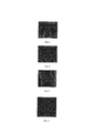
Стандартные срезы: аксиальная (Фиг. 1) корональная (Фиг. 2) и сагиттальная (Фиг. 3) плоскости. Дополнительный срез в косой корональной плоскости (Фиг. 4), биопсия предстательной железы под контролем ее совмещенных изображений, полученных при МРТ (дополнительный срез в косой корональной плоскости) и УЗИ (Фиг. 5).Standard sections: axial (Fig. 1) coronal (Fig. 2) and sagittal (Fig. 3) planes. An additional slice in the oblique coronal plane (Fig. 4), a biopsy of the prostate gland under the control of its combined images obtained by MRI (additional slice in the oblique coronal plane) and ultrasound (Fig. 5).
Гистологическое заключение: ацинарная аденокарцинома (Gleason 7а (3+4), Grade 2, протяженность весь столбик - 100%).The histological conclusion: acinar adenocarcinoma (Gleason 7a (3 + 4), Grade 2, the length of the entire column - 100%).
Пример 2. Пациент 69 лет.Example 2. Patient 69 years.
Анамнез:Anamnesis:
• РПЖ в семье не выявлялся;• Prostate cancer was not identified in the family;
• ТРУЗИ-биопсия в 2016 году (гистологическое заключение - железисто-стромальная гиперплазия предстательной железы);• TRUS biopsy in 2016 (the histological conclusion is glandular-stromal hyperplasia of the prostate gland);
• в 2016 году индекс здоровья простаты PHI в высокой группе риска - 49;• in 2016, the index of prostate health PHI in high risk groups - 49;
• ПРИ - отрицательное;• PRI - negative;
• В 2016 году выполнено мпМРТ предстательной железы - в периферической зоне слева, в средней трети, выявлен очаг размерами 8×7 мм, Pirads 4.• In 2016, an MRI of the prostate gland was performed - in the peripheral zone on the left, in the middle third, a lesion of 8 × 7 mm in size was revealed, Pirads 4.
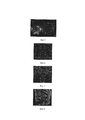
Стандартные срезы: аксиальная (Фиг. 6) корональная (Фиг. 7) и сагиттальная (Фиг. 8) плоскости. Дополнительный срез в косой корональной плоскости (Фиг. 9), (Фиг. 10).Standard sections: axial (Fig. 6) coronal (Fig. 7) and sagittal (Fig. 8) planes. Additional cut in the oblique coronal plane (Fig. 9), (Fig. 10).
Гистологическое заключение: ацинарная аденокарцинома (Gleason 7а (3+4), Grade 2, протяженность 9 мм. - 80%).The histological conclusion: acinar adenocarcinoma (Gleason 7a (3 + 4), Grade 2, length 9 mm. - 80%).
Пример 3. Пациент 58 лет.Example 3. Patient 58 years.
Анамнез:Anamnesis:
• РПЖ в семье не выявлялся;• Prostate cancer was not identified in the family;
• ТРУЗИ-биопсия в 2016 году (гистологическое заключение - железисто-стромальная гиперплазия предстательной железы);• TRUS biopsy in 2016 (the histological conclusion is glandular-stromal hyperplasia of the prostate gland);
• в 2016 году индекс здоровья простаты РШ в высокой группе риска - 62,5;• in 2016, the prostate health index in the high risk group - 62.5;
• ПРИ - отрицательное;• PRI - negative;
• В 2017 году выполнено мпМРТ предстательной железы - в передних отделах, в базальной и средней трети, выявлен очаг размерами 16×10 мм, Pirads 5.• In 2017, an MRI of the prostate gland was performed - in the anterior sections, in the basal and middle third, a lesion of 16 × 10 mm in size was detected, Pirads 5.
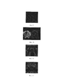
Стандартные срезы: аксиальная (Фиг. 11) корональная (Фиг. 12) и сагиттальная (Фиг. 13) плоскости. Дополнительный срез в косой корональной плоскости (Фиг. 14), биопсия предстательной железы под контролем ее совмещенных изображений, полученных при МРТ и УЗИ (Фиг. 15). Гистологическое заключение: ацинарная аденокарцинома (Gleason 7а (3+4), Grade 2, протяженность 11 мм. - 52%).Standard sections: axial (Fig. 11) coronal (Fig. 12) and sagittal (Fig. 13) planes. An additional slice in the oblique coronal plane (Fig. 14), a biopsy of the prostate gland under the control of its combined images obtained with MRI and ultrasound (Fig. 15). The histological conclusion: acinar adenocarcinoma (Gleason 7a (3 + 4), Grade 2, length 11 mm. - 52%).
Биопсия предстательной железы по заявляемому способу выполнена 56 пациентам. Рак предстательной железы был выявлен у 52 пациентов (индекс Глисона 6 - 14% (8/56), индекс Глисона 7а - 38% (21/56), индекс Глисона 76 - 32% (18/56), индекс Глисона ≥8-9% (5/56)). Из них, у 44 пациентов определен клинически значимый РПЖ (индекс Глисона ≥7) и у 8 пациентов - клинически незначимый РПЖ (индекс Глисона 6).A biopsy of the prostate gland by the present method was performed on 56 patients. Prostate cancer was detected in 52 patients (Gleason index 6–14% (8/56), Gleason index 7a –38% (21/56), Gleason index 76–32% (18/56), Gleason index ≥8 9% (5/56)). Of these, 44 patients had clinically significant prostate cancer (Gleason index ≥7) and 8 patients with clinically insignificant prostate cancer (Gleason index 6).
КБ - количество всех биопсий - 56;KB - the number of all biopsies - 56;
ИП - истинноположительные результаты РПЖ (индекс Глисона ≥7) - 44;PI - true positive results of prostate cancer (Gleason index ≥7) - 44;
ЛП - ложноположительные результаты РПЖ (индекс Глисона 6) - 8;LP - false positive results of prostate cancer (Gleason index 6) - 8;
ИО - истинноотрицательные результаты РПЖ не выявлен - 4.OI - true negative results of PCa not identified - 4.
Чувствительность заявляемого способа рассчитана нами по формуле:The sensitivity of the proposed method is calculated by us by the formula:
![]()
![]()
ЧС - чувствительность способа; КБ - количество всех биопсий - 56;ES - the sensitivity of the method; KB - the number of all biopsies - 56;
ИП - истинноположительные результаты РПЖ (индекс Глисона ≥7) - 44;PI - true positive results of prostate cancer (Gleason index ≥7) - 44;
ЛП - ложноположительные результаты РПЖ (индекс Глисона 6) - 8.LP - false positive results of prostate cancer (Gleason index 6) - 8.
Точность заявляемого способа рассчитана нами по формуле:The accuracy of the proposed method is calculated by us by the formula:
![]()
![]()
ТС - точность способа;TC - the accuracy of the method;
ИП - истинноположительные результаты;PI - true positive results;
ИО - истинноотрицательные результаты;IO - true negative results;
ЛП - ложноположительные результаты.LP - false positive results.
Чувствительность заявляемого способа - 92,9%, способа прототипа - 55,7%. Точность заявляемого способа - 85,8%, способа прототипа - 80,9%.The sensitivity of the proposed method is 92.9%, of the prototype method is 55.7%. The accuracy of the proposed method is 85.8%, of the prototype method is 80.9%.
Таким образом, заявляемый способ, по сравнению со способом прототипом, повышает точность биопсии предстательной железы под контролем ее совмещенных изображений, полученных при магнитно-резонансной томографиии и ультразвуковом исследовании на 4,9%, чувствительность - на 37,2%, что, в свою очередь, улучшает результаты ранней диагностики рака предстательной железы.Thus, the claimed method, compared with the prototype method, improves the accuracy of prostate gland biopsy under the control of its combined images obtained by magnetic resonance imaging and ultrasound by 4.9%, sensitivity - by 37.2%, which, in its queue, improves the results of early diagnosis of prostate cancer.
Claims (1)
Priority Applications (1)
| Application Number | Priority Date | Filing Date | Title |
|---|---|---|---|
| RU2018126444A RU2688373C1 (en) | 2018-07-17 | 2018-07-17 | Method to perform a biopsy of the prostate gland under the control of its combined images obtained by magnetic resonance imaging and ultrasound |
Applications Claiming Priority (1)
| Application Number | Priority Date | Filing Date | Title |
|---|---|---|---|
| RU2018126444A RU2688373C1 (en) | 2018-07-17 | 2018-07-17 | Method to perform a biopsy of the prostate gland under the control of its combined images obtained by magnetic resonance imaging and ultrasound |
Publications (1)
| Publication Number | Publication Date |
|---|---|
| RU2688373C1 true RU2688373C1 (en) | 2019-05-21 |
Family
ID=66636582
Family Applications (1)
| Application Number | Title | Priority Date | Filing Date |
|---|---|---|---|
| RU2018126444A RU2688373C1 (en) | 2018-07-17 | 2018-07-17 | Method to perform a biopsy of the prostate gland under the control of its combined images obtained by magnetic resonance imaging and ultrasound |
Country Status (1)
| Country | Link |
|---|---|
| RU (1) | RU2688373C1 (en) |
Cited By (1)
| Publication number | Priority date | Publication date | Assignee | Title |
|---|---|---|---|---|
| RU2776980C1 (en) * | 2021-07-21 | 2022-07-29 | Дарья Юрьевна Чернышева | Method for intraoperative coordination during perineal prostate biopsy and a kit for its implementation |
-
2018
- 2018-07-17 RU RU2018126444A patent/RU2688373C1/en not_active IP Right Cessation
Non-Patent Citations (4)
| Title |
|---|
| Cash H. et al. The detection of significant prostate cancer is correlated with the Prostate Imaging Reporting and Data System (PI-RADS) in MRI/transrectal ultrasound fusion biopsy. // World J Urol 2016; 34 (4): 525-32. * |
| Shoji S. Transperineal targeted biopsy with real-time fusion image of multiparametric magnetic resonance image and transrectal ultrasound image for the diagnosis of prostate cancer. // Chapetr 4. Prostate Cancer - Leading-edge Diagnostic Procedures and Treatments. Intech. 2016. P.35-43. * |
| Коссов Ф.А. и др. Применение МРТ/УЗИ fusion-биопсии в диагностике клинически значимого рака предстательной железы. // Онкоурология. 2017 13 (3): 61-70. Prostate MRI. найдено [28.02.2019] из Интернет https://mrimaster.com/PLAN%20PROSTATE.html, дата размещ. 06.03.2017 подтв. по адресу https://web.archive.org/web/20170306135638/https://mrimaster.com/PLAN%20PROSTATE.html. * |
| Коссов Ф.А. и др. Применение МРТ/УЗИ fusion-биопсии в диагностике клинически значимого рака предстательной железы. // Онкоурология. 2017 13 (3): 61-70. Prostate MRI. найдено [28.02.2019] из Интернет https://mrimaster.com/PLAN%20PROSTATE.html, дата размещ. 06.03.2017 подтв. по адресу https://web.archive.org/web/20170306135638/https://mrimaster.com/PLAN%20PROSTATE.html. Shoji S. Transperineal targeted biopsy with real-time fusion image of multiparametric magnetic resonance image and transrectal ultrasound image for the diagnosis of prostate cancer. // Chapetr 4. Prostate Cancer - Leading-edge Diagnostic Procedures and Treatments. Intech. 2016. P.35-43. * |
Cited By (1)
| Publication number | Priority date | Publication date | Assignee | Title |
|---|---|---|---|---|
| RU2776980C1 (en) * | 2021-07-21 | 2022-07-29 | Дарья Юрьевна Чернышева | Method for intraoperative coordination during perineal prostate biopsy and a kit for its implementation |
Similar Documents
| Publication | Publication Date | Title |
|---|---|---|
| Ahmad et al. | Transrectal quantitative shear wave elastography in the detection and characterisation of prostate cancer | |
| Cowan et al. | Detection of clinically significant cancer in the anterior prostate by transperineal biopsy | |
| Dianat et al. | Performance of multiparametric magnetic resonance imaging in the evaluation and management of clinically low-risk prostate cancer | |
| JP2012507346A (en) | Method and apparatus using magnetic resonance imaging to identify cancer | |
| JP5212951B2 (en) | Magnetic resonance imaging apparatus, operating method thereof, diagnostic imaging system, and diagnostic method | |
| Pedler et al. | The current status of MRI in prostate cancer | |
| Singh et al. | Patient selection determines the prostate cancer yield of dynamic contrast‐enhanced magnetic resonance imaging‐guided transrectal biopsies in a closed 3‐Tesla scanner | |
| D'Agostino et al. | Comparison between" In-bore" MRI guided prostate biopsy and standard ultrasound guided biopsy in the patient with suspicious prostate cancer: Preliminary results. | |
| US20040254445A1 (en) | Use of MRI to screen individuals for prostate cancer | |
| RU2688373C1 (en) | Method to perform a biopsy of the prostate gland under the control of its combined images obtained by magnetic resonance imaging and ultrasound | |
| Kierans et al. | Implementation of multi-parametric prostate MRI in clinical practice | |
| Roh et al. | The diagnostic accuracy and postoperative outcomes of cervical cancer patients for MR-invisible or MR-visible diagnosis of combined T2-and diffusion-weighted 3T MRI using the external phased-array receiver | |
| Cerci et al. | PET/CT-guided biopsy of liver lesions | |
| Stancioi et al. | Implementing a System for Diagnosing Lung Cancer based on Active Contour Algorithms | |
| RU2822984C1 (en) | Method for differential diagnosis of focal hepatic steatosis and solid hepatic growths in patients with burdened oncoanamnesis | |
| RU2826345C1 (en) | Diagnostic technique for chronic pseudotumorous pancreatitis | |
| RU2766394C1 (en) | Method for diagnosis of breast diseases | |
| Compton | Fundamentals of Cancer Diagnosis and Assessment | |
| Bakhtiyorovna et al. | MEDICINE AND PHARMACY | |
| TWM630582U (en) | Prostate cancer optimizing image interpretation system | |
| Stephens et al. | Investigations that may be useful in detecting cancer | |
| Gomez-Iturriaga et al. | Transperineal biopsies of MRI-detected aggressive index lesions in low-and intermediate-risk prostate cancer patients: Implications for treatment decision | |
| Loft et al. | Intra-and Interobserver Variability of Shear Wave Elastography in Rectal Cancer. Cancers 2022, 14, 2633 | |
| CN119014954A (en) | A method, device and program product for prostate puncture biopsy | |
| Li et al. | The analysis of contrast-enhanced ultrasound features of the liver in male patients with liver cancer and unhealthy life habits under 50 years-of-age |
Legal Events
| Date | Code | Title | Description |
|---|---|---|---|
| MM4A | The patent is invalid due to non-payment of fees |
Effective date: 20200718 |