RU2685416C2 - In vitro diagnosis device and uses thereof - Google Patents
In vitro diagnosis device and uses thereof Download PDFInfo
- Publication number
- RU2685416C2 RU2685416C2 RU2015100233A RU2015100233A RU2685416C2 RU 2685416 C2 RU2685416 C2 RU 2685416C2 RU 2015100233 A RU2015100233 A RU 2015100233A RU 2015100233 A RU2015100233 A RU 2015100233A RU 2685416 C2 RU2685416 C2 RU 2685416C2
- Authority
- RU
- Russia
- Prior art keywords
- reaction
- reaction area
- red
- sample
- porous membrane
- Prior art date
Links
- 238000000338 in vitro Methods 0.000 title claims abstract description 10
- 238000003745 diagnosis Methods 0.000 title claims abstract 14
- 238000006243 chemical reaction Methods 0.000 claims abstract description 146
- 239000012528 membrane Substances 0.000 claims abstract description 130
- 239000000427 antigen Substances 0.000 claims abstract description 66
- 102000036639 antigens Human genes 0.000 claims abstract description 66
- 108091007433 antigens Proteins 0.000 claims abstract description 66
- 210000003743 erythrocyte Anatomy 0.000 claims abstract description 49
- 210000004369 blood Anatomy 0.000 claims abstract description 40
- 239000008280 blood Substances 0.000 claims abstract description 40
- 238000012360 testing method Methods 0.000 claims abstract description 32
- 238000000034 method Methods 0.000 claims abstract description 31
- 230000002209 hydrophobic effect Effects 0.000 claims abstract description 28
- 239000000758 substrate Substances 0.000 claims abstract description 21
- 239000000126 substance Substances 0.000 claims abstract description 13
- 239000000910 agglutinin Substances 0.000 claims abstract description 10
- 101710186708 Agglutinin Proteins 0.000 claims abstract description 9
- 101710146024 Horcolin Proteins 0.000 claims abstract description 9
- 101710189395 Lectin Proteins 0.000 claims abstract description 9
- 101710179758 Mannose-specific lectin Proteins 0.000 claims abstract description 9
- 101710150763 Mannose-specific lectin 1 Proteins 0.000 claims abstract description 9
- 101710150745 Mannose-specific lectin 2 Proteins 0.000 claims abstract description 9
- 239000011148 porous material Substances 0.000 claims abstract description 7
- 238000004519 manufacturing process Methods 0.000 claims abstract description 5
- 239000003795 chemical substances by application Substances 0.000 claims description 106
- 210000004027 cell Anatomy 0.000 claims description 71
- 239000000243 solution Substances 0.000 claims description 70
- 238000000151 deposition Methods 0.000 claims description 39
- 239000000872 buffer Substances 0.000 claims description 36
- 239000003599 detergent Substances 0.000 claims description 35
- 238000005406 washing Methods 0.000 claims description 27
- 239000012491 analyte Substances 0.000 claims description 26
- 230000008021 deposition Effects 0.000 claims description 20
- 239000000203 mixture Substances 0.000 claims description 15
- 230000004931 aggregating effect Effects 0.000 claims description 14
- 210000002966 serum Anatomy 0.000 claims description 11
- 239000003153 chemical reaction reagent Substances 0.000 claims description 9
- 239000007853 buffer solution Substances 0.000 claims description 7
- 238000001514 detection method Methods 0.000 claims description 6
- 238000011533 pre-incubation Methods 0.000 claims description 6
- 230000002391 anti-complement effect Effects 0.000 claims description 5
- 108010008730 anticomplement Proteins 0.000 claims description 5
- 230000006870 function Effects 0.000 claims description 5
- 238000011534 incubation Methods 0.000 claims description 5
- 238000010790 dilution Methods 0.000 claims description 4
- 239000012895 dilution Substances 0.000 claims description 4
- 238000001035 drying Methods 0.000 claims description 4
- 230000036571 hydration Effects 0.000 claims description 4
- 238000006703 hydration reaction Methods 0.000 claims description 4
- 238000011835 investigation Methods 0.000 claims description 4
- 229920002851 polycationic polymer Polymers 0.000 claims description 4
- 102000001554 Hemoglobins Human genes 0.000 claims description 3
- 108010054147 Hemoglobins Proteins 0.000 claims description 3
- 230000002093 peripheral effect Effects 0.000 claims description 3
- 239000002736 nonionic surfactant Substances 0.000 claims description 2
- 229920003023 plastic Polymers 0.000 claims description 2
- 239000004033 plastic Substances 0.000 claims description 2
- 239000007787 solid Substances 0.000 claims description 2
- 230000000694 effects Effects 0.000 abstract description 6
- 230000035945 sensitivity Effects 0.000 abstract description 4
- 238000004458 analytical method Methods 0.000 abstract description 3
- 239000003814 drug Substances 0.000 abstract 1
- 239000000523 sample Substances 0.000 description 48
- 229920001213 Polysorbate 20 Polymers 0.000 description 15
- 239000004365 Protease Substances 0.000 description 15
- FAPWRFPIFSIZLT-UHFFFAOYSA-M Sodium chloride Chemical compound [Na+].[Cl-] FAPWRFPIFSIZLT-UHFFFAOYSA-M 0.000 description 15
- 239000000256 polyoxyethylene sorbitan monolaurate Substances 0.000 description 15
- 235000010486 polyoxyethylene sorbitan monolaurate Nutrition 0.000 description 15
- -1 for example Polymers 0.000 description 11
- 230000003993 interaction Effects 0.000 description 11
- 230000002708 enhancing effect Effects 0.000 description 10
- 238000009738 saturating Methods 0.000 description 10
- 239000011780 sodium chloride Substances 0.000 description 8
- 229920004890 Triton X-100 Polymers 0.000 description 7
- 239000004094 surface-active agent Substances 0.000 description 7
- 229920000209 Hexadimethrine bromide Polymers 0.000 description 6
- 108010039918 Polylysine Proteins 0.000 description 6
- 239000012530 fluid Substances 0.000 description 6
- 239000000463 material Substances 0.000 description 6
- 229920000656 polylysine Polymers 0.000 description 6
- 108010004032 Bromelains Proteins 0.000 description 5
- 102000004190 Enzymes Human genes 0.000 description 5
- 108090000790 Enzymes Proteins 0.000 description 5
- 108090000526 Papain Proteins 0.000 description 5
- 108091005804 Peptidases Proteins 0.000 description 5
- 102100037486 Reverse transcriptase/ribonuclease H Human genes 0.000 description 5
- 230000004520 agglutination Effects 0.000 description 5
- 239000007864 aqueous solution Substances 0.000 description 5
- 235000019835 bromelain Nutrition 0.000 description 5
- 229940088598 enzyme Drugs 0.000 description 5
- 235000019834 papain Nutrition 0.000 description 5
- 229940055729 papain Drugs 0.000 description 5
- 235000019419 proteases Nutrition 0.000 description 5
- LFQSCWFLJHTTHZ-UHFFFAOYSA-N Ethanol Chemical compound CCO LFQSCWFLJHTTHZ-UHFFFAOYSA-N 0.000 description 4
- 230000009471 action Effects 0.000 description 4
- 239000000654 additive Substances 0.000 description 4
- 230000002776 aggregation Effects 0.000 description 4
- 238000004220 aggregation Methods 0.000 description 4
- 230000002349 favourable effect Effects 0.000 description 4
- 239000012634 fragment Substances 0.000 description 4
- 230000001788 irregular Effects 0.000 description 4
- 239000007788 liquid Substances 0.000 description 4
- 229920000642 polymer Polymers 0.000 description 4
- 229930188929 simonin Natural products 0.000 description 4
- CPELXLSAUQHCOX-UHFFFAOYSA-M Bromide Chemical compound [Br-] CPELXLSAUQHCOX-UHFFFAOYSA-M 0.000 description 3
- 239000013504 Triton X-100 Substances 0.000 description 3
- 239000002250 absorbent Substances 0.000 description 3
- 230000002745 absorbent Effects 0.000 description 3
- 238000010521 absorption reaction Methods 0.000 description 3
- 238000005119 centrifugation Methods 0.000 description 3
- 230000000295 complement effect Effects 0.000 description 3
- 238000009792 diffusion process Methods 0.000 description 3
- 229920000136 polysorbate Polymers 0.000 description 3
- 230000008569 process Effects 0.000 description 3
- 230000002441 reversible effect Effects 0.000 description 3
- DHMQDGOQFOQNFH-UHFFFAOYSA-N Glycine Chemical compound NCC(O)=O DHMQDGOQFOQNFH-UHFFFAOYSA-N 0.000 description 2
- 108060003951 Immunoglobulin Proteins 0.000 description 2
- 239000004698 Polyethylene Substances 0.000 description 2
- 229920002873 Polyethylenimine Polymers 0.000 description 2
- 108010008281 Recombinant Fusion Proteins Proteins 0.000 description 2
- 102000007056 Recombinant Fusion Proteins Human genes 0.000 description 2
- PXIPVTKHYLBLMZ-UHFFFAOYSA-N Sodium azide Chemical compound [Na+].[N-]=[N+]=[N-] PXIPVTKHYLBLMZ-UHFFFAOYSA-N 0.000 description 2
- 230000015572 biosynthetic process Effects 0.000 description 2
- 229920002678 cellulose Polymers 0.000 description 2
- 239000001913 cellulose Substances 0.000 description 2
- 238000002405 diagnostic procedure Methods 0.000 description 2
- LOKCTEFSRHRXRJ-UHFFFAOYSA-I dipotassium trisodium dihydrogen phosphate hydrogen phosphate dichloride Chemical compound P(=O)(O)(O)[O-].[K+].P(=O)(O)([O-])[O-].[Na+].[Na+].[Cl-].[K+].[Cl-].[Na+] LOKCTEFSRHRXRJ-UHFFFAOYSA-I 0.000 description 2
- 229920001903 high density polyethylene Polymers 0.000 description 2
- 239000004700 high-density polyethylene Substances 0.000 description 2
- 230000000887 hydrating effect Effects 0.000 description 2
- 102000018358 immunoglobulin Human genes 0.000 description 2
- 229940072221 immunoglobulins Drugs 0.000 description 2
- 238000001727 in vivo Methods 0.000 description 2
- 238000002156 mixing Methods 0.000 description 2
- 230000003204 osmotic effect Effects 0.000 description 2
- 239000002953 phosphate buffered saline Substances 0.000 description 2
- 229920000573 polyethylene Polymers 0.000 description 2
- 102000004169 proteins and genes Human genes 0.000 description 2
- 108090000623 proteins and genes Proteins 0.000 description 2
- 235000000346 sugar Nutrition 0.000 description 2
- 238000003786 synthesis reaction Methods 0.000 description 2
- XOAAWQZATWQOTB-UHFFFAOYSA-N taurine Chemical compound NCCS(O)(=O)=O XOAAWQZATWQOTB-UHFFFAOYSA-N 0.000 description 2
- 239000003656 tris buffered saline Substances 0.000 description 2
- HDTRYLNUVZCQOY-UHFFFAOYSA-N α-D-glucopyranosyl-α-D-glucopyranoside Natural products OC1C(O)C(O)C(CO)OC1OC1C(O)C(O)C(O)C(CO)O1 HDTRYLNUVZCQOY-UHFFFAOYSA-N 0.000 description 1
- 229920000742 Cotton Polymers 0.000 description 1
- 108010044091 Globulins Proteins 0.000 description 1
- 102000006395 Globulins Human genes 0.000 description 1
- WQZGKKKJIJFFOK-GASJEMHNSA-N Glucose Natural products OC[C@H]1OC(O)[C@H](O)[C@@H](O)[C@@H]1O WQZGKKKJIJFFOK-GASJEMHNSA-N 0.000 description 1
- 239000004471 Glycine Substances 0.000 description 1
- 206010018910 Haemolysis Diseases 0.000 description 1
- 102000004856 Lectins Human genes 0.000 description 1
- 108090001090 Lectins Proteins 0.000 description 1
- 239000000020 Nitrocellulose Substances 0.000 description 1
- 239000002033 PVDF binder Substances 0.000 description 1
- 239000004952 Polyamide Substances 0.000 description 1
- 239000004743 Polypropylene Substances 0.000 description 1
- 239000004793 Polystyrene Substances 0.000 description 1
- 208000035388 Ring chromosome 22 syndrome Diseases 0.000 description 1
- CZMRCDWAGMRECN-UGDNZRGBSA-N Sucrose Chemical compound O[C@H]1[C@H](O)[C@@H](CO)O[C@@]1(CO)O[C@@H]1[C@H](O)[C@@H](O)[C@H](O)[C@@H](CO)O1 CZMRCDWAGMRECN-UGDNZRGBSA-N 0.000 description 1
- 229930006000 Sucrose Natural products 0.000 description 1
- 108010008038 Synthetic Vaccines Proteins 0.000 description 1
- HDTRYLNUVZCQOY-WSWWMNSNSA-N Trehalose Natural products O[C@@H]1[C@@H](O)[C@@H](O)[C@@H](CO)O[C@@H]1O[C@@H]1[C@H](O)[C@@H](O)[C@@H](O)[C@@H](CO)O1 HDTRYLNUVZCQOY-WSWWMNSNSA-N 0.000 description 1
- XECAHXYUAAWDEL-UHFFFAOYSA-N acrylonitrile butadiene styrene Chemical compound C=CC=C.C=CC#N.C=CC1=CC=CC=C1 XECAHXYUAAWDEL-UHFFFAOYSA-N 0.000 description 1
- 239000004676 acrylonitrile butadiene styrene Substances 0.000 description 1
- 229920000122 acrylonitrile butadiene styrene Polymers 0.000 description 1
- 230000004913 activation Effects 0.000 description 1
- 239000002671 adjuvant Substances 0.000 description 1
- HDTRYLNUVZCQOY-LIZSDCNHSA-N alpha,alpha-trehalose Chemical compound O[C@@H]1[C@@H](O)[C@H](O)[C@@H](CO)O[C@@H]1O[C@@H]1[C@H](O)[C@@H](O)[C@H](O)[C@@H](CO)O1 HDTRYLNUVZCQOY-LIZSDCNHSA-N 0.000 description 1
- 239000003242 anti bacterial agent Substances 0.000 description 1
- 229940088710 antibiotic agent Drugs 0.000 description 1
- 238000013459 approach Methods 0.000 description 1
- 239000003125 aqueous solvent Substances 0.000 description 1
- 230000004888 barrier function Effects 0.000 description 1
- 230000008901 benefit Effects 0.000 description 1
- WQZGKKKJIJFFOK-VFUOTHLCSA-N beta-D-glucose Chemical compound OC[C@H]1O[C@@H](O)[C@H](O)[C@@H](O)[C@@H]1O WQZGKKKJIJFFOK-VFUOTHLCSA-N 0.000 description 1
- 239000013060 biological fluid Substances 0.000 description 1
- 230000033558 biomineral tissue development Effects 0.000 description 1
- 210000000170 cell membrane Anatomy 0.000 description 1
- 230000001413 cellular effect Effects 0.000 description 1
- 230000008859 change Effects 0.000 description 1
- 239000011248 coating agent Substances 0.000 description 1
- 238000000576 coating method Methods 0.000 description 1
- 108700021073 cold agglutinins Proteins 0.000 description 1
- 239000003086 colorant Substances 0.000 description 1
- 239000012141 concentrate Substances 0.000 description 1
- 239000000470 constituent Substances 0.000 description 1
- 210000000805 cytoplasm Anatomy 0.000 description 1
- 230000001086 cytosolic effect Effects 0.000 description 1
- 230000006378 damage Effects 0.000 description 1
- 230000007812 deficiency Effects 0.000 description 1
- 239000013578 denaturing buffer Substances 0.000 description 1
- 230000001687 destabilization Effects 0.000 description 1
- 238000011161 development Methods 0.000 description 1
- 230000018109 developmental process Effects 0.000 description 1
- 239000008121 dextrose Substances 0.000 description 1
- 238000010586 diagram Methods 0.000 description 1
- 239000012470 diluted sample Substances 0.000 description 1
- 238000010494 dissociation reaction Methods 0.000 description 1
- 230000005593 dissociations Effects 0.000 description 1
- 238000005516 engineering process Methods 0.000 description 1
- 150000002148 esters Chemical class 0.000 description 1
- 229920002313 fluoropolymer Polymers 0.000 description 1
- 238000011010 flushing procedure Methods 0.000 description 1
- 238000013467 fragmentation Methods 0.000 description 1
- 238000006062 fragmentation reaction Methods 0.000 description 1
- 238000002523 gelfiltration Methods 0.000 description 1
- 230000008588 hemolysis Effects 0.000 description 1
- 239000000076 hypertonic saline solution Substances 0.000 description 1
- 210000000987 immune system Anatomy 0.000 description 1
- 230000002163 immunogen Effects 0.000 description 1
- 238000009434 installation Methods 0.000 description 1
- 239000002523 lectin Substances 0.000 description 1
- 210000000265 leukocyte Anatomy 0.000 description 1
- 238000003754 machining Methods 0.000 description 1
- 230000007246 mechanism Effects 0.000 description 1
- 229910052751 metal Inorganic materials 0.000 description 1
- 239000002184 metal Substances 0.000 description 1
- 125000002496 methyl group Chemical group [H]C([H])([H])* 0.000 description 1
- 230000002906 microbiologic effect Effects 0.000 description 1
- 229920005615 natural polymer Polymers 0.000 description 1
- 229920001220 nitrocellulos Polymers 0.000 description 1
- 239000003960 organic solvent Substances 0.000 description 1
- 230000000065 osmolyte Effects 0.000 description 1
- 229920002647 polyamide Polymers 0.000 description 1
- 229920000515 polycarbonate Polymers 0.000 description 1
- 239000004417 polycarbonate Substances 0.000 description 1
- 229920000139 polyethylene terephthalate Polymers 0.000 description 1
- 239000005020 polyethylene terephthalate Substances 0.000 description 1
- 229920000193 polymethacrylate Polymers 0.000 description 1
- 229920001155 polypropylene Polymers 0.000 description 1
- 229920002223 polystyrene Polymers 0.000 description 1
- 239000004800 polyvinyl chloride Substances 0.000 description 1
- 229920000915 polyvinyl chloride Polymers 0.000 description 1
- 229920002981 polyvinylidene fluoride Polymers 0.000 description 1
- 239000001397 quillaja saponaria molina bark Substances 0.000 description 1
- 230000009257 reactivity Effects 0.000 description 1
- 230000010076 replication Effects 0.000 description 1
- 230000004044 response Effects 0.000 description 1
- 230000000717 retained effect Effects 0.000 description 1
- 229930182490 saponin Natural products 0.000 description 1
- 150000007949 saponins Chemical class 0.000 description 1
- 230000011218 segmentation Effects 0.000 description 1
- 238000010186 staining Methods 0.000 description 1
- 238000003756 stirring Methods 0.000 description 1
- 239000005720 sucrose Substances 0.000 description 1
- 150000008163 sugars Chemical class 0.000 description 1
- 229920001059 synthetic polymer Polymers 0.000 description 1
- 229960003080 taurine Drugs 0.000 description 1
- 230000001052 transient effect Effects 0.000 description 1
- 239000011534 wash buffer Substances 0.000 description 1
Images
Classifications
-
- B—PERFORMING OPERATIONS; TRANSPORTING
- B01—PHYSICAL OR CHEMICAL PROCESSES OR APPARATUS IN GENERAL
- B01L—CHEMICAL OR PHYSICAL LABORATORY APPARATUS FOR GENERAL USE
- B01L3/00—Containers or dishes for laboratory use, e.g. laboratory glassware; Droppers
-
- B—PERFORMING OPERATIONS; TRANSPORTING
- B01—PHYSICAL OR CHEMICAL PROCESSES OR APPARATUS IN GENERAL
- B01L—CHEMICAL OR PHYSICAL LABORATORY APPARATUS FOR GENERAL USE
- B01L3/00—Containers or dishes for laboratory use, e.g. laboratory glassware; Droppers
- B01L3/50—Containers for the purpose of retaining a material to be analysed, e.g. test tubes
- B01L3/502—Containers for the purpose of retaining a material to be analysed, e.g. test tubes with fluid transport, e.g. in multi-compartment structures
- B01L3/5023—Containers for the purpose of retaining a material to be analysed, e.g. test tubes with fluid transport, e.g. in multi-compartment structures with a sample being transported to, and subsequently stored in an absorbent for analysis
-
- B—PERFORMING OPERATIONS; TRANSPORTING
- B01—PHYSICAL OR CHEMICAL PROCESSES OR APPARATUS IN GENERAL
- B01L—CHEMICAL OR PHYSICAL LABORATORY APPARATUS FOR GENERAL USE
- B01L3/00—Containers or dishes for laboratory use, e.g. laboratory glassware; Droppers
- B01L3/50—Containers for the purpose of retaining a material to be analysed, e.g. test tubes
- B01L3/508—Containers for the purpose of retaining a material to be analysed, e.g. test tubes rigid containers not provided for above
- B01L3/5085—Containers for the purpose of retaining a material to be analysed, e.g. test tubes rigid containers not provided for above for multiple samples, e.g. microtitration plates
-
- G—PHYSICS
- G01—MEASURING; TESTING
- G01N—INVESTIGATING OR ANALYSING MATERIALS BY DETERMINING THEIR CHEMICAL OR PHYSICAL PROPERTIES
- G01N33/00—Investigating or analysing materials by specific methods not covered by groups G01N1/00 - G01N31/00
- G01N33/48—Biological material, e.g. blood, urine; Haemocytometers
- G01N33/50—Chemical analysis of biological material, e.g. blood, urine; Testing involving biospecific ligand binding methods; Immunological testing
- G01N33/52—Use of compounds or compositions for colorimetric, spectrophotometric or fluorometric investigation, e.g. use of reagent paper and including single- and multilayer analytical elements
- G01N33/525—Multi-layer analytical elements
-
- G—PHYSICS
- G01—MEASURING; TESTING
- G01N—INVESTIGATING OR ANALYSING MATERIALS BY DETERMINING THEIR CHEMICAL OR PHYSICAL PROPERTIES
- G01N33/00—Investigating or analysing materials by specific methods not covered by groups G01N1/00 - G01N31/00
- G01N33/48—Biological material, e.g. blood, urine; Haemocytometers
- G01N33/50—Chemical analysis of biological material, e.g. blood, urine; Testing involving biospecific ligand binding methods; Immunological testing
- G01N33/53—Immunoassay; Biospecific binding assay; Materials therefor
- G01N33/543—Immunoassay; Biospecific binding assay; Materials therefor with an insoluble carrier for immobilising immunochemicals
- G01N33/54366—Apparatus specially adapted for solid-phase testing
- G01N33/54386—Analytical elements
-
- G—PHYSICS
- G01—MEASURING; TESTING
- G01N—INVESTIGATING OR ANALYSING MATERIALS BY DETERMINING THEIR CHEMICAL OR PHYSICAL PROPERTIES
- G01N33/00—Investigating or analysing materials by specific methods not covered by groups G01N1/00 - G01N31/00
- G01N33/48—Biological material, e.g. blood, urine; Haemocytometers
- G01N33/50—Chemical analysis of biological material, e.g. blood, urine; Testing involving biospecific ligand binding methods; Immunological testing
- G01N33/53—Immunoassay; Biospecific binding assay; Materials therefor
- G01N33/543—Immunoassay; Biospecific binding assay; Materials therefor with an insoluble carrier for immobilising immunochemicals
- G01N33/54393—Improving reaction conditions or stability, e.g. by coating or irradiation of surface, by reduction of non-specific binding, by promotion of specific binding
-
- G—PHYSICS
- G01—MEASURING; TESTING
- G01N—INVESTIGATING OR ANALYSING MATERIALS BY DETERMINING THEIR CHEMICAL OR PHYSICAL PROPERTIES
- G01N33/00—Investigating or analysing materials by specific methods not covered by groups G01N1/00 - G01N31/00
- G01N33/48—Biological material, e.g. blood, urine; Haemocytometers
- G01N33/50—Chemical analysis of biological material, e.g. blood, urine; Testing involving biospecific ligand binding methods; Immunological testing
- G01N33/68—Chemical analysis of biological material, e.g. blood, urine; Testing involving biospecific ligand binding methods; Immunological testing involving proteins, peptides or amino acids
- G01N33/6854—Immunoglobulins
-
- G—PHYSICS
- G01—MEASURING; TESTING
- G01N—INVESTIGATING OR ANALYSING MATERIALS BY DETERMINING THEIR CHEMICAL OR PHYSICAL PROPERTIES
- G01N33/00—Investigating or analysing materials by specific methods not covered by groups G01N1/00 - G01N31/00
- G01N33/48—Biological material, e.g. blood, urine; Haemocytometers
- G01N33/50—Chemical analysis of biological material, e.g. blood, urine; Testing involving biospecific ligand binding methods; Immunological testing
- G01N33/80—Chemical analysis of biological material, e.g. blood, urine; Testing involving biospecific ligand binding methods; Immunological testing involving blood groups or blood types or red blood cells
-
- B—PERFORMING OPERATIONS; TRANSPORTING
- B01—PHYSICAL OR CHEMICAL PROCESSES OR APPARATUS IN GENERAL
- B01L—CHEMICAL OR PHYSICAL LABORATORY APPARATUS FOR GENERAL USE
- B01L2300/00—Additional constructional details
- B01L2300/06—Auxiliary integrated devices, integrated components
- B01L2300/0627—Sensor or part of a sensor is integrated
- B01L2300/0663—Whole sensors
-
- B—PERFORMING OPERATIONS; TRANSPORTING
- B01—PHYSICAL OR CHEMICAL PROCESSES OR APPARATUS IN GENERAL
- B01L—CHEMICAL OR PHYSICAL LABORATORY APPARATUS FOR GENERAL USE
- B01L2300/00—Additional constructional details
- B01L2300/06—Auxiliary integrated devices, integrated components
- B01L2300/069—Absorbents; Gels to retain a fluid
-
- B—PERFORMING OPERATIONS; TRANSPORTING
- B01—PHYSICAL OR CHEMICAL PROCESSES OR APPARATUS IN GENERAL
- B01L—CHEMICAL OR PHYSICAL LABORATORY APPARATUS FOR GENERAL USE
- B01L2300/00—Additional constructional details
- B01L2300/08—Geometry, shape and general structure
- B01L2300/0809—Geometry, shape and general structure rectangular shaped
- B01L2300/0829—Multi-well plates; Microtitration plates
-
- B—PERFORMING OPERATIONS; TRANSPORTING
- B01—PHYSICAL OR CHEMICAL PROCESSES OR APPARATUS IN GENERAL
- B01L—CHEMICAL OR PHYSICAL LABORATORY APPARATUS FOR GENERAL USE
- B01L2300/00—Additional constructional details
- B01L2300/16—Surface properties and coatings
- B01L2300/161—Control and use of surface tension forces, e.g. hydrophobic, hydrophilic
-
- G—PHYSICS
- G01—MEASURING; TESTING
- G01N—INVESTIGATING OR ANALYSING MATERIALS BY DETERMINING THEIR CHEMICAL OR PHYSICAL PROPERTIES
- G01N2800/00—Detection or diagnosis of diseases
- G01N2800/22—Haematology
-
- Y—GENERAL TAGGING OF NEW TECHNOLOGICAL DEVELOPMENTS; GENERAL TAGGING OF CROSS-SECTIONAL TECHNOLOGIES SPANNING OVER SEVERAL SECTIONS OF THE IPC; TECHNICAL SUBJECTS COVERED BY FORMER USPC CROSS-REFERENCE ART COLLECTIONS [XRACs] AND DIGESTS
- Y10—TECHNICAL SUBJECTS COVERED BY FORMER USPC
- Y10T—TECHNICAL SUBJECTS COVERED BY FORMER US CLASSIFICATION
- Y10T29/00—Metal working
- Y10T29/49—Method of mechanical manufacture
- Y10T29/49826—Assembling or joining
Landscapes
- Health & Medical Sciences (AREA)
- Life Sciences & Earth Sciences (AREA)
- Immunology (AREA)
- Chemical & Material Sciences (AREA)
- Engineering & Computer Science (AREA)
- Hematology (AREA)
- Biomedical Technology (AREA)
- Molecular Biology (AREA)
- Urology & Nephrology (AREA)
- General Health & Medical Sciences (AREA)
- Analytical Chemistry (AREA)
- Food Science & Technology (AREA)
- General Physics & Mathematics (AREA)
- Medicinal Chemistry (AREA)
- Physics & Mathematics (AREA)
- Cell Biology (AREA)
- Biochemistry (AREA)
- Biotechnology (AREA)
- Microbiology (AREA)
- Pathology (AREA)
- Chemical Kinetics & Catalysis (AREA)
- Clinical Laboratory Science (AREA)
- Proteomics, Peptides & Aminoacids (AREA)
- Investigating Or Analysing Biological Materials (AREA)
- Measuring Or Testing Involving Enzymes Or Micro-Organisms (AREA)
Abstract
Description
Настоящее изобретение относится к устройству для диагностики in vitro в образце крови или одного из ее компонентов, предназначенному для обнаружения реакций между антигенами эритроцитов и антителами, которые специфически направлены против этих антигенов.The present invention relates to an in vitro diagnostic device in a blood sample or one of its components, designed to detect reactions between red blood cell antigens and antibodies that are specifically directed against these antigens.
Изобретение также относится к применению этого устройства для идентификации и определения групп крови.The invention also relates to the use of this device for the identification and determination of blood groups.
Целью иммуногематологических методов диагностики является обеспечение или диагностика атаки антител на красные кровяные клетки. Для этого нужны инструменты, позволяющие обнаружить антигены на поверхности красных клеток, наличие или отсутствие которых определяет группу крови, а также определить, содержит ли кровь одно или больше антител, направленных против известных антигенов красных клеток; присутствие антитела означает вероятность несовместимости.The purpose of immunohematological diagnostic methods is to provide or diagnose an attack of antibodies on red blood cells. This requires tools to detect antigens on the surface of red cells, the presence or absence of which determines the blood group, and also to determine whether the blood contains one or more antibodies directed against the known red cell antigens; the presence of antibodies means the probability of incompatibility.
Таким образом, распространенные методы включают поиск и идентификацию антигенов группы крови на поверхности эритроцитов и/или поиск и идентификацию содержащихся в плазме антител против антигенов групп крови.Thus, common methods include the search and identification of blood group antigens on the surface of red blood cells and / or the search and identification of antibodies contained in plasma against blood group antigens.
Например, для системы АВО тест Бета-Винсента определяет антигены красных клеток, а комплементарный тест Симонина-Михона или перекрестная проверка по сыворотке определяет антитела сыворотки крови.For example, for the ABO system, the Beta-Vincent test determines the red cell antigens, and the Simonin-Mikhon complementary test or cross-check by serum determines serum antibodies.
В ходе теста Бета-Винсента красные клетки индивидуума смешивают с реагентами, состоящими из антител известной специфичности. В общем случае, результаты теста наблюдают по агглютинации красных клеток, возникающей, когда антитела распознают соответствующие антигены эритроцитов.During the Beta-Vincent test, the individual's red cells are mixed with reagents consisting of antibodies of known specificity. In general, the test results are observed by the agglutination of red cells, which occurs when antibodies recognize the corresponding erythrocyte antigens.
В тесте Симонина плазму индивидуума смешивают с пробами красных кровяных клеток (red blood cell tests), каждая из которых относится к конкретной группе антигенов системы АВО. В ходе теста определяют агглютинацию пробы красных кровяных клеток с плазмой индивидуума.In Simonin's test, an individual's plasma is mixed with red blood cell tests, each of which belongs to a specific group of antigens of the ABO system. During the test, agglutination of a sample of red blood cells with the plasma of an individual is determined.
Поиск так называемых транзиторных антител предполагает регистрацию наличия или отсутствия в крови индивидуума иммуноглобулинов, направленных против различных эритроцитарных антигенов. В ходе поиска аутоантител у индивидуума непосредственно ищут антитела, уже обнаруженные in vivo. Задачей поиска аллоантител является обнаружение факта прикрепления этих иммуноглобулинов к пробам красных клеток с известными антигенами, и выполняется это непрямыми методами Кумбса.The search for so-called transient antibodies involves the registration of the presence or absence in the blood of an individual of immunoglobulins directed against various erythrocyte antigens. In the search for autoantibodies in an individual, antibodies directly found in vivo are directly sought by the individual. The task of searching for alloantibodies is to detect whether these immunoglobulins are attached to samples of red cells with known antigens, and this is done by indirect Coombs methods.
В настоящее время известно очень большое количество способов и устройств, применяемых для фенотипирования в области иммуногематологии, но существующие методы фенотипирования групп крови связаны с большим количеством недостатков.Currently, there are a very large number of methods and devices used for phenotyping in the field of immunohematology, but the existing methods for phenotyping blood groups are associated with a large number of disadvantages.
Например, способы с применением микропланшетов нуждаются в этапе центрифугирования, за которым следует этап перемешивания. Этап перемешивания критически важен, поскольку множество реакций, одновременно протекающих на твердом носителе, характеризуются различными кинетическими параметрами ресуспендирования. Это означает риск разрушить слабую агглютинацию, не добившись успеха в ресуспендировании сильной агглютинации. Эти действия необходимо контролировать визуально, особое внимание уделяя явлению адгезии некоторых реагентов.For example, microplate methods require a centrifugation step followed by a mixing step. The stage of mixing is crucial, since many of the reactions that simultaneously occur on a solid support are characterized by different kinetic parameters of resuspension. This means the risk of breaking down weak agglutination without having succeeded in resuspending strong agglutination. These actions need to be monitored visually, with particular attention to the phenomenon of adhesion of some reagents.
Аналогично, хотя проводятся тесты, основанные на технологиях гель-фильтрации, существует риск пропустить некоторые агглютинации, особенно при плазматическом тесте группы по АВО, в связи с диссоциацией, вызванной фрагментацией небольших агглютинатов при их прохождении через гель.Similarly, although tests based on gel filtration technologies are being carried out, there is a risk of missing some agglutination, especially during the plasma test of the ABO group, due to dissociation caused by the fragmentation of small agglutinates as they pass through the gel.
Серьезным недостатком всех этих способов является необходимость центрифугирования для декантации красных клеток или проведения их через гель; этот этап значительно увеличивает время и стоимость анализа и требует использования больших центрифуг, которыми трудно управлять.A serious disadvantage of all these methods is the need for centrifugation for decanting red cells or carrying them through the gel; This step significantly increases the time and cost of analysis and requires the use of large centrifuges that are difficult to manage.
Известен также способ иммунофильтрации, такой как описан в заявке ЕР 2167967, который состоит из захвата аналита, присутствующего в образце, при его прохождении через пористую мембрану с перехватывающим элементом и обнаружения его присутствия выявляющим элементом. Устройство такого типа содержит область депонирования образца, гидрофильную пористую мембрану, на которой находится перехватывающий реагент и под которой располагается абсорбирующая мембрана. Проведение такого вида исследования состоит из осаждения чистого или разбавленного образца в области депонирования, в ходе которого образец проходит через пористую мембрану и оседает на абсорбирующей мембране. Если в образце присутствует соответствующий перехватывающему элементу аналит, он иммобилизируется перехватывающим элементом при прохождении по капиллярам пористой мембраны. Кроме того, область захвата предпочтительного аналита обнаруживают с помощью выявляющего агента, способного определить присутствие аналита в области захвата и содержащего компонент, позволяющий проводить такое обнаружение визуально (окрашенный продукт) или физическим или химическим методом.There is also known a method of immunofiltration, such as described in EP 2167967, which consists of capturing an analyte present in a sample as it passes through a porous membrane with an interceptor element and detects its presence by a detecting element. A device of this type contains a sample deposit area, a hydrophilic porous membrane on which the interception reagent is located and under which the absorbing membrane is located. Conducting this type of study consists of the deposition of a clean or diluted sample in the deposit area, during which the sample passes through a porous membrane and settles on the absorbent membrane. If an analyte corresponding to the intercepting element is present in the sample, it is immobilized by the intercepting element when passing through the capillaries of the porous membrane. In addition, the capture region of the preferred analyte is detected with a detecting agent capable of determining the presence of the analyte in the capture region and containing a component that allows such detection to be carried out visually (colored product) or by physical or chemical methods.
Однако этот способ характеризуется низкой чувствительностью и специфичностью, так как при осаждении образца он распространяется по пористой мембране и впитывается ею, в результате большое количество аналита теряется в мертвом объеме пористой мембраны или выходит за пределы области захвата. То же самое касается и выявляющего раствора. Чтобы депонировать большое количество исследуемого образца и выявляющего раствора, необходимо слишком сильно увеличить абсорбирующую систему, а это не позволяет проводить тесты в небольших объемах с контролируемой кинетикой без выявляющих агентов и с использованием автоматического микродозатора.However, this method is characterized by low sensitivity and specificity, since during the deposition of the sample it spreads across the porous membrane and is absorbed by it, as a result, a large amount of analyte is lost in the dead volume of the porous membrane or goes beyond the capture region. The same applies to the detecting solution. In order to deposit a large amount of the test sample and the detecting solution, it is necessary to increase the absorbing system too much, and this does not allow conducting tests in small volumes with controlled kinetics without detecting agents and using an automatic microbatcher.
В попытке решить эту проблему и сконцентрировать сигнал, в заявках СА 1312265 или WO 02052263 предложено разместить выше и ниже гидрофильной пористой мембраны гидрофобную структуру, которая направляла бы поток через область захвата. Однако в таких устройствах сохраняется проблема центробежной диффузии от пятна; в результате выявляющие элементы, проходящие через область (пятно) захвата и не связывающиеся с ним, могут центробежно диффундировать в гидрофильную пористую мембрану и накапливаться на периферии пятна. Это позволяет им избегать промывки, которая также сфокусирована на пятне. Затем по механизмам центростремительной диффузии эти выявляющие элементы могут вернуться в область захвата, что приводит к появлению окраски там даже в случае отсутствия предпочтительного аналита. Данное явление очень быстро (обычно 5-15 минут) делает невозможным проведение теста из-за ложных положительных результатов, что представляет собой большую проблему, поскольку устройства подобного типа не могут повторить анализ в случае сомнения, человеческой ошибки или потери информации.In an attempt to solve this problem and concentrate the signal, in CA 1312265 or WO 02052263, it has been proposed to place a hydrophobic structure above and below the hydrophilic porous membrane, which would direct the flow through the capture region. However, in such devices, the problem of centrifugal diffusion from the spot persists; as a result, the detecting elements passing through the area (spot) of the capture and not contacting it can diffuse centrifugally into the hydrophilic porous membrane and accumulate at the periphery of the spot. This allows them to avoid washing, which is also focused on the stain. Then, by the mechanisms of centripetal diffusion, these detecting elements can return to the capture region, which leads to the appearance of color there even in the absence of a preferred analyte. This phenomenon very quickly (usually 5-15 minutes) makes it impossible to perform the test due to false positive results, which is a big problem, because devices of this type cannot repeat the analysis in case of doubt, human error or loss of information.
Еще одной крупной проблемой существующих в настоящее время устройств иммунофильтрации является необходимость контролировать скорость движения и, в некоторых случаях, продолжительность предварительной инкубации. Важность этих параметров обусловлена тем, что взаимодействие между перехватывающим элементом и аналитом, а также между аналитом и выявляющим элементом характеризуется специфической кинетикой. Кинетика описывает зависимость числа актов взаимодействия от времени. Поскольку учитывается как взаимодействие в паре перехватывающий агент / аналит, так и в паре выявляющий агент / аналит, для получения адекватного сигнала необходимо обеспечить достаточное число взаимодействий в каждой паре. Если взаимодействие между перехватывающим агентом и захваченным аналитом протекает медленно, необходимо снизить скорость прохождения образца через мембрану. Если же лимитирующей стадией является взаимодействие между выявляющим агентом и аналитом, необходимо обеспечить более продолжительную предварительную инкубацию, когда выявляющий элемент и аналит перемешиваются над областью захвата. Без конкретной системы прохождение через гидрофильную мембрану происходит быстро (500 мкл/мин).Another major problem with currently existing immunofiltration devices is the need to control the speed of movement and, in some cases, the duration of pre-incubation. The importance of these parameters is due to the fact that the interaction between the intercepting element and the analyte, as well as between the analyte and the detecting element, is characterized by specific kinetics. Kinetics describes the dependence of the number of interaction acts on time. Since both the interaction in the pair is the intercepting agent / analyte, and in the pair the identifying agent / analyte is taken into account, to obtain an adequate signal, it is necessary to ensure a sufficient number of interactions in each pair. If the interaction between the intercepting agent and the captured analyte proceeds slowly, it is necessary to reduce the rate at which the sample passes through the membrane. If the limiting stage is the interaction between the detecting agent and the analyte, it is necessary to ensure a longer pre-incubation when the detecting element and the analyte are mixed over the capture region. Without a specific system, passage through the hydrophilic membrane occurs quickly (500 μl / min).
Для контроля потока предлагалось использовать поршень, в частности, в заявке US 2008318342.To control the flow it was proposed to use a piston, in particular, in the application US 2008318342.
Для контроля времени предварительной инкубации в заявке WO 03016902 предложено использовать устройство, состоящее из двух частей: в верхней части расположена область сбора образца и пористая мембрана, а в нижней части -пористая и абсорбирующая мембрана. В первоначальном положении эти два блока не взаимодействуют, и жидкость не может течь по капиллярам. После механической обработки эти две области приходят в контакт друг с другом, после чего жидкость может протекать по капиллярам. Также предложено депонировать перехватывающий элемент на гидрофобной мембране и активизировать прохождение жидкости добавлением поверхностно-активного вещества.To control the pre-incubation time, in application WO 03016902 it is proposed to use a device consisting of two parts: in the upper part there is a sample collection area and a porous membrane, and in the lower part is a porous and absorbing membrane. In the initial position, these two blocks do not interact, and the liquid cannot flow through the capillaries. After machining, these two areas come in contact with each other, after which the liquid can flow through the capillaries. It is also proposed to deposit an intercepting element on a hydrophobic membrane and to activate the passage of fluid by the addition of a surfactant.
Механические подходы трудно реализовать, поскольку для них требуется разработать специальные системы. Кроме того, любые возникающие при работе с ними проблемы требуют участия подготовленных специалистов.Mechanical approaches are difficult to implement because they require the development of special systems. In addition, any problems arising from working with them require the participation of trained professionals.
Добавление поверхностно-активного вещества при проведении теста для ускорения работы системы чрезвычайно нежелательно, поскольку оно серьезно мешает взаимодействию между перехватывающими агентами и аналитами. Кроме того, в иммуногематологии используемые в качестве выявляющих агентов красные клетки несовместимы с жидкими поверхностно-активными веществами, так как последние растворяют мембраны красных клеток, и они теряют гемоглобин.The addition of surfactant during the test to speed up the system is extremely undesirable because it seriously interferes with the interaction between the capture agents and the analytes. In addition, in immunohematology, red cells used as detecting agents are incompatible with liquid surfactants, since they dissolve red cell membranes and they lose hemoglobin.
Еще один способ, описанный в заявке ЕР 0334015, предполагает для контроля потока использование дополнительной мембраны, расположенной ниже первой, но предлагаемое устройство не может решить проблемы, связанные с диффузийе образца и выявляющего элемента в гидрофильную пористую мембрану.Another method described in EP 0334015 involves the use of an additional membrane located below the first to control the flow, but the proposed device cannot solve the problems associated with the diffusion of the sample and the detecting element into the hydrophilic porous membrane.
Таким образом, существующие иммуногематологические диагностические тесты характеризуются большим количеством недостатков.Thus, the existing immunohematological diagnostic tests are characterized by a large number of deficiencies.
Задачей настоящего изобретения является устранение недостатков известных решений путем создания устройства, адаптированного для иммуногематологической диагностики in vitro и работающего с использованием капиллярных явлений, которое является надежным, быстрым, мобильным и экономичным, простым в изготовлении и использовании, может быть выполнено с миниатюрными размерами и автоматизировано и характеризуется достаточной чувствительностью.The present invention is to eliminate the disadvantages of the known solutions by creating a device adapted for immunohematological in vitro diagnostics and working with capillary phenomena, which is reliable, fast, mobile and economical, simple to manufacture and use, can be made with miniature sizes and automated characterized by sufficient sensitivity.
Для достижения этой цели настоящее изобретение предлагает устройство для диагностики in vitro, предназначенное для обнаружения в образце крови или одного из ее компонентов по меньшей мере одной реакции между антигеном эритроцитарного фенотипа и антителом, специфически направленным против этого антигена, характеризующееся тем, что оно содержит:To achieve this goal, the present invention provides an in vitro diagnostic device for detecting in a blood sample or one of its components at least one reaction between an erythrocyte phenotype antigen and an antibody specifically directed against this antigen, characterized in that it contains:
- подложку и- substrate and
- гидрофобную пористую мембрану толщиной от 0,05 до 1,5 мм с диаметром пор от 2 до 30 мкм, причем указанная мембрана содержит по меньшей мере одну гидрофильную область реакции, предназначенную для получения указанного образца, и поверхность указанной области реакции меньше поверхности гидрофобной пористой мембраны.- hydrophobic porous membrane with a thickness of from 0.05 to 1.5 mm with a pore diameter of from 2 to 30 microns, and this membrane contains at least one hydrophilic reaction region, designed to obtain the specified sample, and the surface of the specified reaction region is less than the hydrophobic porous surface membranes.
Изобретение относится также к применению этого устройства, в конкретных способах фенотипирования эритроцитарных групп крови и обнаружения антител с использованием этого устройства.The invention also relates to the use of this device, to specific methods for phenotyping erythrocyte blood groups and detecting antibodies using this device.
Настоящее изобретение с успехом исправляет все недостатки существующих в настоящее время тестов иммунофильтрации, в частности, в связи с:The present invention successfully corrects all the shortcomings of currently existing immunofiltration tests, in particular, in connection with:
- недопущением возврата выявляющего агента, вызывающего появление ложных положительных ответов,- to prevent the return of the detecting agent, causing the occurrence of false positive responses,
- повышенной чувствительностью из-за использования меньших объемов,- increased sensitivity due to the use of smaller volumes,
- миниатюризацией и автоматизацией системы,- miniaturization and automation of the system,
- контролем кинетики реакции без необходимости выполнения механических действий с устройством.- control of the reaction kinetics without the need to perform mechanical actions with the device.
Другие характеристики и преимущества вытекают из следующего описания изобретения, приводимого только для примера и проиллюстрированного прилагаемыми рисунками, на которых:Other characteristics and advantages derive from the following description of the invention, given by way of example only and illustrated by the accompanying drawings, in which:
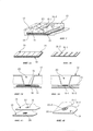
- фигура 1 иллюстрирует схему конкретного воплощения устройства по изобретению с соблюдением законов перспективы;- figure 1 illustrates a diagram of a specific embodiment of the device according to the invention in compliance with the laws of perspective;
- фигура 2А иллюстрирует гидрофобную пористую мембрану и поглощающую мембрану устройства по изобретению с первым вариантом расположения гидрофильных областей реакции;- figure 2A illustrates the hydrophobic porous membrane and absorbing membrane of the device according to the invention with the first option for the location of the hydrophilic reaction regions;
- фигура 2В иллюстрирует гидрофобную пористую мембрану и поглощающую мембрану устройства по изобретению со вторым вариантом расположения гидрофильных областей реакции;- figure 2B illustrates the hydrophobic porous membrane and absorbing membrane of the device according to the invention with the second variant of the location of the hydrophilic reaction areas;
- фигура 3А представляет собой сечение устройства по изобретению с гидрофобной пористой мембраной, соответствующей варианту фигуры 2А;- figure 3A is a cross-section of the device according to the invention with a hydrophobic porous membrane, corresponding to the variant of figure 2A;
- фигура 3В представляет собой сечение устройства по изобретению с гидрофобной пористой мембраной, соответствующей варианту фигуры 2В;- figure 3B is a cross section of a device according to the invention with a hydrophobic porous membrane corresponding to the embodiment of figure 2B;
- фигура 4А иллюстрирует результаты, полученные после применения устройства по изобретению, область реакции гидрофобной пористой мембраны которого показана на рисунке 2А, а также фрагмент расположенной ниже поглощающей мембраны; а также- figure 4A illustrates the results obtained after applying the device according to the invention, the reaction region of the hydrophobic porous membrane of which is shown in figure 2A, as well as a fragment of the lower-lying absorbing membrane; and
- фигура 4В иллюстрирует результаты, полученные после применения устройства по изобретению, область реакции гидрофобной пористой мембраны которого показана на рисунке 2 В.- figure 4B illustrates the results obtained after the use of the device according to the invention, the reaction region of the hydrophobic porous membrane of which is shown in Figure 2 B.
Устройство 10 по изобретению представляет собой устройство для диагностики in vitro для детекции в образце крови или одного из ее компонентов по меньшей мере одной реакции между антигеном эритроцитарного фенотипа и антителом, специфически направленным против этого антигена.The
Это устройство предназначено для диагностики in vitro с использованием капиллярных явлений, оно особенно подходит для иммуногематологической диагностики in vitro.This device is intended for in vitro diagnostics using capillary phenomena, it is particularly suitable for immuno-hematological in vitro diagnostics.
Антиген эритроцитарного фенотипа, антиген эритроцитарной группы или антиген группы крови означает все иммуногенные молекулы, присутствующие на поверхности красных клеток и способные вызывать образование направленных против них антител и/или распознавание и последующее разрушение красных клеток под действием иммунной системы.Erythrocyte phenotype antigen, erythrocyte group antigen or blood group antigen means all immunogenic molecules present on the surface of red cells and can cause the formation of antibodies directed against them and / or recognition and subsequent destruction of red cells by the action of the immune system.
Реакция между антигеном эритроцитарного фенотипа и антителом, направленным специфически против этого антигена, в описании изобретения называется реакцией антиген-антитело.The reaction between the erythrocyte phenotype antigen and the antibody directed specifically against this antigen is referred to in the description of the invention as the antigen-antibody reaction.
Образец крови или одного из ее компонентов означает цельную кровь или один из ее компонентов, выбранный специфически из фракции красных клеток (эритроцитов), белых клеток (лейкоцитов), плазмы или сыворотки.A sample of blood or one of its components means whole blood or one of its components, selected specifically from a fraction of red cells (red blood cells), white cells (leukocytes), plasma or serum.
Как показано на фигуре 1, устройство 10 по изобретению содержит:As shown in figure 1, the
- подложку 12 и- the
- гидрофобную пористую мембрану 14, содержащую по меньшей мере одну гидрофильную область реакции 16, предназначенную для получения исследуемого образца.- a hydrophobic
Толщина пористой мембраны 14 находится в интервале от 0,05 до 1,5 мм, предпочтительно от 0,1 до 1 мм, более предпочтительно от 0,4 до 0,8 мм.The thickness of the
Диаметр пор находится в интервале от 2 до 30 мкм, предпочтительно от 7 до 12 мкм.The pore diameter is in the range from 2 to 30 microns, preferably from 7 to 12 microns.
Гидрофобная пористая мембрана 14 может содержать любой материал, не изменяемый под действием водных растворителей. Этот материал, в частности, может быть выбран из природных полимеров, модифицированных химически или немодифицированных, таких как, например, полимер нитроцеллюлозы или целлюлоза, или из синтетических полимеров, таких как, например, полиэтилен, полиэтилен высокой плотности (ПЭВП) или фторированные полимеры, например, ПВДФ. Этот материал должен быть гидрофобным с самого начала, или его следует сделать гидрофобным адекватной обработкой. Эти полимеры можно функционализировать активными группами, способными образовывать связи с применяемыми впоследствии перехватывающими агентами.The hydrophobic
Гидрофобная пористая мембрана 14 содержит по меньшей мере одну гидрофильную область реакции 16. Поверхность области реакции 16 меньше поверхности гидрофобной пористой мембраны 14, то есть, мембрану 14 нельзя полностью гидрофилизировать.The hydrophobic
Гидрофильную(ые) область(и) реакции 16 на пористой мембране 14 предпочтительно сделаны гидрофильными путем локального введения детергента, без изменения химических функций пористого субстрата предварительной химической или физической обработкой гидрофобной пористой мембраны 14.The hydrophilic (s) region (s) of the
Детергент означает любой гидрофилизирующий агент, то есть, любое вещество, способное сделать гидрофобную мембрану 14 гидрофильной.Detergent means any hydrophilizing agent, that is, any substance capable of making the
Применяемый детергент можно выбрать из природных детергентов, химически модифицированных природных детергентов или детергентов, полученных в ходе химического синтеза. Предпочтительно, это неионное поверхностно-активное вещество, например, Тритон Х-100, Твин 20 или сапонин.The detergent used can be selected from natural detergents, chemically modified natural detergents or detergents obtained during chemical synthesis. Preferably, this non-ionic surfactant, for example, Triton X-100,
Детергент можно разбавить водным раствором или органическим растворителем, таким как этанол, в концентрации от 0,01 до 5%. Предпочтительно, детергент, применяемый для того, чтобы сделать мембрану 14 локально гидрофильной, используется в дозе от 0,01 до 2% (вес/объем).The detergent can be diluted with an aqueous solution or an organic solvent, such as ethanol, in a concentration of from 0.01 to 5%. Preferably, the detergent used to make the
Количество использованного детергента, с учетом других характеристик мембраны (в особенности, ее пористости и толщины) контролирует скорость движения жидкостей через мембрану. Обычно считается, что не следует превышать максимальную дозу 0,1% для детергента Тритон Х-100 и 0,05% для Твина, чтобы сделать гидрофильной мембрану, предназначенную для получения перехватывающего элемента. Однако из-за конкретных характеристик мембраны 14 по изобретению детергенты, способные сделать эту мембрану гидрофильной, могут быть локально использованы в дозе вплоть до 2%, особенно для Тритона Х-100 или Твина-20, без разрушения реактивности области, которая облегчает гидрофилизацию мембраны 14.The amount of detergent used, taking into account other characteristics of the membrane (in particular, its porosity and thickness), controls the speed of movement of fluids through the membrane. It is generally believed that the maximum dose of 0.1% for the Triton X-100 detergent and 0.05% for Tween should not be exceeded in order to make a hydrophilic membrane designed to make the intercepting element. However, due to the specific characteristics of the
Одна и та же гидрофобная мембрана 14 может содержать несколько гидрофильных областей реакции 16 при условии, что эти области не пересекаются.The same
Области реакции 16 могут быть любых геометрических форм, но предпочтительно, чтобы они принимали форму кругов или пятен с диаметром от 0,3 до 20 мм.
Область реакции 16 может быть гидрофильной по всей толщине пористой мембраны 14 и/или на ее поверхности.The
Область реакции 16 может характеризоваться одной и той же степенью гидрофилизации, как показано на фигурах 2А, 3А и 4А. Такая конфигурация особенно хорошо подходит для обнаружения присутствия специфических антител в анализируемом образце.The
В соответствии с вариантом изобретения, область реакции 16 может содержать несколько областей с различными степенями гидрофилизации.In accordance with an embodiment of the invention, the
Как показано на фигурах 2В, 3В и 4В, область реакции 16 может содержать две гидрофильные области 16-1 и 16-2, причем степень гидрофилизации в центре 16-1 области реакции больше, чем на периферии 16-2. Эти гидрофильные области находятся предпочтительно только на поверхности мембраны 14.As shown in figures 2B, 3B and 4B, the
Область реакции 16 также может содержать две гидрофильные области с различной степенью гидрофилизации, одна из которых располагается на поверхности, а другая в толщине мембраны.The
Если область реакции 16 содержит две гидрофильные области, область реакции 16 сделана гидрофильной с использованием двух различных детергентов, без изменения химических функций пористого субстрата предварительной химической или физической обработкой пористой мембраны.If the
Преимущественно, конфигурация области реакции 16 с двумя участками различной гидрофилизации, в особенности на поверхности, такая как показана на фигурах 2В, 3В и 4В, отвечает конкретной потребности профессионалов в области переливания крови в обнаружении одновременного сосуществования двух популяций, одна из которых положительна, а другая отрицательна, это явление называется двойной популяцией. Эта конфигурация особенно хорошо подходит для детекции и идентификации конкретных антигенов в исследуемом образце.Mainly, the configuration of the
Гидрофильная область реакции 16 пористой мембраны 14 может содержать перехватывающие агенты. Перехватывающие агенты абсорбируются или ковалентно связываются с мембраной 14.The hydrophilic region of the
Термин "перехватывающий агент" означает любой химический или биологический элемент, зафиксированный в области 16 и способный удержать соответствующий аналит, содержащийся в исследуемом образце, возможно, в комплексе с выявляющим агентом.The term "intercepting agent" means any chemical or biological element that is fixed in
Если задача заключается в обнаружении в исследуемом образце конкретных антител, то такие перехватывающие агенты содержат антигены конкретного фенотипа эритроцитов. Это могут быть антигены, очищенные или не очищенные от фрагментов или мембран содержащих их клеток, клетки, не содержащие антигены или какие-либо свои компоненты, или даже рекомбинантные протеины или антигены, полученные путем синтеза. В предпочтительном варианте перехватывающие агенты представляют собой красные кровяные клетки без гемоглобина, но несущие антигены.If the task is to detect specific antibodies in the sample under study, then these intercept agents contain antigens of a specific red cell phenotype. These may be antigens, purified or not purified from fragments or membranes of cells containing them, cells that do not contain antigens or any of their components, or even recombinant proteins or antigens obtained by synthesis. In a preferred embodiment, the interception agents are red blood cells without hemoglobin, but carrying antigens.
Если задача заключается в обнаружении в исследуемом образце конкретных антигенов, перехватывающие агенты представляют собой антитела.If the task is to detect specific antigens in the sample, the intercept agents are antibodies.
Если в области реакции 16 присутствуют перехватывающие агенты, то их можно депонировать одновременно с детергентом, используемым для гидрофилизации мембраны 14 с целью ограничения области реакции 16, или после гидрофилизации, независимо от детергента.If interception agents are present in the
Перехватывающие агенты можно депонировать в область реакции 16 в составе не-денатурирующего буфера, рН которого стабилизирован между значениями 4 и 10, предпочтительно между 6,5 и 7,8, еще более предпочтительно между 7 и 7,5. Перехватывающие агенты могут либо абсорбироваться, либо связываться ковалентно.Interception agents can be deposited in the
Перехватывающие агенты можно смешивать с адъювантами, обеспечивающими их микробиологическую стабильность, такими как азид натрия, антибиотики, и их конформационную стабильность, такими как сахара (сахароза, декстроза, трегалоза), а также с другими агентами, известными специалистам в данной области для выполнения этих функций.Intercepting agents can be mixed with adjuvants that ensure their microbiological stability, such as sodium azide, antibiotics, and their conformational stability, such as sugars (sucrose, dextrose, trehalose), as well as with other agents known to specialists in this field to perform these functions .
Перехватывающие агенты могут присутствовать во всей области 16 или только в ее части.Interception agents may be present in the
Детергент и/или перехватывающие агенты предпочтительно представляют собой растворы, которые можно депонировать автоматизированными или ручными пипетками. Предпочтительно, чтобы эти растворы можно было легко депонировать с помощью игл, удерживающих предпочтительный объем силами капиллярного притяжения. Эти иглы можно изготавливать из любых материалов, в частности из металла, и они могут содержать гидрофобное покрытие или не содержать его. Их концы могут быть плоскими или содержать выемки определенного объема.The detergent and / or trapping agents are preferably solutions that can be deposited with automated or manual pipettes. Preferably, these solutions can be easily deposited with needles, which hold the preferred volume by capillary attraction forces. These needles can be made from any materials, in particular from metal, and they may or may not contain a hydrophobic coating. Their ends may be flat or contain grooves of a certain size.
Депонирование растворов детергентов и/или перехватывающих агентов должно сопровождаться сушкой мембраны, причем продолжительность сушки зависит от температуры: при комнатной температуре сушить надо не менее 4 часов, а при 37°С не менее часа.The deposition of detergent solutions and / or intercepting agents must be accompanied by drying the membrane, and the drying time depends on the temperature: it is necessary to dry at least 4 hours at room temperature and at least one hour at 37 ° C.
Устройство по изобретению может содержать поглощающую мембрану 18, расположенную под пористой мембраной 14. Эта мембрана 18 поглощает жидкости, депонированные на уровне областей реакции 16, которые не удержались на мембране 14, в особенности, если область реакции 16 гидрофильна по всей толщине мембраны 14.The device according to the invention may contain an absorbing
Мембрана 18 может содержать материал, обеспечивающий пассивное поглощение за счет капиллярных явлений, такой как поглощающая бумага, целлюлоза и т.д., или состоять из поглощающих полимеров. Например, можно использовать следующие продукты:The
- Millipore С048, С068, С083, С248- Millipore C048, C068, C083, C248
- Whatman CF3, CF4, CF10, категория 470, CF5, CF6, CF7, категория 900, категория 300- Whatman CF3, CF4, CF10, category 470, CF5, CF6, CF7, category 900, category 300
- Ahlstrom категории 601, 642, 631, 238, 237, 222, 243, 320- Ahlstrom category 601, 642, 631, 238, 237, 222, 243, 320
- Pall категории 111, 113, 133, 165, 197, 8975, 8964, 8301, Accuwik® Ultra- Pall categories 111, 113, 133, 165, 197, 8975, 8964, 8301, Accuwik® Ultra
- Суперпоглощающая подложка Cleanis Gelmax- Super-absorbing substrate Cleanis Gelmax
- Хлопок- Cotton
Состав и размеры поглощающей мембраны 18 необходимо выбрать так, чтобы они могли поглотить все растворы, используемые при проведении исследования (Vобщий в мкл). Мембраны, которые можно охарактеризовать по их поглощающей способности (С в мкл/см2) и размерам (D в см2), подбирают так, чтобы соответствовать следующему уравнению: D>Vобщий/С.The composition and dimensions of the absorbing
Альтернативно, жидкость может поглощаться за счет разницы давлений между областью над мембраной и под ней. Например, для этого можно использовать аспирационную систему с частичным вакуумом.Alternatively, the fluid can be absorbed by the pressure difference between and above the membrane. For example, a partial vacuum aspiration system can be used for this.
Мембрана 14 и необязательно мембрана 18 помещены в подложку 12.The
Подложка 12 устройства 10 по изобретению предпочтительно является жесткой подложкой. Например, это может быть твердая оболочка.The
Подложка предпочтительно содержит жесткий материал, который не допускает протечек жидкости. В частности, это может быть пластик, такой как полипропилен, полиэтилен, полистирол, акрилонитрил-бутадиеновый стирол, полиэтилентерефталат, поликарбонат, полиамид, поливинилхлорид, метилполиметакрилат.The substrate preferably contains a rigid material that prevents leakage of fluid. In particular, it can be plastic, such as polypropylene, polyethylene, polystyrene, acrylonitrile-butadiene styrene, polyethylene terephthalate, polycarbonate, polyamide, polyvinyl chloride, methyl polymethacrylate.
Предпочтительно подложка 12 содержит по меньшей мере одно отверстие 20, причем каждой гидрофильной области реакции 16 пористой мембраны 14 соответствует свое отверстие 20. Это отверстие 20 соответствует области сбора образца, депонированного на гидрофильную область реакции 16. Гидрофильная область 16 может быть по размеру идентична основанию отверстия, меньше или больше него, что означает, что две гидрофильные области всегда разделены гидрофобным участком на мембране 14.Preferably, the
Края отверстия 20 могут быть прозрачными, чтобы видеть расположенные ниже сигналы, если это требуется.The edges of the
Размер области сбора должен быть таким, чтобы в ней мог поместиться по меньшей мере максимальный объем исследуемого образца, депонированного на реакционной мембране 16.The size of the collection area should be such that it can fit at least the maximum volume of the test sample deposited on the
Независимость каждой области реакции 16 обеспечивается барьером, создаваемым гидрофобной мембраной между реакционными областями. Эта независимость позволяет выполнять несколько видов диагностики на одном и том же устройстве с одной мембраной. Согласно варианту изобретения, эту независимость можно также реализовать, сегментируя подложку 12 на физически независимые отделенные друг от друга сектора, каждый со своей собственной мембраной.The independence of each
Кроме того, чтобы улучшить контакт между мембранами 14 и 18, можно создать область дополнительной уплотненной массы вокруг отверстия 20 и перед отверстием обтекателя напротив указанного отверстия 20.In addition, in order to improve the contact between the
Подложка 12 может представлять собой твердую оболочку с несколькими отверстиями 20, ее внешние размеры должны быть совместимы со стандартом SBS/AINSI.
Настоящее изобретение также относится к способу производства устройства 10, описанного выше, характеризующемуся тем, что он включает следующие этапы:The present invention also relates to a method for manufacturing a
- гидрофилизацию гидрофобной пористой мембраны 14 толщиной от 0,05 до 1,5 мм с диаметром пор от 2 до 30 мкм, по меньшей мере в одной области 16 указанной мембраны с помощью по меньшей мере одного детергента;- hydrophilization of a hydrophobic
- депонирование раствора перехватывающего агента;- deposition of an intercepting agent solution;
- сушку;- drying;
- сборку пористой мембраны 14 и поглощающей мембраны 18, расположенной ниже пористой мембраны 14, на подложке 12.- the Assembly of the
Устройство 10 по изобретению можно применять для того, чтобы с использованием капиллярных явлений определять присутствие или отсутствие аналита в биологической жидкости, в частности в крови или одной из ее составных частей. Этот аналит может быть антигеном эритроцитарного фенотипа или антителом, направленным против этого антигена.The
Устройство 10 может быть особенно полезно для:
- фенотипирования красных клеток, то есть, для определения антигенов на их поверхности,- phenotyping red cells, that is, to identify antigens on their surface,
- проведения теста Симонина, идентифицирующего наличие анти-А или анти-В антител,- the Simonin test, identifying the presence of anti-A or anti-B antibodies,
- поиска или идентификации антител, направленных против клеточных антигенов для конкретных эритроцитов, что касается поиска аллоантител, аутоантител или даже холодовых агглютининов.- search or identification of antibodies directed against cellular antigens for specific erythrocytes, with regard to the search for alloantibodies, autoantibodies or even cold agglutinins.
Таким образом, специфическим объектом изобретения является применение устройства 10 для идентификации и определения в образце крови или одного из ее компонентов групп крови по системе АВО, расширенного фенотипирования резуса, поиска иррегулярного агглютинина, поиска аутоантител, поиска холодового антиглобулина и/или перекрестной проверки на совместимость.Thus, a specific object of the invention is the use of the
Применение устройства 10 по изобретению требует использования выявляющего агента. Предпочтительно, это красные кровяные клетки. Эти клетки:The use of the
- представляют собой красные кровяные клетки известного фенотипа, называемые пробами красных кровяных клеток, для применения в устройстве для поиска антител или выполнения теста Симонина;- represent the red blood cells of a known phenotype, called red blood cell samples, for use in the device for searching for antibodies or performing the Simonin test;
- содержатся в исследуемом образце для применения в устройстве для фенотипирования.- contained in the test sample for use in the device for phenotyping.
Если целью является определение присутствия аналита в исследуемом образце, в зависимости от аналита может потребоваться изменить перехватывающий агент, выявляющий агент, способ гидрофилизации (одиночная или многократная, по толщине или на поверхности) и последующий протокол исследования.If the goal is to determine the presence of an analyte in a test sample, depending on the analyte, it may be necessary to change the trapping agent, the detecting agent, the hydrophilization method (single or multiple, thickness or on the surface) and the subsequent study protocol.
В любом случае перед депонированием исследуемого образца на области реакции можно осуществить гидратацию области реакции 16 буферным раствором. Этот буфер может содержать раствор, рН которого стабилизирован в интервале от 6 до 8,5, предпочтительно от 6,5 до 7,8, в особенности от 7 до 7,5, а осмотическая концентрация находится в интервале от 250 мОсм до 800 мОсм, предпочтительно от 300 мОсм до 600 мОсм. Этот раствор может дополнительно содержать детергенты в низких концентрациях (Твин 20 от 0,01 до 0,05% (масс/об)), агенты насыщения (БСА) и/или агенты, способные усиливать реакции антиген-антитело.In any case, before depositing the sample under investigation, it is possible to hydrate the
Аналогично, чтобы узнать результаты, необходимо использовать промывку буфером. Такой буфер предпочтительно содержит PBS (забуференный фосфатом физиологический раствор), TBS (забуференный Трис физиологический раствор) или физиологический раствор рН от 2 до 10, предпочтительно от 5 до 9. Необходимо контролировать осмотическую концентрацию буфера, это позволит избежать гемолиза красных клеток. Буфер необходимо подобрать так, чтобы не отделить выявляющие агенты или окрашенные аналиты, прямо или косвенно зафиксированные перехватывающими агентами. Неожиданно оказалось, что для применения в устройстве по изобретению предпочтительно применять слегка гиперосмотические промывающие растворы (то есть, от 300 до 800 мОсм), полученные при растворении компонентов физиологического раствора, например, NaCl, или неионные осмолиты, такие как, например, глицин или таурин. Такой буфер можно окрасить красителями, контрастирующими с окраской выявляющего агента. Например, если выявляющий агент представляет собой красные кровяные клетки, промывающий буферный раствор можно окрасить в синий или зеленый цвета. Для устранения фонового шума к промывающему раствору можно также добавить незначительную концентрацию поверхностно-активного вещества. Такие поверхностно-активные вещества предпочтительно являются неионными веществами и, в частности, представляют собой сложные эфиры Сахаров, в особенности полиоксиэтиленовые эфиры сорбитана (Твин).Similarly, in order to know the results, it is necessary to use a buffer wash. This buffer preferably contains PBS (phosphate buffered saline), TBS (Tris buffered saline) or saline pH from 2 to 10, preferably from 5 to 9. It is necessary to control the osmotic concentration of the buffer, this will avoid hemolysis of red cells. The buffer must be selected so as not to separate the detecting agents or colored analytes directly or indirectly fixed by the intercepting agents. Surprisingly, for use in the device according to the invention, it is preferable to use slightly hyperosmotic washing solutions (i.e., 300 to 800 mOsm) obtained by dissolving components of a saline solution, for example, NaCl, or non-ionic osmolytes, such as, for example, glycine or taurine . Such a buffer can be colored with colorants contrasting with the color of the detecting agent. For example, if the detecting agent is red blood cells, the washing buffer solution can be colored blue or green. To eliminate the background noise to the washing solution, you can also add a slight concentration of surfactant. Such surfactants are preferably non-ionic substances and, in particular, are sugar esters, in particular polyoxyethylene sorbitan esters (Tween).
Работая с устройством 10, жидкости можно депонировать с помощью системы пипетирования или с использованием системы капиллярной репликации.Working with
В основном, устройство по изобретению не нуждается ни в центрифугировании, ни в перемешивании, ни в вакуумировании, ни в специальных дополнительных устройствах. С ним можно полностью автономно работать вручную, а также его можно автоматизировать и использовать в составе роботизированных установок. Различные характеристики мембраны 14 позволяют контролировать скорость движения жидкости, делая его настолько медленным, чтобы успела пройти реакция антиген-антитело, каким бы большим ни был размер пор мембраны 14.Basically, the device according to the invention does not need any centrifugation, or stirring, or vacuuming, or special additional devices. You can work with it completely autonomously manually, and you can also automate it and use it as part of robotized installations. Various characteristics of the
В соответствии с первым вариантом, изобретение направлено на применение устройства 10 для фенотипирования красных клеток.In accordance with the first embodiment, the invention is directed to the use of a
Задача состоит в поиске антигенов на поверхности красных клеток. В данном случае антитело или смесь антител применяют в качестве перехватывающих агентов, способных распознавать специфические интересующие антигены или их варианты. В качестве антител можно использовать очищенные или полуочищенные моноклональные антитела, содержащее моноклональное антитело культуральное поверхностно-активное вещество (см. таблицу 1) или поликлональные антитела, анти-сыворотку. Можно также использовать агглютинины или лектины.The challenge is to search for antigens on the surface of red cells. In this case, the antibody or mixture of antibodies is used as intercept agents capable of recognizing specific antigens of interest or variants thereof. As antibodies, you can use purified or semi-purified monoclonal antibodies, containing monoclonal antibody, culture surfactant (see table 1) or polyclonal antibodies, anti-serum. You can also use agglutinins or lectins.
Далее приводится неполный список моноклональных антител:The following is a partial list of monoclonal antibodies:


В соответствии с конкретным вариантом осуществления, изобретение направлено на способ фенотипирования эритроцитарных групп крови в образце эритроцитов (выделенные клетки или цельная кровь) для одновременного обнаружения популяции положительного фенотипа и популяции отрицательного фенотипа (двойная популяция), включающий следующие этапы:In accordance with a specific embodiment, the invention is directed to a method of phenotyping erythrocyte blood groups in a sample of erythrocytes (isolated cells or whole blood) for the simultaneous detection of a population of a positive phenotype and a population of a negative phenotype (double population), comprising the following steps:
- депонирование раствора для гидратации пористой мембраны 14 в центре области реакции устройства 10 по изобретению; причем указанная область реакции 16 содержит две гидрофильные области 16-1 и 16-2, где степень гидрофилизации в центре 16-1 области реакции больше, чем на периферии 16-2, и в центре находятся перехватывающие агенты, содержащие антитела; раствор для гидратации мембраны представляет собой такой раствор, который благоприятен для иммуногематологических реакций, возможно содержащий добавки, такие как, например, буфер с рН от 6 до 8,5, предпочтительно от 6,5 до 7,8, в особенности от 7 до 7,5, и осмолярностью от 250 до 800 мОсм, предпочтительно от 300 до 600 мОсм. Этот раствор может содержать детергенты в низкой концентрации (особенно, Твин 20 от 0,01 до 0,05% (масс/об)), насыщающие агенты (например, БСА) и/или агенты, способные усиливать реакции антиген-антитело. Этот буфер может обладать также протеазной активностью, достигаемой, например, путем добавления ферментов, таких как папаин или бромелаин. Буфер может также содержать поликатион-содержащие агенты, такие как полибрен или полилизин, для усиления реакции и увеличения продолжительности контакта клеток с перехватывающим элементом;- depositing a solution for hydrating the
- разведение подлежащих фенотипированию красных клеток в буферном растворе, конкретно, в буферном растворе, благоприятном для иммуногематологических реакций, возможно содержащем добавки, например, в буфере с рН от 6 до 8,5, предпочтительно от 6,5 до 7,8, в особенности от 7 до 7,5, и осмолярностью от 250 до 800 мОсм, предпочтительно от 300 до 600 мОсм. Этот раствор может содержать детергенты в низкой концентрации (особенно, Твин 20 от 0,01 до 0,05% (масс/об)), насыщающие агенты (например, БСА) и/или агенты, способные усиливать реакции антиген-антитело. Этот буфер может обладать также протеазной активностью, достигаемой, например, путем добавления ферментов, таких как папаин или бромелаин. Буфер может также содержать поликатион-содержащие агенты, такие как полибрен или полилизин, для усиления реакции и увеличения продолжительности контакта клеток с перехватывающим элементом;- the dilution of red cells to be phenotyped in a buffer solution, specifically in a buffer solution favorable for immuno-hematological reactions, possibly containing additives, for example, in a buffer with a pH from 6 to 8.5, preferably from 6.5 to 7.8, in particular from 7 to 7.5, and osmolarity from 250 to 800 mOsm, preferably from 300 to 600 mOsm. This solution may contain low concentration detergents (especially
- введение этого раствора, содержащего подлежащие фенотипированию красные клетки, в центр 16-1 области реакции;- the introduction of this solution, containing the red cells to be phenotyped, into the center 16-1 of the reaction area;
- инкубация при температуре, предпочтительно от 15 до 40°С, в частности от 18 до 25°С, в течение времени от 60 секунд до 15 минут, в частности от 2 до 10 минут;- incubation at a temperature of, preferably from 15 to 40 ° C, in particular from 18 to 25 ° C, for a time from 60 seconds to 15 minutes, in particular from 2 to 10 minutes;
- депонирование в область реакции промывающего раствора; промывающим является раствор, не препятствующий реакциям антиген-антитело, поддерживающий целостность красных клеток и не допускающий неспецифических взаимодействий, такой как, например, буфер с рН от 6 до 8,5, предпочтительно от 6,5 до 7,8, в особенности от 7 до 7,5, и осмолярностью от 250 до 800 мОсм, предпочтительно от 300 до 600 мОсм. Этот раствор может содержать детергенты в низкой концентрации (особенно, Твин 20 от 0,01 до 0,05% (масс/об)), насыщающие агенты (например, БСА) и/или агенты, способные усиливать реакции антиген-антитело. Если ранее использовали поликатионный агент, то этот буфер должен быть сильно ионным, например, должен содержать более 100 мМ NaCl, чтобы разрушить неспецифические взаимодействия, вызванные добавкой этого типа.- deposition in the reaction area of the washing solution; flushing is a solution that does not interfere with antigen-antibody reactions, maintains the integrity of red cells and prevents non-specific interactions, such as, for example, a buffer with a pH from 6 to 8.5, preferably from 6.5 to 7.8, especially from 7 to 7.5, and an osmolarity of from 250 to 800 mOsm, preferably from 300 to 600 mOsm. This solution may contain low concentration detergents (especially
Депонируемый образец может представлять собой красные клетки или цельную кровь. Образец можно разбавить в буфере, содержащим раствор с рН, стабилизированным в интервале от 6 до 8,5, предпочтительно от 6,5 до 7,8, в особенности от 7 до 7,5, и осмолярностью от 250 до 800 мОсм, предпочтительно от 300 до 600 мОсм. Этот раствор может содержать детергенты в низкой концентрации (особенно, Твин 20 от 0,01 до 0,05% (масс/об)), насыщающие агенты (например, БСА) и/или агенты, способные усиливать реакции антиген-антитело. Этот буфер может обладать также протеазной активностью, достигаемой, например, путем добавления ферментов, таких как папаин или бромелаин.The deposited sample may be red cells or whole blood. The sample can be diluted in a buffer containing a solution with a pH stabilized in the range from 6 to 8.5, preferably from 6.5 to 7.8, in particular from 7 to 7.5, and an osmolarity from 250 to 800 mOsm, preferably from 300 to 600 mOsm This solution may contain low concentration detergents (especially
Гидрофилизация областей реакции 16 мембраны 14 устройства 10, осуществляемая для выполнения этого процесса, предпочтительно представляет собой гидрофилизацию поверхности.Hydrophilization of the
Например, гидрофилизацию можно осуществить раствором Твина 20 в этаноле в концентрации от 0,1 до 1% (масс/об), предпочтительно от 0,2 до 1% (масс/об), в объеме от 2 до 40 мкл, и предпочтительно от 5 до 20 мкл, в центре которого создается область гидрофилизации, а толщина обеспечивается применением водного раствора Тритона X100 в концентрации от 0,1 до 2% (масс/об), предпочтительно от 0,5 до 1% (масс/об), в объеме от 25 нл до 15 мкл, предпочтительно от 100 нл до 10 мкл.For example, hydrophilization can be carried out with a solution of
Если красные клетки, присутствующие в образце, содержат антиген, распознаваемый перехватывающим элементом, они остаются иммобилизированными в центре области реакции 16 мембраны 14, несмотря на промывку, и центральная область реакции 16-1 остается красной. Если красные клетки образца не содержат антигена, распознаваемого перехватывающим элементом, они вымываются при промывке: центральная область 16-1 остается белой, а в периферической области 16-2 формируется красное кольцо. Если образец содержит две различные популяции, наблюдается красное окрашивание центра 22-1 и периферического кольца 22-2.If the red cells present in the sample contain an antigen recognized by the interceptor element, they remain immobilized in the center of the
Регистрация результатов может осуществляться визуально или в автоматическом режиме.Registration of results can be carried out visually or automatically.
В соответствии с еще одним вариантом осуществления изобретение направлено на способ фенотипирования эритроцитарных групп крови в образце эритроцитов (выделенные клетки или цельная кровь), не допускающий обнаружение двойных популяций и включающий следующие этапы:In accordance with another embodiment, the invention is directed to a method for phenotyping erythrocyte blood groups in a sample of erythrocytes (isolated cells or whole blood) that does not allow detection of double populations and includes the following steps:
- депонирование раствора для гидратации пористой мембраны 14 в центре области реакции устройства 10 по изобретению; причем указанная область реакции 16 содержит одну гидрофильную область, где находятся перехватывающие агенты, содержащие антитела; раствор для гидратации мембраны представляет собой такой раствор, который благоприятен для иммуногематологических реакций, возможно, содержащий добавки, такой как, например, буфер с рН от 6 до 8,5, предпочтительно от 6,5 до 7,8, в особенности от 7 до 7,5, и осмолярностью от 250 до 800 мОсм, предпочтительно от 300 до 600 мОсм. Этот раствор может содержать детергенты в низкой концентрации (особенно, Твин 20 от 0,01 до 0,05% (масс/об)), насыщающие агенты (например, БСА) и/или агенты, способные усиливать реакции антиген-антитело. Этот буфер может обладать также протеазной активностью, достигаемой, например, путем добавления ферментов, таких как папаин или бромелаин. Буфер может также содержать поликатион-содержащие агенты, такие как полибрен или полилизин, для усиления реакции и увеличения продолжительности контакта клеток с перехватывающими элементами;- depositing a solution for hydrating the
- возможное разведение подлежащих фенотипированию красных клеток в буферном растворе, конкретно, в буферном растворе, благоприятном для иммуногематологических реакций, возможно, содержащем добавки, например, в буфере с рН от 6 до 8,5, предпочтительно от 6,5 до 7,8, в особенности от 7 до 7,5, и осмолярностью от 250 до 800 мОсм, предпочтительно от 300 до 600 мОсм. Этот раствор может содержать детергенты в низкой концентрации (особенно, Твин 20 от 0,01 до 0,05% (масс/об)), насыщающие агенты (например, БСА) и/или агенты, способные усиливать реакции антиген-антитело. Этот буфер может обладать также протеазной активностью, достигаемой, например, путем добавления ферментов, таких как папаин или бромелаин. Буфер может также содержать поликатион-содержащие агенты, такие как полибрен или полилизин, для усиления реакции и увеличения продолжительности контакта клеток с перехватывающим элементом;- a possible dilution of the red cells to be phenotyped in a buffer solution, specifically, in a buffer solution favorable for immunohematological reactions, possibly containing additives, for example, in a buffer with a pH from 6 to 8.5, preferably from 6.5 to 7.8, in particular from 7 to 7.5, and an osmolarity of from 250 to 800 mOsm, preferably from 300 to 600 mOsm. This solution may contain low concentration detergents (especially
- введение этого раствора, содержащего подлежащие фенотипированию красные клетки, в центр области реакции;- the introduction of this solution, containing red cells to be phenotyped, in the center of the reaction area;
- инкубация при температуре, предпочтительно от 15 до 40°С, в частности, от 18 до 25°С, в течение времени от 2 секунд до 15 минут, в частности, от 1 до 10 минут;- incubation at a temperature of, preferably, from 15 to 40 ° C, in particular, from 18 to 25 ° C, for a time from 2 seconds to 15 minutes, in particular, from 1 to 10 minutes;
- депонирование в область реакции промывающего раствора; промывающим является раствор, не препятствующий реакциям антиген-антитело, поддерживающий целостность красных клеток и не допускающий неспецифических взаимодействий, такой как, например, буфер с рН от 6 до 8,5, предпочтительно от 6,5 до 7,8, предпочтительно от 7 до 7,5, и осмолярностью от 250 до 800 мОсм, предпочтительно от 300 до 600 мОсм. Этот раствор может содержать детергенты в низкой концентрации (особенно, Твин 20 от 0,01 до 0,05% (масс/об)), насыщающие агенты (например, БСА) и/или агенты, способные усиливать реакции антиген-антитело. Если ранее использовали поликатионный агент, то этот буфер должен быть сильно ионным, например, должен содержать более 100 мМ NaCl, чтобы разрушить неспецифические взаимодействия, вызванные добавкой этого типа.- deposition in the reaction area of the washing solution; washing is a solution that does not interfere with antigen-antibody reactions, maintains the integrity of red cells and prevents non-specific interactions, such as, for example, a buffer with a pH from 6 to 8.5, preferably from 6.5 to 7.8, preferably from 7 to 7.5, and an osmolarity of from 250 to 800 mOsm, preferably from 300 to 600 mOsm. This solution may contain low concentration detergents (especially
Гидрофилизация областей реакции 16 мембраны 14 устройства 10, осуществляемая для выполнения этого процесса, может представлять собой гидрофилизацию по толщине мембраны. Необходимо, чтобы устройство 10 содержало также поглощающую систему, расположенную ниже мембраны 14, например, поглощающую мембрану 18.Hydrophilization of the
Гидрофилизацию можно проводить водным раствором Тритон Х100 в концентрации от 0,1 до 2% (масс/об), предпочтительно от 0,5 до 1% (масс/об), в объеме от 25 нл до 15 мкл, предпочтительно от 100 нл до 10 мкл.Hydrophilization can be carried out with an aqueous solution of Triton X100 in a concentration of from 0.1 to 2% (w / v), preferably from 0.5 to 1% (w / v), in a volume of from 25 nl to 15 μl, preferably from 100 nl to 10 µl.
Если красные клетки образца содержат антиген, распознаваемый перехватывающим элементом, они остаются иммобилизированными в центре 22 области реакции 16 мембраны 14, несмотря на промывку, и область реакции 16 остается красной. Если красные клетки образца не содержат антигена, распознаваемого перехватывающим элементом, они вымываются при промывке и Область реакции 16 остается белой. Поглощающая мембрана 18 поглощает (область 24) все, что не связалось с мембраной 14.If the red cells of the sample contain an antigen recognized by the intercepting element, they remain immobilized in the
Регистрация результатов может осуществляться визуально или в автоматическом режиме.Registration of results can be carried out visually or automatically.
В соответствии со вторым вариантом, изобретение направлено на применение устройства 10 для обнаружения мультивалентных антител в крови или тесте Симонина.In accordance with the second option, the invention is directed to the use of the
Задачей является поиск специфических антител. Таким образом, перехватывающий элемент содержит антиген, против которого направлено предпочтительное антитело.The task is to search for specific antibodies. Thus, the interceptor contains the antigen against which the preferred antibody is directed.
Иммобилизированные антигены могут представлять собой синтетические антигены, сопряженные или нет с полимером или протеином. Кроме того, они могут представлять собой рекомбинантные протеины, содержащие одну или более последовательности антигена или смеси антигенов предпочтительных вариантов. Иммобилизированные антигены могут также находится на клетках или фрагментах клеток, в особенности, на фрагментах мембран или клетках без цитоплазматического содержимого. Такие клетки, в частности, могут быть красными клетками без цитоплазмы, называемыми также "тенями".Immobilized antigens can be synthetic antigens, coupled or not with a polymer or protein. In addition, they may be recombinant proteins containing one or more antigen sequences or mixtures of antigens of the preferred embodiments. Immobilized antigens can also be found on cells or fragments of cells, in particular, on fragments of membranes or cells without cytoplasmic contents. Such cells, in particular, can be red cells without cytoplasm, also called "shadows".
Если образец содержит антитела против интересующего антигена, последние прикрепляются к поверхности мембраны 14 на уровне области реакции 16.If the sample contains antibodies against the antigen of interest, the latter attach to the surface of the
Необходимо использовать выявляющий элемент для обнаружения этих антител, также распознавая их природу (анти-IgG, анти-IgM). Если же применяемое антитело является мультивалентным (может одновременно обнаружить несколько антигенов), то можно воспользоваться выявляющим элементом, также несущим интересующий антиген.It is necessary to use a detecting element to detect these antibodies, also recognizing their nature (anti-IgG, anti-IgM). If the antibody used is multivalent (it can simultaneously detect several antigens), then you can use a detecting element that also carries the antigen of interest.

Еще одним объектом изобретения является способ обнаружения присутствующих в крови мультивалентных антител в образце плазмы, сыворотки или цельной крови, включающий следующие этапы:Another object of the invention is a method for detecting multivalent antibodies present in blood in a plasma, serum or whole blood sample, comprising the following steps:
- депонирование исследуемого образца в область реакции 16 устройства 10, причем указанная область реакции 16 содержит перехватывающие агенты, включая антигены;- depositing the sample under investigation into the
- введение красных клеток известного фенотипа, содержащих те же антигены, что и перехватывающие агенты;- the introduction of red cells of a known phenotype containing the same antigens as intercepting agents;
- прохождение смеси через гидрофильную область, причем указанная гидрофильная область обеспечивает время прохождения через нее от 1 до 45 минут, предпочтительно от 3 до 15 минут;- the passage of the mixture through the hydrophilic region, and the specified hydrophilic region provides the time passing through it from 1 to 45 minutes, preferably from 3 to 15 minutes;
- депонирование в область реакции промывающего раствора, такого как, например, буфер с рН от 6 до 8,5, предпочтительно от 6,5 до 7,8, в особенности от 7 до 7,5, и осмолярностью от 250 до 800 мОсм, предпочтительно от 300 до 600 мОсм. Этот раствор может содержать детергенты в низкой концентрации (особенно, Твин 20 от 0,01 до 0,05% (масс/об)) и насыщающие агенты (например, БСА).- deposition in the reaction area of the washing solution, such as, for example, a buffer with a pH from 6 to 8.5, preferably from 6.5 to 7.8, in particular from 7 to 7.5, and an osmolarity from 250 to 800 mOsm, preferably from 300 to 600 mOsm. This solution may contain detergents in low concentrations (especially
Образец для депонирования может представлять собой плазму, сыворотку или цельную кровь, разбавленную или нет в буфере, содержащим раствор рН от 6 до 8,5, предпочтительно от 6,5 до 7,8, в особенности от 7 до 7,5, и осмолярностью от 250 до 800 мОсм, предпочтительно от 300 до 600 мОсм. Этот раствор может дополнительно содержать детергенты в низкой концентрации (например, Твин 20 от 0,01 до 0,05% (масс/об)), насыщающие агенты (особенно, БСА), и/или агенты, способные усиливать реакции антиген-антитело. Минерализация буфера составляет предпочтительно менее 100 мМ NaCl.The sample to be deposited may be plasma, serum or whole blood, diluted or not in buffer, containing a solution of pH from 6 to 8.5, preferably from 6.5 to 7.8, in particular from 7 to 7.5, and osmolarity from 250 to 800 mOsm, preferably from 300 to 600 mOsm. This solution may additionally contain detergents at a low concentration (for example,
Гидрофилизация областей реакции 16 мембраны 14 устройства 10, осуществляемая для выполнения этого процесса, предпочтительно проводится по толщине мембраны. Таким образом, необходимо, чтобы устройство 10 содержало также поглотительную систему, расположенную ниже мембраны 14, например, поглощающую мембрану 18.Hydrophilization of the
Гидрофилизацию можно проводить водным раствором Тритона Х100 в концентрации от 0,3 до 2% (масс/об), предпочтительно от 0,5 до 1% (масс/об), в объеме от 25 нл до 15 мкл, предпочтительно от 100 нл до 10 мкл. Перехватывающие агенты могут быть депонированы на мембране, смешаны с детергентом или добавлены после гидрофилизации реакционных областей 16.Hydrophilization can be carried out with an aqueous solution of Triton X100 in a concentration of from 0.3 to 2% (w / v), preferably from 0.5 to 1% (w / v), in a volume of from 25 nl to 15 μl, preferably from 100 nl to 10 µl. Interception agents can be deposited on the membrane, mixed with detergent, or added after hydrophilization of the
Если исследуемая плазма содержит антитела против антигена, присутствующего в перехватывающем агенте и в выявляющем агенте (пробы красных кровяных клеток), в области реакции 16 появляется красный центр 22. Белый центр сигнализирует об отсутствии в исследуемой плазме антител, направленных против антигена, присутствующего в перехватывающем и в выявляющем агенте (пробы красных кровяных клеток). Поглощающая мембрана 18 поглощает (область 24) все, что не связалось с мембраной 14.If the plasma to be tested contains antibodies against the antigen present in the intercepting agent and in the detecting agent (samples of red blood cells), a
Регистрация результатов может осуществляться визуально или в автоматическом режиме.Registration of results can be carried out visually or automatically.
В соответствии с третьим вариантом, изобретение направлено на применение устройства 10 для обнаружения иррегулярного агглютинина в крови.In accordance with the third option, the invention is directed to the use of the
Задачей является идентификация красных клеток, сенситизированных антителами плазмы образца или активацией последующего комплемента.The task is to identify red cells sensitized by the plasma antibodies of the sample or activation of the subsequent complement.
В таком формате перехватывающий агент либо отсутствует, либо он представляет собой агент агрегации, такой как поликатионный полимер.In this format, the interception agent is either absent or it is an aggregation agent, such as a polycationic polymer.
После инкубации с плазмой тестовой панели красных клеток в смесь добавляют агент, способный обратно агрегировать красные клетки. Такой агент обратной агрегации можно выбрать из поликатионных полимеров, например, полибрена, полилизина или полиэтиленимина. Красные кровяные клетки удерживаются и формируют сгусток глобул, поскольку размер сформированных агрегатов слишком велик, чтобы они могли пройти через мембрану. Глобулы отмывают от избытка неспецифических глобулинов с помощью раствора, содержащего также и агент агрегации, чтобы не допустить дестабилизации сгустка. После промывки удержанных глобул добавляют многовалентный реагент Кумбса, который позволяет осуществить ретикуляцию клеток сгустка, если они были сенситизированы. Добавление основанного на физиологическом растворе буфера с небольшим количеством поверхностно-активного вещества разрушает агрегаты несенситизированных клеток и сохраняет агрегаты ретикулированных сенситизированных клеток.After incubating the red cell test plasma with the plasma, an agent is added to the mixture that can reverse aggregate the red cells. Such a reverse aggregation agent can be selected from polycationic polymers, for example, polybrene, polylysine or polyethyleneimine. Red blood cells are held and form a globule cluster, because the size of the formed aggregates is too large for them to pass through the membrane. Globules are washed from excess nonspecific globulins with a solution containing the aggregation agent as well to prevent destabilization of the clot. After washing the retained globules, a multivalent Coombs reagent is added, which allows for the reticulation of clot cells, if they have been sensitized. The addition of a saline-based buffer with a small amount of surfactant destroys the aggregates of non-sensitized cells and preserves the aggregates of reticulated sensitized cells.

Для реализации этих способов гидрофилизацию реакционных областей 16 мембраны 14 устройства 10 предпочтительно выполняют по толщине. Таким образом, необходимо, чтобы устройство 10 также содержало поглотительную систему, расположенную ниже мембраны 14, например, поглощающую мембрану 18.To implement these methods, the hydrophilization of the
Гидрофилизацию можно проводить водным раствором Тритона Х100 в концентрации от 0,3 до 2% (масс/об), предпочтительно от 0,5 до 1% (масс/об), в объеме от 25 нл до 15 мкл, предпочтительно от 100 нл до 10 мкл. Можно, преимущественно, использовать агент обратной агрегации, предпочтительно выбранных из поликатионных полимеров, например, полибрена, полилизина или полиэтиленимина.Hydrophilization can be carried out with an aqueous solution of Triton X100 in a concentration of from 0.3 to 2% (w / v), preferably from 0.5 to 1% (w / v), in a volume of from 25 nl to 15 μl, preferably from 100 nl to 10 µl. It is advantageous to use a reverse aggregation agent, preferably selected from polycationic polymers, for example, polybrene, polylysine or polyethyleneimine.
В соответствии с этим третьим вариантом, еще один объект изобретения представляет собой способ обнаружения анти-эритроцитарных антител в крови, перекрестной проверки на совместимость или поиска аутоантител или холодового агглютинина в образце плазмы, сыворотки или цельной крови, характеризующийся следующими этапами:In accordance with this third option, another object of the invention is a method for detecting anti-erythrocyte antibodies in the blood, cross-checking for compatibility or searching for autoantibodies or cold agglutinin in a plasma, serum or whole blood sample, characterized by the following steps:
- для поиска аллоантител исследуемый образец сначала инкубируют с буфером, пробами красных кровяных клеток известного фенотипа и агентом, способным агрегировать красные клетки, при температуре от 15 до 40°С, предпочтительно около 37°С, в течение времени от 3 до 60 минут, предпочтительно от 5 до 30 минут; буфер характеризуется низкой ионной силой (L ISS-буфер, где LISS=low ionic strength, низкая ионная сила), например, он содержит менее 50 мМ NaCl; для поиска аутоантител используют просто раствор красных кровяных клеток образца без предварительной инкубации;- to search for alloantibodies, the test sample is first incubated with a buffer, samples of red blood cells of a known phenotype and an agent capable of aggregating red cells at a temperature of from 15 to 40 ° C, preferably about 37 ° C, for a time from 3 to 60 minutes, preferably from 5 to 30 minutes; The buffer is characterized by low ionic strength (L ISS buffer, where LISS = low ionic strength, low ionic strength), for example, it contains less than 50 mM NaCl; to search for autoantibodies, they simply use a solution of the red blood cells of the sample without prior incubation;
- к этой смеси добавляют агент, способный агрегировать клетки, например, раствор гексадиметрин бромида;- an agent capable of aggregating cells is added to this mixture, for example, a solution of hexadimethrin bromide;
- через промежуток времени, предпочтительно от 15 секунд до 5 минут, смесь депонируют в область реакции 16 устройства 10 по изобретению и позволяют ей течь; над пятном она формирует сгустки клеток;- after a period of time, preferably from 15 seconds to 5 minutes, the mixture is deposited in the
- в область реакции депонируют раствор, содержащий агент, способный агрегировать красные клетки, где этот агент может представлять собой, например, раствор гексадиметринбромида, предпочтительно, концентрация гексадиметринбромида в растворе находится в интервале от 0,01 до 2% (масс/об), еще более предпочтительно в интервале от 0,05 до 0,5%; данный агент позволяет отмыть неспецифические протеины и обеспечивает целостность сгустка клеток;- in the reaction area deposit a solution containing an agent capable of aggregating red cells, where this agent can be, for example, a solution of hexadimethrin bromide, preferably, the concentration of hexadimethrin bromide in the solution is in the range from 0.01 to 2% (mass / vol), else more preferably in the range from 0.05 to 0.5%; this agent allows you to wash non-specific proteins and ensures the integrity of the cell clot;
- в область реакции депонируют реагент Кумбса, человеческий антиглобулин или анти-комплемент; а также- Cooms, human antiglobulin or anti-complement is deposited in the reaction area; and
- в область реакции депонируют промывающий раствор, такой как, например, гипертонический физиологический раствор, имеющий рН от 6 до 8,5, предпочтительно от 6,5 до 7,8, предпочтительно от 7 до 7,5, и осмолярность от 300 до 800 мОсм. Этот раствор может дополнительно содержать детергенты в низкой концентрации (например, Твин 20 от 0,01 до 0,05% (масс/об)) и краситель, цвет которого контрастирует с красным (синий или зеленый).- in the reaction area deposit washing solution, such as, for example, hypertonic saline solution, having a pH from 6 to 8.5, preferably from 6.5 to 7.8, preferably from 7 to 7.5, and osmolarity from 300 to 800 mOsm This solution may additionally contain detergents in a low concentration (for example,
Подлежащий депонированию образец может представлять собой плазму, сыворотку или цельную кровь, разбавленную или нет в буфере, содержащем раствор с рН от 6 до 8,5, предпочтительно от 6,5 до 7,8, в особенности от 7 до 7,5, и осмолярностью от 250 до 800 мОсм, предпочтительно от 300 до 600 мОсм. Этот раствор может дополнительно содержать детергенты в низкой концентрации (например, Твин 20 от 0,01 до 0,05% (масс/об)), насыщающие агенты (особенно, БСА), и/или агенты, способные усиливать реакции антиген-антитело. Минерализация буфера составляет предпочтительно менее 50 мМ NaCl.The sample to be deposited can be a plasma, serum or whole blood, diluted or not in a buffer containing a solution with a pH from 6 to 8.5, preferably from 6.5 to 7.8, in particular from 7 to 7.5, and osmolarity from 250 to 800 mOsm, preferably from 300 to 600 mOsm. This solution may additionally contain detergents at a low concentration (for example,
Если исследуемая плазма содержит антитела, направленные против антигена, присутствующего в выявляющем агенте (пробы красных кровяных клеток), или если красные кровяные клетки образца уже сенситизированы in vivo, в области реакции 16 появляется красное центральное пятно 22. Белое центральное пятно сигнализирует об отсутствии или недетектируемом количестве антител, направленных против антигена выявляющего агента (пробы красных кровяных клеток). Поглощающая мембрана 18 поглощает (область 24) все, что не связалось с мембраной 14.If the plasma to be tested contains antibodies directed against the antigen present in the detecting agent (red blood cell samples), or if the red blood cells of the sample are already in vivo sensitive, the red
Регистрация результатов может осуществляться визуально или в автоматическом режиме.Registration of results can be carried out visually or automatically.
С помощью устройства 10 по изобретению можно проводить комплементарные тесты, чаще всего одновременно выполняемые специалистами на одной карточке, при этом все тесты проводят и интерпретируют одинаково.Using the
На каждой карточке можно регистрировать несколько аналитов, сгруппированных в столбцы для каждого образца (донора или пациента), а сами образцы группируются в строки (то есть, по строкам представлены образцы, по столбцам - аналиты). Это могут быть, например, карточки для:On each card, you can register several analytes grouped in columns for each sample (donor or patient), and the samples themselves are grouped into rows (that is, samples are presented in rows, and analytes are arranged in columns). These can be, for example, cards for:
- определения группы крови по системе ABO-D- determination of blood group by ABO-D system
- определения резуса по системе Резуса-Келла- Rhesus definitions by the Rhesus-Kell system
- поиска иррегулярного агглютинина (3 эритроцитарных фенотипа)- search for irregular agglutinin (3 erythrocyte phenotype)
- идентификации иррегулярного агглютинина (10 эритроцитарных фенотипов)- identification of irregular agglutinin (10 erythrocyte phenotypes)
- расширенного фенотипирования- extended phenotyping
- прямого контроля с антиглобулином,- direct control with antiglobulin,
- прямого теста на совместимость- direct compatibility test
Устройство 10 по изобретению может входить в состав набора, включающего также реагенты, необходимые для выполнения по меньшей мере одного из способов применения указанного устройства.The
Claims (74)
Applications Claiming Priority (3)
| Application Number | Priority Date | Filing Date | Title |
|---|---|---|---|
| FR1255452 | 2012-06-11 | ||
| FR1255452A FR2991689B1 (en) | 2012-06-11 | 2012-06-11 | IMMUNO-HEMATOLOGICAL DIAGNOSTIC DEVICE AND USES |
| PCT/FR2013/051358 WO2013186482A1 (en) | 2012-06-11 | 2013-06-11 | In vitro diagnosis device and uses thereof |
Publications (2)
| Publication Number | Publication Date |
|---|---|
| RU2015100233A RU2015100233A (en) | 2016-08-10 |
| RU2685416C2 true RU2685416C2 (en) | 2019-04-18 |
Family
ID=48914324
Family Applications (1)
| Application Number | Title | Priority Date | Filing Date |
|---|---|---|---|
| RU2015100233A RU2685416C2 (en) | 2012-06-11 | 2013-06-11 | In vitro diagnosis device and uses thereof |
Country Status (14)
| Country | Link |
|---|---|
| US (2) | US10591494B2 (en) |
| EP (1) | EP2859344B1 (en) |
| JP (1) | JP6339562B2 (en) |
| KR (1) | KR102034915B1 (en) |
| CN (1) | CN104769429B (en) |
| BR (1) | BR112014030953B8 (en) |
| ES (1) | ES2681972T3 (en) |
| FR (1) | FR2991689B1 (en) |
| IL (1) | IL236062A0 (en) |
| IN (1) | IN2015DN00170A (en) |
| MA (1) | MA37744B1 (en) |
| PL (1) | PL2859344T3 (en) |
| RU (1) | RU2685416C2 (en) |
| WO (1) | WO2013186482A1 (en) |
Families Citing this family (8)
| Publication number | Priority date | Publication date | Assignee | Title |
|---|---|---|---|---|
| ES2765865T3 (en) * | 2015-01-14 | 2020-06-11 | Bio Rad Laboratories | Blood analysis systems and procedures |
| CN107045066A (en) * | 2017-03-27 | 2017-08-15 | 武汉优视科技有限公司 | A kind of Western blotting spool |
| WO2018176262A1 (en) * | 2017-03-27 | 2018-10-04 | 武汉优视科技有限公司 | Automatic detection system for immunoblotting |
| CN106932596A (en) * | 2017-03-27 | 2017-07-07 | 武汉优视科技有限公司 | A kind of Western blotting film and preparation method thereof |
| WO2019149955A1 (en) | 2018-02-05 | 2019-08-08 | Bio-Rad Laboratories, Inc. | Microfluidic probe head with barrier projections |
| AU2019221625A1 (en) * | 2018-02-16 | 2020-08-13 | Diagast | In vitro diagnosis device comprising beads and uses thereof |
| CN110286238B (en) * | 2019-07-25 | 2023-01-06 | 深圳市爱康试剂有限公司 | ABO/Rh (D/C/E) blood type detection card and preparation method thereof |
| CN112345773A (en) * | 2020-09-24 | 2021-02-09 | 北京健乃喜生物技术有限公司 | Immunoassay device for in vitro instant determination of ABO and/or Rh blood type of human |
Citations (4)
| Publication number | Priority date | Publication date | Assignee | Title |
|---|---|---|---|---|
| EP0367468A1 (en) * | 1988-11-04 | 1990-05-09 | Immucor, Inc. | Method for drying mammalian cells for use in solid phase immunoassays and articles incorporating same |
| EP0408378A2 (en) * | 1989-07-12 | 1991-01-16 | W.L. GORE & ASSOCIATES (UK) LTD | Hydrophilic semi-permeable PTFE membranes and their manufacture |
| WO2009007649A2 (en) * | 2007-07-02 | 2009-01-15 | Abo Diag | Device and method for identifying and determining blood groups |
| RU2398235C2 (en) * | 2005-08-31 | 2010-08-27 | Эгомедикаль Технологиз Аг | Test-system with application of elements of non-enzymatic recognition of analysed substance |
Family Cites Families (34)
| Publication number | Priority date | Publication date | Assignee | Title |
|---|---|---|---|---|
| JPS61152700A (en) | 1984-12-26 | 1986-07-11 | Susumu Kogyo Kk | Membrane supporting immobilized protein |
| US4797259A (en) | 1986-12-15 | 1989-01-10 | Pall Corporation | Well-type diagnostic plate device |
| JPH079087Y2 (en) * | 1987-04-13 | 1995-03-06 | 大日本印刷株式会社 | Inspection sheet |
| US4902481A (en) * | 1987-12-11 | 1990-02-20 | Millipore Corporation | Multi-well filtration test apparatus |
| DE68915194T2 (en) | 1988-03-23 | 1994-08-18 | Becton Dickinson Co | Device for dispensing a liquid sample onto a diagnostic device in a regulated manner. |
| US5185127A (en) * | 1989-09-21 | 1993-02-09 | Becton, Dickinson And Company | Test device including flow control means |
| IE903118A1 (en) * | 1989-09-21 | 1991-03-27 | Becton Dickinson Co | Test device including flow control means |
| JP3097768B2 (en) * | 1991-04-19 | 2000-10-10 | 株式会社ニッショー | Diagnostic container |
| CN2169850Y (en) * | 1993-06-17 | 1994-06-22 | 朱烨峰 | ABO blood group system diagnostic reagent card |
| JPH0954091A (en) * | 1995-08-09 | 1997-02-25 | Nippon Zoki Pharmaceut Co Ltd | Measuring method for anti-erythrocyte antibody |
| JPH09257794A (en) * | 1996-03-26 | 1997-10-03 | Kikkoman Corp | Multiple measuring method |
| DE19781288B4 (en) * | 1996-11-08 | 2005-09-29 | Amira Medical, Scotts Valley | Opaque reaction matrix for analysis of whole blood |
| AU4101697A (en) * | 1996-11-22 | 1998-05-28 | Robert A. Levine | Process for enhancing the aggregation and/or agglutination of erythrocytes prior to centrifugation |
| US6045899A (en) * | 1996-12-12 | 2000-04-04 | Usf Filtration & Separations Group, Inc. | Highly assymetric, hydrophilic, microfiltration membranes having large pore diameters |
| JP3808304B2 (en) * | 2000-10-30 | 2006-08-09 | 三菱電機株式会社 | Automatic transmission |
| WO2002052263A1 (en) * | 2000-12-22 | 2002-07-04 | Bio A.R.T. Bvba | Flow through assay device, diagnostic kit comprising said assay device and use of said assay device in the detection of an analyte present in a sample |
| CA2452948C (en) * | 2001-07-18 | 2010-09-21 | Siliang Zhou | A test strip for a lateral flow assay for a sample containing whole cells |
| JP4264348B2 (en) * | 2001-08-20 | 2009-05-13 | プロテオム システムズ リミテッド | Diagnostic inspection method and apparatus |
| WO2003016902A1 (en) | 2001-08-20 | 2003-02-27 | Proteome Systems Intellectual Property Pty Ltd | Diagnostic testing process and apparatus |
| CA2469935C (en) * | 2001-12-12 | 2014-02-11 | Proteome Systems Intellectual Property Pty Ltd. | Diagnostic testing process |
| US7781172B2 (en) | 2003-11-21 | 2010-08-24 | Kimberly-Clark Worldwide, Inc. | Method for extending the dynamic detection range of assay devices |
| US20080318342A1 (en) | 2004-06-04 | 2008-12-25 | Durack Matthew C A | Diagnostic Testing Process and Apparatus Incorporating Controlled Sample Flow |
| CA2588071A1 (en) * | 2004-11-15 | 2006-05-18 | Inverness Medical Switzerland Gmbh | Blood type method system and device |
| JP2009506331A (en) | 2005-08-31 | 2009-02-12 | エゴメディカル テクノロジーズ アクチエンゲゼルシャフト | Analyte inspection system using non-enzymatic analyte recognition components |
| CN100425990C (en) * | 2005-10-17 | 2008-10-15 | 中国人民解放军第三军医大学第一附属医院 | Blood detection microarray and preparation method thereof |
| FR2892820B1 (en) * | 2005-11-03 | 2008-02-01 | Diagast Soc Par Actions Simpli | MAGNETIC IMMUNODIAGNOSTIC METHOD FOR THE EVIDENCE OF ANTIBODY / ANTIGEN COMPLEX, PARTICULARLY BLOOD GROUP |
| EP1963494B1 (en) * | 2005-11-29 | 2014-10-08 | The Trustees of The University of Pennsylvania | Phage particle diagnostic reagents |
| BRPI0718473A2 (en) * | 2006-10-18 | 2013-12-03 | Harvard College | DIRECT FLOW AND SIDE FLOW BIOENSAY BASED ON MODELED POROUS MEDIUM, METHODS OF PRODUCTION THEREOF, AND METHODS OF USE THEREOF. |
| WO2008133880A1 (en) | 2007-04-23 | 2008-11-06 | Clavina Diagnostics, Inc. | Method of detecting red cell antigen-antibody reactions |
| FR2917174B1 (en) * | 2007-06-08 | 2021-02-12 | Bio Rad Pasteur | MULTIPLE ANALYSIS OF BLOOD SAMPLES |
| DE202008017854U1 (en) * | 2007-08-14 | 2010-08-19 | Millipore Corp., Billerica | Means for ion exchange membrane chromatography on the basis of primary polymeric amines, sorption with this means, chromatography method and purification method using it |
| CN101603967B (en) | 2008-06-10 | 2015-02-18 | 英科新创(厦门)科技有限公司 | Method for fast testing human ABO/Rh/MN blood type and kit |
| US20110306065A1 (en) * | 2008-11-12 | 2011-12-15 | The Trustees Of The University Of Pennsylvania | Use of an antibody and a rare-earth based crystal |
| FR2963108B1 (en) * | 2010-07-21 | 2017-06-23 | Diagast | MAGNETIC IMMUNODIAGNOSTIC METHOD AND KIT FOR THE EVIDENCE OF BLOOD GROUP / BLOOD PHENOTYPE ANTIBODY / ANTIGEN COMPLEX |
-
2012
- 2012-06-11 FR FR1255452A patent/FR2991689B1/en active Active
-
2013
- 2013-06-11 MA MA37744A patent/MA37744B1/en unknown
- 2013-06-11 KR KR1020157000619A patent/KR102034915B1/en active IP Right Grant
- 2013-06-11 JP JP2015515577A patent/JP6339562B2/en active Active
- 2013-06-11 IN IN170DEN2015 patent/IN2015DN00170A/en unknown
- 2013-06-11 PL PL13744587T patent/PL2859344T3/en unknown
- 2013-06-11 ES ES13744587.0T patent/ES2681972T3/en active Active
- 2013-06-11 RU RU2015100233A patent/RU2685416C2/en active
- 2013-06-11 BR BR112014030953A patent/BR112014030953B8/en not_active IP Right Cessation
- 2013-06-11 US US14/406,539 patent/US10591494B2/en active Active
- 2013-06-11 CN CN201380042301.4A patent/CN104769429B/en active Active
- 2013-06-11 WO PCT/FR2013/051358 patent/WO2013186482A1/en active Application Filing
- 2013-06-11 EP EP13744587.0A patent/EP2859344B1/en active Active
-
2014
- 2014-12-04 IL IL236062A patent/IL236062A0/en active IP Right Grant
-
2019
- 2019-08-21 US US16/546,493 patent/US20190376986A1/en not_active Abandoned
Patent Citations (4)
| Publication number | Priority date | Publication date | Assignee | Title |
|---|---|---|---|---|
| EP0367468A1 (en) * | 1988-11-04 | 1990-05-09 | Immucor, Inc. | Method for drying mammalian cells for use in solid phase immunoassays and articles incorporating same |
| EP0408378A2 (en) * | 1989-07-12 | 1991-01-16 | W.L. GORE & ASSOCIATES (UK) LTD | Hydrophilic semi-permeable PTFE membranes and their manufacture |
| RU2398235C2 (en) * | 2005-08-31 | 2010-08-27 | Эгомедикаль Технологиз Аг | Test-system with application of elements of non-enzymatic recognition of analysed substance |
| WO2009007649A2 (en) * | 2007-07-02 | 2009-01-15 | Abo Diag | Device and method for identifying and determining blood groups |
Also Published As
| Publication number | Publication date |
|---|---|
| EP2859344B1 (en) | 2018-05-02 |
| IL236062A0 (en) | 2015-01-29 |
| RU2015100233A (en) | 2016-08-10 |
| KR102034915B1 (en) | 2019-10-21 |
| ES2681972T3 (en) | 2018-09-17 |
| IN2015DN00170A (en) | 2015-06-12 |
| JP2015518969A (en) | 2015-07-06 |
| FR2991689A1 (en) | 2013-12-13 |
| BR112014030953B1 (en) | 2021-01-19 |
| CN104769429B (en) | 2018-02-16 |
| MA37744B1 (en) | 2017-10-31 |
| KR20150040269A (en) | 2015-04-14 |
| WO2013186482A1 (en) | 2013-12-19 |
| EP2859344A1 (en) | 2015-04-15 |
| FR2991689B1 (en) | 2018-04-20 |
| MA37744A1 (en) | 2016-05-31 |
| US20190376986A1 (en) | 2019-12-12 |
| CN104769429A (en) | 2015-07-08 |
| BR112014030953B8 (en) | 2021-02-17 |
| US10591494B2 (en) | 2020-03-17 |
| JP6339562B2 (en) | 2018-06-06 |
| US20150140579A1 (en) | 2015-05-21 |
| BR112014030953A2 (en) | 2017-06-27 |
| PL2859344T3 (en) | 2018-10-31 |
Similar Documents
| Publication | Publication Date | Title |
|---|---|---|
| RU2685416C2 (en) | In vitro diagnosis device and uses thereof | |
| US5096809A (en) | Whole blood assays using porous membrane support devices | |
| US7713708B2 (en) | Immunological assay system and method | |
| JPH11230963A (en) | Apparatus for measurement of plasma or serum sample | |
| JP2012008136A (en) | Control flow assay apparatus and method | |
| KR102561659B1 (en) | Recovery of microbial antigens | |
| US8546084B2 (en) | Device and method for identifying and determining blood groups | |
| CA2266747A1 (en) | Diagnostic test devices with improved fluid movement and resistance to interferences | |
| JP6154126B2 (en) | Inspection kit and inspection method | |
| US7425309B2 (en) | Immunological assay system and method | |
| AU2021100489A4 (en) | Ultra-High Sensitive Assay Kit for Brucellosis | |
| KR910001313B1 (en) | Method and apparatus for immunoassays |
Legal Events
| Date | Code | Title | Description |
|---|---|---|---|
| HZ9A | Changing address for correspondence with an applicant |