RU2551237C2 - Virus inactivation in antibody purification - Google Patents
Virus inactivation in antibody purification Download PDFInfo
- Publication number
- RU2551237C2 RU2551237C2 RU2011120174/10A RU2011120174A RU2551237C2 RU 2551237 C2 RU2551237 C2 RU 2551237C2 RU 2011120174/10 A RU2011120174/10 A RU 2011120174/10A RU 2011120174 A RU2011120174 A RU 2011120174A RU 2551237 C2 RU2551237 C2 RU 2551237C2
- Authority
- RU
- Russia
- Prior art keywords
- antibody
- sample
- antibodies
- present
- antigen
- Prior art date
Links
Images
Classifications
-
- C—CHEMISTRY; METALLURGY
- C07—ORGANIC CHEMISTRY
- C07K—PEPTIDES
- C07K16/00—Immunoglobulins [IG], e.g. monoclonal or polyclonal antibodies
- C07K16/18—Immunoglobulins [IG], e.g. monoclonal or polyclonal antibodies against material from animals or humans
- C07K16/24—Immunoglobulins [IG], e.g. monoclonal or polyclonal antibodies against material from animals or humans against cytokines, lymphokines or interferons
- C07K16/241—Tumor Necrosis Factors
-
- A—HUMAN NECESSITIES
- A61—MEDICAL OR VETERINARY SCIENCE; HYGIENE
- A61P—SPECIFIC THERAPEUTIC ACTIVITY OF CHEMICAL COMPOUNDS OR MEDICINAL PREPARATIONS
- A61P1/00—Drugs for disorders of the alimentary tract or the digestive system
- A61P1/04—Drugs for disorders of the alimentary tract or the digestive system for ulcers, gastritis or reflux esophagitis, e.g. antacids, inhibitors of acid secretion, mucosal protectants
-
- A—HUMAN NECESSITIES
- A61—MEDICAL OR VETERINARY SCIENCE; HYGIENE
- A61P—SPECIFIC THERAPEUTIC ACTIVITY OF CHEMICAL COMPOUNDS OR MEDICINAL PREPARATIONS
- A61P17/00—Drugs for dermatological disorders
- A61P17/06—Antipsoriatics
-
- A—HUMAN NECESSITIES
- A61—MEDICAL OR VETERINARY SCIENCE; HYGIENE
- A61P—SPECIFIC THERAPEUTIC ACTIVITY OF CHEMICAL COMPOUNDS OR MEDICINAL PREPARATIONS
- A61P19/00—Drugs for skeletal disorders
- A61P19/02—Drugs for skeletal disorders for joint disorders, e.g. arthritis, arthrosis
-
- A—HUMAN NECESSITIES
- A61—MEDICAL OR VETERINARY SCIENCE; HYGIENE
- A61P—SPECIFIC THERAPEUTIC ACTIVITY OF CHEMICAL COMPOUNDS OR MEDICINAL PREPARATIONS
- A61P25/00—Drugs for disorders of the nervous system
-
- A—HUMAN NECESSITIES
- A61—MEDICAL OR VETERINARY SCIENCE; HYGIENE
- A61P—SPECIFIC THERAPEUTIC ACTIVITY OF CHEMICAL COMPOUNDS OR MEDICINAL PREPARATIONS
- A61P29/00—Non-central analgesic, antipyretic or antiinflammatory agents, e.g. antirheumatic agents; Non-steroidal antiinflammatory drugs [NSAID]
-
- A—HUMAN NECESSITIES
- A61—MEDICAL OR VETERINARY SCIENCE; HYGIENE
- A61P—SPECIFIC THERAPEUTIC ACTIVITY OF CHEMICAL COMPOUNDS OR MEDICINAL PREPARATIONS
- A61P3/00—Drugs for disorders of the metabolism
- A61P3/08—Drugs for disorders of the metabolism for glucose homeostasis
- A61P3/10—Drugs for disorders of the metabolism for glucose homeostasis for hyperglycaemia, e.g. antidiabetics
-
- A—HUMAN NECESSITIES
- A61—MEDICAL OR VETERINARY SCIENCE; HYGIENE
- A61P—SPECIFIC THERAPEUTIC ACTIVITY OF CHEMICAL COMPOUNDS OR MEDICINAL PREPARATIONS
- A61P37/00—Drugs for immunological or allergic disorders
-
- A—HUMAN NECESSITIES
- A61—MEDICAL OR VETERINARY SCIENCE; HYGIENE
- A61P—SPECIFIC THERAPEUTIC ACTIVITY OF CHEMICAL COMPOUNDS OR MEDICINAL PREPARATIONS
- A61P37/00—Drugs for immunological or allergic disorders
- A61P37/02—Immunomodulators
- A61P37/06—Immunosuppressants, e.g. drugs for graft rejection
-
- A—HUMAN NECESSITIES
- A61—MEDICAL OR VETERINARY SCIENCE; HYGIENE
- A61P—SPECIFIC THERAPEUTIC ACTIVITY OF CHEMICAL COMPOUNDS OR MEDICINAL PREPARATIONS
- A61P43/00—Drugs for specific purposes, not provided for in groups A61P1/00-A61P41/00
-
- B—PERFORMING OPERATIONS; TRANSPORTING
- B01—PHYSICAL OR CHEMICAL PROCESSES OR APPARATUS IN GENERAL
- B01D—SEPARATION
- B01D15/00—Separating processes involving the treatment of liquids with solid sorbents; Apparatus therefor
- B01D15/08—Selective adsorption, e.g. chromatography
- B01D15/26—Selective adsorption, e.g. chromatography characterised by the separation mechanism
- B01D15/36—Selective adsorption, e.g. chromatography characterised by the separation mechanism involving ionic interaction, e.g. ion-exchange, ion-pair, ion-suppression or ion-exclusion
- B01D15/361—Ion-exchange
- B01D15/362—Cation-exchange
-
- B—PERFORMING OPERATIONS; TRANSPORTING
- B01—PHYSICAL OR CHEMICAL PROCESSES OR APPARATUS IN GENERAL
- B01D—SEPARATION
- B01D15/00—Separating processes involving the treatment of liquids with solid sorbents; Apparatus therefor
- B01D15/08—Selective adsorption, e.g. chromatography
- B01D15/26—Selective adsorption, e.g. chromatography characterised by the separation mechanism
- B01D15/36—Selective adsorption, e.g. chromatography characterised by the separation mechanism involving ionic interaction, e.g. ion-exchange, ion-pair, ion-suppression or ion-exclusion
- B01D15/361—Ion-exchange
- B01D15/363—Anion-exchange
-
- B—PERFORMING OPERATIONS; TRANSPORTING
- B01—PHYSICAL OR CHEMICAL PROCESSES OR APPARATUS IN GENERAL
- B01D—SEPARATION
- B01D15/00—Separating processes involving the treatment of liquids with solid sorbents; Apparatus therefor
- B01D15/08—Selective adsorption, e.g. chromatography
- B01D15/26—Selective adsorption, e.g. chromatography characterised by the separation mechanism
- B01D15/38—Selective adsorption, e.g. chromatography characterised by the separation mechanism involving specific interaction not covered by one or more of groups B01D15/265 and B01D15/30 - B01D15/36, e.g. affinity, ligand exchange or chiral chromatography
- B01D15/3804—Affinity chromatography
- B01D15/3809—Affinity chromatography of the antigen-antibody type, e.g. protein A, G or L chromatography
-
- C—CHEMISTRY; METALLURGY
- C07—ORGANIC CHEMISTRY
- C07K—PEPTIDES
- C07K1/00—General methods for the preparation of peptides, i.e. processes for the organic chemical preparation of peptides or proteins of any length
- C07K1/14—Extraction; Separation; Purification
- C07K1/36—Extraction; Separation; Purification by a combination of two or more processes of different types
-
- C—CHEMISTRY; METALLURGY
- C07—ORGANIC CHEMISTRY
- C07K—PEPTIDES
- C07K16/00—Immunoglobulins [IG], e.g. monoclonal or polyclonal antibodies
-
- C—CHEMISTRY; METALLURGY
- C07—ORGANIC CHEMISTRY
- C07K—PEPTIDES
- C07K16/00—Immunoglobulins [IG], e.g. monoclonal or polyclonal antibodies
- C07K16/18—Immunoglobulins [IG], e.g. monoclonal or polyclonal antibodies against material from animals or humans
- C07K16/24—Immunoglobulins [IG], e.g. monoclonal or polyclonal antibodies against material from animals or humans against cytokines, lymphokines or interferons
- C07K16/244—Interleukins [IL]
-
- C—CHEMISTRY; METALLURGY
- C07—ORGANIC CHEMISTRY
- C07K—PEPTIDES
- C07K1/00—General methods for the preparation of peptides, i.e. processes for the organic chemical preparation of peptides or proteins of any length
- C07K1/14—Extraction; Separation; Purification
-
- C—CHEMISTRY; METALLURGY
- C07—ORGANIC CHEMISTRY
- C07K—PEPTIDES
- C07K1/00—General methods for the preparation of peptides, i.e. processes for the organic chemical preparation of peptides or proteins of any length
- C07K1/14—Extraction; Separation; Purification
- C07K1/16—Extraction; Separation; Purification by chromatography
- C07K1/18—Ion-exchange chromatography
-
- C—CHEMISTRY; METALLURGY
- C07—ORGANIC CHEMISTRY
- C07K—PEPTIDES
- C07K2317/00—Immunoglobulins specific features
- C07K2317/10—Immunoglobulins specific features characterized by their source of isolation or production
-
- C—CHEMISTRY; METALLURGY
- C07—ORGANIC CHEMISTRY
- C07K—PEPTIDES
- C07K2317/00—Immunoglobulins specific features
- C07K2317/10—Immunoglobulins specific features characterized by their source of isolation or production
- C07K2317/14—Specific host cells or culture conditions, e.g. components, pH or temperature
-
- C—CHEMISTRY; METALLURGY
- C07—ORGANIC CHEMISTRY
- C07K—PEPTIDES
- C07K2317/00—Immunoglobulins specific features
- C07K2317/20—Immunoglobulins specific features characterized by taxonomic origin
- C07K2317/21—Immunoglobulins specific features characterized by taxonomic origin from primates, e.g. man
-
- C—CHEMISTRY; METALLURGY
- C07—ORGANIC CHEMISTRY
- C07K—PEPTIDES
- C07K2317/00—Immunoglobulins specific features
- C07K2317/90—Immunoglobulins specific features characterized by (pharmaco)kinetic aspects or by stability of the immunoglobulin
- C07K2317/92—Affinity (KD), association rate (Ka), dissociation rate (Kd) or EC50 value
-
- C—CHEMISTRY; METALLURGY
- C07—ORGANIC CHEMISTRY
- C07K—PEPTIDES
- C07K2317/00—Immunoglobulins specific features
- C07K2317/90—Immunoglobulins specific features characterized by (pharmaco)kinetic aspects or by stability of the immunoglobulin
- C07K2317/94—Stability, e.g. half-life, pH, temperature or enzyme-resistance
Landscapes
- Chemical & Material Sciences (AREA)
- Health & Medical Sciences (AREA)
- Organic Chemistry (AREA)
- Life Sciences & Earth Sciences (AREA)
- Chemical Kinetics & Catalysis (AREA)
- Medicinal Chemistry (AREA)
- General Health & Medical Sciences (AREA)
- Immunology (AREA)
- Analytical Chemistry (AREA)
- General Chemical & Material Sciences (AREA)
- Nuclear Medicine, Radiotherapy & Molecular Imaging (AREA)
- Bioinformatics & Cheminformatics (AREA)
- Pharmacology & Pharmacy (AREA)
- Engineering & Computer Science (AREA)
- Animal Behavior & Ethology (AREA)
- Public Health (AREA)
- Veterinary Medicine (AREA)
- Molecular Biology (AREA)
- Biochemistry (AREA)
- Proteomics, Peptides & Aminoacids (AREA)
- Genetics & Genomics (AREA)
- Biophysics (AREA)
- Diabetes (AREA)
- Rheumatology (AREA)
- Neurosurgery (AREA)
- Hematology (AREA)
- Endocrinology (AREA)
- Emergency Medicine (AREA)
- Neurology (AREA)
- Biomedical Technology (AREA)
- Obesity (AREA)
- Pain & Pain Management (AREA)
- Transplantation (AREA)
- Dermatology (AREA)
- Orthopedic Medicine & Surgery (AREA)
- Physical Education & Sports Medicine (AREA)
- Peptides Or Proteins (AREA)
- Medicines Containing Antibodies Or Antigens For Use As Internal Diagnostic Agents (AREA)
- Treatment Of Liquids With Adsorbents In General (AREA)
Abstract
Description
ИНАКТИВАЦИЯ ВИРУСА ПРИ ОЧИСТКЕ АНТИТЕЛVIRUS INACTIVATION AT CLEANING ANTIBODIES
ОписаниеDescription
ПЕРЕКРЕСТНЫЕ ССЫЛКИ НА РОДСТВЕННЫЕ ЗАЯВКИCROSS RELATIONS TO RELATED APPLICATIONS
По настоящей заявке испрашивается приоритет предварительной заявки на патент США, серийный No. 61/196754, поданной 20 октября 2008 года, которая включена в настоящее описание полностью в качестве ссылки.This application claims the priority of provisional US patent application Serial No. 61/196754, filed October 20, 2008, which is incorporated herein by reference in its entirety.
ПРЕДПОСЫЛКИ СОЗДАНИЯ ИЗОБРЕТЕНИЯBACKGROUND OF THE INVENTION
Процедуры очистки с целью получения моноклональных антител фармацевтической чистоты, образуемых в ферментационной культуре, в типичном случае охватывают четыре основных стадии. Указанные стадии включают: (1) сбор/осветление - выделение штамма-хозяина из ферментационной среды; (2) захват - отделение антитела от большей части компонентов в осветленной среде; (3) тонкую очистку - удаление оставшихся загрязнений в виде клеток-хозяев и агрегатов; и (4) создание композиции - внесение антитела в соответствующий носитель для достижения максимальной стабильности и максимального срока годности.The purification procedures in order to obtain monoclonal antibodies of pharmaceutical grade formed in a fermentation culture typically cover four main steps. These steps include: (1) collection / clarification — isolation of the host strain from the fermentation medium; (2) capture - separation of antibodies from most of the components in a clarified medium; (3) fine cleaning - removal of the remaining contaminants in the form of host cells and aggregates; and (4) composition creation — incorporation of the antibody into an appropriate carrier to achieve maximum stability and maximum shelf life.
Однако, часто эти стадии, хотя и необязательно, оставляют некоторое вирусное загрязнение. Таким образом, в настоящее время имеется потребность в способах получения и очистки нужного антитела, подходящего для клинического применения, которые включают снижение и/или инактивацию загрязняющих вредных вирусов. Настоящее изобретение удовлетворяет эту потребность.However, often these stages, although not necessarily, leave some viral contamination. Thus, there is currently a need for methods for producing and purifying the desired antibody suitable for clinical use, which include reducing and / or inactivating polluting harmful viruses. The present invention satisfies this need.
КРАТКОЕ ОПИСАНИЕ СУЩНОСТИ ИЗОБРЕТЕНИЯSUMMARY OF THE INVENTION
Настоящее изобретение относится к способам выделения и очистки антител из матричного образца. В одном аспекте, настоящее изобретение относится к инактивации вируса в образцах, полученных на разных стадиях очистки антител. В одном конкретном аспекте, описываемые в настоящем изобретении способы включают стадию кислотной инактивации, за которой следуют одна или несколько стадий хроматографической очистки. Стадии хроматографической очистки могут включать одну или несколько указанных ниже хроматографических процедур, таких как ионообменная хроматография, аффинная хроматография и хроматография гидрофобного взаимодействия. Кроме того, настоящее изобретение относится к фармацевтическим композициям, содержащим одно или несколько антител, полученных и очищенных способом по настоящему изобретению.The present invention relates to methods for the isolation and purification of antibodies from a matrix sample. In one aspect, the present invention relates to inactivation of the virus in samples obtained at different stages of antibody purification. In one particular aspect, the methods described herein comprise an acid inactivation step, followed by one or more chromatographic purification steps. Chromatographic purification steps may include one or more of the following chromatographic procedures, such as ion exchange chromatography, affinity chromatography, and hydrophobic interaction chromatography. In addition, the present invention relates to pharmaceutical compositions containing one or more antibodies obtained and purified by the method of the present invention.
В одном варианте, настоящее изобретение относится к способу очистки антитела и его антиген-связывающей части из матричного образца, так что полученная композиция антитела по существу не содержит белков клетки-хозяина (“HCP”). В одном аспекте, матричный образец (или просто «образец») включает собранные клетки из клеточной линии, где указанная клеточная линия используется для продукции специфических антител по настоящему изобретению. В конкретном аспекте, матричный образец получают из клеточной линии, используемой для продукции анти-IL-12 антител; в другом аспекте, матричный образец получают из клеточной линии, используемой для продукции анти-TNF антител; и еще в одном аспекте указанный матричный образец получают из клеточной линии, используемой для продукции анти-IL-18 антител.In one embodiment, the present invention relates to a method for purifying an antibody and its antigen-binding portion from a matrix sample, such that the resulting antibody composition is substantially free of host cell proteins (“HCP”). In one aspect, the matrix sample (or simply “sample”) includes harvested cells from a cell line, where the specified cell line is used to produce specific antibodies of the present invention. In a specific aspect, a matrix sample is obtained from a cell line used to produce anti-IL-12 antibodies; in another aspect, a matrix sample is obtained from a cell line used to produce anti-TNF antibodies; and in yet another aspect, said matrix sample is obtained from a cell line used to produce anti-IL-18 antibodies.
Один способ осуществления настоящего изобретения относится к матричному образцу, включающему заданное нужное антитело или его антиген-связывающую часть, где указанное антитело или его часть подвергают коррекции по значению рН. В одном аспекте, значение рН доводят до кислого рН. Примером подходящего рН является значение рН от примерно 3 до примерно 5, предпочтительно примерно 3,5. Указанную обработку проводят, частично, для снижения или инактивации рН-чувствительных вирусов. Кроме снижения количества вирусов и/или их инактивации, создаваемые кислые условия облегчают удаление клеток и клеточных осколков, что приводит к формированию образца первичного извлечения. По прошествии некоторого периода времени, значение рН может быть доведено до более нейтрального или щелочного рН, и в некоторых вариантах, указанный образец подвергают одной или нескольким стадиям хроматографии, включающим, без ограничения, аффинную хроматографию, ионообменную хроматографию и хроматографию гидрофобного взаимодействия.One embodiment of the present invention relates to a matrix sample comprising a given desired antibody or antigen binding portion thereof, wherein said antibody or a portion thereof is subjected to pH correction. In one aspect, the pH is adjusted to an acidic pH. An example of a suitable pH is a pH of from about 3 to about 5, preferably about 3.5. This treatment is carried out, in part, to reduce or inactivate pH-sensitive viruses. In addition to reducing the number of viruses and / or their inactivation, the created acidic conditions facilitate the removal of cells and cell debris, which leads to the formation of a primary extraction sample. After a certain period of time, the pH can be adjusted to a more neutral or alkaline pH, and in some embodiments, said sample is subjected to one or more chromatography steps including, without limitation, affinity chromatography, ion exchange chromatography and hydrophobic interaction chromatography.
В одном варианте, стадия аффинной хроматографии включает нанесение образца первичного извлечения на колонку, которая включает подходящий для хроматографии аффинный носитель. Неограничивающие примеры таких хроматографических носителей включают, без ограничения, смолу с белком А, смолу с белком G, аффинные носители, включающие антиген, против которого образуется нужное антитело, и аффинные подложки, включающие Fc-связывающий белок. Смола с белком А используется для аффинной очистки и выделения антител (IgG). В одном аспекте, колонку с белком А уравновешивают подходящим буфером перед нанесением образца. Примером подходящего буфера является Tрис/NaCl буфер с рН примерно 7,2. После уравновешивания, образец может быть нанесен на колонку. После нанесения образца на колонку, колонку промывают один или несколько раз с использованием, например, уравновешивающего буфера. Другие промывки, включающие разные буферы, могут применяться до элюции образца с колонки. Колонка с белком А может быть подвергнута элюции с использованием подходящего элюирующего буфера. Примером подходящего элюирующего буфера является буфер на основе уксусной кислоты/NaCl, рН примерно 3,5. Полученный элюат далее анализируют с использованием методик, известных специалистам в данной области. Так, например, может определяться поглощение при длине волны 280. После элюции может быть получена одна или несколько представляющих интерес фракций, для дальнейшей обработки.In one embodiment, the affinity chromatography step comprises applying a primary extraction sample to a column that includes a suitable affinity chromatography carrier. Non-limiting examples of such chromatographic supports include, without limitation, a resin with protein A, a resin with protein G, affinity carriers including the antigen against which the desired antibody is formed, and affinity supports comprising an Fc-binding protein. Resin with protein A is used for affinity purification and isolation of antibodies (IgG). In one aspect, the protein A column is equilibrated with a suitable buffer before application of the sample. An example of a suitable buffer is Tris / NaCl buffer with a pH of about 7.2. After balancing, the sample can be applied to the column. After applying the sample to the column, the column is washed one or more times using, for example, equilibration buffer. Other washes, including different buffers, may be used before elution of the sample from the column. The Protein A column may be eluted using a suitable elution buffer. An example of a suitable elution buffer is acetic acid / NaCl buffer, pH about 3.5. The resulting eluate is further analyzed using techniques known to those skilled in the art. So, for example, absorption at a wavelength of 280 can be determined. After elution, one or more fractions of interest can be obtained for further processing.
В некоторых вариантах осуществления настоящего изобретения, указанный образец подвергают одной или нескольким дополнительным процедурам хроматографического разделения. В одном аспекте, образец первичного извлечения подвергают ионообменной хроматографии. В рамках этого варианта, стадия ионного обмена может представлять собой хроматографию на основе катионного или анионного обмена или их сочетание. Указанная стадия может включать множество стадий ионного обмена, например, стадия катионного обмена может предшествовать стадии анионного обмена или наоборот. В одном аспекте, стадия ионного обмена включает двухстадийный ионообменный процесс. В конкретном аспекте, за первой стадией катионного обмена следует вторая стадия анионного обмена. Подходящая катионообменная колонка представляет собой колонку, в которой стационарная фаза включает анионные группы. Примером такой колонки является колонка с Fractogel SO3 -. Указанная стадия хроматографии, включающая ионообменный захват, облегчает выделение представляющего интерес антитела из образца первичного извлечения. Подходящая анионообменная колонка представляет собой колонку, в которой стационарная фаза включает катионные группы. Примером такой колонки является колонка с Q-СефарозойТМ (SepharoseTM). Используемые для очистки одна или несколько ионообменных стадий позволяют улучшить выделение антител за счет снижения количества примесей, таких как белки и ДНК клетки-хозяина и, где это применимо, от белка аффинной матрицы. Указанная анионообменная процедура представляет собой способ проточной хроматографии (в отличие от процедуры катионного обмена), где антитела не взаимодействуют или не связываются с анионообменной смолой (или с твердой фазой). Однако, многие примеси взаимодействуют с анионообменной смолой и связываются с ней.In some embodiments of the present invention, said sample is subjected to one or more additional chromatographic separation procedures. In one aspect, the primary extraction sample is subjected to ion exchange chromatography. In the framework of this embodiment, the ion exchange step may be a chromatography based on cationic or anionic exchange, or a combination thereof. The step may include many stages of ion exchange, for example, the cation exchange step may precede the anion exchange step, or vice versa. In one aspect, the ion exchange step includes a two step ion exchange process. In a specific aspect, the first stage of cation exchange is followed by a second stage of anion exchange. A suitable cation exchange column is a column in which the stationary phase comprises anionic groups. An example of such a column is a column with Fractogel SO 3 - . Said chromatography step, including ion exchange capture, facilitates the isolation of the antibody of interest from the primary extraction sample. A suitable anion exchange column is a column in which the stationary phase includes cationic groups. An example of such a column is a Q-Sepharose ™ column (Sepharose ™ ). One or more ion-exchange steps used for purification can improve the secretion of antibodies by reducing the amount of impurities such as proteins and DNA of the host cell and, where applicable, from the affinity matrix protein. Said anion exchange procedure is a flow chromatography method (as opposed to a cation exchange procedure), where the antibodies do not interact or do not bind to the anion exchange resin (or solid phase). However, many impurities interact with and bind to the anion exchange resin.
В другом варианте осуществления настоящего изобретения, образец, полученный после стадии ионного обмена, подвергают следующей процедуре хроматографии. В одном аспекте, следующая стадия хроматографии включает использование хроматографии гидрофобного взаимодействия (“HIC”). В этом случае, используется колонка, в которой стационарная фаза включает гидрофобные группы. Примером такой колонки является колонка с фенил-СефарозойТМ. Вполне возможно, что антитела содержат агрегаты, образованные в ходе выделения/очистки. И, в этой связи, стадия хроматографии гидрофобного взаимодействия облегчает удаление таких агрегатов. Она также помогает удалять примеси. В данной процедуре используется буфер с высоким содержанием соли, что облегчает взаимодействие антител (или их агрегатов) с гидрофобной колонкой. Элюцию с колонки проводят с использованием низких концентраций соли.In another embodiment of the present invention, the sample obtained after the ion exchange step is subjected to the following chromatography procedure. In one aspect, the next step in chromatography involves the use of hydrophobic interaction chromatography (“HIC”). In this case, a column is used in which the stationary phase comprises hydrophobic groups. An example of such a column is a phenyl-Sepharose ™ column. It is possible that antibodies contain aggregates formed during isolation / purification. And, in this regard, the hydrophobic interaction chromatography step facilitates the removal of such aggregates. It also helps remove impurities. This procedure uses a buffer with a high salt content, which facilitates the interaction of antibodies (or their aggregates) with a hydrophobic column. Column elution was performed using low salt concentrations.
В одном варианте, первая и вторая стадии ионного обмена проводятся после первичного извлечения. В этом варианте, образец после ионного обмена подвергают промежуточной стадии фильтрования. В одном аспекте, стадия фильтрования включает захват в ходе ультрафильтрации/диафильтрации (“UF/DF”). Указанная стадия фильтрования облегчает очистку, в частности, за счет концентрирования антител и их антиген-связывающих частиц.In one embodiment, the first and second stages of ion exchange are carried out after the initial extraction. In this embodiment, the sample is subjected to an intermediate filtration step after ion exchange. In one aspect, the filtration step includes capture during ultrafiltration / diafiltration (“UF / DF”). Said filtration step facilitates purification, in particular by concentration of antibodies and their antigen-binding particles.
В другом варианте, элюат после очистки на стадии хроматографии гидрофобного взаимодействия (HIC) фильтруют через фильтр для удаления вирусов, такой как фильтр Ultipor DV50TM. Эта процедура позволяет отделить вирусные частицы от фенильного элюата для снижения количества вируса (если он присутствует) до безопасных уровней. Любые фильтры, известные специалистам в данной области, могут использоваться в этом варианте осуществления настоящего изобретения.In another embodiment, the eluate after purification in the hydrophobic interaction chromatography (HIC) step is filtered through a virus removal filter, such as an Ultipor DV50 ™ filter. This procedure allows the separation of viral particles from the phenyl eluate to reduce the amount of virus (if present) to safe levels. Any filters known to those skilled in the art can be used in this embodiment of the present invention.
Чистота моноклональных антител в образце полученного продукта может быть проанализирована с использованием методов, известных специалистам в данной области, например, в рамках вестерн-блот-анализа.The purity of monoclonal antibodies in the sample of the obtained product can be analyzed using methods known to specialists in this field, for example, in the framework of Western blot analysis.
В еще одном варианте, настоящее изобретение относится к одной или нескольким фармацевтическим композициям, содержащим выделенное моноклональное антитело или его антиген-связывающую часть и подходящий носитель. В другом аспекте, указанная композиция также включает один или несколько фармацевтических агентов.In yet another embodiment, the present invention relates to one or more pharmaceutical compositions comprising an isolated monoclonal antibody or antigen binding portion thereof and a suitable carrier. In another aspect, said composition also includes one or more pharmaceutical agents.
КРАТКОЕ ОПИСАНИЕ РИСУНКОВBRIEF DESCRIPTION OF THE DRAWINGS
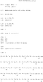
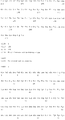
На фиг.1 приведены последовательности тяжелой и легкой цепи вариабельного участка для взятого в качестве неограничивающего примера анти-IL-12 антитела (АВТ-847).Figure 1 shows the sequences of the heavy and light chains of the variable region for taken as a non-limiting example of anti-IL-12 antibodies (ABT-847).
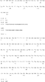
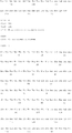
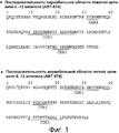
На фиг.2 приведены последовательности тяжелой и легкой цепи для взятого в качестве неограничивающего примера анти-IL-18 антитела (АВТ-325).Figure 2 shows the sequences of the heavy and light chains for taken as a non-limiting example of an anti-IL-18 antibody (ABT-325).
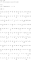
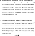
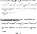
На фиг.3 приведены последовательности тяжелой и легкой цепи для взятого в качестве неограничивающего примера анти-TNFα антитела (АВТ-847) (Adalimumab).Figure 3 shows the sequences of the heavy and light chains for taken as a non-limiting example of an anti-TNFα antibody (ABT-847) (Adalimumab).
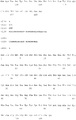
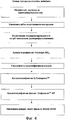
На фиг.4 приведена, в варианте неограничивающего примера, схема очистки по настоящему изобретению.Figure 4 shows, in a variant of a non-limiting example, the cleaning scheme of the present invention.
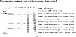
На фиг.5 показана фотография, иллюстрирующая электрофорез в полиакриламидном геле, где видно, что подлежащая очистке молекула антитела остается в растворе при снижении рН в осветленной культуральной среде.5 is a photograph illustrating polyacrylamide gel electrophoresis, where it can be seen that the antibody molecule to be purified remains in solution as the pH decreases in the clarified culture medium.
ПОДРОБНОЕ ОПИСАНИЕ ИЗОБРЕТЕНИЯDETAILED DESCRIPTION OF THE INVENTION
Настоящее изобретение относится к способам выделения и очистки антител из матричного образца. Один аспект настоящего изобретения относится к вирусной инактивации в образцах, полученных на различных стадиях очистки антитела. В конкретном аспекте, описываемые в настоящем изобретении способы включают стадию кислотной инактивации, за которой следует одна или несколько стадий хроматографии. Cтадии хроматографии могут включать одну или несколько приведенных ниже хроматографических процедур: ионообменную хроматографию, аффинную хроматографию и хроматографию гидрофобного взаимодействия. Кроме того, настоящее изобретение относится к фармацевтическим композициям, содержащим одно или несколько антител, полученных и очищенных способом по настоящему изобретению.The present invention relates to methods for the isolation and purification of antibodies from a matrix sample. One aspect of the present invention relates to viral inactivation in samples obtained at various stages of antibody purification. In a specific aspect, the methods described herein include an acid inactivation step followed by one or more chromatography steps. Chromatography steps may include one or more of the following chromatographic procedures: ion exchange chromatography, affinity chromatography, and hydrophobic interaction chromatography. In addition, the present invention relates to pharmaceutical compositions containing one or more antibodies obtained and purified by the method of the present invention.
Для ясности, но не с целью ограничения, приведенное ниже подробное описание будет разделено на несколько подразделов:For clarity, but not for the purpose of limitation, the detailed description below will be divided into several subsections:
1. Определения;1. Definitions;
2. Образование антитела;2. The formation of antibodies;
3. Продукция антитела;3. Production of antibodies;
4. Очистка антитела;4. Purification of antibodies;
5. Способы оценки чистоты образца;5. Methods for assessing sample purity;
6. Дополнительные модификации;6. Additional modifications;
7. Фармацевтические композиции; и7. Pharmaceutical compositions; and
8. Применение антител.8. The use of antibodies.
1. Определения1. Definitions
Для лучшего пояснения настоящего изобретения ниже приводятся определения некоторых используемых в нем терминов.For a better explanation of the present invention, the following are definitions of some of the terms used therein.
Термин «антитело» включает молекулу иммуноглобулина, состоящую из четырех полипептидных цепей, двух тяжелых (Н) и двух легких (L) цепей, связанных дисульфидными связями. Каждая тяжелая цепь состоит из вариабельной области тяжелой цепи (сокращенно обозначаемой в описании как HCVR или VH) и константной области тяжелой цепи (СН). Константная область тяжелой цепи состоит из трех доменов, CH1, СН2 и СН3. Каждая легкая цепь состоит из вариабельной области легкой цепи (сокращенно обозначаемой в описании как LCVR или VL) и константной области легкой цепи. Константная область легкой цепи состоит из одного домена, CL. VH и VL области, в свою очередь, могут быть разделены на области гипервариабельности, так называемые определяющие комплементарность области (CDR), между которыми располагаются более консервативные зоны, известные как каркасные области (FR). Каждая из областей VH и VL состоит из трех CDR и четырех FR, сгруппированных в следующем порядке, начиная от амино-конца к карбоксильному концу: FR1, CDR1, FR2, CDR2, FR3, CDR3, FR4.The term “antibody” includes an immunoglobulin molecule consisting of four polypeptide chains, two heavy (H) and two light (L) chains linked by disulfide bonds. Each heavy chain consists of a variable region of the heavy chain (abbreviated as HCVR or VH in the description) and a constant region of the heavy chain (CH). The constant region of the heavy chain consists of three domains, CH1, CH2 and CH3. Each light chain consists of a variable region of the light chain (abbreviated as LCVR or VL in the description) and a constant region of the light chain. The constant region of the light chain consists of one domain, CL. The VH and VL regions, in turn, can be divided into hypervariability regions, the so-called complementarity determining regions (CDRs), between which more conservative zones, known as framework regions (FR), are located. Each of the VH and VL regions consists of three CDRs and four FRs, grouped in the following order, starting from the amino end to the carboxyl end: FR1, CDR1, FR2, CDR2, FR3, CDR3, FR4.
Термин «антиген-связывающая часть» антитела (или просто «часть антитела») обозначает фрагмент антитела, который сохраняет способность специфически связываться с антигеном (например, hIL-12, hTNFa или hIL-18). Было показано, что антиген-связывающая функция антитела может осуществляться фрагментами антитела полной длины. Примеры связывающихся фрагментов, охватываемых термином «антиген-связывающая часть», включают (i) Fab фрагмент, одновалентный фрагмент, включающий VL, VH, CL и CH1 домены; (ii) F(ab')2, фрагмент, двухвалентный фрагмент, включающий два Fab фрагмента, соединенных дисульфидной связью в шарнирной области; (iii) Fd фрагмент, включающий VH и CH1 домены; (iv) Fv фрагмент, включающий VL и VH домены одного плеча антитела; (v) dAb фрагмент (Ward et al., (1989) Nature 341:544-546, где полное описание данной работы включено в настоящую заявку в качестве ссылки), который включает VH домен; и (vi) выделенная определяющая комплементарность область (CDR). Кроме того, несмотря на то, что два домена Fv фрагмента, VL и VH, кодируются разными генами, они могут быть соединены в рамках рекомбинантных методов через синтетический линкер, который позволяет получать из них одну белковую цепь, в которой VL и VH области спариваются с образованием одновалентных молекул (известных как одноцепочечной Fv (scFv); см. также. Bird et al. (1988) Science 242:423-426; и Huston et al. (1988) Proc. Natl. Acad. Sci. USA 85:5879-5883, полное описание которых включено в настоящую работу в качестве ссылки). Такие одноцепочечные молекулы также охватываются термином «антиген-связывающая часть» антитела. Другие формы одноцепочечных антител, такие как диатела, также охватываются настоящим термином. Диатела представляют собой двухвалентные, биспецифические антитела, в которых VH и VL домены экспрессируются на одной полипептидной цепи, но с использованием линкера, который слишком короток для того, чтобы обеспечить спаривание между двумя доменами на одной цепи, что заставляет домены спариваться с комплементарными доменами на другой цепи с образованием двух антиген-связывающих сайтов (см., например, Holliger, P., et al. (1993) Proc. Natl. Acad. Sci. USA 90:6444-6448; Poljak, R. J., et al. (1994) Structure 2:1121-1123, полное описание которых включено в настоящую работу в качестве ссылки). Кроме того, антитело или его антиген-связывающая часть может представлять собой часть более крупной молекулы иммуноадгезии, образуемой за счет ковалентной или нековалентной ассоциации антитела или части антитела с одним или несколькими другими белками или пептидами. Примеры использования таких молекул иммуноадгезии включают создание тетрамерной scFv молекулы с помощью ядерного участка стрептавидина (Kipriyanov, S. M., et al. (1995) Human Antibodies and Hybridomas 6:93-101, где полное описание данной работы включено в настоящую заявку в качестве ссылки) и использование цистеинового остатка, маркерного пептида и C-концевой полигистидиновой метки для создания бивалентных и биотинилированных scFv молекул (Kipriyanov, S. M., et al. (1994) Mol. Immunol. 31:1047-1058, где полное описание данной работы включено в настоящую заявку в качестве ссылки). Части антител, такие как Fab и F(ab')2, могут быть получены из полных антител с использованием стандартных методик, таких как расщепление полноразмерных антител папаином или пепсином, соответственно. Кроме того, антитела, части антител и молекулы иммуноадгезии могут быть получены с использованием стандартных методик рекомбинантных ДНК, приведенных в настоящем описании. В одном аспекте, антиген-связывающие части представляют собой полные домены или пары полных доменов.The term “antigen-binding part” of an antibody (or simply “part of an antibody”) refers to an antibody fragment that retains the ability to specifically bind to an antigen (eg, hIL-12, hTNFa or hIL-18). It has been shown that the antigen-binding function of an antibody can be carried out by full-length antibody fragments. Examples of binding fragments encompassed by the term “antigen-binding portion” include (i) a Fab fragment, a monovalent fragment comprising the VL, VH, CL and CH1 domains; (ii) F (ab ') 2 fragment, divalent fragment comprising two Fab fragments connected by a disulfide bond in the hinge region; (iii) an Fd fragment comprising the VH and CH1 domains; (iv) an Fv fragment comprising the VL and VH domains of one arm of an antibody; (v) dAb fragment (Ward et al., (1989) Nature 341: 544-546, where a full description of this work is incorporated into this application by reference), which includes a VH domain; and (vi) a designated complementarity determining region (CDR). In addition, despite the fact that the two domains of the Fv fragment, VL and VH, are encoded by different genes, they can be connected using recombinant methods via a synthetic linker, which allows one protein chain to be obtained from them, in which the VL and VH regions pair with the formation of monovalent molecules (known as single chain Fv (scFv); see also.Bird et al. (1988) Science 242: 423-426; and Huston et al. (1988) Proc. Natl. Acad. Sci. USA 85: 5879-5883, the full description of which is incorporated herein by reference). Such single chain molecules are also encompassed by the term “antigen-binding portion” of an antibody. Other forms of single chain antibodies, such as diabodies, are also encompassed by this term. Diatels are bivalent, bispecific antibodies in which the VH and VL domains are expressed on the same polypeptide chain, but using a linker that is too short to allow pairing between two domains on one chain, causing the domains to mate with complementary domains on the other chains with the formation of two antigen-binding sites (see, for example, Holliger, P., et al. (1993) Proc. Natl. Acad. Sci. USA 90: 6444-6448; Poljak, R. J., et al. (1994) Structure 2: 1121-1123, the full description of which is incorporated into this work by reference). In addition, the antibody or antigen binding portion thereof may be part of a larger immunoadhesion molecule formed by covalent or non-covalent association of the antibody or part of the antibody with one or more other proteins or peptides. Examples of the use of such immunoadhesion molecules include the creation of a tetrameric scFv molecule using the streptavidin nuclear site (Kipriyanov, SM, et al. (1995) Human Antibodies and Hybridomas 6: 93-101, where a full description of this work is incorporated herein by reference) and the use of a cysteine residue, a marker peptide and a C-terminal polyhistidine tag to create bivalent and biotinylated scFv molecules (Kipriyanov, SM, et al. (1994) Mol. Immunol. 31: 1047-1058, where a full description of this work is included in the present application in as a link). Parts of antibodies, such as Fab and F (ab ') 2, can be obtained from complete antibodies using standard techniques, such as cleavage of full-sized antibodies with papain or pepsin, respectively. In addition, antibodies, parts of antibodies, and immunoadhesion molecules can be obtained using standard recombinant DNA techniques described herein. In one aspect, the antigen binding parts are complete domains or pairs of complete domains.
Фраза «человеческий интерлейкин 12» (сокращенно обозначаемый как hIL-12 или IL-12) в контексте настоящего описания обозначает человеческий цитокин, который секретируется преимущественно макрофагами и дендритными клетками. Данный термин включает гетеродимерный белок, содержащий субъединицу размером 35 кДа (p35) и субъединицу размером 40 кДа (p40), которые соединены через дисульфидный мостик. Указанный гетеродимерный белок обозначается в настоящем описании как «субъединица р70». Структура человеческого IL-12 описана также в других работах, например, Kobayashi, et al. (1989) J. Exp Med. 170:827-845; Seder, et al. (1993) Proc. Natl. Acad. Sci. 90:10188-10192; Ling, et al. (1995) J. Exp Med. 154:116-127; Podlaski, et al. (1992) Arch. Biochem. Biophys. 294:230-237, полное описание которых включено в настоящую работу в качестве ссылки. Нуклеиновая кислота, кодирующая IL-12, доступна от GenBank с номером доступа No. NM_000882 и полипептидная последовательность доступна от GenBank с номером доступа No. NP_000873.2. Термин человеческий IL-12 в контексте настоящего описания включает рекомбинантный человеческий IL-12 (rh IL-12), который может быть получен стандартными методами рекомбинантной экспрессии.The phrase "human interleukin 12" (abbreviated as hIL-12 or IL-12) in the context of the present description refers to a human cytokine that is secreted mainly by macrophages and dendritic cells. The term includes a heterodimeric protein containing a subunit of 35 kDa (p35) and a subunit of 40 kDa (p40), which are connected through a disulfide bridge. Said heterodimeric protein is referred to herein as the "p70 subunit". The structure of human IL-12 is also described in other works, for example, Kobayashi, et al. (1989) J. Exp Med. 170: 827-845; Seder, et al. (1993) Proc. Natl. Acad. Sci. 90: 10188-10192; Ling, et al. (1995) J. Exp Med. 154: 116-127; Podlaski, et al. (1992) Arch. Biochem. Biophys. 294: 230-237, the full description of which is incorporated into this work by reference. Nucleic acid encoding IL-12 is available from GenBank with access number No. NM_000882 and the polypeptide sequence is available from GenBank with access number No. NP_000873.2. The term human IL-12 in the context of the present description includes recombinant human IL-12 (rh IL-12), which can be obtained by standard methods of recombinant expression.
Фраза «человеческий интерлейкин 18» (сокращенно обозначаемый в настоящем описании как hIL-18 или IL-18) в контексте настоящего описания обозначает человеческий цитокин, который вначале синтезируется как биологически неактивный белок-предшественник из 193 аминокислот, а также зрелый белок, состоящий из 156 аминокислот, образуемый, например, без ограничения, путем расщепления белка-предшественника, в частности, каспазой-1 или каспазой-4, которые обладает биологической активностью, включающей костимуляцию Т-клеточной пролиферации, усиление цитотоксической активности NK-клеток, индукцию образования IFN-γ Т-клетками и NK-клетками и потенцирование дифференцировки Т-хелперов типа 1 (Th1). Нуклеиновая кислота, кодирующая IL-18, доступна от GenBank с номером доступа No. NM_001562 и полипептидная последовательность доступна от GenBank с номером доступа No. NP_001553. Термин человеческий IL-18, в контексте настоящего описания, включает рекомбинантный человеческий IL-18 (rh IL-18), который может быть получен стандартными методами рекомбинантной экспрессии.The phrase "human interleukin 18" (abbreviated herein as hIL-18 or IL-18) as used herein refers to a human cytokine that is first synthesized as a biologically inactive precursor protein of 193 amino acids, as well as a mature protein of 156 amino acids formed, for example, without limitation, by cleaving a protein precursor, in particular, caspase-1 or caspase-4, which has biological activity, including co-stimulation of T-cell proliferation, increased cyt -classical activity of NK-cells, induction of the formation of IFN-γ by T-cells and NK-cells, and the potentiation of differentiation of T helper type 1 (Th1). Nucleic acid encoding IL-18 is available from GenBank with access number No. NM_001562 and the polypeptide sequence is available from GenBank with access number No. NP_001553. The term human IL-18, in the context of the present description, includes recombinant human IL-18 (rh IL-18), which can be obtained by standard methods of recombinant expression.
Фраза «человеческий фактор-α некроза опухолевых клеток» (обозначаемый в настоящем описании как hTNFα или TNFα) представляет собой полифункциональный провоспалительный цитокин, секретируемый преимущественно моноцитами/макрофагами, который оказывает эффект на липидный метаболизм, коагуляцию, инсулинорезистентность и функцию эндотелия. TNFα предсталяет собой растворимый гомотример, состоящий из белковых субъединиц размером 17 кДа. Существует также связанная с мембраной форма предшественника TNFα размером 26 кДа. Она была обнаружена в синовиальных клетках и макрофагах в тканях. Клетки, отличные от моноцитов или макрофагов, также продуцируют TNFα. Так, например, человеческие линии опухолевых клеток не моноцитарного типа продуцируют TNFα, а также CD4+ и CD8+ Т лимфоциты периферической крови и некоторые культуры Т- и В-клеточных линий продуцируют TNFα. Нуклеиновая кислота, кодирующая TNFα, доступна от GenBank с номером доступа No. X02910 и полипептидная последовательность доступна от GenBank с номером доступа No. CAA26669. Термин человеческий TNFα в контексте настоящего описания включает рекомбинантный человеческий TNFα (rh TNFa), который может быть получен стандартными методами рекомбинантной экспрессии.The phrase “human tumor cell necrosis factor-α” (referred to herein as hTNFα or TNFα) is a polyfunctional pro-inflammatory cytokine secreted primarily by monocytes / macrophages that has an effect on lipid metabolism, coagulation, insulin resistance and endothelial function. TNFα is a soluble homotrimer consisting of 17 kDa protein subunits. There is also a membrane-bound form of TNFα precursor of 26 kDa. It has been found in synovial cells and macrophages in tissues. Cells other than monocytes or macrophages also produce TNFα. For example, non-monocytic-type human tumor cell lines produce TNFα, as well as peripheral blood CD4 + and CD8 + T lymphocytes and some cultures of T- and B-cell lines produce TNFα. Nucleic acid encoding TNFα is available from GenBank with access number No. X02910 and the polypeptide sequence is available from GenBank with access number No. CAA26669. The term human TNFα in the context of the present description includes recombinant human TNFα (rh TNFa), which can be obtained by standard methods of recombinant expression.
Термины «нумерация по Кабату», «определение по Кабату» и «обозначение по Кабату» используются в настоящем описании взаимозаменяемо. Данные термины, известные в настоящей области, относятся в системе нумерации аминокислотных остатков, которые являются более вариабельными (в частности, гипервариабельными), чем другие аминокислотные остатки в вариабельных областях тяжелой и легкой цепи антитела или его антиген-связывающей части (Kabat et al. (1971) Ann. NY Acad, Sci. 190:382-391 and, Kabat, E. A., et al. (1991) Sequences of Proteins of Immunological Interest, Fifth Edition, U.S. Department of Health and Human Services, NIH Publication No. 91-3242, где полное описание данной работы включено в настоящую заявку в качестве ссылки). В случае вариабельной области тяжелой цепи, гипервариабельная область простирается на участке аминокислот 31-35 для CDR1, на участке аминокислот 50-65 для CDR2 и на участке аминокислот 95-102 для CDR3. В случае вариабельной области легкой цепи, гипервариабельная область располагается на участке аминокислот 24-34 в CDR1, на участке аминокислот 50-56 в CDR2 и на участке аминокислот 89-97 в CDR3.The terms “Kabat numbering”, “Kabat definition” and “Kabat designation” are used interchangeably herein. These terms, known in this field, refer to the numbering system of amino acid residues that are more variable (in particular, hypervariable) than other amino acid residues in the variable regions of the heavy and light chains of the antibody or its antigen-binding part (Kabat et al. ( 1971) Ann. NY Acad, Sci. 190: 382-391 and, Kabat, EA, et al. (1991) Sequences of Proteins of Immunological Interest, Fifth Edition, US Department of Health and Human Services, NIH Publication No. 91- 3242, where a full description of this work is incorporated into this application by reference). In the case of the variable region of the heavy chain, the hypervariable region extends in the amino acid region 31-35 for CDR1, in the amino acid region 50-65 for CDR2 and in the amino acid region 95-102 for CDR3. In the case of the variable region of the light chain, the hypervariable region is located on the site of amino acids 24-34 in CDR1, on the site of amino acids 50-56 in CDR2 and on the site of amino acids 89-97 in CDR3.
Термин «человеческое антитело» включает антитела, содержащие вариабельные и константные области, которые соответствуют последовательностям человеческих зародышевых иммуноглобулинов, как это описано в работе Kabat et al. (см. Kabat, et al. (1991) Sequences of proteins of Immunological Interest, Fifth Edition, U.S. Department of Health and Human Services, NIH Publication No. 91-3242). Человеческие антитела по настоящему изобретению могут включать аминокислотные остатки, которые не кодируются последовательностями иммуноглобулина человеческой зародышевой линии (т.е. имеются мутации, введенные путем случайного или сайт-специфического мутагенеза in vitro или за счет соматической мутации in vivo), например, в CDR и в перинуклеарных CDR3. Указанные мутации могут быть введены с использованием «стратегии селективного мутагенеза». Человеческое антитело может иметь, по меньшей мере, одно положение, замещенное аминокислотным остатком, например, аминокислотным остатком, повышающим активность, где указанный остаток не кодируется последовательностью иммуноглобулина зародышевой линии человека. Человеческое антитело может содержать до двадцати положений, замещенных аминокислотными остатками, которые не являются частью последовательности иммуноглобулина человеческой зародышевой линии. В других вариантах, может быть замещено до десяти, до пяти, до трех или до двух положений. В одном варианте, указанные замещения осуществляются в пределах областей CDR. Однако, термин «человеческое антитело» в контексте настоящего описания не включает те антитела, в которых CDR последовательности, полученные из зародышевой линии других материнского вида млекопитающего, такого как мышь, были перенесены на каркасные последовательности человеческой молекулы.The term “human antibody” includes antibodies containing variable and constant regions that correspond to sequences of human germline immunoglobulins, as described by Kabat et al. (see Kabat, et al. (1991) Sequences of proteins of Immunological Interest, Fifth Edition, U.S. Department of Health and Human Services, NIH Publication No. 91-3242). The human antibodies of the present invention may include amino acid residues that are not encoded by human germline immunoglobulin sequences (i.e., there are mutations introduced by random or site-specific mutagenesis in vitro or by somatic mutation in vivo), for example, in CDR and in perinuclear CDR3. These mutations can be introduced using a "selective mutagenesis strategy." A human antibody may have at least one position substituted by an amino acid residue, for example, an activity-enhancing amino acid residue, wherein said residue is not encoded by a human germline immunoglobulin sequence. A human antibody may contain up to twenty positions substituted by amino acid residues that are not part of the human germline immunoglobulin sequence. In other embodiments, up to ten, up to five, up to three, or up to two positions may be substituted. In one embodiment, said substitutions occur within the CDR regions. However, the term "human antibody" in the context of the present description does not include those antibodies in which CDR sequences obtained from the germ line of other maternal mammalian species, such as mice, were transferred to the frame sequences of a human molecule.
Фраза «стратегия селективного мутагенеза» включает способ повышения активности антитела за счет выбора и введения мутаций в отдельные аминокислоты в области CDR, по меньшей мере, в одном положении, подходящем для селективного мутагенеза, гипермутации, и/или в контактное положение. «Селективно мутированное» человеческое антитело представляет собой антитело, которое включает мутацию в положении, выбранном с использованием стратегии селективного мутагенеза. В другом аспекте, стратегия селективного мутагенеза представляет собой способ предпочтительного мутирования выбранных индивидуальных аминокислотных остатков в CDR1, CDR2 или CDR3 в вариабельной области тяжелой цепи (далее обозначаемых как H1, H2 и H3, соответственно) или в CDR1, CDR2 или CDR3 в вариабельной области легкой цепи (далее обозначаемых как L1, L2 и L3, соответственно) антитела. Аминокислотные остатки могут быть выбраны из подходящих для селективного мутагенеза положений, контактных положений или гипермутированных положений. Индивидуальные аминокислоты выбирают на основании их положения в вариабельной области легкой или тяжелой цепи. Следует понимать, что положение гипермутации может также представлять собой контактное положение. В одном аспекте, стратегия селективного мутагенеза представляет собой «направленную стратегию». Термин «направленная стратегия» включает способ мутации индивидуальных аминокислотных остатков в CDR1, CDR2 или CDR3 в вариабельной области тяжелой цепи или в CDR1, CDR2 или CDR3 в вариабельной области легкой цепи антитела заданным образом, т.е. представляет собой «стратегию групповой направленности» или «стратегию CDR-направленности». В рамках «стратегии групповой направленности» индивидуальные аминокислотные остатки в определенных группах являются мишенью для селективных мутаций, включающих группы 1 (включая L3 и H3), II (включая H2 и L1) и III (включая L2 и H1), где указанные группы были перечислены в порядке их предпочтительности для воздействия на них в качестве мишеней. В рамках «стратегии CDR-направленности», индивидуальные аминокислотные остатки в определенных CDR являются мишенью для селективных мутаций, где порядке их предпочтительности в качестве мишеней описан ниже: H3, L3, H2, L1, H1 и L2. В выбранные аминокислотные остатки вводят мутации, например, по меньшей мере, до двух других аминокислотных остатков, и определяют эффект созданной мутации на активность антитела. Активность оценивают как изменение в специфичности/аффинности связывания антитела и/или в нейтрализующей способности антитела. Следует понимать, что стратегия селективного мутагенеза может использоваться для оптимизации любого антитела, полученного из любого источника, включая фаговый дисплей, трансгенных животных с генами зародышевых линий человеческого IgG, человеческие антитела, выделенные из человеческих B-клеток. Стратегия селективного мутагенеза может использоваться применительно к антителам, которые не могут быть оптимизированы в достаточной мере с использованием методики фагового дисплея. Следует понимать, что антитела из любого источника, включая фаговый дисплей, трансгенных животных с генами зародышевой линии человеческого IgG, человеческие антитела, выделенные из B-клеток, могут быть подвергнуты обратной мутации (back-мутации) до или после осуществления стратегии селективного мутагенеза.The phrase “selective mutagenesis strategy” includes a method for increasing antibody activity by selecting and introducing mutations into individual amino acids in the CDR region in at least one position suitable for selective mutagenesis, hypermutation, and / or in a contact position. A “selectively mutated” human antibody is an antibody that comprises a mutation at a position selected using a selective mutagenesis strategy. In another aspect, a selective mutagenesis strategy is a method for preferentially mutating selected individual amino acid residues in CDR1, CDR2 or CDR3 in the variable region of the heavy chain (hereinafter referred to as H1, H2 and H3, respectively) or in CDR1, CDR2 or CDR3 in the variable region of the light chains (hereinafter referred to as L1, L2 and L3, respectively) of an antibody. Amino acid residues can be selected from positions suitable for selective mutagenesis, contact positions or hypermutated positions. Individual amino acids are selected based on their position in the variable region of the light or heavy chain. It should be understood that the position of hypermutation may also be a contact position. In one aspect, a selective mutagenesis strategy is a “directional strategy”. The term “directed strategy” includes a method for mutating individual amino acid residues in CDR1, CDR2 or CDR3 in the variable region of the heavy chain or in CDR1, CDR2 or CDR3 in the variable region of the antibody light chain in a predetermined manner, i.e. represents a “group focus strategy” or “CDR focus strategy”. As part of the “grouping strategy,” individual amino acid residues in specific groups are targeted for selective mutations, including groups 1 (including L3 and H3), II (including H2 and L1) and III (including L2 and H1), where these groups were listed in order of preference for targeting them. As part of the “CDR targeting strategy," individual amino acid residues in specific CDRs are targeted for selective mutations, where the order of their preference as targets is described below: H3, L3, H2, L1, H1 and L2. Mutations are introduced into the selected amino acid residues, for example, to at least two other amino acid residues, and the effect of the created mutation on the antibody activity is determined. Activity is evaluated as a change in the specificity / affinity of antibody binding and / or in the neutralizing ability of the antibody. It should be understood that a selective mutagenesis strategy can be used to optimize any antibody obtained from any source, including phage display, transgenic animals with human IgG germline genes, human antibodies isolated from human B cells. A selective mutagenesis strategy can be used for antibodies that cannot be optimized sufficiently using phage display techniques. It should be understood that antibodies from any source, including phage display, transgenic animals with germline genes of human IgG, human antibodies isolated from B cells, can be subjected to reverse mutation (back mutation) before or after the implementation of the strategy of selective mutagenesis.
Фраза «рекомбинантное человеческое антитело» включает человеческие антитела, которые получают, экспрессируют, создают или выделяют методами рекомбинантной технологии, такие как антитела, экспрессированные с использованием вектора рекомбинантной экспрессии, трансфицированного в клетку-хозяин, антитела, выделенные из библиотеки рекомбинантных, комбинаторных человеческих антител, антитела, выделенные из животного (например, мыши), которые являются трансгенными для генов человеческого иммуноглобулина (см., например, Taylor, L. D., et al. (1992) Nucl. Acids Res. 20:6287-6295, где полное описание работы включено в настоящую заявку в качестве ссылки), или антитела, полученные, экспрессированные, созданные или выделенные с использованием любых других способов, которые включают сплайсинг последовательностей гена человеческого иммуноглобулина с образованием других последовательностей ДНК. Такие рекомбинантные человеческие антитела содержат вариабельные и константные области, полученные из последовательностей зародышевой линии человеческого иммуноглобулина (см., Kabat, E. A., et al. (1991) Sequences of Proteins of Immunological Interest, Fifth Edition, U.S. Department of Health and Human Services, NIH Publication No. 91-3242). В некоторых вариантах осуществления настоящего изобретения, такие рекомбинантные человеческие антитела подвергают мутагенезу in vitro (или, в том случае, когда используют последовательности животного, трансгенные для последовательностей человеческого Ig, соматическому мутагенезу in vivo) и, в этой связи, аминокислотные последовательности VH и VL областей рекомбинантных антител представляют собой последовательности, которые, будучи полученными из зародышевой линии человеческих VH и VL или связанными с ними, могут в природных условиях не встречаться в репертуаре зародышевой линии человеческих антител in vivo. В некоторых вариантах осуществления настоящего изобретения, такие рекомбинантные антитела представляют собой результат селективного мутагенеза или back-мутации (обратной мутации) или обеих этих стратегий.The phrase “recombinant human antibody” includes human antibodies that are obtained, expressed, created or isolated by recombinant techniques, such as antibodies expressed using a recombinant expression vector transfected into a host cell, antibodies isolated from a library of recombinant, combinatorial human antibodies, antibodies isolated from animals (e.g., mice) that are transgenic for human immunoglobulin genes (see, for example, Taylor, LD, et al. (1992) Nucl. Acids Res. 20: 6287-6295, where a full description of the work is incorporated herein by reference), or antibodies obtained, expressed, created or isolated using any other methods that include splicing human immunoglobulin gene sequences to form other DNA sequences . Such recombinant human antibodies contain variable and constant regions derived from germline sequences of human immunoglobulin (see Kabat, EA, et al. (1991) Sequences of Proteins of Immunological Interest, Fifth Edition, US Department of Health and Human Services, NIH Publication No. 91-3242). In some embodiments of the present invention, such recombinant human antibodies are subjected to in vitro mutagenesis (or, in the case where animal sequences transgenic for human Ig sequences are used, somatic mutagenesis in vivo) and, therefore, amino acid sequences of the VH and VL regions recombinant antibodies are sequences that, if obtained from the germ line of human VH and VL or associated with them, may not naturally occur take part in the in vivo germline repertoire of human antibodies. In some embodiments of the present invention, such recombinant antibodies are the result of selective mutagenesis or back mutation (reverse mutation), or both of these strategies.
Термин «выделенное антитело» обозначает антитело, которое по существу не содержит других антител, обладающих другими антигенными специфичностями (например, выделенное антитело, которое специфически связывается с hIL-12, по существу не содержит антител, которые специфически связываются с антигенами, отличными от hIL-12). Выделенное антитело, которое специфически связывается с hIL-12, может связываться с молекулами IL-12 из другого вида. Кроме того, выделенное антитело может по существу не содержать другого клеточного материала и/или других химических веществ. Подходящие анти-IL-12, которые могут быть получены и очищены в рамках настоящего изобретения, описаны в патенте США No. 6914128 (который включен в настоящее описание полностью в качестве ссылки), где указанные антитела включают, без ограничения, анти-IL-12 антитело, которое было описано в указанном патенте как J695 и которое впоследствии было идентифицировано как ABT-874. Подходящие анти-IL-18 антитела, которые могут быть получены, очищены и выделены в рамках настоящего изобретения, описаны в патентах США NoNo. 09/780035 и 10/988360, которые включают антитело, впоследствии идентифицированное как ABT-325. Соответствующее анти-TNFα антитело представляет Адалимумаб (Adalimumab (Abbott Laboratories)).The term "isolated antibody" means an antibody that essentially does not contain other antibodies having other antigenic specificities (for example, an isolated antibody that specifically binds to hIL-12, essentially does not contain antibodies that specifically bind to antigens other than hIL- 12). An isolated antibody that specifically binds to hIL-12 can bind to IL-12 molecules from another species. In addition, the isolated antibody may essentially not contain other cellular material and / or other chemicals. Suitable anti-IL-12, which can be obtained and purified in the framework of the present invention, are described in US patent No. 6914128 (which is incorporated herein by reference in its entirety), wherein said antibodies include, without limitation, an anti-IL-12 antibody, which has been described as J695 in this patent and which has subsequently been identified as ABT-874. Suitable anti-IL-18 antibodies that can be obtained, purified and isolated in the framework of the present invention are described in US patents NoNo. 09/780035 and 10/988360, which include an antibody subsequently identified as ABT-325. The corresponding anti-TNFα antibody is Adalimumab (Adalimumab (Abbott Laboratories)).
Термин «нейтрализующее антитело» (или «антитело, которое нейтрализует активность hIL-12») обозначает антитело, которое, связываясь с hIL-12, приводит к ингибированию биологической активности hIL-12. Такое ингибирование биологической активности hIL-12 можно оценить при измерении одного или нескольких показателей биологической активности hIL-12, например, в тесте на ингибирование пролиферации человеческих бластных клеток, образующих фитогемагглютинин (PHA), или в тесте на ингибирование связывания рецептора в тесте на связывание рецептора человеческого IL-12. Указанные показатели биологической активности могут быть оценены с использованием одного или нескольких стандартных тестов in vitro или in vivo, известных в данной области.The term “neutralizing antibody” (or “an antibody that neutralizes hIL-12 activity”) means an antibody that, by binding to hIL-12, inhibits the biological activity of hIL-12. Such an inhibition of the biological activity of hIL-12 can be measured by measuring one or more indicators of the biological activity of hIL-12, for example, in a test for inhibiting the proliferation of human blast cells forming phytohemagglutinin (PHA), or in a test for inhibiting receptor binding in a receptor binding test human IL-12. These biological activity indicators can be evaluated using one or more standard in vitro or in vivo tests known in the art.
Термин «нейтрализующее антитело» (или «антитело, которое нейтрализует активность hIL-18») обозначает антитело, которое, связываясь с hIL-18, приводит к ингибированию биологической активности hIL-18. Такое ингибирование биологической активности hIL-18 можно оценить при измерении одного или нескольких показателей биологической активности hIL-18, например, по индукции образования IFNγ T-клетками или NK-клетками или по ингибированию связывания рецептора IL-18 в тесте на связывание человеческого рецептора IL-18. Указанные показатели биологической активности hIL-18 могут быть оценены с использованием одного или нескольких стандартных тестов in vitro или in vivo, известных в данной области.The term “neutralizing antibody” (or “an antibody that neutralizes hIL-18 activity”) means an antibody that, by binding to hIL-18, inhibits the biological activity of hIL-18. Such an inhibition of the biological activity of hIL-18 can be evaluated by measuring one or more indicators of the biological activity of hIL-18, for example, by inducing the formation of IFNγ by T cells or NK cells or by inhibiting the binding of IL-18 receptor in a human IL-receptor binding test eighteen. These biological activity indicators of hIL-18 can be evaluated using one or more standard in vitro or in vivo tests known in the art.
Термин «активность» обозначает активность, такую как специфичность/аффинность антитела в отношении антигена, например, анти-hIL-12 антитела, которое связывается с антигеном IL-12, и/или обозначает нейтрализующую способность антитела, например, анти-hIL-12 антитела, которое при связывании с hIL-12, ингибирует биологическую активность hIL-12, например, ингибирует бластную пролиферацию в РНА тесте или ингибирует связывание рецептора в тесте на связывание рецептора человеческого IL-12. Термин «активность» также обозначает активность, такую как специфичность /аффинность анти-IL-18 антитела в отношении своего антигена, например, анти-hIL-18 антитела, которое связывается с антигеном IL-18, и/или обозначает нейтрализующую способность антитела, такого как анти-hIL-18 антитело, которое при связывании с антигеном hIL-18 ингибирует биологическую активность hIL-18. Термин «активность» также обозначает другие виды активности, такие как специфичность/аффинность анти-TNFα антитела в отношении своего антигена, например, анти-TNFα антитела, которое связывается с антигеном TNFα, и/или обозначает нейтрализующую способность антитела, например, анти-TNFα антитела, которое, связываясь с hTNFα, ингибирует биологическую активность hTNFα.The term “activity” means activity, such as the specificity / affinity of an antibody for an antigen, for example, an anti-hIL-12 antibody, that binds to an IL-12 antigen, and / or denotes a neutralizing ability of an antibody, for example, an anti-hIL-12 antibody which, when bound to hIL-12, inhibits the biological activity of hIL-12, for example, inhibits blast proliferation in a PHA test or inhibits receptor binding in a human IL-12 receptor binding test. The term “activity” also means activity, such as the specificity / affinity of an anti-IL-18 antibody for its antigen, for example, an anti-hIL-18 antibody that binds to the IL-18 antigen, and / or denotes the neutralizing ability of an antibody such as an anti-hIL-18 antibody that, when bound to the hIL-18 antigen, inhibits the biological activity of hIL-18. The term “activity” also refers to other types of activity, such as the specificity / affinity of an anti-TNFα antibody for its antigen, for example, an anti-TNFα antibody that binds to the TNFα antigen, and / or denotes a neutralizing ability of an antibody, for example, anti-TNFα an antibody that, by binding to hTNFα, inhibits the biological activity of hTNFα.
Фраза «поверхностный плазмонный резонанс» обозначает оптическое явление, которое позволяет проводить анализ биоспецифических взаимодействий в масштабе реального времени путем выявления изменений в концентрации белка в биосенсорной матрице, например, с использованием системы BIAcore (Pharmacia Biosensor AB, Uppsala, Sweden and Piscataway, N.J.). Более подробное объяснение дано в работах Jonsson, U., et al. (1993) Ann. Biol. Clin. 51:19-20; Jonsson, U., et al. (1991) Biotechniques 11:620-627; Johnsson, В., el al. (1995) J. Mol. Recognit. 8:125-131; и Johnnson, В., et al. (1991) Anal. Biochem. 198:268-277, полное описание которых включено в настоящую заявку.The phrase “surface plasmon resonance” means an optical phenomenon that allows real-time analysis of biospecific interactions by detecting changes in protein concentration in the biosensor matrix, for example, using the BIAcore system (Pharmacia Biosensor AB, Uppsala, Sweden and Piscataway, N.J.). A more detailed explanation is given by Jonsson, U., et al. (1993) Ann. Biol. Clin. 51: 19-20; Jonsson, U., et al. (1991) Biotechniques 11: 620-627; Johnsson, B., el al. (1995) J. Mol. Recognit. 8: 125-131; and Johnnson, B., et al. (1991) Anal. Biochem. 198: 268-277, a full description of which is included in this application.
Термин «Koff» в контексте настоящего описания обозначает скорость диссоциации антитела из комплекса антитело/антиген.The term “Koff” as used herein refers to the dissociation rate of an antibody from an antibody / antigen complex.
Термин «Kd» в контексте настоящего описания обозначает константу диссоциации для случая конкретного взаимодействия антитело-антиген.The term "Kd" in the context of the present description refers to the dissociation constant for the case of a specific antibody-antigen interaction.
Фраза «молекула нуклеиновой кислоты» обозначает молекулу ДНК и молекулу РНК. Молекула нуклеиновой кислоты может быть одноцепочечной или двухцепочечной, но в одном аспекте это двухцепочечная ДНК.The phrase "nucleic acid molecule" means a DNA molecule and an RNA molecule. The nucleic acid molecule may be single-stranded or double-stranded, but in one aspect it is double-stranded DNA.
Фраза «выделенная молекула нуклеиновой кислоты» в контексте настоящего описания относится к нуклеиновым кислотам, кодирующим антитела или части антител (например, VH, VL, CDR3), например, такие, которые связываются с hIL-12, hTNFa и hIL-18, и включают молекулу нуклеиновой кислоты, в которой нуклеотидная последовательность, кодирующая антитело или часть антитела, не содержит других нуклеотидных последовательностей, кодирующих антитела или части антител, которые связываются с антигенами, отличными от hIL-12, hTNFa или hIL-18, и где другие последовательности могут в природном состоянии фланкировать молекулу нуклеиновой кислоты в ДНК человеческого генома. Таким образом, например, выделенная нуклеиновая кислота по настоящему изобретению, кодирующая VH область анти-IL-12h, анти-TNFα или анти-hIL-18 антитела, не содержит других последовательностей, кодирующих другие VH области, которые связываются с антигенами, отличными, например, от IL-12, hTNFα или hIL-18. Фраза «выделенная молекула нуклеиновой кислоты» также обозначает последовательности, кодирующие бивалентные, биспецифические антитела, такие как антитела, в которых VH и VL области не содержат других последовательностей, отличных от последовательностей данного диатела.The phrase "isolated nucleic acid molecule" in the context of the present description refers to nucleic acids encoding antibodies or parts of antibodies (for example, VH, VL, CDR3), for example, those that bind to hIL-12, hTNFa and hIL-18, and include a nucleic acid molecule in which the nucleotide sequence encoding an antibody or part of an antibody does not contain other nucleotide sequences encoding antibodies or parts of antibodies that bind to antigens other than hIL-12, hTNFa or hIL-18, and where other sequences could in the natural state, flank the nucleic acid molecule in the DNA of the human genome. Thus, for example, the isolated nucleic acid of the present invention encoding the VH region of anti-IL-12h, anti-TNFα or anti-hIL-18 antibody does not contain other sequences encoding other VH regions that bind to antigens other than, for example , from IL-12, hTNFα or hIL-18. The phrase "isolated nucleic acid molecule" also refers to sequences encoding bivalent, bispecific antibodies, such as antibodies in which the VH and VL regions do not contain other sequences than the sequences of this diabetic.
Фраза «рекомбинантная клетка-хозяин» (или просто «клетка-хозяин») обозначает клетку, в которую был встроен рекомбинантный вектор экспрессии. Следует понимать, что такие термины относятся не только к конкретной рассматриваемой клетке, но и к потомству такой клетки. Поскольку в последующих поколениях могут возникать определенные модификации, связанные с мутацией или влиянием окружающей среды, то такое потомство фактически может быть неидентичным исходной клетке, но оно также охватывается термином «клетка-хозяин».The phrase “recombinant host cell” (or simply “host cell”) refers to a cell into which a recombinant expression vector has been inserted. It should be understood that such terms refer not only to the particular cell in question, but also to the offspring of such a cell. Since in subsequent generations certain modifications may occur associated with a mutation or environmental influence, such offspring may actually not be identical to the original cell, but it is also covered by the term "host cell".
Термин «модификация» в контексте настоящего описания относится к изменению одной или нескольких аминокислот в антителах или их антиген-связывающих частях. Указанное изменение может быть получено за счет добавления, замещения или делеции аминокислоты в одном или нескольких положениях. Такое изменение может быть создано с использованием известных методик, таких как мутагенез в рамках ПЦР.The term "modification" in the context of the present description refers to a change in one or more amino acids in antibodies or their antigen-binding parts. The specified change can be obtained by adding, substituting or deletion of amino acids in one or more positions. Such a change can be created using known techniques, such as mutagenesis through PCR.
Термин «примерно» в контексте настоящего описания относится к диапазонам, который примерно на 10-20% больше или меньше указанного значения. В некоторых случаях, как это очевидно специалистам в данной области, в связи с природой указанного значения, термин «примерно» может означать большую или меньшую величину отклонения, чем 10-20% от приведенной величины.The term "about" in the context of the present description refers to ranges that are approximately 10-20% more or less than the specified value. In some cases, as is obvious to those skilled in the art, due to the nature of the indicated value, the term “about” may mean a greater or lesser amount of deviation than 10-20% of the given value.
Фраза «снижение/инактивация вируса» в контексте настоящего описания относится к снижению числа вирусных частиц в конкретном образце («снижение»), а также к снижению активности, в частности, без ограничения, к снижению инфекционности или способности к репликации вирусных частиц в конкретном образце («инактивация»). Такое снижение числа и/или активности вирусных частиц может достигать величин от примерно 1% до примерно 99%, предпочтительно от примерно 20% до примерно 99%, более предпочтительно от примерно 30% до примерно 99%, боле предпочтительно от примерно 40% до примерно 99%, еще более предпочтительно от примерно 50% до примерно 99%, еще более предпочтительно от примерно 60% до примерно 99%, и еще более предпочтительно от примерно 70% до примерно 99%, и еще более предпочтительно от примерно 80% до 99%, и еще более предпочтительно от примерно 90% до примерно 99%. В некоторых неограничивающих вариантах, количество вируса, если он присутствует, в очищенном антителе будет меньше, чем величина ID50 (количество вируса, которое будет инфицировать 50 процентов целевой популяции) для данного вируса, предпочтительно, по меньшей мере, в 10 раз меньше, чем показатель ID50 для данного вируса, более предпочтительно, по меньшей мере, в 100 раз меньше, чем показатель ID50 для данного вируса, и еще более предпочтительно, по меньшей мере, в 1000 раз меньше, чем показатель ID50 для данного вируса.The phrase "reduction / inactivation of the virus" in the context of the present description refers to a decrease in the number of viral particles in a particular sample ("decrease"), as well as to a decrease in activity, in particular, without limitation, to a decrease in infectivity or the ability to replicate viral particles in a specific sample ("Inactivation"). Such a decrease in the number and / or activity of viral particles can reach values from about 1% to about 99%, preferably from about 20% to about 99%, more preferably from about 30% to about 99%, more preferably from about 40% to about 99%, even more preferably from about 50% to about 99%, even more preferably from about 60% to about 99%, and even more preferably from about 70% to about 99%, and even more preferably from about 80% to 99 %, and even more preferably from about 90% to about 99%. In some non-limiting embodiments, the amount of virus, if present, in the purified antibody will be less than the ID50 value (the amount of virus that will infect 50 percent of the target population) for that virus, preferably at least 10 times less than The ID50 for a given virus, more preferably at least 100 times less than the ID50 for a given virus, and even more preferably at least 1000 times less than an ID50 for a given virus.
Фраза «контактное положение» относится к положению аминокислоты в CDR1, CDR2 или CDR3 в вариабельной области тяжелой цепи или в вариабельной области легкой цепи антитела, которое занимает аминокислота, контактирующая с антигеном в одной из двадцати шести известных структур антитело-антиген. Если аминокислота CDR в любой из двадцати шести известных структур комплексов антитело-антиген контактируют с антигеном, то такая аминокислота может рассматриваться как занимающая контактное положение. Контактное положение характеризуется повышенной вероятностью быть занятым аминокислотой, которая контактирует с антигеном, чем в неконтактном положении. В одном аспекте, контактное положение представляет собой CDR положение, которое содержит аминокислоту, которая контактирует с антигеном более чем в 3 из 26 структур (>1,5%). В другом аспекте, контактное положение представляет собой CDR положение, которое содержит аминокислоту, которая контактирует с антигеном более чем в 8 из 25 структур (>32%).The phrase “contact position” refers to the position of the amino acid in CDR1, CDR2, or CDR3 in the variable region of the heavy chain or in the variable region of the light chain of an antibody that is occupied by an amino acid in contact with an antigen in one of twenty-six known antibody-antigen structures. If the CDR amino acid in any of the twenty-six known structures of the antibody-antigen complexes is in contact with the antigen, then such an amino acid can be considered as occupying a contact position. A contact position is more likely to be occupied by an amino acid that is in contact with an antigen than in a non-contact position. In one aspect, the contact position is a CDR position that contains an amino acid that contacts an antigen in more than 3 of 26 structures (> 1.5%). In another aspect, the contact position is a CDR position that contains an amino acid that contacts the antigen in more than 8 out of 25 structures (> 32%).
2. Образование антител2. The formation of antibodies
Термин «антитело» в контексте настоящего описания относится к интактному антителу или к его антиген-связывающему фрагменту.The term "antibody" in the context of the present description refers to an intact antibody or antigen-binding fragment thereof.
Антитела по настоящему изобретению могут быть получены с использованием множества методик, включающих иммунизацию животного целевым антигеном с последующим получением моноклональных антител традиционным способом, например, гибридизацией соматических клеток согласно методике Колера и Мильштейна (Kohler and Milstein (1975) Nature 256: 495). Хотя некоторые процедуры гибридизации являются предпочтительными, в принципе могут использоваться другие методики для получения моноклонального антитела, например, основанные на вирусной или онкогенной трансформации В-лимфоцитов.Antibodies of the present invention can be obtained using a variety of techniques, including immunizing an animal with a target antigen and then producing monoclonal antibodies in a conventional manner, for example, hybridizing somatic cells according to Kohler and Milstein (Kohler and Milstein (1975) Nature 256: 495). Although some hybridization procedures are preferred, in principle, other techniques can be used to obtain a monoclonal antibody, for example, based on the viral or oncogenic transformation of b-lymphocytes.
Одной предпочтительной животной системой для получения гибридом является система с использованием мышей. Продукция гибридом представляет собой хорошо установленную процедуру. Протокол иммунизации и методики выделения иммунизированных спленоцитов с целью их последующего слияния известны в данной области. Компоненты для проведения слияния (например, миеломные клетки мышей) и процедуры слияния также известны.One preferred animal system for producing hybridomas is a system using mice. Hybrid production is a well established procedure. The immunization protocol and methods for isolating immunized splenocytes for subsequent fusion are known in the art. Components for fusion (e.g., myeloma cells of mice) and fusion procedures are also known.
Антитело представляет собой предпочтительно человеческое, химерное или гуманизированное антитело. Химерные или гуманизированные антитела по настоящему изобретению могут быть получены на основе последовательности отличного от человеческого моноклонального антитела, полученного по описанной выше процедуре. ДНК, кодирующая тяжелую и легкую цепь иммуноглобулинов, может быть получена на основе соответствующей гибридомы, отличной от человеческой гибридомы, с последующим проведением процедур конструирования с целью создания немышиных (например, человеческих) иммуноглобулиновых последовательностей с использованием стандартных методик молекулярной биологии. Так, например, для получения химерного антитела вариабельные области мышиной молекулы могут быть соединены с константными областями человеческой молекулы с использованием известных методик (см., например, патент США No. 4816567, Cabilly et al.). Для получения гуманизированного антитела, мышиные области CDR могут быть встроены в рамку считывания человеческий молекулы с использованием известных методик (см, например, патент США No. 5225539, Winter и патенты США NoNo. 5530101; 5585089; 5693762 и 6180370, Queen et al.).The antibody is preferably a human, chimeric or humanized antibody. Chimeric or humanized antibodies of the present invention can be obtained on the basis of a sequence different from human monoclonal antibodies obtained by the above procedure. DNA encoding the immunoglobulin heavy and light chain can be obtained from an appropriate hybridoma other than a human hybridoma, followed by design procedures to create non-murine (e.g. human) immunoglobulin sequences using standard molecular biology techniques. So, for example, to obtain a chimeric antibody, the variable regions of a mouse molecule can be coupled to the constant regions of a human molecule using known techniques (see, for example, US Pat. No. 4,816,567, Cabilly et al.). To obtain a humanized antibody, murine CDR regions can be inserted into the reading frame of a human molecule using known techniques (see, for example, US Patent No. 5225539, Winter and US Patent Nos. 5530101; 5585089; 5693762 and 6180370, Queen et al.) .
В одном неограничивающем варианте осуществления настоящего изобретения, рассматриваемые в нем антитела, представляют собой человеческие моноклональные антитела. Такие человеческие моноклональные антитела против IL-12, hTNFα или IL-18 могут быть получены с использованием трансгенных или трансхромосомных мышей, содержащих части иммунной системы человека, а не мышиной системы. Такие трансгенные и трансхромосомные мыши включают мышей, обозначенных в данном описании как HuMAb Mouse® (Medarex, Inc.), KM Mouse® (Medarex, Inc.) и XenoMouse® (Amgen).In one non-limiting embodiment of the present invention, the antibodies described therein are human monoclonal antibodies. Such human monoclonal antibodies against IL-12, hTNFα or IL-18 can be obtained using transgenic or transchromosomal mice containing parts of the human immune system, and not the mouse system. Such transgenic and transchromosomal mice include mice designated herein as HuMAb Mouse® (Medarex, Inc.), KM Mouse® (Medarex, Inc.) and XenoMouse® (Amgen).
Кроме того, в данной области доступны альтернативные трансхромосомные животные системы, экспрессирующие гены человеческого иммуноглобулина, и они могут быть использованы для получения антител по настоящему изобретению, таких как анти-IL-12, анти-TNFα или анти-IL-18 антитела. Так, например, могут использоваться мыши, содержащие как трансхромосомные молекулы с тяжелой цепью человека, так и трансхромосомные молекулы с легкой цепью человека, обозначаемые в настоящем описании как «TC мыши»; такие мыши описаны в работе Tomizuka et al. (2000) Proc. Natl. Acad. Sci. USA 97:722-727. Кроме того, были описаны коровы, содержащие трансхромосомные молекулы с тяжелой цепью и легкой цепью (например, Kuroiwa et al. (2002) Nature Biotechnology 20:889-894 и PCT заявка No. WO 2002/092812) и они могут использоваться для получения анти-IL-12, анти-TNFα или анти-IL-18 антител по настоящему описанию.In addition, alternative transchromosomal animal systems expressing human immunoglobulin genes are available in the art and can be used to produce antibodies of the present invention, such as anti-IL-12, anti-TNFα or anti-IL-18 antibodies. For example, mice containing both human heavy chain transchromosomal molecules and human light chain transchromosomal molecules, referred to herein as “mouse TCs”, may be used; such mice are described by Tomizuka et al. (2000) Proc. Natl. Acad. Sci. USA 97: 722-727. In addition, cows containing heavy chain and light chain transchromosomal molecules have been described (e.g., Kuroiwa et al. (2002) Nature Biotechnology 20: 889-894 and PCT application No. WO 2002/092812) and can be used to produce anti -IL-12, anti-TNFα or anti-IL-18 antibodies of the present description.
Рекомбинантные человеческие антитела по настоящему изобретению включают, без ограничения, анти-IL-12, анти-TNFα или анти-IL-18 антитела или их антиген-связывающую часть или антитела, родственные анти-IL-12, родственные анти-TNFα или родственные анти-IL-18 по настоящему изобретению, которые могут быть выделены при скрининге библиотеки рекомбинантных комбинаторных антител, например, библиотеки фагового дисплея scFv, полученной с использованием кДНК VL и VH человека, полученных на основе мРНК человеческих лимфоцитов. Методики получения и скрининга таких библиотек известны в данной области. Кроме коммерчески доступных наборов для получения библиотек фагового дисплея (например, Pharmacia Recombinant Phage Antibody System, No. по каталогу 27-9400-01; и набор фагового дисплея Stratagene SurfZAPXM, No. по каталогу 240612, полное описание которых включено в настоящую работу в качестве ссылки), примеры методик и реактивов, доступных для использования с целью получения и скрининга библиотек фагового дисплея, приведены, например, в Ladner et al. U.S. Patent No. 5223409; Kang et al. PCТ публикация No. WO 92/18619; Dower et al. PCТ публикация No. WO 91/17271; Winter et al. PCТ публикация No. WO 92/20791; Markland et al. PCТ публикация No. WO 92/15679; Breitling et al. PCТ публикация No. WO 93/01288; McCafferty et al. PCТ публикация No. WO 92/01047; Garrard et al. PCТ публикация No. WO 92/09690; Fuchs et al. (1991) Bio/Тechnology 9:1370-1372; Hay et al. (1992) Hum Antibod Hybridomas 3:81-85; Huse et al. (1989) Science 246:1275-1281; McCafferty et al., Nature (1990) 348:552-554; Griffiths et al. (1993) EMBO J 12:725-734; Hawkins et al. (1992) J Mol Biol 226:889-896; Clackson et al. (1991) Nature 352:624-628; Gram et al. (1992) PNAS 89:3576-3580; Garrard et al. (1991) Bio/Тechnology 9:1373-1377; Hoogenboom et al. (1991) Nuc Acid Res 19:4133-4137; и Barbas et al. (1991) PNAS 88:7978-7982; полное описание которых включено в настоящую работу в качестве ссылки.Recombinant human antibodies of the present invention include, without limitation, anti-IL-12, anti-TNFα or anti-IL-18 antibodies or their antigen-binding portion or antibodies related to anti-IL-12, related anti-TNFα or related anti -IL-18 of the present invention, which can be isolated by screening a library of recombinant combinatorial antibodies, for example, a scFv phage display library obtained using human VL and VH cDNAs derived from human lymphocyte mRNA. Techniques for obtaining and screening such libraries are known in the art. In addition to commercially available phage display library kits (e.g. Pharmacia Recombinant Phage Antibody System, catalog No. 27-9400-01; and phage display kit Stratagene SurfZAPXM, catalog No. 240612, the full description of which is included in this work as references), examples of methods and reagents available for use in the preparation and screening of phage display libraries are given, for example, in Ladner et al. U.S. Patent No. 5,223,409; Kang et al. PCT publication No. WO 92/18619; Dower et al. PCT publication No. WO 91/17271; Winter et al. PCT publication No. WO 92/20791; Markland et al. PCT publication No. WO 92/15679; Breitling et al. PCT publication No. WO 93/01288; McCafferty et al. PCT publication No. WO 92/01047; Garrard et al. PCT publication No. WO 92/09690; Fuchs et al. (1991) Bio / Technology 9: 1370-1372; Hay et al. (1992) Hum Antibod Hybridomas 3: 81-85; Huse et al. (1989) Science 246: 1275-1281; McCafferty et al., Nature (1990) 348: 552-554; Griffiths et al. (1993) EMBO J 12: 725-734; Hawkins et al. (1992) J Mol Biol 226: 889-896; Clackson et al. (1991) Nature 352: 624-628; Gram et al. (1992) PNAS 89: 3576-3580; Garrard et al. (1991) Bio / Technology 9: 1373-1377; Hoogenboom et al. (1991) Nuc Acid Res 19: 4133-4137; and Barbas et al. (1991) PNAS 88: 7978-7982; a full description of which is incorporated into this work by reference.
Человеческие моноклональные антитела по настоящему изобретению могут быть также получены с использованием SCID мышей, в которые были введены иммунные клетки человека, так что при иммунизации может быть получен ответ человеческих антител. Такие мыши описаны, например, в патентах США NoNo. 5476996 и 5698767 (Wilson et al.).The human monoclonal antibodies of the present invention can also be obtained using SCID mice into which human immune cells have been introduced, so that a human antibody response can be obtained by immunization. Such mice are described, for example, in US patents NoNo. 5,476,996 and 5,698,767 (Wilson et al.).
В некоторых вариантах, способы по настоящему изобретению включают анти-IL-12, анти-TNFα или анти-IL-18 антитела или части этих антител, антитела, родственные анти-IL-12, родственные анти-TNFα или родственные анти-IL-18, и части таких антител, а также человеческие антитела и части, эквивалентные по свойствам анти-IL-12, анти-TNFα или анти-IL-18 антителам, так что достигается высокая аффинность связывания с hIL-12, hТNFα или hIL-18 в сочетании с низкой кинетикой диссоциации и высокой нейтрализующей способностью. В одном аспекте, настоящее изобретение относится к лечению выделенным человеческим антителом или его антиген-связывающей частью, которое диссоциирует из hIL-12, hTNFα или hIL-18 с показателем Kd примерно 1×10⁻⁸ M и константой Koff, равной 1×10⁻³/сек или менее, по результатам определения обоих показателей по методу поверхностного плазмонного резонанса. В конкретных неограничивающих вариантах, анти-IL-12 антитело, полученное по настоящему изобретению, конкурентно ингибирует связывание ABT-874 с IL-12 в физиологических условиях. В конкретных неограничивающих вариантах, анти-IL-18 антитело, полученное и очищенное по настоящему изобретению, конкурентно ингибирует связывание по ABT-325 с IL-18 в физиологических условиях. В конкретных неограничивающих вариантах, анти-TNFα антитело, полученное и очищенное по настоящему изобретению, конкурентно ингибирует связывание Адалимумаба (Adalimumab) с TNFα в физиологических условиях.In some embodiments, the methods of the present invention include anti-IL-12, anti-TNFα or anti-IL-18 antibodies or portions of these antibodies, antibodies related to anti-IL-12, related anti-TNFα or related anti-IL-18 and parts of such antibodies, as well as human antibodies and parts equivalent in properties to anti-IL-12, anti-TNFα or anti-IL-18 antibodies, so that a high binding affinity for hIL-12, hTNFα or hIL-18 is achieved combined with low dissociation kinetics and high neutralizing ability. In one aspect, the present invention relates to treatment with an isolated human antibody or antigen binding portion thereof that dissociates from hIL-12, hTNFα or hIL-18 with a Kd of about 1 × 10 -8 M and a Koff constant of 1 × 10 - 3 / sec or less, based on the results of determining both indicators by the method of surface plasmon resonance. In specific non-limiting embodiments, the anti-IL-12 antibody obtained by the present invention competitively inhibits the binding of ABT-874 to IL-12 under physiological conditions. In specific non-limiting embodiments, an anti-IL-18 antibody obtained and purified by the present invention competitively inhibits ABT-325 binding to IL-18 under physiological conditions. In specific non-limiting embodiments, an anti-TNFα antibody prepared and purified by the present invention competitively inhibits the binding of Adalimumab to TNFα under physiological conditions.
В еще другом варианте осуществления настоящего изобретения, антитела или их фрагменты, такие как, без ограничения, анти-IL-12, анти-TNFα или анти-IL-18 антитела или их фрагменты, могут быть изменены, при этом константная область антитела модифицируется до ослабления по меньшей мере одной биологической эффекторной функции, определяемой константной областью, относительно немодифицированного антитела. Для модификации антитела по настоящему изобретению, так чтобы оно демонстрировало низкую способность к связыванию с Fc рецептором, в сегмент константной области иммуноглобулина данного антитела может быть введена мутация в определенных участках, необходимых для взаимодействий с Fc рецептором (FcR) (см., например, Canfield and Morrison (1991) J. Exp. Med. 173:1483-1491; и Lund et al. (1991) J. of Immunol. 147:2657-2662, полное описание работ включено в настоящее описание). Снижение FcR-связывающей аффинности для данного антитела может также приводить к снижению других эффекторных функций, определяемых FcR взаимодействиями, таких как опсонизация и фагоцитоз, а также антитело-обусловленная клеточная цитотоксичность.In yet another embodiment of the present invention, antibodies or fragments thereof, such as, without limitation, anti-IL-12, anti-TNFα or anti-IL-18 antibodies or fragments thereof, may be modified, wherein the constant region of the antibody is modified to weakening of at least one biological effector function, determined by the constant region, relative to the unmodified antibody. To modify the antibody of the present invention so that it exhibits low ability to bind to the Fc receptor, a mutation can be introduced into the constant region segment of the immunoglobulin of this antibody at certain sites necessary for interactions with the Fc receptor (FcR) (see, for example, Canfield and Morrison (1991) J. Exp. Med. 173: 1483-1491; and Lund et al. (1991) J. of Immunol. 147: 2657-2662, a full description of the works is included in the present description). A decrease in FcR binding affinity for a given antibody can also lead to a decrease in other effector functions determined by FcR interactions, such as opsonization and phagocytosis, as well as antibody-mediated cell cytotoxicity.
3. Продукция антител3. Production of antibodies
Для экспрессии антитела по настоящему изобретению, ДНК, кодирующие частичные или полноразмерные легкие и тяжелые цепи, встраивают в один или несколько векторов экспрессии, так чтобы гены были функционально связаны с контрольными последовательностями транскрипции и трансляции (см., например, патент США No. 6914128, полное описание которого включено в настоящую работу в качестве ссылки). В этом контексте, термин «функционально связанный» следует понимать как указание на то, что ген антитела лигирован в вектор, так что контрольные последовательности транскрипции и трансляции в данном векторе выполняют свою целевую функцию регуляции транскрипции и трансляции гена антитела. Вектор экспрессии и контрольные последовательности экспрессии выбирают таким образом, чтобы они были совместимы с используемой клеткой-хозяином, в которой осуществляется экспрессия. Ген легкой цепи антитела и ген тяжелой цепи антитела могут быть встроены в отдельный вектор или, что чаще, оба гена встраивают в один и тот же вектор экспрессии. Гены антитела встраивают в вектор экспрессии с использованием известных стандартных методов (таких как лигирование комплементарных сайтов рестрикции на фрагменте гена антитела и векторе или лигирование тупых концов, если нет сайтов рестрикции). Перед встраиванием антитела или связанных с антителом последовательностей легкой или тяжелой цепи, вектор экспрессии уже может содержать последовательность константной области. Так, например, один подход заключается в превращении VH и VL последовательностей анти-IL-12, анти-TNFα или анти-IL-18 антитела или родственных анти-IL-12, анти-TNFα или анти-IL-18 антител до полноразмерных генов антитела, так чтобы их можно было встроить в векторы экспрессии, которые уже будут кодировать константную область тяжелой цепи и константную область легкой цепи, соответственно, так что VH сегмент будет функционально связываться с одним или несколькими CH сегментами в векторе и VL сегмент будет функционально связываться с CL сегментом в векторе. Дополнительно или альтернативно, рекомбинантный вектор экспрессии может кодировать сигнальный пептид, облегчающий секрецию цепи антитела клетки-хозяина. Указанный ген цепи антитела может быть клонирован в векторе, так чтобы сигнальный пептид связывался в рамке считывания с амино-концом гена цепи антитела. Указанный сигнальный пептид может представлять собой сигнальный пептид иммуноглобулина или гетерологичный сигнальный пептид (например, сигнальный пептид белка не иммуноглобулиновой природы).To express the antibodies of the present invention, DNAs encoding partial or full-sized light and heavy chains are inserted into one or more expression vectors so that the genes are operably linked to transcription and translation control sequences (see, for example, US Pat. No. 6,914,128, a full description of which is incorporated into this work by reference). In this context, the term "functionally linked" should be understood as an indication that the antibody gene is ligated into a vector, so that the transcription and translation control sequences in this vector fulfill their target function of regulating the transcription and translation of the antibody gene. The expression vector and expression control sequences are selected so that they are compatible with the host cell in which expression is performed. An antibody light chain gene and an antibody heavy chain gene can be inserted into a separate vector or, more often, both genes are inserted into the same expression vector. Antibody genes are inserted into the expression vector using known standard methods (such as ligating complementary restriction sites on an antibody gene fragment and vector or ligating to blunt ends if there are no restriction sites). Before embedding the antibody or antibody or light chain sequences associated with the antibody, the expression vector may already contain a constant region sequence. So, for example, one approach is to convert the VH and VL sequences of anti-IL-12, anti-TNFα or anti-IL-18 antibodies, or related anti-IL-12, anti-TNFα or anti-IL-18 antibodies to full-sized genes antibodies, so that they can be inserted into expression vectors that will already encode the constant region of the heavy chain and the constant region of the light chain, respectively, so that the VH segment will functionally bind to one or more CH segments in the vector and the VL segment will functionally bind to CL segment in vector. Additionally or alternatively, the recombinant expression vector may encode a signal peptide that facilitates secretion of the antibody chain of the host cell. Said antibody chain gene can be cloned in a vector so that the signal peptide binds in the reading frame to the amino terminus of the antibody chain gene. The specified signal peptide may be an immunoglobulin signal peptide or a heterologous signal peptide (for example, a signal peptide of a non-immunoglobulin protein nature).
Кроме генов цепи антитела, рекомбинантный вектор экспрессии по настоящему изобретению может содержать одну или несколько регуляторных последовательностей, которые контролируют экспрессию генов цепи антитела в клетке-хозяине. Термин «регуляторная последовательность» включает промоторы, энхансеры и другие элементы контроля экспрессии (например, сигналы полиаденилирования), которые контролируют транскрипцию и трансляцию генов цепи антитела. Такие регуляторные последовательности описаны, например, в работе Goeddel, Gene Expression Technology: Methods in Enzymology 185, Academic Press, San Diego, CA (1990), полное описание которой включено в настоящую работу в качестве ссылки. Для специалистов в данной области очевидно, что конструирование вектора экспрессии, включающее выбор регуляторных последовательностей, может зависеть от таких факторов, как выбор трансформируемой клетки-хозяина, уровень экспрессии желательного белка и т.п. Подходящие регуляторные последовательности для экспрессии в клетке-хозяине млекопитающего включают вирусные элементы, которые направляют высокие уровни экспрессии белка в клетках млекопитающих, такие как промоторы и/или энхансеры, полученные из цитомегаловируса (CMV) (такой как промотор/энхансер CMV), обезьяньего вируса (SV40) (такой как промотор/энхансер SV40), аденовируса (например, крупный поздний промотор аденовируса (AdMLP)) и полиомный вирус. Дополнительное описание вирусных регуляторных элементов и их последовательностей содержится, например, в следующих работах: патент США No. 5168062 (Stinski), патент США No. 4510245 (Bell et al.) и патент США 4968615 (Schaffher et al.), полное описание которых включено в настоящую работу в качестве ссылки.In addition to the antibody chain genes, the recombinant expression vector of the present invention may contain one or more regulatory sequences that control the expression of the antibody chain genes in the host cell. The term “regulatory sequence” includes promoters, enhancers, and other expression control elements (eg, polyadenylation signals) that control the transcription and translation of antibody chain genes. Such regulatory sequences are described, for example, in Goeddel, Gene Expression Technology: Methods in
Кроме генов цепи антитела и регуляторных последовательностей, рекомбинантный вектор экспрессии по настоящему изобретению может содержать одну или несколько дополнительных последовательностей, таких как последовательность, которая регулирует репликацию вектора в клетке-хозяине (например, ориджины репликации), и/или селектируемый маркерный ген. Селектируемый маркерный ген облегчает отбор клеток-хозяев, в которую был встроен вектор (см., например, патенты США NoNo. 4399216, 4634665 и 5179017, все Axel et al., полное описание которых включено в настоящую работу в качестве ссылки). Например, в типичном случае, селектируемый маркерный ген придает устойчивость к лекарственным средствам, таким как G418, гигромицин или метотрексат, клетке-хозяину, в которую был встроен вектор. Подходящие селектируемые маркерные гены включают ген дигидрофолятредуктазы (DHFR) (при использовании в dhfr-клетках-хозяевах для селекции/амплификации с помощью метотрексата) и neo-ген (для селекции по G418).In addition to the antibody chain genes and regulatory sequences, the recombinant expression vector of the present invention may contain one or more additional sequences, such as a sequence that regulates the replication of the vector in the host cell (e.g., origin of replication), and / or a selectable marker gene. The selectable marker gene facilitates the selection of host cells into which the vector has been inserted (see, for example, U.S. Pat. 4399216, 4634665 and 5179017, all Axel et al., The full description of which is incorporated herein by reference). For example, in a typical case, a selectable marker gene confers resistance to drugs, such as G418, hygromycin or methotrexate, to the host cell into which the vector was inserted. Suitable breeding marker genes include the dihydrofolate reductase gene (DHFR) (when used in dhfr host cells for selection / amplification using methotrexate) and the neo gene (for selection according to G418).
Антитело или часть антитела по настоящему изобретению могут быть получены путем рекомбинантной экспрессии генов легкой и тяжелой цепи иммуноглобулинов. Для достижения рекомбинантной экспрессии антитела, клетку-хозяин трансфицируют одним или несколькими рекомбинантными векторами экспрессии, содержащими фрагменты ДНК, кодирующей легкую и тяжелую цепи иммуноглобулина, так что легкая и тяжелая цепи экспрессируются в клетке-хозяине и секретируются в среду, в которой проводится культивирование указанных клеток-хозяев, и из этой среды могут быть извлечены антитела. Получают стандартными методами рекомбинантных ДНК гены тяжелой и легкой цепей антитела, включают эти гены в рекомбинантные векторы экспрессии и вводят векторы в клетки-хозяева, такие как клетки, описанные в соответствующих руководствах: Sambrook, Fritsch and Maniatis (eds), Molecular Cloning; A Laboratory Manual, Second Edition, Cold Spring Harbor, N.Y., (1989), Ausubel et al. (eds.) Current Protocols in Molecular Biology, Greene Publishing Associates, (1989) и в патентах США NoNo. 4816397 и 6914128, полное описание которых включено в настоящую работу в качестве ссылки.The antibody or part of an antibody of the present invention can be obtained by recombinant expression of immunoglobulin light and heavy chain genes. To achieve recombinant expression of the antibody, the host cell is transfected with one or more recombinant expression vectors containing DNA fragments encoding the immunoglobulin light and heavy chains, so that the light and heavy chains are expressed in the host cell and secreted into the medium in which these cells are cultured -hosts, and antibodies can be extracted from this medium. Antibody heavy and light chain genes are prepared by standard recombinant DNA methods, incorporate these genes into recombinant expression vectors and introduce vectors into host cells, such as those described in the respective manuals: Sambrook, Fritsch and Maniatis (eds), Molecular Cloning; A Laboratory Manual, Second Edition, Cold Spring Harbor, N.Y., (1989), Ausubel et al. (eds.) Current Protocols in Molecular Biology, Greene Publishing Associates, (1989) and U.S. Patent Nos. 4816397 and 6914128, the full description of which is incorporated into this work by reference.
Для экспрессии легкой и тяжелой цепей, один или несколько векторов экспрессии, кодирующих тяжелую и легкую цепи, трансфицируют в клетку-хозяина с использованием стандартных процедур. Различные формы термина «трансфекция» обозначают в контексте настоящего описания множество методик, используемых обычно для введения экзогенной ДНК в прокариотическую или эукариотическую клетку-хозяина, например, методами электропорации, осаждения с использованием фосфата кальция, трансфекции с использованием DEAE-декстрана и т.п. Хотя теоретически возможно использовать антитела по настоящему изобретению как в прокариотических, так и в эукариотических клетках-хозяевах, рассматривается как приемлемая экспрессия антител в эукариотических клетках, таких как клетки-хозяева млекопитающих, поскольку такие эукариотические клетки и, в частности, клетки млекопитающих, с большей вероятностью, чем прокариотические клетки, смогут осуществить сборку и секрецию иммунологически активного антитела с определенной складчатой структурой. Было сообщение о неэффективности прокариотической экспрессии генов антитела для целей получения высоких выходов активных антител (Boss and Wood (1985) Immunology Today 6:12-13, полное описание работы включено в настоящую заявку в качестве ссылки).For expression of light and heavy chains, one or more expression vectors encoding the heavy and light chains are transfected into a host cell using standard procedures. The various forms of the term “transfection” mean in the context of the present description the many techniques commonly used to introduce exogenous DNA into a prokaryotic or eukaryotic host cell, for example, by electroporation, precipitation using calcium phosphate, transfection using DEAE-dextran and the like. Although it is theoretically possible to use the antibodies of the present invention in both prokaryotic and eukaryotic host cells, it is considered acceptable expression of antibodies in eukaryotic cells, such as mammalian host cells, since such eukaryotic cells and, in particular, mammalian cells with a greater more likely than prokaryotic cells will be able to assemble and secrete an immunologically active antibody with a certain folded structure. There has been a report of the inefficiency of prokaryotic expression of antibody genes for obtaining high yields of active antibodies (Boss and Wood (1985) Immunology Today 6: 12-13, a full description of the work is incorporated into this application by reference).
Подходящие клетки для клонирования или экспрессии ДНК в векторах по настоящему изобретению включают клетки прокариотов, дрожжей или высших эукариотов, указанные выше. Подходящие для этой цели прокариоты включают эубактерии, такие как Грам-отрицательные или Грам-положительные организмы, например, представители Enterobacteriaceae, такие как Escherichia, в частности, E. coli, Enterobacter, Erwinia, Klebsiella, Proteus, Salmonella, в частности., Salmonella typhimurium, Serratia, в частности, Serratia marcescans, и Shigella, а также Bacilli, такие как B. subtilis и B. licheniformis (например, B. licheniformis 41P, описанный в DD 266710, опубликованном 12 апреля 1989 года), Pseudomonas, такие как P. aeruginosa, и Streptomyces. Одним подходящим штаммом E. coli для клонирования в качестве хозяина является E. coli 294 (ATCC 31446), хотя применимы и другие штаммы, такие как E. coli B, E. coli X1776 (ATCC 31537) и E. coli W3110 (ATCC 27325). Эти примеры являются иллюстративными, а не ограничивающими.Suitable cells for cloning or expressing DNA in the vectors of the present invention include prokaryotic, yeast, or higher eukaryotic cells as defined above. Suitable prokaryotes for this purpose include eubacteria, such as Gram-negative or Gram-positive organisms, for example, representatives of the Enterobacteriaceae, such as Escherichia, in particular, E. coli, Enterobacter, Erwinia, Klebsiella, Proteus, Salmonella, in particular., Salmonella typhimurium, Serratia, in particular, Serratia marcescans, and Shigella, as well as Bacilli, such as B. subtilis and B. licheniformis (e.g., B. licheniformis 41P, disclosed in DD 266,710 published 12 April 1989), Pseudomonas, such as P. aeruginosa, and Streptomyces. One suitable E. coli strain for cloning as a host is E. coli 294 (ATCC 31446), although other strains such as E. coli B, E. coli X1776 (ATCC 31537) and E. coli W3110 (ATCC 27325 are applicable) ) These examples are illustrative and not restrictive.
Кроме прокариотов, эукариотические микроорганизмы, такие как нитевидные грибы или дрожжи, применимы в качестве хозяев для клонирования или экспрессии векторов, кодирующих полипептиды. Saccharomyces cerevisiae, или обычные пекарские дрожжи, представляет собой наиболее часто используемого хозяина среди низших эукариотических микроорганизмов. Однако, известно и широко доступно множество других применимых для этой цели родов, видов и штаммов, такие как Schizosaccharomyces pombe; Kluyveromyces, в качестве клеток-хозяев, такие как, например, K. lactis, K. fragilis (ATCC 12424), K. bulgaricus (ATCC 16045), K. wickeramii (ATCC 24178), K. waltii (ATCC 56500), K. drosophilarum (ATCC 36906), K. Thermotolerans и K. marxianus; yarrowia (EP 402226); Pichia pastoris (EP 183070); Candida; Trichoderma reesia (EP 244234); Neurospora crassa; Schwanniomyces, такие как Schwanniomyces occidentalis; и нитевидные грибы, такие как, например, Neurospora, Penicillium, Tolypocladium и Aspergillus, в качемстве хозяев, такие как A. nidulans and A. niger.In addition to prokaryotes, eukaryotic microorganisms, such as filamentous fungi or yeast, are useful as hosts for cloning or expression of vectors encoding polypeptides. Saccharomyces cerevisiae, or common baker's yeast, is the most commonly used host among lower eukaryotic microorganisms. However, many other genera, species, and strains useful for this purpose are known and widely available, such as Schizosaccharomyces pombe; Kluyveromyces, as host cells, such as, for example , K. lactis, K. fragilis (ATCC 12424), K. bulgaricus (ATCC 16045), K. wickeramii (ATCC 24178), K. waltii (ATCC 56500), K Drosophilarum (ATCC 36906), K. Thermotolerans and K. marxianus; yarrowia (EP 402226); Pichia pastoris (EP 183070); Candida Trichoderma reesia (EP 244234); Neurospora crassa; Schwanniomyces such as Schwanniomyces occidentalis; and filamentous fungi, such as, for example , Neurospora, Penicillium, Tolypocladium and Aspergillus, as hosts, such as A. nidulans and A. niger.
Подходящие клетки-хозяева для экспрессии гликозилированных антител получают из многоклеточных организмов. Примеры клеток беспозвоночных включают клетки растений и насекомых. Были идентифицированы различные штаммы бакуловирусов и их варианты и соответствующие пермиссивные клетки-хозяева насекомых из таких организмов-хозяев, как Spodoptera frugiperda (гусеница), Aedes aegypti (комар), Aedes albopictus (комар), Drosophila melanogaster (плодовая мушка) и Bombyx mori. В настоящее время доступно множество вирусных штаммов для проведения трансфекции, например, L-1 вариант Autographa californica NPV и Bm-5 штамм Bombyx mori NPV, и такие вирусы могут использоваться в качестве вируса по настоящему изобретению, в частности, для трансфекции клеток Spodoptera frugiperda. В качестве организмов-хозяев, могут также использоваться культуры клеток растений, таких как хлопок, кукуруза, картофель, соя, петунья, томаты и табак.Suitable host cells for the expression of glycosylated antibodies are obtained from multicellular organisms. Examples of invertebrate cells include plant and insect cells. Various strains of baculoviruses and their variants and corresponding permissive insect host cells from such host organisms as Spodoptera frugiperda (caterpillar), Aedes aegypti (mosquito), Aedes albopictus (mosquito), Drosophila melanogaster (fruit fly) and Bombyx mori have been identified. Numerous viral strains are currently available for transfection, for example, the L-1 variant of Autographa californica NPV and the Bm-5 strain of Bombyx mori NPV, and such viruses can be used as the virus of the present invention, in particular for transfection of Spodoptera frugiperda cells. As host organisms, plant cell cultures such as cotton, corn, potatoes, soy, petunia, tomatoes and tobacco can also be used.
Подходящие клетки-хозяева млекопитающих для экспрессии рекомбинантных антител по настоящему изобретению включают клетки яичника китайского хомячка (CHO клетки) (включающие dhfr-CHO клетки, описанные Urlaub and Chasin, (1980) PNAS USA 77:4216-4220, используемые в сочетании с DHFR-селектируемым маркером, например, как описано в работе Kaufman и Sharp (1982) Mol. Biol. 159:601-621, полное описание которой включено в настоящую заявку в качестве ссылки), клетки NS0 миеломы, COS клетки и SP2 клетки. В том случае, когда рекомбинантные векторы экспрессии, кодирующие гены антитела, встраивают в клетки-хозяева млекопитающих, антитела продуцируются культивируемыми клетками-хозяевами в течение периода времени, достаточного для экспрессии антитела в клетках-хозяевах или для секреции антитела в культуральную среду, где растут клетки-хозяева. Другие примеры используемых линий клеток-хозяев млекопитающих включают линию CV1 почки обезьяны, трансформированную SV40 (COS-7, ATCC CRL 1651); линию эмбриональных клеток почки человека (293 или 293 клетки, субклонированные для роста в суспензионной культуре, Graham et al., J. Gen Virol. 36:59 (1977)); клетки почки детеныша хомячка (BHK, ATCC CCL 10); клетки яичника китайского хомячка/-DHFR (CHO, Urlaub et al., Proc. Natl. Acad. Sci. USA 77:4216 (1980)); клетки Сертоли мышей (TM4, Mather, Biol. Reprod. 23:243-251 (1980)); клетки почки обезьяны (CV1 ATCC CCL 70); клетки почки африканской зеленой мартышки (VERO-76, ATCC CRL-1587); клетки цервикальной карциномы человека (HELA, ATCC CCL 2); клетки почки собаки (MDCK, ATCC CCL 34); клетки печени крысы buffalo (BRL 3А, ATCC CRL 1442); клетки легкого человека (W138, ATCC CCL 75); клетки печени человека (Hep G2, HB 8065); клетки опухоли молочной железы мышей (MMT 060562, ATCC CCL51); TR1 клетки (Mather et al., Annals N.Y. Acad. Sci. 383:44-68 (1982)); MRC 5 клетки; FS4 клетки; и клеточные линии гепатомы человека (Hep G2), полное описание которых включено в настоящую работу в качестве ссылки.Suitable mammalian host cells for expressing the recombinant antibodies of the present invention include Chinese hamster ovary cells (CHO cells) (including dhfr-CHO cells described by Urlaub and Chasin, (1980) PNAS USA 77: 4216-4220 used in combination with DHFR- a selectable marker, for example, as described by Kaufman and Sharp (1982) Mol. Biol. 159: 601-621, the entire disclosure of which is incorporated herein by reference), myeloma NS0 cells, COS cells, and SP2 cells. When recombinant expression vectors encoding antibody genes are inserted into mammalian host cells, the antibodies are produced by cultured host cells for a period of time sufficient to express the antibody in the host cells or to secrete the antibody into the culture medium where the cells grow are the owners. Other examples of mammalian host cell lines used include the monkey kidney transposed line CV1 transformed by SV40 (COS-7, ATCC CRL 1651); a human kidney embryonic cell line (293 or 293 cells subcloned for growth in suspension culture, Graham et al., J. Gen Virol. 36:59 (1977)); hamster baby kidney cells (BHK, ATCC CCL 10); Chinese hamster ovary cells / -DHFR (CHO, Urlaub et al., Proc. Natl. Acad. Sci. USA 77: 4216 (1980)); Mouse Sertoli cells (TM4, Mather, Biol. Reprod. 23: 243-251 (1980)); monkey kidney cells (CVCC ATCC CCL 70); African green monkey kidney cells (VERO-76, ATCC CRL-1587); human cervical carcinoma cells (HELA, ATCC CCL 2); dog kidney cells (MDCK, ATCC CCL 34); rat buffalo liver cells (BRL 3A, ATCC CRL 1442); human lung cells (W138, ATCC CCL 75); human liver cells (Hep G2, HB 8065); mouse breast tumor cells (MMT 060562, ATCC CCL51); TR1 cells (Mather et al., Annals N.Y. Acad. Sci. 383: 44-68 (1982)); MRC 5 cells; FS4 cells; and human hepatoma cell lines (Hep G2), the entire disclosures of which are incorporated herein by reference.
Клетки-хозяева трансформируют указанными выше векторами экспрессии или клонирования для получения антител и культивируют в традиционных питательных средах для достижения соответствующей модификации с целью индукции промоторов, отбора трансформантов или амплификации генов, кодирующих желательные последовательности.Host cells are transformed with the above expression or cloning vectors to produce antibodies and cultured in traditional culture media to achieve appropriate modification in order to induce promoters, select transformants, or amplify genes encoding the desired sequences.
Клетки-хозяева, используемые для продукции антитела, могут культивироваться в большом числе сред. Для культивирования клеток-хозяев могут использоваться коммерчески доступные среды, такие как среда Хэмса F10™ (Sigma), минимальная эссенциальная средаТМ (Minimal Essential Medium™, MEM), (Sigma), RPMI-1640 (Sigma) и среда Игла в модификации Дульбекко (Dulbecco's Modified Eagle's Medium™, DMEM, Sigma). Кроме того, в качестве культуральных сред для культивирования клеток-хозяев может использоваться любая среда из описанных в следующих работах: Ham et al., Meth. Enz. 58:44 (1979), Barnes et al., Anal. Biochem. 102:255 (1980), патенты США NoNo. 4767704; 4657866; 4927762; 4560655; или 5122469; WO 90/03430; WO 87/00195; или патент США No. Re. 30985, полное описание которых включено в настоящую заявку в качестве ссылки. В любую из этих сред, при необходимости, могут быть внесены добавки, включая гормоны и/или другие ростовые факторы (такие как инсулин, трансферрин или эпидермальный фактор роста), соли (такие как хлорид натрия, фосфат кальция, магния) буферы (такие как HEPES), нуклеотиды (такие как аденозин и тимидин), антибиотики (такие как гентамицин), микроэлементы (определяемые как неорганические соединения, обычно используемые в конечной концентрации в микромолярном диапазоне) и глюкозу или эквивалентный источник энергии. Любые другие необходимые добавки могут быть также включены в соответствующих концентрациях, известных специалистам в данной области. Условия культивирования, такие как температура, pH и т.п., не отличаются от тех, которые используют в случае выбранных для экспрессии клеток-хозяев, и которые очевидны специалистам в данной области.Host cells used to produce antibodies can be cultured in a large number of media. For the cultivation of a commercially available medium can be used by the host cells, such as the medium Ham's F10 ™ (Sigma), Minimal Essential TM Wednesday (Minimal Essential Medium ™, MEM) , (Sigma), RPMI- 1640 (Sigma), and Dulbecco in Dulbecco's modification (Dulbecco's Modified Eagle's Medium ™, DMEM, Sigma). In addition, any medium described in the following works can be used as culture media for culturing host cells: Ham et al., Meth. Enz. 58:44 (1979), Barnes et al., Anal. Biochem. 102: 255 (1980), U.S. Pat. 4,767,704; 4,657,866; 4,927,762; 4,560,655; or 5122469; WO 90/03430; WO 87/00195; or U.S. Pat. Re. 30985, the full description of which is incorporated into this application by reference. Additives may be added to any of these media, if necessary, including hormones and / or other growth factors (such as insulin, transferrin or epidermal growth factor), salts (such as sodium chloride, calcium phosphate, magnesium) buffers (such as HEPES), nucleotides (such as adenosine and thymidine), antibiotics (such as gentamicin), micronutrients (defined as inorganic compounds commonly used in the final concentration in the micromolar range) and glucose or an equivalent energy source. Any other necessary additives may also be included at appropriate concentrations known to those skilled in the art. Culturing conditions, such as temperature, pH, etc., are not different from those used in the case of host cells selected for expression, and which are obvious to those skilled in the art.
Клетки-хозяева могут также использоваться для получения частей интактных антител, таких как Fab фрагменты или scFv молекулы. Следует понимать, что вариации в указанной выше процедуре также входят в область настоящего изобретения. Например, в некоторых вариантах может быть желательно провести трансфекцию клетки-хозяина с использованием ДНК, кодирующей легкую цепь или тяжелую цепь (но не обе) антитела по настоящему изобретению. Технология рекомбинантных ДНК может также использоваться для удаления некоторых или всех ДНК, кодирующих тяжелую и легкую цепи, одну или обе, необязательную(ые) для связывания с IL-12, в частности, hIL-12, в случае анти-IL-12 антител, или где указанная ДНК необязательна для связывания с IL-18, в частности, hIL-18, в случае анти-IL-18 антител, или где указанная ДНК не является обязательной для связывания с TNFα, в частности hTNFα, в случае анти-TNFα антител. Молекулы, экспрессируемые на основе таких усеченных молекул ДНК, также охватываются определением антител по настоящему изобретению. Кроме того, могут быть получены бифункциональные антитела, в которых одна тяжелая и одна легкая цепь представляют собой антитело по настоящему изобретению, а другая тяжелая и легкая цепь являются специфичными для антигена, отличного от IL-12, TNFα или IL-18, в зависимости от специфичности антитела по настоящему изобретению, что достигается при сшивке антитела по настоящему изобретению со вторым антителом с использованием стандартных методов химической сшивки.Host cells can also be used to produce portions of intact antibodies, such as Fab fragments or scFv molecules. It should be understood that variations in the above procedure are also included in the scope of the present invention. For example, in some embodiments, it may be desirable to transfect the host cell using DNA encoding the light chain or heavy chain (but not both) of the antibody of the present invention. Recombinant DNA technology can also be used to remove some or all of the DNAs encoding the heavy and light chains, one or both, optional for binding to IL-12, in particular hIL-12, in the case of anti-IL-12 antibodies, or where said DNA is optional for binding to IL-18, in particular hIL-18, in the case of anti-IL-18 antibodies, or where said DNA is not required to bind to TNFα, in particular hTNFα, in the case of anti-TNFα antibodies . Molecules expressed on the basis of such truncated DNA molecules are also encompassed by the definition of antibodies of the present invention. In addition, bifunctional antibodies can be prepared in which one heavy and one light chain are the antibody of the present invention and the other heavy and light chain are specific for an antigen other than IL-12, TNFα or IL-18, depending on the specificity of the antibodies of the present invention, which is achieved by cross-linking the antibodies of the present invention with a second antibody using standard chemical crosslinking methods.
В системе, подходящей для рекомбинантной экспрессии антитела или его антиген-связывающей части по настоящему изобретению, рекомбинантный вектор экспрессии, кодирующий и тяжелую цепь антитела, и легкую цепь антитела, встраивают в dhfr-CHO клетки с использованием трансфекции по методу, включающему использование фосфата кальция. В рекомбинантном векторе экспрессии, гены тяжелой и легкой цепи антитела функционально связаны с CMV энхансерными/AdMLP промоторными регуляторными элементами для достижения высоких уровней транскрипции генов. Рекомбинантный вектор экспрессии также содержит ген DHFR, который позволяет проводить селекцию CHO клеток, трансфицированных вектором, с использованием селекции/амплификации на основе метотрексата. Выбранные трансформированные клетки-хозяева культивируют с целью экспрессии тяжелой и легкой цепи антитела и интактное антитело восстанавливают из культуральной среды. Стандартные методы молекулярной биологии используют для получения рекомбинантного вектора экспрессии, трансфицируют в клетки-хозяев, выбирают трансформанты, культивируют клетки-хозяева, и извлекают антитело из культуральной среды.In a system suitable for recombinant expression of an antibody or antigen binding portion thereof of the present invention, a recombinant expression vector encoding both the antibody heavy chain and antibody light chain is inserted into dhfr-CHO cells using transfection using a method involving calcium phosphate. In a recombinant expression vector, antibody heavy and light chain genes are operably linked to CMV enhancer / AdMLP promoter regulatory elements to achieve high levels of gene transcription. The recombinant expression vector also contains the DHFR gene, which allows the selection of CHO cells transfected with the vector using methotrexate-based selection / amplification. Selected transformed host cells are cultured to express the heavy and light chains of the antibody, and the intact antibody is recovered from the culture medium. Standard molecular biology methods are used to produce a recombinant expression vector, transfected into host cells, transformants are selected, host cells are cultured, and the antibody is recovered from the culture medium.
При использовании рекомбинантных методик, антитело может быть получено внутри клетки, в периплазматическом пространстве или может секретироваться непосредственно в среду. В одном аспекте, если антитело образуется внутри клетки, то на первой стадии клеточные осколки, образуемые из клеток-хозяев или лизированных клеток (например, полученные после гомогенизации) могут быть удалены, например, центрифугированием или ультрафильтрацией. В том случае, когда антитело секретируется в среду, супернатанты от таких систем экспрессии могут быть вначале сконцентрированы с использованием коммерчески доступного фильтра для концентрирования белка, например, устройств для ультрафильтрации Amicon™ или Millipore Pellicon™.When using recombinant techniques, the antibody can be obtained inside the cell, in the periplasmic space, or can be secreted directly into the medium. In one aspect, if the antibody is formed inside the cell, then in the first step, cell debris formed from host cells or lysed cells (e.g., obtained after homogenization) can be removed, for example, by centrifugation or ultrafiltration. When an antibody is secreted into the medium, supernatants from such expression systems can be first concentrated using a commercially available protein concentration filter, for example, Amicon ™ or Millipore Pellicon ™ ultrafiltration devices.
До разработки способа по настоящему изобретению, процедуры очистки антител от клеточных осколков в основном определялись местом экспрессии антитела. Некоторые антитела могут секретироваться непосредственно из клетки в окружающую ростовую среду, другие создаются внутри клетки. В случае последних указанных антител, первая стадия процесса очистки в типичном случае включает лизис клетки, который может быть осуществлен с использованием множества методов, включающих механический сдвиг, осмотический шок или разные виды ферментативной обработки. Такие способы разрушения высвобождают полное содержимое клетки в гомогенат и дополнительно создают субклеточные фрагменты, которые трудно уладить из-за их малого размера. Такие фрагменты в основном удаляются в ходе дифференциального центрифугирования или путем фильтрации. В том случае, когда антитело секретируется, супернатанты от таких систем экспрессии вначале концентрируют, в основном с использованием коммерчески доступного фильтра для концентрирования белка, такого как, например, устройство для ультрафильтрации Amicon™ или Millipore Pellicon™. В том случае, когда антитело секретируется в среду, рекомбинантные клетки-хозяева могут быть также отделены от среды с культивированными клетками, например, путем тангенциальной поточной фильтрации. Антитела могут быть далее извлечены из культуральной среды с использованием способов очистки антител по настоящему изобретению.Prior to the development of the method of the present invention, procedures for purifying antibodies from cell debris were mainly determined by the site of antibody expression. Some antibodies can be secreted directly from the cell into the surrounding growth medium, while others are created inside the cell. In the case of the latter antibodies, the first stage of the purification process typically involves lysis of the cell, which can be carried out using a variety of methods, including mechanical shear, osmotic shock, or various types of enzymatic treatment. Such destruction methods release the full contents of the cell into the homogenate and additionally create subcellular fragments that are difficult to arrange due to their small size. Such fragments are mainly removed by differential centrifugation or by filtration. When the antibody is secreted, the supernatants from such expression systems are first concentrated, mainly using a commercially available protein concentration filter, such as, for example, an Amicon ™ or Millipore Pellicon ™ ultrafiltration device. In the case when the antibody is secreted into the medium, recombinant host cells can also be separated from the medium with cultured cells, for example, by tangential flow filtration. Antibodies can be further recovered from the culture medium using the antibody purification methods of the present invention.
4. Очистка антител4. Purification of antibodies
4.1 Основная стратегия очистки антител4.1 Basic antibody purification strategy
Настоящее изобретение относится к способу получения очищенного (или «сниженного по НСР») препарата антитела из смеси, содержащей антитело и по меньшей мере один НСР. Способ очистки по настоящему изобретению начинается на стадии разделения, где антитело было получено с использованием описанных выше способов и стандартных, известных в настоящей области методик. В Таблице 1 приведен в обобщенном виде один вариант схемы очистки. Также рассматриваются и входят в область настоящего изобретения вариации этой схемы, включающие, без ограничения, вариации, согласно которым опускается стадия аффинной хроматографии на белке А или меняется порядок ионообменных стадий.The present invention relates to a method for producing a purified (or "reduced by NDS") preparation of an antibody from a mixture containing an antibody and at least one NDS. The purification method of the present invention begins at the separation stage, where the antibody was obtained using the methods described above and standard methods known in the art. Table 1 summarizes one version of the cleaning scheme. Variations of this scheme are also contemplated and fall within the scope of the present invention, including, without limitation, variations according to which the affinity chromatography step on protein A is omitted or the order of the ion exchange steps is changed.

Как только получают осветленный раствор или смесь, содержащую антитело, проводят отделение антитела от других получаемых в клетке белков, таких как HCP, с использованием сочетания различных методов очистки, которые включают одну или несколько стадий ионообменного разделения и одну или несколько стадий разделения на основе гидрофобного взаимодействия. Указанные стадии позволяют разделить смеси белков на основе их заряда, степени гидрофобности или размера. В одном варианте осуществления настоящего изобретения, разделение проводится путем хроматографии, включающей катионные, анионные или гидрофобные взаимодействия. Для каждой из этих методик доступно несколько хроматографических смол, позволяющих точно проследить схему очистки для конкретного присутствующего в смеси белка. Основой каждого из методов очистки является то, что белки можно заставить либо двигаться с разной скоростью вниз по колонке, что приводит к достижению физического разделения, которое повышается по мере того, как они проходят все дальше вниз по колонке, или создают условия для их селективного прикрепления к разделяющей среде, что приводит к их дифференциальной элюции различными растворителями. В некоторых случаях, антитело отделяют от примесей, при этом примеси прикрепляют к колонке, а антитело не прикрепляется, т.е. антитело будет идти с протекающим потоком.Once a clarified solution or mixture containing the antibody is obtained, the antibody is separated from other cell-derived proteins, such as HCP, using a combination of different purification methods that include one or more stages of ion exchange separation and one or more stages of separation based on hydrophobic interaction . These steps allow protein mixtures to be separated based on their charge, degree of hydrophobicity, or size. In one embodiment of the present invention, the separation is carried out by chromatography comprising cationic, anionic or hydrophobic interactions. Several chromatographic resins are available for each of these methods, allowing you to accurately follow the purification scheme for the specific protein present in the mixture. The basis of each of the purification methods is that proteins can be forced to either move at different speeds down the column, which leads to the achievement of physical separation, which increases as they go further down the column, or create conditions for their selective attachment to a separating medium, which leads to their differential elution with various solvents. In some cases, the antibody is separated from impurities, while the impurities are attached to the column and the antibody is not attached, i.e. the antibody will go with a flowing stream.
Как указывалось выше, приводимая схема очистки дается с целью очистки рассматриваемого белка. В некоторых вариантах, указанные стадии разделения используются для отделения антитела от одного или нескольких HCP. Антитела, которые можно успешно очистить с использованием способов по настоящему изобретению, включают, без ограничения, человеческие IgA1, IgA2, IgD, IgE, IgG1, IgG2, IgG3, IgG4 и IgM антитела. В некоторых вариантах, стратегии очистки по настоящему изобретению исключают использование аффинной хроматографии с белком А, например, в случае очистки IgG3 антител, поскольку IgG3 антитела неэффективно связываются с белком А. Другие факторы, которые могут способствовать достижению специфичности в используемой схеме очистки, включают, без ограничения, присутствие или отсутствие Fc области (например, в случае полноразмерного антитела, в сравнении с его Fib фрагментом), поскольку белок А связывается с Fc областью; последовательности определенных зародышевых линий, используемые при создании представляющего интерес антитела, и аминокислотный состав антитела (например, первичная последовательность антитела, а также общий заряд/гидрофобность молекулы). Антитела, имеющие одну или несколько общих характеристик, могут быть очищены с использованием стратегий очистки, определяемых преимуществом таких общих характеристик.As indicated above, the cited purification scheme is given for the purification of the protein in question. In some embodiments, these separation steps are used to separate the antibody from one or more HCPs. Antibodies that can be successfully purified using the methods of the present invention include, without limitation, human IgA 1 , IgA 2 , IgD, IgE, IgG 1 , IgG 2 , IgG 3 , IgG 4 and IgM antibodies. In some embodiments, the purification strategies of the present invention preclude the use of protein A affinity chromatography, for example, in the case of purification of IgG 3 antibodies, since IgG 3 antibodies do not bind effectively to protein A. Other factors that may contribute to achieving specificity in the purification scheme used include , without limitation, the presence or absence of the Fc region ( for example , in the case of a full-sized antibody, in comparison with its Fib fragment), since protein A binds to the Fc region; the sequence of certain germ lines used to create the antibody of interest, and the amino acid composition of the antibody ( for example, the primary sequence of the antibody, as well as the total charge / hydrophobicity of the molecule). Antibodies having one or more common characteristics can be purified using purification strategies determined by the advantage of such common characteristics.
4.2 Первичное извлечение4.2 Initial extraction
Начальные стадии способов очистки по настоящему изобретению включают первую фазу осветления и первичного извлечения антитела из матричного образца. Кроме того, стадия первичного извлечения может также представлять собой процесс, в ходе которого инактивируются вирусы, которые могут присутствовать в матричном образце. Например, на стадии первичного извлечения в рассматриваемой схеме очистки может использоваться один или несколько из множества имеющихся методов инактивации вируса, включая тепловую инактивацию (пастеризацию), инактивацию путем изменения рН, обработку растворителем/детергентом, УФ облучение и облучение гамма-лучами и добавление определенных инактивирующих химических агентов, таких как β-пропиолактон, или, например, фенантролин меди, как описано в патенте США. No. 4534972, полное описание которого включено в настоящую работу в качестве ссылки. В некоторых вариантах осуществления настоящего изобретения, матричный образец на стадии первичного извлечения подвергают обработке путем изменения рН для инактивации вируса.The initial stages of the purification methods of the present invention include a first phase of clarification and primary extraction of the antibody from the matrix sample. In addition, the primary extraction step may also be a process during which viruses that may be present in a matrix sample are inactivated. For example, during the initial extraction step, one or more of the many available methods of inactivation of the virus can be used in the purification scheme under consideration, including thermal inactivation (pasteurization), pH inactivation, solvent / detergent treatment, UV and gamma radiation, and the addition of certain inactivating chemical agents such as β-propiolactone, or, for example, copper phenanthroline, as described in US Pat. No. 4534972, the full description of which is incorporated into this work by reference. In some embodiments, implementation of the present invention, the matrix sample at the stage of primary extraction is subjected to processing by changing the pH to inactivate the virus.
Способы инактивции вирусов под действием рН включают, без ограничения, инкубацию смеси в течение определенного периода времени при низком рН с последующей нейтрализацией рН и удалением образовавшихся частиц фильтрованием. В некоторых вариантах, указанную смесь инкубируют при рН в диапазоне значений примерно от 2 до 5, предпочтительно, в диапазоне значений рН примерно от 3 до 4, и более предпочтительно, при рН примерно 3,5. В образце смеси рН может быть снижен с использованием любой подходящей кислоты, включающей, без ограничения, лимонную кислоту, уксусную кислоту, каприловую кислоту или другие приемлемые кислоты. Выбор уровня рН в основном зависит от профиля стабильности продукта антитела и присутствующих буферных компонентов. Известно, что качество целевого антитела, получаемого при инактивации вируса под действием низкого рН, определяется величиной такого рН и длительностью инкубации при низком рН. В некоторых вариантах осуществления настоящего изобретения, длительность инкубации при низком рН составляет от 0,5 часа до 2 часов, предпочтительно, от 0,5 часа до 1,5 часа, и более предпочтительно, длительность инкубации составляет 1 час. Инактивация вируса зависит от указанных параметров и дополнительно определяется концентрацией белка, которая может снижать инактивацию, в случае высоких концентраций. Таким образом, соответствующие параметры, такие как концентрация белка, рН и длительность инактивации, могут быть подобраны соответствующим образом для достижения желательного уровня инактивации вируса.Methods for inactivating viruses by pH include, without limitation, incubating the mixture for a certain period of time at a low pH, followed by neutralizing the pH and removing the resulting particles by filtration. In some embodiments, said mixture is incubated at a pH in the range of about 2 to 5, preferably in the range of about 3 to 4, and more preferably at a pH of about 3.5. In a sample mixture, the pH can be reduced using any suitable acid, including, but not limited to, citric acid, acetic acid, caprylic acid, or other acceptable acids. The choice of pH mainly depends on the stability profile of the antibody product and the buffer components present. It is known that the quality of the target antibody obtained by inactivation of the virus under the influence of low pH is determined by the value of this pH and the duration of incubation at low pH. In some embodiments of the present invention, the incubation time at low pH is from 0.5 hours to 2 hours, preferably from 0.5 hours to 1.5 hours, and more preferably, the incubation time is 1 hour. Inactivation of the virus depends on these parameters and is additionally determined by the concentration of protein, which can reduce inactivation, in the case of high concentrations. Thus, appropriate parameters, such as protein concentration, pH, and duration of inactivation, can be appropriately selected to achieve the desired level of inactivation of the virus.
В некоторых вариантах осуществления настоящего изобретения, инактивация вируса может быть достигнута при использовании подходящих фильтров. Неограничивающий пример подходящих фильтров включает фильтр Ultipor DV50™ от компании Pall Corporation. Несмотря на то, что в некоторых вариантах настоящего изобретения используется такая фильтрация в ходе фазы первичного извлечения, в других вариантах указанное фильтрование используется на других фазах процесса очистки, включая либо предпоследнюю, либо последнюю стадию очистки. В некоторых вариантах, используются другие фильтры для инактивации вируса, такие как, без ограничения, фильтры Viresolve™ (Millipore, Billerica, Mass.); фильтры Zeta Plus VR™ (CUNO; Meriden, Conn.) и фильтры Planova™ (Asahi Kasei Pharma, Planova Division, Buffalo Grove, 111).In some embodiments of the present invention, inactivation of the virus can be achieved using suitable filters. A non-limiting example of suitable filters includes an Ultipor DV50 ™ filter from Pall Corporation. Despite the fact that in some embodiments of the present invention, such filtering is used during the primary extraction phase, in other embodiments, said filtering is used in other phases of the purification process, including either the penultimate or final purification step. In some embodiments, other filters are used to inactivate the virus, such as, without limitation, Viresolve ™ filters (Millipore, Billerica, Mass.); Zeta Plus VR ™ filters (CUNO; Meriden, Conn.) and Planova ™ filters (Asahi Kasei Pharma, Planova Division, Buffalo Grove, 111).
В тех вариантах, где проводится инактивация вируса, образец смеси может быть откорректирован, при необходимости, для проведения следующих стадий очистки. Так, например, после инактивации вируса при низком значении рН, далее рН образца смеси обычно доводят до более нейтрального значения рН, в частности, от примерно 4,5 до примерно 4,9, перед продолжением процесса очистки. Кроме того, через смесь может быть пропущен поток воды для инъекций (WFI) для достижения желательной проводимости.In those cases where the virus is inactivated, the sample mixture can be adjusted, if necessary, for the next stages of purification. So, for example, after inactivation of the virus at a low pH, then the pH of the sample mixture is usually adjusted to a more neutral pH, in particular from about 4.5 to about 4.9, before continuing the cleaning process. In addition, a stream of water for injection (WFI) may be passed through the mixture to achieve the desired conductivity.
В некоторых вариантах осуществления настоящего изобретения, первичное извлечение включает одну или несколько стадий центрифугирования для дальнейшего осветления матричного образца, что способствует очистке анти-IL-12, анти-TNFa, или анти-IL-18 антител. Цетрифугирование образца может проводиться, например, без ограничения, в диапазоне от примерно 7000×g до примерно 12750×g. В том, что касается крупномасштабной очистки, такое центрифугирование может проводиться на поточной линии со скоростью, установленной, например, без ограничения, до уровня мутности 150 нефелометрических единиц (NTU) в полученном супернатанте. Такой супернатант может быть затем собран для дальнейшей очистки.In some embodiments of the present invention, the primary recovery comprises one or more centrifugation steps to further clarify the matrix sample, which facilitates the purification of anti-IL-12, anti-TNFa, or anti-IL-18 antibodies. The centrifugation of the sample can be carried out, for example, without limitation, in the range from about 7000 × g to about 12750 × g. With regard to large-scale purification, such centrifugation can be carried out on the production line at a speed set, for example, without limitation, to a turbidity level of 150 nephelometric units (NTU) in the obtained supernatant. Such supernatant can then be collected for further purification.
В некоторых вариантах, указанное первичное извлечение будет включать использование одной или нескольких стадий глубокой фильтрации для большего осветления матричного образца и будет, таким образом, способствовать очистке анти-IL-12, анти-TNFa или анти-IL-18 антител. Фильтры глубокой очистки содержат фильтрующие среды, обладающие различной плотностью. Такая различная плотность позволяет захватывать более крупные частицы ближе к поверхности, тогда как более мелкие частицы проходят в более крупные открытые зоны на поверхности фильтра и захватываются в более мелких отверстиях ближе к центру фильтра. В некоторых вариантах, стадия глубокой фильтрации может представлять собой стадию глубокой делипидной фильтрации. Хотя в некоторых вариантах осуществления настоящего изобретения стадии глубокой фильтрации используются только в рамках фазы первичного извлечения, в других вариантах фильтры глубокой очистки, включая фильтры делипидной глубокой фильтрации, используются в ходе одной или нескольких дополнительных фаз очистки. Неограничивающие примеры фильтров глубокой фильтрации, которые могут использоваться в контексте настоящего изобретения, включают фильтры глубокой очистки Cuno™ модель 30/60ZA (3M Corp.) и двухслойные фильтрующие картриджи Sartopore™ с размером пор 0,45/0,2 мкм.In some embodiments, said primary recovery will include the use of one or more deep filtration steps to brighten the matrix sample and will thus facilitate the purification of anti-IL-12, anti-TNFa or anti-IL-18 antibodies. Deep filters contain filter media with different densities. This different density allows larger particles to be captured closer to the surface, while smaller particles pass into larger open areas on the filter surface and are captured in smaller holes closer to the center of the filter. In some embodiments, the deep filtration step may be a deep delipid filtration step. Although in some embodiments of the present invention, the deep filtering stages are used only as part of the primary recovery phase, in other embodiments, deep filters, including delipid deep filtering, are used during one or more additional cleaning phases. Non-limiting examples of deep filtering filters that can be used in the context of the present invention include
4.3 Аффинная хроматография4.3 Affinity Chromatography
В некоторых вариантах осуществления настоящего изобретения, образец первичного извлечения подвергают аффинной хроматографии для дополнительной очистки представляющего интерес антитела HCP. В некоторых вариантах, материал в системе хроматографии способен селективно или специфически связываться с антителом, представляющим интерес. Неограничивающие примеры такого хроматографического материала включают белок A, белок G, хроматографический материал, включающий антиген, связанный с интересующим антителом, и хроматографический материал, включающий Fc-связывающий белок. В конкретных вариантах, стадия аффинной хроматографии включает нанесение образца первичного извлечения на колонку, включающую подходящую смолу с белком А. Смола с белком А используется для аффинной очистки и выделения антител множества изотипов, в частности, IgG1, IgG2 и IgG4. Белок А представляет собой белок клеточной стенки бактерий, который связывается с IgG млекопитающих, в основном через их Fc-области. В своем нативном состоянии, белок А содержит пять IgG-связывающих доменов, а также другие домены с неизвестной функцией.In some embodiments of the present invention, the primary extraction sample is subjected to affinity chromatography to further purify the HCP antibody of interest. In some embodiments, the material in the chromatography system is capable of selectively or specifically binding to an antibody of interest. Non-limiting examples of such a chromatographic material include Protein A, Protein G, a chromatographic material comprising an antigen bound to an antibody of interest, and a chromatographic material comprising an Fc-binding protein. In specific embodiments, the affinity chromatography step involves applying a primary extraction sample to a column comprising a suitable resin with protein A. The resin with protein A is used for affinity purification and isolation of antibodies of many isotypes, in particular IgG 1 , IgG 2 and IgG 4 . Protein A is a bacterial cell wall protein that binds to mammalian IgG, mainly through their Fc region. In its native state, protein A contains five IgG-binding domains, as well as other domains with unknown function.
Известно несколько коммерческих источников смолы с белком А. Одна подходящая смола представляет собой MabSelect™ от компании GE Healthcare. Неограничивающий пример подходящей колонки, наполненной МabSelect™, включает колонку с диаметром примерно 1,0 см и длиной примерно 21,6 см (объем слоя примерно 17 мл). Колонка такого размера может использоваться для маломасштабных процессов очистки и для масштабирования может использоваться в качестве варианта сравнения при подборе других колонок. Так, например, для более крупных систем очистки может использоваться колонка размером 20 см × 21 см, где объем слоя составляет примерно 6,6 л. Независимо от размера колонки, такая колонка должна быть заполнена подходящей смолой, такой как MabSelect™.Several commercial sources of protein A are known. One suitable resin is MabSelect ™ from GE Healthcare. A non-limiting example of a suitable column filled with MabSelect ™ includes a column with a diameter of about 1.0 cm and a length of about 21.6 cm (bed volume about 17 ml). A column of this size can be used for small-scale cleaning processes and for scaling can be used as a comparison option when selecting other columns. So, for example, for larger cleaning systems, a column of 20 cm × 21 cm can be used, where the volume of the layer is approximately 6.6 liters Regardless of the size of the column, such a column should be filled with a suitable resin such as MabSelect ™.
В некоторых вариантах осуществления настоящего изобретения, может быть полезно идентифицировать динамическую связывающую способность (DBC) смолы с белком А для того, чтобы улучшить очистку конкретного интересующего антитела. С этой целью, без ограничения, DBC колонки MabSelect™ может быть определена либо в варианте однопоточного введения, либо в варианте двухпоточного введения. В варианте однопоточного введения, оценка может быть проведена со скоростью примерно 300 см/час в ходе всего периода внесения препарата. В варианте двухпоточного введения, оценка может быть проведена при внесении на колонку примерно до 35 мг белка/мл смолы с линейной скоростью примерно 300 см/час, после чего снижают линейную скорость наполовину, с тем чтобы повысить время удерживания последней части вносимого препарата.In some embodiments of the present invention, it may be useful to identify the dynamic binding capacity (DBC) of the resin with protein A in order to improve the purification of the particular antibody of interest. For this purpose, without limitation, the MabSelect ™ DBC column can be determined either in a single-stream injection or in a dual-stream injection. In a single-threaded administration, an assessment can be carried out at a rate of about 300 cm / h during the entire period of application of the drug. In a dual-stream administration embodiment, the evaluation can be carried out by adding to the column up to about 35 mg protein / ml resin with a linear velocity of about 300 cm / h, after which the linear velocity is reduced by half in order to increase the retention time of the last part of the applied preparation.
В некоторых вариантах, колонка с белком А может быть уравновешена подходящим буфером перед внесением образца. Неограничивающий пример подходящего буфера включает Трис /NaCl буфер, рН примерно 7,2. Неограничивающий пример подходящих условий уравновешивания включает использование 25 мM Tрис, 100 мM NaCl, pH примерно 7,2. После уравновешивания, образец может быть нанесен на колонку. После нанесения на колонку, колонка может быть промыта один или несколько раз с использованием, например, уравновешивающего буфера. Другие варианты промывки, включающие промывку с использованием разных буферов, могут использоваться перед элюцией с колонки. Так, например, колонка может быть промыта с использованием одного или нескольких объемов 20 мM лимонной кислоты/цитрата натрия, 0,5 M NaCl, при рН примерно 6,0. После промывки, необязательно может быть проведена еще одна или нескольких промывок с использованием уровновешивающего буфера. Далее может быть проведена элюция с колонки с белком А с использованием соответствующего элюирующего буфера. Неограничивающий пример подходящего элюирующего буфера включает буфер на основе уксусной кислоты/NaCl, pH примерно 3,5. Подходящие условия включают, например, 0,1 M уксусную кислоту, pH примерно 3,5. Элюат далее анализируют с использованием методик, хорошо известных специалистам в данной области. Так, например, может определяться поглощение ОП280. Колоночный элюат может быть собран, начиная с исходных данных примерно 0,5 Ед. абсорбции до показателей 0,5 Ед. абсорбции в хвостовой части пика элюции. Одна или несколько интересующих фракций элюции могут быть приготовлена для дальнейшей обработки. Так, например, собранный образец может быть титрован до pH примерно 5,0 с использованием Tрис (например, 1,0 M) при рН примерно 10. Необязательно, такой титрованный образец может быть далее профильтрован и подвергнут обработке.In some embodiments, the Protein A column may be equilibrated with a suitable buffer before introducing the sample. A non-limiting example of a suitable buffer includes Tris / NaCl buffer, pH about 7.2. A non-limiting example of suitable equilibration conditions includes the use of 25 mM Tris, 100 mM NaCl, a pH of about 7.2. After balancing, the sample can be applied to the column. After application to the column, the column may be washed one or more times using, for example, equilibration buffer. Other flushing options, including flushing using different buffers, may be used before elution from the column. For example, the column can be washed using one or more volumes of 20 mM citric acid / sodium citrate, 0.5 M NaCl, at a pH of about 6.0. After washing, optionally one or more more washes may be carried out using a leveling buffer. Further, protein A column elution can be performed using an appropriate elution buffer. A non-limiting example of a suitable elution buffer includes an acetic acid / NaCl buffer, pH about 3.5. Suitable conditions include, for example, 0.1 M acetic acid, pH about 3.5. The eluate is further analyzed using techniques well known to those skilled in the art. So, for example, the absorption of OD280 can be determined. Column eluate can be collected starting from the initial data of about 0.5 units. absorption to indicators of 0.5 Units. absorption at the tail end of the elution peak. One or more of the elution fractions of interest may be prepared for further processing. For example, a collected sample can be titrated to a pH of about 5.0 using Tris (for example, 1.0 M) at a pH of about 10. Optionally, such a titrated sample can be further filtered and processed.
4.4 Ионнообменная хроматография4.4 Ion exchange chromatography
В некоторых вариантах, настоящее изобретение относится к способам получения препарата антитела со сниженным уровнем HCP из смеси, содержащей антитело и по меньшей мере один HCP, за счет проведения по меньшей мере одной стадии ионообменного разделения смеси, так что в результате получают элюат, содержащий указанное антитело. Разделение методом ионного обмена включает любой способ, посредством которого два вещества разделяются на основании их различий по ионному заряду, и в этом случае может использоваться либо материал катионообменника, либо материал аноионообменника.In some embodiments, the present invention relates to methods for producing an antibody preparation with a reduced level of HCP from a mixture containing an antibody and at least one HCP by performing at least one ion exchange separation step of the mixture, so that an eluate containing said antibody is obtained . Ion exchange separation includes any method by which two substances are separated based on their differences in ionic charge, in which case either a cation exchanger material or an anion exchanger material can be used.
Выбор материала катионообменника или анионообменника для использования по настоящему изобретению основан на оценке общего заряда белка. В этой связи, в область настоящего изобретения входит использование стадии анионного обмена перед стадией применения катионного обмена или использование стадии катионного обмена перед проведением стадии анионного обмена. Кроме того, в область настоящего изобретения входит использование только стадии катионного обмена, только стадии анионного обмена или любое серийное объединение обоих вариантов.The choice of cation exchanger or anion exchanger material for use in the present invention is based on an assessment of the total protein charge. In this regard, it is within the scope of the present invention to use the anion exchange step before the cation exchange step, or to use the cation exchange step before the anion exchange step. In addition, it is within the scope of the present invention to use only the cation exchange step, only the anion exchange step, or any serial combination of both options.
При проведении указанного разделения, исходная смесь с антителом может быть приведена в контакт с материалом ионообменника при использовании любой из числа известных и приемлемых для этого методик, например, с использованием методики периодической очистки или хроматографического разделения.When carrying out the specified separation, the initial mixture with the antibody can be brought into contact with the material of the ion exchanger using any of a number of known and acceptable methods, for example, using the methods of periodic purification or chromatographic separation.
Например, в варианте периодической очистки подготавливают ионообменный материал или уравновешивают его до нужного состояния соответствующим исходным буфером. При подготовке или уравновешивании, получают взвесь материала ионообменника. Приводят в контакт раствор антитела с данной взвесью для абсорбции выделяемого антитела на материале ионообменника. Раствор, включающий НСР, которые не связываются с материалом ионообменника, отделяют от взвеси, например, путем осаждения взвеси и отбора супернатанта. Далее, указанная взвесь может быть подвергнута одной или нескольким стадиям обработки. При желании, указанную взвесь можно снова привести в контакт с раствором более высокой проводимости для десорбции НСР, которые связались с материалом ионообменника. Для элюции связанных полипептидов, концентрация буфера может быть повышена.For example, in the variant of periodic purification, ion-exchange material is prepared or balanced to the desired state with the corresponding initial buffer. When preparing or balancing, a suspension of ion exchanger material is obtained. The antibody solution is contacted with this suspension to absorb the secreted antibody on the ion exchanger material. A solution including NDS that do not bind to the ion exchanger material is separated from the suspension, for example, by sedimentation of the suspension and selection of the supernatant. Further, said suspension may be subjected to one or more processing steps. If desired, this suspension can again be brought into contact with a solution of higher conductivity for the desorption of NDS, which are associated with the material of the ion exchanger. For elution of bound polypeptides, the concentration of the buffer can be increased.
Ионообменная хроматография может также использоваться в методе ионообменного разделения. При ионообменной хроматографии молекулы разделяются на основании различий, существующих между суммарным зарядом молекул. Для очистки антитела, указанное антитело должно иметь заряд, противоположный заряду функциональной группы, присоединенной к материалу ионообменника, например, к смоле, для связывания. Например, антитела, которые в основном имеют общий положительный заряд в буфере с рН ниже их рI, будут хорошо связываться с материалом катионообменника, который содержит отрицательно заряженные функциональные группы.Ion exchange chromatography can also be used in the ion exchange separation method. In ion exchange chromatography, molecules are separated based on the differences that exist between the total charge of the molecules. To purify the antibody, the antibody must have a charge opposite to that of the functional group attached to the ion exchanger material, for example, to the resin, for binding. For example, antibodies that generally have a common positive charge in a buffer with a pH below their pI will bind well to a cation exchanger material that contains negatively charged functional groups.
При проведении ионообменной хроматографии, заряженные полосы на поверхности растворенного вещества притягиваются противоположно заряженными группами, присоединенными к хроматографической матрице, при условии низкой ионной силы окружающего буфера. Элюция достигается в основном путем повышения ионной силы (т.е. проводимости) буфера для конкуренции с растворенным веществом за заряженные сайты на матрице ионообменника. Изменение величины рН и, соответственно, изменение заряда растворенного вещества представляет собой другой вариант проведения элюции растворенного вещества. Изменение проводимости или рН может быть постепенным (градиентная элюция) или ступенчатым (ступенчатая элюция).When conducting ion exchange chromatography, charged bands on the surface of the solute are attracted by oppositely charged groups attached to the chromatographic matrix, provided the ionic strength of the surrounding buffer is low. Elution is achieved mainly by increasing the ionic strength (i.e. conductivity) of the buffer to compete with the solute for the charged sites on the ion exchanger matrix. A change in the pH value and, accordingly, a change in the charge of the solute is another option for conducting elution of the solute. The change in conductivity or pH can be gradual (gradient elution) or stepwise (stepwise elution).
Анионные или катионные заместители могут быть присоединены к матрицам с целью формирования анионных или катионных носителей для хроматографии. Неограничивающие примеры заместителей для получения анионообменников включают диэтиламиноэтильные (DEAE), четвертичные аминоэтильные (QAE) и четвертичные аминные (Q) группы. Катионные заместители включают карбоксиметильные (СМ), сульфоэтильные (SE), сульфопропильные (SP), фосфатные (Р) и сульфонатные (S) группы. Ионообменные смолы на основе целлюлозы, такие как DE23™, DE32™, DE52™, CM-23™, CM-32™ и CM-52™, доступны от компании Whatman Ltd. Maidstone, Kent, U.K. Известны также ионообменники на основе Сефадекса® или кросс-формы Сефадекса®. Например, DEAE-, QAE-, CM- и SP- Сефадекс® и DEAE-, Q-, CM- и S-Сефароза® и легко текучая Сефароза® (SEPHAROSE® Fast Flow), все, доступны от компании Pharmacia AB. Кроме того, и DEAE-, и CM-производные этиленгликоль-метакрилатного сополимера, такие как TOYOPEARL™ DEAE-650S или M и TOYOPEARL™ CM-650S или M, доступны от компании Toso Haas Co., Philadelphia, Pa.Anionic or cationic substituents can be attached to the matrices to form anionic or cationic chromatographic supports. Non-limiting examples of substituents for preparing anion exchangers include diethylaminoethyl (DEAE), quaternary aminoethyl (QAE) and quaternary amine (Q) groups. Cationic substituents include carboxymethyl (CM), sulfoethyl (SE), sulfopropyl (SP), phosphate (P) and sulfonate (S) groups. Cellulose-based ion exchange resins such as DE23 ™, DE32 ™, DE52 ™, CM-23 ™, CM-32 ™ and CM-52 ™ are available from Whatman Ltd. Maidstone, Kent, U.K. Also known are ion exchangers based on Sephadex® or cross-shaped Sephadex®. For example, DEAE-, QAE-, CM- and SP- Sephadex® and DEAE-, Q-, CM- and S-Sepharose® and easy-flowing Sepharose® (SEPHAROSE® Fast Flow) are all available from Pharmacia AB. In addition, both DEAE and CM derivatives of an ethylene glycol methacrylate copolymer such as TOYOPEARL ™ DEAE-650S or M and TOYOPEARL ™ CM-650S or M are available from Toso Haas Co., Philadelphia, Pa.
Смесь, содержащую антитело и примеси, например, HCP, наносят на ионообменную колонку, такую как катионообменная колонка. В частности, например, но не с целью ограничения, можно отметить, что указанная смесь может наноситься в количестве примерно 80 г белка/л смолы, в зависимости от используемой колонки. Примером подходящей катионообменной колонки является колонка с диаметром 80 см и длиной 23 см, в которой объем слоя составляет примерно 116 л. Нанесенную на колонку смесь можно затем промыть буфером (уравновешивающий буфер). Далее, антитело элюируют с колонки, получая первый элюат.A mixture containing an antibody and impurities, for example, HCP, is applied to an ion exchange column, such as a cation exchange column. In particular, for example, but not for the purpose of limiting, it can be noted that the mixture can be applied in an amount of about 80 g protein / l resin, depending on the column used. An example of a suitable cation exchange column is a column with a diameter of 80 cm and a length of 23 cm, in which the volume of the layer is approximately 116 L. The mixture applied to the column can then be washed with buffer (equilibration buffer). Next, the antibody is eluted from the column to give the first eluate.
Указанная стадия ионного обмена позволяет осуществить захват нужного антитела, при сопутствующем снижении уровня примесей, таких как HCP. В некоторых вариантах осуществления настоящего изобретения, указанная ионообменная колонка представляет собой катионообменную колонку. В качестве примера, но не с целью ограничения, можно отметить смолу CM HyperDF, как подходящую смолу для применения в такой катионообменной колонке. Указанные смолы доступны из коммерческих источников, таких как компания Pall Corporation. Процедура катионного обмена может быть осуществлена при комнатной температуре или примерно при комнатной температуре.The indicated stage of ion exchange allows the capture of the desired antibody, with a concomitant decrease in the level of impurities, such as HCP. In some embodiments of the present invention, said ion exchange column is a cation exchange column. As an example, but not for the purpose of limitation, CM HyperDF resin can be noted as a suitable resin for use in such a cation exchange column. These resins are available from commercial sources such as Pall Corporation. The cation exchange procedure can be carried out at room temperature or at about room temperature.
4.5 Ультрафильтрация/Диафильтрация4.5 Ultrafiltration / Diafiltration
В некоторых вариантах осуществления настоящего изобретения, используются стадии ультрафильтрации и/или диафильтрации для дальнейшей очистки и концентрирования образца антитела. Ультрафильтрация описана подробно в следующих работах: Principles and Applications, L. Zeman and A. Zydney (Marcel Dekker, Inc., New York, N.Y., 1996); и Ultrafiltration Handbook, Munir Cheryan (Technomic Publishing, 1986; ISBN No. 87762-456-9). Предпочтительным процессом фильтрации является тангенциальная поточная фильтрация, описанная в каталоге Миллипор (Millipore), озаглавленном: «Pharmaceutical Process Filtration Catalogue» pp. 177-202 (Bedford, Mass., 1995/96). Термин «ультрафильтрация» в основном используется для обозначения процесса фильтрации, проводимой с использованием фильтров, имеющих размер пор меньше, чем 0,1 мкм. При использовании фильтров с таким малым размером пор, объем образца может быть снижен за счет проникновения буфера для образца через фильтр, тогда как антитела остаются с другой стороны фильтра.In some embodiments of the present invention, ultrafiltration and / or diafiltration steps are used to further purify and concentrate the antibody sample. Ultrafiltration is described in detail in the following works: Principles and Applications, L. Zeman and A. Zydney (Marcel Dekker, Inc., New York, N.Y., 1996); and Ultrafiltration Handbook, Munir Cheryan (Technomic Publishing, 1986; ISBN No. 87762-456-9). The preferred filtration process is tangential flow filtration as described in the Millipore catalog, entitled “Pharmaceutical Process Filtration Catalog” pp. 177-202 (Bedford, Mass., 1995/96). The term "ultrafiltration" is mainly used to refer to a filtration process carried out using filters having a pore size of less than 0.1 μm. When using filters with such a small pore size, the sample volume can be reduced by penetrating the sample buffer through the filter, while antibodies remain on the other side of the filter.
Ультрафильтрация представляет собой способ, в котором используются ультрафильтры для удаления и обмена солей, сахаров и неводных растворителей, для отделения свободных форм от связанных форм, для удаления низкомолекулярного материала и/или для того, чтобы быстро изменить ионную силу среды и/или pH среды. Растворенные микроколичества веществ удаляются наиболее эффективно при добавлении растворителя к раствору, подлежащему ультрафильтрации, со скоростью примерно равной скорости ультрафильтрации. Это приводит к вымыванию микрокомпонентов из раствора в постоянном объеме, что приводит к эффективной очистке задержанного антитела. В некоторых вариантах осуществления настоящего изобретения, стадия диафильтрации используется для обмена различных буферов, применяемых в рамках настоящего изобретения, необязательно перед проведением дальнейшей хроматографии и/или других стадий очистки, а также для удаления примесей из препаратов антитела.Ultrafiltration is a method in which ultrafilters are used to remove and exchange salts, sugars and non-aqueous solvents, to separate free forms from bound forms, to remove low molecular weight material and / or in order to quickly change the ionic strength of the medium and / or pH of the medium. Dissolved microquantities of substances are removed most efficiently by adding a solvent to the solution to be ultrafiltered at a rate approximately equal to that of ultrafiltration. This leads to leaching of the microcomponents from the solution in a constant volume, which leads to the effective purification of the delayed antibody. In some embodiments of the present invention, the diafiltration step is used to exchange various buffers used within the scope of the present invention, optionally before further chromatography and / or other purification steps, as well as to remove impurities from the antibody preparations.
4.6 Хроматография, основанная на гидрофобном взаимодействии4.6 Chromatography based on hydrophobic interaction
Настоящее изобретение также относится к получению препарата антитела со сниженным содержанием HCP из смеси, содержащей антитело и по меньшей мере один HCP, где дополнительно включается стадия разделения, основанного на гидрофобном взаимодействии. Так, например, первый элюат, полученный из ионообменной колонки, может быть далее подвергнут следующей стадии очистки, основанной на взаимодействии с гидрофобным материалом, таким образом, чтобы второй элюат имел сниженный уровень HCP. Стадии хроматографии, основанной на гидрофобном взаимодействии, описанные в настоящем изобретении, в основном проводятся для удаления белковых агрегатов, таких как агрегаты антител, и примесей, образованных в ходе обработки.The present invention also relates to the preparation of an antibody preparation with a reduced HCP content from a mixture comprising an antibody and at least one HCP, wherein a hydrophobic interaction-based separation step is further included. So, for example, the first eluate obtained from the ion exchange column can be further subjected to the next purification step based on interaction with a hydrophobic material, so that the second eluate has a reduced level of HCP. The hydrophobic interaction chromatography steps described in the present invention are mainly carried out to remove protein aggregates, such as antibody aggregates, and impurities formed during processing.
При проведении разделения, образец смеси приводят в контакт с материалом HIC, например, в процедуре периодической очистки или с использованием колонки. Перед очисткой с использованием HIC, может быть желательно удалить любые хаотропные агенты или очень гидрофобные вещества, например, путем пропускания смеси через предварительную колонку.During separation, a sample of the mixture is brought into contact with the HIC material, for example, in a batch cleaning procedure or using a column. Before purification using HIC, it may be desirable to remove any chaotropic agents or highly hydrophobic substances, for example, by passing the mixture through a preliminary column.
Например, в том, что касается периодической процедуры очистки, HIC материал получают или уравновешивают до желательного уровня уравновешивающим буфером. Получают взвесь HIC материала. Раствор антитела приводят в контакт с данной взвесью для адсорбции выделяемого антитела с HIC материалом. Раствор, включающий HCP, которые не связались с HIC материалом, отделяют от взвеси, например, путем осаждения взвеси и последующего отбора супернатанта. Затем взвесь может быть промыта в ходе одной или нескольких стадий промывки. При желании, указанная взвесь может быть приведена в контакт с раствором меньшей проводимости для десорбции антител, которые связались с HIC материалом. Для элюции связанных антител, может быть снижена концентрация соли.For example, with regard to the periodic cleaning procedure, HIC material is obtained or balanced to the desired level with a balancing buffer. A suspension of HIC material is obtained. The antibody solution is brought into contact with this suspension to adsorb the secreted antibody with HIC material. A solution comprising HCPs that did not bind to the HIC material is separated from the suspension, for example, by sedimentation of the suspension and subsequent selection of the supernatant. Then the suspension can be washed during one or more stages of washing. If desired, this suspension can be brought into contact with a solution of lower conductivity for the desorption of antibodies that are associated with HIC material. For elution of bound antibodies, salt concentration may be reduced.
Тогда как ионообменная хроматография, проводимая с целью выделения антител, основана на зарядах антител, в случае хроматографии на основе гидрофобного взаимодействия, используются гидрофобные свойства антител. Гидрофобные группы на антителе взаимодействуют с гиброфобными группами на колонке. Чем более гидрофобным является белок, тем сильнее он будет взаимодействовать с колонкой. Таким образом, стадия HIC позволяет удалить примеси из клеток-хозяев (например, ДНК и другие высоко- и низкомолекулярные продукты).While ion-exchange chromatography performed to isolate antibodies is based on antibody charges, in the case of hydrophobic interaction chromatography, the hydrophobic properties of antibodies are used. Hydrophobic groups on the antibody interact with the hydrophobic groups on the column. The more hydrophobic the protein is, the stronger it will interact with the column. Thus, the HIC stage allows the removal of impurities from host cells (e.g., DNA and other high and low molecular weight products).
Гидрофобные взаимодействия являются наиболее сильными при высокой ионной силе, и, в этой связи, данная форма разделения обычно проводится после осаждения соли и/или процедур ионного обмена. Адсорбции антитела на HIC колонке будет способствовать высокая концентрация соли, хотя фактическая концентрация может варьировать в широком диапазоне, в зависимости от природы антитела и конкретного выбранного HIC лиганда. Различные ионы могут быть сгруппированы в так называемую солефобную серию, в зависимости от того, могут ли они усиливать гидрофобные взаимодействия (высаливающие эффекты) или разрушают структуру воды (хаотропный эффект), что ведет к ослаблению гидрофобного взаимодействия. Катионы можно расположить в направлении повышения их высаливающего эффекта следующим образом: Ba++; Ca++; Mg++; Li+; Cs+; Na+; K+; Rb+; NH4 +, а анионы могут быть сгруппированы в плане возрастания хаотропного эффекта следующим образом PО---; S04 --; СН3СО3 -; Cl-; Br-; NО3 -; ClО4; I-; SCN-.Hydrophobic interactions are most powerful at high ionic strength, and, in this regard, this form of separation is usually carried out after salt deposition and / or ion exchange procedures. High salt concentration will contribute to the adsorption of the antibody on the HIC column, although the actual concentration may vary over a wide range, depending on the nature of the antibody and the particular HIC ligand selected. Different ions can be grouped into the so-called saltophobic series, depending on whether they can enhance hydrophobic interactions (salting out effects) or destroy the structure of water (chaotropic effect), which leads to a weakening of hydrophobic interaction. Cations can be arranged in the direction of increasing their salting out effect as follows: Ba ++ ; Ca ++ ; Mg ++ ; Li + ; Cs + ; Na + ; K + ; Rb + ; NH 4 + , and the anions can be grouped in terms of increasing the chaotropic effect as follows PO --- ; S0 4 - ; CH 3 CO 3 - ; Cl - ; Br - ; NO 3 - ; ClO 4 ; I - ; SCN-.
В целом, сульфаты Na, K или NH4 эффективно усиливают взаимодействие по типу лиганд-белок в HIC. Могут быть получены соли, которые влияют на силу взаимодействия и они характеризуются следующей взаимосвязью: (NH4)2SО4>Na2SО4>NaCl>NH4Cl>NaBr>NaSCN. В основном, используют соли в концентрациях от примерно 0,75 до примерно 2М сульфата аммония или примерно от 1 до 4 М NaCl.In general, Na, K, or NH 4 sulfates effectively enhance ligand-protein interaction in HIC. Salts that affect the strength of the interaction can be obtained and they are characterized by the following relationship: (NH 4 ) 2 SO 4 > Na 2 SO 4 >NaCl> NH 4 Cl>NaBr> NaSCN. In general, salts are used in concentrations from about 0.75 to about 2 M ammonium sulfate or from about 1 to 4 M NaCl.
HIC колонки обычно содержат базовую матрицу (например, перекрестно-сшитую агарозу или синтетический сополимерный материал), к которой присоединяют гидрофобные лиганды (например, алкильные или арильные группы). Подходящая HIC колонка включает агарозную смолу, где указанная смола замещена фенильными группами (например, колонка с фенил-Сефарозой™). В настоящее время доступно множество HIC колонок. Примеры таких колонок включают, без ограничения, колонку с легко текучей фенил-Сефарозой™, которая характеризуется низким или высоким уровнем замещения (Pharmacia LKB Biotechnology, AB, Sweden); колонку для высокоэффективной хроматографии с фенил-Сефарозой™ (Phenyl Sepharose™) (Pharmacia LKB Biotechnology, AB, Sweden); колонку для высокоэффективной хроматографии с октил-Сефарозой™ (Octyl Sepharose™) (Pharmacia LKB Biotechnology, AB, Sweden); Fractogel™ EMD пропильные или Fractogel™ EMD фенильные колонки (E. Merck, Germany); колонки с Macro-Prep™ метильными или Macro-Prep™ т-бутильными носителями (Bio-Rad, California); колонки WP HI-пропильные (C3)™ (J. T. Baker, New Jersey); и колонки Toyopearl™ ether, phenyl или butyl Toyopearl™ (TosoHaas, PA).HIC columns typically contain a base matrix (e.g., cross-linked agarose or synthetic copolymer material) to which hydrophobic ligands (e.g., alkyl or aryl groups) are attached. A suitable HIC column includes an agarose resin, wherein said resin is substituted with phenyl groups (for example, a phenyl-Sepharose ™ column). Many HIC columns are currently available. Examples of such columns include, but are not limited to, an easy-flowing phenyl-Sepharose ™ column that has a low or high level of substitution (Pharmacia LKB Biotechnology, AB, Sweden); Phenyl Sepharose ™ high performance chromatography column (Phenyl Sepharose ™) (Pharmacia LKB Biotechnology, AB, Sweden); Octyl Sepharose ™ high performance chromatography column (Octyl Sepharose ™) (Pharmacia LKB Biotechnology, AB, Sweden); Fractogel ™ EMD propyl or Fractogel ™ EMD phenyl columns (E. Merck, Germany); columns with Macro-Prep ™ methyl or Macro-Prep ™ t-butyl carriers (Bio-Rad, California); WP HI-propyl (C3) ™ columns (J. T. Baker, New Jersey); and Toyopearl ™ ether, phenyl or butyl Toyopearl ™ columns (TosoHaas, PA).
4.7 Репрезентативная стратегия очистки4.7 Representative cleaning strategy
В некоторых вариантах осуществления настоящего изобретения, первичное извлечение может проводиться при последовательном использовании процедур снижения pH, центрифугирования и стадии фильтрации для удаления клеток и клеточных осколков (включая HCP) из собранного материала в биореакторе. Например, культура, содержащая антитела, среды и клетки, может быть подвергнута, без ограничения, процедуре, направленной на инактивацию вируса путем изменением pH, при использовании для этого кислого pH (около 3,5), в течение примерно одного часа. Снижение pH может быть достигнуто с использованием кислых препаратов, таких как лимонная кислота, например, 3М лимонная кислота. Воздействие кислого pH снижает, если не полностью устраняет, pH-чувствительные вирусные контаминанты и осаждает некоторые контаминанты среды/клеток. После данной стадии снижения уровня/инактивации вирусов, pH снова доводят до значений примерно 4,9 или 5,0 с использованием щелочи, такой как гидроксид натрия, например, 3М гидроксид натрия, в течение примерно двадцати-сорока минут. Такая коррекция может проводиться при температуре примерно 20ºС. В некоторых вариантах осуществления настоящего изобретения, культуру с откорректированным значением pH далее центрифугируют со скоростью от примерно 7000×g до примерно 11000×g. В некоторых вариантах осуществления настоящего изобретения, полученный супернатант с образцом затем пропускают через фильтрующее устройство, включающее фильтры для многократной глубокой очистки. В некоторых вариантах осуществления настоящего изобретения фильтрующее устройство включает примерно двенадцать 16-дюймовых фильтров для глубокой очистки Cuno™ модель 30/60ZA (3M Corp.) и примерно три круглых фильтровальных насадки, прикрепленных к трем 30-дюймовым фильтрующим картриджам Sartopore™ 2 с размером пор 0,45/0,2 мкм (Sartorius). Осветленный супернатант собирают в сосуд, такой как предварительно простерилизованный сосуд для сбора материала и выдерживают при температуре примерно 8ºС. Затем данный супернатант доводят до температуры примерно 20ºС перед проведением стадии хроматографии по методу захвата или нескольких таких стадий, как будет показано ниже. Следует отметить, что любой специалист в данной области может определить и выбрать условия из числа указанных выше, и все это будет соответствовать области настоящего изобретения.In some embodiments of the present invention, the primary recovery may be carried out by sequentially using pH lowering, centrifugation and filtration steps to remove cells and cell debris (including HCP) from the collected material in a bioreactor. For example, a culture containing antibodies, media, and cells can be subjected, without limitation, to a process to inactivate the virus by changing the pH, using an acidic pH (about 3.5) for about one hour. A decrease in pH can be achieved using acidic preparations such as citric acid, for example 3M citric acid. Exposure to acidic pH reduces, if not completely eliminates, pH-sensitive viral contaminants and precipitates certain environmental / cell contaminants. After this step of reducing the level / inactivation of viruses, the pH is again adjusted to approximately 4.9 or 5.0 using an alkali such as sodium hydroxide, for example 3M sodium hydroxide, for about twenty to forty minutes. Such correction can be carried out at a temperature of approximately 20ºС. In some embodiments of the present invention, the culture with the adjusted pH value is then centrifuged at a speed of from about 7000 × g to about 11000 × g. In some embodiments of the present invention, the obtained supernatant with the sample is then passed through a filter device including filters for multiple deep cleaning. In some embodiments of the present invention, the filter device comprises about twelve 16-inch
В некоторых вариантах, первичное извлечение может быть проведено в рамках аффинной хроматографии с использованием смолы с белком А. Имеется несколько коммерческих источников смолы с белком А. Одной такой подходящей смолой является MabSelect™ от компании GE Healthcare. Примером подходящей колонки, наполненной смолой MabSelect™, является колонка с диаметром примерно 1,0 см и длиной примерно 21,6 см (объем слоя ~17 мл). Колонка такого размера может быть использована для процедур выделения, проводимых в лабораторных масштабах. Ее можно использовать в сочетании с другими колонками для масштабирования процесса. Так, например, колонка размером 20 см × 21 см с объемом слоя примерно 6,6 л может использоваться для масштабирования процесса. Независимо от варианта используемой колонки, колонка может быть заполнена подходящей смолы, такой как MabSelect™.In some embodiments, the primary recovery may be carried out by affinity chromatography using a resin with protein A. There are several commercial sources of the resin with protein A. One such resin is MabSelect ™ from GE Healthcare. An example of a suitable column filled with MabSelect ™ resin is a column with a diameter of about 1.0 cm and a length of about 21.6 cm (bed volume ~ 17 ml). A column of this size can be used for isolation procedures performed on a laboratory scale. It can be used in combination with other columns to scale the process. For example, a column measuring 20 cm × 21 cm with a layer volume of approximately 6.6 L can be used to scale the process. Regardless of the column used, the column may be filled with a suitable resin, such as MabSelect ™.
В других вариантах осуществления настоящего изобретения, осветленный супернатант подвергают очистке с использованием катионообменной колонки. В некоторых вариантах, используемый в катионообменной колонке уравновешивающий буфер представляет собой буфер с pH примерно 5,0. Примером подходящего буфера является ацетат калия с концентрацией примерно 210 мМ, pH 5,0. После уравновешивания, на колонку наносят образец, полученный из материала, который, в свою очередь, был получен на стадии первичного извлечения, описанной выше. Колонку заполняют катионообменной смолой, такой как легко текучая CM-Снфароза™ от компании GE Healthcare. Затем колонку промывают уравновешивающим буфером. Далее проводят стадию элюции с колонки с использованием буфера с большей ионной силой, чем уравновешивающий или промывочный буфер. Так, например, подходящим буфером для элюции может быть примерно 790 мМ ацетат натрия, pH 5,0. Антитела будут элюироваться и этот процесс можно отслеживать с использованием УФ-спектрофотометра по поглощению при длине волны 280 нм. В конкретном примере, получаемый при элюции материал может характеризоваться показателем ОП280 нм от 3 до 8. Следует понимать, что любой специалист в данной области может варьировать условия хроматографии, и эти условия также будут соответствовать области настоящего изобретения.In other embodiments of the present invention, the clarified supernatant is purified using a cation exchange column. In some embodiments, the equilibration buffer used in the cation exchange column is a buffer with a pH of about 5.0. An example of a suitable buffer is potassium acetate with a concentration of about 210 mM, pH 5.0. After equilibration, a sample obtained from a material is applied to the column, which, in turn, was obtained in the initial extraction step described above. The column is filled with a cation exchange resin, such as the easy-flowing CM-Snfarose ™ from GE Healthcare. Then the column is washed with equilibration buffer. Next, an elution step from the column is carried out using a buffer with a higher ionic strength than a balancing or washing buffer. So, for example, about 790 mM sodium acetate, pH 5.0, may be a suitable elution buffer. Antibodies will elute and this process can be monitored using a UV absorption spectrophotometer at a wavelength of 280 nm. In a specific example, the material obtained by elution may have an OD280 nm of 3 to 8. It should be understood that any person skilled in the art can vary the chromatography conditions, and these conditions will also be within the scope of the present invention.
В некоторых вариантах осуществления настоящего изобретения, осветленный супернатант, полученный в ходе первичного извлечения, далее очищают с использованием анионообменной колонки. Неогранивающим примером подходящей для данной стадии колонки является колонка с диаметром 60 см и длиной 30 см, где объем слоя составляет примерно 85 л. Указанную колонку заполняют анионообменной смолой, такой как легко текучая Q-Сефароза™ от компании GE Healthcare. Далее колонка может быть уравновешена с использованием примерно семи объемов колонки соответствующим буфером, таким как Трис/хлорид натрия. Подходящие условия включают использование, в качестве репрезентативного примера, 25 мМ Трис, 50 мМ хлорида натрия, при pH 8,0. Любой специалист со средним уровнем знаний в данной области может варьировать эти условия для достижения наилучшего результата, и все это будет соответствовать области настоящего изобретения. Далее на колонку наносят образец, собранный после стадии первичного извлечения, как указывалось выше. В другом варианте, на колонку наносят образец элюата, собранного после хроматографии на катионнообменной смоле. После нанесения на колонку, указанную колонку промывают уравновешивающим буфером (например, Трис/хлорид натрия буфер). Протекающий поток материала, содержащий антитела, может отслеживаться с использованием УФ-спектрофотометра при длине волны 280 нм. Стадии ионного обмена снижают уровень примесей, связанных с процессом выделения, таких как нуклеиновые кислоты типа ДНК и белки клетки-хозяина. Разделение происходит благодаря тому, что нужные антитела существенным образом не взаимодействуют и не связываются с твердой фазой колонки, например, с Q-Сефарозой™, тогда как многие примеси взаимодействуют с материалом колонки и связываются с твердой фазой колонки. Анионный обмен может проводиться при температуре примерно 12°C.In some embodiments of the present invention, the clarified supernatant obtained during the initial recovery is further purified using an anion exchange column. A non-limiting example of a column suitable for this stage is a column with a diameter of 60 cm and a length of 30 cm, where the volume of the layer is about 85 L. Said column is filled with an anion exchange resin, such as GE Flowcare, an easy flowing Q-Sepharose ™. The column can then be balanced using approximately seven column volumes with an appropriate buffer, such as Tris / sodium chloride. Suitable conditions include the use, as a representative example, of 25 mM Tris, 50 mM sodium chloride, at pH 8.0. Any specialist with an average level of knowledge in this field can vary these conditions to achieve the best result, and all this will correspond to the scope of the present invention. Next, a sample collected after the primary extraction step is applied to the column, as indicated above. In another embodiment, a sample of the eluate collected after chromatography on a cation exchange resin is applied to the column. After application to the column, the column is washed with equilibration buffer (e.g., Tris / sodium chloride buffer). The flowing stream of material containing antibodies can be monitored using a UV spectrophotometer at a wavelength of 280 nm. Stages of ion exchange reduce the level of impurities associated with the isolation process, such as nucleic acids such as DNA and host cell proteins. Separation is due to the fact that the desired antibodies do not significantly interact and do not bind to the solid phase of the column, for example, Q-Sepharose ™, while many impurities interact with the material of the column and bind to the solid phase of the column. Anion exchange can be carried out at a temperature of about 12 ° C.
В некоторых вариантах, элюат, полученный после катионного обмена или анионного обмена, смотря по тому, какой вариант ионного обмена используется на первой стадии очистки, затем фильтруют с использованием, например 16-дюймового делипидного фильтра Cuno™. После данной фильтрации проводимой с использованием делипидного фильтра, может проводиться фильтрование с использованием 30-дюймового бислойного фильтрующего картриджа Sartopore™ с размером пор 0,45/0,2 мкм. Может быть использован буфер, подходящий для ионного обмена для промыва имеющегося в фильтрах остаточного объема и подготовки к стадии ультрафильтрации/диафильтрации.In some embodiments, the eluate obtained after cation exchange or anion exchange, depending on which version of ion exchange is used in the first purification step, is then filtered using, for example, a 16-inch Cuno ™ delipid filter. After this filtration is carried out using a delipid filter, filtration can be carried out using a 30-inch Sartopore ™ bilayer filter cartridge with a pore size of 0.45 / 0.2 μm. A buffer suitable for ion exchange can be used to wash the remaining volume in the filters and prepare for the ultrafiltration / diafiltration step.
Для проведения стадии ультрафильтрации/диафильтрации, готовят фильтрующие среды в соответствующем буфере, например, в 20 мМ фосфата натрия, pH 7,0. Затем может быть добавлена соль, такая как хлорид натрия, для повышения ионной силы, например, это может быть 100 мМ хлорид натрия. Стадия ультрафильтрации/диафильтрации служит для концентрирования анти-IL-12, анти-ТNFα или анти-IL-18 антител, для удаления ацетата натрия и корректировки величины pH. Доступны коммерческие фильтры для проведения этой стадии. Например, компания Миллипор (Millipore) производит целлюлозные мембранные кассеты для ультрафильтрации с отсеканием молекул с молекулярной массой 30 кДа (MWCO). Данная процедура фильтрации может проводиться при комнатной температуре или приблизительно при комнатной температуре.To carry out the ultrafiltration / diafiltration step, filter media are prepared in an appropriate buffer, for example, in 20 mM sodium phosphate, pH 7.0. Then a salt, such as sodium chloride, can be added to increase ionic strength, for example, it can be 100 mM sodium chloride. The ultrafiltration / diafiltration step serves to concentrate anti-IL-12, anti-TNFα or anti-IL-18 antibodies, to remove sodium acetate and adjust the pH. Commercial filters are available for this stage. For example, Millipore manufactures 30 kDa molecular weight cellulose membrane ultrafiltration cassettes (MWCO). This filtration procedure can be carried out at room temperature or at approximately room temperature.
В некоторых вариантах осуществления настоящего изобретения, образец, полученный на стадии фильтрации по методу захвата, как было описано выше, подвергают второй стадии ионообменного разделения. Предпочтительно, указанное второе разделение путем ионного обмена включает метод разделения, основанный на наличии заряда, который противоположен тому заряду, который использовался на первой стадии ионообменного разделения. Например, если после первичного извлечения использовали стадию анионного обмена, то вторая ионообменная хроматография будет представлять собой стадию, основанную на катионном обмене. И наоборот, если стадия, следующая за первичным извлечением, представляла собой катионный обмен, то после нее будет проводиться анионообменная хроматография. В некоторых вариантах, элюат после первого ионного обмена, непосредственно подвергают ионообменной хроматографии на второй стадии, при этом элюат, полученный после первой стадии ионного обмена, доводят до нужного состояния соответствующим буфером. Подходящие материалы и условия для разделения методами анионного обмена и катионного обмена описаны выше.In some embodiments of the present invention, a sample obtained in the capture filtration step as described above is subjected to a second ion exchange separation step. Preferably, said second separation by ion exchange comprises a separation method based on the presence of a charge that is opposite to that used in the first stage of ion exchange separation. For example, if an anion exchange step was used after the initial extraction, the second ion exchange chromatography would be a cation exchange step. Conversely, if the stage following the primary extraction was cationic exchange, then anion exchange chromatography will be performed after it. In some embodiments, the eluate after the first ion exchange is directly subjected to ion exchange chromatography in a second step, wherein the eluate obtained after the first ion exchange step is adjusted to the desired state with an appropriate buffer. Suitable materials and conditions for separation by anion exchange and cation exchange methods are described above.
В некоторых вариантах осуществления настоящего изобретения, образец содержащий антитела, будет далее обрабатываться на стадии разделения, основанной на гидрофобном взаимодействии. Неограничивающим примером подходящей колонки для проведения такой стадии является колонка с диаметром 80 см и длиной 15 см с объемом слоя примерно 75 л, которую наполняют подходящей смолой, используемой для HIC, такой как, без ограничения, фенил-HP Сефароза™ от компании Amersham Biosciences, Upsala, Sweden. Препарат, полученный на предыдущей стадии анионообменной хроматографии, который содержит нужные антитела, может быть разбавлен равным объемом примерно 1,7 М сульфата аммония, 50 мМ фосфата натрия, pH 7,0. Затем все это может быть профильтровано с использованием двухслойного фильтра Sartopore™ 2 размером пор 0,45/0,2 мкм или эквивалентного фильтра. В некоторых вариантах осуществления настоящего изобретения, процедура хроматографии гидрофобного взаимодействия включает два или более циклов.In some embodiments, implementation of the present invention, the sample containing antibodies will be further processed at the stage of separation based on hydrophobic interaction. A non-limiting example of a suitable column for carrying out this step is a column with a diameter of 80 cm and a length of 15 cm with a bed volume of about 75 L, which is filled with a suitable resin used for HIC, such as, without limitation, phenyl-HP Sepharose ™ from Amersham Biosciences, Upsala, Sweden. The preparation obtained at the previous stage of anion exchange chromatography, which contains the desired antibodies, can be diluted with an equal volume of approximately 1.7 M ammonium sulfate, 50 mm sodium phosphate, pH 7.0. Then all this can be filtered using a
В некоторых вариантах осуществления настоящего изобретения, HIC колонку вначале уравновешивают подходящим буфером. Неограничивающий пример подходящего буфера включает 0,85 М сульфата аммония, 50 мМ фосфата натрия, pH 7,0. Любой специалист со средним уровнем знаний в данной области может выбрать соответствующий буфер из числа доступных, и этот выбор будет соответствовать области настоящего изобретения, причем данный выбор может совершаться за счет изменений в концентрации буферных агентов и/или путем замещения эквивалентными буферами. В некоторых вариантах, на указанную колонку наносят образец для анионного разделения и несколько раз промывают, например, три раза, соответствующей буферной системой, такой как сульфат аммония/фосфат натрия. Примером подходящей буферной системы является система, которая включает 1,1 M сульфат аммония, 50 мМ натрий-фосфатный буфер, pH примерно 7,0. Необязательно, такая колонка может промываться в течение нескольких циклов. Например, второй цикл промывки может включать множественные промывки колонки, например, от одного до семи раз с использованием подходящей буферной системы. Неограничивающий пример подходящей буферной системы включает 0,85 М сульфата аммония, 50 мМ фосфата натрия, pH 7,0. В одном аспекте, заполненную колонку промывают третий раз с использованием подходящей буферной системы. Указанная колонка может промываться много раз, например, от одного до трех раз, с использованием буферной системы, такой как система, включающая 1,1 М сульфат аммония, 50 мМ фосфат натрия, pH примерно 7,0. И здесь также любой специалист в данной области может варьировать условия при использовании буферных систем, и этот выбор также будет соответствовать области настоящего изобретения.In some embodiments of the present invention, the HIC column is first balanced with a suitable buffer. A non-limiting example of a suitable buffer includes 0.85 M ammonium sulfate, 50 mM sodium phosphate, pH 7.0. Any specialist with an average level of knowledge in this field can choose the appropriate buffer from among the available, and this choice will correspond to the field of the present invention, and this choice can be made due to changes in the concentration of buffer agents and / or by substitution with equivalent buffers. In some embodiments, an anion separation sample is applied to said column and washed several times, for example, three times, with a suitable buffer system such as ammonium sulfate / sodium phosphate. An example of a suitable buffer system is a system that includes 1.1 M ammonium sulfate, 50 mM sodium phosphate buffer, pH about 7.0. Optionally, such a column may be washed for several cycles. For example, the second wash cycle may include multiple column washes, for example, one to seven times using a suitable buffer system. A non-limiting example of a suitable buffer system includes 0.85 M ammonium sulfate, 50 mM sodium phosphate, pH 7.0. In one aspect, the filled column is washed a third time using a suitable buffer system. The specified column can be washed many times, for example, from one to three times, using a buffer system, such as a system comprising 1.1 M ammonium sulfate, 50 mm sodium phosphate, pH about 7.0. And here, too, any person skilled in the art can vary the conditions when using buffer systems, and this choice will also correspond to the scope of the present invention.
Проводят элюцию с колонки с использованием подходящего элюирующего буфера. Репрезентативным примерном такого элюирующего буфера является буфер, включающий 0,5М сульфата аммония, 15 мМ фосфата натрия, pH примерно 7,0. Представляющие интерес антитела могут быть детектированы и собраны с использованием стандартного спектрофотометра при отборе фракций с показателями ОП280 нм от 3 в верхней до 3 в нижней части пика.Column elution was carried out using a suitable elution buffer. A representative example of such an elution buffer is a buffer comprising 0.5 M ammonium sulfate, 15 mM sodium phosphate, pH about 7.0. Antibodies of interest can be detected and collected using a standard spectrophotometer when selecting fractions with OD values of 280 nm from 3 at the top to 3 at the bottom of the peak.
В некоторых аспектах осуществления настоящего изобретения, элюат, полученный при хроматографии гидрофобного взаимодействия, далее подвергают фильтрации для удаления вирусных частиц, включая интактные вирусы, если они присутствуют. Неограничивающий пример подходящего фильтра включает фильтр Ultipor DV50™ от компании Pall Corporation. Другие задерживающие вирусы фильтры, которые могут использоваться на данной стадии фильтрации, хорошо известны специалистам в данной области. Элюат HIC пропускают через предварительно увлажненный фильтр с размером пор примерно 0,1 мкм и 2 x 30-дюймовое фильтрующее устройство Ultipor DV50™ под давлением примерно 34 фунт/дюйм2. В некоторых вариантах осуществления настоящего изобретения, фильтр, после проведения фильтрации, промывают с использованием, например, элюирующего буфера HIC с целью удаления любых остаточных антител, который задержались на первом фильтре. Полученный фильтрат может храниться в стерилизованном контейнере при температуре примерно 12°C.In some aspects of the implementation of the present invention, the eluate obtained by hydrophobic interaction chromatography is further filtered to remove viral particles, including intact viruses, if present. A non-limiting example of a suitable filter includes an Ultipor DV50 ™ filter from Pall Corporation. Other virus-inhibiting filters that may be used at this stage of filtration are well known to those skilled in the art. HIC eluate was passed through a pre-wetted filter with a pore size of about 0.1 microns and 2 x 30-inch filter device Ultipor DV50 ™ under a pressure of about 34 lb / in2. In some embodiments of the present invention, the filter, after filtering, is washed using, for example, HIC elution buffer to remove any residual antibodies that are trapped on the first filter. The resulting filtrate can be stored in a sterilized container at a temperature of about 12 ° C.
В некоторых вариантах, фильтрат, полученный на указанной выше стадии, вновь подвергают процедуре ультрафильтрации/ диафильтрации. Эта стадия важна, если конечной целью для специалиста является использование антитела, например, в составе фармацевтической композиции. Этот процесс, в случае его использования, может облегчить концентрирование антитела, а также способствовать удалению забуферивающих солей, которые использовались ранее, с замещением их соответствующим буфером, применяемым для получения композиции. В некоторых вариантах осуществления настоящего изобретения, проводится непрерывная диафильтрация множественными объемами, например, двумя объемами, буфера, используемого для создания композиций. Неограничивающим примером подходящего буфера для создания композиции является система, включающая 5 мМ метионина, 2% маннита, 0,5% сахарозы, pH 5,9 (без Твина). По завершению данной стадии буферных обменов в ходе дифафильтрации, антитела концентрируют. Как только достигается заданная концентрация антитела, то осуществляющий процедуру специалист может рассчитать количество 10% Твина, которое следует добавить, чтобы конечная концентрация Твина была 0,005 объем.%.In some embodiments, the filtrate obtained in the above step is again subjected to an ultrafiltration / diafiltration procedure. This stage is important if the ultimate goal for the specialist is the use of antibodies, for example, in the pharmaceutical composition. This process, if used, can facilitate the concentration of antibodies, as well as facilitate the removal of buffering salts that were used previously, replacing them with the appropriate buffer used to obtain the composition. In some embodiments of the present invention, continuous diafiltration is performed in multiple volumes, for example, two volumes, of the buffer used to create the compositions. A non-limiting example of a suitable buffer for creating a composition is a system comprising 5 mM methionine, 2% mannitol, 0.5% sucrose, pH 5.9 (without Tween). Upon completion of this stage of buffer exchanges during difiltration, the antibodies are concentrated. Once the desired antibody concentration is reached, the specialist performing the procedure can calculate the amount of 10% Tween, which should be added so that the final Tween concentration is 0.005 vol.%.
В некоторых вариантах осуществления настоящего изобретения используются дополнительные стадии очистки. Примеры дополнительных процедур очистки, которые могут проводиться до, во время или после стадии ионообменной хроматографии, включают осаждение этанолом, изоэлектрофокусирование, ВЭЖХ с обращением фазы, хроматографию на силикагеле, хроматографию на гепарин-Сефарозе™, еще дополнительную анионообменную хроматографию и/или дополнительную катионообменную хроматографию, хроматофокусирование, ДСН-ПААГ, осаждение сульфатом аммония, хроматографию на гидроксилапатите, гель-электрофорез, диализ и аффинную хроматографию (например, с использованием белка А, белка G, антитела, специфического субстрата, лиганда или антигена в качестве реагента, используемого для захвата исследуемого вещества).In some embodiments, additional purification steps are used. Examples of additional purification procedures that can be performed before, during, or after the ion exchange chromatography step include ethanol precipitation, isoelectric focusing, phase reversal HPLC, silica gel chromatography, heparin-Sepharose ™ chromatography, further additional anion exchange chromatography and / or additional cation exchange chromatography , chromatofocusing, SDS-PAGE, precipitation with ammonium sulfate, chromatography on hydroxylapatite, gel electrophoresis, dialysis and affinity chromatography (for example, using vaniem protein A, protein G, an antibody, a specific substrate, ligand or antigen as a reagent used for capturing the test substance).
В некоторых вариантах осуществления настоящего изобретения, анти-IL-12 антитело представляет собой антитело IgA1, IgA2, IgD, IgE, IgG1 IgG2, IgG3, IgG4 или IgM изотипа, включающее последовательности вариабельных областей тяжелой и легкой цепей, показанные на фиг.1. В предпочтительных вариантах, указанное анти-IL-12 антитело представляет собой антитело IgG1, IgG2, IgG3 или IgG4 изотипа, включающее последовательности вариабельных областей тяжелой и легкой цепей, показанные на фиг.1, более предпочтительно, анти-IL-12 антитело представляет собой IgG1 антитело, включающее последовательности вариабельных областей тяжелой и легкой цепей, показанные на фиг.1. В некоторых вариантах осуществления настоящего изобретения, анти-TNFα-антитело представляет собой антитело IgA1, IgA2, IgD, IgE, IgG1, IgG2, IgG3, IgG4 или IgM изотипа, включающее последовательности вариабельных областей тяжелой и легкой цепей, показанные на фиг.3. В предпочтительных вариантах, анти-TNFα-антитело представляет собой антитело IgG1, IgG2, IgG3 или IgG4 изотипа, включающее последовательности вариабельных областей тяжелой и легкой цепей, показанные на фиг.3, более предпочтительно, анти-TNFα-антитело представляет собой антитело IgG1, включающее последовательности вариабельных областей тяжелой и легкой цепей, показанные на фиг.3.In some embodiments, the anti-IL-12 antibody is an IgA 1 , IgA 2, IgD, IgE, IgG 1 IgG 2 , IgG 3 , IgG 4 or IgM isotype antibody, comprising the heavy and light chain variable region sequences shown in figure 1. In preferred embodiments, said anti-IL-12 antibody is an isotype IgG 1 , IgG 2 , IgG 3 or IgG 4 antibody comprising the sequences of the heavy and light chain variable regions shown in FIG. 1, more preferably anti-IL-12 the antibody is an IgG 1 antibody comprising the sequences of the variable regions of the heavy and light chains shown in figure 1. In some embodiments, the anti-TNFα antibody is an IgA 1 , IgA 2 , IgD, IgE, IgG 1 , IgG 2 , IgG 3 , IgG 4 or IgM isotype antibody, comprising the heavy and light chain variable region sequences shown figure 3. In preferred embodiments, the anti-TNFα antibody is an isotype IgG 1 , IgG 2 , IgG 3 or IgG 4 antibody comprising the sequences of the heavy and light chain variable regions shown in FIG. 3, more preferably, the anti-TNFα antibody is IgG antibody 1 , including the sequences of the variable regions of the heavy and light chains shown in Fig.3.
5. Способы анализа чистоты образца5. Methods for analysis of sample purity
5.1 Анализ белка клетки-хозяина5.1 Analysis of the protein of the host cell
Настоящее изобретение также относится к способам определения остаточной концентрации белка клетки-хозяина (HCP) в выделенной/очищенной композиции антитела. Как было показано выше, HCP в желательном варианте должны быть исключены из готового целевого продукта, например, из анти-IL-12, анти-ТNFα или анти-IL-18 антитела. Примеры HCP включают белки из организма, который был источником для получения антитела. Неспособность идентифицировать и в достаточной степени удалить HCP из целевого антитела может приводить к сниженной эффективности и/или к неблагоприятным реакциям у субъекта.The present invention also relates to methods for determining the residual concentration of a host cell protein (HCP) in an isolated / purified antibody composition. As shown above, HCPs in a desirable embodiment should be excluded from the finished target product, for example, from anti-IL-12, anti-TNFα or anti-IL-18 antibodies. Examples of HCP include proteins from the body that was the source for the antibody. Failure to identify and sufficiently remove HCP from the target antibody may result in reduced efficacy and / or adverse reactions in the subject.
В контексте настоящего описания, термин «HCP ELISA» относится к методу ELISA, в котором второе антитело, используемое в тесте, является специфическим для HCP, продуцируемых клетками, например, CHO клетками, которые использовались для получения антитела (например, анти-IL-12, анти-ТNFα или IL-18 антитела). Второе антитело может быть получено традиционными способами, известными специалистами в данной области. Так, например, второе антитело может быть получено с использованием HCP, полученных путем имитирующей продукции и очистки, то есть, используется та же клеточная линия, которая использовалась для получения интересующего антитела, но данную клеточную линию не трансфицировали специфичной для антитела ДНК. В репрезентативном варианте, второе антитело получают с использованием HCP аналогичным тем HCP, которые экспрессировались в выбранной системе экспрессии клетки, например, в системе экспрессии клетки, используемой для продукции целевого антитела.In the context of the present description, the term "HCP ELISA" refers to an ELISA method in which the second antibody used in the test is specific for HCP produced by cells, for example, CHO cells that were used to obtain antibodies (for example, anti-IL-12 , anti-TNFα or IL-18 antibodies). The second antibody can be obtained by traditional methods known to specialists in this field. So, for example, a second antibody can be obtained using HCP obtained by simulating production and purification, that is, the same cell line is used that was used to obtain the antibody of interest, but this cell line was not transfected with antibody specific DNA. In a representative embodiment, the second antibody is produced using HCP similar to those of HCP that are expressed in a selected cell expression system, for example, in a cell expression system used to produce a target antibody.
В основном, методика HCP ELISA включает сэндвич-модификацию этого метода для анализа жидкого образца, в соответствии с которым HCP находится между двумя слоями антител, то есть, между первым антителом и вторым антителом. Указанный образец инкубируют в течение периода времени, необходимого для захвата HCP в образце первым антителом, например, без ограничения, козьим анти-CHO антителом, полученным после аффинной очистки (Cygnus). Добавляют второе меченое антитело или также содержащую метку смесь антител, специфичное(ых) для HCP, продуцируемых клетками, которые используются для получения антитела, например, биотинилированное анти-CHO HCP, которое связывается с имеющимся в образце HCP. В некоторых вариантах, первое и второе антитела представляют собой поликлональные антитела. В некоторых аспектах осуществления настоящего изобретения, первое и второе антитела представляют собой смеси поликлональных антител против HCP, например, без ограничения, смесь биотинилированных козьих антител против белка клетки-хозяина 599/626/748. Количество HCP, содержащихся в образце, определяют с использованием подходящего теста по метке второго антитела.Basically, the HCP ELISA technique includes a sandwich modification of this method for analyzing a liquid sample, according to which the HCP is between two layers of antibodies, that is, between the first antibody and the second antibody. The specified sample is incubated for the period of time necessary for the capture of HCP in the sample by the first antibody, for example, without limitation, goat anti-CHO antibody obtained after affinity purification (Cygnus). A second labeled antibody or also a labeled mixture of antibodies is added that is specific for the HCP produced by the cells used to produce the antibody, for example, biotinylated anti-CHO HCP that binds to the HCP present in the sample. In some embodiments, the first and second antibodies are polyclonal antibodies. In some aspects of the implementation of the present invention, the first and second antibodies are mixtures of polyclonal antibodies against HCP, for example, without limitation, a mixture of biotinylated goat antibodies against the protein of the host cell 599/626/748. The amount of HCP contained in the sample is determined using a suitable label assay for the second antibody.
HCP ELISA может использоваться для определения уровня HCP в композиции антитела, например, в элюате или протекающем потоке с использованием описанного выше способа. Настоящее изобретение также относится к композиции, содержащей антитело, где указанная композиция не содержит выявляемых уровней HCP, определяемых по результатам иммуноферментного твердофазного анализа HCP («ELISA»).An HCP ELISA can be used to determine the level of HCP in an antibody composition, for example, in an eluate or flow stream using the method described above. The present invention also relates to a composition comprising an antibody, wherein said composition does not contain detectable HCP levels as determined by an HCP enzyme-linked immunosorbent assay ("ELISA").
5.2 Тестирование материала, применявшегося для аффинной хроматографии5.2 Testing material used for affinity chromatography
В некоторых вариантах, настоящее изобретение также относится к способам определения остаточных количеств аффинного хроматографического материала в выделенной/очищенной композиции антитела. В некоторых вариантах, такого рода материалы в ходе очистки могут проходить в композицию антитела. В некоторых вариантах, используется тест для определения концентрации белка А в выделенной/очищенной композиции антитела. В контексте настоящего описания, термин «ELISA с белком А» относится к методу ELISA, где второе используемое в тесте антитело специфично для белка А, применяемого для очистки интересующего антитела, например, анти-IL-12, анти-ТNFα или анти-IL-18 антитела. Второе антитело может быть получено традиционными способами, известными специалистам в данной области. Так, например, второе антитело может быть получено с использованием природного или рекомбинантного белка А в рамках традиционных способов индукции и получения антитела.In some embodiments, the present invention also relates to methods for determining residual affinity chromatographic material in an isolated / purified antibody composition. In some embodiments, such materials during purification may pass into the antibody composition. In some embodiments, a test is used to determine the concentration of protein A in an isolated / purified antibody composition. As used herein, the term “Protein A ELISA” refers to an ELISA method, wherein the second antibody used in the test is specific for Protein A used to purify an antibody of interest, for example, anti-IL-12, anti-TNFα or anti-IL- 18 antibodies. The second antibody can be obtained by traditional methods known to specialists in this field. So, for example, the second antibody can be obtained using natural or recombinant protein A in the framework of traditional methods of induction and preparation of antibodies.
В основном, процедура ELISA с белком А включает сэндвич-модификацию метода, используемого для анализа жидкого образца, включающую белок А (или возможно содержащую белок А) между двумя слоями антител против белка А, т.е. первого антитела против белка А и второго антитела против белка А. Образец приводят в контакт с первым слоем антитела против белка А, например, без ограничения, с поликлональными антителами или смесями поликлональных антител и инкубируют в течение периода времени, достаточного для захвата белка А в образце первым антителом. Затем добавляют второе меченое антитело, например, без ограничения, поликлональное антитело или смеси поликлональных антител, специфичных для белка А, для связывания с захваченным белком А в образце. Дополнительные неограничивающие примеры антител против белка А, применяемых по настоящему описанию, включает куриное антитело против белка А и биотинилированное антитело против белка А. Количество белка А, содержащегося в образце, определяют с использованием соответствующего теста по метке второго антитела. Аналогичные тесты могут использоваться для идентификации концентрации альтернативных материалов, применяемых для аффинной хроматографии.Basically, a protein A ELISA involves a sandwich modification of the method used to analyze a liquid sample, including protein A (or possibly containing protein A) between two layers of anti-protein A antibodies, i.e. the first antibody against protein A and the second antibody against protein A. The sample is brought into contact with the first layer of the antibody against protein A, for example, without limitation, with polyclonal antibodies or mixtures of polyclonal antibodies and incubated for a period of time sufficient to capture protein A in the sample first antibody. A second labeled antibody is then added, for example, without limitation, a polyclonal antibody or mixtures of polyclonal antibodies specific for protein A to bind to trapped protein A in the sample. Additional non-limiting examples of anti-protein A antibodies used herein include a chicken anti-protein A antibody and a biotinylated anti-protein A antibody. The amount of protein A contained in a sample is determined using the appropriate label test of the second antibody. Similar tests can be used to identify the concentration of alternative materials used for affinity chromatography.
Метод ELISA с белком А может использоваться для определения уровня белка А в композиции антитела, такой как элюат или протекающий поток, получаемой с использованием описанного выше способа. Настоящее изобретение также относится к композиции, содержащей антитело, где указанная композиция не содержит выявляемых количеств белка А, по результатам иммуноферментного твердофазного анализа с белком А («ELISA»).Protein A ELISA can be used to determine the level of protein A in an antibody composition, such as an eluate or a flow stream, obtained using the method described above. The present invention also relates to a composition containing an antibody, wherein said composition does not contain detectable amounts of protein A, according to the results of an enzyme-linked immunosorbent assay with protein A ("ELISA").
6. Другие модификации6. Other modifications
Антитела по настоящему изобретению могут быть модифицированы. В некоторых вариантах осуществления настоящего изобретения, антитела или их антиген-связывающие фрагменты химически модифицируют для достижения желательного эффекта. Так, например, может осуществляться ПЭГилирование антител или фрагментов антител по настоящему изобретению с использованием любых реакций ПЭГилирования, известных в данной области, например, описанных в приведенных ниже ссылках: Focus on Growth Factors 3:4-10 (1992); EP 0 154 316; и EP 0 401 384, где каждая из указанных работ включена в настоящее описание полностью. В одном аспекте, указанное ПЭГилирование проводят путем реакции ацилирования или реакции алкилирования с использованием реакционноспособной молекулы полиэтиленгликоля (или аналогичного реакционноспособного водорастворимого полимера). Подходящим водорастворимым полимером для проведения ПЭГилирования антител и фрагментов антител по настоящему изобретению является полиэтиленгликоль (ПЭГ). В контексте настоящего описания, термин «полиэтиленгликоль» обозначает любые формы ПЭГ, которые были использованы для дериватизации других белков, такие как моно (Cl-ClO) алкокси- или арилокси-полиэтиленгликоль.Antibodies of the present invention can be modified. In some embodiments of the present invention, the antibodies or antigen binding fragments thereof are chemically modified to achieve the desired effect. So, for example, the PEGylation of antibodies or antibody fragments of the present invention can be carried out using any PEGylation reaction known in the art, for example, described in the following links: Focus on Growth Factors 3: 4-10 (1992); EP 0 154 316; and EP 0 401 384, where each of these works is included in the present description in full. In one aspect, said PEGylation is carried out by an acylation reaction or an alkylation reaction using a reactive polyethylene glycol molecule (or a similar reactive water-soluble polymer). A suitable water-soluble polymer for PEGylating antibodies and antibody fragments of the present invention is polyethylene glycol (PEG). In the context of the present description, the term "polyethylene glycol" means any form of PEG that has been used to derivatize other proteins, such as mono (Cl-ClO) alkoxy or aryloxy polyethylene glycol.
Способы получения пэгилированных антител или фрагментов антител по настоящему изобретению в основном включают стадии (а) проведения реакции антитела или фрагмента антитела с полиэтиленгликолем, таким как реакционноспособное сложноэфирное или альдегидное производное ПЭГ, в подходящих условиях, в результате которой антитело или фрагмент антитела присоединяется к одной или нескольким группам ПЭГ, и (b) получения реакционных продуктов. Для любого специалиста со средним уровнем знаний в данной области понятно, как можно подобрать оптимальные условия для проведения реакции или каким образом провести реакцию ацилирования на основе известных параметров и с учетом желательного результата.Methods for producing pegylated antibodies or antibody fragments of the present invention mainly include the steps of (a) reacting the antibody or antibody fragment with a polyethylene glycol, such as a reactive ester or aldehyde derivative of PEG, under suitable conditions, as a result of which the antibody or antibody fragment is attached to one or several groups of PEG, and (b) obtaining reaction products. It is clear to any specialist with an average level of knowledge in this field how to choose the optimal conditions for the reaction or how to conduct an acylation reaction based on known parameters and taking into account the desired result.
Пэгилированные антитела или фрагменты антител, специфичные для IL-12, ТNFα или IL-18, могут в основном использоваться для лечения связанных с IL-12, ТNFα или IL-18 расстройств по настоящему изобретению путем введения анти-IL-12 антитела, анти-ТNFα антитела или анти-IL-18 антитела и соответствующих фрагментов антител по настоящему описанию. В основном, пэгилированные антитела или фрагменты антител характеризуются увеличенным периодом полувыведения, в сравнении с непэгилированными антителами или фрагментами антител. Пэгилированные антитела или фрагменты антител могут использоваться по отдельности, совместно или в сочетании с другими фармацевтическими композициями.Pegylated antibodies or antibody fragments specific for IL-12, TNFα or IL-18 can mainly be used to treat the IL-12, TNFα or IL-18 related disorders of the present invention by administering an anti-IL-12 antibody, an anti- TNFα antibodies or anti-IL-18 antibodies and corresponding antibody fragments as described herein. Basically, pegylated antibodies or antibody fragments are characterized by an increased half-life, in comparison with non-pegylated antibodies or antibody fragments. Pegylated antibodies or antibody fragments can be used individually, together or in combination with other pharmaceutical compositions.
Антитело или часть антитела по настоящему изобретению могут быть дериватизированы или присоединены к другой функциональной молекуле (например, к другому пептиду или белку). Соответственно, антитела и части антител по настоящему изобретению включают дериватизированные и иным образом модифицированные формы человеческих анти-hIL-12, анти-TNFa или анти-hIL-18 антител по настоящему изобретению, включая молекулы иммуноадгезии. Например, антитело или часть антитела по настоящему изобретению могут быть функционально присоединены (путем химического связывания, генетического слияния, нековалентной ассоциации или иным образом) к одной или нескольким молекулярным структурам, таким как другое антитело (например, биспецифическое антитело, или диатело), детектируемый агент, цитотоксическое средство, фармацевтический агент и/или белок или пептид, которые могут опосредовать ассоциацию антитела или части антитела с другой молекулой (такой как ядерная область стрептавидина или полигистидиновая метка).The antibody or part of an antibody of the present invention can be derivatized or attached to another functional molecule (for example, to another peptide or protein). Accordingly, antibodies and antibody portions of the present invention include derivatized and otherwise modified forms of the human anti-hIL-12, anti-TNFa or anti-hIL-18 antibodies of the present invention, including immunoadhesion molecules. For example, an antibody or part of an antibody of the present invention can be operably linked (by chemical bonding, genetic fusion, non-covalent association or otherwise) to one or more molecular structures, such as another antibody (e.g., a bispecific antibody, or a diatel), a detectable agent , a cytotoxic agent, a pharmaceutical agent and / or a protein or peptide that can mediate the association of an antibody or part of an antibody with another molecule (such as the streptavia nuclear region dyne or polyhistidine tag).
Один тип дериватизированного антитела получают путем сшивки двух или более антител (одного или разных типов, например, для получения биспецифических антител). Подходящие сшивающие агенты включают такие агенты, которые обладают гетеробифункциональностью за счет наличия двух различных реакционных групп, разделенных соответствующим спейсером (например, м-малеимидобензоил-N-гидроксисукцинимидный сложный эфир) или которые могут быть гомобифункциональными (например, дисукцинимидилсуберат). Такие линкеры доступны от компании Pierce Chemical Company, Rockford, IL.One type of derivatized antibody is obtained by cross-linking two or more antibodies (of the same or different types, for example, to obtain bespecifically antibodies). Suitable crosslinking agents include those that are heterobifunctional due to the presence of two different reaction groups separated by an appropriate spacer (e.g. m-maleimidobenzoyl-N-hydroxysuccinimide ester) or which may be homobifunctional (e.g. disuccinimidyl suberate). Such linkers are available from Pierce Chemical Company, Rockford, IL.
Подходящие детектируемые агенты, с которыми антитело или часть антитела по настоящему изобретению могут быть связаны для дериватизации, включают флуоресцентные соединения. Репрезентативные флуоресцентные детектируемые агенты включают флуоресцеин, изотиоцианат флуоресцеина, родамин, 5-диметиламин-1-нафталинсульфонилхлорид, фикоэритрин и т.п. Антитело может быть также дериватизировано с использованием детектируемых ферментов, таких как щелочная фосфатаза, пероксидаза хрена, глюкозооксидаза и т.п. В том случае, когда антитело дериватизируют с использованием детектируемого фермента, указанный фермент детектируют при добавлении соответствующих реагентов, которые данный фермент использует с образованием выявляемого реакционного продукта. Так, например, когда в качестве детектируемого агента присутствует пероксидаза хрена, добавление пероксида водорода и диаминобензидина приводит к получению окрашенного реакционного продукта, который далее детектируют. Антитело может быть также дериватизировано биотином и его детекция проводится за счет опосредованного измерения связывания авидина или стрептавидина.Suitable detectable agents with which the antibody or part of an antibody of the present invention can be coupled for derivatization include fluorescent compounds. Representative fluorescent detectable agents include fluorescein, fluorescein isothiocyanate, rhodamine, 5-dimethylamine-1-naphthalenesulfonyl chloride, phycoerythrin, and the like. The antibody can also be derivatized using detectable enzymes such as alkaline phosphatase, horseradish peroxidase, glucose oxidase, and the like. In the case when the antibody is derivatized using a detectable enzyme, this enzyme is detected by adding the appropriate reagents, which this enzyme uses to form a detectable reaction product. So, for example, when horseradish peroxidase is present as the detected agent, the addition of hydrogen peroxide and diaminobenzidine results in a colored reaction product, which is further detected. The antibody can also be derivatized with biotin and its detection is carried out by indirect measurement of the binding of avidin or streptavidin.
7. Фармацевтическая композиция7. Pharmaceutical composition
Антитела и части антител по настоящему изобретению могут быть включены в состав фармацевтических композиций, подходящих для введения субъекту. В типичном случае, фармацевтическая композиция содержит антитело или часть антитела по настоящему изобретению и фармацевтически приемлемый носитель. В контексте настоящего описания, термин «фармацевтически приемлемый носитель» включает любой и все растворители, диспергирующие среды, материалы покрытия, антибактериальные и противогрибковые средства, изотонические средства и средства, задерживающие адсорбцию, а также другие физиологически совместимые агенты. Примеры фармацевтически приемлемых носителей включают один или несколько из приведенного списка: воду, физиологический раствор, фосфатно-буферный раствор, декстрозу, глицерин, этанол и т.п., а также их сочетания. Во многих случаях, в состав изготавливаемой композиции желательно включать изотонические средства, такие как сахара, многоатомные спирты, такие как маннит, сорбит, или хлорид натрия. Фармацевтически приемлемые носителя могут также включать микроколичества вспомогательных веществ, таких как увлажнители или эмульгаторы, консерванты или буферы, которые повышают период полувыведения или эффективность антитела или части антитела.Antibodies and antibody portions of the present invention may be included in pharmaceutical compositions suitable for administration to a subject. Typically, the pharmaceutical composition comprises an antibody or part of an antibody of the present invention and a pharmaceutically acceptable carrier. In the context of the present description, the term "pharmaceutically acceptable carrier" includes any and all solvents, dispersants, coating materials, antibacterial and antifungal agents, isotonic agents and adsorption retarding agents, as well as other physiologically compatible agents. Examples of pharmaceutically acceptable carriers include one or more of the following: water, saline, phosphate buffered saline, dextrose, glycerin, ethanol and the like, as well as combinations thereof. In many cases, it is desirable to include isotonic agents, such as sugars, polyhydric alcohols, such as mannitol, sorbitol, or sodium chloride, in the composition to be prepared. Pharmaceutically acceptable carriers may also include trace amounts of excipients, such as humectants or emulsifiers, preservatives or buffers, which increase the half-life or effectiveness of the antibody or part of the antibody.
Антитела и части антител по настоящему изобретению могут быть включены в состав фармацевтической композиции, приемлемой для парентерального введения. Антитело или части антитела могут быть изготовлены в виде инъецируемого раствора, который содержит, например, от 0,1 до 250 мг/мл антитела. Инъецируемый раствор может состоять из жидкости или лиофилизированной дозированной формы во флаконе из флинтгласа или желтого стекла, ампуле или шприце-тюбике. Указанный буфер может представлять собой L-гистидин в концентрации примерно 1-50 мМ (в оптимальном варианте 5-10 мМ), при pH 5,0-7,0 (оптимально pH 6,0). Другие подходящие буферы включают, без ограничения, сукцинат натрия, цитрат натрия, фосфат натрия или фосфат калия. Хлорид натрия может использоваться для модификации токсичности раствора в концентрации 0-300 мМ (в оптимальном варианте 150 мМ для жидкой дозированной формы). В случае лиофилизированной дозированной формы, в состав могут быть включены криопротекторы, в основном 0-10% сахароза (в оптимальном варианте 0,5-1,0%). Другие подходящие криопротекторы включают трегалозу и лактозу. Объемные агенты также могут включаться в состав лиофилизированной дозированной формы, в основном, 10% маннит (в оптимальном варианте 24%). Стабилизаторы могут использоваться как в случае жидких, так и в случае лиофилизированных дозированных форм, в основном, это 1-50 мМ метионин (в оптимальном варианте 5-10 мМ). Другие подходящие объемные агенты включают глицин, аргинин, а также может вводиться полисорбат-80 в количестве 0-0,05% (оптимально 0,005-0,01%). Дополнительные поверхностно-активные вещества включают, без ограничения, полисорбат-20 и поверхностно-активные вещества на основе Бридж.Antibodies and antibody portions of the present invention may be included in a pharmaceutical composition suitable for parenteral administration. The antibody or parts of the antibody can be made in the form of an injectable solution, which contains, for example, from 0.1 to 250 mg / ml of antibody. The injectable solution may consist of a liquid or lyophilized dosage form in a flint glass or yellow glass vial, ampoule or syringe tube. The specified buffer may be L-histidine at a concentration of about 1-50 mm (optimally 5-10 mm), at a pH of 5.0-7.0 (optimally pH 6.0). Other suitable buffers include, but are not limited to, sodium succinate, sodium citrate, sodium phosphate, or potassium phosphate. Sodium chloride can be used to modify the toxicity of the solution at a concentration of 0-300 mm (in the best case, 150 mm for liquid dosage form). In the case of a lyophilized dosage form, cryoprotectants, generally 0-10% sucrose (optimally 0.5-1.0%) can be included in the composition. Other suitable cryoprotectants include trehalose and lactose. Bulk agents can also be included in the lyophilized dosage form, generally 10% mannitol (optimally 24%). Stabilizers can be used both in the case of liquid and in the case of lyophilized dosage forms, mainly 1-50 mM methionine (optimally 5-10 mM). Other suitable bulking agents include glycine, arginine, and polysorbate-80 may also be administered in an amount of 0-0.05% (optimally 0.005-0.01%). Additional surfactants include, without limitation, Polysorbate-20 and Bridge-based surfactants.
В одном аспекте, данная фармацевтическая композиция содержит антитело в количестве примерно от 0,01 мг/кг до 10 мг/кг. В другом аспекте, дозировки антитела включают примерно 1 мг/кг при введении через неделю или примерно 0,3 мг/кг для еженедельного введения. Любой специалист в данной области может определить соответствующую дозировку и режим ее введения субъекту.In one aspect, the pharmaceutical composition comprises an antibody in an amount of from about 0.01 mg / kg to 10 mg / kg. In another aspect, antibody dosages include about 1 mg / kg when administered after a week, or about 0.3 mg / kg for weekly administration. Any specialist in this field can determine the appropriate dosage and mode of administration to the subject.
Композиции по настоящему изобретению могут быть приготовлены в виде различных форм. Указанные формы включают, например, жидкие, полужидкие и твердые дозированные формы, такие как жидкие растворы (например, инъецируемые и инфузируемые растворы), дисперсии или суспензии, таблетки, пилюли, порошки, липосомы и суппозитории. Конкретная используемая форма зависит, например, от предполагаемого способа введения и от соответствующего терапевтического применения. Типичные композиции имеют форму инъецируемых или инфузируемых растворов, таких как композиции, аналогичные композициям, используемым для пассивной иммунизации людей другими антителами. Одним способом введения является парентеральный способ (например, это внутривенный, подкожный, внутрибрюшинный, внутримышечный способы). В одном аспекте указанное антитело вводят путем внутривенной инфузии или инъекции. В другом аспекте, указанное антитело вводят путем внутримышечной или подкожной инъекции.The compositions of the present invention can be prepared in various forms. Said forms include, for example, liquid, semi-liquid and solid dosage forms, such as liquid solutions (e.g., injectable and infusible solutions), dispersions or suspensions, tablets, pills, powders, liposomes and suppositories. The particular form used depends, for example, on the intended route of administration and on the corresponding therapeutic use. Typical compositions are in the form of injectable or infusible solutions, such as compositions similar to compositions used to passively immunize people with other antibodies. One route of administration is the parenteral route (for example, it is intravenous, subcutaneous, intraperitoneal, intramuscular). In one aspect, said antibody is administered by intravenous infusion or injection. In another aspect, said antibody is administered by intramuscular or subcutaneous injection.
В типичном случае терапевтические композиции должны быть стерильными и стабильными в условиях их производства и хранения. Указанные композиции могут быть изготовлены в виде раствора, микроэмульсий, дисперсий, липосомного препарата или другой упорядоченной структуры, подходящей для высокой концентрации лекарственного вещества. Стерильные инъецируемые растворы могут быть получены путем включения активного соединения (например, антитела или части антитела) в требуемом количестве в соответствующий растворитель с одним ингредиентом или с сочетанием ингредиентов из числа указанных выше, как это необходимо, с последующей стерилизацией фильтрованием. В основном, дисперсию получают за счет включения активного соединения в стерильный наполнитель, который содержит основную диспергирующую среду и другие нужные ингредиенты из числа указанных выше. В случае стерильных лиофилизированных порошков, используемых для приготовления стерильных инъецируемых растворов, способы получения включают вакуумную сушку и распылительную сушку, что приводит к получению порошка активного ингредиента плюс любой дополнительный желательный ингредиент из его раствора, который был предварительно простерилизован фильтрованием. Соответствующая текучесть раствора может поддерживаться, например, при использовании покрытия, такого как покрытие из лецитина, за счет поддержания нужного размера частиц в случае дисперсии и использования поверхностно-активных веществ. Пролонгированная абсорбция инъецируемых композиций может быть достигнута за счет включения в состав композиции средства, задерживающего абсорбцию, например, солей моностеарата и желатина.Typically, therapeutic compositions should be sterile and stable under the conditions of their manufacture and storage. These compositions can be made in the form of a solution, microemulsions, dispersions, liposome preparation or other ordered structure suitable for a high concentration of a drug substance. Sterile injectable solutions can be prepared by incorporating the active compound (e.g., an antibody or part of an antibody) in the required amount into an appropriate solvent with one ingredient or with a combination of the ingredients listed above, as necessary, followed by sterilization by filtration. Basically, a dispersion is obtained by incorporating the active compound into a sterile excipient that contains a basic dispersing medium and other desired ingredients from the above. In the case of sterile lyophilized powders used to prepare sterile injectable solutions, the preparation methods include vacuum drying and spray drying, which results in the preparation of the active ingredient powder plus any additional desired ingredient from its solution that has been previously sterilized by filtration. Appropriate fluidity of the solution can be maintained, for example, by using a coating, such as a lecithin coating, by maintaining the desired particle size in the case of dispersion and the use of surfactants. Prolonged absorption of injectable compositions can be achieved by the inclusion in the composition of the composition delaying absorption, for example, salts of monostearate and gelatin.
Антитела и части антител по настоящему изобретению могут вводиться с использованием большого числа известных в данной области способов, один вариант/способ введения включает подкожную инъекцию, внутривенную инъекцию или инфузию. Как это очевидно специалистам в данной области, указанный вариант и/или способ введения будет варьироваться, в зависимости от желательных результатов. В некоторых вариантах, указанное активное соединение может быть получено вместе с носителем, который будет защищать соединение от быстрого высвобождения, например, в виде композиции с пролонгированным высвобождением, которая может включать имплантаты, трансдермальные пластыри и микроинкапсулированные системы доставки. Могут использоваться биодеградируемые, биосовместимые полимеры, такие как этиленвинилацетат, полиангидриды, полигликолевая кислота, коллаген, полиортоэфиры и полимолочная кислота. Множество методов получения таких композиций запатентовано или в основном известно специалистам в данной области. См., например, Sustained and Controlled Release Drug Delivery Systems, J. R. Robinson, ed., Marcel Dekker, Inc., New York, 1978, полное описание которой включено в настоящую работу в качестве ссылки.Antibodies and antibody portions of the present invention can be administered using a large number of methods known in the art, one option / route of administration includes subcutaneous injection, intravenous injection or infusion. As is apparent to those skilled in the art, this option and / or route of administration will vary, depending on the desired results. In some embodiments, said active compound may be prepared together with a carrier that will protect the compound from rapid release, for example, as a sustained release composition, which may include implants, transdermal patches, and microencapsulated delivery systems. Biodegradable, biocompatible polymers such as ethylene vinyl acetate, polyanhydrides, polyglycolic acid, collagen, polyorthoesters and polylactic acid can be used. Many methods for preparing such compositions are patented or generally known to those skilled in the art. See, for example, Sustained and Controlled Release Drug Delivery Systems, J. R. Robinson, ed., Marcel Dekker, Inc., New York, 1978, the entire disclosure of which is incorporated herein by reference.
В некоторых аспектах, антитело или часть антитела по настоящему изобретению может вводиться перорально, например, вместе с инертным разбавителем или с ассимилируемым пищевым носителем. Соединение (и другие ингредиенты, при желании) может включаться в состав твердой или мягкой желатиновой капсулы, может прессоваться с получением таблеток или может непосредственно включаться в пищу субъекта. Для случая перорального терапевтического введения, указанные соединения могут включаться в композицию вместе с эксципиентами и использоваться в форме принимаемых внутрь таблеток, трансбуккальных таблеток, пастилок, капсул, эликсиров, суспензий, сиропов, вафель и т.п. Для введения соединения по настоящему изобретению способом, отличным от парентерального введения, может быть необходимо нанести на соединение покрытие или вводить его совместно с другим соединением, материалом, препятствующим его инактивации.In some aspects, the antibody or part of an antibody of the present invention can be administered orally, for example, together with an inert diluent or with an assimilable food carrier. The compound (and other ingredients, if desired) may be included in a hard or soft gelatin capsule, may be compressed to form tablets, or may be directly incorporated into a subject's food. For the case of oral therapeutic administration, these compounds may be formulated with excipients and used in the form of ingestible tablets, buccal tablets, lozenges, capsules, elixirs, suspensions, syrups, wafers, and the like. To administer a compound of the present invention in a manner different from parenteral administration, it may be necessary to coat the compound or to administer it together with another compound, a material that prevents its inactivation.
Дополнительные активные соединения также могут включаться в состав композиции. В некоторых аспектах, антитело или часть антитела по настоящему изобретению вводят совместно в состав композиции и/или вводят в сочетании с одним или несколькими дополнительными терапевтическими агентами, которые используются для лечения заболеваний, в случае которых активность IL-12, TNF-α или IL-18 является неблагоприятной. Так, например, анти-hIL-12, анти-TNFα или анти-IL-18 антитело или часть антитела по настоящему изобретению может быть совместно введено в композицию и/или введено в сочетании с одним или несколькими дополнительными антителами, которые связываются с другими мишенями (например, с антителами, которые связываются с другими цитокинами или которые связываются с молекулами на клеточной поверхности). Кроме того, одно или несколько антител по настоящему изобретению могут использоваться в сочетании с двумя или более указанными выше терапевтическими средствами. В случае такой комбинированной терапии можно с успехом использовать более низкие дозировки вводимых терапевтических средств, что позволяет избежать возможных токсических реакций или осложнений, ассоциированных с разными вариантами монотерапии. Как это очевидно специалистам в данной области, когда антитела по настоящему изобретению используются в качестве части комбинированной терапии, может быть желательна более низкая дозировка антитела, в сравнении с вариантом введения субъекту одного антитела (например, может быть достигнут синергический терапевтический эффект при использовании комбинированной терапии, что, в свою очередь, позволяет использовать более низкую дозу антитела для достижения желательного терапевтического эффекта).Additional active compounds may also be included in the composition. In some aspects, an antibody or part of an antibody of the present invention is co-administered and / or administered in combination with one or more additional therapeutic agents that are used to treat diseases in which the activity of IL-12, TNF-α, or IL- 18 is unfavorable. So, for example, an anti-hIL-12, anti-TNFα or anti-IL-18 antibody or part of an antibody of the present invention can be co-introduced into the composition and / or introduced in combination with one or more additional antibodies that bind to other targets (for example, antibodies that bind to other cytokines or that bind to molecules on the cell surface). In addition, one or more antibodies of the present invention can be used in combination with two or more of the above therapeutic agents. In the case of such combination therapy, it is possible to successfully use lower dosages of the administered therapeutic agents, thus avoiding possible toxic reactions or complications associated with different monotherapy options. As is obvious to those skilled in the art, when the antibodies of the present invention are used as part of combination therapy, a lower dosage of the antibody may be desirable compared to the option of administering one antibody to the subject (for example, a synergistic therapeutic effect can be achieved using combination therapy, which, in turn, allows the use of a lower dose of the antibody to achieve the desired therapeutic effect).
Следует понимать, что антитела по настоящему изобретению или их антиген-связыающая часть могут использоваться по отдельности или в сочетании с дополнительным средством, например, терапевтическим средством, и указанное дополнительное средство может быть выбрано специалистом в данной области с учетом цели введения. Так, например, дополнительное средство может представлять собой такое терапевтическое средство, которое известно как средство, используемое для лечения заболевания или состояния, которое подлежит лечению антителом по настоящему изобретению. Указанное дополнительное средство может также представлять собой средство, которое оказывает благоприятное воздействие на терапевтическую композицию, то есть это может быть средство, которое влияет на вязкость композиции.It should be understood that the antibodies of the present invention or their antigen-binding part can be used individually or in combination with an additional agent, for example, a therapeutic agent, and said additional agent can be selected by a person skilled in the art for the purpose of administration. Thus, for example, the additional agent can be a therapeutic agent that is known as an agent used to treat a disease or condition that is to be treated with an antibody of the present invention. The specified additional tool may also be a tool that has a beneficial effect on the therapeutic composition, that is, it may be a tool that affects the viscosity of the composition.
Следует также понимать, что сочетания, которые входят в область настоящего изобретения, представляют собой такие сочетания, которые используются для данной поставленной цели. Указанные ниже средства даны только для иллюстрации, и их не следует рассматривать с точки зрения какого-либо ограничения. Сочетания, которые являются частью настоящего изобретения, могут представлять собой антитела по настоящему изобретению и по меньшей мере одно из дополнительных средств, выбранных из приведенного ниже перечня. Указанное сочетание может также включать более, чем одно дополнительное средство, например, это могут быть два или три дополнительных средства, если достигается такое сочетание, что созданная композиция может выполнять нужную функцию.It should also be understood that combinations that fall within the scope of the present invention are those combinations that are used for this purpose. The following tools are provided for illustration only and should not be considered in terms of any limitation. Combinations that are part of the present invention may be antibodies of the present invention and at least one of additional agents selected from the list below. The specified combination may also include more than one additional tool, for example, it can be two or three additional tools, if such a combination is achieved that the created composition can perform the desired function.
Некоторые сочетания представляют собой нестероидные противовоспалительные средства, одно или несколько, также обозначаемые как НСПВС, которые включают лекарственные средства типа ибупрофена. Другие сочетания включают кортикостероиды, включающие преднизолон, при этом известные побочные эффекты применения стероидов могут быть снижены или даже устранены при уменьшении необходимой дозы стероида, когда проводится лечение пациентов в сочетании с анти-IL-12 антителом по настоящему изобретению в составе комбинированной терапии. Неограничивающие примеры терапевтических средств, применяемых для лечения ревматоидного артрита, с которыми антитело или часть антитела по настоящему изобретению могут быть объединены, включают следующие: противовоспалительные средства, одно или несколько (НСПВС), подавляющие цитокин; антитела против других человеческих цитокинов или ростовых факторов или их антагонисты, например, TNF, LT, IL-1, IL-2, IL-6, IL-7, IL-8, IL-15, IL-16, IL-18, EMAP-II, GM-CSF, FGF и PDGF. Антитела по настоящему изобретению или их антиген-связывающие части могут быть объединены с антителами к молекулам на клеточной поверхности, таким как CD2, CD3, CD4, CD8, CD25, CD28, CD30, CD40, CD45, CD69, CD80 (B7.1), CD86 (B7.2), CD90 или их лигандами, включающими CD 154 (gp39 или CD40L).Some combinations are non-steroidal anti-inflammatory drugs, one or more, also referred to as NSAIDs, which include drugs such as ibuprofen. Other combinations include corticosteroids, including prednisone, and known side effects of steroid use can be reduced or even eliminated by reducing the required steroid dose when treating patients with the anti-IL-12 antibody of the present invention as part of combination therapy. Non-limiting examples of therapeutic agents used to treat rheumatoid arthritis with which the antibody or part of an antibody of the present invention can be combined include the following: anti-inflammatory drugs, one or more (NSAIDs), cytokine suppressants; antibodies against other human cytokines or growth factors or their antagonists, for example, TNF, LT, IL-1, IL-2, IL-6, IL-7, IL-8, IL-15, IL-16, IL-18, EMAP-II, GM-CSF, FGF and PDGF. The antibodies of the present invention or their antigen-binding parts can be combined with antibodies to molecules on the cell surface, such as CD2, CD3, CD4, CD8, CD25, CD28, CD30, CD40, CD45, CD69, CD80 (B7.1), CD86 (B7.2), CD90 or their ligands, including CD 154 (gp39 or CD40L).
Некоторые сочетания терапевтических агентов могут в разных точках препятствовать развитию аутоиммунного и последующего воспалительного каскада; примеры таких сочетаний включают антагонисты TNF типа химерных, гуманизированных или человеческих анти-TNF антител, D2E7, (заявка на патент США серийный номер 08/599,226, зарегистрированная 9 февраля 1996, полное описание которой включено в настоящую работу в качестве ссылки), cA2 (Remicade™), CDP 571, фрагменты анти-TNF антитела (например, CDP870) и растворимые рецепторы p55 или p75 TNF, их производные (p75TNFRIgG (Enbrel™) или p55TNFRlgG (Lenercept), растворимый рецептор IL-13 (sIL-13), а также ингибиторы TNFα-превращающего фермента (TACE), аналогичные ингибиторам IL-1 (например, ингибиторы интерлейкин-1-превращающего фермента, такие как Vx740 или IL-1RA и т.п.), которые могут быть эффективны по той же причине. Другие сочетания включают интерлейкин-11, анти-P7s и лиганд гликопротеина (PSGL). Такого рода сочетания включают также другие ключевые агенты, участвующие в механизме аутоиммунного ответа, которые могут быть параллельными относительно функции IL-12, действовать в зависимости или в сочетании с функционированием IL-12; в особенности в их число входят антагонисты IL-18, включающие антитела IL-18 или растворимые рецепторы IL-18, а также IL-18-связывающие белки. Было показано, что IL-12 и IL-18 имеют, хотя и перекрывающиеся, но разные функции, так что сочетание этих антагонистов друг с другом может быть особенно эффективным. Еще другие сочетания включает не истощающие ингибиторы анти-CD4. Еще другие сочетания включают антагонисты костимилирующего пути CD80 (B7.1) или CD86 (B7.2), включающие антитела, растворимые рецепторы или лиганды антагонистов.Some combinations of therapeutic agents may interfere with the development of an autoimmune and subsequent inflammatory cascade at different points; examples of such combinations include TNF antagonists of the type of chimeric, humanized or human anti-TNF antibodies, D2E7, (US patent application serial number 08 / 599,226, registered February 9, 1996, the full description of which is incorporated into this work by reference), cA2 (Remicade ™), CDP 571, fragments of anti-TNF antibodies (e.g., CDP870) and soluble p55 or p75 TNF receptors, derivatives thereof (p75TNFRIgG (Enbrel ™) or p55TNFRlgG (Lenercept), soluble IL-13 receptor (sIL-13), and also TNFα-converting enzyme (TACE) inhibitors similar to IL-1 inhibitors (e.g., interleukin inhibitors -1-converting enzyme such as Vx740 or IL-1RA and the like), which may be effective for the same reason. Other combinations include interleukin-11, anti-P7s and glycoprotein ligand (PSGL). Such combinations include also other key agents involved in the autoimmune response mechanism, which may be parallel with respect to the function of IL-12, act in conjunction or in combination with the functioning of IL-12; in particular, they include IL-18 antagonists, including IL-18 antibodies or soluble IL-18 receptors, as well as IL-18 binding proteins. It was shown that IL-12 and IL-18 have, although overlapping, but different functions, so that the combination of these antagonists with each other can be especially effective. Still other combinations include non-depleting anti-CD4 inhibitors. Still other combinations include CD80 (B7.1) or CD86 (B7.2) co-stimulating pathway antagonists, including antibodies, soluble antagonist receptors or ligands.
Антитела по настоящему изобретению или их антиген-связывающие части могут быть также объединены с агентами, такими как метотрексат, 6-MP, сульфасалазин, азатиоприн, месалазин, олсалазина хлорохинин/гидроксихлорокин, пеницилламин, ауротиомалат (внутримышечный и пероральный препараты), азатиоприн, кохицин, кортикостероиды (препараты пероральные, для ингаляции и для местной инъекции), агонисты β-2-адренорецептора (сальбутанол, тербуталин, салметерал), ксантины (теофиллин, аминофиллин), кромогликат, недокромил, кетотифен, ипратропиум и окситропиум, циклоспорин, FK506, рапамицин, мофетил-микофенолят, лефлуномид, НСПВС, например, ибупрофен, кортикостероиды, такие как преднизолон, ингибиторы фосфодиэстеразы, агонисты аденозина, антитромботические агенты, ингибиторы комплемента, адренергические средства, средства, которые препятствуют сигнальной функции провоспалительных цитокинов, таких как TNFα или IL-1 (например, ингибиторы IRAK, NIK, IKK, p38 или MAP-киназы), ингибиторы IL-1β-превращающего фермента (например, Vx740), анти-P7s, лиганд гликопротеина р-селектина (PSGL), ингибиторы TNFα-превращающего фермента (TACE), ингибиторы сигнальной функции Т-клеток, такие как ингибиторы киназы, ингибиторы металлопротеиназы, сульфасалазин, азатиоприн, 6-меркаптопурины, ингибиторы ангиотензин-превращающего фермента, рецепторы растворимых цитокинов и их производных (например, рецепторы растворимого p55 или p75 TNF и производные p75TNFRIgG (Enbrel.TM.) и p55TNFRIgG (Lenercept), sIL-1 RI, sIL-1RII, sIL-6R, растворимый рецептор IL-13 (sIL-13)) и противовоспалительных цитокинов (например, IL-4, IL-10, IL-11, IL-13 и TGFP). Некоторые сочетания включают метотрексат или лефлуномид и, в случаях средней тяжести или тяжелого течения ревматоидного артрита, также циклоспорин.The antibodies of the present invention or their antigen-binding parts can also be combined with agents such as methotrexate, 6-MP, sulfasalazine, azathioprine, mesalazine, olsalazine chloroquinine / hydroxychloroquine, penicillamine, aurothiomalate (intramuscular and oral preparations), azathioprine, corticosteroids (oral preparations for inhalation and for local injection), β-2-adrenergic agonists (salbutanol, terbutaline, salmeteral), xanthines (theophylline, aminophylline), cromoglycate, nedocromil, ketotifen, ipratropium and oxytro ium, cyclosporine, FK506, rapamycin, mofetil-mycophenolate, leflunomide, NSAIDs, for example, ibuprofen, corticosteroids such as prednisolone, phosphodiesterase inhibitors, adenosine agonists, antithrombotic agents, complement inhibitors, cytotoxic drugs, anti-inflammatory drugs such as TNFα or IL-1 ( e.g., IRAK, NIK, IKK, p38 or MAP kinase inhibitors), IL-1β converting enzyme inhibitors (e.g. Vx740), anti-P7s, p-selectin glycoprotein ligand (PSGL), TNFα-converting farm inhibitors tACE, T cell signaling inhibitors such as kinase inhibitors, metalloproteinase inhibitors, sulfasalazine, azathioprine, 6-mercaptopurines, angiotensin converting enzyme inhibitors, soluble cytokine receptors and their derivatives (e.g., soluble p55 or p75 TNF receptors and derivatives of p75TNFRIgG (Enbrel.TM.) and p55TNFRIgG (Lenercept), sIL-1 RI, sIL-1RII, sIL-6R, soluble IL-13 receptor (sIL-13)) and anti-inflammatory cytokines (e.g. IL-4, IL- 10, IL-11, IL-13, and TGFP). Some combinations include methotrexate or leflunomide and, in cases of moderate or severe rheumatoid arthritis, also cyclosporine.
Неограничивающие примеры терапевтических средств, применяемых для лечения воспалительных заболеваний кишечника, с которыми могут быть объединены антитело или часть антитела по настоящему изобретению, включают следующие: буденозид, эпидермальный фактор роста, кортикостероиды, циклоспорин, сульфасалазин, аминосалицилаты, 6-меркаптопурин, азатиоприн, метронидазол, ингибиторы липоксигеназы, мезаламин, олсалазин, балсалазид, антиоксиданты, ингибиторы тромбоксана, антагонисты рецептора IL-1, анти-IL-1α моноклональные антитела и анти-IL-6 моноклональные антитела, ростовые факторы, ингибиторы эластазы, пиридинил-имидазольные соединения, антитела против других человеческих цитокинов или факторов роста или их антагонисты, например, TNF, LT, IL-1, IL-2, IL-6, IL-7, IL-8, IL-15, IL-16, IL-18, EMAP-II, GM-CSF, FGF и PDGF. Антитела по настоящему изобретению или их антиген-связывающие части могут быть объединены с антителами против молекул, локализованных на клеточных поверхностях, таких как CD2, CD3, CD4, CD8, CD25, CD28, CD30, CD40, CD45, CD69, CD90 или их лигандами. Антитела по настоящему изобретению или их антиген-связывающие части могут быть также объединены с агентами, такими как метотрексат, циклосопорин, FK506, рапамицин, мофетил-микофенолят, лефлуномид, НСПВС (NSAID), такие как ибупрофен, кортикостероиды, такие как преднизолон, ингибиторы фосфодиэстеразы, агонисты аденозина, антитромботические средства, ингибиторы комплемента, адренергические средства, средства, которые препятствуют сигнальной функции провоспалительных цитокинов, таких как TNFα или IL-1 (например, ингибиторы IRAK, NIK, IKK, p38 или MAP-киназы), ингибиторы IL-1β-превращающего фермента (например, Vx740), анти-P7s, лиганд гликопротеина p-селектина (PSGL), ингибиторы TNFα-превращающего фермента ингибиторы сигнальной функции Т-клеток, такие как ингибиторы киназы, ингибиторы металлопротеиназы, сульфасалазин, азатиоприн, 6-меркаптопурины, ингибиторы ангиотензин-превращающего фермента, рецепторы растворимых цитокинов и их производных (например, рецепторы растворимых p55 или p75 TNF, sIL-1RI, sIL-1RII, sIL-6R, растворимый рецептор IL-13 (sIL-13)) и противовоспалительных цитокинов (например, IL-4, IL-10, IL-11, IL-13 и TGFβ).Non-limiting examples of therapeutic agents used to treat inflammatory bowel diseases with which the antibody or part of an antibody of the present invention can be combined include the following: budenoside, epidermal growth factor, corticosteroids, cyclosporine, sulfasalazine, aminosalicylates, 6-mercaptopurine, azathioprine, metronidazole, lipoxygenase inhibitors, mesalamine, olsalazine, balsalazide, antioxidants, thromboxane inhibitors, IL-1 receptor antagonists, anti-IL-1α monoclonal antibodies and anti-IL-6 monoks onal antibodies, growth factors, elastase inhibitors, pyridinyl-imidazole compounds, antibodies against other human cytokines or growth factors or their antagonists, for example, TNF, LT, IL-1, IL-2, IL-6, IL-7, IL- 8, IL-15, IL-16, IL-18, EMAP-II, GM-CSF, FGF and PDGF. The antibodies of the present invention or their antigen binding parts can be combined with antibodies against molecules located on cell surfaces, such as CD2, CD3, CD4, CD8, CD25, CD28, CD30, CD40, CD45, CD69, CD90 or their ligands. The antibodies of the present invention or their antigen binding parts can also be combined with agents such as methotrexate, cyclosoporin, FK506, rapamycin, mofetil-mycophenolate, leflunomide, NSAIDs such as ibuprofen, corticosteroids such as prednisolone phosphorus inhibitors , adenosine agonists, antithrombotic agents, complement inhibitors, adrenergic agents, agents that interfere with the signaling function of pro-inflammatory cytokines, such as TNFα or IL-1 ( e.g., IRAK, NIK, IKK, p38 or MAP kinase inhibitors), inhibitors of IL-1β-converting enzyme (e.g. Vx740), anti-P7s, p-selectin glycoprotein ligand (PSGL), TNFα-converting enzyme inhibitors T-cell signaling inhibitors such as kinase inhibitors, metalloproteinase inhibitors, sulfasalazine, azathioprine, 6-mercaptopurines, angiotensin-converting enzyme inhibitors, soluble cytokine receptors and their derivatives (e.g., soluble p55 or p75 TNF receptors, sIL-1RI, sIL-1RII, sIL-6R, soluble IL-13 receptor (sIL-13)) and anti-inflammatory cytokines (e.g., IL-4, IL-10, IL-11, IL-13, and TGFβ).
Примеры терапевтических средств для лечения болезни Крона, с которыми могут быть объединены антитело или его антиген-связывающая часть, включают следующие: антагонисты TNF, например, анти-TNFα антитела, D2E7 (заявка на патент США, серийный номер 08/599 226, зарегистрированная 9 февраля 1996, полное описание которой включено в настоящую работу в качестве ссылки), cA2 (Remicade™), CDP 571, фрагменты анти-TNF антитела (например, CDP870), конструкции p75TNFRIgG (Enbrel™) и p55TNFRIgG (Lenercept)), анти-P7s, лиганд гликопротеина p-селектина (PSGL), растворимый рецептор IL-13 (sIL-13) и ингибиторы PDE4. Антитела по настоящему изобретению или их антиген-связывающие части могут быть объединены с кортикостероидами, такими как, например, буденозид и дексаметазон. Антитела по настоящему изобретению или их антиген-связывающие части могут быть объединены с такими средства, как сульфасалазин, 5-аминосалициловая кислота и олсалазин, и со средствами, которые препятствуют синтезу или активности провоспалительных цитокинов, такими как ингибиторы IL-1, например, ингибиторы IL-1-превращающего фермента (например, Vx740) и IL-1ra. Антитела по настоящему изобретению и их антиген-связывающие части могут также использоваться в сочетании с ингибиторами сигнальной функции Т-клеток, например, с 6-меркаптопуринами, в качестве ингибиторов трозинкиназы. Антитела по настоящему изобретению и их антиген-связывающие части могут быть объединены с IL-11.Examples of therapeutic agents for the treatment of Crohn’s disease with which the antibody or its antigen-binding part can be combined include the following: TNF antagonists, for example, anti-TNFα antibodies, D2E7 (US Patent Application Serial Number 08/599,226, registered 9 February 1996, the entire disclosure of which is incorporated herein by reference), cA2 (Remicade ™), CDP 571, anti-TNF antibody fragments (e.g., CDP870), constructs p75TNFRIgG (Enbrel ™) and p55TNFRIgG (Lenercept)), anti- P7s, p-selectin glycoprotein ligand (PSGL), soluble IL-13 receptor (sIL-13), and PDE4 inhibitors. The antibodies of the present invention or their antigen-binding parts can be combined with corticosteroids, such as, for example, budenoside and dexamethasone. The antibodies of the present invention or their antigen-binding parts can be combined with agents such as sulfasalazine, 5-aminosalicylic acid and olsalazine, and with agents that inhibit the synthesis or activity of pro-inflammatory cytokines, such as IL-1 inhibitors, for example, IL inhibitors -1-converting enzyme (e.g., Vx740) and IL-1ra. The antibodies of the present invention and their antigen-binding parts can also be used in combination with T-cell signaling inhibitors, for example, 6-mercaptopurines, as trozine kinase inhibitors. The antibodies of the present invention and their antigen-binding parts can be combined with IL-11.
Неограничивающие примеры терапевтических средств для лечения рассеянного склероза, с которыми могут быть объединены антитело или части антитела по настоящему изобретению, включают следующие: кортикостероиды, преднизолон, метилпреднизолон, азатиоприн, циклофосфамид, циклоспорин, метотрексат, 4-аминопиридин, тизанидин, IFNβ1а (Avonex; Biogen), IFNβ1b (Betaseron; Chiron/Berlex), сополимер 1 (Cop-1, Copaxone, Teva Pharmaceutical Industries, Inc.), гипербарический кислород, внутривенный иммуноглобулин, клабрибин, антитела против других человеческих цитокинов или факторов роста или их антагонисты, например, TNF, LT, IL-1, IL-2, IL-6, IL-7, IL-8, IL-15, IL-16, IL-18, EMAP-II, GM-CSF, FGF и PDGF. Антитела по настоящему изобретению или их антиген-связывающие части могут быть объединены с антителами против молекул, локализованных на клеточной поверхности, такими как CD2, CD3, CD4, CD8, CD25, CD28, CD30, CD40, CD45, CD69, CD80, CD86, CD90 или их лиганды. Антитела по настоящему изобретению или их антиген-связывающие части могут быть также объединены с такими средствами, как метотрексат, циклоспорин, FK506, рапамицин, мофетил-микофенолят, лефлуномид, НСПВС (NSAID), такие как ибупрофен, кортикостероиды, такие как преднизолон, ингибиторы фосфодиэстеразы, агонисты аденозина, антитромботические агенты, ингибиторы комплемента, адренергические средства, средства, которые препятствуют сигнальной функции провоспалительных цитокинов, таких как TNFα или IL-1 (например, ингибиторы IRAK, NIK, IKK, p38 или MAP-киназы), ингибиторы IL-1β-превращающего фермента (например, Vx740), анти-P7, лиганд гликопротеина р-селектина (PSGL), ингибиторы TACE, ингибиторы сигнальной функции Т-клеток, такие как ингибиторы киназы, ингибиторы металлопротеиназы, сульфасалазин, азатиоприн, 6-меркаптопурины, ингибитор ангиотензин-превращающего фермента, рецепторы растворимых цитокинов и их производных (например, рецепторы растворимого p55 или p75 TNF, sIL-1 RI, sIL-1 RII, sIL-6R, растворимый рецептор IL-13 (sIL-13)) и противовоспалительных цитокинов (например, IL-4, IL-10, IL-13 и TGFβ).Non-limiting examples of therapeutic agents for the treatment of multiple sclerosis with which the antibody or antibody portions of the present invention can be combined include the following: corticosteroids, prednisolone, methylprednisolone, azathioprine, cyclophosphamide, cyclosporine, methotrexate, 4-aminopyridine, tizanidine, IFNβ1ogen (Avonex; Bione ), IFNβ1b (Betaseron; Chiron / Berlex), copolymer 1 (Cop-1, Copaxone, Teva Pharmaceutical Industries, Inc.), hyperbaric oxygen, intravenous immunoglobulin, clabribine, antibodies against other human cytokines or growth factors or their ant gonists, for example, TNF, LT, IL-1, IL-2, IL-6, IL-7, IL-8, IL-15, IL-16, IL-18, EMAP-II, GM-CSF, FGF and PDGF. The antibodies of the present invention or their antigen binding parts can be combined with antibodies against molecules localized on the cell surface, such as CD2, CD3, CD4, CD8, CD25, CD28, CD30, CD40, CD45, CD69, CD80, CD86, CD90 or their ligands. The antibodies of the present invention or their antigen-binding parts can also be combined with agents such as methotrexate, cyclosporine, FK506, rapamycin, mofetil-mycophenolate, leflunomide, NSAIDs such as ibuprofen, corticosteroids such as prednisolone, phosphodieste inhibitors , adenosine agonists, antithrombotic agents, complement inhibitors, adrenergic agents, agents which interfere with signaling function of proinflammatory cytokines such as TNFα or IL-1 (e.g., IRAK inhibitors, NIK, IKK, p38 or MAP-kinase), inhibitors of IL-1β-converting enzyme (e.g., Vx740), anti-P7, p-selectin glycoprotein ligand (PSGL), TACE inhibitors, T-cell signaling inhibitors, such as kinase inhibitors, metalloproteinase inhibitors, sulfasalazine, azathioprine, 6- mercaptopurines, an angiotensin converting enzyme inhibitor, soluble cytokine receptors and their derivatives (e.g., soluble p55 or p75 TNF receptors, sIL-1 RI, sIL-1 RII, sIL-6R, soluble IL-13 receptor (sIL-13)) and anti-inflammatory cytokines (e.g., IL-4, IL-10, IL-13, and TGFβ).
Примеры терапевтических средств, применяемых для лечения рассеянного склероза, с которыми могут быть объединены антитела или их антиген-связывающие части, включают IFNβ, например, IFNβ1a и IFNβ1b, копаксон, кортикостероиды, ингибиторы IL-1, ингибиторы TNF и антитела против лиганда CD40 и CD80.Examples of therapeutic agents used to treat multiple sclerosis with which antibodies or their antigen-binding parts can be combined include IFNβ, for example IFNβ1a and IFNβ1b, copaxone, corticosteroids, IL-1 inhibitors, TNF inhibitors and anti-CD40 and CD80 ligand antibodies .
Фармацевтические композиции по настоящему изобретению могут включать «терапевтически эффективное количество» или «профилактически эффективное количество» антитела или части антитела по настоящему изобретению. «Терапевтически эффективное» количество относится к такому количеству, которое эффективно в дозировках и в течение необходимого периода времени для достижения желательного терапевтического результата. Терапевтически эффективное количество антитела или части антитела может варьировать в зависимости от таких факторов, как состояние заболевания, возраст пациента, его пол и вес, а также способность антитела или части антитела вызывать желательный ответ у индивидуума. Терапевтически эффективное количество представляет собой также такое количество, для которого любые токсические или неблагоприятные эффекты антитела или его части перевешиваются терапевтически благоприятными и значимыми эффектами. «Профилактически эффективное количество» относится к такому количеству, которое эффективно в дозировках и в течение необходимого периода времени для достижения желательного профилактического эффекта. В типичном случае, поскольку профилактическая доза применяется субъектом до начала заболевания или на ранних стадиях заболевания, то профилактически эффективное количество меньше, чем терапевтически эффективное количество.The pharmaceutical compositions of the present invention may include a "therapeutically effective amount" or a "prophylactically effective amount" of an antibody or part of an antibody of the present invention. A “therapeutically effective” amount refers to that amount which is effective in dosages and for the necessary period of time to achieve the desired therapeutic result. A therapeutically effective amount of an antibody or part of an antibody may vary depending on factors such as the condition of the disease, the patient's age, gender and weight, as well as the ability of the antibody or part of the antibody to elicit a desired response in the individual. A therapeutically effective amount is also an amount for which any toxic or adverse effects of the antibody or parts thereof are outweighed by therapeutically beneficial and significant effects. A “prophylactically effective amount” refers to that amount which is effective in dosages and for the necessary period of time to achieve the desired prophylactic effect. In a typical case, since the prophylactic dose is administered by the subject prior to the onset of the disease or in the early stages of the disease, the prophylactically effective amount is less than the therapeutically effective amount.
Режимы дозировок могут быть откорректированы для достижения оптимального желательного ответа (например, терапевтического или профилактического ответа). Так, например, единичный болюс может быть введен в виде нескольких отдельных доз в течение определенного периода времени или доза может быть пропорционально снижена или повышена, в зависимости от потребностей той или иной терапевтической ситуации. В некоторых вариантах осуществления настоящего изобретения, особенно удобно изготавливать парентеральную композицию в стандартной дозированной форме, ввиду легкости ее введения и однородности получаемой дозировки. Стандартная дозированная форма, в контексте настоящего описания, относится к физически дискретным единицам, подходящим для введения в виде стандартных дозировок млекопитающим, подлежащим лечению; каждая единица в этих дозированных формах включает заданное количество активного соединения, которое, по расчетам, должно вызывать желательный терапевтический эффект в сочетании с требуемым фармацевтическим носителем. Спецификация на стандартные дозированные формы по настоящему изобретению диктуется и напрямую зависит от (а) уникальных характеристик активного соединения и конкретного терапевтического или профилактического ответа, который можно получить и (b) от ограничений, которые следует учитывать при создании композиции активного соединения, с тем чтобы она была приемлема для лечения чувствительных индивидуумов.Dosage regimens may be adjusted to achieve the optimum desired response (e.g., therapeutic or prophylactic response). So, for example, a single bolus can be administered in several separate doses over a period of time, or the dose can be proportionally reduced or increased, depending on the needs of a particular therapeutic situation. In some embodiments of the present invention, it is particularly convenient to formulate the parenteral composition in unit dosage form, due to its ease of administration and uniformity of dosage. A unit dosage form, as used herein, refers to physically discrete units suitable for administration in unit dosage form to mammals to be treated; each unit in these dosage forms includes a predetermined amount of the active compound, which is calculated to cause the desired therapeutic effect in combination with the desired pharmaceutical carrier. The specification for unit dosage forms of the present invention is dictated and depends on (a) the unique characteristics of the active compound and the specific therapeutic or prophylactic response that can be obtained and (b) on the limitations that must be considered when creating the composition of the active compound so that it was acceptable for the treatment of sensitive individuals.
Неограничивающий диапазон терапевтически или профилактически эффективных количеств антитела или его части по настоящему изобретению включает: 0,01-20 мг/кг, или 1-10 мг/кг, или 0,3-1 мг/кг. Следует отметить, что уровни дозировок будут варьировать в зависимости от типа и тяжести состояния, которые предстоит облегчить. Следует также понимать, что специфичные для любого конкретного субъекта режимы дозировок следует с течением времени корректировать, в зависимости от потребностей индивидуума и решения лечащего врача или другого специалиста, ответственного за введение композиции и что диапазоны установленных дозировок являются лишь репрезентативными, и они ни в коей мере не ограничивают область или практический вариант применения заявленной композиции.A non-limiting range of therapeutically or prophylactically effective amounts of an antibody or part thereof of the present invention includes: 0.01-20 mg / kg, or 1-10 mg / kg, or 0.3-1 mg / kg. It should be noted that dosage levels will vary depending on the type and severity of the condition to be alleviated. It should also be understood that dosage regimes specific to any particular subject should be adjusted over time, depending on the needs of the individual and the decision of the attending physician or other specialist responsible for administering the composition and that the ranges of established dosages are only representative and in no way do not limit the scope or practical application of the claimed composition.
8. Применение антител по настоящему изобретению8. The use of antibodies of the present invention
8.1. Основная стратегия применения анти-IL-12 антитела8.1. The main strategy for the use of anti-IL-12 antibodies
В связи со способностью связываться с IL-12, анти-IL-12 антитела или их части по настоящему изобретению могут использоваться для детекции IL-12, и в одном аспекте, hIL-12 (например, в матричном образце, и в одном аспекте, в биологическом образце, таком как сыворотка или плазма) в рамках стандартных иммунологических тестов, таких как иммуноферментный твердофазный анализ (ELISA), радиоиммуноанализ (RIA) или иммуногистохимический анализ ткани. Настоящее изобретение относится к способу детекции IL-12 в биологическом образце, включающему приведение в контакт образца с антителом или частью антитела по настоящему изобретению и детекцию антитела (или части антитела), связанного с IL-12, или, альтернативно, не связанного антитела (или его части), что позволяет выявлять IL-12 в образце. Антитело напрямую или опосредованно метят детектируемым веществом для облегчения детекции связанного или несвязанного антитела. Подходящие детектируемые вещества включают различные ферменты, простетические группы, флуоресцентные материалы, люминесцентные материалы и радиоактивные материалы. Примеры подходящих ферментов включают пероксидазу хрена, щелочную фосфатазу, β-галактозидазу или ацетилхолинэстеразу; примеры комплексов подходящих простетических групп включают стрептавидин/биотин и авидин/биотин; примеры подходящих флуоресцентных материалов включают умбеллиферон, флуоресцеин, изотиоцианат флуоресцеина, родамин, флуоресцеин-дихлортриазиниламин, дансилхлорид или фикоэритрин; люминесцентные материалы включают люминол; и примеры подходящего радиоактивного материала включают 125I, 131I, 35S или 3H. Детекция IL-12 в образце может быть полезна с точки зрения диагностики, например, для диагноза состояния, ассоциированного с повышенным уровнем IL-12, и/или может быть полезна для целей идентификации субъекта, у которого может быть достигнут благоприятный эффект при лечении анти-IL-12 антителом.Due to the ability to bind to IL-12, the anti-IL-12 antibodies or parts thereof of the present invention can be used to detect IL-12, and in one aspect, hIL-12 (for example, in a matrix sample, and in one aspect, in a biological sample, such as serum or plasma) as part of standard immunological tests, such as enzyme-linked immunosorbent assay (ELISA), radioimmunoassay (RIA) or immunohistochemical analysis of tissue. The present invention relates to a method for detecting IL-12 in a biological sample, comprising contacting the sample with an antibody or part of an antibody of the present invention and detecting an antibody (or part of an antibody) bound to IL-12, or, alternatively, an unbound antibody (or parts thereof), which allows the detection of IL-12 in the sample. An antibody is directly or indirectly labeled with a detectable substance to facilitate detection of a bound or unbound antibody. Suitable detectable substances include various enzymes, prosthetic groups, fluorescent materials, luminescent materials and radioactive materials. Examples of suitable enzymes include horseradish peroxidase, alkaline phosphatase, β-galactosidase or acetylcholinesterase; examples of complexes of suitable prosthetic groups include streptavidin / biotin and avidin / biotin; examples of suitable fluorescent materials include umbelliferone, fluorescein, fluorescein isothiocyanate, rhodamine, fluorescein-dichlorotriazinylamine, dansyl chloride or phycoerythrin; luminescent materials include luminol; and examples of suitable radioactive material include 125 I, 131 I, 35 S, or 3 H. Detection of IL-12 in a sample may be useful for diagnostic purposes, for example, to diagnose a condition associated with elevated levels of IL-12, and / or may be useful for identifying a subject that can have a beneficial effect in the treatment of an anti-IL-12 antibody.
В качестве альтернативы мечения антитела, IL-12 можно проанализировать в образце при использовании конкурентного иммунологического теста, в рамках которого, например, стандартные препараты rhIL-12 метят детектируемым веществом и используют немеченное анти-IL-12 антитело, такое как анти-hIL-12 антитело. Согласно этому тесту, объединяют образец, меченые rhIL-12 стандартные препараты и анти-hIL-12 антитело и определяют количество меченого стандартного препарата rhIL-12, связанного с немеченым антителом. Количество hIL-12 в образце обратно пропорционально количеству меченого стандартного препарата rhIL-12, связанного с анти-hIL-12 антителом.As an alternative to labeling an antibody, IL-12 can be analyzed in a sample using a competitive immunological test, in which, for example, standard rhIL-12 preparations are labeled with a detectable substance and an unlabeled anti-IL-12 antibody, such as anti-hIL-12, is used. antibody. According to this test, the sample, the rhIL-12 labeled standard preparations and the anti-hIL-12 antibody are combined and the amount of the labeled standard rhIL-12 preparation bound to the unlabeled antibody is determined. The amount of hIL-12 in the sample is inversely proportional to the amount of labeled standard rhIL-12 preparation bound to an anti-hIL-12 antibody.
Антитела и части антител по настоящему изобретению способны нейтрализовать активность IL-12 in vitro и in vivo, и в одном аспекте, активность hIL-12. Соответственно, антитела и части антител по настоящему изобретению могут использоваться для ингибирования активности IL-12, например, в культуре клеток, содержащей IL-12, у людей, а также у других млекопитающих, имеющих IL-12, с которым антитело по настоящему изобретению вступает в перекрестную реакцию (например, приматы, такие как павиан, яванский макак и макак-резус). В одном аспекте, настоящее изобретение относится к выделенному человеческому антителу или его антиген-связывающей части, которое/ая нейтрализует активность человеческого IL-12, и по меньшей мере активность IL-12 у одного дополнительного IL-12 примата, выбранного из группы, состоящей из IL-12 павиана, IL-12 игрунки, IL-12 обыкновенного шимпанзе, IL-12 яванского макак и IL-12 макака-резус, однако не нейтрализуют активность мышиного IL-12. В одном аспекте, указанный IL-12 представляет собой человеческий IL-12. Например, в случае культуры клеток, которые содержат или которые могут содержать hIL-12, антитело или часть антитела по настоящему изобретению могут быть добавлены в культуральную среду для ингибирования активности hIL-12 в культуре.Antibodies and parts of the antibodies of the present invention are able to neutralize the activity of IL-12 in vitro and in vivo , and in one aspect, the activity of hIL-12. Accordingly, antibodies and antibody portions of the present invention can be used to inhibit IL-12 activity, for example, in a cell culture containing IL-12 in humans, as well as in other mammals having IL-12, with which the antibody of the present invention enters into a cross-reaction (for example, primates such as baboon, cynomolgus monkey and rhesus monkey). In one aspect, the present invention relates to an isolated human antibody or antigen binding portion thereof that neutralizes human IL-12 activity, and at least IL-12 activity in one additional IL-12 primate selected from the group consisting of Baboon IL-12, marmoset IL-12, common chimpanzee IL-12, cynomolgus monkey IL-12 and rhesus monkey IL-12, however, do not neutralize mouse IL-12 activity. In one aspect, said IL-12 is human IL-12. For example, in the case of a culture of cells that contain or which may contain hIL-12, the antibody or part of an antibody of the present invention can be added to the culture medium to inhibit the activity of hIL-12 in the culture.
В другом аспекте, настоящее изобретение относится к способу ингибирования активности IL-12 у субъектов, имеющих расстройство, в развитие которого вовлекается активность IL-12. Было показано, что IL-12 вовлекается в патофизиологию большого числа расстройств (Windhagen et al., (1995) J. Exp. Med. 182: 1985-1996; Morita et al. (1998) Arthritis and Rheumatism. 41: 306-314; Bucht et al (1996) Clin. Exp. Immunol. 103: 347-367; Fais et al. (1994) J. Interferon Res. 14:235-238; Pyrronchi et al., (1997) Am. J. Path. 150:823-832; Monteleone et al., (1997) Gastroenterology. 112:1169-1178, и Berrebi et al (1998) Am. J. Path 152:667-672; Pyrronchi et al. (1997) Am. J. Path. 150:823-832, где полное описание указанных работ включено в настоящую заявку в качестве ссылок). Настоящее изобретение относится к способам ингибирования активности IL-12 у субъекта, имеющего такое расстройство, где указанный способ включает введение указанному субъекту антитела или части антитела по настоящему изобретению, так что достигается ингибирование активности IL-12 у субъекта. В одном аспекте, указанный IL-12 представляет собой человеческий IL-12 и субъектом является человек. Альтернативно, указанный субъект может представлять собой млекопитающее, экспрессирующее IL-12, с которым антитело по настоящему изобретению вступает в перекрестную реакцию. Еще другим субъектом может быть млекопитающее, в организм которого был встроен hIL-12 (например, путем введения IL-12 или при экспрессии трансгена hIL-12). Антитело по настоящему изобретению может вводиться человеку с терапевтической целью. Кроме того, антитело по настоящему изобретению может вводиться в ветеринарном аспекте млекопитающему, отличному от человека, которое экспрессирует IL-12 и с которым данное антитело вступает в перекрестную реакцию, или в том варианте, когда указанное животное можно использовать в качестве модели для изучения заболевания человека. Что касается последнего варианта, то такие модели животных могут использоваться для оценки терапевтической эффективности антител по настоящему изобретению (например, при тестировании дозировок и времени введения).In another aspect, the present invention relates to a method for inhibiting the activity of IL-12 in subjects having a disorder in whose development the activity of IL-12 is involved. IL-12 has been shown to be involved in the pathophysiology of a large number of disorders (Windhagen et al., (1995) J. Exp. Med. 182: 1985-1996; Morita et al. (1998) Arthritis and Rheumatism. 41: 306-314 ; Bucht et al (1996) Clin. Exp. Immunol. 103: 347-367; Fais et al. (1994) J. Interferon Res. 14: 235-238; Pyrronchi et al., (1997) Am. J. Path . 150: 823-832; Monteleone et al., (1997) Gastroenterology. 112: 1169-1178, and Berrebi et al (1998) Am. J. Path 152: 667-672; Pyrronchi et al. (1997) Am. J. Path. 150: 823-832, where a full description of these works is incorporated into this application by reference). The present invention relates to methods of inhibiting the activity of IL-12 in a subject having such a disorder, wherein said method comprises administering to said subject an antibody or a portion of an antibody of the present invention, so that inhibition of IL-12 activity in a subject is achieved. In one aspect, said IL-12 is human IL-12 and the subject is human. Alternatively, said subject may be a mammal expressing IL-12 with which the antibody of the present invention cross-reacts. Another subject may be a mammal in which hIL-12 has been inserted (for example, by administering IL-12 or by expressing the hIL-12 transgene). An antibody of the present invention may be administered to a human for therapeutic purposes. In addition, the antibody of the present invention can be introduced in a veterinary aspect to a non-human mammal that expresses IL-12 and with which the antibody cross-reacts, or in the case when the specified animal can be used as a model for studying human disease . As for the latter option, such animal models can be used to evaluate the therapeutic efficacy of the antibodies of the present invention (for example, when testing dosages and time of administration).
В контексте настоящего описания, фраза «расстройство, при котором активность IL-12 является неблагоприятной» относится к заболеваниям и другим расстройствам, при которых присутствие IL-12 у субъекта, имеющего это заболевание, как было показано, является или, предположительно, является ответственным за патофизиологию такого расстройства или является фактором, который вносит вклад в ухудшение указанного расстройства. Соответственно, расстройство, при котором активность IL-12 представляет собой неблагоприятный фактор, представляет собой такое расстройство, при котором ингибирование активности IL-12, как ожидается, будет облегчать симптомы и/или ослаблять прогрессирование расстройства. Такие расстройства могут проявляться, например, в виде повышения концентрации IL-12 в биологической жидкости у субъекта, имеющего такое расстройство (например, в виде повышения концентрации IL-12 в сыворотке, плазме, синовиальной жидкости и т.п. у субъекта), которое может быть обнаружено, например, с использованием анти-IL-12 антител, как было описано выше. Имеется множество примеров расстройств, при которых активность IL-12 является неблагоприятной. В одном аспекте, антитела или их антиген-связывающие части могут использоваться в курсе лечения заболеваний или расстройств по настоящему описанию. В другом аспекте, рассматриваемые антитела или их антиген-связывающие части могут использоваться для производства лекарственного средства, применяемого при лечении заболеваний или расстройств, описанных выше. Применение антител и частей антител по настоящему изобретению при лечении некоторых, без ограничения, специфических расстройств обсуждается ниже.In the context of the present description, the phrase "disorder in which the activity of IL-12 is unfavorable" refers to diseases and other disorders in which the presence of IL-12 in a subject having this disease has been shown to be or is supposedly responsible for the pathophysiology of such a disorder or is a factor that contributes to the worsening of the specified disorder. Accordingly, a disorder in which the activity of IL-12 is an adverse factor is a disorder in which inhibition of the activity of IL-12 is expected to alleviate the symptoms and / or weaken the progression of the disorder. Such disorders can occur, for example, in the form of an increase in the concentration of IL-12 in a biological fluid in a subject having such a disorder (for example, in the form of an increase in the concentration of IL-12 in serum, plasma, synovial fluid, etc. in a subject), which can be detected, for example, using anti-IL-12 antibodies, as described above. There are many examples of disorders in which the activity of IL-12 is unfavorable. In one aspect, antibodies or their antigen-binding parts can be used in the treatment of diseases or disorders of the present description. In another aspect, the subject antibodies or their antigen binding parts can be used to produce a medicament useful in the treatment of the diseases or disorders described above. The use of antibodies and parts of the antibodies of the present invention in the treatment of certain, without limitation, specific disorders is discussed below.
Интерлейкин 12 играет важную роль в патологии, ассоциированной с множеством заболеваний, включающих иммунные и воспалительные элементы. Указанные заболевания включают, без ограничения, ревматоидный артрит, остеоартрит, ювенильный хронический артрит, артрит Лайма, псориатический артрит, реактивный артрит, спондилоартропатию, красную волчанку, болезнь Крона, язвенный колит, воспалительную болезнь кишечника, инсулин-зависимый сахарный диабет, тиреоидит, астму, аллергические заболевания, псориаз, кожную склеродерму, атопический дерматит, болезнь «трансплантат против хозяина», реакцию отторжения трансплантата органа, острое или хроническое иммунное заболевание, ассоциированное с трансплантацией органа, саркоидоз, склероз, диссеминированное внутрисосудистое коагулирование, болезнь Кавасаки, болезнь Грейвса, нефротический синдром, синдром хронической усталости, гранулематоз Вегенера, пурпуру Геноха-Шенлейна, микроскопический васкулит почек, хронический активный гепатит, увеит, септический шок, синдром токсического шока, септический синдром, кахексию, инфекционные заболевания, паразитарные заболевания, синдром приобретенного иммунодефицита, острый поперечный миелит, хорею Хантингтона, болезнь Паркинсона, болезнь Альцгеймера, инсульт, первичный билиарный цирроз, гемолитическую анемию, злокачественное заболевание, сердечную недостаточность, инфаркт миокарда, болезнь Аддисона, спорадическую полигландулярную недостаточность типа I и полигландулярную недостаточность типа II, синдром Шмидта, респираторный дистрес-синдром взрослых (острый), алопецию, гнездную алопецию, серонегативную артропатию, болезнь Рейтера, псориатическую артропатию, язвенную артропатию, энтеропатический синовит, артропатию, ассоциированную с хламидиями, иерсиниями и сальмонеллами, спондиллоартропатию, атероматозное заболевание/артериосклероз, атопическую аллергию, аутоиммунное буллезное заболевание, пузырчатку вульгарную, листовидную пузырчатку, пемфигоид, IgA-зависимый линейный дерматоз, аутоиммунную гемолитическую анемию, позитивную гемолитическую анемию Кумбса, приобретенную пернициозную анемию, ювенильную пернициозную анемию, миалгический энцефалит/синдром хронической усталости, хронический кожно-слизистый кандидоз, гигантоклеточный артериит, первичный склерозирующий гепатит, криптогенный аутоиммунный гепатит, синдром приобретенного иммунодефицита, болезнь, родственную приобретенному иммунодефициту, гепатит С, вариабельный неклассифицируемый иммунодефицит (вариабельную неклассифицируемую гипогаммаглобулинемию), дилатированную кардиомиопатию, женскую инфертильность, угасание функции яичников, преждевременное угасание функции яичников, фиброз легкого, криптогенный фиброзирующий альвеолит, пост-воспалительную интерстициальную болезнь легкого, интерстициальный пневмонит, болезнь соединительной ткани, связанную с интерстициальной болезнью легкого, смешанную соединительнотканную болезнь, связанную с интерстициальной болезнью легкого, системный склероз, связанный с интерстициальной болезнью легкого, ревматоидный артрит, связанный с интерстициальной болезнью легкого, красную волчанку, связанную с интерстициальной болезнью легкого, дерматомиозит/полимиозит, связанный с болезнью легкого, болезнь Шегрена, связанную с болезнью легкого, анкилозирующий спондилит, связанный с болезнью легкого, васкулярную диффузную болезнь легкого, гемосидероз, связанный с болезнью легкого, интерстициальную болезнь легкого, вызванную лекарственными средствами, радиационный фиброз, облитерирующий бронхиолит, хроническую эозинофильную пневмонию, лимфоцитарную инфильтрирующую болезнь легкого, постинфекционную интерстинциальную болезнью легкого, подагрический артрит, аутоиммунный гепатит, аутоиммуный гепатит типа 1 (классический аутоиммунный или липоидный гепатит), аутоиммуный гепатит типа 2 (гепатит с анти-LKM антителом), аутоиммунное заболевание, опосредованное гипогликемией, инсулин-резистентность типа B с акантотическим дерматозом, гипопаратиреоз, острое иммунное заболевание, связанное с трансплантацией органа, хроническое иммунное заболевание, связанное с трансплантацией органа, остеоартроз, первичный склерозирующий холангит, идиопатическую лейкопению, аутоиммунную нейтропению, болезнь почек, без уточнения диагноза, гломерулонефрит, микроскопическую васкулит почек, болезнь Лайма, дискоидный волчаночный эритематоз, мужскую инфертильность, идиопатическую или без уточнения диагноза, аутоиммунность клеток спермы, рассеянный склероз (все подтипы), инсулин-зависимый сахарный диабет, симпатическую офтальмию, легочную гипертензию, вторичную относительно заболевания соединительной ткани, синдром Гудпасчера, легочное проявление полиартериита нодозного, острую ревматическую лихорадку, ревматоидный спондилит, болезнь Стилла, системный склероз, болезнь Такаясу/артериит, аутоиммунную тромбоцитопению, идиопатическую тромбоцитопению, аутоиммунную болезнь щитовидной железы, гипертиреоз, зобный аутоиммунный гипотиреоз (болезнь Хашимото), атрофический аутоиммунный гипотиреоз, первичную микседему, факогенный увеит, первичный васкулит и витилиго. Человеческие антитела и части антител по настоящему изобретению могут использоваться для лечения аутоиммунных заболеваний, в частности, таких заболеваний, которые ассоциированы с воспалением, включая ревматоидный спондилит, аллергию, аутоиммунный диабет и аутоиммунный увеит.Interleukin 12 plays an important role in the pathology associated with many diseases, including immune and inflammatory elements. Said diseases include, but are not limited to, rheumatoid arthritis, osteoarthritis, juvenile chronic arthritis, Lyme arthritis, psoriatic arthritis, reactive arthritis, spondylarthropathy, lupus erythematosus, Crohn's disease, ulcerative colitis, inflammatory bowel disease, insulin-dependent diabetes mellitus, thyroiditis, asthma, allergic diseases, psoriasis, cutaneous scleroderma, atopic dermatitis, graft versus host disease, organ transplant rejection reaction, acute or chronic immune disease, associated organ transplantation, sarcoidosis, sclerosis, disseminated intravascular coagulation, Kawasaki disease, Graves disease, nephrotic syndrome, chronic fatigue syndrome, Wegener's granulomatosis, Genoch-Schönlein purpura, microscopic renal vasculitis, chronic active hepatitis, uveitis, septic syndrome, septic , septic syndrome, cachexia, infectious diseases, parasitic diseases, acquired immunodeficiency syndrome, acute transverse myelitis, Huntington’s chorea, Parkin’s disease she, Alzheimer's disease, stroke, primary biliary cirrhosis, hemolytic anemia, malignant disease, heart failure, myocardial infarction, Addison’s disease, type I sporadic polyglandular insufficiency and type II polyglandular insufficiency, Schmidt’s syndrome, adult respiratory distress syndrome (acute), alope , alopecia areata, seronegative arthropathy, Reiter’s disease, psoriatic arthropathy, ulcerative arthropathy, enteropathic synovitis, arthropathy associated with chlamydia, yersinia mi and salmonella, spondyloarthropathy, atheromatous disease / arteriosclerosis, atopic allergy, autoimmune bullous disease, pemphigus vulgaris, pemphigus pemphigus, pemphigoid, IgA-dependent linear dermatosis, autoimmune hemolytic peritoneal hemolytic anemia hemolytic anemia, hemolytic anemia hemolytic anemia encephalitis / chronic fatigue syndrome, chronic mucocutaneous candidiasis, giant cell arteritis, primary sclerosing hepatitis, crypt fiery autoimmune hepatitis, acquired immunodeficiency syndrome, a disease related to acquired immunodeficiency, hepatitis C, variable unclassified immunodeficiency (variable unclassified hypogammaglobulinemia), dilated cardiomyopathy, female infertility, decreased function of the egg, dysfunction inflammatory interstitial lung disease, interstitial pneumonitis, connective tissue disease, ligament interstitial lung disease, mixed connective tissue disease associated with interstitial lung disease, systemic sclerosis associated with interstitial lung disease, rheumatoid arthritis associated with interstitial lung disease, lupus erythematosus associated with interstitial lung disease, dermatomyositis / polymyositis lung, Sjogren’s disease associated with lung disease, ankylosing spondylitis associated with lung disease, vascular diffuse lung disease, hemoside h, associated with lung disease, drug-induced interstitial lung disease, radiation fibrosis, bronchiolitis obliterans, chronic eosinophilic pneumonia, lymphocytic infiltrating lung disease, post-infectious pulmonary interstitial disease, gouty arthritis, autoimmune hepatitis, autoimmune hepatitis, and hepatitis), autoimmune type 2 hepatitis (hepatitis with anti-LKM antibody), autoimmune disease mediated by hypoglycemia, insulin-res type B stent with acanthotic dermatosis, hypoparathyroidism, acute immune disease associated with organ transplantation, chronic immune disease associated with organ transplantation, osteoarthritis, primary sclerosing cholangitis, idiopathic leukopenia, autoimmune neutropenia, kidney disease, without microscopic diagnosis kidney disease, Lyme disease, discoid lupus erythematosis, male infertility, idiopathic or without diagnosis, sperm cell autoimmunity, p disseminated sclerosis (all subtypes), insulin-dependent diabetes mellitus, sympathetic ophthalmia, pulmonary hypertension secondary to connective tissue disease, Goodpasture syndrome, pulmonary manifestation of polyarteritis nodosa, acute rheumatic fever, rheumatoid spondylitis, Stillerosis disease, systemic sclerosis, systemic autoimmune thrombocytopenia, idiopathic thrombocytopenia, autoimmune thyroid disease, hyperthyroidism, goiter autoimmune hypothyroidism (Hashimoto's disease), atrophic out immune hypothyroidism, primary myxedema, phacogenic uveitis, primary vasculitis and vitiligo. The human antibodies and antibody portions of the present invention can be used to treat autoimmune diseases, in particular those diseases that are associated with inflammation, including rheumatoid spondylitis, allergies, autoimmune diabetes and autoimmune uveitis.
В некоторых аспектах, антитела или их антиген-связывающие части по настоящему изобретению используются для лечения ревматоидного артрита, болезни Крона, рассеянного склероза, инсулин-зависимого сахарного диабета и псориаза.In some aspects, the antibodies or antigen-binding portions thereof of the present invention are used to treat rheumatoid arthritis, Crohn's disease, multiple sclerosis, insulin-dependent diabetes mellitus and psoriasis.
8.2 Применение анти-IL-12 антитела при ревматоидном артрите8.2 Use of anti-IL-12 antibody for rheumatoid arthritis
Было показано, что интерлейкин-12 вовлекается в развитие воспалительных заболеваний, таких как ревматоидный артрит. Индуцируемый транскрипт IL-12p40 был выявлен в синовиальной жидкости от пациентов с ревматоидным артритом и было показано, что IL-12 присутствует в синовиальной жидкости у пациентов с ревматоидным артритом (см., например, Morita et al., (1998) Arthritis and Rheumatism 41: 306-314, полное описание работы включено в настоящую заявку в качестве ссылки). Было показано, что IL-12-положительные клетки присутствуют в выстилающем слое синовия при ревматоидном артрите. Человеческие антитела и части антител по настоящему изобретению могут применяться с целью лечения, например, ревматоидного артрита, ювенильного ревматоидного артрита, артрита Лайма, ревматоидного спондилита, остеоартрита и подагрического артрита. В типичном случае, указанное антитело и часть антитела вводят системно, хотя в случае некоторых расстройств может быть полезно локальное введение антитело или части антитела. Антитело или часть антитела по настоящему изобретению может также вводиться в сочетании с одним или несколькими дополнительными терапевтическими средствами, применяемыми при лечении аутоиммунных заболеваний.Interleukin-12 has been shown to be involved in the development of inflammatory diseases such as rheumatoid arthritis. An inducible transcript of IL-12p40 was detected in synovial fluid from patients with rheumatoid arthritis and it has been shown that IL-12 is present in synovial fluid in patients with rheumatoid arthritis (see, for example, Morita et al., (1998) Arthritis and Rheumatism 41 : 306-314, a full description of the work is incorporated into this application by reference). It has been shown that IL-12-positive cells are present in the synovium lining layer for rheumatoid arthritis. The human antibodies and antibody portions of the present invention can be used to treat, for example, rheumatoid arthritis, juvenile rheumatoid arthritis, Lyme arthritis, rheumatoid spondylitis, osteoarthritis and gouty arthritis. Typically, the antibody and part of the antibody are administered systemically, although in the case of some disorders, local administration of the antibody or part of the antibody may be useful. An antibody or part of an antibody of the present invention can also be administered in combination with one or more additional therapeutic agents used in the treatment of autoimmune diseases.
На мышах с артритом, индуцированным коллагеном (CIA), используемых в качестве исследуемой модели ревматоидного артрита, лечение мышей, проводимое с использованием анти-IL-12 мАТ (моноклональное антитело крысы против IL-12, C17.15) до наступления артрита, заметно подавило начало его проявления и снизило частоту и тяжесть течения заболевания. Лечение с использованием анти-IL-12 мАТ сразу после начала артрита снижало тяжесть его течения, но применение анти-IL-12 мАТ, проводимое в более поздний период после начала заболевания, оказывало минимальный эффект на тяжесть заболевания.In mice with collagen-induced arthritis (CIA), used as the study model of rheumatoid arthritis, treatment of mice with anti-IL-12 mAb (rat anti-IL-12 monoclonal antibody, C17.15) before the onset of arthritis markedly suppressed the onset of its manifestation reduced the frequency and severity of the course of the disease. Treatment with anti-IL-12 mAbs immediately after the onset of arthritis reduced the severity of its course, but the use of anti-IL-12 mAbs, carried out in a later period after the onset of the disease, had a minimal effect on the severity of the disease.
8.3 Использование анти-IL-12 антитела при болезни Крона8.3 Use of anti-IL-12 antibody in Crohn's disease
Интерлейкин-12 также играет определенную роль в развитии воспалительного заболевания кишечника, болезни Крона. У пациентов с болезнью Крона происходит повышенная экспрессия IFN-γ и IL-12 в слизистой кишечника (см., например, Fais et al., (1994) J. Interferon Res. 14: 235-238; Pyrronchi et al., (1997) Amer. J. Pathol. 150: 823-832; Monteleone et al, (1997) Gastroenterology 112: 1169-1178; Berrebi et al., (1998) Amer. J. Pathol. 152: 667-672, где полное описание указанных работ включено в настоящую заявку в качестве ссылок). Было показано, что анти-IL-12 антитела подавляют заболевание на моделях мышей с колитом, например, у мышей с TNBS-индуцированным колитом с выключенным за счет knockout-мутации IL-12, и позже это было показано также на мышах с выключенным за счет knockout-мутации IL-10. Соответственно, антитела по настоящему изобретению и их части могут применяться при лечении воспалительных заболеваний кишечника.Interleukin-12 also plays a role in the development of inflammatory bowel disease, Crohn's disease. In patients with Crohn's disease, increased expression of IFN-γ and IL-12 occurs in the intestinal mucosa (see, for example, Fais et al., (1994) J. Interferon Res. 14: 235-238; Pyrronchi et al., (1997 ) Amer. J. Pathol. 150: 823-832; Monteleone et al, (1997) Gastroenterology 112: 1169-1178; Berrebi et al. (1998) Amer. J. Pathol. 152: 667-672, where full description these works are included in this application as a reference). Anti-IL-12 antibodies have been shown to suppress the disease in models of colitis mice, for example, in mice with TNBS-induced colitis turned off by knockout mutation of IL-12, and later it was also shown on mice with turned off knockout mutations of IL-10. Accordingly, the antibodies of the present invention and parts thereof can be used in the treatment of inflammatory bowel diseases.
8.4 Использование анти-IL-12 антитела при лечении рассеянного склероза8.4 Use of anti-IL-12 antibody in the treatment of multiple sclerosis
Интерлейкин-12 вовлекается в качестве ключевого медиатора в патологию рассеянного склероза. Экспрессия индуцируемого IL-12 p40 транскрипта или самого IL-12 была продемонстрирована в очагах повреждений у пациентов с рассеянным склерозом (см., например, Windhagen et al., (1995) J. Exp. Med 182: 1985-1996, Drulovic et al (1997) J. Neurol. Sci. 147:145-150, где полное описание указанных работ включено в настоящую заявку в качестве ссылки). Пациенты с хроническим прогрессирующим рассеянным склерозом характеризуются повышенными уровнями IL-12 в крови. Исследования, проведенные с Т-клетками и антиген-презентирующими клетками (APC) от пациентов с рассеянным склерозом, выявили наличие самопроизвольных серий иммунных взаимодействий в качестве основы прогрессирования рассеянного склероза, ведущих к появлению иммунного ответа Th1-типа. Усиленная секреция IFN-γ Т-клетками повышала продукцию IL-12 антигенпрезентирующими клеткаими, так что повторяющийся цикл приводил к хронической активации иммунного ответа Th1-типа, поддерживая заболевание (Balashov et al., (1997) Proc. Natl. Acad. Sci. 94: 599-603, где полное описание данной работы включено в настоящую заявку в качестве ссылки). Роль IL-12 в развитии рассеянного склероза исследовали на мышах и крысах с экспериментальным аллергическим энцефаломиелитом (EAE), взятых в качестве моделей рассеянного склероза. В условиях рецидивирующей-ремиттирующей модели EAE (рассеянного склероза) у мышей, предварительное лечение с использованием анти-IL-12 мАТ задерживало наступление паралича и снижало оценку тяжести заболевания по используемой клинической шкале. Лечение с использованием анти-IL-12 мАТ на пике проявления паралича или в ходе последующего периода ремиссии снижало оценку тяжести заболевания по клинической шкале. Соответственно, антитела или их антиген-связывающие части по настоящему изобретению могут служить для облегчения у людей симптомов, ассоциированных с рассеянным склерозом.Interleukin-12 is involved as a key mediator in the pathology of multiple sclerosis. Expression of the induced IL-12 p40 transcript or IL-12 itself has been demonstrated in lesions in patients with multiple sclerosis (see, for example, Windhagen et al., (1995) J. Exp. Med 182: 1985-1996, Drulovic et al (1997) J. Neurol. Sci. 147: 145-150, where a full description of these works is incorporated into this application by reference). Patients with chronic progressive multiple sclerosis are characterized by elevated blood levels of IL-12. Studies with T cells and antigen presenting cells (APCs) from patients with multiple sclerosis have revealed the presence of spontaneous series of immune interactions as the basis for the progression of multiple sclerosis leading to the emergence of a Th1-type immune response. Enhanced secretion of IFN-γ by T cells increased IL-12 production by antigen-presenting cells, so that a repeated cycle led to the chronic activation of the Th1-type immune response, supporting the disease (Balashov et al., (1997) Proc. Natl. Acad. Sci. 94 : 599-603, where a full description of this work is incorporated into this application by reference). The role of IL-12 in the development of multiple sclerosis was investigated in mice and rats with experimental allergic encephalomyelitis (EAE), taken as models of multiple sclerosis. In a recurrent-remitting model of EAE (multiple sclerosis) in mice, pretreatment with anti-IL-12 mAb delayed the onset of paralysis and reduced the severity rating of the disease according to the clinical scale used. Treatment with anti-IL-12 mAb at the peak of paralysis or during a subsequent period of remission reduced the clinical severity score. Accordingly, the antibodies or their antigen-binding parts of the present invention can serve to alleviate in humans the symptoms associated with multiple sclerosis.
8.5 Использование анти-IL-12 антитела при инсулин-зависимом сахарном диабете8.5 Use of anti-IL-12 antibody for insulin-dependent diabetes mellitus
Было показано, что интерлейкин-12 вовлекается, в качестве важного медиатора процесса, в развитие инсулин-зависимого сахарного диабета (ИЗСД). ИЗСД индуцировали у NOD мышей путем введения IL-12 и анти-IL-12 антителa выполняли защитную функцию на адаптивной модели переноса ИЗСД. Раннее начало ИЗСД у пациентов зачастую проявляется так называемым периодом «медового месяца», в ходе которого еще сохраняются некоторые остаточные функции островковых клеток. Такие остаточные островковые клетки продуцируют инсулин и регулируют уровни глюкозы в крови лучше, чем вводимый инсулин. Лечение указанных пациентов на ранних стадиях заболевания с использованием анти-IL-12 антитела может препятствовать дальнейшему разрушения островковых клеток, позволяя поддерживать эндогенный источник инсулина.It has been shown that interleukin-12 is involved, as an important mediator of the process, in the development of insulin-dependent diabetes mellitus (IDDM). IDDM was induced in NOD mice by administering IL-12 and anti-IL-12 antibody performed a protective function in an adaptive IDDM transfer model. The early onset of IDDM in patients is often manifested by the so-called “honeymoon” period, during which some residual functions of islet cells still persist. Such residual islet cells produce insulin and regulate blood glucose levels better than injected insulin. The treatment of these patients in the early stages of the disease using an anti-IL-12 antibody can inhibit further destruction of islet cells, while maintaining an endogenous source of insulin.
8.6 Использование анти-IL-12 антитела при псориазе8.6 Use of anti-IL-12 antibody for psoriasis
Было показано, что интерлейкин-12 вовлекается, в качестве ключевого медиатора, в развитие псориаза. Псориаз приводит к острым и хроническим поражениям кожи, которые ассоциированы с профилем экспрессии цитокина TH 1-типа (см., например, Hamid ct al. (1996) J. Allergy Clin. Immunol. 1:225-231; Turka et al. (1995) Mol. Med. 1:690-699, где полное описание указанных работ включено в настоящую заявку в качестве ссылок). В образцах пораженной заболеванием кожи у людей с псориазом были выявлены мРНК для IL-12 p35 и p40. Соответственно, антитела и их антиген-связывающие части могут применяться для ослабления хронических кожных расстройств, таких как псориаз.It has been shown that interleukin-12 is involved, as a key mediator, in the development of psoriasis. Psoriasis leads to acute and chronic skin lesions that are associated with an expression profile of
8.7 Основная стратегия применения анти-IL-18 антитела8.7 Basic strategy for the use of anti-IL-18 antibodies
В связи со способностью связываться с IL-18, анти-IL-18 антитела или их части по настоящему изобретению могут использоваться для детекции IL-18, и в одном аспекте, hIL-18 (например, в матричном образце, и в одном аспекте, в биологическом образце, таком как сыворотка или плазма) с использованием стандартных иммунологических тестов, таких как иммуноферментный твердофазный анализ (ELISA), радиоиммунный анализ (RIA) или иммуногистохимический анализ ткани. Настоящее изобретение относится к способу детекции IL-18 в биологическом образце, включающему контактирование образца с антителом или частью антитела по настоящему изобретению и выявление либо антитела (или части антитела), связавшегося с IL-18, либо антитела (или части антитела), которое осталось не связанным, что позволяет обнаруживать IL-18 в образце. Указанное антитело напрямую или опосредованно метят детектируемым веществом для облегчения детекции связанного или несвязанного антитела. Подходящие детектирующие вещества включают различные ферменты, простетические группы, флуоресцентные материалы, люминесцентные материалы и радиоактивные материалы. Примеры подходящих ферментов включают пероксидазу хрена, щелочную фосфатазу, β-галактозидазу или ацетилхолинэстеразу; примеры подходящих комплексов простетических групп включают стрептавидин/биотин и авидин/биотин; примеры подходящих флуоресцентных материалов включают умбеллиферон, флуоресцеин, изотиоцианат флуоресцеина, родамин, флуоресцеин-дихлортриазиниламин, дансилхлорид или фикоэритрин; примером люминесцентных материалов является люминол; и примеры подходящих радиоактивных материалов включают 125I, 131I, 35S или 3H. Детекция IL-18 в образце может использоваться с целью диагностики, например, при диагностике состояния, ассоциированного с повышенным уровнем IL-18, и/или может использоваться при идентификации субъекта, в случае которого может быть показано лечение с использованием анти-IL-18 антитело.Due to the ability to bind to IL-18, the anti-IL-18 antibodies or parts thereof of the present invention can be used to detect IL-18, and in one aspect, hIL-18 (for example, in a matrix sample, and in one aspect, in a biological sample, such as serum or plasma) using standard immunological tests, such as enzyme-linked immunosorbent assay (ELISA), radioimmunoassay (RIA) or immunohistochemical analysis of tissue. The present invention relates to a method for detecting IL-18 in a biological sample, comprising contacting the sample with an antibody or part of an antibody of the present invention and detecting either an antibody (or part of an antibody) bound to IL-18 or an antibody (or part of an antibody) that remains unbound, which allows the detection of IL-18 in the sample. The specified antibody is directly or indirectly labeled with a detectable substance to facilitate the detection of bound or unbound antibodies. Suitable detecting agents include various enzymes, prosthetic groups, fluorescent materials, luminescent materials and radioactive materials. Examples of suitable enzymes include horseradish peroxidase, alkaline phosphatase, β-galactosidase or acetylcholinesterase; examples of suitable prosthetic group complexes include streptavidin / biotin and avidin / biotin; examples of suitable fluorescent materials include umbelliferone, fluorescein, fluorescein isothiocyanate, rhodamine, fluorescein-dichlorotriazinylamine, dansyl chloride or phycoerythrin; an example of luminescent materials is luminol; and examples of suitable radioactive materials include 125 I, 131 I, 35 S, or 3 H. Detection of IL-18 in a sample can be used for diagnostic purposes, for example, in diagnosing a condition associated with elevated levels of IL-18, and / or can be used in identifying a subject in which treatment using an anti-IL-18 antibody may be indicated.
Альтернативно, меченое антитело IL-18 может быть проанализировано в образце с использованием конкурентного иммунологического анализа, в рамках которого, например, стандартные препараты rhIL-18 метят детектируемым веществом и используют немеченое анти-IL-18 антитело, такое как анти-hIL-18 антитело. В рамках данного теста, объединяют образец, меченые стандартные препараты rhIL-18 и анти-hIL-18 антитело, и определяют количество меченного стандартного препарата rhIL-18, связанного с немеченым антителом. Количество hIL-18 в образце обратно пропорционально количеству меченого стандартного препарата rhIL-18, связанного с анти-hIL-18 антителом.Alternatively, a labeled IL-18 antibody can be analyzed in a sample using a competitive immunological assay, in which, for example, standard rhIL-18 preparations are labeled with a detectable substance and an unlabeled anti-IL-18 antibody, such as an anti-hIL-18 antibody, is used. . In this test, a sample of rhIL-18 labeled standard preparations and an anti-hIL-18 antibody are combined, and the amount of labeled standard rhIL-18 preparation associated with an unlabeled antibody is determined. The amount of hIL-18 in the sample is inversely proportional to the amount of labeled standard rhIL-18 preparation bound to an anti-hIL-18 antibody.
Антитела и части антител по настоящему изобретению способны нейтрализовать активность IL-18 in vitro и in vivo, и в одном аспекте активность hIL-18. Соответственно, антитела и части антител по настоящему изобретению могут использоваться для ингибирования активности IL-18, например, в клеточной культуре, содержащей IL-18, у людей или у других млекопитающих, содержащих IL-18, с которым антитело по настоящему изобретению вступает в перекрестную реакцию (например, приматы, такие как павиан, яванский макак и макак- резус). В одном аспекте, настоящее изобретение относится к выделенному человеческому антителу или его антиген-связывающей части, которое/ая нейтрализует активность человеческого IL-18 и по меньшей мере активность IL-18 у еще одного представителя приматов, выбранного из группы, состоящей из IL-18 павиана, IL-18 игрунки, IL-18 обыкновенного шимпанзе, IL-18 яванского макака, IL-18 макака-резус, но не нейтрализуют активность мышиного IL-18. В одном аспекте, указанный IL-18 представляет собой человеческий IL-18. Например, в случае клеточной культуры, которая содержит или которая может содержать hIL-18, к указанной культуральной среде может быть добавлено/а антитело или часть антитела по настоящему изобретению для ингибирования активности по настоящему изобретению в культуре.Antibodies and antibody portions of the present invention are capable of neutralizing in vitro and in vivo activity of IL-18 , and in one aspect, hIL-18 activity. Accordingly, the antibodies and antibody portions of the present invention can be used to inhibit the activity of IL-18, for example, in cell culture containing IL-18, in humans or other mammals containing IL-18, with which the antibody of the present invention crosses reaction (eg, primates such as baboon, cynomolgus macaque and rhesus macaque). In one aspect, the present invention relates to an isolated human antibody or antigen binding portion thereof that neutralizes human IL-18 activity and at least IL-18 activity in yet another primate representative selected from the group consisting of IL-18 baboon, IL-18 marmoset, IL-18 common chimpanzee, IL-18 cynomolgus macaque, IL-18 rhesus macaque, but do not neutralize the activity of mouse IL-18. In one aspect, said IL-18 is human IL-18. For example, in the case of a cell culture that contains or which may contain hIL-18, the antibody or part of an antibody of the present invention may be added to the indicated culture medium to inhibit the activity of the present invention in culture.
В другом аспекте, настоящее изобретение относится к способу ингибирования активности IL-18 у субъекта, имеющего расстройство, при котором активность IL-18 является неблагоприятной. Интерлейкин-18 играет решающую роль в патологиях, ассоциированных с множеством заболеваний, вовлекающих иммунные и воспалительные элементы.In another aspect, the present invention relates to a method for inhibiting IL-18 activity in a subject having a disorder in which IL-18 activity is unfavorable. Interleukin-18 plays a crucial role in pathologies associated with a variety of diseases involving immune and inflammatory elements.
В контексте настоящего описания, фраза «расстройство, при котором активность IL-18 является неблагоприятной» включает заболевания и другие расстройства, при которых, как было показано или как предполагается, присутствие IL-18 у субъекта с таким расстройством, определяет патофизиологию расстройства или представляет собой фактор, который вносит вклад в усиление расстройства. Соответственно, расстройство, при котором активность IL-18 является неблагоприятной, представляет собой такое расстройство, в случае которого ожидается, что ингибирование активности IL-18 будет облегчать симптомы и/или ослаблять прогрессирование расстройства. Такие расстройства могут проявляться, например, в виде повышения концентрации IL-18 в биологической жидкости у субъекта, имеющего такое расстройство (например, проявляться в виде повышения концентрации IL-18 в сыворотке, плазме, синовиальной жидкости и т.п. у субъекта), которое может быть детектировано, например, с использованием анти-IL-18 антитела, как было описано выше. Имеется множество примеров расстройств, при которых активность IL-18 является неблагоприятной. В одном аспекте, антитела или их антиген-связывающие части могут использоваться для лечения заболеваний или расстройств, приведенных в настоящем описании. В другом аспекте, антитела или их антиген-связывающие части могут использоваться в производстве лекарственного средства для лечения заболеваний или расстройств по настоящему описанию. Ниже приведен неограничивающий список вариантов применения антител и частей антител по настоящему изобретению для лечения ряда специфических расстройств.In the context of the present description, the phrase “disorder in which the activity of IL-18 is unfavorable” includes diseases and other disorders in which, as shown or assumed, the presence of IL-18 in a subject with such a disorder determines the pathophysiology of the disorder or is a factor that contributes to an increase in the disorder. Accordingly, a disorder in which the activity of IL-18 is unfavorable is a disorder in which case inhibition of the activity of IL-18 is expected to alleviate the symptoms and / or reduce the progression of the disorder. Such disorders may occur, for example, in the form of an increase in the concentration of IL-18 in a biological fluid in a subject having such a disorder (for example, in the form of an increase in the concentration of IL-18 in a serum, plasma, synovial fluid and the like in a subject), which can be detected, for example, using an anti-IL-18 antibody, as described above. There are many examples of disorders in which IL-18 activity is unfavorable. In one aspect, antibodies or their antigen-binding parts can be used to treat diseases or disorders described herein. In another aspect, antibodies or their antigen binding parts can be used in the manufacture of a medicament for the treatment of diseases or disorders of the present disclosure. The following is a non-limiting list of uses for antibodies and portions of antibodies of the present invention for the treatment of a number of specific disorders.
Настоящее изобретение относится к фармацевтическим композициям, применяемым для лечения заболеваний или расстройств, при которых требуется модуляция активности IL-18. Указанные расстройства или состояния включают аутоиммунные заболевания, диабет типа I, ревматоидный артрит, реакции отторжения трансплантата, воспалительную болезнь кишечника, сепсис, рассеянный склероз, ишемическую болезнь сердца (включая сердечные приступы), ишемическое поражение головного мозга, хронический гепатит, псориаз, хронический панкреатит, острый панкреатит и т.п.The present invention relates to pharmaceutical compositions used to treat diseases or disorders in which modulation of the activity of IL-18 is required. These disorders or conditions include autoimmune diseases, type I diabetes, rheumatoid arthritis, transplant rejection reactions, inflammatory bowel disease, sepsis, multiple sclerosis, coronary heart disease (including heart attacks), ischemic brain damage, chronic hepatitis, psoriasis, chronic pancreatitis, acute pancreatitis, etc.
Соответственно, анти-IL-18 антитела или их антиген-связывающие части или векторы, экспрессирующие их in vivo, показаны для лечения аутоиммунных заболеваний, диабета типа I, ревматоидного артрита, реакций отторжения трансплантата, воспалительной болезни кишечника, сепсиса, рассеянного склероза, ишемической болезни сердца, включая острые сердечные приступы, ишемического поражения головного мозга, хронического гепатита, псориаза, хронического панкреатита, острого панкреатита и родственных заболеваний, при которых отмечается аберрантная экспрессия IL-18, приводящая к избыточным количествам IL-18, или в случаях осложнений, связанных с экзогенным введением IL-18.Accordingly, anti-IL-18 antibodies or their antigen-binding parts or vectors expressing them in vivo are indicated for the treatment of autoimmune diseases, type I diabetes, rheumatoid arthritis, transplant rejection reactions, inflammatory bowel disease, sepsis, multiple sclerosis, coronary disease hearts, including acute heart attacks, ischemic brain damage, chronic hepatitis, psoriasis, chronic pancreatitis, acute pancreatitis and related diseases, in which there is an aberrant exp essiya IL-18, leading to excessive amounts of IL-18 or in cases of complications associated with exogenous IL-18 administration.
8.8 Использование анти-IL-18 антитела при повреждении печени8.8 Use of anti-IL-18 antibody for liver damage
Один аспект настоящего изобретения относится к разработке новых средств для лечения и/или профилактики повреждения печени. Было обнаружено, что ингибитор IL-18 эффективен в направлении предупреждения и лечения повреждения печени. В этой связи, настоящее изобретение относится к использованию ингибитора IL-18 в производстве лекарственного средства, применяемого для лечения и/или предупреждения повреждения печени. Более конкретно, настоящее изобретение относится к лечению и/или предупреждению повреждения печени, вызванного алкогольным гепатитом, вирусным гепатитом, иммунным гепатитом, молниеносным гепатитом, циррозом печени и первичным билиарным циррозом.One aspect of the present invention relates to the development of new agents for the treatment and / or prevention of liver damage. It has been found that an IL-18 inhibitor is effective in preventing and treating liver damage. In this regard, the present invention relates to the use of an IL-18 inhibitor in the manufacture of a medicament used to treat and / or prevent liver damage. More specifically, the present invention relates to the treatment and / or prevention of liver damage caused by alcoholic hepatitis, viral hepatitis, immune hepatitis, fulminant hepatitis, cirrhosis of the liver and primary biliary cirrhosis.
8.9 Использование анти-IL-18 антитела при артрите8.9 Use of anti-IL-18 antibody for arthritis
Было показано, согласно настоящему изобретению, что ингибитор IL-18 эффективен при лечении артрита. Терапевтический эффект такого применения включает снижение тяжести заболевания, а также предупреждение распространения заболевания. В этой связи, настоящее изобретение относится к использованию ингибитора IL-18 для лечения и/или предупреждения артрита. Это открытие было неожиданным, поскольку на основе достигнутого в данной области уровня, как было описано выше, нельзя было сделать вывод о том, что блокада одного специфического фактора, вовлекаемого в развитие артрита, а именно: интерейлкина-18 (IL-18), приведет к ослаблению артрита или даже к излечиванию пораженного артритом сустава.The IL-18 inhibitor has been shown to be effective in the treatment of arthritis. The therapeutic effect of this use includes reducing the severity of the disease, as well as preventing the spread of the disease. In this regard, the present invention relates to the use of an inhibitor of IL-18 for the treatment and / or prevention of arthritis. This discovery was unexpected, because on the basis of the level achieved in this area, as described above, it could not be concluded that the blockade of one specific factor involved in the development of arthritis, namely, interaylkin-18 (IL-18), will to relieve arthritis or even to heal an arthritic joint.
Термин «артрит» обозначает все различные типы артрита и артритных состояний, включая как острый, так и хронический артрит, согласно определению данному, например, в Руководстве Департамента Ортопедии при Университет Вашингтона по артриту (Homepage of the Department of Orthopaedics of the University of Washington on Arthritis). Примеры артритных состояний включают анкилозирующий спондилит, боль в пояснице, запястный синдром, синдром Элерса-Данлоса-Русакова, подагру, ювенильный артрит, красную волчанку, миозит, несовершенный остеогенез, остеопороз, полиартериит, полимиозит, псориатический артрит, синдром Рейтера, склеродерму, артрит в сочетании с болезнью кишечника, болезнь Бехчета, детский артрит, дегенеративное заболевание суставов, фибромиалгию, инфекционный артрит, болезнь Лайма, синдром Марфана, остеоартрит, остеонекроз, болезнь Педжета, ревматическую полимиалгию, псевдоподагру, рефлексную симпатическую дистрофию, ревматоидный артрит, ревматизм, синдром Шенгрена, семейный аденоматозный полипоз и т.п.The term “arthritis” refers to all different types of arthritis and arthritis conditions, including both acute and chronic arthritis, as defined in, for example, the Homepage of the Department of Orthopedic of the University of Washington on Arthritis). Examples of arthritic conditions include ankylosing spondylitis, lower back pain, carpal tunnel syndrome, Ehlers-Danlos-Rusakov syndrome, gout, juvenile arthritis, lupus erythematosus, myositis, imperfect osteogenesis, osteoporosis, polyarteritis, polymyositis, psoriatic arthritis, Reiter’s syndrome, sclera, combined with bowel disease, Behcet’s disease, childhood arthritis, degenerative joint disease, fibromyalgia, infectious arthritis, Lyme disease, Marfan syndrome, osteoarthritis, osteonecrosis, Paget’s disease, rheumatic sex imialgia, pseudogout, reflex sympathetic dystrophy, rheumatoid arthritis, rheumatism, Schengen syndrome, familial adenomatous polyposis, etc.
Ревматоидный артрит (РА) вызывает воспаление выстилки суставов (синовиальной мембраны, однослойного эпителия) и/или внутренних органов. Заболевание может персистировать в течение многих лет, поражая в типичном случае много разных суставов во всем теле, и в итоге может вызвать повреждение хряща, кости, сухожилий и связок. Суставы, которые могут поражаться ревматоидным артритом, включают суставы, локализованные на шее, плечах, локтях, бедрах, запястьях, руках, коленях, голеностопных областях и ногах, причем, например, при РА суставы зачастую воспаляются симметрично.Rheumatoid arthritis (RA) causes inflammation of the lining of the joints (synovial membrane, single-layer epithelium) and / or internal organs. The disease can persist for many years, typically affecting many different joints in the entire body, and may ultimately cause damage to cartilage, bone, tendons and ligaments. Joints that can be affected by rheumatoid arthritis include joints localized on the neck, shoulders, elbows, hips, wrists, hands, knees, ankles and legs, and, for example, with RA, the joints are often inflamed symmetrically.
РА встречается примерно у 1% населения в Соединенный Штатах и распределяется в пределах всех этнических групп и возрастов. Это заболевание распространено по всему миру и количество женщин, больных ревматоидным артритом, превышает количество больных этим заболеванием мужчин в соотношении 3:1.RA is found in approximately 1% of the population in the United States and is distributed across all ethnic groups and ages. This disease is spread all over the world and the number of women with rheumatoid arthritis exceeds the number of men with this disease in the ratio of 3: 1.
На мышиной модели артрита было показано, что введение ингибитора IL-18 значительно снижает эрозию хряща. Таким образом, настоящее изобретение, относится к использованию ингибитора IL-18 в производстве лекарственного средства для лечения и/или предупреждения деструкции хряща.In a mouse model of arthritis, it has been shown that administration of an IL-18 inhibitor significantly reduces cartilage erosion. Thus, the present invention relates to the use of an IL-18 inhibitor in the manufacture of a medicament for the treatment and / or prevention of cartilage destruction.
8.10 Основная стратегия использования анти-TNFα антитела8.10 Basic Strategy for Using Anti-TNFα Antibody
Фактор некроза опухолевой ткани TNFα представляет собой полифункциональный провоспалительный цитокин, секретируемый преимущественно моноцитами/макрофагами, и этот цитокин оказывает эффект на липидный метаболизм, процесс свертывания крови, инсулин-резистентность и функцию эндотелия. TNFα представляет собой растворимый гетеротример, состоящий из белковых субъединиц размером 17 кДа. Существует также связанный с мембраной предшественник TNFα размером 26 кДа. Он был обнаружен в синовиальных клетках и макрофагах в тканях. Клетки, отличные от моноцитов и макрофагов, также продуцируют TNFα. Например, человеческие не моноцитарные линии опухолевых клеток продуцируют TNFα, и, кроме того, CD4+ и CD8+ Т-лимфоциты периферической крови и некоторые Т- и B- клеточные линии также продуцируют TNFα. Он вовлекается, хотя это не уникальная его функция, в процесс ревматоидного артрита и присутствует при многих воспалительных заболеваниях. Рецепторы для TNFα находятся на некоторых мононуклеарных клетках, в синовиальной мембране, а также в периферической крови и синовиальной жидкости. TNFα представляет собой важнейший воспалительный медиатор при ревматоидном артрите и, в этой связи, может представлять собой полезную мишень для специфической иммунотерапии.Tumor tissue necrosis factor TNFα is a multifunctional pro-inflammatory cytokine secreted primarily by monocytes / macrophages, and this cytokine has an effect on lipid metabolism, blood coagulation, insulin resistance and endothelial function. TNFα is a soluble heterotrimer composed of 17 kDa protein subunits. There is also a membrane precursor TNFα of 26 kDa. It has been found in synovial cells and macrophages in tissues. Cells other than monocytes and macrophages also produce TNFα. For example, human non-monocytic tumor cell lines produce TNFα, and in addition, peripheral blood CD4 + and CD8 + T lymphocytes and some T- and B-cell lines also produce TNFα. He is involved, although this is not his unique function, in the process of rheumatoid arthritis and is present in many inflammatory diseases. Receptors for TNFα are found on some mononuclear cells, in the synovial membrane, as well as in peripheral blood and synovial fluid. TNFα is an important inflammatory mediator in rheumatoid arthritis and, in this regard, can be a useful target for specific immunotherapy.
TNFα оказывает провоспалительное действие, которое приводит к повреждению ткани, такому как деградация хряща и кости, к индукции молекул адгезии, вызывающих прокоагулирующую активность в эндотелиальных клетках сосудов, что может привести к усилению процессов слипания нейтрофилов и лимфоцитов и к стимуляции высвобождения из макрофагов, нейтрофилов и эндотелиальных клеток сосудов фактора активации тромбоцитов. Результаты последних исследований выявили связь TNFα с инфекциями, иммунными расстройствами, с патологиями неопластического характера, с аутоиммунными патологиями и с патологиями, определяемыми реакцией «трансплантат против хозяина».TNFα has a pro-inflammatory effect that leads to tissue damage, such as degradation of cartilage and bone, to the induction of adhesion molecules that cause procoagulant activity in vascular endothelial cells, which can lead to increased adhesion of neutrophils and lymphocytes and to stimulate the release of macrophages, neutrophils and platelet vascular endothelial cell vascular factor. Recent studies have shown that TNFα is associated with infections, immune disorders, neoplastic pathologies, autoimmune pathologies, and pathologies determined by the graft versus host reaction.
TNFα рассматривается как важнейший фактор, играющий центральную роль в сепсисе, вызванном Грам-отрицательными бактериями, и в эндотоксическом шоке, включая лихорадку, недомогание общего характера, анорексию и кахексию. Эндотоксин в значительной мере активирует продукцию моноцитами/макрофагами и далее секрецию TNFα и других цитокинов. TNFα и другие образуемые моноцитами цитокины вовлекаются в метаболические и нейрогормональные ответы на эндотоксин. Введение эндотоксина людям-волонтерам вызывает острое заболевание с гриппоподобными симптомами, включающими лихорадку, тахикардию, повышенный уровень метаболизма и высвобождение гормона стресса. Циркуляция TNFα возрастает у пациентов с сепсисом, вызванным Грам-отрицательными бактериями.TNFα is seen as a critical factor that plays a central role in sepsis caused by Gram-negative bacteria and in endotoxic shock, including fever, general malaise, anorexia, and cachexia. Endotoxin significantly activates the production of monocytes / macrophages and further the secretion of TNFα and other cytokines. TNFα and other monocyte-formed cytokines are involved in metabolic and neurohormonal responses to endotoxin. The administration of endotoxin to human volunteers causes an acute illness with flu-like symptoms including fever, tachycardia, increased metabolic rate and the release of stress hormone. TNFα circulation increases in patients with sepsis caused by Gram-negative bacteria.
Таким образом, было показано, что TNFα вовлекается в воспалительные заболевания, аутоиммунные заболевания, вирусные, бактериальные и паразитарные инфекции, злокачественные заболевания и/или нейродегенеративные заболевания и представляет собой полезную мишень для специфически направленной биологической терапии при таких заболеваниях, как ревматоидный артрит и болезнь Крона. Благоприятные результаты испытаний с открытой меткой, проведенных с химерным моноклональным антителом против TNFα, продемонстрировали подавление воспаления и успешность его применения при повторном лечении рецидива ревматоидного артрита и болезни Крона.Thus, it has been shown that TNFα is involved in inflammatory diseases, autoimmune diseases, viral, bacterial and parasitic infections, malignant diseases and / or neurodegenerative diseases and is a useful target for specifically targeted biological therapy for diseases such as rheumatoid arthritis and Crohn’s disease . Favorable results from open-label trials performed with a chimeric monoclonal anti-TNFα antibody have shown that suppression of inflammation and its success in the repeated treatment of recurrence of rheumatoid arthritis and Crohn's disease.
Было показано, что нейтрализующая антисыворотка или мАТ против TNFα ослабляет у млекопитающих побочные физиологические изменения и при эндотоксемии и бактеремии в экспериментах с летальной провокацией препятствует гибели. Адалимумаб (Adalimumab) (также известный под торговой маркой HUMIRA®, доступный от компании Abbott Laboratories) представляет собой рекомбинантное человеческое моноклональное антитело, специфичное для TNFα. Это моноклональное антитело связывается с TNFα и блокирует взаимодействие с p55 и p75 рецепторами TNFα на клеточной поверхности. Указанное моноклональное антитело практически специфично для TNFα, поскольку оно, по всей видимости, не ингибирует активность TNFβ. В присутствии комплемента, адалимумаб лизирует поверхность клеток, экспрессирующих TNFα.It was shown that a neutralizing antiserum or anti-TNFα mAb weakens physiological side-effects in mammals and prevents death in endotoxemia and bacteremia in experiments with lethal provocation. Adalimumab (also known as HUMIRA®, available from Abbott Laboratories) is a recombinant human monoclonal antibody specific for TNFα. This monoclonal antibody binds to TNFα and blocks the interaction with p55 and p75 TNFα receptors on the cell surface. The indicated monoclonal antibody is practically specific for TNFα, since it does not appear to inhibit TNFβ activity. In the presence of complement, adalimumab lyses the surface of cells expressing TNFα.
ПРИМЕРЫEXAMPLES
1. Уничтожение вируса в ходе очистки и выделения анти-IL-18 антител1. The destruction of the virus during the cleaning and isolation of anti-IL-18 antibodies
Настоящее исследование было проведено для оценки эффективности методов уничтожения вируса в ходе очистки анти-IL-18 антител. В данном исследовании оценивали стадии: инактивации при низком pH, катионообменной хроматографии по методу захвата (Fractogel™ EMD S03-смола), анионообменной хроматографии (Q-Sepharose™ FF смола) и хроматографии гидрофобного взаимодействия (Phenyl Sepharose™ HP смола), использованной в качестве хроматографии, позволяющей провести более тонкую очистку. Данное исследование соответствовало руководству ICH относительно уничтожения вирусов ортогональными методами при производстве лекарственного вещества.This study was conducted to evaluate the effectiveness of virus killing methods during the purification of anti-IL-18 antibodies. In this study, the stages were evaluated: inactivation at low pH, capture cation exchange chromatography (Fractogel ™ EMD S03 resin), anion exchange chromatography (Q-Sepharose ™ FF resin) and hydrophobic interaction chromatography (Phenyl Sepharose ™ HP resin) used as chromatography, allowing for finer purification. This study was in accordance with ICH guidelines for killing viruses by orthogonal methods in the production of a drug substance.
Данное исследование относилось к уничтожению двух вирусов, ксенотропного ретровируса лейкоза мышей (X-MuLV или X-MLV) и малого вируса мышей (MVM или MMV). Согласно руководствам ICH, оба этих вируса были выбраны с тем, чтобы «исследовать релевантные вирусы, которые могут загрязнять продукт и представить в исследовании широкий диапазон их физико-химических свойств, с тем чтобы протестировать способность системы устранять вирусы в целом» (см., «Q5A Viral Safety Evaluation of Biotechnology Products Derived From Cell Lines of Human or Animal Original,» published in Federal Register/Vol. 63, No. 185 /Thursday, September 24, 1998/Notices (http://www.fds.gove/cber/gdlns/virsafe.pdf, полное описание руководства включено в настоящую заявку в качестве ссылки). В таблице 2 указаны свойства двух исследованных вирусов. X-MLV представляет собой ретровирус, выбранный в качестве специфической модели вирусов, поскольку неинфекционные частицы типа ретровирусов часто обнаруживаются в линиях клеток грызунов, таких как клетки яичника китайского хомячка (CHO), включая клеточную линию CHO, используемую для получения антител, таких как анти-IL-18 антитела. MVM характеризуется высокой резистентностью к физико-химической инактивиции, и поэтому он может рассматриваться как релевантный вирус, поскольку в ряде сообщений есть указание на его присутствие в некоторых лотах супернатанта культуры CHO клеток ((R.L. Garnick, Dev. Biol. Stand., 88, pp. 49-56 (1996)). Термин MVM является синонимом малого мышиного вируса MMV.This study related to the destruction of two viruses, the xenotropic mouse leukemia retrovirus (X-MuLV or X-MLV) and the small mouse virus (MVM or MMV). According to ICH guidelines, both of these viruses were chosen in order “to investigate relevant viruses that can contaminate the product and to present a wide range of their physicochemical properties in the study in order to test the ability of the system to eliminate viruses in general” (see, “ Q5A Viral Safety Evaluation of Biotechnology Products Derived From Cell Lines of Human or Animal Original, ”published in Federal Register / Vol. 63, No. 185 / Thursday, September 24, 1998 / Notices ( http: //www.fds.gove/ cber / gdlns / virsafe.pdf, manual full description is incorporated herein by reference.) table 2 shows the properties of two studies X-MLV is a retrovirus selected as a specific virus model because non-infectious particles such as retroviruses are often found in rodent cell lines such as Chinese hamster ovary (CHO) cells, including the CHO cell line used to make antibodies such as anti-IL-18 antibodies. MVM is highly resistant to physico-chemical inactivation, and therefore it can be considered as a relevant virus, since some reports indicate its presence in some lots of culture supernatant CHO cells ((RL Garnick, Dev. Biol. Stand., 88, pp. 49-56 (1996)). The term MVM is synonymous with the small mouse virus MMV.
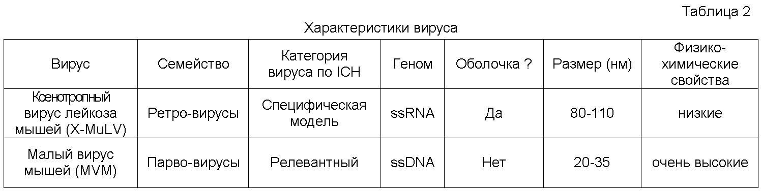
1.1 Методы1.1 Methods
Настоящее исследование проводилось по протоколу, представленному на фиг.4.The present study was conducted according to the protocol presented in figure 4.
На каждой стадии данного процесса проводилась провокация концентрированным «спайком» вируса в подаваемом потоке (объемы «спайка» вируса 1-7,1 объемн %). Нагрузка, продукт и содержание в контрольных потоках оценивались по количеству вирусов и определяли log снижения для каждой стадии.At each stage of this process, a concentrated spike of the virus was provoked in the feed stream (volumes of the
Каждую стадию процесса (см. фиг.4) проводили в двойном повторе. Хроматографические смолы оценивали путем провокации на менее масштабных моделях условий нормального процесса производства, как показано в таблице 4. Все колонки имели внутренний диаметр 1 см и соответствовали требуемым критериям по фактору асимметрии колонки (0,5-2,0). Разрешенный диапазон высоты слоя для каждой колонки в уменьшенном масштабе был таким же, как и в производственных колонках. В таблице 3 представлено краткое описание условий обработки.Each stage of the process (see figure 4) was carried out in duplicate. Chromatographic resins were evaluated by provocation on smaller models of the conditions of the normal production process, as shown in table 4. All columns had an inner diameter of 1 cm and met the required criteria for the asymmetry factor of the column (0.5-2.0). The allowed range of layer height for each column on a reduced scale was the same as in production columns. Table 3 provides a brief description of the processing conditions.


1.2 Токсичность буфера/результаты оценки интерференции1.2 Buffer toxicity / interference assessment results
В рамках исследования эффективности уничтожения вируса, тестировали образцы и буферы для выявления, имелась ли токсичность для клеточных линий, использованных в исследовании вирусов. Образцы и буферы оценивали по их потенциальному воздействию на способность выбранных вирусов инфицировать индикаторные клеточные линии. Не было выявлено значительного снижения инфекционности вирусов ни для одного из использованных буферов, при сравнении полученных результатов с релевантным положительным контролем. Таким образом, по данным проведенного тестирования, буферы, использованные в процессе очистки, были признаны пригодными для ресуспендирования вирусных препаратов после ультрацентрифугирования с целью использования в провокационных тестах на стадиях хроматографии и нанофильтрации.As part of a study on the efficacy of killing a virus, samples and buffers were tested to determine if there was toxicity to the cell lines used in the virus study. Samples and buffers were evaluated by their potential effect on the ability of selected viruses to infect indicator cell lines. There was no significant reduction in virus infectivity for any of the buffers used, when comparing the results with a relevant positive control. Thus, according to the testing, the buffers used in the cleaning process were found to be suitable for resuspension of viral preparations after ultracentrifugation for use in provocative tests at the stages of chromatography and nanofiltration.
1.3 Результаты оценки эффективности уничтожения вирусов1.3 the results of the evaluation of the effectiveness of the destruction of viruses
Эффективность каждой стадии процесса с точки зрения снижения титров вирусов оценивали путем вычисления log фактора снижения (LRF) по следующему уравнению:The effectiveness of each stage of the process in terms of reducing virus titers was evaluated by calculating the log reduction factor (LRF) according to the following equation:
LRF=log10 [Количество внесенного вируса/ Количество вируса в потоке продукта]LRF = log10 [Amount of virus introduced / Amount of virus in the product stream]
Данные по log снижению титров вирусов приведены в обобщенном виде в таблице 5. Результаты, представленные в виде нижних пределов (то есть, LRF≥4,54), показывают, что вирус присутствовал в количествах ниже предела определения.Data on the log reduction of virus titers are summarized in table 5. The results, presented in the form of lower limits (that is, LRF≥4.54), show that the virus was present in quantities below the limit of determination.

Стадию инактивации при низком pH проводили только для вируса X-MLV, поскольку известно, что MMV высоко устойчив к инактивации при низком pH. В нулевой точке времени достигались высокие уровни снижения. К 15-минутной точке вообще не наблюдалось инфекционности вирусов. Для оценки образца в точке 60 минут, использовали объемную инокуляцию для повышения чувствительности теста и были получены факторы снижения ≥ 4,54 log10 и ≥ 5,07 log10 для двойных повторов. В случае X-MLV, наблюдалась быстрая инактивация вируса; независимо от того факта, что осветленный собранный материал содержал примерно 0,5% остаточных клеток после проведения процедуры осветления путем центрифугирования и имелись высокие концентрации белков из компонентов культуральной среды.The inactivation stage at low pH was carried out only for the X-MLV virus, since it is known that MMV is highly resistant to inactivation at low pH. At time zero, high levels of decline were achieved. By the 15-minute point, no infectivity of viruses was observed at all. To evaluate the sample at 60 minutes, volume inoculation was used to increase the sensitivity of the test and reduction factors of ≥ 4.54 log 10 and ≥ 5.07 log 10 for double repeats were obtained. In the case of X-MLV, rapid inactivation of the virus was observed; regardless of the fact that the clarified collected material contained approximately 0.5% residual cells after the clarification procedure by centrifugation and there were high concentrations of proteins from the components of the culture medium.
Стадию хроматографии на Fractogel™-SO3 - проводили с использованием материала, нанесенного на Fractogel™-SO3 - вместе с X-MLV или MMV в уравновешивающем буфере для Fractogel™-SO3 -. Инфекционный вирус выявлялся в элюированных с колонок Fractogel™ фракциях в случае обоих исследованных вирусов. Факторы снижения для X-MLV составляли 5,78 log10 и 5,22 log10 для вариантов двойных повторов. В случае MMV, факторы снижения составляли ≥1,39 log10 и ≥ 1,33 log10.The chromatography step on Fractogel ™ -SO 3 - was performed using the material deposited on Fractogel ™ -SO 3 - together with X-MLV or MMV in equilibration buffer for Fractogel ™ -SO 3 - . Infectious virus was detected in fractions eluted from Fractogel ™ columns in the case of both investigated viruses. The reduction factors for X-MLV were 5.78 log 10 and 5.22 log 10 for double repeat variants. In the case of MMV, the reduction factors were ≥1.39 log 10 and ≥ 1.33 log 10 .
Стадию хроматографии на Q-Сефарозе™ проводили с использованием нанесенного на колонку с Q-Сефарозой™ материала, при его объединении с X-MLV или MMV в уравновешивающем буфере для Q-Сефарозы™. В образцах из вытекающего элюата не выявлялся инфекционный вирус, ни для одного из исследуемых буферов, и в обоих случаях достигался минимальный предел для тестирования. Факторы снижения, наблюдаемые для X-MLV, составляли ≥ 5,51 log10 и ≥5,59 log10 для двойных повторов, а фактор снижения для MMV составлял 7,04 log10 в обоих повторах.The Q-Sepharose ™ chromatography step was performed using Q-Sepharose ™ column-coated material, when combined with X-MLV or MMV in Q-Sepharose ™ equilibration buffer. No infectious virus was detected in the samples from the effluent, for any of the test buffers, and in both cases the minimum limit for testing was reached. The reduction factors observed for X-MLV were ≥ 5.51 log 10 and ≥ 5.59 log 10 for double repeats, and the reduction factor for MMV was 7.04 log 10 in both repeats.
Стадию хроматографии на фенил-Сефарозе™ HP проводили с использованием нанесенного на колонку с фенил-Сефарозой™ материала, при его объединении с X-MLV или MMV в уравновешивающем буфере для фенил-Сефарозы™ НР. Инфекционный вирус выявлялся в элюируемых с колонок с фенил-Сефарозой™ фракциях, для обоих исследованных вирусов. Факторы снижения, наблюдаемые для X-MLV, составляли 2,45 log10 и 2,03 log10 для двойных повторов. Данная стадия очистки была менее жесткой для MMV, где факторы снижения составляли 0,49 log10 и 0,96 log10.The phenyl-Sepharose ™ HPLC chromatography step was performed using phenyl-Sepharose ™ column-coated material when combined with X-MLV or MMV in phenyl-Sepharose ™ HP equilibration buffer. Infectious virus was detected in fractions eluted from phenyl-Sepharose ™ columns for both viruses studied. The reduction factors observed for X-MLV were 2.45 log 10 and 2.03 log 10 for double repeats. This purification step was less stringent for MMV, where the reduction factors were 0.49 log 10 and 0.96 log 10 .
Нанофильтры Pall DV50™ использовали в экспериментах по вирусной провокации с использованием X-MLV. Для фильтрации в экспериментах с использованием MMV были взяты фильтры Pall DV20™. Оба исследованных вируса ресуспендировали в элюирующем буфере для фенил-Сефарозы™ HP перед добавлением элюированного материала к фенил-Сефарозе™ HP. Для проведения очистки MMV были выбраны фильтры DV20™. Не выявлялся инфекционный вирус в образцах фильтрата в случае X-MLV с использованием фильтра DV50. Факторы снижения, наблюдаемые для X-MLV, составляли ≥4,95 log10 и ≥4,61 log10 для двойных повторов. Инфекционный вирус обнаруживался в образцах фильтрата в случае очистки MMV с использованием фильтра DV20. Факторы снижения, наблюдаемые для MMV, составляли ≥2,98 log10 и ≥ 4,88 log10 для двойных повторов. Расхождение результатов между двумя вариантами оценки с использованием DV20 не удалось объяснить за счет различий в условиях проведения эксперимента для каждого фильтра или в титре «спайка», поскольку эти параметры соответствовали заданным ранее спецификациям. Однако, расхождение могло быть связано со сборкой фильтрующих устройств перед работой. Мембранные фильтры были предварительно смочены и подвергнуты автоклавированию, хотя фильтровальные отделения были отдельно простерилизованы паром. Непосредственно перед работой, фильтрующие разбирали и мембраны помещали в фильтровальные отделения и вновь собирали. Фиксация О-кольцевого конца перед повторной сборкой могло привести к менее, чем оптимальному поведению мембраны, помещенной в фильтровальное отделение.Pall DV50 ™ nanofilters were used in viral challenge experiments using X-MLV. Pall DV20 ™ filters were used for filtering in experiments using MMV. Both tested viruses were resuspended in phenyl-Sepharose ™ HP elution buffer before addition of the eluted material to phenyl-Sepharose ™ HP. DV20 ™ filters were selected for MMV cleaning. No infectious virus was detected in the filtrate samples in the case of X-MLV using a DV50 filter. The reduction factors observed for X-MLV were ≥4.95 log 10 and ≥4.61 log 10 for double repeats. An infectious virus was detected in the filtrate samples in the case of MMV purification using a DV20 filter. The reduction factors observed for MMV were ≥ 2.98 log 10 and ≥ 4.88 log 10 for double repeats. The discrepancy between the two evaluation options using the DV20 could not be explained due to differences in the experimental conditions for each filter or in the spike titer, since these parameters corresponded to the previously specified specifications. However, the discrepancy could be due to the assembly of filtering devices before operation. Membrane filters were pre-moistened and autoclaved, although the filter compartments were separately sterilized with steam. Immediately before work, the filters were disassembled and the membranes were placed in the filter compartments and reassembled. Fixation of the O-ring end before reassembly could result in less than optimal behavior of the membrane placed in the filter compartment.
Данные по инактивации/уничтожению вирусов (таблица 6) показывают, что рассматриваемый способ очистки антитела позволяет удалить минимум 21,91 log10 единиц X-MLV с использованием мембраны для нанофильтрации DV50. Общий фактор снижения для MMV, за исключением варианта использования мембраны DV50, составляет минимум 8,37 log10. А если в способ очистки антитела включали мембранный фильтр DV20, то общий фактор снижения для MMV возрастал минимум до 11,29 log10. Таким образом, приведенные данные показывают, что рассматриваемый способ очистки антител гарантирует вирусную безопасность лекарственного препарата, получаемого на основе антитела (например, анти-IL-18 антитела).Virus inactivation / killing data (Table 6) shows that the antibody purification method under consideration allows removal of a minimum of 21.91 log 10 X-MLV units using a DV50 nanofiltration membrane. The overall reduction factor for MMV, with the exception of the DV50 membrane use case, is a minimum of 8.37 log 10 . And if the DV20 membrane filter was included in the antibody purification method, the overall reduction factor for MMV increased to a minimum of 11.29 log 10 . Thus, the data presented show that the method for purification of antibodies under consideration guarantees the viral safety of a drug derived from an antibody (for example, anti-IL-18 antibody).

Материал, который был собран из клеточной культуры и который включал анти-IL-18 антитела, осветляли центрифугированием (3000 × g) с последующей фильтрацией через фильтр с размером пор 0,2 мкм. Собранный и осветленный материал корректировали по значению рН с использованием 1М лимонной кислоты и выдерживали при комнатной температуре. Осажденные белки отделяли от растворимых белков путем центрифугирования (16000 × g). Образцы растворимых и нерастворимых белков обрабатывали при нагревании при 60°C в течение 30 минут в присутствии ДСН и анализировали путем электрофореза в полиакриламидном геле.Material that was collected from cell culture and which included anti-IL-18 antibodies was clarified by centrifugation (3000 × g), followed by filtration through a 0.2 μm filter. The collected and clarified material was adjusted by pH using 1 M citric acid and kept at room temperature. The precipitated proteins were separated from soluble proteins by centrifugation (16000 × g). Samples of soluble and insoluble proteins were processed by heating at 60 ° C for 30 minutes in the presence of SDS and analyzed by polyacrylamide gel electrophoresis.
Результаты проведенного гель-электрофореза (фиг.5) показывают, что молекула антитела остается в растворе при понижении pH в осветленной культуральной среде, где указанная среда также содержит белки клетки-хозяина и компоненты среды. При более низких значениях pH, белки клетки-хозяина дифференциально осаждаются и могут быть удалены путем центрифугирования. Максимальное осаждение достигается в диапазоне значений pH от 4 до 3,5.The results of gel electrophoresis (figure 5) show that the antibody molecule remains in solution with decreasing pH in a clarified culture medium, where this medium also contains host cell proteins and medium components. At lower pH values, the host cell proteins are differentially precipitated and can be removed by centrifugation. Maximum precipitation is achieved in the range of pH values from 4 to 3.5.
В данном эксперименте оценивали влияние pH в ходе нанесения материала с анти-IL-18 антителом на колонку для катионообменной хроматографии (CEX) на связывающую способность (см. Таблица 7). Величину pH в отобранном материале клеточной культуры корректировали добавлением соответствующего количества лимонной кислоты. После выдерживания в течение 1 часа при комнатной температуре указанный материал центрифугировали и проводили фильтрацию через фильтр с размером пор 0,2 мкм. Далее корректировали проводимость путем добавления воды до показателя 10 мс. Исследуемый материал наносили на СЕХ колону с размером 0,5×5 см. Определяли динамическую связывающую способность в ходе нанесения материала на колонку с линейной скоростью 200 см/час. Использовали количественную аналитическую ВЭЖХ с белком A для определения титров антител в образцах вытекающего из колонки материала с целью оценки 5% уровня прорыва breakthrough в ходе нанесения.In this experiment, the effect of pH during application of an anti-IL-18 antibody material to a cation exchange chromatography (CEX) column on binding capacity was evaluated (see Table 7). The pH value in the selected cell culture material was adjusted by adding an appropriate amount of citric acid. After incubation for 1 hour at room temperature, this material was centrifuged and filtration was carried out through a 0.2 μm filter. Next, the conductivity was adjusted by adding water to 10 ms. The test material was applied to a CEX column with a size of 0.5 × 5 cm. The dynamic binding ability was determined during the deposition of the material on the column with a linear velocity of 200 cm / h. Quantitative analytical HPLC with protein A was used to determine antibody titers in samples of column-derived material to evaluate a 5% breakthrough breakthrough rate during application.

Изоэлектрическая точка (pI) для анти-IL-18 антитела составляет 8,43. В растворе при pH 1, что ниже чем значение pI, молекула антитела должна быть положительно заряжена и, соответственно, должна связываться с отрицательно заряженной катионообменной смолой в условиях низкой проводимости. Однако, как видно на приведенной выше таблице 1, антитело совсем не связывалось при нейтральном pH 7,0, меньшем на целую единицу pH относительно pI, который равен 8,43, тогда как при снижении pH связывающая способность смолы повышалась. Титрование pH в собранном материале имеет другие последствия с точки зрении осаждения. Однако, это благоприятный эффект, поскольку происходит селективное осаждение белков и нуклеиновых кислот клетки-хозяина.The isoelectric point (pI) for the anti-IL-18 antibody is 8.43. In a solution at
Жизнеспособность клеток. Отбирали аликвоту клеточной культуры из двухлитрового биореактора в конце стадии продукции при высокой жизнеспособности материала. Собранные культуры подкисляли с использованием 3 M лимонной кислоты, сначала до pH 5,0, затем до pH 3,5 и выдерживали в течение 1 часа при температуре окружающей среды. После одночасового периода выдерживания, pH культуры доводили до значения 4,8 с использованием 3 M NaOH. Образцы отбирали и определяли количество и жизнеспособность клеток в обработанной культуре. Результаты представлены в Таблице 8.Cell viability. An aliquot of the cell culture was selected from a two-liter bioreactor at the end of the production stage with high viability of the material. The collected cultures were acidified using 3 M citric acid, first to pH 5.0, then to pH 3.5 and kept at ambient temperature for 1 hour. After a one hour holding period, the pH of the culture was adjusted to 4.8 using 3 M NaOH. Samples were taken and the number and viability of cells in the treated culture were determined. The results are presented in Table 8.
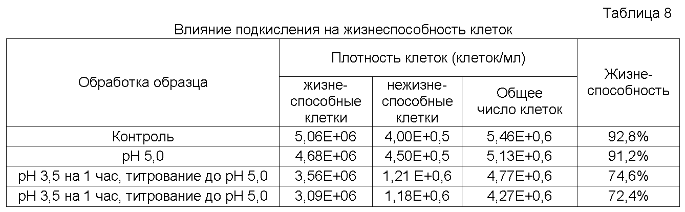
Отмечается умеренное, на уровне 19,6%, снижение жизнеспособности (92,8% против 74,6%) после 1-часовой экспозиции клеточной культуры при pH 3.5. Общая плотность клеток снижалась на 12,4% (5,6×10⁶ против 4,77×10⁶).There is a moderate, at 19.6%, decrease in viability (92.8% versus 74.6%) after a 1-hour exposure of the cell culture at pH 3.5. The total cell density decreased by 12.4% (5.6 × 10 6 versus 4.77 × 10 6 ).
Выдерживание подкисленной культуры при pH 5 и при температуре 4°C в течение ночи привело к небольшому понижению жизнеспособности клеток (на 3% - 74,6% против 72,4%) и плотности клеток (на 10,5% - 4,77×10⁶ против 4,27×10⁶). Точное измерение плотности и жизнеспособности клеток было затруднено в связи с повышением мутности при осаждении белков и нуклеиновых кислот клетки-хозяина. Однако, не было признаков крупномасштабного лизиса клеток, которые должны было выявляться при макроскопическом обследовании.Sustaining the acidified culture at
Извлечение антител. Клеточные культуры получали после завершения ферментации в биореакторе. Клетки отбирали из одной из аликвот после их центрифугирования при 7000 × g. Образцы подкисляли с использованием 3 М лимонной кислоты и выдерживали в течение 1 часа при температуре окружающей среды. После 1-часового выдерживания, культуры корректировали до pH 4,9 с использованием 3 M NaOH. Подкисленные образцы осветляли центрифугированием при 11000 × g с последующей фильтрацией через фильтр с размером пор 0,2 мкм. Проводили количественный анализ по методу ВЭЖХ с белком А для определения титра антител. Полученные титры, после подкисления/осветления, сравнивали с исходными значениями контрольных титров для определения процентной характеристики процесса извлечения. Образцы анализировали и после нейтрализации до pH 5, и после дополнительного выдерживания их в течение ночи при температуре 4°C.Extraction of antibodies. Cell cultures were obtained after completion of fermentation in a bioreactor. Cells were selected from one of the aliquots after centrifugation at 7000 × g. Samples were acidified using 3 M citric acid and kept at ambient temperature for 1 hour. After 1 hour aging, cultures were adjusted to pH 4.9 using 3 M NaOH. The acidified samples were clarified by centrifugation at 11000 × g, followed by filtration through a filter with a pore size of 0.2 μm. Quantitative analysis was performed by HPLC with protein A to determine the titer of antibodies. The obtained titers, after acidification / clarification, were compared with the initial values of the control titers to determine the percentage characteristics of the extraction process. Samples were analyzed both after neutralization to

Как видно из данных, приведенных в таблице 9, оптимальное значение pH для извлечения титра антитела после подкисления/осветления собранного антитела, находилось в диапазоне значений pH от 3,7 до 3,9. При pH 3,5 наблюдалось небольшое снижение титра антител (от 5,1 до 6,4%). Такое небольшое снижение титра наблюдалось в подкисленных культурах, как при наличии клеток, так и в отсутствие клеток. Оптимальное значение pH различалось в зависимости от биохимических и физических свойств молекулы конкретного антитела.As can be seen from the data shown in table 9, the optimal pH for the extraction of the titer of the antibody after acidification / clarification of the collected antibodies was in the range of pH values from 3.7 to 3.9. At pH 3.5, a slight decrease in antibody titer was observed (from 5.1 to 6.4%). Such a slight decrease in titer was observed in acidified cultures, both in the presence of cells and in the absence of cells. The optimal pH value varied depending on the biochemical and physical properties of the molecule of a particular antibody.
Функция антитела. Культуру из биореактора подкисляли с использованием 3 М лимонной кислоты до pH 3,5 и выдерживали в течение 1 часа при температуре окружающей среды. Собранный и подкисленный материал далее титровали до pH 5 с использованием 3M NaOH и осветляли путем центрифугирования и фильтрования через фильтр с размером пор 0,2 мкм. Антитела из подкисленного/осветленного собранного материала далее очищали с использованием катионообменной хроматографии. Неподкисленную культуру из биореактора осветляли центрифугированием и фильтрованием через фильтр с размером пор 0,2 мкм, после чего антитело подвергали очистке на стадии аффинной хроматографии с белком А. С использованием целевого антигена, определяли кинетику связывания выделяемого антитела, взятого из подкисленного и неподкисленного материала. См. таблицу 10.The function of the antibody. The bioreactor culture was acidified using 3 M citric acid to a pH of 3.5 and kept at ambient temperature for 1 hour. The collected and acidified material was then titrated to

Приведенные данные показывают, что применительно к исследуемому антителу, подкисление материала из биореактора оказывало небольшой эффект на функциональность антитело после очистки.The data presented show that, as applied to the antibody under study, acidification of the material from the bioreactor had a small effect on the functionality of the antibody after purification.
Влияние типа используемой кислоты на извлечение антитела. Клеточные культуры получали из биореактора по завершению процесса ферментации. Образцы осторожно подкисляли с использованием 3М лимонной кислоты, 3М фосфорной кислоты или 3М хлористоводородной кислоты и выдерживали в течение 1 часа при температуре окружающей среды. После одночасового выдерживания культуры, корректировали pH до 5,0 с использованием 3M NaOH. Подкисленные образцы осветляли центрифугированием при 11000×g с последующим фильтрованием через фильтр с размером пор 0,2 мкм. Использовали количественный анализ по методу ВЭЖХ с белком А для определения титра антител. Титры, подсчитанные после подкисления/осветления, сравнивали с исходными титрами в контроле для определения процента извлечения. См. Таблицу 11.The effect of the type of acid used on antibody recovery. Cell cultures were obtained from the bioreactor upon completion of the fermentation process. Samples were carefully acidified using 3M citric acid, 3M phosphoric acid, or 3M hydrochloric acid and kept at ambient temperature for 1 hour. After holding the culture for one hour, the pH was adjusted to 5.0 using 3M NaOH. The acidified samples were clarified by centrifugation at 11000 × g, followed by filtration through a filter with a pore size of 0.2 μm. Protein A HPLC quantitative analysis was used to determine antibody titer. The titers calculated after acidification / clarification were compared with the original titers in the control to determine the percentage of recovery. See table 11.

Как видно из данных, приведенных в таблице 11, извлечение титра антител было оптимальным в случае использования лимонной или фосфорной кислоты для целей подкисления до низкого pH/осветления собранного материала антитела. Как было отмечено, перед использованием 3М лимонной кислоты, корректировка культуры до слегка более высокого pH улучшала извлечение антител. При pH 3,5 наблюдалось небольшое снижение титра антител (2,1% для 3M лимонной кислоты, 1% для 3M фосфорной кислоты и 7,7% для 3 M хлористоводородной кислоты).As can be seen from the data shown in table 11, the extraction of antibody titer was optimal in the case of citric or phosphoric acid for acidification to low pH / lightening of the collected antibody material. As noted, before using 3M citric acid, adjusting the culture to a slightly higher pH improved antibody recovery. At pH 3.5, a slight decrease in antibody titer was observed (2.1% for 3M citric acid, 1% for 3M phosphoric acid, and 7.7% for 3 M hydrochloric acid).
2. Определение концентрации белка клетки-хозяина в композиции анти-IL-12 антитела2. Determination of the concentration of the protein of the host cell in the composition of anti-IL-12 antibodies
Данная процедура использовалась для определения остаточной концентрации белка клетки-хозяина в образцах анти-IL-12 антитела. Использовали иммуноферментный твердофазный анализ (ELISA) для сэндвич-тестирования белка клеток-хозяев (антигенов), помещенного между двумя слоями специфических антител. После этого казеином блокировали неспецифические сайты. Далее, белки клеток-хозяев инкубировали и в течение этого времени молекулы антигена связывались с первым антителом (покрывающее антитело). Затем добавляли второе антитело (биотинилированное антитело против белка клетки-хозяина), которое связывается с антигеном (белок клетки-хозяина). Добавляли нейтравидин, конъюгированный с пероксидазой хрена, который связывается с биотинилированным антителом против белка клетки-хозяина. После этого добавляли субстрат К blue. Хромогенный субстрат гидролизуется при связывании с антителом, конъюгированным с ферментом, образуя синее окрашивание. Реакцию останавливали добавлением 2M H3PO4, что сопровождалось изменением окрашивания на желтый цвет. Интенсивность окрашивания была прямо пропорциональна количеству антигена, связанного в лунке.This procedure was used to determine the residual concentration of the host cell protein in anti-IL-12 antibody samples. An enzyme-linked immunosorbent assay (ELISA) was used to sandwich the testing of a host cell protein (antigens) placed between two layers of specific antibodies. After this, casein blocked non-specific sites. Further, host cell proteins were incubated and during this time antigen molecules bind to the first antibody (coating antibody). Then a second antibody (biotinylated anti-host protein) was added which binds to the antigen (host cell protein). Neutravidin conjugated to horseradish peroxidase was added, which binds to a biotinylated antibody against the host cell protein. After that, the substrate K was added. The chromogenic substrate is hydrolyzed upon binding to an antibody conjugated to an enzyme, forming a blue color. The reaction was stopped by the addition of 2M H 3 PO 4 , which was accompanied by a change in the color to yellow. Staining intensity was directly proportional to the amount of antigen bound in the well.
Получение 50 мМ бикарбоната натрия (покрывающий буфер), pH 9,4. В стакан на 1 литр добавляют: 900 мл воды Milli-Q; 4,20 г ± 0,01 г бикарбоната натрия. Хорошо перемешивают до полного растворения. Доводят pH до 9,4 с использованием 1н NaOH. Переносят в мерную колбу на 1 л и доводят объем до метки водой Milli-Q. Перемешивают до гомогенного состояния при переворачивании колбы. Фильтруют через устройство для стерильного фильтрования с размером пор 0,22 мкм. Хранят при температуре 4°C до 7 дней от даты приготовления.Obtaining 50 mm sodium bicarbonate (coating buffer), pH 9.4. In a glass per 1 liter add: 900 ml of Milli-Q water; 4.20 g ± 0.01 g sodium bicarbonate. Mix well until completely dissolved. The pH was adjusted to 9.4 using 1N NaOH. Transfer to a 1 L volumetric flask and adjust the volume to the mark with Milli-Q water. Stir until homogeneous when the flask is turned over. Filter through a sterile filter device with a pore size of 0.22 μm. Store at 4 ° C for up to 7 days from the date of preparation.
Получение 0,104 M Na2HPO4×7H20, 1,37 M NaCl, 0,027 M KCl, 0,0176 M KH2PO4, pH 6,8-6,9(10×ФБР). Вносят примерно 400 мл воды Milli-Q в стеклянный стакан. Далее добавляют 13,94 г ± 0,01 г Na2HPO4 × 7H2O. Добавляют 40,0 г ± 0,1 г NaCl. Добавляют 1,00 г ± 0,01 г KCl. Добавляют 1,20 г ± 0,01 г KH2P04. Перемешивают до гомогенного состояния. Переносят в мерную колбу на 500 мл. Доводят объем до метки 500 мл с использованием воды Milli-Q. Перемешивают путем переворачивания. Фильтруют через устройство для стерильного фильтрования с размером пор 0,2 мкм. Хранят при температуре 4°C до 7 дней от даты приготовления.Getting 0.104 M Na2HPOfour× 7H20, 1.37 M NaCl, 0.027 M KCl, 0.0176 M KH2POfourpH 6.8-6.9 (10 × PBS). Add approximately 400 ml of Milli-Q water to a glass beaker. Then add 13.94 g ± 0.01 g of Na2HPOfour × 7H2O. Add 40.0 g ± 0.1 g of NaCl. 1.00 g ± 0.01 g of KCl are added. Add 1.20 g ± 0.01 g KH2P0four. Mix to a homogeneous state. Transfer to a 500 ml volumetric flask. Bring the volume to the 500 ml mark using Milli-Q water. Stir by inverting. Filter through a sterile filter device with a pore size of 0.2 μm. Store at 4 ° C for up to 7 days from the date of preparation.
Получение 1×ФБР+0,1% Тритон X-100, pH 7,40: (промывочный буфер для планшета). В градуированном цилиндре на 4 л смешивают 400 мл 10×ФБР (стадия 5.2) с 3500 мл воды Milli-Q. Проверяют pH и, при необходимости, доводят до значения 7,40±0,05 с использованием 1н HCl или 1н NaOH. Доводят объем до метки водой Milli-Q. Плотно закрывают цилиндр парафином и перемешивают путем переворачивания до гомогенного состояния. Переносят в бутыль на 4 л. Отбирают 4 мл 1×ФБР и отбрасывают. Добавляют 4 мл Тритона Х-100 к 3996 мл 1×ФБР. Помещают на встряхиваемый планшет и перемешивают до полного растворения. Фильтруют буфер для промывания планшета в количестве, нужном для разбавления буфера, применяемого для получения препарата, через устройство для стерильного фильтрования с размером пор 0,22 мкм. Хранят при комнатной температуре до 7 дней от даты изготовления.Obtaining 1 × PBS + 0.1% Triton X-100, pH 7.40: (washing buffer for the tablet). In a 4-liter graduated cylinder, 400 ml of 10 × PBS were mixed (step 5.2) with 3500 ml of Milli-Q water. Check the pH and, if necessary, bring to a value of 7.40 ± 0.05 using 1N HCl or 1N NaOH. Bring the volume to the mark with Milli-Q water. Close the cylinder tightly with paraffin and mix by inverting to a homogeneous state. Transferred to a 4 liter bottle. 4 ml of 1 × PBS were taken and discarded. Add 4 ml of Triton X-100 to 3996 ml of 1 × FBI. Place on a shake plate and mix until completely dissolved. Filter the tablet washing buffer in an amount necessary to dilute the buffer used to prepare the drug through a sterile filter device with a pore size of 0.22 μm. Store at room temperature for up to 7 days from the date of manufacture.
Получение покрывающей смеси антитела: козье анти-CHO антитело 599/626/748 (лот No. Gl 1201 @ 1,534 мг/мл), после аффинной очистки: Примечание: Концентрированный раствор хранят при номинальной температуре -80°C в ампулах. Отбирают аликвоты. Переносят одну аликвоту на планшет к моменту использования. Непосредственно перед использованием: разбавляют смесь антитела до конечной концентрации 4 мкг/мл с использованием 50 мМ бикарбоната натрия следующим образом: Например: добавляют 31 мкл покрывающей смеси антитела к 11969 мкл холодного покрывающего буфера. Осторожно перемешивают путем переворачивания.Preparation of antibody coating mixture: goat anti-CHO antibody 599/626/748 (lot No. Gl 1201 @ 1.534 mg / ml), after affinity purification: Note: The concentrated solution was stored in ampoules at a nominal temperature of -80 ° C. Aliquots are selected. Transfer one aliquot to the tablet at the time of use. Immediately before use: dilute the antibody mixture to a final concentration of 4 μg / ml using 50 mM sodium bicarbonate as follows: For example: add 31 μl of antibody coating mixture to 11969 μl of cold coating buffer. Mix gently by inverting.
Получение биотинилированного козьего антитела против белковой смеси клеток-хозяев 599/626/748 (лот No. Gl 1202 @ 0,822 мг/мл): Примечание: Концентрированный раствор хранят при номинальной температуре -80°C в ампулах. Отбирают аликвоты. Переносят одну аликвоту на планшет к моменту использования. Непосредственно перед использованием: разбавляют биотинилированную смесь антитела до конечной концентрации 1 мкг/мл при 37°C±2°С казеином следующим образом. Например: добавляют 14,6 мкл биотинилированной смеси антитела к 11985 мкл казеина при температуре 37°C±2°C. Осторожно перемешивают путем переворачивания.Preparation of Biotinylated Goat Antibody Against the Protein Mixture of Host Cells 599/626/748 (Lot No. Gl 1202 @ 0.822 mg / ml): Note: The concentrated solution was stored in ampoules at a nominal temperature of -80 ° C. Aliquots are selected. Transfer one aliquot to the tablet at the time of use. Immediately before use: dilute the biotinylated antibody mixture to a final concentration of 1 μg / ml at 37 ° C ± 2 ° C with casein as follows. For example: add 14.6 μl of the biotinylated antibody mixture to 11985 μl of casein at a temperature of 37 ° C ± 2 ° C. Mix gently by inverting.
Получение Нейтравидина-HRP, конъюгированного со щелочной фосфатазой хрена. Восстанавливают новые лоты (2 мг/флакон) до концентрации 1 мг/мл согласно указанной ниже процедуре: вносят во флакон 400 мкл воды Milli-Q, затем добавляют 1600 мкл 1×ФБР, получая общий объем 2 мл. Осторожно перемешивают на вихревом смесителе. Хранят при температуре -20°C. Отбирают аликвоты желательного объема, с тем чтобы использовать одну аликвоту на планшет. Указанную смесь готовят в полипропиленовой пробирке. Для новых лотов определяют рабочую концентрацию. Проверяют пригодность препарата, с учетом срока годности, составляющего 6 месяцев от даты производства. Например, если рабочая концентрация была определена в количестве 0,2 мкг/мл, то подготовку проводят следующим образом. Непосредственно перед использованием: оттаивают при комнатной температуре аликвоту нейтравидина-HRP. С использованием казеина разбавляют раствор нейтравидина с концентрацией 1 мг/мл до концентрации 0,1 мг/мл (100 мкг/мл) при температуре 37°C±2°C. Например: делают разбавление ×10, добавляя 50 мкл нейтравидина к 450 мкл казеина. Осторожно перемешивают в вихревом смесителе. Далее, указанный раствор с концентрацией 100 мкг/мл разбавляют до концентрации 0,2 мкг/мл казеином при температуре 37˚С±2°C. Например: делают разбавление ×500, добавляя 24 мкл нейтравидина (100 мкг/мл) к 11976 мкл казеина. Осторожно перемешивают в вихревом смесителе.Obtaining Neutravidin-HRP conjugated with alkaline horseradish phosphatase. Restore new lots (2 mg / vial) to a concentration of 1 mg / ml according to the following procedure: add 400 μl Milli-Q water to the vial, then add 1600
Приготовление 5,72 2М фосфорной кислоты (стоп-раствор). Готовят 2М раствор фосфорной кислоты из концентрированной фосфорной кислоты согласно следующей процедуре. С учетом процентного содержания фосфорной кислоты, указанного на этикетке, плотности (1,685 г/мл) и формульной массы (98 г/моль), рассчитывают объем концентрированной фосфорной кислоты, необходимой для получения 500 мл 2М фосфорной кислоты. Вносят в колбу рассчитанный выше объем концентрированной фосфорной кислоты. Доводят объем в колбе водой Milli-Q до метки и перемешивают содержимое до гомогенного состояния при переворачивании колбы. Хранят при температуре окружающей среды до 6 месяцев от даты изготовления.Preparation of 5.72 2M phosphoric acid (stop solution). Prepare a 2M solution of phosphoric acid from concentrated phosphoric acid according to the following procedure. Based on the percentage of phosphoric acid indicated on the label, density (1.685 g / ml) and formula weight (98 g / mol), the volume of concentrated phosphoric acid required to obtain 500 ml of 2M phosphoric acid is calculated. Add the volume of concentrated phosphoric acid calculated above to the flask. Adjust the volume in the flask with Milli-Q water to the mark and mix the contents to a homogeneous state when the flask is turned over. Store at ambient temperature up to 6 months from the date of manufacture.
Приготовление разбавляющего буфера (казеин, разбавленный х100 в 1×ФБР + 0,1% Тритон X100, pH 7,4). Делают разбавление казеина × 100 при температуре 37°C±2°C с использованием 1×ФБР+0,1% Тритона X100, pH 7,4, стерилизованного через фильтр с размером пор 0,22 мкм (как указано выше). Например: при температуре 37°C±2°C добавляют 1 мл казеина к 99 мл стерильно фильтрованного через фильтр с размером пор 0,22 мкм 1×ФБР+0,1% Тритон X100, pH 7,4. Хорошо перемешивают. Для каждого использования готовят новый раствор.Preparation of dilution buffer (casein diluted x100 in 1 × PBS + 0.1% Triton X100, pH 7.4). Casein × 100 was diluted at 37 ° C ± 2 ° C using 1 × PBS + 0.1% Triton X100, pH 7.4, sterilized through a 0.22 μm filter (as described above). For example: at a temperature of 37 ° C ± 2 ° C add 1 ml of casein to 99 ml of sterile filtered through a filter with a pore size of 0.22
Приготовление стандартов. Стандарты белка клетки-хозяина (антигенные стандарты) (лот No. G11203 @ 1,218 мг/мл). Примечание: концентрированные растворы хранят при номинальной температуре -80°C в аликвотах по 70 мкл. Оттаивают аликвоту при комнатной температуре. Проводят серийное разведение в полипропиленовых пробирках с использованием буфера для элюции.Cooking standards. Host Cell Protein Standards (Antigenic Standards) (Lot No. G11203 @ 1.218 mg / ml). Note: concentrated solutions are stored at a nominal temperature of -80 ° C in aliquots of 70 μl. Thaw an aliquot at room temperature. Serial dilution is carried out in polypropylene tubes using an elution buffer.
Приготовление образцов. В полипропиленовых пробирках разбавляют готовые объемные образцы до концентрации 24 мг/мл с использованием буфера для разбавления. Записывают полученную концентрацию. Примечание: используют указанные ниже растворы для получения «спайковых» образцов и для приготовления указанных ниже растворов с концентрацией 12 мг/мл. Далее, в полипропиленовых микропробирках разбавляют растворы с концентрацией 24 мг/мл до концентрации 12 мг/мл с использованием буфера для разбавления. Каждый из растворов с концентрацией 12 мг/мл вносят в трехкратном повторе в лунки планшета, используя всего 6 лунок.Sample preparation. In polypropylene tubes, finished bulk samples are diluted to a concentration of 24 mg / ml using a dilution buffer. Record the resulting concentration. Note: use the following solutions to obtain spike samples and to prepare the following solutions with a concentration of 12 mg / ml. Further, solutions in a concentration of 24 mg / ml are diluted in polypropylene microtubes to a concentration of 12 mg / ml using a dilution buffer. Each of the solutions with a concentration of 12 mg / ml was added in triplicate to the wells of the plate using a total of 6 wells.
Приготовление «спайка». В полипропиленовых микропробирках делают «спайк» белка клетки-хозяина с концентрацией 10 мг/мл из полученного выше стандарта с концентрацией 20 мг/мл при разбавлении в два раза буфером для разбавления. В три лунки планшета добавляют «спайковый» раствор с концентрацией 10 мг/мл. Для «спайковых» образцов используют стандартный раствор с концентрацией 20 мг/мл со стадии 6.1.Cooking "spike". In polypropylene microtubes, a spike of the host cell protein with a concentration of 10 mg / ml is made from the above standard with a concentration of 20 mg / ml when diluted twice with dilution buffer. A “spike” solution with a concentration of 10 mg / ml is added to three wells of the plate. For spike samples, use a standard solution with a concentration of 20 mg / ml from step 6.1.
Приготовление «спайковых» образцов. В полипропиленовых микропробирках объединяют по 300 мкл каждого из готовых объемных растворов с концентрацией 24 мг/мл с 300 мкл «спайкового» раствора с концентрацией 20 мг/мл (6.1). Добавляют в лунки в тройном повторе каждый из растворов «спайкового» образца, получая всего 6 лунок.Preparation of spike samples. In polypropylene microtubes, 300 μl of each of the prepared bulk solutions with a concentration of 24 mg / ml are combined with 300 μl of a “spike” solution with a concentration of 20 mg / ml (6.1). Each of the solutions of the “spike” sample is added to the wells in triplicate, giving a total of 6 holes.
Приготовление контроля. Должен быть установлен диапазон контрольных значений для каждого нового контрольного стандартного раствора перед его использованием для тестирования. Контрольный стандартный раствор: отбирают аликвоты по 150 мкл из партии концентрата лекарственного вещества ABT-874 и хранят их в замороженном состоянии при номинальной температуре -80°C до трех лет.Cooking controls. The range of control values for each new control standard solution must be set before using it for testing. Control standard solution: 150 μl aliquots were taken from a batch of ABT-874 drug concentrate and stored in a frozen state at a nominal temperature of -80 ° C for up to three years.
Приготовление рабочего контрольного раствора. Оттаивают аликвоту контрольного образца при комнатной температуре. С использованием разбавляющего буфера разбавляют указанный контроль до концентрации 24 мг/мл. Далее, с использованием буфера для разведения, в полипропиленовых микропробирках разбавляют контрольный раствор с концентрацией 24 мг/мл до концентрации 12 мг/мл. Получают одно разведение и вносят контрольный раствор в 3 лунки на планшете.Preparation of a working control solution. Thaw an aliquot of the control sample at room temperature. Using a dilution buffer, the indicated control is diluted to a concentration of 24 mg / ml. Next, using a dilution buffer, in a polypropylene microtube, dilute a control solution with a concentration of 24 mg / ml to a concentration of 12 mg / ml. Get one dilution and make a control solution in 3 wells on the tablet.
Процедура ELISA. Заполняют флакон для промывки планшетов соответствующим буфером для промывки планшетов (см. стадию 5.3, 1xФБР+0,1% Тритон X100). Подготавливают к работе промывающее устройство для планшетов, проверяя следующие параметры: параметры должны быть установлены следующим образом: тип планшета: 1 для каждого цикла (всего проводится 5 циклов); объем: 400 мкл; время промывания: 10 секунд; Asp. время: 4 секунды.ELISA procedure. Fill the plate washing bottle with the appropriate tablet washing buffer (see step 5.3, 1x PBS + 0.1% Triton X100). A washing device for tablets is prepared for operation by checking the following parameters: the parameters should be set as follows: tablet type: 1 for each cycle (5 cycles are carried out in total); volume: 400 μl; washing time: 10 seconds; Asp. time: 4 seconds.
Процедура тестирования. Вносят в лунки планшета по 100 мкл/лунку наслаиваемую смесь антител с концентрацией 4 мг/мл в холодном 50 мМ бикарбонате натрия. Приподнимают сторону планшета, так чтобы наслаиваемый раствор равномерно покрывал низ лунки, закрывают пленкой и инкубируют при номинальной температуре 4°C при встряхивании на шейкере для планшетов (или эквивалентном устройстве) со скоростью 3 в течение 18 часов ± 1 час. После инкубации в течение ночи вынимают планшет из холодильника и выравнивают температуру до комнатной. Встряхивают нанесенный раствор. Переносят содержимое планшета на бумажные полотенца. Блокируют казеином, который добавляют по 300 мкл/лунку, при температуре 37°C±2°C, закрывают пленкой и инкубируют при температуре 37°C±2°C при встряхивании на лабораторном шейкере для планшетов (Lab-line Environ) (или эквивалентном устройстве) со скоростью 80 об/мин ± 5 об/мин в течение 1 часа. Готовят стандарт, образец, контроль, «спайк» и «спайковые» образцы во время инкубации с блокирующим раствором. Промывают планшет 5 раз промывочным буфером. Переносят содержимое планшета на бумажные полотенца. Используя 8-канальную пипетку, в тройном повторе вносят в лунки планшета по 100 мкл/лунку стандарты, образцы, «спайки», «спайковые» образцы и контроли. Переносят пипеткой по 100 мкл/лунку разбавляющий буфер во все пустые лунки планшета, которые будут служить в качестве слепой пробы. Закрывают пленкой и инкубируют при температуре 37°C±2 °C при встряхивании на лабораторном шейкере для планшетов (Lab-line Environ) (или эквивалентном устройстве) со скоростью 80 об/мин ± 5 об/мин в течение 1 часа. Заполняют матрицу, которая будет использоваться в качестве стандарта для оценки в ходе нанесения материалов на планшет.Testing procedure. Add 100 μl / well of the layered antibody mixture to a well of a concentration of 4 mg / ml in cold 50 mM sodium bicarbonate. Lift the side of the tablet so that the layered solution evenly covers the bottom of the well, cover with a film and incubate at a nominal temperature of 4 ° C with shaking on a plate shaker (or equivalent device) at a speed of 3 for 18 hours ± 1 hour. After incubation overnight, remove the plate from the refrigerator and equalize the temperature to room temperature. Shake the applied solution. Transfer the contents of the tablet to paper towels. Block with casein, which is added at 300 μl / well, at 37 ° C ± 2 ° C, covered with a film and incubated at 37 ° C ± 2 ° C with shaking on a laboratory shaker for tablets (Lab-line Environ) (or equivalent device) at a speed of 80 rpm ± 5 rpm for 1 hour. Prepare a standard, sample, control, spike and spike samples during incubation with a blocking solution. Wash the
Установка устройства изменения поглощения в содержимом планшетов. Устанавливают прибор, вводя концентрации для стандартов. Не вводят коэффициенты разбавления для образцов, контролей, «спайков» или «спайковых» образцов. Учитывают, что значения для лунок, содержащих разбавитель, используемых в качестве слепых проб, будут вычитаться из результатов для всех лунок. Промывают планшет 5 раз промывочным буфером. Переносят содержимое планшета на бумажные полотенца. Добавляют по 100 мкл/лунку биотинилированное козье антитело. Закрывают пленкой и инкубируют при температуре 37°C±2°C при встряхивании на лабораторном шейкере для планшетов (Lab-line Environ) (или эквивалентном устройстве) со скоростью 80 об/мин ± 5 об/мин в течение 1 часа. Промывают планшет 5 раз промывочным буфером. Переносят содержимое планшета на бумажные полотенца. Добавляют по 100 мкл/лунку раствор конъюгата нейтравидина-HRP (конъюгат с пероксидазой хрена), закрывают пленкой и инкубируют при температуре 37°C±2°C при встряхивании на лабораторном шейкере для планшетов (Lab-line Environ) (или на эквивалентном устройстве) со скоростью 80 об/мин ± 5 об/мин в течение 1 часа. Промывают планшет 5 раз промывочным буфером. Переносят содержимое планшета на бумажные полотенца. Добавляют по 100 мкл/лунку холодный K-blue субстрат, закрывают пленкой и инкубируют при комнатной температуре в течение 10 минут (устанавливают таймер сразу при добавлении субстрата в первый ряд), встряхивая со скоростью 3 на шейкере для планшетов Lab-line titer (или эквивалентном устройстве). Останавливают реакцию, добавляя по 100 мкл/лунку 2М фосфорную кислоту (стадия 5.7). Помещают планшет на шейкер для планшетов и встряхивают со скоростью 3 в течение 3-5 минут. Определяют поглощение содержимого планшета при длине волны 450 нм.Installing a device changes the absorption in the contents of the tablets. Install the appliance by introducing concentrations for standards. Dilution factors for samples, controls, spikes, or spikes are not introduced. Take into account that the values for wells containing diluent used as blind samples will be subtracted from the results for all wells. Wash the
Анализ данных и расчеты. Примечание: обрабатывают данные только для тех образцов, «спайков», «спайковых» образцов и контролей, оптическая плотность которых укладывается в практически применимый предел количественного определения (2,5 нг/мл по стандарту) по стандартной кривой и соответствует указанным ниже значениям % КВ или % по критерию различий. Если показатели ОП для образца ниже 2,5 нг/мл по стандарту, то результат фиксируют как меньше, чем 2,5 нг/мл. Это значение затем следует разделить на концентрацию разбавленного образца (12 мг/мл) для получения значения в нг/мл. Если образец характеризуется высокой концентрацией клеток-хозяев, вследствие чего «неспайковые» и/или «спайковые» образцы попадают в область выше стандартной кривой, то результат описывают как >100 нг/мл. Это значение также следует разделить на концентрацию разбавленного образца (12 мг/мл), чтобы выразить результат в нг/мг. Считают, что образец имеет нулевое значение, по результатам расчета извлечения из «спайка», если показатель для образца падает ниже концентрации 2,5 нг/мл по стандарту.Data analysis and calculations. Note: only data are processed for those samples, “spikes”, “spike” samples and controls, the optical density of which is within the practically applicable limit of quantification (2.5 ng / ml according to the standard) according to the standard curve and corresponds to the following values% KV or% by criterion of differences. If the OD values for the sample are below 2.5 ng / ml by standard, then the result is recorded as less than 2.5 ng / ml. This value should then be divided by the concentration of the diluted sample (12 mg / ml) to obtain a value in ng / ml. If the sample is characterized by a high concentration of host cells, as a result of which “non-spike” and / or “spike” samples fall in the region above the standard curve, then the result is described as> 100 ng / ml. This value should also be divided by the concentration of the diluted sample (12 mg / ml) to express the result in ng / mg. It is believed that the sample has a zero value, according to the calculation results of extraction from the “spike”, if the indicator for the sample falls below a concentration of 2.5 ng / ml according to the standard.
Стандартная кривая. Стандартные концентрации должны быть введены в матричный протокол. Используют вариант квадратической кривой. Коэффициент смешанной корреляции должен составлять 0,99 и % КВ между показателями для исследуемых в тройном повторе лунок должен быть 20%. Если этот критерий не удовлетворяется, то: один стандарт (1 уровень, 3 лунки) можно не учитывать. Если опускают значение 1,25 нг/мл, то используют только значение для образцов и спайковых образцов с приемлемыми значениями оптической плотности в диапазоне от 2,5 нг/мл до 100 нг/мл (оставшиеся точки на стандартной кривой). Дополнительно, для тройных повторов, применительно к каждому стандартному уровню, следует отметить, что если одна лунка является явно контаминированной или демонстрирует низкое связывание, она должна быть пропущена. Если показатель в лунке опускается ниже стандартного уровня, то оставшиеся повторы должны иметь процент различий, равный 20%. Показатель % КВ для наименьшего стандарта, который демонстрирует показатель ОП, близкий к фоновому (слепые пробы) для планшета, должен быть равен 30%. Если показатель для одной лунке выпадает, то процент различий для оставшихся повторов должен составлять 35%. Если выпадает наименьший стандарт, то учитывают только образцы и спайковые образцы с оптическим плотностями, попадающими в оставшийся уровень оптических плотностей на стандартной кривой.Standard curve. Standard concentrations should be entered into the matrix protocol. Use a variant of the quadratic curve. The mixed correlation coefficient should be 0.99 and% CV between the indicators for the wells tested in triplicate; it should be 20%. If this criterion is not satisfied, then: one standard (1 level, 3 holes) can be ignored. If the value of 1.25 ng / ml is omitted, then only the value is used for samples and spike samples with acceptable values of optical density in the range from 2.5 ng / ml to 100 ng / ml (the remaining points on the standard curve). Additionally, for triplicate repeats, for each standard level, it should be noted that if one well is clearly contaminated or exhibits low binding, it should be skipped. If the indicator in the well falls below the standard level, then the remaining repetitions should have a percentage difference of 20%. The% HF value for the smallest standard, which shows an OD value close to the background (blind samples) for the tablet, should be 30%. If the indicator for one well falls out, then the percentage of differences for the remaining repeats should be 35%. If the lowest standard falls out, then only samples and spike samples with optical densities falling into the remaining level of optical densities on the standard curve are taken into account.
Образцы. Значение % КВ должно составлять 20% между результатами для исследуемых в тройном повторе лунок. Записывают % значение КВ между результатами оценки лунок в тройном повторе. Одна лунка для разбавления каждого образца, может быть пропущена. Оставшиеся повторы должны характеризоваться % различий, равным 20%. Следует отметить: если показатель ОП для «неспайкового» образца ниже значения 2,5 нг/мл на стандартной кривой, то % значение критерия различий неприменимо к «неспайковым» результатам. Следует вернуться к указанным выше расчетам.Samples. The% CV value should be 20% between the results for the wells tested in triplicate. Record the% value of CV between the results of the evaluation of the wells in triplicate. One well to dilute each sample may be skipped. The remaining repetitions should be characterized by a% difference of 20%. It should be noted: if the OD index for a “non-spike” sample is lower than 2.5 ng / ml on the standard curve, then the% value of the difference criterion does not apply to “non-spike” results. Return to the above calculations.
Рассчитывают фактическую концентрацию клеток-хозяев в нг/мг относительно среднего значения (нг/мл) следующим образом: значение для белка клетки-хозяина CHO (нг/мг) = среднее значение для «результата для «неспайкового» образца (нг/мл)»_концентрация разбавленного образца (12 мг/мл).The actual concentration of the host cells in ng / mg relative to the average value (ng / ml) is calculated as follows: value for the CHO host cell protein (ng / mg) = average value for the “result for the non-spiked sample (ng / ml)” _ concentration of the diluted sample (12 mg / ml).
«Спайк». %КВ должен составлять 20% между результатами для анализируемых в тройном повторе лунок. Записывают %КВ. Результат для одной лунки «спайка» может быть опущен. Оставшиеся точки должны характеризоваться % различий, равным 20%. Проводят указанные выше расчеты. Записывают концентрацию клеток-хозяев в нг/мл. Это результат будет использоваться в расчетах по определению уровня извлечения из «спайка». Полученная для «спайка» концентрация (нг/мл) должна составлять ±20% от теоретически ожидаемой концентрации в «спайке». Фиксируют результат и указывают, соответствует он или не соответствует требуемому значению. Если результат для «спайка» не укладывается в диапазон 20% от теоретически ожидаемого значения, то тест следует повторить. Среднее значение концентрации в «спайке» (нг/мл) x 100 = должно быть 100% ± 20%, 10 нг/мл."Spike." % CV should be 20% between the results for the wells analyzed in triplicate. Write% CV. The result for a single spike hole may be omitted. The remaining points should be characterized by a% difference of 20%. Perform the above calculations. The concentration of host cells in ng / ml is recorded. This result will be used in the calculations to determine the level of extraction from the “spike”. The concentration obtained for the spike (ng / ml) should be ± 20% of the theoretically expected concentration in the spike. Fix the result and indicate whether it matches or does not match the desired value. If the result for the "spike" does not fit into the range of 20% of the theoretically expected value, then the test should be repeated. The average concentration in the “spike” (ng / ml) x 100 = should be 100% ± 20%, 10 ng / ml.
«Спайковые» образцы. % КВ должен соответствовать 20% между результатами для анализируемых в тройном повторе лунок. Записывают % КВ между результатами для 3 лунок. Результат по одной лунке для каждого разведения «спайкового» образца может быть опущен. Оставшиеся результаты должны характеризоваться % различий, равным 20%. Проводят указанный выше расчет. Записывают «результат для спайкового образца», применительно к каждому разведению, в нг/мл. Записывают % различий между повторными разбавлениями. Процент различий между разбавлениями должен составлять 25%. Эти результаты далее будут использоваться в расчетах по извлечению из «спайкового» материала.Spike samples. % CV should correspond to 20% between the results for the wells analyzed in triplicate. Write% CV between the results for 3 wells. The result of one well for each dilution of the “spike” sample can be omitted. The remaining results should be characterized by a% difference of 20%. Perform the above calculation. Record the “result for the spike sample,” for each dilution, in ng / ml. Record the% differences between repeated dilutions. The percentage of differences between dilutions should be 25%. These results will be further used in the calculations of extraction from the spike material.
Рассчитывают процент извлечения из «спайка» для каждого разведения с использованием приведенной ниже формулы. % извлечения из спайка = (значение для «спайкового» образца - значение для «неспайкового» образца) × 100. Примечание: (1) Если значение ОП для «неспайкового» образца падает ниже 2,5 нг/мл относительно стандарта, то такое значение рассматривают как нулевое в расчетах % извлечения из «спайка». % извлечения из «спайка» должен составлять 100%±50% (50%-150%) для каждого разведения каждого образца. Записывают результаты и указывают, соответствует ли он критериям для данного теста (соответствует/ не соответствует).Calculate the percent recovery from the spike for each dilution using the formula below. % extraction from a spike = (value for a “spike” sample - value for a “non-spike” sample) × 100. Note: (1) If the OD value for a “non-spike” sample falls below 2.5 ng / ml relative to the standard, then this value considered as zero in the calculations of the% extraction from the "spike". The% recovery from the “spike” should be 100% ± 50% (50% -150%) for each dilution of each sample. Record the results and indicate whether it meets the criteria for this test (matches / does not match).
Контроль. % КВ должен соответствовать 20% между результатами для анализируемых в тройном повторе лунок. Записывают процент КВ. Результат для одной лунки «спайка» может быть опущен. Оставшиеся результаты должны иметь процент различий, равный 20%. Проводят указанный выше расчет. Записывают концентрацию клеток-хозяев в контроле в нг/мл. Концентрацию клеток-хозяев в нг/мл рассчитывают следующим образом: концентрация белка клетки-хозяина (нг/мл) = результат оценки белка клетки-хозяина в контроле, в нг/мл.The control. % CV should correspond to 20% between the results for the wells analyzed in triplicate. Record the percentage of HF. The result for a single spike hole may be omitted. The remaining results should have a percentage difference of 20%. Perform the above calculation. The concentration of host cells in the control is recorded in ng / ml. The concentration of host cells in ng / ml is calculated as follows: the concentration of the protein of the host cell (ng / ml) = the result of the evaluation of the protein of the host cell in the control, in ng / ml.
3. Определение концентрации белка А в композициях анти-IL-12 антитела3. Determination of protein A concentration in anti-IL-12 antibody compositions
При проведении процедуры ELISA, в лунки планшета добавляют антитело против куриного белка А и инкубируют. Неспецифические сайты блокируют казеином в ФБР. Планшеты промывают c использованием 1×ФБР+0,1% Тритон X-100 для удаления несвязанного материала. Образцы и стандарты на Cys-белок А разбавляют с использованием 1×ФБР+4,1% Тритона X + 10% казеина. Растворы денатурируют при нагревании при 95˚С ± 2°C, отделяют белок А от ABT-874. Затем указанные растворы добавляют к планшету и инкубируют. Несвязанный материал промывают с использованием 1 × ФБР + 0,1% Тритон X-100. Добавляют к лункам микротитрационного планшета биотинилированное антитело против куриного белка А и инкубируют. Планшет промывают для удаления несвязанного материала и добавляют конъюгат нейтравидина-пероксидазы.During the ELISA, an anti-chicken protein A antibody is added to the wells of the plate and incubated. Nonspecific sites are blocked by casein in the FBI. Plates are washed using 1 × PBS + 0.1% Triton X-100 to remove unbound material. Samples and standards for Cys-protein A are diluted using 1 × PBS + 4.1% Triton X + 10% casein. The solutions are denatured by heating at 95 ° C ± 2 ° C, protein A is separated from ABT-874. Then, these solutions are added to the plate and incubated. Unbound material is washed using 1 × PBS + 0.1% Triton X-100. Add a biotinylated anti-chicken protein A antibody to the wells of the microtiter plate and incubate. The tablet is washed to remove unbound material and a neutravidin peroxidase conjugate is added.
Нейтравидин будет связываться с биотинилированным антителом против куриного белка А, которое не связалось с лунками. Планшет снова промывают для удаления несвязанного нейтравидина и к лункам планшета добавляют субстрат К-blue (тетраметилбензидин (TMB)). Субстрат гидролизуется связанным нейтравидином с образованием синего окрашивания. Реакцию останавливают добавлением фосфорной кислоты, которая меняет окрашивание на желтый. Интенсивность желтого окрашивания в лунках прямо пропорциональна концентрации белка А, имеющего в лунках.Neutravidin will bind to a biotinylated anti-chicken protein A antibody that does not bind to the wells. The plate is washed again to remove unbound neutravidin and K-blue substrate (tetramethylbenzidine (TMB)) is added to the wells of the plate. The substrate is hydrolyzed by bound neutravidin to form a blue color. The reaction is stopped by the addition of phosphoric acid, which changes the color to yellow. The intensity of yellow staining in the wells is directly proportional to the concentration of protein A having in the wells.
Получение реагентов и растворов. Флаконы с казеином должны быть нагреты до температуры 37°C±2°C; далее их озвучивают в течение 2 минут и отбирают аликвоты. Аликвоты хранят при номинальной температуре 4°C. Во время тестирования, помещают нужное количество аликвот казеина в термостат с температурой 37°C±2°C. Буфер для наслаивания и субстрат используют в холодном виде (отбирают непосредственно перед использованием из холодильника, где они хранились при температуре 4°C.)Obtaining reagents and solutions. Casein vials should be heated to 37 ° C ± 2 ° C; then they are voiced for 2 minutes and aliquots are selected. Aliquots are stored at a nominal temperature of 4 ° C. During testing, place the right amount of aliquots of casein in a thermostat with a temperature of 37 ° C ± 2 ° C. The layering buffer and the substrate are used cold (taken immediately before use from the refrigerator, where they were stored at 4 ° C.)
50 мМ бикарбонат натрия (буфер для наслаивания), pH 9,4. В химический стакан объемом 1 л добавляют: 900 мл воды Milli-Q и 4,20 г ± 0,01 г бикарбоната натрия. Перемешивают до полного растворения. Доводят значение pH до 9,4 с использованием 1 н NaOH. Переносят в мерную колбу на 1 л и доводят объем до метки водой Milli-Q. Перемешивают при переворачивании до гомогенного состояния. Фильтруют через устройство для стерильного фильтрования с размером пор 0,22 мкм. Хранят при номинальной температуре 4°C в течение 7 дней от даты приготовления.50 mM sodium bicarbonate (layering buffer), pH 9.4. To a 1 liter beaker: 900 ml of Milli-Q water and 4.20 g ± 0.01 g of sodium bicarbonate are added. Stir until completely dissolved. The pH was adjusted to 9.4 using 1 N NaOH. Transfer to a 1 L volumetric flask and adjust the volume to the mark with Milli-Q water. Stir when turning to a homogeneous state. Filter through a sterile filter device with a pore size of 0.22 μm. Store at a nominal temperature of 4 ° C for 7 days from the date of preparation.
104 M Na2HPО4 × 7H2O,1,37 M NaCl, 0,027 M KC1, 0,0176 M KH2PО4, pH 6,8-6,9. (10×ФБР): добавляют приблизительно 400 мл воды Milli-Q в стеклянный стакан. Добавляют 13,94 г ± 0,01 г Na2HPО4 × 7H2O. Добавляют 40,0 г ± 0,1 г NaCl. Добавляют 1,00 г ± 0,01 г KCl. Добавляют 1,20 г ± 0,01 г KH2PО4. Перемешивают до гомогенного состояния. Переносят в колбу объемом 500 мл. QS до объема 500 мл доводят водой Milli-Q. Перемешивают переворачиванием. Фильтруют через устройство для стерильного фильтрования 0,2 CA мкм. Хранят при комнатной температуре до 7 дней.104 M Na 2 HPO 4 × 7H 2 O, 1.37 M NaCl, 0.027 M KC1, 0.0176 M KH 2 PO 4 , pH 6.8-6.9. (10 × FBI): Add approximately 400 ml Milli-Q water to a glass beaker. 13.94 g ± 0.01 g Na 2 HPO 4 × 7H 2 O are added. 40.0 g ± 0.1 g NaCl are added. 1.00 g ± 0.01 g of KCl are added. Add 1.20 g ± 0.01 g KH 2 PO 4 . Mix to a homogeneous state. Transfer to a 500 ml flask. QS to a volume of 500 ml was made up with Milli-Q water. Mix by turning. Filter through a 0.2 CA micron sterile filter device. Store at room temperature for up to 7 days.
1×ФБР + 0,1% Тритон X-100, pH 7,40: (буфер для промывки планшета): в градуированном цилиндре на 4 л смешивают 400 мл 10 X ФБР (см. выше) с 3500 мл воды Milli-Q. Проверяют pH, и, при необходимости, доводят до 7,40±0,05 с использованием 1н HCl или 1н NaOH. Доводят объем до метки водой Milli-Q. Цилиндр плотно закрывают парафилмом и перемешивают путем переворачивания до гомогенного состояния. Переносят в колбу на 4 л. Отбирают 4 мл 1×ФБР и отбрасывают. Добавляют 4 мл Тритона X-100 к 3996 мл 1×ФБР. Помещают на перемешиваемый планшет и перемешивают до полного растворения. Хранят при комнатной температуре до 7 дней.1 × FBI + 0.1% Triton X-100, pH 7.40: (tablet washing buffer): 400 ml of 10 X FBI (see above) are mixed with 3500 ml of Milli-Q water in a graduated cylinder of 4 L. The pH is checked and, if necessary, adjusted to 7.40 ± 0.05 using 1 N HCl or 1 N NaOH. Bring the volume to the mark with Milli-Q water. The cylinder is tightly closed with parafilm and stirred by inverting to a homogeneous state. Transfer to a 4 L flask. 4 ml of 1 × PBS were taken and discarded. Add 4 ml of Triton X-100 to 3996 ml of 1 × FBI. Place on a stirred plate and mix until completely dissolved. Store at room temperature for up to 7 days.
Наслаиваемое антитело против куриного белка А. К моменту использования отбирают одну аликвоту антитела в расчете на планшет. Для подтверждения качества новых лотов антитела против куриного белка А используют вместе с ним конъюгированное с биотином антитело против куриного белка А для подтверждения его качества (которое было получено из того же лота, что и наслаиваемое антитело). Непосредственно перед использованием: разбавляют смесь антитела холодным 50 мМ бикарбонатом натрия до концентрации, определенной в ходе пробных квалификационных тестов при наслаивании. Например: если в ходе квалификационного теста концентрация наслаиваемого антитела была определена на уровне 6 мкг/мл и если концентрация основного раствора составляет 3000 мкг/мл, то добавляют 24 мкл наслаиваемого антитела к 11976 мкл холодного буфера для внесения в лунки. Осторожно перемешивают путем переворачивания.The layered antibody against chicken protein A. At the time of use, one aliquot of the antibody is selected per tablet. To confirm the quality of the new lots, antibodies against chicken protein A are used together with biotin-conjugated antibody against chicken protein A to confirm its quality (which was obtained from the same lot as the layered antibody). Immediately before use: dilute the antibody mixture with cold 50 mM sodium bicarbonate to the concentration determined during the test qualification tests during layering. For example: if during the qualification test the concentration of the layered antibody was determined to be 6 μg / ml and if the concentration of the main solution is 3000 μg / ml, then 24 μl of the layered antibody is added to 11976 μl of cold buffer to be added to the wells. Mix gently by inverting.
Биотинилированное антитело против куриного белка А. К моменту использования отбирают одну аликвоту антитела в расчете на планшет. Для подтверждения качества новых лотов конъюгированного с биотином антитела против куриного белка А, его используют и подтверждает его качество вместе с тем же лотом, из которого было получено антитело против куриного белка А. Непосредственно перед использованием: разбавляют биотинилированное антитело казеином при температуре 37°C±2°C до концентрации, определенной в ходе квалификационного пробного теста с биотинилированным антителом. Например: если в ходе пробного квалификационного теста концентрация вносимого биотинилированного антитела была определена как 4 мкг/мл и если концентрация основного раствора составляет 1000 мкг/мл, то добавляют 48 мкл биотинилированного антитела к 11952 казеина при температуре 37°C±2°C. Смесь осторожно перемешивают при переворачивании.Biotinylated antibody against chicken protein A. At the time of use, one aliquot of the antibody per tablet is selected. To confirm the quality of new lots of biotin-conjugated anti-chicken protein A antibody, it is used and its quality is confirmed with the same lot from which the anti-chicken protein A antibody was obtained. Immediately before use: dilute the biotinylated antibody with casein at 37 ° C ± 2 ° C to the concentration determined during the qualification test test with biotinylated antibody. For example: if during the trial qualification test the concentration of the introduced biotinylated antibody was determined to be 4 μg / ml and if the concentration of the main solution is 1000 μg / ml, then add 48 μl of the biotinylated antibody to 11952 casein at a temperature of 37 ° C ± 2 ° C. The mixture is gently mixed while turning.
Нейтравидин-HRP. Восстанавливают новые лоты (2 мг/флакон) до концентрации 1 мг/мл следующим образом: добавляют 400 мкл воды Milli-Q во флакон, затем добавляют 1600 мкл 1×ФБР, получая общий объем 2 мл. Осторожно перемешивают на вихревом смесителе. Хранят при номинальной температуре -80°C. Отбирают аликвоты желаемого объема с учетом использования по одной аликвоте на планшет. Получение проводят в полипропиленовой пробирке. Подтверждают годность препарата, которая составляет 6 месяцев от даты изготовления. Например, если рабочая концентрация была определена на уровне 0,1 мкг/мл, то получение проводят следующим образом. Непосредственно перед использованием, оттаивают аликвоту нейтравидина-HRP при комнатной температуре. Разбавляют раствор нейтравидина с концентрацией 1 мг/мл до концентрации 0,01 мг/мл (10 мкг/мл) казеином при температуре 37°C±2°C. Например: для разбавления ×10, добавляют 50 мкл нейтравидина к 450 казеина. Осторожно перемешивают на вихревом смесителе, снова добавляют х10, добавляют 100 мкл ×10 нейтравидина к 900 мкл казеина. Осторожно перемешивают на вихревом смесителе. Далее разбавляют раствор с концентрацией 10 мкг/мл до концентрации 0,1 мкг/мл казеином при температуре 37°C±2°C. Например, для разбавления ×100, добавляют 120 мкл нейтравидина (10 мкг/мл) к 11880 мкл казеина. Осторожно переворачивают несколько раз для перемешивания.Neutravidin-HRP. Restore new lots (2 mg / vial) to a concentration of 1 mg / ml as follows: add 400 μl of Milli-Q water to the vial, then add 1600 μl of 1 × FBI to give a total volume of 2 ml. Gently mix on a vortex mixer. Store at a nominal temperature of -80 ° C. Aliquots of the desired volume are selected, taking into account the use of one aliquot per tablet. The preparation is carried out in a polypropylene tube. Confirm the shelf life of the drug, which is 6 months from the date of manufacture. For example, if the working concentration was determined at the level of 0.1 μg / ml, then the preparation is carried out as follows. Immediately before use, an aliquot of neutravidin-HRP is thawed at room temperature. Dilute a solution of neutravidin with a concentration of 1 mg / ml to a concentration of 0.01 mg / ml (10 μg / ml) casein at a temperature of 37 ° C ± 2 ° C. For example: to dilute × 10, add 50 μl of neutravidin to 450 casein. Mix gently with a vortex mixer, add x10 again, add 100 μl × 10 neutravidin to 900 μl casein. Gently mix on a vortex mixer. Then dilute the solution with a concentration of 10 μg / ml to a concentration of 0.1 μg / ml casein at a temperature of 37 ° C ± 2 ° C. For example, to dilute × 100, add 120 μl of neutravidin (10 μg / ml) to 11880 μl of casein. Gently turn over several times to mix.
Блокирующий раствор для остановки реакции (используют коммерческий препарат 1н фосфорной кислоты). Хранят при температуре окружающей среды в течение 1 года от момента получения. Буфер для разбавления (1×ФБР + 4,1% Тритон X-100 + 10% казеина, pH 7,4). В химический стакан или колбу добавляют 86 мл 1×ФБР + 0,1% Тритон Х-100, pH 7,4 (со стадии 5.3), затем добавляют 4 мл Тритона X-100 и 10 мл казеина в ФБР для блокирования реакции и перемешивают до растворения/полного перемешивания. Для растворения Тритона этот процесс может занять 20-30 минут. Получают 1×ФБР + 4,1% Тритона X-100 + 10% казеина, pH 7,4. Фильтруют через устройство для стерильного фильтрования 0,22 CA мкм. Получают свежий раствор для каждого использования. Этого количества достаточно на 1 планшет.Blocking solution to stop the reaction (use a commercial preparation of 1N phosphoric acid). Store at ambient temperature for 1 year from the date of receipt. Dilution buffer (1 × PBS + 4.1% Triton X-100 + 10% casein, pH 7.4). 86 ml of 1 × FBI + 0.1% Triton X-100, pH 7.4 (from step 5.3) are added to the beaker or flask, then 4 ml of Triton X-100 and 10 ml of casein in the FBI are added to block the reaction and stirred until dissolved / fully mixed. This process may take 20-30 minutes to dissolve Triton. Get 1 × PBS + 4.1% Triton X-100 + 10% casein, pH 7.4. Filter through a 0.22 CA micron sterile filter device. Get a fresh solution for each use. This amount is enough for 1 tablet.
Стандарты на белок А (стандарты на антиген). Примечание: концентрированные растворы хранят при номинальной температуре -20°C в аликвотах по 70 мкл. Оттаивают аликвоту на льду. В пропиленовых пробирках с использованием буфера для разбавления (см. выше) и согласно концентрации, указанной производителями COA, делают серийные разведения с учетом данных, приведенных в таблице, в разделе примеров: например, если производитель COA указывает концентрацию основного раствора 2,1 мг/мл (2100000 нг/мл), то проводят следующую процедуру: оттаивают образцы на льду. Затем в пропиленовых микроцентрифужных пробирках с использованием буфера для разведения (см. выше) разбавляют образцы до конечной концентрации 20 мг/мл. Делают два отдельных разведения. Записывают концентрацию. Используют растворы, указанные ниже, для получения «спайковых» образцов и для приготовления растворов с концентрацией 10 мг/мл. Например: концентрация (мг/м), объем в мкл X мг/мл раствора, объем разбавителя (мкл) для серийного разведения из образца основного раствора 120. В полипропиленовых микроцентрифужных пробирках далее проводят разведение раствора с концентрацией 20 мг/мл до концентрации 10 мг/мл с использованием буфера для разведения.Standards for protein A (standards for antigen). Note: concentrated solutions are stored at a nominal temperature of -20 ° C in aliquots of 70 μl. Thaw an aliquot on ice. In propylene tubes using a dilution buffer (see above) and according to the concentration indicated by the COA manufacturers, serial dilutions are made taking into account the data given in the table in the examples section: for example, if the COA manufacturer indicates the concentration of the basic solution 2.1 mg / ml (2100000 ng / ml), then carry out the following procedure: thaw samples on ice. Then, in propylene microcentrifuge tubes using dilution buffer (see above), samples are diluted to a final concentration of 20 mg / ml. Two separate dilutions are made. Record the concentration. Use the solutions indicated below to obtain spike samples and to prepare solutions with a concentration of 10 mg / ml. For example: concentration (mg / m), volume in μl X mg / ml solution, diluent volume (μl) for serial dilution from a sample of the
Получение «спайка». В полипропиленовых микроцентрифужных пробирках готовят «спайк» белка А с концентрацией 0,296 нг/мл, исходя из стандарта с концентрацией 0,593 нг/мл, полученного выше на стадии 6.1, путем разведения его в 2 раза буфером для разведения. Делают 1 разведение. На лунки планшета вносят в трехкратном повторе «спайковый» раствор с концентрацией 0,296 нг/мл. Используют стандартный раствор с концентрацией 0,593 нг/мл со стадии 6.1 для получения «спайковых» образцов.Getting a “spike”. In polypropylene microcentrifuge tubes, a “spike” of protein A is prepared with a concentration of 0.296 ng / ml, based on the standard with a concentration of 0.593 ng / ml, obtained above in step 6.1, by diluting it 2 times with dilution buffer. Do 1 breeding. A “spike” solution with a concentration of 0.296 ng / ml was added to the wells of the tablet in triplicate. Use a standard solution with a concentration of 0.593 ng / ml from step 6.1 to obtain “spike” samples.
Получение «спайковых» образцов. В полипропиленовых микроцентрифужных пробирках объединяют по 500 мкл каждого из готовых объемных растворов с концентрацией 20 мг/мл с 500 мкл «спайкового» раствора с концентрацией 0,593 нг/мл. Оставляют для денатурации. Вносят на лунки планшета в тройном повторе растворы каждого из «спайковых» образцов, получают в целом 6 лунок.Obtaining spike samples. In polypropylene microcentrifuge tubes, 500 μl of each of the prepared bulk solutions with a concentration of 20 mg / ml is combined with 500 μl of a “spike” solution with a concentration of 0.593 ng / ml. Leave for denaturation. Solutions of each of the spike samples are added to the wells in triplicate, and a total of 6 wells are obtained.
Получение контроля. Получают лот лекарственного вещества ABT-874. Отбирают аликвоты по 150 мкл и хранят в замороженном состоянии при номинальной температуре -80°C в течение трех лет от даты отбора аликвот.Gaining control. A lot of drug substance ABT-874 is obtained. Aliquots of 150 μl were collected and stored frozen at a nominal temperature of -80 ° C for three years from the date of aliquot collection.
Рабочий контроль: оттаивают на льду аликвоту контроля. В полипропиленовой микроцентрифужной пробирке разбавляют контроль до концентрации 10 мг/мл с использованием буфера для разведения до конечного объема 1000 мкл. Делают однократное разведение. Оставляют для денатурации. В лунки планшета в тройном повторе вносят контроль.Worker control: thaw an aliquot of control on ice. In a polypropylene microcentrifuge tube, control is diluted to a concentration of 10 mg / ml using a dilution buffer to a final volume of 1000 μl. Do a single dilution. Leave for denaturation. Controls are added to triplicate wells in triplicate.
Денатурация. Для получения слепых проб для планшетов, 1000 мкл буфера для разведения добавляют в микроцентрифужные пробирки, используя их в количестве, равном количеству слепых проб, которые будут исследоваться на планшете. Колпачки пробирок закрывают парафилмом для предупреждения открывания при нагревании, или сверху них может быть помещен второй ряд, с тем чтобы закрыть колпачки. Нагревают стандарты, «неспайковые» образцы, «спайковые» образцы, «спайк», слепые пробы и контроль при температуре 95°C±2°С в течение 15 минут. При охлаждении, снимают с пробирок парафилм если, он использовался. Охлаждают в течение 15 минут и центрифугируют в течение 5 минут со скоростью примерно 10000 об/мин. Переносят 700 мкл супернатанта в микропробирки для нанесения на планшет. Следует быть осторожным, с тем чтобы не взболтать осадок тритона/белка.Denaturation. To obtain blind samples for tablets, 1000 μl of dilution buffer is added to the microcentrifuge tubes, using them in an amount equal to the number of blind samples that will be tested on the tablet. The tube caps are closed with parafilm to prevent opening when heated, or a second row can be placed on top of them in order to close the caps. Standards, “non-spike” samples, “spike” samples, “spike”, blind samples and control at a temperature of 95 ° C ± 2 ° C for 15 minutes are heated. When cooled, remove parafilm from tubes if it was used. It is cooled for 15 minutes and centrifuged for 5 minutes at a speed of about 10,000 rpm. Transfer 700 μl of the supernatant to microtubes for application onto a plate. Care should be taken not to agitate the triton / protein precipitate.
Инструкция по промывке планшета и установке водяной бани. Заполняют флакон для промывки планшета буфером для промывки планшета (см. стадия 5.3, 1×ФБР+0,1% Тритон X-100). Примируют устройство для промывки планшета. Проверяют следующие параметры: параметры должны быть установлены следующим образом: тип планшета: 1 для каждого цикла (всего проводится 4 цикла), скорость Asp.: 10 мм/с; объем: 400 мкл; время погружения: 5 секунд; время Asp.: 6 секунд. Включают водную баню и устанавливают на температуру 95°C. Оставляют водную баню по меньшей мере на 30 минут для уравновешивания температуры до уровня 95°С±2°С.Instructions for flushing the tablet and installing a water bath. Fill the vial for washing the tablet with the buffer for washing the tablet (see stage 5.3, 1 × FBI + 0.1% Triton X-100). Prime the device for washing the tablet. Check the following parameters: the parameters should be set as follows: tablet type: 1 for each cycle (a total of 4 cycles are carried out), Asp speed .: 10 mm / s; volume: 400 μl; immersion time: 5 seconds; Asp time: 6 seconds. Turn on the water bath and set to a temperature of 95 ° C. Leave a water bath for at least 30 minutes to balance the temperature to 95 ° C ± 2 ° C.
Процедура тестирования: может использоваться памятка в качестве руководства для проверки выполнения стадий. Дополнительно, отмечают все оборудование, необходимое для тестирования. Отбирают и хранят при температуре 37°C ± 2°C нужное количество аликвот казеина для ежедневного тестирования. Наслаиваемый буфер и субстрат используют в холодном виде. Подготавливают стандарт, образец, контроль, «спайк» и «спайковые» образцы до блокирования инкубации и в ее процессе. Процедура подготовки разведений, переноса в эппендорфовские пробирки, денатурации в течение 15 минут, охлаждения в течение 15 минут, центрифугирования в течение 5 минут и переноса в микропробирки может задержать более чем на 1 час блокирование инкубации. Выдерживают в течение по меньшей мере 40 минут перед блокированием планшетов. Образцы, «спайковые» образцы, стандарты, контроль, исследуемый «спайк» и слепые пробы вносят в лунки планшета горизонтально от ряда B к G с использованием 12-канальной пипетки. Стандарты вносят в направлении от высокой концентрации к низкой. Нанесение на лунки планшета, добавление биотина, добавление нейтравидина, добавление субстрата и добавление раствора, блокирующего инкубацию, проводят вертикально от колонки 2 к колонке 11.Testing Procedure: A leaflet can be used as a guide to verify that steps are completed. Additionally, note all the equipment necessary for testing. The right amount of aliquots of casein is taken and stored at 37 ° C ± 2 ° C for daily testing. The layered buffer and substrate are used cold. Prepare a standard, sample, control, spike and spike samples before blocking the incubation and in its process. The preparation of dilutions, transfer to Eppendorf tubes, denaturation for 15 minutes, cooling for 15 minutes, centrifugation for 5 minutes and transfer to microtubes may delay the incubation blocking for more than 1 hour. Incubated for at least 40 minutes before blocking the tablets. Samples, spike samples, standards, control, the studied spike and blind samples are added horizontally from row B to G to the wells using a 12-channel pipette. Standards bring in the direction from high concentration to low. The application to the wells of the tablet, the addition of biotin, the addition of neutravidin, the addition of the substrate and the addition of the solution blocking the incubation are carried out vertically from
Вносят в лунки планшетов по 100 мкл/лунку антитела в холодном 50 мМ бикарбонате натрия. Покачивают планшет за боковую сторону до тех пор, пока наслаиваемый раствор не покроет равномерно нижнюю часть лунок, плотно закрывают его пленкой и инкубируют при номинальной температуре 4°С при встряхивании на шейкере для планшетов (или эквивалентном устройстве) со скоростью 3.Add 100 μl / well of antibody to cold wells in cold 50 mM sodium bicarbonate. Shake the tablet from the side until the layered solution evenly covers the lower part of the wells, cover it tightly with a film and incubate at a nominal temperature of 4 ° C with shaking on a tablet shaker (or equivalent device) at a speed of 3.
После инкубации в течение ночи вынимают планшет из холодильника и оставляют для выравнивания температуры с комнатной температурой. Встряхивают содержимое. Переносят содержимое планшета на бумажные полотенца. Блокируют с казеином в количестве 300 мкл/лунку при 37°C ± 2°C, закрывают плотно пленкой и инкубируют при температуре 37°C ± 2°C, встряхивая на шейкере для планшетов (Lab-line Environ) (или эквивалентном устройстве) со скоростью 80 об/мин ± 5 об/мин в течение 1 часа ±10 минут.After incubation overnight, remove the plate from the refrigerator and leave to equalize the temperature with room temperature. Shake the contents. Transfer the contents of the tablet to paper towels. Block with casein in an amount of 300 μl / well at 37 ° C ± 2 ° C, cover tightly with a film and incubate at 37 ° C ± 2 ° C, shaking on a shaker for tablets (Lab-line Environ) (or an equivalent device) with at a speed of 80 rpm ± 5 rpm for 1 hour ± 10 minutes.
Получение стандарта, образца, контроля, «спайка» и «спайковых» образцов перед блокированием инкубации и во время нее. Промывают планшет 4 раза промывочным буфером. Переносят содержимое планшета на бумажные полотенца. С помощью 8-канальной пипетки переносят в лунки планшета в тройном повторе по 100 мкл/лунку денатурированные стандарты, образцы, «спайки», «спайковые» образцы, слепые пробы и контроль. Наружные лунки на планшете не используют, и в эти лунки добавляют необработанный разведенный буфер. Закрывают плотно пленкой и инкубируют при температуре 37°C ± 2°С при встряхивании в шейкере для планшетов (Lab-line Environ) (или эквивалентном устройстве) со скоростью 80 об/мин ± 5 об/мин в течение 2 часов. Заполняют матрицу, которая будет служить в качестве эталона при нанесении на планшет.Obtaining a standard, sample, control, “spike” and “spike” samples before and during incubation blocking. Wash the
Установка счетчика для планшетов. Промывают планшет 4 раза промывочным буфером. Переносят содержимое планшета на бумажные полотенца. Добавляют биотинилированное антитело в количестве 100 мкл/лунку. Закрывают плотно пленкой и инкубируют при температуре 37°C ± 2°C при встряхивании в шейкере для планшетов (Lab-line Environ) (или эквивалентном устройстве) со скоростью 80 об/мин ± 5 об/мин в течение 1 часа.Setting the counter for tablets. Wash the
Промывают планшет 4 раза промывочным буфером. Переносят содержимое планшета на бумажные полотенца. Добавляют раствор конъюгата нейтравидина-HRP в количестве 100 мкл/лунку. Запускают таймер с момента добавления нейтравидина к последнему ряду. Закрывают плотно пленкой и инкубируют при температуре 37°C ± 2°C при встряхивании в шейкере для планшетов (Lab-line Environ) (или эквивалентном устройстве) со скоростью 80 об/мин ± 5 об/мин в течение 30 минут. Промывают планшет 4 раза промывочным буфером. Переносят содержимое планшета на бумажные полотенца. Добавляют 100 мкл/лунку холодный субстрат K-Blue, закрывают плотно пленкой и инкубируют при комнатной температуре в течение 10 минут (запускают таймер, как только субстрат добавляют в первый рад) при встряхивании со скоростью 3 на шейкере для микротитрационных планшетов (Lab-line titer) (или эквивалентном устройстве). Останавливают реакцию добавлением 1 н фосфорной кислоты по 100 мкл/лунку. Помещают планшет на шейкер для планшетов и встряхивают со скоростью 3 в течение 3 минут. Определяют поглощение содержимого планшета при длине волны 450 нм.Wash the
Анализ данных и расчет. Примечание: принимаются к обработке только данные по тем образцам, «спайкам», «спайковым» образцам и контролю, где оптическая плотность попадает в практически значимый диапазон значений, до предела количественного определения по стандартной кривой и удовлетворяет критериям по проценту КВ или по проценту различий, как указано ниже. Если ОП образца имеет значение ниже стандартной кривой, то результат фиксируется как ниже, чем 0,18 нг/мл (LOQ для теста). Затем это значение делится на концентрацию разбавленного образца (10 мг/мл), с тем чтобы получить значение в нг/мг. Если образец имеет значение, которое превышает концентрацию белка А, так что данные для «неспайкового» и/или «спайкового» образцов находятся выше стандартной кривой (2 нг/мл), тогда делают дальнейшее разбавление, с тем чтобы уложиться в диапазон, соответствующий стандартной кривой. Это значение затем делят на концентрацию разбавленного образца, с тем чтобы выразить значение в нг/мг. При расчетах извлечения из «спайка», вычитают значение для «неспайкового» образца (нг/мл) из результата, полученного для «спайкового» образца (нг/мл), даже в том случае, когда результат для «неспайкового» образца (нг/мл) находится ниже стандартной кривой. Если получен отрицательный результат или так называемый «диапазон», то «неспайковый» образец рассматривают в качестве нулевого значения при расчетах извлечения из «спайка».Data analysis and calculation. Note: only data for those samples, “spikes”, “spike” samples and control, where the optical density falls into a practically significant range of values, up to the limit of quantification by the standard curve and meets the criteria for percentage of HF or percentage of differences, are accepted for processing as indicated below. If the OD of the sample is below the standard curve, then the result is recorded as lower than 0.18 ng / ml (LOQ for the test). Then this value is divided by the concentration of the diluted sample (10 mg / ml) in order to obtain a value in ng / mg. If the sample has a value that exceeds the concentration of protein A, so that the data for the “non-spike” and / or “spike” samples are above the standard curve (2 ng / ml), then further dilution is performed in order to fit within the range corresponding to the standard crooked. This value is then divided by the concentration of the diluted sample in order to express the value in ng / mg. When calculating the extraction from the “spike”, the value for the “non-spike” sample (ng / ml) is subtracted from the result obtained for the “spike” sample (ng / ml), even when the result for the “non-spike” sample (ng / ml) is below the standard curve. If a negative result is obtained or the so-called “range”, then the “non-spike” sample is considered as a zero value in the calculations of extraction from the “spike”.
Стандартная кривая. Стандартные концентрации должны быть введены в протокольную матрицу. Используют квадратическую кривую. Коэффициент определения должен быть равен 0,99 и процент КВ между результатами для тройных повторов при анализе лунок должен составлять 20%. Если этот критерий не удовлетворяется, то: один стандарт (1 уровень, 3 лунки) может быть пропущены. Если опускают значение 0,18 нг/мл, то рассматривают значения только для образцов и «спайковых» образцов со значениями оптической плотности в диапазоне от 0,26 нг/мл до 2 нг/мл (оставшиеся точки на стандартной кривой). Дополнительно, для результатов тройных повторов при оценке каждого стандартного уровня, если в одной лунке выявлено явное загрязнение или низкое связывание, то результат по ней может быть опущен. Если отбрасывается результат по лунке для стандарта, то оставшиеся варианты должны характеризоваться процентом различий, равным 20%. % КВ для наименьшего стандарта, который имеет значение ОП, близкое к фону (слепые образцы) для планшета, должен иметь значение 30%. Если результат в одной лунке опускают, то процент различий для оставшихся повторов должен составлять 35%. Если опускают наименьший стандарт, то принимают к рассмотрению только данные по образцам и «спайковым» образцам, которые имеют оптические плотности в диапазоне оставшихся значений оптической плотности на стандартной кривой.Standard curve. Standard concentrations should be introduced into the protocol matrix. Use a quadratic curve. The coefficient of determination should be equal to 0.99 and the percentage of CV between the results for triple repetitions in the analysis of the holes should be 20%. If this criterion is not satisfied, then: one standard (1 level, 3 holes) may be skipped. If you omit the value of 0.18 ng / ml, then consider only the values for samples and “spike” samples with values of optical density in the range from 0.26 ng / ml to 2 ng / ml (the remaining points on the standard curve). Additionally, for the results of triplicate repeats when assessing each standard level, if one contamination shows obvious contamination or low binding, then the result may be omitted. If a well result for a standard is discarded, then the remaining options should be characterized by a percentage difference of 20%. % HF for the smallest standard that has an OD value close to the background (blind samples) for the tablet should be 30%. If the result in one well is omitted, then the percentage difference for the remaining repeats should be 35%. If the lowest standard is omitted, then only data on samples and spike samples that have optical densities in the range of the remaining optical density values on the standard curve are taken into consideration.
Рассчитывают % различий следующим образом. % различий = (абсолютное значение (результат разбавления 1 - результат разбавления 2)/среднее значение) × 100%. Тестирование должны быть проведено заново, если результаты для стандартов не удовлетворяют указанным критериям. Отмечают процент КВ и/или процент различий и коэффициент искажения стандартной кривой, применительно к результатам определения.Calculate% differences as follows. % differences = (absolute value (dilution result 1 - dilution result 2) / average value) × 100%. Testing should be carried out anew if the results for the standards do not meet the specified criteria. Note the percentage of HF and / or the percentage of differences and the distortion coefficient of the standard curve, as applied to the determination results.
Образцы. Процентное значение КВ должен составлять 20% между результатами для лунок, анализируемых в тройном повторе. Фиксируют процент КВ между результатами для анализируемых в тройном повторе лунок. Значение по одной лунке для каждого разведения образца может быть опущено. Оставшиеся результаты должны иметь процент различий, равный 20%. Примечание: если ОП для «неспайкового» образца ниже наименьшего стандартного ОП, то критерий по проценту различий будет не применим к результатам, полученным для «неспайковых» образцов. Проводят указанный выше расчет.Samples. The percentage value of CV should be 20% between the results for the wells analyzed in triplicate. The percentage of HF is recorded between the results for the wells analyzed in triplicate. The value of one well for each dilution of the sample may be omitted. The remaining results should have a percentage difference of 20%. Note: if the OD for a “non-spike” sample is lower than the lowest standard OD, then the criterion for the percentage of differences will not apply to the results obtained for “non-spike” samples. Perform the above calculation.
Записывают «результат для «неспайкового» образца» применительно к каждому разведению в нг/мл. Эти значения будут далее использоваться в расчетах по извлечению из «спайка». Рассчитывают среднее значение «результата для «неспайкового» образца (нг/мл)» и процент различий между разведениями. Записывают результаты. % различий между разведениями должен составлять 25%. Рассчитывают фактическую концентрацию белка А в нг/мг относительно среднего значения (нг/мл) следующим образом: белок А (нг/мг) = (среднее значение «результата для «неспайкового» образца (нг/мл))»/концентрация разбавленного образца (10 мг/мл). Записывают результат.The “result for the non-spike” sample is recorded for each dilution in ng / ml. These values will be further used in the calculations for extraction from the spike. The average value of the “result for the non-spike” sample (ng / ml) ”and the percentage of differences between dilutions are calculated. Record the results. % differences between dilutions should be 25%. Calculate the actual concentration of protein A in ng / mg relative to the average value (ng / ml) as follows: protein A (ng / mg) = (average value of the “result for a non-spiked” sample (ng / ml)) / concentration of the diluted sample ( 10 mg / ml). Record the result.
«Спайки». Процент КВ должен составлять 20% между результатами для анализируемых в тройном повторе лунок. Записывают % КВ. Результат по одной лунке для «спайка» может быть опущен. Оставшиеся точки должны характеризоваться процентом различий, равным 20%. Проводят указанный выше расчет. Записывают концентрацию белка А в нг/мл. Этот результат будет далее использоваться в расчетах уровня извлечения из «спайка». Полученная для «спайка» концентрация (нг/мл) должна находиться в пределах ±20 % от теоретически ожидаемой концентрации «спайка». Записывают результат и указывают, соответствует ли он критериям для данного теста (соответствует/не соответствует). Если результат по «спайку» не вписывается в 20% значение от теоретически ожидаемого значения, то тест должен быть повторен заново. Среднее значение концентрации «спайка» (нг/мл) × 100 = должно составлять 100%±20%, т.е. 0,296 нг/мл."Adhesions". The percentage of HF should be 20% between the results for the wells analyzed in triplicate. Write% CV. The result of one hole for the "spike" can be omitted. The remaining points should be characterized by a percentage difference of 20%. Perform the above calculation. The concentration of protein A in ng / ml is recorded. This result will be further used in calculating the level of extraction from the “spike”. The concentration obtained for the spike (ng / ml) should be within ± 20% of the theoretically expected concentration of the spike. Record the result and indicate whether it meets the criteria for this test (matches / does not match). If the result of the “spike” does not fit into the 20% value of the theoretically expected value, then the test should be repeated again. The average value of the concentration of "spike" (ng / ml) × 100 = should be 100% ± 20%, i.e. 0.296 ng / ml.
«Спайковые» образцы. Процент КВ должен составлять 20% между результатами для анализируемых в тройном повторе лунок. Фиксируют процент КВ между тремя результатами для анализируемых в тройном повторе лунок. Один результат по лунке для каждого разведения «спайкового» образца может быть опущен. Оставшиеся результаты должны характеризоваться процентом различий, равным 20%. Проводят указанный выше расчет. Записывают «результат для «спайкового» образца» применительно к каждому разведению в нг/мл. Записывают % различий между повторными разведениями. % различий между результатами для повторных разведений должен составлять 25%. Эти результаты будут далее использоваться в расчетах уровня извлечения из «спайка». Рассчитывают % извлечения из «спайка» для каждого разведения с использованием приведенной ниже формулы: % извлечения из «спайка» = (значение для «спайкового» образца - значения для «неспайкового» образца) × 100. Значение для неспайкового образца. Примечание: в расчетах уровня извлечения из «спайка» вычитают значение для «неспайкового» образца (нг/мл) из значения для «спайкового» образца (нг/мл), даже в том случае, когда значение для «неспайкового» образца (нг/мл) находится ниже калибровочной кривой. Если указанный результат имеет отрицательное значение или описывается некоторым «диапазоном», то в расчетах извлечения из «спайка» полагают, что «неспайковый» образец имеет нулевое значение. Процент извлечения из «спайка» должен составлять 100% ± 50% (50% -150%) для каждого разведения каждого образца. Записывают результаты и указывают, соответствует ли он критериям для данного теста (соответствует/не соответствует).Spike samples. The percentage of HF should be 20% between the results for the wells analyzed in triplicate. The percentage of HF is recorded between the three results for the wells analyzed in triplicate. One well result for each dilution of the spike sample may be omitted. The remaining results should be characterized by a percentage difference of 20%. Perform the above calculation. Record the "result for the" spike "sample" for each dilution in ng / ml. Record% differences between re-dilutions. % differences between re-dilutions should be 25%. These results will be further used in calculating the level of extraction from the “spike”. Calculate the% extraction from the “spike” for each dilution using the formula below:% extraction from the “spike” = (value for the “spike” sample - values for the “non-spike” sample) × 100. The value for the non-spike sample. Note: in calculations of the level of extraction from the “spike”, the value for the “non-spike” sample (ng / ml) is subtracted from the value for the “spike” sample (ng / ml), even when the value for the “non-spike” sample (ng / ml) ml) is below the calibration curve. If the indicated result has a negative value or is described by a certain “range”, then in the calculations of extraction from the “spike” it is assumed that the “non-spike” sample has a zero value. The percentage of extraction from the “spike” should be 100% ± 50% (50% -150%) for each dilution of each sample. Record the results and indicate whether it meets the criteria for this test (matches / does not match).
Контроль. Процент КВ должен составлять 20% между результатами исследованных в тройном повторе лунок. Записывают результат в % КВ. Результат по одной лунке для контроля может быть опущен. Оставшиеся данные должны характеризоваться % различий, равным 20%.The control. The percentage of HF should be 20% between the results of the wells tested in triplicate. Record the result in% CV. The result of one well for control may be omitted. The remaining data should be characterized by a% difference of 20%.
В настоящей заявке приведены различные публикации, содержание которых включено полностью в качестве ссылок.This application provides various publications, the contents of which are incorporated by reference in their entirety.
Claims (22)
(a) снижение в указанном образце смеси значения рН, где вирусная частица чувствительна к инактивации путем снижения рН, что приводит к получению образца первичного извлечения, где указанное снижение рН относится к значениям рН примерно 3,8, и указанное снижение достигается путем добавления лимонной или фосфорной кислоты;
(b) доведение указанного образца первичного извлечения по значению рН до уровня примерно 5,0, с последующим;
(c) нанесением указанного образца первичного извлечения на ионообменную смолу и сбор образца после ионного обмена;
(d) нанесение указанного полученного после ионного обмена образца на смолу для проведения хроматографии гидрофобного взаимодействия (HIC) и сбор образца со смолы HIC, где указанный образец после HIC включает указанный препарат антитела или его антигенсвязывающей части со сниженным содержанием НСР.1. A method of obtaining a preparation of an antibody or its antigen-binding part with a reduced content of the host cell protein (HCP) from a sample of a mixture containing HCP, a viral particle and at least 0.42 mg / ml of antibody, where the specified sample of the mixture was not brought into contact with protein A, and where the drug contains a reduced number of viral particles or has a reduced viral activity, in comparison with a sample mixture, where the method includes:
(a) a decrease in the pH of the mixture in the sample sample, where the viral particle is sensitive to inactivation by lowering the pH, which results in a primary extraction sample, where the pH decrease refers to a pH of about 3.8, and this decrease is achieved by adding citric or phosphoric acid;
(b) adjusting said primary extraction sample to a pH of about 5.0, followed by;
(c) applying said primary extraction sample to the ion exchange resin and collecting the sample after ion exchange;
(d) applying said hydrophobic interaction chromatography (HIC) sample obtained after ion exchange and collecting the sample from HIC resin, wherein said sample after HIC includes said antibody preparation or antigen-binding portion thereof with a reduced content of HCP.
Applications Claiming Priority (3)
| Application Number | Priority Date | Filing Date | Title |
|---|---|---|---|
| US19675408P | 2008-10-20 | 2008-10-20 | |
| US61/196,754 | 2008-10-20 | ||
| PCT/US2009/061338 WO2010048192A2 (en) | 2008-10-20 | 2009-10-20 | Viral inactivation during purification of antibodies |
Publications (2)
| Publication Number | Publication Date |
|---|---|
| RU2011120174A RU2011120174A (en) | 2012-11-27 |
| RU2551237C2 true RU2551237C2 (en) | 2015-05-20 |
Family
ID=41832791
Family Applications (1)
| Application Number | Title | Priority Date | Filing Date |
|---|---|---|---|
| RU2011120174/10A RU2551237C2 (en) | 2008-10-20 | 2009-10-20 | Virus inactivation in antibody purification |
Country Status (16)
| Country | Link |
|---|---|
| US (2) | US9109010B2 (en) |
| EP (1) | EP2346897A2 (en) |
| JP (3) | JP5856845B2 (en) |
| KR (1) | KR101722423B1 (en) |
| CN (2) | CN104974251A (en) |
| AU (1) | AU2009307737B2 (en) |
| BR (1) | BRPI0920572A8 (en) |
| CA (1) | CA2738499A1 (en) |
| IL (1) | IL211917A0 (en) |
| MX (1) | MX2011004201A (en) |
| NZ (1) | NZ592097A (en) |
| RU (1) | RU2551237C2 (en) |
| SG (2) | SG195577A1 (en) |
| TW (1) | TW201028433A (en) |
| WO (1) | WO2010048192A2 (en) |
| ZA (1) | ZA201102552B (en) |
Families Citing this family (61)
| Publication number | Priority date | Publication date | Assignee | Title |
|---|---|---|---|---|
| AU2009347206C1 (en) | 2008-10-20 | 2016-12-08 | Abbvie Inc. | Isolation and purification of antibodies using Protein A affinity chromatography |
| MX2011004201A (en) | 2008-10-20 | 2011-05-24 | Abbott Lab | Viral inactivation during purification of antibodies. |
| CN102574911B (en) * | 2009-08-07 | 2017-06-06 | Emd密理博公司 | Method for purifying a target protein from one or more impurities in a sample |
| NZ599100A (en) | 2009-10-20 | 2014-07-25 | Abbvie Inc | Isolation and purification of anti-il-13 antibodies using protein a affinity chromatography |
| GB201012603D0 (en) * | 2010-07-27 | 2010-09-08 | Ucb Pharma Sa | Protein purification |
| EP2598223B1 (en) | 2010-07-30 | 2022-04-06 | EMD Millipore Corporation | Chromatography media and method |
| MX344268B (en) * | 2010-10-11 | 2016-12-09 | Abbvie Inc * | Processes for purification of proteins. |
| AU2011325341B2 (en) * | 2010-11-01 | 2015-12-17 | Dpx Holdings B.V. | Single unit ion exchange chromatography antibody purification |
| EP2702077A2 (en) | 2011-04-27 | 2014-03-05 | AbbVie Inc. | Methods for controlling the galactosylation profile of recombinantly-expressed proteins |
| US20130012689A1 (en) | 2011-07-08 | 2013-01-10 | Emd Millipore Corporation | Depth Filters For Disposable Biotechnological Processes |
| US9067990B2 (en) | 2013-03-14 | 2015-06-30 | Abbvie, Inc. | Protein purification using displacement chromatography |
| US9334319B2 (en) | 2012-04-20 | 2016-05-10 | Abbvie Inc. | Low acidic species compositions |
| US9181572B2 (en) | 2012-04-20 | 2015-11-10 | Abbvie, Inc. | Methods to modulate lysine variant distribution |
| US9249182B2 (en) | 2012-05-24 | 2016-02-02 | Abbvie, Inc. | Purification of antibodies using hydrophobic interaction chromatography |
| ES2895173T3 (en) * | 2012-06-29 | 2022-02-17 | Emd Millipore Corp | Methods to inactivate viruses during a protein purification process |
| EP2682168A1 (en) | 2012-07-02 | 2014-01-08 | Millipore Corporation | Purification of biological molecules |
| US9512214B2 (en) | 2012-09-02 | 2016-12-06 | Abbvie, Inc. | Methods to control protein heterogeneity |
| KR20150043523A (en) | 2012-09-02 | 2015-04-22 | 애브비 인코포레이티드 | Methods to control protein heterogeneity |
| US9376489B2 (en) | 2012-09-07 | 2016-06-28 | Novartis Ag | IL-18 binding molecules |
| JP2015533854A (en) * | 2012-11-05 | 2015-11-26 | メディミューン,エルエルシー | Method for isolating SYNAGIS® in the absence of benzonase |
| WO2014143205A1 (en) | 2013-03-12 | 2014-09-18 | Abbvie Inc. | Human antibodies that bind human tnf-alpha and methods of preparing the same |
| US10023608B1 (en) | 2013-03-13 | 2018-07-17 | Amgen Inc. | Protein purification methods to remove impurities |
| US8921526B2 (en) | 2013-03-14 | 2014-12-30 | Abbvie, Inc. | Mutated anti-TNFα antibodies and methods of their use |
| US9499614B2 (en) | 2013-03-14 | 2016-11-22 | Abbvie Inc. | Methods for modulating protein glycosylation profiles of recombinant protein therapeutics using monosaccharides and oligosaccharides |
| US9017687B1 (en) | 2013-10-18 | 2015-04-28 | Abbvie, Inc. | Low acidic species compositions and methods for producing and using the same using displacement chromatography |
| CA2910065C (en) * | 2013-05-15 | 2023-09-19 | Medimmune Limited | Purification of recombinantly produced polypeptides |
| KR101569783B1 (en) * | 2013-06-05 | 2015-11-19 | 한화케미칼 주식회사 | A Method of Antibody Purification |
| CN105899684B (en) | 2013-09-10 | 2021-06-11 | 默克维溶液 | Method and kit for quantifying pseudoviral particles removed from purified solution |
| RU2016107435A (en) | 2013-09-13 | 2017-10-18 | Дженентек, Инк. | COMPOSITIONS AND METHODS FOR DETECTING AND QUANTITATIVE DETERMINATION OF THE PROTEIN OF CELLS-OWNERS IN CELL LINES AND RECOMBINANT POLYPEPTIDE PRODUCTS |
| IL320195A (en) | 2013-09-13 | 2025-06-01 | Genentech Inc | Methods and compositions comprising purified recombinant polypeptides |
| WO2015051293A2 (en) | 2013-10-04 | 2015-04-09 | Abbvie, Inc. | Use of metal ions for modulation of protein glycosylation profiles of recombinant proteins |
| JP6526025B2 (en) | 2013-10-16 | 2019-06-05 | オンコバイオロジクス,インコーポレイティド | Buffer solution formulation for enhancing antibody stability |
| US8946395B1 (en) | 2013-10-18 | 2015-02-03 | Abbvie Inc. | Purification of proteins using hydrophobic interaction chromatography |
| US9085618B2 (en) | 2013-10-18 | 2015-07-21 | Abbvie, Inc. | Low acidic species compositions and methods for producing and using the same |
| US9181337B2 (en) | 2013-10-18 | 2015-11-10 | Abbvie, Inc. | Modulated lysine variant species compositions and methods for producing and using the same |
| US20150139988A1 (en) | 2013-11-15 | 2015-05-21 | Abbvie, Inc. | Glycoengineered binding protein compositions |
| WO2015099165A1 (en) * | 2013-12-27 | 2015-07-02 | 中外製薬株式会社 | Method for purifying antibody having low isoelectric point |
| JP6700197B2 (en) | 2014-04-30 | 2020-05-27 | ノヴォ ノルディスク アー/エス | Method for purifying protein using caprylic acid |
| CN106459139A (en) * | 2014-05-28 | 2017-02-22 | 新加坡科技研究局 | Virus reduction method |
| CN106794389A (en) | 2014-09-02 | 2017-05-31 | Emd密理博公司 | High surface area fiber medium with nanometer fibrillated surface feature |
| TW201628649A (en) | 2014-10-09 | 2016-08-16 | 再生元醫藥公司 | Method for reducing microscopic particles in a pharmaceutical formulation |
| CN107001410A (en) | 2014-12-08 | 2017-08-01 | Emd密理博公司 | Mixed bed ion-exchange adsorbent |
| WO2016118707A1 (en) | 2015-01-21 | 2016-07-28 | Oncobiologics, Inc. | Modulation of charge variants in a monoclonal antibody composition |
| EP3088006A1 (en) * | 2015-04-28 | 2016-11-02 | Bayer Technology Services GmbH | Method for continuous viral inactivation in a microreactor |
| AU2016381992B2 (en) * | 2015-12-28 | 2024-01-04 | Chugai Seiyaku Kabushiki Kaisha | Method for promoting efficiency of purification of Fc region-containing polypeptide |
| CN109563161A (en) | 2016-02-03 | 2019-04-02 | 安口生物公司 | For improving the buffer preparation of Antibody stability |
| CN107586760B (en) * | 2016-07-07 | 2019-10-11 | 扬州优邦生物药品有限公司 | A purification method and application of porcine circovirus type 2 virus-like particles |
| CN119119244A (en) | 2016-08-16 | 2024-12-13 | 里珍纳龙药品有限公司 | Method for quantification of individual antibodies in a mixture |
| FI4071469T3 (en) | 2016-10-25 | 2025-07-17 | Regeneron Pharma | Methods for chromatography data analysis |
| EP4467565A3 (en) | 2016-12-21 | 2025-03-12 | Amgen Inc. | Anti-tnf alpha antibody formulations |
| EP3446710A1 (en) | 2017-08-25 | 2019-02-27 | Glenmark Pharmaceuticals S.A. | Methods of inactivating viral contaminants |
| SG11202001564QA (en) | 2017-09-19 | 2020-04-29 | Regeneron Pharma | Methods of reducing particle formation and compositions formed thereby |
| CN108059650A (en) * | 2018-01-05 | 2018-05-22 | 上海药明生物技术有限公司 | Purification process in low pH virus inactivation technologies |
| TWI853823B (en) | 2018-07-02 | 2024-09-01 | 美商里珍納龍藥品有限公司 | Systems and methods for preparing a polypeptide from a mixture |
| EP3962924A1 (en) | 2019-05-03 | 2022-03-09 | Genentech, Inc. | Methods of reducing the enzymatic hydrolysis activity rate in a composition obtained from a purification platform |
| AU2021208632A1 (en) * | 2020-01-17 | 2022-08-04 | Regeneron Pharmaceuticals, Inc. | Hydrophobic interaction chromatography for viral clearance |
| IL298039A (en) * | 2020-05-11 | 2023-01-01 | Regeneron Pharma | Viral clearance by low ph hold |
| CN111991571B (en) * | 2020-08-12 | 2022-04-05 | 湖州师范学院 | A kind of method of low pH virus inactivation on column |
| US20220135620A1 (en) * | 2020-10-30 | 2022-05-05 | Genentech, Inc. | Purification platforms for obtaining pharmaceutical compositions having a reduced hydrolytic enzyme activity rate |
| CN113880908B (en) * | 2021-08-25 | 2024-05-14 | 北京伟德杰生物科技有限公司 | Method for purifying fusion protein of recombinant human serum albumin |
| CN119454939A (en) * | 2024-11-28 | 2025-02-18 | 北海康成(苏州)生物制药有限公司 | A single antigen liquid of Omopoulin and preparation method thereof |
Citations (3)
| Publication number | Priority date | Publication date | Assignee | Title |
|---|---|---|---|---|
| SU1484094A1 (en) * | 1987-04-03 | 1994-01-15 | Институт биоорганической химии АН БССР | Method of purification of antibodies raised to thyroid hormones |
| WO1995022389A1 (en) * | 1994-02-22 | 1995-08-24 | Smithkline Beecham Corporation | Antibody purification |
| WO2008079280A1 (en) * | 2006-12-21 | 2008-07-03 | Millipore Corporation | Purification of proteins |
Family Cites Families (163)
| Publication number | Priority date | Publication date | Assignee | Title |
|---|---|---|---|---|
| US5045468A (en) | 1986-12-12 | 1991-09-03 | Cell Enterprises, Inc. | Protein-free culture medium which promotes hybridoma growth |
| US6048728A (en) | 1988-09-23 | 2000-04-11 | Chiron Corporation | Cell culture medium for enhanced cell growth, culture longevity, and product expression |
| US5110913A (en) | 1990-05-25 | 1992-05-05 | Miles Inc. | Antibody purification method |
| US5096816A (en) | 1990-06-05 | 1992-03-17 | Cetus Corporation | In vitro management of ammonia's effect on glycosylation of cell products through pH control |
| EP0666312A1 (en) | 1994-02-08 | 1995-08-09 | Wolfgang A. Renner | Process for the improvement of mammalian cell growth |
| US5853714A (en) | 1995-03-27 | 1998-12-29 | Genetics Institute, Inc. | Method for purification of IL-12 |
| US5641870A (en) | 1995-04-20 | 1997-06-24 | Genentech, Inc. | Low pH hydrophobic interaction chromatography for antibody purification |
| US5705364A (en) | 1995-06-06 | 1998-01-06 | Genentech, Inc. | Mammalian cell culture process |
| US6656466B1 (en) | 1995-06-06 | 2003-12-02 | Genetech, Inc. | Human tumor necrosis factor—immunoglobulin(TNFR1-IgG1) chimera composition |
| US5721121A (en) | 1995-06-06 | 1998-02-24 | Genentech, Inc. | Mammalian cell culture process for producing a tumor necrosis factor receptor immunoglobulin chimeric protein |
| JP4306813B2 (en) | 1995-09-19 | 2009-08-05 | アスビオファーマ株式会社 | New method for culturing animal cells |
| EP0802257B1 (en) | 1996-04-19 | 2002-08-21 | Societe Des Produits Nestle S.A. | Human colon epithelial immortalized cell lines |
| WO1998017786A1 (en) | 1996-10-23 | 1998-04-30 | Oravax, Inc. | Heat inactivation of viruses in antibody preparations |
| ES2213209T3 (en) | 1996-11-15 | 2004-08-16 | Kennedy Institute Of Rheumatology | SUPPRESSION OF TNFALFA AND IL-12 IN THERAPY. |
| PT1864999E (en) | 1996-11-27 | 2009-06-25 | Genentech Inc | Affinity purification of polypeptide on protein a matrix |
| US6955917B2 (en) | 1997-06-20 | 2005-10-18 | Bayer Healthcare Llc | Chromatographic method for high yield purification and viral inactivation of antibodies |
| IL121900A (en) | 1997-10-07 | 2001-12-23 | Omrix Biopharmaceuticals Ltd | Method for the purification of immunoglobulins |
| US20020045207A1 (en) | 1997-10-31 | 2002-04-18 | Lynne A. Krummen | Glycoprotein production process |
| AU1663599A (en) | 1997-12-19 | 1999-07-12 | Novo Nordisk A/S | Method for producing heterologous proteins in eukaryotic cells on an industrial scale using nucleotide-manipulating agents |
| CN1260249C (en) | 1998-05-06 | 2006-06-21 | 基因技术股份有限公司 | Protein Purification by Ion Exchange Chromatography |
| US6528286B1 (en) | 1998-05-29 | 2003-03-04 | Genentech, Inc. | Mammalian cell culture process for producing glycoproteins |
| CA2276216A1 (en) * | 1998-06-24 | 1999-12-24 | Kabushiki Kaisha Hayashibara Seibutsu Kagaku Kenkyujo | An artificial peptide capable of neutralizing the biological activity of interleukin-18 |
| US6914128B1 (en) | 1999-03-25 | 2005-07-05 | Abbott Gmbh & Co. Kg | Human antibodies that bind human IL-12 and methods for producing |
| ATE474052T1 (en) | 1999-09-27 | 2010-07-15 | Genentech Inc | METHOD FOR PRODUCING RECOMBINANT PROTEINS USING APOPTOSIS INHIBITORS |
| DE60120356T2 (en) | 2000-02-08 | 2007-06-14 | Genentech, Inc., South San Francisco | IMPROVED GALACTOSYLATION OF RECOMBINANT PROTEINS |
| KR100951067B1 (en) | 2000-02-10 | 2010-04-07 | 아보트 러보러터리즈 | Antibodies that Bind to Human Interleukin-18 and Methods of Making and Using the Same |
| US7598055B2 (en) | 2000-06-28 | 2009-10-06 | Glycofi, Inc. | N-acetylglucosaminyltransferase III expression in lower eukaryotes |
| EP1175931A1 (en) | 2000-07-25 | 2002-01-30 | Computer Cell Culture Center S.A. | Integration of high cell density bioreactor operation with ultra fast on-line downstream processing |
| US6902734B2 (en) * | 2000-08-07 | 2005-06-07 | Centocor, Inc. | Anti-IL-12 antibodies and compositions thereof |
| US6635448B2 (en) | 2000-08-21 | 2003-10-21 | Clonexdevelopment, Inc. | Methods and compositions for increasing protein yield from a cell culture |
| US6693173B2 (en) | 2000-12-26 | 2004-02-17 | Alpha Therapeutic Corporation | Method to remove citrate and aluminum from proteins |
| US20030096414A1 (en) | 2001-03-27 | 2003-05-22 | Invitrogen Corporation | Culture medium for cell growth and transfection |
| ATE421536T1 (en) | 2001-06-05 | 2009-02-15 | Genetics Inst Llc | METHOD FOR PURIFYING STRONGLY ANIONIC PROTEINS |
| NZ581474A (en) | 2001-08-03 | 2011-04-29 | Glycart Biotechnology Ag | Antibody glycosylation variants having increased antibody-dependent cellular cytotoxicity |
| EP1434857B1 (en) | 2001-10-02 | 2007-08-01 | Novo Nordisk Health Care AG | Method for production of recombinant proteins in eukaryote cells |
| CA2463879C (en) | 2001-10-25 | 2012-12-04 | Genentech, Inc. | Glycoprotein compositions |
| CA2467719C (en) * | 2001-11-14 | 2015-03-17 | Jill Giles-Komar | Anti-il-6 antibodies, compositions, methods and uses |
| IL161858A0 (en) | 2001-11-28 | 2005-11-20 | Sandoz Ag | Cell culture process |
| CA2469815C (en) | 2001-12-21 | 2011-05-03 | Immunex Corporation | Methods for purifying protein |
| US20030201229A1 (en) | 2002-02-04 | 2003-10-30 | Martin Siwak | Process for prefiltration of a protein solution |
| US6870034B2 (en) | 2002-02-05 | 2005-03-22 | Genentech, Inc. | Protein purification |
| CA2417689C (en) | 2002-03-05 | 2006-05-09 | F. Hoffmann-La Roche Ag | Improved methods for growing mammalian cells in vitro |
| US20030190710A1 (en) | 2002-03-28 | 2003-10-09 | Devries Ruth L. | Control of glycoforms in IgG |
| AU2003236015A1 (en) | 2002-04-09 | 2003-10-20 | Kyowa Hakko Kirin Co., Ltd. | Process for producing antibody composition |
| MXPA05000552A (en) | 2002-07-15 | 2005-04-28 | Immunex Corp | Methods and media for controlling sialylation of proteins produced by mammalian cells. |
| US7067279B1 (en) | 2002-08-23 | 2006-06-27 | Immunex Corporation | Cell culture performance with betaine |
| US6974681B1 (en) | 2002-08-23 | 2005-12-13 | Immunex Corporation | Cell culture performance with vanadate |
| EP1549342A4 (en) | 2002-09-17 | 2006-05-10 | Gtc Biotherapeutics Inc | ISOLATION OF IMMUNOGLOBULIN MOLECULES TO WHICH HE LACKS DISULFIDE LINKS BETWEEN HEAVY CHAINS |
| US7208585B2 (en) | 2002-09-18 | 2007-04-24 | Genencor International, Inc. | Protein purification |
| US20040162414A1 (en) | 2002-11-22 | 2004-08-19 | Santora Ling C. | Method for reducing or preventing modification of a polypeptide in solution |
| TWI312368B (en) | 2002-12-23 | 2009-07-21 | Bristol Myers Squibb Compan | Mammalian cell culture processes for protein production |
| MXPA05006522A (en) | 2002-12-23 | 2006-02-17 | Bristol Myers Squibb Co | Product quality enhancement in mammalian cell culture processes for protein production. |
| EP1614693A4 (en) * | 2003-03-31 | 2006-07-19 | Kirin Brewery | PURIFICATION OF A HUMAN MONOCLONAL ANTIBODY AND A HUMAN POLYCLONAL ANTIBODY |
| PL1623019T5 (en) | 2003-05-15 | 2018-02-28 | Wyeth Llc | Restricted glucose feed for animal cell culture |
| DK1648940T3 (en) | 2003-07-28 | 2016-08-15 | Genentech Inc | Reduction of leaching of protein A during protein A affinity chromatography |
| GB2404665B (en) | 2003-08-08 | 2005-07-06 | Cambridge Antibody Tech | Cell culture |
| WO2005035748A1 (en) | 2003-10-10 | 2005-04-21 | Novo Nordisk Health Care Ag | Method for large-scale production of a polypeptide in eukaryote cells and a culture vessel suitable therefor |
| DK2336172T3 (en) | 2003-10-27 | 2015-01-26 | Wyeth Llc | The removal of aggregates of high molecular weight by hydroxylapatite chromatography |
| US8084032B2 (en) | 2004-01-21 | 2011-12-27 | Ajinomoto Co., Inc. | Purification method which prevents denaturation of an antibody |
| SE0400886D0 (en) | 2004-04-02 | 2004-04-02 | Amersham Biosciences Ab | Process of purification |
| US20060127950A1 (en) | 2004-04-15 | 2006-06-15 | Massachusetts Institute Of Technology | Methods and products related to the improved analysis of carbohydrates |
| KR20070008645A (en) | 2004-05-04 | 2007-01-17 | 노보 노르디스크 헬스 케어 악티엔게젤샤프트 | O-linked protein glycoforms of polypeptides and methods for their preparation |
| TWI384069B (en) | 2004-08-27 | 2013-02-01 | Pfizer Ireland Pharmaceuticals | Production of polypeptides |
| LT2550971T (en) | 2004-09-30 | 2018-10-25 | Bayer Healthcare Llc | Devices and Methods for Integrated Continuous Manufacturing of Biological Molecules |
| US20060094104A1 (en) | 2004-10-29 | 2006-05-04 | Leopold Grillberger | Animal protein-free media for cultivation of cells |
| JP2006143601A (en) | 2004-11-16 | 2006-06-08 | Yamato Yakuhin Kk | Blood viscosity reducing agent |
| US8728828B2 (en) | 2004-12-22 | 2014-05-20 | Ge Healthcare Bio-Sciences Ab | Purification of immunoglobulins |
| US8530192B2 (en) | 2005-02-11 | 2013-09-10 | Novo Nordisk Healthcare Ag | Production of a polypeptide in a serum-free cell culture liquid containing plant protein hydrolysate |
| EP1869065B1 (en) | 2005-03-11 | 2020-05-06 | Wyeth LLC | A method of weak partitioning chromatography |
| KR20080006601A (en) | 2005-04-11 | 2008-01-16 | 메다렉스, 인코포레이티드 | Protein Purification Method |
| WO2006113743A2 (en) | 2005-04-18 | 2006-10-26 | Massachusetts Institute Of Technology | Compositions and methods for rna interference with sialidase expression and uses thereof |
| US7247457B2 (en) | 2005-04-26 | 2007-07-24 | United States Of America As Represented By The Secretary Of The Department Of Health And Human Services, Centers For Disease Control And Prevention | Detection and identification of enteroviruses by semi-nested amplification of the enterovirus VP1 protein |
| CA2608818A1 (en) | 2005-06-03 | 2006-12-14 | Genentech, Inc. | Method of producing antibodies with modified fucosylation level |
| SG162788A1 (en) | 2005-06-14 | 2010-07-29 | Amgen Inc | Self-buffering protein formulations |
| MY144484A (en) | 2005-06-17 | 2011-09-30 | Wyeth Corp | Methods of purifying anti a beta antibodies |
| AU2005335900B2 (en) | 2005-08-26 | 2012-03-29 | Ares Trading S.A. | Process for the preparation of glycosylated interferon beta |
| PE20070796A1 (en) | 2005-10-24 | 2007-08-15 | Wyeth Corp | PROTEIN PRODUCTION METHOD USING ANTI-SENESCENCE COMPOUNDS |
| EP4155385A1 (en) | 2005-12-08 | 2023-03-29 | Amgen Inc. | Improved production of glycoproteins using manganese |
| US20070190057A1 (en) | 2006-01-23 | 2007-08-16 | Jian Wu | Methods for modulating mannose content of recombinant proteins |
| EP2004689A4 (en) * | 2006-04-05 | 2010-06-02 | Abbott Biotech Ltd | Antibody purification |
| AU2007254141B2 (en) | 2006-05-19 | 2012-10-11 | Glycofi, Inc. | Erythropoietin compositions |
| JP2010510963A (en) | 2006-06-14 | 2010-04-08 | グラクソスミスクライン・リミテッド・ライアビリティ・カンパニー | Antibody purification method using ceramic hydroxyapatite |
| DK2041270T3 (en) | 2006-07-13 | 2014-01-27 | Wyeth Llc | Preparation of glycoproteins |
| KR101770312B1 (en) * | 2006-08-28 | 2017-08-22 | 아레스 트레이딩 에스.에이. | Process for the purification of fc-containing proteins |
| JP5386354B2 (en) | 2006-09-08 | 2014-01-15 | ワイス・エルエルシー | Arginine washing in protein purification using affinity chromatography |
| WO2008036600A2 (en) | 2006-09-18 | 2008-03-27 | Genentech, Inc. | Methods of protein production |
| JP5401319B2 (en) | 2006-11-03 | 2014-01-29 | ワイス・エルエルシー | Glycolytic inhibitors in cell culture |
| US20110105734A1 (en) | 2006-12-06 | 2011-05-05 | Jcr Pharmaceuticals Co., Ltd. | Method for production of human erythropoietin |
| EP2924113B1 (en) | 2007-03-02 | 2019-04-10 | Wyeth LLC | Use of copper and glutamate in cell culture for production of polypeptides |
| BRPI0809674A2 (en) | 2007-03-30 | 2014-10-07 | Medimmune Llc | WATER FORMULATION, STERILE, STABLE, PHARMACEUTICAL UNIT FORM, SEALED CONTAINER, KIT, METHOD TO PREVENT, CONTROL, TREAT OR IMPROVE INFLAMMATORY DISORDER, COMPOSITION, PROCESS FOR PREPARATION, PREPARATION FOR PREPARATION, PREPARATION AND PREPARATION |
| KR101812400B1 (en) | 2007-04-03 | 2017-12-27 | 옥시레인 유케이 리미티드 | Glycosylation of molecules |
| RU2009141965A (en) | 2007-04-16 | 2011-05-27 | Момента Фармасьютикалз, Инк. (Us) | CERTAIN Glycoprotein Products and Methods of Their Production |
| TW200902708A (en) | 2007-04-23 | 2009-01-16 | Wyeth Corp | Methods of protein production using anti-senescence compounds |
| WO2008135498A2 (en) | 2007-05-04 | 2008-11-13 | Novo Nordisk A/S | Prevention of protein degradation in mammalian cell cultures |
| EP1988101A1 (en) | 2007-05-04 | 2008-11-05 | Novo Nordisk A/S | Improvement of factor VIII polypeptide titers in cell cultures |
| JP2010540411A (en) * | 2007-05-11 | 2010-12-24 | ザ ボード オブ リージェンツ オブ ザ ユニバーシティー オブ ネブラスカ | Compositions for protein delivery and methods of use thereof |
| EP2152856B1 (en) | 2007-05-11 | 2012-08-29 | Amgen Inc. | Improved feed media |
| US20100221823A1 (en) | 2007-06-11 | 2010-09-02 | Amgen Inc. | Method for culturing mammalian cells to improve recombinant protein production |
| HUE061746T2 (en) | 2007-07-09 | 2023-08-28 | Genentech Inc | Prevention of disulfide bond reduction during recombinant production of polypeptides |
| WO2009023562A2 (en) | 2007-08-09 | 2009-02-19 | Wyeth | Use of perfusion to enhance production of fed-batch cell culture in bioreactors |
| EP3441402A1 (en) | 2007-10-30 | 2019-02-13 | Genentech, Inc. | Antibody purification by cation exchange chromatography |
| WO2009058769A1 (en) | 2007-10-30 | 2009-05-07 | Schering Corporation | Purification of antibodies containing hydrophobic variants |
| SG10201604258YA (en) | 2007-11-30 | 2016-07-28 | Abbvie Biotechnology Ltd | Anti-tnf antibody formulations |
| DK2574677T3 (en) | 2007-12-27 | 2017-11-06 | Baxalta GmbH | Methods of cell culture |
| EP2242762B1 (en) | 2008-01-18 | 2015-12-16 | Bio-Rad Laboratories, Inc. | Enhanced purification of antibodies and antibody fragments by apatite chromatography |
| WO2009114641A1 (en) | 2008-03-11 | 2009-09-17 | Genentech, Inc. | Antibodies with enhanced adcc function |
| PT2271382E (en) | 2008-04-15 | 2013-05-07 | Grifols Therapeutics Inc | Two-stage ultrafiltration/diafiltration |
| CA2723197C (en) | 2008-05-02 | 2017-09-19 | Seattle Genetics, Inc. | Methods and compositions for making antibodies and antibody derivatives with reduced core fucosylation |
| US8318416B2 (en) | 2008-08-08 | 2012-11-27 | Biogen Idec Ma Inc. | Nutrient monitoring and feedback control for increased bioproduct production |
| DK3604324T3 (en) | 2008-08-14 | 2024-04-08 | Genentech Inc | Methods for removing a contaminant using ion exchange chromatography with displacement of intrinsic proteins |
| WO2010036443A1 (en) | 2008-09-26 | 2010-04-01 | Eureka Therapeutics, Inc. | Cell lines and proteins with variant glycosylation pattern |
| KR20110071011A (en) | 2008-10-20 | 2011-06-27 | 아보트 러보러터리즈 | Antibody binding to IL-18 and its purification method |
| MX2011004201A (en) | 2008-10-20 | 2011-05-24 | Abbott Lab | Viral inactivation during purification of antibodies. |
| NZ592096A (en) | 2008-10-20 | 2013-01-25 | Abbott Lab | Antibodies that bind to il-12 and methods of purifying the same |
| AU2009347206C1 (en) | 2008-10-20 | 2016-12-08 | Abbvie Inc. | Isolation and purification of antibodies using Protein A affinity chromatography |
| WO2010051360A1 (en) | 2008-10-31 | 2010-05-06 | Wyeth Llc | Purification of acidic proteins using ceramic hydroxyapatite chromatography |
| JP2012511531A (en) | 2008-12-09 | 2012-05-24 | エフ.ホフマン−ラ ロシュ アーゲー | Method for obtaining an excipient-free antibody solution |
| US9631008B2 (en) | 2008-12-22 | 2017-04-25 | Hoffmann-La Roche Inc. | Immunoglobulin purification |
| JP5785098B2 (en) | 2009-01-08 | 2015-09-24 | ジーイー・ヘルスケア・バイオサイエンス・アクチボラグ | Separation method using a single polymer phase system |
| EP2403866B1 (en) | 2009-03-05 | 2018-05-02 | Biogen MA Inc. | Purification of immunoglobulins |
| WO2010109920A1 (en) | 2009-03-27 | 2010-09-30 | 旭化成メディカル株式会社 | Method for removing viruses in high-concentration monoclonal antibody solution |
| AR076288A1 (en) | 2009-04-13 | 2011-06-01 | Bristol Myers Squibb Co | PURIFICATION OF PROTEINS BY PRECIPITATION WITH CITRATE |
| CA2757079C (en) | 2009-04-20 | 2015-05-19 | Pfizer Inc. | Control of protein glycosylation and compositions and methods relating thereto |
| WO2010136515A1 (en) | 2009-05-28 | 2010-12-02 | Boehringer Ingelheim International Gmbh | Method for a rational cell culturing process |
| WO2010141855A1 (en) | 2009-06-05 | 2010-12-09 | Momenta Pharmaceuticals, Inc. | Methods of modulating fucosylation of glycoproteins |
| BR112012000242B1 (en) | 2009-07-06 | 2019-07-09 | Genentech Inc | EUCARION CELL PACKING CULTIVATION METHODS |
| EP2456789A1 (en) | 2009-07-24 | 2012-05-30 | F. Hoffmann-La Roche AG | Optimizing the production of antibodies |
| WO2011015926A1 (en) | 2009-08-03 | 2011-02-10 | Avesthagen Limited | A process of fermentation, purification and production of recombinant soluble tumour necrosis factor alfa receptor (tnfr) - human igg fc fusion protein |
| PT2462158T (en) | 2009-08-06 | 2018-02-23 | H Hoffnabb La Roche Ag | METHOD FOR IMPROVING VIRUS REMOVAL IN PROTEIN PURIFICATION |
| WO2011019622A1 (en) | 2009-08-14 | 2011-02-17 | Genentech, Inc. | Cell culture methods to make antibodies with enhanced adcc function |
| WO2011024025A1 (en) | 2009-08-28 | 2011-03-03 | Avesthagen Limited | An erythropoietin analogue and a method thereof |
| BR112012004697B8 (en) | 2009-09-01 | 2021-05-25 | Genentech Inc | method for purifying a polypeptide comprising a ch2/ch3 region |
| US9540426B2 (en) | 2009-10-06 | 2017-01-10 | Bristol-Myers Squibb Company | Mammalian cell culture processes for protein production |
| NZ599100A (en) | 2009-10-20 | 2014-07-25 | Abbvie Inc | Isolation and purification of anti-il-13 antibodies using protein a affinity chromatography |
| WO2011049798A1 (en) | 2009-10-20 | 2011-04-28 | Merck Sharp & Dohme Corp. | Use of mixed mode chromatography for the capture and purification of basic antibody products |
| US9096879B2 (en) | 2009-11-24 | 2015-08-04 | Biogen Ma Inc. | Method of supplementing culture media to prevent undesirable amino acid substitutions |
| WO2011069056A2 (en) | 2009-12-04 | 2011-06-09 | Momenta Pharmaceuticals, Inc. | Antennary fucosylation in glycoproteins from cho cells |
| US8277649B2 (en) | 2009-12-14 | 2012-10-02 | General Electric Company | Membranes and associated methods for purification of antibodies |
| SG181771A1 (en) | 2009-12-18 | 2012-07-30 | Novartis Ag | Wash solution and method for affinity chromatography |
| WO2011087301A2 (en) | 2010-01-15 | 2011-07-21 | 성균관대학교산학협력단 | Graphene protective film serving as a gas and moisture barrier, method for forming same, and use thereof |
| CA2793695A1 (en) | 2010-04-06 | 2011-10-13 | Heliae Development, Llc | Selective extraction of proteins from freshwater algae |
| US9921210B2 (en) | 2010-04-07 | 2018-03-20 | Momenta Pharmaceuticals, Inc. | High mannose glycans |
| EP2563904B1 (en) | 2010-04-26 | 2015-01-21 | Novartis AG | Improved cell culture medium |
| PL3330370T3 (en) | 2010-04-26 | 2021-09-20 | Novartis Ag | Process for cultivation of cho cells |
| BR112012030197A2 (en) | 2010-05-28 | 2015-09-29 | Genentech Inc | decreased lactate level and increased polypeptide production by downregulating lactate dehydrogenase and pyruvate dehydrogenase kinase expression. |
| US9012178B2 (en) | 2010-08-05 | 2015-04-21 | Amgen Inc. | Dipeptides to enhance yield and viability from cell cultures |
| WO2012030512A1 (en) | 2010-09-03 | 2012-03-08 | Percivia Llc. | Flow-through protein purification process |
| CN103154243A (en) | 2010-10-15 | 2013-06-12 | 日本化学研究株式会社 | Method for producing glycoprotein having mannose residue as non-reducing end of sugar chain |
| EP2450375A1 (en) | 2010-11-09 | 2012-05-09 | Sandoz Gmbh | Cell culture medium and process for protein expression, said medium and process comprising a PAM inhibitor |
| EP2649087A1 (en) | 2010-12-08 | 2013-10-16 | Amgen Inc. | Ion exchange chromatography in the presence of an amino acid |
| WO2012084829A1 (en) | 2010-12-21 | 2012-06-28 | F. Hoffmann-La Roche Ag | Isoform enriched antibody preparation and method for obtaining it |
| CN103597073B (en) | 2011-03-06 | 2019-06-07 | 默克雪兰诺有限公司 | Low-fucose cell line and its application |
| US10906934B2 (en) | 2011-03-25 | 2021-02-02 | Genentech, Inc. | Protein purification methods |
| EP2511293A1 (en) | 2011-04-13 | 2012-10-17 | LEK Pharmaceuticals d.d. | A method for controlling the main complex N-glycan structures and the acidic variants and variability in bioprocesses producing recombinant proteins |
| WO2012145682A1 (en) | 2011-04-21 | 2012-10-26 | Amgen Inc. | A method for culturing mammalian cells to improve recombinant protein production |
| CN105189735B (en) | 2011-04-29 | 2019-04-12 | 拜康研究有限公司 | The method of lactic acid build and the method for production polypeptide are reduced in the training period |
| US20120283419A1 (en) | 2011-05-03 | 2012-11-08 | Avantor Performance Materials, Inc. | Separation of protein monomers from aggregates by solid weak anion exchange support functionalized with amine moieties |
| US9562252B2 (en) | 2011-05-13 | 2017-02-07 | Biogen Ma Inc. | Methods of preventing and removing trisulfide bonds |
| DK2837680T3 (en) | 2011-07-01 | 2020-04-27 | Amgen Inc | MAMMALIA CELL CULTURE |
| WO2013006461A1 (en) | 2011-07-01 | 2013-01-10 | Biogen Idec Ma Inc. | Cholesterol-based media supplementals for cell culture |
| CN103717729B (en) | 2011-07-08 | 2017-11-21 | 动量制药公司 | cell culture method |
| WO2013013013A2 (en) | 2011-07-21 | 2013-01-24 | Alnylam Pharmaceuticals, Inc. | Compositions and methods for producing modified glycoproteins |
| ES2619677T3 (en) | 2011-10-19 | 2017-06-26 | Roche Glycart Ag | Separation method for fucosylated antibodies |
-
2009
- 2009-10-20 MX MX2011004201A patent/MX2011004201A/en active IP Right Grant
- 2009-10-20 CA CA2738499A patent/CA2738499A1/en not_active Abandoned
- 2009-10-20 EP EP09752562A patent/EP2346897A2/en not_active Ceased
- 2009-10-20 WO PCT/US2009/061338 patent/WO2010048192A2/en not_active Ceased
- 2009-10-20 CN CN201510353605.3A patent/CN104974251A/en active Pending
- 2009-10-20 KR KR1020117011273A patent/KR101722423B1/en active Active
- 2009-10-20 CN CN2009801513952A patent/CN102257005A/en active Pending
- 2009-10-20 TW TW098135686A patent/TW201028433A/en unknown
- 2009-10-20 US US12/582,556 patent/US9109010B2/en active Active
- 2009-10-20 SG SG2013077995A patent/SG195577A1/en unknown
- 2009-10-20 JP JP2011532335A patent/JP5856845B2/en active Active
- 2009-10-20 AU AU2009307737A patent/AU2009307737B2/en active Active
- 2009-10-20 BR BRPI0920572A patent/BRPI0920572A8/en active Search and Examination
- 2009-10-20 NZ NZ592097A patent/NZ592097A/en unknown
- 2009-10-20 SG SG10201702951RA patent/SG10201702951RA/en unknown
- 2009-10-20 RU RU2011120174/10A patent/RU2551237C2/en active
-
2011
- 2011-03-24 IL IL211917A patent/IL211917A0/en unknown
- 2011-04-06 ZA ZA2011/02552A patent/ZA201102552B/en unknown
-
2014
- 2014-10-01 JP JP2014203181A patent/JP6023140B2/en active Active
-
2015
- 2015-08-17 US US14/828,072 patent/US20150344564A1/en not_active Abandoned
-
2016
- 2016-04-13 JP JP2016080187A patent/JP2016193900A/en active Pending
Patent Citations (3)
| Publication number | Priority date | Publication date | Assignee | Title |
|---|---|---|---|---|
| SU1484094A1 (en) * | 1987-04-03 | 1994-01-15 | Институт биоорганической химии АН БССР | Method of purification of antibodies raised to thyroid hormones |
| WO1995022389A1 (en) * | 1994-02-22 | 1995-08-24 | Smithkline Beecham Corporation | Antibody purification |
| WO2008079280A1 (en) * | 2006-12-21 | 2008-07-03 | Millipore Corporation | Purification of proteins |
Non-Patent Citations (1)
| Title |
|---|
| LOW D. et al., "Future of antibody purification", J Chromatogr B Analyt Technol Biomed Life Sci. 2007 Mar 15;848(1):48-63. Epub 2006 Nov 28. BRORSON K. et al., "Bracketed generic inactivation of rodent retroviruses by low pH treatment for monoclonal antibodies and recombinant proteins", Biotechnol Bioeng. 2003 May 5;82(3):321-9. * |
Also Published As
| Publication number | Publication date |
|---|---|
| US20100136025A1 (en) | 2010-06-03 |
| JP2012506385A (en) | 2012-03-15 |
| CN102257005A (en) | 2011-11-23 |
| EP2346897A2 (en) | 2011-07-27 |
| JP2015051975A (en) | 2015-03-19 |
| RU2011120174A (en) | 2012-11-27 |
| AU2009307737B2 (en) | 2015-07-23 |
| TW201028433A (en) | 2010-08-01 |
| JP6023140B2 (en) | 2016-11-09 |
| ZA201102552B (en) | 2012-01-25 |
| US20150344564A1 (en) | 2015-12-03 |
| KR20110086069A (en) | 2011-07-27 |
| BRPI0920572A8 (en) | 2015-10-27 |
| NZ592097A (en) | 2013-01-25 |
| WO2010048192A2 (en) | 2010-04-29 |
| SG10201702951RA (en) | 2017-06-29 |
| BRPI0920572A2 (en) | 2015-09-29 |
| JP5856845B2 (en) | 2016-02-10 |
| SG195577A1 (en) | 2013-12-30 |
| AU2009307737A1 (en) | 2010-04-29 |
| CA2738499A1 (en) | 2010-04-29 |
| CN104974251A (en) | 2015-10-14 |
| US9109010B2 (en) | 2015-08-18 |
| IL211917A0 (en) | 2011-06-30 |
| WO2010048192A3 (en) | 2010-06-24 |
| JP2016193900A (en) | 2016-11-17 |
| MX2011004201A (en) | 2011-05-24 |
| KR101722423B1 (en) | 2017-04-18 |
Similar Documents
| Publication | Publication Date | Title |
|---|---|---|
| RU2551237C2 (en) | Virus inactivation in antibody purification | |
| AU2015207915C1 (en) | Isolation and purification of antibodies using Protein A affinity chromatography | |
| AU2009307735B2 (en) | Antibodies that bind to IL-12 and methods of purifying the same | |
| AU2015203650B2 (en) | Viral inactivation during purification of antibodies | |
| AU2015201093A1 (en) | Antibodies that bind to IL-12 and methods of purifying the same |