RU2532228C2 - Compositions and methods used for assessing cytotoxicity of single cells - Google Patents
Compositions and methods used for assessing cytotoxicity of single cells Download PDFInfo
- Publication number
- RU2532228C2 RU2532228C2 RU2011134898/10A RU2011134898A RU2532228C2 RU 2532228 C2 RU2532228 C2 RU 2532228C2 RU 2011134898/10 A RU2011134898/10 A RU 2011134898/10A RU 2011134898 A RU2011134898 A RU 2011134898A RU 2532228 C2 RU2532228 C2 RU 2532228C2
- Authority
- RU
- Russia
- Prior art keywords
- cells
- cell
- effector
- hiv
- target
- Prior art date
Links
Images


Classifications
-
- C—CHEMISTRY; METALLURGY
- C12—BIOCHEMISTRY; BEER; SPIRITS; WINE; VINEGAR; MICROBIOLOGY; ENZYMOLOGY; MUTATION OR GENETIC ENGINEERING
- C12Q—MEASURING OR TESTING PROCESSES INVOLVING ENZYMES, NUCLEIC ACIDS OR MICROORGANISMS; COMPOSITIONS OR TEST PAPERS THEREFOR; PROCESSES OF PREPARING SUCH COMPOSITIONS; CONDITION-RESPONSIVE CONTROL IN MICROBIOLOGICAL OR ENZYMOLOGICAL PROCESSES
- C12Q1/00—Measuring or testing processes involving enzymes, nucleic acids or microorganisms; Compositions therefor; Processes of preparing such compositions
- C12Q1/02—Measuring or testing processes involving enzymes, nucleic acids or microorganisms; Compositions therefor; Processes of preparing such compositions involving viable microorganisms
-
- G—PHYSICS
- G01—MEASURING; TESTING
- G01N—INVESTIGATING OR ANALYSING MATERIALS BY DETERMINING THEIR CHEMICAL OR PHYSICAL PROPERTIES
- G01N33/00—Investigating or analysing materials by specific methods not covered by groups G01N1/00 - G01N31/00
- G01N33/48—Biological material, e.g. blood, urine; Haemocytometers
- G01N33/50—Chemical analysis of biological material, e.g. blood, urine; Testing involving biospecific ligand binding methods; Immunological testing
- G01N33/53—Immunoassay; Biospecific binding assay; Materials therefor
- G01N33/569—Immunoassay; Biospecific binding assay; Materials therefor for microorganisms, e.g. protozoa, bacteria, viruses
- G01N33/56966—Animal cells
- G01N33/56972—White blood cells
-
- G—PHYSICS
- G01—MEASURING; TESTING
- G01N—INVESTIGATING OR ANALYSING MATERIALS BY DETERMINING THEIR CHEMICAL OR PHYSICAL PROPERTIES
- G01N33/00—Investigating or analysing materials by specific methods not covered by groups G01N1/00 - G01N31/00
- G01N33/48—Biological material, e.g. blood, urine; Haemocytometers
- G01N33/50—Chemical analysis of biological material, e.g. blood, urine; Testing involving biospecific ligand binding methods; Immunological testing
- G01N33/5005—Chemical analysis of biological material, e.g. blood, urine; Testing involving biospecific ligand binding methods; Immunological testing involving human or animal cells
- G01N33/5008—Chemical analysis of biological material, e.g. blood, urine; Testing involving biospecific ligand binding methods; Immunological testing involving human or animal cells for testing or evaluating the effect of chemical or biological compounds, e.g. drugs, cosmetics
- G01N33/5044—Chemical analysis of biological material, e.g. blood, urine; Testing involving biospecific ligand binding methods; Immunological testing involving human or animal cells for testing or evaluating the effect of chemical or biological compounds, e.g. drugs, cosmetics involving specific cell types
- G01N33/5047—Cells of the immune system
-
- G—PHYSICS
- G01—MEASURING; TESTING
- G01N—INVESTIGATING OR ANALYSING MATERIALS BY DETERMINING THEIR CHEMICAL OR PHYSICAL PROPERTIES
- G01N33/00—Investigating or analysing materials by specific methods not covered by groups G01N1/00 - G01N31/00
- G01N33/48—Biological material, e.g. blood, urine; Haemocytometers
- G01N33/50—Chemical analysis of biological material, e.g. blood, urine; Testing involving biospecific ligand binding methods; Immunological testing
- G01N33/52—Use of compounds or compositions for colorimetric, spectrophotometric or fluorometric investigation, e.g. use of reagent paper and including single- and multilayer analytical elements
-
- G—PHYSICS
- G01—MEASURING; TESTING
- G01N—INVESTIGATING OR ANALYSING MATERIALS BY DETERMINING THEIR CHEMICAL OR PHYSICAL PROPERTIES
- G01N33/00—Investigating or analysing materials by specific methods not covered by groups G01N1/00 - G01N31/00
- G01N33/48—Biological material, e.g. blood, urine; Haemocytometers
- G01N33/50—Chemical analysis of biological material, e.g. blood, urine; Testing involving biospecific ligand binding methods; Immunological testing
- G01N33/53—Immunoassay; Biospecific binding assay; Materials therefor
- G01N33/563—Immunoassay; Biospecific binding assay; Materials therefor involving antibody fragments
-
- C—CHEMISTRY; METALLURGY
- C40—COMBINATORIAL TECHNOLOGY
- C40B—COMBINATORIAL CHEMISTRY; LIBRARIES, e.g. CHEMICAL LIBRARIES
- C40B30/00—Methods of screening libraries
- C40B30/06—Methods of screening libraries by measuring effects on living organisms, tissues or cells
-
- G—PHYSICS
- G01—MEASURING; TESTING
- G01N—INVESTIGATING OR ANALYSING MATERIALS BY DETERMINING THEIR CHEMICAL OR PHYSICAL PROPERTIES
- G01N2333/00—Assays involving biological materials from specific organisms or of a specific nature
- G01N2333/005—Assays involving biological materials from specific organisms or of a specific nature from viruses
- G01N2333/08—RNA viruses
- G01N2333/15—Retroviridae, e.g. bovine leukaemia virus, feline leukaemia virus, feline leukaemia virus, human T-cell leukaemia-lymphoma virus
- G01N2333/155—Lentiviridae, e.g. visna-maedi virus, equine infectious virus, FIV, SIV
- G01N2333/16—HIV-1, HIV-2
Landscapes
- Health & Medical Sciences (AREA)
- Life Sciences & Earth Sciences (AREA)
- Immunology (AREA)
- Engineering & Computer Science (AREA)
- Chemical & Material Sciences (AREA)
- Hematology (AREA)
- Biomedical Technology (AREA)
- Molecular Biology (AREA)
- Urology & Nephrology (AREA)
- Cell Biology (AREA)
- Analytical Chemistry (AREA)
- Biotechnology (AREA)
- Microbiology (AREA)
- Physics & Mathematics (AREA)
- Biochemistry (AREA)
- General Health & Medical Sciences (AREA)
- Pathology (AREA)
- Food Science & Technology (AREA)
- Medicinal Chemistry (AREA)
- General Physics & Mathematics (AREA)
- Tropical Medicine & Parasitology (AREA)
- Zoology (AREA)
- Bioinformatics & Cheminformatics (AREA)
- Organic Chemistry (AREA)
- Toxicology (AREA)
- Virology (AREA)
- Wood Science & Technology (AREA)
- Proteomics, Peptides & Aminoacids (AREA)
- General Engineering & Computer Science (AREA)
- Biophysics (AREA)
- Genetics & Genomics (AREA)
- Measuring Or Testing Involving Enzymes Or Micro-Organisms (AREA)
- Investigating Or Analysing Biological Materials (AREA)
- Apparatus Associated With Microorganisms And Enzymes (AREA)
- Micro-Organisms Or Cultivation Processes Thereof (AREA)
- Peptides Or Proteins (AREA)
- Investigating, Analyzing Materials By Fluorescence Or Luminescence (AREA)
Abstract
Description
Область, к которой относится изобретениеFIELD OF THE INVENTION
Настоящее изобретение относится к способу анализа взаимодействий между парами «мишень-эффекторные клетки», используемыми в способах крупномасштабного скрининга для определения профиля большого числа отдельных клеток в микромассивах.The present invention relates to a method for analyzing interactions between target-effector cell pairs used in large-scale screening methods to determine the profile of a large number of individual cells in microarrays.
Предшествующий уровень техникиState of the art
Несмотря на то что исследования по контактированию человека с вирусом иммунодефицита человека типа 1 (ВИЧ-1), проводятся уже более двадцати пяти лет, остается признать, что ВИЧ/СПИД являются одной из главных причин заболеваний, представляющих угрозу для здоровья людей во всем мире. В настоящее время специалистами было высказано предположение, что в следующие двадцать лет ВИЧ/СПИД будет занимать третье место после рака и сердечно-сосудистых заболеваний среди причин смертности людей во всем мире. Вакцина, которая позволяла бы предупреждать инфицирование или индуцировать действие природных механизмов против указанного заболевания, пока еще не найдена. Существующие несложные способы анализов не позволяют определить критические признаки, связанные с клетками иммунной системы, которые обеспечивают выработку иммунитета против данного вируса. Методы, такие как проточная цитометрия, и иммуноферментные анализы (ELISpot, ELISA) позволяют выявить определенные популяции клеток, но они обладают недостаточной чувствительностью для оценки редких событий. Другие важные функции, такие как цитотоксичность и пролиферация, в настоящее время могут быть определены лишь в совокупности. Эти ограничения в целом затрудняют или вообще не позволяют оценивать выработку у человека иммунитета на ВИЧ с достаточной степенью точности и тем самым определить факторы корреляции иммунитета. А поэтому необходимость в разработке новых стратегий для анализа защитного иммунного ответа против вируса, такого как ВИЧ, остается крайне актуальной.Despite the fact that studies on human contact with human immunodeficiency virus type 1 (HIV-1) have been conducted for more than twenty-five years, it remains to be recognized that HIV / AIDS is one of the main causes of diseases that threaten the health of people around the world. Currently, experts have suggested that in the next twenty years, HIV / AIDS will take third place after cancer and cardiovascular disease among the causes of death in people around the world. A vaccine that would prevent infection or induce the action of natural mechanisms against this disease has not yet been found. The existing simple methods of analysis do not allow to determine the critical signs associated with the cells of the immune system, which provide the development of immunity against this virus. Methods such as flow cytometry and enzyme-linked immunosorbent assays (ELISpot, ELISA) can detect certain cell populations, but they are not sensitive enough to evaluate rare events. Other important functions, such as cytotoxicity and proliferation, can currently only be determined together. These limitations generally make it difficult or even not possible to evaluate a person's development of HIV immunity with a sufficient degree of accuracy and thereby determine the factors of correlation of immunity. Therefore, the need to develop new strategies for analyzing a protective immune response against a virus, such as HIV, remains urgent.
Описание сущности изобретенияDescription of the invention
Настоящее изобретение относится к способам идентификации CD8+-клеток, способных лизировать CD4+-ВИЧ-инфицированные клетки у индивидуума, путем получения суспензии эффекторных CD8+-клеток и клеток-мишеней, взятых у индивидуума и нанесенных на сформованную пластину, содержащую по меньшей мере одну микролунку в микролуночном массиве, где указанная по меньшей мере одна микролунка в микролуночном массиве имеет отдельную эффекторную клетку; культивирования указанной суспензии в условиях, позволяющих осуществлять лизис клетки-мишени под действием CD8+-клеток; детектирования лизиса клетки-мишени эффекторными клетками; и идентификации CD8+-клеток, способных лизировать CD4+-ВИЧ-инфицированные клетки. Эффекторные клетки, которые лизируют клетку-мишень, могут быть, но необязательно, выделены. Предпочтительно выделенные эффекторные клетки, которые лизируют клетку-мишень, культивируют. В одном из аспектов эффекторные клетки и клетки-мишени смешивают, а затем эти клетки помещают в микролунку. Альтернативно, эффекторные клетки и клетки-мишени смешивают после помещения клеток в микролунку. Лизис детектируют путем мониторинга изменения интенсивности флуоресценции меченых клеток. Альтернативно, лизис детектируют путем мониторинга изменений уровней внутриклеточного кальция в клетках-мишенях. Кальций детектируют с использованием флуоресцентного красителя, чувствительного к кальцию. Предпочтительно флуоресцентным красителем, чувствительным к кальцию, является Fura 2AM (Invitrogen).The present invention relates to methods for identifying CD8 + cells capable of lysing CD4 + HIV infected cells in an individual by preparing a suspension of effector CD8 + cells and target cells taken from an individual and applied to a molded plate containing at least one a microwell in a microwell array, wherein said at least one microwell in a microwell array has a separate effector cell; culturing said suspension under conditions allowing lysis of the target cell under the influence of CD8 + cells; detecting lysis of the target cell by effector cells; and identification of CD8 + cells capable of lysing CD4 + HIV infected cells. Effector cells that lyse the target cell can, but are not necessarily, isolated. Preferably, the isolated effector cells that lyse the target cell are cultured. In one aspect, effector cells and target cells are mixed, and then these cells are placed in a microwell. Alternatively, effector cells and target cells are mixed after placing the cells in a microwell. Lysis is detected by monitoring changes in fluorescence intensity of labeled cells. Alternatively, lysis is detected by monitoring changes in intracellular calcium levels in target cells. Calcium is detected using a calcium sensitive fluorescent dye. Preferably, the calcium sensitive fluorescent dye is Fura 2AM (Invitrogen).
В одном из аспектов микролуночный массив приводят в контакт с субстратом, где указанный субстрат предварительно обрабатывают по меньшей мере одним средством, которое позволяет специфически детектировать продукт эффекторных клеток с последующим детектированием указанного средства. Такими средствами являются антитело, цитокин или растворимый медиатор лизиса. Предпочтительным цитокином является TNF-α или IFN-γ. Необязательным растворимым медиатором лизиса является гранзим B (GzB) или перфорин. Способ по изобретению также предусматривает, но необязательно, мечение эффекторных клеток антигеном CD69.In one aspect, a microwell array is brought into contact with a substrate, wherein said substrate is pretreated with at least one agent that allows specific detection of the effector cell product, followed by detection of the agent. Such agents are an antibody, a cytokine, or a soluble lysis mediator. A preferred cytokine is TNF-α or IFN-γ. An optional soluble lysis mediator is granzyme B (GzB) or perforin. The method of the invention also provides, but not necessarily, labeling the effector cells with the CD69 antigen.
Настоящее изобретение также относится к способам характеристики гуморального ответа у индивидуума путем получения суспензии В-клеток, взятых у индивидуума и нанесенных на сформованную пластину, содержащую по меньшей мере одну микролунку в микролуночном массиве, где указанный индивидуум инфицирован или, предположительно, инфицирован ВИЧ, и где по меньшей мере одна микролунка в микролуночном массиве имеет отдельную клетку; приведение в контакт указанного микролуночного массива с субстратом, где указанный субстрат предварительно обрабатывают по меньшей мере одним средством для детектирования В-клеток; и детектирования указанного средства с последующей характеристикой гуморального ответа. Предпочтительным средством для детектирования В-клеток является антитело, специфичное к эпитопу в gp120.The present invention also relates to methods for characterizing a humoral response in an individual by preparing a suspension of B cells from an individual and applied to a molded plate containing at least one microwell in a microwell array, wherein said individual is infected or suspected to be infected with HIV, and where at least one microwell in the microwell array has a separate cell; contacting said microwell array with a substrate, wherein said substrate is pretreated with at least one means for detecting B cells; and detecting said agent with subsequent characterization of a humoral response. The preferred means for detecting b cells is an antibody specific for the epitope in gp120.
В одном из аспектов способ по изобретению также предусматривает приведение в контакт микролуночного массива со вторым субстратом, где указанный субстрат предварительно обрабатывают по меньшей мере одним первым средством для детектирования В-клеток. Необязательным первым средством для детектирования В-клеток является антитело против gp120 ВИЧ. Предпочтительным средством является антитело против С-концевого gp120 ВИЧ. В одном из аспектов определяют изотип антитела, продуцируемого В-клеткой в микролуночном массиве. Затем выделяют, но необязательно, В-клетки, экспрессирующие антитело, реагирующее с ВИЧ. В другом аспекте выделяют и амплифицируют вариабельные области легкой цепи и тяжелой цепи антитела. В-клетки обрабатывают, но необязательно, средством, которое стимулирует продуцирование антител в клетке. Предпочтительными средствами являются CD40L или анти-BCR антитело. В другом аспекте B-клетки обрабатывают CD40L и анти-BCR антителом.In one aspect, the method of the invention also comprises contacting the microwell array with a second substrate, wherein said substrate is pretreated with at least one first B cell detection agent. An optional first B cell detection agent is an anti-HIV gp120 antibody. A preferred agent is an anti-HIV C-terminal gp120 antibody. In one aspect, an isotype of an antibody produced by a B cell in a microwell array is determined. Then, but not necessarily, B cells expressing an antibody reactive with HIV are isolated. In another aspect, the variable regions of the light chain and heavy chain of an antibody are isolated and amplified. B cells are treated, but not necessarily, with a tool that stimulates the production of antibodies in the cell. Preferred agents are CD40L or anti-BCR antibody. In another aspect, B cells are treated with CD40L and anti-BCR antibody.
Настоящее изобретение также относится к способам характеристики перекрестной реактивности B-клеток с множеством ВИЧ-изолятов путем получения суспензии В-клеток, взятых у индивидуума и нанесенных на сформованную пластину, содержащую по меньшей мере одну микролунку в микролуночном массиве, где указанный индивидуум инфицирован или, предположительно, инфицирован ВИЧ и где по меньшей мере одна микролунка в микролуночном массиве имеет отдельную клетку; приведения в контакт указанного микролуночного массива с первым субстратом, где указанный субстрат предварительно обрабатывают антителом, продуцируемым В-клетками, по меньшей мере в одной микролунке; приведения в контакт указанного субстрата с первым меченым вирионом ВИЧ и со вторым меченым вирионом ВИЧ; и определения наличия связывания первого меченого вириона и второго меченого вириона с антителами, продуцируемыми той же самой клеткой в указанной микролунке. B-клетки, продуцирующие антитела, которые специфически связываются с первым меченым вирионом и со вторым меченым вирионом, могут быть, но необязательно, выделены. В одном из аспектов выделенные В-клетки культивируют. Предпочтительно, по меньшей мере один из вирионов подвергают мечению. Альтернативно, первый вирион и второй вирион метят различными средствами, то есть различными детектируемыми маркерами.The present invention also relates to methods for characterizing the cross-reactivity of B cells with multiple HIV isolates by preparing a suspension of B cells taken from an individual and applied to a molded plate containing at least one microwell in a microwell array, wherein said individual is infected or, presumably, infected with HIV and where at least one microwell in the microwell array has a single cell; contacting said microwell array with a first substrate, wherein said substrate is pretreated with an antibody produced by B cells in at least one microwell; bringing into contact the specified substrate with the first labeled HIV virion and with the second labeled HIV virion; and determining the binding of the first labeled virion and the second labeled virion to antibodies produced by the same cell in said microwell. Antibody-producing B cells that specifically bind to the first labeled virion and to the second labeled virion can be, but are not necessarily, isolated. In one aspect, the isolated B cells are cultured. Preferably, at least one of the virions is labeled. Alternatively, the first virion and the second virion are labeled by various means, i.e., various detectable markers.
Настоящее изобретение также относится к способам создания функционального профиля для эффекторных клеток, чувствительных к ВИЧ-инфекциям у индивидуума, путем получения популяции эффекторных клеток, выбранных из группы, состоящей из клеток CTL (CD8+), NK-клеток (CD16+), NK-T-клеток (CD1d+,Vα24+) или γδ-T-клеток (Vγ9+,Vδ2+), где указанные эффекторные клетки, взятые у индивидуума, были нанесены на сформованную пластину, содержащую по меньшей мере одну микролунку в микролуночном массиве, где по меньшей мере одна микролунка в микролуночном массиве имеет отдельную эффекторную клетку; и где популяция эффекторных клеток нагружена совместно с популяцией когнатных клеток-мишеней; визуализации эффекторных клеток; оценки цитотоксичности эффекторных клеток; приведения в контакт микролуночного массива с первым субстратом, где указанный субстрат предварительно обрабатывают средством, которое позволяет специфически детектировать один или более из IL-2, IL-4, IL-10, TNF-α и IFN-γ; и определения связывания эффекторных клеток, находящихся в микролунках, с одним или несколькими средствами. В одном из аспектов цитотоксичность оценивают путем детектирования высвобождения кальцеина AM. Указанные клетки метят, но необязательно, одним или несколькими поверхность-специфическими маркерными белками. Предпочтительными поверхностными маркерными белками являются CD62L, CXCR3, CCR4 или CCR7. В другом аспекте эффекторную клетку выделяют из одной или нескольких микролунок. Затем выделенную клетку культивируют, но необязательно, до достижения клональной амплификации выделенных клеток. Затем охарактеризовывают, но необязательно, экспрессию одного или нескольких генов в выделенной клетке. Выделенными клетками предпочтительно являются CD8+-цитотоксические T-клетки (CTL), природные киллеры (NK), NK-T-клетки или γδ-T-клетки. В другом аспекте указанным индивидуумом является индивидуум с инфекцией в острой стадии, индивидуум, подвергаемый высокоинтенсивной антиретровирусной терапии (HAART), или индивидуум, являющийся «элит-контроллером».The present invention also relates to methods for creating a functional profile for effector cells susceptible to HIV infection in an individual by obtaining a population of effector cells selected from the group consisting of CTL (CD8 + ), NK cells (CD16 + ), NK- T cells (CD1d + , Vα24 + ) or γδ-T cells (Vγ9 + , Vδ2 + ), where these effector cells, taken from an individual, were applied to a molded plate containing at least one microwell in a microwell array, where at least one microwell in the microwell array has a separate effective effector cell; and where the population of effector cells is loaded in conjunction with the population of cognate target cells; visualization of effector cells; assessment of cytotoxicity of effector cells; contacting the microwell array with a first substrate, wherein said substrate is pretreated with a means that specifically detects one or more of IL-2, IL-4, IL-10, TNF-α and IFN-γ; and determining the binding of effector cells located in microwells to one or more agents. In one aspect, cytotoxicity is assessed by detecting the release of calcein AM. These cells are labeled, but not necessarily, with one or more surface-specific marker proteins. Preferred surface marker proteins are CD62L, CXCR3, CCR4 or CCR7. In another aspect, an effector cell is isolated from one or more microwells. The isolated cell is then cultured, but not necessarily, until the clonal amplification of the isolated cells is achieved. Then, the expression of one or more genes in an isolated cell is characterized, but not necessarily. The isolated cells are preferably CD8 + cytotoxic T cells (CTL), natural killer cells (NK), NK-T cells, or γδ-T cells. In another aspect, the specified individual is an individual with an acute infection, an individual undergoing high intensity antiretroviral therapy (HAART), or an individual that is an “elite controller”.
Настоящее изобретение также относится к способам оценки природного иммунитета у индивидуума с ВИЧ-инфекцией путем получения суспензии NK-клеток, взятых у индивидуума и нанесенных на сформованную пластину, содержащую по меньшей мере одну микролунку в микролуночном массиве, где указанный индивидуум инфицирован или, вероятно, инфицирован ВИЧ, а указанная по меньшей мере одна микролунка в микролуночном массиве имеет отдельную клетку; приведения в контакт указанного микролуночного массива с субстратом, где указанный субстрат предварительно обрабатывают по меньшей мере одним средством для детектирования NK-клеток, и детектирования указанного средства с последующей идентификацией NK-клеток и оценкой природного иммунного ответа. В одном из аспектов NK-клетки детектируют с использованием NKp46-Cy3, CD107a-Alexa647 и/или CD69-Alexa488. Средство для детектирования NK-клеток позволяет детектировать NK-клетки. Эти клетки совместно культивируют, но необязательно, перед их нанесением на сформованную пластину. Предпочтительно указанные клетки совместно культивируют с IL-12 и IL-18.The present invention also relates to methods for assessing natural immunity in an individual with HIV infection by preparing a suspension of NK cells taken from an individual and applied to a molded plate containing at least one microwell in a microwell array, wherein said individual is infected or probably infected HIV, and said at least one microwell in the microwell array has a single cell; contacting said microwell array with a substrate, wherein said substrate is pretreated with at least one means for detecting NK cells, and detecting said means with subsequent identification of NK cells and evaluation of the natural immune response. In one aspect, NK cells are detected using NKp46-Cy3, CD107a-Alexa647 and / or CD69-Alexa488. A means for detecting NK cells allows the detection of NK cells. These cells are co-cultured, but not necessarily, before being applied to the molded plate. Preferably, said cells are co-cultured with IL-12 and IL-18.
Настоящее изобретение также относится к способам оценки клонального разнообразия популяции NK-клеток путем получения суспензии NK-клеток и клеток-мишеней, взятых у индивидуума и нанесенных на сформованную пластину, содержащую по меньшей мере одну микролунку в микролуночном массиве, где указанная по меньшей мере одна микролунка в микролуночном массиве имеет отдельную эффекторную клетку; культивирования указанной суспензии в условиях, позволяющих осуществлять лизис клетки-мишени NK-клетками; детектирования лизиса клеток-мишеней эффекторными клетками; и идентификации эффекторных клеток с последующей оценкой клонального разнообразия популяции NK-клеток. В одном из аспектов эффекторные клетки, которые лизируют клетки-мишени выделяют и, необязательно, культивируют. В другом аспекте выделяют NK-клетки, которые лизируют клетки-мишени. NK-клетки и клетки-мишени смешивают, но необязательно, перед тем как их помещают в микролунку. Альтернативно, NK-клетки и клетки-мишени смешивают после того, как их помещают в микролунку. В одном из аспектов лизис клеток-мишеней определяют путем мониторинга изменения интенсивности флуоресценции меченых клеток. В еще одном аспекте NK-клетки, которые лизируют клетки-мишени, выделяют и детектируют ген иммуноглобулин-подобного рецептора клеток-киллеров (KIR) на NK-клетках. Микролуночный массив приводят в контакт, но необязательно, с субстратом, где указанный субстрат предварительно обрабатывают по меньшей мере одним средством, которое позволяет специфически детектировать продукт NK-клеток, и осуществляют детектирование указанного средства. Указанными средствами являются антитело, цитокин или растворимый медиатор лизиса. Предпочтительным цитокином является TNF-α или IFN-γ.The present invention also relates to methods for assessing the clonal diversity of a population of NK cells by preparing a suspension of NK cells and target cells taken from an individual and applied to a molded plate containing at least one microwell in a microwell, wherein said at least one microwell in the microwell array has a separate effector cell; culturing said suspension under conditions allowing lysis of the target cell by NK cells; detecting lysis of target cells by effector cells; and identification of effector cells, followed by assessment of the clonal diversity of the NK cell population. In one aspect, effector cells that lyse target cells are isolated and, optionally, cultured. In another aspect, NK cells are isolated that lyse target cells. NK cells and target cells are mixed, but not necessarily, before they are placed in the microwell. Alternatively, NK cells and target cells are mixed after they are placed in the microwell. In one aspect, lysis of target cells is determined by monitoring changes in fluorescence intensity of labeled cells. In yet another aspect, NK cells that lyse target cells isolate and detect an immunoglobulin-like killer cell receptor (KIR) gene on NK cells. The microwell array is brought into contact, but not necessarily, with a substrate, wherein said substrate is pretreated with at least one agent that allows specific detection of the NK cell product, and said agent is detected. Said agents are an antibody, a cytokine or a soluble lysis mediator. A preferred cytokine is TNF-α or IFN-γ.
Настоящее изобретение также относится к способам оценки разнообразия популяции NK-клеток и В-клеток путем получения суспензии клеток, содержащих повышенное количество ВИЧ-инфицированных CD4+-Т-клеток, активированных NK-клеток, В-клеток и клеток-мишеней, где указанную суспензию наносят на сформованную пластину, содержащую по меньшей мере одну микролунку в микролуночном массиве, где указанная по меньшей мере одна микролунка в микролуночном массиве имеет отдельную Т-клетку; культивирования клеток в условиях, позволяющих антителам, продуцируемым В-клетками, связываться с поверхностью Т-клеток; идентификации лунок, содержащих В-клетки, NK-клетки и лизированные Т-клетки, и идентификации В-клеток или NK-клеток. В одном из аспектов NK-клетки активируют цитокином IL-2. В другом аспекте В-клетки активируют CD40L или анти-BCR антителом. В еще одном аспекте В-клетки активируют CD40L и анти-BCR антителом. NK-клетки активируют IL-2. В-клетки активируют анти-BCR антителом CD40L. B-клетки или NK-клетки выделяют, но необязательно, из лунок и охарактеризовывают одно или несколько свойств В-клеток. В другом аспекте охарактеризовывают гены антител в В-клетках. Затем анализируют, но необязательно, область VDJ генов, кодирующих антитела в B-клетках. Микролуночный массив приводят в контакт, но необязательно, с субстратом в условиях, стимулирующих связывание антител, продуцируемых В-клетками, с субстратом. В другом аспекте субстрат приводят в контакт с лизатами, выделенными из ВИЧ-инфицированных клеток и идентифицируют лунки с В-клетками, продуцирующими антитела, которые связываются с ВИЧ-лизатом, или анти-IgG3 антитело. Предпочтительно идентифицируют лунки с B-клетками, продуцирующими антитела, которые связываются с ВИЧ-лизатом, или анти-IgG3 антитело.The present invention also relates to methods for assessing the diversity of a population of NK cells and B cells by preparing a suspension of cells containing an increased number of HIV-infected CD4 + T cells, activated NK cells, B cells and target cells, wherein said suspension applied to a molded plate containing at least one microwell in the microwell array, wherein said at least one microwell in the microwell array has a separate T cell; culturing cells under conditions that allow antibodies produced by B cells to bind to the surface of T cells; identifying wells containing B cells, NK cells and lysed T cells, and identifying B cells or NK cells. In one aspect, NK cells are activated by the cytokine IL-2. In another aspect, B cells activate CD40L or anti-BCR antibody. In yet another aspect, B cells activate CD40L and anti-BCR antibody. NK cells activate IL-2. B cells activate anti-BCR antibody CD40L. B cells or NK cells are isolated, but not necessarily, from the wells and characterize one or more properties of the B cells. In another aspect, antibody genes in B cells are characterized. Then analyze, but not necessarily, the region of VDJ genes encoding antibodies in B cells. The microwell array is brought into contact, but not necessarily, with the substrate under conditions that stimulate the binding of antibodies produced by B cells to the substrate. In another aspect, the substrate is contacted with lysates isolated from HIV-infected cells, and wells with B cells producing antibodies that bind to the HIV lysate or anti-IgG3 antibody are identified. Preferably, wells are identified with B cells producing antibodies that bind to the HIV lysate or anti-IgG3 antibody.
Другие признаки и преимущества настоящего изобретения будут очевидны из нижеследующего описания предпочтительных вариантов изобретения и из прилагаемой формулы изобретения. Если это не оговорено особо, все используемые здесь технические и научные термины имеют значения, в основном понятные среднему специалисту в области, к которой относится настоящее изобретение. Хотя для осуществления настоящего изобретения и для проведения тестов в соответствии с настоящим изобретением могут быть применены методы и материалы, аналогичные или эквивалентные описанным здесь методам и материалам, однако предпочтительными являются методы и материалы, описанные ниже. Все публикации, патентные заявки, патенты и другие упомянутые там работы во всей своей полноте приведены в настоящем описании в качестве ссылки. В случае возникновения противоречий они должны быть истолкованы так, как они трактуются в описании, включая определения терминов. Кроме того, такие материалы, методы и примеры носят лишь иллюстративный характер и не ограничивают настоящего изобретения.Other features and advantages of the present invention will be apparent from the following description of preferred embodiments of the invention and from the appended claims. Unless otherwise specified, all technical and scientific terms used herein have meanings that are generally understood by one of ordinary skill in the art to which this invention pertains. Although methods and materials similar or equivalent to the methods and materials described herein may be used to implement the present invention and to conduct tests in accordance with the present invention, the methods and materials described below are preferred. All publications, patent applications, patents and other works mentioned therein in their entirety are given in the present description by reference. In the event of a conflict, they should be construed as interpreted in the description, including definitions of terms. In addition, such materials, methods and examples are illustrative only and do not limit the present invention.
Другие отличительные признаки и преимущества настоящего изобретения будут очевидны из нижеследующего подробного описания и формулы изобретения.Other features and advantages of the present invention will be apparent from the following detailed description and claims.
Краткое описание графического материалаA brief description of the graphic material
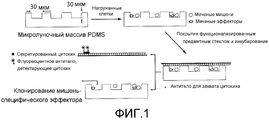
На фигуре 1 представлена схема анализа. Клетки, флуоресцентно меченные мишени (окрашенные кальцеином, зеленым) и эффекторы (окрашенные α-CD8 APC, розовым) загружают на ~30 мкм микролуночный массив и визуализируют на флуоресцентном микроскопе. Затем микролуночный массив покрывают предметным стеклом, предварительно функционализированным антителами для захвата, и инкубируют при 37°C, 5% CO2 в течение 2-6 часов. После инкубирования секретированные цитокины детектируют на предметном стекле с использованием специфических флуоресцентных антител, и мишени, лизированные под действием специфических эффекторов, визуализируют по потере интенсивности флуоресценции (лунка 1). В микролунках, содержащих только клетки-мишени (лунка 2), и в микролунках, содержащих эффекторы, которые неспособны лизировать мишень (лунка 3), должно наблюдаться очень незначительное изменение интенсивности флуоресценции мишени.The figure 1 presents the analysis scheme. Cells, fluorescently labeled targets (stained with calcein, green) and effectors (stained with α-CD8 APC, pink) are loaded onto a ~ 30 μm microwell array and visualized using a fluorescence microscope. The microwell array is then coated with a slide pre-functionalized with capture antibodies and incubated at 37 ° C, 5% CO 2 for 2-6 hours. After incubation, secreted cytokines are detected on a glass slide using specific fluorescent antibodies, and targets lysed by specific effectors are visualized by loss of fluorescence intensity (well 1). In microwells containing only target cells (well 2), and in microwells containing effectors that are unable to lyse the target (well 3), a very slight change in the fluorescence intensity of the target should be observed.
На фигуре 2 представлена серия репрезентативных флуоресцентных изображений меченых мишеней и эффекторов до и после инкубирования (0 и 4 ч.). (A) Окрашенные кальцеином (зеленый) и нагруженные пептидом (KK10) мишени (могут наблюдаться 3 отдельных клетки); (B) эффекторы, меченные (розовые) α-CD8-APC; (C) совместное инкубирование эффекторов (розовых) и ненагруженных мишеней (без добавления пептида KK10) (зеленых); (D) совместное инкубирование эффекторов (показано розовым) и мишеней (зеленых), нагруженных пептидом KK10. Лизис мишеней происходил только в том случае, когда эффекторы распознавали нагруженные пептидом мишени (показано в D).The figure 2 presents a series of representative fluorescence images of labeled targets and effectors before and after incubation (0 and 4 hours). (A) Calcein-stained (green) and peptide-loaded (KK10) targets (3 separate cells can be observed); (B) effectors labeled with (pink) α-CD8-APC; (C) co-incubation of effectors (pink) and unloaded targets (without the addition of the KK10 peptide) (green); (D) co-incubation of effectors (shown in pink) and targets (green) loaded with KK10 peptide. Lysis of targets occurred only when the effectors recognized peptide-loaded targets (shown in D).
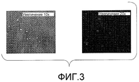
На фигуре 3 представлена панель флуоресцентных изображений CD4-T-клеток, инфицированных GFP-экспрессирующим вирусом NL4-3. Наличие зеленых клеток указывает на инфекцию.The figure 3 presents a panel of fluorescence images of CD4-T cells infected with GFP-expressing virus NL4-3. The presence of green cells indicates infection.
На фигуре 4 представлена серия флуоресцентных изображений, иллюстрирующих совместное культивирование ВИЧ-специфических CD8+-Т-клеточных клонов с В-клетками, меченными кальцеином AM и нагруженными ВИЧ-пептидом. CTL-опосредуемый лизиз определяют по отсутствию флуоресцентного сигнала.The figure 4 presents a series of fluorescence images illustrating the co-cultivation of HIV-specific CD8 + T cell clones with b cells labeled with calcein AM and loaded with HIV peptide. CTL-mediated lysis is determined by the absence of a fluorescent signal.
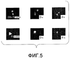
На фигуре 5 представлена панель флуоресцентных изображений, иллюстрирующих совместное культивирование APC-меченных CTL (красным) и В-клеток, меченных кальцеином AM (зеленым). Гашение флуоресценции кальцеина AM указывает на CTL-опосредуемый лизиз.5 is a panel of fluorescence images illustrating co-cultivation of APC-labeled CTL (red) and B-cells labeled with calcein AM (green). Quenching of fluorescence of calcein AM indicates CTL-mediated lysis.
На фигуре 6 схематически представлена серия анализов отдельных клеток, которые были разработаны с использованием массива микролунок. Измерения проводили последовательно или одновременно. Квадраты на каждом изображении имеют размер 50 мкм.The figure 6 schematically presents a series of analyzes of individual cells that were developed using an array of microwells. The measurements were carried out sequentially or simultaneously. The squares in each image are 50 microns in size.
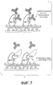
На фигуре 7 схематически представлен анализ методом захвата, проводимый для идентификации клеток, распознающих CD4-связывающую область gp120. Один массив В-клеток был использован для отпечатывания (а) на поверхности иммобилизованного gp120, а затем (b) на второй поверхности иммобилизованного gp120 после блокирования растворимым CD4.7 is a schematic representation of a capture assay performed to identify cells recognizing the CD4 binding region of gp120. One array of B cells was used to imprint (a) on the surface of the immobilized gp120, and then (b) on the second surface of the immobilized gp120 after blocking with soluble CD4.
На фигуре 8 схематически представлена экспериментальная модель для идентификации антител человека, продуцируемых B-клетками и связывающихся с множеством штаммов ВИЧ. Каждый кластер иммобилизованных антител (окрашенных) представляет один элемент микромассива, который соответствует клеткам, присутствующим в соответствующих микролуночных массивах.Figure 8 is a schematic representation of an experimental model for identifying human antibodies produced by B cells and binding to a plurality of HIV strains. Each cluster of immobilized antibodies (stained) represents one element of the microarray, which corresponds to the cells present in the corresponding microwell arrays.
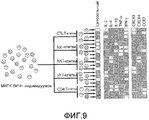
На фигуре 9 проиллюстрировано создание функционального профиля исходя из данных для отдельных клеток. Были отобраны популяции клеток, а затем были оценены их цитотоксические свойства, цитокиновые профили и поверхностные маркеры. На этих картах показан характер изменения частоты встречаемости субпопуляций в процессе инфицирования. Аналогичные профили для CD4+-T-клеток дают базисные точки, указывающие на статус данной системы.The figure 9 illustrates the creation of a functional profile based on data for individual cells. Cell populations were selected, and then their cytotoxic properties, cytokine profiles, and surface markers were evaluated. These maps show the nature of the change in the frequency of occurrence of subpopulations during infection. Similar profiles for CD4 + T cells give baseline points indicating the status of a given system.
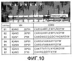
На фигуре 10 показан гель, иллюстрирующий результаты проведения ОТ-ПЦР с использованием вырожденных праймеров для отдельных B-лимфоцитов, выбранных из микролунок. B-клетки выделяли из пробы крови (в результате негативного отбора МКПК посредством сортинга на магнитных сферах) и загружали в микролунки. Клетки произвольно отбирали из двенадцати микролунок, помещали в буфер для лизиса и амплифицировали с помощью ОТ-ПЦР с использованием серии вырожденных праймеров для тяжелых и легких цепей. Было отобрано семь последовательностей белка, которые указаны внизу за гелем.Figure 10 shows a gel illustrating the results of RT-PCR using degenerate primers for individual B-lymphocytes selected from microwells. B-cells were isolated from a blood sample (as a result of negative PBMC selection by sorting on magnetic spheres) and loaded into microwells. Cells were randomly selected from twelve microwells, placed in lysis buffer and amplified by RT-PCR using a series of degenerate heavy and light chain primers. Seven protein sequences were selected, which are indicated below the gel.
Подробное описание изобретенияDETAILED DESCRIPTION OF THE INVENTION
Исчерпывающие знания механизмов, которые позволяют вирусу иммунодефицита человека (ВИЧ) уклоняться от «надзора» иммунной системы, крайне необходимы для разработки эффективных вакцин и методов терапии. На прогрессирование заболевания у индивидуумов, не подвергаемых лечению, указывает непрерывная репликация вируса и потеря CD4+-T-клеток (Kahn J.O., Walker BD (1998) Acute human immunodeficiency virus type I infection. N. Engl. J. Med. 339, 33; Hecht FM et al. (2002) Use of laboratory tests and clinical symptoms for identification of primary HIV infection. AIDS 16, 1119). Вирус-специфические CTL играют значительную роль в регуляции непрерывной репликации, и было показано, что первое появление ВИЧ-1-специфических CD8+-Т-клеток приводит к снижению вирусной нагрузки (Altfeld M et al. (2006) HLA Alleles Associated with delayed progression to AIDS contribute strongly to the initial CD8 + T cell response against HIV-I PLoS. Med. 3(10), 1851). Однако такое снижение репликации вируса у большинства индивидуумов («прогрессоров») является временным. И лишь у очень небольшой подгруппы индивидуумов, называемых LTΝP («элит-контроллерами»), поддерживаются низкие вирусные пороговые уровни на протяжении длительного периода времени, и было показано, что вирус-специфические CD8+-Т-клетки, обнаруженные у этих индивидуумов, обладают более высокой пролиферирующей способностью по сравнению с теми же самыми клетками, выделенными у «прогрессоров» (Migueles S.A. et al. (2002) HIV-specific CD8+ T cell proliferation is coupled to perforin expression and is maintained in nonprogessors Nat. Immun. 3(11), 1061).Comprehensive knowledge of the mechanisms that allow the human immunodeficiency virus (HIV) to evade the “surveillance” of the immune system is essential for developing effective vaccines and therapies. Continued virus replication and loss of CD4 + T cells (Kahn JO, Walker BD (1998) Acute human immunodeficiency virus type I infection. N. Engl. J. Med. 339, 33 indicate progression of disease in untreated individuals . ; Hecht FM et al. (2002) Use of laboratory tests and clinical symptoms for identification of primary HIV infection. AIDS 16, 1119). Virus-specific CTLs play a significant role in the regulation of continuous replication, and it has been shown that the first appearance of HIV-1-specific CD8 + T cells leads to a decrease in viral load (Altfeld M et al. (2006) HLA Alleles Associated with delayed progression to AIDS contribute strongly to the initial CD8 + T cell response against HIV-I PLoS. Med. 3 (10), 1851). However, such a decrease in virus replication in most individuals (“progressors”) is temporary. And only a very small subset of individuals called LTΝP (“elite controllers”) maintain low viral threshold levels over an extended period of time, and it has been shown that virus-specific CD8 + T cells found in these individuals have higher proliferating ability compared to the same cells isolated from “progressors” (Migueles SA et al. (2002) HIV-specific CD8 + T cell proliferation is coupled to perforin expression and is maintained in nonprogessors Nat. Immun. 3 (11 ), 1061).
До появления настоящего изобретения исследователи, изучающие взаимодействия между патогеном и иммунной системой человека, сталкивались с двумя проблемами, связанными с тем, что: 1) число клеток, присутствующих в большинстве клинических образцов, часто очень ограничено; и 2) уникальные клоны, такие как патоген-специфические B-клетки или Т-клетки, встречаются редко.Before the advent of the present invention, researchers studying the interactions between the pathogen and the human immune system faced two problems associated with: 1) the number of cells present in most clinical samples is often very limited; and 2) unique clones, such as pathogen-specific B cells or T cells, are rare.
Существующие в настоящее время аналитические средства недостаточны для одновременной оценки одной и той же отдельной клетки на множество свойств (линии дифференцировки, функции, генотипа). Так, например, проточная цитометрия представляет собой общеизвестный метод, используемый для анализа популяций отдельных клеток на фенотипические маркеры, экспрессируемые на поверхности, а анализ профилей цитокинов, который позволяет определить некоторые функциональные фенотипы, требует фиксации клеток и придания им проницаемости. Такая потеря жизнеспособности клетки означает, что дополнительные функциональные свойства, такие как цитотоксичность, не могут быть оценены прямым методом, при этом также часто затрудняется и генетический анализ. Таким образом, до появления настоящего изобретения было невозможно однозначно и точно определить гетерогенность клеток в субпопуляциях исходя из природной и адаптивной иммунных систем, отвечающих на конкретный инфекционный агент. Также трудно было создать представление о статусе иммунной системы. Такое представление облегчило бы идентификацию механизмов, которые сообщают иммунитет против некоторых патогенов и являются диагностическими индикаторами устойчивых ответов. Поэтому настоящее изобретение относится к новым технологиям измерения и установления корреляции линий дифференцировки, функций и генотипов для многих отдельных клеток, разработанных в целях повышения эффективности исследований взаимодействий между иммунной системой человека и представляющими интерес патогенами, а в частности ВИЧ (Fauci, A.S., Johnston, M.I., Dieffenbach, C.W., Burton, D.R., Hammer, S.M., Hoxie, J.A., Martin, M., Overbaugh, J., Watkins, D.I., Mahmoud, A. & Greene, W.C. Perspective -HIV vaccine research: The way forward. Science 321, 530-532 (2008)).Existing analytical tools are insufficient for the simultaneous assessment of the same single cell for many properties (lines of differentiation, function, genotype). For example, flow cytometry is a well-known method used to analyze populations of individual cells for phenotypic markers expressed on the surface, and analysis of cytokine profiles, which allows to determine some functional phenotypes, requires fixing the cells and imparting them to permeability. This loss of cell viability means that additional functional properties, such as cytotoxicity, cannot be assessed by the direct method, and genetic analysis is also often difficult. Thus, before the advent of the present invention, it was impossible to unambiguously and accurately determine the heterogeneity of cells in subpopulations based on natural and adaptive immune systems that respond to a specific infectious agent. It was also difficult to create an idea of the status of the immune system. Such an idea would facilitate the identification of mechanisms that impart immunity against certain pathogens and are diagnostic indicators of sustained responses. Therefore, the present invention relates to new technologies for measuring and correlating differentiation lines, functions and genotypes for many individual cells, designed to increase the efficiency of studies of interactions between the human immune system and pathogens of interest, in particular HIV (Fauci, AS, Johnston, MI , Dieffenbach, CW, Burton, DR, Hammer, SM, Hoxie, JA, Martin, M., Overbaugh, J., Watkins, DI, Mahmoud, A. & Greene, WC Perspective - HIV vaccine research: The way forward. Science 321, 530-532 (2008)).
Настоящее изобретение относится к способам и композициям, используемым для характеристики иммунного ответа индивидуума на инфекции, включая инфекции, вызываемые вирусом иммунодефицита человека. Микромассивы и пластины могут быть сконструированы способами, известными специалистам, включая способы, описанные в заявке PCT/US 2006/036282 (опубликованной как WO 2007/035633) и в заявке США рег. № 61/057371. Содержание обеих заявок во всей своей полноте приведено в настоящем описании в качестве ссылки. Используемый в настоящем описании термин «сформованная пластина» означает устройство, которое может быть гибким, подвижным или может деформироваться по меньшей мере в одном направлении при его приведении в контакт с субстратом. Так, например, в некоторых конфигурациях сформованная пластина может быть получена, например, из эластомерного материала, так, чтобы при контакте этой сформованной пластины с субстратом они образовывали плотное, в основном водонепроницаемое, соединение, позволяющее задерживать или предотвращать утечку любой жидкости из сформованной пластины.The present invention relates to methods and compositions used to characterize an individual's immune response to infections, including infections caused by the human immunodeficiency virus. Microarrays and wafers may be constructed by methods known to those skilled in the art, including the methods described in PCT / US 2006/036282 (published as WO 2007/035633) and US Reg. No. 61/057371. The contents of both applications in their entirety are given in the present description by reference. Used in the present description, the term "molded plate" means a device that can be flexible, movable or can be deformed in at least one direction when brought into contact with the substrate. So, for example, in some configurations, the molded plate can be obtained, for example, from an elastomeric material, so that upon contact of this molded plate with the substrate they form a dense, mostly waterproof, connection that allows to delay or prevent the leakage of any liquid from the molded plate.
Противовирусная функция цитотоксических T-лимфоцитов (CTL) в ингибировании ВИЧAntiviral function of cytotoxic T lymphocytes (CTL) in HIV inhibition
Цитотоксические CD8+-Т-клетки (CTL) играют важную роль в подавлении острых вирусных инфекций и в регуляции резервуаров инфекции, вызываемой персистирующим вирусом. Истощение CD8+-лимфоцитов у SIV-инфицированных макак приводит к быстрому и заметному повышению виремии. Тем не менее, хронические ВИЧ-1-инфекции связаны с избыточным количеством ВИЧ-специфических CD8+-Т-клеток в отсутствие клиренса вируса или регуляции его уровня. Эти данные позволяют предположить, что именно функции, а не число CD8+-Т-клеток играет важную роль в эффективной регуляции уровня репликации вируса. До настоящего времени активность ВИЧ-специфических CD8+-Т-клеток определяли либо путем оценки частоты встречаемости ВИЧ-специфических CD8+-Т-клеток посредством мечения этих клеток тетрамерами комплексов «пептид-HLA класса I», либо по способности этих клеток секретировать IFN-γ после антигенной стимуляции. Однако недавно проведенные исследования показали, что частота встречаемости CD8+-Т-клеток и секреция IFN-γ под действием этих клеток не коррелирует с регуляцией виремии при хронических инфекциях, вызываемых вирусом ВИЧ-1.Cytotoxic CD8 + T cells (CTL) play an important role in the suppression of acute viral infections and in the regulation of reservoirs of infection caused by persistent virus. Depletion of CD8 + lymphocytes in SIV-infected macaques leads to a rapid and marked increase in viremia. However, chronic HIV-1 infections are associated with an excess of HIV-specific CD8 + T cells in the absence of clearance of the virus or regulation of its level. These data suggest that it is functions, and not the number of CD8 + T cells, that play an important role in effectively regulating the level of virus replication. To date, the activity of HIV-specific CD8 + T cells has been determined either by assessing the frequency of HIV-specific CD8 + T cells by labeling these cells with tetramers of the “peptide-HLA class I” complexes, or by the ability of these cells to secrete IFN- γ after antigenic stimulation. However, recent studies have shown that the frequency of occurrence of CD8 + T cells and the secretion of IFN-γ under the influence of these cells does not correlate with the regulation of viremia in chronic infections caused by the HIV-1 virus.
Способность CTL подавлять репликацию ВИЧ измеряют путем совместного культивирования ВИЧ-инфицированных CD4+-Т-клеток и массы аутологичных CD8+-Т-клеток. Эти эксперименты давали неоднородные результаты в отношении способности индивидуумов к ингибированию ВИЧ. Недавно полученные результаты позволяют предположить, что CD8+-Т-клеточные ответы ex vivo не коррелируют со способностью этих клеток ингибировать репликацию ВИЧ-1 in vitro. Скорее всего CD8+-Т-клетки, которые могут пролифирировать в количествах, достаточных для борьбы с вирусом, способны осуществлять регуляцию уровня вируса, однако фенотип, функциональные свойства и генетический транскрипционный профиль (созревание/истощение) этих клеток пока неизвестны.The ability of CTL to suppress HIV replication is measured by co-culturing HIV-infected CD4 + T cells and a mass of autologous CD8 + T cells. These experiments gave mixed results regarding the ability of individuals to inhibit HIV. Recent results suggest that ex vivo CD8 + T cell responses do not correlate with the ability of these cells to inhibit HIV-1 replication in vitro . Most likely, CD8 + T cells, which can proliferate in quantities sufficient to fight the virus, are able to regulate the level of the virus, however, the phenotype, functional properties and genetic transcriptional profile (maturation / depletion) of these cells are still unknown.
Хотя важная роль ВИЧ-специфических CD8+-Т-клеток в предотвращении прогрессирования заболевания не вызывает сомнений, однако их характеристика и выделение представляют значительные трудности. Для выделения CTL были применены различные методы, каждый из которых имеет свои достоинства и преимущества. Для выделения антиген-специфических клонов может быть проведен анализ методом лимитирующего разведения, однако результаты такого анализа зависят от способности этих клонов к размножению в тканевой культуре. Анализ ELISPOT позволяет определять способность активированных CTL секретировать отдельный цитокин, но не дает какой-либо информации относительно их литических свойств. Было также продемонстрировано, что антигенпрезентирующие клетки (АПК), обнаруживающие низкую плотность нагруженных пептидом MHC (pMHC), могут обладать цитотоксической функцией без одновременной секреции цитокинов (Valitutti S et al. (1996) Different responses are elicited in cytotoxic T lymphocytes by different levels of T cell receptor occupancy. J. Exp. Med. 183, 1917). Это ограничение оказалось особенно эффективным при попытке выделения ВИЧ-специфических CTL, поскольку вирусная инфекция ингибирует MHC класса I (Mangasarian A et al. (1999) Nef-Induced CD4 and Major Histocompatibility Complex Class I (MHC-I) Down-Regulation Are Governed by Distinct Determinants: N-Terminal Alpha Helix and Proline Repeat of Nef Selectively Regulate MHC-I Trafficking J. Virol. 73(3), 1964). Для выделения ангигенспецифических CTL использовали окрашивание нагруженным пептидом и флуоресцентно помеченным тетрамером HLA класса I в комбинации с проточной цитометрией, однако это также не дало какой-либо информации о литической способности этих клеток. Кроме того, было показано, что CTL, выделенные методом окрашивания тетрамером, не всегда распознают инфицированные вирусом клетки (Appay V et al. (2000) HIV-specific CD8+ T cells produce antiviral cytokines but are impaired in cytolytic function. J. Exp. Med. 192(1), 63). В литературе описан анализ лизиса методом проточной цитометрии, проводимый с использованием субстратов каспазы, однако такой анализ непригоден для скрининга большого числа эффекторных клеток (Liu L et al. (2002) Visualization and quantification of T cell-mediated cytotoxicity using cell-permeable fluorogenic caspase substrates Nat. Med. 8, 185). Таким образом, до появления настоящего изобретения не существовало какого-либо конкретного крупномасштабного метода, который позволял бы определять способность отдельных CTL к лизису отдельных инфицированных первичных клеток-мишеней и измерять число цитокинов и цитотоксических молекул, секретируемых этими клетками, а значит, и отобрать «живую» клетку в целях определения клональных линий для последующей функциональной характеристики и генетического анализа.Although the important role of HIV-specific CD8 + T cells in preventing the progression of the disease is not in doubt, their characterization and isolation represent significant difficulties. Various methods were used to isolate CTL, each of which has its own advantages and advantages. To isolate antigen-specific clones, a limiting dilution analysis can be performed, however, the results of this analysis depend on the ability of these clones to reproduce in tissue culture. ELISPOT analysis allows the ability of activated CTLs to secrete a single cytokine to be determined, but does not provide any information regarding their lytic properties. It has also been demonstrated that antigen-presenting cells (APCs) that detect low density of peptides loaded with MHC (pMHC) may have cytotoxic function without simultaneous secretion of cytokines (Valitutti S et al. (1996) Different responses are elicited in cytotoxic T lymphocytes by different levels of T cell receptor occupancy. J. Exp. Med. 183, 1917). This limitation has been found to be particularly effective in attempting to isolate HIV-specific CTLs, since a viral infection inhibits Class I MHC (Mangasarian A et al. (1999) Nef-Induced CD4 and Major Histocompatibility Complex Class I (MHC-I) Down-Regulation Are Governed by Distinct Determinants: N-Terminal Alpha Helix and Proline Repeat of Nef Selectively Regulate MHC-I Trafficking J. Virol. 73 (3), 1964). Angigen-specific CTLs were isolated by staining with a loaded peptide and fluorescently labeled class I HLA tetramer in combination with flow cytometry, but this also did not provide any information on the lytic ability of these cells. In addition, it was shown that CTLs isolated by tetramer staining do not always recognize virus-infected cells (Appay V et al. (2000) HIV-specific CD8 + T cells produce antiviral cytokines but are impaired in cytolytic function. J. Exp. Med . 192 (1), 63). Literature describes a lysis assay using flow cytometry using caspase substrates, but this assay is not suitable for screening a large number of effector cells (Liu L et al. (2002) Visualization and quantification of T cell-mediated cytotoxicity using cell-permeable fluorogenic caspase substrates Nat. Med. 8, 185). Thus, prior to the advent of the present invention, there was no specific large-scale method that could determine the ability of individual CTLs to lyse individual infected primary target cells and measure the number of cytokines and cytotoxic molecules secreted by these cells, and therefore select “live” »Cell in order to determine clonal lines for subsequent functional characteristics and genetic analysis.
Сохранение высоких уровней CD8+-Т-клеток, которые способны распознавать и лизировать инфицированные CD4+-Т-клетки, непосредственно коррелирует с уровнем ингибирования репликации вируса у пациента. Определение уникального фенотипа, функции и профиля экспрессии генов в цитолитических и нецитолитических CTL на уровне отдельных клеток позволяет обнаруживать корреляты сообщаемого CD8+-Т-клетками противовирусного иммунитета, необходимого для разработки эффективной вакцины против ВИЧ. Настоящее изобретение относится к анализу на элиминацию отдельных CTL в комбинации с фенотипированием и генетическими анализами, позволяющими определять иммунологические и генетические корреляты эффективного опосредуемого CD8+-Т-клетками противовирусного иммунного ответа. Определение таких свойств на уровне отдельных клеток позволит определить прототипический ответ, который будет вырабатываться посредством иммунизации вакциной, сконструированной в целях увеличения числа CD8+-Т-клеток, способных эффективно подавлять репликацию вируса.Maintaining high levels of CD8 + T cells that are able to recognize and lyse infected CD4 + T cells directly correlates with the level of inhibition of virus replication in the patient. Determining the unique phenotype, function and profile of gene expression in cytolytic and non-cytolytic CTLs at the individual cell level allows us to detect the correlates of the antiviral immunity reported by CD8 + T cells necessary to develop an effective HIV vaccine. The present invention relates to the analysis of the elimination of individual CTLs in combination with phenotyping and genetic assays to determine the immunological and genetic correlates of an effective CD8 + T-cell mediated antiviral immune response. The determination of such properties at the level of individual cells will determine the prototypical response that will be generated by immunization with a vaccine designed to increase the number of CD8 + T cells capable of effectively inhibiting virus replication.
Разнообразие антител у ВИЧ-инфицированных пациентовA variety of antibodies in HIV-infected patients
Первичным рецептором, экспрессируемым на поверхности ВИЧ, является gp120. Рецептор, презентируемый на поверхности некоторых Т-клеток, необходим для инфицирования и связывается с CD4. Было предпринято множество попыток разработки вакцин против ВИЧ в целях блокирования инфекции посредством вырабатывания NAb-ответа против gp120, однако в настоящее время все эти попытки оказались безуспешными, что обусловлено, главным образом, вариабельностью рецепторов у различных штаммов и предрасположенностью этих рецепторов к мутации в их хозяине. Однако в настоящее время имеются примеры инфицирования людей, у которых в естественных условиях образуются NAb, обладающие способностью нейтрализовать многие варианты вируса широкого ряда. Разнообразие этих антител трудно оценить стандартными методами, поскольку уникальные антитела присутствуют в сыворотке в ограниченном количестве, трудно поддаются очистке и не могут быть рекомбинантно продуцированы без соответствующих генов. Проблема заключается в том, что bNAb соответствуют клональной линии В-клеток, из которых они продуцируются. До настоящего времени наиболее успешной попыткой идентифицировать гены, кодирующие NAb, было применение пэннинга библиотек антител, продуцируемых рекомбинантными методами из большого числа В-клеток, присутствующих в кровотоке ВИЧ-положительных индивидуумов (Koefoed, K., Farnaes, L., Wang, M., Svejgaard, A., Burton, D.R. & Ditzel, H.J. Molecular characterization of the circulating anti-HIV-1 gp120-specific B cell repertoire using antibody phage display libraries generated from pre-selected HIV-I gp120 binding PBLs. J. Immunol Methods 297, 187-201 (2005)). Однако эти подходы не дают возможности точно определить природный репертуар антител у индивидуумов, поскольку такой способ приводит к нарушению уникальных клональных комбинаций тяжелых и легких цепей. Как описано ниже в примерах, необходимо решить две проблемы, а именно определить: (1) клональное разнообразие bNAb-продуцирующих В-клеток у индивидуума; и (2) свойства bNAb, которое связывается с первичными изолятами широкого ряда.The primary receptor expressed on the surface of HIV is gp120. A receptor presented on the surface of certain T cells is necessary for infection and binds to CD4. Many attempts have been made to develop HIV vaccines to block infection by generating an anti-gp120 NAb response, but currently all of these attempts have been unsuccessful, mainly due to the variability of receptors in different strains and the susceptibility of these receptors to mutations in their host. . However, there are currently examples of infection in people with naturally occurring NAb that have the ability to neutralize many variants of a wide range of viruses. The diversity of these antibodies is difficult to evaluate by standard methods, since unique antibodies are present in serum in a limited amount, are difficult to purify, and cannot be recombinantly produced without the appropriate genes. The problem is that bNAbs correspond to the clonal line of B cells from which they are produced. To date, the most successful attempt to identify genes encoding NAb has been the use of panning of antibody libraries produced by recombinant methods from a large number of B cells present in the bloodstream of HIV-positive individuals (Koefoed, K., Farnaes, L., Wang, M. , Svejgaard, A., Burton, DR & Ditzel, HJ Molecular characterization of the circulating anti-HIV-1 gp120-specific B cell repertoire using antibody phage display libraries generated from pre-selected HIV-I gp120 binding PBLs. J. Immunol Methods 297, 187-201 (2005)). However, these approaches do not make it possible to accurately determine the natural repertoire of antibodies in individuals, since this method leads to disruption of unique clonal combinations of heavy and light chains. As described in the examples below, it is necessary to solve two problems, namely to determine: (1) the clonal diversity of bNAb-producing B cells in an individual; and (2) the properties of bNAb, which binds to primary isolates of a wide range.
Клеточные иммунные ответы против ВИЧAnti-HIV Cellular Immune Responses
Есть надежда, что вакцина против ВИЧ будет эффективной для тех индивидуумов, у которых наблюдается естественное заметное подавление прогрессирования заболевания, у так называемых «элит-контроллеров», и у приматов, не являющихся человеком и обладающих иммунитетом к обезьяньему вирусу иммунодефицита (SIV), который был сообщен им путем вакцинации; однако решающие факторы, которые в данном случае коррелируют с иммунитетом, пока еще не ясны (Saez-Cirion, A., Pancino, G., Sinet, M., Venet, A., Lambotte, O. & Gr, A.E.H.C.S. HIV controllers: how do they tame the virus? Trends Immunol 28, 532-540 (2007); Deeks, S.G. & Walker, B.D. Human immunodeficiency virus controllers: Mechanisms of durable virus control in the absence of antiretroviral therapy. Immunity 27, 406-416 (2007); Koff, W.C., Johnson, P.R., Watkins, D.I., Burton, D.R., Lifson, J.D., Hasenkrug, K.J., McDermott, A.B., Schultz, A., Zamb, T.J., Boyle, R. & Desrosiers, R.C. HIV vaccine design: insights from live attenuated SIV vaccines. Nat Immunol 7, 19-23 (2006)). Особенно большого внимания заслуживает адаптивный иммунный ответ, и недавно проведенные исследования природной иммунной системы показали его важность в формировании адаптивного ответа (Pulendran, B. & Ahmed, R. Translating innate immunity into immunological memory: Implications for vaccine development. Cell 124, 849-863 (2006)). Имеющиеся в настоящее время анализы, проводимые путем мониторинга эффективной элиминации инфицированных клеток иммунными клетками, не позволяют в достаточной степени охарактеризовать гетерогенность в отношении их функций (Fauci, A. S., Johnston, M.I., Dieffenbach, C. W., Burton, D.R., Hammer, S. M., Hoxie, J.A., Martin, M., Overbaugh, J., Watkins, D.I., Mahmoud, A. & Greene, W.C. Perspective - HIV vaccine research: The way forward. Science 321, 530-532 (2008); Walker, B.D. & Burton, D.R. Toward an AIDS vaccine. Science 320, 760-764 (2008)). Настоящее изобретение относится к способам характеристики и корреляции множества иммунных функций отдельных клеток различных линий дифференцировки для анализа субпопуляций эффекторных клеток у пациентов различных групп, например у пациентов с острыми инфекциями, у пациентов с хроническим прогрессирующим заболеванием, у пациентов, подвергаемых интенсивной антиретровирусной терапии (HAART), и у «элит-контроллеров». Как описано в настоящей заявке, количественные анализы клеток позволяют идентифицировать клетки, имеющие специфические комбинации функций, необходимых для регуляции репликации вирусов.It is hoped that the HIV vaccine will be effective for those individuals who have a natural marked suppression of disease progression, the so-called “elite controllers”, and non-human primates that are immune to monkey immunodeficiency virus (SIV), which he was informed by vaccination; however, the decisive factors that in this case correlate with immunity are not yet clear (Saez-Cirion, A., Pancino, G., Sinet, M., Venet, A., Lambotte, O. & Gr, AEHCS HIV controllers: how do they tame the virus? Trends Immunol 28, 532-540 (2007); Deeks, SG & Walker, BD Human immunodeficiency virus controllers: Mechanisms of durable virus control in the absence of antiretroviral therapy. Immunity 27, 406-416 (2007 ); Koff, WC, Johnson, PR, Watkins, DI, Burton, DR, Lifson, JD, Hasenkrug, KJ, McDermott, AB, Schultz, A., Zamb, TJ, Boyle, R. & Desrosiers, RC HIV vaccine design : insights from live attenuated SIV vaccines.
Другим средством защиты от вирусных инфекций является природный иммунный ответ. NK-клетки представляют собой центральный компонент такого ответа (Alter, G., Teigen, N., Ahem, R., Streeck, H., Meier, A., Rosenberg, E.S. & Altfeld, M. Evolution of innate and adaptive effector cell functions during acute HIV-1 infection. J. Infect Dis 195, 1452-1460 (2007)). Такими клетками являются цитотоксические эффекторные клетки, которые также индуцируют адаптивные иммунные ответы посредством высвобождения цитокинов, таких как IFN-γ, MIP-1β, TNF-α и GM-CSF. В случае ВИЧ-1-инфекции данные эпидемиологических исследований со всей очевидностью продемонстрировали, что у индивидуумов, у которых имеются конкретные NK-клеточные рецепторы (рецептор цитотоксического иммуноглобулина 3DS1 (KIR3DS1) и некоторые аллели KIR3DL1) и их предполагаемые лиганды (аллели HLA-B с изолейцином в положении 80), СПИД прогрессирует более медленно, чем у пациентов, у которых имеется только один или из этих аллелей или вообще отсутствуют эти аллели (Martin, M.P., Qi, Y., Gao, X.J., Yamada, E., Martin, J.N., Pereyra, F., Colombo, S., Brown, E.E., Shupert, W.L., Phair, J., Goedert, J.J., Buchbinder, S., Kirk, G.D., Telenti, A., Connors, M., O'Brien, S.J., Walker, B.D., Parham, P., Deeks, S. G., McVicar, D. W. & Carrington, M. Innate partnership of HLA-B and KIR3DL1 subtypes against HIV-1. Nat Genet 39, 733-740 (2007)). Аналогичным образом, повышенная активность NK-клеток и повышенная экспрессия транскриптов KIR3DS1 в массе NK-клеток коррелирует с иммунитетом к инфекции даже после повторного инфицирования (Alter, G., Martin, M.P., Teigen, N., Carr, W.H., Suscovich, T.J., Schneidewind, A., Streeck, H., Waring, M., Meier, A., Brander, C, Lifson, J.D., Allen, T.M., Carrington, M. & Altfeld, M. Differential natural killer cell-mediated inhibition of HIV-I replication based on distinct KIR/HLA subtypes. The Journal of experimental medicine 204, 3027-3036 (2007); Long, B.R., Ndhlovu, L.C., Oksenberg, J.R., Lanier, L.L., Hecht, F.M., Nixon, D.F. & Barbour, J.D. Conferral of enhanced natural killer cell function by KIR3DS1 in early human immunodeficiency virus type 1 infection. J. Virol 82, 4785-4792 (2008)). Эти данные позволяют предположить, что конкретные популяции NK-клеток играют защитную роль в предупреждении и подавлении инфекции, однако точные фенотипы этих клеток пока не были определены.Another defense against viral infections is the natural immune response. NK cells are the central component of such a response (Alter, G., Teigen, N., Ahem, R., Streeck, H., Meier, A., Rosenberg, ES & Altfeld, M. Evolution of innate and adaptive effector cell functions during acute HIV-1 infection. J. Infect Dis 195, 1452-1460 (2007)). Such cells are cytotoxic effector cells that also induce adaptive immune responses through the release of cytokines, such as IFN-γ, MIP-1β, TNF-α and GM-CSF. In the case of HIV-1 infection, epidemiological studies have clearly demonstrated that individuals who have specific NK cell receptors (the cytotoxic immunoglobulin receptor 3DS1 (KIR3DS1) and some KIR3DL1 alleles) and their putative ligands (HLA-B alleles with isoleucine at position 80), AIDS progresses more slowly than in patients who have only one or one of these alleles or none of these alleles (Martin, MP, Qi, Y., Gao, XJ, Yamada, E., Martin, JN, Pereyra, F., Colombo, S., Brown, EE, Shupert, WL, Phair, J., Goedert, JJ, Buchbinder, S., Kirk, GD, Telent i, A., Connors, M., O'Brien, SJ, Walker, BD, Parham, P., Deeks, SG, McVicar, DW & Carrington, M. Innate partnership of HLA-B and KIR3DL1 subtypes against HIV-1 Nat Genet 39, 733-740 (2007)). Similarly, increased NK cell activity and increased expression of KIR3DS1 transcripts in the mass of NK cells correlate with infection immunity even after reinfection (Alter, G., Martin, MP, Teigen, N., Carr, WH, Suscovich, TJ, Schneidewind, A., Streeck, H., Waring, M., Meier, A., Brander, C, Lifson, JD, Allen, TM, Carrington, M. & Altfeld, M. Differential natural killer cell-mediated inhibition of HIV -I replication based on distinct KIR / HLA subtypes. The Journal of experimental medicine 204, 3027-3036 (2007); Long, BR, Ndhlovu, LC, Oksenberg, JR, Lanier, LL, Hecht, FM, Nixon, DF & Barbour , JD Conferral of enhanced natural killer cell function by KIR3DS1 in early human
Одним из механизмов, используемых NK-клетками для элиминации ВИЧ-инфицированных клеток, является прямой цитолиз после межклеточного контакта. Некоторые формы KIR, экспрессируемые на поверхности NK-клеток, посылают ингибирующие сигналы клетке, которая подавляет цитолитическую функцию при лигировании с HLA класса I, экспрессируемом на поверхности клетки-мишени (Moretta, A., Bottino, C, Mingari, M.C., Biassoni, R. & Moretta, L. What is a natural killer cell? Nat Immunol 3, 6-8 (2002)). В отсутствие таких взаимодействий NK-клетки активируют и лизируют клетку-мишень. ВИЧ-инфицированные клетки часто ингибируют экспрессию аллелей HLA-A и -B, что делает их более восприимчивыми к цитолизу под действием активированных NK-клеток (Fogli, M., Mavilio, D., Brunetta, E., Varchetta, S., Ata, K., Roby, G., Kovacs, C, Follmann, D., Pende, D., Ward, J., Barker, E., Marcenaro, E., Moretta, A. & Fauci, A.S. Lysis of endogenously infected CD4+ T cell blasts by rIL-2 activated autologous natural killer cells from HIV-infected viremic individuals. Plos Pathogens 4, 1-13 (2008)). Вторым механизмом, посредством которого субпопуляция NK-клеток, экспрессирующих CD16 (Fcγ-рецептор III), разрушает ВИЧ-инфицированные клетки, является ADCC (Cooper, M.A., Fehniger, T.A. & Caligiuri, M.A. The biology of human natural killer-cell subsets. Trends Immunol 22, 633-640 (2001)). Эти рецепторы связываются с Fc-областью антител, связанных с мишенями на поверхности инфицированных клеток, и активируют лизис под действием NK-клеток. Такой механизм защиты требует объединения гуморального иммунного ответа, опосредуемого В-клетками, и природного ответа NK-клеток. Этот ответ может иметь важное значение для замедления прогрессирования ВИЧ-инфекции, а в некоторых случаях для защиты от инфекции ВИЧ-1 у индивидуумов, которым внутривенно было введено лекарственное средство, и от SIV-инфекции у макак (Stratov, L, Chung, A. & Kent, S.J. Robust NK cell-mediated human immunodeficiency virus (HΙV)-specific antibody-dependent responses in HIV-infected subjects. J. Virol 82, 5450-5459 (2008)). Однако до появления настоящего изобретения специалисты, изучающие этот механизм, сталкивались с ограничениями, заключающимися в отсутствие количественных анализов для измерения ADCC.One of the mechanisms used by NK cells to eliminate HIV-infected cells is direct cytolysis after intercellular contact. Some forms of KIR expressed on the surface of NK cells send inhibitory signals to a cell that inhibits cytolytic function when ligated with class I HLA expressed on the surface of the target cell (Moretta, A., Bottino, C, Mingari, MC, Biassoni, R . & Moretta, L. What is a natural killer cell?
Настоящее изобретение более подробно проиллюстрировано на нижеследующих неограничивающих примерах.The present invention is illustrated in more detail in the following non-limiting examples.
Пример 1. Разработка высокоэффективного анализа для оценки цитолитической активности отдельных клетокExample 1. The development of a highly effective analysis to assess the cytolytic activity of individual cells
В ранее проводимых исследованиях был описан высокоэффективный анализ для функциональной характеристики большого числа первичных клеток путем загрузки отдельных клеток в пиколитровые микролунки (~2×10⁵ на микромассив, каждая лунка имела диаметр ~30 мкм) (Bradshaw EM et al. (2008) Concurrent detection of secreted products from human lymphocytes by microengraving: cytokines and antigen-reactive antibodies. Clin. Immunol. 129(1), 10; Love J.C. et al. (2006) A microengraving method for rapid selection of single cells producing antigen-specific antibodies. Nat. Biotech. 24(6), 703). Загруженные микролунки поддерживали в физическом контакте с предметными стеклами, предварительно функционализированными соответствующими реагентами (например, анти-IL-2 антителом для захвата, нанесенным на предметные стекла с полилизином) и инкубировали в течение 2 часов для захвата секретированных цитокинов. Затем предметные стекла обрабатывали и метили соответствующими детектирующими антителами для выявления флуоресцентных пятен на предметных стеклах, которые могут быть картированы на клетках, секретирующих эти антитела. Затем эти клетки могут быть отобраны с применением роботизированных микроманипуляторов для клонального размножения.Previous studies have described a highly effective analysis for the functional characterization of a large number of primary cells by loading individual cells into picoliter microwells (~ 2 × 10 5 per microarray, each well having a diameter of ~ 30 μm) (Bradshaw EM et al. (2008) Concurrent detection of secreted products from human lymphocytes by microengraving: cytokines and antigen-reactive antibodies. Clin. Immunol . 129 (1), 10; Love JC et al. (2006) A microengraving method for rapid selection of single cells producing antigen-specific antibodies. Nat. Biotech. 24 (6), 703). The loaded microwells were maintained in physical contact with glass slides pre-functionalized with appropriate reagents (for example, anti-IL-2 capture antibody coated on a glass slide with polylysine) and incubated for 2 hours to capture secreted cytokines. The slides were then processed and labeled with appropriate detecting antibodies to detect fluorescent spots on the slides that can be mapped to cells secreting these antibodies. Then these cells can be selected using robotic micromanipulators for clonal propagation.
Настоящее изобретение относится к применению массивов микролунок для исследования межклеточных взаимодействий. В частности, настоящее изобретение относится к способу мониторинга лизиса инфицированных клеток-мишеней отдельными CTL при одновременном определении профиля маркеров активации/секретированных растворимых медиаторов. Другим преимуществом этой аналитической системы является скорость получения результатов (или завершения анализа). Так, например, результаты получают (или анализ завершают) менее чем через 24 часа, менее чем через 12 часов или менее чем через 10 часов. Так, например, результаты получают (или анализ завершают) менее чем через 4 часа.The present invention relates to the use of microwell arrays for the study of intercellular interactions. In particular, the present invention relates to a method for monitoring the lysis of infected target cells with individual CTLs while simultaneously determining the profile of activation markers / secreted soluble mediators. Another advantage of this analytical system is the speed of obtaining results (or completion of analysis). For example, the results are obtained (or the analysis is completed) in less than 24 hours, in less than 12 hours, or in less than 10 hours. So, for example, the results are obtained (or the analysis is completed) in less than 4 hours.
Изготовление штампов микромассивовProduction of stamps of microarrays
Микролуночные массивы изготавливают из полидиметилсилоксана (PDMS) с применением фотолитографии и формования реплик. Глубина и размер лунок предпочтительно составляют менее чем 100 мкм, например менее чем 50 мкм. По усмотрению специалиста глубина и размер лунок могут быть доведены до ~30 мкм. Для стерилизации микромассивов и сообщения им гидрофильных свойств проводят обработку O2-плазмой. Обработанный плазмой массив погружают в PBS-BSA в целях сохранения гидрофильных свойств и для последующего применения.Microwell arrays are made from polydimethylsiloxane (PDMS) using photolithography and replica forming. The depth and size of the wells are preferably less than 100 microns, for example less than 50 microns. At the discretion of a specialist, the depth and size of the wells can be brought up to ~ 30 μm. To sterilize microarrays and impart hydrophilic properties to them, O 2 plasma treatment is carried out. The plasma-treated array is immersed in PBS-BSA in order to maintain hydrophilic properties and for subsequent use.
Клеточные материалыCell materials
В описанных ниже методах использовали ВИЧ-инфицированные CD4-клетки и аутологичные CD8-клетки. В описанных ниже методах также использовали МКПК, взятые у индивидуумов с прогрессирующим заболеванием, и у LTNP- и ВИЧ-отрицательных индивидуумов. Кроме того, выделенные CTL-клоны, которые специфически распознают HLA-B27-рестриктированный пептид gag ВИЧ (KK10), служили в качестве позитивного контроля для оценки достоверности анализа и уточнения экспериментальных условий.The methods described below used HIV-infected CD4 cells and autologous CD8 cells. The methods described below also used PBMCs from individuals with progressive disease and from LTNP and HIV-negative individuals. In addition, isolated CTL clones that specifically recognize the HLA-B27-restricted HIV gag peptide (KK10) served as a positive control to evaluate the validity of the assay and refine experimental conditions.
Разработка анализаAnalysis Development
В целях создания точного протокола выделения мишень-специфических CTL ранее упомянутый KKl0-специфический клон (эффектор) использовали в качестве эффекторов, а HLA B27-экспрессирующие EBV-трансформированные B-клетки использовали в качестве мишеней. Общий протокол анализа схематически проиллюстрирован на фигуре 1. Нагруженные пептидом В-клетки (мишени) окрашивали кальцеином AM (Invitrogen, Carlsabad, CA), то есть неспецифическим флуоресцентным субстратом для внутриклеточных эстераз и липаз, и эти клетки служили в качестве маркера целостности клеток. Интактные клетки с нормальным иммунитетом оставались флуоресцентными в отсутствие эффекторных клеток на протяжении всего анализа на цитолиз (4 часа), как показано на фигуре 2A.In order to create an accurate protocol for the isolation of target-specific CTLs, the previously mentioned KKl0-specific clone (effector) was used as effectors, and HLA B27-expressing EBV-transformed B cells were used as targets. The general assay protocol is schematically illustrated in Figure 1. Peptide-loaded B cells (targets) were stained with calcein AM (Invitrogen, Carlsabad, CA), that is, a non-specific fluorescent substrate for intracellular esterases and lipases, and these cells served as a marker for cell integrity. Normal-immune intact cells remained fluorescent in the absence of effector cells throughout the cytolysis assay (4 hours), as shown in Figure 2A.
Поскольку снижение интенсивности флуоресценции мишени также, вероятно, обусловлено фотоотбеливанием/деградацией флуорофора после длительного инкубирования, то мишени метили красителем Sytox красным (Invitrogen, Carlsabad, CA). Семейство мембрано-непроницаемых красителей Sytox давало значительное увеличение интенсивности флуоресценции (>500 раз) при их интеркаляции нуклеиновыми кислотами, а поэтому такие красители могут быть использованы в качестве специфических маркеров для лизированных клеток с поврежденными мембранами. Поскольку красители Sytox и кальцеин метят клетки по ортогональному механизму, то они могут быть использованы одновременно для мониторинга кинетики лизиса мишеней путем детектирования увеличения/снижения интенсивности флуоресценции в независимых каналах флуоресцентного микроскопа.Since the decrease in the fluorescence intensity of the target is also probably due to photobleaching / degradation of the fluorophore after long incubation, the targets were labeled with Sytox red dye (Invitrogen, Carlsabad, CA). The Sytox family of membrane-tight dyes gave a significant increase in fluorescence intensity (> 500 times) when they were intercalated by nucleic acids, and therefore such dyes can be used as specific markers for lysed cells with damaged membranes. Since Sytox and calcein dyes label cells according to the orthogonal mechanism, they can be used simultaneously to monitor the kinetics of target lysis by detecting an increase / decrease in fluorescence intensity in independent channels of a fluorescence microscope.
Эффекторы были помечены флуоресцентными анти-CD8 антителами (APC/Alexa647/тихоокеанский голубой). Может быть использован краситель, который дает наиболее репродуцируемый сигнал и наименьшее фотоотбеливание/наименьшую деградацию в аналитических условиях (4 часа, 37°C). Эффекторы, помеченные α-CD8-APC, представлены на фигуре 2B.The effectors were labeled with fluorescent anti-CD8 antibodies (APC / Alexa647 / Pacific Blue). A dye can be used that gives the most reproducible signal and the least photo bleaching / least degradation under analytical conditions (4 hours, 37 ° C). The effectors labeled with α-CD8-APC are shown in Figure 2B.
Предварительные исследования (Bradshaw EM et al. (2008) Concurrent detection of secreted products from human lymphocytes by microengraving: cytokines and antigen-reactive antibodies. Clin. Immunol. 129(1), 10; Love J.C. et al. (2006) A microengraving method for rapid selection of single cells producing antigen-specific antibodies. Nat. Biotech. 24(6), 703) и предварительные эксперименты (фигуры 2A и 2B) подтвердили, что клетки не подвергаются апоптозу при их инкапсулировании в микролунках. Затем были определены оптимальные пути загрузки микролуночных массивов мишенями и эффекторами в определенных отношениях. Были оценены два различных подхода, а именно предварительное смешивание мишеней и эффекторов в среде перед загрузкой, или последовательная загрузка эффекторов и мишеней. Поскольку сначала осуществляли последовательную загрузку эффекторов, а затем мишеней, то это давало лучший эффект, чем изменение отношения «эффектор:мишень» в микролунках, а поэтому этот подход был принят за стандартный. Для исследования целесообразности применения данного анализа ненагруженные мишени (мишени с несвязанным пептидом, отрицательный контроль) и KK10-нагруженные мишени (положительный контроль) инкубировали с эффекторами. Поскольку T-клеточный рецептор (TCR) на эффекторе может распознавать только pMHC на мишени, то лизис в отсутствие пептида не наблюдался (фигура 2C). При предварительной нагрузке мишеней пептидом KK10 и их инкубировании с эффекторами наблюдался лизис мишеней, на что указывала потеря флуоресцентного излучения в зеленом диапазоне спектра (фигура 2D). Данные, приведенные на фигуре 2, представляют собой репрезентативные данные, полученные для одной отдельной микролунки. Для оценки числа микролунок, которые имеют отдельную мишень, инкубированную с отдельным эффектором, и для определения числа субпопуляции эффекторов, которые специфически лизировали мишени, необходимо провести автоматизированный анализ генерируемых флуоресцентных изображений (два различных канала) и фазово-контрастного изображения (проходящий свет, третий канал). В экспериментах, включающих контроли и проводимых с использованием клонов-мишеней и эффекторных клонов, описанных выше, для оценки достоверности данного анализа достаточно провести лишь мануальную оценку небольшого числа клеток (~102), однако в целях рутинного скрининга необходимо разработать алгоритмы для получения большого набора данных и их анализа.Preliminary studies (Bradshaw EM et al. (2008) Concurrent detection of secreted products from human lymphocytes by microengraving: cytokines and antigen-reactive antibodies. Clin. Immunol. 129 (1), 10; Love JC et al. (2006) A microengraving method for rapid selection of single cells producing antigen-specific antibodies. Nat. Biotech. 24 (6), 703) and preliminary experiments (figures 2A and 2B) confirmed that the cells do not undergo apoptosis when they are encapsulated in microwells. Then, optimal paths for loading microwell arrays with targets and effectors were determined in certain respects. Two different approaches were evaluated, namely pre-mixing of targets and effectors in the medium before loading, or sequential loading of effectors and targets. Since the effectors and then the targets were sequentially loaded first, this gave a better effect than changing the effector: target ratio in the microwells, and therefore this approach was taken as the standard one. To study the feasibility of using this analysis, unloaded targets (targets with an unbound peptide, negative control) and KK10-loaded targets (positive control) were incubated with effectors. Since the T cell receptor (TCR) on the effector can only recognize pMHC on the target, lysis in the absence of the peptide was not observed (Figure 2C). Upon preliminary loading of the targets with the KK10 peptide and their incubation with effectors, lysis of the targets was observed, which was indicated by the loss of fluorescence radiation in the green spectral range (Figure 2D). The data shown in FIG. 2 are representative data obtained for one single microwell. To estimate the number of microwells that have a separate target incubated with a separate effector, and to determine the number of subpopulations of effectors that specifically lyse targets, it is necessary to carry out an automated analysis of the generated fluorescence images (two different channels) and phase-contrast images (transmitted light, third channel ) In experiments involving controls performed using target clones and effector clones described above, to assess the reliability of this analysis, it is sufficient to conduct a manual assessment of a small number of cells (~ 10 2 ), however, for routine screening, it is necessary to develop algorithms to obtain a large set data and their analysis.
Идентификация клеток с низкой частотой встречаемостиIdentification of cells with a low frequency of occurrence
Важным условием для успешного осуществления рутинного скрининг-анализа CTL является возможность детектировать и выделять позитивные клетки с низкой частотой встречаемости. Для этой цели эффекторы предварительно смешивали так, чтобы эффекторы, нагруженные HLA-B27 KK10 (меченные кальцеиновым синим), и эффекторы, нагруженные несоответствующим эпитопом (немеченые), имели различные отношения (1/5000-1/25000), а затем смесь клеток метили α-CD8-APC и инкубировали с KK10-нагруженными мишенями (меченными кальцеиновым зеленым) в микролуночных массивах. Позитивные эффекторы с низкой частотой встречаемости идентифицировали в микролунках по потере интенсивности флуоресценции мишени в зеленом диапазоне спектра под действием KK10-специфических эффекторов, и возможность их картирования может быть независимо оценена путем идентификации локализации KK10-специфических эффекторов посредством флуоресцентных изображений, полученных в синем канале микроскопа.An important condition for successful CTL routine screening analysis is the ability to detect and isolate positive cells with a low frequency of occurrence. For this purpose, the effectors were pre-mixed so that the effectors loaded with HLA-B27 KK10 (labeled with calcein blue) and the effectors loaded with an inappropriate epitope (unlabeled) had different ratios (1 / 5000-1 / 25000), and then the cell mixture was labeled α-CD8-APC and incubated with KK10-loaded targets (labeled with calcein green) in microwell arrays. Positive effectors with a low frequency of occurrence were identified in microwells by the loss of the fluorescence intensity of the target in the green spectrum under the action of KK10-specific effectors, and the possibility of their mapping can be independently assessed by identifying the localization of KK10-specific effectors by means of fluorescence images obtained in the blue channel of the microscope.
Определение фенотипа противовирусной активности CTLDetermination of the CTL antiviral activity phenotype
Для теоретического исследования биологии эффективного CTL-опосредуемого цитолиза применяли планшет микролуночного массива. ВИЧ-инфицированные клетки-мишени (T), а также эффекторные клетки (E) метили двумя различными флуоресцентными репортерными красителями и смешивали в различных отношениях E:T, а затем загружали на массив микролунок так, чтобы в каждой лунке в среднем находилась одна отдельная эффекторная клетка. Эти клетки подвергали совместному культивированию в течение 4 часов, а затем измеряли уровень цитолиза путем детектирования потери интенсивности флуоресценции клеток-мишеней в течение определенного периода времени. И наконец, идентифицировали клетки, которые опосредовали цитолиз, а затем эти клетки отбирали с использованием автоматизированного микроманипулятора и переносили непосредственно в буфер для экстракции РНК. Параллельно для сравнения также отбирали промежуточные клетки-киллеры и клетки, не являющиеся киллерами. В субсерии экспериментов клетки-киллеры и клетки, не являющиеся киллерами, собирали и использовали для клонирования отдельных клеток в целях сбора полной информации о всех указанных клетках на клональном уровне. Этот анализ позволяет более детально охарактеризовать CTL, а также позволяет проводить анализы на немодифицированных Т-клетках для получения точного представления об ex vivo коррелятах для защитных CD8+-Т-клеточных ответов. Эти анализы могут быть подвергнуты различным экспериментальным модификациям, что позволяет определить различие между фенотипами эффекторных CTL, взятых у «контроллеров» и у «прогрессоров» или у пациентов с «защитными» и «незащитными» HLA-аллелями.For a theoretical study of the biology of effective CTL-mediated cytolysis, a microwell plate was used. HIV-infected target cells (T) as well as effector cells (E) were labeled with two different fluorescent reporter dyes and mixed in different E: T ratios, and then loaded onto an array of microwells so that on average there was one separate effector in each well cell. These cells were co-cultured for 4 hours, and then the level of cytolysis was measured by detecting the loss of fluorescence intensity of the target cells over a period of time. Finally, cells that mediated cytolysis were identified, and then these cells were selected using an automated micromanipulator and transferred directly to RNA extraction buffer. In parallel, intermediate killer cells and non-killer cells were also selected for comparison. In a subseries of experiments, killer cells and non-killer cells were collected and used to clone individual cells in order to collect complete information about all these cells at the clonal level. This analysis allows a more detailed characterization of CTL, and also allows for analysis on unmodified T cells to obtain an accurate picture of ex vivo correlates for protective CD8 + T cell responses. These analyzes can be subjected to various experimental modifications, which makes it possible to distinguish between phenotypes of effector CTLs taken from “controllers” and “progressors” or from patients with “protective” and “unprotected” HLA alleles.
Разработка анализа с использованием микролунок для оценки цитолитического действия отдельных клетокDevelopment of an analysis using microwells to assess the cytolytic effect of individual cells
Настоящее изобретение также относится к идентификации клеточно-опосредуемого цитолиза в различных условиях с высокой репродуцируемостью. Необходимо определить множество деталей, связанных с анализами, проводимыми на вышеупомянутых микролуночных планшетах, включая протоколы биологического эксперимента, реагенты, кинетику, клеточные препараты, выбор клеток-мишеней и анализ данных. Этот анализ осуществляли с использованием цитолитических T-клеточных клонов в качестве эффекторов и В-клеток, подвергнутых импульсному мечению пептидом, в качестве мишеней. В предварительном эксперименте HLA B27-экспрессирующие В-клетки подвергали импульсному мечению пептидом gag ВИЧ (KK10), а затем метили кальцеином AM. Клетки-мишени совместно культивировали с CTL-клоном, распознающим эпитоп B27-KK10 (E:T = 1:1), и после инкубирования в течение 4 часов детектировали лизис по потере интенсивности флуоресцентного излучения, испускаемого клеткой-мишенью (фигура 4). Различные способы мечения оценивали с использованием ряда красителей для идентификации эффекторов и клеток-мишеней, а затем отношения E:T на лунку корректировали в целях улучшения качества визуализации цитолитической активности. В другом предварительном эксперименте эффекторные клетки метили анти-CD8-APC антителом, а B-клетки метили кальцеином AM. На фигуре 5 проиллюстрирована прогрессирующая потеря клеток-мишеней в течение 4-часового совместного культивирования. Эффекторные клетки (красные) заметно отличались от клеток-мишеней (зеленые).The present invention also relates to the identification of cell-mediated cytolysis under various conditions with high reproducibility. Many details need to be identified related to the assays performed on the aforementioned microwell plates, including biological experiment protocols, reagents, kinetics, cell preparations, target cell selection, and data analysis. This analysis was carried out using cytolytic T-cell clones as effectors and peptide-tagged B cells as targets. In a preliminary HLA experiment, B27-expressing B cells were pulsedly tagged with HIV gag peptide (KK10) and then labeled with calcein AM. Target cells were co-cultured with a CTL clone recognizing the B27-KK10 epitope (E: T = 1: 1), and after incubation for 4 hours, lysis was detected by the loss in the intensity of the fluorescent radiation emitted by the target cell (Figure 4). Various labeling methods were evaluated using a series of dyes to identify effectors and target cells, and then the E: T ratios per well were adjusted to improve the quality of visualization of cytolytic activity. In another preliminary experiment, effector cells were labeled with anti-CD8-APC antibody, and B cells were labeled with calcein AM. Figure 5 illustrates the progressive loss of target cells during a 4-hour co-culture. Effector cells (red) markedly differed from target cells (green).
Разработка компьютерных алгоритмов для идентификации представляющих интерес клетокDevelopment of computer algorithms for identifying cells of interest
Уточнение разработанного протокола позволило осуществлять рутинный скрининг 105-106 клеток на эксперимент. Данные, полученные с одного массива, содержат 24*72*3 изображений (~5 Гб). Оптимизация данного анализа может быть осуществлена путем мануальной оценки небольшого числа клеток (102-103), однако для рутинного скрининга с применением данного метода предпочтительно разработать алгоритмы для получения большого набора данных и их анализа. Был разработан компьютерный алгоритм для применения в биоинформатике. Был специально разработан пакет программ в целях проведения анализа изображений для обработки и сохранения большого объема данных, которые будут накапливаться при анализе каждого массива. Эта компьютерная программа позволит распознавать лунки, в которых осуществляется цитолиз, и размещать эти лунки в микроманипуляторе.Clarification of the developed protocol allowed routine screening of 10 5 -10 6 cells per experiment. The data obtained from one array contains 24 * 72 * 3 images (~ 5 GB). Optimization of this analysis can be carried out by manual assessment of a small number of cells (10 2 -10 3 ), however, for routine screening using this method, it is preferable to develop algorithms for obtaining a large set of data and their analysis. A computer algorithm was developed for use in bioinformatics. A software package was specially developed in order to analyze images to process and save a large amount of data that will accumulate during the analysis of each array. This computer program will allow you to recognize the wells in which cytolysis is carried out, and to place these holes in the micromanipulator.
Пример 2. Идентификация и выделение CTL, специфичных к ВИЧ-инфицированным CD4Example 2. Identification and isolation of CTL specific for HIV-infected CD4 ++ -T-клеткам, по их способности лизировать клетки-мишени и детектировать секретированные медиаторы этих CTLT cells, in their ability to lyse target cells and detect secreted mediators of these CTLs
После определения оптимальных условий идентификации клеток с низкой частотой встречаемости для идентификации CTL, обладающих способностью лизировать ВИЧ-инфицированные аутологичные CD4+-T-клетки, проводили анализ с использованием микролунок. В качестве предварительной стратегии использовали CD4+-T-клетки, инфицированные GFP-экспрессирующими рекомбинантными вирусами для специфической визуализации предпочтительного цитолиза. Сортинг на проточном цитометре по GFP-флуоресценции также позволяет получить гомогенную вирус-инфицированную популяцию CD4+-мишеней. В недавно проведенных исследованиях были выявлены происходящие от GFP+NL4-3 ВИЧ-варианты, которые давали обещающие результаты с точки зрения их инфекционности, способности к репликации и интенсивности GFP-сигнала. CD4+-T-клетки продуцировали четкий зеленый флуоресцентный сигнал при инфицировании этими GFP-экспрессирующими штаммами (фигура 3). Очистка инфицированных CD4-клеток методом клеточного сортинга давала гомогенную полуляцию мишеней.After determining the optimal conditions for the identification of cells with a low frequency of occurrence for the identification of CTL with the ability to lyse HIV-infected autologous CD4 + T cells, the analysis was performed using microwells. As a preliminary strategy, CD4 + T cells infected with GFP-expressing recombinant viruses were used to specifically visualize the preferred cytolysis. Sorting on a flow cytometer by GFP fluorescence also makes it possible to obtain a homogeneous virus-infected population of CD4 + targets. Recent studies have identified GFP + NL4-3-derived HIV variants that have shown promising results in terms of their infectivity, replication ability and GFP signal strength. CD4 + T cells produced a clear green fluorescent signal upon infection with these GFP-expressing strains (Figure 3). Purification of infected CD4 cells by cell sorting yielded homogeneous target polution.
Детектирование секретированных медиаторов мишень-специфических литических CTLDetection of secreted mediators of target specific lytic CTL
Для мультиплексного детектирования секретированных цитокинов была разработана технология получения микроизображений (Bradshaw EM et al. (2008) Concurrent detection of secreted products from human lymphocytes by microengraving: cytokines and antigen-reactive antibodies. Clin. Immunol. 129(1), 10). Как показано на фигуре 1, предметные стекла, предварительно функционализированные иммобилизованными антителами против цитокинов, приводили в контакт с микролуночным массивом. После инкубирования предметные стекла промывали и анализировали с использованием детектирующих антител, конъюгированных с флуорофором. Пятна картировали на отдельных лунках в микролуночном массиве для идентификации и сбора клеток, содержащихся в микролунке. В случае CTL-опосредуемого цитолиза детектировали секрецию TNF-α, IFN-γ и растворимых медиаторов лизиса, а именно гранзима B (GzB) и перфорина. Детектирование GzB и перфорина может быть затруднено, поскольку их секреция поляризуется в направлении иммунологического синапса (IS) (Faroudi et al. (2003) Lytic versus stimulatory synapse in cytotoxic T lymphocyte/target cell interaction: manifestation of a dual activation threshold. Proc. Natl. Acad. Sci USA 100, 14145) и поскольку количество секретируемых молекул обычно невелико. В случае если количество секретируемых факторов слишком мало для детектирования в данном анализе, то мониторинг активации CTL осуществляют путем регистрации уровней внутриклеточного кальция. Активация TCR комплексами pMHC приводит к дозозависимому увеличению концентрации ионов кальция в цитозоле T-клеток (Kim H. et al. (2006) Live lymphocyte arrays for biosensing Adv. Funct. Mater. 16, 1313). Проникающие через мембраны и чувствительные к кальцию флуоресцентные красители, такие как Fura 2AM (Invitrogen), могут функционировать как стандартные репортеры уровней внутриклеточного кальция, а значит, и активации Т-клеток. Если оба этих варианта окажутся неприемлемыми, то эффекторные клетки могут быть помечены маркером активации, CD69.For the multiplex detection of secreted cytokines, a microimaging technology was developed (Bradshaw EM et al. (2008) Concurrent detection of secreted products from human lymphocytes by microengraving: cytokines and antigen-reactive antibodies. Clin. Immunol . 129 (1), 10). As shown in FIG. 1, slides pre-functionalized with immobilized antibodies against cytokines were brought into contact with a microwell array. After incubation, the slides were washed and analyzed using detecting antibodies conjugated with a fluorophore. Spots were mapped to individual wells in a microwell array to identify and collect cells contained in the microwell. In the case of CTL-mediated cytolysis, secretion of TNF-α, IFN-γ and soluble lysis mediators, namely granzyme B (GzB) and perforin, were detected. Detection of GzB and perforin can be difficult because their secretion is polarized towards the immunological synapse (IS) (Faroudi et al. (2003) Lytic versus stimulatory synapse in cytotoxic T lymphocyte / target cell interaction: manifestation of a dual activation threshold. Proc. Natl Acad. Sci USA 100, 14145) and since the number of secreted molecules is usually small. If the number of secreted factors is too small for detection in this analysis, then CTL activation is monitored by recording levels of intracellular calcium. TCR activation by pMHC complexes leads to a dose-dependent increase in the concentration of calcium ions in the cytosol of T cells (Kim H. et al. (2006) Live lymphocyte arrays for biosensing Adv. Funct. Mater . 16, 1313). Penetrating through membranes and calcium-sensitive fluorescent dyes, such as Fura 2AM (Invitrogen), can function as standard reporters of intracellular calcium levels, and hence T-cell activation. If both of these options are unacceptable, then effector cells can be labeled with an activation marker, CD69.
После характеристики CTL по способности к лизису, секретированным факторам и условиям активации клетки отбирали с использованием микроманипулятора для выявления клональных линий и для получения генетической информации. Если возникает проблема, связанная с потерей клеток в процессе принтинга цитокинов, то могут быть разработаны альтернативные пути для захвата и сохранения клеток в микролуночных массивах с использованием фибронектина/анти-CD44 антитела (маркер B/T-клеточной поверхности), которые не связаны с такими серьезными проблемами.After characterizing CTL by lysis ability, secreted factors, and activation conditions, cells were selected using a micromanipulator to identify clonal lines and to obtain genetic information. If there is a problem associated with cell loss during cytokine printing, alternative ways can be developed to capture and preserve cells in microwell arrays using fibronectin / anti-CD44 antibodies (B / T cell surface marker) that are not associated with such serious problems.
Пример 3. Различия в CTL у пацентов с прогрессирующим заболеванием («прогрессоров») и пацентов с длительным отсуствием прогрессирования заболевания («непрогрессоров») (LTNPS) с точки зрения способности этих CTL секретировать растворимые медиаторы и осуществлять лизисExample 3. Differences in CTL in patients with progressive disease ("progressors") and patients with a long absence of disease progression ("non-progressors") (LTNPS) in terms of the ability of these CTLs to secrete soluble mediators and to carry out lysis
Целями данного исследования являются количественная оценка и сравнение цитотоксических Т-лимфоцитарных (CTL) ответов на ВИЧ-инфицированные CD4+-T-клетки, где указанные оценки и сравнение проводят по способности CTL лизировать мишени и секретировать цитокины. Этот подход основан на недавно разработанной методике высокоэффективного скрининга для определения профиля большого числа отдельных клеток в микромассивах и для анализа взаимодействий между парами мишеней и эффекторых клеток.The objectives of this study are to quantify and compare cytotoxic T-lymphocytic (CTL) responses to HIV-infected CD4 + -T cells, where these assessments and comparisons are made by CTL's ability to lyse targets and secrete cytokines. This approach is based on the recently developed highly effective screening technique for determining the profile of a large number of individual cells in microarrays and for analyzing interactions between pairs of targets and effector cells.
Сравнение и количественная оценка различий в CTLComparison and quantification of differences in CTL
Настоящее изобретение относится к способу количественной оценки различий между отдельными мишень-специфическими CTL у больших группах индивидуумов-«прогрессоров», LTNP, индивидуумов, инфицированных после HAART, и индивидуумов с хронической инфекцией. Так, например, хотя у LTNP и у пациентов-«прогрессоров» наблюдалось значительное различие в уровнях экспрессии перфорина и в пролиферации ВИЧ-специфических CTL, однако анализ, проведенный на популяциях CTL, не дал какой-либо информации, прямо указывающей на литические свойства этих клеток (Migueles S.A. et al. (2002) HIV-specific CD8+ T cell proliferation is coupled to perform expression and is maintained in non-progressors Nat. Immun. 3(11), 1061). В этой связи возникает важный вопрос, существуют ли субпопуляции CTL, которые подвергаются активации посредством связывания с TCR, но имеют пониженный секреторный ответ, заключающийся в вырабатывании либо цитокинов, либо цитотоксических агентов. Высокоэффективный анализ позволяет идентифицировать эти и другие субпопуляции CTL и облегчает поиск клеток для последующего получения клональных линий и генетической модификации.The present invention relates to a method for quantifying the differences between individual target specific CTLs in large groups of progressive individuals, LTNPs, individuals infected after HAART, and individuals with chronic infection. For example, although LTNP and progressive patients showed a significant difference in the levels of perforin expression and in the proliferation of HIV-specific CTLs, the analysis conducted on CTL populations did not provide any information directly indicating the lytic properties of these cell (Migueles SA et al. (2002) HIV-specific CD8 + T cell proliferation is coupled to perform expression and is maintained in non-progressors Nat. Immun. 3 (11), 1061). In this regard, the important question arises whether there are CTL subpopulations that undergo activation by binding to TCR but have a reduced secretory response, which consists in the production of either cytokines or cytotoxic agents. A highly effective analysis allows the identification of these and other CTL subpopulations and facilitates cell search for subsequent clonal lines and genetic modification.
В результате этого был разработан надежный количественный анализ, позволяющий идентифицировать и отбирать CTL, способные лизировать ВИЧ-инфицированные клетки на уровне отдельных клеток. Такой подход позволяет не только количественно оценить степень лизиса, но также получить подробную информацию о иммунологических и генетических коррелятах эффективных противовирусных функций CTL после их отбора методом микроманипуляции. Такая технология позволяет точно определить многие биологические свойства CTL и быстро обследовать большие группы пациентов, которые включают пациентов с острой инфекцией, пациентов, прошедших HAART, «элит-контроллеров» и пациентов с хронической инфекцией для оценки различий в CTL у пациентов этих групп. Кроме того, такой анализ может быть легко адаптирован для проведения исследования по цитолизу с использованием различных клеточных субпопуляций, включая цитолитические клетки (то есть природные клетки-киллеры, макрофаги и т.п.).As a result, reliable quantitative analysis has been developed to identify and select CTLs that can lyse HIV-infected cells at the individual cell level. This approach allows not only to quantify the degree of lysis, but also to obtain detailed information about the immunological and genetic correlates of the effective antiviral functions of CTL after their selection by micromanipulation. This technology allows you to accurately determine the many biological properties of CTL and quickly examine large groups of patients, including patients with acute infection, patients undergoing HAART, elite controllers, and patients with chronic infection to assess differences in CTL in patients of these groups. In addition, such an analysis can be easily adapted to conduct research on cytolysis using various cell subpopulations, including cytolytic cells (i.e., natural killer cells, macrophages, etc.).
Пример 4. Оценка разнообразия антител у ВИЧ-инфицированных индивидуумовExample 4. Evaluation of the diversity of antibodies in HIV-infected individuals
В настоящем изобретении рассматривается ряд новых способов, проводимых с использованием изготовленных микросистем для одновременной оценки множества свойств многих отдельных первичных лимфоцитов. Такие методы позволяют идентифицировать клетки, секретирующие антитело, реагирующее с антигеном (Love, J.C., Ronan, J.L., Grotenbreg, G.M., van der Veen, A.G. & Ploegh, H.L. A microengraving method for rapid selection of single cells producing antigen-specific antibodies. Nat Biotechnol 24, 703-707 (2006); Ronan, J.L., Story, CM., Papa, E. & Love, J.C. Optimization of the surfaces used to capture antibodies from single hybridomas reduces the time required for microengraving. J. Immunol. Methods (в печати); Story, CM., Papa, E., Hu, C-C.A., Ronan, J.L., Herlihy, K., Ploegh, H.L. & Love, J.C. Profiling antibody responses by multiparametric analysis of single B cells. Proc. Natl. Acad. Sci. 105, 17902-17907 (2008)), определять профиль секретированных цитокинов (Bradshaw, E.M., Kent, S.C, Tripuraneni, V., Orban, T., Ploegh, H. L., Hafler, D. A. & Love, J.C. Concurrent detection of secreted products from human lymphocytes by microengraving: antigen-reactive antibodies and cytokines. Clin. Immunol 129, 10-18 (2008)), амплифицировать мРНК-транскрипты и оценить цитотоксическую функцию с разрешением на уровне отдельных клеток (фигура 6). Общим элементом, используемым в этих методах, является плотный массив из субнанолитровых микролунок, сформованных на поверхности полимерного чипа (~105-106 лунок на чип). Клетки осаждали из суспензии при плотности ~1 клетка на лунку (например, 1, 2, 3, 4 или 5 клеток/лунку, а предпочтительно 1 клетка на лунку). Массив клеток может быть использован как штамп для отпечатывания белковых микромассивов секретированных молекул (антител или цитокинов) или как набор контейнеров для проведения определенных анализов отдельных клеток (анализов на экспрессию генов, цитотоксичность или пролиферацию). После проведения всех анализов за исключением анализа на экспрессию генов клетки остаются жизнеспособными, при этом экспрессируемые на поверхности маркеры могут быть визуализированы для определения иммунофенотипов, а представляющие интерес клетки могут быть отобраны посредством микроманипуляции для клонального размножения или генетического анализа. Последующее применение этих анализов позволяет установить корреляцию функции, иммунофенотипа и генотипа одного и того же набора отдельных клеток. Вместе с этим, такие измерения позволили получить данные, аналогичные данным, полученным в анализах групп пациентов на функцию (ELISA, ELISpot, анализ на пролиферацию), фенотип (FACS, иммунофлуоресцентный анализ) и генотип (ОТ-ПЦР), но с разрешением на уровне отдельных клеток.The present invention contemplates a number of new methods carried out using fabricated microsystems to simultaneously evaluate the multiple properties of many individual primary lymphocytes. Such methods allow the identification of cells secreting an antibody that reacts with an antigen (Love, JC, Ronan, JL, Grotenbreg, GM, van der Veen, AG & Ploegh, HL A microengraving method for rapid selection of single cells producing antigen-specific antibodies. Nat Biotechnol 24, 703-707 (2006); Ronan, JL, Story, CM., Papa, E. & Love, JC Optimization of the surfaces used to capture antibodies from single hybridomas reduces the time required for microengraving. J. Immunol. Methods (in press); Story, CM., Papa, E., Hu, CC.A., Ronan, JL, Herlihy, K., Ploegh, HL & Love, JC Profiling antibody responses by multiparametric analysis of single B cells. Proc . Natl. Acad. Sci. 105, 17902-17907 (2008)), determine the profile of secreted cytokines (Bradshaw, EM, Kent, SC, Tripuraneni, V., Orban, T., Ploegh, HL, Hafler, DA & Love, JC Concurrent detection of secreted p roducts from human lymphocytes by microengraving: antigen-reactive antibodies and cytokines. Clin. Immunol 129, 10-18 (2008)), amplify mRNA transcripts and evaluate cytotoxic function with resolution at the level of individual cells (Figure 6). A common element used in these methods is a dense array of subnanoliter microwells formed on the surface of a polymer chip (~ 10 5 -10 6 holes per chip). Cells were precipitated from the suspension at a density of ~ 1 cell per well (for example, 1, 2, 3, 4 or 5 cells / well, and preferably 1 cell per well). An array of cells can be used as a stamp for imprinting protein microarrays of secreted molecules (antibodies or cytokines) or as a set of containers for certain analyzes of individual cells (analyzes for gene expression, cytotoxicity or proliferation). After all analyzes, except for gene expression analysis, cells remain viable, with surface-expressed markers can be visualized to determine immunophenotypes, and cells of interest can be selected by micromanipulation for clonal propagation or genetic analysis. The subsequent application of these analyzes allows us to establish a correlation of function, immunophenotype and genotype of the same set of individual cells. At the same time, such measurements allowed us to obtain data similar to the data obtained in the analysis of patient groups for function (ELISA, ELISpot, proliferation analysis), phenotype (FACS, immunofluorescence analysis) and genotype (RT-PCR), but with a resolution of individual cells.
Клональное разнообразие bNAb-продуцирующих B-клеток у индивидуумаClonal diversity of bNAb-producing B cells in an individual
Получение микроизображений представляет собой способ, который был разработан для отпечатывания микромассивов антител, продуцируемых отдельными клетками, в целях осуществления количественной оценки частоты встречаемости B-клеток, реагирующих с gp120 в кровотоке, распределения изотипов этих антител и их нейтрализующих свойств. Скрининг-анализ был разработан так, что он позволяет идентифицировать реагирующие с gp120 антитела сначала посредством иммобилизации антитела, специфичного к С-концу gp120 (D73-324) на поверхности предметного стекла, а затем путем осаждения рекомбинантного gp120 (Progenics). B-клетки, полученные от ВИЧ+-индивидуумов, имеющих высокие титры bNAb в сыворотке, стимулировали CD40L/анти-BCR антителом для индуцирования продуцирования антител. Эти клетки наносили на массив микролунок и использовали для отпечатывания двух реплик микромассивов антител на gp120-покрытых предметных стеклах. Перед отпечатыванием второго массива растворимый CD4 или b12 (моноклональное антитело с известными нейтрализующими свойствами) добавляли к субстрату, используемому для захвата антител из клеток. Антитела, связывающиеся с gp120 на первом микромассиве, но не связывающиеся с CD4-блокированным gp120 на втором массиве, вероятно, будут нейтрализовать ВИЧ (фигура 7). Для оценки разнообразия присутствующих изотипов, микромассивы метили смесью флуоресцентных изотип-специфических «вторых» антител (IgG1, IgG3, IgG4, IgA, IgM). Представляющие интерес антитела на микромассивах картировали для соответствующей микролунки, и клетки отбирали с применением автоматизированной микроманипуляции (Aviso CellCelector). Вариабельные области генов, кодирующих тяжелые и легкие цепи, амплифицировали и секвенировали с помощью ОТ-ПЦР отдельных клеток (Wang, X. W. & Stollar, B.D. Human immunoglobulin variable region gene analysis by single cell RT-PCR. J. Immunol Methods 244, 217-225 (2000)). Сравнение последовательностей позволило установить взаимосвязь близкородственных клонов с доминантными генами зародышевой линии и соматическими мутациями. Эти антитела рекомбинантно экспрессировали, и их нейтрализующую активность подтверждали путем проведения стандартных анализов (Monogram Biosciences).Microimaging is a method that was developed for imprinting microarrays of antibodies produced by individual cells in order to quantify the frequency of occurrence of B cells that react with gp120 in the bloodstream, the distribution of isotypes of these antibodies and their neutralizing properties. The screening analysis was designed so that it allows the identification of antibodies that react with gp120, first by immobilizing an antibody specific for the C-terminus of gp120 (D73-324) on the surface of a glass slide, and then by precipitation of recombinant gp120 (Progenics). B cells obtained from HIV + individuals having high serum bNAb titers were stimulated with a CD40L / anti-BCR antibody to induce antibody production. These cells were applied to an array of microwells and used to imprint two replicas of antibody microarrays on gp120-coated slides. Before imprinting the second array, soluble CD4 or b12 (a monoclonal antibody with known neutralizing properties) was added to the substrate used to capture antibodies from cells. Antibodies that bind to gp120 on the first array, but do not bind to the CD4-blocked gp120 on the second array, are likely to neutralize HIV (Figure 7). To assess the diversity of isotypes present, microarrays were labeled with a mixture of fluorescent isotype-specific “second” antibodies (IgG1, IgG3, IgG4, IgA, IgM). Antibodies of interest on microarrays were mapped to the corresponding microwell, and cells were selected using automated micromanipulation (Aviso CellCelector). The variable regions of the genes encoding the heavy and light chains were amplified and sequenced using RT-PCR of individual cells (Wang, XW & Stollar, BD Human immunoglobulin variable region gene analysis by single cell RT-PCR. J. Immunol Methods 244, 217-225 (2000)). Comparison of sequences allowed us to establish the relationship of closely related clones with dominant germ line genes and somatic mutations. These antibodies were recombinantly expressed, and their neutralizing activity was confirmed by standard assays (Monogram Biosciences).
Свойства bNAb, связывающихся с различными первичными изолятамиProperties of bNAb binding to various primary isolates
Одна из проблем, возникающих при разработке вакцин для вырабатывания Nab, заключается в том, что иммуногены должны вырабатывать антитела, которые будут перекрестно реагировать с другими вариантами вируса широкого ряда. Выявление механизмов специфичности антител, нейтрализующих различные первичные изоляты, должно послужить руководством для конструирования новых иммуногенов, и в настоящее время является одним из приоритетных факторов при исследовании ВИЧ (Fauci, A. S., Johnston, M.I., Dieffenbach, C.W., Burton, D. R., Hammer, S.M., Hoxie, J.A., Martin, M., Overbaugh, J., Watkins, D.I., Mahmoud, A. & Greene, W.C. Perspective - HIV vaccine research: The way forward. Science 321, 530-532 (2008)). Скрининг-анализ был модифицирован для иммобилизации антител, секретированных из отдельных В-клеток, путем создания микроизображений с последующим пэннингом массива с различными изолятами ВИЧ, каждый из которых был окрашен различными липофильными флуоресцентными красителями (фигура 8). Представляющие интерес клетки отбирали, и их антитела рекомбинантно экспрессировали и охарактеризовывали на специфичность к эпитопу и на нейтрализующую активность.One of the problems with developing vaccines to produce Nab is that immunogens must produce antibodies that cross-react with other variants of the broad-spectrum virus. The identification of specificity mechanisms of antibodies that neutralize various primary isolates should serve as a guide for the construction of new immunogens, and is currently one of the priority factors in the study of HIV (Fauci, AS, Johnston, MI, Dieffenbach, CW, Burton, DR, Hammer, SM , Hoxie, JA, Martin, M., Overbaugh, J., Watkins, DI, Mahmoud, A. & Greene, WC Perspective - HIV vaccine research: The way forward. Science 321, 530-532 (2008)). The screening analysis was modified to immobilize antibodies secreted from individual B cells by creating microimages followed by panning of an array with different HIV isolates, each of which was stained with different lipophilic fluorescent dyes (Figure 8). Cells of interest were selected, and their antibodies were recombinantly expressed and characterized for epitope specificity and neutralizing activity.
Частота встречаемости нужных В-клеток в кровотоке может быть низкой. Хотя существующий в настоящее время предел детектирования (0,01-0,001%) превышает предел, типичный для FACS (0,1%), (Bradshaw, E.M., Kent, S. C, Tripuraneni, V., Orban, T., Ploegh, H.L., Hafler, D.A. & Love, J.C. Concurrent detection of secreted products from human lymphocytes by microengraving: antigen-reactive antibodies and cytokines. Clin Immunol 129, 10-18 (2008)), однако может оказаться необходимым обогащение реагирующими с gp120 B-клетками с использованием gp120-покрытых магнитных сфер. Два альтернативных подхода для проведения скрининг-анализа включают: i) использование клеточных линий или вирусоподобных частиц, экспрессирующих тримерный gp120, в целях идентификации тример-специфических антител, и ii) проведение анализов на конкурентное связывание с другими bNAb, такими как 2Gl2. Предлагаемые здесь подходы позволяют осуществлять ряд простых анализов с мониторингом способности сконструированных иммуногенов вырабатывать антитела, которые связываются с различными первичными изолятами, непосредственно из циркулирующих В-клеток.The frequency of occurrence of the desired B cells in the bloodstream may be low. Although the current detection limit (0.01-0.001%) exceeds the limit typical for FACS (0.1%), (Bradshaw, EM, Kent, S. C, Tripuraneni, V., Orban, T., Ploegh , HL, Hafler, DA & Love, JC Concurrent detection of secreted products from human lymphocytes by microengraving: antigen-reactive antibodies and cytokines. Clin Immunol 129, 10-18 (2008)), but it may be necessary to enrich reactive with gp120 B- cells using gp120-coated magnetic spheres. Two alternative screening assays include: i) using cell lines or virus-like particles expressing trimeric gp120 to identify trimer-specific antibodies, and ii) conducting competitive binding assays with other bNAb such as 2Gl2. The approaches proposed here make it possible to carry out a series of simple assays monitoring the ability of engineered immunogens to produce antibodies that bind to various primary isolates directly from circulating B cells.
Наиболее часто применяемым анализом для оценки ответа эффекторных клеток является детектирование интерферона-γ (IFN-γ) методом ELISpot, однако такое однопараметрическое измерение не коррелирует с регуляцией виремии (Walker, B.D. & Burton, D.R. Toward an AIDS vaccine. Science 320, 760-764 (2008)). Также необходимо провести оценку других функциональных ответов, например пролиферации, широких профилей цитокинов, маркеров рекрутинга в слизистую и цитолитической активности (Fauci, A.S., Johnston, M.I., Dieffenbach, C. W., Burton, D.R., Hammer, S.M., Hoxie, J.A., Martin, M., Overbaugh, J., Watkins, D.I., Mahmoud, A. & Greene, W.C. Perspective - HIV vaccine research: The way forward. Science 321, 530-532 (2008)). Для различных субпопуляций эффекторных клеток (CD8+-цитотоксических Т-клеток (CTL), природных киллеров (NK), NK-T-клеток или γδ-T-клеток), которые продуцируют ответ на ВИЧ-инфицированные клетки, определяли мультипараметрический функциональный профиль с использованием микроанализов. МКПК отсортировывали с помощью FACS на CTL (CD8+), NK-клетки (CD16+), NK-T-клетки (CD1d+,Vα24+), и γδ-T-клетки (Vγ9+,Vδ2+). Каждая субпопуляция эффекторных клеток, а именно HLA-совместимых, EBV-трансформированных B-клеток, нагруженных перекрывающимися пулами пептидов, происходящих от ВИЧ и окрашенных кальцеином AM, может быть загружена в микролунки вместе с клетками-мишенями. Эти совместно нагруженные массивы визуализировали под микроскопом с живыми клетками для определения лунок с двойной нагрузкой. После инкубирования массивы снова визуализировали для оценки цитотоксичности (оцениваемой по высвобождению кальцеина AM) (фигура 6c, внизу справа). Профиль цитокинов, высвобождаемых клетками, которые оставались в лунках, оценивали путем получения микроизображений (IL-2, IL-4, IL-10, TNF-α и IFN-γ). С использованием этой исходной панели можно оценить приблизительно 10-15 Th1- и Th2-цитокинов, а также регуляторных цитокинов в одном анализе. Эти клетки также метили на присутствие 3 специфических поверхностных маркеров, указывающих на их «хоминг»-паттерны (например, CD62L, CXCR3, CCR4, CCR7), и визуализировали под микроскопом. Клетки, представляющие интерес в этом эксперименте, экстрагировали посредством микроманипуляции для клонального размножения или определения профиля экспрессии гена. Эти эксперименты повторяли 3-5 раз для 5 индивидуумов на стадии острой инфекции. Для сравнения эти измерения также проводили у HAART-пациентов и у «элит-контроллеров». Полученные данные указывали на разброс полифункциональных ответов у этих клеточных популяций во время инфекционного поражения и на фенотипические признаки, которые коррелировали с эффективной цитотоксичностью.The most commonly used assay for evaluating the response of effector cells is the detection of interferon-γ (IFN-γ) by ELISpot, but this one-parameter measurement does not correlate with the regulation of viremia (Walker, BD & Burton, DR Toward an AIDS vaccine. Science 320, 760-764 (2008)). It is also necessary to evaluate other functional responses, such as proliferation, broad cytokine profiles, mucosal recruitment markers, and cytolytic activity (Fauci, AS, Johnston, MI, Dieffenbach, CW, Burton, DR, Hammer, SM, Hoxie, JA, Martin, M ., Overbaugh, J., Watkins, DI, Mahmoud, A. & Greene, WC Perspective - HIV vaccine research: The way forward. Science 321, 530-532 (2008)). For various subsets of effector cells (CD8 + cytotoxic T cells (CTL), natural killer cells (NK), NK-T cells or γδ-T cells) that produce a response to HIV-infected cells, a multiparameter functional profile was determined with using microanalysis. MCPCs were sorted by FACS into CTL (CD8 + ), NK cells (CD16 + ), NK-T cells (CD1d + , Vα24 + ), and γδ-T cells (Vγ9 + , Vδ2 + ). Each subset of effector cells, namely, HLA-compatible, EBV-transformed B-cells loaded with overlapping pools of peptides derived from HIV and stained with calcein AM, can be loaded into microwells along with target cells. These co-loaded arrays were visualized under a microscope with living cells to determine double loaded wells. After incubation, arrays were again visualized to assess cytotoxicity (as measured by calcein AM release) (Figure 6c, bottom right). The profile of cytokines released by cells that remained in the wells was evaluated by microimaging (IL-2, IL-4, IL-10, TNF-α and IFN-γ). Using this parent panel, approximately 10-15 Th1 and Th2 cytokines, as well as regulatory cytokines, can be evaluated in a single assay. These cells were also labeled for the presence of 3 specific surface markers indicating their homing patterns (eg, CD62L, CXCR3, CCR4, CCR7) and visualized under a microscope. Cells of interest in this experiment were extracted by micromanipulation for clonal propagation or to determine the gene expression profile. These experiments were repeated 3-5 times for 5 individuals at the stage of acute infection. For comparison, these measurements were also performed in HAART patients and in “elite controllers”. The data obtained indicated a scatter of multifunctional responses in these cell populations during infectious lesions and phenotypic signs that correlated with effective cytotoxicity.
Профиль уровня системного иммунного ответа на ВИЧ у «элит-контроллеров», сравниваемых с пациентами с острыми инфекциями и с HAART-пациентами.Profile of the level of the systemic immune response to HIV in "elite controllers" compared with patients with acute infections and with HAART patients.
Из-за сложности иммунной системы наряду с отсутствием средств для мониторинга небольших функциональных и фенотипических различий между субполуляциями клеток широкого класса (например, CD4+-T-клеток) большинство исследований заболеваний у человека ограничивается характеристикой одной популяции клеток, выделенных из части сетчатой структуры. Могут быть проведены количественные анализы на системные уровни и прогностическое моделирование сложных биологических сетчатых структур, но эти анализы требуют достаточного количества многомерных данных для разрешения тонкой структуры системы. Карты, полученные на основе патологического состояния, создавали исходя из данных для отдельных клеток, которые описывают функциональные профили для субпопуляций эффекторных клеток, полученных от «элит-контроллеров», пациентов с острой инфекцией и HAART-пациентов (фигура 4). Для оценки вариаций среди групп каждого класса были проведены статистические анализы, такие как иерархическая группировка и анализ главных компонентов. Полученные данные использовали в первой части данного проекта для создания таких профилей. Этот подход аналогичен подходу, применяемому в интеграционном геномном анализе, за исключением того, что в данном подходе для определения профилей используется детальные характеристики отдельных клеток (Bradshaw, E.M., Kent, S. C., Tripuraneni, V., Orban, T., Ploegh, H.L., Hafler, D.A. & Love, J.C. Concurrent detection of secreted products from human lymphocytes by microengraving: antigen-reactive antibodies and cytokines. Clin. Immunol. 129, 10-18 (2008)).Due to the complexity of the immune system, along with the lack of tools for monitoring small functional and phenotypic differences between subpolulations of broad-class cells (e.g., CD4 + T cells), most human disease studies are limited to characterizing a single population of cells isolated from a part of the network. Quantitative analyzes at system levels and prognostic modeling of complex biological mesh structures can be performed, but these analyzes require a sufficient amount of multidimensional data to resolve the fine structure of the system. Maps obtained on the basis of the pathological condition were created on the basis of data for individual cells that describe functional profiles for subpopulations of effector cells obtained from “elite controllers”, patients with acute infection, and HAART patients (figure 4). To assess the variations among the groups of each class, statistical analyzes were carried out, such as hierarchical grouping and analysis of the main components. The data obtained was used in the first part of this project to create such profiles. This approach is similar to that used in integration genomic analysis, except that in this approach, detailed characteristics of individual cells are used to determine profiles (Bradshaw, EM, Kent, SC, Tripuraneni, V., Orban, T., Ploegh, HL, Hafler, DA & Love, JC Concurrent detection of secreted products from human lymphocytes by microengraving: antigen-reactive antibodies and cytokines. Clin. Immunol . 129, 10-18 (2008)).
Одно из преимуществ описанных здесь способов заключается в том, что в этих анализах может быть использовано небольшое число клеток (103-105). Использование пептидных пулов для нагрузки клеток-мишеней может искажать измеренные функциональные профили. В качестве альтернативного подхода в этих анализах исследовали аутологичные ВИЧ-инфицированные CD4+-T-клетки, при этом во избежание обратной конволюции этих профилей цитотоксическими клетками сначала необходимо проводить оценку профилей цитокинов. В настоящее время эффективность клонирования нецитолитических человеческих T-клеток составляет ~75-90%. Эта эффективность может снижаться, поскольку некоторые цитолитические клетки могут подвергаться запрограммированной клеточной гибели и эти клетки размножают так, чтобы их было достаточно для проведения анализов на каждом клоне. Определение тонких функциональных различий, коррелирующих с эффективными противовирусными ответами для клеток природной иммунной системы и эффекторных клеток памяти, позволит установить эталонный критерий для разработки вакцин или других методов терапии.One of the advantages of the methods described here is that a small number of cells (10 3 -10 5 ) can be used in these assays. Using peptide pools to load target cells can distort measured functional profiles. Autologous HIV-infected CD4 + -T cells were investigated as an alternative approach in these analyzes, and cytokine profiles must first be evaluated to avoid reverse convolution of these profiles by cytotoxic cells. Currently, the cloning efficiency of non-cytolytic human T cells is ~ 75-90%. This efficiency may be reduced, as some cytolytic cells may undergo programmed cell death and these cells are expanded so that they are sufficient for analysis on each clone. Identification of subtle functional differences that correlate with effective antiviral responses for cells of the natural immune system and effector memory cells will establish a reference criterion for the development of vaccines or other therapies.
Пример 5. Корреляция между фенотипическими маркерами и функциональным поведением NK-клеток во время острых стадий ВИЧ-инфекцииExample 5. The correlation between phenotypic markers and the functional behavior of NK cells during the acute stages of HIV infection
Вышеописанные анализы могут быть применены для мультиплексного детектирования цитокинов, секретированных отдельными клетками, в целях получения детальных фенотипических, функциональных и генотипических профилей NK-клеток у пациентов с острыми ВИЧ-инфекциями и у пациентов с длительным непрогрессирующим заболеванием (непрогрессоров). Как указывалось выше, эти подходы являются неприемлемыми для выявления участия этих клеток в продуцировании иммунного ответа у хозяина, но позволяют идентифицировать свойства редких клеток, которые трудно поддаются анализу существующими методами, такими как проточная цитометрия и ELISpot. Отдельные субпопуляции NK-клеток присутствуют на различных стадиях инфекции, и их функциональное поведение (или потеря функции) позволяют понять механизм снижения действия этих клеток после острой стадии инфекции.The above analyzes can be used for multiplex detection of cytokines secreted by individual cells in order to obtain detailed phenotypic, functional and genotypic profiles of NK cells in patients with acute HIV infections and in patients with prolonged non-progressive disease (non-progressors). As mentioned above, these approaches are unacceptable for detecting the participation of these cells in the production of an immune response in the host, but they can identify the properties of rare cells that are difficult to analyze with existing methods, such as flow cytometry and ELISpot. Separate subpopulations of NK cells are present at various stages of infection, and their functional behavior (or loss of function) allows us to understand the mechanism of decrease in the action of these cells after the acute stage of infection.
Клетками, необходимыми для такого исследования, являются NK-клетки, присутствующие в кровотоке ВИЧ+-пациентов. При этом использовали продольные образцы, взятые у пациентов с острыми инфекциями и у пациентов с длительным непрогрессирующим заболеванием/«элит-контроллеров».The cells required for this study are NK cells present in the bloodstream of HIV + patients. We used longitudinal samples taken from patients with acute infections and from patients with long-term non-progressive disease / “elite controllers”.
Существуют две важных субпопуляции NK-клеток, обнаруживаемых у человека (Cooper, M. A., Fehniger, T. A. & Caligiuri, M. A. The biology of human natural killer-cell subsets. Trends Immunol 22, 633-640 (2001)). Цитолитические NK-клетки (NKp46posCD3negCD56posCD16pos/neg) секретируют большие количества IFN-γ, TNF-α, MIP-1β, GM-CSF. В противоположность этому иммунорегуляторные NK-клетки (NKp46posCD3negCD56pos/negCD16pos) продуцируют большие количества IL-10 и IL-17. У здоровых индивидуумов обычно присутствуют большие количества цитолитических NK-клеток и низкие уровни иммунорегуляторных NK-клеток. Однако во время беременности и локализации распространения инфекций слизистых оболочек, популяции иммунорегуляторных NK-клеток увеличиваются. При этом неясно, происходит ли изменение баланса между этими двумя популяциями NK-клеток во время инфицирования вирусом ВИЧ-1, и способны ли конкретные клоны NK-клеток продуцировать паттерны цитокинов, связанных с множеством Т-хелперных ответов.There are two important subpopulations of NK cells found in humans (Cooper, MA, Fehniger, TA & Caligiuri, MA The biology of human natural killer-cell subsets. Trends Immunol 22, 633-640 (2001)). Cytolytic NK cells (NKp46 pos CD3 neg CD56 pos CD16 pos / neg ) secrete large amounts of IFN-γ, TNF-α, MIP-1β, GM-CSF. In contrast, immunoregulatory NK cells (NKp46 pos CD3 neg CD56 pos / neg CD16 pos ) produce large amounts of IL-10 and IL-17. Healthy individuals typically have large numbers of cytolytic NK cells and low levels of immunoregulatory NK cells. However, during pregnancy and localization of the spread of mucosal infections, populations of immunoregulatory NK cells increase. It is unclear whether there is a change in the balance between these two populations of NK cells during infection with HIV-1 virus, and whether specific NK cell clones are capable of producing cytokine patterns associated with a variety of T-helper responses.
Описанный выше метод создания микроизображений может быть применен для детектирования пяти цитокинов в одном анализе (IFN-γ, TNF-α, MIP-1β, IL-10 и IL-17). Для оптимизации анализа NK-клетки экстрагировали из периферической крови здоровых контрольных индивидуумов путем негативного отбора (Stem Cell Technologies). Эти NK-клетки разделяли на две фракции и либо совместно культивировали с IL-12 и IL-18 (сильными стимуляторами всех NK-клеточных функций), либо оставляли нестимулированными. После стимуляции в течение 4 часов NK-клетки помещали на микролуночный массив (~105, диаметр лунок 30 мкм). Для оценки оптимального времени инкубирования проводили кинетические исследования в целях измерения максимума NK-клеточной функции. После иммобилизации цитокинов на предметном стекле клетки в лунках окрашивали на прототипические маркеры активации NK-клеток (Hoescht 33324 (ядерных), NKp46-Cy3, CD107a-Alexa647 и CD69-Alexa488), и визуализировали на специально сконструированной установке для высокоскоростной визуализации. Детектированные профили цитокинов от каждой клетки коррелировали с этими данными и эти данные группировали с помощью анализа основных компонентов для идентификации наиболее распространенных клеточных субпопуляций (8 параметров на клетку). NK-клетки выделяли у 10 пациентов с острой ВИЧ-инфекцией, а также у 10 пациентов с длительным непрогрессирующим заболеванием («непрогрессоров»). Затем анализы повторяли для того, чтобы определить характер изменения субпопуляций клеток у этих групп по сравнению друг с другом и с контрольной группой. И наконец, оценивали различия субпопуляций клеток, присутствующих у пациентов с острой инфекцией в течение длительного периода времени, для того чтобы определить характер изменения субпопуляций клеток у групп с прогрессирующим заболеванием.The microimaging method described above can be used to detect five cytokines in a single analysis (IFN-γ, TNF-α, MIP-1β, IL-10 and IL-17). To optimize the analysis, NK cells were extracted from the peripheral blood of healthy control individuals by negative selection (Stem Cell Technologies). These NK cells were divided into two fractions and either co-cultured with IL-12 and IL-18 (strong stimulators of all NK cell functions), or left unstimulated. After stimulation for 4 hours, NK cells were placed on a microwell array (~ 10 5 ,
Пример 6. Оценка клонального разнообразия цитолитических NK-клетокExample 6. Evaluation of the clonal diversity of cytolytic NK cells
В отличие от T- и B-клеток, которые экспрессируют один антиген-специфический рецептор, NK-клетки экспрессируют множество различных рецепторов в рандомизированных комбинациях. Комбинация рецепторов на отдельных NK-клеточных клонах позволяет этим клонам дифференцированно распознавать инфицированные вирусом клетки или злокачественные клетки. Экспрессия рецепторов NK-клеток и распределение субпопуляций NK-клеток при ВИЧ-инфекции резко изменяется. У индивидуумов с острой ВИЧ-1-инфекцией наблюдается значительное увеличение NK-клеток, а именно примерно до 50% циркулирующих мононуклеарных клеток периферической крови (МКПК) представляют собой NK-клетки, образующиеся в первые несколько недель после инфицирования. По сравнению с этим прогрессирующую инфекцию определяют по аккумуляции анергических CD56neg-NK-клеток. Механизм изменения ряда рецепторов на клональном уровне и характер его корреляции с функцией NK-клеток, способных осуществлять антивирусный контроль, плохо изучен, особенно для тех клонов, которые обладают явно выраженной цитолитической активностью.Unlike T and B cells that express a single antigen-specific receptor, NK cells express many different receptors in randomized combinations. The combination of receptors on individual NK cell clones allows these clones to differentially recognize virus-infected cells or malignant cells. The expression of NK cell receptors and the distribution of subpopulations of NK cells with HIV infection changes dramatically. In individuals with acute HIV-1 infection, there is a significant increase in NK cells, namely up to about 50% of the circulating peripheral blood mononuclear cells (MCPC) are NK cells that form in the first few weeks after infection. Compared to this, a progressive infection is determined by the accumulation of anergic CD56 neg- NK cells. The mechanism of changes in a number of receptors at the clonal level and the nature of its correlation with the function of NK cells capable of antiviral control are poorly studied, especially for those clones that have a pronounced cytolytic activity.
Профили иммуноглобулин-подобных рецепторов клеток-киллеров (KIR), экспрессируемых отдельными цитолитическими NK-клетками на различных стадиях ВИЧ-1-инфекции, определяли на стадии острой инфекции. Для определения цитолитической функции проводили анализ с использованием микролуночных массивов. Отдельные цитотоксические NK-клетки отбирали методом микроманипуляции для последующей количественной оценки репертуара их рецепторов с помощью количественной ОТ-ПЦР. При проведении цитолитического анализа отдельных клеток NK-клетки, меченные CD56-Alexa 647 или цитозольным красителем (CellTracker красным), совместно нагружали не содержащими МНС клетками-мишенями (K562 или 221), меченными кальцеином AM. Эти массивы инкубировали в течение 5-6 часов и периодически проводили визуализацию для оценки лизиса (по потере кальцеина клетками-мишенями). Для гарантии также обеспечивалась активация CD107a на поверхности NK-клеток.The profiles of immunoglobulin-like killer cell receptors (KIRs) expressed by individual cytolytic NK cells at various stages of HIV-1 infection were determined at the stage of acute infection. To determine the cytolytic function, analysis was performed using microwell arrays. Separate cytotoxic NK cells were selected by micromanipulation for subsequent quantitative assessment of the repertoire of their receptors using quantitative RT-PCR. In a cytolytic analysis of individual cells, NK cells labeled with CD56-Alexa 647 or a cytosolic dye (CellTracker red) were co-loaded with MHC-free target cells (K562 or 221) labeled with calcein AM. These arrays were incubated for 5-6 hours and imaging was periodically performed to evaluate lysis (by the loss of calcein by target cells). To guarantee, CD107a was also activated on the surface of NK cells.
Предварительные результаты анализов такого типа, проводимых с использованием CTL и B-клеточной линии-мишени, представлены на фигуре 5. Альтернативно, в этом анализе в качестве мишеней были использованы ВИЧ-инфицированные CD4+-T-клетки. После определения локализации лунок в массиве, содержащих отдельные литические клетки, эти клетки отбирали автоматизированным методом микроманипуляции (Aviso CellCelector) и клонально размножали в 96-луночных планшетах с использованием рекомбинантного IL-2. После размножения для количественной оценки репертуара рецепторов NK-клеток на каждом клоне с помощью количественной ОТ-ПЦР использовали панель праймеров, специфичных для 12 KIR.Preliminary results of this type of assay using CTL and a B cell target line are presented in Figure 5. Alternatively, HIV-infected CD4 + T cells were used as targets in this assay. After determining the location of the wells in the array containing individual lytic cells, these cells were selected by the automated micromanipulation method (Aviso CellCelector) and cloned in 96-well plates using recombinant IL-2. After propagation, a panel of primers specific for 12 KIR was used to quantify the repertoire of NK cell receptors on each clone using quantitative RT-PCR.
Прямой анализ отдельных клеток без их размножения осуществляли путем помещения этих клеток в маточную смесь для ОТ-ПЦР SuperScript-3 (Invitrogen), содержащую набор олиго-dt-праймеров со встроенными амплифицирующими связывающими последовательностями для гнездовых праймеров, с последующим проведением реакции в термоячейке. Кол.ПЦР осуществляли на кДНК с использованием серии из 12 KIR-праймеров. Разнообразие в репертуарах KIR выделенных NK-клеток может быть определено у 10 здоровых контрольных индивидуумов, у 10 индивидуумов с острой ВИЧ-инфекцией, у 10 индивидуумов со спонтанной длительной непрогрессирующей ВИЧ+-инфекцией (с вирусной нагрузкой ниже 2000 копий/мл) и у 10 индивидуумов с хронической ВИЧ+-инфекцией, не подвергавшихся лечению (с вирусной нагрузкой >10000 копий/мл).Direct analysis of individual cells without their propagation was carried out by placing these cells in SuperScript-3 RT-PCR masterbatch (Invitrogen) containing a set of oligo-dt primers with built-in amplifying binding sequences for nested primers, followed by the reaction in a heat cell. Column PCR was performed on cDNA using a series of 12 KIR primers. The diversity in the KIR repertoires of isolated NK cells can be determined in 10 healthy control individuals, in 10 individuals with acute HIV infection, in 10 individuals with spontaneous long-term non-progressive HIV + infection (with a viral load below 2000 copies / ml), and in 10 individuals with chronic HIV + infection who have not been treated (with viral load> 10,000 copies / ml).
Пример 7. Определение способности NK-клеток к изменению антитело-зависимой клеточной цитотоксичности (ADCC) при прогрессировании инфекцииExample 7. Determination of the ability of NK cells to change antibody-dependent cell cytotoxicity (ADCC) with progression of infection
До настоящего времени было предпринято множество попыток получить вакцины - кандидаты для борьбы с ВИЧ, которые вырабатывали бы широкий спектр нейтрализующих антител против данного вируса (Barouch, D.H. Challenges in the development of an HIV-1 vaccine. Nature 455, 613-619 (2008)). Альтернативной стратегией, применяемой для вырабатывания гуморального ответа в целях опосредования защиты, является ADCC. Исследования, проводимые с забором сыворотки у пациентов с острой инфекцией, позволяют предположить, что может наблюдаться активация NK-клеток, однако в настоящее время не существует каких-либо анализов, позволяющих идентифицировать В-клеточные клоны, которые могли бы продуцировать антитела, эффективно индуцирующие ADCC, опосредуемую NK-клетками. Продуцирование рекомбинантных антител клонами, описанными ниже, облегчает проведение новых исследований по созданию подходящих иммуногенов для вакцинации (Walker, B.D. & Burton, D.R. Toward an AIDS vaccine. Science 320, 760-764 (2008)).To date, many attempts have been made to obtain HIV vaccine candidates that produce a wide range of neutralizing antibodies against this virus (Barouch, DH Challenges in the development of an HIV-1 vaccine. Nature 455, 613-619 (2008) ) An alternative strategy used to generate a humoral response to mediate protection is ADCC. Studies with serum sampling in patients with acute infection suggest that activation of NK cells may be observed, but there are currently no analyzes to identify B cell clones that could produce antibodies that efficiently induce ADCC mediated by NK cells. The production of recombinant antibodies by the clones described below facilitates new research into the development of suitable immunogens for vaccination (Walker, BD & Burton, DR Toward an AIDS vaccine. Science 320, 760-764 (2008)).
У пациентов с острой инфекцией и у пациентов с длительной непрогрессирующей инфекцией («непрогрессоров») выделяли три популяции клеток - CD56lowCD16+-NK-клетки, CD4+-T-клетки и CD20+-B-клетки. CD4+-T-клетки размножали путем культивирования с фитогемаглютинином (PHA) и IL-2, и экзогенно инфицировали ВИЧ. NK-клетки активировали IL-2, а В-клетки активировали CD40L и анти-BCR антителом. B-клетки (меченные CellTracker красным) совместно нагружали ВИЧ-инфицированными Т-клетками (меченными кальцеином AM) и помещали в микролуночный массив. Этот массив герметично закрывали предметным стеклом и оставляли на один час. В это время в отдельных реакторах В-клетки продуцировали антитела, которые могут связываться с поверхностью инфицированных Т-клеток. Затем массив удаляли с предметного стекла и промывали.In patients with acute infection and in patients with prolonged non-progressive infection (“non-progressors”), three cell populations were distinguished - CD56 low CD16 + -NK cells, CD4 + -T cells and CD20 + -B cells. CD4 + T cells were propagated by culturing with phytohemaglutinin (PHA) and IL-2, and exogenously infected with HIV. NK cells activated IL-2, and B cells activated CD40L and anti-BCR antibody. B cells (red-labeled CellTracker) were co-loaded with HIV-infected T cells (calcein-labeled AM) and placed in a microwell array. This array was hermetically sealed with a glass slide and left for one hour. At this time, B cells produced antibodies in separate reactors that could bind to the surface of infected T cells. Then the array was removed from the slide and washed.
Активированные NK-клетки (меченные Celltracker синим) добавляли в лунки и инкубировали в течение 4-6 часов. Массив визуализировали до и после инкубирования для оценки лунок, содержащих: 1) B-клетки, 2) NK-клетки и 3) лизированные Т-клетки после инкубирования. Клетки выделяли из этих лунок и проводили ОТ-ПЦР с использованием серии вырожденных праймеров для областей VDJ генов, кодирующих антитело. Внутренним контролем на массиве являются лунки, которые не содержат либо В-клеток, либо NK-клеток. Также оценивали массивы только с NK-клетками и только с Т-клетками. Рекомбинантная экспрессия выбранных антител позволяет картировать связанные эпитопы на инфицированных клетках и оценивать клональное разнообразие В-клеток, которое коррелирует с эффективной ADCC.Activated NK cells (blue-labeled Celltracker) were added to the wells and incubated for 4-6 hours. The array was visualized before and after incubation to evaluate wells containing: 1) B cells, 2) NK cells, and 3) lysed T cells after incubation. Cells were isolated from these wells and RT-PCR was performed using a series of degenerate primers for regions of the VDJ genes encoding the antibody. The internal controls on the array are wells that do not contain either B cells or NK cells. Arrays with only NK cells and only with T cells were also evaluated. Recombinant expression of selected antibodies allows mapping of associated epitopes on infected cells and evaluating the clonal diversity of B cells, which correlates with effective ADCC.
Альтернативный подход заключается в разделении анализа на две части. Сначала продуцировали обогащенную популяцию В-клеток с применением метода получения микроизображений, а затем эту популяцию использовали для создания микромассивов антител. Эти массивы окрашивали лизатами ВИЧ-инфицированных клеток и анти-IgG3 антителом. B-клетки, которые картировали в би-позитивных лунках, отбирали для генетического анализа. Рекомбинантные антитела, продуцируемые этими клетками, наносили на инфицированные клетки и смешивали с NK-клетками в цитолитическом анализе, подобном анализу, описанному выше.An alternative approach is to split the analysis into two parts. First, an enriched population of B cells was produced using the microimaging technique, and then this population was used to create microarrays of antibodies. These arrays were stained with lysates of HIV-infected cells and anti-IgG3 antibody. B cells that were mapped in bi-positive wells were selected for genetic analysis. Recombinant antibodies produced by these cells were applied to infected cells and mixed with NK cells in a cytolytic assay similar to the assay described above.
Описанные здесь способы позволяют охарактеризовывать взаимодействия «хозяин-патоген» непосредственно в клетках, выделенных из клинических человеческих образцов, и облегчить разработку вакцин и проведение иммунотерапии инфекционных заболеваний путем количественной оценки иммунологического профиля обнаружения, анализа и мониторинга инфекций. Этот способ помимо ВИЧ может быть применен и к другим инфекционным заболеваниям, включая патогены, которые являются основными возбудителями инфекций у индивидуумов с ослабленным иммунитетом независимо от того, является ли такое ослабление иммунитета следствием заражения ВИЧ или его искусственного подавления в целях лечения. Такие инфекции вызываются, например, бактериями Cryptococcus neoformans, Clostridium difficile, Streptococcus pneumoniae и Mycobacterium tuberculosis.The methods described here allow to characterize the host-pathogen interactions directly in cells isolated from clinical human samples, and to facilitate the development of vaccines and immunotherapy of infectious diseases by quantifying the immunological profile of detection, analysis and monitoring of infections. This method, in addition to HIV, can be applied to other infectious diseases, including pathogens, which are the main causative agents of infections in immunocompromised individuals, regardless of whether this weakening of the immune system is a consequence of HIV infection or its artificial suppression for treatment purposes. Such infections are caused, for example, by the bacteria Cryptococcus neoformans, Clostridium difficile, Streptococcus pneumoniae and Mycobacterium tuberculosis .
Дополнительные варианты осуществления изобретения приводятся в формуле изобретения.Additional embodiments of the invention are provided in the claims.
Claims (20)
мониторинг лизиса инфицированных клеток-мишеней с помощью эффекторных клеток в микролунках, где микролунки содержат в среднем одну эффекторную клетку и по меньшей мере одну инфицированную клетку-мишень на микролунку; где мониторинг включает стадию детекции лизиса указанных инфицированных клеток-мишеней с помощью эффекторных клеток, используя флуоресцентный индикатор;
при этом определяя профиль одного или более секретированных продуктов.1. A method for assessing the cytolytic activity of single effector cells, while determining the profile of the secreted product using microwell arrays, including:
monitoring the lysis of infected target cells using effector cells in the microwells, where the microwells contain an average of one effector cell and at least one infected target cell per microwell; where monitoring includes the step of detecting the lysis of these infected target cells using effector cells using a fluorescent indicator;
while determining the profile of one or more secreted products.
приведение микролунок в контакт с субстратом, где на поверхности субстрата содержится один или несколько агентов, которые соответствующим образом связываются с указанным одним или несколькими секретированными продуктами; и
определение на поверхности субстрата расположения агента, связывающегося с секретированным продуктом.12. The method according to claim 1, where the specified definition of the profile includes the stages:
bringing the microwells into contact with the substrate, where on the surface of the substrate contains one or more agents that are associated with the specified one or more secreted products; and
determining on the surface of the substrate the location of the agent that binds to the secreted product.
эффекторными клетками являются CTL;
один или несколько секретированных продуктов содержат IFN-γ; и
эффекторные клетки и мишеневые клетки являются клетками человека. 20. The method according to claim 1, where
effector cells are CTL;
one or more secreted products contain IFN-γ; and
effector cells and target cells are human cells.
Applications Claiming Priority (3)
| Application Number | Priority Date | Filing Date | Title |
|---|---|---|---|
| US14610609P | 2009-01-21 | 2009-01-21 | |
| US61/146,106 | 2009-01-21 | ||
| PCT/US2009/050411 WO2010085275A1 (en) | 2009-01-21 | 2009-07-13 | Compositions and methods for assessing cytotoxicity of single cells |
Publications (2)
| Publication Number | Publication Date |
|---|---|
| RU2011134898A RU2011134898A (en) | 2013-02-27 |
| RU2532228C2 true RU2532228C2 (en) | 2014-10-27 |
Family
ID=42356144
Family Applications (1)
| Application Number | Title | Priority Date | Filing Date |
|---|---|---|---|
| RU2011134898/10A RU2532228C2 (en) | 2009-01-21 | 2009-07-13 | Compositions and methods used for assessing cytotoxicity of single cells |
Country Status (11)
| Country | Link |
|---|---|
| US (1) | US9244071B2 (en) |
| EP (1) | EP2389446B1 (en) |
| JP (2) | JP2012515548A (en) |
| KR (1) | KR20110134386A (en) |
| CN (2) | CN102388145A (en) |
| AU (1) | AU2009338127B2 (en) |
| CA (1) | CA2689681C (en) |
| IL (1) | IL213938A0 (en) |
| RU (1) | RU2532228C2 (en) |
| SG (2) | SG172889A1 (en) |
| WO (1) | WO2010085275A1 (en) |
Families Citing this family (28)
| Publication number | Priority date | Publication date | Assignee | Title |
|---|---|---|---|---|
| US9404924B2 (en) | 2008-12-04 | 2016-08-02 | Massachusetts Institute Of Technology | Method of performing one-step, single cell RT-PCR |
| CN102308213B (en) | 2008-12-04 | 2014-10-29 | 麻省理工学院 | Method for diagnosing allergic reactions |
| US9244071B2 (en) | 2009-01-21 | 2016-01-26 | Massachusetts Institute Of Technology | Compositions and methods for assessing cytotoxicity of single cells |
| CN105842435B (en) | 2010-12-03 | 2019-03-05 | 塞普莱有限公司 | The microanalysis of cell function |
| WO2012072823A1 (en) * | 2010-12-03 | 2012-06-07 | Mindseeds Laboratories Srl | Rapid screening of monoclonal antibodies |
| CA2856868C (en) | 2011-12-13 | 2020-07-07 | Single Cell Technology, Inc. | Method of screening a plurality of single secreting cells for functional activity |
| WO2014001459A1 (en) | 2012-06-29 | 2014-01-03 | Danmarks Tekniske Universitet | A method of charging a test carrier and a test carrier |
| JP6197039B2 (en) * | 2012-08-24 | 2017-09-13 | イェール ユニバーシティーYale University | High-throughput multiplex detection system, apparatus and method |
| CN105593376A (en) * | 2013-08-26 | 2016-05-18 | 哈佛学院院长等 | Determination of immune cells and other cells |
| US9877486B2 (en) | 2014-01-31 | 2018-01-30 | AgBiome, Inc. | Methods of growing plants using modified biological control agents |
| BR122022010122B1 (en) | 2014-01-31 | 2023-05-16 | Agbiome, Inc | COMPOSITION, USE OF THE COMPOSITION, METHOD FOR CONTROLLING A PLANT PATHOGEN, FORMULATION FOR CONTROLING A PLANT PATHOGEN, USE OF THE FORMULATION, METHOD FOR CULTIVATING A PLANT AND USE OF A BIOLOGICAL CONTROL AGENT NRRL No. B-50897 |
| JP6767978B2 (en) | 2014-12-03 | 2020-10-14 | アイソプレキシス コーポレイション | Analysis and screening of cell secretion profiles |
| US10488321B2 (en) | 2015-03-19 | 2019-11-26 | The Board Of Trustees Of The Leland Stanford Junior University | Devices and methods for high-throughput single cell and biomolecule analysis and retrieval in a microfluidic chip |
| EP4306955A3 (en) | 2015-03-26 | 2024-03-06 | University of Houston System | Integrated functional and molecular profiling of cells |
| WO2017124101A2 (en) | 2016-01-15 | 2017-07-20 | The Broad Institute Inc. | Semi-permeable arrays for analyzing biological systems and methods of using same |
| US20190106666A1 (en) * | 2016-03-30 | 2019-04-11 | Sony Corporation | Cell observation device, method for evaluating activity level of immune cells, and method for controlling quality of immune cells |
| EP3258240B1 (en) | 2016-06-14 | 2023-06-07 | Cellply S.R.L. | Screening kit and methods |
| CN106086171A (en) * | 2016-06-15 | 2016-11-09 | 广州中大南沙科技创新产业园有限公司 | A kind of drug screening method suppressing tumor cell proliferation |
| EP3472351B1 (en) | 2016-06-15 | 2020-10-28 | Ludwig-Maximilians-Universität München | Single molecule detection or quantification using dna nanotechnology |
| WO2018075701A1 (en) * | 2016-10-19 | 2018-04-26 | General Automation Lab Technologies, Inc. | High resolution systems, kits, apparatus, and methods for screening microorganisms and other high throughput microbiology applications |
| JP7348066B2 (en) | 2016-11-11 | 2023-09-20 | アイソプレキシス コーポレイション | Compositions and methods for simultaneous analysis of single cell genome, transcriptome and proteome |
| US11085912B2 (en) * | 2016-12-14 | 2021-08-10 | University Of Cincinnati | Methods of diagnosing Clostridium difficile infection or recurrence in a subject |
| SE543211C2 (en) * | 2017-06-29 | 2020-10-27 | Mabtech Production Ab | Method and system for analysing Fluorospot assays |
| EP3720605A1 (en) | 2017-12-07 | 2020-10-14 | Massachusetts Institute Of Technology | Single cell analyses |
| CN108728356B (en) * | 2018-05-23 | 2022-02-22 | 北京科技大学 | Apparatus and co-culture method for pairing of different three-dimensional cell clusters |
| CN113614532A (en) * | 2019-01-16 | 2021-11-05 | 烟台澳斯邦生物工程有限公司 | Automated liquid handling system and method for depositing biological samples for microscopic examination |
| KR20210128444A (en) | 2019-02-18 | 2021-10-26 | 가부시키가이샤 에누비 켄코우겡큐쇼 | Cell selection method, nucleic acid production method, recombinant cell production method, target substance production method, pharmaceutical composition production method, and reagent |
| US12064768B2 (en) | 2020-02-21 | 2024-08-20 | Northeastern University | Single cell isolation and processing system with reversible well shape |
Citations (3)
| Publication number | Priority date | Publication date | Assignee | Title |
|---|---|---|---|---|
| SU1650094A1 (en) * | 1987-12-29 | 1991-05-23 | Научно-исследовательский институт проктологии | Method for diagnosing of the extent of productive large intestine inflammation in nonspecific ulcerative colitis |
| WO2000061629A1 (en) * | 1999-04-09 | 2000-10-19 | Human Genome Sciences, Inc. | 49 human secreted proteins |
| WO2000077026A1 (en) * | 1999-06-11 | 2000-12-21 | Human Genome Sciences, Inc. | 49 human secreted proteins |
Family Cites Families (11)
| Publication number | Priority date | Publication date | Assignee | Title |
|---|---|---|---|---|
| IL68507A (en) | 1982-05-10 | 1986-01-31 | Univ Bar Ilan | System and methods for cell selection |
| US6410252B1 (en) | 1995-12-22 | 2002-06-25 | Case Western Reserve University | Methods for measuring T cell cytokines |
| US6210910B1 (en) | 1998-03-02 | 2001-04-03 | Trustees Of Tufts College | Optical fiber biosensor array comprising cell populations confined to microcavities |
| US7332286B2 (en) * | 2001-02-02 | 2008-02-19 | University Of Pennsylvania | Peptide or protein microassay method and apparatus |
| US20020141906A1 (en) | 2001-03-30 | 2002-10-03 | American Type Culture Collection | Microtiter plate sealing cover with well identifiers |
| US7776553B2 (en) | 2005-09-16 | 2010-08-17 | Presidents And Fellows Of Harvard College | Screening assays and methods |
| JP5718221B2 (en) | 2008-05-30 | 2015-05-13 | マサチューセッツ インスティテュート オブ テクノロジー | Compositions and methods for spatial separation and screening of cells |
| CN102308213B (en) | 2008-12-04 | 2014-10-29 | 麻省理工学院 | Method for diagnosing allergic reactions |
| US9404924B2 (en) | 2008-12-04 | 2016-08-02 | Massachusetts Institute Of Technology | Method of performing one-step, single cell RT-PCR |
| US9244071B2 (en) | 2009-01-21 | 2016-01-26 | Massachusetts Institute Of Technology | Compositions and methods for assessing cytotoxicity of single cells |
| US8569046B2 (en) | 2009-02-20 | 2013-10-29 | Massachusetts Institute Of Technology | Microarray with microchannels |
-
2009
- 2009-07-13 US US13/145,300 patent/US9244071B2/en active Active
- 2009-07-13 JP JP2011547913A patent/JP2012515548A/en not_active Withdrawn
- 2009-07-13 CN CN2009801551526A patent/CN102388145A/en active Pending
- 2009-07-13 CN CN201510140107.0A patent/CN104745669A/en active Pending
- 2009-07-13 SG SG2011049459A patent/SG172889A1/en unknown
- 2009-07-13 CA CA2689681A patent/CA2689681C/en not_active Expired - Fee Related
- 2009-07-13 AU AU2009338127A patent/AU2009338127B2/en not_active Ceased
- 2009-07-13 KR KR1020117018895A patent/KR20110134386A/en not_active Application Discontinuation
- 2009-07-13 WO PCT/US2009/050411 patent/WO2010085275A1/en active Application Filing
- 2009-07-13 EP EP09839012.3A patent/EP2389446B1/en not_active Not-in-force
- 2009-07-13 RU RU2011134898/10A patent/RU2532228C2/en not_active IP Right Cessation
- 2009-07-13 SG SG10201401643PA patent/SG10201401643PA/en unknown
-
2011
- 2011-07-05 IL IL213938A patent/IL213938A0/en unknown
-
2015
- 2015-05-05 JP JP2015094509A patent/JP6017620B2/en active Active
Patent Citations (3)
| Publication number | Priority date | Publication date | Assignee | Title |
|---|---|---|---|---|
| SU1650094A1 (en) * | 1987-12-29 | 1991-05-23 | Научно-исследовательский институт проктологии | Method for diagnosing of the extent of productive large intestine inflammation in nonspecific ulcerative colitis |
| WO2000061629A1 (en) * | 1999-04-09 | 2000-10-19 | Human Genome Sciences, Inc. | 49 human secreted proteins |
| WO2000077026A1 (en) * | 1999-06-11 | 2000-12-21 | Human Genome Sciences, Inc. | 49 human secreted proteins |
Non-Patent Citations (1)
| Title |
|---|
| RENAT SHAYKHIEV and ROBERT BALS. Interactions between epithelial cells and leukocytes in immunity and tissue homeostasis.July 2007. Journal of Leukocyte Biology. vol. 82 no. 1 1-15 * |
Also Published As
| Publication number | Publication date |
|---|---|
| AU2009338127B2 (en) | 2015-11-26 |
| JP6017620B2 (en) | 2016-11-02 |
| KR20110134386A (en) | 2011-12-14 |
| US20120149592A1 (en) | 2012-06-14 |
| WO2010085275A1 (en) | 2010-07-29 |
| RU2011134898A (en) | 2013-02-27 |
| CN104745669A (en) | 2015-07-01 |
| EP2389446A4 (en) | 2012-10-03 |
| JP2012515548A (en) | 2012-07-12 |
| JP2015144617A (en) | 2015-08-13 |
| EP2389446A1 (en) | 2011-11-30 |
| CA2689681A1 (en) | 2010-07-21 |
| SG172889A1 (en) | 2011-08-29 |
| US9244071B2 (en) | 2016-01-26 |
| CN102388145A (en) | 2012-03-21 |
| EP2389446B1 (en) | 2017-07-05 |
| AU2009338127A1 (en) | 2011-07-28 |
| IL213938A0 (en) | 2011-07-31 |
| CA2689681C (en) | 2017-08-29 |
| SG10201401643PA (en) | 2014-08-28 |
Similar Documents
| Publication | Publication Date | Title |
|---|---|---|
| RU2532228C2 (en) | Compositions and methods used for assessing cytotoxicity of single cells | |
| Brenchley et al. | Expression of CD57 defines replicative senescence and antigen-induced apoptotic death of CD8+ T cells | |
| Clute et al. | Cross-reactive influenza virus–specific CD8+ T cells contribute to lymphoproliferation in Epstein-Barr virus–associated infectious mononucleosis | |
| Kern et al. | Measuring Ag-specific immune responses: understanding immunopathogenesis and improving diagnostics in infectious disease, autoimmunity and cancer | |
| US8975069B2 (en) | Method for identifying antigen-specific regulatory T cells | |
| Lacey et al. | Assessment of cellular immunity to human cytomegalovirus in recipients of allogeneic stem cell transplants | |
| US20210102942A1 (en) | High-throughput method to screen cognate T cell and epitope reactivities in primary human cells | |
| Borchers et al. | Multimer monitoring of CMV-specific T cells in research and in clinical applications | |
| Mysore et al. | Protective heterologous T cell immunity in COVID-19 induced by MMR and Tdap vaccine antigens | |
| JP6272820B2 (en) | Method for monitoring HIV-specific T cell response | |
| Anderko et al. | IL-18 responsiveness defines limitations in immune help for specialized FcRγ–NK cells | |
| Cohen et al. | Isolation of viable antigen-specific CD4 T cells by CD40L surface trapping | |
| Richard | Toll-like receptor (TLR) 7 and TLR8 stimulation of mucosal-associated invariant T cells and gamma delta T cells: a role in HIV susceptibility | |
| Zheng et al. | Deconvoluting TCR-dependent &-independent activation is vital for reliable Ag-specific CD4+ T cell characterization by AIM assay | |
| Williams | Role of TCR affinity in function, proliferation, memory generation, and exhaustion during human viral infection | |
| James | Molecular and Cellular Mechanisms of Mycobacterial Glycolipid Recognition by Human T Cells | |
| Wyrożemski | Transcriptomic and functional investigations of glutenreactive CD4+ T cells | |
| Wehr et al. | Proinflammatory impact of putative viral-specific CD8+ T cells infiltrating new-onset seropositive rheumatoid arthritis synovium | |
| Parthasarathy | Characterization of Dendritic Cells in the Human Female Genital Tract and Their Role in HIV-1 Pathogenesis | |
| Leeansyah et al. | Mucosal-Associated Invariant T Cells Develop an Innate-Like Transcriptomic Program in Anti-mycobacterial Responses | |
| Grageda | Evaluation of Immune Responses and Viral Fitness in HIV-1 Individuals Displaying Different Rates of Disease Progression | |
| Mani | CD8+ T cell epitopes derived from the polyomavirus capsid protein VP1 | |
| Kim | The Role of MAP Kinases on the Functional Heterogeneity of Human CD8+ T Cell Maturation Subsets | |
| Clutton | HIV-specific interleukin-10 responses and immune modulation | |
| Phetsouphanh | Novel approaches to the characterization of antigen-specific CD4+ T cells in HIV-1 infection |
Legal Events
| Date | Code | Title | Description |
|---|---|---|---|
| MM4A | The patent is invalid due to non-payment of fees |
Effective date: 20180714 |