RU2486247C2 - Method of identification agent based on high throughput screening - Google Patents
Method of identification agent based on high throughput screening Download PDFInfo
- Publication number
- RU2486247C2 RU2486247C2 RU2009101769/10A RU2009101769A RU2486247C2 RU 2486247 C2 RU2486247 C2 RU 2486247C2 RU 2009101769/10 A RU2009101769/10 A RU 2009101769/10A RU 2009101769 A RU2009101769 A RU 2009101769A RU 2486247 C2 RU2486247 C2 RU 2486247C2
- Authority
- RU
- Russia
- Prior art keywords
- cell
- cells
- agent
- receivers
- dna
- Prior art date
Links
- 238000000034 method Methods 0.000 title claims abstract description 49
- 238000013537 high throughput screening Methods 0.000 title claims description 7
- 239000003795 chemical substances by application Substances 0.000 claims abstract description 69
- 150000001875 compounds Chemical class 0.000 claims abstract description 58
- 239000012634 fragment Substances 0.000 claims abstract description 45
- 239000003112 inhibitor Substances 0.000 claims abstract description 20
- 238000001514 detection method Methods 0.000 claims abstract description 18
- 239000000126 substance Substances 0.000 claims abstract description 18
- 239000013592 cell lysate Substances 0.000 claims abstract description 16
- 238000009830 intercalation Methods 0.000 claims abstract description 15
- 239000000872 buffer Substances 0.000 claims abstract description 12
- 239000006228 supernatant Substances 0.000 claims abstract description 11
- 108010053770 Deoxyribonucleases Proteins 0.000 claims abstract description 7
- 102000016911 Deoxyribonucleases Human genes 0.000 claims abstract description 7
- 239000003599 detergent Substances 0.000 claims abstract description 6
- 230000002934 lysing effect Effects 0.000 claims abstract description 6
- 102000006382 Ribonucleases Human genes 0.000 claims abstract 2
- 108010083644 Ribonucleases Proteins 0.000 claims abstract 2
- 210000004027 cell Anatomy 0.000 claims description 150
- 230000006907 apoptotic process Effects 0.000 claims description 41
- ZYFVNVRFVHJEIU-UHFFFAOYSA-N PicoGreen Chemical compound CN(C)CCCN(CCCN(C)C)C1=CC(=CC2=[N+](C3=CC=CC=C3S2)C)C2=CC=CC=C2N1C1=CC=CC=C1 ZYFVNVRFVHJEIU-UHFFFAOYSA-N 0.000 claims description 35
- 108090000623 proteins and genes Proteins 0.000 claims description 19
- 102000004169 proteins and genes Human genes 0.000 claims description 18
- 239000012472 biological sample Substances 0.000 claims description 14
- 108090000765 processed proteins & peptides Proteins 0.000 claims description 12
- XJMOSONTPMZWPB-UHFFFAOYSA-M propidium iodide Chemical compound [I-].[I-].C12=CC(N)=CC=C2C2=CC=C(N)C=C2[N+](CCC[N+](C)(CC)CC)=C1C1=CC=CC=C1 XJMOSONTPMZWPB-UHFFFAOYSA-M 0.000 claims description 10
- 102000004190 Enzymes Human genes 0.000 claims description 5
- 108090000790 Enzymes Proteins 0.000 claims description 5
- -1 SYBR Green Chemical compound 0.000 claims description 5
- PRDFBSVERLRRMY-UHFFFAOYSA-N 2'-(4-ethoxyphenyl)-5-(4-methylpiperazin-1-yl)-2,5'-bibenzimidazole Chemical compound C1=CC(OCC)=CC=C1C1=NC2=CC=C(C=3NC4=CC(=CC=C4N=3)N3CCN(C)CC3)C=C2N1 PRDFBSVERLRRMY-UHFFFAOYSA-N 0.000 claims description 3
- 230000002018 overexpression Effects 0.000 claims description 3
- 230000002285 radioactive effect Effects 0.000 claims description 3
- FWBHETKCLVMNFS-UHFFFAOYSA-N 4',6-Diamino-2-phenylindol Chemical compound C1=CC(C(=N)N)=CC=C1C1=CC2=CC=C(C(N)=N)C=C2N1 FWBHETKCLVMNFS-UHFFFAOYSA-N 0.000 claims description 2
- MZZINWWGSYUHGU-UHFFFAOYSA-J ToTo-1 Chemical compound [I-].[I-].[I-].[I-].C12=CC=CC=C2C(C=C2N(C3=CC=CC=C3S2)C)=CC=[N+]1CCC[N+](C)(C)CCC[N+](C)(C)CCC[N+](C1=CC=CC=C11)=CC=C1C=C1N(C)C2=CC=CC=C2S1 MZZINWWGSYUHGU-UHFFFAOYSA-J 0.000 claims description 2
- 230000003213 activating effect Effects 0.000 claims description 2
- INAAIJLSXJJHOZ-UHFFFAOYSA-N pibenzimol Chemical compound C1CN(C)CCN1C1=CC=C(N=C(N2)C=3C=C4NC(=NC4=CC=3)C=3C=CC(O)=CC=3)C2=C1 INAAIJLSXJJHOZ-UHFFFAOYSA-N 0.000 claims description 2
- 210000003527 eukaryotic cell Anatomy 0.000 claims 1
- 238000002955 isolation Methods 0.000 claims 1
- 210000001236 prokaryotic cell Anatomy 0.000 claims 1
- 230000000694 effects Effects 0.000 abstract description 26
- 230000015572 biosynthetic process Effects 0.000 abstract description 15
- 238000012216 screening Methods 0.000 abstract description 6
- 238000012546 transfer Methods 0.000 abstract description 4
- 238000005119 centrifugation Methods 0.000 abstract description 2
- 230000002687 intercalation Effects 0.000 abstract description 2
- 108020004414 DNA Proteins 0.000 description 95
- IAZDPXIOMUYVGZ-UHFFFAOYSA-N Dimethylsulphoxide Chemical compound CS(C)=O IAZDPXIOMUYVGZ-UHFFFAOYSA-N 0.000 description 67
- 238000004458 analytical method Methods 0.000 description 34
- 238000013467 fragmentation Methods 0.000 description 29
- 238000006062 fragmentation reaction Methods 0.000 description 29
- 230000014509 gene expression Effects 0.000 description 27
- KLWPJMFMVPTNCC-UHFFFAOYSA-N Camptothecin Natural products CCC1(O)C(=O)OCC2=C1C=C3C4Nc5ccccc5C=C4CN3C2=O KLWPJMFMVPTNCC-UHFFFAOYSA-N 0.000 description 26
- VSJKWCGYPAHWDS-FQEVSTJZSA-N camptothecin Chemical compound C1=CC=C2C=C(CN3C4=CC5=C(C3=O)COC(=O)[C@]5(O)CC)C4=NC2=C1 VSJKWCGYPAHWDS-FQEVSTJZSA-N 0.000 description 26
- 229940127093 camptothecin Drugs 0.000 description 26
- VSJKWCGYPAHWDS-UHFFFAOYSA-N dl-camptothecin Natural products C1=CC=C2C=C(CN3C4=CC5=C(C3=O)COC(=O)C5(O)CC)C4=NC2=C1 VSJKWCGYPAHWDS-UHFFFAOYSA-N 0.000 description 26
- 102000053602 DNA Human genes 0.000 description 17
- 206010028980 Neoplasm Diseases 0.000 description 17
- 238000012360 testing method Methods 0.000 description 16
- 239000003814 drug Substances 0.000 description 15
- 201000011510 cancer Diseases 0.000 description 14
- 208000037265 diseases, disorders, signs and symptoms Diseases 0.000 description 14
- 239000000411 inducer Substances 0.000 description 14
- 201000010099 disease Diseases 0.000 description 13
- 239000002609 medium Substances 0.000 description 13
- 210000001519 tissue Anatomy 0.000 description 13
- 230000036755 cellular response Effects 0.000 description 12
- 239000012139 lysis buffer Substances 0.000 description 12
- 239000000243 solution Substances 0.000 description 12
- 230000004083 survival effect Effects 0.000 description 11
- KCXVZYZYPLLWCC-UHFFFAOYSA-N EDTA Chemical compound OC(=O)CN(CC(O)=O)CCN(CC(O)=O)CC(O)=O KCXVZYZYPLLWCC-UHFFFAOYSA-N 0.000 description 10
- 238000003556 assay Methods 0.000 description 10
- 238000004113 cell culture Methods 0.000 description 10
- 230000007246 mechanism Effects 0.000 description 10
- 102000005962 receptors Human genes 0.000 description 10
- 108020003175 receptors Proteins 0.000 description 10
- QKNYBSVHEMOAJP-UHFFFAOYSA-N 2-amino-2-(hydroxymethyl)propane-1,3-diol;hydron;chloride Chemical compound Cl.OCC(N)(CO)CO QKNYBSVHEMOAJP-UHFFFAOYSA-N 0.000 description 9
- 108091030071 RNAI Proteins 0.000 description 9
- 230000009368 gene silencing by RNA Effects 0.000 description 9
- 239000003446 ligand Substances 0.000 description 9
- 102000039446 nucleic acids Human genes 0.000 description 9
- 108020004707 nucleic acids Proteins 0.000 description 9
- 150000007523 nucleic acids Chemical class 0.000 description 9
- 102000004196 processed proteins & peptides Human genes 0.000 description 9
- 230000004044 response Effects 0.000 description 9
- 229920001213 Polysorbate 20 Polymers 0.000 description 8
- 239000000090 biomarker Substances 0.000 description 8
- 229940079593 drug Drugs 0.000 description 8
- 230000004048 modification Effects 0.000 description 8
- 238000012986 modification Methods 0.000 description 8
- 239000000256 polyoxyethylene sorbitan monolaurate Substances 0.000 description 8
- 235000010486 polyoxyethylene sorbitan monolaurate Nutrition 0.000 description 8
- 229920001184 polypeptide Polymers 0.000 description 8
- UCSJYZPVAKXKNQ-HZYVHMACSA-N streptomycin Chemical compound CN[C@H]1[C@H](O)[C@@H](O)[C@H](CO)O[C@H]1O[C@@H]1[C@](C=O)(O)[C@H](C)O[C@H]1O[C@@H]1[C@@H](NC(N)=N)[C@H](O)[C@@H](NC(N)=N)[C@H](O)[C@H]1O UCSJYZPVAKXKNQ-HZYVHMACSA-N 0.000 description 8
- 238000002965 ELISA Methods 0.000 description 7
- 230000009471 action Effects 0.000 description 7
- 239000000556 agonist Substances 0.000 description 7
- 230000030833 cell death Effects 0.000 description 7
- 239000000975 dye Substances 0.000 description 7
- 210000000056 organ Anatomy 0.000 description 7
- 230000001105 regulatory effect Effects 0.000 description 7
- 239000000523 sample Substances 0.000 description 7
- 108010006654 Bleomycin Proteins 0.000 description 6
- JGSARLDLIJGVTE-MBNYWOFBSA-N Penicillin G Chemical compound N([C@H]1[C@H]2SC([C@@H](N2C1=O)C(O)=O)(C)C)C(=O)CC1=CC=CC=C1 JGSARLDLIJGVTE-MBNYWOFBSA-N 0.000 description 6
- 108020004682 Single-Stranded DNA Proteins 0.000 description 6
- JXLYSJRDGCGARV-WWYNWVTFSA-N Vinblastine Natural products O=C(O[C@H]1[C@](O)(C(=O)OC)[C@@H]2N(C)c3c(cc(c(OC)c3)[C@]3(C(=O)OC)c4[nH]c5c(c4CCN4C[C@](O)(CC)C[C@H](C3)C4)cccc5)[C@@]32[C@H]2[C@@]1(CC)C=CCN2CC3)C JXLYSJRDGCGARV-WWYNWVTFSA-N 0.000 description 6
- 229960001561 bleomycin Drugs 0.000 description 6
- OYVAGSVQBOHSSS-UAPAGMARSA-O bleomycin A2 Chemical compound N([C@H](C(=O)N[C@H](C)[C@@H](O)[C@H](C)C(=O)N[C@@H]([C@H](O)C)C(=O)NCCC=1SC=C(N=1)C=1SC=C(N=1)C(=O)NCCC[S+](C)C)[C@@H](O[C@H]1[C@H]([C@@H](O)[C@H](O)[C@H](CO)O1)O[C@@H]1[C@H]([C@@H](OC(N)=O)[C@H](O)[C@@H](CO)O1)O)C=1N=CNC=1)C(=O)C1=NC([C@H](CC(N)=O)NC[C@H](N)C(N)=O)=NC(N)=C1C OYVAGSVQBOHSSS-UAPAGMARSA-O 0.000 description 6
- 239000006143 cell culture medium Substances 0.000 description 6
- 230000004663 cell proliferation Effects 0.000 description 6
- 239000003153 chemical reaction reagent Substances 0.000 description 6
- 230000007423 decrease Effects 0.000 description 6
- HKSZLNNOFSGOKW-UHFFFAOYSA-N ent-staurosporine Natural products C12=C3N4C5=CC=CC=C5C3=C3CNC(=O)C3=C2C2=CC=CC=C2N1C1CC(NC)C(OC)C4(C)O1 HKSZLNNOFSGOKW-UHFFFAOYSA-N 0.000 description 6
- 239000013604 expression vector Substances 0.000 description 6
- 230000012010 growth Effects 0.000 description 6
- 238000011534 incubation Methods 0.000 description 6
- 230000006698 induction Effects 0.000 description 6
- 230000003834 intracellular effect Effects 0.000 description 6
- UWKQSNNFCGGAFS-XIFFEERXSA-N irinotecan Chemical compound C1=C2C(CC)=C3CN(C(C4=C([C@@](C(=O)OC4)(O)CC)C=4)=O)C=4C3=NC2=CC=C1OC(=O)N(CC1)CCC1N1CCCCC1 UWKQSNNFCGGAFS-XIFFEERXSA-N 0.000 description 6
- 239000013642 negative control Substances 0.000 description 6
- 239000002904 solvent Substances 0.000 description 6
- HKSZLNNOFSGOKW-FYTWVXJKSA-N staurosporine Chemical compound C12=C3N4C5=CC=CC=C5C3=C3CNC(=O)C3=C2C2=CC=CC=C2N1[C@H]1C[C@@H](NC)[C@@H](OC)[C@]4(C)O1 HKSZLNNOFSGOKW-FYTWVXJKSA-N 0.000 description 6
- JIAARYAFYJHUJI-UHFFFAOYSA-L zinc dichloride Chemical compound [Cl-].[Cl-].[Zn+2] JIAARYAFYJHUJI-UHFFFAOYSA-L 0.000 description 6
- 241001465754 Metazoa Species 0.000 description 5
- 230000001640 apoptogenic effect Effects 0.000 description 5
- QVGXLLKOCUKJST-UHFFFAOYSA-N atomic oxygen Chemical compound [O] QVGXLLKOCUKJST-UHFFFAOYSA-N 0.000 description 5
- 210000004369 blood Anatomy 0.000 description 5
- 239000008280 blood Substances 0.000 description 5
- 230000002401 inhibitory effect Effects 0.000 description 5
- 239000006166 lysate Substances 0.000 description 5
- 239000001301 oxygen Substances 0.000 description 5
- 229910052760 oxygen Inorganic materials 0.000 description 5
- 239000004031 partial agonist Substances 0.000 description 5
- 239000013641 positive control Substances 0.000 description 5
- 102000009058 Death Domain Receptors Human genes 0.000 description 4
- 108010049207 Death Domain Receptors Proteins 0.000 description 4
- 229930182555 Penicillin Natural products 0.000 description 4
- 239000012980 RPMI-1640 medium Substances 0.000 description 4
- 239000005557 antagonist Substances 0.000 description 4
- 230000033228 biological regulation Effects 0.000 description 4
- 230000008859 change Effects 0.000 description 4
- 230000001472 cytotoxic effect Effects 0.000 description 4
- 239000012091 fetal bovine serum Substances 0.000 description 4
- 239000001963 growth medium Substances 0.000 description 4
- 230000005764 inhibitory process Effects 0.000 description 4
- 238000002156 mixing Methods 0.000 description 4
- 239000000203 mixture Substances 0.000 description 4
- 238000012544 monitoring process Methods 0.000 description 4
- 229940049954 penicillin Drugs 0.000 description 4
- RXWNCPJZOCPEPQ-NVWDDTSBSA-N puromycin Chemical compound C1=CC(OC)=CC=C1C[C@H](N)C(=O)N[C@H]1[C@@H](O)[C@H](N2C3=NC=NC(=C3N=C2)N(C)C)O[C@@H]1CO RXWNCPJZOCPEPQ-NVWDDTSBSA-N 0.000 description 4
- 238000000926 separation method Methods 0.000 description 4
- CGPUWJWCVCFERF-UHFFFAOYSA-N staurosporine Natural products C12=C3N4C5=CC=CC=C5C3=C3CNC(=O)C3=C2C2=CC=CC=C2N1C1CC(NC)C(OC)C4(OC)O1 CGPUWJWCVCFERF-UHFFFAOYSA-N 0.000 description 4
- 239000011550 stock solution Substances 0.000 description 4
- 229960005322 streptomycin Drugs 0.000 description 4
- 108091003079 Bovine Serum Albumin Proteins 0.000 description 3
- 108010008532 Deoxyribonuclease I Proteins 0.000 description 3
- 102000007260 Deoxyribonuclease I Human genes 0.000 description 3
- 241000124008 Mammalia Species 0.000 description 3
- 108010006519 Molecular Chaperones Proteins 0.000 description 3
- 102000005431 Molecular Chaperones Human genes 0.000 description 3
- BELBBZDIHDAJOR-UHFFFAOYSA-N Phenolsulfonephthalein Chemical compound C1=CC(O)=CC=C1C1(C=2C=CC(O)=CC=2)C2=CC=CC=C2S(=O)(=O)O1 BELBBZDIHDAJOR-UHFFFAOYSA-N 0.000 description 3
- 238000010521 absorption reaction Methods 0.000 description 3
- 150000001413 amino acids Chemical class 0.000 description 3
- 230000003698 anagen phase Effects 0.000 description 3
- 238000001574 biopsy Methods 0.000 description 3
- 230000015556 catabolic process Effects 0.000 description 3
- 230000006037 cell lysis Effects 0.000 description 3
- 239000008004 cell lysis buffer Substances 0.000 description 3
- 239000006285 cell suspension Substances 0.000 description 3
- 230000001413 cellular effect Effects 0.000 description 3
- 239000013611 chromosomal DNA Substances 0.000 description 3
- 230000009089 cytolysis Effects 0.000 description 3
- 230000003247 decreasing effect Effects 0.000 description 3
- CETRZFQIITUQQL-UHFFFAOYSA-N dmso dimethylsulfoxide Chemical compound CS(C)=O.CS(C)=O CETRZFQIITUQQL-UHFFFAOYSA-N 0.000 description 3
- 231100000673 dose–response relationship Toxicity 0.000 description 3
- 238000005516 engineering process Methods 0.000 description 3
- 230000005284 excitation Effects 0.000 description 3
- 238000002474 experimental method Methods 0.000 description 3
- 239000000284 extract Substances 0.000 description 3
- MHMNJMPURVTYEJ-UHFFFAOYSA-N fluorescein-5-isothiocyanate Chemical compound O1C(=O)C2=CC(N=C=S)=CC=C2C21C1=CC=C(O)C=C1OC1=CC(O)=CC=C21 MHMNJMPURVTYEJ-UHFFFAOYSA-N 0.000 description 3
- 230000001965 increasing effect Effects 0.000 description 3
- 150000002611 lead compounds Chemical class 0.000 description 3
- 238000005259 measurement Methods 0.000 description 3
- 239000012528 membrane Substances 0.000 description 3
- 229960003531 phenolsulfonphthalein Drugs 0.000 description 3
- 230000035755 proliferation Effects 0.000 description 3
- 238000003259 recombinant expression Methods 0.000 description 3
- 239000003981 vehicle Substances 0.000 description 3
- 229960003048 vinblastine Drugs 0.000 description 3
- 229960004982 vinblastine sulfate Drugs 0.000 description 3
- KDQAABAKXDWYSZ-PNYVAJAMSA-N vinblastine sulfate Chemical compound OS(O)(=O)=O.C([C@H](C[C@]1(C(=O)OC)C=2C(=CC3=C([C@]45[C@H]([C@@]([C@H](OC(C)=O)[C@]6(CC)C=CCN([C@H]56)CC4)(O)C(=O)OC)N3C)C=2)OC)C[C@@](C2)(O)CC)N2CCC2=C1NC1=CC=CC=C21 KDQAABAKXDWYSZ-PNYVAJAMSA-N 0.000 description 3
- JXLYSJRDGCGARV-XQKSVPLYSA-N vincaleukoblastine Chemical compound C([C@@H](C[C@]1(C(=O)OC)C=2C(=CC3=C([C@]45[C@H]([C@@]([C@H](OC(C)=O)[C@]6(CC)C=CCN([C@H]56)CC4)(O)C(=O)OC)N3C)C=2)OC)C[C@@](C2)(O)CC)N2CCC2=C1NC1=CC=CC=C21 JXLYSJRDGCGARV-XQKSVPLYSA-N 0.000 description 3
- 239000011592 zinc chloride Substances 0.000 description 3
- 235000005074 zinc chloride Nutrition 0.000 description 3
- JKMHFZQWWAIEOD-UHFFFAOYSA-N 2-[4-(2-hydroxyethyl)piperazin-1-yl]ethanesulfonic acid Chemical compound OCC[NH+]1CCN(CCS([O-])(=O)=O)CC1 JKMHFZQWWAIEOD-UHFFFAOYSA-N 0.000 description 2
- 208000031261 Acute myeloid leukaemia Diseases 0.000 description 2
- 208000024827 Alzheimer disease Diseases 0.000 description 2
- 229940088872 Apoptosis inhibitor Drugs 0.000 description 2
- 102000011727 Caspases Human genes 0.000 description 2
- 108010076667 Caspases Proteins 0.000 description 2
- 102000004127 Cytokines Human genes 0.000 description 2
- 108090000695 Cytokines Proteins 0.000 description 2
- 239000012625 DNA intercalator Substances 0.000 description 2
- 239000012591 Dulbecco’s Phosphate Buffered Saline Substances 0.000 description 2
- 239000007995 HEPES buffer Substances 0.000 description 2
- 108020002230 Pancreatic Ribonuclease Proteins 0.000 description 2
- 102000005891 Pancreatic ribonuclease Human genes 0.000 description 2
- 241000508269 Psidium Species 0.000 description 2
- 239000013504 Triton X-100 Substances 0.000 description 2
- 229920004890 Triton X-100 Polymers 0.000 description 2
- 108010067973 Valinomycin Proteins 0.000 description 2
- 230000004913 activation Effects 0.000 description 2
- 230000002411 adverse Effects 0.000 description 2
- 210000004102 animal cell Anatomy 0.000 description 2
- 239000003242 anti bacterial agent Substances 0.000 description 2
- 229940088710 antibiotic agent Drugs 0.000 description 2
- 239000000158 apoptosis inhibitor Substances 0.000 description 2
- 238000013459 approach Methods 0.000 description 2
- 238000011888 autopsy Methods 0.000 description 2
- 210000001124 body fluid Anatomy 0.000 description 2
- 239000010839 body fluid Substances 0.000 description 2
- 230000032823 cell division Effects 0.000 description 2
- 108091092328 cellular RNA Proteins 0.000 description 2
- 210000001175 cerebrospinal fluid Anatomy 0.000 description 2
- 210000000349 chromosome Anatomy 0.000 description 2
- FCFNRCROJUBPLU-UHFFFAOYSA-N compound M126 Natural products CC(C)C1NC(=O)C(C)OC(=O)C(C(C)C)NC(=O)C(C(C)C)OC(=O)C(C(C)C)NC(=O)C(C)OC(=O)C(C(C)C)NC(=O)C(C(C)C)OC(=O)C(C(C)C)NC(=O)C(C)OC(=O)C(C(C)C)NC(=O)C(C(C)C)OC1=O FCFNRCROJUBPLU-UHFFFAOYSA-N 0.000 description 2
- 210000004748 cultured cell Anatomy 0.000 description 2
- 238000012258 culturing Methods 0.000 description 2
- 230000001086 cytosolic effect Effects 0.000 description 2
- 231100000433 cytotoxic Toxicity 0.000 description 2
- 239000008367 deionised water Substances 0.000 description 2
- 229910021641 deionized water Inorganic materials 0.000 description 2
- 239000003937 drug carrier Substances 0.000 description 2
- 238000001962 electrophoresis Methods 0.000 description 2
- VJJPUSNTGOMMGY-MRVIYFEKSA-N etoposide Chemical compound COC1=C(O)C(OC)=CC([C@@H]2C3=CC=4OCOC=4C=C3[C@@H](O[C@H]3[C@@H]([C@@H](O)[C@@H]4O[C@H](C)OC[C@H]4O3)O)[C@@H]3[C@@H]2C(OC3)=O)=C1 VJJPUSNTGOMMGY-MRVIYFEKSA-N 0.000 description 2
- 229960005420 etoposide Drugs 0.000 description 2
- 238000001914 filtration Methods 0.000 description 2
- 238000000684 flow cytometry Methods 0.000 description 2
- 239000012530 fluid Substances 0.000 description 2
- 239000007850 fluorescent dye Substances 0.000 description 2
- 230000006870 function Effects 0.000 description 2
- 229940045109 genistein Drugs 0.000 description 2
- TZBJGXHYKVUXJN-UHFFFAOYSA-N genistein Natural products C1=CC(O)=CC=C1C1=COC2=CC(O)=CC(O)=C2C1=O TZBJGXHYKVUXJN-UHFFFAOYSA-N 0.000 description 2
- 235000006539 genistein Nutrition 0.000 description 2
- ZCOLJUOHXJRHDI-CMWLGVBASA-N genistein 7-O-beta-D-glucoside Chemical compound O[C@@H]1[C@@H](O)[C@H](O)[C@@H](CO)O[C@H]1OC1=CC(O)=C2C(=O)C(C=3C=CC(O)=CC=3)=COC2=C1 ZCOLJUOHXJRHDI-CMWLGVBASA-N 0.000 description 2
- 230000001976 improved effect Effects 0.000 description 2
- 230000006872 improvement Effects 0.000 description 2
- 230000000977 initiatory effect Effects 0.000 description 2
- 229940125425 inverse agonist Drugs 0.000 description 2
- 238000002372 labelling Methods 0.000 description 2
- 239000000463 material Substances 0.000 description 2
- 230000001404 mediated effect Effects 0.000 description 2
- 239000003607 modifier Substances 0.000 description 2
- 238000010369 molecular cloning Methods 0.000 description 2
- 239000003068 molecular probe Substances 0.000 description 2
- 239000002773 nucleotide Substances 0.000 description 2
- 125000003729 nucleotide group Chemical group 0.000 description 2
- 238000012545 processing Methods 0.000 description 2
- 229950010131 puromycin Drugs 0.000 description 2
- ZAHRKKWIAAJSAO-UHFFFAOYSA-N rapamycin Natural products COCC(O)C(=C/C(C)C(=O)CC(OC(=O)C1CCCCN1C(=O)C(=O)C2(O)OC(CC(OC)C(=CC=CC=CC(C)CC(C)C(=O)C)C)CCC2C)C(C)CC3CCC(O)C(C3)OC)C ZAHRKKWIAAJSAO-UHFFFAOYSA-N 0.000 description 2
- 230000002829 reductive effect Effects 0.000 description 2
- 238000012552 review Methods 0.000 description 2
- 238000004062 sedimentation Methods 0.000 description 2
- QFJCIRLUMZQUOT-HPLJOQBZSA-N sirolimus Chemical compound C1C[C@@H](O)[C@H](OC)C[C@@H]1C[C@@H](C)[C@H]1OC(=O)[C@@H]2CCCCN2C(=O)C(=O)[C@](O)(O2)[C@H](C)CC[C@H]2C[C@H](OC)/C(C)=C/C=C/C=C/[C@@H](C)C[C@@H](C)C(=O)[C@H](OC)[C@H](O)/C(C)=C/[C@@H](C)C(=O)C1 QFJCIRLUMZQUOT-HPLJOQBZSA-N 0.000 description 2
- 229960002930 sirolimus Drugs 0.000 description 2
- 230000004936 stimulating effect Effects 0.000 description 2
- 230000002123 temporal effect Effects 0.000 description 2
- 238000013518 transcription Methods 0.000 description 2
- 230000035897 transcription Effects 0.000 description 2
- 238000013519 translation Methods 0.000 description 2
- 239000003656 tris buffered saline Substances 0.000 description 2
- 210000002700 urine Anatomy 0.000 description 2
- FCFNRCROJUBPLU-DNDCDFAISA-N valinomycin Chemical compound CC(C)[C@@H]1NC(=O)[C@H](C)OC(=O)[C@@H](C(C)C)NC(=O)[C@@H](C(C)C)OC(=O)[C@H](C(C)C)NC(=O)[C@H](C)OC(=O)[C@@H](C(C)C)NC(=O)[C@@H](C(C)C)OC(=O)[C@H](C(C)C)NC(=O)[C@H](C)OC(=O)[C@@H](C(C)C)NC(=O)[C@@H](C(C)C)OC1=O FCFNRCROJUBPLU-DNDCDFAISA-N 0.000 description 2
- 230000035899 viability Effects 0.000 description 2
- 229960004528 vincristine Drugs 0.000 description 2
- OGWKCGZFUXNPDA-XQKSVPLYSA-N vincristine Chemical compound C([N@]1C[C@@H](C[C@]2(C(=O)OC)C=3C(=CC4=C([C@]56[C@H]([C@@]([C@H](OC(C)=O)[C@]7(CC)C=CCN([C@H]67)CC5)(O)C(=O)OC)N4C=O)C=3)OC)C[C@@](C1)(O)CC)CC1=C2NC2=CC=CC=C12 OGWKCGZFUXNPDA-XQKSVPLYSA-N 0.000 description 2
- OGWKCGZFUXNPDA-UHFFFAOYSA-N vincristine Natural products C1C(CC)(O)CC(CC2(C(=O)OC)C=3C(=CC4=C(C56C(C(C(OC(C)=O)C7(CC)C=CCN(C67)CC5)(O)C(=O)OC)N4C=O)C=3)OC)CN1CCC1=C2NC2=CC=CC=C12 OGWKCGZFUXNPDA-UHFFFAOYSA-N 0.000 description 2
- XLYOFNOQVPJJNP-UHFFFAOYSA-N water Chemical compound O XLYOFNOQVPJJNP-UHFFFAOYSA-N 0.000 description 2
- WEEMDRWIKYCTQM-UHFFFAOYSA-N 2,6-dimethoxybenzenecarbothioamide Chemical compound COC1=CC=CC(OC)=C1C(N)=S WEEMDRWIKYCTQM-UHFFFAOYSA-N 0.000 description 1
- HBAQYPYDRFILMT-UHFFFAOYSA-N 8-[3-(1-cyclopropylpyrazol-4-yl)-1H-pyrazolo[4,3-d]pyrimidin-5-yl]-3-methyl-3,8-diazabicyclo[3.2.1]octan-2-one Chemical class C1(CC1)N1N=CC(=C1)C1=NNC2=C1N=C(N=C2)N1C2C(N(CC1CC2)C)=O HBAQYPYDRFILMT-UHFFFAOYSA-N 0.000 description 1
- 241000251468 Actinopterygii Species 0.000 description 1
- 102000013455 Amyloid beta-Peptides Human genes 0.000 description 1
- 108010090849 Amyloid beta-Peptides Proteins 0.000 description 1
- 108010033604 Apoptosis Inducing Factor Proteins 0.000 description 1
- 102000007272 Apoptosis Inducing Factor Human genes 0.000 description 1
- 102100021569 Apoptosis regulator Bcl-2 Human genes 0.000 description 1
- 208000023275 Autoimmune disease Diseases 0.000 description 1
- 241000271566 Aves Species 0.000 description 1
- 208000010839 B-cell chronic lymphocytic leukemia Diseases 0.000 description 1
- 108091007065 BIRCs Proteins 0.000 description 1
- 101150017888 Bcl2 gene Proteins 0.000 description 1
- 241000283690 Bos taurus Species 0.000 description 1
- 241000282472 Canis lupus familiaris Species 0.000 description 1
- 241000700198 Cavia Species 0.000 description 1
- 241000938605 Crocodylia Species 0.000 description 1
- 102100030497 Cytochrome c Human genes 0.000 description 1
- 108010075031 Cytochromes c Proteins 0.000 description 1
- 102100038023 DNA fragmentation factor subunit beta Human genes 0.000 description 1
- 239000003298 DNA probe Substances 0.000 description 1
- 230000007018 DNA scission Effects 0.000 description 1
- AHCYMLUZIRLXAA-SHYZEUOFSA-N Deoxyuridine 5'-triphosphate Chemical compound O1[C@H](COP(O)(=O)OP(O)(=O)OP(O)(O)=O)[C@@H](O)C[C@@H]1N1C(=O)NC(=O)C=C1 AHCYMLUZIRLXAA-SHYZEUOFSA-N 0.000 description 1
- 238000008157 ELISA kit Methods 0.000 description 1
- 102100021008 Endonuclease G, mitochondrial Human genes 0.000 description 1
- 241000282326 Felis catus Species 0.000 description 1
- 101000950965 Homo sapiens DNA fragmentation factor subunit beta Proteins 0.000 description 1
- 102000055031 Inhibitor of Apoptosis Proteins Human genes 0.000 description 1
- ZDXPYRJPNDTMRX-VKHMYHEASA-N L-glutamine Chemical compound OC(=O)[C@@H](N)CCC(N)=O ZDXPYRJPNDTMRX-VKHMYHEASA-N 0.000 description 1
- 229930182816 L-glutamine Natural products 0.000 description 1
- 208000031422 Lymphocytic Chronic B-Cell Leukemia Diseases 0.000 description 1
- 241000282553 Macaca Species 0.000 description 1
- 206010027476 Metastases Diseases 0.000 description 1
- 208000034578 Multiple myelomas Diseases 0.000 description 1
- 241000699670 Mus sp. Species 0.000 description 1
- 208000033776 Myeloid Acute Leukemia Diseases 0.000 description 1
- 229920002274 Nalgene Polymers 0.000 description 1
- 208000015914 Non-Hodgkin lymphomas Diseases 0.000 description 1
- 108091093105 Nuclear DNA Proteins 0.000 description 1
- 108091028043 Nucleic acid sequence Proteins 0.000 description 1
- 108091034117 Oligonucleotide Proteins 0.000 description 1
- 108010038807 Oligopeptides Proteins 0.000 description 1
- 102000015636 Oligopeptides Human genes 0.000 description 1
- 108700020796 Oncogene Proteins 0.000 description 1
- 241000283973 Oryctolagus cuniculus Species 0.000 description 1
- 241000282577 Pan troglodytes Species 0.000 description 1
- 108010033276 Peptide Fragments Proteins 0.000 description 1
- 102000007079 Peptide Fragments Human genes 0.000 description 1
- 108010067902 Peptide Library Proteins 0.000 description 1
- 206010035226 Plasma cell myeloma Diseases 0.000 description 1
- 239000004793 Polystyrene Substances 0.000 description 1
- 241000288906 Primates Species 0.000 description 1
- 241000700159 Rattus Species 0.000 description 1
- 108020004511 Recombinant DNA Proteins 0.000 description 1
- 241000283984 Rodentia Species 0.000 description 1
- 239000006146 Roswell Park Memorial Institute medium Substances 0.000 description 1
- FAPWRFPIFSIZLT-UHFFFAOYSA-M Sodium chloride Chemical compound [Na+].[Cl-] FAPWRFPIFSIZLT-UHFFFAOYSA-M 0.000 description 1
- 238000000692 Student's t-test Methods 0.000 description 1
- IQFYYKKMVGJFEH-XLPZGREQSA-N Thymidine Chemical compound O=C1NC(=O)C(C)=CN1[C@@H]1O[C@H](CO)[C@@H](O)C1 IQFYYKKMVGJFEH-XLPZGREQSA-N 0.000 description 1
- 102000004357 Transferases Human genes 0.000 description 1
- 108090000992 Transferases Proteins 0.000 description 1
- 108060008682 Tumor Necrosis Factor Proteins 0.000 description 1
- 241000700605 Viruses Species 0.000 description 1
- HCHKCACWOHOZIP-UHFFFAOYSA-N Zinc Chemical compound [Zn] HCHKCACWOHOZIP-UHFFFAOYSA-N 0.000 description 1
- 230000000996 additive effect Effects 0.000 description 1
- 238000001261 affinity purification Methods 0.000 description 1
- 230000003172 anti-dna Effects 0.000 description 1
- 230000002583 anti-histone Effects 0.000 description 1
- 230000009925 apoptotic mechanism Effects 0.000 description 1
- 230000008901 benefit Effects 0.000 description 1
- 230000007321 biological mechanism Effects 0.000 description 1
- 238000005415 bioluminescence Methods 0.000 description 1
- 230000029918 bioluminescence Effects 0.000 description 1
- 229960002685 biotin Drugs 0.000 description 1
- 239000011616 biotin Substances 0.000 description 1
- 239000012496 blank sample Substances 0.000 description 1
- 230000000903 blocking effect Effects 0.000 description 1
- 239000012888 bovine serum Substances 0.000 description 1
- 239000007853 buffer solution Substances 0.000 description 1
- 238000012832 cell culture technique Methods 0.000 description 1
- 210000000170 cell membrane Anatomy 0.000 description 1
- 230000003833 cell viability Effects 0.000 description 1
- 238000002512 chemotherapy Methods 0.000 description 1
- 230000010428 chromatin condensation Effects 0.000 description 1
- 208000032852 chronic lymphocytic leukemia Diseases 0.000 description 1
- 238000010367 cloning Methods 0.000 description 1
- 238000003501 co-culture Methods 0.000 description 1
- 239000003636 conditioned culture medium Substances 0.000 description 1
- 239000000039 congener Substances 0.000 description 1
- 239000000356 contaminant Substances 0.000 description 1
- 239000013068 control sample Substances 0.000 description 1
- 239000012228 culture supernatant Substances 0.000 description 1
- 239000000824 cytostatic agent Substances 0.000 description 1
- 230000001085 cytostatic effect Effects 0.000 description 1
- 229940127089 cytotoxic agent Drugs 0.000 description 1
- 239000002254 cytotoxic agent Substances 0.000 description 1
- 231100000599 cytotoxic agent Toxicity 0.000 description 1
- 238000007405 data analysis Methods 0.000 description 1
- 238000011161 development Methods 0.000 description 1
- 230000018109 developmental process Effects 0.000 description 1
- 238000003745 diagnosis Methods 0.000 description 1
- 235000014113 dietary fatty acids Nutrition 0.000 description 1
- 238000007865 diluting Methods 0.000 description 1
- 239000013024 dilution buffer Substances 0.000 description 1
- 238000006471 dimerization reaction Methods 0.000 description 1
- 208000035475 disorder Diseases 0.000 description 1
- 108010047964 endonuclease G Proteins 0.000 description 1
- 239000003623 enhancer Substances 0.000 description 1
- 230000002255 enzymatic effect Effects 0.000 description 1
- 238000001704 evaporation Methods 0.000 description 1
- 230000008020 evaporation Effects 0.000 description 1
- 230000006355 external stress Effects 0.000 description 1
- 230000008921 facial expression Effects 0.000 description 1
- 239000000194 fatty acid Substances 0.000 description 1
- 229930195729 fatty acid Natural products 0.000 description 1
- 150000004665 fatty acids Chemical class 0.000 description 1
- 230000002349 favourable effect Effects 0.000 description 1
- 210000003608 fece Anatomy 0.000 description 1
- 238000012921 fluorescence analysis Methods 0.000 description 1
- 239000007789 gas Substances 0.000 description 1
- 238000001502 gel electrophoresis Methods 0.000 description 1
- 238000002523 gelfiltration Methods 0.000 description 1
- 239000003365 glass fiber Substances 0.000 description 1
- 229960003180 glutathione Drugs 0.000 description 1
- 150000004676 glycans Chemical class 0.000 description 1
- PCHJSUWPFVWCPO-UHFFFAOYSA-N gold Chemical compound [Au] PCHJSUWPFVWCPO-UHFFFAOYSA-N 0.000 description 1
- 239000005556 hormone Substances 0.000 description 1
- 229940088597 hormone Drugs 0.000 description 1
- 210000005260 human cell Anatomy 0.000 description 1
- 238000003384 imaging method Methods 0.000 description 1
- 210000001822 immobilized cell Anatomy 0.000 description 1
- 238000000338 in vitro Methods 0.000 description 1
- 230000006882 induction of apoptosis Effects 0.000 description 1
- 230000001939 inductive effect Effects 0.000 description 1
- 230000002757 inflammatory effect Effects 0.000 description 1
- 239000004615 ingredient Substances 0.000 description 1
- 230000003993 interaction Effects 0.000 description 1
- 208000028867 ischemia Diseases 0.000 description 1
- 230000003902 lesion Effects 0.000 description 1
- 150000002632 lipids Chemical class 0.000 description 1
- 238000011068 loading method Methods 0.000 description 1
- 238000004020 luminiscence type Methods 0.000 description 1
- 210000002751 lymph Anatomy 0.000 description 1
- 239000003550 marker Substances 0.000 description 1
- 239000011159 matrix material Substances 0.000 description 1
- 229910052751 metal Inorganic materials 0.000 description 1
- 239000002184 metal Substances 0.000 description 1
- 150000002739 metals Chemical class 0.000 description 1
- 230000009401 metastasis Effects 0.000 description 1
- 230000003278 mimic effect Effects 0.000 description 1
- 230000002438 mitochondrial effect Effects 0.000 description 1
- 210000001700 mitochondrial membrane Anatomy 0.000 description 1
- 201000006417 multiple sclerosis Diseases 0.000 description 1
- 230000004770 neurodegeneration Effects 0.000 description 1
- 208000015122 neurodegenerative disease Diseases 0.000 description 1
- 239000002858 neurotransmitter agent Substances 0.000 description 1
- 238000007899 nucleic acid hybridization Methods 0.000 description 1
- 235000015097 nutrients Nutrition 0.000 description 1
- 210000004248 oligodendroglia Anatomy 0.000 description 1
- 238000002515 oligonucleotide synthesis Methods 0.000 description 1
- 230000003204 osmotic effect Effects 0.000 description 1
- 239000003002 pH adjusting agent Substances 0.000 description 1
- 230000001575 pathological effect Effects 0.000 description 1
- 230000007170 pathology Effects 0.000 description 1
- 239000008188 pellet Substances 0.000 description 1
- 230000035515 penetration Effects 0.000 description 1
- 229940056360 penicillin g Drugs 0.000 description 1
- 230000002093 peripheral effect Effects 0.000 description 1
- 239000008194 pharmaceutical composition Substances 0.000 description 1
- 230000006461 physiological response Effects 0.000 description 1
- 210000002381 plasma Anatomy 0.000 description 1
- 230000008488 polyadenylation Effects 0.000 description 1
- 229920001282 polysaccharide Polymers 0.000 description 1
- 239000005017 polysaccharide Substances 0.000 description 1
- 229920002223 polystyrene Polymers 0.000 description 1
- 239000011148 porous material Substances 0.000 description 1
- 239000002243 precursor Substances 0.000 description 1
- 230000000861 pro-apoptotic effect Effects 0.000 description 1
- 230000008569 process Effects 0.000 description 1
- 239000000047 product Substances 0.000 description 1
- 230000001681 protective effect Effects 0.000 description 1
- 238000011002 quantification Methods 0.000 description 1
- 239000012857 radioactive material Substances 0.000 description 1
- 239000000018 receptor agonist Substances 0.000 description 1
- 229940044601 receptor agonist Drugs 0.000 description 1
- 230000009467 reduction Effects 0.000 description 1
- 230000002441 reversible effect Effects 0.000 description 1
- 210000003296 saliva Anatomy 0.000 description 1
- 238000013341 scale-up Methods 0.000 description 1
- 238000003345 scintillation counting Methods 0.000 description 1
- 238000012106 screening analysis Methods 0.000 description 1
- 230000003248 secreting effect Effects 0.000 description 1
- 230000035945 sensitivity Effects 0.000 description 1
- 210000002966 serum Anatomy 0.000 description 1
- 239000011780 sodium chloride Substances 0.000 description 1
- 238000011895 specific detection Methods 0.000 description 1
- 238000002798 spectrophotometry method Methods 0.000 description 1
- 238000001228 spectrum Methods 0.000 description 1
- 150000003408 sphingolipids Chemical class 0.000 description 1
- 230000002269 spontaneous effect Effects 0.000 description 1
- 230000000087 stabilizing effect Effects 0.000 description 1
- 238000007619 statistical method Methods 0.000 description 1
- 229960002385 streptomycin sulfate Drugs 0.000 description 1
- 230000035882 stress Effects 0.000 description 1
- 230000001502 supplementing effect Effects 0.000 description 1
- 230000001629 suppression Effects 0.000 description 1
- 239000000725 suspension Substances 0.000 description 1
- 230000008961 swelling Effects 0.000 description 1
- 208000024891 symptom Diseases 0.000 description 1
- 230000002195 synergetic effect Effects 0.000 description 1
- 210000001179 synovial fluid Anatomy 0.000 description 1
- 238000012353 t test Methods 0.000 description 1
- 229940124597 therapeutic agent Drugs 0.000 description 1
- 238000009210 therapy by ultrasound Methods 0.000 description 1
- ANRHNWWPFJCPAZ-UHFFFAOYSA-M thionine Chemical compound [Cl-].C1=CC(N)=CC2=[S+]C3=CC(N)=CC=C3N=C21 ANRHNWWPFJCPAZ-UHFFFAOYSA-M 0.000 description 1
- 231100000331 toxic Toxicity 0.000 description 1
- 230000002588 toxic effect Effects 0.000 description 1
- 230000009466 transformation Effects 0.000 description 1
- 230000001052 transient effect Effects 0.000 description 1
- 230000010474 transient expression Effects 0.000 description 1
- 230000004614 tumor growth Effects 0.000 description 1
- 102000003390 tumor necrosis factor Human genes 0.000 description 1
- 239000013598 vector Substances 0.000 description 1
- 239000012224 working solution Substances 0.000 description 1
- 229910052725 zinc Inorganic materials 0.000 description 1
- 239000011701 zinc Substances 0.000 description 1
Images


Classifications
-
- G—PHYSICS
- G01—MEASURING; TESTING
- G01N—INVESTIGATING OR ANALYSING MATERIALS BY DETERMINING THEIR CHEMICAL OR PHYSICAL PROPERTIES
- G01N33/00—Investigating or analysing materials by specific methods not covered by groups G01N1/00 - G01N31/00
- G01N33/15—Medicinal preparations ; Physical properties thereof, e.g. dissolubility
-
- G—PHYSICS
- G01—MEASURING; TESTING
- G01N—INVESTIGATING OR ANALYSING MATERIALS BY DETERMINING THEIR CHEMICAL OR PHYSICAL PROPERTIES
- G01N33/00—Investigating or analysing materials by specific methods not covered by groups G01N1/00 - G01N31/00
- G01N33/48—Biological material, e.g. blood, urine; Haemocytometers
- G01N33/50—Chemical analysis of biological material, e.g. blood, urine; Testing involving biospecific ligand binding methods; Immunological testing
- G01N33/5005—Chemical analysis of biological material, e.g. blood, urine; Testing involving biospecific ligand binding methods; Immunological testing involving human or animal cells
- G01N33/5008—Chemical analysis of biological material, e.g. blood, urine; Testing involving biospecific ligand binding methods; Immunological testing involving human or animal cells for testing or evaluating the effect of chemical or biological compounds, e.g. drugs, cosmetics
- G01N33/5014—Chemical analysis of biological material, e.g. blood, urine; Testing involving biospecific ligand binding methods; Immunological testing involving human or animal cells for testing or evaluating the effect of chemical or biological compounds, e.g. drugs, cosmetics for testing toxicity
-
- G—PHYSICS
- G01—MEASURING; TESTING
- G01N—INVESTIGATING OR ANALYSING MATERIALS BY DETERMINING THEIR CHEMICAL OR PHYSICAL PROPERTIES
- G01N33/00—Investigating or analysing materials by specific methods not covered by groups G01N1/00 - G01N31/00
- G01N33/48—Biological material, e.g. blood, urine; Haemocytometers
- G01N33/50—Chemical analysis of biological material, e.g. blood, urine; Testing involving biospecific ligand binding methods; Immunological testing
- G01N33/53—Immunoassay; Biospecific binding assay; Materials therefor
- G01N33/574—Immunoassay; Biospecific binding assay; Materials therefor for cancer
- G01N33/57407—Specifically defined cancers
- G01N33/57426—Specifically defined cancers leukemia
-
- G—PHYSICS
- G01—MEASURING; TESTING
- G01N—INVESTIGATING OR ANALYSING MATERIALS BY DETERMINING THEIR CHEMICAL OR PHYSICAL PROPERTIES
- G01N33/00—Investigating or analysing materials by specific methods not covered by groups G01N1/00 - G01N31/00
- G01N33/48—Biological material, e.g. blood, urine; Haemocytometers
- G01N33/50—Chemical analysis of biological material, e.g. blood, urine; Testing involving biospecific ligand binding methods; Immunological testing
- G01N33/58—Chemical analysis of biological material, e.g. blood, urine; Testing involving biospecific ligand binding methods; Immunological testing involving labelled substances
- G01N33/582—Chemical analysis of biological material, e.g. blood, urine; Testing involving biospecific ligand binding methods; Immunological testing involving labelled substances with fluorescent label
-
- G—PHYSICS
- G01—MEASURING; TESTING
- G01N—INVESTIGATING OR ANALYSING MATERIALS BY DETERMINING THEIR CHEMICAL OR PHYSICAL PROPERTIES
- G01N2500/00—Screening for compounds of potential therapeutic value
Landscapes
- Health & Medical Sciences (AREA)
- Life Sciences & Earth Sciences (AREA)
- Engineering & Computer Science (AREA)
- Immunology (AREA)
- Biomedical Technology (AREA)
- Hematology (AREA)
- Chemical & Material Sciences (AREA)
- Molecular Biology (AREA)
- Urology & Nephrology (AREA)
- Physics & Mathematics (AREA)
- General Health & Medical Sciences (AREA)
- Pathology (AREA)
- Food Science & Technology (AREA)
- Medicinal Chemistry (AREA)
- General Physics & Mathematics (AREA)
- Analytical Chemistry (AREA)
- Biochemistry (AREA)
- Biotechnology (AREA)
- Microbiology (AREA)
- Cell Biology (AREA)
- Toxicology (AREA)
- Bioinformatics & Cheminformatics (AREA)
- Hospice & Palliative Care (AREA)
- Oncology (AREA)
- Tropical Medicine & Parasitology (AREA)
- Biophysics (AREA)
- Pharmacology & Pharmacy (AREA)
- Measuring Or Testing Involving Enzymes Or Micro-Organisms (AREA)
- Investigating Or Analysing Biological Materials (AREA)
Abstract
Description
Уровень техникиState of the art
В нормально функционирующих биологических системах численность клеток регулируется балансом между пролиферацией клеток и апоптозом. Нарушенный баланс между пролиферацией и апоптозом клеток ранее считался причиной множества заболеваний. Например, считается, что активизация апоптозных механизмов способствует развитию нейродегенеративных заболеваний, например болезни Альцгеймера, аутоиммунных заболеваний, примером которых является рассеянный склероз, а также поражений, связанных с ишемией, таких как инсульт. Напротив, снижение активности соответствующих каналов апоптоза рассматривается как основной механизм таких заболеваний, как рак.In normally functioning biological systems, cell numbers are regulated by the balance between cell proliferation and apoptosis. The imbalance between cell proliferation and apoptosis was previously considered the cause of many diseases. For example, it is believed that activation of apoptotic mechanisms contributes to the development of neurodegenerative diseases, such as Alzheimer's disease, autoimmune diseases, such as multiple sclerosis, as well as lesions associated with ischemia, such as stroke. In contrast, a decrease in the activity of the corresponding apoptotic channels is considered as the main mechanism of diseases such as cancer.
Апоптоз характеризуется несколькими отличительными чертами, в том числе сокращением размера клетки и набуханием цитоплазматической мембраны, конденсацией хроматина, фрагментацией ядерной ДНК и распадом белка. Существует по крайней мере два различных канала апоптоза - внутренний и внешний (см. обзоры в Oncogene 23:2861-2874, 2004; Photochem. Photobiol.Sci. 3:721-729, 2004). Внешний, или опосредованный рецепторами канал индуцируется лигандами рецепторов смерти, например фактором некроза опухоли. Лиганды рецепторов смерти передают сигнал через каскад каспазы, что в конечном счете приводит к расщеплению ДНК нуклеазами, активированными каспазой. Внутренний канал, передающий сигнал через митохондриальные механизмы, чувствителен к внешним факторам стресса, например, ультрафиолету или лекарствам. Подобные факторы стресса делают проницаемой митохондриальную мембрану, что приводит к выбросу цитохрома с, эндонуклеазы G, апоптоз-индуцирующего фактора (АИФ), а также многих других неидентифицированных молекул. Относительный вклад внешнего или внутреннего каналов в апоптоз определяется балансом между проапоптотическими факторами и факторами выживания клетки (см. обзор в J. Intern. Med. 258:479-517, 2005).Apoptosis is characterized by several distinctive features, including a reduction in cell size and swelling of the cytoplasmic membrane, chromatin condensation, nuclear DNA fragmentation, and protein breakdown. There are at least two different channels of apoptosis - internal and external (see reviews in Oncogene 23: 2861-2874, 2004; Photochem. Photobiol.Sci. 3: 721-729, 2004). An external or receptor-mediated channel is induced by ligands of death receptors, for example, tumor necrosis factor. Death receptor ligands transmit a signal through the caspase cascade, which ultimately leads to DNA cleavage by caspase-activated nucleases. The internal channel that transmits a signal through mitochondrial mechanisms is sensitive to external stress factors, such as ultraviolet light or drugs. Such stress factors make the mitochondrial membrane permeable, which leads to the release of cytochrome c, endonuclease G, apoptosis-inducing factor (AMF), as well as many other unidentified molecules. The relative contribution of the external or internal channels to apoptosis is determined by the balance between proapoptotic factors and cell survival factors (see review in J. Intern. Med. 258: 479-517, 2005).
Апоптоз может быть вызван связыванием лигандов рецепторов смерти, активацией индукторов апоптоза, активацией каспаз, супрессией молекул выживания клетки или другими известными или неизвестными новыми механизмами. Все это приводит к фрагментации ДНК, поэтому фенотипический анализ фрагментации ДНК позволит определить все вызывающие апоптоз вещества независимо от характера действия и, возможно, определить соединения с новыми механизмами. Ступенчатый анализ ДНК на основе гель-электрофореза является золотым стандартом анализа фрагментации ДНК. Вместе с тем трудоемкий и многостадийный ступенчатый анализ ДНК на основе гель-электрофореза не подходит для высокопроизводительного скрининга. Поэтому существует потребность в скрининговом анализе, который позволит определить агенты, действующие через классические каналы апоптоза, а также новые механизмы действия. В принципе, можно использовать цитотоксический анализ, но такого рода анализы неселективны в том отношении, что они определяют соединения, задействованные в гибели клеток, как опосредованной апоптозом, так и не связанной с ним, и могут приводить к завышенным ложным результатам. Предпочтительно будет использовать нерадиоактивные и стабильные методы анализа, которые подходят для высокопроизводительного скрининга.Apoptosis can be caused by binding of death receptor ligands, activation of apoptosis inducers, activation of caspases, suppression of cell survival molecules, or other known or unknown new mechanisms. All this leads to DNA fragmentation, therefore, phenotypic analysis of DNA fragmentation will allow to determine all substances causing apoptosis regardless of the nature of the action and, possibly, to identify compounds with new mechanisms. Step electrophoresis DNA analysis is the gold standard for DNA fragmentation analysis. At the same time, a laborious and multistage stepwise DNA analysis based on gel electrophoresis is not suitable for high-throughput screening. Therefore, there is a need for screening analysis, which will determine the agents acting through the classical apoptotic channels, as well as new mechanisms of action. In principle, cytotoxic analysis can be used, but this kind of analysis is not selective in that they determine the compounds involved in cell death, both mediated by apoptosis and not associated with it, and can lead to inflated false results. Non-radioactive and stable assay methods that are suitable for high throughput screening will preferably be used.
В настоящее время используется три различных формата для скрининга соединений, задействованных в апоптозе. Первый основан на методе радиометрической фильтрации, где клетки выращивают в присутствии 3H-тимидина, а затем неповрежденную ДНК отделяют от фрагментированной на пластинке стекловолокнистого фильтра (Anal. Biochem. 242:187-196, 1996). Производительность радиометрического анализа лимитируется опасностью, связанной с большими количествами радиоактивного вещества и трудоемкой процедурой анализа. Вторым подходом к анализу является метод TUNEL, который основан на введении метки флуоресцентного dUTP в 3'-двуцепочечную ДНК (дцДНК) с помощью фермента трансферазы с последующим обнаружением проточной цитометрией или методами визуализации. Существует множество наборов для анализа TUNEL, но все они предполагают трудоемкую процедуру, и в ходе одного анализа можно протестировать лишь несколько образцов. Третьим методом анализа является сэндвичевый иммуноферментный анализ ELISA с использованием анти-ДНК и антигистонных антител. Такой метод анализа также трудоемкий, и потребность в двух антителах делает его сравнительно дорогостоящим для высокопроизводительного скрининга соединений.Three different formats are currently used to screen for compounds involved in apoptosis. The first is based on a radiometric filtering method, where cells are grown in the presence of 3H-thymidine, and then intact DNA is separated from a fragmented glass fiber filter (Anal. Biochem. 242: 187-196, 1996). The performance of radiometric analysis is limited by the danger associated with large quantities of radioactive material and the time-consuming analysis procedure. The second analysis approach is the TUNEL method, which is based on the introduction of a fluorescent dUTP tag into 3'-double-stranded DNA (dsDNA) using the transferase enzyme, followed by detection by flow cytometry or imaging methods. There are many TUNEL analysis kits available, but they all involve a time-consuming procedure and only a few samples can be tested in a single analysis. The third analysis method is an ELISA sandwich enzyme-linked immunosorbent assay using anti-DNA and antihistone antibodies. This assay is also laborious and the need for two antibodies makes it relatively expensive for high throughput screening of compounds.
Эффективный и нерадиоактивный метод анализа мог бы опираться на использование интеркалятора ДНК, например, PicoGreen или пропидия иодида для обнаружения фрагментированной ДНК. Например, PicoGreen представляет собой небольшую органическую молекулу, которая интеркалирует в большую бороздку дцДНК. PicoGreen был полезным инструментом для изучения уровня ДНК в образцах крови Scan. J. Immunol. 57:525 - 533, 2003; Clin. Immunol. 106:139 -147, 2003; Blood 102(6):2243 - 2250, 2003. На основе применения интеркаляторов ДНК в настоящем изобретении приводятся способы обнаружения агентов, которые влияют на образование фрагментов ДНК в клетках, подходящие для использования в высокопроизводительных скрининговых приложениях.An effective and non-radioactive method of analysis could rely on the use of a DNA intercalator, for example, PicoGreen or propidium iodide to detect fragmented DNA. For example, PicoGreen is a small organic molecule that intercalates into a large groove of dsDNA. PicoGreen was a useful tool for studying the level of DNA in Scan blood samples. J. Immunol. 57: 525-533, 2003; Clin. Immunol. 106: 139 -147, 2003; Blood 102 (6): 2243 - 2250, 2003. Based on the use of DNA intercalators, the present invention provides methods for detecting agents that affect the formation of DNA fragments in cells suitable for use in high-throughput screening applications.
КРАТКОЕ ОПИСАНИЕ ЧЕРТЕЖЕЙBRIEF DESCRIPTION OF THE DRAWINGS
На Фиг.1 показано изменение интенсивности флуоресценции PicoGreen в относительных единицах флуоресценции (RFU) в лизатах HL-60 после обработки клеток камптотецином (кампто) или ДМСО (контроль). (RFU- относительные единицы флуоресценции; ДМСО - диметилсульфоксид).Figure 1 shows the change in PicoGreen fluorescence intensity in relative fluorescence units (RFUs) in HL-60 lysates after treatment of cells with camptothecin (campto) or DMSO (control). (RFU - relative fluorescence units; DMSO - dimethyl sulfoxide).
На Фиг.2 показана зависимость интенсивности сигнала флуоресценции PicoGreen (RFU - относительные единицы флуоресценции) от уровня ДНК в лизатах клеток после обработки клеток HL-60 камптотецином (кампто). (RFU - относительные единицы флуоресценции).Figure 2 shows the dependence of the intensity of the PicoGreen fluorescence signal (RFU - relative fluorescence units) on the DNA level in cell lysates after treatment of HL-60 cells with camptothecin (campto). (RFU - relative fluorescence units).
На Фиг.3 показаны эффекты воздействия некоторых соединений, например, камптотецина (кампто), стауроспорина и блеомицина на фрагментацию ДНК в клетках HL-60, регистрируемую с помощью PicoGreen. (RFU - относительные единицы флуоресценции).Figure 3 shows the effects of certain compounds, for example camptothecin (campto), staurosporin and bleomycin on DNA fragmentation in HL-60 cells, recorded using PicoGreen. (RFU - relative fluorescence units).
На Фиг.4 показаны эффекты воздействия камптотецина (кампто), на фрагментацию ДНК в клетках HL-60, регистрируемую с помощью пропидия иодида. (RFU - относительные единицы флуоресценции).Figure 4 shows the effects of camptothecin (campto) on DNA fragmentation in HL-60 cells recorded with propidium iodide. (RFU - relative fluorescence units).
На Фиг.5 показаны эффекты воздействия камптотецина (кампто), стауроспорина и блеомицина на фрагментацию ДНК в клетках HL-60, регистрируемую с помощью ELISA. (Abs - поглощение)Figure 5 shows the effects of camptothecin (campto), staurosporin and bleomycin on DNA fragmentation in HL-60 cells, recorded by ELISA. (Abs - absorption)
На Фиг.6 показан эффект воздействия ингибитора апоптоза, ZnCl2, на вызванную камптотецином фрагментацию ДНК, регистрируемую с помощью PicoGreen. (RFU - относительные единицы флуоресценции).Figure 6 shows the effect of the apoptosis inhibitor, ZnCl 2 , on camptothecin-induced DNA fragmentation recorded with PicoGreen. (RFU - relative fluorescence units).
На Фиг.7 показано, что обработка РНКазой улучшает отношение сигнал/шум для фрагментации ДНК в лизатах клеток HL-60, регистрируемой с помощью PicoGreen. (RFU - относительные единицы флуоресценции).7 shows that treatment with RNase improves the signal-to-noise ratio for DNA fragmentation in HL-60 cell lysates detected with PicoGreen. (RFU - relative fluorescence units).
На Фиг.8 показана временная динамика воздействия камптотецина (кампто) на фрагментацию ДНК, регистрируемую с помощью PicoGreen в лизатах HL-60. (RFU - относительные единицы флуоресценции; ч. - час; ДМСО - диметилсульфоксид).Fig. 8 shows the temporal dynamics of the effect of camptothecin (campto) on DNA fragmentation recorded with PicoGreen in HL-60 lysates. (RFU - relative fluorescence units; hours - hour; DMSO - dimethyl sulfoxide).
На Фиг.9 показан эффект влияния плотности клеток HL-60 на фрагментацию ДНК, регистрируемую с помощью PicoGreen в лизатах HL-60. (RFU, относительные единицы флуоресценции; ДМСО, диметилсульфоксид).Figure 9 shows the effect of HL-60 cell density on DNA fragmentation recorded with PicoGreen in HL-60 lysates. (RFU, relative fluorescence units; DMSO, dimethyl sulfoxide).
На Фиг.10 показан рост индукции сигнала фрагментации ДНК в присутствии PicoGreen в зависимости от плотности клеток HL-60.Figure 10 shows the growth of the induction of DNA fragmentation signal in the presence of PicoGreen depending on the density of HL-60 cells.
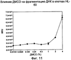
На Фиг.11 показан эффект воздействия концентраций ДМСО на клетки HL-60. (RFU - относительные единицы флуоресценции; ДМСО - диметилсульфоксид)11 shows the effect of the concentration of DMSO on HL-60 cells. (RFU - relative fluorescence units; DMSO - dimethyl sulfoxide)
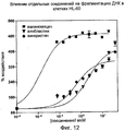
На Фиг.12 показана индукция фрагментации ДНК, обнаруживаемая по PicoGreen, после инкубации клеток HL-60 с валиномицином, винбластином или винкристином.12 shows the induction of DNA fragmentation detected by PicoGreen after incubation of HL-60 cells with valinomycin, vinblastine or vincristine.
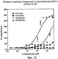
На Фиг.13 показана индукция фрагментации ДНК, обнаруживаемая по PicoGreen, после инкубации клеток HL-60 с этопозидом, генистеином, пуромицином или рапамицином.13 shows the induction of DNA fragmentation detected by PicoGreen after incubation of HL-60 cells with etoposide, genistein, puromycin or rapamycin.
На Фиг.14 показано распределение данных скрининга произвольной химической библиотеки при обнаружении фрагментации ДНК в лизатах HL-60 с помощью PicoGreen. (RFU - относительные единицы флуоресценции).On Fig shows the distribution of the screening data of an arbitrary chemical library when detecting DNA fragmentation in HL-60 lysates using PicoGreen. (RFU - relative fluorescence units).
Подробное описание изобретенияDETAILED DESCRIPTION OF THE INVENTION
Все публикации, цитируемые в настоящем документе, в силу ссылки на них включаются в настоящий документ. Если не указано иначе, все технические и научные термины, используемые в настоящем документе, имеют такое же значение, которое общеизвестно рядовым специалистам в области, к которой относится настоящее изобретение.All publications cited herein are, by reference, referenced herein. Unless otherwise indicated, all technical and scientific terms used in this document have the same meaning as is well known to ordinary specialists in the field to which the present invention relates.
Терминология, используемая в настоящем описании и прилагаемой формуле изобретения, предназначена исключительно для характеристики конкретных приложений, и ее использование в описании не подразумевает какого-либо ограничения изобретения. Формы единственного числа того или иного слова подразумевают охват форм множественного числа, если только в контексте не указано иное. Например, формы единственного числа для конкретных и общих терминологических определений также включают и формы множественного числа. Кроме того, ссылки на тот или иной агент могут относиться к смеси двух или более агентов. Поэтому термин «агент» включает множество агентов, в том числе смеси и/или их энантиомеры. Следует также отметить, что термин «или» обычно имеет свой основной смысл, в том числе «и/или», если только содержание однозначно не указывает на обратное. Кроме того, следует понимать, что термины «включает» и/или «включающий» при использовании в настоящем описании определяют наличие указанных особенностей, стадий, элементов и/или компонентов, но не отрицают наличия или добавления одной или нескольких иных особенностей, стадий, элементов, компонентов и/или их групп.The terminology used in the present description and the attached claims is intended solely to characterize specific applications, and its use in the description does not imply any limitation of the invention. The singular forms of a word are intended to encompass the plural forms, unless the context indicates otherwise. For example, singular forms for specific and general terminological definitions also include plural forms. In addition, references to one or another agent may refer to a mixture of two or more agents. Therefore, the term "agent" includes many agents, including mixtures and / or their enantiomers. It should also be noted that the term “or” usually has its main meaning, including “and / or”, unless the content clearly indicates otherwise. In addition, it should be understood that the terms “includes” and / or “including” when used in the present description determine the presence of these features, stages, elements and / or components, but do not deny the presence or addition of one or more other features, stages, elements , components and / or groups thereof.
Кроме того, в соответствии с настоящим изобретением в рамках ноу-хау могут использоваться стандартные методики молекулярной биологии, микробиологии и технологии рекомбинантной ДНК. Содержание таких методик подробно изложено в литературе. См., например, Sambrook, Fritsch & Maniatis, Molecular Cloning: A Laboratory Manual, Second Edition (1989) Cold Spring Harbor Laboratory Press, Cold Spring Harbor, N.Y. (далее "Sambrook et al., 1989"); DNA Cloning: A Practical Approach, Volumes I and II (D.N.Glover ed. 1985); Oligonucleotide Synthesis (M.J.Gait ed. 1984); Nucleic Acid Hybridization [B.D.Hames & S.J.Higgins eds. (1985)]; Transcription And Translation [B.D.Hames & S.J.Higgins, eds. (1984)]; Animal Cell Culture [R.I.Freshney, ed. (1986)]; Immobilized Cells And Enzymes [IRL Press, (1986)]; B.Perbal, A Practical Guide To Molecular Cloning (1984); F.M.Ausubel et al. (eds.), Current Protocols in Molecular Biology, John Wiley & Sons, Inc. (1994).In addition, in accordance with the present invention, standard molecular biology, microbiology, and recombinant DNA technologies can be used as part of the know-how. The content of such techniques is detailed in the literature. See, e.g., Sambrook, Fritsch & Maniatis, Molecular Cloning: A Laboratory Manual, Second Edition (1989) Cold Spring Harbor Laboratory Press, Cold Spring Harbor, N.Y. (hereinafter "Sambrook et al., 1989"); DNA Cloning: A Practical Approach, Volumes I and II (D.N. Glover ed. 1985); Oligonucleotide Synthesis (M.J. Gait ed. 1984); Nucleic Acid Hybridization [B.D. Hames & S.J. Higgins eds. (1985)]; Transcription And Translation [B.D. Hames & S.J. Higgins, eds. (1984)]; Animal Cell Culture [R.I. Freshney, ed. (1986)]; Immobilized Cells And Enzymes [IRL Press, (1986)]; B. Perbal, A Practical Guide To Molecular Cloning (1984); F.M. Ausubel et al. (eds.), Current Protocols in Molecular Biology, John Wiley & Sons, Inc. (1994).
Поэтому осуществлением настоящего изобретения является способ идентификации агента, который влияет на образование фрагментов ДНК, и такой способ включает: (a) подготовку клеток в наборе приемников; (b) добавление агента, по крайней мере, к одному приемнику; (c) инкубирование агента с клетками в течение заранее определенного периода времени; (d) лизирование клеток; (e) добавление обнаруживаемого соединения, способного к интеркаляции во фрагменты ДНК, по крайней мере, к одному указанному приемнику; (f) измерение количества интеркалированного обнаруживаемого соединения; и (g) сравнение количества интеркалированного обнаруживаемого соединения с контролем с целью определить разницу, что позволяет идентифицировать указанный агент как модифицирующий агент, если разница превышает заранее установленный предел.Therefore, an embodiment of the present invention is a method for identifying an agent that affects the formation of DNA fragments, and such a method includes: (a) preparing cells in a set of receivers; (b) adding an agent to at least one receiver; (c) incubating the agent with the cells for a predetermined period of time; (d) cell lysis; (e) adding a detectable compound capable of intercalating into DNA fragments to at least one of said receptors; (f) measuring the amount of intercalated detectable compound; and (g) comparing the amount of intercalated detectable compound with a control in order to determine the difference, which makes it possible to identify said agent as a modifying agent if the difference exceeds a predetermined limit.
Анализ позволяет обнаруживать фрагменты ДНК. Фрагменты ДНК могут представлять собой небольшие фрагменты двуцепочечной ДНК (дцДНК) в цитоплазматической фракции лизатов клеток и фрагменты дцДНК, выделенные апоптотическими клетками в среду. Кроме того, фрагменты ДНК могут представлять собой фрагменты одноцепочечной ДНК (оцДНК) в цитоплазматической фракции лизатов клеток и фрагменты оцДНК, выделенные апоптотическими клетками в среду. В одном из осуществлений изобретения анализ используется для измерения спонтанного апоптоза, например, среди прочего, апоптоза во время совместного культивирования в присутствии или в отсутствие различных типов клеток или в различных условиях культивирования. Поэтому образование фрагмента ДНК может индуцироваться удалением ингредиента из культуральной среды, например, фетальной бычьей сыворотки. В другом осуществлении изобретения анализ используется для оценки отсутствия апоптоза. В еще одном осуществлении в ходе обработки клеток агентом может оцениваться наличие или рост уровня апоптоза, или отсутствие или снижение уровня апоптоза. В еще одном осуществлении изобретения анализ используется для измерения выживания или пролиферации клеток.Analysis allows the detection of DNA fragments. DNA fragments can be small fragments of double-stranded DNA (dsDNA) in the cytoplasmic fraction of cell lysates and dsDNA fragments isolated by apoptotic cells in the medium. In addition, DNA fragments can be single stranded DNA (ssDNA) fragments in the cytoplasmic fraction of cell lysates and ssDNA fragments isolated by apoptotic cells into the medium. In one embodiment of the invention, analysis is used to measure spontaneous apoptosis, for example, inter alia, apoptosis during co-culture in the presence or absence of different types of cells or under different culturing conditions. Therefore, the formation of a DNA fragment can be induced by removing the ingredient from the culture medium, for example, fetal bovine serum. In another embodiment of the invention, analysis is used to assess the absence of apoptosis. In yet another embodiment, during the treatment of the cells with an agent, the presence or increase of apoptosis or the absence or decrease of apoptosis can be evaluated. In yet another embodiment of the invention, analysis is used to measure cell survival or proliferation.
Еще одним осуществлением настоящего изобретения является способ идентификации агента, который влияет на образование фрагментов ДНК, и такой способ включает: (a) подготовку клеток в наборе приемников; (b) добавление соединения, выбираемого из группы, состоящей из индуктора, ингибитора, модулятора, модулятора индуктора и модулятора ингибитора, по крайней мере, к одному приемнику; (c) инкубирование компонента с клетками в течение заранее определенного периода времени; (d) добавление агента к указанному, по крайней мере, одному приемнику; (е) инкубирование агента с клетками в течение заранее определенного периода времени; (f) лизирование клеток; (g) добавление обнаруживаемого соединения, способного к интеркаляции во фрагменты ДНК, к указанному, по крайней мере, одному приемнику; (h) измерение количества интеркалированного обнаруживаемого соединения; и (i) сопоставление количества интеркалированного обнаруживаемого соединения с контролем с целью определить разницу, что позволяет идентифицировать указанный агент как модифицирующий агент, если разница превышает заранее установленный предел.Another embodiment of the present invention is a method for identifying an agent that affects the formation of DNA fragments, and such a method includes: (a) preparing cells in a set of receivers; (b) adding a compound selected from the group consisting of an inducer, an inhibitor, a modulator, an inductor modulator and an inhibitor modulator to at least one receiver; (c) incubating the component with the cells for a predetermined period of time; (d) adding an agent to said at least one receiver; (e) incubating the agent with the cells for a predetermined period of time; (f) cell lysis; (g) adding a detectable compound capable of intercalation into DNA fragments to said at least one receiver; (h) measuring the amount of intercalated detectable compound; and (i) comparing the amount of intercalated detectable compound with a control in order to determine the difference, which makes it possible to identify said agent as a modifying agent if the difference exceeds a predetermined limit.
Такие усовершенствования, как добавление индуктора, ингибитора или модулятора вместе с агентом на одной стадии, вполне соответствуют уровню знаний и способностям квалифицированного специалиста и рассматриваются как различные осуществления настоящего изобретения.Improvements such as the addition of an inducer, inhibitor or modulator together with the agent at one stage, are fully consistent with the level of knowledge and abilities of a qualified specialist and are considered as various implementations of the present invention.
Одним из осуществлений изобретения является компонент, который влияет на образование фрагментов ДНК за счет воздействия на апоптоз, выживание или пролиферацию клеток. Компонент выбирается из группы, включающей индуктор, ингибитор, модулятор, модулятор индуктора и модулятор ингибитора.One embodiment of the invention is a component that affects the formation of DNA fragments by affecting apoptosis, survival, or cell proliferation. The component is selected from the group consisting of inductor, inhibitor, modulator, inductor modulator and inhibitor modulator.
Выживание клеток представляет собой способность клеток сохранять жизненные функции в благоприятных или неблагоприятных условиях. Неблагоприятные условия включают, среди прочего, присутствие одного или нескольких токсичных компонентов, удаление питательных веществ или недостаток кислорода. В качестве одного из множества примеров - некоторые раковые клетки увеличивали экспрессию белков выживания, например, Bcl2, что делало такие клетки устойчивыми к апоптозу. Другие раковые клетки вырабатывали механизмы, которые обеспечивали более высокую выживаемость клеток или их меньшую склонность к апоптозу в условиях пониженного уровня кислорода.Cell survival is the ability of cells to maintain vital functions under favorable or adverse conditions. Adverse conditions include, but are not limited to, the presence of one or more toxic components, removal of nutrients, or lack of oxygen. As one of many examples, some cancer cells increased the expression of survival proteins, such as Bcl2, which made such cells resistant to apoptosis. Other cancer cells developed mechanisms that ensured higher cell survival or their lower tendency to apoptosis under conditions of low oxygen levels.
Пролиферацией клеток называется увеличение их количества. Ко множеству примеров пролиферации клеток относятся увеличение количества клеток в результате их нормального деления, индукция деления клеток или ингибирование их гибели.Cell proliferation is called an increase in their number. Many examples of cell proliferation include an increase in cell numbers resulting from normal cell division, induction of cell division, or inhibition of cell death.
Одним из осуществлений настоящего изобретения является способ идентификации агента, который влияет на образование фрагментов ДНК в клетках, проходящих апоптоз. Вместе с тем настоящее изобретение не ограничивается какой-либо конкретной формой гибели клеток. Изобретение может применяться к любому механизму гибели клеток, где терминальным процессом является фрагментация ДНК.One of the implementations of the present invention is a method for identifying an agent that affects the formation of DNA fragments in cells undergoing apoptosis. However, the present invention is not limited to any particular form of cell death. The invention can be applied to any cell death mechanism, where the terminal process is DNA fragmentation.
Термин «ингибитор» включает любое лекарство, химическое вещество, белок или белковый фрагмент, способный блокировать, прерывать или предупреждать клеточный ответ, активность или канал, связанные с апоптозом, выживанием или пролиферацией клеток. Кроме того, ингибитором может быть, например, молекулярный шаперон, антитело или ингибирующая РНК (РНКи), которые блокируют экспрессию клеточных белков, тем самым прямо или косвенно ингибируя каналы воздействия. «Ингибитором» может быть регулирование условий культивирования, например увеличение или уменьшение уровня кислорода или изменение компонентов среды, так чтобы блокировать, прервать или предотвратить клеточный ответ.The term “inhibitor” includes any drug, chemical, protein or protein fragment capable of blocking, interrupting or preventing a cellular response, activity or channel associated with apoptosis, survival or proliferation of cells. In addition, the inhibitor can be, for example, a molecular chaperone, an antibody, or an inhibitory RNA (RNAi) that blocks the expression of cellular proteins, thereby directly or indirectly inhibiting exposure channels. An “inhibitor” can be the regulation of culture conditions, for example, an increase or decrease in oxygen levels or a change in the components of the medium so as to block, interrupt or prevent a cellular response.
Термин «индуктор» включает любое лекарство, химическое вещество, белок или белковый фрагмент, способный инициировать или стимулировать клеточный ответ, активность или канал, связанные с апоптозом, выживанием или пролиферацией клеток. Кроме того, индуктором может быть, например, молекулярный шаперон, антитело или ингибирующая РНК (РНКи), которые блокируют экспрессию клеточных белков, тем самым устраняя ингибирование или напрямую инициируя или стимулируя клеточный ответ, активность или канал воздействия. «Индуктором» может быть регулирование условий культивирования, например увеличение или уменьшение уровня кислорода, или изменение компонентов среды, так чтобы блокировать, прервать или предотвратить клеточный ответ.The term “inducer” includes any drug, chemical, protein or protein fragment capable of initiating or stimulating a cellular response, activity or channel associated with apoptosis, survival or cell proliferation. In addition, the inducer may be, for example, a molecular chaperone, an antibody, or an inhibitory RNA (RNAi) that blocks the expression of cellular proteins, thereby eliminating inhibition or directly initiating or stimulating a cellular response, activity or channel of action. An “inductor” can be the regulation of culturing conditions, for example, an increase or decrease in the level of oxygen, or a change in the components of the medium, so as to block, interrupt or prevent a cellular response.
Термин «модулятор» включает любое лекарство, химическое вещество, белок или белковый фрагмент, способный корректировать интенсивность, долю или характеристики клеточного ответа, активности или канала, связанного с апоптозом, выживанием или пролиферацией клеток. Кроме того, модулятором может быть, например, молекулярный шаперон, антитело или ингибирующая РНК (РНКи), которые блокируют экспрессию клеточных белков, тем самым устраняя ингибирование или индуцируя клеточный ответ, активность или канал воздействия. «Модулятором» может быть регулирование условий культивирования, например, увеличение или уменьшение уровня кислорода, или изменение компонентов среды, так чтобы блокировать, прервать или предотвратить клеточный ответ.The term “modulator” includes any drug, chemical, protein or protein fragment that is capable of adjusting the intensity, proportion or characteristics of the cellular response, activity or channel associated with apoptosis, survival or proliferation of cells. In addition, the modulator may be, for example, a molecular chaperone, an antibody, or an inhibitory RNA (RNAi) that blocks the expression of cellular proteins, thereby eliminating inhibition or inducing a cellular response, activity or channel of action. A “modulator” may be the regulation of culture conditions, for example, increasing or decreasing the level of oxygen, or changing the components of the medium so as to block, interrupt or prevent a cellular response.
В еще одном осуществлении изобретения модулятор может использоваться в сочетании с индуктором, так что модулятор индуктора делает индуктор более эффективным (например, приводя к более активному клеточному ответу) или менее эффективным (например, приводя к пониженному клеточному ответу). Модулятор ингибитора делает ингибитор менее эффективным (например, более активный клеточный ответ) или более эффективным (например пониженный клеточный ответ).In yet another embodiment of the invention, the modulator can be used in combination with an inducer, so that the inductor modulator makes the inductor more efficient (for example, leading to a more active cellular response) or less effective (for example, leading to a lower cellular response). An inhibitor modulator makes an inhibitor less effective (e.g., a more active cellular response) or more effective (e.g. a reduced cellular response).
Ингибиторы, индукторы или модуляторы могут использоваться для имитации каналов воздействия или характеристик болезненных состояний. В качестве одного из множества показательных примеров - одним из осуществлений настоящего изобретения будет возможность индуцировать апоптоз с помощью β-амилоидных фрагментов для имитации характеристик болезни Альцгеймера в присутствии или в отсутствие потенциальных модуляторов, например воспалительных цитокинов. Еще один из множества наглядных примеров - применение настоящего изобретения для идентификации агентов, которые стимулируют апоптоз в одном или нескольких аспектах рака. В рамках такой парадигмы предположительное применение настоящего изобретения подразумевает количественное определение способности испытываемого агента индуцировать или усиливать апоптоз в раковых клетках, тканях или органах. Индуктор апоптоза может обладать широким спектром действия, охватывая множество каналов, ведущих к гибели клетки. В альтернативном варианте, индуктор апоптоза может обладать высокой специфичностью к единичному каналу апоптоза или ограничиваться воздействием на конкретное заболевание или патологическое состояние. Поэтому осуществление настоящего изобретения включает способ скрининга для идентификации испытываемого агента, который может корректировать болезненное состояние, при котором предположительно происходит ингибирование апоптоза, например рак. К таким формам рака, среди прочего, относятся острая миелоидная лейкемия, множественная миелома, не-Ходжкинская лимфома, хроническая лимфоцитарная лейкемия и солидные опухоли.Inhibitors, inducers or modulators can be used to simulate exposure channels or characteristics of disease states. As one of many illustrative examples, one embodiment of the present invention will be the ability to induce apoptosis using β-amyloid fragments to mimic the characteristics of Alzheimer's disease in the presence or absence of potential modulators, such as inflammatory cytokines. Another of many illustrative examples is the use of the present invention to identify agents that stimulate apoptosis in one or more aspects of cancer. Within the framework of such a paradigm, the alleged use of the present invention involves the quantification of the ability of a test agent to induce or enhance apoptosis in cancer cells, tissues or organs. The apoptosis inducer can have a wide spectrum of action, spanning many channels leading to cell death. Alternatively, the apoptosis inducer may be highly specific for a single apoptosis channel or limited to exposure to a particular disease or pathological condition. Therefore, an embodiment of the present invention includes a screening method for identifying a test agent that can correct a disease state in which apoptosis inhibition, such as cancer, is suspected to occur. Such forms of cancer include, but are not limited to, acute myeloid leukemia, multiple myeloma, non-Hodgkin lymphoma, chronic lymphocytic leukemia, and solid tumors.
Другим осуществлением настоящего изобретения является использование более одного индуктора, ингибитора или модулятора. Применение более одного индуктора, ингибитора или модулятора может, среди прочего, приводить к аддитивным эффектам, обратному действию, синергическим эффектам или воздействовать на различные каналы.Another embodiment of the present invention is the use of more than one inducer, inhibitor or modulator. The use of more than one inducer, inhibitor or modulator may, among other things, lead to additive effects, reverse action, synergistic effects, or affect various channels.
В одном из осуществлений изобретения используется интеркалирующее поддающееся обнаружению соединение. Одним из множества примеров является интеркалирующий флуоресцентный краситель. Интеркаляторы обычно представляют собой гетероароматические полициклические молекулы, которые встраиваются между двумя парами оснований в дуплексе ДНК. Вместе с тем изобретение не ограничивается гетероароматическими полициклическими молекулами. Любая интеркалирующая молекула, которая демонстрирует заметное усиление флуоресценции или сдвиг эмиссии или параметра (параметров) возбуждения в присутствии фрагментов ДНК в условиях незначительного или полного отсутствия неселективного связывания с РНК или белками, входит в сферу охвата настоящего изобретения. Такие интеркалирующие красители хорошо известны специалистам в области и, среди прочего, включают бис-бензимидный краситель Hoechst 33258. Другим удобным интеркалирующим обнаруживаемым соединением является пропидия иодид. В одном из осуществлений настоящего изобретения используется PicoGreen. PicoGreen относится к семейству несимметричных монометинцианиновых красителей. Он обладает высокими константами связывания с ДНК и демонстрирует высокую интенсивность флуоресценции при связывании с ней, при этом практически не проявляя флуоресцентных свойств в свободном состоянии в растворе. В еще одном осуществлении изобретения используется пропидия иодид. Кроме того, некоторые интеркалирующие соединения в состоянии связываться с оцДНК, например TOTO (Nucleic Acids Res. 23:1215-1222, 1995) и OliGreen (Molecular Probes, кат. №07582, кат. №011492). В еще одном осуществлении изобретения используются проникающие через мембрану зонды ДНК, например, BENA435 (Nucleic Acids Res. 34:), и за счет этого можно избавиться от необходимости лизирования клеток, чтобы ввести метку в ДНК. В другом применении настоящего изобретения используются YOPRO, Hoechst 33342, DAPI и DRAQ5.In one embodiment of the invention, an intercalating detectable compound is used. One of many examples is an intercalating fluorescent dye. Intercalators are usually heteroaromatic polycyclic molecules that are inserted between two base pairs in a DNA duplex. However, the invention is not limited to heteroaromatic polycyclic molecules. Any intercalating molecule that exhibits a marked increase in fluorescence or a shift in emission or excitation parameter (s) in the presence of DNA fragments with little or no non-selective binding to RNA or proteins is within the scope of the present invention. Such intercalating dyes are well known to those skilled in the art and, inter alia, include the Hoechst 33258 bis-benzimide dye. Another convenient intercalating detectable compound is propidium iodide. In one implementation of the present invention uses PicoGreen. PicoGreen belongs to the family of asymmetric monomethine cyanine dyes. It has high binding constants with DNA and shows a high fluorescence intensity upon binding to it, while practically not showing fluorescence properties in the free state in solution. In yet another embodiment of the invention, propidium iodide is used. In addition, some intercalating compounds are able to bind to ssDNA, for example TOTO (Nucleic Acids Res. 23: 1215-1222, 1995) and OliGreen (Molecular Probes, cat. No. 07582, cat. No. 011492). In yet another embodiment of the invention, membrane-penetrating DNA probes are used, for example, BENA435 (Nucleic Acids Res. 34 :), and this eliminates the need to lyse cells to introduce a label into the DNA. In another application of the present invention, YOPRO, Hoechst 33342, DAPI and DRAQ5 are used.
Круг интеркалирующих обнаруживаемых молекул не ограничивается флуоресцентными красителями. Количество фрагментов ДНК может определяться с использованием любой методологии, известной специалистам в области. Количество интеркалирующих молекул, включенных в ДНК, может определяться с помощью меток, в том числе, среди прочих, радиоизотопных или активирующих сцинтиллятор соединений. К методам обнаружения, среди прочих, относятся пептидные метки, ферментативная активность, поглощение, флуоресценция, время-разрешенная флуоресценция, поляризованная флуоресценция, резонансный перенос энергии флуоресценции, люминесценция, резонансный перенос энергии биолюминесценции, радиоактивная метка и сцинтилляционное сближение или другие методы, широко используемые в данной области. В еще одном применении могут использоваться методы косвенной метки, в том числе, среди прочих, применение меченых антител, использование взаимодействия стрептавидин-биотин, аффинные реагенты, образующие хелаты с металлами, или аффинные реагенты GST-глутатиона. Любой прямой или косвенный метод метки, известный специалистам в области, рассматривается как входящий в сферу охвата настоящего изобретения. В еще одном осуществлении количество ДНК может определяться по поглощению на 260 нм (А260).The circle of intercalating detectable molecules is not limited to fluorescent dyes. The number of DNA fragments can be determined using any methodology known to specialists in the field. The number of intercalating molecules included in the DNA can be determined using labels, including, but not limited to, radioisotope or scintillator activating compounds. Detection methods include, but are not limited to, peptide labels, enzymatic activity, absorption, fluorescence, time-resolved fluorescence, polarized fluorescence, resonant fluorescence energy transfer, luminescence, resonant bioluminescence energy transfer, radioactive label and scintillation convergence, other widely used this area. In yet another application, indirect labeling methods may be used, including, but not limited to, the use of labeled antibodies, the use of streptavidin-biotin interactions, affinity reagents that form chelates with metals, or GST-glutathione affinity reagents. Any direct or indirect labeling method known to those skilled in the art is considered to be within the scope of the present invention. In yet another embodiment, the amount of DNA can be determined by absorption at 260 nm (A260).
Еще одно осуществление изобретения включает отделение хромосомной ДНК от фрагментов ДНК до измерения количества интеркалировавшего обнаруживаемого соединения. Отделение хромосомальной ДНК от фрагментов ДНК может проводиться с использованием методов, известных специалистам в области. Среди множества примеров - центрифугирование, фильтрация, седиментация, электрофорез, гель-фильтрация, аффинная очистка и осаждение. Для отделения или удаления хромосомальной ДНК от фрагментов ДНК специалист в данной области может использовать любой метод разделения.Another embodiment of the invention involves the separation of chromosomal DNA from DNA fragments before measuring the amount of intercalated detectable compound. The separation of chromosomal DNA from DNA fragments can be carried out using methods known to specialists in the field. Among many examples are centrifugation, filtration, sedimentation, electrophoresis, gel filtration, affinity purification and sedimentation. Any person skilled in the art can use any separation method to separate or remove chromosomal DNA from DNA fragments.
Одно из осуществлений настоящего изобретения предусматривает лизирование клеток. Лизирование клеток может осуществляться добавлением детергента, содержащего лизирующий буфер. При этом изобретение не ограничивается использованием детергента в лизирующем буфере, но может включать любой метод, который подходит для лизирования клеток. Например, лизирование клеток может проходить под действием гипотонического буфера, ультразвуковой обработкой или в цикле замораживание/оттаивание. Другие методы лизирования также хорошо известны специалистам в данной области.One of the implementations of the present invention provides for the lysis of cells. Cell lysing can be accomplished by adding a detergent containing lysis buffer. However, the invention is not limited to the use of detergent in lysing buffer, but may include any method that is suitable for lysing cells. For example, cell lysis can take place under the action of a hypotonic buffer, by ultrasonic treatment, or in a freeze / thaw cycle. Other lysis methods are also well known to those skilled in the art.
В еще одном осуществлении настоящего изобретения используется свободная от ДНКаз РНКаза для удаления РНК из лизатов клеток с целью увеличить отношение сигнал/шум за счет уменьшения фонового сигнала флуоресценции, связанного с эндогенной клеточной РНК.In another embodiment of the present invention, RNAse-free RNAse is used to remove RNA from cell lysates in order to increase the signal-to-noise ratio by reducing the background fluorescence signal associated with endogenous cellular RNA.
Осуществление настоящего изобретения предусматривает использование набора приемников, в которые могут помещаться клетки и другие материалы, например культуральные среды. Набор приемников может представлять собой любое количество приемников от по крайней мере одного до нескольких приемников, пригодных для помещения клеток в соответствии с охватом настоящего изобретения. К примерам, среди прочего, относятся колбы, чашки для культивирования, пробирки, например пробирки на 1,5 мл, 12-ячеечные планшеты, 96-ячеечные планшеты, 384-ячеечные планшеты и миниатюрные планшеты для микротитрования с около 4000 приемных ячеек (заявка U.S. Patent Application 20050255580). Набор приемников можно усовершенствовать, дополнив его защитной крышкой, тем самым исключив попадание загрязнений или испарение содержимого.The implementation of the present invention provides for the use of a set of receivers in which cells and other materials, such as culture media, can be placed. A set of receivers may be any number of receivers from at least one to several receivers suitable for placement of cells in accordance with the scope of the present invention. Examples include, but are not limited to, flasks, culture dishes, tubes, for example 1.5 ml tubes, 12-well plates, 96-well plates, 384-well plates, and miniature microtiter plates with about 4,000 receiving cells (US application Patent Application 20050255580). The set of receivers can be improved by supplementing it with a protective cover, thereby eliminating the ingress of contaminants or evaporation of the contents.
Дополнительной особенностью приемников является то, что в них можно проводить анализ, к примерам такого анализа относятся спектрофотометрический анализ, сцинтилляционный счет и измерения флуоресценции. Тем не менее это не является ограничением для приемников, которые могут использоваться в рамках охвата настоящего изобретения, с учетом того что образцы можно перенести в подходящую емкость, пригодную для дальнейшего анализа. Один из множества примеров предполагает такую модификацию способа, которая предусматривает подготовку второго набора приемников, причем стадия лизирования клеток дополнительно включает отделение супернатанта от клеточных остатков, а следующая стадия далее включает добавление обнаруживаемого соединения, способного к интеркаляции во фрагменты ДНК, по крайней мере к одному приемнику указанного второго набора, содержащего образец указанного отделенного супернатанта.An additional feature of the receivers is that they can be analyzed; examples of such an analysis include spectrophotometric analysis, scintillation counting and fluorescence measurements. However, this is not a limitation for receivers that can be used within the scope of the present invention, given that the samples can be transferred to a suitable container suitable for further analysis. One of many examples involves such a modification of the method that involves preparing a second set of receivers, the cell lysing step further comprising separating the supernatant from the cell debris, and the next step further comprising adding a detectable compound capable of intercalating into DNA fragments to at least one receiver the specified second set containing a sample of the specified separated supernatant.
В одном из применений настоящего изобретения используется контроль. Контроль представляет собой термин, хорошо понятный квалифицированным специалистам в данной области. Надлежащий контроль может зависеть от используемых параметров анализа или исследуемой экспериментальной проблемы. Контролем может быть конкретный набор условий анализа или добавление в культуральную среду или удаление из нее конкретного соединения. Контроль может рассматриваться как положительный контроль, в том случае если условия анализа или добавленное контрольное соединение обеспечивают ожидаемый ответ. Например, если ожидается, что исследуемый агент вызывает апоптоз, положительным контролем будет соединение, которое является известным индуктором апоптоза. Одним из примеров положительного контроля является добавление винбластина сульфата. Контроль также может быть отрицательным. Отрицательным контролем может быть конкретный набор условий анализа, а также добавление в культуральную среду или удаление из нее конкретного соединения, которое будет вызывать ожидаемый отклик. Например, если ожидается, что исследуемый агент вызывает апоптоз, отрицательным контролем будет соединение, которое, предположительно, не приводит к апоптозу. Контролем может быть «носитель». Например, если испытываемый агент растворяется в ДМСО, тогда контролем с использованием носителя может быть ДМСО без испытываемого агента. Контролем может быть и просто использование полученных в прошлом данных.In one application of the present invention, control is used. Control is a term well understood by those skilled in the art. Proper control may depend on the analysis parameters used or the experimental problem being investigated. The control may be a specific set of assay conditions, or the addition of a particular compound to or from the culture medium. A control can be considered a positive control if the assay conditions or the added control compound provide the expected response. For example, if the test agent is expected to induce apoptosis, a compound that is a known inducer of apoptosis will be a positive control. One example of a positive control is the addition of vinblastine sulfate. Control may also be negative. A negative control may be a specific set of assay conditions, as well as the addition of a specific compound to the culture medium or removal from it, which will cause the expected response. For example, if the test agent is expected to induce apoptosis, a negative control will be a compound that is not expected to lead to apoptosis. The control may be a “carrier." For example, if the test agent is soluble in DMSO, then DMSO without the test agent may be a control using a carrier. Control may also be simply the use of data obtained in the past.
В одном из осуществлений изобретения используются имеющиеся в продаже клеточные линии. Например, клетки, которые пригодны для использования, можно приобрести у American Tissue Culture Company. В одном из применений используются клетки HL-60. Клетки могут быть прокариотическими или эукариотическими. Настоящее изобретение не ограничивается видом используемых клеток. Могут также применяться первичные культуры. Недифференцированные клетки можно подвергать воздействию различных агентов, которые вызывают их дифференциацию в конкретный фенотип. Например, в одном из применений изобретения используются клетки-предшественники, которые после индуцирования дифференцируются в олигодендроциты. Конкретный тип используемых клеток можно выбрать с помощью маркеров, специально экспрессированных в соответствии с желаемым типом клеток, или, в альтернативном варианте, за счет потери конкретного маркера (маркеров). Клетки можно отделять или сортировать с помощью таких методов, как проточная цитометрия, которые широко используются квалифицированными специалистами в области.In one embodiment of the invention, commercially available cell lines are used. For example, cells that are suitable for use can be obtained from the American Tissue Culture Company. In one application, HL-60 cells are used. Cells can be prokaryotic or eukaryotic. The present invention is not limited to the type of cells used. Primary cultures may also be used. Undifferentiated cells can be exposed to various agents that cause them to differentiate into a specific phenotype. For example, in one application of the invention, precursor cells are used that, after induction, differentiate into oligodendrocytes. The particular type of cells used can be selected using markers specifically expressed in accordance with the desired cell type, or, alternatively, by losing a specific marker (s). Cells can be separated or sorted using methods such as flow cytometry, which are widely used by qualified specialists in the field.
В одном из применений настоящего изобретения используется однородная популяция клеток. В альтернативном применении настоящего изобретения используется неоднородная популяция клеток. Для проведения анализа в рамках настоящего изобретения можно использовать клетки любого типа и в любой пропорции.In one application of the present invention, a homogeneous population of cells is used. In an alternative application of the present invention, a heterogeneous population of cells is used. For analysis within the framework of the present invention, you can use cells of any type and in any proportion.
Клетки могут быть получены из биологического образца. К биологическим образцам, среди прочего, могут относиться ткани или жидкости, срезы тканей, например образцы биопсии или вскрытия, а также замороженные срезы, взятые для гистологического анализа. Такие образцы включают кровь, слюну, ткани, культивированные клетки, например первичные культуры, эксплантаты и трансформированные клетки, кал, мочу и пр. Биологический образец может быть взята у эукариотического организма, в том числе у млекопитающего, например примата, такого как шимпанзе, макака или человек; коровы, собаки, кошки, грызуна, в том числе морской свинки, крысы, мыши, кролика, или птицы, рептилии или рыбы.Cells can be obtained from a biological sample. Biological samples can include, but are not limited to, tissues or fluids, tissue sections, such as biopsy or autopsy specimens, and frozen sections taken for histological analysis. Such samples include blood, saliva, tissues, cultured cells, such as primary cultures, explants and transformed cells, feces, urine, etc. A biological sample can be taken from a eukaryotic organism, including a mammal, such as a primate such as chimpanzee, macaque or person; cows, dogs, cats, rodents, including guinea pigs, rats, mice, rabbits, or birds, reptiles or fish.
В другом осуществлении настоящего изобретения используются клетки, которые были временно или стабильно трансформированы, так чтобы обеспечить сверхэкспрессию или исключить экспрессию по крайней мере одного белка и определить, влияет ли такая экспрессия или ее отсутствие на фрагментацию ДНК. Экспрессия может быть индуцированной или конститутивной. Агенты могут проверяться на способность влиять на фрагментацию ДНК в трансформированных клетках. Кроме того, испытываемые агенты могут проверяться на способность влиять на фрагментацию ДНК в трансфектированных клетках в присутствии или в отсутствие индукторов или ингибиторов апоптоза. Такое применение может рассматриваться как контроль.In another embodiment of the present invention, cells are used that have been temporarily or stably transformed so as to overexpress or exclude the expression of at least one protein and determine whether such expression or its absence affects DNA fragmentation. Expression may be induced or constitutive. Agents can be tested for their ability to influence DNA fragmentation in transformed cells. In addition, test agents can be tested for their ability to influence DNA fragmentation in transfected cells in the presence or absence of inducers or inhibitors of apoptosis. Such an application may be considered as a control.
Рекомбинантный вектор экспрессии, представленный в изобретении, включают молекулу нуклеиновой кислоты в форме, подходящей для экспрессии нуклеиновой кислоты в клетке-хозяине. Таким образом, рекомбинантные векторы экспрессии настоящего изобретения могут содержать одну или несколько регуляторных последовательностей, выбранных на основе клетки-реципиента, используемой для экспрессии, причем эта последовательность функционально связана с нуклеиновой кислотой, которая должна быть экспрессирована. По отношению к рекомбинантному вектору экспрессии выражение «функционально связанный» призвано означать, что представляющая интерес последовательность нуклеотидов связана с регуляторной последовательностью (последовательностями) таким способом, который обеспечивает экспрессию последовательности нуклеотидов (например в системе транскрипции/трансляции «in vitro» или в клетке-хозяине, если вектор введен в клетку-хозяина). Термин «регуляторная последовательность» включает промоторы, энхансеры и другие элементы, контролирующие экспрессию (например сигналы полиаденилирования). Такие регуляторные последовательности описаны, например, в Goeddel, Gene Expression Technology: Methods in Enzymology Vol. 185, Academic Press, San Diego, Calif. (1990). Регуляторные последовательности включают направляющие конститутивную экспрессию последовательности нуклеотидов во многих типах клеток-хозяев (например специфичные к тканям регуляторные последовательности). Специалистам известно, что структура вектора экспрессии может зависеть от таких факторов, как выбор клетки-хозяина для трансформации, желаемый уровень экспрессии белка и т.д. Векторы экспрессии настоящего изобретения могут вводиться в клетки-хозяева для выработки белков или пептидов, кодированных нуклеиновыми кислотами, как это описано в настоящем документе.The recombinant expression vector of the invention includes a nucleic acid molecule in a form suitable for expression of a nucleic acid in a host cell. Thus, the recombinant expression vectors of the present invention may contain one or more regulatory sequences selected based on the recipient cell used for expression, and this sequence is operably linked to the nucleic acid to be expressed. With respect to a recombinant expression vector, the expression “operably linked” is intended to mean that the nucleotide sequence of interest is linked to a regulatory sequence (s) in a manner that provides expression of the nucleotide sequence (eg, in an in vitro transcription / translation system or in a host cell if the vector is introduced into the host cell). The term “regulatory sequence” includes promoters, enhancers and other elements that control expression (eg, polyadenylation signals). Such regulatory sequences are described, for example, in Goeddel, Gene Expression Technology: Methods in Enzymology Vol. 185, Academic Press, San Diego, Calif. (1990). Regulatory sequences include directing constitutive expression of nucleotide sequences in many types of host cells (e.g., tissue specific regulatory sequences). It is known to those skilled in the art that the structure of an expression vector may depend on factors such as the choice of a host cell for transformation, the desired level of protein expression, etc. The expression vectors of the present invention can be introduced into host cells to produce proteins or peptides encoded by nucleic acids, as described herein.
Используемый в настоящем документе термин «сверхэкспрессия» относится к экспрессии полипептида (например молекулы, которая может быть задействована в апоптозе или механизмах выживания клеток) в той или иной клетке на уровне, превышающем нормальный уровень экспрессии полипептида в клетке, которая обычно экспрессирует полипептид, или в клетке, которая обычно не обеспечивает экспрессии полипептида. Например, уровень экспрессии полипептида может составлять 10%, 20%, 30%, 40%, 50%, 60%, 70, 80%, 90%, 100% или более по сравнению с экспрессией полипептида в немутантной клетке, которая обычно экспрессирует полипептид. Возможна сверхэкспрессия мутантов, вариантов или аналогов представляющего интерес полипептида.As used herein, the term “overexpression” refers to the expression of a polypeptide (eg, a molecule that can be involved in apoptosis or cell survival mechanisms) in a cell at a level that exceeds the normal expression level of a polypeptide in a cell that typically expresses a polypeptide, or a cell that usually does not express the polypeptide. For example, the expression level of a polypeptide may be 10%, 20%, 30%, 40%, 50%, 60%, 70, 80%, 90%, 100% or more compared to expression of a polypeptide in a non-mutant cell that typically expresses a polypeptide . Overexpression of mutants, variants or analogues of the polypeptide of interest is possible.
Используемый в настоящем документе термин «транзиторная» экспрессия относится к экспрессии экзогенной молекулы (молекул) нуклеиновых кислот, которые отделены от хромосом клетки. Транзиторная экспрессия обычно достигает своего максимума через 2-3 дня после введения экзогенной нуклеиновой кислоты, а затем постепенно снижается.As used herein, the term “transient” expression refers to the expression of an exogenous nucleic acid molecule (s) that are separated from the chromosomes of a cell. Transient expression usually reaches its maximum 2-3 days after the introduction of exogenous nucleic acid, and then gradually decreases.
Используемый в настоящем документе термин «стабильная» экспрессия относится к экспрессии экзогенной молекулы (молекул) нуклеиновых кислот, которые являются частью хромосом клетки. В общем случае векторы стабильной экспрессии генов включают один или несколько маркеров селекции.As used herein, the term “stable” expression refers to the expression of an exogenous nucleic acid molecule (s) that are part of the chromosomes of a cell. In general, stable gene expression vectors include one or more selection markers.
Методы культивирования клеток для трансформированных, нетрансформированных образцов, первичных культур и биологических образцов хорошо известны специалистам в области. Биологические образцы или культивированные клетки могут храниться, пока не потребуется их использование. Среды, используемые для культивирования, могут готовиться специально или приобретаться из коммерческих источников.Cell culture techniques for transformed, non-transformed samples, primary cultures, and biological samples are well known to those skilled in the art. Biological samples or cultured cells can be stored until their use is required. Media used for cultivation can be prepared specifically or purchased from commercial sources.
В настоящем изобретении приводятся способы идентификации (например скрининга, обнаружения, характеризации, анализа и количественного определения) агентов, которые оказывают влияние на образование фрагментов дцДНК и оцДНК. Термин «агент», «испытываемый агент», «испытываемое соединение», «потенциальное лекарство» или «модулятор» или их грамматические эквиваленты, используемые в настоящем документе, описывает любую молекулу, как существующую в природе, так и синтетическую, например белок, олигопептид (например длиной примерно от 5 до 25 аминокислот, предпочтительно длиной примерно от 10 до 20 или от 12 до 18 аминокислот, предпочтительно длиной 12, 15 или 18 аминокислот), малую органическую молекулу, полисахарид, липид (например сфинголипид), жирную кислоту, полинуклеотид, олигонуклеотид и пр., которая используется в анализах, описанных в настоящем изобретении, и анализируется для установления способности влиять на фрагментацию ДНК или апоптоз. Не существует конкретных ограничений в отношении соединения, которое может использоваться для анализа. К примерам относятся отдельные агенты или библиотеки малых, средних или высокомолекулярных химических соединений, очищенные белки, продукты экспрессии генных библиотек, библиотеки синтетических пептидов, клеточные экстракты и супернатанты культур. Термин агент включает любую комбинацию различных агентов.The present invention provides methods for identifying (eg, screening, detecting, characterizing, analyzing, and quantifying) agents that affect the formation of dsDNA and ssDNA fragments. The term “agent”, “test agent”, “test compound”, “potential drug” or “modulator” or their grammatical equivalents used herein describes any molecule that exists in nature or synthetic, such as a protein, oligopeptide (for example, about 5 to 25 amino acids long, preferably about 10 to 20 or 12 to 18 amino acids, preferably 12, 15 or 18 amino acids long), small organic molecule, polysaccharide, lipid (e.g. sphingolipid), fatty acid, polynucleo a tetid, oligonucleotide, etc., which is used in the assays described in the present invention, and analyzed to determine the ability to influence DNA fragmentation or apoptosis. There are no particular restrictions on the compound that can be used for analysis. Examples include individual agents or libraries of small, medium or high molecular weight chemical compounds, purified proteins, gene library expression products, synthetic peptide libraries, cell extracts and culture supernatants. The term agent includes any combination of various agents.
Агент может включать по крайней мере один или несколько растворимых и нерастворимых факторов, компоненты клеточной матрицы, кондиционированные среды, клеточные экстракты, экстракты тканей, эксплантаты, модификаторы рН, газы, модификаторы осмотического давления, модификаторы ионной силы, вирусы, ДНК, РНК или фрагменты генов. Агент может быть в форме библиотеки испытываемых агентов, например комбинаторной или рандомизированной библиотеки, которая обеспечивает достаточный диапазон разнообразия или, напротив, ограничена сходными структурами или характеристиками. Агенты могут быть дополнительно связаны с партнером слияния, например соединениями направленной доставки, спасающими соединениями, соединениями димеризации, стабилизирующими соединениями, адресуемыми соединениями и другими функциональными компонентами. Обычно для создания новых химических объектов с полезными свойствами выбирают испытываемый агент (называемый «ведущим соединением» или «ведущим») с рядом желательных свойств или активностью, например ингибирующей активностью или модулирующей активностью. Затем ведущее соединение используется как основа для создания вариантов ведущего соединения и дальнейшей оценки свойств и активности таких вариантов.An agent may include at least one or more soluble and insoluble factors, cell matrix components, conditioned media, cell extracts, tissue extracts, explants, pH modifiers, gases, osmotic pressure modifiers, ionic strength modifiers, viruses, DNA, RNA, or gene fragments . The agent may be in the form of a library of test agents, for example a combinatorial or randomized library, which provides a sufficient range of diversity or, conversely, is limited to similar structures or characteristics. Agents can be further associated with a merger partner, for example, targeted delivery compounds, rescue compounds, dimerization compounds, stabilizing compounds, addressable compounds, and other functional components. Typically, to create new chemical objects with useful properties, a test agent (called a “lead compound” or “lead”) is selected with a number of desirable properties or activity, for example, inhibitory activity or modulating activity. The lead compound is then used as the basis for creating variants of the lead compound and further evaluating the properties and activity of such variants.
Тот или иной агент может включать условия обработки и регулирование внешних или внутренних условий или среды. Одним из множества примеров такого агента является ультрафиолетовый свет.An agent may include processing conditions and the regulation of external or internal conditions or the environment. One of many examples of such an agent is ultraviolet light.
Одно из осуществлений изобретения предусматривает его использование в методах высокопроизводительного скрининга (ВПС). ВПС является автоматизированным одновременным методом тестирования тысяч различных химических соединений в анализах, призванных моделировать биологические механизмы или аспекты патологий заболеваний. Несколько соединений, например набор соединений, могут тестироваться одновременно, например в одной партии. В одном из осуществлений термин метода скрининга ВПС относится к анализам, в ходе которых тестируется способность одного соединения или набора соединений влиять на выбранные показатели.One of the implementations of the invention provides for its use in methods of high throughput screening (CHD). CHD is an automated, simultaneous method for testing thousands of different chemical compounds in analyzes designed to simulate biological mechanisms or aspects of disease pathologies. Several compounds, for example a set of compounds, can be tested simultaneously, for example, in one batch. In one embodiment, the term IPN screening method refers to analyzes that test the ability of a single compound or set of compounds to influence selected parameters.
Системы для работы с жидкостями, аналитическое оборудование, например планшеты-ридеры флуоресценции или сцинтилляционные счетчики и роботы для клеточных культур и манипуляций с образцами хорошо известны специалистам в области. Механические системы, например роботизированные манипуляторы или устройства точного выбора, доступны любому квалифицированному специалисту. Существуют серийно выпускаемые планшет-ридеры для анализа обычных 96-ячеечных или 384-ячеечных планшет. В наличии имеются ридеры единичных образцов, множества образцов или планшет, которые анализируют заранее определенные ячейки и готовят предварительные отчеты данных. Исходные данные могут преобразовываться и представляться самыми различными способами.Fluid handling systems, analytical equipment, such as fluorescence plate readers or scintillation counters and robots for cell cultures and sample handling are well known to those skilled in the art. Mechanical systems, such as robotic manipulators or precision devices, are available to anyone skilled in the art. There are commercially available plate readers for analyzing conventional 96-cell or 384-cell tablets. Single sample readers, multiple samples, or a tablet are available that analyze predefined cells and prepare preliminary data reports. Source data can be transformed and presented in a variety of ways.
Одним из осуществлений настоящего изобретения является система анализа для идентификации агента, который влияет на образование фрагментов двуцепочечной ДНК, и такая система анализа включает: (a) набор приемников; (b) буфер лизирования; (c) обнаруживаемое соединение, способное к интеркаляции в двуцепочечную ДНК; и (d) по крайней мере, один компонент, где компонент выбирается из группы, состоящей из агента (агентов), индуктора (индукторов) апоптоза, ингибитора (ингибиторов) апоптоза, контроля и клеток.One embodiment of the present invention is an analysis system for identifying an agent that affects the formation of double-stranded DNA fragments, and such an analysis system includes: (a) a set of receivers; (b) a lysis buffer; (c) a detectable compound capable of intercalating into double-stranded DNA; and (d) at least one component, wherein the component is selected from the group consisting of an agent (s), an inducer (s) of apoptosis, an inhibitor (s) of apoptosis, a control, and cells.
Еще одним осуществлением изобретения является набор, включающий, по крайней мере, один элемент системы анализа и инструкции для применения. Таким образом, компоненты системы анализа могут предоставляться отдельно или вместе в виде набора. Компоненты системы анализа могут готовиться и включаться в набор в соответствии с методами, которые максимально увеличивают стабильность отдельных компонентов. Такие методы известны специалистам в области. Например, клетки системы анализа могут поставляться в форме суспензии или в лиофилизованном виде. Могут также присутствовать дополнительные компоненты системы, например буферы, контейнеры для смешения компонентов анализа, например планшеты для микротитрования или пробирки для тестирования. Система для анализа может поставляться в форме набора, который включает инструкции для проведения анализа и инструкции по обработке и интерпретации данных.Another embodiment of the invention is a kit comprising at least one element of an analysis system and instructions for use. Thus, the components of the analysis system can be provided separately or together as a set. The components of the analysis system can be prepared and included in the kit in accordance with methods that maximize the stability of individual components. Such methods are known to those skilled in the art. For example, cells of an assay system may be supplied in suspension or in lyophilized form. Additional system components may also be present, such as buffers, containers for mixing assay components, such as microtiter plates or test tubes. The analysis system may be supplied in the form of a kit, which includes instructions for the analysis and instructions for processing and interpreting the data.
Одним из осуществлений изобретения является лекарственный препарат для модулирования образования фрагментов ДНК, включающий терапевтически эффективное количество агента, идентифицируемого с помощью способов настоящего изобретения, и фармацевтически приемлемый носитель.One embodiment of the invention is a medicament for modulating the formation of DNA fragments comprising a therapeutically effective amount of an agent identified by the methods of the present invention and a pharmaceutically acceptable carrier.
Термин «терапевтически эффективное количество» относится к количеству агента, эффективному при лечении заболевания или расстройства у субъекта или млекопитающего. В случае рака терапевтически эффективное количество лекарства может уменьшать численность раковых клеток; сокращать размер опухоли; ингибировать (то есть в некоторой степени замедлять и, предпочтительно, останавливать) проникновение раковых клеток в периферические органы; ингибировать (то есть в некоторой степени замедлять и предпочтительно останавливать) метастазирование опухоли; в некоторой степени ингибировать рост опухоли; и/или в некоторой степени ослаблять один или несколько связанных с раком симптомов. В зависимости от того, насколько лекарство в состоянии предупреждать рост и/или уничтожать существующие раковые клетки, оно может быть цитостатическим и/или цитотоксическим. Пример рака не является единственным, поскольку агент, который влияет на образование фрагментов дцДНК или оцДНК, может применяться для множества различных заболеваний.The term “therapeutically effective amount” refers to an amount of an agent effective in treating a disease or disorder in a subject or mammal. In the case of cancer, a therapeutically effective amount of the drug can reduce the number of cancer cells; reduce the size of the tumor; inhibit (that is, to some extent slow down and, preferably, stop) the penetration of cancer cells into peripheral organs; inhibit (that is, to some extent slow down and preferably stop) tumor metastasis; inhibit tumor growth to some extent; and / or to some extent, alleviate one or more cancer-related symptoms. Depending on how well the drug is able to prevent the growth and / or destroy existing cancer cells, it can be cytostatic and / or cytotoxic. An example of cancer is not the only one, since an agent that affects the formation of dsDNA or ssDNA fragments can be used for many different diseases.
Лекарственный препарат для модуляции образования фрагментов ДНК может включать терапевтически эффективное количество агента, при котором агент модулирует образование фрагмента ДНК через рецепторный белок. Поэтому лекарственный препарат может включать испытываемый агент, который является агонистом, частичным агонистом, антагонистом или обратным агонистом. Кроме того, фармацевтический состав может включать испытываемый агент, который является пептидом, его пептидными фрагментами, когнатами, конгенерами, мимиками, аналогами или секретирующими клетками и их растворимыми молекулами. Еще одним из осуществлений изобретения является лекарственный препарат для модулирования образования фрагментов ДНК, включающий терапевтически эффективное количество идентифицируемого агента и фармацевтически приемлемый носитель, причем такой лекарственный препарат эффективно влияет на канал или механизм апоптоза.A medicament for modulating the formation of DNA fragments may include a therapeutically effective amount of an agent in which the agent modulates the formation of a DNA fragment through a receptor protein. Therefore, the medicament may include a test agent that is an agonist, partial agonist, antagonist or inverse agonist. In addition, the pharmaceutical composition may include a test agent, which is a peptide, its peptide fragments, cognates, congeners, facial expressions, analogs or secreting cells and their soluble molecules. Another embodiment of the invention is a medicament for modulating the formation of DNA fragments, comprising a therapeutically effective amount of an identifiable agent and a pharmaceutically acceptable carrier, the medicament effectively affecting the channel or mechanism of apoptosis.
Используемый в настоящем документе термин «агонист» относится к веществам (например, среди прочих, к лигандам и агентам), которые при связывании с рецептором вызывают внутриклеточный ответ или усиливают связывание ГТФ с мембранами.As used herein, the term “agonist” refers to substances (for example, among others, ligands and agents) that, upon binding to a receptor, elicit an intracellular response or enhance the binding of GTP to membranes.
Используемый в настоящем документе термин «частичный агонист» относится к веществам (например, среди прочих, к лигандам и агентам), которые при связывании с рецептором вызывают меньший по уровню/масштабам внутриклеточный ответ, чем агонисты, или по сравнению с ними в меньшей степени/масштабе усиливают связывание ГТФ с мембранами.As used herein, the term “partial agonist” refers to substances (for example, among others, ligands and agents) that, upon binding to the receptor, cause a lower level / scale of the intracellular response than agonists, or less / scale up the binding of GTP to membranes.
Используемый в настоящем документе термин «антагонист» относится к веществам (например, среди прочих, к лигандам и агентам), которые конкурентно связываются с рецептором в том же сайте, что и агонист. Вместе с тем антагонист не активирует внутриклеточный ответ, инициированный активной формой рецептора, и, тем самым, в состоянии ингибировать внутриклеточные отклики, вызванные агонистами или частичными агонистами. Соответственно, в отсутствие агониста или частичного агониста антагонисты не снижают исходный внутриклеточный ответ.As used herein, the term “antagonist” refers to substances (for example, among others, ligands and agents) that competitively bind to a receptor at the same site as the agonist. However, the antagonist does not activate the intracellular response initiated by the active form of the receptor, and, thus, is able to inhibit intracellular responses caused by agonists or partial agonists. Accordingly, in the absence of an agonist or partial agonist, antagonists do not reduce the initial intracellular response.
Используемый в настоящем документе термин «обратный агонист» относится к веществам (например, среди прочих, к лиганду и агенту), которые связываются с конститутивно активным рецептором и ингибируют исходный внутриклеточный ответ. Исходный отклик инициируется активной формой рецептора с уровнем активности ниже нормы, который наблюдается при отсутствии агонистов или частичных агонистов, или при уменьшении связывания ГТФ с мембранами.As used herein, the term “inverse agonist” refers to substances (for example, among others, a ligand and an agent) that bind to a constitutively active receptor and inhibit the initial intracellular response. The initial response is initiated by the active form of the receptor with an activity level below the norm, which is observed in the absence of agonists or partial agonists, or with a decrease in GTP binding to membranes.
Используемый в настоящем документе термин «лиганд» относится к веществу, которое связывается с другой молекулой, где таким веществом, среди прочих, может быть гормон или нейромедиатор, и где такое вещество относится к лигандам, стереоселективно связывающимся с рецептором.As used herein, the term “ligand” refers to a substance that binds to another molecule, where such a substance, among others, may be a hormone or a neurotransmitter, and where such a substance refers to ligands that stereoselectively bind to a receptor.
Лекарственные препараты настоящего изобретения могут использоваться в сочетании с другими терапевтическими агентами. Например, при лечении рака лекарственный препарат может вводиться в сочетании с цитокинами или различными соединениями, используемыми для химиотерапии.The medicaments of the present invention can be used in combination with other therapeutic agents. For example, in the treatment of cancer, a drug can be administered in combination with cytokines or various compounds used for chemotherapy.
Еще одним осуществлением настоящего изобретения является способ диагностики или мониторинга лечения заболевания, при котором биомаркер заболевания предполагает образование фрагментов ДНК, и такой способ включает: (a) внесение биологического образца в набор приемников; (b) добавление обнаруживаемого соединения, способного к интеркаляции во фрагменты ДНК, по крайней мере, к одному приемнику; (c) измерение количества интеркалированного обнаруживаемого соединения; и (d) сравнение количества интеркалированного обнаруживаемого соединения со стандартом для определения различия, что позволяет осуществить диагностику или мониторинг лечения заболевания, в случае если различие превышает заранее установленное предельное значение.Another embodiment of the present invention is a method for diagnosing or monitoring the treatment of a disease, wherein the biomarker of the disease involves the formation of DNA fragments, and such a method includes: (a) adding a biological sample to a set of receivers; (b) adding a detectable compound capable of intercalating into DNA fragments to at least one receiver; (c) measuring the amount of intercalated detectable compound; and (d) comparing the amount of intercalated detectable compound with a standard to determine the difference, which allows the diagnosis or monitoring of treatment of a disease if the difference exceeds a predetermined limit value.
Понятие биомаркера хорошо известно специалистам в области. Одним из множества примеров является использование термина биомаркер, с тем чтобы охватить любой физиологический отклик, фенотип или характеристику, которые могут использоваться для количественного или качественного описания определенного состояния клетки, организма и млекопитающего.The concept of a biomarker is well known to specialists in the field. One of many examples is the use of the term biomarker in order to encompass any physiological response, phenotype, or characteristic that can be used to quantitatively or qualitatively describe a particular condition of a cell, organism, and mammal.
Биомаркер считается полезным для диагностики, мониторинга и прогнозирования заболевания или при мониторинге лечения заболевания, если его протекание значительно различается в разных подгруппах тестируемых биологических образцов. Уровни биомаркера считаются «значительно различающимися», если вероятность случайной идентификации конкретного биомаркера оказывается меньше заранее установленного значения. Метод расчета такой вероятности будет зависеть от конкретного метода, используемого для сопоставления уровней между подгруппами, например, t-критерий или подобные ему методы статистического анализа. Для специалистов в данной области будет очевидно, что заранее установленное предельное значение будет меняться в зависимости от числа использованных образцов.A biomarker is considered useful for diagnosing, monitoring and predicting a disease or for monitoring treatment of a disease if its course varies significantly in different subgroups of tested biological samples. Biomarker levels are considered “significantly different” if the probability of accidentally identifying a particular biomarker is less than a predetermined value. The method for calculating this probability will depend on the particular method used to compare levels between subgroups, for example, the t-test or similar statistical analysis methods. For specialists in this field it will be obvious that a predetermined limit value will vary depending on the number of samples used.
Биологическим образцом могут быть образцы органов, взятые из органов животных или человека, образцы тканей, полученные из тканей животных или человека, а также образцы клеток, полученные из клеток животных или человека, или из клеточных культур. В случае экспериментов с животными биологические образцы включают ткани изучаемого органа, полученные после вскрытия или биопсии, а также жидкости организма, например кровь. Для клинического применения биомаркеров особенно предпочтительными образцами являются жидкости организма, например кровь, сыворотка, плазма, моча, синовиальная жидкость, ликвор, спинномозговая жидкость, сперма или лимфа, а также ткани тела, полученные с помощью биопсии.The biological sample may be organ samples taken from animal or human organs, tissue samples obtained from animal or human tissues, as well as cell samples obtained from animal or human cells, or from cell cultures. In the case of experiments with animals, biological samples include tissues of the organ under study obtained after an autopsy or biopsy, as well as body fluids, such as blood. For clinical use of biomarkers, body fluids such as blood, serum, plasma, urine, synovial fluid, cerebrospinal fluid, cerebrospinal fluid, sperm or lymph, and body tissues obtained by biopsy are particularly preferred.
Специалистам в области хорошо известно понятие стандарта. К стандартам может относиться, среди прочего, биологический образец здорового субъекта, причем таким субъектом может быть животное или человек. Кроме того, стандартом может быть биологический образец субъекта, не проходившего лечение. В альтернативном варианте стандарт может быть взят от того же субъекта до, во время и после лечения. Стандарт может быть от того же субъекта, но это может быть другая клетка, образец ткани или органа, отличающиеся от исходной клетки, ткани или органа, используемых для определения уровня биомаркера. Стандартом необязательно должен быть биологический образец, но может быть образец с известным количеством фрагментов ДНК.Specialists in the field are well aware of the concept of a standard. Standards may include, inter alia, a biological sample of a healthy subject, the subject being an animal or a human being. In addition, the standard may be a biological sample of a subject who has not been treated. Alternatively, a standard may be taken from the same subject before, during and after treatment. The standard may be from the same subject, but it may be a different cell, sample of tissue or organ that is different from the original cell, tissue or organ used to determine the level of the biomarker. The standard does not have to be a biological sample, but it can be a sample with a known number of DNA fragments.
Настоящее изобретение более подробно описывается в приведенных ниже примерах, которые не ограничивают сферы его охвата, изложенной в формуле изобретения. Несмотря на достаточно подробное описание и иллюстрации настоящего изобретения, позволяющие квалифицированным специалистам в данной области реализовать и использовать его, очевидны различные альтернативы, модификации и усовершенствования, не затрагивающие основной идеи и охвата настоящего изобретения. Квалифицированный специалист в данной области без труда оценит тот факт, что настоящее изобретение хорошо приспособлено для достижения цели и получения указанных и характерных для него результатов и преимуществ. Приведенные ниже примеры содержат описания осуществлений и не призваны ограничивать сферу охвата настоящего изобретения. Для специалистов в данной области будут очевидны приведенные в настоящем документе модификации и иные применения. Такие модификации не отходят от сути настоящего изобретения и определяются сферой охвата формулы изобретения. В приведенное в настоящем документе изобретение могут быть внесены различные изменения и модификации, не затрагивающие сущности и охвата изобретения.The present invention is described in more detail in the following examples, which do not limit the scope of its scope set forth in the claims. Despite the sufficiently detailed description and illustrations of the present invention, allowing qualified specialists in this field to implement and use it, various alternatives, modifications and improvements are obvious that do not affect the main idea and scope of the present invention. A qualified specialist in this field will easily appreciate the fact that the present invention is well adapted to achieve the goal and obtain the indicated and characteristic results and advantages. The following examples are descriptions of implementations and are not intended to limit the scope of the present invention. Modifications and other uses will be apparent to those skilled in the art. Such modifications do not depart from the essence of the present invention and are defined by the scope of the claims. Various changes and modifications may be made to the invention provided herein without affecting the spirit and scope of the invention.
Изобретение, наглядно описанное в настоящем документе, может применяться в отсутствие любого элемента или элементов, ограничения или ограничений, которые специально не раскрываются в настоящем документе. Применяемые термины и выражения используются в целях описания, а не для ограничения, и в использовании таких терминов и выражений нет никакого намерения исключить любые эквиваленты приведенных и описанных свойств или их части; напротив, признается возможность различных модификаций в рамках сферы охвата заявленного изобретения. Поэтому следует понимать, что несмотря на полное раскрытие настоящего изобретения в его основных осуществлениях и дополнительных характеристиках, специалисты в данной области могут вносить модификации и изменения в концепции, раскрываемые в настоящем документе, и что такие модификации и изменения считаются входящими в сферу охвата изобретения, которая определяется в прилагаемой формуле.The invention, clearly described herein, can be applied in the absence of any element or elements, limitations or restrictions that are not specifically disclosed in this document. The terms and expressions used are used for description purposes and not for limitation, and there is no intention in using such terms and expressions to exclude any equivalents of the listed and described properties or their parts; on the contrary, the possibility of various modifications within the scope of the claimed invention is recognized. Therefore, it should be understood that despite the full disclosure of the present invention in its main implementations and additional characteristics, specialists in this field can make modifications and changes to the concepts disclosed in this document, and that such modifications and changes are considered to be within the scope of the invention, which defined in the attached formula.
Пример 1Example 1
Обнаружение фрагментов ДНК с помощью PicoGreen:Detection of DNA fragments using PicoGreen:
Описываются общие условия и соображения по проведению анализа. Вместе с тем в приведенных ниже последующих примерах условия анализа меняли с целью проверить переменные и обеспечить соответствие целям эксперимента, и не для того чтобы обязательно ограничивать изобретение конкретными применениями.The general conditions and considerations for the analysis are described. However, in the following examples, the conditions of the analysis were changed in order to check the variables and ensure compliance with the objectives of the experiment, and not to necessarily limit the invention to specific applications.
HL-60 представляет собой линию клеток ОМЛ человека, выпускаемую ATCC (ATCC кат. № CCL-240™). Среду для клеточной культуры готовили следующим образом: 100 мл термоинактивированной фетальной бычьей сыворотки, 20 мл 1M HEPES (pH 7,5), 10 мл исходного раствора пенициллина/стрептомицина (см. таблицу 1) добавляли к 1 литру среды RPMI-1640. После тщательного перемешивания полученную среду фильтровали через стерилизованное устройство для фильтрования с диаметром пор 0,22 мкм (Nalgene). Среда для инкубирования готовилась следующим образом: 5 мл исходного раствора пенициллина/стрептомицина и 25 мл термоинактивированной бычьей сыворотки смешивались с 500 мл RPMI-1640 (без фенолового красного и L-глутамина).HL-60 is a human AML cell line manufactured by ATCC (ATCC Cat. No. CCL-240 ™). The cell culture medium was prepared as follows: 100 ml of thermally inactivated fetal bovine serum, 20 ml of 1M HEPES (pH 7.5), 10 ml of the initial solution of penicillin / streptomycin (see table 1) was added to 1 liter of RPMI-1640 medium. After thorough mixing, the resulting medium was filtered through a sterilized filter device with a pore diameter of 0.22 μm (Nalgene). The incubation medium was prepared as follows: 5 ml of the initial solution of penicillin / streptomycin and 25 ml of thermally inactivated bovine serum were mixed with 500 ml of RPMI-1640 (without phenol red and L-glutamine).
В общем, эксперимент проводили в соответствии со следующим протоколом: клетки культивировали от четырех до пяти дней перед обработкой соединением. Жизнеспособность клеток должна была быть больше примерно 92%. Подсчитывали плотность клеток, и их жизнеспособность подтверждали с использованием GUAVA PCA с помощью программы Viacount 2.12. Клетки отбирали и центрифугировали при 300×g в течение 6 минут. Супернатант сливали, и осадок клеток вновь размешивали до концентрации 0,15 млн клеток/мл с RPMI (без фенолового красного) с 5% фетальной бычьей сыворотки и 1% пенициллина/стрептомицина. Аликвоту 40 мкл суспензии клеток помещали в каждую ячейку 384-ячеечной планшеты. Клетки инкубировали в течение надлежащего периода времени в определенных экспериментальных условиях. Затем 45 мкл буфера клеточного лизирования добавляли к образцам клеток.In general, the experiment was carried out in accordance with the following protocol: cells were cultured for four to five days before treatment with the compound. Cell viability should have been greater than approximately 92%. Cell densities were counted, and their viability was confirmed using GUAVA PCA using the Viacount 2.12 software. Cells were selected and centrifuged at 300 × g for 6 minutes. The supernatant was discarded and the cell pellet was again stirred to a concentration of 0.15 million cells / ml with RPMI (without phenol red) with 5% fetal bovine serum and 1% penicillin / streptomycin. An aliquot of 40 μl of the cell suspension was placed in each cell of a 384-cell plate. Cells were incubated for an appropriate period of time under certain experimental conditions. Then, 45 μl of cell lysis buffer was added to the cell samples.
Лизирующий буфер готовили следующим образом: для приготовления 1 литра лизирующего буфера 20 мл 1M раствора Tris-HCl (pH 8,0), 40 мл 0,5M EDTA (pH 8,0), 10 мл 20% раствора Tween-20, 10 мл 20% раствора Triton X-100 смешивали с 920 мл деионизованной воды. Непосредственно перед использованием 5 мл исходного раствора РНКазы А (10 мг/мл) добавляли к буферу лизирования до конечной концентрации 0,05 мг/мл. После добавления буфера лизирования планшеты выдерживали при комнатной температуре в течение 60 минут. Планшеты с клеточными культурами центрифугировали при 2000×g в течение 20 мин, и 10 мкл супернатанта клеточных лизатов, содержащего фрагменты ДНК, переносили в CyBio-well 384 на планшеты для обнаружения (Corning Costar 384-ячеечная полистирольная аналитическая планшета, черная, несвязывающая поверхность). К каждой ячейке образца в планшетах для обнаружения добавляли аликвоту 10 мкл обнаруживающего раствора PicoGreen. Раствор PicoGreen для обнаружения готовили непосредственно перед использованием путем разбавления исходного раствора в ДМСО буфером для обнаружения в соотношении 1:200. Буфер для обнаружения готовили смешением 50 мл исходных 10×Tris-HCL буферного физиологического раствора (TBS, pH 8,0) и 2 мл 0,5M раствора EDTA (pH 8,0) в деионизованной воде до конечного объема 500 мл. Интенсивность флуоресценции анализировали с помощью PerkinElmer Envision.Lysis buffer was prepared as follows: to prepare 1 liter of lysis buffer, 20 ml of 1M Tris-HCl solution (pH 8.0), 40 ml of 0.5M EDTA (pH 8.0), 10 ml of 20% Tween-20 solution, 10 ml A 20% Triton X-100 solution was mixed with 920 ml of deionized water. Immediately prior to use, 5 ml of a stock solution of RNase A (10 mg / ml) was added to the lysis buffer to a final concentration of 0.05 mg / ml. After adding lysis buffer, the plates were kept at room temperature for 60 minutes. Cell culture plates were centrifuged at 2000 × g for 20 min, and 10 μl of the cell lysate supernatant containing DNA fragments was transferred to CyBio-well 384 onto detection plates (Corning Costar 384-cell polystyrene assay plate, black, non-binding surface) . An aliquot of 10 μl PicoGreen Detection Solution was added to each sample cell in the detection plates. A PicoGreen detection solution was prepared immediately before use by diluting the stock solution in DMSO with a detection buffer at a ratio of 1: 200. The detection buffer was prepared by mixing 50 ml of the original 10 × Tris-HCL physiological buffer solution (TBS, pH 8.0) and 2 ml of a 0.5M solution of EDTA (pH 8.0) in deionized water to a final volume of 500 ml. Fluorescence intensity was analyzed using PerkinElmer Envision.
В таблице 1 перечислены некоторые примеры реагентов и материалов, концентрации, функции и сведения по поставщиках. Table 1 lists some examples of reagents and materials, concentrations, functions, and vendor information.
BP3223-1Fisher scientific
BP3223-1
В таблице 2 приводятся некоторые примеры оборудования, методы использования такого оборудования и поставщики. Table 2 provides some examples of equipment, methods for using such equipment, and suppliers.
Ниже приводятся параметры измерений на PerkinElmer Envision: для возбуждения зеркало FITC; фильтр возбуждения FITC 485; фильтр испускания FITC 535; число импульсов 25; возбуждающий свет 1%; усиление детекции равно 1 и высота измерения равна 8 мм.The following are the measurement options on the PerkinElmer Envision: for exciting the FITC mirror; excitation filter FITC 485; emission filter FITC 535; number of
Каждый анализ включает положительный, отрицательный и бланковый контроль. Использование надлежащих методов контроля определяли поставленными экспериментальными целями. Как правило, в качестве положительного контроля использовали клетки HL-60, обработанные 5 мкM винбластина сульфатом в 1% ДМСО. Отрицательным контролем были клетки HL-60, обработанные 1% ДМСО. Бланковой пробой была инкубационная среда с 1% ДМСО (без клеток HL-60).Each analysis includes positive, negative and blank control. The use of appropriate control methods was determined by the set experimental goals. Typically, HL-60 cells treated with 5 μM vinblastine sulfate in 1% DMSO were used as a positive control. The negative control was HL-60 cells treated with 1% DMSO. The blank sample was incubation medium with 1% DMSO (without HL-60 cells).
Анализ данных может быть скорректирован с учетом экспериментальных параметров или исследуемой парадигмы. Как правило, относительное количество образующейся фрагментированной ДНК определяли по интенсивности флуоресценции (ИФ) образца. Эффект влияния агента на фрагментацию ДНК в клетках HL-60 рассчитывали по изменению интенсивности флуоресценции относительно контрольных образцов ДМСО. Процент эффекта определяли как:Data analysis can be adjusted based on experimental parameters or the paradigm under study. Typically, the relative amount of fragmented DNA formed is determined by the fluorescence intensity (IF) of the sample. The effect of the agent on DNA fragmentation in HL-60 cells was calculated from the change in fluorescence intensity relative to DMSO control samples. The percentage of effect was determined as:
% эффекта = 100*[(ИФагента - среднее(ИФотрицательный контроль))/ среднее(ИФотрицательный контроль)]% effect = 100 * [(IF agent - average (IF negative control )) / average (IF negative control )]
Пример 2Example 2
PicoGreen обеспечивает специфическое обнаружение фрагментов ДНК, выделяемых клетками HL-60:PicoGreen provides specific detection of DNA fragments secreted by HL-60 cells:
На Фиг.1 отображено повышение интенсивности флуоресценции PicoGreen в клетках HL-60, обработанных камптотецином. Клетки HL-60 в фазе середины логарифмического роста (0,4 млн клеток/мл) были обработаны либо 0,1% растворителя-носителя ДМСО, либо 3,2 мкМ камптотецина в течение 5,5 часов. Ко всей клеточной культуре добавляли равный объем лизирующего буфера (20 мМ Tris-HCl (pH 8,0), 20 мМ EDTA, 0,2 % Tween-20). После выдерживания при комнатной температуре в течение 45 минут лизат клеток центрифугировали при 2000×g в течение 20 минут, отбирали верхнюю порцию супернатанта и определяли содержание ДНК по величине интенсивности флуоресценции при смешении с красителем PicoGreen. Бланковой пробой среды была смесь в равных отношениях клеточной культуральной среды и лизирующего буфера.Figure 1 shows the increase in PicoGreen fluorescence intensity in HL-60 cells treated with camptothecin. HL-60 cells in the mid-log phase growth phase (0.4 million cells / ml) were treated with either 0.1% DMSO vehicle solvent or 3.2 μM camptothecin for 5.5 hours. An equal volume of lysis buffer (20 mM Tris-HCl (pH 8.0), 20 mM EDTA, 0.2% Tween-20) was added to the entire cell culture. After keeping at room temperature for 45 minutes, the cell lysate was centrifuged at 2000 × g for 20 minutes, the upper portion of the supernatant was taken and the DNA content was determined by the magnitude of the fluorescence intensity when mixed with PicoGreen dye. Blank breakdown of the medium was a mixture of equal cell culture medium and lysis buffer.
Пример 3Example 3
Сигнал флуоресценции PicoGreen зависит от уровня ДНК в клеточных лизатах:The PicoGreen fluorescence signal depends on the level of DNA in cell lysates:
После обработки 0,1% растворителем-носителем ДМСО или 3,2 мкМ камптотецина в течение 5,5 часов равный объем лизирующего буфера без EDTA (20 мМ Tris-HCl (pH 8,0) 0,2% Tween-20 и 3 мкг/мл РНКазы) добавляли ко всей клеточной культуре HL-60. После выдерживания при комнатной температуре в течение 45 минут клеточные лизаты центрифугировали, отбирали верхнюю порцию супернатанта и инкубировали со свободной от РНКаз ДНКазой при 37°C в течение заданного времени (Фиг.2). После 2 часов обработки ДНКаза I оказалась в состоянии уменьшить интенсивность флуоресценции контрольного образца ДМСО примерно на 50%. Сигнал клеток, обработанных камптотецином, обладал более высокой интенсивностью флуоресценции до обработки ДНКазой I и эффективно уменьшался до уровня, сопоставимого с контролем ДМСО при обработке ДНКазой I. Эти результаты показывают, что сигнал флуоресценции PicoGreen связан с присутствием фрагментированной ДНК в клеточных лизатах.After treatment with 0.1% DMSO vehicle-solvent or 3.2 μM camptothecin for 5.5 hours, an equal volume of lysis buffer without EDTA (20 mM Tris-HCl (pH 8.0) 0.2% Tween-20 and 3 μg / ml RNase) was added to the entire cell culture of HL-60. After keeping at room temperature for 45 minutes, the cell lysates were centrifuged, the upper portion of the supernatant was taken and incubated with RNase-free DNase at 37 ° C for a predetermined time (Figure 2). After 2 hours of treatment, DNase I was able to reduce the fluorescence intensity of the DMSO control sample by about 50%. The signal of camptothecin-treated cells had a higher fluorescence intensity before treatment with DNase I and effectively decreased to a level comparable to the DMSO control when treated with DNase I. These results show that the PicoGreen fluorescence signal is associated with the presence of fragmented DNA in cell lysates.
Пример 4Example 4
Сравнение PicoGreen с пропидия иодидом или ELISA для обнаружения дцДНК:Comparison of PicoGreen with propidium iodide or ELISA for the detection of dsDNA:
Клетки HL-60 в фазе середины логарифмического роста (0,3 млн клеток/мл) обрабатывали различными дозами камптотецина, стауроспорина или блеомицина в течение 20 часов. Ко всей клеточной культуре добавляли равный объем лизирующего буфера (20 мМ Tris-HCl (pH 8,0), 20 мМ EDTA, 0,2% Tween-20 и 5 мкг/мл РНКазы). Лизат клеток центрифугировали при 2000xg в течение 20 минут, отбирали верхнюю порцию супернатанта и определяли количественное содержание ДНК с помощью набора ELISA (Roche Applied Sciences) или по величине интенсивности флуоресценции в присутствии PicoGreen или пропидия иодида. Кривая ответной реакции на дозу для камптотецина при обнаружении с помощью PicoGreen (Фиг.3) сопоставляли с обнаружением в присутствии пропидия иодида (Фиг.4). Пропидия иодид разбавляли из исходного раствора 0,5 мг/мл до рабочего раствора 0,00125 мг/мл в 10 мМ Tris-HCL (pH 7,5) с 1 мМ EDTA. 20 мкл рабочего раствора пропидия иодида смешивали с 20 мкл анализируемого раствора перед измерением интенсивности флуоресценции на PerkinElmer Envision с длиной волны возбуждения 531 нм; длина волны испускания 635 нм.HL-60 cells in the mid-phase of logarithmic growth (0.3 million cells / ml) were treated with various doses of camptothecin, staurosporine or bleomycin for 20 hours. An equal volume of lysis buffer (20 mM Tris-HCl (pH 8.0), 20 mM EDTA, 0.2% Tween-20, and 5 μg / ml RNase) was added to the entire cell culture. The cell lysate was centrifuged at 2000xg for 20 minutes, the upper portion of the supernatant was taken and the DNA content was determined using an ELISA kit (Roche Applied Sciences) or by the magnitude of the fluorescence in the presence of PicoGreen or propidium iodide. The dose response curve for camptothecin when detected with PicoGreen (Figure 3) was compared with detection in the presence of propidium iodide (Figure 4). Propidium iodide was diluted from a 0.5 mg / ml stock solution to a 0.00125 mg / ml stock solution in 10 mM Tris-HCL (pH 7.5) with 1 mM EDTA. 20 μl of propidium iodide working solution was mixed with 20 μl of the analyzed solution before measuring the fluorescence intensity on a PerkinElmer Envision with an excitation wavelength of 531 nm; emission wavelength 635 nm.
Кривые ответной реакции на дозу для камптотецина, стауроспорина или блеомицина, зарегистрированные с помощью PicoGreen, приведены на Фиг.3. Значение EC50 для камптотецина составляло 1,48 мкМ, стауроспорина 0,41 мкМ, а для блеомицина было более 100 мкМ. Значения EC50, определяемые по PicoGreen, хорошо согласовывались с результатами, полученными с помощью набора обнаружения ELISA (Фиг.5). Значения EC50, определяемые по ELISA, были равны 1,11 мкМ для камптотецина, 0,19 мкМ для стауроспорина и более 100 мкМ для блеомицина.Dose response curves for camptothecin, staurosporine, or bleomycin recorded with PicoGreen are shown in FIG. 3. The EC50 value for camptothecin was 1.48 μM, staurosporine 0.41 μM, and for bleomycin it was more than 100 μM. The EC50 values determined by PicoGreen were in good agreement with the results obtained using the ELISA detection kit (Figure 5). The EC50 values determined by ELISA were 1.11 μM for camptothecin, 0.19 μM for staurosporine and more than 100 μM for bleomycin.
Пример 5Example 5
Воздействие ZnCl2, ингибитора апоптоза, на индуцированную камптотецином фрагментацию ДНК в клетках HL-60:The effect of ZnCl 2 , an apoptosis inhibitor, on camptothecin-induced DNA fragmentation in HL-60 cells:
Известно, что цинк является ингибитором апоптоза, вызванного химическими веществами и агонистами рецепторов гибели клетки. Чтобы продемонстрировать возможности применения анализа с PicoGreen для обнаружения и количественного определения дцДНК как показателя фрагментации ДНК в апоптозе, клетки HL-60 в фазе середины логарифмического роста (0,3 млн клеток/мл) обрабатывали 3,2 мкМ камптотецина в присутствии различных доз хлористого цинка в течение 20 часов (Фиг.6). Фрагменты ДНК, выделенные из клеток, количественно определяли с помощью реагента PicoGreen, как это описано выше. Для образцов с камптотецином в отсутствие хлорида цинка или при его низких концентрациях сигнал флуоресценции более чем в 3 раза превышал сигнал в образцах, обработанных ДМСО. Если концентрации хлористого цинка увеличивали до 100 мкМ или выше, масштабы фрагментации ДНК, которые определяли по уровню сигнала флуоресценции, уменьшались до тех же величин, что и в образцах, обработанных только ДМСО.Zinc is known to be an inhibitor of apoptosis caused by chemicals and cell death receptor agonists. To demonstrate the applicability of the PicoGreen assay to detect and quantify dsDNA as an indicator of DNA fragmentation in apoptosis, HL-60 cells in the mid-log phase (0.3 million cells / ml) were treated with 3.2 μM camptothecin in the presence of various doses of zinc chloride within 20 hours (Fig.6). DNA fragments isolated from cells were quantified using PicoGreen reagent, as described above. For samples with camptothecin in the absence of zinc chloride or at low concentrations, the fluorescence signal was more than 3 times higher than the signal in samples treated with DMSO. If the concentration of zinc chloride was increased to 100 μM or higher, the scale of DNA fragmentation, which was determined by the level of the fluorescence signal, decreased to the same values as in samples treated only with DMSO.
Пример 6Example 6
Влияние обработки РНКазой на отношение сигнал/шум фрагментации ДНК в лизатах HL-60:The effect of RNase treatment on the signal to noise ratio of DNA fragmentation in HL-60 lysates:
Клетки HL-60 в фазе середины логарифмического роста (0,4 млн клеток/мл) были обработаны либо 0,1% растворителя-носителя ДМСО, либо 3,2 мкМ камптотецина в течение 5,5 часов. Ко всей клеточной культуре добавляли равный объем лизирующего буфера (20 мМ Tris-HCl (pH 8,0), 20 мМ EDTA, 0,2% Tween-20) с различными концентрациями свободной от ДНКазы РНКазы (Roche Applied Sciences 1579681). После выдерживания при комнатной температуре в течение 45 минут лизат клеток центрифугировали при 2000xg в течение 20 минут. Отбирали верхнюю порцию супернатанта и определяли содержание ДНК при смешении с красителем PicoGreen. Обработка лизата клеток высокими концентрациями РНКазы снижала фоновую флуоресценцию, связанную с клеточной РНК, и улучшала величину сигнала (Фиг.7).HL-60 cells in the mid-log phase growth phase (0.4 million cells / ml) were treated with either 0.1% DMSO vehicle solvent or 3.2 μM camptothecin for 5.5 hours. An equal volume of lysis buffer (20 mM Tris-HCl (pH 8.0), 20 mM EDTA, 0.2% Tween-20) with different concentrations of DNase-free RNase (Roche Applied Sciences 1579681) was added to the entire cell culture. After standing at room temperature for 45 minutes, the cell lysate was centrifuged at 2000xg for 20 minutes. The top portion of the supernatant was taken and the DNA content was determined by mixing with PicoGreen dye. Treatment of the cell lysate with high concentrations of RNase reduced the background fluorescence associated with cellular RNA and improved signal magnitude (Figure 7).
Пример 7Example 7
Временная зависимость эффектов воздействия камптотецина на фрагментацию ДНК в клетках HL-60:Temporal dependence of the effects of camptothecin on DNA fragmentation in HL-60 cells:
Клетки HL-60 в фазе середины логарифмического роста (0,4 млн клеток/мл) обрабатывали либо 0,1% растворителя-носителя ДМСО, либо 3,2 мкМ камптотецина (Фиг.8). В каждый заданный момент времени отбирали 100 мкл суспензии клеток, чтобы смешать их с равным объемом буфера лизирования (20 мМ Tris-HCl (pH 8,0), 20 мМ EDTA, 0,2% Tween-20). Камптотецин является быстродействующим агентом, вызывающим апоптоз. После 4 часов обработки камптотецин уже вызвал значительное увеличение фрагментации ДНК.HL-60 cells in the mid-phase of logarithmic growth (0.4 million cells / ml) were treated with either 0.1% DMSO carrier solvent or 3.2 μM camptothecin (Fig. 8). At each given point in time, 100 μl of cell suspension was taken to mix them with an equal volume of lysis buffer (20 mM Tris-HCl (pH 8.0), 20 mM EDTA, 0.2% Tween-20). Camptothecin is a fast-acting agent that causes apoptosis. After 4 hours of treatment, camptothecin has already caused a significant increase in DNA fragmentation.
Пример 8Example 8
Влияние плотности клеток на фрагментацию ДНК в клетках HL-60:The effect of cell density on DNA fragmentation in HL-60 cells:
Клетки HL-60 в клеточной культуральной среде центрифугировали при 300×g в течение 6 мин. После отделения среды клетки ресуспендировали в среде инкубации соединения до указанной концентрации. Суспензии клеток затем инкубировали с 1/10 объема либо 0,1% растворителя-носителя ДМСО, либо 3,2 мкМ камптотецина в течение 20 часов до лизиса и процедуры обнаружения. Бланковой пробой среды была смесь в равных отношениях клеточной культуральной среды и лизирующего буфера. На Фиг.9 приводится воздействие плотности клеток на интенсивность сигнала, а на Фиг.10 графически отображен рост индукции сигнала относительно плотности клеток.HL-60 cells in a cell culture medium were centrifuged at 300 × g for 6 minutes. After separation of the medium, the cells were resuspended in the incubation medium of the compound to the indicated concentration. Cell suspensions were then incubated with 1/10 volume of either 0.1% DMSO vehicle solvent or 3.2 μM camptothecin for 20 hours prior to lysis and detection procedure. Blank breakdown of the medium was a mixture of equal cell culture medium and lysis buffer. Figure 9 shows the effect of cell density on signal intensity, and Figure 10 graphically shows the growth of signal induction relative to cell density.
Пример 9Example 9
Чувствительность к ДМСО клеток HL-60:Sensitivity to DMSO HL-60 cells:
Клетки HL-60 в фазе середины логарифмического роста (0,3 млн клеток/мл) обрабатывали различными дозами ДМСО в течение 20 часов. Ко всей клеточной культуре добавляли равный объем лизирующего буфера (20 мМ Tris-HCl (pH 8,0), 20 мМ EDTA, 0,2% Tween-20 и 5 мкг/мл РНКазы). Лизат клеток центрифугировали при 2000×g в течение 20 минут. Отбирали верхнюю порцию супернатанта и определяли содержание ДНК с помощью флуоресцентного анализа с PicoGreen. На Фиг.11 показано, что в течение 20 часов инкубирования клетки HL-60 могут выдерживать воздействие до 1% ДМСО.HL-60 cells in the mid-phase of logarithmic growth (0.3 million cells / ml) were treated with various doses of DMSO for 20 hours. An equal volume of lysis buffer (20 mM Tris-HCl (pH 8.0), 20 mM EDTA, 0.2% Tween-20, and 5 μg / ml RNase) was added to the entire cell culture. The cell lysate was centrifuged at 2000 × g for 20 minutes. The upper portion of the supernatant was selected and the DNA content was determined using fluorescence analysis with PicoGreen. 11 shows that within 20 hours of incubation, HL-60 cells can withstand up to 1% DMSO.
Пример 10Example 10
Кривые ответной реакции на дозу для набора цитотоксических агентов с различными механизмами действия:Dose response curves for a set of cytotoxic agents with different mechanisms of action:
Клетки HL-60 в фазе середины логарифмического роста (0,3 млн клеток/мл) обрабатывали различными дозами известных соединений, вызывающих апоптоз, в течение 20 часов. На Фиг.12 отображена цитотоксическая активность валиномицина, винбластина и винкристина. На Фиг.13 отображена цитотоксическая активность этопозида, генистеина, пуромицина и рапамицина.HL-60 cells in the mid-log phase growth phase (0.3 million cells / ml) were treated with various doses of known apoptotic compounds for 20 hours. 12 shows the cytotoxic activity of valinomycin, vinblastine and vincristine. 13 shows the cytotoxic activity of etoposide, genistein, puromycin, and rapamycin.
Пример 11Example 11
Распределение данных скрининга случайной химической библиотеки:Random chemical library screening data distribution:
Библиотеку соединений распределяли по 384-ячеечной планшете в ячейки от колонки 1 до колонки 22. Положительный контроль, винбластин (5 мкМ), добавляли в ячейки колонки 24. Ячейки в колонке 23 использовали для отрицательного контроля (без соединения). Аликвоты клеток HL-60 помещали в каждую ячейку и инкубировали в течение 40 часов (Фиг.14). Фрагментацию ДНК измеряли с помощью процедуры, описанной в примере 1.A library of compounds was distributed over a 384-cell plate into cells from
Claims (10)
a) подготовку клеток в первом наборе, включающем, по меньшей мере, 96 приемников;
b) добавление агента, по крайней мере, к одному приемнику;
c) инкубирование агента с клетками в течение заранее определенного периода времени;
d) лизирование клеток путем добавления буфера, который является гипотоническим или содержит детергент, где буфер дополнительно содержит ингибитор ДНКазы;
e) удаление РНК из клеточного лизата с помощью РНКазы;
f) центрифугирование указанного первого набора приемников при низкой скорости;
g) перенос супернатанта, содержащего фрагменты ДНК, на второй набор приемников;
h) добавление обнаруживаемого соединения, способного к интеркаляции с фрагментами ДНК, в буфере для детекции, содержащем ингибитор ДНКазы, к указанному, по крайней мере, одному приемнику;
i) измерение количества интеркалированного обнаруживаемого соединения; и
j) сравнение количества интеркалированного обнаруживаемого соединения с контролем с целью определить разницу, что позволяет идентифицировать указанный агент как модифицирующий агент, если разница превышает заранее установленный предел.1. A method for identifying an agent based on high throughput screening that affects apoptosis, the method comprising:
a) preparing cells in a first set comprising at least 96 receivers;
b) adding an agent to at least one receiver;
c) incubating the agent with the cells for a predetermined period of time;
d) lysing cells by adding a buffer that is hypotonic or contains a detergent, where the buffer further comprises a DNase inhibitor;
e) removing RNA from the cell lysate using RNase;
f) centrifuging said first set of receivers at low speed;
g) transferring the supernatant containing the DNA fragments to a second set of receivers;
h) adding a detectable compound capable of intercalating with DNA fragments in a detection buffer containing a DNase inhibitor to said at least one receiver;
i) measuring the amount of intercalated detectable compound; and
j) comparing the amount of intercalated detectable compound with the control in order to determine the difference, which allows the specified agent to be identified as a modifying agent if the difference exceeds a predetermined limit.
Applications Claiming Priority (3)
| Application Number | Priority Date | Filing Date | Title |
|---|---|---|---|
| US80540906P | 2006-06-21 | 2006-06-21 | |
| US60/805,409 | 2006-06-21 | ||
| PCT/US2007/071619 WO2007149896A2 (en) | 2006-06-21 | 2007-06-20 | Dna fragmentation assay |
Publications (2)
| Publication Number | Publication Date |
|---|---|
| RU2009101769A RU2009101769A (en) | 2010-07-27 |
| RU2486247C2 true RU2486247C2 (en) | 2013-06-27 |
Family
ID=38834338
Family Applications (1)
| Application Number | Title | Priority Date | Filing Date |
|---|---|---|---|
| RU2009101769/10A RU2486247C2 (en) | 2006-06-21 | 2007-06-20 | Method of identification agent based on high throughput screening |
Country Status (16)
| Country | Link |
|---|---|
| US (1) | US20100248224A1 (en) |
| EP (1) | EP2035558A4 (en) |
| JP (1) | JP2009540834A (en) |
| KR (1) | KR101140240B1 (en) |
| CN (1) | CN101454450A (en) |
| AR (1) | AR061550A1 (en) |
| AU (1) | AU2007261007B2 (en) |
| BR (1) | BRPI0713727A2 (en) |
| CA (1) | CA2655121A1 (en) |
| IL (1) | IL196011A (en) |
| MX (1) | MX2008014911A (en) |
| MY (1) | MY149870A (en) |
| NO (1) | NO20090326L (en) |
| RU (1) | RU2486247C2 (en) |
| TW (1) | TW200817523A (en) |
| WO (1) | WO2007149896A2 (en) |
Families Citing this family (3)
| Publication number | Priority date | Publication date | Assignee | Title |
|---|---|---|---|---|
| CN105001665B (en) * | 2014-04-15 | 2017-05-31 | 苏州嘉恒生物科技有限公司 | The preparation and its application of a kind of new nucleic acid dyestuff |
| CN112578107A (en) * | 2020-12-08 | 2021-03-30 | 上海思路迪生物医学科技有限公司 | Working solution for high-throughput determination of double-stranded DNA concentration and determination method thereof |
| CN117867090B (en) * | 2024-03-12 | 2024-06-11 | 北京雅康博生物科技有限公司 | Standard substance for quantitative DNA fragmentation level and preparation method and application thereof |
Family Cites Families (5)
| Publication number | Priority date | Publication date | Assignee | Title |
|---|---|---|---|---|
| ATE229373T1 (en) * | 1998-03-27 | 2002-12-15 | Aventis Pharma Gmbh | MINIATURIZED MICROTITER PLATE FOR HIGH-THROUGHPUT SCREENING |
| EP1433789A1 (en) * | 2002-12-24 | 2004-06-30 | Aponetics AG | Pyrrolopyrazines and their use as selective apoptosis inducers |
| KR20040080452A (en) * | 2003-03-11 | 2004-09-20 | 구성자 | Development of kit and determination of Biomaker cut-off in Korean for the detection of alcohol drinking using urinary 5-hydroxytryptophol |
| EP1723138B1 (en) * | 2004-02-11 | 2010-07-14 | Basilea Pharmaceutica AG | Substituted benzimidazoles and their use for inducing apoptosis |
| KR20070051327A (en) * | 2007-03-13 | 2007-05-17 | 인디부메드 게엠베하 | Use of transthyretin as a biomarker for colorectal adenoma and/or carcinoma; method for detection and test system |
-
2007
- 2007-06-20 KR KR1020087031053A patent/KR101140240B1/en not_active IP Right Cessation
- 2007-06-20 BR BRPI0713727-3A patent/BRPI0713727A2/en not_active IP Right Cessation
- 2007-06-20 EP EP07812208A patent/EP2035558A4/en not_active Withdrawn
- 2007-06-20 JP JP2009516692A patent/JP2009540834A/en active Pending
- 2007-06-20 RU RU2009101769/10A patent/RU2486247C2/en not_active IP Right Cessation
- 2007-06-20 MY MYPI20084728A patent/MY149870A/en unknown
- 2007-06-20 CA CA002655121A patent/CA2655121A1/en not_active Abandoned
- 2007-06-20 WO PCT/US2007/071619 patent/WO2007149896A2/en active Application Filing
- 2007-06-20 MX MX2008014911A patent/MX2008014911A/en active IP Right Grant
- 2007-06-20 US US12/303,588 patent/US20100248224A1/en not_active Abandoned
- 2007-06-20 AU AU2007261007A patent/AU2007261007B2/en not_active Ceased
- 2007-06-20 CN CNA2007800200059A patent/CN101454450A/en active Pending
- 2007-06-20 AR ARP070102719A patent/AR061550A1/en not_active Application Discontinuation
- 2007-06-21 TW TW096122208A patent/TW200817523A/en unknown
-
2008
- 2008-12-17 IL IL196011A patent/IL196011A/en not_active IP Right Cessation
-
2009
- 2009-01-21 NO NO20090326A patent/NO20090326L/en not_active Application Discontinuation
Non-Patent Citations (1)
| Title |
|---|
| YIANIS A. IOANNOU et al. Quantitation of DNA fragmentation in apoptosis, Nucleic Acids Research, 1996, Vol.24, No.5, p.p.992-993. RediPlate 96 PicoGreen dsDNA Quantitation Kit, Molecular Probes, Product Information, 18.12.2003. Найдено в Интернете: http://probes.invitrogen.com/media/pis/mp32716.pdf, [Найдено 19.10.2010]. ГОРДЕЕВА А.В. и др. Апоптоз одноклеточных организмов: механизмы и эволюция. - Биохимия, 2004, т.69, вып.10, с.1301-1313. HUGH J.M. BRADY, Apoptosis Methods and Protocols, 2004, Humana Press inc., p.p.50-59. * |
Also Published As
| Publication number | Publication date |
|---|---|
| KR101140240B1 (en) | 2012-04-26 |
| WO2007149896A3 (en) | 2008-02-14 |
| JP2009540834A (en) | 2009-11-26 |
| IL196011A (en) | 2013-07-31 |
| MY149870A (en) | 2013-10-31 |
| BRPI0713727A2 (en) | 2012-10-30 |
| AR061550A1 (en) | 2008-09-03 |
| EP2035558A4 (en) | 2009-11-11 |
| US20100248224A1 (en) | 2010-09-30 |
| TW200817523A (en) | 2008-04-16 |
| CA2655121A1 (en) | 2007-12-27 |
| MX2008014911A (en) | 2008-12-09 |
| AU2007261007B2 (en) | 2013-05-23 |
| WO2007149896A2 (en) | 2007-12-27 |
| AU2007261007A1 (en) | 2007-12-27 |
| IL196011A0 (en) | 2009-09-01 |
| RU2009101769A (en) | 2010-07-27 |
| CN101454450A (en) | 2009-06-10 |
| KR20090026297A (en) | 2009-03-12 |
| NO20090326L (en) | 2009-01-21 |
| EP2035558A2 (en) | 2009-03-18 |
Similar Documents
| Publication | Publication Date | Title |
|---|---|---|
| US11828763B2 (en) | Simplified nuclei analysis platform and biomarker matrix that supports genotoxic mode of action determinations | |
| Darzynkiewicz et al. | Analysis of individual molecular events of DNA damage response by flow-and image-assisted cytometry | |
| EP2334814A1 (en) | Methods and compositions for direct detection of dna damage | |
| CA3035956A1 (en) | Srm/mrm assays for profiling tumor tissue | |
| RU2486247C2 (en) | Method of identification agent based on high throughput screening | |
| EP3430398A1 (en) | Subcellular localization of target analytes | |
| Lee et al. | High-throughput screening for genes that prevent excess DNA replication in human cells and for molecules that inhibit them | |
| Sotomayor et al. | In situ follicular lymphoma with a 14; 18 translocation diagnosed by a multimodal approach | |
| US7202081B2 (en) | Simultaneous determination of cell proliferation inhibition activity and toxicity | |
| CN110836972A (en) | Biomarker genetic toxicity detection method based on gamma-H2AX | |
| Tay et al. | HTS-compatible cometchip enables genetic screening for modulators of apoptosis and DNA double-strand break repair | |
| WO2016028870A1 (en) | Methods and compositions for assessing germline risk of cancer | |
| EP3929307A1 (en) | Methods for detecting the presence of sepsis | |
| JP2023159545A (en) | Method for determining senescent cell and screening method for senescent cell remover | |
| US20210208148A1 (en) | Method for confirming prdm14 expression | |
| CN102803514A (en) | Methods and uses relating to the identification of compound involved in pain as well as methods of diagnosing algesia | |
| EP1457781A1 (en) | Simultaneous determination of cell proliferation inhibition activity and toxicity using flow cytometry |
Legal Events
| Date | Code | Title | Description |
|---|---|---|---|
| MM4A | The patent is invalid due to non-payment of fees |
Effective date: 20150621 |