RU2436503C1 - Method for detection of dysfunction of hemoliquorodynamics of cerebrum - Google Patents
Method for detection of dysfunction of hemoliquorodynamics of cerebrum Download PDFInfo
- Publication number
- RU2436503C1 RU2436503C1 RU2010120815/14A RU2010120815A RU2436503C1 RU 2436503 C1 RU2436503 C1 RU 2436503C1 RU 2010120815/14 A RU2010120815/14 A RU 2010120815/14A RU 2010120815 A RU2010120815 A RU 2010120815A RU 2436503 C1 RU2436503 C1 RU 2436503C1
- Authority
- RU
- Russia
- Prior art keywords
- eeg
- hemoliquorodynamics
- leads
- rps
- theta
- Prior art date
Links
- 238000000034 method Methods 0.000 title claims abstract description 19
- 238000001514 detection method Methods 0.000 title abstract description 4
- 230000004064 dysfunction Effects 0.000 title abstract 3
- 210000004720 cerebrum Anatomy 0.000 title 1
- 230000033764 rhythmic process Effects 0.000 claims abstract description 8
- 230000001936 parietal effect Effects 0.000 claims abstract description 3
- 210000004556 brain Anatomy 0.000 claims description 18
- 239000003814 drug Substances 0.000 abstract description 3
- 230000000694 effects Effects 0.000 abstract description 3
- 239000000126 substance Substances 0.000 abstract description 3
- 208000000122 hyperventilation Diseases 0.000 abstract description 2
- 230000000870 hyperventilation Effects 0.000 abstract description 2
- 230000001020 rhythmical effect Effects 0.000 abstract description 2
- 238000011835 investigation Methods 0.000 abstract 1
- 238000005259 measurement Methods 0.000 abstract 1
- 210000001175 cerebrospinal fluid Anatomy 0.000 description 12
- 206010022773 Intracranial pressure increased Diseases 0.000 description 10
- 201000009941 intracranial hypertension Diseases 0.000 description 10
- 238000002591 computed tomography Methods 0.000 description 8
- 238000002595 magnetic resonance imaging Methods 0.000 description 8
- 238000001228 spectrum Methods 0.000 description 7
- 238000003745 diagnosis Methods 0.000 description 6
- 208000030886 Traumatic Brain injury Diseases 0.000 description 5
- 208000037265 diseases, disorders, signs and symptoms Diseases 0.000 description 5
- 230000009529 traumatic brain injury Effects 0.000 description 5
- 210000003169 central nervous system Anatomy 0.000 description 4
- 239000012634 fragment Substances 0.000 description 4
- 208000003906 hydrocephalus Diseases 0.000 description 4
- 230000003595 spectral effect Effects 0.000 description 4
- 208000006011 Stroke Diseases 0.000 description 3
- 201000010099 disease Diseases 0.000 description 3
- 230000001771 impaired effect Effects 0.000 description 3
- 230000007170 pathology Effects 0.000 description 3
- 208000011580 syndromic disease Diseases 0.000 description 3
- 206010020772 Hypertension Diseases 0.000 description 2
- 201000009859 Osteochondrosis Diseases 0.000 description 2
- 238000010521 absorption reaction Methods 0.000 description 2
- 230000001154 acute effect Effects 0.000 description 2
- 230000015572 biosynthetic process Effects 0.000 description 2
- 210000004369 blood Anatomy 0.000 description 2
- 239000008280 blood Substances 0.000 description 2
- 230000002490 cerebral effect Effects 0.000 description 2
- 206010008118 cerebral infarction Diseases 0.000 description 2
- 208000026106 cerebrovascular disease Diseases 0.000 description 2
- 230000004087 circulation Effects 0.000 description 2
- 208000035475 disorder Diseases 0.000 description 2
- 238000005755 formation reaction Methods 0.000 description 2
- 230000000004 hemodynamic effect Effects 0.000 description 2
- 230000003902 lesion Effects 0.000 description 2
- 230000007774 longterm Effects 0.000 description 2
- 206010039722 scoliosis Diseases 0.000 description 2
- 201000006474 Brain Ischemia Diseases 0.000 description 1
- 208000014644 Brain disease Diseases 0.000 description 1
- 208000018152 Cerebral disease Diseases 0.000 description 1
- 206010008120 Cerebral ischaemia Diseases 0.000 description 1
- 208000014094 Dystonic disease Diseases 0.000 description 1
- 208000002403 Encephalocele Diseases 0.000 description 1
- 208000032274 Encephalopathy Diseases 0.000 description 1
- 206010018852 Haematoma Diseases 0.000 description 1
- 206010019233 Headaches Diseases 0.000 description 1
- 208000010496 Heart Arrest Diseases 0.000 description 1
- 206010059491 Intracranial haematoma Diseases 0.000 description 1
- 206010028980 Neoplasm Diseases 0.000 description 1
- 206010030113 Oedema Diseases 0.000 description 1
- 208000031481 Pathologic Constriction Diseases 0.000 description 1
- 208000037273 Pathologic Processes Diseases 0.000 description 1
- 208000020339 Spinal injury Diseases 0.000 description 1
- 206010047700 Vomiting Diseases 0.000 description 1
- 208000027418 Wounds and injury Diseases 0.000 description 1
- 238000009825 accumulation Methods 0.000 description 1
- 239000000853 adhesive Substances 0.000 description 1
- 238000011366 aggressive therapy Methods 0.000 description 1
- 238000004458 analytical method Methods 0.000 description 1
- 210000003484 anatomy Anatomy 0.000 description 1
- 210000001367 artery Anatomy 0.000 description 1
- 230000010455 autoregulation Effects 0.000 description 1
- 210000000601 blood cell Anatomy 0.000 description 1
- 230000017531 blood circulation Effects 0.000 description 1
- 230000036770 blood supply Effects 0.000 description 1
- 208000029028 brain injury Diseases 0.000 description 1
- 210000000275 circle of willis Anatomy 0.000 description 1
- 230000001054 cortical effect Effects 0.000 description 1
- 230000006378 damage Effects 0.000 description 1
- 230000006866 deterioration Effects 0.000 description 1
- 238000002405 diagnostic procedure Methods 0.000 description 1
- 238000006073 displacement reaction Methods 0.000 description 1
- 208000002173 dizziness Diseases 0.000 description 1
- 229940079593 drug Drugs 0.000 description 1
- 208000010118 dystonia Diseases 0.000 description 1
- 230000002497 edematous effect Effects 0.000 description 1
- 230000001787 epileptiform Effects 0.000 description 1
- 231100000869 headache Toxicity 0.000 description 1
- 230000002008 hemorrhagic effect Effects 0.000 description 1
- 208000014674 injury Diseases 0.000 description 1
- 238000007917 intracranial administration Methods 0.000 description 1
- 208000028867 ischemia Diseases 0.000 description 1
- 239000012528 membrane Substances 0.000 description 1
- 230000001537 neural effect Effects 0.000 description 1
- 230000007171 neuropathology Effects 0.000 description 1
- 230000008520 organization Effects 0.000 description 1
- 201000008482 osteoarthritis Diseases 0.000 description 1
- 231100000915 pathological change Toxicity 0.000 description 1
- 230000036285 pathological change Effects 0.000 description 1
- 230000009054 pathological process Effects 0.000 description 1
- 210000003388 posterior cerebral artery Anatomy 0.000 description 1
- 230000000750 progressive effect Effects 0.000 description 1
- 238000010183 spectrum analysis Methods 0.000 description 1
- 230000036262 stenosis Effects 0.000 description 1
- 208000037804 stenosis Diseases 0.000 description 1
- 238000002604 ultrasonography Methods 0.000 description 1
- 230000002792 vascular Effects 0.000 description 1
- 208000019553 vascular disease Diseases 0.000 description 1
- 230000001457 vasomotor Effects 0.000 description 1
- 210000003462 vein Anatomy 0.000 description 1
- 210000002385 vertebral artery Anatomy 0.000 description 1
Images


Landscapes
- Magnetic Resonance Imaging Apparatus (AREA)
Abstract
Description
Изобретение относится к медицине, точнее - к диагностике, важной для невропатологии и нейрохирургии. На основе данных электроэнцефалограмм (ЭЭГ) определяют наличие внутричерепной гипертензии на раннем этапе заболевания, связанного с нарушением гемоликвородинамики головного мозга. Точность такой диагностики позволяет предотвратить серьезные осложнения и вовремя назначить адекватное лечение.The invention relates to medicine, more specifically to diagnosis, important for neuropathology and neurosurgery. Based on the data of electroencephalograms (EEG), the presence of intracranial hypertension at an early stage of the disease associated with a violation of hemoliquorodynamics of the brain is determined. The accuracy of such a diagnosis can prevent serious complications and prescribe adequate treatment in time.
По данным ВОЗ (Всемирная организация здравоохранения) за 2004 год среди ведущих причин смерти среди населения в странах со средним уровнем дохода смерть от инсультов и цереброваскулярных болезней стоит на первом месте. А именно инсультам предшествует возникновение внутричерепной гипертензии (ВЧГ).According to WHO (World Health Organization) data for 2004, death among strokes and cerebrovascular diseases is the leading cause of death among the population in middle-income countries. Namely, strokes are preceded by the occurrence of intracranial hypertension (ICH).
Выявление внутричерепной гипертензии на раннем этапе у лиц с различными нарушениями центральной нервной системы (ЦНС), когда еще имеется существенная возможность повлиять на динамику прогрессирующего развития патологического процесса, является важным фактором для предотвращения тяжелых осложнений, таких как формирование внутричерепных гематом, церебральной ишемии и грыж мозгового вещества. Развитию внутричерепной гипертензии чаще всего предшествует нарушение церебральной гемоликвородинамики (Журнал Интенсивная терапия, 2005, №1), поэтому своевременное обнаружение такого рода патологии имеет очень важное значение для врачей неврологов и нейрохирургов в целях поиска наиболее адекватных и щадящих мер по ее предотвращению.The detection of intracranial hypertension at an early stage in individuals with various disorders of the central nervous system (CNS), when there is still a significant opportunity to influence the dynamics of the progressive development of the pathological process, is an important factor in preventing serious complications, such as the formation of intracranial hematomas, cerebral ischemia and cerebral hernias substances. The development of intracranial hypertension is most often preceded by a violation of cerebral hemoliquorodynamics (Intensive Care Journal, 2005, No. 1), therefore, the timely detection of this kind of pathology is very important for neurologists and neurosurgeons in order to find the most appropriate and gentle measures to prevent it.
Для нормального функционирования головному мозгу необходима хорошая перемещаемость спинномозговой жидкости (ликвора) по желудочкам мозга и между его оболочками, достаточная всасываемость в венозную сеть и соответствующий венозный отток, то есть нормальная ликвородинамика. Избыточное накопление ликвора вследствие нарушения баланса между ликворопродукцией и процессами абсорбции ликвора приводит к гидроцефалии, а в результате возрастающего давления ликвора на вещество мозга - к гипертензии. Длительное нарушение процессов ликвородинамики влечет за собой возникновение синдрома внутричерепной гипертензии (ВЧГ) (Intracranial Pressure, Springer-Verlag, 1983, p.68-76). К синдрому ВЧГ также приводит патология в системе артериального и венозного кровообращения мозга, которая может быть обусловлена либо недостаточным притоком артериальной крови к мозгу, либо затрудненным оттоком венозной крови от него. Отсутствие своевременного лечения может привести к ухудшению кровоснабжения мозга, его ишемии и отеку. Отечный мозг смещается к стволовым отделам, сдавливая сосуды Виллизиевого круга и задней мозговой артерии, при этом происходит поражение сосудодвигательного центра, что приводит к остановке кровообращения и смерти больного. Таким образом, ВЧГ требует проведения достаточно агрессивной терапии (Российский журнал анестезиологии и интенсивной терапии. 1999. №1, С.4-11).For the normal functioning of the brain, good mobility of the cerebrospinal fluid (cerebrospinal fluid) along the ventricles of the brain and between its membranes, sufficient absorption in the venous network and the corresponding venous outflow, i.e. normal cerebrospinal fluid dynamics, are necessary. Excessive accumulation of cerebrospinal fluid due to the imbalance between cerebrospinal products and the processes of absorption of cerebrospinal fluid leads to hydrocephalus, and as a result of increasing pressure of the cerebrospinal fluid on the brain substance - to hypertension. Long-term disruption of the processes of cerebrospinal fluid dynamics leads to the occurrence of intracranial hypertension syndrome (ICH) (Intracranial Pressure, Springer-Verlag, 1983, p. 68-76). Pathology in the arterial and venous circulatory system of the brain also leads to ICH syndrome, which can be caused either by insufficient flow of arterial blood to the brain, or obstructed outflow of venous blood from it. Lack of timely treatment can lead to a deterioration in the blood supply to the brain, its ischemia and edema. The edematous brain is shifted to the stem sections, squeezing the vessels of the Willis circle and the posterior cerebral artery, while the vasomotor center is damaged, which leads to circulatory arrest and death of the patient. Thus, ICH requires a fairly aggressive therapy (Russian Journal of Anesthesiology and Intensive Care. 1999. No. 1, pp. 4-11).
Чаще всего нарушение ликвородинамики головного мозга является следствием объемных образований или кистозно-слипчивых процессов, возникающих в результате черепно-мозговых травм или нейроинфекций и препятствующих нормальной циркуляции ликвора. Кроме того, к гипертензионному синдрому могут приводить стеноз позвоночных артерий или затруднение оттока ликвора через венозную сеть, нарушающие ауторегуляцию мозгового кровообращения. Все эти изменения могут являться следствием патологических изменений в шейном отделе позвоночника, артрозов суставов, а также смещения и деформации позвонков вследствие сколиоза, остеохондроза или травм позвоночника. При этом головные боли и головокружения, нередко сопровождающиеся тошнотой и рвотой, являются наиболее частыми жалобами пациентов, обращающихся к врачу за помощью (Журнал Интенсивная терапия, 2005, №1). Для более точного определения причины заболевания необходимо опираться на объективные методы исследования.Most often, a violation of cerebrospinal fluid dynamics is the result of volumetric formations or cystic-adhesive processes that occur as a result of traumatic brain injuries or neuroinfections and interfere with normal cerebrospinal fluid circulation. In addition, stenosis of the vertebral arteries or difficulty in the outflow of cerebrospinal fluid through the venous network, disrupting the autoregulation of cerebral circulation, can lead to hypertension syndrome. All these changes can be the result of pathological changes in the cervical spine, arthrosis of the joints, as well as displacement and deformation of the vertebrae due to scoliosis, osteochondrosis or spinal injuries. At the same time, headaches and dizziness, often accompanied by nausea and vomiting, are the most frequent complaints of patients seeking help from a doctor (Journal of Intensive Care, 2005, No. 1). To more accurately determine the cause of the disease, it is necessary to rely on objective research methods.
Для выявления ВЧГ применяются такие методы диагностических исследований, как доплерография - неинвазивный метод, использующий эффект Доплера, основанный на анализе отраженных ультразвуковых сигналов от движущихся форменных элементов крови, позволяющий оценить состояние артерий и вен, скорость кровотока, изогнутость сосудов, нарушения центральной гемодинамики («Доплерография и дуплексное сканирование головного мозга», Гиппократ, 2009, 96 с.), компьютерная томография (КТ) производит послойное сканирование головного мозга с помощью рентгена, выявляет опухоли, гидроцефалию, гематомы, травмы головного мозга («Компьютерная томография. Базовое руководство. Мед.Лит, 2008, 224 с.), магнитно-резонансная томография (МРТ) - метод получения изображений мозга, созданных на основе магнитных полей и радиоволн, выявляет структурные аномалии, последствия травм и острого инфаркта головного мозга (МРТ и КТ-анатомия головного мозга и позвоночника. ФОЛИАНТ, 2006, 192 с.).Diagnostic methods used to detect ICH include dopplerography, a non-invasive method that uses the Doppler effect, based on the analysis of reflected ultrasound signals from moving blood cells, which makes it possible to assess the state of arteries and veins, blood flow velocity, vascular curvature, and central hemodynamics (“Dopplerography” and duplex scanning of the brain ”, Hippocrates, 2009, 96 pp.), computed tomography (CT) produces a layered scan of the brain using x-ray a, reveals tumors, hydrocephalus, hematomas, brain injuries ("Computed tomography. Basic Guide. Med.Lit, 2008, 224 p.), magnetic resonance imaging (MRI) - a method of obtaining images of the brain created on the basis of magnetic fields and radio waves, reveals structural anomalies, the effects of injuries and acute cerebral infarction (MRI and CT anatomy of the brain and spine. VOLIANT, 2006, 192 p.).
Все эти методы являются весьма информативными, но небезопасны и весьма дорогостоящи, поэтому оказываются недоступными для основной массы населения. Кроме того, к ним прибегают в достаточно острых ситуациях, т.е. когда человек находится в стационаре непосредственно после получения черепно-мозговой травмы или инсульта либо когда имеются уже выраженные клинические проявления в состоянии больного.All these methods are very informative, but unsafe and very expensive, so they are inaccessible to the bulk of the population. In addition, they are resorted to in rather acute situations, i.e. when a person is in the hospital immediately after receiving a traumatic brain injury or stroke, or when there are already pronounced clinical manifestations in the patient's condition.
Предлагаемый способ определения нарушения гемоликвородинамики по ЭЭГ позволяет выявлять такую патологию на ранних стадиях болезни, что дает возможность невропатологу своевременно применить адекватное лечение. Компьютерные энцефалографы имеются практически в каждой районной поликлинике, где абсолютно безвредная процедура регистрации ЭЭГ оказывается бесплатно. Кроме того, клиническая электроэнцефалограмма остается единственным методом, позволяющим оценить функциональное состояние ЦНС и выявить не только очаговые или эпилептиформные поражения головного мозга, но и объективизировать нарушения функционального состояния ЦНС, вызванные последствиями черепно-мозговых травм, нейроинфекций и сосудистых заболеваний, которые часто сопровождаются нарушением гемоликвородинамики. В ранних работах Поворинского А.Г. (Нейрофизиологические исследования в экспертизе трудоспособности. Л.: Медицина, 1978. с.51-111) отмечалось, что появление в ЭЭГ групповых тета-волн в лобных отведениях, особенно при воздействии фотостимуляции, может быть следствием ликвородинамических нарушений. Однако исследований подобных проявлений в других работах не встречалось.The proposed method for determining hemorrhagic dynamics by EEG allows you to identify such a pathology in the early stages of the disease, which makes it possible for a neurologist to apply adequate treatment in a timely manner. Computer encephalographs are available in almost every district clinic, where the completely harmless EEG registration procedure is free. In addition, clinical electroencephalogram remains the only method to assess the functional state of the central nervous system and to identify not only focal or epileptiform lesions of the brain, but also to objectify disorders of the functional state of the central nervous system caused by the consequences of traumatic brain injuries, neuroinfections and vascular diseases, which are often accompanied by a violation of hemoliquorodynamics . In the early works of Povorinsky A.G. (Neurophysiological studies in the examination of disability. L .: Medicine, 1978. p. 51-111) it was noted that the appearance in the EEG of group theta waves in the frontal leads, especially when exposed to photostimulation, may be a consequence of cerebrospinal fluid disturbances. However, studies of such manifestations were not found in other works.
Таким образом, задачей изобретения стало выявление нарушения гемоликвородинамики с помощью аппаратуры, находящейся практически во всех лечебных учреждениях, а именно компьютерного энцефалографа.Thus, the objective of the invention was to identify violations of hemoliquorodynamics using equipment located in almost all medical institutions, namely a computer encephalograph.
В связи с этим цель изобретения состояла в выявлении нарушения гемоликвородинамики головного мозга в результате различных поражений по данным ЭЭГ путем математической обработки участков ЭЭГ с характерными паттернами.In this regard, the purpose of the invention was to identify impaired hemoliquorodynamics of the brain as a result of various lesions according to EEG data by mathematical processing of EEG sections with characteristic patterns.
Сущностью изобретения является то, что с применением математической обработки данных ЭЭГ у больных можно выявлять нарушения гемоликвородинамики головного мозга, что статистически достоверно, поскольку в 92,5% случаев подтверждается при контрольной проверке с помощью МРТ.The essence of the invention is that with the use of mathematical processing of EEG data in patients, it is possible to detect impaired hemoliquorodynamics of the brain, which is statistically significant, since in 92.5% of cases it is confirmed by a control check using MRI.
Для достижения цели были поставлены следующие задачи:To achieve the goal, the following tasks were set:
1) определить и описать паттерны ЭЭГ, связанные с нарушениями гемоликвородинамики;1) to determine and describe the EEG patterns associated with impaired hemoliquorodynamics;
2) сопоставить косвенно выявляемые признаки нарушения гемоликвородинамики с результатами МРТ и КТ-исследований.2) to compare indirectly detected signs of hemoliquorodynamics disturbance with the results of MRI and CT studies.
При изучении ЭЭГ больных с различными церебральными нарушениями было обращено внимание на то, что у некоторых больных в лобных отведениях обнаруживались паттерны в виде вспышек групповых тета-волн либо регулярного тета-ритма (частота 4-7,5 Гц). Чаще всего они регистрировались в отведениях Fp1, Fpz, Fp2, Fz симметрично относительно вертексной линии или с незначительной асимметрией. Эти изменения могли появляться только при воздействии ритмической фотостимуляции (РФС) или отмечаться в фоновой записи и усиливаться под воздействием функциональных нагрузок: РФС и гипервентиляции (ГВ). Причем у здоровых лиц такие изменения не наблюдались ни в фоновой записи ЭЭГ, ни при воздействии функциональных нагрузок. When studying the EEG of patients with various cerebral disorders, attention was drawn to the fact that some patients showed frontal leads in the form of outbreaks of group theta waves or regular theta rhythms (frequency 4-7.5 Hz). Most often, they were recorded in the leads Fp1, Fpz, Fp2, Fz symmetrically with respect to the vertex line or with slight asymmetry. These changes could appear only under the influence of rhythmic photostimulation (RFU) or be noted in the background recording and amplified under the influence of functional loads: RFU and hyperventilation (GV). Moreover, in healthy individuals, such changes were not observed either in the background recording of the EEG, or under the influence of functional loads.
Выявляемые феномены были разделены на три степени выраженности по коэффициенту, который был назван коэффициентом гемоликвородинамики (Кг). Вычислялся он следующим образом: рассчитывалось отношение средней мощности тета-волн в лобных отведениях (Fp1, Fpz, Fp2, Fz) к средней мощности тета-волн в теменных отведениях (Р3, Pz, P4). Мощность тета-волн по каждому отведению определялась на основании спектрального анализа, заложенного в программное обеспечение энцефалографа, и представлялась в виде графиков и таблиц с числовыми данными.Identified phenomena were divided into three degrees of severity by a coefficient that was called the hemoliquorodynamics coefficient (Kg). It was calculated as follows: the ratio of the average power of theta waves in the frontal leads (Fp1, Fpz, Fp2, Fz) to the average power of theta waves in the parietal leads (P3, Pz, P4) was calculated. The power of theta waves for each lead was determined on the basis of spectral analysis embedded in the software of the encephalograph, and was presented in the form of graphs and tables with numerical data.
В контрольной группе здоровых лиц в фоновой записи ЭЭГ этот коэффициент составлял в среднем 1,03±0,21, а при РФС и ГВ его среднее значение равнялось 1,08±0,12.In the control group of healthy individuals in the background EEG recording, this coefficient averaged 1.03 ± 0.21, and with RFU and HS its average value was 1.08 ± 0.12.
На основании этих данных было принято, что нормальное значение Кг не должно превышать 1,2 в фоновой записи ЭЭГ и при воздействии функциональных нагрузок. Превышение этого коэффициента расценивалось как отклонение от нормы, причем чем больше коэффициент Кг, тем выше степень нарушения гемоликвородинамики головного мозга. Таким образом, первая степень нарушения гемоликвородинамики (I) определялась пределами Кг от 1,0 до 2,0 включительно в фоне и Кг более 1,5 на РФС или ГВ, вторая степень (II) характеризовалась коэффициентом Кг более 2,0 в фоновой записи, на РФС или ГВ, а при третьей степени нарушения гемоликвородинамики (III) коэффициент Кг может быть более 3,0.Based on these data, it was accepted that the normal value of Kg should not exceed 1.2 in the background recording of the EEG and when exposed to functional loads. The excess of this coefficient was regarded as a deviation from the norm, and the larger the Kg coefficient, the higher the degree of hemoliquorodynamics of the brain. Thus, the first degree of hemoliquorodynamic disturbance (I) was determined by the limits of Kg from 1.0 to 2.0 inclusive in the background and Kg of more than 1.5 on RFU or HB, the second degree (II) was characterized by a coefficient of Kg of more than 2.0 in the background recording , at the RFU or HB, and with the third degree of hemoliquorodynamics (III) disturbance, the Kg coefficient can be more than 3.0.
Было обследовано 40 пациентов в возрасте от 18 до 72 лет, в ЭЭГ которых были обнаружены описанные выше паттерны. В результате проведенного исследования оказалось, что по предлагаемой методике у 15 пациентов была выявлена первая степень нарушения гемоликвородинамики со средним коэффициентом Кг в фоновой записи ЭЭГ 1,5±0,34, а при РФС Кг был равен 2,69±0,63. У 18 пациентов была выявлена вторая степень нарушения гемоликвородинамики, которая определялась средним коэффициентом Кг, равным 2,42±0,3 в фоне, и средним Кг, равным 3,23±0,62 при РФС. Третья степень была определена у 7 пациентов со средним Кг, равным 4,53±1,35 в фоне, и средним значением Кг, равным 5,57±2,04 при РФС. Корреляций коэффициента Кг с возрастом и полом обследуемых пациентов обнаружено не было. Всем пациентам было рекомендовано пройти МРТ или КТ исследование. В результате оказалось, что у 37 человек из 40 (т.е. у 92,5%) диагноз о нарушении гемоликвородинамики подтвердился.We examined 40 patients aged 18 to 72 years, in whose EEG the patterns described above were found. As a result of the study, it turned out that according to the proposed method, 15 patients had a first degree of hemoliquorodynamics disturbance with an average Kg coefficient in the background EEG record of 1.5 ± 0.34, and with RFU, Kg was 2.69 ± 0.63. In 18 patients, a second degree of hemoliquorodynamics disturbance was detected, which was determined by an average Kg coefficient of 2.42 ± 0.3 in the background and an average Kg of 3.23 ± 0.62 with RFU. The third degree was determined in 7 patients with an average Kg of 4.53 ± 1.35 in the background, and an average Kg of 5.57 ± 2.04 with RFU. There were no correlations of the Kg coefficient with age and gender of the examined patients. All patients were advised to undergo an MRI or CT scan. As a result, it turned out that in 37 out of 40 people (i.e., in 92.5%), the diagnosis of hemodynamic dynamics was confirmed.
Описание алгоритма вычисления коэффициента нарушения гемоликвородинамики Кг.Description of the algorithm for calculating the hemoliquorodynamics disturbance coefficient Kg.
Запись ЭЭГ осуществлялась 21-канальным компьютерным энцефалографом фирмы «Мицар» (Россия). Активные электроды располагались по международной схеме 10-20, в качестве индифферентного использовался усредненный электрод Av. Схема расположения электродов представлена на фиг.1.EEG recording was carried out by a 21-channel computer encephalograph of the Mitsar company (Russia). Active electrodes were arranged according to the
1. При просмотре ЭЭГ определяется участок длительностью 4-8 секунд, с тета-волнами или тета-ритмом (частотой 4-7,5 Гц) в лобных отведениях Fp1, Fpz, Fp2, Fz.1. When viewing an EEG, a section of 4-8 seconds is determined, with theta waves or theta rhythm (frequency 4-7.5 Hz) in the frontal leads Fp1, Fpz, Fp2, Fz.
2. Вычисляется спектр этого участка ЭЭГ (функция в программе WinEEG).2. The spectrum of this EEG section is calculated (function in the WinEEG program).
3. Из таблицы спектральной мощности ЭЭГ и частоты максимума для диапазонов для вычисления Кг выбираются данные мощности тета-ритма в отведениях Fp1, Fpz, Fp2, Fz и в отведениях Р3, Pz, P4.3. From the table of the EEG spectral power and maximum frequency for the ranges for calculating Kg, theta rhythm power data in leads Fp1, Fpz, Fp2, Fz and in leads P3, Pz, P4 are selected.
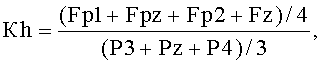
4. Коэффициент Кг рассчитывается по формуле:4. The coefficient Kg is calculated by the formula:
![]()
![]()
Пример 1. Пациент О. 72 года. Основной диагноз: дисциркуляторная энцефалопатия II степени.Example 1. Patient O. 72 years. The main diagnosis: discirculatory encephalopathy II degree.
Процедура расчета коэффициента нарушения гемоликвородинамики Кг по ЭЭГ осуществлялась в соответствии с приведенным алгоритмом.The procedure for calculating the coefficient of violation of hemoliquorodynamics of Kg by EEG was carried out in accordance with the above algorithm.
На фиг.2 представлен фрагмент ЭЭГ пациента О, выбранный для вычисления Кг. Стрелками указаны тета-волны в лобных отведениях.Figure 2 presents a fragment of the EEG of patient O, selected for calculating Kg. Arrows indicate theta waves in the frontal leads.
На фиг.3 представлены спектры выбранного фрагмента ЭЭГ и топограммы тета-ритма с максимумами на частоте 6,84 Гц и 6,35 Гц. Стрелками указаны максимумы на спектрах и соответствующие им топограммы.Figure 3 presents the spectra of the selected EEG fragment and the topogram of theta rhythm with maximums at a frequency of 6.84 Hz and 6.35 Hz. The arrows indicate the maxima on the spectra and the corresponding topograms.
На фиг.4 представлена таблица спектральной мощности ЭЭГ и частоты максимума для диапазонов. Подчеркнуты параметры, используемые для вычисления коэффициента Кг (Fp1=15,60; Fpz=27,04; Fp2=18,27; Fz=45,94; P3=19,82; Pz=12,69; P4=16,61).Figure 4 presents a table of the spectral power of the EEG and maximum frequency for the ranges. The parameters used to calculate the coefficient Kg (Fp1 = 15.60; Fpz = 27.04; Fp2 = 18.27; Fz = 45.94; P3 = 19.82; Pz = 12.69; P4 = 16.61 )
Далее вычисляется коэффициент Кг:Next, the coefficient Kg is calculated:
![]()
![]()
Кг=1,63 соответствует I степени нарушения гемоликвородинамики. По данным МРТ у пациента О. выявлена наружная заместительная гидроцефалия.Kg = 1.63 corresponds to the first degree of hemoliquorodynamics disturbance. According to MRI in patient O. revealed external replacement hydrocephalus.
Пример 2. Пациентка X. 35 лет. Основной диагноз: вегето-сосудистая дистония, остеохондроз шейного отдела позвоночника.Example 2. Patient X. 35 years. The main diagnosis: vegetative-vascular dystonia, osteochondrosis of the cervical spine.
Процедура расчета коэффициента нарушения гемоликвородинамики Кг по ЭЭГ осуществлялась в соответствии с приведенным алгоритмом.The procedure for calculating the coefficient of violation of hemoliquorodynamics of Kg by EEG was carried out in accordance with the above algorithm.
На фиг.5 представлен фрагмент ЭЭГ пациентки X., выбранный для вычисления Кг. Стрелками указаны тета-волны в лобных отведениях.Figure 5 presents a fragment of the EEG of patient X., selected to calculate Kg. Arrows indicate theta waves in the frontal leads.
На фиг.6 представлены спектры и топограммы тета-ритма с максимумами на частоте 6,84 Гц и 6,35 Гц. Стрелками указаны максимумы на спектрах и соответствующие им топограммы.Figure 6 presents the spectra and topograms of theta rhythm with maxima at a frequency of 6.84 Hz and 6.35 Hz. The arrows indicate the maxima on the spectra and the corresponding topograms.
На фиг.7 представлена таблица спектральной мощности ЭЭГ и частоты максимума для диапазонов. Подчеркнуты параметры, используемые для вычисления коэффициента Кг (Fp1=10,21; Fpz=10,83; Fp2=7,36; Fz=6,94; P3=3,17; Pz=3,28; P4=3,59).Figure 7 presents a table of the spectral power of the EEG and maximum frequency for the ranges. The parameters used to calculate the coefficient Kg are emphasized (Fp1 = 10.21; Fpz = 10.83; Fp2 = 7.36; Fz = 6.94; P3 = 3.17; Pz = 3.28; P4 = 3.59 )
Далее вычисляется коэффициент Кг:Next, the coefficient Kg is calculated:
![]()
![]()
Кг=2,64 соответствует II степени нарушения гемоликвородинамики.Kg = 2.64 corresponds to the II degree of hemoliquorodynamics disturbance.
По данным КТ у пациентки X. была выявлена умеренная гидроцефалия.According to CT, patient X. revealed moderate hydrocephalus.
Пример 3. Пациент Ф. 18 лет. Основной диагноз: S-образный сколиоз 3-4 степени. Отдаленные последствия черепно-мозговой травмы (ЧМТ).Example 3. Patient F. 18 years. The main diagnosis: S-shaped scoliosis of 3-4 degrees. Long-term effects of traumatic brain injury (TBI).
Процедура расчета коэффициента нарушения гемоликвородинамики Кг по ЭЭГ осуществлялась в соответствии с приведенным алгоритмом.The procedure for calculating the coefficient of violation of hemoliquorodynamics of Kg by EEG was carried out in accordance with the above algorithm.
На фиг.8 представлен фрагмент ЭЭГ пациентки X., выбранный для вычисления Кг. Стрелками указаны тета-волны в лобных отведениях.On Fig presents an EEG fragment of patient X., selected to calculate Kg. Arrows indicate theta waves in the frontal leads.
На фиг.9 представлены спектры и топограммы тета-ритма с максимумами на частоте 6,84 Гц и 7,32 Гц. Стрелками указаны максимумы на спектрах и соответствующие им топограммы.Figure 9 presents the spectra and topograms of theta rhythm with maxima at a frequency of 6.84 Hz and 7.32 Hz. The arrows indicate the maxima on the spectra and the corresponding topograms.
На фиг.10 представлена таблица спектральной мощности ЭЭГ и частоты максимума для диапазонов. Подчеркнуты параметры, используемые для вычисления коэффициента Кг (Fp1=28,3; Fpz=135,6; Fp2=39,9; Fz=125,2; P3=9,0; Pz=10,5; P4=12,5).Figure 10 presents a table of the spectral power of the EEG and maximum frequency for the ranges. The parameters used to calculate the coefficient Kg (Fp1 = 28.3; Fpz = 135.6; Fp2 = 39.9; Fz = 125.2; P3 = 9.0; Pz = 10.5; P4 = 12.5 )
![]()
![]()
Кг=7,71 соответствует III степени нарушения гемоликвородинамики.Kg = 7.71 corresponds to the III degree of hemoliquorodynamics disturbance.
По данным МРТ - расширение ретроцеребеллярной цистерны и ликворных пространств за счет кортикальных атрофических изменений в лобных отделах.According to MRI, the expansion of the retrocerebellar cistern and cerebrospinal fluid due to cortical atrophic changes in the frontal regions.
Таким образом, предлагаемый способ позволяет выявлять нарушения гемоликвородинамики головного мозга различного генеза с помощью компьютерной энцефалографии, что в 92,5% случаев было подтверждено МРТ и КТ исследованиями.Thus, the proposed method allows to detect violations of hemoliquorodynamics of the brain of various origins using computer encephalography, which in 92.5% of cases was confirmed by MRI and CT studies.
Claims (1)
где Fp1, Fpz, Fp2, Fz, Р3, Pz и Р4 - мощность тета-волн в соответствующих отведениях ЭЭГ;
where Fp1, Fpz, Fp2, Fz, P3, Pz and P4 - the power of the theta waves in the corresponding EEG leads;
Priority Applications (1)
| Application Number | Priority Date | Filing Date | Title |
|---|---|---|---|
| RU2010120815/14A RU2436503C1 (en) | 2010-05-24 | 2010-05-24 | Method for detection of dysfunction of hemoliquorodynamics of cerebrum |
Applications Claiming Priority (1)
| Application Number | Priority Date | Filing Date | Title |
|---|---|---|---|
| RU2010120815/14A RU2436503C1 (en) | 2010-05-24 | 2010-05-24 | Method for detection of dysfunction of hemoliquorodynamics of cerebrum |
Publications (1)
| Publication Number | Publication Date |
|---|---|
| RU2436503C1 true RU2436503C1 (en) | 2011-12-20 |
Family
ID=45404216
Family Applications (1)
| Application Number | Title | Priority Date | Filing Date |
|---|---|---|---|
| RU2010120815/14A RU2436503C1 (en) | 2010-05-24 | 2010-05-24 | Method for detection of dysfunction of hemoliquorodynamics of cerebrum |
Country Status (1)
| Country | Link |
|---|---|
| RU (1) | RU2436503C1 (en) |
Cited By (1)
| Publication number | Priority date | Publication date | Assignee | Title |
|---|---|---|---|---|
| RU2691306C1 (en) * | 2018-10-17 | 2019-06-11 | Федеральное государственное бюджетное научное учреждение "Научный центр неврологии" (ФГБНУ НЦН) | Method for assessing the involvement of disturbed liquor dynamics in the development of a diffuse white matter disorder in cerebral microangiopathy |
Citations (2)
| Publication number | Priority date | Publication date | Assignee | Title |
|---|---|---|---|---|
| RU2158534C2 (en) * | 1997-10-16 | 2000-11-10 | Фирма "НЕЙРОСОФТ" Лтд | Method for diagnosing cerebral disorders in the cases of injured cervical part of the vertebral column of newborn babies |
| EP1493383A2 (en) * | 2003-07-02 | 2005-01-05 | Instrumentarium Corporation | Method of positioning electrodes for central nervous system monitoring |
-
2010
- 2010-05-24 RU RU2010120815/14A patent/RU2436503C1/en not_active IP Right Cessation
Patent Citations (2)
| Publication number | Priority date | Publication date | Assignee | Title |
|---|---|---|---|---|
| RU2158534C2 (en) * | 1997-10-16 | 2000-11-10 | Фирма "НЕЙРОСОФТ" Лтд | Method for diagnosing cerebral disorders in the cases of injured cervical part of the vertebral column of newborn babies |
| EP1493383A2 (en) * | 2003-07-02 | 2005-01-05 | Instrumentarium Corporation | Method of positioning electrodes for central nervous system monitoring |
Non-Patent Citations (2)
| Title |
|---|
| Компьютерная томография. Базовое руководство. - Мед-Лит., 2008, с.146-159. * |
| КРАВЧЕНКО Т.И. Особенности диагностики и лечения больных с посттравматическими нарушениями внутричерепной гемо- и ликвородинамики // Автореф. дисс. - СПб.: 2000, с.7-21. CHIARETTI A. et al. Intraventricular nerve growth factor infusion improves cerebral blood flow and stimulates doublecortin expression in two infants with hypoxic-ischemic brain injury. Neurol Res. 2008 Apr; 30(3):223-8. Epub 2008 Feb 15. * |
Cited By (1)
| Publication number | Priority date | Publication date | Assignee | Title |
|---|---|---|---|---|
| RU2691306C1 (en) * | 2018-10-17 | 2019-06-11 | Федеральное государственное бюджетное научное учреждение "Научный центр неврологии" (ФГБНУ НЦН) | Method for assessing the involvement of disturbed liquor dynamics in the development of a diffuse white matter disorder in cerebral microangiopathy |
Similar Documents
| Publication | Publication Date | Title |
|---|---|---|
| JP6559062B2 (en) | Methods and kits for assessing central nervous system integrity | |
| Xu et al. | Noninvasive methods of detecting increased intracranial pressure | |
| Yao et al. | Cerebral oxygen desaturation is associated with early postoperative neuropsychological dysfunction in patients undergoing cardiac surgery | |
| To et al. | Evaluation of neurocardiac signals in pediatric patients with cyclic vomiting syndrome through power spectral analysis of heart rate variability | |
| US20110196245A1 (en) | Measurement of cerebral hemodynamic parameters | |
| Lei et al. | Postoperative executive function in adult moyamoya disease: a preliminary study of its functional anatomy and behavioral correlates | |
| Zimmermann et al. | The accuracy of the Vigileo/FloTrac continuous cardiac output monitor | |
| Muehlschlegel et al. | Feasibility of NIRS in the neurointensive care unit: a pilot study in stroke using physiological oscillations | |
| Caldas et al. | Intra-aortic balloon pump does not influence cerebral hemodynamics and neurological outcomes in high-risk cardiac patients undergoing cardiac surgery: an analysis of the IABCS trial | |
| Elabasy et al. | Respiratory brain impulse propagation in focal epilepsy | |
| Brandao et al. | Analysis of intracranial pressure waveform using a non-invasive method in individuals with craniosynostosis | |
| Ulv Larsen et al. | Sleep deprivation and sleep intensity exert distinct effects on cerebral vasomotion and brain pulsations driven by the respiratory and cardiac cycles | |
| RU2436503C1 (en) | Method for detection of dysfunction of hemoliquorodynamics of cerebrum | |
| Tsao et al. | Collapsed Jugular Vein and abnormal cerebral blood flow changes in patients of Panic Disorder | |
| RU2464929C1 (en) | Method of estimating efficiency of cranial manual therapy | |
| Edmonds Jr | Central nervous system monitoring | |
| Kolisnyk et al. | The relationship between cessation of brain and systemic circulation after withdrawal of life-sustaining measures | |
| RU2427313C1 (en) | Method of carrying out examination in case of headache syndrome in children | |
| Teo et al. | The Development of Non-Invasive Optical Brain Pulse Monitoring: A Review | |
| RU2141245C1 (en) | Method for determining tactics of surgical intervention in cases of chronic cerebrovascular insufficiency | |
| Roach et al. | Monitoring during vascular surgery | |
| Lushchyk et al. | Predictive and preventive strategies to advance the treatments of cardiovascular and cerebrovascular diseases: the Ukrainian context | |
| Arrowsmith et al. | Intraoperative brain monitoring in cardiac surgery | |
| Ulv Larsen et al. | Sleep pressure propels cerebrovascular oscillations while sleep intensity correlates with respiration-and cardiac-driven brain pulsations | |
| Ivanova et al. | Preoperative state of autonomic regulation in patients with adolescent idiopathic scoliosis |
Legal Events
| Date | Code | Title | Description |
|---|---|---|---|
| MM4A | The patent is invalid due to non-payment of fees |
Effective date: 20130525 |