RU2335307C1 - Method of hypertrophic or keloid skin cicatrixes treatment - Google Patents
Method of hypertrophic or keloid skin cicatrixes treatment Download PDFInfo
- Publication number
- RU2335307C1 RU2335307C1 RU2006142627/14A RU2006142627A RU2335307C1 RU 2335307 C1 RU2335307 C1 RU 2335307C1 RU 2006142627/14 A RU2006142627/14 A RU 2006142627/14A RU 2006142627 A RU2006142627 A RU 2006142627A RU 2335307 C1 RU2335307 C1 RU 2335307C1
- Authority
- RU
- Russia
- Prior art keywords
- scar
- treatment
- hypertrophic
- keloid
- cicatrix
- Prior art date
Links
- 230000001969 hypertrophic effect Effects 0.000 title claims abstract description 17
- 210000001117 keloid Anatomy 0.000 title claims abstract description 11
- 238000000034 method Methods 0.000 title claims abstract description 10
- 238000011282 treatment Methods 0.000 title abstract description 19
- 210000001519 tissue Anatomy 0.000 claims abstract description 29
- CIWBQSYVNNPZIQ-XYWKZLDCSA-N betamethasone dipropionate Chemical compound C1CC2=CC(=O)C=C[C@]2(C)[C@]2(F)[C@@H]1[C@@H]1C[C@H](C)[C@@](C(=O)COC(=O)CC)(OC(=O)CC)[C@@]1(C)C[C@@H]2O CIWBQSYVNNPZIQ-XYWKZLDCSA-N 0.000 claims abstract description 13
- 230000005855 radiation Effects 0.000 claims abstract description 7
- 239000003814 drug Substances 0.000 claims abstract description 5
- 231100000241 scar Toxicity 0.000 claims description 72
- 206010023330 Keloid scar Diseases 0.000 claims description 22
- 208000032544 Cicatrix Diseases 0.000 claims description 9
- 208000002260 Keloid Diseases 0.000 claims description 9
- 230000037387 scars Effects 0.000 claims description 9
- 239000003246 corticosteroid Substances 0.000 claims description 3
- 229960001334 corticosteroids Drugs 0.000 claims description 2
- 229940079593 drug Drugs 0.000 claims description 2
- 210000004767 rumen Anatomy 0.000 claims 1
- 230000015572 biosynthetic process Effects 0.000 abstract description 6
- 230000000694 effects Effects 0.000 abstract description 3
- 239000002537 cosmetic Substances 0.000 abstract description 2
- 210000000589 cicatrix Anatomy 0.000 abstract 4
- 239000000126 substance Substances 0.000 abstract 1
- 210000003491 skin Anatomy 0.000 description 18
- 230000004089 microcirculation Effects 0.000 description 5
- 238000001356 surgical procedure Methods 0.000 description 5
- 238000005755 formation reaction Methods 0.000 description 4
- 210000001562 sternum Anatomy 0.000 description 4
- 102000008186 Collagen Human genes 0.000 description 3
- 108010035532 Collagen Proteins 0.000 description 3
- 229920001436 collagen Polymers 0.000 description 3
- 238000010586 diagram Methods 0.000 description 3
- 238000002559 palpation Methods 0.000 description 3
- 230000010412 perfusion Effects 0.000 description 3
- 230000002980 postoperative effect Effects 0.000 description 3
- 238000002560 therapeutic procedure Methods 0.000 description 3
- 208000003251 Pruritus Diseases 0.000 description 2
- 230000008034 disappearance Effects 0.000 description 2
- 230000036074 healthy skin Effects 0.000 description 2
- 230000000729 hypotrophic effect Effects 0.000 description 2
- 238000002347 injection Methods 0.000 description 2
- 239000007924 injection Substances 0.000 description 2
- 238000007689 inspection Methods 0.000 description 2
- 230000007803 itching Effects 0.000 description 2
- 238000013532 laser treatment Methods 0.000 description 2
- 230000001225 therapeutic effect Effects 0.000 description 2
- 208000019901 Anxiety disease Diseases 0.000 description 1
- 102000004190 Enzymes Human genes 0.000 description 1
- 108090000790 Enzymes Proteins 0.000 description 1
- 206010020565 Hyperaemia Diseases 0.000 description 1
- 206010020880 Hypertrophy Diseases 0.000 description 1
- 206010061218 Inflammation Diseases 0.000 description 1
- 102000014150 Interferons Human genes 0.000 description 1
- 108010050904 Interferons Proteins 0.000 description 1
- 102000015696 Interleukins Human genes 0.000 description 1
- 108010063738 Interleukins Proteins 0.000 description 1
- 208000002720 Malnutrition Diseases 0.000 description 1
- 206010027145 Melanocytic naevus Diseases 0.000 description 1
- 208000007256 Nevus Diseases 0.000 description 1
- 206010030113 Oedema Diseases 0.000 description 1
- 206010033546 Pallor Diseases 0.000 description 1
- 230000001195 anabolic effect Effects 0.000 description 1
- 230000036506 anxiety Effects 0.000 description 1
- 210000004204 blood vessel Anatomy 0.000 description 1
- 230000008822 capillary blood flow Effects 0.000 description 1
- 230000015556 catabolic process Effects 0.000 description 1
- 230000006835 compression Effects 0.000 description 1
- 238000007906 compression Methods 0.000 description 1
- 238000002681 cryosurgery Methods 0.000 description 1
- 230000009089 cytolysis Effects 0.000 description 1
- 238000000295 emission spectrum Methods 0.000 description 1
- 238000005516 engineering process Methods 0.000 description 1
- 210000002615 epidermis Anatomy 0.000 description 1
- 239000000835 fiber Substances 0.000 description 1
- 239000005556 hormone Substances 0.000 description 1
- 229940088597 hormone Drugs 0.000 description 1
- 230000000544 hyperemic effect Effects 0.000 description 1
- 230000004054 inflammatory process Effects 0.000 description 1
- 230000002401 inhibitory effect Effects 0.000 description 1
- 229940079322 interferon Drugs 0.000 description 1
- 229940047122 interleukins Drugs 0.000 description 1
- 208000028867 ischemia Diseases 0.000 description 1
- 208000011379 keloid formation Diseases 0.000 description 1
- 229940063199 kenalog Drugs 0.000 description 1
- 230000001071 malnutrition Effects 0.000 description 1
- 235000000824 malnutrition Nutrition 0.000 description 1
- 238000012544 monitoring process Methods 0.000 description 1
- 208000015380 nutritional deficiency disease Diseases 0.000 description 1
- 230000008506 pathogenesis Effects 0.000 description 1
- 230000001717 pathogenic effect Effects 0.000 description 1
- 230000001575 pathological effect Effects 0.000 description 1
- 230000002085 persistent effect Effects 0.000 description 1
- 230000000750 progressive effect Effects 0.000 description 1
- 230000000770 proinflammatory effect Effects 0.000 description 1
- -1 prostoglandins Proteins 0.000 description 1
- 230000001698 pyrogenic effect Effects 0.000 description 1
- 238000001959 radiotherapy Methods 0.000 description 1
- 230000036573 scar formation Effects 0.000 description 1
- 230000000276 sedentary effect Effects 0.000 description 1
- 238000003786 synthesis reaction Methods 0.000 description 1
- 238000011287 therapeutic dose Methods 0.000 description 1
- 238000012876 topography Methods 0.000 description 1
- 230000001052 transient effect Effects 0.000 description 1
- 230000007704 transition Effects 0.000 description 1
- YNDXUCZADRHECN-JNQJZLCISA-N triamcinolone acetonide Chemical compound C1CC2=CC(=O)C=C[C@]2(C)[C@]2(F)[C@@H]1[C@@H]1C[C@H]3OC(C)(C)O[C@@]3(C(=O)CO)[C@@]1(C)C[C@@H]2O YNDXUCZADRHECN-JNQJZLCISA-N 0.000 description 1
- 230000002792 vascular Effects 0.000 description 1
- 210000005166 vasculature Anatomy 0.000 description 1
- 230000029663 wound healing Effects 0.000 description 1
Images


Landscapes
- Pharmaceuticals Containing Other Organic And Inorganic Compounds (AREA)
- Steroid Compounds (AREA)
- Radiation-Therapy Devices (AREA)
Abstract
Description
Изобретение относится к медицине и может быть использовано в хирургии, дерматологии, дерматокосметологии.The invention relates to medicine and can be used in surgery, dermatology, dermatocosmetology.
Гипертрофические рубцы и келоид могут быть описаны как разновидности обычного заживления раны. С развитием рубца предел его прочности увеличивается в результате прогрессивных связей волокон коллагена. Когда появляется несоответствие между анаболическими и катаболическими процессами, большее количество коллагена производится, чем деградирует, и рубец имеет тенденцию к росту во всех направлениях, поднят выше уровня кожи и остается гиперемированным, классифицируется как келоид или гипертрофический рубец. Келоидный рубец часто вызывает беспокойство в виде зуда, жжения, болезненности. Кроме того, келоидные и гипертрофические рубцы отличаются от здоровой кожи богатой сосудистой сетью, высокой плотностью мезенхимальных клеток и утолщенным эпидермальным слоем [3].Hypertrophic scars and keloid can be described as varieties of conventional wound healing. With the development of the scar, its ultimate strength increases as a result of progressive bonds of collagen fibers. When there is a mismatch between anabolic and catabolic processes, more collagen is produced than degraded, and the scar tends to grow in all directions, is raised above the skin level and remains hyperemic, is classified as a keloid or hypertrophic scar. A keloid scar often causes anxiety in the form of itching, burning, soreness. In addition, keloid and hypertrophic scars differ from healthy skin by a rich vascular network, high density of mesenchymal cells and a thickened epidermal layer [3].
Лечение гипертрофических и келоидных рубцов включает использование окклюзионных повязок, компрессионной терапии, введения кортикостероидов в зону поражения, криохирургию, иссечение, лучевую терапию, лечение интерфероном и другие обещающие, но менее известные методы, направленные на процесс синтеза коллагена [1, 2, 4, 5].Treatment of hypertrophic and keloid scars includes the use of occlusive dressings, compression therapy, the introduction of corticosteroids into the affected area, cryosurgery, excision, radiation therapy, treatment with interferon and other promising, but less well-known methods aimed at the process of collagen synthesis [1, 2, 4, 5 ].
Известен способ лечения гипертрофических и келлоидных рубцов, заключающийся в том, что в толщу рубца вводят кеналог из расчета 40 мг на 5 см2, но не более 80 мг на весь рубец, затем с интервалом 30-40 дней проводят оперативное иссечение келоида с предварительной ишемизацией ткани и наложением внутрикожного шва. В послеоперационном периоде проводят короткофокусную рентгенотерапию: в 1-й день разовая доза составляет 5 Гр, а в 3, 5, 7, 9 дни - по 3 Гр до суммарной очаговой дозы 17 Гр. (Патент РФ № 2195285, МПК7 А61К3 1/573, A61N 5/10, А61Р17/02 от 02.05.2001.) Однако этим способом удается добиться хороших результатов лишь в ранние сроки после образования рубцов и относительно при небольших их размерах (до 3-5 см2). Нередко результатом лечения остается формирование гипотрофических рубцов кожи. Все известные способы также не решают вопрос рецидива гипертрофии рубца.There is a method of treating hypertrophic and keloid scars, namely, that a Kenalog is introduced into the thickness of the scar at the rate of 40 mg per 5 cm 2 , but not more than 80 mg for the entire scar, then an operative excision of the keloid with preliminary ischemia is performed with an interval of 30-40 days tissue and intradermal suture. In the postoperative period, short-focus x-ray therapy is carried out: on the 1st day, a single dose is 5 Gy, and on 3, 5, 7, 9 days - 3 Gy to a total focal dose of 17 Gy. (RF patent No. 2195285, IPC 7 A61K3 1/573, A61N 5/10, A61P17 / 02 of 05/02/2001.) However, this method can achieve good results only in the early stages after scar formation and relatively with small sizes (up to 3 -5 cm 2 ). Often, the result of treatment is the formation of hypotrophic skin scars. All known methods also do not solve the issue of recurrence of scar hypertrophy.
Задача изобретения: повышение эффективности лечения гипертрофических или келоидных рубцов кожи, получение косметического и функционального эффекта.Object of the invention: increasing the effectiveness of the treatment of hypertrophic or keloid scars of the skin, obtaining a cosmetic and functional effect.
Поставленную задачу решают за счет того, что в толщу рубца в течение 4 недель троекратно вводят дипроспан из расчета 7 мг на 5 см2, но не более 14 мг на весь рубец, после полученной медикаментозной гипотрофии рубцовую ткань однократно подвергают воздействию инфракрасного лазерного излучения длиной волны 540 нм, энергией импульса 50-150 мДж, частотой следования импульсов 1-4 Гц, длительностью импульса 1-2 нс.The problem is solved due to the fact that in the thickness of the scar for 4 weeks, diprospan is administered three times at the rate of 7 mg per 5 cm 2 , but not more than 14 mg per entire scar, after the resulting drug hypotrophy, the scar tissue is once exposed to infrared laser radiation with a wavelength 540 nm, pulse energy of 50-150 mJ, pulse repetition rate of 1-4 Hz, pulse duration of 1-2 ns.
Способ осуществляют следующим образом: в течение 4 недель троекратно в толщу рубца вводят дипроспан из расчета 7 мг на 5 см2, но не более 14 мг на весь рубец за один раз, после полученной медикаментозной гипотрофии рубца рубцовую ткань подвергают воздействию инфракрасного лазерного излучения длиной волны 540 нм, энергией импульса 50-150 мДж, частотой следования импульсов 1-4 Гц, длительностью импульса 1-2 нс (режим генерации Q-sw), получаемого с помощью активной среды Nd:YAP (Q-sw)/KTP медицинского лазерного аппарата Multiline.The method is as follows: within 4 weeks, diprospan is administered three times in the thickness of the scar at the rate of 7 mg per 5 cm 2 , but not more than 14 mg for the entire scar at a time, after the received medical hypotrophy of the scar, the scar tissue is exposed to infrared laser radiation with a wavelength 540 nm, pulse energy of 50-150 mJ, pulse repetition rate of 1-4 Hz, pulse duration of 1-2 ns (Q-sw generation mode) obtained using the Nd: YAP (Q-sw) / KTP active medium of a medical laser apparatus Multiline
При контрольном осмотре через 7 недель отмечали формирование нормотрофического рубца с уменьшенной от первоначального состояния площадью, бледно-розовой окраской, при пальпации мягкой консистенции, безболезненного.A follow-up examination after 7 weeks noted the formation of a normotrophic scar with a reduced area from the initial state, pale pink color, with palpation of a soft texture, painless.
Под нашим наблюдением находилось 22 пациента с гипертрофическими и келоидными рубцами кожи с преимущественным расположением в области грудины, наружной поверхности плеча и заушной области. Топография, размеры и возраст гипертрофических рубцов не носили принципиальный характер, поэтому не подвергались клинической классификации. Возраст пациентов составлял от 20 до 44 лет.Under our supervision, there were 22 patients with hypertrophic and keloid skin scars with a predominant location in the sternum, the outer surface of the shoulder and behind the ear. The topography, size, and age of the hypertrophic scars were not fundamental, therefore, they were not subjected to clinical classification. The age of the patients ranged from 20 to 44 years.
В результате проведения лечебных мероприятий у пациентов с гипертрофическими и келоидными рубцами кожи были изучены следующие показатели: высота рубца над поверхностью кожи, площадь рубца, консистенция, цвет, состояние окружающей кожи, микроциркуляция рубца.As a result of therapeutic measures in patients with hypertrophic and keloid skin scars, the following indicators were studied: height of the scar above the skin surface, area of the scar, consistency, color, condition of the surrounding skin, microcirculation of the scar.
Использование кортикостероида пролонгированного действия (дипроспана) в лечении гипертрофических или келоидных рубцов носит патогенетический характер, поскольку, ингибируя провоспалительные ферменты, гормоны, простогландины, интерлейкины, не только препятствует дальнейшему разрастанию рубцовой ткани, но и приводит к значительному ее лизису.The use of a prolonged-acting corticosteroid (diprospan) in the treatment of hypertrophic or keloid scars is pathogenetic in nature, since, by inhibiting pro-inflammatory enzymes, hormones, prostoglandins, interleukins, it not only prevents further growth of scar tissue, but also leads to its significant lysis.
Применение высоких терапевтических доз дипроспана позволяет в короткие сроки привести келоидные рубцы к состоянию гипотрофии. И в результате перехода гипертрофического или келоидного рубца сперва в состояние гипотрофии, а после лазерного воздействия - нормотрофии, происходит не только изменение структуры рубцовой ткани, но и уменьшение первоначальной ее площади.The use of high therapeutic doses of diprospan allows in a short time to bring keloid scars to a state of malnutrition. And as a result of the transition of a hypertrophic or keloid scar first to a state of hypotrophy, and after laser exposure - normotrophy, there is not only a change in the structure of scar tissue, but also a decrease in its initial area.
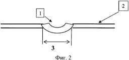
Благодаря скоротечному изменению толщины рубцовой ткани под воздействием дипроспана и лазерного излучения происходит значительное уменьшение и площади рубца. Это свойство использовано в лечебных целях, поскольку мы планомерно достигаем гипотрофического результата, который в последующем переводим в состояние нормотрофии (см. фиг.1, 2, 3): На фиг.1 изображена схема рубцовой ткани до лечения: На фиг.2 изображена схема рубцовой ткани после лечения с помощью дипроспана (отмечается гипотрофия рубца): 1 - рубцовая ткань; 2 - поверхность кожи; 31 - размер рубца. На фиг.3. изображена схема рубцовой ткани после инфракрасного лазерного облучения (отмечается нормотрофия рубца): 1 - рубцовая ткань; 2 - поверхность кожи; 32 - размер рубца. Динамика размеров рубца отмечалась следующая: 3>31>32.Due to the transient change in the thickness of the scar tissue under the influence of diprospan and laser radiation, a significant decrease in the area of the scar occurs. This property is used for medicinal purposes, since we systematically achieve a hypotrophic result, which we subsequently translate into a state of normotrophy (see figures 1, 2, 3): Figure 1 shows a diagram of scar tissue before treatment: Figure 2 shows a diagram scar tissue after treatment with diprospan (hypotrophy of the scar is noted): 1 - scar tissue; 2 - skin surface; 3 1 - the size of the scar. In figure 3. The diagram of scar tissue after infrared laser irradiation is shown (normotrophy of the scar is noted): 1 - scar tissue; 2 - skin surface; 3 2 - the size of the scar. The dynamics of the size of the scar was noted as follows: 3> 3 1 > 3 2 .
На фоне использования инфракрасного лазерного излучения в режиме генерации Q-sw с длительностью импульса 1-2 нс происходит облитерация сосудов дна рубца без воздействия на собственно ткань рубца и на окружающие ткани. Через 8 недель от начала лечения отмечали уменьшение площади келоидных и гипертрофических рубцов в среднем на 23±4,3% (Р<0,05) (таблица 1).Against the background of the use of infrared laser radiation in the Q-sw generation mode with a pulse duration of 1-2 ns, obliteration of the vessels of the scar bottom occurs without affecting the scar tissue itself and the surrounding tissues. After 8 weeks from the start of treatment, a decrease in the area of keloid and hypertrophic scars by an average of 23 ± 4.3% (P <0.05) was noted (table 1).
Исследование состояния микроциркуляции рубца проводили методом лазерной доплеровской флоуметрии (ЛДФ) в красном спектре излучения с использованием лазерного анализатора капиллярного кровотока BLF-21.The study of the state of scar microcirculation was performed by laser Doppler flowmetry (LDF) in the red emission spectrum using a BLF-21 laser analyzer of capillary blood flow.
Запись ЛДФ-грамм производилась до лазерной обработки, непосредственно после воздействия и на протяжении последующих 3 недель наблюдения. Измерения микроциркуляции проводили в области дна рубца и на симметричных точках здоровой кожи.The LDF-gram was recorded before laser treatment, immediately after exposure, and during the next 3 weeks of observation. Microcirculation was measured at the bottom of the scar and at the symmetrical points of healthy skin.
Сразу после лазерной обработки поверхности рубца отмечали снижение показателя микроциркуляции на 45% по сравнению с исходными значениями (Р<0,05) (таблица 2).Immediately after laser treatment of the scar surface, a decrease in the microcirculation index by 45% was noted compared with the initial values (P <0.05) (table 2).
На протяжении 3-недельного контроля показателя микроциркуляции у пациентов не происходило достоверного изменения перфузии патологического очага, что свидетельствует об эффективности однократного воздействия инфракрасного лазерного излучения на ткань рубца.During a 3-week monitoring of the microcirculation index, patients did not significantly change the perfusion of the pathological focus, which indicates the effectiveness of a single exposure to infrared laser radiation on the scar tissue.
Клинический примерClinical example
Пациентка М., 42 года. Обратилась с диагнозом: послеоперационный келоидный рубец кожи верхней трети грудины.Patient M., 42 years old. I was diagnosed with a postoperative keloid scar of the skin of the upper third of the sternum.
Из анамнеза - 4 года назад произведено хирургическое удаление невуса в области грудины. Со слов, послеоперационный рубец в течение четырех лет увеличивался в размерах, вызывал болезненность при касании, периодически беспокоил зуд.From the anamnesis - 4 years ago, surgical removal of the nevus in the sternum was performed. According to the words, the postoperative scar for four years increased in size, caused pain when touched, itching periodically bothered.
На момент осмотра: на коже верхней трети грудины гипертрофический рубец багрово-красного цвета, с четкими границами, неровными краями, до 4 см в длину и 2,5 см в ширину, выступающий над поверхностью кожи на 0,5 см. При пальпации рубец плотный, малоподвижный, умеренно болезненный. Расценен как келоидный рубец.At the time of examination: on the skin of the upper third of the sternum, a hypertrophic scar of crimson-red color, with clear boundaries, uneven edges, up to 4 cm long and 2.5 cm wide, protruding 0.5 cm above the skin surface. On palpation, the scar is dense sedentary, moderately painful. Considered as a keloid scar.
Проведена инъекция дипроспана в объеме 1 мл (7 мг) в рубцовую ткань. При повторном осмотре через 5 дней отмечали некоторое уменьшение объема рубцовой ткани, побледнение окраски. При пальпации рубец стал более мягкий и подвижный, болезненность отсутствует. В рубцовую ткань повторно введено 7 мг дипроспана.A 1 ml (7 mg) diprospan was injected into the scar tissue. When re-examined after 5 days, a slight decrease in the volume of scar tissue, blanching of the color was noted. On palpation, the scar became softer and more mobile, there was no soreness. 7 mg of diprospan was reintroduced into the scar tissue.
Осмотр через 14 дней после первого введения дипроспана: рубец бледно-розового цвета, выступает над поверхностью кожи на 1 мм, мягкий, подвижный, безболезненный. Размеры рубца: 3,7 см - длина, 2,2 см - ширина. Проведена очередная инъекция дипроспана в объеме 1 мл (7 мг) в рубцовую ткань.Inspection 14 days after the first administration of diprospan: a scar of pale pink color, protrudes 1 mm above the surface of the skin, soft, mobile, painless. Scar dimensions: 3.7 cm - length, 2.2 cm - width. The next injection of diprospan in a volume of 1 ml (7 mg) in scar tissue was performed.
Осмотр через 28 дней от начала лечения: отмечается формирование гипотрофии рубцовой ткани в виде углубления от поверхности кожи на 2 мм. Рубец бледно-розовой окраски, мягкий, безболезненный, подвижный. Размеры рубца: 3,5 см - длина, 2,0 см - ширина. Дно рубца представлено истонченной тканью с просвечивающей сквозь нее сетью поверхностно расположенных сосудов ярко-розового цвета, до 1 мм в диаметре.Inspection after 28 days from the start of treatment: the formation of hypotrophy of scar tissue in the form of a depression from the skin surface by 2 mm is noted. The scar is pale pink in color, soft, painless, mobile. Scar dimensions: 3.5 cm - length, 2.0 cm - width. The bottom of the scar is represented by a thinned tissue with a network of superficially located vessels of bright pink color visible through it, up to 1 mm in diameter.
На этом этапе проведено однократное облучение сосудов дна рубца лучом инфракрасного лазера с длинной волны 540 нм, длительностью импульсов 1-2 нс (режим генерации Q-sw), с энергией 75 мДж и частотой следования импульса 2 Гц.At this stage, a single irradiation of the vessels of the scar bottom was carried out with an infrared laser beam with a wavelength of 540 nm, a pulse duration of 1-2 ns (Q-sw generation mode), with an energy of 75 mJ and a pulse repetition rate of 2 Hz.
Контрольный осмотр на 1 сутки после облучения показал частичное исчезновение сосудов, с локальными участками потемнения и локального воспаления, гиперемии, отека тканей. При осмотре пациента через 4 недели отмечали: исчезновение углубления в центре рубца и отсутствие сосудистой сети. Рубец бледно-розового цвета, расположен на уровне интактной кожи, мягкий, безболезненный. Размеры рубца составили: 3,3 см - длина, 1,7 см - ширина. При контрольном осмотре через 1 год отмечен стойкий клинический результат.A control examination on the 1st day after irradiation showed a partial disappearance of blood vessels, with local areas of darkening and local inflammation, hyperemia, tissue edema. When examining the patient after 4 weeks noted: the disappearance of the deepening in the center of the scar and the absence of the vasculature. The scar is pale pink, located at the level of intact skin, soft, painless. The size of the scar was: 3.3 cm - length, 1.7 cm - width. A follow-up examination after 1 year showed a persistent clinical result.
Таким образом, использование инъекций дипроспана из расчета 7 мг на 5 см2, но не более 14 мг на весь рубец и в последующем инфракрасного лазера с длинной волны 540 нм, длительностью импульсов 1-2 нс (режим генерации Q-sw), с энергией 50-150 мДж и частотой следования импульса 1-4 Гц в лечении гипертрофических или келоидных рубцов позволяет получить стойкий клинический результат уже через 8 недель от начала лечения, заключающийся в уменьшении площади рубца (на 22,3%), формировании нормотрофической рубцовой ткани, улучшении консистенции и изменении цвета, характерного окружающей коже. За счет сверхкороткого отрезка времени лазерного воздействия (1-2 нс) благодаря технологии Q-sw способ является безболезненным и безопасным и не оказывает влияния на окружающие ткани, что подтверждается устойчивым клиническим результатом наблюдаемым у пациентов через 1 год после лечения.Thus, the use of diprospan injection at the rate of 7 mg per 5 cm 2 , but not more than 14 mg per entire scar and subsequently an infrared laser with a wavelength of 540 nm, pulse durations of 1-2 ns (Q-sw generation mode), with energy 50-150 mJ and a pulse repetition rate of 1-4 Hz in the treatment of hypertrophic or keloid scars allows you to get a stable clinical result after 8 weeks from the start of treatment, which consists in reducing the area of the scar (by 22.3%), the formation of normotrophic scar tissue, improving consistency and color change, x acteristic surrounding skin. Due to the ultra-short time period of laser exposure (1-2 ns) due to Q-sw technology, the method is painless and safe and does not affect the surrounding tissue, which is confirmed by the stable clinical result observed in
ЛитератураLiterature
1. Гарюк Г.И., Лисовец В.Т., Шевченко A.M. О лечебной тактике при келоидных образованиях ушных раковин. // Вест. оториноларингологии. - 1991. - №3. - С.54-56.1. Garyuk G.I., Lisovets V.T., Shevchenko A.M. On therapeutic tactics in keloid formations of the auricles. // West. otorhinolaryngology. - 1991. - No. 3. - S. 54-56.
2. Кожевников В.А., Осипов А.А., Тен Ю.В. Пирогеналотерапия в комплексном лечении келоидных рубцов лазером. // Хирургия. -1991. - № 8. - С.151-152.2. Kozhevnikov V.A., Osipov A.A., Ten Yu.V. Pyrogenal therapy in the complex treatment of keloid scars with a laser. // Surgery. -1991. - No. 8. - S.151-152.
3. Осипов А.А., Суворова А.В., Трубников П.Н. К вопросу о патогенезе и биомоделировании келоидных рубцов. // Детская хирургия. - 2001. - № 4. - С.34-36.3. Osipov A.A., Suvorova A.V., Trubnikov P.N. To the question of the pathogenesis and biomodeling of keloid scars. // Children's surgery. - 2001. - No. 4. - P.34-36.
4. Шафранов В.В., Короткий Н.Г., Таганов А.В. и др. Возможности использования СВЧ-криодеструкции в дерматокосметологии для лечения келоидных рубцов у детей. // Детская хирургия. - 2000. - № 1.-С.35-37.4. Shafranov V.V., Korotky N.G., Taganov A.V. Possibilities of using microwave cryodestruction in dermatocosmetology for the treatment of keloid scars in children. // Children's surgery. - 2000. - No. 1.-C.35-37.
5. Шафранов В.В., Цветкова Г.М., Сибилева К.Ф. и др. Комплексное лечение келоидных рубцов кожи. // Вестник дерматологии и венерологии. - 1985. - № 3. - С.47-49.5. Shafranov V.V., Tsvetkova G.M., Sibileva K.F. and others. Comprehensive treatment of keloid skin scars. // Bulletin of Dermatology and Venereology. - 1985. - No. 3. - P.47-49.
Claims (1)
Priority Applications (1)
| Application Number | Priority Date | Filing Date | Title |
|---|---|---|---|
| RU2006142627/14A RU2335307C1 (en) | 2006-12-01 | 2006-12-01 | Method of hypertrophic or keloid skin cicatrixes treatment |
Applications Claiming Priority (1)
| Application Number | Priority Date | Filing Date | Title |
|---|---|---|---|
| RU2006142627/14A RU2335307C1 (en) | 2006-12-01 | 2006-12-01 | Method of hypertrophic or keloid skin cicatrixes treatment |
Publications (2)
| Publication Number | Publication Date |
|---|---|
| RU2006142627A RU2006142627A (en) | 2008-06-20 |
| RU2335307C1 true RU2335307C1 (en) | 2008-10-10 |
Family
ID=39927720
Family Applications (1)
| Application Number | Title | Priority Date | Filing Date |
|---|---|---|---|
| RU2006142627/14A RU2335307C1 (en) | 2006-12-01 | 2006-12-01 | Method of hypertrophic or keloid skin cicatrixes treatment |
Country Status (1)
| Country | Link |
|---|---|
| RU (1) | RU2335307C1 (en) |
Cited By (3)
| Publication number | Priority date | Publication date | Assignee | Title |
|---|---|---|---|---|
| RU2452533C2 (en) * | 2010-05-24 | 2012-06-10 | Галина Ивановна Фисенко | Method of eliminating old, multiply treated and x-ray resistant false keloids |
| RU2559914C1 (en) * | 2014-06-10 | 2015-08-20 | Олег Иванович Коновалов | Method of treating anal constriction |
| RU2767909C1 (en) * | 2021-07-12 | 2022-03-22 | Зоя Александровна Евсюкова | Method for the prevention of pathological scars of postoperative skin wounds |
Citations (3)
| Publication number | Priority date | Publication date | Assignee | Title |
|---|---|---|---|---|
| RU2195285C2 (en) * | 2001-02-05 | 2002-12-27 | Ростовский научно-исследовательский онкологический институт | Method for treating keloid scars |
| RU2200557C1 (en) * | 2001-11-23 | 2003-03-20 | Государственное учреждение Межотраслевой научно-технический комплекс "Микрохирургия глаза" им. акад. С.Н.Федорова | Method for treating hypertrophic and keloid scars |
| RU2260462C1 (en) * | 2004-02-10 | 2005-09-20 | Государственное учреждение Межотраслевой научно-технический комплекс "Микрохирургия глаза" им. акад. С.Н. Федорова | Method for treating keloid cicatrices by applying photodynamic therapy techniques |
-
2006
- 2006-12-01 RU RU2006142627/14A patent/RU2335307C1/en not_active IP Right Cessation
Patent Citations (3)
| Publication number | Priority date | Publication date | Assignee | Title |
|---|---|---|---|---|
| RU2195285C2 (en) * | 2001-02-05 | 2002-12-27 | Ростовский научно-исследовательский онкологический институт | Method for treating keloid scars |
| RU2200557C1 (en) * | 2001-11-23 | 2003-03-20 | Государственное учреждение Межотраслевой научно-технический комплекс "Микрохирургия глаза" им. акад. С.Н.Федорова | Method for treating hypertrophic and keloid scars |
| RU2260462C1 (en) * | 2004-02-10 | 2005-09-20 | Государственное учреждение Межотраслевой научно-технический комплекс "Микрохирургия глаза" им. акад. С.Н. Федорова | Method for treating keloid cicatrices by applying photodynamic therapy techniques |
Non-Patent Citations (1)
| Title |
|---|
| ХАЛМУРАТОВ А.М. «Комбинированное использование лазеров при келоидных рубцах», Вопросы реконструктивной и восстановительной хирургии, 1994, с.219-220. МЕЛЬНИЧЕНКО М.Г. «Применение низкоинтенсивного инфракрасного лазерного излучения и дистракции в комплексном лечении рубцовых поражений покровных тканей у детей». Дет. хирургия, 2000, №1, с.32-35. ASILIAN А " New combination of triamcinolone, 5-Fluorouracil, and pulsed-dye laser for treatment of keloid and hypertrophic scars". Dermatol Surg. 2006 Jul; 32(7): 907-15. * |
Cited By (3)
| Publication number | Priority date | Publication date | Assignee | Title |
|---|---|---|---|---|
| RU2452533C2 (en) * | 2010-05-24 | 2012-06-10 | Галина Ивановна Фисенко | Method of eliminating old, multiply treated and x-ray resistant false keloids |
| RU2559914C1 (en) * | 2014-06-10 | 2015-08-20 | Олег Иванович Коновалов | Method of treating anal constriction |
| RU2767909C1 (en) * | 2021-07-12 | 2022-03-22 | Зоя Александровна Евсюкова | Method for the prevention of pathological scars of postoperative skin wounds |
Also Published As
| Publication number | Publication date |
|---|---|
| RU2006142627A (en) | 2008-06-20 |
Similar Documents
| Publication | Publication Date | Title |
|---|---|---|
| Stern et al. | Carbon dioxide laser excision of earlobe keloids: a prospective study and critical analysis of existing data | |
| Nast et al. | German S2k guidelines for the therapy of pathological scars (hypertrophic scars and keloids) | |
| Hirshowitz et al. | Treatment of keloid scars by combined cryosurgery and intralesional corticosteroids | |
| Glassberg et al. | Capillary hemangiomas: case study of a novel laser treatment and a review of therapeutic options | |
| Zhang et al. | Improvement of surgical scars by early intervention with carbon dioxide fractional laser | |
| RU2335307C1 (en) | Method of hypertrophic or keloid skin cicatrixes treatment | |
| CN113143955A (en) | Modified lauromacrogol foam hardening agent, preparation method and application | |
| JP2011520508A (en) | Percutaneous vascular treatment method and apparatus | |
| Alster | Laser treatment of hypertrophic scars | |
| RU2599868C1 (en) | Method of treating pathological cicatrical skin deformities | |
| Wollina et al. | Transdermal CO2 application in chronic wounds | |
| RU2543347C1 (en) | Method for sclerotherapy of juvenile haemangiomas | |
| RU2761265C1 (en) | Method for correcting atrophic acne scarring | |
| RU2528973C1 (en) | Method of treating trophic ulcers | |
| Sobh et al. | Efficacy of Cybele Scagel Phonophoresis on post-burn hypertrophic scar | |
| RU2323021C1 (en) | Method for treating patients for hypotrophic skin cicatrices | |
| Menachem et al. | The effect of vaginal CO2 laser treatment on stress urinary incontinence symptoms | |
| RU2800323C1 (en) | Method of treatment of post-traumatic scars | |
| RU2160135C2 (en) | Method for treating trophic ulcers and purulent wounds not closing up for long time period | |
| RU2316295C1 (en) | Conservative method for treating eye appendage and orbit hemangioma in children | |
| Gupta et al. | Comparative efficacy of oral tranexamic acid and trans-epidermal delivery of tranexamic acid by micro-needling in melasma | |
| RU2731527C1 (en) | Method for elimination of cicatrical skin changes | |
| RU2194537C2 (en) | Conservative method for treating urethral strictures | |
| Lee et al. | Various treatments of scar | |
| US20220176149A1 (en) | Method of treatment or prevention of keloids and hypertrophic scars using electromagnetic radiation |
Legal Events
| Date | Code | Title | Description |
|---|---|---|---|
| MM4A | The patent is invalid due to non-payment of fees |
Effective date: 20081202 |