RU2230786C2 - Heat-induced promoter and its using - Google Patents
Heat-induced promoter and its using Download PDFInfo
- Publication number
- RU2230786C2 RU2230786C2 RU2001124852/13A RU2001124852A RU2230786C2 RU 2230786 C2 RU2230786 C2 RU 2230786C2 RU 2001124852/13 A RU2001124852/13 A RU 2001124852/13A RU 2001124852 A RU2001124852 A RU 2001124852A RU 2230786 C2 RU2230786 C2 RU 2230786C2
- Authority
- RU
- Russia
- Prior art keywords
- heat
- nucleic acid
- sequence
- induced promoter
- seq
- Prior art date
Links
- 150000007523 nucleic acids Chemical class 0.000 claims abstract description 102
- 108020004707 nucleic acids Proteins 0.000 claims abstract description 74
- 102000039446 nucleic acids Human genes 0.000 claims abstract description 74
- 108090000623 proteins and genes Proteins 0.000 claims abstract description 70
- 102000004169 proteins and genes Human genes 0.000 claims abstract description 41
- 230000000694 effects Effects 0.000 claims abstract description 32
- 239000002773 nucleotide Substances 0.000 claims abstract description 25
- 125000003729 nucleotide group Chemical group 0.000 claims abstract description 25
- 230000014509 gene expression Effects 0.000 claims abstract description 21
- 230000000295 complement effect Effects 0.000 claims abstract description 19
- 239000012634 fragment Substances 0.000 claims abstract description 18
- 240000004808 Saccharomyces cerevisiae Species 0.000 claims abstract description 13
- 230000006698 induction Effects 0.000 claims abstract description 13
- 102000007056 Recombinant Fusion Proteins Human genes 0.000 claims abstract description 12
- 108010008281 Recombinant Fusion Proteins Proteins 0.000 claims abstract description 12
- 210000004027 cell Anatomy 0.000 claims description 62
- 241000320412 Ogataea angusta Species 0.000 claims description 43
- 230000035939 shock Effects 0.000 claims description 29
- 108091028043 Nucleic acid sequence Proteins 0.000 claims description 26
- 108010020589 trehalose-6-phosphate synthase Proteins 0.000 claims description 23
- 229920001184 polypeptide Polymers 0.000 claims description 20
- 108090000765 processed proteins & peptides Proteins 0.000 claims description 20
- 102000004196 processed proteins & peptides Human genes 0.000 claims description 20
- 235000014680 Saccharomyces cerevisiae Nutrition 0.000 claims description 12
- 230000001939 inductive effect Effects 0.000 claims description 9
- 238000004519 manufacturing process Methods 0.000 claims description 9
- 230000022532 regulation of transcription, DNA-dependent Effects 0.000 claims description 8
- 238000010367 cloning Methods 0.000 claims description 7
- 210000005253 yeast cell Anatomy 0.000 claims description 5
- 238000012258 culturing Methods 0.000 claims description 4
- 230000002538 fungal effect Effects 0.000 claims description 4
- 230000002068 genetic effect Effects 0.000 claims description 3
- 125000003275 alpha amino acid group Chemical group 0.000 claims 7
- 239000013600 plasmid vector Substances 0.000 claims 6
- FWMNVWWHGCHHJJ-SKKKGAJSSA-N 4-amino-1-[(2r)-6-amino-2-[[(2r)-2-[[(2r)-2-[[(2r)-2-amino-3-phenylpropanoyl]amino]-3-phenylpropanoyl]amino]-4-methylpentanoyl]amino]hexanoyl]piperidine-4-carboxylic acid Chemical compound C([C@H](C(=O)N[C@H](CC(C)C)C(=O)N[C@H](CCCCN)C(=O)N1CCC(N)(CC1)C(O)=O)NC(=O)[C@H](N)CC=1C=CC=CC=1)C1=CC=CC=C1 FWMNVWWHGCHHJJ-SKKKGAJSSA-N 0.000 claims 1
- 241000235347 Schizosaccharomyces pombe Species 0.000 claims 1
- 238000000034 method Methods 0.000 abstract description 29
- 239000013598 vector Substances 0.000 abstract description 12
- 230000004071 biological effect Effects 0.000 abstract 1
- 239000000126 substance Substances 0.000 abstract 1
- LFQSCWFLJHTTHZ-UHFFFAOYSA-N Ethanol Chemical compound CCO LFQSCWFLJHTTHZ-UHFFFAOYSA-N 0.000 description 30
- 108020004414 DNA Proteins 0.000 description 28
- 239000013612 plasmid Substances 0.000 description 27
- HDTRYLNUVZCQOY-UHFFFAOYSA-N α-D-glucopyranosyl-α-D-glucopyranoside Natural products OC1C(O)C(O)C(CO)OC1OC1C(O)C(O)C(O)C(CO)O1 HDTRYLNUVZCQOY-UHFFFAOYSA-N 0.000 description 25
- HDTRYLNUVZCQOY-WSWWMNSNSA-N Trehalose Natural products O[C@@H]1[C@@H](O)[C@@H](O)[C@@H](CO)O[C@@H]1O[C@@H]1[C@H](O)[C@@H](O)[C@@H](O)[C@@H](CO)O1 HDTRYLNUVZCQOY-WSWWMNSNSA-N 0.000 description 25
- HDTRYLNUVZCQOY-LIZSDCNHSA-N alpha,alpha-trehalose Chemical compound O[C@@H]1[C@@H](O)[C@H](O)[C@@H](CO)O[C@@H]1O[C@@H]1[C@H](O)[C@@H](O)[C@H](O)[C@@H](CO)O1 HDTRYLNUVZCQOY-LIZSDCNHSA-N 0.000 description 24
- XLYOFNOQVPJJNP-UHFFFAOYSA-N water Substances O XLYOFNOQVPJJNP-UHFFFAOYSA-N 0.000 description 24
- 101000795074 Homo sapiens Tryptase alpha/beta-1 Proteins 0.000 description 20
- 101000662819 Physarum polycephalum Terpene synthase 1 Proteins 0.000 description 20
- PEDCQBHIVMGVHV-UHFFFAOYSA-N Glycerine Chemical compound OCC(O)CO PEDCQBHIVMGVHV-UHFFFAOYSA-N 0.000 description 19
- 102100029639 Tryptase alpha/beta-1 Human genes 0.000 description 19
- 239000012528 membrane Substances 0.000 description 19
- 239000000523 sample Substances 0.000 description 17
- 230000009466 transformation Effects 0.000 description 17
- OKKJLVBELUTLKV-UHFFFAOYSA-N Methanol Chemical compound OC OKKJLVBELUTLKV-UHFFFAOYSA-N 0.000 description 15
- 239000000872 buffer Substances 0.000 description 15
- 239000000499 gel Substances 0.000 description 15
- 239000000243 solution Substances 0.000 description 15
- WQZGKKKJIJFFOK-GASJEMHNSA-N Glucose Natural products OC[C@H]1OC(O)[C@H](O)[C@@H](O)[C@@H]1O WQZGKKKJIJFFOK-GASJEMHNSA-N 0.000 description 14
- 239000008103 glucose Substances 0.000 description 14
- 101150066555 lacZ gene Proteins 0.000 description 14
- 239000000203 mixture Substances 0.000 description 14
- 238000003752 polymerase chain reaction Methods 0.000 description 14
- 108090000698 Formate Dehydrogenases Proteins 0.000 description 13
- 239000000020 Nitrocellulose Substances 0.000 description 13
- FJWGYAHXMCUOOM-QHOUIDNNSA-N [(2s,3r,4s,5r,6r)-2-[(2r,3r,4s,5r,6s)-4,5-dinitrooxy-2-(nitrooxymethyl)-6-[(2r,3r,4s,5r,6s)-4,5,6-trinitrooxy-2-(nitrooxymethyl)oxan-3-yl]oxyoxan-3-yl]oxy-3,5-dinitrooxy-6-(nitrooxymethyl)oxan-4-yl] nitrate Chemical compound O([C@@H]1O[C@@H]([C@H]([C@H](O[N+]([O-])=O)[C@H]1O[N+]([O-])=O)O[C@H]1[C@@H]([C@@H](O[N+]([O-])=O)[C@H](O[N+]([O-])=O)[C@@H](CO[N+]([O-])=O)O1)O[N+]([O-])=O)CO[N+](=O)[O-])[C@@H]1[C@@H](CO[N+]([O-])=O)O[C@@H](O[N+]([O-])=O)[C@H](O[N+]([O-])=O)[C@H]1O[N+]([O-])=O FJWGYAHXMCUOOM-QHOUIDNNSA-N 0.000 description 13
- 229920001220 nitrocellulos Polymers 0.000 description 13
- 239000006228 supernatant Substances 0.000 description 13
- 108091032973 (ribonucleotides)n+m Proteins 0.000 description 12
- 101150077059 TPS1 gene Proteins 0.000 description 12
- 102100026189 Beta-galactosidase Human genes 0.000 description 11
- 108010005774 beta-Galactosidase Proteins 0.000 description 11
- 239000013604 expression vector Substances 0.000 description 11
- 239000000284 extract Substances 0.000 description 11
- FAPWRFPIFSIZLT-UHFFFAOYSA-M Sodium chloride Chemical compound [Na+].[Cl-] FAPWRFPIFSIZLT-UHFFFAOYSA-M 0.000 description 10
- 238000005119 centrifugation Methods 0.000 description 10
- 239000000463 material Substances 0.000 description 10
- 239000002609 medium Substances 0.000 description 10
- HEMHJVSKTPXQMS-UHFFFAOYSA-M Sodium hydroxide Chemical compound [OH-].[Na+] HEMHJVSKTPXQMS-UHFFFAOYSA-M 0.000 description 9
- 241000926178 Tubulanus polymorphus Species 0.000 description 9
- 238000004458 analytical method Methods 0.000 description 9
- 238000004113 cell culture Methods 0.000 description 9
- 238000002474 experimental method Methods 0.000 description 9
- 108010011619 6-Phytase Proteins 0.000 description 8
- KCXVZYZYPLLWCC-UHFFFAOYSA-N EDTA Chemical compound OC(=O)CN(CC(O)=O)CCN(CC(O)=O)CC(O)=O KCXVZYZYPLLWCC-UHFFFAOYSA-N 0.000 description 8
- 241000588724 Escherichia coli Species 0.000 description 8
- ISAKRJDGNUQOIC-UHFFFAOYSA-N Uracil Chemical compound O=C1C=CNC(=O)N1 ISAKRJDGNUQOIC-UHFFFAOYSA-N 0.000 description 8
- 150000001413 amino acids Chemical group 0.000 description 8
- 239000006152 selective media Substances 0.000 description 8
- QKNYBSVHEMOAJP-UHFFFAOYSA-N 2-amino-2-(hydroxymethyl)propane-1,3-diol;hydron;chloride Chemical compound Cl.OCC(N)(CO)CO QKNYBSVHEMOAJP-UHFFFAOYSA-N 0.000 description 7
- ZMXDDKWLCZADIW-UHFFFAOYSA-N N,N-Dimethylformamide Chemical compound CN(C)C=O ZMXDDKWLCZADIW-UHFFFAOYSA-N 0.000 description 7
- 235000011187 glycerol Nutrition 0.000 description 7
- 238000009396 hybridization Methods 0.000 description 7
- 238000005259 measurement Methods 0.000 description 7
- 108020004999 messenger RNA Proteins 0.000 description 7
- 239000002244 precipitate Substances 0.000 description 7
- HRGUSFBJBOKSML-UHFFFAOYSA-N 3',5'-di-O-methyltricetin Chemical compound COC1=C(O)C(OC)=CC(C=2OC3=CC(O)=CC(O)=C3C(=O)C=2)=C1 HRGUSFBJBOKSML-UHFFFAOYSA-N 0.000 description 6
- QTBSBXVTEAMEQO-UHFFFAOYSA-N Acetic acid Chemical compound CC(O)=O QTBSBXVTEAMEQO-UHFFFAOYSA-N 0.000 description 6
- 229920001817 Agar Polymers 0.000 description 6
- OKTJSMMVPCPJKN-UHFFFAOYSA-N Carbon Chemical compound [C] OKTJSMMVPCPJKN-UHFFFAOYSA-N 0.000 description 6
- IDDMFNIRSJVBHE-UHFFFAOYSA-N Piscigenin Natural products COC1=C(O)C(OC)=CC(C=2C(C3=C(O)C=C(O)C=C3OC=2)=O)=C1 IDDMFNIRSJVBHE-UHFFFAOYSA-N 0.000 description 6
- 108700008625 Reporter Genes Proteins 0.000 description 6
- 238000009825 accumulation Methods 0.000 description 6
- 239000008272 agar Substances 0.000 description 6
- 229910052799 carbon Inorganic materials 0.000 description 6
- 239000003153 chemical reaction reagent Substances 0.000 description 6
- KCFYHBSOLOXZIF-UHFFFAOYSA-N dihydrochrysin Natural products COC1=C(O)C(OC)=CC(C2OC3=CC(O)=CC(O)=C3C(=O)C2)=C1 KCFYHBSOLOXZIF-UHFFFAOYSA-N 0.000 description 6
- 230000012010 growth Effects 0.000 description 6
- 238000011534 incubation Methods 0.000 description 6
- 239000008188 pellet Substances 0.000 description 6
- 229940085127 phytase Drugs 0.000 description 6
- 230000002285 radioactive effect Effects 0.000 description 6
- BMCJATLPEJCACU-UHFFFAOYSA-N tricin Natural products COc1cc(OC)c(O)c(c1)C2=CC(=O)c3c(O)cc(O)cc3O2 BMCJATLPEJCACU-UHFFFAOYSA-N 0.000 description 6
- 108010025188 Alcohol oxidase Proteins 0.000 description 5
- 238000000636 Northern blotting Methods 0.000 description 5
- VMHLLURERBWHNL-UHFFFAOYSA-M Sodium acetate Chemical compound [Na+].CC([O-])=O VMHLLURERBWHNL-UHFFFAOYSA-M 0.000 description 5
- 238000004520 electroporation Methods 0.000 description 5
- 230000002255 enzymatic effect Effects 0.000 description 5
- 239000011521 glass Substances 0.000 description 5
- 230000003834 intracellular effect Effects 0.000 description 5
- 239000007788 liquid Substances 0.000 description 5
- ISWSIDIOOBJBQZ-UHFFFAOYSA-N phenol group Chemical group C1(=CC=CC=C1)O ISWSIDIOOBJBQZ-UHFFFAOYSA-N 0.000 description 5
- 238000002360 preparation method Methods 0.000 description 5
- 238000012163 sequencing technique Methods 0.000 description 5
- 239000001632 sodium acetate Substances 0.000 description 5
- 235000017281 sodium acetate Nutrition 0.000 description 5
- 239000011780 sodium chloride Substances 0.000 description 5
- 238000003756 stirring Methods 0.000 description 5
- 238000012546 transfer Methods 0.000 description 5
- KUWPCJHYPSUOFW-YBXAARCKSA-N 2-nitrophenyl beta-D-galactoside Chemical compound O[C@@H]1[C@@H](O)[C@@H](O)[C@@H](CO)O[C@H]1OC1=CC=CC=C1[N+]([O-])=O KUWPCJHYPSUOFW-YBXAARCKSA-N 0.000 description 4
- HRPVXLWXLXDGHG-UHFFFAOYSA-N Acrylamide Chemical compound NC(=O)C=C HRPVXLWXLXDGHG-UHFFFAOYSA-N 0.000 description 4
- 241000615866 Antho Species 0.000 description 4
- HEDRZPFGACZZDS-UHFFFAOYSA-N Chloroform Chemical compound ClC(Cl)Cl HEDRZPFGACZZDS-UHFFFAOYSA-N 0.000 description 4
- 108091026890 Coding region Proteins 0.000 description 4
- 241000233866 Fungi Species 0.000 description 4
- 230000005526 G1 to G0 transition Effects 0.000 description 4
- DHMQDGOQFOQNFH-UHFFFAOYSA-N Glycine Chemical compound NCC(O)=O DHMQDGOQFOQNFH-UHFFFAOYSA-N 0.000 description 4
- 239000007993 MOPS buffer Substances 0.000 description 4
- ROOXNKNUYICQNP-UHFFFAOYSA-N ammonium persulfate Chemical compound [NH4+].[NH4+].[O-]S(=O)(=O)OOS([O-])(=O)=O ROOXNKNUYICQNP-UHFFFAOYSA-N 0.000 description 4
- 239000011324 bead Substances 0.000 description 4
- 230000007946 glucose deprivation Effects 0.000 description 4
- 230000010076 replication Effects 0.000 description 4
- 239000012723 sample buffer Substances 0.000 description 4
- 238000010561 standard procedure Methods 0.000 description 4
- 239000000725 suspension Substances 0.000 description 4
- 238000012360 testing method Methods 0.000 description 4
- 238000013518 transcription Methods 0.000 description 4
- 230000035897 transcription Effects 0.000 description 4
- 229940035893 uracil Drugs 0.000 description 4
- 238000005406 washing Methods 0.000 description 4
- NBSCHQHZLSJFNQ-GASJEMHNSA-N D-Glucose 6-phosphate Chemical compound OC1O[C@H](COP(O)(O)=O)[C@@H](O)[C@H](O)[C@H]1O NBSCHQHZLSJFNQ-GASJEMHNSA-N 0.000 description 3
- 238000001712 DNA sequencing Methods 0.000 description 3
- 102000004190 Enzymes Human genes 0.000 description 3
- 108090000790 Enzymes Proteins 0.000 description 3
- WSFSSNUMVMOOMR-UHFFFAOYSA-N Formaldehyde Chemical compound O=C WSFSSNUMVMOOMR-UHFFFAOYSA-N 0.000 description 3
- 102100031004 Histidine-tRNA ligase, cytoplasmic Human genes 0.000 description 3
- 101000843187 Homo sapiens Histidine-tRNA ligase, cytoplasmic Proteins 0.000 description 3
- 241000235648 Pichia Species 0.000 description 3
- 108010076504 Protein Sorting Signals Proteins 0.000 description 3
- 239000007983 Tris buffer Substances 0.000 description 3
- HSCJRCZFDFQWRP-JZMIEXBBSA-N UDP-alpha-D-glucose Chemical compound O[C@@H]1[C@@H](O)[C@H](O)[C@@H](CO)O[C@@H]1OP(O)(=O)OP(O)(=O)OC[C@@H]1[C@@H](O)[C@@H](O)[C@H](N2C(NC(=O)C=C2)=O)O1 HSCJRCZFDFQWRP-JZMIEXBBSA-N 0.000 description 3
- 101150050575 URA3 gene Proteins 0.000 description 3
- HSCJRCZFDFQWRP-UHFFFAOYSA-N Uridindiphosphoglukose Natural products OC1C(O)C(O)C(CO)OC1OP(O)(=O)OP(O)(=O)OCC1C(O)C(O)C(N2C(NC(=O)C=C2)=O)O1 HSCJRCZFDFQWRP-UHFFFAOYSA-N 0.000 description 3
- 229960000723 ampicillin Drugs 0.000 description 3
- AVKUERGKIZMTKX-NJBDSQKTSA-N ampicillin Chemical compound C1([C@@H](N)C(=O)N[C@H]2[C@H]3SC([C@@H](N3C2=O)C(O)=O)(C)C)=CC=CC=C1 AVKUERGKIZMTKX-NJBDSQKTSA-N 0.000 description 3
- 238000006243 chemical reaction Methods 0.000 description 3
- 230000000052 comparative effect Effects 0.000 description 3
- 238000001816 cooling Methods 0.000 description 3
- 238000010790 dilution Methods 0.000 description 3
- 239000012895 dilution Substances 0.000 description 3
- 229940088598 enzyme Drugs 0.000 description 3
- 210000003527 eukaryotic cell Anatomy 0.000 description 3
- 230000006870 function Effects 0.000 description 3
- 238000004128 high performance liquid chromatography Methods 0.000 description 3
- 238000002955 isolation Methods 0.000 description 3
- 238000003259 recombinant expression Methods 0.000 description 3
- 230000003362 replicative effect Effects 0.000 description 3
- 230000003938 response to stress Effects 0.000 description 3
- 239000011734 sodium Substances 0.000 description 3
- 238000002415 sodium dodecyl sulfate polyacrylamide gel electrophoresis Methods 0.000 description 3
- LENZDBCJOHFCAS-UHFFFAOYSA-N tris Chemical compound OCC(N)(CO)CO LENZDBCJOHFCAS-UHFFFAOYSA-N 0.000 description 3
- HNSDLXPSAYFUHK-UHFFFAOYSA-N 1,4-bis(2-ethylhexyl) sulfosuccinate Chemical compound CCCCC(CC)COC(=O)CC(S(O)(=O)=O)C(=O)OCC(CC)CCCC HNSDLXPSAYFUHK-UHFFFAOYSA-N 0.000 description 2
- NKDFYOWSKOHCCO-YPVLXUMRSA-N 20-hydroxyecdysone Chemical compound C1[C@@H](O)[C@@H](O)C[C@]2(C)[C@@H](CC[C@@]3([C@@H]([C@@](C)(O)[C@H](O)CCC(C)(O)C)CC[C@]33O)C)C3=CC(=O)[C@@H]21 NKDFYOWSKOHCCO-YPVLXUMRSA-N 0.000 description 2
- OPIFSICVWOWJMJ-AEOCFKNESA-N 5-bromo-4-chloro-3-indolyl beta-D-galactoside Chemical compound O[C@@H]1[C@@H](O)[C@@H](O)[C@@H](CO)O[C@H]1OC1=CNC2=CC=C(Br)C(Cl)=C12 OPIFSICVWOWJMJ-AEOCFKNESA-N 0.000 description 2
- 229920000936 Agarose Polymers 0.000 description 2
- 108020004774 Alkaline Phosphatase Proteins 0.000 description 2
- 102000002260 Alkaline Phosphatase Human genes 0.000 description 2
- USFZMSVCRYTOJT-UHFFFAOYSA-N Ammonium acetate Chemical compound N.CC(O)=O USFZMSVCRYTOJT-UHFFFAOYSA-N 0.000 description 2
- 239000005695 Ammonium acetate Substances 0.000 description 2
- IJGRMHOSHXDMSA-UHFFFAOYSA-N Atomic nitrogen Chemical compound N#N IJGRMHOSHXDMSA-UHFFFAOYSA-N 0.000 description 2
- 108010023063 Bacto-peptone Proteins 0.000 description 2
- YPHMISFOHDHNIV-FSZOTQKASA-N Cycloheximide Natural products C1[C@@H](C)C[C@H](C)C(=O)[C@@H]1[C@H](O)CC1CC(=O)NC(=O)C1 YPHMISFOHDHNIV-FSZOTQKASA-N 0.000 description 2
- 241000196324 Embryophyta Species 0.000 description 2
- ZHNUHDYFZUAESO-UHFFFAOYSA-N Formamide Chemical compound NC=O ZHNUHDYFZUAESO-UHFFFAOYSA-N 0.000 description 2
- VFRROHXSMXFLSN-UHFFFAOYSA-N Glc6P Natural products OP(=O)(O)OCC(O)C(O)C(O)C(O)C=O VFRROHXSMXFLSN-UHFFFAOYSA-N 0.000 description 2
- 239000004471 Glycine Substances 0.000 description 2
- 101100246753 Halobacterium salinarum (strain ATCC 700922 / JCM 11081 / NRC-1) pyrF gene Proteins 0.000 description 2
- KFZMGEQAYNKOFK-UHFFFAOYSA-N Isopropanol Chemical compound CC(C)O KFZMGEQAYNKOFK-UHFFFAOYSA-N 0.000 description 2
- KWYHDKDOAIKMQN-UHFFFAOYSA-N N,N,N',N'-tetramethylethylenediamine Chemical compound CN(C)CCN(C)C KWYHDKDOAIKMQN-UHFFFAOYSA-N 0.000 description 2
- 229930006000 Sucrose Natural products 0.000 description 2
- CZMRCDWAGMRECN-UGDNZRGBSA-N Sucrose Chemical compound O[C@H]1[C@H](O)[C@@H](CO)O[C@@]1(CO)O[C@@H]1[C@H](O)[C@@H](O)[C@H](O)[C@@H](CO)O1 CZMRCDWAGMRECN-UGDNZRGBSA-N 0.000 description 2
- 108010006785 Taq Polymerase Proteins 0.000 description 2
- 239000013504 Triton X-100 Substances 0.000 description 2
- 229920004890 Triton X-100 Polymers 0.000 description 2
- 238000002835 absorbance Methods 0.000 description 2
- 238000010521 absorption reaction Methods 0.000 description 2
- 230000004913 activation Effects 0.000 description 2
- 235000019257 ammonium acetate Nutrition 0.000 description 2
- 229940043376 ammonium acetate Drugs 0.000 description 2
- 229910001870 ammonium persulfate Inorganic materials 0.000 description 2
- 230000003698 anagen phase Effects 0.000 description 2
- 230000001580 bacterial effect Effects 0.000 description 2
- WQZGKKKJIJFFOK-VFUOTHLCSA-N beta-D-glucose Chemical compound OC[C@H]1O[C@@H](O)[C@H](O)[C@@H](O)[C@@H]1O WQZGKKKJIJFFOK-VFUOTHLCSA-N 0.000 description 2
- 230000015572 biosynthetic process Effects 0.000 description 2
- 229940041514 candida albicans extract Drugs 0.000 description 2
- 239000006285 cell suspension Substances 0.000 description 2
- 230000001413 cellular effect Effects 0.000 description 2
- 239000003795 chemical substances by application Substances 0.000 description 2
- 239000002299 complementary DNA Substances 0.000 description 2
- 238000004590 computer program Methods 0.000 description 2
- 239000012228 culture supernatant Substances 0.000 description 2
- 230000006378 damage Effects 0.000 description 2
- 230000001419 dependent effect Effects 0.000 description 2
- 238000000605 extraction Methods 0.000 description 2
- 238000001502 gel electrophoresis Methods 0.000 description 2
- 239000003365 glass fiber Substances 0.000 description 2
- 230000010354 integration Effects 0.000 description 2
- 238000002372 labelling Methods 0.000 description 2
- 238000009630 liquid culture Methods 0.000 description 2
- 239000003550 marker Substances 0.000 description 2
- 239000011159 matrix material Substances 0.000 description 2
- 230000005499 meniscus Effects 0.000 description 2
- 230000004048 modification Effects 0.000 description 2
- 238000012986 modification Methods 0.000 description 2
- ZIUHHBKFKCYYJD-UHFFFAOYSA-N n,n'-methylenebisacrylamide Chemical compound C=CC(=O)NCNC(=O)C=C ZIUHHBKFKCYYJD-UHFFFAOYSA-N 0.000 description 2
- 238000001668 nucleic acid synthesis Methods 0.000 description 2
- 230000003287 optical effect Effects 0.000 description 2
- 239000000137 peptide hydrolase inhibitor Substances 0.000 description 2
- 239000008363 phosphate buffer Substances 0.000 description 2
- 229920003023 plastic Polymers 0.000 description 2
- 239000000843 powder Substances 0.000 description 2
- 101150059999 pro gene Proteins 0.000 description 2
- 230000008569 process Effects 0.000 description 2
- 239000011541 reaction mixture Substances 0.000 description 2
- 238000011160 research Methods 0.000 description 2
- 238000012216 screening Methods 0.000 description 2
- 239000001509 sodium citrate Substances 0.000 description 2
- NLJMYIDDQXHKNR-UHFFFAOYSA-K sodium citrate Chemical compound O.O.[Na+].[Na+].[Na+].[O-]C(=O)CC(O)(CC([O-])=O)C([O-])=O NLJMYIDDQXHKNR-UHFFFAOYSA-K 0.000 description 2
- 239000012064 sodium phosphate buffer Substances 0.000 description 2
- 238000010186 staining Methods 0.000 description 2
- 239000000758 substrate Substances 0.000 description 2
- 239000005720 sucrose Substances 0.000 description 2
- 238000003786 synthesis reaction Methods 0.000 description 2
- 230000035899 viability Effects 0.000 description 2
- 238000001262 western blot Methods 0.000 description 2
- 239000012138 yeast extract Substances 0.000 description 2
- QRXMUCSWCMTJGU-UHFFFAOYSA-L (5-bromo-4-chloro-1h-indol-3-yl) phosphate Chemical compound C1=C(Br)C(Cl)=C2C(OP([O-])(=O)[O-])=CNC2=C1 QRXMUCSWCMTJGU-UHFFFAOYSA-L 0.000 description 1
- QDGAVODICPCDMU-UHFFFAOYSA-N 2-amino-3-[3-[bis(2-chloroethyl)amino]phenyl]propanoic acid Chemical compound OC(=O)C(N)CC1=CC=CC(N(CCCl)CCCl)=C1 QDGAVODICPCDMU-UHFFFAOYSA-N 0.000 description 1
- XZKIHKMTEMTJQX-UHFFFAOYSA-N 4-Nitrophenyl Phosphate Chemical compound OP(O)(=O)OC1=CC=C([N+]([O-])=O)C=C1 XZKIHKMTEMTJQX-UHFFFAOYSA-N 0.000 description 1
- 241001019659 Acremonium <Plectosphaerellaceae> Species 0.000 description 1
- 239000004475 Arginine Substances 0.000 description 1
- 241000228212 Aspergillus Species 0.000 description 1
- 241000228245 Aspergillus niger Species 0.000 description 1
- 108010077805 Bacterial Proteins Proteins 0.000 description 1
- 238000009010 Bradford assay Methods 0.000 description 1
- 241000208199 Buxus sempervirens Species 0.000 description 1
- 241000222120 Candida <Saccharomycetales> Species 0.000 description 1
- 241000222122 Candida albicans Species 0.000 description 1
- 241000283707 Capra Species 0.000 description 1
- 241000238154 Carcinus maenas Species 0.000 description 1
- 244000062995 Cassia occidentalis Species 0.000 description 1
- -1 D + -tagalose Natural products 0.000 description 1
- 102000004163 DNA-directed RNA polymerases Human genes 0.000 description 1
- 108090000626 DNA-directed RNA polymerases Proteins 0.000 description 1
- FEWJPZIEWOKRBE-JCYAYHJZSA-N Dextrotartaric acid Chemical compound OC(=O)[C@H](O)[C@@H](O)C(O)=O FEWJPZIEWOKRBE-JCYAYHJZSA-N 0.000 description 1
- 241000255581 Drosophila <fruit fly, genus> Species 0.000 description 1
- ZGTMUACCHSMWAC-UHFFFAOYSA-L EDTA disodium salt (anhydrous) Chemical compound [Na+].[Na+].OC(=O)CN(CC([O-])=O)CCN(CC(O)=O)CC([O-])=O ZGTMUACCHSMWAC-UHFFFAOYSA-L 0.000 description 1
- 241000620209 Escherichia coli DH5[alpha] Species 0.000 description 1
- 101150000489 GAM1 gene Proteins 0.000 description 1
- 108700039691 Genetic Promoter Regions Proteins 0.000 description 1
- 244000068988 Glycine max Species 0.000 description 1
- 235000010469 Glycine max Nutrition 0.000 description 1
- 101150031823 HSP70 gene Proteins 0.000 description 1
- SQUHHTBVTRBESD-UHFFFAOYSA-N Hexa-Ac-myo-Inositol Natural products CC(=O)OC1C(OC(C)=O)C(OC(C)=O)C(OC(C)=O)C(OC(C)=O)C1OC(C)=O SQUHHTBVTRBESD-UHFFFAOYSA-N 0.000 description 1
- 102000007625 Hirudins Human genes 0.000 description 1
- 108010007267 Hirudins Proteins 0.000 description 1
- 108060003951 Immunoglobulin Proteins 0.000 description 1
- 241000235649 Kluyveromyces Species 0.000 description 1
- 241001138401 Kluyveromyces lactis Species 0.000 description 1
- AGPKZVBTJJNPAG-WHFBIAKZSA-N L-isoleucine Chemical compound CC[C@H](C)[C@H](N)C(O)=O AGPKZVBTJJNPAG-WHFBIAKZSA-N 0.000 description 1
- ROHFNLRQFUQHCH-YFKPBYRVSA-N L-leucine Chemical compound CC(C)C[C@H](N)C(O)=O ROHFNLRQFUQHCH-YFKPBYRVSA-N 0.000 description 1
- OUYCCCASQSFEME-QMMMGPOBSA-N L-tyrosine Chemical compound OC(=O)[C@@H](N)CC1=CC=C(O)C=C1 OUYCCCASQSFEME-QMMMGPOBSA-N 0.000 description 1
- KZSNJWFQEVHDMF-BYPYZUCNSA-N L-valine Chemical compound CC(C)[C@H](N)C(O)=O KZSNJWFQEVHDMF-BYPYZUCNSA-N 0.000 description 1
- 101150007280 LEU2 gene Proteins 0.000 description 1
- ROHFNLRQFUQHCH-UHFFFAOYSA-N Leucine Natural products CC(C)CC(N)C(O)=O ROHFNLRQFUQHCH-UHFFFAOYSA-N 0.000 description 1
- 240000004658 Medicago sativa Species 0.000 description 1
- 235000010624 Medicago sativa Nutrition 0.000 description 1
- NPPQSCRMBWNHMW-UHFFFAOYSA-N Meprobamate Chemical compound NC(=O)OCC(C)(CCC)COC(N)=O NPPQSCRMBWNHMW-UHFFFAOYSA-N 0.000 description 1
- 241000235395 Mucor Species 0.000 description 1
- 102000016943 Muramidase Human genes 0.000 description 1
- 108010014251 Muramidase Proteins 0.000 description 1
- 102000003505 Myosin Human genes 0.000 description 1
- 108060008487 Myosin Proteins 0.000 description 1
- 108010062010 N-Acetylmuramoyl-L-alanine Amidase Proteins 0.000 description 1
- 241000221960 Neurospora Species 0.000 description 1
- MXRIRQGCELJRSN-UHFFFAOYSA-N O.O.O.[Al] Chemical compound O.O.O.[Al] MXRIRQGCELJRSN-UHFFFAOYSA-N 0.000 description 1
- 241000551283 Ogataea polymorpha RB11 Species 0.000 description 1
- 108010055012 Orotidine-5'-phosphate decarboxylase Proteins 0.000 description 1
- 241000283973 Oryctolagus cuniculus Species 0.000 description 1
- 101150023810 PHO1 gene Proteins 0.000 description 1
- 229910019142 PO4 Inorganic materials 0.000 description 1
- 241000228143 Penicillium Species 0.000 description 1
- 239000001888 Peptone Substances 0.000 description 1
- 108010080698 Peptones Proteins 0.000 description 1
- OAICVXFJPJFONN-UHFFFAOYSA-N Phosphorus Chemical compound [P] OAICVXFJPJFONN-UHFFFAOYSA-N 0.000 description 1
- 229920001213 Polysorbate 20 Polymers 0.000 description 1
- 102000013009 Pyruvate Kinase Human genes 0.000 description 1
- 108020005115 Pyruvate Kinase Proteins 0.000 description 1
- 238000002123 RNA extraction Methods 0.000 description 1
- 230000006819 RNA synthesis Effects 0.000 description 1
- 108091027981 Response element Proteins 0.000 description 1
- 241000235070 Saccharomyces Species 0.000 description 1
- 101100271429 Saccharomyces cerevisiae (strain ATCC 204508 / S288c) ATP6 gene Proteins 0.000 description 1
- 241000576755 Sclerotia Species 0.000 description 1
- 229920005654 Sephadex Polymers 0.000 description 1
- 239000012507 Sephadex™ Substances 0.000 description 1
- 241000221948 Sordaria Species 0.000 description 1
- 239000004098 Tetracycline Substances 0.000 description 1
- AYFVYJQAPQTCCC-UHFFFAOYSA-N Threonine Natural products CC(O)C(N)C(O)=O AYFVYJQAPQTCCC-UHFFFAOYSA-N 0.000 description 1
- 239000004473 Threonine Substances 0.000 description 1
- 108091023040 Transcription factor Proteins 0.000 description 1
- 102000040945 Transcription factor Human genes 0.000 description 1
- 241000223259 Trichoderma Species 0.000 description 1
- 241000223230 Trichosporon Species 0.000 description 1
- 241000009298 Trigla lyra Species 0.000 description 1
- 101100115751 Trypanosoma brucei brucei dnaaf11 gene Proteins 0.000 description 1
- 229940122618 Trypsin inhibitor Drugs 0.000 description 1
- 101710162629 Trypsin inhibitor Proteins 0.000 description 1
- KZSNJWFQEVHDMF-UHFFFAOYSA-N Valine Natural products CC(C)C(N)C(O)=O KZSNJWFQEVHDMF-UHFFFAOYSA-N 0.000 description 1
- 101100277160 Xenopus laevis odc1-a gene Proteins 0.000 description 1
- XJLXINKUBYWONI-DQQFMEOOSA-N [[(2r,3r,4r,5r)-5-(6-aminopurin-9-yl)-3-hydroxy-4-phosphonooxyoxolan-2-yl]methoxy-hydroxyphosphoryl] [(2s,3r,4s,5s)-5-(3-carbamoylpyridin-1-ium-1-yl)-3,4-dihydroxyoxolan-2-yl]methyl phosphate Chemical compound NC(=O)C1=CC=C[N+]([C@@H]2[C@H]([C@@H](O)[C@H](COP([O-])(=O)OP(O)(=O)OC[C@@H]3[C@H]([C@@H](OP(O)(O)=O)[C@@H](O3)N3C4=NC=NC(N)=C4N=C3)O)O2)O)=C1 XJLXINKUBYWONI-DQQFMEOOSA-N 0.000 description 1
- 230000009471 action Effects 0.000 description 1
- 239000011543 agarose gel Substances 0.000 description 1
- BFNBIHQBYMNNAN-UHFFFAOYSA-N ammonium sulfate Chemical compound N.N.OS(O)(=O)=O BFNBIHQBYMNNAN-UHFFFAOYSA-N 0.000 description 1
- 229910052921 ammonium sulfate Inorganic materials 0.000 description 1
- 235000011130 ammonium sulphate Nutrition 0.000 description 1
- 238000005349 anion exchange Methods 0.000 description 1
- 229940019748 antifibrinolytic proteinase inhibitors Drugs 0.000 description 1
- 239000008346 aqueous phase Substances 0.000 description 1
- ODKSFYDXXFIFQN-UHFFFAOYSA-N arginine Natural products OC(=O)C(N)CCCNC(N)=N ODKSFYDXXFIFQN-UHFFFAOYSA-N 0.000 description 1
- QVGXLLKOCUKJST-UHFFFAOYSA-N atomic oxygen Chemical compound [O] QVGXLLKOCUKJST-UHFFFAOYSA-N 0.000 description 1
- 230000000721 bacterilogical effect Effects 0.000 description 1
- 230000003115 biocidal effect Effects 0.000 description 1
- 230000008033 biological extinction Effects 0.000 description 1
- 239000012152 bradford reagent Substances 0.000 description 1
- UDSAIICHUKSCKT-UHFFFAOYSA-N bromophenol blue Chemical compound C1=C(Br)C(O)=C(Br)C=C1C1(C=2C=C(Br)C(O)=C(Br)C=2)C2=CC=CC=C2S(=O)(=O)O1 UDSAIICHUKSCKT-UHFFFAOYSA-N 0.000 description 1
- 239000007853 buffer solution Substances 0.000 description 1
- 238000011088 calibration curve Methods 0.000 description 1
- 229940095731 candida albicans Drugs 0.000 description 1
- 239000013592 cell lysate Substances 0.000 description 1
- 238000004140 cleaning Methods 0.000 description 1
- 230000035071 co-translational protein modification Effects 0.000 description 1
- 210000001072 colon Anatomy 0.000 description 1
- 239000013068 control sample Substances 0.000 description 1
- NKLPQNGYXWVELD-UHFFFAOYSA-M coomassie brilliant blue Chemical compound [Na+].C1=CC(OCC)=CC=C1NC1=CC=C(C(=C2C=CC(C=C2)=[N+](CC)CC=2C=C(C=CC=2)S([O-])(=O)=O)C=2C=CC(=CC=2)N(CC)CC=2C=C(C=CC=2)S([O-])(=O)=O)C=C1 NKLPQNGYXWVELD-UHFFFAOYSA-M 0.000 description 1
- 125000004122 cyclic group Chemical group 0.000 description 1
- 230000009849 deactivation Effects 0.000 description 1
- 238000000354 decomposition reaction Methods 0.000 description 1
- 238000012217 deletion Methods 0.000 description 1
- 230000037430 deletion Effects 0.000 description 1
- 238000007598 dipping method Methods 0.000 description 1
- 229940042399 direct acting antivirals protease inhibitors Drugs 0.000 description 1
- 238000001962 electrophoresis Methods 0.000 description 1
- 230000007613 environmental effect Effects 0.000 description 1
- 238000007824 enzymatic assay Methods 0.000 description 1
- ZMMJGEGLRURXTF-UHFFFAOYSA-N ethidium bromide Chemical compound [Br-].C12=CC(N)=CC=C2C2=CC=C(N)C=C2[N+](CC)=C1C1=CC=CC=C1 ZMMJGEGLRURXTF-UHFFFAOYSA-N 0.000 description 1
- 229960005542 ethidium bromide Drugs 0.000 description 1
- 239000011536 extraction buffer Substances 0.000 description 1
- 239000012530 fluid Substances 0.000 description 1
- 235000013305 food Nutrition 0.000 description 1
- 238000013467 fragmentation Methods 0.000 description 1
- 238000006062 fragmentation reaction Methods 0.000 description 1
- BRZYSWJRSDMWLG-CAXSIQPQSA-N geneticin Natural products O1C[C@@](O)(C)[C@H](NC)[C@@H](O)[C@H]1O[C@@H]1[C@@H](O)[C@H](O[C@@H]2[C@@H]([C@@H](O)[C@H](O)[C@@H](C(C)O)O2)N)[C@@H](N)C[C@H]1N BRZYSWJRSDMWLG-CAXSIQPQSA-N 0.000 description 1
- 125000002791 glucosyl group Chemical group C1([C@H](O)[C@@H](O)[C@H](O)[C@H](O1)CO)* 0.000 description 1
- PCHJSUWPFVWCPO-UHFFFAOYSA-N gold Chemical compound [Au] PCHJSUWPFVWCPO-UHFFFAOYSA-N 0.000 description 1
- 239000010931 gold Substances 0.000 description 1
- 229910052737 gold Inorganic materials 0.000 description 1
- 239000001963 growth medium Substances 0.000 description 1
- 229910001385 heavy metal Inorganic materials 0.000 description 1
- 229940006607 hirudin Drugs 0.000 description 1
- WQPDUTSPKFMPDP-OUMQNGNKSA-N hirudin Chemical compound C([C@@H](C(=O)N[C@@H](CCC(O)=O)C(=O)N[C@@H](CCC(O)=O)C(=O)N[C@@H]([C@@H](C)CC)C(=O)N1[C@@H](CCC1)C(=O)N[C@@H](CCC(O)=O)C(=O)N[C@@H](CCC(O)=O)C(=O)N[C@@H](CC=1C=CC(OS(O)(=O)=O)=CC=1)C(=O)N[C@@H](CC(C)C)C(=O)N[C@@H](CCC(N)=O)C(O)=O)NC(=O)[C@H](CC(O)=O)NC(=O)CNC(=O)[C@H](CC(O)=O)NC(=O)[C@H](CC(N)=O)NC(=O)[C@H](CC=1NC=NC=1)NC(=O)[C@H](CO)NC(=O)[C@H](CCC(N)=O)NC(=O)[C@H]1N(CCC1)C(=O)[C@H](CCCCN)NC(=O)[C@H]1N(CCC1)C(=O)[C@@H](NC(=O)CNC(=O)[C@H](CCC(O)=O)NC(=O)CNC(=O)[C@@H](NC(=O)[C@@H](NC(=O)[C@H]1NC(=O)[C@H](CCC(N)=O)NC(=O)[C@H](CC(N)=O)NC(=O)[C@H](CCCCN)NC(=O)[C@H](CCC(O)=O)NC(=O)CNC(=O)[C@H](CC(O)=O)NC(=O)[C@H](CO)NC(=O)CNC(=O)[C@H](CC(C)C)NC(=O)[C@H]([C@@H](C)CC)NC(=O)[C@@H]2CSSC[C@@H](C(=O)N[C@@H](CCC(O)=O)C(=O)NCC(=O)N[C@@H](CO)C(=O)N[C@@H](CC(N)=O)C(=O)N[C@H](C(=O)N[C@H](C(NCC(=O)N[C@@H](CCC(N)=O)C(=O)NCC(=O)N[C@@H](CC(N)=O)C(=O)N[C@@H](CCCCN)C(=O)N2)=O)CSSC1)C(C)C)NC(=O)[C@H](CC(C)C)NC(=O)[C@H]1NC(=O)[C@H](CC(C)C)NC(=O)[C@H](CC(N)=O)NC(=O)[C@H](CCC(N)=O)NC(=O)CNC(=O)[C@H](CO)NC(=O)[C@H](CCC(O)=O)NC(=O)[C@H]([C@@H](C)O)NC(=O)[C@@H](NC(=O)[C@H](CC(O)=O)NC(=O)[C@@H](NC(=O)[C@H](CC=2C=CC(O)=CC=2)NC(=O)[C@@H](NC(=O)[C@@H](N)C(C)C)C(C)C)[C@@H](C)O)CSSC1)C(C)C)[C@@H](C)O)[C@@H](C)O)C1=CC=CC=C1 WQPDUTSPKFMPDP-OUMQNGNKSA-N 0.000 description 1
- HNDVDQJCIGZPNO-UHFFFAOYSA-N histidine Natural products OC(=O)C(N)CC1=CN=CN1 HNDVDQJCIGZPNO-UHFFFAOYSA-N 0.000 description 1
- 230000003054 hormonal effect Effects 0.000 description 1
- 239000005457 ice water Substances 0.000 description 1
- 238000003119 immunoblot Methods 0.000 description 1
- 102000018358 immunoglobulin Human genes 0.000 description 1
- 229940072221 immunoglobulins Drugs 0.000 description 1
- 230000005764 inhibitory process Effects 0.000 description 1
- 239000002054 inoculum Substances 0.000 description 1
- CDAISMWEOUEBRE-GPIVLXJGSA-N inositol Chemical compound O[C@H]1[C@H](O)[C@@H](O)[C@H](O)[C@H](O)[C@@H]1O CDAISMWEOUEBRE-GPIVLXJGSA-N 0.000 description 1
- 229960000367 inositol Drugs 0.000 description 1
- 238000009434 installation Methods 0.000 description 1
- 150000002500 ions Chemical class 0.000 description 1
- 210000003734 kidney Anatomy 0.000 description 1
- 239000012160 loading buffer Substances 0.000 description 1
- 239000012139 lysis buffer Substances 0.000 description 1
- 239000004325 lysozyme Substances 0.000 description 1
- 229960000274 lysozyme Drugs 0.000 description 1
- 235000010335 lysozyme Nutrition 0.000 description 1
- 230000014759 maintenance of location Effects 0.000 description 1
- 230000007246 mechanism Effects 0.000 description 1
- 230000004060 metabolic process Effects 0.000 description 1
- WSFSSNUMVMOOMR-NJFSPNSNSA-N methanone Chemical compound O=[14CH2] WSFSSNUMVMOOMR-NJFSPNSNSA-N 0.000 description 1
- 244000005700 microbiome Species 0.000 description 1
- 238000002703 mutagenesis Methods 0.000 description 1
- 231100000350 mutagenesis Toxicity 0.000 description 1
- 230000035772 mutation Effects 0.000 description 1
- 230000012666 negative regulation of transcription by glucose Effects 0.000 description 1
- 230000003472 neutralizing effect Effects 0.000 description 1
- 229910052757 nitrogen Inorganic materials 0.000 description 1
- QJGQUHMNIGDVPM-UHFFFAOYSA-N nitrogen group Chemical group [N] QJGQUHMNIGDVPM-UHFFFAOYSA-N 0.000 description 1
- 229910052760 oxygen Inorganic materials 0.000 description 1
- 239000001301 oxygen Substances 0.000 description 1
- 239000003973 paint Substances 0.000 description 1
- 238000002161 passivation Methods 0.000 description 1
- 230000037361 pathway Effects 0.000 description 1
- 235000019319 peptone Nutrition 0.000 description 1
- 239000010452 phosphate Substances 0.000 description 1
- 229910052698 phosphorus Inorganic materials 0.000 description 1
- 239000011574 phosphorus Substances 0.000 description 1
- 230000026731 phosphorylation Effects 0.000 description 1
- 238000006366 phosphorylation reaction Methods 0.000 description 1
- 238000007747 plating Methods 0.000 description 1
- 239000000256 polyoxyethylene sorbitan monolaurate Substances 0.000 description 1
- 235000010486 polyoxyethylene sorbitan monolaurate Nutrition 0.000 description 1
- 230000004481 post-translational protein modification Effects 0.000 description 1
- 239000000047 product Substances 0.000 description 1
- 210000001236 prokaryotic cell Anatomy 0.000 description 1
- 230000001012 protector Effects 0.000 description 1
- 238000002731 protein assay Methods 0.000 description 1
- 238000000751 protein extraction Methods 0.000 description 1
- 239000012460 protein solution Substances 0.000 description 1
- 238000000746 purification Methods 0.000 description 1
- 238000011002 quantification Methods 0.000 description 1
- 238000004445 quantitative analysis Methods 0.000 description 1
- 230000001105 regulatory effect Effects 0.000 description 1
- 108091008146 restriction endonucleases Proteins 0.000 description 1
- 150000003839 salts Chemical class 0.000 description 1
- 229920006395 saturated elastomer Polymers 0.000 description 1
- CDAISMWEOUEBRE-UHFFFAOYSA-N scyllo-inosotol Natural products OC1C(O)C(O)C(O)C(O)C1O CDAISMWEOUEBRE-UHFFFAOYSA-N 0.000 description 1
- 230000003248 secreting effect Effects 0.000 description 1
- 230000028327 secretion Effects 0.000 description 1
- 238000000926 separation method Methods 0.000 description 1
- 230000011664 signaling Effects 0.000 description 1
- 235000020183 skimmed milk Nutrition 0.000 description 1
- 229910052708 sodium Inorganic materials 0.000 description 1
- IFGCUJZIWBUILZ-UHFFFAOYSA-N sodium 2-[[2-[[hydroxy-(3,4,5-trihydroxy-6-methyloxan-2-yl)oxyphosphoryl]amino]-4-methylpentanoyl]amino]-3-(1H-indol-3-yl)propanoic acid Chemical compound [Na+].C=1NC2=CC=CC=C2C=1CC(C(O)=O)NC(=O)C(CC(C)C)NP(O)(=O)OC1OC(C)C(O)C(O)C1O IFGCUJZIWBUILZ-UHFFFAOYSA-N 0.000 description 1
- 239000002689 soil Substances 0.000 description 1
- 238000009331 sowing Methods 0.000 description 1
- 230000006641 stabilisation Effects 0.000 description 1
- 238000011105 stabilization Methods 0.000 description 1
- 235000000346 sugar Nutrition 0.000 description 1
- 150000008163 sugars Chemical class 0.000 description 1
- 239000013595 supernatant sample Substances 0.000 description 1
- 229940095064 tartrate Drugs 0.000 description 1
- 229930101283 tetracycline Natural products 0.000 description 1
- 229960002180 tetracycline Drugs 0.000 description 1
- 235000019364 tetracycline Nutrition 0.000 description 1
- 150000003522 tetracyclines Chemical class 0.000 description 1
- 238000010257 thawing Methods 0.000 description 1
- 125000000647 trehalose group Chemical group 0.000 description 1
- 108010045348 trehalose synthase Proteins 0.000 description 1
- LABSPYBHMPDTEL-JGZVXCDNSA-N trehalose-6-phosphate Chemical compound O[C@H]1[C@H](O)[C@@H](O)[C@H](CO)O[C@@H]1O[C@@H]1[C@@H](O)[C@H](O)[C@@H](O)[C@H](COP(O)(O)=O)O1 LABSPYBHMPDTEL-JGZVXCDNSA-N 0.000 description 1
- YNJBWRMUSHSURL-UHFFFAOYSA-N trichloroacetic acid Chemical compound OC(=O)C(Cl)(Cl)Cl YNJBWRMUSHSURL-UHFFFAOYSA-N 0.000 description 1
- 239000002753 trypsin inhibitor Substances 0.000 description 1
- OUYCCCASQSFEME-UHFFFAOYSA-N tyrosine Natural products OC(=O)C(N)CC1=CC=C(O)C=C1 OUYCCCASQSFEME-UHFFFAOYSA-N 0.000 description 1
- 239000004474 valine Substances 0.000 description 1
- 230000000007 visual effect Effects 0.000 description 1
- 239000011534 wash buffer Substances 0.000 description 1
- 239000007222 ypd medium Substances 0.000 description 1
Images
Classifications
-
- C—CHEMISTRY; METALLURGY
- C12—BIOCHEMISTRY; BEER; SPIRITS; WINE; VINEGAR; MICROBIOLOGY; ENZYMOLOGY; MUTATION OR GENETIC ENGINEERING
- C12N—MICROORGANISMS OR ENZYMES; COMPOSITIONS THEREOF; PROPAGATING, PRESERVING, OR MAINTAINING MICROORGANISMS; MUTATION OR GENETIC ENGINEERING; CULTURE MEDIA
- C12N15/00—Mutation or genetic engineering; DNA or RNA concerning genetic engineering, vectors, e.g. plasmids, or their isolation, preparation or purification; Use of hosts therefor
- C12N15/09—Recombinant DNA-technology
- C12N15/11—DNA or RNA fragments; Modified forms thereof; Non-coding nucleic acids having a biological activity
- C12N15/52—Genes encoding for enzymes or proenzymes
-
- C—CHEMISTRY; METALLURGY
- C07—ORGANIC CHEMISTRY
- C07K—PEPTIDES
- C07K14/00—Peptides having more than 20 amino acids; Gastrins; Somatostatins; Melanotropins; Derivatives thereof
- C07K14/37—Peptides having more than 20 amino acids; Gastrins; Somatostatins; Melanotropins; Derivatives thereof from fungi
- C07K14/39—Peptides having more than 20 amino acids; Gastrins; Somatostatins; Melanotropins; Derivatives thereof from fungi from yeasts
Landscapes
- Life Sciences & Earth Sciences (AREA)
- Health & Medical Sciences (AREA)
- Chemical & Material Sciences (AREA)
- Genetics & Genomics (AREA)
- Organic Chemistry (AREA)
- Engineering & Computer Science (AREA)
- Mycology (AREA)
- Molecular Biology (AREA)
- Biophysics (AREA)
- Biochemistry (AREA)
- Microbiology (AREA)
- Biomedical Technology (AREA)
- General Health & Medical Sciences (AREA)
- Wood Science & Technology (AREA)
- Medicinal Chemistry (AREA)
- Proteomics, Peptides & Aminoacids (AREA)
- Gastroenterology & Hepatology (AREA)
- Zoology (AREA)
- Biotechnology (AREA)
- General Engineering & Computer Science (AREA)
- Bioinformatics & Cheminformatics (AREA)
- Plant Pathology (AREA)
- Physics & Mathematics (AREA)
- Micro-Organisms Or Cultivation Processes Thereof (AREA)
- Preparation Of Compounds By Using Micro-Organisms (AREA)
- Air Conditioning Control Device (AREA)
- Macromonomer-Based Addition Polymer (AREA)
- Tires In General (AREA)
- Thermotherapy And Cooling Therapy Devices (AREA)
- Sorption Type Refrigeration Machines (AREA)
- Enzymes And Modification Thereof (AREA)
Abstract
Description
Данное изобретение относится к молекулам нуклеиновых кислот, включающим индуцируемый теплом промотор, а также к экспрессирующим векторам и клеткам-хозяевам, содержащим, по меньшей мере, одну молекулу нуклеиновой кислоты согласно изобретению. Кроме того, данное изобретение относится к наборам и способам получения одного или большего количества белков, используя молекулы нуклеиновых кислот согласно изобретению, и к различным применениям вышеупомянутого.This invention relates to nucleic acid molecules comprising a heat-induced promoter, as well as to expression vectors and host cells containing at least one nucleic acid molecule according to the invention. In addition, this invention relates to kits and methods for producing one or more proteins using nucleic acid molecules according to the invention, and to various applications of the above.
Микроорганизмы способны отвечать на ряд стрессовых ситуаций, таких как тепловой или холодовой шок, этанол, ионы тяжелых металлов, кислородная депривация или пищевая депривация, в частности глюкозная депривация. Известно, что дрожжи и другие грибы накапливают трегалозу в течение фаз пониженного роста. Обычно это стадии развития, которые, например, толерантны к водной депривации и теплу, такие как споры, конидии, склероции или клетки в стационарной фазе роста. Также уже известно, что клетки Saccharomyces cerevisiae накапливают трегалозу во время одночасового теплового шока от 27°С до 40°С и что накопление трегалозы коррелирует с повышенной термотолерантностью. Были использованы селективные мутации для того, чтобы показать, что трегалоза действительно является необходимым фактором для индукции термотолерантности.Microorganisms are able to respond to a number of stressful situations, such as heat or cold shock, ethanol, heavy metal ions, oxygen deprivation or food deprivation, in particular glucose deprivation. Yeast and other fungi are known to accumulate trehalose during low growth phases. Usually these are developmental stages, which, for example, are tolerant to water deprivation and heat, such as spores, conidia, sclerotia, or cells in the stationary phase of growth. It is also already known that Saccharomyces cerevisiae cells accumulate trehalose during a one-hour heat shock from 27 ° C to 40 ° C and that the accumulation of trehalose correlates with increased thermal tolerance. Selective mutations have been used to show that trehalose is indeed a necessary factor for the induction of thermotolerance.
HSE (элементы теплового шока) и STRE (элементы, отвечающие на стресс) присутствуют в промоторных областях генов, индуцируемых при стрессе, таких как гены S. cerevisiae, ответственные за синтез трегалозы. По-видимому, данные элементы опосредуют активацию стресс-генов при индукции стресса, включая индукцию тепловым шоком. В настоящее время общепризнанным является то, что фосфорилирование Msn2p и Msn4p посредством пути Ras/цАМФ ингибирует факторы транскрипции Msn2p и Msn4p. В отсутствие такого ингибирования (например, в условиях стресса) Msn2p и Msn4p становятся активными. STRE с последовательностью ССССТ приписывают роль в ответе на условия стресса.HSE (heat shock elements) and STRE (stress response elements) are present in the promoter regions of stress-induced genes, such as S. cerevisiae genes responsible for trehalose synthesis. Apparently, these elements mediate the activation of stress genes during stress induction, including induction by heat shock. It is currently recognized that phosphorylation of Msn2p and Msn4p via the Ras / cAMP pathway inhibits the transcription factors Msn2p and Msn4p. In the absence of such inhibition (for example, under stress), Msn2p and Msn4p become active. STREs with the sequence CCCT are attributed a role in responding to stress conditions.
Благодаря своей способности осуществлять котрансляционные и посттрансляционные модификации, которые сходны с модификациями у человека, грибы и, в частности, дрожжи являются привлекательными системами для продукции рекомбинантных белков. Для продукции рекомбинантных белков кодирующая последовательность гена, который кодирует интересующий белок, часто экспрессируется под контролем подходящего гетерологичного промотора. Так называемые индуцируемые промоторы, которые можно индуцировать конкретными условиями окружающей среды, оказались особенно выгодными для данной цели. Например, промоторы генов, которые кодируют ключевые ферменты в метаболизме у метилотрофов, такие как промотор МОХ (метанолоксидаза) или FMD (формиатдегидрогеназа), предоставляют возможности широкого применения для экспрессии гетерологичных генов, которая строго регулируется источником углерода.Due to their ability to carry out cotranslational and post-translational modifications that are similar to those in humans, fungi and, in particular, yeast are attractive systems for the production of recombinant proteins. For the production of recombinant proteins, the coding sequence of a gene that encodes a protein of interest is often expressed under the control of a suitable heterologous promoter. The so-called inducible promoters that can be induced by specific environmental conditions have proven to be particularly advantageous for this purpose. For example, gene promoters that encode key enzymes in methylotroph metabolism, such as the MOX (methanol oxidase) or FMD (formate dehydrogenase) promoter, provide widespread use for the expression of heterologous genes, which is strictly regulated by the carbon source.
Для исследования в молекулярной биологии были созданы экспрессирующие векторы, которые включают промотор, индуцируемый теплом, например промотор гена hsp70 Drosophila. Промоторы, используемые в прошлом для индукции теплового шока в клетках грибов и, в частности, у дрожжей, имеют тот недостаток, что они не отвечают избирательно на тепловой шок. Поэтому нельзя достаточно хорошо контролировать механизм их активации и дезактивации, что может, в частности, вызывать проблемы во время продукции белков, которые являются повреждающими по отношению к клеткам. Например, в промоторе TPS1 S. cerevisiae обнаруживаются несколько последовательностей, известных как общие элементы стресса (STRE-элементы), а именно ССССТ и AGGGG, но не более чем одна последовательность, действующая как элемент теплового шока (HSE), а именно GGAACAGAACAATCG. Кроме того, вследствие своего широкого стрессорного ответа известные в настоящее время промоторы активируются фактором стресса в степени, которая не достаточна для многих применений.For research in molecular biology, expression vectors have been created that include a heat inducible promoter, for example, the promoter of the hsp70 gene of Drosophila. The promoters used in the past to induce heat shock in fungal cells and, in particular, in yeast, have the disadvantage that they do not selectively respond to heat shock. Therefore, it is impossible to control the mechanism of their activation and deactivation quite well, which can, in particular, cause problems during the production of proteins that are damaging to cells. For example, in the TPS1 promoter of S. cerevisiae, several sequences are found that are known as common stress elements (STRE elements), namely CCCST and AGGGG, but no more than one sequence acting as a heat shock element (HSE), namely GGAACAGAACAATCG. In addition, due to its broad stress response, the currently known promoters are activated by a stress factor to an extent that is not sufficient for many applications.
Поэтому целью изобретения является предоставление промотора, характеристика индукции теплом которого настолько избирательна, насколько возможно, в частности промотор, который активен в грибах и, в частности, в дрожжах и который пригоден для экспрессии белков при высоких температурах.Therefore, the aim of the invention is the provision of a promoter whose induction of heat is as selective as possible, in particular a promoter that is active in fungi and, in particular, in yeast and which is suitable for expression of proteins at high temperatures.
Согласно изобретению данная цель достигается посредством молекулы нуклеиновой кислоты, включающей индуцируемый теплом промотор и которая выбрана из следующих нуклеиновых кислот:According to the invention, this goal is achieved through a nucleic acid molecule comprising a heat-induced promoter and which is selected from the following nucleic acids:
(a) нуклеиновая кислота, последовательность которой включает последовательность промотора гена Hansenula polymorpha, кодирующего белок с активностью трегалоза-6-фосфатсинтазы;(a) a nucleic acid whose sequence includes the promoter sequence of the Hansenula polymorpha gene encoding a protein with trehalose-6-phosphate synthase activity;
(b) нуклеиновая кислота с последовательностью, указанной в SEQ ID NO:1;(b) a nucleic acid with the sequence indicated in SEQ ID NO: 1;
(c) нуклеиновая кислота с последовательностью, которая проявляет, по меньшей мере, 40% идентичность на протяжении длины в 300 п.н. с одной из последовательностей, указанных в (а) или (b);(c) a nucleic acid with a sequence that exhibits at least 40% identity over a length of 300 bp with one of the sequences specified in (a) or (b);
(d) нуклеиновая кислота, которая гибридизуется с комплементарной цепью одной из нуклеиновых кислот, указанных в (а), (b) или (с);(d) a nucleic acid that hybridizes to a complementary strand of one of the nucleic acids indicated in (a), (b) or (c);
(e) производная одной из нуклеиновых кислот, указанных в (а), (b) или (с), полученная заменой, присоединением и/или делецией одного или большего количества нуклеотидов;(e) a derivative of one of the nucleic acids indicated in (a), (b) or (c), obtained by replacement, addition and / or deletion of one or more nucleotides;
(f) фрагмент одной из нуклеиновых кислот, указанных в (а)-(е), который сохраняет функцию индуцируемого теплом промотора;(f) a fragment of one of the nucleic acids indicated in (a) to (e), which retains the function of the heat-induced promoter;
(g) комбинация нескольких нуклеиновых кислот, указанных в (а)-(f), где последовательности нуклеиновых кислот могут отличаться или быть одинаковыми;(g) a combination of several nucleic acids indicated in (a) to (f), where the nucleic acid sequences may differ or be the same;
или посредством молекулы нуклеиновой кислоты, последовательность которой комплементарна последовательности одной из нуклеиновых кислот, указанных в (а)-(g).or by a nucleic acid molecule whose sequence is complementary to the sequence of one of the nucleic acids indicated in (a) to (g).
Термин “индуцируемый теплом промотор” в используемом в данном контексте виде относится к последовательности нуклеиновой кислоты, которая при повышении температуры в культуральной среде от 25°С до, по меньшей мере, 37°С, предпочтительно до 47°С, приводит к увеличению, по меньшей мере, примерно на 50% в транскрипции (синтезе РНК) гена, находящегося под транскрипционным контролем промотора.The term “heat-inducible promoter” as used in this context refers to a nucleic acid sequence which, when the temperature in the culture medium increases from 25 ° C to at least 37 ° C, preferably to 47 ° C, leads to an increase in at least about 50% in transcription (RNA synthesis) of a gene under transcriptional control of the promoter.
“Активность трегалоза-6-фосфатсинтазы” относится к превращению глюкоза-6-фосфата (Glu6P) и UDP-глюкозы (UDPG) в трегалоза-6-фосфат и UDP, которое катализируется ферментом трегалоза-6-фосфатсинтазой (TPS). Трегалоза-6-фосфатсинтазную активность белка или полипептида можно измерить, например, способом, описанным ниже в “Материалах и способах”.“Trehalose-6-phosphate synthase activity” refers to the conversion of glucose-6-phosphate (Glu6P) and UDP-glucose (UDPG) to trehalose-6-phosphate and UDP, which is catalyzed by the enzyme trehalose-6-phosphate synthase (TPS). Trehalose-6-phosphate synthase activity of a protein or polypeptide can be measured, for example, by the method described below in “Materials and Methods”.
Признак “последовательность, которая гибридизуется с комплементарной цепью одной из нуклеиновых кислот, указанных в (а), (b) или (с)”, относится к последовательности, которая гибридизуется в жестких условиях с комплементарной цепью нуклеиновой кислоты, имеющей признаки, указанные в (а), (b) или (с). Например, гибридизацию можно проводить при 68°С в 2×SSC или согласно протоколу набора для мечения диоксигенином производства Boehringer (Mannheim). Следующим примером жестких условий гибридизации является инкубация при 65°С в течение ночи в 7% SDS, 1% БСА, 1 мМ ЭДТА, 250 мМ натрий-фосфатном буфере (рН 7,2) с последующей промывкой при 65°С в 2×SSC, 0,1% SDS.The sign “sequence that hybridizes to the complementary strand of one of the nucleic acids specified in (a), (b) or (c)” refers to a sequence that hybridizes under stringent conditions to a complementary nucleic acid chain having the characteristics specified in ( a), (b) or (c). For example, hybridization can be carried out at 68 ° C in 2 × SSC or according to the protocol of the kit for labeling with dioxigenin production Boehringer (Mannheim). Another example of stringent hybridization conditions is incubation at 65 ° C overnight in 7% SDS, 1% BSA, 1 mM EDTA, 250 mM sodium phosphate buffer (pH 7.2), followed by washing at 65 ° C in 2 × SSC , 0.1% SDS.
Термин “% идентичности”, как известно в данной области, относится к степени сходства между последовательностями двух или более молекул ДНК или двух или более молекул полипептидов, которая выявляется при сравнении последовательностей. Процент “идентичности” является результатом, получаемым на основании процента идентичных областей в двух или более последовательностях при рассмотрении пробелов или других конкретных особенностей последовательности.The term “% identity,” as is known in the art, refers to the degree of similarity between sequences of two or more DNA molecules or two or more polypeptide molecules, which is detected by comparison of sequences. The percentage of “identity” is the result obtained on the basis of the percentage of identical areas in two or more sequences when considering spaces or other specific features of the sequence.
Идентичность соотносимых молекул ДНК или полипептидов можно определить посредством известных процедур. В основном применяют специализированные компьютерные программы, используя алгоритмы, которые вводят поправку на конкретные требования. Предпочтительные способы определения идентичности сначала генерируют наибольшие совпадения между изучаемыми последовательностями. Компьютерные программы для определения идентичности между двумя последовательностями включают, но не ограничены этим, пакет программ GCG, включая GAP (Devereux J., et al., Nucleic Acids Research 12 (12): 387 (1984); Genetics Computer Group University of Wisconsin, Madison, (WI)); BLASTP, BLASTN и FASTA (Altschul S., et al., J. Molec Biol 215: 403/410 (1990)). Программу BLAST X можно приобрести в Национальном центре биотехнологической информации (NCBI) и из других источников (BLAST Manual, Altschul S., et al., NCB NLM NIH Bethesda MD 20894; Altschul S., et al., J. Mol. Biol. 215: 403/410 (1990)). Хорошо известный алгоритм Smith Waterman также можно использовать для определения идентичности.The identity of the associated DNA molecules or polypeptides can be determined by known procedures. Mostly specialized computer programs are used, using algorithms that adjust for specific requirements. Preferred methods for determining identity first generate the greatest match between the sequences studied. Computer programs for determining the identity between two sequences include, but are not limited to, the GCG software package, including GAP (Devereux J., et al., Nucleic Acids Research 12 (12): 387 (1984); Genetics Computer Group University of Wisconsin, Madison, (WI)); BLASTP, BLASTN, and FASTA (Altschul S., et al., J. Molec Biol 215: 403/410 (1990)). BLAST X is available from the National Center for Biotechnology Information (NCBI) and other sources (BLAST Manual, Altschul S., et al., NCB NLM NIH Bethesda MD 20894; Altschul S., et al., J. Mol. Biol. 215: 403/410 (1990)). The well-known Smith Waterman algorithm can also be used to determine identity.
Предпочтительные параметры сравнения последовательностей включают в себя следующее:Preferred sequence comparison parameters include the following:
Алгоритм: Needleman and Wunsch, J. Mol. Biol. 48: 443-453 (1970)Algorithm: Needleman and Wunsch, J. Mol. Biol. 48: 443-453 (1970)
Матрикс сравнения: Совпадения = +10Comparison Matrix: Matches = +10
Несоответствия = 0Inconsistencies = 0
Штраф пробела: 50Fine space: 50
Штраф длины пробела: 3Fine space length: 3
Программа GAP также пригодна для применения с указанными выше параметрами. Указанные выше параметры представляют собой параметры, устанавливаемые по умолчанию для сравнений последовательностей нуклеиновых кислот.The GAP program is also suitable for use with the above parameters. The above parameters are the default parameters for nucleic acid sequence comparisons.
Могут применяться другие алгоритмы, штрафы открытия пробела, штрафы удлинения пробела, матрицы сравнения, включая те, которые представлены в руководстве по программированию Program Manual, Wisconsin Package, Version 9, September 1997. Сделанные выборы будут зависеть от конкретного сравнения, которое будет проводиться, и, кроме того, от того, проводится ли сравнение между парами последовательностей, и в этом случае предпочтительны GAP или Best Fit, или между одной последовательностью и большой базой данных последовательностей, и в этом случае предпочтительны FASTA или BLAST.Other algorithms may be used, space opening fines, space lengthening fines, comparison matrices, including those presented in the Program Manual, Wisconsin Package, Version 9, September 1997. The choices made will depend on the particular comparison that will be made, and furthermore, on whether comparisons are made between pairs of sequences, in which case GAP or Best Fit is preferred, or between one sequence and a large database of sequences, in which case FASTA or BLAST are preferred.
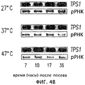
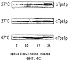
Неожиданно в настоящее время обнаружено, что молекулы нуклеиновых кислот согласно изобретению и, в частности, промотор гена трегалоза-6-фосфатсинтазы (TSP1) Hansenula polymorpha не содержат, по меньшей мере, в первых 300 п.н. выше кодирующей последовательности ни одного STRE-элемента, которые были обнаружены в S. cerevisiae и для которых было сделано предположение, что они являются первично отвечающими элементами для стрессорного ответа, включая индукцию данного гена тепловым шоком. Затем было обнаружено, что данный промотор хорошо и очень избирательно отвечает на тепло.Unexpectedly, it has now been found that the nucleic acid molecules of the invention and, in particular, the promoter of the trehalose-6-phosphate synthase (TSP1) gene of Hansenula polymorpha do not contain at least the first 300 bp. above the coding sequence, not a single STRE element that was found in S. cerevisiae and for which it was assumed that they are the primary responders for the stress response, including the induction of this gene by heat shock. Then it was found that this promoter responds well and very selectively to heat.
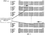
Молекулы нуклеиновых кислот согласно изобретению могут быть либо получены синтетически стандартными способами, либо выделены из подходящих библиотек ДНК и затем мутированы, как это необходимо. Получение таких библиотек также известно в данной области. Выделение предпочтительно выполняют посредством подготовки зонда длиной, по меньшей мере, 200-400 п.н. из кодирующей последовательности гена TPS1 Н. polymorpha (см. фиг.6), которая используется для скрининга библиотеки ДНК, в частности библиотеки геномной ДНК. Зонд такого вида можно получить посредством ПЦР (полимеразной цепной реакции), используя подходящие праймеры, каждый из которых предпочтительно должен быть длиной, по меньшей мере, 20-21 п.н. и иметь подходящие последовательности согласно фиг.6 (или соответствующие комплементарные последовательности), и геномную ДНК или кДНК Н. polymorpha в качестве “матрицы”.The nucleic acid molecules of the invention can either be synthetically prepared by standard methods or isolated from suitable DNA libraries and then mutated as necessary. Obtaining such libraries is also known in the art. Isolation is preferably performed by preparing a probe of at least 200-400 bp in length. from the coding sequence of the TPS1 gene of H. polymorpha (see FIG. 6), which is used to screen a DNA library, in particular a genomic DNA library. A probe of this kind can be obtained by PCR (polymerase chain reaction) using suitable primers, each of which preferably should be at least 20-21 bp in length. and have suitable sequences according to FIG. 6 (or corresponding complementary sequences), and genomic DNA or N. polymorpha cDNA as a “template”.
Таким образом, способность нуклеиновой кислоты согласно изобретению отвечать на тепло приписывается исключительно двум элементам, отвечающим на тепло, которые присутствуют в последовательности промотора, а именно элементу NGAANNNNNNNGAAN (SEQ ID №2), включающему нуклеотиды 627-641 в последовательности промотора (SEQ ID №l), и элементу TGAATATAAAGGAAA (SEQ ID №5), включающему нуклеотиды 698-713 в указанной последовательности промотора. Таким образом, способность отвечать на тепло проявляется двумя элементами, состоящими из 15 п.н., которые разделены 57 п.н. Следовательно, для гетерологической экспрессии гена может быть сконструирован промотор, содержащий один (или гомологичный) элемент или два элемента в любой комбинации, разделенных участком нуклеотидов со схожей последовательностью. Могут быть также задействованы элементы STRE с последовательностями ССССТ и AGGGG, что имеет место в генах теплового шока S.cerevisiae, но которые не являются обязательными. Возможные варианты таких промоторов представлены в примерах.Thus, the ability of a nucleic acid according to the invention to respond to heat is attributed exclusively to two elements that respond to heat that are present in the promoter sequence, namely, the element NGAANNNNNNNGAAN (SEQ ID No. 2), including nucleotides 627-641 in the sequence of the promoter (SEQ ID No. l ), and the element TGAATATAAAGGAAA (SEQ ID No. 5), including nucleotides 698-713 in the indicated sequence of the promoter. Thus, the ability to respond to heat is manifested by two elements consisting of 15 bp, which are separated by 57 bp Therefore, for heterologous gene expression, a promoter can be constructed that contains one (or homologous) element or two elements in any combination, separated by a nucleotide section with a similar sequence. STRE elements can also be involved with the sequences CCCST and AGGGG, which occurs in S.cerevisiae heat shock genes, but which are not required. Possible variants of such promoters are presented in the examples.
Зонды могут быть либо синтезированы, либо получены фрагментацией имеющейся в распоряжении ДНК TPS1, где это применимо. Конечно, также можно прямо проводить скрининг посредством зондов, которые соответствуют частям последовательности промотора; однако эта процедура менее предпочтительна из-за в лучшем случае неполной консервативности последовательности в пределах некодирующих частей.The probes can either be synthesized or obtained by fragmentation of the available TPS1 DNA, where applicable. Of course, it is also possible to directly screen by probes that correspond to parts of the promoter sequence; however, this procedure is less preferred due to, at best, incomplete sequence conservatism within non-coding parts.
В варианте молекул нуклеиновых кислот согласно изобретению последовательность нуклеиновой кислоты проявляет, по меньшей мере, 60%, предпочтительно, по меньшей мере, 80% идентичности на участке из 300 п.н. с одной из последовательностей, указанных выше в (а) или (b).In an embodiment of the nucleic acid molecules of the invention, the nucleic acid sequence exhibits at least 60%, preferably at least 80% identity, in a 300 bp region. with one of the sequences indicated above in (a) or (b).
Молекулы нуклеиновых кислот, которые включают индуцируемый теплом промотор и которые проявляют, по меньшей мере, 90% идентичности на участке из 300 п.н. с одной из последовательностей, указанных выше в пунктах (а) или (b), особенно предпочтительны. Однако наиболее предпочтительны молекулы нуклеиновых кислот, которые проявляют, по меньшей мере, 95% идентичности на участке из 300 п.н. с одной из последовательностей, указанных выше в пунктах (а) или (b).Nucleic acid molecules that include a heat-induced promoter and which exhibit at least 90% identity in a 300 bp region with one of the sequences indicated in (a) or (b) above, are particularly preferred. However, nucleic acid molecules that exhibit at least 95% identity in a 300 bp region are most preferred. with one of the sequences indicated in paragraphs (a) or (b) above.
Молекулы нуклеиновых кислот, предпочтительные для выполнения изобретения, представляют, по меньшей мере, один элемент теплового шока с последовательностью NGAANNNNNNNGAAN (SEQ ID № 2) или комплементарной ей последовательностью, где нуклеотидами, обозначенными N, независимо друг от друга могут быть А, Т, С или G. Молекулы нуклеиновых кислот согласно изобретению предпочтительно представляют, по меньшей мере, один элемент теплового шока с последовательностью NGAANNBWMNNGAAN (SEQ ID №3) или комплементарной ей последовательностью, где В представляет собой G, С или Т, W означает А или Т, и М означает С или А.Nucleic acid molecules preferred for carrying out the invention are at least one heat shock element with the sequence NGAANNNNNNNNGAAN (SEQ ID No. 2) or a sequence complementary to it, where the nucleotides denoted by N may independently be A, T, C or G. The nucleic acid molecules according to the invention preferably represent at least one heat shock element with the sequence NGAANNBWMNNGAAN (SEQ ID No. 3) or its complementary sequence, where B represents G, C or , W is A or T, and M denotes C or A.
В особо предпочтительном варианте изобретения элемент теплового шока выбран из TGAAGCCTCTTGAAA (SEQ ID №4) и/или TGAATATAAAGGAAA (SEQ ID №5) и/или комплементарных им последовательностей, где два или более элементов теплового шока там, где они присутствуют, могут представлять одну и ту же или различные последовательности. Предпочтительная молекула нуклеиновой кислоты согласно изобретению представляет, по меньшей мере, два различных элемента теплового шока.In a particularly preferred embodiment of the invention, the heat shock element is selected from TGAAGCCTCTTGAAA (SEQ ID No. 4) and / or TGAATATAAAGGAAA (SEQ ID No. 5) and / or sequences complementary to them, where two or more heat shock elements, where present, can represent one the same or different sequences. A preferred nucleic acid molecule according to the invention is at least two different heat shock elements.
В предпочтительном варианте изобретения молекулы нуклеиновых кислот согласно изобретению не содержат STRE-элемента, имеющего последовательность ССССТ или AGGGG.In a preferred embodiment of the invention, the nucleic acid molecules of the invention do not contain an STRE element having the sequence CCCST or AGGGG.
Изобретение также предоставляет фрагменты молекул нуклеиновых кислот согласно изобретению, как указано выше, которые сохраняют функцию индуцируемого теплом промотора. Фрагмент, включающий последовательность от нуклеотида 228 до нуклеотида 792 в SEQ ID №l, особенно предпочтителен. Следующий предпочтительный фрагмент включает последовательность от нуклеотида 493 до нуклеотида 792 в SEQ ID №l. Также можно использовать фрагмент, содержащий последовательность от нуклеотида 627 до нуклеотида 713 в SEQ ID №l.The invention also provides fragments of nucleic acid molecules according to the invention, as described above, which retain the function of a heat-induced promoter. A fragment comprising the sequence from
Молекулы нуклеиновых кислот согласно изобретению могут, кроме того, содержать, по меньшей мере, одну последовательность нуклеиновой кислоты гетерологичного гена под транскрипционным контролем индуцируемого теплом промотора.The nucleic acid molecules of the invention may further comprise at least one heterologous gene nucleic acid sequence under the transcriptional control of a heat-induced promoter.
“Гетерологичный ген” следует относить к кодирующей части структурного гена, который либо не экспрессируется под контролем своего собственного (гомологичного) промотора, либо не экспрессируется в организме, из которого ген получен, или не экспрессируется ни под контролем исходного промотора, ни в исходном организме.A “heterologous gene” should be attributed to the coding part of a structural gene that is either not expressed under the control of its own (homologous) promoter, or is not expressed in the organism from which the gene is derived, or is not expressed either under the control of the original promoter or in the original organism.
В следующем варианте изобретения молекулы нуклеиновых кислот согласно изобретению включают последовательность нуклеиновой кислоты под транскрипционным контролем индуцируемого теплом промотора, которая выбрана из следующих последовательностей:In a further embodiment of the invention, the nucleic acid molecules of the invention comprise a nucleic acid sequence under transcriptional control of a heat-induced promoter, which is selected from the following sequences:
i) последовательность нуклеиновой кислоты, которая кодирует полипептид с аминокислотной последовательностью трегалоза-6-фосфатсинтазы Hansenula polymorpha;i) a nucleic acid sequence that encodes a polypeptide with the amino acid sequence of trehalose-6-phosphate synthase Hansenula polymorpha;
(ii) последовательность нуклеиновой кислоты, которая указана в SEQ ID №6;(ii) the nucleic acid sequence that is indicated in SEQ ID No. 6;
(iii) последовательность нуклеиновой кислоты, которая проявляет, по меньшей мере, 80% идентичность с последовательностью, указанной в SEQ ID №6;(iii) a nucleic acid sequence that exhibits at least 80% identity with the sequence specified in SEQ ID No. 6;
(iv) последовательность нуклеиновой кислоты, которая кодирует полипептид с аминокислотной последовательностью, указанной в SEQ ID №7 или с ее частичной последовательностью, где полипептид проявляет трегалоза-6-фосфатсинтазную активность;(iv) a nucleic acid sequence that encodes a polypeptide with the amino acid sequence indicated in SEQ ID No. 7 or with its partial sequence, where the polypeptide exhibits trehalose-6-phosphate synthase activity;
(v) последовательность нуклеиновой кислоты, которая, принимая во внимание вырожденность генетического кода, кодировала бы полипептид с аминокислотной последовательностью, указанной в SEQ ID №7, или с ее частичной последовательностью, где полипептид проявляет трегалоза-6-фосфатсинтазную активность;(v) a nucleic acid sequence that, taking into account the degeneracy of the genetic code, would encode a polypeptide with the amino acid sequence indicated in SEQ ID No. 7, or with its partial sequence, where the polypeptide exhibits trehalose-6-phosphate synthase activity;
(vi) последовательность нуклеиновой кислоты, которая кодирует полипептид, аминокислотная последовательность которого проявляет, по меньшей мере, 80% идентичности с аминокислотной последовательностью, указанной в SEQ ID №7.(vi) a nucleic acid sequence that encodes a polypeptide whose amino acid sequence exhibits at least 80% identity with the amino acid sequence indicated in SEQ ID No. 7.
Последовательность нуклеиновой кислоты, указанная в (iii), предпочтительно проявляет, по меньшей мере, 90% идентичности с последовательностью, указанной в SEQ ID №6. В альтернативной форме молекул нуклеиновых кислот согласно изобретению последовательность нуклеиновой кислоты, указанная в (vi), кодирует полипептид, аминокислотная последовательность которого проявляет, по меньшей мере, 90% идентичности с аминокислотной последовательностью, указанной в SEQ ID №7.The nucleic acid sequence indicated in (iii) preferably exhibits at least 90% identity with the sequence indicated in SEQ ID No. 6. In an alternative form of nucleic acid molecules according to the invention, the nucleic acid sequence indicated in (vi) encodes a polypeptide whose amino acid sequence exhibits at least 90% identity with the amino acid sequence indicated in SEQ ID No. 7.
Молекула нуклеиновой кислоты согласно изобретению может, кроме того, включать последовательность, кодирующую сигнальный пептид, который обеспечивает перенос экспрессированного белка, при этом последовательность нуклеиновой кислоты, кодирующая сигнальный пептид, предпочтительно непосредственно связана с экспрессируемым гетерологичным геном. Секреция и модификация многих эукариотических белков требует, чтобы N-конец последовательности белка был слит с сигнальной последовательностью, для того чтобы направлять полипептиды в секреторный аппарат. Компоненты гена GAM1 S. occidentalis и гормонального гена краба Carcinus maenas, которые были успешно использованы для секреции гирудина (Weydemann et al., 1995), могут быть здесь рассмотрены для примера. Молекула нуклеиновой кислоты согласно изобретению может, кроме того, включать элемент терминатора, содержащий сигнальные структуры для РНК-полимеразы, которые приводят к терминации транскрипции. Примерами терминирующих элементов, которые могут быть использованы, являются терминаторы МОХ или РНО1 Н. polymorpha.The nucleic acid molecule according to the invention may further comprise a sequence encoding a signal peptide that allows transfer of an expressed protein, the nucleic acid sequence encoding a signal peptide preferably being directly linked to the heterologous gene being expressed. The secretion and modification of many eukaryotic proteins requires that the N-terminus of the protein sequence be fused to the signal sequence in order to direct the polypeptides to the secretory apparatus. The components of the GAM1 gene of S. occidentalis and the hormonal gene of the crab Carcinus maenas, which have been successfully used to secrete hirudin (Weydemann et al., 1995), can be considered here as an example. The nucleic acid molecule of the invention may further include a terminator element containing signaling structures for RNA polymerase that result in termination of transcription. Examples of termination elements that can be used are the MOX or PHO1 terminators of H. polymorpha.
Следующим объектом изобретения является клетка-хозяин, содержащая, по меньшей мере, одну молекулу нуклеиновой кислоты согласно изобретению, при этом клеткой-хозяином является прокариотическая или эукариотическая клетка. Эукариотической клеткой может быть, например, растительная клетка. Эукариотической клеткой предпочтительно является клетка гриба, особенно предпочтительна клетка дрожжей. Особо рассматриваются грибы в качестве клеток-хозяев для выполнения данного изобретения, например гифомицеты, такие как Aspergillus, Neurospora, Mucor, Trichoderma, Acremonium, Sordaria и Penicillium, или дрожжи, такие как Saccharomyces, Hansenula, Pichia, Kluyveromyces, Schwanniomyces, Yarrowia, Arxula, Trichosporon и Candida.The next object of the invention is a host cell containing at least one nucleic acid molecule according to the invention, wherein the host cell is a prokaryotic or eukaryotic cell. The eukaryotic cell may be, for example, a plant cell. The eukaryotic cell is preferably a fungal cell, a yeast cell is particularly preferred. Fungi are especially considered as host cells for carrying out the present invention, for example, hyphomycetes, such as Aspergillus, Neurospora, Mucor, Trichoderma, Acremonium, Sordaria, and Penicillium, or yeast, such as Saccharomyces, Hansenula, Pichia, Kluyveromyces, Schwanniiaces, Ywarrowiaces, Ywwarrowiaces, Ywarrowiaces, Ywarrowiaces, Ywarrowiaces, Yewarrow Trichosporon and Candida.
В наиболее предпочтительном варианте изобретения дрожжевая клетка является клеткой факультативных метилотрофных дрожжей Hansenula, предпочтительно Hansenula polymorpha. H. polymorpha является термотолерантной дрожжевой клеткой и относится к небольшой группе так называемых метилотрофных дрожжей, которые способны использовать метанол в качестве источника углерода и энергии. Н. polymorpha выделяли из почвенных образцов при инкубации при 37°С (Levine and Cooney, 1973). Высокая температура, при которой Н. polymorpha продолжает расти и продуцировать белок, дает возможность удалить другие нежелательные организмы. Основанием для этого является то, что было показано, что Н. polymorpha не только имеет очень высокую оптимальную температуру роста, в районе 37°С, но также способна переживать невредимой температуры примерно в 50°С (смотри фиг.1). Жизнеспособность Н. polymorpha после вхождения в стационарную фазу не падает в течение примерно 50 часов даже при 47°С (фиг.2).In a most preferred embodiment of the invention, the yeast cell is a cell of the optional methylotrophic yeast Hansenula, preferably Hansenula polymorpha. H. polymorpha is a thermotolerant yeast cell and belongs to a small group of so-called methylotrophic yeasts that are able to use methanol as a source of carbon and energy. H. polymorpha was isolated from soil samples upon incubation at 37 ° C (Levine and Cooney, 1973). The high temperature at which H. polymorpha continues to grow and produce protein makes it possible to remove other undesirable organisms. The reason for this is that it was shown that N. polymorpha not only has a very high optimum growth temperature, in the region of 37 ° C, but is also able to survive an unharmed temperature of about 50 ° C (see figure 1). The viability of H. polymorpha after entering the stationary phase does not fall for about 50 hours even at 47 ° C (Fig.2).
Следующим предметом данного изобретения является экспрессирующий вектор, включающий, по меньшей мере, одну молекулу нуклеиновой кислоты согласно изобретению. Такой экспрессирующий вектор также может содержать другие последовательности нуклеиновых кислот в дополнение к индуцируемому теплом промотору, например последовательность, которая кодирует полипептид, ген селектируемого маркера, начало репликации Е. coli и т.д.A further subject of the invention is an expression vector comprising at least one nucleic acid molecule according to the invention. Such an expression vector may also contain other nucleic acid sequences in addition to the heat-induced promoter, for example, a sequence that encodes a polypeptide, a selectable marker gene, the origin of E. coli replication, etc.
Данное изобретение также предоставляет набор, включающий:The invention also provides a kit comprising:
(a) экспрессирующий вектор согласно изобретению, который пригоден для включения в него клонированной нуклеиновой кислоты, которая кодирует рекомбинантный белок, и(a) an expression vector according to the invention which is suitable for incorporating a cloned nucleic acid that encodes a recombinant protein, and
(b) подходящую клетку-хозяина для индукции индуцируемого теплом промотора и для продукции рекомбинантного белка.(b) a suitable host cell for inducing a heat-inducible promoter and for producing a recombinant protein.
Термин “клонирование” должен включать все способы клонирования, известные в данной области, которые могут применяться для этой цели. Не все эти способы описаны в данной работе в отдельности, будучи известными специалисту в данной области.The term “cloning” should include all cloning methods known in the art that may be used for this purpose. Not all of these methods are described in this work separately, being known to a person skilled in the art.
Кроме того, изобретение предоставляет набор, включающий:In addition, the invention provides a kit comprising:
(a) экспрессирующий вектор и(a) an expression vector; and
(b) клетку-хозяина, пригодную для индукции индуцируемого теплом промотора и для продукции белка, кодируемого кодирующей последовательностью под транскрипционным контролем индуцируемого теплом промотора.(b) a host cell suitable for inducing a heat-induced promoter and for producing a protein encoded by a coding sequence under transcriptional control of a heat-inducing promoter.
Молекулы нуклеиновых кислот, клетки-хозяева, экспрессирующие векторы и наборы согласно изобретению могут быть использованы для рекомбинантной экспрессии гена под контролем индуцируемого теплом промотора или для продукции одного или большего количества белков.Nucleic acid molecules, host cells, expression vectors and kits according to the invention can be used for recombinant expression of a gene under the control of a heat-induced promoter or for the production of one or more proteins.
“Рекомбинантную экспрессию в подходящей клетке-хозяине” следует относить ко всем способам экспрессии, известным на данной стадии в этой области в известных системах экспрессии, которые можно использовать для данной цели. Не все эти способы описаны здесь в отдельности, поскольку известны специалисту в данной области.“Recombinant expression in a suitable host cell” should be referred to all expression methods known at this stage in this field in known expression systems that can be used for this purpose. Not all of these methods are described individually here, as they are known to the person skilled in the art.
Следующим предметом изобретения является способ получения одного или большего количества белков, указанный способ включает:The next subject of the invention is a method for producing one or more proteins, this method includes:
(i) клонирование, по меньшей мере, одной нуклеиновой кислоты, кодирующей рекомбинантный белок, в экспрессирующем векторе согласно изобретению, так чтобы клонированная таким образом нуклеиновая кислота находилась под транскрипционным контролем индуцируемого теплом промотора;(i) cloning at least one nucleic acid encoding a recombinant protein in an expression vector according to the invention so that the nucleic acid thus cloned is under transcriptional control of a heat-induced promoter;
(ii) введение экспрессирующего вектора, полученного на этапе (i) в клетку-хозяина, подходящую для индукции индуцируемого теплом промотора и для продукции рекомбинантного белка;(ii) introducing an expression vector obtained in step (i) into a host cell suitable for inducing a heat-inducible promoter and for producing a recombinant protein;
(iii) культивирование клетки-хозяина, полученной в (ii);(iii) culturing the host cell obtained in (ii);
(iv) индукцию индуцируемого теплом промотора способами, по существу известными.(iv) inducing a heat-induced promoter by methods essentially known.
В случае, когда экспрессирующий вектор согласно изобретению содержит последовательность, кодирующую полипептид, и находящуюся под транскрипционным контролем индуцируемого теплом промотора, способ согласно изобретению для получения одного или большего количества белков включает следующие этапы:In the case where the expression vector according to the invention contains a sequence encoding a polypeptide and is under transcriptional control of a heat-induced promoter, the method according to the invention for producing one or more proteins includes the following steps:
(i) введение экспрессирующего вектора в клетку-хозяина, подходящую для индукции индуцируемого теплом промотора и для продукции рекомбинантного белка;(i) introducing an expression vector into a host cell suitable for the induction of a heat-induced promoter and for the production of a recombinant protein;
(ii) культивирование клетки-хозяина, полученной в (i);(ii) culturing the host cell obtained in (i);
(iii) индукцию индуцируемого теплом промотора способами, по существу известными.(iii) induction of a heat-induced promoter by methods essentially known.
Далее изобретение описывается с более подробными деталями, со ссылками на фигуры, на которых показано следующее:The invention will now be described with more detailed details, with reference to the figures, which show the following:
Фиг.1 показывает кривые роста Н. polymorpha при 27°С, 37°С и 47°С.Figure 1 shows the growth curves of H. polymorpha at 27 ° C, 37 ° C and 47 ° C.
Фиг.2 показывает жизнеспособность после вхождения в стационарную фазу при 27°С, 37°С и 47°С.Figure 2 shows the viability after entering the stationary phase at 27 ° C, 37 ° C and 47 ° C.
Фиг.3А показывает Нозерн-блот РНК Н. polymorpha дикого типа после теплового шока от 27°С до 47°С и последующего охлаждения до 27°С. Клетки культивировали в среде YDP при 27°С до ранней экспоненциальной фазы роста; затем температуру увеличивали до 47°С (нулевое время) и снова снижали до 27°С через 120 минут.Figa shows the Northern blot of RNA of N. polymorpha wild-type after heat shock from 27 ° C to 47 ° C and subsequent cooling to 27 ° C. Cells were cultured in YDP medium at 27 ° C until an early exponential growth phase; then the temperature was increased to 47 ° C (zero time) and again reduced to 27 ° C after 120 minutes.
Фиг.3В показывает Вестерн-блот белка Tps1 (Tps1p) H. polymorpha после теплового шока от 27°С до 47°С и последующего охлаждения до 27°C (см. фиг.3А), из чего можно видеть корреляцию между увеличением TPSl-мРНК и увеличением белка Tps1 (Tps1p).FIG. 3B shows a Western blot of the H. polymorpha Tps1 (Tps1p) protein after heat shock from 27 ° C. to 47 ° C. and subsequent cooling to 27 ° C. (see FIG. 3A), from which a correlation between the increase in TPSl- mRNA and an increase in Tps1 protein (Tps1p).
Фиг.3С показывает внутриклеточную концентрацию трегалозы и трегалоза-6-фосфатсинтазную активность, нанесенные против времени для Н. polymorpha после теплового шока от 27°С до 47°С и последующего охлаждения до 27°С (см. фиг.3А). Не закрашенными кружками изображена внутриклеточная концентрация трегалозы, закрашенными квадратами показана трегалоза-6-фосфатсинтазная активность. Из фигуры очевидна корреляция между увеличением TPSl-мРНК и увеличением трегалоза-6-фосфатсинтазной активности и внутриклеточной концентрацией трегалозы.Fig. 3C shows the intracellular concentration of trehalose and trehalose-6-phosphate synthase activity plotted against time for H. polymorpha after heat shock from 27 ° C to 47 ° C and subsequent cooling to 27 ° C (see Fig. 3A). The open circles show the intracellular concentration of trehalose; the filled squares show trehalose-6-phosphate synthase activity. The correlation between the increase in TPSl mRNA and the increase in trehalose-6-phosphate synthase activity and the intracellular concentration of trehalose is obvious from the figure.
На фиг.4 показаны три гистограммы, изображающие трегалоза-6-фосфатсинтазную активность (белые столбики) и внутриклеточную концентрацию трегалозы (черные столбики) в клетках Hansenula polymorpha, культивируемых при 27°С (А), 37°С (В) и 47°С (С) и при глюкозной депривации спустя 7, 10, 17 и 36 часов. Накопление трегалозы коррелирует с увеличением трегалоза-6-фосфатсинтазной активности (фиг.4А), увеличением TPSl-мРНК (фиг.4 В) и увеличением белка Tps1 (Tps1p) (фиг.4 С).Figure 4 shows three histograms depicting trehalose-6-phosphate synthase activity (white bars) and the intracellular concentration of trehalose (black bars) in Hansenula polymorpha cells cultured at 27 ° C (A), 37 ° C (B) and 47 ° C (C) and with glucose deprivation after 7, 10, 17 and 36 hours. The accumulation of trehalose correlates with an increase in trehalose-6-phosphate synthase activity (Figure 4A), an increase in TPSl mRNA (Figure 4B) and an increase in Tps1 protein (Tps1p) (Figure 4C).
На фиг.5 показана гомология некоторых областей последовательности ДНК трегалоза-6-фосфатсинтазы ряда организмов.Figure 5 shows the homology of some regions of the DNA sequence of trehalose-6-phosphate synthase of a number of organisms.
На фиг.6 показана последовательность ДНК гена TPS1 Н. polymorpha (SEQ ID №8) и выведенная аминокислотная последовательность (SEQ ID №6). Элементы теплового шока в последовательности промотора подчеркнуты.Figure 6 shows the DNA sequence of the TPS1 gene of H. polymorpha (SEQ ID No. 8) and the deduced amino acid sequence (SEQ ID No. 6). Elements of heat shock in the promoter sequence are underlined.
На фиг.7 показана плазмида рС11, производная рМ1 (М. Suckow, персональное сообщение), которая была получена встраиванием гена lacZ в полилинкер рМ1. Плазмида содержит последовательность HARS1 (автономно реплицирующиеся последовательности Н. polymorpha), ori (начало репликации) pBR322, ген устойчивости к ампициллину, ген URA3 для размножения и селекции в Н. polymorpha и Е. coli и терминатор МОХ после гена lacZ для терминации процесса транскрипции.7 shows the plasmid pC11, a derivative of pM1 (M. Suckow, personal message), which was obtained by embedding the lacZ gene in the polylinker pM1. The plasmid contains the HARS1 sequence (autonomously replicating H. polymorpha sequences), ori (start of replication) pBR322, the ampicillin resistance gene, the URA3 gene for propagation and selection in H. polymorpha and E. coli, and the MOX terminator after the lacZ gene to terminate the transcription process.
На фиг.8 показана плазмида pCll-FMD, полученная посредством встраивания промотора FMD перед репортерным геном lacZ плазмиды рС11.Figure 8 shows the plasmid pCll-FMD obtained by inserting the FMD promoter in front of the reporter gene lacZ of plasmid pC11.
На фиг.9 показана плазмида pCll-TPS1, полученная посредством встраивания промотора TPS1 перед репортерным геном lacZ плазмиды рС11.Figure 9 shows the plasmid pCll-TPS1 obtained by embedding the TPS1 promoter in front of the lacZ reporter gene of plasmid pC11.
На фиг.10 показано сравнение между активностью промоторов FMD (А) и TPS1 (В) при 30, 37 и 44°С в трех различных источниках углерода (2% глюкозы, 2% глицерина или 2% метанола).Figure 10 shows a comparison between the activity of the FMD (A) and TPS1 (B) promoters at 30, 37 and 44 ° C in three different carbon sources (2% glucose, 2% glycerol or 2% methanol).
На фиг.11 показана плазмида pTPSlConphysMT, используемая в примере 4. МОХ-Т = терминатор МОХ, Conphys = ген Соnрhys3, TPS1 = промотор TPS1 Hansenula polymorpha, HARS = автономно реплицирующиеся последовательности Н. polymorpha, tet = ген устойчивости к тетрациклину, URA3 = URA3 S. cerevisiae, amp = ген устойчивости к ампициллину.11 shows the plasmid pTPSlConphysMT used in Example 4. MOX-T = MOX terminator, Conphys = Conphys3 gene, TPS1 = Hansenula polymorpha TPS1 promoter, HARS = autonomously replicating H. polymorpha sequences, tet = tetracycline resistance gene, URA3 = URA3 S. cerevisiae, amp = ampicillin resistance gene.
ПРИМЕРЫEXAMPLES
Материалы и способыMaterials and methods
Специальные реактивы и материалыSpecial Reagents and Materials
Bio 101, Vista, USA - набор Geneclean IIBio 101, Vista, USA - Geneclean II Pack
BioRad Lab., Munich, Germany - анализ белка BioRad (Bredford)BioRad Lab., Munich, Germany - BioRad (Bredford) protein analysis
Boehringer, Mannheim, Germany - набор GOD/POD для измерения глюкозы, этанольный набор, таблетки для “полного” коктейля ингибиторов протеаз “COMPLETE”Boehringer, Mannheim, Germany - GOD / POD glucose test kit, ethanol kit, tablets for a “complete” cocktail of protease inhibitors “COMPLETE”
Fluka Chemie AG, Buchs, Switzerland - циклогексимид (актидион), SDS, D+-тpeгaлoзa, PEP, трицин, НАДФ, фенольный реактив Фолина-ЧиокалтеуFluka Chemie AG, Buchs, Switzerland - cycloheximide (actidion), SDS, D + -tagalose, PEP, tricin, NADP, folin-Chiokalteu phenolic reagent
ICN Biochemicals, Ohio/USA - "Liquigel" 40% акриламид/N'N'-метиленбисакриламид (37,5:1)ICN Biochemicals, Ohio / USA - "Liquigel" 40% acrylamide / N'N'-methylenebisacrylamide (37.5: 1)
Kodak, New York, USA - регистрирующая пленка для научных целей ВIOМАХ MRKodak, New York, USA - registration film for scientific purposes ВIOМАХ MR
Mediatech, Herndon, USA - сульфат генетицина G418 (антибиотик)Mediatech, Herndon, USA - Geneticin sulfate G418 (antibiotic)
Perkin Elmer Applied Biosystems - набор для секвенирования ДНК Forest City, USAPerkin Elmer Applied Biosystems - DNA sequencing kit Forest City, USA
Pharmacia Biotech, Sweden - колонки Nap-10 (с сефадексом G-25), все используемые рестриктазы, полимераза TaqPharmacia Biotech, Sweden - Nap-10 columns (with Sephadex G-25), all restriction enzymes used, Taq polymerase
Qiagen GmbH, Germany - набор для плазмид PlasmideQiagen GmbH, Germany - Plasmide plasmid kit
Midi (50)Midi (50)
Schleicher + Schuell, Dassel, Germany - Protran BA 83 0,2 мкм/⌀ 82 ммSchleicher + Schuell, Dassel, Germany - Protran BA 83 0.2 μm / ⌀ 82 mm
(круглый фильтр из нитрата целлюлозы), Protran BA 83 0,2 мкм (мембрана для переноса при блоттинге)(round cellulose nitrate filter), Protran BA 83 0.2 μm (transfer membrane for blotting)
Sigma, St. Louis, USA - моноклональные антикроличьи иммуноглобулины козы (конъюгат с щелочной фосфатазой), трегалоза почек свиньи (№ в каталоге Т-8778), UDPG, глюкоза-6-Р, LDH, пируваткиназаSigma, St. Louis, USA - monoclonal anti-rabbit goat immunoglobulins (conjugate with alkaline phosphatase), pig kidney trehalose (catalog number T-8778), UDPG, glucose-6-P, LDH, pyruvate kinase
Stratagene, La Jolla, USA - набор Prime-It II (набор для мечения со случайным праймером), колонки NucTrap (колонки для очистки проб, включая защитное устройство от бета-излучения для колонок под давлением)Stratagene, La Jolla, USA - Prime-It II kit (random primer labeling kit), NucTrap columns (sample purification columns, including a beta protector for pressure columns)
US Biological, Swampscott, USA - бактериологический агар, бульон YPD, дополненный композицией W/Peptone X, бульон LB МиллераUS Biological, Swampscott, USA - bacteriological agar, YPD broth, supplemented with W / Peptone X, LB Miller broth
Используемая аппаратураUsed equipment
Прибор для электропорации - генератор импульсов для Е. coli, BioRad Laboratories, Hercules USAElectroporation Device - Pulse Generator for E. coli, BioRad Laboratories, Hercules USA
ВЭЖХ - DIONEX DX-300, DIONEX, Sunnyvale, USAHPLC - DIONEX DX-300, DIONEX, Sunnyvale, USA
Охлаждаемые центрифуги - Centricon H-401, Kontron Instr. AG, Zurich, Switzerland IEC Centra GP8R, Brouwer, Lucerne, SwitzerlandCooled Centrifuges - Centricon H-401, Kontron Instr. AG, Zurich, Switzerland IEC Centra GP8R, Brouwer, Lucerne, Switzerland
Biofuge 17RS, Heraeus Sepatech, GermanyBiofuge 17RS, Heraeus Sepatech, Germany
Аппаратура для ПЦР - Progene, Techne, Cambridge, United KingdomPCR equipment - Progene, Techne, Cambridge, United Kingdom
Визуализатор фосфора - GS 250 молекулярный визуализатор (включая связанное оборудование), BioRad Laboratories, Hercules, USAPhosphorus Visualizer - GS 250 Molecular Visualizer (including related equipment), BioRad Laboratories, Hercules, USA
Фотометр - Anthos 2001 (для планшетов для микротитрования), Anthos Labtec Instruments, Salzburg, AustriaPhotometer - Anthos 2001 (for microtiter plates), Anthos Labtec Instruments, Salzburg, Austria
Shimadzu UV-160A, JapanShimadzu UV-160A, Japan
Секвенатор - ABI PRISM 301 Genetic Analyzer, Perkin Elmer, Applied Biosystems, Foster City, USASequencer - ABI PRISM 301 Genetic Analyzer, Perkin Elmer, Applied Biosystems, Foster City, USA
Бактериальный штамм и условия культивированияBacterial strain and cultivation conditions
Штамм DH5α E. coli (F' endAlhsdR17rkmk+supE44thi-lrecAlgyra relA(lacZYA-argF) U169 (φ80Δ(lacZ)M15)(Gibco BRL, Gaithersburg MD, USA) использовали для клонирования гена TPS1 Н. polymorpha, следуя стандартному протоколу (Sambrook et al., 1989). Среду для Е. coli также получали в соответствии со стандартными процедурами (Sambrook et. al.,1989).The strain E. coli DH5α (F 'endAlhsdR17r k m k + supE44thi-lrecAlgyra relA (lacZYA-argF) U169 (φ80Δ (lacZ) M15) (Gibco BRL, Gaithersburg MD, USA) was used to clone the TPS1 gene of H. polymorpha, following the standard protocol (Sambrook et al., 1989) .Environment for E. coli was also prepared according to standard procedures (Sambrook et. al., 1989).
Выделение плазмидной ДНК из Е. coli (STET prep)Isolation of plasmid DNA from E. coli (STET prep)
Плазмидную ДНК выделяли в соответствии с модифицированным протоколом, согласно Sambrook et. al. (1989). Использовали шпатель для того, чтобы соскабливать клеточный материал с чашки. Затем полученный материал добавляли к 500 мкл STET (8% [мас./об.] сахарозы, 5% [об./об.] тритона Х-100, 50 мМ ЭДТА, 50 мМ трис-HCl, рН 8,0) с 35 мкл лизоцима (10 мг/мл) и перемешивали. Затем образцы кипятили в течение 1 мин 40 с при 100°С и центрифугировали в течение 10 минут при 20000 g и 4°С. Примерно 400 мкл надосадка отбирали пипеткой и осаждали ДНК, используя 400 мкл изопропанола. После центрифугирования в течение 10 минут при 20000 g и 4°С надосадок полностью удаляли и осадок ДНК один раз промывали ледяным 70% [об./об.] этанолом. Наконец ДНК сушили при комнатной температуре и суспендировали в 50-70 мкл ТЕ (10 мМ трис-HCl, рН 8,0, 1 мМ ЭДТА, рН 8,0).Plasmid DNA was isolated according to a modified protocol according to Sambrook et. al. (1989). A spatula was used to scrape the cellular material from the cup. Then, the resulting material was added to 500 μl of STET (8% [w / v] sucrose, 5% [v / v] Triton X-100, 50 mM EDTA, 50 mM Tris-HCl, pH 8.0) s 35 μl of lysozyme (10 mg / ml) and mixed. Then the samples were boiled for 1 min 40 s at 100 ° C and centrifuged for 10 minutes at 20,000 g and 4 ° C. About 400 μl of the supernatant was pipetted and DNA precipitated using 400 μl of isopropanol. After centrifugation for 10 minutes at 20,000 g and 4 ° C, the supernatant was completely removed and the DNA pellet was washed once with ice-cold 70% [v / v] ethanol. Finally, the DNA was dried at room temperature and suspended in 50-70 μl TE (10 mm Tris-HCl, pH 8.0, 1 mm EDTA, pH 8.0).
Штамм дрожжей и условия культивированияYeast Strain and Culturing Conditions
Используемым штаммом дрожжей был Hansenula polymorpha дикого типа (имеющийся в наличии благодаря Р. Piper, London (1994)). Исходные культуры выращивали на агаре YPD (2% [мас./об.] глюкозы, 2% [мас./об.] бактопептона, 1% [мас./об.] дрожжевого экстракта, 2% [мас./об.] агара), и исходные культуры обновляли каждые шесть недель. Данные культуры служили в качестве инокулята для жидких культур YPD (состав тот же, что и для агара YPD, но без 2% [мас./об.] агара).The yeast strain used was wild-type Hansenula polymorpha (available from P. Piper, London (1994)). Stock cultures were grown on YPD agar (2% [w / v] glucose, 2% [w / v] bactopeptone, 1% [w / v] yeast extract, 2% [w / v] agar), and stock cultures were updated every six weeks. These cultures served as an inoculum for liquid YPD cultures (the same composition as for YPD agar, but without 2% [w / v] agar).
Штамм RB11 Н. polymorpha (штамм Н. polymorpha odc1, без оротидин-5-фосфатдекарбоксилазы (ауксотрофный по урацилу) (Weydemann et al., 1995)) использовали для экспериментов в примерах 3 и 4. Используемая полная среда содержала 2% глюкозы или глицерина, 1% дрожжевого экстракта и 2% бактопептона; селективная среда содержала 0,17% азотистого основания дрожжей, 0,5% сульфата аммония, 2% глюкозы или глицерина, 38,4 мг/л аргинина, 57,6 мг/л изолейцина, 48 мг/л фенилаланина, 57,6 мг/л валина, 6 мг/мл треонина, 50 мг/л инозитола, 40 мг/л триптофана, 15 мг/л тирозина, 60 мг/л лейцина, 4 мг/л гистидина. Урацил не присутствовал в селективной среде.Strain RB11 H. polymorpha (strain N. polymorpha odc1, without orotidine-5-phosphate decarboxylase (araxotrophic for uracil) (Weydemann et al., 1995)) was used for the experiments in examples 3 and 4. Used complete medium contained 2% glucose or
Для культивирования клеточных культур в автоклавированную жидкую среду инокулировали исходную культуру и инкубировали в течение ночи в инкубаторах с качалками при 27, 37 или 47°С, в зависимости от эксперимента.To cultivate cell cultures in an autoclaved liquid medium, the initial culture was inoculated and incubated overnight in incubators with shakers at 27, 37 or 47 ° C, depending on the experiment.
Определение оптической плотности культур клеток Н. polymorphaDetermination of the optical density of N. polymorpha cell cultures
Чтобы определить оптическую плотность (OD) 200 мкл культуры клеток (соответствующим образом разведенной YPD там, где это применимо) помещали в лунку планшета для микротитрования и измеряли при 620 нм, используя фотоспектрометр Anthos 2001. 200 мкл YPD использовали в качестве пустого контроля.To determine the optical density (OD), 200 μl of a cell culture (appropriately diluted with YPD, where applicable) was placed in a well of a microtiter plate and measured at 620 nm using an Anthos 2001 spectrometer. 200 μl of YPD was used as an empty control.
Эксперименты по росту и тепловому шоку с Н. polymorphaGrowth and heat shock experiments with H. polymorpha
Ночные культуры использовали для инокуляции в среду YPD в колбах Эрленмейера. Засев данной исходной культуры при той температуре, при которой потом начинали сам эксперимент (27°С для тепловых шоков, 27, 37 или 47°С для ростовых экспериментов) проводили особенно тщательно.Overnight cultures were used to inoculate YPD medium in Erlenmeyer flasks. Sowing of this initial culture at the temperature at which the experiment itself was then started (27 ° C for heat shocks, 27, 37 or 47 ° C for growth experiments) was carried out especially carefully.
Культуры высевали при начальной плотности OD620 0,2 для каждого ростового эксперимента и непрерывно поддерживали в инкубаторах с качалками (Multitron). Напротив, для экспериментов по тепловому шоку культуры высевали при начальной OD 0,05. Культурам давали возможность расти при 27°С до OD620 0,4 (примерно 1-1,5×10⁸ клеток в мл культуры) перед проведением теплового шока при 47°С в водяной бане с качалкой (Aquatron). Затем в течение следующих двух часов отбирали образцы. Затем культуру клеток охлаждали во второй водяной бане в течение одного часа до 27°С.Cultures were seeded at an initial density of OD 620 0.2 for each growth experiment and were continuously maintained in incubators with shakers (Multitron). In contrast, for thermal shock experiments, cultures were plated at an initial OD of 0.05. The cultures were allowed to grow at 27 ° C. to OD 620 0.4 (about 1-1.5 × 10 8 cells per ml of culture) before heat shocking at 47 ° C. in a water bath with a shaker (Aquatron). Then, samples were taken over the next two hours. Then the cell culture was cooled in a second water bath for one hour to 27 ° C.
Трансформация Н. polymorpha путем электропорацииTransformation of H. polymorpha by electroporation
В 100 мл YPD высевали 5 мл плотно выросшей ночной культуры. Культуру встряхивали при 37°С примерно в течение трех часов до OD600 0,8-1,2. Клетки собирали центрифугированием при 3000 об/мин и снова суспендировали в 20 мл буфера Kpi (50 мМ/рН 7,5). После добавления 0,5 мл DTT и встряхивания в течение 15 мин при 37°С клетки осаждали центрифугированием при 2500 об/мин и дважды промывали буфером STM (270 мМ сахарозы, 10 мМ трисС1, 1 мМ MgCl2, pH 7,5). Затем их суспендировали в 0,25 мл буфера STM, и аликвоты объемом 60 мкл хранили при -70°С. Для трансформации векторами, интегрирующими в рДНК, плазмидную ДНК сначала линеаризовали XhoI или SacI. 0,1-1 мкг линеаризованной плазмидной ДНК смешивали со свежими компетентными клетками, размороженными на льду. Затем эти препараты помещали в 2 мм кюветы. Трансформацию выполняли в генераторе импульсов Gene Pulser (Bio-Rad, Munich) при 2,0 кВ, 25 мкФ и 200 Ом. Затем клетки инкубировали в 1 мл YPD в течение одного часа при 37°С для восстановления перед высеванием на селективную среду. Макроспоровые колонии наблюдали после инкубации от двух до четырех дней при 37°С.In 100 ml of YPD, 5 ml of densely grown overnight culture was sown. The culture was shaken at 37 ° C for approximately three hours to an OD 600 of 0.8-1.2. Cells were harvested by centrifugation at 3000 rpm and resuspended in 20 ml Kp i buffer (50 mM / pH 7.5). After adding 0.5 ml of DTT and shaking for 15 min at 37 ° C, the cells were precipitated by centrifugation at 2500 rpm and washed twice with STM buffer (270 mm sucrose, 10 mm TrisCl, 1 mm MgCl 2 , pH 7.5). They were then suspended in 0.25 ml of STM buffer, and aliquots of 60 μl were stored at -70 ° C. For transformation by vectors integrating into rDNA, plasmid DNA was first linearized with XhoI or SacI. 0.1-1 μg of linearized plasmid DNA was mixed with fresh competent cells thawed on ice. Then these preparations were placed in 2 mm cuvettes. Transformation was performed in a Gene Pulser pulse generator (Bio-Rad, Munich) at 2.0 kV, 25 μF and 200 Ohms. The cells were then incubated in 1 ml of YPD for one hour at 37 ° C to recover before plating on selective medium. Macrospore colonies were observed after incubation for two to four days at 37 ° C.
Определение концентрации глюкозы в средеDetermination of glucose concentration in the medium
Концентрацию глюкозы в среде определяли методом GOD (Набор GOD/POD, Bohringer). Образцы разводили 1:200 водой. 190 мкл 1% (мас./об.) раствора фермента GOD (поставляемого в наборе в форме порошка) добавляли к 10 мкл каждого образца, и смесь инкубировали приблизительно 25 минут при 27°С. В данном определении в качестве стандарта также используется 10 мкл раствора глюкозы (0,91 мкг глюкозы), поставляемого в наборе. Поглощение измеряли на спектрофотометре Anthos 2001 при 405 нм.The concentration of glucose in the medium was determined by the GOD method (GOD / POD Kit, Bohringer). Samples were diluted 1: 200 with water. 190 μl of a 1% (w / v) GOD enzyme solution (supplied in powder form) was added to 10 μl of each sample, and the mixture was incubated for approximately 25 minutes at 27 ° C. In this definition, 10 μl glucose solution (0.91 μg glucose) supplied in the kit is also used as a standard. Absorption was measured on an Anthos 2001 spectrophotometer at 405 nm.
Экстракция и количественное определение трегалозыExtraction and quantification of trehalose
Экстракция трегалозыTrehalose Extraction
1-10 мл культуры клеток фильтровали через фильтр из стекловолокна (Whatman GF/C) и три раза промывали водой. Фильтр помещали в пробирку Эппендорф с 1 мл воды и встряхивали с перемешиванием в течение 30 секунд, перед тем как осторожно отжимали и удаляли. Затем суспензию клеток кипятили в течение 10 минут в водяной бане. Чтобы полностью отделить надосадок от клеточного материала, суспензию центрифугировали три раза при 20000 g.1-10 ml of cell culture was filtered through a glass fiber filter (Whatman GF / C) and washed three times with water. The filter was placed in an Eppendorf tube with 1 ml of water and shaken with stirring for 30 seconds before being gently squeezed and removed. Then the cell suspension was boiled for 10 minutes in a water bath. In order to completely separate the supernatant from the cellular material, the suspension was centrifuged three times at 20,000 g.
Определение трегалозы ВЭЖХDetermination of trehalose by HPLC
Экстрагированные сахара отделяли с помощью анионообменной колонки (колонка DIONEX CarboPac РА1, 4×250 мм) и определяли амперометрически на золотом электроде (PED = импульсный электрохимический детектор). Состав элюирующего градиента представляет собой следующее:The extracted sugars were separated using an anion exchange column (DIONEX CarboPac PA1 column, 4 × 250 mm) and determined amperometrically on a gold electrode (PED = pulsed electrochemical detector). The composition of the eluting gradient is as follows:
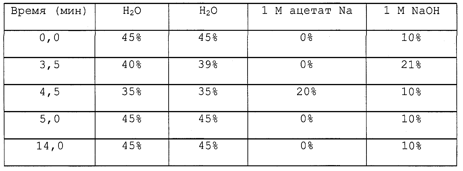
Следствием данных условий является время удержания трегалозы примерно 3,7 минуты. В каждом случае инъецировали 20 мкл образца. В качестве стандарта использовали раствор трегалозы в концентрации 0,1 мг/мл.The consequence of these conditions is the retention time of trehalose is approximately 3.7 minutes. In each case, 20 μl of sample was injected. A solution of trehalose at a concentration of 0.1 mg / ml was used as a standard.
Определение трегалозы ферментативным анализомDetermination of trehalose by enzymatic analysis
В некоторых случаях в качестве альтернативы более дорогому методу ВЭЖХ использовали в равной степени надежный способ ферментативного анализа (Parrou and Francois, 1997, с модификациями): 25 мкл экстракта трегалозы смешивали с 12,5 мкл трегалозы (Sigma) и 37,5 мкл буферного раствора (0,2 М ацетат натрия, 0,03 М CaCl2, pH 5,7) и инкубировали в течение пяти часов при 37°С в водяной бане. Это приводило к полному распаду трегалозы на две единицы глюкозы. После краткого центрифугирования образцы три минуты инкубировали при 95°С и затем снова центрифугировали еще пять минут при 20000 g. Концентрацию трегалозы определяли косвенно посредством определения концентрации глюкозы (набор GOD/POD, смотри выше). Для этой цели использовали 10 мкл полученного надосадка.In some cases, an equally reliable method of enzymatic analysis was used as an alternative to the more expensive HPLC method (Parrou and Francois, 1997, with modifications): 25 μl trehalose extract was mixed with 12.5 μl trehalose (Sigma) and 37.5 μl buffer solution (0.2 M sodium acetate, 0.03 M CaCl 2 , pH 5.7) and incubated for five hours at 37 ° C in a water bath. This led to the complete decomposition of trehalose into two units of glucose. After brief centrifugation, the samples were incubated for three minutes at 95 ° C and then centrifuged again for another five minutes at 20,000 g. The concentration of trehalose was determined indirectly by determining the concentration of glucose (set of GOD / POD, see above). For this purpose, 10 μl of the obtained supernatant was used.
Определение белкаProtein definition
Определение белка по Петерсону (незначительно модифицированное) (Peterson(1997))Peterson protein determination (slightly modified) (Peterson (1997))
Чтобы определить концентрацию суммарного белка в культуре клеток 1 мл суспензии клеток преципитировали в 1 мл 10% (мас./об.) ТХУ и центрифугировали в течение 10 минут при 3000 g. Надосадок отсасывали пастеровской пипеткой, соединенной с водоструйным насосом, и осадок промывали 1 мл 1N PCA. Затем осадок суспендировали в 5-12 мл (в зависимости от OD изучаемой клеточной культуры) раствора 0,8 N NaOH:10% (мас./об.) SDS (1:1) и инкубировали, по меньшей мере, в течение одного часа при 60°С. К 200 мкл суспензии добавляли 600 мкл 6-кратного разведения реагента СТС (10% Na2СО3, 0,1% CuSO4·5Н20, 0,2% тартрата KNa). Ровно через 10 минут добавляли 200 мкл 6-кратного разведения реагента Фолина-Чиокалтеу и недолго перемешивали. Образцы оставляли в темноте на 30 минут, после чего измеряли поглощение при 750 нм, в качестве стандарта служил БСА.To determine the concentration of total protein in cell culture, 1 ml of cell suspension was precipitated in 1 ml of 10% (w / v) TCA and centrifuged for 10 minutes at 3000 g. The supernatant was aspirated with a Pasteur pipette connected to a water jet pump, and the precipitate was washed with 1 ml of 1N PCA. Then the precipitate was suspended in 5-12 ml (depending on the OD of the studied cell culture) of a solution of 0.8 N NaOH: 10% (w / v) SDS (1: 1) and incubated for at least one hour at 60 ° C. To 200 μl of the suspension was added 600 μl of a 6-fold dilution of the STS reagent (10% Na 2 CO 3 , 0.1% CuSO 4 ·
Определение белка по Брэдфорду (1976)Determination of protein according to Bradford (1976)
Чтобы определить концентрацию белка в бесклеточном экстракте, 100 мкл соответствующим образом разведенного экстракта смешивали с 700 мкл воды. Затем добавляли 200 мкл реактива для анализа белка BioRad (Bradford) и недолго встряхивали (Vortex). Поглощение измеряли при 595 нм, в качестве стандарта служил БСА.To determine the protein concentration in the cell-free extract, 100 μl of the appropriately diluted extract was mixed with 700 μl of water. Then 200 μl of BioRad protein assay reagent (Bradford) was added and shaken briefly (Vortex). The absorption was measured at 595 nm; BSA served as a standard.
Измерения ферментативной активностиEnzymatic Activity Measurements
Подготовка проницаемых клетокPermeable Cell Preparation
Ферментативную активность трегалоза-6-фосфатсинтазы (Тре-6-Ф-синтазы) измеряли в проницаемых клетках (De Virgilio et al., 1991). С этой целью 1-6 мл клеток фильтровали (на фильтрах из стекловолокна GF/C, Whatman), дважды промывали, используя ледяную воду, и снова суспендировали при встряхивании и перемешивании в 1 мл лизирующего буфера (0,2 М трицин, рН 7,0, 0,5% [об./об.] тритон Х-100). Фильтры извлекали и пробирки Эппендорф замораживали в жидком азоте и хранили при -20°С. Перед проведением измерения клетки размораживали на водяной бане в течение трех минут при 30°С. Затем их дважды промывали в 0,2 М трицине (рН 7,0) и центрифугировали в течение 20 секунд при 4°С и 8000 об/мин (Biofuge 17RS) после каждой промывки. Наконец клетки снова суспендировали в 600 мкл 0,2 М трицина (рН 7,0).The enzymatic activity of trehalose-6-phosphate synthase (Tre-6-F synthase) was measured in permeable cells (De Virgilio et al., 1991). To this end, 1-6 ml of cells were filtered (on glass fiber filters GF / C, Whatman), washed twice with ice water, and again suspended with shaking and stirring in 1 ml of lysis buffer (0.2 M tricin,
Активность трегалоза-6-фосфатсинтазыTrehalose-6-phosphate synthase activity
Активность Тре-6-Ф-синтазы определяли комбинированным ферментативным анализом согласно Hottiger et al. (1987) при 50°С, при этом всегда использовали 60 мкл проницаемых клеток. Как субстрат (без глюкоза-6-Ф), так и пустые контроли ферментов (без проницаемых клеток) анализировали в качестве контролей.The activity of Tre-6-F synthase was determined by a combined enzymatic assay according to Hottiger et al. (1987) at 50 ° C, always using 60 μl of permeable cells. Both the substrate (without glucose-6-F) and the empty enzyme controls (without permeable cells) were analyzed as controls.
Вестерн-блот-анализыWestern blot analyzes
Экстракция белка разрушением клетокProtein Extraction by Cell Destruction
5-15 мл культуры клеток центрифугировали в течение 5 минут при 4°С и 3000 об/мин (IEC Centra GP8R) и затем надосадок декантировали. Осадок суспендировали в 1 мл воды и переносили в пробирку Сарстедта (с закручивающейся крышкой). После центрифугирования в течение 10 секунд надосадок отсасывали пипеткой и осадок взвешивали, вес пустой пробирки служил в качестве веса тары. 1 мкл 0,2 М трицин-буфера (рН 7,0; с ингибиторами протеиназ [2 табл./25 мл]) добавляли на мг осадка и осадок ресуспендировали. Добавляли стеклянные шарики непосредственно до нижнего уровня мениска жидкости, после чего пробирки Сарстедта осторожно фиксировали в гомогенизаторе клеток (Fastprep FP120) в холодильной камере. Гомогенизатор клеток запускали дважды по 30 секунд при установке 6,0, получая в результате разрушение более 90% клеток. Далее с этого момента строгое внимание обращали на содержание образцов хорошо охлажденными в течение всего времени. С помощью иглы в пробирке Сарстедта делали небольшое отверстие. Пробирку помещали в стеклянную пробирку и центрифугировали при 4°С и 100 g, отделяя, таким образом, экстракт от стеклянных шариков. Затем один раз добавляли трицин-буфер в количестве, используемом для разрушения клеток, в пробирки Сарстедта, которые снова центрифугировали. Затем мутный экстракт переносили в пробирки Эппендорф и центрифугировали три раза в течение 10 минут при 25000 g и 4°С (Biofuge 17RS), каждый раз потом использовали надосадок, содержащий растворимые белки (включая Тре6Ф-синтазу).5-15 ml of cell culture was centrifuged for 5 minutes at 4 ° C and 3000 rpm (IEC Centra GP8R) and then the supernatant was decanted. The pellet was suspended in 1 ml of water and transferred to a Sarstedt tube (with screw cap). After centrifugation for 10 seconds, the supernatant was pipetted and the pellet was weighed, the weight of the empty tube serving as the tare weight. 1 μl of 0.2 M tricin buffer (pH 7.0; with proteinase inhibitors [2 tablets / 25 ml]) was added per mg of pellet and the pellet was resuspended. Glass beads were added directly to the lower level of the meniscus, after which the Sarstedt tubes were carefully fixed in a cell homogenizer (Fastprep FP120) in a refrigerator. The cell homogenizer was run twice for 30 seconds with the installation of 6.0, resulting in the destruction of more than 90% of the cells. Further from this moment, strict attention was paid to the content of the samples well chilled throughout the entire time. A small hole was made in a Sarstedt tube using a needle. The tube was placed in a glass tube and centrifuged at 4 ° C and 100 g, thus separating the extract from the glass beads. Then, tricin buffer was added once in the amount used to destroy the cells to Sarstedt tubes, which were again centrifuged. The turbid extract was then transferred to Eppendorf tubes and centrifuged three times for 10 minutes at 25,000 g and 4 ° C (Biofuge 17RS), each time afterwards a supernatant containing soluble proteins (including Tre6F synthase) was used.
Получение образцовReceiving samples
Концентрацию белка в полученных экстрактах затем определяли методом Бредфорда (смотри выше). В соответствии с полученными значениями их разводили водой до 2,5 мкг белка/мкл, и один объем 5х буфера образца добавляли к четырем объемам данного раствора белка. Затем образцы денатурировали в течение пяти минут при 95°С и либо использовали сразу же для SDS гель-электрофореза, либо замораживали. Для анализа использовали 10 мкл, т.е. 20 мкг белка.The protein concentration in the obtained extracts was then determined by the Bradford method (see above). In accordance with the obtained values, they were diluted with water to 2.5 μg of protein / μl, and one volume of 5x sample buffer was added to four volumes of this protein solution. Samples were then denatured for five minutes at 95 ° C and either used immediately for SDS gel electrophoresis or frozen. For the analysis used 10 μl, i.e. 20 mcg of protein.
Буфер для образца: 1 мл 0,5 М трис-HCl, рН 6,8; 0,8 мл глицерина; 1,6 мл 10% [мас./об.] SDS; 0,2 мл 0,05% [мас./об.] бромфенолового синего, 4 мл воды. 19 объемов буфера для образца добавляли к одному объему 2-β-меркаптоэтанола непосредственно перед использованием.Sample buffer: 1 ml 0.5 M Tris-HCl, pH 6.8; 0.8 ml of glycerin; 1.6
Электрофорез в SDS-полиакриламидном геле (SDS-ПААГ)SDS-Polyacrylamide Gel Electrophoresis (SDS-PAGE)
Применяли систему в соответствии с Laemmli et al. (1970) для разделения белков по их молекулярной массе. Для использования в качестве разделяющего и концентрирующего геля соответственно получали 10% и 4% акриламидный гель (общие размеры 10×10 см) в следующем составе:A system was used in accordance with Laemmli et al. (1970) for the separation of proteins by their molecular weight. For use as a separating and concentrating gel, respectively, received 10% and 4% acrylamide gel (
Разделяющий гель: 2,5 мл 40% (мас./об.) акриламида/бис-акриламида; 2,5 мл 1,5 М трис-HCl, рН 8,8; 100 мкл 10% (мас./об.) SDS; 4,95 мл воды; 50 мкл 10% (мас./об.) персульфата аммония, 5 мкл TEMEDSeparating gel: 2.5 ml of 40% (w / v) acrylamide / bis-acrylamide; 2.5 ml of 1.5 M Tris-HCl, pH 8.8; 100
Концентрирующий гель: 1 мл 40% (мас./об.) акриламида/бисакриламида; 2,5 мл 0,5 М трис-НС1, рН 6,8; 100 мкл 10% (мас./об.) SDS; 6,4 мл воды; 50 мкл 10% (мас./об.) персульфата аммония, 10 мкл TEMED;Concentrating gel: 1
5х рабочий буфер: 15 г трис, 72 г глицина, 5 г SDS, Н2О, добавленная до 1 л. Значение рН должно быть примерно 8,3, без последующего доведения.5x working buffer: 15 g Tris, 72 g glycine, 5 g SDS, H 2 O, added up to 1 L. The pH should be approximately 8.3, without subsequent adjustment.
На каждый гель наносили 20 мкг белка. В качестве стандарта использовали предварительно окрашенный стандарт "Kaleidoscope prestained standard" BioRad следующего состава: миозин (204 кД), β-галактозидаза (121 кД), БСА (78 кД), карбо-ангидраза (39 кД), ингибитор трипсина сои (30 кД). Гель-электофорез проводили примерно один час (но не долее того времени, когда фронт образца достигал нижнего края геля) при постоянном напряжении 200 В. Затем полученные гели красили 0,1% (мас./об.) Кумасси синим R250 в 10% (об./об.) уксусной кислоте/50% (об./об.) этаноле (и отмывали от краски примерно через один час 10% (об./об.) уксусной кислотой, 20% (об./об.) этанолом) или с помощью блоттинга переносили на нитроцеллюлозу (обратитесь к следующему разделу).20 μg of protein was applied to each gel. The pre-stained Kaleidoscope prestained standard BioRad standard was used as the standard: myosin (204 kD), β-galactosidase (121 kD), BSA (78 kD), carbo-anhydrase (39 kD), soybean trypsin inhibitor (30 kD ) Electrophoresis gel was performed for approximately one hour (but not longer than the time when the front of the sample reached the lower edge of the gel) at a constant voltage of 200 V. Then, the obtained gels were stained with 0.1% (w / v) Coomassie blue R250 in 10% ( v / v) acetic acid / 50% (v / v) ethanol (and washed off the paint after about one hour with 10% (v / v) acetic acid, 20% (v / v) ethanol ) or transferred to nitrocellulose using blotting (refer to the next section).
ИммуноблоттингImmunoblotting
Затем SDS-ПААГ-гели с помощью блоттинга переносили на нитроцеллюлозу в устройстве для блоттинга (Scieplas) в буфере для переноса (250 мМ трис, 1250 мМ глицин, 15% (об./об.) метанол) в течение 1 часа 15 минут при 40 В и 4°С.Then, SDS-PAGE gels were blotted onto nitrocellulose in a blotting device (Scieplas) in transfer buffer (250 mM Tris, 1250 mM glycine, 15% (v / v) methanol) for 1 hour 15 minutes at 40 V and 4 ° C.
Иммунное окрашиваниеImmune staining
Нитроцеллюлозную мембрану сначала выдерживали, по меньшей мере, в течение одного часа в растворе для насыщения, включающем 3% (мас./об.) БСА в TBS (TBS: 20 мМ трис, 500 мМ NaCl, pH доводили до 7,5 с помощью НС1), после чего промывали в течение 5 минут, используя TTBS (TTBS: такой же, как TBS, но с 0,05% твина-20). Затем добавляли поликлональное анти-Tpslp антитело кролика (разведенное 1:50 в 1% [мас./об.] БСА в TTBS) (Euro-gentec, Belgium) в течение ночи при 4°С, с целью связывания с белком Tps1 (Tps1p) H. polymorpha, присутствующим на нитроцеллюлозе.The nitrocellulose membrane was first maintained for at least one hour in a saturation solution comprising 3% (w / v) BSA in TBS (TBS: 20 mM Tris, 500 mM NaCl, pH adjusted to 7.5 with HC1), after which it was washed for 5 minutes using TTBS (TTBS: same as TBS, but with 0.05% Tween-20). Then, a rabbit polyclonal anti-Tpslp antibody (diluted 1:50 in 1% [w / v] BSA in TTBS) (Euro-gentec, Belgium) was added overnight at 4 ° C to bind to the Tps1 protein (Tps1p ) H. polymorpha present on nitrocellulose.
Затем нитроцеллюлозный блот дважды промывали в течение 5 минут TTBS и инкубировали 1 час 30 минут с моноклональным антикроличьим антителом, связанным с щелочной фосфатазой (разведенным 1:10000 в 1% [мас./об.] БСА в TTBS). После чего дважды промывали по 5 минут TTBS и один раз в течение 5 минут TBS. Для того чтобы проявить окраску полос 1 мл 10х буфера для проявления окраски (100 мМ трис-HCl, pH 9,5, 1 мМ MgCl2) разводили 1:10 водой и добавляли 45 мкл NBT (75 мг/мл 70% [об./об.] DMF) и 35 мкл Х-фосфата (50 мг 5-бром-4-хлор-3-индолилфосфат, соль толуидиния/мл DMF). Мембраны инкубировали в темноте в указанной смеси в течение 20 минут (или до того момента, когда полосы становились четко видимыми) перед промывкой водой, чтобы остановить реакцию.Then, the nitrocellulose blot was washed twice for 5 minutes with TTBS and incubated for 1
ПЦР колоний с клетками H. polymorphaPCR colonies with H. polymorpha cells
ПЦР колоний выполняли в соответствии с протоколом Huxley et. al. (1990, модифицирован): индивидуальные колонии собирали желтым наконечником пипетки и соскребали в пробирку для ПЦР. Затем пробирки нагревали в течение 2 минут при полной мощности в микроволновой печи. Наконец в каждую пробирку добавляли 25 мкл смеси для ПЦР (0,2 мкл полимеразы Taq, 2,5 мкл 10х буфера для ПЦР, 2,5 мкл 25 мМ MgCl2, 0,5 мкл 10 мМ дНТФ, каждый из праймеров в конечной концентрации 0,5 мкМ и вода, добавленная, чтобы довести объем до 25 мкл), и ресуспендировали клетки. Затем пробирки сразу же помещали в устройство для ПЦР, которое предварительно нагревали до 92°С, и запускали программу.Colon PCR was performed according to the protocol of Huxley et. al. (1990, modified): individual colonies were collected with a yellow pipette tip and scraped into a PCR tube. The tubes were then heated for 2 minutes at full power in the microwave. Finally, 25 μl of the PCR mixture was added to each tube (0.2 μl of Taq polymerase, 2.5 μl of 10x PCR buffer, 2.5 μl of 25 mM MgCl 2 , 0.5 μl of 10 mM dNTP, each of the primers at the final concentration 0.5 μM and water added to bring the volume to 25 μl), and the cells were resuspended. Then the tubes were immediately placed in a PCR device, which was pre-heated to 92 ° C, and the program was started.
Нозерн-блот-анализNorthern blot analysis
РНК экстрагировали из Н. polymorpha согласно протоколу Piper (1994, адаптированный). С этой целью собирали культуру клеток объемом 40 мл в логарифмической и 20 мл в стационарной фазе и (в экспериментах по тепловому шоку) сразу же охлаждали ледяной стерильной водой DEPC. Затем клетки осаждали центрифугированием и снова промывали стерильной водой DEPC. Осадок, полученный после центрифугирования и удаления надосадка, хранили при -20°С. После размораживания к осадку добавляли 1-2 г стеклянных шариков, 2 мл буфера для экстракции РНК (20 мМ трис-HCl, рН 8,5, 10 мМ Na2-ЭДTA, 1% [мас./об.] SDS) и 2 мл фенола. Затем полученную смесь непрерывно встряхивали с перемешиванием в течение 5 минут при комнатной температуре, после чего центрифугировали в течение 5 минут при 3500 об/мин (IEC Centra GP8R). Верхнюю водную фазу переносили в новую пробирку, содержащую равный объем фенола/хлороформа (1:1). Суспензию встряхивали с перемешиванием в течение 1 минуты и центрифугировали 5 минут при 3500 об/мин и надосадок помещали в новую пробирку, содержащую равный объем хлороформа. Повторяли встряхивание с перемешиванием в течение 1 минуты, центрифугировали при 3500 об/мин 2 минуты и надосадок переносили в пробирки Соrех, объемом 15 мл. Добавляли 6М ацетат аммония до конечной концентрации 1М ацетата аммония, затем 2 объема этанола (ледяного) и пробирки выдерживали в морозильном отделении при -20°С, по меньшей мере, 20 минут. Затем РНК осаждали центрифугированием в течение 15 минут при 7500 g и 4°С. Надосадок сливали и пробирки сушили на промокательной бумаге. Затем осадок суспендировали в 1 мл ТЕ и РНК осаждали добавлением 3М ацетата натрия (до конечной концентрации 0,2 М) и 2,5 объемов ледяного этанола. После центрифугирования в течение 15 минут при 7500 g и 4°С осадок промывали ледяным 70% (об./об.) этанолом и сушили при комнатной температуре. Наконец РНК суспендировали в 400 мкл ТЕ.RNA was extracted from H. polymorpha according to the Piper protocol (1994, adapted). For this purpose, a cell culture of 40 ml in a logarithmic and 20 ml in a stationary phase was collected and (in heat shock experiments) immediately cooled with ice-cold DEPC water. The cells were then pelleted by centrifugation and washed again with sterile DEPC water. The precipitate obtained after centrifugation and removal of the supernatant was stored at -20 ° C. After thawing, 1-2 g of glass beads, 2 ml of RNA extraction buffer (20 mM Tris-HCl, pH 8.5, 10 mM Na2-EDTA, 1% [w / v] SDS) and 2 ml were added to the precipitate phenol. Then, the resulting mixture was continuously shaken with stirring for 5 minutes at room temperature, after which it was centrifuged for 5 minutes at 3500 rpm (IEC Centra GP8R). The upper aqueous phase was transferred to a new tube containing an equal volume of phenol / chloroform (1: 1). The suspension was shaken with stirring for 1 minute and centrifuged for 5 minutes at 3500 rpm and the supernatant was placed in a new tube containing an equal volume of chloroform. Shaking with stirring was repeated for 1 minute, centrifuged at 3500 rpm for 2 minutes, and the supernatant was transferred into Coex tubes with a volume of 15 ml. 6M ammonium acetate was added to a final concentration of 1M ammonium acetate, then 2 volumes of ethanol (ice) and the tubes were kept in the freezer compartment at -20 ° C for at least 20 minutes. Then the RNA was precipitated by centrifugation for 15 minutes at 7500 g and 4 ° C. The supernatant was discarded and the tubes were dried on blotting paper. Then the precipitate was suspended in 1 ml of TE, and RNA was precipitated by adding 3M sodium acetate (to a final concentration of 0.2 M) and 2.5 volumes of ice-cold ethanol. After centrifugation for 15 minutes at 7500 g and 4 ° C, the precipitate was washed with ice-cold 70% (v / v) ethanol and dried at room temperature. Finally, RNA was suspended in 400 μl of TE.
Получение образцаSample Retrieval
50 мкг РНК на образец сушили в SpeedVac в течение 10-15 минут для Нозерн-блот-анализа (согласно Sambrook et al., 1989). Затем РНК ресуспендировали в 50 мкл буфера для образца (конечные концентрации: 20 мМ MOPS, pH 7,0, 0,5 мМ ацетат натрия, 1 мМ ЭДТА, pH 8,0, 2,2 М формальдегид, 50% [об./об. л] формамид) и нагревали в течение 10 минут при 55°С. Наконец в каждый образец добавляли 5,5 мкл буфера для загрузки (10х) и 1 мкл раствора бромистого этидия (1 мкл/мл).50 μg of RNA per sample was dried in SpeedVac for 10-15 minutes for Northern blot analysis (according to Sambrook et al., 1989). RNA was then resuspended in 50 μl of sample buffer (final concentrations: 20 mM MOPS, pH 7.0, 0.5 mM sodium acetate, 1 mM EDTA, pH 8.0, 2.2 M formaldehyde, 50% [v / v] vol. l] formamide) and heated for 10 minutes at 55 ° C. Finally, 5.5 μl loading buffer (10x) and 1 μl ethidium bromide solution (1 μl / ml) were added to each sample.
Предварительный гель и основной гельPre-gel and base gel
Предварительный гель (1% [мас./об.] агароза и 0,65 М формальдегид в буфере MOPS, содержащем 40 мМ MOPS, pH 7,0, 10 мМ ацетат натрия, 2 мМ ЭДТА, рН 8,0) использовали для проверки целостности экстрагированной РНК и визуальной калибровки нанесенного количества. Основной гель-электрофорез (состав, идентичный составу предварительного геля) проводили в течение 34 часов при 80 В в буфере MOPS, который служил в качестве рабочего буфера.A preliminary gel (1% [w / v] agarose and 0.65 M formaldehyde in MOPS buffer containing 40 mM MOPS, pH 7.0, 10 mM sodium acetate, 2 mM EDTA, pH 8.0) was used to test the integrity of the extracted RNA and visual calibration of the applied amount. Basic gel electrophoresis (composition identical to that of the preliminary gel) was performed for 34 hours at 80 V in a MOPS buffer, which served as a working buffer.
БлоттингBlotting
Сначала гели дважды промывали в течение 20 минут в 10х SSC (1,5 М NaCl, 170 мМ цитрат натрия). Затем РНК блотировали в течение ночи посредством капиллярного переноса (с использованием в качестве буфера для переноса 20х SSC) на нитроцеллюлозную мембрану (ВА 83). Затем мембрану промывали в 6х SSC, помещали между фильтровальными бумагами 3ММ (Whatman) и помещали в вакуумную печь в течение 2 часов при 80°С, что давало возможность фиксировать РНК на нитроцеллюлозе.First, the gels were washed twice for 20 minutes in 10x SSC (1.5 M NaCl, 170 mm sodium citrate). RNA was then blotted overnight by capillary transfer (using 20x SSC as transfer buffer) onto a nitrocellulose membrane (BA 83). Then the membrane was washed in 6x SSC, placed between 3MM filter papers (Whatman) and placed in a vacuum oven for 2 hours at 80 ° C, which made it possible to fix RNA on nitrocellulose.
ГибридизацияHybridization
Нитроцеллюлозную мембрану предгибридизовали в специальной печи (Hybaid) в 10 мл раствора для РНК-гибридизации (0,5 М NaHPO4, рН 7,2, 1 мМ ЭДТА, 1% [мас./об.] БСА, 7% [мас./об.] SDS) в течение 5 часов при 60°С. Для этапа основной гибридизации 150 мкл радиоактивной пробы (в итоге примерно 1×10⁷ имп/мин) добавляли к 10 мл раствора для гибридизации РНК и мембрану инкубировали в течение ночи при 60°С. Наконец избыток радиоактивности отмывали дважды по 15 минут при 60°С 300 мл буфера для промывки (1 мМ ЭДТА, 40 мМ Na2HPO4, рН 7,2, 1% [мас./об.] SDS). Нитроцеллюлозную мембрану экспонировали на пленке BioMax.The nitrocellulose membrane was prehybridized in a special oven (Hybaid) in 10 ml of an RNA hybridization solution (0.5 M NaHPO 4 , pH 7.2, 1 mM EDTA, 1% [wt./vol.] BSA, 7% [wt. / vol.] SDS) for 5 hours at 60 ° C. For the main hybridization step, 150 μl of a radioactive sample (approximately 1 × 10 7 cpm) was added to 10 ml of RNA hybridization solution and the membrane was incubated overnight at 60 ° C. Finally, the excess radioactivity was washed twice in 15 minutes at 60 ° C with 300 ml washing buffer (1 mM EDTA, 40 mM Na 2 HPO 4 , pH 7.2, 1% [w / v] SDS). The nitrocellulose membrane was exposed on a BioMax film.
Определение фитазыPhytase determination
Клетки Н. polymorpha собирали из 3 мл ночных культур и суспендировали в 200 мкл среды YNB и 1 мл 5% глицерина. После выращивания в течение 1-2 дней сначала определяли OD600. Затем клетки осаждали центрифугированием и затем использовали 25 мкл надосадка. К этой аликвоте добавляли 25 мкл 5 М NaAc и 50 мкл 4-нитрофенилфосфата. Смесь инкубировали в течение 30 минут при 37°С. Ферментативное превращение субстрата прекращали добавлением 100 мкл 15% трихлоруксусной кислоты. После добавления 100 мкл 1 М NaOH образцы надосадка позитивных культур окрашивались в насыщенный желтый цвет. Желтую окраску количественно оценивали измерением OD405 на фотометре.N. polymorpha cells were collected from 3 ml of overnight cultures and suspended in 200 μl of YNB medium and 1 ml of 5% glycerol. After growing for 1-2 days, OD 600 was first determined. The cells were then pelleted by centrifugation and then 25 μl of the supernatant was used. To this aliquot was added 25 μl of 5 M NaAc and 50 μl of 4-nitrophenyl phosphate. The mixture was incubated for 30 minutes at 37 ° C. Enzymatic conversion of the substrate was stopped by the addition of 100 μl of 15% trichloroacetic acid. After adding 100 μl of 1 M NaOH, the supernatant samples of positive cultures were stained saturated yellow. Yellow color was quantified by measuring OD 405 on a photometer.
Анализ при наслаивании Х-gаl - определение β-галаксозидазыX-gal layering analysis - determination of β-galaxosidase
Тестируемые штаммы культивировали в селективной среде в течение 4-6 часов при 37°С. Каплю объемом 4 мкл из каждой культуры помещали на селективную чашку и инкубировали в течение ночи при 37°С. Чашку покрывали свежим верхним слоем агара (0,5% агароза, 0,5 М Na2HPO4/NaH2PO4 (рН 7); 0,2% SDS; 2% DMF (диметилформамид) 2 мг/мл X-gal (о-нитрофенил-β-D-галакто-пиранозид) при 70°С. Через несколько минут клоны с экспрессией lacZ проявляли синюю окраску.The tested strains were cultured in selective medium for 4-6 hours at 37 ° C. A drop of 4 μl from each culture was placed on a selective dish and incubated overnight at 37 ° C. The cup was coated with a fresh top layer of agar (0.5% agarose, 0.5 M Na 2 HPO 4 / NaH 2 PO 4 (pH 7); 0.2% SDS; 2% DMF (dimethylformamide) 2 mg / ml X-gal (o-nitrophenyl-β-D-galacto-pyranoside) at 70 ° C. After a few minutes, the lacZ expression clones showed a blue color.
ПРИМЕР 1EXAMPLE 1
Клонирование гена TPS1 Н. polymorphaCloning of the TPS1 gene of H. polymorpha
Получение радиоактивного TPSl-зондаObtaining a radioactive TPSl probe
На основании сравнения последовательностей известных генов TPS1 S. cerevisiae, S. pombe, К. lactis, Candida albicans и A. niger (см. фиг.6) можно было получить два вырожденных праймера из двух высоко консервативных областей, которые в ходе ПЦР (состоящей из 30 циклов, каждый из которых включал в себя 1 минуту при 92°С, 30 секунд при 52°С, 1 минуту при 72°С) с геномной ДНК Н. polymorpha амплифицировали фрагмент длиной примерно 650 п.н. Последовательности двух праймеров были следующими:Based on a comparison of the sequences of the known TPS1 genes, S. cerevisiae, S. pombe, K. lactis, Candida albicans, and A. niger (see Fig. 6), two degenerate primers from two highly conserved regions, which during PCR (consisting of of 30 cycles, each of which included 1 minute at 92 ° C, 30 seconds at 52 ° C, 1 minute at 72 ° C) with a fragment of approximately 650 bp amplified with N. polymorpha genomic DNA. The sequences of the two primers were as follows:
F1 (прямой): 5' TGGCCVYTNTTCCAYTACCATCCYGG 3' (SEQ ID NO:9)F1 (direct): 5 'TGGCCVYTNTTCCAYTACCATCCYGG 3' (SEQ ID NO: 9)
Rl (обратный): 5' GGCRTGBAAYTTYTGHGGHACACC 3' (SEQ ID NO:10)Rl (reverse): 5 'GGCRTGBAAYTTYTGHGGHACACC 3' (SEQ ID NO: 10)
В=С, G, Т Н=А, С, Т R=А, GB = C, G, T H = A, C, T R = A, G
V=А, С, G N=А, С, G, T Y=С, ТV = A, C, G N = A, C, G, T Y = C, T
Затем продукт ПЦР наносили на препаративный 1% (мас./об.) агарозный гель и разделяли электрофоретически. Вырезали полосу 650 п.н., экстрагировали, используя набор Geneclean II (Bio 101, Vista, USA) и метили радиоактивным [α-32Р]-dCTP. Для этой цели использовали набор Prime-It II и колонки NucTrap для очистки. Указанный радиоактивный зонд использовали для скрининга TPS1 Н. polymorpha и Нозерн-блот-анализа.Then, the PCR product was applied to a preparative 1% (w / v) agarose gel and separated electrophoretically. A 650 bp band was cut out, extracted using a Geneclean II kit (Bio 101, Vista, USA) and labeled with radioactive [α- 32 P] -dCTP. For this purpose, a Prime-It II kit and NucTrap columns for cleaning were used. The specified radioactive probe was used for screening TPS1 N. polymorpha and Northern blot analysis.
Библиотека геномной ДНК Н. polymorphaGenomic DNA library of H. polymorpha
Библиотека геномной ДНК была доступна благодаря R. Hibrands (University of Groningen, Netherlands). Получение библиотеки геномной ДНК не является существенным моментом при условии, что фрагменты имеют длину ≥ 2 т.п.н. Фрагменты геномной ДНК Н. polymorpha длиной 2-5 т.п.н (возможно длина в несколько раз большая) клонировали в сайте рестрикции BamHI плазмиды pHRP2 (7813 п.н.). Данная плазмида (Faber et al., 1992) содержит ori (начало репликации) и ген устойчивости к ампициллину для репликации и селекции в Е. coli. Для трансформации Н. polymorpha присутствует последовательность HARS1 (автономно реплицирующаяся последовательность Н. polymorpha) и ген LEU2 S. cerevisiae, действующий как маркер, который также функционирует в Н. polymorpha. Данная библиотека содержит около 20000 различных клонов.A genomic DNA library was available thanks to R. Hibrands (University of Groningen, Netherlands). The preparation of a genomic DNA library is not essential, provided that the fragments are ≥ 2 kb in length. Fragments of the genomic DNA of H. polymorpha 2-5 kb long (possibly several times longer) were cloned at the BamHI restriction site of plasmid pHRP2 (7813 bp). This plasmid (Faber et al., 1992) contains ori (the beginning of replication) and the ampicillin resistance gene for replication and selection in E. coli. For the transformation of H. polymorpha, there is a HARS1 sequence (autonomously replicating H. polymorpha sequence) and a S. cerevisiae LEU2 gene, acting as a marker that also functions in H. polymorpha. This library contains about 20,000 different clones.
Трансформация Е. coliTransformation of E. coli
Трансформацию Е. coli с использованием библиотеки геномной ДНК осуществляли путем электропорации (Sambrook et al., 1989) и клетки высевали на чашки 50LB+Amp (75 мг/л) (2000-4000 колоний на чашку). Чашки инкубировали в течение ночи при 37°С.Transformation of E. coli using a genomic DNA library was performed by electroporation (Sambrook et al., 1989) and cells were plated on 50LB + Amp plates (75 mg / L) (2000-4000 colonies per plate). The plates were incubated overnight at 37 ° C.
Скрининг гена TPS1 Н. polymorphaH. polymorpha TPS1 gene screening
Чтобы провести анализ ДНК индивидуальных колоний нитроцеллюлозные мембраны осторожно помещали на чашки (согласно Sambrook et al., 1989). Для того чтобы получить четыре асимметрично расположенных отверстия в мембране и геле использовали иглу. Эти отверстия выполняли функцию маркеров, для того чтобы провести ориентацию мембран на чашках, воспроизводимую на более поздних этапах. Когда наносили мембраны, получали реплики колоний, присутствующих на чашке.To conduct DNA analysis of individual colonies, nitrocellulose membranes were carefully placed on plates (according to Sambrook et al., 1989). In order to obtain four asymmetrically located holes in the membrane and gel, a needle was used. These holes served as markers in order to orient the membranes on the plates, reproduced at later stages. When the membranes were applied, replicas of the colonies present on the plate were obtained.
Затем раскладывали четыре пластиковые чашки, содержащие промокательную бумагу 3ММ (Whatman), и каждую чашку смачивали одним из четырех различных растворов. Лишнюю жидкость удаляли. Нитроцеллюлозные мембраны сначала помещали (при этом колонии помещали лицевой стороной вверх) на промокательную бумагу, смоченную в 10% (мас./об.) SDS в течение 3 минут. Затем мембраны помещали во вторую чашку, содержащую денатурирующий раствор (0,5 N NaOH, 1,5 M NaCl) и выдерживали в ней в течение 5 минут. Затем их последовательно выдерживали на промокательной бумаге с нейтрализующим раствором (1,5 М NaCl, 0,5 М трис-HCl, рН 7,4) и с 2×SSC (10×SSC 1,5 М NaCl, 170 мМ цитрат натрия) по 5 минут в каждом случае. Для того чтобы фиксировать ДНК на нитроцеллюлозе, каждую мембрану помещали между двумя промокательными бумагами 3ММ и помещали в вакуумную печь при 80°С в течение 2 часов. Затем мембраны смачивали в течение 5 минут в 2×SSC, перед тем как обмакнуть на 30 минут в раствор для предварительной промывки при 50°С (5×SSC, 0,5% [маc./об.] SDS, 1 мМ ЭДТА, рН 8,0). Влажный бумажный платок Kleenex использовали, чтобы вытереть избыточный бактериальный материал перед тем, как мембраны помещали на 2 часа в раствор для предгибридизации (6×SSC, 0,25% [маc./об.] порошкового снятого молока) при 68°С. Для процесса основной гибридизации радиоактивный TPSl-зонд (см. раздел “подготовка радиоактивного TPSl-зонда) с активностью примерно 1×10⁷ имп/мин помещали в 40 мл раствора для предгибридизации и мембраны инкубировали в нем в течение ночи при 68°С. После коротких полосканий три раза в 2×SSC, 0,1% (маc./об.) SDS и промывки в течение 1 часа при 68°С в 1×SSC, 0,1% (маc./об.) SDS мембраны сушили и экспонировали на пленке BioMax. Сигналы на проявленных пленках позволили отобрать 8 позитивных колоний на чашках и создать из них исходные культуры. Из этих колоний экстрагировали плазмиды. Чтобы проверить, присутствует ли в действительности фрагмент длиной 650 п.н., выполняли ПЦР.Then four plastic cups containing 3MM blotting paper (Whatman) were laid out, and each cup was wetted with one of four different solutions. Excess fluid was removed. Nitrocellulose membranes were first placed (with the colonies facing up) on blotting paper dipped in 10% (w / v) SDS for 3 minutes. The membranes were then placed in a second dish containing a denaturing solution (0.5 N NaOH, 1.5 M NaCl) and held there for 5 minutes. Then they were successively kept on blotting paper with a neutralizing solution (1.5 M NaCl, 0.5 M Tris-HCl, pH 7.4) and 2 × SSC (10 × SSC 1.5 M NaCl, 170 mm sodium citrate) 5 minutes in each case. In order to fix DNA on nitrocellulose, each membrane was placed between two 3MM blotting papers and placed in a vacuum oven at 80 ° C for 2 hours. The membranes were then wetted for 5 minutes in 2 × SSC before dipping for 30 minutes in a pre-wash solution at 50 ° C (5 × SSC, 0.5% [w / v] SDS, 1 mM EDTA, pH 8.0). A Kleenex wet paper handkerchief was used to wipe off excess bacterial material before the membranes were placed for 2 hours in a prehybridization solution (6 × SSC, 0.25% [w / v] skim milk powder) at 68 ° C. For the main hybridization process, a radioactive TPSl probe (see section “Preparation of a radioactive TPSl probe) with an activity of approximately 1 × 10 7 cpm was placed in 40 ml of a prehybridization solution and the membranes were incubated overnight at 68 ° C. After short rinsing three times with 2 × SSC, 0.1% (w / v) SDS and washing for 1 hour at 68 ° C in 1 × SSC, 0.1% (w / v) SDS membrane dried and exposed on a BioMax film. The signals on the developed films made it possible to select 8 positive colonies on plates and create initial cultures from them. Plasmids were extracted from these colonies. To verify whether a 650 bp fragment was actually present, PCR was performed.
ПРИМЕР 2EXAMPLE 2
Секвенирование гена TPS1 Н. polymorphaTPS1 gene sequencing H. polymorpha
Выделение плазмидIsolation of plasmids
Для секвенирования отобрали две колонии, которые при ПЦР с праймерами, направленными от внутренней части фрагмента в 650 п.н. наружу (F4 и R4, см. таблицу 1) и от плазмиды по направлению к вставке (Plasm. F и Plasm. R, см. таблицу 1) давали наибольшие возможные полосы. Чистые экстракты плазмид получали из указанных двух колоний (No. 20.1 и 21.3) с помощью набора Plasmid Midi Kit (Qiagen).Two colonies were selected for sequencing, which were performed by PCR with primers directed from 650 bp from the inside of the fragment. outward (F4 and R4, see table 1) and from the plasmid towards the insert (Plasm. F and Plasm. R, see table 1) gave the greatest possible bands. Pure plasmid extracts were obtained from these two colonies (No. 20.1 and 21.3) using the Plasmid Midi Kit (Qiagen).
СеквенированиеSequencing
Последовательности получали посредством циклической программы секвенирования (аппаратура для ПЦР: Progene) и автоматического секвенатора ABI 301 (Perkin Elmer). Для этой цели использовали 0,5 мкл (0,5 мкг) плазмидной ДНК, 1 мкл праймера (конечная концентрация 0,5 мкМ), 4 мкл реакционной смеси (набор для секвенирования ДНК) и 4 мкл воды. Применяемая программа секвенирования состояла из 27 циклов, включающих 30 секунд при 96°С, 15 секунд при 50°С и 4 минуты при 60°С. При завершении программы к реакционной смеси добавляли 10 мкл воды и ДНК осаждали ацетатом натрия и этанолом. Осадок дважды промывали, используя 1 мл ледяного 70% (об./об.) этанола. Затем ДНК недолго сушили и ресуспендировали в 25 мкл TSR (реактив, подавляющий матрицу, набор для секвенирования ДНК). После инкубации в течение двух минут образцы были готовы далее для секвенирования в ABI 301.The sequences were obtained through a cyclic sequencing program (PCR equipment: Progene) and an automatic sequencer ABI 301 (Perkin Elmer). For this purpose, 0.5 μl (0.5 μg) of plasmid DNA, 1 μl of primer (final concentration 0.5 μM), 4 μl of the reaction mixture (DNA sequencing kit) and 4 μl of water were used. The sequencing program used consisted of 27 cycles, including 30 seconds at 96 ° C, 15 seconds at 50 ° C and 4 minutes at 60 ° C. At the end of the program, 10 μl of water was added to the reaction mixture, and DNA was precipitated with sodium acetate and ethanol. The precipitate was washed twice using 1 ml of ice-cold 70% (v / v) ethanol. Then, the DNA was briefly dried and resuspended in 25 μl TSR (matrix suppressant reagent, DNA sequencing kit). After an incubation of two minutes, the samples were further prepared for sequencing in ABI 301.
Праймеры, используемые для секвенирования плазмиды клона No. 21.3 перечислены в таблице 1. Они были получены при FMI на оборудовании для синтеза нуклеиновых кислот "ExpediteТМ Nucleic Acid Synthesis". Последовательности анализировали с помощью программы GCG (Devereux et al., 1984).Primers used for sequencing plasmid clone No. 21.3 are listed in Table 1. They were obtained with FMI using the Expedite ™ Nucleic Acid Synthesis equipment for nucleic acid synthesis. The sequences were analyzed using the GCG program (Devereux et al., 1984).
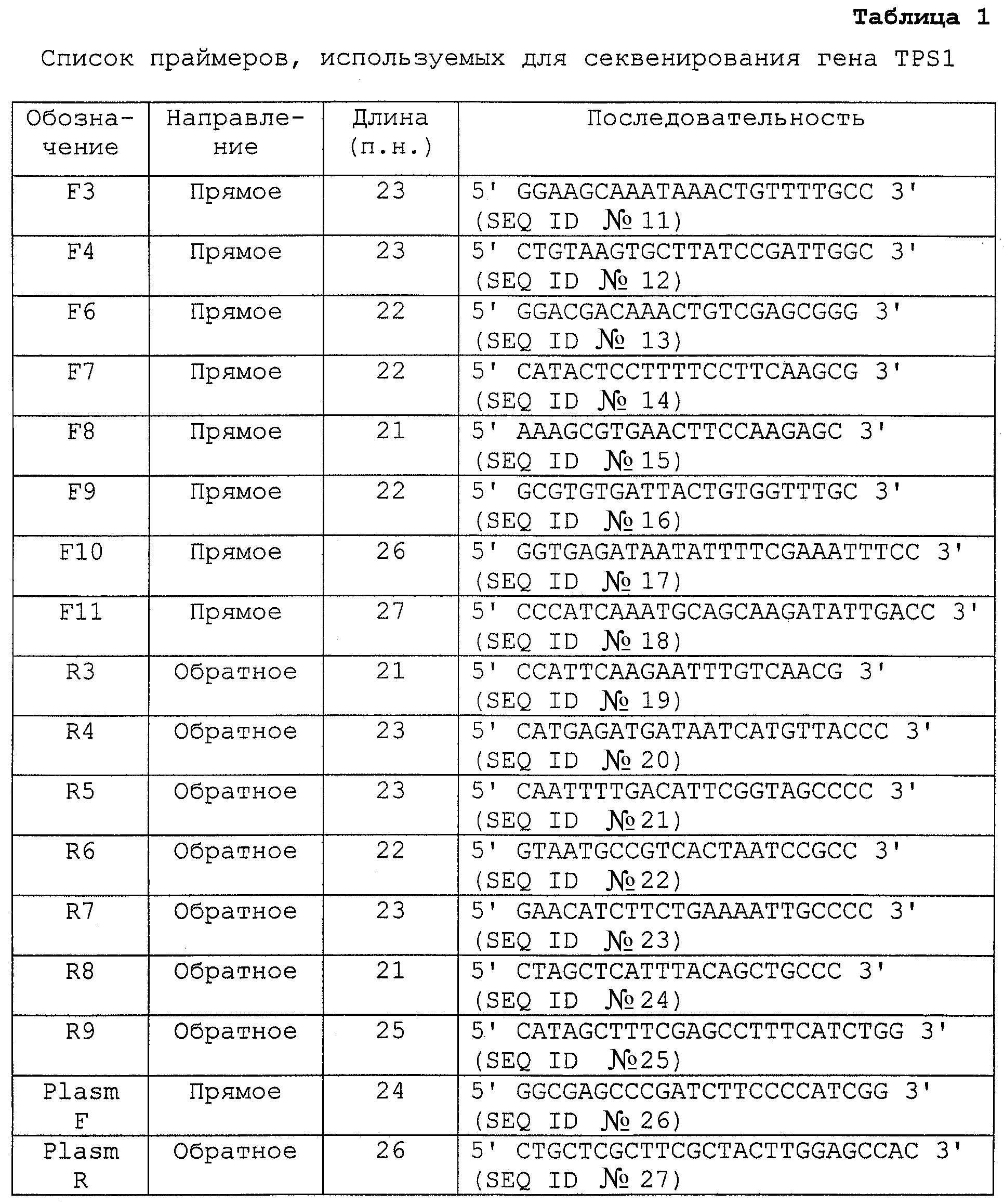
Промотор, выделенный из Н. polymorpha и способ его действия более подробно описаны ниже. Указанный промотор, который контролирует экспрессию TPS1, исследовали, измеряя увеличение TPSl-мРНК при определенных условиях. Было обнаружено, что в то время как при температурах, очень низких для Н. polymorpha, этот промотор экспрессировал небольшие количества TPS1, экспрессия очень сильно возрастала при высоких температурах, а именно намного сильнее, чем в случае с описанными ранее индуцируемыми тепловым шоком промоторами (см. фигуру 3А, Нозерн-блот при тепловом шоке). Индуцируемое теплом увеличение TPS1-мРНК коррелирует с увеличением Tps1 белка (фигура 3В), с увеличением активности трегалоза-6-фосфатсинтазы и с увеличением внутриклеточной концентрации трегалозы (фигура 3С). Для того чтобы оптимизировать влияние температуры, можно, например, избирательно укоротить промотор и соединить с дополнительными участками, содержащими HSE.The promoter isolated from H. polymorpha and its mode of action are described in more detail below. The indicated promoter, which controls the expression of TPS1, was investigated by measuring the increase in TPSl mRNA under certain conditions. It was found that while at temperatures very low for H. polymorpha, this promoter expressed small amounts of TPS1, the expression increased very strongly at high temperatures, namely, much stronger than in the case of the previously described heat-shock-induced promoters (see Figure 3A, Northern blot in heat shock). Heat-induced increase in TPS1-mRNA correlates with an increase in Tps1 protein (Figure 3B), with an increase in the activity of trehalose-6-phosphate synthase and with an increase in the intracellular concentration of trehalose (Figure 3C). In order to optimize the effect of temperature, it is possible, for example, to selectively shorten the promoter and combine with additional sites containing HSE.
Кроме индукции теплом также наблюдали накопление трегалозы, зависимое от глюкозной депривации, предположительно вследствие близкой биологической связи между этими двумя факторами стресса (см. фигуру 4А). Указанное накопление трегалозы коррелирует с увеличением активности трегалоза-6-фосфат-синтазы, увеличением TPSl-мРНК (фигура 4В), и возрастание накопления трегалозы наблюдали вместе с увеличением белка Tps1 во время глюкозной депривации (фигура 4 С).In addition to heat induction, an accumulation of trehalose, dependent on glucose deprivation, was also observed, presumably due to the close biological relationship between the two stress factors (see Figure 4A). The indicated accumulation of trehalose correlates with an increase in the activity of trehalose-6-phosphate synthase, an increase in TPSl mRNA (Figure 4B), and an increase in the accumulation of trehalose was observed together with an increase in Tps1 protein during glucose deprivation (Figure 4 C).
Чрезвычайно высокое накопление TPSl-мРНК свидетельствует о том, что TPSl-мРНК высоко стабильна, что делает ее (и кДНК на ее основе или на основе полученной от нее информации) не только полезным средством для выделения промотора, но также особенно ценным способом защиты других организмов против ряда стрессовых условий, таких как жара или засуха. ТРS1-ДНК, предоставленную с подходящими промоторами и векторами (например, как описано в WO 93/17093 и WO 96/00789), можно, например, применять для защиты растений от недостатка воды, таким образом давая возможность культивировать их в более теплых районах и районах с более низким количеством осадков. Конечно, для этой цели также можно использовать не только TPSl-ДНК, но также близкую ей ДНК.The extremely high accumulation of TPSl mRNA indicates that TPSl mRNA is highly stable, which makes it (and cDNA based on it or based on information received from it) not only a useful tool for isolating the promoter, but also a particularly valuable way to protect other organisms against a number of stressful conditions, such as heat or drought. TPS1 DNA provided with suitable promoters and vectors (for example, as described in WO 93/17093 and WO 96/00789) can, for example, be used to protect plants from a lack of water, thus allowing cultivation in warmer areas and areas with lower rainfall. Of course, not only TPSl DNA, but also DNA close to it can also be used for this purpose.
ПРИМЕР 3EXAMPLE 3
Сравнительная экспрессия бактериального гена lacZ под контролем промоторов EMD и TPS1Comparative expression of the lacZ bacterial gene under the control of EMD and TPS1 promoters
На основе интегрирующего вектора pCll H. polymorpha (фиг.7) сконструировали два производных, которые отличались только соответствующим промотором перед репортерным геном lacZ. В случае pCll-FMD (фиг.8) ген lacZ находится под контролем промотора FMD, который уже хорошо охарактеризован. В случае pC11-TPS1 (фиг.9) указанный ген находится под контролем тестируемого индуцируемого теплом промотора. В целях данного эксперимента в качестве индуцируемого теплом промотора использовали фрагмент между нуклеотидами 228 и 792 последовательности, указанной под номером SEQ ID №l (называемый ниже TPSl-промотором).Based on the integrating vector pCll of H. polymorpha (Fig. 7), two derivatives were constructed that differed only in the corresponding promoter in front of the lacZ reporter gene. In the case of pCll-FMD (Fig. 8), the lacZ gene is under the control of the FMD promoter, which is already well characterized. In the case of pC11-TPS1 (Fig. 9), said gene is under the control of the heat-inducible test promoter. For the purposes of this experiment, a fragment between the
H. polymorpha RB11 трансформировали pCll-FMD и pCll-TPSI (см. раздел “Материалы и способы”). Стабильные штаммы, в которых соответствующая плазмида присутствовала в стабильно интегрированном в геном состоянии, получали отдельно примерно из 1000 клонов прототрофных по урацилу клеток для каждой трансформации. В указанном случае процедура представляла собой следующее: после трансформации клетки высевали на чашки, содержащие селективную среду. Через три дня были видны макроскопические дискретные колонии. В обоих случаях 1000 дискретных отдельных колоний переносили в стерильных условиях на новые селективные чашки, которые затем инкубировали в течение двух дней при 37°С. Далее указанную процедуру повторяли два раза (пассирование). Затем колонии клеток переносили на чашки с полной средой и снова инкубировали в течение двух дней при 37°С (стабилизация). Наконец колонии клеток снова переносили на селективные чашки для того, чтобы исключить любые оставшиеся свободные плазмиды. После инкубации указанных чашек в течение двух дней при 37°С получение штаммов было завершено. Точное количество копий и локус интеграции плазмид в индивидуальных штаммах не определяли; однако в этом отношении согласно Gatzke et al. (1995) различные получаемые штаммы, несомненно, должны отличаться друг от друга.H. polymorpha RB11 was transformed with pCll-FMD and pCll-TPSI (see “Materials and Methods”). Stable strains in which the corresponding plasmid was present in a stably integrated genome state were obtained separately from approximately 1000 clones of prototrophic uracil cells for each transformation. In this case, the procedure was as follows: after transformation, the cells were plated on plates containing selective medium. Three days later, macroscopic discrete colonies were visible. In both cases, 1000 discrete individual colonies were transferred under sterile conditions to new selective plates, which were then incubated for two days at 37 ° C. Further, this procedure was repeated two times (passivation). Then the cell colonies were transferred to plates with complete medium and again incubated for two days at 37 ° C (stabilization). Finally, cell colonies were again transferred to selective plates in order to exclude any remaining free plasmids. After incubation of these plates for two days at 37 ° C, the preparation of the strains was completed. The exact number of copies and the locus of integration of plasmids in individual strains were not determined; however, in this regard, according to Gatzke et al. (1995) the different strains obtained must undoubtedly differ from each other.
Так как и количество копий, и геномное окружение оказывают большое влияние на степень транскрипции гена, следовало предположить, что индивидуальные клоны клеток будут также значительно отличаться друг от друга в отношении активности β-галактозидазы. Это было подтверждено экспериментально (данные не показаны). Поэтому было невозможно сравнить силу промоторов непосредственно с помощью индивидуальных штаммов. Несмотря на это, чтобы сделать возможным проведение объективных исследований промоторов, объединили 500 индивидуальных штаммов, которые были получены отдельно, имея целью создать репрезентативные смеси штаммов в отношении числа копий и локуса интеграции. Так как используемые для получения штаммов плазмиды pCll-FMD и pCll-TPS1 идентичны, за исключением соответствующего промотора, локализованного перед геном lacZ, можно предположить, что они интегрируются в геном хозяина гомологичным образом. Это предположение было подтверждено наблюдением того, что различные смеси штаммов при идентичной трансформации только незначительно отличаются друг от друга по своей β-галактозидазной активности (данные не показаны). Поэтому определение β-галактозидазной активности смесей штаммов, полученных при трансформации плазмидами, которые в значительной степени идентичны, должно позволить объективные сравнения промоторов в Н. polymorpha.Since both the number of copies and the genomic environment have a great influence on the degree of gene transcription, it was assumed that individual cell clones will also significantly differ from each other with respect to β-galactosidase activity. This was confirmed experimentally (data not shown). Therefore, it was impossible to compare the strength of promoters directly using individual strains. Despite this, to make it possible to conduct objective studies of promoters, we combined 500 individual strains, which were obtained separately, with the aim of creating representative mixtures of strains in terms of copy number and locus of integration. Since the plasmids pCll-FMD and pCll-TPS1 used to obtain strains are identical, with the exception of the corresponding promoter localized before the lacZ gene, it can be assumed that they integrate into the host genome in a homologous manner. This assumption was confirmed by the observation that different mixtures of strains with identical transformation only slightly differ from each other in their β-galactosidase activity (data not shown). Therefore, the determination of β-galactosidase activity of mixtures of strains obtained by transformation with plasmids, which are largely identical, should allow objective comparisons of promoters in N. polymorpha.
Функционирование lacZ под контролем промоторов FMD или TPS1 осуществляли при трех различных температурах в трех различных источниках углерода (см. фигуру 10). С этой целью смеси штаммов, описанные выше, культивировали до OD600, равным 5 в 10 мл селективной среды при указанных температурах и источниках углерода, после чего готовили экстракты клеток, активности β-галактозидазы в которых определяли посредством измерений ONPG в жидкой среде. Процедура представляла собой следующее: при достижении желаемой плотности культуры центрифугировали в течение 10 минут при 4°С, осадки клеток промывали в 10 мл lacZ-буфера (50 мМ натрий-фосфатный буфер, рН 7; 10 мМ КС1; 1 мМ MgSO4), ресуспендировали в 500 мкл lacZ-буфера и переносили в пробирки Эппендорф объемом 1,5 мл. Стеклянные шарики диаметром 0,45 мм добавляли к суспензиям (до мениска жидкости), после чего клетки разрушали в Vibrax (Janke & Kunkel; 6 минут; 4°С; 2200 об/мин). Лизаты клеток отбирали и центрифугировали (настольная центрифуга; 4°С; 10 минут). Растворимые фракции использовали как для определения β-галактозидазных активностей, так и для измерения суммарного содержания белка. Для измерений β-галактозидазной активности 1 мл раствора ONPG (4 мг ONPG/мл lacZ-буфера) добавляли к различным разведениям растворимых фракций и затем каждую смесь переносили в пластиковые 1 см-кюветы. Затем измеряли OD420 с 30-секундными интервалами в течение 3-минутного периода, чтобы позволить провести измерение ΔЕ.The functioning of lacZ under the control of the FMD or TPS1 promoters was carried out at three different temperatures in three different carbon sources (see figure 10). To this end, the mixture of strains described above was cultured to OD 600 equal to 5 in 10 ml of selective medium at the indicated temperatures and carbon sources, after which cell extracts were prepared, the β-galactosidase activity in which was determined by ONPG measurements in a liquid medium. The procedure was as follows: when the desired density was reached, the cultures were centrifuged for 10 minutes at 4 ° C, the cell pellets were washed in 10 ml of lacZ buffer (50 mM sodium phosphate buffer,
Чтобы определить суммарное содержание белка в клеточных экстрактах 790 мкл Н2О смешивали с 10 мкл соответствующей растворимой фракции (разведенной 1:10, 1:5, 1:2 или не разведенной, в соответствии с количеством белка) и добавляли 200 мкл реактива Брэдфорда (Biorad). После инкубации в течение 10 минут при комнатной температуре фотометрически определяли OD490 и корректировали по контрольному образцу, содержащему 1асZ-буфер вместо экстракта клеток. Затем определяли концентрацию белка в экстракте клеток на основании значений поглощения посредством калибровочной кривой для БСА. Специфические β-галактозидазные активности рассчитывали в соответствии со следующей формулой:To determine the total protein content in the cell extracts, 790 μl of H 2 O was mixed with 10 μl of the corresponding soluble fraction (diluted 1:10, 1: 5, 1: 2 or not diluted, according to the amount of protein) and 200 μl of Bradford reagent was added ( Biorad). After an incubation of 10 minutes at room temperature, OD 490 was determined photometrically and corrected with a control sample containing 1acZ buffer instead of cell extract. The protein concentration in the cell extract was then determined based on the absorbance values by means of a calibration curve for BSA. Specific β-galactosidase activities were calculated in accordance with the following formula:
Объемная активность (мU/мл) = ΔЕ V/ε d v суммарного белка,Volumetric activity (mU / ml) = ΔЕ V / ε d v total protein,
V: общий объем,V: total volume
v: объем образца,v: sample volume
ε: коэффициент экстинкцииε: extinction coefficient
(0,0045 мМ см),(0.0045 mm cm)
d: плотность слоя (1 см).d: layer density (1 cm).
Известно, что промотор FMD главным образом контролируется типом источника углерода; температурная зависимость еще не описана (патент ЕР № 299108). Это было подтверждено выполненными в данной работе измерениями (см. фиг.10А). Показали, что β-галактозидазные активности были низкими в условиях с глюкозой (репрессия глюкозой), тогда как значительно более высокие значения были измерены в условиях глицерина или метанола (дерепрессия или индукция). Изменения температуры не приводили к разительным изменениям полученных при измерении значений (см. фиг.10А). Это также наблюдали в используемой в данной работе тест-системе. Активности β-галактозидазы были низкими при 30°С или 37°С, но существенно возрастали при 44°С (см. фиг.10В). Указанное зависимое от температуры повышение активности промотора не происходило в условиях метанола (фиг.10В), феномен, который еще не был описан. Неожиданно наибольшие β-галактозидазные активности, измеренные для промоторов TPS1, были значительно выше, чем таковые для промоторов FMD (см. фиг.10А, В).It is known that the FMD promoter is mainly controlled by the type of carbon source; the temperature dependence has not yet been described (patent EP No. 299108). This was confirmed by the measurements performed in this work (see Fig. 10A). It was shown that β-galactosidase activities were low under conditions with glucose (glucose repression), while significantly higher values were measured under glycerol or methanol conditions (depression or induction). Changes in temperature did not lead to dramatic changes in the values obtained during the measurement (see Fig. 10A). This was also observed in the test system used in this work. Β-galactosidase activities were low at 30 ° C or 37 ° C, but increased significantly at 44 ° C (see Fig. 10B). The indicated temperature-dependent increase in promoter activity did not occur under methanol conditions (Fig. 10B), a phenomenon that has not yet been described. Unexpectedly, the highest β-galactosidase activities measured for TPS1 promoters were significantly higher than those for FMD promoters (see Fig. 10A, B).
ПРИМЕР 4EXAMPLE 4
Сравнительная экспрессия гена фитазы под контролем промоторов FMD или TPS1Comparative expression of the phytase gene under the control of FMD or TPS1 promoters
Рекомбинантные штаммы создавали посредством трансформации векторами pTPSlConphysMT и pFMTConphysMT согласно стандартным процедурам. За исключением промоторного элемента в экспрессирующей кассете два используемых для трансформации вектора идентичны. Индуцируемый теплом промотор, входящий в pTPSlConphysMT представляет собой фрагмент, соответствующий последовательности между нуклеотидами 228 и 792 в SEQ ID №l, 3'-конец которого имеет сайт рестрикции EcoRI (называемый ниже TPSl-промотором), тогда как pFMTConPhysMT содержит промотор FMD. Карта плазмиды и нуклеотидная последовательность вектора pTPSlConphysMT показаны на фиг.11. В качестве репортерного гена использовали мутеин фитазы.Recombinant strains were created by transformation with the vectors pTPSlConphysMT and pFMTConphysMT according to standard procedures. With the exception of the promoter element in the expression cassette, the two vectors used for transformation are identical. The heat-induced promoter included in pTPSlConphysMT is a fragment corresponding to the sequence between
После трансформации электропорацией получали рекомбинантные штаммы Н. polymorpha, выращивая полученные трансформацией прототрофные по урацилу штаммы на селективной среде в течение, по меньшей мере, 80 поколений (Gatzke et al., 1995). Представителей трансформантов двух полученных коллекций штаммов культивировали, сравнивая при различных условиях в жидких культурах объемом 3 мл. Культивирование проводили в среде YNB, забуференной 0,1 М фосфатным буфером рН 5,0 с добавлением 2% глюкозы или 5% глицерина. Через 48 часов количественно измеряли секретированную фитазу в аликвотах надосадка культуры с помощью способа, описанного в “Материалах и способах”.After transformation by electroporation, recombinant strains of H. polymorpha were obtained, growing strains obtained by transformation of uracil prototrophic strains on selective medium for at least 80 generations (Gatzke et al., 1995). Representatives of the transformants of the two obtained strain collections were cultured by comparing under different conditions in liquid cultures of 3 ml. Cultivation was carried out in YNB medium, buffered with 0.1 M phosphate buffer pH 5.0 with the addition of 2% glucose or 5% glycerol. After 48 hours, secreted phytase was quantitatively measured in aliquots of the culture supernatant using the method described in “Materials and Methods”.
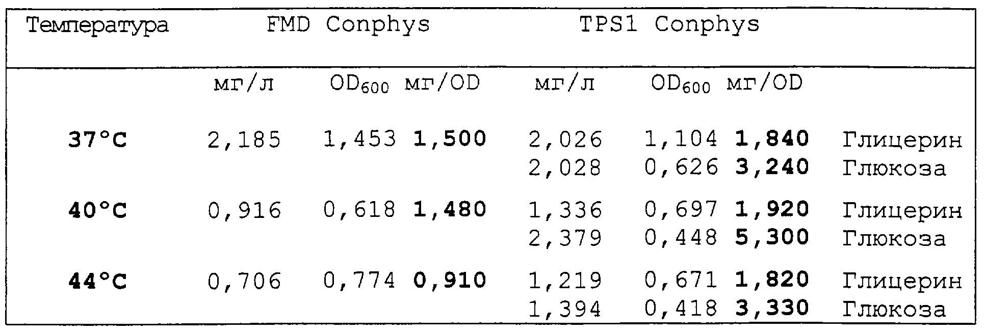
В данном исследовании промотор TPS1 сравнивали с наиболее широко используемым в настоящее время промотором, промотором FMD. Применение промотора TPS1 в результате давало слабо увеличенные значения экспрессии при 37°С при сравнении с промотором FMD. При 40°С и 44°С наблюдали в два-три раза более высокую экспрессию в том случае, когда использовали промотор TPS1, чем наблюдали с промотором FMD.In this study, the TPS1 promoter was compared with the currently most widely used promoter, the FMD promoter. The use of the TPS1 promoter resulted in slightly increased expression values at 37 ° C when compared with the FMD promoter. At 40 ° C and 44 ° C, two to three times higher expression was observed when the TPS1 promoter was used than was observed with the FMD promoter.
ПРИМЕР 5EXAMPLE 5
Сравнительная экспрессия гена фитазы под контролем вариантов промотора TPS1Comparative expression of the phytase gene under the control of TPS1 promoter variants
На основании интегрирующего вектора pCll H.polymorpha (фиг.7), были сконструированы несколько производных векторов, которые отличались только последовательностью промотора перед репортерным геном фитазы.Based on the integrating vector pCll of H. polymorpha (Fig. 7), several derived vectors were constructed that differed only in the promoter sequence before the phytase reporter gene.
В качестве примеров конструировали следующие различные конструкции на основании фрагмента между нуклеотидами 228 и 792 последовательности, представленной в SEQ ID №l (“исходный” промотор, индуцируемый теплом), и двух элементов, представленных в SEQ ID №2 и 5.As examples, the following various constructs were constructed based on the fragment between the
В первой серии промежуточная последовательность, состоящая из 52 п.н., между двумя элементами была модифицирована со 100% до 40%, при этом число в 52 п.н. не изменялось.In the first series, an intermediate sequence of 52 bp between the two elements was modified from 100% to 40%, with a number of 52 bp not changed.
Во второй и третьей сериях элемент 1 и 2 был делегирован.In the second and third series,
В сериях 4 и 5 эти конструкции модифицировали таким образом, что последовательности SEQ ID №4 и 3 использовали вместо последовательностей SEQ ID №2 и 5, производя замену элементов, способных отвечать на тепловой шок, указанными нуклеотидами.In
В дополнительной серии использовали комбинацию исходных и замещенных последовательностей. Для полноразмерного мутагенеза использовали только пример с двумя исходными элементами, сравнивая 100%-ную и 40%-ную гомологию.In an additional series, a combination of the starting and substituted sequences was used. For full-size mutagenesis, only an example with two initial elements was used, comparing 100% and 40% homology.
Различные последовательности получали индивидуально с помощью нуклеотидного синтеза, включая сайты рестрикции для правильного клонирования в вектор. Рекомбинантные штаммы получали трансформацией вышеуказанных векторов в соответствии со стандартными процедурами. Мутеин фитазы был использован в качестве репортерного гена. После осуществления трансформации электропорацией рекомбинантные штаммы H.polymorpha получали выращиванием урацил-фототрофных клонов, полученных трансформацией на селективной среде, на по меньшей мере 80 поколениях. Репрезентативные трансформанты, содержащие различные конструкции, культивировали и сравнивали в различных условиях в 3 мл жидкой культуры. Культивирование осуществляли в среде YNB, забуференной 0,1 М фосфатного буфера при рН 5,0, дополненной 2% глюкозой, и культивирование осуществляли при 40°С. Через 48 часов секретируемую фитазу подвергали количественному анализу в аликвотах культурального надосадка с помощью методов, описанных в разделе Материалы и Способы.Different sequences were obtained individually by nucleotide synthesis, including restriction sites for proper cloning into a vector. Recombinant strains were obtained by transformation of the above vectors in accordance with standard procedures. Mutein phytase has been used as a reporter gene. After transformation by electroporation, recombinant H. polymorpha strains were obtained by growing uracil-phototrophic clones obtained by transformation on a selective medium for at least 80 generations. Representative transformants containing various constructs were cultured and compared under various conditions in 3 ml of liquid culture. Cultivation was carried out in YNB medium, buffered with 0.1 M phosphate buffer at pH 5.0, supplemented with 2% glucose, and cultivation was carried out at 40 ° C. After 48 hours, the secreted phytase was subjected to quantitative analysis in aliquots of the culture supernatant using the methods described in the Materials and Methods section.
Исследование показало, что все варианты промотора TPS1 применимы для рекомбинантной экспрессии.The study showed that all variants of the TPS1 promoter are applicable for recombinant expression.
БиблиографияBibliography
Bradford, M.M. (1976) A rapid and sensitive method for the quantitation of microgram quantities of protein utilizing the principle of protein-dye binding. Anal Biochem 72: 248-254.Bradford, M.M. (1976) A rapid and sensitive method for the quantitation of microgram quantities of protein utilizing the principle of protein-dye binding. Anal Biochem 72: 248-254.
Devereux J., Haeberii P. and Smithies O. (1984) A comparative set of sequence analysis programs for the VAX. Nucl Acids Res 12: 387-395.Devereux J., Haeberii P. and Smithies O. (1984) A comparative set of sequence analysis programs for the VAX. Nucl Acids Res 12: 387-395.
De Virgilio C., Burckert N., Boller T. and Wiemken A. (1991) A method to study the rapid phosphorylation-related modulation of neutral trehalase activity by temperature shifts in yeast. FEBS Lett 291: 355-358.De Virgilio C., Burckert N., Boller T. and Wiemken A. (1991) A method to study the rapid phosphorylation-related modulation of neutral trehalase activity by temperature shifts in yeast. FEBS Lett 291: 355-358.
Faber K.N., Swaving G.J., Faber F., Ab G., Harder W., Veenhuis M. and Haima P. (1992) Chromosomal targeting of replicating plasmids in the yeast Hansenula polymorpha. J Gen Microbiol 138: 2405-2416.Faber K.N., Swaving G.J., Faber F., Ab G., Harder W., Veenhuis M. and Haima P. (1992) Chromosomal targeting of replicating plasmids in the yeast Hansenula polymorpha. J Gen Microbiol 138: 2405-2416.
Gatzke R., Weydemann U., Janowicz Z.A. & Hollenberg C.P. (1995) Stable multicopy integration of vector sequences in Hansenula polymorpha. Appl. Microbiol. Biotechnol 43, 844-849).Gatzke R., Weydemann U., Janowicz Z.A. & Hollenberg C.P. (1995) Stable multicopy integration of vector sequences in Hansenula polymorpha. Appl. Microbiol. Biotechnol 43, 844-849).
Hottiger Т., Schmutz P. and Wiemken A. (1987) Heat-induced accumulation and futile cycling of trehalose in Saccharomyces cerevisiae. J Bacteriol 169: 5518-5522.Hottiger T., Schmutz P. and Wiemken A. (1987) Heat-induced accumulation and futile cycling of trehalose in Saccharomyces cerevisiae. J Bacteriol 169: 5518-5522.
Huxley С., Green E.D. and Dunham I. (1990) Rapid assessment of Saccharomyces cerevisiae mating type by PCR. Trends Genet 6 (8): р. 236.Huxley S., Green E.D. and Dunham I. (1990) Rapid assessment of Saccharomyces cerevisiae mating type by PCR. Trends Genet 6 (8): p. 236.
Laemmli U.K. (1970) Cleavage of structural proteins during the assembly of the head of bacteriophage T4. Nature 227: 680-685.Laemmli U.K. (1970) Cleavage of structural proteins during the assembly of the head of bacteriophage T4. Nature 227: 680-685.
Levine D.W. and Cooney C.L. (1973) Isolation and characterization of a thermotolerant methanol-utilizing yeast. Appi Microbiol 26: 982-990.Levine D.W. and Cooney C.L. (1973) Isolation and characterization of a thermotolerant methanol-utilizing yeast. Appi Microbiol 26: 982-990.
Parrou J.L. and Francois J. (1997) A simplified procedure for a rapid and reliable assay of both glycogen and trehalose in whole yeast cells. Anal Biochem 248: 186-188.Parrou J.L. and Francois J. (1997) A simplified procedure for a rapid and reliable assay of both glycogen and trehalose in whole yeast cells. Anal Biochem 248: 186-188.
Peterson G.C. (1977) A simplification of the protein assay method of Lowry et al. which is more generally applicable. Anal Biochem 83: 346-356.Peterson G.C. (1977) A simplification of the protein assay method of Lowry et al. which is more generally applicable. Anal Biochem 83: 346-356.
Piper P.W. (1994) Measurement of transcription. In: Molecular Genetics of yeast. A practical approach, J.R. Johnston (ed.). IRL Press, Oxford.Piper P.W. (1994) Measurement of transcription. In: Molecular Genetics of yeast. A practical approach, J.R. Johnston (ed.). IRL Press, Oxford.
Sambrook J., Fritsch E.F. and Maniatis T. (1989) Molecular Cloning: A Laboratory Manual. Second edition. Cold Spring Harbor Laboratory Press, Cold Spring Harbor.Sambrook J., Fritsch E.F. and Maniatis T. (1989) Molecular Cloning: A Laboratory Manual. Second edition. Cold Spring Harbor Laboratory Press, Cold Spring Harbor.
Weydemann U, Keup P, Piontek M, Strasser AWM, Schweden J, Gellissen G, Janowicz ZA (1995) High-level secretion of hirudin by Hansenula polymorpha - authentic processing of three different preprohirudins. Appl Microbiol Biotechnol 44: 844-849.Weydemann U, Keup P, Piontek M, Strasser AWM, Schweden J, Gellissen G, Janowicz ZA (1995) High-level secretion of hirudin by Hansenula polymorpha - authentic processing of three different preprohirudins. Appl Microbiol Biotechnol 44: 844-849.
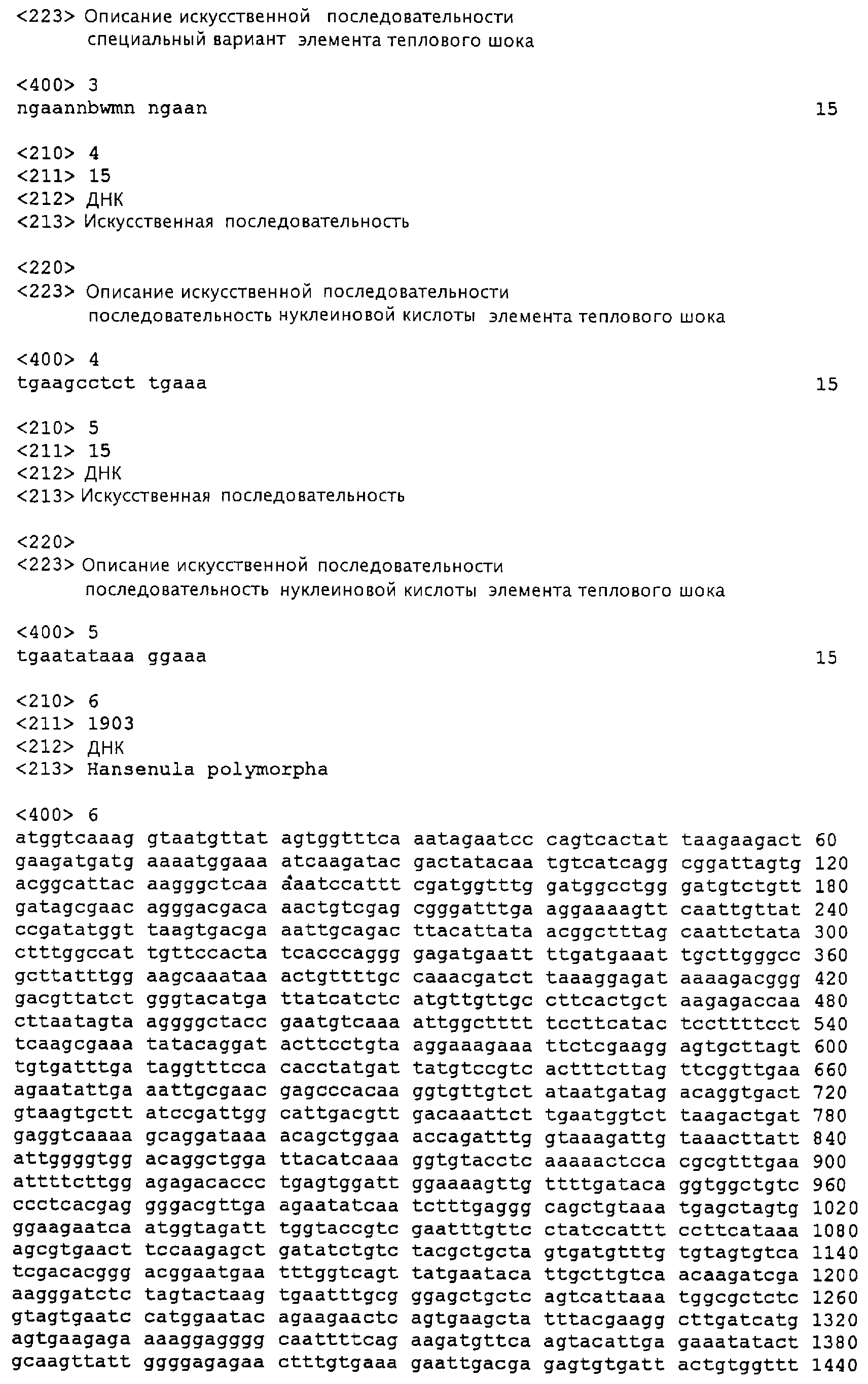
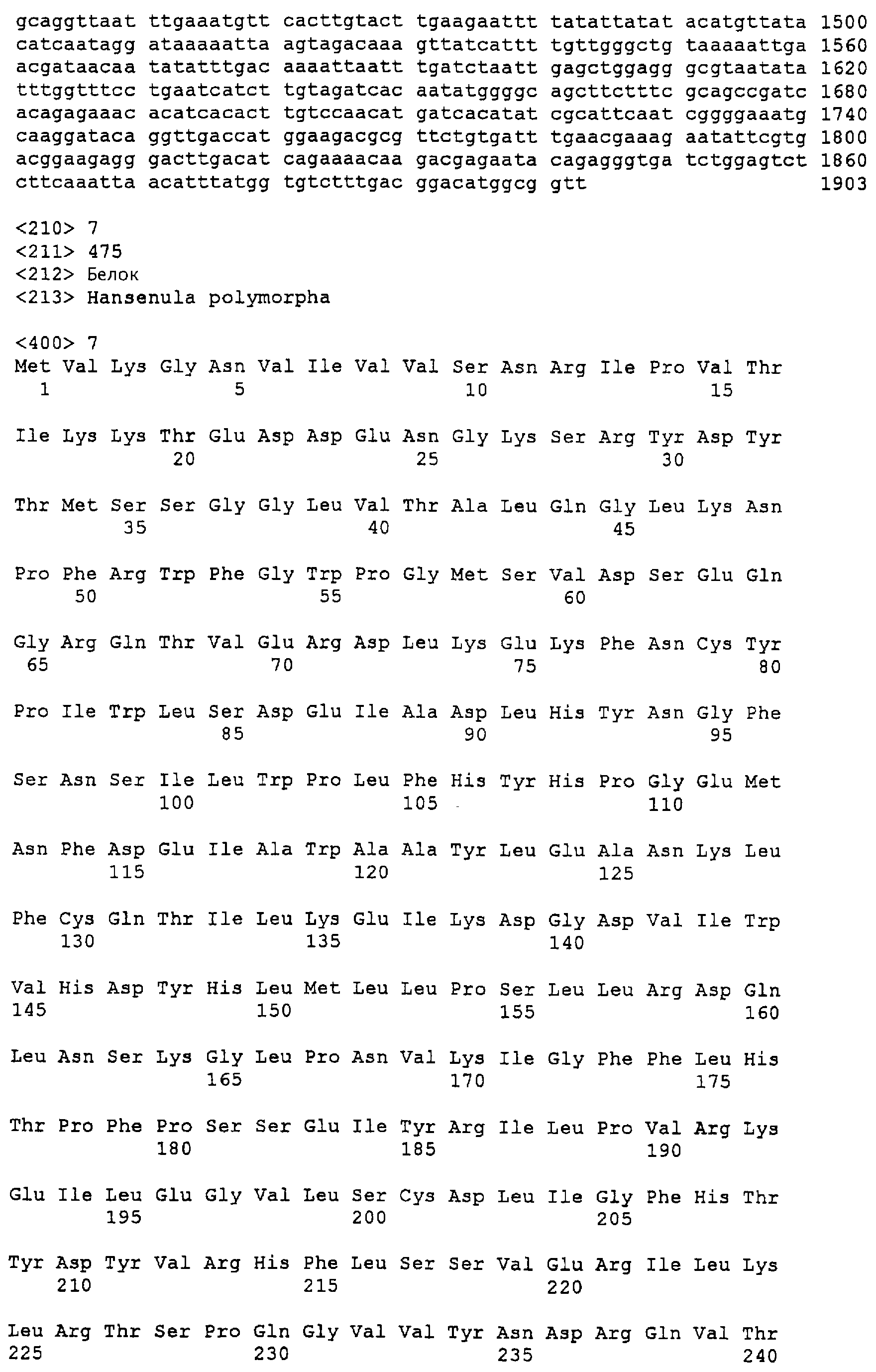
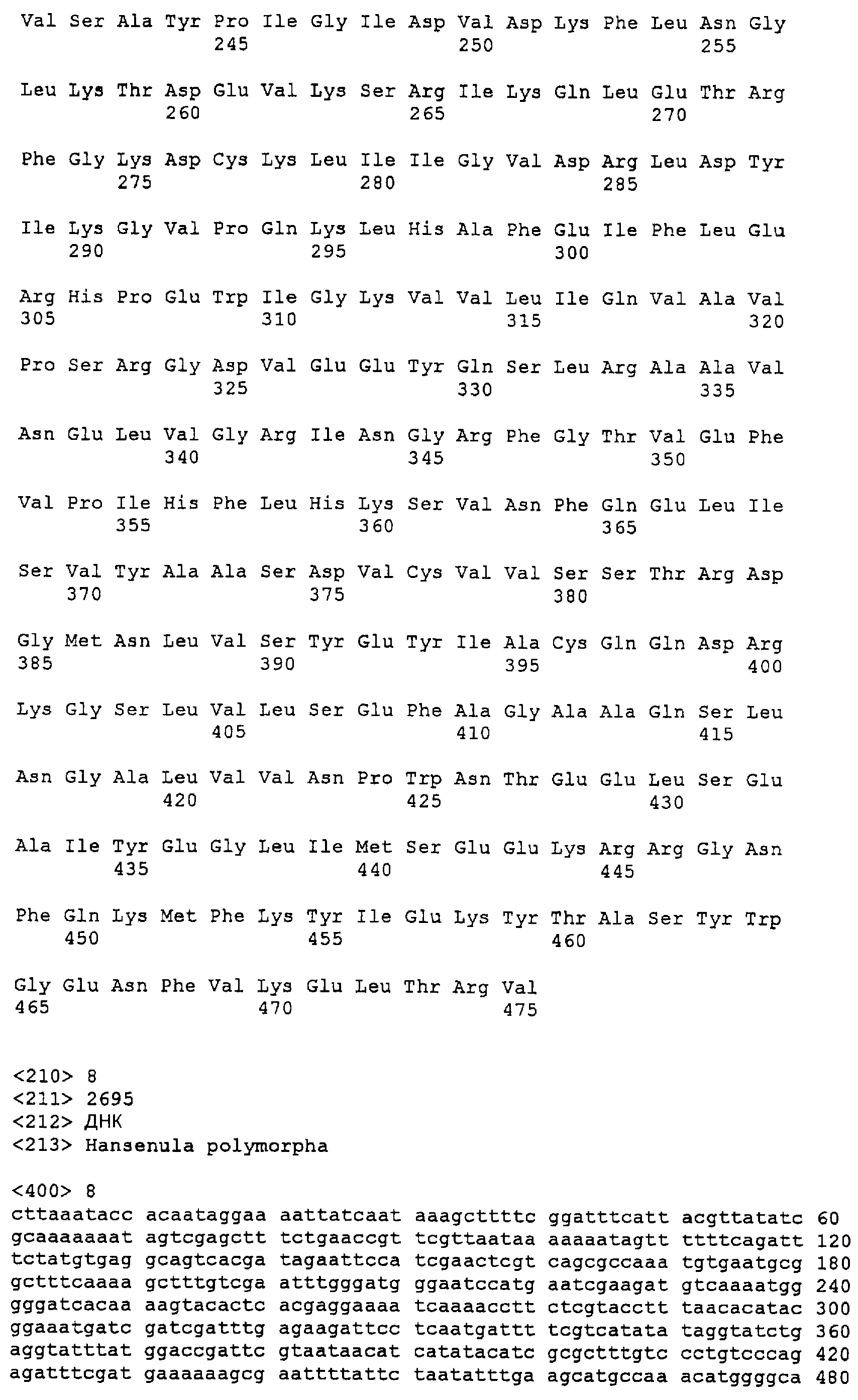
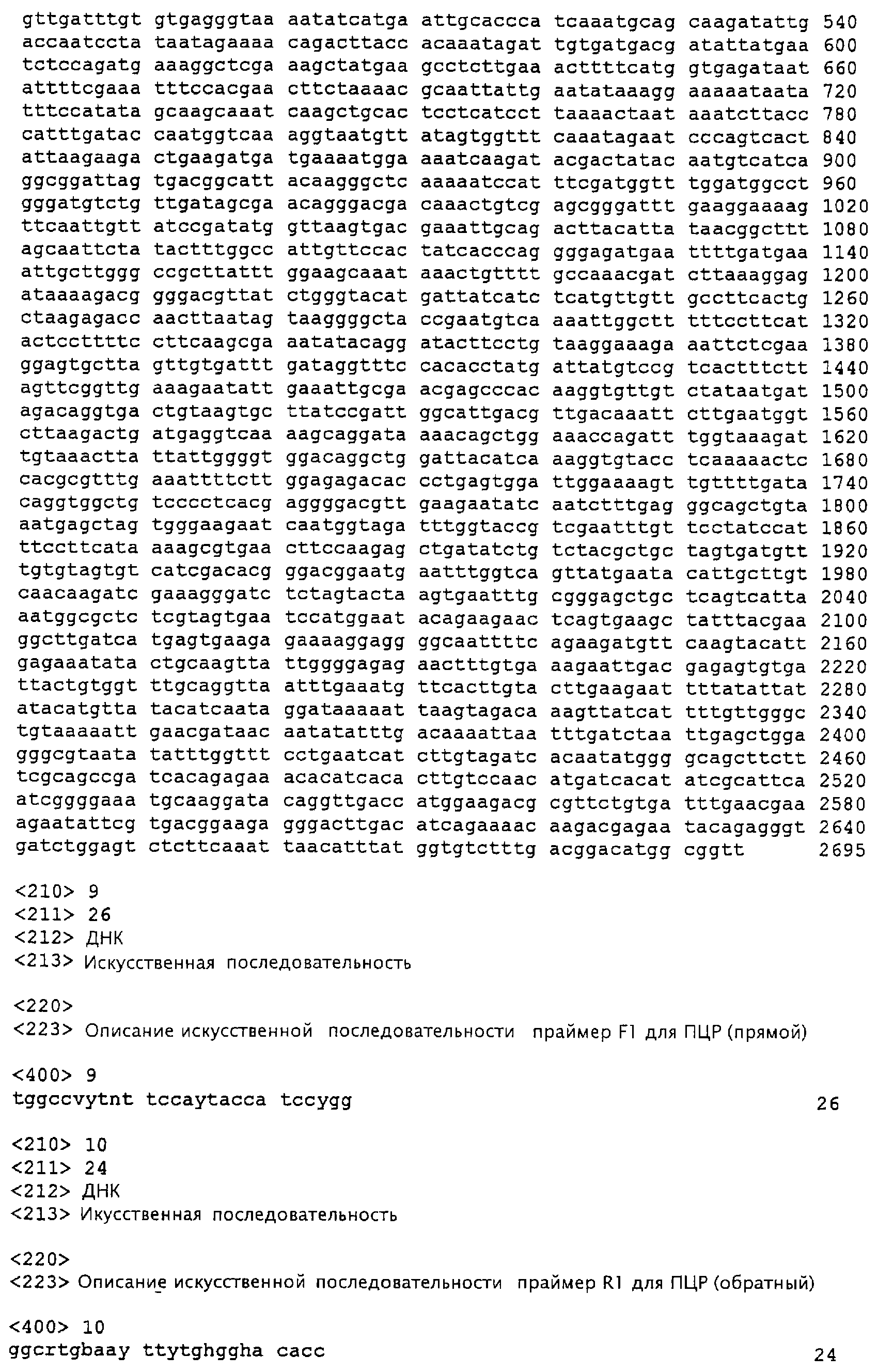
Claims (32)
Applications Claiming Priority (2)
| Application Number | Priority Date | Filing Date | Title |
|---|---|---|---|
| CH00279/99A CH690127A5 (en) | 1999-02-11 | 1999-02-11 | Heat-inducible promoter from Hansenula polymorpha, useful for controlling expression of foreign genes in yeast, contains no general stress response elements |
| CH279/99 | 1999-02-11 |
Publications (2)
| Publication Number | Publication Date |
|---|---|
| RU2001124852A RU2001124852A (en) | 2003-08-10 |
| RU2230786C2 true RU2230786C2 (en) | 2004-06-20 |
Family
ID=4183386
Family Applications (1)
| Application Number | Title | Priority Date | Filing Date |
|---|---|---|---|
| RU2001124852/13A RU2230786C2 (en) | 1999-02-11 | 2000-02-11 | Heat-induced promoter and its using |
Country Status (14)
| Country | Link |
|---|---|
| US (1) | US6852511B2 (en) |
| EP (1) | EP1151112B1 (en) |
| JP (1) | JP3914389B2 (en) |
| KR (1) | KR100473882B1 (en) |
| AT (1) | ATE455856T1 (en) |
| AU (1) | AU3422200A (en) |
| BR (1) | BRPI0008789B1 (en) |
| CA (1) | CA2362518C (en) |
| CH (1) | CH690127A5 (en) |
| DE (1) | DE50015851D1 (en) |
| IL (2) | IL144648A0 (en) |
| RU (1) | RU2230786C2 (en) |
| TR (1) | TR200102302T2 (en) |
| WO (1) | WO2000047749A1 (en) |
Families Citing this family (9)
| Publication number | Priority date | Publication date | Assignee | Title |
|---|---|---|---|---|
| US7901191B1 (en) | 2005-04-07 | 2011-03-08 | Parker Hannifan Corporation | Enclosure with fluid inducement chamber |
| US20110172826A1 (en) * | 2005-12-14 | 2011-07-14 | Amodei Dario G | Device including altered microorganisms, and methods and systems of use |
| US8852916B2 (en) | 2010-01-22 | 2014-10-07 | The Invention Science Fund I, Llc | Compositions and methods for therapeutic delivery with microorganisms |
| US8682619B2 (en) * | 2005-12-14 | 2014-03-25 | The Invention Science Fund I, Llc | Device including altered microorganisms, and methods and systems of use |
| US8278094B2 (en) | 2005-12-14 | 2012-10-02 | The Invention Science Fund I, Llc | Bone semi-permeable device |
| US8734823B2 (en) * | 2005-12-14 | 2014-05-27 | The Invention Science Fund I, Llc | Device including altered microorganisms, and methods and systems of use |
| WO2011159153A2 (en) * | 2010-06-15 | 2011-12-22 | Universiteit Utrecht Holding B.V. | Inducible promoter and its use |
| US11504427B2 (en) | 2016-11-22 | 2022-11-22 | The Regents Of The University Of California | Acoustic and ultrasound-based mechanogenetics and thermogenetics for immunotherapy |
| US20220298515A1 (en) * | 2019-09-10 | 2022-09-22 | The Regents Of The University Of California | Ultrasound-based thermogenetics for immunotherapy |
Citations (1)
| Publication number | Priority date | Publication date | Assignee | Title |
|---|---|---|---|---|
| US5792921A (en) * | 1992-02-14 | 1998-08-11 | Londesborough; John | Increasing the trehalose content of organisms by transforming them with combinations of the structural genes for trehalose synthase |
Family Cites Families (1)
| Publication number | Priority date | Publication date | Assignee | Title |
|---|---|---|---|---|
| US5422254A (en) * | 1992-02-14 | 1995-06-06 | Oy Alko Ab | Method to increase the trehalose content of organisms by transforming them with the structural genes for the short and long chains of yeast trehalose synthase |
-
1999
- 1999-02-11 CH CH00279/99A patent/CH690127A5/en not_active IP Right Cessation
-
2000
- 2000-02-11 AU AU34222/00A patent/AU3422200A/en not_active Abandoned
- 2000-02-11 CA CA002362518A patent/CA2362518C/en not_active Expired - Lifetime
- 2000-02-11 DE DE50015851T patent/DE50015851D1/en not_active Expired - Lifetime
- 2000-02-11 EP EP00912457A patent/EP1151112B1/en not_active Expired - Lifetime
- 2000-02-11 IL IL14464800A patent/IL144648A0/en active IP Right Grant
- 2000-02-11 KR KR10-2001-7010163A patent/KR100473882B1/en not_active Expired - Lifetime
- 2000-02-11 TR TR2001/02302T patent/TR200102302T2/en unknown
- 2000-02-11 BR BRPI0008789A patent/BRPI0008789B1/en not_active IP Right Cessation
- 2000-02-11 WO PCT/EP2000/001144 patent/WO2000047749A1/en not_active Ceased
- 2000-02-11 RU RU2001124852/13A patent/RU2230786C2/en active
- 2000-02-11 JP JP2000598645A patent/JP3914389B2/en not_active Expired - Fee Related
- 2000-02-11 AT AT00912457T patent/ATE455856T1/en not_active IP Right Cessation
-
2001
- 2001-07-31 IL IL144648A patent/IL144648A/en not_active IP Right Cessation
- 2001-08-09 US US09/927,811 patent/US6852511B2/en not_active Expired - Lifetime
Patent Citations (1)
| Publication number | Priority date | Publication date | Assignee | Title |
|---|---|---|---|---|
| US5792921A (en) * | 1992-02-14 | 1998-08-11 | Londesborough; John | Increasing the trehalose content of organisms by transforming them with combinations of the structural genes for trehalose synthase |
Non-Patent Citations (1)
| Title |
|---|
| BLAZOUEZ MIGUEL et al. J. Bacteriol, vol. 176, 1994, р.3895-3902. ЩЕЛКУНОВ С.Н. Генетическая инженерия. - Новосибирск: Из-во Новосибирского университета, 1997, с.192 и 193. * |
Also Published As
| Publication number | Publication date |
|---|---|
| AU3422200A (en) | 2000-08-29 |
| CA2362518C (en) | 2009-04-21 |
| US6852511B2 (en) | 2005-02-08 |
| CA2362518A1 (en) | 2000-08-17 |
| WO2000047749A1 (en) | 2000-08-17 |
| EP1151112A1 (en) | 2001-11-07 |
| KR100473882B1 (en) | 2005-03-08 |
| EP1151112B1 (en) | 2010-01-20 |
| TR200102302T2 (en) | 2002-02-21 |
| DE50015851D1 (en) | 2010-03-11 |
| JP2002536020A (en) | 2002-10-29 |
| IL144648A (en) | 2007-08-19 |
| BRPI0008789B1 (en) | 2015-09-08 |
| CH690127A5 (en) | 2000-05-15 |
| ATE455856T1 (en) | 2010-02-15 |
| BR0008789A (en) | 2001-11-06 |
| KR20010108200A (en) | 2001-12-07 |
| IL144648A0 (en) | 2002-05-23 |
| US20040086998A1 (en) | 2004-05-06 |
| JP3914389B2 (en) | 2007-05-16 |
Similar Documents
| Publication | Publication Date | Title |
|---|---|---|
| Yang et al. | Colletotrichum trifolii mutants disrupted in the catalytic subunit of cAMP-dependent protein kinase are nonpathogenic | |
| Vankuyk et al. | Aspergillus niger mstA encodes a high-affinity sugar/H+ symporter which is regulated in response to extracellular pH | |
| US5716808A (en) | Genetic engineering of pichia methanolica | |
| US5736383A (en) | Preparation of Pichia methanolica auxotrophic mutants | |
| US5888768A (en) | Compositions and methods for producing heterologous polypeptides in Pichia methanolica | |
| WO1998002536A2 (en) | Preparation of pichia methanolica auxotrophic mutants | |
| RU2230786C2 (en) | Heat-induced promoter and its using | |
| US6555657B2 (en) | Isolated transcription factor for an alpha-amylase promoter in filamentous fungi | |
| US5587290A (en) | Stress tolerant yeast mutants | |
| US5728806A (en) | TIH1, protein that interacts with casein kinase I | |
| US5641661A (en) | Pichia pastoris alcohol oxidase ZZA1 and ZZA2 regulatory regions for heterologous gene expression | |
| US6410315B1 (en) | Genes encoding polypeptides containing signal sequences | |
| US20030134353A1 (en) | Recombinant dimorphic fungal cell | |
| Heintel et al. | Expression, processing and high level secretion of a virus toxin in fission yeast | |
| US5919666A (en) | Factors which modify gene transcription and methods of use therefor | |
| Moussy et al. | Inter‐Species DNA Polymerase δ Chimeras are Functional in Saccharomyces Cerevisiae | |
| Skrzypek et al. | Cloning and sequencing of the ornithine carbamoyltransferase gene from Pachysolen tannophilus | |
| NZ528597A (en) | Recombinant dimorphic fungal cell | |
| Santocanale et al. | Overproduction and functional analysis of DNA primase subunits from yeast and mouse | |
| US5210024A (en) | Isolation of strains of Saccharomyces cerevisiae having altered α-acetyltransferase activity | |
| US5500483A (en) | Pichia pastoris alcohol oxidase ZZA1 regulatory region for heterologous gene expression | |
| US5922585A (en) | RNA polymerase II holoenzyme from Saccharomyces cerevisiae | |
| Meinke | Regulation of Expression of a Gene for Glycogen Synthase in Saccharomyces cerevisiae | |
| JPH099971A (en) | Novel selectable marker gene and transformation system of Rhizomucor pusillus | |
| EP0946734A2 (en) | Preparation of pichia methanolica auxotrophic mutants |
Legal Events
| Date | Code | Title | Description |
|---|---|---|---|
| PC4A | Invention patent assignment |
Effective date: 20090226 |