RU2192012C2 - Method of assay of antibody raised to chicken reoviral tenosynovitis antigen - Google Patents
Method of assay of antibody raised to chicken reoviral tenosynovitis antigen Download PDFInfo
- Publication number
- RU2192012C2 RU2192012C2 RU2000117522/13A RU2000117522A RU2192012C2 RU 2192012 C2 RU2192012 C2 RU 2192012C2 RU 2000117522/13 A RU2000117522/13 A RU 2000117522/13A RU 2000117522 A RU2000117522 A RU 2000117522A RU 2192012 C2 RU2192012 C2 RU 2192012C2
- Authority
- RU
- Russia
- Prior art keywords
- virus
- serum
- optical density
- chicken
- antigen
- Prior art date
Links
Images
Abstract
Description
Изобретение относится к ветеринарной вирусологии и биотехнологии и может быть использовано для определения специфических антител к реовирусу теносиновита кур методом иммуноферментного анализа. The invention relates to veterinary virology and biotechnology and can be used to determine specific antibodies to chickens tenosynovitis reovirus by enzyme immunoassay.
Теносиновит - контагиозное реовирусное заболевание птицы, зарегистрированное во всех странах мира, имеет подострое, хроническое или латентноперсистирующее течение. Широко распространено среди кур, индеек, гусей и синантропной птицы. Реовирусы птиц антигенно отличны от реовирусов млекопитающих и человека. Серологические и вирусологические исследования установили широкое распространение (90-95%) реовирусной инфекции у кур в Российской Федерации [1]. Поэтому исключительно важное значение и приобретает диагностика данного заболевания, основанная на эпизоотологических, клинических и патологоанатомических данных, результатах лабораторных исследований (выделение вируса и его идентификация в реакции нейтрализации, биопроба) и выявление специфических антител в сыворотке крови птиц методом иммуноферментного анализа. Tenosynovitis is a contagious reoviral disease of the bird, registered in all countries of the world, has a subacute, chronic or latent-persistent course. Widely distributed among chickens, turkeys, geese and synanthropic birds. Bird reoviruses are antigenically different from mammalian and human reoviruses. Serological and virological studies have established a widespread (90-95%) reovirus infection in chickens in the Russian Federation [1]. Therefore, the diagnosis of this disease based on epizootological, clinical and pathological data, laboratory results (isolation of the virus and its identification in the neutralization reaction, bioassay) and detection of specific antibodies in the blood serum of birds by enzyme-linked immunosorbent assay is extremely important.
Известен способ получения вакцины против proventriculitis (заболевание птиц) [2] . Способ предусматривает выделение реовируса proventriculitis из содержащей реовирус ткани птицы, пораженной proventriculitis, инокулирование клеточной культуры, способной поддерживать рост и репликацию этих реовирусов, экстракцию вируса из клеточных и внеклеточных компонентов и превращение реовирусов в форму, пригодную для введения домашней птице. A known method of obtaining a vaccine against proventriculitis (disease of birds) [2]. The method comprises isolating proventriculitis reovirus from reovirus containing bird tissue affected by proventriculitis, inoculating a cell culture capable of supporting the growth and replication of these reoviruses, extracting the virus from cellular and extracellular components, and converting the reoviruses into a form suitable for administration to poultry.
Недостатком этого способа получения антигена реовируса кур является низкая концентрация вирусного белка, что не позволяет получить высокоактивный антиген, используемый в иммуноферментном анализе (ИФА). The disadvantage of this method of producing chicken reovirus antigen is a low concentration of viral protein, which does not allow to obtain a highly active antigen used in enzyme-linked immunosorbent assay (ELISA).
Известен также способ получения иммунопероксидазного конъюгата для выявления антигенов бруцелл, включающий иммунизацию кроликов смесью водорастворимых антигенов Вг. abortus 544, Вг. melitensis 565 и Вг. suis 1330, взятых в равных отношениях, затем выделение специфических иммуноглобулинов из иммунной сыворотки и конъюгацию с пероксидазой хрена [3]. There is also known a method of producing an immunoperoxidase conjugate for detecting Brucella antigens, comprising immunizing rabbits with a mixture of water-soluble Bg antigens. abortus 544, Vg. melitensis 565 and Br. suis 1330 taken in equal proportions, then the isolation of specific immunoglobulins from the immune serum and conjugation with horseradish peroxidase [3].
Однако получение иммунопероксидазного конъюгата данным способом не решает задачу выявления специфических антител к реовирусному теносиновиту кур. However, the preparation of an immunoperoxidase conjugate by this method does not solve the problem of detecting specific antibodies to chicken reovirus tenosynovitis.
В основу изобретения поставлена комплексная задача создания способа определения антител к антигену реовирусного теносиновита кур, позволяющего повысить чувствительность и специфичность анализа. The basis of the invention is the comprehensive task of creating a method for the determination of antibodies to the chickens rheovirusovirus antigen, which allows to increase the sensitivity and specificity of the analysis.
Поставленная задача решается тем, что согласно изобретению вируссодержащий материал получают с использованием штамма "ВНИВИП-ДЕП", очистку полученного вируссодержащего материала осуществляют путем вирусной гельхроматографии на макропористом стекле с диаметром пор, соответствующим диаметру вириона очищаемого вируса. Наилучший результат получают при использовании макропористого стекла, модифицированного поливинилпирролидоном (ПВП) с диаметром пор не менее 1200 ![]()
![]()
Конечный титр исследуемой сыворотки крови определяют по одному разведению 1:100, используя формулу
Lg титр=2,19 (Lg S/P)+3,59,
где S/P=(S-NKх)/(PKх-NKх),
где S - значение оптической плотности исследуемой сыворотки;
Р - значение оптической плотности положительного контроля;
NKх - среднее значение оптической плотности отрицательного контроля;
PKх - среднее значение оптической плотности положительного контроля;
при этом проба сыворотки при S/P-отношении ниже 0,2 расценивается как отрицательная, а при S/P-отношении, равном или превышающем 0,2, как положительная.The final titer of the test blood serum is determined by one dilution of 1: 100, using the formula
Lg titer = 2.19 (Lg S / P) +3.59,
where S / P = (S-NK x ) / (PK x -NK x ),
where S is the optical density of the investigated serum;
P is the optical density value of the positive control;
NK x is the average optical density of the negative control;
PK x is the average optical density of the positive control;
in this case, a serum sample at an S / P ratio below 0.2 is regarded as negative, and at an S / P ratio equal to or greater than 0.2, as positive.
Заявляемые свойства способа определяются прежде всего использованием штамма "ВНИВИП-ДЕП", активность которого составляет 5,5-6,0 Ig ЭИД50. Штамм депонирован в коллекции Всероссийского государственного научно-исследовательского института контроля, стандартизации и сертификации ветеринарных препаратов под номером "ВНИВИП-ДЕП" [4].The claimed properties of the method are determined primarily by using the strain "VNIVIP-DEPT", whose activity is 5.5-6.0 Ig EID 50 . The strain is deposited in the collection of the All-Russian State Research Institute for Control, Standardization and Certification of Veterinary Medicines under the number "VNIVIP-DEP" [4].
Авторами было установлено, что оптимальной дозой антигена реовируса для адсорбции на полистироловом планшете является 5-10 мкг/0,1 см3, поскольку увеличение ее выше 10 мкг/0,1 см3 приводит к многослойной сорбции антигена, вследствие чего снижается чувствительность реакции, а уменьшение ее ниже 5 мкг/0,1 см3 не приводит к получению монослоя на полистироле, что также отрицательно сказывается на результате.The authors found that the optimal dose of reovirus antigen for adsorption on a polystyrene tablet is 5-10 μg / 0.1 cm 3 , since an increase above 10 μg / 0.1 cm 3 leads to multilayer sorption of antigen, thereby reducing the sensitivity of the reaction, and reducing it below 5 μg / 0.1 cm 3 does not lead to a monolayer on polystyrene, which also negatively affects the result.
Специфический комплекс - антиген-немеченое антитело - выявляют использованием антивидового иммунопероксидазного конъюгата, специфичного к IgG птиц, поскольку это определяется его биологическими свойствами. A specific complex - an antigen-unlabeled antibody - is detected using an anti-species immunoperoxidase conjugate specific for avian IgG, as this is determined by its biological properties.
Способ осуществляют следующим образом. The method is as follows.
Вирус теносиновита кур штамм "ВНИВИП-ДЕП" хранится во ВГНКИ ветпрепаратов и выдается по требованию биопредприятия не реже 1 раза в год с паспортом, характеризующим свойства штамма. Chicken tenosynovitis virus strain "VNIVIP-DEP" is stored in VGNKI of veterinary preparations and is issued at the request of a biological enterprise at least 1 time per year with a passport characterizing the properties of the strain.
Для получения вируссодержащего материала использовали метод перемежающихся пассажей на развивающихся куриных эмбрионах 9-11-суточного возраста, которые заражали освеженным аттенуированным вакцинным штаммом "ВНИВИП-ДЕП" реовирусного теносиновита кур. Активность эмбрионального вируса теносиновита кур определяли методом титрации на куриных эмбрионах [5]. To obtain virus-containing material, the method of intermittent passages was used on developing chicken embryos of 9–11 days old, which were infected with the freshly attenuated vaccine strain “VNIVIP-DEP” of chicken reovirus tenosynovitis. The activity of chicken tenosynovitis embryonic virus was determined by titration on chicken embryos [5].
Биологическая активность вируссодержащей жидкости должна быть не ниже 5,5 Ig ЭИД50. Активный и специфичный антиген вируса теносиновита кур получают путем очистки вируссодержащей эмбриональной жидкости методом молекулярно-ситовой хроматографии на макропористом стекле с диаметром пор 1200 ![]()
![]()
В очищенном препарате вируса количество белка определяют по методу Лоури, и оно, как правило, составляет от 35 до 50 мкг/см3. Далее очищенный препарат вируса разводят 0,01 М фосфатно-буферным раствором с 0,15 М хлоридом натрия (ФБР) рН 7,2-7,4 до конечной концентрации 5-10 мкг/0,1 см3. Это значение является оптимальным параметром для адсорбции очищенного антигена вируса в лунках планшета, поскольку увеличение концентрации антигена свыше 10 мкг/0,1 см3 и уменьшение ее ниже 5 мкг/0,1 см3 снижает чувствительность реакции.In a purified virus preparation, the amount of protein is determined by the Lowry method, and it usually ranges from 35 to 50 μg / cm 3 . Next, the purified virus preparation is diluted with 0.01 M phosphate-buffered saline with 0.15 M sodium chloride (FBI) pH 7.2-7.4 to a final concentration of 5-10 μg / 0.1 cm 3 . This value is the optimal parameter for adsorption of the purified antigen of the virus in the wells of the plate, since an increase in the concentration of antigen above 10 μg / 0.1 cm 3 and a decrease below 5 μg / 0.1 cm 3 reduces the sensitivity of the reaction.
После определения активности и специфичности очищенный антиген вируса теносиновита кур разделяют на две партии (части), одну из которых используют для иммунизации цыплят с целью получения гипериммунной сыворотки крови, а вторую - для адсорбции в лунках планшета для ИФА. After determining the activity and specificity, the purified chicken tenosynovitis virus antigen is divided into two batches (parts), one of which is used to immunize chickens to obtain hyperimmune blood serum, and the second for adsorption in the wells of an ELISA plate.
Твердофазный антиген получают посредством сорбции в лунках планшета для ИФА по 0,1 см3, разведенного ФБР в конечной концентрации очищенного антигена в течение 16-18 часов при 2-4oС.A solid-phase antigen is obtained by sorption in the wells of an ELISA plate of 0.1 cm 3 diluted by the FBI in a final concentration of purified antigen for 16-18 hours at 2-4 o C.
Затем содержимое лунок планшета удаляют резким встряхиванием и трехкратно промывают промывочным буфером следующего состава: фосфатно-буферный раствор (рн 7,2-7,4) 0,01 моль/л; хлорид натрия 0,5 М, твин-20, твин-80 или тритон Х-100 0,1%, после чего лунки планшета подсушивают путем постукивания по фильтровальной бумаге. Then, the contents of the plate wells are removed by sharp shaking and washed three times with washing buffer of the following composition: phosphate-buffered saline (pH 7.2-7.4) 0.01 mol / L; sodium chloride 0.5 M, tween-20, tween-80 or triton X-100 0.1%, after which the wells of the tablet are dried by tapping on filter paper.
Для постановки иммуноферментного анализа используют одно- и восьмиканальные автоматические пипетки со сменными наконечниками разных объемов, термостат с температурой нагрева (37,0±0,5)oС, бытовой холодильник, рН-метр тип 121 или 340, спектрофотометр любой конструкции с вертикальным лучом света при длине волны 450 нм.Enzyme-linked immunosorbent assay is performed using single and eight-channel automatic pipettes with interchangeable tips of different volumes, a thermostat with a heating temperature (37.0 ± 0.5) o С, a household refrigerator, a pH meter type 121 or 340, a spectrophotometer of any design with a vertical beam light at a wavelength of 450 nm.
Для исследования в лабораторию доставляют по 20-25 проб (0,2-0,3) см3 сывороток крови птиц разных возрастных групп из птицехозяйств, где имеется подозрение на данное заболевание.For research, 20-25 samples (0.2-0.3) cm 3 of blood serum of birds of different age groups from poultry farms where there is a suspicion of this disease are delivered to the laboratory.
Далее поступают следующим образом: готовят иммуноспецифические и неспецифические компоненты и рабочие растворы:
Компонент 1 - положительная сыворотка крови кур к реовирусу теносиновита (положительный контроль), разведенная 1:100 0,15 М раствором хлорида натрия с содержанием 1% фетальной сыворотки, лиофилизированная, 1,0 см3 -1 ампула или флакон (К.1.);
Компонент 2 - отрицательная сыворотка крови (отрицательный контроль), не содержащая антител к реовирусу теносиновита кур, разведенная 1:100 0,15 М раствором хлорида натрия с содержанием 1% фетальной сыворотки, лиофилизированная, 1.0 см3 - 1 ампула или флакон (К.2.);
Компонент 3 - антиген реовируса теносиновита кур, очищенный и адсорбированный по 0,1 см3 в лунках планшета для ИФА - 2 планшета (К.3.);
Компонент 4 - антивидовой иммунопероксидазный конъюгат против IgG кур, лиофилизированный 0,2 или 1,0 см3 - 1 ампула (флакон), рабочее разведение 1: 21 (К.4.);
Компонент 5 - концентрированный разбавитель - 1,5 М калий-фосфатный буфер рН 7,2-7,4, жидкий, 10 см3 - 1 флакон (К.5.);
Компонент 6 - разбавленный детергент твин-20, твин-80 или тритон Х-100, жидкий, 5 см3 - 1 флакон (К.6.);
Компонент 7 - 5-аминосалициловая кислота, порошок, 20 мг - 1 флакон или 1 пробирка для микропроб (К.7.);
Компонент 8 - гидроперит, таблетка в стандартной упаковке, 0,75 или 1,5 г - 1 шт. (К.8.).Then proceed as follows: prepare immunospecific and nonspecific components and working solutions:
Component 1 - positive blood serum of chickens to tenosinovitis reovirus (positive control), diluted 1: 100 with 0.15 M sodium chloride solution containing 1% fetal serum, lyophilized, 1.0 cm 3 -1 ampoule or vial (K.1. );
Component 2 - negative blood serum (negative control), not containing antibodies to chicken tenosynovitis reovirus, diluted 1: 100 with 0.15 M sodium chloride solution containing 1% fetal serum, lyophilized, 1.0 cm 3 - 1 ampoule or vial (K. 2.);
Component 3 - chicken tenosynovitis reovirus antigen, purified and adsorbed at 0.1 cm 3 in the wells of an ELISA plate - 2 tablets (K.3.);
Component 4 - antiviral immunoperoxidase conjugate against chicken IgG, lyophilized 0.2 or 1.0 cm 3 - 1 ampoule (vial), working dilution 1: 21 (K.4.);
Component 5 - concentrated diluent - 1.5 M potassium phosphate buffer, pH 7.2-7.4, liquid, 10 cm 3 - 1 bottle (K.5.);
Component 6 - diluted detergent Tween-20, Tween-80 or Triton X-100, liquid, 5 cm 3 - 1 bottle (K.6.);
Component 7 - 5-aminosalicylic acid, powder, 20 mg - 1 vial or 1 tube for microprobe (K.7.);
Component 8 - hydroperite, tablet in a standard package, 0.75 or 1.5 g - 1 pc. (K.8.).
Приготовление рабочих растворов:
Раствор 1- 0,01 М калий-фосфатный буфер, содержащий 0,5 М раствор хлорида натрия с 0,1% конечной концентрацией детергента (твина-20, твина-80 или тритона Х-100), рН 7,2-7,4.Preparation of working solutions:
A solution of 1 - 0.01 M potassium phosphate buffer containing a 0.5 M solution of sodium chloride with a 0.1% final concentration of detergent (Tween-20, Tween-80 or Triton X-100), pH 7.2-7, 4.
Для его приготовления необходимо растворить содержимое флакона с концентрированным разбавителем (К.5.), содержимое флакона с разбавленным детергентом (К.6.) и 44,0 г хлорида натрия в 1500 см3 дистиллированной воды, проверить рН. При необходимости рН корректируют 0,1н. КОН или HCI.To prepare it, it is necessary to dissolve the contents of the bottle with concentrated diluent (K.5.), The contents of the bottle with diluted detergent (K.6.) And 44.0 g of sodium chloride in 1500 cm 3 of distilled water, check the pH. If necessary, adjust the pH to 0.1n. KOH or HCI.
Раствор используют для разведения положительной, отрицательной исследуемых проб сывороток крови кур, антивидового конъюгата и промывания лунок планшетов. Хранят не более 15 суток в бытовом холодильнике при (4-8)oС.The solution is used to dilute positive, negative test samples of chicken blood serum, an anti-species conjugate, and washing the wells of the plates. Store no more than 15 days in a household refrigerator at (4-8) o C.
Раствор 2 - содержимое ампулы (флакона) с положительной сывороткой крови кур (К. 1. ) растворяют в 1,0 см3 раствора 1, получают разведение положительной сыворотки 1:100, готовят перед использованием. Допускается хранение в морозильной камере бытового холодильника до 15 сут.Solution 2 - the contents of the ampoule (vial) with chicken positive blood serum (K. 1.) are dissolved in 1.0 cm 3 of solution 1, a positive serum dilution of 1: 100 is obtained, prepared before use. Storage in the freezer of a household refrigerator is allowed for up to 15 days.
Раствор 3 - содержимое ампулы (флакона) с отрицательной сывороткой крови кур (К. 2. ) растворяют с 1,0 см3 раствора 1, получают разведение отрицательной сыворотки 1:100, готовят перед использованием.Solution 3 - the contents of the ampoule (vial) with negative chicken blood serum (K. 2.) is dissolved with 1.0 cm 3 of solution 1, a negative serum dilution of 1: 100 is obtained, prepared before use.
Допускается хранение в морозильной камере бытового холодильника до 15 сут. Storage in the freezer of a household refrigerator is allowed for up to 15 days.
Раствор 4 - содержимое ампулы с антивидовым конъюгатом (К.4.) растворяют в 21 см3 раствора 1. Готовят перед использованием. Допускается хранение в морозильной камере бытового холодильника до 15 сут.Solution 4 - the contents of the ampoule with the anti-conjugate conjugate (K.4.) Are dissolved in 21 cm 3 of solution 1. Prepare before use. Storage in the freezer of a household refrigerator is allowed for up to 15 days.
Раствор 5 - 1%-ный раствор NaOH. Готовят растворением 1 г гидроокиси натрия в 99 см3 дистиллированной воды. Хранят не более 15 суток при комнатной температуре 20oС.Solution 5 - 1% NaOH solution. Prepared by dissolving 1 g of sodium hydroxide in 99 cm 3 distilled water. Store no more than 15 days at room temperature 20 o C.
Раствор 6 - таблетку гидроперита 1,5 г (К.8.) растворяют в 22,5 или 45 см3 дистиллированной воды. Раствор хранят в защищенном от света месте при 4-8oС не более 10 суток.Solution 6 - a tablet of hydroperite 1.5 g (K.8.) Is dissolved in 22.5 or 45 cm 3 of distilled water. The solution is stored in a dark place at 4-8 o C for no more than 10 days.
Раствор 7 - содержимое флакона с 5-аминосалициловой кислотой (К.7.) растворяют в 25 см3 горячей дистиллированной воды (70±2)oС при постоянном перемешивании на магнитной мешалке, затем охлаждают до комнатной температуры и доводят рН до 6,0±0,1 с помощью раствора 5. Раствор готовят непосредственно перед использованием, не хранят.
Раствор 8 - для приготовления субстратно-индикаторной смеси к 0,1 см3 раствора 6 добавляют 3,3 см3 холодной (4-8)oС дистиллированной воды, затем к 2 см3 полученного раствора приливают 18 см3 раствора 7. Раствор готовят непосредственно перед использованием, не хранят.Solution 8 - to prepare a substrate-indicator mixture, add 0.1 cm 3 of solution 6 to 3.3 cm 3 of cold (4-8) o With distilled water, then 18 cm 3 of solution is added to 2 cm 3 of the resulting solution 7. The solution is prepared immediately before use, do not store.
Постановка реакции:
Образцы исследуемых сывороток разводят 1:100 раствором 1. С этой целью к 1,0 см3 раствора 1 добавляют 0,01 см3 (10 мкл) сыворотки крови птиц. Затем в три лунки (А1, В1, С1) вертикального ряда планшета вносят по 0,1 см3 разведенной отрицательной сыворотки крови (раствор 3) и в следующие три лунки (D1, Е1, F1) вносят по 0,1 см3 разведенной 1:100 положительной сыворотки крови (раствор 2). В лунки G1, HI вносят по 0,1 см3 раствора 1, служащие для контроля конъюгата и субстратно-индикаторной смеси.Statement of the reaction:
Samples of the studied sera are diluted 1: 100 with
В остальные лунки рядов B2-12...H2-12 вносят по 0,1 см3 раствора 1, а в лунки горизонтального ряда A2-12 по 0,2 см3 исследуемых проб сывороток крови и проводят раститровку по вертикальным рядам, начиная от разведения 1:100 до 1: 12800. С этой целью из лунок A2-12 отбирают по 0,1 см3 разведенных исследуемых сывороток крови и переносят в лунки B2-12 соответствующего ряда, пипетируют и проводят двукратное разведение переносом по 0,1 см3 в очередную лунку вертикального ряда планшета, удаляя из последних лунок H2-12 по 0,1 см3.0.1 cm 3 of solution 1 are added to the remaining wells of rows B2-12 ... H2-12, and 0.2 cm 3 of the studied blood serum samples are added to the wells of the horizontal row A2-12 and the test is carried out in vertical rows, starting from dilutions 1: 100 to 1: 12800. For this purpose, 0.1 cm 3 of diluted test blood sera are taken from wells A2-12 and transferred to wells of B2-12 of the corresponding row, pipetted and double dilution is carried out by transfer of 0.1 cm 3 into the next well of the vertical row of the tablet, removing 0.1 cm 3 from the last holes of H2-12.
Планшет осторожно встряхивают для перемешивания содержимого, закрывают крышкой и переносят в термостат (37,0±0,5 oС) на 30 мин. Затем лунки планшета освобождают от содержимого путем вытряхивания, трехкратно промывают раствором 1 и подсушивают путем постукивания по фильтровальной бумаге.The tablet is gently shaken to mix the contents, close the lid and transfer to a thermostat (37.0 ± 0.5 o C) for 30 minutes Then the wells of the tablet are freed from the contents by shaking, washed three times with
Готовят раствор 4 и вносят во все лунки планшета по 0,1 см3, затем помещают в термостат (37,0±0,5 oС) на 30 мин. После инкубации раствор конъюгата удаляют, лунки трехкратно промывают и подсушивают путем постукивания по фильтровальной бумаге.
Затем готовят растворы 5, 6, 7. Во все лунки планшета вносят по 0,1 см3 раствора 8 - субстратно-индикаторной смеси - и оставляют при комнатной температуре в темном месте на 15-20 мин.Then
Выявление специфических антител в сыворотках крови кур иммуноферментным методом можно проводить и без раститровки испытуемых сывороток. Detection of specific antibodies in the blood serum of chickens by the enzyme immunoassay can also be carried out without testing the test sera.
В этом случае в три лунки (А1, В1, С1) вертикального ряда планшета вносят по 0,1 см3 разведенной 1:100 отрицательной сыворотки крови (отрицательный контроль), а в следующие три лунки (D1, Е1, F1) вносят по 0,1 см3 разведенной 1: 100 положительной сыворотки (положительный контроль). В лунки G1, Н1 вносят по 0,1 см3 раствора 1, служащие для контроля конъюгата и субстратно-индикаторной смеси. В лунки В2...Н2 вносят по 0,1 см3 раствора 1, а в лунки А2 по 0,2 см 3 положительной сыворотки и проводят раститровку по вертикальному ряду А2-Н2. В остальные лунки рядов А3-12...Н3-12 вносят по 0,1 см3 исследуемых проб сывороток крови кур в разведении 1:100, используя по одной лунке на каждую пробу. Остальные этапы постановки реакции проводят аналогично.In this case, 0.1 cm 3 of diluted 1: 100 negative blood serum is added to three wells (A1, B1, C1) of the vertical row of the tablet (negative control), and 0 is added to the next three wells (D1, E1, F1) , 1 cm 3 diluted 1: 100 positive serum (positive control). 0.1 cm 3 of solution 1 are added to wells G1, H1, which serve to control the conjugate and the substrate-indicator mixture. 0.1 cm 3 of solution 1 is added to wells B2 ... H2, and 0.2 cm 3 of positive serum is added to wells A2 and a trituration is carried out along the vertical row A2-H2. In the remaining wells of rows A3-12 ... H3-12 make 0.1 cm 3 of the investigated samples of blood serum of chickens at a dilution of 1: 100, using one well for each sample. The remaining stages of the reaction are carried out similarly.
Учет результатов анализа проводят через 15-20 мин визуально - по интенсивности окрашивания содержимого лунок планшета или инструментально - с помощью спектрофотометра с вертикальным лучом света при длине волны 450 нм. Analysis results are taken into account after 15-20 minutes visually - according to the intensity of staining the contents of the wells of the tablet or instrumental - using a spectrophotometer with a vertical beam of light at a wavelength of 450 nm.
При визуальном способе учета первоначально проводят оценку результатов по контрольным лункам планшета: содержимое лунок с положительной сывороткой (положительный контроль) должно быть интенсивно окрашено в коричневый цвет. Содержимое лунок с отрицательной сывороткой (отрицательный контроль) и контроль конъюгата должно быть бесцветным или иметь бледно-соломенное окрашивание. With the visual method of accounting, the results are initially evaluated according to the control wells of the tablet: the contents of the wells with positive serum (positive control) should be intensely colored in brown. The contents of the wells with negative serum (negative control) and the control of the conjugate should be colorless or have a pale straw color.
При отсутствии необходимости строгого количественного определения содержания специфических антител в исследуемых пробах сывороток крови учет результатов ИФА проводят визуально, сравнением цветового окрашивания содержимого лунок исследуемой сыворотки с интенсивностью окраски продукта пероксидазной реакции титруемого положительного контроля. Интенсивность цветового окрашивания продукта пероксидазной реакции пропорциональна содержанию антител к реовирусу теносиновита кур. If there is no need for strict quantitative determination of the content of specific antibodies in the studied blood serum samples, the results of ELISA are carried out visually by comparing the color staining of the contents of the wells of the test serum with the color intensity of the product of the peroxidase reaction of the titrated positive control. The intensity of color staining of the product of peroxidase reaction is proportional to the content of antibodies to chick tenosynovitis reovirus.
Раститровка проб испытуемых сывороток крови дает возможность количественно оценить концентрацию специфических антител. За титр сыворотки принимается наивысшее ее разведение, при котором еще достаточно четко видна разница цветового окрашивания по сравнению с отрицательной сывороткой крови. The leveling of samples of the tested blood serum makes it possible to quantify the concentration of specific antibodies. The highest dilution is taken as the serum titer, at which the difference in color staining is still quite clearly visible compared with negative blood serum.
Спектрофотометрический учет результатов ИФА позволяет количественно оценить содержание антител в исследуемых пробах путем измерения значений оптической плотности с одновременной записью результатов реакции с каждой лунки планшета на специальной бумажной ленте или листе бумаги. Различие между средним значением положительного контроля и средним значением отрицательного контроля должно быть более чем 0,10. Наличие или отсутствие антител против реовируса устанавливается сравнением значений оптической плотности тестируемой сыворотки (S) с положительным контролем (Р). Проба сыворотки с S/P-отношением ниже 0,2 должна расцениваться как отрицательная. Если S/P-отношение равно или больше, чем 0,2, то исследуемая проба должна расцениваться как положительная. Spectrophotometric recording of ELISA results allows you to quantify the content of antibodies in the studied samples by measuring the optical density with simultaneous recording of the reaction results from each well of the tablet on a special paper tape or sheet of paper. The difference between the average value of the positive control and the average value of the negative control should be more than 0.10. The presence or absence of antibodies against reovirus is established by comparing the optical density of the test serum (S) with a positive control (P). A serum sample with an S / P ratio below 0.2 should be considered negative. If the S / P ratio is equal to or greater than 0.2, then the test sample should be regarded as positive.
При определении конечного титра исследуемой пробы по одному разведению 1:100 используют формулу
Lg титр=2,19 (Lg S/P)+3,59,
где S/P=(S-NKх)/(PKх-NKх),
где S - значение оптической плотности исследуемой сыворотки;
Р - значение оптической плотности положительного контроля;
NКх - среднее значение оптической плотности отрицательного контроля;
PKх - среднее значение оптической плотности положительного контроля;
при этом проба сыворотки при S/Р-отношении ниже 0,2 расценивается как отрицательная, а при S/Р-отношении, равном или превышающем 0,2, как положительная.When determining the final titer of the test sample by one dilution of 1: 100, use the formula
Lg titer = 2.19 (Lg S / P) +3.59,
where S / P = (S-NK x ) / (PK x -NK x ),
where S is the optical density of the investigated serum;
P is the optical density value of the positive control;
NK x is the average optical density of the negative control;
PK x is the average optical density of the positive control;
in this case, a serum sample at an S / P ratio below 0.2 is considered negative, and at an S / P ratio equal to or greater than 0.2, it is positive.
Обнаружение в сыворотках крови птиц разных возрастных групп, невакцинированных против реовирусного теносиновита, специфических антител в титрах 1:100 и выше у более чем 50% исследуемых проб, дает основание для постановки диагноза на данное заболевание. При этом учитывают эпизоотическую ситуацию в птицехозяйстве, наличие клинических и патологоанатомических признаков заболевания. Detection in blood serum of birds of different age groups unvaccinated against reovirus tenosinovitis, specific antibodies in titers of 1: 100 and higher in more than 50% of the studied samples, gives the basis for the diagnosis of this disease. In this case, take into account the epizootic situation in the poultry industry, the presence of clinical and pathological signs of the disease.
Для окончательной постановки диагноза на данное заболевание проводят вирусологические исследования по выделению вируса из патологического материала (пораженные суставы, почки, селезенка. печень). For the final diagnosis of this disease, virological studies are carried out to isolate the virus from pathological material (affected joints, kidneys, spleen, liver).
Оценкой эффективности поствакцинального иммунитета является выявление в сыворотках крови птиц специфических антител в титрах не ниже чем 1:400 у 80% вакцинированной птицы. Evaluation of the effectiveness of post-vaccination immunity is the detection of specific antibodies in the blood serum of birds in titers of not less than 1: 400 in 80% of the vaccinated bird.
Осуществление заявленного способа описано в примерах. The implementation of the claimed method is described in the examples.
Пример 1. Для подтверждения высокой чувствительности и специфичности предлагаемого способа был проведен сравнительный анализ результатов исследования проб сывороток крови кур с помощью реакции нейтрализации и предложенного способа. Example 1. To confirm the high sensitivity and specificity of the proposed method, a comparative analysis of the results of the study of samples of blood serum of chickens using the neutralization reaction and the proposed method.
Сыворотки, исследуемые предложенным способом, подобраны так, что все они проверялись на наличие антител к вирусу теносиновита кур в реакции нейтрализации (РН). Тест нейтрализации является классическим тестом диагностики вирусных заболеваний, в том числе и для диагностики реовирусного теносиновита кур. The sera studied by the proposed method are selected so that they are all checked for the presence of antibodies to the chicken tenosynovitis virus in the neutralization reaction (PH). The neutralization test is a classic test for the diagnosis of viral diseases, including the diagnosis of chicken reovirus tenosynovitis.
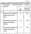
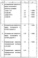
Результаты сравнительного изучения чувствительности предложенного способа и реакции нейтрализации при выявлении специфических антител к вирусу теносиновита кур представлены в табл.1. The results of a comparative study of the sensitivity of the proposed method and the neutralization reaction in identifying specific antibodies to chicken tenosynovitis virus are presented in Table 1.
Результаты исследования показали, что предлагаемый способ обладает высокой чувствительностью и специфичностью и позволяет выявлять специфические антитела к вирусу теносиеновита кур в сыворотках крови. The results of the study showed that the proposed method has high sensitivity and specificity and allows the detection of specific antibodies to chicken tenosienovitis virus in blood serum.
Корреляции между двумя сравниваемыми тестами не обнаружено. Реакция нейтрализации выявляет только вируснейтрализующие антитела, в то время как предложенный способ выявляет целый комплекс антител: комплементсвязывающие, вируснейтрализующие, преципитирующие и др. No correlation was found between the two compared tests. The neutralization reaction reveals only virus-neutralizing antibodies, while the proposed method reveals a whole complex of antibodies: complement-binding, virus-neutralizing, precipitating, etc.
Пример 2. Были проведены испытания на активность и специфичность предложенного способа в шифрованном опыте. Example 2. Tests were conducted on the activity and specificity of the proposed method in an encrypted experience.
На испытание были представлены 3 лабораторные опытные серии иммуноспецифических компонентов для выявления антител к вирусу теносиновита кур предложенным способом, изготовленных:
серия 1 - 02.02.00
серия 2 - 03.03.00
серия 3 - 05.05.00
- набор для диагностики ССЯ-76 ИФМ, производства ВНИВИП (ТУ 10-07-146-89), изготовленного 06.99;
- набор для выявления антител к вирусу инфекционного бронхита кур иммуноферментным способом, производства ВНИВИП (ТУ 10.07.080-93), изготовленного 04.2000;
- сыворотки гиперимунные специфические к вирусу теносиновита кур от разных возрастных групп - 15 проб;
- положительные сыворотки к вирусу теносиновита кур от экспериментально зараженной птицы различными штаммами вируса;
- полевые сыворотки крови кур от птиц разного возраста - 60 проб;
- специфические (гетерологичные) сыворотки крови кур к вирусам: гриппа птиц, инфекционного ларинготрахеита. болезни Марека, инфекционной бурсальной болезни. ПМВ-2 - по 3 пробы;
- отрицательные сыворотки крови кур к вирусу теносиновита - 10 проб.For the test were presented 3 laboratory experimental series of immunospecific components for the detection of antibodies to chicken tenosynovitis virus by the proposed method, manufactured:
series 1 - 02.02.00
series 2 - 03.03.00
series 3 - 05.05.00
- a set for the diagnosis of SSYA-76 IFM, manufactured by VNIVIP (TU 10-07-146-89), manufactured on 06.99;
- a kit for detecting antibodies to the chicken infectious bronchitis virus enzyme immunoassay method, manufactured by VNIVIP (TU 10.07.080-93), manufactured on 04.2000;
- hyperimmune serums specific to the tenosynovitis virus of chickens from different age groups - 15 samples;
- positive serum to the chicken tenosynovitis virus from experimentally infected birds with various strains of the virus;
- field blood serum of chickens from birds of different ages - 60 samples;
- specific (heterologous) blood serum of chickens to viruses: bird flu, infectious laryngotracheitis. Marek's disease, infectious bursal disease. PMV-2 - 3 samples each;
- negative blood serum of chickens to the tenosynovitis virus - 10 samples.
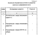
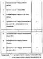
Для исследований было зашифровано 10 проб сывороток крови кур. Исследуемые сыворотки разводили ФБР 1:100 и вносили в лунки планшета с адсорбированным очищенным антигеном реовируса в объеме 0,1 см3, по 2 лунки на каждую сыворотку. Иммуноферментную реакцию проводили аналогичным образом, как указано ранее.For research, 10 chicken serum samples were encrypted. The test sera were diluted 1: 100 with the PBS and added to the wells of the plate with adsorbed purified reovirus antigen in a volume of 0.1 cm 3 , 2 wells per serum. An enzyme-linked immunosorbent reaction was carried out in a similar manner as previously indicated.
Результаты испытаний представлены в табл.2. The test results are presented in table.2.
Результаты шифрованного опыта показали, что предлагаемый способ выявления специфических антител к реовирусу теносиновита в сыворотках крови кур обладает высокой чувствительностью и специфичностью. The results of the encrypted experience showed that the proposed method for detecting specific antibodies to tenosinovitis reovirus in chicken serum has high sensitivity and specificity.
Таким образом, заявленный способ определения антител к антигену реовирусного теносиновита кур обладает высокой чувствительностью, специфичностью и эффективностью при выявлении специфических антител в сыворотках крови кур. Способ может быть использован для качественного и количественного выявления специфических антител к вирусу теносиновита кур в сыворотках крови и может быть использован для серологического контроля при эпизоотологическом обследовании птицепоголовья и определения напряженности поствакцинального иммунитета против теносиновита кур. Thus, the claimed method for determining antibodies to the antigen of rheovirus tenosinovitis chickens has high sensitivity, specificity and effectiveness in detecting specific antibodies in the blood serum of chickens. The method can be used for the qualitative and quantitative detection of specific antibodies to the chicken tenosynovitis virus in blood serum and can be used for serological monitoring during epizootological examination of the poultry head and for determining the post-vaccination immunity against chicken tenosynovitis.
Источники информации
1. Трефилов Б.Б., Никитина Н.В. Реовирусная инфекция птиц. - СПб, Информационный листок N 2-96, ВНИВИП, 1996.Sources of information
1. Trefilov B.B., Nikitina N.V. Reovirus infection of birds. - St. Petersburg, Information leaflet N 2-96, VNIVIP, 1996.
2. ЕР 0101348 А2. Публ. 22.02.84, ИЗР 21, тем. выпуск 13, 1984. 2. EP 0101348 A2. Publ. 02.22.84, IPM 21, topics. issue 13, 1984.
3. SU 1475331 A1 (Волгоградский научно-исследовательский противочумный институт). Публ. 20.05.99. 3. SU 1475331 A1 (Volgograd Research Anti-Plague Institute). Publ. 05/20/99.
4. RU 2158304 (Всероссийский научно-исследовательский ветеринарный институт птицеводства). Публ. 27.10.2000 30. 4. RU 2158304 (All-Russian Scientific Research Veterinary Institute of Poultry). Publ. 10.27.2000 30.
5. Сюрин В.Л. Руководство по ветеринарной вирусологии. - М., 1966. 5. Syurin V.L. Guide to Veterinary Virology. - M., 1966.
Claims (4)
lg титр=2,19•(LgS/P)+3,59,
где S/P=(S-NKx)/(PKx-NKx), где S - значение оптической плотности исследуемой сыворотки;
Р - значение оптической плотности положительного контроля;
NKx - среднее значение оптической плотности отрицательного контроля;
РKx - среднее значение оптической плотности положительного контроля,
при этом проба сыворотки при S/P - отношении ниже 0,2 расценивается как отрицательная, а при S/P - отношении, равном или превышающим 0,2, - как положительная.3. The method according to claim 1, characterized in that the consideration of the results of the ELISA reaction in the test blood serum is carried out spectrophotometrically and the final titer of the test blood serum is determined by one dilution of 1: 100 using the formula
lg titer = 2.19 • (LgS / P) +3.59,
where S / P = (S-NKx) / (PKx-NKx), where S is the optical density of the studied serum;
P is the optical density value of the positive control;
NKx is the average optical density of the negative control;
PKx is the average optical density of the positive control,
in this case, a serum sample at a S / P ratio below 0.2 is regarded as negative, and at S / P a ratio equal to or greater than 0.2 is considered positive.
Priority Applications (1)
| Application Number | Priority Date | Filing Date | Title |
|---|---|---|---|
| RU2000117522/13A RU2192012C2 (en) | 2000-07-03 | 2000-07-03 | Method of assay of antibody raised to chicken reoviral tenosynovitis antigen |
Applications Claiming Priority (1)
| Application Number | Priority Date | Filing Date | Title |
|---|---|---|---|
| RU2000117522/13A RU2192012C2 (en) | 2000-07-03 | 2000-07-03 | Method of assay of antibody raised to chicken reoviral tenosynovitis antigen |
Publications (2)
| Publication Number | Publication Date |
|---|---|
| RU2000117522A RU2000117522A (en) | 2002-05-27 |
| RU2192012C2 true RU2192012C2 (en) | 2002-10-27 |
Family
ID=20237251
Family Applications (1)
| Application Number | Title | Priority Date | Filing Date |
|---|---|---|---|
| RU2000117522/13A RU2192012C2 (en) | 2000-07-03 | 2000-07-03 | Method of assay of antibody raised to chicken reoviral tenosynovitis antigen |
Country Status (1)
| Country | Link |
|---|---|
| RU (1) | RU2192012C2 (en) |
-
2000
- 2000-07-03 RU RU2000117522/13A patent/RU2192012C2/en not_active IP Right Cessation
Similar Documents
| Publication | Publication Date | Title |
|---|---|---|
| US20060246429A1 (en) | Dot-elisa for the detection of animal viruses | |
| CN113009153B (en) | New coronavirus neutralizing antibody detection kit based on magnetic particle chemiluminescence and application thereof | |
| CN111273017B (en) | Fluorescent immunochromatography kit for rapidly detecting novel coronaviruses | |
| Atmar et al. | Immunologic Detection and Characterization | |
| DK2609428T3 (en) | STRENGTH TEST FOR VACCINE FORMULATIONS | |
| CN112760294A (en) | Canine type I adenovirus monoclonal antibody/polyclonal antibody, double-antibody sandwich ELISA kit and application | |
| Yu et al. | Development and application of a colloidal gold test strip for detection of avian leukosis virus | |
| CN103937751B (en) | A kind of colloidal gold immunochromatographydetection detection test paper bar based on NDV hemagglutinin monoclonal antibody | |
| US9599607B2 (en) | Potency test for vaccine formulations | |
| CN111796096A (en) | Preparation method of novel coronavirus IgGIgM antibody combined detection reagent | |
| Manoharan et al. | Rapid serological profiling by an immunocomb-based dot-enzyme-linked immunosorbent test for three major poultry diseases | |
| Rivetz et al. | Evaluation of a novel rapid kit for the visual detection of Newcastle disease virus antibodies | |
| RU2192012C2 (en) | Method of assay of antibody raised to chicken reoviral tenosynovitis antigen | |
| CN1098460C (en) | Detection of antibody production | |
| Maduike CO et al. | Modification of the passive heamagglutination test for detection of infectious bursal disease virus | |
| JP2002525605A (en) | Salmonella antigen formulation and kit for detecting Salmonella antibodies | |
| RU2323743C2 (en) | Method of determination of specific antibodies to virus of goose enteritis | |
| Nandapalan et al. | Enzyme-linked immunosorbent assay for the detection of antibodies to infectious bronchitis virus | |
| US20070160978A1 (en) | Method for seriological diagnosis and determination of immunisation status, comprising various controls | |
| Smith et al. | Bacteriological and serological diagnosis of salmonellosis of fowls | |
| Roy et al. | Dot-enzyme linked immunosorbent assay for demonstration of Newcastle disease virus infection | |
| CN101101297B (en) | Gelatin particle-agglutination detection reagent kit suitable for 1type duck hepatitis virus antibody and its uses | |
| JPH0419561A (en) | Blocking agent for immunoassay | |
| RU2202797C2 (en) | Kit for detecting antibodies to the agent of poultry respiratory mycoplasmosis | |
| US20080032315A1 (en) | Method and Kit for Determining the Immunisation Status of a Person |
Legal Events
| Date | Code | Title | Description |
|---|---|---|---|
| MM4A | The patent is invalid due to non-payment of fees |
Effective date: 20060704 |