RU215196U1 - INSTRUMENT FOR SAMPLING BIOMATERIAL FROM LABORATORY ANIMALS FOR HISTOLOGICAL STUDIES - Google Patents
INSTRUMENT FOR SAMPLING BIOMATERIAL FROM LABORATORY ANIMALS FOR HISTOLOGICAL STUDIES Download PDFInfo
- Publication number
- RU215196U1 RU215196U1 RU2022125294U RU2022125294U RU215196U1 RU 215196 U1 RU215196 U1 RU 215196U1 RU 2022125294 U RU2022125294 U RU 2022125294U RU 2022125294 U RU2022125294 U RU 2022125294U RU 215196 U1 RU215196 U1 RU 215196U1
- Authority
- RU
- Russia
- Prior art keywords
- sampling
- blade
- histological studies
- biomaterial
- tool
- Prior art date
Links
- 239000000560 biocompatible material Substances 0.000 title claims description 9
- 241001465754 Metazoa Species 0.000 title claims description 6
- 238000005070 sampling Methods 0.000 title abstract description 10
- 239000002184 metal Substances 0.000 claims abstract description 4
- 210000001519 tissues Anatomy 0.000 abstract description 24
- 238000005520 cutting process Methods 0.000 abstract description 10
- 238000002224 dissection Methods 0.000 abstract description 6
- 238000004519 manufacturing process Methods 0.000 abstract description 4
- 230000002358 autolytic Effects 0.000 abstract description 3
- 210000001847 Jaw Anatomy 0.000 description 9
- 239000000463 material Substances 0.000 description 5
- 206010022114 Injury Diseases 0.000 description 3
- 206010028980 Neoplasm Diseases 0.000 description 3
- 239000011547 Bouin solution Substances 0.000 description 2
- 241000700157 Rattus norvegicus Species 0.000 description 2
- 210000003491 Skin Anatomy 0.000 description 2
- VADAFWNOUZIFHY-UHFFFAOYSA-N acetic acid;formaldehyde;2,4,6-trinitrophenol Chemical compound O=C.CC(O)=O.OC1=C([N+]([O-])=O)C=C([N+]([O-])=O)C=C1[N+]([O-])=O VADAFWNOUZIFHY-UHFFFAOYSA-N 0.000 description 2
- 238000001574 biopsy Methods 0.000 description 2
- RTZKZFJDLAIYFH-UHFFFAOYSA-N diethyl ether Chemical compound CCOCC RTZKZFJDLAIYFH-UHFFFAOYSA-N 0.000 description 2
- 239000007943 implant Substances 0.000 description 2
- 238000002513 implantation Methods 0.000 description 2
- 238000005303 weighing Methods 0.000 description 2
- 210000003815 Abdominal Wall Anatomy 0.000 description 1
- 206010003445 Ascites Diseases 0.000 description 1
- 208000008342 Leukemia P388 Diseases 0.000 description 1
- 241000700159 Rattus Species 0.000 description 1
- 239000002246 antineoplastic agent Substances 0.000 description 1
- 238000005260 corrosion Methods 0.000 description 1
- LFQSCWFLJHTTHZ-UHFFFAOYSA-N ethanol Chemical compound CCO LFQSCWFLJHTTHZ-UHFFFAOYSA-N 0.000 description 1
- 239000004744 fabric Substances 0.000 description 1
- 230000035876 healing Effects 0.000 description 1
- 238000010562 histological examination Methods 0.000 description 1
- 230000003993 interaction Effects 0.000 description 1
- 210000001699 lower leg Anatomy 0.000 description 1
- 210000000056 organs Anatomy 0.000 description 1
- 230000001575 pathological Effects 0.000 description 1
- 238000002360 preparation method Methods 0.000 description 1
- 238000003825 pressing Methods 0.000 description 1
- 238000007388 punch biopsy Methods 0.000 description 1
- 230000036633 rest Effects 0.000 description 1
- 238000003307 slaughter Methods 0.000 description 1
- 239000007787 solid Substances 0.000 description 1
- 230000001954 sterilising Effects 0.000 description 1
- 238000004659 sterilization and disinfection Methods 0.000 description 1
- 238000001356 surgical procedure Methods 0.000 description 1
- 210000000689 upper leg Anatomy 0.000 description 1
Images

Abstract
Полезная модель относится к экспериментальной медицинской технике, а именно к инструментам для рассечения и забора биологических тканей. Корпус изготовлен из гибкого медицинского металла в виде трубчатой скобы с цилиндрическим хомутом-бегунком, причем в верхней части корпуса имеется прорезь для съемного лезвия с наружной и внутренней заточкой. Предложенный инструмент позволяет срезать (меняя лезвия) требуемый объем для гистологических исследований с оптимальной площадью для полноценной фиксации и предотвращения развития аутолитических изменений. Инструмент промышленно применим, несложен в изготовлении и не требует больших затрат. 3 ил.The utility model relates to experimental medical equipment, namely to instruments for dissection and sampling of biological tissues. The body is made of flexible medical metal in the form of a tubular bracket with a cylindrical clamp-runner, and in the upper part of the body there is a slot for a removable blade with external and internal sharpening. The proposed tool allows cutting (changing blades) the required volume for histological studies with the optimal area for complete fixation and preventing the development of autolytic changes. The tool is industrially applicable, easy to manufacture and does not require large expenditures. 3 ill.
Description
Полезная модель относится к экспериментальной медицинской технике, а именно к инструментам для рассечения и забора биологических тканей.The utility model relates to experimental medical equipment, namely to instruments for dissection and sampling of biological tissues.
Забор тканей для гистологических исследований проводят с использованием только острых инструментов. Однако, как показывает опыт, скальпели, ножи, ножницы и другие инструменты не всегда позволяют получить фрагмент материала с ровными краями, определенного размера не подвергнутого деформации. Для изготовления научных препаратов чаще всего используют кусочки органов и тканей, вырезанные из животных, сразу же после проведения забоя. Большая часть инструментов заимствована из хирургии, где основным достоинством считается качество, простота его изготовления и использования. При всем этом материал инструмента должен быть твердым, переносящим стерилизацию и не подвергаться коррозии. Для равномерной фиксации взятый образец ткани на гистологические исследования не должен быть толще 1-2 см.Tissue sampling for histological studies is carried out using only sharp instruments. However, as experience shows, scalpels, knives, scissors and other tools do not always make it possible to obtain a fragment of material with smooth edges, of a certain size, not subjected to deformation. For the manufacture of scientific preparations, pieces of organs and tissues cut from animals are most often used immediately after slaughter. Most of the instruments are borrowed from surgery, where the main advantage is quality, ease of manufacture and use. With all this, the material of the tool must be solid, resistant to sterilization and not subject to corrosion. For uniform fixation, the tissue sample taken for histological examination should not be thicker than 1-2 cm.
Известны несколько способов забора материалов ткани с помощью скальпелей. Использование трубчатого скальпеля-панча (Punch-биопсия) позволяет получать образец заданной высоты и диаметра в виде цилиндра. Для этого на панч, установленный на поверхности кожи, растянутой по краям, надавливают сверху вниз вращательными движениями по или против часовой стрелки. Затем с помощью иглы извлекают материал, нижнюю часть которого отделяют либо ножницами, либо острым лезвием.There are several ways of sampling tissue materials using scalpels. The use of a tubular scalpel-punch (Punch-biopsy) allows you to obtain a sample of a given height and diameter in the form of a cylinder. To do this, the punch, installed on the surface of the skin, stretched along the edges, is pressed from top to bottom with rotational movements clockwise or counterclockwise. Then, with a needle, the material is removed, the lower part of which is separated either with scissors or with a sharp blade.
Известен способ - кор-биопсия (взятие материала в виде тканевого столбика иглой на подобии гарпуна и биопсийного пистолета) позволяет проводить забор образцов прижизненно на гистологию с минимальным травмированием животного из глубоколежащих тканей. Принцип работы данного способа заключается в том, что в заданную область из пистолета производят выстрел гарпунной, имеющей желоб, частью иглы. Следом выстреливается ее трепанная часть, режущая кромка которой срезает столбик ткани, находящейся в гарпуне. A known method - core biopsy (taking material in the form of a tissue column with a needle like a harpoon and a biopsy gun) allows sampling in vivo for histology with minimal trauma to the animal from deep-lying tissues. The principle of operation of this method lies in the fact that a harpoon, having a groove, part of the needle is fired from a pistol into a given area. Next, its ragged part is fired, the cutting edge of which cuts off a column of tissue in the harpoon.
Известен инструмент для рассечения тканей, состоящий из двух шарнирно-соединенных бранш (RU 2187264 С2), которые имеют рукоятки с кольцами для пальцев и рабочие части; - режущий и элемент выполненный в виде пластинки с продольным окном. У основания лезвия имеется заостренный спереди ограничивающий движение упор. Скошенные края заточки лезвия обеспечивают дополнительное рассечение тканей. Пластинка выполняет роль лопаточки Буяльского, а бранши и рабочие части образуют между собой тупой угол. В результате достигается безопасное, быстрое послойное рассечение брюшной стенки одним инструментом.Known tool for dissecting tissues, consisting of two articulated branches (RU 2187264 C2), which have handles with rings for fingers and working parts; - cutting and element made in the form of a plate with a longitudinal window. At the base of the blade there is a stop, pointed at the front, that limits movement. The beveled edges of the blade sharpening provide additional dissection of tissues. The plate plays the role of Buyalsky's spatula, and the branches and working parts form an obtuse angle between them. The result is a safe, fast, layer-by-layer dissection of the abdominal wall with a single instrument.
Известен инструмент, выполняющий двойную функцию-клипирование с помощью 2 зажимов и рассечение тканей с помощью ножа имеющего рычаг привода, которые размещены между соединенными опорными пластинами (RU 2351290 C1). Бранши зажимов имеют фиксирующие пластины. В одной имеется паз с расширением, в другой - отверстие равное пазу расширителя. Через паз и отверстие проходит рукоятка ножа с резьбовым наконечником, на котором в свою очередь вкручена втулка с возвратной пружиной. Втулка способна проходить сквозь расширение паза и отверстие фиксирующих пластин. Known tool that performs a dual function, clipping with 2 clamps and dissection of tissues with a knife having a drive lever, which are placed between the connected support plates (RU 2351290 C1). Clamp jaws have locking plates. One has a groove with an extension, the other has a hole equal to the groove of the expander. A knife handle with a threaded tip passes through the groove and hole, on which, in turn, a bushing with a return spring is screwed. The sleeve is able to pass through the expansion of the groove and the hole of the fixing plates.
Известен инструмент для рассечения тканей (RU 94003095 А1) состоит из 2 браншей, соединенных шарнирно, имеет рукоятки с кольцами для пальцев хирурга и рабочей части, снабженные съемными - упорной пластинкой с узким продольным окном по середине и лезвия, фиксирующиеся прижимным устройством.Known tool for dissecting tissues (RU 94003095 A1) consists of 2 branches, hinged, has handles with rings for the fingers of the surgeon and the working part, equipped with a removable - thrust plate with a narrow longitudinal window in the middle and blades fixed by a clamping device.
Известен инструмент для клипирования и рассечения тканей (SU 1532003), содержащий корпус с двумя зажимными губками на одном конце и браншами на другом. В корпусе установлены нож и толкатель, губки выполнены в виде рычагов, установленных на корпусе шарнирно и параллельно друг другу. Толкатель выполнен в виде стержня, установленного внутри корпуса с возможностью продольно перемещения и взаимодействия одним концом с губками, а другим, через рычаг, с браншами. Имеется крючкообразное режущее лезвие ножа. На самом корпусе и губках имеются желоба для клипс.Known tool for clipping and dissection of tissues (SU 1532003), containing a body with two clamping jaws at one end and jaws at the other. A knife and a pusher are installed in the body, the jaws are made in the form of levers mounted on the body pivotally and parallel to each other. The pusher is made in the form of a rod installed inside the body with the possibility of longitudinal movement and interaction with jaws at one end, and with jaws at the other end through a lever. There is a hook-shaped cutting blade of the knife. On the body itself and the jaws there are grooves for clips.
Однако, все известные инструменты имеют сложную конструкцию, в том числе и вызывает трудность в использовании. However, all known tools have a complex design, which makes it difficult to use.
Известен скальпель с ручкой (SU 724188 А1), который содержит плоскую рукоятку. Имеется держатель с продольными пазами и хвостовик, съемного с уступом лезвия. Одна грань уступа для удобства использования скальпеля расположена продольно под углом к рукоятке. Вторая грань уступа находится под углом к первой грани таким образом, что вершина образованного угла приходится на ось ручки. Для проведения работы - съемное лезвие надевается на держатель рукоятки таким образом, чтобы хвостик лезвия опирался на уступ, образованный его гранями, которые исключают травмирование окружающих тканей. Known scalpel with a handle (SU 724188 A1), which contains a flat handle. There is a holder with longitudinal grooves and a shank, removable with a ledge of the blade. One side of the ledge for ease of use of the scalpel is located longitudinally at an angle to the handle. The second side of the ledge is at an angle to the first side in such a way that the vertex of the formed angle falls on the axis of the handle. To carry out work, the removable blade is put on the handle holder in such a way that the tail of the blade rests on the ledge formed by its edges, which exclude injury to surrounding tissues.
Наиболее близким (прототипом) являются щипцы (SU 1731186 А1), состоящие из 2-х ручек, соединенных пружинами, которые разводят соединенные шарнирами бранши. В основе - закругленные полые цилиндры с фигурными цилиндрическими выемками. В месте пересечения наружной поверхности первого цилиндра и его выемки находится первая режущая кромка. На пересечении второго цилиндра с выемкой расположена вторая режущая кромка. При сжатии браншей до соприкосновения с упорами первый полый цилиндр входит в полость второго цилиндра. Режущая кромка исключает расхождение браншей и способствует удержанию откусанной ткани. The closest (prototype) are tongs (SU 1731186 A1), consisting of 2 handles, connected by springs, which spread hinged jaws. At the base are rounded hollow cylinders with figured cylindrical recesses. At the intersection of the outer surface of the first cylinder and its recess is the first cutting edge. The second cutting edge is located at the intersection of the second cylinder with the recess. When the jaws are compressed until they come into contact with the stops, the first hollow cylinder enters the cavity of the second cylinder. The cutting edge eliminates the divergence of the branches and helps to hold the bitten tissue.
Однако все известные хирургические инструменты достаточно сложны в использовании и предполагают прижизненный забор тканей у пациентов без нанесения травм с последующим быстрым заживлением.However, all known surgical instruments are quite difficult to use and require intravital tissue sampling from patients without injury, followed by rapid healing.
Техническим решением является создание инструмента для забора тканей после имплантации биоматериалов для гистологических исследований с предотвращением развития аутолитических изменений. The technical solution is to create a tool for tissue sampling after implantation of biomaterials for histological studies to prevent the development of autolytic changes.
Технический результат достигается тем, что корпус изготовлен из гибкого медицинского металла в виде трубчатой скобы с цилиндрическим хомутом-бегунком, причем в верхней части корпуса имеется прорезь для съёмного лезвия с наружной и внутренней заточкой. The technical result is achieved by the fact that the body is made of flexible medical metal in the form of a tubular bracket with a cylindrical clamp-runner, and in the upper part of the body there is a slot for a removable blade with external and internal sharpening.
Полезная модель поясняется подробным описанием и изометрической иллюстрацией, на которой изображено: The utility model is illustrated with a detailed description and an isometric illustration, which shows:
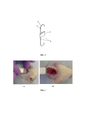
фиг. 1 – Схема инструмента для забора биоматериала у лабораторных животных для гистологических исследований: 1 – корпус в виде трубчатой скобы; 2 - цилиндрический хомут-бегунок; 3 - сменяемое лезвие с наружной и внутренней заточкой; 4 – прорезь для сменного лезвия; fig. 1 - Scheme of an instrument for taking biomaterial from laboratory animals for histological studies: 1 - a body in the form of a tubular bracket; 2 - cylindrical clamp-runner; 3 - replaceable blade with external and internal sharpening; 4 - slot for a replaceable blade;
фиг. 2а, б – фотоиллюстрация забора биоматериала у крысы линии Вистар;fig. 2a, b - photo illustration of biomaterial sampling from a Wistar rat;
фиг. 3а, б – фотоиллюстрация забора биоматериала у мыши линии DBA2.fig. 3a, b – photo illustration of biomaterial sampling from a DBA2 mouse.
Инструмент для забора биоматериала у лабораторных животных для гистологических исследований изготовлен из гибкого медицинского металла. Корпус 1 представляет собой трубчатую скобу, на которую надет подвижный цилиндрический хомут-бегунок 2, причем в верхней части корпуса 1 имеется прорезь 4 для съемного лезвия 3 с наружной и внутренней заточкой 0,5 см. Верхнее положение хомута 2 способствует раскрытию лезвия 3, а его нижнее положение приводит к смыканию краев лезвия 3 в виде петли. The tool for taking biomaterial from laboratory animals for histological studies is made of flexible medical metal. The
Инструмент для забора биоматериала у лабораторных животных для гистологических исследований используют следующим образом.A tool for taking biomaterial from laboratory animals for histological studies is used as follows.
Инструмент берут за нижнюю часть корпуса 1, при этом подвижный цилиндрический хомут-бегунок 2 опускают вниз по корпусу 1 так, чтобы лезвие 3 в виде петли расширилось. Лезвие 3 плотно устанавливают на участок тела животного и надавливают – происходит прорезывание ткани, лезвие сначала входит в кожу, а затем проникает в ткань на заданную глубину. Резким движением петлю лезвия 3 протягивают в горизонтальной поверхности и резко поднимают вверх - лезвие 3 в виде петли смыкается, отрезая или срезая ткань требуемого объема, после чего хомут-бегунок 2 на корпусе 1 переводят в нижнее положение. The tool is taken by the lower part of the
Пример 1 Example 1
У крысы линии Вистар – самец, вес 239,7 г через месяц после вживления имплантата, напечатанного на 3D принтере, проводили забор биоматериала. Операционное поле крысы тщательно освобождали от шерсти: сначала состригали ножницами, затем на 3 мин наносили депилятор и тщательно обрабатывали 75%-ным спиртом. Инструмент, с настроенным, с помощью бегунка, лезвием высотой 0,5 см, плотно прижимали к выбранному участку тела животного и после нажима (погружения лезвия в ткань) проводили сначала в горизонтальной плоскости, а затем переводили вертикально вверх. Отделенный кусочек ткани размером 1х1 см помещали в раствор Буэна для гистологических исследований на биосовместимость вшитого имплантата (Фин.2 а, б).In a Wistar rat, a male, weighing 239.7 g, a biomaterial was taken one month after implantation of an implant printed on a 3D printer. The operating field of the rat was carefully freed from hair: first, it was cut with scissors, then a depilator was applied for 3 minutes and carefully treated with 75% alcohol. The tool, with a blade 0.5 cm high adjusted with the help of a slider, was pressed tightly against the selected area of the animal's body and after pressing (immersing the blade into the tissue), it was first passed in a horizontal plane, and then transferred vertically upwards. A separated piece of tissue 1x1 cm in size was placed in Bouin's solution for histological studies on the biocompatibility of the sutured implant (Find. 2 a, b).
Пример 2 Example 2
У мыши линии DBA2, весом 19,6 г с перевитым в верхнюю часть бедра асцитом лейкоза Р388 (3 ген) на 7 сутки проводили забор опухоли. Лезвие настраивали исходя из объема паталогического узла. Животное усыпляли эфиром. Тщательно обрабатывали, аналогичным образом, всю поверхность лапки. Инструмент накладывали таким образом, чтобы не повредить узел, который необходимо удалять полностью вместе с окружающей здоровой тканью. Нажим делали с упором на бегунок-хомут. Лезвие погружали на 0,5 см (ширина лезвия), проводили под опухолью, и затем, резким движением перемещали инструмент вертикально вверх (Фиг.3 а, б). Опухоль вместе с окружающей тканью помещали в раствор Буэна для гистологических исследований по оценке эффективности используемого противоопухолевого препарата.In a mouse of the DBA2 line, weighing 19.6 g, with leukemia P388 (3 gene) ascites transplanted into the upper part of the thigh, a tumor was taken on the 7th day. The blade was adjusted based on the volume of the pathological node. The animal was euthanized with ether. Carefully processed, in a similar way, the entire surface of the foot. The instrument was applied in such a way as not to damage the node, which must be removed completely along with the surrounding healthy tissue. The pressure was done with an emphasis on the clamp slider. The blade was immersed by 0.5 cm (blade width), passed under the tumor, and then, with a sharp movement, the instrument was moved vertically upwards (Fig.3 a, b). The tumor, together with the surrounding tissue, was placed in Bouin's solution for histological studies to evaluate the effectiveness of the antitumor drug used.
Предложенный инструмент позволяет срезать (меняя лезвия) требуемый объем для гистологических исследований с оптимальной площадью для полноценной фиксации и предотвращения развития аутолитических изменений. Инструмент промышленно применим, несложен в изготовлении и не требует больших затрат.The proposed tool allows cutting (changing blades) the required volume for histological studies with the optimal area for complete fixation and preventing the development of autolytic changes. The tool is industrially applicable, easy to manufacture and does not require large expenditures.
Claims (1)
Publications (1)
| Publication Number | Publication Date |
|---|---|
| RU215196U1 true RU215196U1 (en) | 2022-12-02 |
Family
ID=
Citations (4)
| Publication number | Priority date | Publication date | Assignee | Title |
|---|---|---|---|---|
| SU1731186A1 (en) * | 1989-07-13 | 1992-05-07 | Донецкий медицинский институт им.М.Горького | Surgical forceps-tracheotome |
| SU1801391A1 (en) * | 1990-08-09 | 1993-03-15 | Arkhangelskij G Med I | Device for biopsy analysis |
| RU2183440C2 (en) * | 2000-09-07 | 2002-06-20 | Белгородская государственная сельскохозяйственная академия | Equipment to conduct skin sampling in animals |
| RU49705U1 (en) * | 2005-08-23 | 2005-12-10 | ФГОУ ВПО "Белгородская государственная сельскохозяйственная академия" | DEVICE FOR PUNCH BIOPSY |
Patent Citations (4)
| Publication number | Priority date | Publication date | Assignee | Title |
|---|---|---|---|---|
| SU1731186A1 (en) * | 1989-07-13 | 1992-05-07 | Донецкий медицинский институт им.М.Горького | Surgical forceps-tracheotome |
| SU1801391A1 (en) * | 1990-08-09 | 1993-03-15 | Arkhangelskij G Med I | Device for biopsy analysis |
| RU2183440C2 (en) * | 2000-09-07 | 2002-06-20 | Белгородская государственная сельскохозяйственная академия | Equipment to conduct skin sampling in animals |
| RU49705U1 (en) * | 2005-08-23 | 2005-12-10 | ФГОУ ВПО "Белгородская государственная сельскохозяйственная академия" | DEVICE FOR PUNCH BIOPSY |
Similar Documents
| Publication | Publication Date | Title |
|---|---|---|
| US5397333A (en) | Surgical hook knife | |
| US6740102B2 (en) | Vessel harvesting retractor with bilateral electrosurgical ligation | |
| KR20180102107A (en) | Minimally invasive tissue sampling device | |
| US5989273A (en) | Apparatus for producing hair transplantation donor strips and methods | |
| JPH11507271A (en) | Bone shaving equipment | |
| US3522809A (en) | Surgical instrument | |
| JP2009528877A (en) | Tumor treatment apparatus and method | |
| SU613754A1 (en) | Surgery apparatus for tissue suturing | |
| RU215196U1 (en) | INSTRUMENT FOR SAMPLING BIOMATERIAL FROM LABORATORY ANIMALS FOR HISTOLOGICAL STUDIES | |
| Buck | Ancient technology in contemporary surgery | |
| AU2003278868B2 (en) | Pathology grossing tool | |
| US7806907B2 (en) | Skin lesion exciser and skin-closure device therefor | |
| CN113645908A (en) | Device for skin biopsy | |
| CN107184250B (en) | Adjustable scalpel and medical surgical equipment | |
| WO2014136119A1 (en) | Implantating device | |
| EP1276420A1 (en) | Biopsy device | |
| US11083445B2 (en) | Knife and retractor system | |
| WO2023059279A1 (en) | A self bladed punch type tissue section apparatus | |
| Fan et al. | Minigraft preparation in surgical hair replacement | |
| CN212592475U (en) | Human body fixing device for surgical operation | |
| CN216899835U (en) | Breast cancer postoperative pathology sample cutting preprocessing device | |
| KR101693495B1 (en) | Device for the ablation of biological tissues | |
| RU2234261C1 (en) | Method for sampling split cutaneous autotransplant | |
| CN219126809U (en) | Dissecting tool for veterinarian | |
| JPS6129733A (en) | Sampler for vital tissue |