RU2028110C1 - Apparatus for treatment diaphysial fractures of long tubular bones - Google Patents
Apparatus for treatment diaphysial fractures of long tubular bones Download PDFInfo
- Publication number
- RU2028110C1 RU2028110C1 SU4939071A RU2028110C1 RU 2028110 C1 RU2028110 C1 RU 2028110C1 SU 4939071 A SU4939071 A SU 4939071A RU 2028110 C1 RU2028110 C1 RU 2028110C1
- Authority
- RU
- Russia
- Prior art keywords
- fractures
- brackets
- treatment
- long tubular
- tubular bones
- Prior art date
Links
Images

Abstract
Description
Изобретение относится к травматологии и ортопедии и может быть использовано при оперативном лечении переломов трубчатых костей. The invention relates to traumatology and orthopedics and can be used in surgical treatment of fractures of tubular bones.
Известно устройство для закрытого компрессионно-дистракционного остеосинтеза диафизарных переломов [1], содержащее рамы, штанги, регулирующие винты, спицы с упорами, спицедержатели, включающие болт, натяжной винт и гайку. A device for closed compression-distraction osteosynthesis of diaphyseal fractures [1], containing frames, rods, adjusting screws, spokes with stops, spoke holders, including a bolt, tension screw and nut.
Устройство содержит также внутренние репонирующие корпуса и наружные репонирующие корпуса, попарно соединенные между собой рамами с регулирующими винтами и подвижно расположенные на штангах шкалы. The device also contains internal repository cases and external repository cases, pairwise interconnected by frames with adjusting screws and movably located on the bars of the scale.
Недостатки описанного устройства:
относительная сложность конструкции;
относительно высокая травматичность применения, связанная с чрескостным проведением многочисленных спиц.The disadvantages of the described device:
relative design complexity;
relatively high invasiveness associated with transosseous holding of numerous spokes.
Известен также аппарат для репозиции и фиксации костных фрагментов [2], содержащий две опоры в виде скоб с резьбовыми спицами с болтами, шайбами и штангу. Узел репозиции выполнен в виде кронштейна, соединенного посредством планки с ползуном, в котором установлен резьбовой стержень. На кронштейне, планке и ползуне выполнены шкалы. Also known apparatus for reposition and fixation of bone fragments [2], containing two supports in the form of brackets with threaded spokes with bolts, washers and a rod. The reposition unit is made in the form of a bracket connected by means of a bar to a slider in which a threaded rod is installed. On the bracket, strap and slider are made scales.
Недостатки этого устройства-аналога следующие:
относительная сложность конструкции;
относительно высокая травматичность.The disadvantages of this analog device are as follows:
relative design complexity;
relatively high invasiveness.
Наиболее близким по технической сущности и достигаемому результату является устройство для остеосинтеза трубчатых костей [3], принятое в качестве прототипа. Это устройство состоит из двух полуколец, которые охватывают кость больше половины ее периметра в поперечном сечении. Полукольца фиксируются коническим штифтом. The closest in technical essence and the achieved result is a device for osteosynthesis of tubular bones [3], adopted as a prototype. This device consists of two half rings that span a bone more than half of its perimeter in cross section. The half rings are fixed with a conical pin.
С целью обеспечения самоторможения при заклинивании конический штифт выполнен с углом наклона 7о. Одной стороной штифт ложится на кость, а вторая сторона штифта входит в контакт с наклонными под углом 7о поверхностями полуколец. Концы полуколец выполнены заостренными, с возможностью отделения мягких тканей от кости. На внутренней поверхности полуколец выполнены пазы в виде поперечной насечки, которые позволяют уменьшить площадь контакта полуколец с костью и увеличить удельную нагрузку на кость в местах контакта, что в конечном итоге приводит к лучшему охвату кости и улучшенному креплению последней. Продольная насечка на внутренней стороне полуколец позволяет лучше фиксировать кость. Полукольца-бранши выполнены с изгибами на конце, образующими шарнир, в которых установлен конический штифт.In order to ensure the self-locking tapered pin if jamming is configured with the inclination angle of 7. On one side, the pin rests on the bone, and the second side of the pin comes in contact with the surfaces of the half-rings inclined at an angle of 7 °. The ends of the semirings are made pointed, with the possibility of separation of soft tissues from the bone. On the inner surface of the semirings, grooves are made in the form of a transverse notch, which allows to reduce the contact area of the semicircles with the bone and increase the specific load on the bone at the contact points, which ultimately leads to better bone coverage and improved anchoring of the bone. A longitudinal notch on the inside of the half rings allows better fixation of the bone. The half-branches are made with bends at the end, forming a hinge in which a conical pin is installed.
Недостатки устройства-прототипа:
недостаточно широкие функциональные возможности, связанные с отсутствием компрессирующего усилия по оси отломков и невозможностью лечения поперечных переломов;
недостаточная надежность работы конического штифта во все время лечения (возможен частичный выход его в сторону большего диаметра из-за наличия случайных нагрузок);
то обстоятельство, что одной стороной штифт ложится непосредственно на кость (надкостницу), а с другой стороной взаимодействует с металлическими изгибами, бранш создает, во-первых, неопределенность в отношении угла самоторможения (конический штифт-бранша - один коэффициент трения, а штифт-кость - другой), во-вторых, происходит омертвение надкостницы под штифтом в процессе лечения;
некоторое неудобство в применении (необходимость распатора, специальных насечек для него).The disadvantages of the prototype device:
insufficiently wide functionality associated with the lack of compressive force along the axis of the fragments and the inability to treat transverse fractures;
insufficient reliability of the operation of the conical pin during the entire treatment (it may partially exit towards a larger diameter due to the presence of random loads);
the fact that on one side the pin rests directly on the bone (periosteum) and interacts with metal bends on the other side, the jaw creates, firstly, uncertainty regarding the angle of self-braking (conical pin-jaw - one coefficient of friction, and the pin-bone - other), secondly, the periosteum under the pin is dead in the process of treatment;
some inconvenience in use (the need for a raspator, special notches for it).
Цель изобретения - обеспечение возможности лечения поперечных переломов путем компрессии отломков. The purpose of the invention is the provision of the possibility of treating transverse fractures by compression of fragments.
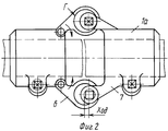
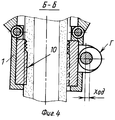
На фиг.1 изображен общий вид устройства (вид спереди) с разрезом по оси эксцентрика компрессирующего узла; на фиг.2 - то же, вид сверху; на фиг.3 - поперечный разрез скобы, оснащенной вкладышами и эксцентриком, разрез А-А на фиг.1; на фиг.4 - разрез по осям вкладышей (разрез Б-Б на фиг.3). Figure 1 shows a General view of the device (front view) with a cut along the axis of the eccentric of the compression unit; figure 2 is the same, a top view; figure 3 is a transverse section of a bracket equipped with liners and an eccentric, section AA in figure 1; figure 4 is a section along the axes of the liners (section BB in figure 3).
Устройство для лечения диафизарных переломов длинных трубчатых костей состоит из двух металлических скоб 1 и 1а, снабженных прижимными эксцентриками 2 и 2а, имеет вкладыш подвижный 3 с острыми шипами или заменяющей их функционально резьбой и вкладыш неподвижный 4 с шипами. A device for treating diaphyseal fractures of long tubular bones consists of two
Эксцентрик 5, непосредственно контактирует поверхностью "Г" (фиг.4) своего кулачка с вкладышем 3. Таких эксцентриков два - левый и правый. Скобы 1 и 1а связаны между собой компрессирующим узлом (фиг.1), состоящим из двух пар ушек 6 и 7, (фиг.2), причем ушки 6 шарнирно прикреплены к скобе 1, а ушки 7 жестко укреплены на скобе 1а. Все эксцентрики 5 имеют квадратные хвостовики "под ключ" 8, 8а, 8б. При осевом совмещении ушек 6 и 7 в них вставлен эксцентрик 5. Всего эксцентриков 5 в устройстве 4 шт. The eccentric 5, directly contacts the surface "G" (figure 4) of his cam with the liner 3. There are two such eccentrics - left and right. The
Рабочие поверхности кулачков эксцентриков обозначены буквой "Г" (фиг. 2 и 4). Возможный ход эксцентриков виден на фиг.2 и 4. The working surfaces of the cam of the eccentrics are indicated by the letter "G" (Fig. 2 and 4). A possible stroke of the eccentrics is visible in FIGS. 2 and 4.
Рабочая поверхность "Г" кулачков 9 эксцентриков выполнена с двумя различными углами подъема по мере хода кулачка - вначале большой угол подъема, затем - значительно меньший, работающий только на зажим. Острые шипы 10 показаны на внутренних поверхностях вкладышей 3 и 4. The working surface "G" of the
Устройство для лечения переломов костей применяется следующим образом. A device for treating bone fractures is used as follows.
На концы предварительно отрепонированных костных отломков (дистального и проксимального) поочередно надевают скобы 1 и 1а. Предварительно вкладыши 3 с кулачками 9 находятся в незатянутом состоянии (фиг.3), т.е. в крайнем правом. At the ends of previously repaired bone fragments (distal and proximal),
Установив левую (фиг.1) скобу 1, имеющую на ушке 6 отверстие, устанавливают правую скобу 1а так, чтобы эксцентрик 8 вошел своим кулачком 9 в отверстие ушка 6. Поворотом хвостовика 8 и 8а эксцентрика 2 и 2а закрепляют левую и правую скобы на концах костных отломков. При этом острые шипы 10 вкладышей 3 и 4 впиваются в надкостницу и достигают кортикального слоя кости, осуществляя жесткое крепление скоб на концах отломков. Незначительное (десятые доли мм) пружинение скоб держит эти зажимы в напряженном состоянии, препятствуя самопроизвольному саморассоединению кулачковых зажимов. Этому же служит и выполнение условия самоторможения для стальных трущихся пар (угол подъема конца хода кулачка ≅5о).Having installed the left bracket (Fig. 1) with an opening on the
Затем стягивают отломки с помощью компрессирующего узла, создавая постоянную осевую компрессию поворотом хвостовика 8б. Then the fragments are pulled together with the help of a compression unit, creating constant axial compression by turning the shank 8b.
Размеры устройства (в первую очередь скоб и вкладышей) выбирают по размеру костей больного (т.е. с учетом антропологических данных). The dimensions of the device (primarily brackets and inserts) are selected according to the size of the patient’s bones (i.e., taking into account anthropological data).
Таким образом, при применении заявляемого устройства для лечения диафизарных переломов длинных трубчатых костей расширены функциональные возможности, позволяющие лечить не только косые переломы, но и поперечные. Увеличена надежность соединения деталей в компрессирующем узле благодаря определенности коэффициентов трения металл-металл. Повышается удобство в применении - появляется возможность одномоментного зажима отломков, а затем одномоментного наложения компрессирующего усилия с фиксацией этого усилия. Thus, when using the inventive device for the treatment of diaphyseal fractures of long tubular bones, the functionality has been expanded to treat not only oblique fractures, but also transverse ones. The reliability of the connection of parts in the compression unit has been increased due to the certainty of the metal-metal friction coefficients. Ease of use is improved - it becomes possible to clamp fragments simultaneously, and then simultaneously apply a compressive force with fixation of this force.
Уменьшается травматичность устройства (отсутствует клин, непосредственно наложенный на надкостницу на все время лечения). Все перечисленные факторы - составляющие положительного эффекта от применения заявляемого устройства - позволяют повысить стабильность остеосинтеза при любых видах диафизарных переломов длинных трубчатых костей. The trauma of the device is reduced (there is no wedge directly placed on the periosteum for the entire duration of treatment). All of these factors - the components of the positive effect of the use of the claimed device - can improve the stability of osteosynthesis in all types of diaphyseal fractures of long tubular bones.
Claims (1)
Priority Applications (1)
| Application Number | Priority Date | Filing Date | Title |
|---|---|---|---|
| SU4939071 RU2028110C1 (en) | 1991-05-22 | 1991-05-22 | Apparatus for treatment diaphysial fractures of long tubular bones |
Applications Claiming Priority (1)
| Application Number | Priority Date | Filing Date | Title |
|---|---|---|---|
| SU4939071 RU2028110C1 (en) | 1991-05-22 | 1991-05-22 | Apparatus for treatment diaphysial fractures of long tubular bones |
Publications (1)
| Publication Number | Publication Date |
|---|---|
| RU2028110C1 true RU2028110C1 (en) | 1995-02-09 |
Family
ID=21575996
Family Applications (1)
| Application Number | Title | Priority Date | Filing Date |
|---|---|---|---|
| SU4939071 RU2028110C1 (en) | 1991-05-22 | 1991-05-22 | Apparatus for treatment diaphysial fractures of long tubular bones |
Country Status (1)
| Country | Link |
|---|---|
| RU (1) | RU2028110C1 (en) |
Cited By (1)
| Publication number | Priority date | Publication date | Assignee | Title |
|---|---|---|---|---|
| RU2638442C2 (en) * | 2016-05-31 | 2017-12-13 | Государственное бюджетное образовательное учреждение высшего профессионального образования "Рязанский государственный медицинский университет имени академика И.П. Павлова" Министерства здравоохранения Российской Федерации | Device for extra-cortical osteosynthesis of tubular bones in experiment |
-
1991
- 1991-05-22 RU SU4939071 patent/RU2028110C1/en active
Non-Patent Citations (3)
| Title |
|---|
| 1. Авторское свидетельство СССР N 434938, кл. A 61B 17/58, 1974. * |
| 2. Авторское свидетельство СССР N 1377078, кл. A 61B 17/58, 1988. * |
| 3. Авторское свидетельство СССР N 858802, кл. A 61B 17/58, 1981. * |
Cited By (1)
| Publication number | Priority date | Publication date | Assignee | Title |
|---|---|---|---|---|
| RU2638442C2 (en) * | 2016-05-31 | 2017-12-13 | Государственное бюджетное образовательное учреждение высшего профессионального образования "Рязанский государственный медицинский университет имени академика И.П. Павлова" Министерства здравоохранения Российской Федерации | Device for extra-cortical osteosynthesis of tubular bones in experiment |
Similar Documents
| Publication | Publication Date | Title |
|---|---|---|
| US5707372A (en) | Multiple node variable length cross-link device | |
| DE60007758T2 (en) | BONE PLATE DEVICE | |
| JP4318421B2 (en) | Multiaxial bone screw assembly | |
| CA2587373C (en) | Endosteal nail | |
| US5032125A (en) | Intramedullary hip screw | |
| US6083224A (en) | Dynamic spinal screw-rod connectors | |
| DE60034528T2 (en) | Front and transpedicular fixation system for attaching the spine | |
| CA2360675C (en) | Surgical fastener assembly | |
| JP3949725B2 (en) | Surgical fastener assembly | |
| US6712820B2 (en) | Fixation plate system for dorsal wrist fracture fixation | |
| US6508819B1 (en) | Method of dorsal wrist fracture fixation | |
| US5275599A (en) | Biocompression external fixator for osteosynthesis | |
| US5312403A (en) | External fixation device | |
| US20010037111A1 (en) | Method and apparatus for dynamized spinal stabilization | |
| KR20000016381A (en) | Stretchable clamp | |
| JPH07222753A (en) | Intramedullary nail | |
| EA005153B1 (en) | Internal fixator for bones | |
| DE2834891B2 (en) | Fixator for fixing bones or bone fragments, especially vertebrae | |
| EP1211991B1 (en) | Repositioning device for bone fragments | |
| RU2028110C1 (en) | Apparatus for treatment diaphysial fractures of long tubular bones | |
| CA2042573C (en) | Pinless external fixation device | |
| CN112057154B (en) | Femoral internal fixation system | |
| EP0809472B1 (en) | Intramedullary cavity nail for femur elongation | |
| DE4226790C1 (en) | Osteo-synthetic stirrup clasp for fixing bone fragments - comprises fixation stirrup, fixation pin and connecting component together with guide socket | |
| RU2133594C1 (en) | Device for epiosteous osteosynthesis |