KR20230017822A - Methods for making extracellular vesicles and their uses - Google Patents
Methods for making extracellular vesicles and their uses Download PDFInfo
- Publication number
- KR20230017822A KR20230017822A KR1020227045395A KR20227045395A KR20230017822A KR 20230017822 A KR20230017822 A KR 20230017822A KR 1020227045395 A KR1020227045395 A KR 1020227045395A KR 20227045395 A KR20227045395 A KR 20227045395A KR 20230017822 A KR20230017822 A KR 20230017822A
- Authority
- KR
- South Korea
- Prior art keywords
- evs
- antibody
- protein
- cells
- membrane
- Prior art date
Links
- 238000000034 method Methods 0.000 title claims abstract description 258
- 239000000427 antigen Substances 0.000 claims abstract description 175
- 102000036639 antigens Human genes 0.000 claims abstract description 175
- 108091007433 antigens Proteins 0.000 claims abstract description 175
- 239000012528 membrane Substances 0.000 claims abstract description 93
- 210000004027 cell Anatomy 0.000 claims description 400
- 108090000623 proteins and genes Proteins 0.000 claims description 241
- 102000004169 proteins and genes Human genes 0.000 claims description 237
- 108010052285 Membrane Proteins Proteins 0.000 claims description 110
- 102000018697 Membrane Proteins Human genes 0.000 claims description 89
- 241001465754 Metazoa Species 0.000 claims description 84
- 230000001464 adherent effect Effects 0.000 claims description 63
- 238000004519 manufacturing process Methods 0.000 claims description 61
- 102000034257 ADP-Ribosylation Factor 6 Human genes 0.000 claims description 58
- 108090000067 ADP-Ribosylation Factor 6 Proteins 0.000 claims description 58
- 230000027455 binding Effects 0.000 claims description 49
- 102100026444 Arrestin domain-containing protein 1 Human genes 0.000 claims description 48
- 125000002252 acyl group Chemical group 0.000 claims description 48
- 230000003053 immunization Effects 0.000 claims description 44
- 102000040430 polynucleotide Human genes 0.000 claims description 41
- 108091033319 polynucleotide Proteins 0.000 claims description 41
- 239000002157 polynucleotide Substances 0.000 claims description 41
- 239000003153 chemical reaction reagent Substances 0.000 claims description 35
- 238000012258 culturing Methods 0.000 claims description 33
- 208000037265 diseases, disorders, signs and symptoms Diseases 0.000 claims description 33
- 239000003814 drug Substances 0.000 claims description 32
- 150000007523 nucleic acids Chemical class 0.000 claims description 30
- 230000014509 gene expression Effects 0.000 claims description 29
- 102000039446 nucleic acids Human genes 0.000 claims description 29
- 108020004707 nucleic acids Proteins 0.000 claims description 29
- 201000010099 disease Diseases 0.000 claims description 28
- 239000002671 adjuvant Substances 0.000 claims description 27
- 239000003550 marker Substances 0.000 claims description 27
- 239000008194 pharmaceutical composition Substances 0.000 claims description 25
- 238000000338 in vitro Methods 0.000 claims description 23
- 210000000628 antibody-producing cell Anatomy 0.000 claims description 22
- 210000004408 hybridoma Anatomy 0.000 claims description 21
- 229940124597 therapeutic agent Drugs 0.000 claims description 18
- 239000012634 fragment Substances 0.000 claims description 17
- 238000011282 treatment Methods 0.000 claims description 17
- 239000007787 solid Substances 0.000 claims description 16
- 102000035160 transmembrane proteins Human genes 0.000 claims description 16
- 108091005703 transmembrane proteins Proteins 0.000 claims description 16
- 238000005199 ultracentrifugation Methods 0.000 claims description 15
- 210000003719 b-lymphocyte Anatomy 0.000 claims description 14
- 101000785762 Homo sapiens Arrestin domain-containing protein 1 Proteins 0.000 claims description 12
- 210000002966 serum Anatomy 0.000 claims description 10
- 230000028993 immune response Effects 0.000 claims description 9
- 108010073443 Ribi adjuvant Proteins 0.000 claims description 8
- 239000003937 drug carrier Substances 0.000 claims description 8
- 108091006047 fluorescent proteins Proteins 0.000 claims description 5
- 102000034287 fluorescent proteins Human genes 0.000 claims description 5
- 210000002381 plasma Anatomy 0.000 claims description 4
- 210000002700 urine Anatomy 0.000 claims description 4
- 238000001943 fluorescence-activated cell sorting Methods 0.000 claims description 2
- 239000002054 inoculum Substances 0.000 claims 1
- 210000004020 intracellular membrane Anatomy 0.000 claims 1
- 239000000203 mixture Substances 0.000 abstract description 27
- 238000003556 assay Methods 0.000 abstract description 16
- 238000012286 ELISA Assay Methods 0.000 abstract description 9
- 238000008157 ELISA kit Methods 0.000 abstract description 6
- 235000018102 proteins Nutrition 0.000 description 209
- 210000004379 membrane Anatomy 0.000 description 76
- 239000002609 medium Substances 0.000 description 50
- 229940024606 amino acid Drugs 0.000 description 44
- 235000001014 amino acid Nutrition 0.000 description 44
- 150000001413 amino acids Chemical group 0.000 description 43
- 101710091379 Arrestin domain-containing protein 1 Proteins 0.000 description 42
- 241000699670 Mus sp. Species 0.000 description 36
- 238000002965 ELISA Methods 0.000 description 33
- 238000002649 immunization Methods 0.000 description 32
- 108020004414 DNA Proteins 0.000 description 30
- 102000053602 DNA Human genes 0.000 description 30
- 241000283973 Oryctolagus cuniculus Species 0.000 description 21
- 101710177291 Gag polyprotein Proteins 0.000 description 20
- 230000000670 limiting effect Effects 0.000 description 20
- 238000001262 western blot Methods 0.000 description 20
- 101710125418 Major capsid protein Proteins 0.000 description 19
- 241001529936 Murinae Species 0.000 description 17
- 238000000746 purification Methods 0.000 description 17
- 241000699666 Mus <mouse, genus> Species 0.000 description 16
- 239000000523 sample Substances 0.000 description 15
- 235000002198 Annona diversifolia Nutrition 0.000 description 13
- 241000700159 Rattus Species 0.000 description 13
- 230000015572 biosynthetic process Effects 0.000 description 13
- 210000000170 cell membrane Anatomy 0.000 description 11
- 238000001514 detection method Methods 0.000 description 11
- 102220491076 ADP-ribosylation factor 6_Q67L_mutation Human genes 0.000 description 10
- 101100356682 Caenorhabditis elegans rho-1 gene Proteins 0.000 description 10
- 101150111584 RHOA gene Proteins 0.000 description 10
- 239000013598 vector Substances 0.000 description 10
- 239000001963 growth medium Substances 0.000 description 9
- 108090000765 processed proteins & peptides Proteins 0.000 description 9
- -1 Acryl Chemical group 0.000 description 8
- 108020004635 Complementary DNA Proteins 0.000 description 8
- 241000282842 Lama glama Species 0.000 description 8
- 239000005090 green fluorescent protein Substances 0.000 description 8
- 230000003834 intracellular effect Effects 0.000 description 8
- 238000001556 precipitation Methods 0.000 description 8
- 102000005962 receptors Human genes 0.000 description 8
- 108020003175 receptors Proteins 0.000 description 8
- 238000012216 screening Methods 0.000 description 8
- 239000007790 solid phase Substances 0.000 description 8
- 102000004190 Enzymes Human genes 0.000 description 7
- 108090000790 Enzymes Proteins 0.000 description 7
- 108010085220 Multiprotein Complexes Proteins 0.000 description 7
- 102000007474 Multiprotein Complexes Human genes 0.000 description 7
- 206010035226 Plasma cell myeloma Diseases 0.000 description 7
- 239000002202 Polyethylene glycol Substances 0.000 description 7
- 125000000539 amino acid group Chemical group 0.000 description 7
- 210000004369 blood Anatomy 0.000 description 7
- 239000008280 blood Substances 0.000 description 7
- 239000003795 chemical substances by application Substances 0.000 description 7
- 229940088598 enzyme Drugs 0.000 description 7
- 239000012530 fluid Substances 0.000 description 7
- 210000002865 immune cell Anatomy 0.000 description 7
- 238000011081 inoculation Methods 0.000 description 7
- 201000000050 myeloid neoplasm Diseases 0.000 description 7
- 229920001223 polyethylene glycol Polymers 0.000 description 7
- 230000008569 process Effects 0.000 description 7
- 230000007115 recruitment Effects 0.000 description 7
- 238000001338 self-assembly Methods 0.000 description 7
- 108060003951 Immunoglobulin Proteins 0.000 description 6
- 241000283984 Rodentia Species 0.000 description 6
- 238000010804 cDNA synthesis Methods 0.000 description 6
- 239000002299 complementary DNA Substances 0.000 description 6
- 238000010586 diagram Methods 0.000 description 6
- 102000044270 human ARRDC1 Human genes 0.000 description 6
- 102000018358 immunoglobulin Human genes 0.000 description 6
- 102000004196 processed proteins & peptides Human genes 0.000 description 6
- 229920002477 rna polymer Polymers 0.000 description 6
- 108010001336 Horseradish Peroxidase Proteins 0.000 description 5
- 241000282838 Lama Species 0.000 description 5
- 206010028980 Neoplasm Diseases 0.000 description 5
- 239000006143 cell culture medium Substances 0.000 description 5
- 230000002596 correlated effect Effects 0.000 description 5
- 208000035475 disorder Diseases 0.000 description 5
- 210000001808 exosome Anatomy 0.000 description 5
- 238000001727 in vivo Methods 0.000 description 5
- 230000010354 integration Effects 0.000 description 5
- 150000002632 lipids Chemical class 0.000 description 5
- 108020004999 messenger RNA Proteins 0.000 description 5
- 125000003729 nucleotide group Chemical group 0.000 description 5
- 239000002245 particle Substances 0.000 description 5
- 241000894007 species Species 0.000 description 5
- 239000000126 substance Substances 0.000 description 5
- 108010091358 Hypoxanthine Phosphoribosyltransferase Proteins 0.000 description 4
- 108010021625 Immunoglobulin Fragments Proteins 0.000 description 4
- 102000008394 Immunoglobulin Fragments Human genes 0.000 description 4
- 108090000862 Ion Channels Proteins 0.000 description 4
- 102000004310 Ion Channels Human genes 0.000 description 4
- 108090000856 Lyases Proteins 0.000 description 4
- 102000004317 Lyases Human genes 0.000 description 4
- 108091007491 NSP3 Papain-like protease domains Proteins 0.000 description 4
- 241000700605 Viruses Species 0.000 description 4
- 239000012472 biological sample Substances 0.000 description 4
- 239000000872 buffer Substances 0.000 description 4
- 238000013461 design Methods 0.000 description 4
- 238000003745 diagnosis Methods 0.000 description 4
- 238000002296 dynamic light scattering Methods 0.000 description 4
- 210000005260 human cell Anatomy 0.000 description 4
- 238000011534 incubation Methods 0.000 description 4
- 230000003993 interaction Effects 0.000 description 4
- 239000003094 microcapsule Substances 0.000 description 4
- 239000002773 nucleotide Substances 0.000 description 4
- 229920001184 polypeptide Polymers 0.000 description 4
- 238000002360 preparation method Methods 0.000 description 4
- 230000002285 radioactive effect Effects 0.000 description 4
- 150000003839 salts Chemical class 0.000 description 4
- 238000010186 staining Methods 0.000 description 4
- 238000002560 therapeutic procedure Methods 0.000 description 4
- XLYOFNOQVPJJNP-UHFFFAOYSA-N water Substances O XLYOFNOQVPJJNP-UHFFFAOYSA-N 0.000 description 4
- 239000012130 whole-cell lysate Substances 0.000 description 4
- 102100031126 6-phosphogluconolactonase Human genes 0.000 description 3
- 108010029731 6-phosphogluconolactonase Proteins 0.000 description 3
- 206010003445 Ascites Diseases 0.000 description 3
- WVDDGKGOMKODPV-UHFFFAOYSA-N Benzyl alcohol Chemical compound OCC1=CC=CC=C1 WVDDGKGOMKODPV-UHFFFAOYSA-N 0.000 description 3
- BWGNESOTFCXPMA-UHFFFAOYSA-N Dihydrogen disulfide Chemical compound SS BWGNESOTFCXPMA-UHFFFAOYSA-N 0.000 description 3
- 108010018962 Glucosephosphate Dehydrogenase Proteins 0.000 description 3
- 241000713772 Human immunodeficiency virus 1 Species 0.000 description 3
- 102100029098 Hypoxanthine-guanine phosphoribosyltransferase Human genes 0.000 description 3
- 102000001845 Lipid-Linked Proteins Human genes 0.000 description 3
- 241000124008 Mammalia Species 0.000 description 3
- 241000714177 Murine leukemia virus Species 0.000 description 3
- 102000004316 Oxidoreductases Human genes 0.000 description 3
- 108090000854 Oxidoreductases Proteins 0.000 description 3
- 108700030796 Tsg101 Proteins 0.000 description 3
- 239000004480 active ingredient Substances 0.000 description 3
- 230000008901 benefit Effects 0.000 description 3
- 210000000234 capsid Anatomy 0.000 description 3
- 150000001720 carbohydrates Chemical class 0.000 description 3
- 235000014633 carbohydrates Nutrition 0.000 description 3
- 238000004113 cell culture Methods 0.000 description 3
- 238000006243 chemical reaction Methods 0.000 description 3
- 230000000875 corresponding effect Effects 0.000 description 3
- 238000011161 development Methods 0.000 description 3
- 230000018109 developmental process Effects 0.000 description 3
- 238000010790 dilution Methods 0.000 description 3
- 239000012895 dilution Substances 0.000 description 3
- 229940079593 drug Drugs 0.000 description 3
- 230000000694 effects Effects 0.000 description 3
- 230000006870 function Effects 0.000 description 3
- 230000012010 growth Effects 0.000 description 3
- 238000003018 immunoassay Methods 0.000 description 3
- 238000001802 infusion Methods 0.000 description 3
- 238000002955 isolation Methods 0.000 description 3
- 239000000463 material Substances 0.000 description 3
- 239000011159 matrix material Substances 0.000 description 3
- 230000010534 mechanism of action Effects 0.000 description 3
- 230000035772 mutation Effects 0.000 description 3
- 230000002265 prevention Effects 0.000 description 3
- 239000000047 product Substances 0.000 description 3
- PYWVYCXTNDRMGF-UHFFFAOYSA-N rhodamine B Chemical compound [Cl-].C=12C=CC(=[N+](CC)CC)C=C2OC2=CC(N(CC)CC)=CC=C2C=1C1=CC=CC=C1C(O)=O PYWVYCXTNDRMGF-UHFFFAOYSA-N 0.000 description 3
- 239000000243 solution Substances 0.000 description 3
- 235000000346 sugar Nutrition 0.000 description 3
- 238000004114 suspension culture Methods 0.000 description 3
- 230000008685 targeting Effects 0.000 description 3
- 238000012360 testing method Methods 0.000 description 3
- 230000001225 therapeutic effect Effects 0.000 description 3
- 210000001519 tissue Anatomy 0.000 description 3
- 238000001890 transfection Methods 0.000 description 3
- 210000004881 tumor cell Anatomy 0.000 description 3
- HDTRYLNUVZCQOY-UHFFFAOYSA-N α-D-glucopyranosyl-α-D-glucopyranoside Natural products OC1C(O)C(O)C(CO)OC1OC1C(O)C(O)C(O)C(CO)O1 HDTRYLNUVZCQOY-UHFFFAOYSA-N 0.000 description 2
- YBJHBAHKTGYVGT-ZKWXMUAHSA-N (+)-Biotin Chemical compound N1C(=O)N[C@@H]2[C@H](CCCCC(=O)O)SC[C@@H]21 YBJHBAHKTGYVGT-ZKWXMUAHSA-N 0.000 description 2
- CJIJXIFQYOPWTF-UHFFFAOYSA-N 7-hydroxycoumarin Natural products O1C(=O)C=CC2=CC(O)=CC=C21 CJIJXIFQYOPWTF-UHFFFAOYSA-N 0.000 description 2
- 108010042708 Acetylmuramyl-Alanyl-Isoglutamine Proteins 0.000 description 2
- CIWBSHSKHKDKBQ-JLAZNSOCSA-N Ascorbic acid Chemical compound OC[C@H](O)[C@H]1OC(=O)C(O)=C1O CIWBSHSKHKDKBQ-JLAZNSOCSA-N 0.000 description 2
- 108090001008 Avidin Proteins 0.000 description 2
- 241000283690 Bos taurus Species 0.000 description 2
- 241000283707 Capra Species 0.000 description 2
- 241000700198 Cavia Species 0.000 description 2
- 241000282693 Cercopithecidae Species 0.000 description 2
- 241000699800 Cricetinae Species 0.000 description 2
- 108090000331 Firefly luciferases Proteins 0.000 description 2
- 108010015133 Galactose oxidase Proteins 0.000 description 2
- 108010010803 Gelatin Proteins 0.000 description 2
- 108010073178 Glucan 1,4-alpha-Glucosidase Proteins 0.000 description 2
- 102100022624 Glucoamylase Human genes 0.000 description 2
- WQZGKKKJIJFFOK-GASJEMHNSA-N Glucose Natural products OC[C@H]1OC(O)[C@H](O)[C@@H](O)[C@@H]1O WQZGKKKJIJFFOK-GASJEMHNSA-N 0.000 description 2
- 108010015776 Glucose oxidase Proteins 0.000 description 2
- 239000004366 Glucose oxidase Substances 0.000 description 2
- SXRSQZLOMIGNAQ-UHFFFAOYSA-N Glutaraldehyde Chemical compound O=CCCCC=O SXRSQZLOMIGNAQ-UHFFFAOYSA-N 0.000 description 2
- DHMQDGOQFOQNFH-UHFFFAOYSA-N Glycine Chemical compound NCC(O)=O DHMQDGOQFOQNFH-UHFFFAOYSA-N 0.000 description 2
- 102000003886 Glycoproteins Human genes 0.000 description 2
- 108090000288 Glycoproteins Proteins 0.000 description 2
- 101000669917 Homo sapiens Rho-associated protein kinase 1 Proteins 0.000 description 2
- MHAJPDPJQMAIIY-UHFFFAOYSA-N Hydrogen peroxide Chemical compound OO MHAJPDPJQMAIIY-UHFFFAOYSA-N 0.000 description 2
- HNDVDQJCIGZPNO-YFKPBYRVSA-N L-histidine Chemical compound OC(=O)[C@@H](N)CC1=CN=CN1 HNDVDQJCIGZPNO-YFKPBYRVSA-N 0.000 description 2
- 108060001084 Luciferase Proteins 0.000 description 2
- 102000016943 Muramidase Human genes 0.000 description 2
- 108010014251 Muramidase Proteins 0.000 description 2
- 241000187479 Mycobacterium tuberculosis Species 0.000 description 2
- 108010062010 N-Acetylmuramoyl-L-alanine Amidase Proteins 0.000 description 2
- 108700020796 Oncogene Proteins 0.000 description 2
- 241001494479 Pecora Species 0.000 description 2
- ISWSIDIOOBJBQZ-UHFFFAOYSA-N Phenol Natural products OC1=CC=CC=C1 ISWSIDIOOBJBQZ-UHFFFAOYSA-N 0.000 description 2
- 108010007100 Pulmonary Surfactant-Associated Protein A Proteins 0.000 description 2
- 102100027773 Pulmonary surfactant-associated protein A2 Human genes 0.000 description 2
- 102000007056 Recombinant Fusion Proteins Human genes 0.000 description 2
- 108010008281 Recombinant Fusion Proteins Proteins 0.000 description 2
- 102100039313 Rho-associated protein kinase 1 Human genes 0.000 description 2
- 108010052164 Sodium Channels Proteins 0.000 description 2
- 102000018674 Sodium Channels Human genes 0.000 description 2
- 210000001744 T-lymphocyte Anatomy 0.000 description 2
- IQFYYKKMVGJFEH-XLPZGREQSA-N Thymidine Chemical compound O=C1NC(=O)C(C)=CN1[C@@H]1O[C@H](CO)[C@@H](O)C1 IQFYYKKMVGJFEH-XLPZGREQSA-N 0.000 description 2
- HDTRYLNUVZCQOY-WSWWMNSNSA-N Trehalose Natural products O[C@@H]1[C@@H](O)[C@@H](O)[C@@H](CO)O[C@@H]1O[C@@H]1[C@H](O)[C@@H](O)[C@@H](O)[C@@H](CO)O1 HDTRYLNUVZCQOY-WSWWMNSNSA-N 0.000 description 2
- 101150072717 Tsg101 gene Proteins 0.000 description 2
- ISAKRJDGNUQOIC-UHFFFAOYSA-N Uracil Chemical compound O=C1C=CNC(=O)N1 ISAKRJDGNUQOIC-UHFFFAOYSA-N 0.000 description 2
- 108010093894 Xanthine oxidase Proteins 0.000 description 2
- 102100033220 Xanthine oxidase Human genes 0.000 description 2
- 230000002776 aggregation Effects 0.000 description 2
- 238000004220 aggregation Methods 0.000 description 2
- HDTRYLNUVZCQOY-LIZSDCNHSA-N alpha,alpha-trehalose Chemical compound O[C@@H]1[C@@H](O)[C@H](O)[C@@H](CO)O[C@@H]1O[C@@H]1[C@H](O)[C@@H](O)[C@H](O)[C@@H](CO)O1 HDTRYLNUVZCQOY-LIZSDCNHSA-N 0.000 description 2
- AZDRQVAHHNSJOQ-UHFFFAOYSA-N alumane Chemical class [AlH3] AZDRQVAHHNSJOQ-UHFFFAOYSA-N 0.000 description 2
- WNROFYMDJYEPJX-UHFFFAOYSA-K aluminium hydroxide Chemical compound [OH-].[OH-].[OH-].[Al+3] WNROFYMDJYEPJX-UHFFFAOYSA-K 0.000 description 2
- 238000004458 analytical method Methods 0.000 description 2
- 238000004873 anchoring Methods 0.000 description 2
- 230000003254 anti-foaming effect Effects 0.000 description 2
- 239000000611 antibody drug conjugate Substances 0.000 description 2
- 230000005875 antibody response Effects 0.000 description 2
- 229940049595 antibody-drug conjugate Drugs 0.000 description 2
- 230000001640 apoptogenic effect Effects 0.000 description 2
- 230000001580 bacterial effect Effects 0.000 description 2
- 239000008228 bacteriostatic water for injection Substances 0.000 description 2
- 102000005936 beta-Galactosidase Human genes 0.000 description 2
- 108010005774 beta-Galactosidase Proteins 0.000 description 2
- 239000002981 blocking agent Substances 0.000 description 2
- 201000008274 breast adenocarcinoma Diseases 0.000 description 2
- YCIMNLLNPGFGHC-UHFFFAOYSA-N catechol Chemical compound OC1=CC=CC=C1O YCIMNLLNPGFGHC-UHFFFAOYSA-N 0.000 description 2
- 230000034303 cell budding Effects 0.000 description 2
- 230000003833 cell viability Effects 0.000 description 2
- 210000004978 chinese hamster ovary cell Anatomy 0.000 description 2
- 230000002759 chromosomal effect Effects 0.000 description 2
- 238000010367 cloning Methods 0.000 description 2
- 230000000295 complement effect Effects 0.000 description 2
- 150000001875 compounds Chemical class 0.000 description 2
- 239000003431 cross linking reagent Substances 0.000 description 2
- 239000012228 culture supernatant Substances 0.000 description 2
- OPTASPLRGRRNAP-UHFFFAOYSA-N cytosine Chemical compound NC=1C=CNC(=O)N=1 OPTASPLRGRRNAP-UHFFFAOYSA-N 0.000 description 2
- 231100000433 cytotoxic Toxicity 0.000 description 2
- 230000001472 cytotoxic effect Effects 0.000 description 2
- 125000001295 dansyl group Chemical group [H]C1=C([H])C(N(C([H])([H])[H])C([H])([H])[H])=C2C([H])=C([H])C([H])=C(C2=C1[H])S(*)(=O)=O 0.000 description 2
- 239000002158 endotoxin Substances 0.000 description 2
- 239000013604 expression vector Substances 0.000 description 2
- GNBHRKFJIUUOQI-UHFFFAOYSA-N fluorescein Chemical compound O1C(=O)C2=CC=CC=C2C21C1=CC=C(O)C=C1OC1=CC(O)=CC=C21 GNBHRKFJIUUOQI-UHFFFAOYSA-N 0.000 description 2
- 229920000159 gelatin Polymers 0.000 description 2
- 239000008273 gelatin Substances 0.000 description 2
- 235000019322 gelatine Nutrition 0.000 description 2
- 235000011852 gelatine desserts Nutrition 0.000 description 2
- 229940116332 glucose oxidase Drugs 0.000 description 2
- 235000019420 glucose oxidase Nutrition 0.000 description 2
- UYTPUPDQBNUYGX-UHFFFAOYSA-N guanine Chemical compound O=C1NC(N)=NC2=C1N=CN2 UYTPUPDQBNUYGX-UHFFFAOYSA-N 0.000 description 2
- 108010023829 hepatocyte growth factor-regulated tyrosine kinase substrate Proteins 0.000 description 2
- 125000000623 heterocyclic group Chemical group 0.000 description 2
- HNDVDQJCIGZPNO-UHFFFAOYSA-N histidine Natural products OC(=O)C(N)CC1=CN=CN1 HNDVDQJCIGZPNO-UHFFFAOYSA-N 0.000 description 2
- 229920002674 hyaluronan Polymers 0.000 description 2
- 229960003160 hyaluronic acid Drugs 0.000 description 2
- FDGQSTZJBFJUBT-UHFFFAOYSA-N hypoxanthine Chemical compound O=C1NC=NC2=C1NC=N2 FDGQSTZJBFJUBT-UHFFFAOYSA-N 0.000 description 2
- 230000001900 immune effect Effects 0.000 description 2
- 230000016784 immunoglobulin production Effects 0.000 description 2
- 229940072221 immunoglobulins Drugs 0.000 description 2
- 238000007918 intramuscular administration Methods 0.000 description 2
- 238000001990 intravenous administration Methods 0.000 description 2
- 238000001155 isoelectric focusing Methods 0.000 description 2
- 238000011813 knockout mouse model Methods 0.000 description 2
- 239000003446 ligand Substances 0.000 description 2
- 108091005630 lipid-anchored proteins Proteins 0.000 description 2
- 229920006008 lipopolysaccharide Polymers 0.000 description 2
- 239000002502 liposome Substances 0.000 description 2
- 229960000274 lysozyme Drugs 0.000 description 2
- 239000004325 lysozyme Substances 0.000 description 2
- 235000010335 lysozyme Nutrition 0.000 description 2
- RLSSMJSEOOYNOY-UHFFFAOYSA-N m-cresol Chemical compound CC1=CC=CC(O)=C1 RLSSMJSEOOYNOY-UHFFFAOYSA-N 0.000 description 2
- 230000007246 mechanism Effects 0.000 description 2
- 230000001404 mediated effect Effects 0.000 description 2
- 102000035085 multipass transmembrane proteins Human genes 0.000 description 2
- 108091005494 multipass transmembrane proteins Proteins 0.000 description 2
- BSOQXXWZTUDTEL-ZUYCGGNHSA-N muramyl dipeptide Chemical compound OC(=O)CC[C@H](C(N)=O)NC(=O)[C@H](C)NC(=O)[C@@H](C)O[C@H]1[C@H](O)[C@@H](CO)O[C@@H](O)[C@@H]1NC(C)=O BSOQXXWZTUDTEL-ZUYCGGNHSA-N 0.000 description 2
- 210000003205 muscle Anatomy 0.000 description 2
- 238000006384 oligomerization reaction Methods 0.000 description 2
- 230000002018 overexpression Effects 0.000 description 2
- 230000007170 pathology Effects 0.000 description 2
- 230000037361 pathway Effects 0.000 description 2
- 239000008188 pellet Substances 0.000 description 2
- 239000012071 phase Substances 0.000 description 2
- 239000002953 phosphate buffered saline Substances 0.000 description 2
- 125000002467 phosphate group Chemical group [H]OP(=O)(O[H])O[*] 0.000 description 2
- 239000004033 plastic Substances 0.000 description 2
- 229920003023 plastic Polymers 0.000 description 2
- 239000002243 precursor Substances 0.000 description 2
- 238000011321 prophylaxis Methods 0.000 description 2
- QELSKZZBTMNZEB-UHFFFAOYSA-N propylparaben Chemical compound CCCOC(=O)C1=CC=C(O)C=C1 QELSKZZBTMNZEB-UHFFFAOYSA-N 0.000 description 2
- 238000003908 quality control method Methods 0.000 description 2
- 238000003127 radioimmunoassay Methods 0.000 description 2
- 229910052761 rare earth metal Inorganic materials 0.000 description 2
- 150000002910 rare earth metals Chemical class 0.000 description 2
- 230000009467 reduction Effects 0.000 description 2
- 230000002829 reductive effect Effects 0.000 description 2
- GHMLBKRAJCXXBS-UHFFFAOYSA-N resorcinol Chemical compound OC1=CC=CC(O)=C1 GHMLBKRAJCXXBS-UHFFFAOYSA-N 0.000 description 2
- 239000011435 rock Substances 0.000 description 2
- 210000003491 skin Anatomy 0.000 description 2
- 239000000758 substrate Substances 0.000 description 2
- 239000000725 suspension Substances 0.000 description 2
- 208000024891 symptom Diseases 0.000 description 2
- RWQNBRDOKXIBIV-UHFFFAOYSA-N thymine Chemical compound CC1=CNC(=O)NC1=O RWQNBRDOKXIBIV-UHFFFAOYSA-N 0.000 description 2
- ORHBXUUXSCNDEV-UHFFFAOYSA-N umbelliferone Chemical compound C1=CC(=O)OC2=CC(O)=CC=C21 ORHBXUUXSCNDEV-UHFFFAOYSA-N 0.000 description 2
- 241001430294 unidentified retrovirus Species 0.000 description 2
- FXYPGCIGRDZWNR-UHFFFAOYSA-N (2,5-dioxopyrrolidin-1-yl) 3-[[3-(2,5-dioxopyrrolidin-1-yl)oxy-3-oxopropyl]disulfanyl]propanoate Chemical compound O=C1CCC(=O)N1OC(=O)CCSSCCC(=O)ON1C(=O)CCC1=O FXYPGCIGRDZWNR-UHFFFAOYSA-N 0.000 description 1
- YYGNTYWPHWGJRM-UHFFFAOYSA-N (6E,10E,14E,18E)-2,6,10,15,19,23-hexamethyltetracosa-2,6,10,14,18,22-hexaene Chemical compound CC(C)=CCCC(C)=CCCC(C)=CCCC=C(C)CCC=C(C)CCC=C(C)C YYGNTYWPHWGJRM-UHFFFAOYSA-N 0.000 description 1
- KGLPWQKSKUVKMJ-UHFFFAOYSA-N 2,3-dihydrophthalazine-1,4-dione Chemical compound C1=CC=C2C(=O)NNC(=O)C2=C1 KGLPWQKSKUVKMJ-UHFFFAOYSA-N 0.000 description 1
- 150000003923 2,5-pyrrolediones Chemical class 0.000 description 1
- BIGBDMFRWJRLGJ-UHFFFAOYSA-N 3-benzyl-1,5-didiazoniopenta-1,4-diene-2,4-diolate Chemical compound [N-]=[N+]=CC(=O)C(C(=O)C=[N+]=[N-])CC1=CC=CC=C1 BIGBDMFRWJRLGJ-UHFFFAOYSA-N 0.000 description 1
- TVZGACDUOSZQKY-LBPRGKRZSA-N 4-aminofolic acid Chemical compound C1=NC2=NC(N)=NC(N)=C2N=C1CNC1=CC=C(C(=O)N[C@@H](CCC(O)=O)C(O)=O)C=C1 TVZGACDUOSZQKY-LBPRGKRZSA-N 0.000 description 1
- NLPWSMKACWGINL-UHFFFAOYSA-N 4-azido-2-hydroxybenzoic acid Chemical class OC(=O)C1=CC=C(N=[N+]=[N-])C=C1O NLPWSMKACWGINL-UHFFFAOYSA-N 0.000 description 1
- TVEXGJYMHHTVKP-UHFFFAOYSA-N 6-oxabicyclo[3.2.1]oct-3-en-7-one Chemical compound C1C2C(=O)OC1C=CC2 TVEXGJYMHHTVKP-UHFFFAOYSA-N 0.000 description 1
- KDCGOANMDULRCW-UHFFFAOYSA-N 7H-purine Chemical compound N1=CNC2=NC=NC2=C1 KDCGOANMDULRCW-UHFFFAOYSA-N 0.000 description 1
- 108091006112 ATPases Proteins 0.000 description 1
- QTBSBXVTEAMEQO-UHFFFAOYSA-M Acetate Chemical compound CC([O-])=O QTBSBXVTEAMEQO-UHFFFAOYSA-M 0.000 description 1
- 229930024421 Adenine Natural products 0.000 description 1
- GFFGJBXGBJISGV-UHFFFAOYSA-N Adenine Chemical compound NC1=NC=NC2=C1N=CN2 GFFGJBXGBJISGV-UHFFFAOYSA-N 0.000 description 1
- 102000057290 Adenosine Triphosphatases Human genes 0.000 description 1
- 229920000936 Agarose Polymers 0.000 description 1
- 108010088751 Albumins Proteins 0.000 description 1
- 102000009027 Albumins Human genes 0.000 description 1
- 102000002260 Alkaline Phosphatase Human genes 0.000 description 1
- 108020004774 Alkaline Phosphatase Proteins 0.000 description 1
- 239000004475 Arginine Substances 0.000 description 1
- DCXYFEDJOCDNAF-UHFFFAOYSA-N Asparagine Natural products OC(=O)C(N)CC(N)=O DCXYFEDJOCDNAF-UHFFFAOYSA-N 0.000 description 1
- 241000271566 Aves Species 0.000 description 1
- 108090000363 Bacterial Luciferases Proteins 0.000 description 1
- DWRXFEITVBNRMK-UHFFFAOYSA-N Beta-D-1-Arabinofuranosylthymine Natural products O=C1NC(=O)C(C)=CN1C1C(O)C(O)C(CO)O1 DWRXFEITVBNRMK-UHFFFAOYSA-N 0.000 description 1
- 241000588832 Bordetella pertussis Species 0.000 description 1
- 108091003079 Bovine Serum Albumin Proteins 0.000 description 1
- 102100025222 CD63 antigen Human genes 0.000 description 1
- 102100027221 CD81 antigen Human genes 0.000 description 1
- 102100037904 CD9 antigen Human genes 0.000 description 1
- 108091033409 CRISPR Proteins 0.000 description 1
- 238000010354 CRISPR gene editing Methods 0.000 description 1
- 108090000312 Calcium Channels Proteins 0.000 description 1
- 102000003922 Calcium Channels Human genes 0.000 description 1
- 241000282832 Camelidae Species 0.000 description 1
- 241000282836 Camelus dromedarius Species 0.000 description 1
- 101710128063 Carbohydrate oxidase Proteins 0.000 description 1
- 108010076119 Caseins Proteins 0.000 description 1
- 108091005462 Cation channels Proteins 0.000 description 1
- 102100025051 Cell division control protein 42 homolog Human genes 0.000 description 1
- 102000011413 Chondroitinases and Chondroitin Lyases Human genes 0.000 description 1
- 108010023736 Chondroitinases and Chondroitin Lyases Proteins 0.000 description 1
- KRKNYBCHXYNGOX-UHFFFAOYSA-K Citrate Chemical compound [O-]C(=O)CC(O)(CC([O-])=O)C([O-])=O KRKNYBCHXYNGOX-UHFFFAOYSA-K 0.000 description 1
- 206010009944 Colon cancer Diseases 0.000 description 1
- 108010047041 Complementarity Determining Regions Proteins 0.000 description 1
- 102000004127 Cytokines Human genes 0.000 description 1
- 108090000695 Cytokines Proteins 0.000 description 1
- FBPFZTCFMRRESA-FSIIMWSLSA-N D-Glucitol Natural products OC[C@H](O)[C@H](O)[C@@H](O)[C@H](O)CO FBPFZTCFMRRESA-FSIIMWSLSA-N 0.000 description 1
- IGXWBGJHJZYPQS-SSDOTTSWSA-N D-Luciferin Chemical compound OC(=O)[C@H]1CSC(C=2SC3=CC=C(O)C=C3N=2)=N1 IGXWBGJHJZYPQS-SSDOTTSWSA-N 0.000 description 1
- FBPFZTCFMRRESA-KVTDHHQDSA-N D-Mannitol Chemical compound OC[C@@H](O)[C@@H](O)[C@H](O)[C@H](O)CO FBPFZTCFMRRESA-KVTDHHQDSA-N 0.000 description 1
- FBPFZTCFMRRESA-JGWLITMVSA-N D-glucitol Chemical compound OC[C@H](O)[C@@H](O)[C@H](O)[C@H](O)CO FBPFZTCFMRRESA-JGWLITMVSA-N 0.000 description 1
- WQZGKKKJIJFFOK-QTVWNMPRSA-N D-mannopyranose Chemical compound OC[C@H]1OC(O)[C@@H](O)[C@@H](O)[C@@H]1O WQZGKKKJIJFFOK-QTVWNMPRSA-N 0.000 description 1
- HMFHBZSHGGEWLO-SOOFDHNKSA-N D-ribofuranose Chemical compound OC[C@H]1OC(O)[C@H](O)[C@@H]1O HMFHBZSHGGEWLO-SOOFDHNKSA-N 0.000 description 1
- CYCGRDQQIOGCKX-UHFFFAOYSA-N Dehydro-luciferin Natural products OC(=O)C1=CSC(C=2SC3=CC(O)=CC=C3N=2)=N1 CYCGRDQQIOGCKX-UHFFFAOYSA-N 0.000 description 1
- 229920002307 Dextran Polymers 0.000 description 1
- 239000004375 Dextrin Substances 0.000 description 1
- 229920001353 Dextrin Polymers 0.000 description 1
- 206010061818 Disease progression Diseases 0.000 description 1
- KCXVZYZYPLLWCC-UHFFFAOYSA-N EDTA Chemical compound OC(=O)CN(CC(O)=O)CCN(CC(O)=O)CC(O)=O KCXVZYZYPLLWCC-UHFFFAOYSA-N 0.000 description 1
- 101710181478 Envelope glycoprotein GP350 Proteins 0.000 description 1
- 241000283074 Equus asinus Species 0.000 description 1
- 241000283073 Equus caballus Species 0.000 description 1
- 241000588724 Escherichia coli Species 0.000 description 1
- 241000289695 Eutheria Species 0.000 description 1
- 241000282326 Felis catus Species 0.000 description 1
- BJGNCJDXODQBOB-UHFFFAOYSA-N Fivefly Luciferin Natural products OC(=O)C1CSC(C=2SC3=CC(O)=CC=C3N=2)=N1 BJGNCJDXODQBOB-UHFFFAOYSA-N 0.000 description 1
- 238000012413 Fluorescence activated cell sorting analysis Methods 0.000 description 1
- 108091006027 G proteins Proteins 0.000 description 1
- 102000003688 G-Protein-Coupled Receptors Human genes 0.000 description 1
- 108090000045 G-Protein-Coupled Receptors Proteins 0.000 description 1
- 102000002702 GPI-Linked Proteins Human genes 0.000 description 1
- 108010043685 GPI-Linked Proteins Proteins 0.000 description 1
- 102000030782 GTP binding Human genes 0.000 description 1
- 108091000058 GTP-Binding Proteins 0.000 description 1
- 241000287828 Gallus gallus Species 0.000 description 1
- 239000004471 Glycine Substances 0.000 description 1
- 108010043121 Green Fluorescent Proteins Proteins 0.000 description 1
- 102000004144 Green Fluorescent Proteins Human genes 0.000 description 1
- 102100022557 Hepatocyte growth factor-regulated tyrosine kinase substrate Human genes 0.000 description 1
- 241001272567 Hominoidea Species 0.000 description 1
- 241000282412 Homo Species 0.000 description 1
- 101000960293 Homo sapiens ADP-ribosylation factor 6 Proteins 0.000 description 1
- 101000934368 Homo sapiens CD63 antigen Proteins 0.000 description 1
- 101000914479 Homo sapiens CD81 antigen Proteins 0.000 description 1
- 101000738354 Homo sapiens CD9 antigen Proteins 0.000 description 1
- 101000935587 Homo sapiens Flavin reductase (NADPH) Proteins 0.000 description 1
- 101000887490 Homo sapiens Guanine nucleotide-binding protein G(z) subunit alpha Proteins 0.000 description 1
- 108090000144 Human Proteins Proteins 0.000 description 1
- 102000003839 Human Proteins Human genes 0.000 description 1
- 241000701044 Human gammaherpesvirus 4 Species 0.000 description 1
- 241000725303 Human immunodeficiency virus Species 0.000 description 1
- 102000003918 Hyaluronan Synthases Human genes 0.000 description 1
- 108090000320 Hyaluronan Synthases Proteins 0.000 description 1
- UGQMRVRMYYASKQ-UHFFFAOYSA-N Hypoxanthine nucleoside Natural products OC1C(O)C(CO)OC1N1C(NC=NC2=O)=C2N=C1 UGQMRVRMYYASKQ-UHFFFAOYSA-N 0.000 description 1
- DGAQECJNVWCQMB-PUAWFVPOSA-M Ilexoside XXIX Chemical compound C[C@@H]1CC[C@@]2(CC[C@@]3(C(=CC[C@H]4[C@]3(CC[C@@H]5[C@@]4(CC[C@@H](C5(C)C)OS(=O)(=O)[O-])C)C)[C@@H]2[C@]1(C)O)C)C(=O)O[C@H]6[C@@H]([C@H]([C@@H]([C@H](O6)CO)O)O)O.[Na+] DGAQECJNVWCQMB-PUAWFVPOSA-M 0.000 description 1
- 101000668058 Infectious salmon anemia virus (isolate Atlantic salmon/Norway/810/9/99) RNA-directed RNA polymerase catalytic subunit Proteins 0.000 description 1
- 238000012695 Interfacial polymerization Methods 0.000 description 1
- ODKSFYDXXFIFQN-BYPYZUCNSA-P L-argininium(2+) Chemical compound NC(=[NH2+])NCCC[C@H]([NH3+])C(O)=O ODKSFYDXXFIFQN-BYPYZUCNSA-P 0.000 description 1
- DCXYFEDJOCDNAF-REOHCLBHSA-N L-asparagine Chemical compound OC(=O)[C@@H](N)CC(N)=O DCXYFEDJOCDNAF-REOHCLBHSA-N 0.000 description 1
- ZDXPYRJPNDTMRX-VKHMYHEASA-N L-glutamine Chemical compound OC(=O)[C@@H](N)CCC(N)=O ZDXPYRJPNDTMRX-VKHMYHEASA-N 0.000 description 1
- KDXKERNSBIXSRK-YFKPBYRVSA-N L-lysine Chemical compound NCCCC[C@H](N)C(O)=O KDXKERNSBIXSRK-YFKPBYRVSA-N 0.000 description 1
- FFEARJCKVFRZRR-BYPYZUCNSA-N L-methionine Chemical compound CSCC[C@H](N)C(O)=O FFEARJCKVFRZRR-BYPYZUCNSA-N 0.000 description 1
- 108010023244 Lactoperoxidase Proteins 0.000 description 1
- 102000045576 Lactoperoxidases Human genes 0.000 description 1
- 239000000232 Lipid Bilayer Substances 0.000 description 1
- 108010040304 Lipid-Linked Proteins Proteins 0.000 description 1
- DDWFXDSYGUXRAY-UHFFFAOYSA-N Luciferin Natural products CCc1c(C)c(CC2NC(=O)C(=C2C=C)C)[nH]c1Cc3[nH]c4C(=C5/NC(CC(=O)O)C(C)C5CC(=O)O)CC(=O)c4c3C DDWFXDSYGUXRAY-UHFFFAOYSA-N 0.000 description 1
- KDXKERNSBIXSRK-UHFFFAOYSA-N Lysine Natural products NCCCCC(N)C(O)=O KDXKERNSBIXSRK-UHFFFAOYSA-N 0.000 description 1
- 239000004472 Lysine Substances 0.000 description 1
- 241000282553 Macaca Species 0.000 description 1
- 241000282567 Macaca fascicularis Species 0.000 description 1
- 241000282560 Macaca mulatta Species 0.000 description 1
- 239000004907 Macro-emulsion Substances 0.000 description 1
- 229930195725 Mannitol Natural products 0.000 description 1
- 101710169959 Membrane protein 2 Proteins 0.000 description 1
- 206010027476 Metastases Diseases 0.000 description 1
- 241000713869 Moloney murine leukemia virus Species 0.000 description 1
- 241000711408 Murine respirovirus Species 0.000 description 1
- 101100379473 Mus musculus Arf6 gene Proteins 0.000 description 1
- 101100324479 Mus musculus Arrdc1 gene Proteins 0.000 description 1
- 101100061435 Mus musculus Cramp1 gene Proteins 0.000 description 1
- 241000282339 Mustela Species 0.000 description 1
- 241000186359 Mycobacterium Species 0.000 description 1
- NQTADLQHYWFPDB-UHFFFAOYSA-N N-Hydroxysuccinimide Chemical class ON1C(=O)CCC1=O NQTADLQHYWFPDB-UHFFFAOYSA-N 0.000 description 1
- GRYLNZFGIOXLOG-UHFFFAOYSA-N Nitric acid Chemical compound O[N+]([O-])=O GRYLNZFGIOXLOG-UHFFFAOYSA-N 0.000 description 1
- 108010058846 Ovalbumin Proteins 0.000 description 1
- 229910019142 PO4 Inorganic materials 0.000 description 1
- 241000282520 Papio Species 0.000 description 1
- 241000009328 Perro Species 0.000 description 1
- 108010081690 Pertussis Toxin Proteins 0.000 description 1
- OAICVXFJPJFONN-UHFFFAOYSA-N Phosphorus Chemical compound [P] OAICVXFJPJFONN-UHFFFAOYSA-N 0.000 description 1
- 208000002151 Pleural effusion Diseases 0.000 description 1
- 239000004698 Polyethylene Substances 0.000 description 1
- 239000004743 Polypropylene Substances 0.000 description 1
- 239000004793 Polystyrene Substances 0.000 description 1
- 102000004257 Potassium Channel Human genes 0.000 description 1
- 206010036790 Productive cough Diseases 0.000 description 1
- CZPWVGJYEJSRLH-UHFFFAOYSA-N Pyrimidine Chemical compound C1=CN=CN=C1 CZPWVGJYEJSRLH-UHFFFAOYSA-N 0.000 description 1
- 108091030071 RNAI Proteins 0.000 description 1
- 101100379475 Rattus norvegicus Arf6 gene Proteins 0.000 description 1
- 101100324480 Rattus norvegicus Arrdc1 gene Proteins 0.000 description 1
- 108020004511 Recombinant DNA Proteins 0.000 description 1
- PYMYPHUHKUWMLA-LMVFSUKVSA-N Ribose Natural products OC[C@@H](O)[C@@H](O)[C@@H](O)C=O PYMYPHUHKUWMLA-LMVFSUKVSA-N 0.000 description 1
- 101100221606 Saccharomyces cerevisiae (strain ATCC 204508 / S288c) COS7 gene Proteins 0.000 description 1
- 241000555745 Sciuridae Species 0.000 description 1
- 229920005654 Sephadex Polymers 0.000 description 1
- 239000012507 Sephadex™ Substances 0.000 description 1
- 108010071390 Serum Albumin Proteins 0.000 description 1
- 102000007562 Serum Albumin Human genes 0.000 description 1
- FAPWRFPIFSIZLT-UHFFFAOYSA-M Sodium chloride Chemical compound [Na+].[Cl-] FAPWRFPIFSIZLT-UHFFFAOYSA-M 0.000 description 1
- 229930006000 Sucrose Natural products 0.000 description 1
- CZMRCDWAGMRECN-UGDNZRGBSA-N Sucrose Chemical compound O[C@H]1[C@H](O)[C@@H](CO)O[C@@]1(CO)O[C@@H]1[C@H](O)[C@@H](O)[C@H](O)[C@@H](CO)O1 CZMRCDWAGMRECN-UGDNZRGBSA-N 0.000 description 1
- 241000282898 Sus scrofa Species 0.000 description 1
- BHEOSNUKNHRBNM-UHFFFAOYSA-N Tetramethylsqualene Natural products CC(=C)C(C)CCC(=C)C(C)CCC(C)=CCCC=C(C)CCC(C)C(=C)CCC(C)C(C)=C BHEOSNUKNHRBNM-UHFFFAOYSA-N 0.000 description 1
- 241000251539 Vertebrata <Metazoa> Species 0.000 description 1
- 241001416177 Vicugna pacos Species 0.000 description 1
- 239000008351 acetate buffer Substances 0.000 description 1
- 230000009471 action Effects 0.000 description 1
- 230000004913 activation Effects 0.000 description 1
- 239000013543 active substance Substances 0.000 description 1
- 230000010933 acylation Effects 0.000 description 1
- 238000005917 acylation reaction Methods 0.000 description 1
- 239000000654 additive Substances 0.000 description 1
- 229960000643 adenine Drugs 0.000 description 1
- 230000002411 adverse Effects 0.000 description 1
- 125000000217 alkyl group Chemical group 0.000 description 1
- HMFHBZSHGGEWLO-UHFFFAOYSA-N alpha-D-Furanose-Ribose Natural products OCC1OC(O)C(O)C1O HMFHBZSHGGEWLO-UHFFFAOYSA-N 0.000 description 1
- 230000004075 alteration Effects 0.000 description 1
- 229940037003 alum Drugs 0.000 description 1
- 229960003896 aminopterin Drugs 0.000 description 1
- 210000004381 amniotic fluid Anatomy 0.000 description 1
- 230000000692 anti-sense effect Effects 0.000 description 1
- 239000003963 antioxidant agent Substances 0.000 description 1
- 235000006708 antioxidants Nutrition 0.000 description 1
- 238000013459 approach Methods 0.000 description 1
- 239000007864 aqueous solution Substances 0.000 description 1
- 229940054733 arestin Drugs 0.000 description 1
- ODKSFYDXXFIFQN-UHFFFAOYSA-N arginine Natural products OC(=O)C(N)CCCNC(N)=N ODKSFYDXXFIFQN-UHFFFAOYSA-N 0.000 description 1
- 235000010323 ascorbic acid Nutrition 0.000 description 1
- 229960005070 ascorbic acid Drugs 0.000 description 1
- 239000011668 ascorbic acid Substances 0.000 description 1
- 229960001230 asparagine Drugs 0.000 description 1
- 235000009582 asparagine Nutrition 0.000 description 1
- 239000011324 bead Substances 0.000 description 1
- 229960000686 benzalkonium chloride Drugs 0.000 description 1
- 229960001950 benzethonium chloride Drugs 0.000 description 1
- UREZNYTWGJKWBI-UHFFFAOYSA-M benzethonium chloride Chemical compound [Cl-].C1=CC(C(C)(C)CC(C)(C)C)=CC=C1OCCOCC[N+](C)(C)CC1=CC=CC=C1 UREZNYTWGJKWBI-UHFFFAOYSA-M 0.000 description 1
- 235000019445 benzyl alcohol Nutrition 0.000 description 1
- CADWTSSKOVRVJC-UHFFFAOYSA-N benzyl(dimethyl)azanium;chloride Chemical compound [Cl-].C[NH+](C)CC1=CC=CC=C1 CADWTSSKOVRVJC-UHFFFAOYSA-N 0.000 description 1
- WQZGKKKJIJFFOK-VFUOTHLCSA-N beta-D-glucose Chemical compound OC[C@H]1O[C@@H](O)[C@H](O)[C@@H](O)[C@@H]1O WQZGKKKJIJFFOK-VFUOTHLCSA-N 0.000 description 1
- IQFYYKKMVGJFEH-UHFFFAOYSA-N beta-L-thymidine Natural products O=C1NC(=O)C(C)=CN1C1OC(CO)C(O)C1 IQFYYKKMVGJFEH-UHFFFAOYSA-N 0.000 description 1
- 230000001588 bifunctional effect Effects 0.000 description 1
- 210000000941 bile Anatomy 0.000 description 1
- 239000013060 biological fluid Substances 0.000 description 1
- 239000012620 biological material Substances 0.000 description 1
- 239000000090 biomarker Substances 0.000 description 1
- 229960002685 biotin Drugs 0.000 description 1
- 235000020958 biotin Nutrition 0.000 description 1
- 239000011616 biotin Substances 0.000 description 1
- HUTDDBSSHVOYJR-UHFFFAOYSA-H bis[(2-oxo-1,3,2$l^{5},4$l^{2}-dioxaphosphaplumbetan-2-yl)oxy]lead Chemical compound [Pb+2].[Pb+2].[Pb+2].[O-]P([O-])([O-])=O.[O-]P([O-])([O-])=O HUTDDBSSHVOYJR-UHFFFAOYSA-H 0.000 description 1
- 229940098773 bovine serum albumin Drugs 0.000 description 1
- 210000004556 brain Anatomy 0.000 description 1
- LRHPLDYGYMQRHN-UHFFFAOYSA-N butyl alcohol Substances CCCCO LRHPLDYGYMQRHN-UHFFFAOYSA-N 0.000 description 1
- 125000000484 butyl group Chemical group [H]C([*])([H])C([H])([H])C([H])([H])C([H])([H])[H] 0.000 description 1
- 210000004899 c-terminal region Anatomy 0.000 description 1
- 201000011510 cancer Diseases 0.000 description 1
- 238000005251 capillar electrophoresis Methods 0.000 description 1
- 150000001718 carbodiimides Chemical class 0.000 description 1
- 239000005018 casein Substances 0.000 description 1
- BECPQYXYKAMYBN-UHFFFAOYSA-N casein, tech. Chemical compound NCCCCC(C(O)=O)N=C(O)C(CC(O)=O)N=C(O)C(CCC(O)=N)N=C(O)C(CC(C)C)N=C(O)C(CCC(O)=O)N=C(O)C(CC(O)=O)N=C(O)C(CCC(O)=O)N=C(O)C(C(C)O)N=C(O)C(CCC(O)=N)N=C(O)C(CCC(O)=N)N=C(O)C(CCC(O)=N)N=C(O)C(CCC(O)=O)N=C(O)C(CCC(O)=O)N=C(O)C(COP(O)(O)=O)N=C(O)C(CCC(O)=N)N=C(O)C(N)CC1=CC=CC=C1 BECPQYXYKAMYBN-UHFFFAOYSA-N 0.000 description 1
- 235000021240 caseins Nutrition 0.000 description 1
- 230000015556 catabolic process Effects 0.000 description 1
- 238000010370 cell cloning Methods 0.000 description 1
- 230000024245 cell differentiation Effects 0.000 description 1
- 230000010261 cell growth Effects 0.000 description 1
- 239000013592 cell lysate Substances 0.000 description 1
- 239000006285 cell suspension Substances 0.000 description 1
- 210000001175 cerebrospinal fluid Anatomy 0.000 description 1
- 238000012512 characterization method Methods 0.000 description 1
- 239000002738 chelating agent Substances 0.000 description 1
- 239000003638 chemical reducing agent Substances 0.000 description 1
- 238000004587 chromatography analysis Methods 0.000 description 1
- 230000001684 chronic effect Effects 0.000 description 1
- 230000004186 co-expression Effects 0.000 description 1
- 208000029742 colonic neoplasm Diseases 0.000 description 1
- 238000002648 combination therapy Methods 0.000 description 1
- 238000007796 conventional method Methods 0.000 description 1
- 238000004132 cross linking Methods 0.000 description 1
- ATDGTVJJHBUTRL-UHFFFAOYSA-N cyanogen bromide Chemical compound BrC#N ATDGTVJJHBUTRL-UHFFFAOYSA-N 0.000 description 1
- DAKAHRYFTUOCIF-UHFFFAOYSA-N cyclohexanol;pentan-3-ol Chemical compound CCC(O)CC.OC1CCCCC1 DAKAHRYFTUOCIF-UHFFFAOYSA-N 0.000 description 1
- 210000002726 cyst fluid Anatomy 0.000 description 1
- 229940104302 cytosine Drugs 0.000 description 1
- 229940127089 cytotoxic agent Drugs 0.000 description 1
- 239000002254 cytotoxic agent Substances 0.000 description 1
- 231100000599 cytotoxic agent Toxicity 0.000 description 1
- 230000003013 cytotoxicity Effects 0.000 description 1
- 231100000135 cytotoxicity Toxicity 0.000 description 1
- 230000002950 deficient Effects 0.000 description 1
- 239000003405 delayed action preparation Substances 0.000 description 1
- 230000001419 dependent effect Effects 0.000 description 1
- 239000003599 detergent Substances 0.000 description 1
- 235000019425 dextrin Nutrition 0.000 description 1
- 239000003085 diluting agent Substances 0.000 description 1
- LOKCTEFSRHRXRJ-UHFFFAOYSA-I dipotassium trisodium dihydrogen phosphate hydrogen phosphate dichloride Chemical compound P(=O)(O)(O)[O-].[K+].P(=O)(O)([O-])[O-].[Na+].[Na+].[Cl-].[K+].[Cl-].[Na+] LOKCTEFSRHRXRJ-UHFFFAOYSA-I 0.000 description 1
- 150000002016 disaccharides Chemical class 0.000 description 1
- 230000005750 disease progression Effects 0.000 description 1
- 239000002270 dispersing agent Substances 0.000 description 1
- 150000002019 disulfides Chemical class 0.000 description 1
- PRAKJMSDJKAYCZ-UHFFFAOYSA-N dodecahydrosqualene Natural products CC(C)CCCC(C)CCCC(C)CCCCC(C)CCCC(C)CCCC(C)C PRAKJMSDJKAYCZ-UHFFFAOYSA-N 0.000 description 1
- 238000012377 drug delivery Methods 0.000 description 1
- 239000012636 effector Substances 0.000 description 1
- 238000001962 electrophoresis Methods 0.000 description 1
- 230000013020 embryo development Effects 0.000 description 1
- 238000006911 enzymatic reaction Methods 0.000 description 1
- 150000002148 esters Chemical class 0.000 description 1
- 210000002950 fibroblast Anatomy 0.000 description 1
- 238000001914 filtration Methods 0.000 description 1
- 238000000684 flow cytometry Methods 0.000 description 1
- 230000004927 fusion Effects 0.000 description 1
- 210000001035 gastrointestinal tract Anatomy 0.000 description 1
- 229940014259 gelatin Drugs 0.000 description 1
- 230000009368 gene silencing by RNA Effects 0.000 description 1
- 239000011521 glass Substances 0.000 description 1
- 239000008103 glucose Substances 0.000 description 1
- ZDXPYRJPNDTMRX-UHFFFAOYSA-N glutamine Natural products OC(=O)C(N)CCC(N)=O ZDXPYRJPNDTMRX-UHFFFAOYSA-N 0.000 description 1
- 150000004676 glycans Chemical class 0.000 description 1
- 229940093915 gynecological organic acid Drugs 0.000 description 1
- 238000003306 harvesting Methods 0.000 description 1
- 230000036541 health Effects 0.000 description 1
- 102000052301 human GNAZ Human genes 0.000 description 1
- 235000020256 human milk Nutrition 0.000 description 1
- 210000004251 human milk Anatomy 0.000 description 1
- 229920001477 hydrophilic polymer Polymers 0.000 description 1
- 229920001600 hydrophobic polymer Polymers 0.000 description 1
- 229920003063 hydroxymethyl cellulose Polymers 0.000 description 1
- 229940031574 hydroxymethyl cellulose Drugs 0.000 description 1
- 229940044700 hylenex Drugs 0.000 description 1
- 150000002463 imidates Chemical class 0.000 description 1
- 230000003100 immobilizing effect Effects 0.000 description 1
- 210000000987 immune system Anatomy 0.000 description 1
- 229940127121 immunoconjugate Drugs 0.000 description 1
- 230000002163 immunogen Effects 0.000 description 1
- 238000001114 immunoprecipitation Methods 0.000 description 1
- 229960001438 immunostimulant agent Drugs 0.000 description 1
- 239000003022 immunostimulating agent Substances 0.000 description 1
- 230000003308 immunostimulating effect Effects 0.000 description 1
- 230000006698 induction Effects 0.000 description 1
- 230000036512 infertility Effects 0.000 description 1
- 238000002347 injection Methods 0.000 description 1
- 239000007924 injection Substances 0.000 description 1
- 239000000543 intermediate Substances 0.000 description 1
- 238000001361 intraarterial administration Methods 0.000 description 1
- 238000007912 intraperitoneal administration Methods 0.000 description 1
- 238000010253 intravenous injection Methods 0.000 description 1
- 238000004255 ion exchange chromatography Methods 0.000 description 1
- 150000002500 ions Chemical class 0.000 description 1
- 210000003292 kidney cell Anatomy 0.000 description 1
- 229940057428 lactoperoxidase Drugs 0.000 description 1
- GZQKNULLWNGMCW-PWQABINMSA-N lipid A (E. coli) Chemical compound O1[C@H](CO)[C@@H](OP(O)(O)=O)[C@H](OC(=O)C[C@@H](CCCCCCCCCCC)OC(=O)CCCCCCCCCCCCC)[C@@H](NC(=O)C[C@@H](CCCCCCCCCCC)OC(=O)CCCCCCCCCCC)[C@@H]1OC[C@@H]1[C@@H](O)[C@H](OC(=O)C[C@H](O)CCCCCCCCCCC)[C@@H](NC(=O)C[C@H](O)CCCCCCCCCCC)[C@@H](OP(O)(O)=O)O1 GZQKNULLWNGMCW-PWQABINMSA-N 0.000 description 1
- 210000004185 liver Anatomy 0.000 description 1
- 238000011068 loading method Methods 0.000 description 1
- 230000004807 localization Effects 0.000 description 1
- DLBFLQKQABVKGT-UHFFFAOYSA-L lucifer yellow dye Chemical compound [Li+].[Li+].[O-]S(=O)(=O)C1=CC(C(N(C(=O)NN)C2=O)=O)=C3C2=CC(S([O-])(=O)=O)=CC3=C1N DLBFLQKQABVKGT-UHFFFAOYSA-L 0.000 description 1
- 210000004705 lumbosacral region Anatomy 0.000 description 1
- 210000004072 lung Anatomy 0.000 description 1
- 210000004880 lymph fluid Anatomy 0.000 description 1
- 210000004324 lymphatic system Anatomy 0.000 description 1
- 210000004962 mammalian cell Anatomy 0.000 description 1
- 239000000594 mannitol Substances 0.000 description 1
- 235000010355 mannitol Nutrition 0.000 description 1
- 229910052751 metal Inorganic materials 0.000 description 1
- 239000002184 metal Substances 0.000 description 1
- 230000009401 metastasis Effects 0.000 description 1
- MYWUZJCMWCOHBA-VIFPVBQESA-N methamphetamine Chemical compound CN[C@@H](C)CC1=CC=CC=C1 MYWUZJCMWCOHBA-VIFPVBQESA-N 0.000 description 1
- 229930182817 methionine Natural products 0.000 description 1
- YCXSYMVGMXQYNT-UHFFFAOYSA-N methyl 3-[(4-azidophenyl)disulfanyl]propanimidate Chemical compound COC(=N)CCSSC1=CC=C(N=[N+]=[N-])C=C1 YCXSYMVGMXQYNT-UHFFFAOYSA-N 0.000 description 1
- 125000002496 methyl group Chemical group [H]C([H])([H])* 0.000 description 1
- 235000010270 methyl p-hydroxybenzoate Nutrition 0.000 description 1
- 239000004292 methyl p-hydroxybenzoate Substances 0.000 description 1
- 229960002216 methylparaben Drugs 0.000 description 1
- 239000004530 micro-emulsion Substances 0.000 description 1
- 108010029942 microperoxidase Proteins 0.000 description 1
- 239000004005 microsphere Substances 0.000 description 1
- 239000002480 mineral oil Substances 0.000 description 1
- 235000010446 mineral oil Nutrition 0.000 description 1
- GLMUAFMGXXHGLU-VQAITOIOSA-N minocycline hydrochloride Chemical compound [H+].[Cl-].C([C@H]1C2)C3=C(N(C)C)C=CC(O)=C3C(=O)C1=C(O)[C@@]1(O)[C@@H]2[C@H](N(C)C)C(O)=C(C(N)=O)C1=O GLMUAFMGXXHGLU-VQAITOIOSA-N 0.000 description 1
- 238000002156 mixing Methods 0.000 description 1
- 239000003607 modifier Substances 0.000 description 1
- 230000004001 molecular interaction Effects 0.000 description 1
- 229940035032 monophosphoryl lipid a Drugs 0.000 description 1
- 150000002772 monosaccharides Chemical class 0.000 description 1
- 210000004400 mucous membrane Anatomy 0.000 description 1
- 210000003098 myoblast Anatomy 0.000 description 1
- 239000002088 nanocapsule Substances 0.000 description 1
- 239000002105 nanoparticle Substances 0.000 description 1
- 210000000276 neural tube Anatomy 0.000 description 1
- 230000007935 neutral effect Effects 0.000 description 1
- 210000002445 nipple Anatomy 0.000 description 1
- 229910017604 nitric acid Inorganic materials 0.000 description 1
- 239000002736 nonionic surfactant Substances 0.000 description 1
- 230000009871 nonspecific binding Effects 0.000 description 1
- 231100000252 nontoxic Toxicity 0.000 description 1
- 230000003000 nontoxic effect Effects 0.000 description 1
- 239000007764 o/w emulsion Substances 0.000 description 1
- TVMXDCGIABBOFY-UHFFFAOYSA-N octane Chemical compound CCCCCCCC TVMXDCGIABBOFY-UHFFFAOYSA-N 0.000 description 1
- 239000003921 oil Substances 0.000 description 1
- 150000007524 organic acids Chemical class 0.000 description 1
- 235000005985 organic acids Nutrition 0.000 description 1
- 229940092253 ovalbumin Drugs 0.000 description 1
- LXCFILQKKLGQFO-UHFFFAOYSA-N p-hydroxybenzoic acid methyl ester Natural products COC(=O)C1=CC=C(O)C=C1 LXCFILQKKLGQFO-UHFFFAOYSA-N 0.000 description 1
- NRZWYNLTFLDQQX-UHFFFAOYSA-N p-tert-Amylphenol Chemical compound CCC(C)(C)C1=CC=C(O)C=C1 NRZWYNLTFLDQQX-UHFFFAOYSA-N 0.000 description 1
- 239000011236 particulate material Substances 0.000 description 1
- 230000001575 pathological effect Effects 0.000 description 1
- 210000003819 peripheral blood mononuclear cell Anatomy 0.000 description 1
- 238000002823 phage display Methods 0.000 description 1
- NBIIXXVUZAFLBC-UHFFFAOYSA-K phosphate Chemical compound [O-]P([O-])([O-])=O NBIIXXVUZAFLBC-UHFFFAOYSA-K 0.000 description 1
- 239000010452 phosphate Substances 0.000 description 1
- 239000011574 phosphorus Substances 0.000 description 1
- 229910052698 phosphorus Inorganic materials 0.000 description 1
- 229920003229 poly(methyl methacrylate) Polymers 0.000 description 1
- 229920000573 polyethylene Polymers 0.000 description 1
- 229920000642 polymer Polymers 0.000 description 1
- 239000004926 polymethyl methacrylate Substances 0.000 description 1
- 229920005862 polyol Polymers 0.000 description 1
- 150000003077 polyols Chemical class 0.000 description 1
- 235000010482 polyoxyethylene sorbitan monooleate Nutrition 0.000 description 1
- 229920001155 polypropylene Polymers 0.000 description 1
- 229920001282 polysaccharide Polymers 0.000 description 1
- 239000005017 polysaccharide Substances 0.000 description 1
- 229920000053 polysorbate 80 Polymers 0.000 description 1
- 229920002223 polystyrene Polymers 0.000 description 1
- 239000004800 polyvinyl chloride Substances 0.000 description 1
- 229920000915 polyvinyl chloride Polymers 0.000 description 1
- 239000001267 polyvinylpyrrolidone Substances 0.000 description 1
- 229920000036 polyvinylpyrrolidone Polymers 0.000 description 1
- 235000013855 polyvinylpyrrolidone Nutrition 0.000 description 1
- 108020001213 potassium channel Proteins 0.000 description 1
- 239000000843 powder Substances 0.000 description 1
- 239000003755 preservative agent Substances 0.000 description 1
- 238000012545 processing Methods 0.000 description 1
- 238000004393 prognosis Methods 0.000 description 1
- 230000001902 propagating effect Effects 0.000 description 1
- 230000000069 prophylactic effect Effects 0.000 description 1
- 235000010232 propyl p-hydroxybenzoate Nutrition 0.000 description 1
- 239000004405 propyl p-hydroxybenzoate Substances 0.000 description 1
- 229960003415 propylparaben Drugs 0.000 description 1
- 230000005180 public health Effects 0.000 description 1
- 150000003254 radicals Chemical class 0.000 description 1
- 238000001959 radiotherapy Methods 0.000 description 1
- 238000011084 recovery Methods 0.000 description 1
- 230000001105 regulatory effect Effects 0.000 description 1
- 210000002345 respiratory system Anatomy 0.000 description 1
- 230000004044 response Effects 0.000 description 1
- 238000004007 reversed phase HPLC Methods 0.000 description 1
- 238000012552 review Methods 0.000 description 1
- 108010033674 rho GTP-Binding Proteins Proteins 0.000 description 1
- 210000003296 saliva Anatomy 0.000 description 1
- 210000000582 semen Anatomy 0.000 description 1
- 230000035945 sensitivity Effects 0.000 description 1
- 230000019491 signal transduction Effects 0.000 description 1
- 235000020183 skimmed milk Nutrition 0.000 description 1
- 239000011734 sodium Substances 0.000 description 1
- 229910052708 sodium Inorganic materials 0.000 description 1
- 239000011780 sodium chloride Substances 0.000 description 1
- 238000002415 sodium dodecyl sulfate polyacrylamide gel electrophoresis Methods 0.000 description 1
- 238000005063 solubilization Methods 0.000 description 1
- 230000007928 solubilization Effects 0.000 description 1
- 239000000600 sorbitol Substances 0.000 description 1
- 238000001179 sorption measurement Methods 0.000 description 1
- 230000009870 specific binding Effects 0.000 description 1
- 210000000952 spleen Anatomy 0.000 description 1
- 210000003802 sputum Anatomy 0.000 description 1
- 208000024794 sputum Diseases 0.000 description 1
- 229940031439 squalene Drugs 0.000 description 1
- TUHBEKDERLKLEC-UHFFFAOYSA-N squalene Natural products CC(=CCCC(=CCCC(=CCCC=C(/C)CCC=C(/C)CC=C(C)C)C)C)C TUHBEKDERLKLEC-UHFFFAOYSA-N 0.000 description 1
- 239000003381 stabilizer Substances 0.000 description 1
- SFVFIFLLYFPGHH-UHFFFAOYSA-M stearalkonium chloride Chemical compound [Cl-].CCCCCCCCCCCCCCCCCC[N+](C)(C)CC1=CC=CC=C1 SFVFIFLLYFPGHH-UHFFFAOYSA-M 0.000 description 1
- 238000011146 sterile filtration Methods 0.000 description 1
- 238000007920 subcutaneous administration Methods 0.000 description 1
- 238000010254 subcutaneous injection Methods 0.000 description 1
- 239000007929 subcutaneous injection Substances 0.000 description 1
- 238000006467 substitution reaction Methods 0.000 description 1
- 239000005720 sucrose Substances 0.000 description 1
- 150000008163 sugars Chemical class 0.000 description 1
- 239000006228 supernatant Substances 0.000 description 1
- 230000001629 suppression Effects 0.000 description 1
- 239000004094 surface-active agent Substances 0.000 description 1
- 230000004083 survival effect Effects 0.000 description 1
- 230000002459 sustained effect Effects 0.000 description 1
- 238000013268 sustained release Methods 0.000 description 1
- 239000012730 sustained-release form Substances 0.000 description 1
- 229960000814 tetanus toxoid Drugs 0.000 description 1
- 229940104230 thymidine Drugs 0.000 description 1
- 229940113082 thymine Drugs 0.000 description 1
- 230000000699 topical effect Effects 0.000 description 1
- 238000003151 transfection method Methods 0.000 description 1
- 230000001131 transforming effect Effects 0.000 description 1
- 230000009261 transgenic effect Effects 0.000 description 1
- 241001515965 unidentified phage Species 0.000 description 1
- 229940035893 uracil Drugs 0.000 description 1
- 238000002255 vaccination Methods 0.000 description 1
- 230000007332 vesicle formation Effects 0.000 description 1
- 230000007486 viral budding Effects 0.000 description 1
- 210000001835 viscera Anatomy 0.000 description 1
Images


Classifications
-
- G—PHYSICS
- G01—MEASURING; TESTING
- G01N—INVESTIGATING OR ANALYSING MATERIALS BY DETERMINING THEIR CHEMICAL OR PHYSICAL PROPERTIES
- G01N33/00—Investigating or analysing materials by specific methods not covered by groups G01N1/00 - G01N31/00
- G01N33/48—Biological material, e.g. blood, urine; Haemocytometers
- G01N33/50—Chemical analysis of biological material, e.g. blood, urine; Testing involving biospecific ligand binding methods; Immunological testing
- G01N33/53—Immunoassay; Biospecific binding assay; Materials therefor
- G01N33/531—Production of immunochemical test materials
-
- C—CHEMISTRY; METALLURGY
- C07—ORGANIC CHEMISTRY
- C07K—PEPTIDES
- C07K16/00—Immunoglobulins [IGs], e.g. monoclonal or polyclonal antibodies
-
- C—CHEMISTRY; METALLURGY
- C07—ORGANIC CHEMISTRY
- C07K—PEPTIDES
- C07K16/00—Immunoglobulins [IGs], e.g. monoclonal or polyclonal antibodies
- C07K16/08—Immunoglobulins [IGs], e.g. monoclonal or polyclonal antibodies against material from viruses
- C07K16/10—Immunoglobulins [IGs], e.g. monoclonal or polyclonal antibodies against material from viruses from RNA viruses
- C07K16/1036—Retroviridae, e.g. leukemia viruses
- C07K16/1045—Lentiviridae, e.g. HIV, FIV, SIV
-
- C—CHEMISTRY; METALLURGY
- C07—ORGANIC CHEMISTRY
- C07K—PEPTIDES
- C07K16/00—Immunoglobulins [IGs], e.g. monoclonal or polyclonal antibodies
- C07K16/18—Immunoglobulins [IGs], e.g. monoclonal or polyclonal antibodies against material from animals or humans
-
- C—CHEMISTRY; METALLURGY
- C12—BIOCHEMISTRY; BEER; SPIRITS; WINE; VINEGAR; MICROBIOLOGY; ENZYMOLOGY; MUTATION OR GENETIC ENGINEERING
- C12N—MICROORGANISMS OR ENZYMES; COMPOSITIONS THEREOF; PROPAGATING, PRESERVING, OR MAINTAINING MICROORGANISMS; MUTATION OR GENETIC ENGINEERING; CULTURE MEDIA
- C12N5/00—Undifferentiated human, animal or plant cells, e.g. cell lines; Tissues; Cultivation or maintenance thereof; Culture media therefor
- C12N5/10—Cells modified by introduction of foreign genetic material
- C12N5/12—Fused cells, e.g. hybridomas
- C12N5/16—Animal cells
- C12N5/163—Animal cells one of the fusion partners being a B or a T lymphocyte
-
- G—PHYSICS
- G01—MEASURING; TESTING
- G01N—INVESTIGATING OR ANALYSING MATERIALS BY DETERMINING THEIR CHEMICAL OR PHYSICAL PROPERTIES
- G01N33/00—Investigating or analysing materials by specific methods not covered by groups G01N1/00 - G01N31/00
- G01N33/48—Biological material, e.g. blood, urine; Haemocytometers
- G01N33/50—Chemical analysis of biological material, e.g. blood, urine; Testing involving biospecific ligand binding methods; Immunological testing
- G01N33/5005—Chemical analysis of biological material, e.g. blood, urine; Testing involving biospecific ligand binding methods; Immunological testing involving human or animal cells
- G01N33/5008—Chemical analysis of biological material, e.g. blood, urine; Testing involving biospecific ligand binding methods; Immunological testing involving human or animal cells for testing or evaluating the effect of chemical or biological compounds, e.g. drugs, cosmetics
- G01N33/5076—Chemical analysis of biological material, e.g. blood, urine; Testing involving biospecific ligand binding methods; Immunological testing involving human or animal cells for testing or evaluating the effect of chemical or biological compounds, e.g. drugs, cosmetics involving cell organelles, e.g. Golgi complex, endoplasmic reticulum
-
- G—PHYSICS
- G01—MEASURING; TESTING
- G01N—INVESTIGATING OR ANALYSING MATERIALS BY DETERMINING THEIR CHEMICAL OR PHYSICAL PROPERTIES
- G01N33/00—Investigating or analysing materials by specific methods not covered by groups G01N1/00 - G01N31/00
- G01N33/48—Biological material, e.g. blood, urine; Haemocytometers
- G01N33/50—Chemical analysis of biological material, e.g. blood, urine; Testing involving biospecific ligand binding methods; Immunological testing
- G01N33/53—Immunoassay; Biospecific binding assay; Materials therefor
- G01N33/566—Immunoassay; Biospecific binding assay; Materials therefor using specific carrier or receptor proteins as ligand binding reagents where possible specific carrier or receptor proteins are classified with their target compounds
-
- G—PHYSICS
- G01—MEASURING; TESTING
- G01N—INVESTIGATING OR ANALYSING MATERIALS BY DETERMINING THEIR CHEMICAL OR PHYSICAL PROPERTIES
- G01N33/00—Investigating or analysing materials by specific methods not covered by groups G01N1/00 - G01N31/00
- G01N33/48—Biological material, e.g. blood, urine; Haemocytometers
- G01N33/50—Chemical analysis of biological material, e.g. blood, urine; Testing involving biospecific ligand binding methods; Immunological testing
- G01N33/68—Chemical analysis of biological material, e.g. blood, urine; Testing involving biospecific ligand binding methods; Immunological testing involving proteins, peptides or amino acids
- G01N33/6854—Immunoglobulins
-
- A—HUMAN NECESSITIES
- A61—MEDICAL OR VETERINARY SCIENCE; HYGIENE
- A61K—PREPARATIONS FOR MEDICAL, DENTAL OR TOILETRY PURPOSES
- A61K39/00—Medicinal preparations containing antigens or antibodies
- A61K2039/505—Medicinal preparations containing antigens or antibodies comprising antibodies
-
- C—CHEMISTRY; METALLURGY
- C07—ORGANIC CHEMISTRY
- C07K—PEPTIDES
- C07K2317/00—Immunoglobulins specific features
- C07K2317/10—Immunoglobulins specific features characterized by their source of isolation or production
-
- C—CHEMISTRY; METALLURGY
- C07—ORGANIC CHEMISTRY
- C07K—PEPTIDES
- C07K2317/00—Immunoglobulins specific features
- C07K2317/10—Immunoglobulins specific features characterized by their source of isolation or production
- C07K2317/14—Specific host cells or culture conditions, e.g. components, pH or temperature
-
- C—CHEMISTRY; METALLURGY
- C07—ORGANIC CHEMISTRY
- C07K—PEPTIDES
- C07K2317/00—Immunoglobulins specific features
- C07K2317/20—Immunoglobulins specific features characterized by taxonomic origin
- C07K2317/24—Immunoglobulins specific features characterized by taxonomic origin containing regions, domains or residues from different species, e.g. chimeric, humanized or veneered
-
- C—CHEMISTRY; METALLURGY
- C07—ORGANIC CHEMISTRY
- C07K—PEPTIDES
- C07K2319/00—Fusion polypeptide
- C07K2319/60—Fusion polypeptide containing spectroscopic/fluorescent detection, e.g. green fluorescent protein [GFP]
Landscapes
- Health & Medical Sciences (AREA)
- Life Sciences & Earth Sciences (AREA)
- Chemical & Material Sciences (AREA)
- Immunology (AREA)
- Engineering & Computer Science (AREA)
- Biomedical Technology (AREA)
- Molecular Biology (AREA)
- Hematology (AREA)
- Urology & Nephrology (AREA)
- General Health & Medical Sciences (AREA)
- Biochemistry (AREA)
- Medicinal Chemistry (AREA)
- Organic Chemistry (AREA)
- Biotechnology (AREA)
- Microbiology (AREA)
- Cell Biology (AREA)
- Genetics & Genomics (AREA)
- Physics & Mathematics (AREA)
- Analytical Chemistry (AREA)
- Food Science & Technology (AREA)
- General Physics & Mathematics (AREA)
- Pathology (AREA)
- Proteomics, Peptides & Aminoacids (AREA)
- Biophysics (AREA)
- Bioinformatics & Cheminformatics (AREA)
- Toxicology (AREA)
- Tropical Medicine & Parasitology (AREA)
- Wood Science & Technology (AREA)
- Zoology (AREA)
- Virology (AREA)
- General Engineering & Computer Science (AREA)
- Oncology (AREA)
- AIDS & HIV (AREA)
- Peptides Or Proteins (AREA)
- Preparation Of Compounds By Using Micro-Organisms (AREA)
- Micro-Organisms Or Cultivation Processes Thereof (AREA)
- Medicines Containing Material From Animals Or Micro-Organisms (AREA)
- Investigating, Analyzing Materials By Fluorescence Or Luminescence (AREA)
- Apparatus Associated With Microorganisms And Enzymes (AREA)
- Measuring Or Testing Involving Enzymes Or Micro-Organisms (AREA)
Abstract
본원 발명은 세포외 소포(EV)를 만들기 위한 향상된 방법과 조성물에 관계한다. 본원 발명은 또한, 신규한 EV 기반 ELISA 검정 및 이런 검정을 수행하기 위한 키트뿐만 아니라 막-결합 항원을 포함하는 EV를 이용하여 특정 항원에 대한 항체를 생산하는 방법에 관계한다.The present invention relates to improved methods and compositions for making extracellular vesicles (EVs). The present invention also relates to novel EV-based ELISA assays and kits for performing such assays, as well as methods of producing antibodies against specific antigens using EVs comprising membrane-bound antigens.
Description
관련된 출원에 대한 교차 참조Cross reference to related applications
본 출원은 2020년 6월 1일 자 제출된 U.S. 특허가출원 번호 63/033,014에 우선권을 주장하고, 이것의 내용은 본원에서 참조로서 편입된다. This application is filed on June 1, 2020, U.S. Patent claims priority to application number 63/033,014, the contents of which are incorporated herein by reference.
도입introduction
본원 발명은 세포외 소포 (EV)를 만들기 위한 향상된 방법과 조성물에 관계한다. 본원 발명은 또한, 신규한 EV 기반 ELISA 검정 및 이런 검정을 수행하기 위한 키트뿐만 아니라 관심되는 막-결합 항원을 포함하는 EV를 이용하여 특정 항원에 대한 항체를 생산하는 방법에 관계한다. The present invention relates to improved methods and compositions for making extracellular vesicles (EVs). The present invention also relates to novel EV-based ELISA assays and kits for performing such assays, as well as methods of producing antibodies against specific antigens using EVs comprising membrane-bound antigens of interest.
배경background
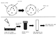
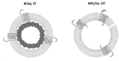
세포외 소포 (EV)는 지질 이중층에 의해 에워싸인 세포 유래된 막성 구조의 이질성 군이다. EV는 엑소솜, 마이크로소포, 바이러스 유사 입자 (VLP) 및 아폽토시스체 (> 1μm)를 포함한다 (Th![]()
![]()
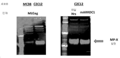
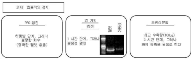
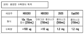
견실한 EV 생성 플랫폼의 특징은 단일통과와 다중통과 막 단백질 둘 모두를 재현적으로 통합하는 능력; 충분한 EV 수확량 (예를 들면, mg 수준)의 생성; 합리적인 규모 (예를 들면, 약 1L)로 쉽게 형질감염됨; 그리고 종 정합된 배경을 생성하는 능력 중에서 한 가지 이상을 포함한다. EV를 생산하는 선행 방법은 이들 요건 모두를 충족시킬 수는 없다.A hallmark of a robust EV production platform is the ability to reproducibly integrate both single-pass and multi-pass membrane proteins; production of sufficient EV yield (eg, mg level); easily transfected on a reasonable scale (eg, about 1 L); and the ability to create a species-matched background. Prior methods of producing EVs cannot meet all of these requirements.
요약summary
본원 발명은 세포외 소포 (EV)를 만들기 위한 향상된 방법과 조성물에 관계한다. 본원 발명은 또한, 신규한 EV 기반 ELISA 검정 및 이런 검정을 수행하기 위한 키트뿐만 아니라 관심되는 막-결합 항원을 포함하는 EV를 이용하여 특정 항원에 대한 항체를 생산하는 방법에 관계한다. The present invention relates to improved methods and compositions for making extracellular vesicles (EVs). The present invention also relates to novel EV-based ELISA assays and kits for performing such assays, as well as methods of producing antibodies against specific antigens using EVs comprising membrane-bound antigens of interest.
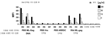
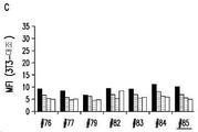
한 양상에서, 본원 발명은 단백질에 특이적으로 결합하는 항체를 생산하기 위한 방법을 제공한다. 일정한 구체예에서, 상기 방법은 (a) (i) 소포 인자에 노출된 세포에서 이종성 단백질을 발현하고, (ii) 상기 세포를 배지에 배양하고, (iii) 이종성 단백질을 포함하는 복수의 EV를 상기 배지로부터 단리함으로써, 이종성 단백질을 포함하는 복수의 EV를 생산하되, 상기 소포 인자는 아실.Hrs (Acyl.Hrs), ARRDC1, ARF6 및 이들의 조합으로 구성된 군에서 선택되는 단계; (b) 복수의 EV를 동물에게 투여함으로써 상기 동물을 면역화하는 단계; 및 (c) 이종성 단백질에 결합하는 항체를 상기 동물로부터 단리하는 단계를 포함한다. In one aspect, the invention provides methods for producing antibodies that specifically bind to a protein. In certain embodiments, the method comprises (a) (i) expressing a heterologous protein in a cell exposed to a vesicular factor, (ii) culturing the cell in a medium, and (iii) generating a plurality of EVs comprising the heterologous protein. By isolating from the medium, producing a plurality of EVs containing heterologous proteins, wherein the vesicle factor is selected from the group consisting of Acyl.Hrs, ARRDC1, ARF6, and combinations thereof; (b) immunizing the animal by administering a plurality of EVs to the animal; and (c) isolating from the animal an antibody that binds to the heterologous protein.
대안으로 및/또는 추가적으로, 단백질에 특이적으로 결합하는 항체를 생산하기 위한 방법은 (a) (i) 이종성 단백질을 세포에서 발현하고, (ii) 상기 세포를 배지에 배양하고, (iii) 상기 단백질을 포함하는 복수의 EV를 상기 배지로부터 단리함으로써, 이종성 단백질을 포함하는 복수의 EV를 생산하되, 상기 세포는 비부착성 세포인 단계; (b) 복수의 EV를 동물에게 투여함으로써 상기 동물을 면역화하는 단계; 및 (c) 이종성 단백질에 결합하는 항체를 상기 동물로부터 단리하는 단계를 포함할 수 있다. 일정한 구체예에서, 상기 방법은 소포 인자, 예를 들면, 이종성 소포 인자, 예를 들면, MLGag, 아실.Hrs, ARRDC1, ARF6 또는 이들의 조합을 세포에서 발현하는 단계를 더욱 포함할 수 있다.Alternatively and/or additionally, a method for producing an antibody that specifically binds to a protein may include (a) (i) expressing the heterologous protein in a cell, (ii) culturing the cell in a medium, and (iii) isolating a plurality of EVs containing the protein from the medium to produce a plurality of EVs containing the heterologous protein, wherein the cells are non-adherent cells; (b) immunizing the animal by administering a plurality of EVs to the animal; and (c) isolating from the animal an antibody that binds to the heterologous protein. In certain embodiments, the method may further comprise expressing in the cell a vesicle factor, eg, a heterologous vesicle factor, eg, MLGag, acyl.Hrs, ARRDC1, ARF6, or a combination thereof.
일정한 구체예에서, 복수의 EV는 초원심분리에 의해 배지로부터 단리된다. 일정한 구체예에서, 복수의 EV는 0주 차, 2주 차 및 4주 차에 동물에게 투여된다. 일정한 구체예에서, 항체를 생산하기 위한 방법은 EV와 동시에 어쥬번트, 예를 들면, Ribi 어쥬번트를 동물에게 투여하는 단계를 더욱 포함한다. 일정한 구체예에서, 항체를 생산하기 위한 방법은 동물에서 단백질에 대한 면역 반응을 증강하기 위해, 추가 접종(boost)을 동물에게 투여하는 단계를 더욱 포함한다. 일정한 구체예에서, 추가 접종은 단백질, 상기 단백질을 인코딩하는 폴리뉴클레오티드, 또는 이들의 조합을 포함한다.In certain embodiments, the plurality of EVs are isolated from the medium by ultracentrifugation. In certain embodiments, a plurality of EVs are administered to the animal at
본원 발명은 본원 발명의 방법에 의해 생산된 항체를 더욱 제공한다. 일정한 구체예에서, 항체는 단일클론 항체이다. 일정한 구체예에서, 항체는 인간, 인간화 또는 키메라 항체이다. 본원 발명은 항체 또는 이의 항원 결합 부분 및 약학적으로 허용되는 운반체를 포함하는 약학적 조성물을 더욱 제공한다. 일정한 구체예에서, 약학적 조성물은 추가 치료제를 더욱 포함한다. 일정한 구체예에서, 본원 발명의 방법에 의해 생산된 항체, 또는 이의 약학적 조성물은 약제로서 이용될 수 있고, 질환을 치료하는 데 이용될 수 있고 및/또는 약제의 제조에서 이용될 수 있다. 일정한 구체예에서, 본원 발명은 질환을 갖는 개체를 치료하는 방법을 제공하는데, 여기서 상기 방법은 본원에서 개시된 단리된 항체 또는 이의 항원 결합 부분, 또는 이의 약학적 조성물의 효과량을 개체에게 투여하는 단계를 포함한다. 본원 발명은 본원에서 개시된 항체 또는 이의 항원 결합 부분을 인코딩하는 단리된 핵산, 그리고 상기 핵산을 포함하는 숙주 세포를 더욱 제공한다. 본원 발명은 또한, 숙주 세포를 항체의 발현에 적합한 조건하에 배양하고, 그리고 임의적으로, 상기 항체를 상기 숙주 세포로부터 단리함으로써 항체를 생산하는 방법을 제공한다. The invention further provides antibodies produced by the methods of the invention. In certain embodiments, the antibody is a monoclonal antibody. In certain embodiments, the antibody is a human, humanized or chimeric antibody. The present invention further provides a pharmaceutical composition comprising the antibody or antigen-binding portion thereof and a pharmaceutically acceptable carrier. In certain embodiments, the pharmaceutical composition further comprises an additional therapeutic agent. In certain embodiments, an antibody produced by a method of the invention, or a pharmaceutical composition thereof, may be used as a medicament, may be used to treat a disease, and/or may be used in the manufacture of a medicament. In certain embodiments, the present invention provides a method of treating a subject having a disease, wherein the method comprises administering to the subject an effective amount of an isolated antibody or antigen-binding portion thereof, or pharmaceutical composition thereof, disclosed herein. includes The present invention further provides isolated nucleic acids encoding the antibodies or antigen-binding portions thereof disclosed herein, and host cells comprising the nucleic acids. The invention also provides a method of producing an antibody by culturing a host cell under conditions suitable for expression of the antibody, and optionally isolating the antibody from the host cell.
추가의 양상에서, 본원 발명은 복수의 EV를 생산하기 위한 방법을 제공한다. 일정한 구체예에서, 상기 방법은 (a) 이종성 단백질을 세포에서 발현하는 단계; (b) 상기 세포를 배지에 배양하는 단계; 및 (c) 이종성 단백질을 포함하는 복수의 EV를 상기 배지로부터 단리하는 단계를 포함하되, 상기 세포는 아실.Hrs, ARRDC1, ARF6 및 이들의 조합으로 구성된 군에서 선택되는 소포 인자에 노출되거나/노출되고 상기 세포는 비부착성 세포이다. In a further aspect, the present invention provides a method for producing a plurality of EVs. In certain embodiments, the method comprises (a) expressing a heterologous protein in a cell; (b) culturing the cells in a culture medium; and (c) isolating a plurality of EVs comprising the heterologous protein from the medium, wherein the cells are exposed/exposed to a vesicular factor selected from the group consisting of acyl.Hrs, ARRDC1, ARF6 and combinations thereof. and the cells are non-adherent cells.
일정한 구체예에서, 이종성 단백질은 막 단백질, 예를 들면, 단일통과 막 단백질 또는 다중통과 막 단백질이다. 일정한 구체예에서, 막 단백질은 단백질 복합체의 구성원이다. 일정한 구체예에서, 막 단백질은 막경유 단백질이 아니지만, 막경유 단백질과의 복합체의 구성원이다. 일정한 구체예에서, 비부착성 세포는 293S 세포 또는 Expi293FTM 세포이다.In certain embodiments, the heterologous protein is a membrane protein, eg, a single-pass membrane protein or a multi-pass membrane protein. In certain embodiments, a membrane protein is a member of a protein complex. In certain embodiments, the membrane protein is not a transmembrane protein, but is a member of a complex with a transmembrane protein. In certain embodiments, the non-adherent cells are 293S cells or Expi293F TM cells.
다른 양상에서, 본원 발명은 항체를 검출하기 위한 방법을 제공한다. 예를 들면, 그러나 제한 없이, 상기 방법은 (a) 샘플을 포획 시약과 함께 인큐베이션하되, 상기 포획 시약은 막-결합 항원을 포함하는 복수의 EV를 포함하고, 그리고 상기 항체는 상기 막-결합 항원에 특이적으로 결합하는 단계; 및 (b) 포획 시약에 결합된 항체를 검출가능 항체와 접촉시켜, 결합된 항체를 검출하되, 상기 검출가능 항체는 상기 항체에 특이적으로 결합하는 단계를 포함할 수 있다. 일정한 구체예에서, 복수의 EV는 (i) 세포에서 막-결합 항원의 발현, (ii) 상기 세포를 시험관내에서 배지에 배양하여, 막-결합 항원을 디스플레이하는 복수의 EV를 생산함 및 (iii) 막-결합 항원을 디스플레이하는 복수의 EV를 상기 배지로부터 단리함에 의해 생성된다. 일정한 구체예에서, 세포는 아실.Hrs, ARRDC1, ARF6 및 이들의 조합으로 구성된 군에서 선택되는 소포 인자에 노출되고/되거나 세포는 비부착성 세포이다. 일정한 구체예에서, 상기 방법은 (c) (b)에서 검출된 항체의 양을 계측하되, 상기 양은 표준 곡선을 이용하여 정량되는 단계를 더욱 포함할 수 있다. 일정한 구체예에서, 샘플은 혈장, 혈청 또는 소변 샘플이다. 일정한 구체예에서, 포획 항체는 고체 지지체, 예를 들면, 마이크로역가 평판 상에 고정될 수 있다. 일정한 구체예에서, 검출가능 항체는 형광 표지화된다. 일정한 구체예에서, 막-결합 항원은 막 단백질 또는 이의 단편이다. In another aspect, the present invention provides methods for detecting antibodies. For example, but without limitation, the method may include (a) incubating a sample with a capture reagent, wherein the capture reagent comprises a plurality of EVs comprising a membrane-bound antigen, and wherein the antibody comprises the membrane-bound antigen. specifically binding to; and (b) contacting the antibody bound to the capture reagent with a detectable antibody to detect the bound antibody, wherein the detectable antibody specifically binds to the antibody. In certain embodiments, the plurality of EVs are capable of (i) expressing membrane-bound antigens in cells, (ii) culturing the cells in vitro in a medium to produce a plurality of EVs displaying membrane-bound antigens ( iii) by isolating from said medium a plurality of EVs displaying membrane-bound antigens. In certain embodiments, the cell is exposed to a vesicular factor selected from the group consisting of acyl.Hrs, ARRDC1, ARF6, and combinations thereof, and/or the cell is a non-adherent cell. In certain embodiments, the method may further comprise (c) measuring the amount of antibody detected in (b), wherein the amount is quantified using a standard curve. In certain embodiments, the sample is a plasma, serum or urine sample. In certain embodiments, the capture antibody may be immobilized on a solid support, such as a microtiter plate. In certain embodiments, the detectable antibody is fluorescently labeled. In certain embodiments, the membrane-bound antigen is a membrane protein or fragment thereof.
본원 발명은 샘플 내에서 항체를 검출하기 위한 키트를 제공한다. 일정한 구체예에서, 본원 발명의 키트는 (a) 막-결합 항원을 포함하는 복수의 EV를 포함하는 포획 시약으로서, 여기서 검출되는 항체가 막-결합 항원에 특이적으로 결합하고; 및 (b) 검출되는 항체에 특이적으로 결합하는 검출가능 항체를 포함한다. 일정한 구체예에서, 복수의 EV가 고체 지지체 상에 고정된다. 일정한 구체예에서, 고체 지지체는 마이크로역가 평판이다. 일정한 구체예에서, 검출가능 항체는 형광 표지화된다. 일정한 구체예에서, 항원은 막 단백질 또는 이의 단편이다. The present invention provides kits for detecting antibodies in a sample. In certain embodiments, a kit of the invention comprises (a) a capture reagent comprising a plurality of EVs comprising a membrane-bound antigen, wherein the antibody to be detected specifically binds to the membrane-bound antigen; and (b) a detectable antibody that specifically binds to the antibody being detected. In certain embodiments, a plurality of EVs are immobilized on a solid support. In certain embodiments, the solid support is a microtiter plate. In certain embodiments, the detectable antibody is fluorescently labeled. In certain embodiments, the antigen is a membrane protein or fragment thereof.
일정한 구체예에서, 본원 발명은 항체-생산 세포를 분류하기 위한 방법을 더욱 제공한다. 일정한 구체예에서, 상기 방법은 항체-생산 세포를 복수의 EV와 함께 인큐베이션하되, 상기 복수의 EV는 (i) 막-결합 항원 및 첫 번째 검출가능 마커를 포함하는 EV의 첫 번째 모집단으로서, 여기서 상기 항체-생산 세포의 부분집합이 막-결합 항원에 특이적으로 결합하고; 및 (b) 막-결합 항원을 결여하지만, 첫 번째 마커와 구별 가능한 두 번째 검출가능 마커를 포함하는 EV의 두 번째 모집단을 포함하는 단계를 포함한다. 일정한 구체예에서, 상기 방법은 항체-생산 세포를 EV의 첫 번째 모집단, 또는 EV의 첫 번째 모집단 및 EV의 두 번째 모집단의 조합 중 어느 하나에 대한 그들의 결합에 근거하여 분류하는 단계를 더욱 포함한다. 일정한 구체예에서, EV의 첫 번째 모집단은 (i) 첫 번째 세포에서 막-결합 항원 및 첫 번째 검출가능 마커의 발현, (ii) 첫 번째 세포를 시험관내에서 배지에 배양하여, 막-결합 항원을 디스플레이하는 복수의 EV를 생산함 및 (iii) 막-결합 항원을 디스플레이하는 복수의 EV를 상기 배지로부터 단리함에 의해 생성된다. 일정한 구체예에서, EV의 두 번째 모집단은 (i) 두 번째 세포에서 두 번째 검출가능 마커의 발현, (ii) 두 번째 세포를 시험관내에서 배지에 배양하여, 두 번째 검출가능 마커를 포함하는 복수의 EV를 생산함 및 (iii) 복수의 EV를 상기 배지로부터 단리함에 의해 생성된다. 일정한 구체예에서, 세포는 아실.Hrs, ARRDC1, ARF6 및 이들의 조합으로 구성된 군에서 선택되는 소포 인자에 노출된다. 일정한 구체예에서, 첫 번째 세포 및/또는 두 번째 세포는 비부착성 세포이다.In certain embodiments, the present invention further provides methods for sorting antibody-producing cells. In certain embodiments, the method incubates antibody-producing cells with a plurality of EVs, wherein the plurality of EVs are (i) a first population of EVs comprising a membrane-bound antigen and a first detectable marker, wherein: A subset of the antibody-producing cells specifically binds to the membrane-bound antigen; and (b) comprising a second population of EVs lacking the membrane-bound antigen, but comprising a second detectable marker distinguishable from the first marker. In certain embodiments, the method further comprises sorting the antibody-producing cells based on their binding to either the first population of EVs, or a combination of the first population of EVs and the second population of EVs. . In certain embodiments, a first population of EVs is obtained by (i) expression of a membrane-bound antigen and a first detectable marker in a first cell, (ii) culturing the first cell in a medium in vitro to obtain a membrane-bound antigen. and (iii) isolating from the medium a plurality of EVs displaying membrane-bound antigens. In certain embodiments, the second population of EVs is an ascites comprising the second detectable marker by (i) expression of the second detectable marker in the second cells, (ii) culturing the second cells in a medium in vitro. and (iii) isolating a plurality of EVs from the medium. In certain embodiments, the cell is exposed to a vesicular factor selected from the group consisting of acyl.Hrs, ARRDC1, ARF6, and combinations thereof. In certain embodiments, the first cell and/or the second cell are non-adherent cells.
도면의 간단한 설명
도 1. 세포외 소포 (EV)의 형성을 보여주는 개략적 도면.
도 2a-2b. EV 형성을 위한 설계 원리. (2a) 일반적인 EV 형성기 및 아실.Hrs 소유 EV 설계. (2b) MLGag 및 ARRDC1 소유 EV 설계.
도 3. EV를 생산하는 작업 흐름을 보여주는 개략적 도면.
도 4. 웨스턴 블롯을 이용하여, MP-X를 발현하는 EV를 생산할 수 있는 소포 인자의 확인.
도 5. 균일한 소포 크기를 보여주는 동적 광 산란 (DLS).
도 6. 웨스턴 블롯은 소포 인자 MLGag, Acryl.Hrs 및 뮤린 ARRDC1 (mARRDC1)이 뮤린 세포에서 MP-X 발현 EV 형성을 유도한다는 것을 보여주었다.
도 7a-7b. EV 생산 과제: (7a) 효율적인 EV 정제 획득의 과제; 및 (7b) 충분한 수확량 획득의 과제.
도 8a-8c. MLGag를 소포 인자로서 갖는, 표적 단백질을 발현하는 EV를 생산하는 데 이용된 Expi293FTM 세포 및 293S 세포에서 수확 일에 EV의 평균 수확량 (8a, 8b) 및 세포 생존력 (8c)이 계측되었다.
도 9a-9c. Expi293FTM 세포 생산된 EV 및 293S 세포 생산된 EV 사이에 MP-7 (9a), MP-8 (9b), 또는 MP-4 (9c)의 발현 수준을 비교하기 위해 ELISA가 수행되었다.
도 10a-10c. (10a) 윤곽이 명확한 EV 입자가 생성되었다. (10b, 10c) EV 기반 ELISA는 단일통과 (10b) 및 다중통과 (10c) 막 단백질에 대한 FACS+ 항체를 검출할 수 있다.
도 11a-11d. Expi293FTM EV 면역화된 쥐와 생쥐를 선별검사하기 위한 세포주가 확인되었다. (11a) 개략적 도면은 동물 면역화 프로토콜을 보여준다. (11b) 세포에 항혈청 및 전채혈의 결합이 FACS에 의해 도시되었다. 쥐에서 EV 면역화로부터 획득된 pAb는 293 세포에는 결합하지만, RBA 세포에는 그렇지 않았다. 생쥐에서 EV 면역화로부터 획득된 pAb는 RBA 세포에는 결합하지만, 3T3 세포에는 그렇지 않았다. (11c, 11d) eGFP를 이용한 FACS는 RBA (11c) 및 3T3 (11d) 둘 모두 형질감염 가능하다는 것을 보여주었다.
도 12a-12c. Expi293FTM EV 면역화된 토끼와 라마/낙타를 선별검사하기 위한 세포주가 확인되었다. (12a) 개략적 도면은 동물 면역화 프로토콜을 보여준다. (12b) 세포에 항혈청 및 전채혈의 결합이 FACS에 의해 도시되었다. 토끼에서 EV 면역화로부터 획득된 pAb는 RK13 세포에 결합하지 않았다. 라마에서 EV 면역화로부터 획득된 pAb는 3T3 세포에는 결합하지만, Dubca 세포에는 결합하지 않았다. (12c) FACS는 RK13 세포 (왼쪽) 및 Dubca 세포 (오른쪽) 둘 모두 형질감염 가능하다는 것을 보여주었다.
도 13. 쥐 RBA 세포는 EV를 생산할 수 있지만, 수확량이 293S 세포와 비교하여 적었다.
도 14. 웨스턴 블롯은 전체 세포 용해물 및 EV에서 MP-1의 존재를 확증하였다.
도 15. 웨스턴 블롯은 MP-2가 EV 및 전체 세포 용해물 내에 존재한다는 것을 확증하였다.
도 16. 웨스턴 블롯은 MP-3이 ARF-6으로 형질감염된 세포로부터 생산된 ARF-6 소유 EV 내에는 존재하지만, MLGag에서는 그렇지 않다는 것을 확증하였다.
도 17. Gag 캡시드가 MP의 큰 세포내 도메인 (ICD)과의 통합을 입체적으로 차단한다는 것을 보여주는 개략적 도면.
도 18. 단백질 ELISA, EV 기반 ELISA, 그리고 FACS의 작동 기전을 보여주는 개략적 도면.
도 19a-19d. EV 기반 ELISA 역가 (19a)는 MP-4에 대한 FACS 역가 (19b)와 잘 상관되었다. FACS (19c) 및 EV 기반 ELISA (19d) 역가는 단백질 ELISA 역가와 잘 상관되지 않았다.
도 20a-20b. DNA 면역화를 이용하여 MP-5로 면역화된 생쥐로부터 항-MP-5 혈청이 수집되었다. EV 기반 ELISA 역가 (20a) 및 FACS 역가 (20b)가 도시된다.
도 21. EV 기반 ELISA는 항-MP5 항체를 검출하기 위한 FACS와 잘 상관되었다.
도 22a-22c. MP-6 EV의 초기 배치의 품질 관리 (QC) 분석. (22a) 웨스턴 블롯은 단리된 EV에서 MP-6의 존재를 보여주었다. (22b) 웨스턴 블롯은 단리된 EV에서 소포 인자의 존재를 보여주었다. (22c) 재조합 단백질 표준을 이용한 정량적 웨스턴 블롯이, 단리된 EV에서 MP-6을 정량하는 데 이용되었다.
도 23. MP-6 EV을 이용한 면역화 프로토콜.
도 24a-24d. (24a) 웨스턴 블롯은 EV로 면역화된 쥐로부터 수집된 항혈청에서 항-MP-6 및 항-Gag 항체의 존재를 보여주었다. (24b) FACS는 형질감염 대조 세포에 대해 항혈청의 어떤 유의미한 비특이적 결합도 없다는 것을 보여주었다. (24c, 24d) FACS는 DNA/단백질 추가 접종 전 (24c) 및 DNA/단백질 추가 접종 후 (24d), 수집된 항혈청에서 MP-6 발현 세포에 결합을 보여주었다.
도 25a-25b. 정제된 일차 항체 (25a) 및 혈청 (25b)은 유사한 FACS 결과를 보여주었다.
도 26. DNA 추가 접종이 EV 면역화된 쥐로부터 항-MP-6 역가를 선택적으로 증가시켰다.
도 27a-27c. 생쥐 항-MP-7 일차 항체가 MP-7 발현 EV로 면역화된 녹아웃 생쥐로부터 생성되었고, 그리고 FACS에 의해 선별검사되었다. 항-MP-7 항체가 마지막 추가 접종 전 (27a) 및 마지막 추가 접종 후 (27b), 수집된 혈청에서 검출되었다. 혈청이 3T3 대조 세포 (27c)에는 결합하지 않았다.
도 28. 생쥐 항-MP-7 하이브리도마가 FACS에 의해 선별검사되었다.
도 29. 생쥐 항-MP-7 하이브리도마가 FACS에 의해 선별검사되었다.
도 30. 일차 세포에서 FACS를 이용한, 생쥐 항-MP-7 mAb의 선별검사.
도 31a-31b. 쥐가 막 결합된 MP-1 또는 MP-1 DNA를 포함하는 EV로, 그리고 단백질 또는 DNA 추가 접종으로 면역화되었다. 추가 접종 전 (31a) 및 추가 접종 후 (31b), 이들 쥐로부터 수집된 항혈청이 FACS에 의해 선별검사되었다.
도 32. 쥐 항-MP-1 하이브리도마가 FACS에 의해 선별검사되었다.
도 33. 쥐가 단지 단백질, DNA, 또는 막 결합된 MP-8을 포함하는 EV로만 면역화되었다. 각 군에서 발견된 ELISA 양성 및 FACS 양성 항체의 수가 도시된다.
도 34. 쥐 및 토끼가 MP-9 및 MP-9 DNA를 포함하는 EV로 면역화되었다. GFP-표지화 MP-9 EV 및 RFP-표지화 빈 EV를 이용한 쥐 및 토끼 IgG+ B 세포의 염색이 도시된다.
도 35. 쥐 및 토끼가 MP-10 또는 MP-11을 포함하는 EV로 면역화되었다. RFP-표지화 MP EV 및 GFP-표지화 빈 EV를 이용한 토끼 IgG+ B 세포의 염색이 도시된다.
도 36a. 표면 발현을 위해 MP-14, MP-15, MP-16 및 MP-17 (공동수용체 "B"), 그리고 MP-12 및 MP-13 (수용체 "A")의 공동발현이 필요하다.
도 36b. MP-14, MP-15, MP-16 및 MP-17 (공동수용체 "B"), 그리고 MP-12 및 MP-13 (수용체 "A")의 공동발현은 EV 통합을 야기한다. Brief description of the drawing
Figure 1. A schematic diagram showing the formation of extracellular vesicles (EVs).
Figures 2a-2b. Design principles for EV formation. (2a) Common EV formers and acyl.Hrs-owned EV designs. (2b) MLGag and ARRDC1 proprietary EV designs.
Figure 3. Schematic diagram showing the workflow of producing EVs.
Figure 4. Identification of vesicular factors capable of producing EVs expressing MP-X using Western blot.
Figure 5. Dynamic light scattering (DLS) showing uniform vesicle size.
Figure 6. Western blot showed that vesicular factors MLGag, Acryl.Hrs and murine ARRDC1 (mARRDC1) induced MP-X expressing EV formation in murine cells.
Figures 7a-7b. EV production challenges: (7a) the challenge of obtaining efficient EV purification; and (7b) the challenge of obtaining sufficient yield.
8a-8c. The average yield of EVs (8a, 8b) and cell viability (8c) were measured on the day of harvest in Expi293F TM cells and 293S cells used to produce EVs expressing the target protein with MLGag as a vesicular factor.
9a-9c. ELISA was performed to compare the expression levels of MP-7 (9a), MP-8 (9b), or MP-4 (9c) between EVs produced by Expi293F TM cells and EVs produced by 293S cells.
10a-10c. (10a) EV particles with well-defined contours were produced. (10b, 10c) EV-based ELISA can detect FACS+ antibodies to single-pass (10b) and multi-pass (10c) membrane proteins.
11a-11d. A cell line was identified for screening Expi293F TM EV immunized rats and mice. (11a) Schematic diagram shows the animal immunization protocol. (11b) Binding of antiserum and whole blood to cells was shown by FACS. pAbs obtained from EV immunization in mice bound to 293 cells but not to RBA cells. pAbs obtained from EV immunization in mice bound RBA cells but not 3T3 cells. (11c, 11d) FACS with eGFP showed that both RBA (11c) and 3T3 (11d) were transfectable.
12a-12c. A cell line was identified for screening Expi293F TM EV immunized rabbits and llamas/camels. (12a) The schematic diagram shows the animal immunization protocol. (12b) Binding of antiserum and whole blood to cells was shown by FACS. pAbs obtained from EV immunization in rabbits did not bind to RK13 cells. pAb obtained from EV immunization in llamas bound to 3T3 cells but not to Dubca cells. (12c) FACS showed that both RK13 cells (left) and Dubca cells (right) were viable for transfection.
Figure 13. Murine RBA cells can produce EVs, but the yield is less compared to 293S cells.
Figure 14. Western blot confirmed the presence of MP-1 in whole cell lysates and EVs.
Figure 15. Western blot confirmed that MP-2 was present in EV and whole cell lysates.
Figure 16. Western blot confirmed that MP-3 was present in ARF-6-bearing EVs produced from cells transfected with ARF-6, but not in MLGag.
Figure 17. Schematic diagram showing that the Gag capsid sterically blocks integration with the large intracellular domain (ICD) of MP.
Figure 18. Schematic diagram showing the mechanism of action of protein ELISA, EV-based ELISA, and FACS.
19a-19d. EV-based ELISA titers (19a) correlated well with FACS titers for MP-4 (19b). FACS (19c) and EV-based ELISA (19d) titers did not correlate well with protein ELISA titers.
20a-20b. Anti-MP-5 sera were collected from mice immunized with MP-5 using DNA immunization. EV-based ELISA titers (20a) and FACS titers (20b) are shown.
21 . EV-based ELISA correlated well with FACS to detect anti-MP5 antibodies.
22a-22c. Quality control (QC) analysis of initial batches of MP-6 EVs. (22a) Western blot showed the presence of MP-6 in isolated EVs. (22b) Western blot showed the presence of vesicular factors in isolated EVs. (22c) Quantitative Western blot using recombinant protein standards was used to quantify MP-6 in isolated EVs .
Figure 23. Immunization protocol using MP-6 EVs.
24a-24d. (24a) Western blot showed the presence of anti-MP-6 and anti-Gag antibodies in antiserum collected from mice immunized with EV. (24b) FACS showed no significant non-specific binding of antiserum to transfected control cells. (24c, 24d) FACS showed binding to MP-6 expressing cells in antiserum collected before DNA/protein boost (24c) and after DNA/protein boost (24d).
25a-25b. Purified primary antibody (25a) and serum (25b) showed similar FACS results.
26. DNA boosting selectively increased anti-MP-6 titers from EV immunized mice.
27a-27c. Mouse anti-MP-7 primary antibodies were raised from knockout mice immunized with MP-7 expressing EVs and screened by FACS. Anti-MP-7 antibody was detected in serum collected before the last boost (27a) and after the last boost (27b). Serum did not bind to 3T3 control cells (27c).
Figure 28. Mouse anti-MP-7 hybridomas were screened by FACS.
Figure 29. Mouse anti-MP-7 hybridomas were screened by FACS.
Figure 30. Screening of mouse anti-MP-7 mAbs using FACS in primary cells.
31a-31b. Mice were immunized with membrane-bound MP-1 or EVs containing MP-1 DNA and with a protein or DNA boost. Before boost (31a) and after boost (31b), antisera collected from these mice were screened by FACS.
Figure 32. Murine anti-MP-1 hybridomas were screened by FACS.
Figure 33. Mice were only immunized with EVs containing protein, DNA, or membrane bound MP-8. The number of ELISA positive and FACS positive antibodies found in each group is shown.
Figure 34. Mice and rabbits were immunized with MP-9 and EVs containing MP-9 DNA. Staining of rat and rabbit IgG+ B cells with GFP-labeled MP-9 EV and RFP-labeled empty EV is shown.
Figure 35. Mice and rabbits were immunized with EVs containing MP-10 or MP-11. Staining of rabbit IgG+ B cells with RFP-labeled MP EVs and GFP-labeled empty EVs is shown.
Fig. 36a. Co-expression of MP-14, MP-15, MP-16 and MP-17 (coreceptor "B"), and MP-12 and MP-13 (receptor "A") is required for surface expression.
Fig. 36b. Coexpression of MP-14, MP-15, MP-16 and MP-17 (co-receptor "B"), and MP-12 and MP-13 (receptor "A") results in EV integration.
상세한 설명details
본원 발명은 세포외 소포 (EV)를 만들기 위한 향상된 방법과 조성물에 관계한다. 본원 발명은 또한, 신규한 EV 기반 ELISA 검정 및 이런 검정을 수행하기 위한 키트뿐만 아니라 관심되는 막-결합 항원, 예를 들면, 막 단백질을 포함하는 EV를 이용하여 특정 항원에 대한 항체를 생산하는 방법에 관계한다. 본원 발명은 일정한 정제 단계, 소포 인자 및/또는 EV 생산 세포주를 채택함으로써, EV의 신속하고 높은 수확량 생성을 달성하는 것이 가능하다는 발견에 부분적으로 근거된다. 이것은 또한, 동물을 항원 제시 EV로 면역화하는 것이 도전적인 막 단백질 항원 및 복합체에 대한 기능적 항체의 개발을 가능하게 한다는 발견에 부분적으로 근거된다. 본원 발명의 무제한적 구체예가 본원에서 설명된다. The present invention relates to improved methods and compositions for making extracellular vesicles (EVs). The present invention also relates to novel EV-based ELISA assays and kits for performing such assays, as well as methods for producing antibodies against specific antigens using EVs comprising a membrane-bound antigen of interest, e.g., a membrane protein. relate to The present invention is based in part on the finding that by employing certain purification steps, vesicle factors and/or EV producing cell lines, it is possible to achieve rapid and high yield production of EVs. This is also based in part on the finding that immunizing animals with antigen-presenting EVs allows the development of functional antibodies against challenging membrane protein antigens and complexes. Non-limiting embodiments of the present invention are described herein.
개시의 명료함을 위해, 하지만 제한 없이, 상세한 설명은 하기의 하위섹션으로 나눠진다:For clarity of disclosure, but without limitation, the detailed description is divided into the following subsections:
I. 정의;I. Justice;
II. EV를 만드는 방법;II. how to make an EV;
III. EV 기반 ELISA 검정 및 키트; III. EV-based ELISA assays and kits;
IV. EV를 이용하여 항체를 생산하는 방법;IV. a method for producing antibodies using EV;
V. 진단과 검출을 위한 방법과 조성물;V. Methods and Compositions for Diagnosis and Detection;
VI. 약학적 조성물;VI. pharmaceutical compositions;
VII. 치료 방법 및 투여 루트;VII. methods of treatment and routes of administration;
VIII. 제조 물품; 그리고VIII. articles of manufacture; and
IX. 예시적인 구체예.IX. Exemplary Embodiments.
I. 정의 I. Definition
본원 발명에서 이용된 용어는 일반적으로, 당해 분야에서, 본원 발명의 맥락에서, 그리고 각 용어가 이용되는 특정한 맥락에서 그들의 통상적인 의미를 갖는다. 본원 발명의 조성물과 방법 및 이들을 만들고 이용하는 방법을 설명함에 있어서 실무자에게 추가 지침을 제공하기 위해, 일정한 용어가 아래에, 또는 본원 발명의 다른 곳에서 논의된다. The terms used herein generally have their ordinary meanings in the art, in the context of the present invention, and in the specific context in which each term is used. Certain terms are discussed below and elsewhere herein to provide additional guidance to the practitioner in describing the compositions and methods of the present invention and how to make and use them.
본원에서 이용된 바와 같이, 청구항에서 및/또는 본 명세서에서 용어 "포함하는"과 함께 이용될 때 단어 "a" 또는 "an"의 이용은 "하나"를 의미할 수 있지만, 이것은 또한 "하나 이상," "적어도 하나," 그리고 "하나 또는 하나 초과"의 의미와 부합한다. 더 나아가, 용어 "갖는," "포함하는," "내포하는" 및 "함유하는"은 교체 가능하고, 그리고 당업자는 이들 용어가 개방형 용어라는 것을 인식하고 있다.As used herein, the use of the word "a" or "an" when used in conjunction with the term "comprising" in the claims and/or herein can mean "one," but also "one or more" ," matches the meanings of "at least one," and "one or more than one." Furthermore, the terms “having,” “comprising,” “including,” and “including” are interchangeable, and one skilled in the art recognizes that these terms are open-ended terms.
용어 "약" 또는 "대략"은 당업자에 의해 결정될 때 특정 값에 대한 허용되는 오차 범위 이내를 의미하는데, 이것은 상기 값이 어떻게 계측되거나 또는 결정되는지, 다시 말하면, 계측 시스템의 제한에 부분적으로 의존할 것이다. 예를 들면, "약"은 당해 분야에서 관례에 따라, 3 이내 또는 3보다 큰 표준 편차를 의미할 수 있다. 대안으로, "약"은 소정의 값의 최대 20%, 바람직하게는 최대 10%, 더 바람직하게는 최대 5%, 그리고 더 바람직하게는 최대 1%의 범위를 의미할 수 있다. 대안으로, 특히, 생물학적 시스템 또는 과정에 대하여, 상기 용어는 값의 1 크기 자릿수 이내, 바람직하게는 5배 이내, 그리고 더 바람직하게는 2배 이내를 의미할 수 있다.The term "about" or "approximately" means within an acceptable error range for a particular value as determined by one skilled in the art, which will depend in part on how the value is measured or determined, i.e., the limitations of the metrology system. will be. For example, "about" can mean within 3 or greater than 3 standard deviations, as is customary in the art. Alternatively, “about” may mean a range of up to 20%, preferably up to 10%, more preferably up to 5%, and even more preferably up to 1% of a given value. Alternatively, particularly for biological systems or processes, the term may mean within one order of magnitude of a value, preferably within five times, and more preferably within two orders of magnitude.
본원에서 "개체" 또는 "피험자"는 척추동물, 예컨대 인간 또는 비인간 동물, 예를 들면, 포유동물이다. 포유동물은 인간, 비인간 영장류, 경작용 동물, 스포츠 동물, 설치류 및 애완동물을 포함하지만 이들에 한정되지 않는다. 비인간 동물 개체의 무제한적 실례는 설치류 예컨대 생쥐, 쥐, 햄스터 및 기니 피그; 토끼; 개; 고양이; 양; 돼지; 염소; 소; 말; 그리고 비인간 영장류 예컨대 유인원 및 원숭이를 포함한다. 일정한 구체예에서, 개체 또는 피험자는 인간이다. An “individual” or “subject” herein is a vertebrate, such as a human or non-human animal, eg, a mammal. Mammals include, but are not limited to, humans, non-human primates, farm animals, sport animals, rodents, and pets. Non-limiting examples of non-human animal subjects include rodents such as mice, rats, hamsters and guinea pigs; rabbit; dog; cat; sheep; pig; Goat; cow; word; and non-human primates such as apes and monkeys. In certain embodiments, the subject or subject is a human.
본원에서 이용된 바와 같이, 용어 "시험관내"는 인공 환경, 그리고 인공 환경에서 일어나는 과정 또는 반응을 지칭한다. 예시된 시험관내 환경에는 시험관 및 세포 배양액이 포함되지만 이들에 한정되지 않는다. As used herein, the term “in vitro” refers to an artificial environment and to a process or reaction that occurs in an artificial environment. Exemplary in vitro environments include, but are not limited to, test tubes and cell cultures.
본원에서 이용된 바와 같이, 용어 "생체내"는 자연 환경 (예를 들면, 동물 또는 세포), 그리고 자연 환경에서 일어나는 과정 또는 반응, 예컨대 배아 발달, 세포 분화, 신경관 형성 등을 지칭한다. As used herein, the term "in vivo" refers to a natural environment (eg, animal or cell) and to processes or reactions that occur in the natural environment, such as embryonic development, cell differentiation, neural tube formation, and the like.
본원에서 이용된 바와 같이, 용어 "생물학적 샘플"은 생물학적 유체, 예를 들면, 혈액, 혈장, 혈청, 소변, 객담, 척수액, 흉수, 유두 흡인물, 림프액, 기도, 장관 및 비뇨생식기로의 유체, 누액, 타액, 모유, 림프계로부터 유체, 정액, 뇌척수액, 내부 기관계 유체, 복수, 종양 낭액, 양수, 기관지폐포액, 담즙액, 그리고 이들의 조합을 비롯하여, 개체로부터 획득된 생물학적 물질의 샘플을 지칭한다. As used herein, the term "biological sample" refers to a biological fluid such as blood, plasma, serum, urine, sputum, spinal fluid, pleural effusion, nipple aspirates, lymph fluid, fluids to the respiratory tract, intestinal tract and urogenital tract, Refers to a sample of biological material obtained from a subject, including tear fluid, saliva, breast milk, fluid from the lymphatic system, semen, cerebrospinal fluid, internal organ system fluid, ascites, tumor cyst fluid, amniotic fluid, bronchoalveolar fluid, bile fluid, and combinations thereof .
본원에서 용어 "항체"는 가장 넓은 의미에서 이용되고, 그리고 단일클론 항체, 다중클론 항체, 다중특이적 항체 (예를 들면, 이중특이적 항체), 그리고 항체 단편 (이들이 원하는 항원 결합 활성을 나타내기만 하면)을 포함하지만 이들에 한정되지 않는 다양한 항체 구조를 포괄한다.The term "antibody" is used herein in its broadest sense, and includes monoclonal antibodies, polyclonal antibodies, multispecific antibodies (eg, bispecific antibodies), and antibody fragments (provided they exhibit the desired antigen-binding activity). It encompasses a variety of antibody structures including, but not limited to, lower surface).
본원에서 이용된 바와 같이, 용어 "항체 단편"은 무손상 항체가 결합하는 항원에 결합하는 무손상 항체의 부분을 포함하는, 무손상 항체 이외의 분자를 지칭한다. 항체 단편의 실례는 Fv, Fab, Fab', Fab'-SH, F(ab')2; 디아바디; 선형 항체; 단일 사슬 항체 분자 (예를 들면, scFv); 그리고 항체 단편으로부터 형성된 다중특이적 항체를 포함하지만 이들에 한정되지 않는다.As used herein, the term “antibody fragment” refers to a molecule, other than an intact antibody, comprising a portion of an intact antibody that binds an antigen to which the intact antibody binds. Examples of antibody fragments include Fv, Fab, Fab', Fab'-SH, F(ab') 2 ; diabodies; linear antibodies; single chain antibody molecules (eg scFv); and multispecific antibodies formed from antibody fragments.
본원에서 이용된 바와 같이, 용어 "키메라 항체"는 중쇄 및/또는 경쇄의 일부분이 특정 공급원 또는 종으로부터 유래되고, 반면 중쇄 및/또는 경쇄의 나머지 부분이 상이한 공급원 또는 종으로부터 유래되는 항체를 지칭한다.As used herein, the term “chimeric antibody” refers to an antibody in which portions of the heavy and/or light chains are derived from a particular source or species, while the remaining portions of the heavy and/or light chains are from a different source or species. .
본원에서 이용된 바와 같이, 용어 "단일클론 항체"는 실제적으로 동질성 항체의 모집단으로부터 획득된 항체를 지칭한다, 다시 말하면, 상기 모집단을 구성하는 개별 항체는 예를 들면, 자연 발생 돌연변이를 내포하거나 또는 단일클론 항체 제조물의 생산 동안 발생하는 가능한 변이체 항체 (이런 변이체는 일반적으로 미량으로 존재한다)를 제외하고, 동일하고 및/또는 동일한 에피토프에 결합한다. 상이한 결정인자 (에피토프)에 대해 지향된 상이한 항체를 전형적으로 포함하는 다중클론 항체 제조물과 대조적으로, 단일클론 항체 제조물의 각 단일클론 항체는 항원 상에서 단일 결정인자에 대해 지향된다. 따라서, 수식어 "단일클론"은 항체의 실제적으로 동질성 모집단으로부터 획득되는 것으로서 항체의 특징을 표시하고, 그리고 임의의 특정 방법에 의한 항체의 생산을 필요로 하는 것으로 해석되지 않는다. 예를 들면, 본원 발명에 따른 단일클론 항체는 하이브리도마 방법, 재조합 DNA 방법, 파지 디스플레이 방법, 그리고 인간 면역글로불린 좌위 중에서 전부 또는 일부를 내포하는 유전자도입 동물을 활용하는 방법을 포함하지만 이들에 한정되지 않는 다양한 기술에 의해 만들어질 수 있는데, 이런 방법 및 단일클론 항체를 만들기 위한 다른 예시적인 방법은 본원에서 설명된다. As used herein, the term "monoclonal antibody" refers to an antibody obtained from a population of substantially homogeneous antibodies, i.e., the individual antibodies comprising the population contain, for example, naturally occurring mutations or Except for possible variant antibodies that arise during production of monoclonal antibody preparations (such variants are generally present in minor amounts), they are identical and/or bind to the same epitope. In contrast to polyclonal antibody preparations, which typically include different antibodies directed against different determinants (epitopes), each monoclonal antibody of a monoclonal antibody preparation is directed against a single determinant on the antigen. Thus, the modifier “monoclonal” indicates the character of the antibody as being obtained from a substantially homogeneous population of antibodies, and is not to be construed as requiring production of the antibody by any particular method. For example, monoclonal antibodies according to the present invention include, but are not limited to, hybridoma methods, recombinant DNA methods, phage display methods, and methods utilizing transgenic animals harboring all or part of a human immunoglobulin locus. These and other exemplary methods for making monoclonal antibodies are described herein.
"나신 항체"는 이종성 모이어티 (예를 들면, 세포독성 모이어티) 또는 방사성 표지에 접합되지 않는 항체를 지칭한다. 나신 항체는 약학적 조성물 내에 존재할 수 있다.A "naked antibody" refers to an antibody that is not conjugated to a heterologous moiety (eg, a cytotoxic moiety) or a radioactive label. A naked antibody may be present in a pharmaceutical composition.
항체의 "부류"는 이의 중쇄에 의해 소유된 불변 도메인 또는 불변 영역의 유형을 지칭한다. 항체의 5가지 주요 부류: IgA, IgD, IgE, IgG 및 IgM이 있고, 그리고 이들 중에서 몇몇은 하위부류 (아이소타입), 예를 들면, IgG1, IgG2, IgG3, IgG4, IgA1 및 IgA2로 더욱 나눠질 수 있다. 일정한 구체예에서, 항체는 IgG1 아이소타입이다. 일정한 구체예에서, 항체는 Fc-영역 효과기 기능을 감소시키기 위한 P329G, L234A 및 L235A 돌연변이를 갖는 IgG1 아이소타입이다. 다른 구체예에서, 항체는 IgG2 아이소타입이다. 일정한 구체예에서, 항체는 IgG4 항체의 안정성을 향상시키기 위한, 힌지 영역에서 S228P 돌연변이를 갖는 IgG4 아이소타입이다. 면역글로불린의 상이한 부류에 상응하는 중쇄 불변 도메인은 각각, α, γ, ε, γ 및 μ로 불린다. 항체의 경쇄는 이의 불변 도메인의 아미노산 서열에 근거하여, 카파 (κ) 및 람다 (λ)로 불리는 두 가지 유형 중에서 한 가지에 배정될 수 있다. The "class" of an antibody refers to the type of constant domain or constant region possessed by its heavy chain. There are five major classes of antibodies: IgA, IgD, IgE, IgG and IgM, and several of these are subclasses (isotypes), such as IgG 1 , IgG 2 , IgG 3 , IgG 4 , IgA 1 and It can be further divided into IgA 2 . In certain embodiments, the antibody is of the IgG 1 isotype. In certain embodiments, the antibody is of the IgG 1 isotype with P329G, L234A and L235A mutations to reduce Fc-region effector function. In another embodiment, the antibody is of the IgG 2 isotype. In certain embodiments, the antibody is of the IgG 4 isotype with a S228P mutation in the hinge region to enhance the stability of the IgG 4 antibody. The heavy chain constant domains corresponding to the different classes of immunoglobulins are called α, γ, ε, γ and μ, respectively. The light chain of an antibody can be assigned to one of two types, called kappa (κ) and lambda (λ), based on the amino acid sequence of its constant domain.
본원에서 이용된 바와 같이, 용어 "프레임워크" 또는 "FR"은 초가변 영역 (CDR) 잔기 이외의 가변 도메인 잔기를 지칭한다. 가변 도메인의 FR은 일반적으로 4개의 FR 도메인: FR1, FR2, FR3 및 FR4로 구성된다. 따라서, CDR과 FR 서열은 일반적으로 VH (또는 VL)에서 하기의 순서로 나타난다: FR1-H1(L1)-FR2-H2(L2)-FR3-H3(L3)-FR4.As used herein, the term “framework” or “FR” refers to variable domain residues other than hypervariable region (CDR) residues. The FRs of a variable domain are generally composed of four FR domains: FR1, FR2, FR3 and FR4. Thus, CDR and FR sequences generally appear in the following order in VH (or VL): FR1-H1(L1)-FR2-H2(L2)-FR3-H3(L3)-FR4.
본원에서 이용된 바와 같이, 용어 "전장 항체", "무손상 항체" 및 "전체 항체"는 선천적 항체 구조와 실제적으로 유사한 구조를 갖는 또는 본원에서 규정된 바와 같은 Fc 영역을 내포하는 중쇄를 갖는 항체를 지칭하기 위해 본원에서 교체가능하게 이용된다.As used herein, the terms “full-length antibody,” “intact antibody,” and “whole antibody” refer to an antibody having a structure substantially similar to that of a native antibody or having a heavy chain that encompasses an Fc region as defined herein. are used interchangeably herein to refer to.
"단리된" 항체는 이의 자연 환경의 구성요소로부터 분리된 것이다. 일정한 구체예에서, 항체는 예를 들면, 전기이동 (예를 들면, SDS-PAGE, 등전위 초점 (IEF), 모세관 전기이동) 또는 크로마토그래피 (예를 들면, 이온 교환 또는 역상 HPLC)에 의해 결정될 때, 95% 또는 99%보다 높은 순도로 정제된다. 항체 순도의 사정을 위한 방법에 관한 검토를 위해, 예를 들면, Flatman et al., J. Chromatogr. B 848:79-87 (2007)을 참조한다.An “isolated” antibody is one that has been separated from a component of its natural environment. In certain embodiments, antibodies are determined, for example, by electrophoresis (eg, SDS-PAGE, isoelectric focusing (IEF), capillary electrophoresis) or chromatography (eg, ion exchange or reverse phase HPLC). , purified to a purity higher than 95% or 99%. For a review of methods for assessment of antibody purity, see, eg, Flatman et al., J. Chromatogr. See B 848:79-87 (2007).
"인간 공통 프레임워크"는 인간 면역글로불린 VL 또는 VH 프레임워크 서열의 선택에서 가장 흔히 발생하는 아미노산 잔기를 나타내는 프레임워크이다. 일반적으로, 인간 면역글로불린 VL 또는 VH 서열은 가변 도메인 서열의 하위군으로부터 유래된다. 일반적으로, 서열의 하위군은 Kabat et al., Sequences of Proteins of Immunological Interest, Fifth Edition, NIH Publication 91-3242, Bethesda MD (1991), Vols. 1-3의 경우에서와 같은 하위군이다. 일정한 구체예에서, VL의 경우에, 하위군은 Kabat et al., 위와 같음의 경우에서와 같은 하위군 카파 I이다. 일정한 구체예에서, VH의 경우에, 하위군은 Kabat et al., 위와 같음의 경우에서와 같은 하위군 III이다. A “human consensus framework” is a framework representing the most commonly occurring amino acid residues in a selection of human immunoglobulin VL or VH framework sequences. Generally, human immunoglobulin VL or VH sequences are derived from a subgroup of variable domain sequences. Generally, subgroups of sequences are described in Kabat et al., Sequences of Proteins of Immunological Interest, Fifth Edition, NIH Publication 91-3242, Bethesda MD (1991), Vols. This is the same subgroup as in cases 1-3. In certain embodiments, for VL, the subgroup is subgroup kappa I as in the case of Kabat et al., supra. In certain embodiments, in the case of VH, the subgroup is subgroup III as in the case of Kabat et al., supra.
"인간화" 항체는 비인간 HVR로부터 아미노산 잔기 및 인간 FR로부터 아미노산 잔기를 포함하는 키메라 항체를 지칭한다. 일정한 구체예에서, 인간화 항체는 적어도 1개, 그리고 전형적으로 2개의 가변 도메인을 실제적으로 모두 포함할 것인데, 여기서 HVR (예를 들면, CDR)의 전부 또는 실제적으로 전부가 비인간 항체의 것들에 상응하고, 그리고 FR의 전부 또는 실제적으로 전부가 인간 항체의 것들에 상응한다. 인간화 항체는 임의적으로, 인간 항체로부터 유래된 항체 불변 영역의 적어도 일부를 포함할 수 있다. 항체, 예를 들면, 비인간 항체의 "인간화 형태"는 인간화를 겪은 항체를 지칭한다. A “humanized” antibody refers to a chimeric antibody comprising amino acid residues from non-human HVRs and amino acid residues from human FRs. In certain embodiments, a humanized antibody will comprise substantially all of at least one, and typically two, variable domains, wherein all or substantially all of the HVRs (eg, CDRs) correspond to those of a non-human antibody and , and all or substantially all of the FRs correspond to those of a human antibody. A humanized antibody may optionally comprise at least a portion of an antibody constant region derived from a human antibody. A “humanized form” of an antibody, eg, a non-human antibody, refers to an antibody that has undergone humanization.
본원에서 이용된 바와 같이, 용어 "초가변 영역"은 서열에서 초가변성 ("상보성 결정 영역" 또는 "CDR")이고 및/또는 구조적으로 규정된 루프 ("초가변 루프")를 형성하고 및/또는 항원 접촉 잔기 ("항원 접촉")를 내포하는, 항체 가변 도메인의 각 영역을 지칭한다. 별도로 표시되지 않으면, CDR 잔기 및 가변 도메인에서 다른 잔기 (예를 들면, FR 잔기)는 본원에서 Kabat et al., 위와 같음에 따라서 넘버링된다. 일반적으로, 항체는 6개의 CDR: VH에서 3개 (H1, H2, H3), 그리고 VL에서 3개 (L1, L2, L3)를 포함한다. 본원에서 예시적인 CDR은 하기를 포함한다: As used herein, the term "hypervariable region" is hypervariable in sequence ("complementarity determining region" or "CDR") and/or forms structurally defined loops ("hypervariable loops") and/or or each region of an antibody variable domain, containing antigen contacting residues ("antigen contact"). Unless otherwise indicated, CDR residues and other residues (eg, FR residues) in the variable domain are numbered herein according to Kabat et al., supra. Generally, an antibody contains six CDRs: three in VH (H1, H2, H3) and three in VL (L1, L2, L3). Exemplary CDRs herein include:
(a) 아미노산 잔기 26-32 (L1), 50-52 (L2), 91-96 (L3), 26-32 (H1), 53-55 (H2), 그리고 96-101 (H3)에서 발생하는 초가변 루프 (Chothia and Lesk, J. Mol. Biol. 196:901-917 (1987));(a) amino acid residues 26-32 (L1), 50-52 (L2), 91-96 (L3), 26-32 (H1), 53-55 (H2), and 96-101 (H3) hypervariable loops (Chothia and Lesk, J. Mol. Biol. 196:901-917 (1987));
(b) 아미노산 잔기 24-34 (L1), 50-56 (L2), 89-97 (L3), 31-35b (H1), 50-65 (H2), 그리고 95-102 (H3)에서 발생하는 CDR (Kabat et al., Sequences of Proteins of Immunological Interest, 5th Ed. Public Health Service, National Institutes of Health, Bethesda, MD (1991));(b) amino acid residues 24-34 (L1), 50-56 (L2), 89-97 (L3), 31-35b (H1), 50-65 (H2), and 95-102 (H3) CDR (Kabat et al., Sequences of Proteins of Immunological Interest, 5th Ed. Public Health Service, National Institutes of Health, Bethesda, MD (1991));
(c) 아미노산 잔기 27c-36 (L1), 46-55 (L2), 89-96 (L3), 30-35b (H1), 47-58 (H2), 그리고 93-101 (H3)에서 발생하는 항원 접촉 (MacCallum et al. J. Mol. Biol. 262: 732-745 (1996)); 그리고(c) amino acid residues 27c-36 (L1), 46-55 (L2), 89-96 (L3), 30-35b (H1), 47-58 (H2), and 93-101 (H3) antigen contact (MacCallum et al. J. Mol. Biol . 262: 732-745 (1996)); and
(d) CDR 아미노산 잔기 46-56 (L2), 47-56 (L2), 48-56 (L2), 49-56 (L2), 26-35 (H1), 26-35b (H1), 49-65 (H2), 93-102 (H3), 그리고 94-102 (H3)를 비롯한, (a), (b) 및/또는 (c)의 조합. (d) CDR amino acid residues 46-56 (L2), 47-56 (L2), 48-56 (L2), 49-56 (L2), 26-35 (H1), 26-35b (H1), 49- A combination of (a), (b) and/or (c), including 65 (H2), 93-102 (H3), and 94-102 (H3).
"면역접합체"는 세포독성 작용제를 포함하지만 이에 한정되지 않는 한 가지 이상의 이종성 분자(들)에 접합된 항체이다. An "immunoconjugate" is an antibody conjugated to one or more heterologous molecule(s), including but not limited to a cytotoxic agent.
용어 "핵산 분자" 또는 "폴리뉴클레오티드"는 뉴클레오티드의 중합체를 포함하는 임의의 화합물 및/또는 물질을 포함한다. 각 뉴클레오티드는 염기, 특이적으로 퓨린- 또는 피리미딘 염기 (다시 말하면, 시토신 (C), 구아닌 (G), 아데닌 (A), 티민 (T) 또는 우라실 (U)), 당 (다시 말하면, 데옥시리보오스 또는 리보오스), 그리고 인산염 기로 구성된다. 종종, 핵산 분자는 염기의 서열에 의해 설명되는데, 여기서 상기 염기는 핵산 분자의 일차 구조 (선형 구조)를 나타낸다. 염기의 서열은 전형적으로 5'에서 3'로 나타내진다. 본원에서, 용어 핵산 분자는 예를 들면, 상보성 DNA (cDNA) 및 유전체 DNA를 비롯한 데옥시리보핵산 (DNA), 리보핵산 (RNA), 특히 전령 RNA (mRNA), DNA 또는 RNA의 합성 형태, 그리고 이들 분자 중에서 두 가지 이상을 포함하는 혼성 중합체를 포괄한다. 핵산 분자는 선형 또는 환상일 수 있다. 이에 더하여, 용어 핵산 분자는 센스와 안티센스 가닥뿐만 아니라 단일 가닥과 이중 가닥 형태 둘 모두를 포함한다. 게다가, 본원에서 설명된 핵산 분자는 자연 발생 또는 비-자연 발생 뉴클레오티드를 내포할 수 있다. 비-자연 발생 뉴클레오티드의 실례는 유도체화된 당 또는 인산염 중추 연쇄 또는 화학적으로 변형된 잔기를 갖는 변형된 뉴클레오티드 염기를 포함한다. 핵산 분자는 또한, 시험관내에서 및/또는 생체내에서, 예를 들면, 숙주 또는 환자에서 본원 발명의 항체의 직접적인 발현을 위한 벡터로서 적합한 DNA와 RNA 분자를 포괄한다. 이런 DNA (예를 들면, cDNA) 또는 RNA (예를 들면, mRNA) 벡터는 변형되지 않거나 또는 변형될 수 있다. 예를 들면, mRNA는 mRNA가 개체 내로 주입되어 항체가 생체내에서 생성될 수 있도록, RNA 벡터의 안정성 및/또는 인코딩된 분자의 발현을 증강하기 위해 화학적으로 변형될 수 있다 (참조: 예를 들면, Stadler er al, Nature Medicine 2017, 2017년 6월 12일 온라인 공개됨, doi:10.1038/nm.4356 또는 EP 2 101 823 B1). The term "nucleic acid molecule" or "polynucleotide" includes any compound and/or substance comprising a polymer of nucleotides. Each nucleotide is a base, specifically a purine- or pyrimidine base (i.e., cytosine (C), guanine (G), adenine (A), thymine (T), or uracil (U)), a sugar (i.e., oxyribose or ribose), and a phosphate group. Often, a nucleic acid molecule is described by a sequence of bases, wherein the bases represent the primary structure (linear structure) of the nucleic acid molecule. The sequence of bases is typically represented from 5' to 3'. As used herein, the term nucleic acid molecule refers to, for example, deoxyribonucleic acid (DNA), including complementary DNA (cDNA) and genomic DNA, ribonucleic acid (RNA), especially messenger RNA (mRNA), synthetic forms of DNA or RNA, and It encompasses interpolymers comprising two or more of these molecules. Nucleic acid molecules can be linear or circular. In addition, the term nucleic acid molecule includes both single-stranded and double-stranded forms as well as sense and antisense strands. In addition, nucleic acid molecules described herein may contain naturally occurring or non-naturally occurring nucleotides. Examples of non-naturally occurring nucleotides include derivatized sugar or phosphate backbone chains or modified nucleotide bases with chemically modified moieties. Nucleic acid molecules also encompass DNA and RNA molecules suitable as vectors for direct expression of an antibody of the invention in vitro and/or in vivo, eg, in a host or patient. Such DNA (eg cDNA) or RNA (eg mRNA) vectors may be unmodified or modified. For example, mRNA can be chemically modified to enhance the stability of the RNA vector and/or expression of the encoded molecule, such that the mRNA can be injected into a subject and antibodies can be produced in vivo (see e.g. , Stadler er al, Nature Medicine 2017, published online 12 June 2017, doi:10.1038/nm.4356 or
"단리된" 핵산은 자연 환경의 구성요소로부터 분리된 핵산 분자를 지칭한다. 단리된 핵산에는 핵산 분자를 통상적으로 내포하는 세포에 내포된 핵산 분자가 포함되지만, 상기 핵산 분자는 염색체외에 또는 자연 염색체 위치와 상이한 염색체 위치에 존재한다.An "isolated" nucleic acid refers to a nucleic acid molecule that has been separated from a component of its natural environment. An isolated nucleic acid includes a nucleic acid molecule that is internal to cells that ordinarily contain the nucleic acid molecule, but the nucleic acid molecule is present extrachromosomally or at a chromosomal location different from its natural chromosomal location.
"항체를 인코딩하는 단리된 핵산"은 항체 중쇄와 경쇄 (또는 이들의 단편)를 인코딩하는 하나 이상의 핵산 분자, 단일 벡터 또는 별개의 벡터에서 이런 핵산 분자(들), 그리고 숙주 세포 내에 하나 이상의 위치에서 존재하는 이런 핵산 분자(들)를 지칭한다. "Isolated nucleic acid encoding an antibody" refers to one or more nucleic acid molecules encoding the antibody heavy and light chains (or fragments thereof), such nucleic acid molecule(s) in a single vector or separate vectors, and at one or more locations in a host cell. refers to such nucleic acid molecule(s) present.
본원에서 이용된 바와 같이, 용어 "벡터"는 자신이 연관되는 다른 핵산을 증식할 수 있는 핵산 분자를 지칭한다. 상기 용어는 자기 복제 핵산 구조로서 벡터뿐만 아니라 이것이 도입된 숙주 세포의 유전체 내로 통합된 벡터를 포함한다. 일정한 벡터는 그들이 작동가능하게 연결되는 핵산의 발현을 주동할 수 있다. 이런 벡터는 본원에서 "발현 벡터"로서 지칭된다.As used herein, the term "vector" refers to a nucleic acid molecule capable of propagating another nucleic acid with which it is associated. The term includes the vector as a self-replicating nucleic acid structure as well as the vector integrated into the genome of a host cell into which it has been introduced. Certain vectors are capable of directing the expression of nucleic acids to which they are operably linked. Such vectors are referred to herein as "expression vectors".
본원에서 이용된 바와 같이, 용어 "항원" 및 "면역원"은 그것으로 면역화된 동물에서 면역 반응 (바람직하게는 항체 반응)을 유도하는 분자 또는 물질을 지칭하기 위해 본원에서 교체가능하게 이용된다. 항원은 단백질, 펩티드, 탄수화물, 핵산, 지질, 합텐 또는 다른 자연 발생 또는 합성 화합물일 수 있다. 일정한 구체예에서, 항원은 단백질이다. 일정한 구체예에서, 항원은 막 단백질 또는 이의 단편이다. 일정한 구체예에서, 항원은 단일통과 또는 다중통과 막 단백질 또는 이의 단편이다. As used herein, the terms "antigen" and "immunogen" are used interchangeably herein to refer to a molecule or substance that induces an immune response (preferably an antibody response) in an animal immunized therewith. Antigens can be proteins, peptides, carbohydrates, nucleic acids, lipids, haptens or other naturally occurring or synthetic compounds. In certain embodiments, an antigen is a protein. In certain embodiments, the antigen is a membrane protein or fragment thereof. In certain embodiments, the antigen is a monopass or multipass membrane protein or fragment thereof.
본원에서 이용된 바와 같이, 용어 "이종성 단백질"은 이러한 이종성 단백질을 인코딩하는 폴리뉴클레오티드를 세포 내로 도입함으로써, 세포에서 발현되는 단백질을 지칭한다. 일정한 구체예에서, 이종성 단백질은 세포에 선천적이지 않다. 일정한 구체예에서, 이종성 단백질은 세포에 선천적이지만, 이러한 이종성 단백질을 인코딩하는 폴리뉴클레오티드의 세포 내로의 도입으로 인해 과다발현되는 단백질이다.As used herein, the term “heterologous protein” refers to a protein that is expressed in a cell by introducing into the cell a polynucleotide encoding such a heterologous protein. In certain embodiments, the heterologous protein is not native to the cell. In certain embodiments, a heterologous protein is a protein that is native to the cell, but is overexpressed due to introduction of a polynucleotide encoding the heterologous protein into the cell.
본원에서 교체가능하게 이용된 바와 같이, 용어 "막-결합 항원" 및 "막 항원"은 막에 직접적으로 또는 간접적으로 결합되는 항원을 지칭한다. As used interchangeably herein, the terms “membrane-bound antigen” and “membrane antigen” refer to antigens that bind directly or indirectly to a membrane.
본원에서 교체가능하게 이용된 바와 같이, 용어 "막 결합된 단백질" 및 "막 단백질"은 막에 직접적으로 또는 간접적으로 결합되는 단백질을 지칭한다. 막 결합된 단백질의 무제한적 실례는 내재성 단백질, 지질 고정 단백질 및 외재성 단백질을 포함한다. 일정한 구체예에서, 막 단백질은 막경유 단백질, 예를 들면, 단일통과 또는 다중통과 막 단백질 또는 이의 단편이다. 일정한 구체예에서, 막 단백질은 막경유 단백질이 아니지만, 막경유 단백질과의 복합체의 일부 (예를 들면, 보조인자)이다. 본원의 실시예에서 이용된 바와 같이, 약어 "MP"는 막 단백질을 지칭한다. As used interchangeably herein, the terms “membrane bound protein” and “membrane protein” refer to proteins that are directly or indirectly bound to a membrane. Non-limiting examples of membrane bound proteins include intrinsic proteins, lipid anchored proteins and extrinsic proteins. In certain embodiments, the membrane protein is a transmembrane protein, eg, a single-pass or multi-pass membrane protein or fragment thereof. In certain embodiments, the membrane protein is not a transmembrane protein, but is part of a complex (eg, a cofactor) with a transmembrane protein. As used in the examples herein, the abbreviation “MP” refers to membrane protein.
본원에서 이용된 바와 같이, 용어 "막경유 항원"은 막에 적어도 한 번 걸치는 항원을 지칭한다. 막경유 항원의 무제한적 실례는 단일통과 항원, 예를 들면, 막에 한 번 걸치는 항원, 지질 고정 단백질, 또는 다중통과 항원, 예를 들면, 막에 적어도 두 번 걸치는 단백질을 포함한다. As used herein, the term "transmembrane antigen" refers to an antigen that spans the membrane at least once. Non-limiting examples of transmembrane antigens include single pass antigens, such as antigens that span the membrane once, lipid anchored proteins, or multipass antigens, such as proteins that span the membrane at least twice.
본원에서 이용된 바와 같이, 용어 "막경유 단백질"은 막에 적어도 한 번 걸치는 단백질을 지칭한다. 막경유 단백질의 무제한적 실례는 단일통과 단백질, 예를 들면, 막에 한 번 걸치는 단백질, 지질 고정 단백질, 또는 다중통과 단백질, 예를 들면, 막에 적어도 두 번 걸치는 단백질을 포함한다. 일정한 구체예에서, 막경유 단백질은 단일통과 또는 다중통과 막경유 단백질 또는 이의 단편이다. 일정한 구체예에서, 막경유 단백질은 다중통과 막경유 단백질 또는 이의 단편이다. As used herein, the term “transmembrane protein” refers to a protein that spans a membrane at least once. Non-limiting examples of transmembrane proteins include single-pass proteins, such as proteins that span the membrane once, lipid-anchored proteins, or multi-pass proteins, such as proteins that span the membrane at least twice. In certain embodiments, the transmembrane protein is a monopass or multipass transmembrane protein or fragment thereof. In certain embodiments, the transmembrane protein is a multipass transmembrane protein or fragment thereof.
본원에서 이용된 바와 같이, 용어 "면역화"는 항체가 동물에서 조성될 수 있도록 하나 이상의 항원을 동물에게 투여하는 단계 또는 단계들을 지칭한다. 일반적으로, 면역화는 항원 또는 항원들을 동물 내로 주입하는 단계를 포함한다. 면역화는 항원 또는 항원들의 1회 이상의 투여를 수반할 수 있다. 일정한 구체예에서, 항원은 이러한 항원을 발현하는 복수의 EV를 통해 동물에게 투여된다. As used herein, the term "immunization" refers to the step or steps of administering one or more antigens to an animal so that antibodies can be raised in the animal. Generally, immunization involves injecting an antigen or antigens into an animal. Immunization may involve one or more administrations of the antigen or antigens. In certain embodiments, antigens are administered to an animal via a plurality of EVs expressing such antigens.
본원에서 이용된 바와 같이, 용어 "다중클론 항체" 또는 "다중클론 항혈청"은 항원 또는 항원들로 면역화된 동물의 혈액으로부터 준비될 수 있는 하나 (일가 또는 특정한 항혈청) 또는 그 초과 (다가 항혈청)의 항원에 대해 특이적인 항체의 혼합물을 내포하는 면역 혈청을 지칭한다. As used herein, the term “polyclonal antibody” or “polyclonal antiserum” refers to one (monovalent or specific antiserum) or more (multivalent antiserum) that may be prepared from the blood of an animal immunized with the antigen or antigens. Refers to an immune serum that contains a mixture of antibodies specific for an antigen.
본원에서 이용된 바와 같이, 용어 "어쥬번트"는 면역 반응의 비특이적 자극제를 지칭한다. 어쥬번트는 하기 구성요소 (a) 항원(들)을 신속한 이화로부터 보호하는 침착물을 형성하도록 설계된 물질 (예를 들면, 광물성 오일, 백반, 수산화알루미늄, 리포솜 또는 계면활성제 [예를 들면, 플루로닉 폴리올]) 및 (b) 면역화된 숙주 동물의 면역 반응을 비특이적으로 자극하는 물질 (예를 들면, 그 안에서 림포카인 수준을 증가시킴으로써) 중에서 한 가지 또는 둘 모두를 포함하는 조성물의 형태일 수 있다. 림포카인 수준을 증가시키기 위한 분자의 무제한적 실례는 지질다당류 (LPS) 또는 이의 지질 A 부분, 보르데텔라 페르투시스 (Bordetella pertussis), 백일해 독소, 미코박테리움 투베르쿨로시스 (Mycobacterium tuberculosis), 그리고 무라밀 디펩티드 (MDP)를 포함한다. 어쥬번트의 무제한적 실례는 프로인드 어쥬번트 (프로인드 완전 어쥬번트 (FCA)를 형성하기 위해, 사멸된 미코박테리움 투베르쿨로시스 (M. tuberculosis)를 임의적으로 포함), 수산화알루미늄 어쥬번트, Ribi 어쥬번트, Titermax 어쥬번트, 스페콜 어쥬번트, 알루미늄 염 어쥬번트, 그리고 모노포스포릴 지질 A-합성 트레할로스 디코리노미콜레이트 (MPL-TDM)를 포함한다. As used herein, the term "adjuvant" refers to a non-specific stimulator of an immune response. An adjuvant may be one of the following components (a) a substance designed to form a deposit that protects the antigen(s) from rapid catabolism (eg, mineral oil, alum, aluminum hydroxide, liposomes, or a surfactant [eg, fluro nick polyol]) and (b) a substance that non-specifically stimulates the immune response of an immunized host animal (eg, by increasing lymphokine levels therein) or both. there is. Non-limiting examples of molecules for increasing lymphokine levels include lipopolysaccharide (LPS) or its lipid A portion, Bordetella pertussis, pertussis toxin, Mycobacterium tuberculosis , and muramyl dipeptide (MDP). Non-limiting examples of adjuvants include Freund's adjuvant (optionally including killed Mycobacterium tuberculosis to form Freund's complete adjuvant (FCA)), aluminum hydroxide adjuvant, Ribi adjuvant, Titermax adjuvant, Specol adjuvant, aluminum salt adjuvant, and monophosphoryl lipid A-synthetic trehalose dicorynomycolate (MPL-TDM).
본원에서 이용된 바와 같이, "치료" (및 이의 문법적 변이, 예컨대 "치료한다" 또는 "치료하는")는 치료되는 개체에서 질환의 자연 경과를 변경하려는 시도에서 임상적 개입을 지칭하고, 그리고 임상 병리의 예방을 위해 또는 임상 병리의 경과 동안 수행될 수 있다. 바람직한 치료 효과는 질환의 발생 또는 재발의 예방, 증상의 경감, 질환의 임의의 직접적인 또는 간접적인 병리학적 결과의 축소, 전이 예방, 질환 진행의 속도 감소, 질환 상태의 개선 또는 완화, 그리고 관해 또는 향상된 예후를 포함하지만 이들에 한정되지 않는다. 일정한 구체예에서, 본원 발명의 항체는 질환의 발달을 지연시키거나 또는 질환의 진행을 늦추는 데 이용된다.As used herein, "treatment" (and grammatical variations thereof, such as "treat" or "treating") refers to clinical intervention in an attempt to alter the natural course of a disease in the individual being treated, and clinical It can be performed for prophylaxis of pathology or during the course of clinical pathology. Desirable therapeutic effects include prevention of occurrence or recurrence of the disease, alleviation of symptoms, reduction of any direct or indirect pathological consequences of the disease, prevention of metastasis, reduction in the rate of disease progression, amelioration or palliation of the disease state, and remission or enhancement. Prognosis includes, but is not limited to. In certain embodiments, antibodies of the invention are used to delay the development of a disease or to slow the progression of a disease.
용어 "가변 영역" 또는 "가변 도메인"은 항체를 항원에 결합시키는 데 관련되는 항체 중쇄 또는 경쇄의 도메인을 지칭한다. 선천적 항체의 중쇄와 경쇄의 가변 도메인 (각각, VH와 VL)은 일반적으로 유사한 구조를 갖는데, 각 도메인이 4개의 보존된 프레임워크 영역 (FR) 및 3개의 초가변 영역 (CDR)을 포함한다. (참조: 예를 들면, Kindt et al. Kuby Immunology, 6th ed., W.H. Freeman and Co., page 91 (2007).) 단일 VH 또는 VL 도메인이 항원 결합 특이성을 부여하는 데 충분할 수 있다. 게다가, 특정 항원에 결합하는 항체는 각각, 상보성 VL 또는 VH 도메인의 라이브러리를 선별검사하기 위해, 항원에 결합하는 항체로부터 VH 또는 VL 도메인을 이용하여 단리될 수 있다. 참조: 예를 들면, Portolano et al., J. Immunol. 150:880-887 (1993); Clarkson et al., Nature 352:624-628 (1991).The term "variable region" or "variable domain" refers to the domain of an antibody heavy or light chain that is involved in binding the antibody to an antigen. The variable domains of the heavy and light chains of native antibodies (VH and VL, respectively) generally have a similar structure, with each domain comprising four conserved framework regions (FRs) and three hypervariable regions (CDRs). (See, eg, Kindt et al. Kuby Immunology , 6 th ed., WH Freeman and Co., page 91 (2007).) A single VH or VL domain may be sufficient to confer antigen binding specificity. In addition, antibodies that bind a particular antigen can be isolated using VH or VL domains from antibodies that bind the antigen to screen a library of complementary VL or VH domains, respectively. See, eg, Portolano et al ., J. Immunol. 150 :880-887 (1993); Clarkson et al., Nature 352:624-628 (1991).
본원에서 이용된 바와 같이, 용어 "선별검사"는 하나 이상의 단일클론 항체 (예를 들면, 정제된 항체 및/또는 이러한 항체를 포함하는 하이브리도마 배양 상층액)에, 관심되는 항원에 결합하는 항체의 능력을 정성적으로 및/또는 정량적으로 결정하는 하나 이상의 검정을 실행하는 것을 지칭한다. As used herein, the term "screen" refers to one or more monoclonal antibodies (eg, purified antibodies and/or hybridoma culture supernatants comprising such antibodies), antibodies that bind to an antigen of interest. refers to performing one or more assays that qualitatively and/or quantitatively determine the ability of
본원에서 이용된 바와 같이, "마커"는 직접적인 또는 간접적인 검출을 가능하게 하는 조성물을 지칭한다. 마커는 형광 조성물, 발색 표지, 전자 밀도 표지, 화학발광 표지 및 방사성 표지를 포함하지만 이들에 한정되지 않는다. 예를 들면, 그러나 제한 없이, 특이적 마커는 녹색 형광 단백질 ("GFP"), mCherry, dtTomato, 또는 당해 분야에서 알려진 다른 형광 단백질 (예를 들면, 본원에서 참조로서 편입된 Shaner et al., A Guide to Choosing Fluorescent Proteins, Nature Methods 2(12) 905-909 (December 2005), 32P、14C、125I、3H 및 131I, 형광발생소 (예컨대 희토류 킬레이트 또는 루시퍼 옐로우 및 이의 유도체), 로다민 (로다민) 및 이의 유도체, 단실, 움벨리페론, 루시페라아제 (예컨대 반딧불이 루시페라아제 및 세균 형광 단순 효소) (U.S. 특허 번호 4,737,456), 플루오레세인, 2,3-디히드로 프탈라진 디케톤뿐만 아니라 검출가능 신호를 발생시키는 효소, 예를 들면, 양고추냉이 과산화효소 (HRP), 알칼리성 인 신 효소, 베타 갈락토시다아제, 글루코아밀라아제, 라이소자임, 탄수화물 옥시다아제 (예컨대 글루코오스 옥시다아제, 갈락토오스 옥시다아제 및 글루코오스-6-인산염 탈수소효소 (G6PD)), 그리고 헤테로환상 옥시다아제 (예컨대 요산분해효소 및 크산틴 옥시다아제)이다. As used herein, “marker” refers to a composition that enables direct or indirect detection. Markers include, but are not limited to, fluorescent compositions, chromogenic labels, electron density labels, chemiluminescent labels, and radioactive labels. For example, but without limitation, specific markers include green fluorescent protein (“GFP”), mCherry, dtTomato, or other fluorescent proteins known in the art (eg, Shaner et al., A Guide to Choosing Fluorescent Proteins, Nature Methods 2(12) 905-909 (December 2005), 32 P, 14 C, 125 I, 3 H and 131 I, fluorophores (such as rare earth chelates or lucifer yellow and its derivatives), rhodamine (rhodamine) and its derivatives, dansyl, umbelliferon, luciferases (such as firefly luciferase and bacterial fluorescent simple enzymes) (US Patent No. 4,737,456), fluorescein, 2,3-dihydrophthalazine diketone, as well as but enzymes that generate detectable signals, such as horseradish peroxidase (HRP), alkaline phosphorus enzyme, beta galactosidase, glucoamylase, lysozyme, carbohydrate oxidases (such as glucose oxidase, galactose oxidase and glucose- 6-phosphate dehydrogenase (G6PD)), and heterocyclic oxidases (such as urate lyase and xanthine oxidase).
본원에서 이용된 바와 같이, "부착성 세포"는 성장을 위해 표면에 부착을 필요로 하는 세포를 지칭한다. As used herein, “adherent cells” refer to cells that require attachment to a surface in order to grow.
본원에서 이용된 바와 같이, "비부착성 세포"는 현탁 상태로 배양되는 세포를 지칭한다. 일정한 구체예에서, 비부착성 세포는 성장을 위해 표면에 부착을 필요로 하지 않는 세포이다. As used herein, “non-adherent cells” refer to cells that are cultured in suspension. In certain embodiments, non-adherent cells are cells that do not require attachment to a surface for growth.
II. EV를 만드는 방법 II. How to make an EV
한 가지 양상에서, 본원 발명은 EV를 만드는 향상된 방법에 관계한다. 이것은 일정한 정제 단계, 소포 인자 및/또는 EV 생산 세포주를 채택함으로써, EV의 신속하고 높은 수확량 생성을 달성하는 것이 가능하다는 발견에 부분적으로 근거된다. In one aspect, the present invention relates to improved methods of making EVs. This is based in part on the finding that by employing certain purification steps, vesicular factors and/or EV producing cell lines, it is possible to achieve rapid and high yield production of EVs.
일정한 무제한적 구체예에서, EV를 생산하기 위한 방법은 다음 단계를 포함한다: (a) 관심되는 단백질, 예를 들면, 관심되는 이종성 단백질을 소포 인자에 노출된 세포에서 발현하는 단계; (b) 상기 세포를 시험관내에서 배지에 배양하여 복수의 EV를 생산하는 단계; 및 (c) 복수의 EV를 상기 배지로부터 단리하는 단계. 일정한 구체예에서, 세포를 소포 인자에 노출시키는 단계는 상기 소포 인자를 상기 세포에서 발현하는 단계를 포함한다. In certain non-limiting embodiments, a method for producing an EV comprises the following steps: (a) expressing a protein of interest, eg, a heterologous protein of interest, in a cell exposed to a vesicular factor; (b) producing a plurality of EVs by culturing the cells in a medium in vitro; and (c) isolating the plurality of EVs from the medium. In certain embodiments, exposing a cell to an antifoaming factor comprises expressing the antifoaming factor in the cell.
일정한 구체예에서, 관심되는 단백질을 세포에서 발현하는 단계는 관심되는 단백질을 인코딩하는 적어도 하나의 폴리뉴클레오티드를 상기 세포 내로 도입하는 단계를 포함한다. 예를 들면, 그러나 제한 없이, EV를 생산하기 위한 방법은 다음 단계를 포함한다: (a) 관심되는 단백질, 예를 들면, 관심되는 이종성 단백질을 인코딩하는 폴리뉴클레오티드를 소포 인자에 노출된 세포에 도입하는 단계; (b) 상기 세포를 시험관내에서 배지에 배양하여 복수의 EV를 생산하는 단계; 및 (c) 복수의 EV를 상기 배지로부터 단리하는 단계. 일정한 구체예에서, 상기 세포는 관심되는 단백질을 세포에서 발현하기 위해 상기 폴리뉴클레오티드로 형질감염될 수 있다.In certain embodiments, expressing a protein of interest in a cell comprises introducing into the cell at least one polynucleotide encoding the protein of interest. For example, but without limitation, a method for producing an EV includes the following steps: (a) introducing a polynucleotide encoding a protein of interest, eg, a heterologous protein of interest, into a cell exposed to a vesicle factor. doing; (b) producing a plurality of EVs by culturing the cells in a medium in vitro; and (c) isolating the plurality of EVs from the medium. In certain embodiments, the cell can be transfected with the polynucleotide to express a protein of interest in the cell.
일정한 구체예에서, 세포를 소포 인자에 노출시키는 단계는 상기 소포 인자를 상기 세포에서, 예를 들면, 관심되는 단백질을 발현하는 동일한 세포에서 발현하는 단계를 포함할 수 있다. 예를 들면, 그러나 제한 없이, 소포 인자를 인코딩하는 폴리뉴클레오티드가 세포 내로 도입될 수 있다. 일정한 구체예에서, 소포 인자는 관심되는 단백질을 인코딩하는 동일한 폴리뉴클레오티드에 의해 인코딩될 수 있다. 대안으로, 관심되는 단백질 및 소포 인자는 2개의 상이한 폴리뉴클레오티드에 의해 인코딩될 수 있다. 예를 들면, 그러나 제한 없이, 관심되는 단백질을 소포 인자에 노출된 세포에서 발현하는 단계는 상기 소포 인자를 인코딩하는 첫 번째 폴리뉴클레오티드 및 상기 관심되는 단백질을 인코딩하는 두 번째 폴리뉴클레오티드를 상기 세포 내로 도입하는 단계를 포함한다. In certain embodiments, exposing a cell to a vesicular factor may comprise expressing the vesicular factor in the cell, eg, in the same cell that expresses the protein of interest. For example, but without limitation, a polynucleotide encoding a vesicle factor may be introduced into a cell. In certain embodiments, the vesicle factor may be encoded by the same polynucleotide that encodes the protein of interest. Alternatively, the protein and vesicle factor of interest can be encoded by two different polynucleotides. For example, but without limitation, expressing a protein of interest in a cell exposed to a vesicle factor introduces a first polynucleotide encoding the vesicle factor and a second polynucleotide encoding the protein of interest into the cell. It includes steps to
일정한 무제한적 구체예에서, EV를 생산하기 위한 방법은 다음 단계를 포함한다: (a) (i) 소포 인자 및 관심되는 단백질을 인코딩하는 폴리뉴클레오티드 및/또는 (ii) 소포 인자를 인코딩하는 첫 번째 폴리뉴클레오티드 및 관심되는 단백질을 인코딩하는 두 번째 폴리뉴클레오티드를 제공하는 단계; (b) 세포를 폴리뉴클레오티드(들), 예를 들면, 상기 폴리뉴클레오티드 또는 상기 첫 번째 및 두 번째 폴리뉴클레오티드로 형질감염시키는 단계; (c) 상기 세포를 시험관내에서 배지에 배양하여 복수의 EV를 생산하는 단계; 및 (d) 복수의 EV를 상기 배지로부터 단리하는 단계. 일정한 구체예에서, 첫 번째 폴리뉴클레오티드 및 두 번째 폴리뉴클레오티드는 단일 핵산에서 제공된다. 예시적인 EV 생성 작업 흐름은 도 3에서 도시된다. In certain non-limiting embodiments, a method for producing an EV comprises the following steps: (a) (i) a polynucleotide encoding a vesicle factor and a protein of interest and/or (ii) a first cell encoding a vesicle factor. providing a polynucleotide and a second polynucleotide encoding a protein of interest; (b) transfecting cells with the polynucleotide(s), eg, the polynucleotide or the first and second polynucleotides; (c) producing a plurality of EVs by culturing the cells in a medium in vitro; and (d) isolating the plurality of EVs from the medium. In certain embodiments, the first polynucleotide and the second polynucleotide are provided from a single nucleic acid. An example EV generation workflow is shown in FIG. 3 .
일정한 구체예에서, 세포를 소포 인자에 노출시키는 단계는 소포 인자를, 관심되는 단백질을 발현하는 세포와 상이한 세포에서 발현하는 단계를 포함할 수 있다. 예를 들면, 그러나 제한 없이, EV를 생산하는 방법은 관심되는 단백질을 세포, 예를 들면, 첫 번째 세포 내에서 발현하는 단계를 포함할 수 있다. 일정한 구체예에서, 관심되는 단백질은 관심되는 단백질을 인코딩하는 폴리뉴클레오티드를 세포에 도입함으로써 상기 세포에서 발현될 수 있다. 일정한 구체예에서, 상기 방법은 소포 인자를, 관심되는 단백질을 발현하는 세포, 예를 들면, 첫 번째 세포와 공동배양되는 상이한 세포, 예를 들면, 두 번째 세포 내에서 발현하는 단계를 더욱 포함할 수 있다. 일정한 구체예에서, 상기 방법은 관심되는 단백질을 발현하는 세포, 예를 들면, 첫 번째 세포를, 다른 세포, 예를 들면, 두 번째 세포에 의해 발현되는 소포 인자에 노출시켜, 관심되는 단백질을 디스플레이하는 EV를 생산하는 단계를 포함한다. In certain embodiments, exposing the cell to the vesicle factor may include expressing the vesicle factor in a cell different from cells expressing the protein of interest. For example, but without limitation, a method of producing an EV can include expressing a protein of interest in a cell, eg, in a first cell. In certain embodiments, a protein of interest can be expressed in a cell by introducing into the cell a polynucleotide encoding the protein of interest. In certain embodiments, the method will further comprise expressing the vesicular factor in a different cell, eg, a second cell, that is co-cultured with a cell, eg, the first cell, expressing the protein of interest. can In certain embodiments, the method exposes a cell expressing a protein of interest, e.g., a first cell, to a vesicular factor expressed by another cell, e.g., a second cell, to display the protein of interest. producing EVs that do.
일정한 무제한적 구체예에서, EV를 생산하기 위한 방법은 관심되는 단백질을 소포 인자 없이 세포에서 발현하는 단계를 포함할 수 있다. 일정한 구체예에서, 상기 방법은 다음 단계를 포함한다: (a) 관심되는 이종성 단백질을 세포에서 발현하는 단계; (b) 상기 세포를 시험관내에서 배지에 배양하여 복수의 EV를 생산하는 단계; 및 (c) 복수의 EV를 상기 배지로부터 단리하되, 상기 세포는 비부착성 세포인 단계. 일정한 무제한적 구체예에서, 본원 발명의 방법은 다음 단계를 포함한다: (a) 관심되는 단백질을 인코딩하는 폴리뉴클레오티드를 제공하는 단계; (b) 세포를 상기 폴리뉴클레오티드로 형질감염시키는 단계; (c) 상기 세포를 시험관내에서 배지에 배양하여 복수의 EV를 생산하는 단계; 및 (d) 복수의 EV를 상기 배지로부터 단리하는 단계. In certain non-limiting embodiments, methods for producing EVs may include expressing a protein of interest in a cell without a vesicle factor. In certain embodiments, the method comprises the steps of: (a) expressing a heterologous protein of interest in a cell; (b) producing a plurality of EVs by culturing the cells in a medium in vitro; and (c) isolating the plurality of EVs from the medium, wherein the cells are non-adherent cells. In certain non-limiting embodiments, the methods of the present invention include the steps of: (a) providing a polynucleotide encoding a protein of interest; (b) transfecting cells with the polynucleotide; (c) producing a plurality of EVs by culturing the cells in a medium in vitro; and (d) isolating the plurality of EVs from the medium.
일정한 구체예에서, EV를 생산하는 방법은 세포 내에서 단백질 복합체를 형성하는 두 개 이상의 단백질을 발현하는, 예를 들면, 세포 내에서 복합체를 형성하는 두 개 이상의 이종성 단백질을 발현하는 단계를 포함할 수 있다. 예를 들면, 그러나 제한 없이, 본원 발명의 방법은 세포에서 단백질 복합체의 두 개 이상의 단백질, 예를 들면, 단백질 복합체의 세 개 이상의 단백질, 네 개 이상의 단백질, 다섯 개 이상의 단백질, 여섯 개 이상의 단백질, 일곱 개 이상의 단백질, 여덟 개 이상의 단백질 또는 아홉 개 이상의 단백질을 발현하는 단계를 포함할 수 있다. 일정한 구체예에서, 세포에서 발현된 단백질 복합체의 하나 이상의 단백질은 막경유 단백질일 수 있다. 일정한 구체예에서, 세포에서 발현된 단백질 복합체의 하나 이상의 단백질은 막경유 단백질이 아니다. 일정한 구체예에서, 세포에서 발현된 단백질 복합체의 하나 이상의 단백질은 외재성 막 단백질, 예를 들면, 막경유 단백질과 연관되는 단백질일 수 있다. 일정한 구체예에서, 두 개 이상의 단백질을 인코딩하는 폴리뉴클레오티드를 도입함으로써 또는 이들 단백질을 인코딩하는 두 개 이상의 폴리뉴클레오티드를 도입함으로써, 이들 단백질이 세포에서 발현될 수 있다. 일정한 구체예에서, 상기 방법은 예를 들면, 두 개 이상의 단백질의 복합체를 디스플레이하는 EV를 생산하기 위해, 예를 들면, 소포 인자를 세포 내에서 발현함으로써 상기 세포를 상기 소포 인자에 노출시키는 단계를 더욱 포함할 수 있다. 대안으로 및/또는 추가적으로, 관심되는 단백질(들)은 소포 인자의 부재에서, 다수의 EV를 생산하는 세포, 예를 들면, 비부착성 세포에서 발현될 수 있다. In certain embodiments, methods of producing EVs will include expressing two or more proteins that form a protein complex within a cell, eg, expressing two or more heterologous proteins that form a complex within a cell. can For example, but not limited to, a method of the present invention can be performed by using two or more proteins of a protein complex in a cell, eg, three or more proteins, four or more proteins, five or more proteins, six or more proteins of a protein complex, Expressing seven or more proteins, eight or more proteins, or nine or more proteins. In certain embodiments, one or more proteins of a protein complex expressed in a cell may be transmembrane proteins. In certain embodiments, one or more proteins of a protein complex expressed in a cell are not transmembrane proteins. In certain embodiments, one or more proteins of a protein complex expressed in a cell may be an extrinsic membrane protein, eg, a protein associated with a transmembrane protein. In certain embodiments, these proteins can be expressed in a cell by introducing polynucleotides encoding two or more proteins or by introducing two or more polynucleotides encoding these proteins. In certain embodiments, the method comprises exposing the cell to a vesicle factor, eg, by expressing the vesicle factor within the cell, eg, to produce an EV displaying a complex of two or more proteins. may include more. Alternatively and/or additionally, the protein(s) of interest can be expressed in cells that produce multiple EVs, eg, non-adherent cells, in the absence of vesicular factors.
일정한 구체예에서, 소포 인자는 자연 소포 경로를 부양하거나 또는 소포 형성을 직접적으로 유도할 수 있는 후보 단백질이다 (도 1). 무제한의 예시적인 소포 인자 및 이들의 작동 기전은 표 1에서 도시된다. 일정한 구체예에서, 세포는 본원에서 개시된 소포 인자 중 한 가지 이상을 발현하도록 유전적으로 변형될 수 있다. 일정한 구체예에서, 소포 인자는 MLGag, 아실.Hrs, ARRDC1, RhoA, 예를 들면, RhoA.F30L, ARF6, 예를 들면, ARF6.Q67L, 그리고 이들의 조합으로 구성된 군에서 선택된다. 일정한 구체예에서, 소포 인자는 MLGag, 아실.Hrs, ARRDC1 및 ARF6, 예를 들면, ARF6.Q67L, 그리고 이들의 조합으로 구성된 군에서 선택된다. 일정한 구체예에서, 소포 인자는 아실.Hrs, ARRDC1 및 ARF6, 예를 들면, ARF6.Q67L, 그리고 이들의 조합으로 구성된 군에서 선택된다. In certain embodiments, the vesicle factor is a candidate protein that can boost the natural vesicle pathway or directly induce vesicle formation (FIG. 1). An unlimited number of exemplary defoaming factors and their mechanisms of action are shown in Table 1. In certain embodiments, cells may be genetically modified to express one or more of the vesicular factors disclosed herein. In certain embodiments, the defoaming factor is selected from the group consisting of MLGag, acyl.Hrs, ARRDC1, RhoA, eg RhoA.F30L, ARF6, eg ARF6.Q67L, and combinations thereof. In certain embodiments, the defoaming factor is selected from the group consisting of MLGag, acyl.Hrs, ARRDC1 and ARF6, such as ARF6.Q67L, and combinations thereof. In certain embodiments, the defoaming factor is selected from the group consisting of acyl.Hrs, ARRDC1 and ARF6, such as ARF6.Q67L, and combinations thereof.
표 1. 소포 인자 및 이들의 작동 기전.Table 1. Defoaming factors and their mechanism of action.
일정한 구체예에서, 소포 인자는 Gag 단백질, 예를 들면, 키메라 Gag 단백질이다. HIV 바이러스 Gag 단백질은, Tsg101/ESCRT 복합체에 결합하고 상기 복합체를 막으로 모집하여 바이러스 발아를 용이하게 하는 펩티드를 내포한다 (Pornillos et al., "HIV Gag mimics the Tsg101-recruiting activity of the human Hrs protein," J Cell Biol. 2003;162(3): 425-434). Gag 단독을 인코딩하는 cDNA를 인간 세포 내로 도입하는 것은 소포를 생성하는 것으로 밝혀졌다 (참조: 예를 들면, Qiu et al. J. Virol. 1999, 73(11):9145-9152; 및 Megede et al. J. Virol. 2000, 74(6):2628-2635). 하지만, 야생형 HIV Gag는 비인간 세포에서 효율적으로 발아하지 않는다. 키메라 Gag 단백질은 인간과 뮤린 세포 둘 모두에서 EV 생산을 유도할 수 있는 것으로 밝혀졌다 (Hammarstedt et al., "Passive and active inclusion of host proteins in human immunodeficiency virus Type 1 Gag particles during budding at the plasma membrane," J Virol. 2004;78(11):5686-97; Chen et al., "Efficient assembly of an HIV-1/MLV Gag-chimeric virus in murine cells," Proc Natl Acad Sci U S A. 2001;98(26):15239-44). 예시적인 키메라 Gag (MLGag)는 Chen et al., "Efficient assembly of an HIV-1/MLV Gag-chimeric virus in murine cells," Proc Natl Acad Sci U S A. 2001;98(26):15239-44에서 개시되는데, 이의 내용은 온전히 참조로서 편입된다. 일정한 구체예에서, 키메라 Gag 단백질은 HIV Gag의 일부 및 상이한 레트로바이러스로부터 유래된 Gag의 일부를 포함한다. 예를 들면, 그러나 제한 없이, 키메라 Gag는 이의 국지화를 주도하는 것으로 알려진 HIV Gag의 영역이 뮤린 레트로바이러스인 몰로니 뮤린 백혈병 바이러스 (MLV)로부터 유래된 기능적으로 상동한 영역으로 대체되는 HIV Gag를 포함한다. 일정한 구체예에서, HIV Gag의 대체된 영역은 본원에서 MLGag로서 지칭되는 키메라 Gag를 생성하는 기질 도메인 (MA)이다. 일정한 구체예에서, 키메라 및 전장 Gag 단백질은 예를 들면 Stocking et al. Cell Mol. Life Sci. 65(21):3383-3398 (2008)에서 설명된 바와 같이, 임의의 종으로부터 유래된 내인성 레트로바이러스 (ERV) 서열로부터 생성될 수 있는데, 이의 내용은 온전히 참조로서 편입된다. 일정한 구체예에서, 소포 인자는 MLGag이다. In certain embodiments, the vesicle factor is a Gag protein, eg a chimeric Gag protein. The HIV viral Gag protein contains peptides that bind to the Tsg101/ESCRT complex and recruit the complex to the membrane to facilitate viral budding (Pornillos et al., "HIV Gag mimics the Tsg101-recruiting activity of the human Hrs protein ," J Cell Biol. 2003;162(3): 425-434). Introduction of cDNA encoding Gag alone into human cells has been shown to generate vesicles (see, eg, Qiu et al. J. Virol. 1999, 73(11):9145-9152; and Megede et al. J. Virol. 2000, 74(6):2628-2635). However, wild-type HIV Gag does not germinate efficiently in non-human cells. Chimeric Gag proteins have been shown to be able to induce EV production in both human and murine cells (Hammarstedt et al., "Passive and active inclusion of host proteins in human
일정한 구체예에서, 소포 인자는 아레스틴 도메인 내포 단백질 1 (ARRDC1)이다. 일정한 구체예에서, 소포 인자는 뮤린 ARRDC1 (mARRDC1)이다. 일정한 구체예에서, 소포 인자는 인간 ARRDC1 (hARRDC1)이다. ARRDC1은 보조 단백질의 테트라펩티드 PSAP 모티프이고, 그리고 EV 형성을 유도하는 숙주 단백질이다. ARRDC1의 과다발현은 증강된 마이크로소포 (MV) 형성을 야기하는 것으로 밝혀졌다. 이런 효과는 PSAP/PTAP 펩티드를 통한 Tsg101의 모집에 의해 매개된다. ATP아제 VPS4a의 과다발현은 MV 형성에서 추가 증강을 야기한다 (Nabhan et al., "Formation and release of arrestin domain-containing protein 1-mediated microvesicles (ARMMs) at plasma membrane by recruitment of TSG101 protein," Proc Natl Acad Sci U S A. 2012;109(11):4146-51). In certain embodiments, the vesicular factor is Arrestin Domain Containing Protein 1 (ARRDC1). In certain embodiments, the vesicular factor is murine ARRDC1 (mARRDC1). In certain embodiments, the vesicular factor is human ARRDC1 (hARRDC1). ARRDC1 is the tetrapeptide PSAP motif of the helper protein, and is the host protein that induces EV formation. Overexpression of ARRDC1 has been shown to result in enhanced microvesicle (MV) formation. This effect is mediated by the recruitment of Tsg101 through the PSAP/PTAP peptide. Overexpression of the ATPase VPS4a causes further enhancement in MV formation (Nabhan et al., "Formation and release of arrestin domain-containing protein 1-mediated microvesicles (ARMMs) at plasma membrane by recruitment of TSG101 protein," Proc Natl Acad Sci USA. 2012;109(11):4146-51).
일정한 구체예에서, 소포 인자는 ADP 리보실화 인자-6 (ARF6)이다. ARF6은 종양 세포에서 마이크로소포 형성을 ERK 의존성 방식으로 주동하는 Rho GTP아제인 것으로 밝혀졌다 (Muralidharan-Chari et al., "ARF6-regulated shedding of tumor cell-derived plasma membrane microvesicles," Curr Biol. 2009;19(22):1875-85). 일정한 구체예에서, 소포 인자는 ARF6의 구성적 활성 형태이다. 예를 들면, 그러나 제한 없이, ARF6의 구성적 활성 형태는 ARF6.Q67L이다 (예를 들면, Peters et al. J. Cell Biol 128(6):1003-1017 (1995)을 참조하는데, 이것은 본원에서 온전히 참조로서 편입된다). In certain embodiments, the defoaming factor is ADP ribosylation factor-6 (ARF6). ARF6 has been shown to be a Rho GTPase that drives microvesicle formation in tumor cells in an ERK-dependent manner (Muralidharan-Chari et al., "ARF6-regulated shedding of tumor cell-derived plasma membrane microvesicles," Curr Biol. 2009; 19(22):1875-85). In certain embodiments, the defoaming factor is a constitutively active form of ARF6. For example, but without limitation, a constitutively active form of ARF6 is ARF6.Q67L (see, eg, Peters et al. J. Cell Biol 128(6):1003-1017 (1995), which is herein incorporated by reference). incorporated by reference in its entirety).
일정한 구체예에서, 소포 인자는 종양 세포에서 마이크로소포 형성을 또한 주동할 수 있는 돌연변이체 RhoA/ROCK1이다 (Li et al., "RhoA triggers a specific signaling pathway that generates transforming microvesicles in cancer cells," Oncogene. 2012;31(45):4740-9). 일정한 구체예에서, 소포 인자는 RhoA의 구성적 활성 형태이다. 예를 들면, 그러나 제한 없이, RhoA의 구성적 활성 형태는 RhoA.F30L이다 (예를 들면, Lin et al. JBC 274(33):23633-23641 (1999)을 참조하는데, 이것은 본원에서 온전히 참조로서 편입된다).In certain embodiments, the vesicular factor is a mutant RhoA/ROCK1 that can also drive microvesicle formation in tumor cells (Li et al., "RhoA triggers a specific signaling pathway that generates transforming microvesicles in cancer cells," Oncogene. 2012;31(45):4740-9). In certain embodiments, the vesicle factor is a constitutively active form of RhoA. For example, but without limitation, a constitutively active form of RhoA is RhoA.F30L (see, eg, Lin et al. JBC 274(33):23633-23641 (1999), which is incorporated herein by reference in its entirety). incorporated).
일정한 구체예에서, 소포 인자는 원형질막 (PM) 결합 도메인, 자가조립 도메인, 그리고 수송에 필요한 엔도솜 분류 복합체 (ESCRT) 모집 도메인을 포함한다 (도 2a). EV 형성을 위한 설계 원리는 새로운 EV 인자/화물의 신속한 생성을 가능하게 하는 것이다. PM 표적화 및 고차 소중합체화가 EV 통합을 주동하는 것으로 밝혀졌다 (Fang et al., "Higher-Order Oligomerization Targets Plasma Membrane Proteins and HIV Gag to Exosomes," PLoS Biol. 2007 Jun;5(6):e158). 일정한 구체예에서, 소포 인자는 아실화 태그의 PM 결합 도메인, 그리고 이중 코일의 자가조립 도메인 및 ESCRT 모집 도메인으로 구성되는 간세포 성장 인자 조절된 티로신 키나아제 기질 (Hrs)의 C 말단 도메인을 포함하는 아실.Hrs이다 (도 2a). 일정한 구체예에서, 소포 인자는 기질의 PM 결합 도메인, 캡시드의 자가조립 도메인, 그리고 p6의 ESCRT 모집 도메인을 포함하는 MLGag이다 (도 2b). 일정한 구체예에서, 소포 인자는 자가조립 도메인 및 ESCRT 모집 도메인을 포함한다. 일정한 구체예에서, 소포 인자는 아레스틴 도메인의 자가조립 도메인 및 ESCRT 모집 도메인을 포함하는 ARRDC1이다 (도 2b). 당해 분야에서 공지된 임의의 방법에 의해, 추가 소포 인자가 확인될 수 있다. 예를 들면, 그러나 제한 없이, EV의 생산을 증가시키는 단일 유전자 또는 유전자의 조합을 확인하기 위해, 모든 단백질, 예를 들면, 인간 단백질의 cDNA 라이브러리의 스크린이 수행될 수 있다. 대안으로 또는 부가적으로, EV의 생산을 저해하는 단일 유전자 또는 유전자의 조합을 확인하기 위해, CRISPR 또는 RNAi 스크린이 수행될 수 있다.In certain embodiments, the vesicle factor comprises a plasma membrane (PM) binding domain, a self-assembly domain, and an endosomal sorting complex (ESCRT) recruitment domain required for transport (FIG. 2A). The design principle for EV formation is to enable rapid generation of new EV factors/cargoes. PM targeting and higher order oligomerization have been shown to drive EV integration (Fang et al., "Higher-Order Oligomerization Targets Plasma Membrane Proteins and HIV Gag to Exosomes," PLoS Biol. 2007 Jun;5(6):e158) . In certain embodiments, the vesicular factor is an acyl comprising the PM binding domain of an acylation tag, and the C-terminal domain of a hepatocyte growth factor regulated tyrosine kinase substrate (Hrs) consisting of a self-assembly domain of the double coil and an ESCRT recruitment domain. is Hrs (Fig. 2a). In certain embodiments, the vesicle factor is an MLGag comprising the PM binding domain of a substrate, the self-assembly domain of a capsid, and the ESCRT recruitment domain of p6 (FIG. 2B). In certain embodiments, the vesicle factor comprises a self-assembly domain and an ESCRT recruitment domain. In certain embodiments, the vesicular factor is ARRDC1 comprising a self-assembly domain of the Arestin domain and an ESCRT recruitment domain (FIG. 2B). Additional defoaming factors can be identified by any method known in the art. For example, but without limitation, a screen of a cDNA library of all proteins, eg, human proteins, can be performed to identify a single gene or combination of genes that increase the production of EVs. Alternatively or additionally, CRISPR or RNAi screens can be performed to identify single genes or combinations of genes that inhibit production of EVs.
일정한 구체예에서, 본원에서 개시된 방법에 의해 생산된 EV는 소포 인자 및/또는 관심되는 단백질을 포함한다. 예를 들면, 그러나 제한 없이, 소포 인자는 EV 내로 통합된다, 예를 들면, 생산된 EV의 내부에 상주한다. 일정한 구체예에서, 관심되는 단백질은 EV의 표면 상에 디스플레이된다, 예를 들면, 관심되는 단백질은 막에 한 번 이상 걸치는 단백질, 또는 막에 걸치는 단백질과 연관되는 단백질이다. 일정한 구체예에서, 개시된 방법에 의해 생산된 EV는 소포 인자, 예를 들면, MLGag, 아실.Hrs, ARRDC1 및/또는 ARF6, 예를 들면, ARF6.Q67L 및 관심되는 단백질을 포함한다. 예를 들면, 그러나 제한 없이, 본원에서 개시된 방법에 의해 생산된 EV는 ARF6, 예를 들면, ARF6.Q67L 및 관심되는 단백질을 포함한다. 일정한 구체예에서, 본원에서 개시된 방법에 의해 생산된 EV는 MLGag 및 관심되는 단백질을 포함한다. 일정한 구체예에서, 본원에서 개시된 방법에 의해 생산된 EV는 아실.Hrs 및 관심되는 단백질을 포함한다. 일정한 구체예에서, 본원에서 개시된 방법에 의해 생산된 EV는 ARRDC1 및 관심되는 단백질을 포함한다. In certain embodiments, an EV produced by a method disclosed herein comprises a vesicle factor and/or a protein of interest. For example, but without limitation, vesicular factors are incorporated into EVs, eg, reside inside the produced EV. In certain embodiments, the protein of interest is displayed on the surface of the EV, eg, the protein of interest is a protein that spans the membrane more than once, or a protein associated with a protein that spans the membrane. In certain embodiments, an EV produced by the disclosed methods comprises a vesicular factor such as MLGag, acyl.Hrs, ARRDC1 and/or ARF6, such as ARF6.Q67L and a protein of interest. For example, but without limitation, EVs produced by the methods disclosed herein include ARF6, eg, ARF6.Q67L and proteins of interest. In certain embodiments, EVs produced by the methods disclosed herein comprise MLGag and a protein of interest. In certain embodiments, EVs produced by the methods disclosed herein comprise an acyl.Hrs and a protein of interest. In certain embodiments, EVs produced by the methods disclosed herein comprise ARRDC1 and a protein of interest.
일정한 구체예에서, 소포 인자는 아실.Hrs, ARRDC1 및 ARF6 중에서 한 가지 이상이다. 일정한 구체예에서, 아실.Hrs, ARRDC1 및 ARF6 중 한 가지 이상의 이용이 유리한데, 그 이유는 소포 인자로서 Gag의 이용이 항-Gag 항체의 생성과 연관되었기 때문이다. 항-Gag 항체의 생산은 관심되는 단백질에 대한 항체를 생산하는 면역화된 동물의 면역계의 능력에 영향을 주어, 관심되는 단백질에 대한 항체의 감소된 역가를 잠재적으로 야기할 수 있다. In certain embodiments, the defoaming factor is one or more of acyl.Hrs, ARRDC1 and ARF6. In certain embodiments, the use of one or more of acyl.Hrs, ARRDC1 and ARF6 is advantageous because the use of Gag as a vesicular factor has been associated with the production of anti-Gag antibodies. Production of anti-Gag antibodies can affect the ability of the immunized animal's immune system to produce antibodies to the protein of interest, potentially resulting in reduced titers of antibodies to the protein of interest.
표 2에서 볼 수 있듯이, 아실.Hrs, ARRDC1 및 ARF6은 MLGag와 유사한 양의 EV를 생산하였다. 표 2에서 도시된 결과는 놀라운데, 그 이유는 Gag는 세포 표면으로부터 발아 바이러스의 맥락에서 진화하여 EV를 효율적으로 생산할 것으로 예상되는 반면, 다른 소포 인자, 예를 들면, 아실.Hrs, ARRDC1 및 ARF6은 이런 맥락에서 진화하지 않지만 여전히 EV의 높은 수확량을 야기하였기 때문이다. As shown in Table 2, acyl.Hrs, ARRDC1 and ARF6 produced similar amounts of EVs as MLGag. The results shown in Table 2 are surprising since Gag is expected to evolve in the context of budding viruses from the cell surface to efficiently produce EVs, whereas other vesicular factors such as acyl.Hrs, ARRDC1 and ARF6 did not evolve in this context, but still resulted in high yields of EVs.
일정한 구체예에서, 소포 인자, 예를 들면, 아실.Hrs, ARRDC1 및/또는 ARF6은 이러한 소포 인자의 발현에 의해 생산된 EV로 면역화되는 동일한 종으로부터 유래될 수 있다. 소포 인자의 발현에 의해 생산된 EV로 면역화되는 동일한 종으로부터 유래되는 소포 인자의 이용이 유리한데, 그 이유는 이것이 면역화된 동물에서 관심되는 단백질 이외의 소포 인자에 대한 면역 반응의 위험을 감소시킬 수 있기 때문이다. 예를 들면, 그러나 제한 없이, 만약 생쥐가 관심되는 단백질에 대한 항체를 생성하기 위해 면역화되면, 소포 인자, 예를 들면, 아실.Hrs, ARRDC1 및/또는 ARF6은 생쥐로부터 유래될 수 있다, 예를 들면, 생쥐 아실.Hrs, 생쥐 ARRDC1 및/또는 생쥐 ARF6이다. 일정한 구체예에서, 만약 쥐가 관심되는 단백질에 대한 항체를 생성하기 위해 면역화되면, 소포 인자, 예를 들면, 아실.Hrs, ARRDC1 및/또는 ARF6은 쥐로부터 유래될 수 있다, 예를 들면, 쥐 아실.Hrs, 쥐 ARRDC1 및/또는 쥐 ARF6이다. 일정한 구체예에서, 만약 토끼가 관심되는 단백질에 대한 항체를 생성하기 위해 면역화되면, 소포 인자, 예를 들면, 아실.Hrs, ARRDC1 및/또는 ARF6은 토끼로부터 유래될 수 있다, 예를 들면, 토끼 아실.Hrs, 토끼 ARRDC1 및/또는 토끼 ARF6이다. 일정한 구체예에서, 만약 라마가 관심되는 단백질에 대한 항체를 생성하기 위해 면역화되면, 소포 인자, 예를 들면, 아실.Hrs, ARRDC1 및/또는 ARF6은 라마로부터 유래될 수 있다, 예를 들면, 라마 아실.Hrs, 라마 ARRDC1 및/또는 라마 ARF6이다. 일정한 구체예에서, 만약 인간이 관심되는 단백질에 대한 항체를 생성하기 위해 면역화되면, 소포 인자, 예를 들면, 아실.Hrs, ARRDC1 및/또는 ARF6은 인간으로부터 유래될 수 있다, 예를 들면, 인간 아실.Hrs, 인간 ARRDC1 및/또는 인간 ARF6이다. In certain embodiments, vesicle factors such as acyl.Hrs, ARRDC1 and/or ARF6 may be from the same species immunized with EVs produced by expression of such vesicle factors. The use of vesicle factors derived from the same species that are immunized with EVs produced by expression of vesicle factors is advantageous, as it may reduce the risk of an immune response to vesicle factors other than the protein of interest in the immunized animal. because there is For example, but without limitation, if a mouse is immunized to produce antibodies against a protein of interest, a vesicular factor such as acyl.Hrs, ARRDC1 and/or ARF6 can be derived from the mouse, for example For example, mouse acyl.Hrs, mouse ARRDC1 and/or mouse ARF6. In certain embodiments, if a mouse is immunized to generate antibodies to a protein of interest, the vesicular factor, eg, acyl.Hrs, ARRDC1 and/or ARF6, can be derived from a mouse, eg, a mouse. acyl.Hrs, rat ARRDC1 and/or rat ARF6. In certain embodiments, if a rabbit is immunized to generate antibodies to a protein of interest, the vesicular factors, eg, acyl.Hrs, ARRDC1 and/or ARF6, may be of rabbit origin, eg rabbit. acyl.Hrs, rabbit ARRDC1 and/or rabbit ARF6. In certain embodiments, if a llama is immunized to generate antibodies to a protein of interest, a vesicular factor such as acyl.Hrs, ARRDC1 and/or ARF6 can be derived from the llama, e.g., a llama. acyl.Hrs, llama ARRDC1 and/or llama ARF6. In certain embodiments, if a human is immunized to generate antibodies to a protein of interest, the vesicular factor, eg, acyl.Hrs, ARRDC1 and/or ARF6, can be of human origin, eg, human acyl.Hrs, human ARRDC1 and/or human ARF6.
일정한 구체예에서, 세포는 소포 인자를 발현하도록 변형된다. 예를 들면, 그러나 제한 없이, 소포 인자를 인코딩하는 폴리뉴클레오티드는 소포 인자를 발현하기 위해 세포 내로 도입된다. 일정한 구체예에서, 세포는 소포 인자를 상기 세포에서 발현하기 위해 소포 인자를 인코딩하는 폴리뉴클레오티드로 형질감염된다.In certain embodiments, cells are modified to express vesicular factors. For example, but without limitation, a polynucleotide encoding a vesicle factor is introduced into a cell to express the vesicle factor. In certain embodiments, a cell is transfected with a polynucleotide encoding a vesicle factor to express the vesicle factor in the cell.
일정한 구체예에서, 세포는 소포 인자의 발현에 적합한 조건하에 배양된다. 일정한 구체예에서, 세포는 EV의 생산에 적합한 조건하에 배양된다. 예를 들면, 그러나 제한 없이, 세포는 소포 인자의 발현 및/또는 EV의 생산을 위해 세포 배양 배지에서 배양된다.In certain embodiments, the cells are cultured under conditions suitable for expression of vesicular factors. In certain embodiments, cells are cultured under conditions suitable for production of EVs. For example, but without limitation, cells are cultured in a cell culture medium for expression of vesicular factors and/or production of EVs.
일정한 구체예에서, 소포 인자를 발현하는 세포는 EV를 생산하기 위해 적절한 시간 동안 인큐베이션된다. 일정한 구체예에서, 소포 인자를 발현하는 세포는 EV를 생산하기 위해 약 12 시간 내지 약 72 시간 동안 인큐베이션된다. 일정한 구체예에서, 소포 인자를 발현하는 세포는 EV를 생산하기 위해 약 24 시간 내지 약 64 시간 동안 인큐베이션된다. 일정한 구체예에서, 소포 인자를 발현하는 세포는 EV를 생산하기 위해 약 48 시간 동안 인큐베이션된다. In certain embodiments, cells expressing vesicle factors are incubated for an appropriate amount of time to produce EVs. In certain embodiments, cells expressing vesicular factors are incubated for about 12 hours to about 72 hours to produce EVs. In certain embodiments, cells expressing vesicular factors are incubated for about 24 hours to about 64 hours to produce EVs. In certain embodiments, cells expressing vesicular factors are incubated for about 48 hours to produce EVs.
일정한 구체예에서, 소포 인자를 발현하는 세포의 인큐베이션에 의해 생산된 EV는 차후에 정제된다. 일정한 구체예에서, EV는 세포 배양 배지로부터 정제된다. 일정한 구체예에서, EV의 정제는 완료까지 약 30 분 내지 약 24 시간이 소요된다. 일정한 구체예에서, EV의 정제는 완료까지 약 30 분 내지 약 12 시간이 소요된다. 일정한 구체예에서, EV의 정제는 완료까지 약 30 분 내지 약 5 시간이 소요된다. 일정한 구체예에서, EV의 정제는 완료까지 약 30 분 내지 약 4 시간, 예를 들면, 완료까지 약 1 시간 내지 약 4 시간이 소요된다. 일정한 구체예에서, EV의 정제는 완료까지 약 3 시간이 소요된다. 일정한 구체예에서, EV는 초원심분리를 이용하여 세포 배양 배지로부터 단리된다. In certain embodiments, EVs produced by incubation of cells expressing vesicle factors are subsequently purified. In certain embodiments, EVs are purified from cell culture medium. In certain embodiments, EV purification takes from about 30 minutes to about 24 hours to complete. In certain embodiments, EV purification takes from about 30 minutes to about 12 hours to complete. In certain embodiments, EV purification takes from about 30 minutes to about 5 hours to complete. In certain embodiments, purification of the EV takes from about 30 minutes to about 4 hours to complete, such as from about 1 hour to about 4 hours to complete. In certain embodiments, EV purification takes about 3 hours to complete. In certain embodiments, EVs are isolated from the cell culture medium using ultracentrifugation.
일정한 구체예에서, 소포 인자를 이용하여 EV를 생산하기 위한 본원에서 설명된 방법은 약 0.5 mg 이상, 예를 들면, 0.5-1.0 mg; 약 1.0 mg 이상, 예를 들면, 1.0-1.5 mg; 약 1.5 mg 이상, 예를 들면, 1.5-2.0 mg; 약 2.0 mg 이상, 예를 들면, 2.0-3.0 mg; 약 2.5 mg 이상, 예를 들면, 2.5-3.0 mg; 약 3.0 mg 이상, 예를 들면, 3.0-4.0 mg; 약 3.5 mg 이상, 예를 들면, 3.5-4.0 mg; 약 4.0 mg 이상, 예를 들면, 4.0-5.0 mg; 약 4.5 mg 이상, 예를 들면, 4.5-5.0 mg; 약 5.0 mg 이상, 예를 들면, 5.0-6.0 mg; 또는 약 5.5 mg 이상, 예를 들면, 5.5-6.0 mg의 정제된 EV를 생산할 수 있다. 일정한 구체예에서, 소포 인자를 이용하여 EV를 생산하기 위한 본원에서 설명된 방법은 약 3.0 mg 이상, 예를 들면, 3.0-5.0 mg의 정제된 EV를 생산할 수 있다. 일정한 구체예에서, 소포 인자를 이용하여 EV를 생산하기 위한 본원에서 설명된 방법은 이종성 단백질을 발현하는 세포를 배양하고 약 24-72 시간 이내에 전술된 양의 EV를 생산할 수 있다. 일정한 구체예에서, 소포 인자를 이용하여 EV를 생산하기 위한 본원에서 설명된 방법은 이종성 단백질을 발현하는 세포를 배양하고 약 24-48 시간 이내에 전술된 양의 EV를 생산할 수 있다. 일정한 구체예에서, 소포 인자를 이용하여 EV를 생산하기 위한 본원에서 설명된 방법은 이종성 단백질을 발현하는 세포를 배양하고 약 48-72 시간 이내에 전술된 양의 EV를 생산할 수 있다. In certain embodiments, the methods described herein for producing EVs using vesicular factors use at least about 0.5 mg, eg, 0.5-1.0 mg; at least about 1.0 mg, such as 1.0-1.5 mg; at least about 1.5 mg, such as 1.5-2.0 mg; greater than or equal to about 2.0 mg, such as 2.0-3.0 mg; at least about 2.5 mg, such as 2.5-3.0 mg; greater than or equal to about 3.0 mg, such as 3.0-4.0 mg; at least about 3.5 mg, such as 3.5-4.0 mg; greater than about 4.0 mg, such as 4.0-5.0 mg; greater than about 4.5 mg, such as 4.5-5.0 mg; at least about 5.0 mg, such as 5.0-6.0 mg; or greater than about 5.5 mg, eg, 5.5-6.0 mg of purified EV. In certain embodiments, the methods described herein for producing EVs using vesicular factors are capable of producing at least about 3.0 mg of purified EV, eg, 3.0-5.0 mg. In certain embodiments, the methods described herein for producing EVs using vesicular factors are capable of culturing cells expressing the heterologous protein and producing the aforementioned amounts of EV within about 24-72 hours. In certain embodiments, the methods described herein for producing EVs using vesicular factors are capable of culturing cells expressing the heterologous protein and producing the aforementioned amounts of EV within about 24-48 hours. In certain embodiments, the methods described herein for producing EVs using vesicular factors are capable of culturing cells expressing the heterologous protein and producing the aforementioned amounts of EV within about 48-72 hours.
일정한 구체예에서, EV 생산을 위한 본원에서 개시된 방법에 이용하기 위한 세포는 포유류 세포이다. 일정한 구체예에서, 세포는 인간 세포이다. 일정한 구체예에서, 세포는 유전적으로 변형된 인간 세포이다. 일정한 구체예에서, 세포는 부착성 세포이다. 예를 들면, 그러나 제한 없이, 세포는 부착성으로 성장하는 HEK293 세포일 수 있다. HEK293은 조직 배양에서 성장된 인간 배아 신장 세포로부터 유래된 세포주이다. 일정한 구체예에서, 세포는 CHO 세포이다. 예를 들면, 그러나 제한 없이, 세포는 ExpiCHO™ 세포 (ThermoFisher Scientific)이다. In certain embodiments, cells for use in the methods disclosed herein for EV production are mammalian cells. In certain embodiments, the cell is a human cell. In certain embodiments, the cell is a genetically modified human cell. In certain embodiments, the cell is an adherent cell. For example, but without limitation, the cell may be an adherently growing HEK293 cell. HEK293 is a cell line derived from human embryonic kidney cells grown in tissue culture. In certain embodiments, the cell is a CHO cell. For example, but without limitation, the cells are ExpiCHO ™ cells (ThermoFisher Scientific).
일정한 구체예에서, EV 생산을 위한 본원에서 개시된 방법에 이용되는 세포는 부착성 세포가 아니다. 일정한 구체예에서, EV 생산을 위한 본원에서 개시된 방법에 이용되는 세포는 비부착성 세포, 예를 들면, 현탁 상태로 성장하는 세포이다. 일정한 구체예에서, 비부착성 세포는 현탁 배양에 적응된 HEK293 세포이다. 예를 들면, 그러나 제한 없이, 세포는 현탁 배양에 적응된 HEK293 세포인 293S 세포이다. 일정한 구체예에서, 세포는 HEK293 세포로부터 유래되고 현탁 배양 상태로 유지되는 Expi293F™ 세포 (ThermoFisher Scientific)이다. 실시예 2에서 볼 수 있듯이, 비부착성 세포, 예를 들면, 293S 세포 및 Expi293F™ 세포는 부착성 세포, 예를 들면, HEK293 세포와 비교하여 가장 높은 수확량의 EV를 생산하였다. In certain embodiments, cells used in the methods disclosed herein for EV production are not adherent cells. In certain embodiments, cells used in the methods disclosed herein for EV production are non-adherent cells, eg, cells growing in suspension. In certain embodiments, non-adherent cells are HEK293 cells adapted to suspension culture. For example, but without limitation, the cell is a 293S cell, which is a HEK293 cell adapted to suspension culture. In certain embodiments, the cells are Expi293F™ cells (ThermoFisher Scientific) derived from HEK293 cells and maintained in suspension culture. As can be seen in Example 2, non-adherent cells such as 293S cells and Expi293F™ cells produced the highest yield of EVs compared to adherent cells such as HEK293 cells.
EV 생산을 위해 비부착성 세포를 이용하는 것은 부착성 세포에 비하여 다수의 유익성이 있다. 예를 들면, 비부착성 세포의 이용은 EV 생산을 단순화하는 데, 그 이유는 동일한 양의 부착성 세포를 성장시키는 데 필요한 많은 수의 조직 배양 평판과 비교하여, 비부착성 세포를 단일 진탕 플라스크에서 성장시키는 것이 훨씬 용이하기 때문이다. 비부착성 세포는 또한, 부착성 세포와 비교하여 배양하는 것이 더 용이하고, 배양하는 데 더 적은 비용이 소모되며, 더 적은 소모성품목을 필요로 하고, 그리고 배지로부터 분리하는 것이 더 용이하다. 이에 더하여, 비부착성 세포주 예컨대 Expi293F™ 세포주의 이용은 놀랍게도, 소포 인자의 부재에서도 소포의 높은 수확량을 야기한다. 도 7b에서 도시된 바와 같이, 비부착성 세포주 예컨대 293S 세포주 및 Expi293F™ 세포주의 이용은 놀랍게도, 부착성 HEK293 세포주와 비교하여 EV의 더 높은 수확량을 야기하였다. Using non-adherent cells for EV production has a number of advantages over adherent cells. For example, the use of non-adherent cells simplifies EV production because non-adherent cells can be grown in a single shake flask, compared to the large number of tissue culture plates needed to grow the same amount of adherent cells. Because it is much easier to grow in Non-adherent cells are also easier to culture than adherent cells, cost less to culture, require fewer consumable items, and are easier to isolate from the medium. In addition to this, the use of non-adherent cell lines such as the Expi293F™ cell line surprisingly results in high yields of vesicles even in the absence of vesicle factors. As shown in FIG. 7B , the use of non-adherent cell lines such as the 293S cell line and the Expi293F™ cell line surprisingly resulted in higher yields of EVs compared to the adherent HEK293 cell line.
일정한 구체예에서, 비부착성 세포는 관심되는 단백질을 발현하도록 변형된다. 예를 들면, 그러나 제한 없이, 관심되는 단백질을 인코딩하는 폴리뉴클레오티드는 관심되는 단백질을 발현하기 위해 비부착성 세포 내로 도입된다. 일정한 구체예에서, 비부착성 세포는 관심되는 단백질을 상기 세포에서 발현하기 위해 관심되는 단백질을 인코딩하는 폴리뉴클레오티드로 형질감염된다.In certain embodiments, non-adherent cells are modified to express a protein of interest. For example, but without limitation, a polynucleotide encoding a protein of interest is introduced into non-adherent cells to express the protein of interest. In certain embodiments, non-adherent cells are transfected with a polynucleotide encoding a protein of interest in order to express the protein of interest in the cell.
일정한 구체예에서, 비부착성 세포는 관심되는 단백질의 발현에 적합한 조건하에 배양된다. 일정한 구체예에서, 비부착성 세포는 EV의 생산에 적합한 조건하에 배양된다. 예를 들면, 그러나 제한 없이, 비부착성 세포는 관심되는 단백질의 발현 및/또는 EV의 생산을 위해 세포 배양 배지에서 배양된다.In certain embodiments, non-adherent cells are cultured under conditions suitable for expression of a protein of interest. In certain embodiments, non-adherent cells are cultured under conditions suitable for the production of EVs. For example, but without limitation, non-adherent cells are cultured in a cell culture medium for expression of a protein of interest and/or production of EVs.
일정한 구체예에서, 관심되는 단백질을 발현하는 비부착성 세포는 EV를 생산하기 위해 적절한 시간 동안 인큐베이션된다. 일정한 구체예에서, 관심되는 단백질을 발현하는 비부착성 세포는 EV를 생산하기 위해 약 12 시간 내지 약 72 시간 동안 인큐베이션된다. 일정한 구체예에서, 관심되는 단백질을 발현하는 비부착성 세포는 EV를 생산하기 위해 약 24 시간 내지 약 64 시간 동안 인큐베이션된다. 일정한 구체예에서, 관심되는 단백질을 발현하는 비부착성 세포는 EV를 생산하기 위해 약 48 시간 동안 인큐베이션된다. In certain embodiments, non-adherent cells expressing a protein of interest are incubated for an appropriate amount of time to produce EVs. In certain embodiments, non-adherent cells expressing a protein of interest are incubated for about 12 hours to about 72 hours to produce EVs. In certain embodiments, non-adherent cells expressing a protein of interest are incubated for about 24 hours to about 64 hours to produce EVs. In certain embodiments, non-adherent cells expressing a protein of interest are incubated for about 48 hours to produce EVs.
일정한 구체예에서, 관심되는 단백질을 발현하는 비부착성 세포의 인큐베이션에 의해 생산된 EV는 차후에 정제된다. 일정한 구체예에서, EV는 세포 배양 배지로부터 정제된다. 일정한 구체예에서, EV의 정제는 완료까지 약 30 분 내지 약 24 시간이 소요된다. 일정한 구체예에서, EV의 정제는 완료까지 약 30 분 내지 약 12 시간이 소요된다. 일정한 구체예에서, EV의 정제는 완료까지 약 30 분 내지 약 5 시간이 소요된다. 일정한 구체예에서, EV의 정제는 완료까지 약 30 분 내지 약 4 시간, 예를 들면, 완료까지 약 1 시간 내지 약 4 시간이 소요된다. 일정한 구체예에서, EV의 정제는 완료까지 약 3 시간이 소요된다. 일정한 구체예에서, EV는 초원심분리를 이용하여 배지로부터 단리된다. In certain embodiments, EVs produced by incubation of non-adherent cells expressing a protein of interest are subsequently purified. In certain embodiments, EVs are purified from cell culture medium. In certain embodiments, EV purification takes from about 30 minutes to about 24 hours to complete. In certain embodiments, EV purification takes from about 30 minutes to about 12 hours to complete. In certain embodiments, EV purification takes from about 30 minutes to about 5 hours to complete. In certain embodiments, purification of the EV takes from about 30 minutes to about 4 hours to complete, such as from about 1 hour to about 4 hours to complete. In certain embodiments, EV purification takes about 3 hours to complete. In certain embodiments, EVs are isolated from the medium using ultracentrifugation.
일정한 구체예에서, 예를 들면, 소포 인자의 부재에서 비부착성 세포주를 이용하여 EV를 생산하기 위한 본원에서 설명된 방법은 약 0.1 mg 이상, 예를 들면, 0.1-1.0 mg, 0.1-2.0 mg, 0.1-3.0 mg, 0.1-4.0 mg, 0.1-5.0 mg 또는 0.1-6.0 mg; 약 0.2 mg 이상; 약 0.3 mg 이상; 약 0.4 mg 이상; 약 0.5 mg 이상; 약 0.6 mg 이상; 약 0.7 mg 이상; 약 0.8 mg 이상; 약 0.9 mg 이상; 약 1.0 mg 이상, 예를 들면, 1.0-2.0 mg, 1.0-3.0 mg, 1.0-4.0 mg, 1.0-5.0 mg 또는 1.0-6.0 mg; 약 1.1 mg 이상; 약 1.2 mg 이상; 약 1.3 mg 이상; 약 1.4 mg 이상; 약 1.5 mg 이상; 약 1.6 mg 이상; 약 1.7 mg 이상; 약 1.8 mg 이상; 약 1.9 mg 이상; 약 2.0 mg 이상, 예를 들면, 2.0-3.0 mg, 2.0-4.0 mg, 2.0-5.0 mg 또는 2.0-6.0 mg; 약 3.0 mg 이상, 예를 들면, 3.0-4.0 mg, 3.0-5.0 mg 또는 3.0-6.0 mg; 약 4.0 mg 이상, 예를 들면, 4.0-5.0 mg 또는 4.0-6.0 mg; 또는 약 5.0 mg 이상의 정제된 EV, 예를 들면, 5.0-6.0 mg을 생산할 수 있다. 일정한 구체예에서, 비부착성 세포주를 이용하여 EV를 생산하기 위한 본원에서 설명된 방법은 약 1.0 mg 이상 예를 들면, 1.0-6.0 mg의 정제된 EV를 생산할 수 있다. 일정한 구체예에서, 비부착성 세포주를 이용하여 EV를 생산하기 위한 본원에서 설명된 방법은 이종성 단백질을 발현하는 비부착성 세포를 배양하고 약 24-72 시간 이내에 전술된 양의 EV를 생산할 수 있다. 일정한 구체예에서, 비부착성 세포를 이용하여 EV를 생산하기 위한 본원에서 설명된 방법은 이종성 단백질을 발현하는 비부착성 세포를 배양하고 약 24-48 시간 이내에 전술된 양의 EV를 생산할 수 있다. 일정한 구체예에서, 비부착성 세포를 이용하여 EV를 생산하기 위한 본원에서 설명된 방법은 이종성 단백질을 발현하는 비부착성 세포를 배양하고 약 48-72 시간 이내에 전술된 양의 EV를 생산할 수 있다. In certain embodiments, the methods described herein for producing EVs using a non-adherent cell line, e.g., in the absence of vesicular factors, use about 0.1 mg or more, e.g., 0.1-1.0 mg, 0.1-2.0 mg. , 0.1-3.0 mg, 0.1-4.0 mg, 0.1-5.0 mg or 0.1-6.0 mg; greater than or equal to about 0.2 mg; greater than or equal to about 0.3 mg; greater than about 0.4 mg; about 0.5 mg or greater; greater than or equal to about 0.6 mg; greater than or equal to about 0.7 mg; greater than about 0.8 mg; greater than about 0.9 mg; at least about 1.0 mg, eg, 1.0-2.0 mg, 1.0-3.0 mg, 1.0-4.0 mg, 1.0-5.0 mg or 1.0-6.0 mg; greater than or equal to about 1.1 mg; greater than about 1.2 mg; greater than or equal to about 1.3 mg; greater than about 1.4 mg; greater than about 1.5 mg; greater than about 1.6 mg; greater than or equal to about 1.7 mg; greater than or equal to about 1.8 mg; greater than or equal to about 1.9 mg; at least about 2.0 mg, eg, 2.0-3.0 mg, 2.0-4.0 mg, 2.0-5.0 mg or 2.0-6.0 mg; at least about 3.0 mg, such as 3.0-4.0 mg, 3.0-5.0 mg or 3.0-6.0 mg; at least about 4.0 mg, such as 4.0-5.0 mg or 4.0-6.0 mg; or at least about 5.0 mg of purified EV, eg, 5.0-6.0 mg. In certain embodiments, the methods described herein for producing EVs using non-adherent cell lines are capable of producing at least about 1.0 mg, eg, 1.0-6.0 mg of purified EV. In certain embodiments, the methods described herein for producing EVs using non-adherent cell lines are capable of culturing non-adherent cells expressing heterologous proteins and producing the aforementioned amounts of EV within about 24-72 hours. . In certain embodiments, the methods described herein for producing EVs using non-adherent cells are capable of culturing non-adherent cells expressing a heterologous protein and producing the aforementioned amounts of EV within about 24-48 hours. . In certain embodiments, the methods described herein for producing EVs using non-adherent cells are capable of culturing non-adherent cells expressing a heterologous protein and producing the aforementioned amounts of EV within about 48-72 hours. .
일정한 구체예에서, 본원에서 개시된 방법은 EV, 예를 들면, 복수의 EV를 배지로부터 단리하는 단계를 더욱 포함한다. 일정한 구체예에서, EV는 초원심분리를 이용하여 배지로부터 단리된다. 일정한 구체예에서, EV는 PEG 침전을 이용하여 배지로부터 단리된다. 일정한 구체예에서, EV는 염 기반 침전을 이용하여 배지로부터 단리된다. 예를 들면, 그러나 제한 없이, EV를 생산하기 위한 방법은 다음 단계를 포함한다: (a) 예를 들면, 소포 인자를 세포에서 발현함으로써, 관심되는 단백질을 소포 인자에 노출된 세포에서 발현하는 단계; (b) 상기 세포를 시험관내에서 배지에 배양하여 복수의 EV를 생산하는 단계; 및 (c) EV를 초원심분리, PEG 침전 및/또는 염 기반 침전에 의해 상기 배지로부터 단리하는 단계. 일정한 구체예에서, EV를 생산하기 위한 방법은 다음 단계를 포함한다: (a) 예를 들면, 소포 인자를 세포에서 발현함으로써, 관심되는 단백질을 소포 인자에 노출된 세포에서 발현하는 단계; (b) 상기 세포를 시험관내에서 배지에 배양하여 복수의 EV를 생산하는 단계; 및 (c) EV를 초원심분리에 의해 상기 배지로부터 단리하는 단계. In certain embodiments, the methods disclosed herein further comprise isolating an EV, eg, a plurality of EVs, from the medium. In certain embodiments, EVs are isolated from the medium using ultracentrifugation. In certain embodiments, EVs are isolated from the medium using PEG precipitation. In certain embodiments, EVs are isolated from the medium using salt-based precipitation. For example, but without limitation, methods for producing EVs include the following steps: (a) expressing a protein of interest in a cell exposed to a vesicle factor, eg, by expressing the vesicle factor in the cell. ; (b) producing a plurality of EVs by culturing the cells in a medium in vitro; and (c) isolating the EVs from the medium by ultracentrifugation, PEG precipitation and/or salt based precipitation. In certain embodiments, a method for producing an EV comprises the following steps: (a) expressing a protein of interest in a cell exposed to the vesicle factor, eg, by expressing the vesicle factor in the cell; (b) producing a plurality of EVs by culturing the cells in a medium in vitro; and (c) isolating the EVs from the medium by ultracentrifugation.
일정한 구체예에서, 세포는 EV의 단리에 앞서 적어도 12 시간, 적어도 24 시간, 적어도 36 시간 또는 적어도 48 시간 동안 배양된다. 일정한 구체예에서, 세포는 EV의 단리에 앞서 적어도 24 시간 동안 배양된다. 일정한 구체예에서, 세포는 EV의 단리에 앞서 적어도 48 시간 동안 배양된다. 예를 들면, 그러나 제한 없이, EV를 생산하기 위한 방법은 다음 단계를 포함한다: (a) 예를 들면, 소포 인자를 세포에서 발현함으로써, 관심되는 단백질을 소포 인자에 노출된 세포에서 발현하는 단계; (b) 상기 세포를 시험관내에서 배지에 적어도 약 24 시간 또는 적어도 약 48 시간 동안 배양하여 복수의 EV를 생산하는 단계; 및 (c) EV를 초원심분리, PEG 침전 및/또는 염 기반 침전에 의해 상기 배지로부터 단리하는 단계. In certain embodiments, the cells are cultured for at least 12 hours, at least 24 hours, at least 36 hours or at least 48 hours prior to isolation of the EVs. In certain embodiments, the cells are cultured for at least 24 hours prior to isolation of the EVs. In certain embodiments, the cells are cultured for at least 48 hours prior to isolation of the EVs. For example, but without limitation, methods for producing EVs include the following steps: (a) expressing a protein of interest in a cell exposed to a vesicle factor, eg, by expressing the vesicle factor in the cell. ; (b) culturing the cells in vitro in a medium for at least about 24 hours or at least about 48 hours to produce a plurality of EVs; and (c) isolating the EVs from the medium by ultracentrifugation, PEG precipitation and/or salt based precipitation.
일정한 구체예에서, 관심되는 단백질 또는 막-결합 항원, 예를 들면, 막 단백질은 엑소솜 표적화 폴리펩티드 또는 펩티드에 융합되지 않는다. 일정한 구체예에서, 관심되는 단백질 또는 막-결합 항원, 예를 들면, 막 단백질은 엑소솜 표적화 폴리펩티드 또는 펩티드에 교차연결되지 않는다. In certain embodiments, a protein or membrane-bound antigen of interest, such as a membrane protein, is not fused to an exosome targeting polypeptide or peptide. In certain embodiments, a protein or membrane-bound antigen of interest, eg, a membrane protein, is not cross-linked to an exosome targeting polypeptide or peptide.
일정한 구체예에서, 소포 인자는 Gag 단백질이 아니다. 일정한 구체예에서, 소포 인자는 MLV Gag 단백질이 아니다. 일정한 구체예에서, 소포 인자는 개열되지 않은 Gag 단백질이 아니다. 일정한 구체예에서, 소포 인자는 변형되지 않은 Gag 단백질이 아니다. 일정한 구체예에서, 소포 인자는 비키메라 Gag 단백질이 아니다. In certain embodiments, the vesicle factor is not a Gag protein. In certain embodiments, the vesicular factor is not an MLV Gag protein. In certain embodiments, the vesicle factor is not an uncleaved Gag protein. In certain embodiments, the vesicle factor is not an unmodified Gag protein. In certain embodiments, the vesicle factor is not a non-chimeric Gag protein.
일정한 구체예에서, 세포 배양액 또는 세포 현탁액은 Gag 단백질을 포함하지 않는다. 비부착성 세포를 이용하는 일정한 구체예에서, 이들 세포는 소포 인자의 부재에서, 예를 들면, Gag 단백질의 부재에서 배양된다. 일정한 구체예에서, 소포 인자를 발현하는 폴리뉴클레오티드를 포함하지 않는 비부착성 세포가 이용된다. 일정한 구체예에서, Gag 단백질을 발현하는 폴리뉴클레오티드를 포함하지 않는 비부착성 예컨대 293S 세포 또는 Expi293 세포가 이용되는데, 여기서 Gag 단백질은 MLV Gag 단백질, 개열되지 않은 Gag 단백질, 비키메라 Gag 단백질 또는 변형되지 않은 Gag 단백질이다.In certain embodiments, the cell culture or cell suspension does not contain Gag protein. In certain embodiments using non-adherent cells, these cells are cultured in the absence of vesicular factors, eg, in the absence of Gag protein. In certain embodiments, non-adherent cells that do not contain a polynucleotide expressing a vesicle factor are used. In certain embodiments, non-adherent cells such as 293S cells or Expi293 cells that do not contain a polynucleotide expressing a Gag protein are used, wherein the Gag protein is an MLV Gag protein, an uncleaved Gag protein, a non-chimeric Gag protein, or an unmodified Gag protein. It is a non-Gag protein.
III. EV 기반 ELISA 검정 및 키트 III. EV-based ELISA assays and kits
본원 발명은 또한, EV 기반 효소 결합 면역흡착 검정 (ELISA)을 제공한다. 본원 발명의 EV 기반 ELISA 검정은 일정한 구체예에서, 샘플 내에 항체의 수준을 검출하는 이점을 갖는데, 여기서 상기 항체는 항원의 선천적 형태에 결합할 수 있다. 일정한 구체예에서, 본원 발명은 샘플 내에 항체를 검출하기 위한 방법을 제공하는데, 이들 방법은 (a) 상기 샘플을 포획 시약과 함께 인큐베이션하여 상기 항체를 상기 포획 시약에 결합시키되, 상기 포획 시약은 복수의 항원 발현 EV를 포함하고, 그리고 상기 항체는 상기 항원에 특이적으로 결합하는 단계; 및 (b) 포획 시약에 결합된 항체를 검출가능 항체와 접촉시킴으로써 상기 결합된 항체를 검출하되, 상기 검출가능 항체는 상기 항체에 특이적으로 결합하는 단계를 포함한다. 일정한 구체예에서, 상기 방법은 (c) (b)에서 검출된 항체의 양을 계측하되, 상기 양은 표준 곡선을 이용하여 정량되는 단계를 더욱 포함한다. 일정한 구체예에서, 포획 시약은 고체상에 고정된다.The present invention also provides an EV-based enzyme-linked immunosorbent assay (ELISA). The EV-based ELISA assays of the present invention, in certain embodiments, have the advantage of detecting the level of antibodies in a sample, wherein the antibodies are capable of binding the native form of the antigen. In certain embodiments, the present invention provides methods for detecting an antibody in a sample, comprising (a) incubating the sample with a capture reagent to bind the antibody to the capture reagent, wherein the capture reagent comprises a plurality of comprising an antigen expressing EV, and wherein the antibody specifically binds to the antigen; and (b) contacting the antibody bound to the capture reagent with a detectable antibody to detect the bound antibody, wherein the detectable antibody specifically binds to the antibody. In certain embodiments, the method further comprises (c) measuring the amount of antibody detected in (b), wherein the amount is quantified using a standard curve. In certain embodiments, the capture reagent is immobilized on a solid phase.
일정한 구체예에서, 항원은 막 단백질 또는 이의 단편이다. 일정한 구체예에서, 막 단백질은 단일통과 막 단백질이다. 일정한 구체예에서, 막 단백질은 지질 고정 단백질이다. 일정한 구체예에서, 막 단백질은 다중통과 막 단백질이다. EV를 생산하기 위한, 당해 분야에서 공지된 임의의 적합한 방법이 본원에서 개시된 검정과 함께 이용될 수 있다. 일정한 구체예에서, 항원 제시 EV는 섹션 II에서 개시된 방법에 따라서 생산된다. 예를 들면, 그러나 제한 없이, 본원에서 개시된 EV 기반 ELISA 검정에 이용하기 위한 EV는 소포 인자, 예를 들면, MLGag, 아실.Hrs, ARRDC1 및/또는 ARF6, 예를 들면, ARF6.Q67L 및 항원을 포함할 수 있다. In certain embodiments, the antigen is a membrane protein or fragment thereof. In certain embodiments, the membrane protein is a single pass membrane protein. In certain embodiments, the membrane protein is a lipid anchoring protein. In certain embodiments, the membrane protein is a multipass membrane protein. Any suitable method known in the art for producing EVs can be used with the assays disclosed herein. In certain embodiments, antigen presenting EVs are produced according to the methods disclosed in Section II. For example, but without limitation, EVs for use in the EV-based ELISA assays disclosed herein may contain vesicular factors such as MLGag, acyl.Hrs, ARRDC1 and/or ARF6, such as ARF6.Q67L and an antigen. can include
일정한 구체예에서, 본원에서 개시된 포획 시약은 검정 전 고체상 위에 고정된다. 고정은 수불용성 매트릭스 또는 표면에 흡착 (U.S. 특허 번호 3,720,760)에 의해, 또는 비공유 또는 공유 연계 (예를 들면, U.S. 특허 번호 3,645,852에서 또는 Rotmans et al. J. Immunol. Methods 57:87-98 (1983)에서 설명된 바와 같이, 예를 들면, 질산 및 환원제를 이용한 지지체의 선행 활성화와 함께 또는 이것 없이, 글루타르알데히드 또는 카르보디이미드 교차연결을 이용)에 의해, 검정 절차 전 포획 시약을 불용화함으로써 달성될 수 있다. 고정은 검정 절차 후, 예를 들면, 면역침전에 의해 포획 시약을 불용화함으로써 달성될 수 있다. In certain embodiments, a capture reagent disclosed herein is immobilized on a solid phase prior to an assay. Immobilization may be by adsorption to a water insoluble matrix or surface (U.S. Patent No. 3,720,760), or by non-covalent or covalent linkage (e.g., in U.S. Patent No. 3,645,852 or Rotmans et al. J. Immunol. Methods 57:87-98 (1983 ), by insolubilizing the capture reagent prior to the assay procedure, for example by using glutaraldehyde or carbodiimide cross-linking, with or without prior activation of the support with nitric acid and a reducing agent, as described in can be achieved Fixation can be achieved by insolubilizing the capture reagent after the assay procedure, eg, by immunoprecipitation.
고정에 이용된 고체상은 본질적으로 수불용성이고 면역계측 검정에서 유용한 임의의 비활성 지지체 또는 운반체일 수 있다. 비활성 지지체는 예를 들면, 표면, 입자, 다공성 매트릭스 등의 형태일 수 있다. 지지체의 무제한적 실례는 작은 시트, 세파덱스, 폴리염화비닐, 플라스틱 비드, 그리고 폴리에틸렌, 폴리프로필렌, 폴리스티렌 등으로부터 제조된 검정 평판 (예를 들면, 96 웰 마이크로역가 평판) 또는 시험관뿐만 아니라 미립자 물질 예컨대 필터지, 아가로오스, 교차연결된 덱스트란, 그리고 다른 다당류를 포함한다. 추가적으로, 반응성 수불용성 매트릭스 예컨대 브롬화시안 활성화 탄수화물 및 U.S. 특허 번호 3,969,287; 3,691,016; 4,195,128; 4,247,642; 4,229,537; 및 4,330,440에서 설명된 반응성 기질이 포획 시약 고정을 위해 본원 발명에서 이용될 수 있고, 그리고 이들의 내용은 온전히 참조로서 편입된다. 일정한 구체예에서, 고정된 포획 시약은 마이크로역가 평판 위에 코팅된다. 일정한 구체예에서, 고체상은 여러 샘플을 한꺼번에 분석하는 데 이용될 수 있는 다중웰 마이크로역가 평판이다. 일정한 구체예에서, 고체상은 96 웰 ELISA 평판이다.The solid phase used for immobilization can be any inert support or carrier that is essentially water insoluble and useful in immunoassay assays. The inert support may be in the form of, for example, a surface, particle, porous matrix, or the like. Non-limiting examples of supports include small sheets, sephadex, polyvinylchloride, plastic beads, and assay plates (eg, 96 well microtiter plates) or test tubes made from polyethylene, polypropylene, polystyrene, and the like, as well as particulate materials such as Contains filter paper, agarose, cross-linked dextran, and other polysaccharides. Additionally, reactive water-insoluble matrices such as cyanogen bromide activated carbohydrates and U.S. Patent No. 3,969,287; 3,691,016; 4,195,128; 4,247,642; 4,229,537; and 4,330,440 can be used in the present invention for immobilizing capture reagents, and the contents of these are fully incorporated by reference. In certain embodiments, immobilized capture reagents are coated onto microtiter plates. In certain embodiments, the solid phase is a multiwell microtiter plate that can be used to assay several samples at once. In certain embodiments, the solid phase is a 96 well ELISA plate.
일정한 구체예에서, 포획 시약은 코팅된 평판을 형성하기 위해, 필요에 따라 비공유 또는 공유 상호작용, 또는 물리적 연쇄에 의해 고체상 위에 연결된다. 부착을 위한 적합한 기술은 U.S. 특허 번호 4,376,110 및 그 안에 인용된 참고문헌에서 설명된 것들을 포함하고, 그리고 이들의 내용은 온전히 참조로서 편입된다. In certain embodiments, the capture reagent is linked onto the solid phase by non-covalent or covalent interactions, or physical linkage, as desired, to form a coated plate. Suitable techniques for attachment are described in U.S. Patent No. 4,376,110 and the references cited therein, including those described therein, and the contents of these are fully incorporated by reference.
일정한 구체예에서, 평판 또는 다른 고체상은 포획 시약을 고체상에 연결하기 위해, 포획 시약뿐만 아니라 교차연결 작용제와 함께 인큐베이션된다. 교차연결 작용제의 무제한적 실례는 예를 들면, 1,1-비스(디아조아세틸)-2-페닐에탄, 글루타르알데히드, N-히드록시숙신이미드 에스테르, 예를 들면, 4-아지도살리실산을 포함하는 에스테르, 디숙신이미딜 에스테르 예컨대 3,3'-디티오비스(숙신이미딜프로피오네이트)를 비롯한 동종이중기능성 이미도에스테르, 그리고 이중기능성 말레이미드 예컨대 비스-N-말레이미도-1,8-옥탄을 포함한다. 유도체화 작용제 예컨대 메틸-3-[(p-아지도페닐)디티오]프로피오이미데이트는 광의 존재에서 교차연결을 형성할 수 있는 광활성화가능 중간체를 산출한다.In certain embodiments, a plate or other solid phase is incubated with a capture reagent as well as a cross-linking agent to link the capture reagent to the solid phase. Non-limiting examples of cross-linking agents include, for example, 1,1-bis(diazoacetyl)-2-phenylethane, glutaraldehyde, N-hydroxysuccinimide esters such as 4-azidosalicylic acid esters including, homodifunctional imidoesters including disuccinimidyl esters such as 3,3′-dithiobis(succinimidylpropionate), and bifunctional maleimides such as bis-N-maleimido-1,8 - Contains octane. Derivatizing agents such as methyl-3-[(p-azidophenyl)dithio]propioimidate yield photoactivatable intermediates capable of forming crosslinks in the presence of light.
일정한 구체예에서, 코팅된 평판은 이후, 결합 부위에 비특이적으로 결합하고 이들 부위를 포화시켜, 평판의 웰 상에서 과잉 부위에 자유 리간드의 원치 않는 결합을 예방하는 차단제로 처리된다. 적합한 차단제의 무제한적 실례는 젤라틴, 소 혈청 알부민, 난알부민, 카제인 및 탈지우유를 포함한다. In certain embodiments, the coated plate is then treated with a blocking agent that non-specifically binds to the binding sites and saturates these sites, preventing unwanted binding of free ligand to excess sites on the wells of the plate. Non-limiting examples of suitable blocking agents include gelatin, bovine serum albumin, ovalbumin, casein and skim milk.
일정한 구체예에서, 샘플을 고정된 포획 시약과 함께 인큐베이션한 후, 고정된 포획 시약은 검출가능 항체와 접촉된다. 일정한 구체예에서, 검출가능 항체는 단일클론 항체이다. 일정한 구체예에서, 검출가능 항체는 다중클론 항체이다. 일정한 구체예에서, 검출가능 항체는 직접적으로 검출 가능하다. 일정한 구체예에서, 검출가능 항체는 형광측정 표지 또는 비색 표지를 포함한다. 일정한 구체예에서, 검출가능 항체는 비오틴화되고, 그리고 검출 수단은 아비딘 또는 스트렙타비딘-β-갈락토시다아제 및 MUG이다.In certain embodiments, after incubation of a sample with an immobilized capture reagent, the immobilized capture reagent is contacted with a detectable antibody. In certain embodiments, the detectable antibody is a monoclonal antibody. In certain embodiments, the detectable antibody is a polyclonal antibody. In certain embodiments, the detectable antibody is directly detectable. In certain embodiments, the detectable antibody includes a fluorometric or colorimetric label. In certain embodiments, the detectable antibody is biotinylated, and the detection means is avidin or streptavidin-β-galactosidase and MUG.
본원 발명은 또한, 본원에서 개시된 EV 기반 ELISA 검정을 수행하기 위한 키트를 제공한다. 일정한 구체예에서, 샘플 내에 항체의 수준을 검출하기 위한 키트는 (a) 복수의 항원 제시 EV를 포함하는 포획 시약으로서, 여기서 상기 항원이 상기 항체에 특이적으로 결합하고; 및 (b) 포획된 항체에 결합하는 검출가능 항체를 포함한다.The present invention also provides kits for performing the EV-based ELISA assays disclosed herein. In certain embodiments, a kit for detecting the level of an antibody in a sample comprises (a) a capture reagent comprising a plurality of antigen-presenting EVs, wherein the antigen specifically binds to the antibody; and (b) a detectable antibody that binds to the captured antibody.
일정한 구체예에서, 키트는 포획 시약을 위한 고체 지지체를 더욱 포함한다. 일정한 구체예에서, 고체 지지체는 별개 요소로서 제공된다. 일정한 구체예에서, 제공된 고체 지지체는 포획 시약에 의해 미리 코팅된다. 따라서, 키트에서 막-결합 항원을 포함하는 EV는 고체 지지체 위에 미리 고정될 수 있거나, 또는 이들은, 키트와 함께 포함되거나 또는 키트와는 별개로 제공되는 그와 같은 지지체 위에 고정될 수 있다. 일정한 구체예에서, 포획 시약은 마이크로역가 평판 위에 코팅된다. 일정한 구체예에서, 검출가능 항체는 직접적으로 검출될 수 있는 표지화된 항체이다. 일정한 구체예에서, 검출가능 항체는 상이한 종에서 조성된 상기 검출가능 항체를 향해 지향된 표지화된 항체에 의해 검출될 수 있는 표지화되지 않은 항체이다. 일정한 구체예에서, 표지는 효소이고, 그리고 키트는 상기 효소에 의해 요구되는 기질 및 보조인자를 더욱 포함한다. 일정한 구체예에서, 표지는 형광단이고, 그리고 키트는 검출가능 발색단을 제공하는 염료 전구체를 더욱 포함한다. 일정한 구체예에서, 검출가능 항체는 표지화되지 않고, 그리고 키트는 바람직하게는 형광측정 검출된 형식으로, 검출가능 항체에 대한 검출 수단, 예컨대 상기 표지화되지 않은 항체를 향해 지향된 표지화된 항체를 더욱 포함한다.In certain embodiments, the kit further includes a solid support for the capture reagent. In certain embodiments, the solid support is provided as a separate element. In certain embodiments, a provided solid support is pre-coated with a capture reagent. Thus, EVs containing membrane-bound antigens in the kit may be pre-immobilized on a solid support, or they may be immobilized on such a support included with the kit or provided separately from the kit. In certain embodiments, the capture reagent is coated onto a microtiter plate. In certain embodiments, a detectable antibody is a labeled antibody that can be directly detected. In certain embodiments, the detectable antibody is an unlabeled antibody that can be detected by a labeled antibody directed towards the detectable antibody formulated in a different species. In certain embodiments, the label is an enzyme, and the kit further includes a substrate and a cofactor required by the enzyme. In certain embodiments, the label is a fluorophore, and the kit further includes a dye precursor that provides a detectable chromophore. In certain embodiments, the detectable antibody is unlabeled, and the kit further comprises a detection means for the detectable antibody, such as a labeled antibody directed towards the unlabeled antibody, preferably in a fluorometrically detected format. do.
일정한 구체예에서, 키트는 검정을 실행하기 위한 사용설명서 및/또는 항체 표준 (예를 들면, 정제된 항체, 바람직하게는 재조합적으로 생산된 항체)뿐만 아니라 다른 첨가제 예컨대 안정제, 세척 및 인큐베이션 완충액 등을 더욱 포함한다.In certain embodiments, the kit includes instructions for performing the assay and/or antibody standards (e.g., purified antibodies, preferably recombinantly produced antibodies) as well as other additives such as stabilizers, wash and incubation buffers, etc. further includes
일정한 구체예에서, 키트의 구성요소는 미리 결정된 비율로 제공되는데, 검정의 원하는 민감도를 획득하기 위해 다양한 시약의 상대적 양이 적절하게 변화된다. 일정한 구체예에서, 시약은 건성 분말로서 제공된다 (예를 들면, 동결 건조된다).In certain embodiments, components of the kit are provided in predetermined ratios, with the relative amounts of the various reagents varied as appropriate to obtain the desired sensitivity of the assay. In certain embodiments, reagents are provided as dry powders (eg, lyophilized).
IV. EV를 이용하여 항체를 생산하기 위한 방법 IV. Methods for Producing Antibodies Using EVs
다른 양상에서, 본원 발명은 항체 생산을 위한 방법을 제공한다. 일정한 항원, 예를 들면, 막-결합 항원에 대한 항체는 충분한 양의 적합하게 접힘된 항원을 생산하기 어렵기 때문에 만들기가 어려울 수 있는데, 이것은 이런 항원의 세포 독성, 낮은 발현 수율, 응집 및 부적합한 접힘에 부분적으로 기인할 수 있다. 참조: 예를 들면, Katzen et al. Trends Biotech. 27(8):455-460 (2009). 예를 들면, 이황화물-풍부한 (>2개의 이황화물) 단백질의 발현이 응집 및 이황화물 오대합으로 인해 제한될 수 있다 (참조: 예를 들면, Saez et al. Meth. Mol. Biol. 1586:155-180 (2017); Crook et al. Nat Comm. 8:2244 (2017)). 이에 더하여, 막 단백질 복합체 (예를 들면, 동종이합체, 이종이합체 및 동종삼합체 복합체)의 발현이 어려울 수 있는데, 그 이유는 복합체 내에 하나 이상의 단백질의 막경유 도메인이 고차 복합체를 종종 안정시키고 복합체의 단백질 사이의 상호작용이 약할 수 있기 때문이다. 게다가, 세정제에서 이런 막 단백질 복합체의 용해화가 혹독하거나, 선천적 복합체 상호작용을 교란하거나, 또는 지질 환경과의 핵심 상호작용을 제거할 수 있다 (참조: 예를 들면, Birnbaum, et al. PNAS 111(49):17576-17581 (2014); Henrich, et al. eLife 6:e20954 (2017)). 따라서, 이런 도전적인 항원을 인코딩하는 DNA, 이런 항원을 발현하는 세포, 대장균 (E. coli)에 의해 생산된 변성된 항원, 또는 CHO 세포에 의해 생산된 Fc 융합 항원으로 생쥐를 면역화하는 것을 비롯하여, 이런 항원에 대한 항체를 생산하기 위한 다양한 면역화 접근방식이 시험되었다. 본원 발명의 요부는 일정한 구체예에서, 도전적인 항원에 대한 바람직한 결합 특징을 갖는 항체의 생성을 야기할 수 있는, 막-결합 항원, 예를 들면, 다중통과 막 단백질을 포함하는 EV로 동물을 면역화하는 것에 관계한다. In another aspect, the invention provides methods for antibody production. Antibodies against certain antigens, such as membrane-bound antigens, can be difficult to make because of the difficulty in producing sufficient amounts of properly folded antigens, which can lead to cytotoxicity, low expression yields, aggregation and improper folding of such antigens. can be partially attributed to See also: See, for example, Katzen et al. Trends Biotech. 27(8):455-460 (2009). For example, expression of disulfide-rich (>2 disulfides) proteins may be restricted due to aggregation and disulfide pentamination (see, e.g., Saez et al. Meth. Mol. Biol. 1586: 155-180 (2017); Crook et al. Nat Comm. 8:2244 (2017)). In addition, expression of membrane protein complexes (e.g., homodimers, heterodimers, and homotrimeric complexes) can be difficult because the transmembrane domains of one or more proteins within the complex often stabilize higher order complexes and This is because interactions between proteins may be weak. Moreover, solubilization of these membrane protein complexes in detergents can be harsh, perturb native complex interactions, or eliminate key interactions with the lipid environment (see, e.g., Birnbaum, et al. PNAS 111 ( 49):17576-17581 (2014); Henrich, et al. eLife 6:e20954 (2017)). Thus, including immunizing mice with DNA encoding these challenging antigens, cells expressing such antigens, denatured antigens produced by E. coli , or Fc fusion antigens produced by CHO cells; Various immunization approaches have been tested to produce antibodies against these antigens. The subject matter of the present invention is, in certain embodiments, It relates to immunizing animals with EVs comprising membrane-bound antigens, such as multipass membrane proteins, which can result in the production of antibodies with desirable binding characteristics to challenging antigens.
일정한 무제한적 구체예에서, 본원 발명은 동물을 면역화하기 위한 방법을 제공하는데, 이들 방법은 막-결합 항원을 포함하는 복수의 EV를 동물에게 투여하여, 상기 항원에 특이적으로 결합하는 항체를 생산하는 단계를 포함한다. In certain non-limiting embodiments, the present invention provides a method for immunizing an animal, wherein the method administers to the animal a plurality of EVs comprising a membrane-bound antigen to produce antibodies that specifically bind to the antigen. It includes steps to
일정한 구체예에서, 항원은 막 단백질 또는 이의 단편이다. 일정한 구체예에서, 항원은 막 단백질의 단편이다. 일정한 구체예에서, 막 단백질은 단일통과 막 단백질, 지질 고정 단백질 또는 다중통과 막 단백질이다. 본원에서 개시된 방법에 이용될 수 있는 단백질의 부류의 무제한적 실례는 수용체, 예를 들면, G-단백질 연계된 수용체 (GPCR), GPI 고정된 단백질, 이온 통로, 다중막경유 단백질, 이황화물-풍부한 세포외 도메인 (ECD), 불안정한 ECD, 다중성분 복합체, 예를 들면, 동종이합체 단백질 복합체, 이종이합체 단백질 복합체, 다중단백질 복합체 등을 포함한다. 일정한 구체예에서, 항원은 막 단백질, 예를 들면, 다중단백질 복합체의 단백질 부분과 연관된 단백질일 수 있다. 예를 들면, 그러나 제한 없이, 막 단백질과 연관된 단백질은 보조인자일 수 있다. In certain embodiments, the antigen is a membrane protein or fragment thereof. In certain embodiments, an antigen is a fragment of a membrane protein. In certain embodiments, the membrane protein is a single pass membrane protein, a lipid anchoring protein or a multipass membrane protein. Non-limiting examples of the classes of proteins that can be used in the methods disclosed herein include receptors such as G-protein linked receptors (GPCRs), GPI anchored proteins, ion channels, transmembrane proteins, disulfide-rich proteins. extracellular domain (ECD), labile ECD, multicomponent complexes such as homodimeric protein complexes, heterodimeric protein complexes, multiprotein complexes, and the like. In certain embodiments, an antigen may be a membrane protein, eg, a protein associated with a protein portion of a multiprotein complex. For example, but without limitation, proteins associated with membrane proteins can be cofactors.
일정한 구체예에서, 막 단백질은 다중통과 막 단백질이다. 예를 들면, 그러나 제한 없이, 막 단백질은 막에 적어도 약 2번, 적어도 약 3번, 적어도 약 4번, 적어도 약 5번, 적어도 약 6번, 적어도 약 7번, 적어도 약 8번, 적어도 약 9번, 적어도 약 10번, 적어도 약 11번 또는 적어도 약 12번 걸친다. 일정한 구체예에서, 막 단백질은 막에 적어도 7번 걸친다, 예를 들면, GPCR이다.In certain embodiments, the membrane protein is a multipass membrane protein. For example, but without limitation, a membrane protein may be present in a membrane at least about 2 times, at least about 3 times, at least about 4 times, at least about 5 times, at least about 6 times, at least about 7 times, at least about 8 times, at least about 9 times, at least about 10 times, at least about 11 times or at least about 12 times. In certain embodiments, the membrane protein spans the membrane at least seven times, eg, a GPCR.
일정한 구체예에서, 막 단백질은 700개 이하의 아미노산, 650개 이하의 아미노산, 600개 이하의 아미노산, 550개 이하의 아미노산, 500개 이하의 아미노산, 450개 이하의 아미노산, 400개 이하의 아미노산, 350개 이하의 아미노산, 300개 이하의 아미노산, 250개 이하의 아미노산, 200개 이하의 아미노산, 150개 이하의 아미노산, 100개 이하의 아미노산, 95개 이하의 아미노산, 90개 이하의 아미노산, 85개 이하의 아미노산, 80개 이하의 아미노산, 75개 이하의 아미노산, 70개 이하의 아미노산, 65개 이하의 아미노산, 60개 이하의 아미노산, 55개 이하의 아미노산, 50개 이하의 아미노산, 45개 이하의 아미노산, 40개 이하의 아미노산, 35개 이하의 아미노산, 30개 이하의 아미노산, 25개 이하의 아미노산, 20개 이하의 아미노산, 15개 이하의 아미노산, 10개 이하의 아미노산 또는 5개 이하의 아미노산을 포함하는 세포내 도메인을 갖는다. 예를 들면, 그러나 제한 없이, 막 단백질은 400개 이하의 아미노산을 포함하는 세포내 도메인을 갖는다. 일정한 구체예에서, 만약 Gag, 예를 들면, MLGag, 소포 인자가 관심되는 막 단백질, 예를 들면, 관심되는 막 단백질을 디스플레이하는 EV를 생성하는 데 이용되면, 막 단백질은 700개 이하의 아미노산, 600개 이하의 아미노산, 500개 이하의 아미노산, 400개 이하의 아미노산, 300개 이하의 아미노산, 200개 이하의 아미노산 또는 100개 이하의 아미노산을 포함하는 세포내 도메인을 갖는다. 일정한 구체예에서, 만약 Gag, 예를 들면, MLGag, 소포 인자가 관심되는 막 단백질, 예를 들면, 관심되는 막 단백질을 디스플레이하는 EV를 생성하는 데 이용되면, 막 단백질은 200개 이하의 아미노산을 포함하는 세포내 도메인을 갖는다. In certain embodiments, the membrane protein is less than 700 amino acids, less than 650 amino acids, less than 600 amino acids, less than 550 amino acids, less than 500 amino acids, less than 450 amino acids, less than 400 amino acids, Less than 350 amino acids, Less than 300 amino acids, Less than 250 amino acids, Less than 200 amino acids, Less than 150 amino acids, Less than 100 amino acids, Less than 95 amino acids, Less than 90 amino acids, 85 less than 80 amino acids, less than 75 amino acids, less than 70 amino acids, less than 65 amino acids, less than 60 amino acids, less than 55 amino acids, less than 50 amino acids, less than 45 amino acids amino acids, not more than 40 amino acids, not more than 35 amino acids, not more than 30 amino acids, not more than 25 amino acids, not more than 20 amino acids, not more than 15 amino acids, not more than 10 amino acids, or not more than 5 amino acids It has an intracellular domain that contains For example, but without limitation, membrane proteins have an intracellular domain comprising less than 400 amino acids. In certain embodiments, if a Gag, eg, MLGag, vesicular factor is used to generate an EV displaying a membrane protein of interest, eg, a membrane protein of interest, the membrane protein is less than 700 amino acids; It has an intracellular domain comprising less than 600 amino acids, less than 500 amino acids, less than 400 amino acids, less than 300 amino acids, less than 200 amino acids, or less than 100 amino acids. In certain embodiments, if a Gag, eg, MLGag, vesicular factor is used to generate an EV displaying a membrane protein of interest, eg, a membrane protein of interest, the membrane protein contains 200 amino acids or less. It has an intracellular domain that contains
일정한 구체예에서, 막 단백질은 이온 통로이다. 일정한 구체예에서, 이온 통로는 양이온 통로이다. 예를 들면, 그러나 제한 없이, 막 단백질은 칼륨 이온 통로, 나트륨 이온 통로 또는 칼슘 이온 통로이다. 일정한 구체예에서, 이온 통로는 나트륨 이온 통로이다.In certain embodiments, the membrane protein is an ion channel. In certain embodiments, the ion channels are cation channels. For example, but without limitation, membrane proteins are potassium ion channels, sodium ion channels or calcium ion channels. In certain embodiments, the ion channels are sodium ion channels.
EV를 만들기 위한 당해 분야에서 공지된 방법이 본원에서 개시된 방법과 함께 이용될 수 있다. 예를 들면, 그러나 제한 없이, 항원을 포함하는 관심되는 단백질을 디스플레이하는 EV를 만들기 위한 당해 분야에서 공지된 방법이 본원에서 개시된 항체 생성 방법과 함께 이용될 수 있다. 일정한 구체예에서, EV는 본원 발명의 섹션 II에서 개시된 방법을 이용하여 생산된다. 일정한 구체예에서, 항원은 EV의 막 상에 위치되는데, 여기서 상기 항원의 입체형태는 세포막 상에서 이의 입체형태, 예를 들면, 선천적 입체형태와 실제적으로 유사하다. Methods known in the art for making EVs can be used in conjunction with the methods disclosed herein. For example, but without limitation, methods known in the art for generating EVs that display a protein of interest comprising an antigen can be used in conjunction with the antibody generation methods disclosed herein. In certain embodiments, EVs are produced using the methods disclosed in Section II of the present invention. In certain embodiments, the antigen is located on the membrane of an EV, wherein the conformation of the antigen substantially resembles its conformation on the cell membrane, eg, the native conformation.
일정한 무제한적 구체예에서, 본원 발명은 관심되는 단백질에 대한 항체를 생산하기 위한 방법을 제공한다. 일정한 구체예에서, 상기 방법은 항원, 예를 들면, 관심되는 단백질, 예를 들면, 관심되는 막 단백질을 포함하는 복수의 EV를 동물에게 투여함으로써 상기 동물을 면역화하여, 상기 항원에 특이적으로 결합하는 항체를 생산하는 단계를 포함한다. In certain non-limiting embodiments, the present invention provides methods for producing antibodies against a protein of interest. In certain embodiments, the method comprises immunizing an animal by administering to the animal a plurality of EVs comprising an antigen, e.g., a protein of interest, e.g., a membrane protein of interest, that specifically binds to the antigen. It includes the step of producing an antibody that
일정한 구체예에서, 동물을 면역화하는 단계는 EV를 동물 내로 주입하는 단계를 포함한다. 면역화는 동물에게 EV의 1회 이상의 투여를 수반할 수 있다. 일정한 구체예에서, 이들 방법은 동물에게 EV의 1, 2, 3, 4, 5, 6, 7, 8, 9 또는 10회, 또는 그 초과의 투여를 포함한다. 일정한 구체예에서, EV는 동물에게 약 3 내지 약 6 회 투여된다. 일정한 구체예에서, EV는 0주 차, 2주 차 및 4주 차에 동물에게 투여된다. In certain embodiments, immunizing an animal comprises injecting an EV into the animal. Immunization may involve one or more administrations of EV to the animal. In certain embodiments, these methods include administering 1, 2, 3, 4, 5, 6, 7, 8, 9 or 10, or more, EVs to the animal. In certain embodiments, the EV is administered to the animal about 3 to about 6 times. In certain embodiments, EVs are administered to animals at
일정한 구체예에서, 동물의 면역화는 생성되는 표적 특이적 항체의 수준을 검출하기 위해 FACS에 의해 모니터링될 수 있다. 만약 적합한 역가가 검출되면 (예를 들면, 1:1000 혈청 희석에서 FACS 반응의 검출에 의해), 단일클론 항체의 생성이 본원에서 설명된 바와 같이, 예를 들면, B 세포 클로닝 또는 하이브리도마 생성에 의해 시작될 수 있다. In certain embodiments, immunization of animals can be monitored by FACS to detect the level of target specific antibodies produced. If a suitable titer is detected (eg, by detection of a FACS reaction at 1:1000 serum dilution), production of monoclonal antibodies as described herein, eg, B cell cloning or hybridoma generation can be initiated by
일정한 구체예에서, 이들 방법은 EV 투여 후 동물로부터 항혈청을 수집하되, 상기 항혈청은 상기 동물에 의해 생산된 항체를 포함하는 단계를 더욱 포함한다. In certain embodiments, these methods further comprise collecting antisera from the animal after EV administration, wherein the antisera include antibodies produced by the animal.
일정한 구체예에서, EV는 어쥬번트와 함께 동물에게 투여된다. 어쥬번트의 무제한적 실례는 프로인드 어쥬번트 (프로인드 완전 어쥬번트 (FCA)를 형성하기 위해 미코박테리움 또는 이의 성분을 임의적으로 포함), Ribi 어쥬번트, Titermax 어쥬번트, 스페콜 어쥬번트, 그리고 알루미늄 염 어쥬번트를 포함한다. 일정한 구체예에서, 어쥬번트는 Ribi 어쥬번트이다. Ribi 어쥬번트 수중유 유제이고, 여기서 항원 (예를 들면, 항원 제시 EV)이 Tween 80을 내포하는 식염수에서 유화되는 대사가능 오일 (스쿠알렌)과 혼합된다. 일정한 구체예에서, Ribi 어쥬번트는 또한, 면역자극제로서 작용하는 정제된 미코박테리아 산물 및 그람 음성 세균 산물 모노포스포릴 지질 A를 내포한다. 일정한 구체예에서, Ribi는 면역 세포의 막과 상호작용하여, 항원 흡수, 처리 및 제시를 증강하는 사이토킨 유도를 야기한다. 일정한 구체예에서, 관심되는 단백질에 대한 항체를 생산하기 위한 방법은 어쥬번트와 병용으로, 관심되는 단백질을 디스플레이하는 복수의 EV를 동물에게 투여하는 단계를 포함한다. In certain embodiments, EVs are administered to animals with an adjuvant. Non-limiting examples of adjuvants include Freund's Adjuvant (optionally including Mycobacterium or components thereof to form Freund's Complete Adjuvant (FCA)), Ribi Adjuvant, Titermax Adjuvant, Specol Adjuvant, and aluminum salt adjuvant. In certain embodiments, the adjuvant is a Ribi adjuvant. Ribi Adjuvant is an oil-in-water emulsion in which an antigen (eg, antigen-presenting EV) is mixed with a metabolizable oil (squalene) that is emulsified in
일정한 구체예에서, 이들 방법은 추가 접종을 동물에게 투여하는 단계를 더욱 포함한다. 예를 들면, 그러나 제한 없이, 관심되는 단백질에 대한 항체를 생산하기 위한 방법은 추가 접종과 병용으로, 관심되는 단백질을 디스플레이하는 복수의 EV를 동물에게 투여하는 단계를 포함한다. 추가 접종은 동물에서 면역 반응을 증강할 수 있고, 따라서 상기 동물에 의해 생산된 항체의 양과 품질을 증가시킬 수 있다. 일정한 구체예에서, 추가 접종은 항원 또는 항원의 단편을 인코딩하는 폴리뉴클레오티드를 포함한다. 일정한 구체예에서, 폴리뉴클레오티드는 DNA이다. 일정한 구체예에서, 추가 접종은 항원 또는 이의 단편을 포함하는 폴리펩티드 또는 단백질을 포함한다. 일정한 구체예에서, 추가 접종은 EV와 동시에 투여된다. 일정한 구체예에서, 추가 접종은 EV가 동물에게 투여된 후 약 1 일, 약 2 일, 약 3 일, 약 4 일, 약 5 일, 약 6 일, 약 7 일, 약 8 일, 약 9 일, 약 10 일, 약 11 일, 약 12 일, 약 13 일, 약 14 일, 약 15 일, 약 16 일, 약 17 일, 약 18 일, 약 19 일, 약 20 일, 약 21 일, 약 22 일, 약 23 일 약 24 일, 약 25 일, 약 26 일, 약 27 일, 약 28 일, 약 29 일 및/또는 약 30 일째에 투여된다. 일정한 구체예에서, 추가 접종은 EV의 첫 번째 용량이 동물에게 투여된 후 약 1 일, 약 2 일, 약 3 일, 약 4 일, 약 5 일, 약 6 일, 약 7 일, 약 8 일, 약 9 일, 약 10 일, 약 11 일, 약 12 일, 약 13 일, 약 14 일, 약 15 일, 약 16 일, 약 17 일, 약 18 일, 약 19 일, 약 20 일, 약 21 일, 약 22 일, 약 23 일 약 24 일, 약 25 일, 약 26 일, 약 27 일, 약 28 일, 약 29 일 및/또는 약 30 일째에 투여된다. 예를 들면, 그러나 제한 없이, 추가 접종은 EV, 예를 들면, EV의 첫 번째 용량이 동물에게 투여된 후 약 14 일째에 투여된다. 일정한 구체예에서, 추가 접종은 EV, 예를 들면, EV의 첫 번째 용량이 동물에게 투여된 후 약 21 일째에 투여된다. 일정한 구체예에서, EV는 어쥬번트 및 추가 접종과 병용으로, 동물에게 투여될 수 있다. In certain embodiments, these methods further comprise administering a booster inoculation to the animal. For example, but without limitation, a method for producing antibodies against a protein of interest comprises administering to an animal a plurality of EVs displaying the protein of interest in combination with a booster inoculation. Booster vaccination can enhance the immune response in an animal and thus increase the quantity and quality of antibodies produced by the animal. In certain embodiments, the booster comprises a polynucleotide encoding an antigen or fragment of an antigen. In certain embodiments, the polynucleotide is DNA. In certain embodiments, the booster comprises a polypeptide or protein comprising an antigen or fragment thereof. In certain embodiments, the booster is administered concurrently with the EV. In certain embodiments, the booster inoculation is about 1 day, about 2 days, about 3 days, about 4 days, about 5 days, about 6 days, about 7 days, about 8 days, about 9 days after the EV is administered to the animal. , about 10 days, about 11 days, about 12 days, about 13 days, about 14 days, about 15 days, about 16 days, about 17 days, about 18 days, about 19 days, about 20 days, about 21 days, about 22, about 23, about 24, about 25, about 26, about 27, about 28, about 29, and/or about 30 days. In certain embodiments, the booster inoculation is about 1 day, about 2 days, about 3 days, about 4 days, about 5 days, about 6 days, about 7 days, about 8 days after the first dose of EV is administered to the animal. , about 9 days, about 10 days, about 11 days, about 12 days, about 13 days, about 14 days, about 15 days, about 16 days, about 17 days, about 18 days, about 19 days, about 20 days, about 21 days, about 22 days, about 23 days, about 24 days, about 25 days, about 26 days, about 27 days, about 28 days, about 29 days, and/or about 30 days. For example, but without limitation, a booster inoculation is administered about 14 days after the first dose of EV, eg, EV, is administered to the animal. In certain embodiments, the booster inoculation is administered about 21 days after a first dose of EV, eg, EV, is administered to the animal. In certain embodiments, EVs can be administered to animals in combination with adjuvants and booster inoculations.
면역화 및 항체 생산을 위한 당해 분야에서 공지된 임의의 동물이 본원에서 개시된 방법에서 이용될 수 있다. 본원에서 개시된 방법에서 이용될 수 있는 동물의 무제한적 실례는 비인간 영장류 예컨대 긴꼬리원숭이 (예를 들면, 개코원숭이, 또는 붉은 털 원숭이 및 시노몰구스 원숭이를 비롯한 마카크, U.S. 특허 5,658,570을 참조한다), 조류 (예를 들면, 닭); 토끼, 염소, 양, 소, 말, 돼지, 당나귀, 라마, 알파카, 그리고 개를 포함한다. 일정한 구체예에서, 동물은 설치류이다. "설치류"는 태반이 있는 포유동물의 설치목 (Rodentia)에 속하는 동물이다. 본원에서 이용될 수 있는 설치류의 무제한적 실례는 생쥐, 쥐, 기니 피그, 다람쥐, 햄스터, 그리고 흰담비를 포함한다. 일정한 구체예에서, 면역화를 위한 동물은 생쥐이다. Any animal known in the art for immunization and antibody production can be used in the methods disclosed herein. Non-limiting examples of animals that may be used in the methods disclosed herein include non-human primates such as long-tailed monkeys (e.g., baboons, or macaques, including rhesus monkeys and cynomolgus monkeys, see U.S. Patent 5,658,570); avian (eg chicken); Includes rabbit, goat, sheep, cow, horse, pig, donkey, llama, alpaca, and dog. In certain embodiments, the animal is a rodent. A “rodent” is an animal belonging to the order Rodentia of placental mammals. Non-limiting examples of rodents that may be used herein include mice, rats, guinea pigs, squirrels, hamsters, and ferrets. In certain embodiments, the animal for immunization is a mouse.
막-결합 항원을 포함하는 EV는 예를 들면, 근육, 피부, 뇌, 폐, 간, 비장을 비롯한 동물 신체의 다양한 세포로, 또는 혈액의 세포로 전달될 수 있다. 막-결합 항원을 포함하는 EV의 투여는 특정 루트 또는 부위에 한정되지 않는다. 투여 루트의 무제한적 실례는 근육내, 피내, 표피, 귓바퀴내, 경구, 질, 그리고 코를 포함한다. 일정한 구체예에서, 막-결합 항원을 포함하는 EV는 동물에게 근육내, 피내 또는 표피 투여된다. 일정한 구체예에서, EV는 근육, 피부 또는 점막의 조직으로 전달된다.EVs comprising membrane-bound antigens can be delivered to various cells of the animal body, including, for example, muscle, skin, brain, lung, liver, spleen, or to cells in the blood. Administration of EVs containing membrane-bound antigens is not limited to a specific route or site. Non-limiting examples of routes of administration include intramuscular, intradermal, epidermal, intraauricular, oral, vaginal, and nasal. In certain embodiments, EVs comprising membrane-bound antigens are administered intramuscularly, intradermally or epidermally to an animal. In certain embodiments, EVs are delivered to tissues in muscle, skin, or mucous membranes.
일정한 구체예에서, 본원에서 개시된 방법은 상기 개시된 면역화된 동물로부터 면역 세포를 획득하되, 상기 면역 세포는 다중클론 항체를 생산하거나 또는 생산할 수 있는 단계를 더욱 포함한다. 이런 면역 세포는 이후, 적합한 융합 작용제, 예컨대 폴리에틸렌 글리콜 또는 센다이 바이러스를 이용하여 골수종 세포와 융합되어, 하이브리도마 세포를 형성할 수 있다 (Godmg, Monoclonal Antibodies: Principles and Practice, pp.59-103, Academic Press, 1986). 대안으로 또는 부가적으로, 본원에서 개시된 방법은 B 세포 배양 클로닝에 의해 또는 면역 파지 라이브러리의 생산에 의해 항체를 생성하는 단계를 포함할 수 있다. 예를 들면, Bazan et al. Hum. Vaccin. Immunother. 8(12):1817-1828 (2012); Carbonetti et al. J. Immunol. Methods 448:66-73 (2017)을 참조하는데, 이들의 내용은 본원에서 참조로서 편입된다. In certain embodiments, the methods disclosed herein further comprise obtaining immune cells from the immunized animal described above, wherein the immune cells produce or are capable of producing polyclonal antibodies. These immune cells can then be fused with myeloma cells using a suitable fusing agent, such as polyethylene glycol or Sendai virus, to form hybridoma cells (Godmg, Monoclonal Antibodies: Principles and Practice, pp.59-103, Academic Press, 1986). Alternatively or additionally, the methods disclosed herein may include generating antibodies by cloning in B cell culture or by production of an immune phage library. For example, Bazan et al. Hum. Vaccin. Immunother. 8(12):1817-1828 (2012); Carbonetti et al. J. Immunol. See Methods 448:66-73 (2017), the contents of which are incorporated herein by reference.
이렇게 제조된 하이브리도마 세포는, 융합되지 않은, 부모 골수종 세포의 성장 또는 생존을 저해하는 한 가지 이상의 물질을 가급적 내포하는 적합한 배양 배지에서 파종되고 성장될 수 있다. 예를 들면, 부모 골수종 세포가 효소 하이포크산틴 구아닌 포스포리보실 전달효소 (HGPRT 또는 HPRT)를 결여하면, 하이브리도마에 대한 배양 배지는 전형적으로, 하이포크산틴, 아미노프테린 및 티미딘 (HAT 배지)을 포함하는데, 이것은 HGPRT-결함성 세포의 성장을 예방한다. 골수종 세포주의 무제한적 실례는 뮤린 골수종 라인, 예컨대 MOPC-21 및 MPC-11 생쥐 종양으로부터 유래된 것들, 그리고 P3X63AgU.l, SP-2 또는 X63-Ag8-653 세포; 쥐 골수종 세포주 210-RCY3.Agl.2.3; 그리고 인간 골수종 및 생쥐-인간 헤테로골수종 세포주이다. The hybridoma cells thus prepared can be seeded and grown in a suitable culture medium preferably containing one or more substances that inhibit the growth or survival of the unfused, parental myeloma cells. For example, if the parental myeloma cells lack the enzyme hypoxanthine guanine phosphoribosyltransferase (HGPRT or HPRT), the culture medium for hybridomas typically contains hypoxanthine, aminopterin and thymidine (HAT medium) Including, which prevents the growth of HGPRT-deficient cells. Non-limiting examples of myeloma cell lines include murine myeloma lines, such as those derived from MOPC-21 and MPC-11 mouse tumors, and P3X63AgU.l, SP-2 or X63-Ag8-653 cells; murine myeloma cell line 210-RCY3.Agl.2.3; and human myeloma and mouse-human heteromyeloma cell lines.
대안으로, 하이브리도마 세포주는 다른 방식으로, 예를 들면, 관심되는 단일클론 항체를 생산하는 영속화 세포주를 생산하기 위해 면역 세포를 바이러스 (예를 들면, 엡스타인 바르 바이러스)로 또는 종양유전자로 영속화함으로써, 면역화된 동물의 면역 세포로부터 제조될 수 있다. 면역화된 동물의 면역 세포를 이용하여 단일클론 항체를 제조하기 위한 또 다른 전략을 위해, 특이적 항체를 생산하는 단일 세포로부터 면역글로불린 cDNA을 클로닝함에 의한 단일클론 항체의 생산과 관련하여, Babcock et al. PNAS (USA), 93:7843-7848 (1996)을 또한 참조한다. Alternatively, hybridoma cell lines can be used in other ways, for example, by perpetuating immune cells with a virus (eg, Epstein Barr virus) or with an oncogene to produce a perpetuating cell line that produces the monoclonal antibody of interest. , It can be prepared from immune cells of an immunized animal. For another strategy for producing monoclonal antibodies using the immune cells of immunized animals, with respect to the production of monoclonal antibodies by cloning immunoglobulin cDNA from single cells producing specific antibodies, Babcock et al . See also PNAS (USA), 93:7843-7848 (1996).
일정한 구체예에서, 본원에서 개시된 방법은 각 항원에 결합할 수 있는 하나 이상의 단일클론 항체를 확인하기 위한 선별검사 단계를 더욱 포함한다. 일정한 구체예에서, 이들 방법은, 동물이 면역화된 항원에 결합하는 항체를 선별검사하는 단계를 더욱 포함한다. 일정한 구체예에서, 선별검사는 클로닝된 하이브리도마 세포로부터 획득된 배양 상층액 및/또는 정제된 항체를 이용하여 실행될 수 있다. 하이브리도마 세포에 의해 생산된 단일클론 항체의 결합 특이성은 예를 들면, 면역검정에서 결정될 수 있다. 면역검정의 무제한적 실례는 ELISA, 방사면역검정 (RIA) 및 FACS 검정을 포함한다. 일정한 구체예에서, 본원에서 개시된 EV 기반 ELISA가 항체 선별검사에 이용될 수 있다.In certain embodiments, the methods disclosed herein further include a screening step to identify one or more monoclonal antibodies capable of binding each antigen. In certain embodiments, these methods further comprise screening for antibodies that bind to the antigen to which the animal was immunized. In certain embodiments, screening may be performed using culture supernatants and/or purified antibodies obtained from cloned hybridoma cells. The binding specificity of monoclonal antibodies produced by hybridoma cells can be determined, for example, in an immunoassay. Non-limiting examples of immunoassays include ELISA, radioimmunoassay (RIA) and FACS assays. In certain embodiments, the EV-based ELISAs disclosed herein can be used for antibody screening.
일정한 구체예에서, 본원에서 개시된 방법은 항체-생산 세포의 분류를 가능하게 한다. 예를 들면, 항체-생산 세포는 일정한 구체예에서, 복수의 EV와 함께 인큐베이션될 수 있는데, 여기서 복수의 EV는 (1) 막-결합 항원 및 첫 번째 검출가능 마커를 포함하는 EV의 첫 번째 모집단으로서, 여기서 상기 항체-생산 세포의 부분집합이 상기 막-결합 항원에 특이적으로 결합하고; 및 (2) EV의 첫 번째 모집단의 막-결합 항원을 결여하지만, 첫 번째 마커와 구별 가능한 두 번째 검출가능 마커를 포함하는 EV의 두 번째 모집단을 포함한다. 일정한 구체예에서, 항체-생산 세포는 이후, 첫 번째 마커를 포함하는 EV의 첫 번째 모집단, 또는 첫 번째 마커를 포함하는 EV의 첫 번째 모집단 및 두 번째 마커를 포함하는 EV의 두 번째 모집단의 조합 중 어느 하나에 그들의 결합에 근거하여 분류될 수 있다. 일정한 구체예에서, 첫 번째 및 두 번째 검출가능 마커는 형광 마커이다. 일정한 구체예에서, 첫 번째 및 두 번째 형광 마커는 형광 단백질이다. 일정한 구체예에서, 분류는 FACS에 의해 수행된다. 일정한 구체예에서, 항체-생산 세포는 B 세포이다. 일정한 구체예에서, 항체-생산 세포는 하이브리도마 세포이다.In certain embodiments, the methods disclosed herein allow sorting of antibody-producing cells. For example, antibody-producing cells may, in certain embodiments, be incubated with a plurality of EVs, wherein the plurality of EVs are: (1) a first population of EVs comprising a membrane-bound antigen and a first detectable marker; wherein the subset of antibody-producing cells specifically binds to the membrane-bound antigen; and (2) a second population of EVs lacking the membrane-bound antigen of the first population of EVs, but comprising a second detectable marker distinguishable from the first marker. In certain embodiments, the antibody-producing cells are then subjected to a first population of EVs comprising the first marker, or a combination of a first population of EVs comprising the first marker and a second population of EVs comprising the second marker. can be classified based on their combination with any one of them. In certain embodiments, the first and second detectable markers are fluorescent markers. In certain embodiments, the first and second fluorescent markers are fluorescent proteins. In certain embodiments, classification is performed by FACS. In certain embodiments, the antibody-producing cell is a B cell. In certain embodiments, the antibody-producing cells are hybridoma cells.
V. 진단과 검출을 위한 방법과 조성물V. Methods and Compositions for Diagnosis and Detection
일정한 구체예에서, 본원에서 개시된 항체, 예를 들면, 본원에서 개시된 방법에 의해 생성된 항체는 생물학적 샘플에서 항원의 존재를 검출하는 데 유용할 수 있다. 본원에서 이용된 바와 같이, 용어 "검출하는"은 정량적 또는 정성적 검출을 포괄한다. In certain embodiments, an antibody disclosed herein, eg, an antibody generated by a method disclosed herein, may be useful for detecting the presence of an antigen in a biological sample. As used herein, the term "detecting" encompasses either quantitative or qualitative detection.
일정한 구체예에서, 진단 또는 검출의 방법에 이용하기 위한 항체, 예를 들면, 본원에서 개시된 방법을 이용하여 생성된 항체가 제공된다. 추가의 양상에서, 생물학적 샘플에서 항원의 존재를 검출하는 방법이 제공된다. 일정한 구체예에서, 상기 방법은 상응하는 항원에 항체의 결합을 허용하는 조건하에 생물학적 샘플을 본원에서 설명된 바와 같은 항체와 접촉시키는 단계, 그리고 상기 항체 및 연관된 항원 사이에 복합체가 형성되는지의 여부를 검출하는 단계를 포함한다. 이런 방법은 시험관내 또는 생체내 방법일 수 있다. 일정한 구체예에서, 항체는 예를 들면, 항원이 환자의 선별을 위한 바이오마커인 경우에, 항체를 이용한 요법에 적격인 개체를 선별하는 데 이용된다. In certain embodiments, antibodies for use in methods of diagnosis or detection, eg, antibodies generated using the methods disclosed herein, are provided. In a further aspect, a method of detecting the presence of an antigen in a biological sample is provided. In certain embodiments, the method comprises contacting a biological sample with an antibody as described herein under conditions permissive for binding of the antibody to the corresponding antigen, and determining whether a complex is formed between the antibody and the associated antigen. It includes detecting Such methods may be in vitro or in vivo methods. In certain embodiments, the antibody is used to select individuals amenable to therapy with the antibody, for example where the antigen is a biomarker for selection of patients.
일정한 구체예에서, 표지화된 항체가 제공된다. 표지는 직접적으로 검출되는 표지 또는 모이어티 (예컨대 형광, 색소생산, 전자 밀도, 화학발광 및 방사성 표지)뿐만 아니라 예를 들면, 효소 반응 또는 분자 상호작용을 통해 간접적으로 검출되는 모이어티, 예컨대 효소 또는 리간드를 포함하지만 이들에 한정되지 않는다. 예시적인 표지는 염료 전구체를 산화시키는 과산화수소를 이용하는 효소, 예컨대 HRP, 락토페록시다아제 또는 마이크로페록시다아제, 비오틴/아비딘, 스핀 표지, 박테리오파지 표지, 안정된 유리 라디칼 등과 연계된 방사성 동위원소 32P, 14C, 125I, 3H 및 131I, 형광단, 예컨대 희토류 킬레이트 또는 플루오레세인 및 이의 유도체, 로다민 및 이의 유도체, 단실, 움벨리페론, 루시페라아제, 예컨대 반딧불이 루시페라아제 및 세균 루시페라아제 (U.S. 특허 번호 4,737,456), 루시페린, 2,3-디히드로프타라진디온, 양고추냉이 과산화효소 (HRP), 알칼리 인산분해효소, β-갈락토시다아제, 글루코아밀라아제, 라이소자임, 사카라이드 옥시다아제, 예컨대 글루코오스 옥시다아제, 갈락토오스 옥시다아제 및 글루코오스-6-인산염 탈수소효소, 헤테로환상 옥시다아제, 예컨대 요산분해효소 및 크산틴 옥시다아제를 포함하지만 이들에 한정되지 않는다.In certain embodiments, labeled antibodies are provided. Labels are labels or moieties that are detected directly (such as fluorescence, chromogenic, electron density, chemiluminescent and radioactive labels) as well as moieties that are detected indirectly, for example, through enzymatic reactions or molecular interactions, such as enzymes or including but not limited to ligands. Exemplary labels include enzymes that utilize hydrogen peroxide to oxidize dye precursors, such as HRP, lactoperoxidase or microperoxidase, biotin/avidin, spin labels, bacteriophage labels, radioactive isotopes 32 P associated with stable free radicals, and the like. , 14 C, 125 I, 3 H and 131 I, fluorophores such as rare earth chelates or fluorescein and its derivatives, rhodamine and its derivatives, dansyl, umbelliferon, luciferases such as firefly luciferase and bacterial luciferase (US patent No. 4,737,456), luciferin, 2,3-dihydrophthalazinedione, horseradish peroxidase (HRP), alkaline phosphatase, β-galactosidase, glucoamylase, lysozyme, saccharide oxidases such as glucose oxidase, galactose oxidase and glucose-6-phosphate dehydrogenase, heterocyclic oxidases such as urate lyase and xanthine oxidase.
VI. 약학적 조성물VI. pharmaceutical composition
추가의 양상에서, 본원 발명은 예를 들면, 임의의 하기 치료 방법에 이용하기 위한, 본원에서 개시된 임의의 항체를 포함하는 약학적 조성물을 제공한다. 예를 들면, 그러나 제한 없이, 항체는 본원에서 개시된 방법을 이용하여 생성된다. 한 가지 양상에서, 약학적 조성물은 본원에서 제공된 임의의 항체 및 약학적으로 허용되는 운반체를 포함한다. 다른 양상에서, 약학적 조성물은 예를 들면, 아래에 설명된 바와 같이, 본원에서 제공된 임의의 항체 및 적어도 한 가지의 추가 치료제를 포함한다.In a further aspect, the present invention provides a pharmaceutical composition comprising any of the antibodies disclosed herein, eg, for use in any of the following methods of treatment. For example, but without limitation, antibodies are generated using the methods disclosed herein. In one aspect, a pharmaceutical composition comprises any of the antibodies provided herein and a pharmaceutically acceptable carrier. In another aspect, a pharmaceutical composition includes any antibody provided herein and at least one additional therapeutic agent, eg, as described below.
본원에서 설명된 바와 같은 항체의 약학적 조성물은 동결 건조된 조성물 또는 수성 용액의 형태에서, 원하는 정도의 순도를 갖는 이런 항체를 한 가지 이상의 임의적인 약학적으로 허용되는 운반체와 혼합함으로써 제조된다 (Remington's Pharmaceutical Sciences 16th edition, Osol, A. Ed. (1980)). 약학적으로 허용되는 운반체는 일반적으로, 이용된 용량과 농도에서 수용자에게 비독성이고, 그리고 다음을 포함하지만 이들에 한정되지 않는다: 완충액, 예컨대 히스티딘, 인산염, 구연산염, 아세트산염, 그리고 다른 유기 산; 아스코르빈산 및 메티오닌을 비롯한 항산화제; 보존제 (예컨대 옥타데실디메틸벤질 염화암모늄; 염화헥사메토늄; 염화벤잘코늄; 염화벤제토늄; 페놀, 부틸 또는 벤질 알코올; 알킬 파라벤, 예컨대 메틸 또는 프로필 파라벤; 카테콜; 레소르시놀; 시클로헥산올; 3-펜탄올; 및 m-크레졸); 저분자량 (약 10개 미만의 잔기) 폴리펩티드; 단백질, 예컨대 혈청 알부민, 젤라틴, 또는 면역글로불린; 친수성 중합체, 예컨대 폴리비닐피롤리돈; 아미노산, 예컨대 글리신, 글루타민, 아스파라긴, 히스티딘, 아르기닌 또는 리신; 글루코오스, 만노오스 또는 덱스트린을 비롯한 단당류, 이당류 및 다른 탄수화물; 킬레이트화제, 예컨대 EDTA; 당, 예컨대 수크로오스, 만니톨, 트레할로스 또는 소르비톨; 염-형성 반대 이온, 예컨대 나트륨; 금속 착물 (예를 들면, Zn-단백질 복합체); 및/또는 비이온성 계면활성제, 예컨대 폴리에틸렌 글리콜 (PEG). 본원에서 예시적인 약학적으로 허용되는 운반체는 틈새 약물 분산 작용제, 예컨대 가용성 중성 활성 히알루론산분해효소 당단백질 (sHASEGP), 예를 들면, 인간 가용성 PH-20 히알루론산분해효소 당단백질, 예컨대 rHuPH20 (HYLENEX®, Halozyme, Inc.)을 더욱 포함한다. rHuPH20을 포함하는 일정한 예시적인 sHASEGP 및 이용 방법은 U.S. 특허 공개 번호 2005/0260186 및 2006/0104968에서 설명된다. 한 가지 양상에서, sHASEGP는 한 가지 이상의 추가 글리코사미노글리카나아제, 예컨대 콘드로이티나아제와 병용된다.Pharmaceutical compositions of antibodies as described herein are prepared by mixing such antibodies of the desired degree of purity with one or more optional pharmaceutically acceptable carriers, either in the form of lyophilized compositions or aqueous solutions ( Remington's Pharmaceutical Sciences 16th edition, Osol, A. Ed. (1980)). Pharmaceutically acceptable carriers are generally nontoxic to recipients at the dosages and concentrations employed, and include, but are not limited to: buffers such as histidine, phosphate, citrate, acetate, and other organic acids; antioxidants including ascorbic acid and methionine; Preservatives (such as octadecyldimethylbenzyl ammonium chloride; hexamethonium chloride; benzalkonium chloride; benzethonium chloride; phenol, butyl or benzyl alcohol; alkyl parabens such as methyl or propyl paraben; catechol; resorcinol; cyclohexanol 3-pentanol and m-cresol); low molecular weight (less than about 10 residues) polypeptides; proteins such as serum albumin, gelatin, or immunoglobulins; hydrophilic polymers such as polyvinylpyrrolidone; amino acids such as glycine, glutamine, asparagine, histidine, arginine or lysine; monosaccharides, disaccharides and other carbohydrates including glucose, mannose or dextrins; chelating agents such as EDTA; sugars such as sucrose, mannitol, trehalose or sorbitol; salt-forming counter ions such as sodium; metal complexes (eg Zn-protein complex); and/or nonionic surfactants such as polyethylene glycol (PEG). Exemplary pharmaceutically acceptable carriers herein include niche drug dispersing agents such as soluble neutral active hyaluronic acid lyase glycoprotein (sHASEGP), e.g., human soluble PH-20 hyaluronic acid lyase glycoprotein, such as rHuPH20 (HYLENEX ® , Halozyme, Inc.). Certain exemplary sHASEGPs containing rHuPH20 and methods of use are described in US Patent Publication Nos. 2005/0260186 and 2006/0104968. In one aspect, a sHASEGP is combined with one or more additional glycosaminoglycanases, such as chondroitinases.
예시적인 동결 건조된 항체 조성물은 U.S. 특허 번호 6,267,958에서 설명된다. 수성 항체 조성물은 U.S. 특허 번호 6,171,586 및 WO2006/044908에서 설명된 것들을 포함하는데, 후자 조성물은 히스티딘-아세트산염 완충액을 포함한다.Exemplary lyophilized antibody compositions are described in U.S. Pat. It is described in Patent No. 6,267,958. Aqueous antibody compositions are described in U.S. including those described in Patent No. 6,171,586 and WO2006/044908, the latter composition comprising a histidine-acetate buffer.
본원에서 약학적 조성물은 또한, 치료되는 특정 적응증에 대해 필요에 따라 한 가지 초과의 활성 성분, 바람직하게는 서로에 부정적으로 영향을 주지 않는 상보적 활성을 갖는 것들을 내포할 수 있다. 이런 활성 성분은 적절하게는, 의도된 목적에 효과적인 양에서 조합으로 존재한다.The pharmaceutical compositions herein may also contain more than one active ingredient, preferably those with complementary activities that do not adversely affect each other, as needed for the particular indication being treated. Such active ingredients are suitably present in combination in amounts effective for the intended purpose.
활성 성분은 예를 들면, 액적형성 기술에 의해 또는 계면 중합화에 의해 제조된 마이크로캡슐 내에, 예를 들면 각각, 콜로이드성 약물 전달 시스템 (예를 들면, 리포솜, 알부민 마이크로스피어, 마이크로유제, 나노입자 및 나노캡슐)에서 또는 마크로유제에서 히드록시메틸셀룰로오스 또는 젤라틴-마이크로캡슐 및 폴리-(메틸메타크릴레이트) 마이크로캡슐 내에 포획될 수 있다. 이런 기술은 Remington's Pharmaceutical Sciences 16th edition, Osol, A. Ed. (1980)에서 개시된다.The active ingredients may be contained in microcapsules prepared, for example, by droplet formation techniques or by interfacial polymerization, respectively, for example, in colloidal drug delivery systems (e.g., liposomes, albumin microspheres, microemulsions, nanoparticles). and nanocapsules) or in macroemulsions in hydroxymethylcellulose or gelatin-microcapsules and poly-(methylmethacrylate) microcapsules. These techniques are described in Remington's Pharmaceutical Sciences 16th edition, Osol, A. Ed. (1980).
지속된 방출을 위한 약학적 조성물이 제조될 수 있다. 지속된 방출 제조물의 적합한 실례는 항체를 내포하는 고체 소수성 중합체의 반투성 매트릭스를 포함하는데, 이들 매트릭스는 성형된 물품, 예를 들면, 필름 또는 마이크로캡슐의 형태이다. Pharmaceutical compositions for sustained release can be prepared. Suitable examples of sustained release preparations include semipermeable matrices of solid hydrophobic polymers containing the antibody, which matrices are in the form of shaped articles, such as films or microcapsules.
생체내 투여에 이용되는 약학적 조성물은 일반적으로 무균이다. 무균은 예를 들면, 무균 여과 막을 통한 여과에 의해 쉽게 달성될 수 있다.Pharmaceutical compositions used for in vivo administration are generally sterile. Sterility can be easily achieved, for example, by filtration through sterile filtration membranes.
VII. 치료 방법 및 투여 루트VII. Methods of treatment and routes of administration
본원에서 제공된 임의의 항체는 치료 방법에서 이용될 수 있다. 예를 들면, 그러나 제한 없이, 본원 발명은 치료 방법에 이용하기 위한, 본원 발명의 방법에 의해 생성된 항체를 제공한다. Any of the antibodies provided herein can be used in a method of treatment. For example, but without limitation, the present invention provides antibodies generated by the methods of the present invention for use in methods of treatment.
한 가지 양상에서, 약제로서 이용을 위한, 본원에서 개시된 항체, 예를 들면, 본원 발명의 방법에 의해 생성된 항체가 제공된다. 추가의 양상에서, 질환을 치료하는 데 이용하기 위한, 본원에서 개시된 항체, 예를 들면, 본원 발명의 방법에 의해 생성된 항체가 제공된다. 일정한 구체예에서, 치료 방법에 이용하기 위한, 본원에서 개시된 항체가 제공된다. In one aspect, an antibody disclosed herein, eg, an antibody generated by a method of the invention, for use as a medicament is provided. In a further aspect, an antibody disclosed herein, eg, an antibody generated by a method of the invention, for use in treating a disease is provided. In certain embodiments, an antibody disclosed herein for use in a method of treatment is provided.
일정한 구체예에서, 본원 발명은 질환을 갖는 개체를 치료하는 방법에 이용하기 위한, 본원에서 개시된 항체, 예를 들면, 본원 발명의 방법에 의해 생성된 항체를 제공한다. 일정한 구체예에서, 상기 방법은 본원에서 개시된 항체의 효과량을 개체에게 투여하는 단계를 포함한다. 일정한 구체예에서, 상기 방법은 적어도 한 가지 추가 치료제 (예를 들면, 1가지, 2가지, 3가지, 4가지, 5가지, 또는 6가지 추가 치료제)의 효과량을 개체에게 투여하는 단계를 더욱 포함한다. In certain embodiments, the invention provides an antibody disclosed herein, eg, an antibody generated by a method of the invention, for use in a method of treating a subject having a disease. In certain embodiments, the method comprises administering to the subject an effective amount of an antibody disclosed herein. In certain embodiments, the method further comprises administering to the subject an effective amount of at least one additional therapeutic agent (eg, one, two, three, four, five, or six additional therapeutic agents). include
추가의 양상에서, 본원 발명은 약제의 제조 또는 준비에서, 본원에서 개시된 항체, 예를 들면, 본원 발명의 방법에 의해 생성된 항체의 용도를 제공한다. 일정한 구체예에서, 약제는 질환의 치료를 위한 것이다. 일정한 구체예에서, 약제는 질환을 치료하는 방법에 이용하기 위한 것이고, 상기 방법은 상기 약제의 효과량을 질환을 갖는 개체에게 투여하는 단계를 포함한다. 일정한 구체예에서, 상기 방법은 적어도 한 가지 추가 치료제의 효과량을 개체에게 투여하는 단계를 더욱 포함한다. In a further aspect, the invention provides use of an antibody disclosed herein, eg, an antibody generated by a method of the invention, in the manufacture or preparation of a medicament. In certain embodiments, the medicament is for treatment of a disease. In certain embodiments, the medicament is for use in a method of treating a disease, the method comprising administering to a subject having the disease an effective amount of the medicament. In certain embodiments, the method further comprises administering to the subject an effective amount of at least one additional therapeutic agent.
추가의 양상에서, 본원 발명은 질환을 치료하기 위한 방법을 제공한다. 일정한 구체예에서, 상기 방법은 이런 질환을 갖는 개체에게 본원에서 개시된 항체의 효과량을 투여하는 단계를 포함한다. 일정한 구체예에서, 상기 방법은 적어도 한 가지 추가 치료제의 효과량을 개체에게 투여하는 단계를 더욱 포함한다. In a further aspect, the present invention provides a method for treating a disease. In certain embodiments, the method comprises administering to an individual having such a disease an effective amount of an antibody disclosed herein. In certain embodiments, the method further comprises administering to the subject an effective amount of at least one additional therapeutic agent.
임의의 상기 구체예에 따른 "개체"는 인간일 수 있다. An “individual” according to any of the above embodiments may be a human.
추가의 양상에서, 본원 발명은 예를 들면, 임의의 상기 치료 방법에 이용하기 위한, 본원에서 제공된 임의의 항체를 포함하는 약학적 조성물을 제공한다. 일정한 구체예에서, 약학적 조성물은 본원에서 제공된 임의의 항체 및 약학적으로 허용되는 운반체를 포함한다. 일정한 구체예에서, 약학적 조성물은 예를 들면, 아래에 설명된 바와 같이, 본원에서 제공된 임의의 항체 및 적어도 한 가지의 추가 치료제를 포함한다.In a further aspect, the present invention provides a pharmaceutical composition comprising any of the antibodies provided herein, eg, for use in any of the above methods of treatment. In certain embodiments, a pharmaceutical composition comprises any of the antibodies provided herein and a pharmaceutically acceptable carrier. In certain embodiments, a pharmaceutical composition comprises any of the antibodies provided herein and at least one additional therapeutic agent, eg, as described below.
본원 발명의 항체는 요법 동안 단독으로 이용되거나 또는 다른 작용제와 병용될 수 있다. 예를 들면, 본원 발명의 항체는 적어도 한 가지 추가 치료제와 공동투여될 수 있다. Antibodies of the invention may be used alone or in combination with other agents during therapy. For example, an antibody of the invention may be co-administered with at least one additional therapeutic agent.
전술된 이런 병용 요법은 병용 투여 (여기서 두 가지 이상의 치료제가 동일한 또는 별개의 약학적 조성물 내에 포함된다), 그리고 별개 투여를 포괄하고, 이러한 사례에서, 본원 발명의 항체의 투여는 추가 치료제 또는 치료제들의 투여에 앞서, 투여와 동시에 및/또는 투여 이후에 발생할 수 있다. 일정한 구체예에서, 본원에서 설명된 항체의 투여 및 추가 치료제의 투여는 서로 약 1 개월 이내에, 또는 약 1, 2 또는 3 주 이내에, 또는 약 1, 2, 3, 4, 5 또는 6 일 이내에 일어난다. 일정한 구체예에서, 본원에서 설명된 항체 및 추가 치료제는 치료의 1일 자에 환자에게 투여된다. 본원 발명의 항체는 또한, 방사선요법과 병용될 수 있다. Such combination therapy, as described above, encompasses combined administration (where two or more therapeutic agents are included in the same or separate pharmaceutical compositions), as well as separate administration, in which case administration of an antibody of the invention may include additional therapeutic agents or agents. It can occur prior to administration, simultaneously with administration and/or after administration. In certain embodiments, administration of an antibody described herein and administration of an additional therapeutic agent occur within about 1 month, or within about 1, 2 or 3 weeks, or within about 1, 2, 3, 4, 5 or 6 days of each other. . In certain embodiments, an antibody described herein and an additional therapeutic agent are administered to the patient on
본원 발명의 항체 (및 임의의 추가 치료제)는 비경구, 폐내 및 비내를 비롯한 임의의 적절한 수단에 의해, 그리고 국소 치료를 위해 원하는 경우에, 병소내 투여에 의해 투여될 수 있다. 비경구 주입은 근육내, 정맥내, 동맥내, 복막내, 또는 피하 투여를 포함한다. 투약은 투여가 단기 또는 장기인지에 부분적으로 따라서, 임의의 적합한 루트, 예를 들면, 주사, 예컨대 정맥내 또는 피하 주사에 의할 수 있다. 단회 투여 또는 다양한 시점에 걸쳐 복수 투여, 일시 투여, 그리고 펄스 주입을 포함하지만 이들에 한정되지 않는 다양한 투약 일정이 본원에서 예기된다.An antibody of the invention (and any additional therapeutic agent) may be administered by any suitable means, including parenteral, intrapulmonary and intranasal, and, if desired for topical treatment, by intralesional administration. Parenteral infusion includes intramuscular, intravenous, intraarterial, intraperitoneal, or subcutaneous administration. Dosing can be by any suitable route, for example by injection, such as intravenous or subcutaneous injection, depending in part on whether the administration is brief or chronic. Various dosing schedules are contemplated herein, including but not limited to single administration or multiple administrations over various time points, bolus administration, and pulse infusion.
본원 발명의 항체는 모범 의료행위 지침과 일치하는 방식으로 조제되고, 투약되고, 투여될 것이다. 이러한 문맥에서 고려되는 인자는 치료되는 특정 장애, 치료되는 특정 포유동물, 개별 환자의 임상적 상태, 장애의 원인, 작용제의 전달 부위, 투여 방법, 투여의 일정, 그리고 개업의에게 공지된 다른 인자를 포함한다. 항체는 반드시 그러할 필요는 없지만, 문제되는 장애를 예방하거나 치료하는 데 현재 이용되는 한 가지 이상의 작용제와 함께 임의적으로 조제된다. 이런 다른 작용제의 효과량은 약학적 조성물 내에 존재하는 항체의 양, 장애 또는 치료의 유형, 그리고 상기 논의된 다른 인자에 의존한다. 이들은 일반적으로, 본원에서 설명된 바와 동일한 용량에서 및 투여 루트로, 또는 본원에서 설명된 용량의 약 1 내지 99%에서, 또는 경험적으로/임상적으로 적합한 것으로 결정되는 임의의 용량에서 및 임의의 루트에 의해 이용된다. Antibodies of the invention may be formulated, dosed, and administered in a fashion consistent with good medical practice guidelines. Factors considered in this context include the particular disorder being treated, the particular mammal being treated, the clinical condition of the individual patient, the cause of the disorder, the site of delivery of the agent, the method of administration, the schedule of administration, and other factors known to the practitioner. include The antibody need not be, but is optionally formulated with one or more agents currently used to prevent or treat the disorder in question. The effective amount of these other agents depends on the amount of antibody present in the pharmaceutical composition, the type of disorder or treatment, and other factors discussed above. They are generally at the same doses and route of administration as described herein, or at about 1-99% of the doses described herein, or at any dose and by any route determined empirically/clinically appropriate. is used by
질환의 예방 또는 치료를 위한, 본원 발명의 항체의 적절한 용량 (단독으로 이용되거나 또는 한 가지 이상의 다른 추가 치료제와 병용될 때)은 치료되는 질환의 유형, 항체의 유형, 질환의 심각도와 경과, 항체가 예방적 또는 치료적 목적으로 투여되는지의 여부, 선행 요법, 환자의 임상 병력 및 항체에 대한 반응, 그리고 주치의의 재량에 의존할 것이다. 상기 항체는 적합하게는, 한꺼번에 또는 일련의 치료에 걸쳐 환자에게 투여된다. 질환의 유형과 심각도에 따라서, 약 1 μg/kg 내지 15 mg/kg (예를 들면, 0.1 mg/kg - 10 mg/kg)의 항체가 예를 들면, 1회 이상의 별개 투여에 의한, 또는 연속 주입에 의한 것인지에 상관없이, 환자에게 투여를 위한 초기 후보 용량일 수 있다. 한 가지 전형적인 일일량은 전술된 인자에 따라서, 약 1 μg/kg 내지 100 mg/kg 이상의 범위일 수도 있다. 수 일 이상에 걸쳐 반복된 투여의 경우에, 상태에 따라서, 치료는 일반적으로, 질환 증상의 원하는 억제가 발생할 때까지 지속될 것이다. 항체의 한 가지 예시적인 용량은 약 0.05 mg/kg 내지 약 10 mg/kg의 범위 안에 있을 것이다. 따라서, 약 0.5 mg/kg, 2.0 mg/kg, 4.0 mg/kg 또는 10 mg/kg (또는 이들의 임의의 조합) 중에서 한 가지 이상의 용량이 환자에게 투여될 수 있다. 이런 용량은 간헐적으로, 예를 들면 매주 또는 3 주마다 (예를 들면 환자가 약 2 내지 약 20회, 또는 예를 들면, 약 6회 용량의 상기 항체를 제공받도록) 투여될 수 있다. 초기 더 높은 부하 용량, 그 이후에 1회 이상의 더 낮은 용량이 투여될 수 있다. 예시적인 투약 섭생은 투여하는 단계를 포함한다. 하지만, 다른 투약 섭생이 유용할 수도 있다. 이러한 요법의 진행은 전통적인 기술과 검정에 의해 쉽게 모니터링된다.An appropriate dose of an antibody of the invention (either used alone or in combination with one or more other additional therapeutic agents) for the prophylaxis or treatment of disease depends on the type of disease being treated, the type of antibody, the severity and course of the disease, the antibody will depend on whether is administered for prophylactic or therapeutic purposes, prior therapy, the patient's clinical history and response to the antibody, and the discretion of the attending physician. The antibody is suitably administered to the patient at one time or over a series of treatments. Depending on the type and severity of the disease, about 1 μg/kg to 15 mg/kg (eg, 0.1 mg/kg - 10 mg/kg) of the antibody may be administered, eg, by one or more separate administrations, or continuously. Whether by infusion or not, it may be an initial candidate dose for administration to a patient. One typical daily dose may range from about 1 μg/kg to 100 mg/kg or more, depending on the factors mentioned above. For repeated administrations over several days or more, depending on the condition, treatment will generally be sustained until the desired suppression of disease symptoms occurs. One exemplary dose of antibody would be in the range of about 0.05 mg/kg to about 10 mg/kg. Thus, a dose of one or more of about 0.5 mg/kg, 2.0 mg/kg, 4.0 mg/kg or 10 mg/kg (or any combination thereof) may be administered to the patient. Such doses may be administered intermittently, eg, every week or 3 weeks (eg, such that the patient receives from about 2 to about 20 doses, or, for example, about 6 doses of the antibody). An initial higher loading dose, thereafter one or more lower doses may be administered. Exemplary dosing regimens include administering. However, other dosing regimens may be useful. The progress of this therapy is easily monitored by conventional techniques and assays.
VIII. 제조 물품VIII. manufactured goods
본원 발명의 다른 양상에서, 전술된 장애의 치료, 예방 및/또는 진단에 유용한 물질을 내포하는 제조 물품이 제공된다. 제조 물품은 용기 및 용기 상에 또는 용기와 결합된 표지 또는 포장 삽입물을 포함한다. 적합한 용기로는, 예를 들면, 병, 바이알, 주사기, IV 용액 백 등이 포함된다. 상기 용기는 유리 또는 플라스틱과 같은 다양한 재료로부터 형성될 수 있다. 용기는 단독으로, 또는 질환을 치료하는, 예방하는 및/또는 진단하는 데 효과적인 다른 조성물과 조합으로 조성물을 보유하고, 그리고 무균 접근 포트를 가질 수 있다 (예를 들면, 용기는 정맥내 용액 백, 또는 피하 주사 바늘에 의해 관통 가능한 마개를 갖는 바이알일 수 있다). In another aspect of the invention, an article of manufacture containing materials useful for the treatment, prevention and/or diagnosis of the disorders described above is provided. Articles of manufacture include containers and labels or package inserts on or associated with containers. Suitable containers include, for example, bottles, vials, syringes, IV solution bags, and the like. The container may be formed from a variety of materials such as glass or plastic. The container holds a composition alone or in combination with other compositions effective for treating, preventing, and/or diagnosing a disease, and may have a sterile access port (eg, the container may include an intravenous solution bag, or a vial with a stopper pierceable by a hypodermic needle).
조성물에서 적어도 한 가지 활성제는 본원 발명의 항체, 예를 들면, 본원 발명의 방법에 의해 생성된 항체이다. 표지 또는 포장 삽입물은 조성물이 선택 질환을 치료하는 데 이용된다는 것을 지시한다. 게다가, 제조 물품은 (a) 조성물이 그 안에 내포된 첫 번째 용기 (여기서 상기 조성물은 본원 발명의 항체, 예를 들면, 본원 발명의 방법에 의해 생성된 항체를 포함한다); 및 (b) 조성물이 그 안에 내포된 두 번째 용기 (여기서 상기 조성물은 추가 세포독성제 또는 만약 그렇지 않으면 치료제를 포함한다)를 포함할 수 있다. 본원 발명의 이러한 구체예에서 제조 물품은 조성물이 특정 질환을 치료하는 데 이용될 수 있다는 것을 표시하는 포장 삽입물을 더욱 포함할 수 있다. 대안으로 또는 부가적으로, 제조 물품은 약학적으로 허용되는 완충액, 예컨대 정균성 주사용수 (BWFI), 인산염-완충된 염수, 링거액 및 덱스트로스 용액을 포함하는 두 번째 (또는 세 번째) 용기를 더욱 포함할 수도 있다. 이것은 다른 완충액, 희석제, 필터, 바늘 및 주사기를 비롯하여, 상업적 관점 및 이용자 관점으로부터 바람직한 다른 물질을 더욱 포함할 수 있다.At least one active agent in the composition is an antibody of the invention, eg, an antibody produced by a method of the invention. The label or package insert indicates that the composition is used to treat a disease of choice. Moreover, the article of manufacture may comprise (a) a first container with a composition contained therein, wherein the composition comprises an antibody of the invention, eg, an antibody produced by a method of the invention; and (b) a second container with a composition contained therein, wherein the composition comprises an additional cytotoxic or otherwise therapeutic agent. The article of manufacture in this embodiment of the invention may further include a package insert indicating that the composition can be used to treat a particular condition. Alternatively or additionally, the article of manufacture may further comprise a second (or third) container comprising a pharmaceutically acceptable buffer, such as bacteriostatic water for injection (BWFI), phosphate-buffered saline, Ringer's solution, and dextrose solution. may also include It may further include other materials desirable from a commercial and user standpoint, including other buffers, diluents, filters, needles and syringes.
IX. 예시적인 구체예IX. Exemplary Embodiments
A. 일정한 무제한적 구체예에서, 본원에서 개시된 요부는 단백질에 특이적으로 결합하는 항체를 생산하기 위한 방법을 제공하는데, 상기 방법은 A. In certain non-limiting embodiments, the disclosure herein provides a method for producing an antibody that specifically binds to a protein, the method comprising:
(a) (i) 소포 인자에 노출된 세포에서 이종성 단백질을 발현하고, (ii) 상기 세포를 배지에 배양하고, (iii) 이종성 단백질을 포함하는 복수의 세포외 소포(EV)를 상기 배지로부터 단리함으로써, 이종성 단백질을 포함하는 복수의 EV를 생산하되, 상기 소포 인자는 아실.Hrs, ARRDC1, ARF6 및 이들의 조합으로 구성된 군에서 선택되는 단계; (a) (i) expressing a heterologous protein in a cell exposed to a vesicle factor, (ii) culturing the cell in a medium, and (iii) transporting a plurality of extracellular vesicles (EVs) containing the heterologous protein from the medium. By isolating, producing a plurality of EVs containing heterologous proteins, wherein the vesicular factor is selected from the group consisting of acyl.Hrs, ARRDC1, ARF6, and combinations thereof;
(b) 복수의 EV를 동물에게 투여함으로써 상기 동물을 면역화하는 단계; 및 (b) immunizing the animal by administering a plurality of EVs to the animal; and
(c) 상기 단백질에 결합하는 항체를 상기 동물로부터 단리하는 단계를 포함한다.(c) isolating from the animal an antibody that binds to the protein.
A1. A의 앞서 말한 방법, 여기서 세포는 비부착성 세포이다.A1. The foregoing method of A, wherein the cells are non-adherent cells.
A2. A 및 A1의 앞서 말한 방법, 여기서 소포 인자 및 단백질을 세포에서 발현하는 단계는 상기 소포 인자 및 상기 단백질을 인코딩하는 하나 이상의 폴리뉴클레오티드를 상기 세포에 도입하는 단계를 포함한다. A2. The foregoing methods of A and A1, wherein the step of expressing the vesicle factor and the protein in a cell comprises introducing into the cell one or more polynucleotides encoding the vesicle factor and the protein.
A3. A2의 앞서 말한 방법, 여기서 소포 인자 및 단백질은 단일 폴리뉴클레오티드에 의해 인코딩된다.A3. The foregoing method of A2, wherein the vesicle factor and protein are encoded by a single polynucleotide.
A4. A2의 앞서 말한 방법, 여기서 소포 인자는 첫 번째 폴리뉴클레오티드에 의해 인코딩되고, 그리고 단백질은 두 번째 폴리뉴클레오티드에 의해 인코딩된다.A4. The foregoing method of A2, wherein the defoaming factor is encoded by the first polynucleotide and the protein is encoded by the second polynucleotide.
B. 일정한 무제한적 구체예에서, 본원에서 개시된 요부는 단백질에 특이적으로 결합하는 항체를 생산하기 위한 방법을 제공하는데, 상기 방법은 (a) (i) 이종성 단백질을 세포에서 발현하고, (ii) 상기 세포를 배지에 배양하고, (iii) 이종성 단백질을 포함하는 복수의 세포외 소포(EV)를 상기 배지로부터 단리함으로써, 이종성 단백질을 포함하는 복수의 EV를 생산하되, 상기 세포는 비부착성 세포인 단계; (b) 복수의 EV를 동물에게 투여함으로써 상기 동물을 면역화하는 단계; 및 (c) 이종성 단백질에 결합하는 항체를 상기 동물로부터 단리하는 단계를 포함한다.B. In certain non-limiting embodiments, a feature disclosed herein provides a method for producing an antibody that specifically binds to a protein, the method comprising: (a) (i) expressing a heterologous protein in a cell; (ii) ) culturing the cells in a medium, and (iii) isolating a plurality of extracellular vesicles (EVs) comprising the heterologous protein from the medium to produce a plurality of EVs comprising the heterologous protein, wherein the cells are non-adherent being a cell; (b) immunizing the animal by administering a plurality of EVs to the animal; and (c) isolating from the animal an antibody that binds to the heterologous protein.
B1. B의 앞서 말한 방법, 여기서 복수의 EV를 생산하는 단계는 이종성 소포 인자를 세포에서 발현하는 단계를 더욱 포함한다.B1. The foregoing method of B, wherein the step of producing a plurality of EVs further comprises expressing a heterologous vesicle factor in the cells.
B2. B1의 앞서 말한 방법, 여기서 소포 인자는 MLGag, 아실.Hrs, ARRDC1, ARF6 및 이들의 조합으로 구성된 군에서 선택된다.B2. The foregoing method of B1, wherein the defoaming factor is selected from the group consisting of MLGag, acyl.Hrs, ARRDC1, ARF6 and combinations thereof.
B3. A-B2의 앞서 말한 방법, 여기서 이종성 단백질은 막 단백질이다.B3. The aforementioned method of A-B2, wherein the heterologous protein is a membrane protein.
B4. B3의 앞서 말한 방법, 여기서 막 단백질은 단일통과 막 단백질이다.B4. The aforementioned method of B3, wherein the membrane protein is a single-pass membrane protein.
B5. B3의 앞서 말한 방법, 여기서 막 단백질은 다중통과 막 단백질이다.B5. The foregoing method of B3, wherein the membrane protein is a multipass membrane protein.
B6. B3의 앞서 말한 방법, 여기서 막 단백질은 단백질 복합체의 구성원이다.B6. The foregoing method of B3, wherein the membrane protein is a member of a protein complex.
B7. B3의 앞서 말한 방법, 여기서 막 단백질은 막경유 단백질이 아니고 단백질 복합체의 구성원이다.B7. The foregoing method of B3, wherein the membrane protein is not a transmembrane protein and is a member of a protein complex.
B8. A1-B7의 앞서 말한 방법, 여기서 비부착성 세포는 293S 세포 또는 Expi293FTM 세포이다.B8. The foregoing method of A1-B7, wherein the non-adherent cells are 293S cells or Expi293F TM cells.
B9. A-B8의 앞서 말한 방법, 여기서 EV는 초원심분리에 의해 배지로부터 단리된다. B9. The aforementioned method of A-B8, wherein EVs are isolated from the medium by ultracentrifugation.
B10. A-B9의 앞서 말한 방법, 여기서 복수의 EV는 0주 차, 2주 차 및 4주 차에 동물에게 투여된다. B10. The aforementioned method of A-B9, wherein a plurality of EVs are administered to animals at
B11. A-B10의 앞서 말한 방법, 여기서 상기 방법은 어쥬번트를 EV와 동시에 동물에게 투여하는 단계를 더욱 포함한다. B11. The foregoing method of A-B10, wherein the method further comprises administering an adjuvant to the animal simultaneously with the EV.
B12. B11의 앞서 말한 방법, 여기서 어쥬번트는 Ribi 어쥬번트이다. B12. The aforementioned method of B11, wherein the adjuvant is Ribi adjuvant.
B13. A-B12의 앞서 말한 방법, 여기서 상기 방법은 동물에서 단백질에 대한 면역 반응을 증강하기 위해, 추가 접종(boost)을 동물에게 투여하는 단계를 더욱 포함한다. B13. The foregoing method of A-B12, wherein the method further comprises administering a boost to the animal to enhance an immune response to the protein in the animal.
B14. B13의 앞서 말한 방법, 여기서 추가 접종은 단백질, 상기 단백질을 인코딩하는 폴리뉴클레오티드, 또는 이들의 조합을 포함한다.B14. The foregoing method of B13, wherein the boosting comprises a protein, a polynucleotide encoding the protein, or a combination thereof.
B15. B14의 앞서 말한 방법, 여기서 추가 접종은 단백질을 포함한다. B15. The foregoing method of B14, wherein the booster comprises a protein.
B16. B14의 앞서 말한 방법, 여기서 추가 접종은 단백질을 인코딩하는 폴리뉴클레오티드를 포함한다.B16. The foregoing method of B14, wherein the boosting comprises a polynucleotide encoding a protein.
B17. A-B16의 앞서 말한 방법, 여기서 항체는 단일클론 항체이다.B17. The foregoing method of A-B16, wherein the antibody is a monoclonal antibody.
B18. A-B17의 앞서 말한 방법, 여기서 항체는 인간, 인간화 또는 키메라 항체이다.B18. The foregoing method of A-B17, wherein the antibody is a human, humanized or chimeric antibody.
C. 일정한 무제한적 구체예에서, 본원에서 개시된 요부는 A-B18 중 어느 하나의 방법에 의해 생산된 단리된 항체 또는 이의 항원 결합 부분을 제공한다.C. In certain non-limiting embodiments, the disclosure herein provides an isolated antibody or antigen-binding portion thereof produced by the method of any one of A-B18.
D. 일정한 무제한적 구체예에서, 본원에서 개시된 요부는 C의 항체 또는 이의 항원 결합 부분을 인코딩하는 단리된 핵산을 제공한다. D. In certain non-limiting embodiments, sections disclosed herein provide an isolated nucleic acid encoding an antibody of C or an antigen-binding portion thereof.
E. 일정한 무제한적 구체예에서, 본원에서 개시된 요부는 D의 핵산을 포함하는 숙주 세포를 제공한다.E. In certain non-limiting embodiments, the disclosure herein provides a host cell comprising the nucleic acid of D.
F. 일정한 무제한적 구체예에서, 본원에서 개시된 요부는 항체 또는 이의 항원 결합 부분을 생산하는 방법을 제공하는데, 여기서 상기 방법은 E의 숙주 세포를 상기 항체의 발현에 적합한 조건하에 배양하는 단계를 포함한다. F. In certain non-limiting embodiments, the disclosure herein provides a method of producing an antibody or antigen-binding portion thereof, wherein the method comprises culturing a host cell of E under conditions suitable for expression of the antibody. do.
F1. F의 앞서 말한 방법, 여기서 상기 방법은 항체를 숙주 세포로부터 회수하는 단계를 더욱 포함한다.F1. The aforementioned method of F, wherein the method further comprises recovering the antibody from the host cells.
G. 일정한 무제한적 구체예에서, 본원에서 개시된 요부는 C의 단리된 항체 또는 이의 항원 결합 부분 및 약학적으로 허용되는 운반체를 포함하는 약학적 조성물을 제공한다. G. In certain non-limiting embodiments, the disclosure herein provides a pharmaceutical composition comprising an isolated antibody of C or antigen-binding portion thereof and a pharmaceutically acceptable carrier.
G1. G의 앞서 말한 약학적 조성물, 여기서 상기 조성물은 추가 치료제를 더욱 포함한다. G1. The aforementioned pharmaceutical composition of G, wherein the composition further comprises an additional therapeutic agent.
H. 약제로서 이용을 위한, C의 앞서 말한 단리된 항체 또는 이의 항원 결합 부분. H. The foregoing isolated antibody of C, or antigen-binding portion thereof, for use as a medicament.
I. 질환을 치료하는 데 이용하기 위한, C의 앞서 말한 단리된 항체 또는 이의 항원 결합 부분. I. The aforementioned isolated antibody of C or antigen-binding portion thereof for use in treating a disease.
J. 일정한 무제한적 구체예에서, 본원에서 개시된 요부는 약제의 제조에서 C의 단리된 항체 또는 이의 항원 결합 부분의 용도를 제공한다. J. In certain non-limiting embodiments, the disclosure herein provides use of an isolated antibody of C or antigen binding portion thereof in the manufacture of a medicament.
K. 일정한 무제한적 구체예에서, 본원에서 개시된 요부는 질환을 갖는 개체를 치료하는 방법을 제공하는데, 여기서 상기 방법은 C의 단리된 항체 또는 이의 항원 결합 부분의 효과량을 개체에게 투여하는 단계를 포함한다.K. In certain non-limiting embodiments, the subject matter disclosed herein provides a method of treating a subject having a disease, wherein the method comprises administering to the subject an effective amount of an isolated antibody of C or antigen-binding portion thereof. include
K1. K의 앞서 말한 방법, 여기서 상기 방법은 추가 치료제를 개체에게 투여하는 단계를 더욱 포함한다. K1. The foregoing method of K, wherein the method further comprises administering to the subject an additional therapeutic agent.
L. 일정한 무제한적 구체예에서, 본원에서 개시된 요부는 질환을 갖는 개체를 치료하는 방법을 제공하는데, 여기서 상기 방법은 G 또는 G1의 약학적 조성물을 개체에게 투여하는 단계를 포함한다.L. In certain non-limiting embodiments, the lumbar region disclosed herein provides a method of treating a subject having a disease, wherein the method comprises administering to the subject a pharmaceutical composition of G or G1.
M. 일정한 무제한적 구체예에서, 본원에서 개시된 요부는 샘플 내에서 항체를 검출하기 위한 방법을 제공하는데, 여기서 상기 방법은 M. In certain non-limiting embodiments, the disclosure herein provides a method for detecting an antibody in a sample, wherein the method comprises:
(a) 샘플을 포획 시약과 함께 인큐베이션하되, 상기 포획 시약은 막-결합 항원을 포함하는 복수의 EV를 포함하고, 그리고 상기 항체는 상기 막-결합 항원에 특이적으로 결합하는 단계; 및 (a) incubating a sample with a capture reagent, wherein the capture reagent comprises a plurality of EVs comprising a membrane-bound antigen, and wherein the antibody specifically binds to the membrane-bound antigen; and
(b) 포획 시약에 결합하는 항체를 검출가능 항체와 접촉시켜, 결합된 항체를 검출하되, 상기 검출가능 항체는 상기 항체에 특이적으로 결합하는 단계를 포함하고, (b) contacting an antibody that binds the capture reagent with a detectable antibody to detect the bound antibody, wherein the detectable antibody specifically binds to the antibody;
여기서 상기 복수의 EV는 (i) 세포에서 막-결합 항원의 발현, (ii) 상기 세포를 시험관내에서 배지에 배양하여, 막-결합 항원을 디스플레이하는 복수의 EV를 생산함 및 (iii) 막-결합 항원을 디스플레이하는 복수의 EV를 상기 배지로부터 단리함에 의해 생성되고, 그리고 wherein the plurality of EVs (i) express membrane-bound antigens in cells, (ii) culture the cells in a medium in vitro to produce a plurality of EVs displaying membrane-bound antigens, and (iii) membrane-bound antigens. -generated by isolating from said medium a plurality of EVs displaying binding antigens, and
여기서 상기 세포는 아실.Hrs, ARRDC1, ARF6 및 이들의 조합으로 구성된 군에서 선택되는 소포 인자에 노출되고/되거나 상기 세포는 비부착성 세포이다. wherein the cell is exposed to a vesicular factor selected from the group consisting of acyl.Hrs, ARRDC1, ARF6 and combinations thereof, and/or the cell is a non-adherent cell.
M1. M의 앞서 말한 방법, 여기서 (c) (b)에서 검출된 항체의 양을 계측하되, 상기 양은 표준 곡선을 이용하여 정량되는 단계를 더욱 포함한다.M1. The foregoing method of M, wherein (c) further comprises measuring the amount of the antibody detected in (b), wherein the amount is quantified using a standard curve.
M2. M 또는 M1의 앞서 말한 방법, 여기서 샘플은 혈장, 혈청 또는 소변 샘플이다.M2. The foregoing method of M or M1, wherein the sample is a plasma, serum or urine sample.
M3. M-M2의 앞서 말한 방법, 여기서 포획 시약은 고체 지지체 위에 고정된다.M3. The aforementioned method of M-M2, wherein the capture reagent is immobilized on a solid support.
M4. M3의 앞서 말한 방법, 여기서 고체 지지체는 마이크로역가 평판이다.M4. The aforementioned method of M3, wherein the solid support is a microtiter plate.
M5. M-M4의 앞서 말한 방법, 여기서 검출가능 항체는 형광 표지화된다. M5. The aforementioned method of M-M4, wherein the detectable antibody is fluorescently labeled.
M6. M-M5의 앞서 말한 방법, 여기서 막-결합 항원은 막 단백질 또는 이의 단편이다. M6. The aforementioned method of M-M5, wherein the membrane-bound antigen is a membrane protein or fragment thereof.
N. 일정한 무제한적 구체예에서, 본원에서 개시된 요부는 항체-생산 세포를 분류하기 위한 방법을 제공하는데, 여기서 상기 방법은 N. In certain non-limiting embodiments, the disclosure herein provides a method for sorting antibody-producing cells, wherein the method comprises:
(a) 항체-생산 세포를 복수의 EV와 함께 인큐베이션하되, 상기 복수의 EV는 i. 막-결합 항원 및 첫 번째 검출가능 마커를 포함하는 EV의 첫 번째 모집단으로서, 여기서 상기 항체-생산 세포의 부분집합이 막-결합 항원에 특이적으로 결합하고; 및 ii. 막-결합 항원을 결여하지만, 첫 번째 마커와 구별 가능한 두 번째 검출가능 마커를 포함하는 EV의 두 번째 모집단을 포함하는 단계; 및(a) incubating antibody-producing cells with a plurality of EVs, wherein the plurality of EVs i. a first population of EVs comprising a membrane-bound antigen and a first detectable marker, wherein a subset of antibody-producing cells specifically binds the membrane-bound antigen; and ii. comprising a second population of EVs lacking the membrane-bound antigen, but comprising a second detectable marker distinguishable from the first marker; and
(b) 항체-생산 세포를 EV의 첫 번째 모집단, 또는 EV의 첫 번째 모집단 및 EV의 두 번째 모집단의 조합 중 어느 하나에 대한 그들의 결합에 근거하여 분류하는 단계를 포함하고, (b) sorting the antibody-producing cells based on their binding to either the first population of EVs, or a combination of the first population of EVs and the second population of EVs;
여기서 EV의 첫 번째 모집단은 (i) 첫 번째 세포에서 막-결합 항원 및 첫 번째 검출가능 마커의 발현, (ii) 첫 번째 세포를 시험관내에서 배지에 배양하여, 막-결합 항원을 디스플레이하는 복수의 EV를 생산함 및 (iii) 막-결합 항원을 디스플레이하는 복수의 EV를 상기 배지로부터 단리함에 의해 생성되고,wherein the first population of EVs is determined by (i) expression of a membrane-bound antigen and a first detectable marker in a first cell, (ii) an ascites in which the first cell is cultured in a medium in vitro to display a membrane-bound antigen. and (iii) isolating from said medium a plurality of EVs displaying membrane-bound antigens;
여기서 EV의 두 번째 모집단은 (i) 두 번째 세포에서 두 번째 검출가능 마커의 발현, (ii) 두 번째 세포를 시험관내에서 배지에 배양하여, 두 번째 검출가능 마커를 포함하는 복수의 EV를 생산함 및 (iii) 두 번째 검출가능 마커를 디스플레이하는 복수의 EV를 상기 배지로부터 단리함에 의해 생성되고, 그리고wherein the second population of EVs produces (i) expression of the second detectable marker in the second cells, (ii) culturing the second cells in the medium in vitro to produce a plurality of EVs comprising the second detectable marker. and (iii) a plurality of EVs displaying a second detectable marker from the medium, and
여기서 (i) 첫 번째 세포 및/또는 두 번째 세포는 아실.Hrs, ARRDC1, ARF6 및 이들의 조합으로 구성된 군에서 선택되는 소포 인자와 접촉되고/되거나 (ii) 첫 번째 및/또는 두 번째 세포는 비부착성 세포이다. wherein (i) the first cell and/or the second cell are contacted with a vesicular factor selected from the group consisting of acyl.Hrs, ARRDC1, ARF6, and combinations thereof, and/or (ii) the first and/or second cell are It is a non-adherent cell.
N1. N의 앞서 말한 방법, 여기서 첫 번째 및 두 번째 검출가능 마커는 형광 마커이다. N1. The foregoing method of N, wherein the first and second detectable markers are fluorescent markers.
N2. N1의 앞서 말한 방법, 여기서 첫 번째 및 두 번째 형광 마커는 형광 단백질이다.N2. The foregoing method of N1, wherein the first and second fluorescent markers are fluorescent proteins.
N3. N-N2의 앞서 말한 방법, 여기서 분류는 형광 활성화 세포 분류에 의해 수행된다. N3. The aforementioned method of N-N2, wherein the sorting is performed by fluorescence activated cell sorting.
N4. N-N3의 앞서 말한 방법, 여기서 항체-생산 세포는 B 세포이다. N4. The aforementioned method of N-N3, wherein the antibody-producing cells are B cells.
N5. N-N4의 앞서 말한 방법, 여기서 항체-생산 세포는 하이브리도마 세포이다.N5. The aforementioned method of N-N4, wherein the antibody-producing cells are hybridoma cells.
O. 일정한 무제한적 구체예에서, 본원에서 개시된 요부는 이종성 단백질을 디스플레이하는 복수의 세포외 소포 (EV)를 생산하기 위한 방법을 제공하는데, 여기서 상기 방법은 O. In certain non-limiting embodiments, a feature disclosed herein provides a method for producing a plurality of extracellular vesicles (EVs) displaying a heterologous protein, wherein the method comprises:
(a) 이종성 단백질을 세포에서 발현하는 단계; (a) expressing the heterologous protein in the cell;
(b) 상기 세포를 배지에 배양하는 단계; 및 (b) culturing the cells in a culture medium; and
(c) 이종성 단백질을 포함하는 복수의 EV를 상기 배지로부터 단리하는 단계를 포함하고, (c) isolating a plurality of EVs comprising the heterologous protein from the medium;
여기서 상기 세포는 아실.Hrs, ARRDC1, ARF6 및 이들의 조합으로 구성된 군에서 선택되는 소포 인자에 노출되고/되거나 상기 세포는 비부착성 세포이다.wherein the cell is exposed to a vesicular factor selected from the group consisting of acyl.Hrs, ARRDC1, ARF6 and combinations thereof, and/or the cell is a non-adherent cell.
O1. O의 앞서 말한 방법, 여기서 이종성 단백질은 막 단백질이다.O1. The aforementioned method of O, wherein the heterologous protein is a membrane protein.
O2. O1의 앞서 말한 방법, 여기서 막 단백질은 단일통과 막 단백질이다.O2. The aforementioned method of O1, wherein the membrane protein is a single-pass membrane protein.
O3. O1의 앞서 말한 방법, 여기서 막 단백질은 다중통과 막 단백질이다.O3. The aforementioned method of O1, wherein the membrane protein is a multipass membrane protein.
O4. O1-O3의 앞서 말한 방법, 여기서 막 단백질은 단백질 복합체의 구성원이다.O4. The aforementioned method of O1-O3, wherein the membrane protein is a member of a protein complex.
O5. M-O4의 앞서 말한 방법, 여기서 비부착성 세포는 293S 세포 또는 Expi293FTM 세포이다.O5. The aforementioned method of M-O4, wherein the non-adherent cells are 293S cells or Expi293F TM cells.
O6. M-O5의 앞서 말한 방법, 여기서 복수의 EV는 초원심분리에 의해 배지로부터 단리된다. O6. The aforementioned method of M-O5, wherein a plurality of EVs are isolated from the medium by ultracentrifugation.
P. 일정한 무제한적 구체예에서, 본원에서 개시된 요부는 샘플 내에서 항체를 검출하기 위한 키트를 제공하는데, 여기서 상기 키트는 P. In certain non-limiting embodiments, the disclosure herein provides a kit for detecting an antibody in a sample, wherein the kit comprises:
(a) 막-결합 항원을 포함하는 복수의 EV를 포함하는 포획 시약으로서, 여기서 검출되는 항체가 상기 항원에 특이적으로 결합하고; 및 (a) a capture reagent comprising a plurality of EVs comprising a membrane-bound antigen, wherein an antibody to be detected specifically binds to said antigen; and
(b) 검출되는 항체에 특이적으로 결합하는 검출가능 항체를 포함하고, (b) comprises a detectable antibody that specifically binds to the antibody being detected;
여기서 상기 복수의 EV는 (i) 세포에서 막-결합 항원의 발현, (ii) 상기 세포를 시험관내에서 배지에 배양하여, 막-결합 항원을 디스플레이하는 복수의 EV를 생산함 및 (iii) 막-결합 항원을 디스플레이하는 복수의 EV를 상기 배지로부터 단리함에 의해 생성되고, 그리고 wherein the plurality of EVs (i) express membrane-bound antigens in cells, (ii) culture the cells in a medium in vitro to produce a plurality of EVs displaying membrane-bound antigens, and (iii) membrane-bound antigens. -generated by isolating from said medium a plurality of EVs displaying binding antigens, and
여기서 상기 세포는 아실.Hrs, ARRDC1, ARF6 및 이들의 조합으로 구성된 군에서 선택되는 소포 인자에 노출되고/되거나 상기 세포는 비부착성 세포이다.wherein the cell is exposed to a vesicular factor selected from the group consisting of acyl.Hrs, ARRDC1, ARF6 and combinations thereof, and/or the cell is a non-adherent cell.
P1. P의 앞서 말한 키트, 여기서 복수의 EV는 고체 지지체 위에 고정된다. P1. The aforementioned kit of P, wherein a plurality of EVs are immobilized on a solid support.
P2. P 또는 P1의 앞서 말한 키트, 여기서 고체 지지체는 마이크로역가 평판이다. P2. The aforementioned kit of P or P1, wherein the solid support is a microtiter plate.
P3. P-P2의 앞서 말한 키트, 여기서 검출가능 항체는 형광 표지화된다. P3. The aforementioned kit of P-P2, wherein the detectable antibody is fluorescently labeled.
P4. P-P3의 앞서 말한 키트, 여기서 막-결합 항원은 막 단백질 또는 이의 단편이다. P4. The aforementioned kit of P-P3, wherein the membrane-bound antigen is a membrane protein or fragment thereof.
실시예Example
본원에서 개시된 요부는 하기 실시예를 참조하면 더 잘 이해될 것인데, 이들 실시예는 본원에서 개시된 요부의 제한이 아닌, 예시로서 제공된다.The features disclosed herein will be better understood by reference to the following examples, which are provided by way of illustration and not limitation of the features disclosed herein.
실시예 1: MP-X를 포함하는 소포를 생산하는 소포 인자의 확인 Example 1: Identification of vesicle factors that produce vesicles containing MP-X
293T 세포가 표적 단백질 인간 G-단백질 연계된 수용체 (막 단백질 (MP)-X), 그리고 hARRDC1, 아실-Hrs, ARF6.Q67L, RhoA.F30L, 구성적 활성 ROCK, MemPro 및 MLGag에서 선택되는 소포 인자로 동시형질감염되었다. EV가 도 3에서 도시된 바와 같이 생성되었다. 표적 단백질 및 소포 인자의 발현을 검사하기 위해 세포 용해물이 수집되었다. 배양 배지가 수집되고 처리되었다. EV가 초원심분리에 의해 배양 배지로부터 수집되었다. 정제된 EV가 항-FLAG 항체를 이용한 웨스턴 블롯 (1:1000 희석 M2 Sigma F3165)에 의해 분석되었다. 293T cells target proteins human G-protein linked receptor (membrane protein (MP)-X), and vesicular factors selected from hARRDC1, acyl-Hrs, ARF6.Q67L, RhoA.F30L, constitutively active ROCK, MemPro and MLGag was cotransfected with EVs were generated as shown in FIG. 3 . Cell lysates were collected to examine the expression of target proteins and vesicular factors. Culture medium was collected and processed. EVs were collected from the culture medium by ultracentrifugation. Purified EVs were analyzed by Western blot using anti-FLAG antibody (1:1000 dilution M2 Sigma F3165).
도 4에서 도시된 바와 같이, 소포 인자 hARRDC1, 아실.Hrs, ARF6.Q67L, 또는 MLGag로 형질감염된 세포로부터 생산된 EV에서는 MP-X가 발현되지만, 소포 인자 RhoA.F30L, 구성적 활성 ROCK 및 MemPro로 형질감염된 세포로부터 생산된 EV에서는 그렇지 않았다. 이에 더하여, 동적 광 산란 (DLS)은 hARRDC1, 아실.Hrs, 또는 MLGag로 형질감염된 세포로부터 생산된 EV가 균일한 소포 크기를 갖는다는 것을 보여주었다 (도 5). As shown in Figure 4, EVs produced from cells transfected with vesicle factors hARRDC1, acyl.Hrs, ARF6.Q67L, or MLGag express MP-X, but vesicle factor RhoA.F30L, constitutively active ROCK and MemPro. This was not the case for EVs produced from cells transfected with . In addition, dynamic light scattering (DLS) showed that EVs produced from cells transfected with hARRDC1, acyl.Hrs, or MLGag had uniform vesicle sizes (FIG. 5).
그 다음, EV를 유도하는 이들 소포 인자의 유용성이 뮤린 세포로 검사되었다. EV를 생산하기 위해, 뮤린 결장암 세포 MC38 및 뮤린 근모세포 C2C12가 MP-X, 그리고 MLGag, 아실.Hrs 및 mARRDC1에서 선택되는 소포 인자로 동시형질감염되었다. EV를 초원심분리기에 의해 배양 배지로부터 정제한 후, MP-X의 존재를 검출하기 위해 이들 EV가 웨스턴 블롯 (항-FLAG 일차 항체, 1:1000 희석 M2 Sigma F3165)에 의해 분석되었다. 도 6은 소포 인자 MLGag, 아실.Hrs, 또는 mARRDC1로 형질감염된 세포로부터 생산된 EV에서 MP-X 수준이 높다는 것을 보여주었다. 소포 인자로 형질감염되지 않은 세포로부터 생산된 EV에서는 MP-X가 검출되지 않았다. The availability of these vesicular factors to induce EVs was then examined in murine cells. To produce EVs, murine colon cancer cells MC38 and murine myoblasts C2C12 were cotransfected with MP-X and vesicular factors selected from MLGag, acyl.Hrs and mARRDC1. After EVs were purified from the culture medium by ultracentrifugation, these EVs were analyzed by Western blot (anti-FLAG primary antibody, 1:1000 dilution M2 Sigma F3165) to detect the presence of MP-X. Figure 6 showed that MP-X levels were high in EVs produced from cells transfected with the vesicular factors MLGag, acyl.Hrs, or mARRDC1. MP-X was not detected in EVs produced from cells not transfected with vesicular factors.
실시예 2: 높은 수확량으로 신속한 EV 생성을 가능하게 하는 향상된 방법 Example 2: Improved method enabling rapid EV production with high yield
EV 생산에서 한 가지 과제는 EV를 배양 배지로부터 효율적으로 정제하는 것이다 (도 7a). PEG 침전법은 회수율이 매우 불량하고, 명확한 펠렛 생산이 없으며, 하룻밤 단계를 필요로 한다. 염 기반 침전은 단지 1 시간만 필요하지만, 불용성 펠렛을 생산하였다. 본 연구는 초원심분리 정제가 3가지 방법 모두 중에서 가장 효율적인 정제 방법이고, 그리고 단지 3 시간만을 필요로 한다는 것을 밝혀냈다. One challenge in EV production is the efficient purification of EVs from the culture medium (Fig. 7a). The PEG precipitation method has very poor recovery, no apparent pellet production, and requires an overnight step. Salt-based precipitation required only 1 hour, but produced insoluble pellets. This study found that ultracentrifugation purification is the most efficient purification method among all three methods, and requires only 3 hours.
EV 생산에서 다른 과제는 충분한 수확량을 갖는 세포주를 선별하는 것이다. 다수의 세포주의 수확량을 비교한 후, 본 연구는 Expi293FTM 및 293S 세포가 4가지 세포주 모두 중에서 가장 높은 수확량을 생산한다는 것을 밝혀냈다 (도 7b). JetPEI가 형질감염 방법으로서 이용되었다. Another challenge in EV production is selecting cell lines with sufficient yield. After comparing the yields of multiple cell lines, this study revealed that Expi293F TM and 293S cells produced the highest yields among all four cell lines (FIG. 7B). JetPEI was used as the transfection method.
본 연구는 또한, Expi293FTM 세포주 및 293S 세포주 사이에 EV 수확량 및 표적 단백질 (다시 말하면, 관심되는 단백질) 발현을 비교하였다. 도 8a-8b는 MLGag를 소포 인자로서 이용할 때, Expi293 세포주가 293S 세포주보다 더 높은 평균 수확량을 갖는다는 것을 보여주었다. 도 8c는 세포의 생존력이 293S 세포주에서보다 Expi293FTM 세포주에서 더 우수하다는 것을 보여주었다. This study also compared EV yield and target protein (ie, protein of interest) expression between the Expi293F TM cell line and the 293S cell line. 8A-8B showed that the Expi293 cell line had a higher average yield than the 293S cell line when MLGag was used as a defoaming factor. 8C showed that cell viability was better in the Expi293F TM cell line than in the 293S cell line.
Expi293FTM 세포주 및 293S 세포주에 대한 배가 시간은 약 24 시간으로 매우 유사하였다. 형질감염후 세포 성장이 양쪽 세포주에서 감소하였다. 293S 세포주는 생산기 동안 배증될 수 있는 반면, Expi293FTM 세포주는 생산기 동안 몇 배로 배증되었다. The doubling times for the Expi293F TM cell line and the 293S cell line were very similar at about 24 hours. Cell growth after transfection was reduced in both cell lines. The 293S cell line is capable of doubling during the production phase, whereas the Expi293F TM cell line multiplied several fold during the production phase .
추가적으로, EV에서 각 표적 단백질의 존재가 ELISA를 이용하여 계측되었다. 도 9a-9c는 Expi293FTM 세포주로부터 획득된 EV가 293S 세포주로부터 획득된 EV보다 더 높은 표적 단백질 농도를 갖는다는 것을 보여주었다. Additionally, the presence of each target protein in EVs was measured using ELISA. 9A-9C showed that EVs obtained from the Expi293F TM cell line had higher target protein concentrations than EVs obtained from the 293S cell line.
본 연구는 또한, 쥐 RBA 세포가 EV 생산에 이용될 수 있는지를 검사하였다. RBA 세포주는 SD 쥐로부터 유래된 유선 선암종 세포주이다. RBA 세포는 EV를 생산할 수 있지만, 293S 세포주와 비교할 때 수확량이 매우 낮은 것으로 밝혀졌다 (도 13). This study also examined whether murine RBA cells could be used for EV production. The RBA cell line is a mammary adenocarcinoma cell line derived from SD rats. RBA cells were able to produce EVs, but yields were found to be very low when compared to the 293S cell line (FIG. 13).
실시예 3: EV는 복합적 막 단백질에 대한 FACS+ 항체의 ELISA 기반 검출을 가능하게 하였다 Example 3: EVs Allowed ELISA-Based Detection of FACS+ Antibodies Against Complex Membrane Proteins
본 연구는 소포 인자 MLGag, 아실.Hrs, ARRDC1 및 ARF6이 Expi293FTM 세포주를 이용하여 윤곽이 명확한 입자 (184 ± 40 nm의 직경)를 생성하는 데 도움을 준다는 것을 밝혀냈다 (도 10a). 추가적으로, EV는 EV에 의해 통합된 단일통과 막 단백질 및 다중통과 막 단백질에 대한 FACS+ 항체를 이용한, 이들 단백질의 ELISA 기반 검출을 가능하게 하였다 (도 10b-10c). This study revealed that the vesicular factors MLGag, acyl.Hrs, ARRDC1 and ARF6 help generate well-defined particles (diameter of 184 ± 40 nm) using the Expi293F TM cell line (FIG. 10A). Additionally, EVs allowed ELISA-based detection of these proteins, using FACS+ antibodies against single-pass membrane proteins and multi-pass membrane proteins incorporated by EVs (FIGS. 10B-10C).
실시예 4: Expi293F Example 4: Expi293F TMTM EV 면역화된 쥐, 토끼, 라마 및 생쥐로부터 항체를 선별검사하기 위한 세포주의 확인 Identification of cell lines for antibody screening from EV immunized rats, rabbits, llamas and mice
본 연구는 형질감염 효율 및 EV 면역화된 쥐 또는 생쥐 항혈청에 대한 결합 능력에 대해 토끼, 라마/낙타, 쥐 및 생쥐 세포주를 선별검사하였다. SD 쥐, 토끼, 라마 및 생쥐가 0주 차, 2주 차 및 4주 차에 Expi293FTM 생산된 EV로 면역화되었다. 혈청 샘플이 면역화 전 (전채혈 샘플) 및 면역화 후 (항혈청 샘플), 이들 동물로부터 수집되었다 (도 11a, 도 12a). Expi293FTM 세포주, RK13 세포주, Dubca 세포주, RBA 세포주 및 3T3 세포주를 비롯한, 상이한 세포주에 대한 수집된 전채혈 및 항혈청 샘플의 결합이 FACS에 의해 계측되었다. 수집된 쥐 항혈청은 293 세포주에는 고도로 결합하지만, SD 쥐로부터 유래된 유선 선암종 세포주인 RBA 세포주에는 그렇지 않았다. 수집된 생쥐 항혈청은 RBA 세포주에는 고도로 결합하지만, Balb/c 생쥐로부터 유래된 배아 섬유모세포 세포주인 3T3 세포주에는 그렇지 않았다 (도 11b). RBA 세포는 고도로 형질감염 가능하였고 (도 11c), 그리고 3T3 세포 또한 합리적으로 형질감염 가능하였다 (도 11d). 수집된 토끼 항혈청은 RK13 세포에 결합하지 않았고, 그리고 수집된 라마 항혈청은 Dubca 세포에는 결합하지 않지만, 3T3 세포에는 결합하였다 (도 12b). RK13 세포 및 Dubca 세포 역시 형질감염 가능하였다 (도 12c). 따라서, RBA 세포주 및 3T3 세포주가 Expi293FTM EV 면역화된 SD 쥐와 생쥐로부터 항체를 선별검사하는 데 이용될 수 있고, 그리고 RK13 세포주 및 Dubca 세포주가 Expi293FTM EV 면역화된 토끼와 라마로부터 항체를 선별검사하는 데 이용될 수 있다.This study screened rabbit, llama/camel, rat and mouse cell lines for transfection efficiency and binding ability to EV immunized rats or mouse antisera. SD rats, rabbits, llamas and mice were immunized with Expi293F ™ produced EVs at
실시예 5: EV 항원을 이용한 도전적인 막 단백질에 대한 기능적 단일클론 항체의 개발 Example 5: Development of functional monoclonal antibodies against challenging membrane proteins using EV antigens
Expi293FTM 세포주가 소포 인자 MLGag 및 막 단백질-1 (MP-1, 다중통과 막 단백질) 작제물로 4 일 동안 동시형질감염되었고, 그리고 EV가 배양 배지로부터 수집되었다. 웨스턴 블롯은 MP-1이 EV 및 전체 세포 용해물 내에 존재한다는 것을 확증하였다 (도 14).The Expi293F TM cell line was cotransfected with vesicular factor MLGag and membrane protein-1 (MP-1, multipass membrane protein) constructs for 4 days, and EVs were harvested from the culture medium. Western blot confirmed that MP-1 was present in EV and whole cell lysates (FIG. 14).
Expi293FTM 세포주는 또한, 소포 인자 MLGag 또는 ARF6, 그리고 막 단백질-2 (MP-2, 110개 초과의 아미노산을 포함하는 세포내 도메인을 갖지 않는 다중통과 막 단백질) 작제물로 4 일 동안 동시형질감염되었다. 웨스턴 블롯은 MP-2가 EV 및 전체 세포 용해물 내에 존재한다는 것을 확증하였다 (도 15). The Expi293F TM cell line was also cotransfected for 4 days with vesicular factors MLGag or ARF6 and membrane protein-2 (MP-2, a multipass membrane protein having no intracellular domain containing more than 110 amino acids) constructs It became. Western blot confirmed that MP-2 was present in EV and whole cell lysates (FIG. 15).
Expi293FTM 세포주는 또한, 소포 인자 MLGag 또는 ARF6, 그리고 막 단백질-3 (MP-3, 700개 초과의 아미노산을 포함하는 세포내 도메인을 갖는 다중통과 막 단백질) 작제물로 4 일 동안 동시형질감염되었다. 웨스턴 블롯은 ARF6으로 형질감염된 세포로부터 생산된 ARF6 소유 EV에는 MP-3이 존재하지만, MLGag에서는 그렇지 않다는 것을 확증하였다 (도 16). 특정 이론에 한정됨 없이, 이런 결과의 원인은 Gag 캡시드가 큰 세포내 도메인을 갖는 MP의 통합을 입체적으로 차단하기 때문일 수 있다 (도 17). The Expi293F TM cell line was also cotransfected with vesicular factors MLGag or ARF6 and membrane protein-3 (MP-3, a multipass membrane protein with an intracellular domain comprising more than 700 amino acids) constructs for 4 days . Western blot confirmed that MP-3 was present in ARF6-bearing EVs produced from cells transfected with ARF6, but not in MLGag (FIG. 16). Without being bound by any particular theory, the cause of this result may be that the Gag capsid sterically blocks the integration of MPs with large intracellular domains (FIG. 17).
실시예 6: 선천적 형식의 항원에 대한 일차 항체를 선별검사하기 위한 EV 기반 ELISA Example 6: EV-based ELISA to screen for primary antibodies against antigens in their native form
본 연구는 선천적 형태의 막 단백질에 대한 일차 항체를 선별검사하기 위해 EV 기반 ELISA를 이용하는 방법을 세포 기반 FACS를 이용하는 방법과 비교하였다. 단백질 ELISA, EV 기반 ELISA 및 FACS의 작동 기전은 도 18에서 도시되었다. This study compared a method using EV-based ELISA to a method using cell-based FACS to screen for primary antibodies against native forms of membrane proteins. The mechanism of action of protein ELISA, EV-based ELISA and FACS is shown in FIG. 18 .
2가지 막 단백질, MP-4 및 MP-5가 본 연구를 위한 결합 항원으로서 이용되었다. MP-4는 단일통과 막 단백질이고, 그리고 MP-5는 다중통과 막 단백질이다. Two membrane proteins, MP-4 and MP-5, were used as binding antigens for this study. MP-4 is a single-pass membrane protein, and MP-5 is a multi-pass membrane protein.
단백질 면역화를 이용하여 MP-4로 면역화된 생쥐로부터 항-MP-4 하이브리도마가 생성되었다. EV 기반 ELISA를 위해 MLGag를 이용하여, 막 결합된 MP-4를 포함하는 EV가 생성되었다. 도 19a-19d는 MP-4의 경우에 EV 기반 ELISA 역가가 FACS 역가와 잘 상관되고, 그리고 EV 기반 ELISA 결과가 FACS 결과와 일치한다는 것을 보여주었다. 대조적으로, 단백질 기반 ELISA 및 EV 기반 ELISA 또는 FACS 중 어느 하나 사이의 상관은 상당히 불량하였다. Anti-MP-4 hybridomas were generated from mice immunized with MP-4 using protein immunization. Using MLGag for EV-based ELISA, EVs containing membrane-bound MP-4 were generated. 19A-19D showed that for MP-4, EV-based ELISA titers correlated well with FACS titers, and EV-based ELISA results were consistent with FACS results. In contrast, the correlation between protein-based ELISA and either EV-based ELISA or FACS was quite poor.
DNA 면역화를 이용하여 MP-5로 면역화된 생쥐로부터 항-MP-5 혈청 및 하이브리도마가 생성되었다. 유사하게, EV 기반 ELISA는 FACS와 잘 상관되었다 (도 20a-20b, 21).Anti-MP-5 sera and hybridomas were generated from mice immunized with MP-5 using DNA immunization. Similarly, EV-based ELISA correlated well with FACS (Figs. 20a-20b, 21).
이런 이유로, 본 연구는 EV가 복합적 막 단백질에 대한 항체의 ELISA 기반 검출을 가능하게 하고, 그리고 EV 기반 ELISA가 선천적 형식 항원에 대한 일차 항체를 선별검사하는 데 이용될 수 있다는 것을 보여주었다. For this reason, this study showed that EVs enable ELISA-based detection of antibodies to complex membrane proteins, and that EV-based ELISAs can be used to screen primary antibodies against native format antigens.
실시예 7: 도전적인 항원 MP-6에 대한 단일클론 항체 발견을 위한 항원 발현 EV의 이용 Example 7: Use of antigen expressing EVs to discover monoclonal antibodies against the challenging antigen MP-6
MP-6은 복수의 암에 대한 높은 가치 항체-약물 접합체 (ADC) 표적이고, 그리고 항-MP-6 항체를 개발하기 위한 도전적인 표적이다. 293S 세포를 MLGag, 아실.Hrs, ARF6 또는 ARRDC1의 소포 인자 및 MP-6 작제물로 동시형질감염시킴으로써, 막 결합된 MP-6을 포함하는 EV가 생산되었다. 초원심분리에 의해 EV가 단리되었다. EV의 수확량은 표 2에서 도시되었다. MP-6의 상대적 수준이 웨스턴 블롯에 의해 계측되었다.MP-6 is a high value antibody-drug conjugate (ADC) target for multiple cancers, and a challenging target for developing anti-MP-6 antibodies. EVs containing membrane-bound MP-6 were produced by cotransfection of 293S cells with vesicular factors of MLGag, acyl.Hrs, ARF6 or ARRDC1 and MP-6 constructs. EVs were isolated by ultracentrifugation. Yields of EVs are shown in Table 2. Relative levels of MP-6 were measured by Western blot.
표 2. 각 소포 인자로 형질감염된 세포에서 EV의 수확량Table 2. Yield of EVs in cells transfected with each vesicle factor.
MP-6 EV의 초기 배치의 분석은 단리된 EV에서 MP-6이 발현된다는 것을 증명하였다 (도 22a). 추가적으로, 웨스턴 블롯 분석은 각각의 소포 인자가 또한 소포 내로 통합된다는 것을 확증하였다 (도 22b). 재조합 단백질 표준을 이용한 정량적 웨스턴 블롯이, 소포에 통합된 MP-6의 절대량을 계측하는 데 이용되었다 (도 22c). Analysis of the initial batch of MP-6 EVs demonstrated that MP-6 was expressed in the isolated EVs (FIG. 22A). Additionally, Western blot analysis confirmed that each vesicle factor was also incorporated into vesicles (FIG. 22B). A quantitative Western blot using recombinant protein standards was used to measure the absolute amount of MP-6 incorporated into vesicles (FIG. 22C).
SD 쥐가 DNA 또는 단백질 추가 접종과 함께, 생산된 EV로 면역화되었다. 면역화를 위한 EV는 PBS 또는 Ribi (어쥬번트)에서 제조되었다. 면역화 프로토콜은 도 23에서 도시되었다. DNA 또는 단백질 추가 접종 전후에 항혈청이 이들 쥐로부터 수집되었다. 항체가 250, 50, 10, 또는 2 μg/ml의 최종 농도로, 수집된 항혈청으로부터 정제되었다. 정제된 항체에서 항-MP-6 항체의 수준이 FACS 또는 웨스턴 블롯에 의해 계측되었다. RBA 세포가 리포펙타민 3000 (리포펙타민:DNA=3:1)을 이용하여, MP-6 발현 작제물과 함께 또는 이것 없이 2 일 동안 형질감염되었다. 이들 RBA 세포는 FACS 분석에 이용되었다. SD mice were immunized with EVs produced, along with DNA or protein boosters. EVs for immunization were prepared in PBS or Ribi (adjuvant). The immunization protocol is shown in FIG. 23 . Antisera were collected from these mice before and after DNA or protein boost. Antibodies were purified from collected antisera at final concentrations of 250, 50, 10, or 2 μg/ml. Levels of anti-MP-6 antibody in purified antibodies were measured by FACS or Western blot. RBA cells were transfected with or without MP-6 expression constructs for 2 days using Lipofectamine 3000 (Lipofectamine:DNA=3:1). These RBA cells were used for FACS analysis.
웨스턴 블롯은 DNA 또는 단백질 추가 접종 후 이들 쥐로부터 수집된 항혈청에서 항-MP-6 항체 및 항-Gag 항체의 수준을 보여주었다 (도 24a). DNA 또는 단백질 추가 접종 후 이들 쥐로부터 수집된 항혈청은 RBA 세포에 대한 유의미한 결합이 없는 것으로 밝혀졌다 (도 24b). 본 연구는 또한, RBA 기반 FACS를 이용하여, 수집된 항혈청에서 항체 수준을 계측하였다. FACS 결과는 웨스턴 블롯 데이터와 잘 상관되었다 (도 24c 및 24d). 따라서, 이것은 면역화된 일차 항체가 RBA 세포에 대한 배경 결합을 보여주지 않고, 그리고 미경험 SD 쥐의 IgG가 MP-6으로 형질감염된 RBA에 결합하지 않는다는 것을 보여주었다. MP-6 발현 EV로 면역화된 쥐로부터 수집된 항체만 MP-6으로 형질감염된 RBA에 결합을 보여주었다. 따라서, RBA 기반 FACS는 EV 면역화된 쥐에 의해 생산된 MP-6 항체를 선별검사하는 데 이용될 수 있다. Western blots showed levels of anti-MP-6 antibody and anti-Gag antibody in antiserum collected from these mice after DNA or protein boost (FIG. 24A). Antisera collected from these mice after DNA or protein boosting showed no significant binding to RBA cells (FIG. 24B). This study also measured antibody levels in collected antisera using RBA-based FACS. FACS results correlated well with Western blot data (Figures 24c and 24d). Thus, it was shown that the immunized primary antibody showed no background binding to RBA cells and that IgG from naïve SD mice did not bind to RBA transfected with MP-6. Only antibodies collected from mice immunized with MP-6 expressing EVs showed binding to RBA transfected with MP-6. Thus, RBA-based FACS can be used to screen for MP-6 antibodies produced by EV immunized mice.
본 연구는 DNA 추가 접종은 MP-6 발현 EV로 면역화된 쥐에서 항-MP-6 항체 역가를 증가시키지만, 단백질 추가 접종은 그렇지 않다는 것을 밝혀냈다 (도 24a-24d, 26). 추가적으로, 어쥬번트 (Ribi)를 면역화 과정에 통합하는 것 또한 항-MP-6 항체 역가를 증가시켰다 (도 24a-24d, 25a-25b).This study found that DNA boosting increased anti-MP-6 antibody titers in mice immunized with MP-6 expressing EVs, but protein boosting did not ( FIGS. 24A-24D , 26 ). Additionally, incorporating an adjuvant (Ribi) into the immunization process also increased anti-MP-6 antibody titers (FIGS. 24A-24D, 25A-25B).
상기 연구는 어쥬번트로서 Ribi가 항체 역가를 증가시키고, 그리고 아실.Hrs EV가 MLGag EV보다 약한 항체 반응을 생성한다는 것을 보여주었다 (도 25a). 도 25b는 혈청이 FACS에 의한 항체 역가 검사에 적합하고, 그리고 IgG 정제가 필요하지 않다는 것을 보여주었다. The study showed that Ribi as an adjuvant increased antibody titers, and that acyl.Hrs EVs generated weaker antibody responses than MLGag EVs (FIG. 25A). 25B showed that the sera were suitable for antibody titer testing by FACS, and IgG purification was not required.
실시예 8: MP-7에 대한 단일클론 항체 발견을 위한 항원 발현 EV의 이용. Example 8: Use of antigen expressing EVs for the discovery of monoclonal antibodies against MP-7.
본 연구는 다중통과 막 단백질인 막 단백질 MP-7에 대한 단일클론 항체를 발견하고 생성하기 위해, 실시예 7에서 개발된 면역화 방법을 이용하였다. 생쥐 항-MP-7 일차 항체가 MP-7을 포함하는 EV로 면역화된 녹아웃 생쥐로부터 생성되었고, 그리고 FACS에 의해 선별검사되었다. 항혈청이 FACS에서 MP-7 발현 세포에 유의미한 결합을 보여주는 생쥐로부터 항-MP-7 하이브리도마가 선별되었다 (도 27a-27c). 이들 하이브리도마는 COS7 안정된 세포 및 내인성 세포에서 MP-7에 강한 결합을 보여주는 항-MP-7 항체 클론을 선별하기 위해 FACS에 의해 더욱 선별검사되었다 (도 28-30).This study used the immunization method developed in Example 7 to discover and generate monoclonal antibodies against the multipass membrane protein, membrane protein MP-7. Mouse anti-MP-7 primary antibodies were generated from knockout mice immunized with EVs containing MP-7 and screened by FACS. Anti-MP-7 hybridomas were selected from mice in which the antiserum showed significant binding to MP-7 expressing cells in FACS ( FIGS. 27A-27C ). These hybridomas were further screened by FACS to select anti-MP-7 antibody clones that showed strong binding to MP-7 in COS7 stable cells and endogenous cells (FIGS. 28-30).
실시예 9: MP-1에 대한 단일클론 항체 발견을 위한 항원을 포함하는 EV의 이용. Example 9: Use of EVs containing antigens for the discovery of monoclonal antibodies to MP-1.
본 연구는 막 단백질 MP-1에 대한 단일클론 항체를 생산하기 위해, 실시예 7에서 개발된 면역화 방법을 이용하였다. 쥐가 단백질 또는 DNA 추가 접종과 함께, 막 결합된 MP-1 또는 MP-1 DNA를 포함하는 EV로 면역화되었다. MP-1이 파상풍 톡소이드로부터 유래된 보편적 T 세포 에피토프 (MP-8 TCE4)의 4개의 반복부에 융합되는 추가의 EV가 생성되었다 (Demotz et al. J Immunol 1989; 142). 이들 EV는 소포 인자로서 MLGag의 발현에 의해 생성되었다. This study used the immunization method developed in Example 7 to produce a monoclonal antibody against the membrane protein MP-1. Mice were immunized with membrane-bound MP-1 or EVs containing MP-1 DNA, along with a protein or DNA boost. Additional EVs have been generated in which MP-1 is fused to four repeats of a universal T cell epitope (MP-8 TCE4) derived from tetanus toxoid (Demotz et al. J Immunol 1989; 142). These EVs were generated by expression of MLGag as a vesicular factor.
이들 쥐로부터 수집된 항혈청이 FACS에 의해 선별검사되었다 (도 31a-31b). 이것은 DNA 추가 접종이 항체 역가를 증가시키는 데 단백질 추가 접종보다 전반적으로 더 효과적이고, 그리고 T 세포 에피토프의 추가가 효과가 없다는 것을 보여주었다. 단백질 추가 접종은 DNA 면역화된 쥐에서 FACS 역가를 증가시켰는데, 이것은 단백질 추가 접종 및 세포 표면 MP-1 사이에 에피토프의 일부 중첩을 암시하였다. 쥐 항-MP-1 하이브리도마가 FACS에 의해 선별검사되었다 (도 32). RBA 세포가 리포펙타민 3000을 이용하여 MP-1 DNA로 1 일 동안 형질감염되었고, 이후 쥐 항-MP-1 하이브리도마 상층액, 그 이후에 AF647-항-쥐 IgG로 염색되었다. Antisera collected from these mice were screened by FACS (FIGS. 31A-31B). This showed that DNA boosting was overall more effective than protein boosting in increasing antibody titers, and adding T cell epitopes had no effect. Protein boosting increased FACS titers in DNA immunized mice, suggesting some epitope overlap between protein boosting and cell surface MP-1. Murine anti-MP-1 hybridomas were screened by FACS (FIG. 32). RBA cells were transfected with MP-1
실시예 10: MP-8에 대한 단일클론 항체 발견을 위한 막-결합 항원을 포함하는 EV의 이용. Example 10: Use of EVs containing membrane-bound antigens for the discovery of monoclonal antibodies against MP-8.
본 연구는 단일통과 막 단백질인 막 단백질 MP-8에 대한 단일클론 항체를 발견하고 생성하기 위해, 실시예 7에서 개발된 면역화 방법을 이용하였다. 쥐가 단지 단백질, MP-8을 인코딩하는 DNA, 또는 막 결합된 MP-8을 포함하는 EV (MLGag를 소포 인자로서 이용함으로써 생성됨)로만 면역화되었다. ELISA 및 FACS 결과는 도 33에서 도시되었다. 이들 결과는 EV 면역화가 단백질 면역화와 비교하여 더 적은 ELISA+ 클론을 야기하긴 하지만, EV 면역화는 단백질 면역화와 비교하여 더 높은 백분율의 FACS+ 항체를 생성할 수 있다는 것을 증명한다.This study used the immunization method developed in Example 7 to discover and generate monoclonal antibodies against the membrane protein MP-8, a single-pass membrane protein. Mice were immunized with only the protein, DNA encoding MP-8, or EVs containing membrane-bound MP-8 (generated by using MLGag as a vesicular factor). ELISA and FACS results are shown in FIG. 33 . These results demonstrate that EV immunization can generate a higher percentage of FACS+ antibodies compared to protein immunization, although EV immunization results in fewer ELISA+ clones compared to protein immunization.
실시예 11: 면역화된 동물로부터 획득된 B 세포의 분류를 위한 막-결합 항원을 포함하는 형광 EV의 이용.Example 11: Use of fluorescent EVs containing membrane-bound antigens for sorting of B cells obtained from immunized animals.
본 연구는 다중통과 막 단백질인 막 단백질 MP-9에 대한 단일클론 항체를 발견하고 생성하기 위해, 실시예 7에서 개발된 면역화 방법을 이용하였다. 쥐 및 토끼가 MP-9 및 MP-9 DNA를 포함하는 EV로 면역화되었다. 이들 EV는 소포 인자로서 MLGag의 발현에 의해 생성되었다. This study used the immunization method developed in Example 7 to discover and generate monoclonal antibodies against the multipass membrane protein, membrane protein MP-9. Mice and rabbits were immunized with MP-9 and EVs containing MP-9 DNA. These EVs were generated by expression of MLGag as a vesicular factor.
PBMC가 쥐 및 토끼 둘 모두로부터 획득되었고, 그리고 GFP-표지화 MP-9 내포 EV 및 MP-9가 없는 RFP-표지화 EV로 염색되었다. IgG+ B 세포의 염색은 도 34에서 도시된다. 결과는 이들 EV로 염색되는 B 세포의 2가지 모집단을 보여준다. GFP/RFP+ 모집단은 EV에서 비-MP-9 단백질을 검출하는 B 세포를 나타낸다. 상자로 표시된 GFP 단독 표지화 모집단은 MP-9를 특이적으로 검출하는 B 세포를 나타낸다. 2가지 다른 MP (MP-10 및 MP-11)를 이용하면, 토끼 IgG+ B 세포가 RFP-표지화 MP EV 및 GFP-표지화 빈 EV로 염색될 수 있는 것으로 밝혀졌다 (도 35). 각 경우에, MP를 특이적으로 검출하는 RFP단독 표지화 B 세포의 명확한 모집단이 있다. PBMCs were obtained from both rats and rabbits and stained with GFP-labeled MP-9-containing EVs and RFP-labeled EVs lacking MP-9. Staining of IgG+ B cells is shown in FIG. 34 . Results show two populations of B cells staining with these EVs. The GFP/RFP+ population represents B cells that detect non-MP-9 proteins in EVs. The boxed GFP-only labeled population represents B cells that specifically detect MP-9. Using two different MPs (MP-10 and MP-11), it was found that rabbit IgG+ B cells could be stained with RFP-labeled MP EVs and GFP-labeled empty EVs (FIG. 35). In each case, there is a distinct population of RFP-only labeled B cells that specifically detect MP.
실시예 12: 단백질 복합체의 막 단백질에 대한 단일클론 항체를 생성하기 위한 EV의 이용.Example 12: Use of EVs to Generate Monoclonal Antibodies Against Membrane Proteins of Protein Complexes.
본 연구는 단백질 복합체 내에 막 단백질에 대한 단일클론 항체를 발견하고 생성하기 위해, 실시예 7에서 개발된 면역화 방법을 이용하였다. 단백질 복합체는 6가지의 상이한 막 단백질 (MP-12, MP-13, MP-14의 2개 사본, MP-15, MP-16 및 MP-17의 2개 사본)을 포함한다. MP-12 및 MP-13은 이합체화하여 수용체 (본원의 도 36a-b에서 수용체 "A"로서 지칭됨)를 형성하고, 그리고 MP-14, MP-15, MP-16 및 MP-17은 MP-12 및 MP-13에 의해 형성된 수용체의 공동수용체로서 기능하는 복합체 (본원의 도 36a-b에서 공동수용체 "B"로서 지칭됨)를 형성한다. MP-12 및 MP-13 둘 모두, 또는 MP-14, MP-15, MP-16 및 MP-17 네 개 모두 중 어느 하나를 인코딩하는 2개의 폴리시스트론성 발현 벡터가 생성되었다. 세포 표면에서 복합체의 형성을 확증하기 위해, Expi293 세포가 (i) MP-12 및 MP-13-인코딩 cDNA, (ii) MP-14, MP-15, MP-16 및 MP-17-인코딩 cDNA, 또는 (i) 및 (ii) 둘 모두로 일시적으로 형질감염되었다. MP-14, MP-15, MP-16 및 MP-17의 발현, 그리고 MP-12 및 MP-13의 발현이, 모든 단백질이 공동발현될 때 유세포분석법에 의해 검출되어, 완전한 복합체의 조립이 확증되었다 (도 36a). 완전한 단백질 복합체를 내포하는 EV가 생성되었고, 그리고 통합이 ELISA에 의해 확증되었다 (도 36b). EV는 MLGag의 발현에 의해 생성되었다. This study used the immunization method developed in Example 7 to discover and generate monoclonal antibodies against membrane proteins in protein complexes. The protein complex contains 6 different membrane proteins (MP-12, MP-13, 2 copies of MP-14, 2 copies of MP-15, MP-16 and MP-17). MP-12 and MP-13 dimerize to form receptors (referred to herein as receptor "A" in Figures 36a-b), and MP-14, MP-15, MP-16 and MP-17 are MPs. The receptor formed by -12 and MP-13 forms a complex that functions as a co-receptor (referred to herein as co-receptor "B" in Figure 36a-b). Two polycistronic expression vectors were generated encoding either both MP-12 and MP-13, or all four of MP-14, MP-15, MP-16 and MP-17. To confirm the formation of the complex at the cell surface, Expi293 cells were cultured with (i) MP-12 and MP-13-encoding cDNAs, (ii) MP-14, MP-15, MP-16 and MP-17-encoding cDNAs, or transiently transfected with both (i) and (ii). Expression of MP-14, MP-15, MP-16 and MP-17, and expression of MP-12 and MP-13 were detected by flow cytometry when all proteins were co-expressed, confirming assembly of the complete complex. (Fig. 36a). EVs containing complete protein complexes were generated, and integration was confirmed by ELISA (FIG. 36B). EVs were generated by expression of MLGag.
쥐가 6가지 막 단백질 (MP-12, MP-13, MP-14, MP-15, MP-16 및 MP-17)의 복합체를 포함하는 EV로 면역화되었다. 이들 쥐로부터 유래된 단일클론 항체의 후속 특징화는 복합체 내에 단백질, 예를 들면, MP-12/MP-13, MP-14, MP-14/MP-16 또는 MP-14/MP-15 중 어느 하나에 결합되는 FACS+ 항체의 성공적인 발견을 보여주었다 (표 3). 이들 데이터는 EV가 단백질 복합체 내에 존재하는 막 단백질에 대한 항체를 생성하는 데 이용될 수 있다는 것을 증명한다.Mice were immunized with EVs containing a complex of 6 membrane proteins (MP-12, MP-13, MP-14, MP-15, MP-16 and MP-17). Subsequent characterization of the monoclonal antibodies derived from these mice will determine which of the proteins in the complex, eg, MP-12/MP-13, MP-14, MP-14/MP-16 or MP-14/MP-15. showed the successful discovery of FACS+ antibodies that bind to one (Table 3). These data demonstrate that EVs can be used to generate antibodies against membrane proteins present in protein complexes.
표 3. 복합체 특이적 결합 항체의 확인 Table 3 . Identification of complex specific binding antibodies
* * * * * * * ** * * * * * * *
비록 본원에서 개시된 요부 및 이의 이점이 상세하게 설명되긴 했지만, 본원 발명의 사상과 범위로부터 벗어나지 않으면서 다양한 변화, 치환 및 변경이 본원에서 만들어질 수 있는 것으로 이해되어야 한다. 게다가, 본 출원의 범위는 본 명세서에서 설명된 과정, 기계, 제조, 물질 조성물, 수단, 방법 및 단계의 특정한 구체예에 한정되는 것으로 의도되지 않는다. 당업자는 본원에서 개시된 요부의 발명으로부터, 실제적으로 동일한 기능을 수행하거나 또는 실제적으로 동일한 결과를 달성하는 현재 존재하거나 또는 이후에 개발될 과정, 기계, 제조, 물질 조성물, 수단, 방법, 또는 단계를 쉽게 인지할 것인데, 그 이유는 본원에서 설명된 상응하는 구체예가 본원에서 개시된 요부에 따라서 활용될 수 있기 때문이다. 따라서, 첨부된 청구항은 그들의 범위 안에 이런 과정, 기계, 제조, 물질 조성물, 수단, 방법, 또는 단계를 포함하는 것으로 의도된다.Although the features disclosed herein and their advantages have been described in detail, it should be understood that various changes, substitutions and alterations may be made herein without departing from the spirit and scope of the invention. Moreover, the scope of this application is not intended to be limited to the specific embodiments of the processes, machines, manufactures, compositions of matter, means, methods and steps described herein. From the subject matter disclosed herein, one skilled in the art will readily find a currently existing or hereafter developed process, machine, manufacture, composition of matter, means, method, or step that performs substantially the same function or achieves substantially the same result. It will be appreciated that the corresponding embodiments described herein may be utilized in accordance with the subject matter disclosed herein. Accordingly, the appended claims are intended to include within their scope such processes, machines, manufactures, compositions of matter, means, methods, or steps.
다양한 특허, 특허 출원, 간행물, 제품 설명, 프로토콜 및 서열 수탁 번호가 본 출원의 전반에서 인용되는데, 이들의 발명은 본원에서 모든 점에서 온전히 참조로서 편입된다.Various patents, patent applications, publications, product descriptions, protocols and sequence accession numbers are cited throughout this application, the disclosures of which are hereby incorporated by reference in their entirety in all respects.
Claims (59)
(a) (i) 소포 인자에 노출된 세포에서 이종성 단백질을 발현시키고,
(ii) 상기 세포를 배지에 배양하고,
(iii) 이종성 단백질을 포함하는 복수의 세포외 소포(EV)를 상기 배지로부터 단리하는 것
에 의해 이종성 단백질을 포함하는 복수의 EV를 생산하는 단계이며,
상기 소포 인자는 아실.Hrs, ARRDC1, ARF6 및 이들의 조합으로 구성된 군에서 선택되는 것인 단계,
(b) 복수의 EV를 동물에게 투여함으로써 상기 동물을 면역화하는 단계, 및
(c) 이종성 단백질에 결합하는 항체를 상기 동물로부터 단리하는 단계
포함하는 방법.A method for producing an antibody that specifically binds to a protein,
(a) (i) expressing the heterologous protein in cells exposed to vesicular factors;
(ii) culturing the cells in a medium;
(iii) isolating a plurality of extracellular vesicles (EVs) comprising the heterologous protein from the medium.
A step of producing a plurality of EVs containing heterologous proteins by
The defoaming factor is selected from the group consisting of acyl.Hrs, ARRDC1, ARF6, and combinations thereof;
(b) immunizing the animal by administering a plurality of EVs to the animal, and
(c) isolating from said animal an antibody that binds to the heterologous protein.
How to include.
(a) (i) 이종성 단백질을 세포에서 발현시키고,
(ii) 상기 세포를 배지에 배양하고,
(iii) 이종성 단백질을 포함하는 복수의 세포외 소포(EV)를 상기 배지로부터 단리하는 것
에 의해 이종성 단백질을 포함하는 복수의 EV를 생산하는 단계이며,
상기 세포는 비부착성 세포인 단계,
(b) 복수의 EV를 동물에게 투여함으로써 상기 동물을 면역화하는 단계, 및
(c) 이종성 단백질에 결합하는 항체를 상기 동물로부터 단리하는 단계
를 포함하는 방법.A method for producing an antibody that specifically binds to a protein,
(a) (i) expressing the heterologous protein in the cell;
(ii) culturing the cells in a medium;
(iii) isolating a plurality of extracellular vesicles (EVs) comprising the heterologous protein from the medium.
A step of producing a plurality of EVs containing heterologous proteins by
The cell is a non-adherent cell;
(b) immunizing the animal by administering a plurality of EVs to the animal, and
(c) isolating from said animal an antibody that binds to the heterologous protein.
How to include.
어쥬번트를 EV와 동시에 동물에게 투여하는 단계
를 더 포함하는 방법. According to any one of claims 1 to 13,
Administering the adjuvant to the animal simultaneously with the EV
How to include more.
동물에서 단백질에 대한 면역 반응을 증강하기 위해 추가 접종(boost)을 동물에게 투여하는 단계
를 더 포함하는 방법. The method according to any one of claims 1 to 15,
Administering a boost to the animal to enhance the immune response to the protein in the animal
How to include more.
청구항 제24항의 숙주 세포를 상기 항체의 발현에 적합한 조건하에 배양하는 단계
를 포함하는 방법. A method for producing an antibody or antigen-binding portion thereof,
Cultivating the host cell of claim 24 under conditions suitable for expression of the antibody.
How to include.
항체를 숙주 세포로부터 회수하는 단계
를 더 포함하는 방법.The method of claim 25,
Recovering Antibodies from Host Cells
How to include more.
약학적으로 허용되는 운반체
를 포함하는 약학적 조성물. The isolated antibody or antigen-binding portion thereof of claim 22 and
pharmaceutically acceptable carrier
A pharmaceutical composition comprising a.
추가 치료제
를 더 포함하는 약학적 조성물. The method of claim 27,
additional treatment
A pharmaceutical composition further comprising a.
청구항 제22항의 단리된 항체 또는 이의 항원 결합 부분의 효과량을 상기 개체에게 투여하는 단계
를 포함하는 방법.A method for treating a subject having a disease,
Administering to the subject an effective amount of the isolated antibody or antigen-binding portion thereof of claim 22 .
How to include.
추가 치료제를 개체에게 투여하는 단계
를 더 포함하는 방법. 33. The method of claim 32,
administering an additional therapeutic agent to the subject
How to include more.
청구항 제27항 또는 28항의 약학적 조성물을 상기 개체에게 투여하는 단계
를 포함하는 방법.A method for treating a subject having a disease,
Administering the pharmaceutical composition of claim 27 or 28 to the individual
How to include.
(a) 샘플을 포획 시약과 함께 인큐베이션하는 단계이며,
상기 포획 시약은 막-결합 항원을 포함하는 복수의 EV를 포함하고,
상기 항체는 상기 막-결합 항원에 특이적으로 결합하는 것인 단계, 및
(b) 포획 시약에 결합하는 항체를 검출가능 항체와 접촉시켜, 결합된 항체를 검출하는 단계
를 포함하고,
상기 검출가능 항체는 상기 항체에 특이적으로 결합하고,
상기 복수의 EV는
(i) 세포 내 막-결합 항원을 발현시키고,
(ii) 상기 세포를 시험관내에서 배지에 배양하여, 막-결합 항원을 디스플레이하는 복수의 EV를 생산하며,
(iii) 막-결합 항원을 디스플레이하는 복수의 EV를 상기 배지로부터 단리하는 것
에 의해 생성되고,
상기 세포는 아실.Hrs, ARRDC1, ARF6 및 이들의 조합으로 구성된 군에서 선택되는 소포 인자에 노출되고/되거나 상기 세포는 비부착성 세포인 방법. A method for detecting antibodies in a sample,
(a) incubating the sample with the capture reagent;
The capture reagent includes a plurality of EVs containing membrane-bound antigens;
wherein the antibody specifically binds to the membrane-bound antigen, and
(b) contacting an antibody that binds to the capture reagent with a detectable antibody to detect the bound antibody.
including,
The detectable antibody specifically binds to the antibody,
The plurality of EVs
(i) expressing an intracellular membrane-bound antigen;
(ii) culturing the cells in vitro in a medium to produce a plurality of EVs displaying membrane-bound antigens;
(iii) isolating from the medium a plurality of EVs displaying membrane-bound antigens.
is created by
wherein the cell is exposed to a vesicular factor selected from the group consisting of acyl.Hrs, ARRDC1, ARF6 and combinations thereof, and/or the cell is a non-adherent cell.
(c) 상기 (b)에서 검출된 항체의 양을 계측하는 단계
를 더 포함하고,
상기 양은 표준 곡선을 이용하여 정량되는 것인 방법.36. The method of claim 35,
(c) measuring the amount of antibody detected in (b) above
Including more,
wherein the amount is quantified using a standard curve.
(a) 항체-생산 세포를 복수의 EV와 함께 인큐베이션하는 단계이며,
상기 복수의 EV는
i. 막-결합 항원 및 첫 번째 검출가능 마커를 포함하는 EV의 첫 번째 모집단이며, 상기 항체-생산 세포의 부분집합이 막-결합 항원에 특이적으로 결합하는 것인 첫 번째 모집단, 및
ii. 막-결합 항원이 결여되어 있지만, 첫 번째 마커와 구별 가능한 두 번째 검출가능 마커를 포함하는 EV의 두 번째 모집단
을 포함하는 것인 단계, 및
(b) 항체-생산 세포를 EV의 첫 번째 모집단, 또는 EV의 첫 번째 모집단 및 EV의 두 번째 모집단의 조합 중 어느 하나에 대한 그들의 결합에 근거하여 분류하는 단계
를 포함하고,
EV의 첫 번째 모집단은
(i) 첫 번째 세포에서 막-결합 항원 및 첫 번째 검출가능 마커를 발현시키고,
(ii) 첫 번째 세포를 시험관내에서 배지에 배양하여, 막-결합 항원을 디스플레이하는 복수의 EV를 생산하며,
(iii) 막-결합 항원을 디스플레이하는 복수의 EV를 상기 배지로부터 단리하는 것
에 의해 생성되고,
EV의 두 번째 모집단은
(i) 두 번째 세포에서 두 번째 검출가능 마커를 발현시키고,
(ii) 두 번째 세포를 시험관내에서 배지에 배양하여, 두 번째 검출가능 마커를 포함하는 복수의 EV를 생산하며,
(iii) 두 번째 검출가능 마커를 디스플레이하는 복수의 EV를 상기 배지로부터 단리하는 것
에 의해 생성되고,
(i) 첫 번째 세포 및/또는 두 번째 세포는 아실.Hrs, ARRDC1, ARF6 및 이들의 조합으로 구성된 군에서 선택되는 소포 인자에 노출되고/되거나
(ii) 첫 번째 및/또는 두 번째 세포는 비부착성 세포인 방법. A method for sorting antibody-producing cells,
(a) incubating antibody-producing cells with a plurality of EVs;
The plurality of EVs
i. a first population of EVs comprising a membrane-bound antigen and a first detectable marker, wherein a subset of antibody-producing cells specifically binds the membrane-bound antigen; and
ii. A second population of EVs lacking the membrane-bound antigen, but containing a second detectable marker distinguishable from the first marker
A step comprising a, and
(b) sorting the antibody-producing cells based on their binding to either the first population of EVs, or a combination of the first population of EVs and the second population of EVs.
including,
The first population of EVs is
(i) expressing a membrane-bound antigen and a first detectable marker in a first cell;
(ii) culturing the first cells in vitro in a medium to produce a plurality of EVs displaying membrane-bound antigens;
(iii) isolating from the medium a plurality of EVs displaying membrane-bound antigens.
is created by
The second population of EVs is
(i) expressing a second detectable marker in a second cell;
(ii) culturing the second cell in vitro in the medium to produce a plurality of EVs comprising a second detectable marker;
(iii) isolating from the medium a plurality of EVs displaying a second detectable marker.
is created by
(i) the first cell and/or the second cell are exposed to a vesicular factor selected from the group consisting of acyl.Hrs, ARRDC1, ARF6, and combinations thereof, and/or
(ii) the first and/or second cells are non-adherent cells.
(a) 이종성 단백질을 세포에서 발현시키는 단계,
(b) 상기 세포를 배지에 배양하는 단계, 및
(c) 이종성 단백질을 포함하는 복수의 EV를 상기 배지로부터 단리하는 단계
를 포함하고,
상기 세포는 아실.Hrs, ARRDC1, ARF6 및 이들의 조합으로 구성된 군에서 선택되는 소포 인자에 노출되고/되거나 상기 세포는 비부착성 세포인 방법.A method for producing a plurality of extracellular vesicles (EVs) displaying proteins,
(a) expressing the heterologous protein in the cell;
(b) culturing the cells in a medium, and
(c) isolating a plurality of EVs comprising heterologous proteins from the medium.
including,
wherein the cell is exposed to a vesicular factor selected from the group consisting of acyl.Hrs, ARRDC1, ARF6 and combinations thereof, and/or the cell is a non-adherent cell.
(a) 막-결합 항원을 포함하는 복수의 EV를 포함하는 포획 시약이며, 검출되는 항체가 상기 항원에 특이적으로 결합하는 것인 포획 시약, 및
(b) 검출되는 항체에 특이적으로 결합하는 검출가능 항체
를 포함하고,
상기 복수의 EV는
(i) 세포에서 막-결합 항원를 발현시키고,
(ii) 상기 세포를 시험관내에서 배지에 배양하여, 막-결합 항원을 디스플레이하는 복수의 EV를 생산하며,
(iii) 막-결합 항원을 디스플레이하는 복수의 EV를 상기 배지로부터 단리하는 것
에 의해 생성되고,
상기 세포는 아실.Hrs, ARRDC1, ARF6 및 이들의 조합으로 구성된 군에서 선택되는 소포 인자에 노출되고/되거나 상기 세포는 비부착성 세포인 키트.A kit for detecting antibodies in a sample,
(a) a capture reagent comprising a plurality of EVs containing membrane-bound antigens, wherein the antibody to be detected specifically binds to the antigen, and
(b) a detectable antibody that specifically binds to the antibody being detected
including,
The plurality of EVs
(i) expressing the membrane-bound antigen in the cell;
(ii) culturing the cells in vitro in a medium to produce a plurality of EVs displaying membrane-bound antigens;
(iii) isolating from the medium a plurality of EVs displaying membrane-bound antigens.
is created by
wherein the cells are exposed to a vesicular factor selected from the group consisting of acyl.Hrs, ARRDC1, ARF6 and combinations thereof, and/or the cells are non-adherent cells.
59. The kit of any one of claims 55-58, wherein the membrane-bound antigen is a membrane protein or fragment thereof.
Applications Claiming Priority (3)
| Application Number | Priority Date | Filing Date | Title |
|---|---|---|---|
| US202063033014P | 2020-06-01 | 2020-06-01 | |
| US63/033,014 | 2020-06-01 | ||
| PCT/US2021/035072 WO2021247457A2 (en) | 2020-06-01 | 2021-05-31 | Methods for making extracellular vesicles and uses thereof |
Publications (1)
| Publication Number | Publication Date |
|---|---|
| KR20230017822A true KR20230017822A (en) | 2023-02-06 |
Family
ID=76943084
Family Applications (1)
| Application Number | Title | Priority Date | Filing Date |
|---|---|---|---|
| KR1020227045395A KR20230017822A (en) | 2020-06-01 | 2021-05-31 | Methods for making extracellular vesicles and their uses |
Country Status (13)
| Country | Link |
|---|---|
| US (1) | US20230090177A1 (en) |
| EP (1) | EP4157866A2 (en) |
| JP (1) | JP2023530600A (en) |
| KR (1) | KR20230017822A (en) |
| CN (1) | CN115667294A (en) |
| AR (1) | AR122496A1 (en) |
| AU (1) | AU2021285802A1 (en) |
| BR (1) | BR112022024472A2 (en) |
| CA (1) | CA3182473A1 (en) |
| IL (1) | IL298599A (en) |
| MX (1) | MX2022014892A (en) |
| TW (2) | TW202423962A (en) |
| WO (1) | WO2021247457A2 (en) |
Families Citing this family (1)
| Publication number | Priority date | Publication date | Assignee | Title |
|---|---|---|---|---|
| WO2023215737A1 (en) | 2022-05-03 | 2023-11-09 | Genentech, Inc. | Anti-ly6e antibodies, immunoconjugates, and uses thereof |
Family Cites Families (17)
| Publication number | Priority date | Publication date | Assignee | Title |
|---|---|---|---|---|
| SE337223B (en) | 1967-05-23 | 1971-08-02 | Pharmacia Ab | |
| US3720760A (en) | 1968-09-06 | 1973-03-13 | Pharmacia Ab | Method for determining the presence of reagin-immunoglobulins(reagin-ig)directed against certain allergens,in aqueous samples |
| US3691016A (en) | 1970-04-17 | 1972-09-12 | Monsanto Co | Process for the preparation of insoluble enzymes |
| CA1023287A (en) | 1972-12-08 | 1977-12-27 | Boehringer Mannheim G.M.B.H. | Process for the preparation of carrier-bound proteins |
| US4195128A (en) | 1976-05-03 | 1980-03-25 | Bayer Aktiengesellschaft | Polymeric carrier bound ligands |
| US4330440A (en) | 1977-02-08 | 1982-05-18 | Development Finance Corporation Of New Zealand | Activated matrix and method of activation |
| CA1093991A (en) | 1977-02-17 | 1981-01-20 | Hideo Hirohara | Enzyme immobilization with pullulan gel |
| US4229537A (en) | 1978-02-09 | 1980-10-21 | New York University | Preparation of trichloro-s-triazine activated supports for coupling ligands |
| US4376110A (en) | 1980-08-04 | 1983-03-08 | Hybritech, Incorporated | Immunometric assays using monoclonal antibodies |
| US4737456A (en) | 1985-05-09 | 1988-04-12 | Syntex (U.S.A.) Inc. | Reducing interference in ligand-receptor binding assays |
| IE922437A1 (en) | 1991-07-25 | 1993-01-27 | Idec Pharma Corp | Recombinant antibodies for human therapy |
| US6267958B1 (en) | 1995-07-27 | 2001-07-31 | Genentech, Inc. | Protein formulation |
| US6171586B1 (en) | 1997-06-13 | 2001-01-09 | Genentech, Inc. | Antibody formulation |
| US20060104968A1 (en) | 2003-03-05 | 2006-05-18 | Halozyme, Inc. | Soluble glycosaminoglycanases and methods of preparing and using soluble glycosaminogly ycanases |
| US7871607B2 (en) | 2003-03-05 | 2011-01-18 | Halozyme, Inc. | Soluble glycosaminoglycanases and methods of preparing and using soluble glycosaminoglycanases |
| JO3000B1 (en) | 2004-10-20 | 2016-09-05 | Genentech Inc | Antibody Formulations. |
| US9737480B2 (en) * | 2012-02-06 | 2017-08-22 | President And Fellows Of Harvard College | ARRDC1-mediated microvesicles (ARMMs) and uses thereof |
-
2021
- 2021-05-31 BR BR112022024472A patent/BR112022024472A2/en unknown
- 2021-05-31 CA CA3182473A patent/CA3182473A1/en active Pending
- 2021-05-31 KR KR1020227045395A patent/KR20230017822A/en active Search and Examination
- 2021-05-31 JP JP2022573448A patent/JP2023530600A/en active Pending
- 2021-05-31 IL IL298599A patent/IL298599A/en unknown
- 2021-05-31 MX MX2022014892A patent/MX2022014892A/en unknown
- 2021-05-31 AU AU2021285802A patent/AU2021285802A1/en active Pending
- 2021-05-31 WO PCT/US2021/035072 patent/WO2021247457A2/en unknown
- 2021-05-31 EP EP21742559.4A patent/EP4157866A2/en active Pending
- 2021-05-31 CN CN202180039085.2A patent/CN115667294A/en active Pending
- 2021-06-01 TW TW112129467A patent/TW202423962A/en unknown
- 2021-06-01 TW TW110119873A patent/TWI814008B/en active
- 2021-06-01 AR ARP210101487A patent/AR122496A1/en unknown
-
2022
- 2022-11-28 US US17/994,638 patent/US20230090177A1/en active Pending
Also Published As
| Publication number | Publication date |
|---|---|
| TWI814008B (en) | 2023-09-01 |
| MX2022014892A (en) | 2023-01-04 |
| WO2021247457A2 (en) | 2021-12-09 |
| WO2021247457A3 (en) | 2022-01-13 |
| AU2021285802A1 (en) | 2022-12-08 |
| CN115667294A (en) | 2023-01-31 |
| TW202204414A (en) | 2022-02-01 |
| EP4157866A2 (en) | 2023-04-05 |
| BR112022024472A2 (en) | 2022-12-27 |
| US20230090177A1 (en) | 2023-03-23 |
| JP2023530600A (en) | 2023-07-19 |
| CA3182473A1 (en) | 2021-12-09 |
| IL298599A (en) | 2023-01-01 |
| AR122496A1 (en) | 2022-09-14 |
| TW202423962A (en) | 2024-06-16 |
Similar Documents
| Publication | Publication Date | Title |
|---|---|---|
| AU2016315873B2 (en) | Humanized anti-CD40 antibodies and uses thereof | |
| US20230113823A1 (en) | Anti-transferrin receptor (tfr) antibody and uses thereof | |
| JP2023538782A (en) | CCR8 antibody and its uses | |
| JP6127043B2 (en) | AGR2 blocking antibody and use thereof | |
| UA122142C2 (en) | Anti-transthyretin antibodies | |
| US20070172817A1 (en) | Methods of Producing Antibodies for Diagnostics and Therapeutics | |
| WO2021043206A1 (en) | Anti-tigit immunosuppressant and application thereof | |
| JP2020536507A (en) | Claudin 6 antibody and methods for treating cancer | |
| CN112703013A (en) | CD3 antigen binding fragment and application thereof | |
| US11873337B2 (en) | Antibody binding to cell adhesion molecule 3 | |
| KR102608028B1 (en) | Monoclonal antibody that specifically binds to the human plasma membrane vesicle-related protein PV-1 and its production method and use | |
| US11965035B2 (en) | Antibody binding to chondroitin sulfate proteoglycan 5 | |
| US20230090177A1 (en) | Methods for making extracellular vesicles and uses thereof | |
| US20210206873A1 (en) | Anti-tlr9 agents and compositions and methods for making and using the same | |
| KR20230005001A (en) | Antibody specific for mesothelin and uses thereof | |
| TWI782000B (en) | Anti gpr20 antibodies, preparation method and application thereof | |
| JPWO2016143702A1 (en) | Anti-human membrane ADAM28 antibody | |
| CN110734897A (en) | Hybridoma cell line 12G6, antibody and application thereof | |
| US20240034790A1 (en) | Antibody specific for cd47 and uses thereof | |
| US20230167198A1 (en) | Isoform-independent antibodies to lipoprotein(a) | |
| EP3643322A1 (en) | Low aggregate anti cd20 ligand formulation | |
| WO2023178240A2 (en) | Pyk2 inhibition modulates immune cell function | |
| WO2024129967A1 (en) | Cd70 anti-idiotype antibodies | |
| KR20220155943A (en) | Antibody specific for CD22 and uses thereof | |
| CN110724672A (en) | Hybridoma cell strain 105D11, antibody and application thereof |
Legal Events
| Date | Code | Title | Description |
|---|---|---|---|
| A201 | Request for examination |