KR20230009507A - Tumor biomarkers for immunotherapy - Google Patents
Tumor biomarkers for immunotherapy Download PDFInfo
- Publication number
- KR20230009507A KR20230009507A KR1020227043808A KR20227043808A KR20230009507A KR 20230009507 A KR20230009507 A KR 20230009507A KR 1020227043808 A KR1020227043808 A KR 1020227043808A KR 20227043808 A KR20227043808 A KR 20227043808A KR 20230009507 A KR20230009507 A KR 20230009507A
- Authority
- KR
- South Korea
- Prior art keywords
- icos
- foxp3
- positive cells
- ser
- positive
- Prior art date
Links
Images


Classifications
-
- G—PHYSICS
- G01—MEASURING; TESTING
- G01N—INVESTIGATING OR ANALYSING MATERIALS BY DETERMINING THEIR CHEMICAL OR PHYSICAL PROPERTIES
- G01N33/00—Investigating or analysing materials by specific methods not covered by groups G01N1/00 - G01N31/00
- G01N33/48—Biological material, e.g. blood, urine; Haemocytometers
- G01N33/50—Chemical analysis of biological material, e.g. blood, urine; Testing involving biospecific ligand binding methods; Immunological testing
- G01N33/53—Immunoassay; Biospecific binding assay; Materials therefor
- G01N33/574—Immunoassay; Biospecific binding assay; Materials therefor for cancer
- G01N33/57484—Immunoassay; Biospecific binding assay; Materials therefor for cancer involving compounds serving as markers for tumor, cancer, neoplasia, e.g. cellular determinants, receptors, heat shock/stress proteins, A-protein, oligosaccharides, metabolites
-
- A—HUMAN NECESSITIES
- A61—MEDICAL OR VETERINARY SCIENCE; HYGIENE
- A61P—SPECIFIC THERAPEUTIC ACTIVITY OF CHEMICAL COMPOUNDS OR MEDICINAL PREPARATIONS
- A61P35/00—Antineoplastic agents
-
- C—CHEMISTRY; METALLURGY
- C07—ORGANIC CHEMISTRY
- C07K—PEPTIDES
- C07K16/00—Immunoglobulins [IGs], e.g. monoclonal or polyclonal antibodies
- C07K16/18—Immunoglobulins [IGs], e.g. monoclonal or polyclonal antibodies against material from animals or humans
- C07K16/28—Immunoglobulins [IGs], e.g. monoclonal or polyclonal antibodies against material from animals or humans against receptors, cell surface antigens or cell surface determinants
- C07K16/2803—Immunoglobulins [IGs], e.g. monoclonal or polyclonal antibodies against material from animals or humans against receptors, cell surface antigens or cell surface determinants against the immunoglobulin superfamily
- C07K16/2818—Immunoglobulins [IGs], e.g. monoclonal or polyclonal antibodies against material from animals or humans against receptors, cell surface antigens or cell surface determinants against the immunoglobulin superfamily against CD28 or CD152
-
- G—PHYSICS
- G01—MEASURING; TESTING
- G01N—INVESTIGATING OR ANALYSING MATERIALS BY DETERMINING THEIR CHEMICAL OR PHYSICAL PROPERTIES
- G01N33/00—Investigating or analysing materials by specific methods not covered by groups G01N1/00 - G01N31/00
- G01N33/48—Biological material, e.g. blood, urine; Haemocytometers
- G01N33/50—Chemical analysis of biological material, e.g. blood, urine; Testing involving biospecific ligand binding methods; Immunological testing
- G01N33/5005—Chemical analysis of biological material, e.g. blood, urine; Testing involving biospecific ligand binding methods; Immunological testing involving human or animal cells
- G01N33/5008—Chemical analysis of biological material, e.g. blood, urine; Testing involving biospecific ligand binding methods; Immunological testing involving human or animal cells for testing or evaluating the effect of chemical or biological compounds, e.g. drugs, cosmetics
- G01N33/5011—Chemical analysis of biological material, e.g. blood, urine; Testing involving biospecific ligand binding methods; Immunological testing involving human or animal cells for testing or evaluating the effect of chemical or biological compounds, e.g. drugs, cosmetics for testing antineoplastic activity
-
- A—HUMAN NECESSITIES
- A61—MEDICAL OR VETERINARY SCIENCE; HYGIENE
- A61K—PREPARATIONS FOR MEDICAL, DENTAL OR TOILETRY PURPOSES
- A61K39/00—Medicinal preparations containing antigens or antibodies
- A61K2039/505—Medicinal preparations containing antigens or antibodies comprising antibodies
-
- C—CHEMISTRY; METALLURGY
- C07—ORGANIC CHEMISTRY
- C07K—PEPTIDES
- C07K2317/00—Immunoglobulins specific features
- C07K2317/70—Immunoglobulins specific features characterized by effect upon binding to a cell or to an antigen
- C07K2317/73—Inducing cell death, e.g. apoptosis, necrosis or inhibition of cell proliferation
-
- G—PHYSICS
- G01—MEASURING; TESTING
- G01N—INVESTIGATING OR ANALYSING MATERIALS BY DETERMINING THEIR CHEMICAL OR PHYSICAL PROPERTIES
- G01N2800/00—Detection or diagnosis of diseases
- G01N2800/52—Predicting or monitoring the response to treatment, e.g. for selection of therapy based on assay results in personalised medicine; Prognosis
Abstract
간세포 암종 및 다른 암에서 종양의 예후에 대한 바이오마커. ICOS+ 조절 T 세포(TReg)에 대해 표적화된 항암 면역요법의 처방을 위한 바이오마커 측정, 예를 들어, 항-ICOS 항체를 이용한 치료를 위한 환자 선택. (i) ICOS 단일 양성 세포의 총 수에 대한 ICOS 단일 양성 세포 주위의 정의된 영향 반경 내의 ICOS FOXP3 이중 양성 세포 수의 비율, (ii) 각각의 ICOS 양성 FOXP3 음성 세포와 이의 가장 가까운 ICOS FOXP3 이중 양성 세포 사이의 평균 거리, (iii) ICOS 양성인 FOXP3 양성 세포의 비율, 및 (iv) ICOS 양성 세포의 밀도를 포함하는 바이오마커.Biomarkers for tumor prognosis in hepatocellular carcinoma and other cancers. Biomarker measurement for prescribing anticancer immunotherapy targeted against ICOS+ regulatory T cells (TReg), eg, selecting patients for treatment with anti-ICOS antibodies. (i) the ratio of the number of ICOS FOXP3 double positive cells within a defined radius of influence around an ICOS single positive cell to the total number of ICOS single positive cells, (ii) each ICOS positive FOXP3 negative cell and its nearest ICOS FOXP3 double positive Biomarkers including average distance between cells, (iii) percentage of FOXP3-positive cells that are ICOS-positive, and (iv) density of ICOS-positive cells.
Description
본 발명은 항-ICOS 항체 및 ICOS+ 조절 T 세포(TReg)를 억제하거나 사멸시키는 다른 치료제를 포함하는, T 세포 표면 수용체 ICOS(유도성 T 세포 공동 자극제)를 발현하는 조절 T 세포에 대해 표적화된 항암 면역요법에 관한 것이다. 본 발명은 이러한 면역요법에 대한 종양의 감수성을 나타내고 면역요법에 대한 환자의 반응의 조기 표시를 제공하는 바이오마커에 관한 것이다.The present invention relates to anti-cancer drugs targeted to regulatory T cells expressing the T cell surface receptor ICOS (Inducible T Cell Co-Stimulator), including anti-ICOS antibodies and other therapeutic agents that inhibit or kill ICOS+ regulatory T cells (TReg). It's about immunotherapy. The present invention relates to biomarkers that indicate the susceptibility of tumors to such immunotherapy and provide an early indication of a patient's response to immunotherapy.
환자 자신의 면역계가 질환과 싸우기 위해 조절되는 면역요법은 많은 유형의 암에 대한 최전선 치료법이 되었다. T 세포 활성화를 억제하는 신호는 표적 약물("면역 관문 차단")을 사용하여 하향 조절되어, T 세포가 효과적인 항종양 반응을 시작할 수 있도록 한다[1]. 면역요법에 반응하는 환자에서, 항종양 결과는 극적이고 생명을 구할 수 있어, 이 분야에 대한 관심을 불러일으킬 수 있다. 세포 예정사 단백질 1(programmed cell death protein 1; PD-1), 예정사 리간드-1(programmed death ligand; PD-L1), 및 세포독성 T-림프구-연관 항원 4(cytotoxic T-lymphocyte-associated antigen 4; CTLA-4)를 표적화하는 모노클로날 항체와 같은 면역 관문 억제제를 이용한 면역요법은 다수의 암에 대한 중요한 치료법이 되었다. 신약 및 신약 조합을 테스트하기 위해 현재 수백건의 임상 시험이 면역 관문 억제제로 진행 중이다.Immunotherapy, in which the patient's own immune system is modulated to fight the disease, has become a front-line treatment for many types of cancer. Signals that inhibit T cell activation are downregulated using targeted drugs ("immune checkpoint blockade"), allowing T cells to initiate an effective antitumor response [1]. In patients responding to immunotherapy, anti-tumor results can be dramatic and life-saving, sparking interest in the field. Programmed cell death protein 1 (PD-1), programmed death ligand (PD-L1), and cytotoxic T-lymphocyte-associated antigen 4 (cytotoxic T-lymphocyte-associated antigen) Immunotherapy using immune checkpoint inhibitors, such as monoclonal antibodies targeting 4; CTLA-4), has become an important therapy for a number of cancers. Hundreds of clinical trials are currently underway with immune checkpoint inhibitors to test new drugs and new drug combinations.
PD-1 및 CTLA-4와 같은 면역 관문 공동 억제 수용체에 추가적으로, 공동 자극 수용체는 T 세포의 기능 상태에 영향을 미치며 암 면역 감시에 중요하다. 리간드가 공동 자극 수용체에 결합하면, 하류 신호가 활성화되어, T 세포 기능, 생존 및/또는 증식을 유도한다. ICOS는 T 세포에서만 발견되는 공동 자극 수용체이며 환자의 항종양 T 세포 반응을 활성화시키기 위한 면역요법에 대한 표적인 것으로 식별되었다. 예를 들어, WO2019/122884 및 이의 참조문헌을 참조한다.In addition to immune checkpoint co-inhibitory receptors such as PD-1 and CTLA-4, co-stimulatory receptors influence the functional state of T cells and are important for cancer immune surveillance. When ligands bind to co-stimulatory receptors, downstream signals are activated leading to T cell function, survival and/or proliferation. ICOS is a costimulatory receptor found only on T cells and has been identified as a target for immunotherapy to activate antitumor T cell responses in patients. See, for example, WO2019/122884 and references therein.
종양뿐만 아니라 면역 성분과 관련된 다른 질환 및 병태에서, 항원-특이적 T 세포 면역 반응을 발휘하는 이펙터 T 세포(TEff, 예컨대 CD8+ 세포독성 T 림프구(CTL))와, TEff를 하향조절함으로써 면역 반응을 억제하는 조절 T 세포(TReg) 사이에 균형이 존재한다. 종양에서 TReg의 상승된 수준은 간세포 암종(HCC)을 포함하여 적어도 일부 유형의 암에 대한 불량한 예후와 관련이 있다[2, 3]. Tu외 다수(2016)는 TReg, 특히 ICOS+FOXP3+ TReg가 HCC 종양에서 면역억제에 기여하고 환자 생존율 감소와 관련이 있다고 보고하였다[3]. ICOS는 TEff에 대한 중요한 공동 자극 수용체이지만, 또한 TReg에 대한 높은 발현으로 인해 종양 성장을 촉진시킨다. 시험관 내 연구는 ICOS-발현(ICOS+) TReg가 ICOS-음성 TReg보다 더 면역억제적이라는 것을 나타내었다[4, 5]. 항-ICOS 항체 면역요법은 TEff 수와 활성을 지지하여 TEff/TReg 균형을 조절하는 것을 목표로 한다. (예를 들어, 항체 의존성 세포 독성(ADCC)과 같은 Fc-매개 이펙터 기능을 통해) ICOS 양성 TReg의 고갈을 촉발하는 항체는 TEff의 억제를 완화하고 TEff/TReg 비율을 개선시킬 것이며, 따라서 TEff 항종양 반응을 촉진시키는 순 효과를 나타낼 것이다. 항-ICOS 항체는 또한 TEff에 의한 사이토카인 생성을 직접 자극하기 위해 ICOS 수용체 수준에서 작용 활성을 발휘할 수 있다. 이러한 2가지 TEff 부스팅 효과의 조합은 ICOS가 TEff보다 TReg에서 상당히 더 높은 수준으로 제시되기 때문에 항-ICOS 항체로 가능하며; 예를 들어, 인간 IgG1 항-ICOS 항체는 ICOSLow Teffs를 자극하고 ICOSHigh TRegs를 고갈시킴으로써 항종양 면역성을 증강시킬 수 있다[6; 7]. 따라서 항-ICOS 항체는 TEff 면역 반응이 동원되는 종양 면역요법을 위한 잠재적으로 가치 있는 치료제 부류를 나타낸다. 다수의 항-ICOS 항체가 설명되었고 T 세포 집단/활성에 영향을 미치는 것으로 나타났다[8; WO2016/120789; WO2016/154177; WO2018/029474; WO2018/187613; WO2019/122884]. 몇 가지 항-ICOS 항체가 암 환자에 대한 임상 시험 진행 중에 있다: JTX-2011[25], GSK-3359609[9], MEDI-570, BMS-986226[WO2018/187613; NCT03251924] 및 KY1044[7].effector T cells (TEff, such as CD8+ cytotoxic T lymphocytes (CTL)) that exert an antigen-specific T cell immune response, and immune responses by downregulating TEff, in tumors as well as other diseases and conditions involving an immune component. A balance exists between suppressive regulatory T cells (TReg). Elevated levels of TReg in tumors are associated with poor prognosis for at least some types of cancer, including hepatocellular carcinoma (HCC) [2, 3]. Tu et al. (2016) reported that TRegs, especially ICOS+FOXP3+ TRegs, contribute to immunosuppression in HCC tumors and are associated with reduced patient survival [3]. ICOS is an important costimulatory receptor for TEff, but also promotes tumor growth due to its high expression for TReg. In vitro studies have shown that ICOS-expressing (ICOS+) TRegs are more immunosuppressive than ICOS-negative TRegs [4, 5]. Anti-ICOS antibody immunotherapy aims to modulate the TEff/TReg balance by supporting TEff number and activity. Antibodies that trigger depletion of the ICOS-positive TReg (eg, through Fc-mediated effector functions such as antibody-dependent cytotoxicity (ADCC)) will relieve inhibition of TEff and improve the TEff/TReg ratio, thus TEff antibody It will have the net effect of promoting the tumor response. Anti-ICOS antibodies can also exert agonistic activity at the ICOS receptor level to directly stimulate cytokine production by TEff. A combination of these two TEff boosting effects is possible with anti-ICOS antibodies since ICOS is presented at significantly higher levels in TReg than TEff; For example, human IgG1 anti-ICOS antibodies can enhance antitumor immunity by stimulating ICOSLow Teffs and depleting ICOSHigh TRegs [6; 7]. Thus, anti-ICOS antibodies represent a potentially valuable class of therapeutics for tumor immunotherapy in which the TEff immune response is mobilized. A number of anti-ICOS antibodies have been described and shown to affect T cell population/activity [8; WO2016/120789; WO2016/154177; WO2018/029474; WO2018/187613; WO2019/122884]. Several anti-ICOS antibodies are under clinical trials for cancer patients: JTX-2011 [25], GSK-3359609 [9], MEDI-570, BMS-986226 [WO2018/187613; NCT03251924] and KY1044 [7].
지금까지 면역종양학의 발전이 인상적이었지만, 면역치료법은 여전히 구하는 환자보다 실패하는 환자가 더 많다. 30% 이하의 반응률은 다양한 종양 유형에 걸친 면역 요법의 표준이다. 흑색종에 대한 항 PD-1 항체인 니볼루맙의 반응률은 약 30%이다. 진행성 HCC에 대한 I/II상 연구에서, 상기 항체의 반응률은 15 내지 20%이었다[10]. 또 다른 항 PD-1 항체인 펨브롤리주맙은 이전에 소라페닙으로 치료받은 적이 있는 HCC 환자에 대한 II상 연구에서 니볼루맙과 유사한 반응률을 달성하였다[11]. 니볼루맙과 펨브롤리주맙은 각각 2017년과 2018년에 미국 식품의약국(US Food and Drug Administration; FDA)에 의해 HCC 치료에 대한 가속 승인을 받았다. 그러나 1차 및 2차 설정에서 니볼루맙과 펨브롤리주맙을 테스트하는 진행성 HCC에 대한 후속 III상 시험은 긍정적인 결과를 얻지 못했다[12, 13]. 요로상피 암종에 대한 항-PD-L1 항체인 아테졸리주맙의 II상 임상 연구에서, 반응률은 PD-L1+ 종양 환자에서 약 26%이고, (종양에서 PD-L1 발현에 관계없이 환자에서) 전체적으로 15%이었으며, PD-L1 음성 종양 환자에서는 10%이었다. 후자의 예는 (비록 여전히 26%이지만) 면역요법으로부터 이익을 얻을 가능성이 더 높은 암 환자 그룹에 면역요법을 일치시키기 위한 바이오마커의 가치를 설명한다. Calderaro외 다수는 또한 HCC에서 PD-L1 발현과 병리학적 특징 사이의 관계를 보고하였다[14].Impressive progress has been made in immuno-oncology so far, but immunotherapy still fails more patients than it finds. A response rate of 30% or less is standard for immunotherapy across a variety of tumor types. The response rate of nivolumab, an anti-PD-1 antibody against melanoma, is about 30%. In a phase I/II study for advanced HCC, the response rate of this antibody was 15-20% [10]. Another anti-PD-1 antibody, pembrolizumab, achieved response rates similar to nivolumab in a phase II study in HCC patients previously treated with sorafenib [11]. Nivolumab and pembrolizumab received accelerated approval for the treatment of HCC by the US Food and Drug Administration (FDA) in 2017 and 2018, respectively. However, subsequent phase III trials in advanced HCC testing nivolumab and pembrolizumab in the primary and secondary settings did not yield positive results [12, 13]. In a phase II clinical study of the anti-PD-L1 antibody atezolizumab for urothelial carcinoma, the response rate was approximately 26% in patients with PD-L1+ tumors and overall (in patients regardless of PD-L1 expression in the tumor) 15 %, and 10% in patients with PD-L1 negative tumors. The latter example illustrates the value of biomarkers for matching immunotherapy to a group of cancer patients who are more likely (albeit still 26%) to benefit from immunotherapy. Calderaro et al also reported a relationship between PD-L1 expression and pathological features in HCC [14].
HCC 및 기타 암에서 면역 관문 억제제의 효능을 개선시키는 방법으로 병용 요법이 조사되었다. 니볼루맙과 항-CTLA-4 항체인 이필리무맙을 조합한 시험 코호트에서 최근 객관적 반응률이 약 30%로 보고되었으며[15], 이에 따라 미국 FDA는 이전에 소라페닙으로 치료받은 적이 있는 HCC 환자에 대한 니볼루맙/이필리무맙 조합에 대해 가속 승인을 부여하였다. 또 다른 최근 시험은 진행성 HCC 환자에 대한 1차 요법으로서 항 PD-L1 항체인 아테졸리주맙과 항-VEGF 항체 베바시주맙의 조합을 소라페닙과 비교하여 테스트하였으며, 이 시험은 아테졸리주맙과 베바시주맙의 조합으로 긍정적인 결과를 보고하였으며, 상기 결과는 ORR을 약 30%로 증가시켰다[16].Combination therapy has been investigated as a way to improve the efficacy of immune checkpoint inhibitors in HCC and other cancers. In a trial cohort combining nivolumab with the anti-CTLA-4 antibody ipilimumab, an objective response rate of approximately 30% was recently reported [15], and accordingly, the US FDA recommended that HCC patients previously treated with sorafenib Accelerated approval was granted for the nivolumab/ipilimumab combination for Another recent trial tested the combination of the anti-PD-L1 antibody atezolizumab and the anti-VEGF antibody bevacizumab compared to sorafenib as first-line therapy for patients with advanced HCC; A combination of cijumab has reported positive results, which increased the ORR to about 30% [16].
일반적으로 일부 환자는 면역요법에 반응하는 반면 다른 환자는 반응하지 않는 이유는 명확하지 않다. 반응의 극적인 차이는 표면상 유사한 환자들에게서 나타나는데, 여기서 한 환자는 완전히 반응하는 반면 다른 환자는 동일한 치료에서 임상적 이익을 거의 또는 전혀 경험하지 않는다. 이는 기준선 종양 미세환경(TME)의 차이에 기인할 수 있지만, 상세한 내용은 아직 이해되지 않고 있다. 일부 예측 바이오마커가 식별되었지만(예를 들어, 상기에서 언급한 아테졸리주맙 연구에서 종양 및 면역 세포에 대한 PD-L1 발현), 면역요법으로 치료받은 많은 환자가 치료로부터 이익을 받지 못하는 경우가 여전히 남아 있다. 효과가 가장 큰 치료법에 환자를 맞추는 것이 발전하면 임상 효능이 크게 개선되고 결실 없는 의료 개입을 견디는 환자의 수가 제한될 것이다.In general, it is not clear why some patients respond to immunotherapy while others do not. Dramatic differences in response occur among ostensibly similar patients, where one patient fully responds while another experiences little or no clinical benefit from the same treatment. This may be due to differences in the baseline tumor microenvironment (TME), but details are not yet understood. Although some predictive biomarkers have been identified (e.g., PD-L1 expression on tumor and immune cells in the atezolizumab study mentioned above), many patients treated with immunotherapy still do not benefit from treatment. Remains. Advances in tailoring patients to the most effective therapies will greatly improve clinical efficacy and limit the number of patients who endure fruitless medical interventions.
암 유형 및 하위 유형의 전통적인 진단, 예를 들어 폐암, 췌장암 등은 해부학 및 조직-기반 분류를 기반으로 한다. 그러나 이와 같은 종양 범주 내에서 그리고 이와 같은 종양 범주에 걸쳐 면역요법에 대한 낮은 반응률을 고려하면, 이러한 정의는 특정 치료로 이익을 받을 환자 그룹을 식별하는 데 부분적인 도움이 될 뿐이다. 따라서 임상의와 규제 기관은 이제 "조직-불문(tissue-agnostic)" 접근법의 가치를 인식하고 있으며, 이에 따라 치료는 종양이 발생한 조직 또는 세포 유형과 분리된(그러나 상관 관계가 있을 수 있는) 종양 및/또는 환자의 바이오마커를 기반으로 한다. 예를 들어, 미국 FDA는 항-PD-1 항체인 펨브롤리주맙을 절제 불가능하거나 전이성, 고빈도 현미부수체 불안정성(microsatellite instability high) 또는 불일치 복구 결함(mismatch repair deficient) 고형 종양 환자의 치료용으로 승인하였다. 조직-유형 및 조직-불문 종양 분류의 조합도 또한 가능하다.Traditional diagnoses of cancer types and subtypes, eg lung cancer, pancreatic cancer, etc., are based on anatomy and tissue-based classification. However, given the low response rates to immunotherapy within and across these tumor categories, these definitions are only partially helpful in identifying patient groups that will benefit from a particular treatment. Thus, clinicians and regulatory agencies are now recognizing the value of a “tissue-agnostic” approach, whereby treatment is directed to tumors that are separate from (but may be correlated to) the tissue or cell type from which they originate. and/or based on the patient's biomarkers. For example, the US FDA has approved the anti-PD-1 antibody pembrolizumab for the treatment of patients with unresectable, metastatic, microsatellite instability high or mismatch repair deficient solid tumors. Approved. Combinations of tissue-type and tissue-independent tumor classification are also possible.
종양의 돌연변이 상태는 미세환경에 영향을 미친다. 종양 세포 집단은 면역 검출로부터 세포를 보호하고 종양의 성장을 지원하기 위해 국소 조직에 영향을 미침으로써 친종양 환경을 구축하는 표현형을 생성하도록 진화할 수 있다. 이와 같은 종양은 면역 세포에 의해 제대로 침윤되지 않을 수 있으며, 면역학적으로 "차가운" 것으로 지칭된다. 다른 상황에서, 종양 세포의 높은 돌연변이율(예를 들어, DNA 불일치 복구의 결함으로 인함)은 풍부한 네오에피토프를 생성하여, 세포독성 T 림프구(CTL)의 높은 침윤 및 면역학적으로 "뜨거운" 종양을 야기한다. 종양 유전자형과 표현형 사이의 연결의 특성과 범위, 그리고 시간 경과에 따른 이들의 공동 진행은 광범위하고 신속한 발견의 주제이다. 반응성 종양과 비-반응성 종양 사이의 차이를 해결함으로써 궁극적으로 특정 면역요법 과정이 확실하게 치유되는 많은 환자 하위 부류를 식별할 수 있게 되기를 희망한다. 전이성 종양 환자의 대다수는 수많은 상이한 게놈 변형을 가지고 있는 것으로 보고되기 때문에, 성공적인 치료는 치료와 암 프로파일 사이의 밀접한 일치를 달성하기 위해 여러 치료제의 맞춤형 조합을 필요로 할 수 있다[17].The mutational status of a tumor affects its microenvironment. Tumor cell populations can evolve to produce phenotypes that establish a pro-tumor environment by protecting cells from immune detection and influencing the local tissue to support tumor growth. Such tumors may be poorly infiltrated by immune cells and are referred to as "cold" immunologically. In other circumstances, high mutation rates of tumor cells (eg, due to defects in DNA mismatch repair) produce abundant neoepitopes, resulting in high infiltration of cytotoxic T lymphocytes (CTL) and immunologically "hot" tumors. do. The nature and extent of the link between tumor genotype and phenotype, and their co-progression over time, is the subject of widespread and rapid discovery. It is hoped that by resolving the differences between responsive and non-responsive tumors, we will eventually be able to identify many patient subclasses for which specific immunotherapy courses reliably cure. Because the majority of patients with metastatic tumors are reported to have numerous different genomic alterations, successful treatment may require a tailored combination of multiple therapeutics to achieve a close match between treatment and cancer profile [17].
면역 침윤 및 국소 염증 반응을 포함하는 TME의 특성화를 기반으로 다양한 진단 시스템이 개발되었다[18; 19; 20; 21]. 항-CTLA-4 및 항-PD-1과 같은 면역 관문 차단제로 치료받은 환자 집단에 대한 후향적 분석은 치료에 반응하는 능력과 관련된 종양의 면역 맥락에서 차이를 나타내었다[21]. "종양 돌연변이 부하" 및 "T 세포-염증 유전자 발현 프로파일"은 항-PD-1 항체인 펨브롤리주맙에 의한 긍정적인 치료 결과와 연관이 있는 것으로 보고되었다[22]. CTL 침윤 및 환자 생존과 상관 관계가 있는 (전처리 RNA-Seq 또는 NanoString 프로파일로부터의) 유전자 서명을 기반으로 하여 종양에 의한 CTL 회피를 모델링하는 계산 모델 "TIDE"(종양 면역 기능 장애 및 배제)는 PD-L1 발현 및 돌연변이 부하와 같은 다른 바이오마커보다 1차 항-PD-1 또는 항-CTLA-4로 치료받은 흑색종 환자의 결과를 더 정확하게 예측하는 것으로 보고되었다[23]. 항-CTLA-4 항체인 이필리무맙의 작용 방식은 FcγRIIIA-발현 비-고전적 단핵구를 포함하는 것으로 보고되었으며, 이는 무반응 환자와 비교하여 반응 환자에서 더 높은 기준선 말초 빈도에서 발견되었다[24].Various diagnostic systems have been developed based on the characterization of the TME, including immune infiltration and local inflammatory response [18; 19; 20; 21]. A retrospective analysis of cohorts of patients treated with immune checkpoint blockers, such as anti-CTLA-4 and anti-PD-1, revealed differences in the immune context of tumors related to their ability to respond to treatment [21]. “Tumor mutational burden” and “T cell-inflammatory gene expression profile” have been reported to be associated with positive treatment outcomes with the anti-PD-1 antibody, pembrolizumab [22]. The computational model “TIDE” (Tumor Immune Dysfunction and Exclusion), which models CTL evasion by tumors based on genetic signatures (from preprocessing RNA-Seq or NanoString profiles) that correlate with CTL invasion and patient survival, is a PD It has been reported to more accurately predict the outcome of melanoma patients treated with primary anti-PD-1 or anti-CTLA-4 than other biomarkers such as -L1 expression and mutational load [23]. The mode of action of the anti-CTLA-4 antibody ipilimumab has been reported to involve FcγRIIIA-expressing non-classical monocytes, which were found at a higher baseline peripheral frequency in responding patients compared to non-responding patients [24].
연구는 항-ICOS 항체는 ICOS 및/또는 FOXP3(TRegs의 마커)의 발현에 대해 양성인 종양에 대해 특정 치료 가치를 제공할 수 있음을 나타낸다[6; WO2018/029474; WO2019/122884]. 항-ICOS 항체 JTX2011을 이용한 ICONIC 시험 NCT02904226의 데이터를 기반으로, 종양에서 ICOS 발현이 높은 환자에서 질환 조절 및 종양 감소율이 더 높은 것으로 보고되었다[25]. 그럼에도 불구하고, ICOS 및/또는 FOXP3 발현은 항-ICOS 항체 요법이 주어진 환자에게 효과가 있는지 여부에 대한 "대략적인" 지표만을 제공한다.Studies indicate that anti-ICOS antibodies may offer specific therapeutic value against tumors that are positive for expression of ICOS and/or FOXP3 (a marker of TRegs) [6; WO2018/029474; WO2019/122884]. Based on data from the ICONIC trial NCT02904226 using the anti-ICOS antibody JTX2011, it was reported that patients with high ICOS expression in their tumors had higher disease control and tumor reduction rates [25]. Nonetheless, ICOS and/or FOXP3 expression provides only a "rough" indicator of whether an anti-ICOS antibody therapy is effective for a given patient.
WO2014/009535는 CCR2, CD3D, CD3E, CD3G, CD8A, CXCL10, CXCL11, GZMA, GZMB, GZMK, GZMM, IL15, IRF1, PRF1, STAT1, CD69, ICOS, CXCR3, STAT4, CCL2, 및 TBX21로부터 선택되는 유전자 세트의 발현 수준에 대해 환자로부터의 종양 샘플을 분석하는 것을 포함하는, 고형암을 앓고 있는 환자가 치료(화학요법, 방사선요법 또는 면역요법)에 반응할 것인지 여부를 결정하는 방법을 기재하였다. 이 세트의 유전자 발현 수준도 또한 암 예후와 관련이 있는 것으로 보고되었다.WO2014/009535 discloses a gene selected from CCR2, CD3D, CD3E, CD3G, CD8A, CXCL10, CXCL11, GZMA, GZMB, GZMK, GZMM, IL15, IRF1, PRF1, STAT1, CD69, ICOS, CXCR3, STAT4, CCL2, and TBX21 A method for determining whether a patient suffering from a solid cancer will respond to treatment (chemotherapy, radiotherapy or immunotherapy) is described which involves analyzing tumor samples from the patient for a set of expression levels. The gene expression level of this set has also been reported to be associated with cancer prognosis.
WO2014/023706은 정의된 유전자 세트의 발현 수준에 대해 환자로부터의 종양 샘플을 분석하는 것을 포함하여, 암 환자가 양호하거나 불량한 적응 면역 반응 및 양호하거나 불량한 면역억제 반응을 가졌는지 여부를 결정하는 방법을 기재하였다.WO2014/023706 describes a method of determining whether a cancer patient has a good or poor adaptive immune response and a good or poor immunosuppressive response comprising analyzing a tumor sample from the patient for the expression level of a defined set of genes did
WO15/103037은 면역 관문 조절제를 이용한 치료에 대한 후보자인 환자를 식별하기 위해 체세포 돌연변이에 대해 암 샘플을 분석하는 방법을 기재하였다.WO15/103037 describes a method for analyzing cancer samples for somatic mutations to identify patients who are candidates for treatment with immune checkpoint modulators.
WO16/109546은 환자로부터 얻은 생물학적 샘플의 "면역 세포 유전자 서명"을 기반으로 하는 면역요법으로 치료하기 위한 환자의 선택을 기재하였으며, 면역 세포 서명은 정의된 유전자 세트로부터의 유전자 중 하나 이상의 발현 수준을 포함한다. WO16/109546 describes the selection of a patient for treatment with immunotherapy based on the "immune cell gene signature" of a biological sample obtained from the patient, wherein the immune cell signature measures the expression level of one or more of the genes from a defined set of genes. include
WO2017/070423은 항-ICOS 항체로 치료할 환자를 식별하기 위해 정의된 유전자 세트로부터의 mRNA 수준에 대해 환자 샘플을 분석하는 방법을 기재하였다.WO2017/070423 describes a method of analyzing patient samples for mRNA levels from defined gene sets to identify patients to be treated with anti-ICOS antibodies.
WO2018/225062 및 WO2018/225063은 사이토카인, 케모카인, 성장 인자, 효소 또는 가용성 수용체와 같은 "숙주 유도" 바이오마커를 분석하는 것을 포함하는, 적어도 면역 관문 억제제를 이용한 치료에 대한 암 환자의 반응을 예측하는 방법을 기재하였다.WO2018/225062 and WO2018/225063 predict the response of cancer patients to treatment with at least immune checkpoint inhibitors, comprising analyzing "host-derived" biomarkers such as cytokines, chemokines, growth factors, enzymes or soluble receptors. How to do it is described.
WO2020/245155는 암에 걸린 환자에서 화학요법제를 이용한 치료 요법을 결정하는 방법을 기재하였으며, 방법은 환자의 종양 샘플에서 CD8 및 CD3을 정량화하는 것을 포함한다. 종양 및 종양의 침습 경계면에서 CD8+ 및 CD3+ 세포의 밀도를 정량화하는 방법이 기재되었다.WO2020/245155 describes a method for determining a treatment regimen with a chemotherapeutic agent in a patient with cancer, the method comprising quantifying CD8 and CD3 in a patient's tumor sample. Methods for quantifying the density of CD8+ and CD3+ cells in tumors and at the invasive interface of tumors have been described.
개별 환자에 대한 적절한 치료 및 치료 조합의 처방을 가능하게 하는 적합한 바이오마커를 식별할 뿐만 아니라 치료 초기 단계에 있는 환자에 대한 예후 바이오마커를 식별하는 데 유용하다. 면역요법 치료 요법은 장기간 지속되며, 적어도 몇 개월, 종종 몇 년 동안 지속된다. 이는 세포 살해/종양 수축이 거의 즉각적으로 일어나는 화학 요법 치료와 다르다. 면역요법에 대한 반응은 3단계 과정, 즉 약물이 면역계의 세포를 활성화시키는 세포 반응; 면역 세포가 종양을 공격하는 항-종양 반응; 및 항-종양 효과가 종양 부담을 감소시키고 환자에 대한 결과를 개선시키는 치료 반응으로 간주될 수 있다. 치료 첫 몇 주 이내에 환자에게서 검출될 수 있고 장기 예후를 나타내는 바이오마커와 같이 궁극적인 치료 반응과 상관 관계가 있는 초기 마커를 식별할 수 있다면, 이는 양성 바이오마커를 나타내는 환자에 대해 지속적인 치료의 확신을 제공할 것인 반면, 상기 바이오마커가 없는 환자는 대체 요법으로 전환될 수 있다.It is useful for identifying suitable biomarkers that enable the prescription of appropriate treatments and treatment combinations for individual patients, as well as for identifying prognostic biomarkers for patients in the early stages of treatment. Immunotherapy treatment regimens are long lasting, lasting at least several months and often years. This differs from chemotherapy treatment in which cell killing/shrinking of the tumor occurs almost instantaneously. The response to immunotherapy is a three-step process: a cellular response in which the drug activates cells of the immune system; an anti-tumor response in which immune cells attack the tumor; and anti-tumor effects can be considered therapeutic responses that reduce tumor burden and improve outcomes for patients. The ability to identify early markers that can be detected in patients within the first few weeks of treatment and that correlate with eventual treatment response, such as biomarkers that indicate long-term prognosis, can provide confidence in continuing treatment for patients exhibiting positive biomarkers. Whereas, patients lacking these biomarkers may be switched to alternative therapies.
Feng외 다수[26]는 구강 편평 세포암 환자에 대한 예후 정보가 종양 조직 절편의 T 세포 빈도에서 유래될 수 있음을 보고하였다. 침습 경계면에서 많은 수의 CD8+ T 세포는 전체 생존 기간과 양의 상관 관계가 있는 반면, CD8+ T 세포의 30 um 내에 있는 FOXP3+ 세포의 수는 전체 생존 기간과 음의 상관 관계가 있었다. 저자는 이러한 측정 및 기타 측정을 전체 생존 기간과 상관 관계가 있는 "누적 억제 지수"로 통합하였다.Feng et al. [26] reported that prognostic information for patients with oral squamous cell carcinoma could be derived from T cell frequencies in tumor tissue sections. A high number of CD8+ T cells at the invasion interface was positively correlated with overall survival, whereas the number of FOXP3+ cells within 30 μm of CD8+ T cells was negatively correlated with overall survival. The authors have incorporated these and other measures into a "cumulative inhibition index" that correlates with overall survival.
WO2019/222188은 ICOS 및 T-bet의 상승된 수준이 항-ICOS 항체에 대한 환자 반응과 관련되어 있음을 식별하였다.WO2019/222188 identified that elevated levels of ICOS and T-bet are associated with patient response to anti-ICOS antibodies.
최근 Kagamu외 다수[27]는 환자의 말초 혈액 내 CD4+ T 세포의 상태가 니볼루맙(항-PD-1)을 이용한 치료 후 초기 질환 진행을 나타낸 비소세포 폐암 환자를 식별하는 데 사용될 수 있으며, 이는 비-반응자 또는 반응자로 환자를 분류할 수 있게 함을 보고하였다. 반응자는 PD-1 차단 전에 이펙터, CD62Llow CD4+ T 세포의 백분율이 유의하게(p < 0.0001) 더 높은 것으로 확인되었다. 반대로, CD25+ FOXP3+ CD4+ T 세포의 백분율은 비-반응자에서 유의하게(p = 0.034) 더 높았다. 유전자 발현 분석으로 CCL19, CLEC-2A, IFNA, IL7, TGFBR3, CXCR3, 및 HDAC9가 반응자로부터 유래된 CD62Llow CD4+ T 세포에서 우선적으로 발현됨이 밝혀졌다. 특히, 무진행 생존 기간이 500일 초과인 장기 반응자는 PD-1 차단 요법 전에 유의하게 더 많은 CD62Llow CD4+ T 세포 수를 나타내었다. 치료 후 감소된 CD62Llow CD4+ T-세포 백분율은 후천적 내성을 초래하였으며, 장기 생존자는 높은 CD62Llow CD4+ T-세포 백분율을 유지하였다.Recently, Kagamu et al [27] found that the status of CD4+ T cells in the patient's peripheral blood can be used to identify non-small cell lung cancer patients with early disease progression after treatment with nivolumab (anti-PD-1), which suggests that reported to allow classification of patients as non-responders or responders. Responders were identified as having a significantly (p < 0.0001) higher percentage of effector, CD62L low CD4+ T cells prior to PD-1 blockade. Conversely, the percentage of CD25+ FOXP3+ CD4+ T cells was significantly (p = 0.034) higher in non-responders. Gene expression analysis revealed that CCL19, CLEC-2A, IFNA, IL7, TGFBR3, CXCR3, and HDAC9 were preferentially expressed in responder-derived CD62L low CD4+ T cells. In particular, long-term responders with progression-free survival greater than 500 days had significantly higher CD62L low CD4+ T cell counts prior to PD-1 blockade therapy. Decreased CD62L low CD4+ T-cell percentages after treatment resulted in acquired resistance, and long-term survivors maintained high CD62L low CD4+ T-cell percentages.
본 발명자들은 종양 미세환경(TME) 내의 세포 구성, 면역 구조 및 세포의 특정 공간 배열의 특징이 환자의 임상 진행 가능성과 면역요법을 이용한 치료로부터 이익을 얻을 가능성이 있는지 여부에 대하여 알릴 수 있음을 발견하였다. 본 발명자들은 환자 생존 기간 및 항-ICOS 및/또는 항-TReg 면역치료제를 이용한 치료를 포함하는 면역요법에 대한 반응 가능성과 상관 관계가 있는 TME의 바이오마커를 식별하였다. 이러한 바이오마커는 암 환자의 질환 예후를 돕고, 환자를 프로파일링하여 면역요법으로부터 이익을 얻을 가능성을 식별할 수 있도록 하며, 치료 전후에 모니터링할 때 환자가 치료법에 반응하는지 여부에 대한 귀중한 초기 지표를 제공한다.We found that the characteristics of the cell composition, immune structure and specific spatial arrangement of cells within the tumor microenvironment (TME) can inform a patient's likelihood of clinical progression and of benefiting from treatment with immunotherapy. did We have identified biomarkers of the TME that correlate with patient survival time and the likelihood of response to immunotherapy, including treatment with anti-ICOS and/or anti-TReg immunotherapies. These biomarkers can help cancer patients with disease prognosis, profile patients to identify potential benefit from immunotherapy, and when monitored before and after treatment, provide valuable early indicators of whether patients are responding to therapy. to provide.
이들 바이오마커는, 예를 들어 절제된 종양 또는 생검 샘플의 분석을 통해, 환자의 종양으로부터 식별될 수 있는 TME의 다음 특징을 포함한다:These biomarkers include the following characteristics of the TME that can be identified from a patient's tumor, for example, through analysis of an excised tumor or biopsy sample:
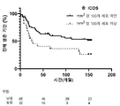
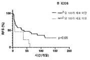
(i) ICOS+ 밀도: ICOS 단백질을 발현하는 세포, 즉 ICOS 양성(ICOS+) 세포의 밀도 또는 농도,(i) ICOS+ density: the density or concentration of cells expressing the ICOS protein, i.e. ICOS positive (ICOS+) cells,
(ii) ICOS+ TReg 비율: ICOS 양성인 TReg의 비율을 나타내는, ICOS+인 FOXP3+ 세포의 비율,(ii) ICOS+ TReg ratio: the ratio of FOXP3+ cells that are ICOS+, representing the ratio of TReg that are ICOS positive;
(iii) 세포간 근접성: ICOS+ FOXP3+ : ICOS+ FOXP3- 세포간 근접성, 각각의 ICOS 단일 양성(즉, ICOS 양성 FOXP3 음성) 세포와 이의 가장 가까운 ICOS FOXP3 이중 양성 세포 사이의 평균 거리이며, 이는 ICOS 양성 TReg와 다른 ICOS 양성 세포(ICOS+ TEff를 포함함) 사이의 근접성을 나타냄, 및(iii) Intercellular Proximity: ICOS+FOXP3+ : ICOS+FOXP3- Intercellular Proximity, the average distance between each ICOS single positive (i.e., ICOS positive FOXP3 negative) cell and its nearest ICOS FOXP3 double positive cell, which is the ICOS positive TReg and other ICOS-positive cells (including ICOS+ TEff), and
(iv) 구역 영향 비율: ICOS 단일 양성 세포의 총 수에 대한 임의의 ICOS 단일 양성 세포의 정의된 영역("영향 반경") 내의 ICOS FOXP3 이중 양성 세포의 수의 비율, 여기서 영향 반경은 ICOS FOXP3 이중 양성 세포와 ICOS 단일 양성 세포 사이에서 세포-세포 및/또는 사이토카인-의존성 통신이 발생할 수 있는 거리(예를 들어, 30 μm)를 나타냄.(iv) Zone Impact Ratio : The ratio of the number of ICOS FOXP3 double-positive cells within a defined area of any ICOS single-positive cell ("radius of influence") to the total number of ICOS single-positive cells, where the radius of influence is the ICOS FOXP3 double-positive cell. Indicates the distance (eg 30 μm) over which cell-cell and/or cytokine-dependent communication can occur between a positive cell and an ICOS single positive cell.
이들 바이오마커 각각은 환자가 항-ICOS 및/또는 항-TReg 면역요법으로부터 이익을 얻을 것이라는 기대와 양의 상관 관계가 있다. 일반적으로, 바이오마커가 높을수록(밀도, 비율, 근접성 또는 구역 영향이 클수록), 치료의 부재시 환자의 예후는 더 좋지 않지만, 환자가 항-ICOS 및/또는 또는 항-TReg 개입에 반응을 할 가능성이 더 크다. 따라서, 본 명세서에 기재된 바와 같은 바이오마커의 측정은 치료를 위한 적절한 환자 선택을 용이하게 하여 유익한 항-종양 반응을 생성할 가능성이 가장 높은 환자에게 면역요법을 선택적으로 투여할 수 있게 한다.Each of these biomarkers positively correlates with the expectation that patients will benefit from anti-ICOS and/or anti-TReg immunotherapy. In general, the higher the biomarker (the greater the density, proportion, proximity or zonal effect), the poorer the patient's prognosis in the absence of treatment, but the likelihood that the patient will respond to anti-ICOS and/or anti-TReg interventions. this is bigger Thus, measurement of biomarkers as described herein facilitates selection of appropriate patients for treatment, enabling the selective administration of immunotherapy to patients most likely to produce a beneficial anti-tumor response.
이러한 바이오마커의 세부 사항은 첨부된 표 B에 요약되어 있으며 하기에 기재되어 있다. 바이오마커 (ii), (iii) 및 (iv)는 면역억제성 TME의 지표이다.The details of these biomarkers are summarized in the attached Table B and are described below. Biomarkers (ii), (iii) and (iv) are indicative of immunosuppressive TMEs.
FOXP3은 TReg의 알려진 마커이고, ICOS 및 FOXP3 둘 다 발현하는 세포는 TReg의 면역억제성이 높은 하위그룹으로 식별 가능하다. 본 발명자들은 본 명세서에 TReg에 대한 식별 마커로서 FOXP3의 사용을 기재하지만, TReg를 선택적으로 식별하기 위해 대체 마커를 쉽게 사용할 수 있음을 이해할 것이다. 본 발명자들이 본 명세서에 개시한 바와 같이, ICOS+ 세포의 더 큰 밀도, ICOS+인 TReg의 더 큰 비율, 및 ICOS+ TReg와 다른 ICOS+ T 세포(FOXP3-) 사이의 더 가까운 근접성은 모두 환자에 대한 더 나쁜 예후와 연관되며, 예를 들어 동일한 종양 유형을 가지는 다른 환자에 비해 감소된 생존 기간 또는 감소된 무재발 생존 기간(RFS), 무진행 생존 기간(PFS) 또는 종양 진행 시간(TTP)으로 나타낸다. 그러나 긍정적으로 생각해보면, 이러한 환자는 특히 항-ICOS 및/또는 항-TReg 면역요법에 반응할 가능성이 있는 하위그룹을 나타낸다.FOXP3 is a known marker of TReg, and cells expressing both ICOS and FOXP3 can be identified as a highly immunosuppressive subgroup of TReg. Although we describe herein the use of FOXP3 as a discriminating marker for TRegs, it will be appreciated that alternative markers can readily be used to selectively identify TRegs. As we have disclosed herein, a greater density of ICOS+ cells, a greater proportion of TRegs that are ICOS+, and a closer proximity between ICOS+ TRegs and other ICOS+ T cells (FOXP3-) all lead to a worse outcome for the patient. It is associated with prognosis and is indicated, for example, by reduced survival time or reduced recurrence-free survival (RFS), progression-free survival (PFS) or time to tumor progression (TTP) compared to other patients with the same tumor type. On a positive note, however, these patients represent a subgroup particularly likely to respond to anti-ICOS and/or anti-TReg immunotherapy.
본 명세서에 기재된 KY1044 항체와 같은 Fc 이펙터 양성 항-ICOS 항체와 같은 항-TReg 치료제를 이용한 치료는 TReg의 수를 감소시키고, TME에서 TEff/TReg 사이의 비율을 개선시키며, 이에 의해 종양에 대한 환자의 면역 반응을 증진시켜, 종양 성장 감소 및 바람직하게는 환자에서 종양 또는 종양들의 크기 감소 및 궁극적인 근절을 야기할 수 있다. 다른 비 TReg-고갈 항-ICOS 항체는 TEff에 의한 사이토카인 생산을 향상시켜 TEff 활성을 증진시킴으로써 항-종양 면역 반응을 유사하게 개선시킬 것이다.Treatment with an anti-TReg therapeutic, such as an Fc effector positive anti-ICOS antibody, such as the KY1044 antibody described herein, reduces the number of TRegs and improves the ratio between TEff/TReg in the TME, thereby helping patients with tumor may enhance the immune response, resulting in reduced tumor growth and preferably reduced size and eventual eradication of the tumor or tumors in the patient. Other non-TReg-depleting anti-ICOS antibodies will similarly improve anti-tumor immune responses by enhancing TEff activity by enhancing cytokine production by TEff.
본 발명자들은 정의된 바이오마커가 개별 종양 샘플 및 환자 그룹에서 어떻게 측정되고 정량화될 수 있는지 본 명세서에 기재하여, 환자를 반응자 대 비-반응자 그룹으로 유용하게 분류하는 컷오프 값을 제공한다. 종양 생물학의 복잡한 특성과 환자의 다양성을 고려하면, 이러한 바이오마커를 사용하여 만든 예측은 100% 정확할 수 없지만, 여전히 확률을 기반으로 한 유용한 가이드를 제공할 것이다. 따라서, 본 발명은 환자가 이익을 얻을 치료법에 환자를 적합하게 일치시킬 가능성을 증가시킨다. 따라서 암 환자는 치료법에 대한 반응의 상대적 가능성의 표시를 제공하기 위해 본 발명에 따른 바이오마커를 사용하여 스크리닝될 수 있으며, 이 정보는 본 발명에 따른 면역요법 과정을 시작할지 여부를 결정하는 데 있어서 및/또는 채택할 면역요법 과정(예를 들어, 어떠한 면역요법 또는 어떠한 약제 조합)을 결정하는 데 있어서 임상의와 환자 둘다에게 가치가 있다.We describe herein how defined biomarkers can be measured and quantified in individual tumor samples and groups of patients, providing cutoff values that usefully classify patients into responder versus non-responder groups. Given the complex nature of tumor biology and patient variability, predictions made using these biomarkers may not be 100% accurate, but will still provide a useful guide based on probabilities. Thus, the present invention increases the likelihood of appropriately matching a patient to a therapy from which the patient will benefit. Thus, cancer patients can be screened using a biomarker according to the present invention to provide an indication of the relative likelihood of a response to therapy, and this information can be used in determining whether to initiate a course of immunotherapy according to the present invention. and/or which immunotherapeutic course to employ (eg, which immunotherapy or which drug combination) to employ.
본 발명의 구현예는 간세포암(HCC)과 관련하여 개시되며, 이는 연구된 환자 코호트에 대한 TME 기반의 다음 정량적 측정으로 예시된다:Embodiments of the present invention are disclosed in relation to hepatocellular carcinoma (HCC), which is exemplified by the following quantitative measures based on TME for the cohort of patients studied:
- mm2당 120개 초과의 세포로 측정된, ICOS+ 세포의 고밀도;- high density of ICOS+ cells, measured as greater than 120 cells per mm 2 ;
- HCC가 B형 간염 바이러스(HBV) 감염과 연관이 있는 것으로 알려지거나 미국 암 연합 위원회(American Joint Committee on Cancer; AJCC)의 기준에 따라 2기 이상의 HCC인 경우 mm2당 100개 초과의 세포로 측정된, ICOS+ 세포의 고밀도[28];- If HCC is known to be associated with hepatitis B virus (HBV) infection or if HCC is
- FOXP3+ 세포의 절반 초과가 ICOS+인 것으로 측정된, ICOS+인 FOXP3+ 세포의 높은 비율;- a high proportion of FOXP3+ cells that are ICOS+, determined as more than half of the FOXP3+ cells are ICOS+;
- 평균 세포간 거리가 105 μm 미만인 것으로 측정된, ICOS+ FOXP3- 음성(ICOS 단일 양성) 세포와 가장 가까운 이웃 ICOS+ FOXP3+ 이중 양성 세포 사이의 인접성; - proximity between ICOS+ FOXP3- negative (ICOS single positive) cells and nearest neighbor ICOS+ FOXP3+ double positive cells, measured as an average intercellular distance of less than 105 μm;
- 모든 ICOS 단일 양성 세포에 대한 ICOS 단일 양성 세포의 영향 반경 30 μm 내에서 ICOS FOXP3 이중 양성 세포 수의 높은 비율.- A high percentage of the number of ICOS FOXP3 double-positive cells within a 30 μm radius of influence of ICOS single-positive cells to all ICOS single-positive cells.
이러한 기준 중 하나 이상(바람직하게는 모두)을 충족하는 HCC 환자는 본 명세서에 기재된 바와 같이 항-ICOS 및/또는 항-TReg 면역요법을 이용한 치료에 대해 선택될 수 있다.HCC patients who meet one or more (preferably all) of these criteria may be selected for treatment with anti-ICOS and/or anti-TReg immunotherapy as described herein.
참조 값은 일반적으로 특정 임상 특성(예컨대, 암 하위 유형)을 가진 환자와 관련하여 결정될 것이며 비교 가능한 환자의 예후에 가장 잘 사용된다. 상기는 본 명세서에 예시된 HCC 환자 그룹에 대한 예시적인 구현예이고, 다른 적합한 컷오프 값은 다른 종양 유형 및/또는 환자 집단을 참조하여 결정될 수 있다. 따라서, 본 명세서에 예시된 참조 값이 HCC 이외의 다른 암 환자의 예후에 선택적으로 적용될 수 있지만, 가능한 경우 표적 암 유형 또는 분자 하위 유형(조직 불문)을 가진 환자로부터의 데이터 평가에 의해 이러한 더 넓은 맥락에서의 사용이 먼저 확인되어야 한다. 본 발명자들은 예측된 암 예후 및 항-ICOS 및/또는 항-TReg 면역요법으로부터 이익을 얻을 가능성에 따라 환자를 구별하기 위한 적합한 참조 값 및 공식을 결정하는 방법을 기재한다. HCC에 대한 참조 값의 결정 및 사용은 실시예에 설명되어 있으며 동등한 방법을 사용하여 다른 암에 대한 참조 값을 결정하고 효율적으로 사용할 수 있다.Reference values will generally be determined with respect to patients with specific clinical characteristics (eg, cancer subtypes) and are best used for comparable patient prognoses. The above is an exemplary embodiment for the HCC patient group exemplified herein, and other suitable cutoff values may be determined with reference to other tumor types and/or patient populations. Thus, while the reference values exemplified herein may be selectively applied to the prognosis of patients with cancers other than HCC, where possible, evaluation of data from patients with the target cancer type or molecular subtype (regardless of tissue) may result in these broader outcomes. Use in context must first be identified. We describe methods for determining suitable reference values and formulas for differentiating patients according to their predicted cancer prognosis and likelihood of benefiting from anti-ICOS and/or anti-TReg immunotherapy. Determination and use of reference values for HCC are described in the Examples, and equivalent methods can be used to determine and effectively use reference values for other cancers.
본 발명은,The present invention,
예를 들어 생존 기간, 무재발 생존 기간(RFS), 무진행 생존 기간(PFS) 및/또는 종양 진행 시간(TTP)일 수 있는 생존을 예측하기 위해, 환자의 암 예후에서;in a patient's cancer prognosis, to predict survival, which may be, for example, survival time, recurrence-free survival (RFS), progression-free survival (PFS) and/or time to tumor progression (TTP);
적절한 치료에 대한 환자의 매칭에서, 예를 들어 항-ICOS 및/또는 항-TReg 면역요법으로 치료할 환자를 선택하고 이익을 받을 가능성이 더 많을 것으로 식별된 환자에 대해 항-ICOS 및/또는 항-TReg 면역요법을 처방함에 있어서;In matching patients to appropriate treatment, for example, selecting patients to be treated with anti-ICOS and/or anti-TReg immunotherapy and selecting anti-ICOS and/or anti-TReg immunotherapy for those identified as more likely to benefit. in prescribing TReg immunotherapy;
환자가 치료에 반응하는지 여부의 초기 지표로서 항-ICOS 및/또는 항-TReg 면역요법에 대한 세포 반응을 모니터링함에 있어서;in monitoring cellular responses to anti-ICOS and/or anti-TReg immunotherapy as an early indicator of whether a patient is responding to treatment;
환자로부터의 바이오마커 데이터가 질환 이력 및/또는 치료법에 대한 반응에 대해 맵핑되어 향후 환자에 대한 임상 평가 및 치료를 개선시킬 수 있는 모델을 제공하거나 개선시키는 의료 연구에서In medical research, where biomarker data from patients is mapped against disease history and/or response to therapy to provide or improve models that can improve clinical evaluation and treatment for future patients.
와 같은 양태를 포함하여, 의학적 맥락에서 바이오마커의 적용에 관한 것이다.It relates to the application of biomarkers in a medical context, including aspects such as.
하나 이상의 바이오마커는, 환자로부터의 종양 코어 조직에서 결정된 다음과 같은 바이오마커 중 하나 이상을 포함하여, 환자에서 암성 고형 종양의 예후에 사용될 수 있다:One or more biomarkers can be used for prognosis of a cancerous solid tumor in a patient, including one or more of the following biomarkers determined in tumor core tissue from the patient:
(i) ICOS 단일 양성 세포의 총 수에 대한 ICOS 단일 양성 세포 주위의 정의된 영향 반경 내의 ICOS FOXP3 이중 양성 세포 수의 비율,(i) The ratio of the number of ICOS FOXP3 double positive cells within a defined radius of influence around ICOS single positive cells to the total number of ICOS single positive cells,
(ii) 각각의 ICOS 양성 FOXP3 음성 세포와 이의 가장 가까운 ICOS+ FOXP3+ 이중 양성 세포 사이의 평균 거리,(ii) the average distance between each ICOS positive FOXP3 negative cell and its nearest ICOS+ FOXP3+ double positive cell;
(iii) ICOS 양성인 FOXP3 양성 세포의 비율, 및(iii) the percentage of FOXP3-positive cells that are ICOS-positive, and
(iv) ICOS 양성 세포의 밀도.(iv) Density of ICOS-positive cells.
따라서, 제1 양태에서, 본 발명은Accordingly, in a first aspect, the present invention provides
환자로부터 얻은 종양 코어 조직의 샘플을 제공하는 단계,providing a sample of tumor core tissue obtained from the patient;
상기 샘플에서in the above sample
(i) ICOS 단일 양성 세포의 총 수에 대한 ICOS 단일 양성 세포 주위의 정의된 영향 반경 내의 ICOS FOXP3 이중 양성 세포 수의 비율,(i) The ratio of the number of ICOS FOXP3 double positive cells within a defined radius of influence around ICOS single positive cells to the total number of ICOS single positive cells,
(ii) 각각의 ICOS 양성 FOXP3 음성 세포와 이의 가장 가까운 ICOS+ FOXP3+ 이중 양성 세포 사이의 평균 거리,(ii) the average distance between each ICOS positive FOXP3 negative cell and its nearest ICOS+ FOXP3+ double positive cell;
(iii) ICOS 양성인 FOXP3 양성 세포의 비율, 및(iii) the percentage of FOXP3-positive cells that are ICOS-positive, and
(iv) ICOS 양성 세포의 밀도(iv) Density of ICOS-positive cells
와 같은 바이오마커 중 하나 이상을 결정하는 단계, 및Determining one or more of the biomarkers such as, and
상기 하나 이상의 바이오마커를 기반으로 하여 환자에 대한 예후를 제공하는 단계Providing a prognosis for a patient based on the one or more biomarkers
를 포함하는, 환자에서의 암성 고형 종양의 예후를 위한 방법을 제공하며, 여기서 더 짧은 생존은Provided is a method for prognosis of a cancerous solid tumor in a patient, comprising a shorter survival
ICOS 단일 양성 세포의 총 수에 대한 ICOS 단일 양성 세포 주위의 정의된 영향 반경 내의 ICOS FOXP3 이중 양성 세포 수의 더 큰 비율,greater ratio of the number of ICOS FOXP3 double-positive cells within a defined radius of influence around ICOS single-positive cells to the total number of ICOS single-positive cells;
각각의 ICOS 양성 FOXP3 음성 세포와 이의 가장 가까운 ICOS+ FOXP3+ 이중 양성 세포 사이의 더 짧은 평균 거리,the shorter average distance between each ICOS positive FOXP3 negative cell and its nearest ICOS+ FOXP3+ double positive cell;
ICOS 양성인 FOXP3 양성 세포의 더 큰 비율, 및/또는a greater proportion of FOXP3-positive cells that are ICOS-positive, and/or
ICOS 양성 세포의 더 큰 밀도Greater density of ICOS-positive cells
에 의해 나타난다.appears by
ICOS 단일 양성 세포의 총 수에 대한 ICOS 단일 양성 세포 주위의 정의된 영향 반경 내의 ICOS FOXP3 이중 양성 세포 수의 비율이 결정되고 참조 값과 비교될 수 있다. 참조 값보다 높은 수치는 생존 기간이 더 짧다는 예후를 나타내고 참조 값보다 낮은 수치는 생존 기간이 더 길다는 예후를 나타낸다. 예를 들어, HCC에서 본 발명자들은 영향 반경이 30 μm(30 마이크로미터)인 경우 환자 분류에 대한 참조 값이 0.1인 것으로 결정하였다. 따라서, ICOS 단일 양성 세포의 총 수에 대한 ICOS 단일 양성 세포 주위의 정의된 영향 반경 내의 ICOS FOXP3 이중 양성 세포의 비율이 0.1 초과인 것으로 결정되는 경우, 이는 생존 기간이 더 짧다는 것을 나타낸다.The ratio of the number of ICOS FOXP3 double positive cells within a defined radius of influence around the ICOS single positive cells to the total number of ICOS single positive cells can be determined and compared to a reference value. A number higher than the reference value indicates a prognosis of a shorter survival period, and a number lower than the reference value indicates a prognosis of a longer survival period. For example, in HCC we have determined that the reference value for patient classification is 0.1 when the radius of influence is 30 μm (30 micrometers). Thus, if the ratio of ICOS FOXP3 double-positive cells within a defined radius of influence around the ICOS single-positive cells to the total number of ICOS single-positive cells is determined to be greater than 0.1, this indicates a shorter survival period.
유사하게, 각각의 ICOS+ FOXP3-(ICOS 단일 양성) 세포와 이의 가장 가까운 ICOS+ FOXP3+(ICOS FOXP3 이중 양성) 세포 사이의 평균 거리가 결정되고 참조 값과 비교될 수 있다. 참조 값 미만의 거리는 생존 기간이 더 짧다는 예후를 나타내는 반면, 참조 값 초과의 거리는 생존 기간이 더 길다는 예후를 나타낸다. 예를 들어, 종양이 HCC인 경우, 105 μm의 참조 값이 사용될 수 있다. 따라서, 각각의 ICOS 단일 양성 세포와 이의 가장 가까운 ICOS+ FOXP3+ 이중 양성 세포 사이의 평균 거리가 105 μm 미만인 것은 HCC에서 생존 기간이 더 짧다는 것을 나타내는 반면, 각각의 ICOS 단일 양성 세포와 이의 가장 가까운 ICOS+ FOXP3+ 이중 양성 세포 사이의 평균이 105 μm 초과인 것은 HCC에서 생존 기간이 더 길다는 것을 나타낸다.Similarly, the average distance between each ICOS+ FOXP3− (ICOS single positive) cell and its nearest ICOS+ FOXP3+ (ICOS FOXP3 double positive) cell can be determined and compared to a reference value. Distances below the reference value indicate a prognosis of shorter survival, while distances above the reference value indicate a prognosis of longer survival. For example, if the tumor is HCC, a reference value of 105 μm may be used. Thus, an average distance of less than 105 μm between each ICOS single-positive cell and its nearest ICOS+ FOXP3+ double-positive cell indicates shorter survival in HCC, whereas each ICOS single-positive cell and its nearest ICOS+ FOXP3+ A mean between double-positive cells greater than 105 μm indicates longer survival in HCC.
ICOS 양성인 FOXP3 양성 세포의 비율이 결정되고 참조 값과 비교될 수 있다. 비율이 참조 값보다 크면 생존 기간이 더 짧다는 예후를 나타내는 반면, 비율이 참조 값보다 작으면 생존 기간이 더 길다는 예후를 나타낸다. 예를 들어, 종양이 HCC인 경우, 0.5의 참조 값이 사용될 수 있다. 따라서, FOXP3 양성 세포의 절반 초과가 ICOS 양성이면 이는 생존 기간이 더 짧다는 것을 나타내는 반면, FOXP3 양성 세포의 절반 미만이 ICOS 양성이면 이는 생존 기간이 더 길다는 것을 나타낸다.The percentage of FOXP3 positive cells that are ICOS positive can be determined and compared to a reference value. A ratio greater than the reference value is indicative of a shorter survival period, whereas a ratio less than the reference value is indicative of a longer survival period. For example, if the tumor is HCC, a reference value of 0.5 may be used. Thus, if more than half of the FOXP3 positive cells are ICOS positive, this indicates a shorter survival period, whereas if less than half of the FOXP3 positive cells are ICOS positive, this indicates a longer survival period.
ICOS 양성 세포의 밀도가 결정되고 참조 값과 비교될 수 있다. 참조 값 초과의 밀도는 생존 기간이 더 짧다는 예후를 나타내는 반면, 참조 값 미만의 밀도는 생존 기간이 더 길다는 예후를 나타낸다. 예를 들어, 종양이 HCC인 경우, mm2당 120개 ICOS 양성 세포의 참조 값이 사용될 수 있다. 따라서, mm2당 120개 초과인 ICOS+ 세포의 밀도는 생존 기간이 더 짧다는 것을 나타내는 반면, mm2당 120개 미만인 밀도는 생존 기간이 더 길다는 것을 나타낸다. 종양이 B형 간염 바이러스 감염과 연관된 HCC이거나 2기 이상의 HCC인 경우, mm2당 100개 ICOS 양성 세포의 참조 값이 사용될 수 있다. 따라서 이러한 하위 유형의 HCC를 나타내는 환자에 대한 예후는 이러한 더 낮은 참조 값을 사용할 수 있는 반면, 다른 유형의 HCC 또는 HBV와 연관되거나 2기 이상의 HCC인 것으로 식별되지 않은 HCC를 나타내는 환자에 대한 예후는 HCC는 mm2당 120개 세포인 더 높은 참조 값을 사용할 수 있다. 후자는 1기 HCC로 진단된 환자를 포함한다.The density of ICOS positive cells can be determined and compared to a reference value. A density above the reference value is indicative of a shorter survival period, whereas a density below the reference value is indicative of a longer survival period. For example, if the tumor is HCC, a reference value of 120 ICOS positive cells per mm 2 can be used. Thus, densities of ICOS+ cells greater than 120 per mm 2 indicate shorter survival times, whereas densities less than 120 per mm 2 indicate longer survival times. If the tumor is HCC associated with hepatitis B virus infection or is more than
첨부된 실시예에서 설명되는 바와 같이, 참조 값은 통계적 모델링으로부터 유래된 것인 한편, 참조 값은 유래된 모델의 데이터에 대해 "최적합"을 나타낼 수 있지만, 환자 분류 및 예후에 대한 대략적인 가이드의 역할만 할 수 있으므로 절대적으로 결정적인 것으로 간주되어서는 안 된다. "30 μm 반경 내의 ICOS FOXP3 이중 양성 세포 0.1", "각각의 ICOS+ FOXP3- 세포와 이의 가장 가까운 ICOS+ FOXP3+ 세포 사이의 평균 거리 105 μm", "ICOS 양성인 FOXP3 양성 세포 50%", "mm2당 120개 ICOS+ 세포" 또는 "mm2당 100개 ICOS+ 세포"와 같은 본 명세서에 제공된 정확한 값은 단지 예시적인 구현예를 나타내며, 본 발명은 여전히 예측 값을 유지하면서 이들 정확한 값의 변형을 사용하여 실시될 수 있음이 이해될 것이다. 예를 들어, 영향 반경을 25 μm로 정의하고 ICOS FOXP3 이중 양성 세포의 임계 수를 0.2로 정의한다면, 환자가 상대적으로 긴 생존이 예상될 수 있는 환자인지 여부 및 이러한 환자가 항-ICOS 및/또는 항-TReg 치료제를 이용한 치료로부터 이익을 얻을 가능성이 어 많거나 적을 것인지 여부를 유용하게 추정할 수 있을 것으로 예상될 것이다.As illustrated in the accompanying examples, reference values are derived from statistical modeling, while reference values may represent a "best fit" to the data of the model from which they are derived, but a rough guide to patient classification and prognosis. should not be regarded as absolutely deterministic. “0.1 ICOS FOXP3 double positive cells within a radius of 30 μm”, “Average distance between each ICOS+ FOXP3− cell and its nearest ICOS+ FOXP3+ cell 105 μm”, “50% of FOXP3 positive cells that are ICOS positive”, “120 per mm 2 Precise values provided herein, such as "dog ICOS+ cells" or "100 ICOS+ cells per mm 2 ," represent exemplary embodiments only, and the present invention may be practiced using variations of these exact values while still retaining predicted values. It will be understood that For example, if the radius of influence is defined as 25 μm and the threshold number of ICOS FOXP3 double positive cells is defined as 0.2, it is possible to determine whether the patient is a patient for whom relatively long survival can be expected and whether such patient is anti-ICOS and/or It would be expected to be useful in estimating how much or less likely there would be a benefit from treatment with an anti-TReg therapeutic.
생존은 다양한 방식으로 정의되고 측정될 수 있다. 가장 간단하게 생존은 전체 생존 기간(OS), 즉 환자가 사망할 때까지의 시간 길이로 정의될 수 있다. 다른 생존-관련 임상 평가변수는 무재발 생존 기간(RFS), 무진행 생존 기간(PFS) 및 종양 진행 시간(TTP)을 포함하는 것과 같이, 측정된 생존 시간에 걸쳐 환자의 질환 상태 측정을 포함한다. 간단히 말해서, RFS는 치료된 암이 재발할 때까지의 시간 길이이고, PFS는 암이 악화(진행)할 때까지의 시간 길이이며, TTP는 PFS와 유사하지만 암과 관련되지 않은 원인으로 사망한 환자를 계산하지 않는다. 본 발명의 바이오마커는 환자의 질환 생존 능력을 예측하므로, 따라서 OS, RFS, PFS 및 TTP 각각의 기간과 상관 관계가 있을 것으로 예상된다. 이러한 맥락에서, 예측되는 생존 기간은 항-ICOS 및/또는 항-TReg 면역치료제를 이용한 치료의 부재시 생존이며, 즉 예후는 환자가 본 명세서의 다른 곳에 기재된 바와 같이 항-ICOS 및/또는 항-TReg 면역요법을 받을 방법 또는 이를 받을 것인지 여부에 상관없이 이루어진다.Survival can be defined and measured in a variety of ways. Most simply, survival can be defined as overall survival (OS), ie the length of time until the patient dies. Other survival-related clinical endpoints include measures of a patient's disease status over a measured survival time, such as recurrence-free survival (RFS), progression-free survival (PFS), and time to tumor progression (TTP). . Briefly, RFS is the length of time until the treated cancer returns, PFS is the length of time until the cancer gets worse (progression), and TTP is similar to PFS but for patients who die of causes not related to cancer. do not calculate Since the biomarkers of the present invention predict the patient's ability to survive the disease, they are therefore expected to be correlated with the duration of each of OS, RFS, PFS and TTP. In this context, the expected survival time is survival in the absence of treatment with an anti-ICOS and/or anti-TReg immunotherapeutic agent, i.e., the prognosis is that the patient has an anti-ICOS and/or anti-TReg immunotherapeutic agent as described elsewhere herein. This is done regardless of how you receive immunotherapy or whether you will receive it.
따라서 예후를 제공하는 것은 환자가 다른 비슷한 환자에 비해 더 긴 생존을 향유할 것인지 여부를 예측하는 것을 포함할 수 있다. 예후를 제공하는 것은, 예를 들어 추정된 개월 수 또는 년 수로서, OS, RFS, PFS 및 TTP의 지속 기간을 예측하는 것과 같은 생존 시간을 예측하는 것을 포함할 수 있다. 환자의 예측된 생존 기간(예를 들어, OS)은 선택적으로 조직 샘플을 채취한 날짜로부터 개월 수로 추정된다. 생존 기간은 바이오마커(들)가 이들의 상응하는 참조 값(들) 초과 또는 미만에 속하는지 여부에 따라 범위, 예를 들어 x개월 미만 또는 x개월 초과로 추정될 수 있다. 예를 들어, 본 명세서에 기재된 HCC 환자 집단에서, OS 중앙값은 100.3개월이었다. HCC를 나타내는 추가 환자의 경우, 구역 영향, 세포간 근접성, ICOS+ TReg 비율 및/또는 ICOS+ 세포 밀도에 대한 바이오마커 판독값이 결정되고 이 집단에 대해 정의된 참조 값과 비교되어 생존 예후가 100.3 개월 초과 또는 미만임을 제공할 것이다.Thus, providing a prognosis may include predicting whether a patient will enjoy a longer survival compared to other similar patients. Providing a prognosis may include predicting survival time, such as predicting duration of OS, RFS, PFS, and TTP, for example as an estimated number of months or years. The patient's predicted survival time (eg, OS) is optionally estimated in months from the date the tissue sample is taken. Survival time can be estimated in a range, eg less than x months or greater than x months, depending on whether the biomarker(s) fall above or below their corresponding reference value(s). For example, in the HCC patient population described herein, the median OS was 100.3 months. For additional patients presenting with HCC, biomarker readouts for area effect, intercellular proximity, ICOS+ TReg ratio, and/or ICOS+ cell density were determined and compared to reference values defined for this population, resulting in a survival prognosis greater than 100.3 months. or less than.
따라서 하나 이상의 이러한 바이오마커에 의해 예측된 바와 같이 상대적으로 불량한 예후를 가지는 것으로 식별된 환자는 예후가 더 낙관적인 환자보다 본 발명에 따른 면역요법에 반응할 확률이 더 높을 수 있다. 즉, 환자의 생존 기간이 상대적으로 짧을 것으로 예측되지만, 항-ICOS 및/또는 항-TReg 면역치료제를 이용한 치료에 의해 환자의 생존 기간이 연장될 가능성이 더 높다.Thus, patients identified as having a relatively poor prognosis as predicted by one or more of these biomarkers may have a higher probability of responding to immunotherapy according to the present invention than patients with a more optimistic prognosis. That is, although the patient's survival period is expected to be relatively short, it is more likely that the patient's survival period will be extended by treatment with an anti-ICOS and/or anti-TReg immunotherapeutic agent.
따라서, 본 발명의 제2 양태는 다른 환자보다 항-ICOS 및/또는 항-TReg 면역치료제를 이용한 치료에 반응할 가능성이 더 높은 환자를 식별하는 것에 관한 것이다. 본 발명의 이러한 양태는 환자 하위그룹(예를 들어, HCC 환자의 하위그룹)의 식별을 제공하며, 여기서 하위그룹은 바이오마커 또는 바이오마커 조합의 존재 또는 값에 의해 정의되고, 상기 하위그룹 내의 환자는 하위그룹 외부의 환자보다(즉, 정의된 바이오마커(들)를 나타내지 않는 환자와 비교하여) 항-ICOS 및/또는 항-TReg 면역요법에 더 반응할 것으로 예측된다.Accordingly, a second aspect of the present invention relates to identifying patients who are more likely than other patients to respond to treatment with an anti-ICOS and/or anti-TReg immunotherapeutic agent. This aspect of the invention provides identification of a patient subgroup (e.g., a subgroup of HCC patients), wherein the subgroup is defined by the presence or value of a biomarker or biomarker combination, and the patients within the subgroup are predicted to be more responsive to anti-ICOS and/or anti-TReg immunotherapy than patients outside the subgroup (i.e., compared to patients not expressing the defined biomarker(s)).
이러한 제2 양태에서, 본 발명은 In this second aspect, the present invention
환자로부터 얻은 종양 코어 조직의 샘플을 제공하는 단계, 및providing a sample of tumor core tissue obtained from the patient; and
상기 샘플에서in the above sample
(i) ICOS 단일 양성 세포의 총 수에 대한 ICOS 단일 양성 세포 주위의 정의된 영향 반경 내의 ICOS FOXP3 이중 양성 세포 수의 비율,(i) The ratio of the number of ICOS FOXP3 double positive cells within a defined radius of influence around ICOS single positive cells to the total number of ICOS single positive cells,
(ii) 각각의 ICOS+ FOXP3- 세포와 이의 가장 가까운 ICOS+ FOXP3+ 세포 사이의 평균 거리,(ii) the average distance between each ICOS+ FOXP3− cell and its nearest ICOS+ FOXP3+ cell,
(iii) ICOS 양성인 FOXP3 양성 세포의 비율, 및(iii) the percentage of FOXP3-positive cells that are ICOS-positive, and
(iv) ICOS 양성 세포의 밀도(iv) Density of ICOS-positive cells
와 같은 바이오마커 중 하나 이상을 결정하는 단계Determining one or more of the biomarkers such as
를 포함하는, 환자에서 암성 고형 종양이 항-ICOS 및/또는 항-TReg 면역치료제에 반응할 가능성을 결정하는 방법을 제공하며,Provides a method for determining the likelihood that a cancerous solid tumor in a patient will respond to an anti-ICOS and/or anti-TReg immunotherapy, comprising:
여기서 환자가 항-ICOS 및/또는 항-TReg 면역치료제에 반응할 가능성이 더 큰 것은 Which patient is more likely to respond to anti-ICOS and/or anti-TReg immunotherapy
ICOS 단일 양성 세포의 총 수에 대한 ICOS 단일 양성 세포 주위의 정의된 영향 반경 내의 ICOS FOXP3 이중 양성 세포 수의 더 큰 비율,greater ratio of the number of ICOS FOXP3 double-positive cells within a defined radius of influence around ICOS single-positive cells to the total number of ICOS single-positive cells;
각각의 ICOS 양성 FOXP3 음성 세포와 이의 가장 가까운 ICOS+ FOXP3+ 양성 세포 사이의 더 짧은 평균 거리,the shorter average distance between each ICOS-positive FOXP3-negative cell and its nearest ICOS+ FOXP3+-positive cell;
ICOS 양성인 FOXP3 양성 세포의 더 큰 비율, 및/또는a greater proportion of FOXP3-positive cells that are ICOS-positive, and/or
ICOS 양성 세포의 더 큰 밀도Greater density of ICOS-positive cells
에 의해 나타난다.appears by
따라서 상기 바이오마커 중 하나 이상은 환자에서 암성 종양이 항-ICOS 및/또는 항-TReg 면역치료제에 반응할 가능성을 결정하는 데 사용된다. 환자가 다른 환자에 비해 치료에 반응할 가능성이 증가 또는 감소하는지 여부가 결정될 수 있고/있거나 반응 가능성이 수치적으로 추정될 수 있다.Accordingly, one or more of these biomarkers are used to determine the likelihood that a cancerous tumor in a patient will respond to an anti-ICOS and/or anti-TReg immunotherapy. Whether a patient has an increased or decreased likelihood of responding to treatment relative to other patients can be determined and/or the likelihood of response can be numerically estimated.
선택적으로, 방법은 본 발명의 제1 양태에 대해 기재되고 본 명세서의 다른 곳에 예시된 바와 동일한 방식으로 하나 이상의 바이오마커를 상응하는 참조 값과 비교하는 단계를 포함한다. 참조 값은 환자의 참조 집단에 대해 계산되며, 예를 들어 HCC 환자에 대해 결정된 바이오마커는 HCC 환자 집단의 참조 값과 비교될 수 있다. 따라서 치료에 대한 반응 가능성의 증가 또는 감소는 참조 집단에서 치료에 대한 반응의 기대와 관련하여 결정된다.Optionally, the method comprises comparing one or more biomarkers to a corresponding reference value in the same manner as described for the first aspect of the invention and exemplified elsewhere herein. A reference value is calculated for a reference population of patients, eg, a biomarker determined for an HCC patient can be compared to a reference value for a population of HCC patients. Thus, an increase or decrease in the likelihood of response to treatment is determined in relation to the expectation of response to treatment in a reference population.
방법은 면역요법에 반응할 가능성이 증가된 환자를 식별함으로써 상기 환자의 치료를 위한 항-ICOS 및/또는 항-TReg 면역치료제를 선택하는 단계를 추가로 포함할 수 있다. 따라서 상기 바이오마커 중 하나 이상은 환자가 항-ICOS 및/또는 항-TReg 면역요법으로부터 이익을 얻을 가능성이 더 높은 것으로 식별하고, 선택적으로 이에 따라 항-ICOS 및/또는 항-TReg 면역치료제를 이용하여 치료할 환자를 선택하는 데 사용될 수 있다. 예를 들어, 정의된 참조 값을 초과하여 TME에서 ICOS+ 세포 수의 증가를 나타내는 HCC 환자는 항-ICOS 및/또는 항-TReg 면역치료제를 이용하여 치료하기에 적합한 것으로 식별될 수 있다. 그 다음 선택적으로, 항-ICOS 및/또는 항-TReg 면역치료제는 환자에게 처방 및/또는 투여된다.The method may further comprise selecting an anti-ICOS and/or anti-TReg immunotherapeutic agent for treatment of the patient by identifying the patient with an increased likelihood of responding to the immunotherapy. Accordingly, one or more of the above biomarkers identifies a patient as more likely to benefit from anti-ICOS and/or anti-TReg immunotherapy and, optionally, to use anti-ICOS and/or anti-TReg immunotherapy accordingly. and can be used to select patients for treatment. For example, HCC patients exhibiting an increase in the number of ICOS+ cells in the TME above a defined reference value can be identified as suitable for treatment with an anti-ICOS and/or anti-TReg immunotherapeutic agent. Optionally, an anti-ICOS and/or anti-TReg immunotherapeutic agent is then prescribed and/or administered to the patient.
바이오마커 판독값이 환자가 면역요법에 반응할 가능성이 더 낮은 것으로 나타나는 경우(비-반응자), 이 환자에게 상이한 치료가 권장될 수 있다. 대안적으로, 이러한 바이오마커 판독값은 환자가 하나 이상의 다른 치료와 조합하여 항-ICOS 및/또는 항-TReg 면역치료제로 치료되어야 하며, 조합으로 반응이 달성될 가능성이 더 높다는 것을 나타낼 수 있다. 예를 들어, 일부 치료는 본 명세서에 기재된 바이오마커에 영향을 미칠 수 있고, 바이오마커가 면역요법에 대한 증가된 역할을 나타내는 방향으로 변화하게 할 수 있다. 면역요법은 이러한 추가 치료와 함께 투여될 수 있으며, 다수의 치료는 임의의 순서로 함께(동시에) 또는 순차적으로 투여된다. 도 1.If the biomarker readings indicate that the patient is less likely to respond to immunotherapy (non-responder), a different treatment may be recommended for this patient. Alternatively, these biomarker readings may indicate that the patient should be treated with an anti-ICOS and/or anti-TReg immunotherapeutic agent in combination with one or more other treatments, with a greater likelihood of achieving a response with the combination. For example, some treatments can affect the biomarkers described herein and cause the biomarkers to change in ways that indicate an increased role for immunotherapy. Immunotherapy can be administered with these additional treatments, and multiple treatments are administered together (simultaneously) or sequentially in any order. Figure 1.
제3 양태에서, 본 발명은 본 명세서에 기재된 바이오마커에 의해 식별되는 암 환자를 항-ICOS 및/또는 항-TReg 면역요법으로 치료하는 것, 및 본 명세서에 기재된 바이오마커에 의해 식별된 환자를 치료하는 데 사용하기 위한 항-ICOS 및/또는 항-TReg 면역치료제에 관한 것이다. 항-ICOS 및/또는 항-TReg 면역치료제를 포함하는 약제학적 조성물은 이러한 환자에서 사용하기 위해 제공될 수 있다. 또한, 항-ICOS 및/또는 항-TReg 면역치료제는 이러한 환자에서 암성 고형 조양을 치료하기 위한 약제의 제조에 사용될 수 있다. 치료는 환자의 생존을 연장하는 것을 포함한다.In a third aspect, the present invention provides anti-ICOS and/or anti-TReg immunotherapy to treat cancer patients identified by the biomarkers described herein, and patients identified by the biomarkers described herein Anti-ICOS and/or anti-TReg immunotherapeutic agents for use in treatment. A pharmaceutical composition comprising an anti-ICOS and/or anti-TReg immunotherapeutic agent may be provided for use in such patients. In addition, anti-ICOS and/or anti-TReg immunotherapeutic agents may be used in the manufacture of medicaments for treating cancerous tumors in such patients. Treatment involves prolonging the patient's survival.
본 발명의 제3 양태에 따른 방법은 환자에서 암성 고형 종양을 치료하는 단계를 포함하며, 여기서 방법은A method according to a third aspect of the invention comprises treating a cancerous solid tumor in a patient, wherein the method comprises:
항-ICOS 및/또는 항-TReg 면역치료제에 반응할 가능성이 증가된 환자를 식별하는 단계, 및identifying patients with increased likelihood of responding to anti-ICOS and/or anti-TReg immunotherapy; and
항-ICOS 및/또는 항-TReg 면역치료제를 환자에게 투여하는 단계administering an anti-ICOS and/or anti-TReg immunotherapy to a patient
를 포함하며, 여기서 상기 환자는 본 발명의 제2 양태에 기재된 것과 같이 식별되거나 식별되었다. 따라서 예를 들어, 환자는 종양으로부터wherein the patient has been identified or identified as described in the second aspect of the present invention. Thus, for example, the patient is free from the tumor
ICOS 단일 양성 세포의 총 수에 대한 ICOS 단일 양성 세포 주위의 정의된 영향 반경 내의 ICOS FOXP3 이중 양성 세포 수의 비율(여기서, 상기 비율은 참조 값보다 큼),the ratio of the number of ICOS FOXP3 double positive cells within a defined radius of influence around ICOS single positive cells to the total number of ICOS single positive cells, where the ratio is greater than the reference value;
각각의 ICOS 양성 FOXP3 음성 세포와 이의 가장 가까운 ICOS 양성 FOXP3 양성 세포 사이의 평균 거리(여기서, 상기 거리는 참조 값 미만임),an average distance between each ICOS-positive FOXP3-negative cell and its nearest ICOS-positive FOXP3-positive cell, wherein said distance is less than a reference value;
ICOS 양성인 FOXP3 양성 세포의 비율(여기서, 상기 비율은 참조 값보다 큼), 및The proportion of FOXP3-positive cells that are ICOS-positive, wherein the proportion is greater than the reference value, and
ICOS 양성 세포의 밀도(여기서, 상기 밀도는 참조 값보다 큼)Density of ICOS positive cells, wherein the density is greater than the reference value
와 같은 바이오마커 판독값 중 하나 이상에 의해 식별될 수 있다.It can be identified by one or more of the biomarker readouts such as
따라서 항-ICOS 및/또는 항-TReg 면역치료제는 환자에서 암성 고형 종양을 치료하는 데 사용되며, 여기서 종양은 하나 이상의 정의된 바이오마커를 포함하는 것으로 결정되었다. 따라서, 항-ICOS 및/또는 항-TReg 면역치료제를 이용하여 치료하기 전에, 환자로부터 이전에 얻은 종양 코어 조직의 샘플에서 하나 이상의 바이오마커는 본 명세서에 기재된 바와 같은 바이오마커에 대한 참조 값과 비교함으로써 치료법의 적합성을 나타내는 것으로 결정되었을 수 있다. 예로서 HCC의 경우, 종양은Accordingly, anti-ICOS and/or anti-TReg immunotherapeutic agents are used to treat cancerous solid tumors in patients, wherein the tumors have been determined to contain one or more defined biomarkers. Thus, prior to treatment with an anti-ICOS and/or anti-TReg immunotherapeutic agent, one or more biomarkers in a sample of tumor core tissue previously obtained from a patient can be compared to reference values for biomarkers as described herein. may have been determined to indicate suitability of therapy. In the case of HCC as an example, the tumor is
ICOS 단일 양성 세포의 총 수에 대한 ICOS 단일 양성 세포 주위의 정의된 영향 반경 내의 ICOS FOXP3 이중 양성 세포 수의 비율(여기서, 상기 비율은 0.1보다 큼),the ratio of the number of ICOS FOXP3 double positive cells within a defined radius of influence around ICOS single positive cells to the total number of ICOS single positive cells, wherein the ratio is greater than 0.1;
각각의 ICOS 양성 FOXP3 음성 세포와 이의 가장 가까운 ICOS 양성 FOXP3 양성 세포 사이의 평균 거리(여기서, 상기 거리는 105 μm 미만임),an average distance between each ICOS positive FOXP3 negative cell and its nearest ICOS positive FOXP3 positive cell, wherein the distance is less than 105 μm;
ICOS 양성인 FOXP3 양성 세포의 비율(여기서, 상기 비율은 절반보다 큼), 및The proportion of FOXP3-positive cells that are ICOS-positive, wherein the proportion is greater than half, and
ICOS 양성 세포의 밀도(여기서, 상기 밀도는 mm2당 120개 세포보다 큼)Density of ICOS positive cells, wherein said density is greater than 120 cells per mm 2 .
와 같은 바이오마커 중 하나 이상을 포함하는 것으로 결정되었을 수 있다.It may have been determined to contain one or more of the biomarkers such as.
HBV와 연관된 HCC, 또는 2기 이상의 HCC의 경우, 종양은For HBV-associated HCC, or
ICOS 단일 양성 세포의 총 수에 대한 ICOS 단일 양성 세포 주위의 정의된 영향 반경 내의 ICOS FOXP3 이중 양성 세포 수의 비율(여기서, 상기 비율은 0.1보다 큼),the ratio of the number of ICOS FOXP3 double positive cells within a defined radius of influence around ICOS single positive cells to the total number of ICOS single positive cells, wherein the ratio is greater than 0.1;
각각의 ICOS 양성 FOXP3 음성 세포와 이의 가장 가까운 ICOS 양성 FOXP3 음성 세포 사이의 평균 거리(여기서, 상기 거리는 105 μm 미만임),an average distance between each ICOS positive FOXP3 negative cell and its nearest ICOS positive FOXP3 negative cell, wherein the distance is less than 105 μm;
ICOS 양성인 FOXP3 양성 세포의 비율(여기서, 상기 비율은 절반보다 큼), 및The proportion of FOXP3-positive cells that are ICOS-positive, wherein the proportion is greater than half, and
ICOS 양성 세포의 밀도(여기서, 상기 밀도는 mm2당 100개 세포보다 큼)Density of ICOS positive cells, wherein the density is greater than 100 cells per mm 2 .
와 같은 바이오마커 중 하나 이상을 포함하는 것으로 결정되었을 수 있다.It may have been determined to contain one or more of the biomarkers such as.
본 발명의 제4 양태는 본 명세서에 기재된 하나 이상의 바이오마커의 변화를 검출함으로써 항-ICOS 및/또는 항-TReg 면역요법에 대한 암 환자의 반응을 측정하는 것에 관한 것이다. 이러한 변화는 반응의 특징을 나타낼 수 있으며, 외부에서 볼 수 있는 임상 징후보다 훨씬 더 일찍 검출가능하여서, 치료가 질환의 진행을 억제할 생물학적 효과를 달성하고 있는지 여부에 대한 유용한 지표를 제공할 수 있다.A fourth aspect of the present invention relates to measuring the response of a cancer patient to anti-ICOS and/or anti-TReg immunotherapy by detecting changes in one or more biomarkers described herein. These changes can characterize a response and can be detected much earlier than outwardly visible clinical signs, providing a useful indicator of whether a treatment is achieving a biological effect that will inhibit disease progression. .
암성 고형 종양을 치료하기 위해 투여된 항-ICOS 및/또는 항-TReg 면역치료제에 대한 환자의 반응을 모니터링하는 방법은A method of monitoring a patient's response to an anti-ICOS and/or anti-TReg immunotherapy administered to treat a cancerous solid tumor is
항-ICOS 및/또는 항-TReg 면역치료제의 투여 후 환자로부터 얻은 종양 코어 조직의 테스트 샘플을 제공하는 단계,providing a test sample of tumor core tissue obtained from the patient after administration of anti-ICOS and/or anti-TReg immunotherapy;
상기 샘플에서in the above sample
(i) ICOS 단일 양성 세포의 총 수에 대한 ICOS 단일 양성 세포 주위의 정의된 영향 반경 내의 ICOS FOXP3 이중 양성 세포 수의 비율,(i) The ratio of the number of ICOS FOXP3 double positive cells within a defined radius of influence around ICOS single positive cells to the total number of ICOS single positive cells,
(ii) 각각의 ICOS+ FOXP3- 세포와 이의 가장 가까운 ICOS+ FOXP3+ 세포 사이의 평균 거리,(ii) the average distance between each ICOS+ FOXP3− cell and its nearest ICOS+ FOXP3+ cell,
(iii) ICOS 양성인 FOXP3 양성 세포의 비율, 및(iii) the percentage of FOXP3-positive cells that are ICOS-positive, and
(iv) ICOS 양성 세포의 밀도(iv) Density of ICOS-positive cells
와 같은 바이오마커 중 하나 이상을 결정하는 단계,Determining one or more of the biomarkers such as,
상기 테스트 샘플에서의 상기 하나 이상의 바이오마커를 환자로부터 얻은 종양 코어 조직의 이전 샘플에서의 동일한 하나 이상의 바이오마커와 비교하는 단계, 및comparing the one or more biomarkers in the test sample to the same one or more biomarkers in a previous sample of tumor core tissue obtained from the patient; and
상기 하나 이상의 바이오마커에서 변화가 발생했는지 여부를 결정하는 단계Determining whether a change has occurred in the one or more biomarkers
를 포함할 수 있다.can include
테스트 샘플은 면역치료제를 투여한 후, 면역치료제가 효과를 나타내도록 일정 시간 동안 허용한 후에 얻어진다. 예를 들어, 테스트 샘플은 상기 면역치료제의 투여 후 적어도 3일 후에 채취될 수 있다. 테스트 샘플은 바람직하게는 상기 투여 2, 4, 6 또는 8주 이내에 채취된다.A test sample is obtained after administration of the immunotherapeutic agent and after allowing the immunotherapeutic agent to take effect for a period of time. For example, a test sample can be taken at least 3 days after administration of the immunotherapeutic agent. A test sample is preferably taken within 2, 4, 6 or 8 weeks of said administration.
이전 샘플은 테스트 샘플을 얻기 전, 바람직하게는 상기 면역치료제의 투여 전에 환자 치료의 더 이른 시점에 환자로부터 얻어진다. 이전 샘플은 테스트 샘플을 얻기 전, 바람직하게는 상기 면역치료제의 투여 전에 환자 치료의 더 이른 시점에 환자로부터 얻어진다. 이전 샘플은 상기 면역치료제의 투여 전(선택적으로 직전, 예를 들어 최대 14일 전), 투여 시점에(예를 들어, 동일한 날에), 또는 투여 직후(예를 들어, 다음 날)에 얻어질 수 있다. 이전 샘플은 면역치료제를 이용한 치료 과정을 시작하기 전 또는 시작 시(예를 들어, 최대 14일 전), 면역치료제의 초기 투여 시점에(예를 들어, 동일한 날에), 또는 투여 직후(예를 들어, 다음 날) 얻은 초기 샘플일 수 있다.A prior sample is obtained from the patient at an earlier point in the patient's treatment prior to obtaining the test sample, preferably prior to administration of the immunotherapeutic agent. A prior sample is obtained from the patient at an earlier point in the patient's treatment prior to obtaining the test sample, preferably prior to administration of the immunotherapeutic agent. A prior sample may be obtained prior to (optionally immediately prior to, eg, up to 14 days prior to) administration of the immunotherapeutic agent, at the time of administration (eg, on the same day), or immediately after administration (eg, the next day). can Previous samples may be taken prior to or at the beginning of a course of treatment with immunotherapy (eg, up to 14 days prior), at the time of initial administration of immunotherapy (eg, on the same day), or immediately after administration (eg, on the same day). For example, it may be an initial sample obtained the next day).
테스트 샘플은 통상적으로 비교되는 이전 샘플의 적어도 2주 후에 얻어진다.A test sample is typically obtained at least two weeks after the previous sample to which it is compared.
이에 의해 환자가 면역치료제에 반응하는지 여부가 평가될 수 있으며, 여기서 반응은 이전 샘플로부터This allows an assessment of whether the patient is responding to the immunotherapeutic agent, wherein the response is determined from previous samples.
ICOS 단일 양성 세포의 총 수에 대한 ICOS 단일 양성 세포 주위의 정의된 영향 반경 내의 ICOS FOXP3 이중 양성 세포 수의 비율의 감소,a decrease in the ratio of the number of ICOS FOXP3 double positive cells within a defined radius of influence around ICOS single positive cells to the total number of ICOS single positive cells;
각각의 ICOS+ FOXP3- 세포와 이의 가장 가까운 ICOS+ FOXP3+ 세포 사이의 평균 거리의 증가,an increase in the average distance between each ICOS+ FOXP3− cell and its nearest ICOS+ FOXP3+ cell;
ICOS 양성인 FOXP3 양성 세포의 비율의 감소, 및a decrease in the proportion of FOXP3-positive cells that are ICOS-positive, and
ICOS 양성 세포의 밀도의 감소Decreased density of ICOS-positive cells
와 같은 변화 중 하나 이상에 의해 나타난다.is manifested by one or more of the following changes.
실제로, 이전 샘플은 하나 이상의 바이오마커에 대한 참조 값을 제공하는 데 사용되며, 그 다음 테스트 샘플로부터의 하나 이상의 바이오마커가 이전 샘플과 비교된다.In practice, the previous sample is used to provide a reference value for one or more biomarkers, and then one or more biomarkers from the test sample are compared to the previous sample.
방법은 이전 샘플과 비교하여 테스트 샘플에서 상기 바이오마커 중 하나 이상의 변화를 관찰하는 단계를 포함할 수 있으며, 여기서 상기 변화는 환자가 면역요법에 반응하고 있음을 나타낸다.The method can include observing a change in one or more of said biomarkers in a test sample compared to a prior sample, wherein said change indicates that the patient is responding to immunotherapy.
본 발명의 이러한 제4 양태의 방법은 항-ICOS 및/또는 항-TReg 면역요법을 받고 있는 환자의 지속적인 평가 또는 모니터링에 사용될 수 있고, 따라서 선택적으로 치료법 과정 동안 주기적으로 반복되며, 여기서 하나 또는 더 많은 바이오마커에 대한 판독값을 이전 판독값과 비교하여, 시간 경과에 따른 하나 이상의 바이오마커의 변화를 차트로 표시한다. 바람직하게는 항-ICOS 및/또는 항-TReg 면역치료제를 환자에게 처음 투여하기 전에 얻은 초기 샘플에 추가적으로, 항-ICOS 및/또는 항-TReg 면역치료제의 매 투여 후, 또는 특정 투여 후에만(예를 들어, 첫 번째 투여 후 및 다시 대략 1개월, 2개월 또는 3개월 간격으로, 여기서 조직 샘플링은 선택적으로 치료를 위해 환자가 진료소를 방문하는 것과 일치하도록 시기가 조정됨) 바이오마커의 측정을 위해 샘플이 선택적으로 채취될 수 있다. 따라서 연장된 기간에 걸쳐 바이오마커의 변화를 모니터링하기 위해 테스트 샘플의 판독값을 여러 이전 샘플의 판독값과 비교할 수 있다. 따라서 방법은 이전 샘플과 비교하여 테스트 샘플에서의 하나 이상의 바이오마커의 변화를 검출하되, 상기 변화는 선택적으로 일련의 다수 샘플에서 검출(테스트 샘플을 디수의 이전 샘플, 예를 들어 몇 주 또는 몇 개월의 연장된 기간에 걸친 샘플과 비교)하는 것인 단계, 및 반응 특징(바람직하게는, 다수의 이전 샘플과 비교하여 테스트 샘플에서 관찰되는 지속적인 반응 특징)을 검출하되, 반응 특징은 환자가 치료에 반응하고 있음을 나타내는 것인 단계를 포함할 수 있다.The methods of this fourth aspect of the invention can be used for continuous evaluation or monitoring of patients receiving anti-ICOS and/or anti-TReg immunotherapy, and thus are optionally repeated periodically during the course of therapy, wherein one or more Readings for a number of biomarkers are compared to previous readings to chart the change in one or more biomarkers over time. Preferably in addition to an initial sample obtained prior to the first administration of the anti-ICOS and/or anti-TReg immunotherapy to a patient, after each administration of the anti-ICOS and/or anti-TReg immunotherapy, or only after a specific administration (e.g. For measurement of biomarkers, eg, after the first administration and again approximately at 1, 2, or 3 month intervals, wherein tissue sampling is optionally timed to coincide with patient visits to the clinic for treatment. A sample may optionally be taken. Thus, readings of a test sample can be compared with readings of several previous samples to monitor changes in biomarkers over an extended period of time. Thus, the method detects a change in one or more biomarkers in a test sample compared to a previous sample, wherein the change is optionally detected in a series of multiple samples (using the test sample as a number of previous samples, e.g., weeks or months). comparing samples over an extended period of time), and detecting a response characteristic (preferably, a sustained response characteristic observed in the test sample compared to a plurality of previous samples), wherein the response characteristic is the patient's response to treatment. It may include a step that indicates that it is reacting.
바이오마커 판독값은 환자의 추가 치료에 관한 임상 결정을 알리는 데 사용될 수 있다. 양성 신호가 검출되는 경우(하나 이상의 바이오마커가 환자가 치료에 반응하고 있음을 나타냄), 항-ICOS 및/또는 항-TReg 면역요법을 계속할 수 있다. 그렇지 않은 경우, 또는 장기 치료 중에 더 이상 해당되지 않는 경우, 면역 요법을 종료하고 환자는 선택적으로 대체 치료로 전환될 수 있다. 대안적으로, 하나 이상의 추가적인 치료로 면역 요법을 보완하는 것이 지시될 수 있다. 선택적으로, 이러한 대체 치료의 선택은 샘플에서 결정된 바이오마커에 의해 안내된다. 물론, 이러한 임의의 치료 결정은 또한 질환의 임의의 다른 증상 또는 징후 또는 환자의 임상 반응을 고려할 것이지만, 바이오마커 판독값은 치료의 약력학에 대한 가치 있고 초기 통찰력을 제시하므로 환자의 예후를 검토 및 업데이트하고 의사에 의한 의사 결정을 안내하는 데 중요한 신호를 제공한다.Biomarker readings can be used to inform clinical decisions regarding further treatment of the patient. If a positive signal is detected (one or more biomarkers indicating that the patient is responding to treatment), anti-ICOS and/or anti-TReg immunotherapy can be continued. If not, or if this is no longer the case during long-term treatment, immunotherapy is terminated and the patient may optionally be switched to an alternative treatment. Alternatively, supplementation of immunotherapy with one or more additional treatments may be indicated. Optionally, selection of such an alternative treatment is guided by a biomarker determined in the sample. Of course, any such treatment decision will also take into account any other symptoms or signs of the disease or the patient's clinical response, but review and update the patient's prognosis as biomarker readings provide valuable and early insight into the pharmacodynamics of the treatment. and provide important signals to guide decision-making by physicians.
일반적으로, 하나의 바이오마커가 변화에 대한 반응을 강하게 나타내고 다른 바이오마커가 그 반대를 나타내기 보다는, 다중 바이오마커의 변화가 서로 동일한 방향의 경향(예를 들어, 모든 변화가 치료에 대한 반응을 나타냄)을 나타낼 것으로 예상된다. 더 큰 정확도와 더 큰 예측 값은 공식에 따라 다중 바이오마커 판독값의 출력을 통합하여 얻을 수 있으며, 여기서 공식의 출력값은 본 명세서의 다른 곳에 기재된 바와 같이, 샘플 간에 및/또는 참조 값과 비교된다.In general, changes in multiple biomarkers tend to tend in the same direction (e.g., all changes lead to response to treatment), rather than one biomarker responding strongly to a change and another biomarker vice versa. indicated) is expected to indicate. Greater accuracy and greater predictive values can be obtained by integrating the output of multiple biomarker readouts according to a formula, where the output of the formula is compared across samples and/or to a reference value, as described elsewhere herein. .
반응 특징이 관찰될 수 있으며, 여기서 하나 이상의 바이오마커 판독값 또는 이로부터 계산된 공식은 치료에 대한 반응을 나타내며, 반응 특징은A response characteristic may be observed, wherein one or more biomarker readings or a formula calculated therefrom indicates a response to treatment, wherein the response characteristic is
ICOS 단일 양성 세포의 총 수에 대한 ICOS 단일 양성 세포 주위의 정의된 영향 반경 내의 ICOS FOXP3 이중 양성 세포 수의 비율 감소의 측정,a measure of the decrease in the ratio of the number of ICOS FOXP3 double positive cells within a defined radius of influence around ICOS single positive cells to the total number of ICOS single positive cells;
각각의 ICOS 단일 양성 세포와 이의 가장 가까운 ICOS FOXP3 이중 양성 세포 사이의 평균 거리 증가의 측정,a measure of the average increase in distance between each ICOS single-positive cell and its nearest ICOS FOXP3 double-positive cell;
ICOS 양성인 FOXP3 양성 세포의 비율 감소의 측정, 및Measurement of the decrease in the percentage of FOXP3-positive cells that are ICOS-positive, and
ICOS 양성 세포의 밀도 감소의 측정Measurement of Decreased Density of ICOS Positive Cells
에 의해 식별된다.identified by
변화는 치료법 전과 후의 바이오마커 판독값을 비교함으로써 식별될 수 있다.Changes can be identified by comparing biomarker readings before and after treatment.
테스트 샘플에서 반응 특징의 존재를 관찰하면, 항-ICOS 및/또는 항-TReg 면역치료제를 이용한 지속적인 치료가 환자에게 처방될 수 있다.Upon observation of the presence of response characteristics in the test sample, continued treatment with an anti-ICOS and/or anti-TReg immunotherapeutic agent may be prescribed to the patient.
바이오마커의 변화 정도는 치료법의 투약 및/또는 일정을 알리는 데 사용될 수 있다. 치료는 바이오마커의 양성 변화, 즉 치료에 대한 반응을 나타내는 변화를 최대화하도록 반복적으로 조정될 수 있으며, 투여되는 치료제의 양 및/또는 시기는 가장 최근의 바이오마커 데이터에 기초하여 조정된다.The degree of change in the biomarker can be used to inform dosing and/or scheduling of therapy. Treatment can be iteratively adjusted to maximize positive changes in biomarkers, ie, changes in response to treatment, and the amount and/or timing of therapeutic agents administered are adjusted based on the most recent biomarker data.
바이오마커에 양성 변화가 없는 경우, 즉 바이오마커가 환자가 치료에 반응하고 있음을 나타내지 않는 경우, 특히 반복 샘플링에 의한 장기간 모니터링에 걸쳐 양성 변화가 관찰되지 않는 경우, 그러면 항-ICOS 및/또는 항-TReg 암 면역요법 치료는 상기 환자에 대해 중단될 수 있거나 치료 요법이, 예를 들어 단일요법 치료에서 병용 치료로 변경될 수 있다.If there is no positive change in the biomarker, i.e. if the biomarker does not indicate that the patient is responding to treatment, especially if no positive change is observed over long-term monitoring by repeated sampling, then anti-ICOS and/or anti-ICOS -TReg Cancer immunotherapy treatment may be discontinued for the patient or the treatment regimen may be changed, eg from monotherapy treatment to combination treatment.
반응의 특징은 항-ICOS 및/또는 항-TReg 면역치료제가 환자가 면역 관문 차단제와 같은 다른 치료에 반응하도록 만든 것을 나타낼 수 있다. 이는 병용 요법에 대한 맞춤형 접근을 가능하게 하여, 반응 특징이 검출된 환자의 치료를 위해 추가 치료제가 선택적으로 처방된다. 하나 이상의 바이오마커의 변화는, 예를 들어 종양이 추가 치료제의 투여와 같은 (기존의 항-ICOS 및/또는 항-TReg 면역요법 외의) 다른 치료에 대한 증가된 감수성을 가짐을 나타낼 수 있고, 바이오마커는 이러한 다른 치료를 위한 적절한 투여 시기를 결정하기 위해 모니터링될 수 있다. 이러한 다른 치료의 예는 상이한 항-ICOS 및/또는 항-TReg 면역치료제, 면역 관문 차단제, 화학치료제, 표적 치료법(예컨대, 항-혈관신생 및 티로신 키나제 억제제), 또는 방사선 치료(또는 이러한 여러 다른 치료이 조합)의 투여를 포함한다. 면역학적 세포 사멸을 유도하는 작용제가 사용될 수 있다. 이러한 임의의 다른 치료는 이전의 항-ICOS 및/또는 항-TReg 면역치료제와 함께 환자에게 투여될 수 있거나(즉, 이전 치료가 중단되지 않음) 이를 대체할 수 있다(즉, 이전 치료가 중단됨, 환자는 다른 치료로 전환됨).Characteristics of the response may indicate that the anti-ICOS and/or anti-TReg immunotherapies made the patient responsive to other treatments, such as immune checkpoint blockers. This allows for a tailored approach to combination therapy, in which additional therapeutic agents are optionally prescribed for the treatment of patients for whom response characteristics have been detected. A change in one or more biomarkers may indicate, for example, that the tumor has increased susceptibility to other treatments (other than conventional anti-ICOS and/or anti-TReg immunotherapy), such as administration of additional therapeutic agents; Markers can be monitored to determine the appropriate timing of administration for these other treatments. Examples of such other treatments are different anti-ICOS and/or anti-TReg immunotherapeutic agents, immune checkpoint blockers, chemotherapeutic agents, targeted therapies (eg, anti-angiogenic and tyrosine kinase inhibitors), or radiation therapy (or several such other therapies). combination). Agents that induce immunological cell death can be used. Any such other treatment may be administered to the patient in conjunction with the previous anti-ICOS and/or anti-TReg immunotherapy (i.e., the previous treatment is not discontinued) or may replace it (i.e., the previous treatment is discontinued, patient is transferred to another treatment).
제5 양태에서, 본 발명은 하나 이상의 바이오마커를 환자 예후 또는 치료에 대한 반응의 상관관계를 입증하고, 환자를 종양 예후 또는 치료에 대한 반응에 대해 구별될 수 있게 하는 바이오마커에 대한 정량적 값을 식별하는 것을 포함하여, 새로운 환자 하위그룹에서 바이오마커를 사용하기 위한 파라미터를 결정하는 것에 관한 것이다. 이렇게 결정된 참조 값은 본 발명의 다른 양태에서, 예컨대 질환 예후 및 치료에 대한 환자의 선택에 이용될 수 있다.In a fifth aspect, the present invention provides a quantitative value for a biomarker that correlates one or more biomarkers with patient prognosis or response to treatment, and allows patients to be distinguished for tumor prognosis or response to treatment. It relates to determining parameters for using biomarkers in new patient subgroups, including identifying. The reference value thus determined can be used in other aspects of the present invention, such as disease prognosis and patient selection for treatment.
본 발명의 이러한 제5 양태는 This fifth aspect of the invention is
질환 결과가 알려져 있거나 항-ICOS 및/또는 항-TReg 면역치료제에 대한 반응이 알려져 있는 동일한 종양 유형(동일한 조직학적 및/또는 분자적 유형의 암성 고형 종양(선택적으로 조직학 불문))을 가진 환자 집단(통계학적으로 의미 있는 숫자, 예를 들어 적어도 20, 적어도 100) 각각으로부터 얻은 종양 코어 조직의 샘플을 제공하는 단계,Patient population with the same tumor type (cancerous solid tumor of the same histological and/or molecular type, optionally of any histology) with known disease outcome or known response to anti-ICOS and/or anti-TReg immunotherapeutic agents providing a sample of tumor core tissue from each (a statistically significant number, eg at least 20, at least 100);
각각의 상기 샘플에서in each of the above samples
(i) ICOS 단일 양성 세포의 총 수에 대한 ICOS 단일 양성 세포 주위의 정의된 영향 반경 내의 ICOS FOXP3 이중 양성 세포 수의 비율,(i) The ratio of the number of ICOS FOXP3 double positive cells within a defined radius of influence around ICOS single positive cells to the total number of ICOS single positive cells,
(ii) 각각의 ICOS 단일 양성 세포와 이의 가장 가까운 ICOS FOXP3 이중 양성 세포 사이의 평균 거리,(ii) The average distance between each ICOS single positive cell and its nearest ICOS FOXP3 double positive cell,
(iii) ICOS 양성인 FOXP3 양성 세포의 비율, 및(iii) the percentage of FOXP3-positive cells that are ICOS-positive, and
(iv) ICOS 양성 세포의 밀도(iv) Density of ICOS-positive cells
와 같은 바이오마커 중 하나 이상을 결정하는 단계,Determining one or more of the biomarkers such as,
각각의 환자에서 질환 결과 또는 항-ICOS 및/또는 항-TReg 면역치료제에 대한 반응에 대한 데이터와 상기 하나 이상의 바이오마커 각각에 대한 데이터의 쌍을 형성하는 단계, 및pairing data for each of the one or more biomarkers with data on disease outcome or response to an anti-ICOS and/or anti-TReg immunotherapy in each patient; and
질환 결과 또는 항-ICOS 및/또는 항-TReg 면역치료제에 대한 반응에 대한 통계적으로 유의한 데이터의 두 그룹을 정의하는 수치 컷오프를 식별하기 위해 상기 하나 이상의 바이오마커 각각에 대한 데이터를 그룹화하는 단계grouping the data for each of the one or more biomarkers to identify a numerical cutoff that defines two groups of statistically significant data for disease outcome or response to an anti-ICOS and/or anti-TReg immunotherapeutic agent.
를 포함하는, 항-ICOS 및/또는 항-Treg 면역치료제에 대한 예측된 예후 또는 예측된 반응에 따라 암성 고형 종양 환자를 분류하기 위한 참조 값을 식별하는 방법을 제공하며,Provides a method for identifying a reference value for classifying cancerous solid tumor patients according to a predicted prognosis or predicted response to an anti-ICOS and/or anti-Treg immunotherapeutic agent, comprising:
여기서 상기 컷오프는 예측된 예후 또는 항-ICOS 및/또는 항-TReg 면역치료제에 대한 예측된 반응에 따라 동일한 유형의 암성 고형 종양을 가진 환자를 분류하기 위한 참조 값을 나타낸다.wherein the cutoff represents a reference value for classifying patients with cancerous solid tumors of the same type according to a predicted prognosis or predicted response to an anti-ICOS and/or anti-TReg immunotherapeutic agent.
본 명세서에 기재된 각각의 바이오마커는 개별적으로 또는 하나 이상의 다른 바이오마커와 함께 사용될 수 있다. 암의 병인 및 진행은 다인자적이며, 바이오마커의 조합을 사용하여 더 큰 예측 값을 얻을 수 있다. 로지스틱 회귀와 같은 통계적 모델링 방법을 사용하여 어떤 바이오마커 및 바이오마커 조합이 최상의 예측 값을 제공하는지 결정할 수 있다. 다수의 바이오마커에 대한 참조 값을 조합하여 예후 및/또는 치료에 대한 반응 가능성에 따라 환자를 분류할 수 있는 공식을 제공할 수 있다. 이러한 공식은 환자가 항-ICOS 및/또는 항-TReg 요법으로부터 이익을 얻을 가능성이 있는지 여부를 결정하고 따라서 항-ICOS 및/또는 항-TReg 치료제를 이용하여 치료할 환자를 식별하는 데 적용될 수 있으며, 여기서 공식에 따라 계산된 참조 값을 충족하거나 초과하는 환자는 항-ICOS 및/또는 항-TReg 치료제를 이용한 치료를 받는다.Each biomarker described herein can be used individually or in combination with one or more other biomarkers. The etiology and progression of cancer is multifactorial, and greater predictive value can be obtained using combinations of biomarkers. Statistical modeling methods such as logistic regression can be used to determine which biomarkers and biomarker combinations provide the best predictive values. Reference values for multiple biomarkers can be combined to provide formulas that can classify patients according to prognosis and/or likelihood of response to treatment. This formula can be applied to determine whether a patient is likely to benefit from an anti-ICOS and/or anti-TReg therapy and thus to identify patients for treatment with an anti-ICOS and/or anti-TReg therapy; Patients who meet or exceed the reference value calculated according to the formula herein receive treatment with an anti-ICOS and/or anti-TReg therapeutic agent.
본 발명의 구현예는 이제 첨부된 도면을 참조하여 보다 상세하게 기재될 것이다. 당업자는 단지 일상적인 실험을 사용하여 본 명세서에 기재된 본 발명의 특정 구현예에 대한 많은 균등물을 인식하거나 확인할 수 있을 것이다. 이러한 균등물은 첨부된 청구범위의 보호 범주 내에 있고자 한다.Embodiments of the present invention will now be described in more detail with reference to the accompanying drawings. Those skilled in the art will recognize, or be able to ascertain using no more than routine experimentation, many equivalents to the specific embodiments of the invention described herein. Such equivalents are intended to fall within the scope of protection of the appended claims.
도 1은 본 발명에 따른 항-ICOS 암 면역요법을 이용하여 치료할 환자를 선택하는 데 바이오마커가 사용되는 방법의 흐름도로서, HCC의 진단이 확인된 환자에서 암 면역 구조를 평가하고 항-ICOS 치료에 대한 적격성 및 필요성을 예측하기 위한 데이터 수집 과정의 구현예를 나타낸다. 바이오마커 판독값이 양성인 환자("예" 아암)는 선택적으로 다른 치료와 함께 항-ICOS 항체인 KY1044를 투여받는다. 바이오마커 판독값이 음성인 환자("아니오" 아암) 환자는 항-ICOS 요법을 처방받지 않거나 하나 이상의 다른 치료(예를 들어, 바이오마커에 영향을 미치고 "예/아니오" 임계값을 가로지르는 효과를 나타낼 다른 작용제의 투여)와 함께 항-ICOS 요법을 받는다.
도 2는 TME 대 정상 인접 조직(종양 주변)에서 ICOS FOXP3 이중 양성 TReg 밀도의 현저한 증가를 나타내는 막대 그래프이다.
도 3은 TME 대 정상 인접 조직(종양 주변)에서 ICOS FOXP3 이중 양성 TReg 대 총 TReg(모든 FOXP3+) 세포 비율의 현저한 증가를 나타내는 막대 그래프이다.
도 4는 HCC 종양 생검(n=142명 환자)의 TME에서 ICOS 양성 세포의 밀도에 따라 분리된, 시간 경과에 따른 전체 생존율의 카플란-마이어(Kaplan-Meier) 곡선을 나타낸다. 대만 NTU 코호트로부터의 142개 HCC 샘플을 mm2당 ICOS 세포 밀도의 최고 사분위수(점선) 대 하위 3개 사분위수(실선)에 따라 계층화하였으며, 이는 mm2당 120개 세포의 컷오프 밀도를 나타낸다. 로그 순위(맨*-콕스(Mantel-Cox)) 테스트 p<0.05.
도 5는 HCC 종양 생검(n=142명 환자)의 TME에서 ICOS+FOXP3+ 대 총 FOXP3+ 세포 비율에 따라 분리된, 시간 경과에 따른 전체 생존율의 카플란-마이어 곡선을 나타낸다. 0.5의 컷오프 비율을 사용하여 높은(점선) 대 낮은(실선) 비율 집단을 분리하였다. 로그 순위(맨텔-콕스) 테스트 p = 0.0451.
도 6은 HBV 감염 HCC 종양 생검(n=87명 환자)의 TME에서 ICOS 양성 세포의 밀도에 따라 분리된, 시간 경과에 따른 전체 생존율의 카플란-마이어 곡선을 나타낸다. 대만 NTU 코호트로부터의 87개 HCC 샘플을 mm2당 ICOS 세포 밀도의 최고 사분위수(점선) 대 하위 3개 사분위수(실선)에 따라 계층화하였으며, 이는 mm2당 100개 세포의 컷오프를 나타낸다. 로그 순위(맨텔-콕스) 테스트 p<0.05.
도 7은 AJCC 2기 HCC 종양 샘플(n=41명 환자)의 TME에서 ICOS 양성 세포의 밀도에 따라 분리된, 시간 경과에 따른 전체 생존율의 카플란-마이어 곡선을 나타낸다. 대만 NTU 코호트로부터의 41개 AJCC 2기 HCC 샘플을 mm2당 ICOS 세포 밀도의 최고 사분위수(점선) 대 하위 3개 사분위수(실선)에 따라 계층화하였으며, 이는 mm2당 100개 세포의 컷오프를 나타낸다. 로그 순위(맨텔-콕스) 테스트 p<0.01.
도 8은 AJCC 2기 HCC 종양 생검(n=41명 환자)의 TME에서 ICOS 양성 세포의 총 밀도에 따라 분리된, 무재발 생존율(RFS%)의 카플란-마이어 곡선을 나타낸다. 대만 NTU 코호트로부터의 41개 AJCC 2기 HCC 샘플을 mm2당 ICOS 세포 밀도의 최고 사분위수(점선) 대 하위 3개 사분위수(실선)에 따라 계층화하였으며, 이는 mm2당 100개 세포의 컷오프를 나타낸다. 로그 순위(맨텔-콕스) 테스트 p<0.05.
도 9는 TME에서 가장 가까운 ICOS FOXP3 이중 양성 세포까지의 ICOS 단일 양성 세포의 평균 거리에 따라 분리된, 시간 경과에 따른 전체 생존율의 카플란-마이어 곡선을 나타낸다. 대만 NTU 코호트로부터의 142개 HCC 샘플을 중앙 평균 거리에 따라 계층화하였다: 짧은 거리(실선의 경우 105 μm 미만) 대 긴 거리(점선의 경우 105 μm 이상). 로그 순위(맨텔-콕스) 테스트 p<0.05.
도 10은 HCC의 TME에서 ICOS 단일 양성 세포의 총 수에 대한 ICOS 단일 양성 세포 주위의 정의된 영향 반경 내의 ICOS FOXP3 이중 양성 세포 수의 비율에 따라 분리된, 시간 경과에 따른 전체 생존율의 카플란-마이어 곡선을 나타낸다. 대만 NTU 코호트로부터의 142개 HCC 샘플을 ICOS 단일 양성 세포의 30μm 내에서 평균 0.1개보다 많거나 적은 이중 양성 세포의 값에 따라 계층화하였다. 낮은 발생률(실선의 경우 0.1 미만) 대 높은 발생률(점선의 경우 0.1 이상). p<0.05 로그 순위(맨텔-콕스).
도 11은 항-ICOS 모노클로날 항체인 KY1044의 투여 전후 인간 환자에서 총 FOXP3 양성 세포에 대한 ICOS FOXP3 이중 양성 세포의 비율(모든 TReg 내에서 ICOS+ TReg 비율을 나타냄) 감소를 나타낸다. A) 평활근육종이 있는 코호트 1(0.8 mg 균일 용량, Q3W) 환자; B) 알려지지 않은 원발성 암종(원), 신장 세포 암종(삼각형) 및 췌장암(사각형)이 있는 코호트 2(2.4 mg 균일 용량 Q3W) 환자; C) 전이성 전립선암이 있는 코호트 3(8 mg 균일 용량 Q3W) 환자; D) 췌장(원) 및 식도(삼각형) 암이 있는 코호트 4(24 mg 균일 용량 Q3W) 환자 및 E) 결장 선암종(원), 췌장(삼각형) 및 중피종(사각형)이 있는 코호트 5(80 mg 균일 용량 Q3W) 환자.
도 12 (A)는 조직 절편의 절반 초과를 포함하는 대략 2 cm x 1 cm의 영역을 정의하는 종양 코어를 정의하는 경계가 그려진 조직 절편 이미지이다. 비-종양 특징은 종양 핵심 영역에서 제외되도록 표시되어 있다. 표시된 경계 내에 있는 종양 조직만 TME로 평가하기 위해 포함된다.
(B)는 (A)의 영역을 확대한 것이다. IHC 염색은 모든 세포 핵을 염색하는 헤마톡실린 염료, 및 ICOS 및 FOXP3 각각에 대한 2가지 별개의 발색*의 더 어두운 염색을 포함하여 가시적이다.
(C)는 (B)와 동일한 도면이며, 추가적으로 ICOS FOXP3 이중 양성 세포(이중 염색으로 식별됨)를 나타내는 사각형 박스 및 ICOS 단일 양성 세포(ICOS-특이적 염색으로만 식별됨, FOXP3 음성)를 나타내는 원을 나타낸다. "가장 가까운 이웃" 선은 각각의 ICOS 단일 양성 세포를 가장 가까운 ICOS FOXP3 이중 양성 세포에 연결한다. 모든 ICOS FOXP3 이중 양성 세포가 연결되어 있지는 않은데, 이는 조직 내의 일부 ICOS FOXP3 이중 양성 세포가 임의의 ICOS 단일 양성 세포에 가장 가까운 ICOS FOXP3 이중 양성 세포가 아니기 때문이다.
(D)는 (C)의 영역을 확대한 것이다. TME에서 모든 ICOS 단일 양성 세포와 ICOS FOXP3 이중 양성 세포 사이의 거리를 측정한다. 각각의 ICOS 단일 양성 세포에서 ICOS FOXP3 이중 양성 세포까지의 최단 거리(즉, 가장 가까운 ICOS FOXP3 이중 양성 세포까지의 거리)는 연결선으로 표시된다. ICOS 단일 양성 세포의 하위집합에서 가장 가까운 ICOS FOXP3 이중 양성 세포까지의 개별 거리 측정값이 각각 50.7, 36.6, 45.4 및 39.5 μm인 것으로 나타나 있으며, 이러한 측정값은 모든 ICOS 단일 양성 세포에 대해 기록된다. 이 거리는 Halo® 소프트웨어 패키지를 사용하여 측정된다.
도 13 (A)는 기저 조직 이미지 없이 ICOS 단일 양성(검은색) 및 ICOS FOXP3 이중 양성(회색) 세포가 표시된 도 12 (A)의 경계에 의해 정의된 종양 코어 영역의 Halo® 플롯이다.
(B)는 (A)의 영역을 확대한 것이다.
(C)는 (B)의 영역을 확대한 것이며 도 12 (C)에 나타낸 영역의 Halo® 플롯이다. 가장 가까운 이웃 선은 각각의 ICOS 단일 양성 세포(원)에서 가장 가까운 ICOS FOXP3 이중 양성 세포(사각형)까지 나타나 있다.
도 14 (A)는 도 12 (A)에 도시된 조직 절편의 영역을 확대한 것이며, 이 영역은 도 12 (B)에 나타낸 영역과 중첩된다.
(B)는 (A)와 동일한 도면이며, 추가적으로 ICOS 단일 양성 세포(ICOS-특이적 염색으로만 식별됨, FOXP3 음성)를 나타내는 원 및 임의의 ICOS 단일 양성 세포의 30 μm 이내에 있는 ICOS FOXP3 이중 양성 세포(이중 염색으로 식별됨)를 나타내는 사각형 박스를 나타낸다. 선은 각각의 ICOS 단일 양성 세포를 30 μm 이내의 ICOS FOXP3 이중 양성 세포에 연결한다. 임의의 ICOS 단일 양성 세포의 30 μm 이내에 있지 않은 ICOS FOXP3 이중 양성 세포는 표시되어 있지 않다. 임의의 ICOS FOXP3 이중 양성 세포의 30 μm 이내에 있지 않은 ICOS 단일 양성 세포는 표시되어 있지 않다.
(C)는 (B)의 영역을 확대한 것이다. ICOS 단일 양성 세포와 ICOS FOXP3 이중 양성 세포 사이의 거리 측정 예는 30 μm 이내로 나타나 있다. 이 거리는 각각 7.4 μm, 7.6 μm 및 15.6 μm이다. 이러한 거리는 전체 TME에 걸쳐 모든 ICOS 단일 양성 세포에서 30 μm 이내의 모든 ICOS FOXP3 이중 양성 세포까지 측정된다. 이 거리는 Halo® 소프트웨어 패키지를 사용하여 측정된다.
(D)는 기저 조직 이미지가 없고 모든 검출된 ICOS 단일 양성 세포(원) 및 ICOS FOXP3 이중 양성 세포(사각형)가 표시된 (B)에 해당하는 영역의 약간 확대된 Halo® 플롯이다. 선은 각각의 ICOS 단일 양성 세포를 30 μm 이내의 ICOS FOXP3 이중 양성 세포에 연결한다.
도 15 (A)는 조직 절편의 대부분을 포함하는 대략 2 cm × 2 cm의 영역을 정의하는 종양 코어를 정의하는 경계가 그려진 조직 절편 이미지이다. 비-종양 특징은 종양 핵심 영역에서 제외되도록 표시되어 있다. 표시된 경계 내에 있는 종양 조직만 TME로 평가하기 위해 포함된다.
(B)는 기저 조직 이미지 없이 ICOS FOXP3 이중 양성 세포(회색 원) 및 FOXP3 단일 양성 세포(검은색 삼각형)가 표시된 (A)의 경계에 의해 정의된 종양 코어 영역의 Halo® 플롯이다.
(C)는 (B)의 영역을 확대한 것이다.
(D) 및 (E)는 각각 (C) 내의 영역에 대한 스캔 이미지 및 Halo® 플롯이다. TME 영역에서 제외된 조직 특징은 고리로 표시된다. IHC 염색은 모든 세포 핵을 염색하는 헤마톡실린 염료, 및 ICOS 및 FOXP3 각각에 대한 2가지 별개의 크로마겐의 더 어두운 염색을 포함하여 (D)에서 볼 수 있다. (E)는 ICOS FOXP3 이중 양성 세포를 회색 원으로 표시하고 FOXP3 단일 양성 세포를 검은색 삼각형으로 표시하며, 기저 조직 이미지는 나타내지 않는다. 1 is a flow chart of a method in which biomarkers are used to select patients to be treated using anti-ICOS cancer immunotherapy according to the present invention, in which cancer immune structure is evaluated in patients with a confirmed diagnosis of HCC and anti-ICOS treatment It shows an implementation example of the data collection process for predicting eligibility and need for. Patients with positive biomarker readings ("yes" arm) will receive the anti-ICOS antibody, KY1044, optionally with other treatments. Patients with negative biomarker readings ("no" arm) are not prescribed anti-ICOS therapy or have one or more other treatments (e.g., effects that affect the biomarker and cross the "yes/no" threshold). receive anti-ICOS therapy in combination with administration of other agents that exhibit
Figure 2 is a bar graph showing a significant increase in ICOS FOXP3 double positive TReg density in TME versus normal adjacent tissue (peri-tumor).
Figure 3 is a bar graph showing a significant increase in the ratio of ICOS FOXP3 double positive TReg to total TReg (all FOXP3+) cells in TME versus normal adjacent tissue (peritumor periphery).
4 shows Kaplan-Meier curves of overall survival over time, separated according to the density of ICOS positive cells in the TME of HCC tumor biopsies (n=142 patients). 142 HCC samples from the Taiwanese NTU cohort were stratified according to the highest quartile (dotted line) versus the bottom three quartiles (solid line) of ICOS cell density per mm 2 , indicating a cutoff density of 120 cells per mm 2 . Log rank (Mantel-Cox) test p<0.05.
5 shows Kaplan-Meier curves of overall survival over time, separated according to the ratio of ICOS+FOXP3+ to total FOXP3+ cells in the TME of HCC tumor biopsies (n=142 patients). A cutoff ratio of 0.5 was used to separate the high (dotted line) versus low (solid line) ratio populations. Log-rank (Mantel-Cox) test p = 0.0451.
6 shows Kaplan-Meier curves of overall survival over time, separated according to the density of ICOS-positive cells in the TME of HBV-infected HCC tumor biopsies (n=87 patients). Eighty-seven HCC samples from the Taiwanese NTU cohort were stratified according to the highest quartile (dotted line) versus the bottom three quartiles (solid line) of ICOS cell density per mm 2 , indicating a cutoff of 100 cells per mm 2 . Log rank (Mantel-Cox) test p<0.05.
7 shows Kaplan-Meier curves of overall survival over time, separated according to the density of ICOS-positive cells in the TME of
8 shows Kaplan-Meier curves of relapse-free survival (RFS%), separated according to the total density of ICOS-positive cells in the TME of
9 shows Kaplan-Meier curves of overall survival over time, separated by the average distance of ICOS single-positive cells from the TME to the nearest ICOS FOXP3 double-positive cell. 142 HCC samples from the Taiwanese NTU cohort were stratified according to median average distance: short distance (<105 μm for solid line) versus long distance (>105 μm for dotted line). Log rank (Mantel-Cox) test p<0.05.
Figure 10 Kaplan-Meier plot of overall survival over time, separated according to the ratio of the number of ICOS FOXP3 double-positive cells within a defined radius of influence around ICOS single-positive cells to the total number of ICOS single-positive cells in the TME of HCC. represents a curve. 142 HCC samples from the Taiwanese NTU cohort were stratified according to the value of an average of more or less than 0.1 double positive cells within 30 μm of ICOS single positive cells. Low incidence (<0.1 for solid line) vs. high incidence (>0.1 for dotted line). p<0.05 log rank (Mantel-Cox).
11 shows the reduction in the ratio of ICOS FOXP3 double positive cells to total FOXP3 positive cells (representing the ratio of ICOS+ TRegs within all TRegs) in human patients before and after administration of the anti-ICOS monoclonal antibody, KY1044. A) Cohort 1 (0.8 mg flat dose, Q3W) patients with leiomyosarcoma; B) Cohort 2 (2.4 mg flat dose Q3W) patients with unknown primary carcinoma (circles), renal cell carcinoma (triangles) and pancreatic cancer (squares); C) Cohort 3 (8 mg flat dose Q3W) patients with metastatic prostate cancer; D) Cohort 4 (24 mg flat-dose Q3W) patients with pancreatic (circle) and esophageal (triangle) cancer and E) Cohort 5 (80 mg flat-dose) with colon adenocarcinoma (circle), pancreatic (triangle), and mesothelioma (square) Dose Q3W) patient.
12 (A) is an image of a tissue section with borders drawn defining a tumor core defining an area of approximately 2 cm x 1 cm comprising more than half of the tissue section. Non-tumor features are marked for exclusion from tumor core regions. Only tumor tissue within the indicated boundaries is included for evaluation by TME.
(B) is an enlargement of the area of (A). IHC staining is visible including hematoxylin dye, which stains all cell nuclei, and darker staining of two distinct chromosomes* for ICOS and FOXP3, respectively.
(C) is the same view as (B), additionally showing square boxes representing ICOS FOXP3 double positive cells (identified by double staining) and ICOS single positive cells (identified only by ICOS-specific staining, FOXP3 negative). represents a circle. A "nearest neighbor" line connects each ICOS single positive cell to the nearest ICOS FOXP3 double positive cell. Not all ICOS FOXP3 double positive cells are connected because some ICOS FOXP3 double positive cells in the tissue are not the nearest ICOS FOXP3 double positive cells to any ICOS single positive cells.
(D) is an enlarged view of the area of (C). Measure the distance between all ICOS single-positive cells and ICOS FOXP3 double-positive cells in the TME. The shortest distance from each ICOS single-positive cell to an ICOS FOXP3 double-positive cell (i.e., the distance to the nearest ICOS FOXP3 double-positive cell) is indicated by a connecting line. Individual distance measurements from a subset of ICOS single-positive cells to the nearest ICOS FOXP3 double-positive cell are shown to be 50.7, 36.6, 45.4, and 39.5 μm, respectively, and these measurements are recorded for all ICOS single-positive cells. This distance is measured using the Halo ® software package.
FIG. 13 (A) is a Halo ® plot of the tumor core area defined by the borders of FIG. 12 (A) showing ICOS single positive (black) and ICOS FOXP3 double positive (gray) cells without underlying tissue images.
(B) is an enlargement of the area of (A).
(C) is an enlargement of the area of (B) and a Halo ® plot of the area shown in FIG. 12 (C). Nearest neighbor lines are drawn from each ICOS single-positive cell (circle) to the nearest ICOS FOXP3 double-positive cell (square).
Figure 14 (A) is an enlarged view of the region of the tissue section shown in Figure 12 (A), which overlaps with the region shown in Figure 12 (B).
(B) is the same view as (A), with additional circles showing ICOS single positive cells (identified only by ICOS-specific staining, FOXP3 negative) and ICOS FOXP3 double positives within 30 μm of any ICOS single positive cells. Rectangular boxes representing cells (identified by double staining) are shown. A line connects each ICOS single-positive cell to an ICOS FOXP3 double-positive cell within 30 μm. ICOS FOXP3 double positive cells that are not within 30 μm of any ICOS single positive cell are not shown. ICOS single positive cells not within 30 μm of any ICOS FOXP3 double positive cells are not shown.
(C) is an enlargement of the area of (B). An example of distance measurement between ICOS single-positive cells and ICOS FOXP3 double-positive cells is shown to be within 30 μm. These distances are 7.4 μm, 7.6 μm and 15.6 μm, respectively. This distance is measured from all ICOS single-positive cells to all ICOS FOXP3 double-positive cells within 30 μm across the entire TME. This distance is measured using the Halo ® software package.
(D) is a slightly enlarged Halo ® plot of the area corresponding to (B) with no underlying tissue image and all detected ICOS single positive cells (circles) and ICOS FOXP3 double positive cells (squares) shown. A line connects each ICOS single-positive cell to an ICOS FOXP3 double-positive cell within 30 μm.
15 (A) is an image of a tissue section with borders drawn defining a tumor core defining an area of approximately 2 cm x 2 cm including most of the tissue section. Non-tumor features are marked for exclusion from tumor core regions. Only tumor tissue within the indicated boundaries is included for evaluation by TME.
(B) is a Halo ® plot of the tumor core area defined by the borders of (A) showing ICOS FOXP3 double positive cells (gray circles) and FOXP3 single positive cells (black triangles) without underlying tissue images.
(C) is an enlargement of the area of (B).
(D) and (E) are scan images and Halo ® plots for the area in (C), respectively. Tissue features excluded from the TME region are indicated by rings. IHC staining can be seen in (D), including hematoxylin dye staining all cell nuclei, and darker staining of two distinct chromagens for ICOS and FOXP3, respectively. (E) shows ICOS FOXP3 double-positive cells as gray circles and FOXP3 single-positive cells as black triangles, and the underlying tissue image is not shown.
종양 샘플 제공Tumor sample provided
TME 내의 세포 구성, 면역 구조 및 특정 공간 배열을 평가하기 위해, 종양 조직 샘플을 시험관 내에서 제공한다. 샘플은 근치 수술(종양을 절제하기 위한 수술) 후 절제된 종양에서 또는 생체 내에 남아 있는 종양에서 얻은 생검 샘플에서 얻을 수 있다. 일반적으로 이용가능한 조직의 부피가 더 크다는 점에서 분석의 용이성을 위해 절제된 조직이 선호되지만, 생검 물질(예를 들어, 코어 바늘 생검에서 얻은 것)도 동일하게 사용될 수 있다. 세침 생검이 대안이 될 수 있지만, 샘플이 분석을 대표할 수 있도록 충분한 종양 코어 조직을 포함하여야 하므로 더 큰 샘플링(예를 들어, 개방 생검) 방법이 선호된다. 대안적으로, 생체 내 이미징 시스템이 TME 내 제자리에서 직접 바이오마커의 평가를 허용하는 경우, 외과적 샘플링 단계가 생략될 수 있다. 그러나, 일반적으로 본 발명의 방법은 일반적으로 시험관 내 샘플에서 수행된다.Tumor tissue samples are provided in vitro to evaluate cellular organization, immune architecture and specific spatial arrangements within the TME. The sample may be obtained from a tumor resected after radical surgery (surgery to remove the tumor) or from a biopsy sample obtained from a tumor that remains in vivo. Biopsy material (e.g., from core needle biopsy) may equally be used, although excised tissue is generally preferred for ease of analysis given the larger volume of available tissue. Fine needle biopsy may be an alternative, but larger sampling (eg, open biopsy) methods are preferred as the sample should contain sufficient tumor core tissue to be representative for analysis. Alternatively, if the in vivo imaging system allows evaluation of biomarkers directly in situ within the TME, the surgical sampling step can be omitted. In general, however, the methods of the present invention are generally performed on in vitro samples.
TME는 종양 코어의 대부분을 구성하는 암성 세포에 추가적으로, 다양한 하위 유형의 T 림프구, 단핵구, 호중구, 수지상 세포 및 대식세포와 같은 다양한 면역 세포를 포함한다.The TME contains various immune cells, such as various subtypes of T lymphocytes, monocytes, neutrophils, dendritic cells and macrophages, in addition to the cancerous cells that make up the majority of the tumor core.
임상 생검에서 단백질 발현의 평가는 종양 특성 및 암시적 예후 마커의 발현을 평가하기 위해 포르말린 고정 파라핀 포매(FFPE) 슬라이드에서 자주 수행된다. 이들은 주어진 환자에 대한 올바른 치료를 맞춤화하는 데 도움이 되거나 새로운 바이오마커의 식별을 통해 주어진 치료법에 대한 반응자 환자와 비-반응자 환자를 후향적으로 구별하는 데 사용될 수 있다.Assessment of protein expression in clinical biopsies is frequently performed on formalin-fixed paraffin-embedded (FFPE) slides to assess tumor characteristics and the expression of suggestive prognostic markers. They can help tailor the right treatment for a given patient or can be used to retrospectively distinguish patients who are responders from non-responders to a given treatment through the identification of new biomarkers.
(예를 들어, 절제된 조직 또는 생검 표본에서) 조직 샘플을 얻으면, 조직 샘플은 보통 무결성을 보존하고 테스트를 위해 준비하기 위해 처리될 것이다. 조직은 10% 포르말린으로 고정한 다음 파라핀으로 포매할 수 있으며, 이 때 표준 블록 모양은 0.5 × 1 × 1 cm이다. 이 과정의 대안은 조직을 최적 절단 온도(OCT) 화합물에서 포매시키고 드라이아이스에서 급속 냉동한 후 장기 보관을 위해 액체 질소로 옮기는 신선 냉동 절차이다. FFPE 공정은 실온에서 장기 보관할 수 있는 이익이 있는 반면, 신선 냉동은 조직을 보관하고 항원을 원래 형식으로 유지하는 더 신속한 공정이지만 준비 및 배송을 위한 드라이아이스와 장기 보관을 위한 액체 질소에 대한 접근을 필요로 한다.Upon obtaining a tissue sample (eg, from an excised tissue or biopsy specimen), the tissue sample will usually be processed to preserve its integrity and prepare it for testing. Tissues can be fixed in 10% formalin and embedded in paraffin, at which time the standard block shape is 0.5 × 1 × 1 cm. An alternative to this process is the fresh-freezing procedure, in which tissue is embedded in an optimal cutting temperature (OCT) compound, flash-frozen on dry ice, and then transferred to liquid nitrogen for long-term storage. The FFPE process benefits from long-term storage at room temperature, while fresh freezing is a more rapid process that preserves tissue and keeps antigens in their original form, but requires access to dry ice for preparation and shipping and liquid nitrogen for long-term storage. in need.
조직 샘플의 2차원 절편이 테스트를 위해 제공될 수 있다. 환자당 또는 종양당 최소 하나의 절편이 준비된다. 선택적으로, 동일한 조직 샘플 또는 다수의 샘플로부터, 예를 들어 하나의 종양의 상이한 영역 또는 다수의 종양으로부터, 다수의 절편을 제조할 수 있다. FFPE 샘플의 경우, 블록을 마이크로톰에 로딩하여 직경이 4 내지 5 미크론인 절편으로 절단하고 염색에 적합한 유리 슬라이드(보통 25 mm × 75 mm의 표준 크기)에 장착한다. 신선 냉동 블록을 본질적으로 냉동고의 마이크로톰인 저온 유지 장치를 통해 유사한 방식으로 절단 및 장착하여 공정 중에 절편이 냉동된 상태를 유지할 수 있게 한다. 그 다음, 예를 들어 면역조직화학(IHC) 및 디지털 이미지 분석에 의해 바이오마커 판독값을 결정하기 위한 테스트를 위해 조직 절편을 제공한다.Two-dimensional sections of tissue samples may be provided for testing. At least one section is prepared per patient or per tumor. Optionally, multiple sections can be prepared from the same tissue sample or multiple samples, eg from different regions of one tumor or multiple tumors. For FFPE samples, the block is loaded into a microtome, cut into
평가를 위해 조직 절편의 종양 코어 영역을 정의할 수 있다. 절제된 조직은 종종 종양 조직에 추가적으로 비-종양 조직을 포함할 것이다. 생검 샘플이 단순히 종양 코어 조직으로 이루어질 수 있지만, 이는 생검된 조직의 경우에도 또한 해당될 수 있다. 종양의 범위는 통상적으로 조직 샘플에서 종양의 경계를 정의할 수 있는 병리학자에 의해 결정된다. 종양 내의 영역(경계 안쪽으로 500 μm 초과)은 종양 코어라고 지칭되며 종양 미세환경은 이 영역을 지칭한다. TME는 종양 경계에서 바깥쪽으로 500 μm 초과의 조직으로 정의되고 일반적으로 정상, 비-암성 조직을 포함하는 종양 주변 기질과 대조될 수 있으며, 이는 종양이 유래된 것과 동일한 조직-유형 또는 기관일 수도 있고 그렇지 않을 수도 있다. 종양 경계를 나타내는 경계는 물리적 IHC 슬라이드 바로 위에(예를 들어, 유리에 수동으로) 또는 바람직하게는 조직 절편의 디지털 이미지에 시각적 오버레이로 전자적으로 그려질 수 있다. 종양 경계는 통상적으로 자유형 모양이며 조직 절편의 2차원에서 단일 루프로 표시될 수 있거나, 예를 들어 종양/비-종양 조직의 여러 패치가 절편에 나타나는 경우 여러 루프를 포함할 수 있다. 조직 절편의 주석은 또한 인공물 또는 조직 주름과 같이 TME에서 제외될 하나 이상의 영역 주변의 경계를 정의할 수 있다. 도 12.The tumor core region of the tissue section can be defined for evaluation. Resected tissue will often contain non-tumor tissue in addition to tumor tissue. Although a biopsy sample may simply consist of tumor core tissue, this may also be true of biopsied tissue. The extent of a tumor is usually determined by a pathologist who can define the boundaries of a tumor in a tissue sample. The area within the tumor (greater than 500 μm inside the border) is referred to as the tumor core and the tumor microenvironment refers to this area. TME is defined as tissue greater than 500 μm outward from the borders of the tumor and may be contrasted with the peritumoral stroma, which usually includes normal, non-cancerous tissue, which may be the same tissue-type or organ from which the tumor was derived Maybe not. Boundaries representing tumor borders may be drawn directly onto a physical IHC slide (eg, manually on glass) or preferably electronically as a visual overlay on a digital image of the tissue section. Tumor borders are typically free-form and may be represented by a single loop in two dimensions of the tissue section, or may contain multiple loops, for example if multiple patches of tumor/non-tumor tissue appear in the section. Annotation of tissue sections may also define boundaries around one or more regions to be excluded from the TME, such as artifacts or tissue folds. Fig. 12.
ICOS의 검출Detection of ICOS
인간, 마우스 및 사이노몰구스 ICOS의 서열은 NCBI로부터 인간 NCBI ID: NP_036224.1, 마우스 NCBI ID: NP_059508.2 및 사이노몰구스 GenBank ID: EHH55098.1로서 이용가능하다. 본 발명의 환자는 바람직하게는 인간 환자이므로, 본 명세서에서 ICOS에 대한 언급은 문맥이 달리 지시하지 않는 한 인간 ICOS를 지칭하거나 포함한다.Sequences of human, mouse and cynomolgus ICOS are available from NCBI as human NCBI ID: NP_036224.1, mouse NCBI ID: NP_059508.2 and cynomolgus GenBank ID: EHH55098.1. Since the patient of the present invention is preferably a human patient, reference to ICOS herein refers to or includes human ICOS unless the context dictates otherwise.
ICOS는 활성화된 T 세포의 마커이다. ICOS 양성 세포는 세포 표면 상의 ICOS 수용체를 검출함으로써 식별될 수 있다. 항체, 예를 들어 항-ICOS 클론인 D1K2T와 같은 ICOS-결합제는 존재하는 경우 작용제와 ICOS 사이의 결합을 허용하는 조건 하에서 조직 절편과 접촉하게 된다. 작용제는 그 자체로 검출가능하거나(예를 들어, 검출가능한 표지를 보유함) 표지된 2차 작용제를 항-ICOS 작용제(예를 들어, 검출가능한 표지를 보유하는 2차 항체)에 특이적으로 결합시킴으로써 간접적으로 검출가능하다. 예를 들어, IHC를 사용하여 항-ICOS 항체(예를 들어, 토끼 IgG인 D1K2T)는 ICOS 양성 세포에 결합되고 검출가능한 표지를 보유하는 2차 항체(예를 들어, 토끼 IgG Fc에 대한 항체)에 의해 검출된다. 과량의 결합되지 않은 작용제는 세척에 의해 제거되고, 그 다음 검출가능한 표지는 조직에서 ICOS를 위치시키기 위해 검출되며, 이에 의해 ICOS 양성 세포의 존재 및 위치를 식별한다.ICOS is a marker of activated T cells. ICOS positive cells can be identified by detecting the ICOS receptor on the cell surface. An ICOS-binding agent, such as an antibody, for example the anti-ICOS clone D1K2T, if present, is brought into contact with the tissue section under conditions permissive for binding between the agent and ICOS. The agent is detectable by itself (e.g., has a detectable label) or specifically binds a labeled secondary agent to an anti-ICOS agent (e.g., a secondary antibody that has a detectable label). can be detected indirectly by For example, using IHC, an anti-ICOS antibody (e.g., D1K2T, a rabbit IgG) binds to ICOS positive cells and a secondary antibody (e.g., an antibody to a rabbit IgG Fc) carries a detectable label. is detected by Excess unbound agent is removed by washing, and then a detectable label is detected to localize ICOS in the tissue, thereby identifying the presence and location of ICOS positive cells.
ICOS 양성(ICOS+) 세포는 본 명세서에 개괄된 바와 같은 방법을 사용하여 검출가능한 ICOS를 발현하는 세포이다. ICOS 양성 세포는 ICOS에 추가적으로, FOXP3과 같은 다른 마커를 발현할 수 있다. ICOS 양성 세포는 FOXP3에 대해 음성일 수 있으며, 이 경우 ICOS 양성 세포는 또한 ICOS 단일 양성 세포(특히 ICOS가 염색된 복수의 항원 중에서 검출된 유일한 항원인 경우), 또는 ICOS+ FOXP3-(ICOS 양성 FOXP3 음성) 세포로 지칭될 수 있다. ICOS 양성 세포는 FOXP3에 대해 양성일 수 있으며, 이 경우 ICOS 양성 세포는 또한 ICOS FOXP3 이중 양성 세포, 또는 ICOS+ FOXP3+ 세포로 지칭될 수 있다. 역으로, ICOS 음성(ICOS -) 세포는 ICOS가 본 명세서에 개괄된 바와 같은 방법을 사용하여 검출할 수 없는 세포이다(예를 들어, 세포는 IHC에서 배경보다 상당히 높은 ICOS 수준을 나타내지 않음).An ICOS positive (ICOS+) cell is a cell that expresses ICOS detectable using a method as outlined herein. ICOS-positive cells may express other markers, such as FOXP3, in addition to ICOS. ICOS-positive cells may be negative for FOXP3, in which case ICOS-positive cells may also be ICOS single-positive cells (especially if ICOS is the only antigen detected among a plurality of stained antigens), or ICOS+ FOXP3− (ICOS positive FOXP3 negative ) cells. ICOS positive cells may be positive for FOXP3, in which case the ICOS positive cells may also be referred to as ICOS FOXP3 double positive cells, or ICOS+ FOXP3+ cells. Conversely, ICOS negative (ICOS − ) cells are cells for which ICOS cannot be detected using methods as outlined herein (eg, cells do not exhibit ICOS levels significantly above background on IHC).
FOXP3의 검출Detection of FOXP3
FOXP3는 TReg의 마커이다. FOXP3 양성 세포는 세포 내에서 FOXP3를 검출함으로써 식별될 수 있다. FOXP3는 세포내 분자이므로, 조직 샘플은 예를 들어 세제와 함께 인큐베이션함으로써, 항원을 노출하도록 처리되어야 한다. 항체, 예를 들어 항-FOXP3 클론인 236A/E7과 같은 FOXP3-결합제는 작용제와 FOXP3(존재하는 경우) 사이의 결합을 허용하는 조건 하에서 조직 절편과 접촉하게 된다. 작용제는 그 자체로 검출가능하거나(예를 들어, 검출가능한 표지를 보유함) 표지된 2차 작용제를 항-FOXP3 작용제(예를 들어, 검출가능한 표지를 보유하는 2차 항체)에 특이적으로 결합시킴으로써 간접적으로 검출가능하다. 예를 들어, IHC를 사용하여 항-FOXP3 항체(예를 들어, 클론 236A/E7)는 ICOS 양성 세포에 결합되고 검출가능한 표지를 보유하는 2차 항체에 의해 검출된다. 과량의 결합되지 않은 작용제는 세척에 의해 제거되고, 그 다음 검출가능한 표지는 조직에서 FOXP3를 위치시키기 위해 검출되며, 이에 의해 FOXP3 양성 세포의 존재 및 위치를 식별한다.FOXP3 is a marker of TReg. FOXP3 positive cells can be identified by detecting FOXP3 in the cells. Since FOXP3 is an intracellular molecule, tissue samples must be treated to expose the antigen, for example by incubation with a detergent. An antibody, eg, a FOXP3-binding agent such as the anti-FOXP3 clone 236A/E7, is brought into contact with the tissue section under conditions permissive for binding between the agent and FOXP3 (if present). The agent is detectable by itself (e.g., has a detectable label) or specifically binds a labeled secondary agent to an anti-FOXP3 agent (e.g., a secondary antibody that has a detectable label). can be detected indirectly by For example, using IHC, an anti-FOXP3 antibody (eg, clone 236A/E7) binds to ICOS positive cells and is detected by a secondary antibody bearing a detectable label. Excess unbound agonist is removed by washing, and then a detectable label is detected to localize FOXP3 in the tissue, thereby identifying the presence and location of FOXP3 positive cells.
FOXP3 양성(FOXP3+) 세포는 본 명세서에 개괄된 바와 같은 방법을 사용하여 검출가능한 FOXP3를 발현하는 세포이다. FOXP3 양성 세포는 FOXP3에 추가적으로, ICOS와 같은 다른 마커를 발현할 수 있다. FOXP3 양성 세포는 ICOS에 대해 음성일 수 있으며, 이 경우 FOXP3 양성 세포는 또한 FOXP3 단일 양성 세포(특히 FOXP3이 염색된 복수의 항원 중에서 검출된 유일한 항원인 경우), 또는 ICOS- FOXP3+(ICOS 음성 FOXP3 양성) 세포로 지칭될 수 있다. FOXP3 양성 세포는 ICOS에 대해 양성일 수 있으며, 이 경우 FOXP3 양성 세포는 또한 ICOS FOXP3 이중 양성 세포, 또는 ICOS+ FOXP3+ 세포로 지칭될 수 있다. 역으로, FOXP3 음성(FOXP3-) 세포는 FOXP3이 본 명세서에 개괄된 바와 같은 방법을 사용하여 검출할 수 없는 세포이다(예를 들어, 세포는 IHC에서 배경보다 상당히 높은 FOXP3 수준을 나타내지 않음).A FOXP3 positive (FOXP3+) cell is one that expresses detectable FOXP3 using a method as outlined herein. FOXP3 positive cells can express other markers, such as ICOS, in addition to FOXP3. FOXP3-positive cells may be negative for ICOS, in which case FOXP3-positive cells may also be FOXP3 single-positive cells (especially if FOXP3 is the only antigen detected among multiple antigens stained), or ICOS-FOXP3+ (ICOS negative FOXP3 positive ) cells. FOXP3 positive cells may be positive for ICOS, in which case the FOXP3 positive cells may also be referred to as ICOS FOXP3 double positive cells, or ICOS+ FOXP3+ cells. Conversely, a FOXP3 negative (FOXP3−) cell is a cell for which FOXP3 is not detectable using a method as outlined herein (eg, the cell does not exhibit FOXP3 levels significantly above background on IHC).
면역조직화학immunohistochemistry
조직 절편은 IHC를 사용하여 항원을 시각화하기 위해 염색될 수 있다. 요약하면, IHC는 조직 절편을 항원-특이적 작용제(예를 들어, 항체)와 함께 인큐베이션하여 작용제가 동족 항원에 결합하도록 하고, 과량의 결합하지 않은 작용제를 세척한 다음, 결합 작용제의 존재를 검출하여, 조직에서 항원의 존재와 위치를 시각화하는 것을 포함한다. IHC는 직접적으로 연결된 리포터 항체를 사용하여 직접적으로 수행되거나 1차 항체에 이어 연결 또는 접합된 2차 항체의 항원 결합을 통해 간접적으로 수행될 수 있다. 전자는 더 시간 효율적일 수 있지만, 후자는 신호 증폭의 이익과 함께 더 유연한 접근법을 제공할 수 있다. IHC는 상이한 항원에 대해 상이한 신호/리포터/표지를 사용하여 다수의 상이한 항원을 검출하고 위치를 찾기 위해 다수의 상이한 항원-특이적 작용제를 이용하여 동일한 조직 절편에서 다중으로 수행될 수 있다(예를 들어, 형광 또는 효소 다중체 IHC)[29; 30; 31; 32; 33].Tissue sections can be stained to visualize antigens using IHC. Briefly, IHC involves incubation of tissue sections with an antigen-specific agent (e.g., antibody) to allow the agent to bind to its cognate antigen, washing away excess unbound agent, and then detecting the presence of the binding agent. to visualize the presence and location of antigens in tissues. IHC can be performed directly using a directly linked reporter antibody or indirectly through antigen binding of a primary antibody followed by a linked or conjugated secondary antibody. While the former may be more time efficient, the latter may provide a more flexible approach with the benefit of signal amplification. IHC can be performed in multiples on the same tissue section using multiple different antigen-specific agents to detect and localize multiple different antigens using different signals/reporters/labels for different antigens (e.g. For example, fluorescent or enzymatic multimeric IHC) [29; 30; 31; 32; 33].
IHC는 일반적으로 FFPE 및 신선 냉동 조직의 절편에 사용된다. FFPE 슬라이드에서 IHC 공정을 시작하기 위해, 먼저 자일렌, 에탄올 및 물에서 일련의 일큐베이션을 통해 조직의 파라핀을 제거한다. 고정액에 의한 항원 에피토프의 피복으로 인해, FFPE 슬라이드는 보통, 고정 메틸렌 가교를 분해하여 관심이 있는 항원을 "노출"시켜 항체가 결합할 수 있도록 하는 과정인 항원 회수라는 과정을 거치는 것이 요구된다. 이는 열-유도 에피토프 회수(HIER)와 프로테아제-유도 에피토프 회수(PIER)의 두 가지 주요 방법에 의해 달성될 수 있다[34; 35]. HIER에서는, 조직이 장착된 슬라이드를 몇 분 동안 끓인 후 세척하고 보통 압력솥, 전자레인지, 또는 야채 찜기를 사용하여 수행되는 반면, PIER은 트립신이나 프로테이나제 K와 같은 특정 효소와 함께 37℃에서 몇 분 동안 인큐베이션한 후 세척하는 과정이다. 사용된 기법과 최적의 회수 및 조직 손상 방지를 위해 회수되는 항원에 따라 각각의 공정의 정확한 시간을 최적화하여야 함에 유의한다. 대조적으로, 신선 냉동 슬라이드는 항원 회수 단계가 필요하지 않지만 FOXP3와 같은 세포내 표적에 대한 염색의 경우 짧은 고정 단계를 필요로 할 것이다. 이는 보통 파라포름알데하이드와 달리 항원 에피토프를 마스킹하지 않는 알코올로 수행된다.IHC is commonly used for sectioning FFPE and fresh frozen tissue. To begin the IHC process on FFPE slides, the tissue is first deparaffinized through a series of incubations in xylene, ethanol and water. Due to the coating of antigenic epitopes by the fixative, FFPE slides are usually required to undergo a process called antigen retrieval, which is a process that breaks down the anchoring methylene bridges to "expose" the antigen of interest, allowing antibody to bind. This can be achieved by two main methods: heat-induced epitope retrieval (HIER) and protease-induced epitope retrieval (PIER) [34; 35]. In HIER, tissue-loaded slides are boiled for several minutes and then washed and are usually performed using a pressure cooker, microwave, or vegetable steamer, whereas PIER is performed at 37°C with specific enzymes such as trypsin or proteinase K. This is the process of incubating for a few minutes and then washing. It should be noted that the exact timing of each process should be optimized according to the technique used and the antigen to be recovered for optimal recovery and avoidance of tissue damage. In contrast, fresh frozen slides do not require an antigen retrieval step but will require a short fixation step in the case of staining for intracellular targets such as FOXP3. This is usually done with an alcohol that, unlike paraformaldehyde, does not mask antigenic epitopes.
FOXP3와 같은 세포내 표적의 염색을 위해, FFPE 및 고정된 신선 냉동 슬라이드는 PBS 중 0.25% Triton-X 100과 같은 세제에서 실온에서 15 내지 30분 동안 인큐베이션된 후, 세척된다. 그 다음 실온에서 1시간 동안 차단 완충액과 함께 인큐베이션함으로써 조직을 차단하여 1차 항체의 비-특이적 결합을 방지한다. 조직 내 내인성 알칼리성 포스파타제(AP)는 또한 냉동 조직으로부터 준비된 슬라이드에서도 널리 퍼질 수 있으며 이 단계에서 레바미솔로 차단할 것을 필요로 한다. 일단 조직 샘플이 차단되면, 관심이 있는 항원을 1차 항원-특이적 항체(항-ICOS 및 항-FOXP3)로 염색할 수 있다. 항원 특이적 항체는 상기 개괄된 공정에 따라 표적에 결합해야 하므로 IHC 염색에 사용하기 위해 확인하여야 함에 유의한다. 1차 항체는 관심이 있는 표적의 주어진 에피토프에 대해 특이적이어야 하며 배경 염색 또는 위양성 염색을 피하기 위해 다른 관련 없는 표적에 결합하지 않아야 한다. 항체 특이성에 대한 가장 확실한 증거는 녹아웃 조직 또는 세포에서 당해 항원에 대한 결합이 부족하다는 것이다. 과량의 결합되지 않은 항체는 세척에 의해 제거된다. 발색성 IHC의 경우, 슬라이드는 다음으로 메탄올 중 과산화수소에서 인큐베이션함으로써 과산화효소 활성을 차단하여야 한다. 1차 항체가 직접 표지되지 않으면, 리포터가 접합된 2차 항체를 인큐베이션하고 최종 세척 단계 후 리포터를 검출한다.For staining of intracellular targets such as FOXP3, FFPE and fixed fresh frozen slides are incubated in a detergent such as 0.25% Triton-
상이한 리포터의 사용은 상이한 장점과 단점을 제공할 수 있다. 발색성 IHC는 기질을 불용성 유색 침전물로 전환시키는 촉매 작용을 하는 효소 연결 모이어티를 사용하며, 이는 주어진 항원의 시각화를 가능하게 한다[29; 30]. 염색은 시간에 따라 저하되는 것과 달리 영구적이며, 헤마톡실린 및 에오신과 같은 조직학적 염료와 병행하여 사용될 수 있다. 그러나 시중에서 입수가능한 효소 및 효소 기질의 수가 적기 때문에 적은 수의 항원만 평가할 수 있다. 면역형광(IF) IHC는 형광 모이어티에 연결된 항원 특이적 항체를 사용하며, 이는 증가된 수의 염색 마커가 병행하여 사용될 수 있게 한다. 이 방법의 단점은 시간 경과에 따른 신호 저하와 여러 항원을 평가할 때 형광단 사이의 스펙트럼 중첩을 포함한다[31; 33]. 본 발명의 목적을 위해, IHC의 두 가지 형태는 ICOS 및 FOXP3에 대한 염색에 보다 더 적당하다. 발색성 IHC를 사용한 염색 공정의 예로서, 조직은 상이한 종의 2가지 1차 항체, 예를 들어 마우스 및 양 IgG를 사용하고 이어서 2가지 별개의 1차 항체 종을 표적화하는 2가지 2차 항체, 즉 호스 래디쉬 퍼옥시다제(HRP) 또는 알칼리 포스파타제(AP)에 접합된 항-마우스 및 항-양 항체를 사용하여 ICOS 및 FOXP3 둘 다에 대해 염색되어야 한다. 그 다음 ICOS 및 FOXP3의 발현은 HRP 또는 AP와 반응하는 2가지 상이한 염료, 예를 들어 DAB 및 BCIP/NBT를 적용하여 조직을 각각 갈색 및 청색/흑색으로 염색함으로써 드러날 수 있다.The use of different reporters can provide different advantages and disadvantages. Chromogenic IHC uses an enzyme-linked moiety that catalyzes the conversion of a substrate to an insoluble colored precipitate, which allows visualization of a given antigen [29; 30]. Staining is permanent, unlike deterioration with time, and can be used in combination with histological dyes such as hematoxylin and eosin. However, due to the small number of commercially available enzymes and enzyme substrates, only a small number of antigens can be evaluated. Immunofluorescence (IF) IHC uses antigen-specific antibodies linked to fluorescent moieties, which allows an increased number of staining markers to be used in parallel. Disadvantages of this method include signal degradation over time and spectral overlap between fluorophores when evaluating multiple antigens [31; 33]. For our purposes, both forms of IHC are more suitable for staining for ICOS and FOXP3. As an example of a staining process using chromogenic IHC, tissue is prepared using two primary antibodies of different species, e.g., mouse and sheep IgG, followed by two secondary antibodies targeting the two distinct primary antibody species, namely It should be stained for both ICOS and FOXP3 using anti-mouse and anti-sheep antibodies conjugated to horseradish peroxidase (HRP) or alkaline phosphatase (AP). Expression of ICOS and FOXP3 can then be revealed by applying two different dyes that react with HRP or AP, eg DAB and BCIP/NBT, to stain the tissue brown and blue/black, respectively.
칩 세포분석(chipcytometry)과 같은 연속 염색 면역형광은 FFPE 또는 신선 냉동 조직의 형광 모이어티에 연결된 항원 특이적 항체를 이용한 염색을 통해 여러 항원을 정량적으로 분석할 수 있는 공정이다. 형광은 절편에서 세포 유형의 분석을 생성하기 위해 인공 지능 소프트웨어와 결합된 높은 동적 범위 이미징으로 평가될 수 있다[36]. 앞에서 설명한 바와 같이, 신선 냉동 절편은 항원 회수를 필요하지 않으므로, 이 방법을 사용하여 FFPE 조직보다 더 나은 품질의 결과를 생성한다. 그러나, 이 분석에 필요한 항원을 노출시키는 것과 유사한 항원 회수 단계가 추가되어 FFPE 슬라이드에서 칩 세포분석 기법이 여전히 가능하다. 이 후, FFPE 및 신선 유래 조직 둘 다의 조직은 다음에 표준 공정에 의해 세포분석 칩으로 재고정될 수 있다. 그 다음 조직은 5가지의 직접 표지된 1차 항체를 사용하여 표준 IHC에 대해 상기 기재된 것과 유사한 과정에서 최대 5가지 개별 항원에 대해 염색될 수 있다. 예를 들어, ICOS 및 FOXP3을 표적화하는 2가지 1차 항체는 2가지 별개의 형광단, 예를 들어 APC 및 GFP에 직접 접합되어 분석 시 조직에서 ICOS 및 FOXP3 항원 발현으 구별을 가능하게 할 것이다. 그 다음 조직의 공간적 배열을 평가할 수 있으며, 바람직하게는 소프트웨어의 도움을 받는다. 이 기법과 표준 IHC 사이의 주요 차이점 중 하나는 고정 공정이 조직을 안정화시켜 실온에서 최대 2년 동안 보관할 수 있게 한다는 것이다. 추가적으로, 이는 손상을 입지 않으면서 형광 항체를 소광하기 위해 조직을 표백할 수 있게 하여, 조직이 추가적인 5가지 항원에 대해 다시 염색될 수 있도록 한다. 염색과 탈색을 반복하는 공정을 통해, 동일한 슬라이드에서 거의 무제한에 가까운 단백질 바이오마커를 고화질 및 낮은 배경 형광으로 염색할 수 있다.Continuous staining immunofluorescence, such as chipcytometry, is a process that allows quantitative analysis of multiple antigens through staining with antigen-specific antibodies linked to fluorescent moieties in FFPE or fresh-frozen tissue. Fluorescence can be assessed with high dynamic range imaging combined with artificial intelligence software to generate an analysis of cell types in sections [36]. As previously described, fresh frozen sections do not require antigen retrieval and therefore produce better quality results than FFPE tissue using this method. However, chip cytometry techniques are still possible on FFPE slides with the addition of an antigen retrieval step similar to exposing the antigen required for this assay. After this, the tissue of both FFPE and fresh-derived tissue can then be re-fixed into a cytometry chip by standard processes. Tissues can then be stained for up to five individual antigens in a procedure similar to that described above for standard IHC using five directly labeled primary antibodies. For example, two primary antibodies targeting ICOS and FOXP3 will be directly conjugated to two distinct fluorophores, such as APC and GFP, to allow discrimination of ICOS and FOXP3 antigen expression in tissues upon analysis. The spatial arrangement of the tissue can then be assessed, preferably with the aid of software. One of the key differences between this technique and standard IHC is that the fixation process stabilizes the tissue, allowing it to be stored at room temperature for up to two years. Additionally, it allows the tissue to be bleached to quench the fluorescent antibody without damaging it, allowing the tissue to be restained for an additional five antigens. By repeating the staining and destaining process, an almost unlimited number of protein biomarkers can be stained with high image quality and low background fluorescence on the same slide.
질량 세포분석 영상화(Mass Cytometry Imaging; MCI)는 상기 기재된 표준 효소 또는 형광 IHC 방법의 대안이다. 영상 질량 세포분석(Imaging Mass Cytometry; IMC) 및 다중화 이온빔 영상화(Multiplexed Ion Beam Imaging; MIBI)는 주어진 영역에서 FFPE 및 신선 냉동 샘플 둘 다로부터 다수 항원의 매우 상세한 표현을 제공할 수 있는 신규 질량 세포분석 영상화의 2가지 형태이다[37; 38]. IMC에서 조직은 금속으로 태깅된 항원-특이적 1차 항체로 염색되며, 그 다음 각각의 항원의 발현은 집중 레이저 빔으로 조직의 매우 작은 영역(예를 들어, 1 μM2)을 연속적으로 제거하고 세포분석 비행시간(Cytometry Time-Of-Flight; CyTOF)을 사용하여 특정 질량의 검출을 통해 특정 금속 이온의 비율을 분석함으로써 평가될 수 있다. 대안적으로, MIBI를 사용하면, 산소 듀오플라즈마트론 1차 이온빔을 사용하여 조직을 래스터화하고, 연속적으로 얇은 층을 제거하고 금속 동위원소를 이온으로 방출시키는 것을 제외하고 공정이 매우 유사하다[39; 40; 41; 42; 43]. 두 가지 방법 모두에서, 금속 태그의 특정 질량을 1 Da의 해상도까지 정량화하여 각각의 절편에서 항원 특이적 항체의 양을 평가할 수 있으며, 그 다음 전체 조직의 매우 상세한 디지털 이미지는 획득한 정보를 사용하여 재구성된다. 예로서, 표준 IHC에 대해 개괄된 것과 유사한 염색 공정을 따르고 필요한 경우 항원 회수를 사용하여, 조직을 102Pd 및 209Bi(많은 다른 중금속 이소타입이 가능함)과 같은 희토류, 중금속 이소타입에 연결된 항-ICOS 및 항-FOXP3 1차 항체로 염색한다[44]. 그 다음 조직에서 ICOS 및 FOXP3의 발현은 상기 개괄된 공정을 사용하여 점진적으로 분석된다. 이 기법의 해상도로 인해, 본 명세서에 기재된 임의의 조직 매개변수 및 바이오마커를 평가하는 데 사용될 수 있다. 1 Da까지 이소타입을 구별할 수 있는 능력으로 MCI가 스펙트럼 중첩 문제를 피하면서 최대 40개의 마커를 동시에 분석할 수 있다. 따라서, 동일한 슬라이드에서 여러 세포 유형을 동시에 평가할 수 있다. 추가적으로, 이 기법은 신호 붕괴 또는 배경 형광에 의해 제한되지 않는다.Mass Cytometry Imaging (MCI) is an alternative to the standard enzymatic or fluorescent IHC methods described above. Imaging Mass Cytometry (IMC) and Multiplexed Ion Beam Imaging (MIBI) are novel mass cytometry methods that can provide highly detailed representations of multiple antigens from both FFPE and fresh frozen samples in a given area. There are two types of imaging [37; 38]. In IMC, tissue is stained with metal-tagged, antigen-specific primary antibodies, and expression of each antigen is then monitored by sequentially ablating very small areas (eg, 1 μM 2 ) of tissue with a focused laser beam. It can be evaluated by analyzing the ratio of specific metal ions through the detection of specific masses using cytometry time-of-flight (CyTOF). Alternatively, with MIBI, the process is very similar except that an oxygen duoplasmatron primary ion beam is used to rasterize the tissue, successively removing thin layers and releasing metal isotopes as ions [39 ; 40; 41; 42; 43]. In both methods, the specific mass of the metal tag can be quantified to a resolution of 1 Da to assess the amount of antigen-specific antibody in each section, then a highly detailed digital image of the entire tissue can be obtained using the acquired information. is reconstituted By way of example, following a staining process similar to that outlined for standard IHC and using antigen retrieval where necessary, tissues are stained with anti- Stain with ICOS and anti-FOXP3 primary antibodies [44]. The expression of ICOS and FOXP3 in the tissue is then progressively assayed using the process outlined above. Due to the resolution of this technique, it can be used to evaluate any of the tissue parameters and biomarkers described herein. The ability to differentiate between isotypes down to 1 Da allows MCI to analyze up to 40 markers simultaneously while avoiding spectral overlap issues. Thus, multiple cell types can be evaluated simultaneously on the same slide. Additionally, this technique is not limited by signal decay or background fluorescence.
디지털 이미지 분석digital image analysis
절편으로 제공되는 조직 샘플은 현미경으로 볼 수 있다. 명시야 조명은 IHC의 대조 염료를 시각화하는 데 적합하다. 조직 샘플은 조직의 디지털 이미지(예를 들어, 확대된 명시야 이미지)를 제공하기 위해 스캔될 수 있다. 이미지 확대, 예를 들어 20× 또는 40×는 분석을 용이하게 한다. 기재된 바와 같이, 종양 코어의 영역을 정의하기 위해 이미지에 주석을 달 수 있다. 본 명세서에 기재된 연구에서, 본 발명자들은 명시야 모드에서 Hamamatsu Nanozoomer 디지털 스캐너를 사용하여 20× 배율로 5 μm IHC 염색된 조직 슬라이드를 스캔하였다.Tissue samples provided as sections can be viewed under a microscope. Brightfield illumination is suitable for visualizing the control dye in IHC. A tissue sample may be scanned to provide a digital image (eg, an enlarged bright field image) of the tissue. Image magnification, eg 20× or 40×, facilitates analysis. As described, images can be annotated to define regions of the tumor core. In the studies described herein, we scanned 5 μm IHC stained tissue slides at 20× magnification using a Hamamatsu Nanozoomer digital scanner in brightfield mode.
종양 코어의 정의된 영역에서 바이오마커 판독값을 얻기 위해 세포를 계수하고 세포간 거리를 측정할 수 있다. 바람직하게는, 세포 계수 및 거리 측정은 디지털 이미지의 소프트웨어 분석을 사용하여 자동화된다. 본 발명자들은 HALO® 플랫폼(Indica Labs)을 사용하여 디지털 이미지 분석을 수행하였다. 간단히 말해서, 공정은 각각 ICOS 및 FOXP3에 대한 IHC 발색체와 핵 대비염색을 구분하기 위한 3-발색체 색상 디콘볼루션을 수반하였다. 세포 객체는 개별 발색체에 대한 가중 광학 밀도 값을 적용하여 형성되었다. 그 다음 정의된 크기, 모양 및 세포하 구획 염색 매개변수를 사용하여 각각의 양성 세포 유형을 식별하였다. 세포 표지화와 일치하지 않는 배경 염색을 식별한 다음 차감하여, ICOS 또는 FOXP3의 존재가 배경 초과 수준의 염색으로 표시되도록 하였다. 분석을 위해 관심이 있는 조직 영역을 자동으로 분할하기 위해 분류기를 개발하여 알고리즘에 통합하였다. 완성된 알고리즘은 상세한 세포별 데이터를 생성하기 위해 자동화되고 객관적인 방식으로 연구의 모든 조직 단면 이미지에 적용되었다.Cells can be counted and intercellular distances measured to obtain biomarker readouts in defined regions of the tumor core. Preferably, cell counting and distance measurements are automated using software analysis of digital images. We performed digital image analysis using the HALO ® platform (Indica Labs). Briefly, the process involved a three-chromogen color deconvolution to separate the IHC chromophore and nuclear counterstain for ICOS and FOXP3, respectively. Cellular objects were formed by applying weighted optical density values for individual chromophores. Each positive cell type was then identified using defined size, shape and subcellular compartment staining parameters. Background staining inconsistent with cell labeling was identified and then subtracted so that the presence of ICOS or FOXP3 was indicated by levels of staining above background. A classifier was developed and incorporated into the algorithm to automatically segment tissue regions of interest for analysis. The completed algorithm was applied to all tissue cross-section images in the study in an automated and objective manner to generate detailed cell-by-cell data.
세포 밀도cell density
TME에서 세포 유형의 밀도는 해당 세포의 총 수를 TME의 면적으로 나눈 값이며, 통상적으로 mm2당 세포 수로 표현된다. 관심이 있는 세포 유형은 본 명세서의 다른 곳에 상술된 바와 같이 ICOS 및 FOXP3과 같은 마커의 검출을 통해 조직 절편(또는 이의 이미지)에서 식별되어 세포를 계수할 수 있다. 기재된 바와 같이, 계수는 소프트웨어를 사용하여 자동화되어 정의된 기준을 충족하는 특정 시야의 모든 객체를 계수할 수 있으므로, 소프트웨어에 종양 코어의 모든 ICOS 양성 세포를 계수하도록 지시할 수 있다. ICOS 이외의 다른 세포 마커가 검출되는 경우, 이 세포 수는 다수의 별개 집단, 예를 들어 ICOS 단일 양성 세포 및 ICOS FOXP3 이중 양성 세포를 포함할 수 있다. ICOS 양성 세포의 총 수는 ICOS가 배경을 초과하는 수준에서 검출되는 모든 세포이며, 따라서 ICOS 단일 양성 세포뿐만 아니라 ICOS FOXP3 이중 양성 세포를 포함하는 다른 검출된 마커에 추가적으로 ICOS가 존재하는 세포를 포함한다. 종양 코어의 영역(TME)은 또한 자동화된 소프트웨어를 사용하여 결정될 수 있으며, 본 명세서에 기재된 바와 같이 종양 코어 주변에 경계를 그림으로써 소프트웨어를 안내한다. TME에서 ICOS 양성 세포의 수는 상기 TME의 면적으로 나누어 밀도를 제공한다.The density of a cell type in the TME is the total number of cells of interest divided by the area of the TME, and is usually expressed as the number of cells per mm 2 . Cell types of interest can be identified in tissue sections (or images thereof) and cells counted through detection of markers such as ICOS and FOXP3, as detailed elsewhere herein. As described, counting can be automated using software to count all objects in a particular field that meet defined criteria, thus instructing the software to count all ICOS positive cells in the tumor core. If a cellular marker other than ICOS is detected, this cell count may include multiple distinct populations, eg ICOS single positive cells and ICOS FOXP3 double positive cells. The total number of ICOS-positive cells is any cell in which ICOS is detected at a level above background, and thus includes cells in which ICOS is present in addition to other detected markers, including ICOS single-positive cells as well as ICOS FOXP3 double-positive cells. . The area (TME) of the tumor core can also be determined using automated software, guiding the software by drawing a border around the tumor core as described herein. The number of ICOS positive cells in the TME is divided by the area of the TME to give the density.
TME에서 ICOS 양성 세포의 증가된 밀도는 항-ICOS 항체인 KY1044와 같은, 또한 TReg를 고갈시키거나 억제할 수 있는 항-ICOS 면역치료제에 대한 반응에 대한 바이오마커이다. 실시예 2, 3, 4 및 8은 이러한 바이오마커의 결정 및 사용을 설명한다.Increased density of ICOS positive cells in the TME is a biomarker for response to anti-ICOS immunotherapeutic agents that can also deplete or inhibit TReg, such as the anti-ICOS antibody KY1044. Examples 2, 3, 4 and 8 describe the determination and use of these biomarkers.
(ICOS+FOXP3+)/총 FOXP3+ 비율(ICOS+FOXP3+)/total FOXP3+ ratio
이 비율 또는 비는 TME에서 ICOS FOXP3 이중 양성 세포의 수를 결정하고, TME에서 FOXP3 양성 세포의 수를 결정한 다음, 전자를 후자로 나눔으로써 계산된다. 관심이 있는 세포 유형은 본 명세서의 다른 곳에 상술된 바와 같이 마커(ICOS, FOXP3)의 검출을 통해 조직 절편(또는 이의 이미지)에서 식별되어 세포를 계수할 수 있다. 기재된 바와 같이, 계수는 소프트웨어를 사용하여 자동화되어 정의된 기준을 충족하는 특정 시야의 모든 객체를 계수할 수 있으므로, 소프트웨어에 종양 코어의 모든 FOXP3 양성 세포를 계수하도록 지시할 수 있다. FOXP3 이외의 다른 세포 마커가 검출되는 경우, 이 세포 수는 다수의 별개 집단, 예를 들어 FOXP3 단일 양성 세포 및 ICOS FOXP3 이중 양성 세포를 포함할 수 있다. FOXP3 양성 세포의 총 수는 FOXP3가 배경을 초과하는 수준에서 검출되는 모든 세포이며, 따라서 FOXP3 단일 양성 세포뿐만 아니라 ICOS FOXP3 이중 양성 세포를 포함하는 다른 검출된 마커에 추가적으로 FOXP3가 존재하는 세포를 포함한다. 종양 코어의 영역(TME)은 또한 자동화된 소프트웨어를 사용하여 결정될 수 있으며, 본 명세서에 기재된 바와 같이 종양 코어 주변에 경계를 그림으로써 소프트웨어를 안내한다. TME에서 ICOS FOXP3 이중 양성 세포의 수는 TME에서 FOXP3 양성 세포의 수로 나누어 비율을 제공한다.This ratio or ratio is calculated by determining the number of ICOS FOXP3 double-positive cells in the TME, determining the number of FOXP3-positive cells in the TME, and then dividing the former by the latter. Cell types of interest can be identified in tissue sections (or images thereof) through detection of markers (ICOS, FOXP3) as detailed elsewhere herein and cells counted. As described, counting can be automated using software to count all objects in a particular field that meet defined criteria, thus instructing the software to count all FOXP3 positive cells in the tumor core. If a cellular marker other than FOXP3 is detected, this cell number may include multiple distinct populations, eg FOXP3 single positive cells and ICOS FOXP3 double positive cells. The total number of FOXP3-positive cells is any cell in which FOXP3 is detected at levels above background, and therefore includes cells in which FOXP3 is present in addition to other detected markers, including FOXP3 single-positive cells as well as ICOS FOXP3 double-positive cells. . The area (TME) of the tumor core can also be determined using automated software, guiding the software by drawing a border around the tumor core as described herein. The number of ICOS FOXP3 double-positive cells in the TME is divided by the number of FOXP3-positive cells in the TME to give a ratio.
FOXP3는 TReg의 마커이므로, 이 비율은 ICOS+ TReg 비율로 지칭될 수 있다. ICOS+인 TReg의 높은 비율은 면역억제된 TME를 나타내는 바이오마커이다. 이 바이오마커의 존재는 면역치료제가 환자에게 이익일 될 수 있음을 나타낸다. 일 구현예에서, 50%의 컷오프(비율 0.5)가 환자를 분류하는 데 사용된다. 따라서, 예를 들어, 환자 유래 종양 샘플의 테스트가 TME 내 FOXP3+(TReg) 세포의 50% 초과가 ICOS+임을 나타내는 경우 HCC 환자는 항-ICOS 및/또는 항-TReg 치료를 위해 선택된다. 실시예 1, 2, 8 및 9는 이 바이오마커의 결정 및 사용을 설명한다.Since FOXP3 is a marker of TReg, this ratio can be referred to as the ICOS+ TReg ratio. A high proportion of TReg that is ICOS+ is a biomarker indicative of an immunosuppressed TME. The presence of this biomarker indicates that the immunotherapeutic agent may be of benefit to the patient. In one embodiment, a cutoff of 50% (ratio 0.5) is used to classify patients. Thus, for example, an HCC patient is selected for anti-ICOS and/or anti-TReg treatment if testing of a patient-derived tumor sample indicates that greater than 50% of the FOXP3+ (TReg) cells in the TME are ICOS+. Examples 1, 2, 8 and 9 describe the determination and use of this biomarker.
세포간 근접성Proximity between cells
기재된 바와 같이 마커의 검출에 의해 세포 유형의 위치를 식별하면, 해당 세포 사이의 거리를 측정할 수 있다. 핵 중심에서 핵 중심까지 측정하여야 하며, 핵 중심은 일반적으로 TME에서 콤팩트한 둥근 모양을 가지는 T 세포에서 세포 중간을 나타낸다.Once the location of cell types is identified by detection of markers as described, the distance between those cells can be measured. Measurements should be made from the nuclear center to the nuclear center, which usually represents the middle of the cell in T cells that have a compact round shape in the TME.
각각의 ICOS 단일 양성 세포와 이의 가장 가까운 ICOS FOXP3 이중 양성 세포 사이의 평균 거리는 ICOS 단일 양성 세포로부터 ICOS FOXP3 이중 양성 세포까지의 최단 거리를 측정하고, 모든 ICOS 단일 양성 세포에 대해 이를 반복한 다음, 이들 최단 거리의 합을 ICOS 단일 양성 세포의 수로 나눔으로써 결정된다. 본 명세서에 기재된 다른 바이오마커와 같이, 측정은 조직 샘플의 종양 코어(TME) 영역에서(예를 들어, 조직 절편의 디지털 이미지 상에서) 이루어진다.The average distance between each ICOS single-positive cell and its nearest ICOS FOXP3 double-positive cell is determined by measuring the shortest distance from an ICOS single-positive cell to an ICOS FOXP3 double-positive cell, repeating this for all ICOS single-positive cells, and It is determined by dividing the sum of the shortest distances by the number of ICOS single positive cells. As with other biomarkers described herein, measurements are made in the tumor core (TME) region of a tissue sample (eg, on a digital image of a tissue section).
ICOS FOXP3 이중 양성 세포와 ICOS 단일 양성 세포 사이의 근접성은 TEff에 대한 강력한 면역억제 세포의 근접성을 나타낸다. 인접성(단거리)는 면역억제 TME를 나타내는 바이오마커이다. 이러한 바이오마커의 존재는 면역치료제가 환자에게 이익이 될 수 있음을 나타낸다. 실시예 5 및 8은 이러한 바이오마커의 결정 및 사용을 설명한다. The proximity between ICOS FOXP3 double-positive cells and ICOS single-positive cells indicates the proximity of strongly immunosuppressive cells to TEff. Proximity (short range) is a biomarker indicative of an immunosuppressive TME. The presence of these biomarkers indicates that immunotherapeutic agents may benefit patients. Examples 5 and 8 describe the determination and use of these biomarkers.
구역 영향zone effect
본 발명자들은 구역 영향 비율을 ICOS 단일 양성 세포의 총 수에 대한 ICOS 단일 양성 세포 주위의 정의된 영향 반경 내의 ICOS FOXP3 이중 양성 세포 수의 비율인 것으로 정의한다.We define zone impact ratio as the ratio of the number of ICOS FOXP3 double positive cells within a defined radius of influence around an ICOS single positive cell to the total number of ICOS single positive cells.
비율은 TME에서 모든 ICOS FOXP3 이중 양성 세포를 식별하고, TME에서 모든 ICOS 단일 양성(즉, ICOS+ FOXP3-) 세포를 식별한 다음, TME에서 임의의 (하나 이상의) ICOS 단일 양성 세포의 정의된 반경 내에 있는 ICOS FOXP3 이중 양성 세포의 하위집합을 선택하고, 이 하위집합의 수를 ICOS 단일 양성 세포의 수로 나눔으로써 확인할 수 있다. The ratio identifies all ICOS FOXP3 double-positive cells in the TME, all ICOS single-positive (i.e., ICOS+ FOXP3−) cells in the TME, then within a defined radius of any (one or more) ICOS single-positive cells in the TME. It can be identified by selecting a subset of ICOS FOXP3 double-positive cells present in the cell and dividing the number of this subset by the number of ICOS single-positive cells.
ICOS FOXP3 이중 양성 세포는 임의의 ICOS 단일 양성 세포의 정의된 반경 내에 있는 경우 계수된다. 이는 임의의 ICOS 단일 양성 세포의 정의된 반경 내에 있지 않는 경우 계수된지 않는다. 다수의 ICOS 단일 양성 세포의 정의된 반경 내에 있는 ICOS FOXP3 이중 양성 세포는 한 번만 계수된다. 따라서 계수된 세포의 이러한 하위집합은 TME에서 임의의 ICOS 단일 양성 세포의 정의된 영향 반경 내에 있는 모든 ICOS FOXP3 이중 양성 세포를 포함한다.ICOS FOXP3 double positive cells are counted if they are within a defined radius of any ICOS single positive cell. It is not counted if it is not within a defined radius of any ICOS single positive cell. ICOS FOXP3 double positive cells within a defined radius of a number of ICOS single positive cells are counted only once. This subset of cells counted therefore includes all ICOS FOXP3 double positive cells within the defined radius of influence of any ICOS single positive cell in the TME.
그 다음 ICOS FOXP3 이중 양성 세포의 계수된 수를 TME 내의 ICOS 단일 양성 세포의 수로 나눈다. ICOS 단일 양성 세포의 계수는 이들 세포가 정의된 반경 내에 ICOS FOXP3 이중 양성 세포를 가지는지 여부에 관계없이, TME 내의 모든 ICOS 단일 양성 세포를 포함하는 총 계수이다.The counted number of ICOS FOXP3 double positive cells is then divided by the number of ICOS single positive cells in the TME. The count of ICOS single positive cells is the total count that includes all ICOS single positive cells within the TME, regardless of whether these cells have ICOS FOXP3 double positive cells within a defined radius.
이 계산은 구역 영향 비율을 제공한다. 반경 30 μm가 바람직하다. 림프구 세포 크기는 직경 7 내지 15 μm 사이에서 다양하다. 즉각적인 세포외 환경은 세포에 영향을 미치며, 2개의 세포가 대략 30 μm 내에 있는 경우 가용성 세포간 매개체를 통한 통신과 잠재적으로 직접적인 세포:세포 접촉을 통해 통신을 기대할 수 있다. ICOS FOXP3 이중 양성 세포와 같은 면역억제성 TReg는 직접적인 상호작용을 통해 또는 IL10 및 TGFβ와 같은 면역억제성 사이토카인의 방출을 통해 FOXP3 음성 림프구를 억제할 수 있다. 따라서, 서로 영향을 미치는 영역 내에 있는 세포를 식별하기 위해, 본 발명자들은 관심이 있는 세포(이 경우, ICOS+ FOXP3- 세포) 주위에 30 μm의 반경을 정의한다. 본 발명은 이 반경의 정확한 거리에 의해 제한되지 않으며, 예를 들어 제안된 30 μm 값의 대략 50%(15 내지 45μm) 또는 20%(24 내지 30 μm) 내에서 더 넓거나(예를 들어, 50 μm) 더 좁은(예를 들어, 20 μm) 구역을 그릴 수 있다. 실시예 6 및 8은 구역 영향 바이오마커의 결정 및 사용을 설명한다.This calculation provides a zone impact rate. A radius of 30 μm is preferred. Lymphocyte cell size varies between 7 and 15 μm in diameter. The immediate extracellular environment affects cells, and if two cells are within approximately 30 μm of each other, we can expect communication through soluble intercellular media and potentially through direct cell:cell contact. Immunosuppressive TRegs, such as ICOS FOXP3 double-positive cells, can suppress FOXP3-negative lymphocytes either through direct interaction or through the release of immunosuppressive cytokines such as IL10 and TGFβ. Therefore, to identify cells within the mutually influencing region, we define a radius of 30 μm around the cells of interest (in this case, ICOS+FOXP3− cells). The present invention is not limited by the exact distance of this radius, for example within approximately 50% (15 to 45 μm) or 20% (24 to 30 μm) of the proposed 30 μm value, wider (eg, 50 μm) narrower (eg 20 μm) areas can be drawn. Examples 6 and 8 describe the determination and use of regional impact biomarkers.
암성 고형 종양cancerous solid tumor
암은 통상적으로 암이 발생한 조직의 유형(조직학적 유형) 또는 암이 처음 발생한 신체의 위치인 원발 부위에 따라 분류된다. 암 분류는, 본 명세서에 참조에 의해 포함되는, 세계보건기구[45]에서 생성한 국제 표준 ICD-O-3의 주제이다. 형태학 축은 M-8000/0에서 M-9989/3까지 범위의 5자리 코드를 제공한다. 처음 네 자리 숫자는 특정 조직학적 용어를 나타낸다. 슬래시(/) 뒤의 다섯 번째 숫자는 종양이 악성인지, 양성인지, 제자리인지, 또는 불확실한지(양성인지 악성인지)를 나타내는 행동 코드이다. 조직학적 등급(분화)을 위한 별도의 한 자리 코드도 또한 제공된다.Cancer is usually classified according to the primary site, which is the type of tissue in which the cancer develops (histological type) or the location in the body in which the cancer first develops. Cancer classification is the subject of the International Standard ICD-O-3, produced by the World Health Organization [45], incorporated herein by reference. The morphology axis provides a 5-digit code ranging from M-8000/0 to M-9989/3. The first four digits represent specific histological terms. The fifth digit after the slash (/) is the action code indicating whether the tumor is malignant, benign, in situ, or uncertain (benign or malignant). A separate one-letter code for histological grade (differentiation) is also provided.
환자의 코호트와 이로부터 계산된 바이오마커의 참조 값을 고려할 때, 참조 값은 동일한 유형의 종양을 가진 환자로부터 계산되는 것이 바람직하다. 종양은 동일한 생리학적 범주(예를 들어, 모든 암종, 모든 육종 또는 모든 골수종, 선택적으로 하나 초과의 범주에 걸친 혼합형 종양을 포함함), 바람직하게는 동일한 조직 유형, 선택적으로 ICD-O-3(예를 들어, HCC M-8170/3)에 의해 분류된 형태적 유형의 것일 수 있다. 종양 유형은, 예를 들어 간암, 예를 들어 HCC(예를 들어, ICD-O-3 M-8170/3, M-8171/3, M-8172/3, M-8173/3, M-8174/3 또는 M-8175/3), 신세포암(선택적으로 신세포 암종, 예를 들어, 투명 세포 신세포 암종), 두경부암, 흑색종(선택적으로 악성 흑색종), 비소세포 폐암(예를 들어, 선암종), 방광암, 난소암, 자궁경부암, 위암, 췌장암, 유방암 또는 고환 생식 세포 암종(열거된 것과 같은 임의의 고형 종양의 전이를 포함함)일 수 있다.When considering a cohort of patients and a reference value of a biomarker calculated therefrom, the reference value is preferably calculated from patients with the same type of tumor. The tumors are of the same physiological category (eg, all carcinomas, all sarcomas, or all myeloma, optionally including mixed tumors across more than one category), preferably the same tissue type, optionally ICD-O-3 ( For example, it may be of a morphological type classified by HCC M-8170/3). The tumor type is, for example, liver cancer, such as HCC (eg, ICD-O-3 M-8170/3, M-8171/3, M-8172/3, M-8173/3, M-8174 /3 or M-8175/3), renal cell carcinoma (optionally renal cell carcinoma, e.g., clear cell renal cell carcinoma), head and neck cancer, melanoma (optionally malignant melanoma), non-small cell lung cancer (e.g. eg, adenocarcinoma), bladder cancer, ovarian cancer, cervical cancer, gastric cancer, pancreatic cancer, breast cancer or testicular germ cell carcinoma (including metastases of any solid tumors such as those listed).
환자의 종양이 간으로 전이된 경우 환자는 전신 항-PD-1 면역요법에 반응할 가능성이 더 낮은 것으로 밝혀졌다. 간 전이가 있는 환자는 종종 임상 시험에서 제외된다. Lee외 다수[46]는 항원-특이적 TReg가 간에서 활성화되고 그 다음 이러한 "간-훈련된" TReg가 환자의 더 광범위한 항-종양 면역 반응을 억제한다고 제안하였다. 전임상 연구는, 간 전이 환자의 경우 TReg를 고갈시킴으로써 항-PD-1과 같은 면역 관문 억제제에 대한 종양의 민감도가 개선될 수 있음을 나타내었다.It has been found that patients are less likely to respond to systemic anti-PD-1 immunotherapy if their tumor has metastasized to the liver. Patients with liver metastases are often excluded from clinical trials. Lee et al. [46] proposed that antigen-specific TRegs are activated in the liver and then these "liver-trained" TRegs suppress the patient's broader anti-tumor immune response. Preclinical studies have shown that the sensitivity of tumors to immune checkpoint inhibitors such as anti-PD-1 can be improved by depleting TReg in patients with liver metastasis.
이러한 논리적 근거는 뼈 및 폐와 같은 다른 신체 조직으로 전이되는 HCC로 확장될 수 있다. 여기서 원발성 종양은 간에 있고 전이는 원격이지만, Lee외 다수에 의해 설명된 효과가 발생할 수 있으며, 즉 종양 항원-특이적 TReg는 간에서 활성화되고 면역 관문 억제제를 이용한 치료에 대한 다른 조직의 종양 민감도를 감소시키는 면역억제 효과를 전파한다.This rationale can be extended to HCC metastasizing to other body tissues such as bone and lung. Although the primary tumor here is in the liver and the metastasis is distant, the effect described by Lee et al. can occur, i.e., the tumor antigen-specific TReg is activated in the liver and increases tumor sensitivity of other tissues to treatment with immune checkpoint inhibitors. disseminate immunosuppressive effects.
따라서 본 발명의 구현예에서, 종양은 다음과 같을 수 있다:Thus, in an embodiment of the present invention, a tumor may be:
- 간의 원발성 종양(예를 들어, HCC);- primary tumors of the liver (eg HCC);
- 간 전이, 즉 신체의 다른 곳에서 발생하여 간으로 전이된 비-간 기원의 종양; 또는- liver metastases, ie tumors of non-hepatic origin that have arisen elsewhere in the body and have spread to the liver; or
- 환자가 간에도 또한 종양 전이를 가지는, 간 이외의 부위의 종양.- Tumors of sites other than the liver, where the patient also has tumor metastases to the liver.
본 발명의 구현예에서, 환자는 간에 종양이 있는 환자이며, 이는 선택적으로 간의 원발성 종양(예를 들어, HCC)이거나 간 전이이다.In an embodiment of the invention, the patient has a tumor in the liver, optionally a primary tumor of the liver (eg HCC) or liver metastasis.
본 발명은 이러한 종양을 가지는 환자를 항-TReg 면역요법으로 치료하기에 적합한 것으로 식별하는 것을 수반할 수 있으며, 여기서 이러한 치료는 또한 면역 관문 억제제(예를 들어, 항-PD-1 또는 항-PD-L1)의 투여를 포함한다.The present invention may involve identifying patients with such tumors as suitable for treatment with anti-TReg immunotherapy, wherein such treatment may also involve an immune checkpoint inhibitor (e.g., anti-PD-1 or anti-PD -L1).
간세포암(HCC)Hepatocellular Carcinoma (HCC)
원발성 간암은 현재 전 세계적으로 암 사망률의 네 번째로 큰 원인이며, 대부분의 간암 사례는 HCC이다. HCC의 발암은 주로 B형 간염 바이러스(HBV) 또는 C형 간염 바이러스(HCV)에 의한 감염 또는 간경변증으로부터 간의 만성 염증에 기인한다.Primary liver cancer is currently the fourth leading cause of cancer mortality worldwide, with the majority of liver cancer cases being HCC. Carcinogenesis of HCC is primarily due to chronic inflammation of the liver from infection with hepatitis B virus (HBV) or hepatitis C virus (HCV) or from cirrhosis of the liver.
혈액 내 알파-태아단백질(AFP)의 수준 상승은 원발성 간암 또는 생식 세포 종양의 존재를 나타내는 알려진 지표이다. 이 단백질은 일반적으로 태아에 의해 생성되며 건강한 성인 남성과 임신하지 않은 건강한 여성의 혈액에서는 검출되지 않는다. AFP 수준은 HCC 환자의 생존과 음의 상관관계가 있는 것으로 밝혀졌다.Elevated levels of alpha-fetoprotein (AFP) in the blood are a known indicator of the presence of primary liver cancer or germ cell tumors. This protein is normally produced by the fetus and is not detectable in the blood of healthy adult males and healthy non-pregnant females. AFP levels were found to be negatively correlated with survival of HCC patients.
HCC에 대해 선택된 현재 치료법은 일반적으로 질환 병기에 의해 영향을 받는다. AJCC 병기 체계는 암 환자의 질환 진행 정도를 설명하기 위해 미국 암 연합 위원회가 개발한 분류 체계이다[28]. 초기 단계의 국소 HCC 환자의 경우, 근치적 치료 방식이 절제, 제거, 및 간 이식을 포함하는 반면, 중기 단계의 국소 HCC 환자의 경우 경동맥 화학색전술과 같은 영상으로 유도되는 카테터를 통한 종양 요법이 생존 이익을 얻었다. 그러나, HCC 환자의 대다수는 신규(de novo) 국소 진행성 또는 전이성 질환으로 진행하거나 발병하여 전신 요법을 필요로 한다[47].Current treatments selected for HCC are generally influenced by disease stage. The AJCC staging system is a classification system developed by the American Cancer Coalition to describe the degree of disease progression in cancer patients [28]. For patients with early-stage local HCC, curative treatment modalities include resection, excision, and liver transplantation, whereas for patients with intermediate-stage local HCC, imaging-guided catheter-assisted tumor therapies such as carotid artery chemoembolization provide survival. made a profit However, the majority of HCC patients progress or develop de novo locally advanced or metastatic disease and require systemic therapy [47].
멀티키나제 억제제인 소라페닙은 HCC에 대해 승인된 최초의 전신 요법이었다[48, 49]. 2016년부터, 3개의 멀티키나제 억제제와 1개의 항혈관 내피 성장 인자 수용체(VEGFR) 모노클로날 항체를 포함한 4개의 다른 표적 작용제가 진행성 HCC 환자에게 생존 이익을 제공하는 III상 임상 시험에서 입증되었다[50, 51, 52, 53]. 이에 따라, 여러 국가의 규제 기관은 렌바티닙을 HCC에 대한 1차 전신 요법으로 승인하였으며, 이전에 소라페닙으로 치료받은 HCC 환자의 치료를 위해 레고라페닙, 카보잔티닙, 및 라무시루맙(α-태아단백질이 400 ng/mL 초과인 환자로 제한됨)을 승인하였다. 항-PD-1 항체는 또한 진행성 HCC의 2차 치료제로서 사용되었다.Sorafenib, a multikinase inhibitor, was the first systemic therapy approved for HCC [48, 49]. Since 2016, four different targeted agents, including three multikinase inhibitors and one anti-vascular endothelial growth factor receptor (VEGFR) monoclonal antibody, have been demonstrated in phase III clinical trials to provide a survival benefit to patients with advanced HCC [ 50, 51, 52, 53]. Accordingly, regulatory agencies in several countries have approved lenvatinib as first-line systemic therapy for HCC, and regorafenib, cabozantinib, and ramucirumab ( limited to patients with α-fetoprotein greater than 400 ng/mL). Anti-PD-1 antibodies have also been used as second-line treatments for advanced HCC.
본 발명에 따른 환자는 HBV 및/또는 HCV 감염과 연관되거나, HBV 및/또는 HCV 감염과 연관되지 않은 HCC를 가질 수 있다. HCC는 임의의 병기(AJCC 병기 매뉴얼, 제8판에 따라 결정됨), 예를 들어 1기, 2기 또는 3기일 수 있다. 환자는 남성 또는 여성, 성인 또는 소아(18세 미만)일 수 있다. 환자는 종양에 대한 간절제술을 받았거나 받을 예정일 수 있다. 환자는 이전에 HCC에 대한 치료, 예를 들어 소라페닙 및/또는 상기 언급한 다른 작용제를 이용한 치료를 받았을 수 있다. 환자는 이전에 면역요법으로 암 치료를 받았을 수도 있고 받지 않았을 수도 있다.A patient according to the present invention may have HCC associated with HBV and/or HCV infection or not associated with HBV and/or HCV infection. HCC can be of any stage (determined according to the AJCC Staging Manual, 8th edition), eg
예후prognosis
전체 생존 기간(OS)은 암과 같은 질환에 대한 진단 날째 또는 치료 시작으로부터 사망까지의 시간 길이로 정의된다. (개별 환자가 아닌) 질환으로 진단된 환자 집단을 설명하는 데 사용되는 경우, OS는 통상적으로 중앙값으로 표현되며, 이는 진단 날짜 또는 치료 시작으로부터 그룹 내 환자의 절반이 아직 생존하는 시간 길이를 나타낸다. 본 명세서에 기재된 HCC 환자 코호트에서, OS는 간절제술 날짜로부터 환자의 사망 날짜 또는 이들의 최종 추적일까지 계산되었다.Overall survival (OS) is defined as the length of time from the day of diagnosis or start of treatment for a disease such as cancer to death. When used to describe a population of patients diagnosed with a disease (rather than individual patients), OS is usually expressed as the median value, which represents the length of time that half of the patients in a group are still alive from the date of diagnosis or start of treatment. In the HCC patient cohort described herein, OS was calculated from the date of hepatectomy to the date of death of the patients or their last follow-up.
암의 무재발 생존 기간(RFS)은 암에 대한 1차 치료가 끝난 후 환자가 해당 암의 징후나 증상 없이 생존하는 시간 길이를 지칭한다. 또한 무병 생존 기간(DFS), 무재발 생존 기간이라고도 한다.Recurrence-free survival (RFS) of cancer refers to the length of time that a patient survives without signs or symptoms of that cancer after primary treatment for that cancer has ended. Also referred to as disease-free survival (DFS), relapse-free survival.
무진행 생존 기간(PFS)은 환자가 질환이 있는 상태로 살지만 악화되지 않는 치료 중 및 치료 후의 시간 길이를 지칭한다.Progression-free survival (PFS) refers to the length of time during and after treatment that a patient lives with the disease but does not get worse.
종양 진행 시간(TTP)은 진단 날짜 또는 치료 시작으로부터 질환이 악화되거나 신체의 다른 부위로 퍼지기 시작할 때까지의 시간 길이를 지칭한다.Tumor progression time (TTP) refers to the length of time from the date of diagnosis or initiation of treatment until the disease worsens or begins to spread to other parts of the body.
일반적으로, 동일한 하나 이상의 생존 측정에 대한 데이터는 동일한 하나 이상의 측정에 의한 생존 예후에 대한 데이터를 제공하기 위해, 코호트(예를 들어, 참조 값이 생성된 환자 집단) 내의 모든 환자에 대해 얻어진다.Generally, data for the same one or more survival measures are obtained for all patients in a cohort (eg, a patient population for which reference values are generated) to provide data on survival prognosis by the same one or more measures.
면역치료제immunotherapy
본 발명에 따른 면역치료제(면역요법에 사용되는 작용제)는 항-ICOS 작용제일 수 있고/거나 항-TReg 면역치료제일 수 있다. 항-ICOS 작용제는 ICOS, 바람직하게는 ICOS 세포외 도메인에 결합하는 작용제이다. 항-TReg 면역치료제는 바람직하게는 선택적으로, 즉 TEff와 같은 다른 T 세포를 고갈시키거나 억제하지 않고 TReg를 고갈시키거나 억제하거나 이러한 다른 T 세포에 대한 효과가 TReg에 대한 효과보다 적은 것이다. ICOS에 결합하고 또한 TReg를 고갈시키거나 억제하는 작용제는 항-ICOS 및 항-TReg 면역치료제이다. 예는 항-ICOS 항체인 KY1044 및 Fc 이펙터 기능을 가지는 다른 항-ICOS 항체(예를 들어, 항-ICOS 인간 IgG1)를 포함한다.An immunotherapeutic agent (agent used for immunotherapy) according to the present invention may be an anti-ICOS agent and/or may be an anti-TReg immunotherapy agent. An anti-ICOS agent is an agent that binds ICOS, preferably the ICOS extracellular domain. The anti-TReg immunotherapeutic agent preferably selectively depletes or inhibits TReg without depleting or inhibiting other T cells, such as TEff, or the effect on these other T cells is less than the effect on TReg. Agents that bind ICOS and also deplete or inhibit TReg are anti-ICOS and anti-TReg immunotherapeutics. Examples include the anti-ICOS antibody KY1044 and other anti-ICOS antibodies with Fc effector functions (eg, anti-ICOS human IgG1).
항-TReg 면역요법은 항체, 기타 생물학적 작용제, 소분자 및 세포 요법을 포함한다. 항-TReg 면역치료제는 TReg에 결합하고 (예를 들어, 이펙터 양성 Fc 영역을 통해) 세포 효과기 기능을 매개하여 TReg를 고갈시키거나 억제하는 항체일 수 있다. 항-TReg 면역치료제는 TReg, 예를 들어 TReg 세포 표면에서 선택적으로 또는 차등적으로 발현되는 TReg의 마커에 결합할 수 있다. 이러한 표면 마커의 예는 ICOS, CD25, CCR8, CTLA-4 및 글루코코르티코이드-유도 TNF-관련 단백질(GITR)을 포함한다. MHC에 표시된 에피토프는 또한 이러한 마커를 나타낼 수 있으며 세포외 부분을 포함하지 않는 FOXP3과 같은 세포내 단백질을 통해 TReg를 표적화할 기회를 제공할 수 있다. FOXP3의 소화된 펩타이드 및 기타 세포내 마커는 MHC 클래스 I의 TReg 표면에 제시되며, 이 때 이들은 MHC 그루브에 표시된 에피토프에 특이적으로 결합하는 작용제에 의해 인식될 수 있다. Dao외 다수는 세포내 FOXP3의 에피토프에 결합하는 TCR 모방 항체를 기재하였다[54]. Dao외 다수에 의해 요약된 바와 같이, 기재된 다른 TReg 면역치료제는 CD25에 대한 표적화제(예를 들어, 항-CD25 항체인 다클리주맙), IL-2 수용체에 대한 작용제(예를 들어, IL-2와 디프테리아 독소의 융합 단백질인 데닐류킨 디피톡스와 같은 IL-2 독소 융합체), TReg를 고갈시키는 항-GITR 항체, TReg 기능을 억제하는 항-CTLA-4 항체 및 TReg에 의한 종양 귀소를 방해하고/거나 T 세포 가소성을 조절하는 작용제를 포함한다.Anti-TReg immunotherapy includes antibodies, other biological agents, small molecules and cell therapies. An anti-TReg immunotherapeutic agent can be an antibody that binds to a TReg and mediates cellular effector function (eg, via an effector positive Fc region) to deplete or inhibit the TReg. An anti-TReg immunotherapeutic agent can bind to a TReg, eg, a marker of TReg that is selectively or differentially expressed on the TReg cell surface. Examples of such surface markers include ICOS, CD25, CCR8, CTLA-4 and glucocorticoid-induced TNF-related protein (GITR). Epitopes displayed on MHC can also represent these markers and provide an opportunity to target TReg via intracellular proteins such as FOXP3 that do not contain extracellular parts. Digested peptides of FOXP3 and other intracellular markers are presented on the TReg surface of MHC class I, where they can be recognized by agents that specifically bind epitopes displayed in the MHC groove. Dao et al. described a TCR mimetic antibody that binds to an epitope of intracellular FOXP3 [54]. As summarized by Dao et al., other TReg immunotherapeutic agents described include agents targeting CD25 (e.g., daclizumab, an anti-CD25 antibody), agonists for the IL-2 receptor (e.g., IL-25). 2 and IL-2 toxin fusions such as denileukin dipitox, a fusion protein of diphtheria toxin), anti-GITR antibodies that deplete TReg, anti-CTLA-4 antibodies that inhibit TReg function, and inhibit tumor homing by TReg. or agents that modulate T cell plasticity.
케모카인 수용체-4(CCR4)에 대한 항체는 더 높은 수준의 FOXP3을 발현하는 TReg를 선택적으로 고갈시키는 것으로 나타났으며, 이는 NY-ESO-1 펩타이드에 특이적인 CD8+ T 세포 반응의 증가를 초래하였다. CCR4는 활성화된 T 세포, T 헬퍼 2, NK 세포, 대식세포, 및 수지상 세포에서 발현되므로, 선택적 효과를 혼란시킨다. 푸코실화 제거된 인간화 항-CCR4 mAb인 모가물리주맙은 다양한 암에서 임상 시험을 진행하고 있다. 케모카인 수용체-8(CCR8)은 유방암 환자의 TReg에서 우선적으로 발현되며 불량한 예후와 연관이 있다. CCR8은 또한 조직-상주 기억 CD8 + T 세포 및 NK-T 세포에서 발현되며, 따라서 이 분자를 표적으로 하는 치료 가능성은 여전히 조사되어야 한다. 추가적으로, TReg 기능에 중요한 사이토카인인 전환 성장 인자(TGF)-베타를 조절하는 것도 또한 시도되었다. 사이클로포스파미드는 TReg를 억제하는 것으로 나타났다.Antibodies to chemokine receptor-4 (CCR4) have been shown to selectively deplete TReg expressing higher levels of FOXP3, resulting in increased CD8+ T cell responses specific to the NY-ESO-1 peptide. CCR4 is expressed on activated T cells,
최근에, 항-CD36 및/또는 PPAR베타 억제제를 포함하는 항-TReg 요법이 WO2020053833에 기재되었다. 이는, 예를 들어 CD36 억제제를 투여하는 것을 포함하여 대상체에서 종양내 TReg(예를 들어, CD4+ 세포)의 수를 감소시키는 방법을 개시하였다. 일부 구현예에서, 방법은 PPAR베타 억제제를 투여하는 것을 포함하였다.Recently, anti-TReg therapies comprising anti-CD36 and/or PPARbeta inhibitors have been described in WO2020053833. This disclosed a method of reducing the number of TRegs (eg, CD4+ cells) in a subject comprising, for example, administering a CD36 inhibitor. In some embodiments, the method comprises administering a PPARbeta inhibitor.
항체antibody
바람직한 면역치료제는 전체 면역글로불린 또는 면역글로불린 도메인을 포함하는 이의 항원-결합 단편일 수 있는 항체이며, 이는 천연이든 부분적으로 또는 전체적으로 합성으로 생성된 것이든 상관없다. 항체는 IgG, IgM, IgA, IgD 또는 IgE 분자 또는 이의 항원-특이적 항체 단편(Fab, F(ab')2, Fv, 이황화 연결 Fv, scFv, 단일 도메인 항체, 폐쇄 형태 다중특이성 항체, 이황화-연결 scfv, 디아바디를 포함하지만 이에 제한되지 않음)일 수 있으며, 이는 자연적으로 항체를 생산하는 임의의 종에서 유래되었거나, 재조합 DNA 기술로 생성되었는지 여부; 혈청, B-세포, 하이브리도마, 트랜스펙토마, 효모 또는 박테리아로부터 분리되었는지 여부는 상관없다. 일상적인 기술을 사용하여 항체를 인간화할 수 있다.Preferred immunotherapeutic agents are antibodies, which can be whole immunoglobulins or antigen-binding fragments thereof comprising immunoglobulin domains, whether natural or partially or wholly synthetically produced. Antibodies may be IgG, IgM, IgA, IgD or IgE molecules or antigen-specific antibody fragments thereof (Fab, F(ab')2, Fv, disulfide-linked Fv, scFv, single domain antibodies, closed conformation multispecific antibodies, disulfide- linked scfv, including but not limited to diabodies), whether derived from any species that naturally produces antibodies, or produced by recombinant DNA techniques; It does not matter whether it is isolated from serum, B-cell, hybridoma, transfectoma, yeast or bacteria. Antibodies can be humanized using routine techniques.
항체는 표적 항원의 에피토프에 결합하고 이에 상보적인 항체 항원-결합 부위(파라토프)를 포함한다. 용어 "에피토프"는 항체에 의해 결합되는 항원의 영역을 지칭한다. 에피토프는 구조적 또는 기능적으로 정의될 수 있다. 기능적 에피토프는 일반적으로 구조적 에피토프의 하위집합이며 상호 작용의 친화성에 직접적으로 기여하는 잔기를 가지고 있다. 에피토프는 또한 형태적일 수 있으며, 즉 비-선형 아미노산으로 구성된다. 특정 구현예에서, 에피토프는 아미노산, 당 측쇄, 포스포릴기, 또는 설포닐기와 같은 분자의 화학적 활성 표면 기인 결정인자를 포함할 수 있고, 특정 구현예에서 특정한 3차원 구조적 특성, 및/또는 특정 전하 특성을 가질 수 있다.Antibodies contain an antibody antigen-binding site (paratope) that binds to and is complementary to an epitope of a target antigen. The term "epitope" refers to the region of an antigen bound by an antibody. Epitopes can be structurally or functionally defined. Functional epitopes are usually a subset of structural epitopes and have residues that directly contribute to the affinity of the interaction. Epitopes can also be conformational, ie composed of non-linear amino acids. In certain embodiments, epitopes may include determinants that are chemically active surface groups of molecules, such as amino acids, sugar side chains, phosphoryl groups, or sulfonyl groups, and in certain embodiments, specific three-dimensional structural properties, and/or specific electrical charges. may have characteristics.
항원 단편의 예는 다음을 포함한다:Examples of antigenic fragments include:
(i) VL, VH, CL 및 CH1 도메인으로 이루어진 1가 단편인 Fab 단편; (ii) 힌지 영역에서 이황화 가교에 의해 연결된 2개의 Fab 단편을 포함하는 2가 단편인 F(ab')2 단편;(i) a Fab fragment, a monovalent fragment consisting of the VL, VH, CL and CH1 domains; (ii) F(ab')2 fragment, a bivalent fragment comprising two Fab fragments linked by a disulfide bridge at the hinge region;
(iii) VH 및 CH1 도메인으로 이루어진 Fd 단편;(iii) a Fd fragment consisting of the VH and CH1 domains;
(iv) 항체 단일 암의 VL 및 VH 도메인으로 이루어진 Fv 단편,(iv) an Fv fragment consisting of the VL and VH domains of a single arm of an antibody;
(v) VH 또는 VL 도메인으로 이루어진 dAb 단편(문헌[Ward et al., (1989) Nature 341:544-546]; 본 명세서에 전문이 참조에 의해 포함됨); 및(v) dAb fragments consisting of VH or VL domains (Ward et al., (1989) Nature 341:544-546; incorporated herein by reference in its entirety); and
(vi) 특정 항원-결합 기능을 유지하는 분리된 상보성 결정 영역(CDR).(vi) an isolated complementarity determining region (CDR) that retains specific antigen-binding function.
항체의 추가 예는 중쇄의 이량체(5'-VH-(선택적인 힌지)-CH2-CH3-3')를 포함하고 경쇄가 없는 H2 항체이다.A further example of an antibody is the H2 antibody, which comprises a dimer of heavy chains (5′-VH-(selective hinge)-CH2-CH3-3′) and no light chain.
단일-사슬 항체(예를 들어, scFv)는 일반적으로 사용되는 단편이다. 다중특이성 항체는 항체 단편으로부터 형성될 수 있다. 본 발명의 항체는 적절한 경우 임의의 이러한 형식을 이용할 수 있다.Single-chain antibodies (eg scFv) are commonly used fragments. Multispecific antibodies can be formed from antibody fragments. Antibodies of the present invention may utilize any of these formats where appropriate.
효소 파파인으로 전체 면역글로불린을 소화시키면, 또한 "Fab" 단편으로도 알려진 2개의 동일한 항원-결합 단편과, 항원-결합 활성은 없지만 결정화 능력을 가지는 "Fc" 단편이 생성된다. 본 명세서에서 사용될 때 "Fab"는 각각의 중쇄 및 경쇄의 하나의 불변 도메인 및 하나의 가변 도메인을 포함하는 항체의 단편을 지칭한다. 본 명세서에서 용어 "Fc 영역"은 천연-서열 Fc 영역 및 변이체 Fc 영역을 포함하는, 면역글로불린 중쇄의 C-말단 영역을 정의하는 데 사용된다. "Fc 단편"은 이황화물에 의해 함께 유지되는 2개 H 사슬의 카르복시-말단 부분을 지칭한다. 항체의 이펙터 기능은 또한 특정 유형의 세포에서 발견되는 Fc 수용체(FcR)에 의해 인식되는 영역인 Fc 영역의 서열에 의해 결정된다. 효소 펩신으로 항체를 소화시키면, 항체 분자의 2개 팔이 연결된 상태로 남아 있고 2개의 항원 결합 부위를 포함하는 F(ab')2 단편이 생성된다. F(ab')2 단편은 항원을 가교시키는 능력을 가진다.Digestion of whole immunoglobulins with the enzyme papain produces two identical antigen-binding fragments, also known as "Fab" fragments, and an "Fc" fragment, which lacks antigen-binding activity but has the ability to crystallize. "Fab" as used herein refers to a fragment of an antibody comprising one constant domain and one variable domain of each heavy and light chain. The term “Fc region” is used herein to define the C-terminal region of an immunoglobulin heavy chain, including native-sequence Fc regions and variant Fc regions. "Fc fragment" refers to the carboxy-terminal portion of the two H chains held together by a disulfide. The effector function of an antibody is also determined by the sequence of its Fc region, which is the region recognized by Fc receptors (FcRs) found on certain types of cells. Digestion of an antibody with the enzyme pepsin produces an F(ab')2 fragment in which the two arms of the antibody molecule remain connected and contain two antigen binding sites. F(ab')2 fragments have the ability to cross-link antigens.
mAb2는 "Fcab"로 알려진 항원-결합 부위를 형성하도록 조작된 변형된 불변 영역에 융합된 온전한 항체 유래의 VH 및 VL 도메인을 포함한다. Fcab/mAb2 형식 이면의 기술은 WO2008/003103에 더 자세히 기재되어 있으며, mAb2 형식에 대한 기재는 본 명세서에 참조에 의해 포함된다. 이 형식에 대한 추가 설명은 WO2006/072620, WO2008/003116, WO2009/000006 및 WO2009/0132876에서 찾을 수 있다.mAb 2 contains VH and VL domains from an intact antibody fused to modified constant regions engineered to form an antigen-binding site known as “Fcab”. The technology behind the Fcab/mAb2 format is described in more detail in WO2008/003103, the description of the mAb 2 format being incorporated herein by reference. Further description of this format can be found in WO2006/072620, WO2008/003116, WO2009/000006 and WO2009/0132876.
본 명세서에서 사용될 때 "Fv"는 항원-인식 및 항원-결합 부위 둘 다를 보유하는 항체의 최소 단편을 지칭한다. 이 영역은 하나의 중쇄 가변 도메인과 하나의 경쇄 가변 도메인의 이량체로 단단한 비-공유 또는 공유 회합으로 이루어진다. 각각의 가변 도메인의 3개의 CDR이 상호작용하여 VH-VL 이량체의 표면에 항원-결합 부위를 정의하는 것은 이러한 구성이다. 총체적으로, 6개의 CDR은 항체에 항원-결합 특이성을 부여한다. 그러나, 단일 가변 도메인(또는 항원에 특이적인 3개의 CDR만 포함하는 Fv의 절반)조차도 전체 결합 부위보다 친화성이 낮지만 항원을 인식하고 결합하는 능력을 가진다."Fv" as used herein refers to the smallest fragment of an antibody that retains both an antigen-recognition and antigen-binding site. This region consists of a dimer of one heavy chain variable domain and one light chain variable domain in tight non-covalent or covalent association. It is in this configuration that the three CDRs of each variable domain interact to define the antigen-binding site on the surface of the VH-VL dimer. Collectively, the six CDRs confer antigen-binding specificity to the antibody. However, even a single variable domain (or half of an Fv comprising only three CDRs specific for an antigen) has the ability to recognize and bind antigen, albeit with lower affinity than the entire binding site.
Fab에서 항체 항원-결합 부위는 하나 이상의 항체 가변 도메인에 의해 제공될 수 있다. 예에서, 항체 결합 부위는 단일 가변 도메인, 예를 들어 중쇄 가변 도메인(VH 도메인) 또는 경쇄 가변 도메인(VL 도메인)에 의해 제공된다. 또 다른 예에서, 결합 부위는 VH/VL 쌍 또는 이러한 쌍 중 2개 이상을 포함한다. 따라서, 항체 항원-결합 부위는 VH 및 VL을 포함할 수 있다.The antibody antigen-binding site in a Fab may be provided by one or more antibody variable domains. In an example, the antibody binding site is provided by a single variable domain, eg a heavy chain variable domain (VH domain) or a light chain variable domain (VL domain). In another example, the binding site comprises a VH/VL pair or two or more of such pairs. Thus, an antibody antigen-binding site may include a VH and a VL.
선택적으로, 항체 면역글로불린 도메인은 추가적인 폴리펩타이드 서열 및/또는 표지, 태그, 독소 또는 다른 분자에 융합되거나 접합될 수 있다. 항체 면역글로불린 도메인은 하나 이상의 상이한 항원 결합 영역에 융합 또는 접합되어 제2 항원에 결합할 수 있는 분자를 제공할 수 있다. 본 발명의 항체는 (i) ICOS에 대한 항체 항원 결합 부위 및 (ii) 다른 항원을 인식하는 추가 항원 결합 부위(선택적으로 본 명세서에 기재된 바와 같은 항체 항원 결합 부위)를 포함하는 다중특이성 항체, 예를 들어 이중특이성 항체일 수 있다.Optionally, antibody immunoglobulin domains may be fused or conjugated to additional polypeptide sequences and/or labels, tags, toxins or other molecules. An antibody immunoglobulin domain may be fused or conjugated to one or more different antigen binding regions to provide a molecule capable of binding a second antigen. An antibody of the present invention may be a multispecific antibody comprising (i) an antibody antigen binding site to ICOS and (ii) an additional antigen binding site that recognizes another antigen (optionally an antibody antigen binding site as described herein), e.g. For example, it may be a bispecific antibody.
항체 가변 도메인은 상보성 결정 영역(CDR; 즉, CDR1, CDR2, 및 CDR3), 및 프레임워크 영역(FR)의 아미노산 서열을 포함한다. 따라서, 각각의 VH 및 VL 도메인 내에는 CDR 및 FR이 있다. 용어 "초가변 영역", "CDR 영역" 또는 "CDR"은 서열이 초가변이고/거나 구조적으로 한정된 루프를 형성하는 항체 가변 도메인의 영역을 지칭한다. 일반적으로 항체의 항원 결합 부위는 6개의 초가변 영역, 즉 VH에 3개(HCDR1, HCDR2, HCDR3), 및 VL에 3개(LCDR1, LCDR2, LCDR3)를 포함한다. 항체의 중쇄 및 경쇄의 이러한 영역은 항체에 대한 항원-결합 특이성을 부여한다. VH 도메인은 HCDR 세트를 포함하고, VL 도메인은 LCDR 세트를 포함한다. VH는 중쇄의 가변 도메인을 지칭한다. VL은 경쇄의 가변 도메인을 지칭한다. 각각의 VH 및 VL은 통상적으로 아미노 말단에서 카르복시 말단으로, FR1, CDR1, FR2, CDR2, FR3, CDR3, FR4의 순서로 배열된 3개의 CDR 및 4개의 FR로 구성된다. 항체는 VH CDR1, CDR2 및 CDR3 및 프레임워크를 포함하는 항체 VH 도메인을 포함할 수 있다. 이는 대안적으로 또는 또한 VL CDR1, CDR2 및 CDR3 및 프레임워크를 포함하는 항체 VL 도메인을 포함할 수 있다. 본 발명에 따른 항체 VH 및 VL 도메인 및 CDR의 예는 첨부된 서열 목록 및 본 개시내용의 일부를 형성하는 표에 열거된 바와 같다. 본 명세서에 기재된 바와 같이, "CDR 세트"는 CDR1, CDR2 및 CDR3을 포함한다. 따라서, HCDR 세트는 HCDR1, HCDR2 및 HCDR3을 지칭하고, LCDR 세트는 LCDR1, LCDR2 및 LCDR3을 지칭한다. 달리 언급되지 않는 한, "CDR 세트"는 HCDR 및 LCDR을 포함한다.Antibody variable domains include amino acid sequences of complementarity determining regions (CDRs; ie, CDR1, CDR2, and CDR3), and framework regions (FR). Thus, within each VH and VL domain there are CDRs and FRs. The term "hypervariable region", "CDR region" or "CDR" refers to a region of an antibody variable domain that is hypervariable in sequence and/or forms structurally defined loops. In general, the antigen binding site of an antibody comprises six hypervariable regions, three in VH (HCDR1, HCDR2, HCDR3) and three in VL (LCDR1, LCDR2, LCDR3). These regions of the antibody's heavy and light chains confer antigen-binding specificity to the antibody. The VH domain includes a set of HCDRs, and the VL domain includes a set of LCDRs. VH refers to the variable domain of the heavy chain. VL refers to the variable domain of the light chain. Each VH and VL typically consists of three CDRs and four FRs arranged from amino-terminus to carboxy-terminus in the following order: FR1, CDR1, FR2, CDR2, FR3, CDR3, FR4. An antibody may comprise an antibody VH domain comprising VH CDR1, CDR2 and CDR3 and a framework. It may alternatively or also include an antibody VL domain comprising VL CDR1, CDR2 and CDR3 and a framework. Examples of antibody VH and VL domains and CDRs according to the present invention are as listed in the appended sequence listing and tables forming part of this disclosure. As described herein, a “CDR set” includes CDR1, CDR2 and CDR3. Thus, a set of HCDRs refers to HCDR1, HCDR2 and HCDR3, and a set of LCDRs refers to LCDR1, LCDR2 and LCDR3. Unless otherwise stated, a “CDR set” includes HCDRs and LCDRs.
CDR은 Kabat 시스템[55], Chothia 시스템[56] 또는 IMGT 시스템[57, 58]에 따라 정의될 수 있다. 기본적으로 IMGT가 사용되므로, 달리 언급되지 않는 한 본 명세서의 CDR 및 가변 도메인 번호 지정은 IMGT를 따른다.CDRs can be defined according to the Kabat system [55], the Chothia system [56] or the IMGT system [57, 58]. Since IMGT is used by default, unless otherwise noted, CDR and variable domain numbering in this specification follows IMGT.
본 발명에 따른 항체 및 기타 생물학적 작용제는 분리된 형태 또는 정제된 형태로 제공될 수 있다. 분리된 항체 또는 단백질은 생산 환경(예를 들어, 천연 또는 재조합)의 구성 요소에서 식별, 분리 및/또는 회수된 항체 또는 단백질이다. 예를 들어, 항체 또는 단백질은 항체가 유래된 세포 또는 조직 공급원으로부터의 세포 물질 또는 기타 오염 단백질이 실질적으로 없거나 화학적으로 합성될 때 화학적 전구체 또는 기타 화학 물질이 실질적으로 없다. "세포 물질이 실질적으로 없는"이라는 표현은 항체가 분리되거나 재조합적으로 생성되는 세포의 세포 구성 요소로부터 항체가 분리되는 항체 제제를 포함한다. 따라서, 세포 물질이 실질적으로 없는 항체는 약 30%, 20%, 10%, 또는 5%(건조 중량 기준) 미만의 이종 단백질(또한 본 명세서에서 "오염 단백질"로도 지칭됨)을 가지는 항체 제제를 포함한다. 항체가 재조합적으로 생산되는 경우, 또한 바람직하게는 배양 배지가 실질적으로 없으며, 즉 배양 배지는 단백질 제제 부피의 약 20%, 10% 또는 5% 미만을 나타낸다. 항체가 화학적 합성에 의해 생성되는 경우, 바람직하게는 화학적 전구체 또는 다른 화학물질이 실질적으로 없으며, 즉 단백질 합성에 관여하는 화학적 전구체 또는 다른 화학물질로부터 분리된다. 따라서 이러한 항체 제제는 약 30%, 20%, 10%, 5%(건조 중량 기준) 미만의 화학 전구체 또는 관심이 있는 항체 이외의 화합물을 가진다. 바람직한 구현예에서, 본 발명의 항체는 분리되거나 정제된다.Antibodies and other biological agents according to the present invention may be provided in isolated or purified form. An isolated antibody or protein is an antibody or protein that has been identified, separated, and/or recovered from a component of the production environment (eg, native or recombinant). For example, the antibody or protein is substantially free of cellular material or other contaminating proteins from the cell or tissue source from which the antibody is derived or, when chemically synthesized, is substantially free of chemical precursors or other chemicals. The expression "substantially free of cellular material" includes antibody preparations in which the antibody is separated from cellular components of the cells from which it is isolated or recombinantly produced. Thus, an antibody that is substantially free of cellular material is an antibody preparation having less than about 30%, 20%, 10%, or 5% (by dry weight) of heterologous protein (also referred to herein as “contaminant protein”). include When the antibody is produced recombinantly, it is also preferably substantially free of culture medium, ie the culture medium represents less than about 20%, 10% or 5% of the volume of the protein preparation. When the antibody is produced by chemical synthesis, it is preferably substantially free of chemical precursors or other chemicals, i.e., it is separated from chemical precursors or other chemicals involved in protein synthesis. Accordingly, such antibody preparations have less than about 30%, 20%, 10%, 5% (by dry weight) of chemical precursors or compounds other than the antibody of interest. In a preferred embodiment, antibodies of the invention are isolated or purified.
항체는 본 명세서에 기재되고/거나 첨부된 서열목록에 나타낸 임의의 항체의 VH 도메인과 적어도 60, 70, 80, 85, 90, 95, 98 또는 99% 아미노산 서열 동일성을 가지는 VH 도메인을 포함할 수 있고/거나, 이들 항체 중 임의의 것의 VL 도메인과 적어도 60, 70, 80, 85, 90, 95, 98 또는 99% 아미노산 서열 동일성을 가지는 VL 도메인을 포함할 수 있다. 2개의 아미노산 서열의 동일성%를 계산하는 데 사용될 수 있는 알고리즘은, 예를 들어 기본 매개변수를 사용하는, 예를 들어 BLAST, FASTA, 또는 Smith-Waterman 알고리즘을 포함한다. 특정 변이체는 하나 이상의 아미노산 서열 변경(아미노산 잔기의 추가, 결실, 치환 및/또는 삽입)을 포함할 수 있다.The antibody may comprise a VH domain having at least 60, 70, 80, 85, 90, 95, 98 or 99% amino acid sequence identity to the VH domain of any antibody described herein and/or shown in the appended sequence listing. and/or may comprise a VL domain that has at least 60, 70, 80, 85, 90, 95, 98 or 99% amino acid sequence identity to the VL domain of any of these antibodies. Algorithms that can be used to calculate the percent identity of two amino acid sequences include, for example, BLAST, FASTA, or the Smith-Waterman algorithm, using default parameters. Certain variants may include one or more amino acid sequence alterations (additions, deletions, substitutions and/or insertions of amino acid residues).
변경은 하나 이상의 프레임워크 영역 및/또는 하나 이상의 CDR에서 만들어질 수 있다. 변이체는 선택적으로 CDR 돌연변이 유발에 의해 제공된다. 변경은 일반적으로 기능 손실을 초래하지 않으므로, 이렇게 변경된 아미노산 서열을 포함하는 항체는 항원(예를 들어, ICOS)에 결합하는 능력을 유지할 수 있다. 이는, 예를 들어 본 명세서에 기재된 분석에서 측정될 때, 변경이 이루어지지 않은 항체와 동일한 정량적 결합 능력을 유지할 수 있다. 이렇게 변경된 아미노산 서열을 포함하는 항체는 개선된 항원 결합 능력을 가질 수 있다.Changes can be made in one or more framework regions and/or one or more CDRs. Variants are optionally provided by CDR mutagenesis. Since alterations generally do not result in loss of function, antibodies comprising such altered amino acid sequences may retain the ability to bind antigen (eg, ICOS). It may retain the same quantitative binding capacity as an unaltered antibody, as measured, for example, in an assay described herein. Antibodies comprising such altered amino acid sequences may have improved antigen binding ability.
변경은 하나 이상의 아미노산 잔기를 비-자연 발생 또는 비-표준 아미노산으로 대체하는 것, 하나 이상의 아미노산 잔기를 비-자연 발생 또는 비-표준 형태로 변형시키는 것, 또는 하나 이상의 비-자연 발생 또는 비-표준 아미노산을 서열에 삽입하는 것을 포함할 수 있다. 본 발명의 서열에서 변경의 수 및 위치의 예는 본 명세서의 다른 곳에 기재되어 있다. 자연 발생 아미노산은 표준 단일-문자 코드에 의해 G, A, V, L, I, M, P, F, W, S, T, N, Q, Y, C, K, R, H, D, E로 식별되는 20개의 "표준" L-아미노산을 포함한다. 비-표준 아미노산은 폴리펩타이드 백본으로 통합되거나 기존 아미노산 잔기의 변형으로부터 생성될 수 있는 임의의 다른 잔기를 포함한다. 비-표준 아미노산은 자연 발생 또는 비-자연 발생일 수 있다.An alteration is the replacement of one or more amino acid residues with a non-naturally occurring or non-standard amino acid, the modification of one or more amino acid residues to a non-naturally occurring or non-standard form, or one or more non-naturally occurring or non-standard amino acids. It may include inserting standard amino acids into the sequence. Examples of the number and location of alterations in the sequences of the present invention are described elsewhere herein. Naturally occurring amino acids are defined by standard single-letter codes G, A, V, L, I, M, P, F, W, S, T, N, Q, Y, C, K, R, H, D, E It includes 20 "standard" L-amino acids identified by . Non-standard amino acids include any other residue that can be incorporated into the polypeptide backbone or result from modification of existing amino acid residues. Non-standard amino acids may be naturally occurring or non-naturally occurring.
본 명세서에서 사용되는 바와 같이 용어 "변이체"는 하나 이상의 아미노산 또는 핵산 결실, 치환 또는 첨가에 의해 모 폴리펩타이드 또는 핵산과 상이하지만, 모 분자의 하나 이상의 특정 기능 또는 생물학적 활성을 유지하는 펩타이드 또는 핵산을 지칭한다. 아미노산 치환은 아미노산이 다른 자연-발생 아미노산 잔기로 대체되는 변경을 포함한다. 이러한 치환은 "보존적"으로 분류될 수 있으며, 이 경우 폴리펩타이드에 함유된 아미노산 잔기가 극성, 측쇄 기능 또는 크기와 관련하여 유사한 특성의 또 다른 천연 발생 아미노산으로 대체된다. 이러한 보존적 치환은 당업계에 잘 알려져 있다. 본 발명에 포함되는 치환은 또한 "비-보존적"일 수 있으며, 여기서 펩타이드에 존재하는 아미노산 잔기는 상이한 특성을 가지는 아미노산, 예컨대 상이한 그룹으로부터의 천연-발생 아미노산으로 치환(예를 들어, 하전된 또는 소수성 아미노산을 알라닌으로 치환함)되거나, 또는 대안적으로 천연-발생 아미노산은 비-통상적인 아미노산으로 치환된다. 일부 구현예에서 아미노산 치환은 보존적이다. 또한 폴리뉴클레오타이드 또는 폴리펩타이드와 관련하여 사용될 때 용어 변이체 내에 또한 포함되는 것은 각각 참조 폴리뉴클레오타이드 또는 폴리펩타이드와 비교하여(예를 들어, 야생형 폴리뉴클레오타이드 또는 폴리펩타이드와 비교하여), 1차, 2차, 또는 3차 구조가 다를 수 있는 폴리뉴클레오타이드 또는 폴리펩타이드를 지칭한다.As used herein, the term “variant” refers to a peptide or nucleic acid that differs from the parent polypeptide or nucleic acid by one or more amino acid or nucleic acid deletions, substitutions or additions, but retains one or more specific functions or biological activities of the parent molecule. refers to Amino acid substitutions include alterations in which an amino acid is replaced with another naturally-occurring amino acid residue. Such substitutions can be classified as "conservative", in which an amino acid residue contained in the polypeptide is replaced with another naturally occurring amino acid of similar properties with respect to polarity, side chain function, or size. Such conservative substitutions are well known in the art. Substitutions encompassed by the present invention may also be "non-conservative", wherein an amino acid residue present in a peptide is replaced with an amino acid having a different property, such as a naturally-occurring amino acid from a different group (e.g., a charged or a hydrophobic amino acid with alanine), or alternatively a naturally-occurring amino acid is substituted with an unconventional amino acid. In some embodiments amino acid substitutions are conservative. Also included within the term variant, when used in reference to a polynucleotide or polypeptide, is a primary, secondary, primary, secondary, relative to a reference polynucleotide or polypeptide (e.g., compared to a wild-type polynucleotide or polypeptide), respectively. or polynucleotides or polypeptides that may differ in tertiary structure.
일부 양태에서, 당업계에 잘 알려진 방법을 사용하여 분리되거나 생성된 "합성 변이체", "재조합 변이체", 또는 "화학적으로 변형된" 폴리뉴클레오타이드 변이체 또는 폴리펩타이드 변이체를 사용할 수 있다. "변형된 변이체"는 하기 기재된 바와 같이 보존적 또는 비-보존적 아미노산 변화를 포함할 수 있다. 폴리뉴클레오타이드 변화는 참조 서열에 의해 암호화된 폴리펩타이드에서 아미노산 치환, 첨가, 결실, 융합 및 절단을 초래할 수 있다. 사용의 일부 양태는 변이체의 기본이 되는 펩타이드 서열에서 일반적으로 발생하지 않는 아미노산 및 다른 분자의 삽입 및 치환을 포함하여 삽입 변이체, 결실 변이체 또는 아미노산 치환이 있는 치환된 변이체를 포함하며, 예를 들어 인간 단백질에서 일반적으로 발생하지 않는 오르니틴의 삽입이 있으나 이에 제한되지 않는다. 용어 "보존적 치환"은 폴리펩타이드를 기재할 때 폴리펩티드의 활성을 실질적으로 변경하지 않는 폴리펩타이드의 아미노산 조성의 변화를 지칭한다. 예를 들어, 보존적 치환은 아미노산 잔기를 유사한 화학적 특성(예를 들어, 산성, 염기성, 양전하 또는 음전하, 극성 또는 비극성 등)을 가지는 상이한 아미노산 잔기로 치환하는 것을 지칭한다. 보존적 아미노산 치환은 류신을 이소류신 또는 발린으로, 아스파테이트를 글루타메이트로, 또는 트레오닌을 세린으로 대체하는 것을 포함한다. 기능적으로 유사한 아미노산을 제공하는 보존적 치환 표는 당업계에 잘 알려져 있다. 예를 들어, 다음 6개 그룹은 각각 서로에 대해 보존적 치환인 아미노산을 포함한다: 1) 알라닌(A), 세린(S), 트레오닌(T); 2) 아스파르트산(D), 글루탐산(E); 3) 아스파라긴(N), 글루타민(Q); 4) 아르기닌(R), 리신(K); 5) 이소류신(I), 류신(L), 메티오닌(M), 발린(V); 및 6) 페닐알라닌(F), 타이로신(Y), 트립토판(W)[59]. 일부 구현예에서, 단일 아미노산 또는 적은 백분율의 아미노산을 변경, 첨가 또는 결실시키는 개별 치환, 결실 또는 첨가는 또한 변화가 펩타이드의 활성을 감소시키지 않는다면 "보존적 치환"으로 간주될 수 있다. 삽입 또는 결실은 통상적으로 약 1 내지 5개 아미노산의 범위이다. 보존적 아미노산의 선택은, 예를 들어 아미노산이 펩타이드 외부에 있고 용매에 노출되거나, 또는 내부에 있고 용매에 노출되지 않는 경우, 펩타이드에서 치환될 아미노산의 위치를 기반으로 하여 선택될 수 있다.In some embodiments, "synthetic variants", "recombinant variants", or "chemically modified" polynucleotide variants or polypeptide variants isolated or generated using methods well known in the art may be used. A "modified variant" may contain conservative or non-conservative amino acid changes, as described below. Polynucleotide changes can result in amino acid substitutions, additions, deletions, fusions and truncations in the polypeptide encoded by the reference sequence. Some embodiments of use include insertion variants, deletion variants, or substituted variants with amino acid substitutions, including insertions and substitutions of amino acids and other molecules that do not normally occur in the peptide sequence underlying the variant, e.g. There is, but is not limited to, intercalation of ornithine, which does not normally occur in proteins. The term "conservative substitution", as the polypeptide is described, refers to a change in the amino acid composition of a polypeptide that does not substantially alter the activity of the polypeptide. For example, conservative substitution refers to the substitution of an amino acid residue with a different amino acid residue that has similar chemical properties (eg, acidic, basic, positively or negatively charged, polar or non-polar, etc.). Conservative amino acid substitutions include replacement of isoleucine or valine for leucine, glutamate for aspartate, or serine for threonine. Conservative substitution tables providing functionally similar amino acids are well known in the art. For example, the following six groups each contain amino acids that are conservative substitutions for each other: 1) alanine (A), serine (S), threonine (T); 2) aspartic acid (D), glutamic acid (E); 3) asparagine (N), glutamine (Q); 4) arginine (R), lysine (K); 5) isoleucine (I), leucine (L), methionine (M), valine (V); and 6) phenylalanine (F), tyrosine (Y), and tryptophan (W) [59]. In some embodiments, individual substitutions, deletions or additions that alter, add or delete a single amino acid or a small percentage of amino acids may also be considered "conservative substitutions" if the changes do not reduce the activity of the peptide. Insertions or deletions usually range from about 1 to 5 amino acids. Conservative amino acid selection can be selected based on the position of the amino acid to be substituted in the peptide, for example if the amino acid is external to the peptide and exposed to solvent, or internal to and not exposed to solvent.
용매에 대한 노출(즉, 아미노산이 용매에 노출되지 않는 내부적으로 국소화된 아미노산과 비교하여 용매에 노출되거나 펩타이드 또는 폴리펩타이드의 외부 표면에 존재하는 경우)을 포함하여 기존 아미노산의 위치를 기반으로 하여 기존 아미노산을 치환할 아미노산을 선택할 수 있다. 이러한 보존적 아미노산 치환의 선택은 당업계에 잘 알려져 있다[60, 61, 62]. 따라서, 단백질 또는 펩타이드의 외부에 있는 아미노산(즉, 용매에 노출된 아미노산)에 적합한 보존적 아미노산 치환을 선택할 수 있으며, 예를 들어 다음 치환이 사용될 수 있지만 이에 제한되지 않는다: Y를 F, T를 S 또는 K, P를 A, E를 D 또는 Q, N을 D 또는 G, R을 K, G를 N 또는 A, T를 S 또는 K, D를 N 또는 E, I를 L 또는 V, F를 Y, S를 T 또는 A, R를 K, G를 N 또는 A, K를 R, A를 S, K 또는 P로 치환.Based on existing amino acid positions, including exposure to solvent (i.e., when an amino acid is exposed to solvent or present on the outer surface of a peptide or polypeptide compared to internally localized amino acids that are not exposed to solvent) You can choose an amino acid to substitute for an amino acid. Selection of such conservative amino acid substitutions is well known in the art [60, 61, 62]. Thus, conservative amino acid substitutions suitable for amino acids external to the protein or peptide (i.e., solvent-exposed amino acids) can be selected, for example, but not limited to, the following substitutions can be used: Y for F, T for T S or K, P, A, E, D or Q, N, D or G, R, K, G, N or A, T, S or K, D, N or E, I, L or V, F Replace Y, S with T or A, R with K, G with N or A, K with R, and A with S, K or P.
대안적 구현예에서, 단백질 또는 펩타이드의 내부에 있는 아미노산에 적합한 것으로 포함된 보존적 아미노산 치환을 선택할 수 있으며, 예를 들어 단백질 또는 펩타이드의 내부에 있는 아미노산에 적합한 보존적 치환을 사용할 수 있고(즉, 아미노산은 용매에 노출되지 않음), 예를 들어 다음과 같은 보존적 치환을 사용할 수 있지만 이에 제한되지 않는다: Y가 F, T가 A 또는 S, I가 L 또는 V, W가 Y, M이 L, N이 D, G가 A, T가 A 또는 S, D가 N, I가 L 또는 V, F가 Y 또는 L, S가 A 또는 T 및 A가 S, G, T 또는 V로 치환되는 경우. 일부 구현예에서, 비-보존적 아미노산 치환은 또한 변이체라는 용어에 포함된다.In an alternative embodiment, conservative amino acid substitutions included as suitable for amino acids internal to the protein or peptide may be selected, e.g., conservative substitutions suitable for amino acids internal to the protein or peptide may be used (i.e. , the amino acid is not exposed to solvent), conservative substitutions can be used, for example but not limited to: Y is F, T is A or S, I is L or V, W is Y, M is L, N is D, G is A, T is A or S, D is N, I is L or V, F is Y or L, S is A or T and A is S, G, T or V Occation. In some embodiments, non-conservative amino acid substitutions are also included in the term variant.
본 명세서에 개시된 항체는, 예를 들어 하나 이상의 항체 불변 영역의 서열 조작 및/또는 다른 분자에 대한 융합, 예를 들어 PEG화 또는 알부민에의 결합에 의해, 예를 들어 알부민 결합 단일 도메인 항체(dAb)를 통합함으로써, 혈청 반감기를 증가 또는 감소시키도록 변형될 수 있다. 다양한 반감기 연장 융합이 기재된 바 있다[63].Antibodies disclosed herein may be produced, for example, by sequence engineering of one or more antibody constant regions and/or fusion to another molecule, for example by PEGylation or binding to albumin, for example albumin-binding single domain antibodies (dAbs). ), can be modified to increase or decrease the serum half-life. A variety of half-life extending fusions have been described [63].
본 발명의 항체는 인간 항체 또는 인간 가변 영역 및 비-인간(예를 들어, 마우스) 불변 영역을 포함하는 키메라 항체일 수 있다. 본 발명의 항체는, 예를 들어 인간 가변 영역을 가지고, 선택적으로 또한 인간 불변 영역을 가진다.Antibodies of the present invention may be human antibodies or chimeric antibodies comprising human variable regions and non-human (eg mouse) constant regions. An antibody of the present invention has, for example, a human variable region, and optionally also a human constant region.
따라서, 항체는 선택적으로 불변 영역 또는 이의 일부, 예를 들어 인간 항체 불변 영역 또는 이의 일부를 포함한다. 예를 들어, VL 도메인은 C-말단에서 항체 경쇄 카파 또는 람다 불변 도메인에 부착될 수 있다. 유사하게, 항체 VH 도메인은 이의 C-말단에서 임의의 항체 이소타입, 예를 들어 IgG, IgA, IgE 및 IgM 및 임의의 이소타입 하위 부류, 예컨대 IgG1 또는 IgG4로부터 유래된 면역글로불린 중쇄 불면 영역의 전부 또는 일부(예를 들어, CH1 도메인 또는 Fc 영역)에 부착될 수 있다. 인간 항체 불변 도메인의 예는 표 C에 나타나 있다.Thus, the antibody optionally comprises a constant region or portion thereof, such as a human antibody constant region or portion thereof. For example, the VL domain may be attached at the C-terminus to an antibody light chain kappa or lambda constant domain. Similarly, an antibody VH domain comprises at its C-terminus all of the immunoglobulin heavy chain constant region derived from any antibody isotype, e.g., IgG, IgA, IgE, and IgM, and any isotype subclass, such as IgG1 or IgG4. or to a portion (eg, a CH1 domain or an Fc region). Examples of human antibody constant domains are shown in Table C.
본 발명의 항체의 불변 영역은 대안적으로 비-인간 불변 영역일 수 있다. 예를 들어, 유전자이식 동물에서 항체가 생성되는 경우(이의 예는 본 명세서의 다른 곳에 기재됨), 인간 가변 영역 및 비-인간(숙주 동물) 불변 영역을 포함하는 키메라 항체가 생성될 수 있다. 일부 유전자이식 동물은 완전한 인간 항체를 생성한다. 다른 것들은 키메라 중쇄 및 완전 인간 경쇄를 포함하는 항체를 생성하도록 조작되었다. 항체가 하나 이상의 비-인간 불변 영역을 포함하는 경우, 이들은 인간 불변 영역으로 대체되어, 면역원성이 감소되므로 치료 조성물로서 인간에게 투여하기에 더 적합한 항체를 제공할 수 있다.The constant region of an antibody of the invention may alternatively be a non-human constant region. For example, where antibodies are raised in transgenic animals (examples of which are described elsewhere herein), chimeric antibodies comprising human variable regions and non-human (host animal) constant regions can be generated. Some transgenic animals produce fully human antibodies. Others have been engineered to generate antibodies comprising chimeric heavy chains and fully human light chains. Where the antibody comprises one or more non-human constant regions, these may be replaced with human constant regions, resulting in reduced immunogenicity and thus rendering the antibody more suitable for administration to humans as a therapeutic composition.
Fc 이펙터 기능, ADCC, ADCP 및 CDCFc effector function, ADCC, ADCP and CDC
상기 논의된 바와 같이, 항체는 다양한 이소타입 및 상이한 불변 영역으로 제공될 수 있다. 인간 IgG 항체 중쇄 불변 영역 서열의 예는 표 C에 나타나 있다. 항체의 Fc 영역은 주로 Fc 결합, 항체-의존성 세포-매개 세포독성(ADCC) 활성, 보체 의존성 세포독성(CDC) 활성 및 항체-의존성 세포 식세포작용(ADCP) 활성의 측면에서 이펙터 기능을 결정한다. 이펙터 T 세포 기능과 구별되는 이러한 "세포 이펙터 기능"은 Fc 수용체를 보유하는 세포를 표적 세포 부위로 동원하여 항체-결합 세포의 살해를 초래하는 것을 포함한다. ADCC 및 CDC에 추가적으로, ADCP 메커니즘[64]은 항체-결합 T 세포를 고갈시키는 수단을 나타내며, 따라서 결실을 위해 높은 ICOS 발현 TReg를 표적화한다.As discussed above, antibodies may be provided in a variety of isotypes and with different constant regions. Examples of human IgG antibody heavy chain constant region sequences are shown in Table C. The Fc region of an antibody determines effector functions primarily in terms of Fc binding, antibody-dependent cell-mediated cytotoxicity (ADCC) activity, complement dependent cytotoxicity (CDC) activity and antibody-dependent cellular phagocytosis (ADCP) activity. This "cellular effector function", distinct from effector T cell function, involves the recruitment of cells bearing Fc receptors to a target cell site resulting in the killing of antibody-binding cells. In addition to ADCC and CDC, the ADCP mechanism [64] represents a means to deplete antibody-binding T cells, thus targeting high ICOS expressing TRegs for deletion.
세포 이펙터 기능 ADCC, ADCP 및/또는 CDC는 또한 Fc 영역이 결여된 항체에 의해 나타날 수 있다. 항체는 다수의 상이한 항원-결합 부위를 포함할 수 있는데, 하나는 ICOS에 대한 것이고 다른 하나는 해당 표적 분자의 맞물림이 ADCC, ADCP 및/또는 CDC를 유도하는 표적 분자에 대한 것이며, 예를 들어 항체는 링커에 의해 연결된 2개의 scFv 영역을 포함하고, 여기서 하나의 scFv는 이펙터 세포와 결합할 수 있다.Cellular effector functions ADCC, ADCP and/or CDC may also be exhibited by antibodies lacking an Fc region. An antibody may comprise a number of different antigen-binding sites, one for ICOS and another for a target molecule where engagement of that target molecule induces ADCC, ADCP and/or CDC, e.g., an antibody contains two scFv regions linked by a linker, wherein one scFv is capable of binding to an effector cell.
본 발명에 따른 항체는 ADCC, ADCP 및/또는 CDC를 나타내는 것일 수 있다. 대안적으로, 본 발명에 따른 항체는 ADCC, ADCP 및/또는 CDC 활성이 결여될 수 있다. 어느 경우든, 본 발명에 따른 항체는 하나 이상 유형의 Fc 수용체에 결합하는 Fc 영역을 포함할 수 있거나, 선택적으로 결여될 수 있다. 상이한 항체 형식의 사용, 및 FcR 결합 및 세포 이펙터 기능의 존재 또는 부재는 본 명세서의 다른 곳에서 논의된 바와 같이 특정 치료 목적에 사용하기 위해 항체를 맞춤화할 수 있게 한다.Antibodies according to the present invention may be those expressing ADCC, ADCP and/or CDC. Alternatively, an antibody according to the invention may lack ADCC, ADCP and/or CDC activity. In either case, an antibody according to the present invention may comprise, or optionally lack, an Fc region that binds to one or more types of Fc receptors. The use of different antibody formats, and the presence or absence of FcR binding and cellular effector function, allows tailoring the antibody for use for specific therapeutic purposes, as discussed elsewhere herein.
일부 치료 적용에 적합한 항체 형식은 야생형 인간 IgG1 불변 영역을 이용한다. 불변 영역은 선택적으로 ADCC 및/또는 CDC 및/또는 ADCP 활성을 가지는 이펙터-활성화 IgG1 불변 영역일 수 있다. 적합한 야생형 인간 IgG1 불변 영역 서열은 IGHG1*01이다. 인간 IgG1 불변 영역의 추가 예가 본 명세서에 제시되어 있다.Antibody formats suitable for some therapeutic applications utilize the wild-type human IgG1 constant region. The constant region may optionally be an effector-activating IgG1 constant region having ADCC and/or CDC and/or ADCP activity. A suitable wild-type human IgG1 constant region sequence is IGHG1*01. Additional examples of human IgG1 constant regions are provided herein.
향상된 ADCC 및/또는 CDC 및/또는 ADCP를 위해 불변 영역이 조작될 수 있다. Fc-매개 효과의 효능은 다양한 확립된 기법으로 Fc 도메인을 조작함으로써 향상될 수 있다. 이러한 방법은 특정 Fc-수용체에 대한 친화성을 증가시켜 잠재적인 다양한 활성화 향상 프로파일을 생성한다. 이는 하나 또는 여러 아미노산 잔기의 변형에 의해 달성될 수 있다[65]. 특정 돌연변이 또는 잔기 Asn297에서의 변경된 글리코실화(예를 들어, N297Q, EU 인덱스 넘버링)를 포함하는 인간 IgG1 불변 영역은 Fc 수용체에 대한 결합을 향상시키는 것으로 나타났다. 돌연변이의 예는 인간 IgG1 불변 영역(또는 다른 IgG 이소타입의 동등한 위치)에 대해 239, 332 및 330으로부터 선택되는 하나 이상의 잔기이다. 따라서 항체는 N297Q, S239D, I332E 및 A330L(EU 인덱스 넘버링)로부터 독립적으로 선택되는 하나 이상의 돌연변이를 가지는 인간 IgG1 불변 영역을 포함할 수 있다. 삼중 돌연변이(M252Y/S254T/T256E)는 FcRn에 대한 결합을 향상시키는 데 사용될 수 있으며, FcRn 결합에 영향을 미치는 다른 돌연변이는 [66]의 표 2에서 논의되며, 이들 중 임의의 것이 본 발명에서 이용될 수 있다.The constant region can be engineered for enhanced ADCC and/or CDC and/or ADCP. The efficacy of Fc-mediated effects can be enhanced by engineering the Fc domain by a variety of established techniques. These methods increase affinity for specific Fc-receptors, creating a potentially diverse activation enhancement profile. This can be achieved by modification of one or several amino acid residues [65]. Human IgG1 constant regions comprising specific mutations or altered glycosylation at residue Asn297 (eg N297Q, EU index numbering) have been shown to enhance binding to Fc receptors. An example of a mutation is one or more residues selected from 239, 332 and 330 relative to the human IgG1 constant region (or equivalent positions of other IgG isotypes). Thus, an antibody may comprise a human IgG1 constant region with one or more mutations independently selected from N297Q, S239D, I332E and A330L (EU index numbering). Triple mutations (M252Y/S254T/T256E) can be used to enhance binding to FcRn, other mutations that affect FcRn binding are discussed in Table 2 in [66], any of which can be used in the present invention. It can be.
Fc 수용체에 대한 증가된 친화성은 또한 예를 들어 푸코실화 또는 탈푸코실화된 변이체를 생성함으로써 Fc 도메인의 천연 글리코실화 프로파일을 변경함으로써 달성될 수 있다[67]. 비-푸코실화 항체는 푸코스 잔기가 없는 Fc의 복합형 N-글리칸의 트리-만노실 코어 구조를 보유한다. Fc N-글리칸의 코어 푸코스 잔기가 결여된 이러한 글리코조작된 항체는 FcγRIIIa 결합 능력의 향상으로 인해 푸코실화된 등가물보다 더 강력한 ADCC를 나타낼 수 있다. 본 발명에 따른 항체는 푸코실화, 어푸코실화, 탈푸코실화 또는 푸코실화 하에 있을 수 있다.Increased affinity for Fc receptors can also be achieved by altering the native glycosylation profile of the Fc domain, for example by creating fucosylated or afucosylated variants [67]. Non-fucosylated antibodies have a tri-mannosyl core structure of complex N-glycans of Fc with no fucose residues. Such glycoengineered antibodies lacking the core fucose residues of Fc N-glycans may exhibit more potent ADCC than fucosylated equivalents due to enhanced FcγRIIIa binding capacity. An antibody according to the invention may be under fucosylation, afucosylation, afucosylation or fucosylation.
ADCC를 증가시키기 위해, 힌지 영역의 잔기를 변경하여 Fc-감마 RIII에 대한 결합을 증가시킬 수 있다[68]. 따라서, 항체는 야생형 인간 IgG 중쇄 불변 영역의 변이체인 인간 IgG 중쇄 불변 영역을 포함할 수 있으며, 여기서 변이체 인간 IgG 중쇄 불변 영역은 야생형 인간 IgG 중쇄 불변 영역이 인간 Fc 수용체에 결합하는 것보다 더 높은 친화성으로 FcyRIIB 및 FcyRIIA로 이루어진 군으로부터 선택되는 인간 Fc 수용체에 결합한다. 항체는 야생형 인간 IgG 중쇄 불변 영역의 변이체인 인간 IgG 중쇄 불변 영역을 포함할 수 있으며, 여기서 변이체 인간 IgG 중쇄 불변 영역은 야생형 인간 IgG 중쇄 불변 영역이 인간 FcγRIIB에 결합하는 것보다 더 높은 친화성으로 인간 FcγRIIB에 결합한다. 변이체 인간 IgG 중쇄 불변 영역은 변이체 인간 IgG1, 변이체 인간 IgG2, 또는 변이체 인간 IgG4 중쇄 불변 영역일 수 있다. 일 구현예에서, 변이체 인간 IgG 중쇄 불변 영역은 G236D, P238D, S239D, S267E, L328F, 및 L328E(EU 인덱스 넘버링 시스템)로부터 선택되는 하나 이상의 아미노산을 포함한다. 다른 구현예에서, 변이체 인간 IgG 중쇄 불변 영역은 S267E 및 L328F; P238D 및 L328E; P238D, 및 E233D, G237D, H268D, P271G, 및 A330R로 이루어진 군으로부터 선택되는 하나 이상의 치환; P238D, E233D, G237D, H268D, P271G, 및 A330R; G236D 및 S267E; S239D 및 S267E; V262E, S267E, 및 L328F; 및 V264E, S267E, 및 L328F(EU 인덱스 넘버링 시스템)로 이루어진 군으로부터 선택되는 아미노산 돌연변이 세트를 포함한다. CDC의 향상은 고전적인 보체 활성화 캐스케이드의 첫 번째 구성 요소인 C1q에 대한 친화성을 증가시키는 아미노산 변화에 의해 달성될 수 있다[69]. 또 다른 접근법은 C1q에 대한 IgG3의 더 높은 친화성을 활용하는 인간 IgG1 및 인간 IgG3 절편으로부터 생성된 키메라 Fc 도메인을 생성하는 것이다[70]. 본 발명의 항체는 잔기 329, 331 및/또는 322에서 돌연변이된 아미노산을 포함하여 C1q 결합 및/또는 감소되거나 제거된 CDC 활성을 변경시킬 수 있다. 또 다른 구현예에서, 본 명세서에 개시된 항체 또는 항체 단편은 잔기 231 및 239에서 변형된 Fc 영역을 포함할 수 있으며, 이에 의해 아미노산이 대체되어 보체를 고정하는 항체의 능력을 변경시킨다. 일 구현예에서, 항체 또는 단편은 E345K, E430G, R344D 및 D356R로부터 선택되는 하나 이상의 돌연변이, 특히 R344D 및 D356R을 포함하는 이중 돌연변이(EU 인덱스 넘버링 시스템)를 포함하는 불변 영역을 가진다.To increase ADCC, residues in the hinge region can be altered to increase binding to Fc-gamma RIII [68]. Thus, the antibody may comprise a human IgG heavy chain constant region that is a variant of a wild-type human IgG heavy chain constant region, wherein the variant human IgG heavy chain constant region binds to a human Fc receptor with a higher affinity than the wild-type human IgG heavy chain constant region. Binds to a human Fc receptor selected from the group consisting of FcyRIIB and FcyRIIA. The antibody may comprise a human IgG heavy chain constant region that is a variant of a wild-type human IgG heavy chain constant region, wherein the variant human IgG heavy chain constant region binds to human FcγRIIB with higher affinity than the wild-type human IgG heavy chain constant region. Binds to FcγRIIB. The variant human IgG heavy chain constant region can be a variant human IgG1, variant human IgG2, or variant human IgG4 heavy chain constant region. In one embodiment, the variant human IgG heavy chain constant region comprises one or more amino acids selected from G236D, P238D, S239D, S267E, L328F, and L328E (EU index numbering system). In another embodiment, the variant human IgG heavy chain constant region comprises S267E and L328F; P238D and L328E; P238D, and one or more substitutions selected from the group consisting of E233D, G237D, H268D, P271G, and A330R; P238D, E233D, G237D, H268D, P271G, and A330R; G236D and S267E; S239D and S267E; V262E, S267E, and L328F; and a set of amino acid mutations selected from the group consisting of V264E, S267E, and L328F (EU index numbering system). Enhancement of CDC can be achieved by amino acid changes that increase affinity for C1q, the first component of the classical complement activation cascade [69]. Another approach is to create chimeric Fc domains created from human IgG1 and human IgG3 fragments that take advantage of the higher affinity of IgG3 for C1q [70]. Antibodies of the invention may contain mutated amino acids at residues 329, 331 and/or 322 to alter C1q binding and/or reduced or eliminated CDC activity. In another embodiment, an antibody or antibody fragment disclosed herein may comprise an Fc region that is modified at residues 231 and 239, whereby amino acids are replaced to alter the ability of the antibody to fix complement. In one embodiment, the antibody or fragment has a constant region comprising at least one mutation selected from E345K, E430G, R344D and D356R, in particular a double mutation comprising R344D and D356R (EU index numbering system).
WO2008/137915는 향상된 이펙터 기능을 가지는 변형된 Fc 영역이 있는 항-ICOS 항체를 기재하였다. 항체는 VH 및 VK 도메인 및 야생형 Fc 영역을 포함하는 모 항체에 의해 매개되는 ADCC 활성의 수준과 비교하여 향상된 ADCC 활성을 매개하는 것으로 보고되었다. 본 발명에 따른 항체는 거기에 기재된 바와 같은 이펙터 기능을 가지는 이러한 변이체 Fc 영역을 이용할 수 있다.WO2008/137915 describes anti-ICOS antibodies with modified Fc regions with enhanced effector functions. Antibodies have been reported to mediate enhanced ADCC activity compared to the level of ADCC activity mediated by a parent antibody comprising VH and VK domains and a wild-type Fc region. Antibodies according to the present invention may utilize such variant Fc regions having effector functions as described therein.
항체의 ADCC 활성은 본 명세서에 기재된 바와 같은 분석에서 결정될 수 있다. 항-ICOS 항체의 ADCC 활성은 본 명세서에 기재된 바와 같은 ICOS 양성 T 세포주를 사용하여 시험관 내에서 결정될 수 있다. 더 일반적으로, 항체의 ADCC 활성은 항체에 의해 인식되는 항원의 세포 표면 디스플레이를 나타내는 세포를 사용하는 ADCC 분석에서 시험관 내에서 결정될 수 있다.ADCC activity of an antibody can be determined in an assay as described herein. ADCC activity of anti-ICOS antibodies can be determined in vitro using ICOS positive T cell lines as described herein. More generally, the ADCC activity of an antibody can be determined in vitro in an ADCC assay using cells exhibiting cell surface display of the antigen recognized by the antibody.
일부 환자의 경우 Fc 이펙터 기능이 없는 항체를 사용하는 것이 바람직할 수 있다. 항체는 불변 영역 없이 또는 Fc 영역 없이 제공될 수 있으며, 이러한 항체 형식의 예는 본 명세서의 다른 곳에 기재되어 있다. 대안적으로, 항체는 이펙터가 없는 불변 영역을 가질 수 있다. 항체는 Fcγ 수용체에 결합하지 않는 중쇄 불변 영역을 가질 수 있으며, 예를 들어 불변 영역은 Leu235Glu 돌연변이(즉, 야생형 류신 잔기가 글루탐산 잔기로 돌연변이된 경우)를 포함할 수 있다. 중쇄 불변 영역에 대한 또 다른 선택적인 돌연변이는 안정성을 증가시키는 Ser228Pro이다. 중쇄 불변 영역은 Leu235Glu 돌연변이 및 Ser228Pro 돌연변이 둘 다를 포함하는 IgG4일 수 있다. 이 "IgG4-PE" 중쇄 불변 영역은 이펙터 기능이 없다.For some patients it may be desirable to use antibodies lacking Fc effector function. Antibodies may be provided without constant regions or without Fc regions; examples of such antibody formats are described elsewhere herein. Alternatively, an antibody may have a constant region without an effector. The antibody may have a heavy chain constant region that does not bind an Fcγ receptor, for example the constant region may contain a Leu235Glu mutation (ie, a wild-type leucine residue is mutated to a glutamic acid residue). Another selective mutation to the heavy chain constant region is Ser228Pro which increases stability. The heavy chain constant region can be an IgG4 containing both a Leu235Glu mutation and a Ser228Pro mutation. This “IgG4-PE” heavy chain constant region has no effector function.
대안적인 이펙터 기능이 없는 인간 불변 영역은 비활성화된 IgG1이다. 비활성화된 IgG1 중쇄 불변 영역은 위치 235 및/또는 237(EU 인덱스 넘버링)에 알라닌을 포함할 수 있고, 예를 들어 L235A 및/또는 G237A 돌연변이("LAGA")를 포함하는 IgG1*01 서열일 수 있다.Human constant regions lacking alternative effector functions are inactivated IgG1. An inactivated IgG1 heavy chain constant region may contain an alanine at position 235 and/or 237 (EU index numbering) and may be, for example, an IgG1*01 sequence containing the L235A and/or G237A mutations (“LAGA”) .
변이체 인간 IgG 중쇄 불변 영역은 인간 FcγRIIIA, 인간 FcγRIIA 또는 인간 FcγRI에 대한 IgG의 친화성을 감소시키는 하나 이상의 아미노산 돌연변이를 포함할 수 있다. 일 구현예에서, FcγRIIB는 대식세포, 단핵구, B-세포, 수지상 세포, 내피 세포, 및 활성화된 T-세포로 이루어진 군으로부터 선택되는 세포에서 발현된다. 일 구현예에서, 변이체 인간 IgG 중쇄 불변 영역은 G236A, S239D, F243L, T256A, K290A, R292P, S298A, Y300L, V305I, A330L, I332E, E333A, K334A, A339T, 및 P396L(EU 인덱스 넘버링 시스템)의 아미노산 돌연변이 중 하나 이상을 포함한다. 일 구현예에서, 변이체 인간 IgG 중쇄 불변 영역은 S239D; T256A; K290A; S298A; I332E; E333A; K334A; A339T; S239D 및 I332E; S239D, A330L, 및 I332E; S298A, E333A, 및 K334A; G236A, S239D, 및 I332E; 및 F243L, R292P, Y300L, V305I, 및 P396L(EU 인덱스 넘버링 시스템)로 이루어진 군으로부터 선택되는 아미노산 돌연변이의 세트를 포함한다. 일 구현예에서, 변이체 인간 IgG 중쇄 불변 영역은 S239D, A330L, 또는 I332E 아미노산 돌연변이(EU 인덱스 넘버링 시스템)를 포함한다. 일 구현예에서, 변이체 인간 IgG 중쇄 불변 영역은 S239D 및 I332E 아미노산 돌연변이(EU 인덱스 넘버링 시스템)를 포함한다. 일 구현예에서, 변이체 인간 IgG 중쇄 불변 영역은 S239D 및 I332E 아미노산 돌연변이(EU 인덱스 넘버링 시스템)를 포함하는 변이체 인간 IgG1 중쇄 불변 영역이다.The variant human IgG heavy chain constant region may contain one or more amino acid mutations that reduce the affinity of the IgG to human FcγRIIIA, human FcγRIIA or human FcγRI. In one embodiment, FcγRIIB is expressed in a cell selected from the group consisting of macrophages, monocytes, B-cells, dendritic cells, endothelial cells, and activated T-cells. In one embodiment, the variant human IgG heavy chain constant region has amino acids of G236A, S239D, F243L, T256A, K290A, R292P, S298A, Y300L, V305I, A330L, I332E, E333A, K334A, A339T, and P396L (EU index numbering system) contains one or more of the mutations. In one embodiment, the variant human IgG heavy chain constant region is S239D; T256A; K290A; S298A; I332E; E333A; K334A; A339T; S239D and I332E; S239D, A330L, and I332E; S298A, E333A, and K334A; G236A, S239D, and I332E; and a set of amino acid mutations selected from the group consisting of F243L, R292P, Y300L, V305I, and P396L (EU index numbering system). In one embodiment, the variant human IgG heavy chain constant region comprises the S239D, A330L, or I332E amino acid mutations (EU index numbering system). In one embodiment, the variant human IgG heavy chain constant region comprises the S239D and I332E amino acid mutations (EU index numbering system). In one embodiment, the variant human IgG1 heavy chain constant region is a variant human IgG1 heavy chain constant region comprising the S239D and I332E amino acid mutations (EU index numbering system).
항체는 하나 이상의 유형의 Fc 수용체에 결합하지만 세포 이펙터 기능을 유도하지 않는, 즉 ADCC, CDC 또는 ADCP 활성을 매개하지 않는 중쇄 불변 영역을 가질 수 있다. 이러한 불변 영역은 ADCC, CDC 또는 ADCP 활성을 촉발시키는 것을 담당하는 특정 Fc 수용체(들)에 결합할 수 없을 수 있다.An antibody may have a heavy chain constant region that binds to one or more types of Fc receptors but does not induce cellular effector functions, ie does not mediate ADCC, CDC or ADCP activity. These constant regions may not be able to bind the specific Fc receptor(s) responsible for triggering ADCC, CDC or ADCP activity.
항-ICOS 항체Anti-ICOS antibody
항-ICOS 작용제는 ICOS 세포외 도메인에 결합하여 ICOS를 발현하는 T 세포를 표적화하는 ICOS에 대한 항체일 수 있다.An anti-ICOS agent may be an antibody against ICOS that binds to the ICOS extracellular domain and targets T cells that express ICOS.
항-ICOS 항체는 ICOS+ T 세포 결합으로 인한 항종양 효과를 가지는 것으로 보고되었다. 항-ICOS 항체는 다양한 메커니즘에 의해 면역치료 효과를 제공할 수 있다. 예를 들어, 항-ICOS 항체는 IFNγ 발현 및 분비를 증가시키는 능력에 의해 나타나는 바와 같이, ICOS에 대한 효현제 효과를 가질 수 있고, 따라서 TEff 세포의 기능을 향상시킬 수 있다. 항-ICOS 항체는 선택적으로 이들이 결합하는 세포를 고갈시키도록 조작될 수 있으며, 이는 ICOS+ TReg를 우선적으로 하향조절하는 효과를 가지고, 이펙터 T 세포 반응에 대한 이들 세포의 억제 효과를 해제하여 이펙터 T 세포 반응을 전반적으로 촉진시켜야 한다. 항-ICOS 항체인 KY1044는 본 발명에서 면역치료제로 사용하기에 이상적인 기능적 프로파일을 가지지만, 다른 항-ICOS 작용제가 대안적으로 사용될 수 있다.Anti-ICOS antibodies have been reported to have antitumor effects due to ICOS+ T cell binding. Anti-ICOS antibodies can provide immunotherapeutic effects by a variety of mechanisms. For example, anti-ICOS antibodies can have an agonist effect on ICOS, as shown by their ability to increase IFNγ expression and secretion, and thus enhance the function of TEff cells. Anti-ICOS antibodies can be engineered to selectively deplete the cells to which they bind, which has the effect of preferentially downregulating ICOS+ TRegs, undoing their inhibitory effect on effector T cell responses, thereby effector T cells. The reaction should be accelerated overall. Although the anti-ICOS antibody, KY1044, has an ideal functional profile for use as an immunotherapeutic agent in the present invention, other anti-ICOS agents may alternatively be used.
항-ICOS 항체는 하기 항체 중 임의의 것일 수 있거나, 하기 항체 중 임의의 것의 VH 및 VL 도메인을 포함할 수 있거나 하기 항체 중 임의의 것의 HCDR 및/또는 LCDR을 포함할 수 있다:The anti-ICOS antibody may be any of the following antibodies, may include the VH and VL domains of any of the following antibodies, or may include the HCDRs and/or LCDRs of any of the following antibodies:
(a) KY1044(a) KY1044
(b) PCT/GB2017/052352, WO2018/029474 또는 US9957323(이의 내용은 본 명세서에 참조에 의해 포함됨)에 기재된 항-ICOS 항체(예를 들어, STIM001, STIM002, STIM002B, STIM003, STIM004, STIM005, STIM006, STIM007, STIM008 또는 STIM009)(b) Anti-ICOS antibodies (e.g., STIM001, STIM002, STIM002B, STIM003, STIM004, STIM005, STIM006, STIM007, STIM008 or STIM009)
(c) PCT/GB2018/053701, WO2019/122884(이의 내용은 본 명세서에 참조에 의해 포함됨)에 기재된 항-ICOS 항체(예를 들어, STIM017, STIM020, STIM021, STIM022, STIM023, STIM039, STIM040, STIM041, STIM042, STIM043, STIM044, STIM050, STIM051, STIM052, STIM053, STIM054, STIM055, STIM056, STIM057, STIM058, STIM059, STIM060, STIM061, STIM062, STIM063, STIM064, STIM065 또는 STIM066)(c) Anti-ICOS antibodies (e.g., STIM017, STIM020, STIM021, STIM022, STIM023, STIM039, STIM040, STIM041, STIM042, STIM043, STIM044, STIM050, STIM051, STIM052, STIM053, STIM054, STIM055, STIM056, STIM057, STIM058, STIM059, STIM060, STIM061, STIM062, STIM063, STIM064, STIM065 or STIM066)
(d) PCT/GB2018/053698, WO2019/122882에 기재된 항-ICOS/PD-L1 mAb2 이중특이성 항체(d) anti-ICOS/PD-L1 mAb 2 bispecific antibody described in PCT/GB2018/053698, WO2019/122882
(e) 보프라테리맙(e) bopraterimab
(f) WO2016/154177 또는 US2016/0304610에 기재된 항-ICOS 항체(예를 들어, 37A10S713, 7F12, 37A10, 35A9, 36E10, 16G10, 37A10S714, 37A10S715, 37A10S716, 37A10S717, 37A10S718, 16G10S71, 16G10S72, 16G10S73, 16G10S83, 35A9S79, 35A9S710 또는 35A9S89) (f) WO2016/154177 또는 US2016/0304610에 기재된 항-ICOS 항체(예를 들어, 37A10S713, 7F12, 37A10, 35A9, 36E10, 16G10, 37A10S714, 37A10S715, 37A10S716, 37A10S717, 37A10S718, 16G10S71, 16G10S72, 16G10S73, 16G10S83, 35A9S79, 35A9S710 or 35A9S89)
(g) WO2016/120789 또는 US2016/0215059에 기재된 항-ICOS 항체(예를 들어, 422.2 H2L5)(g) Anti-ICOS antibodies (eg 422.2 H2L5) described in WO2016/120789 or US2016/0215059
(h) WO2018/187613 또는 US2018/0289790에 기재된 이의 인간화되 항체, 예를 들어 ICOS.33 IgG1f S267E, ICOS.4, ICOS34 G1f, ICOS35 G1f, 17C4, 9D5, 3E8, 1D7a, 1D7b 또는 2644(서열에 대해서는 WO2018187613 표 35 참조), 예를 들어 NCT03251924의 항체 BMS-986226(h) A humanized antibody thereof described in WO2018/187613 or US2018/0289790, for example ICOS.33 IgG1f S267E, ICOS.4, ICOS34 G1f, ICOS35 G1f, 17C4, 9D5, 3E8, 1D7a, 1D7b or 2644 (for sequences see WO2018187613 table 35), for example the antibody BMS-986226 of NCT03251924
(i) 항체 JMAb 136, "136", 또는 WO2010/056804에 기재된 임의의 다른 항체(i) Antibody JMAb 136, “136”, or any other antibody described in WO2010/056804
(j) 항체 314-8, 하이브리도마 CNCM I-4180으로부터 생성된 항체, 또는 WO2012/131004, WO2014/033327 또는 US2015/0239978에 기재된 임의의 다른 항-ICOS 항체(j) Antibody 314-8, an antibody generated from the hybridoma CNCM I-4180, or any other anti-ICOS antibody described in WO2012/131004, WO2014/033327 or US2015/0239978
(k) 항체 Icos145-1, 하이브리도마 CNCM I-4179에 의해 생성된 항체, 또는 WO2012/131004, US9,376,493 또는 US2016/0264666에 기재된 임의의 다른 항체(k) Antibody Icos145-1, an antibody produced by the hybridoma CNCM I-4179, or any other antibody described in WO2012/131004, US9,376,493 or US2016/0264666
(l) 항체 MIC-944(하이브리도마 DSMZ 2645 유래), 9F3(DSMZ 2646) 또는 WO99/15553, US7,259,247, US7,132,099, US7,125,551, US7,306,800, US7,722,872, WO05/103086, US8,318.905 또는 US8,916,155에 기재된 임의의 다른 항-ICOS 항체(l) Antibodies MIC-944 (from hybridoma DSMZ 2645), 9F3 (DSMZ 2646) or WO99/15553, US7,259,247, US7,132,099, US7,125,551, US7,306,800, US7,722,872, WO05/103086, US8,318.905 or any other anti-ICOS antibody described in US8,916,155
(m) WO98/3821, US7,932,358B2, US2002/156242, US7,030,225, US7,045,615, US7,279,560, US7,226,909, US7,196,175, US7,932,358, US8,389,690, WO02/070010, US7,438,905, US7,438,905, WO01/87981, US6,803,039, US7,166,283, US7,988,965, WO01/15732, US7,465,445 또는 US7,998,478에 기재된 항-ICOS 항체(예를 들어, JMAb-124, JMAb-126, JMAb-127, JMAb-128, JMAb-135, JMAb-136, JMAb-137, JMAb-138, JMAb-139, JMAb-140 or JMAb-141, 예를 들어 JMAb136)(m) WO98/3821, US7,932,358B2, US2002/156242, US7,030,225, US7,045,615, US7,279,560, US7,226,909, US7,196,175, US7,932,358, US8,389,690, WO02/0705,37, US7,438 ,438,905, WO01/87981, US6,803,039, US7,166,283, US7,988,965, WO01/15732, US7,465,445 or US7,998,478, such as the anti-ICOS antibodies described (e.g., JMAb-124, JMAb-126, JMAb -127, JMAb-128, JMAb-135, JMAb-136, JMAb-137, JMAb-138, JMAb-139, JMAb-140 or JMAb-141, e.g. JMAb136)
(n) WO2014/08911에 기재된 항-ICOS 항체(n) Anti-ICOS antibodies described in WO2014/08911
(o) WO2012/174338에 기재된 항-ICOS 항체(o) Anti-ICOS antibodies described in WO2012/174338
(p) US2016/0145344에 기재된 항-ICOS 항체(p) Anti-ICOS antibody described in US2016/0145344
(q) WO2011/020024, US2016/002336, US2016/024211 또는 US8,840,889에 기재된 항-ICOS 항체(q) Anti-ICOS antibodies described in WO2011/020024, US2016/002336, US2016/024211 or US8,840,889
(r) US8,497,244에 기재된 항-ICOS 항체(r) Anti-ICOS antibody described in US8,497,244
(s)
항체 클론 ISA-3(eBioscience), 클론 SP98(Novus Biologicals), 클론 1 G1, 클론 3G4(Abnova Corporation), 클론 669222(R&D Systems), 클론 TQ09(Creative Diagnostics), 클론 2C7(Deng et al. Hybridoma Hybridomics 2004), 클론 ISA-3(eBioscience) 또는 클론 17G9(McAdam et al. J Immunol 2000).(s)
Antibody clone ISA-3 (eBioscience), clone SP98 (Novus Biologicals),
KY1044는 인간 항-ICOS 하위 부류 G1 카파 모노클로날 항체이다. 이는 이중 작용 메커니즘, 즉 TReg의 고갈과 TEff 세포의 공동 자극(효능 작용)을 가지도록 개발되었다[7]. 항체 KY1044의 서열은 본 명세서의 표 K에 나타나 있다.KY1044 is a human anti-ICOS subclass G1 kappa monoclonal antibody. It was developed to have a dual mechanism of action: depletion of TReg and co-stimulation of TEff cells (agonism) [7]. The sequence of antibody KY1044 is shown in Table K herein.
항-ICOS 항체는 KY1044의 중쇄 및 경쇄 CDR을 포함하는 항체(예를 들어, 인간 IgG1, 또는 scFv)와 인간 ICOS에 대한 결합에 대해 경쟁하는 것일 수 있다. 항-ICOS 항체는 KY1044와 적어도 동일한 친화성으로, 또는 KY1044 친화성의 50% 이내, 25% 이내 또는 10% 이내의 친화성으로 ICOS에 결합할 수 있다(예를 들어, 칩-결합 항원 또는 칩-결합 IgG 항-ICOS 항체를 이용하는 표면 플라즈몬 공명에 의해 결정됨). The anti-ICOS antibody may be one that competes for binding to human ICOS with an antibody (eg, human IgG1, or scFv) comprising the heavy and light chain CDRs of KY1044. The anti-ICOS antibody may bind ICOS with at least the same affinity as KY1044, or with an affinity within 50%, within 25%, or within 10% of the affinity of KY1044 (e.g., a chip-binding antigen or a chip-binding antigen). determined by surface plasmon resonance using a binding IgG anti-ICOS antibody).
본 발명에서 항-ICOS 항체는 본 명세서에 기재된 바와 같은 KY1044의 하나 이상의 CDR(예를 들어, 모든 6개의 CDR, 또는 HCDR 및/또는 LCDR 세트) 또는 이의 변이체를 포함할 수 있다.An anti-ICOS antibody in the present invention may comprise one or more CDRs of KY1044 (eg, all six CDRs, or a set of HCDRs and/or LCDRs) or variants thereof as described herein.
항체는 CDR HCDR1, HCDR2 및 HCDR3을 포함하는 항체 VH 도메인 및 CDR LCDR1, LCDR2 및 LCDR3을 포함하는 항체 VL 도메인을 포함할 수 있으며, 여기서 HCDR3은 KY1044 HCDR3이거나 1, 2, 3, 4 또는 5개의 아미노산 변경이 있는 해당 HCDR3을 포함한다. HCDR2는 KY1044의 HCDR2일 수 있거나 1, 2, 3, 4 또는 5개의 아미노산 변경이 있는 해당 HCDR2를 포함할 수 있다. HCDR1은 KY1044의 HCDR1일 수 있거나 1, 2, 3, 4 또는 5개의 아미노산 변경이 있는 해당 HCDR1을 포함할 수 있다.The antibody may comprise an antibody VH domain comprising the CDRs HCDR1, HCDR2 and HCDR3 and an antibody VL domain comprising the CDRs LCDR1, LCDR2 and LCDR3, wherein the HCDR3 is KY1044 HCDR3 or is 1, 2, 3, 4 or 5 amino acids Include the corresponding HCDR3 with the change. The HCDR2 can be the HCDR2 of KY1044 or can include that HCDR2 with 1, 2, 3, 4 or 5 amino acid changes. The HCDR1 can be the HCDR1 of KY1044 or can include that HCDR1 with 1, 2, 3, 4 or 5 amino acid changes.
항체는 CDR HCDR1, HCDR2 및 HCDR3을 포함하는 항체 VL 도메인 및 CDR LCDR1, LCDR2 및 LCDR3을 포함하는 항체 VL 도메인을 포함할 수 있으며, 여기서 LCDR3은 KY1044의 LCDR3이거나 1, 2, 3, 4 또는 5개의 아미노산 변경이 있는 해당 LCDR3을 포함한다. LCDR2는 KY1044 LCDR2일 수 있거나 1, 2, 3, 4 또는 5개의 아미노산 변경이 있는 해당 LCDR2를 포함할 수 있다. LCDR1은 KY1044 LCDR1일 수 있거나 1, 2, 3, 4 또는 5개의 아미노산 변경이 있는 해당 LCDR1을 포함할 수 있다.The antibody may comprise an antibody VL domain comprising the CDRs HCDR1, HCDR2 and HCDR3 and an antibody VL domain comprising the CDRs LCDR1, LCDR2 and LCDR3, wherein the LCDR3 is the LCDR3 of KY1044 or one, two, three, four or five and corresponding LCDR3s with amino acid changes. The LCDR2 may be KY1044 LCDR2 or may include a corresponding LCDR2 with 1, 2, 3, 4 or 5 amino acid changes. The LCDR1 may be a KY1044 LCDR1 or may include a corresponding LCDR1 with 1, 2, 3, 4 or 5 amino acid changes.
항체는Antibodies
HCDR HCDR1, HCDR2 및 HCDR3을 포함하는 항체 VH 도메인, 및an antibody VH domain comprising the HCDRs HCDR1, HCDR2 and HCDR3, and
LCDR LCDR1, LCDR2 및 LCDR3을 포함하는 항체 VL 도메인Antibody VL domains comprising LCDR LCDR1, LCDR2 and LCDR3
을 포함할 수 있으며, can include,
여기서 HCDR은 KY1044의 HCDR이거나 1, 2, 3, 4 또는 5개의 아미노산 변경이 있는 KY1044 HCDR을 포함하고/거나;wherein the HCDR is the HCDR of KY1044 or comprises KY1044 HCDR with 1, 2, 3, 4 or 5 amino acid changes;
LCDR은 항체 KY1044의 LCDR이거나 1, 2, 3, 4 또는 5개의 아미노산 변경이 있는 KY1044 LCDR을 포함한다.The LCDR is the LCDR of antibody KY1044 or includes the KY1044 LCDR with 1, 2, 3, 4 or 5 amino acid changes.
항체는 HCDR HCDR1, HCDR2 및 HCDR3의 세트를 포함하는 VH 도메인을 포함하되, 여기서The antibody comprises a VH domain comprising a set of HCDRs HCDR1, HCDR2 and HCDR3, wherein
HCDR1은 서열번호 1의 KY1044 HCDR1이고,HCDR1 is KY1044 HCDR1 of SEQ ID NO: 1;
HCDR2는 서열번호 2의 KY1044의 HCDR2이며,HCDR2 is HCDR2 of KY1044 of SEQ ID NO: 2,
HCDR3은 서열번호 3의 KY1044의 HCDR3이거나,HCDR3 is HCDR3 of KY1044 of SEQ ID NO: 3;
1, 2, 3, 4, 5 또는 6개의 아미노산 변경이 있는 HCDR 세트를 포함하는 VH 도메인을 포함할 수 있다.It may comprise a VH domain comprising a set of HCDRs with 1, 2, 3, 4, 5 or 6 amino acid changes.
항체는 LCDR LCDR1, LCDR2 및 LCDR3의 세트를 포함하는 VL 도메인을 포함하되, 여기서The antibody comprises a VL domain comprising a set of LCDRs LCDR1, LCDR2 and LCDR3, wherein
LCDR1은 서열번호 8의 KY1044 LCDR1이고,LCDR1 is KY1044 LCDR1 of SEQ ID NO: 8;
LCDR2는 서열번호 9의 KY1044 LCDR2이며,LCDR2 is KY1044 LCDR2 of SEQ ID NO: 9,
LCDR3은 서열번호 10의 KY1044의 LCDR3이거나,LCDR3 is LCDR3 of KY1044 of SEQ ID NO: 10;
1, 2, 3 또는 4개의 아미노산 변경이 있는 LCDR 세트를 포함하는 VL 도메인을 포함할 수 있다.It may comprise a VL domain comprising a set of LCDRs with 1, 2, 3 or 4 amino acid alterations.
아미노산 변경(예를 들어, 치환)은 CDR의 임의의 잔기 위치에 있을 수 있다.Amino acid alterations (eg substitutions) may be at any residue position in the CDRs.
바람직하게는, 항체는 KY1044의 6개 CDR의 전체 세트를 포함하는 ICOS 결합 부위를 포함한다.Preferably, the antibody comprises an ICOS binding site comprising the full set of 6 CDRs of KY1044.
본 발명에 따른 항-ICOS 항체는 서열번호 5의 KY1044 VH 도메인이거나 KY1044 항체 VH 도메인 서열과 적어도 90% 동일한 아미노산 서열을 가지는 항체 VH 도메인을 포함할 수 있다. 아미노산 서열 동일성은 적어도 95%, 적어도 96%, 적어도 97%, 적어도 98% 또는 적어도 99%일 수 있다.The anti-ICOS antibody according to the present invention may include the KY1044 VH domain of SEQ ID NO: 5 or an antibody VH domain having an amino acid sequence at least 90% identical to the KY1044 antibody VH domain sequence. The amino acid sequence identity may be at least 95%, at least 96%, at least 97%, at least 98% or at least 99%.
본 발명에 따른 항-ICOS 항체는 서열번호 12의 KY1044 VL 도메인이거나 KY1044 항체 VL 도메인 서열과 적어도 90% 동일한 아미노산 서열을 가지는 항체 VL 도메인을 포함할 수 있다. 아미노산 서열 동일성은 적어도 95%일 수 있다.The anti-ICOS antibody according to the present invention may include the KY1044 VL domain of SEQ ID NO: 12 or an antibody VL domain having an amino acid sequence at least 90% identical to the KY1044 antibody VL domain sequence. Amino acid sequence identity may be at least 95%.
바람직하게는 항체는 KY1044 VH 및 VL 도메인을 포함한다.Preferably the antibody comprises KY1044 VH and VL domains.
본 명세서의 다른 곳에 상술된 바와 같이, 항체는 불변 영역, 선택적으로 인간 중쇄 및/또는 경쇄 불변 영역을 포함할 수 있다. 예시적인 이소타입은 IgG, 예를 들어 인간 IgG1이다. 항-ICOS 항체는 서열번호 7의 KY1044 중쇄 및/또는 서열번호 14의 KY1044 경쇄를 포함할 수 있다. KY1044는 각각 454개의 아미노산 잔기의 2개의 중쇄 및 각각 215개의 아미노산 잔기의 2개의 경쇄로 재조합적으로 생성될 수 있으며, IgG1 항체의 특유한 사슬간 및 사슬내 이황화 결합을 가지고 있다.As detailed elsewhere herein, an antibody may comprise a constant region, optionally a human heavy and/or light chain constant region. An exemplary isotype is IgG, eg human IgG1. The anti-ICOS antibody may comprise KY1044 heavy chain of SEQ ID NO: 7 and/or KY1044 light chain of SEQ ID NO: 14. KY1044 can be produced recombinantly with two heavy chains of 454 amino acid residues each and two light chains of 215 amino acid residues each, and has interchain and intrachain disulfide bonds unique to IgG1 antibodies.
면역요법immunotherapy
면역요법은 항-ICOS 작용제 및/또는 항-TReg 작용제일 수 있는 면역요법제를 이용한 치료를 포함한다. 예를 들어, 정맥내 또는 피하 주사에 의해 약제학적 제형으로서 환자에게 투여될 수 있다. 바람직한 구현예에서, 항-ICOS 및/또는 항-TReg 면역요법은 KY1044와 같은 항-ICOS 항체를 환자에게 투여하는 것을 포함한다.Immunotherapy includes treatment with an immunotherapeutic agent, which may be an anti-ICOS agent and/or an anti-TReg agent. For example, it can be administered to a patient as a pharmaceutical formulation by intravenous or subcutaneous injection. In a preferred embodiment, anti-ICOS and/or anti-TReg immunotherapy comprises administering an anti-ICOS antibody such as KY1044 to a patient.
환자는 또한 선택적으로 수술을 포함하여 암에 대한 표준 치료에 따를 치료를 동시에, 이전에 또는 이후에 받을 수 있다. 멀티키나제 억제제인 소라페닙은 HCC에 대해 승인된 최초의 전신 요법이었다. 2016년부터, 3개의 멀티키나제 억제제와 1개의 항혈관 내피 성장 인자 수용체(VEGFR) 모노클로날 항체를 포함한 4개의 다른 표적 작용제가 진행성 HCC 환자에게 생존 이익을 제공하는 III상 임상 시험에서 입증되었다[71, 72, 73, 74]. 여러 국가의 규제 기관은 렌바티닙을 HCC에 대한 1차 전신 요법으로 승인하였으며, 이전에 소라페닙으로 치료받은 HCC 환자의 치료를 위해 레고라페닙, 카보잔티닙, 및 라무시루맙(α-태아단백질이 400 ng/mL 초과인 환자로 제한됨)을 승인하였다. 항-PD-1 항체는 또한 진행성 HCC의 2차 치료제로서 사용되었다.The patient may also optionally receive treatment concurrently with, before, or after standard treatment for cancer, including surgery. Sorafenib, a multikinase inhibitor, was the first systemic therapy approved for HCC. Since 2016, four different targeted agents, including three multikinase inhibitors and one anti-vascular endothelial growth factor receptor (VEGFR) monoclonal antibody, have been demonstrated in phase III clinical trials to provide a survival benefit to patients with advanced HCC [ 71, 72, 73, 74]. Regulatory agencies in several countries have approved lenvatinib as first-line systemic therapy for HCC, and regorafenib, cabozantinib, and ramucirumab (α-fetal) have been approved for the treatment of HCC patients previously treated with sorafenib. limited to patients with protein greater than 400 ng/mL). Anti-PD-1 antibodies have also been used as second-line treatments for advanced HCC.
항-ICOS 및/또는 항-TReg 면역요법과 조합되거나 이를 대체할 수 있는 치료는 세포에서 ATP 및 HMGB1의 방출 및 원형질막에 칼레티쿨린의 노출을 특징으로 하는 면역학적 세포 사멸을 유도하는 치료를 포함한다[75, 76]. 면역학적 세포 사멸을 유도하는 치료법은 방사선(예를 들어, UVC 광선 또는 감마선을 사용한 세포의 이온화 조사), 화학요법제(예를 들어, 옥살리플라틴, 안트라사이클린(예컨대, 독소루비신, 이다루비신 또는 미톡산트론), BK 채널 효현제(예컨대, 플로레틴 또는 피마르산), 보르테조밉, 강심 배당체, 사이클로포스파미드, 미토마이신이 포함된 GADD34/PP1 억제제, 하이퍼리신이 포함된 PDT, 폴리이노신-폴리시티딜산, 5-플루오로우라실, 젬시타빈, 게피트닙, 엘로티닙, 또는 시스플라틴이 포함된 탑시가르긴) 및 종양-연관 항원에 대한 항체를 포함한다. 종양-연관 항원은 동일한 조직의 비-종양 세포에 비해 종양 세포에 의해 과발현되는 임의의 항원, 예를 들어 HER2, CD20, EGFR일 수 있다. 적합한 항체는 허셉틴(항-HER2), 리툭시맙(항-CD20), 또는 세툭시맙(항-EGFR)을 포함한다.Treatments that may be combined with or substituted for anti-ICOS and/or anti-TReg immunotherapy include treatments that induce immunological cell death characterized by release of ATP and HMGB1 from cells and exposure of calreticulin to the plasma membrane. It does [75, 76]. Treatments that induce immunological cell death include radiation (eg, ionizing irradiation of cells with UVC light or gamma rays), chemotherapeutic agents (eg, oxaliplatin, anthracycline (eg, doxorubicin, idarubicin, or mitoxane)). Tron), BK channel agonists (e.g., phloretin or pimaric acid), bortezomib, cardiac glycosides, cyclophosphamide, GADD34/PP1 inhibitors with mitomycin, PDT with hyperlysin, polyinosine-polysity dilic acid, 5-fluorouracil, gemcitabine, gefitnib, erlotinib, or thapsigargin with cisplatin) and antibodies against tumor-associated antigens. A tumor-associated antigen can be any antigen that is overexpressed by tumor cells relative to non-tumor cells of the same tissue, eg HER2, CD20, EGFR. Suitable antibodies include Herceptin (anti-HER2), Rituximab (anti-CD20), or Cetuximab (anti-EGFR).
항-ICOS 및/또는 항-TReg 면역요법과 병용될 수 있는 다른 치료, 및 항-ICOS 치료 및/또는 병용 요법을 투여하는 방법은 WO2018/029474에 기재되어 있으며, 이는 본 명세서에 전문이 본원에 참조에 의해 포함된다.Other treatments that can be combined with anti-ICOS and/or anti-TReg immunotherapy, and methods of administering anti-ICOS treatments and/or combination therapies, are described in WO2018/029474, which is hereby incorporated herein in its entirety. incorporated by reference.
바이오마커를 결정하는 방법 및 하나 이상의 바이오마커의 변화를 포함하는 반응의 특징에 대해 환자 샘플을 모니터링하는 방법을 포함하는 본 발명의 방법은 이들 또는 다른 치료의 처방을 알리는 데 사용될 수 있다.The methods of the present invention, including methods for determining biomarkers and methods for monitoring patient samples for characteristics of response involving changes in one or more biomarkers, can be used to inform the prescription of these or other treatments.
종양을 제거하거나 감소시키기 위해 수술을 받는 환자는 수술 전 및/또는 후에 면역요법을 받을 수 있다. 수술 후 면역요법은 남아있는 종양 조직 및/또는 기타 남아있는 원발성 종양 또는 전이를 치료할 수 있다.Patients undergoing surgery to remove or reduce a tumor may receive immunotherapy before and/or after surgery. Immunotherapy after surgery can treat remaining tumor tissue and/or other remaining primary tumors or metastases.
통계적 방법 및 모델링Statistical methods and modeling
카플란-마이어 곡선 및 로그 순위 테스트는 단변량 분석의 예이다. 이는 조사 중인 선택된 요인에 따라서만 생존을 설명한다. 대안적인 방법은 양적 예측 변수와 범주형 변수 둘 다에 대해 작동하는 콕스 비례 위험(COX-PH) 회귀 분석이다. 또한 콕스 회귀 모델은 생존 분석 방법을 확장하여 여러 위험 요인이 생존 시간에 미치는 영향을 동시에 평가한다.Kaplan-Meier curves and log-rank tests are examples of univariate analyses. It accounts for survival only according to selected factors under investigation. An alternative method is Cox Proportional Hazards (COX-PH) regression, which works for both quantitative predictors and categorical variables. In addition, the Cox regression model extends survival analysis methods to simultaneously evaluate the effects of multiple risk factors on survival time.
첨부된 실시예에서 입증된 바와 같이, 참조(컷오프) 값은 환자가 임상 척도(예를 들어, OS)에 대해 통계적 유의성으로 구별될 수 있는 값으로 결정될 수 있다. 컷오프 값은 통계적 유의성을 제공하는 값(예를 들어, 로그 순위 테스트에 의해 p<0.05), 바람직하게는 가장 높은 통계적 유의성을 가지는 값을 식별하기 위해 일련의 컷오프 값을 테스트함으로써 경험적으로 결정될 수 있다. 바람직하게는 컷오프 값은 ROC 분석에 의해 데이터로부터 결정되며, 본 명세서에 참조에 의해 포함된 [77]을 참조한다. 선택적으로 컷오프 값은 바이오마커의 중간 판독값, 바이오마커의 상위 사분위수 판독값 또는 바이오마커의 하위 사분위수 판독값이다.As demonstrated in the accompanying examples, a reference (cutoff) value can be determined as a value at which patients can be distinguished with statistical significance on a clinical scale (eg, OS). A cutoff value can be determined empirically by testing a series of cutoff values to identify a value that provides statistical significance (e.g., p<0.05 by a log-rank test), preferably with the highest statistical significance. . Preferably the cutoff value is determined from the data by ROC analysis, see [77], incorporated herein by reference. Optionally, the cutoff value is the median reading of the biomarker, the upper quartile reading of the biomarker, or the lower quartile reading of the biomarker.
참조 값을 계산할 때, 원하는 크기로 값을 표현하기 위해 곱셈 또는 나눗셈 인수를 포함하는 것이 편리할 수 있다. 예를 들어 컷오프 값이 0.001인 것으로 결정되면, 곱셈 인수 1000을 포함하여 편리하게 1로 표현될 수 있다. 그 다음 참조 값과 비교하기 위해, 테스트 샘플의 바이오마커 판독값에 동일한 인수를 곱한다. 유사하게, 참조 번호 및 바이오마커는 임의의 원하는 척도(숫자, 시각적(예를 들어, 컬러 차트), 청각 또는 기타)에서 다른 값을 생성하도록 변환될 수 있으며, 바이오마커 판독값은 동일한 변환이 일관되게 수행된다면 상기 참조 값과 여전히 유효하게 비교될 수 있다. 따라서, 본 발명은 바이오마커 데이터가 제시되는 방식에 의해 제한될 필요가 없다. 오히려, 본 발명에 사용된 판독값 및 참조 값은 기저 데이터를 나타내는 것이 될 것이며, 따라서 (예를 들어) mm2 종양 코어당 100개 ICOS 양성 세포의 밀도를 나타내는 바이오마커 참조 값은 "mm2당 100개 세포" 또는 "0"으로 표현될 수 있다. 후자의 경우에는 100을 뺀 다음 mm2당 110개 세포의 밀도가 계산된 종양 샘플을 0의 참조 값과 비교하기 위해 10으로 변환한다.When calculating a reference value, it may be convenient to include a multiplication or division factor to express the value in the desired magnitude. For example, if the cutoff value is determined to be 0.001, it can conveniently be expressed as 1 including a multiplication factor of 1000. The biomarker readings of the test sample are then multiplied by the same factor for comparison with the reference value. Similarly, reference numbers and biomarkers can be transformed to yield different values on any desired scale (number, visual (eg, color chart), auditory, or otherwise), and biomarker readings are consistent with the same transformation. If done correctly, it can still be compared validly with the reference value. Thus, the present invention need not be limited by the manner in which biomarker data is presented. Rather, the readouts and reference values used in the present invention will be representative of baseline data, so (for example) a biomarker reference value representing a density of 100 ICOS positive cells per mm 2 tumor core is “per mm 2 . 100 cells" or "0". In the latter case, 100 is subtracted and a tumor sample with a calculated density of 110 cells per mm 2 is converted to 10 for comparison with a reference value of 0.
환자의 예후와 치료를 안내하는 데 사용될 수 있는 바이오마커 변수를 식별하기 위해 로지스틱 회귀 분석을 사용하는 예는 Kagamu외 다수의 연구[27]에서 찾을 수 있다. 그 연구는 니볼루맙 치료 후 초기 질환 진행을 나타내는 환자를 식별할 수 있는 바이오마커로서 환자의 말초 혈액에서 CD4+ T 세포의 상태를 식별하여 환자를 비-반응자 또는 반응자로 분류할 수 있게 한다. 저자는 비-반응자를 예측하기 위해 비소세포폐암 환자의 말초 혈액에서 CD62Llow CD4+ T 세포 및 CD25+ FOXP3+ 세포의 백분율을 기반으로 한 공식을 기재한다. 예측 공식은 로지스틱 회귀 모델과 함께 발견 코호트 데이터를 사용하여 개발되었다. 독립적인 검증 코호트 데이터를 사용하여 예측 공식의 성능을 평가하였다. 카플란-마이어 방법을 사용하여 생존 곡선을 추정하였다. 모든 P 값은 양측이었고 p < 0.05는 통계적으로 유의한 것으로 간주되었다. 두 모집단 사이의 차이에 대한 테스트는 Student t 테스트를 사용하여 수행되었다. 다중 그룹 비교는 터키(Tukey) 사후 분석과 함께 일원 ANOVA를 사용하여 수행되었다.An example of using logistic regression analysis to identify biomarker variables that can be used to guide patient prognosis and treatment can be found in a study by Kagamu et al. [27]. The study identifies the status of CD4+ T cells in a patient's peripheral blood as a biomarker that can identify patients exhibiting early disease progression after nivolumab treatment, allowing patients to be classified as non-responders or responders. The authors describe a formula based on the percentage of CD62L low CD4+ T cells and CD25+ FOXP3+ cells in the peripheral blood of non-small cell lung cancer patients to predict non-responders. A prediction formula was developed using discovery cohort data with a logistic regression model. The performance of the predictive formula was evaluated using independent validation cohort data. Survival curves were estimated using the Kaplan-Meier method. All P values were two-tailed and p < 0.05 was considered statistically significant. Testing for differences between two populations was performed using the Student t test. Multiple group comparisons were performed using one-way ANOVA with Tukey post-hoc analysis.
이러한 유형의 모델링은, 예를 들어 예측된 생존 및/또는 치료에 대한 반응 가능성에 따라 환자의 의미 있는 분류를 허용하는 바이오마커 및 바이오마커 조합을 식별하기 위해 암 환자의 임상 데이터에 일반적으로 적용될 수 있다. SAS 9.4(SAS Institute Inc.) 및 Prism 8(GraphPad)을 포함하여 다양한 소프트웨어 패키지기 통계 분석 및 모델링을 실행하는 데 이용가능하다.This type of modeling can be applied generally to the clinical data of cancer patients, for example, to identify biomarkers and biomarker combinations that allow meaningful classification of patients according to their predicted survival and/or likelihood of response to treatment. there is. Various software packages are available to perform statistical analysis and modeling, including SAS 9.4 (SAS Institute Inc.) and Prism 8 (GraphPad).
실시예Example
종양 조직 절편에서 많은 정보를 읽을 수 있으며, 서로 관련하여 다양한 상이한 세포 유형의 분포, 퍼짐, 클러스터링 및 근접성을 포함하여 세포의 공간적 배열을 관찰함으로써 TME의 특성에 대한 통찰력이 얻어진다. 이전에는 흑색종 세포의 20 μm 이내의 면역 세포 밀도가 면역 관문 차단제에 대한 반응과 연관이 있는 것으로 보고되었다(Gide et al 2020). 본 발명에서, 본 발명자들은 TME에서 ICOS 및/또는 FOXP3을 발현하는 면역 세포의 공간적 배열이 질환 예후 및 면역요법에 대한 반응을 위한 바이오마커임을 밝힌다. 하기 실시예에서 본 발명자들은 HCC 샘플의 TME에서 ICOS 양성, FOXP3 양성 및 ICOS FOXP3 이중 양성 세포의 분석을 기재하고, 이들 세포의 밀도, 비율, 세포간 거리 및 수에 관하여 TME를 특성화한다. 본 발명자들은 ICOS 및 FOXP3에 대해 공동 염색하고 mm2당 ICOS 단일 양성, FOXP3 단일 양성 및 ICOS FOXP3 이중 양성 세포의 밀도, 상이한 염색된 세포의 비율(예를 들어, ICOS FOXP3 이중 양성 세포에 대한 FOXP3 단일 양성 세포의 비율), 상이한 염색된 세포 사이의 거리(예를 들어, ICOS FOXP3 이중 양성 세포의 ICOS 단일 양성 세포까지의 평균 거리) 및 ICOS 단일 양성 세포 주위의 영향 영역(반경 30 μM로 정의됨) 내의 ICOS FOXP3 이중 양성 세포의 수를 측정하였다.A lot of information can be read out from tumor tissue sections, and insight into the properties of the TME is gained by observing the spatial arrangement of cells, including the distribution, spreading, clustering and proximity of a variety of different cell types in relation to each other. Previously, it was reported that immune cell density within 20 μm of melanoma cells correlates with response to immune checkpoint blockers (Gide et al 2020). In the present invention, we reveal that the spatial arrangement of immune cells expressing ICOS and/or FOXP3 in the TME is a biomarker for disease prognosis and response to immunotherapy. In the Examples below we describe the analysis of ICOS positive, FOXP3 positive and ICOS FOXP3 double positive cells in the TME of HCC samples and characterize the TME with respect to the density, proportion, intercellular distance and number of these cells. We co-stained for ICOS and FOXP3 and determined the density of ICOS single-positive, FOXP3 single-positive and ICOS FOXP3 double-positive cells per mm 2 , the ratio of different stained cells (eg, FOXP3 single-to-ICOS FOXP3 double-positive cells). percentage of positive cells), the distance between different stained cells (e.g., the average distance of ICOS FOXP3 double positive cells to ICOS single positive cells) and the affected area around ICOS single positive cells (defined as a radius of 30 μM). The number of ICOS FOXP3 double positive cells in the cells was determined.
본 발명자들은 이러한 TME 측정과 임상 결과 또는 임상 메타데이터 사이의 연관성을 찾았다. 본 발명자들은 이러한 측정이 질환 예후(전체 생존 기간을 포함함) 및 항-ICOS 및/또는 항-TReg 면역요법에 대한 반응에 대한 바이오마커를 나타내는 방법을 기재한다.We sought associations between these TME measures and clinical outcomes or clinical metadata. We describe how these measures represent biomarkers for disease prognosis (including overall survival) and response to anti-ICOS and/or anti-TReg immunotherapy.
ICOS FOXP3 이중 양성 세포는 면역억제성 TME의 특성인 활성 TReg로 식별된다. HCC 종양은 높은 비율의 ICOS+ TReg를 포함하는 것으로 밝혀졌으며, 이는 강력한 면역억제 환경을 암시한다. ICOS+ 세포의 밀도가 높을수록, 총 FOXP3+ 세포에 대한 ICOS+ FOXP3+ 이중 양성 세포의 비율이 높을수록, ICOS+ TReg와 다른 ICOS+ 세포 사이의 근접성이 더 가까울수록, 영향을 받는 영역에서 ICOS+ TReg의 수가 많을수록 모두 전체 생존 기간의 저하와 연관되었다.ICOS FOXP3 double-positive cells are identified as active TRegs characteristic of immunosuppressive TMEs. HCC tumors were found to contain high proportions of ICOS+ TRegs, suggesting a strongly immunosuppressive environment. Higher density of ICOS+ cells, higher ratio of ICOS+ FOXP3+ double-positive cells to total FOXP3+ cells, closer proximity between ICOS+ TRegs to other ICOS+ cells, higher number of ICOS+ TRegs in the affected area, all total was associated with a decrease in survival time.
본 발명자들은 이제 항-TReg 항-ICOS 및/또는 면역요법이 TME에서 이러한 바이오마커를 나타내는 환자에서 연장된 생존을 포함하여 임상적 이익을 제공할 수 있는 방법을 기재한다. 항-ICOS 항체인 KY1044는 ICOS 양성 세포를 표적화하고 ICOS 발현이 높은 세포를 선택적으로 고갈시켜, 고도의 면역억제성 TReg를 제거하고 항-종양 면역 반응을 증진시키는 능력 때문에 이상적인 치료제 후보이다.We now describe how anti-TReg anti-ICOS and/or immunotherapy can provide clinical benefit, including prolonged survival, in patients displaying these biomarkers in TME. The anti-ICOS antibody, KY1044, is an ideal therapeutic candidate due to its ability to target ICOS-positive cells and selectively deplete cells with high ICOS expression, thereby eliminating the highly immunosuppressive TReg and enhancing the anti-tumor immune response.
실시예 1. TME 대 종양 주변에서 ICOS 양성 TReg의 증가된 수 및 비율Example 1. Increased Number and Proportion of ICOS Positive TRegs in the TME vs Tumor Peripheral
이전에 20명의 HCC 환자 코호트에서 인접한 정상 조직과 비교하여 TME에서 ICOS FOXP3 이중 양성 세포가 증가한 것으로 보고되었다[3]. 현재 연구에서 본 발명자들은 대규모 환자 집단의 데이터를 평가하여, 이전 연구 결과를 확인하고 확장하였다. 본 발명자들은 HCC 샘플에서 종양 주변 조직과 비교하여 TME에서 ICOS FOXP3 이중 양성 세포의 상당한 증가를 발견하였다. 본 발명자들은 또한 종양 코어 대 종양 주변에서 총 TReg(FOXP3 양성 세포)(p<0.001)에 대한 ICOS 양성 TReg(ICOS FOXP3 이중 양성 세포)의 비율에서 상당한 증가를 관찰하였다. 높은 밀도의 종양-침윤성 TReg는 HCC의 바람직하지 않은 예후 지표인 것으로 생각된다. ICOS+ FOXP3+ 세포가 TGF-β와 IL-10 둘 다의 생산을 통해 고도의 면역억제 효과가 있는 것으로 보고되었다는 사실과 함께[78], 본 발명자들의 발견은 HCC 종양이 ICOS FOXP3 이중 양성 세포를 고갈시키는 것을 목표로 하는 전략으로부터 강력하게 이익을 받아야 함을 나타낸다. ICOS FOXP3 이중 양성 세포 대 ICOS 음성 FOXP3 양성 세포의 비율이 더 높은 종양이 가장 큰 이익을 혜택을 받아야 한다.It was previously reported in a cohort of 20 HCC patients that increased ICOS FOXP3 double-positive cells were found in the TME compared to adjacent normal tissue [3]. In the current study, the inventors evaluated data from a large patient population, confirming and extending the results of previous studies. We found a significant increase in ICOS FOXP3 double positive cells in the TME compared to peritumoral tissue in HCC samples. We also observed a significant increase in the ratio of ICOS positive TRegs (ICOS FOXP3 double positive cells) to total TRegs (FOXP3 positive cells) (p<0.001) in the tumor core versus the tumor periphery. A high density of tumor-invasive TRegs is thought to be an unfavorable prognostic indicator of HCC. Together with the fact that ICOS+ FOXP3+ cells have been reported to be highly immunosuppressive through the production of both TGF-β and IL-10 [78], our findings suggest that HCC tumors deplete ICOS FOXP3 double-positive cells. indicates that they should benefit strongly from strategies that target Tumors with a higher ratio of ICOS FOXP3 double-positive cells to ICOS-negative FOXP3-positive cells should benefit the most.
종양 샘플tumor sample
종양 조직은 원래 대만 타이페이 국립 대만 대학 병원에서 간절제술을 받은 HCC 환자에게서 수집되었다. 보관된 조직에서 본 연구를 위해 포르말린 고정, 파라핀 포매 조직 절편(두께 5 μm)을 회수하였다.Tumor tissues were originally collected from HCC patients who underwent hepatectomy at the National Taiwan University Hospital, Taipei, Taiwan. For this study, formalin-fixed, paraffin-embedded tissue sections (thickness: 5 μm) were collected from the archived tissues.
환자 데이터patient data
총 142명의 환자(남성:여성 = 112:30, 평균 연령 61.0세)가 연구 코호트에 등록되었다. 그 중 87명(61.3%)은 만성 B형 간염 바이러스(HBV) 감염이 있었고, 33명(23.2%)은 만성 C형 간염(HCV) 감염이 있었으며 22명(15.5%)은 HBV/HCV 감염이 없었다. HBV 및 HCV에 대해 이중 양성을 보인 환자는 없었다. AFP <20 ng/mL 또는 초기 단계의 질환을 가진 환자는 전체 생존 기간(OS) 중앙값이 크게 개선되었다. OS는 간절제술 날짜로부터 환자의 사망 날짜 또는 최종 추적일까지로 계산되었다. OS 중앙값은 코호트 환자의 절반이 여전히 연구 중에 있는 진단 후 시간 길이로 계산되었다. 이 코호트에서, OS 중앙값은 100.3개월이었다. 연령, 성별 및 바이러스 병인은 OS의 변화와 유의하게 연관되지 않았다.A total of 142 patients (male:female = 112:30, mean age 61.0 years) were enrolled in the study cohort. Of these, 87 (61.3%) had chronic hepatitis B virus (HBV) infection, 33 (23.2%) had chronic hepatitis C (HCV) infection, and 22 (15.5%) had no HBV/HCV infection. There was no No patients were double positive for HBV and HCV. Patients with AFP <20 ng/mL or early-stage disease had a significant improvement in median overall survival (OS). OS was calculated from the date of hepatectomy to the date of death of the patient or the date of last follow-up. Median OS was calculated as the length of time post diagnosis at which half of the patients in the cohort were still on study. In this cohort, the median OS was 100.3 months. Age, gender and viral etiology were not significantly associated with changes in OS.
면역조직화학immunohistochemistry
ICOS 및 FOXP3 발현 세포를 5 ㎛ 조직 절편에서 다음과 같이 검출하기 위해 순차적 이중 다중 IHC 분석을 수행하였다. 조직 슬라이드에서 파라핀을 제거한 다음, 재수화시키고, 항원 회수를 위해 디클로킹(decloaking) 챔버를 사용하여 시트레이트 완충액(pH 6.0)에서 10분 동안 오토클레이브한 후, 과산화수소로 차단한 다음 카제인-포함 차단 시약(Background Sniper, BioCare Medical)으로 차단하여 비-특이적 배경 염색을 감소시켰다. 그 다음 슬라이드를 항-ICOS 항체(희석 1:800; 클론 D1K2T; Cell Signaling)와 함께 4℃에서 밤새 인큐베이션하였다. D1K2T를 검출하기 위한 2차 항체를 포함하는 MACH1 Universal HRP-Polymer 검출 키트(BioCare Medical)를 적용하였으며, 제조업체의 권장 사항에 따라 자동 DAB 검출을 수행하였다. DAB(3,3'-디아미노벤지딘)는 벤젠의 유도체이며 갈색 염색을 제공한다.Sequential duplex IHC analysis was performed to detect ICOS and FOXP3 expressing cells in 5 μm tissue sections as follows. Tissue slides were deparaffinized, then rehydrated, autoclaved in citrate buffer (pH 6.0) for 10 minutes using a decloaking chamber for antigen retrieval, blocked with hydrogen peroxide, followed by casein-containing block Blocking with reagent (Background Sniper, BioCare Medical) reduced non-specific background staining. The slides were then incubated overnight at 4° C. with anti-ICOS antibody (dilution 1:800; clone D1K2T; Cell Signaling). A MACH1 Universal HRP-Polymer Detection Kit (BioCare Medical) containing a secondary antibody for detecting D1K2T was applied, and automatic DAB detection was performed according to the manufacturer's recommendations. DAB (3,3'-diaminobenzidine) is a derivative of benzene and gives a brown coloration.
차단 단계를 반복한 다음 슬라이드를 항-FOXP3 항체(희석 1:800; 클론 236A/E7; Abcam)와 함께 4℃에서 밤새 인큐베이션하였다. 검출을 위한 2차 항체를 포함하는 MACH1 Universal HRP-Polymer 검출 키트를 적용하였으며, 제조업체의 권장 사항에 따라 Vina Green Chromogen Kit(BioCare Medical)를 수행하였다. Vina Green Chromagen은 여기에서 FOXP3에 대한 2차 염색으로 사용되는 녹색 염색을 제공한다. 슬라이드를 헤마톡실린과 핵 내 헤마톡실린의 초기 가용성 적색을 불용성 청색으로 전환시키는 청색화 시약으로 대비염색하였다. 청색화 용액의 알칼리성 pH는 또한 매염 염료-레이크가 조직에서 재형성되어 보다 영구적이 되도록 한다. 결과는 모든 세포 핵의 염색이며, 이는 조직의 전반적인 구조를 나타내고 2개의 IHC 크로마겐을 볼 수 있는 상황별 배경을 제공하여 병리학자 또는 컴퓨터에 의한 후속 분석을 용이하게 한다. 마지막으로, 모든 슬라이드를 기기에서 제거하고, 세척한 다음, 탈수하고, 표준 IHC 절차에 따라 영구 장착 매체에 장착하였다.The blocking step was repeated and then the slides were incubated overnight at 4° C. with anti-FOXP3 antibody (dilution 1:800; clone 236A/E7; Abcam). A MACH1 Universal HRP-Polymer Detection Kit including a secondary antibody for detection was applied, and a Vina Green Chromogen Kit (BioCare Medical) was performed according to the manufacturer's recommendations. Vina Green Chromagen provides a green stain used herein as a secondary stain for FOXP3. Slides were counterstained with hematoxylin and a bluing reagent that converts the initially soluble red color of hematoxylin in the nucleus to an insoluble blue color. The alkaline pH of the bluing solution also allows mordant dye-lakes to reform in the tissue and become more permanent. The result is staining of all cell nuclei, which reveals the overall structure of the tissue and provides a contextual background in which the two IHC chromagens can be seen, facilitating subsequent analysis by a pathologist or computer. Finally, all slides were removed from the instrument, washed, dehydrated, and mounted in permanent mounting medium according to standard IHC procedures.
디지털 병리학digital pathology
5 μm의 IHC 염색된 조직 슬라이드를 명시야 모드에서 Hamamatsu Nanozoomer 디지털 스캐너를 사용하여 20× 배율로 스캔하였다. 전체 슬라이드 이미지의 이중 IHC 분석을 Indica Labs HALO® 플랫폼으로 수행하였다. 간단히 말해서, 공정은 IHC 발색체(×2)와 대비염색을 분리하기 위한 3-발색체 색상 디콘볼루션을 수반하였다. 세포 객체는 개별 발색체에 대한 가중 광학 밀도 값을 적용하여 형성되었다. 그 다음 정의된 크기, 모양 및 세포하 구획 염색 매개변수를 사용하여 각각의 양성 세포 유형을 식별하였다. 분석을 위해 관심이 있는 조직 영역을 자동으로 분할하기 위해 분류기를 개발하여 알고리즘에 통합하였다. 완성된 알고리즘은 상세한 세포별 데이터를 생성하기 위해 자동화되고 객관적인 방식으로 연구의 모든 조직 단면 이미지에 적용되었다. 모든 스캔 이미지를 검토하고 다음 영역을 정의한 다음 수동으로 주석을 달았다:5 μm IHC stained tissue slides were scanned at 20× magnification using a Hamamatsu Nanozoomer digital scanner in bright field mode. Duplex IHC analysis of whole slide images was performed with the Indica Labs HALO ® platform. Briefly, the process involved 3-chromogen color deconvolution to separate IHC chromogens (x2) and counterstains. Cellular objects were formed by applying weighted optical density values for individual chromophores. Each positive cell type was then identified using defined size, shape and subcellular compartment staining parameters. A classifier was developed and incorporated into the algorithm to automatically segment tissue regions of interest for analysis. The completed algorithm was applied to all tissue cross-section images in the study in an automated and objective manner to generate detailed cell-by-cell data. All scanned images were reviewed and the following areas were defined and manually annotated:
· 침습 경계면: 병리학자가 정의한 종양의 가장자리· Invasion boundary: The edge of the tumor as defined by the pathologist
· 코어(TME): 종양 중심 > 침습 경계면에서 안쪽으로 500 μm· Core (TME): Tumor center > 500 μm inward from invasion border
· 종양 주변 기질: 종양 주위의 조직 > 침습 경계면에서 바깥쪽으로 500 μm. · Stroma around the tumor: tissue around the tumor >500 μm outwards from the invasion border.
조직 접힘 및/또는 염색 인공물은 수동 배제에 의해 분석에서 제외되었다.Tissue folding and/or staining artifacts were excluded from the analysis by manual exclusion.
결과 및 결론Results and conclusions
ICOS+ FOXP3+ 세포의 밀도는 종양 주변 영역보다 TME(종양 코어)에서 유의하게 더 높았다(대응표본 T 검정(paired T test)에 의해 p<0.001). 도 2.The density of ICOS+ FOXP3+ cells was significantly higher in the TME (tumor core) than in the peritumoral area (p<0.001 by paired T test). Fig. 2.
총 FOXP3 세포에 대한 ICOS 양성 TRegs(ICOS FOXP3 이중 양성 세포)의 비율도 또한 종양 주변 영역보다 TME(종양 코어)에서 유의하게 더 높았다(대응표본 T 검정에 의해 p<0.001). 도 3.The ratio of ICOS positive TRegs (ICOS FOXP3 double positive cells) to total FOXP3 cells was also significantly higher in the TME (tumor core) than in the peritumoral area (p<0.001 by paired-sample T-test). Fig. 3.
이러한 결과는 ICOS+ TRegs가 종양 주변 영역보다 TME에서 더 널리 퍼진 HCC의 초기 연구와 일치한다[3].These results are consistent with an earlier study of HCC in which ICOS+ TRegs were more prevalent in the TME than in the peritumoral area [3].
HCC에서 ICOS를 발현하는 TReg의 높은 수와 비율은 이러한 종양에서 강력한 면역억제 환경을 암시한다. 항-ICOS 및/또는 항-TReg 면역요법은 이러한 특성을 가지는 HCC에서 이익이 있을 수 있다.The high number and proportion of TReg expressing ICOS in HCC suggest a strong immunosuppressive environment in these tumors. Anti-ICOS and/or anti-TReg immunotherapy may be of benefit in HCC with these properties.
이러한 효과는 바이러스-관련 암에서 더 뚜렷하였으며, 이는 결과가 본 연구에서 통계적 유의성에 도달하지 않았지만, 바이러스-연관 암 또는 바이러스 유발 암(예를 들어, HBV 양성 HCC, HCV 양성 HCC)이 항-ICOS 및/또는 항-TReg 면역요법에 반응할 가능성이 더 높을 수 있음을 시사한다.This effect was more pronounced in virus-associated cancers, indicating that virus-associated or virus-induced cancers (e.g., HBV-positive HCC, HCV-positive HCC) were anti-ICOS, although the results did not reach statistical significance in this study. and/or may be more likely to respond to anti-TReg immunotherapy.
실시예 2. ICOS는 질환 예후를 위한 바이오마커이다Example 2. ICOS is a biomarker for disease prognosis
실시예 1에서와 동일한 HCC 환자 코호트를 사용하여, 본 발명자들은 환자 생존과 ICOS+ 세포의 밀도 및 종양 샘플에서 ICOS+ TReg의 비율 사이의 연관성을 밝혀내었다. ICOS 양성 세포의 더 높은 밀도는 더 짧은 OS와 관련이 있었다. ICOS 양성 TReg의 높은 비율은 또한 더 짧은 OS와 관련이 있었다.Using the same HCC patient cohort as in Example 1, we found an association between patient survival and the density of ICOS+ cells and the proportion of ICOS+ TReg in tumor samples. A higher density of ICOS-positive cells was associated with shorter OS. A higher proportion of ICOS-positive TRegs was also associated with shorter OS.
샘플 처리sample processing
종양 샘플을 처리하고 IHC 및 디지털 병리학 작업을 실시예 1에 기재된 바와 같이 수행하였다.Tumor samples were processed and IHC and digital pathology work performed as described in Example 1.
데이터 분석data analysis
세포 측정과 생존 간의 연관성을 상이한 세포 측정에 따라 나누어진 환자 집단에 대한 카플란-마이어 곡선을 구축하여 분석하였다. 환자 집단 사이의 생존 차이를 로그 순위 테스트로 비교하였다. 카플란-마이어 로그 순위 분석은 Graphpad Prism 8.3.1을 사용하여 수행하였다.The association between cytometry and survival was analyzed by constructing Kaplan-Meier curves for patient populations divided according to different cytometry. Survival differences between patient populations were compared with the log-rank test. Kaplan-Meier log rank analysis was performed using Graphpad Prism 8.3.1.
SAS 통계 소프트웨어(버전 9.4, SAS Institute Inc., 미국 노스캐롤라이나주 캐리 소재)를 사용하여 통계 분석을 수행하였다. 0.05 미만인 양측 p 값은 통계적으로 유의한 것으로 간주되었다. 카플란-마이어 방법을 사용하여 상이한 하위 그룹에서 환자의 OS 및 RFS를 추정하고 로그 순위 테스트를 사용하여 비교하였다. 독립표본 T 검정(unpaired T test)을 세포 밀도의 차이를 비교하는 데 사용하였다.Statistical analysis was performed using SAS statistical software (version 9.4, SAS Institute Inc., Cary, NC, USA). A two-tailed p-value less than 0.05 was considered statistically significant. The OS and RFS of patients in different subgroups were estimated using the Kaplan-Meier method and compared using the log-rank test. An unpaired T test was used to compare differences in cell density.
95% 신뢰 구간(CI)의 위험비(HR, Mantel-Haenszel)를 또한 총 TReg에 대한 ICOS+ TReg의 비율과 HCC 암의 예후 사이의 연관성 정도를 추정하는 데 사용하였다.Hazard ratios (HR, Mantel-Haenszel) with 95% confidence intervals (CIs) were also used to estimate the degree of association between the ratio of ICOS+ TReg to total TReg and prognosis for HCC cancer.
결과 및 결론Results and conclusions
ICOS+ TReg 비율과 ICOS+ 세포 밀도 및 HCC 예후에 미치는 영향을 각각 카플란-마이어 곡선을 구축하여 조사하고, OS의 차이를 로그 순위 테스트로 비교하였다. ICOS+FOXP3+/총 FOXP3+ 세포의 비율이 높거나(p=0.074) TME에서 ICOS+ 세포 밀도가 높은 환자는 더 짧은 OS와 연관이 있었다(p<0.05).The effects of ICOS+ TReg ratio, ICOS+ cell density, and HCC prognosis were investigated by constructing Kaplan-Meier curves, respectively, and differences in OS were compared by log-rank test. Patients with a higher ICOS+FOXP3+/total FOXP3+ cell ratio (p=0.074) or higher ICOS+ cell density in the TME were associated with shorter OS (p<0.05).
결과는 높은 ICOS 세포 밀도와 나쁜 예후(나쁜 생존) 사이의 유의한 상관관계를 입증하였으며, 이는 TME에서 ICOS+ 세포의 낮은 밀도로 나타나는 낮은 ICOS 발현이 HCC 환자의 더 나은 생존을 위한 예측 인자임을 시사한다. 본 발명자들은 TME에서 mm2당 ICOS 양성 세포가 120개 이상인 환자의 경우, OS 중앙값이 다른 환자와 비교하여 절반 초과로 감소했음을 발견하였다. 밀도가 낮은(mm2당 120개 세포 미만) 환자의 생존 기간 중앙값은 147.3개월인 반면, 밀도가 높은(mm2당 120개 세포 이상) 환자의 생존 기간 중앙값은 68.9개월이었다. 도 4. 120개 세포의 컷오프는 반올림된 값이다. 다른 유사한 컷오프 수치, 예를 들어 mm2당 123개 세포에서 통계적 유의성이 관찰되었다. ICOS+ 세포의 밀도와 HCC 암의 예후 사이의 연관성에 대한 95% 신뢰 구간(CI)의 위험비(HR, Mantel-Haenszel)는 CI 0.3346 내지 0.9963에서 0.5774로 계산되었다; p< 0.05.The results demonstrated a significant correlation between high ICOS cell density and poor prognosis (poor survival), suggesting that low ICOS expression, manifested by low density of ICOS+ cells in the TME, is a predictor for better survival in HCC patients. . We found that for patients with more than 120 ICOS positive cells per mm 2 in the TME, median OS decreased by more than half compared to other patients. Patients with low density (less than 120 cells per mm 2 ) had a median survival of 147.3 months, whereas patients with high density (more than 120 cells per mm 2 ) had a median survival of 68.9 months. Figure 4. Cutoffs of 120 cells are rounded values. Statistical significance was observed at other similar cutoff values, such as 123 cells per mm 2 . The hazard ratio (HR, Mantel-Haenszel) of the 95% confidence interval (CI) for the association between the density of ICOS+ cells and the prognosis of HCC cancer was calculated as CI 0.3346 to 0.9963 to 0.5774; p < 0.05.
전체 생존 기간(OS)이 아닌 무재발 생존 기간(RFS)에 대한 수치로 분석을 수행한 경우, 로그 순위(맨텔-콕스) 테스트는 최고 사분위수(mm2당 120개 세포 이하) 대 하위 3개 사분위수(mm2당 120개 세포 미만)에 따라 계층화된 환자 집단에 대하여 RFS%의 차이에 있어서 통계적 유의성을 나타내지 않았다.If analyzes were performed numerically for recurrence-free survival (RFS) rather than overall survival (OS), the log-rank (Mantel-Cox) test was the highest quartile (less than or equal to 120 cells per mm 2 ) versus the bottom 3 No statistical significance was shown in the difference in RFS% for patient populations stratified according to quartiles (less than 120 cells per mm 2 ).
TReg 집단에서 ICOS 양성 TRegs의 높은 비율은 또한 생존에 대한 더 나쁜 예후와 연관이 있었다. 본 발명자들은 TReg의 50% 이상이 ICOS 양성인 환자는 다른 환자와 비교하여 OS 중앙값이 절반으로 감소하였다. 비율이 낮은(<0.5) 환자의 생존 기간 중앙값은 147.3개월인 반면 비율이 높은(>=0.5)의 환자의 생존 기간 중앙값은 61.6개월이었다. 도 5. 연관성의 위험비는 신뢰 구간(CI) 0.3486 내지 0.9885에서 0.5870이었다; p= 0.0451.A high proportion of ICOS-positive TRegs in the TReg population was also associated with a poorer prognosis for survival. We found that patients with more than 50% of TReg positive for ICOS had a median OS reduction by half compared to other patients. Patients with a low ratio (<0.5) had a median survival time of 147.3 months, whereas patients with a high ratio (>=0.5) had a median survival time of 61.6 months. Figure 5. Hazard ratio of association was 0.5870 in confidence interval (CI) 0.3486 to 0.9885; p = 0.0451.
이 집단에서 컷오프 비율 0.5는 환자가 생존 시간과 관련하여 통계적으로 유의하게 구별될 수 있는 비율로 경험적으로 결정되었다. 중앙값 비율(0.33)을 사용하여 코호트를 분할하면 유사하게 환자를 생존 기간이 더 긴 환자와 더 짧은 환자로 나누었고, 비율이 더 높은 환자는 더 오래 생존했지만 통계적 유의성을 충족하지는 못하였다. 비율의 상위 사분위수 대 하위 사분위수를 가지는 환자로 코호트를 분할할 때에도 동일하게 적용되었으며, 즉 차이가 관찰되었지만 통계적 유의성을 충족하지 못하였다.A cutoff ratio of 0.5 in this population was determined empirically as the ratio at which patients could be statistically significantly differentiated with respect to survival time. Splitting the cohort using a median ratio (0.33) similarly divided patients into patients with longer and shorter survival times, patients with higher ratios survived longer but did not meet statistical significance. The same applied when dividing the cohort into patients with the upper quartile versus the lower quartile of the ratio, ie differences were observed but did not meet statistical significance.
RFS(OS가 아닌)에 대한 효과를 평가하여 0.5의 컷오프 비율로 코호트를 분리한 경우, 본 발명자들은 데이터가 명확하게 분리되며 비율이 0.50 미만인 환자에서 % RFS가 더 높다는 것을 알았다. 이는 OS에서 관찰된 것과 동일한 경향을 반영하지만, RFS의 차이는 로그 순위(만텍-콕스) 테스트에 따라 통계적으로 유의하지 않았다.When the effect on RFS (not OS) was assessed and the cohorts separated by a cutoff ratio of 0.5, we found that the data were clearly segregated and the % RFS was higher in patients with ratios less than 0.50. This reflects the same trend observed in OS, but the difference in RFS was not statistically significant according to the log-rank (Mantec-Cox) test.
결론적으로, ICOS+ 세포의 밀도가 더 높고/거나 TME에서 총 TReg에 대한 ICOS+ TReg의 비율이 더 높은 환자는 생존율이 더 낮고, 항-TReg 치료제를 이용한 치료는 생존을 개선시키기 위해 ICOS+ TReg의 수 및 항-종양 반응에 영향을 미침으로써 이의 적어도 하나의 원인을 해결하여야 한다.In conclusion, patients with higher densities of ICOS+ cells and/or a higher ratio of ICOS+ TRegs to total TRegs in the TME have poorer survival, and treatment with anti-TReg therapeutics can improve the number and number of ICOS+ TRegs and At least one cause of this must be addressed by influencing the anti-tumor response.
실시예 3. ICOS는 HBV 양성 HCC에서 예후 바이오마커이다Example 3. ICOS is a prognostic biomarker in HBV positive HCC
B형 간염 바이러스(HBV)의 만성 감염은 HCC의 병인과 관련이 있다. 실시예 2로부터의 데이터의 추가 분석을 통해, 본 발명자들은 HBV 양성 종양의 TME에서 mm2당 ICOS 양성 세포의 고밀도가 불량한 예후와 연관이 있음을 발견하였다. HBV 양성 HCC 종양에 초점을 맞추었을 때, 결과는 높은 ICOS 세포 밀도와 나쁜 예후 사이의 유의한 상관관계를 입증하였으며, 이는 TME에서 ICOS+ 세포의 낮은 밀도로 나타나는 낮은 ICOS 발현이 HBV 감염과 연관된 HCC 환자의 더 나은 생존을 위한 예측 인자임을 시사한다.Chronic infection with hepatitis B virus (HBV) is implicated in the pathogenesis of HCC. Upon further analysis of the data from Example 2, we found that a high density of ICOS positive cells per mm 2 in the TME of HBV positive tumors was associated with poor prognosis. When focusing on HBV-positive HCC tumors, the results demonstrated a significant correlation between high ICOS cell density and poor prognosis, suggesting that low ICOS expression, manifested by low density of ICOS+ cells in the TME, was associated with HBV infection in HCC patients. suggest that it is a predictor for better survival of
mm2당 120개 세포의 컷오프 값이 HBV 불문 HCC(실시예 2)에 대한 환자 집단을 구별하는 데 사용된 반면, mm2당 100개 세포의 더 낮은 컷오프 값이 HBV 양성 HCC에 대해 결정되었다. TME(상위 사분위수)에서 mm2당 ICOS 양성 세포가 100개 이상인 환자는 OS 중앙값이 26.1개월인 반면, OS는 mm2당 ICOS 세포가 100개 미만인 환자(하위 3개 사분위수)에 대해 정의되지 않았거나 도달하지 못하였다. 도 6. 로그 순위(맨텔-콕스) 테스트에 의한 통계적 유의성은 p<0.05이었다. OS가 아닌 RFS에 대한 동일한 분석은 통계적 유의성을 충족하지 못하였다.A cut-off value of 120 cells per mm 2 was used to discriminate the patient population for HBV unhindered HCC (Example 2), whereas a lower cut-off value of 100 cells per mm 2 was determined for HBV positive HCC. Patients with more than 100 ICOS-positive cells per mm 2 in the TME (upper quartile) had a median OS of 26.1 months, whereas OS was not defined for patients with less than 100 ICOS cells per mm 2 (lower 3 quartiles). not reached or not reached. Figure 6. Statistical significance by log-rank (Mantel-Cox) test was p<0.05. The same analysis for RFS but not OS did not meet statistical significance.
95% 신뢰 구간(CI)의 위험비(HR, Mantel-Haenszel)를 또한 ICOS 양성 세포의 밀도와 HCC 암(OS)의 예후 사이의 연관성 정도를 추정하는 데 사용하였다. HR은 0.4345이고 CI는 0.2131 내지 0.8861이며 p= 0.0219이었다.Hazard ratios (HR, Mantel-Haenszel) with 95% confidence intervals (CI) were also used to estimate the degree of association between the density of ICOS-positive cells and the prognosis of HCC cancer (OS). HR was 0.4345 and CI was 0.2131 to 0.8861, p = 0.0219.
실시예 4. ICOS는 AJCC 2기 HCC에서 예후 바이오마커이다Example 4. ICOS is a prognostic biomarker in
실시예 2로부터의 데이터의 추가 분석을 통해, 본 발명자들은 AJCC 2기 HCC 종양의 TME에서 mm2당 ICOS 양성 세포의 고밀도가 불량한 예후와 연관이 있음을 발견하였다. 실시예 3과 유사하게, 본 발명자들은, AJCC2 HCC 종양에 초점을 맞추었을 때, 결과는 높은 ICOS 세포 밀도와 나쁜 예후 사이의 유의한 상관관계를 입증하였으며, 이는 TME에서 ICOS+ 세포의 낮은 밀도로 나타나는 낮은 ICOS 발현이 AJCC2 HCC 환자의 더 나은 생존을 위한 예측 인자임을 시사한다는 것을 발견하였다.Upon further analysis of the data from Example 2, we found that a high density of ICOS positive cells per mm 2 in the TME of
mm2당 120개 세포의 컷오프 값이 HBV 불문 HCC(실시예 2)에 대한 환자 집단을 구별하는 데 사용된 반면, mm2당 100개 세포의 더 낮은 컷오프 값이 AJCC 2기 HCC에 대해 결정되었다. TME(상위 사분위수)에서 mm2당 ICOS 양성 세포가 100개 이상인 환자는 mm2당 ICOS 세포가 100개 미만인 환자와 비교하여 더 낮은 OS 중앙값을 나타내었다. 도 7. 로그 순위(맨텔-콕스) 테스트에 의한 통계적 유의성은 p<0.01이었다. A cut-off value of 120 cells per mm 2 was used to differentiate the patient population for HBV any HCC (Example 2) whereas a lower cut-off value of 100 cells per mm 2 was determined for
TME(상위 사분위수)에서 mm2당 ICOS 양성 세포가 100개 이상인 환자는 또한 mm2당 ICOS 세포가 100개 미만인 환자와 비교하여 더 낮은 RFS%를 나타내었다. 도 8. 로그 순위(맨텔-콕스) 테스트에 의한 통계적 유의성은 p<0.05이었다.Patients with more than 100 ICOS positive cells per mm 2 in the TME (upper quartile) also had lower RFS% compared to patients with less than 100 ICOS cells per mm 2 . Figure 8. Statistical significance by log-rank (Mantel-Cox) test was p<0.05.
본 발명자들은 mm2당 100개 세포인 더 낮은 컷오프 값이 또한 더 진행된 단계의 HCC, 예를 들어 3기 HCC에 대해서도 적절할 것이라고 여긴다. 이는 현재 코호트에서 3기 AJCC인 18명의 환자에 대한 샘플 및 생존 데이터의 분석에 의해 확인될 수 있다.We believe that a lower cutoff value of 100 cells per mm 2 would also be appropriate for more advanced stage HCC, eg stage 3 HCC. This can be confirmed by analysis of samples and survival data for 18 patients with stage 3 AJCC in the current cohort.
실시예 5. 세포간 거리는 예후에 대한 바이오마커이다Example 5. Intercellular distance is a biomarker for prognosis
서로 상대적인 면역 세포의 분포는 환자의 항-종양 면역 상태뿐만 아니라 환자의 생존에도 영향을 미칠 수 있다. 이전 실시예와 동일한 HCC 환자 코호트로부터의 종양 샘플을 사용하여, 본 발명자들은 ICOS FOXP3 이중 양성 세포와 ICOS 단일 양성(ICOS 양성 FOXP3 음성) 세포 사이의 거리를 측정하였다. ICOS FOXP3 이중 양성 세포는 CD8+ 세포독성 T 세포 및 CD4+ T 헬퍼 세포에 대한 면역억제성이 높은 활성 TReg를 나타낸다. ICOS 단일 양성 세포는 활성화된 CD8+ 세포독성 T 세포 및 CD4+ T 헬퍼 세포와 같은 비-TReg 림프구로 간주된다. 따라서 이 연구에서 본 발명자들은 항-종양 면역 반응을 매개할 수 있는 이펙터 T 세포와, 해당 반응을 억제할 수 있는 활성 TReg 사이의 간격을 조사하였다. 여기에서 죽은 조직에서 세포간 거리가 연구되었지만, 조직을 샘플링하고 보존하는 데 사용되는 방법과, 사용되는 IHC 및 디지털 병리학 기법은 세포 간 거리가 샘플링 직전에 생체 내 종양의 거리를 대표하도록 조직의 완결성을 보존하는 것으로 여겨진다.The distribution of immune cells relative to each other can affect the patient's anti-tumor immune status as well as the patient's survival. Using tumor samples from the same HCC patient cohort as in the previous example, we determined the distance between ICOS FOXP3 double positive and ICOS single positive (ICOS positive FOXP3 negative) cells. ICOS FOXP3 double positive cells display an active TReg with high immunosuppressive properties on CD8+ cytotoxic T cells and CD4+ T helper cells. ICOS single positive cells are considered non-TReg lymphocytes such as activated CD8+ cytotoxic T cells and CD4+ T helper cells. Therefore, in this study, the present inventors investigated the gap between effector T cells capable of mediating anti-tumor immune responses and active TRegs capable of suppressing such responses. Although cell-to-cell distances were studied here in dead tissue, the methods used to sample and preserve the tissue, and the IHC and digital pathology techniques used, ensured that the cell-to-cell distances were representative of those of an in vivo tumor immediately prior to sampling. is believed to preserve
본 발명자들은 ICOS 단일 양성 세포에서 가장 가까운 ICOS FOXP3 이중 양성 세포까지의 평균 거리가 전체 생존 기간과 상관관계가 있음을 발견하였다. 더 짧은 세포 사이의 거리는 더 짧은 생존 기간과 연관이 있었다.We found that the average distance from an ICOS single-positive cell to the nearest ICOS FOXP3 double-positive cell correlated with overall survival. Shorter intercellular distances were associated with shorter survival times.
세포:세포 근접성 측정Cell:cell proximity measurement
ICOS 또는 FOXP3 단일 발현 세포와 ICOS/FOXP3 이중 발현 세포 사이의 세포 밀도 및 거리를 종양 코어, 경계면, 및 종양 주변 영역에서 다음과 같이 디지털 병리학으로 정량화하였다.Cell density and distance between ICOS or FOXP3 single-expressing cells and ICOS/FOXP3 dual-expressing cells were quantified by digital pathology in the tumor core, border, and peritumoral area as follows.
정의된 영역 내에서 다수 발색 마커에 대해 양성인 세포의 정량화를 수행하였다. 다수 발색 마커가 공동 위치하는 세포의 정량화를 수행하였다.Quantification of cells positive for multiple chromogenic markers within a defined area was performed. Quantification of cells co-located with multiple chromogenic markers was performed.
식별된 상이한 세포 집단의 공간적 관계를 조사하였다. Random Forest 접근법을 사용하여 분류 알고리즘을 개발하고 각각의 영역(경계면/코어/종양 주변) 내에서 생존 가능한 조직을 식별하기 위해 각각의 전체 슬라이드 이미지에 적용하였다. FOXP3 및 ICOS 단일 및 이중 ICOS/FOXP3 IHC 양성 염색 세포를 정량화하기 위해 별도의 맞춤형 멀티플렉스 IHC 알고리즘을 개발하였다. 구체적으로, 헤마톡실린 및 IHC 발색체 염색은 영역 내의 세포를 분할하고 정확한 세포 수를 확립하기 위해 차등적으로 가중치를 부여하였다. FOXP3 단독(청록색 핵 발색체), ICOS 단독(3,3'-디아미노벤지딘(DAB) 갈색 발색체) 및 이중 FOXP3/ICOS에 대해 양성인 세포를 결정하기 위해 각각의 발색체에 대한 역치 값을 조정하였다. 각각의 전체 슬라이드 이미지에 대해 영역당 조직의 mm2당 양성인 세포의 수를 얻기 위해 각각의 영역에 대한 생존 가능한 조직 영역에 걸쳐 FOXP3 및 ICOS 단일 및 이중 세포 수를 정규화하였다.The spatial relationship of the different cell populations identified was investigated. A classification algorithm was developed using a random forest approach and applied to each whole-slide image to identify viable tissue within each region (marginal/core/peritumoral). A separate custom multiplex IHC algorithm was developed to quantify FOXP3 and ICOS single and double ICOS/FOXP3 IHC-positive stained cells. Specifically, hematoxylin and IHC chromogen staining were differentially weighted to divide cells within regions and establish accurate cell numbers. Threshold values were adjusted for each chromophore to determine cells positive for FOXP3 alone (cyan nuclear chromogen), ICOS alone (3,3'-diaminobenzidine (DAB) brown chromogen), and double FOXP3/ICOS. did FOXP3 and ICOS single and double cell counts were normalized across the viable tissue area for each area to obtain the number of positive cells per mm 2 of tissue per area for each whole slide image.
세포-기반 분석 데이터는 종양 코어 내에서 검출된 세포 표현형의 공간적 위치를 플롯팅하기 위해 추가로 이용되었다. 이 플롯은 후속적으로 Halo 플랫폼 내에 내장된 공간 분석 모듈을 사용하여 ICOS 단일 및 이중 FOXP3/ICOS 세포에 대한 근접성 및 상대 공간 분포 데이터를 생성하는 데 사용되었다. 암 유형당 절편의 각각의 세트에 대해, 전체 슬라이드 이미지당 종양 코어 내의 가장 가까운 이중 FOXP3/ICOS 양성 세포에 대한 모든 ICOS 단일 양성 세포에 대한 평균 거리 데이터를 초기에 생성하였다. 이어서 전체 슬라이드 이미지당 종양 코어 내의 모든 ICOS 단일 양성 세포의 30 μm 내의 이중 FOXP3/ICOS 양성 세포의 수를 측정하였다. 그 다음 ICOS 단일 양성 세포의 30 μm 이내의 이중 FOXP3/ICOS 양성 세포에 대해 근접성 분석을 수행하여 가장 가까운 ICOS 단일 양성 세포로부터 이들 세포의 평균 거리를 생성하였다.Cell-based assay data was further used to plot the spatial location of the detected cellular phenotypes within the tumor core. These plots were subsequently used to generate proximity and relative spatial distribution data for ICOS single and double FOXP3/ICOS cells using the spatial analysis module built within the Halo platform. For each set of sections per cancer type, average distance data for all ICOS single positive cells to the nearest double FOXP3/ICOS positive cell within the tumor core per whole slide image were initially generated. The number of double FOXP3/ICOS positive cells within 30 μm of all ICOS single positive cells in the tumor core per whole slide image was then determined. Proximity analysis was then performed on double FOXP3/ICOS positive cells within 30 μm of ICOS single positive cells to generate the average distance of these cells from the nearest ICOS single positive cell.
데이터 분석data analysis
데이터 분석은 일반적으로 실시예 2에 기재된 바와 같이 수행하였다.Data analysis was performed generally as described in Example 2.
결과 및 결론Results and conclusions
본 발명자들은 대만 NTU 코호트로부터의 142개 HCC 샘플 각각에서 ICOS 단일 양성 세포에서 가장 가까운 ICOS FOXP3 이중 양성 세포까지의 평균 거리를 계산하였다. 이 코호트의 평균 거리 중앙값은 105 μm이었다. 코호트를 계층화하기 위해 이 중앙값을 컷오프 값으로 사용하여, 본 발명자들은 평균 거리가 105 μm 미만인 환자는 평균 거리가 105 μm 이상인 환자보다 전체 생존 기간이 유의하게 더 낮았음을 발견하였다. 도 9. 이 계층화는 RFS와는 통계적으로 유의한 연관성을 나타내지 않았다. 95% 신뢰 구간(CI)의 위험비(HR, Mantel-Haenszel)를 ICOS+ 단일 양성 세포에서 가장 가까운 ICOS FOXP3 이중 양성 세포까지의 거리와 HCC 암의 전체 생존 기간 사이의 연관성 정도를 추정하는 데 사용하였다. 본 발명자들은 HR이 1.692(긴 거리 대 짧은 거리)이고 CI가 1.063 내지 2.694이며; p=0.0266인 것으로 계산하였다. We calculated the average distance from an ICOS single-positive cell to the nearest ICOS FOXP3 double-positive cell in each of 142 HCC samples from the Taiwanese NTU cohort. The median average distance for this cohort was 105 μm. Using this median as the cutoff value to stratify the cohort, we found that patients with mean distances less than 105 μm had significantly lower overall survival than patients with mean distances greater than or equal to 105 μm. Figure 9. This stratification did not show a statistically significant association with RFS. Hazard ratios (HR, Mantel-Haenszel) with 95% confidence intervals (CI) were used to estimate the degree of association between the distance from an ICOS+ single-positive cell to the nearest ICOS FOXP3 double-positive cell and the overall survival of HCC cancer. . We found that the HR was 1.692 (long versus short distance) and the CI was 1.063 to 2.694; Calculated as p=0.0266.
따라서, ICOS+FOXP3+ 세포와 ICOS+FOXP3- 세포 사이의 거리가 짧을수록 OS가 더 짧은 것과 유의하게 연관이 있었으며(p<0.05), 이는 HCC의 TME에서 ICOS+ TReg가 ICOS+ TEff를 능동적으로 면역억제하고 있음을 시사한다.Thus, shorter distances between ICOS+FOXP3+ and ICOS+FOXP3− cells were significantly associated with shorter OS (p<0.05), suggesting that ICOS+ TRegs actively immunosuppress ICOS+ TEff in the TME of HCC. .
실시예 6. TEff 반경에서 면역억제성 TReg의 구역 영향Example 6. Zone effect of immunosuppressive TReg in TEff radius
이전 실시예에 이어, 본 발명자들은 ICOS+ FOXP3+ 및 ICOS+ 단일 양성 세포의 공간 분포를 추가로 연구하여 ICOS+ 단일 양성 세포(잠재적으로 이펙터 세포)다음으로 고도의 면역억제성 ICOS+FOXP3+ 이중 양성 TRegs의 고밀도를 가지는 잠재적 의미를 확인하였다. TME에 초점을 맞추어, 본 발명자들은 ICOS 단일 양성 세포의 30 μm 내에 있는 ICOS FOXP3 이중 양성 세포의 수를 확인하고 계수하였다.Continuing from previous examples, we further studied the spatial distribution of ICOS+ FOXP3+ and ICOS+ single-positive cells to establish a high density of highly immunosuppressive ICOS+FOXP3+ double-positive TRegs next to ICOS+ single-positive cells (potentially effector cells). potential meanings were identified. Focusing on the TME, we identified and counted the number of ICOS FOXP3 double positive cells within 30 μm of ICOS single positive cells.
각각의 ICOS 단일 양성 세포 주위에 30μm의 반경을 취하여 영역을 정의하고, 본 발명자들은 TME에서 하나 이상의 이러한 영역에 속하는 ICOS FOXP3 이중 양성 세포의 총 수를 측정한 다음, TME 영역에서 이 세포 수를 ICOS 단일 양성 세포의 총 수로 나누어서, 본 발명자들이 구역 영향 비율이라고 칭하는 비율을 제공하였다. By taking a radius of 30 μm around each ICOS single-positive cell to define an area, we measured the total number of ICOS FOXP3 double-positive cells belonging to one or more of these areas in the TME, then counted this number of ICOS cells in the TME area. Dividing by the total number of single positive cells gave a ratio we termed the area effect ratio.
본 발명자들은 전체 생존 기간과 구역 영향 비율 사이에 통계적으로 유의한 연관성을 확인하였다. 구역 영향 비율이 0.1 미만인 환자 샘플에서 환자 OS(RFS 제외)는 평균 0.1 이상인 샘플보다 훨씬 더 컸다. 도 10 및 표 E6-1.We found a statistically significant association between overall survival time and zone effect rate. Patient OS (excluding RFS) was significantly greater in patient samples with area effect ratios less than 0.1 than in samples with mean greater than 0.1. Figure 10 and Table E6-1.
[표 E6-1][Table E6-1]

실시예 7. 면역요법의 가치Example 7. The value of immunotherapy
상기 실시예는 다음 특성이 모두 불량한 OS와 연관이 있음을 나타낸다:The above example indicates that the following characteristics are all associated with a poor OS:
- 종양 코어(TME)에서 더 큰 ICOS 발현(ICOS+ 세포의 더 높은 밀도)- Greater ICOS expression (higher density of ICOS+ cells) in the tumor core (TME)
- ICOS+FOXP3+/총 FOXP3+ 세포의 더 큰 비율- a greater percentage of ICOS + FOXP3 + /total FOXP3 + cells
- ICOS+ TReg와 다른 ICOS+ 세포 간의 더 큰 근접성(더 짧은 거리)- Greater proximity (shorter distance) between ICOS + TReg and other ICOS+ cells
- ICOS 단일 양성 세포의 30 μm 내에 있는 더 많은 수의 ICOS+FOXP3+ TReg.- A greater number of ICOS+FOXP3+ TRegs within 30 μm of ICOS single positive cells.
본 발명자들은 면역억제성 TME가 HCC에서 임상적으로 중요하다는 결론을 내린다. TReg는 CD4+ 및 CD8+ TEff의 기능과 증식을 억제할 수 있다. 이는 TME에서 TReg의 증가와 CD8+ T 세포의 감소가 HCC를 포함한 고형 종양 환자의 불량한 예후와 연관이 있다는 이전 연구와 일치한다[2].We conclude that the immunosuppressive TME is clinically important in HCC. TReg can inhibit the function and proliferation of CD4+ and CD8+ TEff. This is consistent with a previous study that increased TReg and decreased CD8+ T cells in the TME were associated with poor prognosis in patients with solid tumors, including HCC [2].
예를 들어 KY1044와 같은 항체를 이용하여 이러한 종양에서 ICOShigh 양성 TReg를 고갈/감소시키는 것은 면역억제를 감소시킴으로써 면역 구조를 개선시키고 HCC 및 이러한 특징을 특징으로 하는 다른 종양 환자에서 임상적 이익을 제공하여야 한다.Depletion/reduction of ICOS high positive TReg in these tumors using antibodies such as KY1044 improves immune architecture by reducing immunosuppression and provides clinical benefit in patients with HCC and other tumors characterized by these features. shall.
KY1044는 암 환자에서 ICOS+FOXP3+ TReg를 고갈시키는 것으로 나타났다. KY1044 단일요법으로 임상 시험에 참여한 여러 환자 코호트에서 KY1044는 ICOS+FOXP3+ TReg 대 모든 FOXP3+ TReg의 비율을 기준선(선별)에서 두 번째 투약 주기 후 제8일(C2D8)까지 감소시켰으며, 이 때 투약은 3주 간격이었다. 도 11.KY1044 has been shown to deplete ICOS+FOXP3+ TReg in cancer patients. In multiple cohorts of patients enrolled in clinical trials with KY1044 monotherapy, KY1044 reduced the ratio of ICOS+FOXP3+ TRegs to all FOXP3+ TRegs from baseline (screening) to day 8 (C2D8) after the second dosing cycle, when dosing was It was 3 weeks apart. Fig. 11.
실시예 8. 환자 종양 샘플로부터 바이오마커 계산Example 8. Calculation of biomarkers from patient tumor samples
본 발명자들은 실시예 1 내지 6에 기재된 환자 코호트 유래의 종양 샘플로부터의 바이오마커 판독값의 결정을 설명한다.We describe the determination of biomarker readouts from tumor samples from the patient cohorts described in Examples 1-6.
종양 샘플로부터 조직 슬라이드를 준비하고, ICOS 및 FOXP3에 대해 염색하고, 실시예 1에 기재된 바와 같이 디지털 분석하였다. 도 12 내지 14.Tissue slides were prepared from tumor samples, stained for ICOS and FOXP3, and digitally analyzed as described in Example 1. 12 to 14.
종양 코어 영역(AREA_TME)을 정의하고 정량화하였다.The tumor core area (AREA_TME) was defined and quantified.
종양 코어 내의 세포에 대해 다음 데이터가 기록되었다:The following data was recorded for cells within the tumor core:
- ICOS 단일 양성 세포의 수(SPICOS_NB_TME);- number of ICOS single positive cells (SPICOS_NB_TME);
- FOXP3 양성 세포의 수(TOTFOXP_NB_TME);- number of FOXP3 positive cells (TOTFOXP_NB_TME);
- ICOS FOXP3 이중 양성 세포의 수(DP_NB_TME);- Number of ICOS FOXP3 double positive cells (DP_NB_TME);
- 각각의 ICOS 단일 양성 세포에서 이의 가장 가까운 ICOS FOXP3 이중 양성 세포까지의 평균 거리(DIST_SPICOS_TME), ICOS+ FOXP3+ 세포와 ICOS+ FOXP3- 세포 사이의 세포간 근접성을 나타냄; 이 평균의 계산은 거리 측정 및 세포 수 데이터로부터 Halo® 소프트웨어에 의해 자동으로 수행되었음; 및- the average distance from each ICOS single-positive cell to its nearest ICOS FOXP3 double-positive cell (DIST_SPICOS_TME), indicating the cell-to-cell proximity between ICOS+ FOXP3+ cells and ICOS+ FOXP3- cells; Calculation of this average was performed automatically by Halo ® software from distance measurements and cell count data; and
- 임의의 ICOS 단일 양성 세포의 30 μm 내에 있는 ICOS FOXP3 이중 양성 세포의 수(DP_NB_30UMSPICOSROI_TME).- Number of ICOS FOXP3 double positive cells within 30 μm of any ICOS single positive cell (DP_NB_30UMSPICOSROI_TME).
ICOS 단일 양성 세포의 수와 ICOS FOXP3 이중 양성 세포의 수를 합하면 ICOS 양성 세포의 총 수(TOTICOS_NB_TME)가 된다.The sum of the number of ICOS single positive cells and the number of ICOS FOXP3 double positive cells gives the total number of ICOS positive cells (TOTICOS_NB_TME).
ICOS 양성 세포의 총 수를 종양 코어의 면적으로 나누면 ICOS 양성 세포의 밀도(TOTICOS_DEN_TME)가 된다.Dividing the total number of ICOS-positive cells by the area of the tumor core gives the density of ICOS-positive cells (TOTICOS_DEN_TME).
ICOS FOXP3 이중 양성 세포의 수를 FOXP3 양성 세포의 총 수로 나누면 ICOS 양성인 FOXP3 양성 세포의 비율("ICOS+ TReg 비율")(RATIO_DP_TO_TOTFOXP_TME)이 된다.Dividing the number of ICOS FOXP3 double positive cells by the total number of FOXP3 positive cells gives the percentage of FOXP3 positive cells that are ICOS positive ("ICOS+ TReg percentage") (RATIO_DP_TO_TOTFOXP_TME).
임의의 ICOS 단일 양성 세포의 30 μm 내에 있는 ICOS FOXP3 이중 양성 세포의 수를 TME의 ICOS 단일 양성 세포의 총 수로 나누면 구역 영향 비율(RATIO_DP_30UMSPICOSROI_TO_SPICOS_TME)이 된다.Dividing the number of ICOS FOXP3 double-positive cells within 30 μm of any ICOS single-positive cell by the total number of ICOS single-positive cells in the TME gives the area effect ratio (RATIO_DP_30UMSPICOSROI_TO_SPICOS_TME).
[표 E8-1][Table E8-1]
HCC 1기 HBV+ 종양으로부터 얻은 조직 샘플의 데이터. 전체 환자 코호트로부터 계산된 바이오마커 참조 값은 맨 오른쪽 열에 나타나 있다.Data from tissue samples from

ICOS+ 세포 밀도, 세포간 근접성, ICOS+ TReg 비율, 및 구역 영향 비율 각각은 본 발명에서 환자의 예후 및 치료에 대한 바이오마커이다. 환자에 대한 바이오마커 판독값을 코호트에 대한 참조 값과 비교할 수 있다. ICOS 양성 세포의 밀도가 100 mm2보다 크고, 총 FOXP3+ 세포에 대한 ICOS+ FOXP3+ 세포의 비율이 0.5를 초과하며, ICOS+ FOXP3- 세포에서 이의 가장 가까운 ICOS+ FOXP3+까지의 평균 거리가 105 μm 미만이고, 구역 영향 비율이 0.1을 초과하기 때문에, 샘플의 예시적인 바이오마커 데이터는 불량한 예후를 나타낼 것이다. 이 데이터를 나타내는 HCC 종양을 가진 환자는 다른 HCC 환자와 비교하여 상대적으로 짧은 생존 기간을 가질 것으로 예상된다. 바이오마커 데이터는 환자가 환자의 생존을 연장할 수 있는 항-ICOS 및/또는 항-TReg 면역치료제를 이용한 치료로부터 이익을 얻을 것임을 나타낸다.ICOS+ cell density, intercellular proximity, ICOS+ TReg ratio, and area effect ratio, respectively, are biomarkers for prognosis and treatment of patients in the present invention. Biomarker readings for a patient can be compared to reference values for a cohort. The density of ICOS-positive cells is greater than 100 mm 2 , the ratio of ICOS+ FOXP3+ cells to total FOXP3+ cells is greater than 0.5, the average distance from an ICOS+ FOXP3− cell to its nearest ICOS+ FOXP3+ is less than 105 μm, and zone effect Since the ratio exceeds 0.1, the sample's exemplary biomarker data will indicate a poor prognosis. Patients with HCC tumors exhibiting these data are expected to have a relatively short survival compared to other HCC patients. The biomarker data indicate that patients will benefit from treatment with anti-ICOS and/or anti-TReg immunotherapies that may prolong the patient's survival.
실시예 9. ICOS+ TReg 비율 계산Example 9. ICOS+ TReg Ratio Calculation
실시예 1 내지 6에 기재된 환자 코호트 유래의 또 다른 종양 샘플로부터 조직 슬라이드를 제조하였다. 조직 절편을 ICOS 및 FOXP3에 대해 염색하고, 실시예 1에 기재된 바와 같이 디지털 분석하였다. 도 15.Tissue slides were prepared from another tumor sample from the patient cohort described in Examples 1-6. Tissue sections were stained for ICOS and FOXP3 and digitally analyzed as described in Example 1. Fig. 15.
ICOS+ TReg 비율에 대한 판독값의 분석 결과는 다음과 같다:The analysis of the readings for the ICOS+ TReg ratio is as follows:
TME에서 ICOS FOXP3 이중 양성 세포의 수 = 11,066Number of ICOS FOXP3 double positive cells in TME = 11,066
TME에서 FOXP3 단일 양성 세포의 수 = 10,842Number of FOXP3 single positive cells in TME = 10,842
TME에서 총 FOXP3 세포 = 21,908Total FOXP3 cells in TME = 21,908
비율 = 11,066 / 21,908 = 0.51Ratio = 11,066 / 21,908 = 0.51
참조문헌References


[표 B][Table B]
바이오마커 요약Biomarker Summary

[표 C][Table C]
항원 불변 영역 서열antigenic constant region sequence


[표 K][Table K]
KY1044 항체 서열KY1044 antibody sequence


SEQUENCE LISTING <110> Kymab Limited <120> Tumour biomarkers for immunotherapy <130> K00061-1 WO <150> GB2007099.1 <151> 2020-05-14 <160> 78 <170> PatentIn version 3.5 <210> 1 <211> 8 <212> PRT <213> Homo Sapiens <400> 1 Gly Val Thr Phe Asp Asp Tyr Gly 1 5 <210> 2 <211> 8 <212> PRT <213> Homo Sapiens <400> 2 Ile Asn Trp Asn Gly Gly Asp Thr 1 5 <210> 3 <211> 17 <212> PRT <213> Homo Sapiens <400> 3 Ala Arg Asp Phe Tyr Gly Ser Gly Ser Tyr Tyr His Val Pro Phe Asp 1 5 10 15 Tyr <210> 4 <211> 372 <212> DNA <213> Homo Sapiens <400> 4 gaggtgcagc tggtggagtc tgggggaggt gtggtacggc ctggggggtc cctgagactc 60 tcctgtgtag cctctggagt cacctttgat gattatggca tgagctgggt ccgccaagct 120 ccagggaagg ggctggartg ggtctctggt attaattgga atggtggcga cacagattat 180 tcagactctg tgaagggccg attcaccatc tccagagaca acgccaagaa ctccctgtat 240 ctacaaatga atagtctgag agccgaggac acggccttgt attactgtgc gagggatttc 300 tatggttcgg ggagttatta tcacgttcct tttgactact ggggccaggg aatcctggtc 360 accgtctcct ca 372 <210> 5 <211> 124 <212> PRT <213> Homo Sapiens <400> 5 Glu Val Gln Leu Val Glu Ser Gly Gly Gly Val Val Arg Pro Gly Gly 1 5 10 15 Ser Leu Arg Leu Ser Cys Val Ala Ser Gly Val Thr Phe Asp Asp Tyr 20 25 30 Gly Met Ser Trp Val Arg Gln Ala Pro Gly Lys Gly Leu Glu Trp Val 35 40 45 Ser Gly Ile Asn Trp Asn Gly Gly Asp Thr Asp Tyr Ser Asp Ser Val 50 55 60 Lys Gly Arg Phe Thr Ile Ser Arg Asp Asn Ala Lys Asn Ser Leu Tyr 65 70 75 80 Leu Gln Met Asn Ser Leu Arg Ala Glu Asp Thr Ala Leu Tyr Tyr Cys 85 90 95 Ala Arg Asp Phe Tyr Gly Ser Gly Ser Tyr Tyr His Val Pro Phe Asp 100 105 110 Tyr Trp Gly Gln Gly Ile Leu Val Thr Val Ser Ser 115 120 <210> 6 <211> 1368 <212> DNA <213> Homo Sapiens <400> 6 gaggtgcagc tggtggagtc tgggggaggt gtggtacggc ctggggggtc cctgagactc 60 tcctgtgtag cctctggagt cacctttgat gattatggca tgagctgggt ccgccaagct 120 ccagggaagg ggctggartg ggtctctggt attaattgga atggtggcga cacagattat 180 tcagactctg tgaagggccg attcaccatc tccagagaca acgccaagaa ctccctgtat 240 ctacaaatga atagtctgag agccgaggac acggccttgt attactgtgc gagggatttc 300 tatggttcgg ggagttatta tcacgttcct tttgactact ggggccaggg aatcctggtc 360 accgtctcct cagccagcac caagggcccc tctgtgttcc ctctggcccc ttccagcaag 420 tccacctctg gcggaacagc cgctctgggc tgcctcgtga aggactactt ccccgagcct 480 gtgaccgtgt cctggaactc tggcgctctg accagcggag tgcacacctt ccctgctgtg 540 ctgcagtcct ccggcctgta ctccctgtcc tccgtcgtga ccgtgccttc cagctctctg 600 ggcacccaga cctacatctg caacgtgaac cacaagccct ccaacaccaa ggtggacaag 660 aaggtggaac ccaagtcctg cgacaagacc cacacctgtc ccccttgtcc tgcccctgaa 720 ctgctgggcg gaccttccgt gttcctgttc cccccaaagc ccaaggacac cctgatgatc 780 tcccggaccc ccgaagtgac ctgcgtggtg gtggatgtgt cccacgagga ccctgaagtg 840 aagttcaatt ggtacgtgga cggcgtggaa gtgcacaacg ccaagaccaa gcctagagag 900 gaacagtaca actccaccta ccgggtggtg tccgtgctga ccgtgctgca ccaggattgg 960 ctgaacggca aagagtacaa gtgcaaggtg tccaacaagg ccctgcctgc ccccatcgaa 1020 aagaccatct ccaaggccaa gggccagccc cgggaacccc aggtgtacac actgccccct 1080 agcagggacg agctgaccaa gaaccaggtg tccctgacct gtctcgtgaa aggcttctac 1140 ccctccgata tcgccgtgga atgggagtcc aacggccagc ctgagaacaa ctacaagacc 1200 accccccctg tgctggactc cgacggctca ttcttcctgt acagcaagct gacagtggac 1260 aagtcccggt ggcagcaggg caacgtgttc tcctgctccg tgatgcacga ggccctgcac 1320 aaccactaca cccagaagtc cctgtccctg agccccggca agtgatga 1368 <210> 7 <211> 454 <212> PRT <213> Homo Sapiens <400> 7 Glu Val Gln Leu Val Glu Ser Gly Gly Gly Val Val Arg Pro Gly Gly 1 5 10 15 Ser Leu Arg Leu Ser Cys Val Ala Ser Gly Val Thr Phe Asp Asp Tyr 20 25 30 Gly Met Ser Trp Val Arg Gln Ala Pro Gly Lys Gly Leu Glu Trp Val 35 40 45 Ser Gly Ile Asn Trp Asn Gly Gly Asp Thr Asp Tyr Ser Asp Ser Val 50 55 60 Lys Gly Arg Phe Thr Ile Ser Arg Asp Asn Ala Lys Asn Ser Leu Tyr 65 70 75 80 Leu Gln Met Asn Ser Leu Arg Ala Glu Asp Thr Ala Leu Tyr Tyr Cys 85 90 95 Ala Arg Asp Phe Tyr Gly Ser Gly Ser Tyr Tyr His Val Pro Phe Asp 100 105 110 Tyr Trp Gly Gln Gly Ile Leu Val Thr Val Ser Ser Ala Ser Thr Lys 115 120 125 Gly Pro Ser Val Phe Pro Leu Ala Pro Ser Ser Lys Ser Thr Ser Gly 130 135 140 Gly Thr Ala Ala Leu Gly Cys Leu Val Lys Asp Tyr Phe Pro Glu Pro 145 150 155 160 Val Thr Val Ser Trp Asn Ser Gly Ala Leu Thr Ser Gly Val His Thr 165 170 175 Phe Pro Ala Val Leu Gln Ser Ser Gly Leu Tyr Ser Leu Ser Ser Val 180 185 190 Val Thr Val Pro Ser Ser Ser Leu Gly Thr Gln Thr Tyr Ile Cys Asn 195 200 205 Val Asn His Lys Pro Ser Asn Thr Lys Val Asp Lys Lys Val Glu Pro 210 215 220 Lys Ser Cys Asp Lys Thr His Thr Cys Pro Pro Cys Pro Ala Pro Glu 225 230 235 240 Leu Leu Gly Gly Pro Ser Val Phe Leu Phe Pro Pro Lys Pro Lys Asp 245 250 255 Thr Leu Met Ile Ser Arg Thr Pro Glu Val Thr Cys Val Val Val Asp 260 265 270 Val Ser His Glu Asp Pro Glu Val Lys Phe Asn Trp Tyr Val Asp Gly 275 280 285 Val Glu Val His Asn Ala Lys Thr Lys Pro Arg Glu Glu Gln Tyr Asn 290 295 300 Ser Thr Tyr Arg Val Val Ser Val Leu Thr Val Leu His Gln Asp Trp 305 310 315 320 Leu Asn Gly Lys Glu Tyr Lys Cys Lys Val Ser Asn Lys Ala Leu Pro 325 330 335 Ala Pro Ile Glu Lys Thr Ile Ser Lys Ala Lys Gly Gln Pro Arg Glu 340 345 350 Pro Gln Val Tyr Thr Leu Pro Pro Ser Arg Asp Glu Leu Thr Lys Asn 355 360 365 Gln Val Ser Leu Thr Cys Leu Val Lys Gly Phe Tyr Pro Ser Asp Ile 370 375 380 Ala Val Glu Trp Glu Ser Asn Gly Gln Pro Glu Asn Asn Tyr Lys Thr 385 390 395 400 Thr Pro Pro Val Leu Asp Ser Asp Gly Ser Phe Phe Leu Tyr Ser Lys 405 410 415 Leu Thr Val Asp Lys Ser Arg Trp Gln Gln Gly Asn Val Phe Ser Cys 420 425 430 Ser Val Met His Glu Ala Leu His Asn His Tyr Thr Gln Lys Ser Leu 435 440 445 Ser Leu Ser Pro Gly Lys 450 <210> 8 <211> 7 <212> PRT <213> Homo Sapiens <400> 8 Gln Ser Val Ser Arg Ser Tyr 1 5 <210> 9 <211> 3 <212> PRT <213> Homo Sapiens <400> 9 Gly Ala Ser 1 <210> 10 <211> 9 <212> PRT <213> Homo Sapiens <400> 10 His Gln Tyr Asp Met Ser Pro Phe Thr 1 5 <210> 11 <211> 324 <212> DNA <213> Homo Sapiens <400> 11 gaaattgtgt tgacgcagtc tccagggacc ctgtctttgt ctccagggga aagagccacc 60 ctctcctgca gggccagtca gagtgttagc agaagctact tagcctggta ccagcagaaa 120 cgtggccagg ctcccaggct cctcatctat ggtgcatcca gcagggccac tggcatccca 180 gacaggttca gtggcgatgg gtctgggaca gacttcactc tctccatcag cagactggag 240 cctgaagatt ttgcagtgta ttactgtcac cagtatgata tgtcaccatt cactttcggc 300 cctgggacca aagtggatat caaa 324 <210> 12 <211> 108 <212> PRT <213> Homo Sapiens <400> 12 Glu Ile Val Leu Thr Gln Ser Pro Gly Thr Leu Ser Leu Ser Pro Gly 1 5 10 15 Glu Arg Ala Thr Leu Ser Cys Arg Ala Ser Gln Ser Val Ser Arg Ser 20 25 30 Tyr Leu Ala Trp Tyr Gln Gln Lys Arg Gly Gln Ala Pro Arg Leu Leu 35 40 45 Ile Tyr Gly Ala Ser Ser Arg Ala Thr Gly Ile Pro Asp Arg Phe Ser 50 55 60 Gly Asp Gly Ser Gly Thr Asp Phe Thr Leu Ser Ile Ser Arg Leu Glu 65 70 75 80 Pro Glu Asp Phe Ala Val Tyr Tyr Cys His Gln Tyr Asp Met Ser Pro 85 90 95 Phe Thr Phe Gly Pro Gly Thr Lys Val Asp Ile Lys 100 105 <210> 13 <211> 645 <212> DNA <213> Homo Sapiens <400> 13 gaaattgtgt tgacgcagtc tccagggacc ctgtctttgt ctccagggga aagagccacc 60 ctctcctgca gggccagtca gagtgttagc agaagctact tagcctggta ccagcagaaa 120 cgtggccagg ctcccaggct cctcatctat ggtgcatcca gcagggccac tggcatccca 180 gacaggttca gtggcgatgg gtctgggaca gacttcactc tctccatcag cagactggag 240 cctgaagatt ttgcagtgta ttactgtcac cagtatgata tgtcaccatt cactttcggc 300 cctgggacca aagtggatat caaacgtacg gtggccgctc cctccgtgtt catcttccca 360 ccttccgacg agcagctgaa gtccggcacc gcttctgtcg tgtgcctgct gaacaacttc 420 tacccccgcg aggccaaggt gcagtggaag gtggacaacg ccctgcagtc cggcaactcc 480 caggaatccg tgaccgagca ggactccaag gacagcacct actccctgtc ctccaccctg 540 accctgtcca aggccgacta cgagaagcac aaggtgtacg cctgcgaagt gacccaccag 600 ggcctgtcta gccccgtgac caagtctttc aaccggggcg agtgt 645 <210> 14 <211> 215 <212> PRT <213> Homo Sapiens <400> 14 Glu Ile Val Leu Thr Gln Ser Pro Gly Thr Leu Ser Leu Ser Pro Gly 1 5 10 15 Glu Arg Ala Thr Leu Ser Cys Arg Ala Ser Gln Ser Val Ser Arg Ser 20 25 30 Tyr Leu Ala Trp Tyr Gln Gln Lys Arg Gly Gln Ala Pro Arg Leu Leu 35 40 45 Ile Tyr Gly Ala Ser Ser Arg Ala Thr Gly Ile Pro Asp Arg Phe Ser 50 55 60 Gly Asp Gly Ser Gly Thr Asp Phe Thr Leu Ser Ile Ser Arg Leu Glu 65 70 75 80 Pro Glu Asp Phe Ala Val Tyr Tyr Cys His Gln Tyr Asp Met Ser Pro 85 90 95 Phe Thr Phe Gly Pro Gly Thr Lys Val Asp Ile Lys Arg Thr Val Ala 100 105 110 Ala Pro Ser Val Phe Ile Phe Pro Pro Ser Asp Glu Gln Leu Lys Ser 115 120 125 Gly Thr Ala Ser Val Val Cys Leu Leu Asn Asn Phe Tyr Pro Arg Glu 130 135 140 Ala Lys Val Gln Trp Lys Val Asp Asn Ala Leu Gln Ser Gly Asn Ser 145 150 155 160 Gln Glu Ser Val Thr Glu Gln Asp Ser Lys Asp Ser Thr Tyr Ser Leu 165 170 175 Ser Ser Thr Leu Thr Leu Ser Lys Ala Asp Tyr Glu Lys His Lys Val 180 185 190 Tyr Ala Cys Glu Val Thr His Gln Gly Leu Ser Ser Pro Val Thr Lys 195 200 205 Ser Phe Asn Arg Gly Glu Cys 210 215 <210> 15 <211> 990 <212> DNA <213> Homo Sapiens <400> 15 gcctccacca agggcccatc ggtcttcccc ctggcaccct cctccaagag cacctctggg 60 ggcacagcgg ccctgggctg cctggtcaag gactacttcc ccgaaccggt gacggtgtcg 120 tggaactcag gcgccctgac cagcggcgtg cacaccttcc cggctgtcct acagtcctca 180 ggactctact ccctcagcag cgtggtgacc gtgccctcca gcagcttggg cacccagacc 240 tacatctgca acgtgaatca caagcccagc aacaccaagg tggacaagaa agttgagccc 300 aaatcttgtg acaaaactca cacatgccca ccgtgcccag cacctgaact cctgggggga 360 ccgtcagtct tcctcttccc cccaaaaccc aaggacaccc tcatgatctc ccggacccct 420 gaggtcacat gcgtggtggt ggacgtgagc cacgaagacc ctgaggtcaa gttcaactgg 480 tacgtggacg gcgtggaggt gcataatgcc aagacaaagc cgcgggagga gcagtacaac 540 agcacgtacc gggtggtcag cgtcctcacc gtcctgcacc aggactggct gaatggcaag 600 gagtacaagt gcaaggtctc caacaaagcc ctcccagccc ccatcgagaa aaccatctcc 660 aaagccaaag ggcagccccg agaaccacag gtgtacaccc tgcccccatc ccgggatgag 720 ctgaccaaga accaggtcag cctgacctgc ctggtcaaag gcttctatcc cagcgacatc 780 gccgtggagt gggagagcaa tgggcagccg gagaacaact acaagaccac gcctcccgtg 840 ctggactccg acggctcctt cttcctctac agcaagctca ccgtggacaa gagcaggtgg 900 cagcagggga acgtcttctc atgctccgtg atgcatgagg ctctgcacaa ccactacacg 960 cagaagagcc tctccctgtc tccgggtaaa 990 <210> 16 <211> 330 <212> PRT <213> Homo Sapiens <400> 16 Ala Ser Thr Lys Gly Pro Ser Val Phe Pro Leu Ala Pro Ser Ser Lys 1 5 10 15 Ser Thr Ser Gly Gly Thr Ala Ala Leu Gly Cys Leu Val Lys Asp Tyr 20 25 30 Phe Pro Glu Pro Val Thr Val Ser Trp Asn Ser Gly Ala Leu Thr Ser 35 40 45 Gly Val His Thr Phe Pro Ala Val Leu Gln Ser Ser Gly Leu Tyr Ser 50 55 60 Leu Ser Ser Val Val Thr Val Pro Ser Ser Ser Leu Gly Thr Gln Thr 65 70 75 80 Tyr Ile Cys Asn Val Asn His Lys Pro Ser Asn Thr Lys Val Asp Lys 85 90 95 Lys Val Glu Pro Lys Ser Cys Asp Lys Thr His Thr Cys Pro Pro Cys 100 105 110 Pro Ala Pro Glu Leu Leu Gly Gly Pro Ser Val Phe Leu Phe Pro Pro 115 120 125 Lys Pro Lys Asp Thr Leu Met Ile Ser Arg Thr Pro Glu Val Thr Cys 130 135 140 Val Val Val Asp Val Ser His Glu Asp Pro Glu Val Lys Phe Asn Trp 145 150 155 160 Tyr Val Asp Gly Val Glu Val His Asn Ala Lys Thr Lys Pro Arg Glu 165 170 175 Glu Gln Tyr Asn Ser Thr Tyr Arg Val Val Ser Val Leu Thr Val Leu 180 185 190 His Gln Asp Trp Leu Asn Gly Lys Glu Tyr Lys Cys Lys Val Ser Asn 195 200 205 Lys Ala Leu Pro Ala Pro Ile Glu Lys Thr Ile Ser Lys Ala Lys Gly 210 215 220 Gln Pro Arg Glu Pro Gln Val Tyr Thr Leu Pro Pro Ser Arg Asp Glu 225 230 235 240 Leu Thr Lys Asn Gln Val Ser Leu Thr Cys Leu Val Lys Gly Phe Tyr 245 250 255 Pro Ser Asp Ile Ala Val Glu Trp Glu Ser Asn Gly Gln Pro Glu Asn 260 265 270 Asn Tyr Lys Thr Thr Pro Pro Val Leu Asp Ser Asp Gly Ser Phe Phe 275 280 285 Leu Tyr Ser Lys Leu Thr Val Asp Lys Ser Arg Trp Gln Gln Gly Asn 290 295 300 Val Phe Ser Cys Ser Val Met His Glu Ala Leu His Asn His Tyr Thr 305 310 315 320 Gln Lys Ser Leu Ser Leu Ser Pro Gly Lys 325 330 <210> 17 <211> 990 <212> DNA <213> Homo Sapiens <400> 17 gcctccacca agggcccatc ggtcttcccc ctggcaccct cctccaagag cacctctggg 60 ggcacagcgg ccctgggctg cctggtcaag gactacttcc ccgaaccggt gacggtgtcg 120 tggaactcag gcgccctgac cagcggcgtg cacaccttcc cggctgtcct acagtcctca 180 ggactctact ccctcagcag cgtggtgacc gtgccctcca gcagcttggg cacccagacc 240 tacatctgca acgtgaatca caagcccagc aacaccaagg tggacaagaa agttgagccc 300 aaatcttgtg acaaaactca cacatgccca ccgtgcccag cacctgaact cctgggggga 360 ccgtcagtct tcctcttccc cccaaaaccc aaggacaccc tcatgatctc ccggacccct 420 gaggtcacat gcgtggtggt ggacgtgagc cacgaagacc ctgaggtcaa gttcaactgg 480 tacgtggacg gcgtggaggt gcataatgcc aagacaaagc cgcgggagga gcagtacaac 540 agcacgtacc gtgtggtcag cgtcctcacc gtcctgcacc aggactggct gaatggcaag 600 gagtacaagt gcaaggtctc caacaaagcc ctcccagccc ccatcgagaa aaccatctcc 660 aaagccaaag ggcagccccg agaaccacag gtgtacaccc tgcccccatc ccgggatgag 720 ctgaccaaga accaggtcag cctgacctgc ctggtcaaag gcttctatcc cagcgacatc 780 gccgtggagt gggagagcaa tgggcagccg gagaacaact acaagaccac gcctcccgtg 840 ctggactccg acggctcctt cttcctctac agcaagctca ccgtggacaa gagcaggtgg 900 cagcagggga acgtcttctc atgctccgtg atgcatgagg ctctgcacaa ccactacacg 960 cagaagagcc tctccctgtc tccgggtaaa 990 <210> 18 <211> 330 <212> PRT <213> Homo Sapiens <400> 18 Ala Ser Thr Lys Gly Pro Ser Val Phe Pro Leu Ala Pro Ser Ser Lys 1 5 10 15 Ser Thr Ser Gly Gly Thr Ala Ala Leu Gly Cys Leu Val Lys Asp Tyr 20 25 30 Phe Pro Glu Pro Val Thr Val Ser Trp Asn Ser Gly Ala Leu Thr Ser 35 40 45 Gly Val His Thr Phe Pro Ala Val Leu Gln Ser Ser Gly Leu Tyr Ser 50 55 60 Leu Ser Ser Val Val Thr Val Pro Ser Ser Ser Leu Gly Thr Gln Thr 65 70 75 80 Tyr Ile Cys Asn Val Asn His Lys Pro Ser Asn Thr Lys Val Asp Lys 85 90 95 Lys Val Glu Pro Lys Ser Cys Asp Lys Thr His Thr Cys Pro Pro Cys 100 105 110 Pro Ala Pro Glu Leu Leu Gly Gly Pro Ser Val Phe Leu Phe Pro Pro 115 120 125 Lys Pro Lys Asp Thr Leu Met Ile Ser Arg Thr Pro Glu Val Thr Cys 130 135 140 Val Val Val Asp Val Ser His Glu Asp Pro Glu Val Lys Phe Asn Trp 145 150 155 160 Tyr Val Asp Gly Val Glu Val His Asn Ala Lys Thr Lys Pro Arg Glu 165 170 175 Glu Gln Tyr Asn Ser Thr Tyr Arg Val Val Ser Val Leu Thr Val Leu 180 185 190 His Gln Asp Trp Leu Asn Gly Lys Glu Tyr Lys Cys Lys Val Ser Asn 195 200 205 Lys Ala Leu Pro Ala Pro Ile Glu Lys Thr Ile Ser Lys Ala Lys Gly 210 215 220 Gln Pro Arg Glu Pro Gln Val Tyr Thr Leu Pro Pro Ser Arg Asp Glu 225 230 235 240 Leu Thr Lys Asn Gln Val Ser Leu Thr Cys Leu Val Lys Gly Phe Tyr 245 250 255 Pro Ser Asp Ile Ala Val Glu Trp Glu Ser Asn Gly Gln Pro Glu Asn 260 265 270 Asn Tyr Lys Thr Thr Pro Pro Val Leu Asp Ser Asp Gly Ser Phe Phe 275 280 285 Leu Tyr Ser Lys Leu Thr Val Asp Lys Ser Arg Trp Gln Gln Gly Asn 290 295 300 Val Phe Ser Cys Ser Val Met His Glu Ala Leu His Asn His Tyr Thr 305 310 315 320 Gln Lys Ser Leu Ser Leu Ser Pro Gly Lys 325 330 <210> 19 <211> 990 <212> DNA <213> Homo Sapiens <400> 19 gcctccacca agggcccatc ggtcttcccc ctggcaccct cctccaagag cacctctggg 60 ggcacagcgg ccctgggctg cctggtcaag gactacttcc ccgaaccggt gacggtgtcg 120 tggaactcag gcgccctgac cagcggcgtg cacaccttcc cggctgtcct acagtcctca 180 ggactctact ccctcagcag cgtggtgacc gtgccctcca gcagcttggg cacccagacc 240 tacatctgca acgtgaatca caagcccagc aacaccaagg tggacaagag agttgagccc 300 aaatcttgtg acaaaactca cacatgccca ccgtgcccag cacctgaact cctgggggga 360 ccgtcagtct tcctcttccc cccaaaaccc aaggacaccc tcatgatctc ccggacccct 420 gaggtcacat gcgtggtggt ggacgtgagc cacgaagacc ctgaggtcaa gttcaactgg 480 tacgtggacg gcgtggaggt gcataatgcc aagacaaagc cgcgggagga gcagtacaac 540 agcacgtacc gtgtggtcag cgtcctcacc gtcctgcacc aggactggct gaatggcaag 600 gagtacaagt gcaaggtctc caacaaagcc ctcccagccc ccatcgagaa aaccatctcc 660 aaagccaaag ggcagccccg agaaccacag gtgtacaccc tgcccccatc ccgggaggag 720 atgaccaaga accaggtcag cctgacctgc ctggtcaaag gcttctatcc cagcgacatc 780 gccgtggagt gggagagcaa tgggcagccg gagaacaact acaagaccac gcctcccgtg 840 ctggactccg acggctcctt cttcctctat agcaagctca ccgtggacaa gagcaggtgg 900 cagcagggga acgtcttctc atgctccgtg atgcatgagg ctctgcacaa ccactacacg 960 cagaagagcc tctccctgtc cccgggtaaa 990 <210> 20 <211> 330 <212> PRT <213> Homo Sapiens <400> 20 Ala Ser Thr Lys Gly Pro Ser Val Phe Pro Leu Ala Pro Ser Ser Lys 1 5 10 15 Ser Thr Ser Gly Gly Thr Ala Ala Leu Gly Cys Leu Val Lys Asp Tyr 20 25 30 Phe Pro Glu Pro Val Thr Val Ser Trp Asn Ser Gly Ala Leu Thr Ser 35 40 45 Gly Val His Thr Phe Pro Ala Val Leu Gln Ser Ser Gly Leu Tyr Ser 50 55 60 Leu Ser Ser Val Val Thr Val Pro Ser Ser Ser Leu Gly Thr Gln Thr 65 70 75 80 Tyr Ile Cys Asn Val Asn His Lys Pro Ser Asn Thr Lys Val Asp Lys 85 90 95 Arg Val Glu Pro Lys Ser Cys Asp Lys Thr His Thr Cys Pro Pro Cys 100 105 110 Pro Ala Pro Glu Leu Leu Gly Gly Pro Ser Val Phe Leu Phe Pro Pro 115 120 125 Lys Pro Lys Asp Thr Leu Met Ile Ser Arg Thr Pro Glu Val Thr Cys 130 135 140 Val Val Val Asp Val Ser His Glu Asp Pro Glu Val Lys Phe Asn Trp 145 150 155 160 Tyr Val Asp Gly Val Glu Val His Asn Ala Lys Thr Lys Pro Arg Glu 165 170 175 Glu Gln Tyr Asn Ser Thr Tyr Arg Val Val Ser Val Leu Thr Val Leu 180 185 190 His Gln Asp Trp Leu Asn Gly Lys Glu Tyr Lys Cys Lys Val Ser Asn 195 200 205 Lys Ala Leu Pro Ala Pro Ile Glu Lys Thr Ile Ser Lys Ala Lys Gly 210 215 220 Gln Pro Arg Glu Pro Gln Val Tyr Thr Leu Pro Pro Ser Arg Glu Glu 225 230 235 240 Met Thr Lys Asn Gln Val Ser Leu Thr Cys Leu Val Lys Gly Phe Tyr 245 250 255 Pro Ser Asp Ile Ala Val Glu Trp Glu Ser Asn Gly Gln Pro Glu Asn 260 265 270 Asn Tyr Lys Thr Thr Pro Pro Val Leu Asp Ser Asp Gly Ser Phe Phe 275 280 285 Leu Tyr Ser Lys Leu Thr Val Asp Lys Ser Arg Trp Gln Gln Gly Asn 290 295 300 Val Phe Ser Cys Ser Val Met His Glu Ala Leu His Asn His Tyr Thr 305 310 315 320 Gln Lys Ser Leu Ser Leu Ser Pro Gly Lys 325 330 <210> 21 <211> 990 <212> DNA <213> Homo Sapiens <400> 21 gcctccacca agggcccatc ggtcttcccc ctggcaccct cctccaagag cacctctggg 60 ggcacagcgg ccctgggctg cctggtcaag gactacttcc ccgaaccggt gacggtgtcg 120 tggaactcag gcgccctgac cagcggcgtg cacaccttcc cggctgtcct acagtcctca 180 ggactctact ccctcagcag cgtggtgacc gtgccctcca gcagcttggg cacccagacc 240 tacatctgca acgtgaatca caagcccagc aacaccaagg tggacaagaa agttgagccc 300 aaatcttgtg acaaaactca cacatgccca ccgtgcccag cacctgaact cctgggggga 360 ccgtcagtct tcctcttccc cccaaaaccc aaggacaccc tcatgatctc ccggacccct 420 gaggtcacat gcgtggtggt ggacgtgagc cacgaagacc ctgaggtcaa gttcaactgg 480 tacgtggacg gcgtggaggt gcataatgcc aagacaaagc cgcgggagga gcagtacaac 540 agcacgtacc gtgtggtcag cgtcctcacc gtcctgcacc aggactggct gaatggcaag 600 gagtacaagt gcaaggtctc caacaaagcc ctcccagccc ccatcgagaa aaccatctcc 660 aaagccaaag ggcagccccg agaaccacag gtgtacaccc tgcccccatc ccgggatgag 720 ctgaccaaga accaggtcag cctgacctgc ctggtcaaag gcttctatcc cagcgacatc 780 gccgtggagt gggagagcaa tgggcagccg gagaacaact acaagaccac gcctcccgtg 840 ctggactccg acggctcctt cttcctctac agcaagctca ccgtggacaa gagcaggtgg 900 cagcagggga acatcttctc atgctccgtg atgcatgagg ctctgcacaa ccactacacg 960 cagaagagcc tctccctgtc tccgggtaaa 990 <210> 22 <211> 330 <212> PRT <213> Homo Sapiens <400> 22 Ala Ser Thr Lys Gly Pro Ser Val Phe Pro Leu Ala Pro Ser Ser Lys 1 5 10 15 Ser Thr Ser Gly Gly Thr Ala Ala Leu Gly Cys Leu Val Lys Asp Tyr 20 25 30 Phe Pro Glu Pro Val Thr Val Ser Trp Asn Ser Gly Ala Leu Thr Ser 35 40 45 Gly Val His Thr Phe Pro Ala Val Leu Gln Ser Ser Gly Leu Tyr Ser 50 55 60 Leu Ser Ser Val Val Thr Val Pro Ser Ser Ser Leu Gly Thr Gln Thr 65 70 75 80 Tyr Ile Cys Asn Val Asn His Lys Pro Ser Asn Thr Lys Val Asp Lys 85 90 95 Lys Val Glu Pro Lys Ser Cys Asp Lys Thr His Thr Cys Pro Pro Cys 100 105 110 Pro Ala Pro Glu Leu Leu Gly Gly Pro Ser Val Phe Leu Phe Pro Pro 115 120 125 Lys Pro Lys Asp Thr Leu Met Ile Ser Arg Thr Pro Glu Val Thr Cys 130 135 140 Val Val Val Asp Val Ser His Glu Asp Pro Glu Val Lys Phe Asn Trp 145 150 155 160 Tyr Val Asp Gly Val Glu Val His Asn Ala Lys Thr Lys Pro Arg Glu 165 170 175 Glu Gln Tyr Asn Ser Thr Tyr Arg Val Val Ser Val Leu Thr Val Leu 180 185 190 His Gln Asp Trp Leu Asn Gly Lys Glu Tyr Lys Cys Lys Val Ser Asn 195 200 205 Lys Ala Leu Pro Ala Pro Ile Glu Lys Thr Ile Ser Lys Ala Lys Gly 210 215 220 Gln Pro Arg Glu Pro Gln Val Tyr Thr Leu Pro Pro Ser Arg Asp Glu 225 230 235 240 Leu Thr Lys Asn Gln Val Ser Leu Thr Cys Leu Val Lys Gly Phe Tyr 245 250 255 Pro Ser Asp Ile Ala Val Glu Trp Glu Ser Asn Gly Gln Pro Glu Asn 260 265 270 Asn Tyr Lys Thr Thr Pro Pro Val Leu Asp Ser Asp Gly Ser Phe Phe 275 280 285 Leu Tyr Ser Lys Leu Thr Val Asp Lys Ser Arg Trp Gln Gln Gly Asn 290 295 300 Ile Phe Ser Cys Ser Val Met His Glu Ala Leu His Asn His Tyr Thr 305 310 315 320 Gln Lys Ser Leu Ser Leu Ser Pro Gly Lys 325 330 <210> 23 <211> 990 <212> DNA <213> Homo Sapiens <400> 23 gcctccacca agggcccatc ggtcttcccc ctggcaccct cctccaagag cacctctggg 60 ggcacagcgg ccctgggctg cctggtcaag gactacttcc ccgaaccggt gacggtgtcg 120 tggaactcag gcgccctgac cagcggcgtg cacaccttcc cggctgtcct acagtcctca 180 ggactctact ccctcagcag cgtggtgacc gtgccctcca gcagcttggg cacccagacc 240 tacatctgca acgtgaatca caagcccagc aacaccaagg tggacaagaa agtggagccc 300 aaatcttgtg acaaaactca cacatgccca ccgtgcccag cacctgaact cgcgggggca 360 ccgtcagtct tcctcttccc cccaaaaccc aaggacaccc tcatgatctc ccggacccct 420 gaggtcacat gcgtggtggt ggacgtgagc cacgaagacc ctgaggtcaa gttcaactgg 480 tacgtggacg gcgtggaggt gcataatgcc aagacaaagc cgcgggagga gcagtacaac 540 agcacgtacc gtgtggtcag cgtcctcacc gtcctgcacc aggactggct gaatggcaag 600 gagtacaagt gcaaggtctc caacaaagcc ctcccagccc ccatcgagaa aaccatctcc 660 aaagccaaag ggcagccccg agaaccacag gtgtacaccc tgcccccatc ccgggatgag 720 ctgaccaaga accaggtcag cctgacctgc ctggtcaaag gcttctatcc cagcgacatc 780 gccgtggagt gggagagcaa tgggcagccg gagaacaact acaagaccac gcctcccgtg 840 ctggactccg acggctcctt cttcctctac agcaagctca ccgtggacaa gagcaggtgg 900 cagcagggga acgtcttctc atgctccgtg atgcatgagg ctctgcacaa ccactacacg 960 cagaagagcc tctccctgtc tccgggtaaa 990 <210> 24 <211> 330 <212> PRT <213> Homo Sapiens <400> 24 Ala Ser Thr Lys Gly Pro Ser Val Phe Pro Leu Ala Pro Ser Ser Lys 1 5 10 15 Ser Thr Ser Gly Gly Thr Ala Ala Leu Gly Cys Leu Val Lys Asp Tyr 20 25 30 Phe Pro Glu Pro Val Thr Val Ser Trp Asn Ser Gly Ala Leu Thr Ser 35 40 45 Gly Val His Thr Phe Pro Ala Val Leu Gln Ser Ser Gly Leu Tyr Ser 50 55 60 Leu Ser Ser Val Val Thr Val Pro Ser Ser Ser Leu Gly Thr Gln Thr 65 70 75 80 Tyr Ile Cys Asn Val Asn His Lys Pro Ser Asn Thr Lys Val Asp Lys 85 90 95 Lys Val Glu Pro Lys Ser Cys Asp Lys Thr His Thr Cys Pro Pro Cys 100 105 110 Pro Ala Pro Glu Leu Ala Gly Ala Pro Ser Val Phe Leu Phe Pro Pro 115 120 125 Lys Pro Lys Asp Thr Leu Met Ile Ser Arg Thr Pro Glu Val Thr Cys 130 135 140 Val Val Val Asp Val Ser His Glu Asp Pro Glu Val Lys Phe Asn Trp 145 150 155 160 Tyr Val Asp Gly Val Glu Val His Asn Ala Lys Thr Lys Pro Arg Glu 165 170 175 Glu Gln Tyr Asn Ser Thr Tyr Arg Val Val Ser Val Leu Thr Val Leu 180 185 190 His Gln Asp Trp Leu Asn Gly Lys Glu Tyr Lys Cys Lys Val Ser Asn 195 200 205 Lys Ala Leu Pro Ala Pro Ile Glu Lys Thr Ile Ser Lys Ala Lys Gly 210 215 220 Gln Pro Arg Glu Pro Gln Val Tyr Thr Leu Pro Pro Ser Arg Asp Glu 225 230 235 240 Leu Thr Lys Asn Gln Val Ser Leu Thr Cys Leu Val Lys Gly Phe Tyr 245 250 255 Pro Ser Asp Ile Ala Val Glu Trp Glu Ser Asn Gly Gln Pro Glu Asn 260 265 270 Asn Tyr Lys Thr Thr Pro Pro Val Leu Asp Ser Asp Gly Ser Phe Phe 275 280 285 Leu Tyr Ser Lys Leu Thr Val Asp Lys Ser Arg Trp Gln Gln Gly Asn 290 295 300 Val Phe Ser Cys Ser Val Met His Glu Ala Leu His Asn His Tyr Thr 305 310 315 320 Gln Lys Ser Leu Ser Leu Ser Pro Gly Lys 325 330 <210> 25 <211> 978 <212> DNA <213> Homo Sapiens <400> 25 gcctccacca agggcccatc ggtcttcccc ctggcgccct gctccaggag cacctccgag 60 agcacagccg ccctgggctg cctggtcaag gactacttcc ccgaaccggt gacggtgtcg 120 tggaactcag gcgctctgac cagcggcgtg cacaccttcc cagctgtcct acagtcctca 180 ggactctact ccctcagcag cgtggtgacc gtgccctcca gcaacttcgg cacccagacc 240 tacacctgca acgtagatca caagcccagc aacaccaagg tggacaagac agttgagcgc 300 aaatgttgtg tcgagtgccc accgtgccca gcaccacctg tggcaggacc gtcagtcttc 360 ctcttccccc caaaacccaa ggacaccctc atgatctccc ggacccctga ggtcacgtgc 420 gtggtggtgg acgtgagcca cgaagacccc gaggtccagt tcaactggta cgtggacggc 480 gtggaggtgc ataatgccaa gacaaagcca cgggaggagc agttcaacag cacgttccgt 540 gtggtcagcg tcctcaccgt tgtgcaccag gactggctga acggcaagga gtacaagtgc 600 aaggtctcca acaaaggcct cccagccccc atcgagaaaa ccatctccaa aaccaaaggg 660 cagccccgag aaccacaggt gtacaccctg cccccatccc gggaggagat gaccaagaac 720 caggtcagcc tgacctgcct ggtcaaaggc ttctacccca gcgacatcgc cgtggagtgg 780 gagagcaatg ggcagccgga gaacaactac aagaccacac ctcccatgct ggactccgac 840 ggctccttct tcctctacag caagctcacc gtggacaaga gcaggtggca gcaggggaac 900 gtcttctcat gctccgtgat gcatgaggct ctgcacaacc actacacgca gaagagcctc 960 tccctgtctc cgggtaaa 978 <210> 26 <211> 326 <212> PRT <213> Homo Sapiens <400> 26 Ala Ser Thr Lys Gly Pro Ser Val Phe Pro Leu Ala Pro Cys Ser Arg 1 5 10 15 Ser Thr Ser Glu Ser Thr Ala Ala Leu Gly Cys Leu Val Lys Asp Tyr 20 25 30 Phe Pro Glu Pro Val Thr Val Ser Trp Asn Ser Gly Ala Leu Thr Ser 35 40 45 Gly Val His Thr Phe Pro Ala Val Leu Gln Ser Ser Gly Leu Tyr Ser 50 55 60 Leu Ser Ser Val Val Thr Val Pro Ser Ser Asn Phe Gly Thr Gln Thr 65 70 75 80 Tyr Thr Cys Asn Val Asp His Lys Pro Ser Asn Thr Lys Val Asp Lys 85 90 95 Thr Val Glu Arg Lys Cys Cys Val Glu Cys Pro Pro Cys Pro Ala Pro 100 105 110 Pro Val Ala Gly Pro Ser Val Phe Leu Phe Pro Pro Lys Pro Lys Asp 115 120 125 Thr Leu Met Ile Ser Arg Thr Pro Glu Val Thr Cys Val Val Val Asp 130 135 140 Val Ser His Glu Asp Pro Glu Val Gln Phe Asn Trp Tyr Val Asp Gly 145 150 155 160 Val Glu Val His Asn Ala Lys Thr Lys Pro Arg Glu Glu Gln Phe Asn 165 170 175 Ser Thr Phe Arg Val Val Ser Val Leu Thr Val Val His Gln Asp Trp 180 185 190 Leu Asn Gly Lys Glu Tyr Lys Cys Lys Val Ser Asn Lys Gly Leu Pro 195 200 205 Ala Pro Ile Glu Lys Thr Ile Ser Lys Thr Lys Gly Gln Pro Arg Glu 210 215 220 Pro Gln Val Tyr Thr Leu Pro Pro Ser Arg Glu Glu Met Thr Lys Asn 225 230 235 240 Gln Val Ser Leu Thr Cys Leu Val Lys Gly Phe Tyr Pro Ser Asp Ile 245 250 255 Ala Val Glu Trp Glu Ser Asn Gly Gln Pro Glu Asn Asn Tyr Lys Thr 260 265 270 Thr Pro Pro Met Leu Asp Ser Asp Gly Ser Phe Phe Leu Tyr Ser Lys 275 280 285 Leu Thr Val Asp Lys Ser Arg Trp Gln Gln Gly Asn Val Phe Ser Cys 290 295 300 Ser Val Met His Glu Ala Leu His Asn His Tyr Thr Gln Lys Ser Leu 305 310 315 320 Ser Leu Ser Pro Gly Lys 325 <210> 27 <211> 978 <212> DNA <213> Homo Sapiens <400> 27 gcctccacca agggcccatc ggtcttcccc ctggcgccct gctccaggag cacctccgag 60 agcacagcgg ccctgggctg cctggtcaag gactacttcc ccgaaccggt gacggtgtcg 120 tggaactcag gcgctctgac cagcggcgtg cacaccttcc cggctgtcct acagtcctca 180 ggactctact ccctcagcag cgtggtgacc gtgacctcca gcaacttcgg cacccagacc 240 tacacctgca acgtagatca caagcccagc aacaccaagg tggacaagac agttgagcgc 300 aaatgttgtg tcgagtgccc accgtgccca gcaccacctg tggcaggacc gtcagtcttc 360 ctcttccccc caaaacccaa ggacaccctc atgatctccc ggacccctga ggtcacgtgc 420 gtggtggtgg acgtgagcca cgaagacccc gaggtccagt tcaactggta cgtggacggc 480 atggaggtgc ataatgccaa gacaaagcca cgggaggagc agttcaacag cacgttccgt 540 gtggtcagcg tcctcaccgt cgtgcaccag gactggctga acggcaagga gtacaagtgc 600 aaggtctcca acaaaggcct cccagccccc atcgagaaaa ccatctccaa aaccaaaggg 660 cagccccgag aaccacaggt gtacaccctg cccccatccc gggaggagat gaccaagaac 720 caggtcagcc tgacctgcct ggtcaaaggc ttctacccca gcgacatcgc cgtggagtgg 780 gagagcaatg ggcagccgga gaacaactac aagaccacac ctcccatgct ggactccgac 840 ggctccttct tcctctacag caagctcacc gtggacaaga gcaggtggca gcaggggaac 900 gtcttctcat gctccgtgat gcatgaggct ctgcacaacc actacacaca gaagagcctc 960 tccctgtctc cgggtaaa 978 <210> 28 <211> 326 <212> PRT <213> Homo Sapiens <400> 28 Ala Ser Thr Lys Gly Pro Ser Val Phe Pro Leu Ala Pro Cys Ser Arg 1 5 10 15 Ser Thr Ser Glu Ser Thr Ala Ala Leu Gly Cys Leu Val Lys Asp Tyr 20 25 30 Phe Pro Glu Pro Val Thr Val Ser Trp Asn Ser Gly Ala Leu Thr Ser 35 40 45 Gly Val His Thr Phe Pro Ala Val Leu Gln Ser Ser Gly Leu Tyr Ser 50 55 60 Leu Ser Ser Val Val Thr Val Thr Ser Ser Asn Phe Gly Thr Gln Thr 65 70 75 80 Tyr Thr Cys Asn Val Asp His Lys Pro Ser Asn Thr Lys Val Asp Lys 85 90 95 Thr Val Glu Arg Lys Cys Cys Val Glu Cys Pro Pro Cys Pro Ala Pro 100 105 110 Pro Val Ala Gly Pro Ser Val Phe Leu Phe Pro Pro Lys Pro Lys Asp 115 120 125 Thr Leu Met Ile Ser Arg Thr Pro Glu Val Thr Cys Val Val Val Asp 130 135 140 Val Ser His Glu Asp Pro Glu Val Gln Phe Asn Trp Tyr Val Asp Gly 145 150 155 160 Met Glu Val His Asn Ala Lys Thr Lys Pro Arg Glu Glu Gln Phe Asn 165 170 175 Ser Thr Phe Arg Val Val Ser Val Leu Thr Val Val His Gln Asp Trp 180 185 190 Leu Asn Gly Lys Glu Tyr Lys Cys Lys Val Ser Asn Lys Gly Leu Pro 195 200 205 Ala Pro Ile Glu Lys Thr Ile Ser Lys Thr Lys Gly Gln Pro Arg Glu 210 215 220 Pro Gln Val Tyr Thr Leu Pro Pro Ser Arg Glu Glu Met Thr Lys Asn 225 230 235 240 Gln Val Ser Leu Thr Cys Leu Val Lys Gly Phe Tyr Pro Ser Asp Ile 245 250 255 Ala Val Glu Trp Glu Ser Asn Gly Gln Pro Glu Asn Asn Tyr Lys Thr 260 265 270 Thr Pro Pro Met Leu Asp Ser Asp Gly Ser Phe Phe Leu Tyr Ser Lys 275 280 285 Leu Thr Val Asp Lys Ser Arg Trp Gln Gln Gly Asn Val Phe Ser Cys 290 295 300 Ser Val Met His Glu Ala Leu His Asn His Tyr Thr Gln Lys Ser Leu 305 310 315 320 Ser Leu Ser Pro Gly Lys 325 <210> 29 <211> 978 <212> DNA <213> Homo Sapiens <400> 29 gcctccacca agggcccatc ggtcttcccc ctggcgccct gctccaggag cacctccgag 60 agcacagcgg ccctgggctg cctggtcaag gactacttcc ccgaaccggt gacggtgtcg 120 tggaactcag gcgctctgac cagcggcgtg cacaccttcc cagctgtcct acagtcctca 180 ggactctact ccctcagcag cgtggtgacc gtgccctcca gcagcttggg cacccagacc 240 tacacctgca acgtagatca caagcccagc aacaccaagg tggacaagac agttgagcgc 300 aaatgttgtg tcgagtgccc accgtgccca gcaccacctg tggcaggacc gtcagtcttc 360 ctcttccccc caaaacccaa ggacaccctc atgatctccc ggacccctga ggtcacgtgc 420 gtggtggtgg acgtgagcca cgaagacccc gaggtccagt tcaactggta cgtggacggc 480 gtggaggtgc ataatgccaa gacaaagcca cgggaggagc agttcaacag cacgttccgt 540 gtggtcagcg tcctcaccgt tgtgcaccag gactggctga acggcaagga gtacaagtgc 600 aaggtctcca acaaaggcct cccagccccc atcgagaaaa ccatctccaa aaccaaaggg 660 cagccccgag aaccacaggt gtacaccctg cccccatccc gggaggagat gaccaagaac 720 caggtcagcc tgacctgcct ggtcaaaggc ttctacccca gcgacatcgc cgtggagtgg 780 gagagcaatg ggcagccgga gaacaactac aagaccacac ctcccatgct ggactccgac 840 ggctccttct tcctctacag caagctcacc gtggacaaga gcaggtggca gcaggggaac 900 gtcttctcat gctccgtgat gcatgaggct ctgcacaacc actacacgca gaagagcctc 960 tccctgtctc cgggtaaa 978 <210> 30 <211> 326 <212> PRT <213> Homo Sapiens <400> 30 Ala Ser Thr Lys Gly Pro Ser Val Phe Pro Leu Ala Pro Cys Ser Arg 1 5 10 15 Ser Thr Ser Glu Ser Thr Ala Ala Leu Gly Cys Leu Val Lys Asp Tyr 20 25 30 Phe Pro Glu Pro Val Thr Val Ser Trp Asn Ser Gly Ala Leu Thr Ser 35 40 45 Gly Val His Thr Phe Pro Ala Val Leu Gln Ser Ser Gly Leu Tyr Ser 50 55 60 Leu Ser Ser Val Val Thr Val Pro Ser Ser Ser Leu Gly Thr Gln Thr 65 70 75 80 Tyr Thr Cys Asn Val Asp His Lys Pro Ser Asn Thr Lys Val Asp Lys 85 90 95 Thr Val Glu Arg Lys Cys Cys Val Glu Cys Pro Pro Cys Pro Ala Pro 100 105 110 Pro Val Ala Gly Pro Ser Val Phe Leu Phe Pro Pro Lys Pro Lys Asp 115 120 125 Thr Leu Met Ile Ser Arg Thr Pro Glu Val Thr Cys Val Val Val Asp 130 135 140 Val Ser His Glu Asp Pro Glu Val Gln Phe Asn Trp Tyr Val Asp Gly 145 150 155 160 Val Glu Val His Asn Ala Lys Thr Lys Pro Arg Glu Glu Gln Phe Asn 165 170 175 Ser Thr Phe Arg Val Val Ser Val Leu Thr Val Val His Gln Asp Trp 180 185 190 Leu Asn Gly Lys Glu Tyr Lys Cys Lys Val Ser Asn Lys Gly Leu Pro 195 200 205 Ala Pro Ile Glu Lys Thr Ile Ser Lys Thr Lys Gly Gln Pro Arg Glu 210 215 220 Pro Gln Val Tyr Thr Leu Pro Pro Ser Arg Glu Glu Met Thr Lys Asn 225 230 235 240 Gln Val Ser Leu Thr Cys Leu Val Lys Gly Phe Tyr Pro Ser Asp Ile 245 250 255 Ala Val Glu Trp Glu Ser Asn Gly Gln Pro Glu Asn Asn Tyr Lys Thr 260 265 270 Thr Pro Pro Met Leu Asp Ser Asp Gly Ser Phe Phe Leu Tyr Ser Lys 275 280 285 Leu Thr Val Asp Lys Ser Arg Trp Gln Gln Gly Asn Val Phe Ser Cys 290 295 300 Ser Val Met His Glu Ala Leu His Asn His Tyr Thr Gln Lys Ser Leu 305 310 315 320 Ser Leu Ser Pro Gly Lys 325 <210> 31 <211> 978 <212> DNA <213> Homo Sapiens <400> 31 gcctccacca agggcccatc ggtcttcccc ctggcgccct gctccaggag cacctccgag 60 agcacagcgg ccctgggctg cctggtcaag gactacttcc ccgaaccggt gacggtgtcg 120 tggaactcag gcgctctgac cagcggcgtg cacaccttcc cggctgtcct acagtcctca 180 ggactctact ccctcagcag cgtggtgacc gtgccctcca gcaacttcgg cacccagacc 240 tacacctgca acgtagatca caagcccagc aacaccaagg tggacaagac agttgagcgc 300 aaatgttgtg tcgagtgccc accgtgccca gcaccacctg tggcaggacc gtcagtcttc 360 ctcttccccc caaaacccaa ggacaccctc atgatctccc ggacccctga ggtcacgtgc 420 gtggtggtgg acgtgagcca cgaagacccc gaggtccagt tcaactggta cgtggacggc 480 gtggaggtgc ataatgccaa gacaaagcca cgggaggagc agttcaacag cacgttccgt 540 gtggtcagcg tcctcaccgt cgtgcaccag gactggctga acggcaagga gtacaagtgc 600 aaggtctcca acaaaggcct cccagccccc atcgagaaaa ccatctccaa aaccaaaggg 660 cagccccgag aaccacaggt gtacaccctg cccccatccc gggaggagat gaccaagaac 720 caggtcagcc tgacctgcct ggtcaaaggc ttctacccca gcgacatctc cgtggagtgg 780 gagagcaatg ggcagccgga gaacaactac aagaccacac ctcccatgct ggactccgac 840 ggctccttct tcctctacag caagctcacc gtggacaaga gcaggtggca gcaggggaac 900 gtcttctcat gctccgtgat gcatgaggct ctgcacaacc actacacaca gaagagcctc 960 tccctgtctc cgggtaaa 978 <210> 32 <211> 326 <212> PRT <213> Homo Sapiens <400> 32 Ala Ser Thr Lys Gly Pro Ser Val Phe Pro Leu Ala Pro Cys Ser Arg 1 5 10 15 Ser Thr Ser Glu Ser Thr Ala Ala Leu Gly Cys Leu Val Lys Asp Tyr 20 25 30 Phe Pro Glu Pro Val Thr Val Ser Trp Asn Ser Gly Ala Leu Thr Ser 35 40 45 Gly Val His Thr Phe Pro Ala Val Leu Gln Ser Ser Gly Leu Tyr Ser 50 55 60 Leu Ser Ser Val Val Thr Val Pro Ser Ser Asn Phe Gly Thr Gln Thr 65 70 75 80 Tyr Thr Cys Asn Val Asp His Lys Pro Ser Asn Thr Lys Val Asp Lys 85 90 95 Thr Val Glu Arg Lys Cys Cys Val Glu Cys Pro Pro Cys Pro Ala Pro 100 105 110 Pro Val Ala Gly Pro Ser Val Phe Leu Phe Pro Pro Lys Pro Lys Asp 115 120 125 Thr Leu Met Ile Ser Arg Thr Pro Glu Val Thr Cys Val Val Val Asp 130 135 140 Val Ser His Glu Asp Pro Glu Val Gln Phe Asn Trp Tyr Val Asp Gly 145 150 155 160 Val Glu Val His Asn Ala Lys Thr Lys Pro Arg Glu Glu Gln Phe Asn 165 170 175 Ser Thr Phe Arg Val Val Ser Val Leu Thr Val Val His Gln Asp Trp 180 185 190 Leu Asn Gly Lys Glu Tyr Lys Cys Lys Val Ser Asn Lys Gly Leu Pro 195 200 205 Ala Pro Ile Glu Lys Thr Ile Ser Lys Thr Lys Gly Gln Pro Arg Glu 210 215 220 Pro Gln Val Tyr Thr Leu Pro Pro Ser Arg Glu Glu Met Thr Lys Asn 225 230 235 240 Gln Val Ser Leu Thr Cys Leu Val Lys Gly Phe Tyr Pro Ser Asp Ile 245 250 255 Ser Val Glu Trp Glu Ser Asn Gly Gln Pro Glu Asn Asn Tyr Lys Thr 260 265 270 Thr Pro Pro Met Leu Asp Ser Asp Gly Ser Phe Phe Leu Tyr Ser Lys 275 280 285 Leu Thr Val Asp Lys Ser Arg Trp Gln Gln Gly Asn Val Phe Ser Cys 290 295 300 Ser Val Met His Glu Ala Leu His Asn His Tyr Thr Gln Lys Ser Leu 305 310 315 320 Ser Leu Ser Pro Gly Lys 325 <210> 33 <211> 981 <212> DNA <213> Homo Sapiens <400> 33 gcttccacca agggcccatc cgtcttcccc ctggcgccct gctccaggag cacctccgag 60 agcacagccg ccctgggctg cctggtcaag gactacttcc ccgaaccggt gacggtgtcg 120 tggaactcag gcgccctgac cagcggcgtg cacaccttcc cggctgtcct acagtcctca 180 ggactctact ccctcagcag cgtggtgacc gtgccctcca gcagcttggg cacgaagacc 240 tacacctgca acgtagatca caagcccagc aacaccaagg tggacaagag agttgagtcc 300 aaatatggtc ccccatgccc atcatgccca gcacctgagt tcctgggggg accatcagtc 360 ttcctgttcc ccccaaaacc caaggacact ctcatgatct cccggacccc tgaggtcacg 420 tgcgtggtgg tggacgtgag ccaggaagac cccgaggtcc agttcaactg gtacgtggat 480 ggcgtggagg tgcataatgc caagacaaag ccgcgggagg agcagttcaa cagcacgtac 540 cgtgtggtca gcgtcctcac cgtcctgcac caggactggc tgaacggcaa ggagtacaag 600 tgcaaggtct ccaacaaagg cctcccgtcc tccatcgaga aaaccatctc caaagccaaa 660 gggcagcccc gagagccaca ggtgtacacc ctgcccccat cccaggagga gatgaccaag 720 aaccaggtca gcctgacctg cctggtcaaa ggcttctacc ccagcgacat cgccgtggag 780 tgggagagca atgggcagcc ggagaacaac tacaagacca cgcctcccgt gctggactcc 840 gacggctcct tcttcctcta cagcaggcta accgtggaca agagcaggtg gcaggagggg 900 aatgtcttct catgctccgt gatgcatgag gctctgcaca accactacac acagaagagc 960 ctctccctgt ctctgggtaa a 981 <210> 34 <211> 327 <212> PRT <213> Homo Sapiens <400> 34 Ala Ser Thr Lys Gly Pro Ser Val Phe Pro Leu Ala Pro Cys Ser Arg 1 5 10 15 Ser Thr Ser Glu Ser Thr Ala Ala Leu Gly Cys Leu Val Lys Asp Tyr 20 25 30 Phe Pro Glu Pro Val Thr Val Ser Trp Asn Ser Gly Ala Leu Thr Ser 35 40 45 Gly Val His Thr Phe Pro Ala Val Leu Gln Ser Ser Gly Leu Tyr Ser 50 55 60 Leu Ser Ser Val Val Thr Val Pro Ser Ser Ser Leu Gly Thr Lys Thr 65 70 75 80 Tyr Thr Cys Asn Val Asp His Lys Pro Ser Asn Thr Lys Val Asp Lys 85 90 95 Arg Val Glu Ser Lys Tyr Gly Pro Pro Cys Pro Ser Cys Pro Ala Pro 100 105 110 Glu Phe Leu Gly Gly Pro Ser Val Phe Leu Phe Pro Pro Lys Pro Lys 115 120 125 Asp Thr Leu Met Ile Ser Arg Thr Pro Glu Val Thr Cys Val Val Val 130 135 140 Asp Val Ser Gln Glu Asp Pro Glu Val Gln Phe Asn Trp Tyr Val Asp 145 150 155 160 Gly Val Glu Val His Asn Ala Lys Thr Lys Pro Arg Glu Glu Gln Phe 165 170 175 Asn Ser Thr Tyr Arg Val Val Ser Val Leu Thr Val Leu His Gln Asp 180 185 190 Trp Leu Asn Gly Lys Glu Tyr Lys Cys Lys Val Ser Asn Lys Gly Leu 195 200 205 Pro Ser Ser Ile Glu Lys Thr Ile Ser Lys Ala Lys Gly Gln Pro Arg 210 215 220 Glu Pro Gln Val Tyr Thr Leu Pro Pro Ser Gln Glu Glu Met Thr Lys 225 230 235 240 Asn Gln Val Ser Leu Thr Cys Leu Val Lys Gly Phe Tyr Pro Ser Asp 245 250 255 Ile Ala Val Glu Trp Glu Ser Asn Gly Gln Pro Glu Asn Asn Tyr Lys 260 265 270 Thr Thr Pro Pro Val Leu Asp Ser Asp Gly Ser Phe Phe Leu Tyr Ser 275 280 285 Arg Leu Thr Val Asp Lys Ser Arg Trp Gln Glu Gly Asn Val Phe Ser 290 295 300 Cys Ser Val Met His Glu Ala Leu His Asn His Tyr Thr Gln Lys Ser 305 310 315 320 Leu Ser Leu Ser Leu Gly Lys 325 <210> 35 <211> 981 <212> DNA <213> Homo Sapiens <400> 35 gcttccacca agggcccatc cgtcttcccc ctggcgccct gctccaggag cacctccgag 60 agcacagccg ccctgggctg cctggtcaag gactacttcc ccgaaccggt gacggtgtcg 120 tggaactcag gcgccctgac cagcggcgtg cacaccttcc cggctgtcct acagtcctca 180 ggactctact ccctcagcag cgtggtgacc gtgccctcca gcagcttggg cacgaagacc 240 tacacctgca acgtagatca caagcccagc aacaccaagg tggacaagag agttgagtcc 300 aaatatggtc ccccgtgccc atcatgccca gcacctgagt tcctgggggg accatcagtc 360 ttcctgttcc ccccaaaacc caaggacact ctcatgatct cccggacccc tgaggtcacg 420 tgcgtggtgg tggacgtgag ccaggaagac cccgaggtcc agttcaactg gtacgtggat 480 ggcgtggagg tgcataatgc caagacaaag ccgcgggagg agcagttcaa cagcacgtac 540 cgtgtggtca gcgtcctcac cgtcgtgcac caggactggc tgaacggcaa ggagtacaag 600 tgcaaggtct ccaacaaagg cctcccgtcc tccatcgaga aaaccatctc caaagccaaa 660 gggcagcccc gagagccaca ggtgtacacc ctgcccccat cccaggagga gatgaccaag 720 aaccaggtca gcctgacctg cctggtcaaa ggcttctacc ccagcgacat cgccgtggag 780 tgggagagca atgggcagcc ggagaacaac tacaagacca cgcctcccgt gctggactcc 840 gacggctcct tcttcctcta cagcaggcta accgtggaca agagcaggtg gcaggagggg 900 aatgtcttct catgctccgt gatgcatgag gctctgcaca accactacac gcagaagagc 960 ctctccctgt ctctgggtaa a 981 <210> 36 <211> 327 <212> PRT <213> Homo Sapiens <400> 36 Ala Ser Thr Lys Gly Pro Ser Val Phe Pro Leu Ala Pro Cys Ser Arg 1 5 10 15 Ser Thr Ser Glu Ser Thr Ala Ala Leu Gly Cys Leu Val Lys Asp Tyr 20 25 30 Phe Pro Glu Pro Val Thr Val Ser Trp Asn Ser Gly Ala Leu Thr Ser 35 40 45 Gly Val His Thr Phe Pro Ala Val Leu Gln Ser Ser Gly Leu Tyr Ser 50 55 60 Leu Ser Ser Val Val Thr Val Pro Ser Ser Ser Leu Gly Thr Lys Thr 65 70 75 80 Tyr Thr Cys Asn Val Asp His Lys Pro Ser Asn Thr Lys Val Asp Lys 85 90 95 Arg Val Glu Ser Lys Tyr Gly Pro Pro Cys Pro Ser Cys Pro Ala Pro 100 105 110 Glu Phe Leu Gly Gly Pro Ser Val Phe Leu Phe Pro Pro Lys Pro Lys 115 120 125 Asp Thr Leu Met Ile Ser Arg Thr Pro Glu Val Thr Cys Val Val Val 130 135 140 Asp Val Ser Gln Glu Asp Pro Glu Val Gln Phe Asn Trp Tyr Val Asp 145 150 155 160 Gly Val Glu Val His Asn Ala Lys Thr Lys Pro Arg Glu Glu Gln Phe 165 170 175 Asn Ser Thr Tyr Arg Val Val Ser Val Leu Thr Val Val His Gln Asp 180 185 190 Trp Leu Asn Gly Lys Glu Tyr Lys Cys Lys Val Ser Asn Lys Gly Leu 195 200 205 Pro Ser Ser Ile Glu Lys Thr Ile Ser Lys Ala Lys Gly Gln Pro Arg 210 215 220 Glu Pro Gln Val Tyr Thr Leu Pro Pro Ser Gln Glu Glu Met Thr Lys 225 230 235 240 Asn Gln Val Ser Leu Thr Cys Leu Val Lys Gly Phe Tyr Pro Ser Asp 245 250 255 Ile Ala Val Glu Trp Glu Ser Asn Gly Gln Pro Glu Asn Asn Tyr Lys 260 265 270 Thr Thr Pro Pro Val Leu Asp Ser Asp Gly Ser Phe Phe Leu Tyr Ser 275 280 285 Arg Leu Thr Val Asp Lys Ser Arg Trp Gln Glu Gly Asn Val Phe Ser 290 295 300 Cys Ser Val Met His Glu Ala Leu His Asn His Tyr Thr Gln Lys Ser 305 310 315 320 Leu Ser Leu Ser Leu Gly Lys 325 <210> 37 <211> 981 <212> DNA <213> Homo Sapiens <400> 37 gcttccacca agggcccatc cgtcttcccc ctggcgccct gctccaggag cacctccgag 60 agcacagccg ccctgggctg cctggtcaag gactacttcc ccgaaccggt gacggtgtcg 120 tggaactcag gcgccctgac cagcggcgtg cacaccttcc cggctgtcct acagtcctca 180 ggactctact ccctcagcag cgtggtgacc gtgccctcca gcagcttggg cacgaagacc 240 tacacctgca acgtagatca caagcccagc aacaccaagg tggacaagag agttgagtcc 300 aaatatggtc ccccatgccc atcatgccca gcacctgagt tcctgggggg accatcagtc 360 ttcctgttcc ccccaaaacc caaggacact ctcatgatct cccggacccc tgaggtcacg 420 tgcgtggtgg tggacgtgag ccaggaagac cccgaggtcc agttcaactg gtacgtggat 480 ggcgtggagg tgcataatgc caagacaaag ccgcgggagg agcagttcaa cagcacgtac 540 cgtgtggtca gcgtcctcac cgtcctgcac caggactggc tgaacggcaa ggagtacaag 600 tgcaaggtct ccaacaaagg cctcccgtcc tccatcgaga aaaccatctc caaagccaaa 660 gggcagcccc gagagccaca ggtgtacacc ctgcccccat cccaggagga gatgaccaag 720 aaccaggtca gcctgacctg cctggtcaaa ggcttctacc ccagcgacat cgccgtggag 780 tgggagagca atgggcagcc ggagaacaac tacaagacca cgcctcccgt gctggactcc 840 gacggctcct tcttcctcta cagcaagctc accgtggaca agagcaggtg gcaggagggg 900 aacgtcttct catgctccgt gatgcatgag gctctgcaca accactacac gcagaagagc 960 ctctccctgt ctctgggtaa a 981 <210> 38 <211> 327 <212> PRT <213> Homo Sapiens <400> 38 Ala Ser Thr Lys Gly Pro Ser Val Phe Pro Leu Ala Pro Cys Ser Arg 1 5 10 15 Ser Thr Ser Glu Ser Thr Ala Ala Leu Gly Cys Leu Val Lys Asp Tyr 20 25 30 Phe Pro Glu Pro Val Thr Val Ser Trp Asn Ser Gly Ala Leu Thr Ser 35 40 45 Gly Val His Thr Phe Pro Ala Val Leu Gln Ser Ser Gly Leu Tyr Ser 50 55 60 Leu Ser Ser Val Val Thr Val Pro Ser Ser Ser Leu Gly Thr Lys Thr 65 70 75 80 Tyr Thr Cys Asn Val Asp His Lys Pro Ser Asn Thr Lys Val Asp Lys 85 90 95 Arg Val Glu Ser Lys Tyr Gly Pro Pro Cys Pro Ser Cys Pro Ala Pro 100 105 110 Glu Phe Leu Gly Gly Pro Ser Val Phe Leu Phe Pro Pro Lys Pro Lys 115 120 125 Asp Thr Leu Met Ile Ser Arg Thr Pro Glu Val Thr Cys Val Val Val 130 135 140 Asp Val Ser Gln Glu Asp Pro Glu Val Gln Phe Asn Trp Tyr Val Asp 145 150 155 160 Gly Val Glu Val His Asn Ala Lys Thr Lys Pro Arg Glu Glu Gln Phe 165 170 175 Asn Ser Thr Tyr Arg Val Val Ser Val Leu Thr Val Leu His Gln Asp 180 185 190 Trp Leu Asn Gly Lys Glu Tyr Lys Cys Lys Val Ser Asn Lys Gly Leu 195 200 205 Pro Ser Ser Ile Glu Lys Thr Ile Ser Lys Ala Lys Gly Gln Pro Arg 210 215 220 Glu Pro Gln Val Tyr Thr Leu Pro Pro Ser Gln Glu Glu Met Thr Lys 225 230 235 240 Asn Gln Val Ser Leu Thr Cys Leu Val Lys Gly Phe Tyr Pro Ser Asp 245 250 255 Ile Ala Val Glu Trp Glu Ser Asn Gly Gln Pro Glu Asn Asn Tyr Lys 260 265 270 Thr Thr Pro Pro Val Leu Asp Ser Asp Gly Ser Phe Phe Leu Tyr Ser 275 280 285 Lys Leu Thr Val Asp Lys Ser Arg Trp Gln Glu Gly Asn Val Phe Ser 290 295 300 Cys Ser Val Met His Glu Ala Leu His Asn His Tyr Thr Gln Lys Ser 305 310 315 320 Leu Ser Leu Ser Leu Gly Lys 325 <210> 39 <211> 981 <212> DNA <213> Homo Sapiens <400> 39 gcctccacca agggcccatc cgtcttcccc ctggcgccct gctccaggag cacctccgag 60 agcacggccg ccctgggctg cctggtcaag gactacttcc ccgaaccagt gacggtgtcg 120 tggaactcag gcgccctgac cagcggcgtg cacaccttcc cggctgtcct acagtcctca 180 ggactctact ccctcagcag cgtggtgacc gtgccctcca gcagcttggg cacgaagacc 240 tacacctgca acgtagatca caagcccagc aacaccaagg tggacaagag agttgagtcc 300 aaatatggtc ccccatgccc accatgccca gcgcctgaat ttgagggggg accatcagtc 360 ttcctgttcc ccccaaaacc caaggacact ctcatgatct cccggacccc tgaggtcacg 420 tgcgtggtgg tggacgtgag ccaggaagac cccgaggtcc agttcaactg gtacgtggat 480 ggcgtggagg tgcataatgc caagacaaag ccgcgggagg agcagttcaa cagcacgtac 540 cgtgtggtca gcgtcctcac cgtcctgcac caggactggc tgaacggcaa ggagtacaag 600 tgcaaggtct ccaacaaagg cctcccgtca tcgatcgaga aaaccatctc caaagccaaa 660 gggcagcccc gagagccaca ggtgtacacc ctgcccccat cccaggagga gatgaccaag 720 aaccaggtca gcctgacctg cctggtcaaa ggcttctacc ccagcgacat cgccgtggag 780 tgggagagca atgggcagcc ggagaacaac tacaagacca cgcctcccgt gctggactcc 840 gacggatcct tcttcctcta cagcaggcta accgtggaca agagcaggtg gcaggagggg 900 aatgtcttct catgctccgt gatgcatgag gctctgcaca accactacac acagaagagc 960 ctctccctgt ctctgggtaa a 981 <210> 40 <211> 981 <212> DNA <213> Homo Sapiens <400> 40 gcctccacca agggacctag cgtgttccct ctcgccccct gttccaggtc cacaagcgag 60 tccaccgctg ccctcggctg tctggtgaaa gactactttc ccgagcccgt gaccgtctcc 120 tggaatagcg gagccctgac ctccggcgtg cacacatttc ccgccgtgct gcagagcagc 180 ggactgtata gcctgagcag cgtggtgacc gtgcccagct ccagcctcgg caccaaaacc 240 tacacctgca acgtggacca caagccctcc aacaccaagg tggacaagcg ggtggagagc 300 aagtacggcc ccccttgccc tccttgtcct gcccctgagt tcgagggagg accctccgtg 360 ttcctgtttc cccccaaacc caaggacacc ctgatgatct cccggacacc cgaggtgacc 420 tgtgtggtcg tggacgtcag ccaggaggac cccgaggtgc agttcaactg gtatgtggac 480 ggcgtggagg tgcacaatgc caaaaccaag cccagggagg agcagttcaa ttccacctac 540 agggtggtga gcgtgctgac cgtcctgcat caggattggc tgaacggcaa ggagtacaag 600 tgcaaggtgt ccaacaaggg actgcccagc tccatcgaga agaccatcag caaggctaag 660 ggccagccga gggagcccca ggtgtatacc ctgcctccta gccaggaaga gatgaccaag 720 aaccaagtgt ccctgacctg cctggtgaag ggattctacc cctccgacat cgccgtggag 780 tgggagagca atggccagcc cgagaacaac tacaaaacaa cccctcccgt gctcgatagc 840 gacggcagct tctttctcta cagccggctg acagtggaca agagcaggtg gcaggagggc 900 aacgtgttct cctgttccgt gatgcacgag gccctgcaca atcactacac ccagaagagc 960 ctctccctgt ccctgggcaa g 981 <210> 41 <211> 981 <212> DNA <213> Homo Sapiens <400> 41 gccagcacca agggcccttc cgtgttcccc ctggcccctt gcagcaggag cacctccgaa 60 tccacagctg ccctgggctg tctggtgaag gactactttc ccgagcccgt gaccgtgagc 120 tggaacagcg gcgctctgac atccggcgtc cacacctttc ctgccgtcct gcagtcctcc 180 ggcctctact ccctgtcctc cgtggtgacc gtgcctagct cctccctcgg caccaagacc 240 tacacctgta acgtggacca caaaccctcc aacaccaagg tggacaaacg ggtcgagagc 300 aagtacggcc ctccctgccc tccttgtcct gcccccgagt tcgaaggcgg acccagcgtg 360 ttcctgttcc ctcctaagcc caaggacacc ctcatgatca gccggacacc cgaggtgacc 420 tgcgtggtgg tggatgtgag ccaggaggac cctgaggtcc agttcaactg gtatgtggat 480 ggcgtggagg tgcacaacgc caagacaaag ccccgggaag agcagttcaa ctccacctac 540 agggtggtca gcgtgctgac cgtgctgcat caggactggc tgaacggcaa ggagtacaag 600 tgcaaggtca gcaataaggg actgcccagc agcatcgaga agaccatctc caaggctaaa 660 ggccagcccc gggaacctca ggtgtacacc ctgcctccca gccaggagga gatgaccaag 720 aaccaggtga gcctgacctg cctggtgaag ggattctacc cttccgacat cgccgtggag 780 tgggagtcca acggccagcc cgagaacaat tataagacca cccctcccgt cctcgacagc 840 gacggatcct tctttctgta ctccaggctg accgtggata agtccaggtg gcaggaaggc 900 aacgtgttca gctgctccgt gatgcacgag gccctgcaca atcactacac ccagaagtcc 960 ctgagcctgt ccctgggaaa g 981 <210> 42 <211> 327 <212> PRT <213> Homo Sapiens <400> 42 Ala Ser Thr Lys Gly Pro Ser Val Phe Pro Leu Ala Pro Cys Ser Arg 1 5 10 15 Ser Thr Ser Glu Ser Thr Ala Ala Leu Gly Cys Leu Val Lys Asp Tyr 20 25 30 Phe Pro Glu Pro Val Thr Val Ser Trp Asn Ser Gly Ala Leu Thr Ser 35 40 45 Gly Val His Thr Phe Pro Ala Val Leu Gln Ser Ser Gly Leu Tyr Ser 50 55 60 Leu Ser Ser Val Val Thr Val Pro Ser Ser Ser Leu Gly Thr Lys Thr 65 70 75 80 Tyr Thr Cys Asn Val Asp His Lys Pro Ser Asn Thr Lys Val Asp Lys 85 90 95 Arg Val Glu Ser Lys Tyr Gly Pro Pro Cys Pro Pro Cys Pro Ala Pro 100 105 110 Glu Phe Glu Gly Gly Pro Ser Val Phe Leu Phe Pro Pro Lys Pro Lys 115 120 125 Asp Thr Leu Met Ile Ser Arg Thr Pro Glu Val Thr Cys Val Val Val 130 135 140 Asp Val Ser Gln Glu Asp Pro Glu Val Gln Phe Asn Trp Tyr Val Asp 145 150 155 160 Gly Val Glu Val His Asn Ala Lys Thr Lys Pro Arg Glu Glu Gln Phe 165 170 175 Asn Ser Thr Tyr Arg Val Val Ser Val Leu Thr Val Leu His Gln Asp 180 185 190 Trp Leu Asn Gly Lys Glu Tyr Lys Cys Lys Val Ser Asn Lys Gly Leu 195 200 205 Pro Ser Ser Ile Glu Lys Thr Ile Ser Lys Ala Lys Gly Gln Pro Arg 210 215 220 Glu Pro Gln Val Tyr Thr Leu Pro Pro Ser Gln Glu Glu Met Thr Lys 225 230 235 240 Asn Gln Val Ser Leu Thr Cys Leu Val Lys Gly Phe Tyr Pro Ser Asp 245 250 255 Ile Ala Val Glu Trp Glu Ser Asn Gly Gln Pro Glu Asn Asn Tyr Lys 260 265 270 Thr Thr Pro Pro Val Leu Asp Ser Asp Gly Ser Phe Phe Leu Tyr Ser 275 280 285 Arg Leu Thr Val Asp Lys Ser Arg Trp Gln Glu Gly Asn Val Phe Ser 290 295 300 Cys Ser Val Met His Glu Ala Leu His Asn His Tyr Thr Gln Lys Ser 305 310 315 320 Leu Ser Leu Ser Leu Gly Lys 325 <210> 43 <211> 981 <212> DNA <213> Homo Sapiens <400> 43 gcctccacca agggcccatc cgtcttcccc ctggcgccct gctccaggag cacctccgag 60 agcacggccg ccctgggctg cctggtcaag gactacttcc ccgaaccagt gacggtgtcg 120 tggaactcag gcgccctgac cagcggcgtg cacaccttcc cggctgtcct acagtcctca 180 ggactctact ccctcagcag cgtggtgacc gtgccctcca gcagcttggg cacgaagacc 240 tacacctgca acgtagatca caagcccagc aacaccaagg tggacaagag agttgagtcc 300 aaatatggtc ccccatgccc accatgccca gcgcctccag ttgcgggggg accatcagtc 360 ttcctgttcc ccccaaaacc caaggacact ctcatgatct cccggacccc tgaggtcacg 420 tgcgtggtgg tggacgtgag ccaggaagac cccgaggtcc agttcaactg gtacgtggat 480 ggcgtggagg tgcataatgc caagacaaag ccgcgggagg agcagttcaa cagcacgtac 540 cgtgtggtca gcgtcctcac cgtcctgcac caggactggc tgaacggcaa ggagtacaag 600 tgcaaggtct ccaacaaagg cctcccgtca tcgatcgaga aaaccatctc caaagccaaa 660 gggcagcccc gagagccaca ggtgtacacc ctgcccccat cccaggagga gatgaccaag 720 aaccaggtca gcctgacctg cctggtcaaa ggcttctacc ccagcgacat cgccgtggag 780 tgggagagca atgggcagcc ggagaacaac tacaagacca cgcctcccgt gctggactcc 840 gacggatcct tcttcctcta cagcaggcta accgtggaca agagcaggtg gcaggagggg 900 aatgtcttct catgctccgt gatgcatgag gctctgcaca accactacac acagaagagc 960 ctctccctgt ctctgggtaa a 981 <210> 44 <211> 327 <212> PRT <213> Homo Sapiens <400> 44 Ala Ser Thr Lys Gly Pro Ser Val Phe Pro Leu Ala Pro Cys Ser Arg 1 5 10 15 Ser Thr Ser Glu Ser Thr Ala Ala Leu Gly Cys Leu Val Lys Asp Tyr 20 25 30 Phe Pro Glu Pro Val Thr Val Ser Trp Asn Ser Gly Ala Leu Thr Ser 35 40 45 Gly Val His Thr Phe Pro Ala Val Leu Gln Ser Ser Gly Leu Tyr Ser 50 55 60 Leu Ser Ser Val Val Thr Val Pro Ser Ser Ser Leu Gly Thr Lys Thr 65 70 75 80 Tyr Thr Cys Asn Val Asp His Lys Pro Ser Asn Thr Lys Val Asp Lys 85 90 95 Arg Val Glu Ser Lys Tyr Gly Pro Pro Cys Pro Pro Cys Pro Ala Pro 100 105 110 Pro Val Ala Gly Gly Pro Ser Val Phe Leu Phe Pro Pro Lys Pro Lys 115 120 125 Asp Thr Leu Met Ile Ser Arg Thr Pro Glu Val Thr Cys Val Val Val 130 135 140 Asp Val Ser Gln Glu Asp Pro Glu Val Gln Phe Asn Trp Tyr Val Asp 145 150 155 160 Gly Val Glu Val His Asn Ala Lys Thr Lys Pro Arg Glu Glu Gln Phe 165 170 175 Asn Ser Thr Tyr Arg Val Val Ser Val Leu Thr Val Leu His Gln Asp 180 185 190 Trp Leu Asn Gly Lys Glu Tyr Lys Cys Lys Val Ser Asn Lys Gly Leu 195 200 205 Pro Ser Ser Ile Glu Lys Thr Ile Ser Lys Ala Lys Gly Gln Pro Arg 210 215 220 Glu Pro Gln Val Tyr Thr Leu Pro Pro Ser Gln Glu Glu Met Thr Lys 225 230 235 240 Asn Gln Val Ser Leu Thr Cys Leu Val Lys Gly Phe Tyr Pro Ser Asp 245 250 255 Ile Ala Val Glu Trp Glu Ser Asn Gly Gln Pro Glu Asn Asn Tyr Lys 260 265 270 Thr Thr Pro Pro Val Leu Asp Ser Asp Gly Ser Phe Phe Leu Tyr Ser 275 280 285 Arg Leu Thr Val Asp Lys Ser Arg Trp Gln Glu Gly Asn Val Phe Ser 290 295 300 Cys Ser Val Met His Glu Ala Leu His Asn His Tyr Thr Gln Lys Ser 305 310 315 320 Leu Ser Leu Ser Leu Gly Lys 325 <210> 45 <211> 321 <212> DNA <213> Homo Sapiens <400> 45 cgtacggtgg ccgctccctc cgtgttcatc ttcccacctt ccgacgagca gctgaagtcc 60 ggcaccgctt ctgtcgtgtg cctgctgaac aacttctacc cccgcgaggc caaggtgcag 120 tggaaggtgg acaacgccct gcagtccggc aactcccagg aatccgtgac cgagcaggac 180 tccaaggaca gcacctactc cctgtcctcc accctgaccc tgtccaaggc cgactacgag 240 aagcacaagg tgtacgcctg cgaagtgacc caccagggcc tgtctagccc cgtgaccaag 300 tctttcaacc ggggcgagtg t 321 <210> 46 <211> 107 <212> PRT <213> Homo Sapiens <400> 46 Arg Thr Val Ala Ala Pro Ser Val Phe Ile Phe Pro Pro Ser Asp Glu 1 5 10 15 Gln Leu Lys Ser Gly Thr Ala Ser Val Val Cys Leu Leu Asn Asn Phe 20 25 30 Tyr Pro Arg Glu Ala Lys Val Gln Trp Lys Val Asp Asn Ala Leu Gln 35 40 45 Ser Gly Asn Ser Gln Glu Ser Val Thr Glu Gln Asp Ser Lys Asp Ser 50 55 60 Thr Tyr Ser Leu Ser Ser Thr Leu Thr Leu Ser Lys Ala Asp Tyr Glu 65 70 75 80 Lys His Lys Val Tyr Ala Cys Glu Val Thr His Gln Gly Leu Ser Ser 85 90 95 Pro Val Thr Lys Ser Phe Asn Arg Gly Glu Cys 100 105 <210> 47 <211> 321 <212> DNA <213> Homo Sapiens <400> 47 cgaactgtgg ctgcaccatc tgtcttcatc ttcccgccat ctgatgagca gttgaaatct 60 ggaactgcct ctgttgtgtg cctgctgaat aacttctatc ccagagaggc caaagtacag 120 tggaaggtgg ataacgccct ccaatcgggt aactcccagg agagtgtcac agagcaggag 180 agcaaggaca gcacctacag cctcagcagc accctgacgc tgagcaaagc agactacgag 240 aaacacaaag tctacgccgg cgaagtcacc catcagggcc tgagctcgcc cgtcacaaag 300 agcttcaaca ggggagagtg t 321 <210> 48 <211> 107 <212> PRT <213> Homo Sapiens <400> 48 Arg Thr Val Ala Ala Pro Ser Val Phe Ile Phe Pro Pro Ser Asp Glu 1 5 10 15 Gln Leu Lys Ser Gly Thr Ala Ser Val Val Cys Leu Leu Asn Asn Phe 20 25 30 Tyr Pro Arg Glu Ala Lys Val Gln Trp Lys Val Asp Asn Ala Leu Gln 35 40 45 Ser Gly Asn Ser Gln Glu Ser Val Thr Glu Gln Glu Ser Lys Asp Ser 50 55 60 Thr Tyr Ser Leu Ser Ser Thr Leu Thr Leu Ser Lys Ala Asp Tyr Glu 65 70 75 80 Lys His Lys Val Tyr Ala Gly Glu Val Thr His Gln Gly Leu Ser Ser 85 90 95 Pro Val Thr Lys Ser Phe Asn Arg Gly Glu Cys 100 105 <210> 49 <211> 321 <212> DNA <213> Homo Sapiens <400> 49 cgaactgtgg ctgcaccatc tgtcttcatc ttcccgccat ctgatgagca gttgaaatct 60 ggaactgcct ctgttgtgtg cctgctgaat aacttctatc ccagagaggc caaagtacag 120 cggaaggtgg ataacgccct ccaatcgggt aactcccagg agagtgtcac agagcaggag 180 agcaaggaca gcacctacag cctcagcagc accctgacgc tgagcaaagc agactacgag 240 aaacacaaag tctacgcctg cgaagtcacc catcagggcc tgagctcgcc cgtcacaaag 300 agcttcaaca ggggagagtg t 321 <210> 50 <211> 107 <212> PRT <213> Homo Sapiens <400> 50 Arg Thr Val Ala Ala Pro Ser Val Phe Ile Phe Pro Pro Ser Asp Glu 1 5 10 15 Gln Leu Lys Ser Gly Thr Ala Ser Val Val Cys Leu Leu Asn Asn Phe 20 25 30 Tyr Pro Arg Glu Ala Lys Val Gln Arg Lys Val Asp Asn Ala Leu Gln 35 40 45 Ser Gly Asn Ser Gln Glu Ser Val Thr Glu Gln Glu Ser Lys Asp Ser 50 55 60 Thr Tyr Ser Leu Ser Ser Thr Leu Thr Leu Ser Lys Ala Asp Tyr Glu 65 70 75 80 Lys His Lys Val Tyr Ala Cys Glu Val Thr His Gln Gly Leu Ser Ser 85 90 95 Pro Val Thr Lys Ser Phe Asn Arg Gly Glu Cys 100 105 <210> 51 <211> 321 <212> DNA <213> Homo Sapiens <400> 51 cgaactgtgg ctgcaccatc tgtcttcatc ttcccgccat ctgatgagca gttgaaatct 60 ggaactgcct ctgttgtgtg cctgctgaat aacttctatc ccagagaggc caaagtacag 120 tggaaggtgg ataacgccct ccaatcgggt aactcccagg agagtgtcac agagcaggac 180 agcaaggaca gcacctacag cctcagcagc accctgacgc tgagcaaagc agactacgag 240 aaacacaaac tctacgcctg cgaagtcacc catcagggcc tgagctcgcc cgtcacaaag 300 agcttcaaca ggggagagtg t 321 <210> 52 <211> 107 <212> PRT <213> Homo Sapiens <400> 52 Arg Thr Val Ala Ala Pro Ser Val Phe Ile Phe Pro Pro Ser Asp Glu 1 5 10 15 Gln Leu Lys Ser Gly Thr Ala Ser Val Val Cys Leu Leu Asn Asn Phe 20 25 30 Tyr Pro Arg Glu Ala Lys Val Gln Trp Lys Val Asp Asn Ala Leu Gln 35 40 45 Ser Gly Asn Ser Gln Glu Ser Val Thr Glu Gln Asp Ser Lys Asp Ser 50 55 60 Thr Tyr Ser Leu Ser Ser Thr Leu Thr Leu Ser Lys Ala Asp Tyr Glu 65 70 75 80 Lys His Lys Leu Tyr Ala Cys Glu Val Thr His Gln Gly Leu Ser Ser 85 90 95 Pro Val Thr Lys Ser Phe Asn Arg Gly Glu Cys 100 105 <210> 53 <211> 321 <212> DNA <213> Homo Sapiens <400> 53 cgaactgtgg ctgcaccatc tgtcttcatc ttcccgccat ctgatgagca gttgaaatct 60 ggaactgcct ctgttgtgtg cctgctgaat aacttctatc ccagagaggc caaagtacag 120 tggaaggtgg ataacgccct ccaatcgggt aactcccagg agagtgtcac agagcaggac 180 agcaaggaca gcacctacag cctcagcaac accctgacgc tgagcaaagc agactacgag 240 aaacacaaag tctacgcctg cgaagtcacc catcagggcc tgagctcgcc cgtcacaaag 300 agcttcaaca ggggagagtg c 321 <210> 54 <211> 107 <212> PRT <213> Homo Sapiens <400> 54 Arg Thr Val Ala Ala Pro Ser Val Phe Ile Phe Pro Pro Ser Asp Glu 1 5 10 15 Gln Leu Lys Ser Gly Thr Ala Ser Val Val Cys Leu Leu Asn Asn Phe 20 25 30 Tyr Pro Arg Glu Ala Lys Val Gln Trp Lys Val Asp Asn Ala Leu Gln 35 40 45 Ser Gly Asn Ser Gln Glu Ser Val Thr Glu Gln Asp Ser Lys Asp Ser 50 55 60 Thr Tyr Ser Leu Ser Asn Thr Leu Thr Leu Ser Lys Ala Asp Tyr Glu 65 70 75 80 Lys His Lys Val Tyr Ala Cys Glu Val Thr His Gln Gly Leu Ser Ser 85 90 95 Pro Val Thr Lys Ser Phe Asn Arg Gly Glu Cys 100 105 <210> 55 <211> 312 <212> DNA <213> Homo Sapiens <400> 55 cccaaggcca accccacggt cactctgttc ccgccctcct ctgaggagct ccaagccaac 60 aaggccacac tagtgtgtct gatcagtgac ttctacccgg gagctgtgac agtggcttgg 120 aaggcagatg gcagccccgt caaggcggga gtggagacga ccaaaccctc caaacagagc 180 aacaacaagt acgcggccag cagctacctg agcctgacgc ccgagcagtg gaagtcccac 240 agaagctaca gctgccaggt cacgcatgaa gggagcaccg tggagaagac agtggcccct 300 acagaatgtt ca 312 <210> 56 <211> 104 <212> PRT <213> Homo Sapiens <400> 56 Pro Lys Ala Asn Pro Thr Val Thr Leu Phe Pro Pro Ser Ser Glu Glu 1 5 10 15 Leu Gln Ala Asn Lys Ala Thr Leu Val Cys Leu Ile Ser Asp Phe Tyr 20 25 30 Pro Gly Ala Val Thr Val Ala Trp Lys Ala Asp Gly Ser Pro Val Lys 35 40 45 Ala Gly Val Glu Thr Thr Lys Pro Ser Lys Gln Ser Asn Asn Lys Tyr 50 55 60 Ala Ala Ser Ser Tyr Leu Ser Leu Thr Pro Glu Gln Trp Lys Ser His 65 70 75 80 Arg Ser Tyr Ser Cys Gln Val Thr His Glu Gly Ser Thr Val Glu Lys 85 90 95 Thr Val Ala Pro Thr Glu Cys Ser 100 <210> 57 <211> 318 <212> DNA <213> Homo Sapiens <400> 57 ggtcagccca aggccaaccc cactgtcact ctgttcccgc cctcctctga ggagctccaa 60 gccaacaagg ccacactagt gtgtctgatc agtgacttct acccgggagc tgtgacagtg 120 gcctggaagg cagatggcag ccccgtcaag gcgggagtgg agaccaccaa accctccaaa 180 cagagcaaca acaagtacgc ggccagcagc tacctgagcc tgacgcccga gcagtggaag 240 tcccacagaa gctacagctg ccaggtcacg catgaaggga gcaccgtgga gaagacagtg 300 gcccctacag aatgttca 318 <210> 58 <211> 318 <212> DNA <213> Homo Sapiens <400> 58 ggtcagccca aggccaaccc cactgtcact ctgttcccgc cctcctctga ggagctccaa 60 gccaacaagg ccacactagt gtgtctgatc agtgacttct acccgggagc tgtgacagtg 120 gcctggaagg cagatggcag ccccgtcaag gcgggagtgg agaccaccaa accctccaaa 180 cagagcaaca acaagtacgc ggccagcagc tacctgagcc tgacgcccga gcagtggaag 240 tcccacagaa gctacagctg ccaggtcacg catgaaggga gcaccgtgga gaagacagtg 300 gcccctacag aatgttca 318 <210> 59 <211> 106 <212> PRT <213> Homo Sapiens <400> 59 Gly Gln Pro Lys Ala Asn Pro Thr Val Thr Leu Phe Pro Pro Ser Ser 1 5 10 15 Glu Glu Leu Gln Ala Asn Lys Ala Thr Leu Val Cys Leu Ile Ser Asp 20 25 30 Phe Tyr Pro Gly Ala Val Thr Val Ala Trp Lys Ala Asp Gly Ser Pro 35 40 45 Val Lys Ala Gly Val Glu Thr Thr Lys Pro Ser Lys Gln Ser Asn Asn 50 55 60 Lys Tyr Ala Ala Ser Ser Tyr Leu Ser Leu Thr Pro Glu Gln Trp Lys 65 70 75 80 Ser His Arg Ser Tyr Ser Cys Gln Val Thr His Glu Gly Ser Thr Val 85 90 95 Glu Lys Thr Val Ala Pro Thr Glu Cys Ser 100 105 <210> 60 <211> 318 <212> DNA <213> Homo Sapiens <400> 60 ggccagccta aggccgctcc ttctgtgacc ctgttccccc catcctccga ggaactgcag 60 gctaacaagg ccaccctcgt gtgcctgatc agcgacttct accctggcgc cgtgaccgtg 120 gcctggaagg ctgatagctc tcctgtgaag gccggcgtgg aaaccaccac cccttccaag 180 cagtccaaca acaaatacgc cgcctcctcc tacctgtccc tgacccctga gcagtggaag 240 tcccaccggt cctacagctg ccaagtgacc cacgagggct ccaccgtgga aaagaccgtg 300 gctcctaccg agtgctcc 318 <210> 61 <211> 318 <212> DNA <213> Homo Sapiens <400> 61 ggccagccta aagctgcccc cagcgtcacc ctgtttcctc cctccagcga ggagctccag 60 gccaacaagg ccaccctcgt gtgcctgatc tccgacttct atcccggcgc tgtgaccgtg 120 gcttggaaag ccgactccag ccctgtcaaa gccggcgtgg agaccaccac accctccaag 180 cagtccaaca acaagtacgc cgcctccagc tatctctccc tgacccctga gcagtggaag 240 tcccaccggt cctactcctg tcaggtgacc cacgagggct ccaccgtgga aaagaccgtc 300 gcccccaccg agtgctcc 318 <210> 62 <211> 106 <212> PRT <213> Homo Sapiens <400> 62 Gly Gln Pro Lys Ala Ala Pro Ser Val Thr Leu Phe Pro Pro Ser Ser 1 5 10 15 Glu Glu Leu Gln Ala Asn Lys Ala Thr Leu Val Cys Leu Ile Ser Asp 20 25 30 Phe Tyr Pro Gly Ala Val Thr Val Ala Trp Lys Ala Asp Ser Ser Pro 35 40 45 Val Lys Ala Gly Val Glu Thr Thr Thr Pro Ser Lys Gln Ser Asn Asn 50 55 60 Lys Tyr Ala Ala Ser Ser Tyr Leu Ser Leu Thr Pro Glu Gln Trp Lys 65 70 75 80 Ser His Arg Ser Tyr Ser Cys Gln Val Thr His Glu Gly Ser Thr Val 85 90 95 Glu Lys Thr Val Ala Pro Thr Glu Cys Ser 100 105 <210> 63 <211> 318 <212> DNA <213> Homo Sapiens <400> 63 ggtcagccca aggctgcccc ctcggtcact ctgttcccgc cctcctctga ggagcttcaa 60 gccaacaagg ccacactggt gtgtctcata agtgacttct acccgggagc cgtgacagtg 120 gcctggaagg cagatagcag ccccgtcaag gcgggagtgg agaccaccac accctccaaa 180 caaagcaaca acaagtacgc ggccagcagc tatctgagcc tgacgcctga gcagtggaag 240 tcccacagaa gctacagctg ccaggtcacg catgaaggga gcaccgtgga gaagacagtg 300 gcccctacag aatgttca 318 <210> 64 <211> 106 <212> PRT <213> Homo Sapiens <400> 64 Gly Gln Pro Lys Ala Ala Pro Ser Val Thr Leu Phe Pro Pro Ser Ser 1 5 10 15 Glu Glu Leu Gln Ala Asn Lys Ala Thr Leu Val Cys Leu Ile Ser Asp 20 25 30 Phe Tyr Pro Gly Ala Val Thr Val Ala Trp Lys Ala Asp Ser Ser Pro 35 40 45 Val Lys Ala Gly Val Glu Thr Thr Thr Pro Ser Lys Gln Ser Asn Asn 50 55 60 Lys Tyr Ala Ala Ser Ser Tyr Leu Ser Leu Thr Pro Glu Gln Trp Lys 65 70 75 80 Ser His Arg Ser Tyr Ser Cys Gln Val Thr His Glu Gly Ser Thr Val 85 90 95 Glu Lys Thr Val Ala Pro Thr Glu Cys Ser 100 105 <210> 65 <211> 312 <212> DNA <213> Homo Sapiens <400> 65 cccaaggctg ccccctcggt cactctgttc ccaccctcct ctgaggagct tcaagccaac 60 aaggccacac tggtgtgtct cataagtgac ttctacccgg gagccgtgac agttgcctgg 120 aaggcagata gcagccccgt caaggcgggg gtggagacca ccacaccctc caaacaaagc 180 aacaacaagt acgcggccag cagctacctg agcctgacgc ctgagcagtg gaagtcccac 240 aaaagctaca gctgccaggt cacgcatgaa gggagcaccg tggagaagac agttgcccct 300 acggaatgtt ca 312 <210> 66 <211> 104 <212> PRT <213> Homo Sapiens <400> 66 Pro Lys Ala Ala Pro Ser Val Thr Leu Phe Pro Pro Ser Ser Glu Glu 1 5 10 15 Leu Gln Ala Asn Lys Ala Thr Leu Val Cys Leu Ile Ser Asp Phe Tyr 20 25 30 Pro Gly Ala Val Thr Val Ala Trp Lys Ala Asp Ser Ser Pro Val Lys 35 40 45 Ala Gly Val Glu Thr Thr Thr Pro Ser Lys Gln Ser Asn Asn Lys Tyr 50 55 60 Ala Ala Ser Ser Tyr Leu Ser Leu Thr Pro Glu Gln Trp Lys Ser His 65 70 75 80 Lys Ser Tyr Ser Cys Gln Val Thr His Glu Gly Ser Thr Val Glu Lys 85 90 95 Thr Val Ala Pro Thr Glu Cys Ser 100 <210> 67 <211> 318 <212> DNA <213> Homo Sapiens <400> 67 ggtcagccca aggctgcccc ctcggtcact ctgttcccac cctcctctga ggagcttcaa 60 gccaacaagg ccacactggt gtgtctcata agtgacttct acccggggcc agtgacagtt 120 gcctggaagg cagatagcag ccccgtcaag gcgggggtgg agaccaccac accctccaaa 180 caaagcaaca acaagtacgc ggccagcagc tacctgagcc tgacgcctga gcagtggaag 240 tcccacaaaa gctacagctg ccaggtcacg catgaaggga gcaccgtgga gaagacagtg 300 gcccctacgg aatgttca 318 <210> 68 <211> 106 <212> PRT <213> Homo Sapiens <400> 68 Gly Gln Pro Lys Ala Ala Pro Ser Val Thr Leu Phe Pro Pro Ser Ser 1 5 10 15 Glu Glu Leu Gln Ala Asn Lys Ala Thr Leu Val Cys Leu Ile Ser Asp 20 25 30 Phe Tyr Pro Gly Pro Val Thr Val Ala Trp Lys Ala Asp Ser Ser Pro 35 40 45 Val Lys Ala Gly Val Glu Thr Thr Thr Pro Ser Lys Gln Ser Asn Asn 50 55 60 Lys Tyr Ala Ala Ser Ser Tyr Leu Ser Leu Thr Pro Glu Gln Trp Lys 65 70 75 80 Ser His Lys Ser Tyr Ser Cys Gln Val Thr His Glu Gly Ser Thr Val 85 90 95 Glu Lys Thr Val Ala Pro Thr Glu Cys Ser 100 105 <210> 69 <211> 318 <212> DNA <213> Homo Sapiens <400> 69 ggtcagccca aggctgcccc ctcggtcact ctgttcccac cctcctctga ggagcttcaa 60 gccaacaagg ccacactggt gtgtctcata agtgacttct acccgggagc cgtgacagtg 120 gcctggaagg cagatagcag ccccgtcaag gcgggagtgg agaccaccac accctccaaa 180 caaagcaaca acaagtacgc ggccagcagc tacctgagcc tgacgcctga gcagtggaag 240 tcccacaaaa gctacagctg ccaggtcacg catgaaggga gcaccgtgga gaagacagtg 300 gcccctacag aatgttca 318 <210> 70 <211> 106 <212> PRT <213> Homo Sapiens <400> 70 Gly Gln Pro Lys Ala Ala Pro Ser Val Thr Leu Phe Pro Pro Ser Ser 1 5 10 15 Glu Glu Leu Gln Ala Asn Lys Ala Thr Leu Val Cys Leu Ile Ser Asp 20 25 30 Phe Tyr Pro Gly Ala Val Thr Val Ala Trp Lys Ala Asp Ser Ser Pro 35 40 45 Val Lys Ala Gly Val Glu Thr Thr Thr Pro Ser Lys Gln Ser Asn Asn 50 55 60 Lys Tyr Ala Ala Ser Ser Tyr Leu Ser Leu Thr Pro Glu Gln Trp Lys 65 70 75 80 Ser His Lys Ser Tyr Ser Cys Gln Val Thr His Glu Gly Ser Thr Val 85 90 95 Glu Lys Thr Val Ala Pro Thr Glu Cys Ser 100 105 <210> 71 <211> 318 <212> DNA <213> Homo Sapiens <400> 71 ggtcagccca aggctgcccc ctcggtcact ctgttcccgc cctcctctga ggagcttcaa 60 gccaacaagg ccacactggt gtgtctcata agtgacttct acccgggagc cgtgacagtg 120 gcctggaagg cagatagcag ccccgtcaag gcgggagtgg agaccaccac accctccaaa 180 caaagcaaca acaagtacgc ggccagcagc tacctgagcc tgacgcctga gcagtggaag 240 tcccacagaa gctacagctg ccaggtcacg catgaaggga gcaccgtgga gaagacagtg 300 gcccctacag aatgttca 318 <210> 72 <211> 106 <212> PRT <213> Homo Sapiens <400> 72 Gly Gln Pro Lys Ala Ala Pro Ser Val Thr Leu Phe Pro Pro Ser Ser 1 5 10 15 Glu Glu Leu Gln Ala Asn Lys Ala Thr Leu Val Cys Leu Ile Ser Asp 20 25 30 Phe Tyr Pro Gly Ala Val Thr Val Ala Trp Lys Ala Asp Ser Ser Pro 35 40 45 Val Lys Ala Gly Val Glu Thr Thr Thr Pro Ser Lys Gln Ser Asn Asn 50 55 60 Lys Tyr Ala Ala Ser Ser Tyr Leu Ser Leu Thr Pro Glu Gln Trp Lys 65 70 75 80 Ser His Arg Ser Tyr Ser Cys Gln Val Thr His Glu Gly Ser Thr Val 85 90 95 Glu Lys Thr Val Ala Pro Thr Glu Cys Ser 100 105 <210> 73 <211> 318 <212> DNA <213> Homo Sapiens <400> 73 ggtcagccca aggctgcccc atcggtcact ctgttcccgc cctcctctga ggagcttcaa 60 gccaacaagg ccacactggt gtgcctgatc agtgacttct acccgggagc tgtgaaagtg 120 gcctggaagg cagatggcag ccccgtcaac acgggagtgg agaccaccac accctccaaa 180 cagagcaaca acaagtacgc ggccagcagc tacctgagcc tgacgcctga gcagtggaag 240 tcccacagaa gctacagctg ccaggtcacg catgaaggga gcaccgtgga gaagacagtg 300 gcccctgcag aatgttca 318 <210> 74 <211> 106 <212> PRT <213> Homo Sapiens <400> 74 Gly Gln Pro Lys Ala Ala Pro Ser Val Thr Leu Phe Pro Pro Ser Ser 1 5 10 15 Glu Glu Leu Gln Ala Asn Lys Ala Thr Leu Val Cys Leu Ile Ser Asp 20 25 30 Phe Tyr Pro Gly Ala Val Lys Val Ala Trp Lys Ala Asp Gly Ser Pro 35 40 45 Val Asn Thr Gly Val Glu Thr Thr Thr Pro Ser Lys Gln Ser Asn Asn 50 55 60 Lys Tyr Ala Ala Ser Ser Tyr Leu Ser Leu Thr Pro Glu Gln Trp Lys 65 70 75 80 Ser His Arg Ser Tyr Ser Cys Gln Val Thr His Glu Gly Ser Thr Val 85 90 95 Glu Lys Thr Val Ala Pro Ala Glu Cys Ser 100 105 <210> 75 <211> 318 <212> DNA <213> Homo Sapiens <400> 75 ggtcagccca aggctgcccc atcggtcact ctgttcccac cctcctctga ggagcttcaa 60 gccaacaagg ccacactggt gtgtctcgta agtgacttct acccgggagc cgtgacagtg 120 gcctggaagg cagatggcag ccccgtcaag gtgggagtgg agaccaccaa accctccaaa 180 caaagcaaca acaagtatgc ggccagcagc tacctgagcc tgacgcccga gcagtggaag 240 tcccacagaa gctacagctg ccgggtcacg catgaaggga gcaccgtgga gaagacagtg 300 gcccctgcag aatgctct 318 <210> 76 <211> 106 <212> PRT <213> Homo Sapiens <400> 76 Gly Gln Pro Lys Ala Ala Pro Ser Val Thr Leu Phe Pro Pro Ser Ser 1 5 10 15 Glu Glu Leu Gln Ala Asn Lys Ala Thr Leu Val Cys Leu Val Ser Asp 20 25 30 Phe Tyr Pro Gly Ala Val Thr Val Ala Trp Lys Ala Asp Gly Ser Pro 35 40 45 Val Lys Val Gly Val Glu Thr Thr Lys Pro Ser Lys Gln Ser Asn Asn 50 55 60 Lys Tyr Ala Ala Ser Ser Tyr Leu Ser Leu Thr Pro Glu Gln Trp Lys 65 70 75 80 Ser His Arg Ser Tyr Ser Cys Arg Val Thr His Glu Gly Ser Thr Val 85 90 95 Glu Lys Thr Val Ala Pro Ala Glu Cys Ser 100 105 <210> 77 <211> 318 <212> DNA <213> Homo Sapiens <400> 77 ggtcagccca aggctgcccc ctcggtcact ctgttcccac cctcctctga ggagcttcaa 60 gccaacaagg ccacactggt gtgtctcgta agtgacttca acccgggagc cgtgacagtg 120 gcctggaagg cagatggcag ccccgtcaag gtgggagtgg agaccaccaa accctccaaa 180 caaagcaaca acaagtatgc ggccagcagc tacctgagcc tgacgcccga gcagtggaag 240 tcccacagaa gctacagctg ccgggtcacg catgaaggga gcaccgtgga gaagacagtg 300 gcccctgcag aatgctct 318 <210> 78 <211> 106 <212> PRT <213> Homo Sapiens <400> 78 Gly Gln Pro Lys Ala Ala Pro Ser Val Thr Leu Phe Pro Pro Ser Ser 1 5 10 15 Glu Glu Leu Gln Ala Asn Lys Ala Thr Leu Val Cys Leu Val Ser Asp 20 25 30 Phe Asn Pro Gly Ala Val Thr Val Ala Trp Lys Ala Asp Gly Ser Pro 35 40 45 Val Lys Val Gly Val Glu Thr Thr Lys Pro Ser Lys Gln Ser Asn Asn 50 55 60 Lys Tyr Ala Ala Ser Ser Tyr Leu Ser Leu Thr Pro Glu Gln Trp Lys 65 70 75 80 Ser His Arg Ser Tyr Ser Cys Arg Val Thr His Glu Gly Ser Thr Val 85 90 95 Glu Lys Thr Val Ala Pro Ala Glu Cys Ser 100 105 SEQUENCE LISTING <110> Kymab Limited <120> Tumor biomarkers for immunotherapy <130> K00061-1 WO <150> GB2007099.1 <151> 2020-05-14 <160> 78 <170> PatentIn version 3.5 <210> 1 < 211> 8 <212> PRT <213> Homo Sapiens <400> 1 Gly Val Thr Phe Asp Asp Tyr Gly 1 5 <210> 2 <211> 8 <212> PRT <213> Homo Sapiens <400> 2 Ile Asn Trp Asn Gly Gly Asp Thr 1 5 <210> 3 <211> 17 <212> PRT <213> Homo Sapiens <400> 3 Ala Arg Asp Phe Tyr Gly Ser Gly Ser Tyr Tyr His Val Pro Phe Asp 1 5 10 15 Tyr <210> 4 <211> 372 <212> DNA <213> Homo Sapiens <400> 4 gaggtgcagc tggtggagtc tgggggaggt gtggtacggc ctggggggtc cctgagactc 60 tcctgtgtag cctctggagt cacctttgat gattatggca tgagctgggt ccgccaagct 120 ccagggaagg ggctggartg ggtctctggt attaattgga atggtggcga cacagattat 180 tcagactctg tgaagggccg attcaccatc tccagagaca acgccaagaa ctccctgtat 240 ctacaaatga atagtctgag agccgaggac acggccttgt attactgtgc gagggatttc 300 tatggttcgg ggagttatta tcacgttcct tttgactact ggggccaggg aatcctggtc 360 accgtctcct ca 372 <210> 5 <211> 124 <212> PRT <213> Homo Sapiens <400> 5 Glu Val Gln Leu Val Glu Ser Gly Gly Gly Val Val Arg Pro Gly Gly 1 5 10 15 Ser Leu Arg Leu Ser Cys Val Ala Ser Gly Val Thr Phe Asp Asp Tyr 20 25 30 Gly Met Ser Trp Val Arg Gln Ala Pro Gly Lys Gly Leu Glu Trp Val 35 40 45 Ser Gly Ile Asn Trp Asn Gly Gly Asp Thr Asp Tyr Ser Asp Ser Val 50 55 60 Lys Gly Arg Phe Thr Ile Ser Arg Asp Asn Ala Lys Asn Ser Leu Tyr 65 70 75 80 Leu Gln Met Asn Ser Leu Arg Ala Glu Asp Th r Ala Leu Tyr Tyr Cys 85 90 95 Ala Arg Asp Phe Tyr Gly Ser Gly Ser Tyr Tyr His Val Pro Phe Asp 100 105 110 Tyr Trp Gly Gln Gly Ile Leu Val Thr Val Ser Ser 115 120 <210> 6 <211> 1368 <212> DNA <213> Homo Sapiens <400> 6 gaggtgcagc tggtggagtc tgggggaggt gtggtacggc ctggggggtc cctgagactc 60 tcctgtgtag cctctggagt cacctttgat gattatggca tgagctgggt ccgccaagct 120 ccagggaagg ggctggartg ggtctctggt attaattgga atggtggcga cacagattat 180 tcagactctg tgaagggccg attcaccatc tccagagaca acgccaagaa ctccctgtat 240 ctacaaatga atagtctgag agccgaggac acggccttgt attactgtgc gagggatttc 300 tatggttcgg ggagttatta tcacgttcct tttgactact ggggccaggg aatcctggtc 360 accgtctcct cagccagcac caagggcccc tctgtgttcc ctctggcccc ttccagcaag 420 tccacctctg gcggaacagc cgctctgggc tgcctcgtga aggactactt ccccgagcct 480 gtgaccgtgt cctggaactc tggcgctctg accagcggag tgcacacctt ccctgctgtg 540 ctgcagtcct ccggcctgta ctccctgtcc tccgtcgtga ccgtgccttc cagctctctg 600 ggcacccaga cctacatctg caacgtgaac c acaagccct ccaacaccaa ggtggacaag 660 aaggtggaac ccaagtcctg cgacaagacc cacacctgtc ccccttgtcc tgcccctgaa 720 ctgctgggcg gaccttccgt gttcctgttc cccccaaagc ccaaggacac cctgatgatc 780 tcccggaccc ccgaagtgac ctgcgtggtg gtggatgtgt cccacgagga ccctgaagtg 840 aagttcaatt ggtacgtgga cggcgtggaa gtgcacaacg ccaagaccaa gcctagagag 900 gaacagtaca actccaccta ccgggtggtg tccgtgctga ccgtgctgca ccaggattgg 960 ctgaacggca aagagtacaa gtgcaaggtg tccaacaagg ccctgcctgc ccccatcgaa 1020 aagaccatct ccaaggccaa gggccagccc cgggaacccc aggtgtacac actgccccct 1080 agcagggacg agctgaccaa gaaccaggtg tccctgacct gtctcgtgaa aggcttctac 1140 ccctccgata tcgccgtgga atgggagtcc aacggccagc ctgagaacaa ctacaagacc 1200 accccccctg tgctggactc cgacggctca ttcttcctgt acagcaagct gacagtggac 1260 aagtcccggt ggcagcaggg caacgtgttc tcctgctccg tgatgcacga ggccctgcac 1320 aaccactaca cccagaagtc cctgtccctg agccccggca agtgatga 1368 <210> 7 <211> 454 <212> PRT < 213> Homo Sapiens <400> 7 Glu Val Gln Leu Val Glu Ser Gly Gly Gly Val Val Arg Pro Gly Gly 1 5 10 15 Ser Leu Arg Leu Ser Cys Val Ala Ser Gly Val Thr Phe Asp Asp Tyr 20 25 30 Gly Met Ser Trp Val Arg Gln Ala Pro Gly Lys Gly Leu Glu Trp Val 35 40 45 Ser Gly Ile Asn Trp Asn Gly Gly Asp Thr Asp Tyr Ser Asp Ser Val 50 55 60 Lys Gly Arg Phe Thr Ile Ser Arg Asp Asn Ala Lys Asn Ser Leu Tyr 65 70 75 80 Leu Gln Met Asn Ser Leu Arg Ala Glu Asp Thr Ala Leu Tyr Tyr Cys 85 90 95 Ala Arg Asp Phe Tyr Gly Ser Gly Ser Tyr Tyr His Val Pro Phe Asp 100 105 110 Tyr Trp Gly Gln Gly Ile Leu Val Thr Val Ser Ser Ala Ser Thr Lys 115 120 125 Gly Pro Ser Val Phe Pro Leu Ala Pro Ser Ser Lys Ser Thr Ser Gly 130 135 140 Gly Thr Ala Ala Leu Gly Cys Leu Val Lys Asp Tyr Phe Pro Glu Pro 145 150 155 160 Val Thr Val Ser Trp Asn Ser Gly Ala Leu Thr Ser Gly Val His Thr 165 170 175 Phe Pro Ala Val Leu Gln Ser Ser Gly Leu Tyr Ser Leu Ser Ser Val 180 185 190 Val Thr Val Pro Ser Ser Ser Leu Gly Thr Gln Thr Tyr Ile Cys Asn 195 200 205 Val Asn His Lys Pro Ser Asn Thr Lys Val Asp Lys Lys Val Glu Pro 210 215 220 Lys Ser Cys Asp Lys Thr His Thr Cys Pro Pro Cys Pro Ala Pro Glu 225 230 235 240 Leu Leu Gly Gly Pro Ser Val Phe Leu Phe Pro Pro Lys Pro Lys Asp 245 250 255 Thr Leu Met Ile Ser Arg Thr Pro Glu Val Thr Cys Val Val Val Asp 260 265 270 Val Ser His Glu Asp Pro Glu Val Lys Phe Asn Trp Tyr Val Asp Gly 275 280 285 Val Glu Val His Asn Ala Lys Thr Lys Pro Arg Glu Glu Gln Tyr Asn 290 295 300 Ser Thr Tyr Arg Val Val Ser Val Leu Thr Val Leu His Gln Asp Trp 305 310 315 320 Leu Asn Gly Lys Glu Tyr Lys Cys Lys Val Ser Asn Lys Ala Leu Pro 325 330 335 Ala Pro Ile Glu Lys Thr Ile Ser Lys Ala Lys Gly Gln Pro Arg Glu 340 345 350 Pro Gln Val Tyr Thr Leu Pro Pro Ser Arg Asp Glu Leu Thr Lys Asn 355 360 365 Gln Val Ser Leu Thr Cys Leu Val Lys Gly Phe Tyr Pro Ser Asp Ile 370 375 380 Ala Val Glu Trp Glu Ser Asn Gly Gln Pro Glu Asn Asn Tyr Lys Thr 385 390 395 400 Thr Pro Pro Val Leu Asp Ser Asp Gly Ser Phe Phe Leu Tyr Ser Lys 405 410 415 Leu Thr Val Asp Lys Ser Arg Trp Gln Gln Gly Asn Val Phe Ser Cys 420 425 430 Ser Val Met His Glu Ala Leu His Asn His Tyr Thr Gln Lys Ser Leu 435 440 445 Ser Leu Ser Pro Gly Lys 450 <210 > 8 <211> 7 <212> PRT <213> Homo Sapiens <400> 8 Gln Ser Val Ser Arg Ser Tyr 1 5 <210> 9 <211> 3 <212> PRT <213> Homo Sapiens <400> 9 Gly Ala Ser 1 <210> 10 <211> 9 <212> PRT <213> Homo Sapiens <400> 10 His Gln Tyr Asp Met Ser Pro Phe Thr 1 5 <210> 11 <211> 324 < 212> DNA <213> Homo Sapiens <400> 11 gaaattgtgt tgacgcagtc tccagggacc ctgtctttgt ctccagggga aagagccacc 60 ctctcctgca gggccagtca gagtgttagc agaagctact tagcctggta ccagcagaaa 120 cgtggccagg ctcccaggct cctcatctat ggtgcatcca gcagggccac tggcatccca 180 gacaggttca gtggcgatgg gtctgggaca gacttcactc tctccatcag cagactggag 240 cctgaagatt ttgcagtgta ttactgtcac cagtatgata tgtcaccatt cactttcggc 300 cctgggacca aagtggatat caaa 324 <210> 12 <211> 108 <212> PRT <213> Homo Sapiens <400> 12 Glu Ile Val Leu Thr Gln Ser Pro Gly Thr Leu Ser Leu Ser Pro Gly 1 5 10 15 Glu Arg Ala Thr Leu Ser Cys Arg Ala Ser Gln Ser Val Ser Arg Ser 20 25 30 Tyr Leu Ala Trp Tyr Gln Gln Lys Arg Gly Gln Ala Pro Arg Leu Leu 35 40 45 Ile Tyr Gly Ala Ser Ser Arg Ala Thr Gly Ile Pro Asp Arg Phe Ser 50 55 60 Gly Asp Gly Ser Gly Thr Asp Phe Thr Leu Ser Ile Ser Arg Leu Glu 65 70 75 80 Pro Glu Asp Phe Ala Val Tyr Tyr Cys His Gln Tyr Asp Met Ser Pro 85 90 95 Phe Thr Phe Gly Pro Gly Thr Lys Val Asp Ile Lys 100 105 <210> 13 <211> 645 <212> DNA <213> Homo Sapiens <400> 13 gaaattgtgt tgacgcagtc tccagggacc ctgtctttgt ctccagggga aagagccacc 60 ctctcctgca gggccagtca gagtgttagc agaagctact tagcctggta ccagcagaaa 120 cgtggccagg ctcccaggct cctcatctat ggtgcatcca gcagggccac tggcatccca 180 gacaggttca gtggcgatgg gtctgggaca gacttcactc tctccatcag cagactggag 240 cctgaagatt ttgcagtgta ttactgtcac cagtatgata tgtcaccatt cactttcggc 300 cctgggacca aagtggatat caaacgtacg gtggccgctc cctccgtgtt catcttccca 360 ccttccgacg agcagctgaa gtccggcacc gcttctgtcg tgtgcctgct gaacaacttc 420 tacccccgcg aggccaaggt gcagtggaag gtggacaacg ccctgcagtc cggcaactcc 480 caggaatccg tgaccgagca ggactccaag gacagcacct actccctgtc ctccaccctg 540 accctgtcca aggccgacta cgagaagcac aaggtgtacg cctgcgaagt gacccaccag 600 ggcctgtcta gccccgtgac caagtctttc aaccggggcg agtgt 645 <210> 14 <211> 215 <212> PRT <213> Homo Sapiens <400> 14 Glu Ile Val Leu Thr Gln Ser Pro Gly Thr Leu Ser Leu Ser Pro Gly 1 5 10 15 Glu Arg Ala Thr Leu Ser Cys Arg Ala Ser Gln Ser Val Ser Arg Ser 20 25 30 Tyr Leu Ala Trp Tyr Gln Gln Lys Arg Gly Gln Ala Pro Arg Leu Leu 35 40 45 Ile Tyr Gly Ala Ser Ser Arg Ala Thr Gly Ile Pro Asp Arg Phe Ser 50 55 60 Gly Asp Gly Ser Gly Thr Asp Phe Thr Leu Ser Ile Ser Arg Leu Glu 65 70 75 80 Pro Glu Asp Phe Ala Val Tyr Tyr Cys His Gln Tyr Asp Met Ser Pro 85 90 95 Phe Thr Phe Gly Pro Gly Thr Lys Val Asp Ile Lys Arg Thr Val Ala 100 105 110 Ala Pro Ser Val Phe Ile Phe Pro Pro Ser Asp Glu Gln Leu Lys Ser 115 120 125 Gly Thr Ala Ser Val Val Cys Leu Leu Asn Asn Phe Tyr Pro Arg Glu 130 135 140 Ala Lys Val Gln Trp Lys Val Asp Asn Ala Leu Gln Ser Gly Asn Ser 145 150 155 160 Gln Glu Ser Val Thr Glu Gln Asp Ser Lys Asp Ser Thr Tyr Ser Leu 165 170 175 Ser Ser Thr Leu Thr Leu Ser Lys Ala Asp Tyr Glu Lys His Lys Val 180 185 190 Tyr Ala Cys Glu Val Thr His Gln Gly Leu Ser Ser Pro Val Thr Lys 195 200 205 Ser Phe Asn Arg Gly Glu Cys 210 215 <210> 15 <211> 990 <212> DNA <213> Homo Sapiens <400> 15 gcctccacca agggcccatc ggtcttcccc ctggcaccct cctccaagag cacctctggg 60 ggcacagcgg ccctgggctg cctggtcaag gactacttcc ccgaaccggt gacggtgtcg 120 tggaactcag gcgccctgac cagcggcgtg cacaccttcc cggctgtcct acagtcctca 180 ggactctact ccctcagcag cgtggtgacc gtgccctcca gcagcttggg cacccagacc 240 tacatctgca acgtgaatca caagcccagc aacaccaagg tggacaagaa agttgagccc 300 aaatcttgtg acaaaactca cacatgccca ccgtgcccag cacctgaact cctgggggga 360 ccgtcagtct tcctcttccc cccaaaaccc aaggacaccc tcatgatctc ccggacccct 420 gaggtcacat gcgtggtggt ggacgtgagc cacgaagacc ctgaggtcaa gttcaactgg 480 tacgtggacg gcgtggaggt gcataatgcc aagacaaagc cgcgggagga gcagtacaac 540 a gcacgtacc gggtggtcag cgtcctcacc gtcctgcacc aggactggct gaatggcaag 600 gagtacaagt gcaaggtctc caacaaagcc ctcccagccc ccatcgagaa aaccatctcc 660 aaagccaaag ggcagccccg agaaccacag gtgtacaccc tgcccccatc ccgggatgag 720 ctgaccaaga accaggtcag cctgacctgc ctggtcaaag gcttctatcc cagcgacatc 780 gccgtggagt gggagagcaa tgggcagccg gagaacaact acaagaccac gcctcccgtg 840 ctggactccg acggctcctt cttcctctac agcaagctca ccgtggacaa gagcaggtgg 900 cagcagggga acgtcttctc atgctccgtg atgcatgagg ctctgcacaa ccactacacg 960 cagaagagcc tctccctgtc tccgggtaaa 990 <210> 16 <211> 330 <212> PRT <213> Homo Sapiens <400> 16 Ala Ser Thr Lys Gly Pro Ser Val Phe Pro Leu Ala Pro Ser Ser Lys 1 5 10 15 Ser Thr Ser Gly Gly Thr Ala Ala Leu Gly Cys Leu Val Lys Asp Tyr 20 25 30 Phe Pro Glu Pro Val Thr Val Ser Trp Asn Ser Gly Ala Leu Thr Ser 35 40 45 Gly Val His Thr Phe Pro Ala Val Leu Gln Ser Ser Gly Leu Tyr Ser 50 55 60 Leu Ser Ser Val Val Thr Val Pro Ser Ser Ser Leu Gly Thr Gln Thr 65 70 75 80 Tyr Ile Cys Asn Val Asn His Lys Pr o Ser Asn Thr Lys Val Asp Lys 85 90 95 Lys Val Glu Pro Lys Ser Cys Asp Lys Thr His Thr Cys Pro Pro Cys 100 105 110 Pro Ala Pro Glu Leu Leu Gly Gly Pro Ser Val Phe Leu Phe Pro Pro 115 120 125 Lys Pro Lys Asp Thr Leu Met Ile Ser Arg Thr Pro Glu Val Thr Cys 130 135 140 Val Val Val Asp Val Ser His Glu Asp Pro Glu Val Lys Phe Asn Trp 145 150 155 160 Tyr Val Asp Gly Val Glu Val His Asn Ala Lys Thr Lys Pro Arg Glu 165 170 175 Glu Gln Tyr Asn Ser Thr Tyr Arg Val Val Ser Val Leu Thr Val Leu 180 185 190 His Gln Asp Trp Leu Asn Gly Lys Glu Tyr Lys Cys Lys Val Ser Asn 195 200 205 Lys Ala Leu Pro Ala Pro Ile Glu Lys Thr Ile Ser Lys Ala Lys Gly 210 215 220 Gln Pro Arg Glu Pro Gln Val Tyr Thr Leu Pro Pro Ser Arg Asp Glu 225 230 235 240 Leu Thr Lys Asn Gln Val Ser Leu Thr Cys Leu Val Lys Gly Phe Tyr 245 250 255 Pro Ser Asp Ile Ala Val Glu Trp Glu Ser Asn Gly Gln Pro Glu Asn 260 265 270 Asn Tyr Lys Thr Thr Pro Pro Val Leu Asp Ser Asp Gly Ser Phe Phe 275 280 285 Leu Tyr Ser Lys Leu Thr Val Asp Lys Ser Arg Trp Gln Gln Gly Asn 290 295 300 Val Phe Ser Cys Ser Val Met His Glu Ala Leu His Asn His Tyr Thr 305 310 315 320 Gln Lys Ser Leu Ser Leu Ser Pro Gly Lys 325 330 <210> 17 <211> 990 <212> DNA <213> Homo Sapiens <400> 17 gcctccacca agggcccatc ggtcttcccc ctggcaccct cctccaagag cacctctggg 60 ggcacagcgg ccctgggctg cctggtcaag gactacttcc ccgaaccggt gacggtgtcg 120 tggaactcag gcgccctgac cagcggcgtg cacaccttcc cggctgtcct acagtcctca 180 ggactctact ccctcagcag cgtggtgacc gtgccctcca gcagcttggg cacc cagacc 240 tacatctgca acgtgaatca caagcccagc aacaccaagg tggacaagaa agttgagccc 300 aaatcttgtg acaaaactca cacatgccca ccgtgcccag cacctgaact cctgggggga 360 ccgtcagtct tcctcttccc cccaaaaccc aaggacaccc tcatgatctc ccggacccct 420 gaggtcacat gcgtggtggt ggacgtgagc cacgaagacc ctgaggtcaa gttcaactgg 480 tacgtggacg gcgtggaggt gcataatgcc aagacaaagc cgcgggagga gcagtacaac 540 agcacgtacc gtgtggtcag cgtcctcacc gtcctgcacc aggactggct gaatggcaag 600 gagtacaagt gcaaggtctc caacaaagcc ctcccagccc ccatcgagaa aaccatctcc 660 aaagccaaag ggcagccccg agaaccacag gtgtacaccc tgcccccatc ccgggatgag 720 ctgaccaaga accaggtcag cctgacctgc ctggtcaaag gcttctatcc cagcgacatc 780 gccgtggagt gggagagcaa tgggcagccg gagaacaact acaagaccac gcctcccgtg 840 ctggactccg acggctcctt cttcctctac agcaagctca ccgtggacaa gagcaggtgg 900 cagcagggga acgtcttctc atgctccgtg atgcatgagg ctctgcacaa ccactacacg 960 cagaagagcc tctccctgtc tccgggtaaa 990 <210> 18 <211> 330 <212 > PRT <213> Homo Sapiens <400> 18 Ala Ser Thr Lys Gly Pro Ser Val Phe Pro Leu Ala Pro Ser Ser Lys 1 5 10 15 Ser Thr Ser Gly Gly Thr Ala Ala Leu Gly Cys Leu Val Lys Asp Tyr 20 25 30 Phe Pro Glu Pro Val Thr Val Ser Trp Asn Ser Gly Ala Leu Thr Ser 35 40 45 Gly Val His Thr Phe Pro Ala Val Leu Gln Ser Ser Gly Leu Tyr Ser 50 55 60 Leu Ser Ser Val Val Thr Val Pro Ser Ser Ser Leu Gly Thr Gln Thr 65 70 75 80 Tyr Ile Cys Asn Val Asn His Lys Pro Ser Asn Thr Lys Val Asp Lys 85 90 95 Lys Val Glu Pro Lys Ser Cys Asp Lys Thr His Thr Cys Pro Pro Cys 100 105 110 Pro Ala Pro Glu Leu Leu Gly Gly Pro Ser Val Phe Leu Phe Pro Pro 115 120 125 Lys Pro Lys Asp Thr Leu Met Ile Ser Arg Thr Pro Glu Val Thr Cys 130 135 140 Val Val Val Asp Val Ser His Glu Asp Pro Glu Val Lys Phe Asn Trp 145 150 155 160 Tyr Val Asp Gly Val Glu Val His Asn Ala Lys Thr Lys Pro Arg Glu 165 170 175 Glu Gln Tyr Asn Ser Thr Tyr Arg Val Val Ser Val Leu Thr Val Leu 180 185 190 His Gln Asp Trp Leu Asn Gly Lys Glu Tyr Lys Cys Lys Val Ser Asn 195 200 205 Lys Ala Leu Pro Ala Pro Ile Glu Lys Thr Ile Ser Lys Ala Lys Gly 210 215 220 Gln Pro Arg Glu Pro Gln Val Tyr Thr Leu Pro Pro Ser Arg Asp Glu 225 230 235 240 Leu Thr Lys Asn Gln Val Ser Leu Thr Cys Leu Val Lys Gly Phe Tyr 245 250 255 Pro Ser Asp Ile Ala Val Glu Trp Glu Ser Asn Gly Gln Pro Glu Asn 260 265 270 Asn Tyr Lys Thr Thr Pro Pro Val Leu Asp Ser Asp Gly Ser Phe Phe 275 280 285 Leu Tyr Ser Lys Leu Thr Val Asp Lys Ser Arg Trp Gln Gln Gly Asn 290 295 300 Val Phe Ser Cys Ser Val Met His Glu Ala Leu His Asn His Tyr Thr 305 310 315 320 Gln Lys Ser Leu Ser Leu Ser Pro Gly Lys 325 330 <210> 19 <211> 990 <212> DNA <213> Homo Sapiens <400> 19 gcctccacca agggcccatc ggtcttcccc ctggcaccct cctccaagag cacctctggg 60 ggcacagcgg ccctgggctg cctggtcaag gactacttcc ccgaaccggt gacggtgtcg 120 tggaactcag gcgccctgac cagcggcgtg cacaccttcc cggctgtcct acagtcctca 180 ggactctact ccctcagcag cgtggtgacc gtgccctcca gcagcttggg cacccagacc 240 tacatctgca acgtgaatca caagcccagc aacaccaagg tggacaagag agttgagccc 300 aaatcttgtg acaaaactca cacatgccca ccgtgcccag cacctgaact cctgggggga 360 ccgtcagtct tcctcttccc cccaaaaccc aaggacaccc tcatgatctc ccggacccct 420 gaggtcacat gcgtggtggt ggacgtgagc cacgaagacc ctgaggtcaa gttcaactgg 480 tacgtggacg gcgtggaggt gcataatgcc aagacaaagc cgcgggagga gcagtacaac 540 agcacgtacc gtg tggtcag cgtcctcacc gtcctgcacc aggactggct gaatggcaag 600 gagtacaagt gcaaggtctc caacaaagcc ctcccagccc ccatcgagaa aaccatctcc 660 aaagccaaag ggcagccccg agaaccacag gtgtacaccc tgcccccatc ccgggaggag 720 atgaccaaga accaggtcag cctgacctgc ctggtcaaag gcttctatcc cagcgacatc 780 gccgtggagt gggagagcaa tgggcagccg gagaacaact acaagaccac gcctcccgtg 840 ctggactccg acggctcctt cttcctctat agcaagctca ccgtggacaa gagcaggtgg 900 cagcagggga acgtcttctc atgctccgtg atgcatgagg ctctgcacaa ccactacacg 960 cagaagagcc tctccctgtc cccgggtaaa 990 <210> 20 <211> 330 <212> PRT <213> Homo Sapiens <400> 20 Ala Ser Thr Lys Gly Pro Ser Val Phe Pro Leu Ala Pro Ser Ser Lys 1 5 10 15 Ser Thr Ser Gly Gly Thr Ala Ala Leu Gly Cys Leu Val Lys Asp Tyr 20 25 30 Phe Pro Glu Pro Val Thr Val Ser Trp Asn Ser Gly Ala Leu Thr Ser 35 40 45 Gly Val His Thr Phe Pro Ala Val Leu Gln Ser Ser Gly Leu Tyr Ser 50 55 60 Leu Ser Ser Val Val Thr Val Pro Ser Ser Ser Leu Gly Thr Gln Thr 65 70 75 80 Tyr Ile Cys Asn Val Asn His Lys Pro Ser Asn Thr Lys Val Asp Lys 85 90 95 Arg Val Glu Pro Lys Ser Cys Asp Lys Thr His Thr Cys Pro Pro Cys 100 105 110 Pro Ala Pro Glu Leu Leu Gly Gly Pro Ser Val Phe Leu Phe Pro Pro 115 120 125 Lys Pro Lys Asp Thr Leu Met Ile Ser Arg Thr Pro Glu Val Thr Cys 130 135 140 Val Val Val Asp Val Ser His Glu Asp Pro Glu Val Lys Phe Asn Trp 145 150 155 160 Tyr Val Asp Gly Val Glu Val His Asn Ala Lys Thr Lys Pro Arg Glu 165 170 175 Glu Gln Tyr Asn Ser Thr Tyr Arg Val Val Ser Val Leu Thr Val Leu 180 185 190 His Gln Asp Trp Leu Asn Gly Lys Glu Tyr Lys Cys Lys Val Ser Asn 195 200 205 Lys Ala Leu Pro Ala Pro Ile Glu Lys Thr Ile Ser Lys Ala Lys Gly 210 215 220 Gln Pro Arg Glu Pro Gln Val Tyr Thr Leu Pro Pro Ser Arg Glu Glu 225 230 235 240 Met Thr Lys Asn Gln Val Ser Leu Thr Cys Leu Val Lys Gly Phe Tyr 245 250 255 Pro Ser Asp Ile Ala Val Glu Trp Glu Ser Asn Gly Gln Pro Glu Asn 260 265 270 Asn Tyr Lys Thr Thr Pro Pro Val Leu Asp Ser Asp Gly Ser Phe Phe 275 280 285 Leu Tyr Ser Lys Leu Thr Val Asp Lys Ser Arg Trp Gln Gln Gly Asn 290 295 300 Val Phe Ser Cys Ser Val Met His Glu Ala Leu His Asn His Tyr Thr 305 310 315 320 Gln Lys Ser Leu Ser Leu Ser Pro Gly Lys 325 330 <210> 21 <211> 990 <212> DNA <213> Homo Sapiens <400> 21 gcctccacca agggcccatc ggtcttcccc ctggcaccct cctccaagag cacctctggg 60 ggcacagcgg ccctgggctg cctggtcaag gactacttcc ccgaaccggt gacggtgtcg 120 tggaactcag gcgccctgac cagcggcgtg cacaccttcc cggctgtcct acagtcctca 180 ggactctact ccctcagcag cgtggtgacc gtgccctcca gcagcttggg cacccagacc 240 ta catctgca acgtgaatca caagcccagc aacaccaagg tggacaagaa agttgagccc 300 aaatcttgtg acaaaactca cacatgccca ccgtgcccag cacctgaact cctgggggga 360 ccgtcagtct tcctcttccc cccaaaaccc aaggacaccc tcatgatctc ccggacccct 420 gaggtcacat gcgtggtggt ggacgtgagc cacgaagacc ctgaggtcaa gttcaactgg 480 tacgtggacg gcgtggaggt gcataatgcc aagacaaagc cgcgggagga gcagtacaac 540 agcacgtacc gtgtggtcag cgtcctcacc gtcctgcacc aggactggct gaatggcaag 600 gagtacaagt gcaaggtctc caacaaagcc ctcccagccc ccatcgagaa aaccatctcc 660 aaagccaaag ggcagccccg agaaccacag gtgtacaccc tgcccccatc ccgggatgag 720 ctgaccaaga accaggtcag cctgacctgc ctggtcaaag gcttctatcc cagcgacatc 780 gccgtggagt gggagagcaa tgggcagccg gagaacaact acaagaccac gcctcccgtg 840 ctggactccg acggctcctt cttcctctac agcaagctca ccgtggacaa gagcaggtgg 900 cagcagggga acatcttctc atgctccgtg atgcatgagg ctctgcacaa ccactacacg 960 cagaagagcc tctccctgtc tccgggtaaa 990 <210> 22 <211> 330 <212> PRT <213> Homo Sapiens <400> 22 Ala Ser Thr Lys Gly Pro Ser Val Phe Pro Leu Ala Pro Ser Ser Lys 1 5 10 15 Ser Thr Ser Gly Gly Thr Ala Ala Leu Gly Cys Leu Val Lys Asp Tyr 20 25 30 Phe Pro Glu Pro Val Thr Val Ser Trp Asn Ser Gly Ala Leu Thr Ser 35 40 45 Gly Val His Thr Phe Pro Ala Val Leu Gln Ser Ser Gly Leu Tyr Ser 50 55 60 Leu Ser Ser Val Val Thr Val Pro Ser Ser Ser Leu Gly Thr Gln Thr 65 70 75 80 Tyr Ile Cys Asn Val Asn His Lys Pro Ser Asn Thr Lys Val Asp Lys 85 90 95 Lys Val Glu Pro Lys Ser Cys Asp Lys Thr His Thr Cys Pro Pro Cys 100 105 110 Pro Ala Pro Glu Leu Leu Gly Gly Pro Ser Val Phe Leu Phe Pro Pro 115 120 125 Lys Pro Lys Asp Thr Leu Met Ile Ser Arg Thr Pro Glu Val Thr Cys 130 135 140 Val Val Val Asp Val Ser His Glu Asp Pro Glu Val Lys Phe Asn Trp 145 150 155 160 Tyr Val Asp Gly Val Glu Val His Asn Ala Lys Thr Lys Pro Arg Glu 165 170 175 Glu Gln Tyr Asn Ser Thr Tyr Arg Val Val Ser Val Leu Thr Val Leu 180 185 190 His Gln Asp Trp Leu Asn Gly Lys Glu Tyr Lys Cys Lys Val Ser Asn 195 200 205 Lys Ala Leu Pro Ala Pro Ile Glu Lys Thr Ile Ser Lys Ala Lys Gly 210 215 220 Gln Pro Arg Glu Pro Gln Val Tyr Thr Leu Pro Pro Ser Arg Asp Glu 225 230 235 240 Leu Thr Lys Asn Gln Val Ser Leu Thr Cys Leu Val Lys Gly Phe Tyr 245 250 255 Pro Ser Asp Ile Ala Val Glu Trp Glu Ser Asn Gly Gln Pro Glu Asn 260 265 270 Asn Tyr Lys Thr Thr Thr Pro Val Leu Asp Ser Asp Gly Ser Phe Phe 275 280 285 Leu Tyr Ser Lys Leu Thr Val Asp Lys Ser Arg Trp Gln Gln Gly Asn 290 295 300 Ile Phe Ser Cys Ser Val Met His Glu Ala Leu His Asn His Tyr Thr 305 310 315 320 Gln Lys Ser Leu Ser Leu Ser Pro Gly Lys 325 330 <210> 23 <211> 990 <212> DNA <213> Homo Sapiens <400> 23 gcctccacca agggcccatc ggtcttcccc ctggcaccct cctccaagag cacctctggg 60 ggcacagcgg ccctgggctg cctggtcaag gactacttcc ccgaaccggt gacggtgtcg 120 tggaactcag gcgccctgac cagcggcgtg cacaccttcc cggctgtcct acagtcctca 180 ggactctact ccctcagcag cgtggtgacc gtgccctcca gcagcttggg cacccagacc 240 tacatctgca acgtgaatca caagcccagc aacaccaagg tggacaagaa agtggagccc 300 aaatcttgtg acaaaactca cacatgccca ccgtgcccag cacctgaact cgcgggggca 360 ccgtcagtct tcctcttccc cccaaaaccc aaggacaccc tcatgatctc ccggacccct 420 gaggtcacat gcgtggtggt ggacgtgagc cacgaagacc ctgaggtcaa gttcaactgg 480 tacgtggacg gcgtggaggt gcataatgcc aagacaaagc cgcgggagga gcagtacaac 540 agcacgtacc gtgtggtcag cgtcctcacc gtcctgcacc aggactggct gaatggcaag 600 gagtacaagt gcaaggtctc caacaaagcc ctcccagccc ccatcgagaa aaccatctcc 660 aaagccaaag ggcagccccg agaaccacag gtgtacaccc tgcccccatc ccgggatgag 720 ctgaccaaga accaggtcag cctgacctgc ctggtcaaag gcttctatcc cagcgacatc 780 gccgtggagt gggagagcaa tgggcagccg gagaacaact acaagaccac gcctcccgtg 840 ctggactccg acggctcctt cttcctctac agcaagctca ccgtggacaa gagcaggtgg 900 cagcagggga acgtcttctc atgctccgtg atgcatgagg ctctgcacaa ccactacacg 960 cagaagagcc tctccctgtc tccgggtaaa 990 <210> 24 <211> 330 <212> PRT <213> Homo Sapiens <400> 24 Ala Ser Thr Lys Gly Pro Ser Val Phe Pro Leu Ala Pro Ser Ser Lys 1 5 10 15 Ser Thr Ser Gly Gly Thr Ala Ala Leu Gly Cys Leu Val Lys Asp Tyr 20 25 30 Phe Pro Glu Pro Val Thr Val Ser Trp Asn Ser Gly Ala Leu Thr Ser 35 40 45 Gly Val His Thr Phe Pro Ala Val Leu Gln Ser Ser Gly Leu Tyr Ser 50 55 60 Leu Ser Ser Val Val Thr Val Pro Ser Ser Ser Leu Gly Thr Gln Thr 65 70 75 80 Tyr Ile Cys Asn Val Asn His Lys Pro Ser Asn Thr Lys Val Asp Lys 85 90 95 Lys Val Glu Pro Lys Ser Cys Asp Lys Thr His Thr Cys Pro Pro Cys 100 105 110 Pro Ala Pro Glu Leu Ala Gly Ala Pro Ser Val Phe Leu Phe Pro Pro 115 120 125 Lys Pro Lys Asp Thr Leu Met Ile Se r Arg Thr Pro Glu Val Thr Cys 130 135 140 Val Val Val Asp Val Ser His Glu Asp Pro Glu Val Lys Phe Asn Trp 145 150 155 160 Tyr Val Asp Gly Val Glu Val His Asn Ala Lys Thr Lys Pro Arg Glu 165 170 175 Glu Gln Tyr Asn Ser Thr Tyr Arg Val Val Ser Val Leu Thr Val Leu 180 185 190 His Gln Asp Trp Leu Asn Gly Lys Glu Tyr Lys Cys Lys Val Ser Asn 195 200 205 Lys Ala Leu Pro Ala Pro Ile Glu Lys Thr Ile Ser Lys Ala Lys Gly 210 215 220 Gln Pro Arg Glu Pro Gln Val Tyr Thr Leu Pro Pro Ser Arg Asp Glu 225 230 235 240 Leu Thr Lys Asn Gln Val Ser Leu Thr Cys Leu Val Lys Gly Phe Tyr 245 250 255 Pro Ser Asp Ile Ala Val Glu Trp Glu Ser Asn Gly Gln Pro Glu Asn 260 265 270 Asn Tyr Lys Thr Thr Pr o Pro Val Leu Asp Ser Asp Gly Ser Phe Phe 275 280 285 Leu Tyr Ser Lys Leu Thr Val Asp Lys Ser Arg Trp Gln Gln Gly Asn 290 295 300 Val Phe Ser Cys Ser Val Met His Glu Ala Leu His Asn His Tyr Thr 305 310 315 320 Gln Lys Ser Leu Ser Leu Ser Pro Gly Lys 325 330 <210> 25 <211> 978 <212> DNA <213> Homo Sapiens <400> 25 gcctccacca agggcccatc ggtcttcccc ctggcgccct gctccaggag cacctccgag 60 agcacagccg ccctgggctg cctggtcaag gactacttcc ccgaaccggt gacggtgtcg 120 tggaactcag gcgctctgac cagcggcgtg cacaccttcc cagctgtcct acagtcctca 180 ggactctact ccctcagcag cgtggtgacc gtgccctcca gcaacttcgg cacccagacc 240 tacacctgca acgtagatca caagcccagc aacaccaagg tggacaagac agttgagcgc 300 aaatgttgtg tcgagtgccc accgtgccca gcaccacctg tggcaggacc gtcagtcttc 360 ctcttccccc caaaacccaa ggacaccctc atgatctccc ggacccctga ggtcacgtgc 420 gtggtggtgg acgtgagcca cgaagacccc gaggtccagt tcaactggta cgtggacggc 48 0 gtggaggtgc ataatgccaa gacaaagcca cgggaggagc agttcaacag cacgttccgt 540 gtggtcagcg tcctcaccgt tgtgcaccag gactggctga acggcaagga gtacaagtgc 600 aaggtctcca acaaaggcct cccagccccc atcgagaaaa ccatctccaa aaccaaaggg 660 cagccccgag aaccacaggt gtacaccctg cccccatccc gggaggagat gaccaagaac 720 caggtcagcc tgacctgcct ggtcaaaggc ttctacccca gcgacatcgc cgtggagtgg 780 gagagcaatg ggcagccgga gaacaactac aagaccacac ctcccatgct ggactccgac 840 ggctccttct tcctctacag caagctcacc gtggacaaga gcaggtggca gcaggggaac 900 gtcttctcat gctccgtgat gcatgaggct ctgcacaacc actacacgca gaagagcctc 960 tccctgtctc cgggtaaa 978 <210> 26 <211> 326 <212> PRT <213> Homo Sapiens <400> 26 Ala Ser Thr Lys Gly Pro Ser Val Phe Pro Leu Ala Pro Cys Ser Arg0 1 15 Ser Thr Ser Glu Ser Thr Ala Ala Leu Gly Cys Leu Val Lys Asp Tyr 20 25 30 Phe Pro Glu Pro Val Thr Val Ser Trp Asn Ser Gly Ala Leu Thr Ser 35 40 45 Gly Val His Thr Phe Pro Ala Val Leu Gln Ser Ser Gly Leu Tyr Ser 50 55 60 Leu Ser Ser Val Val Thr Val Pro Ser Ser Asn Phe Gl y Thr Gln Thr 65 70 75 80 Tyr Thr Cys Asn Val Asp His Lys Pro Ser Asn Thr Lys Val Asp Lys 85 90 95 Thr Val Glu Arg Lys Cys Cys Val Glu Cys Pro Pro Cys Pro Ala Pro 100 105 110 Pro Val Ala Gly Pro Ser Val Phe Leu Phe Pro Pro Lys Pro Lys Asp 115 120 125 Thr Leu Met Ile Ser Arg Thr Pro Glu Val Thr Cys Val Val Val Asp 130 135 140 Val Ser His Glu Asp Pro Glu Val Gln Phe Asn Trp Tyr Val Asp Gly 145 150 155 160 Val Glu Val His Asn Ala Lys Thr Lys Pro Arg Glu Glu Gln Phe Asn 165 170 175 Ser Thr Phe Arg Val Val Ser Val Leu Thr Val Val His Gln Asp Trp 180 185 190 Leu Asn Gly Lys Glu Tyr Lys Cys Lys Val Ser Asn Lys Gly Leu Pro 195 200 205 Ala Pro Ile Glu Lys Thr Ile Ser Lys Thr Lys Gly Gln Pro Arg Glu 210 215 220 Pro Gln Val Tyr T hr Leu Pro Pro Ser Arg Glu Glu Met Thr Lys Asn 225 230 235 240 Gln Val Ser Leu Thr Cys Leu Val Lys Gly Phe Tyr Pro Ser Asp Ile 245 250 255 Ala Val Glu Trp Glu Ser Asn Gly Gln Pro Glu Asn Asn Tyr Lys Thr 260 265 270 Thr Pro Pro Met Leu Asp Ser Asp Gly Ser Phe Phe Leu Tyr Ser Lys 275 280 285 Leu Thr Val Asp Lys Ser Arg Trp Gln Gln Gly Asn Val Phe Ser Cys 290 295 300 Ser Val Met His Glu Ala Leu His Asn His Tyr Thr Gln Lys Ser Leu 305 310 315 320 Ser Leu Ser Pro Gly Lys 325 <210> 27 <211> 978 <212> DNA <213> Homo Sapiens <400> 27 gcctccacca agggcccatc ggtcttcccc ctggcgccct gctccaggag cacctcagg cggctcagcg 60 agggcaggag cggctcagcgag gactacttcc ccgaaccggt gacggtgtcg 120 tggaactcag gcgctctgac cagcggcgtg cacaccttcc cggctgtcct acagtcctca 180 ggactctact ccctcagcag cgtggtgacc gt gacctcca gcaacttcgg cacccagacc 240 tacacctgca acgtagatca caagcccagc aacaccaagg tggacaagac agttgagcgc 300 aaatgttgtg tcgagtgccc accgtgccca gcaccacctg tggcaggacc gtcagtcttc 360 ctcttccccc caaaacccaa ggacaccctc atgatctccc ggacccctga ggtcacgtgc 420 gtggtggtgg acgtgagcca cgaagacccc gaggtccagt tcaactggta cgtggacggc 480 atggaggtgc ataatgccaa gacaaagcca cgggaggagc agttcaacag cacgttccgt 540 gtggtcagcg tcctcaccgt cgtgcaccag gactggctga acggcaagga gtacaagtgc 600 aaggtctcca acaaaggcct cccagccccc atcgagaaaa ccatctccaa aaccaaaggg 660 cagccccgag aaccacaggt gtacaccctg cccccatccc gggaggagat gaccaagaac 720 caggtcagcc tgacctgcct ggtcaaaggc ttctacccca gcgacatcgc cgtggagtgg 780 gagagcaatg ggcagccgga gaacaactac aagaccacac ctcccatgct ggactccgac 840 ggctccttct tcctctacag caagctcacc gtggacaaga gcaggtggca gcaggggaac 900 gtcttctcat gctccgtgat gcatgaggct ctgcacaacc actacacaca gaagagcctc 960 tccctgtctc cgggtaaa 978 <210> 28 <211> 326 < 212> PRT <213> Homo Sapiens <400> 28 Ala Ser Thr Lys Gly Pro Ser Val Phe Pro Leu Ala Pro Cys Ser Arg 1 5 10 15 Ser Thr Ser Glu Ser Thr Ala Ala Leu Gly Cys Leu Val Lys Asp Tyr 20 25 30 Phe Pro Glu Pro Val Thr Val Ser Trp Asn Ser Gly Ala Leu Thr Ser 35 40 45 Gly Val His Thr Phe Pro Ala Val Leu Gln Ser Ser Ser Gly Leu Tyr Ser 50 55 60 Leu Ser Ser Val Val Thr Val Thr Ser Ser Asn Phe Gly Thr Gln Thr 65 70 75 80 Tyr Thr Cys Asn Val Asp His Lys Pro Ser Asn Thr Lys Val Asp Lys 85 90 95 Thr Val Glu Arg Lys Cys Cys Val Glu Cys Pro Pro Cys Pro Ala Pro 100 105 110 Pro Val Ala Gly Pro Ser Val Phe Leu Phe Pro Pro Lys Pro Lys Asp 115 120 125 Thr Leu Met Ile Ser Arg Thr Pro Glu Val Thr Cys Val Val Val Asp 130 135 140 Val Ser His Glu Asp Pro Glu Val Gln Phe Asn Trp Tyr Val Asp Gly 145 150 155 160 Met Glu Val His Asn Ala Lys Thr Lys Pro Arg Glu Glu Gln Phe Asn 165 170 175 Ser Thr Phe Arg Val Val Ser Val Leu Thr Val Val His Gln Asp Trp 180 185 190 Leu Asn Gly Lys Glu Tyr Lys Cys Lys Val Ser Asn Lys Gly Leu Pro 195 200 205 Ala Pro Ile Glu Lys Thr Ile Ser Lys Thr Lys Gly Gln Pro Arg Glu 210 215 220 Pro Gln Val Tyr Thr Leu Pro Pro Ser Arg Glu Glu Met Thr Lys Asn 225 230 235 240 Gln Val Ser Leu Thr Cys Leu Val Lys Gly Phe Tyr Pro Ser Asp Ile 245 250 255 Ala Val Glu Trp Glu Ser Asn Gly Gln Pro Glu Asn Asn Tyr Lys Thr 260 265 270 Thr Pro Pro Met Leu Asp Ser Asp Gly Ser Phe Phe Leu Tyr Ser Lys 275 280 285 Leu Thr Val Asp Lys Ser Arg Trp Gln Gln Gly Asn Val Phe Ser Cys 290 295 300 Ser Val Met His Glu Ala Leu His Asn His Tyr Thr Gln Lys Ser Leu 305 310 315 320 Ser Leu Ser Pro Gly Lys 325 <210> 29 <211> 978 <212> DNA <213> Homo Sapiens <400> 29 gcctccacca agggcccatc ggtcttcccc ctggcgccct gctccaggag cacctccgag 60 agcacagcgg ccctgggctg cctggtcaag gactacttcc ccgaaccggt gacggtgtcg 120 tggaactcag gcgctctgac cagcggcgtg cacaccttcc cagctgtcct acagtcctca 180 ggactctact ccctcag cag cgtggtgacc gtgccctcca gcagcttggg cacccagacc 240 tacacctgca acgtagatca caagcccagc aacaccaagg tggacaagac agttgagcgc 300 aaatgttgtg tcgagtgccc accgtgccca gcaccacctg tggcaggacc gtcagtcttc 360 ctcttccccc caaaacccaa ggacaccctc atgatctccc ggacccctga ggtcacgtgc 420 gtggtggtgg acgtgagcca cgaagacccc gaggtccagt tcaactggta cgtggacggc 480 gtggaggtgc ataatgccaa gacaaagcca cgggaggagc agttcaacag cacgttccgt 540 gtggtcagcg tcctcaccgt tgtgcaccag gactggctga acggcaagga gtacaagtgc 600 aaggtctcca acaaaggcct cccagccccc atcgagaaaa ccatctccaa aaccaaaggg 660 cagccccgag aaccacaggt gtacaccctg cccccatccc gggaggagat gaccaagaac 720 caggtcagcc tgacctgcct ggtcaaaggc ttctacccca gcgacatcgc cgtggagtgg 780 gagagcaatg ggcagccgga gaacaactac aagaccacac ctcccatgct ggactccgac 840 ggctccttct tcctctacag caagctcacc gtggacaaga gcaggtggca gcaggggaac 900 gtcttctcat gctccgtgat gcatgaggct ctgcacaacc actacacgca gaagagcctc 960 tccctgtctc cgggtaaa 978 <210> 30 <211> 326 <212> PRT <213> Homo Sapiens <400> 30 Ala Ser Thr Lys Gly Pro Ser Val Phe Pro Leu Ala Pro Cys Ser Arg 1 5 10 15 Ser Thr Ser Glu Ser Thr Ala Ala Leu Gly Cys Leu Val Lys Asp Tyr 20 25 30 Phe Pro Glu Pro Val Thr Val Ser Trp Asn Ser Gly Ala Leu Thr Ser 35 40 45 Gly Val His Thr Phe Pro Ala Val Leu Gln Ser Ser Gly Leu Tyr Ser 50 55 60 Leu Ser Ser Val Val Thr Val Pro Ser Ser Ser Leu Gly Thr Gln Thr 65 70 75 80 Tyr Thr Cys Asn Val Asp His Lys Pro Ser Asn Thr Lys Val Asp Lys 85 90 95 Thr Val Glu Arg Lys Cys Cys Val Glu Cys Pro Pro Cys Pro Ala Pro 100 105 110 Pro Val Ala Gly Pro Ser Val Phe Leu Phe Pro Pro Lys Pro Lys Asp 115 120 125 Thr Leu Met Ile Ser Arg Thr Pro Glu Val Thr Cys Val Val Val Asp 130 135 140 Val Ser His Glu Asp Pro Glu Val Gln Phe Asn Trp Tyr Val Asp Gly 145 150 155 160 Val Glu Val His Asn Ala Lys Thr Lys Pro Arg Glu Glu Glu Gln Phe Asn 165 170 175 Ser Thr Phe Arg Val Val Ser Val Leu Thr Val Val His Gln Asp Trp 180 185 190 Leu Asn Gly Lys Glu Tyr Lys Cys Lys Val Ser Asn Lys Gly Leu Pro 195 200 205 Ala Pro Ile Glu Lys Thr Ile Ser Lys Thr Lys Gly Gln Pro Arg Glu 210 215 220 Pro Gln Val Tyr Thr Leu Pro Pro Ser Arg Glu Glu Met Thr Lys Asn 225 230 235 240 Gln Val Ser Leu Thr Cys Leu Val Lys Gly Phe Tyr Pro Ser Asp Ile 245 250 255 Ala Val Glu Trp Glu Ser Asn Gly Gln Pro Glu Asn Asn Tyr Lys Thr 260 265 270 Thr Pro Pro Met Leu Asp Ser Asp Gly Ser Phe Phe Leu Tyr Ser Lys 275 280 285 Leu Thr Val Asp Lys Ser Arg Trp Gln Gln Gly Asn Val Phe Ser Cys 290 295 300 Ser Val Met His Glu Ala Leu His Asn His Tyr Thr Gln Lys Ser Leu 305 310 315 320 Ser Leu Ser Pro Gly Lys 325 <210> 31 <211> 978 <212> DNA <213> Homo Sapiens <400> 31 gcctccacca agggcccatc ggtcttcccc ctggcgccct gctccaggag cacctccgag 60 agcacagcgg ccctgggctg cctggtcaag gactacttcc ccgaaccggt gacggtgtcg 120 tggaactcag gcgctctgac cagcggcgtg cacaccttcc cggctgtcct acagtcctca 180 ggactctact ccctcagcag cgtggtgacc gtgccctcca gcaacttcgg cacccagacc 240 tacacctgca acgtagatca caagcccagc aacaccaagg tggacaagac agttgagcgc 300 aaatgttgtg tcgagtgccc accgtgccca gcaccacctg tggcaggacc gtcagtcttc 360 ctcttccccc caaaacccaa ggacaccctc atgatctccc ggacccctga ggtcacgtgc 420 gtggtggtgg acgtgagcca cgaagacccc gaggtccagt tcaactggta cgtggacggc 480 gtggaggtgc ataatgccaa gacaaagcca cgggaggagc agttcaacag cacgttccgt 540 gtggtcagcg tcctcaccgt cgtgcaccag gactggctga acggcaagga gtacaagtgc 600 aaggtctcca acaaaggcct cccagccccc atcgagaaaa ccatctccaa aaccaaaggg 660 cagccccgag aaccacaggt gtacaccctg cccccatccc gggaggagat gaccaagaac 720 caggtcagcc tgacctgcct ggtcaaaggc ttctacccca gcgacatctc cgtggagtgg 780 gagag caatg ggcagccgga gaacaactac aagaccacac ctcccatgct ggactccgac 840 ggctccttct tcctctacag caagctcacc gtggacaaga gcaggtggca gcaggggaac 900 gtcttctcat gctccgtgat gcatgaggct ctgcacaacc actacacaca gaagagcctc 960 tccctgtctc cgggtaaa 978 <210> 32 <211> 326 <212> PRT <213> Homo Sapiens <400> 32 Ala Ser Thr Lys Gly Pro Ser Val Phe Pro Leu Ala Pro Cys Ser Arg 1 5 10 15 Ser Thr Ser Glu Ser Thr Ala Ala Leu Gly Cys Leu Val Lys Asp Tyr 20 25 30 Phe Pro Glu Pro Val Thr Val Ser Trp Asn Ser Gly Ala Leu Thr Ser 35 40 45 Gly Val His Thr Phe Pro Ala Val Leu Gln Ser Ser Gly Leu Tyr Ser 50 55 60 Leu Ser Ser Val Val Thr Val Pro Ser Ser Asn Phe Gly Thr Gln Thr 65 70 75 80 Tyr Thr Cys Asn Val Asp His Lys Pro Ser Asn Thr Lys Val Asp Lys 85 90 95 Thr Val Glu Arg Lys Cys Cys Val Glu Cys Pro Pro Cys Pro Ala Pro 100 105 110 Pro Val Ala Gly Pro Ser Val Phe Leu Phe Pro Pro Lys Pro Lys Asp 115 120 125 Thr Leu Met Ile Ser Arg Thr Pro Glu Val Th r Cys Val Val Val Asp 130 135 140 Val Ser His Glu Asp Pro Glu Val Gln Phe Asn Trp Tyr Val Asp Gly 145 150 155 160 Val Glu Val His Asn Ala Lys Thr Lys Pro Arg Glu Glu Gln Phe Asn 165 170 175 Ser Thr Phe Arg Val Val Ser Val Leu Thr Val Val His Gln Asp Trp 180 185 190 Leu Asn Gly Lys Glu Tyr Lys Cys Lys Val Ser Asn Lys Gly Leu Pro 195 200 205 Ala Pro Ile Glu Lys Thr Ile Ser Lys Thr Lys Gly Gln Pro Arg Glu 210 215 220 Pro Gln Val Tyr Thr Leu Pro Pro Ser Arg Glu Glu Met Thr Lys Asn 225 230 235 240 Gln Val Ser Leu Thr Cys Leu Val Lys Gly Phe Tyr Pro Ser Asp Ile 245 250 255 Ser Val Glu Trp Glu Ser Asn Gly Gln Pro Glu Asn Asn Tyr Lys Thr 260 265 270 Thr Pro Pro Met Leu Asp Ser As p Gly Ser Phe Phe Leu Tyr Ser Lys 275 280 285 Leu Thr Val Asp Lys Ser Arg Trp Gln Gln Gly Asn Val Phe Ser Cys 290 295 300 Ser Val Met His Glu Ala Leu His Asn His Tyr Thr Gln Lys Ser Leu 305 310 315 320 Ser Leu Ser Pro Gly Lys 325 <210> 33 <211> 981 <212> DNA <213> Homo Sapiens <400> 33 gcttccacca agggcccatc cgtcttcccc ctggcgccct gctccaggag cacctccgag 60 agcacagccg ccctgggctg cctggtcaag gactacttcc ccgaaccggt gacggtgtcg 120 tggaactcag gcgccctgac cagcggcgtg cacaccttcc cggctgtcct acagtcctca 180 ggactctact ccctcagcag cgtggtgacc gtgccctcca gcagcttggg cacgaagacc 240 tacacctgca acgtagatca caagcccagc aacaccaagg tggacaagag agttgagtcc 300 aaatatggtc ccccatgccc atcatgccca gcacctgagt tcctgggggg accatcagtc 360 ttcctgttcc ccccaaaacc caaggacact ctcatgatct cccggacccc tgaggtcacg 420 tgcgtggtgg tggacgtgag ccaggaagac cccgaggtcc agttcaactg gtacgtggat 480 ggcgtggagg tgcataatgc caagacaaag ccgcgggag g agcagttcaa cagcacgtac 540 cgtgtggtca gcgtcctcac cgtcctgcac caggactggc tgaacggcaa ggagtacaag 600 tgcaaggtct ccaacaaagg cctcccgtcc tccatcgaga aaaccatctc caaagccaaa 660 gggcagcccc gagagccaca ggtgtacacc ctgcccccat cccaggagga gatgaccaag 720 aaccaggtca gcctgacctg cctggtcaaa ggcttctacc ccagcgacat cgccgtggag 780 tgggagagca atgggcagcc ggagaacaac tacaagacca cgcctcccgt gctggactcc 840 gacggctcct tcttcctcta cagcaggcta accgtggaca agagcaggtg gcaggagggg 900 aatgtcttct catgctccgt gatgcatgag gctctgcaca accactacac acagaagagc 960 ctctccctgt ctctgggtaa a 981 <210> 34 <211> 327 <212> PRT <213> Homo Sapiens <400> 34 Ala Ser Thr Lys Gly Pro Ser Val Phe Pro Leu Ala Pro Cys Ser Arg 1 5 10 15 Ser Thr Ser Glu Ser Thr Ala Ala Leu Gly Cys Leu Val Lys Asp Tyr 20 25 30 Phe Pro Glu Pro Val Thr Val Ser Trp Asn Ser Gly Ala Leu Thr Ser 35 40 45 Gly Val His Thr Phe Pro Ala Val Leu Gln Ser Ser Gly Leu Tyr Ser 50 55 60 Leu Ser Ser Val Val Thr Val Pro Ser Ser Ser Leu Gly Thr Lys Thr 65 70 75 80 Tyr Thr Cys As n Val Asp His Lys Pro Ser Asn Thr Lys Val Asp Lys 85 90 95 Arg Val Glu Ser Lys Tyr Gly Pro Pro Cys Pro Ser Cys Pro Ala Pro 100 105 110 Glu Phe Leu Gly Gly Pro Ser Val Phe Leu Phe Pro Pro Lys Pro Lys 115 120 125 Asp Thr Leu Met Ile Ser Arg Thr Pro Glu Val Thr Cys Val Val Val 130 135 140 Asp Val Ser Gln Glu Asp Pro Glu Val Gln Phe Asn Trp Tyr Val Asp 145 150 155 160 Gly Val Glu Val His Asn Ala Lys Thr Lys Pro Arg Glu Glu Gln Phe 165 170 175 Asn Ser Thr Tyr Arg Val Val Ser Val Leu Thr Val Leu His Gln Asp 180 185 190 Trp Leu Asn Gly Lys Glu Tyr Lys Cys Lys Val Ser Asn Lys Gly Leu 195 200 205 Pro Ser Ser Ile Glu Lys Thr Ile Ser Lys Ala Lys Gly Gln Pro Arg 210 215 220 Glu Pro Gln Val Tyr Thr Leu Pro Pro Ser Gln Glu Glu Met T hr Lys 225 230 235 240 Asn Gln Val Ser Leu Thr Cys Leu Val Lys Gly Phe Tyr Pro Ser Asp 245 250 255 Ile Ala Val Glu Trp Glu Ser Asn Gly Gln Pro Glu Asn Asn Tyr Lys 260 265 270 Thr Thr Pro Pro Val Leu Asp Ser Asp Gly Ser Phe Phe Leu Tyr Ser 275 280 285 Arg Leu Thr Val Asp Lys Ser Arg Trp Gln Glu Gly Asn Val Phe Ser 290 295 300 Cys Ser Val Met His Glu Ala Leu His Asn His Tyr Thr Gln Lys Ser 305 310 315 320 Leu Ser Leu Ser Leu Gly Lys 325 <210> 35 <211> 981 <212> DNA <213> Homo Sapiens <400> 35 gcttccacca agggcccatc cgtcttcccc ctggcgccct gctccaggag cacctccgag 60 agcacagccg ccctgggctg cctggtcaag gactacttcc ccgaaccggt gacggtgtcg 120 tggaactcag gcgccctgac cagcggcgtg cacaccttcc cggctgtcct acagtcctca 180 ggactctact ccctcagcag cgtggtgacc gtgccctcca gcagcttggg cacgaagacc 240 t acacctgca acgtagatca caagcccagc aacaccaagg tggacaagag agttgagtcc 300 aaatatggtc ccccgtgccc atcatgccca gcacctgagt tcctgggggg accatcagtc 360 ttcctgttcc ccccaaaacc caaggacact ctcatgatct cccggacccc tgaggtcacg 420 tgcgtggtgg tggacgtgag ccaggaagac cccgaggtcc agttcaactg gtacgtggat 480 ggcgtggagg tgcataatgc caagacaaag ccgcgggagg agcagttcaa cagcacgtac 540 cgtgtggtca gcgtcctcac cgtcgtgcac caggactggc tgaacggcaa ggagtacaag 600 tgcaaggtct ccaacaaagg cctcccgtcc tccatcgaga aaaccatctc caaagccaaa 660 gggcagcccc gagagccaca ggtgtacacc ctgcccccat cccaggagga gatgaccaag 720 aaccaggtca gcctgacctg cctggtcaaa ggcttctacc ccagcgacat cgccgtggag 780 tgggagagca atgggcagcc ggagaacaac tacaagacca cgcctcccgt gctggactcc 840 gacggctcct tcttcctcta cagcaggcta accgtggaca agagcaggtg gcaggagggg 900 aatgtcttct catgctccgt gatgcatgag gctctgcaca accactacac gcagaagagc 960 ctctccctgt ctctgggtaa a 981 <210> 36 <211> 327 <212> PRT <213> Homo Sapiens <400> 36 Ala Ser Thr Lys Gly Pro Ser Val Phe Pro Leu Ala Pro Cys Ser Arg 1 5 10 15 Ser Thr Ser Glu Ser Thr Ala Ala Leu Gly Cys Leu Val Lys Asp Tyr 20 25 30 Phe Pro Glu Pro Val Thr Val Ser Trp Asn Ser Gly Ala Leu Thr Ser 35 40 45 Gly Val His Thr Phe Pro Ala Val Leu Gln Ser Ser Gly Leu Tyr Ser 50 55 60 Leu Ser Ser Val Val Thr Val Pro Ser Ser Ser Leu Gly Thr Lys Thr 65 70 75 80 Tyr Thr Cys Asn Val Asp His Lys Pro Ser Asn Thr Lys Val Asp Lys 85 90 95 Arg Val Glu Ser Lys Tyr Gly Pro Pro Cys Pro Ser Cys Pro Ala Pro 100 105 110 Glu Phe Leu Gly Gly Pro Ser Val Phe Leu Phe Pro Pro Lys Pro Lys 115 120 125 Asp Thr Leu Met Ile Ser Arg Thr Pro Glu Val Thr Cys Val Val Val 130 135 140 Asp Val Ser Gln Glu Asp Pro Glu Val Gln Phe Asn Trp Tyr Val Asp 145 150 155 160 Gly Val Glu Val His Asn Ala Lys Thr Lys Pro Arg Glu Glu Gln Phe 165 170 175 Asn Ser Thr Tyr Arg Val Val Ser Val Leu Thr Val Val His Gln Asp 180 185 190 Trp Leu Asn Gly Lys Glu Tyr Lys Cys Lys Val Ser Asn Lys Gly Leu 195 200 205 Pro Ser Ser Ile Glu Lys Thr Ile Ser Lys Ala Lys Gly Gln Pro Arg 210 215 220 Glu Pro Gln Val Tyr Thr Leu Pro Pro Ser Gln Glu Glu Met Thr Lys 225 230 235 240 Asn Gln Val Ser Leu Thr Cys Leu Val Lys Gly Phe Tyr Pro Ser Asp 245 250 255 Ile Ala Val Glu Trp Glu Ser Asn Gly Gln Pro Glu Asn Asn Tyr Lys 260 265 270 Thr Thr Pro Pro Val Leu Asp Ser Asp Gly Ser Phe Phe Leu Tyr Ser 275 280 285 Arg Leu Thr Val Asp Lys Ser Arg Trp Gln Glu Gly Asn Val Phe Ser 290 295 300 Cys Ser Val Met His Glu Ala Leu His Asn His Tyr Thr Gln Lys Ser 305 310 315 320 Leu Ser Leu Ser Leu Gly Lys 325 <210> 37 <211> 9 81 <212> DNA <213> Homo Sapiens <400> 37 gcttccacca agggcccatc cgtcttcccc ctggcgccct gctccaggag cacctccgag 60 agcacagccg ccctgggctg cctggtcaag gactacttcc ccgaaccggt gacggtgtcg 120 tggaactcag gcgccctgac cagcggcgtg cacaccttcc cggctgtcct acagtcctca 180 ggactctact ccctcagcag cgtggtgacc gtgccctcca gcagcttggg cacgaagacc 240 tacacctgca acgtagatca caagcccagc aacaccaagg tggacaagag agttgagtcc 300 aaatatggtc ccccatgccc atcatgccca gcacctgagt tcctgggggg accatcagtc 360 ttcctgttcc ccccaaaacc caaggacact ctcatgatct cccggacccc tgaggtcacg 420 tgcgtggtgg tggacgtgag ccaggaagac cccgaggtcc agttcaactg gtacgtggat 480 ggcgtggagg tgcataatgc caagacaaag ccgcgggagg agcagttcaa cagcacgtac 540 cgtgtggtca gcgtcctcac cgtcctgcac caggactggc tgaacggcaa ggagtacaag 600 tgcaaggtct ccaacaaagg cctcccgtcc tccatcgaga aaaccatctc caaagccaaa 660 gggcagcccc gagagccaca ggtgtacacc ctgcccccat cccaggagga gatgaccaag 720 aaccaggtca gcctgacctg cctggtcaaa ggcttctacc ccagcgacat cgccgtggag 780 tgggagca atgggcagcc ggagaacaac tacaagacca cgcctc ccgt gctggactcc 840 gacggctcct tcttcctcta cagcaagctc accgtggaca agagcaggtg gcaggagggg 900 aacgtcttct catgctccgt gatgcatgag gctctgcaca accactacac gcagaagagc 960 ctctccctgt ctctgggtaa a 981 <210> 38 <211> 327 <212> PRT <213> Homo Sapiens <400> 38 Ala Ser Thr Lys Gly Pro Ser Val Phe Pro Leu Ala Pro Cys Ser Arg 1 5 10 15 Ser Thr Ser Glu Ser Thr Ala Ala Leu Gly Cys Leu Val Lys Asp Tyr 20 25 30 Phe Pro Glu Pro Val Thr Val Ser Trp Asn Ser Gly Ala Leu Thr Ser 35 40 45 Gly Val His Thr Phe Pro Ala Val Leu Gln Ser Ser Gly Leu Tyr Ser 50 55 60 Leu Ser Ser Val Val Thr Val Pro Ser Ser Ser Leu Gly Thr Lys Thr 65 70 75 80 Tyr Thr Cys Asn Val Asp His Lys Pro Ser Asn Thr Lys Val Asp Lys 85 90 95 Arg Val Glu Ser Lys Tyr Gly Pro Pro Cys Pro Ser Cys Pro Ala Pro 100 105 110 Glu Phe Leu Gly Gly Pro Ser Val Phe Leu Phe Pro Pro Lys Pro Lys 115 120 125 Asp Thr Leu Met Ile Ser Arg Thr Pro Glu Val Thr Cys Val Val Val 130 135 140 Asp Val Ser Gln Glu Asp Pro Glu Val Gln Phe Asn Trp Tyr Val Asp 145 150 155 160 Gly Val Glu Val His Asn Ala Lys Thr Lys Pro Arg Glu Glu Gln Phe 165 170 175 Asn Ser Thr Tyr Arg Val Val Ser Val Leu Thr Val Leu His Gln Asp 180 185 190 Trp Leu Asn Gly Lys Glu Tyr Lys Cys Lys Val Ser Asn Lys Gly Leu 195 200 205 Pro Ser Ser Ile Glu Lys Thr Ile Ser Lys Ala Lys Gly Gln Pro Arg 210 215 220 Glu Pro Gln Val Tyr Thr Leu Pro Pro Ser Gln Glu Glu Met Thr Lys 225 230 235 240 Asn Gln Val Ser Leu Thr Cys Leu Val Lys Gly Phe Tyr Pro Ser Asp 245 250 255 Ile Ala Val Glu Trp Glu Ser Asn Gly Gln Pro Glu Asn Asn Tyr Lys 260 265 270 Thr Thr Pro Pro Val Leu Asp Ser Asp Gly Ser Phe Phe Leu Tyr Ser 275 280 285 Lys Leu Thr Val Asp Lys Ser Arg Trp Gln Glu Gly Asn Val Phe Ser 290 295 300 Cys Ser Val Met His Glu Ala Leu His Asn His Tyr Thr Gln Lys Ser 305 310 315 320 Leu Ser Leu Ser Leu Gly Lys 325 <210> 39 <211> 981 <212> DNA <213> Homo Sapiens <400> 39 gcctccacca agggcccatc cgtcttcccc ctggcgccct gctccaggag cacctccgag 60 agcacggccg ccctgggctg cctggtcaag gactacttcc ccgaaccagt gacggtgtcg 120 tggaactcag gcgccctgac cagcggcgtg cacaccttcc cggctgtcct acagtcctca 180 ggactctact ccctcagcag cgtggtgacc gtgccctcca gcagcttggg cacgaagacc 240 tacacctgca acgtagatca caagcccagc aacaccaagg tggacaagag agttgagtcc 300 aaatatggtc ccccatgccc accatgccca gcgcctgaat ttgagggggg accatcagtc 360 ttcctgttcc ccccaaaacc caaggacact ctcatgatct cccggacccc tgaggtcacg 420 tgcgtggtgg tggacgtgag ccaggaagac cccgaggtcc agttcaactg gtacgtggat 480 ggcgtggagg tgcataatgc caagacaaag ccgcgggagg agcagttcaa cagcacgtac 540 cgtgtggtca gcgtcctcac cgtcctgcac caggactggc tgaacggcaa ggagtacaag 600 tgcaaggtct ccaacaaagg cctcccgtca tcgatcgaga aaaccatctc caaagccaaa 660 gggcagcccc gagagccaca ggtgtacacc ctgcccccat cccaggagga gatgaccaag 720 aaccaggtca gcctgacctg cctggtcaaa ggcttctacc ccagcgacat cgccgtggag 780 tgggagagca atgggcagcc ggagaacaac tacaagacca cgcctcccgt gctggactcc 840 gacggatcct tcttcctcta cagcaggcta accgtggaca agagcaggtg gcaggagggg 900 aatgtcttct catgctccgt gatgcatgag gctctgcaca accactacac acagaagagc 960 ctctccctgt ctctgggtaa a 981 <210> 40 <211> 981 <212> DNA <213> Homo Sapiens <400> 40 gcctccacca agggacctag cgtgttccct ctcgccccct gttccaggtc cacaagcgag 60 tccaccgctg ccctcggctg tctggtgaaa gactactttc ccgagcccgt gaccgtctcc 120 tggaatagcg gagccctgac ctccggcgtg cacacatttc ccgccgtgct gcagagcagc 180 ggactgtata gcctgagcag cgtggtgacc gtgcccagct ccagcctcgg caccaaaacc 240 tacacctgca acgtggacca caagccctcc aacaccaagg tggacaagcg ggtggagagc 300 aagtacggcc ccccttgccc tccttgtcct gcccctgagt tcgagggagg accctccgtg 360 ttcctgtttc cccccaaacc caaggacacc ctgatgatct cccggacacc cgaggtgacc 420 tgtgtggtcg tggacgtcag ccaggaggac cccgaggtgc agttcaactg gtatgtggac 480 ggcgtggagg tgcacaatgc caaaaccaag cccagggagg agcagttcaa ttccacctac 540 agggtggtga gcgtgctgac cgtcctgcat caggattggc tgaacggcaa ggagtacaag 600 tgcaaggtgt ccaacaaggg actgcccagc tccatcgaga agaccatcag caaggctaag 660 ggccagccga gggagcccca ggtgt atacc ctgcctccta gccaggaaga gatgaccaag 720 aaccaagtgt ccctgacctg cctggtgaag ggattctacc cctccgacat cgccgtggag 780 tgggagagca atggccagcc cgagaacaac tacaaaacaa cccctcccgt gctcgatagc 840 gacggcagct tctttctcta cagccggctg acagtggaca agagcaggtg gcaggagggc 900 aacgtgttct cctgttccgt gatgcacgag gccctgcaca atcactacac ccagaagagc 960 ctctccctgt ccctgggcaa g 981 <210> 41 <211> 981 <212> DNA < 213> Homo Sapiens <400> 41 gccagcacca agggcccttc cgtgttcccc ctggcccctt gcagcaggag cacctccgaa 60 tccacagctg ccctgggctg tctggtgaag gactactttc ccgagcccgt gaccgtgagc 120 tggaacagcg gcgctctgac atccggcgtc cacacctttc ctgccgtcct gcagtcctcc 180 ggcctctact ccctgtcctc cgtggtgacc gtgcctagct cctccctcgg caccaagacc 240 tacacctgta acgtggacca caaaccctcc aacaccaagg tggacaaacg ggtcgagagc 300 aagtacggcc ctccctgccc tccttgtcct gcccccgagt tcgaaggcgg acccagcgtg 360 ttcctgttcc ctcctaagcc caaggacacc ctcatgatca gccggacacc cgaggtgacc 420 tgcgtggtgg tggatgtgag ccaggaggac cctgaggtcc agttcaactg gtatgtggat 480 ggcgtggagg tgcacaacgc caagacaaag ccccgggaag agcagttcaa ctccacctac 540 agggtggtca gcgtgctgac cgtgctgcat caggactggc tgaacggcaa ggagtacaag 600 tgcaaggtca gcaataaggg actgcccagc agcatcgaga agaccatctc caaggctaaa 660 ggccagcccc gggaacctca ggtgtacacc ctgcctccca gccaggagga gatgaccaag 720 aaccaggtga gcctgacctg cctggtgaag ggattctacc cttccgacat cgccgtggag 780 tgggagtcca acggccagcc cgagaacaat tataagacca cccctcccgt cctcgacagc 840 gacggatcct tctttctgta ctccaggctg accgtggata agtccaggtg gcaggaaggc 900 aacgtgttca gctgctccgt gatgcacgag gccctgcaca atcactacac ccagaagtcc 960 ctgagcctgt ccctgggaaa g 981 <210> 42 <211> 327 <212> PRT <213> Homo Sapiens <400> 42 Ala Ser Thr Lys Gly Pro Ser Val Phe Pro Leu Ala Pro Cys Ser Arg 1 5 10 15 Ser Thr Ser Glu Ser Thr Ala Ala Leu Gly Cys Leu Val Lys Asp Tyr 20 25 30 Phe Pro Glu Pro Val Thr Val Ser Trp Asn Ser Gly Ala Leu Thr Ser 35 40 45 Gly Val His Thr Phe Pro Ala Val Leu Gln Ser Ser Gly Leu Tyr Ser 50 55 60 Leu Ser Ser Val Val Thr Val Pro Ser Ser Ser Leu Gly Thr Lys Thr 65 70 75 80 Tyr T hr Cys Asn Val Asp His Lys Pro Ser Asn Thr Lys Val Asp Lys 85 90 95 Arg Val Glu Ser Lys Tyr Gly Pro Pro Cys Pro Pro Cys Pro Ala Pro 100 105 110 Glu Phe Glu Gly Gly Pro Ser Val Phe Leu Phe Pro Pro Lys Pro Lys 115 120 125 Asp Thr Leu Met Ile Ser Arg Thr Pro Glu Val Thr Cys Val Val Val 130 135 140 Asp Val Ser Gln Glu Asp Pro Glu Val Gln Phe Asn Trp Tyr Val Asp 145 150 155 160 Gly Val Glu Val His Asn Ala Lys Thr Lys Pro Arg Glu Glu Gln Phe 165 170 175 Asn Ser Thr Tyr Arg Val Val Ser Val Leu Thr Val Leu His Gln Asp 180 185 190 Trp Leu Asn Gly Lys Glu Tyr Lys Cys Lys Val Ser Asn Lys Gly Leu 195 200 205 Pro Ser Ser Ile Glu Lys Thr Ile Ser Lys Ala Lys Gly Gln Pro Arg 210 215 220 Glu Pro Gln Val Tyr Thr Leu Pro Pro Ser Gln Glu Glu Met Thr Lys 225 230 235 240 Asn Gln Val Ser Leu Thr Cys Leu Val Lys Gly Phe Tyr Pro Ser Asp 245 250 255 Ile Ala Val Glu Trp Glu Ser Asn Gly Gln Pro Glu Asn Asn Tyr Lys 260 265 270 Thr Thr Pro Pro Val Leu Asp Ser Asp Gly Ser Phe Phe Leu Tyr Ser 275 280 285 Arg Leu Thr Val Asp Lys Ser Arg Trp Gln Glu Gly Asn Val Phe Ser 290 295 300 Cys Ser Val Met His Glu Ala Leu His Asn His Tyr Thr Gln Lys Ser 305 310 315 320 Leu Ser Leu Ser Leu Gly Lys 325 <210> 43 <211> 981 <212> DNA <213> Homo Sapiens <400> 43 gcctccacca agggcccatc cgtcttcccc ctggcgccct gctccaggag cacctccgag 60 agcacggccg ccctgggctg cctggtcaag gactacttcc ccgaaccagt gacggtgtcg 120 tggaactcag gcgccctgac cagcggcgtg cacaccttcc cggctgtcct acagtcctca 180 ggactctact ccctcagcag cgtggtgacc gtgccctcca gcagcttggg cacgaag acc 240 tacacctgca acgtagatca caagcccagc aacaccaagg tggacaagag agttgagtcc 300 aaatatggtc ccccatgccc accatgccca gcgcctccag ttgcgggggg accatcagtc 360 ttcctgttcc ccccaaaacc caaggacact ctcatgatct cccggacccc tgaggtcacg 420 tgcgtggtgg tggacgtgag ccaggaagac cccgaggtcc agttcaactg gtacgtggat 480 ggcgtggagg tgcataatgc caagacaaag ccgcgggagg agcagttcaa cagcacgtac 540 cgtgtggtca gcgtcctcac cgtcctgcac caggactggc tgaacggcaa ggagtacaag 600 tgcaaggtct ccaacaaagg cctcccgtca tcgatcgaga aaaccatctc caaagccaaa 660 gggcagcccc gagagccaca ggtgtacacc ctgcccccat cccaggagga gatgaccaag 720 aaccaggtca gcctgacctg cctggtcaaa ggcttctacc ccagcgacat cgccgtggag 780 tgggagagca atgggcagcc ggagaacaac tacaagacca cgcctcccgt gctggactcc 840 gacggatcct tcttcctcta cagcaggcta accgtggaca agagcaggtg gcaggagggg 900 aatgtcttct catgctccgt gatgcatgag gctctgcaca accactacac acagaagagc 960 ctctccctgt ctctgggtaa a 981 <210> 44 <211> 327 <212 > PRT <213> Homo Sapiens <400> 44 Ala Ser Thr Lys Gly Pro Ser Val Phe Pro Leu Ala Pro Cys Ser Arg 1 5 10 15 Ser Thr Ser Glu Ser Thr Ala Ala Leu Gly Cys Leu Val Lys Asp Tyr 20 25 30 Phe Pro Glu Pro Val Thr Val Ser Trp Asn Ser Gly Ala Leu Thr Ser 35 40 45 Gly Val His Thr Phe Pro Ala Val Leu Gln Ser Ser Gly Leu Tyr Ser 50 55 60 Leu Ser Ser Val Val Thr Val Pro Ser Ser Ser Leu Gly Thr Lys Thr 65 70 75 80 Tyr Thr Cys Asn Val Asp His Lys Pro Ser Asn Thr Lys Val Asp Lys 85 90 95 Arg Val Glu Ser Lys Tyr Gly Pro Pro Cys Pro Pro Cys Pro Ala Pro 100 105 110 Pro Val Ala Gly Gly Pro Ser Val Phe Leu Phe Pro Pro Lys Pro Lys 115 120 125 Asp Thr Leu Met Ile Ser Arg Thr Pro Glu Val Thr Cys Val Val Val 130 135 140 Asp Val Ser Gln Glu Asp Pro Glu Val Gln Phe Asn Trp Tyr Val Asp 145 150 155 160 Gly Val Glu Val His Asn Ala Lys Thr Lys Pro Arg Glu Glu Gln Phe 165 170 175 Asn Ser Thr Tyr Arg Val Val Ser Val Leu Thr Val Leu His Gln Asp 180 185 190 Trp Leu Asn Gly Lys Glu Tyr Lys Cys Lys Val Ser Asn Lys Gly Leu 195 200 205 Pro Ser Ser Ile Glu Lys Thr Ile Ser Lys Ala Lys Gly Gln Pro Arg 210 215 220 Glu Pro Gln Val Tyr Thr Leu Pro Pro Ser Gln Glu Glu Met Thr Lys 225 230 235 240 Asn Gln Val Ser Leu Thr Cys Leu Val Lys Gly Phe Tyr Pro Ser Asp 245 250 255 Ile Ala Val Glu Trp Glu Ser Asn Gly Gln Pro Glu Asn Asn Tyr Lys 260 265 270 Thr Thr Pro Pro Val Leu Asp Ser Asp Gly Ser Phe Phe Leu Tyr Ser 275 280 285 Arg Leu Thr Val Asp Lys Ser Arg Trp Gln Glu Gly Asn Val Phe Ser 290 295 300 Cys Ser Val Met His Glu Ala Leu His Asn His Tyr Thr Gln Lys Ser 305 310 315 320 Leu Ser Leu Ser Leu Gly Lys 325 <210> 4 5 <211> 321 <212> DNA <213> Homo Sapiens <400> 45 cgtacggtgg ccgctccctc cgtgttcatc ttcccacctt ccgacgagca gctgaagtcc 60 ggcaccgctt ctgtcgtgtg cctgctgaac aacttctacc cccgcgaggc caaggtgcag 120 tggaaggtgg acaacgccct gcagtccggc aactcccagg aatccgtgac cgagcaggac 180 tccaaggaca gcacctactc cctgtcctcc accctgaccc tgtccaaggc cgactacgag 240 aagcacaagg tgtacgcctg cgaagtgacc caccagggcc tgtctagccc cgtgaccaag 300 tctttcaacc ggggcgagtg t 321 <210> 46 <211> 107 <212> PRT <213> Homo Sapiens <400> 46 Arg Thr Val Ala Ala Pro Ser Val Phe Ile Phe Pro Pro Ser Asp Glu 1 5 10 15 Gln Leu Lys Ser Gly Thr Ala Ser Val Val Cys Leu Leu Asn Asn Phe 20 25 30 Tyr Pro Arg Glu Ala Lys Val Gln Trp Lys Val Asp Asn Ala Leu Gln 35 40 45 Ser Gly Asn Ser Gln Glu Ser Val Thr Glu Gln Asp Ser Lys Asp Ser 50 55 60 Thr Tyr Ser Leu Ser Ser Thr Leu Thr Leu Ser Lys Ala Asp Tyr Glu 65 70 75 80 Lys His Lys Val Tyr Ala Cys Glu Val Thr His Gln Gly Leu Ser Ser 85 90 95 Pro Val Thr Lys Ser Phe Asn Arg Gly Glu Cys 100 105 <210> 47 <211> 321 <212> DNA <213> Homo Sapiens <400> 47 cgaactgtgg ctgcaccatc tgtcttcatc ttcccgccat ctgatgagca gttgaaatct 60 ggaactgcct ctgttgtgtg cctgctgaat aacttctatc ccagagaggc caaagtacag 120 tggaaggtgg ataacgccct ccaatcgggt aactcccagg agagtgtcac agagcaggag 180 agcaaggaca gcacctacag cctcagcagc accctgacgc tgagcaaagc agactacgag 240 aaacacaaag tctacgccgg cgaagtcacc catcagggcc tgagctcgcc cgtcacaaag 300 agcttcaaca ggggagagtg t 321 <210> 48 <211> 107 <212> PRT <213> Homo Sapiens <400> 48 Arg Thr Val Ala Ala Pro Ser Val Phe Ile Phe Pro Ser5 Asp Glu 10 15 Gln Leu Lys Ser Gly Thr Ala Ser Val Val Cys Leu Leu Asn Asn Phe 20 25 30 Tyr Pro Arg Glu Ala Lys Val Gln Trp Lys Val Asp Asn Ala Leu Gln 35 40 45 Ser Gly Asn Ser Gln Glu Ser Val Thr Glu Gln Glu Ser Lys Asp Ser 50 55 60 Thr Tyr Ser Leu Ser Ser Thr Leu Thr Leu Ser Lys Ala Asp Tyr Glu 65 70 75 80 Lys His Lys Val Tyr Ala Gly Glu Val Thr His Gln Gly Leu Ser Ser 85 90 95 Pro Val Thr Lys Ser Phe Asn Arg Gly Glu Cys 1 00 105 <210> 49 <211> 321 <212> DNA <213> Homo Sapiens <400> 49 cgaactgtgg ctgcaccatc tgtcttcatc ttcccgccat ctgatgagca gttgaaatct 60 ggaactgcct ctgttgtgtg cctgctgaat aacttctatc ccagagaggc caaagtacag 120 cggaaggtgg ataacgccct ccaatcgggt aactcccagg agagtgtcac agagcaggag 180 agcaaggaca gcacctacag cctcagcagc accctgacgc tgagcaaagc agactacgag 240 aaacacaaag tctacgcctg cgaagtcacc catcagggcc tgagctcgcc cgtcacaaag 300 agcttcaaca ggggagagtg t 321 <210> 50 <211> 107 <212> PRT <213> Homo Sapiens <400> 50 Arg Thr Val Ala Ala Pro Ser Val Phe Ile Phe Pro 1 Pro 5 10 15 Gln Leu Lys Ser Gly Thr Ala Ser Val Val Cys Leu Leu Asn Asn Phe 20 25 30 Tyr Pro Arg Glu Ala Lys Val Gln Arg Lys Val Asp Asn Ala Leu Gln 35 40 45 Ser Gly Asn Ser Gln Glu Ser Val Thr Glu Gln Glu Ser Lys Asp Ser 50 55 60 Thr Tyr Ser Leu Ser Ser Thr Leu Thr Leu Ser Lys Ala Asp Tyr Glu 65 70 75 80 Lys His Lys Val Tyr Ala Cys Glu Val Thr His Gln Gly Leu Ser Ser 85 90 95 Pro Val Thr Lys Ser Phe Asn Ar g Gly Glu Cys 100 105 <210> 51 <211> 321 <212> DNA <213> Homo Sapiens <400> 51 cgaactgtgg ctgcaccatc tgtcttcatc ttcccgccat ctgatgagca gttgaaatct 60 ggaactgcct ctgttgtgtg cctgctgaat aacttctatc ccagagaggc caaagtacag 120 tggaaggtgg ataacgccct ccaatcgggt aactcccagg agagtgtcac agagcaggac 180 agcaaggaca gcacctacag cctcagcagc accctgacgc tgagcaaagc agactacgag 240 aaacacaaac tctacgcctg cgaagtcacc catcagggcc tgagctcgcc cgtcacaaag 300 agcttcaaca ggggagagtg t 321 <210> 52 <211> 107 <212> PRT <213> Homo Sapiens <400> 52 Arg Thr Val Ala Ala Pro Ser Val Phe Ile Phe Pro Pro Ser Asp Glu 1 5 10 15 Gln Leu Lys Ser Gly Thr Ala Ser Val Val Cys Leu Leu Asn Asn Phe 20 25 30 Tyr Pro Arg Glu Ala Lys Val Gln Trp Lys Val Asp Asn Ala Leu Gln 35 40 45 Ser Gly Asn Ser Gln Glu Ser Val Thr Glu Gln Asp Ser Lys Asp Ser 50 55 60 Thr Tyr Ser Leu Ser Ser Thr Leu Thr Leu Ser Lys Ala Asp Tyr Glu 65 70 75 80 Lys His Lys Leu Tyr Ala Cys Glu Val Thr His Gln Gly Leu Ser Ser 85 90 95 Pro Val Thr Lys Ser Phe Asn Arg Gly Glu Cys 100 105 <210> 53 <211> 321 <212> DNA <213> Homo Sapiens <400> 53 cgaactgtgg ctgcaccatc tgtcttcatc ttcccgccat ctgatgagca gttgaaatct 60 ggaactgcct ctgttgtgtg cctgctgaat aacttctatc ccagagaggc caaagtacag 120 tggaaggtgg ataacgccct ccaatcgggt aactcccagg agagtgtcac agagcaggac 180 agcaaggaca gcacctacag cctcagcaac accctgacgc tgagcaaagc agactacgag 240 aaacacaaag tctacgcctg cgaagtcacc catcagggcc tgagctcgcc cgtcacaaag 300 agcttcaaca ggggagagtg c 321 <210> 54 <211> 107 <212> PRT <213> Homo Sapiens <400> 54 Arg Thr Val Ala Ala Pro Ser Val Phe Ile Phe Pro Pro Ser Asp Glu 1 5 10 15 Gln Leu Lys Ser Gly Thr Ala Ser Val Val Cys Leu Leu Asn Asn Phe 20 25 30 Tyr Pro Arg Glu Ala Lys Val Gln Trp Lys Val Asp Asn Ala Leu Gln 35 40 45 Ser Gly Asn Ser Gln Glu Ser Val Thr Glu Gln Asp Ser Lys Asp Ser 50 55 60 Thr Tyr Ser Leu Ser Asn Thr Leu Thr Leu Ser Lys Ala Asp Tyr Glu 65 70 75 80 Lys His Lys Val Tyr Ala Cys Glu Val Thr His Gln Gly Leu Ser Ser 85 90 95 Pro Val Thr Lys Ser Phe Asn Arg Gly Glu Cys 100 105 <210> 55 <211> 312 <212> DNA <213> Homo Sapiens <400> 55 cccaaggcca accccacggt cactctgttc ccgccctcct ctgaggagct ccaagccaac 60 aaggccacac tagtgtgtct gatcagtgac ttctacccgg gagctgtgac agtggcttgg 120 aaggcagatg gcagccccgt caaggcggga gtggagacga ccaaaccctc caaacagagc 180 aacaacaagt acgcggccag cagctacctg agcctgacgc ccgagcagtg gaagtcccac 240 agaagctaca gctgccaggt cacgcatgaa gggagcaccg tggagaagac agtggcccct 300 acagaatgtt ca 312 <210> 56 <211> 104 <212> PRT <213> Homo Sapiens <400> 56 Pro Lys Ala Asn Pro Thr Val Thr Leu Phe Pro Pro Ser Ser Glu Glu 1 5 10 15 Leu Gln Ala Asn Lys Ala Thr Leu Val Cys Leu Ile Ser Asp Phe Tyr 20 25 30 Pro Gly Ala Val Thr Val Ala Trp Lys Ala Asp Gly Ser Pro Val Lys 35 40 45 Ala Gly Val Glu Thr Thr Lys Pro Ser Lys Gln Ser Asn Asn Lys Tyr 50 55 60 Ala Ala Ser Ser Tyr Leu Ser Leu Thr Pro Glu Gln Trp Lys Ser His 65 70 75 80 Arg Ser Tyr Ser Cys Gln Val Thr His Glu Gly Ser Thr Val Glu Lys 85 9 0 95 Thr Val Ala Pro Thr Glu Cys Ser 100 <210> 57 <211> 318 <212> DNA <213> Homo Sapiens <400> 57 ggtcagccca aggccaaccc cactgtcact ctgttcccgc cctcctctga ggagctccaa 60 gccaacaagg ccacactagt gtgtctgatc agtgacttct acccgggagc tgtgacagtg 120 gcctggaagg cagatggcag ccccgtcaag gcgggagtgg agaccaccaa accctccaaa 180 cagagcaaca acaagtacgc ggccagcagc tacctgagcc tgacgcccga gcagtggaag 240 tcccacagaa gctacagctg ccaggtcacg catgaaggga gcaccgtgga gaagacagtg 300 gcccctacag aatgttca 318 <210> 58 <211> 318 <212> DNA <213> Homo Sapiens <400 > 58 ggtcagccca aggccaaccc cactgtcact ctgttcccgc cctcctctga ggagctccaa 60 gccaacaagg ccacactagt gtgtctgatc agtgacttct acccgggagc tgtgacagtg 120 gcctggaagg cagatggcag ccccgtcaag gcgggagtgg agaccaccaa accctccaaa 180 cagagcaaca acaagtacgc ggccagcagc tacctgagcc tgacgcccga gcagtggaag 240 tcccacagaa gctacagctg ccaggtcacg catgaaggga gcaccgtgga gaagacagtg 300 gcccctacag aatgttca 318 <210> 59 <211> 106 <212 > PRT <213> Homo Sapiens <400> 59 Gly Gln Pro Lys Ala Asn Pro Thr Val Thr Leu Phe Pro Pro Ser Ser 1 5 10 15 Glu Glu Leu Gln Ala Asn Lys Ala Thr Leu Val Cys Leu Ile Ser Asp 20 25 30 Phe Tyr Pro Gly Ala Val Thr Val Ala Trp Lys Ala Asp Gly Ser Pro 35 40 45 Val Lys Ala Gly Val Glu Thr Thr Lys Pro Ser Lys Gln Ser Asn Asn 50 55 60 Lys Tyr Ala Ala Ser Ser Tyr Leu Ser Leu Thr Pro Glu Gln Trp Lys 65 70 75 80 Ser His Arg Ser Tyr Ser Cys Gln Val Thr His Glu Gly Ser Thr Val 85 90 95 Glu Lys Thr Val Ala Pro Thr Glu Cys Ser 100 105 <210> 60 <211> 318 <212> DNA <213> Homo Sapiens <400> 60 ggccagccta aggccgctcc ttctgtgacc ctgttccccc catcctccga ggaactgcag 60 gctaacaagg ccaccctcgt gtgcctgatc agcgacttct accctggcgc cgtgaccgtg 120 gcctggaagg ctgatagctc tcctgtgaag gccggcgtgg aaaccaccac cccttccaag 180 cagtccaaca acaaatacgc cgcctcctcc tacctgtccc tgacccctga gcagtggaag 240 tcccaccggt cctacagctg ccaagtgacc cacgagggct ccaccgtgga aaagaccgtg 300 gctcctaccg agtgctcc 318 <210> 61 <211> 318 <212> DNA <213 > Homo Sapiens <400> 61 ggccagccta aagctgcccc cagcgtcacc ctgtttcctc cctccagcga ggagctccag 60 gccaacaagg ccaccctcgt gtgcctgatc tccgacttct atcccggcgc tgtgaccgtg 120 gcttggaaag ccgactccag ccctgtcaaa gccacggcggccac acc ag 180 cagtccaaca acaagtacgc cgcctccagc tatctctccc tgacccctga gcagtggaag 240 tcccaccggt cctactcctg tcaggtgacc cacgagggct ccaccgtgga aaagaccgtc 300 gcccccaccg agtgctcc 318 <210> 62 <211> 106 <212> PRT <213> Homo Sapiens <400> 62 Gly Gln Pro Lys Ala Ala Pro Ser Val Thr Leu Phe Pro Pro Ser Ser 1 5 10 15 Glu Glu Leu Gln Ala Asn Lys Ala Thr Leu Val Cys Leu Ile Ser Asp 20 25 30 Phe Tyr Pro Gly Ala Val Thr Val Ala Trp Lys Ala Asp Ser Ser Pro 35 40 45 Val Lys Ala Gly Val Glu Thr Thr Thr Thr Pro Ser Lys Gln Ser Asn Asn 50 55 60 Lys Tyr Ala Ala Ser Ser Tyr Leu Ser Leu Thr Pro Glu Gln Trp Lys 65 70 75 80 Ser His Arg Ser Tyr Ser Cys Gln Val Thr His Glu Gly Ser Thr Val 85 90 95 Glu Lys Thr Val Ala Pro Thr Glu Cys Ser 100 105 <210> 63 <211> 318 <212> DNA <213> Homo Sapiens <400> 63 ggtcagccca aggctgcccc ctcggtcact ctgttcccgc cctcctctga ggagcttcaa 60 gccaacaagg ccacactggt gtgtctcata agtgacttct acccgggagc cgtgacagtg 120 gcctggaagg cagatagcag ccccgtcaag gcgggagtgg agaccaccac accctccaaa 180 caaagcaaca acaagtacgc ggccagcagc tatctgagcc tgacgcctga gcagtggaag 240 tcccacagaa gctacagctg ccaggtcacg catgaaggga gcaccgtgga gaagacagtg 300 gcccctacag aatgttca 318 <210> 64 <211> 106 <212> PRT <213> Homo Sapiens <400> 64 Gly Gln Pro Lys Ala Ala Pro Ser Val Thr Leu Phe Pro Pro Ser Ser 1 5 10 15 Glu Glu Leu Gln Ala Asn Lys Ala Thr Leu Val Cys Leu Ile Ser Asp 20 25 30 Phe Tyr Pro Gly Ala Val Thr Val Ala Trp Lys Ala Asp Ser Ser Pro 35 40 45 Val Lys Ala Gly Val Glu Thr Thr Thr Thr Pro Ser Lys Gln Ser Asn Asn 50 55 60 Lys Tyr Ala Ala Ser Ser Tyr Leu Ser Leu Thr Pro Glu Gln Trp Lys 65 70 75 80 Ser His Arg Ser Tyr Ser Cys Gln Val Thr His Glu Gly Ser Thr Val 85 90 95 Glu Lys Thr Val Ala Pro Thr Glu Cys Ser 100 105 <210> 65 <211> 312 <212> DNA < 213> Homo sapiens <4 00> 65 cccaaggctg ccccctcggt cactctgttc ccaccctcct ctgaggagct tcaagccaac 60 aaggccacac tggtgtgtct cataagtgac ttctacccgg gagccgtgac agttgcctgg 120 aaggcagata gcagccccgt caaggcgggg gtggagacca ccacaccctc caaacaaagc 180 aacaacaagt acgcggccag cagctacctg agcctgacgc ctgagcagtg gaagtcccac 240 aaaagctaca gctgccaggt cacgcatgaa gggagcaccg tggagaagac agttgcccct 300 acggaatgtt ca 312 <210> 66 <211> 104 < 212> PRT <213> Homo Sapiens <400> 66 Pro Lys Ala Ala Pro Ser Val Thr Leu Phe Pro Pro Ser Ser Glu Glu 1 5 10 15 Leu Gln Ala Asn Lys Ala Thr Leu Val Cys Leu Ile Ser Asp Phe Tyr 20 25 30 Pro Gly Ala Val Thr Val Ala Trp Lys Ala Asp Ser Ser Pro Val Lys 35 40 45 Ala Gly Val Glu Thr Thr Thr Thr Pro Ser Lys Gln Ser Asn Asn Lys Tyr 50 55 60 Ala Ala Ser Ser Tyr Leu Ser Leu Thr Pro Glu Gln Trp Lys Ser His 65 70 75 80 Lys Ser Tyr Ser Cys Gln Val Thr His Glu Gly Ser Thr Val Glu Lys 85 90 95 Thr Val Ala Pro Thr Glu Cys Ser 100 <210> 67 <211> 318 <212> DNA < 213> Homo Sapiens <400> 67 ggtcagccca aggctgccc c ctcggtcact ctgttcccac cctcctctga ggagcttcaa 60 gccaacaagg ccacactggt gtgtctcata agtgacttct acccggggcc agtgacagtt 120 gcctggaagg cagatagcag ccccgtcaag gcgggggtgg agaccaccac accctccaaa 180 caaagcaaca acaagtacgc ggccagcagc tacctgagcc tgacgcctga gcagtggaag 240 tcccacaaaa gctacagctg ccaggtcacg catgaaggga gcaccgtgga gaagacagtg 300 gcccctacgg aatgttca 318 <210> 68 <211> 106 <212> PRT < 213> Homo Sapiens <400> 68 Gly Gln Pro Lys Ala Ala Pro Ser Val Thr Leu Phe Pro Pro Ser Ser 1 5 10 15 Glu Glu Leu Gln Ala Asn Lys Ala Thr Leu Val Cys Leu Ile Ser Asp 20 25 30 Phe Tyr Pro Gly Pro Val Thr Val Ala Trp Lys Ala Asp Ser Ser Pro 35 40 45 Val Lys Ala Gly Val Glu Thr Thr Thr Thr Pro Ser Lys Gln Ser Asn Asn 50 55 60 Lys Tyr Ala Ala Ser Ser Tyr Leu Ser Leu Thr Pro Glu Gln Trp Lys 65 70 75 80 Ser His Lys Ser Tyr Ser Cys Gln Val Thr His Glu Gly Ser Thr Val 85 90 95 Glu Lys Thr Val Ala Pro Thr Glu Cys Ser 100 105 <210> 69 <211> 318 <212> DNA <213 > Homo Sapiens <400> 69 ggtcagccca ag gctgcccc ctcggtcact ctgttcccac cctcctctga ggagcttcaa 60 gccaacaagg ccacactggt gtgtctcata agtgacttct acccgggagc cgtgacagtg 120 gcctggaagg cagatagcag ccccgtcaag gcgggagtgg agaccaccac accctccaaa 180 caaagcaaca acaagtacgc ggccagcagc tacctgagcc tgacgcctga gcagtggaag 240 tcccacaaaa gctacagctg ccaggtcacg catgaaggga gcaccgtgga gaagacagtg 300 gcccctacag aatgttca 318 <210> 70 <211> 106 <212> PRT < 213> Homo Sapiens <400> 70 Gly Gln Pro Lys Ala Ala Pro Ser Val Thr Leu Phe Pro Pro Ser Ser 1 5 10 15 Glu Glu Leu Gln Ala Asn Lys Ala Thr Leu Val Cys Leu Ile Ser Asp 20 25 30 Phe Tyr Pro Gly Ala Val Thr Val Ala Trp Lys Ala Asp Ser Ser Pro 35 40 45 Val Lys Ala Gly Val Glu Thr Thr Thr Thr Pro Ser Lys Gln Ser Asn Asn 50 55 60 Lys Tyr Ala Ala Ser Ser Tyr Leu Ser Leu Thr Pro Glu Gln Trp Lys 65 70 75 80 Ser His Lys Ser Tyr Ser Cys Gln Val Thr His Glu Gly Ser Thr Val 85 90 95 Glu Lys Thr Val Ala Pro Thr Glu Cys Ser 100 105 <210> 71 <211> 318 <212> DNA <213 > Homo Sapiens <400> 71 ggtcagccca aggctgcccc ctcggtcact ctgttcccgc cctcctctga ggagcttcaa 60 gccaacaagg ccacactggt gtgtctcata agtgacttct acccgggagc cgtgacagtg 120 gcctggaagg cagatagcag ccccgtcaag gcgggagtgg agaccaccac accctccaaa 180 caaagcaaca acaagtacgc ggccagcagc tacctgagcc tgacgcctga gcagtggaag 240 tcccacagaa gctacagctg ccaggtcacg catgaaggga gcaccgtgga gaagacagtg 300 gcccctacag aatgttca 318 <210> 72 <211> 106 <212> PRT <213> Homo Sapiens <400> 72 Gly Gln Pro Lys Ala Ala Pro Ser Val Thr Leu Phe Pro Pro Ser Ser 1 5 10 15 Glu Glu Leu Gln Ala Asn Lys Ala Thr Leu Val Cys Leu Ile Ser Asp 20 25 30 Phe Tyr Pro Gly Ala Val Thr Val Ala Trp Lys Ala Asp Ser Ser Pro 35 40 45 Val Lys Ala Gly Val Glu Thr Thr Thr Thr Pro Ser Lys Gln Ser Asn Asn 50 55 60 Lys Tyr Ala Ala Ser Ser Tyr Leu Ser Leu Thr Pro Glu Gln Trp Lys 65 70 75 80 Ser His Arg Ser Tyr Ser Cys Gln Val Thr His Glu Gly Ser Thr Val 85 90 95 Glu Lys Thr Val Ala Pro Thr Glu Cys Ser 100 105 <210> 73 <211> 318 <212> DNA < 213> Homo sapiens <4 00> 73 ggtcagccca aggctgcccc atcggtcact ctgttcccgc cctcctctga ggagcttcaa 60 gccaacaagg ccacactggt gtgcctgatc agtgacttct acccgggagc tgtgaaagtg 120 gcctggaagg cagatggcag ccccgtcaac acgggagtgg agaccaccac accctccaaa 180 cagagcaaca acaagtacgc ggccagcagc tacctgagcc tgacgcctga gcagtggaag 240 tcccacagaa gctacagctg ccaggtcacg catgaaggga gcaccgtgga gaagacagtg 300 gcccctgcag aatgttca 318 <210> 74 <211> 106 <212> PRT <213> Homo Sapiens <400> 74 Gly Gln Pro Lys Ala Ala Pro Ser Val Thr Leu Phe Pro Pro Ser Ser 1 5 10 15 Glu Glu Leu Gln Ala Asn Lys Ala Thr Leu Val Cys Leu Ile Ser Asp 20 25 30 Phe Tyr Pro Gly Ala Val Lys Val Ala Trp Lys Ala Asp Gly Ser Pro 35 40 45 Val Asn Thr Gly Val Glu Thr Thr Thr Thr Pro Ser Lys Gln Ser Asn Asn 50 55 60 Lys Tyr Ala Ala Ser Ser Tyr Leu Ser Leu Thr Pro Glu Gln Trp Lys 65 70 75 80 Ser His Arg Ser Tyr Ser Cys Gln Val Thr His Glu Gly Ser Thr Val 85 90 95 Glu Lys Thr Val Ala Pro Ala Glu Cys Ser 100 105 <210> 75 <211> 318 <212> DNA < 213> Homo sapiens <4 00> 75 ggtcagccca aggctgcccc atcggtcact ctgttcccac cctcctctga ggagcttcaa 60 gccaacaagg ccacactggt gtgtctcgta agtgacttct acccgggagc cgtgacagtg 120 gcctggaagg cagatggcag ccccgtcaag gtgggagtgg agaccaccaa accctccaaa 180 caaagcaaca acaagtatgc ggccagcagc tacctgagcc tgacgcccga gcagtggaag 240 tcccacagaa gctacagctg ccgggtcacg catgaaggga gcaccgtgga gaagacagtg 300 gcccctgcag aatgctct 318 <210> 76 <211> 106 < 212> PRT <213> Homo Sapiens <400> 76 Gly Gln Pro Lys Ala Ala Pro Ser Val Thr Leu Phe Pro Pro Ser Ser 1 5 10 15 Glu Glu Leu Gln Ala Asn Lys Ala Thr Leu Val Cys Leu Val Ser Asp 20 25 30 Phe Tyr Pro Gly Ala Val Thr Val Ala Trp Lys Ala Asp Gly Ser Pro 35 40 45 Val Lys Val Gly Val Glu Thr Thr Lys Pro Ser Lys Gln Ser Asn Asn 50 55 60 Lys Tyr Ala Ala Ser Ser Tyr Leu Ser Leu Thr Pro Glu Gln Trp Lys 65 70 75 80 Ser His Arg Ser Tyr Ser Cys Arg Val Thr His Glu Gly Ser Thr Val 85 90 95 Glu Lys Thr Val Ala Pro Ala Glu Cys Ser 100 105 <210> 77 <211> 318 <212 > DNA <213> Homo Sap iens <400> 77 ggtcagccca aggctgcccc ctcggtcact ctgttcccac cctcctctga ggagcttcaa 60 gccaacaagg ccacactggt gtgtctcgta agtgacttca acccgggagc cgtgacagtg 120 gcctggaagg cagatggcag ccccgtcaag gtgggagtgg agaccaccaa accctccaaa 180 caaagcaaca acaagtatgc ggccagcagc tacctgagcc tgacgcccga gcagtggaag 240 tcccacagaa gctacagctg ccgggtcacg catgaaggga gcaccgtgga gaagacagtg 300 gcccctgcag aatgctct 318 <210> 78 <211> 106 <212> PRT <213> Homo Sapiens <400> 78 Gly Gln Pro Lys Ala Ala Pro Ser Val Thr Leu Phe Pro Pro Ser Ser 1 5 10 15 Glu Glu Leu Gln Ala Asn Lys Ala Thr Leu Val Cys Leu Val Ser Asp 20 25 30 Phe Asn Pro Gly Ala Val Thr Val Ala Trp Lys Ala Asp Gly Ser Pro 35 40 45 Val Lys Val Gly Val Glu Thr Thr Lys Pro Ser Lys Gln Ser Asn Asn 50 55 60 Lys Tyr Ala Ala Ser Ser Tyr Leu Ser Leu Thr Pro Glu Gln Trp Lys 65 70 75 80 Ser His Arg Ser Tyr Ser Cys Arg Val Thr Thr His Glu Gly Ser Thr Val 85 90 95 Glu Lys Thr Val Val Ala Pro Ala Glu Cys Ser 100 105
Claims (57)
상기 샘플에서
(i) ICOS 단일 양성 세포의 총 수에 대한 ICOS 단일 양성 세포 주위의 정의된 영향 반경 내의 ICOS FOXP3 이중 양성 세포 수의 비율,
(ii) 각각의 ICOS 양성 FOXP3 음성 세포와 이의 가장 가까운 ICOS FOXP3 이중 양성 세포 사이의 평균 거리,
(iii) ICOS 양성인 FOXP3 양성 세포의 비율, 및
(iv) ICOS 양성 세포의 밀도
와 같은 바이오마커 중 하나 이상을 결정하는 단계, 및
상기 하나 이상의 바이오마커를 기반으로 하여 환자에 대한 예후를 제공하는 단계
를 포함하는, 환자에서의 암성 고형 종양의 예후를 위한 방법으로서, 여기서 더 짧은 환자 생존 기간, 무재발 생존 기간(RFS), 무진행 생존 기간(PFS) 또는 종양 진행 시간(TTP)은
ICOS 단일 양성 세포의 총 수에 대한 ICOS 단일 양성 세포 주위의 정의된 영향 반경 내의 ICOS FOXP3 이중 양성 세포 수의 더 큰 비율,
각각의 ICOS 양성 FOXP3 음성 세포와 이의 가장 가까운 ICOS FOXP3 이중 양성 세포 사이의 더 짧은 평균 거리,
ICOS 양성인 FOXP3 양성 세포의 더 큰 비율, 및/또는
ICOS 양성 세포의 더 큰 밀도
에 의해 나타나는, 방법.providing a sample of tumor core tissue obtained from the patient;
in the above sample
(i) the ratio of the number of ICOS FOXP3 double positive cells within a defined radius of influence around the ICOS single positive cells to the total number of ICOS single positive cells;
(ii) the average distance between each ICOS positive FOXP3 negative cell and its nearest ICOS FOXP3 double positive cell,
(iii) the proportion of FOXP3-positive cells that are ICOS-positive, and
(iv) Density of ICOS-positive cells
Determining one or more of the biomarkers such as, and
Providing a prognosis for a patient based on the one or more biomarkers
A method for prognosis of a cancerous solid tumor in a patient comprising: wherein shorter patient survival, recurrence-free survival (RFS), progression-free survival (PFS) or time to tumor progression (TTP) is
greater ratio of the number of ICOS FOXP3 double-positive cells within a defined radius of influence around ICOS single-positive cells to the total number of ICOS single-positive cells;
the shorter average distance between each ICOS positive FOXP3 negative cell and its nearest ICOS FOXP3 double positive cell;
a greater proportion of FOXP3-positive cells that are ICOS-positive, and/or
Greater density of ICOS-positive cells
Appeared by, the way.
상기 비율을 참조 값과 비교하는 단계
를 포함하며, 여기서
참조 값보다 더 큰 비율은 더 짧은 생존 기간, RFS, PFS 또는 TTP의 예후를 나타내는, 방법.The method of claim 1 , wherein determining the ratio of the number of ICOS FOXP3 double positive cells within a defined radius of influence around the ICOS single positive cells to the total number of ICOS single positive cells, and
comparing the ratio to a reference value
including, where
wherein a ratio greater than the reference value indicates a shorter survival, prognosis of RFS, PFS or TTP.
상기 거리를 참조 값과 비교하는 단계
를 포함하며, 여기서
참조 값보다 짧은 거리는 더 짧은 생존 기간, RFS, PFS 또는 TTP의 예후를 나타내는, 방법.4. The method according to any one of claims 1 to 3, wherein determining the average distance between each ICOS positive FOXP3 negative cell and its nearest ICOS FOXP3 double positive cell, and
comparing the distance to a reference value
including, where
A distance shorter than the reference value indicates a prognosis of shorter survival, RFS, PFS or TTP.
상기 비율을 참조 값과 비교하는 단계
를 포함하며, 여기서
참조 값보다 큰 비율은 더 짧은 생존 기간, RFS, PFS 또는 TTP의 예후를 나타내는, 방법.6. The method according to any one of claims 1 to 5, wherein determining the proportion of FOXP3 positive cells that are ICOS positive, and
comparing the ratio to a reference value
including, where
wherein a ratio greater than the reference value indicates a shorter survival, prognosis of RFS, PFS or TTP.
상기 밀도를 참조 값과 비교하는 단계
를 포함하며, 여기서
참조 값보다 큰 밀도는 더 짧은 생존 기간, RFS, PFS 또는 TTP의 예후를 나타내는, 방법.8. The method according to any one of claims 1 to 7, wherein determining the density of ICOS positive cells, and
comparing the density to a reference value
including, where
A density greater than the reference value indicates a shorter survival, prognosis of RFS, PFS or TTP.
상기 비율을 참조 값과 비교하되, 참조 값보다 더 큰 비율은 더 짧은 생존 기간, RFS, PFS 또는 TTP의 예후를 나타내는 단계,
상기 비율이 참조 값보다 더 큰지를 결정하는 단계, 및
더 짧은 생존 기간, RFS, PFS 또는 TTP의 예후를 제공하는 단계
를 포함하는, 방법.12. The method of claim 11, wherein determining the ratio of the number of ICOS FOXP3 double positive cells within a defined radius of influence around the ICOS single positive cells to the total number of ICOS single positive cells;
comparing the ratio with a reference value, wherein a ratio greater than the reference value indicates a shorter survival period, prognosis of RFS, PFS or TTP;
determining whether the ratio is greater than a reference value; and
Stages that provide a shorter survival, prognosis of RFS, PFS or TTP
Including, method.
상기 비율을 참조 값과 비교하되, 참조 값보다 더 큰 비율은 더 짧은 생존 기간, RFS, PFS 또는 TTP의 예후를 나타내는 단계,
상기 비율이 참조 값보다 더 작은지를 결정하는 단계, 및
더 긴 생존 기간, RFS, PFS 또는 TTP의 예후를 제공하는 단계
를 포함하는, 방법.14. The method of claim 13, wherein determining the ratio of the number of ICOS FOXP3 double positive cells within a defined radius of influence around the ICOS single positive cells to the total number of ICOS single positive cells;
comparing the ratio with a reference value, wherein a ratio greater than the reference value indicates a shorter survival period, prognosis of RFS, PFS or TTP;
determining whether the ratio is less than a reference value; and
Prognosis of longer survival, RFS, PFS or TTP
Including, method.
(i) ICOS 단일 양성 세포의 총 수에 대한 ICOS 단일 양성 세포 주위의 정의된 영향 반경 내의 ICOS FOXP3 이중 양성 세포 수의 비율,
(ii) 각각의 ICOS 양성 FOXP3 음성 세포와 이의 가장 가까운 ICOS FOXP3 이중 양성 세포 사이의 평균 거리,
(iii) ICOS 양성인 FOXP3 양성 세포의 비율, 및
(iv) ICOS 양성 세포의 밀도.Use of a biomarker for the prognosis of a cancerous solid tumor in a patient, wherein the biomarker is one or more of the following when determined in tumor core tissue from the patient:
(i) the ratio of the number of ICOS FOXP3 double positive cells within a defined radius of influence around the ICOS single positive cells to the total number of ICOS single positive cells;
(ii) the average distance between each ICOS positive FOXP3 negative cell and its nearest ICOS FOXP3 double positive cell,
(iii) the proportion of FOXP3-positive cells that are ICOS-positive, and
(iv) Density of ICOS-positive cells.
상기 샘플에서
(i) ICOS 단일 양성 세포의 총 수에 대한 ICOS 단일 양성 세포 주위의 정의된 영향 반경 내의 ICOS FOXP3 이중 양성 세포 수의 비율,
(ii) 각각의 ICOS 양성 FOXP3 음성 세포와 이의 가장 가까운 ICOS FOXP3 이중 양성 세포 사이의 평균 거리,
(iii) ICOS 양성인 FOXP3 양성 세포의 비율, 및
(iv) ICOS 양성 세포의 밀도
와 같은 바이오마커 중 하나 이상을 결정하는 단계
를 포함하는, 환자에서 암성 고형 종양이 항-ICOS 및/또는 항-TReg 면역치료제에 반응할 가능성을 결정하는 방법으로서,
여기서 환자가 면역치료제에 반응할 가능성이 더 큰 것은
ICOS 단일 양성 세포의 총 수에 대한 ICOS 단일 양성 세포 주위의 정의된 영향 반경 내의 ICOS FOXP3 이중 양성 세포 수의 더 큰 비율,
각각의 ICOS 양성 FOXP3 음성 세포와 이의 가장 가까운 ICOS FOXP3 이중 양성 세포 사이의 더 짧은 평균 거리,
ICOS 양성인 FOXP3 양성 세포의 더 큰 비율, 및/또는
ICOS 양성 세포의 더 큰 밀도
에 의해 나타나는, 방법.providing a sample of tumor core tissue obtained from the patient; and
in the above sample
(i) the ratio of the number of ICOS FOXP3 double positive cells within a defined radius of influence around the ICOS single positive cells to the total number of ICOS single positive cells;
(ii) the average distance between each ICOS positive FOXP3 negative cell and its nearest ICOS FOXP3 double positive cell,
(iii) the proportion of FOXP3-positive cells that are ICOS-positive, and
(iv) Density of ICOS-positive cells
Determining one or more of the biomarkers such as
A method for determining the likelihood that a cancerous solid tumor in a patient will respond to an anti-ICOS and/or anti-TReg immunotherapy, comprising:
Patients are more likely to respond to immunotherapy
greater ratio of the number of ICOS FOXP3 double-positive cells within a defined radius of influence around ICOS single-positive cells to the total number of ICOS single-positive cells;
the shorter average distance between each ICOS positive FOXP3 negative cell and its nearest ICOS FOXP3 double positive cell;
a greater proportion of FOXP3-positive cells that are ICOS-positive, and/or
Greater density of ICOS-positive cells
Appeared by, the way.
상기 비율을 참조 값과 비교하는 단계
를 포함하며, 여기서
참조 값보다 더 큰 비율은 면역치료제에 반응할 가능성의 증가를 나타내는, 방법.17. The method of claim 16, determining the ratio of the number of ICOS FOXP3 double positive cells within a defined radius of influence around the ICOS single positive cells to the total number of ICOS single positive cells, and
comparing the ratio to a reference value
including, where
wherein a ratio greater than the reference value indicates an increased likelihood of responding to the immunotherapeutic agent.
상기 거리를 참조 값과 비교하는 단계
를 포함하며, 여기서
참조 값보다 짧은 거리는 면역치료제에 반응할 가능성의 증가를 나타내는, 방법.19. The method of any one of claims 16 to 18, wherein determining the average distance between each ICOS positive FOXP3 negative cell and its nearest ICOS FOXP3 double positive cell, and
comparing the distance to a reference value
including, where
wherein a distance shorter than the reference value indicates an increased likelihood of responding to an immunotherapeutic agent.
상기 비율을 참조 값과 비교하는 단계
를 포함하며, 여기서
참조 값보다 큰 비율은 면역치료제에 반응할 가능성의 증가를 나타내는, 방법.21. The method of any one of claims 16 to 20, wherein determining the proportion of FOXP3 positive cells that are ICOS positive, and
comparing the ratio to a reference value
including, where
wherein a ratio greater than the reference value indicates an increased likelihood of responding to the immunotherapeutic agent.
상기 밀도를 참조 값과 비교하는 단계
를 포함하며, 여기서
참조 값보다 큰 밀도는 면역치료제에 반응할 가능성의 증가를 나타내는, 방법.23. The method of any one of claims 16 to 22, determining the density of ICOS positive cells, and
comparing the density to a reference value
including, where
A density greater than the reference value indicates an increased likelihood of responding to the immunotherapeutic agent.
상기 비율을 참조 값과 비교하되, 참조 값보다 더 큰 비율은 면역치료제에 반응할 가능성의 증가를 나타내는 단계,
상기 비율이 참조 값보다 더 큰지를 결정하는 단계, 및
이에 의해 환자가 면역치료제에 반응할 가능성의 증가를 가지는지 식별하는 단계
를 포함하는, 방법.27. The method of claim 26, determining the ratio of the number of ICOS FOXP3 double positive cells within a defined radius of influence around the ICOS single positive cells to the total number of ICOS single positive cells;
comparing the ratio to a reference value, wherein a ratio greater than the reference value indicates an increased likelihood of responding to an immunotherapeutic agent;
determining whether the ratio is greater than a reference value; and
thereby identifying whether the patient has an increased likelihood of responding to an immunotherapeutic agent.
Including, method.
(i) ICOS 단일 양성 세포의 총 수에 대한 ICOS 단일 양성 세포 주위의 정의된 영향 반경 내의 ICOS FOXP3 이중 양성 세포 수의 비율,
(ii) 각각의 ICOS 양성 FOXP3 음성 세포와 이의 가장 가까운 ICOS FOXP3 이중 양성 세포 사이의 평균 거리,
(iii) ICOS 양성인 FOXP3 양성 세포의 비율, 및
(iv) ICOS 양성 세포의 밀도.Use of a biomarker for determining the likelihood of a cancerous tumor responding to an anti-ICOS and/or anti-TReg immunotherapy in a patient, wherein the biomarker is one or more of the following when determined in tumor core tissue from the patient:
(i) the ratio of the number of ICOS FOXP3 double positive cells within a defined radius of influence around the ICOS single positive cells to the total number of ICOS single positive cells;
(ii) the average distance between each ICOS positive FOXP3 negative cell and its nearest ICOS FOXP3 double positive cell,
(iii) the proportion of FOXP3-positive cells that are ICOS-positive, and
(iv) Density of ICOS-positive cells.
ICOS 단일 양성 세포의 총 수에 대한 ICOS 단일 양성 세포 주위의 정의된 영향 반경 내의 ICOS FOXP3 이중 양성 세포 수의 비율로서, 상기 비율은 참조 값보다 더 큰, 비율,
각각의 ICOS 양성 FOXP3 음성 세포와 이의 가장 가까운 ICOS FOXP3 이중 양성 세포 사이의 평균 거리로서, 상기 거리는 참조 값보다 짧은, 거리,
ICOS 양성인 FOXP3 양성 세포의 비율로서, 상기 비율은 참조 값보다 큰, 비율, 및
ICOS 양성 세포의 밀도로서, 상기 밀도는 참조 값보다 큰, 밀도
와 같은 바이오마커 중 하나 이상을 포함하는 것으로 결정되고,
상기 방법은 항-ICOS 및/또는 항-TReg 면역치료제를 상기 환자에게 투여하는 단계를 포함하는, 방법A method of treating a cancerous solid tumor in a patient, the tumor comprising:
The ratio of the number of ICOS FOXP3 double-positive cells within a defined radius of influence around ICOS single-positive cells to the total number of ICOS single-positive cells, wherein the ratio is greater than the reference value,
The average distance between each ICOS positive FOXP3 negative cell and its nearest ICOS FOXP3 double positive cell, the distance being less than the reference value, the distance,
The proportion of FOXP3-positive cells that are ICOS-positive, wherein the proportion is greater than the reference value, the proportion, and
Density of ICOS positive cells, wherein the density is greater than the reference value.
It is determined to contain one or more of the biomarkers such as,
wherein the method comprises administering to the patient an anti-ICOS and/or anti-TReg immunotherapeutic agent.
ICOS 단일 양성 세포의 총 수에 대한 ICOS 단일 양성 세포 주위의 정의된 영향 반경 내의 ICOS FOXP3 이중 양성 세포 수의 비율로서, 상기 비율은 참조 값보다 더 큰, 비율,
각각의 ICOS 양성 FOXP3 음성 세포와 이의 가장 가까운 ICOS FOXP3 이중 양성 세포 사이의 평균 거리로서, 상기 거리는 참조 값보다 짧은, 거리,
ICOS 양성인 FOXP3 양성 세포의 비율로서, 상기 비율은 참조 값보다 큰, 비율, 및
ICOS 양성 세포의 밀도로서, 상기 밀도는 참조 값보다 큰, 밀도
와 같은 바이오마커 중 하나 이상을 포함하는 것으로 결정된, 항-ICOS 및/또는 항-TReg 면역치료제.An anti-ICOS and/or anti-TReg immunotherapeutic agent for use in a method of treating a cancerous solid tumor in a patient, wherein the tumor comprises:
The ratio of the number of ICOS FOXP3 double-positive cells within a defined radius of influence around ICOS single-positive cells to the total number of ICOS single-positive cells, wherein the ratio is greater than the reference value,
The average distance between each ICOS positive FOXP3 negative cell and its nearest ICOS FOXP3 double positive cell, the distance being less than the reference value, the distance,
The proportion of FOXP3-positive cells that are ICOS-positive, wherein the proportion is greater than the reference value, the proportion, and
Density of ICOS positive cells, wherein the density is greater than the reference value.
An anti-ICOS and/or anti-TReg immunotherapeutic agent that is determined to contain one or more of the biomarkers such as.
ICOS 단일 양성 세포의 총 수에 대한 ICOS 단일 양성 세포 주위의 정의된 영향 반경 내의 ICOS FOXP3 이중 양성 세포 수의 비율로서, 상기 비율은 0.1보다 더 큰, 비율,
각각의 ICOS 양성 FOXP3 음성 세포와 이의 가장 가까운 ICOS FOXP3 이중 양성 세포 사이의 평균 거리로서, 상기 거리는 105 μm보다 짧은, 거리,
ICOS 양성인 FOXP3 양성 세포의 비율로서, 상기 비율은 절반보다 큰, 비율, 및
ICOS 양성 세포의 밀도로서, 상기 밀도는 mm2당 120개 세포보다 큰, 밀도
와 같은 바이오마커를 포함하는 것으로 결정된, 방법, 또는 면역치료제.The method according to claim 30, or the immunotherapeutic agent for use according to claim 31, wherein the tumor is hepatocellular carcinoma and the tumor is
ratio of the number of ICOS FOXP3 double positive cells within a defined radius of influence around an ICOS single positive cell to the total number of ICOS single positive cells, wherein the ratio is greater than 0.1;
as the average distance between each ICOS positive FOXP3 negative cell and its nearest ICOS FOXP3 double positive cell, the distance being less than 105 μm;
as the proportion of FOXP3-positive cells that are ICOS-positive, wherein the proportion is greater than half, the proportion, and
Density of ICOS positive cells, wherein the density is greater than 120 cells per mm 2
A method, or immunotherapeutic agent determined to contain a biomarker such as.
ICOS 단일 양성 세포의 총 수에 대한 ICOS 단일 양성 세포 주위의 정의된 영향 반경 내의 ICOS FOXP3 이중 양성 세포 수의 비율로서, 상기 비율은 0.1보다 더 큰, 비율,
각각의 ICOS 양성 FOXP3 음성 세포와 이의 가장 가까운 ICOS FOXP3 이중 양성 세포 사이의 평균 거리로서, 상기 거리는 105 μm보다 짧은, 거리,
ICOS 양성인 FOXP3 양성 세포의 비율로서, 상기 비율은 절반보다 큰, 비율, 및
ICOS 양성 세포의 밀도로서, 상기 밀도는 mm2당 100개 세포보다 큰, 밀도
와 같은 바이오마커를 포함하는 것으로 결정된, 방법, 또는 면역치료제.The method according to claim 30, or the immunotherapeutic agent for use according to claim 31, wherein the tumor is hepatocellular carcinoma associated with hepatitis B virus infection or is hepatocellular carcinoma of stage 2 or higher, and the tumor is
ratio of the number of ICOS FOXP3 double positive cells within a defined radius of influence around an ICOS single positive cell to the total number of ICOS single positive cells, wherein the ratio is greater than 0.1;
as the average distance between each ICOS positive FOXP3 negative cell and its nearest ICOS FOXP3 double positive cell, the distance being less than 105 μm;
as the proportion of FOXP3-positive cells that are ICOS-positive, wherein the proportion is greater than half, the proportion, and
Density of ICOS positive cells, wherein the density is greater than 100 cells per mm 2
A method, or immunotherapeutic agent determined to contain a biomarker such as.
제26항에 정의된 바와 같이 환자의 치료를 위해 항-ICOS 및/또는 항-TReg 면역치료제를 선택하는 단계, 및
항-ICOS 및/또는 항-TReg 면역치료제를 상기 환자에게 투여하는 단계
를 포함하는, 면역치료제.An anti-ICOS and/or anti-TReg immunotherapeutic agent for use in a method of treating a cancerous solid tumor in a patient, the method comprising:
selecting an anti-ICOS and/or anti-TReg immunotherapeutic agent for treatment of the patient as defined in claim 26; and
administering an anti-ICOS and/or anti-TReg immunotherapy to the patient.
Including, immunotherapeutic agent.
항-ICOS 및/또는 항-TReg 면역치료제의 투여 후 환자로부터 얻은 종양 코어 조직의 샘플을 제공하는 단계,
상기 샘플에서
(i) ICOS 단일 양성 세포의 총 수에 대한 ICOS 단일 양성 세포 주위의 정의된 영향 반경 내의 ICOS FOXP3 이중 양성 세포 수의 비율,
(ii) 각각의 ICOS 양성 FOXP3 음성 세포와 이의 가장 가까운 ICOS FOXP3 이중 양성 세포 사이의 평균 거리,
(iii) ICOS 양성인 FOXP3 양성 세포의 비율, 및
(iv) ICOS 양성 세포의 밀도
와 같은 바이오마커 중 하나 이상을 결정하는 단계,
상기 테스트 샘플에서의 상기 하나 이상의 바이오마커를 상기 면역치료제의 투여 전 환자로부터 얻은 종양 코어 조직의 샘플에서의 동일한 하나 이상의 바이오마커와 비교하는 단계, 및
상기 하나 이상의 바이오마커에서 변화가 발생했는지 여부를 결정하고 이에 의해 환자가 면역치료제에 반응하는지 여부를 평가하는 단계
를 포함하며, 여기서 반응은 상기 면역치료제를 투여하기 전 샘플로부터
ICOS 단일 양성 세포의 총 수에 대한 ICOS 단일 양성 세포 주위의 정의된 영향 반경 내의 ICOS FOXP3 이중 양성 세포 수의 비율의 감소,
각각의 ICOS 양성 FOXP3 음성 세포와 이의 가장 가까운 ICOS FOXP3 이중 양성 세포 사이의 평균 거리의 증가,
ICOS 양성인 FOXP3 양성 세포의 비율의 감소, 및
ICOS 양성 세포의 밀도의 감소
와 같은 변화 중 하나 이상에 의해 나타나는, 방법.A method of monitoring a patient's response to an anti-ICOS and/or anti-TReg immunotherapy in a cancerous solid tumor, comprising:
providing a sample of tumor core tissue obtained from the patient after administration of anti-ICOS and/or anti-TReg immunotherapy;
in the above sample
(i) the ratio of the number of ICOS FOXP3 double positive cells within a defined radius of influence around the ICOS single positive cells to the total number of ICOS single positive cells;
(ii) the average distance between each ICOS positive FOXP3 negative cell and its nearest ICOS FOXP3 double positive cell,
(iii) the proportion of FOXP3-positive cells that are ICOS-positive, and
(iv) Density of ICOS-positive cells
Determining one or more of the biomarkers such as,
comparing the one or more biomarkers in the test sample to the same one or more biomarkers in a sample of tumor core tissue obtained from the patient prior to administration of the immunotherapeutic agent; and
determining whether a change has occurred in the one or more biomarkers and thereby assessing whether the patient responds to an immunotherapeutic agent;
Including, wherein the response is from the sample before administration of the immunotherapeutic agent
a decrease in the ratio of the number of ICOS FOXP3 double positive cells within a defined radius of influence around ICOS single positive cells to the total number of ICOS single positive cells;
an increase in the average distance between each ICOS positive FOXP3 negative cell and its nearest ICOS FOXP3 double positive cell;
a decrease in the proportion of FOXP3-positive cells that are ICOS-positive, and
Decreased density of ICOS-positive cells
A method, indicated by one or more of the same changes.
치료 전과 비교하여 ICOS 단일 양성 세포의 총 수에 대한 ICOS 단일 양성 세포 주위의 정의된 영향 반경 내의 ICOS FOXP3 이중 양성 세포 수의 비율의 감소를 측정하는 단계, 및
환자에게 항-ICOS 및/또는 항-TReg 면역치료제를 이용한 지속적인 치료를 처방하는 단계
를 포함하는, 방법.The method of claim 35,
determining a decrease in the ratio of the number of ICOS FOXP3 double positive cells within a defined radius of influence around the ICOS single positive cells to the total number of ICOS single positive cells compared to before treatment; and
prescribing continued treatment with an anti-ICOS and/or anti-TReg immunotherapy to the patient.
Including, method.
각각의 ICOS 양성 FOXP3 음성 세포와 이의 가장 가까운 ICOS FOXP3 이중 양성 세포 사이의 평균 거리의 증가를 측정하는 단계, 및
환자에게 항-ICOS 및/또는 항-TReg 면역치료제를 이용한 지속적인 치료를 처방하는 단계
를 포함하는, 방법.The method of claim 35 or 36,
measuring the increase in the average distance between each ICOS positive FOXP3 negative cell and its nearest ICOS FOXP3 double positive cell; and
prescribing continued treatment with an anti-ICOS and/or anti-TReg immunotherapy to the patient.
Including, method.
ICOS 양성인 FOXP3 양성 세포의 비율의 감소를 측정하는 단계, 및
환자에게 항-ICOS 및/또는 항-TReg 면역치료제를 이용한 지속적인 치료를 처방하는 단계
를 포함하는, 방법.The method of any one of claims 35 to 37,
Measuring a decrease in the proportion of FOXP3-positive cells that are ICOS-positive, and
prescribing continued treatment with an anti-ICOS and/or anti-TReg immunotherapy to the patient.
Including, method.
ICOS 양성 세포의 밀도의 감소를 측정하는 단계, 및
환자에게 항-ICOS 및/또는 항-TReg 면역치료제를 이용한 지속적인 치료를 처방하는 단계
를 포함하는, 방법.The method of any one of claims 35 to 38,
measuring a decrease in the density of ICOS-positive cells; and
prescribing continued treatment with an anti-ICOS and/or anti-TReg immunotherapy to the patient.
Including, method.
상기 바이오마커는 환자 유래의 종양 코어 조직에서 결정될 때
(i) ICOS 단일 양성 세포의 총 수에 대한 ICOS 단일 양성 세포 주위의 정의된 영향 반경 내의 ICOS FOXP3 이중 양성 세포 수의 비율,
(ii) 각각의 ICOS 양성 FOXP3 음성 세포와 이의 가장 가까운 ICOS FOXP3 이중 양성 세포 사이의 평균 거리,
(iii) ICOS 양성인 FOXP3 양성 세포의 비율, 및
(iv) ICOS 양성 세포의 밀도
와 같은 바이오마커 중 하나 이상이고
면역치료제에 대한 반응은 상기 면역치료제의 투여 전 얻은 종양 코어 조직의 샘플과 비교하여
ICOS 단일 양성 세포의 총 수에 대한 ICOS 단일 양성 세포 주위의 정의된 영향 반경 내의 ICOS FOXP3 이중 양성 세포 수의 비율의 감소,
각각의 ICOS 양성 FOXP3 음성 세포와 이의 가장 가까운 ICOS FOXP3 이중 양성 세포 사이의 평균 거리의 증가,
ICOS 양성인 FOXP3 양성 세포의 비율의 감소, 및
ICOS 양성 세포의 밀도의 감소
와 같은 변화 중 하나 이상에 의해 나타나는, 용도.As a use of a biomarker for monitoring a patient's response to an anti-ICOS and/or anti-TReg immunotherapy in a cancerous solid tumor,
When the biomarker is determined in patient-derived tumor core tissue
(i) the ratio of the number of ICOS FOXP3 double positive cells within a defined radius of influence around the ICOS single positive cells to the total number of ICOS single positive cells;
(ii) the average distance between each ICOS positive FOXP3 negative cell and its nearest ICOS FOXP3 double positive cell,
(iii) the proportion of FOXP3-positive cells that are ICOS-positive, and
(iv) Density of ICOS-positive cells
One or more of the same biomarkers
The response to the immunotherapeutic agent was compared with a sample of tumor core tissue obtained before administration of the immunotherapeutic agent.
a decrease in the ratio of the number of ICOS FOXP3 double positive cells within a defined radius of influence around ICOS single positive cells to the total number of ICOS single positive cells;
an increase in the average distance between each ICOS positive FOXP3 negative cell and its nearest ICOS FOXP3 double positive cell;
a decrease in the proportion of FOXP3-positive cells that are ICOS-positive, and
Decrease in the density of ICOS-positive cells
A use, indicated by one or more of the same changes.
질환 결과가 알려져 있거나 항-ICOS 및/또는 항-TReg 면역치료제에 대한 반응이 알려져 있는 동일한 유형의 암성 고형 종양을 가진 환자 집단 각각으로부터 얻은 종양 코어 조직의 샘플을 제공하는 단계,
각각의 상기 샘플에서
(i) ICOS 단일 양성 세포의 총 수에 대한 ICOS 단일 양성 세포 주위의 정의된 영향 반경 내의 ICOS FOXP3 이중 양성 세포 수의 비율,
(ii) 각각의 ICOS 단일 양성 세포와 이의 가장 가까운 ICOS FOXP3 이중 양성 세포 사이의 평균 거리,
(iii) ICOS 양성인 FOXP3 양성 세포의 비율, 및
(iv) ICOS 양성 세포의 밀도
와 같은 바이오마커 중 하나 이상을 결정하는 단계,
각각의 환자에서 질환 결과 또는 항-ICOS 및/또는 항-TReg 면역치료제에 대한 반응에 대한 데이터와 상기 하나 이상의 바이오마커 각각에 대한 데이터의 쌍을 형성하는 단계, 및
질환 결과 또는 항-ICOS 및/또는 항-TReg 면역치료제에 대한 반응에 대한 통계적으로 유의한 데이터의 두 그룹을 정의하는 수치 컷오프를 식별하기 위해 상기 하나 이상의 바이오마커 각각에 대한 데이터를 그룹화하는 단계
를 포함하고,
여기서 상기 컷오프는 예측된 예후 또는 항-ICOS 및/또는 항-TReg 면역치료제에 대한 예측된 반응에 따라 동일한 유형의 암성 고형 종양을 가진 환자를 분류하기 위한 참조 값을 나타내는, 방법.A method for identifying a reference value for classifying cancerous solid tumor patients according to a predicted prognosis or predicted response to an anti-ICOS and/or anti-TReg immunotherapeutic agent, comprising:
providing a sample of tumor core tissue from each patient population having cancerous solid tumors of the same type with known disease outcome or known response to an anti-ICOS and/or anti-TReg immunotherapeutic agent;
in each of the above samples
(i) the ratio of the number of ICOS FOXP3 double positive cells within a defined radius of influence around the ICOS single positive cells to the total number of ICOS single positive cells;
(ii) the average distance between each ICOS single positive cell and its nearest ICOS FOXP3 double positive cell,
(iii) the proportion of FOXP3-positive cells that are ICOS-positive, and
(iv) Density of ICOS-positive cells
Determining one or more of the biomarkers such as,
pairing data for each of the one or more biomarkers with data on disease outcome or response to an anti-ICOS and/or anti-TReg immunotherapeutic agent in each patient; and
grouping the data for each of the one or more biomarkers to identify a numerical cutoff that defines two groups of statistically significant data for disease outcome or response to an anti-ICOS and/or anti-TReg immunotherapeutic agent.
including,
wherein the cutoff represents a reference value for classifying patients with cancerous solid tumors of the same type according to a predicted prognosis or predicted response to an anti-ICOS and/or anti-TReg immunotherapeutic agent.
HCDR1은 서열번호 1이고
HCDR2는 서열번호 2이며
HCDR3은 서열번호 3이고
LCDR1은 서열번호 8이며
LCDR2는 서열번호 9이고
LCDR3은 서열번호 10인, 방법, 용도 또는 사용을 위한 면역치료제.51. The method of claim 49 or 50, wherein the antibody comprises an ICOS binding site comprising the CDRs of KY1044, wherein
HCDR1 is SEQ ID NO: 1
HCDR2 is SEQ ID NO: 2
HCDR3 is SEQ ID NO: 3
LCDR1 is SEQ ID NO: 8
LCDR2 is SEQ ID NO: 9
LCDR3 is SEQ ID NO: 10, a method, use or immunotherapeutic agent for use.
Applications Claiming Priority (3)
| Application Number | Priority Date | Filing Date | Title |
|---|---|---|---|
| GB2007099.1 | 2020-05-14 | ||
| GBGB2007099.1A GB202007099D0 (en) | 2020-05-14 | 2020-05-14 | Tumour biomarkers for immunotherapy |
| PCT/EP2021/062778 WO2021229032A1 (en) | 2020-05-14 | 2021-05-13 | Tumour biomarkers for immunotherapy |
Publications (1)
| Publication Number | Publication Date |
|---|---|
| KR20230009507A true KR20230009507A (en) | 2023-01-17 |
Family
ID=71135069
Family Applications (1)
| Application Number | Title | Priority Date | Filing Date |
|---|---|---|---|
| KR1020227043808A KR20230009507A (en) | 2020-05-14 | 2021-05-13 | Tumor biomarkers for immunotherapy |
Country Status (12)
| Country | Link |
|---|---|
| US (1) | US20230176060A1 (en) |
| EP (1) | EP4150347A1 (en) |
| JP (1) | JP2023526044A (en) |
| KR (1) | KR20230009507A (en) |
| CN (1) | CN117581101A (en) |
| AU (1) | AU2021271120A1 (en) |
| BR (1) | BR112022022250A2 (en) |
| CA (1) | CA3178642A1 (en) |
| GB (1) | GB202007099D0 (en) |
| IL (1) | IL298164A (en) |
| MX (1) | MX2022014247A (en) |
| WO (1) | WO2021229032A1 (en) |
Families Citing this family (1)
| Publication number | Priority date | Publication date | Assignee | Title |
|---|---|---|---|---|
| US11858996B2 (en) | 2016-08-09 | 2024-01-02 | Kymab Limited | Anti-ICOS antibodies |
Family Cites Families (42)
| Publication number | Priority date | Publication date | Assignee | Title |
|---|---|---|---|---|
| NL1003648C2 (en) | 1996-07-19 | 1998-01-21 | Carino Cornelis Sunderman | Method and device for promoting the flue gas discharge of a fireplace. |
| JP3521382B2 (en) | 1997-02-27 | 2004-04-19 | 日本たばこ産業株式会社 | Cell surface molecules that mediate cell-cell adhesion and signal transduction |
| US7112655B1 (en) | 1997-02-27 | 2006-09-26 | Japan Tobacco, Inc. | JTT-1 protein and methods of inhibiting lymphocyte activation |
| US7259247B1 (en) | 1997-09-23 | 2007-08-21 | Bundersrespublik Deutschaland Letztvertreten Durch Den Direktor Des Robert-Koch-Institutes | Anti-human T-cell costimulating polypeptide monoclonal antibodies |
| DE19821060A1 (en) | 1997-09-23 | 1999-04-15 | Bundesrepublik Deutschland Let | T cell co-stimulating polypeptide, monoclonal antibodies, and the production and use thereof |
| JP3871503B2 (en) | 1999-08-30 | 2007-01-24 | 日本たばこ産業株式会社 | Immune disease treatment |
| JP3597140B2 (en) | 2000-05-18 | 2004-12-02 | 日本たばこ産業株式会社 | Human monoclonal antibody against costimulatory molecule AILIM and pharmaceutical use thereof |
| JP4212278B2 (en) | 2001-03-01 | 2009-01-21 | 日本たばこ産業株式会社 | Graft rejection inhibitor |
| WO2005103086A1 (en) | 2004-04-23 | 2005-11-03 | Bundesrepublik Deutschland letztvertreten durch das Robert-Koch-Institut vertreten durch seinen Präsidenten | Method for the treatment of t cell mediated conditions by depletion of icos-positive cells in vivo |
| GB0521621D0 (en) | 2005-10-24 | 2005-11-30 | Domantis Ltd | Tumor necrosis factor receptor 1 antagonists for treating respiratory diseases |
| ATE425186T1 (en) | 2005-01-05 | 2009-03-15 | F Star Biotech Forsch & Entw | SYNTHETIC IMMUNOGLOBULIN DOMAINS WITH BINDING PROPERTIES MODIFIED IN REGIONS OF THE MOLECULE DIFFERENT FROM THE AREAS DETERMINING COMPLEMENTARITY |
| AT503902B1 (en) | 2006-07-05 | 2008-06-15 | F Star Biotech Forsch & Entw | METHOD FOR MANIPULATING IMMUNE LOBULINS |
| AT503889B1 (en) | 2006-07-05 | 2011-12-15 | Star Biotechnologische Forschungs Und Entwicklungsges M B H F | MULTIVALENT IMMUNE LOBULINE |
| RU2549701C2 (en) | 2007-05-07 | 2015-04-27 | Медиммун, Ллк | Anti-icos antibodies and their application in treatment of oncological, transplantation-associated and autoimmune diseases |
| JP5602625B2 (en) | 2007-06-26 | 2014-10-08 | エフ−スター ビオテヒノロギッシェ フォルシュングス− ウント エントヴィッケルングスゲゼルシャフト ミット ベシュレンクテル ハフツング | Binding substance display |
| EP2113255A1 (en) | 2008-05-02 | 2009-11-04 | f-star Biotechnologische Forschungs- und Entwicklungsges.m.b.H. | Cytotoxic immunoglobulin |
| US20110293605A1 (en) | 2008-11-12 | 2011-12-01 | Hasige Sathish | Antibody formulation |
| WO2011020024A2 (en) | 2009-08-13 | 2011-02-17 | The Johns Hopkins University | Methods of modulating immune function |
| EP3590969A1 (en) | 2011-03-31 | 2020-01-08 | INSERM (Institut National de la Santé et de la Recherche Médicale) | Antibodies directed against icos and uses thereof |
| US20140170157A1 (en) | 2011-06-15 | 2014-06-19 | Glaxosmithkline Intellectual Property (No.2) Limited | Method of selecting therapeutic indications |
| DE102012013637A1 (en) | 2012-07-09 | 2014-01-09 | Iwis Motorsysteme Gmbh & Co. Kg | Lantern planetary gear |
| EP2872646B1 (en) | 2012-07-12 | 2017-08-30 | Institut National de la Sante et de la Recherche Medicale (INSERM) | Methods for predicting the survival time and treatment responsiveness of a patient suffering from a solid cancer with a signature of at least 7 genes |
| EP3470531A1 (en) | 2012-08-06 | 2019-04-17 | INSERM (Institut National de la Santé et de la Recherche Médicale) | Methods and kits for screening patients with a cancer |
| WO2014033327A1 (en) | 2012-09-03 | 2014-03-06 | INSERM (Institut National de la Santé et de la Recherche Médicale) | Antibodies directed against icos for treating graft-versus-host disease |
| EP2738557A1 (en) * | 2012-12-03 | 2014-06-04 | Université Libre de Bruxelles | Organized immune response in cancer |
| AU2014374020A1 (en) | 2014-01-02 | 2016-08-18 | Memorial Sloan Kettering Cancer Center | Determinants of cancer response to immunotherapy |
| US20160145344A1 (en) | 2014-10-20 | 2016-05-26 | University Of Southern California | Murine and human innate lymphoid cells and lung inflammation |
| EP3240908A2 (en) | 2014-12-30 | 2017-11-08 | F. Hoffmann-La Roche AG | Methods and compositions for prognosis and treatment of cancers |
| MA41414A (en) | 2015-01-28 | 2017-12-05 | Centre Nat Rech Scient | ICOS AGONIST BINDING PROTEINS |
| SI3273992T1 (en) | 2015-03-23 | 2020-09-30 | Jounce Therapeutics, Inc. | Antibodies to icos |
| US10968277B2 (en) | 2015-10-22 | 2021-04-06 | Jounce Therapeutics, Inc. | Gene signatures for determining ICOS expression |
| US9567399B1 (en) | 2016-06-20 | 2017-02-14 | Kymab Limited | Antibodies and immunocytokines |
| WO2018029474A2 (en) | 2016-08-09 | 2018-02-15 | Kymab Limited | Anti-icos antibodies |
| EP3546591A4 (en) * | 2016-11-24 | 2020-08-12 | Daiichi Sankyo Company, Limited | Method for predicting sensitivity of cancer to treatment with pd-1 immune checkpoint inhibitor |
| WO2018122245A1 (en) * | 2016-12-28 | 2018-07-05 | INSERM (Institut National de la Santé et de la Recherche Médicale) | Methods of predicting the survival time of patients suffering from cms3 colorectal cancer |
| TWI788340B (en) | 2017-04-07 | 2023-01-01 | 美商必治妥美雅史谷比公司 | Anti-icos agonist antibodies and uses thereof |
| JP2020525758A (en) | 2017-06-04 | 2020-08-27 | ラパポート・ファミリー・インスティテュート・フォー・リサーチ・イン・ザ・メディカル・サイエンシーズRappaport Family Institute for Research in the Medical Sciences | Method for predicting individualized response to cancer treatment by immune checkpoint inhibitor and kit for the same |
| EP3728314A1 (en) | 2017-12-19 | 2020-10-28 | Kymab Limited | Bispecific antibody for icos and pd-l1 |
| GB201721338D0 (en) | 2017-12-19 | 2018-01-31 | Kymab Ltd | Anti-icos Antibodies |
| EP3793598A1 (en) | 2018-05-14 | 2021-03-24 | Jounce Therapeutics, Inc. | Methods of treating cancer |
| WO2020053833A1 (en) | 2018-09-14 | 2020-03-19 | Université de Lausanne | Methods for modulating regulatory t cells and inhibiting tumor growth |
| WO2020245155A1 (en) | 2019-06-03 | 2020-12-10 | INSERM (Institut National de la Santé et de la Recherche Médicale) | Methods for modulating a treatment regimen |
-
2020
- 2020-05-14 GB GBGB2007099.1A patent/GB202007099D0/en not_active Ceased
-
2021
- 2021-05-13 JP JP2022568741A patent/JP2023526044A/en active Pending
- 2021-05-13 KR KR1020227043808A patent/KR20230009507A/en unknown
- 2021-05-13 CN CN202180061391.6A patent/CN117581101A/en active Pending
- 2021-05-13 IL IL298164A patent/IL298164A/en unknown
- 2021-05-13 EP EP21726873.9A patent/EP4150347A1/en active Pending
- 2021-05-13 US US17/921,822 patent/US20230176060A1/en active Pending
- 2021-05-13 CA CA3178642A patent/CA3178642A1/en active Pending
- 2021-05-13 WO PCT/EP2021/062778 patent/WO2021229032A1/en active Application Filing
- 2021-05-13 MX MX2022014247A patent/MX2022014247A/en unknown
- 2021-05-13 AU AU2021271120A patent/AU2021271120A1/en active Pending
- 2021-05-13 BR BR112022022250A patent/BR112022022250A2/en unknown
Also Published As
| Publication number | Publication date |
|---|---|
| EP4150347A1 (en) | 2023-03-22 |
| CN117581101A (en) | 2024-02-20 |
| WO2021229032A1 (en) | 2021-11-18 |
| US20230176060A1 (en) | 2023-06-08 |
| MX2022014247A (en) | 2022-12-02 |
| CA3178642A1 (en) | 2021-11-18 |
| IL298164A (en) | 2023-01-01 |
| BR112022022250A2 (en) | 2022-12-27 |
| GB202007099D0 (en) | 2020-07-01 |
| AU2021271120A1 (en) | 2023-02-02 |
| JP2023526044A (en) | 2023-06-20 |
Similar Documents
| Publication | Publication Date | Title |
|---|---|---|
| RU2765410C2 (en) | Methods for treating cancer, including tigit-binding agents | |
| KR102531889B1 (en) | Anti-PD-L1 and IL-2 cytokines | |
| AU2021269448B2 (en) | Antibodies that bind CD39 and uses thereof | |
| JP6889660B2 (en) | Treatment of cancer with anti-NKG2A agents | |
| KR20200038305A (en) | B7-H4 antibody and method of use | |
| KR20200010501A (en) | Treatment of LAG-3 Positive Tumors | |
| KR20180040138A (en) | Anti-PD-1 antibodies, activatable anti-PD-1 antibodies, and methods of using them | |
| CN112516302A (en) | Combination anti-LAG-3 antibodies with anti-PD-1 antibodies for treatment of tumors | |
| CN108137687A (en) | Anti- OX40 antibody and application thereof | |
| AU2014339900A1 (en) | Anti-PD-L1 monoclonal antibodies and fragments thereof | |
| TW202308693A (en) | Antibodies specific for gucy2c and uses thereof | |
| CA2614183A1 (en) | Anti-alpha v beta 6 antibodies and uses thereof | |
| KR20170003937A (en) | Humanized antibodies against ceacam1 | |
| CN109689688B (en) | anti-ICOS antibodies | |
| KR20210040080A (en) | LAG-3 combination therapy for cancer treatment | |
| KR20210080437A (en) | Combination Therapy for Melanoma | |
| KR101950898B1 (en) | Anti-robo4-antibody | |
| KR20230009507A (en) | Tumor biomarkers for immunotherapy | |
| EP2322610A1 (en) | Anti-human clcp1 antibody and use thereof | |
| KR20220016802A (en) | Anti-NME Antibodies and Methods of Treating Cancer or Cancer Metastases | |
| KR20230021699A (en) | Anti-NME Antibodies and Methods of Treating Cancer or Cancer Metastasis | |
| CN107108736B (en) | TRAIL receptor binding agents and uses thereof | |
| KR20240017054A (en) | Treatment of PD-L1 negative or low-expressing cancers using anti-ICOS antibodies | |
| KR20230028796A (en) | Anti-NME Antibodies and Methods of Treating Cancer or Cancer Metastasis | |
| CN117460529A (en) | Use of anti-ICOS antibodies |