KR20220104764A - Freezing method of nervous system cells - Google Patents
Freezing method of nervous system cells Download PDFInfo
- Publication number
- KR20220104764A KR20220104764A KR1020227020620A KR20227020620A KR20220104764A KR 20220104764 A KR20220104764 A KR 20220104764A KR 1020227020620 A KR1020227020620 A KR 1020227020620A KR 20227020620 A KR20227020620 A KR 20227020620A KR 20220104764 A KR20220104764 A KR 20220104764A
- Authority
- KR
- South Korea
- Prior art keywords
- cells
- cell
- cell aggregate
- positive
- composition
- Prior art date
Links
Images


Classifications
-
- A—HUMAN NECESSITIES
- A61—MEDICAL OR VETERINARY SCIENCE; HYGIENE
- A61P—SPECIFIC THERAPEUTIC ACTIVITY OF CHEMICAL COMPOUNDS OR MEDICINAL PREPARATIONS
- A61P25/00—Drugs for disorders of the nervous system
- A61P25/14—Drugs for disorders of the nervous system for treating abnormal movements, e.g. chorea, dyskinesia
- A61P25/16—Anti-Parkinson drugs
-
- A—HUMAN NECESSITIES
- A61—MEDICAL OR VETERINARY SCIENCE; HYGIENE
- A61K—PREPARATIONS FOR MEDICAL, DENTAL OR TOILETRY PURPOSES
- A61K35/00—Medicinal preparations containing materials or reaction products thereof with undetermined constitution
- A61K35/12—Materials from mammals; Compositions comprising non-specified tissues or cells; Compositions comprising non-embryonic stem cells; Genetically modified cells
- A61K35/48—Reproductive organs
- A61K35/54—Ovaries; Ova; Ovules; Embryos; Foetal cells; Germ cells
- A61K35/545—Embryonic stem cells; Pluripotent stem cells; Induced pluripotent stem cells; Uncharacterised stem cells
-
- A—HUMAN NECESSITIES
- A01—AGRICULTURE; FORESTRY; ANIMAL HUSBANDRY; HUNTING; TRAPPING; FISHING
- A01N—PRESERVATION OF BODIES OF HUMANS OR ANIMALS OR PLANTS OR PARTS THEREOF; BIOCIDES, e.g. AS DISINFECTANTS, AS PESTICIDES OR AS HERBICIDES; PEST REPELLANTS OR ATTRACTANTS; PLANT GROWTH REGULATORS
- A01N1/00—Preservation of bodies of humans or animals, or parts thereof
- A01N1/02—Preservation of living parts
- A01N1/0205—Chemical aspects
- A01N1/021—Preservation or perfusion media, liquids, solids or gases used in the preservation of cells, tissue, organs or bodily fluids
- A01N1/0221—Freeze-process protecting agents, i.e. substances protecting cells from effects of the physical process, e.g. cryoprotectants, osmolarity regulators like oncotic agents
-
- A—HUMAN NECESSITIES
- A01—AGRICULTURE; FORESTRY; ANIMAL HUSBANDRY; HUNTING; TRAPPING; FISHING
- A01N—PRESERVATION OF BODIES OF HUMANS OR ANIMALS OR PLANTS OR PARTS THEREOF; BIOCIDES, e.g. AS DISINFECTANTS, AS PESTICIDES OR AS HERBICIDES; PEST REPELLANTS OR ATTRACTANTS; PLANT GROWTH REGULATORS
- A01N1/00—Preservation of bodies of humans or animals, or parts thereof
- A01N1/02—Preservation of living parts
- A01N1/0278—Physical preservation processes
- A01N1/0284—Temperature processes, i.e. using a designated change in temperature over time
-
- A—HUMAN NECESSITIES
- A61—MEDICAL OR VETERINARY SCIENCE; HYGIENE
- A61K—PREPARATIONS FOR MEDICAL, DENTAL OR TOILETRY PURPOSES
- A61K35/00—Medicinal preparations containing materials or reaction products thereof with undetermined constitution
- A61K35/12—Materials from mammals; Compositions comprising non-specified tissues or cells; Compositions comprising non-embryonic stem cells; Genetically modified cells
- A61K35/30—Nerves; Brain; Eyes; Corneal cells; Cerebrospinal fluid; Neuronal stem cells; Neuronal precursor cells; Glial cells; Oligodendrocytes; Schwann cells; Astroglia; Astrocytes; Choroid plexus; Spinal cord tissue
-
- A—HUMAN NECESSITIES
- A61—MEDICAL OR VETERINARY SCIENCE; HYGIENE
- A61L—METHODS OR APPARATUS FOR STERILISING MATERIALS OR OBJECTS IN GENERAL; DISINFECTION, STERILISATION OR DEODORISATION OF AIR; CHEMICAL ASPECTS OF BANDAGES, DRESSINGS, ABSORBENT PADS OR SURGICAL ARTICLES; MATERIALS FOR BANDAGES, DRESSINGS, ABSORBENT PADS OR SURGICAL ARTICLES
- A61L27/00—Materials for grafts or prostheses or for coating grafts or prostheses
- A61L27/36—Materials for grafts or prostheses or for coating grafts or prostheses containing ingredients of undetermined constitution or reaction products thereof, e.g. transplant tissue, natural bone, extracellular matrix
- A61L27/3641—Materials for grafts or prostheses or for coating grafts or prostheses containing ingredients of undetermined constitution or reaction products thereof, e.g. transplant tissue, natural bone, extracellular matrix characterised by the site of application in the body
- A61L27/3675—Nerve tissue, e.g. brain, spinal cord, nerves, dura mater
-
- A—HUMAN NECESSITIES
- A61—MEDICAL OR VETERINARY SCIENCE; HYGIENE
- A61L—METHODS OR APPARATUS FOR STERILISING MATERIALS OR OBJECTS IN GENERAL; DISINFECTION, STERILISATION OR DEODORISATION OF AIR; CHEMICAL ASPECTS OF BANDAGES, DRESSINGS, ABSORBENT PADS OR SURGICAL ARTICLES; MATERIALS FOR BANDAGES, DRESSINGS, ABSORBENT PADS OR SURGICAL ARTICLES
- A61L27/00—Materials for grafts or prostheses or for coating grafts or prostheses
- A61L27/36—Materials for grafts or prostheses or for coating grafts or prostheses containing ingredients of undetermined constitution or reaction products thereof, e.g. transplant tissue, natural bone, extracellular matrix
- A61L27/38—Materials for grafts or prostheses or for coating grafts or prostheses containing ingredients of undetermined constitution or reaction products thereof, e.g. transplant tissue, natural bone, extracellular matrix containing added animal cells
- A61L27/3804—Materials for grafts or prostheses or for coating grafts or prostheses containing ingredients of undetermined constitution or reaction products thereof, e.g. transplant tissue, natural bone, extracellular matrix containing added animal cells characterised by specific cells or progenitors thereof, e.g. fibroblasts, connective tissue cells, kidney cells
- A61L27/383—Nerve cells, e.g. dendritic cells, Schwann cells
-
- A—HUMAN NECESSITIES
- A61—MEDICAL OR VETERINARY SCIENCE; HYGIENE
- A61L—METHODS OR APPARATUS FOR STERILISING MATERIALS OR OBJECTS IN GENERAL; DISINFECTION, STERILISATION OR DEODORISATION OF AIR; CHEMICAL ASPECTS OF BANDAGES, DRESSINGS, ABSORBENT PADS OR SURGICAL ARTICLES; MATERIALS FOR BANDAGES, DRESSINGS, ABSORBENT PADS OR SURGICAL ARTICLES
- A61L27/00—Materials for grafts or prostheses or for coating grafts or prostheses
- A61L27/36—Materials for grafts or prostheses or for coating grafts or prostheses containing ingredients of undetermined constitution or reaction products thereof, e.g. transplant tissue, natural bone, extracellular matrix
- A61L27/38—Materials for grafts or prostheses or for coating grafts or prostheses containing ingredients of undetermined constitution or reaction products thereof, e.g. transplant tissue, natural bone, extracellular matrix containing added animal cells
- A61L27/3804—Materials for grafts or prostheses or for coating grafts or prostheses containing ingredients of undetermined constitution or reaction products thereof, e.g. transplant tissue, natural bone, extracellular matrix containing added animal cells characterised by specific cells or progenitors thereof, e.g. fibroblasts, connective tissue cells, kidney cells
- A61L27/3834—Cells able to produce different cell types, e.g. hematopoietic stem cells, mesenchymal stem cells, marrow stromal cells, embryonic stem cells
-
- A—HUMAN NECESSITIES
- A61—MEDICAL OR VETERINARY SCIENCE; HYGIENE
- A61P—SPECIFIC THERAPEUTIC ACTIVITY OF CHEMICAL COMPOUNDS OR MEDICINAL PREPARATIONS
- A61P43/00—Drugs for specific purposes, not provided for in groups A61P1/00-A61P41/00
-
- C—CHEMISTRY; METALLURGY
- C12—BIOCHEMISTRY; BEER; SPIRITS; WINE; VINEGAR; MICROBIOLOGY; ENZYMOLOGY; MUTATION OR GENETIC ENGINEERING
- C12N—MICROORGANISMS OR ENZYMES; COMPOSITIONS THEREOF; PROPAGATING, PRESERVING, OR MAINTAINING MICROORGANISMS; MUTATION OR GENETIC ENGINEERING; CULTURE MEDIA
- C12N1/00—Microorganisms, e.g. protozoa; Compositions thereof; Processes of propagating, maintaining or preserving microorganisms or compositions thereof; Processes of preparing or isolating a composition containing a microorganism; Culture media therefor
- C12N1/04—Preserving or maintaining viable microorganisms
-
- C—CHEMISTRY; METALLURGY
- C12—BIOCHEMISTRY; BEER; SPIRITS; WINE; VINEGAR; MICROBIOLOGY; ENZYMOLOGY; MUTATION OR GENETIC ENGINEERING
- C12N—MICROORGANISMS OR ENZYMES; COMPOSITIONS THEREOF; PROPAGATING, PRESERVING, OR MAINTAINING MICROORGANISMS; MUTATION OR GENETIC ENGINEERING; CULTURE MEDIA
- C12N5/00—Undifferentiated human, animal or plant cells, e.g. cell lines; Tissues; Cultivation or maintenance thereof; Culture media therefor
- C12N5/06—Animal cells or tissues; Human cells or tissues
- C12N5/0602—Vertebrate cells
- C12N5/0618—Cells of the nervous system
- C12N5/0619—Neurons
-
- C—CHEMISTRY; METALLURGY
- C12—BIOCHEMISTRY; BEER; SPIRITS; WINE; VINEGAR; MICROBIOLOGY; ENZYMOLOGY; MUTATION OR GENETIC ENGINEERING
- C12N—MICROORGANISMS OR ENZYMES; COMPOSITIONS THEREOF; PROPAGATING, PRESERVING, OR MAINTAINING MICROORGANISMS; MUTATION OR GENETIC ENGINEERING; CULTURE MEDIA
- C12N5/00—Undifferentiated human, animal or plant cells, e.g. cell lines; Tissues; Cultivation or maintenance thereof; Culture media therefor
- C12N5/10—Cells modified by introduction of foreign genetic material
-
- C—CHEMISTRY; METALLURGY
- C12—BIOCHEMISTRY; BEER; SPIRITS; WINE; VINEGAR; MICROBIOLOGY; ENZYMOLOGY; MUTATION OR GENETIC ENGINEERING
- C12N—MICROORGANISMS OR ENZYMES; COMPOSITIONS THEREOF; PROPAGATING, PRESERVING, OR MAINTAINING MICROORGANISMS; MUTATION OR GENETIC ENGINEERING; CULTURE MEDIA
- C12N2501/00—Active agents used in cell culture processes, e.g. differentation
- C12N2501/01—Modulators of cAMP or cGMP, e.g. non-hydrolysable analogs, phosphodiesterase inhibitors, cholera toxin
-
- C—CHEMISTRY; METALLURGY
- C12—BIOCHEMISTRY; BEER; SPIRITS; WINE; VINEGAR; MICROBIOLOGY; ENZYMOLOGY; MUTATION OR GENETIC ENGINEERING
- C12N—MICROORGANISMS OR ENZYMES; COMPOSITIONS THEREOF; PROPAGATING, PRESERVING, OR MAINTAINING MICROORGANISMS; MUTATION OR GENETIC ENGINEERING; CULTURE MEDIA
- C12N2501/00—Active agents used in cell culture processes, e.g. differentation
- C12N2501/10—Growth factors
- C12N2501/119—Other fibroblast growth factors, e.g. FGF-4, FGF-8, FGF-10
-
- C—CHEMISTRY; METALLURGY
- C12—BIOCHEMISTRY; BEER; SPIRITS; WINE; VINEGAR; MICROBIOLOGY; ENZYMOLOGY; MUTATION OR GENETIC ENGINEERING
- C12N—MICROORGANISMS OR ENZYMES; COMPOSITIONS THEREOF; PROPAGATING, PRESERVING, OR MAINTAINING MICROORGANISMS; MUTATION OR GENETIC ENGINEERING; CULTURE MEDIA
- C12N2501/00—Active agents used in cell culture processes, e.g. differentation
- C12N2501/10—Growth factors
- C12N2501/13—Nerve growth factor [NGF]; Brain-derived neurotrophic factor [BDNF]; Cilliary neurotrophic factor [CNTF]; Glial-derived neurotrophic factor [GDNF]; Neurotrophins [NT]; Neuregulins
-
- C—CHEMISTRY; METALLURGY
- C12—BIOCHEMISTRY; BEER; SPIRITS; WINE; VINEGAR; MICROBIOLOGY; ENZYMOLOGY; MUTATION OR GENETIC ENGINEERING
- C12N—MICROORGANISMS OR ENZYMES; COMPOSITIONS THEREOF; PROPAGATING, PRESERVING, OR MAINTAINING MICROORGANISMS; MUTATION OR GENETIC ENGINEERING; CULTURE MEDIA
- C12N2501/00—Active agents used in cell culture processes, e.g. differentation
- C12N2501/10—Growth factors
- C12N2501/155—Bone morphogenic proteins [BMP]; Osteogenins; Osteogenic factor; Bone inducing factor
-
- C—CHEMISTRY; METALLURGY
- C12—BIOCHEMISTRY; BEER; SPIRITS; WINE; VINEGAR; MICROBIOLOGY; ENZYMOLOGY; MUTATION OR GENETIC ENGINEERING
- C12N—MICROORGANISMS OR ENZYMES; COMPOSITIONS THEREOF; PROPAGATING, PRESERVING, OR MAINTAINING MICROORGANISMS; MUTATION OR GENETIC ENGINEERING; CULTURE MEDIA
- C12N2501/00—Active agents used in cell culture processes, e.g. differentation
- C12N2501/40—Regulators of development
- C12N2501/41—Hedgehog proteins; Cyclopamine (inhibitor)
-
- C—CHEMISTRY; METALLURGY
- C12—BIOCHEMISTRY; BEER; SPIRITS; WINE; VINEGAR; MICROBIOLOGY; ENZYMOLOGY; MUTATION OR GENETIC ENGINEERING
- C12N—MICROORGANISMS OR ENZYMES; COMPOSITIONS THEREOF; PROPAGATING, PRESERVING, OR MAINTAINING MICROORGANISMS; MUTATION OR GENETIC ENGINEERING; CULTURE MEDIA
- C12N2501/00—Active agents used in cell culture processes, e.g. differentation
- C12N2501/70—Enzymes
- C12N2501/72—Transferases (EC 2.)
- C12N2501/727—Kinases (EC 2.7.)
-
- C—CHEMISTRY; METALLURGY
- C12—BIOCHEMISTRY; BEER; SPIRITS; WINE; VINEGAR; MICROBIOLOGY; ENZYMOLOGY; MUTATION OR GENETIC ENGINEERING
- C12N—MICROORGANISMS OR ENZYMES; COMPOSITIONS THEREOF; PROPAGATING, PRESERVING, OR MAINTAINING MICROORGANISMS; MUTATION OR GENETIC ENGINEERING; CULTURE MEDIA
- C12N2506/00—Differentiation of animal cells from one lineage to another; Differentiation of pluripotent cells
- C12N2506/45—Differentiation of animal cells from one lineage to another; Differentiation of pluripotent cells from artificially induced pluripotent stem cells
-
- C—CHEMISTRY; METALLURGY
- C12—BIOCHEMISTRY; BEER; SPIRITS; WINE; VINEGAR; MICROBIOLOGY; ENZYMOLOGY; MUTATION OR GENETIC ENGINEERING
- C12N—MICROORGANISMS OR ENZYMES; COMPOSITIONS THEREOF; PROPAGATING, PRESERVING, OR MAINTAINING MICROORGANISMS; MUTATION OR GENETIC ENGINEERING; CULTURE MEDIA
- C12N2513/00—3D culture
-
- C—CHEMISTRY; METALLURGY
- C12—BIOCHEMISTRY; BEER; SPIRITS; WINE; VINEGAR; MICROBIOLOGY; ENZYMOLOGY; MUTATION OR GENETIC ENGINEERING
- C12N—MICROORGANISMS OR ENZYMES; COMPOSITIONS THEREOF; PROPAGATING, PRESERVING, OR MAINTAINING MICROORGANISMS; MUTATION OR GENETIC ENGINEERING; CULTURE MEDIA
- C12N2533/00—Supports or coatings for cell culture, characterised by material
- C12N2533/50—Proteins
- C12N2533/52—Fibronectin; Laminin
Abstract
신경계 세포를 포함하는 세포 응집체를 동결하는 방법을 제공한다. 이하의 공정 (1) 및 공정 (2) 을 포함하는, 신경계 세포를 포함하는 삼차원 구조를 갖는 세포 응집체를 동결하는 방법을 제공한다: (1) 신경계 세포를 포함하는 삼차원 구조를 갖는 세포 응집체를 동결 전에 0 ℃ 내지 30 ℃ 에서 보존액에 접촉시켜서 보존액에 침지된 세포 응집체를 조제하는 공정; 및 (2) 공정 (1) 에서 얻어지는 보존액에 침지된 세포 응집체를 적어도 상기 보존액의 응고점보다 약 5 ℃ 높은 온도에서 응고점보다 약 5 ℃ 낮은 온도까지 평균 2 내지 7 ℃/분의 냉각 속도로 냉각하여 동결시키는 공정.A method of freezing a cell aggregate comprising a nervous system cell is provided. There is provided a method of freezing a cell aggregate having a three-dimensional structure containing nervous system cells, comprising the following steps (1) and (2): (1) Freezing a cell aggregate having a three-dimensional structure containing nervous system cells a step of preparing a cell aggregate immersed in the preserving solution by contacting the preservative at 0°C to 30°C; and (2) cooling the cell aggregates immersed in the preserving solution obtained in step (1) from at least about 5 ° C. higher than the freezing point of the preservative solution to about 5 ° C. lower than the freezing point at an average cooling rate of 2 to 7 ° C./min. freezing process.
Description
본원은 신경계 세포를 포함하는 세포 응집체의 동결 방법에 관한 것이다.The present application relates to a method of freezing a cell aggregate comprising a nervous system cell.
도파민 생산 (DA) 신경의 이식은 태아 중뇌 세포를 이용한 임상 시험의 보고 등에서 파킨슨병 (PD) 의 유망한 치료 방법이 된다고 생각되고 있다 (비특허문헌 1). 또한, 배아 줄기 세포 (ES 세포 또는 ESC) 나 인공 다능성 줄기 세포 (iPS 세포 또는 iPSC) 등의 다능성 줄기 세포 (PSC) 를 도파민 생산 신경세포 또는 그 전구세포로 분화 유도하는 방법이 보고되어 있다. 본 발명자 등의 그룹은 이미 인간 인공 다능성 줄기 세포에서 도파민 생산 신경세포, 또는 도파민 생산 신경 전구세포 등을 제작하는 방법을 보고하고 있다 (비특허문헌 2). 다른 그룹에서도 PSC 유래 도파민 생산 신경 제작이 보고되어 있다 (비특허문헌 3 및 4).Transplantation of dopaminergic (DA) nerves is considered to be a promising treatment method for Parkinson's disease (PD), such as reports of clinical trials using fetal midbrain cells (Non-Patent Document 1). In addition, a method for inducing differentiation of pluripotent stem cells (PSCs) such as embryonic stem cells (ES cells or ESCs) or artificial pluripotent stem cells (iPS cells or iPSCs) into dopamine-producing neurons or progenitor cells thereof has been reported. . The present inventors and the like have already reported a method for producing dopamine-producing neurons or dopamine-producing neural progenitor cells from human artificial pluripotent stem cells (Non-Patent Document 2). In other groups, PSC-derived dopamine-producing neurons have been reported (Non-Patent
세포를 유효 성분으로 하는 의약품에 있어서는, 최종 생성물의 동결 보존 달성은 세포 요법을 넓히기 위해 불가결한 요소이다 (특허문헌 1 내지 4). 세포 생물학의 연구와는 달리, 임상에 사용될 경우, 동결 보존 세포는 해동 후 회복 배양을 수행하지 않고 즉시 이식되는 것이 바람직하다. 따라서, 동결 세포로서는 해동 후에 생착능, 기능 또는 활성 및 세포 생존률이 유지되어 있는 것이 중요하다.In pharmaceuticals containing cells as an active ingredient, achieving cryopreservation of the final product is an essential element in order to expand cell therapy (
고형 조직의 이식에서는 도너의 혈관 및 항원 제시 세포가 그대로 존재함으로써, 세포 현탁액의 이식보다 강한 면역 반응이 유발되는 것이 시사되어 있다 (비특허문헌 5). 한편, 동계 이식 또는 면역 억제제에 의해 면역 반응이 억제되면, 이 문제는 해소되고 복측 중뇌 (VM) 조직의 이식은 세포 현탁액의 이식보다 높은 도파민 생산 신경의 생존율 및 행동 회복을 나타낸다 (비특허문헌 6). 또한, 세포 현탁액을 취득하기 위한 기계적 해리 프로세스 및 효소적 해리 프로세스는 세포 특성을 변화시켜 세포 손상을 일으킬 수 있다. 따라서, 임상 용도에 있어서는, 이식되는 세포를 세포 현탁액이 아닌 세포괴로서 투여하는 것이 바람직하다. 그러나, 세포괴는 단일 세포보다 동결 보존이 어렵다고 하는 문제가 있다.In transplantation of solid tissue, it has been suggested that a stronger immune response than transplantation of a cell suspension is induced by the presence of donor blood vessels and antigen-presenting cells as they are (Non-Patent Document 5). On the other hand, when the immune response is suppressed by syngeneic transplantation or immunosuppressant, this problem is solved, and transplantation of ventral midbrain (VM) tissue shows higher survival rate and behavioral recovery of dopaminergic neurons than transplantation of cell suspension (Non-Patent Document 6) ). In addition, mechanical dissociation processes and enzymatic dissociation processes to obtain cell suspensions can change cell properties and cause cell damage. Therefore, in a clinical use, it is preferable to administer the cells to be transplanted as a cell mass rather than a cell suspension. However, a cell aggregation has the problem that cryopreservation is more difficult than a single cell.
동결 보존한 PSC 유래 도파민 생산 신경의 단일 세포 현탁액을 래트 선조체에 이식하면, 생존 TH+ 세포의 비율은 비동결 세포와 비교하여 약 60% 까지 감소한다 (비특허문헌 7). 한편, 인간 또는 래트 복측 중뇌 (VM) 조직을 동결 보존한 대부분의 연구에서는, 생체 내에서의 도파민 생산 신경의 생존율은 비동결 조직과 비교하여 20% 미만까지 감소하고 있다 (비특허문헌 8 내지 10). 따라서, PSC 유래 도파민 생산 신경세포 덩어리의 생존을 유지할 수 있는 동결 방법의 개발이 필요하다.When cryopreserved single-cell suspensions of PSC-derived dopaminergic neurons are transplanted into rat striatum, the proportion of viable TH + cells is reduced by about 60% compared to non-frozen cells (Non-Patent Document 7). On the other hand, in most studies in which human or rat ventral midbrain (VM) tissues are cryopreserved, the survival rate of dopaminergic neurons in vivo is reduced to less than 20% compared to non-frozen tissues (Non-Patent
일반적으로, 2 개의 세포 동결 보존법이 존재한다 (비특허문헌 11 내지 13). 이 중 완만법은 저농도의 동결 보호 물질 (CPA) (10% 디메틸 설포옥사이드 (DMSO) 등) 과 함께 세포를 약 1 ℃/분에서 동결시키는 방법이다 (특허문헌 5, 비특허문헌 14, 15). 한편, 유리화법은 고농도의 동결 보호 물질을 첨가한 후, 즉시 세포를 액체 질소 중으로 이행시키는 고속 냉각법이다 (특허문헌 6, 비특허문헌 16). 유리화법은 엄격한 시간 제어를 필요로 하기 때문에, 임상용 세포 제조에의 적용은 기술적으로 어렵다 (비특허문헌 17).In general, there are two cell cryopreservation methods (Non-Patent Documents 11 to 13). Among them, the gentle method is a method of freezing cells at about 1° C./min with a low concentration cryoprotectant (CPA) (10% dimethyl sulfoxide (DMSO), etc.) (
한편, 완만법에서는 얼음 형성이 먼저 세포 외 공간에서 시작해, 세포 외액의 농축을 초래한다. 그 결과, 세포막을 사이에 둔 삼투압 경사에 의해 세포에서 수분이 빼내진다. 이 세포의 탈수에 의해 세포 내 얼음 형성이 회피된다. 그러나, 과도하게 세포가 탈수된 경우, 농축된 세포 내액 및 동결 보존액 중의 CPA 에 의해 세포는 상해된다. 얼음 형성 및 세포 탈수가 정확한 제어가 필요하기 때문에, 세포괴의 임상용 동결 보존법은 확립되어 있지 않았다.On the other hand, in the gentle method, ice formation first begins in the extracellular space, resulting in a thickening of the extracellular fluid. As a result, water is drawn out from the cell by the osmotic pressure gradient across the cell membrane. By dehydration of these cells, intracellular ice formation is avoided. However, when the cells are excessively dehydrated, the cells are injured by CPA in the concentrated intracellular fluid and cryopreservation solution. Since accurate control of ice formation and cell dehydration is required, the cryopreservation method for clinical use of a cell mass has not been established.
본원은 신경계 세포를 포함하는 세포 응집체의 동결 방법을 제공하는 것을 목적으로 한다.An object of the present application is to provide a method for freezing a cell aggregate comprising a nervous system cell.
본 발명자 등은 예의 검토를 수행한 결과, 신경계 세포를 포함하는 세포 응집체의 동결 방법을 발견하여 본 발명을 완성하는데 이르렀다. 더욱 구체적으로, 본 발명은 이하에 관한 것이다:As a result of intensive studies, the present inventors have found a method for freezing cell aggregates containing nervous system cells, and have completed the present invention. More specifically, the present invention relates to:
[1] 이하의 공정 (1) 및 공정 (2) 을 포함하는, 삼차원 구조를 갖는 신경계 세포를 포함하는 세포 응집체를 동결하는 방법:[1] A method of freezing a cell aggregate containing nervous system cells having a three-dimensional structure, comprising the following steps (1) and (2):
(1) 삼차원 구조를 갖는 신경계 세포를 포함하는 세포 응집체를 동결 전에 0 ℃ 내지 30 ℃ 에서 보존액에 접촉시켜서 보존액에 침지된 세포 응집체를 조제하는 공정; 및(1) a step of preparing a cell aggregate immersed in the storage solution by contacting the cell aggregate containing the nervous system cells having a three-dimensional structure with a storage solution at 0°C to 30°C before freezing; and
(2) 공정 (1) 에서 얻어지는 보존액에 침지된 세포 응집체를 적어도 상기 보존액의 응고점보다 약 5 ℃ 높은 온도에서 응고점보다 약 5 ℃ 낮은 온도까지 평균 2 내지 7 ℃/분의 냉각 속도로 냉각하여 동결시키는 공정. (2) The cell aggregate immersed in the preserving solution obtained in step (1) is at least about 5° C. higher than the freezing point of the preservative solution to about 5° C. lower than the freezing point by cooling at an average cooling rate of 2 to 7° C./min and freezing. making process.
[2] 공정 (2) 에서의 냉각 속도가 평균 3 내지 7 ℃/분인, 상기 [1] 에 기재된 방법.[2] The method according to the above [1], wherein the cooling rate in the step (2) is an average of 3 to 7°C/min.
[3] 공정 (1) 에서 세포 응집체를 15 분간 내지 90 분간, 바람직하게는 15 분간 내지 60 분간 보존액에 접촉시키는, 상기 [1] 또는 [2] 에 기재된 방법.[3] The method according to the above [1] or [2], wherein in the step (1), the cell aggregate is brought into contact with the storage solution for 15 minutes to 90 minutes, preferably 15 minutes to 60 minutes.
[4] 보존액의 응고점이 -1 ℃ 내지 -10 ℃ 인, 상기 [1] 내지 [3] 중 어느 한 항에 기재된 방법.[4] The method according to any one of [1] to [3], wherein the freezing point of the storage solution is -1 °C to -10 °C.
[5] 보존액이 7% 내지 12% 의 디메틸 설폭사이드 및/또는 프로필렌 글리콜을 포함하는 수성 액체이며, 공정 (2) 가 0 ± 5 ℃ 에서 -30 ± 5 ℃ 까지 평균 2 내지 5 ℃/분의 냉각 속도로 냉각하는 공정인, 상기 [1] 내지 [4] 중 어느 한 항에 기재된 방법.[5] The preservative is an aqueous liquid containing 7% to 12% of dimethyl sulfoxide and/or propylene glycol, and the step (2) is performed at an average of 2 to 5°C/min from 0±5°C to -30±5°C The method according to any one of [1] to [4], which is a step of cooling at a cooling rate.
[6] 공정 (2) 에서의 냉각 속도가 평균 3 내지 5 ℃/분인, 상기 [1] 내지 [5] 중 어느 한 항에 기재된 방법.[6] The method according to any one of [1] to [5], wherein the cooling rate in the step (2) is on
[7] 또한 이하의 공정 (3) 을 포함하는, 상기 [1] 내지 [6] 중 어느 한 항에 기재된 방법:[7] The method according to any one of [1] to [6], further comprising the following step (3):
(3) 공정 (2) 에서 얻어지는 동결한 세포 응집체를 -50 ℃ 이하로 냉각하는 공정.(3) A step of cooling the frozen cell aggregate obtained in step (2) to -50°C or lower.
[8] 신경계 세포를 포함하는 세포 응집체가 다능성 줄기 세포 유래의 신경계 세포를 포함하는 세포 응집체인, 상기 [1] 내지 [7] 중 어느 한 항에 기재된 방법.[8] The method according to any one of [1] to [7], wherein the cell aggregate containing nervous system cells is a cell aggregate containing nervous system cells derived from pluripotent stem cells.
[9] 신경계 세포를 포함하는 세포 응집체가 FOXA2, TH 및 NURR1 중 적어도 하나가 양성인 세포를 포함하는, 상기 [1] 내지 [8] 중 어느 한 항에 기재된 방법.[9] The method according to any one of [1] to [8], wherein the cell aggregate containing nervous system cells includes cells positive for at least one of FOXA2, TH, and NURR1.
[10] 신경계 세포를 포함하는 세포 응집체가 FOXA2 양성 및 LMX1A 양성 세포를 포함하는, 상기 [9] 에 기재된 방법.[10] The method according to the above [9], wherein the cell aggregate comprising nervous system cells includes FOXA2-positive and LMX1A-positive cells.
[11] 신경계 세포를 포함하는 세포 응집체가 FOXA2 양성, TH 양성 및 NURR1 양성 세포를 포함하는, 상기 [9] 에 기재된 방법.[11] The method according to the above [9], wherein the cell aggregate comprising nervous system cells includes FOXA2-positive, TH-positive and NURR1-positive cells.
[12] 신경계 세포를 포함하는 세포 응집체가 FOXA2 양성 및 LMX1A 양성 세포를 전세포수의 40% 이상 포함하고, TH 양성 및 NURR1 양성 세포를 전세포수의 40% 이하 포함하는, 상기 [1] 내지 [8] 중 어느 한 항에 기재된 방법.[12] The above [1] to [8], wherein the cell aggregate comprising nervous system cells contains 40% or more of FOXA2-positive and LMX1A-positive cells and 40% or less of TH-positive and NURR1-positive cells of the total number of cells ] The method according to any one of the above.
[13] 신경계 세포를 포함하는 세포 응집체가 도파민 생산 신경 전구세포 및/또는 도파민 생산 신경세포를 포함하는, 상기 [1] 내지 [12] 중 어느 한 항에 기재된 방법.[13] The method according to any one of [1] to [12], wherein the cell aggregate comprising nervous system cells contains dopamine-producing neural progenitor cells and/or dopamine-producing neurons.
[14] 세포 응집체가 500 개 내지 150000 개의 세포를 포함하는, 상기 [1] 내지 [13] 중 어느 한 항에 기재된 방법.[14] The method according to any one of [1] to [13], wherein the cell aggregate contains 500 to 150000 cells.
[15] 보존액 중에 포함되는 세포수가 80000 내지 5000000 세포/㎖ 이며, 세포 응집체의 원상당 지름이 150 내지 1000 ㎛ 인, 상기 [1] 내지 [14] 중 어느 한 항에 기재된 방법.[15] The method according to any one of [1] to [14], wherein the number of cells contained in the storage solution is 80000 to 5,000,000 cells/ml, and the equivalent circular diameter of the cell aggregates is 150 to 1000 µm.
[16] 세포 응집체 및 보존액의 부피가 0.25 ㎖ 내지 2 ㎖ 인, 상기 [1] 내지 [15] 중 어느 한 항에 기재된 방법.[16] The method according to any one of [1] to [15], wherein the volume of the cell aggregate and the storage solution is 0.25 ml to 2 ml.
[17] 세포 응집체 및 보존액이 0.5 ㎖ 내지 15 ㎖ 의 용기에 충전되어 있는, 상기 [1] 내지 [16] 중 어느 한 항에 기재된 방법.[17] The method according to any one of [1] to [16], wherein the cell aggregate and the storage solution are filled in a container of 0.5 ml to 15 ml.
[18] 상기 [1] 내지 [17] 중 어느 하나에 기재된 방법으로 얻어지는 동결한 세포 응집체를 -80 ℃ 이하에서 유지하는 것을 포함하는, 삼차원 구조를 갖는 신경계 세포를 포함하는 세포 응집체의 장기 보존 방법.[18] A method for long-term storage of a cell aggregate containing nervous system cells having a three-dimensional structure, comprising maintaining the frozen cell aggregate obtained by the method according to any one of [1] to [17] at -80°C or lower .
[19] 얻어지는 동결한 세포 응집체를 해동 후에 회복을 위한 배양을 수행하는 것을 필요로 하지 않는 것을 특징으로 하는, 상기 [1] 내지 [18] 중 어느 하나에 기재된 방법.[19] The method according to any one of [1] to [18], wherein it is not necessary to perform culture for recovery after thawing of the obtained frozen cell aggregate.
[20] 상기 [1] 내지 [19] 중 어느 한 항에 기재된 방법으로 동결 또는 장기 보존된 세포 응집체를 유효 성분으로서 함유하는 이식용 조성물.[20] A composition for transplantation comprising, as an active ingredient, a cell aggregate frozen or stored for a long time by the method according to any one of [1] to [19].
[21] 하기를 포함하는 동결된 이식용 조성물:[21] A frozen composition for implantation comprising:
다능성 줄기 세포 유래의 도파민 생산 신경 전구세포 및 도파민 생산 신경세포를 60% 이상 포함하는, 원상당 지름이 150 ㎛ 내지 1000 ㎛ 이며, 500 개 내지 150000 개의 세포를 포함하는 세포 응집체; 및a cell aggregate containing more than 60% of dopamine-producing neural progenitor cells derived from pluripotent stem cells and dopamine-producing neurons, having an equivalent circle diameter of 150 μm to 1000 μm, and including 500 to 150000 cells; and
7% 내지 12% 의 디메틸 설폭사이드 또는 프로필렌 글리콜을 포함하는 응고점이 -1 ℃ 내지 -10 ℃ 인 동결 보존액,A cryopreservation solution containing 7% to 12% of dimethyl sulfoxide or propylene glycol and having a coagulation point of -1°C to -10°C;
상기 동결된 세포 응집체는 이하의 성질을 나타낸다:The frozen cell aggregates exhibit the following properties:
(1) 해동 후에 전세포수의 약 60% 이상이 생존하고 있고; (1) after thawing, at least about 60% of the total number of cells is alive;
(2) 해동 후에 세포가 동결 전과 비교하여 50% 이상의 신경돌기 신장 활성을 가지고; (2) after thawing the cells have neurite elongation activity of 50% or more compared to before freezing;
(3) 해동 후에 생존하고 있는 세포의 FOXA2, LMX1A, NURR1 및 TH 의 양성률의 변화가 해동 전의 세포와 비교하여 ± 10% 이내이다. (3) The change in the positivity of FOXA2, LMX1A, NURR1 and TH of the surviving cells after thawing is within ±10% compared to the cells before thawing.
[22] 세포수가 80000 내지 5000000 세포/㎖ 이며, FOXA2 양성 및 LMX1A 양성 세포를 전세포수의 40% 이상 포함하고, TH 양성 및 NURR1 양성 세포를 전세포수의 40% 이하 포함하는, 상기 [20] 또는 [21] 에 기재된 이식용 조성물.[22] The above [20] or The composition for transplantation according to [21].
[23] 세포 응집체의 원상당 지름이 150 내지 1000 ㎛ 인, 상기 [20] 내지 [22] 중 어느 한 항에 기재된 이식용 조성물.[23] The composition for transplantation according to any one of [20] to [22], wherein the equivalent circular diameter of the cell aggregate is 150 to 1000 µm.
[24] 해동 후에 회복을 위한 배양을 수행하는 것을 필요로 하지 않는 것을 특징으로 하는, 상기 [20] 내지 [23] 중 어느 한 항에 기재된 이식용 조성물.[24] The composition for transplantation according to any one of [20] to [23], wherein it is not required to perform culture for recovery after thawing.
[25] 조성물이 세포 응집체를 8 내지 192 개/㎖ 포함하고, 당해 세포 응집체의 원상당 지름이 150 ㎛ 내지 1000 ㎛ 이며, 용기 당 세포수가 80000 내지 2400000 개인, 상기 [20] 내지 [24] 중 어느 한 항에 기재된 이식용 조성물.[25] of the above [20] to [24], wherein the composition contains 8 to 192 cells/ml of cell aggregates, the equivalent circle diameter of the cell aggregates is 150 µm to 1000 µm, and the number of cells per container is 80000 to 2.40000 cells The composition for transplantation according to any one of claims.
[26] 세포 응집체 및 보존액의 부피가 0.25 ㎖ 내지 2 ㎖ 인, 상기 [20] 내지 [25] 중 어느 한 항에 기재된 이식용 조성물.[26] The composition for transplantation according to any one of [20] to [25] above, wherein the volume of the cell aggregate and the preservative is 0.25 ml to 2 ml.
[27] 0.5 ㎖ 내지 15 ㎖ 의 용기에 충전되어 있는, 상기 [20] 내지 [26] 중 어느 한 항에 기재된 이식용 조성물.[27] The composition for implantation according to any one of [20] to [26], which is filled in a container of 0.5 ml to 15 ml.
[28] 하기 단계를 포함하는, 도파민 생산 신경 전구세포를 유효 성분으로서 함유하는 이식용 조성물의 제조 방법:[28] A method for preparing a composition for transplantation containing dopamine-producing neural progenitor cells as an active ingredient, comprising the steps of:
상기 [1] 내지 [17] 중 어느 한 항에 기재된 방법으로 세포 응집체를 동결하는 단계, 여기에서 조성물의 세포수는 80000 내지 5000000 세포/㎖ 이며, 세포 응집체는 FOXA2 양성 및 LMX1A 양성 세포를 전세포수의 40% 이상 포함하고 TH 양성 및 NURR1 양성 세포를 전세포수의 40% 이하 포함하며, 세포 응집체는 원상당 지름이 150 ㎛ 내지 1000 ㎛ 이다.Freezing the cell aggregate by the method according to any one of [1] to [17], wherein the number of cells in the composition is 80000 to 5,000,000 cells/ml, and the cell aggregate is FOXA2-positive and LMX1A-positive cells to the total number of cells contains 40% or more of the TH-positive and NURR1-positive cells and 40% or less of the total number of cells, and the cell aggregate has an equivalent circle diameter of 150 µm to 1000 µm.
[29] 조성물이 당해 세포 응집체의 원상당 지름이 150 내지 1000 ㎛ 인 세포 응집체의 집단을 포함하고, 용기 당 세포수가 80000 내지 2400000 개이며, 세포 응집체 및 보존액의 부피가 0.25 ㎖ 내지 2 ㎖ 이며, 조성물이 0.5 ㎖ 내지 15 ㎖ 의 용기에 충전되어 있는, 신경계 세포의 응집체를 유효 성분으로서 함유하는 상기 [28] 에 기재된 이식용 조성물의 제조 방법.[29] The composition contains a population of cell aggregates having an equivalent circle diameter of the cell aggregates of 150 to 1000 μm, the number of cells per container is 80000 to 2400000, and the volume of the cell aggregates and the storage solution is 0.25 ml to 2 ml, The method for producing the composition for transplantation according to the above [28], wherein the composition is filled in a container of 0.5 ml to 15 ml and contains, as an active ingredient, an aggregate of nervous system cells.
[30] 이하의 공정을 포함하는, 도파민 생산 신경의 재생을 필요로 하는 질환의 치료 방법:[30] A method of treating a disease requiring regeneration of dopaminergic neurons, comprising the following steps:
(1) 상기 [20] 내지 [27] 중 어느 한 항에 기재된 이식용 조성물을 30 ℃ 내지 40 ℃, 바람직하게는 37 ℃ ± 3 ℃ 에서 해동하는 공정; 및(1) thawing the composition for transplantation according to any one of [20] to [27] at 30°C to 40°C, preferably at 37°C±3°C; and
(2) (1) 에서 얻어진 이식용 조성물을 환자의 선조체 영역에 이식하는 공정.(2) A step of transplanting the composition for transplantation obtained in (1) into the striatal region of the patient.
[31] 해동 후에 배양을 수행하지 않고 동결 보존액을 투여 매체로 치환하여 공정 (2) 을 수행하는 것을 특징으로 하는, 상기 [30] 에 기재된 방법.[31] The method according to the above [30], wherein the step (2) is performed by replacing the cryopreservation solution with an administration medium without performing culturing after thawing.
본원은 신경계 세포를 포함하는 세포 응집체를 동결 보존하는 방법을 제공한다. 본원 방법에 의해 동결 보존된 신경계 세포는 높은 세포 생존률을 나타내며 기능적 성질을 유지하고 있다.Provided herein are methods of cryopreserving cell aggregates comprising nervous system cells. Nervous system cells cryopreserved by the present method exhibit high cell viability and maintain functional properties.
[도 1] iPSC 에서 도파민 생산 신경 전구세포로의 분화 유도의 프로토콜 및 평가 실험의 타이밍을 나타내는 스킴. 도면 중의 약호는 배지에 첨가되는 각 성분을 나타낸다. LDN: LDN193189, A: A83-01, Y: Y-27632, Pur: 퍼모파민, CHIR: CHIR99021, AA: 아스코르브산.
[도 2A] iPSC 유래 도파민 생산 신경 전구세포에 대한 동결 보존액의 효과. 표 1 에 표시되는 동결 보존액에 15 분간 침투시킨 후, 0.5 ℃/분에서 동결 보존한, 소팅되지 않은 세포의 해동 후의 생세포 회수율 (n = 4).
[도 2B] iPSC 유래 도파민 생산 신경 전구세포에 대한 동결 보존액의 효과. 표 1 에 표시되는 동결 보존액에 15 분간 침투시킨 후, 0.5 ℃/분에서 동결 보존한, 소팅되지 않은 세포 유래의 세포괴의 신경돌기 신장 (n = 4).
[도 2C] iPSC 유래 도파민 생산 신경 전구세포에 대한 동결 보존액의 효과. 표 1 에 표시되는 동결 보존액에 15 분간 침투시킨 후, 0.5 ℃/분에서 동결 보존한, 소팅되지 않은 세포 유래의 세포괴의 신경돌기를 초기 신경 마커 (PSA-NCAM) 로 염색한 면역 염색상을 나타낸다. 스케일 바는 1 ㎜ 를 나타낸다.
[도 3] 시료 (직선), 냉동 챔버 (파선) 및 프로그램 (점선) 의 시간-온도 곡선. Bambanker hRM 를 시료로서 이용했다. 아래의 도면은 위의 도면의 잠열 방출에 의한 온도 변화를 확대한 것이다.
[도 4A] iPSC 유래 도파민 생산 신경 전구세포에 대한 동결 보존 프로그램의 효과. Bambanker hRM 에 15 분간 침투시킨 후, 다양한 동결 프로그램으로 동결 보존한, 소팅되지 않은 세포의 해동 후의 생세포 회수율.
[도 4B] iPSC 유래 도파민 생산 신경 전구세포에 대한 동결 보존 프로그램의 효과. Bambanker hRM 에 15 분간 침투시킨 후, 다양한 동결 프로그램으로 동결 보존한, 소팅되지 않은 세포 유래의 세포괴의 신경돌기 신장.
[도 5A] iPSC 유래 도파민 생산 신경 전구세포에 대한 동결 보존액에 장시간 노출한 후의 동결 효과. 다양한 동결 프로그램에 있어서 Bambanker hRM 에 60 분간 노출한 후에 동결 보존된, 소팅되지 않은 세포의 해동 후의 생세포 회수율.
[도 5B] iPSC 유래 도파민 생산 신경 전구세포에 대한 동결 보존액에 장시간 노출한 후의 동결 효과. 다양한 동결 프로그램에 있어서 Bambanker hRM 에 60 분간 노출한 후에 동결 보존된, 소팅되지 않은 세포 유래의 세포괴의 신경돌기 신장.
[도 6A] 인비트로 (in vitro) 의 동결 보존 세포괴의 특징. 해동 후의 생세포 회수율.
[도 6B] 인비트로의 동결 보존 세포괴의 특징. 세포괴의 신경돌기 신장.
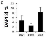
[도 6C] 인비트로의 동결 보존 세포괴의 특징. 35 일째 및 해동 후 7 일째에 있어서의 세포괴의 면역 염색. 좌측 패널은 FOXA2/DAPI, 중앙의 패널은 NURR1/TH, 우측 패널은 SOX1/KI67/PAX6/DAPI 의 면역 염색상을 나타낸다. 스케일 바는 100 ㎛ 를 나타낸다.
[도 6D] 인비트로의 동결 보존 세포괴의 특징. 35 일째 및 해동 후 7 일째의 전세포에 대한 FOXA2+ 세포, NURR1+ 세포, TH+ 세포의 비율.
[도 6E] 인비트로의 동결 보존 세포괴의 특징. 35 일째 및 해동 후 7 일째의 전세포에 대한 SOX1+ 세포, PAX6+ 세포 및 KI67+ 세포의 비율.
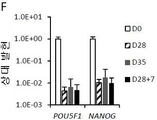
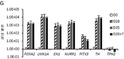
[도 6F] 인비트로의 동결 보존 세포괴의 특징. 정량 RT-PCR 로 측정된, GAPDH 에 대한 세포괴의 유전자 발현. 미분화 세포 (0 일째) 의 발현 레벨을 1 로 설정했다.
[도 6G] 인비트로의 동결 보존 세포괴의 특징. 정량 RT-PCR 로 측정된, GAPDH 에 대한 세포괴의 유전자 발현. 미분화 세포 (0 일째) 의 발현 레벨을 1 로 설정했다.
[도 6H] 인비트로의 동결 보존 세포괴의 특징. 마이크로어레이 데이터의 주성분 분석. 비동결 세포 (원형) 및 동결 세포 (삼각형) 의 유전자 발현의 시간 변화를 나타낸다.
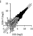
[도 6I] 인비트로의 동결 보존 세포괴의 특징. 35 일째 (X 축) 또는 해동 후 7 일째 (Y 축) 에 있어서의, 동일 로트의 비동결 세포와 동결 세포의 마이크로어레이 데이터의 스캐터 플롯을 나타낸다. 검은색 원형은 어느 한쪽 검체로 시그널 강도 50 이상을 나타내는 유전자, 흰색 원형은 양 검체로 시그널 강도 50 이하의 유전자를 나타낸다.
[도 6J] 인비트로의 동결 보존 세포괴의 특징. 35 일째에 있어서의, 다른 로트 간의 비동결 세포의 마이크로어레이 데이터의 스캐터 플롯을 나타낸다. 검은색 원형은 어느 한쪽 검체로 시그널 강도 50 이상을 나타내는 유전자, 흰색 원형은 양 검체로 시그널 강도 50 이하의 유전자를 나타낸다.
[도 6K] 인비트로의 동결 보존 세포괴의 특징. 28 + 21 일째의 해동 후 iPSC 유래 도파민 생산 신경세포의 TUBB3, TH 및 DAPI 의 면역 염색상. 스케일 바는 50 ㎛ 를 나타낸다.
[도 6L] 인비트로의 동결 보존 세포괴의 특징. 28 + 21 일째의 해동 후 iPSC 유래 도파민 생산 신경세포의 대표적인 유도 활동 전위.
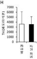
[도 6M] 인비트로의 동결 보존 세포괴의 특징. 56 일째 또는 해동 후 28 + 28 일째에 있어서 고농도 칼륨 자극에 의해 유도된 도파민 방출량의 결과.
[도 7A] 해동 후의 도파민 생산 신경 전구세포 마커 발현의 시간 변화. 28, 29, 31, 35 일째 및 해동 후 0, 1, 3, 7 일째의 전세포에 대한 FOXA2+ 세포의 비율.
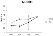
[도 7B] 해동 후의 도파민 생산 신경 전구세포 마커 발현의 시간 변화. 28, 29, 31, 35 일째 및 해동 후 0, 1, 3, 7 일째의 전세포에 대한 NURR1+ 세포의 비율.
[도 7C] 해동 후의 도파민 생산 신경 전구세포 마커 발현의 시간 변화. 정량 RT-PCR 로 측정된, GAPDH 에 대한 세포괴의 TH 유전자 발현. 미분화 세포 (0 일째) 의 발현 레벨을 1 로 설정했다.
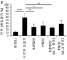
[도 8A] 동결 보존 세포괴의 이식편 생착율 및 기능. 이식편을 갖는 래트의 메탐페타민 유발 회전운동. 데이터를 평균값 ± SEM (n = 6 내지 8) 로서 나타낸다. 투키의 다중 비교 검정을 동반하는 2원 배치 분산 분석을 하고, 유의 수준을 용매 투여군에 대한 **p <0.01, ****p <0.001 에 의해 나타낸다.
[도 8B] 동결 보존 세포괴의 이식편 생착율 및 기능. 비동결 세포 (상측 패널) 및 동결 세포 (하측 패널) 유래의 대표적인 이식편에 있어서의 HNA 의 면역 염색상.
[도 8C] 동결 보존 세포괴의 이식편 생착율 및 기능. 이식편에 있어서의 생존 HNA+ 세포수.
[도 8D] 동결 보존 세포괴의 이식편 생착율 및 기능. 비동결 세포 유래의 대표적인 이식편에 있어서의 TH 의 DAB 염색상.
[도 8E] 동결 보존 세포괴의 이식편 생착율 및 기능. 도 8D 의 틀 내의 확대상. 우측 패널은 좌측 패널의 틀 내의 확대상이다. 스케일 바는 200 ㎛ 를 나타낸다.
[도 8F] 동결 보존 세포괴의 이식편 생착율 및 기능. 동결 세포 유래의 대표적인 이식편에 있어서의 TH 의 DAB 염색상.
[도 8G] 동결 보존 세포괴의 이식편 생착율 및 기능. 도 8F 의 틀 내의 확대상. 우측 패널은 좌측 패널의 틀 내의 확대상이다. 스케일 바는 200 ㎛ 를 나타낸다.
[도 8H] 동결 보존 세포괴의 이식편 생착율 및 기능. 이식편에 있어서의 생존 TH+ 세포수.
[도 8I] 동결 보존 세포괴의 이식편 생착율 및 기능. 동결 세포 유래의 이식편의 FOXA2, TH, HNA (상측), KI67, HNA (하측) 의 면역 염색상. 스케일 바는 50 ㎛ 를 나타낸다.
[도 8J] 동결 보존 세포괴의 이식편 생착율 및 기능. HNA+ 세포에 대한 FOXA2+ 세포의 비율.
[도 8K] 동결 보존 세포괴의 이식편 생착율 및 기능. HNA+ 세포에 대한 KI67+ 세포의 비율.
[도 9] 동결 보존 세포괴를 다른 조건에서 해동한 경우에서의, 생존율 및 신경돌기 신장 활성을 나타낸다.
[도 10A] 항CORIN 항체로 소팅되지 않은 세포 유래의 세포괴의 마커 발현. 세포괴의 LMX1A, FOXA2, DAPI (상단), NURR1, TH, DAPI (중단), SOX1, KI67, PAX6, DAPI (하단) 의 면역 염색상. 스케일 바는 100 ㎛ 를 나타낸다.
[도 10B] 항CORIN 항체로 소팅되지 않은 세포 유래의 세포괴의 마커 발현. 전세포에 대한 FOXA2+/LMX1A+ 세포, NURR1+ 세포 및 TH+ 세포의 비율.
[도 10C] 항CORIN 항체로 소팅되지 않은 세포 유래의 세포괴의 마커 발현. 전세포에 대한 SOX1+ 세포, PAX6+ 세포 및 KI67+ 세포의 비율.
[도 11] 도 6C 에 나타낸 세포괴와 동일 로트의 동결 직전 (28 일째) 의 세포괴의 마커 발현. 세포괴의 LMX1A, FOXA2, DAPI (상단), NURR1, FOXA2, TH, DAPI (중단), SOX1, KI67, PAX6, DAPI (하단) 의 면역 염색상. 스케일 바는 100 ㎛ 를 나타낸다.[FIG. 1] Scheme showing the protocol and timing of evaluation experiments for induction of differentiation into dopamine-producing neural progenitor cells in iPSCs. The symbol in the figure indicates each component added to the medium. LDN: LDN193189, A: A83-01, Y: Y-27632, Pur: permorphamine, CHIR: CHIR99021, AA: ascorbic acid.
[Figure 2A] Effect of cryopreservation solution on iPSC-derived dopamine-producing neural progenitor cells. The recovery rate of viable cells after thawing of unsorted cells that were infiltrated in the cryopreservation solution shown in Table 1 for 15 minutes and then cryopreserved at 0.5°C/min (n = 4).
[Fig. 2B] Effect of cryopreservation solution on iPSC-derived dopamine-producing neural progenitor cells. Neurite elongation of cell agglomerates derived from unsorted cells, which were infiltrated into the cryopreservation solution shown in Table 1 for 15 minutes and then cryopreserved at 0.5°C/min (n = 4).
[Fig. 2C] Effect of cryopreservation solution on iPSC-derived dopamine-producing neural progenitor cells. Immunostained images are shown in which neurites of unsorted cell-derived cell masses, which were infiltrated into the cryopreservation solution shown in Table 1 for 15 minutes and then cryopreserved at 0.5°C/min, were stained with an initial neuronal marker (PSA-NCAM). . The scale bar represents 1 mm.
[Fig. 3] Time-temperature curves of the sample (straight line), the freezing chamber (dashed line) and the program (dotted line). Bambanker hRM was used as a sample. The figure below is an enlarged view of the temperature change due to the release of latent heat in the figure above.
[Fig. 4A] Effect of cryopreservation program on iPSC-derived dopamine-producing neural progenitor cells. Viable cell recovery after thawing of unsorted cells after infiltration in Bambanker hRM for 15 min and cryopreserved by various freezing programs.
[Fig. 4B] Effect of cryopreservation program on iPSC-derived dopamine-producing neural progenitor cells. Neurite elongation of unsorted cell-derived cell mass after infiltration in Bambanker hRM for 15 minutes and cryopreserved by various freezing programs.
[Fig. 5A] Effect of freezing on iPSC-derived dopamine-producing neural progenitor cells after prolonged exposure to cryopreservation solution. Viable cell recovery after thawing of cryopreserved, unsorted cells after 60 min exposure to Bambanker hRM for various freezing programs.
[Fig. 5B] Freezing effect after prolonged exposure to cryopreservation solution on iPSC-derived dopamine-producing neural progenitor cells. Neurite elongation of unsorted cell-derived cell masses cryopreserved after 60 min exposure to Bambanker hRM in various freezing programs.
[ Fig. 6A ] Characteristics of cryopreserved cell agglomerates in vitro . Viable cell recovery after thawing.
[ Fig. 6B ] Characteristics of cryopreserved cell agglomerates in vitro. Neurite elongation of the cell mass.
[ Fig. 6C ] Characteristics of cryopreserved cell agglomerates in vitro. Immunostaining of the cell mass on the 35th day and the 7th day after thawing. The left panel shows immunostaining images of FOXA2/DAPI, the middle panel shows NURR1/TH, and the right panel shows immunostaining images of SOX1/KI67/PAX6/DAPI. The scale bar represents 100 μm.
[ Fig. 6D ] Characteristics of cryopreserved cell agglomerates in vitro. Ratio of FOXA2 + cells, NURR1 + cells, TH + cells to whole cells at day 35 and at
[ Fig. 6E ] Characteristics of cryopreserved cell agglomerates in vitro. Ratio of SOX1 + cells, PAX6 + cells and KI67 + cells to whole cells at day 35 and at
[ Fig. 6F ] Characteristics of cryopreserved cell agglomerates in vitro. The gene expression of the cell aggregation with respect to GAPDH measured by quantitative RT-PCR. The expression level of undifferentiated cells (day 0) was set to 1.
[ Fig. 6G ] Characteristics of cryopreserved cell agglomerates in vitro. The gene expression of the cell aggregation with respect to GAPDH measured by quantitative RT-PCR. The expression level of undifferentiated cells (day 0) was set to 1.
[ Fig. 6H ] Characteristics of cryopreserved cell agglomerates in vitro. Principal component analysis of microarray data. Time changes of gene expression in non-frozen cells (circles) and frozen cells (triangles) are shown.
[ Fig. 6I ] Characteristics of cryopreserved cell agglomerates in vitro. Scatter plots of microarray data of unfrozen cells and frozen cells of the same lot at day 35 (X axis) or at
[Fig. 6J] Characteristics of cryopreserved cell agglomerates in vitro. A scatter plot of microarray data of non-frozen cells between different lots on day 35 is shown. A black circle indicates a gene with a signal intensity of 50 or more in either sample, and a white circle indicates a gene with a signal intensity of 50 or less in both samples.
[ Fig. 6K ] Characteristics of cryopreserved cell agglomerates in vitro. Immunostaining images of TUBB3, TH and DAPI of iPSC-derived dopamine-producing neurons after thawing at 28+21 days. The scale bar represents 50 μm.
[ Fig. 6L ] Characteristics of cryopreserved cell agglomerates in vitro. Representative induced action potentials of iPSC-derived dopaminergic neurons after thawing at
[ Fig. 6M ] Characteristics of cryopreserved cell agglomerates in vitro. Results of dopamine release induced by high potassium stimulation at
[Fig. 7A] Time change of dopamine-producing neural progenitor cell marker expression after thawing. Ratio of FOXA2 + cells to whole cells at
[Fig. 7B] Time change of dopamine-producing neural progenitor cell marker expression after thawing. Ratio of NURR1 + cells to whole cells at
[Fig. 7C] Time change of dopamine-producing neural progenitor cell marker expression after thawing. TH gene expression of the cell mass with respect to GAPDH measured by quantitative RT-PCR. The expression level of undifferentiated cells (day 0) was set to 1.
[Fig. 8A] Graft engraftment rate and function of cryopreserved cell mass. Methamphetamine-induced rotational movement of rats with grafts. Data are presented as mean ± SEM (n = 6 to 8). Two-way Batch Analysis of ANOVA accompanied by Tukey's multiple comparison test was performed, and significance levels are indicated by **p <0.01, ****p <0.001 for solvent administration groups.
[Fig. 8B] Graft engraftment rate and function of cryopreserved cell masses. Immunostaining images of HNAs in representative explants derived from non-frozen cells (upper panel) and frozen cells (lower panel).
[Fig. 8C] Graft engraftment rate and function of cryopreserved cell masses. Number of viable HNA + cells in the graft.
[Fig. 8D] Graft engraftment rate and function of cryopreserved cell masses. DAB staining image of TH in a representative explant derived from non-frozen cells.
[Fig. 8E] Graft engraftment rate and function of cryopreserved cell masses. An enlarged view within the frame of FIG. 8D . The right panel is an enlarged image within the frame of the left panel. The scale bar represents 200 μm.
[Fig. 8F] Graft engraftment rate and function of cryopreserved cell masses. DAB staining image of TH in a representative graft derived from frozen cells.
[Fig. 8G] Graft engraftment rate and function of cryopreserved cell masses. An enlarged view within the frame of FIG. 8F . The right panel is an enlarged image within the frame of the left panel. The scale bar represents 200 μm.
[Fig. 8H] Graft engraftment rate and function of cryopreserved cell masses. Viable TH + number of cells in the graft.
[Fig. 8I] Graft engraftment rate and function of cryopreserved cell masses. Immunostaining images of FOXA2, TH, HNA (top), KI67, and HNA (bottom) of frozen cell-derived grafts. The scale bar represents 50 μm.
[Fig. 8J] Graft engraftment rate and function of cryopreserved cell masses. Ratio of FOXA2 + cells to HNA + cells.
[Fig. 8K] Graft engraftment rate and function of cryopreserved cell masses. Ratio of KI67 + cells to HNA + cells.
Fig. 9 shows the survival rate and neurite elongation activity when cryopreserved cell masses are thawed under different conditions.
[Fig. 10A] Marker expression of cell agglomerates derived from cells not sorted by anti-CORIN antibody. Immunostaining images of LMX1A, FOXA2, DAPI (top), NURR1, TH, DAPI (middle), SOX1, KI67, PAX6, and DAPI (bottom) of the cell mass. The scale bar represents 100 μm.
[Fig. 10B] Marker expression of cell agglomerates derived from cells not sorted by anti-CORIN antibody. Ratio of FOXA2 + /LMX1A + cells, NURR1 + cells and TH + cells to whole cells.
[Fig. 10C] Marker expression in cell aggregation derived from cells not sorted by anti-CORIN antibody. Ratio of SOX1 + cells, PAX6 + cells and KI67 + cells to whole cells.
[FIG. 11] Marker expression in the cell aggregation immediately before freezing (day 28) of the same lot as the cell aggregation shown in FIG. 6C. Immunostaining images of LMX1A, FOXA2, DAPI (top), NURR1, FOXA2, TH, DAPI (middle), SOX1, KI67, PAX6, and DAPI (bottom) of the cell mass. The scale bar represents 100 μm.
본 명세서 및 특허청구범위에서는 수치가 "약" 용어를 동반할 경우, 그 값의 ± 10% 범위를 포함하는 것을 의도한다. 예를 들면, "약 20" 은 "18 내지 22" 를 포함하는 것으로 한다. 수치 범위는 양단점 사이의 모든 수치 및 양단점의 수치를 포함한다. 범위와 관련된 "약" 은 그 범위의 양단점에 적용된다. 따라서, 예를 들면, "약 20 내지 30" 은 "18 내지 33" 을 포함하는 것으로 한다.In this specification and claims, when a number is accompanied by the term "about", it is intended to include a range of ±10% of that value. For example, “about 20” is meant to include “18 to 22”. Numerical ranges include all values between the endpoints and the values between the endpoints. “About” with respect to a range applies to both ends of that range. Thus, for example, "about 20 to 30" is intended to include "18 to 33".
[신경계 세포][nervous system cells]
본원은 삼차원 구조를 갖는 신경계 세포를 포함하는 세포 응집체를 동결하는 방법을 제공한다.The present application provides a method of freezing a cell aggregate comprising a nervous system cell having a three-dimensional structure.
신경계 세포는 신경세포 또는 뉴런 및 그 신경세포의 전구세포, 즉, 신경 선조세포 또는 신경 전구세포 등을 포함한다.Nervous system cells include nerve cells or neurons and progenitor cells of the nerve cells, that is, neural progenitor cells or neural progenitor cells.
신경계 세포는 중추신경계의 신경계 세포, 또는 운동신경이나 감각 기관계의 체성 신경계 세포 또는 자율 신경의 신경계 세포의 말초신경계 신경계 세포 등의 모든 부위 유래의 신경계 세포일 수 있고, 신경 (뉴런), 신경능선 유래 세포, 올리고덴드로사이트 또는 아스트로사이트 등의 신경교 세포 및 이들의 줄기 세포 또는 전구세포 등을 들 수 있다. 신경계 세포로서 신경계 세포 마커를 발현하는 세포를 들 수 있다. 신경계 세포 마커로는 예를 들면, NCAM, βIII-Tubulin (TUJ1), 티로신 수산화 효소 (TH), 세로토닌, 네스틴, MAP2, MAP2AB, NEUN, GABA, 글루타메이트, CHAT, SOX1, BF1, EMX1, VGLUT1, PAX, NKX, GSH, Telencephalin, GLUR1, CAMKII, CTIP2, TBR1, Reelin, TBR1, BRN2, OTX2, LMX1A, LMX1B, EN1, NURR1, PITX3, DAT, GIRK2 및 TH 등을 들 수 있으나, 이에 한정되는 것은 아니다. 신경계 세포 마커의 1 이상의 발현에 의해 신경계 세포인 것을 확인할 수 있다. 본 명세서에 있어서, 신경계 세포로서 상기 신경계 세포 마커의 1 이상, 2 이상, 또는 3 이상을 발현하는 세포를 들 수 있다.The nervous system cell may be a nervous system cell derived from any part of the nervous system, such as a nervous system cell of the central nervous system, a somatic nervous system cell of a motor neuron or sensory system, or a peripheral nervous system nervous system cell of an autonomic nervous system, nerve (neuron), neural crest-derived and glial cells such as cells, oligodendrocytes or astrocytes, and stem cells or progenitor cells thereof. As the nervous system cell, a cell expressing a nervous system cell marker is exemplified. Nervous system cell markers include, for example, NCAM, βIII-Tubulin (TUJ1), tyrosine hydroxylase (TH), serotonin, nestin, MAP2, MAP2AB, NEUN, GABA, glutamate, CHAT, SOX1, BF1, EMX1, VGLUT1, PAX, NKX, GSH, Teleencephalin, GLUR1, CAMKII, CTIP2, TBR1, Reelin, TBR1, BRN2, OTX2, LMX1A, LMX1B, EN1, NURR1, PITX3, DAT, GIRK2 and TH, etc., but are not limited thereto. . It can be confirmed that it is a nervous system cell by the expression of one or more nervous system cell markers. In the present specification, cells expressing one or more, two or more, or three or more of the above-mentioned nervous system cell markers are mentioned as nervous system cells.
중추신경계의 신경계 세포는 신경계 세포가 존재하는 부위의 차이에 의해 분류할 수 있다. 즉, 전뇌, 종뇌, 간뇌, 대뇌, 시상하부, 중뇌, 후뇌, 중뇌 후뇌 경계 영역, 소뇌, 망막, 뇌하수체, 또는 척수 유래의 신경세포 및 이들의 전구세포를 들 수 있다.Nervous system cells of the central nervous system can be classified by the difference in the region where the nervous system cells exist. That is, the forebrain, telencephalon, diencephalon, cerebrum, hypothalamus, midbrain, hindbrain, midbrain, hindbrain border region, cerebellum, retina, pituitary, or spinal cord-derived nerve cells and their precursor cells are mentioned.
전뇌 유래의 신경세포는 전뇌 조직 (즉, 종뇌, 대뇌, 해마 또는 맥락막, 간뇌, 시상하부 등) 에 존재하는 신경세포이다. 전뇌의 신경세포는 전뇌 신경세포 마커의 발현에 의해 확인할 수 있다. 전뇌 신경세포 마커로는 OTX1 (전뇌), BF1 (FOXG1 라고도 한다) 또는 SIX3 (종뇌 또는 대뇌의 마커일 수 있다) 등을 들 수 있다. 본 명세서에 있어서, 신경계 세포로서 상기 전뇌 신경세포 마커, 종뇌 또는 대뇌의 마커 1 이상, 2 이상, 또는 3 이상을 발현하는 세포를 들 수 있다.Forebrain-derived neurons are neurons present in forebrain tissues (ie, telencephalon, cerebrum, hippocampus or choroid, diencephalon, hypothalamus, etc.). Forebrain neurons can be identified by the expression of forebrain neuron markers. Examples of the forebrain neuron marker include OTX1 (forebrain), BF1 (also referred to as FOXG1), or SIX3 (which may be a marker of telencephalon or cerebrum). In the present specification, cells expressing one or more, two or more, or three or more of the above-mentioned forebrain neuronal cell markers, telencephalon or cerebral markers are mentioned as the nervous system cells.
대뇌 유래의 신경세포로서 배측 세포 (예를 들면, 대뇌피질 세포, 카하르 레치우스 세포, 해마 신경세포 등) 또는 복측 세포 (예를 들면, 대뇌 기저 핵세포 등) 를 들 수 있다. 복측 대뇌 신경세포 마커로는 예를 들면, 대뇌 기저핵 신경세포 마커 (예를 들면, GSH2, MASH1, NKX2.1, NOZ1) 를 들 수 있다. 배측 대뇌 신경세포 마커로는 예를 들면, 대뇌피질 신경세포 마커 (예를 들면, PAX6, EMX1, TBR1) 를 들 수 있다. 본 명세서에 있어서, 신경계 세포로서 상기의 대뇌 신경세포 마커, 대뇌 기저핵 신경세포 마커 또는 대뇌피질 신경세포 마커의 1 이상, 2 이상, 또는 3 이상을 발현하는 세포를 들 수 있다.Examples of the cerebral-derived nerve cells include dorsal cells (eg, cortical cells, Cahar recius cells, hippocampal neurons, etc.) and ventral cells (eg, basal ganglia cells). Examples of the ventral cerebral neuron marker include cerebral basal ganglia neuronal markers (eg, GSH2, MASH1, NKX2.1, NOZ1). Examples of the dorsal cerebral neuron marker include cortical neuronal markers (eg, PAX6, EMX1, TBR1). In the present specification, cells expressing one or more, two or more, or three or more of the above-mentioned cerebral nerve cell markers, cerebral basal ganglion cell markers, or cortical nerve cell markers are mentioned as the nervous system cells.
중뇌 유래의 신경계 세포로는 중뇌 복측부 유래 신경 전구세포, 도파민 생산 신경세포 (도파민 신경세포라고도 한다) 또는 도파민 생산 신경 전구세포 (도파민 신경 선조세포 또는 도파민 신경 전구세포라고도 한다) 등을 들 수 있다. 중뇌 유래의 신경계 세포의 마커로는 FOXA2, EN2, TUJ1 등을 들 수 있다. FOXA2 양성 및 TUJ1 양성 신경계 세포로는 도파민 생산 신경 전구세포 및 도파민 생산 신경세포 등을 들 수 있다. 또한 도파민 생산 신경세포는 FOXA2 양성, NURR1 양성 및 TH 양성임을 지표로서 동정할 수 있다.Examples of the midbrain-derived nervous system cells include midbrain ventral-derived neural progenitor cells, dopamine-producing neurons (also referred to as dopaminergic neurons), or dopamine-producing neural progenitors (also referred to as dopaminergic neural progenitors or dopaminergic neural progenitors). . FOXA2, EN2, TUJ1, etc. are mentioned as a marker of a nervous system cell derived from midbrain. Examples of FOXA2-positive and TUJ1-positive nervous system cells include dopamine-producing neural progenitor cells and dopamine-producing neurons. In addition, dopamine-producing neurons can be identified as FOXA2-positive, NURR1-positive and TH-positive as an indicator.
도파민 생산 신경 전구세포는 FOXA2 양성 및 LMX1A 양성임을 지표로서 동정할 수 있다. 더욱 바람직하게는, 도파민 생산 신경 전구세포는 OTX2, LMX1A, LMX1B, CORIN, SHH, AADC, βIII-Tubulin, EN1, NURR1, PITX3, DAT, GIRK2 및 TH 중 1 이상이 양성인 세포를 함유한다. 본 명세서에 있어서, 도파민 생산 신경 전구세포를 포함하는 세포 응집체는 별도의 설명이 없으면 도파민 생산 신경세포 또는 도파민 작동성 뉴런 등을 포함할 수 있다.Dopamine-producing neural progenitor cells can be identified as FOXA2-positive and LMX1A-positive as an indicator. More preferably, the dopamine producing neural progenitor cells contain cells positive for at least one of OTX2, LMX1A, LMX1B, CORIN, SHH, AADC, βIII-Tubulin, EN1, NURR1, PITX3, DAT, GIRK2 and TH. In the present specification, the cell aggregate including dopaminergic neural progenitor cells may include dopaminergic neurons or dopaminergic neurons, unless otherwise specified.
본 명세서에 있어서, 신경계 세포로서 중뇌 유래의 신경계 세포의 마커, 도파민 생산 신경 전구세포의 마커 또는 도파민 생산 신경세포의 마커의 1 이상, 2 이상, 또는 3 이상을 발현하는 세포를 들 수 있다.In the present specification, as the nervous system cell, a cell expressing one or more, two or more, or three or more of a marker of a neuronal cell derived from the midbrain, a marker of a dopamine-producing neural progenitor cell, or a marker of a dopamine-producing neuronal cell is mentioned.
본 명세서에 있어서, 도파민 생산 신경 전구세포로서 FOXA2 및/또는 LMX1A 를 발현하는 (FOXA2 양성 및/또는 LMX1A 양성) 세포, 바람직하게는 FOXA2 및 LMXA1 에 더해 OTX2, LMX1B, CORIN, SHH, AADC 및 βIII-Tubulin 로 이루어진 군에서 선택되는 1 이상, 2 이상 또는 3 이상을 발현하는 세포를 들 수 있다.In the present specification, cells expressing FOXA2 and/or LMX1A (FOXA2 positive and/or LMX1A positive) as dopamine-producing neural progenitor cells, preferably FOXA2 and LMXA1 plus OTX2, LMX1B, CORIN, SHH, AADC and βIII- and cells expressing one or more, two or more, or three or more selected from the group consisting of tubulin.
본 명세서에 있어서, 도파민 생산 신경세포 (도파민 신경세포) 로서 TH 및/또는 NURR1 를 발현하는 (TH 양성 및/또는 NURR1 양성) 세포, 바람직하게는 TH 및 NURR1 에 더해 FOXA2, AADC, DAT 및 GIRK2 로 이루어진 군에서 선택되는 1 이상, 2 이상 또는 3 이상을 발현하는 세포를 들 수 있다.In the present specification, cells expressing TH and/or NURR1 (TH positive and/or NURR1 positive) as dopaminergic neurons (dopamine neurons), preferably FOXA2, AADC, DAT and GIRK2 in addition to TH and NURR1 and cells expressing one or more, two or more, or three or more selected from the group consisting of.
중뇌 후뇌 경계 영역 유래의 신경세포로서 소뇌, 소뇌판 조직, 뇌실대, 능뇌순 등에 존재하는 신경세포를 들 수 있다. 중뇌 후뇌 경계 영역 마커로는 EN2 (중뇌), GBX2 (후뇌), N-Cadherin (중뇌 후뇌 경계 영역의 신경 전구세포) 를 들 수 있다. 소뇌 신경 전구세포 마커로는 GABA 작동성 신경 전구세포 마커인 KIRREL2, PTF1A 또는 SOX2 나 소뇌 과립 세포 전구세포 마커인 ATOH1 또는 BARHL1 등을 들 수 있다. 본 명세서에 있어서, 신경계 세포로서 상기의 중뇌 후뇌 경계 영역 마커, 소뇌 신경 전구세포 마커, GABA 작동성 신경 전구세포 마커 또는 소뇌 과립 세포 전구세포 마커의 1 이상, 2 이상, 또는 3 이상을 발현하는 세포를 들 수 있다.Examples of the nerve cells derived from the midbrain hindbrain border region include nerve cells present in the cerebellum, cerebellar plate tissue, ventricular zone, and labrum. Examples of the midbrain hindbrain border region markers include EN2 (midbrain), GBX2 (hibrain), and N-Cadherin (neural progenitor cells in the midbrain hindbrain border region). Examples of the cerebellar neural progenitor cell markers include KIRREL2, PTF1A, or SOX2, which are GABAergic neural progenitor cell markers, and ATOH1 or BARHL1, which are cerebellar granular cell progenitor cell markers. In the present specification, cells expressing one or more, two or more, or three or more of the above midbrain hindbrain border region markers, cerebellar neural progenitor cell markers, GABA agonistic neural progenitor cell markers, or cerebellar granular cell progenitor cell markers as a nervous system cell can be heard
망막 유래의 신경계 세포로는 시세포, 시세포 전구세포, 망막 색소 상피 세포, 각막 세포 등을 들 수 있다.Examples of the retinal-derived nervous system cells include photoreceptors, photoreceptor cell progenitor cells, retinal pigment epithelial cells, and corneal cells.
또한 신경계 세포는 생산 (분비) 하는 신경전달물질의 차이에 의해 분류할 수도 있고, 예를 들면, 도파민 생산 신경세포, 도파민 생산 신경 전구세포, GABA 신경세포, GABA 신경 전구세포, 콜린 신경세포, 콜린 신경 전구세포, 세로토닌 신경세포, 세로토닌 신경 전구세포, 글루탐산 신경세포, 글루탐산 신경 전구세포, 노르아드레날린 신경세포, 노르아드레날린 신경 전구세포, 아드레날린 신경세포, 아드레날린 신경 전구세포 등을 들 수 있다.In addition, nervous system cells may be classified by the difference in the neurotransmitters they produce (secrete), for example, dopamine-producing neurons, dopamine-producing neural progenitors, GABA neurons, GABA neural precursor cells, choline neurons, choline Neural progenitor cells, serotonin neurons, serotonin neural progenitors, glutamic acid neurons, glutamic acid neural progenitors, noradrenergic neurons, noradrenergic neural progenitors, adrenergic neurons, adrenergic neural progenitors, and the like.
운동신경이나 감각 기관계의 신경계 세포로는 콜린 신경세포 또는 그 전구세포 등을 들 수 있다.As the nervous system cells of the motor nerve or sensory organ system, a choline neuron or a precursor cell thereof may be mentioned.
자율 신경의 신경계 세포로는 콜린 신경세포, 아드레날린 신경세포, 또는 이들의 전구세포 등을 들 수 있다.Examples of the autonomic nervous system cells include choline neurons, adrenergic neurons, and precursor cells thereof.
본 명세서의 신경계 세포로서 바람직하게는, 도파민 생산 신경세포 (도파민 신경세포), 도파민 생산 신경 전구세포 (도파민 신경 전구세포) 를 들 수 있다.Preferable examples of the nervous system cells in the present specification include dopaminergic neurons (dopaminergic neurons) and dopaminergic neural progenitors (dopaminergic neural progenitors).
생체 유래의 신경계 세포는 사람 등의 포유동물로부터 분리한 세포이며, 예를 들면, 인간 뇌조직에서 분리한 세포로는 Nature Neuroscience, 2, 1137 (1999) 또는 N. Engl. J. Med.; 344: 710-9 (2001) 에 기재되는 바와 같은 태아의 중뇌 조직에 함유되는 세포가 예시된다.The biological-derived nervous system cells are cells isolated from mammals such as humans. For example, as cells isolated from human brain tissue, Nature Neuroscience, 2, 1137 (1999) or N. Engl. J. Med.; 344: 710-9 (2001) exemplified cells contained in fetal midbrain tissue.
신경계 세포는 또한 배아 줄기 세포 (ES 세포) 및 iPS 세포 등의 다능성 줄기 세포에서 분화 유도시켜 얻어진 세포일 수 있다. 신경계 세포를 다능성 줄기 세포에서 분화 유도하는 방법은 예를 들면, 상술한 비특허문헌 3, 4 및 WO2015/034012 에 기재되는 방법 (도파민 생산 신경 전구세포), WO2009/148170 (대뇌 등의 신경계 세포), WO2013/065763, WO2016/013669 또는 WO2017/126551 (뇌하수체 또는 시상하부의 신경계 세포), WO2016/039317 (소뇌의 신경계 세포), WO2015/076388 (종뇌의 신경계 세포), Numasawa-Kuroiwa, Y 등, Stem Cell Reports, 2: 648-661 (2014) (신경 전구세포), Qiu, L 등, Stem Cells Transl Med. 6 (9): 1803-1814 (2017) (도파민 생산 신경 전구세포) 가 예시된다.The nervous system cells may also be cells obtained by inducing differentiation in pluripotent stem cells such as embryonic stem cells (ES cells) and iPS cells. Methods for inducing differentiation of nervous system cells into pluripotent stem cells include, for example, the methods described in
신경계 세포는 간엽계 줄기 세포 (MSC) 등의 복능성 줄기 세포에서 분화 유도시켜 얻어진 세포일 수 있다. 신경계 세포를 간엽계 줄기 세포에서 분화 유도하는 방법으로는 J Chem Neuroanat. 96:126-133 (2019) 에 기재된 방법 등이 예시된다.Nervous system cells may be cells obtained by inducing differentiation in pluripotent stem cells such as mesenchymal stem cells (MSCs). A method for inducing differentiation of nervous system cells from mesenchymal stem cells is described in J Chem Neuroanat. 96:126-133 (2019) and the like are exemplified.
[다능성 줄기 세포][pluripotent stem cells]
다능성 줄기 세포는 생체에 존재하는 거의 모든 세포로 분화가 가능한 다능성을 가지며, 또한 증식능을 겸비하는 줄기 세포를 의미한다. 다능성 줄기 세포는 수정란, 클론 배아, 생식 줄기 세포, 조직 내 줄기 세포, 체세포 등에서 유도할 수 있다. 다능성 줄기 세포에는 특별히 한정되지 않으나 예를 들면 배아성 줄기 (ES) 세포, 핵 이식에 의해 얻어진 클론 배아 유래의 배아성 줄기 (ntES) 세포, 정자 줄기 세포 (GS 세포), 배아성 생식 세포 (EG 세포), 인공 다능성 줄기 (iPS) 세포, 배양 섬유아세포 및 골수 줄기 세포 유래의 다능성 줄기 세포 (Muse 세포) 등이 포함된다. 다능성 줄기 세포는 ES 세포, ntES 세포, 또는 iPS 세포일 수 있다. 윤리적인 점을 고려하면 다능성 줄기 세포는 iPS 세포일 수 있다. 또한 배아 줄기 세포는 수정 14 일 이내의 배아에서 수립된 것이다.The pluripotent stem cell means a stem cell having a pluripotency capable of differentiating into almost all cells existing in a living body and also having a proliferative ability. Pluripotent stem cells can be induced from fertilized eggs, clonal embryos, germ stem cells, stem cells in tissues, somatic cells, and the like. The pluripotent stem cells are not particularly limited, but include, for example, embryonic stem (ES) cells, embryonic stem (ntES) cells derived from clonal embryos obtained by nuclear transfer, sperm stem cells (GS cells), embryonic germ cells ( EG cells), artificial pluripotent stem (iPS) cells, cultured fibroblasts and bone marrow stem cells-derived pluripotent stem cells (Muse cells), and the like. The pluripotent stem cell may be an ES cell, an ntES cell, or an iPS cell. For ethical considerations, pluripotent stem cells may be iPS cells. Also, embryonic stem cells are those established from embryos within 14 days of fertilization.
배아 줄기 세포는 1981년에 처음으로 수립되고 1989년 이후 녹아웃 마우스 제작에도 응용되어 있다. 1998년에는 인간 배아 줄기 세포가 수립되었고 재생 의학에도 이용되고 있다. 배아 줄기 세포는 내부 세포괴를 피더 세포 상에서 또는 백혈병 억제 인자 (LIF) 를 포함하는 배지 안에서 배양함으로써 제조할 수 있다. 배아 줄기 세포 제조 방법은, 예를 들면, WO96/22362, WO02/101057, US5,843,780, US6,200,806, US6,280,718 등에 기재되어 있다. 배아 줄기 세포는 소정의 기관에서 입수할 수 있고 또한 시판품을 구입할 수도 있다. 예를 들면, 인간 배아 줄기 세포인 KhES-1, KhES-2 및 KhES-3 은 쿄토 대학 재생 의과학 연구소에서 입수 가능하다. 인간 배아 줄기 세포인 Rx::GFP 주 (KhES-1 주 유래) 는 국립 연구 개발 법인 이화학 연구소에서 입수 가능하다. 마우스 배아 줄기 세포인 EB5 세포주 및 D3 세포주는 각각 국립 연구 개발 법인 이화학 연구소 및 ATCC 에서 입수 가능하다.Embryonic stem cells were first established in 1981 and have been applied to the production of knockout mice since 1989. In 1998, human embryonic stem cells were established and are also being used in regenerative medicine. Embryonic stem cells can be produced by culturing the inner cell mass on feeder cells or in a medium containing leukemia inhibitory factor (LIF). Methods for producing embryonic stem cells are described, for example, in WO96/22362, WO02/101057, US5,843,780, US6,200,806, US6,280,718 and the like. Embryonic stem cells can be obtained from certain institutions and can also be purchased commercially. For example, human embryonic stem cells KhES-1, KhES-2 and KhES-3 are available from Kyoto University Regenerative Medicine Research Institute. Human embryonic stem cells, the Rx::GFP strain (derived from the KhES-1 strain), are available from the National Research and Development Institute of Physics and Chemistry. Mouse embryonic stem cells, EB5 cell line and D3 cell line, are available from the National Research and Development Institute of Physics and Chemistry and ATCC, respectively.
배아 줄기 세포 중 하나인 핵 이식 배아 줄기 세포 (ntES 세포) 는 핵을 제거한 난자에 체세포의 핵을 이식해 만든 클론 배아에서 수립할 수 있다.One of the embryonic stem cells, nuclear-transferred embryonic stem cells (ntES cells) can be established from cloned embryos made by transplanting the nucleus of a somatic cell into an egg from which the nucleus has been removed.
EG 세포는 시원 생식 세포를 mSCF, LIF 및 bFGF 를 포함하는 배지 안에서 배양함으로써 제조할 수 있다 (Cell, 70: 841-847, 1992).EG cells can be prepared by culturing primordial germ cells in a medium containing mSCF, LIF and bFGF (Cell, 70: 841-847, 1992).
본 명세서에서의 "인공 다능성 줄기 세포" 란, 체세포를 공지된 방법 등에 의해 초기화 (reprogramming) 함으로써 다능성을 유도한 세포이다. 구체적으로는 섬유아세포, 또는 말초혈 단핵구 등의 분화된 체세포를 OCT3/4, SOX2, KLF4, MYC (c-MYC, N-MYC, L-MYC), GLIS1, NANOG, SALL4, LIN28, ESRRB 등을 포함하는 초기화 유전자군에서 선택되는 복수의 유전자의 조합 중 어느 하나의 발현에 의해 초기화하여 다분화 능력을 유도한 세포를 들 수 있다. 바람직한 초기화 인자의 조합으로는 (1) OCT3/4, SOX2, KLF4 및 MYC (c-MYC 또는 L-MYC), (2) OCT3/4, SOX2, KLF4, LIN28 및 L-MYC (Stem Cells, 2013; 31: 458-466), (3) OCT3/4, SOX2, NANOG, LIN28 (Science 2007; 318: 1917-1920) 등을 들 수 있다.The term "artificial pluripotent stem cell" as used herein refers to a cell inducing pluripotency by reprogramming somatic cells by a known method or the like. Specifically, differentiated somatic cells such as fibroblasts or peripheral blood mononuclear cells were treated with OCT3/4, SOX2, KLF4, MYC (c-MYC, N-MYC, L-MYC), GLIS1, NANOG, SALL4, LIN28, ESRRB, etc. and a cell in which multidifferentiation ability is induced by initialization by expression of any one of a combination of a plurality of genes selected from the included initialization gene group. Preferred combinations of initialization factors include (1) OCT3/4, SOX2, KLF4 and MYC (c-MYC or L-MYC), (2) OCT3/4, SOX2, KLF4, LIN28 and L-MYC (Stem Cells, 2013). ; 31: 458-466), (3) OCT3/4, SOX2, NANOG, LIN28 (Science 2007; 318: 1917-1920) and the like.
인공 다능성 줄기 세포는 2006년에 야마나카 등에 의해 마우스 세포로 수립되었다 (Cell, 2006, 126 (4), pp. 663-676). 인공 다능성 줄기 세포는 2007년에 인간 섬유아세포로도 수립되었고, 배아 줄기 세포와 동일하게 다능성과 자기 복제능을 갖는다 (Cell, 2007, 131 (5), pp. 861-872; Science, 2007, 318 (5858), pp. 1917-1920; Nat. Biotechnol., 2008, 26 (1), pp. 101-106).Artificial pluripotent stem cells were established as mouse cells by Yamanaka et al. in 2006 (Cell, 2006, 126 (4), pp. 663-676). Artificial pluripotent stem cells were also established as human fibroblasts in 2007, and have the same pluripotency and self-renewal capacity as embryonic stem cells (Cell, 2007, 131 (5), pp. 861-872; Science, 2007, 318 (5858), pp. 1917-1920; Nat. Biotechnol., 2008, 26 (1), pp. 101-106).
인공 다능성 줄기 세포는 유전자 발현에 의한 직접 초기화로 제조하는 방법 이외에 화합물의 첨가 등에 의해 체세포에서 인공 다능성 줄기 세포를 유도하는 방법에 의해서도 제조할 수 있다 (Science, 2013, 341, pp. 651-654).Artificial pluripotent stem cells can be produced by a method of inducing artificial pluripotent stem cells in somatic cells by addition of a compound, etc., in addition to the method of direct initialization by gene expression (Science, 2013, 341, pp. 651- 654).
또한 수립된 인공 다능성 줄기 세포를 입수하는 것도 가능하며, 예를 들면, 쿄토 대학에서 수립된 201B7 세포, 201B7-Ff 세포, 253G1 세포, 253G4 세포, 1201C1 세포, 1205D1 세포, 1210B2 세포, 1231A3 세포 등의 인간 인공 다능성 줄기 세포 세포주가 쿄토 대학에서 입수 가능하다. 주화된 인공 다능성 줄기 세포로 예를 들면, 쿄토 대학에서 수립된 Ff-I01 세포, Ff-I01s04 세포, QHJ-I01 및 Ff-I14 세포가 쿄토 대학에서 입수 가능하다.It is also possible to obtain established artificial pluripotent stem cells, for example, 201B7 cells, 201B7-Ff cells, 253G1 cells, 253G4 cells, 1201C1 cells, 1205D1 cells, 1210B2 cells, 1231A3 cells, etc. established at Kyoto University. A human artificial pluripotent stem cell cell line from Kyoto University is available. As the immunized artificial pluripotent stem cells, for example, Ff-I01 cells, Ff-I01s04 cells, QHJ-I01 and Ff-I14 cells established at Kyoto University are available from Kyoto University.
인공 다능성 줄기 세포를 제조할 때 이용되는 체세포로는 특별히 한정은 없지만 조직 유래의 섬유아세포, 혈구계 세포 (예를 들면, 말초혈 단핵구 (PBMC), T 세포), 간세포, 췌장 세포, 장 상피 세포, 평활근 세포 등을 들 수 있다.There is no particular limitation on the somatic cells used in the production of artificial pluripotent stem cells, but tissue-derived fibroblasts, hemocytometer cells (eg, peripheral blood mononuclear cells (PBMC), T cells), hepatocytes, pancreatic cells, intestinal epithelium a cell, a smooth muscle cell, etc. are mentioned.
인공 다능성 줄기 세포를 제조할 때, 여러 종류의 유전자 발현에 의해 초기화할 경우, 유전자를 발현시키기 위한 수단은 특별히 한정되지 않는다. 상기 수단으로는 바이러스 벡터 (예를 들면, 레트로바이러스 벡터, 렌티바이러스 벡터, 센다이바이러스 벡터, 아데노바이러스 벡터, 또는 아데노 수반 바이러스 벡터) 를 이용한 감염법, 플라스미드 벡터 (예를 들면, 플라스미드 벡터, 또는 에피소말 벡터) 를 이용한 유전자 도입법 (예를 들면, 인산칼슘법, 리포펙션법, 레트로넥틴법, 또는 일렉트로포레이션법), RNA 벡터를 이용한 유전자 도입법 (예를 들면, 인산칼슘법, 리포펙션법, 또는 일렉트로포레이션법), 단백질의 직접 주입법 (예를 들면, 바늘을 이용한 방법, 리포펙션법, 또는 일렉트로포레이션법) 등을 들 수 있다.When producing artificial pluripotent stem cells, in the case of initialization by expression of several types of genes, the means for expressing the gene is not particularly limited. Such means include an infection method using a viral vector (eg, a retroviral vector, a lentiviral vector, a Sendaivirus vector, an adenoviral vector, or an adeno-associated viral vector), a plasmid vector (eg, a plasmid vector, or epi Somal vector) (for example, calcium phosphate method, lipofection method, retronectin method, or electroporation method), gene transfer method using RNA vector (for example, calcium phosphate method, lipofection method, or an electroporation method), a direct protein injection method (eg, a method using a needle, a lipofection method, or an electroporation method).
인공 다능성 줄기 세포는 피더 세포 존재 하 또는 피더 세포 비존재 하 (피더 프리) 에서 제조할 수 있다. 피더 세포 존재 하에서 인공 다능성 줄기 세포를 제조할 때는, 공지된 방법으로 미분화 유지 인자 존재 하에서 인공 다능성 줄기 세포를 제조할 수 있다. 피더 세포 비존재 하에서 인공 다능성 줄기 세포를 제조할 때 이용되는 배지로는 특별히 한정은 없지만 공지된 배아 줄기 세포 및/또는 인공 다능성 줄기 세포의 유지 배지, 또는 피더 프리로 인공 다능성 줄기 세포를 수립하기 위한 배지를 이용할 수 있다. 피더 프리로 인공 다능성 줄기 세포를 수립하기 위한 배지로는 예를 들면 Essential 8 배지 (E8 배지), Essential 6 배지, TeSR 배지, mTeSR 배지, mTeSR-E8 배지, Stabilized Essential 8 배지, StemFit 배지 등의 피더 프리 배지를 들 수 있다. 인공 다능성 줄기 세포를 제조할 때, 예를 들면, 피더 프리로 체세포에 센다이바이러스 벡터를 이용하여 OCT3/4, SOX2, KLF4 및 MYC (L-MYC 또는 C-MYC) 의 4 인자를 유전자 도입함으로써, 인공 다능성 줄기 세포를 제작할 수 있다.Artificial pluripotent stem cells can be produced in the presence of feeder cells or in the absence of feeder cells (feeder-free). When producing artificial pluripotent stem cells in the presence of feeder cells, artificial pluripotent stem cells can be produced in the presence of undifferentiated maintenance factors by a known method. There is no limitation in particular as a medium to be used for producing artificial pluripotent stem cells in the absence of feeder cells, but known embryonic stem cells and/or artificial pluripotent stem cell maintenance medium, or feeder-free artificial pluripotent stem cells. A medium for establishment may be used. As a medium for establishing artificial pluripotent stem cells in a feeder-free manner, for example, Essential 8 medium (E8 medium),
본 발명에 이용하는 다능성 줄기 세포는 포유동물의 다능성 줄기 세포이며, 바람직하게는 설치류 (예, 마우스 또는 래트) 또는 영장류 (예, 사람 또는 원숭이) 의 다능성 줄기 세포이며, 보다 바람직하게는 인간 또는 마우스 다능성 줄기 세포, 보다 더 바람직하게는 인간 인공 다능성 줄기 세포 (iPS 세포) 또는 인간 배아 줄기 세포 (ES 세포) 이다.The pluripotent stem cells used in the present invention are mammalian pluripotent stem cells, preferably rodent (eg, mouse or rat) or primate (eg, human or monkey) pluripotent stem cells, and more preferably human or mouse pluripotent stem cells, even more preferably human artificial pluripotent stem cells (iPS cells) or human embryonic stem cells (ES cells).
[세포 응집체][cell aggregate]
본 명세서에서 세포 응집체가 "삼차원 구조를 갖는다" 는 배양 세포가, 예를 들면, 부유 배양 또는 삼차원 배양 등에 의해 서로 접착해서 형성되는 삼차원의 세포 집단인 세포 응집체 (cell aggregate 또는 sphere) 를 형성하고 있는 것을 의미한다. 신경계 세포의 세포 응집체를 뉴로스피어 (neurosphere) 라고도 한다. 상기 세포 응집체의 형상에는 특별히 한정은 없고 구형이거나 비구형일 수 있다. 본 명세서의 세포 응집체는 바람직하게는 구형에 가까운 입체적인 형태를 갖는 세포 응집체이다. 구형에 가까운 입체적인 형태는 삼차원 구조를 갖는 형태로서, 이차원면에 투영했을 때, 예를 들면, 원형 또는 타원형을 나타낸다.In the present specification, the expression that a cell aggregate “has a three-dimensional structure” means that cultured cells form a cell aggregate (cell aggregate or sphere), which is a three-dimensional cell population formed by adhesion to each other by, for example, suspension culture or three-dimensional culture. means that Cell aggregates of nervous system cells are also called neurospheres. The shape of the cell aggregate is not particularly limited and may be spherical or non-spherical. The cell aggregate of the present specification is preferably a cell aggregate having a three-dimensional shape close to a spherical shape. A three-dimensional shape close to a spherical shape is a shape having a three-dimensional structure and, when projected onto a two-dimensional surface, represents, for example, a circle or an ellipse.
삼차원 구조를 갖는 신경계 세포를 포함하는 세포 응집체의 크기는 특별히 한정되지 않으나, 통상 원상당 지름 150 ㎛ 내지 1000 ㎛, 일 양태로는 예를 들면 200 ㎛ 내지 800 ㎛, 또는 300 ㎛ 내지 500 ㎛ 이다. 또한 삼차원 구조를 갖는 신경계 세포를 포함하는 세포 응집체는 통상 500 내지 150000 개, 일 양태로는 예를 들면 1000 내지 100000 개, 1000 내지 70000 개, 또는 3000 내지 30000 개의 세포를 포함한다.The size of the cell aggregate containing the nervous system cells having a three-dimensional structure is not particularly limited, but is usually 150 µm to 1000 µm in equivalent circle diameter, and in one embodiment, for example, 200 µm to 800 µm, or 300 µm to 500 µm. In addition, the cell aggregate including the nervous system cells having a three-dimensional structure usually contains 500 to 150000 cells, and in one embodiment, for example, 1000 to 100000 cells, 1000 to 70000 cells, or 3000 to 30000 cells.
신경계 세포를 포함하는 세포 응집체는 신경계 세포와 함께 다른 세포가 포함되어 있어도 좋다. 신경계 세포를 60% 이상, 70% 이상, 80% 이상, 더욱 바람직하게는 90% 이상 포함하는 세포 응집체가 예시된다.The cell aggregate containing the nervous system cells may contain other cells together with the nervous system cells. Cell aggregates containing 60% or more, 70% or more, 80% or more, more preferably 90% or more of nervous system cells are exemplified.
일 양태로서, 신경계 세포를 포함하는 세포 응집체는 도파민 생산 신경 전구세포 및/또는 도파민 생산 신경세포를 60% 이상, 70% 이상, 또는 80% 이상 포함할 수 있다. 즉, 신경계 세포를 포함하는 세포 응집체는 FOXA2, LMX1A, LMX1B, NURR1 및 TH 에서 선택되는 1 이상의 마커를 발현하는 신경계 세포를 60% 이상, 70% 이상, 또는 80% 이상 포함할 수 있다.In one embodiment, the cell aggregate comprising the nervous system cells may include 60% or more, 70% or more, or 80% or more of dopamine-producing neural progenitor cells and/or dopamine-producing neurons. That is, the cell aggregate comprising the nervous system cells may include 60% or more, 70% or more, or 80% or more of the nervous system cells expressing one or more markers selected from FOXA2, LMX1A, LMX1B, NURR1 and TH.
일 양태로서, 신경계 세포를 포함하는 세포 응집체는 도파민 생산 신경 전구세포를 40% 이상, 60% 이상, 70% 이상, 80% 이상, 85% 이상 또는 90% 이상 포함한다.In one embodiment, the cell aggregate comprising the nervous system cells contains 40% or more, 60% or more, 70% or more, 80% or more, 85% or more, or 90% or more of dopamine-producing neural progenitor cells.
일 양태로서, 신경계 세포를 포함하는 세포 응집체는 도파민 생산 신경 전구세포의 마커 1 이상, 2 이상 또는 3 이상을 발현하는 세포를 40% 이상, 60% 이상, 70% 이상, 80% 이상, 85% 이상 또는 90% 이상 포함한다.In one embodiment, the cell aggregate comprising the nervous system cells contains 40% or more, 60% or more, 70% or more, 80% or more, 85% or more of cells expressing 1 or more, 2 or more, or 3 or more markers of dopaminergic neural progenitor cells. or greater or greater than 90%.
일 양태로서, 신경계 세포를 포함하는 세포 응집체는 FOXA2 양성 및 LMX1A 양성 세포를 40% 이상, 60% 이상, 70% 이상, 80% 이상, 85% 이상 또는 90% 이상 포함한다. 일 양태로서, 상기 세포 응집체는 또한 TH 양성 및 NURR1 양성 세포를 40% 이하 포함한다.In one embodiment, the cell aggregate comprising nervous system cells comprises at least 40%, at least 60%, at least 70%, at least 80%, at least 85%, or at least 90% of FOXA2-positive and LMX1A-positive cells. In one embodiment, the cell aggregate also comprises 40% or less of TH positive and NURR1 positive cells.
일 양태로서, 신경계 세포를 포함하는 세포 응집체는 FOXA2 양성, TH 양성 및 NURR1 양성 세포를 0% 이상, 10% 이상, 또는 20% 이상 포함할 수 있다.In one embodiment, the cell aggregate comprising the nervous system cells may include 0% or more, 10% or more, or 20% or more of FOXA2-positive, TH-positive and NURR1-positive cells.
일 양태로서, 도파민 생산 신경 전구세포를 포함하는 세포 응집체는 NURR1 양성 세포를 60% 이하, 50% 이하, 40% 이하, 5 내지 50%, 5 내지 40% 또는 5 내지 20% 포함할 수 있다.In one embodiment, the cell aggregate comprising dopamine-producing neural progenitor cells may contain 60% or less, 50% or less, 40% or less, 5 to 50%, 5 to 40%, or 5 to 20% of NURR1-positive cells.
일 양태로서, 도파민 생산 신경 전구세포 및/또는 도파민 생산 신경세포를 포함하는 세포 응집체는 TH 양성 세포를 30% 이하, 20% 이하, 1 내지 30%, 5 내지 30%, 1 내지 20%, 5 내지 20%, 또는 5 내지 15% 포함할 수 있다.In one embodiment, the cell aggregate comprising dopamine-producing neural progenitor cells and/or dopamine-producing neurons contains TH positive cells by 30% or less, 20% or less, 1 to 30%, 5 to 30%, 1 to 20%, 5 to 20%, or 5 to 15%.
일 양태로서, 도파민 생산 신경 전구세포 및/또는 도파민 생산 신경세포를 포함하는 세포 응집체는 KI67 양성 세포를 30% 이하, 1 내지 25%, 1 내지 20%, 또는 5 내지 20% 포함할 수 있다.In one embodiment, the cell aggregate comprising dopamine-producing neural progenitor cells and/or dopamine-producing neurons may include 30% or less, 1 to 25%, 1 to 20%, or 5 to 20% of KI67-positive cells.
일 양태로서, 도파민 생산 신경 전구세포 및/또는 도파민 생산 신경세포를 포함하는 세포 응집체는 SOX1 양성 세포를 20% 이하, 10% 이하, 5% 이하, 또는 1% 이하 포함할 수 있다.In one embodiment, the cell aggregate comprising dopamine-producing neural progenitor cells and/or dopamine-producing neurons may contain 20% or less, 10% or less, 5% or less, or 1% or less of SOX1-positive cells.
일 양태로서, 도파민 생산 신경 전구세포 및/또는 도파민 생산 신경세포를 포함하는 세포 응집체는 PAX6 양성 세포를 5% 이하, 2% 이하, 1% 이하, 또는 0.5% 이하 포함할 수 있다.In one embodiment, the cell aggregate comprising dopamine-producing neural progenitor cells and/or dopamine-producing neurons may include 5% or less, 2% or less, 1% or less, or 0.5% or less of PAX6-positive cells.
일 양태로서, 도파민 생산 신경 전구세포 및/또는 도파민 생산 신경세포를 포함하는 세포 응집체는 또한 TH 양성 및 NURR1 양성 세포를 20% 이하, 구체적으로는 1% 내지 20%, 더 구체적으로는 5% 내지 15% 포함한다.In one embodiment, the cell aggregate comprising dopamine-producing neural progenitor cells and/or dopamine-producing neurons also contains 20% or less, specifically 1% to 20%, more specifically 5% to TH-positive and NURR1-positive cells. 15% included.
일 양태로서, 도파민 생산 신경 전구세포 및/또는 도파민 생산 신경세포를 포함하는 세포 응집체는 FOXA2 양성 및 LMX1A 양성 세포를 50% 이상, 바람직하게는 60% 이상, 70% 이상, 또는 80% 이상 포함하고, TH 양성 및 NURR1 양성 세포를 20% 이하, 1% 내지 20%, 더 구체적으로는 5% 내지 15% 포함한다.In one embodiment, the cell aggregate comprising dopamine-producing neural progenitor cells and/or dopamine-producing neurons comprises 50% or more, preferably 60% or more, 70% or more, or 80% or more of FOXA2-positive and LMX1A-positive cells, and , 20% or less, 1% to 20%, more specifically 5% to 15% of TH positive and NURR1 positive cells.
일 양태로서, 도파민 생산 신경 전구세포 및/또는 도파민 생산 신경세포를 포함하는 세포 응집체는 또한 SOX1 양성 세포가 10% 이하, 바람직하게는 7% 이하, 더욱 바람직하게는 3% 이하이며, PAX6 양성 세포가 5% 이하, 바람직하게는 4% 이하, 더욱 바람직하게는 2% 이하이다.In one embodiment, the cell aggregate comprising dopamine-producing neural progenitor cells and/or dopamine-producing neurons also contains 10% or less, preferably 7% or less, more preferably 3% or less of SOX1-positive cells, and PAX6-positive cells is 5% or less, preferably 4% or less, and more preferably 2% or less.
일 양태로서, 도파민 생산 신경 전구세포 및/또는 도파민 생산 신경세포를 포함하는 세포 응집체는 FOXA2 양성 및 LMX1A 양성 세포를 60% 이상 포함하고, TH 양성 및 NURR1 양성 세포를 1% 내지 20% 포함하고, SOX1 양성 세포가 10% 이하, 바람직하게는 7% 이하, 더욱 바람직하게는 3% 이하이며, PAX6 양성 세포가 5% 이하, 바람직하게는 4% 이하, 더욱 바람직하게는 2% 이하이다.In one embodiment, the cell aggregate comprising dopamine-producing neural progenitor cells and/or dopamine-producing neurons comprises 60% or more of FOXA2-positive and LMX1A-positive cells, and 1% to 20% of TH-positive and NURR1-positive cells, SOX1-positive cells are 10% or less, preferably 7% or less, more preferably 3% or less, and PAX6-positive cells are 5% or less, preferably 4% or less, more preferably 2% or less.
일 양태로서, 도파민 생산 신경 전구세포 및/또는 도파민 생산 신경세포를 포함하는 세포 응집체는 FOXA2 양성 및 LMX1A 양성 세포를 전세포수의 60% 이상 포함하고, TH 양성 및 NURR1 양성 세포를 전세포수의 20% 이하, 1 내지 20%, 또는 5 내지 15% 포함할 수 있다.In one embodiment, the cell aggregate comprising dopamine-producing neural progenitor cells and/or dopamine-producing neurons comprises at least 60% of the total number of FOXA2-positive and LMX1A-positive cells, and 20% of the total number of TH-positive and NURR1-positive cells. Below, 1 to 20%, or 5 to 15% may be included.
일 양태로서, 도파민 생산 신경 전구세포 및/또는 도파민 생산 신경세포를 포함하는 상기의 세포 응집체는 원상당 지름이 150 ㎛ 내지 1000 ㎛ 인 세포 응집체이다.In one embodiment, the cell aggregate comprising dopamine-producing neural progenitor cells and/or dopamine-producing neurons is a cell aggregate having an equivalent circle diameter of 150 μm to 1000 μm.
일 양태로서, 도파민 생산 신경 전구세포 및/또는 도파민 생산 신경세포를 포함하는 상기의 세포 응집체는 FOXA2 양성 및 LMX1A 양성 세포를 60% 이상 포함하고, NURR1 양성 및 TH 양성 세포를 1% 내지 20% 포함하고, 원상당 지름이 150 ㎛ 내지 1000 ㎛ 인 세포 응집체이다.In one embodiment, the cell aggregate comprising dopamine-producing neural progenitor cells and/or dopamine-producing neurons includes 60% or more of FOXA2-positive and LMX1A-positive cells, and 1% to 20% of NURR1-positive and TH-positive cells. and a cell aggregate having an equivalent circle diameter of 150 μm to 1000 μm.
[동결 방법][Freezing method]
본원 방법은 (1) 삼차원 구조를 갖는 신경계 세포를 포함하는 세포 응집체를 동결 전에 0 ℃ 내지 30 ℃ 에서 보존액에 접촉시켜서 보존액에 침지된 세포 응집체를 조제하는 공정을 포함한다.The method of the present application includes the step of (1) preparing a cell aggregate immersed in the storage solution by contacting the cell aggregate containing the nervous system cells having a three-dimensional structure with a storage solution at 0°C to 30°C before freezing.
본원에 있어서, 동결 보존액 (보존액) 은 동결 보호 물질을 포함하는 수성 액체를 의미한다. 동결 보호 물질은 물 분자와의 친화성이 높고 동결 보존액 중에서 빙정의 성장을 억제하는 효과가 높은 물질을 의미한다. 동결 보호 물질로는, 예를 들면, 디메틸 설폭사이드 (DMSO), 에틸렌 글리콜 (EG), 프로필렌 글리콜 (PG), 1,2-프로판디올 (1,2-PD), 1,3-프로판디올 (1,3-PD), 부틸렌 글리콜 (BG), 이소프렌 글리콜 (IPG), 디프로필렌 글리콜 (DPG) 및/또는 글리세린 등을 들 수 있다. 본원에 있어서, 동결 보호 물질은 바람직하게는 디메틸 설폭사이드 및/또는 프로필렌 글리콜이다. 동결 보존액 중의 동결 보호 물질의 농도는 동결 보호 물질로서 디메틸 설폭사이드 및/또는 프로필렌 글리콜을 사용할 경우 일반적으로 7 내지 12%, 바람직하게는 약 10% 이다.As used herein, the cryopreservation solution (preservative solution) means an aqueous liquid containing a cryoprotectant material. The cryoprotectant refers to a substance having a high affinity for water molecules and a high effect of inhibiting the growth of ice crystals in a cryopreservation solution. The cryoprotectant includes, for example, dimethyl sulfoxide (DMSO), ethylene glycol (EG), propylene glycol (PG), 1,2-propanediol (1,2-PD), 1,3-propanediol ( 1,3-PD), butylene glycol (BG), isoprene glycol (IPG), dipropylene glycol (DPG), and/or glycerin. Herein, the cryoprotectant is preferably dimethyl sulfoxide and/or propylene glycol. The concentration of the cryoprotectant in the cryopreservation solution is generally 7 to 12%, preferably about 10% when dimethyl sulfoxide and/or propylene glycol are used as the cryoprotectant.
수성 액체로는, 예를 들면, 생리 식염수, PBS, EBSS, HBSS 등의 완충액이나 DMEM, GMEM, RPMI 등의 세포나 조직 등을 배양하는 배양액, 혈청, 혈청 대체물, 또는 이들의 혼합물 등을 이용할 수 있다.As the aqueous liquid, for example, a buffer solution such as physiological saline, PBS, EBSS, HBSS, or a culture solution for culturing cells or tissues such as DMEM, GMEM, RPMI, serum, serum replacement, or a mixture thereof, etc. can be used. have.
동결 보존액으로는, 디메틸 설폭사이드 (DMSO) 및/또는 프로필렌 글리콜을 실질적인 성분으로 하는 시판하는 동결 보존액을 이용할 수 있다. 동결 보존액으로서 구체적으로는 STEM-CELL BANKER (SCB; ZENOAQ), STEM-CELL BANKER DMSO 프리 (SCB DMSO 프리; ZENOAQ), Bambanker hRM (BBK; NIPPON Genetics), CryoStor CS5 (CS5; BioLife Solutions), CryoStor CS10 (CS10; BioLife Solutions), 및 Synth-a-Freeze (SaF; Thermo Fisher Scientific) 등의 시판하는 동결 보존액을 들 수 있다. 예를 들면, 7 내지 12%, 바람직하게는 약 10% 의 디메틸 설폭사이드 및/또는 프로필렌 글리콜을 함유하는 동결 보존액 (예를 들면, STEM-CELL BANKER, Bambanker hRM, CryoStor CS10 또는 Synth-a-Freeze 등) 을 사용하는 것이 바람직하다.As the cryopreservation solution, a commercially available cryopreservation solution containing dimethyl sulfoxide (DMSO) and/or propylene glycol as substantial components can be used. Specifically as cryopreservation solution, STEM-CELL BANKER (SCB; ZENOAQ), STEM-CELL BANKER DMSO-free (SCB DMSO-free; ZENOAQ), Bambanker hRM (BBK; NIPPON Genetics), CryoStor CS5 (CS5; BioLife Solutions), CryoStor CS10 (CS10; BioLife Solutions), and commercially available cryopreservation solutions such as Synth-a-Freeze (SaF; Thermo Fisher Scientific). For example, a cryopreservation solution containing 7 to 12%, preferably about 10% of dimethyl sulfoxide and/or propylene glycol (eg STEM-CELL BANKER, Bambanker hRM, CryoStor CS10 or Synth-a-Freeze) etc.) is preferably used.
더욱 바람직하게는, Bambanker hRM 를 이용할 수 있다.More preferably, Bambanker hRM can be used.
본 명세서에 있어서, 세포 응집체를 동결시킬 경우, 동결 보존액에 대한 세포수 (세포의 충전 밀도) 가 80000 내지 5000000 세포/㎖, 100000 내지 4000000 세포/㎖ 또는 200000 내지 2000000 세포/㎖, 300000 내지 1000000 세포/㎖ 이다.In the present specification, when the cell aggregate is frozen, the number of cells (packed density of cells) relative to the cryopreservation solution is 80000 to 5000000 cells/ml, 100000 to 4000000 cells/ml, or 200000 to 2000000 cells/ml, 300000 to 1000000 cells /ml.
본 명세서에 있어서, 세포 응집체를 동결시킬 경우, 세포 응집체의 원상당 지름은 150 내지 1000 ㎛, 150 ㎛ 내지 600 ㎛, 또는 300 ㎛ 내지 500 ㎛ 이다.In the present specification, when the cell aggregate is frozen, the equivalent circle diameter of the cell aggregate is 150 to 1000 µm, 150 µm to 600 µm, or 300 µm to 500 µm.
본 명세서에 있어서, 세포 응집체 및 보존액의 부피는 0.25 ㎖ 내지 2 ㎖, 0.5 ㎖ 내지 1.5 ㎖, 또는 0.5 ㎖ 내지 1 ㎖ 이다.In the present specification, the volume of the cell aggregate and the storage solution is 0.25 ml to 2 ml, 0.5 ml to 1.5 ml, or 0.5 ml to 1 ml.
본 명세서에 있어서, 세포 응집체 및 보존액은 0.5 ㎖ 내지 15 ㎖, 1 ㎖ 내지 5 ㎖, 또는 1 ㎖ 내지 2 ㎖ 의 용기에 충전되어 있을 수 있다.In the present specification, the cell aggregate and the storage solution may be filled in a container of 0.5 ml to 15 ml, 1 ml to 5 ml, or 1 ml to 2 ml.
본원의 동결 보존액의 응고점은 특별히 한정되지 않으나 일반적으로-1 ℃ 내지 -10 ℃, 바람직하게는 -3 ℃ 내지 -10 ℃, 더욱 바람직하게는 -3 ℃ 내지 -6 ℃, 또한 보다 바람직하게는 약 -5 ℃ 다. 본 명세서의 동결 보존액으로서 실질적인 성분으로서 7 내지 12%, 바람직하게는 약 10% 의 디메틸 설폭사이드를 포함하고, 응고점이 -1 ℃ 내지 -10 ℃ 인 수성 액체를 들 수 있다. 또한 본 명세서의 동결 보존액으로서 실질적인 성분으로서 7 내지 12%, 바람직하게는 약 10% 의 디메틸 설폭사이드를 포함하고, 응고점이 -3 ℃ 내지 -6 ℃ 인 수성 액체를 들 수 있다.The freezing point of the cryopreservation solution of the present application is not particularly limited, but generally -1 ℃ to -10 ℃, preferably -3 ℃ to -10 ℃, more preferably -3 ℃ to -6 ℃, more preferably about -5 ℃ c. As a cryopreservation solution of the present specification, an aqueous liquid containing 7 to 12%, preferably about 10% of dimethyl sulfoxide as a substantial component, and having a freezing point of -1°C to -10°C can be mentioned. In addition, the cryopreservation solution of the present specification includes an aqueous liquid containing 7 to 12%, preferably about 10% of dimethyl sulfoxide as a substantial component, and having a freezing point of -3°C to -6°C.
신경계 세포를 포함하는 세포 응집체를 동결 보존액에 접촉시킬 때의 온도는 일반적으로 0 ℃ 이상 30 ℃ 이하, 바람직하게는 0 ℃ 이상 20 ℃ 이하, 보다 바람직하게는 0 ℃ 이상 10 ℃ 이하, 또한 보다 바람직하게는 0 ℃ 이상 4 ℃ 이하이다.The temperature when the cell aggregate containing nervous system cells is brought into contact with the cryopreservation solution is generally 0°C or more and 30°C or less, preferably 0°C or more and 20°C or less, more preferably 0°C or more and 10°C or less, and more preferably It is preferably 0°C or higher and 4°C or lower.
또한 신경계 세포를 포함하는 세포 응집체를 동결 보존액에 접촉시키는 시간은 일반적으로 5 분간 내지 240 분간, 5 분간 내지 120 분간, 바람직하게는 5 분간 내지 60 분간, 15 분간 내지 240 분간, 15 분간 내지 180 분간, 15 분간 내지 150 분간, 바람직하게는 15 분간 내지 120 분간, 15 분간 내지 90 분간, 더욱 바람직하게는 15 분간 내지 60 분간이다.In addition, the time for contacting the cell aggregate containing the nervous system cells with the cryopreservation solution is generally 5 minutes to 240 minutes, 5 minutes to 120 minutes, preferably 5 minutes to 60 minutes, 15 minutes to 240 minutes, 15 minutes to 180 minutes. , 15 minutes to 150 minutes, preferably 15 minutes to 120 minutes, 15 minutes to 90 minutes, more preferably 15 minutes to 60 minutes.
본원 방법은 또한 (2) 공정 (1) 에서 얻어지는 보존액에 침지된 세포 응집체를 적어도 상기 보존액의 응고점보다 약 5 ℃ 높은 온도에서 응고점보다 약 5 ℃ 낮은 온도까지 평균 2 내지 7 ℃, 2.5 내지 7 ℃, 또는 3 내지 7 ℃/분의 냉각 속도로 냉각하여 동결시키는 공정을 포함한다.The method of the present invention also comprises (2) the cell aggregate immersed in the preservative solution obtained in step (1) from at least about 5° C. higher than the freezing point of the preservative solution to about 5° C. lower than the freezing point, on average 2 to 7° C., 2.5 to 7° C. , or a process of freezing by cooling at a cooling rate of 3 to 7 °C/min.
본원 방법에 있어서, 보존액에 침지된 세포 응집체는 적어도 상기 보존액의 응고점보다 약 5 ℃ 높은 온도에서 응고점보다 약 5 ℃ 낮은 온도로 평균 2 내지 7 ℃/분, 2.5 내지 7 ℃/분, 또는 평균 3 내지 7 ℃/분, 바람직하게는 평균 2 내지 5.5 ℃/분, 2.5 내지 5.5 ℃/분, 또는 3 내지 5.5 ℃/분의 냉각 속도로 냉각된다. 바람직한 실시형태에서, 보존액에 침지된 세포 응집체는 0 ± 5 ℃ 에서 -30 ℃ ± 5 ℃ 까지 평균 2 내지 5 ℃/분, 2.5 내지 5 ℃/분, 또는 3 내지 5 ℃/분의 냉각 속도로 냉각된다.In the present method, the cell aggregates immersed in the preservative solution are at least about 5° C. higher than the freezing point of the preservative solution and about 5° C. lower than the freezing point of the preservative solution at an average of 2 to 7° C./min, 2.5 to 7° C./min, or an average of 3 to 7°C/min, preferably at an average cooling rate of 2 to 5.5°C/min, 2.5 to 5.5°C/min, or 3 to 5.5°C/min. In a preferred embodiment, the cell aggregates immersed in the preservative solution are cooled from 0 ± 5 °C to -30 °C ± 5 °C at an average cooling rate of 2 to 5 °C/min, 2.5 to 5 °C/min, or 3 to 5 °C/min. is cooled
냉각 수단은 상기의 공정이 달성되는 한 특별히 한정되지 않으며, 시판하는 냉동고를 사용할 수 있고, 온도를 제어할 수 있는 프로그램 프리저 (컨트롤드 레이트 냉각기라고도 말한다) 를 사용할 수 있다.The cooling means is not particularly limited as long as the above process is achieved, and a commercially available freezer may be used, and a program freezer (also referred to as a controlled rate cooler) capable of controlling the temperature may be used.
공정 (2) 에서, 보존액에 침지된 세포 응집체는 전자기장 및/또는 자기장에 노출될 수 있다. 전자기장의 주파수는, 예를 들면, 약 300 kHz 내지 약 2 MHz, 바람직하게는 약 500 kHz 내지 약 1 MHz, 더욱 바람직하게는 약 600 kHz 내지 약 1 MHz 이다. 자기장의 주파수는 특별히 한정되지 않으나 고정 주파수인 것이 바람직하다. 또한 예를 들면, 10 내지 2000 가우스, 바람직하게는 50 내지 1000 가우스, 또한 바람직하게는 100 내지 150 가우스의 정전기장 하에서 동결할 수 있다.In step (2), the cell aggregate immersed in the preservative solution may be exposed to an electromagnetic field and/or a magnetic field. The frequency of the electromagnetic field is, for example, from about 300 kHz to about 2 MHz, preferably from about 500 kHz to about 1 MHz, more preferably from about 600 kHz to about 1 MHz. The frequency of the magnetic field is not particularly limited, but is preferably a fixed frequency. It can also be frozen under an electrostatic field of, for example, 10 to 2000 Gauss, preferably 50 to 1000 Gauss, and also preferably 100 to 150 Gauss.
상기 조건을 달성하는 방법은 특별히 한정되지 않으나, 예를 들면, 프로톤 동결기 (RYOHO FREEZE SYSTEMS. CO., LTD.) 로서 판매되고 있는 전자기장 및 자기장을 발생시키는 장치를 구비한 프로그램 프리저를 사용할 수 있다. 즉, 자기장 (Static Magnetic Field (SMF)), 전자파 (Alternating Electric Field (AEF)) 및 초냉각 기류 (Ultra-cold air) 를 겸비하는 장치를 사용할 수 있다. 구체적으로는, 300 kHz 내지 2 MHz 의 전자파를 이용함으로써, 얼음 형성에 의한 세포 응집체에의 상해를 억제할 수 있다.A method of achieving the above conditions is not particularly limited, but, for example, a program freezer equipped with an electromagnetic field and a device for generating a magnetic field sold as a proton freezer (RYOHO FREEZE SYSTEMS. CO., LTD.) can be used. . That is, a device having a magnetic field (Static Magnetic Field (SMF)), an electromagnetic wave (Alternating Electric Field (AEF)), and an ultra-cold air flow may be used. Specifically, by using electromagnetic waves of 300 kHz to 2 MHz, damage to cell aggregates caused by ice formation can be suppressed.
본원 방법은 또한 (3) 공정 (2) 에서 얻어지는 동결한 세포 응집체를 -50 ℃ 이하, 바람직하게는 -80 ℃ 이하, 또한 바람직하게는 -150 ℃ 이하로 냉각하는 공정을 포함할 수 있다.The method of the present application may also include a step of (3) cooling the frozen cell aggregate obtained in step (2) to -50°C or lower, preferably -80°C or lower, and preferably -150°C or lower.
냉각 수단은 특별히 한정되지 않으나, 예를 들면, 딥 프리저, 프로그램 프리저, 프로톤 동결기 및 저온의 매체 (예를 들면, 액체 질소 등) 에 제공하는 것 등을 들 수 있다.The cooling means is not particularly limited, and examples thereof include those provided in a deep freezer, a program freezer, a proton freezer, and a low-temperature medium (eg, liquid nitrogen, etc.).
또한 공정 (2) 또는 공정 (3) 에서 얻어진 동결한 세포 응집체를 -80 ℃ 이하, 바람직하게는 -150 ℃ 이하에서 유지하여 장기 보존할 수 있다.Further, the frozen cell aggregate obtained in the step (2) or step (3) can be stored at -80°C or lower, preferably at -150°C or lower, for long-term storage.
장기 보존하기 위한 수단은 특별히 한정되지 않으나, 예를 들면, 딥 프리저, 프로그램 프리저, 프로톤 동결기 및 저온의 매체 (예를 들면, 액체 질소 등) 에 제공하는 것 등을 들 수 있다.Although the means for long-term storage is not specifically limited, For example, providing to a deep freezer, a program freezer, a proton freezer, and a low-temperature medium (for example, liquid nitrogen etc.), etc. are mentioned.
동결된 세포 응집체는 적절히 해동해 사용할 수 있다. 해동 방법으로는 특별히 한정은 없지만, 기능, 활성 및 세포 생존률의 관점에서 체온 정도의 온도에서 단시간에 해동하는 것이 바람직하다. 구체적으로는, 30 ℃ 내지 40 ℃, 바람직하게는 35 ℃ 내지 38 ℃, 더욱 바람직하게는 사람의 체온 부근의 온도, 예를 들면, 약 37 ℃ 에서 해동하는 것이 바람직하다.Frozen cell aggregates can be appropriately thawed before use. The thawing method is not particularly limited, but from the viewpoint of function, activity and cell viability, it is preferable to thaw at a temperature about body temperature for a short time. Specifically, it is preferable to defrost at 30°C to 40°C, preferably at 35°C to 38°C, more preferably at a temperature near human body temperature, for example, about 37°C.
본 발명의 방법으로 동결된 세포 응집체는 해동 후에 동결 보존액을 배지로 치환하여 회복 배양을 수행해도 좋고, 회복 배양을 수행하지 않고 생체에 이식할 수도 있다.Cell aggregates frozen by the method of the present invention may be subjected to recovery culture by replacing the cryopreservation solution with a medium after thawing, or may be transplanted into a living body without performing recovery culture.
즉, 본 발명의 방법으로 동결된 세포 응집체는 동결을 수행하지 않은 세포 응집체와 동등한 성질을 유지할 수 있다. 예를 들면, 본 발명의 방법으로 동결된 세포 응집체는 해동 후 7 일간 회복 배양을 수행한 경우에 마커 발현율이 동결 전의 세포 응집체와 동등하다. 예를 들면, 도파민 생산 신경 전구세포를 포함하는 세포 응집체의 경우, 상기 마커로서는 FOXA2, LMX1A, NURR1 또는 TH 를 들 수 있다. 여기서 마커 발현율이 동등하다는 전세포수에 대한 마커 발현 세포의 비율의 수치 차이가 동결 전과 해동 후 또는 해동 후 7 일간 배양 후로 10% 정도 이하임을 의미한다.That is, the cell aggregate frozen by the method of the present invention can maintain properties equivalent to that of the cell aggregate that has not been frozen. For example, when the cell aggregate frozen by the method of the present invention is cultured for 7 days after thawing, the marker expression rate is equivalent to that of the cell aggregate before freezing. For example, in the case of a cell aggregate containing dopamine-producing neural progenitor cells, FOXA2, LMX1A, NURR1, or TH can be mentioned as the marker. Here, it means that the difference in the numerical value of the ratio of marker-expressing cells to the total number of cells that the marker expression rate is equal is about 10% or less before freezing and after thawing or after culturing for 7 days after thawing.
본 발명의 방법으로 동결된 세포 응집체는 회복 배양을 수행하지 않고 생체에 이식 가능한 점에서 유용하다.The cell aggregate frozen by the method of the present invention is useful in that it can be transplanted into a living body without performing recovery culture.
[의약 조성물][Pharmaceutical composition]
또한 본원은 상기 방법으로 동결 또는 장기 보존된 세포 응집체를 유효 성분으로서 함유하는 의약 조성물, 즉, 이식용 조성물 (제제) 을 제공한다.The present application also provides a pharmaceutical composition containing, as an active ingredient, a cell aggregate frozen or stored for a long time by the above method, that is, a composition for transplantation (formulation).
본 발명의 의약 조성물 (이식용 조성물) 은 본 발명의 방법으로 동결된 의약 조성물 및 이것을 해동해 얻어진 의약 조성물을 함께 포함하는 개념이다. 즉, 본 발명의 의약 조성물 (이식용 조성물) 로는 신경계 세포를 포함하는 세포 응집체 및 동결 보존액을 포함하는 동결 또는 비동결의 조성물 및 해동 후 동결 보존액을 투여용 매체로 치환한 신경계 세포를 포함하는 세포 응집체 및 투여용 매체를 포함하는 조성물을 들 수 있다.The pharmaceutical composition (composition for transplantation) of the present invention is a concept including the pharmaceutical composition frozen by the method of the present invention and the pharmaceutical composition obtained by thawing the same. That is, the pharmaceutical composition (composition for transplantation) of the present invention includes a frozen or non-frozen composition containing a cryopreservation solution and a cell aggregate containing nervous system cells, and nervous system cells in which the cryopreservation solution is substituted with a medium for administration after thawing. compositions comprising an aggregate and a medium for administration.
본 발명의 의약 조성물 (이식용 조성물) 로서 상기 [20] 내지 [27] 에 기재된 이식용 조성물을 들 수 있다.Examples of the pharmaceutical composition (composition for transplantation) of the present invention include the compositions for transplantation according to the above [20] to [27].
또한 일 양태로서, 본원은 하기를 포함하는 동결된 이식용 조성물을 제공한다:In another aspect, the present application provides a frozen composition for implantation comprising:
다능성 줄기 세포 유래의 도파민 생산 신경 전구세포 및 도파민 생산 신경세포를 60% 이상 포함하는, 원상당 지름 150 ㎛ 내지 1000 ㎛ 이며, 500 개 내지 150000 개의 세포를 포함하는 세포 응집체; 및Cell aggregates containing more than 60% of dopamine-producing neural progenitor cells and dopamine-producing neurons derived from pluripotent stem cells, 150 μm to 1000 μm in diameter, and 500 to 150000 cells; and
7% 내지 12% 의 디메틸 설폭사이드 또는 프로필렌 글리콜을 포함하는 응고점이 -1 ℃ 내지 -10 ℃ 인 동결 보존액, 바람직하게는 Bambanker hRM,A cryopreservation solution containing 7% to 12% of dimethyl sulfoxide or propylene glycol with a coagulation point of -1°C to -10°C, preferably Bambanker hRM,
여기에서 동결된 세포 응집체는 이하의 성질을 갖는다:Here, the frozen cell aggregate has the following properties:
(1) 해동 후에 전세포수의 약 60% 이상이 생존하고 있고;(1) after thawing, at least about 60% of the total number of cells is alive;
(2) 동결 전과 비교하여 50% 이상의 신경돌기 신장 활성을 갖고;(2) have 50% or more neurite elongation activity compared to before freezing;
(3) 해동 후에 생존하고 있는 세포의 FOXA2, LMX1A, NURR1 및 TH 의 양성률의 변화가 ± 10% 이내, 바람직하게는, FOXA2, LMX1A, NURR1, TH, EN1 및 PITX3 의 양성률의 변화가 ± 10% 이내이다.(3) The change in the positivity of FOXA2, LMX1A, NURR1 and TH of the surviving cells after thawing is within ±10%, preferably, the change in the positivity of FOXA2, LMX1A, NURR1, TH, EN1 and PITX3 is ±10% is within
일 양태로서, 본 발명에는 세포수가 80000 내지 5000000 세포/㎖, 100000 내지 4000000 세포/㎖ 또는 200000 내지 2000000 세포/㎖, 300000 내지 1000000 세포/㎖ 이며, FOXA2 양성 및 LMX1A 양성 세포를 전세포수의 40% 이상, 바람직하게는 60% 이상, 60% 이상, 80% 이상, 85% 이상 또는 90% 이상 포함하고, TH 양성 및 NURR1 양성 세포를 전세포수의 40% 이하, 1% 내지 20%, 또는 5% 내지 15% 포함하는 이식용 조성물이 포함된다.In one embodiment, in the present invention, the cell number is 80000 to 5000000 cells/ml, 100000 to 4000000 cells/ml, or 200000 to 2000000 cells/ml, 300000 to 1000000 cells/ml, and FOXA2-positive and LMX1A-positive cells are 40% of the total number of cells or more, preferably 60% or more, 60% or more, 80% or more, 85% or more or 90% or more, and 40% or less, 1% to 20%, or 5% of TH-positive and NURR1-positive cells of the total cell number A composition for transplantation containing 15% to 15% is included.
일 양태로서, 세포 응집체의 원상당 지름은 150 내지 1000 ㎛, 150 ㎛ 내지 600 ㎛, 또는 300 ㎛ 내지 500 ㎛ 이다.In one embodiment, the equivalent circle diameter of the cell aggregate is 150 to 1000 μm, 150 μm to 600 μm, or 300 μm to 500 μm.
일 양태로서, 세포 응집체 및 보존액의 부피는 0.25 ㎖ 내지 2 ㎖, 0.5 ㎖ 내지 1.5 ㎖, 또는 0.5 ㎖ 내지 1 ㎖ 이다.In one embodiment, the volume of the cell aggregate and the stock solution is 0.25 ml to 2 ml, 0.5 ml to 1.5 ml, or 0.5 ml to 1 ml.
일 양태로서, 세포 응집체 및 보존액은 0.5 ㎖ 내지 15 ㎖, 0.5 ㎖ 내지 5 ㎖, 또는 1 ㎖ 내지 2 ㎖ 의 용기에 충전되어 있을 수 있다.In one embodiment, the cell aggregate and the preservative solution may be filled in a container of 0.5 ml to 15 ml, 0.5 ml to 5 ml, or 1 ml to 2 ml.
일 양태로서, 본 발명에는 해동 후에 회복을 위한 배양을 수행하는 것을 필요로 하지 않는 것을 특징으로 하는 이식용 조성물이 포함된다.In one aspect, the present invention includes a composition for transplantation, characterized in that it does not require performing culture for recovery after thawing.
일 양태로서, 본 발명에는 세포 응집체를 8 내지 192 개/㎖ 포함하고, 당해 세포 응집체의 평균 입자 지름이 150 ㎛ 내지 1000 ㎛ 이며, 용기 당 세포수가 80000 내지 2400000 개인, 상기 [20] 내지 [24] 중 어느 한 항에 기재된 이식용 조성물이 포함된다.In one embodiment, the present invention contains 8 to 192 cells/ml of cell aggregates, the average particle diameter of the cell aggregates is 150 µm to 1000 µm, and the number of cells per container is 80000 to 2.40000 cells, the above [20] to [24] ] The composition for implantation according to any one of the above is included.
상기 세포 응집체는 신경계 세포의 이식을 필요로 하는 질환에 걸린 환자를 위한 이식용 의약 조성물로서 유용하고, 신경계 세포의 변성, 손상 또는 기능 장애를 동반하는 질환의 치료제 등의 의약으로서 사용할 수 있다. 즉, 본 발명의 세포 응집체 및 의약으로서 허용되는 담체를 포함하는 의약 조성물도 또한 본 발명의 범주에 있다.The cell aggregate is useful as a pharmaceutical composition for transplantation for a patient suffering from a disease requiring transplantation of nervous system cells, and can be used as a medicament, such as a therapeutic agent for a disease accompanied by degeneration, damage or dysfunction of nervous system cells. That is, a pharmaceutical composition comprising the cell aggregate of the present invention and a pharmaceutically acceptable carrier is also within the scope of the present invention.
신경계 세포의 이식을 필요로 하는 질환, 또는 신경계 세포의 손상 또는 기능 장애를 동반하는 질환으로는 예를 들면 척수 손상, 운동신경 질환, 다발성 경화증, 근위축성 측축 경화증, 위축성 측삭 경화증, 헌팅턴 무도증병, 다계통 위축증, 척수 소뇌 변성증, 알츠하이머병, 망막색소변성증, 노인성 황반변성, 파킨슨 증후군 (파킨슨병을 포함한다) 을 들 수 있다.Diseases requiring transplantation of nervous system cells, or diseases accompanying damage or dysfunction of nervous system cells include, for example, spinal cord injury, motor neuron disease, multiple sclerosis, amyotrophic lateral sclerosis, atrophic lateral sclerosis, Huntington's chorea, Multiple system atrophy, spinal cerebellar degeneration, Alzheimer's disease, retinitis pigmentosa, age-related macular degeneration, and Parkinson's syndrome (Parkinson's disease are included) are mentioned.
본 발명의 일 양태로서 본 발명의 도파민 생산 신경 전구세포 및/또는 도파민 생산 신경세포를 포함하는 세포 응집체를 유효 성분으로서 함유하는 파킨슨병 치료용 의약 조성물을 들 수 있다. 당해 파킨슨병 치료제에 포함되는 도파민 생산 신경 전구세포 및/또는 도파민 생산 신경세포의 세포수는 이식편이 투여 후에 생착할 수 있으면 특별히 한정되지 않으나, 예를 들면, 1 회의 이식 당 1.0×10⁴ 개 이상 포함될 수 있다. 또한, 세포수를 증상이나 체구의 크기에 맞추어 적당히 증감하여 치료제를 조제할 수 있다. 도파민 생산 신경 전구세포의 질환 부위로의 이식은, 예를 들면, Nature Neuroscience, 2, 1137 (1999) 또는 N Engl J Med.; 344: 710-9 (2001) 에 기재되는 기법에 의해 수행할 수 있다.As an aspect of the present invention, a pharmaceutical composition for treating Parkinson's disease containing, as an active ingredient, a cell aggregate containing dopamine-producing neural progenitor cells and/or dopamine-producing neurons of the present invention is mentioned. The number of dopamine-producing neural progenitor cells and/or dopamine-producing neurons contained in the therapeutic agent for Parkinson's disease is not particularly limited as long as the graft can be engrafted after administration. For example, 1.0×10 4 or more per transplantation. may be included. In addition, the therapeutic agent can be prepared by appropriately increasing or decreasing the number of cells according to the symptoms or the size of the body. Transplantation of dopaminergic neural progenitor cells into diseased sites is described, for example, in Nature Neuroscience, 2, 1137 (1999) or in N Engl J Med.; 344: 710-9 (2001).
일 양태로서, 본 발명의 의약 조성물 (이식용 조성물이라고도 말한다) 은 사람에게 이식되는 신경계 세포를 포함하는 세포 응집체 및 동결 보존액을 포함한다. 본 발명의 의약 조성물에는 동결된 고체형의 것도 동결 전 또는 해동 후의 액체형의 것도 함께 포함된다. 당해 의약 조성물에는 동결 속도 및 동결 온도에 영향을 주지 않는 범위에서 세포의 생존을 유지하기 위해 이용되는 첨가물을 적절히 포함할 수 있다. 동결 보존액으로는 상기에서 설명된 것을 들 수 있다.In one aspect, the pharmaceutical composition (also referred to as a composition for transplantation) of the present invention contains a cell aggregate containing nervous system cells to be transplanted into a human and a cryopreservation solution. The pharmaceutical composition of the present invention includes both a frozen solid form and a liquid form before freezing or thawing. The pharmaceutical composition may suitably contain additives used to maintain cell viability within a range that does not affect the freezing rate and freezing temperature. Examples of the cryopreservation solution include those described above.
본 발명의 의약 조성물 또는 이식용 조성물은 후술하는 대로, 해동해 동결 보존액을 제거하고, 생체에 투여 가능한 투여용 매체로 치환한 후에 이식에 이용된다. 즉, 해동된 세포 응집체 및 투여용 매체를 포함하는 조성물도 또한 본 발명의 의약 조성물 (이식용 조성물이라고도 한다) 의 범주에 있다.The pharmaceutical composition or composition for transplantation of the present invention is used for transplantation after thawing, removing the cryopreservation solution, and replacing it with an administration medium that can be administered to a living body, as described later. That is, a composition comprising the thawed cell aggregate and a medium for administration also falls within the scope of the pharmaceutical composition (also referred to as a composition for transplantation) of the present invention.
[이식용 조성물의 제조 방법][Method for producing composition for transplantation]
상술한 [1] 내지 [17] 중 어느 한 항에 기재된 동결 방법에 의해 상술한 [20] 내지 [27] 에 기재된 의약 조성물 (이식용 조성물) 을 제조할 수 있다. 즉, 본 발명은 상술한 의약 조성물 (이식용 조성물) 의 제조 방법을 포함한다.The pharmaceutical composition (composition for transplantation) according to the above-mentioned [20] to [27] can be prepared by the freezing method according to any one of [1] to [17]. That is, the present invention includes a method for producing the aforementioned pharmaceutical composition (composition for implantation).
[치료 방법][Method of treatment]
본 발명의 일 양태로서 본 발명의 세포 응집체를 신경계 세포의 이식을 필요로 하는 질환에 걸린 환자에게 이식하는 공정을 포함하는 신경계 세포의 보충을 필요로 하는 질환의 치료 방법을 들 수 있다.One aspect of the present invention includes a method for treating a disease requiring replenishment of nervous system cells, comprising the step of transplanting the cell aggregate of the present invention into a patient suffering from a disease requiring transplantation of nervous system cells.
본 발명의 일 양태로서, 본 발명에서 얻어진 도파민 생산 신경 전구세포 및/또는 도파민 생산 신경세포를 포함하는 세포 응집체는 의약 조성물, 구체적으로는 이식용 재료로서 파킨슨병 환자에게 투여할 수 있다.In one aspect of the present invention, the dopamine-producing neural progenitor cells and/or the cell aggregate containing the dopamine-producing neurons obtained in the present invention can be administered to a Parkinson's disease patient as a pharmaceutical composition, specifically, as a material for transplantation.
구체적으로는, 본 발명의 도파민 생산 신경 전구세포 및/또는 도파민 생산 신경세포를 포함하는 세포 응집체 및 동결 보존액을 포함하는 동결 상태의 의약 조성물을 해동하고, 적절히 생리 식염수 등의 적절한 이식용 매체에 현탁시키고, 환자의 도파민 신경이 부족한 영역, 예를 들면, 선조체에 이식함으로써 수행된다. 예를 들면, 상기 의약 조성물을 해동 후에 적절한 담체를 포함하는 매체로 세척하고, 동결 보존액을 사람에게 이식할 때 세포 응집체를 현탁시키기 위한 이식용 매체로 치환할 수 있다. 해동 온도로는 특별히 한정은 없지만 상술한 바와 같이 30 ℃ 내지 40 ℃, 바람직하게는 35 ℃ 내지 38 ℃, 더욱 바람직하게는 사람의 체온 부근의 온도, 예를 들면, 약 37 ℃ 를 들 수 있다.Specifically, the frozen pharmaceutical composition containing the cryopreservation solution and the dopamine-producing neural progenitor cells of the present invention and/or the cell aggregate containing dopamine-producing neurons of the present invention is thawed, and appropriately suspended in an appropriate medium for transplantation such as physiological saline. and transplantation into areas lacking dopaminergic neurons in the patient, for example, the striatum. For example, the pharmaceutical composition may be washed with a medium containing an appropriate carrier after thawing, and the cryopreservation solution may be substituted with a transplant medium for suspending cell aggregates when transplanting into a human. The thawing temperature is not particularly limited, but as described above, 30° C. to 40° C., preferably 35° C. to 38° C., more preferably a temperature near human body temperature, for example, about 37° C.
본 발명의 의약 조성물 (이식용 조성물) 에 포함되는 세포 응집체는 해동 후에 회복을 위한 배양을 수행하지 않고 동결 보존액을 투여용 매체로 치환하여 생체에 이식할 수 있다.The cell aggregate contained in the pharmaceutical composition (composition for transplantation) of the present invention can be transplanted into a living body by replacing the cryopreservation solution with an administration medium without performing culture for recovery after thawing.
도파민 생산 신경 전구세포 및/또는 도파민 생산 신경세포를 포함하는 세포 응집체에 이용되는 이식용 매체 (투여용 매체) 에 이용되는 담체로는 세포의 생존을 유지하기 위해 이용되는 물질이라면 특별히 한정은 없지만 당업자에게 잘 알려진 물질을 이용할 수 있다. 구체적으로는, 생리적인 수성 용매 (생리 식염수, 완충액, 무혈청 배지 등) 를 이용할 수 있다. 필요에 따라, 이식 의료에 있어서, 이식하는 조직 또는 세포를 포함하는 의약 조성물에 일반적으로 사용되는 보존제, 안정제, 환원제, 등장화제 등을 배합시킬 수 있다.The carrier used for the transplantation medium (administration medium) used for cell aggregates containing dopamine-producing neural progenitor cells and/or dopamine-producing neurons is not particularly limited as long as it is a material used to maintain cell survival, but those skilled in the art materials well known to Specifically, a physiological aqueous solvent (physiologic saline, buffer, serum-free medium, etc.) can be used. If necessary, a preservative, a stabilizer, a reducing agent, an isotonic agent, etc. which are generally used in a pharmaceutical composition containing a tissue or cell to be transplanted in transplant medicine can be blended.
이식하는 경우에, 해동한 세포 응집체를 각 세포 응집체의 생존 능력을 유지하기 위해 필요한 매체에 저장할 수 있다. "생존 능력을 유지하기 위해 필요한 매체" 로서는 배지, 생리학적 완충 용액 등을 들 수 있으나, 도파민 생산 신경 전구세포 및/또는 도파민 생산 신경세포를 포함하는 세포 집단이 생존하는 한에 있어서 특별히 한정되지 않으며, 당업자라면 적절히 선택할 수 있다. 일례로 동물 세포의 배양에 일반적으로 이용되는 배지를 기초 배지로서 조제한 배지를 들 수 있다. 기초 배지로는 예를 들면 BME 배지, BGJb 배지, CMRL 1066 배지, GMEM 배지, Improved MEM Zinc Option 배지, Neurobasal 배지, IMDM 배지, Medium 199 배지, Eagle MEM 배지, αMEM 배지, DMEM 배지, F-12 배지, DMEM/F12 배지, IMDM/F12 배지, 햄 배지, RPMI 1640 배지, Fischer's 배지 또는 이들의 혼합 배지 등, 동물 세포의 배양에 이용할 수 있는 배지를 들 수 있다.In the case of transplantation, the thawed cell aggregates may be stored in a medium necessary to maintain the viability of each cell aggregate. Examples of the "medium necessary to maintain viability" include a medium and a physiological buffer solution, but it is not particularly limited as long as the dopamine-producing neural progenitor cells and/or the cell population including the dopamine-producing neurons survives. , can be appropriately selected by those skilled in the art. An example is a medium prepared from a medium generally used for culturing animal cells as a basal medium. Basal medium includes, for example, BME medium, BGJb medium, CMRL 1066 medium, GMEM medium, Improved MEM Zinc Option medium, Neurobasal medium, IMDM medium, Medium 199 medium, Eagle MEM medium, αMEM medium, DMEM medium, F-12 medium. , DMEM/F12 medium, IMDM/F12 medium, Ham's medium, RPMI 1640 medium, Fischer's medium, or a mixed medium thereof, and the like medium that can be used for culturing animal cells.
상기 세포 응집체를 이식함으로써, 이식된 도파민 생산 신경 전구세포 및/또는 도파민 생산 신경세포 및 이식 후로 유도되는 도파민 생산 신경 전구세포 및/또는 도파민 생산 신경세포는 세포 응집체가 투여된 환자에게 기능적으로 생착한다.By transplanting the cell aggregate, the transplanted dopamine-producing neural progenitor cells and/or dopamine-producing neurons and the dopamine-producing neural progenitor cells and/or dopamine-producing neurons induced after transplantation are functionally engrafted into a patient administered with the cell aggregate. .
본 명세서에 있어서의 "생착" 이란, 이식된 세포가 생체 내에 장기간 (예: 30 일 이상, 60 일 이상, 90 일 이상) 생존하고, 장기 내에 접착해 머무르는 것을 의미한다."Engraftment" as used herein means that the transplanted cells survive for a long period of time (eg, 30 days or more, 60 days or more, 90 days or more) in the living body, and adhere to and stay in the organ.
본 명세서의 "기능적 생착" 이란, 이식된 세포가 생착하여, 생체 내에서 본래의 기능을 수행하는 상태를 의미한다.As used herein, "functional engraftment" refers to a state in which transplanted cells are engrafted and perform their original function in vivo.
본 명세서의 "기능적 생착율" 이란, 이식한 세포 중 기능적 생착을 이룬 세포의 비율을 의미한다. 이식된 도파민 생산 신경 전구세포의 기능적 생착율은, 예를 들면, 이식편 중의 TH 양성 세포수의 계측에 의해 구할 수 있다.As used herein, the term "functional engraftment rate" refers to the proportion of cells that have achieved functional engraftment among transplanted cells. The functional engraftment rate of the transplanted dopamine-producing neural progenitor cells can be determined, for example, by counting the number of TH-positive cells in the graft.
상기 세포 응집체를 이식함으로써, 이식된 세포 및 이식 후로 유도되는 도파민 생산 신경 전구세포 및/또는 도파민 생산 신경세포의 기능적 생착율은 0.1% 이상, 바람직하게는 0.2% 이상, 또한 바람직하게는 0.4% 이상, 또한 바람직하게는 0.5% 이상, 또한 바람직하게는 0.6% 이상이다.By transplanting the cell aggregate, the functional engraftment rate of the transplanted cells and dopamine-producing neural progenitor cells and/or dopamine-producing neurons induced after transplantation is 0.1% or more, preferably 0.2% or more, and preferably 0.4% or more, Further, it is preferably 0.5% or more, and more preferably 0.6% or more.
본 발명의 일 양태로서, 이하의 공정을 포함하는 도파민 생산 신경의 재생을 필요로 하는 질환의 치료 방법이 포함된다.As one aspect of the present invention, a method for treating a disease requiring regeneration of dopaminergic neurons comprising the following steps is included.
(1) 상기 [20] 내지 [27] 중 어느 한 항에 기재된 이식용 조성물을 30 ℃ 내지 40 ℃, 바람직하게는 37 ℃ ± 3 ℃ 에서 해동하는 공정; 및(1) thawing the composition for transplantation according to any one of [20] to [27] at 30°C to 40°C, preferably at 37°C±3°C; and
(2) (1) 에서 얻어진 이식용 조성물을 환자의 선조체 영역에 이식하는 공정.(2) A step of transplanting the composition for transplantation obtained in (1) into the striatal region of the patient.
본 발명의 일 양태로서 해동 후에 배양을 수행하지 않고 동결 보존액을 투여 매체로 치환하여 공정 (2) 을 수행하는 것을 특징으로 하는 치료 방법이 포함된다.One aspect of the present invention includes a treatment method characterized in that the step (2) is performed by replacing the cryopreservation solution with an administration medium without performing culturing after thawing.
본 명세서에 있어서 이식의 대상이 되는 포유동물로는 예를 들면 인간, 마우스, 래트, 기니피그, 햄스터, 토끼, 고양이, 개, 양, 돼지, 소, 말, 염소, 원숭이 등을 들 수 있으며, 바람직하게는 설치류 (예, 마우스, 래트) 또는 영장류 (예, 사람, 원숭이), 보다 바람직하게는 사람을 들 수 있다.Examples of mammals to be transplanted in the present specification include humans, mice, rats, guinea pigs, hamsters, rabbits, cats, dogs, sheep, pigs, cattle, horses, goats, monkeys, etc., preferably preferably a rodent (eg, mouse, rat) or a primate (eg, human, monkey), more preferably a human.
실시예Example
이하에서 실시예를 나타내 더 상세하게 설명하겠지만, 본원은 실시예에 의해 한정되는 것은 아니다.Examples will be described below in more detail, but the present application is not limited by the examples.
실시예 1Example 1
실시예 1 의 개략을 도 1 에 나타냈다.The outline of Example 1 was shown in FIG.
[재료와 방법][Materials and Methods]
인간 iPS 세포의 유지 및 신경세포로의 분화Maintenance of Human iPS Cells and Differentiation into Neurons
Doi et al., 2014 에 기재된 dual SMAD 저해 및 바닥판 유도 프로토콜에 따라 iPSC 유래 도파민 생산 신경 전구세포를 유도했다. 즉, 인간 iPSC (1231A3) (쿄토 대학) 를 iMatrix-511 (Nippi, Inc.) 로 코팅된 6 웰 플레이트 상에서 Stem Fit 배지 (AJINOMOTO CO., INC.) 안에서 유지했다. 신경 분화를 시작하기 위해, iPSC 를 TrypLE select (Invitrogen) 와 함께 10 분간 인큐베이팅한 후, 단세포에 해리하고, iMatrix-511 (Nippi, Inc.) 로 코팅된 6 웰 플레이트 상에 5×10⁶ 세포/웰의 밀도로 분화 배지와 함께 파종했다. 분화 배지는 8% KSR, 0.1 mM MEM 비필수 아미노산 (모두 Invitrogen), 피루브산 나트륨 (Sigma-Aldrich) 및 0.1 mM 2-메르캅토에탄올을 첨가한 GMEM 를 포함하는 배지이다. 분화 배지는 파종 다음날부터 12 일째까지 매일 교환했다. 파종 후의 세포 생존률을 높이기 위해서, 1 일째에 10 μM Y-27632 (WAKO CHEMICAL CO., LTD.) 를 첨가했다. 신경 분화를 효율적으로 유도하기 위해서, LDN193189 (STEMGENT, INC.) 및 A83-01 (WAKO CHEMICAL CO., LTD.) 를 첨가했다. 또한 바닥판 세포를 유도하기 위해서, 1 내지 7 일째에 2 μM 퍼모파민 및 100 ng ㎖-1 FGF8 (WAKO CHEMICAL CO., LTD.) 를 첨가하고, 3 내지 12 일째에 3 μM CHIR99021 (WAKO CHEMICAL CO., LTD.) 를 첨가했다 (도 1 을 참조).IPSC-derived dopamine-producing neural progenitor cells were induced according to the dual SMAD inhibition and floor plate induction protocol described in Doi et al., 2014. That is, human iPSCs (1231A3) (Kyoto University) were maintained in Stem Fit medium (AJINOMOTO CO., INC.) on 6 well plates coated with iMatrix-511 (Nippi, Inc.). To initiate neural differentiation, iPSCs were incubated with TrypLE select (Invitrogen) for 10 min, then dissociated to single cells and 5×10 6 cells/in 6 well plates coated with iMatrix-511 (Nippi, Inc.). The wells were seeded with differentiation medium at the density. The differentiation medium is a medium containing GMEM to which 8% KSR, 0.1 mM MEM non-essential amino acids (all of Invitrogen), sodium pyruvate (Sigma-Aldrich) and 0.1 mM 2-mercaptoethanol have been added. The differentiation medium was changed daily from the day after sowing to the 12th day. In order to increase the cell viability after seeding, 10 µM Y-27632 (WAKO CHEMICAL CO., LTD.) was added on the first day. In order to efficiently induce neural differentiation, LDN193189 (STEMGENT, INC.) and A83-01 (WAKO CHEMICAL CO., LTD.) were added. Also, to induce basal cells, 2 μM permopamine and 100 ng ml -1 FGF8 (WAKO CHEMICAL CO., LTD.) were added on days 1-7, and 3 μM CHIR99021 (WAKO CHEMICAL CO.) on days 3-12. ., LTD.) was added (see FIG. 1 ).
셀 소팅 및 배양Cell sorting and culture
12 일째에, CORIN (발생 중의 뇌의 바닥판 마커) 발현 세포를 분리해 도파민 생산 신경 전구세포를 농축하고, 소팅 (FACS법) 을 수행했다. 먼저, 배양 세포를 PE 표지 항CORIN 항체 (100 ng/㎖; Catalent/BD) 를 이용하여 20 분간 염색했다. 사세포 및 데브리를 7-AAD 염색에 의해 배제했다. FACSAria II 또는 III 셀 소터 및 FACSDiva 소프트웨어 프로그램 (BD Biosciences) 을 이용하여 분석을 수행했다. 배양 12 일째에 셀 소팅을 수행한 후, 소팅된 세포를 세포 저접착 U 바닥 96 웰 플레이트 (Sumitomo Bakelite Co., Ltd.) 상에서 B27 보충제, 2 μM Glutamax-I (모두 Invitrogen), 10 ng ㎖-1 GDNF, 200 mM 아스코르브산, 20 ng ㎖-1 BDNF (모두 WAKO CHEMICAL CO., LTD.) 및 400 μM dbcAMP (Sigma-Aldrich) 를 첨가한 Neurobasal 배지를 포함하는 신경 분화 배지 안에 2-3×10⁴ 세포/웰의 밀도로 재파종하고, 부유 세포괴로서 28 일째까지 배양했다. 또한 소팅되지 않은 세포를 1.5×10⁴ 세포/웰의 밀도로 재파종했다. 배지를 3일 마다 반량 교환하고, 처음 배지에만 30 μM 의 Y-27632 (WAKO CHEMICAL CO., LTD.) 를 첨가했다. 장기간 배양을 위해, 부유 세포괴를 신경 분화 배지 안에서 배양했다 (도 1 을 참조).On
동결 보존법cryopreservation
28 일째에 회수한 세포괴를 동결 보존하고, 이후의 실험에 이용하는 것으로 했다. 즉, 회수한 세포괴를 표 1 에 기재되어 있는 1 ㎖ 의 빙냉 동결 보존액을 포함하는 크라이오 바이알 중에 배치하고, 동결할 때까지 빙상에서 유지했다. 동결 보존을 위해, 바이알을 냉동 컨테이너: BICELL (NIHON FREEZER CO., LTD.), 프로그래밍된 프리저: PDF-150, 250 (STREX Inc.), CryoMed (Thermo Fisher Scientific Inc.) 또는 프로톤 동결기 (RYOHO FREEZE SYSTEMS. CO., LTD.) 중으로 이행했다. 본 실험에서는 (도 3 에 도시된다) 6 종류의 냉각 프로파일을 이용했다. 즉, BICELL 를 딥 프리저 (-80 ℃) 로 이행하고, 4 시간 이상 유지했다 (도 3 상 좌측). 속도가 제어된 동결 방법으로는, 바이알을 0.5 ℃/분 (도 3 상 중앙) 또는 1 ℃/분 (도 3 상 우측) 의 속도로 -40 ℃ 까지 동결하고, 그 후 보다 빠른 3 내지 5 ℃/분의 속도로 -80 ℃ 까지 냉각시켰다. 쇼크 쿨링법에서는, -35 ℃ 까지 -25 ℃/분의 속도로 동결시킨 다음에 -12 ℃ 까지 +10 ℃/분의 속도로 가온하는 공정을 동결 중의 -4 ℃ 의 온도에서 삽입했다 (도 3 하 좌측 및 중앙). 프로톤 동결기로 동결한 바이알을 챔버 내에서 30 내지 60 분간 유지했다 (도 3 하 우측). 프로톤 동결기는 정자기장, 전자파 및 냉기를 겸비하고 있다. 정자기장 및 전자기장은 물 분자의 배향에 영향을 미쳐 작은 빙정의 생성을 일으킴으로써 세포 파괴를 방지한다고 생각되고 있다. 그러나, 이들의 기구는 아직 완전히는 이해되어 있지 않다. 동결 후, 크라이오 바이알을 액체 질소 탱크의 기상 중에 저장했다. 동결 세포를 37 ℃ 에서 약 2 분에서 해동하고, 10 ㎖ 의 Neurobasal 배지를 포함하는 15 ㎖ 튜브로 이행했다. 상청을 제거한 후, 세포를 PBS 로 린스하고, 각 어세이 또는 이식에 이용했다. 동결 보존 후의 세포수를 개산하기 위해서, 약 50 개의 응집체를 해리시키고 해동의 1 일 후 및 동결 전의 세포 농도를 계산하기 위해 혈구 계산기로 계수했다.The cell aggregation collect|recovered on the 28th day was cryopreserved, and it was set as using for the subsequent experiment. That is, the collect|recovered cell aggregation was arrange|positioned in the cryovial containing 1 ml of ice-cold cryopreservation liquid described in Table 1, and it hold|maintained on ice until it freezes. For cryopreservation, vials are placed in a freezer container: BICELL (NIHON FREEZER CO., LTD.), programmed freezer: PDF-150, 250 (STREX Inc.), CryoMed (Thermo Fisher Scientific Inc.) or proton freezer (RYOHO). FREEZE SYSTEMS. CO., LTD.). In this experiment (shown in Fig. 3), six types of cooling profiles were used. That is, BICELL was transferred to a deep freezer (-80°C) and maintained for 4 hours or more (upper left in Fig. 3). In the rate controlled freezing method, the vials are frozen at a rate of 0.5 °C/min (Fig. 3 center) or 1 °C/min (Fig. 3 right side) to -40 °C, followed by a faster 3-5 °C. It was cooled to -80 °C at a rate of /min. In the shock cooling method, a step of freezing at a rate of -25 °C/min to -35 °C and then heating at a rate of +10 °C/min to -12 °C was inserted at a temperature of -4 °C during freezing (Fig. 3). lower left and center). Vials frozen in a proton freezer were kept in the chamber for 30 to 60 minutes (Fig. 3 lower right). Proton freezers combine static magnetic fields, electromagnetic waves and cold air. Static and electromagnetic fields are thought to prevent cell destruction by influencing the orientation of water molecules, resulting in the formation of small ice crystals. However, their mechanism is not yet fully understood. After freezing, the cryovials were stored in the gas phase of a liquid nitrogen tank. Frozen cells were thawed at 37°C for about 2 minutes, and transferred to a 15 ml tube containing 10 ml of Neurobasal medium. After removal of the supernatant, the cells were rinsed with PBS and used for each assay or transplantation. To estimate the number of cells after cryopreservation, about 50 aggregates were dissociated and counted with a hemocytometer to calculate the cell concentration after 1 day of thawing and before freezing.
표 1. 본 실시예로 사용한 시판하는 제노프리 동결 보존액Table 1. Commercially available xenofree cryopreservation solution used in this example
[표 1][Table 1]

정량 RT-PCRQuantitative RT-PCR
미분화 세포, iPSC 유래 도파민 생산 신경세포 (28 일째, 29 일째, 31 일째, 35 일째) 및 28 + 0, 1, 3, 7 일째의 해동 후 iPSC 유래 도파민 생산 신경세포의 전체 RNA 를 RNeasy Mini Kit 또는 RNeasy Micro Kit (Qiagen) 를 이용하여 추출하고, cDNA 를 Super Script III First-Strand Synthesis System (Invitrogen) 를 이용하여 합성했다. 정량 PCR 반응을 StepOne 에서 Power SYBR Green PCR Master Mix (Applied Biosystems) 를 이용하여 수행했다. 데이터를 델타 Ct 법을 이용하여 평가하고, GAPDH 발현에 의해 표준화했다. 사용한 프라이머 배열을 표 2에 나타낸다.Total RNA of undifferentiated cells, iPSC-derived dopamine-producing neurons (
[표 2][Table 2]

면역 형광 연구Immunofluorescence study
인비트로 연구에 있어서, 35 일째 또는 해동 후 7 일째의 배양 세포를 4% 파라포름알데하이드를 이용하여 고정했다. 인비보 연구에 있어서, 고정 및 동결한 뇌를 40 ㎛ 로 얇게 슬라이스했다. 박편을 프리 플로팅법을 이용하여 면역 염색했다. 사용한 일차 항체를 표 3 에 나타낸다. 세포를 형광 현미경 (BZ-9000; Keyence Corporation) 및 공초점 레이저 현미경 (Fluoview FV1000D; Olympus) 을 이용하여 시각화했다. 면역 반응 세포의 수를 이식편 전체에 걸쳐 6 편 마다 정량화하고, Abercrombie 법을 이용하여 보정했다.In the in vitro study, cultured cells at day 35 or at
[표 3][Table 3]

*TUBB3 는 βIII-Tubulin 를 의미한다. *TUBB3 stands for βIII-Tubulin.
신경돌기 신장 어세이Neurite Kidney Assay
28 일째에 있어서, 신경돌기 신장 어세이용으로 픽업한 부유 세포괴를 iMatrix511 로 코팅된 24 웰 플레이트 상에서 5 일간 배양하고 4% 파라포름알데하이드에서 고정했다. 세포괴를 PE 표지 항PSA-NCAM 항체 (1:100; Milteny Biotec) 로 염색하고, 형광 현미경 (BZ-9000; Keyence) 을 이용하여 시각화했다. 세포체를 포함하지 않는 PSA-NCAM 양성 신경돌기의 면적을 Photoshop (Adobe systems) 및 WinRoof (Mitani Corporation) 를 이용하여 측정했다.On
전기 생리학적 분석Electrophysiological analysis
28 일째에 부유 세포괴를 파파인으로 해리시키고 폴리-l-오르니틴, 피브로넥틴 및 라미닌 (O/F/L) 으로 코팅된 플레이트 상에서 49 일째까지 배양했다. 대형 세포체 및 신경돌기형 구조를 갖는 신경을 홀 셀 패치 클램프 기록을 위해 선택했다. 세포를 이하의 성분을 포함하는 생리 식염수 용액 중에 유지했다: 125 mM NaCl, 2.5 mM KCl, 2 mM CaCl2, 1 mM MgCl2, 26 mM NaHCO3, 1.25 mM NaH2PO4 및 17 mM 포도당. 패치 피펫을 붕규산 유리 모세관 (GC150TF-10; Clark) 으로 제작했다. 이 패치 피펫은 140 mM KCl, 10 mM HEPES 및 0.2 mM EGTA (pH 7.3) 으로 구성되는 내액으로 충족한 경우에 3 내지 4 MW 의 저항을 가지고 있었다. 전압 고정법 및 전류 고정법에서의 기록을 패치 클램프 앰프 (EPC-8; HEKA) 를 이용하여 수행했다. 기가실시의 저항은 10 내지 20 GW 의 범위 내였다. 패치 클램프 앰프로부터의 전류 신호를 베셀 특성을 갖는 4 극 저역 통과 필터 (UF-BL2; NF) 를 통해 5 kHz 에서 필터하고, 12 비트의 A/D 컨버터를 이용하여 샘플링하고, 32 비트의 컴퓨터 (PC-9821Ra333; NEC) 에 저장했다. 모든 실험을 실온으로 수행했다.On
도파민 방출 어세이dopamine release assay
28 일째에 있어서, 도파민 방출 어세이용으로 픽업한 부유 세포괴를 O/F/L 코팅된 12 웰 플레이트 상에서 또한 28 일간 배양하고, 저농도 KCl 용액 (4.7 mM) 으로 2 회 세정하고, 저농도 KCl 용액 중에서 15 분간 인큐베이팅했다. 다음에 배지를 고농도 KCl 용액 (60 mM) 으로 15 분간 교환했다. 용액을 회수하고, 도파민 농도를 LC/MS/MS 로 결정했다. 플레이트 상에 잔존하고 있던 세포를 PBS 중에 회수하고, 초음파 처리했다. 세포 용해물의 DNA 농도를 Quant-iT ™ dsDNA Assay Kit (Thermo Fisher Scientific Inc.) 를 이용하여 측정해, 도파민 농도를 보정하기 위해 사용했다.On
마이크로어레이 분석Microarray Analysis
cDNA 마이크로어레이 분석은 Bio-Medical Department of Kurabo Industries Ltd 에 있어서 수행했다. 미분화 세포, 12 일째의 배양 세포, iPSC 유래 도파민 생산 신경세포 (28 일째, 29 일째, 31 일째, 35 일째) 및 28 + 0, 1, 3, 7 일째의 해동 후 iPSC 유래 도파민 생산 신경세포의 전체 RNA 를 Genechip® 3'IVT pico Reagent Kit 및 Human Genome U133 Plus 2.0 array (Affymetrix, Inc.) 를 이용하여 처리했다. 데이터 분석을 Operating Software ver. 1.4 (Affymetrix, Inc.) 를 이용하여 수행했다. 시그널 검출 및 정량화를 MAS5 알고리즘을 이용하여 수행했다. 전체 프로브 세트의 평균 시그널 강도가 100 과 동일해지도록, 글로벌 노멀라이제이션을 수행했다. p < 0.05 에서 50 보다 높은 시그널 강도를 나타내며 샘플 간에 2 배 이상의 변동을 나타내는 프로브 세트의 데이터를 이용하여 분석을 실시했다.cDNA microarray analysis was performed by the Bio-Medical Department of Kurabo Industries Ltd. Total of undifferentiated cells, cultured cells on
세포 이식cell transplant
실험동물을 대일본 스미토모 제약의 동물 실험 윤리 규정, 쿄토 대학에 있어서의 동물 실험의 실시에 관한 규정 및 실험동물 자원 협회 (ILAR; Washington, DCA) 의 실험동물의 관리와 사용에 관한 지침에 따라 치료 및 처리했다. 웅성 F344/NJcl-rnu/rnu (누드) 래트, 파킨슨병 모델 F344 NJcl-rnu/rnu 래트 (CLEA Japan, Inc.) 및 Sprague Dawley (SD) 래트 (SHIMIZU LABORATORY SUPPLIES Co., Ltd.) 를 단기간의 이식 연구에 사용했다. SD 래트의 우측 반구의 내측 전뇌 다발에 6-OHDA 를 이하의 배치로 주사했다 (A, -4.0; L, -1.3; V, -7.0). (0.02% 아스코르브산을 포함하는 3 ㎕ 의 생리 식염수 중의) 전체 19.2 ㎎ 의 6-OHDA 를 래트마다 주사했다. 이식의 1 일전부터 SD 래트의 면역을 사이클로스포린 (10 ㎎/㎏, 복강 내, LC Laboratories) 을 이용하여 매일 억제했다. 세포 이식을 세포괴의 정위 주사를 이용하여 수행했다 (A, +1.0; L, -3.0; V, -5.0 및 -4.0; 및 TB, 0 (2 ㎕; 200,000 세포/㎕).Experimental animals were treated in accordance with the Animal Experimental Ethics Code of Sumitomo Pharmaceuticals, Kyoto University, and the Guidelines for the Care and Use of Laboratory Animals of the Laboratory Animal Resources Association (ILAR; Washington, DCA). and processed. Male F344/NJcl-rnu/rnu (nude) rats, Parkinson's model F344 NJcl-rnu/rnu rats (CLEA Japan, Inc.) and Sprague Dawley (SD) rats (SHIMIZU LABORATORY SUPPLIES Co., Ltd.) were treated for a short period of time. used in transplantation studies. The medial forebrain bundles of the right hemisphere of SD rats were injected with 6-OHDA in the following batches (A, -4.0; L, -1.3; V, -7.0). A total of 19.2 mg of 6-OHDA (in 3 μl physiological saline containing 0.02% ascorbic acid) was injected per rat. Immunity of SD rats was suppressed daily from 1 day before transplantation using cyclosporine (10 mg/kg, intraperitoneal, LC Laboratories). Cell transplantation was performed using stereotactic injection of cell masses (A, +1.0; L, -3.0; V, -5.0 and -4.0; and TB, 0 (2 μl; 200,000 cells/μl).
파킨슨병 모델 누드 래트를 장기간의 이식 연구에 이용했다. 세포 이식을 비동결 세포괴 (A, +1.0; L, -3.0; V, -5.0 및 -4.0; 및 TB, 0 (2 ㎕; 200,000 세포/㎕) 또는 동결 보존 세포괴 (A, +1.0; L, -3.5 및 2.5; V, -5.5 및 -4.5; 및 TB, 0 (4 ㎕; 200,000 세포/㎕) 의 우뇌의 선조체로의 정위 주사에 의해 수행했다. 실험동물을 PBS 및 그 후의 4% 파라포름알데하이드를 이용하여 경심적으로 마취 및 관류했다.Parkinson's disease model nude rats were used in a long-term transplant study. Cell transplantation was performed with either non-frozen cell masses (A, +1.0; L, -3.0; V, -5.0 and -4.0; and TB, 0 (2 µl; 200,000 cells/µl) or cryopreserved cell masses (A, +1.0; L, -3.5 and 2.5;V, -5.5 and -4.5; and TB, 0 (4 μl; 200,000 cells/μl) by stereotactic injection into the striatum of the right brain.The animals were treated with PBS and then 4% paraform. It was gently anesthetized and perfused with aldehyde.
행동 해석Behavior Interpretation
메탐페타민 유발 회전운동 평가를 비디오로 모니터된 회전용 볼을 이용하여 이식 전 및 이식 후 4 주간 마다 수행했다. 2.5 ㎎/㎏ 의 용량의 메탐페타민 (Sumitomo Dainippon Pharma Co., Ltd.) 을 복강내 주사하고, 회전을 90 분간 기록했다.Methamphetamine-induced rotational motion assessments were performed pre-implantation and every 4 weeks post-transplantation using a video-monitored spinning ball. Methamphetamine (Sumitomo Dainippon Pharma Co., Ltd.) at a dose of 2.5 mg/kg was injected intraperitoneally, and rotation was recorded for 90 minutes.
통계 해석Statistical Interpretation
통계 해석을 시판하는 소프트웨어 패키지 (GraphPad Prism 6; GraphPad Software) 를 이용하여 수행했다. 인비트로로부터의 데이터를 일원 배치 분산 분석 및 투키의 사후 분석에 의해 분석했다 (도 2A, 2B, 4A, 4B, 5A 및 도 5B). 행동의 데이터를 투키의 다중 비교 검정을 동반하는 2원 배치 분산 분석에 의해 분석했다 (도 8A). p < 0.05 값 차이를 통계적으로 유의하다고 간주했다. 행동의 데이터 (평균값 ± SEM) 를 제외하고, 데이터를 평균값 ± SD 로서 나타냈다.Statistical analysis was performed using a commercially available software package (
[결과][result]
(1) 동결 보존액의 검토 결과 (참고예)(1) Results of examination of cryopreservation solution (reference example)
상술한 iPSC 유래 도파민 생산 신경 전구세포괴에 적합한 동결 보존 조건을 확립하기 위해서, 상기 표 1 에 기재되는 임상 응용 가능한 동결 보존액을 비교했다.In order to establish cryopreservation conditions suitable for the above-mentioned iPSC-derived dopamine-producing neural progenitor cell mass, the cryopreservation solutions that can be applied clinically described in Table 1 above were compared.
먼저, Doi et al, 2014 에 기재되는 바와 같이 CORIN 로 소팅되지 않은 신경 전구세포괴를 이용하여 동결 보존액의 조건을 스크리닝했다. 괴사 및 그 후의 지발성 세포사멸 및 지발성 괴사의 지속에 의해 해동 후에 세포수는 감소하는 것이 보고되어 있다 (Milosevic et al., 2005). 증식능은 이 프로세스 중에 회복하기 시작해 세포수는 해동의 약 24 내지 48 시간 후에 최저가 되는 것으로 알려져 있다 (Malpique et al., 2010; Mitchell et al., 2014; Baust et al., 2017). 또한 신경 독성이 있는 화합물 또는 유전자 이상과 관련된 질환에 의해 신경돌기에 이상이 발생하는 것도 보고되어 있다 (Radio et al., 2008; Reinhardt et al., 2013; Ryan et al., 2016). 이들의 현상은 신경돌기의 형태가 신경세포의 특징을 나타내는 좋은 지표가 되는 것을 시사하고 있다. 그래서, 세포 생존률로서 해동의 1 일 후에 있어서의 생존 세포의 회수율을 평가하고, 또한 신경돌기 신장의 면적을 새로운 기능 평가로서 조사했다. 결과를 도 2A, 도 2B 및 도 2C 에 나타냈다.First, as described in Doi et al, 2014, the conditions of cryopreservation solution were screened using neural progenitor cell agglomerates that were not sorted with CORIN. It has been reported that the number of cells decreases after thawing due to necrosis and subsequent delayed cell death and delayed necrosis (Milosevic et al., 2005). It is known that proliferative capacity begins to recover during this process and the cell number becomes the lowest after about 24-48 hours of thawing (Malpique et al., 2010; Mitchell et al., 2014; Baust et al., 2017). It has also been reported that abnormalities in neurites occur due to diseases related to neurotoxic compounds or genetic abnormalities (Radio et al., 2008; Reinhardt et al., 2013; Ryan et al., 2016). These phenomena suggest that the morphology of neurites is a good indicator of the characteristics of neurons. Therefore, the recovery rate of viable cells after 1 day of thawing was evaluated as the cell viability, and the area of neurite elongation was investigated as a novel function evaluation. The results are shown in Figs. 2A, 2B and 2C.
모든 동결 보존액에 있어서, 세포 생존률 및 신경돌기 신장은 동결 후에 유의하게 감소했다 (도 2). 10% DMSO 를 포함하는 보존액은 모두, STEM-CELL BANKER DMSO 프리 (SCB DMSO 프리) (21 ± 7%) 및 CryoStor CS5 (CS5) (16 ± 6%) 보다 유의하게 높은 생존율을 나타냈다 (도 2A). Bambanker hRM (BBK) 및 Synth-a-Freeze (SaF) 는 다른 동결 조건보다 상대적으로 큰 신경돌기 신장의 경향을 나타냈다 (도 2B 및 도 2C). SCB DMSO 프리 조건, CS5 조건 및 CryoStor CS10 조건에서는 각각 8 ± 17%, 50 ± 43% 및 13 ± 25% 의 세포괴가 플레이트에 접착하지 않았다. BBK 는 이미 일본의 드러그 마스터 파일에 등록되었으므로, 이후의 실험에서는 BBK 를 이용했다.For all cryopreservation solutions, cell viability and neurite elongation were significantly decreased after freezing ( FIG. 2 ). All of the preservatives containing 10% DMSO showed significantly higher survival rates than STEM-CELL BANKER DMSO free (SCB DMSO free) (21 ± 7%) and CryoStor CS5 (CS5) (16 ± 6%) (Fig. 2A). . Bambanker hRM (BBK) and Synth-a-Freeze (SaF) showed relatively greater tendency for neurite elongation than other freezing conditions ( FIGS. 2B and 2C ). In the SCB DMSO-free condition, the CS5 condition, and the CryoStor CS10 condition, 8 ± 17%, 50 ± 43%, and 13 ± 25% of cell agglomerates did not adhere to the plate, respectively. Since BBK has already been registered in the Japanese drug master file, BBK was used in subsequent experiments.
(2) 중간의 냉각 속도 및 과냉각의 억제의 영향(2) the influence of moderate cooling rate and suppression of supercooling
이어서 BBK 중의 소팅되지 않은 세포괴를 6 종류의 프로토콜을 이용하여 동결 보존했다. 각 프로토콜의 온도 프로파일을 도 3 에 나타낸다. 또한, 응고점에서의 빙핵 및 잠열의 제어에 의한 효과를 검증하기 위해서, 일시적인 온도 강하 과정을 갖는 쇼크 쿨링법 (Morris and Acton., 2013) 에 대해서도 조사했다. 프로톤 동결기는 자기장 및 전자파와 함께 특유의 냉각 프로파일을 가지고 있다. 프로톤 동결기는 과냉각을 일으키지 않고 -4 ℃ 에서 -30 ℃ 까지 약 5 ℃/분으로 시료를 냉각한다. 이 냉각 속도는 종래의 완만법과 유리화법의 중간 속도이다. 상세하게는, 프로톤 동결기에서 냉각할 경우, 고내의 시료는 실측값으로서 약 2 내지 7 ℃/분으로 냉각된다.Subsequently, the unsorted cell aggregation in BBK was cryopreserved using 6 types of protocols. The temperature profile of each protocol is shown in FIG. 3 . In addition, in order to verify the effect of controlling the ice core and latent heat at the freezing point, a shock cooling method having a temporary temperature drop process (Morris and Acton., 2013) was also investigated. Proton freezers have a unique cooling profile with magnetic and electromagnetic waves. The proton freezer cools the sample from -4 °C to -30 °C at about 5 °C/min without causing supercooling. This cooling rate is intermediate between the conventional gentle method and the vitrification method. Specifically, when cooling in a proton freezer, the sample in the refrigerator is cooled at about 2 to 7° C./min as measured values.
프로톤 동결기 및 쇼크 쿨링 공정을 동반하지 않는 0.5 ℃/분 및 1 ℃/분의 조건은 다른 조건보다 상대적으로 높은 세포 생존률을 초래했다 (도 4A). 특히, 프로톤 동결기 및 0.5 ℃/분에서의 조건은 비동결 세포의 경우와 비교하여 약 80% 의 세포 생존률을 나타냈다. 또한, 프로톤 동결기는 쇼크 쿨링 공정을 동반하는 1 ℃/분의 조건을 제외하고, 다른 동결 조건보다 현저하게 큰 신경돌기 신장을 나타내며, 이것은 비동결 세포의 경우와 비교하여 약 60% 의 크기였다 (도 4B). 이들의 결과는 프로톤 동결기 조건에 의한 세포 기능의 유지를 나타내고 있다. 동결 전의 동결 보호 물질로의 침투 시간을 1 시간으로 연장해도, 프로톤 동결기 조건에서는 15 분간에서의 침투 시간과 동일하게 세포 생존률 및 신경돌기 신장이 양호한 결과가 얻어졌다 (도 5A 및 B). 또한, 0.5 ℃/분의 조건에서 세포 생존률이 감소되었으나, 기타 조건에서도 세포 생존률 및 신경돌기 신장에서 유의한 차이는 관찰되지 않았다 (도 5A 및 B). 이상으로부터, 중간의 냉각 속도 및 과냉각의 억제가 iPSC 유래 도파민 생산 신경 전구세포괴의 동결 보존에 유효한 것을 알았다.Conditions of 0.5 °C/min and 1 °C/min without proton freezer and shock cooling process resulted in relatively higher cell viability than other conditions ( FIG. 4A ). In particular, the conditions in the proton freezer and 0.5 °C/min showed a cell viability of about 80% compared to the case of non-frozen cells. In addition, the proton freezer showed significantly larger neurite elongation than the other freezing conditions, except for the condition of 1 °C/min accompanying the shock cooling process, which was about 60% of the size compared to the case of non-frozen cells ( Figure 4B). These results indicate the maintenance of cell function by proton freezing conditions. Even if the penetration time into the cryoprotectant before freezing was extended to 1 hour, good results were obtained in cell viability and neurite elongation in the same manner as the penetration time in 15 minutes in the proton freezer condition ( FIGS. 5A and 5B ). In addition, the cell viability was reduced at 0.5 °C/min, but no significant difference was observed in cell viability and neurite elongation in other conditions ( FIGS. 5A and 5B ). From the above, it turned out that the suppression of an intermediate cooling rate and supercooling is effective for cryopreservation of an iPSC-derived dopamine-producing neural progenitor cell aggregation.
상기에서 이용한 세포 응집괴는 항CORIN 항체에 의한 소팅을 수행하지 않는 것 이외에는 도 1 과 동일한 방법으로 분화 유도를 수행했다. 동결 전의 당해 세포 응집괴의 마커 발현을 측정한 결과를 도 10 및 표 4 에 나타냈다.The cell aggregates used above were subjected to differentiation induction in the same manner as in FIG. 1 except that sorting by the anti-CORIN antibody was not performed. The result of having measured the marker expression of the said cell aggregate before freezing was shown in FIG. 10 and Table 4.
[표 4][Table 4]

(3) 동결 보존한 iPSC 유래 도파민 생산 신경 전구세포괴의 세포수 및 기능(3) Cell number and function of cryopreserved iPSC-derived dopamine-producing neural progenitor cell mass
상기의 스크리닝 결과에 기반해, BBK 및 프로톤 동결기를 이용하여 동결 보존한 iPSC 유래 도파민 생산 신경 전구세포괴의 해동 후의 성질을 조사했다. iPSC 유래 도파민 생산 신경 전구세포를 80% 이상으로 농축하기 위해, CORIN 항체를 이용하여 소팅하고, 상술한 조건에서 부유 배양을 하여 세포 응집체를 형성시킨 후, BBK 및 프로톤 동결기를 이용하여 동결 보존했다 (도 1 을 참조). BBK 의 침투 시간은 1 시간이었다.Based on the above screening results, the properties after thawing of the iPSC-derived dopamine-producing neural progenitor cell mass cryopreserved using BBK and a proton freezer were investigated. In order to concentrate iPSC-derived dopamine-producing neural progenitor cells to 80% or more, they were sorted using a CORIN antibody, suspended in culture under the above conditions to form cell aggregates, and then cryopreserved using BBK and a proton freezer ( see Figure 1). The penetration time of BBK was 1 hour.
그 결과, 동결 보존한 세포괴의 크기는 200 ㎛ 내지 500 ㎛ 이며, 세포수 5500 개 내지 12000 개였다 (세포수에 대해서는 전 세포괴로 계측하고 있지 않기 때문에 세포괴의 크기에서 2000 개 내지 15000 개라고 추정된다). 얻어진 세포괴는 52 ± 8% 의 세포 생존률 및 51 ± 19% 의 신경돌기 신장을 나타냈다 (도 6A, B). 도파민 생산 신경 전구세포임을 확인하기 위해서, 단백질 마커 및 유전자 발현을 해동의 7 일 후에 조사했다. 면역 세포 화학의 결과, 동결 보존 세포괴는 비동결 세포괴와 비교하여 거의 동일한 레벨의 도파민 생산 신경 전구세포 마커 (FOXA2), 도파민 생산 신경 마커 (NURR1 및 TH) 및 증식성 세포 마커 (KI67) 를 발현했던 것을 알 수 있었다 (도 6C 내지 E).As a result, the size of the cryopreserved cell agglomerates was 200 µm to 500 µm, and the number of cells was 5500 to 12,000 (because the number of cells is not counted in the total cell mass, it is estimated that they are 2000 to 15000 from the size of the cell mass) . The obtained cell mass exhibited a cell viability of 52±8% and neurite elongation of 51±19% ( FIGS. 6A, B). To confirm dopamine-producing neural progenitor cells, protein markers and gene expression were examined 7 days after thawing. As a result of immunocytochemistry, cryopreserved cell agglomerates expressed almost the same levels of dopamine-producing neural progenitor cell markers (FOXA2), dopamine-producing neuronal markers (NURR1 and TH) and proliferative cell markers (KI67) as compared to non-frozen cell masses. was found (FIGS. 6C to E).
셀 소팅의 프로세스에 의해 도파민 생산 신경 전구세포가 고도로 농축되어 있었기 때문에, 신경 줄기 세포 마커 (SOX1 및 PAX6) 를 발현하는 세포는 비동결 세포괴 및 동결 세포괴의 양쪽 모두에 있어서 매우 적었다. 이것은 동결 보존 프로세스에 의해 이상 증식이 발생하지 않는 것을 나타내고 있다. 또한, qPCR 분석에 의해 도파민 생산 신경 전구세포 마커 (FOXA2, LMX1A 및 EN1) 및 도파민 생산 신경 마커 (NURR1, PITX3 및 TH) 의 발현 레벨이 동결 보존의 유무에 의해 변화하지 않는 것을 알 수 있었다 (도 6G). 다능성 마커 (POU5F1 및 NANOG) 의 발현 레벨은 양쪽 세포괴에 있어서 0 일째의 1% 미만으로 유지되었다 (도 6F).Since dopamine-producing neural progenitor cells were highly concentrated by the process of cell sorting, there were very few cells expressing neural stem cell markers (SOX1 and PAX6) in both the non-frozen cell mass and the frozen cell mass. This indicates that abnormal growth does not occur by the cryopreservation process. In addition, by qPCR analysis, it was found that the expression levels of dopamine-producing neural progenitor cell markers (FOXA2, LMX1A and EN1) and dopamine-producing neural markers (NURR1, PITX3 and TH) did not change with or without cryopreservation (Fig. 6G). The expression level of pluripotency markers (POU5F1 and NANOG) was maintained below 1% on
또한, 도 6C 에 나타낸 세포괴와 동일 로트의 동결 직전의 세포괴의 마커 발현을 도 11 에, 복수 로트에 있어서 마커 발현량을 측정한 결과를 표 5 에 각각 나타냈다.Moreover, the result of having measured the marker expression level in FIG. 11 for the cell aggregation shown to FIG. 6C and the cell aggregation immediately before freezing of the same lot in multiple lots was shown in Table 5, respectively.
[표 5][Table 5]

또한, FOXA2 및 LMX1A 의 발현율을 비교했다 (표 6).In addition, the expression rates of FOXA2 and LMX1A were compared (Table 6).
[표 6][Table 6]

또한, 동결 보존/해동 프로세스 후의 성숙화의 시간 변화를 개관하기 위해서, 마이크로어레이 데이터를 이용한 글로벌 PCA 분석을 수행했다. 유의한 신호를 포함하는 전체 21,882 종의 프로브 세트를 분석에 이용했다. PC1 및 PC2 의 기여는 각각 48.0% 및 15.7% 였다. PC 점수 플롯은 3 개의 주요한 군 (0 일째 (iPSC), 12 일째 및 28 일째 이후) 으로 클러스터화되었다 (도 6H). 비동결 시료 및 동결 보존 시료의 PCA 플롯은 함께 28 일째의 동결 보존 및 28 일째 이후에 관계없이 PC1 축 및 PC2 축의 양의 방향으로 이동하고 있었다. 동일한 분화 배치의 비동결 시료와 동결 보존 시료 간에서 23349 종의 프로브 세트 중 206 종 (0.88%) 만이 2 배를 초과하는 증가 또는 감소를 나타냈다. 한편, 다른 배치의 비동결 시료 간에서 23275 종의 프로브 세트 중 222 종 (0.95%) 이 2 배를 초과하는 증가 또는 감소를 나타냈다 (도 6I, J). 이 결과는 동결 보존 프로세스가 도파민 생산 신경 전구세포의 세포 특성에 영향을 주지 않은 것을 강하게 나타내고 있다.In addition, global PCA analysis using microarray data was performed to overview the time change of maturation after cryopreservation/thaw process. A total of 21,882 probe sets containing significant signals were used for analysis. The contributions of PC1 and PC2 were 48.0% and 15.7%, respectively. The PC score plots were clustered into three major groups (day 0 (iPSC), after
iPSC 유래 도파민 생산 신경 전구세포괴의 기능적 성숙을 확인하기 위해서, 전기 생리학적 분석 및 도파민 방출 측정을 수행했다. 동결 보존 세포괴를 해리시키고 추가적인 성숙화를 위해 플레이트 상에서 배양하면, 대부분의 세포는 28 + 21 일째에 TH+/TUBB3+ 도파민 생산 신경을 발생시켰다 (도 6K). 이 시점에서, 성숙 도파민 생산 신경 유래의 연속된 활동 전위를 전류 고정화의 홀 셀 패치 클램프법으로 검출했다 (도 6L). 또한 56 일째의 비동결 세포괴 및 28 + 28 일째의 동결 보존 세포괴의 도파민 분비를 LC/MS/MS 로 검출했다. 방출된 도파민의 양은 비동결 세포괴와 동일한 정도였다 (도 6M). 이상으로부터, 동결 보존한 iPSC 유래 도파민 생산 신경 전구세포괴의 세포수 및 기능은 유지되어 있는 것을 알 수 있었다.To confirm the functional maturation of iPSC-derived dopamine-producing neural progenitor cell masses, electrophysiological analysis and measurement of dopamine release were performed. When cryopreserved cell masses were dissociated and cultured on plates for further maturation, most cells developed TH + /TUBB3 + dopaminergic neurons at
(4) 동결 보존한 iPSC 유래 도파민 생산 신경 전구세포괴의 생착 및 6-OHDA 병변 래트의 행동(4) Cryopreserved iPSC-derived dopamine-producing neural progenitor cell mass engraftment and behavior of 6-OHDA lesion rats
동결 보존한 iPSC 유래 도파민 생산 신경 전구세포가 이식 후에 생존 가능함을 확인하기 위해서, 비동결 세포괴 및 동결 보존 세포괴를 여러 종류의 래트 선조체에 이식하고, 단기간 관찰한 후, 이식편 생착율 및 생존 세포수를 계수했다. 결과를 표 7 에 나타낸다. 양 군 사이에서 이식편이 생착한 래트의 수에 차이는 없었다. 프로톤 동결기 및 BBK 를 이용하여 동결 보존한 세포괴를 이식한 경우, 전체 생존 세포 (HNA+) 의 비율은 비동결 세포의 경우의 42 ± 20% 였다.In order to confirm that cryopreserved iPSC-derived dopamine-producing neural progenitor cells are viable after transplantation, non-frozen cell masses and cryopreserved cell masses were transplanted into various types of rat striatum, and after short-term observation, the graft engraftment rate and viable cell number were counted. did. A result is shown in Table 7. There was no difference in the number of graft-engrafted rats between the two groups. When the cryopreserved cell mass was transplanted using a proton freezer and BBK, the ratio of total viable cells (HNA + ) was 42±20% of the case of non-frozen cells.
표 7. 동결 보존 세포괴의 이식편 생착율을 평가한 단기간의 검증 실험 결과.Table 7. Results of a short-term validation experiment evaluating the graft engraftment rate of cryopreserved cell masses.
[표 7][Table 7]

이식을 이용한 단기간에서의 검증 실험의 결과로서 인 비보 (in vivo) 에서 비동결 세포와 유사한 세포 생존수를 달성하기 위해서는 비동결 세포의 2 배를 초과하는 동결 보존 세포를 이식해야 하는 것을 알 수 있었다. 그래서 동결 보존 세포의 생존수 및 약리 효과를 평가하기 위해서, 비동결 세포괴 (4×10⁵ 세포) 및 동결 보존 세포괴 (8×10⁵ 세포) 를 6-OHDA 병변 PD 질환 모델 래트에게 이식하고, 메탐페타민 유발 회전운동을 측정했다. 이식의 24 주일 후, 양 군에 있어서 이상 회전이 감소되었다 (도 8A). 24 주간째의 면역 형광 염색에 의해 행동 회복을 나타낸 모든 래트에서 세포의 생착이 확인되었다. 비동결 세포괴 유래의 이식편에 있어서 55996 ± 3603 개의 HNA+ 세포가 생존하고, 동결 보존 세포괴 유래의 이식편에 있어서 36486 ± 3578 개의 HNA+ 세포가 생존하고 있었다 (도 8B, C). HNA+ 세포 생존수에 대해 양 군 간에 유의한 차이는 발견되지 않았다. 양 군에 있어서 TH+ 세포는 생존하고, 정상적인 신경돌기 신장을 나타냈다 (도 8D 내지 G). 비동결 세포괴 유래의 이식편과 동결 보존 세포괴 유래의 이식편 사이에 TH+ 세포의 수에 차이는 없었다 (3603 ± 1576 세포/이식편 및 3578 ± 1490 세포/이식편) (도 8H). 비동결 세포괴 및 동결 보존 세포괴에서 유래하는 이식편의 생존 TH+ 세포는 각각 주입한 세포의 1.1 ± 0.4% 및 0.5 ± 0.2% 였다. 이 결과는 비동결 세포괴의 경우와 비교하여 약 50% 의 TH+ 신경이 동결 보존 세포괴 유래의 이식편에서 생존하고 있던 것을 의미한다. 또한, 양 군의 생존 세포의 대부분은 FOXA2+ 도파민 생산 신경 전구세포였다: 비동결 세포괴의 71 ± 6% 및 동결 보존 세포괴의 76 ± 4% (도 8I, J). 한편, 이상 증식의 위험을 일으킬 수 있는 KI67+ 증식성 세포는 양 군에 있어서 거의 인정받지 못했다 (도 8I, K). 이상에서, 동결 보존한 iPSC 유래 도파민 생산 신경 전구세포괴는 생착하고, 6-OHDA 병변 래트의 행동을 개선하는 것을 알 수 있었다.As a result of a short-term validation experiment using transplantation, it was found that in order to achieve a cell viability similar to that of non-frozen cells in vivo , cryopreserved cells in excess of 2 times that of non-frozen cells must be transplanted. . Therefore, in order to evaluate the viability and pharmacological effect of cryopreserved cells, a non-frozen cell mass (4×10 5 cells) and cryopreserved cell masses (8×10 5 cells) were transplanted into 6-OHDA lesion PD disease model rats, and methamphetamine-induced rotational motion was measured. After 24 weeks of transplantation, abnormal rotation was reduced in both groups ( FIG. 8A ). Engraftment of cells was confirmed in all rats that showed behavioral recovery by immunofluorescence staining at week 24. In the graft derived from the non-frozen cell mass, 55996±3603 HNA + cells survived, and in the graft derived from the cryopreserved cell mass, 36486±3578 HNA + cells survived ( FIG. 8B , C). No significant difference was found between the two groups for HNA + cell viability. TH + cells in both groups survived and exhibited normal neurite elongation ( FIGS. 8D-G ). There was no difference in the number of TH + cells (3603 ± 1576 cells/graft and 3578 ± 1490 cells/graft) between the grafts from the non-frozen cell mass and the grafts from the cryopreserved cell mass ( FIG. 8H ). The viable TH + cells of the grafts derived from the non-frozen cell mass and cryopreserved cell mass were 1.1 ± 0.4% and 0.5 ± 0.2% of the injected cells, respectively. Compared with the case of a non-frozen cell aggregation, this result means that about 50% of TH + nerves were surviving in the cryopreserved cell aggregation-derived graft. In addition, the majority of viable cells in both groups were FOXA2 + dopamine-producing neural progenitor cells: 71 ± 6% of the non-frozen cell mass and 76 ± 4% of the cryopreserved cell mass (Fig. 8I, J). On the other hand, KI67 + proliferative cells that could cause the risk of abnormal proliferation were hardly recognized in both groups (Fig. 8I, K). From the above, it was found that the cryopreserved iPSC-derived dopamine-producing neural progenitor cell mass engrafted and improved the behavior of 6-OHDA lesioned rats.
(5) 도파민 생산 신경 전구세포 마커의 발현(5) Expression of dopamine-producing neural progenitor cell markers
해동 후의 세포가 비동결 세포와 동일하게 도파민 생산 신경세포로의 성숙 능력을 가짐을 확인하기 위해서, 단백질 마커 및 유전자 발현을 해동의 7 일 후까지 경시적으로 조사했다. 면역 세포 화학의 결과, 동결 보존 세포괴는 비동결 세포괴와 비교하여 거의 동일한 레벨의 FOXA2 를 계속 발현하는 것을 알 수 있었다 (도 7A). 비동결 세포에서 NURR1+ 세포는 28 일째부터 35 일째에 걸쳐 증가한다. 해동 후의 세포는 비동결 세포보다 다소 늦게, 해동 후 7 일째에 NURR1+ 세포의 증가가 인정되었다 (도 7B). 또한, qPCR 분석에 의해 비동결 세포에서는 TH 의 발현 레벨이 35 일째까지 배양 기간에 동반해 상승하는 것을 알 수 있었다. 해동 후 0 일째부터 3 일째의 세포는 TH 의 발현이 일정한 상태이나, 7 일째에는 TH 의 발현이 증가하는 것이 확인되었다 (도 7C). 이상에서, 동결 보존한 iPSC 유래 도파민 생산 신경 전구세포괴는 도파민 생산 신경세포로의 성숙 능력을 유지하고 있는 것을 알 수 있었다.In order to confirm that the cells after thawing have the same maturation ability to dopamine-producing neurons as the non-frozen cells, protein markers and gene expression were examined over time until 7 days after thawing. As a result of immunocytochemistry, it turned out that cryopreserved cell aggregation continues expressing FOXA2 of the substantially same level compared with a non-frozen cell aggregation (FIG. 7A). In unfrozen cells, NURR1 + cells increase from
또한, 복수회의 로트로 비동결 세포를 35 일째까지 배양한 경우와 28 일째에 동결하고 해동 후 7 일간 배양한 경우를 비교한 결과를 이하의 표 8 에 나타냈다.Table 8 below shows the results of comparison between the case where non-frozen cells were cultured up to the 35th day in multiple lots and the case where the cells were frozen on the 28th day and cultured for 7 days after thawing.
[표 8][Table 8]

(6) 해동 조건의 비교 (참고)(6) Comparison of thawing conditions (reference)
BBK 를 동결 보존액을 이용해 프로톤 냉각기로 동결시킨 동결 보존 세포괴의 해동 조건에 대해 검토했다. 액체 질소 기층에 보관되어 있던 튜브를 취출해, 조건 (i) 실온, (ii) 37 ℃ 탕욕, (iii) 프로그램 프리저를 사용하여 약 3 ℃/분 (실측값) 으로 융해의 3 가지 조건으로 해동했다. 해동에 필요한 시간은 각각 (i) 17 분간, (ii) 2 분간, (iii) 약 25 분간이었다. 해동 후의 세포괴 생존율 (생세포 회수율) 및 신경돌기 신장 활성을 상술한 방법으로 측정했다. 도 9 에 나타내는 대로, 37 ℃ 탕욕에서의 해동이 가장 양호한 결과를 부여했다.The thawing conditions of the cryopreserved cell aggregation which frozen BBK with the proton cooler using the cryopreservation liquid were examined. Take out the tube stored in the liquid nitrogen base layer, and thaw under three conditions: (i) room temperature, (ii) 37°C hot water bath, and (iii) thawing at about 3°C/min (actual value) using a program freezer. did. The time required for thawing was (i) 17 minutes, (ii) 2 minutes, and (iii) about 25 minutes, respectively. The cell mass survival rate (live cell recovery rate) and neurite elongation activity after thawing were measured by the above-mentioned method. As shown in FIG. 9, thawing in a 37 degreeC hot water bath gave the best result.
SEQUENCE LISTING <110> SUMITOMO DAINIPPON PHARMA CO., LTD. Kyoto University <120> Method of freezing neural cells <130> 675804 <150> JP2019-209929 <151> 2019-11-20 <160> 20 <170> PatentIn version 3.5 <210> 1 <211> 20 <212> DNA <213> Artificial Sequence <220> <223> primer <400> 1 agaccatctg ccgctttgag 20 <210> 2 <211> 16 <212> DNA <213> Artificial Sequence <220> <223> primer <400> 2 gcaagggccg cagctt 16 <210> 3 <211> 25 <212> DNA <213> Artificial Sequence <220> <223> primer <400> 3 ggctctgttt tgctatatcc cctaa 25 <210> 4 <211> 24 <212> DNA <213> Artificial Sequence <220> <223> primer <400> 4 cattacgatg cagcaaatac gaga 24 <210> 5 <211> 19 <212> DNA <213> Artificial Sequence <220> <223> primer <400> 5 ttcaggcccg gctaactct 19 <210> 6 <211> 20 <212> DNA <213> Artificial Sequence <220> <223> primer <400> 6 agtctcgacc cccacttgct 20 <210> 7 <211> 21 <212> DNA <213> Artificial Sequence <220> <223> primer <400> 7 gatcccttcc gacagggtct c 21 <210> 8 <211> 20 <212> DNA <213> Artificial Sequence <220> <223> primer <400> 8 ggtttcccac tctggactgc 20 <210> 9 <211> 23 <212> DNA <213> Artificial Sequence <220> <223> primer <400> 9 tgggtgtact gcacacgtta ttc 23 <210> 10 <211> 21 <212> DNA <213> Artificial Sequence <220> <223> primer <400> 10 ggaactccgc cttgagtctc t 21 <210> 11 <211> 22 <212> DNA <213> Artificial Sequence <220> <223> primer <400> 11 cgaaaccgaa gagcccacag ga 22 <210> 12 <211> 22 <212> DNA <213> Artificial Sequence <220> <223> primer <400> 12 ggtcatagcc gggttggagt cg 22 <210> 13 <211> 22 <212> DNA <213> Artificial Sequence <220> <223> primer <400> 13 gggccaggag cacagcgact ca 22 <210> 14 <211> 22 <212> DNA <213> Artificial Sequence <220> <223> primer <400> 14 gctgccgccg ctgcttcttt tt 22 <210> 15 <211> 20 <212> DNA <213> Artificial Sequence <220> <223> primer <400> 15 gcagttctcg caggacattg 20 <210> 16 <211> 18 <212> DNA <213> Artificial Sequence <220> <223> primer <400> 16 cggcaccata ggccttca 18 <210> 17 <211> 21 <212> DNA <213> Artificial Sequence <220> <223> primer <400> 17 tcagctactt ggcagctcaa c 21 <210> 18 <211> 20 <212> DNA <213> Artificial Sequence <220> <223> primer <400> 18 cttgccactt tcggtagcag 20 <210> 19 <211> 20 <212> DNA <213> Artificial Sequence <220> <223> primer <400> 19 ggtcggagtc aacggatttg 20 <210> 20 <211> 20 <212> DNA <213> Artificial Sequence <220> <223> primer <400> 20 tcagccttga cggtgccatg 20 SEQUENCE LISTING <110> SUMITOMO DAINIPPON PHARMA CO., LTD. Kyoto University <120> Method of freezing neural cells <130> 675804 <150> JP2019-209929 <151> 2019-11-20 <160> 20 <170> PatentIn version 3.5 <210> 1 <211> 20 <212> DNA <213> Artificial Sequence <220> <223> primer <400> 1 agaccatctg ccgctttgag 20 <210> 2 <211> 16 <212> DNA <213> Artificial Sequence <220> <223> primer <400> 2 gcaagggccg cagctt 16 <210> 3 <211> 25 <212> DNA <213> Artificial Sequence <220> <223> primer <400> 3 ggctctgttt tgctatatcc cctaa 25 <210> 4 <211> 24 <212> DNA <213> Artificial Sequence <220> <223> primer <400> 4 cattacgatg cagcaaatac gaga 24 <210> 5 <211> 19 <212> DNA <213> Artificial Sequence <220> <223> primer <400> 5 ttcaggcccg gctaactct 19 <210> 6 <211> 20 <212> DNA <213> Artificial Sequence <220> <223> primer <400> 6 agtctcgacc cccacttgct 20 <210> 7 <211> 21 <212> DNA <213> Artificial Sequence <220> <223> primer <400> 7 gatcccttcc gacagggtct c 21 <210> 8 <211> 20 <212> DNA <213> Artificial Sequence <220> <223> primer <400> 8 ggtttcccac tctggactgc 20 <210> 9 <211> 23 <212> DNA <213> Artificial Sequence <220> <223> primer <400> 9 tgggtgtact gcacacgtta ttc 23 <210> 10 <211> 21 <212> DNA <213> Artificial Sequence <220> <223> primer <400> 10 ggaactccgc cttgagtctc t 21 <210> 11 <211> 22 <212> DNA <213> Artificial Sequence <220> <223> primer <400> 11 cgaaaccgaa gagcccacag ga 22 <210> 12 <211> 22 <212> DNA <213> Artificial Sequence <220> <223> primer <400> 12 ggtcatagcc gggttggagt cg 22 <210> 13 <211> 22 <212> DNA <213> Artificial Sequence <220> <223> primer <400> 13 gggccaggag cacagcgact ca 22 <210> 14 <211> 22 <212> DNA <213> Artificial Sequence <220> <223> primer <400> 14 gctgccgccg ctgcttcttt tt 22 <210> 15 <211> 20 <212> DNA <213> Artificial Sequence <220> <223> primer <400> 15 gcagttctcg caggacattg 20 <210> 16 <211> 18 <212> DNA <213> Artificial Sequence <220> <223> primer <400> 16 cggcaccata ggccttca 18 <210> 17 <211> 21 <212> DNA <213> Artificial Sequence <220> <223> primer <400> 17 tcagctactt ggcagctcaa c 21 <210> 18 <211> 20 <212> DNA <213> Artificial Sequence <220> <223> primer <400> 18 cttgccactt tcggtagcag 20 <210> 19 <211> 20 <212> DNA <213> Artificial Sequence <220> <223> primer <400> 19 ggtcggagtc aacggatttg 20 <210> 20 <211> 20 <212> DNA <213> Artificial Sequence <220> <223> primer <400> 20 tcagccttga cggtgccatg 20
Claims (31)
(1) 신경계 세포를 포함하는 삼차원 구조를 갖는 세포 응집체를 동결 전에 0 ℃ 내지 30 ℃ 에서 보존액에 접촉시켜서 보존액에 침지된 세포 응집체를 조제하는 공정; 및
(2) 공정 (1) 에서 얻어지는 보존액에 침지된 세포 응집체를 적어도 상기 보존액의 응고점보다 약 5 ℃ 높은 온도에서 응고점보다 약 5 ℃ 낮은 온도까지 2 내지 7 ℃/분의 평균 냉각 속도로 냉각하여 세포 응집체를 동결시키는 공정.A method of freezing a cell aggregate having a three-dimensional structure containing nervous system cells, comprising the following steps (1) and (2):
(1) a step of contacting a cell aggregate having a three-dimensional structure containing nervous system cells with a preservative solution at 0°C to 30°C before freezing to prepare a cell aggregate immersed in the preservative solution; and
(2) The cell aggregate immersed in the storage solution obtained in step (1) is cooled at an average cooling rate of 2 to 7° C./min from a temperature at least about 5° C. higher than the freezing point of the storage solution to a temperature about 5° C. lower than the freezing point of the storage solution. The process of freezing the agglomerates.
(3) 공정 (2) 에서 얻어지는 동결한 세포 응집체를 -50 ℃ 이하로 냉각하는 공정.The method according to any one of claims 1 to 6, further comprising the following step (3):
(3) A step of cooling the frozen cell aggregate obtained in step (2) to -50°C or lower.
다능성 줄기 세포 유래의 도파민 생산 신경 전구세포 및 도파민 생산 신경세포를 60% 이상 포함하고, 원상당 지름이 150 ㎛ 내지 1000 ㎛ 이고, 500 개 내지 150000 개의 세포를 포함하는 세포 응집체; 및
7% 내지 12% 의 디메틸 설폭사이드 또는 프로필렌 글리콜을 포함하고, 응고점이 -1 ℃ 내지 -10 ℃ 인 동결 보존액,
여기에서 동결된 세포 응집체는 이하의 성질을 나타낸다:
(1) 해동 후에 전세포수의 약 60% 이상이 생존하고 있고;
(2) 해동 후의 세포가 동결 전과 비교하여 50% 이상의 신경돌기 신장 활성을 갖고;
(3) 해동 후에 생존하고 있는 세포 중 FOXA2 양성 세포, LMX1A 양성 세포, NURR1 양성 세포 및 TH 양성 세포의 비율이 동결 전의 세포와 비교하여 ± 10% 이내에서 변화한다.A frozen implantable composition comprising:
a cell aggregate comprising at least 60% of dopamine-producing neural progenitor cells and dopamine-producing neurons derived from pluripotent stem cells, having an equivalent circle diameter of 150 μm to 1000 μm, and including 500 to 150000 cells; and
A cryopreservation solution comprising 7% to 12% of dimethyl sulfoxide or propylene glycol and having a freezing point of -1 °C to -10 °C;
Here, the frozen cell aggregate exhibits the following properties:
(1) after thawing, at least about 60% of the total number of cells is alive;
(2) the cells after thawing have 50% or more neurite elongation activity compared to before freezing;
(3) After thawing, the proportion of FOXA2-positive cells, LMX1A-positive cells, NURR1-positive cells and TH-positive cells among the surviving cells changes within ±10% compared to the cells before freezing.
제 1 항 내지 제 17 항 중 어느 한 항에 따른 방법으로 세포 응집체를 동결하는 단계, 여기에서 조성물 중의 세포수는 80000 내지 5000000 세포/㎖ 이고, 세포 응집체는 FOXA2 양성 및 LMX1A 양성 세포를 전세포수의 40% 이상 포함하고, TH 양성 및 NURR1 양성 세포를 전세포수의 40% 이하 포함하고, 세포 응집체는 원상당 지름이 150 ㎛ 내지 1000 ㎛ 이다.A method for preparing a composition for transplantation containing dopaminergic neural progenitor cells as an active ingredient, comprising the steps of:
Freezing the cell aggregate by the method according to any one of claims 1 to 17, wherein the number of cells in the composition is 80000 to 5000000 cells/ml, and the cell aggregate comprises FOXA2-positive and LMX1A-positive cells in the total number of cells. 40% or more, TH-positive and NURR1-positive cells are included in 40% or less of the total number of cells, and the cell aggregate has an equivalent circle diameter of 150 µm to 1000 µm.
(1) 제 20 항 내지 제 27 항 중 어느 한 항에 따른 이식용 조성물을 30 ℃ 내지 40 ℃, 바람직하게는 37 ℃ ± 3 ℃ 에서 해동하는 공정; 및
(2) (1) 에서 얻어진 이식용 조성물을 환자의 선조체 영역에 이식하는 공정.A method of treating a disease requiring regeneration of dopaminergic neurons, comprising the following steps:
(1) thawing the composition for transplantation according to any one of claims 20 to 27 at 30°C to 40°C, preferably at 37°C±3°C; and
(2) A step of transplanting the composition for transplantation obtained in (1) into the striatal region of the patient.
Applications Claiming Priority (3)
| Application Number | Priority Date | Filing Date | Title |
|---|---|---|---|
| JP2019209929 | 2019-11-20 | ||
| JPJP-P-2019-209929 | 2019-11-20 | ||
| PCT/JP2020/043275 WO2021100829A1 (en) | 2019-11-20 | 2020-11-19 | Method for freezing neural cells |
Publications (1)
| Publication Number | Publication Date |
|---|---|
| KR20220104764A true KR20220104764A (en) | 2022-07-26 |
Family
ID=75980133
Family Applications (1)
| Application Number | Title | Priority Date | Filing Date |
|---|---|---|---|
| KR1020227020620A KR20220104764A (en) | 2019-11-20 | 2020-11-19 | Freezing method of nervous system cells |
Country Status (9)
| Country | Link |
|---|---|
| US (1) | US20230000071A1 (en) |
| EP (1) | EP4063496A1 (en) |
| JP (1) | JPWO2021100829A1 (en) |
| KR (1) | KR20220104764A (en) |
| CN (1) | CN115175989A (en) |
| AU (1) | AU2020387259A1 (en) |
| CA (1) | CA3162268A1 (en) |
| IL (1) | IL293137A (en) |
| WO (1) | WO2021100829A1 (en) |
Citations (6)
| Publication number | Priority date | Publication date | Assignee | Title |
|---|---|---|---|---|
| JP2008501320A (en) | 2004-06-02 | 2008-01-24 | イーエス・セル・インターナショナル・プライヴェート・リミテッド | Cell preservation method |
| JP2011103885A (en) | 1999-09-24 | 2011-06-02 | Abt Holding Co | Pluripotent embryonic-like stem cell, and composition, method, and use thereof |
| JP2013110988A (en) | 2011-11-25 | 2013-06-10 | Sumitomo Chemical Co Ltd | Cryopreservation method of tissue derived from pluripotent stem cell |
| JP2015521469A (en) | 2012-06-11 | 2015-07-30 | ロンザ ウォカーズビル インコーポレーティッド | In vitro differentiation method of motor neuron progenitor cells (MNP) from human induced pluripotent stem cells and cryopreservation method of MNP |
| JP2017104061A (en) | 2015-12-10 | 2017-06-15 | 学校法人慶應義塾 | Method for freezing human ips cell-derived neural stem/progenitor cells |
| WO2017159862A1 (en) | 2016-03-18 | 2017-09-21 | 国立大学法人京都大学 | Freezing method for aggregates of pluripotent stem cell-derived myocardial cells |
Family Cites Families (14)
| Publication number | Priority date | Publication date | Assignee | Title |
|---|---|---|---|---|
| CA2122140A1 (en) * | 1991-10-23 | 1993-04-29 | Dale R. Peterson | Method for freezing engrafting cells |
| US5843780A (en) | 1995-01-20 | 1998-12-01 | Wisconsin Alumni Research Foundation | Primate embryonic stem cells |
| US6280718B1 (en) | 1999-11-08 | 2001-08-28 | Wisconsin Alumni Reasearch Foundation | Hematopoietic differentiation of human pluripotent embryonic stem cells |
| US7323337B2 (en) | 2001-06-08 | 2008-01-29 | Dnavec Research Inc. | Gene transfer into primate embryonic stem cells using VSV-G pseudotyped simian immunodeficiency virus vectors |
| US7811819B2 (en) * | 2005-05-31 | 2010-10-12 | The University Of Washington | Cryopreservation of primate embryonic stem cells |
| US10227563B2 (en) | 2008-06-06 | 2019-03-12 | Riken | Method for culture of stem cell |
| EP2774983B1 (en) | 2011-10-31 | 2019-05-15 | Riken | Method for culturing stem cell |
| CA2923592A1 (en) | 2013-09-05 | 2015-03-12 | Kyoto University | Method for inducing dopamine-producing neural precursor cells from pluripotent stem cells |
| ES2732730T3 (en) | 2013-11-22 | 2019-11-25 | Riken | Method to produce telencephalon or progenitor tissue thereof |
| US10760047B2 (en) | 2014-07-25 | 2020-09-01 | Riken | Method for producing adenohypophysis or precursor tissue thereof |
| US10781420B2 (en) | 2014-09-08 | 2020-09-22 | Riken | Method for producing cerebellar progenitor tissue |
| IL260380B2 (en) | 2016-01-22 | 2023-09-01 | Univ Nagoya Nat Univ Corp | Differentiation induction from human pluripotent stem cells into hypothalamic neurons |
| CN105850980B (en) * | 2016-04-14 | 2018-06-12 | 广州赛莱拉干细胞科技股份有限公司 | The frozen stock solution and cryopreservation methods of a kind of limbal stem cell |
| WO2019160148A1 (en) * | 2018-02-19 | 2019-08-22 | 大日本住友製薬株式会社 | Cell aggregate, mixture of cell aggregates, and method for preparing same |
-
2020
- 2020-11-19 JP JP2021558457A patent/JPWO2021100829A1/ja active Pending
- 2020-11-19 CA CA3162268A patent/CA3162268A1/en active Pending
- 2020-11-19 IL IL293137A patent/IL293137A/en unknown
- 2020-11-19 EP EP20890883.0A patent/EP4063496A1/en active Pending
- 2020-11-19 CN CN202080093441.4A patent/CN115175989A/en active Pending
- 2020-11-19 WO PCT/JP2020/043275 patent/WO2021100829A1/en unknown
- 2020-11-19 KR KR1020227020620A patent/KR20220104764A/en active Search and Examination
- 2020-11-19 US US17/778,250 patent/US20230000071A1/en active Pending
- 2020-11-19 AU AU2020387259A patent/AU2020387259A1/en active Pending
Patent Citations (6)
| Publication number | Priority date | Publication date | Assignee | Title |
|---|---|---|---|---|
| JP2011103885A (en) | 1999-09-24 | 2011-06-02 | Abt Holding Co | Pluripotent embryonic-like stem cell, and composition, method, and use thereof |
| JP2008501320A (en) | 2004-06-02 | 2008-01-24 | イーエス・セル・インターナショナル・プライヴェート・リミテッド | Cell preservation method |
| JP2013110988A (en) | 2011-11-25 | 2013-06-10 | Sumitomo Chemical Co Ltd | Cryopreservation method of tissue derived from pluripotent stem cell |
| JP2015521469A (en) | 2012-06-11 | 2015-07-30 | ロンザ ウォカーズビル インコーポレーティッド | In vitro differentiation method of motor neuron progenitor cells (MNP) from human induced pluripotent stem cells and cryopreservation method of MNP |
| JP2017104061A (en) | 2015-12-10 | 2017-06-15 | 学校法人慶應義塾 | Method for freezing human ips cell-derived neural stem/progenitor cells |
| WO2017159862A1 (en) | 2016-03-18 | 2017-09-21 | 国立大学法人京都大学 | Freezing method for aggregates of pluripotent stem cell-derived myocardial cells |
Non-Patent Citations (17)
| Title |
|---|
| Chong et al., Stem Cells, 27(1), 29-39, 2009 |
| Doi et al., Stem Cell Reports, 2(3), 337-350, 2014 |
| Fahy and Wowk., Methods in Molecular Biology (Methods and Protocols), vol 1257. Springer, New York, NY, 2015 |
| Fricker et al., PLoS ONE, 7(10), e47169, 2012 |
| Frodl et al., Brain Research, 647(2), 286-298, 1994 |
| Jang et al., Integrative Medicine Research, 6(1), 12-18, 2017 |
| Nagano et al., Biomedical Research, 28(3), 153-160, 2007 |
| Nolbrant et al., Nature Protocols, 12(9), 1962-1979, 2017 |
| Nolbrant et al., Nature Protocols, 12(9), 1962-1979,.2017 |
| Piccini et al; Nature Neuroscience, 2(12), 1137-1140, 1999 |
| Redmond et al., Neurobiology of Disease, 29(1), 103-116, 2008 |
| Sautter et al., Experimental Neurology, 164(1), 121-129, 2000 |
| Sautter et al., Journal of Neuroscience Methods, 64(2), 173-179, 1996 |
| Schwartz et al., Journal of Neuroscience Research, 74(6), 838-851, 2003 |
| Smith et al., Fertility and Sterility, 94(6), 2088-2095, 2010 |
| Sundberg et al., Stem Cells 31, 1548-1562, 2013 |
| Woods et al., Cryobiology, 59(2), 150-157, 2009 |
Also Published As
| Publication number | Publication date |
|---|---|
| CN115175989A (en) | 2022-10-11 |
| AU2020387259A1 (en) | 2022-06-09 |
| EP4063496A1 (en) | 2022-09-28 |
| JPWO2021100829A1 (en) | 2021-05-27 |
| IL293137A (en) | 2022-07-01 |
| CA3162268A1 (en) | 2021-05-25 |
| WO2021100829A1 (en) | 2021-05-27 |
| US20230000071A1 (en) | 2023-01-05 |
Similar Documents
| Publication | Publication Date | Title |
|---|---|---|
| KR102115273B1 (en) | Midbrain dopamine (da) neurons for engraftment | |
| JP6316938B2 (en) | Method for preparing induced neural stem cells reprogrammed from non-neuronal cells using HMGA2 | |
| JP6680681B2 (en) | Cerebellar precursor tissue production method | |
| JP6789814B2 (en) | Method for producing glandular pituitary gland or its precursor tissue | |
| CN105940101A (en) | Specification of functional cranial placode derivatives from human pluripotent stem cells | |
| WO2019017492A1 (en) | Method for maturation of retinal tissue containing continuous epithelium | |
| WO2012162124A1 (en) | Neuronal progenitor cells and uses | |
| EP3683304A1 (en) | Method for amplifying cone photoreceptors or rod photoreceptors using dorsalization signal transmitter or ventralization signal transmitter | |
| WO2015181253A1 (en) | Neural progenitor cell population | |
| JP4576545B2 (en) | Embryonic stem cell culture method using amnion-derived factor | |
| WO2021100829A1 (en) | Method for freezing neural cells | |
| WO2021100830A1 (en) | Method for freezing cell aggregates | |
| KR101252694B1 (en) | Composition comprising curcumin for improving stem cell potency in undifferentiated adipocytes | |
| CN114174498A (en) | Autologous cell replacement therapy for parkinson's disease | |
| KR20230165846A (en) | Dopaminergic progenitor cells and methods of use | |
| Wang | Molecular and functional characterisation of Müller glia derived from retinal organoids formed by human pluripotent stem cells (hPSCs) | |
| Fan et al. | Embryonic Stem Cell in the Therapy of Neurodegenerative Diseases | |
| Ganz et al. | Stem Cells as a Source for Cell Therapy in Parkinson’s Disease |
Legal Events
| Date | Code | Title | Description |
|---|---|---|---|
| A201 | Request for examination |