JP2015521469A - In vitro differentiation method of motor neuron progenitor cells (MNP) from human induced pluripotent stem cells and cryopreservation method of MNP - Google Patents
In vitro differentiation method of motor neuron progenitor cells (MNP) from human induced pluripotent stem cells and cryopreservation method of MNP Download PDFInfo
- Publication number
- JP2015521469A JP2015521469A JP2015517358A JP2015517358A JP2015521469A JP 2015521469 A JP2015521469 A JP 2015521469A JP 2015517358 A JP2015517358 A JP 2015517358A JP 2015517358 A JP2015517358 A JP 2015517358A JP 2015521469 A JP2015521469 A JP 2015521469A
- Authority
- JP
- Japan
- Prior art keywords
- mnp
- hesc
- medium
- cells
- ipsc
- Prior art date
- Legal status (The legal status is an assumption and is not a legal conclusion. Google has not performed a legal analysis and makes no representation as to the accuracy of the status listed.)
- Ceased
Links
Images


Classifications
-
- C—CHEMISTRY; METALLURGY
- C12—BIOCHEMISTRY; BEER; SPIRITS; WINE; VINEGAR; MICROBIOLOGY; ENZYMOLOGY; MUTATION OR GENETIC ENGINEERING
- C12N—MICROORGANISMS OR ENZYMES; COMPOSITIONS THEREOF; PROPAGATING, PRESERVING, OR MAINTAINING MICROORGANISMS; MUTATION OR GENETIC ENGINEERING; CULTURE MEDIA
- C12N5/00—Undifferentiated human, animal or plant cells, e.g. cell lines; Tissues; Cultivation or maintenance thereof; Culture media therefor
- C12N5/06—Animal cells or tissues; Human cells or tissues
- C12N5/0602—Vertebrate cells
- C12N5/0618—Cells of the nervous system
- C12N5/0619—Neurons
-
- A—HUMAN NECESSITIES
- A01—AGRICULTURE; FORESTRY; ANIMAL HUSBANDRY; HUNTING; TRAPPING; FISHING
- A01N—PRESERVATION OF BODIES OF HUMANS OR ANIMALS OR PLANTS OR PARTS THEREOF; BIOCIDES, e.g. AS DISINFECTANTS, AS PESTICIDES OR AS HERBICIDES; PEST REPELLANTS OR ATTRACTANTS; PLANT GROWTH REGULATORS
- A01N1/00—Preservation of bodies of humans or animals, or parts thereof
- A01N1/10—Preservation of living parts
- A01N1/12—Chemical aspects of preservation
- A01N1/122—Preservation or perfusion media
- A01N1/125—Freeze protecting agents, e.g. cryoprotectants or osmolarity regulators
-
- A—HUMAN NECESSITIES
- A01—AGRICULTURE; FORESTRY; ANIMAL HUSBANDRY; HUNTING; TRAPPING; FISHING
- A01N—PRESERVATION OF BODIES OF HUMANS OR ANIMALS OR PLANTS OR PARTS THEREOF; BIOCIDES, e.g. AS DISINFECTANTS, AS PESTICIDES OR AS HERBICIDES; PEST REPELLANTS OR ATTRACTANTS; PLANT GROWTH REGULATORS
- A01N1/00—Preservation of bodies of humans or animals, or parts thereof
- A01N1/10—Preservation of living parts
- A01N1/16—Physical preservation processes
- A01N1/162—Temperature processes, e.g. following predefined temperature changes over time
-
- A—HUMAN NECESSITIES
- A61—MEDICAL OR VETERINARY SCIENCE; HYGIENE
- A61K—PREPARATIONS FOR MEDICAL, DENTAL OR TOILETRY PURPOSES
- A61K35/00—Medicinal preparations containing materials or reaction products thereof with undetermined constitution
- A61K35/12—Materials from mammals; Compositions comprising non-specified tissues or cells; Compositions comprising non-embryonic stem cells; Genetically modified cells
- A61K35/30—Nerves; Brain; Eyes; Corneal cells; Cerebrospinal fluid; Neuronal stem cells; Neuronal precursor cells; Glial cells; Oligodendrocytes; Schwann cells; Astroglia; Astrocytes; Choroid plexus; Spinal cord tissue
-
- C—CHEMISTRY; METALLURGY
- C12—BIOCHEMISTRY; BEER; SPIRITS; WINE; VINEGAR; MICROBIOLOGY; ENZYMOLOGY; MUTATION OR GENETIC ENGINEERING
- C12N—MICROORGANISMS OR ENZYMES; COMPOSITIONS THEREOF; PROPAGATING, PRESERVING, OR MAINTAINING MICROORGANISMS; MUTATION OR GENETIC ENGINEERING; CULTURE MEDIA
- C12N2501/00—Active agents used in cell culture processes, e.g. differentation
- C12N2501/10—Growth factors
- C12N2501/115—Basic fibroblast growth factor (bFGF, FGF-2)
-
- C—CHEMISTRY; METALLURGY
- C12—BIOCHEMISTRY; BEER; SPIRITS; WINE; VINEGAR; MICROBIOLOGY; ENZYMOLOGY; MUTATION OR GENETIC ENGINEERING
- C12N—MICROORGANISMS OR ENZYMES; COMPOSITIONS THEREOF; PROPAGATING, PRESERVING, OR MAINTAINING MICROORGANISMS; MUTATION OR GENETIC ENGINEERING; CULTURE MEDIA
- C12N2501/00—Active agents used in cell culture processes, e.g. differentation
- C12N2501/30—Hormones
- C12N2501/38—Hormones with nuclear receptors
- C12N2501/385—Hormones with nuclear receptors of the family of the retinoic acid recptor, e.g. RAR, RXR; Peroxisome proliferator-activated receptor [PPAR]
-
- C—CHEMISTRY; METALLURGY
- C12—BIOCHEMISTRY; BEER; SPIRITS; WINE; VINEGAR; MICROBIOLOGY; ENZYMOLOGY; MUTATION OR GENETIC ENGINEERING
- C12N—MICROORGANISMS OR ENZYMES; COMPOSITIONS THEREOF; PROPAGATING, PRESERVING, OR MAINTAINING MICROORGANISMS; MUTATION OR GENETIC ENGINEERING; CULTURE MEDIA
- C12N2506/00—Differentiation of animal cells from one lineage to another; Differentiation of pluripotent cells
- C12N2506/02—Differentiation of animal cells from one lineage to another; Differentiation of pluripotent cells from embryonic cells
-
- C—CHEMISTRY; METALLURGY
- C12—BIOCHEMISTRY; BEER; SPIRITS; WINE; VINEGAR; MICROBIOLOGY; ENZYMOLOGY; MUTATION OR GENETIC ENGINEERING
- C12N—MICROORGANISMS OR ENZYMES; COMPOSITIONS THEREOF; PROPAGATING, PRESERVING, OR MAINTAINING MICROORGANISMS; MUTATION OR GENETIC ENGINEERING; CULTURE MEDIA
- C12N2506/00—Differentiation of animal cells from one lineage to another; Differentiation of pluripotent cells
- C12N2506/45—Differentiation of animal cells from one lineage to another; Differentiation of pluripotent cells from artificially induced pluripotent stem cells
-
- C—CHEMISTRY; METALLURGY
- C12—BIOCHEMISTRY; BEER; SPIRITS; WINE; VINEGAR; MICROBIOLOGY; ENZYMOLOGY; MUTATION OR GENETIC ENGINEERING
- C12N—MICROORGANISMS OR ENZYMES; COMPOSITIONS THEREOF; PROPAGATING, PRESERVING, OR MAINTAINING MICROORGANISMS; MUTATION OR GENETIC ENGINEERING; CULTURE MEDIA
- C12N2509/00—Methods for the dissociation of cells, e.g. specific use of enzymes
Landscapes
- Health & Medical Sciences (AREA)
- Engineering & Computer Science (AREA)
- Life Sciences & Earth Sciences (AREA)
- Biomedical Technology (AREA)
- Zoology (AREA)
- Wood Science & Technology (AREA)
- General Health & Medical Sciences (AREA)
- Neurology (AREA)
- Chemical & Material Sciences (AREA)
- Biotechnology (AREA)
- Cell Biology (AREA)
- Organic Chemistry (AREA)
- Bioinformatics & Cheminformatics (AREA)
- Genetics & Genomics (AREA)
- Neurosurgery (AREA)
- Environmental Sciences (AREA)
- Dentistry (AREA)
- Developmental Biology & Embryology (AREA)
- Microbiology (AREA)
- Biochemistry (AREA)
- General Engineering & Computer Science (AREA)
- Immunology (AREA)
- Ophthalmology & Optometry (AREA)
- Virology (AREA)
- Medicinal Chemistry (AREA)
- Pharmacology & Pharmacy (AREA)
- Epidemiology (AREA)
- Animal Behavior & Ethology (AREA)
- Public Health (AREA)
- Veterinary Medicine (AREA)
- Micro-Organisms Or Cultivation Processes Thereof (AREA)
Abstract
ヒト胚幹細胞(hESC)および人工多能性幹細胞(iPSC)の運動ニューロン前駆細胞(MNP)への開始および分化のための方法を開示する。MNPを凍結保存する方法もまた開示する。これらの方法は詳しくは、後の治療応用のために、MNPの単純で効率的で大規模に実現可能で再現可能な作製、およびその後の凍結維持に関する。これらの方法は、hESCおよびiPSCの様々な株からMNPを産生するために用いることができる。Disclosed are methods for the initiation and differentiation of human embryonic stem cells (hESC) and induced pluripotent stem cells (iPSC) into motor neuron progenitor cells (MNP). A method for cryopreserving MNP is also disclosed. These methods are particularly concerned with the simple, efficient, large-scale feasible and reproducible production of MNPs for subsequent therapeutic applications, and subsequent freezing. These methods can be used to produce MNP from various strains of hESC and iPSC.
Description
関連出願の相互参照
本出願は、その全体が参照により本明細書に組み入れられる、2012年6月11日に提出された"METHOD OF IN VITRO DIFFERENTIATION OF MOTOR NEURON PROGENITORS (MNPS) FROM HUMAN INDUCED PLURIPOTENT STEM CELLS AND CRYOPRESERVATION OF MNPS,"と題する米国特許仮出願第61/658,061号に対する優先権を主張する。
CROSS REFERENCE TO RELATED APPLICATIONS This application is a "METHOD OF IN VITRO DIFFERENTIATION OF MOTOR NEURON PROGENITORS (MNPS) FROM HUMAN INDUCED PLURIPOTENT STEM CELLS, filed June 11, 2012, which is incorporated herein by reference in its entirety. Claims priority to US Provisional Application No. 61 / 658,061, entitled “AND CRYOPRESERVATION OF MNPS,”.
発明の分野
本発明は、ヒト人工多能性幹細胞(iPSC)株およびヒト胚幹細胞(hESC)株からの運動ニューロン前駆細胞(MNP)の産生に関する。より詳しくは、本発明は、様々なhESC株およびiPSC株から75〜90%より高い純度の機能的MNPを産生させる方法を提供する。本発明はまた、MNPの凍結保存にも関する。より詳しくは、本発明は、融解後に、非常に生存率が高くかつ機能的な細胞の、90%より多くの回収を可能にするMNPの凍結保存法を提供する。
The present invention relates to the production of motor neuron progenitor cells (MNP) from human induced pluripotent stem cell (iPSC) lines and human embryonic stem cell (hESC) lines. More particularly, the present invention provides methods for producing functional MNPs of greater than 75-90% purity from various hESC and iPSC strains. The present invention also relates to the cryopreservation of MNP. More particularly, the present invention provides a method for cryopreserving MNP that allows greater than 90% recovery of very viable and functional cells after thawing.
発明の背景
ニューロンは、その構造および機能に基づいて分類されうる。構造学上の分類は、ニューロンの細胞体から伸びる突起の数に基づく。これに対し、機能的分類は、ニューロンが神経インパルスを伝達する方向に基づく。運動ニューロン(MN)は、脳および脊髄からエフェクター(筋肉または腺のいずれかでありうる)に神経インパルスを伝達する(すなわち、中枢神経系から遠ざかる)遠心性ニューロンである。MNのサテライト形状の細胞体は、1本の長い軸索(エフェクターとの神経筋接合部を形成する)および細胞体から突出するいくつかのより短い樹状突起につながっている。神経学および新薬開発の研究施設は、従来、MN機能の研究、ならびに筋萎縮性側索硬化症(ALSまたはルー・ゲーリグ病としても知られる)および脊髄性筋萎縮などの関連疾患の研究を齧歯類モデルに依存してきた。しかし、hESCおよびiPSCが利用できることなどの幹細胞研究の最近の進歩により、MNおよび関連疾患を研究するためのヒトモデルを開発する機会が与えられている。
BACKGROUND OF THE INVENTION Neurons can be classified based on their structure and function. Structural classification is based on the number of processes extending from the cell body of the neuron. In contrast, functional classification is based on the direction in which neurons transmit nerve impulses. Motor neurons (MN) are efferent neurons that transmit nerve impulses from the brain and spinal cord to effectors (which can be either muscles or glands) (ie away from the central nervous system). The satellite-shaped cell body of MN is connected to one long axon (which forms a neuromuscular junction with the effector) and several shorter dendrites protruding from the cell body. Research centers for neurology and new drug development have traditionally studied MN function and related diseases such as amyotrophic lateral sclerosis (also known as ALS or Lou Gehrig's disease) and spinal muscular atrophy. Relied on a dental model. However, recent advances in stem cell research, such as the availability of hESC and iPSC, have given the opportunity to develop human models for studying MN and related diseases.
hESCおよびiPSCに関する現在のMN分化プロトコールは、胚様体形成、および間質フィーダー細胞の共培養によるかまたは選択的生存条件による方向性の分化に依存している。現在のMN作製法(Hu et al. 2010; Karumbayaram et al. 2009, Bounlting et al. 2011; US2011/0091927A1; Nature Biotechnology 29:279-286 2011)は、互いに似通っており、一般的に全てがレチノイン酸の存在下およびsonic hedgehog経路の誘導下でhESCまたはiPSCのいずれかから胚様体または凝集体を作製する段階を含む。次に、MNの生存を促進するために、凝集体を神経栄養因子と共に培養において維持する。Okano and Shimazaki(US 7,294,510)は、胚様体を形成するためにnogginを用いてES細胞を神経幹細胞に分化させる別の方法を記述している。次に、胚様体を、レチノイン酸を用いずに線維芽細胞増殖因子およびsonic hedgehogタンパク質の存在下で浮遊培養に供して、神経幹細胞の形成を誘導する。最終的に、神経幹細胞は、神経膠細胞が混入することなく、運動ニューロンおよびGABA作動性ニューロンのみへと分化する。胚様体/凝集体形成を用いない他の方向性の分化法(Karumbayaram et al. 2009) Stem Cells; 27(4) 806-811; 2009もMNの作製の成功を示したが、効率は非常に低かった。 Current MN differentiation protocols for hESCs and iPSCs rely on embryoid body formation and directional differentiation by co-culture of stromal feeder cells or by selective survival conditions. Current MN production methods (Hu et al. 2010; Karumbayaram et al. 2009, Bounlting et al. 2011; US2011 / 0091927A1; Nature Biotechnology 29: 279-286 2011) are similar to each other and are generally all retinoin Generating embryoid bodies or aggregates from either hESCs or iPSCs in the presence of acid and induction of the sonic hedgehog pathway. The aggregates are then maintained in culture with neurotrophic factors to promote MN survival. Okano and Shimazaki (US 7,294,510) describe another method for differentiating ES cells into neural stem cells using noggin to form embryoid bodies. The embryoid bodies are then subjected to suspension culture in the presence of fibroblast growth factor and sonic hedgehog protein without retinoic acid to induce neural stem cell formation. Eventually, neural stem cells differentiate into motor and GABAergic neurons without contaminating glial cells. Other directional differentiation methods without embryoid / aggregate formation (Karumbayaram et al. 2009) Stem Cells; 27 (4) 806-811; 2009 also showed successful MN production, but the efficiency is very high It was low.
現在の方法は、複雑で、冗長であり、得られる収率は低い。Zhang and Li(US 7,588,937)もまた、開始時点でマウス胚性線維芽細胞フィーダー上で生育するhESCを用いることによる脊髄運動ニューロンの産生法を記述している。これらの細胞は、浮遊培養で胚様体を形成し、レチノイン酸、およびレチノイン酸とsonic hedgehogとの併用を用いて、ロゼット構造で分化し続ける。この方法によるMNPの収率は、約20〜50%の純度であった。米国特許第8,137,971号は、hESCからMNを作製するために今のところ利用可能な最も効率的な方法を開示している。しかし、この方法は、フィーダー細胞を含まない条件でhESCを培養する。MNPを効率よく産生するために十分な神経の誘導を一貫して達成することは難しい。さらに、この方法は、1つのhESC細胞株について成功しており、およそ65%の純度でMNPを産生する。それゆえ、このプロセスは、示されるほど効率的ではない。他の研究(Hu et al. 2010; Karumbayaram et al. 2009, Bounlting et al. 2011; PNAS 107(9) 4335-4340; 2010; Stem Cells; 27(4) 806-811; 2009; Nature Biotechnology 29:279-286 2011)は、MNPを多数のiPSC株から産生できることを証明することに成功している。しかし、これらの研究は非常にばらつきが大きく、非効率的である。現在のところ、iPSCからMNPを再現よく産生するために、単純で、効率的で大規模に実現可能な最適化されたプロセスはない。それゆえ、hESCおよびiPSCの様々な株からMNPを産生するための単純かつ効率的な方法が必要である。加えて、後の治療応用のために、大規模に実現可能で再現可能であるMNPを産生する方法が必要である。 Current methods are complex and redundant and the yields obtained are low. Zhang and Li (US 7,588,937) also describe a method of producing spinal motoneurons by using hESCs that grow on mouse embryonic fibroblast feeders at the beginning. These cells form embryoid bodies in suspension culture and continue to differentiate in the rosette structure using retinoic acid and a combination of retinoic acid and sonic hedgehog. The yield of MNP by this method was about 20-50% pure. US Pat. No. 8,137,971 discloses the most efficient method currently available for making MN from hESC. However, in this method, hESCs are cultured under conditions that do not include feeder cells. It is difficult to consistently achieve sufficient nerve induction to efficiently produce MNP. Furthermore, this method has been successful for one hESC cell line, producing MNP with approximately 65% purity. This process is therefore not as efficient as shown. Other studies (Hu et al. 2010; Karumbayaram et al. 2009, Bounlting et al. 2011; PNAS 107 (9) 4335-4340; 2010; Stem Cells; 27 (4) 806-811; 2009; Nature Biotechnology 29: 279-286 2011) have succeeded in demonstrating that MNP can be produced from many iPSC strains. However, these studies are highly variable and inefficient. At present, there is no simple, efficient and large-scale optimized process for reproducibly producing MNP from iPSC. Therefore, there is a need for a simple and efficient method for producing MNP from various strains of hESC and iPSC. In addition, there is a need for methods of producing MNPs that are feasible and reproducible on a large scale for later therapeutic applications.
加えて、MNPが効率的に回収されるMNPの凍結保存に関して利用可能な方法はない。現行のMNPの凍結保存法は、B27を添加した無血清の基本培地と共に高濃度のDMSOを用いており、凍結は、Nalgene(登録商標)「Mr. Frosty」(Sigma-Aldrichから入手可能)などの凍結容器を用いて、イソプロパノールの存在下で、-80℃まで約-1℃/分の遅い冷却速度を提供する機械的-80℃フリーザーによって行われ、その後、凍結容器を液体窒素中に入れる。この単純な凍結法は、MNPおよび他の細胞タイプの凍結保存に関して奏功するが、融解後のMNPの回収は、必ずしも一貫しておらず、決して90%より高い細胞生存率に達することはない。これは、凍結容器が冷却速度の制御を有しないという事実による可能性がある。この冷却速度は、機械的-80℃フリーザーがその温度を維持する能力に依存している。さらに、同様に、フリーザーの棚での位置および棚のある場所によって温度の変動が存在することから、フリーザー内での位置にも依存する。それゆえ、凍結細胞を一貫して高い生存率および機能性を有するように融解することができるMNPの凍結保存法が必要である。 In addition, there are no methods available for cryopreservation of MNPs where MNPs are efficiently recovered. Current MNP cryopreservation method uses high concentration DMSO together with serum-free basal medium supplemented with B27. Freezing is performed by Nalgene (registered trademark) “Mr. Frosty” (available from Sigma-Aldrich), etc. Using a mechanical cryo-freezer in the presence of isopropanol, with a mechanical -80 ° C. freezer providing a slow cooling rate of about -1 ° C./min to -80 ° C., after which the cryocontainer is placed in liquid nitrogen . Although this simple freezing method works for cryopreservation of MNP and other cell types, recovery of MNP after thawing is not always consistent and never reaches cell viability greater than 90%. This may be due to the fact that the cryocontainer does not have control of the cooling rate. This cooling rate depends on the ability of the mechanical -80 ° C freezer to maintain its temperature. Furthermore, similarly, since there is a temperature variation depending on the position of the shelf in the freezer and the location of the shelf, it also depends on the position in the freezer. Therefore, there is a need for a cryopreservation method for MNPs that can thaw frozen cells with consistently high viability and functionality.
したがって、本発明の目的は、ヒト多能性細胞(hESCおよびiPSCを含む)からMNPを産生する、単純で、効率的で、大規模に実現可能で、および再現可能な方法を提供することである。 Accordingly, an object of the present invention is to provide a simple, efficient, large-scale feasible and reproducible method for producing MNP from human pluripotent cells (including hESC and iPSC). is there.
本発明のさらなる目的は、凍結細胞を高い生存率および機能性で融解することができるMNPの凍結保存法を提供することである。 A further object of the present invention is to provide a cryopreservation method for MNPs that can thaw frozen cells with high viability and functionality.
本発明のさらなる目的は、多能性幹細胞全般に、特に人工多能性幹細胞に応用することができる方法を提供することである。 A further object of the present invention is to provide a method that can be applied to pluripotent stem cells in general, and particularly to induced pluripotent stem cells.
本発明のさらなる目的は、細胞の起源によらず応用可能な普遍的方法を提供することである。 A further object of the present invention is to provide a universal method which can be applied regardless of the origin of the cells.
本発明のさらなる目的は、MNPのより高い頑健性および生存率を提供することである。 A further object of the present invention is to provide higher robustness and survival rate of MNP.
本発明のさらなる目的は、回収したMNPの、90%より高い生存率を提供することである。 A further object of the present invention is to provide greater than 90% survival of recovered MNPs.
本発明のさらなる目的は、細胞の集塊を減少させることである。 A further object of the present invention is to reduce cell clumps.
本発明のさらなる目的は、凍結細胞を高い回収率で融解することができるMNPの凍結保存法を提供することである。 A further object of the present invention is to provide a method for cryopreserving MNP that can thaw frozen cells with high recovery.
本発明のさらなる目的は、iPSCおよびhESCを培養するためにマウス胚性線維芽細胞(MEF)フィーダー細胞の使用を含む方法を提供することである。 A further object of the present invention is to provide a method comprising the use of mouse embryonic fibroblast (MEF) feeder cells to culture iPSCs and hESCs.
1つの代表的な態様において、マウス胚性線維芽細胞上で少なくとも6〜7日間生育させたhESCまたはiPSCを採取することによって、インビトロでMNPを産生する方法が提供され、これらの未分化のhESCまたはiPSCは、無血清の運動ニューロン誘導培地を含む超低接着性のフラスコに播種して約5日間培養することによって神経化される。誘導培地は、増殖因子、非必須アミノ酸、L-グルタミン、インスリン、トランスフェリン、セレン、およびB27を含む古典的な培地であり、スフェアを作製するためにbFGFおよびレチノイン酸が添加されている。ニューロスフェアの形成を促進するために、低濃度のbFGFを添加した誘導培地を用いて、培養スフェアを約10日間腹側化する。浮遊培養ニューロスフェアを、より小さいスフェアへと機械的に解離させ、接着表面上で神経ロゼットまたは初期MNPとして約5日間発達させる。その後、接着した初期MNPを、トリプシン溶液を用いて解離させ、MNP細胞をさらに濃縮するために誘導培地を含むゼラチンコーティングフラスコに移す。非接着細胞を、ゼラチンコーティングフラスコから集めて、誘導培地を含むMatrigel(登録商標)などのマトリクスコーティングフラスコ中で接着細胞として再播種し、繰り返し培養して、誘導培地を約5〜6日間毎に交換する。次に、接着した後期MNPを、トリプシン溶液を用いてマトリクスコーティングフラスコから採取する。MNP細胞をさらに濃縮して、混入細胞を除去するために、得られた細胞浮遊液をゼラチンコーティングフラスコに移す。ゼラチンコーティングフラスコ由来の非接着MNPを集めて、大きい細胞塊を遠心管に沈降させる。大きい細胞塊を沈降させた後、MNPを遠心管の上清から集めて、凍結保存MNPとして保存することができる。 In one exemplary embodiment, a method for producing MNP in vitro by harvesting hESCs or iPSCs grown on mouse embryonic fibroblasts for at least 6-7 days is provided and these undifferentiated hESCs are provided. Alternatively, iPSCs are neurized by seeding in ultra-low adhesion flasks containing serum-free motor neuron induction medium and culturing for about 5 days. The induction medium is a classic medium containing growth factors, non-essential amino acids, L-glutamine, insulin, transferrin, selenium, and B27, to which bFGF and retinoic acid are added to create spheres. To promote neurosphere formation, culture spheres are ventralized for about 10 days using induction media supplemented with low concentrations of bFGF. Suspension cultured neurospheres are mechanically dissociated into smaller spheres and allowed to develop for about 5 days as a nerve rosette or initial MNP on the adherent surface. The adherent initial MNP is then dissociated using a trypsin solution and transferred to a gelatin-coated flask containing induction medium to further enrich the MNP cells. Non-adherent cells are collected from gelatin-coated flasks and replated as adherent cells in a matrix-coated flask such as Matrigel® containing induction medium and cultured repeatedly until the induction medium is about every 5-6 days. Exchange. The late MNPs that have adhered are then removed from the matrix-coated flask using a trypsin solution. The resulting cell suspension is transferred to a gelatin-coated flask to further concentrate MNP cells and remove contaminating cells. Non-adherent MNP from the gelatin coated flask is collected and the large cell mass is sedimented into a centrifuge tube. After allowing large cell mass to settle, MNP can be collected from the supernatant of the centrifuge tube and stored as cryopreserved MNP.
1つの代表的な態様のある局面において、bFGFの濃度は、未分化hESCまたはiPSCの播種の初日から培養7日目までは誘導培地中で10 ng/mlで用いられ、培養8日目からMNPの採取まではbFGF濃度を5 ng/mlに減少させる。
In one aspect of one exemplary embodiment, the concentration of bFGF is used at 10 ng / ml in induction medium from the first day of seeding of undifferentiated hESC or iPSC to day 7 of culture and from
1つの代表的な態様のある局面において、レチノイン酸の濃度は、培養1日目から7日目まで10μMで用いられる。 In one aspect of one exemplary embodiment, the concentration of retinoic acid is used at 10 μM from day 1 to day 7 of culture.
別の代表的な態様において、採取したMNP含有上清を遠心分離して、得られたMNPの細胞沈降物を、DMSOと予め調合した、冷却された無タンパク質かつ無血清の凍結培地に、クライオバイアル中で浮遊させる。そのような凍結培地の例には、BioLife of Seattle, WAによるCRYOSTORE(登録商標)CS10溶液を挙げうる。例として、10%DMSOもまた細胞を凍結するために用いられ、最適化されうる。クライオバイアルを、速度制御フリーザーに移して、プログラムされた凍結プロセスに供する。その後、クライオバイアルを、速度制御フリーザーから、長期保存のために液体窒素デュワーに移す。 In another exemplary embodiment, the harvested MNP-containing supernatant is centrifuged, and the resulting MNP cell pellet is cryopreserved in a chilled, protein-free and serum-free freezing medium previously formulated with DMSO. Suspend in vial. An example of such a freezing medium may include the CRYOSTORE® CS10 solution from BioLife of Seattle, WA. As an example, 10% DMSO can also be used and optimized to freeze cells. The cryovial is transferred to a speed control freezer and subjected to a programmed freezing process. The cryovial is then transferred from the speed control freezer to a liquid nitrogen dewar for long-term storage.
典型的に、凍結保存幹細胞を再構成すると、融解後に生存しているのは細胞の70%以下である。本明細書において開示されるMNPの凍結保存のさらなる局面は、再構成時に70%またはそれより多くの、および詳しくは90%またはそれより多くの細胞が融解後生存していることである。 Typically, when cryopreserved stem cells are reconstituted, no more than 70% of the cells survive after thawing. A further aspect of the cryopreservation of MNPs disclosed herein is that 70% or more, and specifically 90% or more cells are alive after thawing upon reconstitution.
これらおよび他の目的は、本発明において達成される。本発明は、様々なhESCおよびiPSC株からMNPを分化させるための、単純で、非常に効率的で大規模に実現可能で、および再現可能な方法を提供することによって、治療応用のためにMNPを産生する現行の方法の主要な欠点を克服する。本明細書において開示される1つの発明は、iPSCおよびhESCを培養するためにマウス胚性線維芽細胞(MEF)フィーダー細胞を用いることである。hESCを、本来の手法で、未分化状態でのhESCの持続的生育を可能にするMEF層上で誘導および培養した(Amit et al 2003; Biology of Reprod 68:2150-2156)。インビトロにおいて、hESCは、MEFフィーダー層の非存在下で培養すると分化する傾向があった(Thomson et al 1998 Science 282 (5391) 1145-7)。フィーダー細胞はまた、ヒト包皮線維芽細胞または成人ファローピウス型上皮細胞などのいくつかのヒト細胞タイプにも由来している(Amit et al 2003 Biology of Reproduct 22(5) 1231-8; Richard et al 2002 Nat Biotech 20(9) 933-6; Richards et al 2003. Stem Cells 21(5)546-56; Hovatta et al 2003. Human Reproduction 18(7) 1404-9; Choo et al 2004. Biotech and Bioengineering 88(3) 321-33)。しかし、異なるタイプのヒトフィーダー細胞がhESCの未分化での生育を支持する能力は多様である(Richards et al 2003 Stem Cells 21(5)546-56; Eiselleova et al 2008. J of Devel Biol 52(4) 353-63)。アクチビンAおよび塩基性線維芽細胞増殖因子(bFGF)は、幹細胞の多能性状態を維持する上で重要な要因である(Eiselleova et al 2008. J of Devel Biol 52(4) 353-6315; Xiao et al 2006. Stem Cells 24(6) 1476-86)。マウスフィーダー細胞は、ヒトフィーダー細胞より多くのアクチビンAを発現するが、ヒトフィーダー細胞と同様にbFGFを発現しない(Eiselleova et al 2008. J of Devel Biol 52(4) 353- 6315)。ヒトフィーダー細胞と比較すると、MEFはいくつかのhESC細胞株の未分化状態での生育をより良好に支持するように思われるが、ヒトフィーダー細胞ではより自発的な分化およびより低い割合のSSEA3陽性細胞が観察されうる(Eiselleova et al 2008. J of Devel Biol 52(4) 353-6315)。培養フィーダー細胞は、多数の特徴付けされていない増殖因子、サイトカイン、プロテオグリカン、フィブロネクチン、様々なタイプのコラーゲン、ニドゲン、およびラミニンなどの細胞外マトリクス(ECM)成分を分泌する。フィーダー細胞によって分泌されるこれらの前述のECMタンパク質、増殖因子、およびサイトカインは、hESC/iPSCが接着するための足場をhESC/iPSCに提供し、かつhESC/iPSCが増殖してその多能性を維持するためのシグナルを提供する。本発明者らは、フィーダー細胞を用いることによって、これらの細胞が、運動ニューロン分化プロセスのその後の神経化および腹側化のステップに関してより良好な条件でプライミングされるようにiPSCおよびhESCの多能性状態が維持されることを、予想外に見いだした。現在利用可能な技術に対するこの重要な方法論の変化によって、神経化能が改善され、それによってMNPの機能的でより均一な集団が得られる。加えて、本発明の方法は、多様なiPSCおよびhESC細胞株のために用いることができ、高純度のMNPを一貫した収量で得ることができる。したがって、本発明の方法は、マトリクスゲルなどのフィーダー細胞を含まない条件下でhESCを培養する段階を必要とする現行の技術(米国特許第8,137,971号)に対して有意な改善を有する。 These and other objects are achieved in the present invention. The present invention provides a simple, highly efficient, large-scale, and reproducible method for differentiating MNPs from various hESC and iPSC strains for therapeutic applications. Overcoming the major drawbacks of current methods of producing One invention disclosed herein is to use mouse embryonic fibroblast (MEF) feeder cells to culture iPSCs and hESCs. hESCs were induced and cultured in a native manner on MEF layers that allowed for sustained growth of hESCs in an undifferentiated state (Amit et al 2003; Biology of Reprod 68: 2150-2156). In vitro, hESC tended to differentiate when cultured in the absence of a MEF feeder layer (Thomson et al 1998 Science 282 (5391) 1145-7). Feeder cells are also derived from several human cell types, such as human foreskin fibroblasts or adult Fallopian epithelial cells (Amit et al 2003 Biology of Reproduct 22 (5) 1231-8; Richard et al 2002 Nat Biotech 20 (9) 933-6; Richards et al 2003. Stem Cells 21 (5) 546-56; Hovatta et al 2003. Human Reproduction 18 (7) 1404-9; Choo et al 2004. Biotech and Bioengineering 88 (3) 321-33). However, the ability of different types of human feeder cells to support undifferentiated growth of hESCs varies (Richards et al 2003 Stem Cells 21 (5) 546-56; Eiselleova et al 2008. J of Devel Biol 52 ( 4) 353-63). Activin A and basic fibroblast growth factor (bFGF) are important factors in maintaining the pluripotent state of stem cells (Eiselleova et al 2008. J of Devel Biol 52 (4) 353-6315; Xiao et al 2006. Stem Cells 24 (6) 1476-86). Mouse feeder cells express more activin A than human feeder cells, but do not express bFGF like human feeder cells (Eiselleova et al 2008. J of Devel Biol 52 (4) 353-6315). Compared to human feeder cells, MEF seems to better support the growth of some hESC cell lines in the undifferentiated state, but in human feeder cells, more spontaneous differentiation and a lower proportion of SSEA3 positive Cells can be observed (Eiselleova et al 2008. J of Devel Biol 52 (4) 353-6315). Cultured feeder cells secrete numerous extracellular matrix (ECM) components such as uncharacterized growth factors, cytokines, proteoglycans, fibronectin, various types of collagen, nidogens, and laminin. These aforementioned ECM proteins, growth factors, and cytokines secreted by feeder cells provide hESC / iPSC with a scaffold for hESC / iPSC adhesion, and hESC / iPSC proliferates its pluripotency. Provides a signal to maintain. By using feeder cells, we have pluripotent iPSCs and hESCs so that these cells are primed in better conditions with respect to the subsequent neuralization and ventralization steps of the motor neuron differentiation process. Unexpectedly found that sexual status is maintained. This significant methodological change to currently available technology improves neurogenic potential, thereby resulting in a functional and more homogeneous population of MNPs. In addition, the methods of the invention can be used for a variety of iPSC and hESC cell lines, and high purity MNPs can be obtained in consistent yields. Thus, the method of the present invention has a significant improvement over current techniques (US Pat. No. 8,137,971) that require culturing hESCs under conditions that do not include feeder cells such as matrix gels.
本発明はまた、長期保存から融解後の細胞回収を改善するために、既知組成の凍結保護剤の使用および速度制御フリーザーの使用を含む、MNPを非常に効率的に凍結保存する方法も紹介する。先行技術に対する後者の改善により、MNPの長期保存が可能となり、これは後の応用においてより大きい実験的柔軟性を提供する。分化プロセスの際の凍結保存により、MNPを商業的製造するための効率が得られる。 The present invention also introduces a highly efficient cryopreservation method for MNPs, including the use of cryoprotectants of known composition and the use of rate-controlled freezers to improve cell recovery after thawing from long term storage. . The latter improvement over the prior art allows for long-term storage of MNPs, which provides greater experimental flexibility in later applications. Cryopreservation during the differentiation process provides efficiency for commercial production of MNP.
このように、以下のその詳細な説明がよりよく理解されうるために、および当技術分野に対する本発明の貢献がよりよく認識されうるように、本発明の特徴をどちらかといえば広く概説してきた。当然、以下にさらに詳述される本発明の追加の特徴が存在する。実際に、前述の一般的説明および以下の詳細な説明はいずれも、例示的および説明的であり、特許請求される本発明のさらなる説明を提供すると意図されると理解されるべきである。 As such, the features of the present invention have been outlined rather broadly so that the following detailed description can be better understood and so that the contribution of the present invention to the art can be better appreciated. . There are, of course, additional features of the invention that will be described in further detail below. Indeed, it is to be understood that both the foregoing general description and the following detailed description are exemplary and explanatory and are intended to provide further explanation of the claimed invention.
この点において、本発明の少なくとも1つの態様を詳細に説明する前に、本発明は、その応用が、以下の説明に記載されるかまたは図面に例示される構築の詳細および成分の組み合わせに限定されないと理解される。本発明は、他の態様を行うことができ、様々なやり方で実践および行うことができる。同様に、本明細書において用いられるフレーズおよび用語は、説明目的のためであり、制限すると解釈すべきではないと理解されるべきである。 In this regard, before describing at least one embodiment of the present invention in detail, the present invention is limited in its application to the construction details and component combinations described in the following description or illustrated in the drawings. It is understood that it will not. The invention can be carried out in other ways and can be practiced and carried out in various ways. Similarly, the phrases and terms used herein are to be understood for illustrative purposes and should not be construed as limiting.
そのため、当業者は、本開示が基づく概念が、本発明のいくつかの目的を行うために、他の方法、システム、キット、および組成物を設計するための基礎として容易に利用されうることを認識するであろう。それゆえ、同等の構築物は、それらが本発明の主旨および範囲から逸脱しない限り、それらも本発明に含まれることは重要である。 As such, those skilled in the art will appreciate that the concepts upon which this disclosure is based can be readily utilized as a basis for designing other methods, systems, kits, and compositions to accomplish some of the purposes of the present invention. You will recognize. It is important, therefore, that equivalent constructs be included in the present invention unless they depart from the spirit and scope of the present invention.
添付の図面は、本発明のさらなる理解を提供するために含められ、本明細書に組み入れられて、本明細書の一部を構成し、本発明のいくつかの態様を例示して、説明と共に本発明の原理を説明するために役立つ。 The accompanying drawings are included to provide a further understanding of the invention, and are incorporated in and constitute a part of this specification, and illustrate several aspects of the invention, together with the description. Useful for explaining the principles of the present invention.
発明の詳細な説明
本明細書において、hESCおよびiPSCの様々な株からのMNPの産生法を提供する。
DETAILED DESCRIPTION OF THE INVENTION Provided herein are methods for producing MNP from various strains of hESC and iPSC.
その例が添付の図面に例示される、本発明の代表的な態様をより詳細に参照する。 Reference will now be made in greater detail to representative aspects of the invention, examples of which are illustrated in the accompanying drawings.
材料および装置のリスト
以下は、本発明において用いられる材料、試薬、および装置のリストである。当業者は、本発明の材料、試薬、および装置が、例として提供されるに過ぎず、同様に機能する材料、試薬、および装置を、過度の実験を行うことなく、リストに記載の材料、試薬、および装置の代わりに用いることができることを理解する。装置のリスト:層流バイオセーフティキャビネットクラスII、遠心分離器、水浴槽、インキュベータ、冷蔵庫、第一のフリーザー、第二のフリーザー、ピペットエイドまたはピペットボール、マルチチャンネルピペッター、マルチチャンネルアスピレータ、顕微鏡、種々のピペッターおよびピペットチップ、血球計算盤、NucleoCounter(登録商標)(Lonza)、セルスクレイパー(Corning 3010)、T-25フラスコ(Corning 430639)、T-75フラスコ(Corning 430641)、T-75超低接着フラスコ(Corning 3814)、15 ml遠心管(BD Falcon 352097)、50 ml遠心管(BD Falcon 352098)、吸引ピペット(BD Falcon 357558)、種々の血清ピペット(BD Falcon 356543、357551、357525、357550)、50 ml Steriflip(登録商標)チューブ(Millipore SCGP00525)、50 ml試薬リザーバー、精密天秤、種々のNalgene(登録商標)ボトル(Fisher Scientific 339072、2923145、0292500A、および0292500B)、クライオバイアル(Corning 430659)、Technicloth(登録商標)ワイプ、トリパンブルー溶液、70%イソプロパノール、2%Bacdown(登録商標)(Fisher Scientific 04-355-13)、細胞培養用水(Lonza)、hESC培地、MNP基本培地、MNP播種用培地、MNP維持培地、10 μg/ml bFGF保存液(BioSource PHG0263(1 mg粉末))、レチノイン酸(Sigma-Aldrich R2625)、TrypLE(登録商標)溶液(Gibco 12605-010)、DPBSおよび1 mM EDTA中で調合した組換え型ブタトリプシン、PBS:リン酸緩衝生理食塩液、L-グルタミンを含むおよび/または含まない低浸透圧培地、またはhESCおよびiPSC用Hepes緩衝液、ポリ-D-リジン(Sigma-Aldrich P7405-5MG)、ラミニン溶液(Roche 11243217001)、高グルコースDMEM(Thermo Fisher SH3008101)、凍結乾燥IV型コラーゲン(Gibco 17104-019)、DMSOと予め調合した、最適化されcGMP準拠産生され無タンパク質かつ無血清の凍結培地、ラミニン希釈標準液、ポリ-D-リジン保存液、ポリ-D-リジン希釈標準液、コラゲナーゼIV希釈標準液、およびレチノイン酸保存液。
List of Materials and Equipment The following is a list of materials, reagents and equipment used in the present invention. Those skilled in the art will appreciate that the materials, reagents, and devices of the present invention are provided as examples only, and that the materials, reagents, and devices that function in the same way, without undue experimentation, It will be appreciated that reagents and devices can be used instead. List of equipment: Laminar flow biosafety cabinet class II, centrifuge, water tub, incubator, refrigerator, first freezer, second freezer, pipette aid or pipette ball, multichannel pipettor, multichannel aspirator, microscope, various Pipetter and pipette tips, hemocytometer, NucleoCounter® (Lonza), cell scraper (Corning 3010), T-25 flask (Corning 430639), T-75 flask (Corning 430641), T-75 ultra-low adhesion Flask (Corning 3814), 15 ml centrifuge tube (BD Falcon 352097), 50 ml centrifuge tube (BD Falcon 352098), suction pipette (BD Falcon 357558), various serum pipettes (BD Falcon 356543, 357551, 357525, 357550), 50 ml Steriflip® tube (Millipore SCGP00525), 50 ml reagent reservoir, precision balance, various Nalgene® Toll (Fisher Scientific 339072, 2923145, 0292500A, and 0292500B), cryovial (Corning 430659), Technicloth® wipe, trypan blue solution, 70% isopropanol, 2% Bacdown® (Fisher Scientific 04-355- 13), Cell culture water (Lonza), hESC medium, MNP basic medium, MNP seeding medium, MNP maintenance medium, 10 μg / ml bFGF preservation solution (BioSource PHG0263 (1 mg powder)), retinoic acid (Sigma-Aldrich R2625) ), TrypLE® solution (Gibco 12605-010), recombinant porcine trypsin formulated in DPBS and 1 mM EDTA, PBS: low with and without phosphate buffered saline, L-glutamine Osmotic medium or Hepes buffer for hESC and iPSC, poly-D-lysine (Sigma-Aldrich P7405-5MG), laminin solution (Roche 11243217001), high glucose DMEM (Thermo Fisher SH3008101), lyophilized type IV collagen (Gibco 17104- 019), optimized and cGMP-compliant, protein-free and serum-free freezing medium, laminin diluted standard solution, poly-D-lysine stock solution, poly-D-lysine diluted standard solution, collagenase IV diluted Standard solution and retinoic acid stock solution.
少なくとも1つの態様において、水浴は約37℃である。 In at least one embodiment, the water bath is about 37 ° C.
少なくとも1つの態様において、インキュベータは、5%±2%CO2および湿潤大気を伴って37℃±2℃を維持することができる。 In at least one embodiment, the incubator can maintain 37 ° C. ± 2 ° C. with 5% ± 2% CO 2 and humid atmosphere.
少なくとも1つの態様において、冷蔵庫は約2〜8℃を維持することができる。 In at least one embodiment, the refrigerator can maintain between about 2-8 ° C.
少なくとも1つの態様において、第一のフリーザーは、約-20〜-30℃を維持することができる。 In at least one embodiment, the first freezer can maintain between about -20 and -30 ° C.
少なくとも1つの態様において、第二のフリーザーは、約-78〜-82℃を維持することができる。 In at least one embodiment, the second freezer can maintain between about -78 and -82 ° C.
少なくとも1つの態様において、hESC培地は、20%KOSR、glutamax、非必須アミノ酸、およびbFGFならびにβメルカプトエタノールを添加したKnockout DMEM、古典的な高グルコースDMEMと、インスリン、トランスフェリン、セレン、グルタミン、塩化マグネシウム、およびB27(NSF1)を添加したDMEM/F12との50:50混合物などのMNP誘導培地である。混合した後、培地は14日以内に使用した。 In at least one embodiment, the hESC medium comprises 20% KOSR, glutamax, non-essential amino acids, and Knockout DMEM supplemented with bFGF and β-mercaptoethanol, classic high glucose DMEM and insulin, transferrin, selenium, glutamine, magnesium chloride And MNP induction medium such as a 50:50 mixture with DMEM / F12 supplemented with B27 (NSF1). After mixing, the medium was used within 14 days.
少なくとも1つの態様において、MNP基本培地は、L-グルタミンを含むまたは含まない、B27(NSF1)、非必須アミノ酸、インスリン、トランスフェリン、セレン、Hepes、塩化マグネシウム、硫酸亜鉛、および硫酸銅を添加した古典的な高グルコースDMEMである。混合後、培地は14日以内に使用した。 In at least one embodiment, the MNP basal medium is classic supplemented with B27 (NSF1), non-essential amino acids, insulin, transferrin, selenium, Hepes, magnesium chloride, zinc sulfate, and copper sulfate, with or without L-glutamine. High glucose DMEM. After mixing, the medium was used within 14 days.
少なくとも1つの態様において、MNP播種培地は、B27(NSF1)およびL-グルタミンを添加したMNP基本培地である。 In at least one embodiment, the MNP seeding medium is a MNP basal medium supplemented with B27 (NSF1) and L-glutamine.
少なくとも1つの態様において、MNP維持培地は、B27(NSF1)を添加したMNP基本培地である。 In at least one embodiment, the MNP maintenance medium is an MNP basal medium supplemented with B27 (NSF1).
少なくとも1つの態様において、低浸透圧培地は、KnockOut DMEM/F12(Gibco 12660-012)である。 In at least one embodiment, the hypotonic medium is KnockOut DMEM / F12 (Gibco 12660-012).
少なくとも1つの態様において、DMSOと予め調合した、最適化されcGMP準拠産生され無タンパク質かつ無血清の凍結培地は、CryoStor(登録商標)CS10(BioLife(登録商標)溶液、210102)である。 In at least one embodiment, the optimized cGMP-compliant produced protein-free and serum-free freezing medium pre-formulated with DMSO is CryoStor® CS10 (BioLife® solution, 210102).
少なくとも1つの態様において、ラミニン標準希釈液は、ラミニン保存液300μlをMNP基本培地(15μg/ml)10 ml中で混合することによって希釈したラミニン保存液(500μg/ml)である。 In at least one embodiment, the laminin standard dilution is a laminin stock (500 μg / ml) diluted by mixing 300 μl of laminin stock in 10 ml of MNP basal medium (15 μg / ml).
少なくとも1つの態様において、滅菌クライオバイアル10個にラベルをつけて、使用前の30分間、-20℃で保存した。Sigmaのポリ-D-リジン粉末5 mgを、層流フード内で細胞培養用水10 ml中で少なくとも30分間再水和させた。再水和させたポリ-D-リジン保存液(500μg/ml)を1 ml/バイアルのアリコートにして、コーティングに必要となるまでこのアリコートを-20℃で保存した。 In at least one embodiment, 10 sterile cryovials were labeled and stored at −20 ° C. for 30 minutes prior to use. 5 mg of Sigma poly-D-lysine powder was rehydrated in 10 ml of cell culture water for at least 30 minutes in a laminar flow hood. Rehydrated poly-D-lysine stock solution (500 μg / ml) was aliquoted at 1 ml / vial and the aliquot was stored at −20 ° C. until needed for coating.
少なくとも1つの態様において、ポリ-D-リジン保存液の1 mlアリコートを融解して、これをPBS 9 ml中で混合することによって希釈した。ポリ-D-リジンの標準希釈溶液(50μg/ml)をコーティングのために用いた。 In at least one embodiment, a 1 ml aliquot of poly-D-lysine stock was thawed and diluted by mixing in 9 ml PBS. A standard diluted solution of poly-D-lysine (50 μg / ml) was used for coating.
少なくとも1つの態様において、コラゲナーゼIV標準希釈液は、1 mg/mLコラゲナーゼ溶液(T-75フラスコに関して7.5 mlおよびT-25フラスコに関して2.5 ml)の0.1 ml/cm2である。計算されたコラゲナーゼ溶液量に2を乗じることによって、適当なミリグラム量のコラゲナーゼ粉末を、精密天秤を用いて量りとった。量りとったコラゲナーゼを50 mlチューブに移して、低浸透圧DMEM/F12培地、たとえば、Life Technologies社のKnockout(商標)DMEM/F12の計算量を添加した。コラゲナーゼが全て完全に溶解するまでかき混ぜることによって溶液を十分に混合して、使用前に0.22μmフィルターを通して濾過した。溶液を4℃で保存して、1週間以内に使用した。
In at least one embodiment, the collagenase IV standard dilution is 0.1 ml /
少なくとも1つの態様において、レチノイン酸のアリコートを調製する際には、材料を光に長い間あてないように注意した。1つの態様において、アリコートを層流フード内で、フードの照明を消して可能な限り迅速に調製した。レチノイン酸の100 mgバイアルを消毒して、それを層流フード内に入れた。ガラスバイアルの先端を注意深く切って、医療廃棄物用容器の中に捨てた。1 mlシリンジフィルターを用いて、ジメチルスルホキシド(DMSO)溶液1 mlを加えて、レチノイン酸粉末を溶解した。混合物を50 mlチューブに移して、ガラスバイアルをDMSO各1 mlによって3回すすいだ。すすぎ液を、レチノイン酸混合物を含む50 mlチューブに移した。さらにDMSO 12.6 mlをチューブに添加して、数回上下にピペッティングすることによって十分に混合した。これによって、20 mMレチノイン酸保存液が作製された。200μlピペットを用いて、保存液の100μlアリコートを、500μlの褐色チューブ中で作製して、チューブを直ちに-80℃のフリーザーに入れた。 In at least one embodiment, when preparing aliquots of retinoic acid, care was taken not to expose the material to light for a long time. In one embodiment, aliquots were prepared in a laminar flow hood as quickly as possible with the hood turned off. A 100 mg vial of retinoic acid was sterilized and placed in a laminar flow hood. The tip of the glass vial was carefully cut and discarded into a medical waste container. Using a 1 ml syringe filter, 1 ml of dimethyl sulfoxide (DMSO) solution was added to dissolve the retinoic acid powder. The mixture was transferred to a 50 ml tube and the glass vial was rinsed 3 times with 1 ml of DMSO each. The rinse was transferred to a 50 ml tube containing a retinoic acid mixture. Further, 12.6 ml of DMSO was added to the tube and mixed well by pipetting up and down several times. This produced a 20 mM retinoic acid stock solution. Using a 200 μl pipette, a 100 μl aliquot of the stock solution was made in a 500 μl brown tube and the tube was immediately placed in a −80 ° C. freezer.
実施例1
hESCの分化
材料および実験設計
運動ニューロン分化の開始0日目
ヒトESCまたはiPSCを、T75フラスコ中でhESC増殖培地(Knockout DMEM/F12、20%KSR、Glutamax、NEAA、BME、およびbFGF)を用いてMEFフィーダー細胞と共培養した(図2A)。細胞密度がおよそ80%コンフルエンスに達した時に、hESCを開始した。T75フラスコの使用済み培地を、10 ng/ml bFGFを添加したhESC培地とMNP誘導培地の1:1混合物30 mlに交換した。24時間後、hESC/iPSCコロニーを、コラゲナーゼ溶液(1 mg/ml)を用いて解離させて、解離した細胞を、10 ng/ml bFGFおよび10μMレチノイン酸を添加したMNP誘導培地に浮遊させた。次に、細胞浮遊液を超低接着T-75フラスコに移した。
Example 1
hESC differentiation
Materials and experimental design
Day 0 of motor neuron differentiation Human ESCs or iPSCs were co-cultured with MEF feeder cells using hESC growth media (Knockout DMEM / F12, 20% KSR, Glutamax, NEAA, BME, and bFGF) in T75 flasks (Figure 2A). HESC was initiated when cell density reached approximately 80% confluence. The spent medium in the T75 flask was replaced with 30 ml of a 1: 1 mixture of hESC medium and MNP induction medium supplemented with 10 ng / ml bFGF. After 24 hours, hESC / iPSC colonies were dissociated using collagenase solution (1 mg / ml), and the dissociated cells were suspended in MNP induction medium supplemented with 10 ng / ml bFGF and 10 μM retinoic acid. The cell suspension was then transferred to an ultra low adhesion T-75 flask.
細胞凝集体を壊さぬよう、培地を7日間毎日優しく交換した(図2B)。細胞浮遊液を、使用済み培地と共に50 ml遠心管に移した。細胞凝集体(スフェア)を沈降させて、いかなるスフェアも失うことなく、使用済み培地および細胞の破片を注意深く吸引した。 The medium was gently changed daily for 7 days so as not to break the cell aggregates (FIG. 2B). The cell suspension was transferred to a 50 ml centrifuge tube with spent media. Cell aggregates (spheres) were allowed to settle and the spent media and cell debris were carefully aspirated without losing any spheres.
8日目からは、レチノイン酸を培地から除去して、bFGF濃度を5 ng/mlに減少させた。多数のニューロスフェアが形成されたが、いくつかは、他の細胞に接着して、より大きい細胞スフェアを形成する傾向を有する(図2C)。同じ手順を用いて、しかし非ニューロスフェアを除去するためにより短い沈降時間で、培地を20日目まで1日おきに交換した。
From
20日目に、使用済み培地と共にスフェアを50 ml遠心管に移した。スフェアを約30秒間沈降させて、使用済み培地を吸引した。スフェアを、新しい培地5〜7 mlに浮遊させ、約15〜30秒間沈降させて、使用済み培地を吸引した。この洗浄段階を2回繰り返した。スフェアを10 ml血清ピペットによって優しくピペッティングして凝集体を破壊し、Matrigel(登録商標)コーティングT-75フラスコに移して、均一に分布させた後、インキュベータに入れた。 On day 20, the spheres with spent media were transferred to a 50 ml centrifuge tube. The spheres were allowed to settle for about 30 seconds and the spent medium was aspirated. The spheres were suspended in 5-7 ml of fresh medium and allowed to settle for about 15-30 seconds and the spent medium was aspirated. This washing step was repeated twice. The spheres were gently pipetted with a 10 ml serum pipette to break up aggregates, transferred to a Matrigel® coated T-75 flask, evenly distributed, and placed in an incubator.
ニューロスフェアはMatrigel(登録商標)コーティング表面に接着し、スフェアからの細胞の遊走および単極性のまたは双極性の伸長を有する細胞の観察が可能であった(図2D)。培地を1日おきに交換した。 The neurospheres adhered to the Matrigel®-coated surface, allowing cell migration from the sphere and observation of cells with unipolar or bipolar elongation (FIG. 2D). The medium was changed every other day.
5日後、培養物をTrypLE溶液によって解離させた。細胞が解離した後、各T-75フラスコに、bFGFを添加したMNP誘導培地15 mlを加えて、細胞凝集体を優しくつぶして、残りのスフェアを破壊した。細胞浮遊液を200×gで3分間遠心沈降させた。細胞を、bFGFを添加した新しいMNP誘導培地10 mlに浮遊させて、浮遊液をゼラチンコーティングT-75フラスコに移した。T-75フラスコを37℃で15分間、動かさないようにインキュベートした。インキュベーション後、全ての非接着細胞をT-75フラスコから集めて、別のゼラチンコーティングT-75フラスコに移した。T-75フラスコを37℃で15分間、動かさないようにインキュベートした。全ての非接着細胞をT-75フラスコから集めて、細胞浮遊液を2つのMatrigel(登録商標)コーティングT-75フラスコ(約20 ml/T-75フラスコ)、およびMatrigel(登録商標)を予めコーティングした1つの4ウェルチャンバースライドガラスに均一に分配した。播種したフラスコおよびチャンバーをインキュベータに入れた。 After 5 days, the culture was dissociated with TrypLE solution. After the cells dissociated, 15 ml of MNP induction medium supplemented with bFGF was added to each T-75 flask to gently crush the cell aggregates and destroy the remaining spheres. The cell suspension was spun down at 200 × g for 3 minutes. The cells were suspended in 10 ml of fresh MNP induction medium supplemented with bFGF, and the suspension was transferred to a gelatin-coated T-75 flask. The T-75 flask was incubated at 37 ° C. for 15 minutes with no movement. After incubation, all non-adherent cells were collected from the T-75 flask and transferred to another gelatin-coated T-75 flask. The T-75 flask was incubated at 37 ° C. for 15 minutes with no movement. Collect all non-adherent cells from the T-75 flask and pre-coat the cell suspension with two Matrigel®-coated T-75 flasks (approximately 20 ml / T-75 flask), and Matrigel® Evenly distributed into one 4-well chamber glass slide. The seeded flask and chamber were placed in an incubator.
培地を1日おきに交換した。28日目に、スライドガラスを4%パラホルムアルデヒドによって固定して、ニューロンマーカーTuj1、およびHb9、Islet1を含む特異的運動ニューロンマーカーによって染色した。 The medium was changed every other day. On day 28, glass slides were fixed with 4% paraformaldehyde and stained with neuronal markers Tuj1, and specific motor neuron markers including Hb9, Islet1.
30日目に、trypLEを用いて25日目のMNPに関して先の章で記述した方法と同じ方法を用いることによって、MNP(図2E)を採取して、非MNP細胞を除去するためにゼラチンコーティングフラスコを用いることによって精製した。MNPをゼラチンコーティングフラスコから非接着細胞として集めた。細胞数および生存率は、NucleoCounterを用いることによって決定した。 On day 30, MNP (Figure 2E) is harvested and gelatin coated to remove non-MNP cells by using the same method described in the previous chapter for day 25 MNP using trypLE. Purified by using a flask. MNP was collected as non-adherent cells from gelatin coated flasks. Cell number and viability were determined by using NucleoCounter.
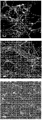
結果
脊椎動物における運動ニューロンの作製は、以下の段階を伴う:外胚葉細胞の神経化、神経外胚葉細胞の尾側化、および尾側化神経前駆細胞の腹側化。図1は、MNP分化プロセスの概要を提供する。神経化を開始するために、既知組成の混合物、すなわち古典的高グルコースDMEMと、インスリン、トランスフェリン、セレン、グルタミン、塩化マグネシウム、およびB27(NSF1)を添加したDMEM/F12との50:50混合物を導入する。次に、hES細胞/iPSCの物理的環境を、接着性のフィーダー細胞条件から非接着性の凝集体条件(浮遊培養)へと変化させて、細胞浮遊液を低接着容器中でさらに20日間培養した。
Results Generation of motor neurons in vertebrates involves the following stages: neurization of ectoderm cells, caudalization of neuroectodermal cells, and ventralization of caudalized neural progenitor cells. FIG. 1 provides an overview of the MNP differentiation process. To initiate neurization, a 50:50 mixture of a known composition, ie classical high glucose DMEM, and DMEM / F12 supplemented with insulin, transferrin, selenium, glutamine, magnesium chloride, and B27 (NSF1) Introduce. Next, the physical environment of hES cells / iPSC is changed from adhesive feeder cell conditions to non-adhesive aggregate conditions (floating culture), and the cell suspension is further cultured in a low adhesion container for 20 days. did.
レチノイン酸(RA)を用いることによって尾側化および腹側化を誘導した。細胞凝集体をRAによって1日目から7日目まで処置した。RA処置後、bFGF濃度を5 ng/mlに減少させて、培地交換の頻度を毎日供給から2日間供給スケジュールへと延ばした。これは、神経前駆細胞からのオートクライン因子の蓄積を促進しうる。理論に拘束されたくはないが、オートクライン因子は運動ニューロン分化にとって重要であると考えられる。 Caudalization and ventralization were induced by using retinoic acid (RA). Cell aggregates were treated with RA from day 1 to day 7. Following RA treatment, the bFGF concentration was reduced to 5 ng / ml, extending the frequency of medium exchange from a daily supply to a 2-day supply schedule. This can promote the accumulation of autocrine factors from neural progenitor cells. Without wishing to be bound by theory, autocrine factors are thought to be important for motor neuron differentiation.
ニューロスフェアの形成後、さらに発達させるために浮遊液をMatrigel(登録商標)コーティング表面に播種した。この期間中、いくつかの平坦な細胞と共にスフェア形成物(ロゼット)から遊走した放射状の配置を有する伸長した細胞も同様に生育するであろう。混入細胞が接着するゼラチンコーティング表面での陰性吸着を用いることによって、初期MNPを精製することができ、MNPを非接着細胞として分離した。 After formation of the neurospheres, the suspension was seeded on the Matrigel® coating surface for further development. During this period, elongated cells with a radial arrangement that migrated from the sphere former (rosette) with some flat cells will grow as well. By using negative adsorption on the gelatin coating surface to which contaminating cells adhere, the initial MNP could be purified and the MNP was separated as non-adherent cells.
発達および精製後、凍結保存の前にニューロンマーカーおよびMNP特異的マーカーによってMNPの特徴を調べる。その結果から、28日目に大多数の細胞がHB9、Islet1およびTuj1を発現することが示された(図3)。Oct4などのPSCマーカーおよび神経膠細胞マーカーGFAPは、検出されなかった。中胚葉マーカーSMAの発現は非常に少なかった。 After development and purification, MNPs are characterized by neuronal markers and MNP-specific markers before cryopreservation. The results showed that the majority of cells expressed HB9, Islet1 and Tuj1 on day 28 (FIG. 3). PSC markers such as Oct4 and glial marker GFAP were not detected. The expression of mesoderm marker SMA was very low.
実施例2
MNPの採取および凍結保存
材料および実験設計
30日目または31日目に、4つのT-75フラスコを、フラスコあたり7 mlの0.1%ゼラチン溶液でコーティングして、インキュベータ内で30分間インキュベートした。MNP誘導培地およそ150 mlを予め加温した。T-75フラスコからの使用済み培地を吸引して、フラスコをPBS各15 mlによって1回洗浄した。TrypLE溶液5 mlを添加して、3〜10分間インキュベートした。ほとんどの細胞が解離するまでフラスコを3分毎に調べた。MNP誘導培地10 mlを各フラスコに添加して、浮遊液を50 ml遠心管に移した。各T-75フラスコをさらに培地10 mlによってすすぎ、溶液を50 mlチューブに移した。チューブを200×gで、室温で3分間遠心分離した。上清を吸引して、細胞沈降物を培地20 mlに浮遊させて、浮遊液をゼラチンコーティングT-75フラスコに移した(10 ml/フラスコ)。遠心管をMNP誘導培地4 mlによってすすいで、溶液をゼラチンコーティングT-75フラスコに移した。T-75フラスコを37℃で15分間、動かさないようにインキュベートした。インキュベーション後、全ての非接着細胞をT-75フラスコから集めて別のゼラチンコーティングT-75フラスコに移し、フラスコを培地3〜5 mlによって優しくすすぎ、この溶液をゼラチンコーティングT-75フラスコに移した。T-75フラスコを37℃で15分間、動かさないようにインキュベートした。インキュベーション後、全ての非接着細胞をT-75フラスコから集めて、50 mlチューブに移した。より大きい細胞塊を、チューブの底に3分間沈降させた。上清(溶液の>95%)を新しいチューブに移して、NucleoCounter(登録商標)を用いて細胞を計数した。チューブを200×gで3〜5分間、室温で遠心沈降させた。上清を吸引して、冷却CryoStor(登録商標)溶液10 mlを細胞沈降物に徐々に加え、細胞沈降物を優しく浮遊させた。再度、NucleoCounter(登録商標)を用いて計数するために、浮遊液約100〜200μlを採取した。細胞の計数を待つあいだ、残りの細胞液は氷中に置いた。より多くのCryoStor(登録商標)溶液を添加することによって、生存細胞濃度を600万個/mlに調節して、浮遊液1 mlを各クライオバイアルに入れてアリコートとした。プログラム可能な速度制御フリーザーを用いて、以下のパラメータを用いてMNPを凍結した。凍結サイクルが完了した後、凍結プログラムの完了直後にクライオバイアルを液体窒素デュワーに移した。
Example 2
MNP collection and cryopreservation
Materials and experimental design
On day 30 or 31, 4 T-75 flasks were coated with 7 ml 0.1% gelatin solution per flask and incubated for 30 minutes in an incubator. Approximately 150 ml of MNP induction medium was pre-warmed. Spent media from the T-75 flask was aspirated and the flask was washed once with 15 ml of PBS each. 5 ml of TrypLE solution was added and incubated for 3-10 minutes. Flasks were examined every 3 minutes until most cells were dissociated. 10 ml of MNP induction medium was added to each flask and the suspension was transferred to a 50 ml centrifuge tube. Each T-75 flask was further rinsed with 10 ml of medium and the solution was transferred to a 50 ml tube. The tube was centrifuged at 200 xg for 3 minutes at room temperature. The supernatant was aspirated, the cell sediment was suspended in 20 ml of medium, and the suspension was transferred to a gelatin-coated T-75 flask (10 ml / flask). The centrifuge tube was rinsed with 4 ml of MNP induction medium and the solution was transferred to a gelatin coated T-75 flask. The T-75 flask was incubated at 37 ° C. for 15 minutes with no movement. After incubation, all non-adherent cells were collected from the T-75 flask and transferred to another gelatin-coated T-75 flask, the flask was gently rinsed with 3-5 ml of medium, and this solution was transferred to the gelatin-coated T-75 flask . The T-75 flask was incubated at 37 ° C. for 15 minutes with no movement. After incubation, all non-adherent cells were collected from the T-75 flask and transferred to a 50 ml tube. Larger cell clumps were allowed to settle for 3 minutes at the bottom of the tube. The supernatant (> 95% of the solution) was transferred to a new tube and the cells were counted using a NucleoCounter®. Tubes were spun down at 200 xg for 3-5 minutes at room temperature. The supernatant was aspirated and 10 ml of cooled CryoStor® solution was slowly added to the cell sediment to gently float the cell sediment. Again, about 100-200 μl of the suspension was collected for counting with NucleoCounter®. While waiting for cell counting, the remaining cell fluid was placed in ice. The viable cell concentration was adjusted to 6 million cells / ml by adding more CryoStor® solution, and 1 ml of the suspension was placed in each cryovial to make aliquots. Using a programmable speed control freezer, the MNPs were frozen using the following parameters. After the freezing cycle was completed, the cryovials were transferred to a liquid nitrogen dewar immediately after completion of the freezing program.
結果
-80℃機械的フリーザーと共に凍結容器を用いる典型的な方法によるMNPの凍結保存に関するこれまでの研究は、融解後に一貫しない回収および約70〜80%の細胞生存率を示した。説明として可能性があるのは、凍結保護剤が利用されていないこと、および-80℃の機械的フリーザー内での位置などの多数の理由により冷却速度が一貫しないことである。凍結保存プロセスの際のMNPの回収を改善するために、冷却速度が確実に一貫するようにするため、凍結保護剤を速度制御フリーザーと共に調べた。トレハロース、マンニトール、およびヘタスターチなどのいくつかの凍結保護剤を、DMSOと併用して用いて、CryoStor CS10(表1)と比較した。凍結前の細胞数を表1に示し、全ての試験条件は、凍結前に約90%の細胞生存率を有した。細胞およそ6×10⁶個を各クライオバイアルにおいて凍結した。クライオバイアルを全て、表2に示される凍結パラメータによってプログラム可能な速度制御フリーザーを用いて凍結した。凍結保存MNPを液体窒素下で保存した後、37℃の水浴を用いることによって細胞を液体窒素での保存から急速に融解した。細胞数をNucleoCounterによって決定して、生存率を計算した。結果は、CryotoRが他の試験条件より良好な成績を示したこと、および細胞生存率が、融解後一貫して85%より高かったことを示した(表3)。融解したMNPのIsl1およびHB9発現などの運動ニューロンマーカーは、凍結/融解を受けなかったMNP(データは示していない)と比較して正常であった(図4B、図4C)。
result
Previous studies on cryopreservation of MNP by typical methods using cryocontainers with a -80 ° C mechanical freezer have shown inconsistent recovery after thawing and cell viability of about 70-80%. Possible explanations are that cryoprotectants are not utilized and cooling rates are inconsistent for a number of reasons, such as location in a mechanical freezer at -80 ° C. To improve the recovery of MNP during the cryopreservation process, cryoprotectants were examined with a rate control freezer to ensure consistent cooling rates. Several cryoprotectants such as trehalose, mannitol, and hetastarch were used in combination with DMSO to compare with CryoStor CS10 (Table 1). The number of cells before freezing is shown in Table 1, and all test conditions had about 90% cell viability before freezing. Approximately 6 × 10 6 cells were frozen in each cryovial. All cryovials were frozen using a speed-controlled freezer programmable with the freezing parameters shown in Table 2. After cryopreserved MNP was stored under liquid nitrogen, cells were rapidly thawed from storage in liquid nitrogen by using a 37 ° C. water bath. Cell number was determined by NucleoCounter and viability was calculated. The results showed that CryotoR performed better than the other test conditions and that cell viability was consistently higher than 85% after thawing (Table 3). Motor neuron markers such as Isl1 and HB9 expression of thawed MNPs were normal compared to MNPs that did not undergo freeze / thaw (data not shown) (FIGS. 4B, 4C).
(表1)MNPを凍結保存するための凍結培地の試験

(表2)MNPを凍結保存するための凍結パラメータ
実施例3
運動ニューロンの播種
材料および実験設計
プレートのコーティング
各96ウェルプレートは、ポリ-D-リジン標準希釈液(50μg/ml)10 mlを必要とした。ポリ-D-リジン標準希釈液の必要量を、滅菌試薬リザーバーに移した。マルチチャンネルピペッターを用いて、96ウェルプレートの各ウェルを、ポリ-D-リジン標準希釈液100μlによってコーティングして、プレートをインキュベータ内に終夜入れた。インキュベーション後、マルチチャンネルアスピレータを用いてポリ-D-リジン溶液を吸引した。ウェルあたり100μlのPBSによってウェルを2回すすいだ。プレートの蓋を外して、層流フード内で少なくとも1時間乾燥させると、ラミニンコーティングの準備が整った。ラミニン標準希釈液(15μg/ml)の必要量をMNP基本培地(B27/NSF1を含まない)中で調製した。マルチチャンネルピペッターを用いて、ラミニン標準希釈液75μlをウェルに添加した。プレートを37℃で少なくとも1時間、6時間を超えないようインキュベートした。ラミニン溶液を吸引した後、細胞を播種する準備が整った。細胞を播種する前にウェルあたり100μlの培地によってウェルを1回すすいだ。
Example 3
Seeding of motor neurons
Materials and experimental design
Plate Coating Each 96-well plate required 10 ml of poly-D-lysine standard dilution (50 μg / ml). The required amount of poly-D-lysine standard diluent was transferred to a sterile reagent reservoir. Using a multichannel pipettor, each well of a 96-well plate was coated with 100 μl of poly-D-lysine standard dilution and the plate was placed in the incubator overnight. After incubation, the poly-D-lysine solution was aspirated using a multichannel aspirator. The wells were rinsed twice with 100 μl PBS per well. Once the plate lid was removed and dried for at least 1 hour in a laminar flow hood, the laminin coating was ready. The required amount of laminin standard dilution (15 μg / ml) was prepared in MNP basal medium (without B27 / NSF1). Using a multichannel pipettor, 75 μl of laminin standard dilution was added to the wells. Plates were incubated at 37 ° C for at least 1 hour and no more than 6 hours. After aspirating the laminin solution, the cells were ready to be seeded. Wells were rinsed once with 100 μl medium per well before seeding the cells.
MNPの融解
1つのアンプルを37℃水浴中で融解した。凍結保存からの細胞の良好な回収を確保するために、細胞浮遊液を急速に融解したが、37℃で維持することはなくあるいは37℃より上にはしなかった。浮遊液を融解直後に、細胞を15 mlチューブに移して、加温した播種用培地(NFS1およびグルタミンを添加したMNP基本培地)9 mlを、約2分間滴下して加えた。チューブを室温で5分間、200×gで遠心分離した。上清を吸引して、細胞を新しい播種用培地2 mlに浮遊させて、細胞浮遊液200μlをNucleoCounter(登録商標)において計数した。細胞数は、ウェルあたり生存細胞40,000個必要であり、播種のために必要な細胞浮遊液を計算した。播種用培地300μlあたり生存細胞40,000個の最終濃度を有するように計算した細胞浮遊液を浮遊させた。細胞浮遊液を滅菌リザーバーに移して、マルチチャンネルピペットを用いてMNPのウェルあたり細胞浮遊液300μlを添加した。96ウェルプレートをインキュベータ内に入れた。プレートを少なくとも24時間、動かさないように放置した。播種の48時間後、マルチチャンネルピペッターを用いて、使用済み培地200μlを各ウェルから除去して、新しいMNP維持培地200μlを添加した。供給手順は非常に優しく行った。接着細胞を乱さないように特に注意した。QC試験に合格したプレートは、後続の使用の準備が整うまで記述のように1日おきに培地を交換した。
Melting MNP
One ampoule was melted in a 37 ° C water bath. To ensure good recovery of cells from cryopreservation, the cell suspension was rapidly thawed but was not maintained at or above 37 ° C. Immediately after thawing the suspension, the cells were transferred to a 15 ml tube, and 9 ml of a warmed seeding medium (MNP basic medium supplemented with NFS1 and glutamine) was added dropwise for about 2 minutes. The tube was centrifuged at 200 xg for 5 minutes at room temperature. The supernatant was aspirated, the cells were suspended in 2 ml of fresh seeding medium and 200 μl of cell suspension was counted in a NucleoCounter®. The number of cells required 40,000 viable cells per well, and the cell suspension required for seeding was calculated. The cell suspension calculated to have a final concentration of 40,000 viable cells per 300 μl of seeding medium was suspended. The cell suspension was transferred to a sterile reservoir and 300 μl of cell suspension was added per well of MNP using a multichannel pipette. A 96-well plate was placed in the incubator. The plate was left stationary for at least 24 hours. 48 hours after seeding, 200 μl of spent medium was removed from each well using a multichannel pipettor and 200 μl of fresh MNP maintenance medium was added. The feeding procedure was very gentle. Special care was taken not to disturb the adherent cells. Plates that passed the QC test were changed medium every other day as described until ready for subsequent use.
結果
これまでの結果から、MNPが成熟プロセスの際に、約7〜10日後に集塊を形成する傾向を有することが示された。この集塊の問題の主な理由は不明であった。MNP播種のための96ウェルプレートのポリ-D-リジンおよびラミニンのコーティング手順を分析し、ラミニンコーティング段階はB27を含む培地を用いた。この段階によって、ポリ-D-リジン表面との結合に関して互いに競合するB27とラミニンの混合物が作製され、MNPの播種のための不均一な表面を生じる。ニューロンが一般にラミニンおよびポリ-D-リジン表面上で播種されることは周知である。本発明者らの仮説は、ラミニンコーティング段階に存在するB27がMNP集塊を引き起こすという点である。成熟MNP実験を、ラミニン単独またはラミニン+B27混合物によってコーティングしたポリ-D-リジンを用いて行った。培養物を、成熟プロセスの際の様々な時点でモニターした。結果から、10日後、ラミニン+B27の培養において集塊形成プロセスが出現し始めたことが示された(図5D)。17〜21日後では、ラミニン+B27のMNP培養において集塊形成がより明白になった(図5F)ものの、ラミニンコーティング単独の培養では集塊は検出されなかった(図5Cおよび図5E)。これらの結果は、B27が成熟プロセスの際のMNP集塊形成の原因であるという本発明者らの仮説と一貫する。
Results Previous results showed that MNP has a tendency to form clumps after about 7-10 days during the maturation process. The main reason for this agglomeration problem was unknown. The 96-well plate poly-D-lysine and laminin coating procedure for MNP seeding was analyzed and the laminin coating step used media containing B27. This step creates a mixture of B27 and laminin that compete with each other for binding to the poly-D-lysine surface, resulting in a heterogeneous surface for MNP seeding. It is well known that neurons are generally seeded on laminin and poly-D-lysine surfaces. Our hypothesis is that B27 present in the laminin coating stage causes MNP agglomeration. Mature MNP experiments were performed using poly-D-lysine coated with laminin alone or laminin + B27 mixture. Cultures were monitored at various times during the maturation process. The results showed that after 10 days, an agglomeration process began to appear in the laminin + B27 culture (FIG. 5D). After 17-21 days, agglomeration was more evident in laminin + B27 MNP cultures (Figure 5F), but no agglomeration was detected in cultures with laminin coating alone (Figures 5C and 5E). These results are consistent with our hypothesis that B27 is responsible for MNP agglomeration during the maturation process.
実施例4
記録保管
非臨床MNPを開始する毎に、0日目にバッチの記録を開始して、生産部門が維持した。品質記録を、品質記録の保持に関する内部標準操作手順に明示されているように、GMPおよびISOの要件のために保存した。適用可能なISO標準に従ったおよび/またはこれを参照とした。
Example 4
Every time a record keeping nonclinical MNP was started, batch recording started on day 0 and was maintained by the production department. Quality records were saved for GMP and ISO requirements as specified in internal standard operating procedures for maintaining quality records. According to applicable ISO standards and / or as a reference.
本発明のいくつかの態様を記述してきたが、前述は単なる例示に過ぎず、制限的でなく、例として提示されていることは当業者には明らかであるはずである。多数の改変および他の態様は、当業者の範囲内であり、本発明およびその任意の同等物の範囲内に入ることが企図される。本発明に対する変更は、当業者に容易に明らかとなり、本発明はそれらの代替物を含むと意図されると認識されうる。さらに、多数の改変が当業者に容易に想起されることから、例示および記述される構築および操作そのものに本発明を限定すると望んでおらず、したがって、全ての適した改変およびその同等物は、本発明の範囲内に入ると再分類されうる。本明細書において引用した各々の参考文献は、その全体が参照により本明細書に組み入れられる。 While several aspects of the present invention have been described, it should be apparent to those skilled in the art that the foregoing has been presented by way of example only, and not limitation. Numerous modifications and other embodiments are within the scope of those skilled in the art and are intended to fall within the scope of the invention and any equivalents thereof. Modifications to the invention will be readily apparent to those skilled in the art and it can be appreciated that the invention is intended to include alternatives thereof. Moreover, since numerous modifications will readily occur to those skilled in the art, it is not desired to limit the invention to the constructions and operations themselves illustrated and described, and therefore all suitable modifications and equivalents are It can be reclassified within the scope of the present invention. Each reference cited herein is hereby incorporated by reference in its entirety.
Claims (24)
未分化ヒト胚幹細胞(hESC)またはヒト人工多能性幹細胞(iPSC)を、フィーダー細胞上で培養する段階;
採取前の24時間、hESC培地とMNP誘導培地の混合物をhESC/iPSC培養物に供給する段階;
継代溶液を用いてhESC/iPSCコロニーを採取する段階;
hESC/iPSCコロニーを、レチノイン酸およびFGF-2を添加したMNP誘導培地を含む超低接着フラスコに移すことによって神経化ステップを開始する段階であって、少なくとも5日間ニューロスフェアを作製する、段階;
FGF-2を添加したMNP誘導培地を用いることによって、hESC/iPSCニューロスフェア培養物を約10〜12日間腹側化する段階;
ニューロスフェアをより小さいスフェアへと解離させる段階と、接着フラスコにおいて少なくとも4日間MNP誘導培地を用い神経ロゼットとして発達させる段階;
トリプシンを用いて神経ロゼットを細胞浮遊液へと解離させる段階;
ゼラチンコーティング表面による陰性吸着プロセスを用いてMNPを精製および濃縮する段階と、Matrigelコーティングフラスコに非接着MNPを再播種する段階;
FGF-2を添加したMNP誘導培地を用いてMNPを約4〜5日間発達させる段階;
トリプシンを用いてマトリクスコーティングフラスコから接着MNPを採取する段階;
ゼラチンコーティングフラスコによる陰性吸着プロセスを用いることによってMNP細胞浮遊液を精製する段階;および
得られた非接着MNPをゼラチンコーティングフラスコから集める段階。 A method for producing high purity motor neuron progenitor cells (MNP) in vitro, comprising the following steps:
Culturing undifferentiated human embryonic stem cells (hESC) or human induced pluripotent stem cells (iPSC) on feeder cells;
Feeding a mixture of hESC medium and MNP induction medium to the hESC / iPSC culture for 24 hours prior to harvesting;
Picking hESC / iPSC colonies with passage solution;
starting the neuralization step by transferring hESC / iPSC colonies to an ultra-low adhesion flask containing MNP induction medium supplemented with retinoic acid and FGF-2, creating a neurosphere for at least 5 days;
Ventralizing the hESC / iPSC neurosphere culture for about 10-12 days by using MNP induction medium supplemented with FGF-2;
Dissociating the neurospheres into smaller spheres and developing them as nerve rosettes using MNP induction medium for at least 4 days in adherent flasks;
Dissociating the nerve rosette into cell suspension using trypsin;
Purifying and concentrating the MNP using a negative adsorption process with a gelatin coated surface and reseeding the non-adherent MNP in a Matrigel coated flask;
Developing MNP for about 4-5 days using MNP induction medium supplemented with FGF-2;
Collecting adherent MNP from the matrix-coated flask using trypsin;
Purifying the MNP cell suspension by using a negative adsorption process with a gelatin coated flask; and collecting the resulting non-adherent MNP from the gelatin coated flask.
(a)採取したMNPを含む上清を遠心分離する段階;
(b)得られた MNPの沈降物を凍結培地中で再浮遊させる段階と、クライオバイアルに入れアリコートにする段階;
(c)MNPを含むクライオバイアルを速度制御フリーザーに移す段階;
(d)速度制御フリーザーにおいてMNPを含むクライオバイアルをプログラムされた凍結プロセスに供する段階;および
(e)長期間保存するために、プログラムされた凍結後に、MNPを含むクライオバイアルを速度制御フリーザーから液体窒素デュワーに移す段階。 A method for cryopreserving MNP, including the following steps:
(A) centrifuging the supernatant containing the collected MNP;
(B) resuspending the resulting MNP sediment in freezing medium and placing it in cryovials to make aliquots;
(C) transferring the cryovial containing MNP to a speed control freezer;
(D) subjecting the cryovial containing MNP to a programmed freezing process in a speed control freezer; and (e) liquid the cryovial containing MNP from the speed control freezer after programmed freezing for long-term storage. Transfer to a nitrogen dewar.
(a)プログラム可能な速度制御フリーザーを用いてクライオバイアルを5℃に平衡化する段階;
(b)フリーザーチャンバー温度が約5〜-10℃に達するまで、フリーザーを0.5〜2℃/分の速度で冷却する段階;
(c)フリーザーチャンバーが約-35℃〜-50℃の温度に達するまで、フリーザーを20〜30℃/分の速度で冷却する段階;
(d)フリーザーチャンバーを、5℃/分〜20℃/分の速度で-10℃〜-20℃に加温する段階;
(e)フリーザーチャンバーを、0.5〜2℃/分の速度で-30℃〜-50℃に冷却する段階;
(f)フリーザーチャンバーを、5℃/分〜15℃/分の一定速度で-70℃〜-100℃に冷却する段階;および
(g)フリーザーからMNPを含むクライオバイアルを取り出す段階と、液体窒素での保存に移す段階。 The method of claim 9 further comprising the following parameters:
(a) equilibrating the cryovial to 5 ° C. using a programmable speed control freezer;
(b) cooling the freezer at a rate of 0.5-2 ° C / min until the freezer chamber temperature reaches about 5-10 ° C;
(c) cooling the freezer at a rate of 20-30 ° C / min until the freezer chamber reaches a temperature of about -35 ° C to -50 ° C;
(d) heating the freezer chamber to -10 ° C to -20 ° C at a rate of 5 ° C / min to 20 ° C / min;
(e) cooling the freezer chamber to -30 ° C to -50 ° C at a rate of 0.5-2 ° C / min;
(f) cooling the freezer chamber to -70 ° C to -100 ° C at a constant rate of 5 ° C / min to 15 ° C / min; and
(g) The step of removing the cryovial containing MNP from the freezer and the step of storing it in liquid nitrogen.
未分化ヒト胚幹細胞(hESC)またはヒト人工多能性幹細胞(iPSC)を、フィーダーを含まない条件で、およそ60〜80%コンフルエンスに達するように少なくとも5日間培養する段階;
採取前の24時間のあいだ、無血清で異種由来成分を含まずかつ既知成分の培地をhESC/iPSC培養物に供給する段階;
無タンパク質の酵素性ではない継代溶液を用いてhESC/iPSCコロニーを採取する段階;
hESC/iPSCコロニーを、レチノイン酸およびFGF-2を添加したMNP誘導培地を含む超低接着フラスコに移すことによって神経化ステップを開始する段階であって、少なくとも5日間ニューロスフェアを作製する、段階;
FGF-2を添加したMNP誘導培地を用いることによって、hESC/iPSCニューロスフェアを約10〜12日間腹側化する段階;
ニューロスフェアをより小さいスフェアへと解離させる段階と、接着フラスコにおいて少なくとも4日間MNP誘導培地を用い神経ロゼットとして発達させる段階;
トリプシンを用いて神経ロゼットを細胞浮遊液へと解離させる段階;
ゼラチンコーティング表面による陰性吸着プロセスを用いてMNPを精製および濃縮する段階;
無タンパク質の既定のマトリクスコーティングフラスコに非接着MNPを再播種する段階;
FGF-2を添加した異種由来成分を含まない既知組成のMNP誘導培地を用いてMNPを約4〜5日間発達させる段階;
無タンパク質の既知組成の継代溶液を用いて、無タンパク質の既定のマトリクスコーティングフラスコから接着MNPを採取する段階;
ゼラチンコーティングフラスコによる陰性吸着プロセスを用いることによって、MNP細胞浮遊液を精製する段階;および
得られた非接着MNPをゼラチンコーティングフラスコから集める段階。 A method for producing high purity motor neuron progenitor cells (MNP) in vitro according to cGMP, comprising the following steps:
Culturing undifferentiated human embryonic stem cells (hESC) or human induced pluripotent stem cells (iPSC) for at least 5 days to reach approximately 60-80% confluence in a feeder-free condition;
Feeding the hESC / iPSC culture with serum-free, xeno-free and known medium for 24 hours prior to harvesting;
Picking hESC / iPSC colonies using a non-enzymatic, non-enzymatic passage solution;
starting the neuralization step by transferring hESC / iPSC colonies to an ultra-low adhesion flask containing MNP induction medium supplemented with retinoic acid and FGF-2, creating a neurosphere for at least 5 days;
Ventralizing hESC / iPSC neurospheres for about 10-12 days by using MNP induction medium supplemented with FGF-2;
Dissociating the neurospheres into smaller spheres and developing them as nerve rosettes using MNP induction medium for at least 4 days in adherent flasks;
Dissociating the nerve rosette into cell suspension using trypsin;
Purifying and concentrating MNP using a negative adsorption process with a gelatin coated surface;
Re-seeding non-adherent MNP in a protein-free pre-determined matrix-coated flask;
Developing MNP for about 4-5 days using a known composition of MNP induction medium without phenotypic components supplemented with FGF-2;
Taking adherent MNPs from a protein-free, predefined matrix-coated flask using a known protein-free passage solution;
Purifying the MNP cell suspension by using a negative adsorption process with a gelatin coated flask; and collecting the resulting non-adherent MNP from the gelatin coated flask.
Applications Claiming Priority (3)
| Application Number | Priority Date | Filing Date | Title |
|---|---|---|---|
| US201261658061P | 2012-06-11 | 2012-06-11 | |
| US61/658,061 | 2012-06-11 | ||
| PCT/US2013/045184 WO2013188407A2 (en) | 2012-06-11 | 2013-06-11 | Method of in vitro differentiation of motor neuron progenitors (mnps) from human induced pluripotent stem cells and cryopreservation of mnps |
Publications (2)
| Publication Number | Publication Date |
|---|---|
| JP2015521469A true JP2015521469A (en) | 2015-07-30 |
| JP2015521469A5 JP2015521469A5 (en) | 2016-07-14 |
Family
ID=49758856
Family Applications (1)
| Application Number | Title | Priority Date | Filing Date |
|---|---|---|---|
| JP2015517358A Ceased JP2015521469A (en) | 2012-06-11 | 2013-06-11 | In vitro differentiation method of motor neuron progenitor cells (MNP) from human induced pluripotent stem cells and cryopreservation method of MNP |
Country Status (4)
| Country | Link |
|---|---|
| US (1) | US20150159133A1 (en) |
| EP (1) | EP2858581A4 (en) |
| JP (1) | JP2015521469A (en) |
| WO (1) | WO2013188407A2 (en) |
Cited By (1)
| Publication number | Priority date | Publication date | Assignee | Title |
|---|---|---|---|---|
| KR20220104764A (en) | 2019-11-20 | 2022-07-26 | 스미토모 파마 가부시키가이샤 | Freezing method of nervous system cells |
Families Citing this family (3)
| Publication number | Priority date | Publication date | Assignee | Title |
|---|---|---|---|---|
| US20160130561A1 (en) * | 2013-06-13 | 2016-05-12 | The New York Stem Cell Foundation Inc. | Pluripotent stem cells derived from non-cryoprotected frozen tissue and methods for making and using the same |
| EP3491134B1 (en) | 2016-08-01 | 2023-10-11 | University of Pittsburgh - of The Commonwealth System of Higher Education | Human induced pluripotent stem cells for high efficiency genetic engineering |
| CN119432743A (en) * | 2023-07-28 | 2025-02-14 | 士泽生物医药(苏州)有限公司 | A kit for inducing stem cells to differentiate into motor nerve precursor cells and/or motor neurons and its application |
Citations (3)
| Publication number | Priority date | Publication date | Assignee | Title |
|---|---|---|---|---|
| WO2008118421A1 (en) * | 2007-03-23 | 2008-10-02 | California Stem Cell, Inc. | Human late stage motor neuron progenitor cells and methods of making and using same |
| JP2010536357A (en) * | 2007-08-20 | 2010-12-02 | ユニヴァルシテ リブレ デ ブリュッセル | Generation of neuronal cells from pluripotent stem cells |
| WO2011047289A1 (en) * | 2009-10-16 | 2011-04-21 | University Of Medicine And Dentistry Of New Jersey | Closed system separation of adherent bone marrow stem cells for regenerative medicine applications |
Family Cites Families (6)
| Publication number | Priority date | Publication date | Assignee | Title |
|---|---|---|---|---|
| JP3660601B2 (en) * | 2001-03-30 | 2005-06-15 | 独立行政法人科学技術振興機構 | Method for producing neural stem cells, motor neurons and GABAergic neurons from embryonic stem cells |
| US8153424B2 (en) * | 2001-10-03 | 2012-04-10 | Wisconsin Alumni Research Foundation | Method of in vitro differentiation of neural stem cells, motor neurons and dopamine neurons from primate embryonic stem cells |
| US7285415B2 (en) * | 2002-07-11 | 2007-10-23 | The Regents Of The University Of California | Oligodendrocytes derived from human embryonic stem cells for remyelination and treatment of spinal cord injury |
| US8263406B2 (en) * | 2003-06-11 | 2012-09-11 | Cornell Research Foundation, Inc. | Enriched or purified population of motor neurons and its preparation from a population of embryonic stem cells |
| WO2006044204A2 (en) * | 2004-10-05 | 2006-04-27 | University Of Georgia Research Foundation, Inc. | Neuronal progenitors from feeder-free human embryonic stem cell culture |
| WO2009122413A1 (en) * | 2008-03-31 | 2009-10-08 | Hadasit Medical Research Services & Development Limited | Motor neurons developed from stem cells |
-
2013
- 2013-06-11 JP JP2015517358A patent/JP2015521469A/en not_active Ceased
- 2013-06-11 EP EP13803543.1A patent/EP2858581A4/en not_active Withdrawn
- 2013-06-11 WO PCT/US2013/045184 patent/WO2013188407A2/en active Application Filing
- 2013-06-11 US US14/406,665 patent/US20150159133A1/en not_active Abandoned
Patent Citations (3)
| Publication number | Priority date | Publication date | Assignee | Title |
|---|---|---|---|---|
| WO2008118421A1 (en) * | 2007-03-23 | 2008-10-02 | California Stem Cell, Inc. | Human late stage motor neuron progenitor cells and methods of making and using same |
| JP2010536357A (en) * | 2007-08-20 | 2010-12-02 | ユニヴァルシテ リブレ デ ブリュッセル | Generation of neuronal cells from pluripotent stem cells |
| WO2011047289A1 (en) * | 2009-10-16 | 2011-04-21 | University Of Medicine And Dentistry Of New Jersey | Closed system separation of adherent bone marrow stem cells for regenerative medicine applications |
Cited By (1)
| Publication number | Priority date | Publication date | Assignee | Title |
|---|---|---|---|---|
| KR20220104764A (en) | 2019-11-20 | 2022-07-26 | 스미토모 파마 가부시키가이샤 | Freezing method of nervous system cells |
Also Published As
| Publication number | Publication date |
|---|---|
| WO2013188407A2 (en) | 2013-12-19 |
| EP2858581A4 (en) | 2016-07-27 |
| WO2013188407A3 (en) | 2015-05-07 |
| US20150159133A1 (en) | 2015-06-11 |
| EP2858581A2 (en) | 2015-04-15 |
Similar Documents
| Publication | Publication Date | Title |
|---|---|---|
| JP5560393B2 (en) | Differentiation of primate pluripotent stem cells into cardiomyocyte lineage cells | |
| JP6983762B2 (en) | Methods for Reproducible Differentiation of Clinical Grade Retinal Pigment Epithelial Cells | |
| JP6682446B2 (en) | Method for manufacturing retinal tissue | |
| JP4971131B2 (en) | Method for making high-purity cardiomyocyte preparations suitable for regenerative medicine | |
| JP2016504037A (en) | Suspension and clustering of human pluripotent cells for differentiation into pancreatic endocrine cells | |
| WO2011044684A1 (en) | Manipulation of osmolality for differentiating stem cells | |
| CN114787341B (en) | Method for preparing mesenchymal stem cells from human pluripotent stem cells and mesenchymal stem cells prepared thereby | |
| JP2015521469A (en) | In vitro differentiation method of motor neuron progenitor cells (MNP) from human induced pluripotent stem cells and cryopreservation method of MNP | |
| Amit et al. | Atlas of human pluripotent stem cells: derivation and culturing | |
| US20250180544A1 (en) | Artificial sertoli cells and method for their production | |
| Balbasi et al. | Mouse embryonic stem cell culture in serum-containing or 2i conditions | |
| JP2015521469A5 (en) | ||
| WO2022203051A1 (en) | Method for producing pluripotent stem cell population | |
| KR20250051648A (en) | Differentiation of iPSCs in a bioreactor | |
| EP4519416A2 (en) | Methods of generating three-dimensional retinal organoids from human pluripotent stem cells | |
| JP2022151855A (en) | Method for producing pluripotent stem cell population | |
| Mummery | Cardiomyocytes from human embryonic stem cells: more than heart repair alone | |
| US20230078230A1 (en) | Methods for the production of committed cardiac progenitor cells | |
| JP2022151854A (en) | Method for producing pluripotent stem cell population | |
| US20240352427A1 (en) | Mass production method of pluripotent stem cell stock | |
| WO2025038906A1 (en) | Artificial testis cells and method for their production | |
| Şuşman et al. | Human placenta–stem cell source for obtaining pancreatic progenitors | |
| CN116855441A (en) | Method for inducing and differentiating pig expanded pluripotent stem cells into pig intestinal organoids | |
| JP2025521749A (en) | Differentiation of iPSCs in a bioreactor | |
| Espinha | Bioprocess engineering of induced pluripotent stem cells for application in cell therapy and pre-clinical research |
Legal Events
| Date | Code | Title | Description |
|---|---|---|---|
| A521 | Request for written amendment filed |
Free format text: JAPANESE INTERMEDIATE CODE: A523 Effective date: 20160524 |
|
| A621 | Written request for application examination |
Free format text: JAPANESE INTERMEDIATE CODE: A621 Effective date: 20160524 |
|
| A131 | Notification of reasons for refusal |
Free format text: JAPANESE INTERMEDIATE CODE: A131 Effective date: 20170412 |
|
| A601 | Written request for extension of time |
Free format text: JAPANESE INTERMEDIATE CODE: A601 Effective date: 20170711 |
|
| A521 | Request for written amendment filed |
Free format text: JAPANESE INTERMEDIATE CODE: A523 Effective date: 20170905 |
|
| A01 | Written decision to grant a patent or to grant a registration (utility model) |
Free format text: JAPANESE INTERMEDIATE CODE: A01 Effective date: 20180124 |
|
| A045 | Written measure of dismissal of application [lapsed due to lack of payment] |
Free format text: JAPANESE INTERMEDIATE CODE: A045 Effective date: 20180524 |