KR20220104201A - Methods of treating and/or preventing idiopathic pneumonia syndrome (IPS) and/or capillary leakage syndrome (CLS) and/or engraftment syndrome (ES) and/or fluid overload (FO) associated with hematopoietic stem cell transplantation - Google Patents
Methods of treating and/or preventing idiopathic pneumonia syndrome (IPS) and/or capillary leakage syndrome (CLS) and/or engraftment syndrome (ES) and/or fluid overload (FO) associated with hematopoietic stem cell transplantation Download PDFInfo
- Publication number
- KR20220104201A KR20220104201A KR1020227020672A KR20227020672A KR20220104201A KR 20220104201 A KR20220104201 A KR 20220104201A KR 1020227020672 A KR1020227020672 A KR 1020227020672A KR 20227020672 A KR20227020672 A KR 20227020672A KR 20220104201 A KR20220104201 A KR 20220104201A
- Authority
- KR
- South Korea
- Prior art keywords
- masp
- antibody
- subject
- seq
- inhibitory
- Prior art date
Links
- 238000000034 method Methods 0.000 title claims abstract description 235
- 208000007915 ichthyosis prematurity syndrome Diseases 0.000 title claims description 16
- 238000011134 hematopoietic stem cell transplantation Methods 0.000 title claims description 15
- 206010063725 Idiopathic pneumonia syndrome Diseases 0.000 title claims description 11
- 206010050684 Engraftment syndrome Diseases 0.000 title claims description 8
- 206010016803 Fluid overload Diseases 0.000 title claims description 8
- 208000011580 syndromic disease Diseases 0.000 title claims description 8
- 102100026046 Mannan-binding lectin serine protease 2 Human genes 0.000 claims abstract description 530
- 101710117460 Mannan-binding lectin serine protease 2 Proteins 0.000 claims abstract description 529
- 230000002401 inhibitory effect Effects 0.000 claims abstract description 296
- 230000024203 complement activation Effects 0.000 claims abstract description 128
- 241000282414 Homo sapiens Species 0.000 claims abstract description 109
- 230000001419 dependent effect Effects 0.000 claims abstract description 76
- 230000037361 pathway Effects 0.000 claims description 194
- 230000027455 binding Effects 0.000 claims description 117
- 239000012634 fragment Substances 0.000 claims description 104
- 125000003275 alpha amino acid group Chemical group 0.000 claims description 100
- 239000000427 antigen Substances 0.000 claims description 88
- 108091007433 antigens Proteins 0.000 claims description 88
- 102000036639 antigens Human genes 0.000 claims description 88
- 210000002966 serum Anatomy 0.000 claims description 85
- 239000000203 mixture Substances 0.000 claims description 81
- 230000008021 deposition Effects 0.000 claims description 68
- 238000011282 treatment Methods 0.000 claims description 65
- 101001056015 Homo sapiens Mannan-binding lectin serine protease 2 Proteins 0.000 claims description 60
- 102000054960 human MASP2 Human genes 0.000 claims description 58
- 208000037265 diseases, disorders, signs and symptoms Diseases 0.000 claims description 45
- 201000010099 disease Diseases 0.000 claims description 38
- 230000003442 weekly effect Effects 0.000 claims description 29
- 230000002829 reductive effect Effects 0.000 claims description 28
- 239000012636 effector Substances 0.000 claims description 22
- 210000003958 hematopoietic stem cell Anatomy 0.000 claims description 17
- 230000000735 allogeneic effect Effects 0.000 claims description 11
- 201000005488 Capillary Leak Syndrome Diseases 0.000 claims description 7
- 208000017850 Diffuse alveolar hemorrhage Diseases 0.000 claims description 3
- 208000031932 Systemic capillary leak syndrome Diseases 0.000 claims description 2
- 210000001124 body fluid Anatomy 0.000 claims 1
- 239000010839 body fluid Substances 0.000 claims 1
- 230000000694 effects Effects 0.000 abstract description 30
- 102000004856 Lectins Human genes 0.000 description 148
- 108090001090 Lectins Proteins 0.000 description 148
- 239000002523 lectin Substances 0.000 description 145
- 239000003795 chemical substances by application Substances 0.000 description 98
- 230000000295 complement effect Effects 0.000 description 97
- 108090000765 processed proteins & peptides Proteins 0.000 description 87
- 230000004913 activation Effects 0.000 description 86
- 241000699670 Mus sp. Species 0.000 description 83
- 108010087870 Mannose-Binding Lectin Proteins 0.000 description 69
- 102000009112 Mannose-Binding Lectin Human genes 0.000 description 69
- 208000035913 Atypical hemolytic uremic syndrome Diseases 0.000 description 67
- 238000000151 deposition Methods 0.000 description 64
- 208000034841 Thrombotic Microangiopathies Diseases 0.000 description 60
- 102000004196 processed proteins & peptides Human genes 0.000 description 58
- 101000901154 Homo sapiens Complement C3 Proteins 0.000 description 55
- 102100022133 Complement C3 Human genes 0.000 description 54
- 206010043561 Thrombocytopenic purpura Diseases 0.000 description 50
- 208000032759 Hemolytic-Uremic Syndrome Diseases 0.000 description 46
- 208000007536 Thrombosis Diseases 0.000 description 42
- 239000003112 inhibitor Substances 0.000 description 42
- 230000006870 function Effects 0.000 description 41
- 230000015572 biosynthetic process Effects 0.000 description 40
- 208000015181 infectious disease Diseases 0.000 description 40
- 208000024891 symptom Diseases 0.000 description 40
- 108090000623 proteins and genes Proteins 0.000 description 39
- 229920001184 polypeptide Polymers 0.000 description 36
- 108010034753 Complement Membrane Attack Complex Proteins 0.000 description 34
- 230000004154 complement system Effects 0.000 description 34
- 108090000062 ficolin Proteins 0.000 description 34
- 230000006378 damage Effects 0.000 description 31
- 230000035772 mutation Effects 0.000 description 31
- 206010040047 Sepsis Diseases 0.000 description 30
- 108010053085 Complement Factor H Proteins 0.000 description 29
- 238000003776 cleavage reaction Methods 0.000 description 29
- 230000001404 mediated effect Effects 0.000 description 29
- 235000001014 amino acid Nutrition 0.000 description 28
- 230000015271 coagulation Effects 0.000 description 28
- 238000005345 coagulation Methods 0.000 description 28
- 230000007017 scission Effects 0.000 description 28
- 229940024606 amino acid Drugs 0.000 description 26
- 150000001413 amino acids Chemical class 0.000 description 26
- 238000003556 assay Methods 0.000 description 26
- 235000018102 proteins Nutrition 0.000 description 26
- 102000004169 proteins and genes Human genes 0.000 description 26
- 230000002950 deficient Effects 0.000 description 25
- 238000002616 plasmapheresis Methods 0.000 description 24
- 210000004027 cell Anatomy 0.000 description 23
- 210000003743 erythrocyte Anatomy 0.000 description 23
- 230000005764 inhibitory process Effects 0.000 description 23
- 102000008394 Immunoglobulin Fragments Human genes 0.000 description 22
- 108010021625 Immunoglobulin Fragments Proteins 0.000 description 22
- DDRJAANPRJIHGJ-UHFFFAOYSA-N creatinine Chemical compound CN1CC(=O)NC1=N DDRJAANPRJIHGJ-UHFFFAOYSA-N 0.000 description 22
- 102000016550 Complement Factor H Human genes 0.000 description 21
- 206010028980 Neoplasm Diseases 0.000 description 21
- 201000007023 Thrombotic Thrombocytopenic Purpura Diseases 0.000 description 21
- 201000011510 cancer Diseases 0.000 description 21
- 206010043554 thrombocytopenia Diseases 0.000 description 21
- 102100031506 Complement C5 Human genes 0.000 description 20
- 201000009906 Meningitis Diseases 0.000 description 20
- 241001529936 Murinae Species 0.000 description 20
- 241000700159 Rattus Species 0.000 description 20
- 102000012479 Serine Proteases Human genes 0.000 description 20
- 108010022999 Serine Proteases Proteins 0.000 description 20
- 238000001990 intravenous administration Methods 0.000 description 20
- 230000004083 survival effect Effects 0.000 description 20
- 208000029713 Catastrophic antiphospholipid syndrome Diseases 0.000 description 19
- 102100024521 Ficolin-2 Human genes 0.000 description 19
- 108060003951 Immunoglobulin Proteins 0.000 description 19
- 208000022993 cryopyrin-associated periodic syndrome Diseases 0.000 description 19
- 102000018358 immunoglobulin Human genes 0.000 description 19
- 210000003734 kidney Anatomy 0.000 description 19
- 238000004519 manufacturing process Methods 0.000 description 19
- 108091005670 ADAMTS13 Proteins 0.000 description 18
- 208000022774 Congenital thrombotic thrombocytopenic purpura Diseases 0.000 description 18
- 101000941598 Homo sapiens Complement C5 Proteins 0.000 description 18
- 241000699666 Mus <mouse, genus> Species 0.000 description 18
- 210000001772 blood platelet Anatomy 0.000 description 18
- 102100032290 A disintegrin and metalloproteinase with thrombospondin motifs 13 Human genes 0.000 description 17
- 230000008506 pathogenesis Effects 0.000 description 17
- 230000004044 response Effects 0.000 description 17
- 208000001647 Renal Insufficiency Diseases 0.000 description 16
- 210000004369 blood Anatomy 0.000 description 16
- 239000008280 blood Substances 0.000 description 16
- LTMHDMANZUZIPE-PUGKRICDSA-N digoxin Chemical compound C1[C@H](O)[C@H](O)[C@@H](C)O[C@H]1O[C@@H]1[C@@H](C)O[C@@H](O[C@@H]2[C@H](O[C@@H](O[C@@H]3C[C@@H]4[C@]([C@@H]5[C@H]([C@]6(CC[C@@H]([C@@]6(C)[C@H](O)C5)C=5COC(=O)C=5)O)CC4)(C)CC3)C[C@@H]2O)C)C[C@@H]1O LTMHDMANZUZIPE-PUGKRICDSA-N 0.000 description 16
- 208000014674 injury Diseases 0.000 description 16
- 201000006370 kidney failure Diseases 0.000 description 16
- 102000004528 Mannose-Binding Protein-Associated Serine Proteases Human genes 0.000 description 15
- 108010042484 Mannose-Binding Protein-Associated Serine Proteases Proteins 0.000 description 15
- 230000007812 deficiency Effects 0.000 description 14
- 208000017169 kidney disease Diseases 0.000 description 14
- 230000007246 mechanism Effects 0.000 description 14
- 241001465754 Metazoa Species 0.000 description 13
- 108010079723 Shiga Toxin Proteins 0.000 description 13
- 208000007502 anemia Diseases 0.000 description 13
- 238000002512 chemotherapy Methods 0.000 description 13
- 210000004408 hybridoma Anatomy 0.000 description 13
- 238000002054 transplantation Methods 0.000 description 13
- 206010018364 Glomerulonephritis Diseases 0.000 description 12
- 229960002224 eculizumab Drugs 0.000 description 12
- 210000002889 endothelial cell Anatomy 0.000 description 12
- 230000003511 endothelial effect Effects 0.000 description 12
- 239000002158 endotoxin Substances 0.000 description 12
- 229940027941 immunoglobulin g Drugs 0.000 description 12
- 230000001965 increasing effect Effects 0.000 description 12
- 229920006008 lipopolysaccharide Polymers 0.000 description 12
- 238000007911 parenteral administration Methods 0.000 description 12
- 238000007920 subcutaneous administration Methods 0.000 description 12
- 241000894006 Bacteria Species 0.000 description 11
- -1 H-ficolin Proteins 0.000 description 11
- 206010018910 Haemolysis Diseases 0.000 description 11
- 241000282412 Homo Species 0.000 description 11
- 102100026061 Mannan-binding lectin serine protease 1 Human genes 0.000 description 11
- 230000001154 acute effect Effects 0.000 description 11
- 239000002299 complementary DNA Substances 0.000 description 11
- 229940109239 creatinine Drugs 0.000 description 11
- 208000009190 disseminated intravascular coagulation Diseases 0.000 description 11
- 239000003814 drug Substances 0.000 description 11
- 230000001575 pathological effect Effects 0.000 description 11
- 201000000596 systemic lupus erythematosus Diseases 0.000 description 11
- 229960004072 thrombin Drugs 0.000 description 11
- WHUUTDBJXJRKMK-VKHMYHEASA-N L-glutamic acid Chemical compound OC(=O)[C@@H](N)CCC(O)=O WHUUTDBJXJRKMK-VKHMYHEASA-N 0.000 description 10
- 229920000057 Mannan Polymers 0.000 description 10
- 108090000190 Thrombin Proteins 0.000 description 10
- 238000009093 first-line therapy Methods 0.000 description 10
- 230000008588 hemolysis Effects 0.000 description 10
- 238000001727 in vivo Methods 0.000 description 10
- 230000000977 initiatory effect Effects 0.000 description 10
- 238000001361 intraarterial administration Methods 0.000 description 10
- 210000001519 tissue Anatomy 0.000 description 10
- 108010047041 Complementarity Determining Regions Proteins 0.000 description 9
- 241000588724 Escherichia coli Species 0.000 description 9
- 102100024508 Ficolin-1 Human genes 0.000 description 9
- 206010061218 Inflammation Diseases 0.000 description 9
- AYFVYJQAPQTCCC-GBXIJSLDSA-N L-threonine Chemical compound C[C@@H](O)[C@H](N)C(O)=O AYFVYJQAPQTCCC-GBXIJSLDSA-N 0.000 description 9
- 101100326461 Mus musculus C1ra gene Proteins 0.000 description 9
- 101100326462 Mus musculus C1rb gene Proteins 0.000 description 9
- 206010040070 Septic Shock Diseases 0.000 description 9
- 208000027418 Wounds and injury Diseases 0.000 description 9
- 239000003937 drug carrier Substances 0.000 description 9
- 230000014509 gene expression Effects 0.000 description 9
- 238000007918 intramuscular administration Methods 0.000 description 9
- 210000000056 organ Anatomy 0.000 description 9
- 230000035935 pregnancy Effects 0.000 description 9
- 230000036303 septic shock Effects 0.000 description 9
- 230000000451 tissue damage Effects 0.000 description 9
- 231100000827 tissue damage Toxicity 0.000 description 9
- 108010089414 Anaphylatoxins Proteins 0.000 description 8
- 102100035432 Complement factor H Human genes 0.000 description 8
- DHMQDGOQFOQNFH-UHFFFAOYSA-N Glycine Chemical compound NCC(O)=O DHMQDGOQFOQNFH-UHFFFAOYSA-N 0.000 description 8
- 101001018257 Rattus norvegicus Mannan-binding lectin serine protease 2 Proteins 0.000 description 8
- 230000000903 blocking effect Effects 0.000 description 8
- 230000004054 inflammatory process Effects 0.000 description 8
- 230000003993 interaction Effects 0.000 description 8
- 201000008383 nephritis Diseases 0.000 description 8
- 239000012071 phase Substances 0.000 description 8
- 230000009885 systemic effect Effects 0.000 description 8
- 108010047303 von Willebrand Factor Proteins 0.000 description 8
- 102100036537 von Willebrand factor Human genes 0.000 description 8
- 229960001134 von willebrand factor Drugs 0.000 description 8
- MTCFGRXMJLQNBG-REOHCLBHSA-N (2S)-2-Amino-3-hydroxypropansäure Chemical compound OC[C@H](N)C(O)=O MTCFGRXMJLQNBG-REOHCLBHSA-N 0.000 description 7
- 206010001052 Acute respiratory distress syndrome Diseases 0.000 description 7
- 102000016574 Complement C3-C5 Convertases Human genes 0.000 description 7
- 108010067641 Complement C3-C5 Convertases Proteins 0.000 description 7
- 102000004190 Enzymes Human genes 0.000 description 7
- 108090000790 Enzymes Proteins 0.000 description 7
- 101100329495 Mus musculus C1sa gene Proteins 0.000 description 7
- 101100329496 Mus musculus C1sb gene Proteins 0.000 description 7
- 108091034117 Oligonucleotide Proteins 0.000 description 7
- 241000283973 Oryctolagus cuniculus Species 0.000 description 7
- 239000004365 Protease Substances 0.000 description 7
- 206010051379 Systemic Inflammatory Response Syndrome Diseases 0.000 description 7
- 229920000392 Zymosan Polymers 0.000 description 7
- 125000000539 amino acid group Chemical group 0.000 description 7
- 239000003242 anti bacterial agent Substances 0.000 description 7
- 210000004204 blood vessel Anatomy 0.000 description 7
- 230000005779 cell damage Effects 0.000 description 7
- 238000010586 diagram Methods 0.000 description 7
- 229940088598 enzyme Drugs 0.000 description 7
- 208000028867 ischemia Diseases 0.000 description 7
- 230000003389 potentiating effect Effects 0.000 description 7
- 239000000126 substance Substances 0.000 description 7
- 101710155594 Coiled-coil domain-containing protein 115 Proteins 0.000 description 6
- 102100023661 Coiled-coil domain-containing protein 115 Human genes 0.000 description 6
- 108090000056 Complement factor B Proteins 0.000 description 6
- 102000003712 Complement factor B Human genes 0.000 description 6
- 241001646719 Escherichia coli O157:H7 Species 0.000 description 6
- DCXYFEDJOCDNAF-REOHCLBHSA-N L-asparagine Chemical compound OC(=O)[C@@H](N)CC(N)=O DCXYFEDJOCDNAF-REOHCLBHSA-N 0.000 description 6
- CKLJMWTZIZZHCS-REOHCLBHSA-N L-aspartic acid Chemical compound OC(=O)[C@@H](N)CC(O)=O CKLJMWTZIZZHCS-REOHCLBHSA-N 0.000 description 6
- AGPKZVBTJJNPAG-WHFBIAKZSA-N L-isoleucine Chemical compound CC[C@H](C)[C@H](N)C(O)=O AGPKZVBTJJNPAG-WHFBIAKZSA-N 0.000 description 6
- KDXKERNSBIXSRK-YFKPBYRVSA-N L-lysine Chemical compound NCCCC[C@H](N)C(O)=O KDXKERNSBIXSRK-YFKPBYRVSA-N 0.000 description 6
- KDXKERNSBIXSRK-UHFFFAOYSA-N Lysine Natural products NCCCCC(N)C(O)=O KDXKERNSBIXSRK-UHFFFAOYSA-N 0.000 description 6
- 206010064281 Malignant atrophic papulosis Diseases 0.000 description 6
- 101710117390 Mannan-binding lectin serine protease 1 Proteins 0.000 description 6
- 102000035195 Peptidases Human genes 0.000 description 6
- 108091005804 Peptidases Proteins 0.000 description 6
- 206010063837 Reperfusion injury Diseases 0.000 description 6
- JLCPHMBAVCMARE-UHFFFAOYSA-N [3-[[3-[[3-[[3-[[3-[[3-[[3-[[3-[[3-[[3-[[3-[[5-(2-amino-6-oxo-1H-purin-9-yl)-3-[[3-[[3-[[3-[[3-[[3-[[5-(2-amino-6-oxo-1H-purin-9-yl)-3-[[5-(2-amino-6-oxo-1H-purin-9-yl)-3-hydroxyoxolan-2-yl]methoxy-hydroxyphosphoryl]oxyoxolan-2-yl]methoxy-hydroxyphosphoryl]oxy-5-(5-methyl-2,4-dioxopyrimidin-1-yl)oxolan-2-yl]methoxy-hydroxyphosphoryl]oxy-5-(6-aminopurin-9-yl)oxolan-2-yl]methoxy-hydroxyphosphoryl]oxy-5-(6-aminopurin-9-yl)oxolan-2-yl]methoxy-hydroxyphosphoryl]oxy-5-(6-aminopurin-9-yl)oxolan-2-yl]methoxy-hydroxyphosphoryl]oxy-5-(6-aminopurin-9-yl)oxolan-2-yl]methoxy-hydroxyphosphoryl]oxyoxolan-2-yl]methoxy-hydroxyphosphoryl]oxy-5-(5-methyl-2,4-dioxopyrimidin-1-yl)oxolan-2-yl]methoxy-hydroxyphosphoryl]oxy-5-(4-amino-2-oxopyrimidin-1-yl)oxolan-2-yl]methoxy-hydroxyphosphoryl]oxy-5-(5-methyl-2,4-dioxopyrimidin-1-yl)oxolan-2-yl]methoxy-hydroxyphosphoryl]oxy-5-(5-methyl-2,4-dioxopyrimidin-1-yl)oxolan-2-yl]methoxy-hydroxyphosphoryl]oxy-5-(6-aminopurin-9-yl)oxolan-2-yl]methoxy-hydroxyphosphoryl]oxy-5-(6-aminopurin-9-yl)oxolan-2-yl]methoxy-hydroxyphosphoryl]oxy-5-(4-amino-2-oxopyrimidin-1-yl)oxolan-2-yl]methoxy-hydroxyphosphoryl]oxy-5-(4-amino-2-oxopyrimidin-1-yl)oxolan-2-yl]methoxy-hydroxyphosphoryl]oxy-5-(4-amino-2-oxopyrimidin-1-yl)oxolan-2-yl]methoxy-hydroxyphosphoryl]oxy-5-(6-aminopurin-9-yl)oxolan-2-yl]methoxy-hydroxyphosphoryl]oxy-5-(4-amino-2-oxopyrimidin-1-yl)oxolan-2-yl]methyl [5-(6-aminopurin-9-yl)-2-(hydroxymethyl)oxolan-3-yl] hydrogen phosphate Polymers Cc1cn(C2CC(OP(O)(=O)OCC3OC(CC3OP(O)(=O)OCC3OC(CC3O)n3cnc4c3nc(N)[nH]c4=O)n3cnc4c3nc(N)[nH]c4=O)C(COP(O)(=O)OC3CC(OC3COP(O)(=O)OC3CC(OC3COP(O)(=O)OC3CC(OC3COP(O)(=O)OC3CC(OC3COP(O)(=O)OC3CC(OC3COP(O)(=O)OC3CC(OC3COP(O)(=O)OC3CC(OC3COP(O)(=O)OC3CC(OC3COP(O)(=O)OC3CC(OC3COP(O)(=O)OC3CC(OC3COP(O)(=O)OC3CC(OC3COP(O)(=O)OC3CC(OC3COP(O)(=O)OC3CC(OC3COP(O)(=O)OC3CC(OC3COP(O)(=O)OC3CC(OC3COP(O)(=O)OC3CC(OC3COP(O)(=O)OC3CC(OC3CO)n3cnc4c(N)ncnc34)n3ccc(N)nc3=O)n3cnc4c(N)ncnc34)n3ccc(N)nc3=O)n3ccc(N)nc3=O)n3ccc(N)nc3=O)n3cnc4c(N)ncnc34)n3cnc4c(N)ncnc34)n3cc(C)c(=O)[nH]c3=O)n3cc(C)c(=O)[nH]c3=O)n3ccc(N)nc3=O)n3cc(C)c(=O)[nH]c3=O)n3cnc4c3nc(N)[nH]c4=O)n3cnc4c(N)ncnc34)n3cnc4c(N)ncnc34)n3cnc4c(N)ncnc34)n3cnc4c(N)ncnc34)O2)c(=O)[nH]c1=O JLCPHMBAVCMARE-UHFFFAOYSA-N 0.000 description 6
- 229940088710 antibiotic agent Drugs 0.000 description 6
- 210000002565 arteriole Anatomy 0.000 description 6
- 230000008901 benefit Effects 0.000 description 6
- 150000001720 carbohydrates Chemical class 0.000 description 6
- 208000037887 cell injury Diseases 0.000 description 6
- 238000002716 delivery method Methods 0.000 description 6
- 238000011161 development Methods 0.000 description 6
- 230000018109 developmental process Effects 0.000 description 6
- 208000035475 disorder Diseases 0.000 description 6
- 230000002068 genetic effect Effects 0.000 description 6
- 238000000099 in vitro assay Methods 0.000 description 6
- 230000036542 oxidative stress Effects 0.000 description 6
- 239000000047 product Substances 0.000 description 6
- 230000003331 prothrombotic effect Effects 0.000 description 6
- 230000035939 shock Effects 0.000 description 6
- 230000035882 stress Effects 0.000 description 6
- 238000012360 testing method Methods 0.000 description 6
- 230000001225 therapeutic effect Effects 0.000 description 6
- 238000002560 therapeutic procedure Methods 0.000 description 6
- 230000008733 trauma Effects 0.000 description 6
- 108010003529 Alternative Pathway Complement C3 Convertase Proteins 0.000 description 5
- 108010034358 Classical Pathway Complement C3 Convertase Proteins 0.000 description 5
- 108700017374 Complement Factor H Deficiency Proteins 0.000 description 5
- 206010064571 Gene mutation Diseases 0.000 description 5
- ZWQVYZXPYSYPJD-RYUDHWBXSA-N Glu-Gly-Phe Chemical group OC(=O)CC[C@H](N)C(=O)NCC(=O)N[C@H](C(O)=O)CC1=CC=CC=C1 ZWQVYZXPYSYPJD-RYUDHWBXSA-N 0.000 description 5
- WHUUTDBJXJRKMK-UHFFFAOYSA-N Glutamic acid Natural products OC(=O)C(N)CCC(O)=O WHUUTDBJXJRKMK-UHFFFAOYSA-N 0.000 description 5
- 101000909536 Homo sapiens Collectin-11 Proteins 0.000 description 5
- 101001055956 Homo sapiens Mannan-binding lectin serine protease 1 Proteins 0.000 description 5
- ZDXPYRJPNDTMRX-VKHMYHEASA-N L-glutamine Chemical compound OC(=O)[C@@H](N)CCC(N)=O ZDXPYRJPNDTMRX-VKHMYHEASA-N 0.000 description 5
- ROHFNLRQFUQHCH-YFKPBYRVSA-N L-leucine Chemical compound CC(C)C[C@H](N)C(O)=O ROHFNLRQFUQHCH-YFKPBYRVSA-N 0.000 description 5
- COLNVLDHVKWLRT-QMMMGPOBSA-N L-phenylalanine Chemical compound OC(=O)[C@@H](N)CC1=CC=CC=C1 COLNVLDHVKWLRT-QMMMGPOBSA-N 0.000 description 5
- 208000004451 Membranoproliferative Glomerulonephritis Diseases 0.000 description 5
- NWIBSHFKIJFRCO-WUDYKRTCSA-N Mytomycin Chemical compound C1N2C(C(C(C)=C(N)C3=O)=O)=C3[C@@H](COC(N)=O)[C@@]2(OC)[C@@H]2[C@H]1N2 NWIBSHFKIJFRCO-WUDYKRTCSA-N 0.000 description 5
- OVRNDRQMDRJTHS-UHFFFAOYSA-N N-acelyl-D-glucosamine Natural products CC(=O)NC1C(O)OC(CO)C(O)C1O OVRNDRQMDRJTHS-UHFFFAOYSA-N 0.000 description 5
- 101710098940 Pro-epidermal growth factor Proteins 0.000 description 5
- 102100033237 Pro-epidermal growth factor Human genes 0.000 description 5
- 108010005642 Properdin Proteins 0.000 description 5
- 102100038567 Properdin Human genes 0.000 description 5
- 102100027378 Prothrombin Human genes 0.000 description 5
- 108010094028 Prothrombin Proteins 0.000 description 5
- 208000013616 Respiratory Distress Syndrome Diseases 0.000 description 5
- 239000012190 activator Substances 0.000 description 5
- 201000000028 adult respiratory distress syndrome Diseases 0.000 description 5
- 238000010171 animal model Methods 0.000 description 5
- 230000009286 beneficial effect Effects 0.000 description 5
- 230000017531 blood circulation Effects 0.000 description 5
- 239000011575 calcium Substances 0.000 description 5
- 235000014633 carbohydrates Nutrition 0.000 description 5
- 230000001684 chronic effect Effects 0.000 description 5
- 230000007423 decrease Effects 0.000 description 5
- 230000007123 defense Effects 0.000 description 5
- 210000003989 endothelium vascular Anatomy 0.000 description 5
- 230000002255 enzymatic effect Effects 0.000 description 5
- BTCSSZJGUNDROE-UHFFFAOYSA-N gamma-aminobutyric acid Chemical compound NCCCC(O)=O BTCSSZJGUNDROE-UHFFFAOYSA-N 0.000 description 5
- 230000028709 inflammatory response Effects 0.000 description 5
- 210000004379 membrane Anatomy 0.000 description 5
- 239000012528 membrane Substances 0.000 description 5
- 244000005700 microbiome Species 0.000 description 5
- 210000004088 microvessel Anatomy 0.000 description 5
- 210000000440 neutrophil Anatomy 0.000 description 5
- 239000002773 nucleotide Substances 0.000 description 5
- 125000003729 nucleotide group Chemical group 0.000 description 5
- 229940039716 prothrombin Drugs 0.000 description 5
- 210000003491 skin Anatomy 0.000 description 5
- 208000010110 spontaneous platelet aggregation Diseases 0.000 description 5
- 238000011476 stem cell transplantation Methods 0.000 description 5
- 238000006467 substitution reaction Methods 0.000 description 5
- 235000000346 sugar Nutrition 0.000 description 5
- 150000007970 thio esters Chemical class 0.000 description 5
- 238000011830 transgenic mouse model Methods 0.000 description 5
- 230000001960 triggered effect Effects 0.000 description 5
- 230000002792 vascular Effects 0.000 description 5
- 208000035143 Bacterial infection Diseases 0.000 description 4
- 208000005590 Choroidal Neovascularization Diseases 0.000 description 4
- 206010060823 Choroidal neovascularisation Diseases 0.000 description 4
- 206010053567 Coagulopathies Diseases 0.000 description 4
- 101710172562 Cobra venom factor Proteins 0.000 description 4
- 102100024331 Collectin-11 Human genes 0.000 description 4
- 108010028780 Complement C3 Proteins 0.000 description 4
- 102000016918 Complement C3 Human genes 0.000 description 4
- 102000000989 Complement System Proteins Human genes 0.000 description 4
- PMATZTZNYRCHOR-CGLBZJNRSA-N Cyclosporin A Chemical compound CC[C@@H]1NC(=O)[C@H]([C@H](O)[C@H](C)C\C=C\C)N(C)C(=O)[C@H](C(C)C)N(C)C(=O)[C@H](CC(C)C)N(C)C(=O)[C@H](CC(C)C)N(C)C(=O)[C@@H](C)NC(=O)[C@H](C)NC(=O)[C@H](CC(C)C)N(C)C(=O)[C@H](C(C)C)NC(=O)[C@H](CC(C)C)N(C)C(=O)CN(C)C1=O PMATZTZNYRCHOR-CGLBZJNRSA-N 0.000 description 4
- 108010036949 Cyclosporine Proteins 0.000 description 4
- 108020004414 DNA Proteins 0.000 description 4
- 102000053602 DNA Human genes 0.000 description 4
- 102000010911 Enzyme Precursors Human genes 0.000 description 4
- 108010062466 Enzyme Precursors Proteins 0.000 description 4
- 108700024394 Exon Proteins 0.000 description 4
- 108010054147 Hemoglobins Proteins 0.000 description 4
- 102000001554 Hemoglobins Human genes 0.000 description 4
- 101000961414 Homo sapiens Membrane cofactor protein Proteins 0.000 description 4
- 208000010159 IgA glomerulonephritis Diseases 0.000 description 4
- 206010021263 IgA nephropathy Diseases 0.000 description 4
- 108010067060 Immunoglobulin Variable Region Proteins 0.000 description 4
- 102000017727 Immunoglobulin Variable Region Human genes 0.000 description 4
- XUJNEKJLAYXESH-REOHCLBHSA-N L-Cysteine Chemical compound SC[C@H](N)C(O)=O XUJNEKJLAYXESH-REOHCLBHSA-N 0.000 description 4
- OUYCCCASQSFEME-QMMMGPOBSA-N L-tyrosine Chemical compound OC(=O)[C@@H](N)CC1=CC=C(O)C=C1 OUYCCCASQSFEME-QMMMGPOBSA-N 0.000 description 4
- 241000699660 Mus musculus Species 0.000 description 4
- 206010037549 Purpura Diseases 0.000 description 4
- 241001672981 Purpura Species 0.000 description 4
- LOUPRKONTZGTKE-WZBLMQSHSA-N Quinine Chemical compound C([C@H]([C@H](C1)C=C)C2)C[N@@]1[C@@H]2[C@H](O)C1=CC=NC2=CC=C(OC)C=C21 LOUPRKONTZGTKE-WZBLMQSHSA-N 0.000 description 4
- 206010062237 Renal impairment Diseases 0.000 description 4
- 241000283984 Rodentia Species 0.000 description 4
- 241000607142 Salmonella Species 0.000 description 4
- 241000607768 Shigella Species 0.000 description 4
- KZSNJWFQEVHDMF-UHFFFAOYSA-N Valine Natural products CC(C)C(N)C(O)=O KZSNJWFQEVHDMF-UHFFFAOYSA-N 0.000 description 4
- 230000003213 activating effect Effects 0.000 description 4
- 230000002776 aggregation Effects 0.000 description 4
- 238000004220 aggregation Methods 0.000 description 4
- 230000003321 amplification Effects 0.000 description 4
- 239000002246 antineoplastic agent Substances 0.000 description 4
- 230000001580 bacterial effect Effects 0.000 description 4
- 208000022362 bacterial infectious disease Diseases 0.000 description 4
- 238000006243 chemical reaction Methods 0.000 description 4
- 229960001265 ciclosporin Drugs 0.000 description 4
- 238000003745 diagnosis Methods 0.000 description 4
- ZDXPYRJPNDTMRX-UHFFFAOYSA-N glutamine Natural products OC(=O)C(N)CCC(N)=O ZDXPYRJPNDTMRX-UHFFFAOYSA-N 0.000 description 4
- 208000007475 hemolytic anemia Diseases 0.000 description 4
- 239000003018 immunosuppressive agent Substances 0.000 description 4
- 238000000338 in vitro Methods 0.000 description 4
- 230000002458 infectious effect Effects 0.000 description 4
- 230000002757 inflammatory effect Effects 0.000 description 4
- 238000005259 measurement Methods 0.000 description 4
- 206010062198 microangiopathy Diseases 0.000 description 4
- 230000000813 microbial effect Effects 0.000 description 4
- 229960004857 mitomycin Drugs 0.000 description 4
- 238000003199 nucleic acid amplification method Methods 0.000 description 4
- 244000052769 pathogen Species 0.000 description 4
- 125000001151 peptidyl group Chemical group 0.000 description 4
- 230000003285 pharmacodynamic effect Effects 0.000 description 4
- 230000008569 process Effects 0.000 description 4
- 230000002797 proteolythic effect Effects 0.000 description 4
- 102000005962 receptors Human genes 0.000 description 4
- 108020003175 receptors Proteins 0.000 description 4
- 230000010410 reperfusion Effects 0.000 description 4
- 241000894007 species Species 0.000 description 4
- 230000002889 sympathetic effect Effects 0.000 description 4
- 229940124597 therapeutic agent Drugs 0.000 description 4
- 210000003556 vascular endothelial cell Anatomy 0.000 description 4
- 208000003343 Antiphospholipid Syndrome Diseases 0.000 description 3
- 239000004475 Arginine Substances 0.000 description 3
- DCXYFEDJOCDNAF-UHFFFAOYSA-N Asparagine Natural products OC(=O)C(N)CC(N)=O DCXYFEDJOCDNAF-UHFFFAOYSA-N 0.000 description 3
- 108010017384 Blood Proteins Proteins 0.000 description 3
- 102000004506 Blood Proteins Human genes 0.000 description 3
- 241000282472 Canis lupus familiaris Species 0.000 description 3
- 108010069112 Complement System Proteins Proteins 0.000 description 3
- 102000004127 Cytokines Human genes 0.000 description 3
- 108090000695 Cytokines Proteins 0.000 description 3
- 206010012735 Diarrhoea Diseases 0.000 description 3
- 206010061818 Disease progression Diseases 0.000 description 3
- 241000192125 Firmicutes Species 0.000 description 3
- 206010018370 Glomerulonephritis membranoproliferative Diseases 0.000 description 3
- 239000004471 Glycine Substances 0.000 description 3
- 208000009329 Graft vs Host Disease Diseases 0.000 description 3
- 208000032843 Hemorrhage Diseases 0.000 description 3
- 206010020751 Hypersensitivity Diseases 0.000 description 3
- 102000009490 IgG Receptors Human genes 0.000 description 3
- 108010073807 IgG Receptors Proteins 0.000 description 3
- 206010061216 Infarction Diseases 0.000 description 3
- QNAYBMKLOCPYGJ-REOHCLBHSA-N L-alanine Chemical compound C[C@H](N)C(O)=O QNAYBMKLOCPYGJ-REOHCLBHSA-N 0.000 description 3
- KZSNJWFQEVHDMF-BYPYZUCNSA-N L-valine Chemical compound CC(C)[C@H](N)C(O)=O KZSNJWFQEVHDMF-BYPYZUCNSA-N 0.000 description 3
- ROHFNLRQFUQHCH-UHFFFAOYSA-N Leucine Natural products CC(C)CC(N)C(O)=O ROHFNLRQFUQHCH-UHFFFAOYSA-N 0.000 description 3
- 208000005777 Lupus Nephritis Diseases 0.000 description 3
- 239000004472 Lysine Substances 0.000 description 3
- 108700033009 MASP2 Deficiency Proteins 0.000 description 3
- 241000124008 Mammalia Species 0.000 description 3
- 102100039373 Membrane cofactor protein Human genes 0.000 description 3
- 206010027527 Microangiopathic haemolytic anaemia Diseases 0.000 description 3
- 229930192392 Mitomycin Natural products 0.000 description 3
- OVRNDRQMDRJTHS-FMDGEEDCSA-N N-acetyl-beta-D-glucosamine Chemical compound CC(=O)N[C@H]1[C@H](O)O[C@H](CO)[C@@H](O)[C@@H]1O OVRNDRQMDRJTHS-FMDGEEDCSA-N 0.000 description 3
- 108700022034 Opsonin Proteins Proteins 0.000 description 3
- 102000057297 Pepsin A Human genes 0.000 description 3
- 108090000284 Pepsin A Proteins 0.000 description 3
- 240000004808 Saccharomyces cerevisiae Species 0.000 description 3
- MTCFGRXMJLQNBG-UHFFFAOYSA-N Serine Natural products OCC(N)C(O)=O MTCFGRXMJLQNBG-UHFFFAOYSA-N 0.000 description 3
- 208000037549 Shiga toxin-associated hemolytic uremic syndrome Diseases 0.000 description 3
- 208000006011 Stroke Diseases 0.000 description 3
- 241000282887 Suidae Species 0.000 description 3
- AYFVYJQAPQTCCC-UHFFFAOYSA-N Threonine Natural products CC(O)C(N)C(O)=O AYFVYJQAPQTCCC-UHFFFAOYSA-N 0.000 description 3
- 239000004473 Threonine Substances 0.000 description 3
- 108010073929 Vascular Endothelial Growth Factor A Proteins 0.000 description 3
- 102000005789 Vascular Endothelial Growth Factors Human genes 0.000 description 3
- 108010019530 Vascular Endothelial Growth Factors Proteins 0.000 description 3
- 241000700605 Viruses Species 0.000 description 3
- 239000002253 acid Substances 0.000 description 3
- 230000002411 adverse Effects 0.000 description 3
- 235000004279 alanine Nutrition 0.000 description 3
- 125000003295 alanine group Chemical group N[C@@H](C)C(=O)* 0.000 description 3
- 230000007815 allergy Effects 0.000 description 3
- 125000003277 amino group Chemical group 0.000 description 3
- 238000004458 analytical method Methods 0.000 description 3
- 230000000890 antigenic effect Effects 0.000 description 3
- ODKSFYDXXFIFQN-UHFFFAOYSA-N arginine Natural products OC(=O)C(N)CCCNC(N)=N ODKSFYDXXFIFQN-UHFFFAOYSA-N 0.000 description 3
- 210000001367 artery Anatomy 0.000 description 3
- 235000009582 asparagine Nutrition 0.000 description 3
- 229960001230 asparagine Drugs 0.000 description 3
- 230000001363 autoimmune Effects 0.000 description 3
- 230000003115 biocidal effect Effects 0.000 description 3
- 230000033228 biological regulation Effects 0.000 description 3
- 238000001574 biopsy Methods 0.000 description 3
- 208000034158 bleeding Diseases 0.000 description 3
- 230000000740 bleeding effect Effects 0.000 description 3
- 230000023555 blood coagulation Effects 0.000 description 3
- 210000003169 central nervous system Anatomy 0.000 description 3
- 230000008859 change Effects 0.000 description 3
- 239000004074 complement inhibitor Substances 0.000 description 3
- 230000002596 correlated effect Effects 0.000 description 3
- 239000003246 corticosteroid Substances 0.000 description 3
- 229930182912 cyclosporin Natural products 0.000 description 3
- 235000018417 cysteine Nutrition 0.000 description 3
- XUJNEKJLAYXESH-UHFFFAOYSA-N cysteine Natural products SCC(N)C(O)=O XUJNEKJLAYXESH-UHFFFAOYSA-N 0.000 description 3
- 230000034994 death Effects 0.000 description 3
- 230000001627 detrimental effect Effects 0.000 description 3
- 230000029087 digestion Effects 0.000 description 3
- 238000006471 dimerization reaction Methods 0.000 description 3
- 230000005750 disease progression Effects 0.000 description 3
- 210000003038 endothelium Anatomy 0.000 description 3
- 230000004927 fusion Effects 0.000 description 3
- 238000010448 genetic screening Methods 0.000 description 3
- 150000004676 glycans Chemical class 0.000 description 3
- 208000024908 graft versus host disease Diseases 0.000 description 3
- 210000000224 granular leucocyte Anatomy 0.000 description 3
- HNDVDQJCIGZPNO-UHFFFAOYSA-N histidine Natural products OC(=O)C(N)CC1=CN=CN1 HNDVDQJCIGZPNO-UHFFFAOYSA-N 0.000 description 3
- 229910052739 hydrogen Inorganic materials 0.000 description 3
- 125000002887 hydroxy group Chemical group [H]O* 0.000 description 3
- 230000007124 immune defense Effects 0.000 description 3
- 230000028993 immune response Effects 0.000 description 3
- 230000036039 immunity Effects 0.000 description 3
- 230000002163 immunogen Effects 0.000 description 3
- 229940072221 immunoglobulins Drugs 0.000 description 3
- 230000006698 induction Effects 0.000 description 3
- 230000007574 infarction Effects 0.000 description 3
- 238000001802 infusion Methods 0.000 description 3
- 238000002347 injection Methods 0.000 description 3
- 239000007924 injection Substances 0.000 description 3
- 102000006495 integrins Human genes 0.000 description 3
- 108010044426 integrins Proteins 0.000 description 3
- 238000007912 intraperitoneal administration Methods 0.000 description 3
- 229960000310 isoleucine Drugs 0.000 description 3
- AGPKZVBTJJNPAG-UHFFFAOYSA-N isoleucine Natural products CCC(C)C(N)C(O)=O AGPKZVBTJJNPAG-UHFFFAOYSA-N 0.000 description 3
- 108010045069 keyhole-limpet hemocyanin Proteins 0.000 description 3
- 239000003446 ligand Substances 0.000 description 3
- 230000007774 longterm Effects 0.000 description 3
- 210000004072 lung Anatomy 0.000 description 3
- 230000036210 malignancy Effects 0.000 description 3
- 239000003550 marker Substances 0.000 description 3
- 201000008350 membranous glomerulonephritis Diseases 0.000 description 3
- 210000005063 microvascular endothelium Anatomy 0.000 description 3
- 230000004048 modification Effects 0.000 description 3
- 238000012986 modification Methods 0.000 description 3
- 230000003990 molecular pathway Effects 0.000 description 3
- 238000012544 monitoring process Methods 0.000 description 3
- 229950006780 n-acetylglucosamine Drugs 0.000 description 3
- 244000045947 parasite Species 0.000 description 3
- 230000007310 pathophysiology Effects 0.000 description 3
- 230000035515 penetration Effects 0.000 description 3
- 229940111202 pepsin Drugs 0.000 description 3
- 230000002085 persistent effect Effects 0.000 description 3
- 210000001539 phagocyte Anatomy 0.000 description 3
- COLNVLDHVKWLRT-UHFFFAOYSA-N phenylalanine Natural products OC(=O)C(N)CC1=CC=CC=C1 COLNVLDHVKWLRT-UHFFFAOYSA-N 0.000 description 3
- 230000036470 plasma concentration Effects 0.000 description 3
- 231100000857 poor renal function Toxicity 0.000 description 3
- 201000001474 proteinuria Diseases 0.000 description 3
- 230000005855 radiation Effects 0.000 description 3
- 230000009467 reduction Effects 0.000 description 3
- 229920002477 rna polymer Polymers 0.000 description 3
- 230000028327 secretion Effects 0.000 description 3
- 238000002741 site-directed mutagenesis Methods 0.000 description 3
- 150000003384 small molecules Chemical class 0.000 description 3
- 239000000243 solution Substances 0.000 description 3
- 230000002269 spontaneous effect Effects 0.000 description 3
- 238000010561 standard procedure Methods 0.000 description 3
- 239000000758 substrate Substances 0.000 description 3
- 239000006228 supernatant Substances 0.000 description 3
- 231100000331 toxic Toxicity 0.000 description 3
- 230000002588 toxic effect Effects 0.000 description 3
- 230000009261 transgenic effect Effects 0.000 description 3
- 208000017271 typical hemolytic-uremic syndrome Diseases 0.000 description 3
- OUYCCCASQSFEME-UHFFFAOYSA-N tyrosine Natural products OC(=O)C(N)CC1=CC=C(O)C=C1 OUYCCCASQSFEME-UHFFFAOYSA-N 0.000 description 3
- 210000002700 urine Anatomy 0.000 description 3
- 239000004474 valine Substances 0.000 description 3
- WQZGKKKJIJFFOK-SVZMEOIVSA-N (+)-Galactose Chemical compound OC[C@H]1OC(O)[C@H](O)[C@@H](O)[C@H]1O WQZGKKKJIJFFOK-SVZMEOIVSA-N 0.000 description 2
- 101150028074 2 gene Proteins 0.000 description 2
- 101710131943 40S ribosomal protein S3a Proteins 0.000 description 2
- XTWYTFMLZFPYCI-KQYNXXCUSA-N 5'-adenylphosphoric acid Chemical compound C1=NC=2C(N)=NC=NC=2N1[C@@H]1O[C@H](COP(O)(=O)OP(O)(O)=O)[C@@H](O)[C@H]1O XTWYTFMLZFPYCI-KQYNXXCUSA-N 0.000 description 2
- 101150085726 ADAMTS13 gene Proteins 0.000 description 2
- XTWYTFMLZFPYCI-UHFFFAOYSA-N Adenosine diphosphate Natural products C1=NC=2C(N)=NC=NC=2N1C1OC(COP(O)(=O)OP(O)(O)=O)C(O)C1O XTWYTFMLZFPYCI-UHFFFAOYSA-N 0.000 description 2
- 108700028369 Alleles Proteins 0.000 description 2
- 102000005590 Anaphylatoxin C5a Receptor Human genes 0.000 description 2
- 108010059426 Anaphylatoxin C5a Receptor Proteins 0.000 description 2
- 208000037157 Azotemia Diseases 0.000 description 2
- 239000005528 B01AC05 - Ticlopidine Substances 0.000 description 2
- 108010006654 Bleomycin Proteins 0.000 description 2
- 208000019838 Blood disease Diseases 0.000 description 2
- 102000007350 Bone Morphogenetic Proteins Human genes 0.000 description 2
- 108010007726 Bone Morphogenetic Proteins Proteins 0.000 description 2
- 241000283690 Bos taurus Species 0.000 description 2
- 108091003079 Bovine Serum Albumin Proteins 0.000 description 2
- 102100022002 CD59 glycoprotein Human genes 0.000 description 2
- 241000283707 Capra Species 0.000 description 2
- 102000014914 Carrier Proteins Human genes 0.000 description 2
- 241000700198 Cavia Species 0.000 description 2
- 208000017667 Chronic Disease Diseases 0.000 description 2
- 235000001258 Cinchona calisaya Nutrition 0.000 description 2
- 208000005443 Circulating Neoplastic Cells Diseases 0.000 description 2
- 102000004405 Collectins Human genes 0.000 description 2
- 108090000909 Collectins Proteins 0.000 description 2
- 101710184994 Complement control protein Proteins 0.000 description 2
- 102100040132 Complement factor H-related protein 1 Human genes 0.000 description 2
- 102100035321 Complement factor H-related protein 3 Human genes 0.000 description 2
- 102100035324 Complement factor H-related protein 4 Human genes 0.000 description 2
- 101710101162 Complement factor H-related protein 4 Proteins 0.000 description 2
- 108010069514 Cyclic Peptides Proteins 0.000 description 2
- 102000001189 Cyclic Peptides Human genes 0.000 description 2
- WQZGKKKJIJFFOK-QTVWNMPRSA-N D-mannopyranose Chemical compound OC[C@H]1OC(O)[C@@H](O)[C@@H](O)[C@@H]1O WQZGKKKJIJFFOK-QTVWNMPRSA-N 0.000 description 2
- 229920002307 Dextran Polymers 0.000 description 2
- 241000257465 Echinoidea Species 0.000 description 2
- 241000283086 Equidae Species 0.000 description 2
- 102000014702 Haptoglobin Human genes 0.000 description 2
- 108050005077 Haptoglobin Proteins 0.000 description 2
- 208000032456 Hemorrhagic Shock Diseases 0.000 description 2
- NTYJJOPFIAHURM-UHFFFAOYSA-N Histamine Chemical compound NCCC1=CN=CN1 NTYJJOPFIAHURM-UHFFFAOYSA-N 0.000 description 2
- 101000897400 Homo sapiens CD59 glycoprotein Proteins 0.000 description 2
- 101000890732 Homo sapiens Complement factor H-related protein 1 Proteins 0.000 description 2
- 101000878136 Homo sapiens Complement factor H-related protein 3 Proteins 0.000 description 2
- 101001052749 Homo sapiens Ficolin-3 Proteins 0.000 description 2
- 208000035888 Immune-mediated thrombotic thrombocytopenic purpura Diseases 0.000 description 2
- 108010054477 Immunoglobulin Fab Fragments Proteins 0.000 description 2
- 102000001706 Immunoglobulin Fab Fragments Human genes 0.000 description 2
- 206010022678 Intestinal infections Diseases 0.000 description 2
- ODKSFYDXXFIFQN-BYPYZUCNSA-P L-argininium(2+) Chemical compound NC(=[NH2+])NCCC[C@H]([NH3+])C(O)=O ODKSFYDXXFIFQN-BYPYZUCNSA-P 0.000 description 2
- HNDVDQJCIGZPNO-YFKPBYRVSA-N L-histidine Chemical compound OC(=O)[C@@H](N)CC1=CN=CN1 HNDVDQJCIGZPNO-YFKPBYRVSA-N 0.000 description 2
- 102000003855 L-lactate dehydrogenase Human genes 0.000 description 2
- 108700023483 L-lactate dehydrogenases Proteins 0.000 description 2
- QIVBCDIJIAJPQS-VIFPVBQESA-N L-tryptophane Chemical compound C1=CC=C2C(C[C@H](N)C(O)=O)=CNC2=C1 QIVBCDIJIAJPQS-VIFPVBQESA-N 0.000 description 2
- 208000034486 Multi-organ failure Diseases 0.000 description 2
- 208000010718 Multiple Organ Failure Diseases 0.000 description 2
- 208000004221 Multiple Trauma Diseases 0.000 description 2
- OVRNDRQMDRJTHS-RTRLPJTCSA-N N-acetyl-D-glucosamine Chemical compound CC(=O)N[C@H]1C(O)O[C@H](CO)[C@@H](O)[C@@H]1O OVRNDRQMDRJTHS-RTRLPJTCSA-N 0.000 description 2
- MBLBDJOUHNCFQT-LXGUWJNJSA-N N-acetylglucosamine Natural products CC(=O)N[C@@H](C=O)[C@@H](O)[C@H](O)[C@H](O)CO MBLBDJOUHNCFQT-LXGUWJNJSA-N 0.000 description 2
- 108091028043 Nucleic acid sequence Proteins 0.000 description 2
- 206010030302 Oliguria Diseases 0.000 description 2
- 241001494479 Pecora Species 0.000 description 2
- 102000007079 Peptide Fragments Human genes 0.000 description 2
- 108010033276 Peptide Fragments Proteins 0.000 description 2
- 208000035109 Pneumococcal Infections Diseases 0.000 description 2
- 241000288906 Primates Species 0.000 description 2
- 206010037660 Pyrexia Diseases 0.000 description 2
- 101100400474 Rattus norvegicus Masp2 gene Proteins 0.000 description 2
- 102000007056 Recombinant Fusion Proteins Human genes 0.000 description 2
- 108010008281 Recombinant Fusion Proteins Proteins 0.000 description 2
- 206010063897 Renal ischaemia Diseases 0.000 description 2
- 206010053879 Sepsis syndrome Diseases 0.000 description 2
- 206010049771 Shock haemorrhagic Diseases 0.000 description 2
- 241000191967 Staphylococcus aureus Species 0.000 description 2
- NKANXQFJJICGDU-QPLCGJKRSA-N Tamoxifen Chemical compound C=1C=CC=CC=1C(/CC)=C(C=1C=CC(OCCN(C)C)=CC=1)/C1=CC=CC=C1 NKANXQFJJICGDU-QPLCGJKRSA-N 0.000 description 2
- QIVBCDIJIAJPQS-UHFFFAOYSA-N Tryptophan Natural products C1=CC=C2C(CC(N)C(O)=O)=CNC2=C1 QIVBCDIJIAJPQS-UHFFFAOYSA-N 0.000 description 2
- 206010045168 Tumour embolism Diseases 0.000 description 2
- 206010047115 Vasculitis Diseases 0.000 description 2
- PNNCWTXUWKENPE-UHFFFAOYSA-N [N].NC(N)=O Chemical compound [N].NC(N)=O PNNCWTXUWKENPE-UHFFFAOYSA-N 0.000 description 2
- 230000002159 abnormal effect Effects 0.000 description 2
- 238000009825 accumulation Methods 0.000 description 2
- 208000004886 acquired thrombotic thrombocytopenic purpura Diseases 0.000 description 2
- 230000009471 action Effects 0.000 description 2
- 230000004721 adaptive immunity Effects 0.000 description 2
- 239000002671 adjuvant Substances 0.000 description 2
- 230000001494 anti-thymocyte effect Effects 0.000 description 2
- 230000005875 antibody response Effects 0.000 description 2
- 239000003146 anticoagulant agent Substances 0.000 description 2
- 229940127219 anticoagulant drug Drugs 0.000 description 2
- 102000025171 antigen binding proteins Human genes 0.000 description 2
- 108091000831 antigen binding proteins Proteins 0.000 description 2
- 229940127218 antiplatelet drug Drugs 0.000 description 2
- 230000001640 apoptogenic effect Effects 0.000 description 2
- 206010003246 arthritis Diseases 0.000 description 2
- 235000003704 aspartic acid Nutrition 0.000 description 2
- 230000002238 attenuated effect Effects 0.000 description 2
- 210000003719 b-lymphocyte Anatomy 0.000 description 2
- OQFSQFPPLPISGP-UHFFFAOYSA-N beta-carboxyaspartic acid Natural products OC(=O)C(N)C(C(O)=O)C(O)=O OQFSQFPPLPISGP-UHFFFAOYSA-N 0.000 description 2
- 108091008324 binding proteins Proteins 0.000 description 2
- 230000004071 biological effect Effects 0.000 description 2
- 229960001561 bleomycin Drugs 0.000 description 2
- OYVAGSVQBOHSSS-UAPAGMARSA-O bleomycin A2 Chemical compound N([C@H](C(=O)N[C@H](C)[C@@H](O)[C@H](C)C(=O)N[C@@H]([C@H](O)C)C(=O)NCCC=1SC=C(N=1)C=1SC=C(N=1)C(=O)NCCC[S+](C)C)[C@@H](O[C@H]1[C@H]([C@@H](O)[C@H](O)[C@H](CO)O1)O[C@@H]1[C@H]([C@@H](OC(N)=O)[C@H](O)[C@@H](CO)O1)O)C=1N=CNC=1)C(=O)C1=NC([C@H](CC(N)=O)NC[C@H](N)C(N)=O)=NC(N)=C1C OYVAGSVQBOHSSS-UAPAGMARSA-O 0.000 description 2
- 208000015294 blood coagulation disease Diseases 0.000 description 2
- 210000000988 bone and bone Anatomy 0.000 description 2
- 238000010322 bone marrow transplantation Methods 0.000 description 2
- 229940112869 bone morphogenetic protein Drugs 0.000 description 2
- 229940098773 bovine serum albumin Drugs 0.000 description 2
- 210000004899 c-terminal region Anatomy 0.000 description 2
- 102000023852 carbohydrate binding proteins Human genes 0.000 description 2
- 108091008400 carbohydrate binding proteins Proteins 0.000 description 2
- 230000003197 catalytic effect Effects 0.000 description 2
- 230000006037 cell lysis Effects 0.000 description 2
- 210000000170 cell membrane Anatomy 0.000 description 2
- 210000002421 cell wall Anatomy 0.000 description 2
- 230000001413 cellular effect Effects 0.000 description 2
- 229960001231 choline Drugs 0.000 description 2
- OEYIOHPDSNJKLS-UHFFFAOYSA-N choline Chemical compound C[N+](C)(C)CCO OEYIOHPDSNJKLS-UHFFFAOYSA-N 0.000 description 2
- LOUPRKONTZGTKE-UHFFFAOYSA-N cinchonine Natural products C1C(C(C2)C=C)CCN2C1C(O)C1=CC=NC2=CC=C(OC)C=C21 LOUPRKONTZGTKE-UHFFFAOYSA-N 0.000 description 2
- DQLATGHUWYMOKM-UHFFFAOYSA-L cisplatin Chemical compound N[Pt](N)(Cl)Cl DQLATGHUWYMOKM-UHFFFAOYSA-L 0.000 description 2
- 229960004316 cisplatin Drugs 0.000 description 2
- 102000006834 complement receptors Human genes 0.000 description 2
- 108010047295 complement receptors Proteins 0.000 description 2
- 230000000875 corresponding effect Effects 0.000 description 2
- 229960001334 corticosteroids Drugs 0.000 description 2
- 230000009089 cytolysis Effects 0.000 description 2
- 229940127089 cytotoxic agent Drugs 0.000 description 2
- 230000000254 damaging effect Effects 0.000 description 2
- 231100000673 dose–response relationship Toxicity 0.000 description 2
- 239000006196 drop Substances 0.000 description 2
- 229940079593 drug Drugs 0.000 description 2
- 230000004064 dysfunction Effects 0.000 description 2
- 238000005516 engineering process Methods 0.000 description 2
- 230000005713 exacerbation Effects 0.000 description 2
- 230000001747 exhibiting effect Effects 0.000 description 2
- MHMNJMPURVTYEJ-UHFFFAOYSA-N fluorescein-5-isothiocyanate Chemical compound O1C(=O)C2=CC(N=C=S)=CC=C2C21C1=CC=C(O)C=C1OC1=CC(O)=CC=C21 MHMNJMPURVTYEJ-UHFFFAOYSA-N 0.000 description 2
- 102000037865 fusion proteins Human genes 0.000 description 2
- 108020001507 fusion proteins Proteins 0.000 description 2
- 210000001035 gastrointestinal tract Anatomy 0.000 description 2
- SDUQYLNIPVEERB-QPPQHZFASA-N gemcitabine Chemical compound O=C1N=C(N)C=CN1[C@H]1C(F)(F)[C@H](O)[C@@H](CO)O1 SDUQYLNIPVEERB-QPPQHZFASA-N 0.000 description 2
- 229960005277 gemcitabine Drugs 0.000 description 2
- 238000010353 genetic engineering Methods 0.000 description 2
- 235000013922 glutamic acid Nutrition 0.000 description 2
- 239000004220 glutamic acid Substances 0.000 description 2
- 230000010005 growth-factor like effect Effects 0.000 description 2
- 210000002216 heart Anatomy 0.000 description 2
- 238000005534 hematocrit Methods 0.000 description 2
- 208000014951 hematologic disease Diseases 0.000 description 2
- 230000002489 hematologic effect Effects 0.000 description 2
- 208000018706 hematopoietic system disease Diseases 0.000 description 2
- 210000003630 histaminocyte Anatomy 0.000 description 2
- 102000055676 human FCN3 Human genes 0.000 description 2
- 230000007062 hydrolysis Effects 0.000 description 2
- 238000006460 hydrolysis reaction Methods 0.000 description 2
- 230000002209 hydrophobic effect Effects 0.000 description 2
- 230000003053 immunization Effects 0.000 description 2
- 229960003444 immunosuppressant agent Drugs 0.000 description 2
- 229940124589 immunosuppressive drug Drugs 0.000 description 2
- 230000001771 impaired effect Effects 0.000 description 2
- 230000006872 improvement Effects 0.000 description 2
- 230000002779 inactivation Effects 0.000 description 2
- 230000001939 inductive effect Effects 0.000 description 2
- 239000012678 infectious agent Substances 0.000 description 2
- 230000001524 infective effect Effects 0.000 description 2
- 230000004941 influx Effects 0.000 description 2
- 239000003999 initiator Substances 0.000 description 2
- 230000002147 killing effect Effects 0.000 description 2
- 238000011813 knockout mouse model Methods 0.000 description 2
- 230000002045 lasting effect Effects 0.000 description 2
- 210000004924 lung microvascular endothelial cell Anatomy 0.000 description 2
- 210000002540 macrophage Anatomy 0.000 description 2
- 238000010172 mouse model Methods 0.000 description 2
- 208000029744 multiple organ dysfunction syndrome Diseases 0.000 description 2
- 208000031225 myocardial ischemia Diseases 0.000 description 2
- 230000003472 neutralizing effect Effects 0.000 description 2
- 230000000269 nucleophilic effect Effects 0.000 description 2
- 230000014207 opsonization Effects 0.000 description 2
- 230000008520 organization Effects 0.000 description 2
- 230000000242 pagocytic effect Effects 0.000 description 2
- 239000002245 particle Substances 0.000 description 2
- 230000007170 pathology Effects 0.000 description 2
- 230000004962 physiological condition Effects 0.000 description 2
- 239000000106 platelet aggregation inhibitor Substances 0.000 description 2
- 102000054765 polymorphisms of proteins Human genes 0.000 description 2
- 229920001282 polysaccharide Polymers 0.000 description 2
- 239000005017 polysaccharide Substances 0.000 description 2
- 230000004481 post-translational protein modification Effects 0.000 description 2
- 238000002360 preparation method Methods 0.000 description 2
- 208000011511 primary membranoproliferative glomerulonephritis Diseases 0.000 description 2
- 238000004393 prognosis Methods 0.000 description 2
- 230000000750 progressive effect Effects 0.000 description 2
- 230000000770 proinflammatory effect Effects 0.000 description 2
- 235000019419 proteases Nutrition 0.000 description 2
- 235000004252 protein component Nutrition 0.000 description 2
- 230000006916 protein interaction Effects 0.000 description 2
- 230000004850 protein–protein interaction Effects 0.000 description 2
- 230000006337 proteolytic cleavage Effects 0.000 description 2
- 230000002685 pulmonary effect Effects 0.000 description 2
- 229960000948 quinine Drugs 0.000 description 2
- 238000003259 recombinant expression Methods 0.000 description 2
- 238000010188 recombinant method Methods 0.000 description 2
- 230000001105 regulatory effect Effects 0.000 description 2
- 229960004641 rituximab Drugs 0.000 description 2
- 238000012216 screening Methods 0.000 description 2
- 230000009870 specific binding Effects 0.000 description 2
- 210000004989 spleen cell Anatomy 0.000 description 2
- 150000008163 sugars Chemical class 0.000 description 2
- 238000003786 synthesis reaction Methods 0.000 description 2
- 125000003396 thiol group Chemical group [H]S* 0.000 description 2
- 230000001732 thrombotic effect Effects 0.000 description 2
- 229960005001 ticlopidine Drugs 0.000 description 2
- PHWBOXQYWZNQIN-UHFFFAOYSA-N ticlopidine Chemical compound ClC1=CC=CC=C1CN1CC(C=CS2)=C2CC1 PHWBOXQYWZNQIN-UHFFFAOYSA-N 0.000 description 2
- 238000012384 transportation and delivery Methods 0.000 description 2
- 210000004881 tumor cell Anatomy 0.000 description 2
- 208000009852 uremia Diseases 0.000 description 2
- 230000003966 vascular damage Effects 0.000 description 2
- 230000024883 vasodilation Effects 0.000 description 2
- 210000003462 vein Anatomy 0.000 description 2
- 210000000264 venule Anatomy 0.000 description 2
- 238000001262 western blot Methods 0.000 description 2
- 210000005253 yeast cell Anatomy 0.000 description 2
- ASWBNKHCZGQVJV-UHFFFAOYSA-N (3-hexadecanoyloxy-2-hydroxypropyl) 2-(trimethylazaniumyl)ethyl phosphate Chemical compound CCCCCCCCCCCCCCCC(=O)OCC(O)COP([O-])(=O)OCC[N+](C)(C)C ASWBNKHCZGQVJV-UHFFFAOYSA-N 0.000 description 1
- MHKBMNACOMRIAW-UHFFFAOYSA-N 2,3-dinitrophenol Chemical class OC1=CC=CC([N+]([O-])=O)=C1[N+]([O-])=O MHKBMNACOMRIAW-UHFFFAOYSA-N 0.000 description 1
- 101710112984 20 kDa protein Proteins 0.000 description 1
- STQGQHZAVUOBTE-UHFFFAOYSA-N 7-Cyan-hept-2t-en-4,6-diinsaeure Natural products C1=2C(O)=C3C(=O)C=4C(OC)=CC=CC=4C(=O)C3=C(O)C=2CC(O)(C(C)=O)CC1OC1CC(N)C(O)C(C)O1 STQGQHZAVUOBTE-UHFFFAOYSA-N 0.000 description 1
- 208000030090 Acute Disease Diseases 0.000 description 1
- 208000009304 Acute Kidney Injury Diseases 0.000 description 1
- 102000011767 Acute-Phase Proteins Human genes 0.000 description 1
- 108010062271 Acute-Phase Proteins Proteins 0.000 description 1
- 108010083822 Alternative Pathway Complement C5 Convertase Proteins 0.000 description 1
- 208000024827 Alzheimer disease Diseases 0.000 description 1
- 108010032595 Antibody Binding Sites Proteins 0.000 description 1
- 201000003126 Anuria Diseases 0.000 description 1
- QYLJIYOGHRGUIH-CIUDSAMLSA-N Arg-Ile Chemical compound CC[C@H](C)[C@@H](C(O)=O)NC(=O)[C@@H](N)CCCNC(N)=N QYLJIYOGHRGUIH-CIUDSAMLSA-N 0.000 description 1
- 206010003178 Arterial thrombosis Diseases 0.000 description 1
- BSYNRYMUTXBXSQ-UHFFFAOYSA-N Aspirin Chemical compound CC(=O)OC1=CC=CC=C1C(O)=O BSYNRYMUTXBXSQ-UHFFFAOYSA-N 0.000 description 1
- 208000023275 Autoimmune disease Diseases 0.000 description 1
- 239000005552 B01AC04 - Clopidogrel Substances 0.000 description 1
- 238000011725 BALB/c mouse Methods 0.000 description 1
- 208000031729 Bacteremia Diseases 0.000 description 1
- 206010006187 Breast cancer Diseases 0.000 description 1
- 208000026310 Breast neoplasm Diseases 0.000 description 1
- 208000009079 Bronchial Spasm Diseases 0.000 description 1
- 208000014181 Bronchial disease Diseases 0.000 description 1
- 206010006482 Bronchospasm Diseases 0.000 description 1
- YDNKGFDKKRUKPY-JHOUSYSJSA-N C16 ceramide Natural products CCCCCCCCCCCCCCCC(=O)N[C@@H](CO)[C@H](O)C=CCCCCCCCCCCCCC YDNKGFDKKRUKPY-JHOUSYSJSA-N 0.000 description 1
- 101800001654 C5a anaphylatoxin Proteins 0.000 description 1
- OYPRJOBELJOOCE-UHFFFAOYSA-N Calcium Chemical compound [Ca] OYPRJOBELJOOCE-UHFFFAOYSA-N 0.000 description 1
- 102000053642 Catalytic RNA Human genes 0.000 description 1
- 108090000994 Catalytic RNA Proteins 0.000 description 1
- 241000700199 Cavia porcellus Species 0.000 description 1
- 241000282693 Cercopithecidae Species 0.000 description 1
- 108010078804 Classical Pathway Complement C5 Convertase Proteins 0.000 description 1
- 108091026890 Coding region Proteins 0.000 description 1
- 208000028698 Cognitive impairment Diseases 0.000 description 1
- 102000004266 Collagen Type IV Human genes 0.000 description 1
- 108010042086 Collagen Type IV Proteins 0.000 description 1
- 102000016917 Complement C1 Human genes 0.000 description 1
- 108010028774 Complement C1 Proteins 0.000 description 1
- 108090000955 Complement C2 Proteins 0.000 description 1
- 108010028778 Complement C4 Proteins 0.000 description 1
- 108010028773 Complement C5 Proteins 0.000 description 1
- 102100025680 Complement decay-accelerating factor Human genes 0.000 description 1
- 108010062580 Concanavalin A Proteins 0.000 description 1
- 206010010356 Congenital anomaly Diseases 0.000 description 1
- 206010011086 Coronary artery occlusion Diseases 0.000 description 1
- 241000557626 Corvus corax Species 0.000 description 1
- 101150018425 Cr1l gene Proteins 0.000 description 1
- 229930105110 Cyclosporin A Natural products 0.000 description 1
- UHDGCWIWMRVCDJ-CCXZUQQUSA-N Cytarabine Chemical compound O=C1N=C(N)C=CN1[C@H]1[C@@H](O)[C@H](O)[C@@H](CO)O1 UHDGCWIWMRVCDJ-CCXZUQQUSA-N 0.000 description 1
- 241000701022 Cytomegalovirus Species 0.000 description 1
- 101100372758 Danio rerio vegfaa gene Proteins 0.000 description 1
- WEAHRLBPCANXCN-UHFFFAOYSA-N Daunomycin Natural products CCC1(O)CC(OC2CC(N)C(O)C(C)O2)c3cc4C(=O)c5c(OC)cccc5C(=O)c4c(O)c3C1 WEAHRLBPCANXCN-UHFFFAOYSA-N 0.000 description 1
- 206010012741 Diarrhoea haemorrhagic Diseases 0.000 description 1
- BWGNESOTFCXPMA-UHFFFAOYSA-N Dihydrogen disulfide Chemical compound SS BWGNESOTFCXPMA-UHFFFAOYSA-N 0.000 description 1
- 238000002965 ELISA Methods 0.000 description 1
- 241000194033 Enterococcus Species 0.000 description 1
- 108010087819 Fc receptors Proteins 0.000 description 1
- 102000009109 Fc receptors Human genes 0.000 description 1
- 241000282326 Felis catus Species 0.000 description 1
- 108010073385 Fibrin Proteins 0.000 description 1
- 102000009123 Fibrin Human genes 0.000 description 1
- BWGVNKXGVNDBDI-UHFFFAOYSA-N Fibrin monomer Chemical compound CNC(=O)CNC(=O)CN BWGVNKXGVNDBDI-UHFFFAOYSA-N 0.000 description 1
- 108010049003 Fibrinogen Proteins 0.000 description 1
- 102000008946 Fibrinogen Human genes 0.000 description 1
- 108090000270 Ficain Proteins 0.000 description 1
- 206010017533 Fungal infection Diseases 0.000 description 1
- 241000233866 Fungi Species 0.000 description 1
- 108091092584 GDNA Proteins 0.000 description 1
- 241000287828 Gallus gallus Species 0.000 description 1
- 206010017711 Gangrene Diseases 0.000 description 1
- 206010017993 Gastrointestinal neoplasms Diseases 0.000 description 1
- 208000034826 Genetic Predisposition to Disease Diseases 0.000 description 1
- 108700039691 Genetic Promoter Regions Proteins 0.000 description 1
- 206010018372 Glomerulonephritis membranous Diseases 0.000 description 1
- 229930186217 Glycolipid Natural products 0.000 description 1
- 102000003886 Glycoproteins Human genes 0.000 description 1
- 108090000288 Glycoproteins Proteins 0.000 description 1
- 208000002250 Hematologic Neoplasms Diseases 0.000 description 1
- 208000031220 Hemophilia Diseases 0.000 description 1
- 208000009292 Hemophilia A Diseases 0.000 description 1
- HTTJABKRGRZYRN-UHFFFAOYSA-N Heparin Chemical compound OC1C(NC(=O)C)C(O)OC(COS(O)(=O)=O)C1OC1C(OS(O)(=O)=O)C(O)C(OC2C(C(OS(O)(=O)=O)C(OC3C(C(O)C(O)C(O3)C(O)=O)OS(O)(=O)=O)C(CO)O2)NS(O)(=O)=O)C(C(O)=O)O1 HTTJABKRGRZYRN-UHFFFAOYSA-N 0.000 description 1
- 101000978267 Homo sapiens Coiled-coil domain-containing protein 115 Proteins 0.000 description 1
- 101000856022 Homo sapiens Complement decay-accelerating factor Proteins 0.000 description 1
- 101000946505 Homo sapiens Cytosolic carboxypeptidase 1 Proteins 0.000 description 1
- 101000878605 Homo sapiens Low affinity immunoglobulin epsilon Fc receptor Proteins 0.000 description 1
- 101001056128 Homo sapiens Mannose-binding protein C Proteins 0.000 description 1
- 241000725303 Human immunodeficiency virus Species 0.000 description 1
- 102000004157 Hydrolases Human genes 0.000 description 1
- 108090000604 Hydrolases Proteins 0.000 description 1
- PMMYEEVYMWASQN-DMTCNVIQSA-N Hydroxyproline Chemical compound O[C@H]1CN[C@H](C(O)=O)C1 PMMYEEVYMWASQN-DMTCNVIQSA-N 0.000 description 1
- 206010058558 Hypoperfusion Diseases 0.000 description 1
- 208000024781 Immune Complex disease Diseases 0.000 description 1
- 102000009786 Immunoglobulin Constant Regions Human genes 0.000 description 1
- 108010009817 Immunoglobulin Constant Regions Proteins 0.000 description 1
- 102000018071 Immunoglobulin Fc Fragments Human genes 0.000 description 1
- 108010091135 Immunoglobulin Fc Fragments Proteins 0.000 description 1
- 102100022339 Integrin alpha-L Human genes 0.000 description 1
- 206010022653 Intestinal haemorrhages Diseases 0.000 description 1
- 206010022680 Intestinal ischaemia Diseases 0.000 description 1
- 206010022822 Intravascular haemolysis Diseases 0.000 description 1
- 108010076876 Keratins Proteins 0.000 description 1
- 102000011782 Keratins Human genes 0.000 description 1
- ODKSFYDXXFIFQN-BYPYZUCNSA-N L-arginine Chemical compound OC(=O)[C@@H](N)CCCN=C(N)N ODKSFYDXXFIFQN-BYPYZUCNSA-N 0.000 description 1
- FFEARJCKVFRZRR-BYPYZUCNSA-N L-methionine Chemical compound CSCC[C@H](N)C(O)=O FFEARJCKVFRZRR-BYPYZUCNSA-N 0.000 description 1
- 125000000393 L-methionino group Chemical group [H]OC(=O)[C@@]([H])(N([H])[*])C([H])([H])C(SC([H])([H])[H])([H])[H] 0.000 description 1
- 125000000174 L-prolyl group Chemical group [H]N1C([H])([H])C([H])([H])C([H])([H])[C@@]1([H])C(*)=O 0.000 description 1
- 125000000510 L-tryptophano group Chemical group [H]C1=C([H])C([H])=C2N([H])C([H])=C(C([H])([H])[C@@]([H])(C(O[H])=O)N([H])[*])C2=C1[H] 0.000 description 1
- 102100038007 Low affinity immunoglobulin epsilon Fc receptor Human genes 0.000 description 1
- 108010064548 Lymphocyte Function-Associated Antigen-1 Proteins 0.000 description 1
- 102000018721 Macroglobulins Human genes 0.000 description 1
- 108010091934 Macroglobulins Proteins 0.000 description 1
- 101710175625 Maltose/maltodextrin-binding periplasmic protein Proteins 0.000 description 1
- 102000012750 Membrane Glycoproteins Human genes 0.000 description 1
- 108010090054 Membrane Glycoproteins Proteins 0.000 description 1
- 101710146216 Membrane cofactor protein Proteins 0.000 description 1
- 206010066453 Mesangioproliferative glomerulonephritis Diseases 0.000 description 1
- 102000005741 Metalloproteases Human genes 0.000 description 1
- 108010006035 Metalloproteases Proteins 0.000 description 1
- 101710159910 Movement protein Proteins 0.000 description 1
- MSFSPUZXLOGKHJ-UHFFFAOYSA-N Muraminsaeure Natural products OC(=O)C(C)OC1C(N)C(O)OC(CO)C1O MSFSPUZXLOGKHJ-UHFFFAOYSA-N 0.000 description 1
- 101100006976 Mus musculus C4b gene Proteins 0.000 description 1
- 208000031888 Mycoses Diseases 0.000 description 1
- 208000007201 Myocardial reperfusion injury Diseases 0.000 description 1
- 206010028851 Necrosis Diseases 0.000 description 1
- 241000588650 Neisseria meningitidis Species 0.000 description 1
- 206010060860 Neurological symptom Diseases 0.000 description 1
- 206010053159 Organ failure Diseases 0.000 description 1
- 238000010222 PCR analysis Methods 0.000 description 1
- 108090000526 Papain Proteins 0.000 description 1
- 241000282520 Papio Species 0.000 description 1
- 229930182555 Penicillin Natural products 0.000 description 1
- JGSARLDLIJGVTE-MBNYWOFBSA-N Penicillin G Chemical compound N([C@H]1[C@H]2SC([C@@H](N2C1=O)C(O)=O)(C)C)C(=O)CC1=CC=CC=C1 JGSARLDLIJGVTE-MBNYWOFBSA-N 0.000 description 1
- 108010013639 Peptidoglycan Proteins 0.000 description 1
- 206010035226 Plasma cell myeloma Diseases 0.000 description 1
- 206010036303 Post streptococcal glomerulonephritis Diseases 0.000 description 1
- ONIBWKKTOPOVIA-UHFFFAOYSA-N Proline Natural products OC(=O)C1CCCN1 ONIBWKKTOPOVIA-UHFFFAOYSA-N 0.000 description 1
- 206010060862 Prostate cancer Diseases 0.000 description 1
- 208000000236 Prostatic Neoplasms Diseases 0.000 description 1
- 108010076504 Protein Sorting Signals Proteins 0.000 description 1
- 241000589517 Pseudomonas aeruginosa Species 0.000 description 1
- 230000010799 Receptor Interactions Effects 0.000 description 1
- 108020004511 Recombinant DNA Proteins 0.000 description 1
- 208000033626 Renal failure acute Diseases 0.000 description 1
- 206010061481 Renal injury Diseases 0.000 description 1
- 241000219061 Rheum Species 0.000 description 1
- 101100497534 Saccharomyces cerevisiae (strain ATCC 204508 / S288c) CUB1 gene Proteins 0.000 description 1
- 241001222774 Salmonella enterica subsp. enterica serovar Minnesota Species 0.000 description 1
- 241000293869 Salmonella enterica subsp. enterica serovar Typhimurium Species 0.000 description 1
- 229920002684 Sepharose Polymers 0.000 description 1
- FAPWRFPIFSIZLT-UHFFFAOYSA-M Sodium chloride Chemical compound [Na+].[Cl-] FAPWRFPIFSIZLT-UHFFFAOYSA-M 0.000 description 1
- 241000193998 Streptococcus pneumoniae Species 0.000 description 1
- OUUQCZGPVNCOIJ-UHFFFAOYSA-M Superoxide Chemical compound [O-][O] OUUQCZGPVNCOIJ-UHFFFAOYSA-M 0.000 description 1
- 108010055044 Tetanus Toxin Proteins 0.000 description 1
- 206010053615 Thermal burn Diseases 0.000 description 1
- 102100026966 Thrombomodulin Human genes 0.000 description 1
- 108010079274 Thrombomodulin Proteins 0.000 description 1
- 206010052779 Transplant rejections Diseases 0.000 description 1
- 101150030763 Vegfa gene Proteins 0.000 description 1
- 208000012346 Venoocclusive disease Diseases 0.000 description 1
- 206010047249 Venous thrombosis Diseases 0.000 description 1
- 241000251539 Vertebrata <Metazoa> Species 0.000 description 1
- 101710090398 Viral interleukin-10 homolog Proteins 0.000 description 1
- 206010047700 Vomiting Diseases 0.000 description 1
- 230000001594 aberrant effect Effects 0.000 description 1
- 229960001138 acetylsalicylic acid Drugs 0.000 description 1
- 230000002378 acidificating effect Effects 0.000 description 1
- 239000013543 active substance Substances 0.000 description 1
- 230000009692 acute damage Effects 0.000 description 1
- 201000011040 acute kidney failure Diseases 0.000 description 1
- 201000005638 acute proliferative glomerulonephritis Diseases 0.000 description 1
- 208000012998 acute renal failure Diseases 0.000 description 1
- 230000033289 adaptive immune response Effects 0.000 description 1
- 210000005006 adaptive immune system Anatomy 0.000 description 1
- 238000009098 adjuvant therapy Methods 0.000 description 1
- 208000011341 adult acute respiratory distress syndrome Diseases 0.000 description 1
- 238000001042 affinity chromatography Methods 0.000 description 1
- WNROFYMDJYEPJX-UHFFFAOYSA-K aluminium hydroxide Chemical compound [OH-].[OH-].[OH-].[Al+3] WNROFYMDJYEPJX-UHFFFAOYSA-K 0.000 description 1
- 239000005557 antagonist Substances 0.000 description 1
- 230000000844 anti-bacterial effect Effects 0.000 description 1
- 230000003302 anti-idiotype Effects 0.000 description 1
- 230000000845 anti-microbial effect Effects 0.000 description 1
- 230000000692 anti-sense effect Effects 0.000 description 1
- 238000009175 antibody therapy Methods 0.000 description 1
- 210000000628 antibody-producing cell Anatomy 0.000 description 1
- 239000002506 anticoagulant protein Substances 0.000 description 1
- 238000013459 approach Methods 0.000 description 1
- YZXBAPSDXZZRGB-DOFZRALJSA-N arachidonic acid Chemical class CCCCC\C=C/C\C=C/C\C=C/C\C=C/CCCC(O)=O YZXBAPSDXZZRGB-DOFZRALJSA-N 0.000 description 1
- 229940009098 aspartate Drugs 0.000 description 1
- 230000005784 autoimmunity Effects 0.000 description 1
- 230000000468 autoproteolytic effect Effects 0.000 description 1
- 210000002469 basement membrane Anatomy 0.000 description 1
- 210000003651 basophil Anatomy 0.000 description 1
- 239000003659 bee venom Substances 0.000 description 1
- SQVRNKJHWKZAKO-UHFFFAOYSA-N beta-N-Acetyl-D-neuraminic acid Natural products CC(=O)NC1C(O)CC(O)(C(O)=O)OC1C(O)C(O)CO SQVRNKJHWKZAKO-UHFFFAOYSA-N 0.000 description 1
- 208000027119 bilirubin metabolic disease Diseases 0.000 description 1
- 230000008827 biological function Effects 0.000 description 1
- 238000009640 blood culture Methods 0.000 description 1
- 210000001185 bone marrow Anatomy 0.000 description 1
- 210000004556 brain Anatomy 0.000 description 1
- 229910052791 calcium Inorganic materials 0.000 description 1
- 238000004422 calculation algorithm Methods 0.000 description 1
- 125000002915 carbonyl group Chemical group [*:2]C([*:1])=O 0.000 description 1
- 230000000747 cardiac effect Effects 0.000 description 1
- 230000002612 cardiopulmonary effect Effects 0.000 description 1
- 230000015556 catabolic process Effects 0.000 description 1
- 238000004113 cell culture Methods 0.000 description 1
- 239000006143 cell culture medium Substances 0.000 description 1
- 230000004663 cell proliferation Effects 0.000 description 1
- 208000015114 central nervous system disease Diseases 0.000 description 1
- 229940106189 ceramide Drugs 0.000 description 1
- ZVEQCJWYRWKARO-UHFFFAOYSA-N ceramide Natural products CCCCCCCCCCCCCCC(O)C(=O)NC(CO)C(O)C=CCCC=C(C)CCCCCCCCC ZVEQCJWYRWKARO-UHFFFAOYSA-N 0.000 description 1
- 230000002490 cerebral effect Effects 0.000 description 1
- 239000003638 chemical reducing agent Substances 0.000 description 1
- 230000003399 chemotactic effect Effects 0.000 description 1
- 239000002820 chemotaxin Substances 0.000 description 1
- 230000035605 chemotaxis Effects 0.000 description 1
- 230000000973 chemotherapeutic effect Effects 0.000 description 1
- 235000013330 chicken meat Nutrition 0.000 description 1
- 230000035606 childbirth Effects 0.000 description 1
- 208000020832 chronic kidney disease Diseases 0.000 description 1
- 230000004087 circulation Effects 0.000 description 1
- 238000010367 cloning Methods 0.000 description 1
- 229960003009 clopidogrel Drugs 0.000 description 1
- GKTWGGQPFAXNFI-HNNXBMFYSA-N clopidogrel Chemical compound C1([C@H](N2CC=3C=CSC=3CC2)C(=O)OC)=CC=CC=C1Cl GKTWGGQPFAXNFI-HNNXBMFYSA-N 0.000 description 1
- 230000035602 clotting Effects 0.000 description 1
- 238000011260 co-administration Methods 0.000 description 1
- 208000010877 cognitive disease Diseases 0.000 description 1
- 238000010835 comparative analysis Methods 0.000 description 1
- 229940126681 complement 5a receptor antagonist Drugs 0.000 description 1
- 210000002808 connective tissue Anatomy 0.000 description 1
- 238000010276 construction Methods 0.000 description 1
- 230000001276 controlling effect Effects 0.000 description 1
- 238000007796 conventional method Methods 0.000 description 1
- 210000004351 coronary vessel Anatomy 0.000 description 1
- 125000000151 cysteine group Chemical group N[C@@H](CS)C(=O)* 0.000 description 1
- STQGQHZAVUOBTE-VGBVRHCVSA-N daunorubicin Chemical compound O([C@H]1C[C@@](O)(CC=2C(O)=C3C(=O)C=4C=CC=C(C=4C(=O)C3=C(O)C=21)OC)C(C)=O)[C@H]1C[C@H](N)[C@H](O)[C@H](C)O1 STQGQHZAVUOBTE-VGBVRHCVSA-N 0.000 description 1
- 230000003247 decreasing effect Effects 0.000 description 1
- 238000006731 degradation reaction Methods 0.000 description 1
- 230000002939 deleterious effect Effects 0.000 description 1
- 238000013461 design Methods 0.000 description 1
- 230000001066 destructive effect Effects 0.000 description 1
- 238000001514 detection method Methods 0.000 description 1
- 238000000502 dialysis Methods 0.000 description 1
- 208000037765 diseases and disorders Diseases 0.000 description 1
- 238000010494 dissociation reaction Methods 0.000 description 1
- 230000005593 dissociations Effects 0.000 description 1
- 238000009826 distribution Methods 0.000 description 1
- 239000002552 dosage form Substances 0.000 description 1
- 230000003828 downregulation Effects 0.000 description 1
- 238000002651 drug therapy Methods 0.000 description 1
- 230000002526 effect on cardiovascular system Effects 0.000 description 1
- 210000001671 embryonic stem cell Anatomy 0.000 description 1
- 239000000839 emulsion Substances 0.000 description 1
- 210000002472 endoplasmic reticulum Anatomy 0.000 description 1
- 230000000369 enteropathogenic effect Effects 0.000 description 1
- 230000007613 environmental effect Effects 0.000 description 1
- 206010015037 epilepsy Diseases 0.000 description 1
- 230000029142 excretion Effects 0.000 description 1
- 238000002474 experimental method Methods 0.000 description 1
- 239000013604 expression vector Substances 0.000 description 1
- 108010071253 factor H-related protein 1 Proteins 0.000 description 1
- 239000000835 fiber Substances 0.000 description 1
- 229950003499 fibrin Drugs 0.000 description 1
- 229940012952 fibrinogen Drugs 0.000 description 1
- POTUGHMKJGOKRI-UHFFFAOYSA-N ficin Chemical compound FI=CI=N POTUGHMKJGOKRI-UHFFFAOYSA-N 0.000 description 1
- 235000019836 ficin Nutrition 0.000 description 1
- 239000012530 fluid Substances 0.000 description 1
- 235000013305 food Nutrition 0.000 description 1
- 230000005714 functional activity Effects 0.000 description 1
- 230000002538 fungal effect Effects 0.000 description 1
- 230000002496 gastric effect Effects 0.000 description 1
- 239000000499 gel Substances 0.000 description 1
- 230000009368 gene silencing by RNA Effects 0.000 description 1
- 102000034356 gene-regulatory proteins Human genes 0.000 description 1
- 108091006104 gene-regulatory proteins Proteins 0.000 description 1
- 230000007614 genetic variation Effects 0.000 description 1
- 230000001434 glomerular Effects 0.000 description 1
- 210000000585 glomerular basement membrane Anatomy 0.000 description 1
- 210000003904 glomerular cell Anatomy 0.000 description 1
- 229930195712 glutamate Natural products 0.000 description 1
- 125000003147 glycosyl group Chemical group 0.000 description 1
- 210000005255 gram-positive cell Anatomy 0.000 description 1
- 210000003714 granulocyte Anatomy 0.000 description 1
- 230000036541 health Effects 0.000 description 1
- 201000001505 hemoglobinuria Diseases 0.000 description 1
- 230000002949 hemolytic effect Effects 0.000 description 1
- 208000027909 hemorrhagic diarrhea Diseases 0.000 description 1
- 229960002897 heparin Drugs 0.000 description 1
- 229920000669 heparin Polymers 0.000 description 1
- 229960001340 histamine Drugs 0.000 description 1
- 238000007489 histopathology method Methods 0.000 description 1
- 230000009215 host defense mechanism Effects 0.000 description 1
- 102000050420 human AGTPBP1 Human genes 0.000 description 1
- 102000057770 human C3 Human genes 0.000 description 1
- 210000003917 human chromosome Anatomy 0.000 description 1
- 208000036796 hyperbilirubinemia Diseases 0.000 description 1
- 230000036737 immune function Effects 0.000 description 1
- 210000000987 immune system Anatomy 0.000 description 1
- 208000026278 immune system disease Diseases 0.000 description 1
- 238000002649 immunization Methods 0.000 description 1
- 230000000984 immunochemical effect Effects 0.000 description 1
- 238000000760 immunoelectrophoresis Methods 0.000 description 1
- 230000005847 immunogenicity Effects 0.000 description 1
- 230000002055 immunohistochemical effect Effects 0.000 description 1
- 238000001114 immunoprecipitation Methods 0.000 description 1
- 230000003308 immunostimulating effect Effects 0.000 description 1
- 230000001861 immunosuppressant effect Effects 0.000 description 1
- 238000002650 immunosuppressive therapy Methods 0.000 description 1
- 230000001976 improved effect Effects 0.000 description 1
- 238000011534 incubation Methods 0.000 description 1
- 230000008595 infiltration Effects 0.000 description 1
- 238000001764 infiltration Methods 0.000 description 1
- 230000006749 inflammatory damage Effects 0.000 description 1
- 208000027866 inflammatory disease Diseases 0.000 description 1
- 210000005007 innate immune system Anatomy 0.000 description 1
- 229910052500 inorganic mineral Inorganic materials 0.000 description 1
- 238000003780 insertion Methods 0.000 description 1
- 230000037431 insertion Effects 0.000 description 1
- 230000002452 interceptive effect Effects 0.000 description 1
- 210000005027 intestinal barrier Anatomy 0.000 description 1
- 230000007358 intestinal barrier function Effects 0.000 description 1
- 208000028774 intestinal disease Diseases 0.000 description 1
- 230000000968 intestinal effect Effects 0.000 description 1
- 230000003903 intestinal lesions Effects 0.000 description 1
- 244000000074 intestinal pathogen Species 0.000 description 1
- 239000007928 intraperitoneal injection Substances 0.000 description 1
- 238000004255 ion exchange chromatography Methods 0.000 description 1
- 238000011862 kidney biopsy Methods 0.000 description 1
- 208000037806 kidney injury Diseases 0.000 description 1
- 231100000518 lethal Toxicity 0.000 description 1
- 230000001665 lethal effect Effects 0.000 description 1
- 210000000265 leukocyte Anatomy 0.000 description 1
- 150000002617 leukotrienes Chemical class 0.000 description 1
- 230000000670 limiting effect Effects 0.000 description 1
- 230000033001 locomotion Effects 0.000 description 1
- 230000004777 loss-of-function mutation Effects 0.000 description 1
- 210000004698 lymphocyte Anatomy 0.000 description 1
- 230000002132 lysosomal effect Effects 0.000 description 1
- 208000002780 macular degeneration Diseases 0.000 description 1
- 210000004962 mammalian cell Anatomy 0.000 description 1
- 238000013507 mapping Methods 0.000 description 1
- 239000000463 material Substances 0.000 description 1
- 239000011159 matrix material Substances 0.000 description 1
- 101150011109 mbl gene Proteins 0.000 description 1
- 230000010534 mechanism of action Effects 0.000 description 1
- 231100000855 membranous nephropathy Toxicity 0.000 description 1
- 229930182817 methionine Natural products 0.000 description 1
- 210000004925 microvascular endothelial cell Anatomy 0.000 description 1
- 230000003278 mimic effect Effects 0.000 description 1
- 239000011707 mineral Substances 0.000 description 1
- 235000010755 mineral Nutrition 0.000 description 1
- 239000003607 modifier Substances 0.000 description 1
- 230000009456 molecular mechanism Effects 0.000 description 1
- 210000001616 monocyte Anatomy 0.000 description 1
- 210000002864 mononuclear phagocyte Anatomy 0.000 description 1
- 150000002772 monosaccharides Chemical group 0.000 description 1
- 201000006417 multiple sclerosis Diseases 0.000 description 1
- 206010028417 myasthenia gravis Diseases 0.000 description 1
- 201000000050 myeloid neoplasm Diseases 0.000 description 1
- 230000003680 myocardial damage Effects 0.000 description 1
- 208000010125 myocardial infarction Diseases 0.000 description 1
- 230000017074 necrotic cell death Effects 0.000 description 1
- 230000031978 negative regulation of complement activation Effects 0.000 description 1
- 210000000885 nephron Anatomy 0.000 description 1
- 230000000926 neurological effect Effects 0.000 description 1
- VVGIYYKRAMHVLU-UHFFFAOYSA-N newbouldiamide Natural products CCCCCCCCCCCCCCCCCCCC(O)C(O)C(O)C(CO)NC(=O)CCCCCCCCCCCCCCCCC VVGIYYKRAMHVLU-UHFFFAOYSA-N 0.000 description 1
- 150000007523 nucleic acids Chemical class 0.000 description 1
- 102000039446 nucleic acids Human genes 0.000 description 1
- 108020004707 nucleic acids Proteins 0.000 description 1
- 239000003921 oil Substances 0.000 description 1
- 235000019198 oils Nutrition 0.000 description 1
- 230000001662 opsonic effect Effects 0.000 description 1
- 229940127234 oral contraceptive Drugs 0.000 description 1
- 239000003539 oral contraceptive agent Substances 0.000 description 1
- 230000008816 organ damage Effects 0.000 description 1
- DWAFYCQODLXJNR-BNTLRKBRSA-L oxaliplatin Chemical compound O1C(=O)C(=O)O[Pt]11N[C@@H]2CCCC[C@H]2N1 DWAFYCQODLXJNR-BNTLRKBRSA-L 0.000 description 1
- 229960001756 oxaliplatin Drugs 0.000 description 1
- 229940055729 papain Drugs 0.000 description 1
- 235000019834 papain Nutrition 0.000 description 1
- 230000036961 partial effect Effects 0.000 description 1
- 230000001717 pathogenic effect Effects 0.000 description 1
- 231100000915 pathological change Toxicity 0.000 description 1
- 230000036285 pathological change Effects 0.000 description 1
- 230000007119 pathological manifestation Effects 0.000 description 1
- 229940049954 penicillin Drugs 0.000 description 1
- 238000010647 peptide synthesis reaction Methods 0.000 description 1
- 230000002093 peripheral effect Effects 0.000 description 1
- 230000035699 permeability Effects 0.000 description 1
- 238000001558 permutation test Methods 0.000 description 1
- XEBWQGVWTUSTLN-UHFFFAOYSA-M phenylmercury acetate Chemical compound CC(=O)O[Hg]C1=CC=CC=C1 XEBWQGVWTUSTLN-UHFFFAOYSA-M 0.000 description 1
- 230000001443 photoexcitation Effects 0.000 description 1
- 230000006461 physiological response Effects 0.000 description 1
- 229940012957 plasmin Drugs 0.000 description 1
- 230000010118 platelet activation Effects 0.000 description 1
- 229920001983 poloxamer Polymers 0.000 description 1
- 229920000447 polyanionic polymer Polymers 0.000 description 1
- 229920000642 polymer Polymers 0.000 description 1
- 238000003752 polymerase chain reaction Methods 0.000 description 1
- 229920005862 polyol Polymers 0.000 description 1
- 150000003077 polyols Chemical class 0.000 description 1
- 238000010837 poor prognosis Methods 0.000 description 1
- 230000023603 positive regulation of transcription initiation, DNA-dependent Effects 0.000 description 1
- 238000011533 pre-incubation Methods 0.000 description 1
- 239000002243 precursor Substances 0.000 description 1
- 230000002265 prevention Effects 0.000 description 1
- 238000012545 processing Methods 0.000 description 1
- 230000002947 procoagulating effect Effects 0.000 description 1
- 201000008171 proliferative glomerulonephritis Diseases 0.000 description 1
- 230000002035 prolonged effect Effects 0.000 description 1
- 230000001681 protective effect Effects 0.000 description 1
- 238000000159 protein binding assay Methods 0.000 description 1
- 229940024999 proteolytic enzymes for treatment of wounds and ulcers Drugs 0.000 description 1
- 238000000746 purification Methods 0.000 description 1
- 150000003214 pyranose derivatives Chemical group 0.000 description 1
- 238000011552 rat model Methods 0.000 description 1
- 239000003642 reactive oxygen metabolite Substances 0.000 description 1
- 230000000306 recurrent effect Effects 0.000 description 1
- 210000005084 renal tissue Anatomy 0.000 description 1
- 238000011160 research Methods 0.000 description 1
- 230000019254 respiratory burst Effects 0.000 description 1
- 238000012552 review Methods 0.000 description 1
- 206010039073 rheumatoid arthritis Diseases 0.000 description 1
- 108091092562 ribozyme Proteins 0.000 description 1
- 230000003248 secreting effect Effects 0.000 description 1
- 238000000926 separation method Methods 0.000 description 1
- 238000012163 sequencing technique Methods 0.000 description 1
- 230000009919 sequestration Effects 0.000 description 1
- 208000026775 severe diarrhea Diseases 0.000 description 1
- SQVRNKJHWKZAKO-OQPLDHBCSA-N sialic acid Chemical compound CC(=O)N[C@@H]1[C@@H](O)C[C@@](O)(C(O)=O)OC1[C@H](O)[C@H](O)CO SQVRNKJHWKZAKO-OQPLDHBCSA-N 0.000 description 1
- 230000037432 silent mutation Effects 0.000 description 1
- 238000001542 size-exclusion chromatography Methods 0.000 description 1
- 206010040882 skin lesion Diseases 0.000 description 1
- 231100000444 skin lesion Toxicity 0.000 description 1
- 210000002460 smooth muscle Anatomy 0.000 description 1
- 239000011780 sodium chloride Substances 0.000 description 1
- 238000001228 spectrum Methods 0.000 description 1
- 210000000952 spleen Anatomy 0.000 description 1
- 230000000087 stabilizing effect Effects 0.000 description 1
- 238000011272 standard treatment Methods 0.000 description 1
- 238000000528 statistical test Methods 0.000 description 1
- 210000000130 stem cell Anatomy 0.000 description 1
- 230000008093 supporting effect Effects 0.000 description 1
- 238000001356 surgical procedure Methods 0.000 description 1
- 230000002194 synthesizing effect Effects 0.000 description 1
- 238000012353 t test Methods 0.000 description 1
- 229960001603 tamoxifen Drugs 0.000 description 1
- 229940118376 tetanus toxin Drugs 0.000 description 1
- 230000036575 thermal burns Effects 0.000 description 1
- 150000003573 thiols Chemical class 0.000 description 1
- 230000036964 tight binding Effects 0.000 description 1
- 231100000167 toxic agent Toxicity 0.000 description 1
- 239000003440 toxic substance Substances 0.000 description 1
- 239000003053 toxin Substances 0.000 description 1
- 231100000765 toxin Toxicity 0.000 description 1
- 108700012359 toxins Proteins 0.000 description 1
- 230000001052 transient effect Effects 0.000 description 1
- 230000014616 translation Effects 0.000 description 1
- 238000013519 translation Methods 0.000 description 1
- 230000008736 traumatic injury Effects 0.000 description 1
- 230000036325 urinary excretion Effects 0.000 description 1
- 230000008728 vascular permeability Effects 0.000 description 1
- 238000007631 vascular surgery Methods 0.000 description 1
- 230000006442 vascular tone Effects 0.000 description 1
- 229940124549 vasodilator Drugs 0.000 description 1
- 239000003071 vasodilator agent Substances 0.000 description 1
- 239000013598 vector Substances 0.000 description 1
- 230000008673 vomiting Effects 0.000 description 1
- XLYOFNOQVPJJNP-UHFFFAOYSA-N water Substances O XLYOFNOQVPJJNP-UHFFFAOYSA-N 0.000 description 1
- 230000029663 wound healing Effects 0.000 description 1
- 150000003952 β-lactams Chemical class 0.000 description 1
Images


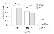


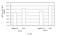
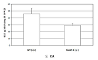

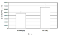

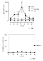

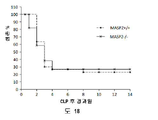


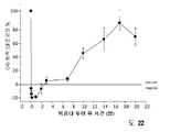


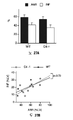


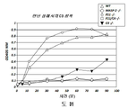


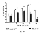


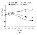


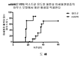
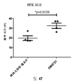


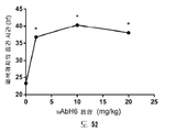


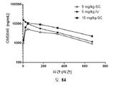


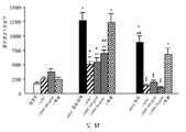

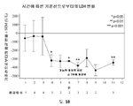


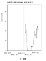


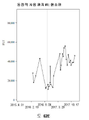
Classifications
-
- C—CHEMISTRY; METALLURGY
- C07—ORGANIC CHEMISTRY
- C07K—PEPTIDES
- C07K16/00—Immunoglobulins [IGs], e.g. monoclonal or polyclonal antibodies
- C07K16/40—Immunoglobulins [IGs], e.g. monoclonal or polyclonal antibodies against enzymes
-
- A—HUMAN NECESSITIES
- A61—MEDICAL OR VETERINARY SCIENCE; HYGIENE
- A61P—SPECIFIC THERAPEUTIC ACTIVITY OF CHEMICAL COMPOUNDS OR MEDICINAL PREPARATIONS
- A61P11/00—Drugs for disorders of the respiratory system
-
- A—HUMAN NECESSITIES
- A61—MEDICAL OR VETERINARY SCIENCE; HYGIENE
- A61P—SPECIFIC THERAPEUTIC ACTIVITY OF CHEMICAL COMPOUNDS OR MEDICINAL PREPARATIONS
- A61P27/00—Drugs for disorders of the senses
- A61P27/02—Ophthalmic agents
- A61P27/10—Ophthalmic agents for accommodation disorders, e.g. myopia
-
- A—HUMAN NECESSITIES
- A61—MEDICAL OR VETERINARY SCIENCE; HYGIENE
- A61P—SPECIFIC THERAPEUTIC ACTIVITY OF CHEMICAL COMPOUNDS OR MEDICINAL PREPARATIONS
- A61P37/00—Drugs for immunological or allergic disorders
- A61P37/02—Immunomodulators
- A61P37/06—Immunosuppressants, e.g. drugs for graft rejection
-
- A—HUMAN NECESSITIES
- A61—MEDICAL OR VETERINARY SCIENCE; HYGIENE
- A61K—PREPARATIONS FOR MEDICAL, DENTAL OR TOILETRY PURPOSES
- A61K39/00—Medicinal preparations containing antigens or antibodies
- A61K2039/505—Medicinal preparations containing antigens or antibodies comprising antibodies
-
- A—HUMAN NECESSITIES
- A61—MEDICAL OR VETERINARY SCIENCE; HYGIENE
- A61K—PREPARATIONS FOR MEDICAL, DENTAL OR TOILETRY PURPOSES
- A61K39/00—Medicinal preparations containing antigens or antibodies
- A61K2039/545—Medicinal preparations containing antigens or antibodies characterised by the dose, timing or administration schedule
-
- C—CHEMISTRY; METALLURGY
- C07—ORGANIC CHEMISTRY
- C07K—PEPTIDES
- C07K2317/00—Immunoglobulins specific features
- C07K2317/50—Immunoglobulins specific features characterized by immunoglobulin fragments
- C07K2317/54—F(ab')2
-
- C—CHEMISTRY; METALLURGY
- C07—ORGANIC CHEMISTRY
- C07K—PEPTIDES
- C07K2317/00—Immunoglobulins specific features
- C07K2317/50—Immunoglobulins specific features characterized by immunoglobulin fragments
- C07K2317/56—Immunoglobulins specific features characterized by immunoglobulin fragments variable (Fv) region, i.e. VH and/or VL
- C07K2317/565—Complementarity determining region [CDR]
-
- C—CHEMISTRY; METALLURGY
- C07—ORGANIC CHEMISTRY
- C07K—PEPTIDES
- C07K2317/00—Immunoglobulins specific features
- C07K2317/70—Immunoglobulins specific features characterized by effect upon binding to a cell or to an antigen
- C07K2317/76—Antagonist effect on antigen, e.g. neutralization or inhibition of binding
-
- C—CHEMISTRY; METALLURGY
- C07—ORGANIC CHEMISTRY
- C07K—PEPTIDES
- C07K2317/00—Immunoglobulins specific features
- C07K2317/90—Immunoglobulins specific features characterized by (pharmaco)kinetic aspects or by stability of the immunoglobulin
-
- C—CHEMISTRY; METALLURGY
- C07—ORGANIC CHEMISTRY
- C07K—PEPTIDES
- C07K2317/00—Immunoglobulins specific features
- C07K2317/90—Immunoglobulins specific features characterized by (pharmaco)kinetic aspects or by stability of the immunoglobulin
- C07K2317/92—Affinity (KD), association rate (Ka), dissociation rate (Kd) or EC50 value
Abstract
한 측면으로, 발명은 HSCT-IPS를 앓고 있거나, 또는 발병 위험이 있는 및/또는 HSCT-CLS를 앓고 있거나, 또는 발병 위험이 있는 및/또는 HSCT-FO를 앓고 있거나, 또는 발병 위험이 있는 및/또는 HSCT-ES를 앓고 있거나, 또는 발병 위험이 있는 인간 대상체에서 MASP-2-의존성 보체 활성화의 효과를 억제하는 방법을 제공한다. 방법은 MASP-2-의존성 보체 활성화를 억제하기에 효과적인 양의 MASP-2 억제제를 그것을 필요로 하는 대상체에게 투여하는 단계를 포함한다.In one aspect, the invention is suffering from, or at risk of developing HSCT-IPS and/or suffering from HSCT-CLS, or at risk of developing and/or suffering from, or at risk of developing HSCT-FO, and/or or a method of inhibiting the effect of MASP-2-dependent complement activation in a human subject suffering from, or at risk of developing HSCT-ES. The method comprises administering to a subject in need thereof an amount of a MASP-2 inhibitory agent effective to inhibit MASP-2-dependent complement activation.

Description
관련 출원에 대한 교차 참조CROSS REFERENCE TO RELATED APPLICATIONS
본 출원은 2019년 11월 26일에 출원된 미국 가출원 번호 62/940,720호, 및 2019년 11월 26일에 출원된 미국 가출원 번호 62/940,735호의 이익을 주장하며, 상기 출원들은 그 전문이 참조로 본원에 포함된다.This application claims the benefit of U.S. Provisional Application No. 62/940,720, filed on November 26, 2019, and U.S. Provisional Application No. 62/940,735, filed on November 26, 2019, which applications are hereby incorporated by reference in their entireties. incorporated herein.
서열 목록에 관한 진술STATEMENT REGARDING SEQUENCE LISTING
본 출원과 관련된 서열 목록은 종이 사본 대신 텍스트 파일형식으로 제공되며 명세서에 참조됨으로써 본원에 포함된다. 서열 목록이 들어 있는 텍스트 파일의 명칭은 MP_1_0307_PCT_Sequence_Listing_20201120_ST25.txt이고; 파일은 115 KB이며; 2020년 11월 20일에 생성되었고; 명세서의 출원과 함께 EFS-웹을 통해 제출되었다.The Sequence Listing relevant to this application is provided in text file format instead of a paper copy and is incorporated herein by reference. The name of the text file containing the sequence listing is MP_1_0307_PCT_Sequence_Listing_20201120_ST25.txt; The file is 115 KB; was created on November 20, 2020; It was submitted via the EFS-Web along with the filing of the specification.
보체 시스템은 인간 및 기타 척추동물에서 미생물 감염 및 다른 급성 손상에 대한 면역 반응을 개시하고, 증폭시키고 조정하기 위한 조기 작용 메커니즘을 제공한다 (M.K. Liszewski and J.P. Atkinson, 1993, in Fundamental Immunology, Third Edition, edited by W.E. Paul, Raven Press, Ltd., New York). 보체 활성화가 잠재적 병원체에 대한 가치 있는 제일선 방어를 제공하지만, 보호 면역 반응을 촉진하는 보체의 활성은 또한 숙주에게 잠재적 위협을 나타낼 수 있다 (K.R. Kalli, et al., Springer Semin. Immunopathol. 15:417-431, 1994; B.P. Morgan, Eur. J. Clinical Investig. 24:219-228, 1994). 예를 들어, C3 및 C5 단백질 분해 생성물은 호중구를 모집하여 활성화한다. 숙주 방어를 위해서는 없어서는 안 될 것이지만, 활성화된 호중구는 파괴적의 효소의 방출에 있어 무차별적이고 장기 손상을 유발할 수 있다. 더불어, 보체 활성화는 인접한 숙주 세포뿐만 아니라 미생물 표적에서 용해성 보체 구성요소의 침착을 유발하여 숙주 세포 용해를 초래할 수 있다.The complement system provides an early mechanism of action to initiate, amplify and modulate immune responses to microbial infections and other acute injuries in humans and other vertebrates (MK Liszewski and JP Atkinson, 1993, in Fundamental Immunology , Third Edition, edited by WE Paul, Raven Press, Ltd., New York). Although complement activation provides a valuable first-line defense against potential pathogens, the activity of complement to promote a protective immune response may also represent a potential threat to the host (KR Kalli, et al., Springer Semin. Immunopathol. 15 :417). -431, 1994; BP Morgan, Eur. J. Clinical Investig. 24 :219-228, 1994). For example, C3 and C5 proteolytic products recruit and activate neutrophils. Although indispensable for host defense, activated neutrophils are promiscuous in the release of destructive enzymes and can cause organ damage. In addition, complement activation can result in the deposition of soluble complement components in adjacent host cells as well as microbial targets, resulting in host cell lysis.
보체 시스템은 또한: 심근 경색(myocardial infarction), 뇌졸중(stroke), ARDS, 재관류 손상(reperfusion injury), 패혈성 쇼크(septic shock), 열 화상 후 모세혈관 누출(capillary leakage following thermal burns), 심폐 우회후 염증(postcardiopulmonary bypass inflammation), 이식 거부반응(transplant rejection), 류머티스성 관절염(rheumatoid arthritis), 다발성 경화증(multiple sclerosis), 중증 근무력증(myasthenia gravis), 및 알츠하이머병(Alzheimer's disease)을 포함한 수많은 급성 및 만성 질환 상태의 발병에 연루되었다. 거의 모든 이들 상태에서, 보체는 원인은 아니지만 발병에 관여하는 여러 인자 중 하나이다. 그럼에도 불구하고, 보체 활성화는 주요 병리적 메커니즘일 수 있고 이들 질환 상태 중 많은 것에서 임상 제어를 위한 효과적인 지점을 나타낸다. 다양한 질환 상태에서 보체-매개 조직 손상의 중요성에 대한 점점 커지는 인식은 효과적인 보체 억제 약물에 대한 필요성을 강조한다. 지금까지, C5에 대한 항체인 에쿨리주맙(eculizumab) (Solaris®)이 인간에 사용되도록 승인된 유일한 보체-표적화 약물이다. 그러나 아직까지, C5는 보체 시스템의 "하류"에 위치한 여러 이펙터 분자 중 하나이며, C5의 봉쇄는 보체 시스템의 활성화를 억제하지 못한다. 그러므로, 보체 활성화의 개시 단계의 억제제가 "하류" 보체 억제제를 뛰어넘는 상당한 장점을 가질 것이다.The complement system also: myocardial infarction, stroke, ARDS, reperfusion injury, septic shock, capillary leakage following thermal burns, cardiopulmonary bypass Numerous acute and chronic diseases, including postcardiopulmonary bypass inflammation, transplant rejection, rheumatoid arthritis, multiple sclerosis, myasthenia gravis, and Alzheimer's disease has been implicated in the pathogenesis of chronic disease states. In almost all of these conditions, complement is not the cause, but is one of several factors involved in the pathogenesis. Nevertheless, complement activation may be a major pathological mechanism and represents an effective point for clinical control in many of these disease states. The growing awareness of the importance of complement-mediated tissue damage in a variety of disease states highlights the need for effective complement inhibitory drugs. To date, eculizumab (Solaris®), an antibody to C5, is the only complement-targeting drug approved for use in humans. However, so far, C5 is one of several effector molecules located "downstream" of the complement system, and blockade of C5 does not inhibit activation of the complement system. Therefore, inhibitors of the initiation phase of complement activation would have significant advantages over "downstream" complement inhibitors.
현재, 보체 시스템은 3가지의 구별되는 경로: 고전적 경로, 렉틴 경로, 및 대체 경로를 통해 활성화될 수 있는 것으로 널리 받아들여진다. 고전적 경로는 일반적으로 외래 입자 (즉, 항원)에 결합된 숙주 항체로 구성된 복합체에 의해 촉발되며 그로써 특정 항체 반응의 생성을 위해 항원에 대한 사전 노출을 필요로 한다. 고전적 경로의 활성화는 숙주에 의한 사전 적응 면역 반응에 의존적이기 때문에, 고전적 경로는 후천성 면역 체계의 일부분이다. 대조적으로, 렉틴 및 대체 경로는 둘 다 적응 면역에 무관하며 선천성 면역 체계의 일부분이다.Currently, it is widely accepted that the complement system can be activated through three distinct pathways: the classical pathway, the lectin pathway, and the alternative pathway. The classical pathway is usually triggered by a complex consisting of a host antibody bound to a foreign particle (ie, an antigen), thereby requiring prior exposure to the antigen for the generation of a specific antibody response. Because activation of the classical pathway is dependent on a pre-adaptive immune response by the host, the classical pathway is part of the adaptive immune system. In contrast, both lectins and alternative pathways are independent of adaptive immunity and are part of the innate immune system.
보체 시스템의 활성화는 세린 프로테아제 지모겐의 순차적인 활성화를 초래한다. 고전적 경로의 활성화의 제1 단계는 특정 인식 분자, C1q의 항원 결합 IgG 및 IgM 분자에의 결합이다. C1q는 C1로 불리는 복합체로서 C1r 및 C1s 세린 프로테아제 전구효소(proenzymes)와 회합된다. 면역 복합체에 C1q가 결합될 때, C1r의 Arg-Ile 부위의 자가단백분해성 절단이 일어나고 이어서 C1r-매개 절단 및 C1s의 활성화가 일어나며, 그로써 C4 및 C2를 절단하는 능력을 획득하게 된다. C4는 C4a 및 C4b로 지정된 2개의 단편으로 절단되며, 유사하게, C2는 C2a 및 C2b로 절단된다. C4b 단편은 인접한 하이드록실 또는 아미노 기와 공유 결합을 형성할 수 있어서 활성화된 C2의 C2a 단편과의 비공유 상호작용을 통해 C3 전환효소 (C4b2a)를 생성한다.Activation of the complement system results in sequential activation of the serine protease zymogen. The first step in the activation of the classical pathway is the binding of specific recognition molecules, Clq, to antigen-binding IgG and IgM molecules. C1q is a complex called C1 that associates with C1r and C1s serine protease proenzymes. When C1q binds to the immune complex, autoproteolytic cleavage of the Arg-Ile site of C1r occurs followed by C1r-mediated cleavage and activation of C1s, thereby acquiring the ability to cleave C4 and C2. C4 is cleaved into two fragments designated C4a and C4b, and similarly, C2 is cleaved into C2a and C2b. The C4b fragment can form covalent bonds with adjacent hydroxyl or amino groups, resulting in a C3 convertase (C4b2a) through non-covalent interaction with the C2a fragment of activated C2.
C3 전환효소 (C4b2a)는 C3을 C3a 및 C3b 하위구성요소로 단백질 분해성 절단함으로써 활성화하여 C5 전환효소 (C4b2a3b)의 생성을 유도하고, 그것은, C5 절단에 의해 세포 용해를 유도하는 세포막을 파괴할 수 있는 막 공격 복합체 (C6, C7, C8 및 C-9와 조합된 C5b, 또한 "MAC"로서 언급됨)의 형성으로 이어진다. C3 및 C4 (C3b 및 C4b)의 활성화된 형태는 외래 표적 표면 상에 공유적으로 침착되고, 다중 식세포 상의 보체 수용체에 의해 인식된다.C3 convertase (C4b2a) activates by proteolytic cleavage of C3 into C3a and C3b subcomponents to induce the production of C5 convertase (C4b2a3b), which by C5 cleavage can disrupt the cell membrane leading to cell lysis lead to the formation of a membrane attack complex (C5b in combination with C6, C7, C8 and C-9, also referred to as "MAC") in Activated forms of C3 and C4 (C3b and C4b) are covalently deposited on foreign target surfaces and recognized by complement receptors on multiple phagocytes.
독립적으로, 렉틴 경로를 통한 보체 시스템의 활성화의 제1 단계는 또한 특정 인식 분자의 결합과, 이어서 관련된 세린 프로테아제 전구효소의 활성화이다. 그러나, C1q에 의한 면역 복합체의 결합보다는 오히려, 렉틴 경로의 인식 분자는 종합적으로 렉틴으로서 언급된 탄수화물-결합 단백질 (만난-결합 렉틴 (MBL), H-피콜린, M-피콜린, L-피콜린 및 C-유형 렉틴 CL-11)의 그룹을 포함한다. J. Lu et al., Biochim. Biophys. Acta 1572:387-400, (2002); Holmskov et al., Annu. Rev. Immunol. 21:547-578 (2003); Teh et al., Immunology 101:225-232 (2000)) 참고. 또한 J. Luet et al., Biochim Biophys Acta 1572:387-400 (2002); Holmskov et al, Annu Rev Immunol 21:547-578 (2003); Teh et al., Immunology 101:225-232 (2000); Hansen et al, J. Immunol 185(10):6096-6104 (2010) 참고.Independently, the first step in activation of the complement system via the lectin pathway is also the binding of specific recognition molecules followed by activation of the associated serine protease proenzyme. However, rather than binding of immune complexes by Clq, the recognition molecules of the lectin pathway are the carbohydrate-binding proteins collectively referred to as lectins (mannan-binding lectin (MBL), H-ficolin, M-ficolin, L-fi choline and the C-type lectin CL-11). J. Lu et al., Biochim. Biophys. Acta 1572 :387-400, (2002); Holmskov et al., Annu. Rev. Immunol . 21:547-578 (2003); See Teh et al., Immunology 101 :225-232 (2000)). See also J. Luet et al., Biochim Biophys Acta 1572:387-400 (2002); Holmskov et al, Annu Rev Immunol 21:547-578 (2003); Teh et al., Immunology 101:225-232 (2000); See Hansen et al, J. Immunol 185(10):6096-6104 (2010).
Ikeda 등은 처음, C1q와 같이, MBL이 효모 만난-코팅 적혈구에 결합시 C4-의존성 방식으로 보체 시스템을 활성화할 수 있음을 입증하였다 (Ikeda et al., J. Biol. Chem. 262:7451-7454, (1987)). 콜렉틴 단백질 패밀리의 구성원인 MBL은 피라노스 고리의 적도면에 배향된 3- 및 4-하이드록시기를 가진 탄수화물에 결합하는 칼슘-의존성 렉틴이다. MBL에 중요한 리간드는 그러므로 D-만노스 및 N-아세틸-D-글루코사민인 한편, 이런 입체적 요건에 맞지 않는 탄수화물은 MBL에 대해 검출할 수 없는 친화도를 가진다 (Weis et al., Nature 360:127-134, (1992)). MBL과 일가 당 사이의 상호작용은 극히 약하며, 해리 상수는 전형적으로 한 자리 밀리몰 범위이다. MBL은 결합력에 의해, 즉, 서로 매우 근접하게 위치한 다중 단당 잔기와 동시에 상호작용함으로써 글리칸 리간드에 단단하고 특이적인 결합을 이룬다 (Lee et al., Archiv. Biochem. Biophys. 299:129-136, (1992)). MBL은 일반적으로 박테리아, 효모, 기생충 및 특정 바이러스와 같은 미생물을 장식하는 탄수화물 패턴을 인식한다. 대조적으로, MBL은 보통 포유류 플라즈마 및 세포 표면 당단백질 상에 존재하는 "성숙한" 복합체 당콘쥬게이트를 장식하는 끝에서 두 번째 및 최종 당인 D-갈락토스 및 시알산을 인식하지 못한다. 이런 결합 특이성은 "외래" 표면의 인식을 촉진하고 "자기 활성화"로부터 보호하는 것을 돕는 것으로 여겨진다. 그러나, MBL은 포유류 세포의 소포체 및 골지에 격리된 N-결합 당단백질 및 당지질 상의 고-만노스 "전구체" 글리칸의 클러스터와 높은 친화도로 결합한다 (Maynard et al., J. Biol. Chem. 257:3788-3794, (1982)). 그러므로, 손상된 세포는 MBL 결합을 통한 렉틴 경로 활성화의 잠재적 표적이다.Ikeda et al. first demonstrated that, like Clq, MBL can activate the complement system in a C4-dependent manner upon binding to yeast mannan-coated erythrocytes (Ikeda et al., J. Biol. Chem. 262 :7451- 7454, (1987)). MBL, a member of the collectin protein family, is a calcium-dependent lectin that binds carbohydrates with 3- and 4-hydroxy groups oriented in the equatorial plane of the pyranose ring. Important ligands for MBL are therefore D-mannose and N-acetyl-D-glucosamine, while carbohydrates that do not meet these steric requirements have undetectable affinity for MBL (Weis et al., Nature 360:127- 134, (1992)). The interaction between MBL and monovalent sugar is extremely weak, and dissociation constants are typically in the single digit millimolar range. MBL achieves tight and specific binding to glycan ligands by avidity, ie by simultaneous interaction with multiple monosaccharide residues located in close proximity to each other (Lee et al., Archiv. Biochem. Biophys. 299:129-136, (1992)). MBL recognizes carbohydrate patterns that normally decorate microorganisms such as bacteria, yeast, parasites and certain viruses. In contrast, MBL does not recognize D-galactose and sialic acid, which are the second-to-last and final sugars that normally decorate "mature" complex glycoconjugates present on mammalian plasma and cell surface glycoproteins. This binding specificity is believed to facilitate recognition of “foreign” surfaces and help protect against “self-activation”. However, MBL binds with high affinity to clusters of high-mannose "precursor" glycans on N-linked glycoproteins and glycolipids sequestered in the endoplasmic reticulum and Golgi of mammalian cells (Maynard et al., J. Biol. Chem. 257 ). :3788-3794, (1982)). Therefore, damaged cells are potential targets for lectin pathway activation through MBL binding.
피콜린은 피브리노겐-유사 도메인으로 불리는, MBL과 상이한 유형의 렉틴 도메인을 가지고 있다. 피콜린은 Ca++-무관한 방식으로 당 잔기에 결합한다. 인간에서, 3 종류의 피콜린 (L-피콜린, M-피콜린 및 H-피콜린)이 확인되었다. 2개의 혈청 피콜린인 L-피콜린 및 H-피콜린은 일반적으로 N-아세틸-D-글루코사민에 대해 특이성을 가지고 있다; 그러나, H-피콜린 또한 N-아세틸-D-글루코사민에 결합한다. L-피콜린, H-피콜린, CL-11, 및 MBL의 당 특이성의 차이는 상이한 렉틴이 상보하고, 비록 중복되지만, 상이한 당콘쥬게이트를 표적으로 하는 것을 의미한다. 이런 개념은 렉틴 경로의 공지된 렉틴 중에서, L-피콜린만이 모든 그람 양성균에서 발견된 세포벽 당콘쥬게이트인 리포테이코산에 특이적으로 결합한다는 최근 보고에 의해 지지된다 (Lynch et al., J. Immunol. 172:1198-1202, (2004)). 콜렉틴 (즉, MBL) 및 피콜린은 아미노산 서열에서 유의한 유사성을 갖고 있지 않다. 그러나, 단백질의 두 그룹은 유사한 도메인 조직화를 가지며, C1q와 같이, 올리고머 구조로 조립되어 다중부위 결합의 가능성이 최대화된다.Ficolin has a different type of lectin domain than MBL, called a fibrinogen-like domain. Ficolin binds to sugar residues in a Ca ++ -independent manner. In humans, three types of ficolins (L-ficolin, M-ficolin and H-ficolin) have been identified. Two serum ficolins, L-ficolin and H-ficolin, generally have specificity for N-acetyl-D-glucosamine; However, H-ficolin also binds to N-acetyl-D-glucosamine. The difference in the sugar specificity of L-ficolin, H-ficolin, CL-11, and MBL means that different lectins complement and, although overlapping, target different glycoconjugates. This concept is supported by a recent report that, of the known lectins of the lectin pathway, only L-ficolin specifically binds to lipoteichoic acid, a cell wall glycoconjugate found in all gram-positive bacteria (Lynch et al., J. Immunol. 172 :1198-1202, (2004)). Collectin (ie, MBL) and ficolin do not have significant similarity in amino acid sequence. However, both groups of proteins have similar domain organization and, like C1q, assemble into oligomeric structures, maximizing the potential for multisite binding.
MBL의 혈청 농도는 건강한 집단에서 매우 가변적이고 이것은 MBL 유전자의 프로모터 및 코딩 영역 둘 다에서의 다형성/돌연변이에 의해 유전적으로 제어된다. 급성 단계 단백질로서, MBL의 발현은 추가로 염증 중에 상향 조절된다. L-피콜린은 혈청에 MBL의 농도와 유사한 농도로 존재한다. 그러므로, 렉틴 경로의 L-피콜린 분지는 잠재적으로 MBL 팔(arm)과 강도가 비슷하다. MBL 및 피콜린은 또한 옵소닌으로서 작용할 수 있어서, 식세포가 MBL- 및 피콜린-장식된 표면을 표적으로 하는 것을 허용한다 (Jack et al., J Leukoc Biol., 77(3):328-36 (2004), Matsushita and Fujita, Immunobiology, 205(4-5):490-7 (2002), Aoyagi et al., J Immunol, 174(1):418-25(2005) 참고). 이런 옵소닌화는 이들 단백질과 식세포 수용체와의 상호작용을 필요로 하지만 (Kuhlman et al., J. Exp. Med. 169:1733, (1989); Matsushita et al., J. Biol. Chem. 271:2448-54, (1996)), 그것의 실체는 아직 확립되지 않았다.Serum concentrations of MBL are highly variable in healthy populations and this is genetically controlled by polymorphisms/mutations in both the promoter and coding regions of the MBL gene. As an acute phase protein, expression of MBL is further upregulated during inflammation. L-ficolin is present in serum at a concentration similar to that of MBL. Therefore, the L-ficolin branch of the lectin pathway is potentially comparable in strength to the MBL arm. MBL and ficolin can also act as opsonins, allowing phagocytes to target MBL- and ficolin-decorated surfaces (Jack et al., J Leukoc Biol ., 77(3):328-36). (2004), Matsushita and Fujita, Immunobiology , 205(4-5):490-7 (2002), Aoyagi et al., J Immunol, 174(1):418-25 (2005)). This opsonization requires the interaction of these proteins with phagocytic receptors (Kuhlman et al., J. Exp. Med. 169 :1733, (1989); Matsushita et al., J. Biol. Chem. 271 ). :2448-54, (1996)), the substance of which has not yet been established.
인간 MBL은 그것의 콜라겐 유사 도메인을 통해서 독특한 C1r/C1s-유사 세린 프로테아제와, MBL-결합 세린 프로테아제 (MASP)로 명명되는, 특이적이고 고-친화도 상호작용을 형성한다. 현재까지, 3개의 MASP가 기술되었다. 첫째, 단일 효소 "MASP"가 보체 캐스케이드의 개시 (즉, C2 및 C4 절단)를 담당하는 효소로서 확인되고 특성화되었다 (Matsushita et al., J Exp Med 176(6):1497-1502 (1992); Ji et al., J. Immunol. 150:571-578, (1993)). 계속해서 MASP 활성은, 실제로, 2개 프로테아제: MASP-1 및 MASP-2의 혼합인 것으로 측정되었다 (Thiel et al., Nature 386:506-510, (1997)). 그러나, MBL-MASP-2 복합체 단독이 보체 활성화에 충분한 것으로 입증되었다 (Vorup-Jensen et al., J. Immunol. 165:2093-2100, (2000)). 나아가, MASP-2만이 C2 및 C4를 고속으로 절단하였다 (Ambrus et al., J. Immunol. 170:1374-1382, (2003)). 그러므로, MASP-2는 C4 및 C2를 활성화하여 C3 전환효소, C4b2a를 생성하는 것을 담당하는 프로테아제이다. 이것이 고전적 경로의 C1 복합체로부터의 유의한 차이이며, 여기서 2개의 특정 세린 프로테아제 (C1r 및 C1s)의 협응 작용은 보체 시스템의 활성화로 이어진다. 더불어, 세 번째 신규한 프로테아제인 MASP-3이 분리되었다 (Dahl, M.R., et al., Immunity 15:127-35, 2001). MASP-1 및 MASP-3은 동일한 유전자의 대체 스플라이싱된 생성물이다.Human MBL forms a specific, high-affinity interaction, termed MBL-binding serine protease (MASP), with a unique C1r/C1s-like serine protease through its collagen-like domain. To date, three MASPs have been described. First, a single enzyme "MASP" was identified and characterized as the enzyme responsible for initiation of the complement cascade (ie, C2 and C4 cleavage) (Matsushita et al., J Exp Med 176(6):1497-1502 (1992); Ji et al., J. Immunol. 150 :571-578, (1993)). Subsequently, MASP activity was, in fact, determined to be a mixture of two proteases: MASP-1 and MASP-2 (Thiel et al., Nature 386 :506-510, (1997)). However, the MBL-MASP-2 complex alone has been demonstrated to be sufficient for complement activation (Vorup-Jensen et al., J. Immunol. 165 :2093-2100, (2000)). Furthermore, only MASP-2 cleaved C2 and C4 at a high rate (Ambrus et al., J. Immunol. 170 :1374-1382, (2003)). Therefore, MASP-2 is the protease responsible for activating C4 and C2 to produce the C3 convertase, C4b2a. This is a significant difference from the C1 complex of the classical pathway, where the coordinated action of two specific serine proteases (C1r and C1s) leads to activation of the complement system. In addition, a third novel protease, MASP-3, was isolated (Dahl, MR, et al., Immunity 15 :127-35, 2001). MASP-1 and MASP-3 are alternative spliced products of the same gene.
MASP는 동일한 도메인 조직화를 C1 복합체의 효소 구성요소인 C1r 및 C1s의 그것과 공유한다 (Sim et al., Biochem. Soc. Trans. 28:545, (2000)). 이들 도메인은 N-말단 C1r/C1s/성게 VEGF/뼈 형성 단백질 (CUB) 도메인, 표피 성장 인자-유사 도메인, 제2 CUB 도메인, 보체 제어 단백질 도메인의 탠덤, 및 세린 프로테아제 도메인을 포함한다. C1 프로테아제에서처럼, MASP-2의 활성화는 세린 프로테아제 도메인에 인접한 Arg-I1e 결합의 절단을 통해 일어나며, 그것은 효소를 다이설파이드-연결된 A 및 B 사슬로 나누고, 후자는 세린 프로테아제 도메인으로 이루어진다.MASP shares the same domain organization with that of C1r and C1s, the enzymatic components of the C1 complex (Sim et al., Biochem. Soc. Trans. 28 :545, (2000)). These domains include an N-terminal C1r/C1s/sea urchin VEGF/bone morphogenetic protein (CUB) domain, an epidermal growth factor-like domain, a second CUB domain, a tandem of a complement control protein domain, and a serine protease domain. As in C1 proteases, activation of MASP-2 occurs through cleavage of the Arg-I1e bond adjacent to the serine protease domain, which divides the enzyme into disulfide-linked A and B chains, the latter consisting of the serine protease domain.
MBL은 또한 MASP2의 촉매적 활성이 결핍된 19 kDa의 MBL-결합 단백질 (MAp19) 또는 작은 MBL-결합 단백질 (sMAP)로서 알려져 있는 MASP-2의 대체 스플라이싱 형태와 결합할 수 있다 (Stover, J. Immunol. 162:3481-90, (1999); Takahashi et al., Int. Immunol. 11:859-863, (1999)). MAp19는 MASP-2의 처음 2개의 도메인과, 이어서 4개의 독특한 아미노산의 엑스트라 서열을 포함한다. Map19의 기능은 분명하지 않다 (Degn et al., J Immunol. Methods, 2011). MASP-1 및 MASP-2 유전자는 각각 인간 염색체 3 및 1에 위치한다 (Schwaeble et al., Immunobiology 205:455-466, (2002)).MBL can also bind an alternative splicing form of MASP-2, known as the 19 kDa MBL-binding protein (MAp19) or the small MBL-binding protein (sMAP), which lacks the catalytic activity of MASP2 (Stover, J. Immunol. 162 :3481-90, (1999); Takahashi et al., Int. Immunol. 11 :859-863, (1999)). MAp19 contains the first two domains of MASP-2 followed by an extra sequence of four unique amino acids. The function of Map19 is not clear (Degn et al., J Immunol. Methods , 2011). The MASP-1 and MASP-2 genes are located on
여러 라인의 증거가 상이한 MBL-MASP 복합체가 있고 혈청 중의 MASP의 큰 분율이 MBL과 복합체를 형성하지 않는 것을 시사한다 (Thiel, et al., J. Immunol. 165:878-887, (2000)). H- 및 L-피콜린은 둘 다 모든 MASP에 결합하며 MBL처럼 렉틴 보체 경로를 활성화한다 (Dahl et al., Immunity 15:127-35, (2001); Matsushita et al., J. Immunol. 168:3502-3506, (2002)). 렉틴 및 고전적 경로는 둘 다 공통 C3 전환효소 (C4b2a)를 형성하며 이 단계에서 두 경로는 수렴된다.Several lines of evidence suggest that there are different MBL-MASP complexes and that a large fraction of MASP in serum does not form complexes with MBL (Thiel, et al., J. Immunol. 165 :878-887, (2000)). . Both H- and L-ficolin bind to all MASPs and, like MBL, activate the lectin complement pathway (Dahl et al., Immunity 15 :127-35, (2001); Matsushita et al., J. Immunol. 168 :3502-3506, (2002)). Both the lectin and classical pathways form a common C3 convertase (C4b2a), at which point the two pathways converge.
렉틴 경로는 나이브(naive) 숙주에서 감염에 대한 숙주 방어에서 중요한 역할을 하는 것으로 널리 여겨진다. 숙주 방어에 MBL의 포함에 대한 강력한 증거는 기능성 MBL의 혈청 수준이 감소된 환자의 분석으로부터 온다 (Kilpatrick, Biochim. Biophys. Acta 1572:401-413, (2002)). 그러한 환자는 재발하는 박테리아 및 진균 감염에 대한 민감성을 나타낸다. 이들 증상은 보통 모체로부터 유래된 항체 역가가 약해짐에 따라 취약성의 창이 나타나는 동안, 그러나 항체 반응의 전체 레퍼토리가 발달하기 전인 생의 초기에 분명하다. 이 증후군은 자주 MBL 올리고머의 적절한 형성을 간섭하는, MBL의 콜라겐성 부분의 여러 부위에서의 돌연변이로부터 유발된다. 그러나, MBL이 보체와 무관한 옵소닌으로서 기능할 수 있기 때문에, 감염에 대한 어느 정도의 증가된 민감성이 손상된 보체 활성화로 인한 것인지는 알려져 있지 않다.The lectin pathway is widely believed to play an important role in host defense against infection in naive hosts. Strong evidence for the inclusion of MBL in host defense comes from analysis of patients with reduced serum levels of functional MBL (Kilpatrick, Biochim. Biophys. Acta 1572 :401-413, (2002)). Such patients exhibit susceptibility to recurrent bacterial and fungal infections. These symptoms are usually evident early in life, during a window of vulnerability as maternally derived antibody titers weaken, but before the full repertoire of antibody responses develops. This syndrome results from mutations at various sites in the collagenous portion of the MBL, often interfering with the proper formation of MBL oligomers. However, it is not known to what extent the increased susceptibility to infection is due to impaired complement activation, as MBL can function as a complement-independent opsonin.
고전적 및 렉틴 경로와 대조적으로, C1q 및 렉틴이 다른 두 경로에서 수행하는 인식 기능을 이행하는 것으로 나타난 대체 경로의 개시자가 없다. 현재 대체 경로는 체크시 자발적인 보체 활성화를 유지하는 적절한 분자 요소가 결핍된 외래 또는 다른 비정상적 표면 (박테리아, 효모, 바이러스로 감염된 세포, 또는 손상된 조직) 상에서 쉽게 증폭될 수 있는 자발적으로 낮은 수준의 전환 활성화를 진행하는 것이 널리 수용된다. 대체 경로의 활성화에 직접 관여하는 4개의 혈장 단백질: C3, 인자 B 및 D, 및 프로퍼딘(properdin)이 있다.In contrast to the classical and lectin pathways, there are no initiators of alternative pathways that have been shown to fulfill the recognition functions that Clq and lectins perform in the other two pathways. Current alternative pathways are spontaneously low-level transactivation that can be readily amplified on foreign or other aberrant surfaces (bacteria, yeast, virus-infected cells, or damaged tissue) lacking the appropriate molecular elements that maintain spontaneous complement activation upon check. It is widely accepted to proceed with There are four plasma proteins directly involved in activation of the alternative pathway: C3, factors B and D, and properdin.
비록 비감염성 인간 질환의 발병에 고전적 및 대체 보체 경로가 모두 연루된 것을 보여주는 광범위한 증거가 있지만, 렉틴 경로의 역할은 이제 막 평가되기 시작하였다. 최근 연구는 렉틴 경로의 활성화가 허혈/재관류 손상에서 보체 활성화 및 관련된 염증의 원인일 수 있는 증거를 제공한다. Collard 등 (2000)은 산화성 스트레스를 받은 배양된 내피 세포가 MBL에 결합하여 인간 혈청에 노출시 C3의 침착을 보이는 것을 보고하였다 (Collard et al., Am. J. Pathol. 156:1549-1556, (2000)). 더불어, 항-MBL 단클론성 항체를 차단하면서 인간 혈청의 처리는 MBL 결합 및 보체 활성화를 억제하였다. 이들 발견은 심근 허혈-재관류의 래트 모델로 확장되었는데, 래트는 래트 MBL에 대해 지시된 차단 항체로 처리된 래트는 대조군 항체로 처리된 래트보다 관상 동맥의 폐색시 상당히 더 적은 심근 손상을 보였다 (Jordan et al., Circulation 104:1413-1418, (2001)). 산화성 스트레스 후 혈관 내피에 대한 MBL 결합의 분자 메커니즘은 분명하지 않다; 최근 연구는 산화성 스트레스 후 렉틴 경로의 활성화가 당콘쥬게이트가 아니라, 혈관 내피 사이토케라틴에 대한 MBL 결합에 의해 매개될 수 있음을 시사한다 (Collard et al., Am. J. Pathol. 159:1045-1054, (2001)). 다른 연구는 허혈/재관류 손상의 발병에서 고전적 및 대체 경로가 연루된 것과 이 질환에서 렉틴 경로의 역할이 논란의 여지가 있음을 보여주었다 (Riedermann, N.C., et al., Am. J. Pathol. 162:363-367, 2003).Although there is extensive evidence showing the involvement of both classical and alternative complement pathways in the pathogenesis of non-infectious human diseases, the role of the lectin pathway is only beginning to be evaluated. Recent studies provide evidence that activation of the lectin pathway may be responsible for complement activation and associated inflammation in ischemia/reperfusion injury. Collard et al. (2000) reported that cultured endothelial cells subjected to oxidative stress bind to MBL and exhibit C3 deposition upon exposure to human serum (Collard et al., Am. J. Pathol. 156 :1549-1556, (2000)). In addition, treatment with human serum while blocking anti-MBL monoclonal antibody inhibited MBL binding and complement activation. These findings were extended to a rat model of myocardial ischemia-reperfusion, in which rats treated with a blocking antibody directed against rat MBL showed significantly less myocardial damage upon coronary artery occlusion than rats treated with a control antibody (Jordan). et al., Circulation 104 :1413-1418, (2001)). The molecular mechanism of MBL binding to the vascular endothelium after oxidative stress is unclear; A recent study suggests that activation of the lectin pathway after oxidative stress may be mediated by MBL binding to vascular endothelial cytokeratin, but not by glycoconjugates (Collard et al., Am. J. Pathol. 159 :1045). 1054, (2001)). Other studies have shown the involvement of classical and alternative pathways in the pathogenesis of ischemia/reperfusion injury and the role of the lectin pathway in this disease is controversial (Riedermann, NC, et al., Am. J. Pathol. 162 : 363-367, 2003).
최근 연구는 MASP-1 (및 아마도 또한 MASP-3)이 대체 경로 활성화 효소 인자 D를 그것의 지모겐 형태로부터 효소적으로 활성 형태로 전환시키는 데 필요한 것을 보였다 (Takahashi M. et al., J Exp Med 207(1):29-37 (2010) 참고). 이 과정의 생리적 중요성은 MASP-1/3-결핍 마우스의 혈장에서 대체 경로 기능적 활성의 부재에 의해 강조된다. 천연 C3으로부터 C3b의 단백질 분해성 생성은 대체 경로가 기능하는 데 필요하다. 대체 경로 C3 전환효소 (C3bBb)가 필수 하위유닛으로서 C3b를 함유하기 때문에, 대체 경로를 통한 제1 C3b의 기원과 관련한 의문은 난해한 문제를 제시하였고 상당한 연구를 자극하였다.Recent studies have shown that MASP-1 (and possibly also MASP-3) is required to convert alternative pathway activating enzyme factor D from its zymogen form to an enzymatically active form (Takahashi M. et al., J Exp . Med 207(1):29-37 (2010)). The physiological significance of this process is highlighted by the absence of alternative pathway functional activity in the plasma of MASP-1/3-deficient mice. Proteolytic production of C3b from native C3 is required for the alternative pathway to function. Since the alternative pathway C3 convertase (C3bBb) contains C3b as an essential subunit, the question of the origin of the first C3b via the alternative pathway has presented a thorny problem and has stimulated considerable research.
C3은 티오에스테르 결합으로서 알려진 흔하지 않은 번역후 변형을 함유한 단백질 패밀리에 속한다 (C4 및 α-2 거대글로불린과 함께). 티오에스테르 기는 말단 카르보닐 기가 3개 아미노산이 떨어져 있는 시스테인의 설프하이드릴 기와 공유 티오에스테르 결합을 형성하는 글루타민으로 구성된다. 이 결합은 불안정하고 친전자성 글루타밀-티오에스테르는 하이드록실 또는 아미노 기와 같은 친핵성 모이어티와 반응할 수 있고 그로써 다른 분자와 공유 결합을 형성한다. 티오에스테르 결합은 온전한 C3의 소수성 포켓 내에 격리될 때 타당하게 안정적이다. 그러나, C3의 C3a 및 C3b로의 단백질 분해성 절단은 C3b 상의 고도로 반응성인 티오에스테르 결합의 노출과, 이어서 하이드록실 또는 아미노 기를 포함하는 인접한 모이어티에 의한 친핵성 공격을 초래하여, C3b는 표적에 공유적으로 연결된다. C3b의 보체 표적에의 공유 부착에서 그것의 잘 증명된 역할 외에, C3 티오에스테르는 또한 대체 경로를 촉발시키는 데 중추적 역할을 하는 것으로 여겨진다. 광범위하게 수용된 "틱 오버 이론(tick-over theory)"에 따르면, 대체 경로는 C3과 가수분해된 티오에스테르 (iC3; C3(H2O)) 및 인자 B로부터 형성된 유동 상 전환효소, iC3Bb의 생성에 의해 개시된다 (Lachmann, P.J., et al., Springer Semin. Immunopathol. 7:143-162, (1984)). C3b-유사 C3(H2O)은 천연 C3으로부터 단백질의 내부 티오에스테르의 느린 자발적 가수분해에 의해 생성된다 (Pangburn, M.K., et al., J. Exp. Med. 154:856-867, 1981). C3(H2O)Bb 전환효소의 활성을 통해, C3b 분자는 표적 표면 상에 침착되고 그로써 대체 경로가 개시된다.C3 belongs to a family of proteins (along with C4 and α-2 macroglobulins) that contain an uncommon post-translational modification known as a thioester linkage. A thioester group consists of glutamine where the terminal carbonyl group forms a covalent thioester bond with the sulfhydryl group of
대체 경로의 활성화의 개시자에 대해 알려진 것은 거의 없다. 활성자는 효모 세포벽 (지모산), 많은 순수한 다당, 토끼 적혈구, 특정 면역글로불린, 바이러스, 진균, 박테리아, 동물 종양 세포, 기생충, 및 손상된 세포를 포함하는 것으로 여겨진다. 이들 활성자에 공통적인 유일한 특징은 탄수화물의 존재지만, 탄수화물 구조의 복잡성 및 다양성은 인식되는 공유된 분자 결정기를 확립하는 것을 어렵게 만든다. 대체 경로 활성화는 이 경로의 억제 조절 구성요소, 예컨대 인자 H, 인자 I, DAF, 및 CR1과, 대체 경로의 유일한 양성 조절자인 프로퍼딘과의 미세한 균형을 통해 제어되는 것으로 널리 받아들여졌다 (Schwaeble W.J. 및 Reid K.B., Immunol Today 20(1):17-21 (1999) 참고).Little is known about the initiators of activation of the alternative pathway. Activators are believed to include yeast cell walls (zymosan), many pure polysaccharides, rabbit red blood cells, certain immunoglobulins, viruses, fungi, bacteria, animal tumor cells, parasites, and damaged cells. The only feature common to these activators is the presence of carbohydrates, but the complexity and diversity of carbohydrate structures makes it difficult to establish recognized shared molecular determinants. It is widely accepted that alternative pathway activation is controlled through a fine balance of inhibitory regulatory components of this pathway, such as factor H, factor I, DAF, and CR1, with properdin, the only positive regulator of the alternative pathway (Schwaeble WJ). and Reid KB, Immunol Today 20(1):17-21 (1999)).
위에서 기술된 외관상 상향 조절된 활성화 메커니즘 외에, 생성된 임의의 C3b가 인자 B와 함께 추가적인 대체 경로 C3 전환효소 (C3bBb)를 형성하는 데 참여할 수 있기 때문에 대체 경로는 또한 렉틴/고전적 경로 C3 전환효소 (C4b2a)에 대한 강력한 증폭 루프를 제공할 수 있다. 대체 경로 C3 전환효소는 프로퍼딘의 결합에 의해 안정화된다. 프로퍼딘은 대체 경로 C3 전환효소 반감기를 10배 연장시킨다. C3b의 대체 경로 C3 전환효소에의 첨가는 대체 경로 C5 전환효소의 형성으로 이어진다.In addition to the apparently upregulated activation mechanism described above, the alternative pathway is also a lectin/classical pathway C3 convertase ( C4b2a) can provide a strong amplification loop. Alternative pathway C3 convertase is stabilized by binding of properdin. Properdin prolongs the alternative pathway C3 convertase half-life by 10-fold. Addition of C3b to the alternative pathway C3 convertase leads to the formation of the alternative pathway C5 convertase.
모든 3개 경로 (즉, 고전적, 렉틴 및 대체)는 C5에서 수렴하는 것으로 여겨졌는데, 그것은 절단되어 다중 전염증 효과를 가진 생성물을 형성한다. 수렴된 경로는 말단 보체 경로로서 언급되었다. C5a는 가장 강력한 아나필라톡신으로, 평활근 및 혈관 색조의 변화, 뿐만 아니라 혈관 투과도의 변화를 유도한다. 또한 그것은 호중구 및 단핵세포 둘 다의 강력한 케모탁신 및 활성자이다. C5a-매개 세포 활성화는 사이토카인, 가수분해 효소, 아라키돈산 대사산물, 및 반응성 산소 종을 포함한 다중 추가 염증 매개체의 방출을 유도함으로써 염증 반응을 상당히 증폭시킬 수 있다. C5 절단은 막 공격 복합체 (MAC)로도 알려져 있는 C5b-9의 형성으로 이어진다. 이제 저용해성 MAC 침착이 그것의 용해성 기공 형성 복합체로서의 역할 외에 염증에서 중요한 역할을 할 수 있다는 강력한 증거가 있다.All three pathways (ie, classical, lectin and alternative) were thought to converge at C5, which is cleaved to form products with multiple pro-inflammatory effects. The converged pathway was referred to as the terminal complement pathway. C5a is the most potent anaphylatoxin, inducing changes in smooth muscle and vascular tone, as well as changes in vascular permeability. It is also a potent chemotaxin and activator of both neutrophils and monocytes. C5a-mediated cellular activation can significantly amplify the inflammatory response by inducing the release of multiple additional inflammatory mediators, including cytokines, hydrolases, arachidonic acid metabolites, and reactive oxygen species. C5 cleavage leads to the formation of C5b-9, also known as the membrane attack complex (MAC). There is now strong evidence that hyposoluble MAC deposition may play an important role in inflammation in addition to its role as a soluble pore-forming complex.
면역 방어에서의 필수 역할 외에, 보체 시스템은 많은 임상 상태에서 조직 손상에 기여한다. 그러므로, 이들 부작용을 방지하기 위한 치료적으로 효과적인 보체 억제제를 개발하는 것이 시급하다.In addition to its essential role in immune defense, the complement system contributes to tissue damage in many clinical conditions. Therefore, there is an urgent need to develop therapeutically effective complement inhibitors to prevent these side effects.
이 요약은 아래의 상세한 설명에서 한층 더 기술되는 단순화된 형태의 개념의 선택을 도입하도록 제공된다. 이 요약은 청구된 주제의 핵심 특징을 확인하기 위해 의도되지 않으며, 청구된 주제의 범주를 결정하는 데 보조수단으로서 사용되도록 의도되지 않는다.This Summary is provided to introduce a selection of concepts in a simplified form that are further described below in the Detailed Description. This Summary is not intended to identify key features of the claimed subject matter, nor is it intended to be used as an aid in determining the scope of the claimed subject matter.
한 측면으로, 본 발명은 조혈 줄기 세포 이식 후 특발성 폐렴 증후군(idiopathic pneumonia syndrome, IPS) (HSCT-IPS)을 앓고 있는 인간 대상체를 치료하는 방법을 제공하며, 방법은 MASP-2-의존성 보체 활성화를 억제하기에 효과적인 양의 MASP-2 억제 항체, 또는 그것의 항원 결합 단편을 대상체에게 투여하고 그로써 HSCT-IPS의 적어도 한 증상을 개선시키는 단계를 포함한다. 한 구체예에서, 방법은 이전에 동종 조혈 줄기 세포 이식을 받은 대상체에게 조성물을 투여하는 단계를 포함한다. 한 구체예에서, 방법은 이전에 자가 조혈 줄기 세포 이식을 받은 대상체에게 조성물을 투여하는 단계를 포함한다. 한 구체예에서, 방법은 대상체에게 MASP-2-의존성 보체 활성화를 억제하기에 효과적인 양의 MASP-2 억제 항체, 또는 그것의 항원 결합 단편을 포함하는 조성물을 투여하는 단계 전에 특발성 폐렴 증후군 (IPS)을 앓고 있는 인간 대상체를 확인하는 단계를 추가로 포함한다. 한 구체예에서, 특발성 폐렴 증후군 (IPS)을 앓고 있는 대상체는 DAH를 갖지 않는 것으로 측정된다. 한 구체예에서, MASP-2 억제 항체는 인간 MASP-2에 특이적으로 결합하는 단클론성 항체, 또는 그것의 단편이다. 한 구체예에서, MASP-2 억제 항체 또는 그것의 단편은 재조합 항체, 이펙터 기능이 감소된 항체, 키메릭 항체, 인간화된 항체, 및 인간 항체로 이루어지는 군으로부터 선택된다. 한 구체예에서, MASP-2 억제 항체는 고전적 경로를 실질적으로 억제하지 않는다. 한 구체예에서, MASP-2 억제 항체는 90%의 인간 혈청에서 C3b 침착을 30 nM 이하의 IC50으로 억제한다. 한 구체예에서, MASP-2 억제 항체는 대상체에게 전신적으로 전달된다. 한 구체예에서, MASP-2 억제 항체 또는 그것의 항원 결합 단편은 SEQ ID NO:67로서 제시된 아미노산 서열의 CDR-H1, CDR-H2 및 CDR-H3을 포함하는 중쇄 가변 영역 및 SEQ ID NO:70으로서 제시된 아미노산 서열의 CDR-L1, CDR-L2 및 CDR-L3을 포함하는 경쇄 가변 영역을 포함한다. 한 구체예에서, MASP-2 억제 항체 또는 그것의 항원 결합 단편은 SEQ ID NO:67로서 제시된 아미노산 서열을 포함하는 중쇄 가변 영역 및 SEQ ID NO:70으로서 제시된 아미노산 서열을 포함하는 경쇄 가변 영역을 포함한다. 일부 구체예에서, 방법은 HSCT-IPS를 앓고 있는 대상체에게 SEQ ID NO:67로서 제시된 아미노산 서열을 포함하는 중쇄 가변 영역 및 SEQ ID NO:70으로서 제시된 아미노산 서열을 포함하는 경쇄 가변 영역을 포함하는 MASP-2 억제 항체, 또는 그것의 항원 결합 단편을 포함하는 조성물을 1 mg/kg 내지 10 mg/kg (즉, 1 mg/kg, 2 mg/kg, 3 mg/kg, 4 mg/kg, 5 mg/kg, 6 mg/kg, 7 mg/kg, 8 mg/kg, 9 mg/kg 또는 10 mg/kg)의 투여량으로 적어도 2주, 또는 적어도 3주 동안, 또는 적어도 4주 동안, 또는 적어도 5주 동안, 또는 적어도 6주 동안, 또는 적어도 7주 동안, 또는 적어도 8주 동안, 또는 적어도 9주 동안, 또는 적어도 10주 동안, 또는 적어도 11주 동안, 또는 적어도 12주 동안 적어도 매주 1회 (예컨대 적어도 매주 2회 또는 적어도 매주 3회) 투여하는 단계를 포함한다. 한 구체예에서, MASP-2 억제 항체의 투여량은 약 4 mg/kg (즉, 3.6 mg/kg 내지 4.4 mg/kg)이다. 한 구체예에서, MASP-2 억제 항체의 투여량은 약 300 mg 내지 약 450 mg (즉, 약 300 mg 내지 약 400 mg, 또는 약 350 mg 내지 약 400 mg), 예컨대 약 300 mg, 약 305 mg, 약 310 mg, 약 315 mg, 약 320 mg, 약 325 mg, 약 330 mg, 약 335 mg, 약 340 mg, 약 345 mg, 약 350 mg, 약 355 mg, 약 360 mg, 약 365 mg, 약 370 mg, 약 375 mg, 약 380 mg, 약 385 mg, 약 390 mg, 약 395 mg, 약 400 mg, 약 405 mg, 약 410 mg, 약 415 mg, 약 420 mg, 약 425 mg, 약 430 mg, 약 435 mg, 약 440 mg, 약 445 mg 또는 약 450 mg의 고정 용량이다. 한 구체예에서, MASP-2 억제 항체의 투여량은 약 370 mg (±10%)의 고정 용량이다. 한 구체예에서, 방법은 약 370 mg (±10%)의 고정 용량의 MASP-2 억제 항체를 HSCT-IPS를 앓고 있는 대상체에게 적어도 8주의 치료 기간 동안 매주 2회 정맥내로 투여하는 단계를 포함한다.In one aspect, the invention provides a method of treating a human subject suffering from idiopathic pneumonia syndrome (IPS) after hematopoietic stem cell transplantation (HSCT-IPS), the method comprising: administering to the subject an amount of a MASP-2 inhibitory antibody, or antigen-binding fragment thereof, effective to inhibit, thereby ameliorating at least one symptom of HSCT-IPS. In one embodiment, the method comprises administering the composition to a subject who has previously received an allogeneic hematopoietic stem cell transplant. In one embodiment, the method comprises administering the composition to a subject who has previously received an autologous hematopoietic stem cell transplant. In one embodiment, the method comprises idiopathic pneumonia syndrome (IPS) prior to administering to the subject a composition comprising an amount of a MASP-2 inhibitory antibody, or antigen-binding fragment thereof, effective to inhibit MASP-2-dependent complement activation. further comprising identifying the human subject suffering from In one embodiment, a subject suffering from idiopathic pneumonia syndrome (IPS) is determined to be free of DAH. In one embodiment, the MASP-2 inhibitory antibody is a monoclonal antibody that specifically binds human MASP-2, or a fragment thereof. In one embodiment, the MASP-2 inhibitory antibody or fragment thereof is selected from the group consisting of a recombinant antibody, an antibody with reduced effector function, a chimeric antibody, a humanized antibody, and a human antibody. In one embodiment, the MASP-2 inhibitory antibody does not substantially inhibit the classical pathway. In one embodiment, the MASP-2 inhibitory antibody inhibits C3b deposition with an IC 50 of 30 nM or less in 90% of human serum. In one embodiment, the MASP-2 inhibitory antibody is delivered systemically to the subject. In one embodiment, the MASP-2 inhibitory antibody or antigen-binding fragment thereof comprises a heavy chain variable region comprising CDR-H1, CDR-H2 and CDR-H3 of the amino acid sequence set forth as SEQ ID NO:67 and SEQ ID NO:70 and a light chain variable region comprising CDR-L1, CDR-L2 and CDR-L3 of the amino acid sequence shown as In one embodiment, the MASP-2 inhibitory antibody or antigen-binding fragment thereof comprises a heavy chain variable region comprising the amino acid sequence set forth as SEQ ID NO:67 and a light chain variable region comprising the amino acid sequence set forth as SEQ ID NO:70 do. In some embodiments, the method administers to a subject suffering from HSCT-IPS a MASP comprising a heavy chain variable region comprising the amino acid sequence set forth as SEQ ID NO:67 and a light chain variable region comprising the amino acid sequence set forth as SEQ ID NO:70 -2 inhibitory antibody, or a composition comprising an antigen-binding fragment thereof from 1 mg / kg to 10 mg / kg (ie, 1 mg / kg, 2 mg / kg, 3 mg / kg, 4 mg / kg, 5 mg /kg, 6 mg/kg, 7 mg/kg, 8 mg/kg, 9 mg/kg or 10 mg/kg) for at least 2 weeks, or at least 3 weeks, or at least 4 weeks, or at least at least once a week for 5 weeks, or for at least 6 weeks, or for at least 7 weeks, or for at least 8 weeks, or for at least 9 weeks, or for at least 10 weeks, or for at least 11 weeks, or for at least 12 weeks ( eg at least twice weekly or at least 3 times weekly). In one embodiment, the dosage of the MASP-2 inhibitory antibody is about 4 mg/kg (ie, 3.6 mg/kg to 4.4 mg/kg). In one embodiment, the dosage of the MASP-2 inhibitory antibody is from about 300 mg to about 450 mg (i.e., from about 300 mg to about 400 mg, or from about 350 mg to about 400 mg), such as about 300 mg, about 305 mg. , about 310 mg, about 315 mg, about 320 mg, about 325 mg, about 330 mg, about 335 mg, about 340 mg, about 345 mg, about 350 mg, about 355 mg, about 360 mg, about 365 mg, about 370 mg, about 375 mg, about 380 mg, about 385 mg, about 390 mg, about 395 mg, about 400 mg, about 405 mg, about 410 mg, about 415 mg, about 420 mg, about 425 mg, about 430 mg , about 435 mg, about 440 mg, about 445 mg or about 450 mg. In one embodiment, the dosage of the MASP-2 inhibitory antibody is a fixed dose of about 370 mg (±10%). In one embodiment, the method comprises administering a fixed dose of about 370 mg (±10%) of a MASP-2 inhibitory antibody intravenously to a subject suffering from HSCT-IPS twice weekly for a treatment period of at least 8 weeks. .
또 다른 측면으로, 본 발명은 조혈 줄기 세포 이식 후 모세혈관 누출 증후군 (Capillary Leak Syndrome, CLS) (HSCT-CLS)을 앓고 있는 대상체를 치료하는 방법을 제공하며, 방법은 대상체에게 MASP-2-의존성 보체 활성화를 억제하기에 효과적인 양의 MASP-2 억제 항체, 또는 그것의 항원 결합 단편을 투여하고 그로써 HSCT-CLS의 적어도 한 증상을 개선시키는 단계를 포함한다. 한 구체예에서, 방법은 이전에 동종 조혈 줄기 세포 이식을 받은 대상체에게 조성물을 투여하는 단계를 포함한다. 한 구체예에서, 방법은 이전에 자가 조혈 줄기 세포 이식을 받은 대상체에게 조성물을 투여하는 단계를 포함한다. 한 구체예에서, 방법은 MASP-2-의존성 보체 활성화를 억제하기에 효과적인 양의 MASP-2 억제 항체, 또는 그것의 항원 결합 단편을 포함하는 조성물을 대상체에게 투여하는 단계 전에 모세혈관 누출 증후군 (CLS)을 앓고 있는 인간 대상체를 확인하는 단계를 추가로 포함한다. 한 구체예에서, CLS를 앓고 있는 대상체는 VOD를 갖지 않는 것으로 측정된다. 한 구체예에서, MASP-2 억제 항체는 인간 MASP-2에 특이적으로 결합하는 단클론성 항체, 또는 그것의 단편이다. 한 구체예에서, MASP-2 억제 항체 또는 그것의 단편은 재조합 항체, 이펙터 기능이 감소된 항체, 키메릭 항체, 인간화된 항체, 및 인간 항체로 이루어지는 군으로부터 선택된다. 한 구체예에서, MASP-2 억제 항체는 고전적 경로를 실질적으로 억제하지 않는다. 한 구체예에서, MASP-2 억제 항체는 90% 인간 혈청에서 30 nM 이하의 IC50으로 C3b 침착을 억제한다. 한 구체예에서, MASP-2 억제 항체는 대상체에게 전신적으로 전달된다. 한 구체예에서, MASP-2 억제 항체 또는 그것의 항원 결합 단편은 SEQ ID NO:67로서 제시된 아미노산 서열의 CDR-H1, CDR-H2 및 CDR-H3을 포함하는 중쇄 가변 영역 및 SEQ ID NO:70으로서 제시된 아미노산 서열의 CDR-L1, CDR-L2 및 CDR-L3을 포함하는 경쇄 가변 영역을 포함한다. 한 구체예에서, MASP-2 억제 항체 또는 그것의 항원 결합 단편은 SEQ ID NO:67로서 제시된 아미노산 서열을 포함하는 중쇄 가변 영역 및 SEQ ID NO:70으로서 제시된 아미노산 서열을 포함하는 경쇄 가변 영역을 포함한다. 일부 구체예에서, 방법은 HSCT-CLS를 앓고 있는 대상체에게 SEQ ID NO:67로서 제시된 아미노산 서열을 포함하는 중쇄 가변 영역 및 SEQ ID NO:70으로서 제시된 아미노산 서열을 포함하는 경쇄 가변 영역을 포함하는 MASP-2 억제 항체, 또는 그것의 항원 결합 단편을 포함하는 조성물을 1 mg/kg 내지 10 mg/kg (즉, 1 mg/kg, 2 mg/kg, 3 mg/kg, 4 mg/kg, 5 mg/kg, 6 mg/kg, 7 mg/kg, 8 mg/kg, 9 mg/kg 또는 10 mg/kg)의 투여량으로 적어도 2주, 또는 적어도 3주 동안, 또는 적어도 4주 동안, 또는 적어도 5주 동안, 또는 적어도 6주 동안, 또는 적어도 7주 동안, 또는 적어도 8주 동안, 또는 적어도 9주 동안, 또는 적어도 10주 동안, 또는 적어도 11주 동안, 또는 적어도 12주 동안 적어도 매주 1회 (예컨대 적어도 매주 2회 또는 적어도 매주 3회) 투여하는 단계를 포함한다. 한 구체예에서, MASP-2 억제 항체의 투여량은 약 4 mg/kg (즉, 3.6 mg/kg 내지 4.4 mg/kg)이다. 한 구체예에서, MASP-2 억제 항체의 투여량은 300 mg 내지 약 450 mg (즉, 약 300 mg 내지 약 400 mg, 또는 약 350 mg 내지 약 400 mg), 예컨대 약 300 mg, 약 305 mg, 약 310 mg, 약 315 mg, 약 320 mg, 약 325 mg, 약 330 mg, 약 335 mg, 약 340 mg, 약 345 mg, 약 350 mg, 약 355 mg, 약 360 mg, 약 365 mg, 약 370 mg, 약 375 mg, 약 380 mg, 약 385 mg, 약 390 mg, 약 395 mg, 약 400 mg, 약 405 mg, 약 410 mg, 약 415 mg, 약 420 mg, 약 425 mg, 약 430 mg, 약 435 mg, 약 440 mg, 약 445 mg 또는 약 450 mg의 고정 용량이다. 한 구체예에서, MASP-2 억제 항체의 투여량은 약 370 mg (±10%)의 고정 용량이다. 한 구체예에서, 방법은 약 370 mg (±10%)의 고정 용량의 MASP-2 억제 항체를 HSCT-CLS를 앓고 있는 대상체에게 적어도 8주의 치료 기간 동안 매주 2회 정맥내로 투여하는 단계를 포함한다.In another aspect, the present invention provides a method of treating a subject suffering from Capillary Leak Syndrome (CLS) (HSCT-CLS) after hematopoietic stem cell transplantation, the method comprising administering to the subject MASP-2-dependent administering an amount of a MASP-2 inhibitory antibody, or antigen-binding fragment thereof, effective to inhibit complement activation, thereby ameliorating at least one symptom of HSCT-CLS. In one embodiment, the method comprises administering the composition to a subject who has previously received an allogeneic hematopoietic stem cell transplant. In one embodiment, the method comprises administering the composition to a subject who has previously received an autologous hematopoietic stem cell transplant. In one embodiment, the method comprises prior to administering to the subject a composition comprising an amount of a MASP-2 inhibitory antibody, or antigen-binding fragment thereof, effective to inhibit MASP-2-dependent complement activation, capillary leakage syndrome (CLS). ) further comprising identifying the human subject suffering from In one embodiment, the subject suffering from CLS is determined not to have VOD. In one embodiment, the MASP-2 inhibitory antibody is a monoclonal antibody that specifically binds human MASP-2, or a fragment thereof. In one embodiment, the MASP-2 inhibitory antibody or fragment thereof is selected from the group consisting of a recombinant antibody, an antibody with reduced effector function, a chimeric antibody, a humanized antibody, and a human antibody. In one embodiment, the MASP-2 inhibitory antibody does not substantially inhibit the classical pathway. In one embodiment, the MASP-2 inhibitory antibody inhibits C3b deposition with an IC 50 of 30 nM or less in 90% human serum. In one embodiment, the MASP-2 inhibitory antibody is delivered systemically to the subject. In one embodiment, the MASP-2 inhibitory antibody or antigen-binding fragment thereof comprises a heavy chain variable region comprising CDR-H1, CDR-H2 and CDR-H3 of the amino acid sequence set forth as SEQ ID NO:67 and SEQ ID NO:70 and a light chain variable region comprising CDR-L1, CDR-L2 and CDR-L3 of the amino acid sequence shown as In one embodiment, the MASP-2 inhibitory antibody or antigen-binding fragment thereof comprises a heavy chain variable region comprising the amino acid sequence set forth as SEQ ID NO:67 and a light chain variable region comprising the amino acid sequence set forth as SEQ ID NO:70 do. In some embodiments, the method administers to a subject suffering from HSCT-CLS a MASP comprising a heavy chain variable region comprising the amino acid sequence set forth as SEQ ID NO:67 and a light chain variable region comprising the amino acid sequence set forth as SEQ ID NO:70 -2 inhibitory antibody, or a composition comprising an antigen-binding fragment thereof from 1 mg / kg to 10 mg / kg (ie, 1 mg / kg, 2 mg / kg, 3 mg / kg, 4 mg / kg, 5 mg /kg, 6 mg/kg, 7 mg/kg, 8 mg/kg, 9 mg/kg or 10 mg/kg) for at least 2 weeks, or at least 3 weeks, or at least 4 weeks, or at least at least once a week for 5 weeks, or for at least 6 weeks, or for at least 7 weeks, or for at least 8 weeks, or for at least 9 weeks, or for at least 10 weeks, or for at least 11 weeks, or for at least 12 weeks ( eg at least twice weekly or at least 3 times weekly). In one embodiment, the dosage of the MASP-2 inhibitory antibody is about 4 mg/kg (ie, 3.6 mg/kg to 4.4 mg/kg). In one embodiment, the dosage of the MASP-2 inhibitory antibody is 300 mg to about 450 mg (i.e., about 300 mg to about 400 mg, or about 350 mg to about 400 mg), such as about 300 mg, about 305 mg, about 310 mg, about 315 mg, about 320 mg, about 325 mg, about 330 mg, about 335 mg, about 340 mg, about 345 mg, about 350 mg, about 355 mg, about 360 mg, about 365 mg, about 370 mg, about 375 mg, about 380 mg, about 385 mg, about 390 mg, about 395 mg, about 400 mg, about 405 mg, about 410 mg, about 415 mg, about 420 mg, about 425 mg, about 430 mg, a fixed dose of about 435 mg, about 440 mg, about 445 mg or about 450 mg. In one embodiment, the dosage of the MASP-2 inhibitory antibody is a fixed dose of about 370 mg (±10%). In one embodiment, the method comprises administering a fixed dose of about 370 mg (±10%) of a MASP-2 inhibitory antibody intravenously to a subject suffering from HSCT-CLS twice weekly for a treatment period of at least 8 weeks. .
또 다른 측면으로, 본 발명은 조혈 줄기 세포 이식 후 체액 과부하 (fluid overload, FO) (HSCT-FO)를 앓고 있는 인간 대상체를 치료하는 방법을 제공하며, 방법은 대상체에게 MASP-2-의존성 보체 활성화를 억제하기에 효과적인 양의 MASP-2 억제 항체, 또는 그것의 항원 결합 단편을 투여하고 그로써 HSCT-FO의 적어도 한 증상을 개선시키는 단계를 포함한다. 한 구체예에서, 방법은 이전에 동종 조혈 줄기 세포 이식을 받은 대상체에게 조성물을 투여하는 단계를 포함한다. 한 구체예에서, 방법은 이전에 자가 조혈 줄기 세포 이식을 받은 대상체에게 조성물을 투여하는 단계를 포함한다. 한 구체예에서, 방법은 MASP-2-의존성 보체 활성화를 억제하기에 효과적인 양의 MASP-2 억제 항체, 또는 그것의 항원 결합 단편을 포함하는 조성물을 대상체에게 투여하는 단계 전에 체액 과부하 (FO)를 앓고 있는 인간 대상체를 확인하는 단계를 추가로 포함한다. 한 구체예에서, MASP-2 억제 항체는 인간 MASP-2에 특이적으로 결합하는 단클론성 항체, 또는 그것의 단편이다. 한 구체예에서, MASP-2 억제 항체 또는 그것의 단편은 재조합 항체, 이펙터 기능이 감소된 항체, 키메릭 항체, 인간화된 항체, 및 인간 항체로 이루어지는 군으로부터 선택된다. 한 구체예에서, MASP-2 억제 항체는 고전적 경로를 실질적으로 억제하지 않는다. 한 구체예에서, MASP-2 억제 항체는 90% 인간 혈청에서 30 nM 이하의 IC50으로 C3b 침착을 억제한다. 한 구체예에서, MASP-2 억제 항체는 대상체에게 전신적으로 전달된다. 한 구체예에서, MASP-2 억제 항체 또는 그것의 항원 결합 단편은 SEQ ID NO:67로서 제시된 아미노산 서열의 CDR-H1, CDR-H2 및 CDR-H3을 포함하는 중쇄 가변 영역 및 SEQ ID NO:70으로서 제시된 아미노산 서열의 CDR-L1, CDR-L2 및 CDR-L3을 포함하는 경쇄 가변 영역을 포함한다. 한 구체예에서, MASP-2 억제 항체 또는 그것의 항원 결합 단편은 SEQ ID NO:67로서 제시된 아미노산 서열을 포함하는 중쇄 가변 영역 및 SEQ ID NO:70으로서 제시된 아미노산 서열을 포함하는 경쇄 가변 영역을 포함한다. 일부 구체예에서, 방법은 HSCT-FO를 앓고 있는 대상체에게 SEQ ID NO:67로서 제시된 아미노산 서열을 포함하는 중쇄 가변 영역 및 SEQ ID NO:70으로서 제시된 아미노산 서열을 포함하는 경쇄 가변 영역을 포함하는 MASP-2 억제 항체, 또는 그것의 항원 결합 단편을 포함하는 조성물을 1 mg/kg 내지 10 mg/kg (즉, 1 mg/kg, 2 mg/kg, 3 mg/kg, 4 mg/kg, 5 mg/kg, 6 mg/kg, 7 mg/kg, 8 mg/kg, 9 mg/kg 또는 10 mg/kg)의 투여량으로 적어도 2주, 또는 적어도 3주 동안, 또는 적어도 4주 동안, 또는 적어도 5주 동안, 또는 적어도 6주 동안, 또는 적어도 7주 동안, 또는 적어도 8주 동안, 또는 적어도 9주 동안, 또는 적어도 10주 동안, 또는 적어도 11주 동안, 또는 적어도 12주 동안 적어도 매주 1회 (예컨대 적어도 매주 2회 또는 적어도 매주 3회) 투여하는 단계를 포함한다. 한 구체예에서, MASP-2 억제 항체의 투여량은 약 4 mg/kg (즉, 3.6 mg/kg 내지 4.4 mg/kg)이다. 한 구체예에서, MASP-2 억제 항체의 투여량은 약 300 mg 내지 약 450 mg (즉, 약 300 mg 내지 약 400 mg, 또는 약 350 mg 내지 약 400 mg), 예컨대 약 300 mg, 약 305 mg, 약 310 mg, 약 315 mg, 약 320 mg, 약 325 mg, 약 330 mg, 약 335 mg, 약 340 mg, 약 345 mg, 약 350 mg, 약 355 mg, 약 360 mg, 약 365 mg, 약 370 mg, 약 375 mg, 약 380 mg, 약 385 mg, 약 390 mg, 약 395 mg, 약 400 mg, 약 405 mg, 약 410 mg, 약 415 mg, 약 420 mg, 약 425 mg, 약 430 mg, 약 435 mg, 약 440 mg, 약 445 mg 또는 약 450 mg의 고정 용량이다. 한 구체예에서, MASP-2 억제 항체의 투여량은 약 370 mg (±10%)의 고정 용량이다. 한 구체예에서, 방법은 약 370 mg (±10%)의 고정 용량의 MASP-2 억제 항체를 HSCT-FO를 앓고 있는 대상체에게 적어도 8주의 치료 기간 동안 매주 2회 정맥내로 투여하는 단계를 포함한다.In another aspect, the invention provides a method of treating a human subject suffering from fluid overload (FO) (HSCT-FO) following hematopoietic stem cell transplantation, the method comprising administering to the subject MASP-2-dependent complement activation administering an amount of a MASP-2 inhibitory antibody, or antigen-binding fragment thereof, effective to inhibit HSCT-FO, thereby ameliorating at least one symptom of HSCT-FO. In one embodiment, the method comprises administering the composition to a subject who has previously received an allogeneic hematopoietic stem cell transplant. In one embodiment, the method comprises administering the composition to a subject who has previously received an autologous hematopoietic stem cell transplant. In one embodiment, the method prevents fluid overload (FO) prior to administering to the subject a composition comprising an amount of a MASP-2 inhibitory antibody, or antigen-binding fragment thereof, effective to inhibit MASP-2-dependent complement activation. further comprising identifying the afflicted human subject. In one embodiment, the MASP-2 inhibitory antibody is a monoclonal antibody that specifically binds to human MASP-2, or a fragment thereof. In one embodiment, the MASP-2 inhibitory antibody or fragment thereof is selected from the group consisting of a recombinant antibody, an antibody with reduced effector function, a chimeric antibody, a humanized antibody, and a human antibody. In one embodiment, the MASP-2 inhibitory antibody does not substantially inhibit the classical pathway. In one embodiment, the MASP-2 inhibitory antibody inhibits C3b deposition with an IC 50 of 30 nM or less in 90% human serum. In one embodiment, the MASP-2 inhibitory antibody is delivered systemically to the subject. In one embodiment, the MASP-2 inhibitory antibody or antigen-binding fragment thereof comprises a heavy chain variable region comprising CDR-H1, CDR-H2 and CDR-H3 of the amino acid sequence set forth as SEQ ID NO:67 and SEQ ID NO:70 and a light chain variable region comprising CDR-L1, CDR-L2 and CDR-L3 of the amino acid sequence shown as In one embodiment, the MASP-2 inhibitory antibody or antigen-binding fragment thereof comprises a heavy chain variable region comprising the amino acid sequence set forth as SEQ ID NO:67 and a light chain variable region comprising the amino acid sequence set forth as SEQ ID NO:70 do. In some embodiments, the method administers to a subject suffering from HSCT-FO a MASP comprising a heavy chain variable region comprising the amino acid sequence set forth as SEQ ID NO:67 and a light chain variable region comprising the amino acid sequence set forth as SEQ ID NO:70 -2 inhibitory antibody, or a composition comprising an antigen-binding fragment thereof from 1 mg / kg to 10 mg / kg (ie, 1 mg / kg, 2 mg / kg, 3 mg / kg, 4 mg / kg, 5 mg /kg, 6 mg/kg, 7 mg/kg, 8 mg/kg, 9 mg/kg or 10 mg/kg) for at least 2 weeks, or at least 3 weeks, or at least 4 weeks, or at least at least once a week for 5 weeks, or for at least 6 weeks, or for at least 7 weeks, or for at least 8 weeks, or for at least 9 weeks, or for at least 10 weeks, or for at least 11 weeks, or for at least 12 weeks ( eg at least twice weekly or at least 3 times weekly). In one embodiment, the dosage of the MASP-2 inhibitory antibody is about 4 mg/kg (ie, 3.6 mg/kg to 4.4 mg/kg). In one embodiment, the dosage of the MASP-2 inhibitory antibody is from about 300 mg to about 450 mg (i.e., from about 300 mg to about 400 mg, or from about 350 mg to about 400 mg), such as about 300 mg, about 305 mg. , about 310 mg, about 315 mg, about 320 mg, about 325 mg, about 330 mg, about 335 mg, about 340 mg, about 345 mg, about 350 mg, about 355 mg, about 360 mg, about 365 mg, about 370 mg, about 375 mg, about 380 mg, about 385 mg, about 390 mg, about 395 mg, about 400 mg, about 405 mg, about 410 mg, about 415 mg, about 420 mg, about 425 mg, about 430 mg , about 435 mg, about 440 mg, about 445 mg or about 450 mg. In one embodiment, the dosage of the MASP-2 inhibitory antibody is a fixed dose of about 370 mg (±10%). In one embodiment, the method comprises administering a fixed dose of about 370 mg (±10%) of a MASP-2 inhibitory antibody intravenously to a subject suffering from HSCT-FO twice weekly for a treatment period of at least 8 weeks. .
또 다른 측면으로, 본 발명은 조혈 줄기 세포 이식 후 생착 증후군 (Engraftment syndrome, ES) (HSCT-ES)을 앓고 있는 인간 대상체를 치료하는 방법을 제공하며, 방법은 대상체에게 MASP-2-의존성 보체 활성화를 억제하기에 효과적인 양의 MASP-2 억제 항체, 또는 그것의 항원 결합 단편을 포함하는 조성물을 투여하고 그로써 HSCT-ES의 적어도 한 증상을 개선시키는 단계를 포함한다. 한 구체예에서, 방법은 이전에 동종 조혈 줄기 세포 이식을 받은 대상체에게 조성물을 투여하는 단계를 포함한다. 한 구체예에서, 방법은 이전에 자가 조혈 줄기 세포 이식을 받은 대상체에게 조성물을 투여하는 단계를 포함한다. 한 구체예에서, 방법은 MASP-2-의존성 보체 활성화를 억제하기에 효과적인 양의 MASP-2 억제 항체, 또는 그것의 항원 결합 단편을 포함하는 조성물을 대상체에게 투여하는 단계 전에 이식 후 생착 증후군 (HSCT-ES)을 앓고 있는 인간 대상체를 확인하는 단계를 추가로 포함한다. 한 구체예에서, MASP-2 억제 항체는 인간 MASP-2에 특이적으로 결합하는 단클론성 항체, 또는 그것의 단편이다. 한 구체예에서, MASP-2 억제 항체 또는 그것의 단편은 재조합 항체, 이펙터 기능이 감소된 항체, 키메릭 항체, 인간화된 항체, 및 인간 항체로 이루어지는 군으로부터 선택된다. 한 구체예에서, MASP-2 억제 항체는 고전적 경로를 실질적으로 억제하지 않는다. 한 구체예에서, MASP-2 억제 항체는 90% 인간 혈청에서 30 nM 이하의 IC50으로 C3b 침착을 억제한다. 한 구체예에서, MASP-2 억제 항체는 대상체에게 전신적으로 전달된다. 한 구체예에서, MASP-2 억제 항체 또는 그것의 항원 결합 단편은 SEQ ID NO:67로서 제시된 아미노산 서열의 CDR-H1, CDR-H2 및 CDR-H3을 포함하는 중쇄 가변 영역 및 SEQ ID NO:70으로서 제시된 아미노산 서열의 CDR-L1, CDR-L2 및 CDR-L3을 포함하는 경쇄 가변 영역을 포함한다. 한 구체예에서, MASP-2 억제 항체 또는 그것의 항원 결합 단편은 SEQ ID NO:67로서 제시된 아미노산 서열을 포함하는 중쇄 가변 영역 및 SEQ ID NO:70으로서 제시된 아미노산 서열을 포함하는 경쇄 가변 영역을 포함한다. 일부 구체예에서, 방법은 HSCT-ES를 앓고 있는 대상체에게 SEQ ID NO:67로서 제시된 아미노산 서열을 포함하는 중쇄 가변 영역 및 SEQ ID NO:70으로서 제시된 아미노산 서열을 포함하는 경쇄 가변 영역을 포함하는 MASP-2 억제 항체, 또는 그것의 항원 결합 단편을 포함하는 조성물을 1 mg/kg 내지 10 mg/kg (즉, 1 mg/kg, 2 mg/kg, 3 mg/kg, 4 mg/kg, 5 mg/kg, 6 mg/kg, 7 mg/kg, 8 mg/kg, 9 mg/kg 또는 10 mg/kg)의 투여량으로 적어도 2주, 또는 적어도 3주 동안, 또는 적어도 4주 동안, 또는 적어도 5주 동안, 또는 적어도 6주 동안, 또는 적어도 7주 동안, 또는 적어도 8주 동안, 또는 적어도 9주 동안, 또는 적어도 10주 동안, 또는 적어도 11주 동안, 또는 적어도 12주 동안 적어도 매주 1회 (예컨대 적어도 매주 2회 또는 적어도 매주 3회) 투여하는 단계를 포함한다. 한 구체예에서, MASP-2 억제 항체의 투여량은 약 4 mg/kg (즉, 3.6 mg/kg 내지 4.4 mg/kg)이다. 한 구체예에서, MASP-2 억제 항체의 투여량은 약 300 mg 내지 약 450 mg (즉, 약 300 mg 내지 약 400 mg, 또는 약 350 mg 내지 약 400 mg), 예컨대 약 300 mg, 약 305 mg, 약 310 mg, 약 315 mg, 약 320 mg, 약 325 mg, 약 330 mg, 약 335 mg, 약 340 mg, 약 345 mg, 약 350 mg, 약 355 mg, 약 360 mg, 약 365 mg, 약 370 mg, 약 375 mg, 약 380 mg, 약 385 mg, 약 390 mg, 약 395 mg, 약 400 mg, 약 405 mg, 약 410 mg, 약 415 mg, 약 420 mg, 약 425 mg, 약 430 mg, 약 435 mg, 약 440 mg, 약 445 mg 또는 약 450 mg의 고정 용량이다. 한 구체예에서, MASP-2 억제 항체의 투여량은 약 370 mg (±10%)의 고정 용량이다. 한 구체예에서, 방법은 약 370 mg (±10%)의 고정 용량의 MASP-2 억제 항체를 HSCT-ES를 앓고 있는 대상체에게 적어도 8주의 치료 기간 동안 매주 2회 정맥내로 투여하는 단계를 포함한다.In another aspect, the present invention provides a method of treating a human subject suffering from Engraftment syndrome (ES) (HSCT-ES) after hematopoietic stem cell transplantation, the method comprising administering to the subject MASP-2-dependent complement activation administering a composition comprising a MASP-2 inhibitory antibody, or antigen-binding fragment thereof, in an amount effective to inhibit HSCT-ES, thereby ameliorating at least one symptom of HSCT-ES. In one embodiment, the method comprises administering the composition to a subject who has previously received an allogeneic hematopoietic stem cell transplant. In one embodiment, the method comprises administering the composition to a subject who has previously received an autologous hematopoietic stem cell transplant. In one embodiment, the method comprises post-transplant engraftment syndrome (HSCT) prior to administering to the subject a composition comprising an amount of a MASP-2 inhibitory antibody, or antigen-binding fragment thereof, effective to inhibit MASP-2-dependent complement activation. -ES). In one embodiment, the MASP-2 inhibitory antibody is a monoclonal antibody that specifically binds to human MASP-2, or a fragment thereof. In one embodiment, the MASP-2 inhibitory antibody or fragment thereof is selected from the group consisting of a recombinant antibody, an antibody with reduced effector function, a chimeric antibody, a humanized antibody, and a human antibody. In one embodiment, the MASP-2 inhibitory antibody does not substantially inhibit the classical pathway. In one embodiment, the MASP-2 inhibitory antibody inhibits C3b deposition with an IC 50 of 30 nM or less in 90% human serum. In one embodiment, the MASP-2 inhibitory antibody is delivered systemically to the subject. In one embodiment, the MASP-2 inhibitory antibody or antigen-binding fragment thereof comprises a heavy chain variable region comprising CDR-H1, CDR-H2 and CDR-H3 of the amino acid sequence set forth as SEQ ID NO:67 and SEQ ID NO:70 and a light chain variable region comprising CDR-L1, CDR-L2 and CDR-L3 of the amino acid sequence shown as In one embodiment, the MASP-2 inhibitory antibody or antigen-binding fragment thereof comprises a heavy chain variable region comprising the amino acid sequence set forth as SEQ ID NO:67 and a light chain variable region comprising the amino acid sequence set forth as SEQ ID NO:70 do. In some embodiments, the method administers to a subject suffering from HSCT-ES a MASP comprising a heavy chain variable region comprising the amino acid sequence set forth as SEQ ID NO:67 and a light chain variable region comprising the amino acid sequence set forth as SEQ ID NO:70 -2 inhibitory antibody, or a composition comprising an antigen-binding fragment thereof from 1 mg / kg to 10 mg / kg (ie, 1 mg / kg, 2 mg / kg, 3 mg / kg, 4 mg / kg, 5 mg /kg, 6 mg/kg, 7 mg/kg, 8 mg/kg, 9 mg/kg or 10 mg/kg) for at least 2 weeks, or at least 3 weeks, or at least 4 weeks, or at least at least once a week for 5 weeks, or for at least 6 weeks, or for at least 7 weeks, or for at least 8 weeks, or for at least 9 weeks, or for at least 10 weeks, or for at least 11 weeks, or for at least 12 weeks ( eg at least twice weekly or at least 3 times weekly). In one embodiment, the dosage of the MASP-2 inhibitory antibody is about 4 mg/kg (ie, 3.6 mg/kg to 4.4 mg/kg). In one embodiment, the dosage of the MASP-2 inhibitory antibody is from about 300 mg to about 450 mg (i.e., from about 300 mg to about 400 mg, or from about 350 mg to about 400 mg), such as about 300 mg, about 305 mg. , about 310 mg, about 315 mg, about 320 mg, about 325 mg, about 330 mg, about 335 mg, about 340 mg, about 345 mg, about 350 mg, about 355 mg, about 360 mg, about 365 mg, about 370 mg, about 375 mg, about 380 mg, about 385 mg, about 390 mg, about 395 mg, about 400 mg, about 405 mg, about 410 mg, about 415 mg, about 420 mg, about 425 mg, about 430 mg , about 435 mg, about 440 mg, about 445 mg or about 450 mg. In one embodiment, the dosage of the MASP-2 inhibitory antibody is a fixed dose of about 370 mg (±10%). In one embodiment, the method comprises administering a fixed dose of about 370 mg (±10%) of a MASP-2 inhibitory antibody intravenously to a subject suffering from HSCT-ES twice weekly for a treatment period of at least 8 weeks. .
발명의 방법, 조성물 및 의약은 본원에서 한층 더 기술되는 바 HSCT-IPS (즉, 줄기 세포 이식 후 IPS)를 앓고 있거나 발병 위험이 있는 인간을 포함한 포유류 대상체에서 및/또는 HSCT-CLS (즉, 줄기 세포 이식 후 CLS)를 앓고 있거나, 또는 발병 위험이 있는 대상체에서, 및/또는 HSCT-FO (즉, 줄기 세포 이식 후 FO)를 앓고 있거나, 또는 발병 위험이 있는 대상체에서, 및/또는 HSCT-ES (즉, 줄기 세포 이식 후 ES)를 앓고 있거나, 또는 발병 위험이 있는 대상체에서 생체내에서 MASP-2-의존성 보체 활성화의 부작용을 억제하기에 유용하다. 또 다른 측면으로, 본 발명은 치료적 유효량의 MASP-2 억제제, 예컨대 MASP-2 억제 항체 및 제약학적으로 허용되는 담체를 포함하는, MASP-2-의존성 보체 활성화의 부작용을 억제하기 위한 조성물을 제공한다. 또한 제약학적 담체에 치료적 유효량의 MASP-2 억제제를 포함하는, 필요로 하는 대상체에서 MASP-2-의존성 보체 활성화의 부작용을 억제하는 데 사용하기 위한 의약의 제조 방법이 제공된다. 또한 아래에서 본원에 기술된 상태, 질환 및 장애의 각각의 치료를 위해 MASP-2-의존성 보체 활성화를 억제하는 데 사용하기 위한 의약의 제조 방법이 제공된다.The methods, compositions and medicaments of the invention are further described herein in mammalian subjects, including humans, suffering from or at risk of developing HSCT-IPS (ie, IPS after stem cell transplantation) and/or HSCT-CLS (ie, stem cell transplantation). CLS after cell transplantation), or in a subject at risk of developing, and/or in a subject suffering from, or at risk of developing, HSCT-FO (ie, FO after stem cell transplantation), and/or HSCT-ES It is useful for inhibiting the side effects of MASP-2-dependent complement activation in vivo in subjects suffering from, or at risk of developing (ie, ES following stem cell transplantation). In another aspect, the present invention provides a composition for inhibiting the side effects of MASP-2-dependent complement activation comprising a therapeutically effective amount of a MASP-2 inhibitory antibody, such as a MASP-2 inhibitory antibody, and a pharmaceutically acceptable carrier. do. Also provided is a method for the manufacture of a medicament for use in inhibiting the side effects of MASP-2-dependent complement activation in a subject in need thereof, comprising a therapeutically effective amount of a MASP-2 inhibitory agent in a pharmaceutical carrier. Also provided below are methods for the manufacture of a medicament for use in inhibiting MASP-2-dependent complement activation for the treatment of each of the conditions, diseases and disorders described herein.
본 발명의 전술한 측면 및 많은 동반되는 장점은 그것이 첨부되는 도면과 함께 취해질 때, 하기의 상세한 설명을 참조로 더 잘 이해됨에 따라 보다 쉽게 인정될 것이다.
도 1은 인간 MASP-2의 게놈 구조를 예시하는 다이아그램이다;
도 2A는 인간 MASP-2 단백질의 도메인 구조를 예시하는 개략도이다;
도 2B는 인간 MAp19 단백질의 도메인 구조를 예시하는 개략도이다;
도 3은 쥐과 MASP-2 녹아웃 전략을 예시하는 다이아그램이다;
도 4는 인간 MASP-2 미니유전자 구성물을 예시하는 다이아그램이다;
도 5A는 실시예 2에서 기술되는 것과 같이, MASP-2-결핍이 만난 상의 C4b 침착의 결핍에 의해 측정되는 바 렉틴 경로-매개 C4 활성화의 손실로 이어지는 것을 입증하는 결과를 도시한다;
도 5B는 실시예 2에서 기술되는 것과 같이, MASP-2-결핍이 지모산 상의 C4b 침착의 결핍에 의해 측정되는 바 렉틴 경로-매개 C4 활성화의 손실로 이어지는 것을 입증하는 결과를 도시한다;
도 5C는 실시예 2에서 기술되는 것과 같이, MASP-2-결핍이 만난 및 지모산 상의 C4b 침착의 결핍에 의해 측정되는 바 MASP-2+/-; MASP-2-/- 및 야생형 스트레인으로부터 얻어진 혈청 샘플의 상대적인 C4 활성화 수준을 입증하는 결과를 도시한다;
도 6은 실시예 2에서 기술되는 것과 같이, 쥐과 재조합 MASP-2의 MASP-2-/- 혈청 샘플에의 첨가가 만난 상의 C4b 침착에 의해 측정되는 바, 단백질 농도 의존성 방식으로 렉틴 경로-매개 C4 활성화를 회복시키는 것을 입증하는 결과를 도시한다;
도 7은 실시예 8에서 기술되는 것과 같이, 고전적 경로가 MASP-2-/- 스트레인에서 기능적인 것을 입증하는 결과를 도시한다;
도 8A는 실시예 10에서 기술되는 것과 같이, 항-MASP-2 Fab2 항체 #11이 C3 전환효소 형성을 억제하는 것을 입증하는 결과를 도시한다;
도 8B는 실시예 10에서 기술되는 것과 같이, 항-MASP-2 Fab2 항체 #11이 천연 래트 MASP-2에 결합하는 것을 입증하는 결과를 도시한다;
도 8C는 실시예 10에서 기술되는 것과 같이, 항-MASP-2 Fab2 항체 #41이 C4 절단을 억제하는 것을 입증하는 결과를 도시한다;
도 9는 실시예 10에서 기술되는 것과 같이, C3 전환효소 형성을 억제한 것으로 테스트된 항-MASP-2 Fab2 항체가 전부 또한 C4 절단을 억제하는 것으로 나타난 것을 입증하는 결과를 도시한다;
도 10은 실시예 11에서 기술되는 것과 같이, 항-MASP-2 차단 Fab2 항체의 에피토프 지도화에 사용된 래트 MASP-2로부터 유래된 재조합 폴리펩타이드를 예시하는 다이아그램이다;
도 11은 실시예 11에서 기술되는 것과 같이, 항-MASP-2 Fab2 #40 및 #60의 래트 MASP-2 폴리펩타이드에의 결합을 입증하는 결과를 도시한다;
도 12는 실시예 12에서 기술되는 것과 같이, 신장 허혈/재관류 손상 모델에서 재관류 후 24 및 48시간 후에 야생형 (+/+) 및 MASP-2 (-/-) 마우스에 대한 혈액 요소 질소 제거를 입증하는 결과를 도시한다;
도 13A는 실시예 13에서 기술되는 것과 같이, 야생형 (+/+) 및 MASP-2 (-/-) 마우스로부터 분리된 RPE-맥락막 복합체에서 기준선 VEGF 단백질 수준을 보여주는 결과를 도시한다;
도 13B는 실시예 13에서 기술되는 것과 같이, 황반 변성(macular degeneration) 모델에서 레이저 유도 손상 후 야생형 (+/+) 및 MASP-2 (-/-) 마우스에서 3일 째 RPE-맥락막 복합체의 VEGF 단백질을 보여주는 결과를 도시한다;
도 14는 실시예 13에서 기술되는 것과 같이, 야생형 (+/+) 및 MASP-2 (-/-) 마우스에서 레이저 유도 손상 후 7일 째에 평균 맥락막 혈관신생 (CNV) 부피를 보여주는 결과를 도시한다;
도 15A 및 15B는 실시예 14에서 기술되는 것과 같이, 정상적인 래트 혈청에서 MASP-2 Fab2 항체의 투여 후 C4b 침착의 억제 (도 15A) 및 트롬빈 활성화의 억제 (도 15B)에 대한 용량 반응 곡선을 도시한다;
도 16A 및 16B는 실시예 15에서 기술되는 것과 같이, 파종성 혈관내 응고(disseminated intravascular coagulation)의 국지화된 슈바르츠만(Schwartzman) 반응 모델에서 보체 경로가 고갈제 코브라 독 인자 (CVF) 및 말단 경로 억제제 (C5aR 길항물질)에 의해 억제되어 있는 미처리 야생형 마우스 및 야생형 마우스에서의 혈소판 응집 (도 16A)과 비교된 MASP-2 (-/-) 마우스에서의 측정된 혈소판 응집 (응집 면적으로서 표시됨) (도 16B)을 도시한다;
도 17은 실시예 16에서 기술되는 것과 같이, WT (+/+) (B6) 또는 WT (+/+) 도너 신장의 MASP-2 (-/-) 이식 수령체 마우스에서 측정된 혈액 요소 질소 (BUN) 수준을 그래프로 도시한다;
도 18은 실시예 17에서 기술되는 것과 같이, 맹장 결찰 및 천공 (CLP) 모델에서 미생물 감염 후 경과일 수의 함수로서 WT (+/+) 및 MASP-2 (-/-) 마우스의 백분율 생존을 그래프로 도시한다;
도 19는 실시예 17에서 기술되는 것과 같이, 맹장 결찰 및 천공 (CLP) 모델에서 미생물 감염 후 WT (+/+) 및 MASP-2 (-/-)에서 측정된 박테리아 수를 그래프로 도시한다;
도 20은 실시예 18에서 기술되는 것과 같이, 녹농균(Pseudomonas aeruginosa)의 비내 투여로 도전 후 6일 후의 WT (+/+), MASP-2 (-/-) 및 C3 (-/-) 마우스의 생존율을 예시하는 카플란-마이어(Kaplan-Mayer) 도표이다;
도 21은 실시예 19에서 기술되는 것과 같이, WT 마우스에서 0.3 mg/kg 또는 1.0 mg/kg의 마우스 항-MASP-2 단클론성 항체의 피하 투여 후 다양한 시점에서 취해진 샘플 중의, 대조군의 %로서 측정된, C4b 침착의 수준을 그래프로 도시한다;
도 22는 실시예 19에서 기술되는 것과 같이, WT 마우스에서 0.6 mg/kg의 마우스 항-MASP-2 단클론성 항체의 복강내 투여 후 다양한 시점에서 취해진 샘플 중의, 대조군의 %로서 측정된, C4b 침착의 수준을 그래프로 도시한다;
도 23은 실시예 20에서 기술되는 것과 같이, 0.3 mg/kg 또는 1.0 mg/kg의 마우스 항-MASP-2 단클론성 항체의 단일 복강내 주사로 사전 처리된 WT (+/+) 마우스에서 레이저 유도된 손상 후 7일 째에 평균 맥락막 혈관신생 (CNV) 부피를 그래프로 도시한다;
도 24A는 실시예 21에서 기술되는 것과 같이, 5x10⁸/100 μl cfu 수막염균(N. meningitidis)으로 감염된 후 MASP-2 (-/-) 및 WT (+/+) 마우스에서 생존율을 그래프로 도시한다;
도 24B는 실시예 21에서 기술되는 것과 같이, 5x10⁸ cfu/100 μl 수막염균으로 감염된 MASP-2 KO (-/-) 및 WT (+/+) 마우스로부터 취한 혈액 샘플에서 다양한 시점에서 회수된 수막염균의 로그 cfu/ml을 그래프로 도시한다;
도 25A는 실시예 21에서 기술되는 것과 같이, 2x10⁸ cfu/100 μl 수막염균으로 감염된 후 MASP-2 KO (-/-) 및 WT (+/+) 마우스의 생존율을 그래프로 도시한다;
도 25B는 실시예 21에서 기술되는 것과 같이, 2x10⁸ cfu/100 μl 수막염균으로 감염된 WT (+/+) 마우스로부터 취한 혈액 샘플에서 다양한 시점에서 회수된 수막염균의 로그 cfu/ml을 그래프로 도시한다;
도 25C는 실시예 21에서 기술되는 것과 같이, 2x10⁸ cfu/100 μl 수막염균으로 감염된 MASP-2 (-/-) 마우스로부터 취한 혈액 샘플에서 다양한 시점에서 회수된 수막염균의 로그 cfu/ml을 그래프로 도시한다;
도 26A는 실시예 22에서 기술되는 것과 같이, MASP-2 (-/-) 마우스가 기능성 고전적 경로를 유지하는 것을 입증하는 C3b 침착 검정의 결과를 그래프로 도시한다;
도 26B는 실시예 22에서 기술되는 것과 같이, MASP-2 (-/-) 마우스가 기능성 대체 경로를 유지하는 것을 입증하는, 지모산 코팅된 플레이트 상의 C3b 침착 검정의 결과를 그래프로 도시한다;
도 27A는 실시예 22에서 기술되는 것과 같이 위험 영역 (AAR) 및 경색 크기 (INF)를 보여주는, C4 (-/-) 마우스 (n=6마리) 및 매칭되는 WT 한배새끼 대조군 (n=7마리)에서 관상 동맥의 좌측 전방 하행 가지 (LAD)의 결찰 및 재관류 후의 심근 허혈/재관류 손상 (MIRI)-유도된 조직 손실을 그래프로 도시한다;
도 27B는 실시예 22에서 기술되는 것과 같이, C4 (-/-) 마우스가 WT 대조군 (점선)과 같이 MIRI에 취약한 것을 입증하는, 도 42A에서 기술된 것과 같이 처리된 C4 (-/-) 및 WT 마우스에서 위험 영역 (AAR)의 함수로서의 경색 크기 (INF)를 그래프로 도시한다;
도 28A는 실시예 22에서 기술되는 것과 같이, WT 마우스, C4 (-/-) 마우스로부터의 혈청, 및 만난과 함께 사전 인큐베이션된 C4 (-/-) 마우스로부터의 혈청을 사용하는 C3b 침착 검정의 결과를 그래프로 도시한다;
도 28B는 실시예 22에서 기술되는 것과 같이, 다양한 농도의 항-쥐과 MASP-2 mAb (mAbM11)와 혼합된 WT, C4 (-/-), 및 MASP-2 (-/-) 마우스로부터의 혈청에 대한 C3b 침착 검정의 결과를 그래프로 도시한다;
도 28C는 실시예 22에서 기술되는 것과 같이, WT (C4 충분)로부터의 인간 혈청 및 C4 결핍 혈청, 및 만난과 함께 사전 인큐베이션된 C4 결핍 대상체로부터의 혈청에 대한 C3b 침착 검정의 결과를 그래프로 도시한다;
도 28D는 실시예 22에서 기술되는 것과 같이, WT (C4 충분) 및 항-인간 MASP-2 mAb (mAbH3)와 혼합된 C4 결핍 대상체로부터의 인간 혈청에 대한 C3b 침착 검정의 결과를 그래프로 도시한다;
도 29A는 실시예 22에서 기술되는 것과 같이, 렉틴 활성화 경로 특이적 검정 조건 하에서, 또는 고전적 활성화 경로 특이적 검정 조건 하에서 테스트된 다양한 보체 결핍 마우스 스트레인으로부터의 혈장에서의 C3 전환효소 활성의 비교 분석을 그래프로 도시한다;
도 29B는 실시예 22에서 기술되는 것과 같이, 렉틴 활성화 경로 특이적 조건 하에서 테스트된 다양한 보체 결핍 마우스 스트레인으로부터의 혈장에서의 C3 전환효소 활성의 시간-분해 역동학을 그래프로 도시한다;
도 30은 실시예 23에서 기술되는 것과 같이, 트롬빈 기질 FXIa 및 FXa에 의한 a' 사슬의 존재에 의해 나타난, 인간 C3의 활성화를 보여주는 웨스턴 블롯 분석의 결과를 도시한다;
도 31은 실시예 23에서 기술되는 것과 같이, WT, MASP-2 (-/-), F11(-/-), F11(-/-)/C4 (-/-) 및 C4 (-/-)로부터 얻어진 혈청 샘플에 대한 C3 침착 검정의 결과를 도시한다;
도 32A는 실시예 29에서 기술되는 것과 같이, 대조군 마우스 및 항-쥐과 MASP-2 항체 (mAbM11) 또는 항-인간 MASP-2 항체 (mAbH6)로 테스트된 마우스에서 7.0 Gy 방사선에 노출된 후 시간에 따른 생존율을 보여주는 카플란-마이어 생존 곡선이다;
도 32B는 실시예 29에서 기술되는 것과 같이, 대조군 마우스 및 항-쥐과 MASP-2 항체 (mAbM11) 또는 항-인간 MASP-2 항체 (mAbH6)로 테스트된 마우스에서 6.5 Gy 방사선에 노출된 후 시간에 따른 생존율을 보여주는 카플란-마이어 생존 곡선이다;
도 33은 실시예 30에서 기술되는 것과 같이, MASP-2 결핍 마우스가 수막염균 유도된 사망으로부터 보호되는 것을 입증하는, 2.6 x 10⁷ cfu의 수막염균 혈청군 A Z2491의 감염 용량의 투여 후에 MASP-2 KO 및 WT 마우스의 생존율을 그래프로 예시한 카플란-마이어 도표이다;
도 34는 실시예 30에서 기술되는 것과 같이, MASP-2-결핍 마우스가 수막염균 혈청군 B 스트레인 MC58 유도된 사망으로부터 보호되는 것을 입증하는, 6 x 10⁶ cfu의 수막염균 혈청군 B 스트레인 MC58의 감염 용량의 투여 후에 MASP-2 KO 및 WT 마우스의 생존율을 그래프로 예시한 카플란-마이어 도표이다;
도 35는 실시예 30에서 기술되는 것과 같이, 비록 MASP-2 KO 마우스가 WT 마우스와 동일한 용량의 수막염균 혈청군 B 스트레인 MC58로 감염되었지만, MASP-2 KO 마우스는 WT와 비교하여 향상된 균혈증(bacteraemia)의 제거를 보인 것을 입증하는, 6x10⁶ cfu의 수막염균 혈청군 B 스트레인 MC58로 복강내 감염 후 MASP-2 KO 및 WT 마우스로부터 취한 혈액 샘플에서 상이한 시점에 회수된 로그 cfu/ml의 수막염균 혈청군 B 스트레인을 그래프로 도시한다 (상이한 시점에서 두 그룹의 마우스에 대해 모두 n=3마리, 결과는 평균±SEM으로서 표시됨);
도 36은 실시예 30에서 기술되는 것과 같이, MASP-2 결핍 마우스가 6시간 째에 훨씬 더 낮은 질병 점수로 감염에 대해 높은 저항성을 보인 것을 입증하는, 6x10⁶ cfu/100 μl 수막염균 혈청군 혈청군 B 스트레인 MC58로 감염된 후 3, 6, 12 및 24시간 째에 MASP-2 및 WT 마우스의 평균 질병 점수를 그래프로 도시한다;
도 37은 실시예 31에서 기술되는 것과 같이, 항-MASP-2 항체가 수막염균으로 감염된 대상체에서 치료 및 생존율 개선에 효과적인 것을 입증하는, 감염 용량의 4 x 10⁶/100 μl cfu 수막염균 혈청군 B 스트레인 MC58의 투여 후, 억제 항-MASP-2 항체 (1 mg/kg) 또는 대조군 아이소타입 항체의 감염 후 3시간 후 투여 후의 마우스의 생존율을 그래프로 예시한 카플란-마이어 도표이다;
도 38은 실시예 32에서 기술되는 것과 같이, 인간 혈청에서 수막염균의 보체 의존성 사멸이 인간 항-MASP-2 항체의 첨가에 의해 상당히 향상된 것을 보여주는, (A) 정상 인간 혈청 (NHS) 플러스 인간 항-MASP-2 항체; (B) 정상 인간 혈청 (NHS) 플러스 아이소타입 대조군 항체; (C) MBL-/- 인간 혈청; (D) 정상 인간 혈청 (NHS) 및 (E) 열 비활성화된 정상 인간 혈청 (NHS)의 존재 하에 인큐베이션한 후 0, 30, 60 및 90분 째에 6.5x10⁶ cfu/100 μl 수막염균 혈청군 B 스트레인 MC58로 복강내 감염된 후 20% 인간 혈청 농도에서의 상이한 시점에서 회수된 수막염균 혈청군 B-MC58의 생존 가능한 카운트의 로그 cfu/ml을 그래프로 도시한다;
도 39는 실시예 32에서 기술되는 것과 같이, MASP-2 -/- 마우스 혈청이 WT 마우스 혈청보다 수막염균에 대해 더 높은 수주의 박테리아 사멸 활성을 가지는 것을 입증하는, 마우스 혈청 샘플에서 상이한 시점에 회수된 수막염균 혈청군 B-MC58의 로그 cfu/ml의 생존 가능한 카운트를 그래프로 도시한다;
도 40은 실시예 33에서 기술되는 것과 같이, 혈청 농도 범위에 걸쳐 인간 혈청에 의한 만난-코팅 쥐과 적혈구의 용혈 (광도계에 의해 측정된, 용해된 마우스 적혈구 (Crry/C3-/-)의 상층액으로의 헤모글로빈 방출에 의해 측정됨)을 그래프로 도시한다. 테스트된 혈청은 열-비활성화 (HI) NHS, MBL-/-, NHS +항-MASP-2 항체 및 NHS 대조군을 포함하였다;
도 41은 실시예 33에서 기술되는 것과 같이, 혈청 농도 범위에 걸쳐 인간 혈청에 의한 비-코팅 쥐과 적혈구의 용혈 (광도계에 의해 측정된, 용해된 WT 마우스 적혈구의 상층액으로의 헤모글로빈 방출에 의해 측정됨)을 그래프로 도시한다. 테스트된 혈청은 열-비활성화 (HI) NHS, MBL-/-, NHS +항-MASP-2 항체 및 NHS 대조군을 포함하였고, MASP-2의 억제가 민감화되지 않은 WT 마우스 적혈구의 보체-매개 용해를 억제하는 것을 입증한다;
도 42는 실시예 34에서 기술되는 것과 같이, 혈청 농도 범위에 걸쳐 인간 혈청에 의한 비-코팅 쥐과 적혈구의 용혈 (광도계에 의해 측정된, 용해된 마우스 적혈구 (CD55/59 -/-)의 상층액으로의 헤모글로빈 방출에 의해 측정됨)을 그래프로 도시한다. 테스트된 혈청은 열-비활성화 (HI) NHS, MBL-/-, NHS +항-MASP-2 항체 및 NHS 대조군을 포함하였다;
도 43은 실시예 34에서 기술되는 것과 같이, 대조군 마우스 및 항-인간 MASP-2 항체 (mAbH6)로 처리된 마우스에서 8.0 Gy 방사선에 노출된 후 시간 (일)에 따른 생존율을 그래프로 도시한다;
도 44는 실시예 35에서 기술되는 것과 같이, 혈전 형성이 WT 마우스에서 15분 후에 검출되고, 최대 80%의 WT 마우스가 60분째에 혈전 형성을 나타냈으며; 대조적으로, MASP-2 -/- 마우스는 어느 것도 60분 기간 중에 혈전 형성을 나타내지 않았음 (로그 순위: p=0.0005)을 입증하는, 60분에 걸쳐 측정된 마우스의 혈전 형성의 백분율을 보여주는, MASP-2 -/- 및 WT 마우스에서의 LPS 주사 후 미세혈관 폐색(microvascular occlusion)이 시작되기까지의 시간을 그래프로 도시한다;
도 45는 실시예 36에서 기술되는 것과 같이, 모든 대조군 마우스가 42시간 후에 사망한 반면, 대조적으로, 항-MASP-2 항체-처리된 마우스는 100%가 실험 기간 내내 생존한 것을 입증하는, 시간(시간)에 따른 HUS의 STX/LPS-유도 모델에서 식염수 처리된 대조군 마우스 (n=5) 및 항-MASP-2 항체 처리 마우스 (n=5)의 생존율을 그래프로 도시한다;
도 46은 실시예 37에서 기술되는 것과 같이, FITC/덱스트란의 주사 전 16시간 및 1시간 전에 아이소타입 대조군 또는 인간 MASP-2 항체 mAbH6 (10 mg/kg)으로 처리 후 FITC/덱스트란 UV 모델에서 미세혈관 폐색을 보인 마우스의 백분율을, 손상 유도 후 시간의 함수로서 그래프로 도시한다;
도 47은 실시예 37에서 기술되는 것과 같이, 인간 MASP-2 항체 (mAbH6) 및 아이소타입 대조군 항체로 처리된 마우스에 대한, 분으로 표시되는 폐색 시간을 그래프로 도시하며, 여기서 데이터는 평균 값 (수평 막대) 및 표준 오차 막대 (수직 막대)를 포함한 산점도로서 기록된다. 분석을 위해 사용된 통계적 테스트는 독립표본 t 테스트였고; 여기서 기호 "*"는 p=0.0129를 나타낸다; 그리고
도 48은 실시예 37에서 기술되는 것과 같이, 혈전증의 FITC-덱스트란/광 유도 내피 세포 손상 모델에서 낮은 광 강도 (800-1500)로 혈전증을 유도하기 전 16시간 전, 및 다시 1시간 전에 10 mg/kg으로 복강내 투여된 야생형 마우스, MASP-2 KO 마우스, 및 인간 MASP-2 항체 (mAbH6)로 사전 처리된 야생형 마우스에 대한, 분으로 표시되는, 폐색까지 걸리는 시간을 그래프로 도시한다;
도 49는 실시예 39에서 기술되는 것과 같이, 증가하는 용량의 인간 MASP-2 억제 항체 (mAbH6) 또는 아이소타입 대조군 항체로 처리된 마우스에서 FITC-덱스트란 유도된 혈전성 미세혈관병증(thrombotic microangiopathy)에서의 시간의 함수로서 혈전을 보인 마우스의 백분율을 보여주는 카플란-마이어 도표이다;
도 50은 실시예 39에서 기술되는 것과 같이, mAbH6 용량의 함수로서의 혈전 형성의 개시까지의 중간 시간 (분)을 그래프로 도시한다 (* 대조군에 비교하여 p<0.01);
도 51은 실시예 39에서 기술되는 것과 같이, 증가하는 용량의 인간 MASP-2 억제 항체 (mAbH6) 또는 아이소타입 대조군 항체로 처리된 마우스에서 FITC-덱스트란 유도 혈전성 미세혈관병증에서의 시간의 함수로서 미세혈관 폐색을 보이는 마우스의 백분율을 보여주는 카플란-마이어 도표이다;
도 52는 실시예 39에서 기술되는 것과 같이, mAbH6 용량의 함수로서 미세혈관 폐색까지의 중간 시간을 그래프로 도시한다 (* 대조군과 비교하여 p<0.05);
도 53A는 실시예 40에서 기술되는 것과 같이, OMS646이 대략 1 nM의 IC50 값으로 렉틴-매개 MAC 침착을 억제하는 것을 입증하는, 렉틴 경로-특이적 검정 조건 하에서 인간 MASP-2 단클론성 항체 (OMS646)의 존재 또는 부재 시의 MAC 침착의 수준을 그래프로 도시한다;
도 53B는 실시예 40에서 기술되는 것과 같이, OMS646이 고전적 경로-매개 MAC 침착을 억제하지 못하는 것을 입증하는, 고전적 경로-특이적 검정 조건 하에서 인간 MASP-2 단클론성 항체 (OMS646)의 존재 또는 부재 시의 MAC 침착의 수준을 그래프로 도시한다;
도 53C는 실시예 40에서 기술되는 것과 같이, OMS646이 대체 경로-매개 MAC 침착을 억제하지 못하는 것을 입증하는, 대체 경로-특이적 검정 조건 하에서 인간 MASP-2 단클론성 항체 (OMS646)의 존재 또는 부재 시의 MAC 침착의 수준을 그래프로 도시한다;
도 54는 실시예 40에서 기술되는 것과 같이, 표시된 용량에서 투여 후 시간의 함수로서 OMS646 농도 (n=3마리 동물/그룹의 평균)를 보여주는, 마우스에서 인간 MASP-2 단클론성 항체 (OMS646)의 약동학 (PK) 프로파일을 그래프로 도시한다;
도 55A는 실시예 40에서 기술되는 것과 같이, 정맥내 투여 후 마우스에서 전신적인 렉틴 경로 활성의 강하로서 측정된, 인간 MASP-2 단클론성 항체 (OMS646)의 약력학적 (PD) 반응을 그래프로 도시한다;
도 55B는 실시예 40에서 기술되는 것과 같이, 피하 투여 후 마우스에서 전신적인 렉틴 경로 활성의 강하로서 측정된, 인간 MASP-2 단클론성 항체 (OMS646)의 약력학적 (PD) 반응을 그래프로 도시한다;
도 56은 실시예 41에서 기술되는 것과 같이, ADP-활성화된 HMEC-1 세포 상의 aHUS 혈청-유도된 C5b-9 침착에 미치는 sCR1에 비교된 MASP-2 항체 (OMS646)의 억제 효과를 그래프로 도시한다;
도 57은 실시예 42에서 기술되는 것과 같이, ADP-활성화된 HMEC-1 세포 상의 aHUS 혈청-유도된 혈전 형성에 미치는 sCR1에 비교된 MASP-2 항체 (OMS646)의 억제 효과를 그래프로 도시한다;
도 58은 실시예 46에서 기술되는 것과 같이, MASP-2 억제 항체 (OMS646)로 치료된 후 지속적인 조혈 줄기 세포 이식-관련 혈전성 미세혈관병증 (HSCT-TMA)을 앓고 있는 대상체에서 시간 (주)에 따른 기준선으로부터의 혈소판 수의 평균 변화를 그래프로 도시한다;
도 59는 실시예 46에서 기술되는 것과 같이, MASP-2 억제 항체 (OMS646)로 치료된 후 지속적인 (HSCT-TMA)를 앓고 있는 대상체에서 시간 (주)에 따른 기준선으로부터의 LDH의 평균 변화를 그래프로 도시한다;
도 60은 실시예 46에서 기술되는 것과 같이, MASP-2 억제 항체 (OMS646)로 치료된 후 지속적인 (HSCT-TMA)를 앓고 있는 대상체에서 시간 (주)에 따른 기준선으로부터의 합토글로빈의 평균 변화를 그래프로 도시한다;
도 61은 실시예 47에서 기술되는 것과 같이, MASP-2 억제 항체 (OMS646)로 치료된 후 HSCT-TMA, GVHD의 개선 및 신경학적 증상의 개선을 입증하는, HSCT-TMA 및 이식편대숙주병 (GVHD)이 발병된 조혈 줄기 세포 이식 (HSCT) 후 환자의 임상 과정을 도시한다;
도 62A는 실시예 48에서 기술되는 것과 같이, 동정적 사용(compassionate use) 환자 #1에서의 시간에 따른 크레아티닌의 수준을 그래프로 도시하며, 여기서 수직선은 MASP-2 억제 항체 (OMS646)로의 치료 시작을 나타낸다;
도 62B는 실시예 48에서 기술되는 것과 같이, 동정적 사용 환자 #1에서의 시간에 따른 합토글로빈의 수준을 그래프로 도시하며, 여기서 수직선은 MASP-2 억제 항체 (OMS646)로의 치료 시작을 나타낸다;
도 62C는 실시예 48에서 기술되는 것과 같이, 동정적 사용 환자 #1에서의 시간에 따른 헤모글로빈의 수준을 그래프로 도시하며, 여기서 수직선은 MASP-2 억제 항체 (OMS646)로의 치료 시작을 나타낸다;
도 62D는 실시예 48에서 기술되는 것과 같이, 동정적 사용 환자 #1에서의 시간에 따른 LDH의 수준을 그래프로 도시하며, 여기서 수직선은 MASP-2 억제 항체 (OMS646)로의 치료 시작을 나타낸다; 그리고
도 62E는 실시예 48에서 기술되는 것과 같이, 동정적 사용 환자 #1에서의 시간에 따른 혈소판의 수준을 그래프로 도시하며, 여기서 수직선은 MASP-2 억제 항체 (OMS646)로의 치료 시작을 나타낸다.The foregoing aspects and many accompanying advantages of the present invention will be more readily appreciated as they become better understood by reference to the following detailed description, when taken in conjunction with the accompanying drawings.
1 is a diagram illustrating the genomic structure of human MASP-2;
2A is a schematic diagram illustrating the domain structure of the human MASP-2 protein;
2B is a schematic diagram illustrating the domain structure of the human MAp19 protein;
3 is a diagram illustrating a murine MASP-2 knockout strategy;
4 is a diagram illustrating the human MASP-2 minigene construct;
5A depicts results demonstrating that MASP-2-deficiency leads to loss of bar lectin pathway-mediated C4 activation as measured by a lack of C4b deposition on mannan, as described in Example 2;
5B depicts results demonstrating that MASP-2-deficiency leads to loss of bar lectin pathway-mediated C4 activation as measured by a lack of C4b deposition on zymosan, as described in Example 2;
5C shows MASP-2+/- as described in Example 2, where MASP-2-deficiency is measured by a lack of C4b deposition on mannan and zymosan; Results demonstrating the relative C4 activation levels of serum samples obtained from MASP-2-/- and wild-type strains are shown;
6 shows lectin pathway-mediated C4 in a protein concentration dependent manner, as described in Example 2, as the addition of murine recombinant MASP-2 to MASP-2-/- serum samples was measured by C4b deposition on mannan. Results demonstrating restoring activation are shown;
7 depicts the results demonstrating that the classical pathway is functional in the MASP-2-/- strain, as described in Example 8;
8A depicts results demonstrating that anti-MASP-2
8B depicts results demonstrating that anti-MASP-2
8C depicts results demonstrating that anti-MASP-2
9 depicts results demonstrating that all of the anti-MASP-2 Fab2 antibodies tested to inhibit C3 convertase formation were also shown to inhibit C4 cleavage, as described in Example 10;
10 is a diagram illustrating a recombinant polypeptide derived from rat MASP-2 used for epitope mapping of an anti-MASP-2 blocking Fab2 antibody, as described in Example 11;
11 depicts results demonstrating binding of anti-MASP-2
12 demonstrates blood urea nitrogen clearance for wild-type (+/+) and MASP-2 (-/-)
13A depicts results showing baseline VEGF protein levels in RPE-choroid complexes isolated from wild-type (+/+) and MASP-2 (-/-) mice, as described in Example 13;
13B shows VEGF in RPE-choroid complexes at
14 depicts results showing mean choroidal neovascularization (CNV) volumes at
15A and 15B depict dose response curves for inhibition of C4b deposition ( FIG. 15A ) and inhibition of thrombin activation ( FIG. 15B ) after administration of MASP-2 Fab2 antibody in normal rat serum, as described in Example 14. do;
16A and 16B show that the complement pathway depletes cobra venom factor (CVF) and terminal pathway inhibitors in a localized Schwartzman response model of disseminated intravascular coagulation, as described in Example 15. Measured platelet aggregation (expressed as aggregation area) in MASP-2 (-/-) mice compared to platelet aggregation in untreated wild-type mice and wild-type mice ( FIG. 16A ) inhibited by (C5aR antagonist) (FIG. 16B) is shown;
17 shows blood urea nitrogen ( BUN) level graphically;
18 shows the percentage survival of WT (+/+) and MASP-2 (-/-) mice as a function of the number of days after microbial infection in the cecal ligation and perforation (CLP) model, as described in Example 17. graphically;
19 graphically depicts bacterial counts measured in WT (+/+) and MASP-2 (-/-) after microbial infection in a cecal ligation and perforation (CLP) model, as described in Example 17;
FIG. 20 shows WT (+/+), MASP-2 (-/-) and C3 (-/-)
Figure 21, as described in Example 19, in samples taken at various time points after subcutaneous administration of either 0.3 mg/kg or 1.0 mg/kg of mouse anti-MASP-2 monoclonal antibody in WT mice, as a percentage of control. , graphically depicts the level of C4b deposition;
22 shows C4b deposition, measured as % of control, in samples taken at various time points following intraperitoneal administration of 0.6 mg/kg of mouse anti-MASP-2 monoclonal antibody in WT mice, as described in Example 19. graphically shows the level of;
23 is laser induction in WT (+/+) mice pretreated with a single intraperitoneal injection of either 0.3 mg/kg or 1.0 mg/kg of mouse anti-MASP-2 monoclonal antibody, as described in Example 20. Graph the mean choroidal neovascularization (CNV) volume at 7 days post-injury;
24A graphically depicts survival in MASP-2 (-/-) and WT (+/+) mice after infection with 5x10 8/100 μl cfu meningitidis ( N. meningitidis ), as described in Example 21. do;
Figure 24B shows meningitis recovered at various time points in blood samples taken from MASP-2 KO (-/-) and WT (+/+) mice infected with 5x10 8 cfu/100 μl meningitis, as described in Example 21. The log cfu/ml of bacteria is graphically shown;
25A graphically depicts the survival rate of MASP-2 KO (-/-) and WT (+/+) mice after infection with 2×10 8 cfu/100 μl meningitis, as described in Example 21;
25B graphically depicts log cfu/ml of meningitis recovered at various time points in blood samples taken from WT (+/+) mice infected with 2×10 8 cfu/100 μl meningitis, as described in Example 21. do;
25C is a graph of log cfu/ml of meningitis recovered at various time points in blood samples taken from MASP-2 (-/-) mice infected with 2x10 8 cfu/100 μl meningitis, as described in Example 21; is shown as;
26A graphically depicts the results of a C3b deposition assay demonstrating that MASP-2 (-/-) mice maintain a functional classical pathway, as described in Example 22;
26B graphically depicts the results of a C3b deposition assay on zymosan coated plates demonstrating that MASP-2 (-/-) mice maintain a functional alternative pathway, as described in Example 22;
27A shows C4 (-/-) mice (n=6) and matched WT littermate controls (n=7), showing area of risk (AAR) and infarct size (INF) as described in Example 22. FIG. ) graphically depicts myocardial ischemia/reperfusion injury (MIRI)-induced tissue loss after ligation and reperfusion of the left anterior descending branch (LAD) of the coronary artery;
FIG. 27B shows C4 (-/-) mice treated as described in FIG. 42A as described in Example 22 (-/-) and C4 (-/-) treated as described in FIG. graphically depicts infarct size (INF) as a function of area at risk (AAR) in WT mice;
28A is a diagram of a C3b deposition assay using serum from WT mice, C4 (-/-) mice, and serum from C4 (-/-) mice pre-incubated with mannan, as described in Example 22. The results are presented graphically;
28B shows sera from WT, C4 (-/-), and MASP-2 (-/-) mice mixed with various concentrations of anti-murine MASP-2 mAb (mAbM11), as described in Example 22. The results of the C3b deposition assay for
28C graphically depicts the results of a C3b deposition assay for human serum and C4-deficient sera from WT (C4-sufficient), and sera from C4-deficient subjects pre-incubated with mannan, as described in Example 22. do;
28D graphically depicts the results of a C3b deposition assay on human sera from C4 deficient subjects mixed with WT (C4 sufficient) and anti-human MASP-2 mAb (mAbH3), as described in Example 22. ;
29A is a comparative analysis of C3 convertase activity in plasma from various complement deficient mouse strains tested under lectin activation pathway specific assay conditions, or under classical activation pathway specific assay conditions, as described in Example 22. graphically;
29B graphically depicts the time-resolved kinetics of C3 convertase activity in plasma from various complement deficient mouse strains tested under lectin activation pathway specific conditions, as described in Example 22;
30 depicts the results of a Western blot analysis showing the activation of human C3, shown by the presence of the a' chain by the thrombin substrates FXIa and FXa, as described in Example 23;
31 shows WT, MASP-2 (-/-), F11 (-/-), F11 (-/-)/C4 (-/-) and C4 (-/-) as described in Example 23. The results of the C3 deposition assay on serum samples obtained from
32A shows time after exposure to 7.0 Gy radiation in control mice and mice tested with anti-murine MASP-2 antibody (mAbM11) or anti-human MASP-2 antibody (mAbH6), as described in Example 29. Kaplan-Meier survival curves showing survival rates according to the following;
32B shows time after exposure to 6.5 Gy radiation in control mice and mice tested with anti-murine MASP-2 antibody (mAbM11) or anti-human MASP-2 antibody (mAbH6), as described in Example 29. Kaplan-Meier survival curves showing survival rates according to the following;
FIG. 33 shows MASP-2 following administration of an infectious dose of 2.6×10 7 cfu of meningitis serogroup A Z2491 demonstrating that MASP-2 deficient mice are protected from meningitis induced death, as described in Example 30. Kaplan-Meier plots graphically illustrate the survival rates of KO and WT mice;
FIG. 34 shows an infective dose of 6×10 6 cfu of meningitis serogroup B strain MC58 demonstrating that MASP-2-deficient mice are protected from meningitis serogroup B strain MC58 induced death, as described in Example 30. is a Kaplan-Meier plot graphically illustrating the survival rate of MASP-2 KO and WT mice after administration of ;
35 shows that, as described in Example 30, although MASP-2 KO mice were infected with the same dose of meningitis serogroup B strain MC58 as WT mice, MASP-2 KO mice had improved bacteraemia compared to WT. 6x10 6 cfu of meningitis, which proves that the removal of Graphs of log cfu/ml meningitis serogroup B strain recovered at different time points in blood samples taken from MASP-2 KO and WT mice following intraperitoneal infection with serogroup B strain MC58 (in both groups of mice at different time points) for all n=3 animals, results are expressed as mean±SEM);
FIG. 36 shows 6x10 6 cfu/100 μl meningitis serogroup serogroup B, demonstrating that MASP-2 deficient mice exhibited high resistance to infection at 6 hours with a much lower disease score, as described in Example 30. Mean disease scores of MASP-2 and WT mice at 3, 6, 12 and 24 hours after infection with strain MC58 are graphically shown;
37 is a 4 x 10 6 /100 μl cfu meningococcal serogroup B at an infective dose demonstrating that anti-MASP-2 antibodies are effective in improving treatment and survival in subjects infected with meningitis, as described in Example 31. Kaplan-Meier plots graphically illustrate the survival rate of mice after administration of strain MC58, 3 hours after infection with inhibitory anti-MASP-2 antibody (1 mg/kg) or control isotype antibody;
Figure 38 shows that complement dependent killing of meningococcus in human serum was significantly enhanced by the addition of human anti-MASP-2 antibody, as described in Example 32, (A) normal human serum (NHS) plus human anti -MASP-2 antibody; (B) normal human serum (NHS) plus an isotype control antibody; (C) MBL-/- human serum; 6.5x10 6 cfu/100 μl meningitis at 0, 30, 60 and 90 minutes after incubation in the presence of (D) normal human serum (NHS) and (E) heat inactivated normal human serum (NHS) Graphs are log cfu/ml of viable counts of meningitis serogroup B-MC58 recovered at different time points at 20% human serum concentrations following intraperitoneal infection with serogroup B strain MC58;
39 , as described in Example 32, recovered at different time points in mouse serum samples demonstrating that MASP-2 −/− mouse sera has a higher bacterial killing activity for several weeks against meningitis than WT mouse sera. Viable counts in log cfu/ml of meningitis serogroup B-MC58 are graphically shown;
40 shows hemolysis of mannan-coated murine erythrocytes by human serum over a range of serum concentrations, as described in Example 33 (supernatant of lysed mouse erythrocytes (Crry/C3-/-), measured photometrically). as measured by hemoglobin release into Sera tested included heat-inactivated (HI) NHS, MBL-/-, NHS + anti-MASP-2 antibody and NHS control;
41 is hemolysis of uncoated murine erythrocytes by human serum over a range of serum concentrations, as described in Example 33 (as measured by hemoglobin release into the supernatant of lysed WT mouse erythrocytes photometrically). ) is shown as a graph. The sera tested included heat-inactivated (HI) NHS, MBL-/-, NHS + anti-MASP-2 antibody and NHS control, and inhibition of MASP-2 resulted in complement-mediated lysis of unsensitized WT mouse erythrocytes. prove to inhibit;
Figure 42 shows hemolysis of uncoated murine erythrocytes by human serum over a range of serum concentrations (supernatant of lysed mouse erythrocytes (CD55/59 −/−, measured photometrically), as described in Example 34. as measured by hemoglobin release into Sera tested included heat-inactivated (HI) NHS, MBL-/-, NHS + anti-MASP-2 antibody and NHS control;
43 graphically depicts survival versus time (days) after exposure to 8.0 Gy radiation in control mice and mice treated with anti-human MASP-2 antibody (mAbH6), as described in Example 34;
Figure 44 shows that thrombus formation was detected after 15 minutes in WT mice, and up to 80% of WT mice exhibited thrombus formation at 60 minutes, as described in Example 35; In contrast, MASP-2 −/- mice showing the percentage of thrombus formation in mice measured over 60 min, demonstrating that none of the mice exhibited thrombus formation during the 60 min period (log rank: p=0.0005). The time to onset of microvascular occlusion after LPS injection in MASP-2 −/- and WT mice is graphically depicted;
45 shows that all control mice died after 42 hours, as described in Example 36, while in contrast, anti-MASP-2 antibody-treated mice demonstrated 100% survival throughout the duration of the experiment. The survival rates of saline-treated control mice (n=5) and anti-MASP-2 antibody-treated mice (n=5) in an STX/LPS-induced model of HUS as a function of time are graphically shown;
Figure 46 FITC/dextran UV model after treatment with isotype control or human MASP-2 antibody mAbH6 (10 mg/kg) 16 hours and 1 hour before injection of FITC/dextran, as described in Example 37. The percentage of mice with microvascular occlusion in , graphically as a function of time after injury induction;
FIG. 47 graphically depicts occlusion time, expressed in minutes, for mice treated with human MASP-2 antibody (mAbH6) and an isotype control antibody, as described in Example 37, where the data are average values ( It is reported as a scatterplot including horizontal bars) and standard error bars (vertical bars). The statistical test used for analysis was the independent t test; where the symbol "*" denotes p=0.0129; and
FIG. 48 shows 16 hours before induction of thrombosis with low light intensity (800-1500), and again 1 hour before 10 in a FITC-dextran/light induced endothelial cell injury model of thrombosis, as described in Example 37. The time to occlusion, expressed in minutes, is graphically depicted for wild-type mice, MASP-2 KO mice, and wild-type mice pretreated with human MASP-2 antibody (mAbH6) administered intraperitoneally at mg/kg;
Figure 49 FITC-dextran induced thrombotic microangiopathy in mice treated with increasing doses of human MASP-2 inhibitory antibody (mAbH6) or isotype control antibody, as described in Example 39. is a Kaplan-Meier plot showing the percentage of mice exhibiting thrombus as a function of time in ;
FIG. 50 graphically depicts the median time to onset of thrombus formation in minutes as a function of mAbH6 dose, as described in Example 39 (*p<0.01 compared to control);
51 is a function of time in FITC-dextran induced thrombotic microangiopathy in mice treated with increasing doses of human MASP-2 inhibitory antibody (mAbH6) or an isotype control antibody, as described in Example 39. is a Kaplan-Meier plot showing the percentage of mice with microvascular occlusion as ;
FIG. 52 graphically depicts median time to microvascular occlusion as a function of mAbH6 dose, as described in Example 39 (*p<0.05 compared to control);
53A is a human MASP-2 monoclonal antibody under lectin pathway-specific assay conditions demonstrating that OMS646 inhibits lectin-mediated MAC deposition with an IC 50 value of approximately 1 nM, as described in Example 40. graphically depicts the level of MAC deposition in the presence or absence of OMS646);
Figure 53B shows the presence or absence of human MASP-2 monoclonal antibody (OMS646) under classical pathway-specific assay conditions demonstrating that OMS646 does not inhibit classical pathway-mediated MAC deposition, as described in Example 40. graphically depicts the level of MAC deposition over time;
Figure 53C shows the presence or absence of human MASP-2 monoclonal antibody (OMS646) under alternative pathway-specific assay conditions, demonstrating that OMS646 does not inhibit alternative pathway-mediated MAC deposition, as described in Example 40. graphically depicts the level of MAC deposition over time;
Figure 54 shows the OMS646 concentration (mean of 3 animals/group) as a function of time after administration at the indicated doses, as described in Example 40, of human MASP-2 monoclonal antibody (OMS646) in mice. The pharmacokinetic (PK) profile is graphically depicted;
FIG. 55A graphically depicts the pharmacodynamic (PD) response of human MASP-2 monoclonal antibody (OMS646), measured as a drop in systemic lectin pathway activity in mice after intravenous administration, as described in Example 40. do;
FIG. 55B graphically depicts the pharmacodynamic (PD) response of human MASP-2 monoclonal antibody (OMS646), measured as a drop in systemic lectin pathway activity in mice after subcutaneous administration, as described in Example 40. ;
Figure 56 graphically depicts the inhibitory effect of MASP-2 antibody (OMS646) compared to sCR1 on aHUS serum-induced C5b-9 deposition on ADP-activated HMEC-1 cells, as described in Example 41. do;
Figure 57 graphically depicts the inhibitory effect of MASP-2 antibody (OMS646) compared to sCR1 on aHUS serum-induced thrombus formation on ADP-activated HMEC-1 cells, as described in Example 42;
Figure 58 shows time (weeks) in subjects suffering from persistent hematopoietic stem cell transplantation-associated thrombotic microangiopathy (HSCT-TMA) after treatment with MASP-2 inhibitory antibody (OMS646), as described in Example 46. graphically depicts the mean change in platelet count from baseline according to;
59 is a graph of the mean change in LDH from baseline over time (weeks) in subjects suffering from persistent (HSCT-TMA) after treatment with a MASP-2 inhibitory antibody (OMS646), as described in Example 46. is shown as;
60 is the mean change in haptoglobin from baseline over time (weeks) in subjects suffering from persistent (HSCT-TMA) after treatment with MASP-2 inhibitory antibody (OMS646), as described in Example 46. is graphically shown;
Figure 61 shows HSCT-TMA, amelioration of GVHD and amelioration of neurological symptoms after treatment with MASP-2 inhibitory antibody (OMS646) as described in Example 47, HSCT-TMA and graft-versus-host disease ( shows the clinical course of patients after hematopoietic stem cell transplantation (HSCT) with onset GVHD;
62A graphically depicts the level of creatinine over time in
62B graphically depicts the level of haptoglobin over time in sympathetic
62C graphically depicts the level of hemoglobin over time in sympathetic
62D graphically depicts the level of LDH over time in Sympathetic
62E graphically depicts the level of platelets over time in sympathetic
서열 목록의 설명Description of Sequence Listing
SEQ ID NO:1 인간 MAp19 cDNA SEQ ID NO:1 human MAp19 cDNA
SEQ ID NO:2 인간 MAp19 단백질 (리더 포함) SEQ ID NO:2 human MAp19 protein (with leader)
SEQ ID NO:3 인간 MAp19 단백질 (성숙) SEQ ID NO:3 human MAp19 protein (mature)
SEQ ID NO:4 인간 MASP-2 cDNA SEQ ID NO:4 human MASP-2 cDNA
SEQ ID NO:5 인간 MASP-2 단백질 (리더 포함) SEQ ID NO:5 human MASP-2 protein (with leader)
SEQ ID NO:6 인간 MASP-2 단백질 (성숙) SEQ ID NO:6 human MASP-2 protein (mature)
SEQ ID NO:7 인간 MASP-2 gDNA (엑손 1-6) SEQ ID NO:7 human MASP-2 gDNA (exons 1-6)
항원: (MASP-2 성숙 단백질에 대한 참고문헌에서)antigen: (in reference to MASP-2 mature protein)
SEQ ID NO:8 CUBI 서열 (aa 1-121) SEQ ID NO:8 CUBI sequence (aa 1-121)
SEQ ID NO:9 CUBEGF 서열 (aa 1-166) SEQ ID NO:9 CUBEGF sequence (aa 1-166)
SEQ ID NO:10 CUBEGFCUBII (aa 1-293) SEQ ID NO:10 CUBEGFCUBII (aa 1-293)
SEQ ID NO:11 EGF 영역 (aa 122-166) SEQ ID NO: 11 EGF region (aa 122-166)
SEQ ID NO:12 세린 프로테아제 도메인 (aa 429 - 671) SEQ ID NO: 12 serine protease domain (aa 429 - 671)
SEQ ID NO:13 세린 프로테아제 도메인 비활성 (Ser618의 Ala 돌연변이를 포함한 aa 610-625) SEQ ID NO: 13 Serine protease domain inactivity (aa 610-625 with Ala mutation of Ser618)
SEQ ID NO:14 TPLGPKWPEPVFGRL (CUB1 펩타이드) SEQ ID NO: 14 TPLGPKWPEPVFGRL (CUB1 peptide)
SEQ ID NO:15 TAPPGYRLRLYFTHFDLELSHLCEYDFVKLSSGAKVLATLCGQ (CUBI 펩타이드) SEQ ID NO: 15 TAPPGYRLRLYFTHFDLELSHLCEYDFVKLSSGAKVLATLCGQ (CUBI peptide)
SEQ ID NO:16 TFRSDYSN (MBL 결합 영역 코어) SEQ ID NO:16 TFRSDYSN (MBL binding region core)
SEQ ID NO:17 FYSLGSSLDITFRSDYSNEKPFTGF (MBL 결합 영역) SEQ ID NO:17 FYSLGSSLDITFRSDYSNEKPFTGF (MBL binding region)
SEQ ID NO:18 IDECQVAPG (EGF 펩타이드) SEQ ID NO: 18 IDECQVAPG (EGF peptide)
SEQ ID NO:19 ANMLCAGLESGGKDSCRGDSGGALV (세린 프로테아제 결합 코어) SEQ ID NO:19 ANMLCAGLESGGKDSCRGDSGGALV (serine protease binding core)
펩타이드 억제제:Peptide inhibitors:
SEQ ID NO:20 MBL 전장 cDNA SEQ ID NO: 20 MBL full length cDNA
SEQ ID NO:21 MBL 전장 단백질 SEQ ID NO:21 MBL full length protein
SEQ ID NO:22 OGK-X-GP (공통 결합) SEQ ID NO:22 OGK-X-GP (common bond)
SEQ ID NO:23 OGKLG SEQ ID NO:23 OGKLG
SEQ ID NO:24 GLR GLQ GPO GKL GPO G SEQ ID NO:24 GLR GLQ GPO GKL GPO G
SEQ ID NO:25 GPO GPO GLR GLQ GPO GKL GPO GPO GPO SEQ ID NO:25 GPO GPO GLR GLQ GPO GKL GPO GPO GPO
SEQ ID NO:26 GKDGRDGTKGEKGEPGQGLRGLQGPOGKLGPOG SEQ ID NO:26 GKDGRDGTKGEKGEPGQGLRGLQGPOGKLGPOG
SEQ ID NO:27 GAOGSOGEKGAOGPQGPOGPOGKMGPKGEOGDO (인간 h-피콜린) SEQ ID NO:27 GAOGSOGEKGAOGPQGPOGPOGKMGPKGEOGDO (human h-ficolin)
SEQ ID NO:28 GCOGLOGAOGDKGEAGTNGKRGERGPOGPOGKAGPOGPNGAOGEO (인간 피콜린 p35) SEQ ID NO:28 GCOGLOGAOGDKGEAGTNGKRGERGPOGPOGKAGPOGPNGAOGEO (human ficolin p35)
SEQ ID NO:29 LQRALEILPNRVTIKANRPFLVFI (C4 절단 부위) SEQ ID NO:29 LQRALEILPNRVTIKANRPFLVFI (C4 cleavage site)
발현 억제제:Expression inhibitors:
SEQ ID NO:30 CUBI-EGF 도메인의 cDNA (SEQ ID NO:4의 뉴클레오타이드 22-680) cDNA of SEQ ID NO:30 CUBI-EGF domain (nucleotides 22-680 of SEQ ID NO:4)
SEQ ID NO:31 SEQ ID NO:31
5' CGGGCACACCATGAGGCTGCTGACCCTCCTGGGC 3' MASP-2 번역 시작 부위 (센스)를 포함하는 SEQ ID NO:4의 뉴클레오타이드 12-45 5' CGGGCACACCATGAGGCTGCTGACCCTCCTGGGC 3' MASP-2 Nucleotides 12-45 of SEQ ID NO:4 containing the translation start site (sense)
SEQ ID NO:32 SEQ ID NO:32
5'GACATTACCTTCCGCTCCGACTCCAACGAGAAG3' MASP-2 MBL 결합 부위 (센스)를 포함하는 영역을 암호화하는 SEQ ID NO:4의 뉴클레오타이드 361-396 Nucleotides 361-396 of SEQ ID NO:4 encoding the region comprising the 5'GACATTACCTTCCGCTCCGACTCCAACGAGAAG3' MASP-2 MBL binding site (sense)
SEQ ID NO:33 SEQ ID NO:33
5'AGCAGCCCTGAATACCCACGGCCGTATCCCAAA3' CUBII 도메인을 포함하는 영역을 암호화하는 SEQ ID NO:4의 뉴클레오타이드 610-642 5'AGCAGCCCTGAATACCCACGGCCGTATCCCAAA3' Nucleotides 610-642 of SEQ ID NO:4, encoding the region comprising the CUBII domain
클로닝 프라이머:Cloning Primer:
SEQ ID NO:34 CGGGATCCATGAGGCTGCTGACCCTC (CUB에 대한 5' PCR) SEQ ID NO:34 CGGGATCCATGAGGCTGCTGACCCTC (5' PCR to CUB)
SEQ ID NO:35 GGAATTCCTAGGCTGCATA (CUB에 대한 3' PCR) SEQ ID NO:35 GGAATTCCTAGGCTGCATA (3' PCR to CUB)
SEQ ID NO:36 GGAATTCCTACAGGGCGCT (CUBIEGF에 대한 3' PCR) SEQ ID NO:36 GGAATTCCTACAGGGCGCT (3' PCR to CUBIEGF)
SEQ ID NO:37 GGAATTCCTAGTAGTGGAT (CUBIEGFCUBII에 대한 3' PCR) SEQ ID NO:37 GGAATTCCTAGTAGTGGAT (3' PCR to CUBIEGFCUBII)
SEQ ID NOS:38-47은 인간화된 항체에 대한 클로닝 프라이머이다 SEQ ID NOS:38-47 are cloning primers for humanized antibodies
SEQ ID NO:48은 9 aa 펩타이드 결합이다 SEQ ID NO:48 is a 9 aa peptide bond
발현 벡터:Expression vector:
SEQ ID NO:49는 MASP-2 미니유전자 삽입이다 SEQ ID NO:49 is MASP-2 minigene insertion
SEQ ID NO: 50은 쥐과 MASP-2 cDNA이다 SEQ ID NO: 50 is murine MASP-2 cDNA
SEQ ID NO: 51은 쥐과 MASP-2 단백질 (w/리더)이다 SEQ ID NO: 51 is the murine MASP-2 protein (w/leader)
SEQ ID NO: 52는 성숙한 쥐과 MASP-2 단백질이다 SEQ ID NO: 52 is the mature murine MASP-2 protein
SEQ ID NO: 53은 래트 MASP-2 cDNA이다 SEQ ID NO: 53 is rat MASP-2 cDNA
SEQ ID NO: 54는 래트 MASP-2 단백질 (w/ 리더)이다 SEQ ID NO: 54 is rat MASP-2 protein (w/ leader)
SEQ ID NO: 55는 성숙한 래트 MASP-2 단백질이다 SEQ ID NO: 55 is the mature rat MASP-2 protein
SEQ ID NO: 56-59는 인간 MASP-2A를 생성하기 위해 사용된 인간 MASP-2A의 부위 지정 돌연변이생성을 위한 올리고뉴클레오타이드이다 SEQ ID NOs: 56-59 are oligonucleotides for site-directed mutagenesis of human MASP-2A used to generate human MASP-2A
SEQ ID NO: 60-63은 쥐과 MASP-2A를 생성하기 위해 사용된 쥐과 MASP-2A의 부위 지정 돌연변이생성을 위한 올리고뉴클레오타이드이다 SEQ ID NOs: 60-63 are oligonucleotides for site-directed mutagenesis of murine MASP-2A used to generate murine MASP-2A
SEQ ID NO: 64-65는 래트 MASP-2A를 생성하기 위해 사용된 래트 MASP-2A의 부위 지정 돌연변이생성을 위한 올리고뉴클레오타이드이다 SEQ ID NOs: 64-65 are oligonucleotides for site-directed mutagenesis of rat MASP-2A used to generate rat MASP-2A
SEQ ID NO: 66 17D20_dc35VH21N11VL (OMS646) 중쇄 가변 영역 (VH) (신호 펩타이드 없음)을 암호화하는 DNA DNA encoding SEQ ID NO: 66 17D20_dc35VH21N11VL (OMS646) heavy chain variable region (VH) (no signal peptide)
SEQ ID NO: 67 17D20_dc35VH21N11VL (OMS646) 중쇄 가변 영역 (VH) 폴리펩타이드 SEQ ID NO: 67 17D20_dc35VH21N11VL (OMS646) heavy chain variable region (VH) polypeptide
SEQ ID NO: 68 17N16mc 중쇄 가변 영역 (VH) 폴리펩타이드 SEQ ID NO: 68 17N16mc heavy chain variable region (VH) polypeptide
SEQ ID NO: 69: 17D20_dc35VH21N11VL (OMS646) 경쇄 가변 영역 (VL)을 암호화하는 DNA SEQ ID NO: 69: DNA encoding 17D20_dc35VH21N11VL (OMS646) light chain variable region (VL)
SEQ ID NO: 70: 17D20_dc35VH21N11VL (OMS646) 경쇄 가변 영역 (VL) 폴리펩타이드 SEQ ID NO: 70: 17D20_dc35VH21N11VL (OMS646) light chain variable region (VL) polypeptide
SEQ ID NO: 71: 17N16_dc17N9 경쇄 가변 영역 (VL) 폴리펩타이드 SEQ ID NO: 71: 17N16_dc17N9 light chain variable region (VL) polypeptide
상세한 설명details
본 발명은 고전적 경로를 온전하게 놓아두면서 렉틴 매개된 MASP-2 경로를 억제하는 것이 가능하다는 본 발명자들에 의한 놀라운 발견을 토대로 한다. 본 발명은 또한 면역 체계의 고전적 (C1q-의존성) 경로 구성요소를 온전하게 놓아두면서 렉틴-매개 보체 경로 활성화와 관련된 세포 손상을 억제하기 위한 치료제로서의 MASP-2의 용도를 기술한다.The present invention is based on the surprising discovery by the inventors that it is possible to inhibit the lectin-mediated MASP-2 pathway while leaving the classical pathway intact. The present invention also describes the use of MASP-2 as a therapeutic agent for inhibiting cellular damage associated with lectin-mediated complement pathway activation while leaving the classical (C1q-dependent) pathway components of the immune system intact.
I. 정의I. Definition
본원에서 구체적으로 정의되지 않는 한, 본원에서 사용된 모든 용어는 본 발명의 기술분야의 통상적인 지식을 가진 사람들에 의해 이해되는 것과 같은 의미를 가진다. 다음의 정의는 용어가 본 발명을 기술하기 위해 명세서 및 특허청구범위에서 사용된 대로의 용어와 관련하여 명료성을 제공하기 위해 제공된다.Unless specifically defined herein, all terms used herein have the same meaning as understood by one of ordinary skill in the art. The following definitions are provided to provide clarity with respect to terms as they are used in the specification and claims to describe the present invention.
본원에서 사용되는 바, 용어 "MASP-2-의존성 보체 활성화"는 렉틴 경로 C3 전환효소 C4b2a의 형성 및 C3 절단 생성물 C3b의 축적시 계속해서, 주로 옵소닌화를 유발하는 것으로 측정된 C5 전환효소 C4b2a(C3b)n의 형성으로 이어지는, 생리적 조건 하에서 (즉, Ca++의 존재 하에) 발생하는 MASP-2-의존성 렉틴 경로의 활성화를 포함한다.As used herein, the term “MASP-2-dependent complement activation” refers to the formation of the lectin pathway C3 convertase C4b2a and the accumulation of the C3 cleavage product C3b, which continues to result in C5 convertase C4b2a, which has been determined to primarily lead to opsonization. Activation of the MASP-2-dependent lectin pathway that occurs under physiological conditions (ie, in the presence of Ca ++ ), leading to the formation of (C3b)n.
본원에서 사용되는 바, 용어 "대체 경로"는 예를 들어, 진균 및 효모 세포벽으로부터의 지모산, 그람 음성균 외막으로부터의 리포다당 (LPS), 및 토끼 적혈구에 의해서뿐만 아니라 많은 순수 다당, 토끼 적혈구, 바이러스, 박테리아, 동물 종양 세포, 기생충 및 손상된 세포로부터 촉발되고, 전통적으로 보체 인자 C3으로부터의 C3b의 자발적인 단백질분해성 생성으로부터 발생되는 것으로 여겨지는 보체 활성화를 나타낸다.As used herein, the term "alternative pathway" includes, for example, zymosan from fungal and yeast cell walls, lipopolysaccharide (LPS) from the outer membrane of gram-negative bacteria, and many pure polysaccharides, rabbit erythrocytes, as well as by rabbit erythrocytes; It exhibits complement activation triggered by viruses, bacteria, animal tumor cells, parasites and damaged cells, and traditionally thought to result from the spontaneous proteolytic production of C3b from complement factor C3.
본원에서 사용되는 바, 용어 "렉틴 경로"는 만난-결합 렉틴 (MBL), CL-11 및 피콜린 (H-피콜린, M-피콜린, 또는 L-피콜린)을 포함한 혈청 및 비-혈청 탄수화물-결합 단백질의 특이적 결합을 통해 발생하는 보체 활성화를 나타낸다.As used herein, the term “lectin pathway” refers to serum and non-serum including mannan-binding lectin (MBL), CL-11 and ficolin (H-ficolin, M-ficolin, or L-ficolin). Represents complement activation that occurs through specific binding of carbohydrate-binding proteins.
본원에서 사용되는 바, 용어 "고전적 경로"는 외래 입자에 결합된 항체에 의해 촉발되고 인식 분자 C1q의 결합을 필요로 하는 보체 활성화를 나타낸다.As used herein, the term “classical pathway” refers to complement activation that is triggered by an antibody bound to a foreign particle and requires binding of the recognition molecule Clq.
본원에서 사용되는 바, 용어 "MASP-2 억제제"는 항-MASP-2 항체 및 그것의 MASP-2 결합 단편, 천연 및 합성 펩타이드, 소분자, 가용성 MASP-2 수용체, 발현 억제제 및 분리된 천연 억제제를 포함한, MASP-2에 결합하거나 직접 상호작용하고 MASP-2-의존성 보체 활성화를 효과적으로 억제하는 임의의 작용제를 나타내고, 또한 렉틴 경로의 또 다른 인식 분자 (예컨대, MBL, H-피콜린, M-피콜린, 또는 L-피콜린)에 대한 결합에 대해 MASP-2와 경합하는 펩타이드를 포함하지만, 그런 다른 인식 분자에 결합하는 항체는 포함하지 않는다. 발명의 방법에 유용한 MASP-2 억제제는 MASP-2-의존성 보체 활성화를 20% 이상, 예컨대 50% 이상, 예컨대 90% 이상 감소시킬 수 있다. 한 구체예에서, MASP-2 억제제는 MASP-2-의존성 보체 활성화를 90% 이상 감소시킨다 (즉, 단지 10% 이하의 MASP-2 보체 활성화를 초래한다).As used herein, the term “MASP-2 inhibitory agent” refers to anti-MASP-2 antibodies and MASP-2 binding fragments thereof, natural and synthetic peptides, small molecules, soluble MASP-2 receptors, expression inhibitors and isolated native inhibitors. refers to any agent that binds to or directly interacts with MASP-2 and effectively inhibits MASP-2-dependent complement activation, including choline, or L-ficolin), but not antibodies that bind to other such recognition molecules, which compete with MASP-2. MASP-2 inhibitory agents useful in the methods of the invention can reduce MASP-2-dependent complement activation by at least 20%, such as at least 50%, such as at least 90%. In one embodiment, the MASP-2 inhibitory agent reduces MASP-2-dependent complement activation by at least 90% (ie, results in no more than 10% MASP-2 complement activation).
본원에서 사용되는 바, 용어 "항체"는 표적 폴리펩타이드, 예컨대, 예를 들면, MASP-2, 폴리펩타이드 또는 그것의 일부에 특이적으로 결합하는, 임의의 항체-생성 포유류 (예컨대, 마우스, 래트, 토끼, 및 인간을 포함한 영장류)로부터, 또는 하이브리도마, 파지 선택, 재조합 발현 또는 형질전환 동물 (또는 다른 항체 또는 항체 단편의 제조 방법)로부터 유래된 항체 및 그것의 항체 단편을 포함한다. 용어 "항체"는 항체의 근원 또는 그것이 만들어지는 방식 (예컨대, 하이브리도마, 파지 선택, 재조합 발현, 형질전환 동물, 펩타이드 합성, 등에 의한 방식)과 관려하여 제한되는 것으로 의도되지 않는다. 예시의 항체로는 다클론성, 단클론성 및 재조합 항체; pan-특이적, 다중특이적 항체 (예컨대, 이중특이적 항체, 삼중특이적 항체); 인간화된 항체; 쥐과 항체; 키메릭, 마우스-인간, 마우스-영장류, 영장류-인간 단클론성 항체; 및 항-이디오타입 항체를 들 수 있고, 임의의 온전한 항체 또는 그것의 단편일 수 있다. 본원에서 사용되는 바, 용어 "항체"는 온전한 다클론성 또는 단클론성 항체뿐만 아니라, 그것의 단편 (예컨대 dAb, Fab, Fab', F(ab')2, Fv), 단일 사슬 (ScFv), 그것의 합성 변이체, 자연적으로 발생하는 변이체, 필요한 특이성의 항원 결합 단편을 가진 부분을 포함하는 융합 단백질, 인간화된 항체, 키메릭 항체, 및 필요한 특이성의 항원 결합 부위 또는 단편 (에피토프 인식 부위)을 포함하는 면역글로불린 분자의 임의의 다른 변형된 구성을 포함한다.As used herein, the term "antibody" refers to any antibody-producing mammal (eg, mouse, rat, , rabbits, and primates, including humans), or from hybridomas, phage-selected, recombinantly expressed or transgenic animals (or other methods of making antibodies or antibody fragments), and antibody fragments thereof. The term "antibody" is not intended to be limited with respect to the source of the antibody or the manner in which it is made (eg, by hybridoma, phage selection, recombinant expression, transgenic animal, peptide synthesis, etc.). Exemplary antibodies include polyclonal, monoclonal and recombinant antibodies; pan-specific, multispecific antibodies (eg, bispecific antibodies, trispecific antibodies); humanized antibodies; murine antibody; chimeric, mouse-human, mouse-primate, primate-human monoclonal antibodies; and anti-idiotypic antibodies, which may be any intact antibody or fragment thereof. As used herein, the term "antibody" refers to intact polyclonal or monoclonal antibodies, as well as fragments thereof (such as dAb, Fab, Fab', F(ab') 2 , Fv), single chain (ScFv), Synthetic variants thereof, naturally occurring variants, fusion proteins comprising moieties with antigen-binding fragments of the required specificity, humanized antibodies, chimeric antibodies, and antigen-binding sites or fragments (epitope recognition sites) of the required specificity; any other modified configuration of an immunoglobulin molecule.
"단클론성 항체"는 단클론성 항체가 에피토프의 선택적 결합에 관여하는 (자연적으로 발생하는 및 자연적으로 발생하지 않는) 아미노산으로 구성되는 균일한 항체 집단을 나타낸다. 단클론성 항체는 표적 항원에 대해 고도로 특이적이다. 용어 "단클론성 항체"는 온전한 단클론성 항체 및 전장 단클론성 항체뿐만 아니라, 그것의 단편 (예컨대 Fab, Fab', F(ab')2, Fv), 단일 사슬 (ScFv), 그것의 변이체, 항원 결합 부분을 포함하는 융합 단백질, 인간화된 단클론성 항체, 키메릭 단클론성 항체, 및 필요한 특이성의 항원 결합 단편 (에피토프 인식 부위) 및 에피토프에 결합하는 능력을 포함하는 면역글로불린 분자의 임의의 다른 변형된 구성을 포함한다. 항체의 근원 또는 그것이 만들어지는 방식 (예컨대, 하이브리도마, 파지 선택, 재조합 발현, 형질전환 동물, 등에 의한 방식)과 관련해서 제한되는 것으로 의도되지 않는다. 용어는 전체 면역글로불린뿐만 아니라 위에서 "항체"의 정의 하에 기술된 단편 등을 포함한다."Monoclonal antibody" refers to a homogeneous population of antibodies in which monoclonal antibodies are composed of amino acids (naturally occurring and non-naturally occurring) that are involved in the selective binding of epitopes. Monoclonal antibodies are highly specific for their target antigen. The term "monoclonal antibody" refers to intact monoclonal antibodies and full-length monoclonal antibodies, as well as fragments thereof (such as Fab, Fab', F(ab') 2 , Fv), single chain (ScFv), variants thereof, antigens. Fusion proteins comprising a binding moiety, humanized monoclonal antibodies, chimeric monoclonal antibodies, and antigen binding fragments of the required specificity (epitope recognition sites) and any other modifications of immunoglobulin molecules comprising the ability to bind to an epitope includes configuration. It is not intended to be limited with respect to the source of the antibody or the manner in which it is made (eg, by hybridoma, phage selection, recombinant expression, transgenic animal, etc.). The term includes whole immunoglobulins as well as fragments and the like described above under the definition of “antibody”.
본원에서 사용되는 바, 용어 "항체 단편"은 일반적으로 항원 결합 또는 그것의 가변 영역을 포함하는, 전장 항체로부터 유래된 또는 그것과 관련된 부분, 예컨대, 예를 들면 항-MASP-2 항체를 나타낸다. 항체 단편의 예시적인 예로는 Fab, Fab', F(ab)2, F(ab')2 및 Fv 단편, scFv 단편, 디아바디, 선형 항체, 단일 사슬 항체 분자 및 항체 단편으로부터 형성된 다중특이적 항체를 들 수 있다.As used herein, the term “antibody fragment” generally refers to a portion derived from or related to a full-length antibody, such as an anti-MASP-2 antibody, comprising an antigen binding or variable region thereof. Illustrative examples of antibody fragments include Fab, Fab′, F(ab) 2 , F(ab′) 2 and Fv fragments, scFv fragments, diabodies, linear antibodies, single chain antibody molecules and multispecific antibodies formed from antibody fragments. can be heard
본원에서 사용되는 바, "단일 사슬 Fv" 또는 "scFv" 항체 단편은 항체의 VH 및 VL 도메인을 포함하며, 이들 도메인은 단일 폴리펩타이드 사슬에 존재한다. 일반적으로, Fv 폴리펩타이드는 VH과 VL 도메인 사이에 scFv가 항원 결합을 위해 바람직한 구조를 형성하는 것을 가능하게 하는 폴리펩타이드 링커를 추가로 포함한다.As used herein, a “single chain Fv” or “scFv” antibody fragment comprises the V H and V L domains of an antibody, which domains are present in a single polypeptide chain. Generally, the Fv polypeptide further comprises a polypeptide linker between the V H and V L domains that enables the scFv to form the desired structure for antigen binding.
본원에서 사용되는 바, "키메릭 항체"는 비-인간 종 (예컨대, 설치류) 항체로부터 유래된 가변 도메인 및 상보성 결정 영역을 함유하는 한편, 항체 분자의 나머지는 인간 항체로부터 유래된 재조합 단백질이다.As used herein, a “chimeric antibody” is a recombinant protein that contains variable domains and complementarity determining regions derived from a non-human species (eg, rodent) antibody, while the remainder of the antibody molecule is derived from a human antibody.
본원에서 사용되는 바, "인간화된 항체"는 인간 항체 프레임워크레 이식된 비-인간 면역글로불린로부터 유래된 특정 상보성 결정 영역에 따르는 최소 서열을 포함하는 키메릭 항체이다. 인간화된 항체는 전형적으로 항체 상보성 결정 영역만이 비-인간 기원의 것인 재조합 단백질이다.As used herein, a “humanized antibody” is a chimeric antibody comprising minimal sequences according to specific complementarity determining regions derived from a non-human immunoglobulin grafted into a human antibody framework. Humanized antibodies are typically recombinant proteins in which only the antibody complementarity determining regions are of non-human origin.
본원에서 사용되는 바, 용어 "만난-결합 렉틴" ("MBL")은 만난-결합 단백질 ("MBP")에 동등하다.As used herein, the term “mannan-binding lectin” (“MBL”) is equivalent to mannan-binding protein (“MBP”).
본원에서 사용되는 바, "막 공격 복합체" ("MAC")는 막에 삽입되어 막을 공격하는 말단의 5개의 보체 구성요소 (C6, C7, C8 및 C-9와 조합된 C5b)의 복합체를 나타낸다 (또한 C5b-9로도 언급됨).As used herein, "membrane attack complex" ("MAC") refers to a complex of the terminal five complement components (C5b in combination with C6, C7, C8 and C-9) that are inserted into the membrane and attack the membrane. (also referred to as C5b-9).
본원에서 사용되는 바, "대상체"는 제한 없이 인간, 비-인간 영장류, 개, 고양이, 말, 양, 염소, 소, 토끼, 돼지 및 설치류를 포함한 모든 포유류를 포함한다.As used herein, "subject" includes all mammals including, without limitation, humans, non-human primates, dogs, cats, horses, sheep, goats, cattle, rabbits, pigs, and rodents.
본원에서 사용되는 바, 아미노산 잔기는 다음과 같이 약칭된다: 알라닌 (Ala;A), 아스파라긴 (Asn;N), 아스파르트산 (Asp;D), 아르기닌 (Arg;R), 시스테인 (Cys;C), 글루탐산 (Glu;E), 글루타민 (Gln;Q), 글리신 (Gly;G), 히스티딘 (His;H), 아이소류신 (Ile;I), 류신 (Leu;L), 리신 (Lys;K), 메티오닌 (Met;M), 페닐알라닌 (Phe;F), 프롤린 (Pro;P), 세린 (Ser;S), 트레오닌 (Thr;T), 트립토판 (Trp;W), 티로신 (Tyr;Y), 및 발린 (Val;V).As used herein, amino acid residues are abbreviated as follows: alanine (Ala;A), asparagine (Asn;N), aspartic acid (Asp;D), arginine (Arg;R), cysteine (Cys;C) , glutamic acid (Glu;E), glutamine (Gln;Q), glycine (Gly;G), histidine (His;H), isoleucine (Ile;I), leucine (Leu;L), lysine (Lys;K) , methionine (Met;M), phenylalanine (Phe;F), proline (Pro;P), serine (Ser;S), threonine (Thr;T), tryptophan (Trp;W), tyrosine (Tyr;Y), and valine (Val;V).
가장 넓은 의미로, 자연적으로 발생하는 아미노산은 각각의 아미노산의 측쇄의 화학적 특성을 토대로 여러 그룹으로 나누어질 수 있다. "소수성" 아미노산은 Ile, Leu, Met, Phe, Trp, Tyr, Val, Ala, Cys 또는 Pro를 의미한다. "친수성" 아미노산은 Gly, Asn, Gln, Ser, Thr, Asp, Glu, Lys, Arg 또는 His를 의미한다. 아미노산의 이런 그룹화는 추가로 다음과 같이 세분화될 수 있다. "대전되지 않은 친수성" 아미노산은 Ser, Thr, Asn 또는 Gln을 의미한다. "산성" 아미노산은 Glu 또는 Asp를 의미한다. "염기성" 아미노산은 Lys, Arg 또는 His를 의미한다.In the broadest sense, naturally occurring amino acids can be divided into groups based on the chemical properties of the side chains of each amino acid. By “hydrophobic” amino acid is meant He, Leu, Met, Phe, Trp, Tyr, Val, Ala, Cys or Pro. By “hydrophilic” amino acid is meant Gly, Asn, Gin, Ser, Thr, Asp, Glu, Lys, Arg or His. This grouping of amino acids can be further subdivided as follows. By "uncharged hydrophilic" amino acid is meant Ser, Thr, Asn or Gin. By "acidic" amino acid is meant Glu or Asp. By "basic" amino acid is meant Lys, Arg or His.
본원에서 사용되는 용어 "보존성 아미노산 치환"은 다음의 각각의 그룹 내에 있는 아미노산 중에서의 치환에 의해 예시된다: (1) 글리신, 알라닌, 발린, 류신, 및 아이소류신, (2) 페닐알라닌, 티로신, 및 트립토판, (3) 세린 및 트레오닌, (4) 아스파테이트 및 글루타메이트, (5) 글루타민 및 아스파라긴, 및 (6) 리신, 아르기닌 및 히스티딘.As used herein, the term "conservative amino acid substitution" is exemplified by substitutions among amino acids within each of the following groups: (1) glycine, alanine, valine, leucine, and isoleucine, (2) phenylalanine, tyrosine, and tryptophan, (3) serine and threonine, (4) aspartate and glutamate, (5) glutamine and asparagine, and (6) lysine, arginine and histidine.
본원에서 사용되는 용어 "올리고뉴클레오타이드"는 리보핵산 (RNA) 또는 데옥시리보핵산 (DNA)의 올리고머 또는 중합체 또는 그것들의 모방물을 나타낸다. 이 용어는 또한 자연적으로 발생하는 뉴클레오타이드, 당 및 공유 뉴클레오사이드간 (백본) 결합으로 구성된 올리고핵염기뿐만 아니라 자연적으로 발생하지 않는 변형을 갖는 올리고뉴클레오타이드를 포함한다.As used herein, the term “oligonucleotide” refers to an oligomer or polymer of ribonucleic acid (RNA) or deoxyribonucleic acid (DNA) or mimics thereof. The term also includes oligonucleotides composed of naturally occurring nucleotides, sugars, and covalent internucleoside (backbone) bonds, as well as oligonucleotides having modifications not occurring in nature.
본원에서 사용되는 바, "에피토프"는 항체에 의해 결합되는 단백질 (예컨대, 인간 MASP-2 단백질) 상의 부위를 나타낸다. "중복하는 에피토프"는 선형 및 비-선형 에피토프를 포함하여, 적어도 하나 (예컨대, 2, 3, 4, 5, 또는 6개)의 공통 아미노산 잔기(들)를 포함한다.As used herein, “epitope” refers to a site on a protein (eg, human MASP-2 protein) that is bound by an antibody. An “overlapping epitope” comprises at least one (eg, 2, 3, 4, 5, or 6) consensus amino acid residue(s), including linear and non-linear epitopes.
본원에서 사용되는 바, 용어 "폴리펩타이드", "펩타이드", 및 "단백질"은 상호교환 가능하게 사용되며 길이 또는 번역후 변형과 관계없이 아미노산의 임의의 펩타이드-결합 사슬을 의미한다. 본원에 기술된 MASP-2 단백질은 야생형 단백질을 함유하거나 야생형 단백질일 수 있거나 또는 50개 이하 (예컨대, 1, 2, 3, 4, 5, 6, 7, 8, 9, 10, 12, 15, 20, 25, 30, 35, 40, 또는 50개 이하)의 보존성 아미노산 치환을 가지는 변이체일 수 있다. 보존성 치환은 전형적으로 다음 그룹 내에서의 치환을 포함한다: 글리신 및 알라닌; 발린, 아이소류신, 및 류신; 아스파르트산 및 글루탐산; 아스파라긴, 글루타민, 세린 및 트레오닌; 리신, 히스티딘 및 아르기닌; 및 페닐알라닌 및 티로신.As used herein, the terms “polypeptide,” “peptide,” and “protein” are used interchangeably and refer to any peptide-binding chain of amino acids, regardless of length or post-translational modification. The MASP-2 proteins described herein may contain, be wild-type proteins, or contain up to 50 (eg, 1, 2, 3, 4, 5, 6, 7, 8, 9, 10, 12, 15, 20, 25, 30, 35, 40, or 50 or less) conservative amino acid substitutions. Conservative substitutions typically include substitutions within the following groups: glycine and alanine; valine, isoleucine, and leucine; aspartic acid and glutamic acid; asparagine, glutamine, serine and threonine; lysine, histidine and arginine; and phenylalanine and tyrosine.
일부 구체예에서, 인간 MASP-2 단백질은 SEQ ID NO: 5에 제시된 아미노산 서열을 가지는 인간 MASP-2 단백질에 대해 70 (예컨대, 71, 72, 73, 74, 75, 76, 77, 78, 79, 80, 81, 82, 83, 84, 85, 86, 87, 88, 89, 90, 91, 92, 93, 94, 95, 96, 97, 98, 99, 또는 100)%, 또는 그 이상 동일한 아미노산 서열을 가질 수 있다.In some embodiments, the human MASP-2 protein is 70 to human MASP-2 protein having the amino acid sequence set forth in SEQ ID NO: 5 (eg, 71, 72, 73, 74, 75, 76, 77, 78, 79). , 80, 81, 82, 83, 84, 85, 86, 87, 88, 89, 90, 91, 92, 93, 94, 95, 96, 97, 98, 99, or 100)%, or more identical It may have an amino acid sequence.
일부 구체예에서, 펩타이드 단편은 길이가 적어도 6개 (예컨대, 적어도 7, 8, 9, 10, 11, 12, 13, 14, 15, 16, 17, 18, 19, 20, 21, 22, 23, 24, 25, 26, 27, 28, 29, 30, 31, 32, 33, 34, 35, 36, 37, 38, 39, 40, 41, 42, 43, 44, 45, 46, 47, 48, 49, 50, 55, 60, 65, 70, 75, 80, 85, 90, 95, 100, 110, 120, 130, 140, 150, 160, 170, 180, 190, 200, 250, 300, 350, 400, 450, 500, 또는 600개 또는 그 이상)의 아미노산 잔기일 수 있다 (예컨대, SEQ ID NO: 5의 적어도 6개의 연속적인 아미노산 잔기). 일부 구체예에서, 인간 MASP-2 단백질의 항원성 펩타이드 단편은 길이가 500개보다 적은 (예컨대, 450, 400, 350, 325, 300, 275, 250, 225, 200, 190, 180, 170, 160, 150, 140, 130, 120, 110, 100, 95, 90, 85, 80, 75, 70, 65, 60, 55, 50, 49, 48, 47, 46, 45, 44, 43, 42, 41, 40, 39, 38, 37, 36, 35, 34, 33, 32, 31, 30, 29, 28, 27, 26, 25, 24, 23, 22, 21, 20, 19, 18, 17, 16, 15, 14, 13, 12, 11, 10, 9, 8, 7, 또는 6개보다 적은) 아미노산이다 (예컨대, SEQ ID NOS: 5 중 어느 하나의 500개의 연속적인 아미노산 잔기보다 적다).In some embodiments, a peptide fragment is at least 6 in length (eg, at least 7, 8, 9, 10, 11, 12, 13, 14, 15, 16, 17, 18, 19, 20, 21, 22, 23 , 24, 25, 26, 27, 28, 29, 30, 31, 32, 33, 34, 35, 36, 37, 38, 39, 40, 41, 42, 43, 44, 45, 46, 47, 48 , 49, 50, 55, 60, 65, 70, 75, 80, 85, 90, 95, 100, 110, 120, 130, 140, 150, 160, 170, 180, 190, 200, 250, 300, 350 , 400, 450, 500, or 600 or more) amino acid residues (eg, at least 6 consecutive amino acid residues of SEQ ID NO: 5). In some embodiments, the antigenic peptide fragment of human MASP-2 protein is less than 500 in length (eg, 450, 400, 350, 325, 300, 275, 250, 225, 200, 190, 180, 170, 160). , 150, 140, 130, 120, 110, 100, 95, 90, 85, 80, 75, 70, 65, 60, 55, 50, 49, 48, 47, 46, 45, 44, 43, 42, 41 , 40, 39, 38, 37, 36, 35, 34, 33, 32, 31, 30, 29, 28, 27, 26, 25, 24, 23, 22, 21, 20, 19, 18, 17, 16 , 15, 14, 13, 12, 11, 10, 9, 8, 7, or less than 6) amino acids (eg, less than 500 consecutive amino acid residues of any one of SEQ ID NOS: 5).
퍼센트 (%) 아미노산 서열 동일성은 필요하다면, 최대 퍼센트 서열 동일성을 이루기 위해 서열을 정렬하고 갭을 도입한 후에, 참조 서열의 아미노산과 동일한 후보 서열의 아미노산의 백분율로서 정의된다. 퍼센트 서열 동일성을 측정할 목적의 정렬은 기술분야의 숙련된 지식 내에 있는 다양한 방식으로, 예를 들어, BLAST, BLAST-2, ALIGN, ALIGN-2 또는 Megalign (DNASTAR) 소프트웨어와 같은 공공연하게 이용 가능한 컴퓨터 소프트웨어를 사용하여 달성될 수 있다. 비교되는 전장 서열에 대해 최대 정렬을 달성하기 위해 필요한 임의의 알고리즘을 포함한 적절한 파라미터는 공지된 방법에 의해 결정될 수 있다.Percent (%) amino acid sequence identity is defined as the percentage of amino acids in a candidate sequence that are identical to amino acids in a reference sequence, after aligning the sequences and introducing gaps, if necessary, to achieve maximum percent sequence identity. Alignment for the purpose of determining percent sequence identity can be performed in a variety of ways that are within the skill of the art, for example, on a publicly available computer such as BLAST, BLAST-2, ALIGN, ALIGN-2 or Megalign (DNASTAR) software. This can be accomplished using software. Appropriate parameters, including any algorithms necessary to achieve maximal alignment over the full-length sequences being compared, can be determined by known methods.
II. 발명의 개요II. Summary of the invention
렉틴 (MBL, M-피콜린, H-피콜린, L-피콜린 및 CL-11)은 선천성 보체 시스템을 촉발시키는 특정 인식 분자이며 시스템은 렉틴 개시 경로 및 말단 보체 이펙터 분자의 렉틴-개시된 활성화를 증폭시키는 관련된 말단 경로 증폭 루프를 포함한다. C1q는 후천적 보체 시스템을 촉발시키는 특정 인식 분자이며 시스템은 고전적 개시 경로 및 말단 보체 이펙터 분자의 C1q-개시된 활성화를 증폭시키는 관련된 말단 경로 증폭 루프를 포함한다. 본 발명자들은 이들 두 주요 보체 활성화 시스템을 각각 렉틴-의존성 보체 시스템 및 C1q-의존성 보체 시스템으로서 언급한다.Lectins (MBL, M-ficolin, H-ficolin, L-ficolin and CL-11) are specific recognition molecules that trigger the innate complement system and the system promotes lectin-initiated activation of the lectin initiation pathway and terminal complement effector molecules. It contains an associated terminal pathway amplification loop that amplifies it. Clq is a specific recognition molecule that triggers the acquired complement system and the system includes a classical initiation pathway and an associated terminal pathway amplification loop that amplifies Clq-initiated activation of terminal complement effector molecules. We refer to these two major complement activation systems as the lectin-dependent complement system and the Clq-dependent complement system, respectively.
면역 방어에서의 본질적인 역할 외에, 보체 시스템은 많은 임상 조건에서 조직 손상에 기여한다. 그러므로, 이들 부작용을 방지하기 위해 치료적으로 효과적인 보체 억제제를 개발할 급박한 필요성이 있다. 고전적 경로를 온전하게 남겨두면서 렉틴 매개 MASP-2 경로를 억제하는 것이 가능하다는 인식으로 보체의 면역 방어 능력을 완전하게 정지시키지 않으면서 특정 병리를 유발하는 보체 활성화 시스템만을 특이적으로 억제하는 것이 매우 바람직할 것임을 깨닫게 된다. 예를 들어, 보체 활성화가 렉틴-의존성 보체 시스템에 의해 우선적으로 매개되는 질환 상태에서, 이 시스템만을 특이적으로 억제하는 것이 유리할 것이다. 이것은 면역 복합체 프로세싱을 다루고 감염에 대한 숙주 방어를 보조하기 위해 C1q-의존성 보체 활성화 시스템을 온전하게 남겨둘 것이다.In addition to its essential role in immune defense, the complement system contributes to tissue damage in many clinical conditions. Therefore, there is an urgent need to develop therapeutically effective complement inhibitors to prevent these side effects. Recognizing that it is possible to inhibit the lectin-mediated MASP-2 pathway while leaving the classical pathway intact, it is highly desirable to specifically inhibit only the complement activation system that causes a specific pathology without completely shutting down the immune defense capacity of complement. realize that you will For example, in disease states where complement activation is preferentially mediated by the lectin-dependent complement system, it would be advantageous to specifically inhibit this system alone. This will leave the C1q-dependent complement activation system intact to address immune complex processing and aid in host defense against infection.
렉틴-의존성 보체 시스템을 특이적으로 억제하기 위한 치료제의 개발에서 표적에 대해 바람직한 단백질 구성요소는 MASP-2이다. 렉틴-의존성 보체 시스템의 알려져 있는 모든 단백질 구성요소 (MBL, H-피콜린, M-피콜린, L-피콜린, MASP-2, C2-C9, 인자 B, 인자 D, 및 프로퍼딘) 중에서, MASP-2만이 렉틴-의존성 보체 시스템에 대해 독특하고 시스템이 기능하는 데 필요하다. 렉틴 (MBL, H-피콜린, M-피콜린, L-피콜린 및 CL-11)은 또한 렉틴-의존성 보체 시스템의 독특한 구성요소이다. 그러나, 렉틴 구성요소 중 어느 하나의 손실은 렉틴 중복성(redundancy)으로 인해 시스템의 활성화를 반드시 억제하지는 않을 것이다. 렉틴-의존성 보체 활성화 시스템의 억제를 보장하기 위해서는 모두 5개의 렉틴을 억제하는 것이 필요할 것이다. 나아가, MBL 및 피콜린이 또한 보체와 무관하게 옵소닌 활성을 갖고 있는 것으로 알려져 있기 때문에, 렉틴 기능의 억제는 감염에 대한 이런 유익한 숙주 방어 메커니즘의 상실을 초래할 것이다. 대조적으로, 이 보체-무관한 렉틴 옵소닌 활성은 만약 MASP-2가 억제 표적이었다면 온전하게 유지되었을 것이다. 렉틴-의존성 보체 활성화 시스템을 억제하기 위한 치료 표적으로서 NASP-2의 부가된 이익은 MASP-2의 혈장 농도가 임의의 보체 단백질 중에서 가장 낮고 (약 500 ng/ml); 따라서, 완전한 억제를 얻기 위해 상응하게 낮은 농도의 MASP-2의 고친화도 억제제가 충분할 수 있다는 것이다 (Moller-Kristensen, M., et al., J. Immunol Methods 282:159-167, 2003).A preferred protein component for a target in the development of therapeutics to specifically inhibit the lectin-dependent complement system is MASP-2. Among all known protein components of the lectin-dependent complement system (MBL, H-ficolin, M-ficolin, L-ficolin, MASP-2, C2-C9, factor B, factor D, and properdin), Only MASP-2 is unique to the lectin-dependent complement system and is required for the system to function. Lectins (MBL, H-ficolin, M-ficolin, L-ficolin and CL-11) are also unique components of the lectin-dependent complement system. However, loss of any one of the lectin components will not necessarily inhibit activation of the system due to lectin redundancy. It would be necessary to inhibit all five lectins to ensure inhibition of the lectin-dependent complement activation system. Furthermore, since MBL and ficolins are also known to have complement-independent opsonic activity, inhibition of lectin function would result in a loss of this beneficial host defense mechanism against infection. In contrast, this complement-independent lectin opsonin activity would have remained intact if MASP-2 had been an inhibitory target. The added benefit of NASP-2 as a therapeutic target for inhibiting the lectin-dependent complement activation system is that the plasma concentration of MASP-2 is the lowest of any complement protein (about 500 ng/ml); Thus, it is suggested that a correspondingly low concentration of a high-affinity inhibitor of MASP-2 may be sufficient to obtain complete inhibition (Moller-Kristensen, M., et al., J. Immunol Methods 282:159-167, 2003).
III. 혈전성 미세혈관병증에서 MASP-2의 역할 및 MASP-2 억제제를 사용하는 치료 방법III. Role of MASP-2 in thrombotic microangiopathy and methods of treatment using MASP-2 inhibitory agents
개요summary
혈전성 미세혈관병증 (TMA)은 작은 혈관에서의 혈액 응고를 특징으로 하는 병리이다 (Benz, K.; et al., Curr Opin Nephrol Hypertens 19(3):242-7 (2010)). 근본적인 혈관 내피에 대한 스트레스 또는 손상이 주요 동인인 것으로 여겨진다. TMA의 임상적 및 실험실 결과로는 혈소판 감소증(thrombocytopenia), 빈혈(anemia), 자반증(purpura), 및 신부전(renal failure)을 들 수 있다. 고전적 TMA는 용혈성 요독 증후군 (HUS) 및 및 혈전성 혈소판 감소성 자반증 (TTP)이다. TMA의 특징적인 근본적인 병리적 특징은 혈소판 활성화 및 작은 세동맥 및 정맥에서의 미세혈전의 형성이다. 적어도 부분적으로, 미세혈관 내피에 대한 손상 또는 스트레스에 의해 개시된 보체 활성화는 또한 치명적인 항인지질 증후군 (CAPS), 전신성 디고스병(systemic Degos disease), 및 암, 암 화학요법 및 이식에 이차적인 TMA를 포함한 다른 TMA에 연루된다.Thrombotic microangiopathy (TMA) is a pathology characterized by blood clotting in small blood vessels (Benz, K.; et al., Curr Opin Nephrol Hypertens 19(3):242-7 (2010)). Stress or damage to the underlying vascular endothelium is believed to be a major driver. Clinical and laboratory outcomes of TMA include thrombocytopenia, anemia, purpura, and renal failure. The classic TMAs are hemolytic uremic syndrome (HUS) and thrombotic thrombocytopenic purpura (TTP). The characteristic underlying pathological features of TMA are platelet activation and the formation of microthrombi in small arterioles and veins. Complement activation initiated, at least in part, by damage or stress to the microvascular endothelium may also lead to fatal antiphospholipid syndrome (CAPS), systemic Degos disease, and TMA secondary to cancer, cancer chemotherapy and transplantation. involved in other TMAs, including
신염군(nephritides)의 숙주에서의 보체의 병리적 역할에 대한 직접적인 증거는 특정 보체 구성요소에 유전적 결핍이 있는 환자의 연구에 의해 제공된다. 많은 보고서가 신장 손상과 보체 조절 인자 H의 결핍의 연관성을 입증하였다 (Ault, B.H., Nephrol. 14:1045-1053, 2000; Levy, M., et al., Kidney Int. 30:949-56, 1986; Pickering, M.C., et al., Nat. Genet. 31:424-8, 2002). 인자 H 결핍은 인자 B 및 C3의 활성화-관련 소모로 인해 이들 구성요소의 낮은 혈장 수준을 초래한다. C5b-9의 순환하는 수준 또한 이들 환자의 혈청에서 상승되는데, 이는 보체 활성화를 시사한다. Direct evidence for a pathological role of complement in the host of nephritides is provided by studies of patients with genetic deficiencies in specific complement components. Numerous reports have demonstrated an association of renal impairment with deficiency of the complement regulator H (Ault, BH, Nephrol. 14 :1045-1053, 2000; Levy, M., et al., Kidney Int . 30 :949-56, 1986; Pickering, MC, et al., Nat. Genet . 31 :424-8, 2002). Factor H deficiency results in low plasma levels of these components due to activation-related depletion of factors B and C3. Circulating levels of C5b-9 are also elevated in the serum of these patients, suggesting complement activation.
막증식성 사구체신염 (MPGN) 및 특발성 용혈성 요독 증후군 (HUS)은 인자 H 결핍 또는 인자 H의 돌연변이와 관련된다. 인자 H-결핍 돼지 (Jansen, J.H., et al., Kidney Int. 53:331-49, 1998) 및 인자-H 녹아웃 마우스 (Pickering, M.C., 2002)는 MPGN-유사 증상을 나타내는데, 이것은 보체 조절에서 인자 H의 중요성을 확인시켜준다. 다른 보체 구성요소의 결핍은 전신성 홍반성 루푸스 (SLE)의 발병에 이차적인 신장 질환과 관련된다 (Walport, M.J., Davies, et al., Ann. N.Y. Acad. Sci. 815:267-81, 1997). C1q, C4 및 C2에 대한 결핍은 면역 복합체 및 세포자멸성 물질의 결함이 있는 제거와 관련된 메커니즘을 통해 SLE의 발병에 대해 강력하게 취약하다. 이들 SLE 환자 중 많은 환자에서 사구체 전체에서 면역복합체의 침착을 특징으로 하는 루푸스 신염이 발생한다.Membrane proliferative glomerulonephritis (MPGN) and idiopathic hemolytic uremic syndrome (HUS) are associated with factor H deficiency or mutations in factor H. Factor H-deficient pigs (Jansen, JH, et al., Kidney Int . 53 :331-49, 1998) and factor-H knockout mice (Pickering, MC, 2002) exhibit MPGN-like symptoms, which are implicated in complement regulation. This confirms the importance of factor H. Deficiencies of other complement components are associated with renal disease secondary to the pathogenesis of systemic lupus erythematosus (SLE) (Walport, MJ, Davies, et al., Ann. NY Acad. Sci . 815 :267-81, 1997). . Deficiencies for C1q, C4 and C2 are strongly vulnerable to the pathogenesis of SLE through mechanisms involving defective clearance of immune complexes and apoptotic substances. Many of these SLE patients develop lupus nephritis, characterized by deposition of immune complexes throughout the glomeruli.
aHUSaHUS
비정형 용혈성 요독 증후군 (aHUS)은 "혈전성 미세혈관병증(Thrombotic microangiopathies)"으로 불리는 상태군의 일부이다. HUS의 비정형 형태 (aHUS)에서, 질환은 결함이 있는 보체 조절과 관련이 있고 산발적이거나 가족적일 수 있다. aHUS의 가족적인 경우는 보체 인자 H, 인자 I, 인자 B, 막 보조인자 CD46, 뿐만 아니라 보체 인자 H-관련 단백질 1 (CFHR1) 및 보체 인자 H-관련 단백질 3 (CFHR3)을 포함한, 보체 활성화 또는 보체 조절 단백질을 코딩하는 유전자의 돌연변이와 관련이 있다 (Zipfel, P.F., et al., PloS Genetics 3(3):e41 (2007)). aHUS와 관련된 이런 다양한 배열의 유전자 돌연변이를 통합하는 특징은 세포 또는 조직 표면 상에서의 향상된 보체 활성화 소인이다. 그러므로, 본 발명의 한 측면은 유효량의 MASP-2 억제제를 투여함으로써 인자 H 결핍과 관련된 aHUS를 앓고 있는 환자를 치료하는 것을 포함한다. 본 발명의 또 다른 측면은 유효량의 MASP-2 억제제를 투여함으로써 인자 I, 인자 B, 막 보조인자 CD46, CFHR1 또는 CFHR3 결핍과 관련된 HUS를 앓고 있는 환자를 치료하는 것을 포함한다.Atypical hemolytic uremic syndrome (aHUS) is part of a group of conditions called "Thrombotic microangiopathies." In the atypical form of HUS (aHUS), the disease is associated with defective complement regulation and may be sporadic or familial. Familial cases of aHUS include complement activation or It is associated with mutations in genes encoding complement regulatory proteins (Zipfel, PF, et al., PloS Genetics 3 (3):e41 (2007)). A feature that incorporates this diverse array of genetic mutations associated with aHUS is a predisposition to enhanced complement activation on the cell or tissue surface. Therefore, one aspect of the invention comprises treating a patient suffering from aHUS associated with factor H deficiency by administering an effective amount of a MASP-2 inhibitory agent. Another aspect of the invention comprises treating a patient suffering from HUS associated with factor I, factor B, membrane cofactor CD46, CFHR1 or CFHR3 deficiency by administering an effective amount of a MASP-2 inhibitory agent.
다양한 세트의 돌연변이 보체 인자에 의해 유발된 aHUS에서 증강된 보체 활성화의 근본적인 분자 병태생리학을 이해하기 위한 상당한 진전이 최근에 이루어졌다. 이 메커니즘은 인자 H 돌연변이에 대해 가장 잘 이해된다. 인자 H는 숙주 세포 표면에서뿐만 아니라 용액에서 모두 보체 활성화의 음성 조절자로서 작용하는 20개의 짧은 공통 반복 (SCR) 도메인을 포함하는 풍부한 혈청 단백질이다. 그것은 C3의 활성화 형태를 표적으로 하고, 인자 I 및 다른 보조인자와 함께 그것의 비활성화를 촉진하여 추가의 보체 활성화를 미연에 방지한다. 숙주 세포 표면 상에서 보체 활성화를 효과적으로 제어하기 위하여, 인자 H는 숙주 세포와 상호작용할 것을 필요로 하며, 그것은 SCR 도메인 16-20에 의해 매개된다. 지금까지 기술된 aHUS와 관련된 모든 인자 H 돌연변이는 (SCR) 도메인 16-20을 포함하는 C-말단 영역에서 클러스터링된다. 이들 돌연변이 인자 H 단백질은 용액에서 C3 활성화를 제어하는 데 전체적으로 기능적이지만, 숙주 세포 표면과 상호작용할 수 없고 따라서 세포 표면에서 C3 활성화를 제어할 수 없다 (Exp Med 204(6):1249-56 (2007)). 그러므로, 인자 H의 특정 돌연변이는 돌연변이 인자 H 단백질이 숙주 세포 표면과 상호작용하지 못하고 그로써 미세혈관 내피를 포함한 숙주 세포 표면 상에서 보체 활성화를 효과적으로 하향 조절할 수 없기 때문에 aHUS와 관련된다. 그 결과로서, 초기 C3 활성화가 발생한 후에, 후속되는 미세혈관 내피 표면 상에서의 보체 활성화는 인자 H 돌연변이를 가진 환자에서 수그러들지 않고 진행된다. 이런 제어되지 않은 보체 활성화는 결국 혈관 내피에 대한 진행성 손상, 후속되는 혈소판 응집 및 미세혈관 응고, 및 부분적으로 폐색된 미세혈관을 통한 RBC 통과의 전단 응력에 의해 유발된 용혈로 이어진다. 그러므로, aHUS 질환 징후 및 임상적 및 실험실 결과는 미세혈관 내피 표면 상에서 보체의 음성 조절의 결핍과 직접적으로 연결된다.Significant progress has recently been made to understand the molecular pathophysiology underlying enhanced complement activation in aHUS caused by a diverse set of mutant complement factors. This mechanism is best understood for factor H mutations. Factor H is an abundant serum protein containing 20 short consensus repeat (SCR) domains that act as negative regulators of complement activation both on the host cell surface as well as in solution. It targets the activated form of C3 and, together with factor I and other cofactors, promotes its inactivation, precluding further complement activation. To effectively control complement activation on the host cell surface, factor H requires interaction with the host cell, which is mediated by SCR domains 16-20. All factor H mutations associated with aHUS described so far are clustered in the C-terminal region comprising (SCR) domains 16-20. Although these mutant factor H proteins are entirely functional in controlling C3 activation in solution, they cannot interact with the host cell surface and thus cannot control C3 activation at the cell surface (Exp Med 204(6):1249-56 (2007) )). Therefore, certain mutations in factor H are associated with aHUS because the mutant factor H protein is unable to interact with the host cell surface and thereby effectively down-regulate complement activation on the host cell surface, including the microvascular endothelium. As a result, after initial C3 activation occurs, subsequent complement activation on the microvascular endothelial surface proceeds unabated in patients with factor H mutations. This uncontrolled complement activation eventually leads to hemolysis induced by progressive damage to the vascular endothelium, subsequent platelet aggregation and microvascular coagulation, and shear stress of RBC passage through partially occluded microvessels. Therefore, aHUS disease signs and clinical and laboratory outcomes are directly linked to a lack of negative regulation of complement on the microvascular endothelial surface.
인자 H 돌연변이와 유사하게, 음성 보체 조절 인자 I 및 막 보조인자 단백질 (CD46)에서의 기능 상실(loss-of-function) 돌연변이는 또한 aHUS와 연결된다. aHUS가 이들 단백질의 기능 획득(gain-of-function) 돌연변이와 관련이 있는 것으로 발견되었다는 점에서 그 반대가 인자 B C3 단백질에 대해 관찰되었다 (Pediatr Nephrol 25(12):2431-42 (2010)). 그러므로, 수렴하는 데이터의 숙주는 aHUS 발병에서 보체 활성화가 연루된 것을 시사한다. 이런 생각은 aHUS의 치료에서 말단 보체 단백질 C5를 차단하는 단클론성 항체인 치료 효능 오페쿨리주맙(ofeculizumab)에 의해 가장 설득력있게 지지된다.Similar to factor H mutations, loss-of-function mutations in negative complement regulatory factor I and membrane cofactor protein (CD46) are also linked with aHUS. The opposite was observed for the factor B C3 protein in that aHUS was found to be associated with gain-of-function mutations in these proteins (Pediatr Nephrol 25(12):2431-42 (2010)). . Therefore, a host of converging data suggests that complement activation is implicated in aHUS pathogenesis. This idea is most convincingly supported by the therapeutic efficacy ofeculizumab, a monoclonal antibody that blocks terminal complement protein C5 in the treatment of aHUS.
aHUS에서 이펙터 메커니즘으로서의 보체의 중심 역할이 널리 수용된 한편, 보체 활성화를 개시하는 촉발요인 및 연루된 분자 경로는 해결되지 않고 있다. 위에서 기술된 돌연변이를 갖고 있는 모든 개체가 aHUS를 발병시키는 것은 아니다. 실제로, 가족력 연구는 aHUS의 침투도가 겨우 약 50%라고 시사하였다 (Ann Hum Genet 74(1):17-26 (2010)). 질환의 자연적인 이력은 aHUS가 감염성 에피소드 또는 손상과 같은 개시 사건 후에 가장 자주 발생하는 것을 시사한다. 감염원은 보체 시스템을 활성화시키는 것으로 잘 알려져 있다. 기존의 적응 면역이 없을 때, 감염원에 의한 보체 활성화는 렉틴 경로를 통해 주로 개시될 수 있다. 그러므로, 감염에 의해 촉발된 렉틴 경로 활성화는 aHUS-성향의 개체에서 궁극적으로 질환 진행으로 이어지는 보체 활성화의 후속되는 병리적 증폭에 대한 개시 촉발요인을 나타낼 수 있다. 따라서, 본 발명의 또 다른 측면은 유효량의 MASP-2 억제제를 투여함으로써 감염에 이차적인 aHUS를 앓고 있는 환자를 치료하는 것을 포함한다.While the central role of complement as an effector mechanism in aHUS has been widely accepted, the triggers that initiate complement activation and the molecular pathways involved remain unresolved. Not all individuals carrying the mutations described above develop aHUS. Indeed, family history studies have suggested that penetration of aHUS is only about 50% (Ann Hum Genet 74(1):17-26 (2010)). The natural history of the disease suggests that aHUS most often occurs after an initiating event such as an infectious episode or injury. Infectious agents are well known to activate the complement system. In the absence of pre-existing adaptive immunity, complement activation by infectious agents can be initiated primarily through the lectin pathway. Thus, infection-triggered lectin pathway activation may represent an initiating trigger for the subsequent pathological amplification of complement activation that ultimately leads to disease progression in aHUS-prone individuals. Accordingly, another aspect of the invention comprises treating a patient suffering from aHUS secondary to an infection by administering an effective amount of a MASP-2 inhibitory agent.
숙주 조직에 대한 손상, 특히 혈관 내피에 대한 손상의 다른 형태는 렉틴 경로를 통해 보체를 활성화할 것이다. 산화성 스트레스를 받는 인간 혈관 내피 세포는 예를 들어 렉틴에 결합하여 보체의 렉틴 경로를 활성화하는 표면 모이어티를 발현함으로써 반응한다 (Am J. Pathol 156(6):1549-56 (2000)). 허혈/재관류 후의 혈관 손상 또한 생체내에서 렉틴 경로를 통해 보체를 활성화한다 (Scand J Immunol 61(5):426-34 (2005)). 이런 환경에서 렉틴 경로 활성화는 숙주에 대해 병리적 결과를 가져오고, MASP-2를 차단함에 의한 렉틴 경로의 억제는 추가의 숙주 조직 손상 및 불리한 결과를 방지한다 (Schwaeble PNAS 2011).Other forms of damage to host tissues, particularly damage to the vascular endothelium, will activate complement via the lectin pathway. Human vascular endothelial cells subjected to oxidative stress respond by, for example, expressing a surface moiety that binds to a lectin and activates the lectin pathway of complement (Am J. Pathol 156(6):1549-56 (2000)). Vascular damage following ischemia/reperfusion also activates complement via the lectin pathway in vivo (Scand J Immunol 61(5):426-34 (2005)). In this environment, lectin pathway activation has pathological consequences for the host, and inhibition of the lectin pathway by blocking MASP-2 prevents further host tissue damage and adverse consequences (Schwaeble PNAS 2011).
그러므로, aHUS를 떨어뜨리는 다른 과정은 또한 보체의 렉틴 경로를 활성화하는 것으로 알려진다. 그러므로 렉틴 경로는 유전적으로 aHUS의 성향이 있는 개체에서 규제되지 않은 방식으로 부적절하게 증폭되고, 그로써 aHUS 발병을 개시할 수 있는 초기 보체 활성화 메커니즘을 나타낼 수 있을 가능성이 있다. 추론하면, 항-MASP-2 항체를 포함하여, 렉틴 경로를 통해 보체의 활성화를 차단하는 작용제는 aHUS가 의심되는 개체에서 질환 진행을 방지하거나 악화를 감소시킬 것으로 예상된다.Therefore, other processes that release aHUS are also known to activate the lectin pathway of complement. It is therefore possible that the lectin pathway is inappropriately amplified in an unregulated manner in individuals genetically predisposed to aHUS, thereby representing an early complement activation mechanism that may initiate aHUS pathogenesis. Inferred, agents that block activation of complement via the lectin pathway, including anti-MASP-2 antibodies, are expected to prevent disease progression or reduce exacerbation in individuals with suspected aHUS.
이 개념을 추가로 지지하는 것으로, 최근 연구는 aHUS의 소아 사례에서 중요한 병인으로서 폐렴구균(S. pneumonia)을 확인하였다 (Nephrology (Carlton), 17:48-52 (2012); Pediatr Infect Dis J. 30(9):736-9 (2011)). 이 특별한 병인학은 유의미한 사망률 및 장기간 이환율과 함께 불리한 예후를 갖는 것으로 나타난다. 특히, 이들 사례는 aHUS에 걸리기 쉬운 것으로 알려져 있는 보체 유전자의 동시 돌연변이의 증거 없이 미세혈관병증, 요독증(uremia) 및 용혈의 징후로 이어지는 비-장 감염을 포함하였다. 폐렴구균이 특히 보체를 활성화하는 데 효과적이고, 렉틴 경로를 통해 우선적으로 그렇게 한다는 것을 주지하는 것이 중요하다. 그러므로, 폐렴구균 감염과 관련된 비-장 HUS의 경우에, 미세혈관병증, 요독증 및 용혈의 징후는 렉틴 경로의 활성화에 의해 우세하게 구동되며, 항-MASP-2 항체를 포함한, 렉틴 경로를 차단하는 작용제는 이들 환자에서 aHUS의 진행을 방지하거나 질환 중증도를 감소시킬 것으로 예상된다. 따라서, 본 발명의 또 다른 측면은 유효량의 MASP-2 억제제를 투여함으로써 폐렴구균과 관련된 비-장 aHUS를 앓고 있는 환자를 치료하는 것을 포함한다.In further support of this concept, recent studies have identified S. pneumonia as an important etiology in pediatric cases of aHUS (Nephrology (Carlton), 17:48-52 (2012); Pediatr Infect Dis J. 30(9):736-9 (2011)). This particular etiology appears to have an adverse prognosis with significant mortality and long-term morbidity. In particular, these cases included non-intestinal infections leading to signs of microangiopathy, uremia and hemolysis without evidence of concurrent mutations in the complement gene known to predispose to aHUS. It is important to note that pneumococci are particularly effective at activating complement, preferentially doing so via the lectin pathway. Therefore, in the case of non-enteric HUS associated with pneumococcal infection, the manifestations of microangiopathy, uremia, and hemolysis are predominantly driven by activation of the lectin pathway, which blocks the lectin pathway, including anti-MASP-2 antibodies. Agents are expected to prevent progression of aHUS or reduce disease severity in these patients. Accordingly, another aspect of the present invention comprises treating a patient suffering from non-enteric aHUS associated with pneumococci by administering an effective amount of a MASP-2 inhibitory agent.
전술한 설명에 따르면, 일부 구체예에서, aHUS와 관련된 신부전이 발병할 위험이 있는 대상체의 환경에서, 대상체에서 신부전을 개선하거나 방지하기에 효과적인 기간 동안 효과적인 양의 MASP-2 억제제를 투여하는 단계를 포함하는, aHUS가 발병하거나, aHUS와 관련된 신부전이 발병할 가능성을 감소시키는 방법이 제공된다. 일부 구체예에서, 방법은 대상체가 aHUS와 관련된 임의의 증상이 시작되기 전에 aHUS가 발병할 위험이 있는지의 여부를 측정하는 단계를 추가로 포함한다. 다른 구체예에서, 방법은 대상체가 aHUS를 나타내는 적어도 하나의 증상 (예컨대, 빈혈, 혈소판 감소증 및/또는 신부전(renal insufficiency)의 존재)이 시작될 때 및/또는 대상체로부터 얻어진 생검에서 혈전성 미세혈관병증이 존재할 때 aHUS가 발병할 위험이 있는지의 여부를 측정하는 단계를 포함한다. 대상체가 aHUS가 발병할 위험이 있는지의 여부를 측정하는 것은 대상체가 aHUS가 발병할 유전적 성향을 가졌는지를 측정하는 단계를 포함하며, 그것은 (예컨대 대상체의 유전자형을 함유한 데이터베이스로부터) 유전자 정보를 평가함으로써, 또는 게놈 서열분석 또는 유전자-특이적 분석 (예컨대, PCR 분석)을 통해 대상체에 대해 적어도 하나의 유전자 스크리닝 테스트를 수행하여 aHUS와 관련된 유전자 마커의 존재 또는 부재를 측정함으로써 (즉, 보체 인자 H (CFH), 인자 I (CFI), 인자 B (CFB), 막 보조인자 CD46, C3, 보체 인자 H-관련 단백질 1 (CFHR1), 또는 THBD (항응고 단백질 트롬보모듈린을 암호화함) 또는 보체 인자 H-관련 단백질 3 (CFHR3), 또는 보체 인자 H-관련 단백질 4 (CFHR4)를 암호화하는 유전자에서 aHUS와 관련된 유전자 돌연변이의 존재 또는 부재를 측정함으로써), 및/또는 대상체가 aHUS의 가족력을 가졌는지의 여부를 측정함으로써 수행될 수 있다. aHUS와 관련된 유전자 돌연변이의 존재 또는 부재에 대해 유전자 스크리닝하는 방법은 잘 확립되어 있고, 예를 들어, Noris M et al. "Atypical Hemolytic-Uremic Syndrome", 2007 Nov 16 [Updated 2011 Mar 10]. In: Pagon RA, Bird TD, Dolan CR, et al., editors. GeneReviewsTM, Seattle (WA): University of Washington, Seattle을 참고한다.In accordance with the foregoing description, in some embodiments, administering an effective amount of a MASP-2 inhibitory agent for a period effective to ameliorate or prevent renal failure in the subject in the setting of the subject at risk of developing renal failure associated with aHUS. A method of reducing the likelihood of developing aHUS or developing aHUS-associated renal failure is provided, comprising the steps of: In some embodiments, the method further comprises determining whether the subject is at risk of developing aHUS before onset of any symptoms associated with aHUS. In other embodiments, the method comprises thrombotic microangiopathy in a biopsy obtained from the subject and/or when the subject begins at least one symptom indicative of aHUS (eg, the presence of anemia, thrombocytopenia and/or renal insufficiency) and/or determining whether there is a risk of developing aHUS when present. Determining whether a subject is at risk of developing aHUS includes determining whether the subject has a genetic predisposition to develop aHUS, which includes evaluating genetic information (eg, from a database containing the subject's genotype) or by performing at least one genetic screening test on the subject via genomic sequencing or gene-specific analysis (eg, PCR analysis) to determine the presence or absence of a genetic marker associated with aHUS (i.e., complement factor H (CFH), factor I (CFI), factor B (CFB), membrane cofactor CD46, C3, complement factor H-related protein 1 (CFHR1), or THBD (encoding the anticoagulant protein thrombomodulin) or complement by determining the presence or absence of a gene mutation associated with aHUS in the gene encoding factor H-related protein 3 (CFHR3), or complement factor H-related protein 4 (CFHR4)), and/or whether the subject has a family history of aHUS This can be done by measuring whether Methods for genetic screening for the presence or absence of gene mutations associated with aHUS are well established, see, eg, Noris M et al. "Atypical Hemolytic-Uremic Syndrome", 2007 Nov 16 [Updated 2011 Mar 10]. In: Pagon RA, Bird TD, Dolan CR, et al., editors. GeneReviews TM , Seattle (WA): University of Washington, Seattle.
예를 들어, 보체 인자 H (CFH)의 돌연변이가 있는 사람들에서 질환의 전체 침투도는 48%이고, CD46의 돌여변이의 경우 침투도는 53%이며, CFI의 돌연변이의 경우 50%이고, C3의 돌연변이의 경우 56%이며 THBD의 돌연변이의 경우 64%이다 (Caprioli J. et al., Blood, 108:1267-79 (2006); Noris et al., Clin J Am Soc Nephrol 5:1844-59 (2010)). Caprioli et al., (2006), supra에서 기술되는 것과 같이, 보체 인자 H (CFH)에 돌연변이가 있는 개체의 상당한 수는 aHUS를 결코 발병시키지 않으며, 이들 개체에서 차선적인 CFH 활성이 생리적 조건에서 숙주를 보체 활성화의 영향으로부터 보호하기에 충분한 것으로 가정되지만, 차선적인 CFH 활성은 보체를 활성화하는 작용제에 대한 노출로 C3b의 정상적인 양보다 많이 생성될 때 C3b가 혈관 내피 세포 상에 침착되는 것을 방지하기에는 충분하지 않다.For example, in people with mutations in complement factor H (CFH), the overall penetration of the disease is 48%, the penetration is 53% for mutations in CD46, 50% for mutations in CFI, and 50% for mutations in C3. 56% of cases and 64% of mutations in THBD (Caprioli J. et al., Blood, 108:1267-79 (2006); Noris et al., Clin J Am Soc Nephrol 5:1844-59 (2010)) . As described in Caprioli et al., (2006), supra, a significant number of individuals with mutations in complement factor H (CFH) never develop aHUS, and suboptimal CFH activity in these individuals is likely to occur under physiological conditions. is hypothesized to be sufficient to protect against the effects of complement activation, but suboptimal CFH activity is sufficient to prevent deposition of C3b on vascular endothelial cells when higher than normal amounts of C3b are produced by exposure to agents that activate complement. don't
따라서, 한 구체예에서, MASP-2-의존성 보체 활성화를 억제하기에 효과적인 양의 MASP-2 억제제를 포함하는 조성물을 대상체에게 투여하는 단계를 포함하는, 비-인자 H-의존성 비정형 용혈성 요독 증후군을 앓고 있거나, 또는 발병할 위험이 있는 대상체에서 MASP-2-의존성 보체 활성화를 억제하는 방법이 제공된다. 또 다른 구체예에서, 빈혈, 혈소판 감소증 또는 상승하는 크레아티닌의 존재 또는 부재를 측정하기 위해 대상체를 주기적으로 모니터링하는 단계, 및 빈혈, 혈소판 감소증, 또는 상승하는 크레아티닌의 존재가 측정될 때 MASP-2 억제제로 치료하는 단계를 포함하는, 인자 H-의존성 비정형 용혈성 요독 증후군이 발병할 위험이 있는 대상체에서 MASP-2-의존성 보체 활성화를 억제하는 방법이 제공된다. 또 다른 구체예에서, aHUS 임상 증상을 촉발시키는 것과 관련된 것으로 알려져 있는 사건 전에, 또는 중에, 또는 후에, 예를 들어, 노출 (예컨대, 화학요법), 감염 (예컨대, 박테리아 감염), 악성 종양, 손상, 장기 또는 조직 이식, 또는 임신 중에 MASP-2 억제제를 투여하는 단계를 포함하는, 인자 H-의존성 aHUS가 발병할 위험이 있는 대상체가 aHUS와 관련된 임상 증상을 겪을 가능성을 감소시키는 방법이 제공된다.Thus, in one embodiment, non-factor H-dependent atypical hemolytic uremic syndrome comprising administering to the subject a composition comprising a MASP-2 inhibitory agent in an amount effective to inhibit MASP-2-dependent complement activation is treated. Methods of inhibiting MASP-2-dependent complement activation in a subject suffering from or at risk of developing are provided. In another embodiment, periodically monitoring the subject to determine the presence or absence of anemia, thrombocytopenia or elevated creatinine, and a MASP-2 inhibitory agent when the presence of anemia, thrombocytopenia, or elevated creatinine is determined. Provided is a method of inhibiting MASP-2-dependent complement activation in a subject at risk of developing factor H-dependent atypical hemolytic uremic syndrome, comprising treating with In another embodiment, before, during, or after an event known to be associated with triggering aHUS clinical symptoms, e.g., exposure (e.g., chemotherapy), infection (e.g., bacterial infection), malignancy, injury Methods are provided for reducing the likelihood that a subject at risk of developing factor H-dependent aHUS will experience clinical symptoms associated with aHUS, comprising administering a MASP-2 inhibitory agent during pregnancy, organ or tissue transplantation, or pregnancy.
한 구체예에서, 빈혈, 혈소판 감소증 또는 상승하는 크레아티닌의 존재 또는 부재를 측정하기 위해 대상체를 주기적으로 모니터링하는 단계, 및 빈혈, 혈소판 감소증, 또는 상승하는 크레아티닌의 존재가 측정될 때 MASP-2 억제제로 치료하는 단계를 포함하는, aHUS가 발병할 위험이 있는 대상체가 aHUS와 관련된 임상 증상을 겪을 가능성을 감소시키는 방법이 제공된다.In one embodiment, periodically monitoring the subject to determine the presence or absence of anemia, thrombocytopenia or elevated creatinine, and a MASP-2 inhibitory agent when the presence of anemia, thrombocytopenia, or elevated creatinine is determined. A method of reducing the likelihood that a subject at risk of developing aHUS will experience clinical symptoms associated with aHUS comprising the step of treating is provided.
또 다른 구체예에서, aHUS 임상 증상을 촉발시키는 것과 관련된 것으로 알려져 있는 사건 전에, 또는 중에, 또는 후에, 예를 들어, 노출 (예컨대, 화학요법), 감염 (예컨대, 박테리아 감염), 악성 종양, 손상, 장기 또는 조직 이식, 또는 임신 중에 MASP-2 억제제를 투여하는 단계를 포함하는, aHUS가 발병할 위험이 있는 대상체가 aHUS와 관련된 임상 증상을 겪을 가능성을 감소시키는 방법이 제공된다.In another embodiment, before, during, or after an event known to be associated with triggering aHUS clinical symptoms, e.g., exposure (e.g., chemotherapy), infection (e.g., bacterial infection), malignancy, injury Methods are provided for reducing the likelihood that a subject at risk of developing aHUS will experience clinical symptoms associated with aHUS, comprising administering a MASP-2 inhibitory agent during pregnancy, organ or tissue transplantation, or pregnancy.
일부 구체예에서, MASP-2 억제제는 aHUS 임상 증상을 촉발시키는 것과 관련된 것으로 알려져 있는 사건 전에, 또는 중에, 또는 후에, 적어도 1, 2, 3, 4일, 또는 그 이상의 기간 동안 투여되며 상태가 해소될 때까지 또는 제어될 때까지 의사에 의해 결정된 대로 반복될 수 있다. 사전-aHUS 환경에서, MASP-2 억제제는 대상체에게 전신적으로, 예컨대 동맥내, 정맥내, 근육내, 흡입, 코, 피하 또는 다른 비경구 투여에 의해 투여될 수 있다.In some embodiments, the MASP-2 inhibitory agent is administered before, during, or after an event known to be associated with triggering clinical symptoms of aHUS, for a period of at least 1, 2, 3, 4, or more days and the condition resolves It may be repeated as determined by the physician until controlled or until controlled. In a pre-aHUS setting, the MASP-2 inhibitory agent may be administered to a subject systemically, such as by intra-arterial, intravenous, intramuscular, inhalational, nasal, subcutaneous, or other parenteral administration.
일부 구체예에서, aHUS의 초기 진단된 환경에서, 또는 aHUS의 진단과 일치하는 하나 이상의 증상 (예컨대, 빈혈, 혈소판 감소증 및/또는 신부전의 존재)을 나타내는 대상체에서, 대상체는 혈장분리교환술(plasmapheresis)의 부재 하에, 또는 혈장분리교환술과 함께 제1선 치료법으로서 유효량의 MASP-2 억제제 (예컨대, 항-MASP-2 항체)로 치료된다. 제1선 치료법으로서, MASP-2 억제제는 대상체에게 전신적으로, 예컨대 동맥내, 정맥내, 근육내, 흡입, 코, 피하 또는 다른 비경구 투여에 의해 투여될 수 있다. 일부 구체예에서, MASP-2 억제제는 출혈, 감염, 및 혈장 기증자에게 고유한 장애 및/또는 알레르기에 대한 노출을 포함한 혈장분리교환술의 잠재적 합병증을 피하기 위해 혈장분리교환술의 부재 하에, 또는 그렇지 않으면 혈장분리교환술을 싫어하는 대상체에서, 또는 혈장분리교환술을 이용할 수 없는 환경에서 제1선 치료법으로서 대상체에게 투여된다.In some embodiments, in the setting of an initially diagnosed aHUS, or in a subject exhibiting one or more symptoms consistent with a diagnosis of aHUS (eg, the presence of anemia, thrombocytopenia, and/or renal failure), the subject undergoes plasmapheresis. ), or in combination with plasmapheresis, with an effective amount of a MASP-2 inhibitory agent (eg, an anti-MASP-2 antibody) as first line therapy. As a first-line therapy, a MASP-2 inhibitory agent can be administered to a subject systemically, such as by intra-arterial, intravenous, intramuscular, inhalational, nasal, subcutaneous, or other parenteral administration. In some embodiments, the MASP-2 inhibitory agent is administered in the absence of plasmapheresis, or otherwise, to avoid potential complications of plasmapheresis, including bleeding, infection, and exposure to disorders and/or allergies inherent to the plasma donor. It is administered to a subject as a first-line therapy in subjects who are otherwise reluctant to undergo plasmapheresis, or in settings where plasmapheresis is not available.
일부 구체예에서, 방법은 aHUS를 앓고 있는 대상체에게 MASP-2 억제제를 제1 기간 (예컨대, 적어도 1일 내지 1주 또는 2주) 동안 카테터를 통해 (예컨대, 정맥내로) 투여한 후 MASP-2 억제제를 대상체에게 제2 기간 (예컨대, 적어도 2주 또는 그 이상의 만성 단계) 동안 피하로 투여하는 단계를 포함한다. 일부 구체예에서, 제1 및/또는 제2 시간 내의 투여는 혈장분리교환술의 부재 하에 일어난다. 일부 구체예에서, 방법은 치료 전에, 선택적으로 치료 중에 적어도 하나의 보체 인자 (예컨대, C3, C5)의 수준을 측정하는 단계를 추가로 포함하며, 표준 값 또는 건강한 대조군 대상체와 비교하여 적어도 하나의 보체 인자의 감소된 수준의 측정은 MASP-2 억제제로의 지속적인 치료에 대한 필요성을 나타낸다.In some embodiments, the method comprises administering a MASP-2 inhibitory agent via a catheter (eg, intravenously) to a subject suffering from aHUS for a first period of time (eg, at least 1 day to 1 week or 2 weeks) followed by MASP-2 administering the inhibitor subcutaneously to the subject for a second period of time (eg, a chronic phase of at least 2 weeks or more). In some embodiments, administration within the first and/or second hour occurs in the absence of plasmapheresis. In some embodiments, the method further comprises measuring the level of at least one complement factor (eg, C3, C5) prior to, and optionally during treatment, at least one complement factor (eg, C3, C5) compared to a standard value or a healthy control subject. Measurement of reduced levels of complement factors indicates a need for continued treatment with MASP-2 inhibitory agents.
일부 구체예에서, 방법은 MASP-2 억제제, 예컨대 항-MASP-2 항체를 aHUS를 앓고 있거나, 또는 발병 위험이 있는 대상체에게 정맥내로, 근육내로, 또는 바람직하게는 피하로 투여하는 단계를 포함한다. 치료는 만성적일 수 있고 매일 내지 월별로, 바람직하게는 2주마다 투여될 수 있다. 항-MASP-2 항체는 단독으로, 또는 C5 억제제, 예컨대 에쿨리주맙과 함께 투여될 수 있다.In some embodiments, the method comprises administering a MASP-2 inhibitory agent, such as an anti-MASP-2 antibody, intravenously, intramuscularly, or preferably subcutaneously to a subject suffering from or at risk of developing aHUS. . Treatment may be chronic and may be administered daily to monthly, preferably every two weeks. The anti-MASP-2 antibody may be administered alone or in combination with a C5 inhibitor such as eculizumab.
HUSHUS
비정형 HUS와 같이, 전형적인 형태의 HUS는 TMA의 모든 임상적 및 실험실 결과를 나타낸다. 그러나, 전형적인 HUS는 자주 소아과 질환이며 일반적으로 가족적인 구성요소나 보체 유전자의 돌연변이와의 직접적인 연관성이 없다. 전형적인 HUS의 병인은 특정 장 병원체로의 감염과 밀접하게 관련된다. 환자는 전형적으로 일반적으로 출혈성 설사의 에피소드가 이어지는 급성 신부전, 헤모글로빈뇨(hemoglobinuria), 및 혈소판 감소증을 나타낸다. 상태는 시겔라 디센테리아(Shigella dissenteria), 살모넬라(Salmonella) 또는 대장균 O157:H7과 같은 대장균의 시가 독소(Shiga toxin)-유사 생성 장출혈 스트레인으로의 장 감염에 의해 유발된다. 병원체는 오염된 식품이나 급수로부터 획득된다. HUS는 의학적 응급상태이고 5-10%의 사망률을 나타낸다. 상당 부분의 생존자가 만성 신장 질환이 발생하고 (Corrigan and Boineau, Pediatr Rev 22 (11): 365-9 (2011)) 신장 이식을 필요로 할 수 있다.Like atypical HUS, the classic form of HUS represents all clinical and laboratory outcomes of TMA. However, typical HUS is often a pediatric disease and usually has no direct association with familial components or mutations in the complement gene. The etiology of typical HUS is closely related to infection with specific intestinal pathogens. Patients typically present with acute renal failure, hemoglobinuria, and thrombocytopenia, usually followed by episodes of hemorrhagic diarrhea. The condition is caused by intestinal infection with Shiga toxin-like producing intestinal bleeding strains of E. coli such as Shigella dissenteria , Salmonella or E. coli O157:H7. Pathogens are obtained from contaminated food or water supplies. HUS is a medical emergency and has a mortality rate of 5-10%. A significant proportion of survivors develop chronic kidney disease (Corrigan and Boineau, Pediatr Rev 22 (11): 365-9 (2011)) and may require a kidney transplant.
전형적인 HUS에서 미세혈관 응고는, 독점적이지는 않지만, 주로 신장 미세혈관에서 일어난다. 근본적인 병태생리학은 시가 독소 (STX)에 의해 매개된다. 장질환 미생물에 의해 장 루멘으로 분비된 STX는 장의 장벽일 가로질러 혈류로 진입하여, 사구체 내피 상에서 우선적으로 발현되고 STX의 독성 효과를 매개하는 블로보트라이아오실 세라마이드 수용체 CD77을 통하여 혈관 내피 세포에 결합한다 (Boyd and Lingwood Nephron 51:207 (1989)). 내피에 결합된 후에, STX는 혈관 내피를 손상시키고, 백혈구를 활성화하며 vWF-의존성 혈전 형성을 유발하는 일련의 사건을 유도한다 (Forsyth et al., Lancet 2: 411-414 (1989); Zoja et al., Kidney Int. 62: 846-856 (2002); Zanchi et al., J. Immunol. 181:1460-1469 (2008); Morigi et al., Blood 98: 1828-1835 (2001); Guessou et al., Infect. Immun., 73: 8306-8316 (2005)). 이들 미세혈전은 신장 및 다른 장기의 세동맥(arterioles) 및 모세혈관을 방해하고 차단한다. 세동맥 및 모세혈관에서 미세혈전에 의한 혈류의 방해는 그것이 좁아진 혈관을 통해 쥐어짜기 때문에 RBC에 인가된 전단력을 증가시킨다. 이것은 전단력에 의한 RBC의 파괴 및 소위 분열적혈구(schistocytes)로 불리는 RBC 단편의 형성을 초래한다. 분열적혈구의 존재는 HUS의 특징적인 결과이다. 이 메커니즘은 미세혈관병증성 용혈(microangiopathic hemolysis)로서 알려진다. 더불어, 혈류의 방해는 영향을 받은 장기에 대한 추가 손상을 유발하는 보체-매개 염증 반응을 시작하는 허혈을 초래한다.In typical HUS, microvascular coagulation occurs mainly, but not exclusively, in renal microvessels. The underlying pathophysiology is mediated by Shiga toxin (STX). STX secreted into the intestinal lumen by intestinal disease microorganisms crosses the intestinal barrier and enters the bloodstream, where it is preferentially expressed on the glomerular endothelium and binds to vascular endothelial cells via the blovotriaosyl ceramide receptor CD77 mediating the toxic effects of STX. (Boyd and Lingwood Nephron 51:207 (1989)). After binding to the endothelium, STX induces a series of events that damage the vascular endothelium, activate leukocytes and trigger vWF-dependent thrombus formation (Forsyth et al., Lancet 2: 411-414 (1989); Zoja et al. al., Kidney Int . 62: 846-856 (2002); Zanchi et al., J. Immunol . 181: 1460-1469 (2008); Morigi et al., Blood 98: 1828-1835 (2001); Guessou et al. al., Infect. Immun ., 73: 8306-8316 (2005)). These microthrombi obstruct and block the arterioles and capillaries of the kidneys and other organs. Disruption of blood flow by microthrombus in arterioles and capillaries increases the shear force applied to the RBC as it squeezes through the narrowed blood vessels. This results in the destruction of RBCs by shear forces and the formation of RBC fragments called schistocytes. The presence of split red blood cells is a characteristic consequence of HUS. This mechanism is known as microangiopathic hemolysis. In addition, obstruction of blood flow results in ischemia, which initiates a complement-mediated inflammatory response that causes further damage to the affected organ.
보체의 렉틴 경로는 2개의 주된 메커니즘에 의해 HUS의 병인에 기여한다: 1) 내피 손상에 의해 유발된 응고 캐스케이드의 MASP-2-매개 직접 활성화, 및 2) 미세혈관 혈류의 초기 폐색으로부터 유발된 허혈에 의해 유도된 렉틴-매개 후속 보체 활성화.The lectin pathway of complement contributes to the pathogenesis of HUS by two major mechanisms: 1) MASP-2-mediated direct activation of the coagulation cascade induced by endothelial injury, and 2) ischemia resulting from initial occlusion of microvascular blood flow. lectin-mediated subsequent complement activation induced by
STX는 미세혈관 내피 세포를 손상시키고, 손상된 내피 세포는 보체 시스템을 활성화하는 것으로 알려져 있다. 위에서 상세하게 설명된 것과 같이, 내피 세포 손상 후의 보체 활성화는 주로 렉틴 경로에 의해 구동된다. 산화성 스트레스를 받은 인간 혈관 내피 세포는 렉틴에 결합하여 보체의 렉틴 경로를 활성화하는 표면 모이어티를 발현함으로써 반응한다 (Collard et al., Am J Pathol. 156(5):1549-56 (2000)). 허혈 재관류 후의 혈관 손상 또한 생체내에서 렉틴 경로를 통해 보체를 활성화한다 (Scand J Immunol 61(5):426-34 (2005)). 이런 환경에서 렉틴 경로 활성화는 숙주에 대한 병리적 결과를 가지며, MASP-2의 차단에 의한 렉틴 경로의 억제는 추가의 숙주 조직 손상 및 불리한 결과를 방지한다 (Schwaeble et al., PNAS (2011)). 보체 활성화 외에, MASP-2의 렉틴-의존성 활성화는 트롬빈을 형성하고 응고를 촉진하기 위한 프로트롬빈의 절단을 초래하는 것으로 나타났다. 그러므로, 손상된 내피 세포에 의한 보체의 렉틴 경로의 활성화는 응고 시스템을 직접적으로 활성화할 수 있다. 그러므로, MASP-2-매개 프로트롬빈 활성화에 의한 보체의 렉틴 경로는 STX에 의한 초기 내피 손상을 HUS에서 발생하는 응고 및 미세혈관 혈전증(thrombosis)에 연결시키는 우세한 분자 경로일 가능성이 있다. 그러므로 한정하는 것은 아니지만, MASP-2 기능을 차단하는 항체를 포함하여 렉틴 경로 억제제는 HUS를 앓고 있는 환자에서 미세혈관 응고, 혈전증 및 용혈을 방지하거나 완화시킬 것으로 예상된다. 실제로, 항-MASP-2 항체의 투여는 전형적인 HUS의 모델에서 마우스를 완전히 보호한다. 실시예 36에서 기술되고 도 45에서 나타낸 것과 같이, STX 및 LPS에 노출된 대조군 마우스는 전부 중증의 HUS를 발병하였고 48시간 이내에 빈사 상태가 되거나 사망하였다. 다른 한편으로, 추가로 도 45에서 나타낸 것과 같이, 항-MASP-2 항체로 처리된 후 STX 및 LPS에 노출된 마우스는 전부 생존하였다 (피셔 정확 검정 p<0.01; N=5마리). 그러므로, 항-MASP-2 치료는 이 HUS 모델에서 마우스를 완전히 보호한다. MASP-2 억제제, 예컨대 MASP-2 항체의 투여는 HUS 환자의 치료에 효과적일 것이고 미세혈관 응고, 혈전증, 및 장병원성 대장균 또는 다른 STX-생성 병원체로의 감염에 의해 유발된 용혈로부터의 보호를 제공할 것이다.STX is known to damage microvascular endothelial cells, and damaged endothelial cells activate the complement system. As detailed above, complement activation after endothelial cell injury is driven primarily by the lectin pathway. Human vascular endothelial cells subjected to oxidative stress respond by expressing a surface moiety that binds to lectins and activates the lectin pathway of complement (Collard et al., Am J Pathol. 156(5):1549-56 (2000)) . Vascular damage after ischemia reperfusion also activates complement via the lectin pathway in vivo (Scand J Immunol 61(5):426-34 (2005)). In this environment, lectin pathway activation has pathological consequences for the host, and inhibition of the lectin pathway by blockade of MASP-2 prevents further host tissue damage and adverse consequences (Schwaeble et al., PNAS (2011)). . In addition to complement activation, lectin-dependent activation of MASP-2 has been shown to result in cleavage of prothrombin to form thrombin and promote coagulation. Therefore, activation of the lectin pathway of complement by damaged endothelial cells can directly activate the coagulation system. Therefore, it is likely that the lectin pathway of complement by MASP-2-mediated prothrombin activation is the predominant molecular pathway linking STX-induced early endothelial damage to coagulation and microvascular thrombosis occurring in HUS. Therefore, lectin pathway inhibitors, including but not limited to antibodies that block MASP-2 function, are expected to prevent or ameliorate microvascular coagulation, thrombosis and hemolysis in patients suffering from HUS. Indeed, administration of anti-MASP-2 antibodies completely protects mice in a typical model of HUS. As described in Example 36 and shown in FIG. 45 , all control mice exposed to STX and LPS developed severe HUS and became moribund or died within 48 hours. On the other hand, as further shown in FIG. 45 , all mice exposed to STX and LPS after treatment with anti-MASP-2 antibody survived (Fischer's exact test p<0.01; N=5 mice). Therefore, anti-MASP-2 treatment completely protects mice in this HUS model. Administration of a MASP-2 inhibitory agent, such as a MASP-2 antibody, will be effective in the treatment of HUS patients and provide protection from microvascular coagulation, thrombosis, and hemolysis induced by infection with enteropathogenic E. coli or other STX-producing pathogens. something to do.
STX에 의해 유발된 HUS에 대해 여기서 나타냈지만, 항-MASP-2 치료법은 또한 다른 독성 작용제에 의해 유발된 내피 손상으로 인한 HUS-유사 증후군에도 유익할 것으로 예상된다. 이런 것으로는 미토마이신, 티클로피딘, 시스플라틴, 퀴닌, 사이클로스포린, 블레오마이신 뿐만 아니라 다른 화학요법 약물 및 면역억제 약물과 같은 작용제를 들 수 있다. 그러므로, 항-MASP-2 항체 요법, 또는 MASP-2 활성을 억제하는 다른 양상은 응고, 혈전 형성, 및 RBC 파괴를 효과적으로 방지하거나 제한하고 HUS 및 다른 TMA 관련 질환 (즉, aHUS 및 TTP)에서 신부전을 방지할 것으로 예상된다.Although shown here for HUS induced by STX, anti-MASP-2 therapy is also expected to be beneficial for HUS-like syndromes due to endothelial damage induced by other toxic agents. These include agents such as mitomycin, ticlopidine, cisplatin, quinine, cyclosporine, bleomycin, as well as other chemotherapeutic and immunosuppressive drugs. Therefore, anti-MASP-2 antibody therapy, or other modality that inhibits MASP-2 activity, effectively prevents or limits clotting, thrombus formation, and RBC destruction and renal failure in HUS and other TMA-associated diseases (i.e., aHUS and TTP). is expected to prevent
HUS를 앓고 있는 환자는 자주 설사 및 구토를 나타내고, 그들의 혈소판 수는 보통 감소되며 (혈소판 감소증), RBC는 감소된다 (빈혈). 전-HUS 설사 단계는 전형적으로 악 4일 동안 지속되며, 그 와중에 HUS를 발병할 위험이 있는 대상체는 전형적으로 중증의 설사 외에 다음 증상 중 하나 이상을 나타낸다: 혈관내 적혈구 파괴의 도말 증거를 포함하여 30% 미만의 헤마토크릿 수준, 혈소판 감소증 (혈소판 수 <150 x 103/mm3), 및/또는 손상된 신장 기능의 존재 (연령대 기준의 상한보다 더 큰 혈청 크레아티닌 농도). 희소뇨증(oligoanuria) (1일 이상 동안 소변 배출 ≤0.5 mL/kg/시간)의 존재는 HUS 발병을 향한 진행에 대한 척도로서 사용될 수 있다 (C. Hickey et al., Arch Pediatr Adolesc Med 165(10):884-889 (2011) 참고). 테스트는 전형적으로 대장균 박테리아 (대장균 O157:H7), 또는 시겔라 또는 살모넬라 종으로의 감염의 존재에 대해 수행된다. 장내 대장균 (예컨대, 대장균 O157:H7)으로의 감염에 대해 양성으로 테스트된 대상체에서, 항생물질의 사용이 증가된 STX 생성을 통해 HUS를 발병시킬 위험을 증가시킬 수 있기 때문에 항생물질의 사용은 금기이다 (Wong C. et al., N Engl J. Med 342:1930-1936 (2000) 참고). 시겔라 또는 살모넬라에 대해 양성으로 테스트된 대상체의 경우, 항생물질은 전형적으로 감염을 제거하기 위해 투여된다. HUS에 대한 다른 잘 확립된 제1선 치료법으로는 부피 팽창, 투석 및 혈장분리교환술을 들 수 있다.Patients with HUS often present with diarrhea and vomiting, their platelet count is usually reduced (thrombocytopenia), and RBCs are reduced (anemia). The pre-HUS diarrhea phase typically lasts for 4 days, during which subjects at risk of developing HUS typically exhibit one or more of the following symptoms in addition to severe diarrhea: including smear evidence of intravascular red blood cell destruction. Hematocrit levels less than 30%, thrombocytopenia (platelet count <150 x 10 3 /mm 3 ), and/or the presence of impaired renal function (serum creatinine concentration greater than the upper limit of age-based criteria). The presence of oligoanuria (urine excretion ≤0.5 mL/kg/hr for more than 1 day) can be used as a measure for progression towards the onset of HUS (C. Hickey et al., Arch Pediatr Adolesc Med 165(10) ):884-889 (2011)). Tests are typically performed for the presence of infection with E. coli bacteria (E. coli O157:H7), or Shigella or Salmonella spp. In subjects testing positive for infection with enteric E. coli (eg, E. coli O157:H7), the use of antibiotics is contraindicated because the use of antibiotics may increase the risk of developing HUS through increased STX production. (see Wong C. et al., N Engl J. Med 342:1930-1936 (2000)). For subjects that test positive for Shigella or Salmonella, antibiotics are typically administered to clear the infection. Other well-established first-line treatments for HUS include volume expansion, dialysis, and plasmapheresis.
전술한 설명에 따르면, 일부 구체예에서, 전-HUS 단계와 관련된 하나 이상의 증상을 앓고 있거나 HUS가 발병할 위험이 있는 (즉, 대상체는 다음 중 하나 이상을 나타냄: 설사, 혈관내 적혈구 파괴의 도말 증거가 있는 30% 미만의 헤마토크릿 수준, 혈소판 감소증 (150 x 103/mm3 미만의 혈소판 수), 및/또는 손상된 신장 기능의 존재 (연령별 기준의 상한보다 큰 혈청 크레아티닌 농도)) 대상체의 환경에서, 손상된 신장 기능을 개선하거나 방지하기에 효과적인 기간 동안 효과적인 양의 MASP-2 억제제를 투여하는 단계를 포함하는, 대상체에서 HUS가 발병할 위험을 감소시키거나, 또는 신부전의 가능성을 감소시키는 방법이 제공된다. 일부 구체예에서, MASP-2 억제제는 적어도 1, 2, 3, 4일 또는 그 이상의 기간 동안 투여되며, 상태가 해소되었거나 제어될 때까지 의사에 의해 결정된 대로 반복될 수 있다. 전-HUS 환경에서, MASP-2 억제제는 대상체에게 전신적으로, 예컨대 동맥내, 정맥내, 근육내, 흡입, 코, 경구, 피하 또는 다른 비경구 투여에 의해 투여될 수 있다.In accordance with the foregoing description, in some embodiments, one or more symptoms associated with the pre-HUS stage or at risk of developing HUS (ie, the subject exhibits one or more of the following: diarrhea, smear of intravascular red blood cell destruction) Evidence of a hematocrit level of less than 30%, thrombocytopenia (platelet count less than 150 x 10 3 /mm 3 ), and/or presence of impaired renal function (serum creatinine concentration greater than the upper limit of age-specific criteria) in the subject's environment Provided is a method of reducing the risk of developing HUS in a subject, or reducing the likelihood of renal failure, comprising administering an effective amount of a MASP-2 inhibitory agent for a period effective to improve or prevent impaired renal function. do. In some embodiments, the MASP-2 inhibitory agent is administered for a period of at least 1, 2, 3, 4 or more days, and may be repeated as determined by the physician until the condition resolves or is controlled. In the pre-HUS setting, the MASP-2 inhibitory agent may be administered to a subject systemically, such as by intra-arterial, intravenous, intramuscular, inhalational, nasal, oral, subcutaneous, or other parenteral administration.
대장균 0157:H7 감염의 살균 항생제, 특히 β-락탐으로의 치료는 증가된 HUS 발병 위험과 관련되었다 (Smith et al., Pediatr Infect Dis J 31(1):37-41 (2012). 일부 구체예에서, 대상체가 장내 대장균으로 감염된 것으로 알려져 있고 그에 대한 항생제 사용이 금기되어 있는 (예컨대, 대장균 0157:H7) 전-HUS 단계와 관련된 증상을 앓고 있는 대상체의 환경에서, 대상체에서 희소뇨증의 존재를 억제하거나 방지하기에 효과적인 제1 기간 (예컨대, 적어도 1, 2, 3 또는 4일) 동안 효과적인 양의 MASP-2 억제제를 투여하는 단계를 포함하는, 대상체에서 HUS가 발병할 위험을 감소시키거나, 또는 신부전의 가능성을 감소시키는 방법이 제공되며, 제1 기간 중의 MASP-2 억제제의 투여는 항생제의 부재 하에 일어난다. 일부 구체예에서, 방법은 MASP-2 억제제를 제2 기간 (예컨대 적어도 1 내지 2주) 동안 항생제와 함께 대상체에게 투여하는 단계를 추가로 포함한다.Treatment of Escherichia coli 0157:H7 infection with bactericidal antibiotics, particularly β-lactam, was associated with an increased risk of developing HUS (Smith et al., Pediatr Infect Dis J 31(1):37-41 (2012). Some embodiments in the setting of the subject suffering from symptoms associated with the pre-HUS stage, in which the subject is known to be infected with enteric E. coli and for which antibiotic use is contraindicated (eg, E. coli 0157:H7), inhibiting the presence of oliguria in the subject reducing the risk of developing HUS in a subject comprising administering an effective amount of a MASP-2 inhibitory agent for a first period of time (eg, at least 1, 2, 3 or 4 days) effective to prevent Provided is a method of reducing the likelihood of renal failure, wherein administration of the MASP-2 inhibitory agent during a first period occurs in the absence of an antibiotic, In some embodiments, the method comprises administering the MASP-2 inhibitory agent during a second period (such as at least 1-2 weeks). ), along with the antibiotic, to the subject.
다른 구체예에서, 대상체가 시겔라 또는 살모넬라로 감염된 것으로 알려져 있는 (예컨대, 대장균 0157:H7) 전-HUS 단계와 관련된 증상을 앓고 있는 대상체의 환경에서, 대상체에서 희소뇨증의 존재를 억제하거나 방지하기에 효과적인 기간 동안 효과적인 양의 MASP-2 억제제를 투여하는 단계를 포함하는, 대상체에서 HUS가 발병할 위험을 감소시키거나, 또는 신부전의 가능성을 감소시키는 방법이 제공되며, MASP-2 억제제의 투여는 적합한 항생제의 존재 또는 부재 하에 일어난다.In another embodiment, inhibiting or preventing the presence of oliguria in a subject in the context of the subject suffering from symptoms associated with the pre-HUS stage (eg, E. coli 0157:H7) known to be infected with Shigella or Salmonella Provided is a method of reducing the risk of developing HUS in a subject, or reducing the likelihood of renal failure, comprising administering an effective amount of a MASP-2 inhibitory agent for a period effective to It occurs in the presence or absence of suitable antibiotics.
일부 구체예에서, HUS의 초기 진단의 환경에서, 또는 HUS의 진단과 일치하는 하나 이상의 증상 (예컨대, 신부전의 존재, 또는 낮은 피브리노겐의 부재 하에 미세혈관병증성 용혈성 빈혈, 또는 혈소판 감소증)을 나타내는 대상체에서, 대상체는 혈장분리교환술의 부재 하에, 또는 혈장분리교환술과 함께 제1선 치료법으로서 유효량의 MASP-2 억제제 (예컨대 항-MASP-2 항체)로 치료된다. 제1선 치료법으로서, MASP-2 억제제는 대상체에게 전신적으로, 예컨대 동맥내, 정맥내, 근육내, 흡입, 코, 피하 또는 다른 비경구 투여에 의해 투여될 수 있다. 일부 구체예에서, MASP-2 억제제는 출혈, 감염, 및 혈장 기증자에게 고유한 장애 및/또는 알레르기에 대한 노출과 같은 혈장분리교환술의 합병증을 피하기 위해 혈장분리교환술의 부재 하에, 또는 그렇지 않으면 혈장분리교환술을 싫어하는 대상체에서, 또는 혈장분리교환술을 이용할 수 없는 환경에서 제1선 치료법으로서 대상체에게 투여된다.In some embodiments, in the setting of an initial diagnosis of HUS, or in a subject who exhibits one or more symptoms consistent with a diagnosis of HUS (e.g., microangiopathic hemolytic anemia, or thrombocytopenia in the presence of renal failure, or in the absence of low fibrinogen). In this case, the subject is treated with an effective amount of a MASP-2 inhibitory agent (eg, an anti-MASP-2 antibody) as a first line therapy in the absence of, or in combination with, plasmapheresis. As a first-line therapy, a MASP-2 inhibitory agent can be administered to a subject systemically, such as by intra-arterial, intravenous, intramuscular, inhalational, nasal, subcutaneous, or other parenteral administration. In some embodiments, the MASP-2 inhibitory agent is administered in the absence of plasmapheresis, or otherwise to avoid complications of plasmapheresis, such as bleeding, infection, and exposure to disorders and/or allergies inherent to the plasma donor. It is administered to a subject as a first-line therapy in a subject who is reluctant to plasmapheresis, or in settings where plasmapheresis is not available.
일부 구체예에서, 방법은 HUS를 앓고 있는 대상체에게 MASP-2 억제제를 제1 기간 (예컨대, 적어도 1일 내지 1주 또는 2주 지속되는 급성 단계) 동안 카테터를 통해 (예컨대, 정맥내로) 투여한 후 MASP-2 억제제를 대상체에게 제2 기간 (예컨대, 적어도 2주 또는 그 이상의 만성 단계) 동안 피하로 투여하는 단계를 포함한다. 일부 구체예에서, 제1 및/또는 제2 시간 내의 투여는 혈장분리교환술의 부재 하에 일어난다. 일부 구체예에서, 방법은 치료 전에, 선택적으로 치료 중에 적어도 하나의 보체 인자 (예컨대, C3, C5)의 수준을 측정하는 단계를 추가로 포함하며, 표준 값 또는 건강한 대조군 대상체와 비교하여 적어도 하나의 보체 인자의 감소된 수준의 측정은 치료에 대한 필요성을 나타내고, 정상 수준의 측정은 개선을 나타낸다.In some embodiments, the method comprises administering a MASP-2 inhibitory agent via a catheter (eg, intravenously) to a subject suffering from HUS for a first period (eg, an acute phase lasting from at least 1 day to 1 week or 2 weeks). then subcutaneously administering the MASP-2 inhibitory agent to the subject for a second period of time (eg, a chronic phase of at least 2 weeks or more). In some embodiments, administration within the first and/or second hour occurs in the absence of plasmapheresis. In some embodiments, the method further comprises measuring the level of at least one complement factor (eg, C3, C5) prior to, and optionally during treatment, at least one complement factor (eg, C3, C5) compared to a standard value or a healthy control subject. Measurement of reduced levels of complement factor indicates a need for treatment and measurement of normal levels indicates improvement.
일부 구체예에서, 방법은 MASP-2 억제제, 예컨대 항-MASP-2 항체를 HUS를 앓고 있거나, 또는 발병 위험이 있는 대상체에게 피하로 또는 정맥내로 투여하는 단계를 포함한다. 치료는 바람직하게는 매일 하지만, 매주 또는 매월처럼 드물 수 있다. 치료는 적어도 1주 동안 및 길게는 3개월 동안 계속될 수 있다. 항-MASP-2 항체는 단독으로, 또는 C5 억제제, 예컨대 에쿨리주맙과 함께 투여될 수 있다.In some embodiments, the method comprises subcutaneously or intravenously administering a MASP-2 inhibitory agent, such as an anti-MASP-2 antibody, to a subject suffering from, or at risk of developing HUS. Treatment is preferably daily, but may be as rare as weekly or monthly. Treatment may continue for at least 1 week and as long as 3 months. The anti-MASP-2 antibody may be administered alone or in combination with a C5 inhibitor such as eculizumab.
TTP : TTP :
혈전성 혈소판 감소성 자반증 (TTP)은 응고 캐스케이드 또는 보체 시스템을 활성화하는 자가면역 또는 유전적 기능장애에 의해 유발된 혈류 시스템의 생명을 위협하는 장애이다 (George, JN, N Engl J Med; 354:1927-35 (2006)). 이것은 신체 전체의 작은 혈관에서 수많은 미세한 응고물, 또는 혈전을 초래한다. 적혈구는 막을 손상시키는 전단 응력을 받아 혈관내 용혈로 이어진다. 결과적으로 생성된 감소된 혈류 및 내피 손상을 뇌, 심장, 및 신장을 포함한 장기 손상을 초래한다. TTP는 임상적으로 혈소판 감소증, 미세혈관병증성 용혈성 빈혈, 신경학적 변화, 신부전 및 열을 특징으로 한다. 혈장 교환 이전 시대에, 치명률은 급성 에피소드 동안 90%였다. 혈장 교환을 사용하여도, 6개월째 생존율은 약 80%이다.Thrombotic thrombocytopenic purpura (TTP) is a life-threatening disorder of the bloodstream system caused by the coagulation cascade or autoimmune or genetic dysfunction that activates the complement system (George, JN, N Engl J Med ; 354: 1927-35 (2006)). This results in numerous microscopic clots, or clots, in small blood vessels throughout the body. Red blood cells are subjected to membrane-damaging shear stress, leading to intravascular hemolysis. The resulting reduced blood flow and endothelial damage result in damage to organs including the brain, heart, and kidneys. TTP is clinically characterized by thrombocytopenia, microangiopathic hemolytic anemia, neurological changes, renal failure, and fever. In the pre-plasma exchange era, the fatality rate was 90% during acute episodes. Even using plasma exchange, the survival rate at 6 months is about 80%.
TTP는 폰 빌레브란트 인자 (vWF)의 큰 멀티머의 작은 유닛으로의 절단을 담당하는 메탈로프로테아제인 효소 ADAMTS-13의 유전적 또는 후천적 억제로부터 발생할 수 있다. ADAMTS-13 억제 또는 결핍은 궁극적으로 증가된 응고를 초래한다 (Tsai, H. J Am Soc Nephrol 14: 1072-1081, (2003)). ADAMTS-13은 vWF의 활성을 조절하며; 그것의 부재시, vWF는 혈소판에 결합할 가능성이 더 많은 큰 멀티머를 형성하고 환자가 미세혈관에서 혈소판 응집 및 혈전증을 일으키기 쉽게 만든다.TTP may arise from genetic or acquired inhibition of the enzyme ADAMTS-13, a metalloprotease responsible for the cleavage of large multimers of von Willebrand factor (vWF) into small units. ADAMTS-13 inhibition or deficiency ultimately results in increased coagulation (Tsai, H. J Am Soc Nephrol 14: 1072-1081, (2003)). ADAMTS-13 modulates the activity of vWF; In its absence, vWF forms large multimers that are more likely to bind to platelets and predispose the patient to platelet aggregation and thrombosis in microvessels.
업쇼-슐만 증후군 (Upshaw-Schulman syndrome, USS, 또한 선천적 TTP로도 기술됨)은 ADAMTS13 유전자 돌연변이로 인한 ADAMTS13 활성의 선천적 결핍이다 (Schulman et al., Blood, 16(1):943-57, 1960; Upshaw et al., New Engl. J. Med, 298 (24):1350-2, 1978). ADAMTS13의 수많은 돌연변이가 선천적 TTP를 가진 개인에서 확인되었다 (Kinoshita et al., International Journal of Hematology, 74:101-108 (2001); Levy et al., Nature, 413 (6855):488-494 (2001); Kokame et al., PNAS 99(18):11902-11907 (2002); Savasan et al., Blood, 101:4449-4451 (2003); Matsumoto et al., Blood, 103:1305-1310 (2004) 및 Fujimura et al., Brit. J. Haemat 144:742-754 (2008)). USS를 가진 대상체는 전형적으로 정상 ADAMTS13 활성의 5-10%를 가진다 (Kokame et al., PNAS 99(18):11902-11907, 2002). 비록 후천적 TTP 및 USS가 약간의 유사성이 있지만, USS는 임상적 특징에 일부 중요한 차이를 가진다. USS는 일반적으로 유아 또는 아동에게서 나타나며 출생 직후 쿰스 테스트(Coombs test) 음성, 신선한 혈장 주입에 대한 반응, 및 빈번한 재발과 함께 중증의 고빌리루빈혈증(hyperbilirubinemia)을 특징으로 한다 (Savasan et al., Blood, 101:4449-4451, 2003). 일부 경우에, 이 유전성 ADAMTS13 결핍을 가진 환자는 출생시 경미한 표현형을 나타내며 폰 빌레브란트 인자 수준이 증가된 임상 상황, 예컨대 감염 또는 임신시에만 TTP와 관련된 증상을 발달시킨다. 예를 들어, Fujimura 등은 유전적으로 확인된 USS를 가진 6 가족으로부터 9명의 일본 여성이 첫 번째 임신 중에 이 장애로 진단받은 것을 보고하였다. 혈소판 감소증은 15번의 임신의 각각에서 임신 2개월 내지 3개월 중에 발생하였고, 종종 TTP가 이어졌다. 이들 여성은 전부 ADAMTS13 활성이 심각하게 결핍된 것으로 나타났다 (Fujimura et al., Brit. J. Haemat 144:742-754, 2008).Upshaw-Schulman syndrome (USS, also described as congenital TTP) is a congenital deficiency of ADAMTS13 activity due to an ADAMTS13 gene mutation (Schulman et al., Blood, 16(1):943-57, 1960; Upshaw et al., New Engl. J. Med, 298 (24):1350-2, 1978). Numerous mutations in ADAMTS13 have been identified in individuals with congenital TTP (Kinoshita et al., International Journal of Hematology , 74:101-108 (2001); Levy et al., Nature, 413 (6855):488-494 (2001). ) ; _ ) and Fujimura et al., Brit. J. Haemat 144:742-754 (2008)). Subjects with USS typically have 5-10% of normal ADAMTS13 activity (Kokame et al., PNAS 99(18):11902-11907, 2002). Although acquired TTP and USS have some similarities, USS has some important differences in clinical characteristics. USS commonly occurs in infants or children and is characterized by severe hyperbilirubinemia with Coombs test negative shortly after birth, response to fresh plasma infusion, and frequent relapses (Savasan et al., Blood , 101:4449-4451, 2003). In some cases, patients with this hereditary ADAMTS13 deficiency display a mild phenotype at birth and develop symptoms associated with TTP only in clinical situations in which von Willebrand factor levels are elevated, such as infection or pregnancy. For example, Fujimura et al. reported that nine Japanese women from six families with genetically confirmed USS were diagnosed with this disorder during their first pregnancy. Thrombocytopenia occurred during the 2nd to 3rd trimester of gestation in each of the 15 pregnancies, often followed by TTP. All of these women were shown to be severely deficient in ADAMTS13 activity (Fujimura et al., Brit. J. Haemat 144:742-754, 2008).
전술한 설명에 따르면, 일부 구체예에서, 업쇼-슐만 증후군 (USS)을 가진 대상체 (즉, 대상체는 ADAMTS13 활성이 결핍된 것으로 알려져 있거나 및/또는 대상체는 하나 이상의 ADAMTS13 유전자 돌인변이(들)를 가진 것으로 알려져 있음)의 환경에서, TTP와 관련된 하나 이상의 임상 증상을 개선 또는 방지하기에 효과적인 기간 동안 효과적인 양의 MASP-2 억제제 (예컨대, MASP-2 항체)를 투여하는 단계를 포함하는, 선천적 TTP와 관련된 임상 증상 (예컨대, 혈소판 감소증, 빈혈, fever, 및/또는 신부전)이 발생할 가능성을 감소시키는 방법이 제공된다. 일부 구체예에서, 방법은 TTP와 관련된 임의의 증상이 개시되기 전에, 또는 TTP를 나타내는 적어도 하나 이상의 증상 (예컨대, 빈혈, 혈소판 감소증 및/또는 신부전의 존재)이 개시될 때 대상체가 선천적 TTP와 관련된 증상을 발병할 위험이 있는지를 측정하는 단계를 추가로 포함한다. 대상체가 선천적 TTP와 관련된 증상을 발병할 위험이 있는지 (즉, 대상체가 USS를 가지는지)의 측정은 대상체가 ADAMTS13을 암호화하는 유전자에 돌연변이를 가지는지를 측정하는 단계, 및/또는 대상체가 ADAMTS13 활성이 결핍되어 있는지를 측정하는 단계, 및/또는 대상체가 USS의 가족력이 있는지를 측정하는 단계를 포함한다. USS와 관련된 유전자 돌연변이의 존재 또는 부재에 대한 유전자 스크리닝 방법은 잘 확립되어 있고, 예를 들어 Kinoshita et al., International Journal of Hematology, 74:101-108 (2001); Levy et al., Nature, 413 (6855):488-494 (2001); Kokame et al., PNAS 99(18):11902-11907 (2002); Savasan et al., Blood, 101:4449-4451 (2003); Matsumoto et al., Blood, 103:1305-1310 (2004) 및 Fujimura et al., Brit. J. Haemat 144:742-754 (2008)을 참고한다.According to the foregoing description, in some embodiments, a subject having Upshaw-Schulman syndrome (USS) (i.e., the subject is known to lack ADAMTS13 activity and/or the subject has one or more ADAMTS13 gene mutation(s)) is known to be associated with innate TTP, comprising administering an effective amount of a MASP-2 inhibitory agent (eg, a MASP-2 antibody) for a period effective to ameliorate or prevent one or more clinical symptoms associated with TTP. Methods are provided for reducing the likelihood of developing associated clinical symptoms (eg, thrombocytopenia, anemia, fever, and/or renal failure). In some embodiments, the method provides that the subject is associated with congenital TTP prior to the onset of any symptoms associated with TTP, or upon the onset of at least one or more symptoms indicative of TTP (eg, the presence of anemia, thrombocytopenia, and/or renal failure). further comprising determining whether there is a risk of developing a symptom. Determining whether a subject is at risk of developing symptoms associated with congenital TTP (ie, whether the subject has USS) comprises determining whether the subject has a mutation in the gene encoding ADAMTS13, and/or if the subject has ADAMTS13 activity determining whether there is a deficiency, and/or determining whether the subject has a family history of USS. Methods for genetic screening for the presence or absence of gene mutations associated with USS are well established, see, eg, Kinoshita et al., International Journal of Hematology , 74:101-108 (2001); Levy et al., Nature, 413 (6855):488-494 (2001); Kokame et al., PNAS 99(18):11902-11907 (2002); Savasan et al., Blood , 101:4449-4451 (2003); Matsumoto et al., Blood , 103:1305-1310 (2004) and Fujimura et al., Brit. J. Haemat 144:742-754 (2008).
한 구체예에서, 빈혈, 혈소판 감소증 또는 상승하는 크레아티닌의 존재 또는 부재를 측정하기 위하여 대상체를 주기적으로 모니터링하는 단계, 및 빈혈, 혈소판 감소증 또는 상승하는 크레아티닌의 존재가 측정될 때, 또는 TTP 임상 증상을 촉발시키는 것과 관련된 것으로 알려져 있는 사건, 예를 들어, 약물 노출 (예컨대, 화학요법), 감염 (예컨대 박테리아 감염), 악성 종양, 손상, 이식, 또는 임신의 존재시에 MASP-2 억제제 (예컨대, MASP-2 항체)로 치료하는 단계를 포함하는, USS로 진단된 대상체가 TTP와 관련된 임상 증상을 앓을 가능성을 감소시키는 방법이 제공된다.In one embodiment, periodically monitoring the subject to determine the presence or absence of anemia, thrombocytopenia or elevated creatinine, and when the presence of anemia, thrombocytopenia or elevated creatinine is determined, or TTP clinical symptoms A MASP-2 inhibitory agent (eg, MASP -2 antibody) for reducing the likelihood that a subject diagnosed with USS will have clinical symptoms associated with TTP.
또 다른 구체예에서, TTP와 관련된 하나 이상의 임상 증상을 개선 또는 방지하기에 효과적인 기간 동안 효과적인 양의 MASP-2 억제제 (예컨대, MASP-2 항체)를 투여하는 단계를 포함하는, USS를 가지며 TTP와 관련된 임상 증상을 앓고 있는 대상체를 치료하는 방법이 제공된다.In another embodiment, having USS comprising administering an effective amount of a MASP-2 inhibitory agent (eg, a MASP-2 antibody) for a period effective to ameliorate or prevent one or more clinical symptoms associated with TTP and Methods of treating a subject suffering from associated clinical symptoms are provided.
TTP는 또한 ADAMTS-13에 대한 자가항체로 인해 발생할 수 있다. 더불어, TTP는 유방암, 위장관암, 또는 전립선암 중에 (George JN., Oncology (Williston Park). 25:908-14 (2011)), 임신 중에 (두번째 삼분기 또는 출산 후) (George JN., Curr Opin Hematol 10:339-344 (2003)) 발생할 수 있거나, 또는 질환, 예컨대 HIV 또는 전신성 홍반성 루푸스와 같은 자가면역 질환과 관련된다 (Hamasaki K, et al., Clin Rheumatol.22:355-8 (2003)). TTP는 또한 헤파린, 퀴닌, 면역매개된 성분, 암 화학요법제 (블레오마이신, 시스플라틴, 시토신 아라비노시드, 다우노마이신 겜시타빈, 미토마이신 C, 및 타목시펜), 사이클로스포린 A, 경구용 피임제, 페니실린, 리파민 및 티클로피딘(ticlopidine) 및 클로피도그렐(clopidogrel)을 포함한 항-혈소판 약물을 포함한, 특정 약물 요법에 의해 유발될 수 있다 (Azarm, T. et al., J Res Med Sci., 16: 353-357 (2011)). TTP와 관련된 다른 인자 또는 상태는 봉독과 같은 독소, 패혈증, 비장 격리, 이식, 혈관염(vasculitis), 혈관 수술, 및 폐렴구균 및 사이토메갈로바이러스같은 감염이다 (Moake JL., N Engl J Med., 347:589-600 (2002)). 일시적인 기능성 ADAMTS-13 결핍으로 인한 TTP는 폐렴구균 감염과 관련된 내피 세포 손상의 결과로서 발생할 수 있다 (Pediatr Nephrol., 26:631-5 (2011)).TTP may also result from autoantibodies to ADAMTS-13. In addition, TTP is associated with breast cancer, gastrointestinal cancer, or prostate cancer (George JN., Oncology (Williston Park). 25:908-14 (2011)), during pregnancy (second trimester or after childbirth) (George JN., Curr ). Opin Hematol 10:339-344 (2003)) or is associated with a disease such as HIV or an autoimmune disease such as systemic lupus erythematosus (Hamasaki K, et al ., Clin Rheumatol. 22:355-8 ( 2003)). TTP also contains heparin, quinine, immunomediated components, cancer chemotherapeutic agents (bleomycin, cisplatin, cytosine arabinoside, daunomycin gemcitabine, mitomycin C, and tamoxifen), cyclosporin A, oral contraceptives, penicillin, It can be induced by certain drug therapies, including rifamine and anti-platelet drugs, including ticlopidine and clopidogrel (Azarm, T. et al., J Res Med Sci ., 16: 353-357). (2011)). Other factors or conditions associated with TTP are toxins such as bee venom, sepsis, spleen sequestration, transplantation, vasculitis, vascular surgery, and infections such as pneumococci and cytomegalovirus (Moake JL., N Engl J Med ., 347). :589-600 (2002)). TTP due to transient functional ADAMTS-13 deficiency may occur as a result of endothelial cell damage associated with pneumococcal infection ( Pediatr Nephrol ., 26:631-5 (2011)).
혈장 교환은 TTP에 대한 표준 치료이다 (Rock GA, et al., N Engl J Med 325:393-397 (1991)). 혈장 교환은 유전자 결함이 있는 환자에서 ADAMTS-13 활성을 대체하며 후천적 자가면역 TTP가 있는 그런 환자에서 ADAMTS-13 자가항체를 제거한다 (Tsai, H-M, Hematol Oncol Clin North Am., 21(4): 609-v (2007)). 면역억제 약물과 같은 추가적인 작용제가 치료법에 일상적으로 첨가된다 (George, JN, N Engl J Med, 354:1927-35 (2006)). 그러나, 혈장 교환은 약 20%의 환자의 경우 성공적이지 못하고, 환자의 1/3 이상에서 재발이 발생하며, 혈장분리교환술은 비싸고 기술적으로 까다롭다. 나아가, 많은 환자가 혈장 교환을 견딜 수 없다. 따라서, TTP에 대한 추가적이고 더 나은 치료에 대한 중요한 필요성이 남아 있다.Plasma exchange is the standard treatment for TTP (Rock GA, et al., N Engl J Med 325:393-397 (1991)). Plasma exchange replaces ADAMTS-13 activity in genetically defective patients and eliminates ADAMTS-13 autoantibodies in those patients with acquired autoimmune TTP (Tsai, HM, Hematol Oncol Clin North Am ., 21(4): 609-v (2007)). Additional agents, such as immunosuppressive drugs, are routinely added to therapy (George, JN, N Engl J Med , 354:1927-35 (2006)). However, plasma exchange is unsuccessful in about 20% of patients, recurrence occurs in more than one third of patients, and plasmapheresis is expensive and technically demanding. Furthermore, many patients cannot tolerate plasma exchange. Therefore, there remains an important need for additional and better treatments for TTP.
TTP가 혈액 응고 캐스케이드의 장애이기 때문에, 보체 시스템의 길항물질을 사용한 치료가 질환을 안정화시키고 교정하는 데 도움이 될 수 있다. 대체 보체 경로의 병리적 활성화가 aHUS에 연관되지만, TTP에서 보체 활성화의 역할은 덜 분명하다. ADAMTS13의 기능 결핍은 TTP의 감수성에 중요하지만, 급성 에피소드를 유발하기에는 충분하지 않다. 환경적 요인 및/또는 다른 유전자 변이가 TTP의 징후에 기여할 수 있다. 예를 들어, 응고 캐스케이드, vWF, 혈소판 기능, 내피 혈관 표면의 구성요소, 또는 보체 시스템의 조절에 관여하는 단백질을 암호화하는 유전자는 급성 혈전성 미세혈관병증의 발생에 연루될 수 있다 (Galbusera, M. et al., Haematologica, 94: 166-170 (2009)). 특히, 보체 활성화는 중요한 역할을 하는 것으로 나타났다; ADAMTS-13 결핍과 관련된 혈전성 미세혈관병증으로부터의 혈청은 C3 및 MAC 침착 및 보체 비활성화에 의해 없어질 수 있는 후속되는 호중구 활성화를 유발하는 것으로 나타났다 (Ruiz-Torres MP, et al., Thromb Haemost, 93:443-52 (2005)). 더불어, 최근에 TTP의 급성 에피소드 중에 C4d, C3bBbP, 및 C3a의 수준이 증가되는 것으로 나타났고 (M. Reti et al., J Thromb Haemost. Feb 28.(2012) doi: 10.1111/j.1538-7836.2012.04674.x. [Epub ahead of print]), 이것은 고전적/렉틴 및 대체 경로의 활성화와 일치한다. 급성 에피소드 중의 이런 증가된 양의 보체 활성화는 말단 경로 활성화를 개시할 수 있고 TTP의 추가 악화의 원인이 될 수 있다.Because TTP is a disorder of the blood coagulation cascade, treatment with antagonists of the complement system may help to stabilize and correct the disease. Although pathological activation of the alternative complement pathway is implicated in aHUS, the role of complement activation in TTP is less clear. Functional deficiency of ADAMTS13 is important for the susceptibility of TTP, but is not sufficient to induce acute episodes. Environmental factors and/or other genetic variations may contribute to the manifestation of TTP. For example, genes encoding proteins involved in the regulation of the coagulation cascade, vWF, platelet function, endothelial vascular surface components, or the complement system may be implicated in the development of acute thrombotic microangiopathy (Galbusera, M. et al., Haematologica , 94: 166-170 (2009)). In particular, complement activation has been shown to play an important role; Serum from thrombotic microangiopathy associated with ADAMTS-13 deficiency has been shown to induce C3 and MAC deposition and subsequent neutrophil activation that may be abrogated by complement inactivation (Ruiz-Torres MP, et al., Thromb Haemost , 93:443-52 (2005)). In addition, it has recently been shown that the levels of C4d, C3bBbP, and C3a are increased during acute episodes of TTP (M. Reti et al., J Thromb Haemost.
TTP에서 ADAMTS-13 및 vWF의 역할은 분명하게 혈소판의 활성화 및 응집의 원인이 되고 그것들의 후속 역할은 미세혈관병증에서 전단 응력 및 침착에 있다. 활성화된 혈소판은 보체의 고전적 및 대체 경로 둘 다와 상호작용하여 그것들을 촉발시킨다. 혈소판 매개된 보체 활성화는 염증 매개체 C3a 및 C5a를 증가시킨다 (Peerschke E et al., Mol Immunol, 47:2170-5 (2010)). 그러므로 혈소판은 유전된 또는 자가면역 TTP에서 고전적 보체 활성화의 표적으로서 작용할 수 있다.The role of ADAMTS-13 and vWF in TTP is clearly responsible for the activation and aggregation of platelets and their subsequent role is in shear stress and deposition in microangiopathy. Activated platelets interact with and trigger both the classical and alternative pathways of complement. Platelet-mediated complement activation increases inflammatory mediators C3a and C5a (Peerschke E et al., Mol Immunol, 47:2170-5 (2010)). Platelets can therefore serve as targets of classical complement activation in inherited or autoimmune TTPs.
위에서 기술된 것과 같이, MASP-2 매개된 프로트롬빈 활성화에 의한 보체의 렉틴 경로는 내피 손상을 응고 및 HUS에서 발생하는 미세혈관 혈전증에 연결시키는 우세한 분자 경로이다. 유사하게, 보체의 렉틴 경로의 활성화는 TTP의 응고 시스템을 직접 구동시킬 수 있다. 렉틴 경로 활성화는 TTP의 ADAMTS-13 결핍에 의해 유발된 초기 내피 손상에 대한 반응으로 시작될 수 있다. 그러므로 한정하는 것은 아니지만 MASP-2 기능을 차단하는 항체를 포함하여, 렉틴 경로 억제제는 TTP를 앓고 있는 환자에서 미세혈관 응고, 혈전증, 및 용혈과 관련된 미세혈관병증을 완화시킬 것으로 예상된다.As described above, the lectin pathway of complement by MASP-2 mediated prothrombin activation is the predominant molecular pathway linking endothelial damage to coagulation and microvascular thrombosis occurring in HUS. Similarly, activation of the lectin pathway of complement can directly drive the coagulation system of TTP. Lectin pathway activation may be initiated in response to early endothelial damage induced by ADAMTS-13 deficiency of TTP. Therefore, lectin pathway inhibitors, including but not limited to antibodies that block MASP-2 function, are expected to ameliorate microangiopathy associated with microvascular coagulation, thrombosis, and hemolysis in patients suffering from TTP.
TTP를 앓고 있는 환자는 전형적으로 다음 중 하나 이상으로 응급실에 온다: 자반증, 신부전, 낮은 혈소판, 빈혈 및/또는 뇌졸중을 포함한, 혈전증. TTP에 대한 현재 치료 기준은 2주 이상의 기간 동안, 전형적으로 주 3회, 그러나 최대 매일 대체 혈장분리교환술의 카테터내 전달 (예컨대, 정맥내 또는 다른 형태의 카테터)을 포함한다. 만약 대상체가 ADAMTS13의 억제제 (즉, ADAMTS13에 대한 내인성 항체)의 존재에 대해 양성으로 테스트되면, 혈장분리교환술이 면여억제 요법 (예컨대, 코르티코스테로이드, 리툭산, 또는 사이클로스포린)과 함께 수행될 수 있다. 불응성 TTP를 가진 대상체 (대략 20%의 TTP 환자)는 적어도 2주의 혈장분리교환술 요법에 반응하지 않는다.Patients with TTP typically come to the emergency department with one or more of the following: purpura, renal failure, low platelets, anemia and/or thrombosis, including stroke. Current standards of care for TTP include intracatheter delivery of replacement plasmapheresis (eg, intravenous or other type of catheter) for a period of at least two weeks, typically three times a week, but up to daily. If the subject tests positive for the presence of an inhibitor of ADAMTS13 (ie, endogenous antibody to ADAMTS13), plasmapheresis may be performed in conjunction with immunosuppressive therapy (eg, corticosteroids, rituxan, or cyclosporine). Subjects with refractory TTP (approximately 20% of TTP patients) do not respond to plasmapheresis therapy for at least 2 weeks.
전술한 설명에 따르면, 한 구체예에서, TTP의 초기 진단의 환경에서 또는 TTP의 진단과 일치하는 하나 이상의 증상 (예컨대, 중추신경계 침범, 중증의 혈소판 감소증 (아스피린을 끊으면 5000/μL 이하, 아스피린을 복용 중이면 20,000/μL 이하의 혈소판 수), 중증의 심장 침범, 중증의 폐 침범, 위장 경색 또는 괴저(gangrene))을 나타내는 대상체에서, 혈장분리교환술의 부재 하에, 또는 혈장분리교환술과 함께 제1선 치료법으로서 유효량의 MASP-2 억제제 (예컨대, 항-MASP-2 항체)로 대상체를 치료하는 방법이 제공된다. 제1선 치료법으로서, MASP-2 억제제는 대상체에게 전신적으로, 예컨대 동맥내, 정맥내, 근육내, 흡입, 코, 피하 또는 다른 비경구 투여에 의해 투여될 수 있다. 일부 구체예에서, MASP-2 억제제는 혈장분리교환술의 잠재적 합병증, 예컨대 출혈, 감염, 및 혈장 기증자에 고유한 장애 및/또는 알레르기에 대한 노출을 피하기 위하여, 또는 그렇지 않으면 혈장분리교환술을 싫어하는 대상체에서, 또는 혈장분리교환술을 이용할 수 없는 환경에서 혈장분리교환술의 부재 하에 제1선 치료법으로서 대상체에게 투여된다. 일부 구체예에서, MASP-2 억제제는 TTP를 앓고 있는 대상체에게 면역억제제 (예컨대, 코르티코스테로이드, 리툭산 또는 사이클로스포린)와 함께 (공동 투여를 포함함) 및/또는 농축된 ADAMTS-13과 함께 투여된다.According to the foregoing description, in one embodiment, one or more symptoms consistent with or in the setting of an initial diagnosis of TTP (e.g., central nervous system involvement, severe thrombocytopenia (5000/μL or less when aspirin is discontinued) platelet count less than or equal to 20,000/μL if on dose), severe cardiac involvement, severe lung involvement, gastrointestinal infarction, or gangrene) in the absence or with plasmapheresis A method of treating a subject with an effective amount of a MASP-2 inhibitory agent (eg, an anti-MASP-2 antibody) as a first line therapy is provided. As a first-line therapy, a MASP-2 inhibitory agent can be administered to a subject systemically, such as by intra-arterial, intravenous, intramuscular, inhalational, nasal, subcutaneous, or other parenteral administration. In some embodiments, the MASP-2 inhibitory agent is administered to avoid potential complications of plasmapheresis, such as bleeding, infection, and exposure to disorders and/or allergies inherent to the plasma donor, or otherwise amenable to plasmapheresis. It is administered to a subject as a first line therapy in the subject, or in the absence of plasmapheresis in circumstances where plasmapheresis is not available. In some embodiments, the MASP-2 inhibitory agent is administered with (including co-administration) and/or concentrated ADAMTS-13 with an immunosuppressant (eg, corticosteroid, rituxan, or cyclosporine) to a subject suffering from TTP.
일부 구체예에서, 방법은 MASP-2 억제제를 TTP를 앓고 있는 대상체에게 카테터를 통해 (예컨대, 정맥내로) 제1 기간 (예컨대, 적어도 1일 내지 1주 또는 2주 지속되는 급성 단계) 동안 투여한 후 MASP-2 억제제를 대상체에게 피하로 제2 기간 (예컨대, 적어도 2주 이상의 만성 단계) 동안 투여하는 단계를 포함한다. 일부 구체예에서, 제1 및/또는 제2 기간의 투여는 혈장분리교환술의 부재 하에 일어난다. 일부 구체예에서, 방법은 대상체가 TTP와 관련된 하나 이상의 증상을 앓는 것을 방지하도록 대상체를 유지하기 위해 사용된다.In some embodiments, the method comprises administering a MASP-2 inhibitory agent via a catheter (eg, intravenously) to a subject suffering from TTP for a first period (eg, an acute phase lasting at least 1 day to 1 week or 2 weeks). then administering the MASP-2 inhibitory agent subcutaneously to the subject for a second period of time (eg, a chronic phase of at least 2 weeks or more). In some embodiments, administration of the first and/or second period occurs in the absence of plasmapheresis. In some embodiments, the method is used to maintain a subject to prevent the subject from suffering from one or more symptoms associated with TTP.
또 다른 구체예에서, TTP의 하나 이상의 증상을 감소시키기에 효과적인 양의 MASP-2 억제제를 투여함으로써, 불응성 TTP를 앓고 있는 대상체 (즉, 적어도 2주의 혈장교환 요법에 반응하지 않는 대상체)를 치료하는 방법이 제공된다. 한 구체예에서, MASP-2 억제제 (예컨대, 항-MASP-2 항체)는 만성적으로 불응성 TTP를 가진 대상체에게 적어도 2주 이상의 기간 동안 피하 또는 다른 비경구 투여를 통해 투여된다. 투여는 상태가 해소되었거나 제어될 때까지 의사에 의해 결정된 대로 반복될 수 있다.In another embodiment, a subject suffering from refractory TTP (ie, a subject who has not responded to plasma exchange therapy for at least 2 weeks) is treated by administering an amount of a MASP-2 inhibitory agent effective to reduce one or more symptoms of TTP. method is provided. In one embodiment, the MASP-2 inhibitory agent (eg, anti-MASP-2 antibody) is administered to the subject with chronic refractory TTP via subcutaneous or other parenteral administration for a period of at least 2 weeks or longer. Administration may be repeated as determined by the physician until the condition resolves or is controlled.
일부 구체예에서, 방법은 치료 전, 및 선택적으로 치료 중에, 대상체의 적어도 하나의 보체 인자 (예컨대, C3, C5)의 수준을 측정하는 단계를 추가로 포함하며, 표준 값 또는 건강한 대조군 대상체와 비교하여 적어도 하나의 보체 인자의 감소된 수준의 측정은 MASP-2 억제제로의 지속적인 치료에 대한 필요성을 나타낸다.In some embodiments, the method further comprises determining the level of at least one complement factor (eg, C3, C5) in the subject prior to, and optionally during, treatment, compared to a standard value or a healthy control subject. Thus, measurement of reduced levels of at least one complement factor indicates a need for continued treatment with a MASP-2 inhibitory agent.
일부 구체예에서, 방법은 MASP-2 억제제, 예컨대 항-MASP-2 항체를 TTP를 앓고 있거나, 또는 발병 위험이 있는 대상체에게 피하로 또는 정맥내로 투여하는 단계를 포함한다. 치료는 바람직하게 매일이지만, 격주와 같이 빈번하지 않을 수 있다. 치료는 대상체의 혈소판 수가 적어도 연속 2일 동안 150,000/ml보다 클 때까지 계속된다. 항-MASP-2 항체는 단독으로, 또는 에쿨리주맙과 같은 C5 억제제와 함께 투여될 수 있다.In some embodiments, the method comprises subcutaneously or intravenously administering a MASP-2 inhibitory agent, such as an anti-MASP-2 antibody, to a subject suffering from, or at risk of developing TTP. Treatment is preferably daily, but may be infrequent, such as every other week. Treatment is continued until the subject's platelet count is greater than 150,000/ml for at least 2 consecutive days. The anti-MASP-2 antibody may be administered alone or in combination with a C5 inhibitor such as eculizumab.
한 구체예에서, MASP-2 억제 항체는 다음 특징 중 적어도 하나 이상을 나타낸다: 상기 항체는 10 nM 이하의 KD로 인간 MASP-2에 결합하며, 상기 항체는 MASP-2의 CCP1 도메인의 에피토프에 결합하고, 상기 항체는 시험관내 검정에서 10 nM 이하의 IC50으로 1% 인간 혈청에서 C3b 침착을 억제하며, 상기 항체는 90% 인간 혈청에서 30 nM 이하의 IC50으로 C3b 침착을 억제하고, 여기서 항체는 Fv, Fab, Fab', F(ab)2 및 F(ab')2로 이루어지는 군으로부터 선택된 항체 단편이며, 항체는 단일 사슬 분자이고, 상기 항체는 IgG2 분자이며, 상기 항체는 IgG1 분자이고, 상기 항체는 IgG4 분자이며, IgG4 분자는 S228P 돌연변이를 포함하고, 및/또는 항체는 고전적 경로를 실질적으로 억제하지 않는다. 한 구체예에서, 항체는 MASP-2에 결합하고 렉틴 경로를 선택적으로 억제하며 대체 경로를 실질적으로 억제하지 않는다. 한 구체예에서, 항체는 MASP-2에 결합하고 렉틴 경로를 선택적으로 억제하며 고전적 경로 또는 대체 경로를 실질적으로 억제하지 않는다 (즉, 렉틴 경로를 억제하지만 고전적 및 대체 보체 경로는 온전하게 남겨 둔다).In one embodiment, the MASP-2 inhibitory antibody exhibits at least one or more of the following characteristics: the antibody binds to human MASP-2 with a K D of 10 nM or less, and wherein the antibody binds to an epitope in the CCP1 domain of MASP-2. wherein the antibody inhibits C3b deposition in 1% human serum with an IC 50 of 10 nM or less in an in vitro assay, wherein the antibody inhibits C3b deposition in 90% human serum with an IC 50 of 30 nM or less, wherein the antibody is an antibody fragment selected from the group consisting of Fv, Fab, Fab′, F(ab) 2 and F(ab′) 2 , wherein the antibody is a single chain molecule, the antibody is an IgG2 molecule, the antibody is an IgG1 molecule and , wherein the antibody is an IgG4 molecule, the IgG4 molecule comprises the S228P mutation, and/or the antibody does not substantially inhibit the classical pathway. In one embodiment, the antibody binds MASP-2 and selectively inhibits the lectin pathway and does not substantially inhibit the alternative pathway. In one embodiment, the antibody binds MASP-2 and selectively inhibits the lectin pathway and does not substantially inhibit the classical or alternative pathway (ie, inhibits the lectin pathway but leaves the classical and alternative complement pathway intact). .
한 구체예에서, MASP-2 억제 항체는 TTP를 앓고 있는 대상체로부터의 혈청의 혈전 형성을, 미처리 혈청과 비교하여 적어도 30%, 예컨대 적어도 40%, 예컨대 적어도 50%, 예컨대 적어도 60%, 예컨대 적어도 70%, 예컨대 적어도 80%, 예컨대 적어도 85%, 예컨대 적어도 90%, 예컨대 적어도 95%, 최대 99% 억제한다. 일부 구체예에서, MASP-2 억제 항체는 TTP를 앓고 있는 대상체로부터의 혈청의 혈전 형성을 혈청의 C5b-9 침착에 미치는 억제 효과보다 더 많이 적어도 20 퍼센트 이상 (예컨대 적어도 30%, 적어도 40%, 적어도 50%)의 수준으로 억제한다.In one embodiment, the MASP-2 inhibitory antibody reduces thrombus formation in serum from a subject suffering from TTP by at least 30%, such as at least 40%, such as at least 50%, such as at least 60%, such as at least as compared to untreated serum. 70%, such as at least 80%, such as at least 85%, such as at least 90%, such as at least 95%, at most 99%. In some embodiments, the MASP-2 inhibitory antibody has an inhibitory effect on C5b-9 deposition of serum by at least 20 percent greater (such as at least 30%, at least 40%, at least 50%).
한 구체예에서, MASP-2 억제 항체는 TTP 환자로부터의 혈청의 혈전 형성을, 미처리 혈청과 비교하여 적어도 30%, 예컨대 적어도 40%, 예컨대 적어도 50%, 예컨대 적어도 60%, 예컨대 적어도 70%, 예컨대 적어도 80%, 예컨대 적어도 85%, 예컨대 적어도 90%, 예컨대 적어도 95%, 최대 99% 억제한다.In one embodiment, the MASP-2 inhibitory antibody reduces thrombus formation in sera from TTP patients by at least 30%, such as at least 40%, such as at least 50%, such as at least 60%, such as at least 70%, as compared to untreated serum, For example at least 80%, such as at least 85%, such as at least 90%, such as at least 95%, at most 99% inhibition.
한 구체예에서, MASP-2 억제 항체는 정맥내 카테터 또는 다른 카테터 전달 방법을 통해 대상체에게 투여된다.In one embodiment, the MASP-2 inhibitory antibody is administered to the subject via an intravenous catheter or other catheter delivery method.
한 구체예에서, 발명은 (I) (a) i) SEQ ID NO: 67의 31-35의 아미노산 서열을 포함하는 중쇄 CDR-H1; 및 ii) SEQ ID NO: 67의 50-65의 아미노산 서열을 포함하는 중쇄 CDR-H2; 및 iii) SEQ ID NO: 67의 95-102의 아미노산 서열을 포함하는 중쇄 CDR-H3을 포함하는 중쇄 가변 영역 및 (b) i) SEQ ID NO: 70의 24-34의 아미노산 서열을 포함하는 경쇄 CDR-L1; 및 ii) SEQ ID NO: 70의 50-56의 아미노산 서열을 포함하는 경쇄 CDR-L2; 및 iii) SEQ ID NO: 70의 89-97의 아미노산 서열을 포함하는 경쇄 CDR-L3을 포함하는 경쇄 가변 영역, 또는 (II) SEQ ID NO: 67에 대해 적어도 90% 동일성 (예컨대, SEQ ID NO: 67에 대해 적어도 91%, 적어도 92%, 적어도 93%, 적어도 94%, 적어도 95%, 적어도 96%, 적어도 97%, 적어도 98%, 적어도 99% 동일성)을 가지는 중쇄 가변 영역 및 SEQ ID NO:70에 대해 적어도 90% 동일성 (예컨대, 적어도 91%, 적어도 92%, 적어도 93%, 적어도 94%, 적어도 95%, 적어도 96%, 적어도 97%, 적어도 98%, 적어도 99% 동일성)을 가지는 경쇄 가변 영역을 포함하는 그것의 변이체를 포함하는 MASP-2 억제 항체, 또는 그것의 항원 결합 단편의 일정량을 포함하는 조성물을 대상체에게 투여하는 단계를 포함하는, TTP를 앓고 있는 대상체에서 혈전 형성을 억제하는 방법을 제공한다.In one embodiment, the invention provides (I) (a) i) a heavy chain CDR-H1 comprising the amino acid sequence 31-35 of SEQ ID NO: 67; and ii) a heavy chain CDR-H2 comprising the amino acid sequence of 50-65 of SEQ ID NO: 67; and iii) a heavy chain variable region comprising a heavy chain CDR-H3 comprising the amino acid sequence of 95-102 of SEQ ID NO: 67 and (b) i) a light chain comprising the amino acid sequence of 24-34 of SEQ ID NO: 70 CDR-L1; and ii) a light chain CDR-L2 comprising the amino acid sequence of 50-56 of SEQ ID NO: 70; and iii) a light chain variable region comprising a light chain CDR-L3 comprising the amino acid sequence of 89-97 of SEQ ID NO: 70, or (II) at least 90% identity to SEQ ID NO: 67 (eg, SEQ ID NO : at least 91%, at least 92%, at least 93%, at least 94%, at least 95%, at least 96%, at least 97%, at least 98%, at least 99% identity to 67) and a SEQ ID NO having at least 90% identity to :70 (e.g., at least 91%, at least 92%, at least 93%, at least 94%, at least 95%, at least 96%, at least 97%, at least 98%, at least 99% identity) Inhibiting thrombus formation in a subject suffering from TTP comprising administering to the subject a composition comprising an amount of a MASP-2 inhibitory antibody, or antigen-binding fragment thereof, comprising a variant thereof comprising a light chain variable region; provides a way to
일부 구체예에서, 방법은 SEQ ID NO:67로서 제시된 아미노산 서열을 포함하는 중쇄 가변 영역을 포함하는 MASP-2 억제 항체, 또는 그것의 항원 결합 단편의 일정량을 포함하는 조성물을 대상체에게 투여하는 단계를 포함한다. 일부 구체예에서, 방법은 SEQ ID NO:70으로서 제시된 아미노산 서열을 포함하는 경쇄 가변 영역을 포함하는 MASP-2 억제 항체, 또는 그것의 항원 결합 단편의 일정량을 포함하는 조성물을 대상체에게 투여하는 단계를 포함한다.In some embodiments, the method comprises administering to the subject a composition comprising an amount of a MASP-2 inhibitory antibody comprising a heavy chain variable region comprising the amino acid sequence set forth as SEQ ID NO:67, or an antigen-binding fragment thereof. include In some embodiments, the method comprises administering to the subject a composition comprising an amount of a MASP-2 inhibitory antibody comprising a light chain variable region comprising the amino acid sequence set forth as SEQ ID NO:70, or an antigen-binding fragment thereof. include
일부 구체예에서, 방법은 SEQ ID NO:67에 제시된 중쇄 가변 영역 및 SEQ ID NO:70에 제시된 경쇄 가변 영역을 포함하는 참조 항체 OMS646에 의해 인식된 인간 MASP-2 상의 에피토프의 적어도 일부를 특이적으로 인식하는 MASP-2 억제 항체, 또는 그것의 항원 결합 단편을 포함하는 조성물을 대상체에게 투여하는 단계를 포함한다.In some embodiments, the method is specific for at least a portion of an epitope on human MASP-2 recognized by reference antibody OMS646 comprising a heavy chain variable region set forth in SEQ ID NO:67 and a light chain variable region set forth in SEQ ID NO:70. and administering to the subject a composition comprising the MASP-2 inhibitory antibody, or antigen-binding fragment thereof.
디고스병Digos disease
악성 위축성 구진증(malignant atrophic papulosis)으로도 알려져 있는 디고스병은 피부의 소혈관, 위장관, 및 CNS의 내피를 병들게 하는 매우 드문 TMA이다. 이 혈광병은 세정맥 및 세동맥의 폐색을 유발하여 피부 병변, 장의 허혈, 및 뇌졸중, 간질 및 인지 장애를 포함한 CNS 장애를 초래한다. 피부에서, 결합 조직 괴사는 작은 동맥의 혈전성 폐색으로 인한 것이다. 그러나, 디고스병의 원인은 알려져 있지 않다. 혈관염, 응고병, 또는 내피 세포의 일차 기능장애가 연루되어 있다. 디고스병은 불과 2 내지 3년의 50% 생존율을 가진다. 디고스병에 대한 효과적인 치료는 없지만 항혈소판 약물, 항응고제, 및 면역억제제가 증상을 경감시키기 위해 사용된다.Digos disease, also known as malignant atrophic papulosis, is a very rare TMA that affects the small blood vessels of the skin, the gastrointestinal tract, and the endothelium of the CNS. This hemophilia causes occlusion of venules and arterioles, resulting in skin lesions, intestinal ischemia, and CNS disorders including stroke, epilepsy and cognitive impairment. In the skin, connective tissue necrosis is due to thrombotic blockage of small arteries. However, the cause of Digos disease is unknown. Vasculitis, coagulopathy, or primary dysfunction of endothelial cells have been implicated. Digos disease has a 50% survival rate of only 2 to 3 years. There is no effective treatment for Digos disease, but antiplatelet drugs, anticoagulants, and immunosuppressants are used to relieve symptoms.
디고스병의 메커니즘은 알려져 있지 않지만, 보체 경로가 연루되어 있다. Margo 등은 4명의 디고스병 말기 환자의 피부, 위장관 및 뇌 혈관에서 현저한 C5b-9 침착을 확인하였다 (Margo et al., Am J Clin Pathol 135(4):599-610, 2011). 에쿨리주맙을 사용한 실험적 치료가 피부 및 장 병변의 치료에서 초기에는 효과적이었지만, 전신적 질환의 진행을 막지는 못하였다 (Garrett-Bakelman F. et al., "C5b-9 is a potential effector in the pathophysiology of Degos disease; a case report of treatment with eculizumab" (Abstract), Jerusalem: International Society of Hematology; 2010, Poster #156; 및 Polito J. et al, "Early detection of Systemic Degos disease (DD) or malignant atrophic papulosis (MAP) may increase survival" (Abstract), San Antonio, TX: American College of Gastroenterology; 2010, Poster #1205 참고).The mechanism of Digos disease is unknown, but the complement pathway is implicated. Margo et al. (Margo et al., Am J Clin Pathol 135(4):599-610, 2011) identified significant C5b-9 deposition in the skin, gastrointestinal tract and cerebral blood vessels of 4 end-stage Digos disease patients. Although experimental treatment with eculizumab was initially effective in the treatment of skin and intestinal lesions, it did not prevent systemic disease progression (Garrett-Bakelman F. et al., "C5b-9 is a potential effector in the pathophysiology). of Degos disease; a case report of treatment with eculizumab" (Abstract), Jerusalem: International Society of Hematology; 2010, Poster #156; and Polito J. et al, "Early detection of Systemic Degos disease (DD) or malignant atrophic papulosis. (MAP) may increase survival" (Abstract), San Antonio, TX: American College of Gastroenterology; 2010, see Poster #1205).
디고스병을 앓고 있는 많은 환자가 혈액 응고가 결핍되어 있다. 피부의 작은 동맥의 혈전성 폐색이 질환의 특징이다. 보체 경로가 이 질환에 연루되기 때문에, 다른 TMA에 대해 본원에서 기술된 것과 같이, 한정하는 것은 아니지만 MASP-2 기능을 차단하는 항체를 포함하여 렉틴 경로 억제제가 디고스병을 앓고 있는 환자를 치료하는 데 유익할 것으로 예상된다.Many patients with Digos disease have a blood clotting deficiency. Thrombotic blockage of small arteries of the skin is characteristic of the disease. Because the complement pathway is implicated in this disease, lectin pathway inhibitors, including but not limited to antibodies that block MASP-2 function, as described herein for other TMAs, are used to treat patients with Digos disease. is expected to be useful for
따라서, 또 다른 구체예에서, 발명은 치료적 유효량의 MASP-2 억제제, 예컨대 MASP-2 항체를 제약학적 담체에 포함하고 있는 조성물을 디고스병 또는 디고스병으로부터 유발된 상태를 앓고 있는 대상체에게 투여함으로써 디고스병을 치료하는 방법을 제공한다. MASP-2 억제제는 디고스병 또는 디고스병으로부터 유발된 상태를 앓고 있는 대상체에게 전신적으로, 예컨대 동맥내, 정맥내, 근육내, 흡입, 피하 또는 다른 비경구 투여에 의해, 또는 잠재적으로 비-펩타이드성 작용제의 경우 경구 투여에 의해 투여된다. 항-MASP-2 항체는 단독으로, 또는 에쿨리주맙과 같은 C5 억제제와 함께 투여될 수 있다.Accordingly, in another embodiment, the invention provides a composition comprising a therapeutically effective amount of a MASP-2 inhibitory agent, such as a MASP-2 antibody, in a pharmaceutical carrier to a subject suffering from Digos disease or a condition resulting from Digos disease. A method of treating Digos disease by administering is provided. A MASP-2 inhibitory agent may be administered to a subject suffering from Digos disease or a condition resulting from Digos disease systemically, such as by intra-arterial, intravenous, intramuscular, inhalation, subcutaneous or other parenteral administration, or potentially non- For peptidic agents, they are administered by oral administration. The anti-MASP-2 antibody may be administered alone or in combination with a C5 inhibitor such as eculizumab.
한 구체예에서, MASP-2 억제 항체는 다음 특징 중 적어도 하나 이상을 나타낸다: 상기 항체는 10 nM 이하의 KD로 인간 MASP-2에 결합하며, 상기 항체는 MASP-2의 CCP1 도메인의 에피토프에 결합하고, 상기 항체는 시험관내 검정에서 10 nM 이하의 IC50으로 1% 인간 혈청에서 C3b 침착을 억제하며, 상기 항체는 90% 인간 혈청에서 30 nM 이하의 IC50으로 C3b 침착을 억제하고, 여기서 항체는 Fv, Fab, Fab', F(ab)2 및 F(ab')2로 이루어지는 군으로부터 선택된 항체 단편이며, 항체는 단일 사슬 분자이고, 상기 항체는 IgG2 분자이며, 상기 항체는 IgG1 분자이고, 상기 항체는 IgG4 분자이며, IgG4 분자는 S228P 돌연변이를 포함하고, 및/또는 항체는 고전적 경로를 실질적으로 억제하지 않는다. 한 구체예에서, 항체는 MASP-2에 결합하고 렉틴 경로를 선택적으로 억제하며 대체 경로를 실질적으로 억제하지 않는다. 한 구체예에서, 항체는 MASP-2에 결합하고 렉틴 경로를 선택적으로 억제하며 고전적 경로 또는 대체 경로를 실질적으로 억제하지 않는다 (즉, 렉틴 경로를 억제하지만 고전적 및 대체 보체 경로는 온전하게 남겨 둔다).In one embodiment, the MASP-2 inhibitory antibody exhibits at least one or more of the following characteristics: the antibody binds to human MASP-2 with a K D of 10 nM or less, and wherein the antibody binds to an epitope in the CCP1 domain of MASP-2. wherein the antibody inhibits C3b deposition in 1% human serum with an IC 50 of 10 nM or less in an in vitro assay, wherein the antibody inhibits C3b deposition in 90% human serum with an IC 50 of 30 nM or less, wherein the antibody is an antibody fragment selected from the group consisting of Fv, Fab, Fab′, F(ab) 2 and F(ab′) 2 , wherein the antibody is a single chain molecule, the antibody is an IgG2 molecule, the antibody is an IgG1 molecule and , wherein the antibody is an IgG4 molecule, the IgG4 molecule comprises the S228P mutation, and/or the antibody does not substantially inhibit the classical pathway. In one embodiment, the antibody binds MASP-2 and selectively inhibits the lectin pathway and does not substantially inhibit the alternative pathway. In one embodiment, the antibody binds MASP-2 and selectively inhibits the lectin pathway and does not substantially inhibit the classical or alternative pathway (ie, inhibits the lectin pathway but leaves the classical and alternative complement pathway intact). .
한 구체예에서, MASP-2 억제 항체는 디고스병을 앓고 있는 대상체로부터의 혈청의 혈전 형성을, 미처리 혈청과 비교하여 적어도 30%, 예컨대 적어도 40%, 예컨대 적어도 50%, 예컨대 적어도 60%, 예컨대 적어도 70%, 예컨대 적어도 80%, 예컨대 적어도 85%, 예컨대 적어도 90%, 예컨대 적어도 95%, 최대 99% 억제한다. 일부 구체예에서, MASP-2 억제 항체는 디고스병을 앓고 있는 대상체로부터의 혈청의 혈전 형성을 혈청의 C5b-9 침착에 미치는 억제 효과보다 더 많이 적어도 20 퍼센트 이상 (예컨대 적어도 30%, 적어도 40%, 적어도 50%)의 수준으로 억제한다.In one embodiment, the MASP-2 inhibitory antibody reduces thrombus formation in serum from a subject suffering from Digos disease by at least 30%, such as at least 40%, such as at least 50%, such as at least 60%, as compared to untreated serum; For example at least 70%, such as at least 80%, such as at least 85%, such as at least 90%, such as at least 95%, at most 99% inhibition. In some embodiments, the MASP-2 inhibitory antibody has an inhibitory effect on C5b-9 deposition of serum by at least 20 percent greater (such as at least 30%, at least 40 %, at least 50%).
한 구체예에서, MASP-2 억제 항체는 디고스병 환자로부터의 혈청의 혈전 형성을, 미처리 혈청과 비교하여 적어도 30%, 예컨대 적어도 40%, 예컨대 적어도 50%, 예컨대 적어도 60%, 예컨대 적어도 70%, 예컨대 적어도 80%, 예컨대 적어도 85%, 예컨대 적어도 90%, 예컨대 적어도 95%, 최대 99% 억제한다.In one embodiment, the MASP-2 inhibitory antibody reduces thrombus formation in sera from patients with Digos disease by at least 30%, such as at least 40%, such as at least 50%, such as at least 60%, such as at least 70% as compared to untreated sera. %, such as at least 80%, such as at least 85%, such as at least 90%, such as at least 95%, at most 99%.
한 구체예에서, MASP-2 억제 항체는 정맥내 카테터 또는 다른 카테터 전달 방법을 통해 대상체에게 투여된다.In one embodiment, the MASP-2 inhibitory antibody is administered to the subject via an intravenous catheter or other catheter delivery method.
한 구체예에서, 발명은 (I) (a) i) SEQ ID NO: 67의 31-35의 아미노산 서열을 포함하는 중쇄 CDR-H1; 및 ii) SEQ ID NO: 67의 50-65의 아미노산 서열을 포함하는 중쇄 CDR-H2; 및 iii) SEQ ID NO: 67의 95-102의 아미노산 서열을 포함하는 중쇄 CDR-H3을 포함하는 중쇄 가변 영역 및 (b) i) SEQ ID NO: 70의 24-34의 아미노산 서열을 포함하는 경쇄 CDR-L1; 및 ii) SEQ ID NO: 70의 50-56의 아미노산 서열을 포함하는 경쇄 CDR-L2; 및 iii) SEQ ID NO: 70의 89-97의 아미노산 서열을 포함하는 경쇄 CDR-L3을 포함하는 경쇄 가변 영역, 또는 (II) SEQ ID NO: 67에 대해 적어도 90% 동일성 (예컨대, SEQ ID NO: 67에 대해 적어도 91%, 적어도 92%, 적어도 93%, 적어도 94%, 적어도 95%, 적어도 96%, 적어도 97%, 적어도 98%, 적어도 99% 동일성)을 가지는 중쇄 가변 영역 및 SEQ ID NO:70에 대해 적어도 90% 동일성 (예컨대, 적어도 91%, 적어도 92%, 적어도 93%, 적어도 94%, 적어도 95%, 적어도 96%, 적어도 97%, 적어도 98%, 적어도 99% 동일성)을 가지는 경쇄 가변 영역을 포함하는 그것의 변이체를 포함하는 MASP-2 억제 항체, 또는 그것의 항원 결합 단편의 일정량을 포함하는 조성물을 대상체에게 투여하는 단계를 포함하는, TTP를 앓고 있는 대상체에서 혈전 형성을 억제하는 방법을 제공한다.In one embodiment, the invention provides (I) (a) i) a heavy chain CDR-H1 comprising the amino acid sequence 31-35 of SEQ ID NO: 67; and ii) a heavy chain CDR-H2 comprising the amino acid sequence of 50-65 of SEQ ID NO: 67; and iii) a heavy chain variable region comprising a heavy chain CDR-H3 comprising the amino acid sequence of 95-102 of SEQ ID NO: 67 and (b) i) a light chain comprising the amino acid sequence of 24-34 of SEQ ID NO: 70 CDR-L1; and ii) a light chain CDR-L2 comprising the amino acid sequence of 50-56 of SEQ ID NO: 70; and iii) a light chain variable region comprising a light chain CDR-L3 comprising the amino acid sequence of 89-97 of SEQ ID NO: 70, or (II) at least 90% identity to SEQ ID NO: 67 (eg, SEQ ID NO : at least 91%, at least 92%, at least 93%, at least 94%, at least 95%, at least 96%, at least 97%, at least 98%, at least 99% identity to 67) and a SEQ ID NO having at least 90% identity to :70 (e.g., at least 91%, at least 92%, at least 93%, at least 94%, at least 95%, at least 96%, at least 97%, at least 98%, at least 99% identity) Inhibiting thrombus formation in a subject suffering from TTP comprising administering to the subject a composition comprising an amount of a MASP-2 inhibitory antibody, or antigen-binding fragment thereof, comprising a variant thereof comprising a light chain variable region; provides a way to
일부 구체예에서, 방법은 SEQ ID NO:67로서 제시된 아미노산 서열을 포함하는 중쇄 가변 영역을 포함하는 MASP-2 억제 항체, 또는 그것의 항원 결합 단편의 일정량을 포함하는 조성물을 대상체에게 투여하는 단계를 포함한다. 일부 구체예에서, 방법은 SEQ ID NO:70으로서 제시된 아미노산 서열을 포함하는 경쇄 가변 영역을 포함하는 MASP-2 억제 항체, 또는 그것의 항원 결합 단편의 일정량을 포함하는 조성물을 대상체에게 투여하는 단계를 포함한다.In some embodiments, the method comprises administering to the subject a composition comprising an amount of a MASP-2 inhibitory antibody comprising a heavy chain variable region comprising the amino acid sequence set forth as SEQ ID NO:67, or an antigen-binding fragment thereof. include In some embodiments, the method comprises administering to the subject a composition comprising an amount of a MASP-2 inhibitory antibody comprising a light chain variable region comprising the amino acid sequence set forth as SEQ ID NO:70, or an antigen-binding fragment thereof. include
일부 구체예에서, 방법은 SEQ ID NO:67에 제시된 중쇄 가변 영역 및 SEQ ID NO:70에 제시된 경쇄 가변 영역을 포함하는 참조 항체 OMS646에 의해 인식된 인간 MASP-2 상의 에피토프의 적어도 일부를 특이적으로 인식하는 MASP-2 억제 항체, 또는 그것의 항원 결합 단편을 포함하는 조성물을 대상체에게 투여하는 단계를 포함한다.In some embodiments, the method is specific for at least a portion of an epitope on human MASP-2 recognized by reference antibody OMS646 comprising a heavy chain variable region set forth in SEQ ID NO:67 and a light chain variable region set forth in SEQ ID NO:70. and administering to the subject a composition comprising the MASP-2 inhibitory antibody, or antigen-binding fragment thereof.
치명적인 항인지질 증후군 (CAPS)Fatal Antiphospholipid Syndrome (CAPS)
치명적인 항인지질 증후군 (CAPS)은 항인지질 항체 (APLA) 증후군의 극단적인 변이체이다. CAPS는 병원성 항체로 인한 정맥 및 동맥 혈전증을 특징으로 한다. CAPS는 다중 장기 혈전증, 허혈, 및 장기 부전이 있는 TMA이다. 다른 TMA와 같이, 다양한 장기의 소혈관의 폐색이 특징적이다. CAPS는 약 50%의 높은 사망률을 가지며 자주 감염 또는 외상과 관련된다. 환자는 항인지질 항체, 일반적으로 IgG를 가진다.Fatal antiphospholipid syndrome (CAPS) is an extreme variant of the antiphospholipid antibody (APLA) syndrome. CAPS is characterized by venous and arterial thrombosis due to pathogenic antibodies. CAPS is a TMA with multiple organ thrombosis, ischemia, and organ failure. As with other TMAs, occlusion of small blood vessels in various organs is characteristic. CAPS has a high mortality rate of about 50% and is frequently associated with infection or trauma. The patient has antiphospholipid antibodies, usually IgG.
임상적으로, CAPS는 소혈관이 폐색된 조직병리학적 증거가 있는 적어도 3개의 장기 또는 조직을 포함한다. 말초 혈전증은 CNS, 심혈관, 신장, 또는 폐 시스템의 정맥 및 동맥을 포함할 수 있다. 환자는 항생물질, 항응고제, 코르티코스테로이드, 혈장 교환, 및 정맥내 면역글로불린으로 치료된다. 그럼에도 불구하고, 다중 장기 부전으로부터 사망이 발생할 수 있다.Clinically, CAPS involves at least three organs or tissues with histopathological evidence of occlusion of small blood vessels. Peripheral thrombosis may involve veins and arteries of the CNS, cardiovascular, renal, or pulmonary systems. Patients are treated with antibiotics, anticoagulants, corticosteroids, plasma exchange, and intravenous immunoglobulin. Nevertheless, death can result from multiple organ failure.
보체 경로가 CAPS에 연루될 수 있다. 예를 들어, 동물 모델에서의 연구는 보체 억제가 CAPS와 관련된 혈전증을 방지하기 위한 효과적인 수단일 수 있음을 나타낸다 (Shapira L. et al., Arthritis Rheum 64(8):2719-23, 2012). 더욱이, Shapira 등에 의해 추가로 보고된 것과 같이, CAPS를 앓고 있는 대상체에게 보체 경로를 차단하는 용량으로 에쿨리주맙을 투여하여 급성 진행성 혈전성 사건을 중단시키고 혈소판 감소증을 반전시켰다 (또한 Lim W., Curr Opin Hematol 18(5):361-5, 2011 참고). 그러므로, 본원에서 다른 TMA에 대해 기술된 것과 같이, 한정하는 것은 아니지만 MASP-2 기능을 차단하는 항체를 포함하여, 렉틴 경로 억제제가 CAPS를 앓고 있는 환자를 치료하는 데 유익할 것이라고 예상된다.The complement pathway may be implicated in CAPS. For example, studies in animal models indicate that complement inhibition may be an effective means to prevent thrombosis associated with CAPS (Shapira L. et al., Arthritis Rheum 64(8):2719-23, 2012). Moreover, as further reported by Shapira et al., administration of eculizumab at a dose that blocks the complement pathway to subjects suffering from CAPS halted acute progressive thrombotic events and reversed thrombocytopenia (see also Lim W., See Curr Opin Hematol 18(5):361-5, 2011). Therefore, it is expected that lectin pathway inhibitors, including but not limited to antibodies that block MASP-2 function, as described for other TMAs herein, will be beneficial in treating patients suffering from CAPS.
따라서, 또 다른 구체예에서, 발명은 치료적 유효량의 MASP-2 억제제, 예컨대 MASP-2 항체를 제약학적 담체에 포함하고 있는 조성물을 CAPS 또는 CAPS로부터 유발된 상태를 앓고 있는 대상체에게 투여함으로써 CAPS를 치료하는 방법을 제공한다. MASP-2 억제제는 CAPS 또는 CAPS로부터 유발된 상태를 앓고 있는 대상체에게 전신적으로, 예컨대 동맥내, 정맥내, 근육내, 흡입, 피하 또는 다른 비경구 투여에 의해, 또는 잠재적으로 비-펩타이드성 작용제의 경우 경구 투여에 의해 투여된다. 항-MASP-2 항체는 단독으로, 또는 에쿨리주맙과 같은 C5 억제제와 함께 투여될 수 있다.Accordingly, in another embodiment, the invention provides CAPS by administering to a subject suffering from CAPS or a condition resulting from CAPS a composition comprising a therapeutically effective amount of a MASP-2 inhibitory agent, such as a MASP-2 antibody, in a pharmaceutical carrier. method of treatment is provided. MASP-2 inhibitory agents may be administered to a subject suffering from CAPS or a condition resulting from CAPS systemically, such as by intra-arterial, intravenous, intramuscular, inhalational, subcutaneous or other parenteral administration, or potentially with a non-peptidyl agent. In some cases, it is administered by oral administration. The anti-MASP-2 antibody may be administered alone or in combination with a C5 inhibitor such as eculizumab.
한 구체예에서, MASP-2 억제 항체는 다음 특징 중 적어도 하나 이상을 나타낸다: 상기 항체는 10 nM 이하의 KD로 인간 MASP-2에 결합하며, 상기 항체는 MASP-2의 CCP1 도메인의 에피토프에 결합하고, 상기 항체는 시험관내 검정에서 10 nM 이하의 IC50으로 1% 인간 혈청에서 C3b 침착을 억제하며, 상기 항체는 90% 인간 혈청에서 30 nM 이하의 IC50으로 C3b 침착을 억제하고, 여기서 항체는 Fv, Fab, Fab', F(ab)2 및 F(ab')2로 이루어지는 군으로부터 선택된 항체 단편이며, 항체는 단일 사슬 분자이고, 상기 항체는 IgG2 분자이며, 상기 항체는 IgG1 분자이고, 상기 항체는 IgG4 분자이며, IgG4 분자는 S228P 돌연변이를 포함하고, 및/또는 항체는 고전적 경로를 실질적으로 억제하지 않는다. 한 구체예에서, 항체는 MASP-2에 결합하고 렉틴 경로를 선택적으로 억제하며 대체 경로를 실질적으로 억제하지 않는다. 한 구체예에서, 항체는 MASP-2에 결합하고 렉틴 경로를 선택적으로 억제하며 고전적 경로 또는 대체 경로를 실질적으로 억제하지 않는다 (즉, 렉틴 경로를 억제하지만 고전적 및 대체 보체 경로는 온전하게 남겨 둔다).In one embodiment, the MASP-2 inhibitory antibody exhibits at least one or more of the following characteristics: the antibody binds to human MASP-2 with a K D of 10 nM or less, and wherein the antibody binds to an epitope in the CCP1 domain of MASP-2. wherein the antibody inhibits C3b deposition in 1% human serum with an IC 50 of 10 nM or less in an in vitro assay, wherein the antibody inhibits C3b deposition in 90% human serum with an IC 50 of 30 nM or less, wherein the antibody is an antibody fragment selected from the group consisting of Fv, Fab, Fab′, F(ab) 2 and F(ab′) 2 , wherein the antibody is a single chain molecule, the antibody is an IgG2 molecule, the antibody is an IgG1 molecule and , wherein the antibody is an IgG4 molecule, the IgG4 molecule comprises the S228P mutation, and/or the antibody does not substantially inhibit the classical pathway. In one embodiment, the antibody binds MASP-2 and selectively inhibits the lectin pathway and does not substantially inhibit the alternative pathway. In one embodiment, the antibody binds MASP-2 and selectively inhibits the lectin pathway and does not substantially inhibit the classical or alternative pathway (ie, inhibits the lectin pathway but leaves the classical and alternative complement pathway intact). .
한 구체예에서, MASP-2 억제 항체는 CAPS를 앓고 있는 대상체로부터의 혈청의 혈전 형성을, 미처리 혈청과 비교하여 적어도 30%, 예컨대 적어도 40%, 예컨대 적어도 50%, 예컨대 적어도 60%, 예컨대 적어도 70%, 예컨대 적어도 80%, 예컨대 적어도 85%, 예컨대 적어도 90%, 예컨대 적어도 95%, 최대 99% 억제한다. 일부 구체예에서, MASP-2 억제 항체는 CAPS를 앓고 있는 대상체로부터의 혈청의 혈전 형성을 혈청의 C5b-9 침착에 미치는 억제 효과보다 더 많이 적어도 20 퍼센트 이상 (예컨대 적어도 30%, 적어도 40%, 적어도 50%)의 수준으로 억제한다.In one embodiment, the MASP-2 inhibitory antibody reduces thrombus formation in serum from a subject suffering from CAPS by at least 30%, such as at least 40%, such as at least 50%, such as at least 60%, such as at least as compared to untreated serum. 70%, such as at least 80%, such as at least 85%, such as at least 90%, such as at least 95%, at most 99%. In some embodiments, the MASP-2 inhibitory antibody has an inhibitory effect on C5b-9 deposition of serum by at least 20 percent greater (such as at least 30%, at least 40%, at least 50%).
한 구체예에서, MASP-2 억제 항체는 CAPS 환자로부터의 혈청의 혈전 형성을, 미처리 혈청과 비교하여 적어도 30%, 예컨대 적어도 40%, 예컨대 적어도 50%, 예컨대 적어도 60%, 예컨대 적어도 70%, 예컨대 적어도 80%, 예컨대 적어도 85%, 예컨대 적어도 90%, 예컨대 적어도 95%, 최대 99% 억제한다.In one embodiment, the MASP-2 inhibitory antibody reduces thrombus formation in sera from CAPS patients by at least 30%, such as at least 40%, such as at least 50%, such as at least 60%, such as at least 70%, as compared to untreated serum, For example at least 80%, such as at least 85%, such as at least 90%, such as at least 95%, at most 99% inhibition.
한 구체예에서, MASP-2 억제 항체는 정맥내 카테터 또는 다른 카테터 전달 방법을 통해 대상체에게 투여된다.In one embodiment, the MASP-2 inhibitory antibody is administered to the subject via an intravenous catheter or other catheter delivery method.
한 구체예에서, 발명은 (I) (a) i) SEQ ID NO: 67의 31-35의 아미노산 서열을 포함하는 중쇄 CDR-H1; 및 ii) SEQ ID NO: 67의 50-65의 아미노산 서열을 포함하는 중쇄 CDR-H2; 및 iii) SEQ ID NO: 67의 95-102의 아미노산 서열을 포함하는 중쇄 CDR-H3을 포함하는 중쇄 가변 영역 및 (b) i) SEQ ID NO: 70의 24-34의 아미노산 서열을 포함하는 경쇄 CDR-L1; 및 ii) SEQ ID NO: 70의 50-56의 아미노산 서열을 포함하는 경쇄 CDR-L2; 및 iii) SEQ ID NO: 70의 89-97의 아미노산 서열을 포함하는 경쇄 CDR-L3을 포함하는 경쇄 가변 영역, 또는 (II) SEQ ID NO: 67에 대해 적어도 90% 동일성 (예컨대, SEQ ID NO: 67에 대해 적어도 91%, 적어도 92%, 적어도 93%, 적어도 94%, 적어도 95%, 적어도 96%, 적어도 97%, 적어도 98%, 적어도 99% 동일성)을 가지는 중쇄 가변 영역 및 SEQ ID NO:70에 대해 적어도 90% 동일성 (예컨대, 적어도 91%, 적어도 92%, 적어도 93%, 적어도 94%, 적어도 95%, 적어도 96%, 적어도 97%, 적어도 98%, 적어도 99% 동일성)을 가지는 경쇄 가변 영역을 포함하는 그것의 변이체를 포함하는 MASP-2 억제 항체, 또는 그것의 항원 결합 단편의 일정량을 포함하는 조성물을 대상체에게 투여하는 단계를 포함하는, CAPS를 앓고 있는 대상체에서 혈전 형성을 억제하는 방법을 제공한다.In one embodiment, the invention provides (I) (a) i) a heavy chain CDR-H1 comprising the amino acid sequence 31-35 of SEQ ID NO: 67; and ii) a heavy chain CDR-H2 comprising the amino acid sequence of 50-65 of SEQ ID NO: 67; and iii) a heavy chain variable region comprising a heavy chain CDR-H3 comprising the amino acid sequence of 95-102 of SEQ ID NO: 67 and (b) i) a light chain comprising the amino acid sequence of 24-34 of SEQ ID NO: 70 CDR-L1; and ii) a light chain CDR-L2 comprising the amino acid sequence of 50-56 of SEQ ID NO: 70; and iii) a light chain variable region comprising a light chain CDR-L3 comprising the amino acid sequence of 89-97 of SEQ ID NO: 70, or (II) at least 90% identity to SEQ ID NO: 67 (eg, SEQ ID NO : at least 91%, at least 92%, at least 93%, at least 94%, at least 95%, at least 96%, at least 97%, at least 98%, at least 99% identity to 67) and a SEQ ID NO having at least 90% identity to :70 (e.g., at least 91%, at least 92%, at least 93%, at least 94%, at least 95%, at least 96%, at least 97%, at least 98%, at least 99% identity) Inhibiting thrombus formation in a subject suffering from CAPS comprising administering to the subject a composition comprising an amount of a MASP-2 inhibitory antibody, or antigen-binding fragment thereof, comprising a variant thereof comprising a light chain variable region; provides a way to
일부 구체예에서, 방법은 SEQ ID NO:67로서 제시된 아미노산 서열을 포함하는 중쇄 가변 영역을 포함하는 MASP-2 억제 항체, 또는 그것의 항원 결합 단편의 일정량을 포함하는 조성물을 대상체에게 투여하는 단계를 포함한다. 일부 구체예에서, 방법은 SEQ ID NO:70으로서 제시된 아미노산 서열을 포함하는 경쇄 가변 영역을 포함하는 MASP-2 억제 항체, 또는 그것의 항원 결합 단편의 일정량을 포함하는 조성물을 대상체에게 투여하는 단계를 포함한다.In some embodiments, the method comprises administering to the subject a composition comprising an amount of a MASP-2 inhibitory antibody comprising a heavy chain variable region comprising the amino acid sequence set forth as SEQ ID NO:67, or an antigen-binding fragment thereof. include In some embodiments, the method comprises administering to the subject a composition comprising an amount of a MASP-2 inhibitory antibody comprising a light chain variable region comprising the amino acid sequence set forth as SEQ ID NO:70, or an antigen-binding fragment thereof. include
일부 구체예에서, 방법은 SEQ ID NO:67에 제시된 중쇄 가변 영역 및 SEQ ID NO:70에 제시된 경쇄 가변 영역을 포함하는 참조 항체 OMS646에 의해 인식된 인간 MASP-2 상의 에피토프의 적어도 일부를 특이적으로 인식하는 MASP-2 억제 항체, 또는 그것의 항원 결합 단편을 포함하는 조성물을 대상체에게 투여하는 단계를 포함한다.In some embodiments, the method is specific for at least a portion of an epitope on human MASP-2 recognized by reference antibody OMS646 comprising a heavy chain variable region set forth in SEQ ID NO:67 and a light chain variable region set forth in SEQ ID NO:70. and administering to the subject a composition comprising the MASP-2 inhibitory antibody, or antigen-binding fragment thereof.
암에 이차적인 TMATMA secondary to cancer
임의의 유형의 전신성 악성 종양이 TMA의 임상적 및 병리적 징후로 이어질 수 있다 (예컨대, Batts and Lazarus, Bone Marrow Transplantation 40:709-719, 2007 참고). 암 관련 TMA는 자주 폐에서 발견되고 종양 색전과 관련되는 것으로 여겨진다 (Francis KK et al., Commun Oncol 2:339-43, 2005). 종양 색전은 혈류를 감소시킬 수 있고 그로써 영향을 받은 세동맥 및 세정맥에서 저관류 상태로 이어질 수 있다. 결과적으로 생성된 조직 스트레스 및 손상은 보체의 렉틴 경로를 국지화된 양상으로 활성화할 것으로 예상된다. 활성화된 렉틴 경로는 계속해서 프로트롬빈의 트롬빈으로의 MASP-2 의존성 절단을 통해 응고 캐스케이드를 활성화하여 TMA의 특징인 전혈전 상태로 이어질 수 있다. 이런 환경에서 MASP-2 억제는 트롬빈의 국지화된 활성화를 감소시키고 따라서 전혈전 상태를 경감시킬 것으로 예상된다.Any type of systemic malignancy can lead to clinical and pathological manifestations of TMA (see, eg, Batts and Lazarus, Bone Marrow Transplantation 40:709-719, 2007). Cancer-associated TMA is frequently found in the lung and is thought to be associated with tumor embolism (Francis KK et al., Commun Oncol 2:339-43, 2005). Tumor embolism can reduce blood flow and thereby lead to hypoperfusion conditions in the affected arterioles and venules. The resulting tissue stress and damage is expected to activate the lectin pathway of complement in a localized fashion. The activated lectin pathway can in turn activate the coagulation cascade through MASP-2 dependent cleavage of prothrombin to thrombin, leading to the prothrombotic state characteristic of TMA. In this setting, MASP-2 inhibition is expected to decrease the localized activation of thrombin and thus alleviate the prothrombotic state.
그러므로, 다른 TMA에 대해 본원에서 기술되는 것과 같이, 한정하는 것은 아니지만, MASP-2 기능을 차단하는 항체를 포함하여 렉틴 경로 억제제가 암에 이차적인 TMA를 앓고 있는 환자를 치료하는 데 유익할 것으로 예상된다.Therefore, as described herein for other TMAs, lectin pathway inhibitors, including but not limited to antibodies that block MASP-2 function, are expected to be beneficial in treating patients suffering from TMA secondary to cancer. do.
따라서, 또 다른 구체예에서, 발명은 치료적 유효량의 MASP-2 억제제, 예컨대 MASP-2 항체를 제약학적 담체에 포함하고 있는 조성물을 암에 이차적인 TMA를 앓고 있거나, 또는 발병 위험이 있는 대상체에게 투여함으로써 암에 이차적인 TMA를 치료 또는 예방하는 방법을 제공한다. MASP-2 억제제는 암에 이차적인 TMA를 앓고 있거나, 또는 발병 위험이 있는 대상체에게 전신적으로, 예컨대 동맥내, 정맥내, 근육내, 흡입, 피하 또는 다른 비경구 투여에 의해, 또는 잠재적으로 비-펩타이드성 작용제의 경우 경구 투여에 의해 투여된다. 항-MASP-2 항체는 단독으로, 또는 에쿨리주맙과 같은 C5 억제제와 함께 투여될 수 있다.Accordingly, in another embodiment, the invention provides a composition comprising a therapeutically effective amount of a MASP-2 inhibitory agent, such as a MASP-2 antibody, in a pharmaceutical carrier to a subject suffering from or at risk of developing TMA secondary to cancer. A method of treating or preventing TMA secondary to cancer by administering is provided. MASP-2 inhibitory agents are administered systemically, such as by intraarterial, intravenous, intramuscular, inhalation, subcutaneous or other parenteral administration, or potentially non- For peptidic agents, they are administered by oral administration. The anti-MASP-2 antibody may be administered alone or in combination with a C5 inhibitor such as eculizumab.
한 구체예에서, MASP-2 억제 항체는 다음 특징 중 적어도 하나 이상을 나타낸다: 상기 항체는 10 nM 이하의 KD로 인간 MASP-2에 결합하며, 상기 항체는 MASP-2의 CCP1 도메인의 에피토프에 결합하고, 상기 항체는 시험관내 검정에서 10 nM 이하의 IC50으로 1% 인간 혈청에서 C3b 침착을 억제하며, 상기 항체는 90% 인간 혈청에서 30 nM 이하의 IC50으로 C3b 침착을 억제하고, 여기서 항체는 Fv, Fab, Fab', F(ab)2 및 F(ab')2로 이루어지는 군으로부터 선택된 항체 단편이며, 항체는 단일 사슬 분자이고, 상기 항체는 IgG2 분자이며, 상기 항체는 IgG1 분자이고, 상기 항체는 IgG4 분자이며, IgG4 분자는 S228P 돌연변이를 포함하고, 및/또는 항체는 고전적 경로를 실질적으로 억제하지 않는다. 한 구체예에서, 항체는 MASP-2에 결합하고 렉틴 경로를 선택적으로 억제하며 대체 경로를 실질적으로 억제하지 않는다. 한 구체예에서, 항체는 MASP-2에 결합하고 렉틴 경로를 선택적으로 억제하며 고전적 경로 또는 대체 경로를 실질적으로 억제하지 않는다 (즉, 렉틴 경로를 억제하지만 고전적 및 대체 보체 경로는 온전하게 남겨 둔다).In one embodiment, the MASP-2 inhibitory antibody exhibits at least one or more of the following characteristics: the antibody binds to human MASP-2 with a K D of 10 nM or less, and wherein the antibody binds to an epitope in the CCP1 domain of MASP-2. wherein the antibody inhibits C3b deposition in 1% human serum with an IC 50 of 10 nM or less in an in vitro assay, wherein the antibody inhibits C3b deposition in 90% human serum with an IC 50 of 30 nM or less, wherein the antibody is an antibody fragment selected from the group consisting of Fv, Fab, Fab′, F(ab) 2 and F(ab′) 2 , wherein the antibody is a single chain molecule, the antibody is an IgG2 molecule, the antibody is an IgG1 molecule and , wherein the antibody is an IgG4 molecule, the IgG4 molecule comprises the S228P mutation, and/or the antibody does not substantially inhibit the classical pathway. In one embodiment, the antibody binds MASP-2 and selectively inhibits the lectin pathway and does not substantially inhibit the alternative pathway. In one embodiment, the antibody binds MASP-2 and selectively inhibits the lectin pathway and does not substantially inhibit the classical or alternative pathway (ie, inhibits the lectin pathway but leaves the classical and alternative complement pathway intact). .
한 구체예에서, MASP-2 억제 항체는 암에 이차적인 TMA를 앓고 있는 대상체로부터의 혈청의 혈전 형성을, 미처리 혈청과 비교하여 적어도 30%, 예컨대 적어도 40%, 예컨대 적어도 50%, 예컨대 적어도 60%, 예컨대 적어도 70%, 예컨대 적어도 80%, 예컨대 적어도 85%, 예컨대 적어도 90%, 예컨대 적어도 95%, 최대 99% 억제한다.In one embodiment, the MASP-2 inhibitory antibody reduces thrombus formation in sera from a subject suffering from TMA secondary to cancer by at least 30%, such as at least 40%, such as at least 50%, such as at least 60% as compared to untreated serum. %, such as at least 70%, such as at least 80%, such as at least 85%, such as at least 90%, such as at least 95%, at most 99%.
한 구체예에서, MASP-2 억제 항체는 암에 이차적인 TMA를 앓고 있는 환자로부터의 혈청의 혈전 형성을, 미처리 혈청과 비교하여 적어도 30%, 예컨대 적어도 40%, 예컨대 적어도 50%, 예컨대 적어도 60%, 예컨대 적어도 70%, 예컨대 적어도 80%, 예컨대 적어도 85%, 예컨대 적어도 90%, 예컨대 적어도 95%, 최대 99% 억제한다.In one embodiment, the MASP-2 inhibitory antibody reduces thrombus formation in serum from a patient suffering from TMA secondary to cancer by at least 30%, such as at least 40%, such as at least 50%, such as at least 60% as compared to untreated serum. %, such as at least 70%, such as at least 80%, such as at least 85%, such as at least 90%, such as at least 95%, at most 99%.
한 구체예에서, MASP-2 억제 항체는 정맥내 카테터 또는 다른 카테터 전달 방법을 통해 대상체에게 투여된다.In one embodiment, the MASP-2 inhibitory antibody is administered to the subject via an intravenous catheter or other catheter delivery method.
한 구체예에서, 발명은 (I) (a) i) SEQ ID NO: 67의 31-35의 아미노산 서열을 포함하는 중쇄 CDR-H1; 및 ii) SEQ ID NO: 67의 50-65의 아미노산 서열을 포함하는 중쇄 CDR-H2; 및 iii) SEQ ID NO: 67의 95-102의 아미노산 서열을 포함하는 중쇄 CDR-H3을 포함하는 중쇄 가변 영역 및 (b) i) SEQ ID NO: 70의 24-34의 아미노산 서열을 포함하는 경쇄 CDR-L1; 및 ii) SEQ ID NO: 70의 50-56의 아미노산 서열을 포함하는 경쇄 CDR-L2; 및 iii) SEQ ID NO: 70의 89-97의 아미노산 서열을 포함하는 경쇄 CDR-L3을 포함하는 경쇄 가변 영역, 또는 (II) SEQ ID NO: 67에 대해 적어도 90% 동일성 (예컨대, SEQ ID NO: 67에 대해 적어도 91%, 적어도 92%, 적어도 93%, 적어도 94%, 적어도 95%, 적어도 96%, 적어도 97%, 적어도 98%, 적어도 99% 동일성)을 가지는 중쇄 가변 영역 및 SEQ ID NO:70에 대해 적어도 90% 동일성 (예컨대, 적어도 91%, 적어도 92%, 적어도 93%, 적어도 94%, 적어도 95%, 적어도 96%, 적어도 97%, 적어도 98%, 적어도 99% 동일성)을 가지는 경쇄 가변 영역을 포함하는 그것의 변이체를 포함하는 MASP-2 억제 항체, 또는 그것의 항원 결합 단편의 일정량을 포함하는 조성물을 대상체에게 투여하는 단계를 포함하는, 암에 이차적인 TMA를 앓고 있는 대상체에서 혈전 형성을 억제하는 방법을 제공한다.In one embodiment, the invention provides (I) (a) i) a heavy chain CDR-H1 comprising the amino acid sequence 31-35 of SEQ ID NO: 67; and ii) a heavy chain CDR-H2 comprising the amino acid sequence of 50-65 of SEQ ID NO: 67; and iii) a heavy chain variable region comprising a heavy chain CDR-H3 comprising the amino acid sequence of 95-102 of SEQ ID NO: 67 and (b) i) a light chain comprising the amino acid sequence of 24-34 of SEQ ID NO: 70 CDR-L1; and ii) a light chain CDR-L2 comprising the amino acid sequence of 50-56 of SEQ ID NO: 70; and iii) a light chain variable region comprising a light chain CDR-L3 comprising the amino acid sequence of 89-97 of SEQ ID NO: 70, or (II) at least 90% identity to SEQ ID NO: 67 (eg, SEQ ID NO : at least 91%, at least 92%, at least 93%, at least 94%, at least 95%, at least 96%, at least 97%, at least 98%, at least 99% identity to 67) and a SEQ ID NO having at least 90% identity to :70 (e.g., at least 91%, at least 92%, at least 93%, at least 94%, at least 95%, at least 96%, at least 97%, at least 98%, at least 99% identity) In a subject suffering from TMA secondary to cancer, comprising administering to the subject a composition comprising an amount of a MASP-2 inhibitory antibody, or antigen-binding fragment thereof, comprising a variant thereof comprising a light chain variable region. A method for inhibiting thrombus formation is provided.
일부 구체예에서, 방법은 SEQ ID NO:67로서 제시된 아미노산 서열을 포함하는 중쇄 가변 영역을 포함하는 MASP-2 억제 항체, 또는 그것의 항원 결합 단편의 일정량을 포함하는 조성물을 대상체에게 투여하는 단계를 포함한다. 일부 구체예에서, 방법은 SEQ ID NO:70으로서 제시된 아미노산 서열을 포함하는 경쇄 가변 영역을 포함하는 MASP-2 억제 항체, 또는 그것의 항원 결합 단편의 일정량을 포함하는 조성물을 대상체에게 투여하는 단계를 포함한다.In some embodiments, the method comprises administering to the subject a composition comprising an amount of a MASP-2 inhibitory antibody comprising a heavy chain variable region comprising the amino acid sequence set forth as SEQ ID NO:67, or an antigen-binding fragment thereof. include In some embodiments, the method comprises administering to the subject a composition comprising an amount of a MASP-2 inhibitory antibody comprising a light chain variable region comprising the amino acid sequence set forth as SEQ ID NO:70, or an antigen-binding fragment thereof. include
일부 구체예에서, 방법은 SEQ ID NO:67에 제시된 중쇄 가변 영역 및 SEQ ID NO:70에 제시된 경쇄 가변 영역을 포함하는 참조 항체 OMS646에 의해 인식된 인간 MASP-2 상의 에피토프의 적어도 일부를 특이적으로 인식하는 MASP-2 억제 항체, 또는 그것의 항원 결합 단편을 포함하는 조성물을 대상체에게 투여하는 단계를 포함한다.In some embodiments, the method is specific for at least a portion of an epitope on human MASP-2 recognized by reference antibody OMS646 comprising a heavy chain variable region set forth in SEQ ID NO:67 and a light chain variable region set forth in SEQ ID NO:70. and administering to the subject a composition comprising the MASP-2 inhibitory antibody, or antigen-binding fragment thereof.
암 화학요법에 이차적인 TMATMA secondary to cancer chemotherapy
화학요법-관련 TMA는 겜시타빈, 미토마이신, 옥살리플라틴 및 기타와 같은 화학요법제로 치료된 악성 신생물의 이력을 가진 환자의 2-10%에서 발생하는 혈소판 감소증, 미세혈관병증성 용혈성 빈혈, 및 신부전을 포함하는 상태이다. 화학요법-관련 TMA는 높은 사망률과 불량한 임상 결과와 결부된다 (예컨대, Blake-Haskins et al., Clin Cancer Res 17(18):5858-5866, 2011 참고).Chemotherapy-related TMA is thrombocytopenia, microangiopathic hemolytic anemia, and renal failure, occurring in 2-10% of patients with a history of malignant neoplasms treated with chemotherapeutic agents such as gemcitabine, mitomycin, oxaliplatin, and others. state that contains Chemotherapy-related TMA is associated with high mortality and poor clinical outcomes (see, eg, Blake-Haskins et al., Clin Cancer Res 17(18):5858-5866, 2011).
화학요법-관련 TMA의 병인은 미세혈관 내피에 대한 비특이적, 독성 손상을 포함하는 것으로 여겨진다. 내피 세포에 대한 직접적인 손상은 미토마이신-유도 TMA의 동물 모델에서 밝혀졌다 (Dlott J. et al., Ther Apher Dial 8:102-11, 2004). 다양한 메커니즘을 통한 내피 세포 손상은 보체의 렉틴 경로를 활성화하는 것으로 나타났다. 예를 들어, Stahl 등은 산화성 스트레스에 노출된 내피 세포가 시험관내 및 생체내에서 모두 보체의 렉틴 경로를 활성화시키는 것을 밝혔다 (Collard et al., Am J Pathol. 156(5):1549-56, 2000; La Bonte et al, J Immunol. 15;188(2):885-91, 2012). 생체내에서, 이 과정은 혈전증으로 이어지며, 렉틴 경로의 억제는 혈전증을 방지하는 것으로 나타났다 (La Bonte et al. J Immunol . 15;188(2):885-91, 2012). 나아가, 본원의 실시예 37-39에서 입증되는 바, FITC-Dex의 국지화된 광여기가 사용되어 미세혈관에 대한 국지화된 손상 및 계속해서 TMA 반응의 발생을 유도하는 TMA의 마우스 모델에서, 본 발명자들은 MASP-2의 억제가 TMA를 방지할 수 있는 것을 밝혔다. 그러므로, 화학요법제에 의한 미세혈관 내피 손상은 보체의 렉틴 경로를 활성화시킬 수 있고, 그것은 국지화된 전혈전 상태를 생성하며 TMA 반응을 촉진한다. 렉틴 경로의 활성화 및 전혈전 상태의 생성이 MASP-2-의존적이기 때문에, 한정하는 것은 아니지만, MASP-2 기능을 차단하는 항체를 포함하여, MASP-2 억제제가 TMA 반응을 경감시키고 암 화학요법-관련 TMA의 위험을 감소시킬 것으로 예상된다.The etiology of chemotherapy-related TMA is believed to include non-specific, toxic damage to the microvascular endothelium. Direct damage to endothelial cells has been shown in animal models of mitomycin-induced TMA (Dlott J. et al., Ther Apher Dial 8:102-11, 2004). Endothelial cell damage through various mechanisms has been shown to activate the lectin pathway of complement. For example, Stahl et al. showed that endothelial cells exposed to oxidative stress activate the lectin pathway of complement both in vitro and in vivo (Collard et al., Am J Pathol . 156(5):1549-56, 2000; La Bonte et al, J Immunol . 15;188(2):885-91, 2012). In vivo, this process leads to thrombosis, and inhibition of the lectin pathway has been shown to prevent thrombosis (La Bonte et al. J Immunol . 15;188(2):885-91, 2012). Furthermore, as demonstrated in Examples 37-39 herein, in a mouse model of TMA in which localized photoexcitation of FITC-Dex is used to induce localized damage to microvessels and subsequent development of a TMA response, the present inventors They found that inhibition of MASP-2 could prevent TMA. Therefore, microvascular endothelial injury by chemotherapeutic agents can activate the lectin pathway of complement, which creates a localized prothrombotic state and promotes the TMA response. Because activation of the lectin pathway and generation of a prothrombotic state are MASP-2-dependent, MASP-2 inhibitors, including, but not limited to, antibodies that block MASP-2 function, ameliorate the TMA response and reduce cancer chemotherapy- It is expected to reduce the risk of associated TMA.
따라서, 또 다른 구체예에서, 발명은 치료적 유효량의 MASP-2 억제제, 예컨대 MASP-2 항체를 제약학적 담체에 포함하고 있는 조성물을 화학요법에 이차적인 TMA를 앓고 있거나, 또는 발병 위험이 있는 대상체에게 투여함으로써 화학요법에 이차적인 TMA를 치료 또는 예방하는 방법을 제공한다. MASP-2 억제제는 화학요법이 진행되었거나, 진행 중이거나, 또는 진행할 대상체에게 전신적으로, 예컨대 동맥내, 정맥내, 근육내, 흡입, 피하 또는 다른 비경구 투여에 의해, 또는 잠재적으로 비-펩타이드성 작용제의 경우 경구 투여에 의해 투여된다. 항-MASP-2 항체는 단독으로, 또는 에쿨리주맙과 같은 C5 억제제와 함께 투여될 수 있다.Accordingly, in another embodiment, the invention provides a composition comprising a therapeutically effective amount of a MASP-2 inhibitory agent, such as a MASP-2 antibody, in a pharmaceutical carrier to a subject suffering from or at risk of developing TMA secondary to chemotherapy. To provide a method for treating or preventing TMA secondary to chemotherapy by administering to MASP-2 inhibitory agents are administered systemically, such as intraarterially, intravenously, intramuscularly, inhalationally, subcutaneously or other parenteral administration, or potentially non-peptidyl, to a subject undergoing, undergoing, or undergoing chemotherapy. For agents, they are administered by oral administration. The anti-MASP-2 antibody may be administered alone or in combination with a C5 inhibitor such as eculizumab.
한 구체예에서, MASP-2 억제 항체는 다음 특징 중 적어도 하나 이상을 나타낸다: 상기 항체는 10 nM 이하의 KD로 인간 MASP-2에 결합하며, 상기 항체는 MASP-2의 CCP1 도메인의 에피토프에 결합하고, 상기 항체는 시험관내 검정에서 10 nM 이하의 IC50으로 1% 인간 혈청에서 C3b 침착을 억제하며, 상기 항체는 90% 인간 혈청에서 30 nM 이하의 IC50으로 C3b 침착을 억제하고, 여기서 항체는 Fv, Fab, Fab', F(ab)2 및 F(ab')2로 이루어지는 군으로부터 선택된 항체 단편이며, 항체는 단일 사슬 분자이고, 상기 항체는 IgG2 분자이며, 상기 항체는 IgG1 분자이고, 상기 항체는 IgG4 분자이며, IgG4 분자는 S228P 돌연변이를 포함하고, 및/또는 항체는 고전적 경로를 실질적으로 억제하지 않는다. 한 구체예에서, 항체는 MASP-2에 결합하고 렉틴 경로를 선택적으로 억제하며 대체 경로를 실질적으로 억제하지 않는다. 한 구체예에서, 항체는 MASP-2에 결합하고 렉틴 경로를 선택적으로 억제하며 고전적 경로 또는 대체 경로를 실질적으로 억제하지 않는다 (즉, 렉틴 경로를 억제하지만 고전적 및 대체 보체 경로는 온전하게 남겨 둔다).In one embodiment, the MASP-2 inhibitory antibody exhibits at least one or more of the following characteristics: the antibody binds to human MASP-2 with a K D of 10 nM or less, and wherein the antibody binds to an epitope in the CCP1 domain of MASP-2. wherein the antibody inhibits C3b deposition in 1% human serum with an IC 50 of 10 nM or less in an in vitro assay, wherein the antibody inhibits C3b deposition in 90% human serum with an IC 50 of 30 nM or less, wherein the antibody is an antibody fragment selected from the group consisting of Fv, Fab, Fab′, F(ab) 2 and F(ab′) 2 , wherein the antibody is a single chain molecule, the antibody is an IgG2 molecule, the antibody is an IgG1 molecule and , wherein the antibody is an IgG4 molecule, the IgG4 molecule comprises the S228P mutation, and/or the antibody does not substantially inhibit the classical pathway. In one embodiment, the antibody binds MASP-2 and selectively inhibits the lectin pathway and does not substantially inhibit the alternative pathway. In one embodiment, the antibody binds MASP-2 and selectively inhibits the lectin pathway and does not substantially inhibit the classical or alternative pathway (ie, inhibits the lectin pathway but leaves the classical and alternative complement pathway intact). .
한 구체예에서, MASP-2 억제 항체는 암 화학요법에 이차적인 TMA를 앓고 있는 대상체로부터의 혈청의 혈전 형성을, 미처리 혈청과 비교하여 적어도 30%, 예컨대 적어도 40%, 예컨대 적어도 50%, 예컨대 적어도 60%, 예컨대 적어도 70%, 예컨대 적어도 80%, 예컨대 적어도 85%, 예컨대 적어도 90%, 예컨대 적어도 95%, 최대 99% 억제한다.In one embodiment, the MASP-2 inhibitory antibody reduces thrombus formation in serum from a subject suffering from TMA secondary to cancer chemotherapy by at least 30%, such as at least 40%, such as at least 50%, such as compared to untreated serum. at least 60%, such as at least 70%, such as at least 80%, such as at least 85%, such as at least 90%, such as at least 95%, at most 99%.
한 구체예에서, MASP-2 억제 항체는 암 화학요법에 이차적인 TMA를 앓고 있는 환자로부터의 혈청의 혈전 형성을, 미처리 혈청과 비교하여 적어도 30%, 예컨대 적어도 40%, 예컨대 적어도 50%, 예컨대 적어도 60%, 예컨대 적어도 70%, 예컨대 적어도 80%, 예컨대 적어도 85%, 예컨대 적어도 90%, 예컨대 적어도 95%, 최대 99% 억제한다.In one embodiment, the MASP-2 inhibitory antibody reduces thrombus formation in serum from a patient suffering from TMA secondary to cancer chemotherapy by at least 30%, such as at least 40%, such as at least 50%, such as compared to untreated serum, such as at least 60%, such as at least 70%, such as at least 80%, such as at least 85%, such as at least 90%, such as at least 95%, at most 99%.
한 구체예에서, MASP-2 억제 항체는 정맥내 카테터 또는 다른 카테터 전달 방법을 통해 대상체에게 투여된다.In one embodiment, the MASP-2 inhibitory antibody is administered to the subject via an intravenous catheter or other catheter delivery method.
한 구체예에서, 발명은 (I) (a) i) SEQ ID NO: 67의 31-35의 아미노산 서열을 포함하는 중쇄 CDR-H1; 및 ii) SEQ ID NO: 67의 50-65의 아미노산 서열을 포함하는 중쇄 CDR-H2; 및 iii) SEQ ID NO: 67의 95-102의 아미노산 서열을 포함하는 중쇄 CDR-H3을 포함하는 중쇄 가변 영역 및 (b) i) SEQ ID NO: 70의 24-34의 아미노산 서열을 포함하는 경쇄 CDR-L1; 및 ii) SEQ ID NO: 70의 50-56의 아미노산 서열을 포함하는 경쇄 CDR-L2; 및 iii) SEQ ID NO: 70의 89-97의 아미노산 서열을 포함하는 경쇄 CDR-L3을 포함하는 경쇄 가변 영역, 또는 (II) SEQ ID NO: 67에 대해 적어도 90% 동일성 (예컨대, SEQ ID NO: 67에 대해 적어도 91%, 적어도 92%, 적어도 93%, 적어도 94%, 적어도 95%, 적어도 96%, 적어도 97%, 적어도 98%, 적어도 99% 동일성)을 가지는 중쇄 가변 영역 및 SEQ ID NO:70에 대해 적어도 90% 동일성 (예컨대, 적어도 91%, 적어도 92%, 적어도 93%, 적어도 94%, 적어도 95%, 적어도 96%, 적어도 97%, 적어도 98%, 적어도 99% 동일성)을 가지는 경쇄 가변 영역을 포함하는 그것의 변이체를 포함하는 MASP-2 억제 항체, 또는 그것의 항원 결합 단편의 일정량을 포함하는 조성물을 대상체에게 투여하는 단계를 포함하는, 암 화학요법에 이차적인 TMA를 앓고 있는 대상체에서 혈전 형성을 억제하는 방법을 제공한다.In one embodiment, the invention provides (I) (a) i) a heavy chain CDR-H1 comprising the amino acid sequence 31-35 of SEQ ID NO: 67; and ii) a heavy chain CDR-H2 comprising the amino acid sequence of 50-65 of SEQ ID NO: 67; and iii) a heavy chain variable region comprising a heavy chain CDR-H3 comprising the amino acid sequence of 95-102 of SEQ ID NO: 67 and (b) i) a light chain comprising the amino acid sequence of 24-34 of SEQ ID NO: 70 CDR-L1; and ii) a light chain CDR-L2 comprising the amino acid sequence of 50-56 of SEQ ID NO: 70; and iii) a light chain variable region comprising a light chain CDR-L3 comprising the amino acid sequence of 89-97 of SEQ ID NO: 70, or (II) at least 90% identity to SEQ ID NO: 67 (eg, SEQ ID NO : at least 91%, at least 92%, at least 93%, at least 94%, at least 95%, at least 96%, at least 97%, at least 98%, at least 99% identity to 67) and a SEQ ID NO having at least 90% identity to :70 (e.g., at least 91%, at least 92%, at least 93%, at least 94%, at least 95%, at least 96%, at least 97%, at least 98%, at least 99% identity) A patient suffering from TMA secondary to cancer chemotherapy comprising administering to the subject a composition comprising an amount of a MASP-2 inhibitory antibody, or antigen-binding fragment thereof, comprising a variant thereof comprising a light chain variable region. A method of inhibiting thrombus formation in a subject is provided.
일부 구체예에서, 방법은 SEQ ID NO:67로서 제시된 아미노산 서열을 포함하는 중쇄 가변 영역을 포함하는 MASP-2 억제 항체, 또는 그것의 항원 결합 단편의 일정량을 포함하는 조성물을 대상체에게 투여하는 단계를 포함한다. 일부 구체예에서, 방법은 SEQ ID NO:70으로서 제시된 아미노산 서열을 포함하는 경쇄 가변 영역을 포함하는 MASP-2 억제 항체, 또는 그것의 항원 결합 단편의 일정량을 포함하는 조성물을 대상체에게 투여하는 단계를 포함한다.In some embodiments, the method comprises administering to the subject a composition comprising an amount of a MASP-2 inhibitory antibody comprising a heavy chain variable region comprising the amino acid sequence set forth as SEQ ID NO:67, or an antigen-binding fragment thereof. include In some embodiments, the method comprises administering to the subject a composition comprising an amount of a MASP-2 inhibitory antibody comprising a light chain variable region comprising the amino acid sequence set forth as SEQ ID NO:70, or an antigen-binding fragment thereof. include
일부 구체예에서, 방법은 SEQ ID NO:67에 제시된 중쇄 가변 영역 및 SEQ ID NO:70에 제시된 경쇄 가변 영역을 포함하는 참조 항체 OMS646에 의해 인식된 인간 MASP-2 상의 에피토프의 적어도 일부를 특이적으로 인식하는 MASP-2 억제 항체, 또는 그것의 항원 결합 단편을 포함하는 조성물을 대상체에게 투여하는 단계를 포함한다.In some embodiments, the method is specific for at least a portion of an epitope on human MASP-2 recognized by reference antibody OMS646 comprising a heavy chain variable region set forth in SEQ ID NO:67 and a light chain variable region set forth in SEQ ID NO:70. and administering to the subject a composition comprising a MASP-2 inhibitory antibody, or an antigen-binding fragment thereof, which is recognized as
이식에 이차적인 TMATMA secondary to transplantation
이식-관련 TMA (TA-TMA)는 이식 환자, 예컨대 동종 조혈 줄기 세포 이식 수령체에서 발생할 수 있는 파괴적인 증후군이다 (예컨대, Batts and Lazarus, Bone Marrow Transplantation 40:709-719, 2007 참고). 이 상태의 발병에 대해서는 잘 이해되지 않지만, 결국 내피 세포 손상에 도달하는 반응들의 합류를 포함할 가능성이 있다 (Laskin B.L. et al., Blood 118(6):1452-62, 2011). 위에서 논의된 것과 같이, 내피 세포 손상은 렉틴 경로 활성화 및 전혈전 환경의 생성에 대한 원형(prototypic) 자극이다.Transplant-associated TMA (TA-TMA) is a devastating syndrome that can occur in transplant patients, such as allogeneic hematopoietic stem cell transplant recipients (see, eg, Batts and Lazarus, Bone Marrow Transplantation 40:709-719, 2007). The pathogenesis of this condition is not well understood, but likely involves the confluence of responses that eventually lead to endothelial cell damage (Laskin BL et al., Blood 118(6):1452-62, 2011). As discussed above, endothelial cell injury is a prototypic stimulus for lectin pathway activation and creation of a prothrombotic environment.
최근의 데이터는 TA-TMA의 발병에서 렉틴 경로를 통한 보체 활성화의 역할을 한층 더 지지한다. Laskin 등은 신장 세동맥의 C4d 침착이 대조군 (8%)과 비교하여 조직학적 TA-TMA가 있는 대상체에서 (75%) 훨씬 더 일반적이었음을 입증하였다 (Laskin B.L., et al., Transplantation, 27; 96(2):217-23, 2013). 그러므로, C4d는 TA-TMA의 병리적 마커일 수 있고, 이것은 렉틴 또는 고전적 경로를 통한 국지화된 보체 고정을 함축한다.Recent data further support a role for complement activation via the lectin pathway in the pathogenesis of TA-TMA. Laskin et al. demonstrated that C4d deposition in renal arterioles was much more common in subjects with histological TA-TMA (75%) compared to controls (8%) (Laskin BL, et al., Transplantation , 27; 96). (2):217-23, 2013). Therefore, C4d may be a pathological marker of TA-TMA, implying localized complement fixation via the lectin or classical pathway.
렉틴 경로의 활성화 및 전혈전 상태의 생성이 MASP-2-의존적이기 때문에, 한정하는 것은 아니지만, MASP-2 기능을 차단하는 항체를 포함하여, MASP-2 억제제가 TMA 반응을 경감시키고 이식-관련 TMA (TA-TMA)의 위험을 감소시킬 것으로 예상된다.Because activation of the lectin pathway and generation of a prothrombotic state are MASP-2-dependent, MASP-2 inhibitors, including but not limited to antibodies that block MASP-2 function, ameliorate the TMA response and reduce transplant-associated TMA. (TA-TMA) is expected to reduce the risk.
따라서, 또 다른 구체예에서, 발명은 치료적 유효량의 MASP-2 억제제, 예컨대 MASP-2 항체를 제약학적 담체에 포함하고 있는 조성물을 이식에 이차적인 TMA를 앓고 있거나, 또는 발병 위험이 있는 대상체에게 투여함으로써 이식에 이차적인 TMA를 치료 또는 예방하는 방법을 제공한다. MASP-2 억제제는 이식 과정이 진행되었거나, 진행 중이거나, 또는 진행할 대상체에게 전신적으로, 예컨대 동맥내, 정맥내, 근육내, 흡입, 피하 또는 다른 비경구 투여에 의해, 또는 잠재적으로 비-펩타이드성 작용제의 경우 경구 투여에 의해 투여된다. 항-MASP-2 항체는 단독으로, 또는 에쿨리주맙과 같은 C5 억제제와 함께 투여될 수 있다. 일부 구체예에서, 발명은 동종 줄기 세포 이식이 진행되기 전에, 중에 또는 후에 대상체에게 일정량의 MASP-2 억제제, 예컨대 MASP-2 억제 항체를 포함하는 조성물을 투여하는 단계를 포함하는, 동종 줄기 세포 이식에 이차적인 TMA를 치료 또는 예방하는 방법을 제공한다.Thus, in another embodiment, the invention provides a composition comprising a therapeutically effective amount of a MASP-2 inhibitory agent, such as a MASP-2 antibody, in a pharmaceutical carrier to a subject suffering from, or at risk of developing TMA secondary to transplantation. A method of treating or preventing TMA secondary to transplantation by administering is provided. MASP-2 inhibitory agents may be administered systemically, such as by intraarterial, intravenous, intramuscular, inhalation, subcutaneous or other parenteral administration, or potentially non-peptidyl, to a subject who has undergone, is undergoing, or will undergo a transplantation procedure. For agents, they are administered by oral administration. The anti-MASP-2 antibody may be administered alone or in combination with a C5 inhibitor such as eculizumab. In some embodiments, the invention provides an allogeneic stem cell transplant comprising administering to the subject an amount of a composition comprising an amount of a MASP-2 inhibitory agent, such as a MASP-2 inhibitory antibody, before, during or after the allogeneic stem cell transplantation proceeds. To provide a method for treating or preventing TMA secondary to
한 구체예에서, MASP-2 억제 항체는 다음 특징 중 적어도 하나 이상을 나타낸다: 상기 항체는 10 nM 이하의 KD로 인간 MASP-2에 결합하며, 상기 항체는 MASP-2의 CCP1 도메인의 에피토프에 결합하고, 상기 항체는 시험관내 검정에서 10 nM 이하의 IC50으로 1% 인간 혈청에서 C3b 침착을 억제하며, 상기 항체는 90% 인간 혈청에서 30 nM 이하의 IC50으로 C3b 침착을 억제하고, 여기서 항체는 Fv, Fab, Fab', F(ab)2 및 F(ab')2로 이루어지는 군으로부터 선택된 항체 단편이며, 항체는 단일 사슬 분자이고, 상기 항체는 IgG2 분자이며, 상기 항체는 IgG1 분자이고, 상기 항체는 IgG4 분자이며, IgG4 분자는 S228P 돌연변이를 포함하고, 및/또는 항체는 고전적 경로를 실질적으로 억제하지 않는다. 한 구체예에서, 항체는 MASP-2에 결합하고 렉틴 경로를 선택적으로 억제하며 대체 경로를 실질적으로 억제하지 않는다. 한 구체예에서, 항체는 MASP-2에 결합하고 렉틴 경로를 선택적으로 억제하며 고전적 경로 또는 대체 경로를 실질적으로 억제하지 않는다 (즉, 렉틴 경로를 억제하지만 고전적 및 대체 보체 경로는 온전하게 남겨 둔다).In one embodiment, the MASP-2 inhibitory antibody exhibits at least one or more of the following characteristics: the antibody binds to human MASP-2 with a K D of 10 nM or less, and wherein the antibody binds to an epitope in the CCP1 domain of MASP-2. wherein the antibody inhibits C3b deposition in 1% human serum with an IC 50 of 10 nM or less in an in vitro assay, wherein the antibody inhibits C3b deposition in 90% human serum with an IC 50 of 30 nM or less, wherein the antibody is an antibody fragment selected from the group consisting of Fv, Fab, Fab′, F(ab) 2 and F(ab′) 2 , wherein the antibody is a single chain molecule, the antibody is an IgG2 molecule, the antibody is an IgG1 molecule and , wherein the antibody is an IgG4 molecule, the IgG4 molecule comprises the S228P mutation, and/or the antibody does not substantially inhibit the classical pathway. In one embodiment, the antibody binds MASP-2 and selectively inhibits the lectin pathway and does not substantially inhibit the alternative pathway. In one embodiment, the antibody binds MASP-2 and selectively inhibits the lectin pathway and does not substantially inhibit the classical or alternative pathway (ie, inhibits the lectin pathway but leaves the classical and alternative complement pathway intact). .
한 구체예에서, MASP-2 억제 항체는 이식에 이차적인 TMA를 앓고 있는 대상체로부터의 혈청의 혈전 형성을, 미처리 혈청과 비교하여 적어도 30%, 예컨대 적어도 40%, 예컨대 적어도 50%, 예컨대 적어도 60%, 예컨대 적어도 70%, 예컨대 적어도 80%, 예컨대 적어도 85%, 예컨대 적어도 90%, 예컨대 적어도 95%, 최대 99% 억제한다.In one embodiment, the MASP-2 inhibitory antibody reduces thrombus formation in serum from a subject suffering from TMA secondary to transplantation by at least 30%, such as at least 40%, such as at least 50%, such as at least 60% as compared to untreated serum. %, such as at least 70%, such as at least 80%, such as at least 85%, such as at least 90%, such as at least 95%, at most 99%.
한 구체예에서, MASP-2 억제 항체는 이식에 이차적인 TMA를 앓고 있는 환자로부터의 혈청의 혈전 형성을, 미처리 혈청과 비교하여 적어도 30%, 예컨대 적어도 40%, 예컨대 적어도 50%, 예컨대 적어도 60%, 예컨대 적어도 70%, 예컨대 적어도 80%, 예컨대 적어도 85%, 예컨대 적어도 90%, 예컨대 적어도 95%, 최대 99% 억제한다.In one embodiment, the MASP-2 inhibitory antibody reduces thrombus formation in serum from a patient suffering from TMA secondary to transplantation by at least 30%, such as at least 40%, such as at least 50%, such as at least 60% as compared to untreated serum. %, such as at least 70%, such as at least 80%, such as at least 85%, such as at least 90%, such as at least 95%, at most 99%.
한 구체예에서, MASP-2 억제 항체는 정맥내 카테터 또는 다른 카테터 전달 방법을 통해 대상체에게 투여된다.In one embodiment, the MASP-2 inhibitory antibody is administered to the subject via an intravenous catheter or other catheter delivery method.
한 구체예에서, 발명은 (I) (a) i) SEQ ID NO: 67의 31-35의 아미노산 서열을 포함하는 중쇄 CDR-H1; 및 ii) SEQ ID NO: 67의 50-65의 아미노산 서열을 포함하는 중쇄 CDR-H2; 및 iii) SEQ ID NO: 67의 95-102의 아미노산 서열을 포함하는 중쇄 CDR-H3을 포함하는 중쇄 가변 영역 및 (b) i) SEQ ID NO: 70의 24-34의 아미노산 서열을 포함하는 경쇄 CDR-L1; 및 ii) SEQ ID NO: 70의 50-56의 아미노산 서열을 포함하는 경쇄 CDR-L2; 및 iii) SEQ ID NO: 70의 89-97의 아미노산 서열을 포함하는 경쇄 CDR-L3을 포함하는 경쇄 가변 영역, 또는 (II) SEQ ID NO: 67에 대해 적어도 90% 동일성 (예컨대, SEQ ID NO: 67에 대해 적어도 91%, 적어도 92%, 적어도 93%, 적어도 94%, 적어도 95%, 적어도 96%, 적어도 97%, 적어도 98%, 적어도 99% 동일성)을 가지는 중쇄 가변 영역 및 SEQ ID NO:70에 대해 적어도 90% 동일성 (예컨대, 적어도 91%, 적어도 92%, 적어도 93%, 적어도 94%, 적어도 95%, 적어도 96%, 적어도 97%, 적어도 98%, 적어도 99% 동일성)을 가지는 경쇄 가변 영역을 포함하는 그것의 변이체를 포함하는 MASP-2 억제 항체, 또는 그것의 항원 결합 단편의 일정량을 포함하는 조성물을 대상체에게 투여하는 단계를 포함하는, 이식에 이차적인 TMA를 앓고 있는 대상체에서 혈전 형성을 억제하는 방법을 제공한다.In one embodiment, the invention provides (I) (a) i) a heavy chain CDR-H1 comprising the amino acid sequence 31-35 of SEQ ID NO: 67; and ii) a heavy chain CDR-H2 comprising the amino acid sequence of 50-65 of SEQ ID NO: 67; and iii) a heavy chain variable region comprising a heavy chain CDR-H3 comprising the amino acid sequence of 95-102 of SEQ ID NO: 67 and (b) i) a light chain comprising the amino acid sequence of 24-34 of SEQ ID NO: 70 CDR-L1; and ii) a light chain CDR-L2 comprising the amino acid sequence of 50-56 of SEQ ID NO: 70; and iii) a light chain variable region comprising a light chain CDR-L3 comprising the amino acid sequence of 89-97 of SEQ ID NO: 70, or (II) at least 90% identity to SEQ ID NO: 67 (eg, SEQ ID NO : at least 91%, at least 92%, at least 93%, at least 94%, at least 95%, at least 96%, at least 97%, at least 98%, at least 99% identity to 67) and a SEQ ID NO having at least 90% identity to :70 (e.g., at least 91%, at least 92%, at least 93%, at least 94%, at least 95%, at least 96%, at least 97%, at least 98%, at least 99% identity) In a subject suffering from TMA secondary to transplantation comprising administering to the subject a composition comprising an amount of a MASP-2 inhibitory antibody, or antigen-binding fragment thereof, comprising a variant thereof comprising a light chain variable region. A method for inhibiting thrombus formation is provided.
일부 구체예에서, 방법은 SEQ ID NO:67로서 제시된 아미노산 서열을 포함하는 중쇄 가변 영역을 포함하는 MASP-2 억제 항체, 또는 그것의 항원 결합 단편의 일정량을 포함하는 조성물을 대상체에게 투여하는 단계를 포함한다. 일부 구체예에서, 방법은 SEQ ID NO:70으로서 제시된 아미노산 서열을 포함하는 경쇄 가변 영역을 포함하는 MASP-2 억제 항체, 또는 그것의 항원 결합 단편의 일정량을 포함하는 조성물을 대상체에게 투여하는 단계를 포함한다.In some embodiments, the method comprises administering to the subject a composition comprising an amount of a MASP-2 inhibitory antibody comprising a heavy chain variable region comprising the amino acid sequence set forth as SEQ ID NO:67, or an antigen-binding fragment thereof. include In some embodiments, the method comprises administering to the subject a composition comprising an amount of a MASP-2 inhibitory antibody comprising a light chain variable region comprising the amino acid sequence set forth as SEQ ID NO:70, or an antigen-binding fragment thereof. include
일부 구체예에서, 방법은 SEQ ID NO:67에 제시된 중쇄 가변 영역 및 SEQ ID NO:70에 제시된 경쇄 가변 영역을 포함하는 참조 항체 OMS646에 의해 인식된 인간 MASP-2 상의 에피토프의 적어도 일부를 특이적으로 인식하는 MASP-2 억제 항체, 또는 그것의 항원 결합 단편을 포함하는 조성물을 대상체에게 투여하는 단계를 포함한다.In some embodiments, the method is specific for at least a portion of an epitope on human MASP-2 recognized by reference antibody OMS646 comprising a heavy chain variable region set forth in SEQ ID NO:67 and a light chain variable region set forth in SEQ ID NO:70. and administering to the subject a composition comprising the MASP-2 inhibitory antibody, or antigen-binding fragment thereof.
IV. 다른 질환 및 상태에서 MASP-2의 역할 및 MASP-2 억제제를 사용하는 치료 방법IV. Role of MASP-2 in Other Diseases and Conditions and Methods of Treatment Using MASP-2 Inhibitors
신장 상태kidney condition
보체 시스템의 활성화는 광범위한 신장 질환: 이를테면, 메산지움 증식성(mesangioproliferative) 사구체신염 (IgA-신장병, 버거병(Berger's disease)) (Endo, M., et al., Clin. Nephrology 55:185-191, 2001), 막성 사구체신염 (Kerjashki, D., Arch B Cell Pathol. 58:253-71, 1990; Brenchley, P.E., et al., Kidney Int., 41:933-7, 1992; Salant, D.J., et al., Kidney Int. 35:976-84, 1989), 막증식성 사구체신염 (메산지움 모세혈관성 사구체신염) (Bartlow, B.G., et al., Kidney Int. 15:294-300, 1979; Meri, S., et al., J. Exp. Med. 175:939-50, 1992), 급성 감염후 사구체신염 (연쇄상구균 감염후 사구체신염), 한랭글로불린혈증성 사구체신염 (Ohsawa, I., et al., Clin Immunol. 101:59-66, 2001), 루푸스 신염 (Gatenby, P.A., Autoimmunity 11:61-6, 1991), 및 헤노흐-쇤라인 자반증 신염 (Endo, M., et al., Am. J. Kidney Dis. 35:401-407, 2000)에 연루되어 있다. 신장 질환에 보체의 개입은 수십년 동안 인식되어 왔지만 신장 질환의 개시, 발달 및 분해 단계에서 그것의 정확한 역할에 대해서 여전히 주요 논의가 있다. 정상 조건 하에서 보체의 기여는 숙주에게 유리하지만, 보체의 부적절한 활성화 및 침착은 조직 손상에 기여할 수 있다.Activation of the complement system is associated with a wide range of renal diseases: such as mesangioproliferative glomerulonephritis (IgA-nephropathy, Berger's disease) (Endo, M., et al., Clin. Nephrology 55 :185-191, 2001), membranous glomerulonephritis (Kerjashki, D., Arch B Cell Pathol . 58 :253-71, 1990; Brenchley, PE, et al., Kidney Int ., 41 :933-7, 1992; Salant, DJ, et al. al., Kidney Int . 35 :976-84, 1989), membranoproliferative glomerulonephritis (mesangial capillary glomerulonephritis) (Bartlow, BG, et al., Kidney Int . 15 :294-300, 1979; Meri, S., et al., J. Exp. Med . 175 :939-50, 1992), acute post-infectious glomerulonephritis (post-streptococcal glomerulonephritis), cryoglobulinemic glomerulonephritis (Ohsawa, I., et al) ., Clin Immunol . 101 :59-66, 2001), lupus nephritis (Gatenby, PA, Autoimmunity 11 :61-6, 1991), and Henoch-Schoonlein purpura nephritis (Endo, M., et al., Am J. Kidney Dis . 35 :401-407, 2000). The involvement of complement in kidney disease has been recognized for decades, but there is still major debate about its precise role in the initiation, development and degradation stages of kidney disease. While the contribution of complement under normal conditions favors the host, inappropriate activation and deposition of complement can contribute to tissue damage.
사구체의 염증인 사구체신염이 자주 사구체 또는 관형 구조 상으로의 면역 복합체의 침착에 의해 개시된 후 보체 활성화, 염증 및 조직 손상을 촉발시킨다는 실질적인 증거가 있다. Kahn 및 Sinniah는 다양한 형태의 사구체신염 환자로부터 취한 생검의 관련 기저막에서 C5b-9의 증가된 침착을 입증하였다 (Kahn, T.N., et al., Histopath. 26:351-6, 1995). IgA 신장병 환자의 연구에서 (Alexopoulos, A., et al., Nephrol. Dial. Transplant 10:1166-1172, 1995), 관형 내피/기저막 구조의 C5b-9 침착은 혈장 크레아티닌 수준과 상관이 있었다. 막성 신장병의 또 다른 연구는 임상 결과와 소변의 sC5b-9 사이의 관계를 입증하였다 (Kon, S.P., et al., Kidney Int. 48:1953-58, 1995). 상승된 sC5b-9 수준은 불량한 예후와 긍정적으로 상관이 있었다. Lehto 등은 원형질 막의 막 공격 복합체를 억제하는 보체 조절 인자인 CD59뿐만 아니라, 막성 사구체신염 환자의 소변 중의 C5b-9의 상승된 수준을 측정하였다 (Lehto, T., et al., Kidney Int. 47:1403-11, 1995). 이들 동일한 환자로부터 취한 생검 샘플의 조직병리학적 분석은 사구체에서 C3 및 C9 단백질의 침착을 입증한 반면, 이들 조직에서 CD59의 발현은 정상 신장 조직의 그것과 비교하여 감소하였다. 이들 다양한 연구는 진행 중인 보체-매개 사구체신염이 조직 손상 및 질환 예후의 정도와 상관이 있는 보체 단백질의 소변 배설을 초래하는 것을 시사한다.There is substantial evidence that glomerulonephritis, an inflammation of the glomeruli, is frequently initiated by the deposition of immune complexes onto the glomeruli or tubular structures, then triggers complement activation, inflammation and tissue damage. Kahn and Sinniah demonstrated increased deposition of C5b-9 in the relevant basement membrane in biopsies taken from patients with various forms of glomerulonephritis (Kahn, TN, et al., Histopath . 26 :351-6, 1995). In a study of patients with IgA nephropathy (Alexopoulos, A., et al., Nephrol. Dial. Transplant 10 :1166-1172, 1995), C5b-9 deposition in tubular endothelial/basal membrane structures was correlated with plasma creatinine levels. Another study of membranous nephropathy demonstrated a relationship between clinical outcome and urine sC5b-9 (Kon, SP, et al., Kidney Int . 48 :1953-58, 1995). Elevated sC5b-9 levels were positively correlated with poor prognosis. Lehto et al. measured elevated levels of C5b-9 in the urine of patients with membranous glomerulonephritis, as well as CD59, a complement regulator that inhibits the membrane attack complex of the plasma membrane (Lehto, T., et al., Kidney Int . 47 :1403-11, 1995). Histopathological analysis of biopsy samples taken from these same patients demonstrated deposition of C3 and C9 proteins in the glomeruli, whereas the expression of CD59 in these tissues was decreased compared to that of normal kidney tissues. These various studies suggest that ongoing complement-mediated glomerulonephritis results in urinary excretion of complement proteins that correlate with the extent of tissue damage and disease prognosis.
사구체신염의 다양한 동물 모델에서 보체 활성화의 억제는 또한 질환의 병인에서 보체 활성화의 중요성을 입증하였다. 막증식성 사구체신염 (MPGN)의 모델에서, C6-결핍 래트 (C5b-9를 형성할 수 없음)에서 항-Thy1 항혈청의 주입은 C6+ 정상 래트에서보다 90% 더 적은 사구체 세포 증식, 혈소판 및 대식세포 침윤의 80% 감소, 감소된 콜라겐 타입 IV 합성 (메산지움 매트릭스 확장에 대한 마커), 및 50% 더 적은 단백뇨를 초래하였다 (Brandt, J., et al., Kidney Int. 49:335-343, 1996). 이들 결과는 이 래트 항-흉선 세포 혈청 모델에서 보체에 의한 조직 손상의 주요 매개체로서 C5b-9가 연루된 것을 보여준다. 사구체신염의 또 다른 모델에서, 토끼 항-래트 사구체 기저막의 단계별 투여량의 주입은 코브라 독 인자 (보체를 소모시키기 위함)로의 전처리에 의해 감쇠된 다형핵 백혈구 (PMN)의 용량-의존성 유입을 유발하였다 (Scandrett, A.L., et al., Am. J. Physiol. 268:F256-F265, 1995). 코브라 독 인자-처리된 래트는 또한 대조군 래트보다 감소된 조직병리학, 감소된 장기 단백뇨, 및 더 낮은 크레아티닌 수준을 보였다. 래트에서 GN의 3가지 모델 (항-흉선 세포 혈청, Con A 항-Con A, 및 수동 헤이만 신염)을 사용하여, Couser 등은 재조합 sCR1 단백질을 사용함으로써 보체를 억제하는 접근법의 잠재적 치료 효능을 입증하였다 (Couser, W.G., et al., J. Am. Soc. Nephrol. 5:1888-94, 1995). sCR1로 처리된 래트는 대조군 래트 대비 상당히 감소된 PMN, 혈소판 및 대식세포 유입, 감소된 메산지움 용해, 및 단백뇨를 보였다. 사구체신염에서 보체 활성화의 중요성에 대한 추가의 증거는 NZB/W F1 마우스 모델에서 항-C5 MoAb를 사용함으로써 제공되었다. 항-C5 MoAb는 C5의 절단을 억제하고, 그로써 C5a 및 C5b-9의 생성을 차단한다. 6개월 동안 항-C5 MoAb를 사용한 연속 치료법은 사구체신염 과정의 상당한 개선을 초래하였다. 인간 보체 구성요소 C5의 전염증성 구성요소로의 절단을 방지하는 인간화된 항-C5 MoAb 단클론성 항체 (5G1.1)는 Alexion Pharmaceuticals, Inc.사 (New Haven, Connecticut)에 의해 사구체신염에 대한 잠재적 치료제로서 개발 중이다.Inhibition of complement activation in various animal models of glomerulonephritis also demonstrated the importance of complement activation in the pathogenesis of the disease. In a model of membranoproliferative glomerulonephritis (MPGN), injection of anti-Thy1 antiserum in C6-deficient rats (inability to form C5b-9) resulted in 90% less glomerular cell proliferation, platelets and macrophages than in C6+ normal rats. It resulted in an 80% reduction in phagocytic infiltration, reduced collagen type IV synthesis (a marker for mesangial matrix expansion), and 50% less proteinuria (Brandt, J., et al., Kidney Int . 49 :335-343). , 1996). These results show the involvement of C5b-9 as a major mediator of complement-induced tissue damage in this rat anti-thymocyte serum model. In another model of glomerulonephritis, infusion of step-by-step doses of rabbit anti-rat glomerular basement membrane induces a dose-dependent influx of polymorphonuclear leukocytes (PMN) attenuated by pretreatment with cobra venom factor (to deplete complement). (Scandrett, AL, et al., Am. J. Physiol . 268 :F256-F265, 1995). Cobra venom factor-treated rats also showed reduced histopathology, reduced organ proteinuria, and lower creatinine levels than control rats. Using three models of GN in rats (anti-thymocyte serum, Con A anti-Con A, and passive Heimann's nephritis), Couser et al. investigated the potential therapeutic efficacy of an approach to inhibit complement by using recombinant sCR1 protein. (Couser, WG, et al., J. Am. Soc. Nephrol. 5 :1888-94, 1995). Rats treated with sCR1 showed significantly reduced PMN, platelet and macrophage influx, reduced mesangium lysis, and proteinuria compared to control rats. Additional evidence for the importance of complement activation in glomerulonephritis was provided by the use of anti-C5 MoAb in the NZB/W F1 mouse model. Anti-C5 MoAb inhibits the cleavage of C5, thereby blocking the production of C5a and C5b-9. Serial therapy with anti-C5 MoAb for 6 months resulted in a significant improvement in the course of glomerulonephritis. A humanized anti-C5 MoAb monoclonal antibody (5G1.1) that prevents cleavage of human complement component C5 into its pro-inflammatory component was developed by Alexion Pharmaceuticals, Inc. (New Haven, Connecticut) as a potential treatment for glomerulonephritis. It is being developed as a therapeutic agent.
신장 손상에서 보체의 병리적 역할에 대한 직접적인 증거는 특정 구성요소가 유전적으로 결핍된 환자의 연구에 의해 제공된다. 많은 보고서가 신장 질환과 보체 조절 인자 H의 결핍과의 연관성을 입증하였다 (Ault, B.H., Nephrol. 14:1045-1053, 2000; Levy, M., et al., Kidney Int. 30:949-56, 1986; Pickering, M.C., et al., Nat. Genet. 31:424-8, 2002). 인자 H 결핍은 인자 B 및 C3의 낮은 혈장 수준 및 C5b-9의 소모를 초래한다. 비정형 막증식성 사구체신염 (MPGN) 및 특발성 용혈성 요독 증후군 (HUS)은 둘 다 인자 H 결핍과 관련이 있다. 인자 H 결핍 돼지 (Jansen, J.H., et al., Kidney Int. 53:331-49, 1998) 및 인자 H 녹아웃 마우스 (Pickering, M.C., 2002)는 MPGN-유사 증상을 나타내, 보체 조절에서 인자 H의 중요성을 확인시켜주었다. 다른 보체 구성요소의 결핍은 전신성 홍반성 루푸스 (SLE)의 발생에 이차적인 신장 질환과 관련된다 (Walport, M.J., Davies, et al., Ann. N.Y. Acad. Sci. 815:267-81, 1997). C1q, C4 및 C2에 대한 결핍은 면역 복합체 및 세포자멸성 물질의 결함성 제거와 관련된 메커니즘을 통해 SLE를 발생시키려는 경향이 강하다. 이들 SLE 환자 중 많은 환자에서 사구체 전체에서 면역 복합체의 침착을 특징으로 하는 루푸스 신염이 발생한다.Direct evidence for a pathological role of complement in kidney injury is provided by studies of patients with genetic deficiencies in certain components. Numerous reports have demonstrated an association between kidney disease and deficiency of the complement regulator H (Ault, BH, Nephrol. 14 :1045-1053, 2000; Levy, M., et al., Kidney Int . 30 :949-56). , 1986; Pickering, MC, et al., Nat. Genet . 31 :424-8, 2002). Factor H deficiency results in low plasma levels of factors B and C3 and depletion of C5b-9. Atypical membranoproliferative glomerulonephritis (MPGN) and idiopathic hemolytic uremic syndrome (HUS) are both associated with factor H deficiency. Factor H deficient pigs (Jansen, JH, et al., Kidney Int . 53 :331-49, 1998) and factor H knockout mice (Pickering, MC, 2002) exhibit MPGN-like symptoms, suggesting that factor H confirmed its importance. Deficiencies in other complement components are associated with renal disease secondary to the development of systemic lupus erythematosus (SLE) (Walport, MJ, Davies, et al., Ann. NY Acad. Sci . 815 :267-81, 1997). . Deficiencies for C1q, C4 and C2 have a strong tendency to develop SLE through mechanisms related to defective clearance of immune complexes and apoptotic substances. Many of these SLE patients develop lupus nephritis, characterized by the deposition of immune complexes throughout the glomeruli.
보체 활성화와 신장 질환을 연결시키는 추가의 증거는 환자에서 보체 구성요소에 대해 지시된 자가항체의 확인에 의해 제공되었고, 그것들 중 일부는 직접적으로 신장 질환과 관련되었다 (Trouw, L.A., et al., Mol. Immunol. 38:199-206, 2001). 많은 이들 자가항체가 신장 질환과 고도의 상관관계를 보이므로 이런 활성을 나타내기 위해 신염 인자 (NeF)라는 용어가 도입되었다. 임상 연구에서, 신염 인자에 대해 양성인 환자의 50%가 MPGN을 발생하였다 (Spitzer, R.E., et al., Clin. Immunol. Immunopathol. 64:177-83, 1992). C3NeF는 대체 경로 C3 전환효소 (C3bBb)에 대해 지시된 자가항체이며 그것은 이 전환효소를 안정화시킴으로써, 대체 경로 활성화를 촉진한다 (Daha, M.R., et al., J. Immunol. 116:1-7, 1976). 유사하게, C4NeF로 불리는, 고전적 경로 C3 전환효소 (C4b2a)에 대한 특이성을 가진 자가항체는 이 전환효소를 안정화시키고 그로써 고전적 경로 활성화를 촉진한다 (Daha, M.R. et al., J. Immunol. 125:2051-2054, 1980; Halbwachs, L., et al., J. Clin. Invest. 65:1249-56, 1980). 항-C1q 자가항체는 SLE 환자에서 신염과 관련된 것으로 기술되었고 (Hovath, L., et al., Clin. Exp. Rheumatol. 19:667-72, 2001; Siegert, C., et al., J. Rheumatol. 18:230-34, 1991; Siegert, C., et al., Clin. Exp. Rheumatol. 10:19-23, 1992), 이들 항-C1q 자가항체의 역가의 상승은 신염의 발적(flare)을 예측하는 것으로 보고되었다 (Coremans, I.E., et al., Am. J. Kidney Dis. 26:595-601, 1995). SLE 환자의 사후 신장으로부터 용출된 면역 침착은 이들 항-C1q 자가항체의 축적을 나타냈다 (Mannick, M., et al., Arthritis Rheumatol. 40:1504-11, 1997). 이들 사실은 전부 이들 자가항체의 병리적 역할을 가리킨다. 그러나, 항-C1q 자가항체를 가진 모든 환자가 신장 질환을 발병하지 않으며 또한 일부 건강한 개인도 낮은 역가의 항-C1q 자가항체를 가진다 (Siegert, C.E., et al., Clin. Immunol. Immunopathol. 67:204-9, 1993).Additional evidence linking complement activation with renal disease was provided by the identification of autoantibodies directed against complement components in patients, some of which were directly associated with renal disease (Trouw, LA, et al., Mol. Immunol . 38 :199-206, 2001). Since many of these autoantibodies are highly correlated with kidney disease, the term nephritis factor (NeF) was introduced to indicate this activity. In a clinical study, 50% of patients positive for nephritis factors developed MPGN (Spitzer, RE, et al., Clin. Immunol. Immunopathol . 64 :177-83, 1992). C3NeF is an autoantibody directed against the alternative pathway C3 convertase (C3bBb) and by stabilizing this convertase, it promotes alternative pathway activation (Daha, MR, et al., J. Immunol . 116 :1-7, 1976). Similarly, an autoantibody with specificity for the classical pathway C3 convertase (C4b2a), termed C4NeF, stabilizes this convertase and thereby promotes classical pathway activation (Daha, MR et al., J. Immunol . 125 : 2051-2054, 1980; Halbwachs, L., et al., J. Clin. Invest . 65 :1249-56, 1980). Anti-C1q autoantibodies have been described to be associated with nephritis in patients with SLE (Hovath, L., et al., Clin. Exp. Rheumatol . 19 :667-72, 2001; Siegert, C., et al., J. Rheumatol . 18 :230-34, 1991; Siegert, C., et al., Clin. Exp. Rheumatol . 10 :19-23, 1992), elevation of the titers of these anti-C1q autoantibodies can lead to flare of nephritis (flare). ) has been reported (Coremans, IE, et al., Am. J. Kidney Dis . 26 :595-601, 1995). Immune deposition eluted from the postmortem kidneys of patients with SLE revealed the accumulation of these anti-C1q autoantibodies (Mannick, M., et al., Arthritis Rheumatol . 40 :1504-11, 1997). All of these facts point to the pathological role of these autoantibodies. However, not all patients with anti-C1q autoantibodies develop kidney disease, and some healthy individuals also have low titers of anti-C1q autoantibodies (Siegert, CE, et al., Clin. Immunol. Immunopathol . 67 : 204-9, 1993).
보체 활성화의 대체 및 고전적 경로 외에, 렉틴 경로는 또한 신장 질환에서 중요한 병리적 역할을 가질 수 있다. MBL, MBL-관련 세린 프로테아제 및 보체 활성화 생성물의 상승된 수준이 헤노흐-쇤라인 자반증 신염 (Endo, M., et al., Am. J. Kidney Dis. 35:401-407, 2000), 한랭글로불린혈증성 사구체신염 (Ohsawa, I., et al., Clin. Immunol. 101:59-66, 2001) 및 IgA 신장병 (Endo, M., et al., Clin. Nephrology 55:185-191, 2001)을 포함하여, 여러 상이한 신장 질환으로 진단된 환자로부터 얻어진 신장 생검 물질에 대한 면역조직화학적 기법에 의해 검출되었다. 그러므로, 보체와 신장 질환 사이의 연관성이 수십년 동안 알려져 왔다는 사실에도 불구하고, 보체가 어떻게 정확하게 이들 신장 질환에 영향을 미치는지에 대한 데이터는 아직 완전하지 않다.In addition to the alternative and classical pathways of complement activation, the lectin pathway may also have important pathological roles in kidney disease. Elevated levels of MBL, MBL-associated serine proteases and complement activation products are associated with Henoch-Schoonrein purpura nephritis (Endo, M., et al., Am. J. Kidney Dis . 35 :401-407, 2000), cold Globulinemic glomerulonephritis (Ohsawa, I., et al., Clin. Immunol . 101 :59-66, 2001) and IgA nephropathy (Endo, M., et al., Clin. Nephrology 55 :185-191, 2001) ) were detected by immunohistochemical techniques on kidney biopsy material obtained from patients diagnosed with several different kidney diseases, including Therefore, despite the fact that the link between complement and kidney disease has been known for decades, data on exactly how complement affects these kidney diseases are still incomplete.
혈액 장애blood disorders
패혈증은 침입하는 미생물에 대한 환자의 압도적인 반응에 의해 유발된다. 보체 시스템의 주요 기능은 침입하는 박테리아 및 다른 병원체에 대한 염증 반응을 조정하는 것이다. 이런 생리적 역할과 일치하게, 보체 활성화는 수많은 연구에서 패혈증의 발병에 중요한 역할을 하는 것으로 나타났다 (Bone, R.C., Annals. Internal. Med. 115:457-469, 1991). 패혈증의 임상 증상의 정의는 계속 진화하고 있다. 패혈증은 일반적으로 감염에 대한 전신성 숙주 반응으로서 정의된다. 그러나, 많은 경우에, 패혈증 증상이 있는 환자에게서 감염에 대한 임상적 증거 (예컨대, 양성 박테리아 혈액 배양)는 발견되지 않는다. 이런 불일치는 먼저 용어 "전신성 염증 반응 증후군" (SIRS)이라는 용어가 확립된 때인 1992년 합의 회의에서 처음 고려되었고, 그것에 대해 정의할 수 있는 박테리아 감염의 존재는 필요하지 않았다 (Bone, R.C., et al., Crit. Care Med. 20:724-726, 1992). 이제 패혈증과 SIRS에 염증 반응을 조절하지 못하는 무능력이 수반된다는 것에 일반적으로 동의한다. 이런 간단한 검토의 목적에 대해, 본 발명자들은 패혈증의 임상적 정의가 또한 중증의 패혈증, 패혈성 쇼크, 및 SIRS를 포함하는 것으로 간주할 것이다.Sepsis is caused by the patient's overwhelming response to invading microorganisms. The main function of the complement system is to coordinate the inflammatory response to invading bacteria and other pathogens. Consistent with this physiological role, complement activation has been shown to play an important role in the pathogenesis of sepsis in numerous studies (Bone, RC, Annals. Internal. Med. 115 :457-469, 1991). The definition of the clinical symptoms of sepsis continues to evolve. Sepsis is generally defined as a systemic host response to infection. However, in many cases, no clinical evidence of infection (eg, positive bacterial blood cultures) is found in patients with symptoms of sepsis. This discrepancy was first considered at a consensus meeting in 1992, when the term "systemic inflammatory response syndrome" (SIRS) was first established, and the existence of a definable bacterial infection was not necessary (Bone, RC, et al. ., Crit. Care Med. 20 :724-726, 1992). It is now generally agreed that sepsis and SIRS are accompanied by an inability to regulate the inflammatory response. For the purposes of this brief review, we will consider that the clinical definition of sepsis also includes severe sepsis, septic shock, and SIRS.
1980년대 후반 이전에 패혈증 환자에서 감염의 우세한 공급원은 그람 음성균이었다. 그람 음성균의 세포벽의 주요 구성요소인 리포다당 (LPS)은 다양한 세포 유형으로부터 염증성 매개체의 방출을 자극하고 동물에게 주입되었을 때 급성 감염성 증상을 유도하는 것으로 알려졌다 (Haeney, M.R., et al., Antimicrobial 화학요법 41(Suppl. A):41-6, 1998). 흥미롭게도, 원인이 되는 미생물의 스펙트럼은 1970년대 후반 및 1980년대에 우세했던 그럼 음성균으로부터, 현재로서는 불명확한 이유들로 현재 우세하게 그람 양성균으로 이동하였다 (Martin, G.S., et al., N. Eng. J. Med. 348:1546-54, 2003).Before the late 1980s, the predominant source of infection in patients with sepsis was Gram-negative bacteria. Lipopolysaccharide (LPS), a major component of the cell wall of Gram-negative bacteria, is known to stimulate the release of inflammatory mediators from various cell types and induce acute infectious symptoms when injected into animals (Haeney, MR, et al., Antimicrobial Chemistry). Therapy 41 (Suppl. A):41-6, 1998). Interestingly, the spectrum of causative microorganisms has shifted from the predominantly gram-negative bacteria in the late 1970s and 1980s to the now predominantly gram-positive bacteria for reasons unclear at present (Martin, GS, et al., N. Eng ). ( J. Med. 348 :1546-54, 2003).
많은 연구가 염증을 매개하고 쇼크, 특히 패혈성 쇼크 및 출혈성 쇼크(hemorrhagic shock)의 특징에 기여하는 보체 활성화의 중요성을 보여주었다. 그람 음성 및 그람 양성 유기체 모두 일반적으로 패혈성 쇼크를 촉발시킨다. LPS는 주로 대체 경로를 통한 보체의 강력한 활성자이지만, 항체에 의해 매개된 고전적 경로 활성화가 또한 일어난다 (Fearon, D.T., et al., N. Engl. J. Med. 292:937-400, 1975). 그람 양성 세포벽의 주요 구성요소는 펩티도글리칸 및 리포테이코산으로, 이 두 구성요소 모두 대체 보체 경로의 강력한 활성자이지만, 특정 항체의 존재 하에서는 또한 고전적 보체 경로를 활성화시킬 수 있다 (Joiner, K.A., et al., Ann. Rev. Immunol. 2:461-2, 1984).Numerous studies have shown the importance of complement activation in mediating inflammation and contributing to the hallmarks of shock, particularly septic shock and hemorrhagic shock. Both gram-negative and gram-positive organisms commonly trigger septic shock. LPS is a potent activator of complement primarily via the alternative pathway, but classical pathway activation mediated by antibodies also occurs (Fearon, DT, et al., N. Engl. J. Med. 292 :937-400, 1975). . The major components of the Gram-positive cell wall are peptidoglycan and lipoteichoic acid, both of which are potent activators of the alternative complement pathway, but in the presence of specific antibodies can also activate the classical complement pathway (Joiner, KA). , et al., Ann. Rev. Immunol. 2 :461-2, 1984).
보체 시스템은 처음에 연구자들에 의해 아나필라톡신 C3a 및 C5a가 패혈증 동안에도 또한 발생할 수 있는 다양한 염증 반응을 매개한다는 것이 주지되었을 때 패혈증의 발병에 연루되었다. 이들 아나필라톡신은 패혈성 쇼크에서 중심 역할을 하는 사건인 혈관확장 및 미세혈관 침투성의 증가를 유발한다 (Schumacher, W.A., et al., Agents Actions 34:345-349, 1991). 더불어, 아나필라톡신은 기관지 경련(bronchospasm), 비만 세포로부터의 히스타민 방출, 및 혈소판의 응집을 유도한다. 더욱이, 그것들은 과립구에 수많은 영향, 예컨대 화학주성, 응집, 부착, 리소솜 효소의 방출, 독성 슈퍼 옥사이드 음이온의 생성 및 류코트리엔의 형성을 나타낸다 (Shin, H.S., et al., Science 162:361-363, 1968; Vogt, W., Complement 3:177-86, 1986). 이들 생물학적 효과는 쇼크 또는 급성 호흡기 혼란 증후군 (ARDS)과 같은 패혈증의 합병증의 발병에 역할을 하는 것으로 여겨진다 (Hammerschmidt, D.E., et al., Lancet 1:947-949, 1980; Slotman, G.T., et al., Surgery 99:744-50, 1986). 나아가, 아나필라톡신 C3a의 상승된 수준은 패혈증의 치명적 결과와 관련된다 (Hack, C.E., et al., Am. J. Med. 86:20-26, 1989). 쇼크의 일부 동물 모델에서, 특정 보체-결핍 스트레인 (예컨대, C5-결핍 스트레인)이 LPS 투입의 효과에 대해 더 내성이다 (Hseuh, W., et al., Immunol. 70:309-14, 1990).The complement system was initially implicated in the pathogenesis of sepsis when it was noted by researchers that the anaphylatoxins C3a and C5a mediate various inflammatory responses that may also occur during sepsis. These anaphylatoxins induce vasodilation and increased microvascular permeability, events that play a central role in septic shock (Schumacher, WA, et al., Agents Actions 34 :345-349, 1991). In addition, anaphylatoxins induce bronchospasm, histamine release from mast cells, and platelet aggregation. Moreover, they exhibit numerous effects on granulocytes, such as chemotaxis, aggregation, adhesion, release of lysosomal enzymes, production of toxic superoxide anions and formation of leukotrienes (Shin, HS, et al., Science 162 :361-363). , 1968; Vogt, W., Complement 3 :177-86, 1986). These biological effects are believed to play a role in the pathogenesis of complications of sepsis such as shock or acute respiratory distress syndrome (ARDS) (Hammerschmidt, DE, et al., Lancet 1 :947-949, 1980; Slotman, GT, et al. ., Surgery 99 :744-50, 1986). Furthermore, elevated levels of anaphylatoxin C3a are associated with a lethal outcome of sepsis (Hack, CE, et al., Am. J. Med. 86 :20-26, 1989). In some animal models of shock, certain complement-deficient strains (eg, C5-deficient strains) are more resistant to the effects of LPS infusion (Hseuh, W., et al., Immunol. 70 :309-14, 1990). .
설치류에서 패혈증이 시작되는 동안 항체를 사용한 C5a 생성의 차단은 생존율을 크게 개선시키는 것으로 나타났다 (Czermak, B.J., et al., Nat. Med. 5:788-792, 1999). 유사한 결과가 항체 또는 소분자 억제제로 C5a 수용체 (C5aR)가 차단되었을 때 이루어졌다 (Huber-Lang, M.S., et al., FASEB J. 16:1567-74, 2002; Riedemann, N.C., et al., J. Clin. Invest. 110:101-8, 2002). 원숭이에서의 선행 실험 연구는 C5a의 항체 차단이 대장균-유도 패혈성 쇼크 및 성인 호흡 곤란 증후군을 약화시킨 것을 시사하였다 (Hangen, D.H., et al., J. Surg. Res. 46:195-9, 1989; Stevens, J.H., et al., J. Clin. Invest. 77:1812-16, 1986). 패혈증이 있는 인간에서, C5a는 중증도가 덜한 패혈증 환자 및 생존자에서의 그것과 비교하여 상승되었고 다중 장기 부전과 함께 상당히 감소된 생존율과 관련이 있었다 (Nakae, H., et al., Res. Commun. Chem. Pathol. Pharmacol. 84:189-95, 1994; Nakae, et al., Surg. Today 26:225-29, 1996; Bengtson, A., et al., Arch. Surg. 123:645-649, 1988). C5a가 패혈증 중에 그것의 해로운 영향을 발휘하게 되는 메커니즘은 아직 더 상세하게 조사되지는 않았지만, 최근의 데이터는 패혈증 중에 C5a의 생성은 혈액 호중구의 선천성 면역 기능 (Huber-Lang, M.S., et al., J. Immunol. 169:3223-31, 2002)), 그것들의 호흡 폭발을 표현하는 능력, 및 그것들의 사이토카인 생성 능력 (Riedemann, N.C., et al., Immunity 19:193-202, 2003)을 상당히 손상시키는 것을 시사한다. 더불어, 패혈증 중에 C5a의 생성은 응고촉진 효과를 가지는 것으로 나타난다 (Laudes, I.J., et al., Am. J. Pathol. 160:1867-75, 2002). 보체-매개 단백질 CI INH는 또한 패혈증 및 ARDS의 동물 모델에서 효능을 나타냈다 (Dickneite, G., Behring Ins. Mitt. 93:299-305, 1993).Blockade of C5a production with antibodies during the onset of sepsis in rodents has been shown to significantly improve survival (Czermak, BJ, et al., Nat. Med. 5 :788-792, 1999). Similar results were achieved when the C5a receptor (C5aR) was blocked with antibodies or small molecule inhibitors (Huber-Lang, MS, et al., FASEB J. 16 :1567-74, 2002; Riedemann, NC, et al., J. ( Clin. Invest. 110 :101-8, 2002). Previous experimental studies in monkeys suggested that antibody blockade of C5a attenuated E. coli-induced septic shock and adult respiratory distress syndrome (Hangen, DH, et al., J. Surg. Res. 46 :195-9, 1989; Stevens, JH, et al., J. Clin. Invest. 77 :1812-16, 1986). In humans with sepsis, C5a was elevated compared to that in patients and survivors of less severe sepsis and was associated with significantly reduced survival with multiple organ failure (Nakae, H., et al., Res. Commun. Chem. Pathol. Pharmacol. 84 :189-95, 1994; Nakae, et al., Surg. Today 26 :225-29, 1996; Bengtson, A., et al., Arch. Surg. 123 :645-649, 1988). Although the mechanism by which C5a exerts its detrimental effects during sepsis has not yet been investigated in further detail, recent data suggest that the production of C5a during sepsis is associated with the innate immune function of blood neutrophils (Huber-Lang, MS, et al., J. Immunol. 169 :3223-31, 2002)), their ability to express respiratory bursts, and their ability to produce cytokines (Riedemann, NC, et al., Immunity 19 :193-202, 2003). suggest damaging In addition, the production of C5a during sepsis appears to have a procoagulant effect (Laudes, IJ, et al., Am. J. Pathol. 160 : 1867-75, 2002). The complement-mediated protein CI INH has also shown efficacy in animal models of sepsis and ARDS (Dickneite, G., Behring Ins. Mitt. 93 :299-305, 1993).
렉틴 경로가 또한 패혈증의 발병에 역할을 할 수 있다. MBL은 그람 음성균 및 그람 양성균을 모두 포함하는 범위의 임상적으로 중요한 미생물에 결합하고, 렉틴 경로를 활성화하는 것으로 나타났다 (Neth, O., et al., Infect. Immun. 68:688, 2000). 리포테이코산 (LTA)은 점점 더 LPS의 그람 양성 대응물로서 간주되고 있다. 그것은 단핵 식세포 및 전혈로부터 사이토카인 방출을 유도하는 강력한 면역자극제이다 (Morath, S., et al., J. Exp. Med. 195:1635, 2002; Morath, S., et al., Infect. Immun. 70:938, 2002). 최근에 L-피콜린이 황색 포도상구균(Staphylococcus aureus)을 포함한 수많은 그람 양성균 종으로부터 분리된 LTA에 특이적으로 결합하고, 렉틴 경로를 활성화하는 것이 입증되었다 (Lynch, N.J., et al., J. Immunol. 172:1198-02, 2004). MBL은 또한 폴리글리세로포스페이트 사슬이 글리코실 기로 치환되어 있는 엔테로코커스 종(Enterococcus spp)으로부터의 LTA에 결합하지만, 황색 포도상구균을 포함한 9개의 다른 종으로부터의 LTA에는 결합하지 않는 것으로 나타났다 (Polotsky, V.Y., et al., Infect. Immun. 64:380, 1996).The lectin pathway may also play a role in the pathogenesis of sepsis. MBL has been shown to bind to a range of clinically important microorganisms, including both gram-negative and gram-positive bacteria, and activate the lectin pathway (Neth, O., et al., Infect. Immun. 68 :688, 2000). Lipoteichoic acid (LTA) is increasingly being considered as a gram-positive counterpart of LPS. It is a potent immunostimulatory agent that induces cytokine release from mononuclear phagocytes and whole blood (Morath, S., et al., J. Exp. Med. 195 :1635, 2002; Morath, S., et al., Infect. Immun 70 : 938, 2002). It was recently demonstrated that L-ficolin specifically binds to LTA isolated from numerous gram-positive species, including Staphylococcus aureus , and activates the lectin pathway (Lynch, NJ, et al., J. Immunol. 172 :1198-02, 2004). MBL has also been shown to bind LTA from Enterococcus spp , in which the polyglycerophosphate chain is substituted with a glycosyl group, but not LTA from nine other species, including Staphylococcus aureus (Polotsky, VY, et al., Infect. Immun. 64 :380, 1996).
그러므로 발명의 한 측면은 치료적 유효량의 MASP-2 억제제를 제약학적 담체에 포함하는 조성물을 패혈증 또는 제한 없이 중증의 패혈증, 패혈성 쇼크, 패혈증으로부터 유발된 급성 호흡 곤란 증후군, 및 전신성 염증 반응 증후군을 포함한 패혈증으로부터 유발된 상태를 앓고 있는 대상체에게 투여함으로써, 패혈증 또는 패혈증으로부터 유발된 상태를 치료하는 방법을 제공한다. 출혈성 쇼크, 용혈성 빈혈, 자가면역 혈전성 혈소판 감소성 자반증 (TTP), 용혈성 요독 증후군 (HUS), 비정형 용혈성 요독 증후군 (aHUS), 또는 다른 골수/혈액 파괴 상태를 포함한, 다른 혈액 장애의 치료를 위한 관련된 방법이, 치료적 유효량의 MASP-2 억제제를 제약학적 담체에 포함하는 조성물을 그러한 상태를 앓고 있는 대상체에게 투여함으로써 제공된다. MASP-2 억제제는 대상체에세 전신적으로, 예컨대 동맥내, 정맥내, 근육내, 흡입 (특히 ARDS의 경우에), 피하 또는 다른 비경구 투여에 의해, 또는 잠재적으로 비-펩타이드성 작용제의 경우 경구 투여에 의해 투여된다. MASP-2 억제제 조성물은 패혈증 및/또는 쇼크의 후유증에 대처하기 위한 하나 이상의 추가 치료제와 조합될 수 있다. 진행된 패혈증 또는 쇼크 또는 그것들로부터 유발된 곤란한 상태의 경우, MASP-2 억제 조성물이 속효성 투여 형태로, 예컨대 MASP-2 억제제 조성물을 함유한 용액의 일시 대용량의 정맥내 또는 동맥내 전달에 의해 적합하게 투여될 수 있다. 반복된 투여는 상태가 해소될 때까지 의사에 의해 결정된 대로 수행될 수 있다.Therefore, one aspect of the invention provides a composition comprising a therapeutically effective amount of a MASP-2 inhibitory agent in a pharmaceutical carrier to treat sepsis or severe sepsis without limitation, septic shock, acute respiratory distress syndrome resulting from sepsis, and systemic inflammatory response syndrome. Methods of treating sepsis or a condition resulting from sepsis are provided by administering to a subject suffering from sepsis, including. for the treatment of other blood disorders, including hemorrhagic shock, hemolytic anemia, autoimmune thrombotic thrombocytopenic purpura (TTP), hemolytic uremic syndrome (HUS), atypical hemolytic uremic syndrome (aHUS), or other bone marrow/blood destruction conditions A related method is provided by administering to a subject suffering from such a condition a composition comprising a therapeutically effective amount of a MASP-2 inhibitory agent in a pharmaceutical carrier. MASP-2 inhibitory agents may be administered to a subject systemically, such as intra-arterially, intravenously, intramuscularly, by inhalation (especially in the case of ARDS), subcutaneously or other parenteral administration, or potentially orally in the case of non-peptidyl agents. administered by administration. The MASP-2 inhibitory composition may be combined with one or more additional therapeutic agents to combat the sequelae of sepsis and/or shock. In case of advanced sepsis or shock or the difficult conditions resulting therefrom, the MASP-2 inhibitory composition is suitably administered in a short-acting dosage form, for example, by intravenous or intraarterial delivery of a large bolus of a solution containing the MASP-2 inhibitory composition. can be Repeated administration may be performed as determined by the physician until the condition resolves.
응고병증(Coagulopathies)Coagulopathies
파종성 혈관내 응고 ("DIC"), 예컨대 유의한 신체적 외상에 이차적인 DIC에서 보체 시스템의 역할에 대한 증거가 개발되었다. 이전의 연구들은 C4-/- 마우스가 신장 재관류 손상으로부터 보호되지 않는 것을 보여주었다 (Zhou, W., et al, "Predominant role for C5b-9 in renal ischemia/reperfusion injury", J Clin Invest 105:1363-1371 (2000)). C4-/- 마우스가 여전히 고전적 또는 렉틴 경로를 통해 보체를 활성화할 수 있는지를 조사하기 위하여, C4-/- 혈장에서의 C3 전환이 고전적, 또는 렉틴 경로 활성화 경로에 특이적인 검정으로 측정되었다. 고전적 경로를 통해 활성화가 촉발될 때 C3 절단이 관찰되지 않았지만, C4 결핍 혈청에서 C3의 매우 효율적인 렉틴 경로-의존성 활성화가 관찰되었다 (도 30). 만난 및 지모산 상에서의 C3b 침착은, 실험 조건 하에서도, 대체 경로 활성화에 대한 이전에 공개된 많은 논문에 따르면, 3가지 경로 전부에 대해 허용적이어야 하는 MASP-2-/- 마우스에서 심각하게 손상되는 것을 알 수 있다. 만난 또는 지모산 대신 면역글로불린 복합체로 코팅된 웰에서 동일한 혈청을 사용했을 때, C3b 침착 및 인자 B 절단은 MASP-2+/+ 마우스 혈청 및 MASP-2-/- 혈청에서 볼 수 있지만, C1q 고갈 혈청에서는 볼 수 없다. 이것은 초기 C3b가 고전적 활성을 통해 제공될 때 대체 경로 활성화가 MASP-2-/- 혈청에서 촉진되는 것을 가리킨다. 도 30C는 C3이 C4 결핍 혈장에서 렉틴 경로-의존성 양상으로 효율적으로 활성화될 수 있다는 놀라운 발견을 도시한다.Evidence has been developed for a role of the complement system in disseminated intravascular coagulation (“DIC”), such as DIC secondary to significant physical trauma. Previous studies have shown that C4-/- mice are not protected from renal reperfusion injury (Zhou, W., et al, "Predominant role for C5b-9 in renal ischemia/reperfusion injury", J Clin Invest 105 :1363). -1371 (2000)). To investigate whether C4-/- mice could still activate complement via the classical or lectin pathway, C3 conversion in C4-/- plasma was measured in an assay specific for the classical, or lectin pathway activation pathway. Although no C3 cleavage was observed when activation was triggered via the classical pathway, a highly efficient lectin pathway-dependent activation of C3 was observed in C4-deficient sera ( FIG. 30 ). C3b deposition on mannan and zymosan, even under experimental conditions, is severely impaired in MASP-2-/- mice, which should be permissive for all three pathways, according to many previously published papers on alternative pathway activation. it can be seen that When the same sera was used in wells coated with immunoglobulin complexes instead of mannan or zymosan, C3b deposition and factor B cleavage were seen in MASP-2+/+ mouse sera and MASP-2-/- sera, whereas C1q depletion It is not visible in the serum. This indicates that alternative pathway activation is promoted in MASP-2-/- sera when nascent C3b is provided through classical activation. 30C depicts the surprising finding that C3 can be efficiently activated in a lectin pathway-dependent fashion in C4-deficient plasma.
이런 "C4 우회"는 가용성 만난 또는 만노스와 함께 혈장의 사전 인큐베이션을 통한 렉틴 경로-활성화의 억제에 의해 없어진다.This "C4 bypass" is abrogated by inhibition of lectin pathway-activation via pre-incubation of plasma with soluble mannan or mannose.
보체 시스템의 비정상적인, 비-면역, 활성화는 잠재적으로 사람에게 해롭고 혈액학적 경로 활성화, 특히 염증성 및 혈액학적 경로가 모두 활성화되어 있는 심각한 외상 상황에서 중요한 역할을 할 수 있다. 정상적인 건강 상태에서, C3 전환율은 총 혈장 C3 단백질의 <5%이다. 패혈증 및 면역 복합 질환을 포함한 만연한 감염에서, C3 전환은 증가된 이용 및 모음 분포의 변화로 인해 정상보다 종종 낮은 보체 수준으로 약 30%에서 자체적으로 재확립된다. 30%보다 큰 즉각적인 C3 경로 활성화는 일반적으로 혈관확장 및 조직으로의 유체 손실의 명백한 임상 증거를 유발한다. C3 전환율이 30%를 넘으면, 시작하는 메커니즘은 우세하게 비면역성이 되고 결과적으로 생성된 임상 증상은 환자에게 해롭다. 건강한 및 제어된 질환에서의 보체 C5 수준은 C3보다 훨씬 더 안정적인 것으로 나타난다. C5 수준의 상당한 감소 및 또는 전환은 비정상적인 다중 외상 (예컨대, 교통 사고) 및 아마도 쇼크 폐 증후군의 발생에 대한 환자의 반응과 관련이 있다. 그러므로, 혈관 모음의 30%를 넘는 보체 C3 활성화 또는 임의의 C5 개입, 또는 둘 다의 임의의 증거는 환자에서의 해로운 병리학적 변화의 조짐일 가능성이 있는 것으로 여겨질 수 있다.Abnormal, non-immune, activation of the complement system is potentially detrimental to humans and may play an important role in hematological pathway activation, particularly in severe trauma situations where both inflammatory and hematological pathways are activated. In normal health, C3 conversion is <5% of total plasma C3 protein. In prevalent infections, including sepsis and immune complex diseases, C3 conversion re-establishes itself in about 30% with complement levels often lower than normal due to increased utilization and changes in vowel distribution. Immediate C3 pathway activation greater than 30% generally results in clear clinical evidence of vasodilation and loss of fluid to tissues. When the C3 conversion rate exceeds 30%, the initiating mechanism becomes predominantly non-immune and the resulting clinical symptoms are detrimental to the patient. Complement C5 levels in healthy and controlled disease appear to be much more stable than C3. Significant reductions and/or diversions in C5 levels are associated with the patient's response to abnormal multiple traumas (eg, car accidents) and possibly the development of shock pulmonary syndrome. Therefore, any evidence of either complement C3 activation or any C5 intervention, or both, greater than 30% of the vascular pool can be considered likely to be indicative of deleterious pathological changes in the patient.
C3 및 C5는 둘 다 혈관확장성 화학물질을 방출하는 비만 세포 및 호염기성 세포에 대해 작용하는 아나필라톡신 (C3a 및 C5a)을 방출한다. 그것들은 다형핵 세포 (PMN)를 면역학적 장애 (유익한 반응)의 중심으로 안내하기 위한 화학주성 구배를 설정하지만, 여기서 그것들은 C5a가 이들 식작용 세포에 대해 특정 클럼핑 (응집) 효과를 나타내서, 반응 부위로부터의 무작위 이동을 방해하기 때문에 상이하다. 감염의 정상적인 제어시에는, C3은 C5를 활성화한다. 그러나, 다중 외상에서, C5는 광범위하게 활성화되어 C5a 아나필라톡신을 전신적으로 생성하는 것으로 여겨진다. 이런 제어되지 않은 활성은 다형태가 혈관 시스템 내에 클럼핑되는 것을 유발하고, 이들 클럼프가 폐의 모세혈관으로 휩쓸려 들어가서, 폐색하고 슈퍼옥사이드 방출의 결과로서 국지적 손상 효과를 생성한다. 이론에 제한되기를 바라지 않지만, 메커니즘은 급성 호흡 곤란 증후군 (ARDS)의 발병에 중요할 가능성이 있지만, 이 견해는 최근에 도전을 받았다. 시험관내에서 C3a 아나필라톡신은 강력한 혈소판 응집제인 것으로 보일 수 있지만, 그것들의 생체내에서의 개입은 정의된 것이 별로 없고 상처 회복에서 혈소판 물질 및 플라스민의 방출만이 이차적으로 보체 C3을 포함한다. DIC를 생성하기 위해서는 C3활성화의 장기간의 상승이 필요할 수 있다.C3 and C5 both release anaphylatoxins (C3a and C5a) that act on mast cells and basophils releasing vasodilator chemicals. They establish a chemotactic gradient to guide polymorphonuclear cells (PMNs) to the center of an immunological disorder (beneficial response), but where they show that C5a exhibits a specific clumping (aggregation) effect on these phagocytic cells, resulting in a response It is different because it prevents random movement from the site. In normal control of infection, C3 activates C5. However, in multiple trauma, C5 is believed to be widely activated to systemically produce C5a anaphylatoxin. This uncontrolled activity causes polymorphisms to clump within the vascular system, where these clumps are swept into the capillaries of the lungs, occluding and producing a local damaging effect as a result of superoxide release. Without wishing to be bound by theory, although mechanisms are likely to be important in the pathogenesis of acute respiratory distress syndrome (ARDS), this view has recently been challenged. Although C3a anaphylatoxins may appear to be potent platelet aggregators in vitro, their involvement in vivo is poorly defined and only the release of platelet substances and plasmin in wound healing involves secondary complement C3. Long-term elevations of C3 activation may be required to generate DIC.
외상과 DIC와의 연결을 설명해줄 수 있는 위에서 개관된 활성화된 보체 구성요소의 세포 및 혈관 효과 외에, 부상하는 과학적 발견들이 보체와 응고 시스템 간의 직접적인 분자 연결 및 기능적 혼선(cross-talk)을 확인시켜주었다. 지지하는 데이터는 C3 결핍 마우스에서의 연구로부터 얻어졌다. C3이 각각의 보체 경로에 대해 공유된 구성요소이기 때문에, C3 결핍 마우스는 모든 보체 기능이 결핍된 것으로 예측된다. 그러나, 놀랍게도, C3 결핍 마우스는 말단 보체 구성요소를 완벽하게 활성화할 수 있다 (Huber-Lang, M., et al., "Generation of C5a in the absence of C3: a new complement activation pathway", Nat. Med 12:682-687 (2006)). 심층 연구에서 말단 보체 구성요소의 C3-의존성 활성화는 응고 캐스케이드의 속도 제한 효소인 트롬빈에 의해 매개되는 것으로 드러났다 (Huber et al., 2006). 초기 보체 활성화후에 트롬빈 활성화를 매개하는 분자 구성요소는 여전히 알기 어렵다.In addition to the cellular and vascular effects of activated complement components outlined above that may explain the link between trauma and DIC, emerging scientific findings have confirmed a direct molecular link and functional cross-talk between complement and the coagulation system. . Supporting data were obtained from studies in C3-deficient mice. Because C3 is a shared component for each complement pathway, C3-deficient mice are predicted to be deficient in all complement functions. Surprisingly, however, C3 deficient mice are able to fully activate the terminal complement component (Huber-Lang, M., et al., "Generation of C5a in the absence of C3: a new complement activation pathway", Nat. Med 12 :682-687 (2006)). In-depth studies have shown that C3-dependent activation of terminal complement components is mediated by thrombin, a rate limiting enzyme of the coagulation cascade (Huber et al., 2006). The molecular components mediating thrombin activation after initial complement activation remain elusive.
본 발명자들은 보체와 응고 캐스케이드 사이의 혼선에 대한 분자 기초인 것으로 여겨진 것이 무엇인지 설명하였고 두 시스템을 연결시키는 중심 제어 지점으로서 MASP-2를 확인하였다. MASP-2의 기질 특이성에 대한 생화학적 연구는 잘 알려져 있는 C2 및 C4 보체 단백질 외에 가능한 기질로서 트로트롬빈을 확인하였다. MASP-2는 기능적으로 관련된 부위에서 프로트롬빈을 특이적으로 절단하여 응고 캐스케이드의 속도 제한 효소인 트롬빈을 생성한다 (Krarup, A., et al., "Simultaneous Activation of Complement and Coagulation by MBL-Associated Serine Protease 2", PLoS. ONE. 2:e623 (2007)). MASP-2-생성된 트롬빈은 정의된 재구성된 시험관내 시스템에서 피브린 침착을 촉진할 수 있고, 이것은 MASP-2 절단의 기능적 관련성을 입증한다 (Krarup et al., 2007). 아래의 실시예에서 논의되는 것과 같이, 발명자들은 렉틴 경로 활성화 후 정상적인 설치류 혈청에서 트롬빈 활성화를 증명함으로써 이 발견의 생리적 유의성을 추가로 입증하고, 이 과정이 MASP-2 단클론성 항체를 중화함으로써 차단되는 것을 입증하였다.We described what was believed to be the molecular basis for crosstalk between complement and the coagulation cascade and identified MASP-2 as the central control point linking the two systems. Biochemical studies of the substrate specificity of MASP-2 have identified trothrombin as a possible substrate in addition to the well-known C2 and C4 complement proteins. MASP-2 specifically cleaves prothrombin at functionally relevant sites to generate thrombin, a rate-limiting enzyme of the coagulation cascade (Krarup, A., et al., “Simultaneous Activation of Complement and Coagulation by MBL-Associated Serine Protease). 2", PLoS. ONE. 2 :e623 (2007)). MASP-2-generated thrombin can promote fibrin deposition in a defined reconstituted in vitro system, demonstrating a functional relevance of MASP-2 cleavage (Krarup et al., 2007). As discussed in the Examples below, the inventors further demonstrate the physiological significance of this finding by demonstrating thrombin activation in normal rodent serum after lectin pathway activation, and that this process is blocked by neutralizing MASP-2 monoclonal antibodies. proved that
MASP-2는 보체 및 응고 시스템 둘 다의 활성화를 촉진할 수 있는, 렉틴 경로의 중심 분기점을 대표할 수 있다. 렉틴 경로 활성화가 많은 유형의 외상성 손상에 대한 생리적 반응이기 때문에, 본 발명자들은 동시적인 전신성 염증 (보체 구성요소에 의해 매개됨) 및 파종성 응고 (응고 경로를 통해 매개됨)가 두 경로를 모두 활성화하는 MASP-2의 능력에 의해 설명될 수 있다고 여긴다. 이들 발견은 분명하게 DIC 생성 및 DIC의 치료 또는 예방에서 MASP-2 억제의 치료 유익에 대한 역할을 시사한다. MASP-2는 보체 및 응고 시스템 사이의 분자적 연결을 제공할 수 있고 렉틴 경로의 활성화는 그것이 외상 환경에서 일어나기 때문에 MASP-2-트롬빈 축을 통해 응고 시스템의 활성화를 직접 시작하여 외상과 DIC 사이의 기계적 연결을 제공한다. 본 발명의 측면에 따르면, MASP-2의 억제는 렉틴 경로 활성화를 억제할 것이고 아나필라톡신 C3a 및 C5a 둘 다의 생성을 감소시킨다. C3 활성화의 장기간 상승이 DIC를 생성하는 데 필요한 것으로 여겨진다.MASP-2 may represent a central branch of the lectin pathway, which may promote activation of both the complement and coagulation systems. Since lectin pathway activation is a physiological response to many types of traumatic injury, we show that simultaneous systemic inflammation (mediated by the complement component) and disseminated coagulation (mediated via the coagulation pathway) activate both pathways. This can be explained by the ability of MASP-2 to These findings clearly suggest a role for the therapeutic benefit of MASP-2 inhibition in DIC generation and treatment or prevention of DIC. MASP-2 can provide a molecular link between the complement and coagulation systems and activation of the lectin pathway directly initiates the activation of the coagulation system via the MASP-2-thrombin axis, as it occurs in the trauma environment, leading to a mechanical link between trauma and DIC. provide connectivity. According to an aspect of the invention, inhibition of MASP-2 will inhibit lectin pathway activation and reduce the production of both anaphylatoxins C3a and C5a. It is believed that a long-term elevation of C3 activation is required to generate DIC.
미세순환 응고 (모세혈관 및 소혈관에서의 블롯 응고)는 패혈성 쇼크와 같은 환경에서 발생한다. 실시예 17 및 도 18 및 19에서 기술된 패혈증의 의 보호된 표현형에 의해 증거되는 바와 같이 패혈성 쇼크에서 렉틴 경로의 역할이 확립된다. 나아가, 실시예 15 및 도 16A 및 16B에서 입증된 것과 같이, MASP-2 (-/-) 마우스는 미세혈관에서의 국지화된 응고의 모델인 파종성 혈관내 응고 (DIC)의 국지화된 슈바르츠만 반응 모델에서 보호된다.Microcirculatory coagulation (blot coagulation in capillaries and small vessels) occurs in circumstances such as septic shock. The role of the lectin pathway in septic shock is established as evidenced by the protected phenotype of sepsis described in Example 17 and FIGS. 18 and 19 . Furthermore, as demonstrated in Example 15 and FIGS. 16A and 16B , MASP-2 (-/-) mice have a localized Schwarzmann response of disseminated intravascular coagulation (DIC), a model of localized coagulation in microvessels. protected by the model.
V. MASP-2 억제제V. MASP-2 Inhibitors
한 측면으로, 본 발명은 혈전성 미세혈관병증을 앓고 있거나, 또는 발병 위험이 있는 대상체에서 MASP-2-의존성 보체 활성화를 억제하는 방법을 제공한다. MASP-2 억제제는 살아있는 대상체에서 MASP-2-의존성 보체 활성화를 억제하기에 효과적인 양으로 투여된다. 발명의 이런 측면의 실시에서, 대표적인 MASP-2 억제제로는: MASP-2가 렉틴 보체 경로를 활성화시키지 못하도록 방지하는 MASP-2의 생물학적 활성을 억제하는 분자 (예컨대 MASP-2와 상호작용하거나 단백질-단백질 상호작용을 간섭하는 소분자 억제제, 항-MASP-2 항체 또는 차단 펩타이드), 및 MASP-2의 발현을 감소시키는 분자 (예컨대 MASP-2 안티센스 핵산 분자, MASP-2 특이적 RNAi 분자 및 MASP-2 리보자임)를 들 수 있다. MASP-2 억제제는 일차 요법으로서 단독으로 또는 다른 의학적 치료의 치료 이익을 향상시키기 위한 보조 요법으로서 다른 치료제와 함께 사용될 수 있다.In one aspect, the invention provides a method of inhibiting MASP-2-dependent complement activation in a subject suffering from, or at risk of developing thrombotic microangiopathy. The MASP-2 inhibitory agent is administered in an amount effective to inhibit MASP-2-dependent complement activation in a living subject. In the practice of this aspect of the invention, representative MASP-2 inhibitory agents include: molecules that inhibit a biological activity of MASP-2 that prevents MASP-2 from activating the lectin complement pathway (such as interacting with MASP-2 or protein- small molecule inhibitors, anti-MASP-2 antibodies or blocking peptides that interfere with protein interactions, and molecules that decrease the expression of MASP-2 (such as MASP-2 antisense nucleic acid molecules, MASP-2 specific RNAi) molecules and MASP-2 ribozymes). MASP-2 inhibitory agents may be used alone as first-line therapy or in combination with other therapeutic agents as adjuvant therapy to enhance the therapeutic benefit of other medical treatments.
MASP-2-의존성 보체 활성화의 억제는 발명의 방법에 따라 MASP-2 억제제의 투여의 결과로서 발생하는 보체 시스템의 구성요소의 다음의 변화 중 적어도 하나를 특징으로 한다: MASP-2-의존성 보체 활성화 시스템 생성물 C4b, C3a, C5a 및/또는 C5b-9 (MAC)의 생성 또는 제조의 억제 (예를 들어, 실시예 2에서 기술된 것과 같이 측정됨), 미감작 토끼 또는 기니아 피그 적혈구를 사용한 용혈성 검정으로 평가된 보체 활성화의 감소 (예를 들어, 실시예 33에서 기술된 것과 같이 측정됨), C4 절단 및 C4b 침착의 감소 (예를 들어, 실시예 2에서 기술된 것과 같이 측정됨), 또는 C3 절단 및 C3b 침착의 감소 (예를 들어, 실시예 2에서 기술된 것과 같이 측정됨).The inhibition of MASP-2-dependent complement activation is characterized by at least one of the following changes in a component of the complement system that occurs as a result of administration of a MASP-2 inhibitory agent according to the method of the invention: MASP-2-dependent complement activation Inhibition of production or production of system products C4b, C3a, C5a and/or C5b-9 (MAC) (eg, as measured as described in Example 2), hemolytic assay using naïve rabbit or guinea pig red blood cells a decrease in complement activation (e.g., as measured as described in Example 33), a decrease in C4 cleavage and C4b deposition (e.g., as measured as described in Example 2), or C3 Reduction of cleavage and C3b deposition (as measured, for example, as described in Example 2).
본 발명에 따르면, MASP-2-의존성 보체 활성화 시스템을 억제하는 데 효과적인 MASP-2 억제제가 사용된다. 발명의 이 측면의 실시에 유용한 MASP-2 억제제로는, 예를 들어, 항-MASP-2 항체 및 그것의 단편, MASP-2 억제 펩타이드, 소분자, MASP-2 가용성 수용체 및 발현 억제제를 들 수 있다. MASP-2 억제제는 MASP-2의 생물학적 기능을 차단함으로써 MASP-2-의존성 보체 활성화 시스템을 억제할 수 있다. 예를 들어, 억제제는 MASP-2 단백질-대-단백질 상호작용을 효과적으로 차단하거나, MASP-2 다이머화 또는 조립을 간섭하거나, Ca2+ 결합을 차단하거나, MASP-2 세린 프로테아제 활성 부위를 간섭하거나, 또는 MASP-2 단백질 발현을 감소시킬 수 있다.In accordance with the present invention, MASP-2 inhibitory agents effective for inhibiting the MASP-2-dependent complement activation system are used. MASP-2 inhibitory agents useful in the practice of this aspect of the invention include, for example, anti-MASP-2 antibodies and fragments thereof, MASP-2 inhibitory peptides, small molecules, MASP-2 soluble receptors and expression inhibitors. . MASP-2 inhibitory agents can inhibit the MASP-2-dependent complement activation system by blocking the biological function of MASP-2. For example, the inhibitor effectively blocks MASP-2 protein-to-protein interaction, interferes with MASP-2 dimerization or assembly, blocks Ca 2+ binding, interferes with the MASP-2 serine protease active site, or , or decrease MASP-2 protein expression.
일부 구체예에서, MASP-2 억제제는 MASP-2 보체 활성화를 선택적으로 억제하여, C1q-의존성 보체 활성화 시스템을 기능적으로 온전하게 남겨둔다.In some embodiments, the MASP-2 inhibitory agent selectively inhibits MASP-2 complement activation, leaving the Clq-dependent complement activation system functionally intact.
한 구체예에서, 발명의 방법에 유용한 MASP-2 억제제는 SEQ ID NO:6을 포함하는 폴리펩타이드에 보체 시스템의 다른 항원에 대한 것보다 적어도 10배 더 큰 친화도로 특이적으로 결합하는 특이적 MASP-2 억제제이다. 또 다른 구체예에서, MASP-2 억제제는 SEQ ID NO:6을 포함하는 폴리펩타이드에 보체 시스템의 다른 항원에 대한 것보다 적어도 100배 더 큰 친화도로 특이적으로 결합한다. MASP-2 억제제의 결합 친화도는 적합한 결합 검정을 사용하여 측정될 수 있다.In one embodiment, a MASP-2 inhibitory agent useful in the methods of the invention specifically binds to a polypeptide comprising SEQ ID NO:6 with an affinity that is at least 10-fold greater than for other antigens of the complement system. -2 inhibitor. In another embodiment, the MASP-2 inhibitory agent specifically binds a polypeptide comprising SEQ ID NO:6 with an affinity that is at least 100 fold greater than for other antigens of the complement system. The binding affinity of a MASP-2 inhibitory agent can be determined using a suitable binding assay.
MASP-2 폴리펩타이드는 MASP-1, MASP-3, 및 C1 보체 시스템의 프로테아제인 C1r 및 C1s와 유사한 분자 구조를 나타낸다. SEQ ID NO:4에 제시된 cDNA 분자는 MASP-2의 대표적인 예를 암호화하며 (SEQ ID NO:5에 제시된 아미노산 서열로 이루어짐) 분비 후에 절단되어 성숙한 형태의 인간 MASP-2 (SEQ ID NO:6)를 초래하는, 리더 서열 (aa 1-15)을 포함한 인간 MASP-2 폴리펩타이드를 제공한다. 도 2에서 나타낸 것과 같이, 인간 MASP 2 유전자는 12개의 엑손을 포함한다. 인간 MASP-2 cDNA는 엑손 B, C, D, F, G, H, I, J, K 및 L에 의해 암호화된다. 대체 스플라이스는 도 2에서 도시된 것과 같이 엑손 B, C, D 및 E로부터 발생한 (SEQ ID NO:1)에 의해 암호화된, MBL-관련 단백질 19 ("MAp19", 또한 "sMAP"로도 언급됨) (SEQ ID NO:2)로 명명된 20 kDa 단백질을 초래한다. SEQ ID NO:50에 제시된 cDNA 분자는 쥐과 MASP-2 (SEQ ID NO:51에 제시된 아미노산 서열로 이루어짐)를 암호화하고 분비 후에 절단되는 리더 서열을 가진 쥐과 MASP-2 폴리펩타이드를 제공하며, 성숙한 형태의 쥐과 MASP-2 (SEQ ID NO:52)를 초래한다. SEQ ID NO:53에 제시된 cDNA 분자는 래트 MASP-2 (SEQ ID NO:54에 제시된 아미노산 서열로 이루어짐)를 암호화하고 분비 후에 절단되는 리더 서열을 가진 래트 MASP-2 폴리펩타이드를 제공하며, 성숙한 형태의 래트 MASP-2 (SEQ ID NO:55)를 초래한다.MASP-2 polypeptide exhibits a molecular structure similar to MASP-1, MASP-3, and C1r and C1s, which are proteases of the C1 complement system. The cDNA molecule set forth in SEQ ID NO:4 encodes a representative example of MASP-2 (consisting of the amino acid sequence set forth in SEQ ID NO:5) and is cleaved after secretion to a mature form of human MASP-2 (SEQ ID NO:6) A human MASP-2 polypeptide comprising a leader sequence (aa 1-15) is provided. As shown in FIG. 2 , the
기술분야에 숙련된 사람들은 SEQ ID NO:4, SEQ ID NO:50 및 SEQ ID NO:53에 개시된 서열이 각각 인간, 쥐과 및 레트 MASP-2의 단일 대립유전자를 나타내며, 대립 유전자의 변이 및 대체 스플라이싱이 일어날 것으로 예상되는 것을 인식할 것이다. SEQ ID NO:4, SEQ ID NO:50 및 SEQ ID NO:53에 제시된 뉴클레오타이드 서열의 대립 유전자 변이체는, 사일런트 돌연변이를 함유하는 것과 돌연변이가 아미노산 서열 변화를 초래하는 것들을 포함하여, 본 발명의 범주 내에 있다. MASP-2 서열의 대립 유전자 변이체는 상이한 개체로부터의 cDNA 또는 게놈 라이브러리를 표준 과정에 따라 프로빙함으로써 클로닝될 수 있다.Those of skill in the art will recognize that the sequences set forth in SEQ ID NO:4, SEQ ID NO:50 and SEQ ID NO:53 represent a single allele of human, murine and rat MASP-2, respectively, and variations and replacements of alleles It will be appreciated that splicing is expected to occur. Allelic variants of the nucleotide sequences set forth in SEQ ID NO:4, SEQ ID NO:50 and SEQ ID NO:53 are within the scope of the present invention, including those containing silent mutations and those in which the mutations result in amino acid sequence changes. have. Allelic variants of the MASP-2 sequence can be cloned by probing cDNA or genomic libraries from different individuals according to standard procedures.
인간 MASP-2 단백질 (SEQ ID NO:6)의 도메인은 도 1 및 2A에 도시되며 N-말단 C1r/C1s/성게 Vegf/뼈 형성 단백질 (CUBI) 도메인 (SEQ ID NO:6의 aa 1-121), 표피 성장 인자-유사 도메인 (aa 122-166), 제2 CUBI 도메인 (aa 167-293), 뿐만 아니라 보체 제어 단백질 도메인 및 세린 프로테아제 도메인의 탠덤을 포함한다. MASP 2 유전자의 대체 스플라이싱은 도 1에 도시된 MAp19를 초래한다. MAp19는 도 1에 도시된 것과 같이 엑손 E로부터 유래된 4개의 추가 잔기 (EQSL)가 있는 MASP-2의 N-말단 CUB1-EGF 영역을 함유하는 비효소적 단백질이다.The domain of the human MASP-2 protein (SEQ ID NO:6) is shown in Figures 1 and 2A and the N-terminal C1r/C1s/sea urchin Vegf/bone morphogenetic protein (CUBI) domain (aa 1-121 of SEQ ID NO:6) ), an epidermal growth factor-like domain (aa 122-166), a second CUBI domain (aa 167-293), as well as a tandem of a complement control protein domain and a serine protease domain. Alternative splicing of the
여러 단백질이 단백질-대-단백질 상호작용을 통해 MASP-2에 결합하거나, 또는 상호작용하는 것으로 나타났다. 예를 들어, MASP-2는 렉틴 단백질 MBL, H-피콜린 및 L-피콜린에 결합하여 그것과 Ca2+ 의존성 복합체를 형성하는 것으로 알려져 있다. 각각의 MASP-2/렉틴 복합체는 단백질 C4 및 C2의 MASP-2-의존성 절단을 통해 보체를 활성화하는 것으로 나타났다 (Ikeda, K., et al., J. Biol. Chem. 262:7451-7454, 1987; Matsushita, M., et al., J. Exp. Med. 176:1497-2284, 2000; Matsushita, M., et al., J. Immunol. 168:3502-3506, 2002). 연구 결과 MASP-2의 CUB1-EGF 도메인이 MASP-2와 MBL의 회합에 필수적인 것으로 나타났다 (Thielens, N.M., et al., J. Immunol. 166:5068, 2001). 또한 CUB1EGFCUBII 도메인은 활성 MBL 복합체의 형성에 필요한 MASP-2의 다이머화를 매개한다 (Wallis, R., et al., J. Biol. Chem. 275:30962-30969, 2000). 그러므로, MASP-2 억제제는 MASP-2-의존성 보체 활성화에 중요한 것으로 알려져 있는 MASP-2 표적 영역에 결합하거나 MASP-2 표적 영역을 간섭하는 것이 확인될 수 있다.Several proteins have been shown to bind to, or interact with, MASP-2 through protein-to-protein interactions. For example, MASP-2 is known to bind to and form a Ca 2+ dependent complex with the lectin proteins MBL, H-ficolin and L-ficolin. Each MASP-2/lectin complex has been shown to activate complement through MASP-2-dependent cleavage of proteins C4 and C2 (Ikeda, K., et al., J. Biol. Chem . 262 :7451-7454, 1987; Matsushita, M., et al., J. Exp. Med. 176 :1497-2284, 2000; Matsushita, M., et al., J. Immunol. 168 :3502-3506, 2002). Studies have shown that the CUB1-EGF domain of MASP-2 is essential for the association of MASP-2 with MBL (Thielens, NM, et al., J. Immunol. 166 :5068, 2001). The CUB1EGFCUBII domain also mediates the dimerization of MASP-2 required for the formation of an active MBL complex (Wallis, R., et al., J. Biol. Chem. 275 :30962-30969, 2000). Therefore, MASP-2 inhibitory agents can be identified that bind to or interfere with MASP-2 target regions known to be important for MASP-2-dependent complement activation.
항-MASP-2 항체anti-MASP-2 antibody
본 발명의 이 측면의 일부 구체예에서, MASP-2 억제제는 MASP-2-의존성 보체 활성화 시스템을 억제하는 항-MASP-2 항체를 포함한다. 발명의 이 측면에 유용한 항-MASP-2 항체는 임의의 항체 생성 포유류로부터 유래된 다클론성, 단클론성 또는 재조합 항체를 포함하며 다중특이적, 키메릭, 인간화된, 항-이디오타입, 및 항체 단편일 수 있다. 항체 단편은 본원에서 추가로 기술되는 바와 같이 Fab, Fab', F(ab)2, F(ab')2, Fv 단편, scFv 단편 및 단일 사슬 항체를 포함한다.In some embodiments of this aspect of the invention, the MASP-2 inhibitory agent comprises an anti-MASP-2 antibody that inhibits the MASP-2-dependent complement activation system. Anti-MASP-2 antibodies useful in this aspect of the invention include polyclonal, monoclonal or recombinant antibodies derived from any antibody producing mammal and include multispecific, chimeric, humanized, anti-idiotypic, and It may be an antibody fragment. Antibody fragments include Fab, Fab′, F(ab) 2 , F(ab′) 2 , Fv fragments, scFv fragments and single chain antibodies as further described herein.
여러 항-MASP-2 항체가 문헌에 기술되어 있고, 그 중 일부가 아래 표 1에 열거된다. 이전에 기술된 이들 항-MASP-2 항체는 본원에 기술된 검정을 사용하여 MASP-2-의존성 보체 활성화 시스템을 억제하는 능력에 대해 스크리닝되었다. 예를 들어, 본원의 실시예 10 및 11에서 보다 상세하게 기술되는 바와 같이, MASP-2 의존성 보체 활성화를 차단하는 항 래트 MASP-2 Fab2 항체가 확인되었다. MASP-2 억제제로서 기능하는 항-MASP-2 항체가 확인된 후에, 그것은 아래에서 추가로 기술되는 것과 같이 항-이디오타입 항체를 생성하기 위해 사용되고 다른 MASP-2 결합 분자를 확인하기 위해 사용될 수 있다.Several anti-MASP-2 antibodies have been described in the literature, some of which are listed in Table 1 below. These previously described anti-MASP-2 antibodies were screened for their ability to inhibit the MASP-2-dependent complement activation system using the assay described herein. For example, as described in more detail in Examples 10 and 11 herein, anti-rat MASP-2 Fab2 antibodies have been identified that block MASP-2 dependent complement activation. After an anti-MASP-2 antibody that functions as a MASP-2 inhibitory agent has been identified, it can be used to generate anti-idiotypic antibodies and used to identify other MASP-2 binding molecules as further described below. have.
(하위부류 IgG1)Rat MoAb
(subclass IgG1)
(하위부류 IgG1)Rat MoAb
(subclass IgG1)
마우스 MoAb (N-말단)Mouse MoAb (S/P)
Mouse MoAb (N-terminal)
하이브리도마 세포주 03050904 (ECACC)에 의해 생성됨Rat MoAb: Nimoab101,
Produced by hybridoma cell line 03050904 (ECACC)
NimoAb108, 하이브리도마 세포주 M0545YM029 (DSMZ)에 의해 생성됨
NimoAb109, 하이브리도마 세포주 M0545YM046 (DSMZ)에 의해 생성됨
NimoAb110, 하이브리도마 세포주 M0545YM048 (DSMZ)에 의해 생성됨Murine MoAb:NimoAb104, produced by hybridoma cell line M0545YM035 (DSMZ)
NimoAb108, produced by hybridoma cell line M0545YM029 (DSMZ)
NimoAb109, produced by hybridoma cell line M0545YM046 (DSMZ)
NimoAb110, produced by hybridoma cell line M0545YM048 (DSMZ)
이펙터 기능이 감소된 항-MASP-2 항체Anti-MASP-2 Antibody with Reduced Effector Function
발명의 이 측면의 일부 구체예에서, 항-MASP-2 항체는 고전적 보체 경로의 활성화로부터 발생할 수 있는 염증을 감소시키기 위하여 이펙터 기능이 감소되었다. IgG 분자의 고전적 보체 경로를 촉발시키는 능력은 분자의 Fc 부분 내에 상주하는 것으로 나타났다 (Duncan, A.R., et al., Nature 332:738-740 1988). 분자의 Fc 부분이 효소적 절단에 의해 제거된 IgG 분자는 이런 이펙터 기능을 피할 수 있다 (Harlow, Antibodies: A Laboratory Manual, Cold Spring Harbor Laboratory, New York, 1988). 따라서, 이펙터 기능이 감소된 항체가 이펙터 기능을 최소화한 유전자 조작된 Fc 서열을 가지거나, 인간 IgG2 또는 IgG4 아이소타입이 됨으로써 분자의 Fc 부분이 결핍된 결과로서 생성될 수 있다.In some embodiments of this aspect of the invention, the anti-MASP-2 antibody has reduced effector function to reduce inflammation that may result from activation of the classical complement pathway. The ability of IgG molecules to trigger the classical complement pathway has been shown to reside within the Fc portion of the molecule (Duncan, AR, et al., Nature 332 :738-740 1988). IgG molecules in which the Fc portion of the molecule has been removed by enzymatic cleavage can avoid this effector function (Harlow, Antibodies: A Laboratory Manual , Cold Spring Harbor Laboratory, New York, 1988). Thus, antibodies with reduced effector function may be generated as a result of lacking the Fc portion of the molecule by having a genetically engineered Fc sequence with minimal effector function, or being of the human IgG 2 or IgG 4 isotype.
이펙터 기능이 감소된 항체는 본원의 실시예 9에서 기술된 것과 같이 또한 문헌: Jolliffe et al., Int'l Rev. Immunol. 10:241-250, 1993, 및 Rodrigues et al., J. Immunol. 151:6954-6961, 1998에서 기술된 것과 같이 IgG 중쇄의 Fc 부분의 표준 분자 생물학 조작에 의해 제조될 수 있다. 이펙터 기능이 감소된 항체는 또한 보체를 활성화하거나 및/또는 Fc 수용체와 상호작용하는 능력이 감소된 인간 IgG2 및 IgG4 아이소타입을 포함한다 (Ravetch, J.V., et al., Annu. Rev. Immunol. 9:457-492, 1991; Isaacs, J.D., et al., J. Immunol. 148:3062-3071, 1992; van de Winkel, J.G., et al., Immunol. Today 14:215-221, 1993). IgG2 또는 IgG4 아이소타입으로 구성된 인간 MASP-2에 특이적인 인간화된 또는 전체 인간 항체는 Vaughan, T.J., et al., Nature Biotechnical 16:535-539, 1998에서 기술된 것과 같이, 기술분야에 통상적인 지식을 가진 사람에게 알려져 있는 여러 방법 중 하나에 의해 제조될 수 있다.Antibodies with reduced effector function are also described in Example 9 herein, as described in Jolliffe et al., Int'l Rev. Immunol. 10 :241-250, 1993, and Rodrigues et al., J. Immunol. 151 :6954-6961, 1998, by standard molecular biology manipulation of the Fc portion of an IgG heavy chain. Antibodies with reduced effector function also include human IgG2 and IgG4 isotypes with reduced ability to activate complement and/or interact with Fc receptors (Ravetch, JV, et al., Annu. Rev. Immunol. 9 :457-492, 1991; Isaacs, JD, et al., J. Immunol. 148 :3062-3071, 1992; van de Winkel, JG, et al., Immunol. Today 14 :215-221, 1993). Humanized or fully human antibodies specific for human MASP-2, consisting of either IgG2 or IgG4 isotypes, are of common knowledge in the art, as described in Vaughan, TJ, et al., Nature Biotechnical 16 :535-539, 1998. It can be prepared by one of several methods known to those with
항-MASP-2 항체의 제조Preparation of anti-MASP-2 antibodies
항-MASP-2 항체는 MASP-2 폴리펩타이드 (예컨대, 전장 MASP-2)를 사용하거나 또는 항원성 MASP-2 에피토프-포함 펩타이드 (예컨대, MASP-2 폴리펩타이드의 부분)를 사용하여 제조될 수 있다. 면역원성 펩타이드는 5개 아미노산 잔기 정도로 작을 수 있다. 예를 들어, SEQ ID NO:6의 전체 아미노산 서열을 포함하는 MASP-2 폴리펩타이드가 발명의 방법에 유용한 항-MASP-2 항체를 유도하기 위해 사용될 수 있다. 단백질-단백질 상호작용에 관여하는 것으로 알려져 있는 특정 MASP-2 도메인, 예컨대 CUBI, 및 CUBIEGF 도메인, 뿐만 아니라 세린-프로테아제 활성 부위를 포함하는 영역은 실시예 3에서 기술된 재조합 폴리펩타이드로서 발현되고 항원으로서 사용될 수 있다. 더불어, MASP-2 폴리펩타이드 (SEQ ID NO:6)의 적어도 6개 아미노산의 부분을 포함하는 펩타이드는 또한 MASP-2 항체를 유도하는 데 유용하다. MASP-2 항체를 유도하기에 유용한 MASP-2 유래 항원의 추가적인 예가 아래 표 2에 제공된다. 항체를 발생시키기 위해 사용된 MASP-2 펩타이드 및 폴리펩타이드는 천연 폴리펩타이드, 또는 재조합 또는 합성 펩타이드로서 분리될 수 있고 실시예 5-7에서 추가로 기술되는 것과 같이, 재조합 폴리펩타이드, 예컨대 MASP-2A를 촉매적으로 비활성화한다. 발명의 이 측면의 일부 구체예에서, 항-MASP-2 항체는 실시예 8 및 9에서 기술되고 아래에서 한층 더 기술되는 것과 같이 유전자도입 마우스 스트레인을 사용하여 얻어진다.Anti-MASP-2 antibodies can be prepared using a MASP-2 polypeptide (eg, full-length MASP-2) or using an antigenic MASP-2 epitope-containing peptide (eg, a portion of a MASP-2 polypeptide). have. Immunogenic peptides can be as small as 5 amino acid residues. For example, a MASP-2 polypeptide comprising the entire amino acid sequence of SEQ ID NO:6 can be used to elicit anti-MASP-2 antibodies useful in the methods of the invention. Certain MASP-2 domains known to be involved in protein-protein interactions, such as CUBI, and CUBIEGF domains, as well as regions comprising a serine-protease active site, are expressed as recombinant polypeptides described in Example 3 and expressed as antigens. can be used In addition, peptides comprising a portion of at least 6 amino acids of a MASP-2 polypeptide (SEQ ID NO:6) are also useful for eliciting MASP-2 antibodies. Additional examples of MASP-2 derived antigens useful for eliciting MASP-2 antibodies are provided in Table 2 below. MASP-2 peptides and polypeptides used to generate antibodies can be isolated as native polypeptides, or recombinant or synthetic peptides and as further described in Examples 5-7, recombinant polypeptides such as MASP-2A catalytically inactivates. In some embodiments of this aspect of the invention, the anti-MASP-2 antibody is obtained using a transgenic mouse strain as described in Examples 8 and 9 and further described below.
항-MASP-2 항체를 제조하는 데 유용한 항원은 또한 융합 폴리펩타이드, 예컨대 MASP-2 또는 그것의 부분과 면역글로불린 폴리펩타이드 또는 말토스-결합 단백질과의 융합을 포함한다. 폴리펩타이드 면역원은 전장 분자 또는 그것의 부분일 수 있다. 만약 폴리펩타이드 부분이 합텐과 유사하다면, 그러한 부분은 면역화를 위해 거대분자 담체 (예컨대 키홀 림펫 헤모시아닌 (KLH), 소 혈청 알부민 (BSA) 또는 파상풍 독소)에 유리하게 연합되거나 연결될 수 있다.Antigens useful for making anti-MASP-2 antibodies also include fusions of a fusion polypeptide, such as MASP-2 or a portion thereof, with an immunoglobulin polypeptide or maltose-binding protein. The polypeptide immunogen may be a full-length molecule or a portion thereof. If the polypeptide moiety is hapten-like, it can be advantageously associated or linked to a macromolecular carrier (such as keyhole limpet hemocyanin (KLH), bovine serum albumin (BSA) or tetanus toxin) for immunization.
(SEQ ID NO:6의 aa 1-121)CUBI domain of human MASP-2
(aa 1-121 of SEQ ID NO:6)
(SEQ ID NO:6의 aa 1-166)CUBIEGF domain of human MASP-2
(aa 1-166 of SEQ ID NO:6)
(SEQ ID NO:6의 aa 1-293)CUBIEGFCUBII domain of human MASP-2
(aa 1-293 of SEQ ID NO:6)
(SEQ ID NO:6의 aa 122-166)EGF domain of human MASP-2
(aa 122-166 of SEQ ID NO:6)
(SEQ ID NO:6의 aa 429-671)Serine-protease domain of human MASP-2
(aa 429-671 of SEQ ID NO:6)
GKDSCRGDAGGALVFLSEQ ID NO: 13
GKDSCRGDAGGALVFL
(돌연변이된 Ser 618이 있는 SEQ ID NO:6의 aa 610-625)Serine-protease inactivated mutant form
(aa 610-625 of SEQ ID NO:6 with mutated Ser 618)
TPLGPKWPEPVFGRLSEQ ID NO: 14
TPLGPKWPEPVFGRL
TFRSDYSNSEQ ID NO:16:
TFRSDYSN
FYSLGSSLDITFRSDYSNEKPFTGFSEQ ID NO:17:
FYSLGSSLDITFRSDYSNEKPFTGF
IDECQVAPGSEQ ID NO:18
IDECQVAPG
ANMLCAGLESGGKDSCRGDSGGALVSEQ ID NO: 19
ANMLCAGLESGGKDSCRGDSGGALV
다클론성 항체polyclonal antibody
MASP-2에 대한 다클론성 항체는 동물을 MASP-2 폴리펩타이드 또는 그것의 면역원성 부분으로 기술분야에 통상적인 지식을 가진 사람들에게 잘 알려져 있는 방법을 사용하여 면역화함으로써 제조될 수 있다. 예를 들어, Green et al., "Production of Polyclonal Antisera", in Immunochemical Protocols (Manson, ed.), page 105를 참고하며, 실시예 6에서 추가로 기술된 것과 같다. MASP-2 폴리펩타이드의 면역원성은 미네랄 겔, 예컨대 수산화 알루미늄 또는 프로인트 보조제 (완전 또는 불완전), 리소레시틴, 플루로닉 폴리올, 다가음이온, 오일 에멀션, 키홀 림펫 헤모시아닌 및 다이니트로페놀과 같은 표면 활성 물질을 포함한, 보조제의 사용을 통해 증가될 수 있다. 다클론성 항체는 전형적으로 말, 소, 개, 닭, 래트, 마우스, 토기, 기니아 피그, 염소, 또는 양과 같은 동물에서 상승된다. 대안으로, 본 발명에 유용한 항-MASP-2 항체는 또한 인간 이하의 영장류로부터 유래될 수 있다. 비비에서 진단적으로 및 치료적으로 유용한 항체를 발생시키기 위한 일반적인 기법은, 예를 들어, Goldenberg et al., 국제 특허 공개 번호 WO 91/11465, 및 Losman, M.J., et al., Int. J. Cancer 46:310, 1990에서 찾아볼 수 있다. 그런 후 면역학적으로 활성인 항체를 함유한 혈청이 그렇게 면역화된 동물의 혈액으로부터 기술분야에 잘 알려져 있는 표준 과정을 사용하여 제조된다.Polyclonal antibodies to MASP-2 can be prepared by immunizing an animal with a MASP-2 polypeptide or immunogenic portion thereof using methods well known to those of ordinary skill in the art. See, eg, Green et al., "Production of Polyclonal Antisera", in Immunochemical Protocols (Manson, ed.),
단클론성 항체monoclonal antibody
일부 구체예에서, MASP-2 억제제는 항-MASP-2 단클론성 항체이다. 항-MASP-2 단클론성 항체는 매우 특이적이며, 단일 MASP-2 에피토프에 대해 지시된다. 본원에서 사용되는 바, 수식어 "단클론성"은 항체의 실질적으로 균일한 집단으로부터 얻어지는 것으로서 항체의 특징을 나타내며, 임의의 특정 방법에 의한 항체의 제조를 필요로 하는 것으로서 해석되지 않는다. 단클론성 항체는 배양 중의 연속 세포주에 의해 항체 분자의 생성을 제공하는 임의이 기법, 예컨대 Kohler, G., et al., Nature 256:495, 1975에 기술된 하이브리도마 방법을 사용하여 얻어지거나, 또는 그것은 재조합 DNA 방법에 의해 만들어질 수 있다 (예컨대, Cabilly에 부여된 미국 특허 제 4,816,567호 참고). 단클론성 항체는 또한 파지 항체 라이브러리로부터 Clackson, T., et al., Nature 352:624-628, 1991, 및 Marks, J.D., et al., J. Mol. Biol. 222:581-597, 1991에 기술된 기법을 사용하여 분리될 수 있다. 그러한 항체는 IgG, IgM, IgE, IgA, IgD를 포함하는 임의의 면역글로불린 부류 및 그것의 임의의 하위부류의 것일 수 있다.In some embodiments, the MASP-2 inhibitory agent is an anti-MASP-2 monoclonal antibody. Anti-MASP-2 monoclonal antibodies are highly specific and are directed against a single MASP-2 epitope. As used herein, the modifier "monoclonal" indicates the character of an antibody as being obtained from a substantially homogeneous population of antibodies, and is not to be construed as requiring production of the antibody by any particular method. Monoclonal antibodies are obtained using any technique that provides for the production of antibody molecules by continuous cell lines in culture, such as the hybridoma method described in Kohler, G., et al., Nature 256 :495, 1975, or It can be made by recombinant DNA methods (see, eg, US Pat. No. 4,816,567 to Cabilly). Monoclonal antibodies are also obtained from phage antibody libraries by Clackson, T., et al., Nature 352 :624-628, 1991, and Marks, JD, et al., J. Mol. Biol. 222 :581-597, 1991 can be isolated using the techniques described. Such antibodies may be of any immunoglobulin class, including IgG, IgM, IgE, IgA, IgD, and any subclass thereof.
예를 들어, 단클론성 항체는 적합한 동물 (예컨대, BALB/c 마우스)을 MASP-2 폴리펩타이드 또는 그것의 부분을 포함하는 조성물로 주입함으로써 얻어질 수 있다. 미리 정해진 시간 후에, 비장 세포가 마우스로부터 제거되고 세포 배양 배지에 현탁된다. 그런 후 비장 세포는 불멸 세포주와 융합되어 하이브리도마가 형성된다. 형성된 하이브리도마는 세포 배양에서 성장되고 MASP-2에 대한 단클론성 항체를 생성하는 능력에 대해 스크리닝된다. 항-MASP-2 단클론성 항체의 제조를 추가로 기술하는 예가 실시예 7에 제공된다. (또한 Current Protocols in Immunology, Vol. 1., John Wiley & Sons, pages 2.5.1-2.6.7, 1991 참고).For example, monoclonal antibodies can be obtained by injecting a suitable animal (eg, a BALB/c mouse) with a composition comprising a MASP-2 polypeptide or portion thereof. After a predetermined time, spleen cells are removed from the mice and suspended in cell culture medium. The spleen cells then fuse with an immortal cell line to form a hybridoma. The hybridomas formed are grown in cell culture and screened for their ability to produce monoclonal antibodies to MASP-2. An example further describing the preparation of anti-MASP-2 monoclonal antibodies is provided in Example 7. (See also Current Protocols in Immunology , Vol. 1., John Wiley & Sons, pages 2.5.1-2.6.7, 1991).
인간 단클론성 항체는 항원성 도전에 대한 반응으로 특정 인간 항체를 제조하도록 조작된 유전자도입 마우스의 사용을 통해 얻어질 수 있다. 이 기법에서, 인간 면역글로불린 중쇄 및 경쇄 유전자좌의 요소들이 내인성 면역글로불린 중쇄 및 경쇄 유전자좌의 표적화된 파괴가 함유되어 있는 배아 줄기 세포주로부터 유래된 마우스의 스트레인에 도입된다. 유전자도입 마우스는 인간 항원, 예컨대 본원에서 기술된 MASP-2 항원에 특이적인 인간 항체를 합성할 수 있고, 마우스는 그러한 동물로부터의 B-세포를 적합한 골수종 세포주와, 추가로 실시예 7에서 기술된 것과 같은 종래의 쾰러-미스타인 (Kohler-Milstein) 기술을 사용하여 융합시킴으로써 인간 MASP-2 항체-분비 하이브리도마를 생성하기 위해 사용될 수 있다. 인간 면역글로불린 게놈을 가진 유전자도입 마우스는 (예컨대, Abgenix, Inc.사 (Fremont, CA) 및 Medarex, Inc.사 (Annandale, N.J.)로부터) 상업적으로 이용 가능하다. 유전자도입 마우스로부터 인간 항체를 얻는 방법은 예를 들어 Green, L.L., et al., Nature Genet. 7:13, 1994; Lonberg, N., et al., Nature 368:856, 1994; 및 Taylor, L.D., et al., Int. Immun. 6 :579, 1994에 기술되어 있다.Human monoclonal antibodies can be obtained through the use of transgenic mice engineered to produce specific human antibodies in response to antigenic challenge. In this technique, elements of human immunoglobulin heavy and light chain loci are introduced into strains of mice derived from embryonic stem cell lines that contain targeted disruption of endogenous immunoglobulin heavy and light chain loci. The transgenic mice are capable of synthesizing human antibodies specific for human antigens, such as the MASP-2 antigens described herein, and the mice can synthesize B-cells from such animals with a suitable myeloma cell line, further as described in Example 7 can be used to generate human MASP-2 antibody-secreting hybridomas by fusion using conventional Kohler-Milstein technology such as Transgenic mice with human immunoglobulin genomes are commercially available (eg, from Abgenix, Inc. (Fremont, CA) and Medarex, Inc. (Annandale, NJ)). Methods for obtaining human antibodies from transgenic mice are described, for example, in Green, LL, et al., Nature Genet. 7:13 , 1994; Lonberg, N., et al., Nature 368 :856, 1994; and Taylor, LD, et al., Int. Immun. 6 : 579, 1994.
단클론성 항체는 다양한 잘 확립된 기법에 의해 하이브리도마로부터 분리 및 정제될 수 있다. 그러한 분리 기법으로는 단백질-A 세파로스를 사용하는 친화성 크로마토그래피, 크기 배제 크로마토그래피, 및 이온 교환 크로마토그래피를 들 수 있다 (예를 들어, Coligan at pages 2.7.1-2.7.12 및 pages 2.9.1-2.9.3; Baines et al., "Purification of Immunoglobulin G (IgG)", in Methods in Molecular Biology, The Humana Press, Inc., Vol. 10, pages 79-104, 1992 참고).Monoclonal antibodies can be isolated and purified from hybridomas by a variety of well-established techniques. Such separation techniques include affinity chromatography using Protein-A Sepharose, size exclusion chromatography, and ion exchange chromatography (e.g., Coligan at pages 2.7.1-2.7.12 and pages 2.9). .1-2.9.3; see Baines et al., "Purification of Immunoglobulin G (IgG)", in Methods in Molecular Biology , The Humana Press, Inc., Vol. 10, pages 79-104, 1992).
제조된 후, 다클론성, 단클론성 또는 파지-유래 항체는 먼저 특이적 MASP-2 결합에 대해 테스트된다. MASP-2에 특이적으로 결합하는 항체를 검출하기 위하여 기술분야에 숙련된 사람들에게 알려져 있는 다양한 검정이 활용될 수 있다. 예시의 검정으로는 표준 방법에 의한 웨스턴 블롯 또는 면역침전 분석 (예컨대, Ausubel 등에서 기술됨), 면역전기영동, 효소-결합 면역흡착 검정, 돗트 블롯, 억제 또는 경합 검정 및 샌드위치 검정 (Harlow and Land, Antibodies: A Laboratory Manual, Cold Spring Harbor Laboratory Press, 1988에 기술됨)을 들 수 있다. MASP-2에 특이적으로 결합하는 항체가 확인된 후, 항-MASP-2 항체는, 예를 들어, 렉틴-특이적 C4 절단 검정 (실시예 2에서 기술됨), C3b 침착 검정 (실시예 2에서 기술됨) 또는 C4b 침착 검정 (실시예 2에서 기술됨)과 같은 여러 검정 중 하나로 MASP-2 억제제로서 기능하는 능력에 대해 테스트된다.After being prepared, polyclonal, monoclonal or phage-derived antibodies are first tested for specific MASP-2 binding. A variety of assays known to those skilled in the art can be utilized to detect antibodies that specifically bind MASP-2. Exemplary assays include Western blot or immunoprecipitation assays by standard methods (eg, as described in Ausubel et al.), immunoelectrophoresis, enzyme-linked immunosorbent assays, dot blots, inhibition or competition assays, and sandwich assays (Harlow and Land, Antibodies: A Laboratory Manual , described in Cold Spring Harbor Laboratory Press, 1988). After an antibody that specifically binds MASP-2 has been identified, anti-MASP-2 antibodies can be used, for example, in a lectin-specific C4 cleavage assay (described in Example 2), a C3b deposition assay (Example 2). ) or the C4b deposition assay (described in Example 2) for ability to function as a MASP-2 inhibitory agent.
항-MASP-2 단클론성 항체의 친화도는 기술분야에 통상적인 지식을 가진 사람에 의해 쉽게 측정될 수 있다 (예컨대, Scatchard, A., NY Acad. Sci. 51:660-672, 1949 참고). 한 구체예에서, 발명의 방법에 유용한 항-MASP-2 단클론성 항체는 <100 nM, 바람직하게 <10 nM 및 가장 바람직하게 <2 nM의 결합 친화도로 MASP-2에 결합한다. 일부 구체예에서, 발명의 방법에 유용한 MASP-2 억제 단클론성 항체는 (I) (a) i) SEQ ID NO: 67의 31-35의 아미노산 서열을 포함하는 중쇄 CDR-H1; 및 ii) SEQ ID NO: 67의 50-65의 아미노산 서열을 포함하는 중쇄 CDR-H2; 및 iii) SEQ ID NO: 67의 95-102의 아미노산 서열을 포함하는 중쇄 CDR-H3을 포함하는 중쇄 가변 영역 및 (b) i) SEQ ID NO: 70의 24-34의 아미노산 서열을 포함하는 경쇄 CDR-L1; 및 ii) SEQ ID NO: 70의 50-56의 아미노산 서열을 포함하는 경쇄 CDR-L2; 및 iii) SEQ ID NO: 70의 89-97의 아미노산 서열을 포함하는 경쇄 CDR-L3을 포함하는 경쇄 가변 영역, 또는 (II) SEQ ID NO: 67에 대해 적어도 90% 동일성 (예컨대, SEQ ID NO: 67에 대해 적어도 91%, 적어도 92%, 적어도 93%, 적어도 94%, 적어도 95%, 적어도 96%, 적어도 97%, 적어도 98%, 적어도 99% 동일성)을 가지는 중쇄 가변 영역 및 SEQ ID NO:70에 대해 적어도 90% 동일성 (예컨대, 적어도 91%, 적어도 92%, 적어도 93%, 적어도 94%, 적어도 95%, 적어도 96%, 적어도 97%, 적어도 98%, 적어도 99% 동일성)을 가지는 경쇄 가변 영역을 포함하는 그것의 변이체를 포함하는 MASP-2 억제 단클론성 항체, 또는 그것의 항원 결합 단편이다.The affinity of an anti-MASP-2 monoclonal antibody can be readily determined by one of ordinary skill in the art (see, eg, Scatchard, A., NY Acad. Sci. 51 :660-672, 1949). . In one embodiment, the anti-MASP-2 monoclonal antibody useful in the methods of the invention binds MASP-2 with a binding affinity of <100 nM, preferably <10 nM and most preferably <2 nM. In some embodiments, a MASP-2 inhibitory monoclonal antibody useful in the methods of the invention comprises (I) (a) i) a heavy chain CDR-H1 comprising the amino acid sequence of 31-35 of SEQ ID NO: 67; and ii) a heavy chain CDR-H2 comprising the amino acid sequence of 50-65 of SEQ ID NO: 67; and iii) a heavy chain variable region comprising a heavy chain CDR-H3 comprising the amino acid sequence of 95-102 of SEQ ID NO: 67 and (b) i) a light chain comprising the amino acid sequence of 24-34 of SEQ ID NO: 70 CDR-L1; and ii) a light chain CDR-L2 comprising the amino acid sequence of 50-56 of SEQ ID NO: 70; and iii) a light chain variable region comprising a light chain CDR-L3 comprising the amino acid sequence of 89-97 of SEQ ID NO: 70, or (II) at least 90% identity to SEQ ID NO: 67 (eg, SEQ ID NO : at least 91%, at least 92%, at least 93%, at least 94%, at least 95%, at least 96%, at least 97%, at least 98%, at least 99% identity to 67) and a SEQ ID NO having at least 90% identity to :70 (e.g., at least 91%, at least 92%, at least 93%, at least 94%, at least 95%, at least 96%, at least 97%, at least 98%, at least 99% identity) a MASP-2 inhibitory monoclonal antibody comprising a variant thereof comprising a light chain variable region, or an antigen-binding fragment thereof.
키메릭/인간화된 항체Chimeric/Humanized Antibodies
발명의 방법에 유용한 단클론성 항체는 중쇄 및/또는 경쇄의 부분이 특정 종으로부터 유래된 또는 특정 항체 부류 또는 하위부류에 속하는 항체의 상응하는 서열과 동일하거나 상동하는 한편, 사슬(들)의 나머지는 또 다른 종으로부터 유래된 또는 또 다른 항체 부류 또는 하위부류에 속하는 항체, 뿐만 아니라 그러한 항체의 단편의 상응하는 서열과 동일하거나 상동하는 키메릭 항체를 포함한다 (Cabilly에 부여된 미국 특허 제 4,816,567호; 및 Morrison, S.L., et al., Proc. Nat'l Acad. Sci. USA 81:6851-6855, 1984).Monoclonal antibodies useful in the methods of the invention are those in which portions of the heavy and/or light chains are identical to or homologous to the corresponding sequences of antibodies derived from a particular species or belonging to a particular antibody class or subclass, while the remainder of the chain(s) are antibodies derived from another species or belonging to another antibody class or subclass, as well as chimeric antibodies identical or homologous to the corresponding sequences of fragments of such antibodies (U.S. Pat. Nos. 4,816,567 to Cabilly; and Morrison, SL, et al., Proc. Nat'l Acad. Sci. USA 81 :6851-6855, 1984).
발명에 유용한 한 형태의 키메릭 항체는 인간화된 단클론성 항-MASP-2 항체이다. 비-인간 (예컨대, 쥐과) 항체의 인간화된 항체는 키메릭 항체로, 비-인간 면역글로불린으로부터 유래된 최소 서열을 함유한다. 인간화된 단클론성 항체는 마우스 면역글로불린의 중쇄 및 경쇄 가변 사슬로부터의 비-인간 (예컨대, 마우스) 상보성 결정 영역 (CDR)을 인간 가변 도메인에 전달함으로써 제조된다. 전형적으로, 그런 후 인간 항체의 잔기는 비-인간 대응물의 프레임워크 영역에서 돌연변이된다. 나아가, 인간화된 항체는 수령체 항체 또는 도너 항체에서 발견되지 않는 잔기를 포함할 수 있다. 이들 변형은 항체 성능을 한층 더 개선하기 위해 만들어진다. 일반적으로, 인간화된 항체는 적어도 하나, 전형적으로 2개의 가변 도메인을 실질적으로 전부 포함할 것이며, 이때 모든 또는 실질적으로 모든 초가변 루프는 비-인간 면역글로불린의 그것에 상응하고 모든 또는 실질적으로 모든 Fv 프레임워크 영역은 인간 면역글로불린 서열의 것이다. 인간화된 항체는 또한 선택적으로 면역글로불린 불변 영역 (Fc), 전형적으로 인간 면역글로불린의 그것의 적어도 일부를 포함할 것이다. 추가의 상세한 설명에 대해서는 Jones, P.T., et al., Nature 321:522-525, 1986; Reichmann, L., et al., Nature 332:323-329, 1988; 및 Presta, Curr. Op. Struct. Biol. 2:593-596, 1992를 참고한다.One type of chimeric antibody useful in the invention is a humanized monoclonal anti-MASP-2 antibody. Humanized antibodies of non-human (eg, murine) antibodies are chimeric antibodies and contain minimal sequence derived from non-human immunoglobulins. Humanized monoclonal antibodies are prepared by transferring non-human (eg, mouse) complementarity determining regions (CDRs) from the heavy and light chain variable chains of a mouse immunoglobulin to human variable domains. Typically, residues of a human antibody are then mutated in the framework regions of their non-human counterparts. Furthermore, a humanized antibody may comprise residues that are not found in the recipient antibody or the donor antibody. These modifications are made to further improve antibody performance. In general, a humanized antibody will comprise substantially all of at least one, typically two, variable domains, wherein all or substantially all of the hypervariable loops correspond to those of a non-human immunoglobulin and all or substantially all Fv frames. The work region is that of a human immunoglobulin sequence. A humanized antibody will also optionally comprise at least a portion of an immunoglobulin constant region (Fc), typically that of a human immunoglobulin. For further details see Jones, PT, et al., Nature 321 :522-525, 1986; Reichmann, L., et al., Nature 332 :323-329, 1988; and Presta, Curr. Op. Struct. Biol. 2 :593-596, 1992.
발명에 유용한 인간화된 항체는 적어도 MASP-2 결합 CDR3 영역을 포함하는 인간 단클론성 항체를 포함한다. 더불어, Fc 부분은 인간 IgG 항체뿐만 아니라 IgA 또는 IgM을 생성하기 위하여 대체될 수 있다. 그러한 인간화된 항체는 인간 MASP-2를 특이적으로 인식하겠지만 항체 자체에 대해 인간에서 면역 반응을 불러 일으키진 않을 것이기 때문에 특별한 임상적 유용성을 가질 것이다. 따라서, 그것은 특히 반복 또는 장기간 투여가 필요할 때 인간에서 생체내 투여에 더 잘 맞는다.Humanized antibodies useful in the invention include human monoclonal antibodies comprising at least a MASP-2 binding CDR3 region. In addition, the Fc portion can be replaced to generate IgA or IgM as well as human IgG antibodies. Such humanized antibodies would have particular clinical utility because they would specifically recognize human MASP-2 but would not elicit an immune response in humans against the antibody itself. Thus, it is better suited for in vivo administration in humans, especially when repeated or prolonged administration is required.
쥐과 항-MASP-2 단클론성 항체로부터 인간화된 항-MASP-2 항체의 생성의 예가 본원의 실시예 6에 제공된다. 인간화된 단클론성 항체를 제조하는 기법은 또한, 예를 들어, Jones, P.T., et al., Nature 321:522, 1986; Carter, P., et al., Proc. Nat'l. Acad. Sci. USA 89:4285, 1992; Sandhu, J.S., Crit. Rev. Biotech. 12:437, 1992; Singer, I.I., et al., J. Immun. 150:2844, 1993; Sudhir (ed.), Antibody Engineering Protocols, Humana Press, Inc., 1995; Kelley, "Engineering Therapeutic Antibodies", in Protein Engineering: Principles and Practice, Cleland et al. (eds.), John Wiley & Sons, Inc., pages 399-434, 1996; 및 Queen에 부여된 미국 특허 제 5,693,762호에 기술되어 있다. 더불어, 특정 쥐과 항체 영역으로부터 인간화된 항체를 합성할 상업적 실체, 예컨대 단백질 Design Labs (Mountain View, CA)이 있다.An example of the generation of a humanized anti-MASP-2 antibody from a murine anti-MASP-2 monoclonal antibody is provided in Example 6 herein. Techniques for making humanized monoclonal antibodies are also described, for example, in Jones, PT, et al., Nature 321 :522, 1986; Carter, P., et al., Proc. Nat'l. Acad. Sci. USA 89 :4285, 1992; Sandhu, JS, and Crit. Rev. Biotech. 12 :437, 1992; Singer, II, et al., J. Immun. 150 :2844, 1993; Sudhir (ed.), Antibody Engineering Protocols, Humana Press, Inc., 1995; Kelley, "Engineering Therapeutic Antibodies", in Protein Engineering: Principles and Practice , Cleland et al. (eds.), John Wiley & Sons, Inc., pages 399-434, 1996; and U.S. Patent No. 5,693,762 to Queen. In addition, there are commercial entities that will synthesize humanized antibodies from specific murine antibody regions, such as Protein Design Labs (Mountain View, CA).
재조합 항체Recombinant Antibodies
항-MASP-2 항체는 또한 재조합 방법을 사용하여 만들어질 수 있다. 예를 들어, 인간 항체는 인간 항체의 단편 (VH, VL, Fv, Fd, Fab 또는 F(ab')2)을 제조하기 위하여 인간 면역글로불린 발현 라이브러리 (예를 들어 Stratagene, Corp. (La Jolla, CA)로부터 이용 가능함)를 사용하여 만들어질 수 있다. 그런 후 이들 단편은 키메릭 항체의 제조 기법과 유사한 기법을 사용하여 전체 인간 항체를 구성하는 데 사용된다.Anti-MASP-2 antibodies can also be made using recombinant methods. For example, human antibodies can be prepared using human immunoglobulin expression libraries ( e.g. , Stratagene , Corp. (La Jolla, CA)). These fragments are then used to construct whole human antibodies using techniques similar to those for making chimeric antibodies.
항-이디오타입 항체anti-idiotypic antibody
원하는 억제 활성을 가진 항-MASP-2 항체가 확인된 후, 이들 항체는 기술분야에 잘 알려져 있는 기법을 사용하여 MASP-2의 부분을 닮은 항-이디오타입 항체를 생성하기 위해 사용될 수 있다. 예컨대, Greenspan, N.S., et al., FASEB J. 7:437, 1993 참고. 예를 들어, MASP-2에 결합하고 보체 활성화에 필요한 MASP-2 단백질 상호작용을 경쟁적으로 억제하는 항체가 MASP-2 단백질 상의 MBL 결합 부위를 닮아서 MASP-2의 결합 리간드, 예컨대 MBL에 결합하여 중화시키는 항-이디오타입을 생성하기 위해 사용될 수 있다.After anti-MASP-2 antibodies with the desired inhibitory activity have been identified, these antibodies can be used to generate anti-idiotypic antibodies that resemble portions of MASP-2 using techniques well known in the art. See, eg, Greenspan, NS, et al., FASEB J. See 7 :437, 1993. For example, an antibody that binds MASP-2 and competitively inhibits the MASP-2 protein interaction required for complement activation resembles the MBL binding site on the MASP-2 protein, thereby binding to and neutralizing the binding ligand of MASP-2, such as MBL. ) can be used to generate anti-idiotypes.
면역글로불린 단편immunoglobulin fragment
발명의 방법에 유용한 MASP-2 억제제는 온전한 면역글로불린 분자뿐만 아니라 Fab, Fab', F(ab)2, F(ab')2 및 Fv 단편, scFv 단편, 디아바디, 선형 항체, 단일 사슬 항체 분자 및 항체 단편으로부터의 다중특이적 항체를 포함한 잘 알려져 있는 단편들을 포함한다.MASP-2 inhibitory agents useful in the methods of the invention include intact immunoglobulin molecules as well as Fab, Fab', F(ab) 2 , F(ab') 2 and Fv fragments, scFv fragments, diabodies, linear antibodies, single chain antibody molecules. and well-known fragments, including multispecific antibodies from antibody fragments.
항체 분자의 작은 부분인 파라토프만이 항체의 에피토프에의 결합에 관여하는 것은 기술분야에 잘 알려져 있다 (예컨대, Clark, W.R., The Experimental Foundations of Modern Immunology, Wiley & Sons, Inc., NY, 1986 참고). 항체의 pFc' 및 Fc 영역은 고전적 보체 경로의 이펙터지만, 항원 결합에는 관여하지 않는다. pFc' 영역이 효소적으로 절단되었거나, 또는 pFc' 영역이 없이 제조된 항체는 F(ab')2 단편으로 표시되며 온전한 항체의 항원 결합 부위를 둘 다 보유한다. 분리된 F(ab')2 단편은 그것의 2개의 항원 결합 부위로 인해 이가 단클론성 단편으로서 언급된다. 유사하게, Fc 영역이 효소적으로 절단되었거나, 또는 Fc 영역이 없이 제조된 항체는 Fab 단편으로서 표시되며, 온전한 항체 분자의 항원 결합 부위 중 하나를 보유한다.It is well known in the art that only a small portion of an antibody molecule, the paratope, is involved in the binding of an antibody to an epitope (eg, Clark, WR, The Experimental Foundations of Modern Immunology , Wiley & Sons, Inc., NY, 1986). Reference). The pFc' and Fc regions of antibodies are effectors of the classical complement pathway, but are not involved in antigen binding. Antibodies in which the pFc' region has been enzymatically cleaved, or prepared without the pFc' region, are denoted as F(ab') 2 fragments and retain both antigen binding sites of the intact antibody. An isolated F(ab′) 2 fragment is referred to as a bivalent monoclonal fragment due to its two antigen binding sites. Similarly, antibodies in which the Fc region has been enzymatically cleaved or prepared without the Fc region are denoted as Fab fragments and retain one of the antigen binding sites of the intact antibody molecule.
항체 단편은 단백질 분해성 가수분해에 의해, 예컨대 종래 방법에 의한 전체 항체의 펩신 또는 파파인 분해에 의해 얻어질 수 있다. 예를 들어, 항체 단편은 펩신을 사용한 항체의 효소적 절단에 의해 F(ab')2로 표시된 5S 단편이 제공됨으로써 제조될 수 있다. 이 단편은 티올 환원제를 사용하여 추가로 절단되어 3.5S Fab' 일가 단편이 생성될 수 있다. 선택적으로, 절단 반응은 이황화 결합의 절단으로부터 생성된 설프하이드릴기에 대한 차단기를 사용하여 수행될 수 있다. 대안으로서, 펩신을 사용하는 효소적 절단은 2개의 일가 Fab 단편 및 Fc 단편을 직접 생성한다. 이들 방법은, 예를 들어, Goldenberg에게 부여된 미국 특허 제 4,331,647호; Nisonoff, A., et al., Arch. Biochem. Biophys. 89:230, 1960; Porter, R.R., Biochem. J. 73:119, 1959; Edelman, et al., in Methods in Enzymology 1:422, Academic Press, 1967; 및 Coligan, pages 2.8.1-2.8.10 및 2.10.-2.10.4에서 기술된다.Antibody fragments can be obtained by proteolytic hydrolysis, such as by pepsin or papain digestion of whole antibodies by conventional methods. For example, an antibody fragment can be prepared by enzymatic cleavage of the antibody with pepsin to provide a 5S fragment designated as F(ab') 2 . This fragment can be further cleaved using a thiol reducing agent to generate a 3.5S Fab' monovalent fragment. Alternatively, the cleavage reaction can be carried out using a blocking group for the sulfhydryl group resulting from cleavage of the disulfide bond. Alternatively, enzymatic cleavage using pepsin directly generates two monovalent Fab fragments and an Fc fragment. These methods are described, for example, in US Pat. Nos. 4,331,647 to Goldenberg; Nisonoff, A., et al., Arch. Biochem. Biophys. 89 :230, 1960; Porter, RR, Biochem. J. 73 :119, 1959; Edelman, et al., in Methods in Enzymology 1 :422, Academic Press, 1967; and Coligan, pages 2.8.1-2.8.10 and 2.10.-2.10.4.
일부 구체예에서, Fc 영역이 없는 항체 단편의 사용은 Fc가 Fcγ 수용체에 결합될 때 시작되는 고전적 보체 경로의 활성화를 피하기 위해 바람직하다. 당업자가 Fcγ 수용체 상호작용을 피하는 MoAb를 제조할 수 있는 여러 방법이 있다. 예를 들어, 단클론성 항체의 Fc 영역은 단백질 분해 효소에 의한 부분 분해 (예컨대 피신 분해)를 사용하여 화학적으로 제거될 수 있고, 그로써, 예를 들어, Fab 또는 F(ab)2 단편과 같은 항원 결합 항체 단편이 생성될 수 있다 (Mariani, M., et al., Mol. Immunol. 28:69-71, 1991). 대안으로, Fcγ 수용체에 결합하지 않는 인간 γ4 IgG 아이소타입이 본원에 기술된 것과 같이 인간화된 항체의 구성 중에 사용될 수 있다. Fc 도메인이 없는 항체, 단일 사슬 항체 및 항원 결합 도메인은 또한 본원에 기술된 재조합 기법을 사용하여 조작될 수 있다.In some embodiments, the use of antibody fragments lacking an Fc region is preferred to avoid activation of the classical complement pathway, which is initiated when Fc binds to an Fcγ receptor. There are several methods by which one of skill in the art can make MoAbs that avoid Fcγ receptor interactions. For example, the Fc region of a monoclonal antibody can be chemically removed using partial digestion by proteolytic enzymes (such as ficin digestion), such that antigens such as, for example, Fab or F(ab) 2 fragments Binding antibody fragments can be generated (Mariani, M., et al., Mol. Immunol. 28 :69-71, 1991). Alternatively, a human γ4 IgG isotype that does not bind Fcγ receptors can be used during construction of humanized antibodies as described herein. Antibodies, single chain antibodies and antigen binding domains lacking an Fc domain can also be engineered using the recombinant techniques described herein.
단일 사슬 항체 단편single chain antibody fragment
대안으로, 당업자는 중쇄 및 경쇄 Fv 영역이 연결되어 있는, MASP-2에 대해 특이적인 단일 펩타이드 사슬 결합 분자를 생성할 수 있다. Fv 단편은 펩타이드 링커에 의해 연결되어 단일 사슬 항원 결합 단백질 (scFv)을 형성할 수 있다. 이들 단일 사슬 항원 결합 단백질은 올리고뉴클레오타이드에 의해 연결된 VH 및 VL 도메인을 암호화하는 DNA 서열을 포함하는 구조적 유전자를 구성함으로써 제조된다. 구조적 유전자는 발현 벡터에 삽입되고, 그것은 후속적으로 숙주 세포, 예컨대 대장균에 도입된다. 재조합 숙주 세포는 2개의 V 도메인을 연결시키는 링커 펩타이드를 사용하여 단일 폴리펩타이드 사슬을 합성한다. scFv를 제조하는 방법은 예를 들어, Whitlow, et al., "Methods: A Companion to Methods in Enzymology" 2:97, 1991; Bird, et al., Science 242:423, 1988; Ladner에게 부여된 미국 특허 제 4,946,778호; Pack, P., et al., Bio/Technology 11:1271, 1993에서 기술된다.Alternatively, one of ordinary skill in the art can generate a single peptide chain binding molecule specific for MASP-2, in which the heavy and light chain Fv regions are linked. Fv fragments can be joined by a peptide linker to form a single chain antigen binding protein (scFv). These single chain antigen binding proteins are prepared by constructing a structural gene comprising DNA sequences encoding the V H and V L domains linked by oligonucleotides. The structural gene is inserted into an expression vector, which is subsequently introduced into a host cell, such as E. coli. Recombinant host cells synthesize a single polypeptide chain using a linker peptide that connects the two V domains. Methods for preparing scFvs are described, for example, in Whitlow, et al., "Methods: A Companion to Methods in Enzymology" 2:97 , 1991; Bird, et al., Science 242 :423, 1988; US Pat. No. 4,946,778 to Ladner; Pack, P., et al., Bio/Technology 11 :1271, 1993.
예시적인 예로서, MASP-2 특이적 scFv는 림프구를 시험관내에서 MASP-2 폴리펩타이드에 노출시키고 파지 또는 유사한 벡터에서 항체 디스플레이 라이브러리를 선택함으로써 (예를 들어, 고정된 또는 표지화된 MASP-2 단백질 또는 펩타이드의 사용을 통해) 얻어질 수 있다. 잠재적 MASP-2 폴리펩타이드 결합 도메인을 가지는 폴리펩타이드를 암호화하는 유전자는 파지 또는 대장균과 같은 박테리아 상에 나타난 무작위 펩타이드 라이브러리를 스크리닝함으로써 얻어질 수 있다. 이들 무작위 펩타이드 디스플레이 라이브러리는 AMSP-2와 상호작용하는 펩타이드에 대해 스크리닝하기 위해 사용될 수 있다. 그러한 무작위 펩타이드 디스플레이 라이브러리를 생성하고 스크리닝하기 위한 기법은 기술분야에 잘 알려져 있고 (Lardner에게 부여된 미국 특허 제 5,223,409호; Lardner에게 부여된 미국 특허 제 4,946,778호; Lardner에게 부여된 미국 특허 제 5,403,484호; Lardner에게 부여된 미국 특허 제 5,571,698호; 및 Kay et al., Phage Display of Peptides and Proteins Academic Press, Inc., 1996) 무작위 펩타이드 디스플레이 라이브러리 및 그러한 라이브러리를 스크리닝하기 위한 키트는, 예를 들어 CLONTECH Laboratories, Inc. (Palo Alto, Calif.), Invitrogen Inc. (San Diego, Calif.), New England Biolabs, Inc. (Beverly, Mass.), 및 Pharmacia LKB Biotechnology Inc. (Piscataway, N.J.)로부터 상업적으로 입수 가능하다.As an illustrative example, MASP-2 specific scFvs can be prepared by exposing lymphocytes to MASP-2 polypeptides in vitro and selecting antibody display libraries from phage or similar vectors (e.g., immobilized or labeled MASP-2 protein or through the use of peptides). A gene encoding a polypeptide having a potential MASP-2 polypeptide binding domain can be obtained by screening a library of random peptides displayed on bacteria such as phage or E. coli. These random peptide display libraries can be used to screen for peptides that interact with AMSP-2. Techniques for generating and screening such random peptide display libraries are well known in the art (US Pat. No. 5,223,409 to Lardner; US Pat. No. 4,946,778 to Lardner; US Pat. No. 5,403,484 to Lardner; U.S. Patent No. 5,571,698 to Lardner; Inc. (Palo Alto, Calif.), Invitrogen Inc. (San Diego, Calif.), New England Biolabs, Inc. (Beverly, Mass.), and Pharmacia LKB Biotechnology Inc. (Piscataway, NJ) commercially available.
발명의 이 측면에 유용한 항-MASP-2 항체 단편의 또 다른 형태는 MASP-2 항원 상의 에피토프에 결합하여 MASP-2-의존성 보체 활성화를 억제하는 단일 상보성 결정 영역 (CDR)을 코딩하는 펩타이드이다. CDR 펩타이드 ("최소 인식 단위")는 관심 있는 항체의 CDR을 암호화하는 유전자를 구성함으로써 얻어질 수 있다. 그러한 유전자는, 예를 들어, 항체 생성 세포의 RNA로부터 가변 영역을 합성하기 위한 중합효소 연쇄 반응을 사용함으로써 제조된다 (예를 들어, Larrick et al., Methods: A Companion to Methods in Enzymology 2:106, 1991; Courtenay-Luck, "Genetic Manipulation of Monoclonal Antibodies", in Monoclonal Antibodies: Production, Engineering and Clinical Application, Ritter et al. (eds.), page 166, Cambridge University Press, 1995; 및 Ward et al., "Genetic Manipulation and Expression of Antibodies", in Monoclonal Antibodies: Principles and Applications, Birch et al. (eds.), page 137, Wiley-Liss, Inc., 1995 참고).Another form of anti-MASP-2 antibody fragment useful in this aspect of the invention is a peptide encoding a single complementarity determining region (CDR) that binds to an epitope on the MASP-2 antigen and inhibits MASP-2-dependent complement activation. A CDR peptide (“minimal recognition unit”) can be obtained by constructing a gene encoding the CDR of an antibody of interest. Such genes are prepared, for example, by using polymerase chain reaction to synthesize variable regions from RNA of antibody-producing cells (see, e.g., Larrick et al., Methods: A Companion to Methods in Enzymology 2 :106). , 1991; Courtenay-Luck, "Genetic Manipulation of Monoclonal Antibodies", in Monoclonal Antibodies: Production, Engineering and Clinical Application , Ritter et al. (eds.),
본원에 기술된 MASP-2 항체는 MASP-2-의존성 보체 활성화를 억제하는 것을 필요로 하는 대상체에게 투여된다. 일부 구체예에서, MASP-2 억제제는 이펙터 기능이 감소된 고친화도 인간 또는 인간화된 단클론성 항-MASP-2 항체이다.The MASP-2 antibodies described herein are administered to a subject in need thereof to inhibit MASP-2-dependent complement activation. In some embodiments, the MASP-2 inhibitory agent is a high affinity human or humanized monoclonal anti-MASP-2 antibody with reduced effector function.
펩타이드 억제제Peptide inhibitors
발명의 이 측면의 일부 구체예에서, MASP-2 억제제는 MASP-2-의존성 보체 활성화 시스템을 억제하는 분리된 천연 펩타이드 억제제 및 합성 펩타이드 억제제를 포함하여, 분리된 MASP-2 펩타이드 억제제를 포함한다. 본원에서 사용되는 바, 용어 "분리된 MASP-2 펩타이드 억제제"는 렉틴 경로에서 또 다른 인식 분자 (예컨대, MBL, H-피콜린, M-피콜린, 또는 L-피콜린)에 대한 결합에 대해 MASP-2와 경합하는, 및/또는 MASP-2-의존성 보체 활성화를 억제하기 위하여 MASP-2와 직접 상호작용하는 결합에 의해 MASP-2 의존성 보체 활성화를 억제하는, 실질적으로 순수하고 의도된 용도에 대해 실제적이고 적절한 정도로 그것들과 함께 자연계에서 발견될 수 있는 다른 물질이 본질적으로 없는 펩타이드를 나타낸다. In some embodiments of this aspect of the invention, the MASP-2 inhibitory agent comprises isolated MASP-2 peptide inhibitors, including isolated native peptide inhibitors and synthetic peptide inhibitors that inhibit the MASP-2-dependent complement activation system. As used herein, the term "isolated MASP-2 peptide inhibitor" refers to binding to another recognition molecule (eg, MBL, H-ficolin, M-ficolin, or L-ficolin) in the lectin pathway. Inhibits MASP-2 dependent complement activation by binding that competes with MASP-2 and/or directly interacts with MASP-2 to inhibit MASP-2-dependent complement activation. refers to peptides that are essentially free of other substances that may be found in nature with them to the extent practical and appropriate for
펩타이드 억제제는 단백질-단백질 상호작용 및 촉매적 부위를 간섭하기 위해 생체내에서 성공적으로 사용되었다. 예를 들어, LFA-1에 구조적으로 관련된 부착 분자에 대한 펩타이드 억제제가 최근에 응고병증에서의 임상적 용도에 대해 승인되었다 (Ohman, E.M., et al., European Heart J. 16:50-55, 1995). 인테그린-의존성 부착을 방지 또는 간섭하는 더 짧은 선형 펩타이드 (<30개 아미노산)가 기술되었다 (Murayama, O., et al., J. Biochem. 120:445-51, 1996). 25 내지 200개 아미노산 잔기의 범위의 길이를 가진 더 긴 펩타이드가 또한 인테그린-의존성 부착을 차단하는 데 성공적으로 사용되었다 (Zhang, L., et al., J. Biol. Chem. 271(47):29953-57, 1996). 일반적으로, 더 긴 펩타이드가 짧은 펩타이드보다 더 높은 친화도 및/또는 더 느린 오프 속도를 가지며 따라서 더 강력한 억제제이다. 고리형 펩타이드 억제제 또한 인간 염증 질환의 치료에 대해 생체내에서 인테그린의 효과적인 억제제인 것으로 나타났다 (Jackson, D.Y., et al., J. Med. Chem. 40:3359-68, 1997). 고리형 펩타이드를 생성하는 한 가지 방법은 펩타이드의 말단 아미노산이 시스테인이고, 그로써 펩타이드가 말단 아미노산 사이의 이황화 결합에 의해, 조혈 신생물의 치료를 위해 생체내에서 친화도 및 반감기를 개선시키는 것으로 밝혀진 고리형 형태로 존재하는 것을 허용하는 펩타이드의 합성을 포함한다 (예컨대, Larson에게 부여된 미국 특허 제 6,649,592호).Peptide inhibitors have been used successfully in vivo to interfere with protein-protein interactions and catalytic sites. For example, peptide inhibitors of adhesion molecules structurally related to LFA-1 have recently been approved for clinical use in coagulopathy (Ohman, EM, et al., European Heart J. 16 :50-55, 1995). Shorter linear peptides (<30 amino acids) that prevent or interfere with integrin-dependent adhesion have been described (Murayama, O., et al., J. Biochem. 120 :445-51, 1996). Longer peptides with lengths ranging from 25 to 200 amino acid residues have also been used successfully to block integrin-dependent attachment (Zhang, L., et al., J. Biol. Chem. 271 (47): 29953-57, 1996). In general, longer peptides have higher affinity and/or slower off rates than shorter peptides and are therefore more potent inhibitors. Cyclic peptide inhibitors have also been shown to be effective inhibitors of integrins in vivo for the treatment of human inflammatory diseases (Jackson, DY, et al., J. Med. Chem. 40 :3359-68, 1997). One method of generating cyclic peptides is that the terminal amino acid of the peptide is cysteine, so that the peptide is a cyclic disulfide bond between the terminal amino acids, which has been shown to improve affinity and half-life in vivo for the treatment of hematopoietic neoplasms. including the synthesis of peptides that allow them to exist in a fictitious form (eg, US Pat. No. 6,649,592 to Larson).
합성 MASP-2 펩타이드 억제제Synthetic MASP-2 Peptide Inhibitors
발명의 이 측면의 방법에 유용한 MASP-2 억제 펩타이드는 MASP-2 기능에 중요한 표적 영역을 모방하는 아미노산 서열에 의해 예시된다. 발명의 방법의 실시에 유용한 억제 펩타이드는 크기가 약 5개 아미노산 내지 약 300개 아미노산 범위이다. 표 3은 본 발명의 이 측면의 실시에 유용할 수 있는 예시의 억제 펩타이드의 목록을 제공한다. 후보 MASP-2 억제 펩타이드가, 예를 들어, 렉틴 특이적 C4 절단 검정 (실시예 2에서 기술됨), 및 C3b 침착 검정 (실시예 2에서 기술됨)을 포함한 여러 검정 중 하나로 MASP-2 억제제로서 기능하는 능력에 대해 테스트될 수 있다.MASP-2 inhibitory peptides useful in the methods of this aspect of the invention are exemplified by amino acid sequences that mimic a target region important for MASP-2 function. Inhibitory peptides useful in the practice of the methods of the invention range in size from about 5 amino acids to about 300 amino acids. Table 3 provides a list of exemplary inhibitory peptides that may be useful in practicing this aspect of the invention. Candidate MASP-2 inhibitory peptides can be used as MASP-2 inhibitory agents in one of several assays, including, for example, a lectin specific C4 cleavage assay (described in Example 2), and a C3b deposition assay (described in Example 2). can be tested for ability to function.
일부 구체예에서, MASP-2 억제 펩타이드는 MASP-2 폴리펩타이드로부터 유래되고 전장 성숙한 MASP-2 단백질 (SEQ ID NO:6)로부터, 또는, 예를 들어, CUBI 도메인 (SEQ ID NO:8), CUBIEGF 도메인 (SEQ ID NO:9), EGF 도메인 (SEQ ID NO:11), 및 세린 프로테아제 도메인 (SEQ ID NO:12)과 같은 MASP-2 단백질의 특정 도메인으로부터 선택된다. 앞서 기술된 것과 같이, CUBEGFCUBII 영역은 다이머화 및 MBL과의 결합에 필요한 것으로 나타났다 (Thielens et al., supra). 특히, MASP-2의 CUBI 도메인의 펩타이드 서열 TFRSDYN (SEQ ID NO:16)은 Asp105에서 Gly105로의 동형접합성 돌연변이를 내포하여 MBL 복합체로부터 MASP-2의 상실을 초래하는 인간을 확인한 연구에서 MBL에 대한 결합에 관여하는 것으로 나타났다 (Stengaard-Pedersen, K., et al., New England J. Med. 349:554-560, 2003).In some embodiments, the MASP-2 inhibitory peptide is derived from a MASP-2 polypeptide and from the full-length mature MASP-2 protein (SEQ ID NO:6), or, e.g., the CUBI domain (SEQ ID NO:8), a specific domain of the MASP-2 protein, such as the CUBIEGF domain (SEQ ID NO:9), the EGF domain (SEQ ID NO:11), and the serine protease domain (SEQ ID NO:12). As previously described, the CUBEGFCUBII region has been shown to be required for dimerization and binding to MBL (Thielens et al., supra ). In particular, the peptide sequence TFRSDYN (SEQ ID NO:16) of the CUBI domain of MASP-2 contains a homozygous mutation from Asp105 to Gly105 that results in loss of MASP-2 from the MBL complex in humans in a study that identified binding to MBL. (Stengaard-Pedersen, K., et al., New England J. Med. 349 :554-560, 2003).
일부 구체예에서, MASP-2 억제 펩타이드는 MASP-2에 결합하고 렉틴 보체 경로에 관여하는 렉틴 단백질로부터 유래된다. 만난-결합 렉틴 (MBL), L-피콜린, M-피콜린 및 H-피콜린을 포함하여, 이 경로에 관여하는 여러 상이한 렉틴이 확인되었다 (Ikeda, K., et al., J. Biol. Chem. 262:7451-7454, 1987; Matsushita, M., et al., J. Exp. Med. 176:1497-2284, 2000; Matsushita, M., et al., J. Immunol. 168:3502-3506, 2002). 이들 렉틴은 혈청에, 각각이 탄수화물 인식 도메인을 포함한 N-말단 콜라겐-유사 섬유를 가지고 있는 단일트라이머 하위유닛의 올리고머로서 존재한다. 이들 상이한 렉틴은 MASP-2에 결합하는 것으로 나타났고, 렉틴/MASP-2 복합체는 단백질 C4 및 C2의 절단을 통해 보체를 활성화한다. H-피콜린은 24개 아미노산의 아미노-말단 영역, 11개의 Gly-Xaa-Yaa 반복부가 있는 콜라겐-유사 도메인, 12개 아미노산의 넥(neck) 도메인, 및 207개 아미노산의 피브리노겐-유사 도메인을 가진다 (Matsushita, M., et al., J. Immunol. 168:3502-3506, 2002). H-피콜린은 GlcNAc에 결합하여 살모넬라 타이피뮤리움(S. typhimurium), 살모넬라 미네소타(S. minnesota) 및 대장균으로부터 유래된 LPS로 코팅된 인간 적혈구를 응집시킨다. H-피콜린은 MASP-2 및 MAp19와 회합되는 것으로 나타났고 렉틴 경로를 활성화한다. Id. L-피콜린/P35 또한 GlcNAc에 결합하여 인간 혈청에서 MASP-2 및 MAp19와 회합되는 것으로 나타났고 이 복합체는 렉틴 경로를 활성화하는 것으로 나타났다 (Matsushita, M., et al., J. Immunol. 164:2281, 2000). 따라서, 본 발명에 유용한 MASP-2 억제 펩타이드는 MBL 단백질 (SEQ ID NO:21), H-피콜린 단백질 (Genbank 수탁 번호 NM_173452), M-피콜린 단백질 (Genbank 수탁 번호 O00602) 및 L-피콜린 단백질 (Genbank 수탁 번호 NM_015838)으로부터 선택된 적어도 5개 아미노산의 영역을 포함할 수 있다.In some embodiments, the MASP-2 inhibitory peptide is derived from a lectin protein that binds MASP-2 and is involved in the lectin complement pathway. Several different lectins involved in this pathway have been identified, including mannan-binding lectin (MBL), L-ficolin, M-ficolin and H-ficolin (Ikeda, K., et al., J. Biol ). Chem. 262 :7451-7454, 1987; Matsushita, M., et al., J. Exp. Med. 176 :1497-2284, 2000; Matsushita, M., et al., J. Immunol. 168 :3502 -3506, 2002). These lectins are present in serum as oligomers of monotrimeric subunits, each with an N-terminal collagen-like fiber containing a carbohydrate recognition domain. These different lectins have been shown to bind MASP-2, and the lectin/MASP-2 complex activates complement through cleavage of proteins C4 and C2. H-ficolin has an amino-terminal region of 24 amino acids, a collagen-like domain with 11 Gly-Xaa-Yaa repeats, a neck domain of 12 amino acids, and a fibrinogen-like domain of 207 amino acids (Matsushita, M., et al., J. Immunol. 168 :3502-3506, 2002). H-ficolin binds to GlcNAc and aggregates LPS-coated human red blood cells derived from Salmonella typhimurium , S. minnesota and E. coli. H-ficolin has been shown to associate with MASP-2 and MAp19 and activates the lectin pathway. Id. L-ficolin/P35 also binds to GlcNAc and has been shown to associate with MASP-2 and MAp19 in human serum, and this complex has been shown to activate the lectin pathway (Matsushita, M., et al., J. Immunol. 164 :2281, 2000). Accordingly, MASP-2 inhibitory peptides useful in the present invention include MBL protein (SEQ ID NO:21), H-ficolin protein (Genbank accession number NM_173452), M-ficolin protein (Genbank accession number 00602) and L-ficolin protein. a region of at least 5 amino acids selected from a protein (Genbank accession number NM_015838).
보다 구체적으로, 과학자들은 MBP의 힌지와 콜라겐-유사 도메인의 C-말단 부분의 넥 사이에 있는 12개의 Gly-X-Y 트리플렛 "GKD GRD GTK GEK GEP GQG LRG LQG POG KLG POG NOG PSG SOG PKG QKG DOG KS" (SEQ ID NO:26) 내에 있는 MBL 상의 MASP-2 결합 부위를 확인하였다. 이 MASP-2 결합 부위 영역은 또한 인간 H-피콜린 및 인간 L-피콜린에서 고도로 보존된다. 아미노산 서열 "OGK-X-GP" (SEQ ID NO:22)을 포함하는 공통 결합 부위가 3개의 렉틴 단백질 전부에 존재하는 것으로 기술되었고 서열에서 문자 "O"는 하이드록시프롤린을 나타내고 문자 "X"는 소수성 잔기이다 (Wallis et al., 2004, supra). 따라서, 일부 구체예에서, 발명의 이 측면에 유용한 MASP-2 억제 펩타이드는 길이가 적어도 6개 아미노산이며 SEQ ID NO:22를 포함한다. 아미노산 서열 "GLR GLQ GPO GKL GPO G" (SEQ ID NO:24)를 포함하는 MBL로부터 유래된 펩타이드는 시험관내에서 MASP-2에 결합하는 것으로 나타났다 (Wallis, et al., 2004, supra). MASP-2에 대한 결합을 향상시키기 위하여, 천연 MBL 단백질에서 발견되는 것과 같이 삼중 나선의 형성을 향상시키기 위해 각 단부에서 2개의 GPO 트리플렛에 의해 플랭킹되어 있는 펩타이드가 합성될 수 있다 ("GPO GPO GLR GLQ GPO GKL GPO GGP OGP O" SEQ ID NO:25) (Wallis, R., et al., J. Biol. Chem. 279:14065, 2004에서 한층 더 기술됨). More specifically, scientists have identified 12 Gly-XY triplets "GKD GRD GTK GEK GEP GQG LRG LQG POG KLG POG between the hinge of the MBP and the neck of the C-terminal portion of the collagen-like domain." The MASP-2 binding site on MBL was identified within NOG PSG SOG PKG QKG DOG KS" (SEQ ID NO:26). This MASP-2 binding site region was also highly enriched in human H-ficolin and human L-ficolin. A consensus binding site comprising the amino acid sequence "OGK-X-GP" (SEQ ID NO:22) is described as being present in all three lectin proteins, in which the letter "O" represents hydroxyproline and the letter "X" is a hydrophobic residue (Wallis et al., 2004, supra ) Thus, in some embodiments, the MASP-2 inhibitory peptide useful in this aspect of the invention is at least 6 amino acids in length and has SEQ ID NO:22 Peptides derived from MBL comprising the amino acid sequence "GLR GLQ GPO GKL GPO G" (SEQ ID NO:24) have been shown to bind MASP-2 in vitro (Wallis, et al., 2004, supra ) To enhance binding to MASP-2, peptides flanked by two GPO triplets at each end can be synthesized to enhance the formation of triple helices as found in native MBL proteins (" GPO GPO GLR GLQ GPO GKL GPO GGP OGP O" SEQ ID NO:25) (further described in Wallis, R., et al., J. Biol. Chem. 279 :14065, 2004).
MASP-2 억제 펩타이드는 또한 H-피콜린의 공통 MASP-2 결합 영역으로부터의 서열 "GAO GSO GEK GAO GPQ GPO GPO GKM GPK GEO GDO" (SEQ ID NO:27)를 포함하는 인간 H-피콜린으로부터 유래될 수 있다. 또한 L-피콜린의 공통 MASP-2 결합 영역으로부터의 서열 "GCO GLO GAO GDK GEA GTN GKR GER GPO GPO GKA GPO GPN GAO GEO" (SEQ ID NO:28)를 포함하는 인간 L-피콜린으로부터 유래된 펩타이드가 포함된다.MASP-2 inhibitory peptides are also derived from human H-ficolin comprising the sequence "GAO GSO GEK GAO GPQ GPO GPO GKM GPK GEO GDO" (SEQ ID NO:27) from the consensus MASP-2 binding region of H-ficolin. can be derived Also derived from human L-ficolin comprising the sequence "GCO GLO GAO GDK GEA GTN GKR GER GPO GPO GKA GPO GPN GAO GEO" (SEQ ID NO:28) from the consensus MASP-2 binding region of L-ficolin. Peptides are included.
MASP-2 억제 펩타이드는 또한 항트롬빈 III의 C-말단 부분에 연결된 C4 절단 부위인 "LQRALEILPNRVTIKANRPFLVFI" (SEQ ID NO:29)와 같은 C4 절단 부위로부터 유래될 수 있다 (Glover, G.I., et al., Mol. Immunol. 25:1261 (1988)).MASP-2 inhibitory peptides can also be derived from a C4 cleavage site such as "LQRALEILPNRVTIKANRPFLVFI" (SEQ ID NO:29), which is a C4 cleavage site linked to the C-terminal portion of antithrombin III (Glover, GI, et al., Mol. Immunol. 25 :1261 (1988)).
OGK-X-GP, 여기서 "O" = 하이드록시프롤린이고 "X"는 소수성 아미노산 잔기임SEQ ID NO:22
OGK-X-GP, where "O" = hydroxyproline and "X" is a hydrophobic amino acid residue
OGKLGSEQ ID NO:23
OGKLG
GLR GLQ GPO GKL GPO GSEQ ID NO:24
GLR GLQ GPO GKL GPO G
GPOGPOGLRGLQGPOGKLGPOGGPOGPOSEQ ID NO:25
GPOGPOGLRGLQGPOGKLGPOGGPOGPO
GKDGRDGTKGEKGEPGQGLRGLQGPOGKLGPOGNOGPSGSOGPKGQKGDOGKSSEQ ID NO:26
GKDGRDGTKGEKGEPGQGLRGLQGPOGKLGPOGNOGPSGSOGPKGQKGDOGKS
GAOGSOGEKGAOGPQGPOGPOGKMGPKGEOGDOSEQ ID NO:27
GAOGSOGEKGAOGPQGPOGPOGKMGPKGEOGDO
GCOGLOGAOGDKGEAGTNGKRGERGPOGPOGKAGPOGPNGAOGEOSEQ ID NO:28
GCOGLOGAOGDKGEAGTNGKRGERGPOGPOGKAGPOGPNGAOGEO
LQRALEILPNRVTIKANRPFLVFISEQ ID NO:29
LQRALEILPNRVTIKANRPFLVFI
주: 문자 "O"는 하이드록시프롤린을 나타낸다. 문자 "X"는 소수성 잔기이다.Note: The letter "O" stands for hydroxyproline. The letter "X" is a hydrophobic residue.
C4 절단 부위뿐만 아니라 MASP-2 세린 프로테아제 부위를 억제하는 다른 펩타이드로부터 유래된 펩타이드는 비가역적인 프로테아제 억제제가 되도록 화학적으로 변형될 수 있다. 예를 들어, 적절한 변형으로는, 반드시 한정하는 것은 아니지만, C-말단에서 할로메틸 케톤 (Br, Cl, I, F), Asp 또는 Glu, 또는 기능성 측쇄에 대한 첨부; 아미노기 또는 다른 기능성 측쇄 상의 할로아세틸 (또는 다른 α-할로아세틸) 기; 아미노 또는 카르복시 말단 상의 또는 기능성 측쇄 상의 에폭시드 또는 이민 함유 기; 또는 아미노 또는 카르복시 말단 상의 또는 기능성 측쇄 상의 이미데이트 에스테르를 들 수 있다. 그러한 변형은 펩타이드의 공유 부착에 의해 효소를 영구적으로 억제하는 장점을 제공할 수 있다. 이것은 낮은 유효 용량 및/또는 펩타이드 억제제의 더 적은 빈도의 투여에 대한 필요성을 초래할 수 있을 것이다.Peptides derived from other peptides that inhibit the C4 cleavage site as well as the MASP-2 serine protease site can be chemically modified to become irreversible protease inhibitors. For example, suitable modifications include, but are not necessarily limited to, halomethyl ketones (Br, Cl, I, F), Asp or Glu, or attachments to functional side chains at the C-terminus; haloacetyl (or other α-haloacetyl) groups on amino groups or other functional side chains; epoxide or imine containing groups on the amino or carboxy terminus or on functional side chains; or imidate esters on the amino or carboxy terminus or on functional side chains. Such modifications may provide the advantage of permanently inhibiting the enzyme by covalent attachment of the peptide. This may result in a need for lower effective doses and/or less frequent administration of the peptide inhibitor.
위에서 기술된 억제 펩타이드 외에, 발명의 방법에 유용한 MASP-2 억제 펩타이드로는 본원에 기술된 것과 같이 얻어진 항-MASP-2 MoAb의 MASP-2-결합 CDR3 영역을 함유하는 펩타이드를 들 수 있다. 펩타이드의 합성에 사용하기 위한 CDR 영역의 서열은 기술분야에 알려져 있는 방법에 의해 결정될 수 있다. 중쇄 가변 영역은 일반적으로 길이가 100 내지 150 아미노산의 범위인 펩타이드이다. 경쇄 가변 영역은 일반적으로 길이가 80 내지 130 아미노산의 범위인 펩타이드이다. 중쇄 및 경쇄 가변 영역 내의 CDR 서열은 기술분야에 통상적인 지식을 가진 사람에 의해 쉽게 서열이 분석될 수 있는 대략 3-25개 아미노산 서열만을 포함한다.In addition to the inhibitory peptides described above, MASP-2 inhibitory peptides useful in the methods of the invention include peptides containing the MASP-2-binding CDR3 region of an anti-MASP-2 MoAb obtained as described herein. The sequence of the CDR regions for use in the synthesis of peptides can be determined by methods known in the art. Heavy chain variable regions are generally peptides ranging in length from 100 to 150 amino acids. Light chain variable regions are generally peptides ranging in length from 80 to 130 amino acids. The CDR sequences in the heavy and light chain variable regions comprise only approximately 3-25 amino acid sequences that can be readily sequenced by one of ordinary skill in the art.
기술분야에 숙련된 사람은 위에서 기술된 MASP-2 억제 펩타이드의 실질적으로 상동성인 변이가 또한 MASP-2 억제 활성을 나타낼 것임을 인식할 것이다. 예시의 변이로는, 반드시 한정하는 것은 아니지만, 삽입, 결실, 대체, 및/또는 대상체 펩타이드 및 그것의 혼합물의 카르복시 말단 또는 아미노 말단 부분 상에 추가적인 아미노산을 가지는 펩타이드를 들 수 있다. 따라서, MASP-2 억제 활성을 가지는 상동성 펩타이드는 본 발명의 방법에 유용한 것으로 간주된다. 기술된 펩타이드는 또한 복제하는 모티프 및 보존성 치환이 있는 다른 변형을 포함할 수 있다. 보존성 변이체는 본원의 다른 곳에서 기술되며, 유사한 전하, 크기 또는 소수성 등의 또 다른 아미노산에 대한 아미노산의 교환을 포함한다.Those skilled in the art will recognize that substantially homologous variants of the MASP-2 inhibitory peptides described above will also exhibit MASP-2 inhibitory activity. Exemplary mutations include, but are not necessarily limited to, insertions, deletions, substitutions, and/or peptides having additional amino acids on the carboxy terminus or amino terminus portion of the subject peptide and mixtures thereof. Thus, homologous peptides having MASP-2 inhibitory activity are considered useful in the methods of the present invention. The described peptides may also contain replicating motifs and other modifications with conservative substitutions. Conservative variants are described elsewhere herein and include the exchange of an amino acid for another amino acid of similar charge, size, or hydrophobicity.
MASP-2 억제 펩타이드는 용해도를 증가시키거나 및/또는 온전한 단백질의 분절을 보다 밀접하게 닮기 위하여 양전하 또는 음전하를 최대화하기 위하여 변형될 수 있다. 유도체는 그것이 MASP-2 억제의 원하는 특성을 기능적으로 보유하는 한 본원에 개시된 펩타이드의 정확한 일차 아미노산 구조를 가질 수 있거나 갖지 않을 수 있다. 변형은 일반적으로 알려져 있는 20개의 아미노산 중 하나로 또는 또 다른 아미노산으로, 부수적으로 바람직한 특성, 예컨대 효소적 분해에 대한 내성을 가진 유도체화된 또는 치환된 아미노산으로 또는 D-아미노산 또는 아미노산, 아미노산들 또는 펩타이드의 천연 입체구조 및 기능을 모방하는 또 다른 분자 또는 화합물, 예컨대 탄수화물로의 치환으로의 아미노산 치환; 아미노산 결실; 일반적으로 알려져 있는 20개의 아미노산 중 하나로 또는 또 다른 아미노산으로, 부수적으로 바람직한 특성, 예컨대 효소적 분해에 대한 내성을 가진 유도체화된 또는 치환된 아미노산으로 또는 D-아미노산 또는 아미노산, 아미노산들 또는 펩타이드의 천연 입체구조 및 기능을 모방하는 또 다른 분자 또는 화합물, 예컨대 탄수화물로의 치환이 있는 아미노산 삽입; 또는 모 펩타이드의 천연 입체구조, 전하 분포 및 기능을 모방하는 또 다른 분자 또는 화합물, 예컨대 탄수화물 또는 핵산 모노머로의 치환을 들 수 있다. 펩타이드는 또한 아세틸화 또는 아미드화에 의해 변형될 수 있다.MASP-2 inhibitory peptides can be modified to increase solubility and/or maximize positive or negative charge to more closely resemble segments of the intact protein. A derivative may or may not have the exact primary amino acid structure of the peptides disclosed herein as long as it functionally retains the desired properties of MASP-2 inhibition. Modifications may be made to one of the twenty commonly known amino acids or to another amino acid, to a derivatized or substituted amino acid with concomitantly desirable properties, such as resistance to enzymatic degradation, or to a D-amino acid or amino acid, amino acids or peptide. amino acid substitution with another molecule or compound that mimics the natural conformation and function of amino acid deletion; As one of the 20 commonly known amino acids or as another amino acid, as a derivatized or substituted amino acid with concomitantly desirable properties, such as resistance to enzymatic degradation, or as a natural D-amino acid or amino acid, amino acid or peptide. an amino acid insertion with substitution with another molecule or compound that mimics conformation and function, such as a carbohydrate; or substitution with another molecule or compound, such as a carbohydrate or nucleic acid monomer, that mimics the native conformation, charge distribution and function of the parent peptide. Peptides may also be modified by acetylation or amidation.
유도체 억제 펩타이드의 합성은 펩타이드 생합성, 탄수화물 생합성 등의 알려져 있는 기법에 의존할 수 있다. 출발 지점으로서, 전문가는 관심 있는 펩타이드의 입체구조를 결정하기 위해 적합한 컴퓨터 프로그램에 의존할 수 있다. 본원에 개시된 펩타이드의 입체구조가 알려진 후에는, 전문가는 모 펩타이드의 기본적인 입체구조 및 전하 분포를 유지하지만 모 펩타이드에서 존재하지 않거나 발견되는 것보다 향상된 특성을 가질 수 있는 유도체를 만들기 위해 어떤 종류의 치환이 하나 이상의 부위에서 만들어질 수 있는지를 합리적인 설계 방식으로 결정할 수 있다. 후보 유도체 분자가 확인된 후에는, 유도체는 본원에 기술된 검정을 사용하여 그것이 MASP-2 억제제로서 기능하는지를 측정하기 위해 테스트될 수 있다.Synthesis of derivative inhibitory peptides may rely on known techniques such as peptide biosynthesis, carbohydrate biosynthesis, and the like. As a starting point, the expert can rely on a suitable computer program to determine the conformation of the peptide of interest. After the conformation of the peptides disclosed herein is known, the expert can make some kind of substitution to make a derivative that retains the basic conformation and charge distribution of the parent peptide but may have improved properties over those found or not present in the parent peptide. It can be determined in a rational design manner whether it can be made in more than one site. After a candidate derivative molecule has been identified, the derivative can be tested to determine whether it functions as a MASP-2 inhibitory agent using the assays described herein.
MASP-2 억제 펩타이드에 대한 스크리닝Screening for MASP-2 inhibitory peptides
당업자는 또한 MASP-2의 핵심 결합 영역의 분자 구조를 모방하고 MASP-2의 보체 활성을 억제하는 펩타이드를 생성하고 스크리닝하기 위하여 분자 모델링 및 합리적 분자 설계를 사용할 수 있다. 모델링에 사용된 분자 구조는 항-MASP-2 단클론성 항체의 CDR 영역뿐만 아니라, 앞서 기술된 것과 같이 다이머화에 필요한 영역, MBL에의 결합에 관여하는 영역, 및 세린 프로테아제 활성 부위를 포함한 MASP-2 기능에 중요한 것으로 알려져 있는 표적 영역을 포함한다. 특정 표적에 결합하는 펩타이드를 확인하는 방법은 기술분야에 잘 알려져 있다. 예를 들어, 분자 각인(molecular imprinting)이 특정 분자에 결합하는 펩타이드와 같은 거대분자 구조의 드노보 구성을 위해 사용될 수 있다. 예를 들어, Shea, K.J., "Molecular Imprinting of Synthetic Network Polymers: The De Novo synthesis of Macromolecular Binding and Catalytic Sties", TRIP 2(5) 1994 참고.Those skilled in the art can also use molecular modeling and rational molecular design to generate and screen peptides that mimic the molecular structure of the key binding region of MASP-2 and inhibit the complement activity of MASP-2. The molecular structures used for modeling were not only the CDR regions of the anti-MASP-2 monoclonal antibody, but also MASP-2 including regions required for dimerization, regions involved in binding to MBL, and serine protease active sites as described above. Contains target regions known to be important for function. Methods for identifying peptides that bind to specific targets are well known in the art. For example, molecular imprinting can be used for de novo construction of macromolecular structures such as peptides that bind to specific molecules. See, eg, Shea, KJ, "Molecular Imprinting of Synthetic Network Polymers: The De Novo synthesis of Macromolecular Binding and Catalytic Sties", TRIP 2 (5) 1994.
예시적인 예로서, MASP-2 결합 펩타이드의 모방물을 제조하는 한 가지 방법은 다음과 같다. MASP-2 억제를 나타내는 공지된 MASP-2 결합 펩타이드의 기능성 단량체 또는 항-MASP-2 항체의 결합 영역 (주형)이 중합된다. 그런 후 주형은 제거되고, 이어서 주형에 의해 남겨진 보이드의 제2 부류의 단량체가 중합되어 주형과 유사한 하나 이상의 원하는 특성을 나타내는 새로운 분자가 제공된다. 이런 방식으로 펩타이드를 제조하는 것 외에, 다당, 뉴클레오사이드, 약물, 핵단백질, 리포단백질, 탄수화물, 당단백질, 스테로이드, 지질 및 다른 생물학적 활성 물질과 같은 MASP-2 억제제인 다른 MASP-2 결합 분자가 또한 제조될 수 있다. 이 방법은 전형적으로 기능 단량체의 유리 라디칼 중합에 의해 제조되어 생분해되지 않는 백본을 가진 화합물을 초래하기 때문에 천연 대응물보다 더 안정적인 광범위한 생물학적 모방물을 설계하는 데 유용하다.As an illustrative example, one method of making a mimic of a MASP-2 binding peptide is as follows. Functional monomers of known MASP-2 binding peptides that exhibit MASP-2 inhibition or the binding region (template) of anti-MASP-2 antibodies are polymerized. The template is then removed, and the monomers of the second class of voids left by the template are then polymerized to provide new molecules that exhibit one or more desired properties similar to the template. In addition to preparing peptides in this way, other MASP-2 binding molecules that are MASP-2 inhibitory agents such as polysaccharides, nucleosides, drugs, nucleoproteins, lipoproteins, carbohydrates, glycoproteins, steroids, lipids and other biologically active substances. can also be prepared. This method is useful for designing a wide range of biological mimics that are more stable than their natural counterparts, as they are typically prepared by free radical polymerization of functional monomers, resulting in compounds with non-biodegradable backbones.
펩타이드 합성peptide synthesis
MASP-2 억제 펩타이드는 기술분야에 잘 알려져 있는 기법, 예컨대 처음에 Merrifield, in J. Amer. Chem. Soc. 85:2149-2154, 1963에서 기술된 고상 합성 기법을 사용하여 제조될 수 있다. 자동 합성은, 예를 들어, Applied Biosystems 431A 펩타이드 합성기 (Foster City, Calif.)를 사용하여 제조사에 의해 제공된 설명서를 따라 달성될 수 있다. 다른 기법은, 예를 들어, Bodanszky, M., et al., Peptide Synthesis, second edition, John Wiley & Sons, 1976, 뿐만 아니라 기술분야에 숙련된 사람들에게 알려져 있는 다른 참조 작업에서 찾아볼 수 있다.MASP-2 inhibitory peptides can be prepared using techniques well known in the art, such as initially described in Merrifield, in J. Amer. Chem. Soc. 85 :2149-2154, 1963. Automated synthesis can be accomplished using, for example, an Applied Biosystems 431A Peptide Synthesizer (Foster City, Calif.) following the instructions provided by the manufacturer. Other techniques can be found, for example, in Bodanszky, M., et al., Peptide Synthesis , second edition, John Wiley & Sons, 1976, as well as other reference works known to those skilled in the art.
펩타이드는 또한 기술분야에 숙련된 사람들에게 알려져 있는 표준 유전자 조작 기법을 사용하여 제조될 수 있다. 예를 들어, 펩타이드는 펩타이드를 암호화하는 핵산을 발현 벡터에 삽입하고, DNA를 발현시키고, DNA를 필요한 아미노산의 존재 하에 펩타이드로 번역함으로써 효소적으로 제조될 수 있다. 그런 후 펩타이드는 크로마토그래피 또는 전기영동 기법을 사용하여, 또는 담체 단백질을 암호화하는 핵산 서열을 펩타이드를 암호화하는 서열과 보조를 맞추어 발현 벡터에 삽입함으로써 펩타이드에 융합된 후, 계속해서 펩타이드로부터 절단될 수 있는 담체 단백질에 의해 정제된다. 융합 단백질-펩타이드는 크로마토그래피, 전기영동 또는 면역학적 기법 (예컨대 담체 단백질에 대한 항체를 통해 수지에 결합됨)을 사용하여 분리될 수 있다. 펩타이드는 화학적 방법을 사용하여 또는 효소적으로, 예를 들어, 가수분해효소에 의해 절단될 수 있다.Peptides can also be prepared using standard genetic engineering techniques known to those skilled in the art. For example, a peptide can be prepared enzymatically by inserting a nucleic acid encoding the peptide into an expression vector, expressing the DNA, and translating the DNA into the peptide in the presence of the required amino acids. The peptide can then be fused to the peptide using chromatography or electrophoresis techniques, or by inserting a nucleic acid sequence encoding a carrier protein into an expression vector in tandem with the sequence encoding the peptide, followed by subsequent cleavage from the peptide. purified by the carrier protein. The fusion protein-peptide can be isolated using chromatography, electrophoresis, or immunological techniques (eg, bound to a resin via an antibody to a carrier protein). Peptides may be cleaved using chemical methods or enzymatically, for example by hydrolases.
발명의 방법에 유용한 MASP-2 억제 펩타이드는 또한 종래 기법을 따라 재조합 숙주 세포에서 제조될 수 있다. MASP-2 억제 펩타이드를 암호화하는 서열을 발현하기 위하여, 펩타이드를 암호화하는 핵산 분자는 발현 벡터에서 전사 발현을 제어하는 조절 서열에 작동 가능하게 연결된 후 숙주 세포에 도입되어야 한다. 전사 조절 서열, 예컨대 프로모터 및 인핸서 외에, 발현 벡터는 발현 벡터를 운반하는 세포의 선택에 적합한 전사 조절 서열 및 마커 유전자를 포함할 수 있다.MASP-2 inhibitory peptides useful in the methods of the invention can also be prepared in recombinant host cells according to conventional techniques. In order to express a sequence encoding a MASP-2 inhibitory peptide, a nucleic acid molecule encoding the peptide must be operably linked to regulatory sequences that control transcriptional expression in an expression vector and then introduced into a host cell. In addition to transcriptional regulatory sequences, such as promoters and enhancers, the expression vector may contain transcriptional regulatory sequences and marker genes suitable for selection of cells carrying the expression vector.
MASP-2 억제 펩타이드를 암호화하는 핵산 분자는 포스포라미다이트 방법과 같은 프로토콜을 사용하여 "유전자 기계"로 합성될 수 있다. 만약 화학적으로 합성된 이중 가닥 DNA가 유전자 또는 유전자 단편의 합성과 같은 적용에 필요하다면, 각각의 상보하는 가닥이 별도로 만들어진다. 짧은 유전자 (60 내지 80 염기쌍)의 제조는 기술적으로 간단하며 상보하는 가닥을 합성한 후 그것들을 어닐링함으로써 이루어질 수 있다. 더 긴 유전자의 제조의 경우, 합성 유전자 (이중 가닥)가 길이가 20 내지 100개 뉴클레오타이드인 단일 가닥 단편으로부터 모듈식으로 조립된다. 폴리뉴클레오타이드 합성에 대한 검토에 대해서는, 예를 들어, Glick and Pasternak, "Molecular Biotechnology, Principles and Applications of Recombinant DNA", ASM Press, 1994; Itakura, K., et al., Annu. Rev. Biochem. 53:323, 1984; 및 Climie, S., et al., Proc. Nat'l Acad. Sci. USA 87:633, 1990 참고.Nucleic acid molecules encoding MASP-2 inhibitory peptides can be synthesized by a “gene machine” using protocols such as the phosphoramidite method. If chemically synthesized double-stranded DNA is needed for an application such as the synthesis of a gene or gene fragment, each complementary strand is made separately. The production of short genes (60 to 80 base pairs) is technically simple and can be accomplished by synthesizing complementary strands and then annealing them. For the production of longer genes, synthetic genes (double-stranded) are modularly assembled from single-stranded fragments of 20-100 nucleotides in length. For a review of polynucleotide synthesis, see, eg, Glick and Pasternak, " Molecular Biotechnology, Principles and Applications of Recombinant DNA ", ASM Press, 1994; Itakura, K., et al., Annu. Rev. Biochem. 53 :323, 1984; and Climie, S., et al., Proc. Nat'l Acad. Sci. See USA 87 :633, 1990.
소분자 억제제small molecule inhibitors
일부 구체예에서, MASP-2 억제제는 저분자량을 가지는 천연 및 합성 물질을 포함한 소분자 억제제, 예컨대 예를 들어, 펩타이드, 펩타이드 모방물 및 비펩타이드 억제제 (올리고뉴클레오타이드 및 유기 화합물을 포함함)이다. MASP-2의 소분자 억제제는 항-MASP-2 항체의 가변 영역의 분자 구조를 토대로 생성될 수 있다.In some embodiments, MASP-2 inhibitory agents are small molecule inhibitors, including natural and synthetic substances with low molecular weight, such as, for example, peptides, peptidomimetics, and non-peptide inhibitors (including oligonucleotides and organic compounds). Small molecule inhibitors of MASP-2 can be generated based on the molecular structure of the variable region of an anti-MASP-2 antibody.
소분자 억제제는 또한 컴퓨터 약물 설계를 사용하여 MASP-2 결정 구조를 토대로 설계되고 생성될 수 있다 (Kuntz I.D., et al., Science 257:1078, 1992). 래트 MASP-2의 결정 구조가 기술되어 있다 (Feinberg, H., et al., EMBO J. 22:2348-2359, 2003). Kuntz 등에 의해 기술된 방법을 사용하여, MASP-2 결정 구조 좌표가 DOCK와 같은 컴퓨터 프로그램에 대한 입력값으로서 사용되고, MASP-2에 결합하는 것으로 예상되는 소분자 구조의 목록이 출력된다. 그러한 컴퓨터 프로그램의 사용은 기술분야에 숙련된 사람에게 잘 알려져 있다. 예를 들어, HIV-1 프로테아제 억제제의 결정 구조는 프로그램 DOCK를 사용하여 캠브리지 결정학 데이터베이스에서 찾은 화합물의 효소의 결합 부위에 대한 피팅을 평가함으로써 HIV-1 프로테아제 억제제인 독특한 비펩타이드 리간드를 확인하는 데 사용되었다 (Kuntz, I.D., et al., J. Mol. Biol. 161:269-288, 1982; DesJarlais, R.L., et al., PNAS 87:6644-6648, 1990).Small molecule inhibitors can also be designed and generated based on the MASP-2 crystal structure using computational drug design (Kuntz ID, et al., Science 257 :1078, 1992). The crystal structure of rat MASP-2 has been described (Feinberg, H., et al., EMBO J. 22 :2348-2359, 2003). Using the method described by Kuntz et al., the MASP-2 crystal structure coordinates are used as input to a computer program such as DOCK, and a list of small molecule structures expected to bind MASP-2 is output. The use of such computer programs is well known to those skilled in the art. For example, the crystal structure of an HIV-1 protease inhibitor was used to identify unique non-peptide ligands that are HIV-1 protease inhibitors by using the program DOCK to evaluate the fit of compounds found in the Cambridge Crystallography Database to the binding sites of enzymes. (Kuntz, ID, et al., J. Mol. Biol . 161 :269-288, 1982; DesJarlais, RL, et al., PNAS 87 :6644-6648, 1990).
컴퓨터 방법에 의해 잠재적 MASP-2 억제제로서 확인된 소분자 구조의 목록은 실시예 10에서 기술된 것과 같은 MASP-2 결합 검정을 사용하여 스크리닝된다. 그런 후 MASP-2에 결합하는 것으로 나타난 소분자는 실시예 2에서 기술된 것과 같은 기능성 검정으로 검정되어 MASP-2-의존성 보체 활성화를 억제하는지가 측정된다.A list of small molecule structures identified as potential MASP-2 inhibitors by computational methods is screened using a MASP-2 binding assay such as that described in Example 10. Small molecules shown to bind MASP-2 are then assayed in a functional assay as described in Example 2 to determine whether they inhibit MASP-2-dependent complement activation.
MASP-2 가용성 수용체MASP-2 soluble receptor
다른 적합한 MASP-2 억제제는 기술분야에 통상적인 지식을 가진 사람에게 알려져 있는 기법을 사용하여 제조될 수 있는 MASP-2 가용성 수용체를 포함하는 것으로 여겨진다.Other suitable MASP-2 inhibitory agents are believed to include MASP-2 soluble receptors, which may be prepared using techniques known to those of ordinary skill in the art.
MASP-2의 발현 억제제MASP-2 expression inhibitors
발명의 이 측면의 또 다른 구체예에서, MASP-2 억제제는 MASP-2-의존성 보체 활성화를 억제할 수 있는 MASP-2 발현 억제제이다. 발명의 이 측면의 실시에서, 대표적인 MASP-2 발현 억제제로는 MASP-2 안티센스 핵산 분자 (예컨대 안티센스 mRNA, 안티센스 DNA 또는 안티센스 올리고뉴클레오타이드), MASP-2 리보자임 및 MASP-2 RNAi 분자를 들 수 있다.In another embodiment of this aspect of the invention, the MASP-2 inhibitory agent is a MASP-2 expression inhibitor capable of inhibiting MASP-2-dependent complement activation. In the practice of this aspect of the invention, representative MASP-2 expression inhibitors include MASP-2 antisense nucleic acid molecules (such as antisense mRNA, antisense DNA or antisense oligonucleotides), MASP-2 ribozymes, and MASP-2 RNAi molecules. .
안티센스 RNA 및 DNA 분자는 MASP-2 mRNA에 혼성화하여 MASP-2 단백질의 번역을 방지함으로써 MASP-2 mRNA의 번역을 직접적으로 차단하는 작용을 한다. 안티센스 핵산 분자는 MASP-2의 발현을 간섭할 수 있는 많은 상이한 방법으로 구성될 수 있다. 예를 들어, 안티센스 핵산 분자는 전사에 대한 정상적인 방향과 관련하여 MASP-2 cDNA (SEQ ID NO:4)의 코딩 영역 (또는 그것의 일부)을 거꾸로 하여 그것의 보체의 전사를 허용함으로써 구성될 수 있다.Antisense RNA and DNA molecules act to directly block the translation of MASP-2 mRNA by hybridizing to MASP-2 mRNA and preventing translation of the MASP-2 protein. Antisense nucleic acid molecules can be constructed in many different ways that can interfere with the expression of MASP-2. For example, an antisense nucleic acid molecule can be constructed by inverting the coding region (or a portion thereof) of MASP-2 cDNA (SEQ ID NO:4) with respect to the normal direction for transcription to allow transcription of its complement. have.
안티센스 핵산 분자는 일반적으로 표적 유전자 또는 유전자들의 적어도 일부에 실질적으로 동일하다. 그러나, 핵산은 발현을 억제하기 위해 완벽하게 동일할 필요는 없다. 일반적으로, 더 높은 상동성이 더 짧은 안티센스 핵산 분자의 사용을 보상하기 위해 사용될 수 있다. 최소 퍼센트 동일성은 전형적으로 약 65%보다 크지만, 더 높은 퍼센트 동일성이 내인성 서열의 발현의 보다 효과적인 억제를 발휘할 수 있다. 실질적으로 약 80%의 더 큰 퍼센트 동일성이 바람직하지만, 약 95% 내지 절대 동일성이 전형적으로 가장 바람직하다.Antisense nucleic acid molecules are generally substantially identical to a target gene or at least a portion of genes. However, the nucleic acids need not be perfectly identical to inhibit expression. In general, higher homology can be used to compensate for the use of shorter antisense nucleic acid molecules. The minimum percent identity is typically greater than about 65%, although higher percent identity may result in more effective inhibition of expression of the endogenous sequence. Although substantially greater percent identity of about 80% is preferred, from about 95% to absolute identity is typically most preferred.
안티센스 핵산 분자는 표적 유전자와 동일한 인트론 또는 엑손 패턴을 가질 필요가 없으며, 표적 유전자의 비코딩 분절은 표적 유전자 발현의 안티센스 억제를 이루는데 코딩 분절과 동등하게 효과적일 수 있다. 적어도 약 8개 정도의 뉴클레오타이드의 DNA 서열이 안티센스 핵산 분자로서 사용될 수 있지만, 더 긴 서열이 바람직하다. 본 발명에서, MASP-2의 유용한 억제제의 대표적인 예는 SEQ ID NO:4에 제시된 핵산 서열로 이루어지는 MASP-2 cDNA에 대해 적어도 90 퍼센트 동일한 안티센스 MASP-2 핵산 분자이다. SEQ ID NO:4에 제시된 핵산 서열은 SEQ ID NO:5에 제시된 아미노산 서열로 이루어지는 MASP-2 단백질을 암호화한다.Antisense nucleic acid molecules need not have the same intron or exon pattern as the target gene, and noncoding segments of the target gene may be equally effective as coding segments in effecting antisense inhibition of target gene expression. A DNA sequence of at least about 8 nucleotides can be used as the antisense nucleic acid molecule, although longer sequences are preferred. In the present invention, a representative example of a useful inhibitor of MASP-2 is an antisense MASP-2 nucleic acid molecule that is at least 90 percent identical to a MASP-2 cDNA consisting of the nucleic acid sequence set forth in SEQ ID NO:4. The nucleic acid sequence set forth in SEQ ID NO:4 encodes the MASP-2 protein consisting of the amino acid sequence set forth in SEQ ID NO:5.
MASP-2 mRNA에 결합하기 위한 안티센스 올리고뉴클레오타이드의 표적화는 MASP-2 단백질 합성의 수준을 감소시킬 수 있는 또 다른 메커니즘이다. 예를 들어, 폴리갈락투로나제 및 무스카린 유형 2 아세틸콜린 수용체의 합성은 그들 각각의 mRNA 서열에 대해 지시된 안티센스 올리고뉴클레오타이드에 의해 억제된다 (Cheng에게 부여된 미국 특허 제 5,739,119호, 및 Shewmaker에게 부여된 미국 특허 제 5,759,829,호). 나아가, 안티센스 억제의 예는 핵 단백질 사이클린, 다중 약물 내성 유전자 (MDG1), ICAM-1, E-셀렉틴, STK-1, 선조체 GABAA 수용체 및 인간 EGF로 입증되었다 (예컨대, Baracchini에게 부여된 미국 특허 제 5,801,154호; Baker에게 부여된 미국 특허 제 5,789,573호; Considine에게 부여된 미국 특허 제 5,718,709호; 및 Reubenstein에게 부여된 미국 특허 제 5,610,288호 참고).Targeting of antisense oligonucleotides to bind MASP-2 mRNA is another mechanism that can reduce the level of MASP-2 protein synthesis. For example, the synthesis of polygalacturonase and
올리고뉴클레오타이드가 발명에 유용한지를 당업자가 측정하는 것을 허용하고, 전사체 내의 서열의 접근성에 대한 지표로서 Rnase H 절단을 사용하여 표적 mRNA의 적합한 부위를 프로빙하는 것을 포함하는 시스템이 기술되었다. Scherr, M., et al., Nucleic Acids Res. 26:5079-5085, 1998; Lloyd, et al., Nucleic Acids Res. 29:3665-3673, 2001. MASP-2 전사체의 특정 영역에 상보하는 안티센스 올리고뉴클레오타이드의 혼합물이 MASP-2를 발현하는 세포 추출물, 예컨대 간세포에 첨가되고, 혼성화되어 RNAseH 취약 부위가 생성된다. 이 방법은 다이머, 헤어핀, 또는 숙주 세포에서 표적 mRNA에 대한 특이적 결합을 감소시키거나 금지할 다른 이차 구조를 형성하는 상대적인 능력을 토대로 안티센스 조성물에 대한 최적의 서열 선택을 예측할 수 있는 컴퓨터-보조 서열 선택과 조합될 수 있다. 이들 이차 구조 분석 및 표적 부위 선택 고려는 올리고 프라이머 분석 소프트웨어 (Rychlik, I., 1997) 및 BLASTN 2.0.5 알고리즘 소프트웨어 (Altschul, S.F., et al., Nucl. Acids Res. 25:3389-3402, 1997)를 사용하여 수행될 수 있다. 표적 서열에 대해 지시된 안티센스 화합물은 바람직하게 약 8 내지 약 50개 뉴클레오타이드의 길이를 포함한다. 약 9 내지 약 35개 정도의 뉴클레오타이드를 포함하는 안티센스 올리고뉴클레오타이드가 특이 바람직하다. 발명자들은 9 내지 35개 뉴클레오타이드 범위의 모든 올리고뉴클레오타이드 조성물 (즉, 9, 10, 11, 12, 13, 14, 15, 16, 17, 18, 19, 20, 21, 22, 23, 24, 25, 26, 27, 28, 29, 30, 31, 32, 33, 34, 또는 35개 정도의 염기의 길이의 것)이 발명의 안티센스 올리고뉴클레오타이드-기반 방법의 실시에 매우 바람직하다고 여긴다. MASP-2 mRNA의 매우 바람직한 표적 영역은 AUG 번역 개시 코돈에 또는 근처에 있는 영역, 및 mRNA의 5' 영역, 예컨대, MASP-2 유전자 뉴클레오타이드 서열 (SEQ ID NO:4)의 -10 내지 +10 영역 사이에 실질적으로 상보하는 그런 서열이다. 예시적인 MASP-2 발현 억제제가 표 4에 제공된다.A system has been described that allows one of ordinary skill in the art to determine whether an oligonucleotide is useful in the invention and which involves probing the appropriate site of a target mRNA using Rnase H cleavage as an indicator of the accessibility of sequences within the transcriptome. Scherr, M., et al., Nucleic Acids Res. 26 :5079-5085, 1998; Lloyd, et al., Nucleic Acids Res. 29 :3665-3673, 2001. A mixture of antisense oligonucleotides complementary to specific regions of the MASP-2 transcript is added to extracts of cells expressing MASP-2, such as hepatocytes, and hybridized to generate RNAseH vulnerable sites. This method is a computer-aided sequence capable of predicting optimal sequence selection for antisense compositions based on their relative ability to form dimers, hairpins, or other secondary structures that will reduce or inhibit specific binding to a target mRNA in host cells. Can be combined with selection. These secondary structure analysis and target site selection considerations are oligo primer analysis software (Rychlik, I., 1997) and BLASTN 2.0.5 algorithm software (Altschul, SF, et al., Nucl. Acids Res. 25 :3389-3402, 1997). ) can be used. Antisense compounds directed against a target sequence preferably comprise a length of about 8 to about 50 nucleotides. Antisense oligonucleotides comprising from about 9 to about 35 nucleotides are particularly preferred. The inventors have found that all oligonucleotide compositions ranging from 9 to 35 nucleotides (i.e., 9, 10, 11, 12, 13, 14, 15, 16, 17, 18, 19, 20, 21, 22, 23, 24, 25, 26, 27, 28, 29, 30, 31, 32, 33, 34, or on the order of 35 bases in length) are considered highly preferred in the practice of the antisense oligonucleotide-based method of the present invention. Highly preferred target regions of MASP-2 mRNA are the region at or near the AUG translation initiation codon, and the 5' region of the mRNA, such as the -10 to +10 region of the MASP-2 gene nucleotide sequence (SEQ ID NO:4). It is such a sequence that is substantially complementary between them. Exemplary MASP-2 expression inhibitors are provided in Table 4.
5'CGGGCACACCATGAGGCTGCTGACCCTCCTGGGC3SEQ ID NO:31
5'CGGGCACACCATGAGGCTGCTGACCCTCCTGGGC3
5'GACATTACCTTCCGCTCCGACTCCAACGAGAAG3'SEQ ID NO:32
5'GACATTACCTTCCGCTCCGACTCCAACGAGAAG3'
5'AGCAGCCCTGAATACCCACGGCCGTATCCCAAA3'SEQ ID NO:33
5'AGCAGCCCTGAATACCCACGGCCGTATCCCAAA3'
위에서 주지된 것과 같이, 본원에서 사용되는 용어 "올리고뉴클레오타이드"는 리보핵산 (RNA) 또는 데옥시리보핵산 (DNA) 또는 그것들의 모방물의 올리고머 또는 중합체를 나타낸다. 이 용어는 또한 자연적으로 발생하는 뉴클레오타이드, 당 및 공유 뉴클레오사이드간 (백본) 결합으로 구성된 올리고핵염기뿐만 아니라 자연적으로 발생하지 않는 변형을 가진 올리고뉴클레오타이드를 포함한다. 이들 변형은 당업자가 자연적으로 발생하는 올리고뉴클레오타이드를 통해 제공되지 않는 특정한 바람직한 특성, 예컨대 감소된 독성 특성, 뉴클레아제 분해에 대해 증가된 안정성 및 향상된 세포 흡수력을 도입하는 것을 허용한다. 예시적인 구체예에서, 발명의 안티센스 화합물은 포스페이트 치환기가 포스포로티오에이트로 대체되어 있는 안티센스 올리고뉴클레오타이드의 수명을 연장시키기 위한 포스포다이에스테르 백본의 변형에 의해 천연 DNA와 상이하다. 마찬가지로, 올리고뉴클레오타이드의 한 쪽 또는 양 단부는 핵산 가닥 내의 인접한 염기쌍 사이에 삽입되는 하나 이상의 아크리딘 유도체에 의해 치환될 수 있다.As noted above, the term “oligonucleotide” as used herein refers to an oligomer or polymer of ribonucleic acid (RNA) or deoxyribonucleic acid (DNA) or mimics thereof. The term also includes oligonucleotides consisting of naturally occurring nucleotides, sugars, and covalent internucleoside (backbone) bonds, as well as oligonucleotides with modifications not occurring in nature. These modifications allow one skilled in the art to introduce certain desirable properties not provided through naturally occurring oligonucleotides, such as reduced toxicity properties, increased stability against nuclease degradation and improved cellular uptake. In an exemplary embodiment, the antisense compounds of the invention differ from native DNA by modification of the phosphodiester backbone to extend the life of the antisense oligonucleotide in which the phosphate substituent is replaced with phosphorothioate. Likewise, one or both ends of an oligonucleotide may be substituted by one or more acridine derivatives inserted between adjacent base pairs in a nucleic acid strand.
안티센스에 대한 또 다른 대안은 "RNA 간섭" (RNAi)의 사용이다. 이중 가닥 RNA (dsRNA)는 생체내에서 포유류의 유전자 사일런싱을 유발할 수 있다. RNAi의 자연적인 기능 및 공동 억제는 레트로트랜스포존 및 활성이 되었을 때 숙주 세포에서 비정상적인 RNA 또는 dsRNA를 생성하는 바이러스와 같은 이동하는 유전자 요소에 의한 침입에 대해 게놈을 보호하는 것으로 여겨진다 (예컨대, Jensen, J., et al., Nat. Genet. 21:209-12, 1999 참고). 이중 가닥 RNA 분자는 이중 가닥 RNA 분자를 형성할 수 있는, 각각이 약 19 내지 25개 (예컨대, 19-23개 뉴클레오타이드)의 길이를 가지는 2개의 RNA 가닥을 합성함으로써 제조될 수 있다. 예를 들어, 발명의 방법에 유용한 dsRNA 분자는 표 4에 열거된 서열 및 그것의 보체에 상응하는 RNA를 포함할 수 있다. 바람직하게, RNA의 적어도 하나의 가닥은 1-5개 뉴클레오타이드로부터의 3' 돌출을 가진다. 합성된 RNA 가닥은 이중 가닥 분자를 형성하는 조건 하에서 조합된다. RNA 서열은 SEQ ID NO:4의 적어도 8개의 뉴클레오타이드 부분을 포함할 수 있고 총 길이는 25개 뉴클레오타이드 이하이다. 주어진 표적에 대한 siRNA 서열의 설계는 기술분야의 당업자의 통상적인 지식 내에 있다. siRNA 서열을 설계하고 발현의 적어도 70%의 녹다운을 보장하는 상업적 서비스가 이용 가능하다 (Qiagen, Valencia, Calif).Another alternative to antisense is the use of "RNA interference" (RNAi). Double-stranded RNA (dsRNA) can induce gene silencing in mammals in vivo. The natural function and co-inhibition of RNAi is believed to protect the genome against invasion by retrotransposons and migrating genetic elements such as viruses that, when activated, produce aberrant RNA or dsRNA in host cells (e.g., Jensen, J. ., et al., Nat. Genet. 21 :209-12, 1999). Double-stranded RNA molecules can be prepared by synthesizing two RNA strands, each having a length of about 19-25 (eg, 19-23 nucleotides), capable of forming a double-stranded RNA molecule. For example, dsRNA molecules useful in the methods of the invention may comprise RNAs corresponding to the sequences listed in Table 4 and their complements. Preferably, at least one strand of the RNA has a 3' overhang from 1-5 nucleotides. The synthesized RNA strands combine under conditions to form a double-stranded molecule. The RNA sequence may comprise a portion of at least 8 nucleotides of SEQ ID NO:4 and the total length is no more than 25 nucleotides. The design of siRNA sequences for a given target is within the common knowledge of one of ordinary skill in the art. Commercial services are available that design siRNA sequences and guarantee knockdown of at least 70% of expression (Qiagen, Valencia, Calif).
dsRNA는 제약학적 조성물로서 투여될 수 있고 공지된 방법에 의해 수행될 수 있으며, 이때 핵산은 원하는 표적 세포로 도입된다. 일반적으로 사용되는 유전자 전달 방법으로 인산 칼슘, DEAE-덱스트란, 전기천공, 미세주입 및 바이러스 방법을 들 수 있다. 그러한 방법은 Ausubel et al., Current Protocols in Molecular Biology, John Wiley & Sons, Inc., 1993에서 교시된다.The dsRNA may be administered as a pharmaceutical composition and may be performed by known methods, wherein the nucleic acid is introduced into a desired target cell. Commonly used gene delivery methods include calcium phosphate, DEAE-dextran, electroporation, microinjection, and viral methods. Such methods are taught in Ausubel et al., Current Protocols in Molecular Biology , John Wiley & Sons, Inc., 1993.
MASP-2 mRNA를 표적으로 하는 리보자임과 같이, 리보자임이 또한 MASP-2의 양 및/또는 생물학적 활성을 감소시키기 위해 활용될 수 있다. 리보자임은 리보자임의 서열에 완전히 또는 부분적으로 상동하는 서열을 갖는 핵산 분자를 절단할 수 있는 촉매적 RNA 분자이다. 표적 RNA와 특이적으로 쌍을 형성하고 특정 위치에서 포스포다이에스테르 백본을 절단함으로써 표적 RNA를 기능적으로 비활성화시키는 RNA 리보자임을 암호화하는 리보자임 도입유전자를 설계하는 것이 가능하다. 이런 절단을 수행할 때, 리보자임은 자체가 변화하지 않으며, 따라서 다른 분자를 재순환시키고 절단할 수 있다. 안티센스 RNA 내에 리보자임 서열을 포함시킴으로써, 그것에 RNA-절단 활성을 부여하고 그로써 안티센스 구성물의 활성이 증가한다.Like ribozymes that target MASP-2 mRNA, ribozymes can also be utilized to reduce the amount and/or biological activity of MASP-2. A ribozyme is a catalytic RNA molecule capable of cleaving a nucleic acid molecule having a sequence that is fully or partially homologous to the sequence of the ribozyme. It is possible to design ribozyme transgenes encoding RNA ribozymes that functionally inactivate the target RNA by specifically pairing with the target RNA and cleaving the phosphodiester backbone at a specific position. When carrying out such cleavage, the ribozyme does not change itself and thus can recycle and cleave other molecules. By including a ribozyme sequence in the antisense RNA, RNA-cleaving activity is conferred to it and thereby the activity of the antisense construct is increased.
발명의 실시에 유용한 리보자임은 전형적으로, 표적 MASP-2 mRNA의 적어도 일부에 대해 뉴클레오타이드서열에서 상보하는 적어도 약 9개 뉴클레오타이드의 혼성화 영역, 및 표적 MASP-2 mRNA를 절단하기 위해 적응된 촉매적 영역을 포함한다 (일반적으로, EPA No. 0 321 201; WO88/04300; Haseloff, J., et al., Nature 334:585-591, 1988; Fedor, M.J., et al., Proc. Natl. Acad. Sci. USA 87:1668-1672, 1990; Cech, T.R., et al., Ann. Rev. Biochem. 55:599-629, 1986 참고).Ribozymes useful in the practice of the invention typically have a hybridization region of at least about 9 nucleotides that is complementary in nucleotide sequence to at least a portion of the target MASP-2 mRNA, and a catalytic region adapted to cleave the target MASP-2 mRNA. (generally, EPA No. 0 321 201; WO88/04300; Haseloff, J., et al., Nature 334 :585-591, 1988; Fedor, MJ, et al., Proc. Natl. Acad. Sci. USA 87 :1668-1672, 1990; Cech, TR, et al., Ann. Rev. Biochem. 55 :599-629, 1986).
리보자임은 리보자임 서열을 통합하는 RNA 올리고뉴클레오타이드의 형태로 세포에 대해 직접 표적화되거나, 또는 원하는 리모자임 RNA를 암호화하는 발현 벡터로서 세포에 도입될 수 있다. 리보자임은 안티센스 폴리뉴클레오타이드에 대해 기술된 것과 거의 같은 방식으로 사용되고 적용될 수 있다.The ribozyme can be targeted directly to the cell in the form of an RNA oligonucleotide incorporating the ribozyme sequence, or introduced into the cell as an expression vector encoding the desired ribozyme RNA. Ribozymes can be used and applied in much the same way as described for antisense polynucleotides.
발명의 방법에 유용한 안티센스 RNA 및 DNA, 리보자임 및 RNAi 분자는 DNA 및 RNA 분자의 합성에 대해 기술분야에 알려져 있는 임의의 방법에 의해 제조될 수 있다. 이것들로는 기술분야에 잘 알려져 있는 올리고데옥시리보뉴클레오타이드 및 올리고리보뉴클레오타이드를 화학적으로 합성하기 위한 기법, 예컨대 예를 들어 고상 포스포라미다이트 화학적 합성을 들 수 있다. 대안으로, RNA 분자는 안티센스 RNA 분자를 암호화하는 DNA 서열의 시험관내 및 생체내 전사에 의해 생성될 수 있다. 그러한 DNA 서열은 T7 또는 SP6 중합효소 프로모터와 같은 적합한 RNA 중합효소 프로모터를 통합하는 광범위한 벡터에 통합될 수 있다. 대안으로, 사용된 프로모터에 따라 안티센스 RNA를 구성적으로 또는 유도적으로 합성하는 안티센스 cDNA 구성물이 세포주에 안정적으로 도입될 수 있다.Antisense RNA and DNA, ribozymes and RNAi molecules useful in the methods of the invention can be prepared by any method known in the art for the synthesis of DNA and RNA molecules. These include oligodeoxyribonucleotides and techniques for chemically synthesizing oligoribonucleotides that are well known in the art, such as, for example, solid phase phosphoramidite chemical synthesis. Alternatively, RNA molecules can be generated by in vitro and in vivo transcription of a DNA sequence encoding an antisense RNA molecule. Such DNA sequences can be integrated into a wide variety of vectors incorporating suitable RNA polymerase promoters, such as T7 or SP6 polymerase promoters. Alternatively, an antisense cDNA construct that either constitutively or inducibly synthesizes antisense RNA depending on the promoter used can be stably introduced into the cell line.
DNA 분자의 잘 알려져 있는 다양한 변형이 증가하는 안정성 및 반감기의 수단으로서 도입될 수 있다. 유용한 변형으로는, 한정하는 것은 아니지만, 리보뉴클레오타이드 또는 데옥시리보뉴클레오타이드의 플랭킹 서열의 분자의 5' 및/또는 3' 단부에의 첨가 또는 올리고데옥시리보뉴클레오타이드 백본 내에서 포스포다이에스테라제 결합보다는 포스포로티오에이트 또는 2' O-메틸의 사용을 들 수 있다.Various well-known modifications of DNA molecules can be introduced as a means of increasing stability and half-life. Useful modifications include, but are not limited to, addition of flanking sequences of ribonucleotides or deoxyribonucleotides to the 5' and/or 3' ends of the molecule or phosphodiesterase binding within the oligodeoxyribonucleotide backbone. rather than the use of phosphorothioate or 2' O-methyl.
VI. 제약학적 조성물 및 전달 방법VI. Pharmaceutical Compositions and Delivery Methods
투약dosage
또 다른 측면으로, 발명은 대상체에게 치료적 유효량의 MASP-2 억제제 및 제약학적으로 허용되는 담체를 포함하는 조성물을 투여하는 단계를 포함하는, 본원에 개시된 질환 또는 상태를 앓고 있는 대상체에서 MASP-2-의존성 보체 활성화의 부작용을 억제하기 위한 조성물을 제공한다. MASP-2 억제제는 MASP-2-의존성 보체 활성화와 관련된 상태를 치료 또는 개선하기 위하여 치료적 유효 용량으로 그것을 필요로 하는 대상체에게 투여될 수 있다. 치료적 유효 용량은 질환 또는 상태와 관련된 증상의 개선을 초래하기에 충분한 MASP-2 억제제의 양을 나타낸다.In another aspect, the invention provides MASP-2 in a subject suffering from a disease or condition disclosed herein comprising administering to the subject a composition comprising a therapeutically effective amount of a MASP-2 inhibitory agent and a pharmaceutically acceptable carrier. A composition for inhibiting the side effects of -dependent complement activation is provided. A MASP-2 inhibitory agent can be administered to a subject in need thereof at a therapeutically effective dose to treat or ameliorate a condition associated with MASP-2-dependent complement activation. A therapeutically effective dose refers to an amount of a MASP-2 inhibitory agent sufficient to result in amelioration of symptoms associated with a disease or condition.
MASP-2 억제제의 독성 및 치료 효능은 실험 동물 모델, 예컨대 실시예 1에서 기술된 인간 MASP-2 도입유전자를 발현하는 쥐과 MASP-2 -/- 마우스 모델을 사용하여 표준 제약학적 과정에 의해 측정될 수 있다. 그러한 동물 모델을 사용하여, NOAEL (관찰된 부작용 수준이 없음) 및 MED (최소한의 유효 용량)는 표준 방법을 사용하여 측정될 수 있다. NOAEL과 MED 효과 사이의 용량 비는 NOAEL/MED 비율로서 표시되는 치료적 비율이다. 큰 치료적 비율 또는 지수를 나타내는 MASP-2 억제제가 가장 바람직하다. 세포 배양 검정 및 동물 연구로부터 얻어진 데이터는 인간에서 사용하기 위한 범위의 투여량을 공식화하는 데 사용될 수 있다. MASP-2 억제제의 투여량은 바람직하게 거의 또는 전혀 독성이 없는 MED를 포함하는 순환 농도 범위 내에 있다. 투여량은 사용된 투여 형태 및 활용된 투여 경로에 따라 이 범위 내에서 다양할 수 있다.Toxicity and therapeutic efficacy of MASP-2 inhibitory agents can be measured by standard pharmaceutical procedures using experimental animal models, such as the murine MASP-2 −/- mouse model expressing the human MASP-2 transgene described in Example 1. can Using such animal models, NOAEL (no observed level of adverse events) and MED (minimum effective dose) can be determined using standard methods. The dose ratio between NOAEL and MED effects is the therapeutic ratio expressed as the NOAEL/MED ratio. MASP-2 inhibitory agents exhibiting large therapeutic rates or indices are most preferred. Data obtained from cell culture assays and animal studies can be used to formulate a range of dosages for use in humans. The dosage of the MASP-2 inhibitory agent is preferably within a range of circulating concentrations that include MED with little or no toxicity. The dosage may vary within this range depending upon the dosage form employed and the route of administration utilized.
임의의 화합물 제형에 대해, 치료적 유효 용량은 동물 모델을 사용하여 추정될 수 있다. 예를 들어, 용량은 MED를 포함하는 순환 혈장 농도 범위를 이루기 위하여 동물 모델에서 제형화될 수 있다. 혈장 중의 MASP-2 억제제의 정량적 수준이 또한, 예를 들어, 고성능 액체 크로마토그래피에 의해 측정될 수 있다.For any compound formulation, a therapeutically effective dose can be estimated using animal models. For example, a dose may be formulated in an animal model to achieve a range of circulating plasma concentrations that includes the MED. Quantitative levels of MASP-2 inhibitory agents in plasma can also be measured, for example, by high performance liquid chromatography.
독성 연구 외에, 효과적인 투여량은 또한 살아있는 대상체에 존재하는 MASP-2 단백질의 양 및 MASP-2 억제제의 결합 친화도를 토대로 하여 추정될 수 있다. 정상적인 인간 대상체의 MASP-2 수준은 혈청에 500 ng/ml의 범위의 저수준으로 존재하는 것으로 나타났으며, 특정 대상체에서 MASP-2 수준은 Moller-Kristensen M., et al., J. Immunol. Methods 282:159-167, 2003에서 기술된 MASP-2에 대한 정량적 검정을 사용하여 측정될 수 있다.In addition to toxicity studies, effective dosages can also be estimated based on the amount of MASP-2 protein present in the living subject and the binding affinity of the MASP-2 inhibitory agent. MASP-2 levels in normal human subjects have been shown to be present in serum at low levels in the range of 500 ng/ml, and MASP-2 levels in certain subjects have been described in Moller-Kristensen M., et al., J. Immunol. Methods 282 :159-167, 2003 can be measured using the quantitative assay for MASP-2.
일반적으로, MASP-2 억제제를 포함하는 투여된 조성물의 투여량은 대상체의 연령, 체중, 신장, 성별, 일반적인 의학적 상태, 및 이전의 병력과 같은 인자에 따라 달라진다. 예시로서, MASP-2 억제제, 예컨대 항-MASP-2 항체는 약 0.010 내지 10.0 mg/kg, 바람직하게 0.010 내지 1.0 mg/kg, 보다 바람직하게 0.010 내지 0.1 mg/kg의 대상체 체중의 투여량 범위로 투여될 수 있다. 일부 구체예에서 조성물은 항-MASP-2 항체 및 MASP-2 억제 펩타이드의 조합을 포함한다.In general, the dosage of an administered composition comprising a MASP-2 inhibitory agent will depend on factors such as the subject's age, weight, height, sex, general medical condition, and previous medical history. By way of example, the MASP-2 inhibitory agent, such as an anti-MASP-2 antibody, may be administered in a dosage range of about 0.010 to 10.0 mg/kg, preferably 0.010 to 1.0 mg/kg, more preferably 0.010 to 0.1 mg/kg of the subject's body weight. may be administered. In some embodiments the composition comprises a combination of an anti-MASP-2 antibody and a MASP-2 inhibitory peptide.
주어진 대상체에서 본 발명의 MASP-2 억제 조성물 및 방법의 치료적 효능, 및 적절한 투여량은 기술분야에 숙련된 사람들에게 잘 알려져 있는 보체 검정에 따라 측정될 수 있다. 보체는 수많은 특정 생성물을 생성한다. 지난 십년 동안, 민감하고 특이적인 검정이 개발되었고 작은 활성화 단편 C3a, C4a, 및 C5a 및 큰 활성화 단편 iC3b, C4d, Bb, 및 sC5b-9를 포함한 이들 활성화 생성물 중 대부분에 대해 상업적으로 이용 가능하다. 이들 검정의 대부분은 새로운 항원이 형성되는 천연 단백질 상이 아닌, 단편 상에 노출된 새로운 항원 (네오항원)과 반응하여 이들 검정을 매우 단순하고 특이적으로 만드는 단클론성 항체를 활용한다. 대부분은 ELISA 기술에 의존하지만, 방사선 면역검정은 여전히 때때로 C3a 및 C5a에 대해 사용된다. 이들 후자의 검정은 순환에서 발견되는 주요 형태인 미처리된 단편 및 그것의 'desArg' 단편을 모두 측정한다. 미처리된 단편 및 C5adesArg은 세포 표면 수용체에의 결합에 의해 신속하게 제거되며 따라서 매우 낮은 농도로 존재하는 반면, C3adesArg은 세포에 결합하지 않고 혈장에 축적된다. C3a의 측정은 보체 활성화의 민감한, 경로-무관한 지표를 제공한다. 대체 경로 활성화는 Bb 단편을 측정함으로써 평가될 수 있다. 막 공격 경로 활성화의 유동상 생성물인 sC5b-9의 검출은 보체가 완료되기 위하여 활성화되고 있다는 증거를 제공한다. 렉틴 및 고전적 경로가 둘 다 동일한 활성화 생성물인 C4a 및 C4d를 생성하기 때문에, 이들 두 단편의 측정은 이들 두 경로 중 어느 것이 활성화 생성물을 생성했는지에 대한 어떠한 정보도 제공하지 못한다.The therapeutic efficacy, and appropriate dosage, of the MASP-2 inhibitory compositions and methods of the present invention in a given subject can be determined according to complement assays well known to those skilled in the art. Complement produces a number of specific products. Over the past decade, sensitive and specific assays have been developed and are commercially available for most of these activation products, including the small activating fragments C3a, C4a, and C5a and the large activating fragments iC3b, C4d, Bb, and sC5b-9. Most of these assays utilize monoclonal antibodies that react with new antigens exposed on fragments (neoantigens), rather than on the native protein from which the new antigens are formed, making these assays very simple and specific. Although most rely on ELISA techniques, radioimmunoassays are still sometimes used for C3a and C5a. These latter assays measure both the untreated fragment and its 'desArg' fragment, the major form found in circulation. Untreated fragments and C5a desArg are rapidly cleared by binding to cell surface receptors and are therefore present in very low concentrations, whereas C3a desArg does not bind to cells and accumulates in plasma. Measurement of C3a provides a sensitive, pathway-independent indicator of complement activation. Alternative pathway activation can be assessed by measuring Bb fragments. Detection of sC5b-9, a fluidized bed product of membrane attack pathway activation, provides evidence that complement is being activated to complete. Because the lectin and classical pathways both produce the same activation products, C4a and C4d, measurement of these two fragments does not provide any information about which of these two pathways produced the activation product.
MASP-2-의존성 보체 활성화의 억제는 발명의 방법에 따라 MASP-2 억제제의 투여의 결과로서 발생하는 보체 시스템의 구성요소의 다음 변화 중 적어도 하나를 특징으로 한다: MASP-2-의존성 보체 활성화 시스템 생성물 C4b, C3a, C5a 및/또는 C5b-9 (MAC)의 생성 또는 생산의 억제 (예를 들어, 실시예 2에서 기술된 것과 같이 측정됨), C4 절단 및 C4b 침착의 감소 (예를 들어 실시예 10에서 기술된 것과 같이 측정됨), 또는 C3 절단 및 C3b 침착의 감소 (예를 들어, 실시예 10에서 기술된 것과 같이 측정됨).The inhibition of MASP-2-dependent complement activation is characterized by at least one of the following changes in a component of the complement system that occurs as a result of administration of a MASP-2 inhibitory agent according to the method of the invention: MASP-2-dependent complement activation system Inhibition of production or production of products C4b, C3a, C5a and/or C5b-9 (MAC) (e.g. measured as described in Example 2), reduction of C4 cleavage and C4b deposition (e.g. practice measured as described in Example 10), or a reduction in C3 cleavage and C3b deposition (eg, measured as described in Example 10).
추가 작용제additional agent
MASP-2 억제제를 포함하는 조성물 및 방법은 MASP-2 억제제의 활성을 증대시킬 수 있거나 또는 추가적인 또는 상조적 방식으로 관련된 치료 기능을 제공하는 하나 이상의 추가 치료제를 선택적으로 포함할 수 있다. 예를 들어, TTP를 앓고 있는 대상체를 치료하는 맥락에서, 여기서 대상체는 ADAM-TS13의 억제제에 대해 양성인 경우에, 하나 이상의 MASP-2 억제제는 하나 이상의 면역억제제와 함께 투여될 수 있다 (공동 투여를 포함함). 적합한 면역억제제로는: 코르티코스테로이드, 리툭산, 사이클로스포린, 등을 들 수 있다. HUS 또는 aHUS를 앓고 있거나, 또는 발병 위험이 있는 대상체를 치료하는 맥락에서, 하나 이상의 MASP-2 억제제는 적합한 항생물질과 함께 투여될 수 있다 (공동 투여를 포함함). aHUS를 앓고 있거나, 또는 발병 위험이 있는 대상체를 치료하는 맥락에서, 하나 이상의 MASP-2 억제제는 에쿨리주맙 (Soliris), TT-30, 인자 B에 대한 항체, 또는 말단 보체 구성요소 또는 대체 경로 증폭을 억제하는 다른 작용제와 같은 다른 보체 억제제와 함께 투여될 수 있다 (공동 투여를 포함함).Compositions and methods comprising a MASP-2 inhibitory agent may optionally include one or more additional therapeutic agents that may enhance the activity of the MASP-2 inhibitory agent or provide a related therapeutic function in an additional or synergistic manner. For example, in the context of treating a subject suffering from TTP, wherein the subject is positive for an inhibitor of ADAM-TS13, the one or more MASP-2 inhibitory agents may be administered with one or more immunosuppressive agents (co-administration included). Suitable immunosuppressants include: corticosteroids, rituxan, cyclosporine, and the like. In the context of treating a subject suffering from, or at risk of developing HUS or aHUS, one or more MASP-2 inhibitory agents may be administered (including co-administration) with a suitable antibiotic. In the context of treating a subject suffering from, or at risk of developing aHUS, the one or more MASP-2 inhibitory agents include eculizumab (Soliris), TT-30, an antibody to factor B, or amplification of a terminal complement component or alternative pathway. may be administered (including co-administration) with other complement inhibitors, such as other agents that inhibit
추가 작용제(들)의 포함 및 선택은 원하는 치료 결과를 이루기 위해 결정될 것이다. 일부 구체예에서, MASP-2 억제제는 하나 이상의 항염증제 및/또는 진통제와 함께 투여될 수 있다. 적합한 항염증제 및/또는 진통제로는: 세로토닌 수용체 길항물질; 세로토닌 수용체 작용물질(agonists); 히스타민 수용체 길항물질(antagonist); 브래디키닌(bradykinin) 수용체 길항물질; 칼리크레인(kallikrein) 억제제; 타키키닌(tachykinin) 수용체 길항물질, 이를테면 뉴로키닌1 및 뉴로키닌2 수용체 하위유형 길항물질; 칼시토닌 유전자 관련 펩타이드 (CGRP) 수용체 길항물질; 인터류킨 수용체 길항물질; 아라키돈산 대사산물에 대한 합성 경로에서 활성인 효소의 억제제, 이를테면 포스포리파제 억제제, 이를테면 PLA2 동형 억제제 및 PLCg 동형 억제제, 사이클로옥시게나제 (COX) 억제제 (COX-1, COX-2, 또는 비선택적 COX-1 및 -2 억제제일 수 있음), 리포옥시게나제 억제제; 에이코사노이드 EP-1 및 EP-4 수용체 하위유형 길항물질 및 트롬복산 수용체 하위유형 길항물질을 포함한 프로스타노이드 수용체 길항물질; 류코트리엔 B4 수용체 하위유형 길항물질 및 류코트리엔 D4 수용체 하위유형 길항물질을 포함한 류코트리엔 수용체 길항물질; m-오피오이드, d-오피오이드, 및 k-오피오이드 수용체 하위유형 작용물질을 포함한 오피오이드 수용체 작용물질; P2X 수용체 길항물질 및 P2Y 수용체 작용물질을 포함한 퓨리노셉터 작용물질 및 길항물질; 아데노신 트라이포스페이트 (ATP)-민감성 칼륨 채널 오프너; MAP 키나제 억제제; 니코틴성 아세틸콜린 억제제; 및 알파 아드레날린성 수용체 작용물질 (알파-1, 알파-2, 및 비선택적 알파-1 및 2 작용물질)을 들 수 있다.The inclusion and selection of additional agent(s) will be determined to achieve the desired therapeutic outcome. In some embodiments, the MASP-2 inhibitory agent may be administered in combination with one or more anti-inflammatory and/or analgesic agents. Suitable anti-inflammatory and/or analgesic agents include: serotonin receptor antagonists; serotonin receptor agonists; histamine receptor antagonists; bradykinin receptor antagonists; kallikrein inhibitors; tachykinin receptor antagonists such as neurokinin 1 and neurokinin 2 receptor subtype antagonists; calcitonin gene related peptide (CGRP) receptor antagonists; interleukin receptor antagonists; Inhibitors of enzymes active in the synthetic pathway for arachidonic acid metabolites, such as phospholipase inhibitors, such as PLA 2 isoform inhibitors and PLC g isoform inhibitors, cyclooxygenase (COX) inhibitors (COX-1, COX-2, or may be non-selective COX-1 and -2 inhibitors), lipooxygenase inhibitors; prostanoid receptor antagonists, including eicosanoids EP-1 and EP-4 receptor subtype antagonists and thromboxane receptor subtype antagonists; leukotriene receptor antagonists, including leukotriene B 4 receptor subtype antagonists and leukotriene D 4 receptor subtype antagonists; opioid receptor agonists, including m-opioid, d-opioid, and k-opioid receptor subtype agonists; purinoceptor agonists and antagonists, including P 2X receptor antagonists and P 2Y receptor agonists; adenosine triphosphate (ATP)-sensitive potassium channel opener; MAP kinase inhibitors; nicotinic acetylcholine inhibitors; and alpha adrenergic receptor agonists (alpha-1, alpha-2, and nonselective alpha-1 and 2 agonists).
본 발명의 MASP-2 억제제는 또한 하나 이상의 다른 보체 억제제, 예컨대 C5의 억제제와 함께 투여될 수 있다. 지금까지, C5에 대한 항체인 에쿨리주맙 (Solaris®)은 인간에게 사용이 승인된 유일한 보체-표적화 약물이다. 그러나, 일부 약리학적 작용제가 생체내에서 보체를 차단하는 것으로 나타났다. K76COOH 및 나팜스타트 메실레이트(nafamstat mesilate)는 이식의 동물 모델에서 일부 유효성을 보인 2가지 작용제이다 (Miyagawa, S., et al., Transplant Proc. 24:483-484, 1992). 저분자량 헤파린이 또한 보체 활성을 조절하는 데 효과적인 것으로 나타났다 (Edens, R.E., et al., Complement Today, pp. 96-120, Basel: Karger, 1993). 이들 소분자 억제제는 본 발명의 MASP-2 억제제와 함께 사용하기 위한 작용제로서 유용할 수 있는 것으로 여겨진다.The MASP-2 inhibitory agents of the present invention may also be administered in combination with one or more other complement inhibitors, such as inhibitors of C5. To date, eculizumab (Solaris®), an antibody to C5, is the only complement-targeting drug approved for use in humans. However, some pharmacological agents have been shown to block complement in vivo. K76COOH and nafamstat mesilate are two agents that have shown some efficacy in animal models of transplantation (Miyagawa, S., et al., Transplant Proc. 24 :483-484, 1992). Low molecular weight heparin has also been shown to be effective in modulating complement activity (Edens, RE, et al., Complement Today , pp. 96-120, Basel: Karger, 1993). It is believed that these small molecule inhibitors may be useful as agents for use with the MASP-2 inhibitory agents of the present invention.
다른 자연적으로 발생하는 보체 억제제는 본 발명의 MASP-2 억제제와 함께 유용할 수 있다. 보체의 생물학적 억제제는 보체 인자 1 (sCR1)을 포함한다. 이것은 인간 세포의 외막에서 발견될 수 있는 자연적으로 발생하는 억제제이다. 다른 막 억제제로는 DAF, MCP, 및 CD59를 들 수 있다. 재조합 형태는 시험관내 및 생체내에서 그것의 항-보체 활성에 대해 테스트되었다. sCR1은 이종이식에서 효과적인 것으로 나타났으며, 이때 보체 시스템 (대체 및 고전적 둘 다)은 새롭게 이식된 장기를 통해 관류한 지 수분 내에 과잉반응성 거부반응 증후군에 대한 촉발요인을 제공한다 (Platt, J.L., et al., Immunol. Today 11:450-6, 1990; Marino, I.R., et al., Transplant Proc. 1071:6, 1990; Johnstone, P.S., et al., Transplantation 54:573-6, 1992). sCR1의 사용은 이식된 장기를 보호하고 그것의 생존 시간을 연장하여, 장기 생존의 병인에서 보체 경로를 연루시킨다 (Leventhal, J.R., et al., Transplantation 55:857-66, 1993; Pruitt, S.K., et al., Transplantation 57:363-70, 1994).Other naturally occurring complement inhibitors may be useful in conjunction with the MASP-2 inhibitory agents of the present invention. Biological inhibitors of complement include complement factor 1 (sCR1). It is a naturally occurring inhibitor that can be found in the outer membrane of human cells. Other membrane inhibitors include DAF, MCP, and CD59. The recombinant form was tested for its anti-complement activity in vitro and in vivo. sCR1 has been shown to be effective in xenografts, where the complement system (both alternative and classical) provides a trigger for hyperreactive rejection syndrome within minutes of perfusion through newly transplanted organs (Platt, JL, et al., Immunol. Today 11 :450-6, 1990; Marino, IR, et al., Transplant Proc. 1071 :6, 1990; Johnstone, PS, et al., Transplantation 54 :573-6, 1992). The use of sCR1 protects transplanted organs and prolongs their survival time, implicating the complement pathway in the pathogenesis of long-term survival (Leventhal, JR, et al., Transplantation 55 :857-66, 1993; Pruitt, SK, et al., Transplantation 57 :363-70, 1994).
본 발명의 조성물과 함께 사용하기에 적합한 추가 보체 억제제로는 또한, 예를 들자면, Alexion Pharmaceuticals, Inc. (New Haven, Connecticut)사에 의해 개발 중인 항-C5 항체 (예컨대, 에쿨리주맙)와 같은 MoAb, 및 항-프로퍼딘 MoAb를 들 수 있다.Additional complement inhibitors suitable for use with the compositions of the present invention also include, for example, Alexion Pharmaceuticals, Inc. MoAbs such as the anti-C5 antibody (eg, eculizumab) being developed by (New Haven, Connecticut), and anti-properdin MoAbs.
제약학적 담체 및 전달 비히클Pharmaceutical Carriers and Delivery Vehicles
일반적으로, 임의의 다른 선택된 치료제와 조합된 본 발명의 MASP-2 억제제 조성물은 제약학적으로 허용되는 담체에 적합하게 함유된다. 담체는 무독성, 생체적합하며 MASP-2 억제제 (및 그것과 조합된 임의의 다른 치료제)의 생물학적 활성에 유해하게 영향을 미치지 않도록 선택된다. 펩타이드에 대한 예시의 제약학적으로 허용되는 담체는 Yamada에 대해 부여된 미국 특허 제 5,211,657호에서 기술된다. 발명에 유용한 항-MASP-2 항체 및 억제 펩타이드는 경구, 비경구 또는 외과용 투여를 가능하게 하는 정제, 캡슐, 분말, 과립, 연고, 용액, 기탁제, 흡입제 및 주사제와 같은 고체, 반고체, 겔, 액체 또는 수성 형태의 제제로 제형화될 수 있다. 발명은 또한 의료 장치 등의 코팅에 의한 조성물의 국소 투여를 고려한다.In general, the MASP-2 inhibitory composition of the present invention in combination with any other selected therapeutic agent is suitably contained in a pharmaceutically acceptable carrier. The carrier is selected so as to be non-toxic, biocompatible and not deleteriously affect the biological activity of the MASP-2 inhibitory agent (and any other therapeutic agent in combination therewith). Exemplary pharmaceutically acceptable carriers for peptides are described in US Pat. No. 5,211,657 to Yamada. Anti-MASP-2 antibodies and inhibitory peptides useful in the present invention are solid, semi-solid, gels, such as tablets, capsules, powders, granules, ointments, solutions, deposits, inhalants and injections, which allow for oral, parenteral or surgical administration. , in liquid or aqueous form. The invention also contemplates topical administration of the composition by coating on a medical device or the like.
주사 가능한, 주입 또는 관개 및 국소 전달을 통한 비경구 전달에 적합한 담체로는 증류수, 생리학적 인산염-완충 식염수, 일반적인 또는 락테이트 첨가 링거액, 덱스트로스 용액, 행크 용액, 또는 프로판다이올을 들 수 있다. 더불어, 멸균된, 고정 오일이 용매 또는 현탁 매질로서 사용될 수 있다. 이 목적에 대해 합성 모노- 또는 다이글리세라이드를 포함한 임의의 생체적합성 오일이 사용될 수 있다. 더불어, 올레산과 같은 지방산이 주사제의 제조에 사용될 수 있다. 담체 및 작용제는 액체, 현탁액, 중합 가능한 또는 비-중합성 겔, 페이스트 또는 고약으로서 합성될 수 있다.Suitable carriers for parenteral delivery via injectable, infusion or irrigation and topical delivery include distilled water, physiological phosphate-buffered saline, plain or lactated Ringer's solution, dextrose solution, Hank's solution, or propanediol. . In addition, sterile, fixed oils may be used as the solvent or suspending medium. Any biocompatible oil can be used for this purpose, including synthetic mono- or diglycerides. In addition, fatty acids such as oleic acid can be used in the preparation of injections. Carriers and agents may be synthesized as liquids, suspensions, polymerizable or non-polymerizable gels, pastes or salves.
담체는 또한 작용제(들)의 전달을 지속 (즉, 연장, 지연 또는 조절)시키기 위하여 또는 치료제(들)의 전달, 흡수, 안정성 또는 약동학을 향상시키기 위하여 전달 비히클을 포함할 수 있다. 그러한 전달 비히클로는, 비제한적인 예를 들자면, 단백질로 구성된 미세입자, 마이크로스피어, 나노스피어 또는 나노입자, 리포솜, 탄수화물, 합성 유기 화합물, 무기 화합물, 중합체 또는 공중합체 하이드로겔 및 중합체 미셸을 들 수 있다. 적합한 하이드로겔 및 미셸 전달 시스템으로는 WO 2004/009664 A2에서 개시된 PEO:PHB:PEO 공중합체 및 공중합체/사이클로덱스트린 복합체 및 미국 특허 출원 공개 공보 2002/0019369 A1에서 개시된 PEO 및 PEO/사이클로덱스트린을 들 수 있다. 그러한 하이드로겔은 의도된 작용 부위에 국소적으로, 또는 지속적인 방출 데포를 형성하기 위하여 피하로 또는 근육내로 주입될 수 있다.The carrier may also include a delivery vehicle to prolong (ie, prolong, delay or modulate) delivery of the agent(s) or to enhance the delivery, absorption, stability or pharmacokinetics of the therapeutic agent(s). Such delivery vehicles include, but are not limited to, microparticles, microspheres, nanospheres or nanoparticles composed of proteins, liposomes, carbohydrates, synthetic organic compounds, inorganic compounds, polymer or copolymer hydrogels and polymer micelles. can Suitable hydrogel and micelle delivery systems include the PEO:PHB:PEO copolymers and copolymer/cyclodextrin complexes disclosed in WO 2004/009664 A2 and the PEO and PEO/cyclodextrins disclosed in US Patent Application Publication 2002/0019369 A1. can Such hydrogels can be injected topically at the site of intended action, or subcutaneously or intramuscularly to form a sustained release depot.
관절내 전달의 경우, MASP-2 억제제는 주사 가능한 상기 기술된 액체 또는 겔 담체, 주사 가능한 상기 기술된 지속성 방출 전달 비히클, 또는 히알루론산 또는 히알루론산 유도체로 운반될 수 있다.For intra-articular delivery, the MASP-2 inhibitory agent may be delivered in an injectable liquid or gel carrier as described above, an injectable sustained release delivery vehicle as described above, or hyaluronic acid or a hyaluronic acid derivative.
비-펩타이드성 작용제의 경구 투여의 경우, MASP-2 억제제는 수크로스, 옥수수 전분, 또는 셀룰로스와 같은 비활성 충전제 또는 희석제로 운반될 수 있다.For oral administration of non-peptidyl agents, the MASP-2 inhibitory agent may be delivered with an inert filler or diluent such as sucrose, corn starch, or cellulose.
국소 투여의 경우, MASP-2 억제제는 연고, 로션, 크림, 겔, 드롭, 좌제, 스프레이, 액체 또는 분말로, 또는 경피 패치를 통한 겔 또는 마이크로캡슐 전달 시스템으로 운반될 수 있다.For topical administration, the MASP-2 inhibitory agent may be delivered as an ointment, lotion, cream, gel, drop, suppository, spray, liquid or powder, or as a gel or microcapsule delivery system via a transdermal patch.
에어로졸, 계량-용량 흡입기, 건조 분말 흡입기, 및 분무기를 포함한 다양한 코 및 폐 전달 시스템이 개발 중이며 각각 에어로졸, 흡입제, 또는 분무된 전달 비히클로 본 발명의 전달을 위해 적합하게 적응될 수 있다.A variety of nasal and pulmonary delivery systems are under development, including aerosols, metered-dose inhalers, dry powder inhalers, and nebulizers, each suitably adapted for delivery of the present invention as an aerosol, inhalant, or nebulized delivery vehicle.
척수강내 (IT) 또는 뇌실내 (ICV) 전달의 경우, 적절하게 멸균된 전달 시스템 (예컨대, 액체; 겔, 현탁액, 등)이 본 발명을 투여하기 위해 사용될 수 있다.For intrathecal (IT) or intraventricular (ICV) delivery, an appropriately sterile delivery system (eg, liquid; gel, suspension, etc.) can be used to administer the present invention.
본 발명의 조성물은 또한 생체적합성 부형제, 예컨대 분산 또는 습윤제, 현탁제, 희석제, 완충제, 침투 증강제, 유화제, 결합제, 증점제, 풍미제 (경구 투여용)를 포함할 수 있다.The compositions of the present invention may also include biocompatible excipients such as dispersing or wetting agents, suspending agents, diluents, buffers, penetration enhancers, emulsifying agents, binders, thickening agents, flavoring agents (for oral administration).
항체 및 펩타이드를 위한 제약학적 담체Pharmaceutical Carriers for Antibodies and Peptides
항-MASP-2 항체 및 억제 펩타이드와 관련하여 보다 구체적으로, 예시의 제형은 물, 오일, 식염수, 글리세롤 또는 에탄올과 같이 멸균된 액체일 수 있는 제약학적 담체와 함께 생리적으로 허용되는 희석제 중의 화합물의 용액 또는 현탁액의 주사 가능한 투여량으로서 비경구로 투여될 수 있다. 추가적으로, 습윤 또는 유화제, 계면활성제, pH 완충 물질 등과 같은 보조 물질이 항-MASP-2 항체 및 억제 펩타이드를 포함하는 조성물에 존재할 수 있다. 제약학적 조성물의 추가 구성요소로는 석유 (예컨대 동물, 식물성 또는 합성 기원의 석유), 예를 들어, 대두 오일 및 미네랄 오일을 들 수 있다. 일반적으로, 프로필렌 글리콜 또는 폴리에틸렌 글리콜과 가은 글리콜이 주사용 용액에 대해 바람직한 액체 담체이다.More specifically with respect to anti-MASP-2 antibodies and inhibitory peptides, exemplary formulations comprise the compound in a physiologically acceptable diluent with a pharmaceutical carrier which may be a sterile liquid such as water, oil, saline, glycerol or ethanol. It may be administered parenterally as an injectable dosage of a solution or suspension. Additionally, auxiliary substances such as wetting or emulsifying agents, surfactants, pH buffering substances and the like may be present in the composition comprising the anti-MASP-2 antibody and the inhibitory peptide. Additional components of the pharmaceutical composition include petroleum (such as petroleum of animal, vegetable or synthetic origin), such as soybean oil and mineral oil. In general, propylene glycol or polyethylene glycol and glycols are preferred liquid carriers for injectable solutions.
항-MASP-2 항체 및 억제 펩타이드는 또한 활성제의 지속적인 또는 박동성 방출을 허용하기 위한 것과 같은 방식으로 제형화될 수 있는 데포 주사 또는 임플란트 제제의 형태로 투여될 수 있다.Anti-MASP-2 antibodies and inhibitory peptides may also be administered in the form of depot injection or implant preparations, which may be formulated in such a way as to permit sustained or pulsatile release of the active agent.
발현 억제제를 위한 제약학적으로 허용되는 담체Pharmaceutically Acceptable Carriers for Expression Inhibitors
발명의 방법에 유용한 발현 억제제와 관련하여 보다 구체적으로, 위에서 기술된 발현 억제제 및 제약학적으로 허용되는 담체 또는 희석제를 포함하는 조성물이 제공된다. 조성물은 추가로 콜로이드 분산 시스템을 포함할 수 있다.More particularly with respect to expression inhibitors useful in the methods of the invention, there is provided a composition comprising the expression inhibitor described above and a pharmaceutically acceptable carrier or diluent. The composition may further comprise a colloidal dispersion system.
발현 억제제를 포함하는 제약학적 조성물로는, 한정하는 것은 아니지만, 용액, 에멀션, 및 리포솜-함유 제형을 들 수 있다. 이들 조성물은, 한정하는 것은 아니지만, 사전 형성된 액체, 자기 유화 고체 및 자기 유화 반고체로부터 생성될 수 있다. 그러한 조성물의 제조는 전형적으로 발현 억제제를 다음 중 하나 이상과 조합하는 것을 포함한다: 완충제, 항산화제, 저분자량 폴리펩타이드, 단백질, 아미노산, 글루코스, 수크로스 또는 덱스트린을 포함한 탄수화물, EDTA와 같은 킬레이트화제, 글루타티온 및 기타 안정화제 및 부형제. 중성 완충 식염수 또는 비특이적 혈청 알부민과 혼합된 식염수가 적합한 희석제의 예이다.Pharmaceutical compositions comprising expression inhibitors include, but are not limited to, solutions, emulsions, and liposome-containing formulations. These compositions can be produced from, but are not limited to, preformed liquids, self-emulsifying solids and self-emulsifying semi-solids. Preparation of such compositions typically involves combining an expression inhibitor with one or more of the following: buffers, antioxidants, low molecular weight polypeptides, proteins, amino acids, carbohydrates including glucose, sucrose or dextrin, chelating agents such as EDTA , glutathione and other stabilizers and excipients. Neutral buffered saline or saline mixed with non-specific serum albumin are examples of suitable diluents.
일부 구체예에서, 조성물은 전형적으로 액적의 형태로 또 다른 액체에 분산된 한 액체의 이질적 시스템인 에멀션으로서 제조되고 제형화될 수 있다 (Idson, in Pharmaceutical Dosage Forms, Vol. 1, Rieger and Banker (eds.), Marcek Dekker, Inc., N.Y., 1988 참고). 에멀션 제형에 사용된 자연적으로 발생하는 유화제의 예로는 아카시아, 밀랍, 라놀린, 레시틴 및 포스파타이드를 들 수 있다.In some embodiments, compositions may be prepared and formulated as emulsions, which are heterogeneous systems of one liquid dispersed in another liquid, typically in the form of droplets (Idson, in Pharmaceutical Dosage Forms , Vol. 1, Rieger and Banker ( eds.), Marcek Dekker, Inc., NY, 1988). Examples of naturally occurring emulsifiers used in emulsion formulations include acacia, beeswax, lanolin, lecithin and phosphatides.
한 구체예에서, 핵산을 포함하는 조성물은 마이크로에멀션으로서 제형화될 수 있다. 본원에서 사용되는 바 마이크로에멀션은 물, 오일, 및 양친매성 용매의 시스템을 나타내며, 광학적으로 단일 등방성이며 열역학적으로 안정저인 액테 용액이다 (Rosoff in Pharmaceutical Dosage Forms, Vol. 1 참고). 발명의 방법은 또한 안티센스 올리고뉴클레오타이드의 원하는 부위로의 이동 및 전달을 위해 리포솜을 사용할 수 있다.In one embodiment, a composition comprising a nucleic acid may be formulated as a microemulsion. Microemulsion, as used herein, refers to a system of water, oil, and an amphiphilic solvent, and is an optically monoisotropic and thermodynamically stable solution of acetic acid (see Rosoff in Pharmaceutical Dosage Forms , Vol. 1). The methods of the invention may also use liposomes for the transport and delivery of antisense oligonucleotides to a desired site.
국소 투여를 위한 발현 억제제의 제약학적 조성물 및 제형으로는 경피 패치, 연고, 로션, 크림, 겔, 드롭, 좌제, 스프레이, 액체 및 분말을 들 수 있다. 종래의 제약학적 담체뿐만 아니라, 수성, 분말 또는 유성 염기 및 증점제 등이 사용될 수 있다.Pharmaceutical compositions and formulations of expression inhibitors for topical administration include transdermal patches, ointments, lotions, creams, gels, drops, suppositories, sprays, liquids and powders. In addition to conventional pharmaceutical carriers, aqueous, powder or oily bases and thickeners and the like can be used.
투여 방식mode of administration
MASP-2 억제제를 포함하는 제약학적 조성물은 국소적인 또는 전신성 방식의 투여가 치료되는 상태에 가장 적절한지에 따라 많은 방법으로 투여될 수 있다. 추가적으로, 본원에서 체외 재관류 과정과 관련하여 위에서 기술된 것과 같이, MASP-2 억제제는 본 발명의 조성물의 순환하는 혈액 또는 혈장에의 도입을 통해 투여될 수 있다. 추가로, 본 발명의 조성물은 이식된 의료 장치 상에 또는 그 안으로 조성물을 코팅하거나 통합시킴으로써 전달될 수 있다.A pharmaceutical composition comprising a MASP-2 inhibitory agent can be administered in a number of ways depending on whether local or systemic administration is most appropriate for the condition being treated. Additionally, as described hereinabove with respect to the extracorporeal reperfusion process, the MASP-2 inhibitory agent may be administered via introduction of a composition of the present invention into circulating blood or plasma. Additionally, the compositions of the present invention may be delivered by coating or incorporating the compositions onto or into an implanted medical device.
전신성 전달systemic delivery
본원에서 사용되는 바, 용어 "전신성 전달" 및 "전신성 투여"는 한정하는 것은 아니지만 근육내 (IM), 피하, 정맥내 (IV), 동맥내, 흡입, 혀밑, 볼, 국소적, 경피, 코, 직장, 질 및 전달된 작용제의 의도된 치료 작용의 단일 또는 다중 부위로의 분산을 효과적으로 초래하는 다른 경로를 포함한 경구 및 비경구 경로를 포함하는 것으로 의도된다. 본 발명의 조성물을 위한 전신성 전달의 바람직한 경로로 정맥내, 근육내, 피하 및 흡입을 들 수 있다. 특히 본 발명의 조성물에 활용된 선택된 작용제를 위한 정확한 전신성 투여 경로는 부분적으로 주어진 투여 경로와 관련된 대사 변환 경로에 대한 작용제의 민감성을 고려하여 결정될 것임이 인지될 것이다. 예를 들어, 펩타이드성 작용제는 경구 이외의 경로에 의해 가장 적합하게 투여될 수 있다.As used herein, the terms “systemic delivery” and “systemic administration” include, but are not limited to, intramuscular (IM), subcutaneous, intravenous (IV), intraarterial, inhalational, sublingual, buccal, topical, transdermal, nasal It is intended to include oral and parenteral routes, including , rectal, vaginal, and other routes that effectively result in dispersion to a single or multiple sites of the intended therapeutic action of the delivered agent. Preferred routes of systemic delivery for the compositions of the present invention include intravenous, intramuscular, subcutaneous and inhalation. In particular, it will be appreciated that the precise systemic route of administration for a selected agent utilized in the compositions of the present invention will be determined in part by considering the agent's sensitivity to metabolic pathways associated with a given route of administration. For example, the peptidic agent may be most suitably administered by a route other than oral.
MASP-2 억제 항체 및 폴리펩타이드는 임의의 적합한 수단에 의해 그것을 필요로 하는 대상체에게 전달될 수 있다. MASP-2 항체 및 폴리펩타이드의 전달 방법으로는 경구, 폐, 비경구 (예컨대, 근육내, 복강내, 정맥내 (IV) 또는 피하 주사), 흡입 (예컨대 미세 분말 제형을 통한), 경피, 코, 질, 직장, 또는 혀밑 투여 경로를 들 수 있고, 각각의 투여 경로에 적절한 투여 형태로 제형화될 수 있다.MASP-2 inhibitory antibodies and polypeptides can be delivered to a subject in need thereof by any suitable means. Methods of delivery of MASP-2 antibodies and polypeptides include oral, pulmonary, parenteral (eg, intramuscular, intraperitoneal, intravenous (IV) or subcutaneous injection), inhalation (eg, via a fine powder formulation), transdermal, nasal , vaginal, rectal, or sublingual routes of administration, and may be formulated in a dosage form suitable for each route of administration.
대표적인 예를 들면, MASP-2 억제 항체 및 펩타이드는 폴리펩타이드를 흡수할 수 있는 신체 막, 예를 들어 코, 위장 및 직장의 막에의 적용에 의해 살아있는 신체에 도입될 수 있다. 폴리펩타이드는 전형적으로 투과 증강제와 함께 흡수성 막에 적용된다 (예컨대, Lee, V.H.L., Crit. Rev. Ther. Drug Carrier Sys. 5 :69, 1988; Lee, V.H.L., J. Controlled Release 13:213, 1990; Lee, V.H.L., Ed., Peptide and Protein Drug Delivery, Marcel Dekker, New York (1991); DeBoer, A.G., et al., J. Controlled Release 13 :241, 1990, 참고) 예를 들어, STDHF는 담즙 염과 구조가 유사한 스테로이드 계면활성제인 후시드산의 합성 유도체로, 코 전달을 위한 투과 증강제로서 사용되었다 (Lee, W.A., Biopharm. 22, Nov./Dec. 1990).As a representative example, MASP-2 inhibitory antibodies and peptides can be introduced into the living body by application to body membranes capable of absorbing the polypeptide, eg, membranes of the nose, stomach and rectum. Polypeptides are typically applied to absorbent membranes with permeation enhancers (eg, Lee, VHL, Crit. Rev. Ther. Drug Carrier Sys . 5 : 69, 1988; Lee, VHL, J. Controlled Release 13 :213, 1990). ; Lee, VHL, Ed., Peptide and Protein Drug Delivery, Marcel Dekker, New York (1991); DeBoer, AG, et al., J. Controlled Release 13 : 241, 1990, see) For example, STDHF is bile A synthetic derivative of fusidic acid, a steroid surfactant similar in structure to a salt, used as a penetration enhancer for nasal delivery (Lee, WA, Biopharm . 22 , Nov./Dec. 1990).
MASP-2 억제 항체 및 폴리펩타이드는 효소적 분해로부터 폴리펩타이드를 보호하기 위하여 또 다른 분자, 예컨대 지질과 회합되어 도입될 수 있다. 예를 들어, 중합체, 특히 폴리에틸렌 글리콜 (PEG)의 공유 부착이 신체에서의 효소적 가수분해로부터 특정 단백질을 보호하고 그로써 반감기를 연장시키기 위해 사용되었다 (Fuertges, F., et al., J. Controlled Release 11:139, 1990). 단백질 전달을 위한 많은 중합체 시스템이 보고되었다 (Bae, Y.H., et al., J. Controlled Release 9:271, 1989; Hori, R., et al., Pharm. Res. 6:813, 1989; Yamakawa, I., et al., J. Pharm. Sci. 79:505, 1990; Yoshihiro, I., et al., J. Controlled Release 10:195, 1989; Asano, M., et al., J. Controlled Release 9:111, 1989; Rosenblatt, J., et al., J. Controlled Release 9:195, 1989; Makino, K., J. Controlled Release 12:235, 1990; Takakura, Y., et al., J. Pharm. Sci. 78:117, 1989; Takakura, Y., et al., J. Pharm. Sci. 78:219, 1989).MASP-2 inhibitory antibodies and polypeptides can be introduced in association with another molecule, such as a lipid, to protect the polypeptide from enzymatic degradation. For example, covalent attachment of polymers, particularly polyethylene glycol (PEG), has been used to protect certain proteins from enzymatic hydrolysis in the body and thereby prolong half-life (Fuertges, F., et al., J. Controlled ). Release 11 :139, 1990). Many polymer systems for protein delivery have been reported (Bae, YH, et al., J. Controlled Release 9 :271, 1989; Hori, R., et al., Pharm. Res . 6 :813, 1989; Yamakawa, I., et al., J. Pharm. Sci . 79 :505, 1990; Yoshihiro, I., et al., J. Controlled Release 10 :195, 1989; Asano, M., et al., J. Controlled Release 9 :111, 1989; Rosenblatt, J., et al., J. Controlled Release 9 :195, 1989; Makino, K., J. Controlled Release 12 :235, 1990; Takakura, Y., et al., J. Pharm. Sci . 78 :117, 1989; Takakura, Y., et al., J. Pharm. Sci . 78 :219, 1989).
최근에, 혈청 안정성 및 순환 반감기가 개선된 리포솜이 개발되었다 (예컨대, Webb에게 부여된 미국 특허 제 5,741,516호 참고). 나아가, 잠재적 약물 담체로서의 리포솜 및 리포솜-유사 제조의 다양한 방법이 검토되었다 (예컨대, Szoka에게 부여된 미국 특허 제 5,567,434호; Yagi에게 부여된 미국 특허 제 5,552,157호; Nakamori에게 부여된 미국 특허 제 5,565,213호; Shinkarenko에게 부여된 미국 특허 제 5,738,868호; 및 Gao에게 부여된 미국 특허 제 5,795,587호 참고).Recently, liposomes with improved serum stability and circulating half-life have been developed (see, eg, US Pat. No. 5,741,516 to Webb). Furthermore, various methods of liposome and liposome-like preparation as potential drug carriers have been reviewed (e.g., U.S. Patent No. 5,567,434 to Szoka; U.S. Patent No. 5,552,157 to Yagi; U.S. Patent No. 5,565,213 to Nakamori) ; see US Pat. No. 5,738,868 to Shinkarenko; and US Pat. No. 5,795,587 to Gao).
경피 적용의 경우, MASP-2 억제 항체 및 폴리펩타이드는 다른 적합한 성분, 예컨대 담체 및/또는 보조제와 조합될 수 있다. 그러한 다른 성분의 본질에 대한 제한은 없지만, 그것은 그것의 의도된 투여에 대해 제약학적으로 허용되어야 하며, 조성물의 활성 성분의 활성을 저하시킬 수 없다. 적합한 비히클의 예로는 정제된 콜라겐이 있거나 없는, 연고, 크림, 겔, 또는 현탁액을 들 수 있다. MASP-2 억제 항체 및 폴리펩타이드는 또한 경피 패치, 고약, 및 붕대에, 바람직하게 액체 또는 반액체 형태로 스며들 수 있다.For transdermal applications, MASP-2 inhibitory antibodies and polypeptides may be combined with other suitable ingredients such as carriers and/or adjuvants. There are no restrictions on the nature of such other ingredients, but they must be pharmaceutically acceptable for their intended administration and cannot reduce the activity of the active ingredients of the composition. Examples of suitable vehicles include ointments, creams, gels, or suspensions, with or without purified collagen. MASP-2 inhibitory antibodies and polypeptides can also be impregnated into transdermal patches, salves, and bandages, preferably in liquid or semi-liquid form.
본 발명의 조성물은 원하는 치료 효과 수준을 유지하기 위해 결정된 간격으로 주기적으로 전신적으로 투여될 수 있다. 예를 들어, 조성물은, 예컨대 피하 주사에 의해, 2주 내지 4주마다 또는 더 적은 빈도 간격으로 투여될 수 있다. 투여 요법은 작용제들의 조합의 작용에 영향을 미칠 수 있는 다양한 인자를 고려하여 의사에 의해 결정될 것이다. 이들 인자로는 치료되는 상태의 진행 정도, 환자의 연령, 성별 및 체중, 및 기타 임상 인자를 들 수 있을 것이다. 각각의 개별 작용제에 대한 투여량은 조성물에 포함되는 MASP-2 억제제의 기능뿐만이 아니라, 임의의 약물 전달 비히클 (예컨대, 지속성 방출 전달 비히클)의 존재 및 본질에 따라 달라질 것이다. 더불어, 투여량은 투여 빈도의 변화 및 전달된 작용제(들)의 약동학적 거동을 고려하여 조정될 수 있다.The compositions of the present invention may be administered systemically, periodically, at determined intervals to maintain the desired level of therapeutic effect. For example, the composition may be administered, such as by subcutaneous injection, every two to four weeks or at less frequent intervals. The dosing regimen will be determined by the physician taking into account various factors that may affect the action of the combination of agents. These factors may include the degree of progression of the condition being treated, the age, sex and weight of the patient, and other clinical factors. The dosage for each individual agent will vary depending on the function of the MASP-2 inhibitory agent included in the composition, as well as the presence and nature of any drug delivery vehicle (eg, sustained release delivery vehicle). In addition, the dosage may be adjusted to account for changes in the frequency of administration and the pharmacokinetic behavior of the delivered agent(s).
국소적 전달local delivery
본원에서 사용되는 바, 용어 "국소적"은 의도된 국소적 작용 부위에 또는 그 주변에 약물의 적용을 포함하여, 예를 들어 피부 또는 다른 영향을 받은 조직에의 국소적 전달, 안과적 전달, 척수강내 (IT), 뇌실내(ICV), 관절내, 체강내, 두개내 또는 방광내 투여, 배치 또는 관개를 포함할 수 있다. 국소 투여는 더 적은 양의 투여를 가능하게 하기 위하여, 전신성 부작용을 피하기 위하여, 그리고 국소 전달 부위에서 활성제의 전달 타이밍 및 농도의 보다 정확한 제어를 위하여 바람직할 수 있다. 국소 투여는 대사, 혈류, 등에서 환자간 가변성과 관계 없이 표적 부위에서 알려진 농도를 제공한다. 또한 직접적인 전달 방식에 의해 개선된 투여량 제어가 제공된다.As used herein, the term "topical" includes application of a drug at or near the site of intended topical action, including, for example, topical delivery to the skin or other affected tissue, ophthalmic delivery, intrathecal (IT), intraventricular (ICV), intra-articular, intracavitary, intracranial or intravesical administration, placement or irrigation. Topical administration may be desirable to allow for lower dose administration, to avoid systemic side effects, and for more precise control of the delivery timing and concentration of the active agent at the local delivery site. Topical administration provides a known concentration at the target site irrespective of patient-to-patient variability in metabolism, blood flow, etc. Also provided is improved dosage control by the direct mode of delivery.
MASP-2 억제제의 국소적 전달은 질환 또는 상태를 치료하는 외과적 방법의 맥락에서, 예컨대 예를 들어 동맥 우회술, 죽종 제거술, 레이저 시술, 초음파 시술, 풍선 혈관 성형술 및 스텐트 배치와 같은 시술 중에 이루어질 수 있다. 예를 들어, MASP-2 억제제는 풍선 혈관 성형술과 함께 대상체에 투여될 수 있다. 풍선 혈관 성형술은 수축된 풍선을 가진 카테터를 동맥에 삽입하는 것을 포함한다. 수축된 풍선은 죽상 동맥경화반 가까이에 배치되고 경화반이 혈관 벽에 대해 압착되도록 팽창된다. 그 결과로서, 풍선 표면은 혈관 표면의 혈관 내피 세포 층과 접촉하게 된다. MASP-2 억제제는 죽상 동맥경화반의 부위에서 작용제의 방출을 허용하는 방식으로 풍선 혈관성형 카테터에 부착될 수 있다. 작용제는 기술분야에 알려져 있는 표준 과정을 따라 풍선 카테터에 부착될 수 있다. 예를 들어, 작용제는 풍선이 팽창될 때까지 풍선 카테터의 구획에 보관될 수 있고, 그 지점에서 국소 환경으로 작용제가 방출된다. 대안으로, 작용제는 풍선 표면 상에 스며들어 있을 수 있어서, 풍선이 팽창함에 따라 동맥 벽의 세포와 접촉하게 된다. 작용제는 또한 Flugelman, M.Y., et al., Circulation 85:1110-1117, 1992에서 개시된 것과 같은 천공된 풍선 카테터로 전달될 수 있다. 또한 풍선 혈관성형 카테터에 치료 단백질을 부착시키는 예시적인 과정에 대해서는 공개된 PCT 출원 WO 95/23161을 참고한다. 유사하게, MASP-2 억제제는 스텐트에 적용되는 겔 또는 중합체 코팅에 포함되거나, 또는 스텐트의 물질로 통합되어서, 스텐트가 혈관 배치 후에 MASP-2 억제제를 용출할 수 있게 된다.Local delivery of a MASP-2 inhibitory agent may be made in the context of a surgical method of treating a disease or condition, such as during procedures such as, for example, arterial bypass surgery, atherectomy, laser procedures, ultrasound procedures, balloon angioplasty, and stent placement. have. For example, a MASP-2 inhibitory agent may be administered to a subject in conjunction with balloon angioplasty. Balloon angioplasty involves inserting a catheter with a deflated balloon into an artery. The deflated balloon is placed close to the atherosclerotic plaque and inflated to squeeze the plaque against the vessel wall. As a result, the balloon surface comes into contact with the vascular endothelial cell layer on the vessel surface. The MASP-2 inhibitory agent may be attached to the balloon angioplasty catheter in a manner that allows release of the agent at the site of the atherosclerotic plaque. The agent may be attached to the balloon catheter following standard procedures known in the art. For example, the agent may be held in a compartment of a balloon catheter until the balloon is inflated, at which point the agent is released into the local environment. Alternatively, the agent may be impregnated on the surface of the balloon, such that as the balloon inflates it comes into contact with the cells of the arterial wall. Agents can also be delivered by punctured balloon catheters such as those disclosed in Flugelman, MY, et al., Circulation 85 :1110-1117, 1992. See also published PCT application WO 95/23161 for an exemplary procedure for attaching a therapeutic protein to a balloon angioplasty catheter. Similarly, the MASP-2 inhibitory agent may be incorporated into, or incorporated into, the material of the stent in a gel or polymer coating applied to the stent, allowing the stent to elute the MASP-2 inhibitory agent after vessel placement.
관절염 및 다른 근골격 장애의 치료에 사용되는 MASP-2 억제 조성물은 관절내 주사에 의해 국소적으로 전달될 수 있다. 그러한 조성물은 적합하게 지속성 방출 전달 비히클을 포함할 수 있다. 국소적 전달이 바람직할 수 있는 경우의 추가 예로서, 비뇨생식기 상태의 치료에 사용되는 MASP-2 억제 조성물은 방광내로 또는 또 다른 비뇨생식기 구조 내로 적합하게 주입될 수 있다.MASP-2 inhibitory compositions used in the treatment of arthritis and other musculoskeletal disorders may be delivered topically by intra-articular injection. Such compositions may suitably include a sustained release delivery vehicle. As a further example of where topical delivery may be desirable, a MASP-2 inhibitory composition used in the treatment of a urogenital condition may be suitably injected into the bladder or into another urogenital structure.
의료 장치 상의 코팅coating on medical devices
항체 및 억제 펩타이드와 같은 MASP-2 억제제는 이식 가능한 또는 부착 가능한 의료 장치의 표면 상에 (또는 내에) 고정될 수 있다. 변형된 표면은 전형적으로 동물 신체에 이식 후 살아있는 조직과 접촉할 것이다. "이식 가능한 또는 부착 가능한 의료 장치"는 장치 (예컨대, 스텐트 및 이식가능한 약물 전달 장치)의 정상적인 작동 중에 동물 신체의 조직에 이식되거나, 부착되는 임의의 장치를 의도한다. 그러한 이식 가능한 또는 부착 가능한 의료 장치는 예를 들어, 니트로셀룰로스, 다이아조셀룰로스, 유리, 폴리스티렌, 폴리비닐클로라이드, 폴리프로필렌, 폴리에틸렌, 덱스트란, 세파로스, 아가, 전분, 나일론, 스테인레스 강, 티타늄 및 생체분해성 및/또는 생체적합성 중합체로부터 만들어질 수 있다. 단백질의 장치에의 연결은 연결된 단백질의 생물학적 활성을 파괴하지 않는 임의의 기법에 의해, 예를 들어 단백질의 N- 또는 C-말단 잔기 중 하나 또는 둘 다를 장치에 부착시킴으로써 달성될 수 있다. 부착은 또한 단백질의 하나 이상의 내부 부위에서 만들어질 수 있다. 다중 부착 (단백질 내부 및 단부 둘 다를 포함함)이 또한 사용될 수 있다. 이식 가능한 또는 부착 가능한 의료 장치의 표면은 그곳에서의 단백질 고정화를 위해 작용기 (예컨대, 카르복실, 아미드, 아미노, 에테르, 하이드록실, 시아노, 니트리도, 설판아미도, 아세틸린, 에폭시드, 실란, 무수성, 석신상, 아지도)를 포함하도록 변형될 수 있다. 결합 화학은, 한정하는 것은 아니지만, 에스테르, 에테르, 아미드, 아지도 및 설판아미도 유도체, 시아네이트 및 MASP-2 항체 또는 억제 펩타이드에 대해 활용 가능한 작용기에 대한 다른 결합을 포함한다. MASP-2 항체 또는 억제 단편은 또한 친화성 태그 서열, 예컨대 GST (D.B. Smith and K.S. Johnson, Gene 67:31, 1988), 폴리히스티딘 (E. Hochuli et al., J. Chromatog. 411:77, 1987), 또는 비오틴의 단백질에의 첨가에 의해 비공유적으로 부착될 수 있다. 그러한 친화성 태그는 장치에 대한 단백질의 가역적 부착에 사용될 수 있다.MASP-2 inhibitory agents, such as antibodies and inhibitory peptides, may be immobilized on (or within) the surface of an implantable or attachable medical device. The modified surface will typically come into contact with living tissue after implantation into the animal body. By "implantable or attachable medical device" is intended any device that is implanted or attached to the tissues of an animal's body during the normal operation of the device (eg, stents and implantable drug delivery devices). Such implantable or attachable medical devices include, for example, nitrocellulose, diazocellulose, glass, polystyrene, polyvinylchloride, polypropylene, polyethylene, dextran, sepharose, agar, starch, nylon, stainless steel, titanium and It may be made from biodegradable and/or biocompatible polymers. Linking of a protein to a device can be accomplished by any technique that does not destroy the biological activity of the linked protein, for example by attaching one or both of the N- or C-terminal residues of the protein to the device. Attachments may also be made at one or more internal sites of the protein. Multiple attachments (including both inside and at the end of the protein) may also be used. The surface of an implantable or attachable medical device may contain functional groups (eg, carboxyl, amide, amino, ether, hydroxyl, cyano, nitrido, sulfanamido, acetylline, epoxide, silane) for protein immobilization therein. , anhydrous, succinate, azido). Binding chemistries include, but are not limited to, esters, ethers, amides, azido and sulfanamido derivatives, cyanates and other bonds to functional groups available for MASP-2 antibodies or inhibitory peptides. MASP-2 antibodies or inhibitory fragments may also contain affinity tag sequences such as GST (DB Smith and KS Johnson, Gene 67:31, 1988), polyhistidine (E. Hochuli et al., J. Chromatog. 411 :77, 1987). ), or by addition of biotin to the protein. Such affinity tags can be used for reversible attachment of proteins to devices.
단백질은 또한 장치 본체의 표면에, 예를 들어, 의료 장치의 표면의 공유 활성화에 의해 공유 부착될 수 있다. 대표적인 예를 들면, 모세포 단백질(들)이 장치 본체에 다음의 반응기 쌍 중 어느 것에 의해 부착될 수 있다 (상의 한 구성원은 장치 모통의 표면에 존재하고, 쌍의 다른 구성원은 모세포 단백질(들) 상에 존재함): 에스테르 결합을 생성하는 하이드록실/카르복실산; 에스테르 결합을 생성하는 하이드록실/무수물; 우레탄 결합을 생성하는 하이드록실/이소시아네이트. 유용한 반응기를 갖지 않는 장치 본체의 표면은 모세포 단백질(들)의 침착을 허용하기 위하여 반응기를 생성하기 위해 무선 주파수 방전 플라즈마 (EFGD) 에칭으로 처리될 수 있다 (예컨대, 산소-함유 기를 도입하기 위한 산소 플라즈마로의 처리; 아민 기를 도입하기 위한 프로필 아미노 플라즈마로의 처리).The protein may also be covalently attached to the surface of the device body, for example, by covalent activation of the surface of the medical device. As a representative example, the parental protein(s) may be attached to the device body by any of the following reactive pairs (one member of the phase is on the surface of the device hairs and the other member of the pair is the parental protein(s) phase) present in): hydroxyl/carboxylic acids that form ester linkages; hydroxyl/anhydride forming ester linkages; Hydroxyl/isocyanate to create urethane linkages. The surface of the device body that does not have a useful reactive group can be treated with a radio frequency discharge plasma (EFGD) etch to create a reactive group to allow deposition of the parental protein(s) (eg, oxygen to introduce oxygen-containing groups). treatment with plasma; treatment with propyl amino plasma to introduce amine groups).
안티센스, RNAi- 또는 DNA-암호화 펩타이드 억제제와 같은 핵산 분자를 포함하는 MASP-2 억제제는 장치 본체에 부착된 다공성 매트릭스에 삽입될 수 있다. 표면층을 만들기에 유용한 대표적인 다공성 매트릭스는 다양한 상업적 공급원 (예컨대, Sigma and Collagen Corporation)으로부터 얻어질 수 있는 것과 같이, 힘줄 또는 진피 콜라겐으로부터 제조된 것들, 또는 Jefferies에게 부여된 미국 특허 제 4,394,370호, 및 Koezuka에게 부여된 제 4,975,527호에 기술된 것과 같이 제조된 콜라겐 매트릭스이다. 한 가지 콜라겐성 물질이 UltraFiberTM으로 명명되고 Norian Corp. (Mountain View, California)사로부터 얻을 수 있다.MASP-2 inhibitory agents, including nucleic acid molecules, such as antisense, RNAi- or DNA-encoding peptide inhibitors, may be embedded in a porous matrix attached to the device body. Representative porous matrices useful for making surface layers are those made from tendon or dermal collagen, such as can be obtained from a variety of commercial sources (eg, Sigma and Collagen Corporation), or US Pat. No. 4,394,370 to Jefferies, and Koezuka Collagen matrix prepared as described in No. 4,975,527 to One collagenous material is named UltraFiber ™ and is manufactured by Norian Corp. (Mountain View, California).
특정 중합체 매트릭스가 또한 필요에 따라 사용될 수 있고, 예를 들어 Urist에게 부여된 미국 특허 제 4,526,909호 및 제 4,563,489호에서 개시된 것과 같이, 아크릴 에스테르 중합체 및 락트산 중합체를 예로 들 수 있다. 유용한 중합체의 특정 예는 오르토에스테르, 무수물, 프로필렌-코푸마레이트, 또는 하나 이상의 α-하이드록시 카르복실산 단량체 (예컨대, α-하이드록시 아세트산 (글리콜산) 및/또는 α-하이드록시 프로피온산 (락트산))의 중합체이다.Specific polymer matrices may also be used as desired, such as, for example, acrylic ester polymers and lactic acid polymers as disclosed in US Pat. Nos. 4,526,909 and 4,563,489 to Urist. Specific examples of useful polymers are orthoesters, anhydrides, propylene-cofumarate, or one or more α-hydroxy carboxylic acid monomers (such as α-hydroxy acetic acid (glycolic acid) and/or α-hydroxy propionic acid (lactic acid) )) is a polymer of
치료 요법treatment regimen
예방적 적용에서, 제약학적 조성물은 MASP-2-의존성 보체 활성화와 관련된 상태에 취약한, 또는 그렇지 않으면 상태의 위험이 있는 대상체에게, 상태의 증상이 발생할 위험을 제거 또는 감소시키기에 충분한 양으로 투여된다. 치료적 적용에서, 제약학적 조성물은 MASP-2-의존성 보체 활성화와 관련된 상태로 의심되거나, 또는 이미 상태를 앓고 있는 대상체에게 상태의 증상을 완화시키거나, 또는 적어도 부분적으로 감소시키기에 충분한 치료적 유효량으로 투여된다. 예방적 및 치료적 요법 둘 다에서, MASP-2 억제제를 포함하는 조성물은 대상체에서 충분한 치료 결과가 이루어질 때까지 여러 투여량으로 투여될 수 있다. 본 발명의 MASP-2 억제 조성물의 적용은 급성 상태, 예컨대 재관류 손상 또는 다른 외상성 손상의 치료를 위해 조성물의 단일 투여에 의해, 또는 제한된 순서의 투여에 의해 수행될 수 있다. 대안으로, 조성물은 만성 상태, 예컨대, 관절염 또는 건선의 치료를 위해 연장된 기간 동안 주기적인 간격으로 투여될 수 있다.In prophylactic applications, the pharmaceutical composition is administered to a subject susceptible to, or otherwise at risk of, a condition associated with MASP-2-dependent complement activation in an amount sufficient to eliminate or reduce the risk of developing symptoms of the condition. . In therapeutic applications, the pharmaceutical composition is administered in a therapeutically effective amount sufficient to alleviate, or at least partially reduce, symptoms of a condition in a subject suspected of, or already suffering from, a condition associated with MASP-2-dependent complement activation. is administered with In both prophylactic and therapeutic regimens, a composition comprising a MASP-2 inhibitory agent may be administered in multiple dosages until a sufficient therapeutic outcome is achieved in the subject. Application of the MASP-2 inhibitory composition of the present invention may be effected by a single administration of the composition, or by a limited sequence of administration, for the treatment of acute conditions, such as reperfusion injury or other traumatic injury. Alternatively, the composition may be administered at periodic intervals for an extended period of time for the treatment of chronic conditions such as arthritis or psoriasis.
본 발명의 방법 및 조성물은 전형적으로 진단 및 치료적 의학적 및 외과적 기술로부터 유발된 염증 및 관련 과정을 억제하기 위해 사용될 수 있다. 그러한 과정을 억제하기 위하여, 본 발명의 MASP-2 억제 조성물은 시술 전후로 적용될 수 있다. 본원에서 사용되는 바 "시술 전후로"는 시술 전에 및/또는 시술 중에 및/또는 시술 후에, 즉, 시술 전에, 시술 전과 중에, 시술 전과 후에, 시술 전, 중 및 후에, 시술 중에, 시술 중과 후에, 또는 시술 후에 억제 조성물의 투여를 나타낸다. 시술 전후 적용은 조성물의 수술 또는 시술 부위에의 국소적 투여에 의해, 예컨대 주사 또는 연속적인 또는 간헐적인 부위의 관개에 의해 또는 전신성 투여에 의해 수행될 수 있다. MASP-2 억제제 용액의 국소적 시술 전후 전달에 적합한 방법은 Demopulos에게 부여된 미국 특허 제 6,420,432호 및 Demopulos에게 부여된 제 6,645,168호에서 개시된다. MASP-2 억제제(들)를 포함하는 연골보호 조성물의 국소 전달에 적합한 방법은 국제 PCT 특허 출원 WO 01/07067 A2에서 개시된다. MASP-2 억제제(들)를 포함하는 연골보호 조성물의 표적화된 전신성 전달에 적합한 방법 및 조성물은 국제 PCT 특허 출원 WO 03/063799 A2에서 개시된다.The methods and compositions of the present invention can be used to inhibit inflammation and related processes typically resulting from diagnostic and therapeutic medical and surgical techniques. In order to inhibit such a process, the MASP-2 inhibitory composition of the present invention may be applied before and after the procedure. As used herein, "before and after a procedure" means prior to and/or during and/or after a procedure, i.e., before, during and after a procedure, before and after a procedure, before, during and after a procedure, during a procedure, during and after a procedure; or administration of the inhibitory composition after the procedure. Pre-procedural application may be effected by topical administration of the composition to the surgical or surgical site, such as by injection or continuous or intermittent irrigation of the site, or by systemic administration. Suitable methods for topical periprocedural delivery of MASP-2 inhibitory solutions are disclosed in US Pat. Nos. 6,420,432 to Demopulos and 6,645,168 to Demopulos. A suitable method for topical delivery of a chondroprotective composition comprising a MASP-2 inhibitory agent(s) is disclosed in International PCT Patent Application WO 01/07067 A2. Methods and compositions suitable for targeted systemic delivery of chondroprotective compositions comprising MASP-2 inhibitory agent(s) are disclosed in International PCT Patent Application WO 03/063799 A2.
발명의 한 측면으로, 제약학적 조성물은 혈전성 미세혈관병증 (TMA)을 앓고 있거나, 또는 발병 위험이 있는 대상체에게 투여된다. 한 구체예에서, TMA는 용혈성 요독 증후군 (HUS), 혈전성 혈소판 감소성 자반증 (TTP) 및 비정형 용혈성 요독 증후군 (aHUS)으로 이루어지는 군으로부터 선택된다. 한 구체예에서, TMA는 aHUS이다. 한 구체예에서, 조성물은 질환의 급성 단계 중에 aHUS 환자에게 투여된다. 한 구체예에서, 조성물은 차도 단계 중의 aHUS 환자에게 투여된다 (즉, 급성 단계 aHUS의 에피소드로부터 회복된 또는 부분적으로 회복된 대상체에서, 그러한 차도는 예를 들어, 증가된 혈소판 수 및/또는 감소된 혈청 LDH 농도에 의해 증명됨, 예를 들어 Loirat C et al., Orphanet Journal of Rare Diseases 6:60, 2011 (참조로 본원에 포함됨)에서 기술됨). 한 구체예에서, 대상체는 (i) 암에 이차적인 TMA; (ii) 화학요법에 이차적인 TMA; 또는 (iii) 이식 (예컨대, 장기 이식, 예컨대 신장 이식 또는 동종 조혈 줄기 세포 이식)에 이차적인 TMA인 TMA를 앓고 있거나, 또는 발병의 위험이 있다. 한 구체예에서, 대상체는 업쇼-슐만 증후군 (USS)을 앓고 있거나, 또는 발병의 위험이 있다. 한 구체예에서, 대상체는 디고스병을 앓고 있거나, 또는 발병의 위험이 있다. 한 구체예에서, 대상체는 치명적인 항인지질 증후군 (CAPS)을 앓고 있거나, 또는 발병의 위험이 있다. 치료적 적용에서, 제약학적 조성물은 TMA를 앓고 있거나, 또는 발병 위험이 있는 대상체에게 혈전 형성을 억제하고, 상태의 증상을 완화, 또는 적어도 부분적으로 감소시키기에 충분한 치료적 유효량으로 투여된다.In one aspect of the invention, the pharmaceutical composition is administered to a subject suffering from, or at risk of developing, thrombotic microangiopathy (TMA). In one embodiment, the TMA is selected from the group consisting of hemolytic uremic syndrome (HUS), thrombotic thrombocytopenic purpura (TTP) and atypical hemolytic uremic syndrome (aHUS). In one embodiment, the TMA is aHUS. In one embodiment, the composition is administered to an aHUS patient during the acute stage of the disease. In one embodiment, the composition is administered to an aHUS patient in a remission phase (i.e., in a subject who has recovered or partially recovered from an episode of acute phase aHUS, such remission may be, for example, increased platelet count and/or decreased As evidenced by serum LDH concentrations, as described, for example, in Loirat C et al., Orphanet Journal of Rare Diseases 6:60, 2011 (incorporated herein by reference). In one embodiment, the subject has (i) TMA secondary to cancer; (ii) TMA secondary to chemotherapy; or (iii) is suffering from, or at risk of developing, a TMA that is a TMA secondary to a transplant (eg, an organ transplant, such as a kidney transplant or an allogeneic hematopoietic stem cell transplant). In one embodiment, the subject suffers from, or is at risk of developing Upshaw-Schulman syndrome (USS). In one embodiment, the subject has, or is at risk of developing, Digos disease. In one embodiment, the subject is suffering from, or at risk of developing fatal antiphospholipid syndrome (CAPS). In therapeutic applications, the pharmaceutical composition is administered to a subject suffering from or at risk of developing TMA in a therapeutically effective amount sufficient to inhibit thrombus formation and alleviate, or at least partially reduce, symptoms of the condition.
예방적 및 치료적 요법 둘 다에서, MASP-2 억제제를 포함하는 조성물은 대상체에서 충분한 치료 결과가 이루어질 때까지 여러 투여량으로 투여될 수 있다. 발명의 한 구체예에서, MASP-2 억제제는 항-MASP-2 항체를 포함하며, 적합하게 성인 환자 (예컨대, 평균 성인 체중은 70 kg임)에게 0.1 mg 내지 10,000 mg, 보다 적합하게 1.0 mg 내지 5,000 mg, 보다 적합하게 10.0 mg 내지 2,000 mg, 보다 적합하게 10.0 mg 내지 1,000 mg 및 여전히 보다 적합하게 50.0 mg 내지 500 mg의 투여량으로 투여될 수 있다. 소아 환자의 경우, 투여량은 환자 체중에 비례하여 조정될 수 있다. 본 발명의 MASP-2 억제 조성물의 적용은 TMA의 치료를 위해 조성물의 단일 투여에 의해, 또는 제한된 순서의 투여에 의해 수행될 수 있다. 대안으로, 조성물은 TMA의 치료를 위해 연장된 기간 동안 매일, 주 2회, 주마다, 격주마다, 월마다 또는 격월과 같은 주기적 간격으로 투여될 수 있다.In both prophylactic and therapeutic regimens, a composition comprising a MASP-2 inhibitory agent may be administered in multiple dosages until a sufficient therapeutic outcome is achieved in the subject. In one embodiment of the invention, the MASP-2 inhibitory agent comprises an anti-MASP-2 antibody, suitably administered to an adult patient (eg, an average adult weight of 70 kg) from 0.1 mg to 10,000 mg, more suitably from 1.0 mg to 5,000 mg, more suitably from 10.0 mg to 2,000 mg, more suitably from 10.0 mg to 1,000 mg and still more suitably from 50.0 mg to 500 mg. For pediatric patients, the dosage may be adjusted proportionally to the patient's body weight. Application of the MASP-2 inhibitory composition of the present invention may be effected by a single administration of the composition or by a limited sequence of administrations for the treatment of TMA. Alternatively, the composition may be administered at periodic intervals such as daily, biweekly, weekly, biweekly, monthly or bimonthly for an extended period of time for the treatment of TMA.
일부 구체예에서, TMA를 앓고 있거나 또는 발병 위험이 있는 대상체는 보체 단백질 C5의 절단을 억제하는 말단 보체 억제제로의 치료가 이전에 진행되었거나, 또는 현재 진행 중이다. 일부 구체예에서, 방법은 대상체에게 MASP-2 억제제를 포함하는 발명의 조성물을 투여하는 단계 및 추가로 대상체에게 보체 단백질 C5의 절단을 억제하는 말단 보체 억제제를 투여하는 단계를 포함한다. 일부 구체예에서, 말단 보체 억제제는 인간화된 항-C5 항체 또는 그것의 항원 결합 단편이다. 일부 구체예에서, 말단 보체 억제제는 에쿨리주맙이다.In some embodiments, the subject suffering from or at risk of developing TMA has previously undergone, or is currently undergoing treatment with, a terminal complement inhibitor that inhibits cleavage of complement protein C5. In some embodiments, the method comprises administering to the subject a composition of the invention comprising a MASP-2 inhibitory agent and further administering to the subject a terminal complement inhibitor that inhibits cleavage of complement protein C5. In some embodiments, the terminal complement inhibitor is a humanized anti-C5 antibody or antigen binding fragment thereof. In some embodiments, the terminal complement inhibitor is eculizumab.
발명의 한 측면으로, 제약학적 조성물은 aHUS에 취약하거나, 또는 그렇지 않으면 발병 위험이 있는 대상체에게 상태의 증상이 발병할 위험을 제거 또는 감소시키기에 충분한 양으로 투여된다. 치료적 적용에서, 제약학적 조성물은 aHUS가 의심되거나, 또는 이미 aHUS를 앓고 있는 대상체에게 상태의 증상을 완화, 또는 적어도 부분적으로 감소시키기에 충분한 치료적 유효량으로 투여된다. 발명의 한 측면으로, 투여 전에, 대상체는 대상체가 (i) 빈혈, (ii) 혈소판 감소증, (iii) 신부전 및 (iv) 상승하는 크레아티닌을 포함하여 aHUS의 하나 이상의 증상을 나타내는지를 측정하기 위하여 조사될 수 있고, 본 발명의 조성물은 그런 후에 이들 증상(들)을 개선하기에 충분한 시간 동안 충분한 유효량으로 투여된다.In one aspect of the invention, the pharmaceutical composition is administered in an amount sufficient to eliminate or reduce the risk of developing symptoms of the condition in a subject susceptible to, or otherwise at risk of developing aHUS. In therapeutic applications, the pharmaceutical composition is administered in a therapeutically effective amount sufficient to alleviate, or at least partially reduce, symptoms of the condition to a subject with suspected or already suffering from aHUS. In one aspect of the invention, prior to administration, the subject is examined to determine if the subject exhibits one or more symptoms of aHUS, including (i) anemia, (ii) thrombocytopenia, (iii) renal failure, and (iv) elevated creatinine. and the composition of the present invention is then administered in an effective amount sufficient for a time sufficient to ameliorate these symptom(s).
발명의 또 다른 측면으로, 본 발명의 MASP-2 억제 조성물은 aHUS를 발병할 위험이 상승된 대상체에게 예방적으로 치료하기 위해 및 그로써 대상체가 aHUS를 전달할 가능성을 감소시키기 위해 사용될 수 있다. aHUS와 관련된 것으로 알려져 있는 대상체에서 유전자 마커의 존재는 먼저 대상체로부터 얻어진 샘플에 대해 유전자 스크리닝 테스트를 수행하고 aHUS와 관련된 적어도 하나의 유전자 마커, 보체 인자 H (CFH), 인자 I (CFI), 인자 B (CFB), 막 보조인자 CD46, C3, 보체 인자 H-관련 단백질 (CFHR1), 항응고 단백질 트롬보둘린 (THBD), 보체 인자 H-관련 단백질 3 (CFHR3) 또는 보체 인자 H-관련 단백질 4 (CFHR4)의 존재를 확인하는 단계에 의해 측정된다. 그런 후 대상체는 주기적으로 (예컨대, 월마다, 분기별로, 년 2회 또는 해마다) 모니터링되어 aHUS의 적어도 한 가지 증상, 예컨대 빈혈, 혈소판 감소증, 신부전 및 상승하는 크레아티닌의 존재 또는 부재가 측정된다. 이들 증상 중 적어도 하나의 존재가 측정될 때, 대상체는 MASP-2 의존성 보체 활성화를 억제하기에 효과적인 MASP-2 억제제의 양이 상기 하나 이상의 증상을 개선하기에 충분한 시간 동안 충분한 유효량으로 투여될 수 있다. 또한 본 발명의 추가의 측면으로, 스크리닝되고 aHUS와 관련된 유전자 마커 중 하나를 가진 것으로 측정됨으로 인해 aHUS를 발병할 위험이 증가된 대상체는 약물 노출, 감염 (예컨대, 박테리아 감염), 악성 종양, 손상, 장기 또는 조직 이식 및 임신을 포함한, aHUS 임상 증상을 촉발시키는 것과 관련된 사건의 발생에 대해 모니터링될 수 있다.In another aspect of the invention, the MASP-2 inhibitory compositions of the invention can be used to prophylactically treat a subject at an increased risk of developing aHUS and thereby reduce the likelihood that the subject will transmit aHUS. The presence of a genetic marker in a subject known to be associated with aHUS is determined by first performing a genetic screening test on a sample obtained from the subject and at least one genetic marker associated with aHUS, complement factor H (CFH), factor I (CFI), factor B (CFB), membrane cofactor CD46, C3, complement factor H-related protein (CFHR1), anticoagulant protein thrombodulin (THBD), complement factor H-related protein 3 (CFHR3) or complement factor H-related protein 4 ( by confirming the presence of CFHR4). The subject is then monitored periodically (e.g., monthly, quarterly, bi-yearly or yearly) to determine the presence or absence of at least one symptom of aHUS, such as anemia, thrombocytopenia, renal failure, and elevated creatinine. When the presence of at least one of these symptoms is determined, the subject can be administered in an effective amount sufficient for a time and for a time that an amount of a MASP-2 inhibitory agent effective to inhibit MASP-2 dependent complement activation is sufficient to ameliorate the one or more symptoms. . In yet a further aspect of the invention, a subject being screened for and having an increased risk of developing aHUS as determined to have one of the genetic markers associated with aHUS is selected from drug exposure, infection (e.g., bacterial infection), malignancy, injury, can be monitored for the occurrence of events related to triggering clinical symptoms of aHUS, including organ or tissue transplantation and pregnancy.
또한 본 발명의 추가의 측면으로, MASP-2 의존성 보체 활성화를 억제하기에 효과적인 양의 MASP-2 억제제를 포함하는 조성물은 감염에 이차적인 비정형 용혈성 요독 증후군 (aHUS)을 앓고 있거나 또는 발병 위험이 있는 대상체에게 투여될 수 있다. 예를 들어, 폐렴구균 감염과 관련된 비-장내 aHUS를 앓고 있거나 또는 발병 위험이 있는 환자는 본 발명의 조성물로 치료될 수 있다.In yet a further aspect of the present invention, a composition comprising a MASP-2 inhibitory agent in an amount effective to inhibit MASP-2 dependent complement activation is administered to a patient suffering from or at risk of developing atypical hemolytic uremic syndrome (aHUS) secondary to infection. can be administered to the subject. For example, a patient suffering from or at risk of developing non-enteric aHUS associated with a pneumococcal infection may be treated with a composition of the present invention.
또한 본 발명의 추가의 측면으로, aHUS를 앓고 있는 대상체는 카테터 라인, 예컨대 정맥내 카테터 라인 또는 피하 카테터 라인을 통해 1시간, 12시간, 1일, 2일 또는 3일과 같은 제1 기간 동안 투여되는 본 발명의 MASP-2 억제 조성물로 초기에 치료될 수 있다. 대상체는 그런 후 제2 기간 동안 규칙적인 피하 주사, 예컨대 매일, 주 2회, 주마다, 격주마다, 매월 또는 격월의 주사를 통해 투여되는 MASP-2 억제 조성물로 치료될 수 있다.In yet a further aspect of the invention, the subject suffering from aHUS is administered via a catheter line, such as an intravenous catheter line or a subcutaneous catheter line, for a first period of time, such as 1 hour, 12 hours, 1 day, 2 days or 3 days. may be initially treated with the MASP-2 inhibitory composition of the present invention. The subject can then be treated with the MASP-2 inhibitory composition administered via regular subcutaneous injections, such as daily, biweekly, weekly, biweekly, monthly or bimonthly injections, for a second period of time.
또한 본 발명의 추가의 측면으로, 본 발명의 MASP-2 억제 조성물은 출혈, 감염, 및 혈장 기증자에게 고유한 장애 및/또는 알레르기에 대한 노출을 포함한 혈장분리교환술의 잠재적 합병증을 피하기 위하여, 또는 그렇지 않으면 혈장분리교환술을 싫어하는 대상체에서, 또는 혈장분리교환술을 이용할 수 없는 환경에서 혈장분리교환술의 부재 하에 aHUS를 앓고 있는 대상체 (즉, aHUS 증상이 혈장분리교환술로 치료된 적이 없고 MASP-2 억제 조성물로의 치료시 혈장분리교환술로 치료되지 않은 대상체)에게 투여될 수 있다.Also in a further aspect of the present invention, the MASP-2 inhibitory composition of the present invention is used to avoid potential complications of plasmapheresis, including bleeding, infection, and exposure to disorders and/or allergies inherent to plasma donors, or Subjects suffering from aHUS in the absence of plasmapheresis in subjects who are otherwise reluctant to plasmapheresis, or in settings where plasmapheresis is not available (i.e., aHUS symptoms never treated with plasmapheresis and MASP -2 to subjects not treated with plasmapheresis upon treatment with the inhibitory composition).
또한 본 발명의 추가의 측면으로, 본 발명의 MASP-2 억제 조성물은 혈장분리교환술로 환자를 치료하는 것과 동시에 aHUS를 앓고 있는 대상체에게 투여될 수 있다. 예를 들어, 혈장분리교환술 치료를 받은 대상체는 그런 후 혈장 교환 후에 또는 혈장 교환과 교대로 MASP-2 억제 조성물이 투여될 수 있다.Also in a further aspect of the invention, a MASP-2 inhibitory composition of the invention may be administered to a subject suffering from aHUS concurrently with treating the patient with plasmapheresis. For example, a subject undergoing plasmapheresis treatment may then be administered the MASP-2 inhibitory composition after or in alternation with plasma exchange.
또한 본 발명의 추가의 측면으로, aHUS를 앓고 있거나 또는 발병의 위험이 있고 본 발명의 MASP-2 억제 조성물로 치료되고 있는 대상체는 주기적으로, 예컨대 12시간마다 또는 매일 기준으로 적어도 하나의 보체 인자의 수준을 측정함으로써 모니터링되고, 이때 표준 값 또는 건강한 대상체와 비교하여 적어도 하나의 보체 인자의 감소된 수준의 측정은 조성물로의 지속적인 치료에 대한 필요성을 나타낸다.In yet a further aspect of the present invention, a subject suffering from or at risk of developing aHUS and being treated with a MASP-2 inhibitory composition of the present invention is administered on a periodic, eg every 12 hour or daily basis, of at least one complement factor. monitored by measuring the level, wherein measurement of a reduced level of at least one complement factor as compared to a standard value or healthy subject is indicative of a need for continued treatment with the composition.
예방적 및 치료적 요법 둘 다에서, MASP-2 억제제를 포함하는 조성물은 대상체에서 충분한 치료 결과가 달성될 때까지 여러 투여량으로 투여될 수 있다. 발명의 한 구체예에서, MASP-2 억제제는 항-MASP-2 항체를 포함하고, 적합하게 성인 환자 (예컨대, 평균 성인 체중은 70 kg임)에게 0.1 mg 내지 10,000 mg, 보다 적합하게 1.0 mg 내지 5,000 mg, 보다 적합하게 10.0 mg 내지 2,000 mg, 보다 적합하게 10.0 mg 내지 1,000 mg 및 여전히 보다 적합하게 50.0 mg 내지 500 mg의 투여량으로 투여될 수 있다. 소아 환자의 경우, 투여량은 환자 체중에 비례하여 조정될 수 있다. 본 발명의 MASP-2 억제 조성물의 적용은 aHUS의 치료를 위해 조성물의 단일 투여에 의해, 또는 제한된 순서의 투여에 의해 수행될 수 있다. 대안으로, 조성물은 aHUS의 치료를 위해 연장된 기간 동안 매일, 주 2회, 주마다, 격주마다, 월마다 또는 격월과 같은 주기적 간격으로 투여될 수 있다.In both prophylactic and therapeutic regimens, a composition comprising a MASP-2 inhibitory agent may be administered in multiple dosages until a sufficient therapeutic result is achieved in the subject. In one embodiment of the invention, the MASP-2 inhibitory agent comprises an anti-MASP-2 antibody, suitably administered to an adult patient (eg, an average adult weight of 70 kg) from 0.1 mg to 10,000 mg, more suitably from 1.0 mg to 5,000 mg, more suitably from 10.0 mg to 2,000 mg, more suitably from 10.0 mg to 1,000 mg and still more suitably from 50.0 mg to 500 mg. For pediatric patients, the dosage may be adjusted proportionally to the patient's body weight. Application of the MASP-2 inhibitory composition of the present invention may be effected by a single administration of the composition for the treatment of aHUS, or by a limited sequence of administration. Alternatively, the composition may be administered at periodic intervals such as daily, twice weekly, weekly, biweekly, monthly or bimonthly for an extended period of time for the treatment of aHUS.
일부 구체예에서, aHUS를 앓고 있는 대상체는 보체 단백질 C5의 절단을 억제하는 말단 보체 억제제로의 치료가 이전에 진행되었거나, 또는 현재 진행 중이다. 일부 구체예에서, 방법은 대상체에게 MASP-2 억제제를 포함하는 발명의 조성물을 투여하는 단계 및 추가로 대상체에게 보체 단백질 C5의 절단을 억제하는 말단 보체 억제제를 투여하는 단계를 포함한다. 일부 구체예에서, 말단 보체 억제제는 인간화된 항-C5 항체 또는 그것의 항원 결합 단편이다. 일부 구체예에서, 말단 보체 억제제는 에쿨리주맙이다.In some embodiments, the subject suffering from aHUS has previously undergone, or is currently undergoing treatment with, a terminal complement inhibitor that inhibits cleavage of complement protein C5. In some embodiments, the method comprises administering to the subject a composition of the invention comprising a MASP-2 inhibitory agent and further administering to the subject a terminal complement inhibitor that inhibits cleavage of complement protein C5. In some embodiments, the terminal complement inhibitor is a humanized anti-C5 antibody or antigen binding fragment thereof. In some embodiments, the terminal complement inhibitor is eculizumab.
발명의 한 측면으로, 제약학적 조성물은 HUS에 취약하거나, 또는 그렇지 않으면 발병 위험이 있는 대상체에게 상태의 증상이 발병할 위험을 제거 또는 감소시키기에 충분한 양으로 투여된다. 치료적 적용에서, 제약학적 조성물은 HUS가 의심되거나, 또는 이미 aHUS를 앓고 있는 대상체에게 상태의 증상을 완화, 또는 적어도 부분적으로 감소시키기에 충분한 치료적 유효량으로 투여된다.In one aspect of the invention, the pharmaceutical composition is administered in an amount sufficient to eliminate or reduce the risk of developing symptoms of the condition in a subject susceptible to, or otherwise at risk of developing HUS. In therapeutic applications, the pharmaceutical composition is administered in a therapeutically effective amount sufficient to alleviate, or at least partially reduce, symptoms of the condition to a subject suspected of having HUS, or already suffering from aHUS.
발명의 또 다른 측면으로, HUS가 발병할 위험이 있는 대상체에서 손상된 신장 기능이 발병할 가능성은 대상체에게 본 발명의 MASP-2 억제 조성물을 MASP-2 의존성 보체 활성화를 억제하기에 효과적인 양으로 투여함으로써 감소될 수 있다. 예를 들어, HUS가 발병할 위험이 있고 본 발명의 MASP-2 억제 조성물로 치료되는 대상체는 설사, 혈관내 적혈구 파괴의 도말 증거로 30% 미만의 헤마토크릿 수준, 혈소판 감소증 및 상승하는 크레아티닌 수준을 포함하여 HUS와 관련된 하나 이상의 증상을 나타낼 수 있다. 추가의 예로서, HUS가 발병할 위험이 있고 본 발명의 MASP-2 억제 조성물로 치료되는 대상체는 대장균, 시겔라 또는 살모넬라로 감염될 수 있다. 대장균, 시겔라 또는 살모넬라로 감염된 그러한 대상체는 항생물질 치료와 동시에 본 발명의 MASP-2 억제 조성물로 치료될 수 있거나, 또는 대안으로, 특히 항생물질 치료가 금기되는 장내 대장균의 경우, 항생물질로의 동시 치료 없이 MASP-2 억제 조성물로 치료될 수 있다. 항생물질로 치료된 장내 대장균으로 감염된 대상체는 HUS가 발병할 위험이 증가할 수 있고, 그 위험을 감소시키기 위해 본 발명의 MASP-2 억제 조성물로 적합하게 치료될 수 있다. 장내 대장균으로 감염된 대상체는 제1 기간 동안 항생물질의 부재 하에 본 발명의 MASP-2 억제 조성물로 치료된 후 제2 기간 동안 본 발명의 MASP-2 억제 조성물 및 항생물질 둘 다로 치료될 수 있다.In another aspect of the invention, the likelihood of developing impaired renal function in a subject at risk of developing HUS is determined by administering to the subject a MASP-2 inhibitory composition of the present invention in an amount effective to inhibit MASP-2 dependent complement activation. can be reduced. For example, a subject at risk of developing HUS and being treated with a MASP-2 inhibitory composition of the present invention has diarrhea, a hematocrit level of less than 30% with smear evidence of intravascular red blood cell destruction, thrombocytopenia, and elevated creatinine levels. may present one or more symptoms associated with HUS. As a further example, a subject at risk of developing HUS and being treated with a MASP-2 inhibitory composition of the present invention may be infected with E. coli, Shigella or Salmonella. Such subjects infected with E. coli, Shigella or Salmonella may be treated with the MASP-2 inhibitory composition of the present invention concurrently with antibiotic treatment, or alternatively, with antibiotics, particularly in the case of enteric E. coli, where antibiotic treatment is contraindicated. can be treated with a MASP-2 inhibitory composition without concurrent treatment. Subjects infected with enteric E. coli treated with antibiotics may have an increased risk of developing HUS, and may be suitably treated with the MASP-2 inhibitory composition of the present invention to reduce the risk. A subject infected with E. coli can be treated with a MASP-2 inhibitory composition of the invention in the absence of an antibiotic for a first period, followed by treatment with both a MASP-2 inhibitory composition of the invention and an antibiotic for a second period.
또한 본 발명의 추가의 측면으로, HUS를 앓고 있는 대상체는 카테터 라인, 예컨대 정맥내 카테터 라인 또는 피하 카테터 라인을 통해 1시간, 12시간, 1일, 2일 또는 3일과 같은 제1 기간 동안 투여되는 본 발명의 MASP-2 억제 조성물로 초기에 치료될 수 있다. 대상체는 그런 후 제2 기간 동안 규칙적인 피하 주사, 예컨대 매일, 주 2회, 주마다, 격주마다, 매월 또는 격월의 주사를 통해 투여되는 MASP-2 억제 조성물로 치료될 수 있다.In yet a further aspect of the invention, the subject suffering from HUS is administered via a catheter line, such as an intravenous catheter line or a subcutaneous catheter line, for a first period of time, such as 1 hour, 12 hours, 1 day, 2 days or 3 days. may be initially treated with the MASP-2 inhibitory composition of the present invention. The subject can then be treated with the MASP-2 inhibitory composition administered via regular subcutaneous injections, such as daily, biweekly, weekly, biweekly, monthly or bimonthly injections, for a second period of time.
또한 본 발명의 추가의 측면으로, 본 발명의 MASP-2 억제 조성물은 출혈, 감염, 및 혈장 기증자에게 고유한 장애 및/또는 알레르기에 대한 노출을 포함한 혈장분리교환술의 잠재적 합병증을 피하기 위하여, 또는 그렇지 않으면 혈장분리교환술을 싫어하는 대상체에서, 또는 혈장분리교환술을 이용할 수 없는 환경에서 혈장분리교환술의 부재 하에 HUS를 앓고 있는 대상체 (즉, HUS 증상이 혈장분리교환술로 치료된 적이 없고 MASP-2 억제 조성물로의 치료시 혈장분리교환술로 치료되지 않은 대상체)에게 투여될 수 있다.Also in a further aspect of the present invention, the MASP-2 inhibitory composition of the present invention is used to avoid potential complications of plasmapheresis, including bleeding, infection, and exposure to disorders and/or allergies inherent to plasma donors, or Subjects suffering from HUS in the absence of plasmapheresis in subjects who are otherwise reluctant to plasmapheresis, or in settings where plasmapheresis is not available (i.e., HUS symptoms never treated with plasmapheresis and MASP -2 to subjects not treated with plasmapheresis upon treatment with the inhibitory composition).
또한 본 발명의 추가의 측면으로, 본 발명의 MASP-2 억제 조성물은 혈장분리교환술로 환자를 치료하는 것과 동시에 HUS를 앓고 있는 대상체에게 투여될 수 있다. 예를 들어, 혈장분리교환술 치료를 받은 대상체는 그런 후 혈장 교환 후에 또는 혈장 교환과 교대로 MASP-2 억제 조성물이 투여될 수 있다.Also in a further aspect of the invention, a MASP-2 inhibitory composition of the invention may be administered to a subject suffering from HUS concurrently with treating the patient with plasmapheresis. For example, a subject undergoing plasmapheresis treatment may then be administered the MASP-2 inhibitory composition after or in alternation with plasma exchange.
또한 본 발명의 추가의 측면으로, HUS를 앓고 있거나 또는 발병의 위험이 있고 본 발명의 MASP-2 억제 조성물로 치료되고 있는 대상체는 적어도 하나의 보체 인자의 수준을 주기적으로, 예컨대 12시간마다 또는 매일 기준으로 측정함으로써 모니터링되고, 이때 표준 값 또는 건강한 대상체와 비교하여 적어도 하나의 보체 인자의 감소된 수준의 측정은 조성물로의 지속적인 치료에 대한 필요성을 나타낸다.In yet a further aspect of the invention, a subject suffering from or at risk of developing HUS and being treated with a MASP-2 inhibitory composition of the invention has the level of at least one complement factor periodically, such as every 12 hours or daily. monitored by measurement as a baseline, wherein a measurement of a reduced level of at least one complement factor as compared to a standard value or healthy subject is indicative of a need for continued treatment with the composition.
예방적 및 치료적 요법 둘 다에서, MASP-2 억제제를 포함하는 조성물은 대상체에서 충분한 치료 결과가 달성될 때까지 여러 투여량으로 투여될 수 있다. 발명의 한 구체예에서, MASP-2 억제제는 항-MASP-2 항체를 포함하고, 적합하게 성인 환자 (예컨대, 평균 성인 체중은 70 kg임)에게 0.1 mg 내지 10,000 mg, 보다 적합하게 1.0 mg 내지 5,000 mg, 보다 적합하게 10.0 mg 내지 2,000 mg, 보다 적합하게 10.0 mg 내지 1,000 mg 및 여전히 보다 적합하게 50.0 mg 내지 500 mg의 투여량으로 투여될 수 있다. 소아 환자의 경우, 투여량은 환자 체중에 비례하여 조정될 수 있다. 본 발명의 MASP-2 억제 조성물의 적용은 HUS의 치료를 위해 조성물의 단일 투여에 의해, 또는 제한된 순서의 투여에 의해 수행될 수 있다. 대안으로, 조성물은 HUS의 치료를 위해 연장된 기간 동안 주기적 간격으로, 예컨대 매일, 주 2회, 주마다, 격주마다, 월마다 또는 격월로 투여될 수 있다.In both prophylactic and therapeutic regimens, a composition comprising a MASP-2 inhibitory agent may be administered in multiple dosages until a sufficient therapeutic result is achieved in the subject. In one embodiment of the invention, the MASP-2 inhibitory agent comprises an anti-MASP-2 antibody, suitably administered to an adult patient (eg, an average adult weight of 70 kg) from 0.1 mg to 10,000 mg, more suitably from 1.0 mg to 5,000 mg, more suitably from 10.0 mg to 2,000 mg, more suitably from 10.0 mg to 1,000 mg and still more suitably from 50.0 mg to 500 mg. For pediatric patients, the dosage may be adjusted proportionally to the patient's body weight. Application of the MASP-2 inhibitory composition of the present invention may be effected by a single administration of the composition or by a limited sequence of administrations for the treatment of HUS. Alternatively, the composition may be administered at periodic intervals for an extended period of time for treatment of HUS, such as daily, twice a week, weekly, biweekly, monthly or bimonthly.
일부 구체예에서, HUS를 앓고 있는 대상체는 보체 단백질 C5의 절단을 억제하는 말단 보체 억제제로의 치료가 이전에 진행되었거나, 또는 현재 진행 중이다. 일부 구체예에서, 방법은 대상체에게 MASP-2 억제제를 포함하는 발명의 조성물을 투여하는 단계 및 추가로 대상체에게 보체 단백질 C5의 절단을 억제하는 말단 보체 억제제를 투여하는 단계를 포함한다. 일부 구체예에서, 말단 보체 억제제는 인간화된 항-C5 항체 또는 그것의 항원 결합 단편이다. 일부 구체예에서, 말단 보체 억제제는 에쿨리주맙이다.In some embodiments, the subject suffering from HUS has previously undergone, or is currently undergoing treatment with, a terminal complement inhibitor that inhibits cleavage of complement protein C5. In some embodiments, the method comprises administering to the subject a composition of the invention comprising a MASP-2 inhibitory agent and further administering to the subject a terminal complement inhibitor that inhibits cleavage of complement protein C5. In some embodiments, the terminal complement inhibitor is a humanized anti-C5 antibody or antigen binding fragment thereof. In some embodiments, the terminal complement inhibitor is eculizumab.
발명의 한 측면으로, 제약학적 조성물은 TTP에 취약하거나, 또는 그렇지 않으면 발병 위험이 있는 대상체에게 상태의 증상이 발병할 위험을 제거 또는 감소시키기에 충분한 양으로 투여된다. 치료적 적용에서, 제약학적 조성물은 TTP가 의심되거나, 또는 이미 TTP를 앓고 있는 대상체에게 상태의 증상을 완화, 또는 적어도 부분적으로 감소시키기에 충분한 치료적 유효량으로 투여된다.In one aspect of the invention, the pharmaceutical composition is administered in an amount sufficient to eliminate or reduce the risk of developing symptoms of the condition in a subject susceptible to, or otherwise at risk of developing TTP. In therapeutic applications, the pharmaceutical composition is administered in a therapeutically effective amount sufficient to alleviate, or at least partially reduce, symptoms of the condition to a subject with suspected or already suffering from TTP.
발명의 또 다른 측면으로, 중추신경계 침범, 혈소판 감소증, 중증의 심장 침범, 중증의 폐 침범, 위장 경색 및 괴저를 포함한, TTP의 하나 이상의 증상을 나타내는 대상체는 본 발명의 MASP-2 억제 조성물로 치료될 수 있다. 본 발명의 또 다른 측면으로, 저하된 수준의 ADAMTS13을 가지는 것으로 측정되고 또한 ADAMTS13의 억제제 (즉, 항체)의 존재에 대해 양성으로 테스트된 대상체는 본 발명의 MASP-2 억제 조성물로 치료될 수 있다. 또한 본 발명의 추가의 측면으로, ADAMTS13의 억제제의 존재에 대해 양성으로 테스트된 대상체는 본 발명의 MASP-2 억제 조성물로의 치료와 동시에 면역억제제 (예컨대, 코르티코스테로이드, 리툭산, 또는 사이클로스포린)로 치료될 수 있다. 또한 본 발명의 추가의 측면으로, 감소된 수준의 ADAMTS13을 가지는 것으로 측정되고 ADAMTS13의 억제제의 존재에 대해 양성으로 테스트된 대상체는 본 발명의 MASP-2 억제 조성물로의 치료와 동시에 ADAMTS13으로 치료될 수 있다.In another aspect of the invention, a subject exhibiting one or more symptoms of TTP, including central nervous system involvement, thrombocytopenia, severe cardiac involvement, severe lung involvement, gastrointestinal infarction and gangrene, is treated with a MASP-2 inhibitory composition of the invention. can be In another aspect of the invention, a subject determined to have reduced levels of ADAMTS13 and also tested positive for the presence of an inhibitor of ADAMTS13 (ie, an antibody) may be treated with a MASP-2 inhibitory composition of the invention. . In yet a further aspect of the invention, a subject that tests positive for the presence of an inhibitor of ADAMTS13 is treated with an immunosuppressant (eg, a corticosteroid, rituxan, or cyclosporine) concurrently with treatment with a MASP-2 inhibitory composition of the invention. can be Also in a further aspect of the invention, a subject determined to have reduced levels of ADAMTS13 and tested positive for the presence of an inhibitor of ADAMTS13 may be treated with ADAMTS13 concurrently with treatment with a MASP-2 inhibitory composition of the invention. have.
또한 본 발명의 추가의 측면으로, TTP를 앓고 있는 대상체는 카테터 라인, 예컨대 정맥내 카테터 라인 또는 피하 카테터 라인을 통해 1시간, 12시간, 1일, 2일 또는 3일과 같은 제1 기간 동안 투여되는 본 발명의 MASP-2 억제 조성물로 초기에 치료될 수 있다. 대상체는 그런 후 제2 기간 동안 규칙적인 피하 주사, 예컨대 매일, 주 2회, 주마다, 격주마다, 매월 또는 격월의 주사를 통해 투여되는 MASP-2 억제 조성물로 치료될 수 있다.In yet a further aspect of the invention, the subject suffering from TTP is administered via a catheter line, such as an intravenous catheter line or a subcutaneous catheter line, for a first period of time, such as 1 hour, 12 hours, 1 day, 2 days or 3 days. may be initially treated with the MASP-2 inhibitory composition of the present invention. The subject can then be treated with the MASP-2 inhibitory composition administered via regular subcutaneous injections, such as daily, twice-weekly, weekly, bi-weekly, monthly or bi-monthly injections, for a second period of time.
또한 본 발명의 추가의 측면으로, 본 발명의 MASP-2 억제 조성물은 출혈, 감염, 및 혈장 기증자에게 고유한 장애 및/또는 알레르기에 대한 노출을 포함한 혈장분리교환술의 잠재적 합병증을 피하기 위하여, 또는 그렇지 않으면 혈장분리교환술을 싫어하는 대상체에서, 또는 혈장분리교환술을 이용할 수 없는 환경에서 혈장분리교환술의 부재 하에 HUS를 앓고 있는 대상체 (즉, TTP 증상이 혈장분리교환술로 치료된 적이 없고 MASP-2 억제 조성물로의 치료시 혈장분리교환술로 치료되지 않은 대상체)에게 투여될 수 있다.Also in a further aspect of the present invention, the MASP-2 inhibitory composition of the present invention is used to avoid potential complications of plasmapheresis, including bleeding, infection, and exposure to disorders and/or allergies inherent to plasma donors, or Subjects with HUS in the absence of plasmapheresis in subjects who are otherwise reluctant to plasmapheresis, or in settings where plasmapheresis is not available (i.e., TTP symptoms never treated with plasmapheresis and MASP -2 to subjects not treated with plasmapheresis upon treatment with the inhibitory composition).
또한 본 발명의 추가의 측면으로, 본 발명의 MASP-2 억제 조성물은 혈장분리교환술로 환자를 치료하는 것과 동시에 TTP를 앓고 있는 대상체에게 투여될 수 있다. 예를 들어, 혈장분리교환술 치료를 받고 있는 대상체는 그런 후 혈장 교환 후에 또는 혈장 교환과 교대로 MASP-2 억제 조성물이 투여될 수 있다.Also in a further aspect of the invention, a MASP-2 inhibitory composition of the invention may be administered to a subject suffering from TTP concurrently with treating the patient with plasmapheresis. For example, a subject undergoing plasmapheresis treatment may then be administered the MASP-2 inhibitory composition after or alternating with plasma exchange.
또한 본 발명의 추가의 측면으로, 불응성 TTP, 즉, 혈장분리교환술과 같은 다른 치료에 적절하게 반응하지 않은 TTP 증상을 앓고 있는 대상체는 추가의 혈장분리교환술이 있거나 없이 본 발명의 MASP-2 억제 조성물로 치료될 수 있다.Also in a further aspect of the invention, a subject suffering from refractory TTP, ie a TTP symptom that has not responded adequately to other treatments, such as plasmapheresis, is treated with the MASP- of the invention with or without further plasmapheresis. 2 can be treated with an inhibitory composition.
또한 본 발명의 추가의 측면으로, TTP를 앓고 있거나 발병의 위험이 있고 본 발명의 MASP-2 억제 조성물로 치료되고 있는 대상체는 적어도 하나의 보체 인자의 수준을 주기적으로, 예컨대 12시간마다 또는 매일 기준으로 측정함으로써 모니터링될 수 있고, 표준 값 또는 건강한 대상체와 비교하여 적어도 하나의 보체 인자의 감소된 수준의 측정은 조성물로의 지속적인 치료에 대한 필요성을 나타낸다.In yet a further aspect of the invention, a subject suffering from or at risk of developing TTP and being treated with a MASP-2 inhibitory composition of the invention has the level of at least one complement factor measured periodically, such as every 12 hours or daily. and can be monitored by measuring a .
예방적 및 치료적 요법 둘 다에서, MASP-2 억제제를 포함하는 조성물은 대상체에서 충분한 치료 결과가 달성될 때까지 여러 투여량으로 투여될 수 있다. 발명의 한 구체예에서, MASP-2 억제제는 항-MASP-2 항체를 포함하고, 적합하게 성인 환자 (예컨대, 평균 성인 체중은 70 kg임)에게 0.1 mg 내지 10,000 mg, 보다 적합하게 1.0 mg 내지 5,000 mg, 보다 적합하게 10.0 mg 내지 2,000 mg, 보다 적합하게 10.0 mg 내지 1,000 mg 및 여전히 보다 적합하게 50.0 mg 내지 500 mg의 투여량으로 투여될 수 있다. 소아 환자의 경우, 투여량은 환자 체중에 비례하여 조정될 수 있다. 본 발명의 MASP-2 억제 조성물의 적용은 TTP의 치료를 위해 조성물의 단일 투여에 의해, 또는 제한된 순서의 투여에 의해 수행될 수 있다. 대안으로, 조성물은 TTP의 치료를 위해 연장된 기간 동안 주기적 간격으로, 예컨대 매일, 주 2회, 주마다, 격주마다, 월마다 또는 격월로 투여될 수 있다.In both prophylactic and therapeutic regimens, a composition comprising a MASP-2 inhibitory agent may be administered in multiple dosages until a sufficient therapeutic result is achieved in the subject. In one embodiment of the invention, the MASP-2 inhibitory agent comprises an anti-MASP-2 antibody, suitably administered to an adult patient (eg, an average adult weight of 70 kg) from 0.1 mg to 10,000 mg, more suitably from 1.0 mg to 5,000 mg, more suitably from 10.0 mg to 2,000 mg, more suitably from 10.0 mg to 1,000 mg and still more suitably from 50.0 mg to 500 mg. For pediatric patients, the dosage may be adjusted proportionally to the patient's body weight. Application of the MASP-2 inhibitory composition of the present invention may be effected by a single administration of the composition or by a limited sequence of administrations for the treatment of TTP. Alternatively, the composition may be administered at periodic intervals for an extended period of time for treatment of TTP, such as daily, twice a week, weekly, biweekly, monthly or bimonthly.
일부 구체예에서, TTP를 앓고 있는 대상체는 보체 단백질 C5의 절단을 억제하는 말단 보체 억제제로의 치료가 이전에 진행되었거나, 또는 현재 진행 중이다. 일부 구체예에서, 방법은 대상체에게 MASP-2 억제제를 포함하는 발명의 조성물을 투여하는 단계 및 추가로 대상체에게 보체 단백질 C5의 절단을 억제하는 말단 보체 억제제를 투여하는 단계를 포함한다. 일부 구체예에서, 말단 보체 억제제는 인간화된 항-C5 항체 또는 그것의 항원 결합 단편이다. 일부 구체예에서, 말단 보체 억제제는 에쿨리주맙이다.In some embodiments, the subject suffering from TTP has previously undergone, or is currently undergoing treatment with, a terminal complement inhibitor that inhibits cleavage of complement protein C5. In some embodiments, the method comprises administering to the subject a composition of the invention comprising a MASP-2 inhibitory agent and further administering to the subject a terminal complement inhibitor that inhibits cleavage of complement protein C5. In some embodiments, the terminal complement inhibitor is a humanized anti-C5 antibody or antigen binding fragment thereof. In some embodiments, the terminal complement inhibitor is eculizumab.
VI. 실시예VI. Example
다음의 실시예는 단지 발명을 실시하기 위해 현재 고려되는 최상의 방식을 예시하지만, 발명을 제한하는 것으로 해석되지 않아야 한다. 본원의 모든 문헌 인용은 분명히 참조로 포함된다.The following examples merely illustrate the presently contemplated best mode for carrying out the invention, but should not be construed as limiting the invention. All literature citations herein are expressly incorporated by reference.
실시예 1Example 1
이 실시예는 MASP-2는 결핍 (MASP-2-/-)이지만 MAp19는 충분한 (MAp19+/+) 마우스 스트레인의 생성을 기술한다.This example describes the generation of a mouse strain deficient in MASP-2 (MASP-2-/-) but sufficient in MAp19 (MAp19+/+).
물질 및 방법: 도 3에서 도시한 것과 같이, 세린 프로테아제 도메인을 암호화하는 엑손을 포함한, 쥐과 MASP-2의 C-말단부를 코딩하는 3개의 엑손을 파괴하기 위하여 표적화 벡터 pKO-NTKV 1901을 설계하였다. 쥐과 ES 세포주 E14.1a (SV129 Ola)를 트랜스펙션하기 위하여 PKO-NTKV 1901을 사용하였다. 네오마이신-내성 및 티미딘 키나제-민감성 클론을 선택하였다. 600개의 ES 클론을 스크리닝하였고, 그 중에서 4개의 상이한 클론을 서던 블롯에 의하여 도 3에서 도시한 것과 같이 예상된 선택적 표적화 및 재조합 사건을 함유하는 것으로 확인하고 검증하였다. 이들 4개의 양성 클론으로부터 배아 전달에 의해 키메라를 생성하였다. 그런 후 키메라를 유전자 배경 C57/BL6에서 역교배하여 유전자도입 수컷을 생성하였다. 유전자도입 수컷을 암컷과 교배하여 새끼의 50%가 파괴된 MASP-2 유전자에 대해 이형접합성을 보이는 F1을 생성하였다. 이형접합성 마우스를 이종교배하여 동형접합성 MASP-2 결핍 새끼를 생성하여, 이형접합성 및 야행성 마우스가 각각 1:2:1의 비율이 되는 결과를 초래하였다. Materials and Methods: As shown in FIG. 3 , a targeting vector pKO-
결과 및 표현형: 결과적으로 생성된 동형접합성 MASP-2-/- 결핍 마우스는 생존 가능하고 생식력이 있는 것으로 나타났으며 정확한 표적화 사건을 확인하기 위한 서던 블롯에 의해, MASP-2 mRNA의 부재를 확인하기 위한 노던 블롯에 의해, 및 MASP-2 단백질의 부재를 확인하기 위한 웨스턴 블롯에 의해 (데이터 미제시) MASP-2 결핍인 것으로 검증되었다. MAp19 mRNA의 존재 및 MASP-2 mRNA의 부재는 LightCycler 기계 상에서 시간-분해 RT-PCR을 사용하여 추가로 확인하였다. MASP-2-/- 마우스는 예상했던 것과 같이 계속해서 MAp19, MASP-1, 및 MASP-3 mRNA 및 단백질을 발현하였다 (데이터 미제시). 프로퍼딘, 인자 B, 인자 D, C4, C2, 및 C3에 대해 MASP-2-/- 마우스에서 mRNA의 존재 및 부재를 LightCycler 분석에 의해 평가하였고 야생형 한배 새끼 대조군의 그것과 동일한 것으로 나타났다 (데이터 미제시). 동형접합성 MASP-2-/- 마우스로부터의 혈장은 전체적으로 실시예 2에서 추가로 기술된 것과 같이 렉틴 경로-매개 보체 활성화가 결핍되었다. Results and phenotype: The resulting homozygous MASP-2-/- deficient mice were shown to be viable and fertile and the absence of MASP-2 mRNA was confirmed by Southern blot to confirm the correct targeting event. MASP-2 deficient was verified by Northern blot to confirm the absence of MASP-2 protein (data not shown). The presence of MAp19 mRNA and the absence of MASP-2 mRNA were further confirmed using time-resolved RT-PCR on a LightCycler machine. MASP-2-/- mice continued to express MAp19, MASP-1, and MASP-3 mRNA and protein as expected (data not shown). The presence and absence of mRNA in MASP-2-/- mice for properdin, factor B, factor D, C4, C2, and C3 were assessed by LightCycler analysis and found to be identical to that of wild-type littermate controls (data not shown). city). Plasma from homozygous MASP-2-/- mice overall lacked lectin pathway-mediated complement activation as further described in Example 2.
순수한 C57BL6 배경에서 MASP-2-/- 스트레인의 생성: MASP-2-/- 마우스를 순수한 C57BL6 라인과 9 세대 동안 역교배한 후 MASP-2-/- 스트레인을 실험 동물 모델로서 사용하였다.Generation of MASP-2-/- strain on pure C57BL6 background: MASP-2-/- mice were backcrossed with a pure C57BL6 line for 9 generations and then the MASP-2-/- strain was used as an experimental animal model.
쥐과 MASP-2-/-, MAp19+/+이고 인간 MASP-2 도입유전자 (쥐과 MASP-2 녹아웃(knock-out) 및 인간 MASP-2 녹인(knock-in))를 발현하는 유전자도입 마우스 스트레인을 또한 다음과 같이 생성하였다:Transgenic mouse strains that are murine MASP-2-/-, MAp19+/+ and express human MASP-2 transgenes (murine MASP-2 knock-out and human MASP-2 knock-in) were also It was generated as follows:
물질 및 방법: 도 4에서 도시된 것과 같이 "미니 hMASP-2" (SEQ ID NO:49)로 불리는, 인간 MASP 2 유전자의 프로모터 영역을 포함하며, 처음 3개의 엑손 (엑손 1 내지 엑손 3)과 이어서 다음의 8개 엑손의 코딩 서열을 나타내는 cDNA 서열을 포함하고, 그로써 그것의 내인성 프로모터에 의해 구동되는 전장 MASP-2 단백질을 암호화하는, 인간 MASP-2를 암호화하는 미니유전자를 구성하였다. 미니 hMASP-2 구성물을 결핍 쥐과 MASP 2 유전자를 형질전환 발현된 인간 MASP-2에 의해 대체하기 위해 MASP-2-/-의 수정란에 주입하였다. Materials and Methods: As shown in FIG. 4 . It contains the promoter region of the
실시예 2Example 2
이 실시예는 MASP-2가 렉틴 경로를 통한 보체 활성화에 필요한 것을 입증한다.This example demonstrates that MASP-2 is required for complement activation via the lectin pathway.
물질 및 방법:Materials and methods:
렉틴 경로 특이적 C4 절단 검정: C4 절단 검정은 Petersen, 등에 의해 기술된 것으로 (J. Immunol. Methods 257:107 (2001)) L-피콜린에 결합하는, 황색 포도상구균으로부터의 리포테이코산 (LTA)으로부터 유발된 렉틴 경로 활성화를 측정한다. Petersen 등 (2001)에 의해 기술된 검정을 적응하여 아래에서 기술되는 것과 같이 MASP-2 -/- 마우스로부터의 혈청을 첨가하기 전에 플레이트를 LPS 및 만난 또는 지모산으로 코팅함으로써 MBL을 통한 렉틴 경로 활성화를 측정하였다. 검정을 또한 고전적 경로로 인한 C4 절단의 가능성을 제거하도록 변형하였다. 이것은 1 M NaCl을 함유한 샘플 희석 완충액을 사용함으로써 이루어졌고, 렉틴 경로 인식 구성요소의 리간드에의 고친화도 결합을 허용하지만 내인성 C4의 활성화를 방지함으로써 C1 복합체 분해로 인한 고전적 경로의 참여를 배제시키는 것을 허용한다. 간단히 설명하면, 변형된 검정에서 혈청 샘플 (고농도 염 (1 M NaCl) 완충액으로 희석됨)을 리간드-코팅된 플레이트에 첨가한 후, 생리적 염 농도를 가진 완충액 중의 일정량의 정제된 C4를 첨가한다. 결합된 MASP-2를 함유한 인식 복합체는 C4를 절단하여 C4b 침착을 초래한다. Lectin pathway specific C4 cleavage assay: The C4 cleavage assay was described by Petersen, et al. ( J. Immunol. Methods 257 :107 (2001)) lipoteichoic acid (LTA) from Staphylococcus aureus, which binds to L-ficolin. ) induced lectin pathway activation. Activating the lectin pathway through MBL by adapting the assay described by Petersen et al. (2001) and coating plates with LPS and mannan or zymosan prior to addition of serum from MASP-2 −/− mice as described below. was measured. The assay was also modified to eliminate the possibility of C4 cleavage due to the classical pathway. This was done by using a sample dilution buffer containing 1 M NaCl, which allows high-affinity binding of lectin pathway recognition components to ligands but prevents activation of endogenous C4, thereby precluding participation in the classical pathway due to C1 complex degradation. allow that Briefly, in a modified assay, a serum sample (diluted with high salt (1 M NaCl) buffer) is added to a ligand-coated plate followed by addition of an aliquot of purified C4 in buffer with a physiological salt concentration. Recognition complexes containing bound MASP-2 cleave C4, resulting in C4b deposition.
검정 방법:Assay method:
1) Nunc Maxisorb 미세적정 플레이트 (Maxisorb, Nunc, Cat. No. 442404, Fisher Scientific)를 코팅 완충액 (15 mM Na2CO3, 35 mM NaHCO3, pH 9.6)에 희석된 1 μg/ml 만난 (M7504 Sigma) 또는 임의의 다른 리간드 (예컨대, 하기 열거된 것과 같음)로 코팅하였다.1) Nunc Maxisorb microtiter plates (Maxisorb, Nunc, Cat. No. 442404, Fisher Scientific) were diluted with 1 μg/ml mannan (M7504) in coating buffer (15 mM Na 2 CO 3 , 35 mM NaHCO 3 , pH 9.6). Sigma) or any other ligand (eg, as listed below).
다음의 시약을 검정에 사용하였다:The following reagents were used in the assay:
a. 만난 (100 μl 코팅 완충액 중의 1 μg/웰 만난 (M7504 Sigma)):a. Mannan (1 μg/well mannan (M7504 Sigma) in 100 μl coating buffer):
b. 지모산 (100 μl 코팅 완충액 중의 1 μg/웰 지모산 (Sigma));b. Zymosan (1 μg/well Zymosan (Sigma) in 100 μl Coating Buffer);
c. LTA (100 μl 코팅 완충액 중의 1μg/웰 또는 20 μl 메탄올 중의 2 μg/웰);c. LTA (1 μg/well in 100 μl coating buffer or 2 μg/well in 20 μl methanol);
d. 코팅 완충액 중의 1 μg의 H-피콜린 특이적 Mab 4H5;d. 1 μg of H-ficolin specific Mab 4H5 in coating buffer;
e. Aerococcus viridans로부터의 PSA (100 μl 코팅 완충액 중의 2 μg/웰);e. PSA from Aerococcus viridans (2 μg/well in 100 μl coating buffer);
f. 코팅 완충액 중의 100 μl/웰의 포르말린-고정 황색 포도상구균 DSM20233 (OD550=0.5).f. 100 μl/well of formalin-fixed Staphylococcus aureus DSM20233 (OD 550 =0.5) in coating buffer.
2) 플레이트를 4℃에서 밤새 인큐베이션하였다.2) Plates were incubated overnight at 4°C.
3) 밤새 인큐베이션한 후, 플레이트를 0.1% HSA-TBS 차단 완충액 (10 mM Tris-CL, 140 mM NaCl, 1.5 mM NaN3, pH 7.4 중의 0.1% (중량/부피) HSA)과 1-3시간 동안 인큐베이션함으로써 잔류하는 단백질 결합 부위를 포화시킨 후, 플레이트를 TBS/tween/Ca2+ (0.05% Tween 20 및 5 mM CaCl2, 1 mM MgCl2, pH 7.4가 포함된 TBS)로 3회 세척하였다.3) After overnight incubation, plates were incubated with 0.1% HSA-TBS blocking buffer (0.1% (wt/vol) HSA in 10 mM Tris-CL, 140 mM NaCl, 1.5 mM NaN 3 , pH 7.4) for 1-3 hours. After saturation of the remaining protein binding sites by incubation, the plates were washed three times with TBS/tween/Ca 2+ (TBS with 0.05
4) 테스트할 혈청 샘플을 MBL-결합 완충액 (1 M NaCl)으로 희석하고 희석된 샘플을 플레이트에 첨가하고 밤새 4℃에서 인큐베이션하였다. 완충액만을 넣은 웰을 음성 대조군으로서 사용하였다.4) Serum samples to be tested were diluted with MBL-binding buffer (1 M NaCl) and diluted samples were added to the plate and incubated overnight at 4°C. Wells containing only buffer were used as negative controls.
5) 4℃에서 밤새 인큐베이션한 후, 플레이트를 TBS/tween/Ca2+로 3회 세척하였다. 그런 후 인간 C4 (BBS (4 mM 바르비탈, 145 mM NaCl, 2 mM CaCl2, 1 mM MgCl2, pH 7.4)에 희석된 1 μg/ml의 100 μl/웰)를 플레이트에 첨가하고 90분 동안 37℃에서 인큐베이션하였다. 플레이트를 다시 TBS/tween/Ca2+로 3회 세척하였다.5) After overnight incubation at 4°C, plates were washed 3 times with TBS/tween/Ca2+. Then human C4 (100 μl/well of 1 μg/ml diluted in BBS (4 mM barbital, 145 mM NaCl, 2 mM CaCl 2 , 1 mM MgCl 2 , pH 7.4)) was added to the plate and stirred for 90 min. Incubated at 37°C. Plates were washed again 3 times with TBS/tween/Ca 2+ .
6) C4b 침착을 플레이트에 첨가하고 실온에서 90분 동안 인큐베이션한 알칼리 포스파타제-콘쥬게이션된 닭 항-인간 C4c (TBS/tween/Ca2+에 1:1000 희석됨)로 검출하였다. 그런 후 플레이트를 다시 TBS/tween/Ca2+로 3회 세척하였다.6) C4b deposition was detected with alkaline phosphatase-conjugated chicken anti-human C4c (diluted 1:1000 in TBS/tween/Ca 2+ ) added to the plate and incubated for 90 min at room temperature. Then the plate was washed again 3 times with TBS/tween/Ca 2+ .
7) 100 μl의 p-니트로페닐 포스페이트 기질 용액을 첨가하고, 실온에서 20분 동안 인큐베이션하고, 미세적정 플레이트 판독기에서 OD405를 판독함으로써 알칼리 포스파타제를 검출하였다.7) Alkaline phosphatase was detected by adding 100 μl of p -nitrophenyl phosphate substrate solution, incubating at room temperature for 20 minutes, and reading OD 405 in a microtiter plate reader.
결과: 도 5A-B는 MASP-2+/+ (십자형), MASP-2+/- (닫힌 원형) 및 MASP-2-/- (닫힌 삼각형)로부터의 혈청 희석 중의 만난 (도 5A) 및 지모산 (도 5B) 상의 C4b 침착의 양을 도시한다. 도 5C는 야생형 혈청에 대해 표준화된 C4b 침착의 양의 측정을 토대로 한 야생형 마우스 (n=5마리)에 비해 MASP-2-/+ 마우스 (n=5마리) 및 MASP-2-/- 마우스 (n=4마리)로부터의 지모산 (백색 막대) 또는 만난 (음영 막대)으로 코팅된 플레이트 상의 상대적인 C4 전환효소 활성을 도시한다. 오차 막대는 표준 편차를 나타낸다. 도 5A-C에서 알 수 있는 것과 같이, MASP-2-/- 마우스로부터의 혈장은 만난 및 지모산 코팅 플레이트 상에서의 렉틴 경로-매개 보체 활성화가 전체적으로 결핍된다. 이들 결과는 MASP-2가 렉틴 경로의 이펙터 구성요소인 것을 분명하게 입증한다. Results: Figures 5A-B show mannan (Figure 5A) and Ji during serum dilutions from MASP-2+/+ (crosses), MASP-2+/- (closed circles) and MASP-2-/- (closed triangles). Shows the amount of C4b deposition on the mother acid ( FIG. 5B ). Figure 5C shows MASP-2-/+ mice (n=5) and MASP-2-/- mice ( Relative C4 convertase activity on plates coated with zymosan (white bars) or mannan (shaded bars) from n=4) is shown. Error bars represent standard deviation. As can be seen in Figures 5A-C, plasma from MASP-2-/- mice is totally deficient in lectin pathway-mediated complement activation on mannan and zymosan coated plates. These results clearly demonstrate that MASP-2 is an effector component of the lectin pathway.
재조합 MASP-2는 MASP-2-/- 마우스로부터의 혈청에서 렉틴 경로-의존성 C4 활성화를 재구성한다Recombinant MASP-2 reconstitutes lectin pathway-dependent C4 activation in serum from MASP-2-/- mice.
MASP-2의 부재가 MASP-2-/- 마우스에서 렉틴 경로-의존성 C4 활성화의 상실의 직접적인 원인인지를 확립하기 위하여, 재조합 MASP-2 단백질을 혈청 샘플에 첨가하는 효과를 위에서 기술된 C4 절단 검정으로 조사하였다. 기능적으로 활성인 쥐과 MASP-2 및 촉매적으로 비활성인 쥐과 MASP-2A (세린 프로테아제 도메인의 활성 부위 세린 잔기가 알라닌 잔기로 치환되어 있음) 재조합 단백질을 아래의 실시예 3에서 기술하는 것과 같이 제조하고 정제하였다. 4마리의 MASP-2 -/- 마우스로부터 모아진 혈청을 증가하는 단백질 농도의 재조합 쥐과 MASP-2 또는 비활성 재조합 쥐과 MASP-2A와 사전 인큐베이션하고 C4 전환효소 활성을 위에서 기술한 것과 같이 검정하였다.To establish whether the absence of MASP-2 is a direct cause of the loss of lectin pathway-dependent C4 activation in MASP-2-/- mice, the effect of adding recombinant MASP-2 protein to serum samples was evaluated in the C4 cleavage assay described above. was investigated. Functionally active murine MASP-2 and catalytically inactive murine MASP-2A (the active site serine residue of the serine protease domain replaced with an alanine residue) recombinant proteins were prepared as described in Example 3 below, Purified. Serum pools from four MASP-2 −/- mice were pre-incubated with increasing protein concentrations of either recombinant murine MASP-2 or inactive recombinant murine MASP-2A and C4 convertase activity assayed as described above.
결과: 도 6에서 나타낸 것과 같이, MASP-2 -/- 마우스로부터 얻어진 혈청에 기능적으로 활성인 쥐과 재조합 MASP-2 단백질 (개방 삼각형으로 표시됨)의 첨가는 렉틴 경로-의존성 C4 활성화를 단백질 농도 의존성 방식으로 복원시킨 반면, 촉매적으로 비활성인 쥐과 MASP-2A 단백질 (별표로 표시됨)은 C4 활성화를 복원시키지 못하였다. 도 6에 도시된 결과를 모아진 야생형 마우스 혈청으로 관찰된 C4 활성화 (점선으로 표시됨)에 대해 표준화한다. Results: As shown in FIG. 6 , the addition of functionally active murine recombinant MASP-2 protein (represented by open triangles) to serum obtained from MASP-2 −/− mice suppressed lectin pathway-dependent C4 activation in a protein concentration-dependent manner. , whereas the catalytically inactive murine MASP-2A protein (marked with asterisks) did not restore C4 activation. The results shown in Figure 6 are normalized to the C4 activation (indicated by the dashed line) observed with pooled wild-type mouse sera.
실시예 3Example 3
이 실시예는 재조합 전장 인간, 래트 및 쥐과 MASP-2, MASP-2 유래 폴리펩타이드, 및 MASP-2의 촉매적으로 비활성화된 돌연변이 형태의 재조합 발현 및 단백질 생성을 기술한다.This example describes recombinant expression and protein production of recombinant full-length human, rat and murine MASP-2, MASP-2 derived polypeptides, and catalytically inactivated mutant forms of MASP-2.
전장 인간, 쥐과 및 래트 MASP-2의 발현:Expression of full-length human, murine and rat MASP-2:
인간 MASP-2 (SEQ ID NO: 4)의 전장 cDNA 서열을 또한, CMV 인핸서/프로모터 영역의 제어 하에 진핵 발현을 유도하는 포유류 발현 벡터 pCI-Neo (Promega)에 하위클로닝하였다 (Kaufman R.J. et al., Nucleic Acids Research 19:4485-90, 1991; Kaufman, Methods in Enzymology, 185:537-66 (1991)에서 기술됨). 전장 마우스 cDNA (SEQ ID NO:50) 및 래트 MASP-2 cDNA (SEQ ID NO:53)를 각각 pED 발현 벡터에 하위클로닝하였다. 그런 후 MASP-2 발현 벡터를 Maniatis et al., 1989에 기술된 표준 칼슘 포스페이트 트랜스펙션 과정을 사용하여 부착 차이니즈 햄스터 난소 세포주 DXB1에 트랜스펙션하였다. 이들 구성물로 트랜스펙션된 세포는 매우 느리게 성장하였고, 이것은 암호화된 프로테아제가 세포독성인 것을 시사한다.The full-length cDNA sequence of human MASP-2 (SEQ ID NO: 4) was also subcloned into the mammalian expression vector pCI-Neo (Promega) which induces eukaryotic expression under the control of the CMV enhancer/promoter region (Kaufman RJ et al. , Nucleic Acids Research 19 :4485-90, 1991; Kaufman, Methods in Enzymology , 185 :537-66 (1991)). Full length mouse cDNA (SEQ ID NO:50) and rat MASP-2 cDNA (SEQ ID NO:53) were each subcloned into the pED expression vector. The MASP-2 expression vector was then transfected into the adherent Chinese hamster ovary cell line DXB1 using the standard calcium phosphate transfection procedure described in Maniatis et al., 1989. Cells transfected with these constructs grew very slowly, suggesting that the encoded protease is cytotoxic.
또 다른 접근법으로, 내인성 프로모터에 의해 구동된 MASP-2의 인간 cDNA를 함유한 미니유전자 구성물 (SEQ ID NO:49)을 차이니즈 햄스터 난소 세포 (CHO)에 일시적으로 트랜스펙션한다. 인간 MASP-2 단백질이 배양 배지로 분비되고 아래에서 기술하는 것과 같이 분리된다.In another approach, a minigene construct containing the human cDNA of MASP-2 driven by an endogenous promoter (SEQ ID NO:49) is transiently transfected into Chinese hamster ovary cells (CHO). Human MASP-2 protein is secreted into the culture medium and isolated as described below.
전장 촉매적 비활성 MASP-2의 발현:Expression of full length catalytically inactive MASP-2:
근거: MASP-2는 인식 하위구성요소인 MBL 또는 피콜린 (L-피콜린, H-피콜린 또는 M-피콜린 중 어느 하나)이 그것들의 각각의 탄수화물 패턴에 결합된 후에 자가촉매적 절단에 의해 활성화된다. MASP-2의 활성화를 초래하는 자가촉매적 절단은 자주 혈청으로부터 MASP-2가 분로되는 과정 중에, 또는 재조합 발현 후 정제 중에 일어난다. 항원으로서 사용하기 위해 보다 안정적인 단백질 제제를 얻기 위하여, MASP-2A로서 표시되는, MASP-2의 촉매적 비활성 형태를 프로테아제 도메인의 촉매적 트라이애드에 존재하는 세린 잔기를 래트에서 (SEQ ID NO:55 Ser617에서 Ala617으로); 마우스에서 (SEQ ID NO:52 Ser617에서 Ala617으로); 또는 인간에서 (SEQ ID NO:3 Ser618에서 Ala618으로) 알라닌 잔기로 대체함으로써 생성하였다.Rationale: MASP-2 is involved in autocatalytic cleavage after the recognition subcomponent MBL or ficolin (either L-ficolin, H-ficolin or M-ficolin) is bound to their respective carbohydrate patterns. is activated by Autocatalytic cleavage leading to activation of MASP-2 frequently occurs during the cleavage of MASP-2 from serum or during purification after recombinant expression. In order to obtain a more stable protein preparation for use as an antigen, the catalytically inactive form of MASP-2, denoted MASP-2A, was replaced with a serine residue present in the catalytic triad of the protease domain in rat (SEQ ID NO:55). Ser617 to Ala617); in mice (SEQ ID NO:52 Ser617 to Ala617); or in humans (SEQ ID NO:3 Ser618 to Ala618) by replacing with an alanine residue.
촉매적 비활성 인간 및 쥐과 MASP-2A 단백질을 생성하기 위하여, 부위 지정 돌연변이생성을 표 5에서 제시한 올리고뉴클레오타이드를 사용하여 수행하였다. 표 5의 올리고뉴클레오타이드는 세린 코돈을 알라닌 코돈으로 바꾸기 위하여 미스매치를 함유하는 올리고뉴클레오타이드와 효소적 활성 세린을 암호화하는 인간 및 쥐과 cDNA의 영역을 어닐링하도록 설계되었다. 예를 들어, PCR 올리고뉴클레오타이드 SEQ ID NOS:56-59를 인간 MASP-2 cDNA (SEQ ID NO:4)와 조합하여 사용하여 출발 코돈으로부터 효소적 활성 세린까지의 영역 및 세린부터 중단 코돈까지의 영역을 증폭시켜서 Ser618에서 Ala618로의 돌연변이를 함유하는 돌연변이된 MASP-2A의 완전한 오픈 리딩 프레임을 생성하였다. 아가로스 겔 전기영동 및 밴드 제조 후에 PCR 생성물을 정제하고 단일 아데노신 오버랩을 표준 테일링 과정을 사용하여 생성하였다. 그런 후 아데노신 테일링된 MASP-2A를 pGEM-T 용이 벡터(easy vector)에 클로닝하고, 대장균에 형질전환시켰다.To generate catalytically inactive human and murine MASP-2A proteins, site-directed mutagenesis was performed using the oligonucleotides shown in Table 5 . The oligonucleotides in Table 5 were designed to anneal regions of human and murine cDNA encoding enzymatically active serine with oligonucleotides containing mismatches to change the serine codon to an alanine codon. For example, the region from the start codon to the enzymatically active serine and the region from the serine to the stop codon using PCR oligonucleotides SEQ ID NOS:56-59 in combination with human MASP-2 cDNA (SEQ ID NO:4) was amplified to generate the complete open reading frame of the mutated MASP-2A containing the Ser618 to Ala618 mutation. After agarose gel electrophoresis and band preparation, the PCR product was purified and a single adenosine overlap was generated using standard tailing procedures. Then, the adenosine-tailed MASP-2A was cloned into pGEM-T easy vector, and transformed into E. coli.
촉매적 비활성 래트 MASP-2A 단백질을 SEQ ID NO:64 및 SEQ ID NO:65를, 이들 두 올리고뉴클레오타이드를 등몰량으로 조합하고, 100℃에서 2분 동안 가열하고 서서히 실온으로 냉각시킴으로써 키나제 처리하고 어닐링함으로써 생성하였다. 결과적으로 생성된 어닐링된 단편은 Pst1 및 Xba1 양립성 단부를 가지며 야생형 래트 MASP-2 cDNA (SEQ ID NO:53)의 Pst1-Xba1 단편 대신 삽입되어 래트 MASP-2A가 생성되었다.The catalytically inactive rat MASP-2A protein was kinase-treated and annealed by combining SEQ ID NO:64 and SEQ ID NO:65, these two oligonucleotides in equimolar amounts, heating at 100° C. for 2 minutes and slowly cooling to room temperature. was created by The resulting annealed fragment had Pst1 and Xba1 compatible ends and was inserted in place of the Pst1-Xba1 fragment of wild-type rat MASP-2 cDNA (SEQ ID NO:53) to generate rat MASP-2A.
5 'GAGGTGACGCAGGAGGGGCATTAGTGTTT 3' (SEQ ID NO:64)5 'GAGGTGACGCAGGAGGGGCATTAGTGTTT 3' (SEQ ID NO:64)
5' CTAGAAACACTAATGCCCCTCCTGCGTCACCTCTGCA 3' (SEQ ID NO:65)5' CTAGAAACACTAATGCCCCTCCTGCGTCACCTCTGCA 3' (SEQ ID NO:65)
인간, 쥐과 및 래트 MASP-2A를 각각 추가로 포유류 발현 벡터 pED 또는 pCI-Neo에 하위클로닝하고 차이니즈 햄스터 난소 세포주 DXB1에 아래에서 기술되는 것과 같이 트랜스펙션하였다.Human, murine and rat MASP-2A were further subcloned into mammalian expression vectors pED or pCI-Neo, respectively, and transfected into Chinese hamster ovary cell line DXB1 as described below.
또 다른 접근법으로, MASP-2의 촉매적 비활성 형태는 Chen et al., J. Biol. Chem., 276(28):25894-25902, 2001에 기술된 방법을 사용하여 구성된다. 간단히 설명하면, 전장 인간 MASP-2 cDNA를 함유하는 플라스미드 (Thiel et al., Nature 386:506, 1997에 기술됨)를 Xho1 및 EcoR1로 분해하고 MASP-2 cDNA (본원에서 SEQ ID NO:4로서 기술됨)를 pFastBac1 배큘로바이러스 전달 벡터 (Life Technologies, NY)의 상응하는 제한 부위에 클로닝한다. 그런 후 펩타이드 영역 아미노산 610-625 (SEQ ID NO:13)를 암호화하는 이중 가닥 올리고뉴클레오타이드를 천연 영역 아미노산 610 내지 625로 대체시킴으로써 Ser618의 MASP-2 세린 프로테아제 활성 부위를 Ala618로 변경시켜서 비활성 프로테아제 도메인을 가진 MASP-2 전장 폴리펩타이드를 생성한다. 인간 Masp-2로부터 유래된 폴리펩타이드 영역을 함유하는 발현 플라스미드의 구성.As another approach, the catalytically inactive form of MASP-2 is described in Chen et al., J. Biol. Chem. , 276 (28):25894-25902, 2001. Briefly, a plasmid containing the full-length human MASP-2 cDNA (described in Thiel et al., Nature 386 :506, 1997) was digested with
다음의 구성물을 MASP-2의 다양한 도메인을 분비하도록 MASP-2 신호 펩타이드 (SEQ ID NO:5의 잔기 1-15)를 사용하여 제조한다. 인간 MASP-2 CUBI 도메인 (SEQ ID NO:8)을 발현하는 구성물을 MASP-2 (SEQ ID NO:6)의 잔기 1-121 (N-말단 CUB1 도메인에 상응함)을 암호화하는 영역을 PCR 증폭시킴으로써 만든다. 인간 MASP-2 CUBIEGF 도메인 (SEQ ID NO:9)을 발현하는 구성물을 MASP-2 (SEQ ID NO:6)의 잔기 1-166 (N-말단 CUB1EGF 도메인에 상응함)을 암호화하는 영역을 PCR 증폭시킴으로써 만든다. 인간 MASP-2 CUBIEGFCUBII 도메인 (SEQ ID NO:10)을 발현하는 구성물을 MASP-2 (SEQ ID NO:6)의 잔기 1-293 (N-말단 CUBIEGFCUBII 도메인에 상응함)을 암호화하는 영역을 PCR 증폭시킴으로써 만든다. 위에서 언급된 도메인은 VentR 중합효소 및 주형으로서 pBS-MASP-2를 사용하여, 확립된 PCR 방법을 따라 증폭된다. 센스 프라이머의 5' 프라이머 서열 (5'-CGGGATCCATGAGGCTGCTGACCCTC-3' SEQ ID NO:34)은 PCR 생성물의 5' 단부에 BamHI 제한 부위 (밑줄)를 도입시킨다. 아래의 표 5에 제시된 각각의 MASP-2 도메인에 대한 안티센스 프라이머를 각각의 PCR 생성물의 단부에서 중간 코돈 (굵게 표시)과 이어서 EcoRI 부위 (밑줄)가 도입되도록 설계한다. 증폭된 후에는, DNA 단편을 BamHI 및 EcoRI로 분해하고 pFastBac1 벡터의 상응하는 부위로 클로닝한다. 결과적으로 생성된 구성물을 제한 지도화에 의해 특성화하고 dsDNA 서열분석에 의해 확인한다.The following constructs are prepared using the MASP-2 signal peptide (residues 1-15 of SEQ ID NO:5) to secrete the various domains of MASP-2. PCR amplify the region encoding residues 1-121 (corresponding to the N-terminal CUB1 domain) of MASP-2 (SEQ ID NO:6) to the construct expressing the human MASP-2 CUBI domain (SEQ ID NO:8) made by doing The region encoding residues 1-166 of MASP-2 (SEQ ID NO:6) (corresponding to the N-terminal CUB1EGF domain) of the construct expressing the human MASP-2 CUBIEGF domain (SEQ ID NO:9) was PCR amplified made by doing The region encoding residues 1-293 of MASP-2 (SEQ ID NO:6) (corresponding to the N-terminal CUBIEGFCUBII domain) of the construct expressing the human MASP-2 CUBIEGFCUBII domain (SEQ ID NO:10) was PCR amplified made by doing The above-mentioned domains are amplified according to established PCR methods, using Vent R polymerase and pBS-MASP-2 as a template. The 5' primer sequence of the sense primer (5'-CG GGATCC ATGAGGCTGCTGACCCTC-3' SEQ ID NO:34) introduces a Bam HI restriction site (underlined) at the 5' end of the PCR product. Antisense primers for each MASP-2 domain shown in Table 5 below are designed to introduce a middle codon (bold) followed by an Eco RI site (underlined) at the end of each PCR product. After amplification, the DNA fragment was digested with Bam HI and Eco RI and cloned into the corresponding site of the pFastBac1 vector. The resulting construct is characterized by restriction mapping and confirmed by dsDNA sequencing.
CUBI (SEQ ID NO:6의 aa 1-121)SEQ ID NO:8
CUBI (aa 1-121 of SEQ ID NO:6)
CUBIEGF (SEQ ID NO:6의 aa 1-166)SEQ ID NO:9
CUBIEGF (aa 1-166 of SEQ ID NO:6)
인간 MASP-2SEQ ID NO:4
Human MASP-2
인간 MASP-2 cDNASEQ ID NO:4
Human MASP-2 cDNA
쥐과 MASP-2 cDNASEQ ID NO:50
Murine MASP-2 cDNA
쥐과 MASP-2 cDNASEQ ID NO:50
Murine MASP-2 cDNA
효소적 비활성 마우스, 래트, 및 인간 MASP-2A의 MASP-2의 재조합 진핵 발현 및 단백질 생성. Recombinant eukaryotic expression of MASP-2 and protein production of enzymatically inactive mouse, rat, and human MASP-2A .
위에서 기술된 MASP-2 및 MASP-2A 발현 구성물을 표준 칼슘 포스페이트 트랜스펙션 과정 (Maniatis et al., 1989)을 사용하여 DXB1 세포에 트랜스펙션하였다. MASP-2A를 혈청-유리 배지에서 생성하여 제제가 다른 혈청 단백질로 오염되지 않도록 보장하였다. 배지를 합류 세포로부터 2일마다 수득하였다 (총 4회). 재조합 MASP-2A의 수준은 3개 종 각각에 대해 평균 약 1.5 mg/배양 배지의 리터였다.The MASP-2 and MASP-2A expression constructs described above were transfected into DXB1 cells using a standard calcium phosphate transfection procedure (Maniatis et al., 1989). MASP-2A was produced in serum-free medium to ensure that the formulation was not contaminated with other serum proteins. Medium was obtained every 2 days from confluent cells (a total of 4 times). Levels of recombinant MASP-2A averaged about 1.5 mg/liter of culture medium for each of the three species.
MASP-2A 단백질 정제: MASP-2A (위에서 기술된 Ser-Ala 돌연변이체)를 MBP-A-아가로스 칼럼 상의 친화성 크로마토그래피에 의해 정제하였다. 이 전략은 외인성 태그를 사용하지 않으면서 신속한 정제를 가능하게 하였다. MASP-2A (동등한 부피의 로딩 완충액 (50 mM Tris-Cl, pH 7.5, 150 mM NaCl 및 25 mM CaCl2를 함유함)으로 희석된 배지의 100-200 ml)를 10 ml의 로딩 완충액으로 사전 평형화된 MBP-아가로스 친화성 칼럼 (4 ml) 상에 로딩하였다. 추가의 10 ml의 로딩 완충액으로 세척한 후, 단백질을 1.25 M NaCl 및 10 mM EDTA를 함유한 50 mM Tris-Cl, pH 7.5로 1 ml 분획으로 용출하였다. MASP-2A를 함유한 분획을 SDS-폴리아크릴아미드 겔 전기영동에 의해 확인하였다. 필요에 따라, MASP-2A를 MonoQ 칼럼 상의 이온 교환 크로마토그래피 (HR 5/5)에 의해 추가로 정제하였다. 단백질을 50 mM NaCl을 함유한 50 mM Tris-Cl pH 7.5로 투석하고 동일한 완충액으로 평형화된 칼럼 상에 로딩하였다. 세척 후, 결합된 MASP-2A를 10 ml에 대해 0.05-1 M NaCl 구배로 용출하였다. MASP-2A protein purification: MASP-2A (Ser-Ala mutant described above) was purified by affinity chromatography on an MBP-A-agarose column. This strategy allowed for rapid purification without the use of exogenous tags. Pre-equilibrate MASP-2A (100-200 ml of medium diluted with an equal volume of loading buffer (containing 50 mM Tris-Cl, pH 7.5, 150 mM NaCl and 25 mM CaCl 2 )) with 10 ml of loading buffer loaded onto an MBP-agarose affinity column (4 ml). After washing with an additional 10 ml of loading buffer, the protein was eluted in 1 ml fractions with 50 mM Tris-Cl containing 1.25 M NaCl and 10 mM EDTA, pH 7.5. Fractions containing MASP-2A were identified by SDS-polyacrylamide gel electrophoresis. If necessary, MASP-2A was further purified by ion exchange chromatography on a MonoQ column (
결과: MASP-2A 단백질의 0.25-0.5 mg의 수율을 200 ml의 배지로부터 얻었다. MALDI-MS에 의해 측정된 77.5 kDa의 분자량은 글리코실화로 인해 비변형 폴리펩타이드의 계산값 (73.5 kDa)보다 더 크다. 각각의 N-글리코실화 부위에서의 글리칸의 부착은 관찰된 질량을 설명해준다. MASP-2A는 SDS-폴리아크릴아미드 겔 상에서 단일 밴드로서 이동하여, 그것이 생합성 중에 단백질 분해적으로 처리되지 않은 것을 입증한다. 평형 초원심분리에 의해 측정된 중량 평균 분자량은 글리코실화된 폴리펩타이드의 호모다이머에 대한 계산값과 일치한다. Results: A yield of 0.25-0.5 mg of MASP-2A protein was obtained from 200 ml of medium. The molecular weight of 77.5 kDa determined by MALDI-MS is greater than the calculated value (73.5 kDa) of the unmodified polypeptide due to glycosylation. The attachment of glycans at each N -glycosylation site accounts for the observed mass. MASP-2A migrated as a single band on the SDS-polyacrylamide gel, demonstrating that it was not proteolytically processed during biosynthesis. The weight average molecular weight determined by equilibrium ultracentrifugation is consistent with the calculated value for homodimers of glycosylated polypeptides.
재조합 인간 Masp-2 폴리펩타이드의 제조Preparation of Recombinant Human Masp-2 Polypeptide
재조합 MASP-2 및 MASP2A 유래 폴리펩타이드를 제조하는 또 다른 방법이 Thielens, N.M., et al., J. Immunol. 166:5068-5077, 2001에 기술되어 있다. 간단히 설명하면, 스포도프테라 프루기페르다(Spodoptera frugiperda) 곤충 세포 (Novagen (Madison, WI)로부터 얻어진 Ready-Plaque Sf9 세포)를 50 IU/ml 페니실린 및 50 mg/ml 스트렙토마이신 (Life Technologies)이 보충된 Sf900II 혈청-유리 배지 (Life Technologies)에서 성장시키고 유지시킨다. 트리코플루시아 니(Trichoplusia ni) (High Five) 곤충 세포 (Jadwiga Chroboczek, Institut de Biologie Structurale (Grenoble, France)에서 제공됨)를 50 IU/ml 페니실린 및 50 mg/ml 스트렙토마이신이 보충된 10% FCS (Dominique Dutscher, Brumath, France)를 함유한 TC100 배지 (Life Technologies)에서 유지한다. 재조합 배큘로바이러스를 Bac-to-Bac 시스템 (Life Technologies)을 사용하여 생성한다. 백미드(bacmid) DNA를 Qiagen 미디프렙 정제 시스템 (Qiagen)을 사용하여 정제하고 제조사 프로토콜에 기술된 것과 같이 Sf900 II SFM 배지 (Life Technologies)에서 셀펙틴을 사용하여 Sf9 곤충 세포를 트랜스펙션하는 데 사용한다. 재조합 바이러스 입자를 4일 후 수집하여, 바이러스 플라크 검정에 의해 적정하고, King 및 Possee에 의해 기술된 것과 같이 증폭시킨다, The Baculovirus Expression System: A Laboratory Guide, Chapman and Hall Ltd., London, pp. 111-114, 1992.Another method for preparing recombinant MASP-2 and MASP2A derived polypeptides is described in Thielens, NM, et al., J. Immunol. 166:5068-5077, 2001. Briefly, Spodoptera frugiperda insect cells (Ready-Plaque Sf9 cells obtained from Novagen, Madison, WI) were mixed with 50 IU/ml penicillin and 50 mg/ml streptomycin (Life Technologies). Grow and maintain in supplemented Sf900II serum-free medium (Life Technologies). Trichoplusia Trichoplusia ni (High Five) insect cells (provided by Jadwiga Chroboczek, Institut de Biologie Structurale, Grenoble, France) were treated with 10% FCS (Dominique Dutscher, Brumath, France) containing TC100 medium (Life Technologies). Recombinant baculoviruses are generated using the Bac-to-Bac system (Life Technologies). Bacmid DNA was purified using a Qiagen midiprep purification system (Qiagen) and used to transfect Sf9 insect cells using Cellfectin in Sf900 II SFM medium (Life Technologies) as described in the manufacturer protocol. use. Recombinant viral particles are collected after 4 days, titrated by viral plaque assay, and amplified as described by King and Possee, The Baculovirus Expression System: A Laboratory Guide , Chapman and Hall Ltd., London, pp. 111-114, 1992.
High Five 세포 (1.75 x 10⁷ 세포/175-cm2 조직 배양 플라스크)를 Sf900 II SFM 배지에서 28℃에서 96시간 동안 2의 감염 다중도로 MASP-2 폴리펩타이드를 함유한 재조합 바이러스로 감겸시킨다. 원심분리에 의해 상층액을 수집하고 다이아이소프로필 포스포로플루오라이데이트를 1 mM의 최종 농도로 첨가한다.High Five cells (1.75 x 10 7 cells/175-cm 2 tissue culture flask) are incubated with recombinant virus containing MASP-2 polypeptide at a multiplicity of infection of 2 in Sf900 II SFM medium at 28° C. for 96 hours. Collect the supernatant by centrifugation and add diisopropyl phosphorofluoridate to a final concentration of 1 mM.
MASP-2 폴리펩타이드는 배양 배지에 분비된다. 배양 상층액을 50 mM NaCl, 1 mM CaCl2, 50 mM 트라이에탄올아민 염산염 pH 8.1에 대해 투석하고, 1.5 ml/분으로 동일한 완충액으로 평형화된 Q-세파로스 패스트 플로우 칼럼 (Amersham Pharmacia Biotech) (2.8 x 12 cm) 상에 로딩한다. 동일한 완충액 중의 350 mM NaCl까지의 대략 2 리터의 선형 구배를 적용함으로써 용출을 수행한다. 재조합 MASP-2 폴리펩타이드를 함유한 분획을 웨스턴 블롯 분석에 의해 확인하고, (NH4)2SO4를 60% (중량/부피)까지 첨가하여 침전시키고, 밤새 4℃에서 방치하였다. 펠릿을 145 mM NaCl, 1 mM CaCl2, 50 mM 트라이에탄올아민 염산염 pH 7.4에 재현탁하고, 동일한 완충액으로 평형화된 TSK G3000 SWG 칼럼 (7.5 x 600 mm) (Tosohaas, Montgomeryville, PA) 위로 적용하였다. 그런 후 정제된 폴리펩타이드를 Microsep 미세농축기 상에서의 한외여과에 의해 0.3 mg/ml로 농축한다 (분자량 컷 오프 = 10,000) (Filtron, Karlstein, Germany).The MASP-2 polypeptide is secreted into the culture medium. The culture supernatant was dialyzed against 50 mM NaCl, 1 mM CaCl 2 , 50 mM triethanolamine hydrochloride pH 8.1 and Q-Sepharose Fast Flow column (Amersham Pharmacia Biotech) (2.8) equilibrated with the same buffer at 1.5 ml/min. x 12 cm). Elution is performed by applying a linear gradient of approximately 2 liters to 350 mM NaCl in the same buffer. Fractions containing recombinant MASP-2 polypeptide were identified by western blot analysis, precipitated by adding (NH 4 ) 2 SO 4 up to 60% (wt/vol), and left at 4° C. overnight. The pellet was resuspended in 145 mM NaCl, 1 mM CaCl 2 , 50 mM triethanolamine hydrochloride pH 7.4 and applied onto a TSK G3000 SWG column (7.5×600 mm) (Tosohaas, Montgomeryville, PA) equilibrated with the same buffer. The purified polypeptide is then concentrated to 0.3 mg/ml by ultrafiltration on a Microsep microconcentrator (molecular weight cut off = 10,000) (Filtron, Karlstein, Germany).
실시예 4Example 4
이 실시예는 MASP-2 폴리펩타이드에 대해 다클론성 항체를 제조하는 방법을 기술한다.This example describes a method for making polyclonal antibodies to MASP-2 polypeptides.
물질 및 방법:Materials and methods:
MASP-2 항원: 다클론성 항-인간 MASP-2 항혈청을 다음의 분리된 MASP-2 폴리펩타이드로 토끼를 면역화함으로써 생성한다: 혈청으로부터 분리된 인간 MASP-2 (SEQ ID NO:6); 실시예 3에서 기술된, 비활성 프로테아제 도메인 (SEQ ID NO:13)을 함유한 MASP-2A인 재조합 인간 MASP-2 (SEQ ID NO:6); 및 실시예 3에서 기술된 것과 같이 발현된 재조합 CUBI (SEQ ID NO:8), CUBEGFI (SEQ ID NO:9), 및CUBEGFCUBII (SEQ ID NO:10). MASP-2 Antigen: Polyclonal anti-human MASP-2 antisera are generated by immunizing rabbits with the following isolated MASP-2 polypeptides: human MASP-2 isolated from serum (SEQ ID NO:6); recombinant human MASP-2 (SEQ ID NO:6), which is MASP-2A containing an inactive protease domain (SEQ ID NO:13), described in Example 3 ; and recombinant CUBI (SEQ ID NO:8), CUBEGFI (SEQ ID NO:9), and CUBEGFCUBII (SEQ ID NO:10) expressed as described in Example 3 .
다클론성 항체: BCG (바실루스 Calmette-Guerin 백신)로 프라이밍된 생후 6주령의 토끼를 100 μg의 MASP-2 폴리펩타이드를 멸균 식염수 용액 중의 100 μg/ml로 주사함으로써 면역화한다. 주사는 실시예 5에서 기술된 것과 같이 ELISA 검정에 의해 모니터링되는 항체 역가로 4주마다 실시한다. 배양 상층액을 단백질 A 친화성 크로마토그래피에 의한 항체 정제를 위해 수집한다.Polyclonal Antibodies: Six-week-old rabbits primed with BCG (Bacillus Calmette-Guerin vaccine) are immunized by injecting 100 μg of MASP-2 polypeptide at 100 μg/ml in sterile saline solution. Injections are made every 4 weeks with antibody titers monitored by an ELISA assay as described in Example 5 . The culture supernatant is collected for antibody purification by protein A affinity chromatography.
실시예 5Example 5
이 실시예는 래트 또는 인간 MASP-2 폴리펩타이드에 대해 쥐과 단클론성 항체를 제조하는 방법을 기술한다.This example describes a method for making murine monoclonal antibodies to a rat or human MASP-2 polypeptide.
물질 및 방법:Materials and methods:
생후 8-12주령의 수컷 A/J 마우스 (Harlan, Houston, Tex.)를 200 μl의 인산염 완충 식염수 (PBS) pH 7.4 중의 완전 프로인트 보조제 (Difco Laboratories, Detroit, Mich.) 중의 100 μg 인간 또는 래트 rMASP-2 또는 rMASP-2A 폴리펩타이드 (실시예 3에서 기술된 대로 제조됨)로 피하 주사한다. 2주 간격으로 마우스를 불완전 프로인트 보조제 중의 50 μg의 인간 또는 래트 rMASP-2 또는 rMASP-2A 폴리펩타이드로 2회 피하 주사한다. 4주차에 마우스에게 PBS 중의 50 μg의 인간 또는 래트 rMASP-2 또는 rMASP-2A 폴리펩타이드를 주사하고 4일 후에 융합한다.Male A/J mice (Harlan, Houston, Tex.) aged 8-12 weeks were treated with 100 μg human or 100 μg human or complete Freund’s adjuvant (Difco Laboratories, Detroit, Mich.) in 200 μl phosphate buffered saline (PBS) pH 7.4. Inject subcutaneously with rat rMASP-2 or rMASP-2A polypeptide (prepared as described in Example 3 ). Mice are injected subcutaneously twice with 50 μg of human or rat rMASP-2 or rMASP-2A polypeptide in incomplete Freund's adjuvant at 2 week intervals. At
각각의 융합에 대해, 단일 세포 현탁액을 면역화된 마우스의 비장으로부터 준비하여 Sp2/0 골수종 세포와의 융합에 사용한다. 5x10⁸개의 Sp2/0 및 5x10⁸개의 비장 세포를 50% 폴리에틸렌 글리콜 (분자량 1450) (Kodak, Rochester, N.Y.) 및 5% 다이메틸설폭사이드 (Sigma Chemical Co., St. Louis, Mo.)를 함유한 배지에서 융합한다. 그런 후 세포를 10% 태아 소 혈청, 100 유닛/ml의 페니실린, 100 μg/ml의 스트렙토마이신, 0.1 mM 하이포크산틴, 0.4 μM 아미노프테린 및 16 μM 티미딘이 보충된 이스코프 배지 (Gibco, Grand Island, N.Y.)에서 200 μl의 현탁액당 1.5x10⁵ 비장 세포의 농도로 조정한다. 200 마이크로리터의 세포 현탁액을 약 20개의 96-웰 미세배양 플레이트의 각 웰에 첨가한다. 약 10일 후에 배양 상층액을 정제된 인자 MASP-2와의 반응성에 대해 ELISA 검정으로 스크리닝하기 위해 회수한다.For each fusion, a single cell suspension is prepared from the spleen of immunized mice and used for fusion with Sp2/0 myeloma cells. 5x10 8 Sp2/0 and 5x10 8 splenocytes containing 50% polyethylene glycol (molecular weight 1450) (Kodak, Rochester, NY) and 5% dimethylsulfoxide (Sigma Chemical Co., St. Louis, Mo.) fuse in one medium. Cells were then cultured in Iscop's medium (Gibco, Grand) supplemented with 10% fetal bovine serum, 100 units/ml penicillin, 100 μg/ml streptomycin, 0.1 mM hypoxanthine, 0.4 μM aminopterin and 16 μM thymidine. Island, NY) to a concentration of 1.5x10 5 splenocytes per 200 μl of suspension. 200 microliters of cell suspension are added to each well of approximately 20 96-well microculture plates. After about 10 days culture supernatants are harvested for screening by ELISA assay for reactivity with purified factor MASP-2.
ELISA 검정: Immulon 2 (Dynatech Laboratories, Chantilly, Va.) 마이크로테스트 플레이트의 웰을 50 μl의 정제된 hMASP-2를 50 ng/ml로 또는 래트 rMASP-2 (또는 rMASP-2A)를 밤새 실온에서 첨가함으로써 코팅한다. 코팅을 위해 저농도의 MASP-2는 고친화성 항체의 선택을 가능하게 한다. 플레이트를 튕김으로써 코팅 용액을 제거한 후, PBS 중의 200 μl의 BLOTTO (무지방 분유)를 각 웰에 1시간 동안 첨가하여 비특이적 부위를 차단한다. 1시간 후에, 웰을 완충액 PBST (0.05% Tween 20을 함유한 PBS)로 세척한다. 각각의 융합 웰로부터 50 마이크로리터의 배양 상층액을 수집하고 50 μl의 BLOTTO와 혼합한 후 마이크로테스트 플레이트의 개별 웰에 첨가한다. 1시간의 인큐베이션 후, 웰을 PBST로 세척한다. 그런 후 결합된 쥐과 항체를 양고추냉이 과산화효소 (HRP)와 콘쥬게이션되고 BLOTTO에서 1:2,000으로 희석된 염소 항-마우스 IgG (Fc 특이적) (Jackson ImmunoResearch Laboratories, West Grove, Pa.)와의 반응에 의해 검출한다. 색 발생을 위해 0.1% 3,3,5,5 테트라메틸 벤지딘 (Sigma, St. Louis, Mo.) 및 0.0003% 과산화 수소 (Sigma)를 함유한 과산화효소 기질 용액을 30분 동안 웰에 첨가한다. 웰당 50 μl의 2 M H2SO4를 첨가함으로써 반응을 종결한다. 반응 혼합물의 450 nm에서의 광학 밀도를 BioTek ELISA 판독기 (BioTek Instruments, Winooski, Vt.)에서 판독한다. ELISA assay: Add 50 μl of purified hMASP-2 to the wells of an Immulon 2 (Dynatech Laboratories, Chantilly, Va.) microtest plate at 50 ng/ml or rat rMASP-2 (or rMASP-2A) overnight at room temperature. coating it by A low concentration of MASP-2 for coating allows the selection of high affinity antibodies. After removing the coating solution by flicking the plate, 200 μl of BLOTTO (nonfat dry milk) in PBS is added to each well for 1 hour to block non-specific sites. After 1 hour, wash the wells with buffer PBST (PBS with 0.05% Tween 20). Collect 50 microliters of culture supernatant from each fusion well, mix with 50 μl of BLOTTO and add to individual wells of the microtest plate. After 1 hour of incubation, the wells are washed with PBST. The bound murine antibody was then conjugated with horseradish peroxidase (HRP) and reacted with goat anti-mouse IgG (Fc specific) (Jackson ImmunoResearch Laboratories, West Grove, Pa.) diluted 1:2,000 in BLOTTO. detected by For color development, a peroxidase substrate solution containing 0.1% 3,3,5,5 tetramethyl benzidine (Sigma, St. Louis, Mo.) and 0.0003% hydrogen peroxide (Sigma) is added to the wells for 30 min. The reaction is terminated by adding 50 μl of 2 MH 2 SO 4 per well. The optical density at 450 nm of the reaction mixture is read on a BioTek ELISA reader (BioTek Instruments, Winooski, Vt.).
MASP-2 결합 검정:MASP-2 binding assay:
위에서 기술한 MASP-2 ELISA 검정에서 테스트 양성인 배양 상층액을 결합 검정으로 테스트하여 MASP-2 억제제가 MASP-2에 대해 결합 친화성을 갖는지를 측정할 수 있다. 억제제가 보체 시스템의 다른 항원에 결합하는지를 측정하기 위하여 유사한 검정을 또한 사용할 수 있다.Culture supernatants that are test positive in the MASP-2 ELISA assay described above can be tested in a binding assay to determine whether a MASP-2 inhibitory agent has binding affinity for MASP-2. Similar assays can also be used to determine whether an inhibitor binds to other antigens of the complement system.
폴리스티렌 미세적정 플레이트 웰 (96-웰 배지 결합 플레이트, Corning Costar, Cambridge, MA)을 인산염 완충 식염수 (PBS) pH 7.4에서 MASP-2 (20 ng/100 μl/웰, Advanced Research Technology, San Diego, CA)로 밤새 4℃에서 코팅한다. MASP-2 용액을 흡인한 후, 웰을 1% 소 혈청 알부민 (BSA; Sigma Chemical)을 함유한 PBS로 2시간 동안 실온에서 차단한다. MASP-2 코팅이 없는 웰은 배경 대조군으로서 작용한다. 차단 용액 중의 다양한 농도의 하이브리도마 상층액 또는 정제된 항-MASP-2 MoAb의 부분표본을 웰에 첨가한다. 실온에서 2시간 인큐베이션한 후, 웰을 PBS로 집중적으로 세정한다. MASP-2-결합된 항-MASP-2 MoAb를 차단 용액 중의 과산화효소-콘쥬게이션된 염소 항-마우스 IgG (Sigma Chemical)를 첨가한 후, 실온에서 1시간 동안 인큐베이션함으로써 검출한다. 플레이트를 다시 PBS로 철저하게 세정하고, 100 μl의 3,3',5,5'-테트라메틸 벤지딘 (TMB) 기질 (Kirkegaard and Perry Laboratories, Gaithersburg, MD)을 첨가한다. TMB의 반응을 100 μl의 1 M 인산을 첨가함으로써 퀀칭하고, 플레이트를 마이크로플레이트 판독기 (SPECTRA MAX 250, Molecular Devices, Sunnyvale, CA)에서 450 nm에서 판독한다.Polystyrene microtiter plate wells (96-well media binding plates, Corning Costar, Cambridge, MA) were mixed with MASP-2 (20 ng/100 μl/well, Advanced Research Technology, San Diego, CA) in phosphate buffered saline (PBS) pH 7.4. ) at 4°C overnight. After aspirating the MASP-2 solution, the wells are blocked with PBS containing 1% bovine serum albumin (BSA; Sigma Chemical) for 2 hours at room temperature. Wells without MASP-2 coating serve as background controls. Aliquots of hybridoma supernatants or purified anti-MASP-2 MoAb at various concentrations in blocking solution are added to the wells. After a 2 h incubation at room temperature, the wells are intensively washed with PBS. MASP-2-bound anti-MASP-2 MoAb is detected by addition of peroxidase-conjugated goat anti-mouse IgG (Sigma Chemical) in blocking solution followed by incubation at room temperature for 1 hour. Plates are again thoroughly washed with PBS and 100 μl of 3,3',5,5'-tetramethyl benzidine (TMB) substrate (Kirkegaard and Perry Laboratories, Gaithersburg, MD) is added. The reaction of TMB is quenched by adding 100 μl of 1 M phosphoric acid and the plate is read at 450 nm in a microplate reader (
그런 후 양성 웰로부터의 배양 상층액을 보체 활성화를 억제하는 능력에 대해 실시예 2에서 기술된 것과 같은 C4 절단 검정과 같은 기능성 검정으로 테스트한다. 그런 후 양성 웰의 세포를 제한 희석에 의해 클로닝한다. MoAb를 다시 hMASP-2와의 반응성에 대해 위에서 기술한 것과 같이 ELISA 검정으로 테스트한다. 선택된 하이브리도마를 스피너 플라스크에서 성장시키고 사용한 배양 상층액을 단백질 A 친화성 크로마토그래피에 의한 항체 정제를 위해 수집한다.Culture supernatants from positive wells are then tested in functional assays, such as the C4 cleavage assay as described in Example 2 , for their ability to inhibit complement activation. Cells in the positive wells are then cloned by limiting dilution. MoAbs are again tested for reactivity with hMASP-2 in an ELISA assay as described above. Selected hybridomas are grown in spinner flasks and the used culture supernatant is collected for antibody purification by Protein A affinity chromatography.
실시예 6Example 6
이 실시예는 인간화된 쥐과 항-MASP-2 항체 및 항체 단편의 생성 및 생산을 기술한다.This example describes the generation and production of humanized murine anti-MASP-2 antibodies and antibody fragments.
쥐과 항-MASP-2 단클론성 항체를 실시예 5에서 기술한 것과 같이 수컷 A/J 마우스에서 생성한다. 그런 후 쥐과 항체를 그것의 면역원성을 감소시키기 위해 아래에서 기술하는 것과 같이 쥐과 불변 영역을 그것의 인간 대응물로 대체하여 키메릭 Ig 및 항체의 Fab 단편을 생성함으로써 인간화하며, 그것은 본 발명에 따라 인간 대상체에서 MASP-2-의존성 보체 활성화의 불리한 효과를 억제하는 데 유용하다.Murine anti-MASP-2 monoclonal antibodies are generated in male A/J mice as described in Example 5 . The murine antibody is then humanized by replacing the murine constant region with its human counterpart to generate a chimeric Ig and Fab fragment of the antibody as described below to reduce its immunogenicity, which according to the invention It is useful for inhibiting the adverse effects of MASP-2-dependent complement activation in human subjects.
1. 쥐과 하이브리도마 세포로부터의 항-MASP-2 가변 영역 유전자의 클로닝. 총 RNA를 항-MASP-2 MoAb를 분비하는 하이브리도마 세포 (실시예 7에서 기술된 것과 같이 얻어짐)로부터 RNAzol을 사용하여 제조사 (Biotech, Houston, Tex.)의 프로토콜을 따라 분리한다. 첫 번째 가닥의 cDNA를 총 RNA로부터 프라이머로서 올리고 dT를 사용하여 합성한다. PCR을 면역글로불린 불변 C 영역-유래 3' 프라이머 및 리더 펩타이드로부터 유래된 축퇴성 프라이머 세트를 사용하여 또는 쥐과 VH 또는 VK 유전자의 첫 번째 프레임워크 영역을 5' 프라이머로서 사용하여 수행한다. 고정된 PCR을 Chen 및 Platsucas (Chen, P.F., Scand. J. Immunol. 35:539-549, 1992)에 의해 기술된 것과 같이 수행한다. VK 유전자의 클로닝을 위해, 이중 가닥 cDNA를 Notl-MAK1 프라이머 (5'-TGCGGCCGCTGTAGGTGCTGTCTTT-3' SEQ ID NO:38)를 사용하여 제조한다. 어닐링된 어댑터 AD1 (5'-GGAATTCACTCGTTATTCTCGGA-3' SEQ ID NO:39) 및 AD2 (5'-TCCGAGAATAACGAGTG-3' SEQ ID NO:40)를 이중 가닥 cDNA의 양 5' 및 3' 말단에 결찰시킨다. 3' 단부의 어댑터를 Notl 분해에 의해 제거한다. 그런 후 분해된 생성물을 PCR에서 주형으로서 사용하는데, AD1 올리고뉴클레오타이드는 5' 프라이머로서 사용되고 MAK2 (5'-CATTGAAAGCTTTGGGGTAGAAGTTGTTC-3' SEQ ID NO:41)는 3' 프라이머로서 사용된다. 대략 500 bp의 DNA 단편을 pUC19에 클로닝한다. 여러 클론을 서열 분석을 위해 선택하여 클로닝된 서열이 예상된 쥐과 면역글로불린 불변 영역을 포함하는 것을 검증한다. Not1-MAK1 및 MAK2 올리고뉴클레오타이드는 VK 영역으로부터 유래하며 각각 C 카파 유전자의 첫 번째 염기쌍으로부터 하류로 182 및 84 bp에 있다. 완전한 VK 및 리더 펩타이드를 포함하는 클론을 선택한다. 1. Cloning of anti-MASP-2 variable region gene from murine hybridoma cells. Total RNA is isolated from hybridoma cells secreting anti-MASP-2 MoAb (obtained as described in Example 7) using RNAzol following the manufacturer's protocol (Biotech, Houston, Tex.). First strand cDNA is synthesized from total RNA using oligo dT as a primer. PCR is performed using an immunoglobulin constant C region-derived 3' primer and a degenerate primer set derived from a leader peptide or using the first framework region of a murine V H or V K gene as a 5' primer. Immobilized PCR is performed as described by Chen and Platsucas (Chen, PF, Scand. J. Immunol. 35:539-549, 1992). For cloning of the V K gene, double-stranded cDNA was prepared using Notl-MAK1 primer (5'-TGCGGCCGCTGTAGGTGCTGTCTTT-3' SEQ ID NO:38). Annealed adapters AD1 (5′-GGAATTCACTCGTTATTCTCGGA-3′ SEQ ID NO:39) and AD2 (5′-TCCGAGAATAACGAGTG-3′ SEQ ID NO:40) are ligated to both 5′ and 3′ ends of the double stranded cDNA. The adapter at the 3' end is removed by Notl digestion. The digested product is then used as template in PCR, with AD1 oligonucleotide as 5' primer and MAK2 (5'-CATTGAAAGCTTTGGGGTAGAAGTTGTTC-3' SEQ ID NO:41) as 3' primer. A DNA fragment of approximately 500 bp was cloned into pUC19. Several clones are selected for sequencing to verify that the cloned sequence contains the expected murine immunoglobulin constant region. The Not1-MAK1 and MAK2 oligonucleotides are from the V K region and are 182 and 84 bp downstream from the first base pair of the C kappa gene, respectively. Select clones containing the complete V K and leader peptide.
VH 유전자의 클로닝을 위해, 이중 가닥 cDNA를 Not1 MAG1 프라이머 (5'-CGCGGCCGCAGCTGCTCAGAGTGTAGA-3' SEQ ID NO:42)를 사용하여 제조한다. 어닐링된 어뎁터 AD1 및 AD2를 이중 가닥 cDNA의 양 5' 및 3' 말단에 결찰시킨다. 3' 단부의 어댑터를 Notl 분해에 의해 제거한다. 분해된 생성물을 PCR에서 주형으로서 사용하는데, AD1 올리고뉴클레오타이드 및 MAG2 (5'-CGGTAAGCTTCACTGGCTCAGGGAAATA-3' SEQ ID NO:43)를 프라이머로서 사용한다. 500 내지 600 bp 길이의 DNA 단편을 pUC19에 클로닝한다. Notl-MAG1 및 MAG2 올리고뉴클레오타이드는 쥐과 Cg.7.1 영역으로부터 유래하며, 각각 쥐과 Cg.7.1 유전자의 첫 번째 bp로부터 하류로 180 및 93 bp에 있다. 완전한 VH 및 리더 펩타이드를 포함하는 클론을 선택한다.For cloning of the V H gene, double-stranded cDNA was prepared using the Notl MAGl primer (5'-CGCGGCCGCAGCTGCTCAGAGTGTAGA-3' SEQ ID NO:42). The annealed adapters AD1 and AD2 are ligated to both 5' and 3' ends of the double-stranded cDNA. The adapter at the 3' end is removed by Notl digestion. The digested product is used as template in PCR, with AD1 oligonucleotide and MAG2 (5'-CGGTAAGCTTCACTGGCTCAGGGAAATA-3' SEQ ID NO:43) as primers. A DNA fragment of 500 to 600 bp in length was cloned into pUC19. The Notl-MAG1 and MAG2 oligonucleotides are from the murine Cg.7.1 region, 180 and 93 bp downstream from the first bp of the murine Cg.7.1 gene, respectively. Select clones containing the complete V H and leader peptide.
2. 키메릭 MASP-2 IgG 및 Fab에 대한 발현 벡터의 구성. 위에서 기술된 클로닝된 VH 및 VK 유전자를, 뉴클레오타이드 서열의 5' 단부에 Kozak 공통 서열을 첨가하고 스플라이스 도너를 3' 단부에 첨가하기 위한 PCR 반응에서 주형으로 사용한다. 서열을 분석하여 PCR 에러의 부재를 확인한 후, VH 및 VK 유전자를 각각 인간 C.γ1 및 C. 카파를 함유하는 발현 벡터 카세트에 삽입하여 pSV2neoVH-huCγ1 및 pSV2neoV-huCγ를 얻는다. 중쇄 및 경쇄 벡터의 CsCl 구배-정제된 플라스미드 DNA를 사용하여 전기천공에 의해 COS 세포를 트랜스펙션한다. 48시간 후, 배양 상층액을 대략 200 ng/ml의 키메릭 IgG의 존재를 확인하기 위해 ELISA에 의해 테스트한다. 세포를 수득하고 총 RNA를 제조한다. 첫 번째 가닥의 cDNA를 총 RNA로부터 올리고 dT를 프라이머로서 사용하여 합성한다. 이 cDNA를 PCR에서 주형으로서 사용하여 Fd 및 카파 DNA 단편을 생성한다. Fd 유전자에 대해, PCR을 5'-AAGAAGCTTGCCGCCACCATGGATTGGCTGTGGAACT-3' (SEQ ID NO:44)를 5' 프라이머로서 사용하고 CH1-유래 3' 프라이머 (5'-CGGGATCCTCAAACTTTCTTGTCCACCTTGG-3' SEQ ID NO:45)를 사용하여 수행한다. DNA 서열을 인간 IgG1의 완전한 VH 및 CH1 도메인을 함유하는지 확인한다. 적절한 효소로 분해한 후, Fd DNA 단편을 발현 벡터 카세트 pSV2dhfr-TUS의 HindIII 및 BamHI 제한 부위에 삽입하여 pSV2dhfrFd를 얻는다. pSV2 플라스미드는 상업적으로 이용 가능하며 다양한 공급처로부터의 DNA 분절로 이루어진다: pBR322 DNA (얇은 선)는 pBR322 DNA 복제 기원 (pBR ori) 및 락타마제 암피실린 내성 유전자 (Amp)를 함유하고; 더 넓은 해칭으로 표시되고 마킹된 SV40 DNA는 SV40 DNA 복제 기원 (SV40 ori), 초기 프로모터 (dhfr 및 neo 유전자에 대해 5'), 및 폴리아데닐화 신호 (dhfr 및 neo 유전자에 대해 3')를 함유한다. SV40-유래 폴리아데닐화 신호 (pA)가 또한 Fd 유전자의 3' 단부에 위치한다. 2. Construction of expression vectors for chimeric MASP-2 IgG and Fab. The cloned V H and V K genes described above are used as templates in a PCR reaction to add a Kozak consensus sequence to the 5' end of the nucleotide sequence and a splice donor to the 3' end. After sequence analysis confirmed the absence of PCR errors, the V H and V K genes were respectively transformed into human C . pSV2neoV H -huCγ1 inserted into an expression vector cassette containing γ1 and C. kappa and pSV2neoV-huCγ. CsCl gradient-purified plasmid DNA of heavy and light chain vectors is used to transfect COS cells by electroporation. After 48 hours, the culture supernatants are tested by ELISA to confirm the presence of approximately 200 ng/ml of chimeric IgG. Cells are harvested and total RNA is prepared. First strand cDNA is synthesized from total RNA using dT as a primer. This cDNA is used as a template in PCR to generate Fd and kappa DNA fragments. For the Fd gene, PCR was used with 5'-AAGAAGCTTGCCCGCCACCATGGATTGGCTGTGGAACT-3' (SEQ ID NO:44) as 5' primer and CH1-derived 3' primer (5'-CGGGATCCTCAAACTTTCTTGTCCACCTTGG-3' SEQ ID NO:45) to do it Confirm that the DNA sequence contains the complete V H and CH1 domains of human IgG1. After digestion with an appropriate enzyme, the Fd DNA fragment is inserted into the HindIII and BamHI restriction sites of the expression vector cassette pSV2dhfr-TUS to obtain pSV2dhfrFd. The pSV2 plasmid is commercially available and consists of DNA segments from various sources: pBR322 DNA (thin line) contains the pBR322 DNA origin of replication (pBR ori) and the lactamase ampicillin resistance gene (Amp); SV40 DNA shown and marked with wider hatching contains an SV40 DNA origin of replication (SV40 ori), an early promoter (5' for the dhfr and neo genes), and a polyadenylation signal (3' for the dhfr and neo genes) do. An SV40-derived polyadenylation signal (pA) is also located at the 3' end of the Fd gene.
카파 유전자의 경우, PCR을 5'- AAGAAAGCTTGCCGCCACCATGTTCTCACTAGCTCT-3' (SEQ ID NO:46)를 5' 프라이머로서 사용하고 CK-유래 3' 프라이머 (5'-CGGGATCCTTCTCCCTCTAACACTCT-3' SEQ ID NO:47)를 사용하여 수행한다. DNA 서열을 완전한 VK 및 인간 CK 영역을 함유하는지 확인한다. 적절한 제한 효소로 분해한 후, 카파 DNA 단편을 발현 벡터 카세트 pSV2neo-TUS의 HindIII 및 BamHI 제한 부위에 삽입하여 pSV2neoK를 얻는다. Fd 및 .카파 유전자 둘 다의 발현은 HCMV-유래 인핸서 및 프로모터 요소에 의해 구동된다. Fd 유전자가 사슬간 이황화 결합에 관여하는 시스테인 아미노산 잔기를 포함하지 않기 때문에, 이 재조합 키메릭 Fab는 비-공유 결합된 중쇄 및 경쇄를 함유한다. 이 키메릭 Fab는 cFab로서 표시된다.For the kappa gene, PCR was used with 5'-AAGAAAGCTTGCCGCCACCATGTTCTCACTAGCTCT-3' (SEQ ID NO:46) as 5' primer and C K -derived 3' primer (5'-CGGGATCCTTCTCCCTCTAACACTCT-3' SEQ ID NO:47) do it using Ensure that the DNA sequence contains the complete V K and human C K regions. After digestion with an appropriate restriction enzyme, the kappa DNA fragment is inserted into the HindIII and BamHI restriction sites of the expression vector cassette pSV2neo-TUS to obtain pSV2neoK. Expression of both the Fd and .kappa genes is driven by HCMV-derived enhancer and promoter elements. Since the Fd gene does not contain the cysteine amino acid residues involved in interchain disulfide bonds, this recombinant chimeric Fab contains non-covalently linked heavy and light chains. This chimeric Fab is denoted as cFab.
중쇄와 경쇄간 이황화 결합을 가지는 재조합 Fab를 얻기 위해, 상기 Fd 유전자는 인간 IgG1의 힌지 영역으로부터의 추가의 9개 아미노산 (EPKSCDKTH SEQ ID NO:48)에 대한 코딩 서열을 포함하기 위해 연장될 수 있다. Fd 유전자의 3' 단부에 있는 30개 아미노산을 암호화하는 BstEII-BamHI DNA 분절은 연장된 Fd를 암호화하는 DNA 분절로 대체되어 결과적으로 pSV2dhfrFd/9aa가 생성될 수 있다.To obtain a recombinant Fab with disulfide bonds between the heavy and light chains, the Fd gene can be extended to include the coding sequence for an additional 9 amino acids from the hinge region of human IgG1 (EPKSCDKTH SEQ ID NO:48). . The BstEII-BamHI DNA segment encoding the 30 amino acids at the 3' end of the Fd gene can be replaced with a DNA segment encoding the extended Fd resulting in pSV2dhfrFd/9aa.
3. 키메릭 항-MASP-2 IgG의 발현 및 정제3. Expression and Purification of Chimeric Anti-MASP-2 IgG
키메릭 항-MASP-2 IgG를 분비하는 세포주를 생성하기 위하여, NSO 세포를 pSV2neoVH-huC.γ1 및 pSV2neoV-huC 카파의 정제된 플라스미드 DNA로 전기천공에 의해 트랜스펙션한다. 트랜스펙션된 세포를 0.7 mg/ml G418의 존재 하에 선택한다. 세포를 250 ml 스피너 플라스크에서 혈청-함유 배지를 사용하여 성장시킨다.To generate cell lines secreting chimeric anti-MASP-2 IgG, NSO cells were transfected with pSV2neoV H -huC.γ1 and pSV2neoV-huC The purified plasmid DNA of kappa is transfected by electroporation. Transfected cells are selected in the presence of 0.7 mg/ml G418. Cells are grown using serum-containing medium in 250 ml spinner flasks.
100 ml 스피너 배양의 배양 상층액을 10-ml PROSEP-A 칼럼 (Bioprocessing, Inc., Princeton, N.J.) 상에 로딩한다. 칼럼을 10배 베드 부피의 PBS로 세척한다. 결합된 항체를 50 mM 시트레이트 완충액, pH 3.0으로 용출한다. 동일한 부피의 1 M Hepes, pH 8.0을 정제된 항체를 함유하고 있는 분획에 첨가하여 pH를 7.0으로 조정한다. 잔류하는 염을 PBS를 사용하는 완충액 교환에 의해 Millipore 막 한외여과 (분자량 컷오프: 3,000)에 의해 제거한다. 정제된 항체의 단백질 농도를 BCA 방법 (Pierce)에 의해 측정한다.The culture supernatant of a 100 ml spinner culture is loaded onto a 10-ml PROSEP-A column (Bioprocessing, Inc., Princeton, N.J.). Wash the column with 10 bed volumes of PBS. The bound antibody is eluted with 50 mM citrate buffer, pH 3.0. An equal volume of 1 M Hepes, pH 8.0, is added to the fraction containing the purified antibody to adjust the pH to 7.0. Residual salts are removed by Millipore membrane ultrafiltration (molecular weight cutoff: 3,000) by buffer exchange with PBS. The protein concentration of the purified antibody is determined by the BCA method (Pierce).
4. 키메릭 항-MASP-2 Fab의 발현 및 정제4. Expression and Purification of Chimeric Anti-MASP-2 Fab
키메릭 항-MASP-2 Fab를 분비하는 세포주를 생성하기 위하여, CHO 세포를 pSV2dhfrFd (또는 pSV2dhfrFd/9aa) 및 pSV2neo카파의 정제된 플라스미드 DNA로 전기천공에 의해 트랜스펙션한다. 트랜스펙션된 세포를 G418의 존재 하에 선택한다. 선택된 세포주를 증가하는 농도의 메토트렉세이트에서 증폭시킨다. 세포를 제한 희석에 의해 단일 세포 하위클로닝한다. 그런 후 고-생성 단일 세포 하위클로닝된 세포주를 100 ml 스피너 배양으로 혈청-유리 배지를 사용하여 성장시킨다.To generate cell lines secreting chimeric anti-MASP-2 Fab, CHO cells are transfected by electroporation with purified plasmid DNA of pSV2dhfrFd (or pSV2dhfrFd/9aa) and pSV2neokappa. Transfected cells are selected in the presence of G418. Selected cell lines are amplified in increasing concentrations of methotrexate. Cells are subcloned into single cells by limiting dilution. The high-yielding single cell subcloned cell line is then grown using serum-free medium in 100 ml spinner culture.
키메릭 항-MASP-2 Fab를 MASP-2 MoAb에 대한 마우스 항-이디오타입 MoAb를 사용하여 친화성 크로마토그래피에 의해 정제한다. 항-이디오타입 MASP-2 MoAb는 마우스를 키홀 림펫 헤모시아닌 (KLH)과 콘쥬게이션된 쥐과 항-MASP-2 MoAb로 면역화하고 인간 MASP-2와 경합할 수 있는 특이적 MoAb 결합에 대해 스크리닝함으로써 만들어질 수 있다. 정제를 위해, cFab 또는 cFab/9aa를 생성하는 CHO 세포의 스피너 배양으로부터의 100 ml의 상층액을 항-이디오타입 MASP-2 MoAb와 결합된 친화성 칼럼 위에 로딩한다. 그런 후 칼럼을 PBS로 철저하게 세척한 후 결합된 Fab를 50 mM 다이에틸아민, pH 11.5로 용출한다. 잔류하는 염을 위에서 기술한 것과 같이 완충액 교환에 의해 제거한다. 정제된 Fab의 단백질 농도를 BCA 방법 (Pierce)에 의해 측정한다.The chimeric anti-MASP-2 Fab is purified by affinity chromatography using a mouse anti-idiotypic MoAb to the MASP-2 MoAb. Anti-idiotypic MASP-2 MoAb immunized mice with murine anti-MASP-2 MoAb conjugated with keyhole limpet hemocyanin (KLH) and screened for specific MoAb binding capable of competing with human MASP-2 can be made by doing For purification, 100 ml of supernatant from spinner cultures of CHO cells producing cFab or cFab/9aa are loaded onto an affinity column coupled with anti-idiotypic MASP-2 MoAb. Then, the column is thoroughly washed with PBS, and the bound Fab is eluted with 50 mM diethylamine, pH 11.5. Residual salts are removed by buffer exchange as described above. The protein concentration of the purified Fab is determined by the BCA method (Pierce).
키메릭 MASP-2 IgG, cFab, 및 cFAb/9aa의 MASP-2-의존성 보체 경로를 억제하는 능력을 실시예 2 또는 실시예 7에서 기술된 억제 검정을 사용함으로써 측정할 수 있다.The ability of chimeric MASP-2 IgG, cFab, and cFAb/9aa to inhibit the MASP-2-dependent complement pathway can be determined by using the inhibition assay described in Example 2 or Example 7 .
실시예 7Example 7
이 실시예는 MASP-2 억제제가 L-피콜린/P35, H-피콜린, M-피콜린 또는 만난을 통해 MASP-2-의존성 보체 활성화를 차단할 수 있는지를 확인하기 위한 기능적 스크리닝으로서 사용된 시험관내 C4 절단 검정을 기술한다.This Example is a test used as a functional screening to determine whether MASP-2 inhibitory agents can block MASP-2-dependent complement activation via L-ficolin/P35, H-ficolin, M-ficolin or mannan. An in vitro C4 cleavage assay is described.
C4 절단 검정: C4 절단 검정은 Petersen, S.V., 등에 의해 기술되었고 (J. Immunol. Methods 257:107, 2001), L-피콜린에 결합하는 황색 포도상구균으로부터의 리포테이코산 (LTA)으로부터 유발된 렉틴 경로 활성화를 측정한다. C4 Cleavage Assay: The C4 cleavage assay was described by Petersen, SV, et al. ( J. Immunol. Methods 257:107, 2001) and derived from lipoteichoic acid (LTA) from Staphylococcus aureus that binds to L-ficolin. Measure lectin pathway activation.
시약: 포르말린-고정된 황색 포도상구균 (DSM20233)을 다음과 같이 준비한다: 박테리아를 트립신 소이 혈액 배지에서 37℃에서 밤새 성장시키고, PBS로 3회 세척한 후, 1시간 동안 실온에서 PBS/0.5% 포르말린에 고정시키고, 추가로 PBS로 3회 세척한 후, 코팅 완충액 (15 mM Na2Co3, 35 mM NaHCO3, pH 9.6)에 재현탁하였다. Reagents: Formalin-fixed Staphylococcus aureus (DSM20233) is prepared as follows: Bacteria are grown overnight at 37° C. in trypsin-soy blood medium, washed 3 times with PBS, then PBS/0.5% at room temperature for 1 hour. It was fixed in formalin and washed three times with PBS, and then resuspended in coating buffer (15 mM Na 2 Co 3 , 35 mM NaHCO 3 , pH 9.6).
검정: Nunc MaxiSorb 미세적정 플레이트 (Nalgene Nunc International, Rochester, NY)의 웰을 코팅 완충액 중의 1 ug의 L-피콜린과 함께 코팅 완충액 중의 100 μl의 포르말린-고정된 황색 포도상구균 DSM20233 (OD550 = 0.5)으로 코팅한다. 밤새 인큐베이션한 후, 웰을 TBS (10 mM Tris-HCl, 140 mM NaCl, pH 7.4) 중의 0.1% 인간 혈청 알부민 (HSA)으로 차단한 다음, 0.05% Tween 20 및 5 mM CaCl2를 함유한 TBS (세척 완충액)로 세척한다. 인간 혈청 샘플을 내인성 C4의 활성화를 방지하고 C1 복합체 (C1q, C1r 및 C1s로 구성됨)를 분해하는 20 mM Tris-HCl, 1 M NaCl, 10 mM CaCl2, 0.05% Triton X-100, 0.1% HSA, pH 7.4로 희석한다. 항-MASP-2 MoAb 및 억제펩타이드를 포함한 MASP-2 억제제를 다양한 농도로 혈청 샘플에 첨가한다. 희석된 샘플을 플레이트에 첨가하고 4℃에서 밤새 인큐베이션한다. 24시간 후, 플레이트를 세척 완충액으로 철저하게 세척한 다음, 100 μl의 4 mM 바르비탈, 145 mM NaCl, 2 mM CaCl2, 1 mM MgCl2, pH 7.4 중의 0.1 μg의 정제된 인간 C4 (Dodds, A.W., Methods Enzymol. 223:46, 1993에서 기술된 것과 같이 얻음)를 각 웰에 첨가한다. 37℃에서 1.5시간 후, 플레이트를 다시 세척하고 C4b 침착을 알칼리 포스파타제-콘쥬게이션된 닭 항-인간 C4c (Immunsystem (Uppsala, Sweden)사로부터 얻음)를 사용하여 검출하고 비색 기질(colorimetric substrate)인 ρ-니트로페닐 포스페이트를 사용하여 측정한다. Assay: Wells of Nunc MaxiSorb microtiter plates (Nalgene Nunc International, Rochester, NY) were plated with 100 μl of formalin-fixed Staphylococcus aureus DSM20233 (OD 550 = 0.5) in coating buffer with 1 ug of L-ficolin in coating buffer. ) is coated with After overnight incubation, wells were blocked with 0.1% human serum albumin (HSA) in TBS (10 mM Tris-HCl, 140 mM NaCl, pH 7.4) followed by TBS containing 0.05
만난 상에서의 C4 검정: 위에서 기술된 검정을 적응하여 LSP 또는 만난으로 플레이트를 코팅한 후 다양한 MASP-2 억제제와 혼합된 혈청을 첨가함으로써 MBL을 통한 렉틴 경로 활성화를 측정한다. C4 assay on mannan: The assay described above was adapted to measure lectin pathway activation through MBL by coating plates with LSP or mannan followed by addition of serum mixed with various MASP-2 inhibitory agents.
H-피콜린 (Hakata Ag) 상에서의 C4 검정: 위에서 기술된 검정을 적응하여 LSP 및 또는 H-피콜린으로 플레이트를 코팅한 후 다양한 MASP-2 억제제와 혼합된 혈청을 첨가함으로써 H-피콜린을 통한 렉틴 경로 활성화를 측정한다. C4 Assay on H-ficolin (Hakata Ag): Adapting the assay described above, H-ficolin was obtained by coating plates with LSP and/or H-ficolin followed by addition of serum mixed with various MASP-2 inhibitory agents. Measure the activation of the lectin pathway through
실시예 8Example 8
다음의 검정은 야생형 및 MASP-2-/- 마우스에서의 고전적 경로 활성화의 존재를 입증한다.The following assay demonstrates the presence of classical pathway activation in wild-type and MASP-2-/- mice.
방법: 미세적정 플레이트 (Maxisorb, Nunc, cat. No. 442404, Fisher Scientific)를 10 mM Tris, 140 mM NaCl, pH 7.4 중의 0.1% 인간 혈청 알부민으로 1시간 동안 실온에서 코팅한 후 TBS/tween/Ca2+에 1:1000으로 희석된 양 항 전체 혈청 항혈청 (Scottish Antibody Production Unit, Carluke, Scotland)과 함께 밤새 4℃에서 인큐베이션함으로써 면역 복합체를 생성하였다. Method: Microtiter plates (Maxisorb, Nunc, cat. No. 442404, Fisher Scientific) were coated with 0.1% human serum albumin in 10 mM Tris, 140 mM NaCl, pH 7.4 for 1 hour at room temperature followed by TBS/tween/Ca Immune complexes were generated by incubation at 4° C. overnight with sheep anti whole serum antisera (Scottish Antibody Production Unit, Carluke, Scotland) diluted 1:1000 in 2+ .
혈청 샘플을 야생형 및 MASP-2-/- 마우스로부터 얻어서 코팅된 플레이트에 첨가하였다. C1q가 야생형 및 MASP-2-/- 혈청 샘플로부터 고갈된 대조군 샘플을 제조하였다. C1q-고갈된 마우스 혈청을 토끼 항-인간 C1q IgG (Dako, Glostrup, Denmark)로 코팅된 단백질-A-결합 다이나비드 (Dynal Biotech, Oslo, Norway)를 사용하여 공급자의 설명서를 따라 제조하였다. 플레이트를 90분 동안 37℃에서 인큐베이션하였다. 결합된d C3b를 TBS/tw/ Ca++에 1:1000으로 희석된 다클론성 항-인간-C3c 항체 (Dako A 062)로 검출하였다. 이차 항체는 염소 항-토끼 IgG이다.Serum samples were obtained from wild-type and MASP-2-/- mice and added to coated plates. Control samples in which Clq was depleted from wild-type and MASP-2-/- serum samples were prepared. Clq-depleted mouse serum was prepared using protein-A-binding dynabeads (Dynal Biotech, Oslo, Norway) coated with rabbit anti-human Clq IgG (Dako, Glostrup, Denmark) according to the supplier's instructions. Plates were incubated at 37° C. for 90 minutes. Bound C3b was detected with a polyclonal anti-human-C3c antibody (Dako A 062) diluted 1:1000 in TBS/tw/Ca ++ . The secondary antibody is goat anti-rabbit IgG.
결과: 도 7은 야생형 혈청, MASP-2-/- 혈청, C1q-고갈된 야생형 및 C1q-고갈된 MASP-2-/- 혈청 중의 IgG로 코팅된 플레이트 상에서의 상대적인 C3b 침착 수준을 보여준다. 이들 결과는 고전적 경로가 MASP-2-/- 마우스 스트레인에서 온전한 것을 입증한다. Results: Figure 7 shows the relative levels of C3b deposition on IgG-coated plates in wild-type serum, MASP-2-/- serum, Clq-depleted wild-type and Clq-depleted MASP-2-/- serum. These results demonstrate that the classical pathway is intact in the MASP-2-/- mouse strain.
실시예 9Example 9
다음의 검정을 사용하여 고전적 경로가 면역 복합체에 의해 개시되는 조건 하에서 MASP-2 억제제의 효과를 분석함으로써 MASP-2 억제제가 고전적 경로를 차단하는지의 여부를 테스트한다.The following assay is used to test whether MASP-2 inhibitory agents block the classical pathway by analyzing the effect of MASP-2 inhibitory agents under conditions in which the classical pathway is initiated by immune complexes.
방법: 고전적 경로가 면역 복합체에 의해 개시되는 보체 활성화 상태에 미치는 MASP-2 억제제의 영향을 테스트하기 위하여, 90% NHS를 함유하는 삼중의 50 μl 샘플을 37℃에서 10 μg/ml 면역 복합체 (IC) 또는 PBS의 존재 하에 인큐베이션하고, 37℃ 인큐베이션 중에 200 nM 항-프로퍼딘 단클론성 항체를 함유하는 병렬 삼중 샘플 (+/-IC)을 또한 포함시켰다. 37℃에서 2시간 인큐베이션한 후에, 13 mM EDTA를 모든 샘플에 첨가하여 추가의 보체 활성화를 중단시키고 샘플을 즉시 5℃로 냉각시킨다. 그런 후 샘플을 -70℃에서 보관한 후에 ELISA 키트 (Quidel, Catalog Nos. A015 및 A009)를 제조사의 설명서를 따라 사용하여 보체 활성화 생성물 (C3a 및 sC5b-9)에 대해 검정한다. Methods: To test the effect of MASP-2 inhibitory agents on the state of complement activation in which the classical pathway is initiated by immune complexes, 50 μl samples in triplicate containing 90% NHS were subjected to 10 μg/ml immune complexes (IC ) or in the presence of PBS, and parallel triplicate samples (+/-IC) containing 200 nM anti-properdin monoclonal antibody during 37° C. incubation were also included. After a 2 h incubation at 37° C., 13 mM EDTA is added to all samples to stop further complement activation and the samples are immediately cooled to 5° C. Samples are then assayed for complement activation products (C3a and sC5b-9) using ELISA kits (Quidel, Catalog Nos. A015 and A009) following the manufacturer's instructions after storage at -70°C.
실시예 10Example 10
이 실시예는 MASP-2 활성을 차단하는 고친화성 항-MASP-2 Fab2 항체 단편의 확인을 기술한다.This example describes the identification of high affinity anti-MASP-2 Fab2 antibody fragments that block MASP-2 activity.
배경 및 근거: MASP-2는 MBL 및 피콜린을 위한 결합 부위(들), 세린 프로테아제 촉매 부위, 단백질 분해성 기질 C2에 대한 결합 부위, 단백질 분해성 기질 C4에 대한 결합 부위, MASP-2 지모겐의 자가활성화를 위한 MASP-2 절단 부위, 및 2개의 Ca++ 결합 부위를 포함한, 많은 별도의 기능성 도메인을 가진 복합체 단백질이다. MASP-2에 고친화성으로 결합하는 Fab2 항체 단편을 확인하고, 확인된 Fab2 단편을 기능성 검정으로 테스트하여 그것이 MASP-2 기능적 활성을 차단할 수 있는지 테스트하였다. Background and rationale: MASP-2 has a binding site(s) for MBL and ficolin, a serine protease catalytic site, a binding site for proteolytic substrate C2, a binding site for proteolytic substrate C4, and autologous MASP-2 zymogen. It is a complex protein with many distinct functional domains, including a MASP-2 cleavage site for activation, and two Ca ++ binding sites. Fab2 antibody fragments that bind MASP-2 with high affinity were identified, and the identified Fab2 fragments were tested in a functional assay to test whether they could block MASP-2 functional activity.
MASP-2 기능적 활성을 차단하기 위하여, 항체 또는 Fab2 항체 단편은 MASP-2 기능적 활성에 필요한 MASP-2 상의 구조적 에피토프에 결합하여 그것을 간섭하여야 한다. 그러므로, 고친화성 결합 항-MASP-2 Fab2 중 많은 또는 전부가 그것들이 MASP-2 기능적 활성에 직접 관연하는 MASP-2 상의 구조적 에피토프에 결합하지 않는 한 MASP-2 기능적 활성을 억제할 수 없다.To block MASP-2 functional activity, the antibody or Fab2 antibody fragment must bind to and interfere with a structural epitope on MASP-2 required for MASP-2 functional activity. Therefore, many or all of the high affinity binding anti-MASP-2 Fab2s cannot inhibit MASP-2 functional activity unless they bind to a structural epitope on MASP-2 that is directly implicated in MASP-2 functional activity.
렉틴 경로 C3 전환효소 형성의 억제를 측정하는 기능성 검정을 사용하여 항-MASP-2 Fab2의 "차단 활성"을 평가하였다. 렉틴 경로에서 MASP-2의 주된 생리적 역할이 렉틴-매개 보체 경로의 다음 기능적 구성요소, 즉 렉틴 경로 C3 전환효소를 생성하는 것으로 알려져 있다. 렉틴 경로 C3 전환효소는 C3을 C3a 및 C3b로 단백질 가수분해적으로 절단하는 중요한 효소적 복합체 (C4bC2a)이다. MASP-2는 렉틴 경로 C3 전환효소 (C4bC2a)의 구조적 구성요소가 아니다; 그러나, MASP-2 기능적 활성은 렉틴 경로 C3 전환효소를 포함하는 2개의 단백질 구성요소 (C4b, C2a)를 생성하기 위해 필요하다. 나아가, 위에서 열거된 MASP-2의 별도의 기능적 활성은 모두 MASP-2가 렉틴 경로 C3 전환효소를 생성하기 위해 필요한 것으로 나타난다. 이들 이유로, 항-MASP-2 Fab2의 "차단 활성"을 평가하는 데 사용하기 위한 바람직한 검정은 렉틴 경로 C3 전환효소 형성의 억제를 측정하는 기능성 검정인 것으로 여겨진다.The "blocking activity" of anti-MASP-2 Fab2 was assessed using a functional assay measuring inhibition of lectin pathway C3 convertase formation. It is known that the major physiological role of MASP-2 in the lectin pathway is to generate the next functional component of the lectin-mediated complement pathway, namely the lectin pathway C3 convertase. The lectin pathway C3 convertase is an important enzymatic complex (C4bC2a) that proteolytically cleaves C3 into C3a and C3b. MASP-2 is not a structural component of the lectin pathway C3 convertase (C4bC2a); However, MASP-2 functional activity is required to generate two protein components (C4b, C2a) that contain the lectin pathway C3 convertase. Furthermore, all of the distinct functional activities of MASP-2 listed above appear to be required for MASP-2 to generate lectin pathway C3 convertase. For these reasons, it is believed that the preferred assay for use in assessing the "blocking activity" of anti-MASP-2 Fab2 is a functional assay that measures inhibition of lectin pathway C3 convertase formation.
고친화성 Fab2의 생성: 인간 가변 경쇄 및 중쇄 항체 서열의 파지 디스플레이 라이브러리 및 관심 있는 선택된 리간드와 반응하는 Fab2를 확인하기 위한 자동화된 항체 선택 기술을 사용하여 래트 MASP-2 단백질 (SEQ ID NO:55)에 대한 고친화성 Fab2를 생성하였다. 공지된 양의 래트 MASP-2 (~1 mg, >85% 순수) 단백질을 항체 스크리닝에 사용하였다. 최상의 친화성을 가진 항체의 선택을 위해 3회기의 증폭을 사용하였다. 항체 단편을 발현하는 대략 250개의 상이한 히트를 ELISA 스크리닝을 위해 선택하였다. 고친화성 히트를 계속해서 서열분석하여 상이한 항체의 독특성을 측정하였다. Generation of High Affinity Fab2s: Rat MASP-2 protein (SEQ ID NO:55) using phage display libraries of human variable light and heavy chain antibody sequences and automated antibody selection techniques to identify Fab2s that react with selected ligands of interest A high affinity Fab2 was generated for A known amount of rat MASP-2 (˜1 mg, >85% pure) protein was used for antibody screening. Three rounds of amplification were used to select the antibody with the best affinity. Approximately 250 different hits expressing antibody fragments were selected for ELISA screening. High affinity hits were subsequently sequenced to determine the uniqueness of the different antibodies.
50개의 독특한 항-MASP-2 항체를 정제하였고 250 μg의 각각의 정제된 Fab2 항체를 아래에서 보다 상세하게 기술하는 것과 같이, MASP-2 결합 친화성의 특성화 및 보체 경로 기능성 테스트에 사용하였다.Fifty unique anti-MASP-2 antibodies were purified and 250 μg of each purified Fab2 antibody was used for characterization of MASP-2 binding affinity and complement pathway functional testing, as described in more detail below.
항-MASP-2 Fab2의 억제 (차단) 활성을 평가하기 위해 사용된 검정Assays used to assess the inhibitory (blocking) activity of anti-MASP-2 Fab2
1. 렉틴 경로 C3 전환효소의 형성의 억제를 측정하기 위한 검정:1. Assay for determining inhibition of formation of lectin pathway C3 convertase:
배경: 렉틴 경로 C3 전환효소는 C3을 2개의 강력한 전염증성 단편인 아나필라톡신 C3a 및 옵소닉 C3b로 단백질 가수분해적으로 절단하는 효소적 복합체 (C4bC2a)이다. C3 전환효소의 형성은 염증을 매개하는 측면에서 렉틴 경로에서 핵심 단계인 것으로 나타난다. MASP-2는 렉틴 경로 C3 전환효소 (C4bC2a)의 구조적 구성요소가 아니다; 그러므로 항-MASP-2 항체 (또는 Fab2)는 기존의 C3 전환효소의 활성을 직접 억제하지는 못할 것이다. 그러나, 렉틴 경로 C3 전환효소를 포함하는 2개의 단백질 구성요소 (C4b, C2a)를 생성하기 위해서는 MASP-2 세린 프로테아제 활성이 필요하다. 그러므로, MASP-2 기능적 활성을 억제하는 (즉, 항-MASP-2 Fab2를 차단하는) 항-MASP-2 Fab2가 렉틴 경로 C3 전환효소의 새로운 형성을 억제할 것이다. C3은 비정상적이고 고도로 반응성인 티오에스테르 기를 그것의 구조의 일부분으로서 함유한다. 이 검정에서 C3 전환효소에 의해 C3가 절단될 때, C3b 상의 티오에스테르 기는 플라스틱 웰의 바닥에 고정된 거대분자 상의 하이드록실 또는 아미노 기와 에스테르 또는 아미드 결합을 통해 공유 결합을 형성할 수 있으므로 ELISA 검정에서 C3b의 검출을 용이하게 해준다.BACKGROUND: The lectin pathway C3 convertase is an enzymatic complex (C4bC2a) that proteolytically cleaves C3 into two potent proinflammatory fragments, anaphylatoxin C3a and opsonic C3b. The formation of C3 convertase appears to be a key step in the lectin pathway in terms of mediating inflammation. MASP-2 is not a structural component of the lectin pathway C3 convertase (C4bC2a); Therefore, the anti-MASP-2 antibody (or Fab2) will not directly inhibit the activity of the existing C3 convertase. However, MASP-2 serine protease activity is required to generate two protein components (C4b, C2a) that contain the lectin pathway C3 convertase. Therefore, anti-MASP-2 Fab2 that inhibits MASP-2 functional activity (ie, blocks anti-MASP-2 Fab2) will inhibit de novo formation of lectin pathway C3 convertase. C3 contains an abnormal and highly reactive thioester group as part of its structure. When C3 is cleaved by C3 convertase in this assay, the thioester group on C3b can form a covalent bond via an ester or amide bond with a hydroxyl or amino group on a macromolecule immobilized at the bottom of a plastic well, thus making it possible to use in ELISA assays. It facilitates the detection of C3b.
효모 만난은 렉틴 경로의 공지된 활성자이다. C3 전환효소의 형성을 측정하기 위한 다음의 방법에서, 만난으로 코팅된 플라스틱 웰을 30분 동안 37℃에서 희석된 래트 혈청과 함께 인큐베이션하여 렉틴 경로를 활성화하였다. 그런 후 웰을 세척하고 표준 ELISA 방법을 사용하여 웰 상에 고정된 C3b에 대해 검정하였다. 이 검정에서 생성된 C3b의 양은 렉틴 경로 C3 전환효소의 새로운 형성을 직접 반영한다. 선택된 농도의 항-MASP-2 Fab2를 그것의 C3 전환효소 형성 및 결과적으로 C3b 생성을 억제하는 능력에 대해 이 검정으로 테스트하였다.Yeast mannan is a known activator of the lectin pathway. In the following method for measuring the formation of C3 convertase, plastic wells coated with mannan were incubated with diluted rat serum at 37°C for 30 min to activate the lectin pathway. The wells were then washed and assayed for C3b immobilized on the wells using standard ELISA methods. The amount of C3b produced in this assay directly reflects the de novo formation of the lectin pathway C3 convertase. Selected concentrations of anti-MASP-2 Fab2 were tested in this assay for their ability to inhibit C3 convertase formation and consequently C3b production.
방법:Way:
96-웰 Costar 배지 결합 플레이트를 50 mM 탄산염 완충액, pH 9.5에 희석된 만난과 함께 1 ug/50 Tl/웰로 5℃에서 밤새 인큐베이션하였다. 밤새 인큐베이션한 후, 각 웰을 200 Tl PBS로 3회 세척하였다. 그런 후 웰을 PBS 중의 100 Tl/웰의 1% 소 혈청 알부민으로 차단하고 1시간 동안 실온에서 부드럽게 혼합하면서 인큐베이션하였다. 그런 후 각 웰을 200 Tl의 PBS로 3회 세척하였다. 항-MASP-2 Fab2 샘플을 Ca++ 및 Mg++ 함유 GVB 완충액 (4.0 mM 바르비탈, 141 mM NaCl, 1.0 mM MgCl2, 2.0 mM CaCl2, 0.1% 젤라틴, pH 7.4)으로 선택된 농도로 5℃에서 희석하였다. 상기 샘플에 5℃에서 0.5% 래트 혈청을 첨가하고 100 Tl을 각 웰에 전달하였다. 플레이트를 덮고 30분 동안 37℃ 수조에서 인큐베이션하여 보체 활성화를 허용하였다. 플레이트를 37℃ 수조로부터 얼음-물 혼합물을 함유하고 있는 용기로 옮김으로써 반응을 중단하였다. 각 웰을 200 Tl의 PBS-Tween 20 (PBS 중의 0.05% Tween)으로 5회 세척한 후 200 Tl PBS로 2회 세척하였다. 100 Tl/웰의 일차 항체 (토끼 항-인간 C3c, DAKO A0062)의 1:10,000 희석을 2.0 mg/ml 소 혈청 알부민을 함유한 PBS에 첨가하고 1시간 동안 실온에서 부드럽게 혼합하면서 인큐베이션하였다. 각 웰을 200 Tl PBS로 5회 세척하였다. 100 Tl/웰의 이차 항체 (과산화효소-콘쥬게이션된 염소 항-토끼 IgG, American Qualex A102PU)의 1:10,000 희석을 2.0 mg/ml 소 혈청 알부민을 함유한 PBS에 첨가하고 1시간 동안 실온에서 쉐이커 상에서 부드럽게 혼합하면서 인큐베이션하였다. 각 웰을 200 Tl PBS로 5회 세척하였다. 100 Tl/웰의 과산화효소 기질 TMB (Kirkegaard & Perry Laboratories)를 첨가하고 실온에서 10분 동안 인큐베이션하였다. 100 Tl/웰의 1.0 M H3PO4를 첨가함으로써 과산효소 반응을 중단하고 OD450.을 측정하였다.96-well Costar medium binding plates were incubated overnight at 5°C at 1 ug/50 Tl/well with mannan diluted in 50 mM carbonate buffer, pH 9.5. After overnight incubation, each well was washed 3 times with 200 Tl PBS. The wells were then blocked with 100 Tl/well of 1% bovine serum albumin in PBS and incubated for 1 hour at room temperature with gentle mixing. Then, each well was washed 3 times with 200 Tl of PBS. Anti-MASP-2 Fab2 samples were treated with Ca ++ and Mg ++ containing GVB buffer (4.0 mM barbital, 141 mM NaCl, 1.0 mM MgCl 2 , 2.0 mM CaCl 2 , 0.1% gelatin, pH 7.4) at selected
2. MASP-2-의존성 C4 절단의 억제를 측정하기 위한 검정2. Assay to measure inhibition of MASP-2-dependent C4 cleavage
배경: MASP-2의 세린 프로테아제 활성은 매우 특이적이고 MASP-2에 대한 2개의 단백질 기질만이 확인되어 있다; C2 및 C4. C4의 절단으로 C4a 및 C4b가 생성된다. 항-MASP-2 Fab2는 C4 절단에 직접 관여하는 MASP-2 상의 구조적 에피토프 (예컨대, C4에 대한 MASP-2 결합 부위; MASP-2 세린 프로테아제 촉매 부위)에 결합함으로써 MASP-2의 C4 절단 기능적 활성을 억제할 수 있다.Background: The serine protease activity of MASP-2 is highly specific and only two protein substrates for MASP-2 have been identified; C2 and C4. Cleavage of C4 yields C4a and C4b. Anti-MASP-2 Fab2 binds to a structural epitope on MASP-2 that is directly involved in C4 cleavage (eg, MASP-2 binding site for C4; MASP-2 serine protease catalytic site) and thus C4 cleavage functional activity of MASP-2 can be suppressed.
효모 만난은 렉틴 경로의 공지된 활성자이다. MASP-2의 C4 절단 활성을 측정하기 위한 다음의 방법에서, 만난으로 코팅된 플라스틱 웰을 30분 동안 37℃에서 희석된 래트 혈청과 함께 인큐베이션하여 렉틴 경로를 활성화하였다. 이 ELISA 검정에서 사용된 일차 항체는 인간 C4만을 인식하기 때문에, 희석된 래트 혈청에 또한 인간 C4 (1.0 Tg/ml)로 보충하였다. 그런 후 웰을 세척하고 표준 ELISA 방법을 사용하여 웰 상에 고정된 인간 C4b에 대해 검정하였다. 이 검정에서 생성된 C4b의 양은 MASP-2 의존성 C4 절단 활성의 척도이다. 선택된 농도의 항-MASP-2 Fab2를 그것의 C4 절단을 억제하는 능력에 대해 이 검정으로 테스트하였다.Yeast mannan is a known activator of the lectin pathway. In the following method for measuring the C4 cleavage activity of MASP-2, plastic wells coated with mannan were incubated with diluted rat serum at 37°C for 30 minutes to activate the lectin pathway. Since the primary antibody used in this ELISA assay recognizes only human C4, diluted rat serum was also supplemented with human C4 (1.0 Tg/ml). The wells were then washed and assayed for immobilized human C4b on the wells using standard ELISA methods. The amount of C4b produced in this assay is a measure of MASP-2 dependent C4 cleavage activity. Selected concentrations of anti-MASP-2 Fab2 were tested in this assay for their ability to inhibit C4 cleavage.
방법: 96-웰 Costar 배지 결합 플레이트를 50 mM 탄산염 완충액, pH 9.5에 희석된 만난과 함께 1.0 Tg/50 Tl/웰로 5℃에서 밤새 인큐베이션하였다. 각 웰을 200 Tl PBS로 3회 세척하였다. 그런 후 웰을 PBS 중의 100 Tl/웰의 1% 소 혈청 알부민으로 차단하고 1시간 동안 실온에서 부드럽게 혼합하면서 인큐베이션하였다. 각 웰을 200 Tl의 PBS로 3회 세척하였다. 항-MASP-2 Fab2 샘플을 Ca++ 및 Mg++ 함유 GVB 완충액 (4.0 mM 바르비탈, 141 mM NaCl, 1.0 mM MgCl2, 2.0 mM CaCl2, 0.1% 젤라틴, pH 7.4)으로 선택된 농도로 5℃에서 희석하였다. 1.0 Tg/ml 인간 C4 (Quidel)를 또한 이들 샘플에 포함시켰다. 0.5% 래트 혈청을 상기 샘플에 5℃에서 첨가하고 100 Tl을 각 웰에 전달하였다. 플레이트를 덮고 30분 동안 37℃ 수조에서 인큐베이션하여 보체 활성화를 허용하였다. 플레이트를 37℃ 수조로부터 얼음-물 혼합물을 함유하고 있는 용기로 옮김으로써 반응을 중단하였다. 각 웰을 200 Tl의 PBS-Tween 20 (PBS 중의 0.05% Tween)으로 5회 세척한 후, 각 웰을 200 Tl PBS로 2회 세척하였다. 100 Tl/웰의 비오틴-콘쥬게이션된 닭 항-인간 C4c (Immunsystem AB, Uppsala, Sweden)의 1:700 희석을 2.0 mg/ml 소 혈청 알부민 (BSA)을 함유한 PBS에 첨가하고 1시간 동안 실온에서 부드럽게 혼합하면서 인큐베이션하였다. 각 웰을 200 Tl PBS로 5회 세척하였다. 100 Tl/웰의 0.1 Tg/ml of 과산화효소-콘쥬게이션된 스트렙트아비딘 (Pierce Chemical #21126)을 2.0 mg/ml BSA를 함유한 PBS에 첨가하고 1시간 동안 실온에서 쉐이커 상에서 부드럽게 혼합하면서 인큐베이션하였다. 각 웰을 200 Tl PBS로 5회 세척하였다. 100 Tl/웰의 과산화효소 기질 TMB (Kirkegaard & Perry Laboratories)를 첨가하고 실온에서 16분 동안 인큐베이션하였다. 100 Tl/웰의 1.0 M H3PO4를 첨가함으로써 과산화효소 반응을 중단하고 OD450.을 측정하였다. Methods: 96-well Costar medium binding plates were incubated overnight at 5° C. with 1.0 Tg/50 Tl/well of mannan diluted in 50 mM carbonate buffer, pH 9.5. Each well was washed 3 times with 200 Tl PBS. The wells were then blocked with 100 Tl/well of 1% bovine serum albumin in PBS and incubated for 1 hour at room temperature with gentle mixing. Each well was washed 3 times with 200 Tl of PBS. Anti-MASP-2 Fab2 samples were treated with Ca ++ and Mg ++ containing GVB buffer (4.0 mM barbital, 141 mM NaCl, 1.0 mM MgCl 2 , 2.0 mM CaCl 2 , 0.1% gelatin, pH 7.4) at selected
3. '천연' 래트 MASP-2에 대한 항-래트 MASP-2 Fab2의 결합 검정3. Binding assay of anti-rat MASP-2 Fab2 to 'native' rat MASP-2
배경: MASP-2는 일반적으로 혈장에, 또한 특이적 렉틴 분자 (만노스-결합 단백질 (MBL) 및 피콜린)를 포함하는 MASP-2 다이머 복합체로서 존재한다. 그러므로, 만약 당업자가 항-MASP-2 Fab2의 생리적으로 관련된 형태의 MASP-2에 대한 결합을 연구하는 데 관심이 있다면, 정제된 재조합 MASP-2보다는 혈장의 '천연' MASP-2와 Fab 사이의 상호작용이 사용되는 결합 검정을 개발하는 것이 중요하다. 이런 결합 검정에서 10% 래트 혈청으로부터의 '천연' MASP-2-MBL 복합체가 먼저 만난-코팅 웰 상에 고정시켰다. 그런 후에 고정된 '천연' MASP-2에 대한 다양한 항-MASP-2 Fab2의 결합을 표준 ELISA 방법을 사용하여 연구하였다.BACKGROUND: MASP-2 is normally present in plasma and also as a MASP-2 dimer complex comprising specific lectin molecules (mannose-binding protein (MBL) and ficolin). Therefore, if one of ordinary skill in the art is interested in studying the binding of anti-MASP-2 Fab2 to the physiologically relevant form of MASP-2, it is important to note that the difference between plasma 'native' MASP-2 and Fab is rather than purified recombinant MASP-2. It is important to develop binding assays in which interactions are used. In this binding assay the 'native' MASP-2-MBL complex from 10% rat serum was first immobilized on mannan-coated wells. The binding of various anti-MASP-2 Fab2s to immobilized 'native' MASP-2 was then studied using standard ELISA methods.
방법: 96-웰 Costar 고 결합 플레이트를 50 mM 탄산염 완충액, pH 9.5에 희석된 만난과 함께 1 Tg/50 Tl/웰로 5℃에서 밤새 인큐베이션하였다. 각 웰을 200 Tl PBS로 3회 세척하였다. 그런 후 웰을 PBST (0.05% Tween 20이 첨가된 PBS)중의 100 Tl/웰의 0.5% 무지방 분유로 차단하고 1시간 동안 실온에서 부드럽게 혼합하면서 인큐베이션하였다. 각 웰을 5.0 mM CaCl2, pH 7.4를 함유한 200 Tl의 TBS/Tween/Ca++ 세척 완충액 (Tris-완충 식염수, 0.05% Tween 20)으로 3회 세척하였다. 고 염 결합 완충액 20 mM Tris, 1.0 M NaCl, 10 mM CaCl2, 0.05% Triton-X100, 0.1% (중량/부피) 소 혈청 알부민, pH 7.4) 중의 10% 래트 혈청을 얼음 상에서 준비하였다. 100 Tl/웰을 첨가하고 5℃에서 밤새 인큐베이션하였다. 웰을 200 Tl의 TBS/Tween/Ca++ 세척 완충액으로 3회 세척하였다. 그런 후 웰을 200 Tl PBS로 2회 세척하였다. Ca++ 및 Mg++ 함유 GVB 완충액 (4.0 mM 바르비탈, 141 mM NaCl, 1.0 mM MgCl2, 2.0 mM CaCl2, 0.1% 젤라틴, pH 7.4)에 희석된 선택된 농도의 항-MASP-2 Fab2의 100 Tl/웰을 첨가하고 1시간 동안 실온에서 부드럽게 혼합하면서 인큐베이션하였다. 각 웰을 200 Tl PBS로 5회 세척하였다. PBS 중의 2.0 mg/ml 소 혈청 알부민에 1:5000으로 희석된 100 Tl/웰의 HRP-콘쥬게이션된 염소 항-Fab2 (Biogenesis Cat No 0500-0099)를 첨가하고 1시간 동안 실온에서 부드럽게 혼합하면서 인큐베이션하였다. 각 웰을 200 Tl PBS로 5회 세척하였다. 100 Tl/웰의 과산화효소 기질 TMB (Kirkegaard & Perry Laboratories)을 첨가하고 실온에서 70분 동안 인큐베이션하였다. 100 Tl/웰의 1.0 M H3PO4를 첨가함으로써 과산화효소 반응을 중단하고 OD450.을 측정하였다. Methods : 96-well Costar high binding plates were incubated overnight at 5°C at 1 Tg/50 Tl/well with mannan diluted in 50 mM carbonate buffer, pH 9.5. Each well was washed 3 times with 200 Tl PBS. The wells were then blocked with 100 Tl/well of 0.5% nonfat dry milk in PBST (PBS with 0.05% Tween 20) and incubated for 1 hour at room temperature with gentle mixing. Each well was washed three times with 200 Tl of TBS/Tween/Ca ++ wash buffer (Tris-buffered saline, 0.05% Tween 20) containing 5.0 mM CaCl 2 , pH 7.4. 10% rat serum in high
결과:result:
래트 MASP-2 단백질에 고친화성으로 반응한 대략 250개의 상이한 Fab2를 ELISA 스크리닝을 위해 선택하였다. 이들 고친화성 Fab2를 서열분석하여 상이한 항체의 독특성을 측정하였고, 50개의 독특한 항-MASP-2 항체를 추가 분석을 위해 정제하였다. 250 μg의 각각의 정제된 Fab2 항체를 MASP-2 결합 친화성의 특성화 및 보체 경로 기능성 테스트에 사용하였다. 이 분석의 결과를 아래의 표 6에 제시한다.Approximately 250 different Fab2s that responded with high affinity to the rat MASP-2 protein were selected for ELISA screening. These high affinity Fab2s were sequenced to determine the uniqueness of the different antibodies, and 50 unique anti-MASP-2 antibodies were purified for further analysis. 250 μg of each purified Fab2 antibody was used for characterization of MASP-2 binding affinity and complement pathway functional testing. The results of this analysis are presented in Table 6 below.
(IC50 (nM)C3 convertase
(IC 50 (nM)
IC50 (nM)C4 cut
IC 50 (nM)
상기 표 6에서 나타난 것과 같이, 테스트된 50개의 항-MASP-2 Fab2 중에서, 17개의 Fab2를 10 nM Fab2과 동등하거나 더 적은 IC50으로 C3 전환효소 형성을 강력하게 억제하는 MASP-2 차단 Fab2로서 확인하였다 (34% 양성 적중률). 17개의 Fab2 중 8개가 나노몰 이하 범위의 IC50을 가지는 것으로 확인되었다. 나아가, 표 6에 제시된 17개의 MASP-2 차단 Fab2는 전부 렉틴 경로 C3 전환효소 검정에서 C3 전환효소 형성의 본질적으로 완전한 억제를 제공하였다. 도 8A는 테스트되고 그 결과가 표 6에 제시된 다른 항체를 대표하는, Fab2 항체 #11에 대한 C3 전환효소 형성 검정의 결과를 그래프로 도시한다. 이것은 "차단" Fab2가 각각의 MASP-2 분자가 Fab2에 의해 결합되었을 때에도 MASP-2 기능을 단기 기능적으로 억제할 수 있는 것이 이론적으로 가능하기 때문에 중요한 고려사항이다.As shown in Table 6 above, among the 50 anti-MASP-2 Fab2s tested, 17 Fab2s as MASP-2 blocking Fab2s that strongly inhibit C3 convertase formation with an IC 50 equal to or less than 10 nM Fab2. Confirmed (34% positive hit rate). Eight out of 17 Fab2s were found to have IC 50 in the sub-nanomolar range. Furthermore, all 17 MASP-2 blocking Fab2s shown in Table 6 provided essentially complete inhibition of C3 convertase formation in the lectin pathway C3 convertase assay. 8A graphically depicts the results of a C3 convertase formation assay for
비록 만난이 렉틴 경로의 공지된 활성자이지만, 래트 혈청 중의 항-만난 항체의 존재가 또한 고전적 경로를 활성화할 것이고 고전적 경로 C3 전환효소를 통해 C3b를 생성할 것임이 이론적으로 가능하다. 그러나, 이 실시예에서 열거된 17개의 차단 항-MASP-2 Fab2의 각각은 C3b 생성을 강력하게 억제하고 (>95%), 그로써 렉틴 경로 C3 전환효소에 대한 이 검정의 특이성을 입증한다.Although mannan is a known activator of the lectin pathway, it is theoretically possible that the presence of anti-mannan antibodies in rat serum will also activate the classical pathway and generate C3b via the classical pathway C3 convertase. However, each of the 17 blocking anti-MASP-2 Fab2s listed in this example potently inhibited C3b production (>95%), thereby demonstrating the specificity of this assay for the lectin pathway C3 convertase.
결합 검정을 또한 17개의 차단 Fab2 전부를 사용하여 각각에 대한 외관상 Kd를 계산하기 위하여 수행하였다. 차단 Fab2 중 6개에 대한 천연 래트 MASP-2에 대한 항-래트 MASP-2 Fab2의 결합 검정의 결과를 또한 표 6에 제시한다. 도 8B는 Fab2 항체 #11를 사용한 결합 검정의 결과를 그래프로 도시한다. 다른 Fab2에 대해서도 유사한 결합 검정을 수행하였고, 그 결과를 표 6에 제시한다. 일반적으로, 6개 Fab2의 각각의 '천연' MASP-2에 대한 결합에 대해 얻어진 외관상 Kd는 C3 전환효소 기능성 검정에서 Fab2에 대한 IC50과 타당하게 잘 일치한다. MASP-2가 그것의 프로테아제 활성이 활성화될 때 '비활성' 형태로부터 '활성' 형태로 입체구조 변화를 진행한다는 증거가 있다 (Feinberg et al., EMBO J 22:2348-59 (2003); Gal et al., J. Biol. Chem. 280:33435-44 (2005)). C3 전환효소 형성 검정에서 사용된 정상 래트 혈장에서, MASP-2는 주로 '비활성' 지모겐 입체구조로 존재한다. 대조적으로, 결합 검정에서, MASP-2는 고정된 만난에 결합된 MBL과의 복합체의 일부로서 존재한다; 그러므로, MASP-2는 '활성' 입체구조로 있을 것이다 (Petersen et al., J. Immunol Methods 257:107-16, 2001). 따라서, 각각의 검정에서 Fab2가 MASP-2의 상이한 입체구조 형태와 결합할 것이기 때문에, 당업자는 이들 두 기능성 검정으로 테스트된 17개의 차단 Fab2 각각에 대한 IC50과 Kd 사이의 정확한 대응성을 반드시 예상하지는 못할 것이다. 그럼에도 불구하고, Fab2 #88은 제외하고, 두 검정으로 테스트된 다른 16개의 Fab2의 각각에 대해 IC50과 외관상 Kd 사이에 타당하게 밀접한 대응성이 있는 것으로 나타난다 (표 6 참고).Binding assays were also performed to calculate the apparent K d for each using all 17 blocking Fab2s. The results of binding assays of anti-rat MASP-2 Fab2 to native rat MASP-2 to 6 of the blocking Fab2s are also presented in Table 6 . 8B graphically depicts the results of a binding assay using
차단 Fab2 중 여러 개를 C4의 MASP-2 매개 절단의 억제에 대해 평가하였다. 도 8C는 C4 절단 검정의 결과를 그래프로 도시하며, Fab2 #41로 수행한 억제를 보여주며, IC50=0.81 nM이다 (표 6 참고). 도 9에서 나타낸 것과 같이, 테스트된 모든 Fab2는 C4 절단을 억제하는 것으로 나타났고 이때 IC50은 C3 전환효소 검정에서 얻어진 것과 유사하였다 (표 6 참고).Several of the blocking Fab2s were evaluated for inhibition of MASP-2 mediated cleavage of C4. 8C graphically depicts the results of a C4 cleavage assay, showing inhibition with
비록 만난이 렉틴 경로의 공지된 활성자이지만, 래트 혈청 중의 항-만난 항체의 존재가 또한 고전적 경로를 활성화할 것이고 그로써 C4의 C1s-매개 절단에 의해 C4b를 생성할 것임이 이론적으로 가능하다. 그러나, 여러 개의 항-MASP-2 Fab2가 C4b 생성을 강력하게 억제하는 것으로 확인되었고 (>95%), 그로써 MASP-2 매개 C4 절단에 대한 이 검정의 특이성을 입증한다. C3과 같이 C4는 비정상적이고 매우 반응성인 티오에스테르 기를 그것의 구조의 일부분으로서 함유한다. 이 검정에서 MASP-2에 의해 C4가 절단될 때, C4b 상의 티오에스테르 기는 플라스틱 웰의 바닥에 고정된 거대분자 상의 하이드록실 또는 아미노 기와 에스테르 또는 아미드 결합을 통해 공유 결합을 형성할 수 있음으로써, ELISA 검정에서 C4b의 검출을 용이하게 해준다.Although mannan is a known activator of the lectin pathway, it is theoretically possible that the presence of anti-mannan antibodies in rat serum will also activate the classical pathway, thereby generating C4b by C1s-mediated cleavage of C4. However, several anti-MASP-2 Fab2s were found to strongly inhibit C4b production (>95%), thereby demonstrating the specificity of this assay for MASP-2 mediated C4 cleavage. Like C3, C4 contains an unusual and highly reactive thioester group as part of its structure. When C4 is cleaved by MASP-2 in this assay, the thioester groups on C4b can form covalent bonds via ester or amide bonds with hydroxyl or amino groups on macromolecules immobilized at the bottom of plastic wells, thereby ELISA Facilitates the detection of C4b in the assay.
이들 연구는 C4 및 C3 전환효소 활성을 둘 다 기능적으로 차단하고, 그로써 렉틴 경로 활성화를 방지하는 래트 MASP-2 단백질에 대한 고친화성 FAB2의 생성을 분명하게 입증한다.These studies clearly demonstrate the production of high affinity FAB2 to the rat MASP-2 protein that functionally blocks both C4 and C3 convertase activity, thereby preventing lectin pathway activation.
실시예 11Example 11
이 실시예는 실시예 10에서 기술된 것과 같이 생성된 여러 차단 항-래트 MASP-2 Fab2 항체에 대한 에피토프 지도화를 기술한다.This example describes epitope mapping for several blocking anti-rat MASP-2 Fab2 antibodies generated as described in Example 10 .
방법:Way:
도 10에서 나타낸 것과 같이, 전부 N-말단 6X His 태그가 있는 다음의 단백질을 pED4 벡터를 사용하여 CHO 세포에서 발현시켰다:As shown in Figure 10 , the following proteins, all with N-terminal 6X His tags, were expressed in CHO cells using the pED4 vector:
래트 MASP-2A, 활성 센터에서 세린을 알라닌으로 변경시킴으로써 (S613A) 비활성화된 전장 MASP-2 단백질;Rat MASP-2A, full-length MASP-2 protein inactivated by changing serine to alanine at the activation center (S613A);
래트 MASP-2K, 자가활성화를 감소하도록 변경된 전장 MASP-2 단백질 (R424K);rat MASP-2K, full-length MASP-2 protein (R424K) altered to reduce autoactivation;
CUBI-II, CUBI, EGF-유사 및 CUBII 도메인만을 함유한 래트 MASP-2의 N-말단 단편; 및an N-terminal fragment of rat MASP-2 containing only CUBI-II, CUBI, EGF-like and CUBII domains; and
CUBI/EGF-유사, CUBI 및 EGF-유사 도메인만을 함유한 래트 MASP-2의 N-말단 단편.N-terminal fragment of rat MASP-2 containing only CUBI/EGF-like, CUBI and EGF-like domains.
이들 단백질을 앞서 기술된 것과 같이 (Chen et al., J. Biol. Chem. 276:25894-02 (2001)), 배양 상층액으로부터 니켈-친화성 크로마토그래피에 의해 정제하였다.These proteins were purified by nickel-affinity chromatography from the culture supernatant as previously described (Chen et al., J. Biol. Chem. 276:25894-02 (2001)).
CCPII 및 래트 MASP-2의 세린 프로테아제 도메인을 함유한 C-말단 폴리펩타이드 (CCPII-SP)를 대장균에서 pTrxFus (Invitrogen)를 사용하여 티오레독신 융합 단백질로서 발현시켰다. 단백질을 세포 용해물로부터 티오결합(Thiobond) 친화성 수지를 사용하여 정제하였다. 티오레독신 융합 파트너를 음성 대조군으로서 빈(empty) pTrxFus로부터 발현시켰다.C-terminal polypeptide (CCPII-SP) containing the serine protease domain of CCPII and rat MASP-2 was expressed as a thioredoxin fusion protein in E. coli using pTrxFus (Invitrogen). Proteins were purified from cell lysates using Thiobond affinity resin. Thioredoxin fusion partners were expressed from empty pTrxFus as negative controls.
모든 재조합 단백질을 TBS 완충액에 투석하였고 그것의 농도를 280 nm에서의 OD를 측정함으로써 측정하였다.All recombinant proteins were dialyzed against TBS buffer and their concentration was determined by measuring the OD at 280 nm.
돗트 블롯 분석:Dot blot analysis:
위에서 기술되고 도 10에 도시된 5개의 재조합 MASP-2 폴리펩타이드 (및 CCPII-세린 프로테아제 폴리펩타이드에 대한 음성 대조군으로서 티오레독신 폴리펩타이드)의 연속 희석을 니트로셀룰로스 막 위에 스폿팅하였다. 스폿팅된 단백질의 양은 5배 단계에서 100 ng 내지 6.4 pg의 범위였다. 후기 실험에서, 스폿팅된 단백질의 양은 다시 5배 단계에서 50 ng에서 16 pg까지의 범위였다. 막을 TBS (차단 완충액) 중의 5% 탈지 분유로 차단한 후 차단 완충액 (5.0 mM Ca2+ 함유) 중의 1.0 μg/ml 항-MASP-2 Fab2와 함께 인큐베이션하였다. 결합된 Fab2를 HRP-콘쥬게이션된 항-인간 Fab (AbD/Serotec; 1/10,000으로 희석됨) 및 ECL 검출 키트 (Amersham)를 사용하여 검출하였다. 한 개의 막을 양성 대조군으로서 다클론성 토끼-항 인간 MASP-2 Ab (described in Stover et al., J Immunol 163:6848-59 (1999))와 함께 인큐베이션하였다. 이 경우, 결합된 Ab를 HRP-콘쥬게이션된 염소 항-토끼 IgG (Dako; 1/2,000으로 희석됨)를 사용하여 검출하였다.Serial dilutions of the five recombinant MASP-2 polypeptides described above and shown in FIG. 10 (and thioredoxin polypeptide as negative control for CCPII-serine protease polypeptide) were spotted onto a nitrocellulose membrane. The amount of spotted protein ranged from 100 ng to 6.4 pg in 5-fold steps. In later experiments, the amount of spotted protein ranged from 50 ng to 16 pg again in 5-fold steps. Membranes were blocked with 5% skim milk powder in TBS (blocking buffer) and then incubated with 1.0 μg/ml anti-MASP-2 Fab2 in blocking buffer (containing 5.0 mM Ca 2+ ). Bound Fab2 was detected using HRP-conjugated anti-human Fab (AbD/Serotec; diluted to 1/10,000) and an ECL detection kit (Amersham). One membrane was incubated with a polyclonal rabbit-anti-human MASP-2 Ab (described in Stover et al., J Immunol 163 :6848-59 (1999)) as a positive control. In this case, bound Ab was detected using HRP-conjugated goat anti-rabbit IgG (Dako; diluted to 1/2000).
MASP-2 결합 검정MASP-2 binding assay
ELISA 플레이트를 탄산염 완충액 (pH 9.0) 중의 1.0 μg/웰의 재조합 MASP-2A 또는 CUBI-II 폴리펩타이드로 밤새 4℃에서 코팅하였다. 웰을 TBS 중의 1% BSA로 차단한 후, 5.0 mM Ca2+를 함유한 TBS 중의 항-MASP-2 Fab2의 연속 희석을 첨가하였다. 플레이트를 1시간 동안 실온에서 인큐베이션하였다. TBS/tween/Ca2+로 3회 세척한 후, TBS/ Ca2+에 1/10,000으로 희석된 HRP-콘쥬게이션된 항-인간 Fab (AbD/Serotec)를 첨가하고 플레이트를 다시 1시간 동안 실온에서 인큐베이션하였다. 결합된 항체를 TMB 과산화효소 기질 키트 (Biorad)를 사용하여 검출하였다.ELISA plates were coated with 1.0 μg/well of recombinant MASP-2A or CUBI-II polypeptide in carbonate buffer (pH 9.0) overnight at 4°C. After blocking the wells with 1% BSA in TBS, serial dilutions of anti-MASP-2 Fab2 in TBS containing 5.0 mM Ca 2+ were added. Plates were incubated for 1 hour at room temperature. After washing 3 times with TBS/tween/Ca 2+ , HRP-conjugated anti-human Fab (AbD/Serotec) diluted 1/10,000 in TBS/Ca 2+ was added and the plate was again allowed to stand at room temperature for 1 hour. was incubated in Bound antibodies were detected using the TMB peroxidase substrate kit (Biorad).
결과:result:
Fab2와 다양한 MASP-2 폴리펩타이드과의 반응성을 입증하는 돗트 블롯 분석의 결과를 아래의 표 7에 제공한다. 표 7에 제공된 수치는 최대 신호 강도의 대략 절반을 제공하기 위해 필요한 스폿팅된 단백질의 양을 나타낸다. 제시된 것과 같이, 모든 폴리펩타이드 (티오레독신 융합 파트너 단독은 제외됨)는 양성 대조군 Ab (토끼에서 발생된 다클론성 항-인간 MASP-2 혈청)에 의해 인식되었다.The results of dot blot analysis demonstrating the reactivity of Fab2 with various MASP-2 polypeptides are provided in Table 7 below. The numbers provided in Table 7 represent the amount of spotted protein required to provide approximately half of the maximum signal intensity. As shown, all polypeptides (except the thioredoxin fusion partner alone) were recognized by positive control Abs (polyclonal anti-human MASP-2 serum from rabbits).
경로 활성을 제2 및 제3주 동안 관찰하였고, 항-MASP-2 MoAb를 투여하고 17일 후에 마우스에서 완전한 렉틴 경로 복원이 이루어졌다. NR = 반응 없음. 양성 대조군 항체는 토끼에서 발생된 다클론성 항-인간 MASP-2 혈청이다.Pathway activity was observed during the second and third weeks, and complete lectin pathway restoration was achieved in
모든 Fab2는 MASP-2A뿐만 아니라 MASP-2K와 반응하였다 (데이터 미제시). 대부분의 Fab2는 N-말단 단편이 아닌 CCPII-SP 폴리펩타이드를 인식하였다. 두 가지 예뇌는 Fab2 #60 및 Fab2 #57이다. Fab2 #60은 MASP-2A 및 CUBI-II 단편을 인식하지만, CUBI/EGF-유사 폴리펩타이드 또는 CCPII-SP 폴리펩타이드를 인식하지 못하였고, 이것은 그것이 CUBII의 에피토프에 결합하거나, 또는 CUBII 및 EGF-유사 도메인에 걸쳐 있음을 시사한다. Fab2 #57은 MASP-2A를 인식하지만 테스트된 MASP-2 단편 중 어느 것도 인식하지 못하며, 이것은 이 Fab2가 CCP1의 에피토프를 인식하는 것을 나타낸다. Fab2 #40 및 #49는 완전한 MASP-2A에만 결합하였다. 도 11에 도시된 ELISA 결합 검정에서, Fab2 #60은 또한, 약간 더 낮은 외관상 친화도에도 불구하고 CUBI-II 폴리펩타이드에 결합하였다.All Fab2s reacted with MASP-2A as well as MASP-2K (data not shown). Most of the Fab2 recognized the CCPII-SP polypeptide but not the N-terminal fragment. The two brain brains are
이들 발견은 MASP-2 단백질의 다중 영역에 대한 독특한 차단 Fab2의 확인을 입증한다.These findings validate the identification of a unique blocking Fab2 against multiple regions of the MASP-2 protein.
실시예 12Example 12
이 실시예는 쥐과 신장 허혈/재관류 모델에서 MASP-2-/- 마우스의 분석을 기술한다.This example describes the analysis of MASP-2-/- mice in a murine renal ischemia/reperfusion model.
배경/근거: 체온에서 신장의 허혈-재관류 (I/R) 손상은 저혈량성 쇼크, 신동맥 폐색 및 교차 클램핑 수술을 포함한 많은 임상 상태와 관련된다. Background/Evidence : Ischemia-reperfusion (I/R) injury of the kidney at body temperature is associated with many clinical conditions, including hypovolemic shock, renal artery occlusion, and cross-clamping surgery.
신장 허혈-재관류 (I/R)는 50%까지의 사망률과 관련된, 급성 신부전의 중요한 원인이다 (Levy et al., JAMA 275:1489-94, 1996; Thadhani et al., N. Engl. J. Med. 334:1448-60, 1996). 이식후 신부전은 신장 이식 후 흔하고 위협적인 합병증이다 (Nicholson et al., Kidney Int. 58:2585-91, 2000). 신장 I/R/ 손상에 대한 효과적인 치료법은 현재 이용 가능하지 않으며 혈액투석만이 유일한 이용 가능한 치료법이다. 신장 I/R/ 손상의 병태생리학은 복잡하다. 최근 연구는 보체의 렉틴 경로 활성화가 신장 I/R/ 손상의 발병에 중요한 역할을 할 수 있을 것임을 보였다 (deVries et al., Am. J. Path. 165:1677-88, 2004).Renal ischemia-reperfusion (I/R) is an important cause of acute renal failure, associated with mortality up to 50% (Levy et al., JAMA 275 :1489-94, 1996; Thadhani et al., N. Engl. J. Med. 334 :1448-60, 1996). Post-transplant renal failure is a common and threatening complication after kidney transplantation (Nicholson et al., Kidney Int. 58 :2585-91, 2000). No effective treatment is currently available for renal I/R/injury, and hemodialysis is the only available treatment. The pathophysiology of renal I/R/injury is complex. Recent studies have shown that activation of the lectin pathway of complement may play an important role in the pathogenesis of renal I/R/ injury (deVries et al., Am. J. Path. 165 :1677-88, 2004).
방법:Way:
MASP-2(-/-) 마우스를 실시예 1에서 기술한 것과 같이 생성하고 적어도 10 세대 동안 C57Bl/6과 역교배하였다. 22-25 g의 체중으로 측정된 6마리의 수컷 MASP-2(-/-) 및 6마리의 야생형 (+/+) 마우스에 Hypnovel (6.64 mg/kg; Roche products Ltd. Welwyn Garden City, UK)의 복강내 주사를 투여하고, 계속해서 이소플루란 (Abbott Laboratories Ltd., Kent, UK)을 흡입시켜 마취하였다. 이소플루란은 최소 간 독성을 가진 약한 흡입 마취제이고; 농도를 정확하게 산출할 수 있으며 장기간 마취 후에도 동물이 빠르게 회복되기 때문에 그것을 선택하였다. Hypnovel은 동물에서 신경이완진통(neuroleptanalgesia) 상태를 유발하고 그것은 더 적은 이소플루란이 투여될 필요가 있음을 의미하기 때문에 투여하였다. 따뜻한 패드를 동물의 아래에 배치하여 일정한 체온을 유지시켰다. 다음에, 중앙선 복부 절개를 수행하고 한 쌍의 견인기를 사용하여 체강을 개방 상태로 유지하였다. 결합 조직을 신장 정맥 및 좌우 양 신장의 동맥의 위 및 아래에서 제거하였고, 신장 육경(renal pedicle)을 55분의 기간 동안 미세동맥류 클램프(microaneurysm clamp)를 적용함으로써 클램핑하였다. 이 허혈 기간은 초기에는 이 실험실에서 수행된 이전 연구를 기초로 하였다 (Zhou et al., J. Clin. Invest. 105:1363-71 (2000)). 더불어, 55분의 표준 허혈 시간을 허혈성 적정 후에 선택하였고 55분이 또한 가역적이고, 5% 미만으로 낮은 사망률을 가진 일정한 손상을 제공하는 것으로 나타났다. 폐색 후에, 0.4 ml의 따뜻한 식염수 (37℃)를 복강에 배치한 후 허혈 기간 동안 복부를 닫았다. 미세동맥류 클램프를 제거한 후, 혈액이 신장으로 다시 흐르는 것을 나타내는 색의 변화가 있을 때까지 신장을 관찰하였다. 추가로 0.4 ml의 따뜻한 식염수를 복강에 배치하고 개구부를 봉합한 후, 동물을 우리로 복귀시켰다. 클램프를 제거한 후 24시간 째에 꼬리 혈액 샘플을 채혈하고, 48시간 째에 마우스를 희생시키고 추가의 혈액 샘플을 수집하였다.MASP-2(-/-) mice were generated as described in Example 1 and backcrossed with C57Bl/6 for at least 10 generations. Hypnovel (6.64 mg/kg; Roche products Ltd. Welwyn Garden City, UK) in 6 male MASP-2 (-/-) and 6 wild-type (+/+) mice weighing 22-25 g. was administered by intraperitoneal injection, followed by anesthesia by inhalation of isoflurane (Abbott Laboratories Ltd., Kent, UK). Isoflurane is a mild inhalation anesthetic with minimal liver toxicity; It was chosen because the concentration can be accurately calculated and the animal recovers quickly even after prolonged anesthesia. Hypnovel was administered because it caused a state of neuroleptanalgesia in the animals, which meant that less isoflurane needed to be administered. A warm pad was placed under the animal to maintain a constant body temperature. A midline abdominal incision was then performed and the body cavity was kept open using a pair of retractors. Connective tissue was removed above and below the renal vein and arteries of both right and left kidneys, and the renal pedicle was clamped by applying microaneurysm clamps for a period of 55 minutes. This ischemic period was initially based on previous studies performed in this laboratory (Zhou et al., J. Clin. Invest. 105:1363-71 (2000)). In addition, a standard ischemic time of 55 min was chosen after ischemic titration and it was shown that 55 min was also reversible and provided consistent damage with mortality as low as <5%. After occlusion, 0.4 ml of warm saline (37° C.) was placed in the abdominal cavity and the abdomen was closed during the ischemia period. After the microaneurysm clamp was removed, the kidneys were observed until there was a change in color indicating blood flow back to the kidneys. After an additional 0.4 ml of warm saline was placed in the abdominal cavity and the opening was closed, the animals were returned to their cages. A tail blood sample was drawn 24 hours after the clamp was removed, and at 48 hours the mice were sacrificed and additional blood samples were collected.
신장 손상의 평가: 6마리의 수컷 MASP-2(-/-) 및 6마리의 WT (+/+) 마우스에서 재관류 후 24시간 및 48시간 후에 신장 기능을 평가하였다. 혈액 크레아티닌 측정을 질량 분광분석에 의해 측정하였는데, 그것은 신장 기능의 재생성 지수를 제공한다 (민감도 < 1.0 μmol/L). 도 12는 재관류 후 24시간 및 48시간 째의 야생형 C57Bl/6 대조군 및 MASP-2 (-/-)에 대한 혈액 요소 질소 제거를 그래프로 도시한다. 도 12에서 나타낸 것과 같이, MASP-2(-/-) 마우스는 24시간 및 48시간 째에 야생형 대조군 마우스와 비교하여 혈액 요소의 양의 상당한 감소를 나타냈고, 이것은 허혈 재관류 손상 모델에서 신장 손상으로부터 보호 기능 효과를 나타낸다. Assessment of Renal Injury : Renal function was assessed 24 and 48 hours after reperfusion in 6 male MASP-2 (-/-) and 6 WT (+/+) mice. Blood creatinine measurements were determined by mass spectrometry, which gives an index of regeneration of renal function (sensitivity < 1.0 μmol/L). 12 graphically depicts blood urea nitrogen clearance for wild-type C57Bl/6 control and MASP-2 (-/-) at 24 and 48 hours after reperfusion. As shown in FIG . 12 , MASP-2(-/-) mice exhibited a significant reduction in the amount of blood urea compared to wild-type control mice at 24 and 48 hours, which was a result of renal injury in the ischemic reperfusion injury model. It shows a protective effect.
전체적으로, 증가된 혈액 요소를 외과적 수술 및 허혈 손상 후 24시간 및 48시간 째에 WT (+/+) 및 MASP-2 (-/-) 마우스에서 모두 볼 수 있었다. 비-허혈 WT (+/+) 수술 동물에서의 혈액 요소의 수준은 별도로 5.8 mmol/L인 것으로 측정되었다. 도 12에 제시된 데이터 외에, 한 마리의 MASP-2 (-/-) 동물이 허혈 손상으로부터 새롭게 완전히 보호된 것을 보였는데, 24시간 및 48시간 째에 각각 6.8 및 9.6 mmol/L의 값을 보였다. 이 동물은 잠재적 열외로서 그룹 분석으로부터 배제하였는데, 허혈 손상이 존재하지 않았다. 그러므로, 도 12에 도시된 최종 분석은 5마리의 MASP-2(-/-) 마우스 및 6마리의 WT (+/+) 마우스를 포함하였고 혈액 요소의 통계적으로 유의미한 감소를 MASP-2 (-/-) 마우스에서 24시간 및 48시간 째에 볼 수 있었다 (학생 t-테스트 p<0.05). 이들 발견은 MASP-2 활성의 억제가 허혈 손상으로 인한 신장 손상으로부터 보호적 또는 치료적 효과를 가질 것으로 예상될 수 있음을 나타낸다.Overall, increased blood urea was seen in both WT (+/+) and MASP-2 (-/-) mice at 24 and 48 hours after surgery and ischemic injury. The level of blood urea in non-ischemic WT (+/+) surgical animals was separately determined to be 5.8 mmol/L. In addition to the data presented in FIG. 12 , one MASP-2 (-/-) animal showed new complete protection from ischemic injury, with values of 6.8 and 9.6 mmol/L at 24 and 48 hours, respectively. This animal was excluded from group analysis as a potential outlier, with no ischemic injury present. Therefore, the final analysis shown in Figure 12 included 5 MASP-2(-/-) mice and 6 WT (+/+) mice and showed a statistically significant reduction in blood urea MASP-2 (-/-) mice. -) mice at 24 and 48 hours (Student t-test p <0.05). These findings indicate that inhibition of MASP-2 activity could be expected to have a protective or therapeutic effect from renal damage due to ischemic injury.
실시예 13Example 13
이 실시예는 쥐과 황반 변성 모델에서 MASP-2-/-의 결과를 기술한다.This example describes the results of MASP-2-/- in a murine macular degeneration model.
배경/근거: 연령-관련 황반 변성 (AMD)은 산업화된 세계에서 55세 이후의 실명의 주된 원인이다. AMD는 2가지 주요 형태로 발생한다: 신생혈관성 (습성) AMD 및 위축성 (건성) AMD. AMD가 있는 개체의 단지 약 20%가 습성 형태를 발병함에도, 신생혈관성 (습성) 형태는 AMD와 관련된 중증의 시력 상실의 90%를 차지한다. AMD의 임상적 특징으로는 다중 드루젠(multiple drusen), 지도모양 위축(geographic atrophy), 및 맥락막 혈관신생(choroidal neovascularization, CNV)을 들 수 있다. 2004년 12월에, FDA는 AMD의 습성 (신생혈관성) 형태의 치료를 위해, 혈관 내피 성장 인자 (VEGF)를 특이적으로 표적화하여 그것의 효과를 차단하는 새로운 부류의 안과 약물인 마큐젠(Macugen)(페갑타닙)을 승인하였다 (Ng et al., Nat Rev. Drug Discov 5:123-32 (2006)). 비록 마큐젠이 AMD 환자의 하위그룹에 대해 전도유망한 새로운 치료적 선택권을 나타내지만, 이 복합적인 질환에 대한 추가 치료제를 개발해야 할 절실한 필요성이 남아 있다. 다수의, 독립적인 라인의 연구조사는 AMD의 발병에 보체 활성화에 대한 중심 역할을 암시한다. AMD의 가장 중증 형태인 맥락막 혈관신생 (CNV)의 발병은 보체 경로의 활성화를 포함할 수 있다. Background/Evidence : Age-related macular degeneration (AMD) is the leading cause of blindness after
25년 전에, Ryan은 동물에서 CNV의 레이저-유도 손상 모델을 기술하였다 (Ryan, S.J., Tr. Am. Opth. Soc. LXXVII:707-745, 1979). 모델은 처음에 레서스 원숭이를 사용하여 개발되었지만, 그 이후에 동일한 기술을 사용하여 마우스를 포함한 다양한 연구 동물에서 CNV의 유사한 모델을 개발하였다 (Tobe et al., Am. J. Pathol. 153:1641-46, 1998). 이 모델에서, 레이저 광응고(laser photocoagulation)를 사용하여 브루흐 막(Bruch's membrane)을 파괴하고, 그 결과 CNV-유사 막이 형성된다. 레이저-유도 모델은 인간 상태의 많은 중요한 특징을 포착한다 (최근 리뷰에 대해서는 Ambati et al., Survey Ophthalmology 48:257-293, 2003 참고). 레이저-유도 마우스 모델은 이제 잘 확립되었고, 대다수의, 계속 증가하는 수의 연구 프로젝트에서 실험 기초로서 사용된다. 일반적으로 레이저-유도 모델은 이 모델을 사용하는 발병 및 약물 억제의 전임상 연구는 인간의 CNV와 관련이 있는 인간의 CNV와 충분한 생물학적 유사성을 공유한다고 받아들여진다.Twenty-five years ago, Ryan described a laser-induced injury model of CNV in animals (Ryan, SJ, Tr. Am. Opth. Soc. LXXVII :707-745, 1979). The model was initially developed using rhesus monkeys, but the same techniques have since been used to develop similar models of CNV in various research animals, including mice (Tobe et al., Am. J. Pathol. 153 :1641). -46, 1998). In this model, laser photocoagulation is used to disrupt Bruch's membrane, resulting in the formation of a CNV-like membrane. Laser-guided models capture many important features of the human condition (for a recent review see Ambati et al., Survey Ophthalmology 48 :257-293, 2003). The laser-guided mouse model is now well established and is used as the experimental basis in the majority, ever-increasing number of research projects. It is generally accepted that laser-guided models share sufficient biosimilarity with human CNV to correlate with human CNV, which preclinical studies of pathogenesis and drug suppression using this model are associated with.
방법:Way:
MASP-2-/- 마우스를 실시예 1에서 기술한 것과 같이 생성하고 적어도 10 세대 동안 C57Bl/6과 역교배하였다. 본 연구는 MASP-2 (-/-) 및 MASP-2 (+/+) 수컷 마우스를, 레이저-유도 CNV의 부피에 초점을 맞춘 신생혈관성 AMD의 가속화된 모델인 레이저-유도 CNV 과정에서, 조직 손상의 척도로서 주사 레이저 공초점 현미경 검사 및 레이저 손상 후 ELISA에 의한 망막 색소 상피 (RPE)/맥락막에서의, CNV에 연루된 강력한 혈관신생 인자인 VEGF의 수준의 측정에 의해 평가했을 때의 결과를 비교하였다.MASP-2-/- mice were generated as described in Example 1 and backcrossed with C57Bl/6 for at least 10 generations. This study examined the tissue of MASP-2 (-/-) and MASP-2 (+/+) male mice during laser-induced CNV, an accelerated model of neovascular AMD that focuses on the volume of laser-induced CNV. Comparison of results as assessed by scanning laser confocal microscopy as a measure of injury and measurement of levels of VEGF, a potent angiogenic factor implicated in CNV, in retinal pigment epithelium (RPE)/choroid by ELISA after laser injury. did.
맥락막 혈관신생 (CNV)의 유도: 레이저 광응고 (532 nm, 200 mW, 100 ms, 75 μm; Oculight GL, Iridex, Mountain View, CA)를 약물 그룹 배정에 대해 마스킹된 단일 개체에 의해 0일에 임의의 동물의 양 눈에 대해 수행하였다. 레이저 스폿을 슬릿 램프 전달 시스템 및 접촉 렌즈로서 커버슬립을 사용하여 표준화된 방식으로 시신경 주변에 적용하였다. 레이저 손상의 형태학적 종점은 브루흐 막의 파괴와 상관관계가 있는 것으로 여겨지는 신호인 공동현상(cavitation) 버블의 출현이었다. 상세한 방법 및 평가된 종점은 다음과 같다. Induction of choroidal neovascularization (CNV): laser photocoagulation (532 nm, 200 mW, 100 ms, 75 μm; Oculight GL, Iridex, Mountain View, CA) on
플루오레세인 혈관조영술: 레이저 광응고 후 1주 째에 플루오레세인 혈관조영술을 카메라 및 영상 시스템 (TRC 50 1A 카메라; ImageNet 2.01 시스템; Topcon, Paramus , NJ)으로 수행하였다. 사진을 0.1 ml의 2.5% 플루오레세인 나트륨을 복강내 주사한 후 안저 카메라 렌즈와 접촉된 20-D 렌즈로 촬영하였다. 레이저 광응고 또는 혈관조영술에 관여하지 않은 망막 전문가가 마스킹된 방식으로 한 번에 플루오레세인 조영도를 평가하였다. Fluorescein angiography: One week after laser photocoagulation, fluorescein angiography was performed with a camera and imaging system (
맥락막 혈관신생 (CNV)의 부피: 레이저 손상 후 1주 후에, 눈을 적출하여 4℃에서 4% 파라포름알데하이드에 30분 동안 고정시켰다. 냉안배(eye cup)를 전방 부분을 제거하고 PBS로 3회 세척한 후, 탈수하고 메탄올 시리즈를 통해 재수화함으로써 얻었다. 완충액 (1% 소 혈청알부민 및 0.5% Triton X-100을 함유한 PBS)으로 30분 동안 실온에서 2회 차단한 후, 냉안배를 0.2% BSA 및 0.1% Triton X-100을 함유한 PBS로 희석된, 내피 세포 표면 상의 말단 β-D-갈락토스 잔기와 결합하여 쥐과 혈관구조를 선택적으로 표지화하는 0.5% FITC-아이소렉틴 B4 (Vector laboratories, Burlingame, CA)와 함께 4℃에서 밤새 인큐베이션하였다. 0.1% Triton X-100을 함유한 PBS로 2회 세척한 후, 신경감각 망막은 시신경으로부터 부드럽게 분리 및 제거되었다. 4개의 이완된 방사상 절개를 만들었고, 나머지 RPE-맥락막-공막 복합체를 변색방지 매체 (Immu-Mount Vectashield 장착 매체; Vector Laboratories)에 평평하게 장착하고 커버슬립을 덮었다. Choroidal neovascularization (CNV) volume : 1 week after laser injury, eyes were enucleated and fixed in 4% paraformaldehyde at 4°C for 30 minutes. Eye cups were obtained by removing the anterior portion and
평평하게 장착된 것을 주사 레이저 공초점 현미경 (TCS SP; Leica, Heidelberg, Germany)으로 조사하였다. 청색 아르곤 파장 (488 nm)으로 여기시키고 515 nm와 545 nm 사이에서 방출을 포착함으로써 혈관을 시각화하였다. 40X 오일 침지 대물렌즈를 모든 영상 연구에 사용하였다. 수평 광학 섹션 (1 μm 단계)을 RPE-맥락막-공막 복합체 표면으로부터 얻었다. 병변에 연결되어 있는 주변의 맥락막 혈관 네트워크를 확인할 수 있는 가장 깊은 촛점 평면을 병변의 바닥인 것으로 판단하였다. 레이저-표적화된 영역 및 이 참조 평면의 표면에 있는 임의의 혈관을 CNV로서 판단하였다. 각 섹션의 이미지를 디지털로 보관하였다. CNV-관련 형광 영역을 현미경 소프트웨어 (TCS SP; Leica)를 사용하여 컴퓨터화된 이미지 분석에 의해 측정하였다. 각 수평 섹션에서 전체 형광 영역의 합을 CNV의 부피의 지수로서 사용하였다. 영상을 치료 그룹 배정에 대해 마스킹된 조작자에 의해 수행하였다.The flat mounted ones were examined with a scanning laser confocal microscope (TCS SP; Leica, Heidelberg, Germany). Vessels were visualized by excitation with blue argon wavelength (488 nm) and capturing emission between 515 nm and 545 nm. A 40X oil immersion objective was used for all imaging studies. Horizontal optical sections (1 μm steps) were obtained from the surface of the RPE-choroid-sclera complex. The deepest focal plane in which the surrounding choroidal vascular network connected to the lesion could be identified was judged to be the bottom of the lesion. The laser-targeted area and any blood vessels on the surface of this reference plane were judged as CNVs. Images of each section were digitally archived. CNV-associated fluorescence regions were determined by computerized image analysis using microscopy software (TCS SP; Leica). The sum of the total fluorescence area in each horizontal section was used as an index of the volume of the CNV. Imaging was performed by an operator masked for treatment group assignments.
각각의 레이저 병변이 CNV로 발달될 가능성이 그것이 속한 그룹 (마우스, 눈, 및 레이저 스폿)에 의해 영향을 받기 때문에, 평균 병변 부피를 선형 혼합 모델을 사용하여 스플릿 플롯 반복 측정 설계와 비교하였다. 전체 플롯 인자는 동물이 속하는 유전자 그룹이었던 반면, 스플릿 플롯 인자는 눈이었다. 통계적 유의성을 0.05 수준에서 측정하였다. 평균의 사후 비교는 다중 비교를 위한 본페로니 조정(Bonferroni adjustment)으로 구성되었다.Because the likelihood of each laser lesion developing into CNV is influenced by the group it belongs to (mouse, eye, and laser spot), mean lesion volume was compared to a split-plot repeated measures design using a linear mixed model. The overall plot factor was the gene group to which the animal belonged, while the split plot factor was the eye. Statistical significance was measured at the 0.05 level. Post hoc comparisons of means consisted of a Bonferroni adjustment for multiple comparisons.
VEGF ELISA. 12개의 레이저 스폿에 의한 손상 후 3일 후에, RPE-맥락막 복합체를 얼음 상의 용해 완충액 (프로테아제 억제제를 포함한 20 mM 이미다졸 HCl, 10 mM KCl, 1 mM MgCL2, 10 mM EGTA, 1% Triton X-100, 10 mM NaF, 1 mM Na 몰리브데이트, 및 1 mM EDTA)에서 15분 동안 초음파 처리를 하였다. 상층액 중의 VEGF 단백질 수준을 450 내지 570 nm (Emax; Molecular Devices, Sunnyvale, CA)에서 모든 스플라이스 변종을 인식하고, 총 단백질에 대해 표준화된 ELISA 키트 (R&D Systems, Minneapolis, MN)에 의해 측정하였다. 중복 측정을 마스킹된 방식으로 광응고, 영상, 또는 조영술에 관여하지 않은 조작자에 의해 수행하였다. VEGF 수를 적어도 3회의 도립적인 실험의 평균 +/- SEM으로서 표시하고 만-휘트니 U 테스트(Mann-Whitney U test)를 사용하여 비교하였다. 귀무 가설(null hypothesis)은 P<0.05에서 거부되었다. VEGF ELISA . Three days after injury by 12 laser spots, the RPE-choroid complexes were mixed with lysis buffer on ice (20 mM imidazole HCl with protease inhibitor, 10 mM KCl, 1 mM MgCL 2 , 10 mM EGTA, 1% Triton X- 100, 10 mM NaF, 1 mM Na molybdate, and 1 mM EDTA) for 15 min. VEGF protein levels in the supernatants were recognized by all splice variants at 450-570 nm (Emax; Molecular Devices, Sunnyvale, CA) and determined for total protein by a standardized ELISA kit (R&D Systems, Minneapolis, MN). . Duplicate measurements were performed in a masked manner by operators not involved in photocoagulation, imaging, or angiography. VEGF counts were expressed as the mean +/- SEM of at least three inverted experiments and compared using the Mann-Whitney U test. The null hypothesis was rejected at P<0.05.
결과:result:
VEGF 수준의 평가:Assessment of VEGF levels:
도 13A는 0일에 C57Bl6 야생형 및 MASP-2(-/-) 마우스로부터 분리된 RPE-맥락막 복합체에서의 VEGF 단백질 수준을 그래프로 도시한다. 도 13A에서 나타낸 것과 같이, VEGF 수준의 평가는 C57bl 야생형 대조군 마우스 대비 MASP-2 (-/-) 마우스에서의 VEGF에 대한 기준선 수준의 감소를 나타낸다. 도 13B는 레이저 유도 손상 후 3일 째에 측정된 VEGF 단백질 수준을 그래프로 도시한다. 도 13B에서 나타낸 것과 같이, VEGF 수준은 레이저 유도 손상 후 3일 후에 야생형 (+/+) 마우스에서 상당히 증가하였고, 이것은 공개된 연구와 일치하였다 (Nozaki et al., Proc. Natl. Acad. Sci. USA 103:2328-33 (2006)). 그러나, 놀랍게도 매우 낮은 수준의 VEGF를 MASP-2 (-/-) 마우스에서 볼 수 있었다. 13A graphically depicts VEGF protein levels in RPE-choroid complexes isolated from C57B16 wild-type and MASP-2(-/-) mice at
맥락막 혈관신생 (CNV)의 평가:Assessment of Choroidal Neovascularization (CNV):
레이저 유도된 황반 변성 후에 VEGF 수준의 감소 외에, CNV 영역을 레이저 손상 전과 후에 측정하였다. 도 14는 레이저 유도 손상 후 7일 째에 C57bl 야생형 마우스 및 MASP-2(-/-) 마우스에서 측정된 CNV 부피를 그래프로 도시한다. 도 14에서 나타낸 것과 같이, MASP-2 (-/-) 마우스는 레이저 유도 손상 후에 야생형 대조군 마우스와 비교하여 7일 째에 CNV 영역의 약 30% 감소를 나타냈다.In addition to the decrease in VEGF levels after laser induced macular degeneration, CNV areas were measured before and after laser injury. 14 graphically depicts CNV volumes measured in C57bl wild-type mice and MASP-2(-/-)
이들 발견은 야생형 (+/+) 대조군 대비 MASP (-/-) 마우스에서 볼 수 있는 것과 가이 VEGF 및 CNV의 감소 및 MASP-2의 억제제로의 차단이 황반 변성의 치료에서 예방적 또는 치료적 효과를 가질 수 있을 것임을 나타낸다.These findings compared to those seen in MASP (-/-) mice compared to wild-type (+/+) controls, and the reduction of VEGF and CNV and blockade of MASP-2 with inhibitors of prophylactic or therapeutic effects in the treatment of macular degeneration. indicates that it is possible to have
실시예 14Example 14
이 실시예는 트롬빈 활성화가 생리적 조건 하에서 렉틴 경로 활성화 후에 일어날 수 있는 것을 입증하고, MASP-2 개입의 정도를 입증한다. 정상적인 래트 혈청에서, 렉틴 경로의 활성화는 보체 활성화 (C4 침착으로서 평가됨)와 동시에 트롬빈 활성화 (트롬빈 침착으로서 평가됨)로 이어진다. 도 15A 및 15B에서 알 수 있는 것과 같이, 이 시스템에서 트롬빈 활성화는 MASP-2 차단 항체 (Fab2 방식)에 의해 억제되고, 보체 활성화 (도 15A)에 대한 것과 평행한 억제 농도-반응 곡선 (도 15B)을 나타낸다. 이들 데이터는 렉틴 경로의 활성화가 외상에서 발생함에 따라 전적으로 MASP-2에 의존하는 과정으로 보체 및 응고 시스템 둘 다의 활성화로 이어질 것임을 나타낸다. 추론하면, MASP2 차단 항체는 주요 외상 원인에서 사망으로 이어지는 특징 중 하나인 과도한 전신성 응고, 예컨대, 파종성 혈관내 응고의 경우를 완화시키는 데 효과적인 것으로 증명될 수 있다.This example demonstrates that thrombin activation can occur following lectin pathway activation under physiological conditions, and demonstrates the extent of MASP-2 intervention. In normal rat serum, activation of the lectin pathway leads to complement activation (assessed as C4 deposition) and simultaneous thrombin activation (assessed as thrombin deposition). As can be seen in FIGS. 15A and 15B , thrombin activation in this system is inhibited by a MASP-2 blocking antibody (Fab2 mode) and an inhibitory concentration-response curve ( FIG. 15B ) parallel to that for complement activation ( FIG. 15A ). ) is indicated. These data indicate that activation of the lectin pathway will lead to activation of both the complement and coagulation systems, a process that is entirely MASP-2 dependent as it occurs in trauma. Inferred, MASP2 blocking antibodies may prove effective in alleviating cases of excessive systemic coagulation, such as disseminated intravascular coagulation, which is one of the hallmarks leading to death from major trauma causes.
실시예 15Example 15
이 실시예는 파종성 혈관내 응고 ("DIC")에서의 렉틴 경로의 역할을 평가하기 위하여 MASP-2 -/- 결핍 및 MASP-2 +/+ 충분한 마우스에서 DIC의 국지화된 슈바르츠만 반응 모델을 사용하여 생성된 결과를 제공한다.This example demonstrates a localized Schwarzmann response model of DIC in MASP-2 −/− deficient and MASP-2 +/+ sufficient mice to evaluate the role of the lectin pathway in disseminated intravascular coagulation (“DIC”). to provide the generated results.
배경/근거:Background/Reason:
위에서 기술된 것과 같이, MASP-2의 차단은 렉틴 경로 활성화를 억제하고 아나필라톡신 C3a 및 C5a 둘 다의 생성을 감소시킨다. C3a 아나필라톡신은 시험관내에서 강력한 혈소판 응집요인인 것으로 나타날 수 있지만, 생체내에서의 그것의 개입은 잘 정의되어 있지 않고 상처 수복에서 혈소판 물질 및 플라스민의 방출은 단지 이차적으로 보체 C3을 포함할 수 있다. 이 실시예에서, C3 활성화의 연장된 상승이 파종성 혈관내 응고를 생성하기 위해 필요한지를 해명하기 위하여 렉틴 경로의 역할을 MASP-2 (-/-) 및 WT (+/+) 마우스에서 분석하였다.As described above, blockade of MASP-2 inhibits lectin pathway activation and reduces the production of both anaphylatoxins C3a and C5a. Although C3a anaphylatoxin may be shown to be a potent platelet aggregator in vitro, its involvement in vivo is not well defined and the release of platelet material and plasmin in wound repair may only involve secondary complement C3. have. In this example, the role of the lectin pathway was analyzed in MASP-2 (-/-) and WT (+/+) mice to elucidate whether a prolonged elevation of C3 activation is required to generate disseminated intravascular coagulation. .
방법:Way:
이 연구에서 사용된 MASP-2 (-/-) 마우스를 실시예 1에서 기술한 것과 같이 생성하고 적어도 10 세대 동안 C57Bl/6과 역교배하였다.The MASP-2 (-/-) mice used in this study were generated as described in Example 1 and backcrossed with C57Bl/6 for at least 10 generations.
국지화된 슈바르츠만 반응 모델 을 이 실험에서 사용하였다. 국지화된 슈바르츠만 반응 (LSR)은 선천성 면역 체계의 세포성 및 체액성 요소로부터 잘 특성화된 기여를 포함한 리포다당 (LPS)-유도 반응이다. 보체에 대한 LSR의 의존성은 잘 확립되어 있다 (Polak, L., et al., Nature 223:738-739 (1969); Fong J.S. et al., J Exp Med 134:642-655 (1971)). LSR 모델에서, 마우스를 TNF 알파 (500 ng, 음낭내)로 4시간 동안 프라이밍한 후, 마우스를 마취시키고 고환거근(cremaster muscle)의 생체내 현미경 검사를 준비하였다. 혈류가 양호한 (1-4 mm/초) 모세혈관 뒤 세정맥 (15-60 μm 직경)의 네트워크를 관찰을 위해 선택하였다. 동물을 호중구, 또는 혈소판을 선택적으로 표지하기 위하여 형광 항체로 처리하였다. 혈관 네트워크를 순차적으로 스캔하고 모든 혈관의 이미지를 나중의 분석을 위해 디지털로 기록하였다. 미세혈관의 기준 상태를 기록한 후, 마우스에게 LPS (100 μg)를 단독으로 또는 아래에서 열거하는 작용제와 함께 단일 정맥내 주사로 제공하였다. 그런 후에 동일한 혈관 네트워크를 1시간 동안 10분마다 스캔하였다. 발색단의 특이적 축적을 배경 형광을 뺌으로써 확인하고 이미지의 한계값을 설정함으로써 향상시켰다. 슈바르츠만 반응의 기본 측정은 집합 데이터였다.A localized Schwarzmann reaction model was used in this experiment. The localized Schwarzmann response (LSR) is a lipopolysaccharide (LPS)-induced response with well-characterized contributions from cellular and humoral components of the innate immune system. The dependence of LSR on complement is well established (Polak, L., et al., Nature 223 :738-739 (1969); Fong JS et al., J Exp Med 134 :642-655 (1971)). In the LSR model, mice were primed with TNF alpha (500 ng, intrascrotal) for 4 h, then the mice were anesthetized and prepared for in vivo microscopy of the cremaster muscle. A network of venules (15-60 μm diameter) behind capillaries with good blood flow (1-4 mm/sec) was selected for observation. Animals were treated with fluorescent antibodies to selectively label neutrophils or platelets. The vascular network was sequentially scanned and images of all vessels were digitally recorded for later analysis. After the baseline status of the microvessels was recorded, mice were given LPS (100 μg) either alone or in combination with the agents listed below as a single intravenous injection. The same vascular network was then scanned every 10 minutes for 1 hour. Specific accumulation of chromophores was confirmed by subtracting background fluorescence and enhanced by setting threshold values for images. The primary measure of the Schwarzmann response was aggregate data.
연구는 공지의 보체 경로 고갈제, 코브라 독 인자 (CVF), 또는 말단 경로 억제제 (C5aR 길항물질)에 노출된 MASP-2 +/+ 충분한, 또는 야생형 마우스를 비교하였다. 결과 (도 16A)는 CVF뿐만 아니라 C5aR 길항물질이 모두 미세혈관에서의 응집의 출현을 방지했음을 입증한다. 더불어, MASP-2 -/- 결핍 마우스 (도 16B)도 또한 국지화된 슈바르츠만 반응의 완전한 억제를 입증하였고, 이것은 렉틴 경로 개입을 지지한다. 이들 결과는 DIC 발생에서 MASP-2의 역할을 분명하게 입증하며 DIC의 치료 및 예방을 위한 MASP-2 억제제의 사용을 지지한다.The study compared MASP-2 +/+ sufficient, or wild-type mice exposed to known complement pathway depleting agents, cobra venom factor (CVF), or terminal pathway inhibitors (C5aR antagonists). The results ( FIG. 16A ) demonstrate that both CVF as well as C5aR antagonists prevented the appearance of aggregation in microvessels. In addition, MASP-2 −/− deficient mice ( FIG. 16B ) also demonstrated complete inhibition of the localized Schwarzmann response, supporting lectin pathway intervention. These results clearly demonstrate a role for MASP-2 in the development of DIC and support the use of MASP-2 inhibitory agents for the treatment and prevention of DIC.
실시예 16Example 16
이 실시예는 쥐과 신장 이식 모델에서 MASP-2 (-/-) 마우스의 분석을 기술한다.This example describes the analysis of MASP-2 (-/-) mice in a murine kidney transplant model.
배경/근거:Background/Reason:
신장 이식의 기능적 결과에서 MASP-2의 역할을 마우스 모델을 사용하여 평가하였다.The role of MASP-2 in the functional outcome of kidney transplantation was evaluated using a mouse model.
방법:Way:
신장 이식의 기능적 결과를 한쪽 신장이 절제된 수령체 마우스, 6마리의 WT (+/+) 이식 수령체 (B6), 및 6마리의 MASP-2 (-/-) 이식 수령체로의 단일 신장 동종이식을 사용하여 평가하였다. 이식된 신장의 기능을 평가하기 위하여, 남아 있는 천연 신장을 이식 후 5일 후에 수령체로부터 제거하고, 신장 기능을 24시간 후에 혈액 요소 질소 (bun) 수준의 측정에 의해 평가하였다.The functional outcome of kidney transplantation was evaluated by single kidney allografts into recipient mice with unilateral renal resection, 6 WT (+/+) transplant recipients (B6), and 6 MASP-2 (-/-) transplant recipients. was evaluated using To evaluate the function of the transplanted kidney, the remaining native kidney was removed from the
결과:result:
도 17은 WT (+/+) 수령체 및 MASP-2 (-/-) 수령체에서 신장 이식 후 6일 째에 신장의 혈액 요소 질소 (BUN) 수준을 그래프로 도시한다. 도 17에서 나타낸 것과 같이, 신부전을 나타내는, 강력하게 상승된 BUN 수준을 WT (+/+) (B6) 이식 수령체 (마우스에서 정상적인 BUN 수준은 < 5 mM임)에서 관찰하였다. 대조적으로, MASP-2 (-/-) 동종이식 수령 마우스는 실질적으로 더 낮은 BUN 수준을 보였고, 이것은 개선된 신장 기능을 시사한다. 이들 결과는 WT (+/+) 신장 도너로부터의 이식편을 사용하여 얻어진 것이 주지되며, 이것은 이식 수령체에서 기능적 렉틴 경로 단독의 부재가 치료 이익을 달성하기에 충분한 것을 시사한다. 17 graphically depicts renal blood urea nitrogen (BUN) levels at
함께 고려하면, 이들 결과는 MASP-2 억제를 통한 렉틴 경로의 일시적인 억제가 사망률 및 신장 이식에서 지연된 생착 기능을 감소시키는 방법을 제공하며, 이 접근법이 다른 이식 환경에서 유용할 가능성이 있음을 나타낸다.Taken together, these results indicate that transient inhibition of the lectin pathway through MASP-2 inhibition provides a way to reduce mortality and delayed engraftment function in kidney transplantation, indicating that this approach has the potential to be useful in other transplant settings.
실시예 17Example 17
이 실시예는 MASP-2 (-/-) 마우스가 쥐과 다균성 패혈성 복막염(Polymicrobial Septic Peritonitis) 모델에서 패혈성 쇼크에 대해 내성인 것을 입증한다.This example demonstrates that MASP-2 (-/-) mice are resistant to septic shock in a model of murine Polymicrobial Septic Peritonitis.
배경/근거:Background/Reason:
감염에서의 MASP-2 (-/-)의 잠재적 영향을 평가하기 위하여, 다균성 패혈성 복막염의 모델인 맹장 결찰 및 천공 (CLP) 모델을 평가하였다. 이 모델은 인간 패혈성 복막염의 과정을 가장 정확하게 모방하는 것으로 여겨진다. 맹장 결찰 및 천공 (CLP) 모델은 맹장이 결찰되고 바늘에 의해 천공되어, 박테리아의 복강으로의 지속적인 누출로 이어져 림프 누출을 통해 혈액에 도달한 후 모든 복주 장기로 분포되어 다중 장기 부전 및 패혈성 쇼크로 이어지는 모델이다 (Eskandari et al., J Immunol 148(9):2724-2730 (1992)). CLP 모델은 환자에게서 관찰되는 패혈증 과정을 모방하며 초기 고염증 반응을 유도한 후 뚜렷한 저염증 단계가 이어진다. 이 단계 동안, 동물은 박테리아 도전에 매우 민감하다 (Wichterman et al., J. Surg. Res. 29(2):189-201 (1980)).To evaluate the potential effect of MASP-2 (-/-) on infection, the cecal ligation and perforation (CLP) model, a model of polymicrobial septic peritonitis, was evaluated. This model is believed to most accurately mimic the course of human septic peritonitis. The cecum ligation and perforation (CLP) model is based on the cecum ligation and perforation (CLP) model, in which the cecum is ligated and punctured by a needle, leading to continuous leakage of bacteria into the abdominal cavity, reaching the blood via lymphatic leakage and then distributing to all abdominal organs, leading to multiple organ failure and septic shock. (Eskandari et al., J Immunol 148 (9):2724-2730 (1992)). The CLP model mimics the sepsis process observed in patients and induces an initial hyperinflammatory response followed by a distinct hypoinflammatory phase. During this stage, animals are very sensitive to bacterial challenge (Wichterman et al., J. Surg. Res. 29 (2):189-201 (1980)).
방법:Way:
맹장 결찰 및 천공 CLP) 모델의 다균성 감염의 사망률을 WT (+/+) (n=18마리) 및 MASP-2 (-/-) (n=16마리) 마우스에서 측정하였다. 간단하게 기술하자면, MASP-2 결핍 마우스 및 그것들의 야생형 한배 새끼를 마취하고 맹장을 밖으로 꺼내어 말단부 위의 30%를 결찰하였다. 그런 후, 맹장을 0.4 mm 직경의 바늘로 한 번 천공하였다. 그런 다음 맹장을 복강에 재위치시키고 피부를 클램프로 닫았다. CLP를 받은 마우스의 생존을 CLP 후 14일 동안 모니터링하였다. CLP 후 16시간 후에 마우스의 복강 누출물을 수집하여 박테리아 로드를 측정하였다. 복강 누출물의 연속 희석을 PBS로 제조하고 Mueller Hinton 플레이트에 접종하고 계속해서 37℃에서 혐기 조건 하에 24시간 동안 인큐베이션한 후 박테리아 로드를 측정하였다.Mortality of polymicrobial infection in the cecal ligation and perforation CLP) model was determined in WT (+/+) (n=18) and MASP-2 (-/-) (n=16) mice. Briefly, MASP-2 deficient mice and their wild-type littermates were anesthetized, the cecum was removed and 30% over the distal end was ligated. The cecum was then punctured once with a 0.4 mm diameter needle. The cecum was then repositioned into the abdominal cavity and the skin closed with clamps. The survival of mice that received CLP was monitored for 14 days after CLP. Bacterial load was measured by collecting intraperitoneal leaks from
박테리아 감염에 대한 TNF-알파 사이토카인 반응을 또한 WT (+/+) 및 MASP-2 (-/-) 마우스에서 폐 및 비장에서의 CLP 후 16시간 후에 정량적 실시간 중합효소 연쇄 반응 (qRT-PCR)을 통해 측정하였다. WT (+/+) 및 MASP-2 (-/-) 마우스에서 CLP 후 16시간 후 TNF-알파의 혈청 수준을 또한 샌드위치 ELISA에 의해 정량화하였다.The TNF-alpha cytokine response to bacterial infection was also analyzed by quantitative real-time polymerase chain reaction (qRT-PCR) 16 h after CLP in lung and spleen in WT (+/+) and MASP-2 (-/-) mice. was measured through Serum levels of TNF-alpha 16 h after CLP in WT (+/+) and MASP-2 (-/-) mice were also quantified by sandwich ELISA.
결과:result:
도 18은 CLP 수술 후 경과일의 함수로서 CLP 처리된 동물의 백분율 생존을 그래프로 도시한다. 도 18에서 나타낸 것과 같이, MASP-2 (-/-) 마우스에서 렉틴 경로 결핍은 WT (+/+) 마우스와 비교하여 맹장 결찰 및 천공 모델을 사용한 다균성 감염 후에 마우스의 사망률을 증가시키지 않는다. 그러나, 도 19에서 나타낸 것과 같이, MASP-2 (-/-) 마우스는 그들의 WT (+/+) 한배 새끼와 비교했을 때 CLP 후에 복강 누출물에서 상당히 더 높은 박테리아 로드 (박테리아 수에서 대략 1000배 증가)를 보였다. 이들 결과는 MASP-2 (-/-) 결핍 마우스가 패혈성 쇼크에 대해 내성인 것을 나타낸다. 이 모델에서 MASP-2 결핍 마우스의 감소된 박테리아 제거는, C3 침착이 MASP-2 의존적인 것으로 입증된 것과 같이, 손상된 C3b 매개 식세포작용으로 인한 것일 수 있다. 18 graphically depicts the percentage survival of CLP treated animals as a function of days after CLP surgery. As shown in Figure 18 , lectin pathway deficiency in MASP-2 (-/-) mice does not increase the mortality of mice after polymicrobial infection using the cecal ligation and perforation model compared to WT (+/+) mice. However, as shown in Figure 19 , MASP-2 (-/-) mice had a significantly higher bacterial load (approximately 1000-fold in bacterial count) in the peritoneal leak after CLP when compared to their WT (+/+) littermates. increase) was shown. These results indicate that MASP-2 (-/-) deficient mice are resistant to septic shock. The reduced bacterial clearance of MASP-2 deficient mice in this model may be due to impaired C3b mediated phagocytosis, as C3 deposition has been demonstrated to be MASP-2 dependent.
박테리아 감염에 대한 TNF-알파 사이토카인 반응은 WT (+/+) 대조군과 비교하여 MASP-2 (-/-) 마우스에서 상승하지 않은 것으로 측정되었다 (데이터 미제시). 또한 MASP-2 (-/-) 마우스와는 대조적으로 CLP 후 16시간 후 WT (+/+) 마우스에서 TNF-알파의 상당히 더 높은 혈청 농도가 있었고, 여기서 TNF-알파의 혈청 수준은 건의 변하기 않고 유지된 것으로 측정되었다. 이들 결과는 패혈성 상태에 대한 강력한 염증 반응이 MASP-2 (-/-) 마우스에서 누그러졌고 동물이 더 높은 박테리아 수의 존재 하에서 생존할 수 있게 되었음을 시사한다.It was determined that the TNF-alpha cytokine response to bacterial infection was not elevated in MASP-2 (-/-) mice compared to WT (+/+) controls (data not shown). There were also significantly higher serum concentrations of TNF-alpha in WT (+/+) mice 16 h after CLP in contrast to MASP-2 (-/-) mice, where serum levels of TNF-alpha were suggestively unchanged. was measured to be maintained. These results suggest that the strong inflammatory response to the septic condition was alleviated in MASP-2 (-/-) mice and the animals were able to survive in the presence of higher bacterial counts.
함께 고려하면, 이들 결과는 패혈증의 경우에 렉틴 경로 보체 활성화의 잠재적인 유해한 효과 및 압도적인 패혈증 환자에서 증가하는 사망률을 입증한다. 이들 결과는 추가로 MASP-2 결핍이 염증성 면역 반응을 조절하고 패혈증 동안 염증 매개체의 바련 수준을 감소시키는 것을 입증한다. 그러므로, MASP-2에 대한 억제성 단클론성 항체의 투여에 의한 MASP-2 (-/-)의 억제가 패혈성 쇼크를 앓고 있는 대상체에서의 염증 반응을 감소시키기에 효과적일 것이라고 여겨진다.Taken together, these results demonstrate a potentially detrimental effect of lectin pathway complement activation in cases of sepsis and increased mortality in patients with overwhelming sepsis. These results further demonstrate that MASP-2 deficiency modulates the inflammatory immune response and reduces the level of inflammatory mediators during sepsis. Therefore, it is believed that inhibition of MASP-2 (-/-) by administration of an inhibitory monoclonal antibody to MASP-2 would be effective in reducing the inflammatory response in a subject suffering from septic shock.
실시예 18Example 18
이 실시예는 쥐과 비강 감염 모델에서 MASP-2 (-/-) 마우스의 분석을 기술한다.This example describes the analysis of MASP-2 (-/-) mice in a murine nasal infection model.
배경/근거:Background/Reason:
녹농균은 특히 면역 손상된 개체에서 광범위한 감염을 유발하는 그람 음성 기회성 인간 박테리아 병원체이다. 그것은 획득된 병원성 감염, 특히 병원에서 획득된 폐렴의 주요 원천이다. 또한 그것은 낭포선 섬유증 (CF) 환자의 상당한 이환률 및 사망률의 원인이 된다. 녹농균(P. aeruginosa) 폐 감염은 강력한 호중구 모집 및 광범위한 조직 손상을 초래하는 상당한 폐 염증을 특징으로 한다 (Palanki M.S. et al., J. Med. Chem 51:1546-1559 (2008)).Pseudomonas aeruginosa is a gram-negative opportunistic human bacterial pathogen that causes widespread infection, particularly in immunocompromised individuals. It is a major source of acquired pathogenic infections, particularly hospital-acquired pneumonia. It is also responsible for significant morbidity and mortality in patients with cystic fibrosis (CF). P. aeruginosa lung infection is characterized by strong neutrophil recruitment and significant lung inflammation resulting in extensive tissue damage (Palanki MS et al., J. Med. Chem 51 :1546-1559 (2008)).
이 실시예에서, 연구는 MASP-2 (-/-) 마우스에서 렉틴 경로의 제거가 박테리아 감염에 대한 마우스의 민감성을 증가시키는지를 측정하기 위해 수행되었다.In this example, a study was conducted to determine whether ablation of the lectin pathway in MASP-2 (-/-) mice increases the susceptibility of mice to bacterial infection.
방법:Way:
22마리의 WT (+/+) 마우스, 22마리의 MASP-2 (-/-) 마우스, 및 11마리의 C3 (-/-) 마우스를 녹농균 박테리아 스트레인의 비강내 투여로 도전시켰다. 마우스를 감염 후 6일 동안 모니터링하였고 카플란-마이어 플롯을 구성하여 생존율을 나타냈다.22 WT (+/+) mice, 22 MASP-2 (-/-) mice, and 11 C3 (-/-) mice were treated with P. aeruginosa. challenge with intranasal administration of bacterial strains. Mice were monitored for 6 days post infection and a Kaplan-Meier plot was constructed to indicate survival.
결과:result:
도 20은 감염 후 6일 후의 WT (+/+), MASP-2 (-/-) 또는 C3 (-/-) 마우스의 생존율의 카플란-마이어 플롯이다. 도 20에서 나타낸 것과 같이, MASP-2 (-/-) 마우스 대비 WT (+/+) 마우스에서 차이는 관찰되지 않았다. 그러나, C3 (-/-) 마우스에서 고전적 (C1q) 경로의 제거는 박테리아 감염에 대한 중증의 민감성을 초래하였다. 이들 결과는 MASP-2 억제가 박테리아 감염에 대한 민감성을 증가시키지 않는 것을 입증하며, 고전적 보체 경로를 사용하여 환자가 감염에 대항하는 능력을 손상시키지 않으면서 MASP-2를 억제함으로써 외상 환자에서 바람직하지 못한 염증 합병증을 감소시키는 것이 가능한 것을 나타낸다. 20 is a Kaplan-Meier plot of the survival rate of WT (+/+), MASP-2 (-/-) or C3 (-/-)
실시예 19Example 19
이 실시예는 실시예 10에서 기술된 것과 같이 확인된 대표적인 고친화성 항-MASP-2 Fab2 항체의 약력학적 분석을 기술한다.This example describes the pharmacokinetic analysis of representative high affinity anti-MASP-2 Fab2 antibodies identified as described in Example 10 .
배경/근거:Background/Reason:
실시예 10에서 기술된 것과 같이, 래트 렉틴 경로를 차단하는 고친화성 항체를 확인하기 위하여, 래트 MASP-2 단백질을 활용하여 파지 디스플레이 라이브러리를 패닝하였다. 이 라이브러리를 높은 면역학적 다양성을 제공하기 위해 설계하였고 전체적으로 인간 면역글로불린 유전자 서열을 사용하여 구성하였다. 실시예 10에서 기술된 것과 같이, ELISA 스크리닝에 의해 래트 MASP-2 단백질에 고친화성으로 결합된 대략 250개의 개별 파지 클론을 확인하였다. 이들 클론의 서열분석으로 50개의 독특한 MASP-2 항체 암호화 파지를 확인하였다. 이들 클론으로부터 Fab2 단백질을 발현시키고, 정제하고 MASP-2 결합 친화성 및 렉틴 보체 경로 기능적 억제에 대해 분석하였다.As described in Example 10 , in order to identify high-affinity antibodies that block the rat lectin pathway, the phage display library was panned using the rat MASP-2 protein. This library was designed to provide high immunological diversity and was constructed entirely using human immunoglobulin gene sequences. As described in Example 10 , approximately 250 individual phage clones were identified with high affinity binding to rat MASP-2 protein by ELISA screening. Sequencing of these clones identified 50 unique MASP-2 antibody encoding phages. Fab2 protein was expressed from these clones, purified and analyzed for MASP-2 binding affinity and functional inhibition of the lectin complement pathway.
실시예 10의 표 6에서 나타낸 것과 같이, 이 분석의 결과로서 기능적 차단 활성을 가진 17개의 항-MASP-2 Fab2를 확인하였다 (차단 항체에 대해 34% 적중률). Fab2에 의한 렉틴 보체 경로의 기능적 억제는 MASP-2에 의한 C4 절단의 직접적인 척도인 C4 침착의 수준에서 나타났다. 중요하게, 억제는 C3 전환효소 활성이 평가되었을 때 똑같이 분명하였고, 이것은 렉틴 보체 경로의 기능적 차단을 입증한다. 실시예 10에서 기술된 것과 같이 확인된 17개의 MASP-2 차단 Fab2는 10 nM 이하의 IC50 값으로 C3 전환효소 형성을 강력하게 억제한다. 확인된 17개의 Fab2 중 8개가 나노몰 이하 범위의 IC50 값을 가진다. 나아가, 17개의 MASP-2 차단 Fab2는 전부 도 8A-C에서 나타내고, 실시예 10의 표 6에서 요약한 것과 같이, 렉틴 경로 C3 전환효소 검정에서 C3 전환효소 형성의 본질적으로 완전한 억제를 제공하였다. 더욱이, 표 6에서 제시된 17개의 차단 항-MASP-2 Fab2는 C3b 생성을 강력하게 억제하며 (>95%), 그로써 렉틴 경로 C3 전환효소에 대한 이 검정의 특이성을 입증한다.As shown in Table 6 of Example 10 , as a result of this analysis, 17 anti-MASP-2 Fab2s with functional blocking activity were identified (34% hit rate for blocking antibody). Functional inhibition of the lectin complement pathway by Fab2 was shown at the level of C4 deposition, a direct measure of C4 cleavage by MASP-2. Importantly, inhibition was equally evident when C3 convertase activity was assessed, demonstrating a functional blockade of the lectin complement pathway. Seventeen MASP-2 blocking Fab2s identified as described in Example 10 potently inhibit C3 convertase formation with IC 50 values of 10 nM or less. Eight of the 17 identified Fab2s have IC 50 values in the sub-nanomolar range. Furthermore, all 17 MASP-2 blocking Fab2s provided essentially complete inhibition of C3 convertase formation in the lectin pathway C3 convertase assay, as shown in Figures 8A-C and summarized in Table 6 of Example 10. Moreover, the 17 blocking anti-MASP-2 Fab2s shown in Table 6 potently inhibit C3b production (>95%), thereby demonstrating the specificity of this assay for the lectin pathway C3 convertase.
래트 IgG2c 및 마우스 IgG2a 전장 항체 아이소타입 변이체를 Fab2 #11로부터 유도하였다. 이 실시예는 약력학적 파라미터에 대한 이들 아이소타입의 생체내 특성화를 기술한다.Rat IgG2c and mouse IgG2a full length antibody isotype variants were derived from
방법:Way:
실시예 10에서 기술된 것과 같이, 래트 MASP-2 단백질을 활용하여 Fab 파지 디스플레이 라이브러리를 패닝하고, 그것으로부터 Fab2 #11을 확인하였다. 래트 IgG2c 및 마우스 IgG2a 전장 항체 아이소타입 변이체를 Fab2 #11로부터 유도하였다. 래트 IgG2c 및 마우스 IgG2a 전장 항체 아이소타입을 둘 다 다음과 같이 약력학적 파라미터에 대해 생체내에서 특성화하였다.As described in Example 10, the rat MASP-2 protein was utilized to pan the Fab phage display library, and
마우스에서의 생체내 연구:In vivo studies in mice:
생체내에서 혈장 렉틴 경로 활성에 미치는 항-MASP-2 항체 투약의 영향을 조사하기 위하여 약력학적 연구를 마우스에서 수행하였다. 이 연구에서, C4 침착을 0.3 mg/kg 또는 1.0 mg/kg의 마우스 항-MASP-2 MoAb (Fab2 #11로부터 유래된 마우스 IgG2a 전장 항체 아이소타입)의 피하 (sc) 및 복강내 (ip) 투여 후 다양한 시점에서 렉틴 경로 검정으로 생체외에서 측정하였다.Pharmacodynamic studies were performed in mice to investigate the effect of anti-MASP-2 antibody dosing on plasma lectin pathway activity in vivo. In this study, C4 deposition was achieved by subcutaneous (sc) and intraperitoneal (ip) administration of a mouse anti-MASP-2 MoAb (a mouse IgG2a full-length antibody isotype derived from Fab2 #11) at 0.3 mg/kg or 1.0 mg/kg. It was then measured ex vivo with a lectin pathway assay at various time points.
도 21은 0.3 mg/kg 또는 1.0 mg/kg의 마우스 항-MASP-2 MoAb의 투여 후 다양한 시점에서 마우스 (n=3마리 마우스/그룹)로부터 취한 희석되지 않은 혈청 샘플에서 생체외에서 측정된 렉틴 경로 특이적 C4b 침착을 그래프로 도시한다. 항체 투여 전에 수집된 마우스로부터의 혈청 샘플은 음성 대조군 (100% 활성)으로서 작용하였고, 시험관내에서 100 nM의 동일한 차단 항-MASP-2 항체가 보충된 혈청은 양성 대조군 (0% 활성)으로서 사용하였다. 21 shows the lectin pathway measured ex vivo in undiluted serum samples taken from mice (n=3 mice/group) at various time points after administration of either 0.3 mg/kg or 1.0 mg/kg of mouse anti-MASP-2 MoAb. Specific C4b deposition is graphically depicted. Serum samples from mice collected prior to antibody administration served as negative controls (100% activity) and sera supplemented in vitro with 100 nM of the same blocking anti-MASP-2 antibody served as positive controls (0% activity). did.
도 21에 도시된 결과는 1.0 mg/kg 용량의 마우스 항-MASP-2 MoAb의 피하 투여 후 C4b 침착의 신속하고 완전한 억제를 입증한다. C4b 침착의 부분적인 억제는 0.3 mg/kg 용량의 마우스 항-MASP-2 MoAb의 피하 투여 후에 볼 수 있었다.The results shown in Figure 21 demonstrate rapid and complete inhibition of C4b deposition following subcutaneous administration of mouse anti-MASP-2 MoAb at a dose of 1.0 mg/kg. Partial inhibition of C4b deposition was seen after subcutaneous administration of mouse anti-MASP-2 MoAb at a dose of 0.3 mg/kg.
렉틴 경로 회복의 시간 경과는 마우스에서 0.6 mg/kg으로 마우스 항-MASP-2 MoAb의 단일 복강내 투여 후에 3주 동안 이어졌다. 도 22에서 나타낸 것과 같이, 렉틴 경로 활성의 급격한 하락이 항체 투여 후에 발생한 후 완전한 렉틴 경로 억제가 복강내 투여 후 약 7일 동안 지속되었다. 렉틴의 느린 복원.The time course of lectin pathway recovery was followed by a single intraperitoneal administration of mouse anti-MASP-2 MoAb at 0.6 mg/kg in mice for 3 weeks. As shown in FIG . 22 , after a sharp drop in lectin pathway activity occurred after antibody administration, complete lectin pathway inhibition continued for about 7 days after intraperitoneal administration. Slow restoration of lectins.
이들 결과는 Fab2 #11로부터 유래된 마우스 항-MASP-2 Moab가 전신적으로 전달될 때 용량-반응성 방식으로 마우스의 렉틴 경로를 억제하는 것을 입증한다.These results demonstrate that the mouse anti-MASP-2 Moab derived from
실시예 20Example 20
이 실시예는 연령-관련 황반 변성에 대한 마우스 모델에서의 효능에 대해 Fab2 #11로부터 유래된 마우스 항-MASP-2 Moab의 분석을 기술한다.This example describes the analysis of a mouse anti-MASP-2 Moab derived from
배경/근거:Background/Reason:
실시예 10에서 기술된 것과 같이, 래트 MASP-2 단백질을 활용하여 Fab 파지 디스플레이 라이브러리를 패닝하고, 그것으로부터 기능적으로 활성인 항체로서 Fab2 #11을 생성하였다. 마우스 IgG2a 아이소타입의 전장 항-MASP-2 항체를 실시예 19에서 기술된 것과 같이 약력학적 파라미터에 대해 특성화하였다. 이 실시예에서, Fab2 #11로부터 유래된 마우스 항-MASP-2 전장 항체를 Bora P.S. et al, J Immunol 174:491-497 (2005)에서 기술된, 연령-관련 황반 변성 (AMD)의 마우스 모델에서 분석하였다.As described in Example 10 , the rat MASP-2 protein was utilized to pan the Fab phage display library, from which
방법:Way:
실시예 19에서 기술된 것과 같이 Fab2 #11로부터 유래된 마우스 IgG2a 전장 항-MASP-2 항체 아이소타입을, 다음과 같은 변형을 포함하여 실시예 13에서 기술된 것과 같이 연령-관련 황반 변성 (AMD)의 마우스 모델에서 테스트하였다.A mouse IgG2a full-length anti-MASP-2 antibody isotype derived from
마우스-항-MASP-2 MoAb의 투여Administration of mouse-anti-MASP-2 MoAb
아이소타입 대조군 MoAb 치료와 함께 2가지 상이한 용량 (0.3 mg/kg 및 1.0 mg/kg)의 마우스 항-MASP-2 MoAb를 CNV 유도 전 16시간 전에 WT (+/+) 마우스 (n= 그룹당 8마리 마우스)에 복강내 주사하였다.Two different doses (0.3 mg/kg and 1.0 mg/kg) of mouse anti-MASP-2 MoAb along with isotype control MoAb treatment were administered to WT (+/+) mice (n=8 per group) 16 h prior to CNV induction. mice) were injected intraperitoneally.
맥락막 혈관신생 (CNV)의 유도Induction of choroidal neovascularization (CNV)
맥락막 혈관신생 (CNV)의 유도 및 CNV의 부피의 측정을 실시예 13에서 기술된 것과 같이 레이저 광응고를 사용하여 수행하였다.Induction of choroidal neovascularization (CNV) and measurement of the volume of CNV was performed using laser photocoagulation as described in Example 13.
결과:result:
도 23은 아이소타입 대조군 MoAb, 또는 마우스 항-MASP-2 MoAb (0.3 mg/kg 및 1.0 mg/kg)로 처리된 마우스에서 레이저 손상 후 7일째에 측정된 CNV 면적을 그래프로 도시한다. 도 23에서 나타낸 것과 같이, 1.0 mg/kg 항-MASP-2 MoAb로 사전 처리된 마우스에서, 통계적으로 유의미한 (p <0.01) CNV의 대략 50%의 감소가 레이저 처리 후 7일 후에 관찰되었다. 도 23에서 추가로 나타낸 것과 같이, 0.3 mg/kg 용량의 항-MASP-2 MoAb는 CNV를 감소시키는 데 효과적이지 않은 것으로 관찰되었다. 실시예 19에서 기술되고 도 21에서 나타낸 것과 같이, 0.3 mg/kg 용량의 항-MASP-2 MoAb는 피하 투여 후 C4b 침착의 부분적이고 일시적인 억제를 가지는 것으로 나타난 것으로 주지된다. 23 graphically depicts CNV area measured 7 days after laser injury in mice treated with isotype control MoAb, or mouse anti-MASP-2 MoAb (0.3 mg/kg and 1.0 mg/kg). As shown in FIG . 23 , in mice pretreated with 1.0 mg/kg anti-MASP-2 MoAb, a statistically significant (p <0.01) approximately 50% reduction in CNV was observed 7 days after laser treatment. As further shown in FIG . 23 , it was observed that anti-MASP-2 MoAb at a dose of 0.3 mg/kg was not effective in reducing CNV. It is noted that, as described in Example 19 and shown in FIG . 21 , anti-MASP-2 MoAb at a dose of 0.3 mg/kg was shown to have partial and transient inhibition of C4b deposition following subcutaneous administration.
이 실시예에서 기술된 결과는 억제제, 예컨대 항-MASP-2 MoAb로의 MASP-2의 차단이 황반 변성의 치료에서 예방적 및/또는 치료적 효과를 가지는 것을 입증한다. 이들 결과는 실시예 13에서 기술된, MASP-2 (-/-) 마우스에서 수행된 연구에서 관찰된 결과와 일치하는 것으로 주지되며, 연구에서 레이저 치료 후 7일 후에 야생형 대조군 마우스와 비교하여 MASP-2 (-/-) 마우스에서 CNV의 30% 감소가 관찰되었다. 더욱이, 이 실시예의 결과는 전신적으로 전달된 항-MASP-2 항체가 눈에서 국소적인 치료 효과를 제공함으로써 MD 환자를 치료하기 위하여 전신성 투여 경로에 대한 잠재력을 강조하는 것을 추가로 입증한다. 요약하면, 이들 결과는 AMD의 치료에서 MASP-2 MoAb의 사용을 지지하는 증거를 제공한다.The results described in this example demonstrate that blockade of MASP-2 with an inhibitor, such as an anti-MASP-2 MoAb, has a prophylactic and/or therapeutic effect in the treatment of macular degeneration. It is noted that these results are consistent with the results observed in the study performed in MASP-2 (-/-) mice, described in Example 13 , and compared with wild-
실시예 21Example 21
이 실시예는 MASP-2 결핍 마우스가 수막염균으로의 감염 후 나이세리아 메닝기티디스(Neisseria meningitidis) 유도된 사망으로부터 보호되며 야생형 대조군 마우스와 비교하여 균혈증의 향상된 제거를 가지는 것을 입증한다.This example demonstrates that MASP-2 deficient mice are protected from Neisseria meningitidis induced death after infection with meningitis and have improved clearance of bacteremia compared to wild-type control mice.
근거: 나이세리아 메닝기티디스는 수막염 및 수막알균혈증(meningococcemia)과 같은 수막구균 질환의 다른 형태에서의 그것의 역할에 대해 알려져 있는 종속영양성 그람 음성 쌍구균 박테리아이다. 수막염균은 아동기 동안의 이환율 및 사암률의 주요 원인이다. 중증의 합병증으로는 패혈증, 워터하우스-프리데릭센 증후군(Waterhouse-Friderichsen syndrome), 부신 기능부전(adrenal insufficiency) 및 파종성 혈관내 응고 (DIC)를 들 수 있다. 예컨대, Rintala E. et al., Critical Care Medicine 28(7):2373-2378 (2000) 참고. 이 실시예에서, MASP-2 결핍 마우스가 수막염균 유도 사망률에 민감할 것인지를 해명하기 위하여 렉틴 경로의 역할을 MASP-2 (-/-) 및 WT (+/+) 마우스에서 분석하였다. Rationale: Neisseria meningitidis is a heterotrophic gram-negative dicoccal bacterium known for its role in meningitis and other forms of meningococcal disease, such as meningococcemia. Bacillus meningitis is a major cause of morbidity and mortality during childhood. Severe complications include sepsis, Waterhouse-Friderichsen syndrome, adrenal insufficiency and disseminated intravascular coagulation (DIC). See, eg, Rintala E. et al., Critical Care Medicine 28 (7):2373-2378 (2000). In this example, the role of the lectin pathway was analyzed in MASP-2 (-/-) and WT (+/+) mice to elucidate whether MASP-2 deficient mice would be susceptible to meningitis-induced mortality.
방법:Way:
MASP-2 녹아웃 마우스를 실시예 1에서 기술된 것과 같이 생성하고 적어도 10 세대 동안 C57Bl/6과 역교배하였다. 생후 10주령의 MASP-2 KO 마우스 (n=10마리) 및 야생형 C57/B6 마우스 (n=10마리)를 400 mg/kg 철 덱스트란 중의 5x10⁸ cfu/100 μl, 2x10⁸ cfu/100 μl 또는 3x10⁷ cfu/100 μl의 투여량의 나이세리아 메닝기티디스 혈청군 A Z2491로 정맥내 주사에 의해 접종하였다. 감염 후 마우스의 생존을 72시간 기간에 걸쳐 모니터링하였다. 혈액 샘플을 감염 후 1시간 간격으로 채혈하고 분석하여 감염을 검증하고 혈청으로부터 박테리아의 제거율을 측정하기 위하여 수막염균의 혈청 수준 (로그 cfu/ml)을 측정하였다.MASP-2 knockout mice were generated as described in Example 1 and backcrossed with C57Bl/6 for at least 10 generations. 10-week-old MASP-2 KO mice (n=10) and wild-type C57/B6 mice (n=10) were treated with 5x10 8 cfu/100 μl, 2x10 8 cfu/100 μl or It was inoculated by intravenous injection with Neisseria meningitidis serogroup A Z2491 at a dose of 3x10 7 cfu/100 μl. Post-infection survival of mice was monitored over a 72 hour period. Blood samples were collected and analyzed at 1 hour intervals after infection to determine the level of meningitis serum (log cfu/ml) in order to verify the infection and to measure the removal rate of bacteria from the serum.
결과:result:
도 24A는 5x10⁸/100 μl cfu 수막염균의 감염성 용량의 투여 후 MASP-2 KO 및 WT 마우스의 생존율을 그래프로 도시한다. 도 24A에서 나타낸 것과 같이, 최고 용량의 5x10⁸/100 μl cfu 수막염균으로의 감염 후, 100%의 MASP-2 KO 마우스가 감염 후 72시간 기간 내내 생존하였다. 대조적으로, WT 마우스의 20%만이 감염 후 24시간 후에 여전히 살아 있었다. 이들 결과는 MASP-2 결핍 마우스가 수막염균 유도 사망으로부터 보호된 것을 입증한다. 24A graphically depicts the survival rate of MASP-2 KO and WT mice after administration of an infectious dose of 5x10 8/100 μl cfu meningitis. As shown in FIG . 24A , after infection with the highest dose of 5×10 8 /100 μl cfu meningitis, 100% of MASP-2 KO mice survived throughout the 72 hour period post infection. In contrast, only 20% of WT mice were still alive 24 hours after infection. These results demonstrate that MASP-2 deficient mice were protected from meningococcal induced death.
도 24B는 5x10⁸ cfu/100 μl 수막염균으로 감염된 MASP-2 KO 및 WT 마우스로부터 취한 혈액 샘플에서 상이한 시점에 회수된 수막염균의 로그 cfu/ml을 그래프로 도시한다. 도 24B에서 나타낸 것과 같이, WT 마우스에서 혈중 수막염균의 수준은 감염 후 24시간 째에 약 6.5 로그 cfu/ml의 피크에 도달하였고 감염 후 48시간 째에 0으로 떨어졌다. 대조적으로, MASP-2 KO 마우스에서, 수막염균의 수준은 감염 후 6시간 째에 약 3.5 로그 cfu/ml의 피크에 도달하였고 감염 후 36시간 째에 0으로 떨어졌다. 24B graphically depicts log cfu/ml of meningitis recovered at different time points in blood samples taken from MASP-2 KO and WT mice infected with 5×10 8 cfu/100 μl meningitis. As shown in FIG . 24B , the level of meningitis in the blood in WT mice reached a peak of about 6.5 log cfu/ml at 24 hours post-infection and dropped to zero at 48 hours post-infection. In contrast, in MASP-2 KO mice, the level of meningitis reached a peak of about 3.5 log cfu/ml at 6 hours post infection and dropped to zero at 36 hours post infection.
도 25A는 2x10⁸ cfu/100 μl 수막염균으로의 감염 후 MASP-2 KO 및 WT 마우스의 생존율을 그래프로 도시한다. 도 25A에서 나타낸 것과 같이, 2x10⁸ cfu/100 μl 용량의 수막염균으로의 감염 후, 100%의 MASP-2 KO 마우스가 감염 후 72시간 기간 내내 생존하였다. 대조적으로, WT 마우스의 80%만이 감염 후 24시간 후에 여전히 살아 있었다. 도 24A에 나타낸 결과와 일치하게, 이들 결과는 MASP-2 결핍 마우스가 수막염균 유도 사망으로부터 보호된 것을 추가로 입증한다. 25A graphically depicts the survival rate of MASP-2 KO and WT mice after infection with 2×10 8 cfu/100 μl meningitis. As shown in FIG . 25A , after infection with 2x10 8 cfu/100 μl dose of meningitis, 100% of MASP-2 KO mice survived throughout the 72 hour period post infection. In contrast, only 80% of WT mice were still alive 24 hours after infection. Consistent with the results shown in Figure 24A , these results further demonstrate that MASP-2 deficient mice were protected from meningococcal induced death.
도 25B는 2x10⁸ cfu/100 μl 수막염균으로 감염된 WT 마우스로부터 취한 혈액 샘플에서 상이한 시점에 회수된 수막염균의 로그 cfu/ml을 그래프로 도시한다. 도 25B에서 나타낸 것과 같이, 2x10⁸ cfu로 감염된 WT 마우스의 혈액에서 수막염균의 수준은 감염 후 12시간 째에 약 4 로그 cfu/ml의 피크에 도달하였고 감염 후 24시간 째에 0으로 떨어졌다. 도 25C는 2x10⁸ cfu/100 μl 수막염균으로 감염된 MASP-2 KO 마우스로부터 취한 혈액 샘플에서 상이한 시점에 회수된 수막염균의 로그 cfu/ml을 그래프로 도시한다. 도 25C에서 나타낸 것과 같이, 2x10⁸ cfu로 감염된 MASP-2 KO 마우스의 혈액에서 수막염균의 수준은 감염 후 2시간 째에 약 3.5 로그 cfu/ml의 피크 수준에 도달하였고 감염 후 3시간 째에 0으로 떨어졌다. 도 24B에서 나타낸 결과와 일치하게, 이들 결과는 MASP-2 KO 마우스가 WT 마우스와 동일한 용량의 수막염균으로 감염되었지만, MASP-2 KO 마우스는 WT와 비교하여 향상된 균혈증 제거를 가지는 것을 입증한다. 25B graphically depicts log cfu/ml of meningitis recovered at different time points in blood samples taken from WT mice infected with 2×10 8 cfu/100 μl meningitis. As shown in FIG . 25B , the level of meningitis in the blood of WT mice infected with 2×10 8 cfu reached a peak of about 4 log cfu/ml at 12 hours post-infection and dropped to zero at 24 hours post-infection. 25C graphically depicts log cfu/ml of meningitis recovered at different time points in blood samples taken from MASP-2 KO mice infected with 2×10 8 cfu/100 μl meningitis. As shown in FIG . 25C , the level of meningitis in the blood of MASP-2 KO mice infected with 2×10 8 cfu reached a peak level of about 3.5 log cfu/ml at 2 hours post infection and 0 at 3 hours post infection. fell to Consistent with the results shown in FIG . 24B , these results demonstrate that MASP-2 KO mice were infected with meningitis at the same dose as WT mice, but MASP-2 KO mice had improved clearance of bacteremia compared to WT.
3x10⁷ cfu/100 μl 수막염균의 최저 용량으로의 감염 후 MASP-2 KO 및 WT 마우스의 생존율은 72시간 기간에 100%였다(데이터 미제시).The survival rate of MASP-2 KO and WT mice after infection with the lowest dose of 3x10 7 cfu/100 μl meningitis was 100% in the 72 hour period (data not shown).
논의Argument
이들 결과는 MASP-2 결핍 마우스가 수막염균 유도 사망으로부터 보호되며 WT와 비교하여 향상된 균혈증 제거를 가지는 것을 보여준다. 그러므로, 이들 결과의 관점에서, MASP-2 억제제, 예컨대 MASP-2 MoAb의 치료적 적용이 수막염균 박테리아로의 감염 (즉, 패혈증 및 DIC)의 효과를 치료, 예방 또는 완화시키기에 효과적인 것으로 기대될 수 있을 것으로 예상된다. 추가로, 이들 결과는 MASP-2 억제제, 예컨대 MASP-2 MoAb의 치료적 적용이 대상체를 수막염균 감염에 걸릴 위험이 증가되도록 하는 경향이 없을 것임을 나타낸다.These results show that MASP-2 deficient mice are protected from meningococcal induced death and have improved bacteremia clearance compared to WT. Therefore, in view of these results, therapeutic application of MASP-2 inhibitory agents, such as MASP-2 MoAb, would be expected to be effective in treating, preventing or ameliorating the effects of infection with meningococcal bacteria (ie, sepsis and DIC). expected to be able Additionally, these results indicate that therapeutic application of a MASP-2 inhibitory agent, such as MASP-2 MoAb, will not tend to render a subject an increased risk of contracting meningitis infection.
실시예 22Example 22
이 실시예는 보체 C3의 신규한 렉틴 경로 매개된 및 MASP-2 의존성 C4-우회 활성화의 발견을 기술한다.This example describes the discovery of a novel lectin pathway mediated and MASP-2 dependent C4-bypass activation of complement C3.
근거:Evidence:
심근 허혈/재관류 손상 (MIRI)을 제한하기 위해 보체 활성화의 억제제를 사용하는 주요 치료적 이익은 이십년 전에 심근 경색의 실험 래트 모델에서 확실하게 입증되었다: 세포 표면 보체 수용체 유형-1 (CR1)의 가용성 절단 유도체인 재조합 sCR1을 정맥내로 제공하고 그것의 효과를 MIRI의 래트 생체내 모델에서 평가하였다. sCR1으로의 치료는 경색 부피를 40% 이상 감소시켰다 (Weisman, H.F., et al., Science 249:146-151 (1990)). 이 재조합 억제제의 치료적 잠재력은 MI가 있는 환자에서 sCR1의 투여는 허혈 후 심장의 위축성 부전을 방지했음을 보여주는 임상 실험에서 계속해서 입증되었다 (Shandelya, S., et al., Circulation 87:536-546 (1993)). 그러나, 허혈 조직에서 보체의 활성화로 이어지는 일차 메커니즘은 주로 적절한 실험 모델의 부재, 산소 고갈 세포의 보체 활성화로 이어지는 분자 과정에 대한 제한된 이해, 및 상이한 보체 활성화 경로간 혼선 및 시너지로 인해 궁극적으로 규명되지 않았다.The major therapeutic benefit of using inhibitors of complement activation to limit myocardial ischemia/reperfusion injury (MIRI) was clearly demonstrated in an experimental rat model of myocardial infarction two decades ago: availability of cell surface complement receptor type-1 (CR1) A cleaved derivative, recombinant sCR1, was given intravenously and its effect was evaluated in a rat in vivo model of MIRI. Treatment with sCR1 reduced infarct volume by more than 40% (Weisman, HF, et al., Science 249 :146-151 (1990)). The therapeutic potential of this recombinant inhibitor has been further demonstrated in clinical trials showing that administration of sCR1 in patients with MI prevented atrophic failure of the heart after ischemia (Shandelya, S., et al., Circulation 87:536-546). (1993)). However, the primary mechanisms leading to activation of complement in ischemic tissues are ultimately not elucidated, mainly due to the absence of an appropriate experimental model, limited understanding of the molecular processes leading to complement activation of oxygen-depleted cells, and crosstalk and synergy between different complement activation pathways. didn't
면역 반응의 기본 구성요소로서, 보체 시스템은 항체-의존성 및 항체-무관한 메커니즘 둘 다를 통해 침입하는 미생물에 대한 보호를 제공한다. 그것은 화학주성, 식세포작용, 세포 부착, 및 B-세포 분화를 포함한, 면역 반응 내의 많은 세포 및 체액성 상호작용을 조정한다. 3가지의 상이한 경로는 보체 캐스케이드를 시작한다: 고전적 경로, 대체 경로, 및 렉틴 경로. 고전적 경로 인식 하위 구성요소 C1q는 결합된 세린 프로테아제, C1r 및 C1s의 단계식 활성화를 개시하기 위하여 다양한 표적 - 가장 두드러지게는 면역 복합체 -에 결합하여 적응 면역 체계에 의한 결합 후 병원체 및 면역 복합체 제거에 대한 주요 메커니즘을 제공한다. C1q의 면역 복합체에의 결합은 C1r 지모겐 다이머를 그것의 활성 형태로 전환시켜서 절단하고 그로써 C1s를 활성화한다. C1s는 C1q 결합을 2개의 절단 단계로 보체 활성화로 번역한다: 먼저 그것은 C4를 C4a 및 C4b로 전환시킨 후 C4b-결합 C2를 절단하여 C3 전환효소 C4b2a를 형성한다. 이 복합체는 풍부한 혈장 구성요소 C3을 C3a 및 C3b로 전환시킨다. C4b2a 복합체와 아주 가까운 곳에서 C3b의 축적은 C3에 대한 기질 특이성을 C5로 이동시켜서 C5 전환효소 C4b2a(C3b)n을 형성한다. 고전적 경로 활성화를 통해 생성된 C3 및 C5 전환효소 복합체는 렉틴 경로 활성화 경로를 통해 생성된 것과 동일하다. 대체 경로에서, 구성요소 C3의 자발적인 저수준 가수분해는 세포 표면 위로의 단백질 단편의 침착을 초래하여 외래 세포에서의 보체 활성화를 촉발시키는 한편, 숙주 조직 상에서의 세포-관련 조절 단백질은 활성화를 방지함으로써 자가 손상을 방지한다. 대체 경로와 마찬가지로, 렉틴 경로는 면역 복합체의 부재 하에 활성화될 수 있다. 활성화는 주로 박테리아, 진균 또는 바이러스 병원체 상에 존재하는 병원체-관련 분자 패턴(Pathogen-Associated Molecular Pattern, PAMP) 또는 세포자멸성, 괴사성, 악성 또는 산소-고갈 세포 상의 비정상적인 글리코실화 패턴에 대한 다중 분자 렉틴 경로 활성화 복합체의 결합에 의해 개시된다 (Collard, C.D., et al., Am. J. Pathol. 156:1549-1556 (2000); Walport, M.J., N. Engl. J. Med. 344:1058-1066 (2001); Schwaeble, W., et al., Immunobiology 205:455-466 (2002); 및 Fujita, T., Nat. Rev. Immunol. 2:346-353 (2002)).As a fundamental component of the immune response, the complement system provides protection against invading microorganisms through both antibody-dependent and antibody-independent mechanisms. It modulates many cellular and humoral interactions within the immune response, including chemotaxis, phagocytosis, cell adhesion, and B-cell differentiation. Three different pathways initiate the complement cascade: the classical pathway, the alternative pathway, and the lectin pathway. The classical pathway recognition subcomponent C1q binds to a variety of targets - most notably immune complexes - to initiate step-wise activation of bound serine proteases, C1r and C1s, and is therefore implicated in post-binding pathogen and immune complex clearance by the adaptive immune system. It provides the main mechanism for Binding of C1q to the immune complex converts and cleaves the C1r zymogen dimer to its active form, thereby activating C1s. C1s translates C1q binding into complement activation in two cleavage steps: first it converts C4 to C4a and C4b and then cleaves C4b-linked C2 to form C3 convertase C4b2a. This complex converts the abundant plasma component C3 to C3a and C3b. Accumulation of C3b in close proximity to the C4b2a complex shifts the substrate specificity for C3 to C5 to form the C5 convertase C4b2a(C3b) n . The C3 and C5 convertase complexes generated via classical pathway activation are identical to those generated via the lectin pathway activation pathway. In the alternative pathway, spontaneous low-level hydrolysis of component C3 results in deposition of protein fragments onto the cell surface, triggering complement activation in foreign cells, while cell-associated regulatory proteins on host tissues are self-reliant by preventing activation. prevent damage; Like the alternative pathway, the lectin pathway can be activated in the absence of immune complexes. Activation is primarily a pathogen-associated molecular pattern ( P athogen - A ssociated Molecular P attern, PAMP) present on bacterial, fungal or viral pathogens or an aberrant glycosylation pattern on apoptotic, necrotic, malignant or oxygen-depleted cells. It is initiated by binding of the multimolecular lectin pathway activation complex to (Collard, CD, et al., Am. J. Pathol. 156 :1549-1556 (2000); Walport, MJ, N. Engl. J. Med. 344 :1058-1066 (2001); Schwaeble, W., et al., Immunobiology 205 :455-466 (2002); and Fujita, T., Nat. Rev. Immunol. 2 :346-353 (2002)).
만난-결합 렉틴 (MBL)은 MBL-관련 세린 프로테아제 (MASP)로 명명되고 그것의 발견 순서에 따라 번호가 매겨진 (즉, MASP-1, MASP-2 및 MASP-3) 신규한 세세린 프로테아제 군과 복합체를 형성하는 것으로 나타난 첫 번째 탄수화물 인식 하위 구성요소였다. 사람에게서, 렉틴 경로 활성화 복합체는 상이한 탄수화물 결합 특이성을 가진 4개의 대체 탄수화물 인식 하위 구성요소, 즉, MBL 2, 및 피콜린 패밀리의 3개의 상이한 구성원, 즉 L-피콜린, H-피콜린 및 M-피콜린 및 MASP로 형성될 수 있다. MBL의 2가지 형태인 MBL A 및 MBL C, 및 피콜린-A는 마우스 및 래트 혈장에서 MASP와 렉틴 활성화 경로 복합체를 형성한다. 본 발명자들은 이전에 MASP-2 및 MAp19 또는 sAMP로 명명한, 19 kDa의 추가의 절단된 MASP-2 유전자 생성물을 인간, 마우스 및 래트에서 클로닝하고 특성화하였다 (Thiel, S., et al., Nature 386:506-510 (1997);. Stover, C.M., et al., J. Immunol. 162:3481-3490 (1999); Takahashi, M., et al., Int. Immunol. 11:859-863 (1999); 및 Stover, C.M., et al., J. Immunol. 163:6848-6859 (1999)). MAp19/sMAP는 프로테아제 활성이 없지만, MASP의 탄수화물 인식 복합체에 대한 결합에 경합함으로써 렉틴 경로 활성화를 조절할 수 있다 (Iwaki, D. et al., J. Immunol. 177:8626-8632 (2006)).Mannan-binding lectin (MBL) is a novel serine protease family named MBL-associated serine proteases (MASP) and numbered according to the order of their discovery (i.e. MASP-1, MASP-2 and MASP-3). It was the first carbohydrate recognition subcomponent to be shown to form a complex. In humans, the lectin pathway activation complex has four alternative carbohydrate recognition subcomponents with different carbohydrate binding specificities, namely
3개의 MASP 중에서, MASP-2만이 렉틴 경로 인식 복합체의 결합을 보체 활성화로 번역하는 데 필요한 것을 시사하는 증거가 있다 (Thiel, S., et al. (1997); Vorup-Jensen, T., et al., J. Immunol. 165:2093-2100 (2000); Thiel, S., et al., J. Immunol. 165:878-887 (2000); Rossi, V., et al., J. Biol. Chem. 276:40880-40887 (2001)). 이런 결론은 가장 최근에 기술된 MASP-1 및 MASP-3에서의 마우스 스트레인 결핍의 표현형에 의해 강조된다. 시험관내에서 렉틴 경로 매개 보체 활성화의 개시의 지연과 별도로 MASP-1/3 결핍 마우스는 렉틴 경로 기능적 활성을 유지한다. MASP-1 및 MASP-3 결핍 혈청의 재조합 MASP-1으로의 재구성은 렉틴 경로 활성화에서의 이런 지연을 극복하며 이것은 MASP-1이 MASP-2 활성화를 촉진할 수 있음을 시사한다 (Takahashi, M., et al., J. Immunol. 180:6132-6138 (2008)). 가장 최근의 연구는 MASP-1 (및 가능하게는 또한 MASP-3)이 대체 경로 활성화 효소 인자 D를 그것의 지모겐 형태로부터 그것의 효소적 활성 형태로 전환시키는 데 필요한 것을 나타냈다 (Takahashi, M., et al., J. Exp. Med. 207:29-37 (2010)). 이 과정의 생리적인 중요성은 MASP-1/3 결핍 마우스의 혈장에서 대체 경로 기능적 활성의 부재에 의해 강조된다.Of the three MASPs, there is evidence to suggest that only MASP-2 is required to translate binding of the lectin pathway recognition complex into complement activation (Thiel, S., et al. (1997); Vorup-Jensen, T., et al. al., J. Immunol. 165 :2093-2100 (2000); Thiel, S., et al., J. Immunol. 165 :878-887 (2000); Rossi, V., et al., J. Biol Chem. 276 :40880-40887 (2001)). This conclusion is highlighted by the most recently described phenotypes of mouse strain deficiency in MASP-1 and MASP-3. Apart from the delay in onset of lectin pathway mediated complement activation in vitro, MASP-1/3 deficient mice retain lectin pathway functional activity. Reconstitution of MASP-1 and MASP-3 deficient sera with recombinant MASP-1 overcomes this delay in lectin pathway activation, suggesting that MASP-1 may promote MASP-2 activation (Takahashi, M. , et al., J. Immunol. 180 :6132-6138 (2008)). Most recent studies have shown that MASP-1 (and possibly also MASP-3) is required to convert the alternative pathway activating enzyme factor D from its zymogen form to its enzymatically active form (Takahashi, M. , et al., J. Exp. Med. 207 :29-37 (2010)). The physiological significance of this process is highlighted by the absence of alternative pathway functional activity in the plasma of MASP-1/3-deficient mice.
렉틴 경로 탄수화물 인식 하위 구성요소 MBL A 및 MBL C의 조합된 표적화된 결핍이 있는 최근에 생성된 마우스 스트레인은 남아 있는 쥐과 렉틴 경로 인식 하위 구성요소 피콜린 A를 통해 여전히 렉틴 경로 활성화를 개시할 수 있다 (Takahashi, K., et al., Microbes Infect. 4:773-784 (2002)). MASP-2 결핍 마우스에서 임의의 잔여 렉틴 경로 기능적 활성의 부재는 건강 및 질환에서 선천성 체액성 면역의 이 이펙터 암의 역할을 연구하기 위한 결정적인 모델을 제공한다.A recently generated mouse strain with a combined targeted deficiency of the lectin pathway carbohydrate recognition sub-components MBL A and MBL C can still initiate lectin pathway activation via the remaining murine lectin pathway recognition sub-component ficolin A. (Takahashi, K., et al., Microbes Infect. 4 :773-784 (2002)). The absence of any residual lectin pathway functional activity in MASP-2 deficient mice provides a definitive model for studying the role of effector cancers of innate humoral immunity in health and disease.
C4 및 MASP-2 결핍 마우스 스트레인의 유용성은 본 발명자들이 보체 C3의 신규한 렉틴 경로 특이적이지만, MASP-2 의존성인, C4-우회 활성화 경로를 규정할 수 있게 해주었다. 이 신규한 렉티 경로 매개된 C4-우회 활성화 경로의 허혈후 조직 상실을 향한 본질적인 기여는 MIRI에서 MASP-2 결핍의 현저한 보호성 표현형에 의해 강조되는 한편 동일한 모델에서 테스트된 C4-결핍 마우스는 보호를 나타내지 않는다.The availability of C4 and MASP-2 deficient mouse strains allowed us to define a novel lectin pathway specific, but MASP-2 dependent, C4-bypass activation pathway of complement C3. The intrinsic contribution of this novel recti pathway-mediated C4-bypass activation pathway towards post-ischemic tissue loss is highlighted by the marked protective phenotype of MASP-2 deficiency in MIRI, while C4-deficient mice tested in the same model exhibited protection. does not indicate
이 실시예에서, 본 발명자들은 보체 C3의 신규한 렉틴 경로 매개되고 MASP-2 의존적인 C4-우회 활성화를 기술한다. 이런 새로운 활성화 경로의 생리적 관련성은 C4 결핍 동물이 보호되지 않은, 심근 허혈/재관류 손상 (MIRI)의 실험 모델에서 MASP-2 결핍의 보호성 표현형에 의해 확립된다.In this example, we describe a novel lectin pathway mediated and MASP-2 dependent C4-bypass activation of complement C3. The physiological relevance of this novel activation pathway is established by the protective phenotype of MASP-2 deficiency in an experimental model of myocardial ischemia/reperfusion injury (MIRI), in which C4-deficient animals are unprotected.
방법:Way:
MASP-2 결핍 마우스는 심한 비정상을 보이지 않는다. MASP-2 결핍 마우스를 실시예 1에서 기술된 것과 같이 생성하였다. 이형접합성 (+/-) 및 동형접합성 (-/-) MASP-2 결핍 마우스는 모두 건강하고 생식능력이 있으며, 심한 비정상을 보이지 않는다. 그들의 기대 수명은 그들의 WT 한배 새끼의 기대 수명 (>18개월)과 유사하다. 이들 마우스의 표현형을 질환의 실험 모델에서 연구하기 전에, 본 발명자들의 MASP-2-/- 라인을 7 세대 동안 C57BL/6 배경과 역교배시켰다. MASP-2 mRNA의 총 부재를 폴리 A+ 선택된 간 RNA 제제의 노던 블롯팅에 의해 확인한 한편, MAp19 또는 sMAP (MASP2 유전자의 절단된 대체 스플라이싱 생성물)를 암호화하는 1.2 kb mRNA는 풍부하게 발현된다. MASP-2 deficient mice do not show severe abnormalities. MASP-2 deficient mice were generated as described in Example 1 . Both heterozygous ( +/- ) and homozygous ( -/- ) MASP-2 deficient mice are healthy and fertile, and show no severe abnormalities. Their life expectancy is similar to that of their WT littermates (>18 months). Before the phenotype of these mice was studied in an experimental model of the disease, our MASP-2 −/- line was backcrossed with a C57BL/6 background for 7 generations. Total absence of MASP-2 mRNA was confirmed by Northern blotting of poly A+ selected liver RNA preparations, while 1.2 kb mRNA encoding MAp19 or sMAP (a truncated alternative splicing product of the MASP2 gene) is abundantly expressed.
MASP-2의 세린 프로테아제 도메인에 대한 코딩 서열 (B 사슬) 또는 A 사슬에 대한 코딩 서열의 나머지에 대해 특이적인 프라이머 쌍을 사용한 qRT-PCR 분석은 mRNA를 암호화하는 B 사슬을 MASP-2 -/- 마우스에서 검출할 수 없는 한편 파괴된 A 사슬 mRNA 전사체의 풍부성은 상당히 증가된 것을 보여주었다. 마찬가지로, MAp19/sMAP 암호화 mRNA의 풍부성은 MASP-2 +/- 및 MASP-2 -/- 마우스에서 증가한다. 각각의 유전자형의 5마리씩의 동물에 대한 ELISA에 의해 측정된 혈장 MASP-2 수준은 대조군의 경우 300 ng/ml (260-330 ng/ml 범위), 이형접합성 마우스의 경우 360 ng/ml (330-395 ng/ml 범위)이며, MASP-2 -/- 마우스에서는 검출할 수 없었다. qRT-PCR을 사용하여, mRNA 발현 프로파일을 확립하였고, MASP-2-/- 마우스가 그들의 MASP-2 충분한 한배 새끼의 것과 유사한 풍부성으로 MBL A, MBL C, 피콜린 A, MASP-1, MASP-3, C1q, C1rA, C1sA, 인자 B, 인자 D, C4, 및 C3에 대한 mRNA를 발현하는 것을 입증하였다 (데이터 미제시).qRT-PCR analysis using primer pairs specific for the coding sequence for the serine protease domain of MASP-2 (B chain) or for the remainder of the coding sequence for the A chain showed that the B chain encoding mRNA was identified as MASP-2 -/- The abundance of undetectable and disrupted A chain mRNA transcripts in mice was significantly increased. Likewise, the abundance of MAp19/sMAP encoding mRNA is increased in MASP-2 +/- and MASP-2 −/- mice. Plasma MASP-2 levels measured by ELISA in 5 animals of each genotype were 300 ng/ml (range 260-330 ng/ml) for controls and 360 ng/ml (330- ng/ml) for heterozygous mice. 395 ng/ml), and could not be detected in MASP-2 -/- mice. Using qRT-PCR, mRNA expression profiles were established, and MASP-2 −/- mice showed MBL A, MBL C, ficolin A, MASP-1, MASP with an abundance similar to that of their MASP-2 sufficient littermates. -3, demonstrated mRNA expression for C1q, C1rA, C1sA, factor B, factor D, C4, and C3 (data not shown).
MASP-2-/- (n=8마리) 및 MASP-2+/+ (n=7마리) 한배 새끼의 혈장 C3 수준을 상업적으로 이용 가능한 마우스 C3 ELISA 키트 (Kamiya, Biomedical, Seattle, WA)를 사용하여 측정하였다. MASP-2 결핍 마우스의 C3 수준 (평균 0.84 mg/ml, +/- 0.34)은 WT 대조군의 그것 (평균 0.92, +/- 0.37)과 유사하였다.Plasma C3 levels of MASP-2 −/- (n=8) and MASP-2 +/+ (n=7) litters were analyzed using a commercially available mouse C3 ELISA kit (Kamiya, Biomedical, Seattle, WA). was used to measure. C3 levels of MASP-2 deficient mice (mean 0.84 mg/ml, +/- 0.34) were similar to those of WT controls (mean 0.92, +/- 0.37).
결과:result:
MASP-2는 렉틴 경로 기능적 활성에 필수적이다.MASP-2 is essential for lectin pathway functional activity.
실시예 2에서 기술되고 도 5에서 도시된 것과 같이, MASP-2-/- 혈장의 시험관내 분석은 C4의 활성화를 위해 만난- 및 지모산-코팅 표면을 활성화하는 데 렉틴 경로 기능적 활성이 총체적으로 부재한 것을 보여주었다. 마찬가지로, 렉틴 경로-의존성 C4 또는 C3 절단 중 어느 것도 MASP-2-/- 혈장에서 MBL A, MBL C 및 피콜린 A를 통해 결합하여 활성화를 촉발시키는 N-아세틸 글루코사민으로 코팅된 표면 상에서 검출되지 않았다 (데이터 미제시).As described in Example 2 and shown in FIG. 5 , an in vitro assay of MASP-2 −/− plasma revealed that lectin pathway functional activity was aggregated to activate mannan- and zymosan-coated surfaces for activation of C4. showed absence. Likewise, neither lectin pathway-dependent C4 or C3 cleavage was detected on surfaces coated with N-acetyl glucosamine, which trigger activation by binding via MBL A, MBL C and ficolin A in MASP-2 −/− plasma. (data not shown).
MASP-2-/-마우스의 혈청 및 혈장의 분석은 MASP-2가 렉틴 경로를 통해 보체를 활성화하는 데 필수적으로 필요한 것을 분명하게 입증한다. 그러나, 렉틴 경로 기능적 활성의 총 결핍은 다른 보체 활성화 경로를 온전하게 남겨둔다: MASP-2-/- 혈장은 고전적 (도 26A) 및 대체 경로 (도 26B)를 통해 여전히 보체를 활성화할 수 있다. 도 26A 및 26B에서, 기호 "*"는 WT (MASP-2 (+/+))로부터의 혈청을 나타내고; 기호 "![]()
![]()
도 26A는 MASP-2-/- 마우스가 기능적 고전적 경로를 유지하는 것을 그래프로 도시한다: C3b 침착을 면역 복합체 BSA로 코팅한 후 염소 항-BSA IgG를 첨가함으로써 생성됨)로 코팅된 미세적정 플레이트 상에서 검정하였다. 도 26B는 MASP-2 결핍 마우스가 기능적 대체 경로를 유지하는 것을 그래프로 도시한다: C3b 침착을 대체 경로 활성화만을 허용하는 조건 (Mg2+ 및 EGTA를 함유한 완충액) 하에서 지모산 코팅된 미세적정 플레이트 상에서 검정하였다. 도 26A 및 도 26B에 나타낸 결과는 중복의 평균이며 3개의 독립적인 실험에 전형적이다. 혈청 공급원에 대한 동일한 기호를 전체에서 사용하였다. 이들 결과는 대체 경로를 직접 촉발시키는 한편, 고전적 경로 및 렉틴 경로를 모두 비활성화시키기 위해 설계된 실험 조건 하에서 도 26B에 도시된 결과에서 증명되는 바와 같이, 기능적 대체 경로가 MASP-2 결핍 마우스에 존재하는 것을 보여준다. Figure 26A graphically depicts MASP-2-/- mice maintaining a functional classical pathway: C3b deposition on microtiter plates coated with immune complex BSA followed by addition of goat anti-BSA IgG). Tested. Figure 26B graphically depicts MASP-2 deficient mice maintaining a functional alternative pathway: zymosan coated microtiter plates under conditions (buffer containing Mg 2+ and EGTA) that allow C3b deposition only to activate the alternative pathway. assayed on the The results shown in Figures 26A and 26B are averages of duplicates and are typical of three independent experiments. The same symbols for serum sources were used throughout. These results indicate that a functional alternative pathway is present in MASP-2 deficient mice, as evidenced by the results shown in Figure 26B under experimental conditions designed to inactivate both the classical and lectin pathways, while directly triggering the alternative pathway. show
보체의 렉틴 경로 활성화는 심근 허혈/재관류 손상 (MIRI)에서 염증성 조직 손실에 결정적으로 기여한다.Activation of the lectin pathway of complement critically contributes to inflammatory tissue loss in myocardial ischemia/reperfusion injury (MIRI).
렉틴 경로 기능적 활성의 MIRI에 대한 기여를 연구하기 위하여, 본 발명자들은 MASP-2-/- 마우스 및 WT 한배 새끼 대조군을 관상 동맥의 좌측 전방 하행 가지 (LAD)의 일시적인 결찰 및 재관류 후에 MIRI의 모델에서 비교하였다. 보체 C4의 존재 또는 부재는 MIRI의 허혈성 조직 손실 정도에 영향을 미치지 않는다. 본 발명자들은 실험적 MIRI 후에 경색 크기에 미치는 C4 결핍의 영향을 평가하였다. 도 27A 및 도 27B에서 나타낸 것과 같이, 동일한 경색 크기가 C4-결핍 마우스 및 그들의 WT 한배 새끼에서 모두 관찰되었다. 도 27A는 C4-/- 마우스 (n=6마리) 및 매칭된 WT 한배 새끼 대조군 (n=7마리)에서 LAD 결찰 및 재관류 후의 MIRI-유도된 조직 손실을 그래프로 도시한다. 도 27B는 AAR의 함수로서의 INF를 그래프로 도시하며, C4-/- 마우스가 그들의 WT 대조군처럼 MIRI가 의심되는 것을 분명하게 보여준다 (점선).To study the contribution of lectin pathway functional activity to MIRI, we used MASP-2 −/- mice and WT littermate controls in a model of MIRI after transient ligation and reperfusion of the left anterior descending branch (LAD) of the coronary artery. compared. The presence or absence of complement C4 does not affect the extent of ischemic tissue loss in MIRI. We evaluated the effect of C4 deficiency on infarct size after experimental MIRI. 27A and 27B , the same infarct size was observed in both C4-deficient mice and their WT littermates. 27A graphically depicts MIRI-induced tissue loss after LAD ligation and reperfusion in C4-/- mice (n=6) and matched WT littermate controls (n=7). 27B graphically depicts INF as a function of AAR, clearly showing that C4-/- mice are suspected of MIRI as their WT controls (dotted lines).
이들 결과는 C4 결핍 마우스가 MIRI로부터 보호되지 않는 것을 입증한다. 이 결과는 주요 C4 활성화 단편인 C4b가 고전적 및 렉틴 경로 C3 전환효소 C4b2a의 필수적인 구성요소라는 광범위하게 받아들여진 견해와 상충하는 것이기 때문에 예상하지 못하던 것이었다. 그러므로 본 발명자들은 보체 C3의 잔여 렉틴 경로 특이적 활성화가 C4-결핍 마우스 및 인간 혈장에서 검출될 수 있는지를 평가하였다.These results demonstrate that C4 deficient mice are not protected from MIRI. This result was unexpected because it conflicts with the widely accepted view that the major C4 activation fragment, C4b, is an essential component of the classical and lectin pathway C3 convertase C4b2a. We therefore evaluated whether residual lectin pathway-specific activation of complement C3 could be detected in C4-deficient mice and human plasma.
렉틴 경로는 신규한 MASP-2 의존성 C4-우회 활성화 경로를 통해 C4의 부재 하에 보체 C3을 활성화한다.The lectin pathway activates complement C3 in the absence of C4 via a novel MASP-2 dependent C4-bypass activation pathway.
C4-결핍 기니아 피그 혈청에서 C4-우회 활성화 경로의 존재를 가리키는 과거 보고서에 고무되어 (May, J.E., 및 M. Frank, J. Immunol. 111:1671-1677 (1973)), 본 발명자들은 C4-결핍 마우스가 잔여 고전적 또는 렉틴 경로 기능적 활성을 가질 수 있는지를 분석하고 대체 경로의 기여를 배제하는 경로-특이적 검정 조건 하에서 C3의 활성화를 모니터링하였다.Encouraged by past reports indicating the existence of a C4-bypass activation pathway in C4-deficient guinea pig serum (May, JE, and M. Frank, J. Immunol. 111 :1671-1677 (1973)), we present a C4- Whether deficient mice could have residual classical or lectin pathway functional activity was analyzed and the activation of C3 was monitored under pathway-specific assay conditions to exclude the contribution of alternative pathways.
C3b 침착을 대체 경로 활성화를 금지하는 혈장 농도 (1.25% 이하)에서 재석회화된 혈장을 사용하여 만난-코팅 미세적정 플레이트 상에서 검정하였다. 고전적 경로 활성화에 대해 테스트한 C4-결핍 혈장에서 C3의 절단은 검출되지 않은 한편 (데이터 미제시), 강력한 잔여 C3 절단 활성이 렉틴 경로를 통해 보체 활성화가 개시되었을 때 C4-결핍 마우스 혈장에서 관찰되었다. 렉틴 경로 의존성을 C4-결핍 혈장 희석과 가용성 만난과의 사전 인큐베이션 후에 C3 절단의 경합성 억제에 의해 증명한다 (도 28A 참고). 도 28A-D에서 나타낸 것과 같이, C3의 MASP-2 의존성 활성화를 C4의 부재 하에 관찰하였다. 도 28A는 C4+/+ (십자형) 및 C4-/- (개방 원형) 마우스 혈장에 의한 C3b 침착을 그래프로 도시한다. 검정 전에 과잉량 (1 μg/ml)의 유동상 만난과의 C4-/- 혈장의 사전 인큐베이션은 C3 침착을 완전히 억제한다 (닫힌 원형). 결과는 3개의 독립적인 실험에 전형적이다. 도 28B는 야생형, MASP-2 결핍 (개방 사각형) 및 C4-/-마우스 혈장 (1%)이 다양한 농도의 항-래트 MASP-2 mAbM11 (가로 좌표)과 혼합되고 C3b 침착이 만난-코팅 플레이트 상에서 검정되는 실험 결과를 그래프로 도시한다. 결과는 4개 검정 (각 유형의 혈장의 2개의 이중)의 평균 (± SD)이다. 도 28C는 인간 혈장: 모아진 NHS (십자형), C4-/- 혈장 (개방 원형) 및 1 μg/ml 만난과 사전 인큐베이션된 C4-/- 혈장 (닫힌 원형)에서의 실험 결과를 그래프로 도시한다. 결과는 3개의 독립적인 실험을 나타낸다. 도 28D는 C4 충분한 및 C4 결핍 인간 혈장 (1%)에서 항-인간 MASP-2 mAbH3에 의한 C3b 침착의 억제를 그래프로 도시한다 (3개 실험의 평균 ± SD). 도 28B에서 나타낸 것과 같이, 렉틴 경로-의존성 C3 활성화는 나란히 검정된 MASP-2-/-혈장에서 관찰되지 않았고, 이것은 C3의 이 C4-우회 활성화 경로가 MASP-2 의존성인 것을 시사한다.C3b deposition was assayed on mannan-coated microtiter plates using remineralized plasma at plasma concentrations (up to 1.25%) that inhibit alternative pathway activation. Cleavage of C3 was not detected in C4-deficient plasma tested for classical pathway activation (data not shown), while strong residual C3 cleavage activity was observed in C4-deficient mouse plasma when complement activation was initiated via the lectin pathway. . Lectin pathway dependence is demonstrated by competitive inhibition of C3 cleavage following C4-deficient plasma dilution and pre-incubation with soluble mannan (see Figure 28A ). As shown in Figures 28A-D , MASP-2 dependent activation of C3 was observed in the absence of C4. 28A graphically depicts C3b deposition by C4+/+ (cross) and C4-/- (open circles) mouse plasma. Pre-incubation of C4-/- plasma with excess (1 μg/ml) fluidized bed mannan prior to assay completely inhibits C3 deposition (closed circles). Results are typical of three independent experiments. Figure 28B shows wild-type, MASP-2 deficient (open squares) and C4-/- mouse plasma (1%) mixed with various concentrations of anti-rat MASP-2 mAbM11 (abscissa) and C3b deposition on mannan-coated plates. The experimental results to be assayed are graphically shown. Results are the mean (± SD) of 4 assays (2 duplicates of each type of plasma). Figure 28C graphically depicts experimental results in human plasma: pooled NHS (crosses), C4-/- plasma (open circles) and C4-/- plasma (closed circles) pre-incubated with 1 μg/ml mannan. Results represent three independent experiments. 28D graphically depicts inhibition of C3b deposition by anti-human MASP-2 mAbH3 in C4-sufficient and C4-deficient human plasma (1%) (mean±SD of three experiments). As shown in FIG. 28B , no lectin pathway-dependent C3 activation was observed in MASP-2-/- plasma assayed in parallel, suggesting that this C4-bypass activation pathway of C3 is MASP-2 dependent.
이들 발견을 추가로 확증하기 위하여, 본 발명자들은 재조합 인간 및 래트 MASP-2A에 대해 친화성 스크리닝에 의해 파지 디스플레이 항체 라이브러리로부터 분리된 일련의 재조합 억제 mAb를 확립하였다 (여기서 활성 프로테아제 도메인의 세린 잔기는 부위 지정 돌연변이생성에 의해 알라닌 잔기에 의해 대체되어 항원의 자가용해성 분해가 방지됨). MASP-2에 대한 재조합 항체 (AbH3 및 AbM11)를 조합 항체 라이브러리로부터 (Knappik, A., et al., J. Mol. Biol. 296:57-86 (2000)), 항원으로서 재조합 인간 및 래트 MASP-2A를 사용하여 분리하였다(Chen, C.B. 및 Wallis, J. Biol. Chem. 276:25894-25902 (2001)). 마우스 혈장 (IC50~1 nM)에서 C4 및 C3의 렉틴 경로-매개 활성화을 강력하게 억제하는 항-래트 Fab2 단편은 전장 IgG2a 항체로 전환되었다. 다클론성 항-쥐과 MASP-2A 항혈청은 래트에서 상승되었다. 이들 도구는 본 발명자들이, 아래에서 추가로 기술되는 것과 같이, C3의 이런 신규한 렉틴 경로 특이적 C4-우회 활성화 경로의 MASP-2 의존성을 확인할 수 있도록 해주었다.To further corroborate these findings, we established a series of recombinant inhibitory mAbs isolated from phage display antibody libraries by affinity screening for recombinant human and rat MASP-2A, wherein the serine residues of the active protease domain are replaced by an alanine residue by site-directed mutagenesis to prevent autolytic degradation of the antigen). Recombinant antibodies to MASP-2 (AbH3 and AbM11) were prepared from combinatorial antibody libraries (Knappik, A., et al., J. Mol. Biol. 296 :57-86 (2000)), recombinant human and rat MASP as antigens. -2A (Chen, CB and Wallis, J. Biol. Chem. 276 :25894-25902 (2001)). An anti-rat Fab2 fragment that potently inhibits lectin pathway-mediated activation of C4 and C3 in mouse plasma (IC50-1 nM) was converted to a full-length IgG2a antibody. Polyclonal anti-murine MASP-2A antiserum was elevated in rats. These tools allowed us to identify the MASP-2 dependence of this novel lectin pathway specific C4-bypass activation pathway of C3, as further described below.
도 28B에서 나타낸 것과 같이, M211, 마우스 및 래트 MASP-2에 선택적으로 결합하는 억제성 단클론성 항체는 C4-결핍 마우스에서 C3의 C4-우회 활성화뿐만 아니라 WT 마우스 혈장의 C3 활성화를 유사한 IC50 값으로 농도 의존성 방식으로 억제하였다. 모든 검정을 대체 경로 활성화 경로를 기능할 수 없게 만드는 높은 혈장 희석에서 수행하였다 (최고 혈장 농도는 1.25%임).As shown in FIG . 28B , inhibitory monoclonal antibodies that selectively bind M211, mouse and rat MASP-2, inhibited C4-bypass activation of C3 in C4-deficient mice as well as C3 activation in WT mouse plasma with similar IC 50 values. was inhibited in a concentration-dependent manner. All assays were performed at high plasma dilutions that render the alternative pathway activation pathway inoperable (the highest plasma concentration is 1.25%).
인간에서 C3의 유사한 렉틴 경로 특이적 C4-우회 활성화의 존재를 조사하기 위하여, 본 발명자들은 두 인간 C4 유전자 (즉, C4A 및 C4B)가 유전적으로 결핍되어, C4의 총체적인 부재를 초래하는 도너의 혈장을 분석하였다 (Yang, Y., et al., J. Immunol. 173:2803-2814 (2004)). 도 28C는 이 환자의 혈장이 높은 혈장 희석 (대체 활성화 경로를 기능부전으로 만듬)에서 C3을 효율적으로 활성화하는 것을 보여준다. 만난 코팅 플레이트 상에서의 C3 활성화의 렉틴 경로 특이적 방식을 쥐과 C4-결핍 혈장 (도 28A) 및 인간 C4 결핍 혈장 (도 28C)에서, 과잉 농도의 유동상 만난을 첨가함으로써 입증한다. 인간 C4-결핍 혈장에서 C3의 이런 활성화 메커니즘의 MASP-2 의존성을 단클론성 항체 인간 MASP-2에 특이적으로 결합하여 MASP-2 기능적 활성을 제거하는 AbH3을 사용하여 평가하였다. 도 28D에서 나타낸 것과 같이, AbH3은 C4-충분한 및 C4-결핍 인간 혈장에서 모두 비슷한 효능으로 C3b (및 C3dg)의 침착을 억제하였다.To investigate the existence of a similar lectin pathway-specific C4-bypass activation of C3 in humans, we found that both human C4 genes (i.e., C4A and C4B) are genetically deficient in the donor's plasma resulting in a total absence of C4. was analyzed (Yang, Y., et al., J. Immunol. 173 :2803-2814 (2004)). Figure 28C shows that plasma from this patient efficiently activates C3 at high plasma dilutions (making the alternative activation pathway dysfunctional). The lectin pathway specific mode of C3 activation on mannan-coated plates is demonstrated in murine C4-deficient plasma ( FIG. 28A ) and human C4-deficient plasma ( FIG. 28C ) by adding an excess concentration of fluidized-phase mannan. The MASP-2 dependence of this mechanism of activation of C3 in human C4-deficient plasma was evaluated using AbH3, which specifically binds to the monoclonal antibody human MASP-2 and abolishes MASP-2 functional activity. As shown in Figure 28D , AbH3 inhibited the deposition of C3b (and C3dg) with comparable efficacy in both C4-rich and C4-deficient human plasma.
C3의 C4-우회 활성화에서 다른 보체 구성요소의 가능한 역할을 평가하기 위하여, 본 발명자들은 tested 혈장 of MASP-1/3-/-및 Bf/C2-/-마우스와 함께 MASP-2-/-, C4-/- 및 C1q-/- 혈장 (대조군)의 혈장을 렉틴 경로 특이적 검정 및 고전적 경로 특이적 검정 조건 둘 다에서 테스트하였다. C3 절단의 상대적인 양을 WT 혈장을 사용할 때 침착된 C3의 양에 대해 플롯화하였다.To evaluate the possible role of other complement components in C4-bypass activation of C3, we tested MASP-2-/-, along with tested plasma of MASP-1/3-/- and Bf/C2-/- mice. Plasma of C4-/- and Clq-/- plasma (control) was tested in both lectin pathway specific assay and classical pathway specific assay conditions. The relative amount of C3 cleavage was plotted against the amount of C3 deposited when using WT plasma.
도 29A는 렉틴 활성화 경로 또는 고전적 활성화 경로 특이적 검정 조건 하에서 테스트된 다양한 보체 결핍 마우스 스트레인으로부터의 혈장에서의 C3 전환효소 활성의 비슷한 분석을 그래프로 도시한다. WT 마우스 (n=6마리), MASP-2-/-마우스 (n=4마리), MASP-1/3-/- 마우스 (n=2마리), C4-/- 마우스 (n=8마리), C4/MASP-1/3-/- 마우스 (n=8마리), Bf/C2-/- (n=2마리) 및 C1q-/- 마우스 (n=2마리)의 희석된 혈장 샘플 (1%)을 나란히 테스트하였다. 2.5 μg/ml 재조합 래트 C2 (Bf/C2-/- +C2)로의 Bf/C2-/- 혈장의 재구성으로 C3b 침착이 복원되었다. 결과는 평균 (±SD)이다. **p<0.01 (WT 혈장과 비교하여). 도 29A에서 나타낸 것과 같이, 실질적인 C3 침착을 고전적 경로 특이적 조건 하에서가 아닌, 렉틴 경로 특이적 검정 조건 하에서 테스트된 C4-/- 혈장에서 볼 수 있다. 다시 말하면, C3 침착을 렉틴 경로 활성화 경로를 통해 MASP-2 결핍 혈장에서 볼 수 없었던 한편, 동일한 혈장은 고전적 경로를 통해 C3을 침착시켰다. MASP-1/3-/- 혈장에서, C3 침착은 렉틴 및 고전적 경로 특이적 검정 조건에서 모두 발생하였다. C3 침착을 렉틴 경로 또는 고전적 경로 특이적 조건을 사용하여, C4 및 MASP-1/3의 조합된 결핍이 있는 혈장에서는 볼 수 없었다. C3 침착을 렉틴 경로를 통해, 또는 고전적 경로를 통해 C2/Bf-/- 혈장에서 검출할 수 없다. 그러나, 재조합 C2로의 C2/Bf-/- 마우스 혈장의 재구성은 렉틴 경로 및 고전적 경로-매개 C3 절단을 모두 복원시켰다. 검정 조건을 C1q-/- 혈장을 사용하여 검증하였다. 29A graphically depicts a comparable analysis of C3 convertase activity in plasma from various complement deficient mouse strains tested under lectin activation pathway or classical activation pathway specific assay conditions. WT mice (n=6 mice), MASP-2-/- mice (n=4 mice), MASP-1/3-/- mice (n=2 mice), C4-/- mice (n=8 mice) , diluted plasma samples (1) of C4/MASP-1/3-/- mice (n=8), Bf/C2-/- (n=2) and C1q-/- mice (n=2) %) were tested side by side. Reconstitution of Bf/C2-/- plasma with 2.5 μg/ml recombinant rat C2 (Bf/C2-/- +C2) restored C3b deposition. Results are mean (±SD). **p<0.01 (compared to WT plasma). As shown in FIG . 29A , substantial C3 deposition can be seen in C4-/- plasma tested under lectin pathway specific assay conditions, but not under classical pathway specific conditions. In other words, C3 deposition was not seen in MASP-2 deficient plasma via the lectin pathway activation pathway, while the same plasma deposited C3 via the classical pathway. In MASP-1/3-/- plasma, C3 deposition occurred in both lectin and classical pathway specific assay conditions. C3 deposition was not seen in plasma with a combined deficiency of C4 and MASP-1/3, using either the lectin pathway or classical pathway specific conditions. C3 deposition cannot be detected in C2/Bf-/- plasma via the lectin pathway, or via the classical pathway. However, reconstitution of C2/Bf-/- mouse plasma with recombinant C2 restored both the lectin pathway and classical pathway-mediated C3 cleavage. Assay conditions were validated using Clq-/- plasma.
도 29B는 렉틴 활성화 경로 특이적 검정 조건 (1% 혈장, 결과는 3개의 독립적인 실험에 전형적임) 하에서 테스트된 다양한 보체 결핍 마우스 스트레인 WT, fB-/-, C4-/-, MASP-1/3-/-, 및 MASP-2-/- 혈장으로부터의 혈장에서 C3 전환효소 활성의 시간 분해 동역학을 그래프로 도시한다. 도 29B에서 나타낸 것과 같이, C3 절단을 MASP-2-/-혈장에서 볼 수 없었지만, fB-/- 혈장은 WT 혈장과 유사한 동역학으로 C3을 절단하였다. C3의 C3b (및 C3dg)로의 렉틴 경로-의존성 전환의 상당한 지연을 MASP-1/3 결핍 혈장에서뿐만 아니라 C4-/-에서 볼 수 있었다. MASP-1/3-/- 혈장에서 C3 활성화의 이런 지연은 최근에 MASP-3 의존적이기보다는 MASP-1 의존적인 것으로 나타났다 (Takahashi, M., et al., J. Immunol. 180:6132-6138 (2008)). 29B shows various complement deficient mouse strains WT, fB-/-, C4-/-, MASP-1/ tested under lectin activation pathway specific assay conditions (1% plasma, results are typical of 3 independent experiments). The time-resolved kinetics of C3 convertase activity in plasma from 3-/-, and MASP-2-/- plasma are graphically depicted. As shown in Figure 29B , C3 cleavage was not seen in MASP-2-/- plasma, but fB-/- plasma cleaved C3 with similar kinetics to WT plasma. A significant delay in the lectin pathway-dependent conversion of C3 to C3b (and C3dg) was seen in C4-/- as well as in MASP-1/3-deficient plasma. This delay in C3 activation in MASP-1/3-/- plasma has recently been shown to be MASP-1 dependent rather than MASP-3 dependent (Takahashi, M., et al., J. Immunol. 180 :6132-6138). (2008)).
논의:Argument:
이 실시예에서 기술된 결과는 MASP-2 기능적 활성이 C4의 존재 및 부재 하에서 모두 렉틴 경로를 통한 C3의 활성화에 필수적인 것을 강력하게 시사한다. 나아가, C2 및 MASP-1은 C3의 이런 신규한 렉틴 경로 특이적 C4-우회 활성화 경로가 작업하는 데에 필요하다. MASP-2-/-뿐만 아니라 C4-/- 혈장에서의 렉틴 경로 기능적 활성의 비슷한 분석은 보체 C3의 이전에 인식하지 못했던 C4-의존적이지만, MASP-2-의존성 활성화 경로의 존재를 드러냈고 C3이 C4의 총체적인 부재 하에서 렉틴 경로-의존성 방식으로 활성화될 수 있음을 나타냈다. 이런 신규한 MASP-2 의존성 C3 전환효소의 상세한 분자 조성 및 활성화 사건의 순서가 해명되어야 할 것으로 남아있지만, 본 발명자들의 결과는 이 C4-우회 활성화 경로가 추가적으로 보체 C2뿐만 아니라 MASP-1의 존재를 필요로 하는 것을 시사한다. 조합된 C4 및 MASP-1/3 결핍이 있는 마우스의 혈장에서 렉틴 경로-매개 C3 절단 활성의 손실은 MASP-2의 직접적인 절단 및 활성화를 통해 MASP-2 의존성 보체 활성화를 향상시키는 MASP-1의 가장 최근에 기술된 역할에 의해 설명될 수 있다 (Takahashi, M., et al., J. Immunol. 180:6132-6138 (2008)). 마찬가지로, MASP-1은 그것의 C2 절단 능력을 통해 MASP-2 기능적 활성을 보조할 수 있다 (Moller-Kristensen, et al., Int. Immunol. 19:141-149 (2007)). 두 활성 모두 MASP-1/3 결핍 혈장이 렉틴 활성화 경로를 통해 C3을 절단하는 속도의 감소 및 MASP-1이 C4-우회 활성화 경로를 통해 C3 전환을 지속시키기 위해 필요할 수 있을 것인가에 대한 이유를 설명할 수 있다.The results described in this example strongly suggest that MASP-2 functional activity is essential for activation of C3 via the lectin pathway both in the presence and absence of C4. Furthermore, C2 and MASP-1 are required for this novel lectin pathway specific C4-bypass activation pathway of C3 to work. Similar analyzes of lectin pathway functional activity in MASP-2-/- as well as C4-/- plasma revealed the presence of a previously unrecognized C4-dependent, but MASP-2-dependent activation pathway of complement C3 and that C3 is C4 showed that it can be activated in a lectin pathway-dependent manner in the total absence of Although the detailed molecular composition of this novel MASP-2 dependent C3 convertase and the sequence of activation events remain to be elucidated, our results show that this C4-bypass activation pathway additionally demonstrates the presence of MASP-1 as well as complement C2. suggest what you need. Loss of lectin pathway-mediated C3 cleavage activity in the plasma of mice with combined C4 and MASP-1/3 deficiencies is the most potent of MASP-1 enhancing MASP-2 dependent complement activation through direct cleavage and activation of MASP-2. may be explained by a recently described role (Takahashi, M., et al., J. Immunol. 180 :6132-6138 (2008)). Likewise, MASP-1 can assist MASP-2 functional activity through its C2 cleavage ability (Moller-Kristensen, et al., Int. Immunol. 19 :141-149 (2007)). Both activities reduce the rate at which MASP-1/3-deficient plasma cleaves C3 via the lectin activation pathway and explain why MASP-1 may be required to sustain C3 conversion via the C4-bypass activation pathway. can do.
C2/fB-/- 혈장이 렉틴 경로를 통해 C3을 활성화하지 못하는 무능력은 재조합 래트 C2의 C2/fB-/- 혈장에의 첨가가 재구성된 혈장의 만난-코팅 플레이트 상에서 C3을 활성화하는 능력을 복원시켰기 때문에 C2-의존성인 것으로 나타났다.The inability of C2/fB-/- plasma to activate C3 via the lectin pathway, the addition of recombinant rat C2 to C2/fB-/- plasma restores the ability of reconstituted plasma to activate C3 on mannan-coated plates. was shown to be C2-dependent.
C4 결핍이 고전적 보체 활성화 경로를 특이적으로 파괴하는 한편 렉틴 경로는 MASP-2 의존성 C4-우회 활성화 경로를 통해 C3 전환효소 활성의 생리적으로 결정적인 수준을 보유한다는 발견은 실험적 폐렴구균 감염을 포함한 다양한 질환 모델에서 렉틴 경로의 역할에 대한 재평가를 요구한다 (Brown, J. S., et al., Proc. Natl. Acad. Sci. U. S. A. 99:16969-16974 (2002); Experimental Allergic Encephalomyelitis (Boos, L.A., et al., Glia 49:158-160 (2005); 및 C3 의존성 쥐과 간 재생 모델 (Clark, A., et al., Mol. Immunol. 45:3125-3132 (2008)). 후자의 그룹은 인자 B 기능적 활성의 항체-매개 고갈에 의한 대체 경로의 생체내 억제가 C4-/- 마우스에서 C3 절단-의존성 간 재생에 영향을 미치지 않았기 때문에 C4-결핍 마우스가 대체 경로 무관한 방식으로 C3를 활성화할 수 있음을 입증하였다 (Clark, A., et al. (2008)). 이런 C3의 렉틴 경로 매개 C4-우회 활성화 경로는 또한 본 발명자들의 MIRI 모델에서뿐만 아니라 이전에 기술된 신장 동종이식 거부반응 모델에서 C4 결핍의 보호성 표현형의 결핍을 설명해줄 수 있다 (Lin, T., et al., Am. J. Pathol. 168:1241-1248 (2006)). 대조적으로, 본 발명자들의 최근 결과는 독립적으로 신장 이식의 모델에서 MASP-2-/-마우스의 상당한 보호성 표현형을 입증하였다 (Farrar, C.A., et al., Mol. Immunol. 46:2832 (2009)).The discovery that C4 deficiency specifically disrupts the classical complement activation pathway, while the lectin pathway retains physiologically critical levels of C3 convertase activity via a MASP-2 dependent C4-bypass activation pathway, has led to the discovery of a variety of diseases, including experimental pneumococcal infection. Re-evaluation of the role of the lectin pathway in the model (Brown, JS, et al., Proc. Natl. Acad. Sci. USA 99 :16969-16974 (2002); Experimental Allergic Encephalomyelitis (Boos, LA, et al. , Glia 49 :158-160 (2005); and a C3-dependent murine liver regeneration model (Clark, A., et al., Mol. Immunol. 45 :3125-3132 (2008)). Since in vivo inhibition of the alternative pathway by antibody-mediated depletion of C4-/- did not affect C3 cleavage-dependent liver regeneration in C4-/- mice, it is possible that C4-deficient mice can activate C3 in an alternative pathway-independent manner. (Clark, A., et al. (2008)) This lectin pathway-mediated C4-bypass activation pathway of C3 was also demonstrated in our MIRI model as well as in the previously described kidney allograft rejection model of C4 deficiency. (Lin, T., et al., Am. J. Pathol. 168 :1241-1248 (2006)) In contrast, our recent results independently indicate that kidney transplantation The model demonstrated a significant protective phenotype of MASP-2-/- mice (Farrar, CA, et al., Mol. Immunol. 46 :2832 (2009)).
요약하면, 이 실시예의 결과는 C3의 MASP-2 의존성 C4-우회 활성화가, C4의 유용성이 C3 활성화를 제한하는 조건 하에서 중요할 수 있는 생리적으로 관련된 메커니즘이라는 견해를 지지한다.In summary, the results of this Example support the view that MASP-2 dependent C4-bypass activation of C3 is a physiologically relevant mechanism that may be important under conditions where the availability of C4 limits C3 activation.
실시예 23Example 23
이 실시예는 WT (+/+), MASP-2 (-/-), F11 (-/-), F11/C4 (-/-) 및 C4 (-/-) 마우스에서 트롬빈 기질에 의한 C3의 활성화 및 만난 상에서의 C3 침착을 기술한다.This example demonstrates the expression of C3 by thrombin substrate in WT (+/+), MASP-2 (-/-), F11 (-/-), F11/C4 (-/-) and C4 (-/-) mice. Activation and C3 deposition on mannan are described.
근거:Evidence:
실시예 14에서 기술된 것과 같이, 트롬빈 활성화는 생리적 조건 하에서 렉틴 경로 활성화 후에 발생할 수 있고, MASP-2 개입 정도를 입증하는 것으로 측정되었다. C3은 보체 시스템의 활성화에서 중심 역할을 한다. C3 활성화는 고전적 및 대체 보체 활성화 경로에 모두 필요하다. C3이 트롬빈 기질에 의해 활성화되는지를 측정하기 위한 실험을 수행하였다.As described in Example 14 , thrombin activation can occur following lectin pathway activation under physiological conditions, and was determined to demonstrate the extent of MASP-2 involvement. C3 plays a central role in the activation of the complement system. C3 activation is required for both classical and alternative complement activation pathways. An experiment was performed to determine whether C3 is activated by a thrombin substrate.
방법:Way:
트롬빈 기질에 의한 C3 활성화C3 activation by thrombin substrate
C3의 활성화를 다음의 트롬빈 기질의 활성화 형태의 존재 하에 측정하였다: 인간 FCXIa, 인간 FVIIa, 소과 FXa, 인간 FXa, 인간 활성화된 단백질 C, 및 인간 트롬빈. C3을 다양한 트롬빈 기질과 함께 인큐베이션한 후, 10% SDS-폴리아크릴아미드 겔 상에서 환원 조건 하에서 분리시켰다. 셀룰로스 막을 사용하는 전기영동적 전달 후에, 막을 단클론성 비오틴-결합 래트 항-마우스 C3과 함께 인큐베이션하고, 스트렙트아비딘-HRP 키트로 검출하고 ECL 시약을 사용하여 전개시켰다.Activation of C3 was measured in the presence of activated forms of the following thrombin substrates: human FCXIa, human FVIIa, bovine FXa, human FXa, human activated protein C, and human thrombin. C3 was incubated with various thrombin substrates and then separated on a 10% SDS-polyacrylamide gel under reducing conditions. After electrophoretic transfer using cellulosic membranes, membranes were incubated with monoclonal biotin-conjugated rat anti-mouse C3, detected with a streptavidin-HRP kit and developed using ECL reagents.
결과:result:
C3의 활성화는 온전한 a-사슬의 절단된 a' 사슬 및 가용성 C3a (도 30에서 도시되지 않음)로의 절단을 포함한다. 도 30은 인간 C3 by 트롬빈 기질에 의한 인간 C3의 활성화에 대한 웨스턴 블롯 분석의 결과를 도시하는데, 여기서 절단되지 않은 C3 알파 사슬, 및 활성화 생성물의 사슬은 화살표로 표시된다. 도 30에서 나타낸 것과 같이, C3과 인간 응고 인자 XI 및 인자 X의 활성화된 형태, 뿐만 아니라 활성화된 소과 응고 인자 X와의 인큐베이션은 임의의 보체 프로테아제의 부재 하에 시험관내에서 C3을 절단할 수 있다.Activation of C3 involves cleavage of the intact a-chain into a cleaved a' chain and soluble C3a (not shown in FIG. 30 ). 30 depicts the results of Western blot analysis for the activation of human C3 by human C3 by thrombin substrate, where the uncleaved C3 alpha chain, and the chain of the activation product are indicated by arrows. As shown in Figure 30 , incubation of C3 with activated forms of human coagulation factor XI and factor X, as well as activated bovine coagulation factor X, can cleave C3 in vitro in the absence of any complement proteases.
만난 상에서의 C3 침착C3 Deposition on Manna
C3 침착 검정을 WT, MASP-2 (-/-), F11(-/-), F11(-/-)/C4(-/-) 및 C4(-/-)로부터 얻어진 혈청 샘플에 대해 수행하였다. F11은 응고 인자 XI을 암호화하는 유전자이다. C3 활성화를 측정하기 위하여, 미세적정 플레이트를 만난 (1 μg/웰)으로 코팅한 후, TBS/tween/Ca2+ 중의 양 항-HSA 혈청 (2 μg/ml)을 첨가하였다. 플레이트를 TBS 중의 0.1% HSA로 차단하고 위에서와 같이 세척하였다. 혈장 샘플을 4 mM 바르비탈, 145 mM NaCl, 2 mM CaCl2, 1 mM MgCl2, pH 7.4로 희석하고, 플레이트에 첨가하고 1.5시간 동안 37℃에서 인큐베이션하였다. 세척 후에, 결합된 C3b를 토끼 항-인간 C3c (Dako)와, 이어서 알칼리 포스파타제-콘쥬게이션된 염소 항-토끼 IgG 및 pNPP를 사용하여 검출하였다.A C3 deposition assay was performed on serum samples obtained from WT, MASP-2 (-/-), F11(-/-), F11(-/-)/C4(-/-) and C4(-/-). . F11 is a gene encoding coagulation factor XI. To measure C3 activation, microtiter plates were coated with mannan (1 μg/well) and then sheep anti-HSA serum (2 μg/ml) in TBS/tween/Ca 2+ was added. Plates were blocked with 0.1% HSA in TBS and washed as above. Plasma samples were diluted with 4 mM barbital, 145 mM NaCl, 2 mM CaCl 2 , 1 mM MgCl 2 , pH 7.4, added to plates and incubated at 37° C. for 1.5 hours. After washing, bound C3b was detected using rabbit anti-human C3c (Dako) followed by alkaline phosphatase-conjugated goat anti-rabbit IgG and pNPP.
결과:result:
도 31은 WT, MASP-2 (-/-), F11(-/-), F11(-/-)/C4 (-/-) 및 C4 (-/-)로부터 얻어진 혈청 샘플에 대한 C3 침착 검정의 결과를 도시한다. 도 31에서 나타낸 것과 같이, C4의 완전한 부재 하에서도 기능적 렉틴 경로 사건이 있다. 도 31에서 추가로 나타낸 것과 같이, 이런 신규한 렉틴 경로 의존성 보체 활성화는 응고 인자 XI을 필요로 한다. 31 is a C3 deposition assay for serum samples obtained from WT, MASP-2 (-/-), F11 (-/-), F11 (-/-)/C4 (-/-) and C4 (-/-). shows the results of As shown in Figure 31 , there are functional lectin pathway events even in the complete absence of C4. As further shown in Figure 31, this novel lectin pathway dependent complement activation requires coagulation factor XI.
논의:Argument:
이 실험에서 얻어진 결과 전에, 기술분야에 숙련된 사람들에게 보체의 렉틴 경로는 활성을 위해 C4를 필요로 한다고 여겨졌었다. 따라서, C4 녹아웃 마우스 (및 C4 결핍 인간)로부터의 데이터는 그러한 유기체가 렉틴 경로 결핍 (고전적 경로 결핍 외에)이었다는 가정 하에 해석되었다. 본 결과는 이런 생각이 잘못되었음을 입증한다. 그러므로, 렉틴 경로가 C4 결핍 동물의 표현형을 토대로 한 특정 질환 환경에서 중요하지 않았음을 시사하는 과거 연구의 결론은 잘못된 것일 수 있다. 이 실시예에서 기술된 데이터는 또한 전체 혈청의 생리적 맥락에서 렉틴 경로가 응고 캐스케이드의 구성요소를 활성화할 수 있음을 보여준다. 그러므로, MASP-2를 포함하는 보체와 응고 사이에 혼선이 있음이 입증된다.Prior to the results obtained in this experiment, it was believed to those skilled in the art that the lectin pathway of complement requires C4 for activity. Thus, data from C4 knockout mice (and C4 deficient humans) were interpreted under the assumption that such organisms were lectin pathway deficient (in addition to classical pathway deficiency). These results prove that this idea is wrong. Therefore, conclusions from past studies suggesting that the lectin pathway is not important in certain disease settings based on the phenotype of C4-deficient animals may be erroneous. The data described in this example also show that the lectin pathway can activate components of the coagulation cascade in the physiological context of whole serum. Therefore, it is demonstrated that there is a crosstalk between complement and coagulation involving MASP-2.
실시예 24Example 24
이 실시예는 발작성 야간(Paroxysmal nocturnal) 헤모글로빈뇨 (PNH) 환자로부터 얻어진 혈액 샘플로부터의 적혈구의 용해에 미치는 항-MASP-2 항체의 영향을 평가하는 방법을 기술한다.This example describes a method for evaluating the effect of anti-MASP-2 antibodies on the lysis of red blood cells from blood samples obtained from patients with paroxysmal nocturnal hemoglobinuria (PNH).
배경/근거:Background/Reason:
마르키아파바-미켈리 증후군(Marchiafava-Micheli syndrome)으로도 언급되는 발작성 야간 헤모글로빈뇨 (PNH)는 보체-유도된 혈관내 용혈성 빈혈을 특징으로 하는 후천적, 잠재적으로 생명을 위협하는 혈액 질환이다. PNH의 특징은 보체의 대체 경로의 조절되지 않은 활성화의 결과인 만성 혈관내 용혈이다. Lindorfer, M.A., et al., Blood 115(11) (2010). PNH에서의 빈혈은 혈류의 적혈구의 파괴로 인한 것이다. PNH의 증상으로는 소변에 헤모글로빈의 출현으로 인한 적색 소변, 및 혈전증을 들 수 있다. PNH는 "일차 PNH"로 언급되는 자체적으로 발생되거나, 또는 "이차 PNH"로서 언급되는 재생불량성 빈혈과 같은 다른 골수 장애의 맥락에서 발생할 수 있다. PNH의 치료는 빈혈에 대한 수혈, 혈정증에 대한 항응고 및 보체 시스템을 억제함으로써 면역 파괴에 대해 혈액 세포를 보호하는 단클론성 항체 에쿨리주맙 (Soliris)의 사용을 들 수 있다 (Hillmen P. et al., N. Engl. J. Med. 350(6):552-9 (2004)). 그러나, 에쿨리주맙으로 치료된 PNH 환자의 상당수가 항체가 보체의 대체 경로의 활성화를 차단하지 못하기 때문에 임상적으로 유의미한 면역-매개 용혈성 빈혈이 남아 있다.Paroxysmal nocturnal hemoglobinuria (PNH), also referred to as Marchiafava-Micheli syndrome, is an acquired, potentially life-threatening blood disorder characterized by complement-induced intravascular hemolytic anemia. A hallmark of PNH is chronic intravascular hemolysis, the result of unregulated activation of the alternative pathway of complement. Lindorfer, MA, et al., Blood 115 (11) (2010). Anemia in PNH is due to the destruction of red blood cells in the bloodstream. Symptoms of PNH include red urine due to the presence of hemoglobin in the urine, and thrombosis. PNH can occur on its own, referred to as "primary PNH", or in the context of other bone marrow disorders, such as aplastic anemia, referred to as "secondary PNH". Treatment of PNH includes transfusion for anemia, anticoagulation for schizophrenia and the use of the monoclonal antibody eculizumab (Soliris), which protects blood cells against immune destruction by inhibiting the complement system (Hillmen P. et al. al., N. Engl. J. Med. 350 (6):552-9 (2004)). However, a significant proportion of Eculizumab-treated PNH patients remain clinically significant immune-mediated hemolytic anemia because antibodies fail to block activation of the complement alternative pathway.
이 실시예는 ABO-매칭 산성화된 정상 인간 혈청과 함께 인큐베이션된 PNH 환자 (Soliris로 치료되지 않음)로부터 얻어진 혈액 샘플로부터의 적혈구의 용해에 미치는 항-MASP-2 항체의 영향을 평가하는 방법을 기술한다.This example describes a method for evaluating the effect of anti-MASP-2 antibodies on the lysis of red blood cells from blood samples obtained from PNH patients (not treated with Soliris) incubated with ABO-matched acidified normal human serum. do.
방법:Way:
시약:reagent:
정상 도너 및 PNH를 앓고 있는 환자 (Soliris로 치료되지 않음)로부터의 적혈구를 정맥 천공에 의해 얻어서, Wilcox, L.A. 등에 의해 기술된 것과 같이 제조하였다 (Blood 78:820-829 (1991), 본원에 참조로 포함됨). 렉틴 경로의 기능적 차단 활성이 있는 항-MASP-2 항체를 실시예 10에서 기술된 것과 같이 생성하였다.Red blood cells from normal donors and patients suffering from PNH (not treated with Soliris) were obtained by venous puncture and prepared as described by Wilcox, LA et al. ( Blood 78 :820-829 (1991), see herein). included as). Anti-MASP-2 antibodies with functional blocking activity of the lectin pathway were generated as described in Example 10.
용혈 분석:Hemolysis Assay:
PNH 환자로부터의 적혈구의 용혈을 차단하는 능력에 미치는 항-MASP-2 항체의 영향을 측정하는 방법을 Lindorfer, M.A., et al., Blood 15(11):2283-91 (2010) 및 Wilcox, L.A., et al., Blood 78:820-829 (1991) (두 참고문헌 모두 본원에 참조로 포함됨)에 기술된 방법을 사용하여 수행한다. Lindorfer 등이 기술한 것과 같이, PNH 환자 샘플로부터의 적혈구를 원심분리하고, 버피 코트를 흡인하고 세포를 젤라틴 베로날 완충액 (GVB)으로 세척한 후 각각의 실험을 하였다. 적혈구를 다음과 같이 APC-매개 용해에 대한 민감성에 대해 테스트한다. ABO-매칭 정상 인간 혈청을 0.15 mM CaCl2 및 0.5 mM MgCl2 (GVB+2)를 함유한 GVB로 희석하고 pH 6.4로 산성화하여 (산성화된 NHS, aNHS) 적혈구를 50% aNHS로 1.6%의 헤마토크릿으로 재구성하는 데 사용하였다. 그런 후 혼합물을 37℃에서 인큐베이션하고, 1시간 후, 적혈구를 원심분리에 의해 펠릿화한다. 회수된 상층액의 부분표본의 광학 밀도를 405 nM에서 측정하고 용해율을 계산하는 데 사용한다. 산성화된 혈청-EDTA로 재구성된 샘플을 유사하게 처리하고 배경 비보체-매개 용해 (전형적으로 3% 미만)를 정의하기 위해 사용한다. 적혈구를 증류수에서 인큐베이션한 후에 완전한 용해 (100%)를 측정한다.Methods for determining the effect of anti-MASP-2 antibodies on the ability to block hemolysis of red blood cells from PNH patients are described in Lindorfer, MA, et al., Blood 15 (11):2283-91 (2010) and Wilcox, LA. , et al., Blood 78 :820-829 (1991), both references incorporated herein by reference. As described by Lindorfer et al., red blood cells from PNH patient samples were centrifuged, buffy coats aspirated and cells washed with gelatin veronal buffer (GVB) before each experiment. Red blood cells are tested for sensitivity to APC-mediated lysis as follows. ABO-matched normal human serum was diluted with GVB containing 0.15 mM CaCl 2 and 0.5 mM MgCl 2 (GVB +2 ) and acidified to pH 6.4 (acidified NHS, aNHS) to erythrocytes with 50% aNHS to a hematocrit of 1.6%. was used to reconstruct The mixture is then incubated at 37° C., and after 1 hour, the erythrocytes are pelleted by centrifugation. The optical density of an aliquot of the recovered supernatant is measured at 405 nM and used to calculate the dissolution rate. Samples reconstituted with acidified serum-EDTA are similarly treated and used to define background non-complement-mediated lysis (typically less than 3%). Complete lysis (100%) is determined after red blood cells are incubated in distilled water.
PNH 적혈구의 용혈에 미치는 항-MASP-2 항체의 영향을 측정하기 위하여, PNH 환자로부터의 적혈구를 증분 농도의 항-MASP-2 항체의 존재 하에 aNHS에서 인큐베이션하고, 용혈의 존재/양을 계속해서 정량화한다.To determine the effect of anti-MASP-2 antibody on the hemolysis of PNH red blood cells, red blood cells from PNH patients were incubated in aNHS in the presence of incremental concentrations of anti-MASP-2 antibody, and the presence/amount of hemolysis was continuously monitored. quantify
항-MASP-2 항체가 대체 보체 경로의 후속 활성화를 차단한 것으로 밝혀진 사실의 견지에서, 항-MASP-2 항체는 PNH 적혈구의 대체 경로-매개 용혈을 차단하는 데 효과적일 것이고, PNH를 앓고 있는 환자를 치료하기 위한 치료제로서 유용할 것이라고 예상된다.In view of the fact that anti-MASP-2 antibodies were found to block the subsequent activation of the alternative complement pathway, anti-MASP-2 antibodies would be effective in blocking the alternative pathway-mediated hemolysis of PNH erythrocytes, and It is expected to be useful as a therapeutic agent for treating patients.
실시예 25Example 25
이 실시예는 한랭글로불린혈증(cryoglobulinemia)을 앓고 있는 환자로부터 얻어진 혈액 샘플에서 한랭글로불린(cryoglobulin)에 의한 보체 활성화에 미치는 항-MASP-2 차단 항체의 영향을 평가하는 방법을 기술한다.This example describes a method for evaluating the effect of an anti-MASP-2 blocking antibody on complement activation by cryoglobulin in blood samples obtained from a patient suffering from cryoglobulinemia.
배경/근거:Background/Reason:
한랭글로불린혈증은 혈청의 한랭글로불린의 존재를 특징으로 한다. 한랭글로불린은 저온에서 가역적인 응집을 진행하는 단일 또는 복합 면역글로불린 (전형적으로 IgM 항체)이다. 응집은 혈관 베드에서, 특히 말초에서 고전적 경로 보체 활성화 및 염증으로 이어진다. 한랭글로불린혈증의 임상적 표시로는 혈관염 및 사구체신염을 들 수 있다.Cryoglobulinemia is characterized by the presence of cryoglobulins in the serum. Cryoglobulins are single or complex immunoglobulins (typically IgM antibodies) that undergo reversible aggregation at low temperatures. Aggregation leads to classical pathway complement activation and inflammation in the vascular bed, particularly in the periphery. Clinical indications of cryoglobulinemia include vasculitis and glomerulonephritis.
한랭글로불린혈증은 한랭글로불린 조성을 토대로 다음과 같이 분류될 수 있다: 유형 I 한랭글로불린혈증, 또는 단순 한랭글로불린혈증은 단클론성 면역글로불린, 보통 면역글로불린 M (IgM)의 결과이며; 유형 II 및 III 한랭글로불린혈증 (복합 한랭글로불린혈증)은 류머티스 인자 (RF)를 함유하고, 다클론성 IgG의 Fc 부분과의 복합체에서 보통 IgM이다.Cryoglobulinemia can be classified as follows based on cryoglobulin composition: Type I cryoglobulinemia, or cryoglobulinemia simplex, is the result of monoclonal immunoglobulin, usually immunoglobulin M (IgM); Types II and III cryoglobulinemia (complex cryoglobulinemia) contain rheumatoid factor (RF) and are usually IgM in complex with the Fc portion of polyclonal IgG.
한랭글로불린혈증과 관련된 상태로는 C형 간염 감염, 림프증식성 장애 및 기타 자가면역 질환을 들 수 있다. 한랭글로불린-함유 면역 복합체는 아마도 보체를 활성화하는 능력으로 인해 전신성 염증의 임상 증후군을 초래한다. IgG 면역 복합체는 일반적으로 보체의 고전적 경로를 활성화하며, IgM 함유 복합체는 또한 렉틴 경로를 통해 보체를 활성화할 수 있다 (Zhang, M., et al., Mol Immunol 44(1-3):103-110 (2007) 및 Zhang. M., et al., J. Immunol. 177(7):4727-34 (2006)).Conditions associated with cryoglobulinemia include hepatitis C infection, lymphoproliferative disorders and other autoimmune diseases. Cryoglobulin-containing immune complexes result in a clinical syndrome of systemic inflammation, presumably due to their ability to activate complement. IgG immune complexes normally activate the classical pathway of complement, and IgM-containing complexes can also activate complement via the lectin pathway (Zhang, M., et al., Mol Immunol 44 (1-3):103- 110 (2007) and Zhang. M., et al., J. Immunol. 17 7(7):4727-34 (2006)).
면역조직학적 연구는 추가로 한랭글로불린 면역 복합체가 렉틴 경로의 구성요소를 함유하는 것을 입증하였고, 한랭글로불린혈증성 사구체신염 환자로부터의 생검은 제자리에서 렉틴 경로 활성화의 면역조직학적 증거를 보였다 (Ohsawa, I., et al., Clin Immunol 101(1):59-66 (2001)). 이들 결과는 렉틴 경로가 한랭글로불린혈증 질환에서 염증 및 불리한 결과에 기여할 수 있음을 시사한다.Immunohistological studies further demonstrated that cryoglobulin immune complexes contain components of the lectin pathway, and biopsies from patients with cryoglobulinemic glomerulonephritis showed immunohistologic evidence of lectin pathway activation in situ (Ohsawa, I., et al., Clin Immunol 101 (1):59-66 (2001)). These results suggest that the lectin pathway may contribute to inflammation and adverse outcomes in cryoglobulinemic diseases.
방법:Way:
한랭글로불린혈증의 불리한 효과를 차단하는 능력에 대한 항-MASP-2 항체의 효과를 측정하는 방법을 Ng Y.C. et al., Arthritis and Rheumatism 31(1):99-107 (1988) (본원에 참조로 포함됨)에서 기술된 것과 같이 유동상 C3 전환에 대한 검정을 사용하여 수행한다. Ng 등에 의해 기술된 것과 같이, 본태성 복합 한랭글로불린혈증 (EMC)에서, 단클론성 류머티스 인자 (mRF), 일반적으로 IgM은 다클론성 IgG와 복합체를 형성하여 특징적인 한랭침전물 면역 복합체 (IC) (유형 II 한랭글로불린)를 형성한다. 면역글로불린 및 C3은 피부, 신경 및 신장과 같은 병등 조직의 혈관 벽에서 입증되었다. Ng 등에 의해 기술된 것과 같이, 125I-표지된 mRF를 혈청 (정상 인간 혈청 및 한랭글로불린혈증을 앓고 있는 환자로부터 얻어진 혈청)에 첨가하고, 37℃에서 인큐베이션하고, 적혈구에 대한 결합을 측정한다.Methods for determining the effect of anti-MASP-2 antibodies on the ability to block the adverse effects of cryoglobulinemia are described in Ng YC et al., Arthritis and Rheumatism 31 (1):99-107 (1988) (herein by reference included) using the assay for fluidized bed C3 conversion as described in As described by Ng et al., in essential combined cryoglobulinemia (EMC), monoclonal rheumatoid factor (mRF), usually IgM, forms a complex with polyclonal IgG to form a characteristic cryoprecipitate immune complex (IC) ( type II cryoglobulins). Immunoglobulins and C3 have been demonstrated in the vascular walls of diseased tissues such as skin, nerves and kidneys. As described by Ng et al., 125 I-labeled mRF is added to serum (normal human serum and serum obtained from patients suffering from cryoglobulinemia), incubated at 37° C., and binding to red blood cells is measured.
유동상 C3 전환율을 혈청 (정상 인간 혈청 및 한랭글로불린혈증을 앓고 있는 환자로부터 얻어진 혈청)에서 항-MASP-2 항체의 존재 또는 부재 하에 다음의 IC의 존재 또는 부재 하에 측정한다: BSA-항 BSA, mRF, mRF 플러스 IgG, 또는 한랭글로불린. C3 및 C4의 IC에 대한 고정을 F(ab')2 항-C3 및 F(ab')2 항-C4를 사용한 동시침전 검정을 사용하여 측정한다.Fluidized phase C3 conversion is determined in the presence or absence of the following ICs in the presence or absence of anti-MASP-2 antibody in serum (normal human serum and serum obtained from patients suffering from cryoglobulinemia): BSA-anti-BSA, mRF, mRF plus IgG, or cryoglobulin. Fixation for IC of C3 and C4 is determined using a co-precipitation assay with F(ab')2 anti-C3 and F(ab')2 anti-C4.
항-MASP-2 항체가 렉틴 경로의 활성화를 차단하는 것으로 밝혀진 사실의 견지에서 항-MASP-2 항체는 한랭글로불린혈증과 관련된 보체 매개된 부작용을 차단하는 데 효과적일 것이고, 한랭글로불린혈증을 앓고 있는 환자를 치료하기 위한 치료제로서 유용할 것이라고 예상된다.In view of the fact that anti-MASP-2 antibodies have been shown to block activation of the lectin pathway, anti-MASP-2 antibodies will be effective in blocking the complement-mediated side effects associated with cryoglobulinemia, It is expected to be useful as a therapeutic agent for treating patients.
실시예 26Example 26
이 실시예는 빈혈로서 나타나는 저온 응집소 질환(Cold Agglutinin Disease)이 있는 환자로부터 얻어진 혈액 샘플에 대한 항-MASP-2 항체의 영향을 평가하는 방법을 기술한다.This example describes a method for evaluating the effect of anti-MASP-2 antibodies on blood samples obtained from patients with Cold Agglutinin Disease manifested as anemia.
배경/근거:Background/Reason:
저온 응집소 질환 (CAD)은 자가면역 용혈성 빈혈의 유형이다. 저온 응집소 항체 (일반적으로 IgM)는 저온에 의해 활성화되고 적혈구에 결합하여 적혈구를 응집시킨다. 저온 응집소 항체는 보체와 조합하여 적혈구 표면 상의 항원을 공격한다. 이것은 망상 내피계에 의한 그것의 제거를 촉발시키는 적혈구의 옵소닌화 (용혈)로 이어진다. 응집이 일어나는 온도는 환자별로 달라진다.Cold agglutinin disease (CAD) is a type of autoimmune hemolytic anemia. Cold agglutinin antibodies (usually IgM) are activated by cold temperatures and bind to red blood cells and cause red blood cells to aggregate. Cold agglutinin antibodies combine with complement to attack antigens on the surface of red blood cells. This leads to opsonization (hemolysis) of red blood cells which triggers their clearance by the reticuloendothelial system. The temperature at which agglomeration occurs varies from patient to patient.
CAD는 빈혈로서 나타난다. 적혈구 파괴의 파괴 속도가 골수의 적당한 수의 산소 운반 세포 생성 능력을 초과하면 빈혈이 발생한다. CAD는 "이차 CAD"로서 언급되는 기저 질환 또는 장애, 예컨대 감염성 질환 (마이코플라스마 폐렴(mycoplasma pneumonia), 유행성 이하선염(mumps), 단핵구증(mononucleosis)), 림프증식성 질환 (림프종, 만성 림프구성 백혈병), 또는 결합 조직 장애에 의해 유발될 수 있다. 일차 CAD 환자는 저등급 림프증식성 골수 장애를 가지는 것으로 여겨진다. 일차 및 이차 CAD 모두 후천적 상태이다.CAD manifests as anemia. Anemia occurs when the rate of destruction of red blood cells exceeds the bone marrow's ability to produce an adequate number of oxygen-carrying cells. CAD is an underlying disease or disorder referred to as "secondary CAD", such as infectious diseases (mycoplasma pneumonia, mumps, mononucleosis), lymphoproliferative diseases (lymphoma, chronic lymphocytic leukemia) , or a connective tissue disorder. Primary CAD patients are considered to have low-grade lymphoproliferative bone marrow disorders. Both primary and secondary CAD are acquired conditions.
방법:Way:
시약:reagent:
정상 도너 및 CAD를 앓고 있는 환자로부터의 적혈구를 정맥 천공에 의해 얻는다. 렉틴 경로의 기능적 차단 활성을 가진 항-MASP-2 항체는 실시예 10에서 기술된 것과 같이 생성될 수 있다.Red blood cells from normal donors and patients suffering from CAD are obtained by venous puncture. Anti-MASP-2 antibodies with functional blocking activity of the lectin pathway can be generated as described in Example 10.
저온 응집소-매개 렉틴 경로의 활성화를 차단하는 항-MASP-2 항체의 효과를 다음과 같이 측정할 수 있다. 혈액 그룹 I 양성 환자로부터의 적혈구를 항-MASP-2 항체의 존재 또는 부재 하에 저온 응집소 (즉, IgM 항체)로 감작시킨다. 그런 후 적혈구를 C3 결합을 측정함으로써 렉틴 경로를 활성화하는 능력에 대해 테스트한다.The effect of an anti-MASP-2 antibody to block activation of the cold agglutinin-mediated lectin pathway can be measured as follows. Red blood cells from blood group I positive patients are sensitized with cold agglutinin (ie, IgM antibody) in the presence or absence of anti-MASP-2 antibody. The red blood cells are then tested for their ability to activate the lectin pathway by measuring C3 binding.
항-MASP-2 항체가 렉틴 경로의 활성화를 차단하는 것으로 밝혀진 사실의 견지에서, 항-MASP-2 항체는 저온 응집소 질환과 관련된 보체 매개 부작용을 차단하는 데 효과적일 것이고, 저온 응집소 질환을 앓고 있는 환자를 치료하기 위한 치료제로서 유용할 것이라고 예상된다.In view of the fact that anti-MASP-2 antibodies have been shown to block activation of the lectin pathway, anti-MASP-2 antibodies will be effective in blocking complement-mediated side effects associated with cold agglutinin disease, It is expected to be useful as a therapeutic agent for treating patients.
실시예 27Example 27
이 실시예는 비정형 용혈성 요독 증후군 (aHUS)이 있는 마우스로부터 얻어진 혈액 샘플에서 적혈구의 용해에 미치는 항-MASP-2 항체의 영향을 평가하는 방법을 기술한다.This example describes a method to evaluate the effect of anti-MASP-2 antibodies on the lysis of red blood cells in blood samples obtained from mice with atypical hemolytic uremic syndrome (aHUS).
배경/근거:Background/Reason:
비정형 용혈성 요독 증후군 (aHUS)은 신장 및 다른 장기의 미세순환에서 용혈성 빈혈, 혈소판 감소증, 및 혈소판 혈전에 의해 유발된 신부전을 특징으로 한다. aHUS는 결함이 있는 보체 조절과 관련되고 산발적이거나 가족력이 있을 수 있다. aHUS는 보체 인자 H, 막 보조인자 B 및 인자 I, 뿐만 아니라 보체 인자 H-관련 1 (CFHR1) 및 보체 인자 H-관련 3 (CFHR3)을 포함한, 보체 활성화를 코딩하는 유전자의 돌연변이와 관련된다. Zipfel, P.F., et al., PloS Genetics 3(3):e41 (2007). 이 실시예는 aHUS 마우스로부터 얻어진 혈액 샘플로부터의 적혈구의 용혈에 미치는 항-MASP-2 항체의 영향을 평가하는 방법을 기술한다.Atypical hemolytic uremic syndrome (aHUS) is characterized by renal failure caused by hemolytic anemia, thrombocytopenia, and platelet thrombus in the microcirculation of the kidneys and other organs. aHUS is associated with defective complement regulation and may be sporadic or familial. aHUS is associated with mutations in genes encoding complement activation, including complement factor H, membrane cofactor B and factor I, as well as complement factor H-related 1 (CFHR1) and complement factor H-related 3 (CFHR3). Zipfel, PF, et al., PloS Genetics 3 (3):e41 (2007). This example describes a method for evaluating the effect of anti-MASP-2 antibodies on the hemolysis of red blood cells from blood samples obtained from aHUS mice.
방법:Way:
aHUS를 치료하는 항-MASP-2 항체의 효과는 내인성 마우스 fH 유전자가 aHUS 환자에서 자주 발견되는 fH의 돌연변이 형태를 암호화하는 인간 유사체로 대체되어 있는 이 질환의 마우스 모델에서 측정할 수 있다. Pickering M.C. et al., J. Exp. Med. 204(6):1249-1256 (2007) (참조로 본원에 포함됨) 참고. Pickering 등에 의해 기술된 것과 같이, 그러한 마우스는 aHUS 유사 병리를 나타낸다. aHUS의 치료를 위한 항-MASP-2 항체의 영향을 평가하기 위하여, 항-MASP-2 항체를 돌연변이 aHUS 마우스에 투여하고 항-MASP-2 ab 처리된 및 미처리 대조군으로부터 얻어진 적혈구의 용해를 비교한다. 항-MASP-2 항체가 렉틴 경로의 활성화를 차단하는 것이 밝혀진 사실의 견지에서 항-MASP-2 항체는 aHUS를 앓고 있는 퍼유류 대상체에서 적혈구의 용해를 차단하는 데 효과적인 것으로 예상된다.The effectiveness of anti-MASP-2 antibodies to treat aHUS can be measured in a mouse model of this disease in which the endogenous mouse fH gene is replaced with a human analog encoding a mutant form of fH frequently found in patients with aHUS. Pickering MC et al., J. Exp. Med . 204(6):1249-1256 (2007) (incorporated herein by reference). As described by Pickering et al., such mice exhibit aHUS-like pathology. To evaluate the effect of anti-MASP-2 antibody for treatment of aHUS, anti-MASP-2 antibody is administered to mutant aHUS mice and the lysis of red blood cells obtained from anti-MASP-2 ab treated and untreated controls is compared. . In view of the fact that anti-MASP-2 antibodies have been shown to block activation of the lectin pathway, anti-MASP-2 antibodies are expected to be effective in blocking the lysis of red blood cells in peroil subjects suffering from aHUS.
실시예 28Example 28
이 실시예는 녹내장(glaucoma)의 치료를 위한 항-MASP-2 항체의 효과를 평가하는 방법을 기술한다.This example describes a method for evaluating the effect of an anti-MASP-2 antibody for the treatment of glaucoma.
근거/배경:Rationale/Background:
제어되지 않은 보체 활성화는 녹내장에서 망막 신경절 세포 (RGC), 그것의 시냅스 및 축삭에 대한 축퇴성 손상의 진행에 기여하는 것으로 밝혀졌다. Tezel G. et al., Invest Ophthalmol Vis Sci 51:5071-5082 (2010) 참고. 예를 들어, 인간 조직의 조직병리학적 연구 및 상이한 동물 모델을 사용하는 생체내 연구는 C1q 및 C3을 포함한 보체 구성요소가 합성되고 말단 보체 복합체가 녹내장 망막에서 형성디는 것을 입증하였다 (Stasi K. et al., Invest Ophthalmol Vis Sci 47:1024-1029 (2006), Kuehn M.H. et al., Exp Eye Res 83:620-628 (2006) 참고). 추가로 Kuehn M.H. et al., Experimental Eye Research 87:89-95 (2008)에서 기술된 것과 같이, 보체 합성 및 침착은 망막의 I/R에 의해 유도되고 보체 캐스케이드의 파괴는 RGC 변성을 지연시킨다. 이 연구에서, 보체 구성요소 C3의 표적화된 파괴를 내포하고 있는 마우스는 정상 동물과 비교했을 때 일시적인 망막 I/R 후에 지연된 RGC 변성을 나타내는 것으로 나타났다.Uncontrolled complement activation has been shown to contribute to the progression of degenerative damage to retinal ganglion cells (RGCs), their synapses and axons in glaucoma. See Tezel G. et al., Invest Ophthalmol Vis Sci 51 :5071-5082 (2010). For example, histopathological studies of human tissues and in vivo studies using different animal models have demonstrated that complement components including C1q and C3 are synthesized and that terminal complement complexes are formed in the glaucoma retina (Stasi K. et al., Invest Ophthalmol Vis Sci 47 :1024-1029 (2006), Kuehn MH et al., Exp Eye Res 83 :620-628 (2006)). As further described in Kuehn MH et al., Experimental Eye Research 87 :89-95 (2008), complement synthesis and deposition are induced by retinal I/R and disruption of the complement cascade delays RGC degeneration. In this study, mice harboring a targeted disruption of complement component C3 were shown to exhibit delayed RGC degeneration after transient retinal I/R when compared to normal animals.
방법:Way:
RGC 변성에 미치는 항-MASP-2 항체의 영향을 측정하는 방법을 Kuehn M.H. et al., Experimental Eye Research 87:89-95 (2008) (본원에 참조로 포함됨)에서 기술된 것과 같이 망막 I/R의 동물 모델에서 수행한다. Kuehn 등이 기술한 것과 같이, 망막의 허혈은 동물을 마취시킨 후, 인산염 완충 식염수를 함유한 저장소에 연결된 30 게이지 바늘을 각막을 통해 눈의 전실로 삽입시킴으로써 유도된다. 식염수 저장소는 그런 후 망막 혈관구조를 통한 순환을 완전히 방지하기에 충분한 104 mmHg의 안내 압력을 생성하도록 상승된다. 상승된 안내 허혈을 홍채 및 망막의 창백현상(blanching)에 의해 확인하고 허혈을 좌측 눈에서만 45분 동안 유지시킨다; 우측 눈은 대조군으로서 작용하고 삽관을 받지 않는다. 그런 후 마우스를 허혈 손상 후 1주 또는 3주 후에 안락사시킨다. 항-MASP-2 항체를 마우스에 국소적으로 눈에 또는 전신적으로 투여하여 허혈 손상 전에 투여된 항-MASP 항체의 영향을 평가한다.A method for determining the effect of anti-MASP-2 antibodies on RGC degeneration is described in Kuehn MH et al., Experimental Eye Research 87 :89-95 (2008) (incorporated herein by reference) for retinal I/R. performed in an animal model of As described by Kuehn et al., retinal ischemia is induced by anesthetizing the animal and then inserting a 30 gauge needle connected to a reservoir containing phosphate buffered saline through the cornea into the anterior chamber of the eye. The saline reservoir is then raised to create an intraocular pressure of 104 mmHg sufficient to completely prevent circulation through the retinal vasculature. Elevated intraocular ischemia is confirmed by blanching of the iris and retina and ischemia is maintained for 45 minutes in the left eye only; The right eye served as a control and was not intubated. The mice are then euthanized 1 or 3 weeks after ischemic injury. Anti-MASP-2 antibody is administered topically to the eye or systemically to mice to evaluate the effect of the administered anti-MASP antibody prior to ischemic injury.
눈의 면역조직화학을 C1q 및 C3에 대한 항체를 사용하여 수행함으로써 보체 침착을 검출한다. 시신경 손상은 또한 표준 전자 현미경 검사법을 사용하여 평가될 수 있다. 살아남은 망막 RGC의 정량화를 감마 시누클레인 표지화를 사용하여 수행한다.Immunohistochemistry of the eye is performed using antibodies to C1q and C3 to detect complement deposition. Optic nerve damage can also be assessed using standard electron microscopy. Quantification of surviving retinal RGCs is performed using gamma synuclein labeling.
결과:result:
Kuehn 등이 기술한 것과 같이, 정상 대조군 마우스에서, 일시적인 망막 허혈은 면역조직화학에 의해 검출할 수 있는 시신경 및 C1q 및 C3의 망막 침전물의 축퇴성 변화를 초래한다. 대조적으로, C3 결핍 마우스는 축삭 변성의 현저한 감소를 나타냈고, 유도 후 1주 후에 경미한 수준의 시신경 손상만을 나타냈다. 이들 결과를 토대로, 이 검정이 MASP-2 녹아웃 마우스에서 수행될 때, 그리고 항-MASP-2 항체가 허혈 손상 전에 정상 마우스에게 투여될 대 유사한 결과가 관찰될 수 있을 것으로 예상된다.As described by Kuehn et al., in normal control mice, transient retinal ischemia results in degenerative changes in the optic nerve and retinal deposits of Clq and C3 detectable by immunohistochemistry. In contrast, C3-deficient mice showed a significant reduction in axonal degeneration and only mild levels of
실시예 29Example 29
이 실시예는 MASP-2 억제제, 예컨대 항-MASP-2 항체가 방사선 노출의 치료를 위해 및/또는 급성 방사선 증후군(acute radiation syndrome)의 치료, 개선 또는 예방을 위해 효과적인 것을 입증한다.This example demonstrates that a MASP-2 inhibitory agent, such as an anti-MASP-2 antibody, is effective for the treatment of radiation exposure and/or for the treatment, amelioration or prophylaxis of acute radiation syndrome.
근거:Evidence:
고용량의 이온화 방사선에 대한 노출은 2가지 주요 메커니즘: 골수에 대한 독성 및 위장 증후군(gastrointestinal syndrome)에 의해 사망을 유발한다. 골수 독성은 모든 혈액학적 세포의 강하를 초래하여 유기체를 감염 및 출혈에 의해 사망에 이르게 한다. 위장 증후군은 보다 중증이며 장 상피층의 붕괴로 인한 장 장벽 기능의 상실 및 장 내분비 기능의 상실에 의해 구동된다. 이것은 패혈증 및 사망을 초래할 수 있는 관련된 전신성 염증 반응 증후군으로 이어진다.Exposure to high doses of ionizing radiation causes death by two main mechanisms: toxicity to the bone marrow and gastrointestinal syndrome. Bone marrow toxicity results in a drop of all hematological cells, leading to death of the organism by infection and bleeding. Gastrointestinal syndrome is more severe and is driven by loss of intestinal barrier function and loss of intestinal endocrine function due to disruption of the intestinal epithelial layer. This leads to sepsis and an associated systemic inflammatory response syndrome that can lead to death.
보체의 렉틴 경로는 조직 손상 및 외래 표면 (즉, 박테리아)에 대한 노출에 대한 반응으로 염증반응을 시작하는 선천성 면역 메커니즘이다. 이 경로의 차단은 허혈성 장 조직 손상 또는 패혈성 쇼크의 마우스 모델에서 더 나은 결과로 이어진다. 렉틴 경로는 방사선-유도 조직 손상에 대한 반응으로 과도하고 해로운 염증을 촉발할 수 있는 것으로 가설이 세워진다. 그러므로 렉틴 경로의 차단은 이차 손상을 줄일 수 있고 급성 방사선 노출 후 생존율을 증가시킬 수 있다.The lectin pathway of complement is an innate immune mechanism that initiates an inflammatory response in response to tissue damage and exposure to foreign surfaces (ie, bacteria). Blockade of this pathway leads to better outcomes in mouse models of ischemic intestinal tissue injury or septic shock. The lectin pathway is hypothesized to be capable of triggering excessive and detrimental inflammation in response to radiation-induced tissue damage. Therefore, blockade of the lectin pathway can reduce secondary damage and increase survival after acute radiation exposure.
이 실시예에서 기술된 연구 수행의 목적은 항-쥐과 MASP-2 항체를 투여함으로써 방사선 손상의 마우스 모델에서 생존율에 미치는 렉틴 경로 차단의 영향을 평가하는 것이었다.The purpose of conducting the study described in this example was to evaluate the effect of lectin pathway blockade on survival in a mouse model of radiation injury by administration of an anti-murine MASP-2 antibody.
방법 및 물질:Methods and materials:
물질. 이 연구에서 사용된 테스트 물품은 트랜스펙션된 포유류 세포에서 생성된 렉틴 보체 경로의 MASP-2 단백질 구성요소를 차단하는 (i) 고친화성 항-쥐과 MASP-2 항체 (mAbM11) 및 (ii) 고친화성 항-인간 MASP-2 항체 (mAbH6)였다. 투약 농도는 1 mg/kg의 항-쥐과 MASP-2 항체 (mAbM11), 5 mg/kg의 항-인간 MASP-2 항체 (mAbH6), 또는 멸균 식염수였다. 각각의 투약 세션에 대해, 적당한 부피의 신선한 투약 용액을 제조하였다. substance . The test articles used in this study were (i) a high affinity anti-murine MASP-2 antibody (mAbM11) and (ii) a fixation that block the MASP-2 protein component of the lectin complement pathway produced in transfected mammalian cells. It was a chemotactic anti-human MASP-2 antibody (mAbH6). Dosage concentrations were 1 mg/kg of anti-murine MASP-2 antibody (mAbM11), 5 mg/kg of anti-human MASP-2 antibody (mAbH6), or sterile saline. For each dosing session, an appropriate volume of fresh dosing solution was prepared.
동물. 젊은 성체 수컷 Swiss-Webster 마우스를 Harlan Laboratories (Houston, TX)로부터 얻었다. 동물을 알파-Dri 침구가 있는 딱딱한-바닥 케이지에 하우징하고 인증된 PMI 5002 설치류 다이어트 (Animal Specialties, Inc., Hubbard OR) 및 물을 임의로 제공하였다. 온도는 모니터링하고 동물 보유실을 12시간 조명/12시간 암광 사이클로 조작하였다. animal . Young adult male Swiss-Webster mice were obtained from Harlan Laboratories (Houston, TX). Animals were housed in hard-bottom cages with alpha-Dri bedding and provided ad libitum a certified PMI 5002 rodent diet (Animal Specialties, Inc., Hubbard OR) and water. Temperature was monitored and the animal holding room operated on a 12 hour light/12 hour dark cycle.
방사선 조사. 시설에서 2주간의 적응 후에, 마우스를 10마리씩의 그룹에서 320-kV 고안정성 X-선 발생기, 금속 세라믹 X-선 튜브, 가변 x-선 빔 콜리메이터(beam collimator) 및 필터가 장착된 Therapax X-RAD 320 시스템 (Precision X-ray Incorporated, East Haven, CT)을 사용하여 0.78 Gy/분의 선량률로 6.5 및 7.0 Gy에 전신 노출에 의해 방사선을 조사하였다. 선량 수준을 LD50/30이 6.5 내지 7.0 Gy (데이터 미제시)였음을 나타내는 동일한 마우스 스트레인으로 수행한 선행 연구를 토대로 선택하였다. Irradiation . After 2 weeks of acclimatization at the facility, mice were transferred in groups of 10 mice to Therapax X-rays equipped with a 320-kV high-stability X-ray generator, metal-ceramic X-ray tube, variable x-ray beam collimator and filter. Irradiation was performed by systemic exposure to 6.5 and 7.0 Gy at a dose rate of 0.78 Gy/min using a RAD 320 system (Precision X-ray Incorporated, East Haven, CT). Dose levels were selected based on previous studies performed with the same mouse strain showing LD 50/30 between 6.5 and 7.0 Gy (data not shown).
약물 제형 및 투여. 적절한 부피의 농축된 스톡 용액을 얼음처럼 저온의 식염수로 희석하여 프로토콜을 따라 0.2 mg/ml의 항-쥐과 MASP-2 항체 (mAbM11) 또는 0.5 mg/ml의 항-인간 MASP-2 항체 (mAbH6)의 투약 용액을 제조하였다. 항-MASP-2 항체 mAbM11 및 mAbH6의 투여는 1 mg/kg mAbM11, 5mg/kg mAbH6, 또는 식염수 비히클을 전달하기 위하여 동물 체중을 토대로 25 게이지 바늘을 사용한 복강내 주사를 통해 투여하였다. Drug formulation and administration . Dilute an appropriate volume of the concentrated stock solution with ice cold saline and follow the protocol at 0.2 mg/ml of anti-murine MASP-2 antibody (mAbM11) or 0.5 mg/ml of anti-human MASP-2 antibody (mAbH6) A dosing solution was prepared. Administration of anti-MASP-2 antibodies mAbM11 and mAbH6 was administered via intraperitoneal injection using a 25 gauge needle based on animal body weight to deliver 1 mg/kg mAbM11, 5 mg/kg mAbH6, or saline vehicle.
연구 설계. 마우스를 표 8에서 기술된 것과 같이 무작위로 그룹에 배정하였다. 매일 체중 및 체온을 측정하고 기록하였다. 그룹 7, 11 및 13의 마우스를 조사 후 7일 째에 희생시키고 깊은 마취 하에 심장 천공에 의해 혈액을 수집하였다. 조사 후 30일 후에 생존한 동물을 동일한 방식으로 희생시키고 혈액을 수집하였다. 혈장을 프로토콜에 따라 수집된 혈액 샘플로부터 제조하고 분석을 위해 스폰서에게 반환하였다. study design . Mice were randomly assigned to groups as described in Table 8. Body weight and body temperature were measured and recorded daily. Mice in
통계적 분석. 카플란-마이어 생존 곡선을 생성하여 로그 순위 및 윌콕슨 방법을 사용하여 치료 그룹간 평균 생존 시간을 계산하는 데 사용하였다. 표준 편차가 있는 평균, 또는 평균의 표준 오차가 있는 평균을 기록한다. 통계적 비교를 제어된 조사된 동물과 개별 치료 그룹 사이의 양측 쌍이 없는 t-테스트(two-tailed unpaired t-test)를 사용하여 비교하였다. Statistical analysis . Kaplan-Meier survival curves were generated and used to calculate mean survival time between treatment groups using log-rank and Wilcoxon methods. Record the mean with standard deviation, or the mean with standard error of the mean. Statistical comparisons were made using a two-tailed unpaired t-test between controlled irradiated animals and individual treatment groups.
결과result
7.0 및 6.5 Gy 노출 그룹에 대한 카플란-마이어 생존 플롯을 각각 도 32A 및 32B에 제공하며, 아래의 표 9에서 요약한다. 전체적으로, 조사 전 항-쥐과 MASP-2 ab (mAbM11)로의 치료는 6.5 (20% 증가) 및 7.0 Gy (30% 증가) 노출 수준에서 모두 비히클 처리되고 조사된 대조군 동물에 비교하여 조사된 마우스의 생존율을 증가시켰다. 6.5 Gy 노출 수준에서, 항-쥐과 MASP-2 ab로의 조사 후 치료는 비히클 대조군 조사된 동물에 비교하여 생존의 보통의 증가 (15%)를 초래하였다.Kaplan-Meier survival plots for the 7.0 and 6.5 Gy exposure groups are provided in FIGS. 32A and 32B , respectively, and are summarized in Table 9 below. Overall, treatment with anti-murine MASP-2 ab (mAbM11) prior to irradiation showed survival rates of irradiated mice compared to vehicle-treated and irradiated control animals at both 6.5 (20% increase) and 7.0 Gy (30% increase) exposure levels. increased At the 6.5 Gy exposure level, post-irradiation treatment with anti-murine MASP-2 ab resulted in a moderate increase in survival (15%) compared to vehicle control irradiated animals.
비교하면, 7.0 Gy 노출 수준에서 치료된 모든 동물은 비히클 치료된 조사된 대조군 동물에 비교하여 생존율의 증가를 보였다. mAbH6을 받은 동물에서 발생한 생존율의 가장 큰 변화는 대조군 동물과 비교하여 45% 증가였다. 추가로, 7.0 Gy 노출 수준에서, mAbH6 치료된 그룹에서 사망은 비히클 치료된 조사된 대조군 동물의 경우 조사 후 8일 후와 비교하여 조사 후 15일 후에 처음 발생하였고, 대조군 동물보다 7일이 증가하였다. mAbH6을 받은 마우스의 경우 사망에 이르는 평균 시간 (27.3 ± 1.3일)은 7.0 Gy 노출 수준에서 대조군 동물 (20.7 ± 2.0일)에 비교하여 상당히 증가하였다 (p = 0.0087).In comparison, all animals treated at the 7.0 Gy exposure level showed an increase in survival compared to vehicle treated irradiated control animals. The largest change in survival occurred in animals receiving mAbH6 was a 45% increase compared to control animals. Additionally, at the 7.0 Gy exposure level, death in the mAbH6 treated group first occurred 15 days post-irradiation compared to 8 days post-irradiation for vehicle-treated irradiated control animals, with a 7-day increase over control animals . Mean time to death (27.3 ± 1.3 days) for mice receiving mAbH6 was significantly increased (p = 0.0087) compared to control animals (20.7 ± 2.0 days) at the 7.0 Gy exposure level.
조사 전 일 (-1일)과 비교하여 체중의 변화율을 연구 기간 내내 기록하였다. 일시적인 체중 손실이 모든 조사된 동물에서 발생하였고, 대조군과 비교하여 mAbM11 또는 mAbH6 치료로 인한 차등 변화의 증거는 없었다 (데이터 미제시). 연구를 종결했을 때, 모든 생존 동물은 출발 (-1일) 체중으로부터 체중의 증가를 보였다.The rate of change in body weight compared to the pre-irradiation day (Day -1) was recorded throughout the study period. Transient weight loss occurred in all investigated animals and there was no evidence of differential changes with mAbM11 or mAbH6 treatment compared to controls (data not shown). At the end of the study, all surviving animals showed an increase in body weight from the starting (day -1) body weight.
*p = 0.0087, 동일한 조사 노출 수준에서 제어된 조사된 동물과 치료 그룹 사이의 양측 쌍이 없는 t-테스트에 의함.*p = 0.0087, by two-tailed unpaired t-test between controlled irradiated animals and treatment groups at the same irradiation exposure level.
논의Argument
급성 방사선 증후군은 3가지의 규명된 하위증후군으로 이루어진다: 조혈성, 위장, 및 뇌혈관. 관찰된 증후군은 방사선 용량에 좌우되며, 조혈 효과는 인간에서 1 Gy를 초과하는 상당한 부분적 또는 전신 방사선 노출로 관찰되었다. 조혈 증후군은 면역 체계에 대한 손상과 동시에 발생하는 혈액 카운트, 적혈구 및 백혈구, 및 혈소판의 변화가 있는 범혈구감소증(pancytopenia)으로 이어지는 골수 기능의 심각한 침체를 특징으로 한다. 최악의 순간(nadir)이 발생함에 따라, 말초혈, 호중구 감소증, 열, 패혈증의 합병증 및 제어할 수 없는 출혈에 존재하는 호중구 및 혈소판의 적은 수는 사망으로 이어진다.Acute radiation syndrome consists of three identified subsyndromes: hematopoietic, gastrointestinal, and cerebrovascular. The observed syndrome is radiation dose dependent, and hematopoietic effects have been observed in humans with significant partial or systemic radiation exposure in excess of 1 Gy. Hematopoietic syndrome is characterized by severe stagnation of bone marrow function leading to pancytopenia with changes in blood count, red and white blood cells, and platelets that occur concurrently with damage to the immune system. As the nadir occurs, the low number of neutrophils and platelets present in the peripheral blood, complications of neutropenia, fever, sepsis and uncontrollable bleeding leads to death.
본 연구에서, mAbH6의 투여는 7.0 Gy에서 조사된 Swiss-Webster 수컷 마우스에서 전신 x-선 조사의 생존성을 증가시키는 것으로 나타났다. 특히, 7.0 Gy 노출 수준에서, 비히클 처리된 대조군 조사된 동물의 35%와 비교하여 mAbH6을 받은 동물의 80%가 30일까지 생존하였다. 중요하게, 이 치료 그룹에서 사망의 최초일은 조사 후 15일이 될 때까지 발생하지 않았고, 비히클 처리된 대조군 조사된 동물에서 관찰된 것보다 7일이 증가한 것이다. 흥미롭게도, 더 낮은 X-선 노출 (6.5 Gy)에서, mAbH6의 투여는 비히클 처리된 대조군 조사된 동물과 비교하여 생존성에 영향을 미치거나 사망을 지연시키지는 않은 것으로 나타났다. 비록 임의의 가설의 검증이 미생물학적 배양 및 혈액학적 파라미터에 대한 잠정적 샘플 수집을 포함하여 추가 연구를 필요로 할 수 있겠지만, 노출 수준간 반응의 이런 차이에 대해서는 다수의 이유가 있을 수 있다. 한 가지 설명은 단순히 그룹에 배정된 동물의 수가 어떠한 미묘한 치료 관련 차이를 보는 것을 방해할 수 있다는 것일 수 있다. 예를 들어, n=20마리 크기의 그룹의 경우, 65% (6.5 Gy 노출에서 mAbH6)와 80% (7.0 Gy 노출에서 mAbH6) 사이의 생존의 차이는 3마리 동물이다. 다른 한편으로, the difference between 35% (7.0 Gy 노출에서 비히클 대조군)와 80% (7.0 Gy 노출에서 mAbH6) 사이의 차이는 9마리 동물이며, 치료 관련 차이의 확실한 증거를 제공한다.In this study, administration of mAbH6 was shown to increase the viability of whole body x-ray irradiation in Swiss-Webster male mice irradiated at 7.0 Gy. Specifically, at the 7.0 Gy exposure level, 80% of animals receiving mAbH6 survived to 30 days compared to 35% of vehicle treated control irradiated animals. Importantly, the first day of death in this treatment group did not occur until 15 days post-irradiation, a 7-day increase over that observed in vehicle treated control irradiated animals. Interestingly, at lower X-ray exposure (6.5 Gy), administration of mAbH6 did not appear to affect viability or delay death compared to vehicle treated control irradiated animals. Although validation of any hypothesis may require further studies, including provisional sample collection for microbiological culture and hematological parameters, there may be a number of reasons for this difference in response between exposure levels. One explanation may simply be that the number of animals assigned to a group may prevent us from seeing any subtle treatment-related differences. For example, for a group of size n=20, the difference in survival between 65% (mAbH6 at 6.5 Gy exposure) and 80% (mAbH6 at 7.0 Gy exposure) is 3 animals. On the other hand, the difference between 35% (vehicle control at 7.0 Gy exposure) and 80% (mAbH6 at 7.0 Gy exposure) was 9 animals, providing solid evidence of treatment-related differences.
이들 결과는 항-MASP-2 항체가 급성 방사선 증후군의 발병 위험이 있거나, 또는 해로운 영향으로 고생하는 포유류 대상체를 치료하는 데 효과적인 것을 입증한다.These results demonstrate that anti-MASP-2 antibodies are effective in treating mammalian subjects at risk of developing acute radiation syndrome or suffering from deleterious effects.
실시예 30Example 30
이 실시예는 MASP-2 결핍 마우스가 수막염균 혈청군 A 또는 나이세리아 메닝기티디스 혈청군 B로의 감염 후 나이세리아 메닝기티디스 유도 사망으로부터 보호되는 것을 입증한다.This example demonstrates that MASP-2 deficient mice are protected from Neisseria meningitidis-induced death following infection with either meningitis meningitidis serogroup A or Neisseria meningitidis serogroup B.
방법:Way:
MASP-2 녹아웃 마우스 (MASP-2 KO 마우스)를 실시예 1에서 기술된 것과 같이 생성하였다. 생후 10주령의 MASP-2 KO 마우스 (n=10마리) 및 야생형 (WT) C57/BL6 마우스 (n=10마리)를 100 μl의 부피 중의 2.6 x 10⁷ cfu의 나이세리아 메닝기티디스 혈청군 A Z2491의 투여량으로 복강내 (i.p.) 주사에 의해 접종하였다. 감염 용량을 400 mg/kg의 최종 용량으로 철 덱스트란과 함께 마우스에게 투여하였다. 감염 후 마우스의 생존을 72시간 기간 동안 모니터링하였다.MASP-2 knockout mice (MASP-2 KO mice) were generated as described in Example 1. 10-week-old MASP-2 KO mice (n=10) and wild-type (WT) C57/BL6 mice (n=10) were treated with 2.6×10 7 cfu of Neisseria meningitidis serogroup A Z2491 in a volume of 100 μl. was inoculated by intraperitoneal (ip) injection at a dose of Infectious doses were administered to mice along with iron dextran at a final dose of 400 mg/kg. The survival of the mice after infection was monitored for a 72 hour period.
별도의 실험으로, 생후 10주령의 MASP-2 KO 마우스 (n=10마리) 및 야생형 C57/BL6 마우스 (n=10마리)를 100 μl의 부피 중의 6 x 10⁶ cfu의 나이세리아 메닝기티디스 혈청군 B 스트레인 MC58의 투여량으로 복강내 주사에 의해 접종하였다. 감염 용량을 400 mg/kg의 최종 용량으로 철 덱스트란과 함께 마우스에게 투여하였다. 감염 후 마우스의 생존을 72시간 기간 동안 모니터링하였다. 또한 감염 후 72시간 기간 동안의 질병 점수를, 아래의 표 10에 기술된, Fransen 등 (2010)의 계획을 토대로 하고 약간 변형시킨 질병 채점 파라미터를 토대로 WT 및 MASP-2 KO 마우스에 대해 측정하였다.In a separate experiment, 10-week-old MASP-2 KO mice (n=10) and wild-type C57/BL6 mice (n=10) were treated with 6×10 6 cfu Neisseria meningitidis serogroup in a volume of 100 μl. The dose of B strain MC58 was inoculated by intraperitoneal injection. Infectious doses were administered to mice along with iron dextran at a final dose of 400 mg/kg. The survival of the mice after infection was monitored for a 72 hour period. Disease scores for the 72 h period post infection were also determined for WT and MASP-2 KO mice based on disease scoring parameters based on the scheme of Fransen et al. (2010) and with slight modifications described in Table 10 below.
혈액 샘플을 감염 후 매시간마다 마우스로부터 취하여 수막염균의 혈청 수준 (로그 cfu/mL)을 측정하여 감염을 검증하고 혈청으로부터 박테리아의 제거율을 측정하였다.Blood samples were taken from mice every hour after infection to determine the serum level (log cfu/mL) of meningococcus to verify infection and to measure the clearance of bacteria from the serum.
결과:result:
도 33은 2.6 x 10⁷ cfu의 수막염균 혈청군 A Z2491의 감염 용량의 투여 후 MASP-2 KO 및 WT 마우스의 생존율을 그래프로 도시하는 카플란-마이어 플롯이다. 도 33에서 나타낸 것과 같이, 100%의 MASP-2 KO 마우스가 감염 후 72시간 기간 내내 생존하였다. 대조적으로, WT 마우스의 80% (p=0.012)만이 감염 후 24시간까지 살아 있었고, WT 마우스의 50%만이 감염 후 72시간 째에 살아 있었다. 이들 결과는 MASP-2-결핍 마우스가 수막염균 혈청군 A Z2491-유도 사망으로부터 보호되는 것을 입증한다. 33 is a Kaplan-Meier plot graphically depicting the survival rate of MASP-2 KO and WT mice after administration of an infectious dose of 2.6×10 7 cfu of meningitis serogroup A Z2491. As shown in FIG . 33 , 100% of MASP-2 KO mice survived throughout the 72 hour period after infection. In contrast, only 80% ( p = 0.012) of WT mice were alive until 24 h post infection, and only 50% of WT mice were alive at 72 h post infection. These results demonstrate that MASP-2-deficient mice are protected from meningococcal serogroup A Z2491-induced death.
도 34는 6 x 10⁶ cfu의 수막염균 혈청군 B 스트레인 MC58의 감염 용량의 투여 후 MASP-2 KO 및 WT 마우스의 생존율을 그래프로 도시하는 카플란-마이어 플롯이다. 도 34에서 나타낸 것과 같이, 90%의 MASP-2 KO 마우스가 감염 후 72시간 기간 내내 생존하였다. 대조적으로, WT 마우스의 20% (p=0.0022)만이 감염 후 24시간까지 살아 있었다. 이들 결과는 MASP-2-결핍 마우스가 수막염균 혈청군 B 스트레인 MC58-유도 사망으로부터 보호되는 것을 입증한다. FIG. 34 is a Kaplan-Meier plot graphically depicting the survival rate of MASP-2 KO and WT mice after administration of an infective dose of 6×10 6 cfu of meningitis serogroup B strain MC58. As shown in FIG . 34 , 90% of MASP-2 KO mice survived throughout the 72 hour period after infection. In contrast, only 20% ( p =0.0022) of WT mice were alive until 24 h post infection. These results demonstrate that MASP-2-deficient mice are protected from meningitis serogroup B strain MC58-induced death.
도 35는 6x10⁶ cfu의 수막염균 혈청군 B 스트레인 MC58로의 복강내 감염 후 MASP-2 KO 및 WT 마우스로부터 취한 혈액 샘플에서 상이한 시점에 회수된 수막염균 혈청군 B 스트레인 MC58의 로그 cfu/mL을 그래프로 도시한다 (두 그룹의 마우스에 대해 상이한 시점에 n=3마리). 결과를 평균±SEM으로서 표시한다. 도 35에서 나타낸 것과 같이, WT 마우스에서 혈액 중의 수막염균의 수준은 감염 후 24시간 째에 약 6.0 로그 cfu/mL의 피크에 도달하였고 감염 후 36시간 후에는 약 4.0 로그 cfu/mL로 떨어졌다. 대조적으로, MASP-2 KO 마우스에서, 수막염균의 수준은 감염 후 12시간 째에 약 4.0 로그 cfu/mL의 피크에 도달하였고 감염 후 36시간 후에 약 1.0 로그 cfu/mL로 떨어졌다 (기호 "*"는 p<0.05를 나타내고; 기호 "**"는 p=0.0043을 나타냄). 이들 결과는 비록 MASP-2 KO 마우스가 WT 마우스와 동일한 용량의 수막염균 혈청군 B 스트레인 MC58로 감염되었을지라도, MASP-2 KO 마우스는 WT와 비교하여 향상된 균혈증의 제거율을 가졌음을 입증한다. 35 is 6x10 6 cfu of meningococcus Graphs log cfu/mL of meningitis serogroup B strain MC58 recovered at different time points in blood samples taken from MASP-2 KO and WT mice following intraperitoneal infection with serogroup B strain MC58 (different for the two groups of mice). n=3 at time point). Results are expressed as mean±SEM. As shown in FIG . 35 , the level of meningitis in the blood in WT mice reached a peak of about 6.0 log cfu/mL at 24 hours post-infection and dropped to about 4.0 log cfu/
도 36은 6x10⁶ cfu의 수막염균 혈청군 B 스트레인 MC58로의 감염 후 3, 6, 12 및 24시간 째에 MASP-2 KO 및 WT 마우스의 평균 질병 점수를 그래프로 도시한다. 도 36에서 나타낸 것과 같이, MASP-2-결핍 마우스는 감염에 대해 높은 저항성을 보였으며, WT 마우스와 비교하여 감염 후 6시간 (기호 "*"는 p=0.0411을 나타냄), 12시간 (기호 "**"는 p=0.0049를 나타냄) 및 24시간 (기호 "***"는 p=0.0049를 나타냄)째에 훨씬 더 낮은 질병 점수를 보였다. 도 36의 결과는 평균±SEM으로서 표시된다. FIG. 36 graphically depicts mean disease scores of MASP-2 KO and WT mice at 3, 6, 12 and 24 hours after infection with 6× 10 6 cfu of meningitis serogroup B strain MC58. As shown in FIG . 36 , MASP-2-deficient mice showed high resistance to infection, and compared with WT mice, 6 hours after infection (symbol "*" indicates p=0.0411), 12 hours (symbol "**" denotes p=0.0049) and at 24 hours (symbol "***" denotes p=0.0049) showed much lower disease scores. The results in FIG. 36 are presented as mean±SEM.
요약하면, 이 실시예의 결과는 MASP-2-결핍 마우스가 수막염균 혈청군 A 또는 수막염균 혈청군 B로의 감염 후 나이세리아 메닝기티디-유도 사망으로부터 보호되는 것을 입증한다.In summary, the results of this Example demonstrate that MASP-2-deficient mice are protected from Neisseria meningitidi-induced death following infection with either meningitis serogroup A or meningitis serogroup B.
실시예 31Example 31
이 실시예는 수막염균으로의 감염 후 항-MASP-2 항체의 투여가 수막염균으로 감염된 마우스의 생존율을 증가시키는 것을 입증한다.This example demonstrates that administration of an anti-MASP-2 antibody after infection with Bacillus meningitis increases the survival rate of mice infected with Bacillus meningitis.
배경/근거background/ground ::
실시예 10에서 기술된 것과 같이, 래트 MASP-2 단백질을 활용하여 Fab 파지 디스플레이 라이브러리를 패닝하고, 그것으로부터 Fab2 #11을 기능적 활성 항체로서 확인하였다. 래트 IgG2c 및 마우스 IgG2a 아이소타입의 전장 항체를 Fab2 #11로부터 생성하였다. 마우스 IgG2a 아이소타입의 전장 항-MASP-2 항체를 약력학적 파라미터에 대해 특성화하였다 (실시예 19에서 기술된 것과 같음).As described in Example 10, the rat MASP-2 protein was utilized to pan the Fab phage display library, from which
이 실시예에서, Fab2 #11로부터 유래된 마우스 항-MASP-2 전장 항체를 수막염균 감염의 마우스 모델에서 분석하였다.In this example, a mouse anti-MASP-2 full-length antibody derived from
방법Way ::
상기와 같이 생성된, Fab2 #11로부터 유래된 마우스 IgG2a 전장 항-MASP-2 항체 아이소타입을 다음과 같이 수막염균 감염의 마우스 모델에서 테스트하였다.The mouse IgG2a full-length anti-MASP-2 antibody isotype derived from
감염 후 마우스-항-MASP-2 단클론성 항체 (MoAb) 의 투여Administration of mouse-anti-MASP-2 monoclonal antibody (MoAb) after infection
생후 9주령의 Charles River 마우스를 고용량 (4x10⁶ cfu)의 수막염균 혈청군 B 스트레인 MC58로 복강내 주사한 후 3시간 째에 억제 마우스 항-MASP-2 항체 (1.0 mg/kg) (n=12마리) 또는 대조군 아이소타입 항체 (n=10마리)로 처리하였다.Inhibitory mouse anti-MASP-2 antibody (1.0 mg/kg) (n=12 mice) at 3 hours after intraperitoneal injection of 9-week-old Charles River mice with a high dose (4x10 6 cfu) of meningitis serogroup B strain MC58 (n=12 mice) ) or a control isotype antibody (n=10).
결과:result:
도 37은 감염 용량의 4x10⁶ cfu의 수막염균 혈청군 B 스트레인 MC58을 투여하고, 이어서 감염 후 3시간 후에 억제 항-MASP-2 항체 (1.0 mg/kg) 또는 대조군 아이소타입 항체를 투여한 후 마우스의 생존율을 그래프로 도시하는 카플란-마이어 플롯이다. 도 37에서 나타낸 것과 같이, 항-MASP-2 항체로 처리된 마우스의 90%가 감염 후 72시간 기간 내내 생존하였다. 대조적으로, 아이소타입 대조군 항체로 처리된 마우스의 50%만이 감염 후 72시간 기간 내내 생존하였다. 기호 "*"는 두 생존 곡선의 비교에 의해 측정된 바, p=0.0301을 나타낸다. Figure 37 shows the administration of an infection dose of 4x10 6 cfu of meningitis serogroup B strain MC58, followed by administration of an inhibitory anti-MASP-2 antibody (1.0 mg/kg) or a
이들 결과는 항-MASP-2 항체의 투여가 수막염균으로 감염된 대상체를 치료하고 생존율을 개선하는 데 효과적인 것을 입증한다.These results demonstrate that administration of an anti-MASP-2 antibody is effective in treating subjects infected with meningitis and improving survival.
본원에서 입증된 것과 같이, 수막염균으로 감염된 대상체의 치료에서 항-MASP-2 항체의 사용은 감염 후 3시간 이내에 투여될 때 효과적이며, 감염 후 24시간 내지 48시간 내에 효과적일 것으로 예상된다. 수막구균성 질환 (수막알균혈증 또는 수막염)은 의료적 응급상황이고, 그러므로 전형적으로 수막구균성 질환이 의심되면 즉시 (즉, 수막염균이 병인으로서 양성으로 확인되기 전에) 치료가 시작될 것이다.As demonstrated herein, the use of anti-MASP-2 antibodies in the treatment of subjects infected with meningitis is effective when administered within 3 hours post infection, and is expected to be effective within 24 to 48 hours post infection. Meningococcal disease (meningococcal disease or meningitis) is a medical emergency, and therefore typically treatment will begin immediately (ie, before meningococcus is confirmed positive as the etiology) when meningococcal disease is suspected.
실시예 30에서 입증된 MASP-2 KO 마우스에서의 결과의 관점에서, 수막염균으로의 감염 전에 항-MASP-2 항체의 투여는 또한 감염의 중증도를 방지 또는 개선하는 데 효과적일 수 있을 것으로 여겨진다.In view of the results in MASP-2 KO mice demonstrated in Example 30 , it is believed that administration of an anti-MASP-2 antibody prior to infection with meningitis may also be effective in preventing or ameliorating the severity of infection.
실시예 32Example 32
이 실시예는 항-MASP-2 항체의 투여가 인간 혈청의 수막염균 감염을 치료하는 데 효과적인 것을 입증한다.This example demonstrates that administration of an anti-MASP-2 antibody is effective for treating meningococcal infection in human serum.
근거:Evidence:
기능적 MBL의 혈청 수준이 감소된 환자는 재발성 박테리아 및 진균 감염에 대해 증가된 민감성을 나타낸다 (Kilpatrick et al., Biochim Biophys Acta 1572:401-413 (2002)). It is known that 수막염균은 MBL에 의해 인식되는 것으로 알려져 있고, MBL-결핍 혈청은 나이세리아를 용해시키지 못하는 것으로 나타났다.Patients with reduced serum levels of functional MBL show increased susceptibility to recurrent bacterial and fungal infections (Kilpatrick et al., Biochim Biophys Acta 1572:401-413 (2002)). It is known that Bacillus meningitis is known to be recognized by MBL, and MBL-deficient serum has not been shown to lyse Neisseria.
실시예 30 및 31에서 기술된 결과의 관점에서, 보체-결핍 및 대조군 인간 혈청에서 항-MASP-2 항체의 투여의 수막염균 감염을 치료하는 효능을 측정하기 위하여 일련의 실험을 수행하였다. 실험을 보체 경로를 보존하기 위하여 고농도의 혈청 (20%)에서 수행하였다.In view of the results described in Examples 30 and 31, a series of experiments were performed to determine the efficacy of administration of anti-MASP-2 antibody in complement-deficient and control human serum to treat meningococcal infection. Experiments were performed at high concentrations of serum (20%) to preserve the complement pathway.
방법:Way:
1. 다양한 보체-결핍 인간 혈청 및 인간 항-MASP-2 항체로 처리된 인간 혈청에서의 혈청 박테리아 활성1. Serum bacterial activity in various complement-deficient human sera and human sera treated with human anti-MASP-2 antibody
다음의 보체-결핍 인간 혈청 및 대조군 인간 혈청을 이 실험에 사용하였다:The following complement-deficient human sera and control human sera were used in this experiment:
인간 MASP-2에 대한 재조합 항체를 조합 항체 라이브러리 (Knappik, A., et al., J. Mol. Biol. 296:57-86 (2000))로부터, 항원으로서 재조합 인간 MASP-2A를 사용하여 분리하였다 (Chen, C.B. and Wallis, J. Biol. Chem. 276:25894-25902 (2001)). 인간 혈장에서 C4 및 C3의 렉틴 경로-매개 활성화를 강력하게 억제하는 (IC50~20 nM) 항-인간 scFv 단편을 확인하였고 전장 인간 IgG4 항체로 전환시켰다. 수막염균 혈청군 B-MC58을, 각각 억제 인간 항-MASP-2 항체 (3 μg in 100 μl total volume)의 첨가가 있거나 없이 20%의 혈청 농도에서 표 11에서 제시한 상이한 혈청과 함께 37℃에서 흔들어주면서 인큐베이션하였다. 샘플을 다음의 시점: 0-, 30-, 60- 및 90-분 간격으로 취하고, 플레이팅한 후 생존 가능한 카운트를 측정하였다. 열-비할성화 인간 혈청을 음성 대조군으로서 사용하였다.Recombinant antibodies to human MASP-2 were isolated from combinatorial antibody libraries (Knappik, A., et al., J. Mol. Biol. 296 :57-86 (2000)) using recombinant human MASP-2A as antigen. (Chen, CB and Wallis, J. Biol. Chem. 276 :25894-25902 (2001)). An anti-human scFv fragment that potently inhibits the lectin pathway-mediated activation of C4 and C3 in human plasma (IC50-20 nM) was identified and converted to a full-length human IgG4 antibody. Meningococcal serogroup B-MC58 was shaken at 37° C. with the different sera shown in Table 11 at a serum concentration of 20%, respectively, with or without the addition of inhibitory human anti-MASP-2 antibody (3 μg in 100 μl total volume). and incubated. Samples were taken at the following time points: 0-, 30-, 60- and 90-minute intervals, and viable counts were determined after plating. Heat-inactivated human serum was used as a negative control.
결과:result:
도 38은 표 11에 제시된 인간 혈청 샘플에서 상이한 시점에 회수된 수막염균 혈청군 B-MC58의 생존 가능한 카운트의 로그 cfu/mL을 그래프로 도시한다. 표 12는 도 38에 대한 학생 t-테스트 결과를 제공한다. 38 graphically depicts log cfu/mL of viable counts of meningitis serogroup B-MC58 recovered at different time points in human serum samples presented in Table 11 . Table 12 provides the student t-test results for FIG. 38 .
도 38 및 표 12에서 나타낸 것과 같이, 인간 20% 혈청에서 수막염균의 보체-의존성 사멸은 인간 항-MASP-2 억제 항체의 첨가에 의해 상당히 향상되었다.As shown in Figure 38 and Table 12 , complement-dependent killing of meningitis in human 20% serum was significantly enhanced by the addition of human anti-MASP-2 inhibitory antibody.
2. MASP-2가 결핍된 20% (v/v) 마우스 혈청에서 수막염균의 보체-의존성 사멸.2. Complement-dependent killing of meningitis in 20% (v/v) mouse serum lacking MASP-2.
다음의 보체-결핍 마우스 혈청 및 대조군 마우스 혈청을 이 실험에 사용하였다:The following complement-deficient mouse sera and control mouse sera were used in this experiment:
수막염균 혈청군 B-MC58을, 각각 억제 20%의 혈청 농도에서 상이한 보체-결핍 마우스 혈청과 함께 37℃에서 흔들어주면서 인큐베이션하였다. 샘플을 다음의 시점: 0-, 15-, 30-, 60-, 90- 및 120-분 간격으로 취하고, 플레이팅한 후 생존 가능한 카운트를 측정하였다. 열-비할성화 인간 혈청을 음성 대조군으로서 사용하였다.The meningococcal serogroup B-MC58 was incubated with different complement-deficient mouse sera at serum concentrations of 20% inhibition each at 37° C. with shaking. Samples were taken at the following time points: 0-, 15-, 30-, 60-, 90- and 120-minute intervals, and viable counts were determined after plating. Heat-inactivated human serum was used as a negative control.
결과:result:
도 39는 표 13에 제시된 마우스 혈청 샘플에서 상이한 시점에 회수된 수막염균 혈청군 B-MC58의 생존 가능한 카운트의 로그 cfu/mL을 그래프로 도시한다. 표 14는 도 39에 대한 학생 t-테스트 결과를 제공한다. 도 39에서 나타낸 것과 같이, MASP-2 -/- 마우스 혈청은 WT 마우스 혈청보다 더 높은 수준의 수막염균에 대한 살균 활성을 가진다. 기호 "**"는 p=0.0058을 나타내고, 기호 "***"는 p=0.001을 나타낸다. Figure 39 graphically depicts log cfu/mL of viable counts of meningitis serogroup B-MC58 recovered at different time points in mouse serum samples presented in Table 13 . Table 14 provides the student t-test results for FIG. 39 . As shown in FIG . 39 , MASP-2 −/− mouse sera has a higher level of bactericidal activity against meningitis than WT mouse sera. The symbol "**" indicates p=0.0058, and the symbol "***" indicates p=0.001.
요약하면, 이 실시예의 결과는 MASP-2 -/- 혈청이 WT 혈청보다 더 높은 수준의 수막염균에 대한 살균 활성을 가지는 것을 입증한다.In summary, the results of this example demonstrate that MASP-2 −/- sera has a higher level of bactericidal activity against meningitis than WT sera.
실시예 33Example 33
이 실시예는 발작성 야간 헤모글로빈뇨 (PNH)의 마우스 모델로부터 얻어진 혈액 샘플로부터의 적혈구의 용혈에 미치는 MASP-2 결핍의 억제 효과를 입증한다. This example demonstrates the inhibitory effect of MASP-2 deficiency on hemolysis of red blood cells from blood samples obtained from a mouse model of paroxysmal nocturnal hemoglobinuria (PNH).
배경/근거background/ground : :
마르키아파바-미켈리 증후군으로도 언급되는 발작성 야간 헤모글로빈뇨 (PNH)는 보체-유도된 혈관내 용혈성 빈혈을 특징으로 하는 후천적, 잠재적으로 생명을 위협하는 혈액 질환이다. PNH의 특징은 PNH 적혈구 상의 보체 조절자 CD55 및 CD59의 부재로 인한 보체의 대체 경로의 조절되지 않은 활성화의 결과인 만성 보체-매개 혈관내 용혈과, 후속되는 헤모글로빈뇨 및 빈혈이다. Lindorfer, M.A., et al., Blood 115(11) (2010), Risitano, A.M, Mini-Reviews in Medicinal Chemistry, 11:528-535 (2011). PNH에서의 빈혈은 혈류의 적혈구의 파괴로 인한 것이다. PNH의 증상으로는 소변에 헤모글로빈의 출현으로 인한 적색 소변, 허리 통증, 피로, 호흡 곤란 및 혈전증을 들 수 있다. PNH는 "일차 PNH"로 언급되는 자체적으로 발생되거나, 또는 "이차 PNH"로서 언급되는 재생불량성 빈혈과 같은 다른 골수 장애의 맥락에서 발생할 수 있다. PNH에 대한 치료는 빈혈에 대한 수혈, 혈정증에 대한 항응고 및 보체 시스템을 억제함으로써 면역 파괴에 대해 혈액 세포를 보호하는 단클론성 항체 에쿨리주맙 (Soliris®)의 사용을 들 수 있다 (Hillmen P. et al., N. Engl. J. Med. 350(6):552-9 (2004)). 에쿨리주맙 (Soliris®)은 보체 구성요소 C5를 표적화하고, C5 전환효소에 의해 그것의 절단을 차단함으로써, C5a의 생성 및 MAC의 조립을 방지하는 인간화된 단클론성 항체이다. 에쿨리주맙을 이용한 PNH 환자의 치료는 락테이트 탈수소효소 (LDH)에 의해 측정되는 바, 환자의 약 절반에서 헤모글로빈 안정화 및 수혈 독립성으로 이어지는 혈관내 용혈의 감소를 초래한다 (Hillmen P, et al., Mini-Reviews in Medicinal Chemistry, vol 11(6) (2011)). 에쿨리주맙으로 치료가 진행 중인 거의 모든 환자가 (혈관내 용혈의 제어로 인해) 정상 또는 거의 정상 LDH 수준을 달성하는 한편, 약 1/3의 환자만이 11 gr/dL을 초과하는 헤모글로빈 값에 도달하며, 에쿨리주맙으로 치료되는 나머지 환자는 계속해서 거의 같은 비율로 보통 내지 중증 (즉, 수혈-의존성) 빈혈을 나타낸다 (Risitano A.M. et al., Blood 113:4094-100 (2009)). Risitano et al., Mini-Reviews in Medicinal Chemistry 11:528-535 (2011)에서 기술된 것과 같이, 에쿨리주맙으로 치료되는 PNH 환자는 그들의 PNH 적혈구의 실질적인 부분에 결합된 C3 단편을 함유하여 (비치료 환자는 그렇지 않음), 막-결합 C3 단편이 PNH 적혈구 상의 옵소닌처럼 작용하여 특이적 C3 수용체를 통한 그것의 세망내피 세포에의 포획 및 후속되는 혈관외 용혈을 초래한다는 결론으로 이어지는 것이 입증되었다. 그러므로, 에쿨리주맙의 사용 외의 치료 전략은 C3 단편-매개 혈관외 용혈이 발생하는 환자의 경우, 그들이 계속해서 적혈구 수혈을 필요로 하기 때문에 필요하다.Paroxysmal nocturnal hemoglobinuria (PNH), also referred to as Marquiafaba-Micheli syndrome, is an acquired, potentially life-threatening blood disorder characterized by complement-induced intravascular hemolytic anemia. A hallmark of PNH is chronic complement-mediated intravascular hemolysis, followed by hemoglobinuria and anemia, as a result of unregulated activation of the alternative pathway of complement due to the absence of the complement regulators CD55 and CD59 on PNH erythrocytes. Lindorfer, MA, et al., Blood 115 (11) (2010), Risitano, AM, Mini-Reviews in Medicinal Chemistry , 11:528-535 (2011). Anemia in PNH is due to the destruction of red blood cells in the bloodstream. Symptoms of PNH include red urine due to the presence of hemoglobin in the urine, back pain, fatigue, shortness of breath, and thrombosis. PNH can occur on its own, referred to as "primary PNH", or in the context of other bone marrow disorders, such as aplastic anemia, referred to as "secondary PNH". Treatments for PNH include transfusions for anemia, anticoagulation for schizophrenia and the use of the monoclonal antibody eculizumab (Soliris®), which protects blood cells against immune destruction by inhibiting the complement system (Hillmen P et al., N. Engl. J. Med. 350 (6):552-9 (2004)). Eculizumab (Soliris®) is a humanized monoclonal antibody that targets the complement component C5 and blocks its cleavage by C5 convertase, thereby preventing the generation of C5a and assembly of MAC. Treatment of PNH patients with eculizumab results in a decrease in intravascular hemolysis leading to hemoglobin stabilization and transfusion independence in about half of the patients, as measured by lactate dehydrogenase (LDH) (Hillmen P, et al. , Mini-Reviews in Medicinal Chemistry, vol 11(6) (2011)). While almost all patients on treatment with eculizumab achieve normal or near-normal LDH levels (due to control of intravascular hemolysis), only about one-third of patients have hemoglobin values greater than 11 gr/dL. , and the remaining patients treated with eculizumab continue to develop moderate to severe (ie, transfusion-dependent) anemia at about the same rate (Risitano AM et al., Blood 113:4094-100 (2009)). As described in Risitano et al., Mini-Reviews in Medicinal Chemistry 11:528-535 (2011), PNH patients treated with eculizumab contain a C3 fragment bound to a substantial portion of their PNH erythrocytes (non It has been demonstrated that treated patients do not), leading to the conclusion that membrane-bound C3 fragments act like opsonins on PNH erythrocytes, resulting in their entrapment to reticuloendothelial cells via specific C3 receptors and subsequent extravascular hemolysis. . Therefore, a therapeutic strategy other than the use of eculizumab is necessary for patients who develop C3 fragment-mediated extravascular hemolysis as they continue to require red blood cell transfusions.
이 실시예는 PNH의 마우스 모델로부터 얻어진 혈액 샘플로부터의 적혈구의 용혈에 미치는 MASP-2- 결핍 혈청 및 MASP-2 억제제로 처리된 혈청의 영향을 평가하는 방법을 기술하며 PNH를 앓고 있는 대상체를 치료하는 MASP-2 억제의 효능을 입증하고, 또한 에쿨리주맙과 같은 C5 억제제를 사용한 치료법이 진행 중인 PNH 대상체에서 C3 단편-매개 혈관외 용혈의 효과를 개선하기 위한 MASP-2의 억제제의 사용을 지지한다.This example describes a method for evaluating the effect of MASP-2-deficient sera and sera treated with a MASP-2 inhibitory agent on the hemolysis of red blood cells from blood samples obtained from a mouse model of PNH and treating a subject suffering from PNH. and also support the use of inhibitors of MASP-2 to ameliorate the effect of C3 fragment-mediated extravascular hemolysis in PNH subjects undergoing therapy with a C5 inhibitor such as eculizumab. do.
방법:Way:
PNH 동물 모델:PNH Animal Model:
혈액 샘플을 Crry 및 C3이 결핍된 (Crry/C3-/-) 유전자-표적화된 마우스 및 CD55/CD59-결핍 마우스로부터 얻었다. 이들 마우스는 각각의 표면 보체 조절자가 없으므로, 그들의 적혈구는 PNH 인간 혈구처럼 자발적인 보체 자가용해에 민감하다.Blood samples were obtained from Crry and C3-deficient (Crry/C3-/-) gene-targeted mice and CD55/CD59-deficient mice. Since these mice lack their respective surface complement modulators, their red blood cells are as susceptible to spontaneous complement autolysis as PNH human blood cells.
이들 적혈구를 더욱 민감하게 만들기 위하여, 이들 세포를 만난에 의한 코팅과 함께 및 코팅 없이 사용한 후 WT C56/BL6 혈장, MBL 널(null) 혈장, MASP-2 -/- 혈장, 인간 NHS, 인간 MBL -/- 혈장, 및 인간 항-MASP-2 항체로 처리된 NHS에서 용혈에 대해 테스트하였다.To make these red blood cells more sensitive, WT C56/BL6 plasma, MBL null plasma, MASP-2 -/- plasma, human NHS, human MBL - after using these cells with and without coating by mannan. // tested for hemolysis in plasma, and NHS treated with human anti-MASP-2 antibody.
1. MASP-2-결핍/고갈된 혈청 및 대조군에서 Crry/C3 및 CD55/CD59 이중-결핍 쥐과 적혈구의 용혈 검정1. Hemolysis assay of Crry/C3 and CD55/CD59 double-deficient murine erythrocytes in MASP-2-deficient/depleted sera and controls
1일차. 쥐과 RBC (± 만난 코팅)의 제조 1st day. Preparation of murine RBCs (± mannan coating)
포함된 물질: 신선한 마우스 혈액, BBS/Mg2+/Ca2+ (4.4 mM 바르비투르산, 1.8 mM 나트륨 바르비톤, 145 mM NaCl, pH7.4, 5 mM Mg2+, 5 mM Ca2+), 염화 크로뮴, CrCl3·6H20 (BBS/Mg2+/Ca2+ 중의 0.5 mg/mL) 및 BBS /Mg2+/Ca2+ 중의 만난, 100 μg/mL. Contains : Fresh mouse blood, BBS/Mg 2+/ Ca 2+ (4.4 mM barbituric acid, 1.8 mM sodium barbitone, 145 mM NaCl, pH7.4, 5 mM Mg 2+ , 5 mM Ca 2+ ) , Chromium chloride, CrCl 3 .6H 2 0 (0.5 mg/mL in BBS/Mg2+/Ca2+) and mannan in BBS/Mg2+/Ca2+, 100 μg/mL.
전혈 (2 mL)을 4℃의 냉장 원심분리기에서 1-2분 동안 2000xg에서 회전시켰다. 혈장 및 버피 코트를 흡인하였다. 그런 후 샘플을 RBC 펠릿을 2 mL의 얼음처럼 차가운 BBS/젤라틴/Mg2+/Ca2+에 재현탁하고 원심분리 단계를 반복함으로써 3회 세척하였다. 3번째 세척 후, 펠릿을 4 mL BBS/Mg2+/Ca2+에 재현탁하였다. RBC의 2 mL의 부분표본을 미코팅 대조군으로서 따로 두었다. 나머지 2 mL에, 2 mL CrCl3 및 2 mL 만난을 첨가하고 샘플을 부드럽게 혼합하면서 실온에서 5분 동안 인큐베이션하였다. 반응을 7.5 mL BBS/젤라틴/Mg2+/Ca2+를 첨가함으로써 종결시켰다. 샘플을 위와 같이 회전하고, 2 mL BBS/젤라틴/Mg2+/Ca2+에 재현탁하고 상기와 같이 추가로 2회 더 세척한 후, 4℃에서 보관하였다.Whole blood (2 mL) was spun at 2000×g for 1-2 min in a refrigerated centrifuge at 4°C. Plasma and buffy coat were aspirated. The sample was then washed 3 times by resuspending the RBC pellet in 2 mL of ice-cold BBS/Gelatin/Mg2+/Ca2+ and repeating the centrifugation step. After the third wash, the pellet was resuspended in 4 mL BBS/Mg2+/Ca2+. An aliquot of 2 mL of RBC was set aside as an uncoated control. To the remaining 2 mL, 2 mL CrCl3 and 2 mL mannan were added and the sample was incubated for 5 minutes at room temperature with gentle mixing. The reaction was quenched by adding 7.5 mL BBS/Gelatin/Mg2+/Ca2+. The sample was spun as above, resuspended in 2 mL BBS/gelatin/Mg2+/Ca2+, washed two more times as above, and stored at 4°C.
2일차. 용혈 검정
물질은 BBS/젤라틴/Mg2+/Ca2+ (상기와 같음), 테스트 혈청, 96-웰 둥근 바닥 및 평평 바닥 플레이트 및 96-웰 플레이트를 410-414 nm에서 판독하는 분광기를 포함하였다.Materials included BBS/Gelatin/Mg 2+ /Ca 2+ (as above), test serum, 96-well round bottom and flat bottom plates and a spectrometer reading 96-well plates at 410-414 nm.
RBC의 농도를 먼저 측정하고, 세포를 109/mL로 조정하고, 이 농도에서 보관하였다. 사용 전에, 검정 완충액을 108/mL로 희석한 후, 웰당 100 ul을 사용하였다. 용혈을 410-414 nm에서 측정하였다 (541 nm보다 더 큰 민감성을 허용함). 테스트 혈청의 희석을 얼음처럼 차가운 BBS/젤라틴/Mg2+/Ca2+에서 제조하였다. 100 μl의 각각의 혈청 희석을 둥근 바닥 플레이트에 피펫팅하였다 (플레이트 레이아웃 참고). 100 μl의 적절하게 희석된 RBC 제제를 첨가하고 (즉, 108 /mL) (플레이트 레이아웃 참고), 37℃에서 약 1시간 동안 인큐베이션하고, 용해를 관찰하였다. (플레이트를 이 시점에서 사진 촬영할 수 있음). 그런 후 플레이트를 최대 속도에서 5분 동안 회전시켰다. 유동상에서 100 ul을 흡인하여 평평 바닥 플레이트에 옮기고, 410-414 nm에서 OD를 기록하였다. RBC 펠릿이 보유되었다 (이것들은 계속해서 물로 용해시켜 반전 결과를 얻을 수 있음).The concentration of RBCs was measured first, and the cells were adjusted to 10 9 /mL and stored at this concentration. Prior to use, assay buffer was diluted to 10 8 /mL and then 100 ul per well was used. Hemolysis was measured at 410-414 nm (allowing for greater sensitivity than 541 nm). Dilutions of test sera were prepared in ice-cold BBS/Gelatin/Mg2+/Ca2+. 100 μl of each serum dilution was pipetted into a round bottom plate (see plate layout). 100 μl of the appropriately diluted RBC preparation was added (ie, 10 8 /mL) (see plate layout), incubated at 37° C. for about 1 hour, and dissolution was observed. (The plate can be photographed at this point). The plate was then rotated at maximum speed for 5 minutes. 100 ul of the fluidized bed was aspirated and transferred to a flat bottom plate, and the OD was recorded at 410-414 nm. RBC pellets were retained (they could be subsequently dissolved with water to obtain inversion results).
실험 #1:Experiment #1:
신선한 혈액을 CD55/CD59 이중-결핍 마우스로부터 얻고 Crry/C3 이중-결핍 마우스의 혈액 및 적혈구를 상기 프로토콜에서 상세하게 기술한 것과 같이 제조하였다. 세포를 나누어 세포의 절반을 만난으로 코팅하고 다른 절반을 미처리로 남겨서, 최종 농도를 mL당 1x 10⁸으로 조정하고, 그 중 100 ul을 상기 기술한 것과 같이 수행한 용혈 검정에 사용하였다.Fresh blood was obtained from CD55/CD59 double-deficient mice and blood and red blood cells from Crry/C3 double-deficient mice were prepared as detailed in the protocol above. By dividing the cells, half of the cells were coated with mannan and the other half was left untreated to adjust the final concentration to
실험 #1의 결과: 렉틴 경로는 PNH 동물 모델에서 적혈구 용해에 관여한다Results of Experiment #1: The lectin pathway is involved in erythrocyte lysis in the PNH animal model.
초기 실험에서, 비-코팅 WT 마우스 적혈구는 어떠한 마우스 혈청에서도 용해되지 않은 것으로 측정되었다. 추가로 만난-코팅 Crry-/- 마우스 적혈구는 WT 마우스 혈청에서 느리게 용해되었지만 (37도에서 3시간 이상), MBL 널 혈청에서는 용해되지 않은 것으로 측정되었다. (데이터 미제시).In initial experiments, it was determined that uncoated WT mouse erythrocytes were not lysed in any mouse serum. Additionally, it was determined that mannan-coated Crry-/- mouse erythrocytes were slowly lysed in WT mouse serum (more than 3 hours at 37 degrees), but not in MBL null serum. (data not shown).
만난-코팅 Crry-/- 마우스 적혈구는 인간 혈청에서 빠르게 용해되었지만 열-비활성화 NHS에서는 그렇지 않은 것으로 측정되었다. 중요하게, 만난-코팅 Crry-/- 마우스 적혈구는 1/640까지 희석된 NHS에서 용해되었다 (즉, 1/40, 1/80, 1/160, 1/320 및 1/640 희석에서 모두 용해됨). (데이터 미제시). 이 희석에서, 대체 경로는 작동하지 않는다 (AP 기능적 활성은 8% 혈청 농도 미만에서 상당히 감소됨).It was determined that mannan-coated Crry-/- mouse erythrocytes were rapidly lysed in human serum but not in heat-inactivated NHS. Importantly, mannan-coated Crry-/- mouse erythrocytes were lysed in NHS diluted to 1/640 (i.e., all lysed at 1/40, 1/80, 1/160, 1/320 and 1/640 dilutions) ). (data not shown). At this dilution, the alternative pathway does not work (AP functional activity is significantly reduced below 8% serum concentration).
실험 #1로부터의 결론Conclusion from
만난-코팅 Crry-/- 마우스 적혈구는 MBL이 있는 고도로 희석된 인간 혈청에서는 매우 잘 용해되지만 MBL이 없는 혈청에서는 그렇지 않다. 테스트된 모든 혈청 농도에서 효율적인 용해는 대체 경로가 이 용해에 관여하지 않거나 필요하지 않은 것을 암시한다. 만난-코팅 Crry-/- 마우스 적혈구를 용해하지 못하는 MBL-결핍 마우스 혈청 및 인간 혈청의 무능력은 고전적 경로가 또한 관찰된 용해와 아무런 관련이 없음을 가리킨다. 렉틴 경로 인식 분자 (즉, MBL)가 필요하기 때문에, 이 용해는 렉틴 경로에 의해 매개된다.Mannan-coated Crry-/- mouse erythrocytes dissolve very well in highly diluted human serum with MBL but not in serum without MBL. Efficient lysis at all serum concentrations tested suggests that an alternative pathway is not involved or required for this lysis. The inability of MBL-deficient mouse sera and human sera to lyse mannan-coated Crry-/- mouse erythrocytes indicates that the classical pathway also has nothing to do with the observed lysis. Since a lectin pathway recognition molecule (ie, MBL) is required, this lysis is mediated by the lectin pathway.
실험 #2Experiment #2 ::
신선한 혈액을 Crry/C3 및 CD55/CD59 이중-결핍 마우스로부터 얻고 만난-코팅 Crry-/- 마우스 적혈구를 상기에서 기술한 것과 같이 다음의 인간 혈청의 존재 하에 용혈 검정으로 분석하였다: MBL 널; WT; 인간 항-MASP-2 항체로 사전 처리된 NHS; 및 대조군으로서 열-비활성화 NHS.Fresh blood was obtained from Crry/C3 and CD55/CD59 double-deficient mice and mannan-coated Crry-/- mouse red blood cells were analyzed by hemolysis assay in the presence of the following human sera as described above: MBL null; WT; NHS pre-treated with human anti-MASP-2 antibody; and heat-inactivated NHS as control.
실험 #2의 결과: MASP-2 억제제는 PNH 동물 모델에서 적혈구 용해를 방지한다Results of Experiment #2: MASP-2 Inhibitors Prevent Erythrocyte Lysis in the PNH Animal Model
만난-코팅 Crry-/- 마우스 적혈구와 함께, NHS를 1/640까지 (즉, 1/40, 1/80, 1/160, 1/320 및 1/640) 희석된 희석으로, 인간 MBL-/- 혈청, 항-MASP-2 mAb로 사전 처리된 NHS, 및 대조군으로서 열-비활성화 NHS를 인큐베이션하였다.Human MBL-/ with mannan-coated Crry-/- mouse erythrocytes, with NHS diluted to 1/640 (i.e. 1/40, 1/80, 1/160, 1/320 and 1/640) dilutions. - Serum, NHS pre-treated with anti-MASP-2 mAb, and heat-inactivated NHS as control were incubated.
ELISA 미세적정 플레이트를 회전시켰고 용해되지 않은 적혈구가 둥근-웰 플레이트의 바닥에 수집되었다. 각 웰의 상층액을 수집하여 용해된 적혈구로부터 방출된 헤모글로빈의 양을 ELISA 판독기에서 OD415 nm를 판독함으로써 측정하였다.The ELISA microtiter plate was spun and unlysed red blood cells were collected at the bottom of the round-well plate. The supernatant from each well was collected and the amount of hemoglobin released from the lysed red blood cells was determined by reading the OD415 nm in an ELISA reader.
예상했던 것과 같이, 대조군 열-비활성화 NHS (음성 대조군)에서, 용해는 관찰되지 않았다. MBL-/- 인간 혈청은 만난-코팅 마우스 적혈구를 1/8 및 1/16 희석에서 용해하였다. 항-MASP-2-항체-사전 처리된 NHS는 만난-코팅 마우스 적혈구를 1/8 및 1/16 희석에서 용해하였지만 WT 인간 혈청은 1/32의 희석으로 가서야 만난-코팅 마우스 적혈구를 용해시켰다.As expected, in the control heat-inactivated NHS (negative control), no lysis was observed. MBL-/- human serum was lysed at 1/8 and 1/16 dilutions of mannan-coated mouse erythrocytes. Anti-MASP-2-antibody-pre-treated NHS lysed mannan-coated mouse erythrocytes at 1/8 and 1/16 dilutions, but WT human serum only lysed mannan-coated mouse erythrocytes at 1/32 dilutions.
도 40은 열-비활성화 (HI) NHS, MBL-/-, 항-MASP-2 항체로 사전 처리된 NHS, 및 NHS 대조군으로부터의 혈청에서 혈청 농도의 범위에 걸쳐 인간 혈청에 의한 만난-코팅 쥐과 적혈구의 용혈 (광도계에 의해 측정된 용해된 마우스 적혈구 (Cryy/C3-/-)의 상층액으로의 헤모글로빈 방출에 의해 측정됨)을 그래프로 도시한다.40 shows mannan-coated murine erythrocytes by human serum over a range of serum concentrations in sera from heat-inactivated (HI) NHS, MBL-/-, NHS pretreated with anti-MASP-2 antibody, and NHS control. Hemolysis (measured by hemoglobin release into the supernatant of lysed mouse red blood cells (Cryy/C3-/-) measured photometrically) is graphically shown.
도 40에 도시된 결과로부터, 항-MASP-2 항체로의 MASP-2 억제가 CH50을 유의미하게 이동시켰고 자가 보체 활성화로부터의 보호가 결핍된 민감화된 적혈구의 보체-매개 용해를 억제한 것이 입증된다.From the results shown in Figure 40, it is demonstrated that MASP-2 inhibition with anti-MASP-2 antibody significantly shifted CH 50 and inhibited complement-mediated lysis of sensitized erythrocytes lacking protection from auto complement activation. do.
실험 #3
비-코팅 Crry-/- 마우스 적혈구로 Crry/C3 및 CD55/CD59 이중-결핍 마우스로부터 얻어진 신선한 혈액을 상기에서 기술한 것과 같이 용혈 검정으로 다음의 혈청의 존재 하에 분석하였다: MBL -/-; WT 혈청; 인간 항-MASP-2 항체로 사전 처리된 NHS 및 대조군으로서 열-비활성화 NHS.Fresh blood obtained from Crry/C3 and CD55/CD59 double-deficient mice with uncoated Crry-/- mouse erythrocytes was analyzed in the presence of the following sera by hemolysis assay as described above: MBL −/-; WT serum; NHS pre-treated with human anti-MASP-2 antibody and heat-inactivated NHS as control.
결과:result:
도 41은 열 비활성화 (HI) NHS, MBL-/-, 항-MASP-2 항체로 사전 처리된 NHS, 및 NHS 대조군으로부터의 혈청에서 혈청 농도의 범위에 걸쳐 인간 혈청에 의한 비-코팅 쥐과 적혈구의 용혈 (광도계에 의해 측정된 상층액으로의 용해된 WT 마우스 적혈구의 헤모글로빈 방출에 의해 측정됨)을 그래프로 도시한다. 도 41에서 나타낸 것과 같이, MASP-2를 억제하는 것은 민감화되지 않은 WT 마우스 적혈구의 보체-매개 용해를 억제하는 것이 입증된다.Figure 41 shows heat inactivated (HI) NHS, MBL-/-, NHS pretreated with anti-MASP-2 antibody, and uncoated murine erythrocytes by human sera over a range of serum concentrations in sera from NHS controls. Hemolysis (measured by hemoglobin release of lysed WT mouse erythrocytes into the supernatant as measured photometrically) is graphically depicted. As shown in Figure 41, it is demonstrated that inhibiting MASP-2 inhibits complement-mediated lysis of unsensitized WT mouse erythrocytes.
도 42는 열-비활성화 (HI) NHS, MBL-/-, 항-MASP-2 항체로 사전 처리된 NHS, 및 NHS 대조군으로부터의 혈청에서 혈청 농도의 범위에 걸쳐 인간 혈청에 의한 비-코팅 쥐과 적혈구의 용혈 (광도계에 의해 측정된 상층액으로의 용해된 마우스 적혈구 (CD55/59 -/-)의 헤모글로빈 방출에 의해 측정됨)을 그래프로 도시한다.42 shows uncoated murine erythrocytes with human serum over a range of serum concentrations in sera from heat-inactivated (HI) NHS, MBL-/-, NHS pretreated with anti-MASP-2 antibody, and NHS control. Hemolysis (measured by hemoglobin release of lysed mouse red blood cells (CD55/59 −/−) into the supernatant measured photometrically) is graphically shown.

주: "CH50"은 보체 매개된 용혈이 50%에 도달하는 지점임.Note: “CH 50 ” is the point at which complement-mediated hemolysis reaches 50%.
요약하면, 이 실시예의 결과는 MASP-2를 억제하는 것이 자가 보체 활성화로부터의 보호가 결핍된 민감화된 및 민감화되지 않은 적혈구의 보체-매개 용해를 억제하는 것을 입증한다. 그러므로, MASP-2 억제제는 PNH를 앓고 있는 대상체를 치료하기 위해 사용될 수 있고, 에쿨리주맙 (Soliris®)과 같은 C5 억제제로의 치료가 진행 중인 PNH 환자에서 혈관외 용혈을 개선 (즉, 억제, 예방 또는 중증도를 감소)하기 위해 사용될 수 있다.In summary, the results of this Example demonstrate that inhibiting MASP-2 inhibits complement-mediated lysis of sensitized and unsensitized erythrocytes that lack protection from autologous complement activation. Therefore, a MASP-2 inhibitory agent can be used to treat a subject suffering from PNH and ameliorate (i.e., inhibit, prevention or reduction in severity).
실시예 34Example 34
이 실시예는 상기 실시예 29에서 기술된 연구에 대한 후속 연구를 기술하며, MASP-2 억제제, 예컨대 MASP-2 항체가 방사선 노출의 치료 및/또는 급성 방사선 증후군의 치료, 개선 또는 예방에 효과적인 것을 확인하는 추가의 증거를 제공한다.This example describes a follow-up study to the study described in Example 29 above, wherein a MASP-2 inhibitory agent, such as a MASP-2 antibody, is effective in the treatment of radiation exposure and/or in the treatment, amelioration or prevention of acute radiation syndrome. provide additional evidence to confirm.
근거: 실시예 29에서 기술된 초기 연구에서, 마우스에서 항-MASP-2 항체로의 조사전 치료는 6.5 Gy 및 7.0 Gy 노출 수준에서 모두 비히클 치료된 조사된 대조군 동물에 비교하여 조사된 마우스의 생존율을 증가시킨 것이 입증되었다. 추가로 실시예 29에서 6.5 Gy 노출 수준에서, 항-MASP-2 항체로의 조사 후 치료는 비히클 대조군 조사된 동물과 비교하여 생존율의 중간 정도의 증가를 초래한 것이 입증되었다. 이 실시예는 제1 연구를 확인하기 위해 수행된 제2 방사선 연구를 기술한다. Rationale: In the initial study described in Example 29, pre-irradiation treatment with anti-MASP-2 antibody in mice resulted in survival rates of irradiated mice compared to vehicle-treated irradiated control animals at both 6.5 Gy and 7.0 Gy exposure levels. has been proven to increase It was further demonstrated in Example 29 that at an exposure level of 6.5 Gy, treatment following irradiation with an anti-MASP-2 antibody resulted in a moderate increase in survival compared to vehicle control irradiated animals. This example describes a second radiation study performed to confirm the first study.
방법:Way:
연구 A의 설계:Design of Study A:
Swiss Webster 마우스 (n=50마리)를 이온화 방사선 (8.0 Gy)에 노출시켰다. 방사선 노출 전 18시간 전 및 노출 후 2시간 후에, 그리고 그 후에 주마다 투여된 항-MASP-2 항체 요법 (mAbH6 5 mg/kg)의 사망률에 미치는 영향을 평가하였다.Swiss Webster mice (n=50) were exposed to ionizing radiation (8.0 Gy). The effect on mortality of anti-MASP-2 antibody therapy (
연구 A의 결과:Results of Study A:
도 43에서 나타낸 것과 같이, 항-MASP-2 항체 mAbH6의 투여는 8.0 Gy에 노출된 마우스에서 생존율을 증가시켰는데, 비히클 대조군을 받은 마우스와 비교하여 4 내지 6일이 증가된 중앙 생존율로 조정되었고, 사망률은 비히클 대조군을 받은 마우스와 비교하여 12% 감소한 것으로 입증되었다 (로그-순위 테스트, p=0.040).As shown in Figure 43, administration of anti-MASP-2 antibody mAbH6 increased survival in mice exposed to 8.0 Gy, adjusted to an increased median survival of 4-6 days compared to mice receiving vehicle control and , demonstrated a 12% reduction in mortality compared to mice receiving vehicle control (log-rank test, p=0.040).
연구 B의 설계Design of Study B ::
Swiss Webster 마우스 (n=50마리)를 다음의 그룹에서 이온화 방사선 (8.0 Gy)에 노출시켰다: (I: 비히클) 식염수 대조군; (II: 낮은) 조사 전 18시간 전 및 조사 후 2시간 후에 투여된 항-MASP-2 항체 mAbH6 (5 mg/kg); (III: 높은) 조사 전 18시간 전 및 조사 후 2시간 후에 투여된 mAbH6 (10 mg/kg); 및 (IV: 후기 높은) 조사 후 2시간 후에만 투여된 mAbH6 (10 mg/kg).Swiss Webster mice (n=50) were exposed to ionizing radiation (8.0 Gy) in the following groups: (I: vehicle) saline control; (II: low) anti-MASP-2 antibody mAbH6 (5 mg/kg) administered 18 hours before irradiation and 2 hours after irradiation; (III: high) mAbH6 (10 mg/kg) administered 18 hours before irradiation and 2 hours after irradiation; and (IV: late high) mAbH6 (10 mg/kg) administered only 2 hours after irradiation.
연구 B의 결과:Results of Study B:
조사 전 및 후의 항-MASP-2 항체의 투여는 비히클 대조군을 받은 동물과 비교하여 평균 생존율을 4일에서 5일로 조정하였다. 항-MASP-2 항체-처리된 마우스의 사망률은 비히클 대조군 마우스와 비교하여 6-12% 감소하였다. 추가로 유의미한 해로운 치료 효과가 관찰되지 않은 것이 주지된다 (데이터 미제시).Administration of anti-MASP-2 antibody before and after irradiation adjusted mean survival from 4 to 5 days compared to animals receiving vehicle control. Mortality of anti-MASP-2 antibody-treated mice was reduced by 6-12% compared to vehicle control mice. It is further noted that no significant adverse therapeutic effects were observed (data not shown).
요약하면, 이 실시예에서 제시된 결과는 실시예 29에서 제시된 결과와 일치하며 항-MASP-2 항체가 급성 방사선 증후군이 발병할 위험이 있는, 또는 그것의 해로운 효과로 고생하는 포유류 대상체를 치료하는 데 효과적인 것을 추가로 입증한다.In summary, the results presented in this Example are consistent with the results presented in Example 29 and anti-MASP-2 antibodies may be used to treat mammalian subjects at risk of developing acute radiation syndrome or suffering from its deleterious effects. It further proves that it is effective.
실시예 35Example 35
이 연구는 LPS (리포다당)-유도 혈전증의 마우스 모델에서 MASP-2-결핍의 효과를 조사한다.This study investigates the effect of MASP-2-deficiency in a mouse model of LPS (lipopolysaccharide)-induced thrombosis.
근거:Evidence:
시가 독소-생성 대장균 감염에 의해 유발되는 용혈성 요독 증후군 (HUS)은 아동의 급성 신부전의 주된 원인이다. 이 실시예에서는, LPS-유도 혈전증 (미세혈관 응고)의 슈바르츠만 모델을 MASP-2-/- (KO) 마우스에서 수행하여 MASP-2 억제가 혈관내 혈전의 형성을 억제 또는 예방하는 데 효과적인지를 측정하였다.Hemolytic uremic syndrome (HUS), caused by Shiga toxin-producing E. coli infection, is a leading cause of acute renal failure in children. In this example, a Schwarzmann model of LPS-induced thrombosis (microvascular coagulation) was performed in MASP-2-/- (KO) mice to determine whether MASP-2 inhibition is effective in inhibiting or preventing the formation of intravascular thrombi. measured.
방법:Way:
MASP-2-/- (n=9마리) 및 WT (n=10마리) 마우스를 LPS-유도 혈전증 (미세혈관 응고)의 슈바르츠만 모델로 분석하였다. 마우스에게 Serratia LPS를 투여하고 시간에 따른 혈전 형성을 모니터링하였다. 미세혈전 및 LPS-유도 미세혈관 응고의 발생률의 비교를 수행하였다.MASP-2-/- (n=9) and WT (n=10) mice were analyzed with the Schwarzmann model of LPS-induced thrombosis (microvascular coagulation). Mice were administered Serratia LPS and monitored for thrombus formation over time. A comparison of the incidence of microthrombosis and LPS-induced microvascular coagulation was performed.
결과:result:
특히, 테스트한 모든 MASP-2 -/- 마우스 (9마리/9마리)는 Serratia LPS 투여 후에 혈관내 혈전을 형성하지 않았다. 대조적으로, 미세혈전이 나란히 테스트된 10마리의 마우스 중 7마리에서 검출되었다 (p=0.0031, 피셔 정확). 도 44에서 나타낸 것과 같이, LPS 감염 후 미세혈관 폐색이 시작될 때까지의 시간을 MASP-2-/- 및 WT 마우스에서 측정하였고, 60분 동안 측정된 혈전이 형성된 WT 마우스의 백분율을 보였으며, 혈전 형성은 약 15분 정도로 초기에 검출되었다. WT 마우스의 최대 80%는 60분에 혈전 형성을 나타냈다. 대조적으로, 도 44에서 나타낸 것과 같이, MASP-2 -/- 중 어느 것도 60분 째에 혈전 형성을 보이지 않았다 (로그 순위: p=0.0005).In particular, all MASP-2 −/− mice tested (9/9) did not form intravascular thrombi after administration of Serratia LPS. In contrast, microthrombi were detected in 7 of 10 mice tested side-by-side (p=0.0031, Fisher exact). As shown in FIG. 44 , the time from LPS infection to the onset of microvascular occlusion was measured in MASP-2-/- and WT mice, and the percentage of WT mice that formed a thrombus measured for 60 minutes was shown, and thrombus Formation was initially detected around 15 minutes. Up to 80% of WT mice showed thrombus formation at 60 min. In contrast, as shown in FIG. 44 , none of MASP-2 −/− showed thrombus formation at 60 min (log rank: p=0.0005).
이들 결과는 MASP-2 억제가 HUS 모델에서 혈관내 혈전의 발생에 대해 보호하는 것을 입증한다.These results demonstrate that MASP-2 inhibition protects against the development of intravascular thrombus in the HUS model.
실시예 36Example 36
이 실시예는 정제된 시가 독소 2 (STX2) 플러스 LPS의 복강내 동시 주사를 사용하는 HUS의 마우스 모델에서의 항-MASP-2 항체의 효과를 기술한다.This example describes the effect of anti-MASP-2 antibodies in a mouse model of HUS using simultaneous intraperitoneal injection of purified Shiga toxin 2 (STX2) plus LPS.
배경:background:
HUS의 마우스 모델을 정제된 시가 독소 2 (STX2) 플러스 LPS의 복강내 동시 주사를 사용하여 개발하였다. 마우스 신장의 생화학적 및 마이크로어레이 분석으로 STX2 플러스 LPS 도전이 각각의 작용제 단독의 효과와 구별되는 것으로 나타났다. 이들 마우스의 혈액 및 혈청 분석은 호중구 증가증(neutrophilia), 혈소판 감소증, 적혈구 용혈, 및 감소된 혈청 크레아티닌 및 혈액 요소 질소를 보였다. 더불어, 마우스 신장의 조직학적 분석 및 전자 현미경 검사는 사구체 피브린 침착, 적혈구 혼잡(congestion), 미세혈전 형성, 및 사구체 초구조적 변화를 나타냈다. 이 HUS 모델은 인간 질환을 규정하는 혈소판 감소증, 용혈성 빈혈, 및 신부전을 포함한 인간 HUS 병리의 모든 임상 증상을 C57BL/6 마우스에서 유도하는 것으로 확립되었다 (J. Immunol 187(1):172-80 (2011)).A mouse model of HUS was developed using simultaneous intraperitoneal injection of purified Shiga toxin 2 (STX2) plus LPS. Biochemical and microarray analysis of mouse kidneys showed that STX2 plus LPS challenge was distinct from the effect of each agent alone. Blood and serum analyzes of these mice showed neutrophilia, thrombocytopenia, hemolysis, and decreased serum creatinine and blood urea nitrogen. In addition, histological analysis and electron microscopy of mouse kidneys revealed glomerular fibrin deposition, erythrocyte congestion, microthrombus formation, and glomerular hyperstructural changes. This HUS model was established to induce all clinical symptoms of human HUS pathology in C57BL/6 mice, including thrombocytopenia, hemolytic anemia, and renal failure, which define human diseases (J. Immunol 187(1):172-80 (J. Immunol 187(1):172-80 ( 2011)).
방법:Way:
체중이 18 내지 20 g인 C57BL/6 암컷 마우스를 Charles River Laboratories로부터 구매하여 2 그룹으로 나누었다 (각 그룹에 5마리 마우스). 한 그룹의 마우스에게 총 부피 150 μl의 식염수에 희석된 재조합 항-MASP-2 항체 mAbM11 (마우스당 100 μg; 5 mg/kg 체중의 최종 농도에 상응함)로 복강내 (i.p.) 주사에 의해 사전처리하였다. 대조군 그룹은 어떠한 항체 없이 식염수를 제공하였다. 항-MASP-2 항체 mAbM11의 복강내 주사 후 6시간 후에, 모든 마우스에게 총 부피 150 μl에 준치사량 (3 μg/동물; 150 μg/kg 체중에 상응함)의 세라티아 마르세스센스(Serratia marcescens) (L6136; Sigma-Aldrich, St. Louis, MO)의 LPS 및 4.5 ng/동물 용량 (225 ng/kg에 상응함)의 STX2 (LD50 용량의 2배)의 조합된 복강내 주사를 제공하였다. 대조군에게는 식염수 주사를 사용하였다.C57BL/6 female mice weighing 18-20 g were purchased from Charles River Laboratories and divided into 2 groups (5 mice in each group). One group of mice was pre-injected by intraperitoneal (ip) injection with recombinant anti-MASP-2 antibody mAbM11 (100 μg per mouse; corresponding to a final concentration of 5 mg/kg body weight) diluted in saline in a total volume of 150 μl. processed. The control group received saline without any antibody. Six hours after intraperitoneal injection of anti-MASP-2 antibody mAbM11, all mice were given a sublethal dose (3 μg/animal; corresponding to 150 μg/kg body weight) of Serratia marcescens in a total volume of 150 μl. ) (L6136; Sigma-Aldrich, St. Louis, MO) and a 4.5 ng/animal dose (corresponding to 225 ng/kg) of STX2 (twice the LD50 dose). Saline injection was used for the control group.
마우스의 생존을 투약 후 6시간마다 모니터링하였다. 마우스를 그들이 HUS 병리의 무기력 단계에 도달하자마자 도태시켰다. 36시간 후, 모든 마우스를 도태시켰고 양쪽 신장을 면역조직화학 및 주사 전자 현미경 검사를 위해 제거하였다. 혈액 샘플을 실험이 종료되었을 때 심장 천공에 의해 취하였다. 혈청을 분리하고 치료 및 대조군 그룹에서 모두 BUN 및 혈청 크레아티닌 수준을 측정하기 위해 -80℃에서 냉동 유지하였다.The survival of the mice was monitored every 6 hours after dosing. Mice were culled as soon as they reached the lethargic stage of HUS pathology. After 36 hours, all mice were culled and both kidneys were removed for immunohistochemistry and scanning electron microscopy. Blood samples were taken by cardiac puncture at the end of the experiment. Serum was isolated and kept frozen at -80°C to measure BUN and serum creatinine levels in both treatment and control groups.
면역조직화학immunohistochemistry
각 마우스 신장의 1/3을 4% 파라포름알데하이드에 24시간 동안 고정시키고, 처리하고, 파라핀에 끼워넣었다. 3 마이크론 두께의 섹션을 절단하여 H & E 스테인으로의 후속 염색을 위해 대전된 슬라이드 상에 놓았다.One third of each mouse kidney was fixed in 4% paraformaldehyde for 24 hours, treated, and embedded in paraffin.
전자 현미경 검사electron microscopy
신장의 중간 섹션을 대략 1 내지 2 mm3의 블록으로 절단하고, 밤새 4℃에서 1x PBS 중의 2.5% 글루타르알데하이드에서 고정시켰다. 고정된 조직을 계속해서 레스터 대학 전자 현미경 검사 시설에서 처리하였다.Middle sections of the kidneys were cut into blocks of approximately 1-2 mm 3 and fixed in 2.5% glutaraldehyde in 1x PBS at 4° C. overnight. The fixed tissue was subsequently processed at the University of Leicester electron microscopy facility.
저온 유지 장치 섹션cryostat section
신장의 또 다른 1/3을 대략 1 내지 2 mm3의 블록으로 절단하고 액체 질소에서 순간 냉동시켜서 저온 유지 장치 섹션 및 mRNA 분석을 위해 -80℃에서 보관하였다.Another third of the kidney was cut into blocks of approximately 1-2 mm 3 , flash frozen in liquid nitrogen and stored at -80° C. for cryostat sections and mRNA analysis.
결과:result:
도 45는 STX/LPS-유도 모델에서 시간(시간)에 따른 식염수-처리 대조군 마우스 (n=5마리) 및 항-MASP-2 항체-처리 마우스 (n=5마리)의 생존율을 그래프로 도시한다. 특히, 도 45에서 나타낸 것과 같이, 대조군 마우스는 전부 42시간 내에 죽었다. 극명하게 대조적으로, 항-MASP-2 항체-처리 마우스는 실험 기간 내내 100% 생존하였다. 도 45에 도시된 결과와 일치하게, 죽었거나 중증 질환의 신호로 도태되어야 했던 미처리 마우스는 전부 상당한 사구체 손상이 있었지만, 모든 항-MASP-2-처리 마우스의 사구체는 정상으로 보였다 (데이터 미제시). 이들 결과는 MASP-2 억제제, 예컨대 항-MASP-2 항체가 혈전성 미세혈관병증 (TMA), 예컨대 용혈성 요독 증후군 (HUS), 비정형 HUS (aHUS), 또는 혈전성 혈소판 감소성 자반증 (TTP)을 앓고 있거나, 또는 발병의 위험에 있는 대상체를 치료하기 위하여 사용될 수 있음을 입증한다.45 graphically depicts the survival rate of saline-treated control mice (n=5) and anti-MASP-2 antibody-treated mice (n=5) versus time in an STX/LPS-induced model. . In particular, as shown in FIG. 45 , all control mice died within 42 hours. In stark contrast, anti-MASP-2 antibody-treated mice survived 100% throughout the duration of the experiment. Consistent with the results shown in Figure 45, all untreated mice that died or had to be culled as a sign of severe disease had significant glomerular damage, whereas the glomeruli of all anti-MASP-2-treated mice appeared normal (data not shown). . These results show that MASP-2 inhibitory agents, such as anti-MASP-2 antibodies, treat thrombotic microangiopathy (TMA), such as hemolytic uremic syndrome (HUS), atypical HUS (aHUS), or thrombotic thrombocytopenic purpura (TTP). demonstrating that it can be used to treat subjects suffering from, or at risk of developing.
실시예 37Example 37
이 실시예는 혈전증의 쥐과 FITC-덱스트란/광 유도 내피 세포 손상 모델에서 MASP-2 결핍 및 MASP-2 억제의 효과를 기술한다.This example describes the effect of MASP-2 deficiency and MASP-2 inhibition in a murine FITC-dextran/light-induced endothelial cell injury model of thrombosis.
배경/근거: 실시예 35 및 36에서 입증된 것과 같이, MASP-2 결핍 (MASP-2 KO) 및 (억제 MASP-2 항체의 투여를 통한) MASP-2 억제는 전형적인 HUS의 모델에서 마우스를 보호하는 반면, STX 및 LPS에 노출된 모든 대조군 마우스는 중증의 HUS를 발병하고 48시간 내에 죽어가거나 죽었다. 예를 들어, 도 54에서 나타낸 것과 같이, MASP-2 억제 항체로 처리된 후 STX 및 LPS에 노출된 마우스는 모두 생존하였다 (피셔 정확 p<0.01; N=5). 그러므로, 항-MASP-2 요법은 이 HUS 모델에서 마우스를 보호한다. Background/Rationale: As demonstrated in Examples 35 and 36, MASP-2 deficiency (MASP-2 KO) and MASP-2 inhibition (via administration of an inhibitory MASP-2 antibody) protect mice in a classic model of HUS. In contrast, all control mice exposed to STX and LPS developed severe HUS and died or died within 48 hours. For example, as shown in FIG. 54 , both mice exposed to STX and LPS after treatment with MASP-2 inhibitory antibody survived (Fischer's exact p<0.01; N=5). Therefore, anti-MASP-2 therapy protects mice in this HUS model.
HUS, aHUS, TTP, 및 다른 병인학을 가진 TMA의 치료를 위한 MASP-2 억제제의 추가의 이익을 입증하기 위하여 혈전성 미세혈관병증 (TMA)의 플루오레세인 이소티오시아네이트 (FITC)-덱스트란-유도 내피 세포 손상 모델에서 MASP-2 결핍 및 MASP-2 억제의 효과를 분석하기 위해 다음의 실험을 수행하였다.Fluorescein isothiocyanate (FITC)-dextran in thrombotic microangiopathy (TMA) to demonstrate the additional benefit of MASP-2 inhibitory agents for the treatment of HUS, aHUS, TTP, and TMA with other etiologies. The following experiments were performed to analyze the effects of MASP-2 deficiency and MASP-2 inhibition in a -induced endothelial cell injury model.
방법:Way:
생체내 현미경 검사In Vivo Microscopy
마우스를 Frommhold et al., BMC Immunology 12:56-68, 2011에 기술된 것과 같이 생체내 현미경 검사를 위해 준비하였다. 간단히 설명하면, 마우스를 케타민 (125 mg/kg 체중, Ketanest, Pfitzer GmbH, Karlsruhe, Germany) 및 자일라진 (12.5 mg/kg 체중; Rompun, Bayer, Leverkusen, Germany)의 복강내 (i.p.) 주사로 마취시키고 가열 패드에 배치하여 체온을 37℃에서 유지하였다. 생체내 현미경 검사를 식염수 침지 대물렌즈 (SW 40/0.75 numerical aperture, Zeiss, Jena, Germany)가 있는 직립 현미경 (Leica, Wetzlar, Germany) 상에서 수행하였다. 호흡을 용이하게 하기 위해, 마우스를 PE 90 튜빙 (Becton Dickson and Company, Sparks, MD, USA)을 사용하여 삽관하였다. 왼쪽 경동맥을 혈액 샘플링 및 전신성 단클론성 항체 (mAb) 투여를 위해 PE10 튜빙 (Becton Dickson and Company, Sparks, MD, USA)을 삽입하였다.Mice were prepared for in vivo microscopy as described in Frommhold et al., BMC Immunology 12:56-68, 2011. Briefly, mice were anesthetized with an intraperitoneal (ip) injection of ketamine (125 mg/kg body weight, Ketanest, Pfitzer GmbH, Karlsruhe, Germany) and xylazine (12.5 mg/kg body weight; Rompun, Bayer, Leverkusen, Germany). and placed on a heating pad to maintain body temperature at 37°C. In vivo microscopy was performed on an upright microscope (Leica, Wetzlar, Germany) with a saline immersion objective (
고환거근 제조levator testis manufacturing
생체내 현미경 검사를 위한 고환거근의 수술적 제조를 Sperandio et al., Blood, 97:3812-3819, 2001에서 기술된 것과 같이 수행하였다. 간단히 설명하면, 음낭을 개방하고 고환거근을 움직였다. 세로방향으로 절개하고 근육을 커버 글래스 위로 펼쳐놓은 후, 부고환 및 고환을 움직여 옆으로 고정시켜서, 현미경이 완전히 고환거근 미세순환에 접근할 수 있게 하였다. 고환거근 세정맥을 Panasonic S-VHS 리코더 상의 CCD 카메라 (CF8/1; 카파, Gleichen, Germany)를 통해 기록하였다. 고환거근을 이전에 Frommhold et al., BMC Immunology 12:56-68, 20112011에서 기술된 것과 같이 온도 제어(35℃ 중탄산염 완충 식염수)로 쏟아부었다.Surgical preparation of the levator testis for in vivo microscopy was performed as described in Sperandio et al., Blood, 97:3812-3819, 2001. Briefly, the scrotum was opened and the levator testis was moved. After longitudinal incision and muscle stretching over a cover glass, the epididymis and testis were moved and fixed laterally, allowing the microscope full access to the levator testis microcirculation. The levator testis venules were recorded via a CCD camera (CF8/1; Kappa, Gleichen, Germany) on a Panasonic S-VHS recorder. The levator testis muscles were poured with temperature control (35°C bicarbonate buffered saline) as previously described in Frommhold et al., BMC Immunology 12:56-68, 20112011.
광 여기 FITC 덱스트란 손상 모델Photoexcitation FITC Dextran Damage Model
고환거근 세정맥 및 세동맥의 내피의 제어된, 광-용량-의존성 혈관 손상을 광독성 (FITC)-덱스트란 (Cat. No. FD150S, Sigma Aldrich, Poole, U.K.)의 광 여기에 의해 유도하였다. 이 과정은 국지화된 혈정증을 시작한다. 광독성 시약으로서, FITC-덱스트란의 60 μL의 10% 중량/부피 용액을 좌측 경동맥 접근을 통해 주사하고 10분 동안 순환하는 혈액 전체에 균일하게 확산되도록 하였다. 잘 관류된 세정맥을 선택한 후, 낮은 강도 내지 중간 강도 (800-1500)의 할로겐 광을 관심 있는 혈관에 집중시켜서 FITC-덱스트란 형광을 유도하고 경미 내지 중간 광독성을 내피 표면에 유도하여 재생 가능하고, 제어된 방식으로 혈전증을 자극하였다. FITC-덱스트란의 여기를 위해 필요한 광독성 광 강도는 할로겐 램프 (12V, 100W, Zeiss, Oberkochen, Germany)를 사용하여 발생시켰다. 형광색소의 광 유도 여기로부터 발생된 광독성은 광강도 및/또는 조명 기간의 한계값을 필요로 하며 내피 표면의 직접적인 가열에 의해 또는 Steinbauer et al., Langenbecks Arch Surg 385:290-298, 2000에서 기술된 것과 같이 반응성 산소 라디칼의 생성에 의해 유발된다.Controlled, light-dose-dependent vascular injury of the endothelium of the levator testis venules and arterioles was induced by light excitation of phototoxic (FITC)-dextran (Cat. No. FD150S, Sigma Aldrich, Poole, UK). This process initiates localized hemophilia. As a phototoxic reagent, 60 μL of a 10% wt/vol solution of FITC-dextran was injected through the left carotid artery access and allowed to spread evenly throughout the circulating blood for 10 min. After selecting well-perfused venules, halogen light of low to moderate intensity (800-1500) is focused on the blood vessel of interest to induce FITC-dextran fluorescence and mild to moderate phototoxicity to the endothelial surface to be reproducible, Thrombosis was stimulated in a controlled manner. The phototoxic light intensity required for excitation of FITC-dextran was generated using a halogen lamp (12V, 100W, Zeiss, Oberkochen, Germany). Phototoxicity resulting from light-induced excitation of fluorochromes requires threshold values of light intensity and/or illumination duration, either by direct heating of the endothelial surface or as described in Steinbauer et al., Langenbecks Arch Surg 385:290-298, 2000. It is caused by the generation of reactive oxygen radicals as
각 혈관에 적용된 광 강도를 저전력 측정을 위한 파장-교정 다이오드 검출기 (Labmaster LM-2, Coherent, Auburn, USA)에 의한 조정에 대해 측정하였다. 비디오 스캔의 오프라인 분석을 컴퓨터 보조 미세순환 분석 시스템 (CAMAS, Dr. Zeintl, Heidelberg)에 의해 수행하였고 적혈구 속도를 Zeintl et al., Int J Microcirc Clin Exp, 8(3):293-302, 2000에서 기술된 것과 같이 측정하였다.The light intensity applied to each vessel was measured against calibration by a wavelength-corrected diode detector (Labmaster LM-2, Coherent, Auburn, USA) for low power measurements. Off-line analysis of video scans was performed by a computer-assisted microcirculation analysis system (CAMAS, Dr. Zeintl, Heidelberg) and erythrocyte velocities were obtained from Zeintl et al., Int J Microcirc Clin Exp , 8(3):293-302, 2000. Measurements were made as described.
혈전증의 유도 전 단클론성 항-인간 MASP-2 억제 항체 (mAbH6) 및 비히클 대조군의 적용Application of monoclonal anti-human MASP-2 inhibitory antibody (mAbH6) and vehicle control prior to induction of thrombosis
맹검 연구 설계를 사용하여, 생후 9주령의 수컷 C57BL/6 WT 한배 새끼 마우스에게 MASP-2 기능적 활성의 억제제인 재조합 단클론성 인간 MASP-2 항체 (mAbH6) (10 mg/kg 체중의 최종 농도로 제공됨), 또는 동일한 양의 아이소타입 대조군 항체 (MASP-2 억제 활성 없음)를 생체내 현미경 검사의 고환거근 모델에서 혈전증의 광독성 유도 전 16시간 전에 복강내 주사로 제공하였다. 혈전증 유도 전 1시간 전에, 제2 용량의 mAbH6 또는 대조군 항체를 제공하였다. MASP-2 녹아웃 (KO) 마우스를 또한 이 모델에서 평가하였다.Using a blinded study design, 9-week-old male C57BL/6 WT littermates were given recombinant monoclonal human MASP-2 antibody (mAbH6), an inhibitor of MASP-2 functional activity, at a final concentration of 10 mg/kg body weight. ), or the same amount of an isotype control antibody (no MASP-2 inhibitory activity) was given by intraperitoneal injection 16 h prior to phototoxic induction of thrombosis in an in vivo microscopic levator testis model. One hour prior to thrombosis induction, a second dose of mAbH6 or control antibody was given. MASP-2 knockout (KO) mice were also evaluated in this model.
mAbH6 (재조합 인간 MASP-2에 대해 확립됨)은 인간 MASP-2 기능적 활성의 강력한 억제제로, 마우스 MASP-2와 교차 반응하고, 결합하여 억제하지만 그것의 종 특이성으로 인해 낮은 친화도를 가진다 (데이터 미제시). mAbH6의 마우스 MASP-2에 대한 낮은 친화도를 보상하기 위하여, mAbH6를 고농도 (10 mg/kg 체중)로 제공하여 종 특이성의 변이, 및 마우스 MASP-2에 대한 더 적은 친화도를 극복하여 생체내 조건 하에서 쥐과 MASP-2 기능적 활성의 효과적인 차단을 제공하였다.mAbH6 (established against recombinant human MASP-2) is a potent inhibitor of human MASP-2 functional activity, which cross-reacts with, binds and inhibits mouse MASP-2, but with low affinity due to its species specificity (data not shown). To compensate for the low affinity of mAbH6 for mouse MASP-2, mAbH6 was provided at a high concentration (10 mg/kg body weight) to overcome variations in species specificity and less affinity for mouse MASP-2 in vivo. It provided effective blockade of murine MASP-2 functional activity under conditions.
이 맹검 연구에서, 테스트된 각각의 개별 세정맥 (선택 기준은 비슷한 직경 및 혈류 속도에 의해서임)이 완전히 폐색될 때까지 필요한 시간을 기록하였다.In this blinded study, the time required for complete occlusion of each individual venule tested (selection criteria was by similar diameter and blood flow rate) was recorded.
미세혈관 폐색이 있는 마우스의 백분율, 개시 시간, 및 폐색까지의 시간을 생체내 현미경 검사 비디오 기록을 사용하여 60분 관찰 주기에 걸쳐 평가하였다.The percentage of mice with microvascular occlusion, time to onset, and time to occlusion were assessed over a 60 minute observation period using in vivo microscopy video recordings.
결과:result:
도 46은 FITC/덱스트란의 주사 전 16시간 및 1시간 전에 아이소타입 대조군 또는 인간 MASP-2 항체 mAbH6 (10 mg/kg)으로 처리 후 FITC/덱스트란 UV 모델에서 미세혈관 폐색을 보인 마우스의 백분율을, 손상 유도 후 시간의 함수로서 그래프로 도시한다. 도 46에서 나타낸 것과 같이, 아이소타입 대조군 항체를 받은 야생형 마우스의 85%가 30분 이내에 폐색된 반면, 인간 MASP-2 항체 (mAbH6)로 사전 처리된 야생형 마우스는 19%만이 동일한 시간 내에 폐색되었고, 폐색까지의 시간은 인간 MASP-2 항체-처리 그룹에서 궁극적으로 폐색된 마우스에서 지연되었다. 추가로 MASP-2 mAbH6 처리된 마우스 중 3마리가 60분-관찰 기간 내에 전혀 폐셕되지 않는 것(즉, 혈전성 폐색으로부터 보호되었음)이 주지된다.Figure 46 shows the percentage of mice exhibiting microvascular occlusion in the FITC/dextran UV model after treatment with isotype control or human MASP-2 antibody mAbH6 (10 mg/kg) 16 hours and 1 hour before injection of FITC/dextran. , is plotted as a function of time after injury induction. As shown in Figure 46, 85% of wild-type mice receiving the isotype control antibody occluded within 30 minutes, whereas only 19% of wild-type mice pretreated with human MASP-2 antibody (mAbH6) were occluded within the same time period, Time to occlusion was delayed in mice ultimately occluded in the human MASP-2 antibody-treated group. It is further noted that 3 of the MASP-2 mAbH6 treated mice did not occlude at all (ie, were protected from thrombotic occlusion) within the 60 min-observation period.
도 47은 인간 MASP-2 항체 (mAbH6) 및 아이소타입 대조군 항체로 처리된 마우스에 대한, 분으로 표시되는 폐색 시간을 그래프로 도시한다. 데이터는 평균 값 (수평 막대) 및 표준 오차 막대 (수직 막대)를 포함한 산점(scatter-dot)으로서 기록된다. 이 도면은 폐색을 관찰할 수 있었던 마우스에에서의 폐색 시간을 도시한다. 그러므로, 60분 관찰 기간 중에 폐색되지 않은 3마리의 MASP-2 항체-처리 마우스는 이 분석에 포함되지 않았다 (폐색되지 않은 대조군 처리 마우스는 없었음). 분석에 사용한 통계학적 테스트는 독립표본 t 테스트였고; 여기서 기호 "*"는 p=0.0129를 나타낸다. 도 47에서 나타낸 것과 같이, 폐색된 4마리의 MASP-2 항체 (mAbH6)-처리 마우스에서, MASP-2 항체로의 치료는 아이소타입 대조군 항체로 치료된 마우스와 비교하여 낮은 광 강도 (800-1500)를 사용한 혈전증의 FITC-덱스트란/광 유도 내피 세포 손상 모델에서 정맥 폐색을 상당히 증가시켰다. 아이소타입 대조군의 완전한 폐색 시간의 평균은 19.75분이었고, MASP-2 항체 치료 그룹에 대한 완전한 폐색 시간의 평균은 32.5분이었다.47 graphically depicts occlusion times in minutes for mice treated with human MASP-2 antibody (mAbH6) and an isotype control antibody. Data are reported as scatter-dots including mean values (horizontal bars) and standard error bars (vertical bars). This figure shows the occlusion time in mice where occlusion could be observed. Therefore, 3 MASP-2 antibody-treated mice that were not occluded during the 60 min observation period were not included in this analysis (there were no non-occluded control treated mice). The statistical test used for the analysis was the independent t test; Here, the symbol "*" represents p=0.0129. As shown in FIG. 47 , in four MASP-2 antibody (mAbH6)-treated mice with occlusion, treatment with the MASP-2 antibody resulted in lower light intensity (800-1500) compared to mice treated with an isotype control antibody. ) significantly increased venous occlusion in a FITC-dextran/light-induced endothelial cell injury model of thrombosis. The mean time to complete occlusion for the isotype control group was 19.75 minutes, and the mean time to complete occlusion for the MASP-2 antibody treated group was 32.5 minutes.
도 48은 혈전증의 FITC-덱스트란/광 유도 내피 세포 손상 모델에서 낮은 광 강도 (800-1500)로 혈전증을 유도하기 전 16시간 전에 10 mg/kg으로 복강내 투여된 후, 다시 1시간 전에 정맥내 투여된 야생형 마우스, MASP-2 KO 마우스, 및 인간 MASP-2 항체 (mAbH6)로 사전 처리된 야생형 마우스에 대한, 분으로 표시되는, 폐색까지 걸리는 시간을 그래프로 도시한다. 폐색된 동물만을 도 48에 포함시켰다; 아이소타입 대조군 항체를 받은 야생형 마우스의 경우 n=2마리; MASP-2 KO의 경우 n=2마리; 및 인간 MASP-2 항체 (mAbH6)를 받은 야생형 마우스의 경우 n=4마리. 기호 "*"는 p<0.01을 나타낸다. 도 48에서 나타낸 것과 같이, MASP-2 결핍 및 MASP-2 억제 (10 mg/kg의 mAbH6)는 낮은 광 강도 (800-1500)로의 혈전증의 FITC-덱스트란/광 유도 내피 세포 손상 모델에서 정맥 폐색 시간을 증가시켰다.48 is a FITC-dextran/light-induced endothelial cell injury model of thrombosis, intraperitoneally administered at 10 mg/
결론:conclusion:
이 실시예의 결과는 렉틴 경로를 차단하는 MASP-2 억제제 (예컨대, MASP-2 기능을 차단하는 항체)가 TMA의 마우스 모델에서 다발성 미세혈관병증 장애(multiple microangiopathic disorder)의 특징인 미세혈관 응고 및 혈전증을 억제하는 것을 추가로 입증한다. 그러므로, MASP-2 억제제, 예컨대 MASP-2 억제 항체의 투여는 HUS, aHUS, TTP, 또는 다른 미세혈관병증 장애를 앓고 있는 환자에서 효과적인 치료법일 것이고 미세혈관 응고 및 혈전증으로부터 보호를 제공할 것으로 예상된다.The results of this example show that MASP-2 inhibitory agents that block the lectin pathway (eg, antibodies that block MASP-2 function) are a hallmark of multiple microangiopathic disorders in a mouse model of TMA microvascular coagulation and thrombosis. It is further demonstrated that suppression of Therefore, administration of a MASP-2 inhibitory agent, such as a MASP-2 inhibitory antibody, will be an effective therapy in patients suffering from HUS, aHUS, TTP, or other microangiopathy disorders and is expected to provide protection from microvascular coagulation and thrombosis. .
실시예 38Example 38
이 실시예는 인간 MASP-2 억제 항체 (mAbH6)가 혈소판-풍부 인간 혈장에서 혈소판 기능에 영향을 미치지 않는 것을 입증하는 연구를 기술한다.This example describes a study demonstrating that human MASP-2 inhibitory antibody (mAbH6) does not affect platelet function in platelet-rich human plasma.
배경/근거: 실시예 37에서 기술된 것과 같이, 인간 MASP-2 억제 항체 (mAbH6)로의 MASP-2 억제는 혈전증의 FITC-덱스트란/광 유도 내피 세포 손상 모델에서 정맥 폐색 시간을 증가시켰음이 입증되었다. MASP-2 억제 항체 (mAbH6)가 혈소판 기능에 영향을 미치는지를 측정하기 위하여 다음의 실험을 수행하였다. Background/Evidence: As described in Example 37, it was demonstrated that MASP-2 inhibition with a human MASP-2 inhibitory antibody (mAbH6) increased venous occlusion time in a FITC-dextran/light-induced endothelial cell injury model of thrombosis. became The following experiment was performed to determine whether MASP-2 inhibitory antibody (mAbH6) affects platelet function.
방법: 인간 mAbH6 MASP-2 항체의 혈소판의 ADP-유도 응집에 미치는 영향을 다음과 같이 테스트하였다. 1 μg/ml 또는 0.1 μg/ml 농도의 인간 MASP-2 mAbH6을 360 μL의 새롭게 제조한 혈소판-풍부 인간 혈장에 40 μL 용액으로 첨가하였다. 아이소타입 대조군 항체를 음성 대조군으로서 사용하였다. 항체를 혈장에 첨가한 후, ADP를 2 μM의 최종 농도로 첨가함으로써 혈소판 활성화를 유도하였다. 1 mL 큐벳에서 작은 자석으로 용액을 교반함으로써 검정을 시작하였다. 혈소판 응집을 2-채널 크로노(Chrono)-로그 혈소판 응집계 모델 700 전혈/광학 루미(Lumi)-응집계로 측정하였다.Methods: The effect of human mAbH6 MASP-2 antibody on ADP-induced aggregation of platelets was tested as follows. Human MASP-2 mAbH6 at a concentration of 1 μg/ml or 0.1 μg/ml was added as a 40 μL solution to 360 μL of freshly prepared platelet-rich human plasma. An isotype control antibody was used as a negative control. After the antibody was added to the plasma, platelet activation was induced by adding ADP to a final concentration of 2 μM. The assay was started by stirring the solution with a small magnet in a 1 mL cuvette. Platelet aggregation was measured with a 2-channel Chrono-Log platelet aggregometer model 700 whole blood/optical Lumi-aggregometer.
결과:result:
용액에서의 응집 퍼센트를 5분 동안 측정하였다. 결과를 아래의 표 13에 나타낸다.The percent aggregation in solution was measured for 5 minutes. The results are shown in Table 13 below.

상기 표 13에서 나타낸 것과 같이, 대조군 항체 또는 MASP-2 mAbH6 항체로 처리된 ADP-유도 혈소판의 응집 사이에서 유의미한 차이는 관찰되지 않았다. 이들 결과는 인간 MASP-2 항체 (mAbH6)가 혈소판 기능에 영향을 미치지 않는 것을 입증한다. 그러므로, 인간 MASP-2 억제 항체 (mAbH6)로의 MASP-2 억제가 혈전증의 FITC-덱스트란/광 유도 내피 세포 손상 모델에서 정맥 폐색 시간을 증가시켰음을 보여주는 실시예 37에서 기술된 결과는 혈소판 기능에 대한 mAbH6의 영향으로 인한 것이 아니었다. 그러므로, MASP-2 억제는 혈소판 기능에 직접적으로 영향을 미치지 않으면서 혈전증을 방지하며, 이것은 기존의 항-혈전제와는 구별되는 치료 메커니즘을 나타낸다.As shown in Table 13 above, no significant difference was observed between the aggregation of ADP-induced platelets treated with the control antibody or the MASP-2 mAbH6 antibody. These results demonstrate that human MASP-2 antibody (mAbH6) does not affect platelet function. Therefore, the results described in Example 37 showing that MASP-2 inhibition with a human MASP-2 inhibitory antibody (mAbH6) increased the venous occlusion time in a FITC-dextran/light-induced endothelial cell injury model of thrombosis was found to affect platelet function. It was not due to the effect of mAbH6 on Therefore, MASP-2 inhibition prevents thrombosis without directly affecting platelet function, which represents a therapeutic mechanism distinct from existing anti-thrombotic agents.
실시예 39Example 39
이 실시예는 TMA의 쥐과 모델에서 혈전 형성 및 혈관 폐색에 미치는 MASP-2 억제의 영향을 기술한다.This example describes the effect of MASP-2 inhibition on thrombus formation and vascular occlusion in a murine model of TMA.
배경/근거: 렉틴 경로는 내피 세포 스트레스 또는 손상의 환경에서 보체 시스템을 활성화하는 데 지배적인 역할을 한다. 이 활성화는 aHUS를 나타내는 많은 환자에서 조절에 장애가 있는 대체 경로에 의해 신속하게 증폭된다. 그러므로 MASP-2 및 렉틴 경로의 활성화를 방지하는 것은 막 공격 복합체의 형성, 혈소판 활성화, 및 백혈구 모집으로 이어지는 효소적 반응의 순서를 정지시키는 것으로 예상된다. 이 효과는 조직 손상을 제한한다. Background/Evidence : The lectin pathway plays a dominant role in activating the complement system in the context of endothelial cell stress or damage. This activation is rapidly amplified by alternative pathways that are dysregulated in many patients presenting with aHUS. Therefore, preventing activation of MASP-2 and lectin pathways is expected to halt the sequence of enzymatic reactions that lead to the formation of membrane attack complexes, platelet activation, and leukocyte recruitment. This effect limits tissue damage.
더불어, MASP-2는 인자 Xa-유사 활성을 가지며 프로트롬빈을 절단하여 트롬빈을 형성한다. 응고 시스템의 이런 MASP-2-구동 활성화는 항상성의 균형을 깰 수 있고 TMA의 병리를 초래할 수 있다. 그러므로, 보체 및 응고 시스템의 활성화를 차단하는 MASP-2 억제제, 예컨대 MASP-2 억제 항체를 사용하는 MASP-2의 억제는 aHUS 및 다른 TMA-관련 상태의 결과를 개선할 것으로 예상된다.In addition, MASP-2 has factor Xa-like activity and cleaves prothrombin to form thrombin. This MASP-2-driven activation of the coagulation system can disrupt the balance of homeostasis and lead to the pathology of TMA. Therefore, inhibition of MASP-2 using MASP-2 inhibitory agents, such as MASP-2 inhibitory antibodies that block activation of the complement and coagulation systems, is expected to improve the outcome of aHUS and other TMA-related conditions.
실시예 37에서 기술된 것과 같이, 인간 MASP-2 억제 항체 (mAbH6)로의 MASP-2 억제는 혈전증의 FITC-덱스트란/광 유도 내피 세포 손상 모델에서 정맥 폐색 시간을 증가시킨 것으로 입증되었다. TMA의 이런 모델에서, 마우스를 FITC- 덱스트란의 정맥내 주사에 의해 민감화한 후, 마우스 고환거근의 미세혈관구조에서 FITC- 덱스트란의 광-활성화를 국지화하였다 (Thorlacius H et al., Eur J Clin. Invest 30(9):804-10, 2000; Agero et al., Toxicon 50(5):698-706, 2007).As described in Example 37, MASP-2 inhibition with a human MASP-2 inhibitory antibody (mAbH6) was demonstrated to increase venous occlusion time in a FITC-dextran/light induced endothelial cell injury model of thrombosis. In this model of TMA, mice were sensitized by intravenous injection of FITC-dextran, followed by localization of photo-activation of FITC-dextran in the microvasculature of the mouse levator testis (Thorlacius H et al., Eur J ). Clin. Invest 30(9):804-10, 2000; Agero et al., Toxicon 50(5):698-706, 2007).
MASP-2 억제 항체 (mAbH6)가 TMA의 쥐과 모델에서 혈전 형성 및 혈관 폐색에 대해 용량-반응 효과를 가지는지를 측정하기 위하여 다음의 실험을 수행하였다.The following experiment was performed to determine whether the MASP-2 inhibitory antibody (mAbH6) had a dose-response effect on thrombus formation and vascular occlusion in a murine model of TMA.
방법: 국지화된 혈전증을 C57 Bl/6 마우스의 고환거근의 미세혈관구조에서 플루오레세인 이소티오시아네이트-표지된 덱스트란 (FITC-덱스트란)의 광-활성화에 의해 유도하고 생체내 현미경 검사를 사용하여 실시예 37에서 기술된 방법을 다음의 변형을 포함하여 사용하여 혈전 형성 및 혈관 폐색의 시작을 측정하였다. 마우스 그룹에 mAbH6 (2 mg/kg, 10 mg/kg 또는 20 mg/kg) 또는 아이소타입 대조군 항체 (20 mg/kg)로의 투약을 TMA 유도 전 1시간 전에 정맥내 (iv) 주사에 의해 투여하였다. 혈전 형성이 시작될 때까지의 시간 및 혈관 폐색에 완료되기까지의 시간을 기록하였다. 30 내지 60분 동안 기록한 생체내 현미경 검사 이미지의 비디오 재생 분석을 사용하여 혈관 크기, 혈류 속도, 광 강도, 혈소판 부착과 동등한 혈전 형성의 개시 속도, 혈전 형성의 개시까지의 시간, 총 혈관 혜색의 속도 및 총 혈관 폐색까지의 시간을 평가하였다. 통계적 분석을 SigmaPlot v12.0을 사용하여 수행하였다. Methods: Localized thrombosis was induced by light-activation of fluorescein isothiocyanate-labeled dextran (FITC-dextran) in the microvasculature of the levator testis muscle of C57 Bl/6 mice and subjected to in vivo microscopy. was used to measure the onset of thrombus formation and vascular occlusion using the method described in Example 37 with the following modifications. Groups of mice were dosed with mAbH6 (2 mg/kg, 10 mg/kg or 20 mg/kg) or isotype control antibody (20 mg/kg) by intravenous (iv)
결과: Result :
혈전 형성의 개시onset of thrombus formation
도 49는 증가하는 용량의 인간 MASP-2 억제 항체 (2 mg/kg, 10mg/kg 또는 20 mg/kg의 mAbH6) 또는 아이소타입 대조군 항체로 처리된 마우스에서 FITC-덱스트란 유도된 혈전성 미세혈관병증에서의 시간의 함수로서 혈전을 보인 마우스의 백분율을 보여주는 카플란-마이어 도표이다. 도 49에서 나타낸 것과 같이, 혈전 형성의 개시는 대조군-처리 마우스에 비해 mAbH6-처리 마우스에서 용량-의존성 방식으로 지연되었다.Figure 49 FITC-dextran induced thrombotic microvessels in mice treated with increasing doses of human MASP-2 inhibitory antibody (mAbH6 at 2 mg/kg, 10 mg/kg or 20 mg/kg) or isotype control antibody. Kaplan-Meier plots showing the percentage of mice exhibiting thrombus as a function of time in pathology. 49 , the onset of thrombus formation was delayed in a dose-dependent manner in mAbH6-treated mice compared to control-treated mice.
도 50은 mAbH6 용량의 함수로서의 혈전 형성의 개시까지의 중간 시간 (분)을 그래프로 도시한다 (* 대조군에 비교하여 p<0.01). 도 50에서 나타낸 것과 같이, 혈전 형성의 개시까지의 중간 시간은 mAbH6의 증가하는 용량으로 대조군 그룹에서 6.8분으로부터 20 mg/kg mAbH6 처리된 그룹에서 17.7분으로 증가하였다 (p<0.01). 기본적인 실험 데이터 및 통계적 분석을 표 14 및 15에 제공한다.50 graphically depicts the median time to onset of thrombus formation (min) as a function of mAbH6 dose (*p<0.01 compared to control). As shown in FIG. 50 , the median time to onset of thrombus formation increased from 6.8 minutes in the control group to 17.7 minutes in the 20 mg/kg mAbH6 treated group with increasing doses of mAbH6 (p<0.01). Basic experimental data and statistical analysis are provided in Tables 14 and 15.
비디오 녹화의 평가를 토대로 기록한 개별 마우스에서의 혈전 형성의 개시까지의 시간을 아래의 표 14에서 상세하게 제시한다.The time to onset of thrombus formation in individual mice recorded based on the evaluation of the video recordings is detailed in Table 14 below.

* 혈관은 표시된 관찰 기간 중에 개시를 보이지 않았음* Vessels showed no onset during the indicated observation period
대조군과 mAbH6 처리된 동물 사이의 폐색의 개시까지의 시간을 비교하는 통계적 분석을 아래의 표 15에 제시한다.A statistical analysis comparing the time to onset of occlusion between control and mAbH6 treated animals is presented in Table 15 below.
(2 mg/kg)(2 mg/kg)
(10 mg/kg)(10 mg/kg)
(20 mg/kg)(20 mg/kg)
(100%)6/6
(100%)
(100%)9/9
(100%)
(80.0%)8/10
(80.0%)
(5.9, 12.8)10.4
(5.9, 12.8)
(2.5, 15.8)11.7
(2.5, 15.8)
(10.0, 22.7)17.7
(10.0, 22.7)
중간 (분) 및 그것의 95% CI는 카플란-마이어 추정값을 토대로 하였음
NE = 추정할 수 없음
*p-값을 Dunnett-Hsu 다중 비교에 의해 조정함Event = time to observed onset
Median (min) and its 95% CI were based on Kaplan-Meier estimates
NE = cannot be estimated
*p-values adjusted by Dunnett-Hsu multiple comparison
미세혈관 폐색microvascular occlusion
도 51은 증가하는 용량의 인간 MASP-2 억제 항체 (2 mg/kg, 10mg/kg 또는 20 mg/kg의 mAbH6) 또는 아이소타입 대조군 항체로 처리된 마우스에서 FITC-덱스트란 유도 혈전성 미세혈관병증에서의 시간의 함수로서 미세혈관 폐색을 보이는 마우스의 백분율을 보여주는 카플란-마이어 도표이다. 도 51에서 나타낸 것과 같이, 완전한 미세혈관 폐색은 대조군 마우스에 비교하여 mAbH6 처리된 그룹에서 지연되었다.Figure 51 FITC-dextran-induced thrombotic microangiopathy in mice treated with increasing doses of human MASP-2 inhibitory antibody (mAbH6 at 2 mg/kg, 10 mg/kg or 20 mg/kg) or isotype control antibody. is a Kaplan-Meier plot showing the percentage of mice exhibiting microvascular occlusion as a function of time in As shown in FIG. 51 , complete microvascular occlusion was delayed in the mAbH6-treated group compared to control mice.
도 52는 mAbH6 용량의 함수로서 미세혈관 폐색까지의 중간 시간을 그래프로 도시한다 (* 대조군과 비교하여 p<0.05). 도 52에서 나타낸 것과 같이, 완전한 미세혈관 폐색까지의 중간 시간은 대조군 그룹에서 23.3분으로부터 2mg/kg mAbH6 처리 그룹에서 38.6분으로 증가하였다 (p<0.05). 10 mg/kg 또는 20 mg/kg의 용량은 2 mg/kg mAbH6 처리 그룹과 유사하게 수행하였다 (완전한 미세혈관 폐색까지의 중간 시간은 각각 40.3 및 38분이었음). 기본적인 실험 데이터 및 통계적 분석을 표 16 및 17에 제공한다.52 graphically depicts median time to microvascular occlusion as a function of mAbH6 dose (*p<0.05 compared to control). As shown in FIG. 52 , the median time to complete microvascular occlusion increased from 23.3 minutes in the control group to 38.6 minutes in the 2 mg/kg mAbH6 treated group (p<0.05). Doses of 10 mg/kg or 20 mg/kg were performed similarly to the 2 mg/kg mAbH6 treatment group (median times to complete microvascular occlusion were 40.3 and 38 minutes, respectively). Basic experimental data and statistical analysis are provided in Tables 16 and 17.
비디오 녹화의 일차 평가를 토대로 기록한 개별 마우스에서의 완전한 혈관 폐색까지의 시간을 아래의 표 16에서 상세하게 제시한다.The time to complete vascular occlusion in individual mice recorded based on the primary assessment of the video recording is detailed in Table 16 below.
* 혈관은 표시된 관찰 기간 중에 완전히 폐색되지 않았다.* Vessels were not completely occluded during the indicated observation period.
대조군과 mAbH6 처리된 동물 사이의 완전한 폐색까지의 시간을 비교하는 통계적 분석을 아래의 표 17에 제시한다.A statistical analysis comparing the time to complete occlusion between control and mAbH6 treated animals is presented in Table 17 below.
(2 mg/kg)(2 mg/kg)
(10 mg/kg)(10 mg/kg)
(20 mg/kg)(20 mg/kg)
(66.7%)4/6
(66.7%)
(77.8%)7/9
(77.8%)
(70.0%)7/10
(70.0%)
(21.9, NE)36.8
(21.9, NE)
(17.5, NE)40.3
(17.5, NE)
(28.0, 40.6)38.0
(28.0, 40.6)
중간 (분) 및 그것의 95% CI는 카플란-마이어 추정값을 토대로 하였음
NE = 추정할 수 없음
*p-값을 Dunnett-Hsu 다중 비교에 의해 조정함Event = Time to Observed Occlusion
Median (min) and its 95% CI were based on Kaplan-Meier estimates
NE = cannot be estimated
*p-values adjusted by Dunnett-Hsu multiple comparison
요약summary
표 18에서 요약하는 것과 같이, 혈전 형성의 개시는 대조군-처리 마우스에 비해 mAbH6 처리 마우스에서 용량-의존성 방식으로 지연되었다 (개시까지의 중간 시간은 10.4 내지 17.7분 대비 6.8분). 완전한 폐색까지의 중간 시간은 대조군-처리 그룹에 비해 모든 mAbH6-처리 그룹에서 상당히 지연되었다 (표 18).As summarized in Table 18, the onset of thrombus formation was delayed in a dose-dependent manner in mAbH6 treated mice compared to control-treated mice (median time to onset was 6.8 minutes versus 10.4 to 17.7 minutes). Median time to complete occlusion was significantly delayed in all mAbH6-treated groups compared to control-treated groups (Table 18).
(2 mg/kg)(2 mg/kg)
(10 mg/kg)(10 mg/kg)
(20 mg/kg)(20 mg/kg)
# 중간 값은 카플란-마이어 추정값을 토대로 함# Median values are based on Kaplan-Meier estimates
* p<0.05, 대조군과 비교함 (다중 비교를 위해 Dunnett-Hsu에 의해 조정된 윌콕슨)* p<0.05, compared to control (Wilcoxon adjusted by Dunnett-Hsu for multiple comparisons)
이들 결과는 MASP-2에 결합하여 보체 시스템의 렉틴 경로를 차단하는 인간 단클론성 항체인 mAbH6이 TMA의 실험적 마우스 모델에서 용량-의존성 방식으로 미세혈관 혈전증을 감소시켰음을 입증한다. 그러므로, MASP-2 억제제, 예컨대 MASP-2 억제 항체의 투여는 HUS, aHUS, TTP, 또는 치명적인 항인지질 증후군 (CAPS), 전신성 디고스병, 및 암, 암 화학요법 및 이식에 이차적인 TMA를 포함한 다른 TMA와 같은 다른 미세혈관병증 장애를 앓고 있는 환자에서 효과적인 치료법일 것이고 미세혈관 응고 및 혈전증으로부터 보호를 제공할 것으로 예상된다.These results demonstrate that mAbH6, a human monoclonal antibody that binds to MASP-2 and blocks the lectin pathway of the complement system, reduced microvascular thrombosis in a dose-dependent manner in an experimental mouse model of TMA. Therefore, administration of a MASP-2 inhibitory agent, such as a MASP-2 inhibitory antibody, includes HUS, aHUS, TTP, or lethal antiphospholipid syndrome (CAPS), systemic Digos disease, and TMA secondary to cancer, cancer chemotherapy and transplantation. It is expected to be an effective therapy in patients suffering from other microangiopathy disorders such as other TMA and to provide protection against microvascular coagulation and thrombosis.
실시예 40Example 40
이 실시예는 파지 디스플레이를 사용하는, MASP-2에 결합하여 면역 체계의 고전적 (C1q-의존성) 경로 및 대체 경로 구성요소를 온전하게 남겨두고 렉틴-매개 보체 활성화를 억제하는 전체 인간 scFv 항체의 확인을 기술한다.This Example uses phage display to identify whole human scFv antibodies that bind MASP-2 and inhibit lectin-mediated complement activation while leaving the classical (C1q-dependent) pathway and alternative pathway components of the immune system intact. describe
개요summary ::
전체 인간, 고친화성 MASP-2 항체를 파지 디스플레이 라이브러리를 스크리닝함으로써 확인하였다. 항체의 가변 경쇄 및 중쇄 단편을 scFv 포맷 및 전장 IgG 포맷으로 모두 분리하였다. 인간 MASP-2 항체는 면역 체계의 고전적 (C1q-의존성) 경로 구성요소를 온전하게 남겨두면서 렉틴 경로-매개된 대체 보체 경로 활성화와 관련된 세포 손상을 억제하는 데 유용하다. 일부 구체예에서, 대상 MASP-2 억제 항체는 다음의 특징을 가진다: (a) 인간 MASP-2에 대한 고친화도 (예컨대, 10 nM 이하의 KD), 및 (b) 90%의 인간 혈청에서 30 nM 이하의 IC50로 MASP-2-의존성 보체 활성을 억제함.Whole human, high affinity MASP-2 antibodies were identified by screening phage display libraries. The variable light and heavy chain fragments of the antibody were isolated in both scFv format and full-length IgG format. Human MASP-2 antibodies are useful for inhibiting cellular damage associated with lectin pathway-mediated alternative complement pathway activation while leaving the classical (C1q-dependent) pathway components of the immune system intact. In some embodiments, the subject MASP-2 inhibitory antibody has the following characteristics: (a) high affinity for human MASP-2 (eg, a K D of 10 nM or less), and (b) at 90% human serum Inhibits MASP-2-dependent complement activity with an IC 50 of 30 nM or less.
방법Way ::
전장 촉매적 비활성 MASP-2의 발현 : Expression of full-catalytically inactive MASP-2 :
리더 서열 (SEQ ID NO:5)을 가진 인간 MASP-2 폴리펩타이드를 암호화하는 인간 MASP-2 (SEQ ID NO: 4)의 전장 cDNA 서열을, CMV 인핸서/프로모터 영역의 제어 하에 진핵 발현을 구동하는 포유류 발현 벡터 pCI-Neo (Promega)에 하위클로닝하였다 (Kaufman R.J. et al., Nucleic Acids Research 19:4485-90, 1991; Kaufman, Methods in Enzymology, 185:537-66 (1991)에 기술되어 있음).The full-length cDNA sequence of human MASP-2 (SEQ ID NO: 4), encoding a human MASP-2 polypeptide with a leader sequence (SEQ ID NO: 5), was used to drive eukaryotic expression under the control of a CMV enhancer/promoter region. It was subcloned into the mammalian expression vector pCI-Neo (Promega) (described in Kaufman RJ et al., Nucleic Acids Research 19 :4485-90, 1991; Kaufman, Methods in Enzymology , 185 :537-66 (1991)). .
촉매적 비활성 인간 MASP-2A 단백질을 생성하기 위하여, 부위 지정 돌연변이생성을 US2007/0172483 (본원에 참조로 포함됨)에서 기술된 것과 같이 수행하였다. PCR 생성물을 아가로스 겔 전기영동 및 밴드 제조 후에 정제하고 단일 아데노신 오버랩을 표준 테일링 과정을 사용하여 생성하였다. 그런 후 아데노신 꼬리가 달린 MASP-2A를 pGEM-T 용이 벡터에 클로닝하고 대장균으로 형질전환시켰다. 인간 MASP-2A를 포유류 발현 벡터 pED 또는 pCI-Neo 중 하나에 추가로 하위클로닝하였다.To generate the catalytically inactive human MASP-2A protein, site directed mutagenesis was performed as described in US2007/0172483 (incorporated herein by reference). PCR products were purified after agarose gel electrophoresis and band preparation and single adenosine overlaps were generated using standard tailing procedures. Then, MASP-2A with adenosine tail was cloned into pGEM-T easy vector and transformed into E. coli. Human MASP-2A was further subcloned into either the mammalian expression vectors pED or pCI-Neo.
상기 기술된 MASP-2A 발현 구성물을 표준 칼슘 포스페이트 트랜스펙션 과정을 사용하여 DXB1 세포에 트랜스펙션하였다 (Maniatis et al., 1989). MASP-2A는 혈청-유리 배지에서 생성되어 제제가 다른 혈청 단백질로 오염되지 않은 것을 보장하였다. 배지를 합류 세포로부터 2일마다 수득하였다 (총 4회). 재조합 MASP-2A의 수준은 평균 대략 1.5 mg/리터 배양 배지였다. MASP-2A (상기 기술된 Ser-Ala 돌연변이체)를 MBP-A-아가로스 칼럼 상에서 친화성 크로마토그래피에 의해 정제하였다.The MASP-2A expression construct described above was transfected into DXB1 cells using standard calcium phosphate transfection procedures (Maniatis et al., 1989). MASP-2A was produced in serum-free medium to ensure that the formulation was not contaminated with other serum proteins. Medium was obtained every 2 days from confluent cells (a total of 4 times). Levels of recombinant MASP-2A averaged approximately 1.5 mg/liter culture medium. MASP-2A (the Ser-Ala mutant described above) was purified by affinity chromatography on an MBP-A-agarose column.
패닝/scFv 전환 및 필터 스크리닝에 의해 확인된 ScFv 후보 클론에 대한 MASP-2A ELISAMASP-2A ELISA for ScFv candidate clones identified by panning/scFv conversion and filter screening
인간 MASP-2 단백질에 대한 고친화성 scFv 항체를 확인하기 위하여 인간 면역글로불린 경쇄 및 중쇄 가변 영역 서열의 파지 디스플레이 라이브러리에 대해 항원 패닝을 수행한 후 자동 항체 스크리닝 및 선택을 수행하였다. HIS-태그화된 또는 비오틴-태그화된 MASP-2A에 대한 scFv 파지 라이브러리의 패닝을 3회기 수행하였다. 제3 회기의 패닝을 먼저 MBL로 나중에 TEA (알칼리성)로 용출하였다. 표적 MASP-2A에 대한 scFv 단편을 나타내는 파지의 특정 풍부화를 모니터링하기 위하여, 고정된 MASP-2A에 대한 다클론성 파지 ELISA를 수행하였다. 제3 회기 패닝으로부터의 scFv 유전자를 pHOG 발현 벡터에 클로닝하고 소규모 필터 스크리닝을 작동시켜서 MASP-2A에 대한 특정 클론을 찾았다.In order to identify a high-affinity scFv antibody against human MASP-2 protein, antigen panning was performed on a phage display library of human immunoglobulin light and heavy chain variable region sequences, followed by automatic antibody screening and selection. Panning of scFv phage libraries against HIS-tagged or biotin-tagged MASP-2A was performed in triplicate. The third round of panning was eluted first with MBL and later with TEA (alkaline). To monitor the specific enrichment of phages displaying scFv fragments for the target MASP-2A, polyclonal phage ELISA for immobilized MASP-2A was performed. The scFv gene from the third round of panning was cloned into the pHOG expression vector and small scale filter screening was run to find specific clones for MASP-2A.
제3 회기의 패닝으로부터의 scFv 단편을 암호화하는 플라스미드를 함유한 박테리아 콜로니를 선택하여, 니트로셀룰로스 막 위에 그리딩하고 밤새 비-유도 배지에서 성장시켜서 마스터 플레이트를 생성하였다. 제3 회기 패닝으로부터 총 18,000개 콜로니를 선택하여 분석하였고, 그 중 절반은 경합 용출로부터 선택하였고 절반은 후속 TEA 용출로부터 선택하였다. MASP-2A에 대한 scFv 파지미드 라이브러리의 패닝과 이어서 scFv 전환 및 필터 스크리닝으로 137개의 양성 클론을 얻었다. 137개 클론 중 108개가 MASP-2 결합에 대한 ELISA 검정에서 양성이었고 (데이터 미제시), 그 중 45개 클론을 정상 인간 혈청에서 MASP-2 활성을 차단하는 능력에 대해 추가로 분석하였다.Bacterial colonies containing the plasmid encoding the scFv fragment from the third round of panning were selected, gridded on nitrocellulose membranes and grown overnight in non-inducing medium to generate master plates. A total of 18,000 colonies were selected and analyzed from the third round of panning, of which half were selected from competitive elution and half from subsequent TEA elution. Panning of the scFv phagemid library against MASP-2A followed by scFv conversion and filter screening yielded 137 positive clones. 108 of 137 clones were positive in an ELISA assay for MASP-2 binding (data not shown), of which 45 clones were further analyzed for their ability to block MASP-2 activity in normal human serum.
렉틴 경로 C3 전환효소의 형성의 억제를 측정하기 위한 검정Assays for Determining Inhibition of Formation of Lectin Pathway C3 Convertase
렉틴 경로 C3 전환효소 형성의 억제를 측정하는 기능성 검정을 사용하여 MASP-2 scFv 후보 클론의 "차단 활성"을 평가하였다. MASP-2 세린 프로테아제 활성은 렉틴 경로 C3 전환효소를 포함하는 2개의 단백질 구성요소 (C4b, C2a)를 생성하기 위해 필요하다. 그러므로, MASP-2 기능적 활성을 억제하는 MASP-2 scFv (즉, 차단 MASP-2 scFv)는 렉틴 경로 C3 전환효소의 드노보 형성을 억제할 것이다. C3은 그것의 구조의 일부로서 비정상적이로 매우 반응성인 티오에스테르 기를 함유한다. 이 검정에서 C3의 C3 전환효소에 의한 절단 시에, C3b 상의 티오에스테르 기는 플라스틱 웰의 바닥에 고정된 거대 분자 상의 하이드록실 또는 아미노 기와 함께 에스테르 또는 아미드 결합을 통해 공유 결합을 형성할 수 있음으로써, ELISA 검정에서 C3b의 검출을 용이하게 한다.The "blocking activity" of MASP-2 scFv candidate clones was assessed using a functional assay measuring inhibition of lectin pathway C3 convertase formation. MASP-2 serine protease activity is required to generate two protein components (C4b, C2a) that contain the lectin pathway C3 convertase. Therefore, a MASP-2 scFv that inhibits MASP-2 functional activity (ie, a blocking MASP-2 scFv) will inhibit de novo formation of the lectin pathway C3 convertase. C3 contains an unusually highly reactive thioester group as part of its structure. Upon cleavage by the C3 convertase of C3 in this assay, the thioester group on C3b is capable of forming a covalent bond via an ester or amide bond with a hydroxyl or amino group on a macromolecule anchored to the bottom of a plastic well, Facilitates the detection of C3b in an ELISA assay.
효모 만난은 렉틴 경로의 공지된 활성자이다. C3 전환효소의 형성을 측정하기 위한 다음의 방법에서, 만난으로 코팅된 플라스틱 웰을 희석된 인간 혈청과 인큐베이션하여 렉틴 경로를 활성화하였다. 그런 후 웰을 세척하고 웰 상에 고정된 C3b에 대해 표준 ELISA 방법을 사용하여 검정하였다. 이 검정으로 생성된 C3b의 양은 렉틴 경로 C3 전환효소의 드노보 형성을 직접 반영한다. 선택된 농도의 MASP-2 scFv 클론을 이 검정으로 C3 전환효소 형성 및 후속 C3b 생성을 억제하는 그것의 능력에 대해 테스트하였다.Yeast mannan is a known activator of the lectin pathway. In the following method for measuring the formation of C3 convertase, mannan-coated plastic wells were incubated with diluted human serum to activate the lectin pathway. The wells were then washed and assayed for C3b immobilized on the wells using standard ELISA methods. The amount of C3b produced in this assay directly reflects de novo formation of the lectin pathway C3 convertase. Selected concentrations of MASP-2 scFv clones were tested for their ability to inhibit C3 convertase formation and subsequent C3b production in this assay.
방법Way ::
상기 기술된 것과 같이 확인된 45개의 후보 클론을 발현시키고, 정제하고 동일한 스톡 농도로 희석하고, 다시 Ca++ 및 Mg++ 함유 GVB 완충액 (4.0 mM 바르비탈, 141 mM NaCl, 1.0 mM MgCl2, 2.0 mM CaCl2, 0.1% 젤라틴, pH 7.4)에 희석하여 모든 클론이 동일한 양의 완충액을 가지도록 보장하였다. scFv 클론을 각각 3중으로 2 μg/mL의 농도에서 테스트하였다. 양성 대조군은 OMS100 Fab2였고 0.4 μg/mL에서 테스트하였다. C3c 형성을 scFv/IgG 클론의 존재 및 부재 하에 모니터링하였다.The 45 candidate clones identified as described above were expressed, purified and diluted to the same stock concentration, again in GVB buffer containing Ca ++ and Mg ++ (4.0 mM barbital, 141 mM NaCl, 1.0 mM MgCl 2 , 2.0 mM CaCl 2 , 0.1% gelatin, pH 7.4) to ensure that all clones had the same amount of buffer. scFv clones were tested at a concentration of 2 μg/mL in triplicate each. The positive control was OMS100 Fab2 and was tested at 0.4 μg/mL. C3c formation was monitored in the presence and absence of scFv/IgG clones.
만난을 50 mM 탄산염 완충액 (15 mM Na2CO3 + 35 mM NaHCO3 + 1.5 mM NaN3), pH 9.5에서 20 μg/mL (1 μg/웰)의 농도로 희석하고 ELISA 플레이트 상에서 밤새 4℃에서 코팅하였다. 다음날, 만난-코팅 플레이트를 200 μl PBS로 3회 세척하였다. 그런 후 100 μl의 1% HSA 차단 용액을 웰에 첨가하고 1시간 동안 실온에서 인큐베이션하였다. 플레이트를 200 μl PBS로 3회 세척하고, 샘플을 첨가할 때까지 200 μl PBS와 함께 얼음에서 보관하였다.Mannan was diluted in 50 mM carbonate buffer (15 mM Na 2 CO 3 + 35 mM NaHCO 3 + 1.5 mM NaN 3 ), pH 9.5 to a concentration of 20 μg/mL (1 μg/well) and ELISA plate overnight at 4°C. coated. The next day, the mannan-coated plate was washed 3 times with 200 μl PBS. Then 100 μl of 1% HSA blocking solution was added to the wells and incubated for 1 hour at room temperature. Plates were washed 3 times with 200 μl PBS and stored on ice with 200 μl PBS until sample addition.
정상 인간 혈청을 CaMgGVB 완충액에서 0.5%로 희석하고, scFv 클론 또는 OMS100 Fab2 양성 대조군을 삼중으로 0.01 μg/mL; 1 μg/mL (OMS100 대조군만) 및 10 μg/mL로 이 완충액에 첨가하고 차단된 ELISA 플레이트에 첨가하기 전에 얼음에서 45분 동안 사전인큐베이션하였다. 반응을 1시간 동안 37℃에서 인큐베이션함으로써 시작하고 플레이트를 얼음 배스에 옮김으로써 중단시켰다. C3b 침착을 토끼 α-마우스 C3c 항체와 이어서 염소 α-토끼 HRP로 검출하였다. 음성 대조군은 항체가 없는 완충액이었고 (항체 없음 = 최대 C3b 침착), 양성 대조군은 EDTA가 첨가된 완충액이었다 (C3b 침착 없음). 웰이 만난이 없는 것을 제외하고 동일한 검정을 수행함으로써 배경을 측정하였다. 만난이 없는 플레이트에 대한 배경 신호를 만남 함유 웰에서의 신호로부터 뺐다. 컷오프 기준을 무관한 scFv 클론 (VZV) 및 완충액 단독의 활성의 절반으로 설정하였다.Normal human serum was diluted to 0.5% in CaMgGVB buffer, 0.01 μg/mL in triplicate with scFv clones or OMS100 Fab2 positive control; 1 μg/mL (OMS100 control only) and 10 μg/mL were added to this buffer and pre-incubated on ice for 45 min before addition to blocked ELISA plates. The reaction was started by incubating at 37° C. for 1 hour and stopped by transferring the plate to an ice bath. C3b deposition was detected with rabbit α-mouse C3c antibody followed by goat α-rabbit HRP. The negative control was buffer without antibody (no antibody = maximal C3b deposition), and positive control was buffer with EDTA added (no C3b deposition). Background was determined by performing the same assay except that the wells lacked mannan. Background signal for plates without mannan was subtracted from signal in wells containing mannan. The cutoff criterion was set at half the activity of the irrelevant scFv clone (VZV) and buffer alone.
결과 : 컷오프 기준을 토대로, 총 13개의 클론이 MASP-2의 활성을 차단하는 것으로 나타났다. > 50% 경로 억제를 유발하는 13개 클론을 전부 선택하고 서열을 분석하여 10개의 독특한 클론을 얻었다. 10개의 클론은 전부 동일한 경쇄 하위부류인 λ3을 갖지만, 3개의 상이한 중쇄 하위부류: VH2, VH3 및 VH6을 가지는 것으로 나타났다. 기능성 검정에서, 10개 후보 scFv 클론 중 5개가 0.5% 인간 혈청을 사용하는 25 nM 표적 기준보다 적은 IC50 nM 값을 제공하였다. Results : Based on the cutoff criteria, a total of 13 clones were found to block the activity of MASP-2. All 13 clones causing >50% pathway inhibition were selected and sequenced to yield 10 unique clones. All ten clones were shown to have the same light chain subclass, λ3, but with three different heavy chain subclasses: VH2, VH3 and VH6. In the functional assay, 5 out of 10 candidate scFv clones gave IC 50 nM values less than the 25 nM target criterion using 0.5% human serum.
개선된 효능을 가진 항체를 확인하기 위하여, 상기 기술된 것과 같이 확인된 3개의 모 scFv 클론에 대해 경쇄 셔플링을 수행하였다. 이 과정은 각각의 모 클론의 VH와 6개의 건강한 도너로부터의 나이브, 인간 람다 경쇄 (VL)의 라이브러리로 이루어지는 조합 라이브러리의 생성을 포함한다. 그런 후 이 라이브러리를 개선된 결합 친화성 및/또는 기능성을 가진 scFv 클론에 대해 스크리닝하였다.To identify antibodies with improved potency, light chain shuffling was performed on the three parental scFv clones identified as described above. This process involves the generation of a combinatorial library consisting of the VH of each parental clone and a library of naive, human lambda light chains (VL) from six healthy donors. This library was then screened for scFv clones with improved binding affinity and/or functionality.
C3 검정C3 Black
(IC(IC
5050
nM) nM)
C3 검정C3 Black
(IC(IC
5050
nM) nM)
C4 검정C4 black
(IC(IC
5050
nM) nM)
아래에 상기 표 19에서 제시된 모 클론 및 딸 클론에 대한 중쇄 가변 영역 (VH) 서열이 제공된다.The heavy chain variable region (VH) sequences for the parent and daughter clones shown in Table 19 above are provided below.
Kabat CDR (31-35 (H1), 50-65 (H2) 및 95-107 (H3))은 진하게 표시되고; Chothia CDR (26-32 (H1), 52-56 (H2) 및 95-101 (H3))은 밑줄로 표시된다.Kabat CDRs (31-35 (H1), 50-65 (H2) and 95-107 (H3)) are shown in bold; Chothia CDRs (26-32 (H1), 52-56 (H2) and 95-101 (H3)) are underlined.
17D20_35VH-21N11VL 중쇄 가변 영역 (VH) (SEQ ID NO:67, SEQ ID NO:66에 의해 암호화됨)17D20_35VH-21N11VL heavy chain variable region (VH) (encoded by SEQ ID NO:67, SEQ ID NO:66)
QVTLKESGPVLVKPTETLTLTCTVSGFSLS RG KMGVSWIRQPPGKALEWLA HIFSS DEKSYRTSLKSRLTISKDTSKNQVVLTMTNMDPVDTAT YYCARIR RGGIDYWGQGTLVTVSSQVTLKESGPVLVKPTETLTLTCTVS GFSLS RG KMG VSWIRQPPGKALEW LA HIFSS DEKSYRTSL KSRLTISKDTSKNQVVLTMTNMDPVDTAT YYCARIR RGGIDY WGQGTLVTVSS
d17N9 중쇄 가변 영역 (VH) (SEQ ID NO:68)d17N9 heavy chain variable region (VH) (SEQ ID NO:68)
QVQLQQSGPGLVKPSQTLSLTCAISGDSVS ST SAAWNWIRQSPSRGLEWLG RTYYR SKWYNDYAVSVKSRITINPDTSKNQFSLQLNSVTPEDT AVYYCAR DPFGVPFDIWGQGTMVTVSSQVQLQQSGPGLVKPSQTLSLTCAIS GDSVS ST SAA WNWIRQSPSRGLEW LG RTYYR SKWYNDYAV SVKSRITINPDTSKNQFSLQLNSVTPEDT AVYYCAR DPFGVPFDI WGQGTMVTVSS
아래에 모 클론 및 딸 클론에 대한 경쇄 가변 영역 (VL) 서열이 제공된다. The light chain variable region (VL) sequences for the parent and daughter clones are provided below.
Kabat CDR (24-34 (L1); 50-56 (L2); 및 89-97 (L3))은 진하게 표시되고; Chothia CDR (24-34 (L1); 50-56 (L2) 및 89-97 (L3))은 밑줄로 표시된다. 이들 영역은 Kabat 시스템에 의해 넘버링되든지 또는 Chothia 시스템에 의해 넘버링되든지 동일하다.Kabat CDRs (24-34 (L1); 50-56 (L2); and 89-97 (L3)) are shown in bold; Chothia CDRs (24-34 (L1); 50-56 (L2) and 89-97 (L3)) are underlined. These regions are the same whether numbered by the Kabat system or by the Chothia system.
17D20m_d3521N11 경쇄 가변 영역 (VL) (SEQ ID NO:70, SEQ ID NO:69에 의해 암호화됨)17D20m_d3521N11 light chain variable region (VL) (encoded by SEQ ID NO:70, SEQ ID NO:69)
QPVLTQPPSLSVSPGQTASITCS GEKLGDKYAYW YQQKPGQSPVLVMYQ DKQRPSG IPERFSGSNSGNTATLTISGTQAMDEADYYCQ AWDSSTAVF GGGTKLTVLQPVLTQPPSLSVSPGQTASITCS GEKLGDKYAYW YQQKPGQSPVLVMYQ DKQRPSG IPERFSGSNSGNTATLTISGTQAMDEADYYCQ AWDSSTAVF GGGTKLTVL
17N16m_d17N9 경쇄 가변 영역 (VL) (SEQ ID NO:71)17N16m_d17N9 light chain variable region (VL) (SEQ ID NO:71)
SYELIQPPSVSVAPGQTATITCA GDNLGKKRVHW YQQRPGQAPVLVIYD DSDRPSG IPDRFSASNSGNTATLTITRGEAGDEADYYCQ VWDIATDHV VFGGGTKLTVLAAAGSEQKLISE.SYELIQPPSVSVAPGQTATITCA GDNLGKKRVHW YQQRPGQAPVLVIYD DSDRPSG IPDRFSASNSGNTATLTITRGEAGDEADYYCQ VWDIATDHV VFGGGTKLTVLAAAGSEQKLISE.
인간 MASP-2에 고친화성으로 결합하고 기능적 보체 활성을 차단하는 능력을 가진 것으로 입증된 MASP-2 항체 OMS100 및 MoAb_d3521N11VL (SEQ ID NO:67로서 제시된 중쇄 가변 영역 및 SEQ ID NO:70으로서 제시된 경쇄 가변 영역을 포함함, 또한 "OMS646" 및 "mAbH6"으로도 언급됨)을 에피토프 결합과 관련하여 돗트 블롯 분석에 의해 분석하였다. 결과는 OMS646 및 OMS100 항체가 MASP-2에 대해 매우 특이적이며 MASP-1/3에 결합하지 않는 것을 보여준다. 항체는 MAp19 또는 MASP-2의 CCP1 도메인을 함유하지 않은 MASP-2 단편에 결합하지 않아서 결합 부위가 CCP1을 포함한다는 결론으로 이어진다.MASP-2 antibodies OMS100 and MoAb_d3521N11VL (heavy chain variable region presented as SEQ ID NO:67 and light chain variable region presented as SEQ ID NO:70) demonstrated to have high affinity binding to human MASP-2 and the ability to block functional complement activity. regions, also referred to as "OMS646" and "mAbH6") were analyzed for epitope binding by dot blot analysis. The results show that the OMS646 and OMS100 antibodies are highly specific for MASP-2 and do not bind MASP-1/3. The antibody did not bind to MASP-2 fragments that did not contain the CCP1 domain of MAp19 or MASP-2, leading to the conclusion that the binding site comprises CCP1.
MASP-2 항체 OMS646을 C1s, C1r 또는 MASP-1과 비교할 때 >5000배의 선택성으로 재조합 MASP-2 (Kd 60-250 pM)에 강력하게 결합하는 것으로 측정하였다 (아래의 표 20 참고):It was determined that the MASP-2 antibody OMS646 strongly binds recombinant MASP-2 (Kd 60-250 pM) with >5000-fold selectivity when compared to Cls, Clr or MASP-1 (see Table 20 below):
* 평균±SD; n=12* mean±SD; n=12
OMS646은 말단 보체 구성요소의 렉틴-의존성 활성화를 특이적으로 차단한다OMS646 specifically blocks lectin-dependent activation of terminal complement components
방법:Way:
막 공격 복합체 (MAC) 침착에 미치는 OMS646의 영향을 렉틴 경로, 고전적 경로 및 대체 경로에 대한 경로-특이적 조건을 사용하여 분석하였다. 이 목적에 대해, Wieslab Comp300 보체 스크리닝 키트 (Wieslab, Lund, Sweden)를 제조사의 설명서를 따라 사용하였다.The effect of OMS646 on membrane attack complex (MAC) deposition was analyzed using pathway-specific conditions for the lectin pathway, the classical pathway and the alternative pathway. For this purpose, the Wieslab Comp300 complement screening kit (Wieslab, Lund, Sweden) was used according to the manufacturer's instructions.
결과 : Result :
도 53A는 렉틴 경로-특이적 검정 조건 하에서 항-MASP-2 항체 (OMS646)의 존재 또는 부재 하에 MAC 침착의 수준을 그래프로 도시한다. 도 53B는 고전적 경로-특이적 검정 조건 하에서 항-MASP-2 항체 (OMS646)의 존재 또는 부재 하에 MAC 침착의 수준을 그래프로 도시한다. 도 53C는 대체 경로-특이적 검정 조건 하에서 항-MASP-2 항체 (OMS646)의 존재 또는 부재 하에 MAC 침착의 수준을 그래프로 도시한다.53A graphically depicts the level of MAC deposition in the presence or absence of anti-MASP-2 antibody (OMS646) under lectin pathway-specific assay conditions. Figure 53B graphically depicts the level of MAC deposition in the presence or absence of anti-MASP-2 antibody (OMS646) under classical pathway-specific assay conditions. Figure 53C graphically depicts the level of MAC deposition in the presence or absence of anti-MASP-2 antibody (OMS646) under alternative pathway-specific assay conditions.
도 53A에서 나타낸 것과 같이, OMS646은 대략 1 nM의 IC50 값으로 MAC 침착의 렉틴 경로-매개 활성화를 차단한다. 그러나, OMS646은 고전적 경로-매개 활성화 (도 53B) 또는 대체 경로-매개 활성화 (도 53C)로부터 생성된 MAC 침착에는 아무런 영향을 나타내지 않았다.53A , OMS646 blocks lectin pathway-mediated activation of MAC deposition with an IC 50 value of approximately 1 nM. However, OMS646 had no effect on MAC deposition resulting from classical pathway-mediated activation (FIG. 53B) or alternative pathway-mediated activation (FIG. 53C).
마우스에의 정맥내 (IV) 또는 피하 (SC) 투여 후 OMS646의 약동학 및 약력학Pharmacokinetics and pharmacodynamics of OMS646 following intravenous (IV) or subcutaneous (SC) administration to mice
OMS646의 약동학 (PK) 및 약력학 (PD)을 마우스에서 28일 단일 용량 PK/PD 연구로 평가하였다. 연구는 피하로 투여된 (SC) 5 mg/kg 및 15 mg/kg의 OMS646의 용량 수준뿐만 아니라, 정맥내로 투여된 (IV) 5 mg/kg OMS646의 용량 수준을 테스트하였다.Pharmacokinetics (PK) and pharmacodynamics (PD) of OMS646 were evaluated in a 28-day single-dose PK/PD study in mice. The study tested dose levels of 5 mg/kg and 15 mg/kg of OMS646 administered subcutaneously (SC), as well as dose levels of 5 mg/kg OMS646 administered intravenously (IV).
OMS646의 PK 프로파일과 관련하여, 도 54는 표시된 용량에서 OMS646의 투여 후 시간의 함수로서 OMS646 농도 (n=3마리 동물/그룹의 평균)를 그래프로 도시한다. 도 54에서 나타낸 것과 같이, 5 mg/kg SC에서, OMS646는 투약 후 대략 1-2일 후에 5-6 ug/mL의 최대 혈장 농도에 도달하였다. 5 mg/kg SC에서 OMS646의 생체이용률은 대략 60%였다. 도 54에서 추가로 나타낸 것과 같이, 15 mg/kg SC에서, OMS646은 투약 후 대략 1 내지 2일 후에 10-12 ug/mL의 최대 혈장 농도에 도달하였다. 모든 그룹에 대해, OMS646은 전신 순환으로부터 서서히 제거되었고 말단 반감기는 대략 8-10일이었다. OMS646의 프로파일은 마우스에서의 인간 항체에 대해 전형적이다. With respect to the PK profile of OMS646, FIG. 54 graphically depicts OMS646 concentration (mean of n=3 animals/group) as a function of time after administration of OMS646 at the indicated doses. 54 , at 5 mg/kg SC, OMS646 reached a maximum plasma concentration of 5-6 ug/mL approximately 1-2 days after dosing. The bioavailability of OMS646 at 5 mg/kg SC was approximately 60%. As further shown in FIG. 54 , at 15 mg/kg SC, OMS646 reached a maximum plasma concentration of 10-12 ug/mL approximately 1-2 days after dosing. For all groups, OMS646 was slowly cleared from the systemic circulation and the terminal half-life was approximately 8-10 days. The profile of OMS646 is typical for human antibodies in mice.
OMS646의 PD 활성을 도 55A 및 55B에 그래프로 도시한다. 도 55A 및 55B는 5 mg/kg IV (도 55A) 및 5 mg/kg SC (도 55B) 그룹에서 각각의 마우스에 대한 PD 반응 (전신성 렉틴 경로 활성의 감소)을 보여준다. 점선은 검정의 기준선을 나타낸다 (최대 억제; 검정 전 과잉량의 OMS646으로 시험관내에서 스파이크된 나이브 마우스 혈청). 도 55A에서 나타낸 것과 같이, 5 mg/kg의 OMS646의 정맥내 투여 후에, 전신성 렉틴 경로 활성은 즉시 거의 검출할 수 없는 수준으로 떨어졌고, 렉틴 경로 활성은 28일 관찰 주기 동안 겨우 적당한 정도의 회복을 보였다. 도 55B에서 나타낸 것과 같이, 5 mg/kg의 OMS646을 피하로 투여한 마우스에서, 렉틴 경로 활성의 시간-의존성 억제를 관찰하였다. 렉틴 경로 활성은 약물 투여 후 24시간 이내에 거의 검출할 수 없는 수준으로 떨어졌고 적어도 7일 동안 낮은 수준으로 유지되었다. 렉틴 경로 활성은 시간이 지남에 따라 점진적으로 증가하였지만, 28일 관찰 기간 내에 투여 전 수준으로 돌아가진 못하였다. 15 mg/kg 피하 투여 후 관찰된 렉틴 경로 활성 대비 시간 프로파일은 5 mg/kg 피하 용량과 유사하였고 (데이터 미제시), 그것은 PD 종점의 포화를 나타낸다. 데이터는 추가로 주마다 정맥내 또는 피하로 투여된 5 mg/kg의 OMS646의 용량이 마우스에서 전신성 렉틴 경로 활성의 연속적인 억제를 달성하는 데 충분한 것을 나타냈다.PD activity of OMS646 is graphically shown in FIGS. 55A and 55B. 55A and 55B show the PD response (reduction of systemic lectin pathway activity) for each mouse in the 5 mg/kg IV (FIG. 55A) and 5 mg/kg SC (FIG. 55B) groups. The dotted line represents the baseline of the assay (maximal inhibition; naive mouse serum spiked in vitro with excess OMS646 prior to assay). As shown in FIG. 55A , after intravenous administration of 5 mg/kg of OMS646, systemic lectin pathway activity immediately dropped to almost undetectable levels, and lectin pathway activity showed only moderate recovery during the 28-day observation period. seemed As shown in FIG. 55B , time-dependent inhibition of lectin pathway activity was observed in mice administered subcutaneously with 5 mg/kg of OMS646. Lectin pathway activity dropped to almost undetectable levels within 24 hours of drug administration and remained low for at least 7 days. Lectin pathway activity increased gradually over time, but did not return to pre-dose levels within the 28-day observation period. The observed lectin pathway activity versus time profile after 15 mg/kg subcutaneous administration was similar to the 5 mg/kg subcutaneous dose (data not shown), indicating saturation of the PD endpoint. The data further indicated that a dose of 5 mg/kg of OMS646 administered intravenously or subcutaneously every week was sufficient to achieve continuous inhibition of systemic lectin pathway activity in mice.
실시예 41Example 41
이 실시예는 MASP-2 억제 항체 (OMS646)가 질환의 급성 단계 및 차도 단계 중에 얻어진 비정형 용혈성 요독 증후군 (aHUS) 환자로부터의 혈청에 노출된 후 활성화된 인간 미세혈관 내피 세포 (HMEC-1)의 표면에서 aHUS 혈청-유도 보체 C5b-9 침착을 억제하는 것을 입증한다.This example demonstrates the activation of human microvascular endothelial cells (HMEC-1) after exposure to serum from a patient with atypical hemolytic uremic syndrome (aHUS) obtained during the acute and remission phases of the disease with a MASP-2 inhibitory antibody (OMS646). Demonstrates inhibition of aHUS serum-induced complement C5b-9 deposition on the surface.
배경/근거: MASP-2에 특이적으로 결합하여 렉틴 경로 활성화를 억제하는 MASP-2 항체인 OMS646의 존재 또는 부재 하에 질환의 (1) 급성 단계 중에 및 (2) 차도 단계 중에 얻어진 aHUS 환자 혈청에 노출된 후 활성화된 HMEC-1 세포의 표면에서 aHUS 혈청-유도 보체 C5b-9 침착을 분석하기 위해 다음의 연구를 수행하였다. Background/Evidence : In the presence or absence of OMS646, a MASP-2 antibody that specifically binds to MASP-2 and inhibits lectin pathway activation, sera of aHUS patients obtained during (1) acute and (2) remission phases of the disease The following study was performed to analyze aHUS serum-induced complement C5b-9 deposition on the surface of activated HMEC-1 cells after exposure.
방법: Method :
환자: HUS/TTP의 국제 등록소에 포함되고 마리오 네그리 연구소 (Mario Negri Institute) 이식 및 희귀 질환의 면역학 및 유전학 연구실에 의해 유전자형이 분류된 환자들 중에서, 질환의 급성 단계 및 차도 단계 중에서 모두 연구된 aHUS가 있는 4명의 환자를 이 연구를 위해 선택하였다. 1명의 aHUS 환자가 이형접합성 p.R1210C 보체 인자 H (CFH) 돌연변이를 가졌고 1명은 항-CFH 자가항체를 가진 한편, 다른 2명의 aHUS 환자에서는 CFH에 대한 돌연변이 또는 항체가 발견되지 않았다. Patients : Among patients included in the international registry of HUS/TTP and genotyped by the Mario Negri Institute Immunology and Genetics Laboratory of Transplantation and Rare Disorders, studied both during the acute and remission phases of the disease. Four patients with aHUS were selected for this study. One aHUS patient had a heterozygous p.R1210C complement factor H (CFH) mutation and one had anti-CFH autoantibodies, while no mutations or antibodies to CFH were found in the other two aHUS patients.
표 21 및 22는 급성 단계 중에 또는 차도 단계 중에 측정된 임상적 및 생화학적 데이터와 함께 이 연구에서 분석된 4명의 aHUS 환자에서의 보체 유전자 돌연변이 및 항-CFH 자가항체에 대한 스크리닝 결과를 요약한다.Tables 21 and 22 summarize the screening results for complement gene mutations and anti-CFH autoantibodies in 4 aHUS patients analyzed in this study, along with clinical and biochemical data measured during the acute phase or during the remission phase.
(150-400*103/μl)platelets
(150-400*10 3 /μl)
(266-500 IU/l)LDH
(266-500 IU/l)
(14-18g/dl)hemoglobin
(14-18g/dl)
(0.55-1.25 mg/dl)s-creatinine
(0.55-1.25 mg/dl)
주: n.a. = 이용할 수 없음Note: n.a. = not available
(83-180 mg/dl)Serum C3
(83-180 mg/dl)
(127-400 ng/ml)Plasma SC5b-9
(127-400 ng/ml)
#One
#2
#3
#4
실험 방법: 피부 기원의 인간 미세혈관 내피 세포주 (HMEC-1)로부터의 세포를 유리 슬라이드 상에 플레이팅하고 합류가 되었을 때 사용하였다. 합류 HMEC-1 세포를 10 μM ADP (아데노신 다이포스페이트)로 10분 동안 활성화한 후 질환의 급성 단계 중에, 또는 차도시 동일한 aHUS 환자로부터, 또는 4명의 건강한 대조군 대상체로부터 수집된, 표 23 및 24에서 상기 기술된 4명의 aHUS 환자로부터의 혈청과 4시간 동안 인큐베이션하였다. 혈청을 상기 실시예 40에서 기술된 것과 같이 생성된 MASP-2 억제 항체, OMS646 (100 μg/mL)의 존재 또는 부재 하에, 또는 보체 억제의 양성 대조군으로서 가용성 보체 수용체 1 (sCR1) (150 μg/mL)의 존재 하에 테스트 배지 (0.5% BSA를 포함한 HBSS)로 희석하였다. 인큐베이션 단계가 끝났을 때, HMEC-1 세포를 토끼 항-인간 보체 C5b-9로 처리하고 이어서 FITC-콘쥬게이션된 이차 항체로 처리하였다. 각 실험에서, 1명의 건강한 대조군으로부터의 혈청을 aHUS 환자 혈청 (급성 단계 및 차도)과 나란히 테스트하였다. 내피 세포 표면 상의 형광 염색의 획득을 위해 공초점 도립(inverted) 레이저 현미경을 사용하였다. 샘플 당 15개의 필드를 얻었고 형광 염색이 차지한 면적을 소프트웨어 Image J의 빌트인 특정 기능을 사용하여 자동 DPT지 검출에 의해 평가하고 분석된 필드당 픽셀2으로서 표시하였다. 최저 및 최고 값을 보이는 필드는 계산으로부터 배제하였다. Experimental method : Cells from the human microvascular endothelial cell line (HMEC-1) of skin origin were plated on glass slides and used when confluent. Confluent HMEC-1 cells were activated with 10 μM ADP (adenosine diphosphate) for 10 min after activation during the acute stage of disease, or in remission, from the same aHUS patient, or from 4 healthy control subjects, in Tables 23 and 24. It was incubated with sera from 4 aHUS patients described above for 4 hours. Serum was assayed in the presence or absence of the MASP-2 inhibitory antibody, OMS646 (100 μg/mL), generated as described in Example 40 above, or as a positive control for complement inhibition, soluble complement receptor 1 (sCR1) (150 μg/mL). mL) in the presence of test medium (HBSS with 0.5% BSA). At the end of the incubation step, HMEC-1 cells were treated with rabbit anti-human complement C5b-9 followed by FITC-conjugated secondary antibody. In each experiment, sera from one healthy control were tested side-by-side with sera from aHUS patients (acute phase and remission). A confocal inverted laser microscope was used for acquisition of fluorescence staining on the endothelial cell surface. Fifteen fields per sample were obtained and the area occupied by fluorescence staining was assessed by automatic DPT paper detection using the built-in specific function of the software Image J and expressed as 2 pixels per field analyzed. Fields showing the lowest and highest values were excluded from the calculation.
통계적 분석 (다중 비교를 위해 일원변량분석과 이어서 Tukey 테스트)을 위해 각 환자 및 대조군에 대한 각각의 실험 조건에서 고려된 13개 필드의 픽셀2의 결과를 사용하였다.For statistical analysis (one-way analysis of variance followed by Tukey test for multiple comparisons), the results of pixel 2 of 13 fields considered in each experimental condition for each patient and control group were used.
결과:result:
4명의 aHUS 환자로부터의 혈청을 사용한 보체 침착 분석의 결과를 아래의 표 23A에서 요약하고, 4명의 건강한 대상체로부터의 혈청을 사용한 결과를 아래의 표 23B에서 요약한다.The results of the complement deposition assay using sera from 4 aHUS patients are summarized in Table 23A below, and the results using sera from 4 healthy subjects are summarized in Table 23B below.


표 23A 및 23B에 대해: 데이터는 평균 ± SE이다. °P<0.001 대비 대조군; *P<0.001, **P<0.01 대비 미처리 aHUS 급성 단계; §P<0.001, §§P<0.01, §§§P<0.05 대비 미처리 aHUS 차도 단계.For Tables 23A and 23B: Data are mean±SE. °P<0.001 versus control; *P<0.001, **P<0.01 versus untreated aHUS acute stage; §P<0.001, §§P<0.01, §§§P<0.05 versus untreated aHUS remission phase.
도 56은 ADP-활성화된 HMEC-1 세포 상에서 aHUS 혈청-유도 C5b-9 침착에 미치는 MASP-2 항체 (OMS646) 및 sCR1의 억제 효과를 그래프로 도시한다. 도 56에서, 데이터는 평균 ± SE이다. °P<0.0001 대비 대조군; *P<0.0001 대비 미처리 aHUS 급성 단계; ^P<0.0001 대비 aHUS 급성 단계 + sCR1; §P<0.0001 대비 aHUS 차도 단계 및 #P<0.0001 대비 aHUS 차도 단계 + sCR1.Figure 56 graphically depicts the inhibitory effect of MASP-2 antibody (OMS646) and sCR1 on aHUS serum-induced C5b-9 deposition on ADP-activated HMEC-1 cells. In FIG. 56 , data are mean±SE. °P<0.0001 versus control; *P<0.0001 versus untreated aHUS acute stage; ^P<0.0001 versus aHUS acute phase + sCR1; §AHUS remission step versus #P<0.0001 and aHUS remission phase versus #P<0.0001 + sCR1.
표 23A, 23B 및 도 56에서 나타낸 것과 같이, aHUS 환자로부터의 혈청 (급성 단계 또는 차도시 수집됨)에 4시간 동안 정적 조건에서 노출된 ADP-자극된 HMEC-1 세포는 공초점 현미경 검사에 의해 검출된 바 세포 표면 상에서 C5b-9의 강렬한 침착을 보였다. C5b-9에 의해 덮인 영역을 측정함으로써, aHUS 혈청이 급성 단계에서 수집되었든지 또는 차도시 수집되었든지 관계 없이 건강한 대조군 대상체로부터의 혈청에 노출된 세포 상에서보다 상당히 더 많은 양의 C5b-9 침착을 관찰하였다. 급성 단계와 차도 사이에서 혈청-유도 내피 C5b-9 침착의 차이를 관찰하지 못하였다.As shown in Tables 23A, 23B and Figure 56, ADP-stimulated HMEC-1 cells exposed in static conditions for 4 h to serum from aHUS patients (collected during acute phase or remission) were analyzed by confocal microscopy. The detection showed intense deposition of C5b-9 on the cell surface. By measuring the area covered by C5b-9, a significantly higher amount of C5b-9 deposition was observed than on cells exposed to serum from healthy control subjects, whether aHUS serum was collected at the acute stage or at remission. observed. No difference in serum-induced endothelial C5b-9 deposition was observed between the acute phase and remission.
표 23A, 23B 및 도 56에서 추가로 나타낸 것과 같이, MASP-2 항체 OMS646의 aHUS 혈청 (급성 단계 중에 또는 차도시 환자로부터 얻어짐)에의 첨가는 미처리 aHUS 혈청과 비교하여 내피 세포 표면 상의 C5b-9 침착의 상당한 감소로 이어졌다. 그러나, C5b-9 침착에 미치는 OMS646의 억제 효과는 보체 pan-억제제 sCR1에 의해 나타난 효과보다 덜 뚜렷하였다. 실제로, 통계적으로 유의미한 차이가 OMS646 대비 sCR1의 존재 하에서의 aHUS 혈청-유도 C5b-9 침착 사이에서 관찰되었다 (도 56 및 표 23A 및 23B).As further shown in Tables 23A, 23B and Figure 56, the addition of MASP-2 antibody OMS646 to aHUS sera (obtained from patients during the acute phase or at remission) resulted in C5b-9 on the endothelial cell surface compared to untreated aHUS sera. This led to a significant decrease in sedation. However, the inhibitory effect of OMS646 on C5b-9 deposition was less pronounced than that shown by the complement pan-inhibitor sCR1. Indeed, a statistically significant difference was observed between aHUS serum-induced C5b-9 deposition in the presence of sCR1 versus OMS646 (Figure 56 and Tables 23A and 23B).
4명의 aHUS 환자의 평균으로서 계산하였을 때, 보체 억제제의 존재 하에 관찰된 5b-9 침착의 감소 백분율 (동일한 환자로부터의 미처리 혈청에 의해 유도된 C5b-9 침착을 100%로 하여 비교됨)은 다음과 같았다:The percent reduction in 5b-9 deposition observed in the presence of a complement inhibitor (compared to 100% C5b-9 deposition induced by untreated sera from the same patient), calculated as the average of 4 aHUS patients, is It was like:
급성 단계: Acute stage :
sCR1 (150 μg/ml): C5b-9 침착의 91% 감소sCR1 (150 μg/ml): 91% reduction in C5b-9 deposition
OMS646 (100 μg/ml): C5b-9 침착의 40% 감소OMS646 (100 μg/ml): 40% reduction in C5b-9 deposition
차도 단계: Driveway steps :
sCR1 (150 μg/ml): C5b-9 침착의 91% 감소sCR1 (150 μg/ml): 91% reduction in C5b-9 deposition
OMS646 (100 μg/ml): C5b-9 침착의 54% 감소OMS646 (100 μg/ml): 54% reduction in C5b-9 deposition
결론: 이 실시예에서 기술된 결과는 보체의 렉틴 경로가 활성화된 미세혈관 내피 세포에 의해 자극되고 이 자극은 aHUS의 특징인 지나친 보체 활성화 반응의 유의미한 구동요인인 것을 입증한다. 또한 이 렉틴 경로의 자극 및 결과적으로 유발된 지나친 보체 활성화 반응은 aHUS의 급성 단계 중에 및 임상 차도시 모두 발생하는 것으로 입증된다. 더욱이, 이런 발견은 aHUS와 관련된 어떠한 특정 보체 결핍에 국한되지 않는 것으로 나타난다. 이 실시예에서 추가로 입증되는 것은, OMS646과 같은 MASP-2 억제 항체로의 렉틴 경로의 선택적 억제는 다양한 병인을 가진 aHUS 환자의 보체 침착을 감소시킨다는 것이다. Conclusions : The results described in this example demonstrate that the lectin pathway of complement is stimulated by activated microvascular endothelial cells, and this stimulation is a significant driver of the excessive complement activation response characteristic of aHUS. It is also demonstrated that stimulation of this lectin pathway and consequently elicited excessive complement activation responses occur both during the acute phase of aHUS and during clinical remission. Moreover, these findings do not appear to be limited to any specific complement deficiency associated with aHUS. Further demonstrated in this example is that selective inhibition of the lectin pathway with MASP-2 inhibitory antibodies such as OMS646 reduces complement deposition in patients with aHUS with various etiologies.
실시예 42Example 42
이 실시예는 MASP-2 억제 항체 (OMS646)가 aHUS의 (1) 급성 단계 및 (2) 차도 단계 중에 얻어진 aHUS 환자 혈청에 대한 노출 후에 활성화된 인간 미세혈관 내피 세포 (HMEC-1)의 표면 상의 aHUS 혈청-유도 혈소판 응집 및 혈전 형성을 억제하는 것을 입증한다.This example demonstrates that a MASP-2 inhibitory antibody (OMS646) on the surface of activated human microvascular endothelial cells (HMEC-1) after exposure to aHUS patient sera obtained during (1) acute and (2) remission phases of aHUS. Demonstrates that aHUS inhibits serum-induced platelet aggregation and thrombus formation.
방법:Way:
환자: aHUS를 가진 3명의 환자 (표 21, 22, 23A 및 23B 및 실시예 41에서 기술된 바 환자 #1, #2 및 #4) (1명의 환자는 이형접합성 p.R1210C CFH 돌연변이를 가진 한편, 다른 2명의 환자에서는 돌연변이 또는 항-CFH 항체가 발견되지 않음)를 질환의 급성 단계 중에 및 차도시 모두 연구하였다. HUS/TTP의 국제 등록소에 포함되고 마리오 네그리 연구소 이식 및 희귀 질환의 면역학 및 유전학 연구실에 의해 유전자형이 분류된 환자들 중에서 환자를 이 연구를 위해 선택하였다. 5명의 건강한 대상체를 또한 관류 실험을 위한 혈액 도너로서 선택하였다. Patients : 3 patients with aHUS (
방법: 합류 HMEC-1 세포를 10 μM ADP로 10분 동안 활성화한 후 질환의 급성 단계 중에 또는 차도시 동일 환자로부터 수집된 3명의 aHUS 환자 (실시예 41에서 기술된 환자 #1, 2 및 4)로부터의 혈청과, 또는 건강한 대상체로부터의 대조군 혈청과 3시간 동안 인큐베이션하였다. 혈청을 실시예 40에서 기술된 것과 같이 생성된 MASP-2 억제 항체, OMS646 (100 μg/mL)의 존재 또는 부재 하에 테스트 배지 (0.5% BSA를 포함한 HBSS)로; 또는 보체 억제의 양성 대조군으로서 sCR1 (150 μg/mL)로 1:2로 희석하였다. 환자 #1 및 #2에 대해 추가 웰을 100 μg/mL의 무관한 아이소타입 대조군 항체를 함유한 테스트 배지로 또는 20 μg/mL의 OMS646으로 1:2로 희석한 혈청 (급성 단계 및 차도시로부터 얻어짐)과 함께 인큐베이션하였다 (후자의 경우, 사례 #1을 차도시에만 테스트하였고 사례 #2는 급성 단계 중에 및 차도시 모두 테스트함). Methods: Three aHUS patients (
인큐베이션 단계가 끝났을 때, HMEC-1 세포를 건강한 대상체로부터 얻어진 헤파린 처리된 전혈 (10 UI/mL) (혈소판을 표지하는 형광 염료 메파크린을 함유함)로 미세순환에서 만나는 전단 응력 (60 다인/cm2, 3분)에서 유동 챔버에서 관류시켰다. 3분의 관류 후, 내피 세포 단일층을 아세톤에 고정시켰다. 내피 세포 표면 상의 혈소판 혈전의 샘플당 15개 이미지를 공초점 도립 레이저 현미경에 의해 획득하고, 혈전이 차지한 면적을 소프트웨어 Image J를 사용하여 평가하였다. 최저 및 최고 값을 보이는 필드는 계산으로부터 배제하였다.At the end of the incubation phase, HMEC-1 cells were transfected with heparinized whole blood (10 UI/mL) obtained from healthy subjects (containing the fluorescent dye mepacrine to label platelets) with shear stress encountered in the microcirculation (60 dynes/mL). cm 2 , 3 min) in a flow chamber. After 3 minutes of perfusion, endothelial cell monolayers were fixed in acetone. Fifteen images per sample of platelet thrombus on the endothelial cell surface were acquired by confocal inverted laser microscopy, and the area occupied by the thrombus was evaluated using the software Image J. Fields showing the lowest and highest values were excluded from the calculation.
통계적 분석 (다중 비교를 위해 일원변량분석과 이어서 Tukey 테스트)을 위해, 각 환자 및 대조군에 대한 각각의 실험 조건에서 고려된 13개 필드의 픽셀2의 결과를 사용하였다.For statistical analysis (one-way analysis of variance followed by Tukey test for multiple comparisons), the results of pixel 2 of 13 fields considered in each experimental condition for each patient and control group were used.
결과:result:
3명의 aHUS 환자로부터의 혈청을 사용한 혈전 형성 실험의 결과를 아래의 표 24A에서 요약하고, 5명의 건강한 대상체로부터의 혈청을 사용한 결과를 아래의 표 24B에서 요약한다.The results of thrombus formation experiments using sera from 3 aHUS patients are summarized in Table 24A below, and results using sera from 5 healthy subjects are summarized in Table 24B below.


표 24A 및 24B에 대해: 데이터는 평균 ± SE이다. °P<0.001 대비 대조군; *P<0.001, **P<0.05 대비 미처리 aHUS 급성 단계; §P<0.001, §§P<0.01, §§§P<0.05 대비 미처리 aHUS 차도 단계.For Tables 24A and 24B: Data are mean±SE. °P<0.001 versus control; *P<0.001, **P<0.05 versus untreated aHUS acute stage; §P<0.001, §§P<0.01, §§§P<0.05 versus untreated aHUS remission phase.
도 57은 ADP-활성화된 HMEC-1 세포 상에서 aHUS 혈청-유도 혈전 형성에 미치는 MASP-2 항체 (OMS646) 및 sCR1의 효과를 그래프로 도시한다. 도 57에서, 제시된 데이터는 평균 ± SE이다. °P<0.0001, °°P<0.01 대비 대조군; *P<0.0001, **P<0.01 대비 미처리 aHUS 급성 단계; §P<0.0001 대비 미처리 aHUS 차도 단계.Figure 57 graphically depicts the effect of MASP-2 antibody (OMS646) and sCR1 on aHUS serum-induced thrombus formation on ADP-activated HMEC-1 cells. In FIG. 57 , the data presented are mean±SE. °P<0.0001, °P<0.01 versus control; *P<0.0001, **P<0.01 versus untreated aHUS acute stage; §P<0.0001 versus untreated aHUS remission phase.
표 24A 및 도 57에서 나타낸 것과 같이, 건강한 대조군 대상체로부터의 혈청에 노출된 세포와 비교하여, 혈전에 덮인 면적의 현저한 증가가 급성 단계 중에 또는 차도시 수집된 aHUS 혈청으로 처리된 HMEC-1 세포 상에서 관찰되었다 (표 24B 및 도 57). 도 57 및 표 24A에서 나타낸 것과 같이, OMS646 (100 μg/ml 및 20 μg/ml에서 모두)은 급성 단계 중에 취한 aHUS 혈청에 사전 노출된 세포에 대해 혈전 형성의 부분 억제를 보였다. 항혈전 형성 효과는 OMS646의 두 상이한 용량 사이에서 비슷하였고 sCR1의 효과와 다르지 않았다 (도 57 및 표 24A). 무관한 아이소타입 대조군 항체의 첨가는 aHUS-혈청-유도 혈전 형성에 억제 효과를 나타내지 않았다.As shown in Table 24A and Figure 57, a significant increase in thrombus-covered area was observed on HMEC-1 cells treated with aHUS serum collected during the acute phase or during remission compared to cells exposed to serum from healthy control subjects. observed (Table 24B and Figure 57). As shown in Figure 57 and Table 24A, OMS646 (at both 100 μg/ml and 20 μg/ml) showed partial inhibition of thrombus formation on cells previously exposed to aHUS serum taken during the acute phase. The antithrombogenic effect was comparable between the two different doses of OMS646 and not different from that of sCR1 (Figure 57 and Table 24A). Addition of an irrelevant isotype control antibody had no inhibitory effect on aHUS-serum-induced thrombus formation.
도 57 및 표 24A에서 추가로 나타낸 것과 같이, OMS646의 억제 효과는 차도 단계 중에 수집된 aHUS 혈청에 대해 더욱 더 분명하였다. 실제로, 차도시 수집된 aHUS 환자 혈청에 대한 100 μg/ml 및 20 μg/ml의 두 용량 모두에서의 OMS646의 첨가는, sCR1의 첨가로 관찰된 것과 유사한 거의 완전한 혈전 형성의 억제를 초래하였다. 무관한 아이소타입 대조군 항체는 유의미한 억제 효과를 보이지 않았다.As further shown in Figure 57 and Table 24A, the inhibitory effect of OMS646 was even more pronounced for aHUS sera collected during the remission phase. Indeed, addition of OMS646 at both doses of 100 μg/ml and 20 μg/ml to aHUS patient sera collected at remission resulted in near complete inhibition of thrombus formation similar to that observed with the addition of sCR1. The irrelevant isotype control antibody did not show a significant inhibitory effect.
3명의 aHUS 환자의 평균으로서 계산했을 때, 보체 억제제로 기록된 혈전 침착에 의해 덮인 HMEC-1 표면의 감소 백분율 (동일한 환자로부터의 미처리 혈청에 의해 유도된 혈전 형성을 100%로 하여 비교됨)은 다음과 같았다:The percentage reduction in HMEC-1 surface covered by thrombus deposition recorded with complement inhibitors (compared to 100% thrombus formation induced by untreated sera from the same patient), calculated as the average of three aHUS patients, was It was like:
급성 단계: Acute stage :
sCR1 (150 μg/ml): 60% 감소sCR1 (150 μg/ml): reduced by 60%
OMS646 (100 μg/ml): 57% 감소OMS646 (100 μg/ml): 57% reduction
OMS646 (20 μg/ml): 45% 감소OMS646 (20 μg/ml): 45% reduction
차도 단계:Driveway Steps:
sCR1 (150 μg/ml): 85% 감소sCR1 (150 μg/ml): 85% reduction
OMS646 (100 μg/ml): 79% 감소OMS646 (100 μg/ml): 79% reduction
OMS646 (20 μg/ml): 89% 감소OMS646 (20 μg/ml): 89% reduction
결과의 논의Discussion of results ::
이 실시예의 결과는 MASP-2 억제 항체, 예컨대 OMS646 (실시예 40에서 기술된 것과 같이 생성됨)이 HMEC-1 세포 상의 aHUS 혈청-유도 혈전 형성에 대해 강력한 억제 효과를 가지는 것을 입증한다. 놀랍게도, 혈전 형성에 미치는 OMS646의 억제 효과는 HMEC-1 상에서 유도된 C5b-9 침착에 미치는 그것의 효과 (실시예 41에서 기술된 것과 같음)보다 컸다. 또한 놀랍게도 차도시 수집된 aHUS 환자 혈청에 대한 100 μg/ml 및 20 μg/ml의 두 용량의 OMS646의 첨가는 모두 거의 완전한 혈전 형성의 억제를 초래한다. 또 다른 놀라운 발견은 급성 단계 및 차도시에 모두 OMS646이 보체 시스템의 광범위하고 거의 완전한 억제제인 양성 대조군 sCR1만큼 효과적이었다는 관찰이다 (Weisman H. et al., Science 249:146-151, 1990; Lazar H. et al., Circulation 100:1438-1442, 1999).The results of this example demonstrate that a MASP-2 inhibitory antibody, such as OMS646 (generated as described in Example 40), has a potent inhibitory effect on aHUS serum-induced thrombus formation on HMEC-1 cells. Surprisingly, the inhibitory effect of OMS646 on thrombus formation was greater than its effect on C5b-9 deposition induced on HMEC-1 (as described in Example 41). Also surprisingly, the addition of both doses of OMS646 at 100 μg/ml and 20 μg/ml to the aHUS patient sera collected at remission both resulted in almost complete inhibition of thrombus formation. Another surprising finding was the observation that OMS646 was as effective as the positive control sCR1, a broad and nearly complete inhibitor of the complement system, both in the acute phase and in remission (Weisman H. et al., Science 249:146-151, 1990; Lazar H. et al., Circulation 100:1438-1442, 1999).
건강한 대상체로부터의 대조군 혈청은 또한 HMEC-1 세포 상에 중간 정도의 혈전 형성을 유도한 것이 주지된다. 본 발명자들은 OMS646으로 또는 sCR1로 대조군 혈청 유도된 혈전 형성에 대한 일관된 억제를 관찰하지 못하였다. 어떠한 특정 이론에 매이기를 바라지는 않지만, 대조군 혈청과 함께 인큐베이션된 HMEC-1 상에서 관찰된 매우 낮은 C5b-9 침착에 의해 지지되는 바, 대조군-유도 혈전은 보체에 좌우되지 않는 것으로 여겨진다 (실시예 41 참고).It is noted that control sera from healthy subjects also induced moderate thrombus formation on HMEC-1 cells. We did not observe consistent inhibition of control serum induced thrombus formation with OMS646 or with sCR1. While not wishing to be bound by any particular theory, it is believed that control-induced thrombus is not complement dependent, supported by the very low C5b-9 deposition observed on HMEC-1 incubated with control serum (Example 41). Reference).
결론:conclusion:
결론적으로, MASP-2 억제 항체, 예컨대 OMS646의 관찰된 항-혈전 효과 (실시예 41에서 기술되고 도 56에서 도시된 것과 같음)는 이 실험 시스템에서 관찰된 C5b-9 침착에 미치는 OMS646의 억제 효과를 토대로 예상되었던 것보다 실질적으로 더 큰 것으로 나타난다. 예를 들어, Gastoldi et al., Immunobiology 217:1129-1222 Abstract 48 (2012)에서 "C5a/C5aR 상호작용은 비정형 용혈성 요독 증후군 (aHUS)의 내피 세포 상의 보체 활성화 및 혈전증을 매개한다"라는 제목 하에 기술된 것과 같이, C5b-9 침착을 억제하는 (60% 감소) C5 항체의 첨가는 비슷한 정도로 (60% 감소) HMEC-1 상에서의 혈전 형성을 제한시킨 것이 측정되었다. 대조적으로, MASP-2 억제 항체 (100 μg/mL의 OMS646)는 (급성 단계 = 40% 감소; 차도 단계 = 54% 감소)의 평균 값으로 C5b-9 침착을 억제하였고; 실질적으로 더 높은 퍼센트로 혈전 형성을 억제하였다 (급성 단계 = 57% 감소; 차도 단계 = 79% 감소). 비교하면, OMS646은 양성 대조군 보체 억제제보다 더 낮은 백분율로 보체 침착을 억제하였지만 (150 μg/mL의 sCR1, C5b-9 침착의 급성 단계 억제 = 91% 감소; 차도 단계 = 91% 감소) 혈전 형성을 억제하는 데에는 sCR1 양성 대조군과 동등하게 효과적이었다 (150 μg/mL의 sCR1, 급성 단계 = 60% 감소; 차도 단계 = 85% 감소). 이들 결과는 MASP-2 억제 항체 (예컨대, OMS646)가 놀랍게도 급성 단계 및 차도 단계 둘 다에서 aHUS 대상체로부터 얻어진 혈청에서 혈전 형성을 억제하는 데 효과적인 것을 입증한다.In conclusion, the observed anti-thrombotic effect of a MASP-2 inhibitory antibody, such as OMS646 (as described in Example 41 and shown in FIG. 56), is the inhibitory effect of OMS646 on C5b-9 deposition observed in this experimental system. appears to be substantially larger than expected based on the For example, in Gastoldi et al., Immunobiology 217:1129-1222 Abstract 48 (2012), "C5a/C5aR interaction mediates complement activation and thrombosis on endothelial cells in atypical hemolytic uremic syndrome (aHUS)" As described, it was determined that addition of C5 antibody that inhibits C5b-9 deposition (60% reduction) limited thrombus formation on HMEC-1 to a similar extent (60% reduction). In contrast, MASP-2 inhibitory antibody (OMS646 at 100 μg/mL) inhibited C5b-9 deposition with mean values of (acute phase = 40% reduction; remission phase = 54% reduction); substantially higher percentage inhibition of thrombus formation (acute phase = 57% reduction; remission phase = 79% reduction). In comparison, OMS646 inhibited complement deposition to a lower percentage than the positive control complement inhibitor (150 μg/mL of sCR1, acute phase inhibition of C5b-9 deposition = 91% reduction; remission phase = 91% reduction) but inhibited thrombus formation. It was equally effective in inhibition as the sCR1-positive control (sCR1 at 150 μg/mL, acute phase = 60% reduction; remission phase = 85% reduction). These results demonstrate that MASP-2 inhibitory antibodies (eg, OMS646) are surprisingly effective in inhibiting thrombus formation in sera obtained from aHUS subjects in both acute and remission phases.
전술한 설명에 따르면, 한 구체예에서, 발명은 MASP-2-의존성 보체 활성화를 억제하기에 효과적인 양의 MASP-2 억제 항체를 포함하는 조성물을 대상체에게 투여하는 단계를 포함하는, 혈전성 미세혈관병증 (TMA)을 앓고 있거나, 또는 발병 위험이 있는 대상체에서 혈전 형성을 억제하는 방법을 제공한다. 한 구체예에서, TMA는 용혈성 요독 증후군 (HUS), 혈전성 혈소판 감소성 자반증 (TTP) 및 비정형 용혈성 요독 증후군 (aHUS)으로 이루어지는 군으로부터 선택된다. 한 구체예에서, TMA는 aHUS이다. 한 구체예에서, 조성물은 질환의 급성 단계 중에 aHUS 환자에게 투여된다. 한 구체예에서, 조성물은 차도 단계 중에 aHUS 환자에게 투여된다 (즉, 급성 단계 aHUS의 에피소드로부터 회복되었거나 부분적으로 회복된 대상체에서, 그러한 차도는, 예를 들어 Loirat C et al., Orphanet Journal of Rare Diseases 6:60, 2011 (본원에 참조로 포함됨)에서 기술된 것과 같이, 예를 들어, 증가된 혈소판 수 및/또는 감소된 혈청 LDH 농도에 의해 증명된다).In accordance with the foregoing description, in one embodiment, the invention provides thrombotic microvessels comprising administering to a subject a composition comprising a MASP-2 inhibitory antibody in an amount effective to inhibit MASP-2-dependent complement activation. A method of inhibiting thrombus formation in a subject suffering from, or at risk of developing a condition (TMA) is provided. In one embodiment, the TMA is selected from the group consisting of hemolytic uremic syndrome (HUS), thrombotic thrombocytopenic purpura (TTP) and atypical hemolytic uremic syndrome (aHUS). In one embodiment, the TMA is aHUS. In one embodiment, the composition is administered to an aHUS patient during the acute stage of the disease. In one embodiment, the composition is administered to an aHUS patient during a phase of remission (ie, in a subject recovering or partially recovering from an episode of acute phase aHUS, such remission is described, for example, in Loirat C et al., Orphanet Journal of Rare ). Diseases 6:60, 2011 (incorporated herein by reference) as evidenced, for example, by increased platelet counts and/or decreased serum LDH concentrations).
한 구체예에서, MASP-2 억제 항체는 다음 특징 중 적어도 하나 이상을 나타낸다: 상기 항체는 10 nM 이하의 KD로 인간 MASP-2에 결합하며, 상기 항체는 MASP-2의 CCP1 도메인의 에피토프에 결합하고, 상기 항체는 시험관내 검정에서 10 nM 이하의 IC50으로 1% 인간 혈청에서 C3b 침착을 억제하며, 상기 항체는 90% 인간 혈청에서 30 nM 이하의 IC50으로 C3b 침착을 억제하고, 여기서 항체는 Fv, Fab, Fab', F(ab)2 및 F(ab')2로 이루어지는 군으로부터 선택된 항체 단편이며, 항체는 단일 사슬 분자이고, 상기 항체는 IgG2 분자이며, 상기 항체는 IgG1 분자이고, 상기 항체는 IgG4 분자이며, IgG4 분자는 S228P 돌연변이를 포함하고, 및/또는 항체는 고전적 경로를 실질적으로 억제하지 않는다. 한 구체예에서, 항체는 MASP-2에 결합하고 렉틴 경로를 선택적으로 억제하며 대체 경로를 실질적으로 억제하지 않는다. 한 구체예에서, 항체는 MASP-2에 결합하고 렉틴 경로를 선택적으로 억제하며 고전적 경로 또는 대체 경로를 실질적으로 억제하지 않는다 (즉, 렉틴 경로를 억제하지만 고전적 및 대체 보체 경로는 온전하게 남겨 둔다).In one embodiment, the MASP-2 inhibitory antibody exhibits at least one or more of the following characteristics: the antibody binds to human MASP-2 with a K D of 10 nM or less, and wherein the antibody binds to an epitope in the CCP1 domain of MASP-2. wherein the antibody inhibits C3b deposition in 1% human serum with an IC 50 of 10 nM or less in an in vitro assay, wherein the antibody inhibits C3b deposition with an IC 50 of 30 nM or less in 90% human serum, wherein the antibody is an antibody fragment selected from the group consisting of Fv, Fab, Fab′, F(ab) 2 and F(ab′) 2 , wherein the antibody is a single chain molecule, the antibody is an IgG2 molecule, the antibody is an IgG1 molecule and , wherein the antibody is an IgG4 molecule, the IgG4 molecule comprises the S228P mutation, and/or the antibody does not substantially inhibit the classical pathway. In one embodiment, the antibody binds MASP-2 and selectively inhibits the lectin pathway and does not substantially inhibit the alternative pathway. In one embodiment, the antibody binds MASP-2 and selectively inhibits the lectin pathway and does not substantially inhibit the classical or alternative pathway (ie, inhibits the lectin pathway but leaves the classical and alternative complement pathway intact). .
한 구체예에서, MASP-2 억제 항체는 aHUS (급성 또는 차도 단계)와 같은 TMA를 앓고 있는 대상체로부터의 혈청에서 혈전 형성을, 미처리 혈청과 비교하여 적어도 30%, 예컨대 적어도 40%, 예컨대 적어도 50%, 예컨대 적어도 60%, 예컨대 적어도 70%, 예컨대 적어도 80%, 예컨대 적어도 85%, 예컨대 적어도 90%, 예컨대 적어도 95%, 최대 99% 억제한다. 일부 구체예에서, MASP-2 억제 항체는 aHUS를 앓고 있는 대상체로부터의 혈청에서 혈전 형성을 혈청에서 C5b-9 침착에 미치는 억제 효과보다 적어도 20 퍼센트 이상 (예컨대 적어도 30%, 적어도 40%, 적어도 50%)의 수준으로 억제한다.In one embodiment, the MASP-2 inhibitory antibody reduces thrombus formation in serum from a subject suffering from a TMA, such as aHUS (acute or remission stage), by at least 30%, such as at least 40%, such as at least 50% as compared to untreated serum. %, such as at least 60%, such as at least 70%, such as at least 80%, such as at least 85%, such as at least 90%, such as at least 95%, at most 99%. In some embodiments, the MASP-2 inhibitory antibody has at least 20 percent greater (such as at least 30%, at least 40%, at least 50%) greater than the inhibitory effect on C5b-9 deposition in serum on thrombus formation in serum from a subject suffering from aHUS. %).
한 구체예에서, MASP-2 억제 항체는 차도 단계의 aHUS 환자로부터의 혈청에서 혈전 형성을, 미처리 혈청과 비교하여 적어도 30%, 예컨대 적어도 40%, 예컨대 적어도 50%, 예컨대 적어도 60%, 예컨대 적어도 70%, 예컨대 적어도 80%, 예컨대 적어도 85%, 예컨대 적어도 90%, 예컨대 적어도 95%, 최대 99% 억제한다. 일부 구체예에서, MASP-2 억제 항체는 차도 단계의 aHUS 환자의 혈청에서 혈전 형성을 C5b-9 침착에 미치는 억제 효과보다 적어도 20 퍼센트 이상 (예컨대 적어도 30%, 적어도 40%, 적어도 50%)의 수준으로 억제한다.In one embodiment, the MASP-2 inhibitory antibody reduces thrombus formation in sera from aHUS patients in remission by at least 30%, such as at least 40%, such as at least 50%, such as at least 60%, such as at least as compared to untreated serum. 70%, such as at least 80%, such as at least 85%, such as at least 90%, such as at least 95%, at most 99%. In some embodiments, the MASP-2 inhibitory antibody has an inhibitory effect on C5b-9 deposition on thrombus formation in the sera of aHUS patients in remission phase by at least 20% or more (such as at least 30%, at least 40%, at least 50%) level down.
한 구체예에서, MASP-2 억제 항체는 대상체에게 정맥내 카테터 또는 다른 카테터 전달 방법을 통해 투여된다.In one embodiment, the MASP-2 inhibitory antibody is administered to the subject via an intravenous catheter or other catheter delivery method.
한 구체예에서, 발명은 (I) (a) i) SEQ ID NO: 67의 31-35의 아미노산 서열을 포함하는 중쇄 CDR-H1; 및 ii) SEQ ID NO: 67의 50-65의 아미노산 서열을 포함하는 중쇄 CDR-H2; 및 iii) SEQ ID NO: 67의 95-102의 아미노산 서열을 포함하는 중쇄 CDR-H3을 포함하는 중쇄 가변 영역 및 b) i) SEQ ID NO: 70의 24-34의 아미노산 서열을 포함하는 경쇄 CDR-L1; 및 ii) SEQ ID NO: 70의 50-56의 아미노산 서열을 포함하는 경쇄 CDR-L2; 및 iii) SEQ ID NO: 70의 89-97의 아미노산 서열을 포함하는 경쇄 CDR-L3을 포함하는 경쇄 가변 영역, 또는 (II) SEQ ID NO: 67에 대해 적어도 90% 동일성 (예컨대, SEQ ID NO: 67에 대해 적어도 91%, 적어도 92%, 적어도 93%, 적어도 94%, 적어도 95%, 적어도 96%, 적어도 97%, 적어도 98%, 적어도 99% 동일성)을 가지는 중쇄 가변 영역 및 SEQ ID NO:70에 대해 적어도 90% 동일성 (예컨대, 적어도 91%, 적어도 92%, 적어도 93%, 적어도 94%, 적어도 95%, 적어도 96%, 적어도 97%, 적어도 98%, 적어도 99% 동일성)을 가지는 경쇄 가변 영역을 포함하는 그것의 변이체를 포함하는 MASP-2 억제 항체, 또는 그것의 항원 결합 단편의 일정량을 포함하는 조성물을 대상체에게 투여하는 단계를 포함하는, TMA를 앓고 있는 대상체에서 혈전 형성을 억제하는 방법을 제공한다.In one embodiment, the invention provides a composition comprising (I) (a) i) a heavy chain CDR-H1 comprising the amino acid sequence 31-35 of SEQ ID NO: 67; and ii) a heavy chain CDR-H2 comprising the amino acid sequence of 50-65 of SEQ ID NO: 67; and iii) a heavy chain variable region comprising a heavy chain CDR-H3 comprising the amino acid sequence of 95-102 of SEQ ID NO: 67 and b) i) a light chain CDR comprising the amino acid sequence of 24-34 of SEQ ID NO: 70 -L1; and ii) a light chain CDR-L2 comprising the amino acid sequence of 50-56 of SEQ ID NO: 70; and iii) a light chain variable region comprising a light chain CDR-L3 comprising the amino acid sequence of 89-97 of SEQ ID NO: 70, or (II) at least 90% identity to SEQ ID NO: 67 (eg, SEQ ID NO: : at least 91%, at least 92%, at least 93%, at least 94%, at least 95%, at least 96%, at least 97%, at least 98%, at least 99% identity to 67) and a SEQ ID NO having at least 90% identity to :70 (e.g., at least 91%, at least 92%, at least 93%, at least 94%, at least 95%, at least 96%, at least 97%, at least 98%, at least 99% identity) Inhibiting thrombus formation in a subject suffering from TMA comprising administering to the subject a composition comprising an amount of a MASP-2 inhibitory antibody, or antigen-binding fragment thereof, comprising a variant thereof comprising a light chain variable region; provides a way to
한 구체예에서, TMA는 비정형 용혈성 요독 증후군 (aHUS) (급성 또는 차도 단계), HUS 및 TTP로 이루어지는 군으로부터 선택된다. 한 구체예에서, 대상체는 aHUS의 급성 단계에 있다. 한 구체예에서, 대상체는 aHUS의 차도 단계에 있다.In one embodiment, the TMA is selected from the group consisting of atypical hemolytic uremic syndrome (aHUS) (acute or remission phase), HUS and TTP. In one embodiment, the subject is in the acute stage of aHUS. In one embodiment, the subject is in remission of aHUS.
일부 구체예에서, 방법은 SEQ ID NO:67로서 제시된 아미노산 서열을 포함하는 중쇄 가변 영역을 포함하는 MASP-2 억제 항체, 또는 그것의 항원 결합 단편의 일정량을 포함하는 조성물을 대상체에게 투여하는 단계를 포함한다. 일부 구체예에서, 방법은 SEQ ID NO:70으로서 제시된 아미노산 서열을 포함하는 경쇄 가변 영역을 포함하는 MASP-2 억제 항체, 또는 그것의 항원 결합 단편의 일정량을 포함하는 조성물을 대상체에게 투여하는 단계를 포함한다.In some embodiments, the method comprises administering to the subject a composition comprising an amount of a MASP-2 inhibitory antibody comprising a heavy chain variable region comprising the amino acid sequence set forth as SEQ ID NO:67, or an antigen-binding fragment thereof. include In some embodiments, the method comprises administering to the subject a composition comprising an amount of a MASP-2 inhibitory antibody comprising a light chain variable region comprising the amino acid sequence set forth as SEQ ID NO:70, or an antigen-binding fragment thereof. include
일부 구체예에서, 방법은 SEQ ID NO:67에 제시된 중쇄 가변 영역 및 SEQ ID NO:70에 제시된 경쇄 가변 영역을 포함하는 참조 항체 OMS646에 의해 인식된 인간 MASP-2 상의 에피토프의 적어도 일부를 특이적으로 인식하는 MASP-2 억제 항체, 또는 그것의 항원 결합 단편을 포함하는 조성물을 대상체에게 투여하는 단계를 포함한다. 결합 구성원 사이의 경합은 시험관내에서, 예를 들어 ELISA를 사용하여 및/또는 한 결합 구성원에 특정 리포터 분자를 태그를 부착하여 태그가 부착되지 않은 다른 결합 구성원(들)의 존재 하에 검출될 수 있도록 함으로써 동일한 에피토프 또는 중복되는 에피토프에 결합하는 특정 결합 구성원의 확인을 가능하게 함으로써 쉽게 검정될 수 있다. 그러므로, 현재 인간 MASP-2에 대한 결합에 대해 참조 항체 OMS646과 경합하는, 인간 항체 항원 결합 부위를 포함하는 특정 항체 또는 그것의 항원 결합 단편이 제공된다.In some embodiments, the method is specific for at least a portion of an epitope on human MASP-2 recognized by reference antibody OMS646 comprising a heavy chain variable region set forth in SEQ ID NO:67 and a light chain variable region set forth in SEQ ID NO:70. and administering to the subject a composition comprising the MASP-2 inhibitory antibody, or antigen-binding fragment thereof. Competition between binding members can be detected in vitro, for example using ELISA and/or in the presence of other untagged binding member(s) by tagging one binding member with a specific reporter molecule allowing the identification of specific binding members that bind to the same epitope or to overlapping epitopes by allowing for easy assaying. Therefore, currently provided are specific antibodies or antigen-binding fragments thereof comprising a human antibody antigen binding site that compete with the reference antibody OMS646 for binding to human MASP-2.
실시예 43Example 43
이 실시예는 인간 MASP-2 억제 항체 (OMS646)가 피부 기원의 일차 인간 미세혈관 내피 세포 (MVEC)에서 세포자멸사의 TMA 환자 혈장-매개 유도를 억제할 수 있음을 입증한다.This example demonstrates that human MASP-2 inhibitory antibody (OMS646) can inhibit TMA patient plasma-mediated induction of apoptosis in primary human microvascular endothelial cells (MVECs) of skin origin.
배경/근거:Background/Reason:
TMA의 병태생리학은 다양한 인자에 의해 유도된 내피 세포 손상과 이어지는 혈소판 플러그 및/또는 피브린 혈전에 의한 소혈관 (예컨대, 작은 세동맥 및 모세혈관)의 폐색을 포함하는 것으로 알려져 있다 (Hirt-Minkowsk P. et al., Nephron Clin Pract 114:c219-c235, 2010; Goldberg R.J. et al., Am J Kidney Dis 56(6):1168-1174, 2010). MVEC는 시험관내에서 TMA-관련 장애가 있는 환자로부터의 혈장에 노출되었을 때 세포자멸성 손상이 진행되는 것으로 나타났다 (Stefanescu et al., Blood Vol 112 (2):340-349, 2008; Mitra D. et al., Blood 89:1224-1234, 1997 참고). TMA와 연관된 세포자멸성 손상은 그러한 환자의 조직 생검 (피부, 뼈, 골수, 비장, 신장, 회장)으로부터 얻어진 MVEC에서 증명되었다. 또한 MVEC에 대한 세포자멸성 손상은 MVEC에서 막 결합 보체 조절 단백질의 수준을 감소시키는 것으로 나타났다 (예컨대, Mold & Morris, Immunology 102:359-364, 2001; Christmas et al., Immunology 119:522, 2006 참고).The pathophysiology of TMA is known to include endothelial cell damage induced by a variety of factors, followed by occlusion of small blood vessels (eg, small arterioles and capillaries) by platelet plugs and/or fibrin thrombi (Hirt-Minkowsk P. et al., Nephron Clin Pract 114:c219-c235, 2010; Goldberg RJ et al., Am J Kidney Dis 56(6):1168-1174, 2010). MVECs have been shown to undergo apoptotic injury when exposed to plasma from patients with TMA-associated disorders in vitro (Stefanescu et al., Blood Vol 112 (2):340-349, 2008; Mitra D. et al. al., Blood 89:1224-1234, 1997). Apoptotic injury associated with TMA has been demonstrated in MVECs obtained from tissue biopsies (skin, bone, bone marrow, spleen, kidney, ileum) of such patients. Apoptotic damage to MVECs has also been shown to reduce levels of membrane-bound complement regulatory proteins in MVECs (eg, Mold & Morris, Immunology 102:359-364, 2001; Christmas et al., Immunology 119:522, 2006). Reference).
말단 보체 구성요소를 포함하는 양성 피드백 루프는 비정형 용혈성-요독 증후군 (aHUS), 및 치명적인 항인지질 증후군 (CAPS)과 관련된 TMA, 디고스병, 및 암, 암 화학요법, 자가면역 및 이식에 이차적인 TMA를 포함한 TMA의 병태생리학에 관여하는 것으로 여겨지며, 이들 상태의 각각은 mAb 에쿨리주맙을 사용한 항-C5 요법에 반응하는 것으로 알려져 있거나 여겨진다 (Chapin J. et al., Brit. J. Hematol 157:772-774, 2012; Tsai et al., Br J Haematol 162(4):558-559, 2013); Magro C. M. et al., Journal of Rare Diseases 8:185, 2013).Positive feedback loops containing terminal complement components are secondary to atypical hemolytic-uremic syndrome (aHUS), and TMA associated with fatal antiphospholipid syndrome (CAPS), Digos disease, and cancer, cancer chemotherapy, autoimmunity and transplantation. It is believed to be involved in the pathophysiology of TMA, including TMA, and each of these conditions is known or believed to respond to anti-C5 therapy with the mAb eculizumab (Chapin J. et al., Brit. J. Hematol 157: 772-774, 2012; Tsai et al., Br J Haematol 162(4):558-559, 2013); Magro CM et al., Journal of Rare Diseases 8:185, 2013).
인간 MASP-2 억제 항체 (OMS646)가 aHUS, ADAMTS13 결핍-관련 혈전성 혈소판 감소성 자반증 (TTP), CAPS 및 전신성 디고스병, 뿐만 아니라 암, 이식, 자가면역 및 화학요법에 이차적인 TMA를 앓고 있는 환자로부터 얻어진 혈장 샘플 중의 일차 인간 피부 MVEC에서 세포자멸사의 TMA 환자 혈장-매개 유도를 차단하는 능력을 분석하기 위하여 다음의 실험을 수행하였다.Human MASP-2 inhibitory antibody (OMS646) suffers from aHUS, ADAMTS13 deficiency-associated thrombotic thrombocytopenic purpura (TTP), CAPS and systemic Digos disease, as well as TMA secondary to cancer, transplantation, autoimmunity and chemotherapy. The following experiments were performed to analyze the ability to block TMA patient plasma-mediated induction of apoptosis in primary human skin MVECs in plasma samples obtained from patients with
방법: Method :
MASP-2 억제 항체 (OMS646)가 피부 기원의 일차 인간 MVEC에서 세포자멸사의 TMA 환자 혈장-매개 유도를 차단하는 효능을 분석하기 위하여 Stefanescu R. et al., Blood Vol 112 (2):340-349, 2008 (본원에 참조로 포함됨)에서 기술된 것과 같이 시험관내 검정을 수행하였다. 이 검정에서 사용된 혈장 샘플을 건강한 대조군 대상체의 수집으로부터 및 급성-단계 또는 회복기의 혈전성 미세혈관병증을 가진 개체로부터 얻었다. TMA 환자에서 미세혈관병증의 존재를 말초혈 도말 상에서 분열적혈구를 검출함으로써 평가하였다. 더불어, TTP를 Stefanescu R. et al., Blood Vol 112 (2):340-349, 2008에서 기술된 것과 같이 진단하였다.To analyze the efficacy of MASP-2 inhibitory antibody (OMS646) in blocking TMA patient plasma-mediated induction of apoptosis in primary human MVEC of skin origin Stefanescu R. et al., Blood Vol 112 (2):340-349 , 2008 (incorporated herein by reference). Plasma samples used in this assay were obtained from collections of healthy control subjects and from individuals with acute-stage or convalescent thrombotic microangiopathy. The presence of microangiopathy in TMA patients was assessed by detecting schizophrenia on a peripheral blood smear. In addition, TTP was diagnosed as described in Stefanescu R. et al., Blood Vol 112 (2):340-349, 2008.
내피 세포 (EC) 배양Endothelial cell (EC) culture
Stefanescu 등이 기술한 것과 같이, 피부 기원의 일차 인간 MVEC를 ScienCell Research Labs (San Diego, CA)로부터 구입하였다. MVEC는 5 및 6번째 계대까지 cd34를 발현하였다 (Blood 89:1224-1234, 1997). MVEC를 내피 세포 성장 보충물, 페니실린, 스트렙토마이신 및 15% 태아 소 혈청을 함유한 ECM1001 배지 (ScienCell Research Labs)에서 물 중의 0.1% 젤라틴으로 코팅된 폴리스티렌 플라스크에서 유지하였다. 모든 MVEC를 2 내지 6번째 계대에서 사용하였다. 하위배양은 0.25% 트립신-EDTA에 대한 5 내지 10분 노출을 포함하였다.As described by Stefanescu et al., primary human MVECs of skin origin were purchased from ScienCell Research Labs (San Diego, CA). MVEC expressed cd34 by
세포자멸사 검정Apoptosis Assay
TTP/HUS 혈장-유도 세포자멸사에 민감한 것으로 알려져 있는 피부 기원의 대표적인 일차 인간 MVEC를 인산염 완충 식염수 (PBS)로 세척하고 물 중의 0.1% 젤라틴으로 코팅된 12-웰 플레이트의 챔버에 0.15x10⁶ 생존 세포/mL로 플레이팅하였다. 플레이팅된 MVEC 세포를 완전 배지에서 24시간 동안 굶긴 후 MASP-2 mAb OMS646 (150 μg/mL)의 존재 또는 부재 하에 달라지는 농도 (2%에서 20% 부피/부피)의 TMA 환자 혈장 샘플 또는 건강한 도너 혈장에 18시간 동안 노출시킨 다음 트립신 처리에 의해 수득하였다. 각각의 TMA 환자 샘플을 이중으로 분석하였다. 혈장-매개 세포자멸사의 정도를 요오드화 프로피듐 (PI) 염색을 사용하여 평가하였다. >5 x10³ 세포를 세포형광 사진으로 분석하고 A0 피크를 컴퓨터 소프트웨어 (MCycle Av, Phoenix Flow Systems, San Diego, CA)에 의해 규정하였다. 세포 용해물로부터의 세포질 히스톤-관련 DNA 단편의 효소-결합 면역흡착 검정 (ELISA)-기반 정량화를 또한 제조사 (Roche Diagnostics, Mannheim, Germany)의 설명서대로 수행하였다.Representative primary human MVECs of skin origin known to be sensitive to TTP/HUS plasma-induced apoptosis were washed with phosphate buffered saline (PBS) and 0.15x10 6 viable cells in the chamber of a 12-well plate coated with 0.1% gelatin in water. plated at /mL. Plated MVEC cells were fasted in complete medium for 24 h followed by TMA patient plasma samples at varying concentrations (2% to 20% vol/vol) in the presence or absence of MASP-2 mAb OMS646 (150 μg/mL) or healthy donors. It was obtained by exposure to plasma for 18 hours followed by trypsinization. Each TMA patient sample was analyzed in duplicate. The extent of plasma-mediated apoptosis was assessed using propidium iodide (PI) staining. >5×10 3 cells were analyzed by cytofluorescence and the A0 peak was defined by computer software (MCycle Av, Phoenix Flow Systems, San Diego, CA). Enzyme-linked immunosorbent assay (ELISA)-based quantification of cytoplasmic histone-associated DNA fragments from cell lysates was also performed according to the manufacturer's instructions (Roche Diagnostics, Mannheim, Germany).
결과: Result :
MASP-2 mAb (OMS646)의 존재 하에 TMA 환자 혈장-유도 MVEC 세포자멸사 검정의 결과를 아래의 표 25에 제시한다.The results of a TMA patient plasma-induced MVEC apoptosis assay in the presence of the MASP-2 mAb (OMS646) are presented in Table 25 below.
ng/mlMASP-2
ng/ml
활성ADAMS
activation
응답자
표 25에서 사용된 약어: "APLA" = 치명적인 항인지질 증후군 (CAPS)과 관련된 항인지질 항체.Abbreviations used in Table 25: "APLA" = antiphospholipid antibody associated with fatal antiphospholipid syndrome (CAPS).
"SLE" = 전신성 홍반성 루푸스"SLE" = systemic lupus erythematosus
"CVA" = 뇌혈관 사고 (뇌졸중)"CVA" = Cerebrovascular accident (stroke)
Stefanescu R. et al., Blood Vol 112 (2):340-349, 2008에서 보고된 결과와 일치하게, 유의미한 세포자멸사를 MASP-2 항체의 부재 하에 13개의 TMA 환자 혈장 샘플의 존재 하에 피부 기원의 일차 MVEC에 대해 관찰하였다. 건강한 인간 대상체로부터의 대조군 혈장 샘플을 나란히 작업하였고 MVEC에서 세포자멸사를 유도하지 않았다 (데이터 미제시). 표 25에서 나타낸 것과 같이, MASP-2 억제 mAb (OMS646)는 테스트된 13개의 환자 혈장 샘플 중 6개에서 (46%)일차 MVEC에서 세포자멸사의 TMA 환자 혈장-매개 유도를 억제하였다 (표 25에서 "응답자"). 특히,MASP-2 억제 mAb (OMS646)는 aHUS, TTP, 디고스병, SLE, 이식, 및 APLA (CAPS)를 앓고 있는 환자로부터 얻어진 혈장에서 세포자멸사를 억제한 것이 주지된다. MASP-2 mAb가 세포자멸사를 차단하지 못한 (표 25에서 "비응답자) 이 검정에서 테스트된 7개의 환자 샘플과 관련하여, 세포자멸사는 여러 경로에 의해 유도될 수 있고, 그런 모든 경로가 보체 의존성이 아닌 것이 주지된다. 예를 들어, Stefanescu R. et al., Blood Vol 112 (2):340-349, 2008에서 주지된 것과 같이, EC 검정에서 세포자멸사는 세포자멸사를 유도하는 것이 필요한 손상 수준을 측정하는 데 역할을 할 수 있는 혈장 인자에 의해 영향을 받는 기저 EC 활성화 상태에 의존적이다. Stefanescu R. 등에 의해 추가로 주지된 것과 같이, 세포자멸사를 조절할 수 있는 추가의 인자가 TMA 환자 혈장, 예컨대 사이토카인 및 보체 시스템의 다양한 구성요소에 존재할 수 있다. 그러므로, 이들 복잡한 요인으로 인해, MASP-2 항체가 TMA-혈장 유도된 세포자멸사를 나타낸 혈장 샘플의 전부에서 차단 효과를 보이지 않았던 것은 놀라운 것이 아니다.Consistent with the results reported in Stefanescu R. et al., Blood Vol 112 (2):340-349, 2008, significant apoptosis was observed in the presence of plasma samples from 13 TMA patients in the absence of MASP-2 antibody. Observations were made for primary MVEC. Control plasma samples from healthy human subjects were run side-by-side and did not induce apoptosis in MVECs (data not shown). As shown in Table 25, the MASP-2 inhibitory mAb (OMS646) inhibited (46%) TMA patient plasma-mediated induction of apoptosis in primary MVEC in 6 of 13 patient plasma samples tested (Table 25 "respondent"). In particular, it is noted that the MASP-2 inhibitory mAb (OMS646) inhibited apoptosis in plasma obtained from patients suffering from aHUS, TTP, Digos disease, SLE, transplant, and APLA (CAPS). With respect to the seven patient samples tested in this assay for which MASP-2 mAb failed to block apoptosis (“non-responders” in Table 25), apoptosis can be induced by several pathways, all of which are complement dependent. It is noted that apoptosis in the EC assay is the level of damage required to induce apoptosis, as noted, for example, in Stefanescu R. et al., Blood Vol 112 (2):340-349, 2008. depends on the basal EC activation status influenced by plasma factors that may play a role in measuring For example, it may be present in various components of cytokines and complement system.It is therefore surprising that, due to these complex factors, the MASP-2 antibody did not show a blocking effect in all plasma samples that showed TMA-plasma-induced apoptosis. not.
추가로 이와 관련하여, 유사한 분석을 항-C5 항체 에쿨리주맙으로 TMA-혈장 유도된 세포자멸사 검정을 사용하여 수행하였고 매우 유사한 결과를 관찰한 것이 주지된다 (Chapin et al., Blood (ASH Annual Meeting Abstracts): Abstract #3342, 120: 2012 참고). 매우 성공적인 상업적 제품인 에쿨리주맙의 임상 효능은 이 모델에서 입증된 효능보다 더 큰 것으로 나타나고, 그것은 이 시험관내 모델이 보체 억제 약물의 임상적 잠재력을 과소평가할 수 있음을 시사한다.Further in this regard, it is noted that a similar assay was performed using a TMA-plasma induced apoptosis assay with the anti-C5 antibody eculizumab and observed very similar results (Chapin et al., Blood (ASH Annual Meeting). Abstracts): See Abstract #3342, 120: 2012). The clinical efficacy of eculizumab, a highly successful commercial product, appears to be greater than the efficacy demonstrated in this model, suggesting that this in vitro model may underestimate the clinical potential of complement inhibitory drugs.
이들 결과는 OMS646과 같은 MASP-2 억제 항체가 aHUS, TTP, 디고스병, SLE, 이식, 및 APLA (CAPS)와 같은 TMA를 앓고 있는 환자로부터 얻어진 혈장에서 TMA-혈장-유도 세포자멸사를 억제하는 데 효과적인 것을 입증한다. 내피 손상 및 세포자멸사는 특발성 TTP 및 산발성 HUS와 같은 TMA의 병리학에서 핵심 역할을 하는 것이 알려져 있다 (Kim et al., Microvascular Research vol 62(2):83-93, 2001). Dang 등이 기술한 것과 같이, 세포자멸사는 건강한 대조군 대상체에서가 아닌 TTP 환자의 비장 적색 펄프에서 입증되었다 (Dang et al., Blood 93(4):1264-1270, 1999). 세포자멸사의 증거는 또한 HUS 환자의 MVEC 기원의 신장 사구체 세포에서 관찰되었다 (Arends M. J. et al., Hum Pathol 20:89, 1989). 그러므로, MASP-2 억제제, 예컨대 MASP-2 억제 항체 (예컨대, OMS646)의 투여는 aHUS, TTP와 같은 TMA 또는 CAPS, 전신성 디고스병, 및 암에 이차적인 TMA; 화학요법에 이차적인 TMA, 또는 이식에 이차적인 TMA를 포함하는 다른 TMA와 같은 다른 미세혈관병증 장애를 앓고 있는 환자에서 효과적인 치료법일 것으로 예상된다.These results show that MASP-2 inhibitory antibodies such as OMS646 inhibit TMA-plasma-induced apoptosis in plasma obtained from patients suffering from aHUS, TTP, Digos disease, SLE, transplant, and TMA such as APLA (CAPS). prove to be effective for It is known that endothelial damage and apoptosis play a key role in the pathology of TMA such as idiopathic TTP and sporadic HUS (Kim et al., Microvascular Research vol 62(2):83-93, 2001). As described by Dang et al., apoptosis has been demonstrated in the red pulp of the spleen of TTP patients but not in healthy control subjects (Dang et al., Blood 93(4):1264-1270, 1999). Evidence of apoptosis has also been observed in renal glomerular cells of MVEC origin from HUS patients (Arends MJ et al., Hum Pathol 20:89, 1989). Therefore, administration of a MASP-2 inhibitory agent, such as a MASP-2 inhibitory antibody (eg, OMS646), may include aHUS, TMA or CAPS such as TTP, TMA secondary to systemic Digos disease, and cancer; It is expected to be an effective treatment in patients suffering from other microangiopathy disorders, such as TMA secondary to chemotherapy, or other TMA including TMA secondary to transplantation.
전술한 설명에 따르면, 한 구체예에서, 발명은 MASP-2-의존성 보체 활성화를 억제하기에 충분한 양의 MASP-2 억제 항체를 포함하는 조성물을 대상체에게 투여하는 단계를 포함하는, 내피 세포 손상 및/또는 내피 세포 세포자멸사, 및/또는 혈전 형성을 억제하는 방법을 제공한다. 한 구체예에서, TMA는 비정형 용혈성 요독 증후군 (aHUS), 혈전성 혈소판 감소성 자반증 (TTP) 및 용혈성 요독 증후군 (HUS)로 이루어지는 군으로부터 선택된다. 한 구체예에서, TMA는 aHUS이다. 한 구체예에서, 조성물은 질환의 급성 단계 중에 aHUS 환자에게 투여된다. 한 구체예에서, 조성물은 차도 단계 (즉, 급성 단계 aHUS의 에피소드로부터 회복되었거나 부분적으로 회복된 대상체에서, 예를 들어, 증가된 혈소판 수 및/또는 감소된 혈청 LDH 농도에 의해 증거된 그러한 차도, 예를 들어 Loirat C et al., Orphanet Journal of Rare Diseases 6:60, 2011 (본원에 참조로 포함됨)에서 기술된 것과 같음) 중에 aHUS 환자에게 투여된다.In accordance with the foregoing description, in one embodiment, the invention provides an endothelial cell injury and and/or methods of inhibiting endothelial cell apoptosis, and/or thrombus formation. In one embodiment, the TMA is selected from the group consisting of atypical hemolytic uremic syndrome (aHUS), thrombotic thrombocytopenic purpura (TTP) and hemolytic uremic syndrome (HUS). In one embodiment, the TMA is aHUS. In one embodiment, the composition is administered to an aHUS patient during the acute stage of the disease. In one embodiment, the composition comprises a remission phase (i.e., in a subject who has recovered or partially recovered from an episode of acute stage aHUS, such remission as evidenced by, for example, increased platelet count and/or decreased serum LDH concentration, For example, as described in Loirat C et al., Orphanet Journal of Rare Diseases 6:60, 2011 (incorporated herein by reference)) to patients with aHUS.
한 구체예에서, 대상체는 (i) 암에 이차적인 TMA; (ii) 화학요법에 이차적인 TMA; 또는 (iii) 이식에 이차적인 TMA (예컨대, 장기 이식, 예컨대 신장 이식 또는 동종 조혈 줄기 세포 이식)인 TMA를 앓고 있거나, 또는 발병의 위험이 있다. 한 구체예에서, 대상체는 업쇼-슐만 증후군 (USS)을 앓고 있거나, 또는 발병의 위험이 있다. 한 구체예에서, 대상체는 디고스병을 앓고 있거나, 또는 발병의 위험이 있다. 한 구체예에서, 대상체는 치명적인 항인지질 증후군 (CAPS)을 앓고 있거나, 또는 발병의 위험이 있다.In one embodiment, the subject has (i) TMA secondary to cancer; (ii) TMA secondary to chemotherapy; or (iii) TMA secondary to transplantation (eg, organ transplant, such as kidney transplant or allogeneic hematopoietic stem cell transplant). In one embodiment, the subject suffers from, or is at risk of developing Upshaw-Schulman syndrome (USS). In one embodiment, the subject has, or is at risk of developing, Digos disease. In one embodiment, the subject is suffering from, or at risk of developing fatal antiphospholipid syndrome (CAPS).
본원에 개시된 임의의 구체예에 따르면, MASP-2 억제 항체는 다음 특징 중 적어도 하나 이상을 나타낸다: 상기 항체는 10 nM 이하의 KD로 인간 MASP-2에 결합하며, 상기 항체는 MASP-2의 CCP1 도메인의 에피토프에 결합하고, 상기 항체는 시험관내 검정에서 10 nM 이하의 IC50으로 1% 인간 혈청에서 C3b 침착을 억제하며, 상기 항체는 90% 인간 혈청에서 30 nM 이하의 IC50으로 C3b 침착을 억제하고, 여기서 항체는 Fv, Fab, Fab', F(ab)2 및 F(ab')2로 이루어지는 군으로부터 선택된 항체 단편이며, 항체는 단일 사슬 분자이고, 상기 항체는 IgG2 분자이며, 상기 항체는 IgG1 분자이고, 상기 항체는 IgG4 분자이며, IgG4 분자는 S228P 돌연변이를 포함하고, 및/또는 항체는 고전적 경로를 실질적으로 억제하지 않는다. 한 구체예에서, 항체는 MASP-2에 결합하고 렉틴 경로를 선택적으로 억제하며 대체 경로를 실질적으로 억제하지 않는다. 한 구체예에서, 항체는 MASP-2에 결합하고 렉틴 경로를 선택적으로 억제하며 고전적 경로를 실질적으로 억제하지 않는다 (즉, 렉틴 경로를 억제하지만 고전적 보체 경로는 온전하게 남겨 둔다).According to any of the embodiments disclosed herein, the MASP-2 inhibitory antibody exhibits at least one or more of the following characteristics: the antibody binds human MASP-2 with a K D of 10 nM or less, and wherein the antibody binds to that of MASP-2. binds to an epitope of the CCP1 domain, said antibody inhibits C3b deposition in 1% human serum with an IC 50 of 10 nM or less in an in vitro assay, said antibody depositing C3b in 90% human serum with an IC 50 of 30 nM or less wherein the antibody is an antibody fragment selected from the group consisting of Fv, Fab, Fab′, F(ab) 2 and F(ab′) 2 , the antibody is a single chain molecule, the antibody is an IgG2 molecule, The antibody is an IgG1 molecule, the antibody is an IgG4 molecule, the IgG4 molecule comprises the S228P mutation, and/or the antibody does not substantially inhibit the classical pathway. In one embodiment, the antibody binds MASP-2 and selectively inhibits the lectin pathway and does not substantially inhibit the alternative pathway. In one embodiment, the antibody binds MASP-2, selectively inhibits the lectin pathway, and does not substantially inhibit the classical pathway (ie, inhibits the lectin pathway but leaves the classical complement pathway intact).
한 구체예에서, MASP-2 억제 항체는 aHUS (급성 또는 차도 단계), 용혈성 요독 증후군 (HUS), 혈전성 혈소판 감소성 자반증 (TTP), 암에 이차적인 TMA; 화학요법에 이차적인 TMA; 이식 (예컨대, 장기 이식, 예컨대 신장 이식 또는 동종 조혈 줄기 세포 이식)에 이차적인 TMA와 같은 TMA를 앓고 있는 대상체로부터의 혈청에서, 또는 업쇼-슐만 증후군 (USS)을 앓고 있는 대상체로부터의 혈청에서, 또는 디고스병을 앓고 있는 대상체로부터의 혈청에서, 또는 치명적인 항인지질 증후군 (CAPS)앓고 있는 대상체에서 혈장 유도된 MVEC 세포자멸사를 억제하며, 여기서 혈장 유도된 MVEC 세포자멸사는 미처리 혈청과 비교하여 적어도 5%, 예컨대 적어도 10%, 예컨대 적어도 20%, 예컨대 적어도 30%, 예컨대 적어도 40%, 예컨대 적어도 50%, 예컨대 적어도 60%, 예컨대 적어도 70%, 예컨대 적어도 80%, 예컨대 적어도 85%, 예컨대 적어도 90%, 예컨대 적어도 95%, 최대 99% 억제된다. 일부 구체예에서, MASP-2 억제 항체는 TMA (예컨대, 예컨대 aHUS (급성 또는 차도 단계), 용혈성 요독 증후군 (HUS), 혈전성 혈소판 감소성 자반증 (TTP), 암에 이차적인 TMA; 화학요법에 이차적인 TMA; 이식 (예컨대, 장기 이식, 예컨대 신장 이식 또는 동종 조혈 줄기 세포 이식)에 이차적인 TMA를 앓고 있는 대상체로부터의 혈청에서, 또는 업쇼-슐만 증후군 (USS)을 앓고 있는 대상체로부터의 혈청에서, 또는 디고스병을 앓고 있는 대상체로부터의 혈청에서, 또는 치명적인 항인지질 증후군 (CAPS)을 앓고 있는 대상체에서 혈전 형성을 혈청의 C5b-9 침착에 미치는 억제 효과보다 더 많이 적어도 20 퍼센트 이상 (예컨대 적어도 30%, 적어도 40%, 적어도 50%)의 수준으로 억제한다.In one embodiment, the MASP-2 inhibitory antibody comprises aHUS (acute or remission phase), hemolytic uremic syndrome (HUS), thrombotic thrombocytopenic purpura (TTP), TMA secondary to cancer; TMA secondary to chemotherapy; In serum from a subject suffering from TMA, such as TMA secondary to transplantation (eg, organ transplantation, such as kidney transplantation or allogeneic hematopoietic stem cell transplantation), or in serum from a subject suffering from Upshaw-Schulman syndrome (USS); or inhibits plasma induced MVEC apoptosis in serum from a subject suffering from Digos disease, or in a subject suffering from fatal antiphospholipid syndrome (CAPS), wherein the plasma induced MVEC apoptosis is at least 5 compared to untreated serum. %, such as at least 10%, such as at least 20%, such as at least 30%, such as at least 40%, such as at least 50%, such as at least 60%, such as at least 70%, such as at least 80%, such as at least 85%, such as at least 90 %, such as at least 95%, at most 99%. In some embodiments, the MASP-2 inhibitory antibody is administered to a TMA (e.g., aHUS (acute or remission phase), hemolytic uremic syndrome (HUS), thrombotic thrombocytopenic purpura (TTP), TMA secondary to cancer; TMA secondary to; , or in serum from a subject suffering from Digos disease, or in a subject suffering from lethal antiphospholipid syndrome (CAPS), by at least 20 percent or more (such as at least at least 30%, at least 40%, at least 50%).
한 구체예에서, MASP-2 억제 항체는 대상체에게 정맥내 카테터 또는 다른 카테터 전달 방법을 통해 투여된다.In one embodiment, the MASP-2 inhibitory antibody is administered to the subject via an intravenous catheter or other catheter delivery method.
한 구체예에서, 발명은 TMA (예컨대 aHUS (급성 또는 차도 단계), 용혈성 요독 증후군 (HUS), 혈전성 혈소판 감소성 자반증 (TTP), 암에 이차적인 TMA; 화학요법에 이차적인 TMA; 이식 (예컨대, 장기 이식, 예컨대 신장 이식 또는 동종 조혈 줄기 세포 이식)에 이차적인 TMA)를 앓고 있는 대상체에서, 또는 업쇼-슐만 증후군 (USS)을 앓고 있는 대상체로부터의 혈청에서, 또는 디고스병을 앓고 있는 대상체로부터의 혈청에서, 또는 치명적인 항인지질 증후군 (CAPS)을 앓고 있는 대상체에서 혈정 형성을 억제하는 방법을 제공하며, 방법은 (I) (a) i) SEQ ID NO: 67의 31-35의 아미노산 서열을 포함하는 중쇄 CDR-H1; 및 ii) SEQ ID NO: 67의 50-65의 아미노산 서열을 포함하는 중쇄 CDR-H2; 및 iii) SEQ ID NO: 67의 95-102의 아미노산 서열을 포함하는 중쇄 CDR-H3을 포함하는 중쇄 가변 영역 및 b) i) SEQ ID NO: 70의 24-34의 아미노산 서열을 포함하는 경쇄 CDR-L1; 및 ii) SEQ ID NO: 70의 50-56의 아미노산 서열을 포함하는 경쇄 CDR-L2; 및 iii) SEQ ID NO: 70의 89-97의 아미노산 서열을 포함하는 경쇄 CDR-L3을 포함하는 경쇄 가변 영역, 또는 (II) SEQ ID NO: 67에 대해 적어도 90% 동일성 (예컨대, SEQ ID NO: 67에 대해 적어도 91%, 적어도 92%, 적어도 93%, 적어도 94%, 적어도 95%, 적어도 96%, 적어도 97%, 적어도 98%, 적어도 99% 동일성)을 가지는 중쇄 가변 영역 및 SEQ ID NO:70에 대해 적어도 90% 동일성 (예컨대, 적어도 91%, 적어도 92%, 적어도 93%, 적어도 94%, 적어도 95%, 적어도 96%, 적어도 97%, 적어도 98%, 적어도 99% 동일성)을 가지는 경쇄 가변 영역을 포함하는 그것의 변이체를 포함하는 MASP-2 억제 항체, 또는 그것의 항원 결합 단편의 일정량을 포함하는 조성물을 대상체에게 투여하는 단계를 포함한다.In one embodiment, the invention relates to TMA (such as aHUS (acute or remission stage), hemolytic uremic syndrome (HUS), thrombotic thrombocytopenic purpura (TTP), TMA secondary to cancer; TMA secondary to chemotherapy; transplantation ( For example, in a subject suffering from a TMA secondary to an organ transplant, such as a kidney transplant or allogeneic hematopoietic stem cell transplant), or in serum from a subject suffering from Upshaw-Schulman syndrome (USS), or suffering from Digos disease. Provided is a method of inhibiting clot formation in serum from a subject or in a subject suffering from fatal antiphospholipid syndrome (CAPS), the method comprising: (I) (a) i) amino acids 31-35 of SEQ ID NO: 67 heavy chain CDR-H1 comprising the sequence; and ii) a heavy chain CDR-H2 comprising the amino acid sequence of 50-65 of SEQ ID NO: 67; and iii) a heavy chain variable region comprising a heavy chain CDR-H3 comprising the amino acid sequence of 95-102 of SEQ ID NO: 67 and b) i) a light chain CDR comprising the amino acid sequence of 24-34 of SEQ ID NO: 70 -L1; and ii) a light chain CDR-L2 comprising the amino acid sequence of 50-56 of SEQ ID NO: 70; and iii) a light chain variable region comprising a light chain CDR-L3 comprising the amino acid sequence of 89-97 of SEQ ID NO: 70, or (II) at least 90% identity to SEQ ID NO: 67 (eg, SEQ ID NO : at least 91%, at least 92%, at least 93%, at least 94%, at least 95%, at least 96%, at least 97%, at least 98%, at least 99% identity to 67) and a SEQ ID NO having at least 90% identity to :70 (e.g., at least 91%, at least 92%, at least 93%, at least 94%, at least 95%, at least 96%, at least 97%, at least 98%, at least 99% identity) administering to the subject a composition comprising an amount of a MASP-2 inhibitory antibody, or antigen-binding fragment thereof, comprising a variant thereof comprising a light chain variable region.
한 구체예에서, 대상체는 암에 이차적인 TMA; 화학요법에 이차적인 TMA; 이식 (예컨대, 장기 이식, 예컨대 신장 이식 또는 동종 조혈 줄기 세포 이식)에 이차적인 TMA, 업쇼-슐만 증후군 (USS), 디고스병, 및 치명적인 항인지질 증후군 (CAPS)으로 이루어지는 군으로부터 선택된 TMA를 앓고 있다.In one embodiment, the subject has TMA secondary to cancer; TMA secondary to chemotherapy; suffering from a TMA selected from the group consisting of TMA secondary to transplantation (eg, organ transplantation, such as kidney transplantation or allogeneic hematopoietic stem cell transplantation), Upshaw-Schulman syndrome (USS), Digos disease, and fatal antiphospholipid syndrome (CAPS); have.
일부 구체예에서, 방법은 SEQ ID NO:67로서 제시된 아미노산 서열을 포함하는 중쇄 가변 영역을 포함하는 MASP-2 억제 항체, 또는 그것의 항원 결합 단편의 일정량을 포함하는 조성물을 대상체에게 투여하는 단계를 포함한다. 일부 구체예에서, 방법은 SEQ ID NO:70으로서 제시된 아미노산 서열을 포함하는 경쇄 가변 영역을 포함하는 MASP-2 억제 항체, 또는 그것의 항원 결합 단편의 일정량을 포함하는 조성물을 대상체에게 투여하는 단계를 포함한다.In some embodiments, the method comprises administering to the subject a composition comprising an amount of a MASP-2 inhibitory antibody comprising a heavy chain variable region comprising the amino acid sequence set forth as SEQ ID NO:67, or an antigen-binding fragment thereof. include In some embodiments, the method comprises administering to the subject a composition comprising an amount of a MASP-2 inhibitory antibody comprising a light chain variable region comprising the amino acid sequence set forth as SEQ ID NO:70, or an antigen-binding fragment thereof. include
일부 구체예에서, 방법은 SEQ ID NO:67에 제시된 중쇄 가변 영역 및 SEQ ID NO:70에 제시된 경쇄 가변 영역을 포함하는 참조 항체 OMS646에 의해 인식된 인간 MASP-2 상의 에피토프의 적어도 일부를 특이적으로 인식하는 MASP-2 억제 항체, 또는 그것의 항원 결합 단편을 포함하는 조성물을 대상체에게 투여하는 단계를 포함한다.In some embodiments, the method is specific for at least a portion of an epitope on human MASP-2 recognized by reference antibody OMS646 comprising a heavy chain variable region set forth in SEQ ID NO:67 and a light chain variable region set forth in SEQ ID NO:70. and administering to the subject a composition comprising the MASP-2 inhibitory antibody, or antigen-binding fragment thereof.
실시예 44Example 44
이 실시예는 혈전성 미세혈관병증(TMA)을 가진 성인에서 전체 인간 단클론성 MASP-2 억제 항체의 안전성 및 임상 효능을 평가하기 위한 진행 중인 2상 임상 시험의 초기 결과를 기술한다.This example describes the initial results of an
배경: TMA는 신체 장기, 가장 일반적으로 신장 및 뇌의 미세순환에서 과도한 혈전 (응고) -혈소판의 응집-을 특징으로 하는 드물고, 쇠약하게 하며 생명을 위협하는 장애 군이다.BACKGROUND: TMA is a group of rare, debilitating and life-threatening disorders characterized by excessive blood clots (coagulation) - aggregation of platelets - in the microcirculation of body organs, most commonly the kidneys and brain.
방법: Method :
공개 임상 2상 시험의 제1 단계를 일차 비정형 용혈성 요독 증후군 (aHUS), 혈장 요법-내성 aHUS 및 혈전성 혈소판 감소성 자반증 (TTP)을 가진 대상체에서 수행하였다. 이 2상 임상 시험은 질환의 생명을 위협하는 본성을 고려하여 위약 팔(arm)이 없다.The first phase of an open-label Phase II trial was conducted in subjects with primary atypical hemolytic uremic syndrome (aHUS), plasma therapy-resistant aHUS, and thrombotic thrombocytopenic purpura (TTP). This
대상체는 선별시 18세 이상이었고 다음 TMA 중 하나로 진단되었다면 이 연구에만 포함되었다:Subjects were only included in this study if they were 18 years of age or older at screening and were diagnosed with one of the following TMAs:
1) 임상적으로 진단되고 혈장의 > 10%에서 ADAMTS13 활성을 가지는 일차 aHUS. 환자는 서류로 입증된 보체 돌연변이 또는 항-CFH 항체가 있거나 없을 수 있다. 환자를 혈장 요법 (혈장 교환 또는 혈장 주입)에 대한 그들의 반응에 따라 분류한다: 1) Primary aHUS clinically diagnosed and having ADAMTS13 activity in >10% of plasma. Patients may or may not have documented complement mutations or anti-CFH antibodies. Patients are classified according to their response to plasma therapy (plasma exchange or plasma infusion):
o 이 연구의 목적을 위한 혈장 요법-내성 aHUS 환자, 다음의 전부를 충족한다:o Plasma therapy-resistant aHUS patients for the purpose of this study, meeting all of the following :
(a) 선별 전 적어도 4회의 혈장 요법 치료에도 불구하고 선별시 혈소판 수 < 150,000/μL; (a) platelet count < 150,000/μL at screening despite at least 4 plasma therapy treatments prior to screening;
(b) 미세혈관병증성 용혈의 증거 (분열적혈구의 존재, 혈청 락테이트 탈수소효소 (LDH) > 정상의 상한 (ULN), 합토글로빈 < LLN); 및 (b) evidence of microangiopathic hemolysis (presence of mitotic red blood cells, serum lactate dehydrogenase (LDH) > upper limit of normal (ULN), haptoglobin < LLN); and
(c) 혈청 크레아티닌 > ULN. (c) Serum creatinine > ULN.
o 만성 혈장 요법-반응성 aHUS 환자 (혈장 요법-민감성)는 4주 동안 적어도 주 1회 혈장 요법을 필요로 한 후 혈청 크레아티닌 > ULN과 함께 제1 용량의 OMS646을 투여한다.o Chronic plasma therapy-responsive aHUS patients (plasma therapy-sensitive) require plasma therapy at least once a week for 4 weeks, then administer the first dose of OMS646 with serum creatinine > ULN.
2) TTP를 다음 전부를 가지는 것으로 정의한다: 2) Define TTP as having all of the following :
o 혈소판 수 < 150,000/μL; o Platelet count < 150,000/μL;
o 미세혈관병증성 용혈의 증거 (분열적혈구의 존재, 혈청 LDH > ULN, 또는 합토글로빈 < LLN); 및 o evidence of microangiopathic hemolysis (presence of mitotic red blood cells, serum LDH > ULN, or haptoglobin < LLN); and
o TTP의 현재 에피소드 중에 또는 이력적으로 ADAMTS13 활성 ≤ 10%. o ADAMTS13 activity ≤ 10% during or historically during the current episode of TTP.
주: 대상체가 선별 전 3개월 전 이내에 에쿨리주맙 요법을 받았으면 그들은 연구로부터 배제하였다. 환자가 혈장 요법에 내성인 것으로 자격을 주는 기준은 이 연구에서 적격성을 결정하는 목적을 위한 것이었다. 발명의 추가의 측면으로, MASP-2 억제제로 치료된 혈장 요법 내성 환자는 이전에 혈장 요법으로 적어도 1회 치료되었고 그러한 혈장 요법 치료 후 여전히 그러한 혈장 요법 치료에 의해 적절하게 감소되거나 제거되지 않은 aHUS의 하나 이상의 임상 마커를 가지고 있었다.Note: Subjects were excluded from the study if they had received eculizumab therapy within 3 months prior to screening. The criteria for qualifying a patient as resistant to plasma therapy were for the purpose of determining eligibility in this study. In a further aspect of the invention, a plasma therapy-resistant patient treated with a MASP-2 inhibitory agent has a history of aHUS that has been previously treated at least once with plasma therapy and is still not adequately reduced or eliminated by such plasma therapy treatment after such plasma therapy treatment. had one or more clinical markers.
이 연구에서 사용된 단클론성 항체인 OMS646은 인간 MASP-2에 대해 지시된 전체 인간 IgG4 mAb이다. 실시예 40에서 입증돈 것과 같이, OMS646은 재조합 MASP-2에 강력하게 결합하고 (100 pM 범위의 외관상 평형 해리 상수) 상동성 단백질 C1s, C1r, 및 MASP-1보다 5000배 이상 큰 선택성을 나타낸다. 기능성 검정에서, OMS646은 인간 렉틴 경로를 나노몰 효능 (약 3 nM의 50% 억제 [IC50]로 이어지는 농도)으로 억제하지만 고전적 경로에 대해서는 유의미한 영향을 갖지 않는다. 마우스, 비-인간 영장류, 및 인간에 대한 정맥내 (IV) 또는 피하 (SC) 주사에 의해 투여된 OMS646은 생체외 검정에서 렉틴 경로 활성화의 억제와 관련된 높은 혈장 농도를 초래하였다.The monoclonal antibody used in this study, OMS646, is a fully human IgG4 mAb directed against human MASP-2. As demonstrated in Example 40, OMS646 binds strongly to recombinant MASP-2 (apparent equilibrium dissociation constant in the range of 100 pM) and exhibits at least 5000-fold greater selectivity than the homologous proteins Cls, Clr, and MASP-1. In a functional assay, OMS646 inhibits the human lectin pathway with nanomolar potency (concentrations leading to 50% inhibition [IC 50 ] of about 3 nM) but has no significant effect on the classical pathway. OMS646 administered by intravenous (IV) or subcutaneous (SC) injection to mice, non-human primates, and humans resulted in high plasma concentrations associated with inhibition of lectin pathway activation in an ex vivo assay.
실시예 42에서 추가로 기술된 것과 같이, OMS646 치료는 TMA의 시험관내 모델에서 C5b-9 침착 및 혈전 형성 및 TMA의 마우스 모델에서 혈전 형성을 감소시켰고, 그로서 OMS646이 치료 이용성을 가지는 것을 입증한다.As further described in Example 42, OMS646 treatment reduced C5b-9 deposition and thrombus formation in an in vitro model of TMA and thrombus formation in a mouse model of TMA, thereby demonstrating that OMS646 has therapeutic utility. .
이 연구에서, OMS646 약물 물질은 100 mg/mL의 농도로 제공되었고, 정맥내 투여를 위해 추가로 희석하였다. 적절한 계산된 부피의 OMS646 100 mg/mL 주사 용액을 용량 제조용 주사기를 사용하여 바이알로부터 빼냈다. 주입 주머니를 제조 후 4시간 이내에 투여하였다.In this study, OMS646 drug substance was given at a concentration of 100 mg/mL and further diluted for intravenous administration. An appropriate calculated volume of
연구의 단계 1에서, OMS646을 집단 당 3명의 대상체의 증량 용량 집단에 투여하였다. 단계 1의 각 대상체는 아래의 표 26에서 제시된 것과 같이 OMS646의 4주 용량을 받았다.In

희석된 연구 약물을 정맥내로 30분에 걸쳐 주입하였다.Diluted study drug was infused intravenously over 30 minutes.
일차 종점primary endpoint
공동 일차 종점은 다음과 같았다:The joint primary endpoints were:
· AE, 바이탈 신호, ECG, 및 임상 실험실 테스트에 의해 평가된 안전성Safety assessed by AE, vital signs, ECG, and clinical laboratory tests
· 혈소판 수의 변화에 의해 평가된 임상 활성Clinical activity assessed by changes in platelet count
이차 종점secondary endpoint
이차 종점은 다음과 같았다:The secondary endpoints were:
· TMA 임상 활성· TMA clinical activity
o 혈청 LDH o Serum LDH
o 혈청 합토글로빈 o Serum haptoglobin
o 헤모글로빈 o Hemoglobin
o 혈청 크레아티닌 o Serum creatinine
o TMA-관련 증상 o TMA-related symptoms
o 혈장 요법 (혈장 교환 또는 혈장 주입)에 대한 필요성 o Need for plasma therapy (plasma exchange or plasma infusion)
o 투석에 대한 필요성 o Need for dialysis
허용된 병용 요법Accepted Combination Therapies
· 혈장 요법-내성 aHUS - OMS646 치료 중에 조사자가 혈장 요법을 의학적으로 지시된 것으로 간주하는 경우에 혈장 요법을 허용하였다.Plasma Therapy-Resistant aHUS - Plasma therapy was allowed during OMS646 treatment if the investigator considered plasma therapy to be medically indicated.
· 만성 혈장 요법-반응성 aHUS - 조사자는 혈장 요법이 TMA의 개선의 신호, 예컨대, 혈소판 수의 증가, LDH의 감소, 합토글로빈의 증가, 헤모글로빈의 증가, 크레아티닌의 감소가 있을 때까지 지속되어야 한다는 조언을 받았고, 이때 조사자는 혈장 요법이 지속될 수 있을지의 여부를 평가하기 위하여 혈장 요법을 철회하고 TMA 파라미터를 모니터링할 것을 고려하도록 조언을 받았다.Chronic Plasma Therapy-Responsive aHUS - The investigator states that plasma therapy should be continued until there are signs of improvement in TMA, such as an increase in platelet count, a decrease in LDH, an increase in haptoglobin, an increase in hemoglobin, and a decrease in creatinine. Advised, at which time the investigator was advised to consider withdrawing plasma therapy and monitoring TMA parameters to assess whether plasma therapy could be continued.
· TTP - 조사자가 혈장 요법을 의학적으로 지시된 것으로 간주하는 경우에 허용하였다.· TTP - allowed if the investigator considers plasma therapy to be medically indicated.
· 조사자는 신장 투석 요법이 표준 돌봄에 따라 관리되어야 한다는 조언을 받았다.· Investigators were advised that renal dialysis therapy should be administered according to standard care.
· 에쿨리주맙을 연구 중에 투여하지 않았다.· Eculizumab was not administered during the study.
결과 : Result :
3명의 aHUS 환자로 이루어지는 대상체의 제1 집단을 최저 용량의 OMS646으로 치료하였다. 이 연구 집단의 모든 환자에서 TMA 질환 마커 전체에서 개선이 관찰되었다. 혈소판 수, 혈청 락테이트 탈수소효소 (LDH) 및 혈청 합토글로빈을 질환 활성의 마커로서 측정하였다. 기준선 수준과 비교했을 때, 혈소판 수는 모든 환자에서 개선되었다. 혈청 LDH 수준은 한 명의 환자에서 정상을 유지하였고, 다른 한 명에서는 정상 범위에 가깝게 실질적으로 감소하였으며 세 번째 환자에서는 상승된 채로 유지되었다. 합토글로빈은 2명의 환자에서 개선되었고, 한 명에서는 정상화되었다. 독립적인 신장 기능을 가진 한 명의 환자에서 크레아티닌 수준은 또한 개선되었다.A first cohort of subjects consisting of 3 aHUS patients was treated with the lowest dose of OMS646. Improvements in overall TMA disease markers were observed in all patients in this study population. Platelet count, serum lactate dehydrogenase (LDH) and serum haptoglobin were measured as markers of disease activity. When compared to baseline levels, platelet counts improved in all patients. Serum LDH levels remained normal in one patient, substantially decreased close to the normal range in the other, and remained elevated in the third patient. Haptoglobin improved in 2 patients and normalized in 1 patient. Creatinine levels also improved in one patient with independent renal function.
설계한 바와 같이, 3명의 환자를 이 임상 시험의 제2, 중간 용량 집단으로 치료하였다. 2명의 환자는 혈장 요법-내성 aHUS를 가졌고 한 명의 환자는 혈전성 혈소판 감소성 자반증 (TTP)을 가졌다. aHUS를 가진 2명의 환자는 모두 연구 등록 전 및 등록 당시 신장 투석 중이었다. 제2 또는 중간 용량 집단에서, 혈장 요법-내성 aHUS를 가진 2명의 환자는 다음을 입증하였다:As designed, 3 patients were treated with the second, intermediate dose cohort of this clinical trial. Two patients had plasma therapy-resistant aHUS and one patient had thrombotic thrombocytopenic purpura (TTP). Both patients with aHUS were on renal dialysis before and at study enrollment. In the second or intermediate dose cohort, two patients with plasma therapy-resistant aHUS demonstrated:
· 평균 혈소판 수의 47% 증가로, 두 환자 모두 정상 범위의 수를 가지는 결과를 초래함A 47% increase in mean platelet count, resulting in both patients with numbers in the normal range
· 평균 분열적혈구 수의 86% 감소, 한 명의 환자에서는 분열적혈구 사라짐86% reduction in mean schizoblast count, disappearance of schizoblasts in one patient
· 평균 합토글로빈의 71% 증가로 치료 중에 두 환자 모두 정상 범위에 도달함, 한 명은 마지막 용량 후 1주 후에 약간 정상 아래로 떨어짐71% increase in mean haptoglobin, both patients reaching normal range during treatment, one dropping slightly below normal 1 week after last dose
· LDH의 평균 수준의 5% 감소, 두 환자 모두에서 수준은 정상 범위보다 약간 상승된 수준을 유지함.5% reduction in mean levels of LDH, with levels remaining slightly above the normal range in both patients.
중간 용량 집단 TTP 환자는 연구에 유입되기 전에 반복된 혈장 주입 요법을 필요로 하였다. 실험실 파라미터는 일관된 개선을 보이지 않았지만, 환자는 OMS646으로 치료 중에, 또는, 치료가 완료된 후 지금까지 혈장 요법을 필요로 하지 않았다.Intermediate dose cohort TTP patients required repeated plasma infusion therapy prior to entry into the study. Although laboratory parameters did not show consistent improvement, the patient did not require plasma therapy to date either during treatment with OMS646, or after treatment was completed.
약물은 치료 기간 내내 모든 환자가 잘 받아들여졌다. 제2 또는 중간 용량 집단으로부터의 긍정적인 데이터를 토대로, 제3 또는 고용량 집단을 시작하였고 aHUS 환자는 이미 연구 치료 기간을 완료하였다. 모든 환자에 대해 참조한 데이터는 마지막 용량 후 1주 후의 측정을 포함한다.The drug was well tolerated by all patients throughout the treatment period. Based on positive data from the second or intermediate dose cohort, a third or high dose cohort was initiated and aHUS patients had already completed the study treatment period. Data referenced for all patients includes measurements one week after the last dose.
제3 (고용량) 집단의 첫 번째 환자 - C형 간염, 한랭글로불린혈증 및 림프종을 포함한 추가의 복잡해진 장애를 가진 혈장 요법-내성 aHUS 환자 -가 또한 OMS646으로의 치료를 완료하였다. OMS646 치료 전에, 환자는 반복된 투석을 필요로 하였다. 치료 기간 내내 및 OMS646 과정이 완료된 후, 지금까지 환자는 투석 없이 유지되었다. 혈액학적 및 신장 파라미터는 다음과 같았다:The first patient in the third (high-dose) cohort - plasma therapy-resistant aHUS patients with additional complicating disorders including hepatitis C, cryoglobulinemia and lymphoma - also completed treatment with OMS646. Prior to OMS646 treatment, the patient required repeated dialysis. Throughout the treatment period and after the OMS646 course was completed, so far the patient remained without dialysis. Hematological and renal parameters were as follows:
· 혈소판 수의 63% 개선으로, 정상 수준으로 복귀됨63% improvement in platelet count, returning to normal level
· 분열적혈구의 100% 감소100% reduction in split red blood cells
· 검출할 수 없는 수준으로부터 증가되고 표준화된 합토글로빈Increased and normalized haptoglobin from undetectable levels
· LDH의 43% 감소로, 정상보다 약간 높은 수준을 초래함43% reduction in LDH, resulting in slightly higher than normal levels
· 크레아티닌 수준의 24% 감소24% reduction in creatinine levels
제1 및 제2 집단에서처럼, 약물은 잘 받아들여졌다.As in
이 개방 임상 2상 시험에서 일차 종점은 혈소판 수의 변화이다. 중간- 및 고-용량 집단의 3명의 환자는 모두 (2명은 중간 용량 집단이고 한 명은 고용량 집단임)의 혈소판 수는 정상으로 복귀되었고, 기준선으로부터의 통계적으로 유의미한 평균 증가는 대략 68,000 혈소판/mL이었다 (p=0.0055).The primary endpoint in this
요약하면, 이 임상 2상 시험으로부터 지금까지 얻어진 데이터는 일차 aHUS, 혈장 요법-내성 aHUS를 가진 환자 및 TTP 환자에서 OMS646의 효능을 보여준다.In summary, the data obtained to date from this
전술한 설명에 따르면, 한 구체예에서, 발명은 MASP-2-의존성 보체 활성화를 억제하기에 효과적인 양의 MASP-2 억제 항체를 포함하는 조성물을 대상체에게 투여하는 단계를 포함하는, 혈장 요법-내성 aHUS를 앓고 있는 대상체를 치료하는 방법을 제공한다. 일부 구체예에서, 방법은 MASP-2 억제 항체를 포함하는 조성물을 대상체에게 투여하는 단계 전에 혈장 요법-내성 aHUS를 앓고 있는 대상체를 확인하는 단계를 추가로 포함한다.In accordance with the foregoing description, in one embodiment, the invention provides plasma therapy-tolerance comprising administering to a subject a composition comprising a MASP-2 inhibitory antibody in an amount effective to inhibit MASP-2-dependent complement activation. Methods of treating a subject suffering from aHUS are provided. In some embodiments, the method further comprises identifying the subject suffering from plasma therapy-resistant aHUS prior to administering to the subject a composition comprising a MASP-2 inhibitory antibody.
본원에 개시된 구체예들 중 임의의 구체예에 따르면, MASP-2 억제 항체는 다음 특징 중 적어도 하나 이상을 나타낸다: 상기 항체는 10 nM 이하의 KD로 인간 MASP-2에 결합하며, 상기 항체는 MASP-2의 CCP1 도메인의 에피토프에 결합하고, 상기 항체는 시험관내 검정에서 10 nM 이하의 IC50으로 1% 인간 혈청에서 C3b 침착을 억제하며, 상기 항체는 90% 인간 혈청에서 30 nM 이하의 IC50으로 C3b 침착을 억제하고, 여기서 항체는 Fv, Fab, Fab', F(ab)2 및 F(ab')2로 이루어지는 군으로부터 선택된 항체 단편이며, 항체는 단일 사슬 분자이고, 상기 항체는 IgG2 분자이며, 상기 항체는 IgG1 분자이고, 상기 항체는 IgG4 분자이며, IgG4 분자는 S228P 돌연변이를 포함한다. 한 구체예에서, 항체는 MASP-2에 결합하고 렉틴 경로를 선택적으로 억제하며 고전적 경로를 실질적으로 억제하지 않는다 (즉, 렉틴 경로를 억제하지만 고전적 보체 경로는 온전하게 남겨 둔다).According to any of the embodiments disclosed herein, the MASP-2 inhibitory antibody exhibits at least one or more of the following characteristics: the antibody binds human MASP-2 with a K D of 10 nM or less, and wherein the antibody binds to an epitope of the CCP1 domain of MASP-2, the antibody inhibits C3b deposition in 1% human serum with an IC 50 of 10 nM or less in an in vitro assay, wherein the antibody inhibits C3b deposition in 90% human serum with an
한 구체예에서, MASP-2 억제 항체는 혈소판 수의 증가, LDH의 감소, 합토글로빈의 증가, 헤모글로빈의 증가, 및/또는 크레아티닌의 감소와 같은 aHUS와 관련된 적어도 하나 이상의 임상 파라미터를 개선하기에 효과적인 양으로 투여된다.In one embodiment, the MASP-2 inhibitory antibody is used to improve at least one clinical parameter associated with aHUS, such as an increase in platelet count, a decrease in LDH, an increase in haptoglobin, an increase in hemoglobin, and/or a decrease in creatinine. administered in an effective amount.
일부 구체예에서, 방법은 MASP-2 억제 항체를 혈장 요법-내성 aHUS를 앓고 있는 대상체에게 카테터를 통해 (예컨대, 정맥내로) 제1 기간 (예컨대, 적어도 1일 내지 1주 또는 2주) 동안 투여한 후 MASP-2 억제 항체를 대상체에게 피하로 제2 기간 (예컨대, 적어도 2주 이상의 만성 단계) 동안 투여하는 단계를 포함한다. 일부 구체예에서, 제1 및/또는 제2 기간의 투여는 혈장 요법의 부재 하에 일어난다. 일부 구체예에서, 제1 및/또는 제2 기간의 투여는 혈장 요법의 존재 하에 일어난다.In some embodiments, the method administers the MASP-2 inhibitory antibody via a catheter (eg, intravenously) to a subject suffering from plasma therapy-resistant aHUS for a first period of time (eg, at least 1 day to 1 week or 2 weeks). and then administering the MASP-2 inhibitory antibody subcutaneously to the subject for a second period of time (eg, a chronic phase of at least 2 weeks or more). In some embodiments, administration of the first and/or second period occurs in the absence of plasma therapy. In some embodiments, administration of the first and/or second period of time occurs in the presence of plasma therapy.
일부 구체예에서, 방법은 MASP-2 억제 항체를 혈장 요법-내성 aHUS를 앓고 있는 대상체에게 정맥내로, 근육내로, 또는 바람직하게, 피하로 투여하는 단계를 포함한다. 치료는 만성적일 수 있고 매일 내지 매월 투여될 수 있지만, 바람직하게는 2주마다 투여될 수 있다. MASP-2 억제 항체는 단독으로, 또는 에쿨리주맙과 같은 C5 억제제와 함께 투여될 수 있다.In some embodiments, the method comprises administering a MASP-2 inhibitory antibody intravenously, intramuscularly, or preferably subcutaneously to a subject suffering from plasma therapy-resistant aHUS. Treatment may be chronic and may be administered daily to monthly, but preferably every two weeks. The MASP-2 inhibitory antibody may be administered alone or in combination with a C5 inhibitor such as eculizumab.
한 구체예에서, 방법은 (I) (a) i) SEQ ID NO: 67의 31-35의 아미노산 서열을 포함하는 중쇄 CDR-H1; 및 ii) SEQ ID NO: 67의 50-65의 아미노산 서열을 포함하는 중쇄 CDR-H2; 및 iii) SEQ ID NO: 67의 95-107의 아미노산 서열을 포함하는 중쇄 CDR-H3을 포함하는 중쇄 가변 영역 및 (b) i) SEQ ID NO: 70의 24-34의 아미노산 서열을 포함하는 경쇄 CDR-L1; 및 ii) SEQ ID NO: 70의 50-56의 아미노산 서열을 포함하는 경쇄 CDR-L2; 및 iii) SEQ ID NO: 70의 89-97의 아미노산 서열을 포함하는 경쇄 CDR-L3을 포함하는 경쇄 가변 영역, 또는 (II) SEQ ID NO: 67에 대해 적어도 90% 동일성 (예컨대, SEQ ID NO: 67에 대해 적어도 91%, 적어도 92%, 적어도 93%, 적어도 94%, 적어도 95%, 적어도 96%, 적어도 97%, 적어도 98%, 적어도 99% 동일성)을 가지는 중쇄 가변 영역 및 SEQ ID NO:70에 대해 적어도 90% 동일성 (예컨대, 적어도 91%, 적어도 92%, 적어도 93%, 적어도 94%, 적어도 95%, 적어도 96%, 적어도 97%, 적어도 98%, 적어도 99% 동일성)을 가지는 경쇄 가변 영역을 포함하는 그것의 변이체를 포함하는 MASP-2 억제 항체, 또는 그것의 항원 결합 단편의 일정량을 포함하는 조성물을 대상체에게 투여하는 단계를 포함하는, 혈장 요법-내성 aHUS를 앓고 있는 대상체를 치료하는 단계를 포함한다.In one embodiment, the method comprises (I) (a) i) a heavy chain CDR-H1 comprising the amino acid sequence of 31-35 of SEQ ID NO: 67; and ii) a heavy chain CDR-H2 comprising the amino acid sequence of 50-65 of SEQ ID NO: 67; and iii) a heavy chain variable region comprising a heavy chain CDR-H3 comprising the amino acid sequence of 95-107 of SEQ ID NO: 67 and (b) i) a light chain comprising the amino acid sequence of 24-34 of SEQ ID NO: 70 CDR-L1; and ii) a light chain CDR-L2 comprising the amino acid sequence of 50-56 of SEQ ID NO: 70; and iii) a light chain variable region comprising a light chain CDR-L3 comprising the amino acid sequence of 89-97 of SEQ ID NO: 70, or (II) at least 90% identity to SEQ ID NO: 67 (eg, SEQ ID NO : at least 91%, at least 92%, at least 93%, at least 94%, at least 95%, at least 96%, at least 97%, at least 98%, at least 99% identity to 67) and a SEQ ID NO having at least 90% identity to :70 (e.g., at least 91%, at least 92%, at least 93%, at least 94%, at least 95%, at least 96%, at least 97%, at least 98%, at least 99% identity) A subject suffering from plasma therapy-resistant aHUS comprising administering to the subject a composition comprising an amount of a MASP-2 inhibitory antibody, or antigen-binding fragment thereof, comprising a variant thereof comprising a light chain variable region; including treating.
일부 구체예에서, 방법은 SEQ ID NO:67로서 제시된 아미노산 서열을 포함하는 중쇄 가변 영역 및 SEQ ID NO:70으로서 제시된 아미노산 서열을 포함하는 경쇄 가변 영역을 포함하는 MASP-2 억제 항체, 또는 그것의 항원 결합 단편의 일정량을 포함하는 조성물을 대상체에게 투여하는 단계를 포함한다.In some embodiments, the method comprises a MASP-2 inhibitory antibody comprising a heavy chain variable region comprising the amino acid sequence set forth as SEQ ID NO:67 and a light chain variable region comprising the amino acid sequence set forth as SEQ ID NO:70, or its administering to the subject a composition comprising an amount of the antigen-binding fragment.
일부 구체예에서, 방법은 SEQ ID NO:67에 제시된 중쇄 가변 영역 및 SEQ ID NO:70에 제시된 경쇄 가변 영역을 포함하는 참조 항체 OMS646에 의해 인식된 인간 MASP-2 상의 에피토프의 적어도 일부를 특이적으로 인식하는 MASP-2 억제 항체, 또는 그것의 항원 결합 단편을 포함하는 조성물을 대상체에게 투여하는 단계를 포함한다.In some embodiments, the method is specific for at least a portion of an epitope on human MASP-2 recognized by reference antibody OMS646 comprising a heavy chain variable region set forth in SEQ ID NO:67 and a light chain variable region set forth in SEQ ID NO:70. and administering to the subject a composition comprising the MASP-2 inhibitory antibody, or antigen-binding fragment thereof.
실시예 45Example 45
이 실시예는 실시예 44에서 기술된 혈전성 미세혈관병증(TMA)을 가진 성인에서 전체 인간 단클론성 MASP-2 억제 항체의 안전성 및 임상 효능을 평가하기 위한 진행 중인 2상 임상 시험에서 얻어진 추가 결과를 기술한다.This example provides additional results from an
배경: 실시예 44에서 기술된 것과 같이, TMA는 신체 장기, 가장 일반적으로 신장 및 뇌의 미세순환에서 과도한 혈전 (응고) -혈소판의 응집-을 특징으로 하는 드물고, 쇠약하게 하며 생명을 위협하는 장애 군이다. 본원에서 기술되는 것과 같이, 이식-관련 TMA (TA-TMA)는 이식 환자, 예컨대 조혈 줄기 세포 이식 (HSCT) 수령체에서 일어날 수 있는 파괴적인 증후군이다. 이 실시예는 조혈 줄기 세포 이식-관련 TMA를 앓고 있는 환자에서 전체 인간 단클론성 MASP-2 억제 항체의 안전성 및 임상 효능을 평가하기 위한 임상 2상 시험의 초기 결과를 기술한다. BACKGROUND : As described in Example 44, TMA is a rare, debilitating and life-threatening disorder characterized by excessive blood clots (coagulation) - aggregation of platelets - in the microcirculation of body organs, most commonly the kidneys and brain. is the military As described herein, transplant-related TMA (TA-TMA) is a devastating syndrome that can occur in transplant patients, such as hematopoietic stem cell transplant (HSCT) recipients. This example describes the initial results of a
방법: Method :
실시예 44에서 기술된 것과 같이, 2상 TMA 시험은 MASP-2 억제 항체 OMS646의 3가지 수준의 용량 범위 단계와, 이어서 고정 용량 단계로 이루어진다. 실시예 44에서 추가로 기술된 것과 같이, 양성 결과는 aHUS 환자 및 한 명의 TTP 환자로부터 얻어졌고, 효능 측정에서 일관되고 강력한 개선이 있었다. 저용량 집단에서처럼, OMS646은 치료 기간 내내 중간- 및 고-용량 집단에서 모든 환자에 의해 잘 받아들여졌다. 전임상 독성 연구를 완료하였고 안전성 문제가 없는 것이 입증되어 임상 시험에서 만성 투약이 허용되었다.As described in Example 44, the
실시예 44에서 기술된 것과 같이, OMS646 2상 TMA 임상 시험으로부터의 데이터는 aHUS 및 TTP 환자로부터 얻어졌다. 투약은 이제 실시예 44에서 기술된 방법을 사용하여 고용량 집단에서 추가의 조혈 줄기 세포 이식-관련 TMA 환자에 대해 완료되었다. 이것은 조혈 줄기 세포 이식을 받은 림프종 이력이 있는 환자이다. 그의 이식 후 과정은 혈소판 수혈을 필요로 하는 TMA를 포함하여, 생명을 위협하는 많은 장애로 인해 복잡해졌다. 수혈에도 불구하고, 환자의 조혈 세포 이식-관련 TMA는 지속되었고 OMS646 2상 시험에 등록하였다.As described in Example 44, data from the
결과: Result :
실시예 44에서 기술된 것과 같이 4주 투약 기간 (고용량 집단) 후에, 조혈 줄기 세포 이식-관련 TMA 환자는 다음을 입증하였다:After a 4-week dosing period (high-dose cohort) as described in Example 44, patients with hematopoietic stem cell transplantation-associated TMA demonstrated:
· 혈소판 수는 4배 증가하여, 100,000개 이상의 혈소판 수가 되었다;· platelet count quadrupled, resulting in a platelet count of over 100,000;
· 합토글로빈 수준은 2배 이상이었고 표준화되었다;· Haptoglobin levels were more than doubled and normalized;
· 혈관 내의 손상 척도인 혈장 락테이트 탈수소효소 수준은 35% 감소하였지만, 여전히 정상보다 위였다;· Plasma lactate dehydrogenase levels, a measure of intravascular damage, decreased by 35%, but were still above normal;
· 분열적혈구 수는 단지 1개로 유지되었다.· The split red blood cell count was maintained at only one.
OMS646으로 투약하는 내내 및 OMS646 치료를 완료한 이후에, 환자는 어떠한 혈소판 수혈 또는 혈장분리교환술을 필요로 하지 않았다.Throughout dosing with OMS646 and after completing OMS646 treatment, the patient did not require any platelet transfusion or plasmapheresis.
요약하면, 지금까지 이 임상 2상 시험 (실시예 44 및 이 실시예에서 기술된 바)으로부터 얻어진 데이터는 일차 aHUS, 혈장 요법-내성 aHUS, TTP 환자에서 및 조혈 줄기 세포 이식과 관련된 TMA를 가진 환자에서 OMS646의 효능을 보여준다.In summary, data obtained so far from this Phase II trial (Example 44 and as described in this Example) are consistent with primary aHUS, plasma therapy-resistant aHUS, TTP patients and patients with TMA associated with hematopoietic stem cell transplantation. shows the efficacy of OMS646 in
전술한 설명에 따르면, 한 구체예에서, 발명은 MASP-2-의존성 보체 활성화를 억제하기에 효과적인 양의 MASP-2 억제 항체를 포함하는 조성물을 대상체에게 투여하는 단계를 포함하는, 조혈 줄기 세포 이식과 관련된 TMA를 앓고 있는 인간 대상체를 치료하는 방법을 제공한다. 한 구체예에서, 대상체는 혈소판 수혈 및/또는 혈장분리교환술을 사용한 치료에 대해 내성인 조혈 줄기 세포 이식과 관련된 TMA를 앓고 있다. 한 구체예에서, 방법은 MASP-2-의존성 보체 활성화를 억제하기에 효과적인 양의 MASP-2 억제 항체를 포함하는 조성물을 대상체에게 투여하는 단계 전에 혈소판 수혈 및/또는 혈장분리교환술로의 치료에 대해 내성인 조혈 줄기 세포 이식과 관련된 TMA를 앓고 있는 인간 대성체를 확인하는 단계를 추가로 포함한다.According to the foregoing description, in one embodiment, the invention provides a hematopoietic stem cell transplantation comprising administering to a subject a composition comprising a MASP-2 inhibitory antibody in an amount effective to inhibit MASP-2-dependent complement activation. A method of treating a human subject suffering from TMA associated with In one embodiment, the subject suffers from TMA associated with hematopoietic stem cell transplantation that is resistant to treatment with platelet transfusion and/or plasmapheresis. In one embodiment, the method comprises treatment with platelet transfusion and/or plasmapheresis prior to administering to the subject a composition comprising an amount of a MASP-2 inhibitory antibody effective to inhibit MASP-2-dependent complement activation. further comprising identifying a human adult suffering from a TMA associated with a hematopoietic stem cell transplant that is resistant to
본원에 개시된 임의의 구체예에 따르면, MASP-2 억제 항체는 다음 특징 중 적어도 하나 이상을 나타낸다: 상기 항체는 10 nM 이하의 KD로 인간 MASP-2에 결합하며, 상기 항체는 MASP-2의 CCP1 도메인의 에피토프에 결합하고, 상기 항체는 시험관내 검정에서 10 nM 이하의 IC50으로 1% 인간 혈청에서 C3b 침착을 억제하며, 상기 항체는 90% 인간 혈청에서 30 nM 이하의 IC50으로 C3b 침착을 억제하고, 여기서 항체는 Fv, Fab, Fab', F(ab)2 및 F(ab')2로 이루어지는 군으로부터 선택된 항체 단편이며, 항체는 단일 사슬 분자이고, 상기 항체는 IgG2 분자이며, 상기 항체는 IgG1 분자이고, 상기 항체는 IgG4 분자이며, IgG4 분자는 S228P 돌연변이를 포함한다. 한 구체예에서, 항체는 MASP-2에 결합하고 렉틴 경로를 선택적으로 억제하며 고전적 경로를 실질적으로 억제하지 않는다 (즉, 렉틴 경로를 억제하지만 고전적 보체 경로는 온전하게 남겨 둔다).According to any of the embodiments disclosed herein, the MASP-2 inhibitory antibody exhibits at least one or more of the following characteristics: the antibody binds human MASP-2 with a K D of 10 nM or less, and wherein the antibody binds to that of MASP-2. binds to an epitope of the CCP1 domain, said antibody inhibits C3b deposition in 1% human serum with an IC 50 of 10 nM or less in an in vitro assay, said antibody depositing C3b in 90% human serum with an IC 50 of 30 nM or less wherein the antibody is an antibody fragment selected from the group consisting of Fv, Fab, Fab′, F(ab) 2 and F(ab′) 2 , the antibody is a single chain molecule, the antibody is an IgG2 molecule, The antibody is an IgG1 molecule, the antibody is an IgG4 molecule, and the IgG4 molecule comprises the S228P mutation. In one embodiment, the antibody binds MASP-2, selectively inhibits the lectin pathway, and does not substantially inhibit the classical pathway (ie, inhibits the lectin pathway but leaves the classical complement pathway intact).
한 구체예에서, MASP-2 억제 항체는 조혈 줄기 세포 이식과 관련된 TMA와 관련된 적어도 하나 이상의 임상 파라미터, 예컨대 혈소판 수의 증가 (예컨대, 치료 전 혈소판 수의 적어도 2배, 적어도 3배, 적어도 4배), 합토글로빈의 증가, 및/또는 락테이트 탈수소효소의 감소를 개선하기에 효과적인 양으로 투여된다.In one embodiment, the MASP-2 inhibitory antibody is at least one clinical parameter associated with TMA associated with hematopoietic stem cell transplantation, such as an increase in platelet count (e.g., at least 2-fold, at least 3-fold, at least 4-fold pre-treatment platelet count). ), an increase in haptoglobin, and/or a decrease in lactate dehydrogenase.
일부 구체예에서, 방법은 MASP-2 억제 항체를 조혈 줄기 세포 이식과 관련된 TMA를 앓고 있는 대상체에게 카테터를 통해 (예컨대, 정맥내로) 제1 기간 (예컨대, 적어도 1일 내지 1주 또는 2주) 동안 투여한 후 MASP-2 억제 항체를 대상체에게 피하로 제2 기간 (예컨대, 적어도 2주 이상의 만성 단계) 동안 투여하는 단계를 포함한다. 일부 구체예에서, 제1 및/또는 제2 기간의 투여는 혈장 요법의 부재 하에 일어난다. 일부 구체예에서, 제1 및/또는 제2 기간의 투여는 혈장 요법의 존재 하에 일어난다.In some embodiments, the method comprises administering a MASP-2 inhibitory antibody to a subject suffering from TMA associated with hematopoietic stem cell transplantation via a catheter (eg, intravenously) for a first period (eg, at least 1 day to 1 week or 2 weeks) administering the MASP-2 inhibitory antibody subcutaneously to the subject for a second period of time (eg, a chronic phase of at least 2 weeks or more). In some embodiments, administration of the first and/or second period occurs in the absence of plasma therapy. In some embodiments, administration of the first and/or second period of time occurs in the presence of plasma therapy.
일부 구체예에서, 방법은 MASP-2 억제 항체를 조혈 줄기 세포 이식과 관련된 TMA를 앓고 있는 대상체에게 정맥내로, 근육내로, 또는 바람직하게, 피하로 투여하는 단계를 포함한다. 치료는 만성적일 수 있고 매일 내지 매월 투여될 수 있지만, 바람직하게는 2주마다 투여될 수 있다. MASP-2 억제 항체는 단독으로, 또는 에쿨리주맙과 같은 C5 억제제와 함께 투여될 수 있다.In some embodiments, the method comprises administering a MASP-2 inhibitory antibody intravenously, intramuscularly, or preferably subcutaneously to a subject suffering from TMA associated with hematopoietic stem cell transplantation. Treatment may be chronic and may be administered daily to monthly, but preferably every two weeks. The MASP-2 inhibitory antibody may be administered alone or in combination with a C5 inhibitor such as eculizumab.
한 구체예에서, 방법은 (I) (a) i) SEQ ID NO: 67의 31-35의 아미노산 서열을 포함하는 중쇄 CDR-H1; 및 ii) SEQ ID NO: 67의 50-65의 아미노산 서열을 포함하는 중쇄 CDR-H2; 및 iii) SEQ ID NO: 67의 95-107의 아미노산 서열을 포함하는 중쇄 CDR-H3을 포함하는 중쇄 가변 영역 및 (b) i) SEQ ID NO: 70의 24-34의 아미노산 서열을 포함하는 경쇄 CDR-L1; 및 ii) SEQ ID NO: 70의 50-56의 아미노산 서열을 포함하는 경쇄 CDR-L2; 및 iii) SEQ ID NO: 70의 89-97의 아미노산 서열을 포함하는 경쇄 CDR-L3을 포함하는 경쇄 가변 영역, 또는 (II) SEQ ID NO: 67에 대해 적어도 90% 동일성 (예컨대, SEQ ID NO: 67에 대해 적어도 91%, 적어도 92%, 적어도 93%, 적어도 94%, 적어도 95%, 적어도 96%, 적어도 97%, 적어도 98%, 적어도 99% 동일성)을 가지는 중쇄 가변 영역 및 SEQ ID NO:70에 대해 적어도 90% 동일성 (예컨대, 적어도 91%, 적어도 92%, 적어도 93%, 적어도 94%, 적어도 95%, 적어도 96%, 적어도 97%, 적어도 98%, 적어도 99% 동일성)을 가지는 경쇄 가변 영역을 포함하는 그것의 변이체를 포함하는 MASP-2 억제 항체, 또는 그것의 항원 결합 단편의 일정량을 포함하는 조성물을 대상체에게 투여하는 단계를 포함하는, 조혈 줄기 세포 이식과 관련된 TMA를 앓고 있는 대상체를 치료하는 단계를 포함한다.In one embodiment, the method comprises (I) (a) i) a heavy chain CDR-H1 comprising the amino acid sequence of 31-35 of SEQ ID NO: 67; and ii) a heavy chain CDR-H2 comprising the amino acid sequence of 50-65 of SEQ ID NO: 67; and iii) a heavy chain variable region comprising a heavy chain CDR-H3 comprising the amino acid sequence of 95-107 of SEQ ID NO: 67 and (b) i) a light chain comprising the amino acid sequence of 24-34 of SEQ ID NO: 70 CDR-L1; and ii) a light chain CDR-L2 comprising the amino acid sequence of 50-56 of SEQ ID NO: 70; and iii) a light chain variable region comprising a light chain CDR-L3 comprising the amino acid sequence of 89-97 of SEQ ID NO: 70, or (II) at least 90% identity to SEQ ID NO: 67 (eg, SEQ ID NO : at least 91%, at least 92%, at least 93%, at least 94%, at least 95%, at least 96%, at least 97%, at least 98%, at least 99% identity to 67) and a SEQ ID NO having at least 90% identity to :70 (e.g., at least 91%, at least 92%, at least 93%, at least 94%, at least 95%, at least 96%, at least 97%, at least 98%, at least 99% identity) A patient suffering from TMA associated with hematopoietic stem cell transplantation comprising administering to the subject a composition comprising an amount of a MASP-2 inhibitory antibody, or antigen-binding fragment thereof, comprising a variant thereof comprising a light chain variable region. treating the subject.
일부 구체예에서, 방법은 SEQ ID NO:67로서 제시된 아미노산 서열을 포함하는 중쇄 가변 영역 및 SEQ ID NO:70으로서 제시된 아미노산 서열을 포함하는 경쇄 가변 영역을 포함하는 MASP-2 억제 항체, 또는 그것의 항원 결합 단편의 일정량을 포함하는 조성물을 대상체에게 투여하는 단계를 포함한다.In some embodiments, the method comprises a MASP-2 inhibitory antibody comprising a heavy chain variable region comprising the amino acid sequence set forth as SEQ ID NO:67 and a light chain variable region comprising the amino acid sequence set forth as SEQ ID NO:70, or its administering to the subject a composition comprising an amount of the antigen-binding fragment.
일부 구체예에서, 방법은 SEQ ID NO:67에 제시된 중쇄 가변 영역 및 SEQ ID NO:70에 제시된 경쇄 가변 영역을 포함하는 참조 항체 OMS646에 의해 인식된 인간 MASP-2 상의 에피토프의 적어도 일부를 특이적으로 인식하는 MASP-2 억제 항체, 또는 그것의 항원 결합 단편을 포함하는 조성물을 대상체에게 투여하는 단계를 포함한다.In some embodiments, the method is specific for at least a portion of an epitope on human MASP-2 recognized by reference antibody OMS646 comprising a heavy chain variable region set forth in SEQ ID NO:67 and a light chain variable region set forth in SEQ ID NO:70. and administering to the subject a composition comprising the MASP-2 inhibitory antibody, or antigen-binding fragment thereof.
실시예 46Example 46
이 실시예는 실시예 44 및 45에서 기술된 혈전성 미세혈관병증(TMA)을 가진 성인에서 전체 인간 단클론성 MASP-2 억제 항체의 안전성 및 임상 효능을 평가하기 위한 진행 중인 2상 임상 시험에서 얻어진 추가 결과를 기술한다.This example was obtained in an
배경: 실시예 45에서 기술된 것과 같이, 이식-관련 TMA (TA-TMA)는 이식 환자, 예컨대 조혈 줄기 세포 이식 (HSCT) 수령체에서 일어날 수 있는 파괴적인 증후군이다. 조혈 줄기 세포 이식-관련 TMA (HSCT-TMA)는 내피 손상에 의해 촉발되는 생명을 위협하는 합병증이다. 신장이 가장 일반적으로 영향을 받는 장기지만, HSCT-TMA는 또한 폐, 장, 심장 및 뇌를 포함하는 다중 시스템 질환이다. 심지어 경미한 TMA의 발생도 장기간 신장 손상과 연관된다. 동종 HSCT 후-관련 TMA의 개발은 달라지는 진단 기준 및 조건화 및 이식편대수주병 예방 요법을 토대로 한 빈도에 차이가 있으며, 칼시뉴린 억제제가 가장 빈번한 약물로 포함된다 (Ho VT et al., Biol Blood Marrow Transplant, 11(8):571-5, 2005). 면역억제 칼시뉴린 억제제 요법의 변형은 칼시뉴린 억제제 투여를 감소시키거나 중단하고 수 주 내에 일부 환자에서 TMA의 개선을 초래할 수 있다. Background : As described in Example 45, transplant-related TMA (TA-TMA) is a devastating syndrome that can occur in transplant patients, such as hematopoietic stem cell transplant (HSCT) recipients. Hematopoietic stem cell transplantation-associated TMA (HSCT-TMA) is a life-threatening complication triggered by endothelial damage. Although the kidneys are the organs most commonly affected, HSCT-TMA is also a multisystem disease involving the lungs, intestines, heart and brain. Even mild development of TMA is associated with long-term kidney damage. The development of post-allogeneic HSCT-associated TMA varies in frequency based on different diagnostic criteria and conditioning and graft-versus-squamous disease prophylaxis regimens, with calcineurin inhibitors being the most frequent drugs (Ho VT et al., Biol Blood Marrow Transplant ). , 11(8):571-5, 2005). Modifications to immunosuppressive calcineurin inhibitor therapy may reduce or discontinue calcineurin inhibitor administration and result in improvement of TMA in some patients within weeks.
TMA는 HSCT의 잠재적으로 생명을 위협하는 합병증이며 현재 주로 면역억제제 (예컨대, 칼시뉴린 억제제)의 회피 및 진행 중인 감염의 치료를 포함한 자극 요인의 개선뿐만 아니라 혈액투석과 같은 지지 조치에 의해 관리된다. 비록 많은 HSCT-TMA 환자가 면역억제제의 감소 또는 중단에 잘 반응하지만, 보존적인 치료 조치에도 불구하고 지속적인 HSCT-TMA를 가지는 환자의 하위집단이 있다 (즉, TMA는 면역억제제의 감소 또는 중단에 반응하지 않았음). 이들 보존적 치료 조치에 반응하지 않은 환자는 불량한 예후를 가진다. 혈장 교환은 효능을 보이지 않았고 다른 치료법은 승인된 것이 없다.TMA is a potentially life-threatening complication of HSCT and is currently mainly managed by supportive measures such as hemodialysis as well as avoidance of immunosuppressants (eg, calcineurin inhibitors) and improvement of provoking factors, including treatment of ongoing infections. Although many HSCT-TMA patients respond well to reduction or discontinuation of immunosuppressants, there is a subpopulation of patients with persistent HSCT-TMA despite conservative treatment measures (i.e., TMA responds to reduction or discontinuation of immunosuppressants). did not). Patients who do not respond to these conservative treatment measures have a poor prognosis. Plasma exchange has not shown efficacy and no other therapies have been approved.
실시예 45는 지속적인 조혈 줄기 세포 이식-관련 TMA (HSCT-TMA)로 고생하는 환자에서 전체 인간 단클론성 MASP-2 억제 항체 (OMS646)의 안전성 및 임상 효능을 평가하기 위한 2상 임상 시험의 초기 양성 결과를 기술한다. 본 실시예는 보전적 치료 조치에 대해 내성인 지속적인 HSCT-TMA로 고생하는 3명의 추가 대상체에서 OMS646의 안전성 및 임상 효능을 평가하기 위해 진행 중인 2상 임상 시험의 추가 결과를 기술한다.Example 45 is an early
방법: Method :
실시예 44에서 기술된 것과 같이, 2상 TMA 시험은 MASP-2 억제 항체 OMS646의 3가지 수준 용량 범위 단계와, 이어서 고정된 용량 단계로 이루어진다. OMS646은 치료 기간 내내 낮은, 중간 및 고-용량 집단의 모든 환자에 의해 잘 받아들여졌다. 전임상 독성 연구를 완료하였고 안전성 문제가 없는 것으로 입증되어, 임상 시험에서 만성 투약이 허용되었다.As described in Example 44, the
2상 TMA 시험은 보존적 치료 조치에 대해 내성인 지속적인 HSCT-관련 TMA를 앓고 있는 대상체를 포함하는데, 지속적인 HSCT-관련 TMA는 본 연구의 목적에 대해 면역억제제 (예컨대, 칼시뉴린 억제제 치료)의 감소 또는 중단 후 적어도 2주 또는 이식 후 적어도 30일 후에 다음 전부를 가지는 것으로서 규정한다:
- 혈소판 감소증 (혈소판 수 < 150,000/μL); 및- thrombocytopenia (platelet count < 150,000/μL); and
- 미세혈관병증성 용혈성 빈혈의 증거 (분열적혈구의 존재, 혈청 락테이트 탈수소효소 (LDH) > 정상의 상한선 (ULN), 또는 합토글로빈 < 정상의 하한선 (LLN).- evidence of microangiopathic hemolytic anemia (presence of schizophrenia, serum lactate dehydrogenase (LDH) > upper limit of normal (ULN), or haptoglobin < lower limit of normal (LLN).
HSCT-관련 TMA에 대한 허용된 병용 요법Accepted Combination Therapies for HSCT-Related TMA
· OMS646 치료 중에 조사자가 혈장 요법을 의학적으로 지시된 것으로 간주하는 경우에 혈장 요법을 허용하였다. 혈장 요법을 받은 환자는 추가로 절반 용량의 OMS646을 받을 수 있었다.· Plasma therapy was allowed during OMS646 treatment if the investigator considered it medically indicated. Patients receiving plasma therapy could receive an additional half dose of OMS646.
· 조사자는 신장 투석 요법이 표준 돌봄에 따라 관리되어야 한다는 조언을 받았다.· Investigators were advised that renal dialysis therapy should be administered according to standard care.
· 에쿨리주맙은 연구 중에 투여하지 않았다.· Eculizumab was not administered during the study.
진행 중인 TMA 연구는 표준 치료 조치에 대해 내성인 지속적인 HSCT-TMA (즉, 칼시뉴린 억제제 치료의 감소 또는 중단 후 적어도 2주 후 지속적인 TMA)를 앓고 있는 대상체를 포함한다.The ongoing TMA study includes subjects with persistent HSCT-TMA (ie, persistent TMA after at least 2 weeks after reduction or discontinuation of calcineurin inhibitor treatment) who are resistant to standard of care treatment.
실시예 45에서 기술된 것과 같이, 실시예 44에서 기술된 방법을 사용하여 고용량 집단 (4주 동안 주 1회 정맥내로 투여된 4 mg/kg OMS646)에서 지속적인 HSCT-TMA를 앓고 있는 환자 (환자 #1)에 대해 투약을 완료하였다.As described in Example 45, using the method described in Example 44, patients with persistent HSCT-TMA (Patient # Dosing was completed for 1).
투약을 이제 아래에서 기술하는 것과 같이, 지속적인 HSCT-TMA를 앓고 있는 추가의 4명의 환자에 대해 완료하였다.Dosing was now completed for an additional 4 patients with persistent HSCT-TMA, as described below.
환자 #1 (실시예 44에서 기술됨)을 4주 동안 OMS646으로 치료하였다 (4 mg/kg 정맥내, 매주 1회);Patient #1 (described in Example 44) was treated with OMS646 for 4 weeks (4 mg/kg intravenous, once weekly);
환자 #2 및 #3을 8주 동안 OMS646으로 치료하였다 (4 mg/kg 정맥내, 매주 1회);
환자 #4 및 #5를 각각 2주 및 3주 동안 OMS646으로 치료하였다 (4 mg/kg 정맥내, 매주 1회). 환자 #4 및 #5는 각각 2주 및 3주 후에 연구로부터 철회시켰는데, 치료에 반응하지 않았고 악화되었다. 이들 환자 중 1명은 연구에 들어올 당시 현저하게 상승된 크레아티닌을 가졌음이 주지된다.
결과: Result :
지속적인 HSCT-TMA를 가진 5명의 환자를 본 연구에 등록시켰다. 모든 대상체는 성인이었고 혈액암에 대해 HSCT를 받았다. 도 58, 59 및 60은 MASP-2 억제 항체 OMS646으로의 치료 후 TMA 변수에서 기준선으로부터의 변화를 제공한다. 이들 도면은 5명의 환자 전부에 대한 데이터를 제공하는데, 그들 중 2명은 2-3주 후에 연구를 중단하였고, 그 중 한 명은 후속적을 재발하였고 다른 한 명은 완화 치료를 받았다. 도 58, 59 및 60에서 주지된 것과 같이, 제공된 마지막 가능한 치료는 7주 째에 발생하였다.Five patients with persistent HSCT-TMA were enrolled in the study. All subjects were adults and underwent HSCT for hematologic cancer. Figures 58, 59 and 60 provide the change from baseline in TMA parameters after treatment with the MASP-2 inhibitory antibody OMS646. These figures provide data for all 5 patients, 2 of whom discontinued the study after 2-3 weeks, one of whom had subsequent relapse and the other received palliative care. As noted in Figures 58, 59 and 60, the last possible treatment given occurred at 7 weeks.
도 58은 MASP-2 억제 항체 (OMS646)로의 치료 후 지속적인 조혈 줄기 세포 이식-관련 혈전성 미세혈관병증 (HSCT-TMA)을 앓고 있는 대상체에서 시간 (주)에 따른 기준선으로부터의 혈소판 수의 평균 변화를 그래프로 도시한다.Figure 58. Mean change in platelet count from baseline over time (weeks) in subjects suffering from persistent hematopoietic stem cell transplantation-associated thrombotic microangiopathy (HSCT-TMA) after treatment with a MASP-2 inhibitory antibody (OMS646). is shown graphically.
도 59는 MASP-2 억제 항체 (OMS646)로의 치료 후 지속적인 (HSCT-TMA)를 앓고 있는 대상체에서 시간 (주)에 따른 기준선으로부터의 LDH의 평균 변화를 그래프로 도시한다.59 graphically depicts the mean change in LDH from baseline over time (weeks) in subjects suffering from persistent (HSCT-TMA) following treatment with a MASP-2 inhibitory antibody (OMS646).
도 60은 MASP-2 억제 항체 (OMS646)로의 치료 후 지속적인 (HSCT-TMA)를 앓고 있는 대상체에서 시간 (주)에 따른 기준선으로부터의 합토글로빈의 평균 변화를 그래프로 도시한다.60 graphically depicts the mean change in haptoglobin from baseline over time (weeks) in subjects suffering from persistent (HSCT-TMA) following treatment with a MASP-2 inhibitory antibody (OMS646).
도 59 및 60에서 나타낸 것과 같이, LDH 및 합토글로빈의 통계적으로 유의미한 개선이 치료 중에 관찰되었다. 도 58에서 나타낸 것과 같이, 혈소판 수는 개선되었지만 이 작은 수의 환자에서 통계적 유의성에는 도달하지 못하였다. 3명의 환자 중에서 2명이 치료를 완료하였고, 1명은 크레아티닌의 개선을 보이지 않았지만 병용된 신독성 작용제를 받았다. 크레아티닌은 다른 2명의 환자에서는 개선되었거나 정상으로 유지되었다. 연장된 추적 조사에서, 1명의 환자는 이식 실패를 경험하였고, 두 번째 이식을 대기 중이다. 다른 2명의 환자는 안정을 유지하였다.As shown in Figures 59 and 60, statistically significant improvements in LDH and haptoglobin were observed during treatment. As shown in FIG. 58 , the platelet count improved but did not reach statistical significance in this small number of patients. Of the 3 patients, 2 completed treatment and 1 did not show improvement in creatinine but received concomitant nephrotoxic agents. Creatinine improved or remained normal in the other two patients. At extended follow-up, one patient experienced transplant failure and is awaiting a second transplant. The other two patients remained stable.
대상체 중 2명에서 골수 억제 및 신독성과 관련된 약물의 잠재적으로 혼란스러운 효과에도 불구하고, OMS646의 유익한 치료 효과가 지속적인 HSCT-TMA로 고생하는 대상체 중 3명에게서 관찰되었고 (그 중 한 명은 실시예 45에서 기술됨) 추가로 아래에서 기술한다. 특히, 3명의 응답자 각각에서 치료 효과를 OMS646으로 치료한 후 대략 3주 후에 초기에 볼 수 있었다.Despite the potentially confounding effects of drugs associated with myelosuppression and nephrotoxicity in 2 of the subjects, a beneficial therapeutic effect of OMS646 was observed in 3 of the subjects suffering from persistent HSCT-TMA (one of which was Example 45) ) are further described below. In particular, in each of the three responders, the treatment effect was initially visible approximately 3 weeks after treatment with OMS646.
환자 #1 (실시예 45에서 기술된 것과 같이 4주 동안 치료됨). 간단히 요약하면, 혈소판 수는 4배가 되어 100,000/μL 이상의 혈소판 수를 초래하였고; 합토글로빈 수준은 2배 이상이고 정상이었으며; 혈장 락테이트 탈수소효소 수준은 35% 감소하였지만 여전히 정상보다 위였고; 분열적혈구 수는 단지 1에서 유지되었다.Patient #1 (treated for 4 weeks as described in Example 45). Briefly, platelet counts quadrupled, resulting in platelet counts greater than 100,000/μL; Haptoglobin levels were more than doubled and normal; Plasma lactate dehydrogenase levels decreased by 35% but were still above normal; The split red blood cell count was maintained at only 1.
환자 #2 (8주 동안 치료됨): 혈소판 수는 치료에 반응하지 않았지만, 환자는 또한 발간시클로비(valganciclovi)로의 병용 치료 및 나중의 이식 실패에 이차적인 골수 억제로 고생하며; 혈장 락테이트 탈수소효소 수준은 712 U/L로부터 256 U/L로 낮게 감소하였고; 합토글로빈 수준은 검출할 수 없는 수준으로부터 250 mg/dL로 높게 증가하였다.Patient #2 (treated for 8 weeks): The platelet count did not respond to treatment, but the patient also suffers from myelosuppression secondary to concomitant treatment with valganciclovi and later transplant failure; Plasma lactate dehydrogenase levels decreased as low as 712 U/L to 256 U/L; Haptoglobin levels increased as high as 250 mg/dL from undetectable levels.
환자 #3 (8주 동안 치료됨): 혈소판 수는 13,500/μL로부터 133,000/μL로 증가하였고; 혈장 락테이트 탈수소효소 수준은 537로부터 225 U/L로 감소하였으며, 합토글로빈 수준은 검출할 수 없는 수준으로부터 181 mg/dL로 높게 증가하였다.Patient #3 (treated for 8 weeks): platelet count increased from 13,500/μL to 133,000/μL; Plasma lactate dehydrogenase levels decreased from 537 to 225 U/L, and haptoglobin levels increased as high as 181 mg/dL from undetectable levels.
결과의 요약: Summary of results :
OMS646로의 투약을 완료한 지속적인 HSCT-관련 TMA를 앓고 있는 3명의 환자에 대해, 데이터는 강력하고 일관된 효능 신호를 입증한다. LDH 및 합토글로빈의 통계적으로 유의미한 개선이 치료 중에 관찰되었다. 혈소판 수는 개선되었지만 이 작은 수의 환자에서 통계적 유의성에는 도달하지 못하였다. 치료를 완료한 3명의 환자 중에서, 한 명은 크레아티닌의 개선을 보이지 않았지만 병용된 신독성 작용제를 받았다. 다른 두 명의 환자에서 크레아티닌은 개선되거나 정상으로 유지되었다.For three patients with persistent HSCT-related TMA who completed dosing with OMS646, the data demonstrate a strong and consistent efficacy signal. Statistically significant improvements in LDH and haptoglobin were observed during treatment. Platelet count improved, but no statistical significance was reached in this small number of patients. Of the three patients who completed treatment, one showed no improvement in creatinine but received a concomitant nephrotoxic agent. In the other two patients, creatinine improved or remained normal.
결론: Conclusion :
OMS646은 보존적 치료 조치에 반응하지 않은 지속적인 HSCT-TMA를 앓고 있는 환자에서 TMA 마커를 개선하였다. OMS646으로 치료된 HSCT-TMA 환자는 치료하기 가장 어려운 일부를 나타내며, 그로써 보존적 치료 조치에도 불구하고 고위험의 지속적인 HSCT-TMA를 가진 환자에서 OMS646의 치료 효과의 임상적 증거를 입증한다.OMS646 improved TMA markers in patients with persistent HSCT-TMA who did not respond to conservative treatment measures. HSCT-TMA patients treated with OMS646 represent some of the most difficult to treat, thereby substantiating clinical evidence of the therapeutic efficacy of OMS646 in patients with high-risk persistent HSCT-TMA despite conservative treatment measures.
전술한 설명에 따르면, 한 구체예에서, 발명은 MASP-2-의존성 보체 활성화를 억제하기에 효과적인 양의 MASP-2 억제 항체를 포함하는 조성물을 대상체에게 투여하는 단계를 포함하는, 조혈 줄기 세포 이식과 관련된 TMA를 앓고 있는 인간 대상체를 치료하는 방법을 제공한다. 한 구체예에서, 대상체는 보존적 치료 조치에 대해 내성인 조혈 줄기 세포 이식과 관련된 지속적인 TMA를 앓고 있다. 한 구체예에서, 방법은 MASP-2-의존성 보체 활성화를 억제하기에 효과적인 양의 MASP-2 억제 항체를 포함하는 조성물을 대상체에게 투여하는 단계 전에 보존적 치료 조치에 대해 내성인 조혈 줄기 세포 이식과 관련된 지속적인 TMA를 앓고 있는 인간 대성체를 확인하는 단계를 추가로 포함한다.According to the foregoing description, in one embodiment, the invention provides a hematopoietic stem cell transplantation comprising administering to a subject a composition comprising a MASP-2 inhibitory antibody in an amount effective to inhibit MASP-2-dependent complement activation. A method of treating a human subject suffering from TMA associated with In one embodiment, the subject suffers from persistent TMA associated with hematopoietic stem cell transplantation that is resistant to conservative treatment measures. In one embodiment, the method comprises transplantation of a hematopoietic stem cell resistant to conservative treatment measures prior to administering to the subject a composition comprising an amount of a MASP-2 inhibitory antibody effective to inhibit MASP-2-dependent complement activation; further comprising identifying a human adult suffering from an associated persistent TMA.
본원에 개시된 임의의 구체예에 따르면, MASP-2 억제 항체는 다음 특징 중 적어도 하나 이상을 나타낸다: 상기 항체는 10 nM 이하의 KD로 인간 MASP-2에 결합하며, 상기 항체는 MASP-2의 CCP1 도메인의 에피토프에 결합하고, 상기 항체는 시험관내 검정에서 10 nM 이하의 IC50으로 1% 인간 혈청에서 C3b 침착을 억제하며, 상기 항체는 90% 인간 혈청에서 30 nM 이하의 IC50으로 C3b 침착을 억제하고, 여기서 항체는 Fv, Fab, Fab', F(ab)2 및 F(ab')2로 이루어지는 군으로부터 선택된 항체 단편이며, 항체는 단일 사슬 분자이고, 상기 항체는 IgG2 분자이며, 상기 항체는 IgG1 분자이고, 상기 항체는 IgG4 분자이며, IgG4 분자는 S228P 돌연변이를 포함한다. 한 구체예에서, 항체는 MASP-2에 결합하고 렉틴 경로를 선택적으로 억제하며 고전적 경로를 실질적으로 억제하지 않는다 (즉, 렉틴 경로를 억제하지만 고전적 보체 경로는 온전하게 남겨 둔다).According to any of the embodiments disclosed herein, the MASP-2 inhibitory antibody exhibits at least one or more of the following characteristics: the antibody binds human MASP-2 with a K D of 10 nM or less, and wherein the antibody binds to that of MASP-2. binds to an epitope of the CCP1 domain, said antibody inhibits C3b deposition in 1% human serum with an IC 50 of 10 nM or less in an in vitro assay, said antibody depositing C3b in 90% human serum with an IC 50 of 30 nM or less wherein the antibody is an antibody fragment selected from the group consisting of Fv, Fab, Fab′, F(ab) 2 and F(ab′) 2 , the antibody is a single chain molecule, the antibody is an IgG2 molecule, The antibody is an IgG1 molecule, the antibody is an IgG4 molecule, and the IgG4 molecule comprises the S228P mutation. In one embodiment, the antibody binds MASP-2, selectively inhibits the lectin pathway, and does not substantially inhibit the classical pathway (ie, inhibits the lectin pathway but leaves the classical complement pathway intact).
한 구체예에서, MASP-2 억제 항체는 조혈 줄기 세포 이식과 관련된 TMA와 관련된 적어도 하나 이상의 임상 파라미터, 예컨대 혈소판 수의 증가 (예컨대, 치료 전 혈소판 수의 적어도 2배, 적어도 3배, 적어도 4배), 합토글로빈의 증가, 및/또는 락테이트 탈수소효소의 감소를 개선하기에 효과적인 양으로 투여된다.In one embodiment, the MASP-2 inhibitory antibody is at least one clinical parameter associated with TMA associated with hematopoietic stem cell transplantation, such as an increase in platelet count (e.g., at least 2-fold, at least 3-fold, at least 4-fold pre-treatment platelet count). ), an increase in haptoglobin, and/or a decrease in lactate dehydrogenase.
일부 구체예에서, 방법은 MASP-2 억제 항체를 조혈 줄기 세포 이식과 관련된 TMA를 앓고 있는 대상체에게 카테터를 통해 (예컨대, 정맥내로) 제1 기간 (예컨대, 적어도 1일 내지 1주 또는 2주 또는 3주 또는 4주 또는 그 이상) 동안 투여한 후 MASP-2 억제 항체를 대상체에게 피하로 제2 기간 (예컨대, 적어도 2주 이상의 만성 단계) 동안 투여하는 단계를 포함한다. 일부 구체예에서, 제1 및/또는 제2 기간의 투여는 혈장 요법의 부재 하에 일어난다. 일부 구체예에서, 제1 및/또는 제2 기간의 투여는 혈장 요법의 존재 하에 일어난다.In some embodiments, the method comprises administering the MASP-2 inhibitory antibody to a subject suffering from TMA associated with hematopoietic stem cell transplantation via a catheter (eg, intravenously) for a first period (eg, at least 1 day to 1 week or 2 weeks or more) 3 weeks or 4 weeks or more) followed by administering the MASP-2 inhibitory antibody subcutaneously to the subject for a second period of time (eg, a chronic phase of at least 2 weeks or more). In some embodiments, administration of the first and/or second period occurs in the absence of plasma therapy. In some embodiments, administration of the first and/or second period of time occurs in the presence of plasma therapy.
일부 구체예에서, 방법은 MASP-2 억제 항체를 조혈 줄기 세포 이식과 관련된 TMA를 앓고 있는 대상체에게 정맥내로, 근육내로, 또는 피하로 투여하는 단계를 포함한다. 치료는 만성적일 수 있고 매일 내지 매월 투여될 수 있지만, 바람직하게는 적어도 2주마다, 또는 적어도 주 1회, 예컨대 주 2회 또는 주 3회 투여될 수 있다. MASP-2 억제 항체는 단독으로, 또는 에쿨리주맙과 같은 C5 억제제와 함께 투여될 수 있다.In some embodiments, the method comprises administering a MASP-2 inhibitory antibody intravenously, intramuscularly, or subcutaneously to a subject suffering from TMA associated with hematopoietic stem cell transplantation. The treatment may be chronic and may be administered daily to monthly, but preferably administered at least every two weeks, or at least once a week, such as twice a week or three times a week. The MASP-2 inhibitory antibody may be administered alone or in combination with a C5 inhibitor such as eculizumab.
한 구체예에서, 방법은 SEQ ID NO:67로서 제시된 아미노산 서열의 CDR-H1, CDR-H2 및 CDR-H3을 포함하는 중쇄 가변 영역 및 SEQ ID NO:70으로서 제시된 아미노산 서열의 CDR-L1, CDR-L2 및 CDR-L3을 포함하는 경쇄 가변 영역을 포함하는 MASP-2 억제 항체, 또는 그것의 항원 결합 단편의 일정량을 포함하는 조성물을 대상체에게 투여하는 단계를 포함하는, 조혈 줄기 세포 이식과 관련된 지속적인 TMA를 앓고 있는 대상체를 치료하는 단계를 포함한다. 일부 구체예에서, 조성물은 (I) (a) i) SEQ ID NO: 67의 31-35의 아미노산 서열을 포함하는 중쇄 CDR-H1; 및 ii) SEQ ID NO: 67의 50-65의 아미노산 서열을 포함하는 중쇄 CDR-H2; 및 iii) SEQ ID NO: 67의 95-107의 아미노산 서열을 포함하는 중쇄 CDR-H3을 포함하는 중쇄 가변 영역 및 (b) i) SEQ ID NO: 70의 24-34의 아미노산 서열을 포함하는 경쇄 CDR-L1; 및 ii) SEQ ID NO: 70의 50-56의 아미노산 서열을 포함하는 경쇄 CDR-L2; 및 iii) SEQ ID NO: 70의 89-97의 아미노산 서열을 포함하는 경쇄 CDR-L3을 포함하는 경쇄 가변 영역, 또는 (II) SEQ ID NO: 67에 대해 적어도 90% 동일성 (예컨대, SEQ ID NO: 67에 대해 적어도 91%, 적어도 92%, 적어도 93%, 적어도 94%, 적어도 95%, 적어도 96%, 적어도 97%, 적어도 98%, 적어도 99% 동일성)을 가지는 중쇄 가변 영역 및 SEQ ID NO:70에 대해 적어도 90% 동일성 (예컨대, 적어도 91%, 적어도 92%, 적어도 93%, 적어도 94%, 적어도 95%, 적어도 96%, 적어도 97%, 적어도 98%, 적어도 99% 동일성)을 가지는 경쇄 가변 영역을 포함하는 그것의 변이체를 포함하는 MASP-2 억제 항체를 포함한다.In one embodiment, the method comprises a heavy chain variable region comprising CDR-H1, CDR-H2 and CDR-H3 of the amino acid sequence set forth as SEQ ID NO:67 and CDR-L1, CDR of the amino acid sequence set forth as SEQ ID NO:70 - persistent associated with hematopoietic stem cell transplantation, comprising administering to the subject a composition comprising an amount of a MASP-2 inhibitory antibody, or antigen-binding fragment thereof, comprising a light chain variable region comprising -L2 and CDR-L3 treating a subject suffering from TMA. In some embodiments, the composition comprises (I) (a) i) a heavy chain CDR-H1 comprising the amino acid sequence 31-35 of SEQ ID NO: 67; and ii) a heavy chain CDR-H2 comprising the amino acid sequence of 50-65 of SEQ ID NO: 67; and iii) a heavy chain variable region comprising a heavy chain CDR-H3 comprising the amino acid sequence of 95-107 of SEQ ID NO: 67 and (b) i) a light chain comprising the amino acid sequence of 24-34 of SEQ ID NO: 70 CDR-L1; and ii) a light chain CDR-L2 comprising the amino acid sequence of 50-56 of SEQ ID NO: 70; and iii) a light chain variable region comprising a light chain CDR-L3 comprising the amino acid sequence of 89-97 of SEQ ID NO: 70, or (II) at least 90% identity to SEQ ID NO: 67 (eg, SEQ ID NO : at least 91%, at least 92%, at least 93%, at least 94%, at least 95%, at least 96%, at least 97%, at least 98%, at least 99% identity to 67) and a SEQ ID NO having at least 90% identity to :70 (e.g., at least 91%, at least 92%, at least 93%, at least 94%, at least 95%, at least 96%, at least 97%, at least 98%, at least 99% identity) MASP-2 inhibitory antibodies comprising variants thereof comprising a light chain variable region.
일부 구체예에서, 방법은 SEQ ID NO:67로서 제시된 아미노산 서열을 포함하는 중쇄 가변 영역 및 SEQ ID NO:70으로서 제시된 아미노산 서열을 포함하는 경쇄 가변 영역을 포함하는 MASP-2 억제 항체, 또는 그것의 항원 결합 단편의 일정량을 포함하는 조성물을 대상체에게 투여하는 단계를 포함한다.In some embodiments, the method comprises a MASP-2 inhibitory antibody comprising a heavy chain variable region comprising the amino acid sequence set forth as SEQ ID NO:67 and a light chain variable region comprising the amino acid sequence set forth as SEQ ID NO:70, or its administering to the subject a composition comprising an amount of the antigen-binding fragment.
일부 구체예에서, 방법은 SEQ ID NO:67에 제시된 중쇄 가변 영역 및 SEQ ID NO:70에 제시된 경쇄 가변 영역을 포함하는 참조 항체 OMS646에 의해 인식된 인간 MASP-2 상의 에피토프의 적어도 일부를 특이적으로 인식하는 MASP-2 억제 항체, 또는 그것의 항원 결합 단편을 포함하는 조성물을 대상체에게 투여하는 단계를 포함한다.In some embodiments, the method is specific for at least a portion of an epitope on human MASP-2 recognized by reference antibody OMS646 comprising a heavy chain variable region set forth in SEQ ID NO:67 and a light chain variable region set forth in SEQ ID NO:70. and administering to the subject a composition comprising the MASP-2 inhibitory antibody, or antigen-binding fragment thereof.
일부 구체예에서, 방법은 보존적 치료 조치에 대해 내성인 조혈 줄기 세포 이식과 관련된 지속적인 TMA를 앓고 있는 대상체를 포함한, HSCT와 관련된 TMA를 앓고 있거나, 또는 발병 위험이 있는 대상체에게, SEQ ID NO:67로서 제시된 아미노산 서열을 포함하는 중쇄 가변 영역 및 SEQ ID NO:70으로서 제시된 아미노산 서열을 포함하는 경쇄 가변 영역을 포함하는 MASP-2 억제 항체, 또는 그것의 항원 결합 단편을 포함하는 조성물을 1 mg/kg 내지 10 mg/kg (즉, 1 mg/kg, 2 mg/kg, 3 mg/kg, 4 mg/kg, 5 mg/kg, 6 mg/kg, 7 mg/kg, 8 mg/kg, 9 mg/kg 또는 10 mg/kg)의 투여량으로 적어도 3주의 기간 동안, 또는 적어도 4주 동안, 또는 적어도 5주 동안, 또는 적어도 6주 동안, 또는 적어도 7주 동안, 또는 적어도 8주 동안 적어도 매주 1회 (예컨대 적어도 매주 2회 또는 적어도 매주 3회) 투여하는 단계를 포함한다.In some embodiments, the method is administered to a subject suffering from, or at risk of developing, TMA associated with HSCT, including a subject suffering from persistent TMA associated with hematopoietic stem cell transplantation that is resistant to conservative treatment measures, comprising: SEQ ID NO: A composition comprising a MASP-2 inhibitory antibody, or antigen-binding fragment thereof, comprising a heavy chain variable region comprising the amino acid sequence set forth as 67 and a light chain variable region comprising the amino acid sequence set forth as SEQ ID NO:70 is administered at 1 mg/m kg to 10 mg/kg (i.e. 1 mg/kg, 2 mg/kg, 3 mg/kg, 4 mg/kg, 5 mg/kg, 6 mg/kg, 7 mg/kg, 8 mg/kg, 9 mg/kg or 10 mg/kg) for a period of at least 3 weeks, or for at least 4 weeks, or for at least 5 weeks, or for at least 6 weeks, or for at least 7 weeks, or at least weekly for at least 8 weeks. and administering once (such as at least twice weekly or at least three times weekly).
실시예 47Example 47
이 실시예는 MASP-2 억제 항체, OMS646으로 미세혈관병증과 관련된 이식편대숙주병 (GVHD)의 효과적인 치료를 입증하는 사례 연구를 기술한다.This example describes a case study demonstrating the effective treatment of graft-versus-host disease (GVHD) associated with microangiopathy with the MASP-2 inhibitory antibody, OMS646.
배경: 이식편대숙주병은 조혈 줄기 세포 이식 (HSCT)의 일반적인 합병증이다. 급성 및 만성 형태가 둘 다 존재하며 수령체 환자를 외래 조직으로서 인식하는 도너 면역 세포로부터 유발된다. 이것은 수령체 환자에 대한 면역 반응을 촉발시킨다. 급성 GvHD는 동종 이식을 받은 환자의 최대 50% 이상에서 일어난다. 급성 GvHD는 가장 일반적으로 피부, 위장관, 및 간을 표적으로 하지만, 또한 신장, 눈, 폐 및 혈액 세포에도 영향을 줄 수 있다. 만성 GvHD는 동종 이식을 받은 환자의 대략 40%에서 발생하며, 가장 일반적으로 피부, 간, 눈, 위장관 및 폐에 영향을 미친다. 급성 및 만성 gvHD 모두 유의미한 이환율 및 사망률과 관련이 있다. BACKGROUND : Graft-versus-host disease is a common complication of hematopoietic stem cell transplantation (HSCT). Both acute and chronic forms exist and arise from donor immune cells that recognize the recipient patient as foreign tissue. This triggers an immune response in the recipient patient. Acute GvHD occurs in up to 50% or more of allogeneic transplant patients. Acute GvHD most commonly targets the skin, gastrointestinal tract, and liver, but can also affect kidneys, eyes, lungs, and blood cells. Chronic GvHD occurs in approximately 40% of allogeneic transplant patients, and most commonly affects the skin, liver, eyes, gastrointestinal tract, and lungs. Both acute and chronic gvHD are associated with significant morbidity and mortality.
방법 및 결과 Methods and results
제2 완전 차도 (CR)기에 T 세포 급성 림프모세포성 백혈병 (T-ALL)의 영향을 받은 46세의 남성이 사이클로포스파미드 및 전신 방사선 조사 처방 (TBI-Cy) 및 혈연이 아닌 35세 여성 기증자 9/10 HLA 호환 (유전자좌 A에서 항원성 미스매치)으로부터의 말초혈 줄기 세포 (PBSC) 동종 HSCT 이식으로 전체 골수억제 전처치(full myeloablative conditioning)를 받았다. 이식편대숙주병 (GVHD) 예방은 전처치 중에 항흉선 세포 글로불린 (ATG, 5 mg/kg), 사이클로스포린 A (CSA) 및 단기 과정의 메토트렉세이트 (즉, +1, +3, +6일에 메토트렉세이트 (낮은 CD34+로 인해 용량 감소))를 토대로 하였다. 그는 강력한 혈액학적 재구성을 달성하였다 (생착: WBC >1000/mmc, +15일에 호중구 >500/mmc; +18일에 혈소판 >20000/mmc, +21일에 50000/mmc). +30, +60, +90, +180, +354일에 전체 기증자에서 키메리즘을 관찰하였다 (골수 및 말초혈, CD3+ 및 CD3- 분획). 완전한 차도 (면역표현형, 분자 MRD)를 +30, +60, +90, +180일에 확인하였다.A 46-year-old man affected by T-cell acute lymphoblastic leukemia (T-ALL) in the second complete remission (CR) phase was treated with cyclophosphamide and systemic irradiation (TBI-Cy) and an unrelated 35-year-old woman. Peripheral blood stem cell (PBSC) allogeneic HSCT transplantation from
이식 후 +35일 째에, 그는 위장 (GI) GVHD를 나타냈고 (구토, 거식증, 설사, 복부 통증; 대장내시경: 확산성 미란; 조직학: 미만성 점막 궤양, 고유층의 림프과립구 염증, 샘 및 크립트 왜곡, 세포사멸체로 나타남), 급성 GvHD, 4기 (장), 사이토메갈로바이러스 (CMV) 대장염과 함께 전체 4등급으로 진단받았다 (장 생검에 대해 양성 면역조직화학 및 실시간 PCR; 전신성 CMV 반응성화 (말초혈에 대해 2614 UI/mL)에 의해 측정됨). 2가지 상태를 동시에 메틸프레드니솔론 (1 mg/Kg), 포스카르넷(foscarnet) 및 간시클로비르(ganciclovir)로 치료하였고 양호한 반응을 보였으며 스테로이드를 점차적으로 중단하였다.At +35 days post-transplant, he presented with gastrointestinal (GI) GVHD (vomiting, anorexia, diarrhea, abdominal pain; colonoscopy: diffuse erosion; histology: diffuse mucosal ulceration, lymphogranulocyte inflammation of the lamina propria, glands and crypts; Distortion, manifested as apoptosis), acute GvHD, stage IV (intestinal), was diagnosed as
이식 후 +121일 째에, 그는 스테로이드 불응성 GI GVHD (구토, 거식증, 설사, 복부 통증; 조직학 결과 (위 및 복부)로 나타남)가 재발하였고, 후기 발병 급성 GI GvHD 4기 (장), 전테 등급 4로 진단받았으며, 그것을 CSA를 계속하고 등록된 임상 연구 (ClinicalTrials.Gov Identifier NCT02032446)에 따라 펜토스타틴 및 중간엽 줄기 세포의 순차적 투여로 치료하였고 양호한 반응을 보였다.At +121 days post-transplant, he had relapsed steroid-refractory GI GVHD (vomiting, anorexia, diarrhea, abdominal pain; indicated by histological findings (stomach and abdomen)), and late-onset acute GI GvHD stage 4 (intestinal), anterior antecubital Diagnosed with
이식 후 +210일 째에 환자는 체중 감소, 현저한 무력증, 전신 근육 위축, 사지 마비, 감소된 심부 건 반사, 양측 종아리 감각이상, 신경인성 방광(neurogenic bladder) 및 점막 (즉, 배뇨) 자극의 부재시 요실금을 나타냈다. 임상 검사 및 뇌척수액, 신경방사선 및 전기생리학적 소견은 축삭, 감각운동 및 자율신경성 다발신경병증의 그림(ficture)을 기술하였다. 신경방사선은 정상 MRI (뇌 및 척추)를 나타냈고 뇌척수액은 정상이었다. T-ALL의 재발, 감염, 혈관 손상, 영양 결핍, 탈수초 장애 및 후방 가역성 뇌병증 증후군(posterior reversible encephalopathy syndrome, PRES)은 배제시켰다. 고용량 면역글로불린을 투여하였지만 유익은 없었다. 신장 손상, 망상적혈구 증가증 (335 x 109/L), 증가된 LDH (1635 U/L) 및 검출할 수 없는 합토글로빈이 없이 대혈관병증 용혈성 빈혈 (6.7 g/dL)의 특징을 가진 부수적인 혈소판 감소증 (6 x 10⁹/L)이 또한 문서화되었다. 직접적인 항글로불린 테스트는 음성이었다. 분열적혈구를 말초혈 도말 (2-3/필드 50x)에서 관찰하였을뿐만 아니라, 항-B abs (주요 부적합 환자/기증자)의 부재, 정상적인 ADAMTS13 활성 활성, 항-ADAMTS13 Ab의 부재, 정상적인 응고 테스트 및 정상적인 크레아티닌 값을 가진 단백뇨를 관찰하였다. 그는 또한 내시경 검사에서 대장 궤양으로 흑색변(melena)을 나타냈고; 조직병리학적 연구는 점막 부종, 섬유증, 관내 피브린 침착이 있는 확장성 소혈관, 선 위축증(glandular atrophy) 및 미생물 소견이 없는 세포자멸사체를 보였다. 그러므로, 비록 지속적인 GI GVHD의 증거가 있을지라도, 임상 및 조직병리학적 그림은 또한 대장 이식-관련 혈전성 미세혈관병증의 진단과 일치하였다. 재발성 GI GVHD로 인해, CSA를 점차로 감소시켰지만 임상적 또는 혈액학적 변화를 얻지 않으면 철회하지 않았다 (최저 수준 100-150 ng/mL).At +210 days post-transplant, patients had weight loss, marked asthenia, generalized muscle atrophy, quadriplegia, decreased deep tendon reflexes, bilateral calf paresthesia, neurogenic bladder and absence of mucosal (i.e. urination) stimulation. showed urinary incontinence. Clinical examination and cerebrospinal fluid, neuroradiography, and electrophysiological findings described a picture of axonal, sensorimotor and autonomic polyneuropathy. Neuroradiography showed normal MRI (brain and spine) and cerebrospinal fluid was normal. Recurrence of T-ALL, infection, vascular damage, nutritional deficiency, demyelinating disorder and posterior reversible encephalopathy syndrome (PRES) were excluded. High-dose immunoglobulin was administered, but there was no benefit. Concomitant with renal impairment, reticulocytosis (335 x 10 9 /L), increased LDH (1635 U/L) and macroangiopathy hemolytic anemia (6.7 g/dL) characterized by undetectable haptoglobin Phosphorus thrombocytopenia (6 x 10 9 /L) was also documented. The direct antiglobulin test was negative. Schizophrenia was observed in peripheral blood smears (2-3/field 50x), as well as in the absence of anti-B abs (major ineligible patient/donor), normal ADAMTS13 activity activity, absence of anti-ADAMTS13 Ab, normal coagulation test and Proteinuria with normal creatinine values was observed. He also presented melena with colonic ulcers on endoscopy; Histopathological studies showed mucosal edema, fibrosis, dilated small vessels with intraluminal fibrin deposition, glandular atrophy and apoptosis without microbial findings. Therefore, although there is evidence of persistent GI GVHD, the clinical and histopathological picture was also consistent with the diagnosis of colon transplant-associated thrombotic microangiopathy. Due to recurrent GI GVHD, CSA was gradually reduced but not withdrawn unless clinical or hematological changes were obtained (lowest level 100-150 ng/mL).
이식 후 +263일 째에, 환자를 보체의 렉틴 경로 활성화를 억제하는 MASP-2 억제 항체인 OMS646으로의 치료를 포함하는, 상기 실시예 45 및 46에서 기술한 임상 시험에 등록시켰다. 도 61에서 나타낸 것과 같이, 약물의 처음 주 2회 용량 후에 흑색변 및 용혈의 해소 및 혈소판 수의 상승이 있는 임상적 및 실험실 반응을 관찰하였다. 4회 용량 후에, 흥미롭게도, 감각운동 결핍의 임상적 및 전기생리학적 개선 둘 다를 포함한 현저한 신경학적 개선이 또한 기록되었다. OMS646의 8회 용량 후, CSA를 점차 감소시켰음에도 불구하고 GvHD 반응은 유지되었지만, 독성은 관찰되지 않았다. 이식 후 +354일 째에, 환자는 신경학적 개선 및 점막 (즉, 배뇨) 결핍의 초기 회복을 추가로 기록하였다. 이 환자에 의해 달성된 신속한 임상적 유익은 OMS646의 투여로부터 유발된 보체-매개 내피 손상의 효과적인 억제가 GVHD 관련된 TMA의 치료에 매우 효과적일 수 있음을 시사한다.At +263 days post-transplant, patients were enrolled in the clinical trial described in Examples 45 and 46 above, which included treatment with OMS646, a MASP-2 inhibitory antibody that inhibits lectin pathway activation of complement. As shown in FIG. 61 , after the first twice-weekly dose of the drug, clinical and laboratory responses with resolution of melanosis and hemolysis and elevation of platelet count were observed. Interestingly, after 4 doses, significant neurological improvement, including both clinical and electrophysiological improvement of sensorimotor deficits, was also recorded. After 8 doses of OMS646, the GvHD response was maintained despite the gradual decrease in CSA, but no toxicity was observed. At +354 days post-transplant, the patient further documented neurological improvement and initial recovery of mucosal (ie, urination) deficits. The rapid clinical benefit achieved by this patient suggests that effective inhibition of complement-mediated endothelial damage resulting from administration of OMS646 could be highly effective in the treatment of GVHD-associated TMA.
요약하면, 이 실시예는 MASP-2 억제 항체 (OMS646)로의 치료 후 둘 다 해소된 공존하는 HSCT-TMA 및 GvHD를 가진 이식 후 HSCT 환자의 사례 보고를 기술한다. OMS646로의 치료 전에 환자는 스테로이드-불응성 GvHD, 사이토메갈로바이러스 감염 및 HSCT-TMA의 다중 에피소드에 의해 복잡해진 어려운 이식 후 과정을 가졌다. GvHD의 2회의 이전 에피소드 후에, 환자는 혈변을 나타냈다. 장 생검은 HSCT-TMA 및 GvHD를 둘 다 입증하였다. 감염은 확인되지 않았다. 특히, 환자는 또한 감각이상, 사지 마비 및 신경인성 방광의 새롭게 시작된 신경학적 증상을 가졌고, 이것은 GvHD 및 TMA의 신경학적 증상으로서 보고된 것이다. 환자는 감각이상으로 인해 일할 수 없었고 적어도 1일 1회의 혈액 수혈을 필요로 하였다. 혈액학적 마커는 혈소판 감소증, 상승된 락테이트 탈수소효소 (LDH) 및 분열적혈구를 포함한 HSCT-TMA를 입증하였다. 환자는 임상 시험에 돌입하였고 OMS646을 받기 시작하였다. 그의 면역억제 (사이클로스포린)는 2주 더 일찍 감소하였고 그는 그의 스테로이드-불응성 GvHD의 이력의 관점에서 저용량의 코르티코스테로이드만을 받았다. 그는 다른 GvHD 치료는 받지 않았다. 2회의 OMS646 투여 후 그의 혈변은 해결되었고 혈액학적 마커가 개선되었다. 4회의 OMS646 투여 후 그는 일할 수 있었다. 그는 8주의 OMS646 치료를 완료하였고 집에서 잘 지내고 있다. HSCT-TMA 및 GvHD의 모든 신호 및 증상은 해소되었다. 그의 신경학적 증상은 계속해서 개선되었다. OMS646 치료 전에 이 환자는 악화되고 있었고 조기 사망의 위험이 높았다. OMS646으로의 치료 후, GvHD, HSCT-TMA 및 신경학적 증상의 개선이 관찰되었고 환자는 집에서 잘 지내고 있다.In summary, this example describes the case report of post-transplant HSCT patients with coexisting HSCT-TMA and GvHD that both resolved after treatment with a MASP-2 inhibitory antibody (OMS646). Prior to treatment with OMS646, the patient had a difficult post-transplant course complicated by multiple episodes of steroid-refractory GvHD, cytomegalovirus infection and HSCT-TMA. After two previous episodes of GvHD, the patient presented with bloody stools. Intestinal biopsy demonstrated both HSCT-TMA and GvHD. No infection was confirmed. In particular, the patient also had paresthesia, quadriplegia, and newly onset neurological symptoms of neurogenic bladder, reported as neurological symptoms of GvHD and TMA. The patient was unable to work due to paresthesia and required at least one blood transfusion per day. Hematological markers demonstrated HSCT-TMA including thrombocytopenia, elevated lactate dehydrogenase (LDH) and schizophrenia. The patient entered clinical trials and started receiving OMS646. His immunosuppression (cyclosporine) decreased two weeks earlier and he received only low doses of corticosteroids in view of his history of steroid-refractory GvHD. He did not receive any other GvHD treatment. After two doses of OMS646, his bloody stools resolved and hematological markers improved. After 4 doses of OMS646 he was able to work. He has completed 8 weeks of OMS646 treatment and is doing well at home. All signs and symptoms of HSCT-TMA and GvHD resolved. His neurological symptoms continued to improve. Prior to treatment with OMS646, this patient was deteriorating and had an increased risk of premature death. After treatment with OMS646, improvements in GvHD, HSCT-TMA and neurological symptoms were observed and the patient is doing well at home.
전술한 설명에 따르면, 한 구체예에서, 발명은 MASP-2-의존성 보체 활성화를 억제하거나, 또는 GVHD의 발병 위험을 감소시키거나, 또는 GVHD와 관련된 하나 이상의 증상의 중증도를 감소시키기에 효과적인 양의 MASP-2 억제 항체를 포함하는 조성물을 대상체에게 투여하는 단계를 포함하는, 이식편대숙주병 (GVHD)을 앓고 있거나, 또는 발병 위험이 있는 인간 대상체를 치료하는 방법을 제공한다. 한 구체예에서, GVHD는 활성, 급성 GVHD이다. 한 구체예에서, GVHD는 만성 GVHD이다. 한 구체예에서, GVHD는 스테로이드-불응성이다 (즉, 스테로이드 치료에도 불구하고 지속적임). 한 구체예에서, GVHD는 위장 (GI) GVHD이다. 한 구체예에서, 방법은 MASP-2 억제 항체로의 치료 전에 대상체에서 GVHD의 존재를 측정하는 단계를 추가로 포함한다.In accordance with the foregoing description, in one embodiment, the invention provides an amount effective to inhibit MASP-2-dependent complement activation, or to reduce the risk of developing GVHD, or to reduce the severity of one or more symptoms associated with GVHD. Provided is a method of treating a human subject suffering from, or at risk of developing, graft-versus-host disease (GVHD) comprising administering to the subject a composition comprising a MASP-2 inhibitory antibody. In one embodiment, the GVHD is active, acute GVHD. In one embodiment, the GVHD is chronic GVHD. In one embodiment, GVHD is steroid-refractory (ie, persistent despite steroid treatment). In one embodiment, the GVHD is gastrointestinal (GI) GVHD. In one embodiment, the method further comprises determining the presence of GVHD in the subject prior to treatment with the MASP-2 inhibitory antibody.
한 구체예에서, 대상체는 조혈 줄기 세포 이식과 관련된 GVHD를 앓고 있거나, 또는 발병의 위험이 있다. 한 구체예에서, 대상체는 조혈 줄기 세포 이식과 관련된 TMA와 관련된 GVHD를 앓고 있거나, 또는 발병의 위험이 있다. 한 구체예에서, 대상체는 백혈병, 예컨대 T-세포 급성 림프모세포성 백혈병을 앓고 있다.In one embodiment, the subject is suffering from, or at risk of developing GVHD associated with hematopoietic stem cell transplantation. In one embodiment, the subject is suffering from, or at risk of developing, GVHD associated with TMA associated with hematopoietic stem cell transplantation. In one embodiment, the subject suffers from a leukemia, such as T-cell acute lymphoblastic leukemia.
한 구체예에서, 대상체는 GvHD 및/또는 TMA와 관련된 하나 이상의 신경학적 증상, 예컨대, 예를 들어, 무력증, 감각이상, 사지 마비, 감각운동 결핍, 자율신경성 다발성 신경병증(dysautonomic polyneuropathy), 및/또는 신경인성 방광을 앓고 있으며 MASP-2 억제 항체는 하나 이상의 신경학적 증상을 개선하기에 충분한 시간 동안 충분한 양으로 투여된다.In one embodiment, the subject has one or more neurological symptoms associated with GvHD and/or TMA, such as, for example, asthenia, paresthesia, quadriplegia, sensorimotor deficits, dysautonomic polyneuropathy, and/or or neurogenic bladder, and the MASP-2 inhibitory antibody is administered in a sufficient amount for a time sufficient to ameliorate one or more neurological symptoms.
본원에 개시된 임의의 구체예에 따르면, MASP-2 억제 항체는 다음의 특징 중 적어도 하나 이상을 나타낸다: 상기 항체는 10 nM 이하의 KD로 인간 MASP-2에 결합하며, 상기 항체는 MASP-2의 CCP1 도메인의 에피토프에 결합하고, 상기 항체는 시험관내 검정에서 10 nM 이하의 IC50으로 1% 인간 혈청에서 C3b 침착을 억제하며, 상기 항체는 90% 인간 혈청에서 30 nM 이하의 IC50으로 C3b 침착을 억제하고, 여기서 항체는 Fv, Fab, Fab', F(ab)2 및 F(ab')2로 이루어지는 군으로부터 선택된 항체 단편이며, 항체는 단일 사슬 분자이고, 상기 항체는 IgG2 분자이며, 상기 항체는 IgG1 분자이고, 상기 항체는 IgG4 분자이며, IgG4 분자는 S228P 돌연변이를 포함한다. 한 구체예에서, 항체는 MASP-2에 결합하고 렉틴 경로를 선택적으로 억제하며 고전적 경로를 실질적으로 억제하지 않는다 (즉, 렉틴 경로를 억제하지만 고전적 보체 경로는 온전하게 남겨 둔다).According to any of the embodiments disclosed herein, the MASP-2 inhibitory antibody exhibits at least one or more of the following characteristics: wherein the antibody binds human MASP-2 with a K D of 10 nM or less, and wherein the antibody binds to MASP-2 binds to an epitope of the CCP1 domain of , wherein the antibody inhibits C3b deposition in 1% human serum with an IC 50 of 10 nM or less in an in vitro assay, and wherein the antibody inhibits C3b deposition in 90% human serum with an IC 50 of 30 nM or less. inhibits deposition, wherein the antibody is an antibody fragment selected from the group consisting of Fv, Fab, Fab′, F(ab) 2 and F(ab′) 2 , wherein the antibody is a single chain molecule, wherein the antibody is an IgG2 molecule, The antibody is an IgG1 molecule, the antibody is an IgG4 molecule, and the IgG4 molecule comprises the S228P mutation. In one embodiment, the antibody binds MASP-2, selectively inhibits the lectin pathway, and does not substantially inhibit the classical pathway (ie, inhibits the lectin pathway but leaves the classical complement pathway intact).
한 구체예에서, MASP-2 억제 항체는 GVHD와 관련된 적어도 하나 이상의 임상 파라미터를 개선하기에 효과적인 양으로 투여된다.In one embodiment, the MASP-2 inhibitory antibody is administered in an amount effective to ameliorate at least one or more clinical parameters associated with GVHD.
일부 구체예에서, 방법은 MASP-2 억제 항체를 조혈 줄기 세포 이식과 관련된 GVHD를 앓고 있는 대상체에게 정맥내로, 근육내로, 또는 피하로 투여하는 단계를 포함한다. 치료는 만성적일 수 있고 매일 내지 매월 투여될 수 있지만, 바람직하게는 적어도 2주마다, 또는 적어도 주 1회, 예컨대 주 2회 또는 주 3회 투여될 수 있다. MASP-2 억제 항체는 단독으로, 또는 에쿨리주맙과 같은 C5 억제제와 함께 투여될 수 있다. In some embodiments, the method comprises administering a MASP-2 inhibitory antibody intravenously, intramuscularly, or subcutaneously to a subject suffering from GVHD associated with hematopoietic stem cell transplantation. The treatment may be chronic and may be administered daily to monthly, but preferably administered at least every two weeks, or at least once a week, such as twice a week or three times a week. The MASP-2 inhibitory antibody may be administered alone or in combination with a C5 inhibitor such as eculizumab.
한 구체예에서, 방법은 SEQ ID NO:67로서 제시된 아미노산 서열의 CDR-H1, CDR-H2 및 CDR-H3을 포함하는 중쇄 가변 영역 및 SEQ ID NO:70으로서 제시된 아미노산 서열의 CDR-L1, CDR-L2 및 CDR-L3을 포함하는 경쇄 가변 영역을 포함하는 MASP-2 억제 항체, 또는 그것의 항원 결합 단편의 일정량을 포함하는 조성물을 대상체에게 투여하는 단계를 포함하는, 조혈 줄기 세포 이식과 관련된 GVHD를 앓고 있는 대상체를 치료하는 단계를 포함한다. 일부 구체예에서, 조성물은 (I) (a) i) SEQ ID NO: 67의 31-35의 아미노산 서열을 포함하는 중쇄 CDR-H1; 및 ii) SEQ ID NO: 67의 50-65의 아미노산 서열을 포함하는 중쇄 CDR-H2; 및 iii) SEQ ID NO: 67의 95-107의 아미노산 서열을 포함하는 중쇄 CDR-H3을 포함하는 중쇄 가변 영역 및 (b) i) SEQ ID NO: 70의 24-34의 아미노산 서열을 포함하는 경쇄 CDR-L1; 및 ii) SEQ ID NO: 70의 50-56의 아미노산 서열을 포함하는 경쇄 CDR-L2; 및 iii) SEQ ID NO: 70의 89-97의 아미노산 서열을 포함하는 경쇄 CDR-L3을 포함하는 경쇄 가변 영역, 또는 (II) SEQ ID NO: 67에 대해 적어도 90% 동일성 (예컨대, SEQ ID NO: 67에 대해 적어도 91%, 적어도 92%, 적어도 93%, 적어도 94%, 적어도 95%, 적어도 96%, 적어도 97%, 적어도 98%, 적어도 99% 동일성)을 가지는 중쇄 가변 영역 및 SEQ ID NO:70에 대해 적어도 90% 동일성 (예컨대, 적어도 91%, 적어도 92%, 적어도 93%, 적어도 94%, 적어도 95%, 적어도 96%, 적어도 97%, 적어도 98%, 적어도 99% 동일성)을 가지는 경쇄 가변 영역을 포함하는 그것의 변이체를 포함하는 MASP-2 억제 항체를 포함한다.In one embodiment, the method comprises a heavy chain variable region comprising CDR-H1, CDR-H2 and CDR-H3 of the amino acid sequence set forth as SEQ ID NO:67 and CDR-L1, CDR of the amino acid sequence set forth as SEQ ID NO:70 -GVHD associated with hematopoietic stem cell transplantation comprising administering to the subject a composition comprising an amount of a MASP-2 inhibitory antibody comprising a light chain variable region comprising -L2 and CDR-L3, or an antigen-binding fragment thereof treating a subject suffering from In some embodiments, the composition comprises (I) (a) i) a heavy chain CDR-H1 comprising the amino acid sequence 31-35 of SEQ ID NO: 67; and ii) a heavy chain CDR-H2 comprising the amino acid sequence of 50-65 of SEQ ID NO: 67; and iii) a heavy chain variable region comprising a heavy chain CDR-H3 comprising the amino acid sequence of 95-107 of SEQ ID NO: 67 and (b) i) a light chain comprising the amino acid sequence of 24-34 of SEQ ID NO: 70 CDR-L1; and ii) a light chain CDR-L2 comprising the amino acid sequence of 50-56 of SEQ ID NO: 70; and iii) a light chain variable region comprising a light chain CDR-L3 comprising the amino acid sequence of 89-97 of SEQ ID NO: 70, or (II) at least 90% identity to SEQ ID NO: 67 (eg, SEQ ID NO : at least 91%, at least 92%, at least 93%, at least 94%, at least 95%, at least 96%, at least 97%, at least 98%, at least 99% identity to 67) and a SEQ ID NO having at least 90% identity to :70 (e.g., at least 91%, at least 92%, at least 93%, at least 94%, at least 95%, at least 96%, at least 97%, at least 98%, at least 99% identity) MASP-2 inhibitory antibodies comprising variants thereof comprising a light chain variable region.
일부 구체예에서, 방법은 SEQ ID NO:67로서 제시된 아미노산 서열을 포함하는 중쇄 가변 영역 및 SEQ ID NO:70으로서 제시된 아미노산 서열을 포함하는 경쇄 가변 영역을 포함하는 MASP-2 억제 항체, 또는 그것의 항원 결합 단편의 일정량을 포함하는 조성물을 대상체에게 투여하는 단계를 포함한다.In some embodiments, the method comprises a MASP-2 inhibitory antibody comprising a heavy chain variable region comprising the amino acid sequence set forth as SEQ ID NO:67 and a light chain variable region comprising the amino acid sequence set forth as SEQ ID NO:70, or its administering to the subject a composition comprising an amount of the antigen-binding fragment.
일부 구체예에서, 방법은 SEQ ID NO:67에 제시된 중쇄 가변 영역 및 SEQ ID NO:70에 제시된 경쇄 가변 영역을 포함하는 참조 항체 OMS646에 의해 인식된 인간 MASP-2 상의 에피토프의 적어도 일부를 특이적으로 인식하는 MASP-2 억제 항체, 또는 그것의 항원 결합 단편을 포함하는 조성물을 대상체에게 투여하는 단계를 포함한다.In some embodiments, the method is specific for at least a portion of an epitope on human MASP-2 recognized by reference antibody OMS646 comprising a heavy chain variable region set forth in SEQ ID NO:67 and a light chain variable region set forth in SEQ ID NO:70. and administering to the subject a composition comprising the MASP-2 inhibitory antibody, or antigen-binding fragment thereof.
일부 구체예에서, 방법은 TMA와 관련된 GVHD를 앓고 있는 대상체를 포함한, GVHD를 앓고 있거나, 또는 발병 위험이 있는 대상체, 예컨대 HSCT가 진행될 예정이거나, 진행 중이거나, 또는 진행되었던 대상체에게, SEQ ID NO:67로서 제시된 아미노산 서열을 포함하는 중쇄 가변 영역 및 SEQ ID NO:70으로서 제시된 아미노산 서열을 포함하는 경쇄 가변 영역을 포함하는 MASP-2 억제 항체, 또는 그것의 항원 결합 단편을 포함하는 조성물을 1 mg/kg 내지 10 mg/kg (즉, 1 mg/kg, 2 mg/kg, 3 mg/kg, 4 mg/kg, 5 mg/kg, 6 mg/kg, 7 mg/kg, 8 mg/kg, 9 mg/kg 또는 10 mg/kg)의 투여량으로 적어도 3주의 기간 동안, 또는 적어도 4주 동안, 또는 적어도 5주 동안, 또는 적어도 6주 동안, 또는 적어도 7주 동안, 또는 적어도 8주 동안 적어도 매주 1회 (예컨대 적어도 매주 2회 또는 적어도 매주 3회) 투여하는 단계를 포함한다.In some embodiments, the method is administered to a subject suffering from, or at risk of developing GVHD, including a subject suffering from GVHD associated with TMA, such as a subject who is going to, is undergoing, or has been undergoing HSCT, SEQ ID NO 1 mg of a composition comprising a MASP-2 inhibitory antibody, or antigen-binding fragment thereof, comprising a heavy chain variable region comprising the amino acid sequence set forth as :67 and a light chain variable region comprising the amino acid sequence set forth as SEQ ID NO:70 /kg to 10 mg/kg (i.e. 1 mg/kg, 2 mg/kg, 3 mg/kg, 4 mg/kg, 5 mg/kg, 6 mg/kg, 7 mg/kg, 8 mg/kg, 9 mg/kg or 10 mg/kg) for a period of at least 3 weeks, or for at least 4 weeks, or for at least 5 weeks, or for at least 6 weeks, or for at least 7 weeks, or for at least 8 weeks. and administering once weekly (such as at least twice weekly or at least 3 times weekly).
실시예 48Example 48
이 실시예는 MASP-2 억제 항체인 OMS646을 사용한 조혈 줄기 세포 이식 후 TMA 및 미만성 폐포 출혈 (DAH)의 효과적인 치료를 입증하는 사례 연구를 기술한다.This example describes a case study demonstrating the effective treatment of TMA and diffuse alveolar hemorrhage (DAH) following hematopoietic stem cell transplantation using the MASP-2 inhibitory antibody, OMS646.
배경: 조혈 줄기 세포 이식 (HSCT)은 혈전성 미세혈관병증 (TMA), 이식편대숙주병 (GvHD), 미만성 폐포 출혈 (DAH) 및 정맥 폐색성 질환 (veno-occlusive disease, VOD)과 같은 심각한 HSCT-후 합병증에 기여하는 실질적인 내피 손상을 유도한다. (E. Carreras and M. Diaz-Ricart, Bone Marrow Transplant vol 46:1495-1502, 2011; Akil et al., Biol Blood Marrow Transplant 21:1739-1745, 2015; 및 Vion et al., Semin Thromb Hemost 41(06):629-643, 2015 (이들 문헌은 각각 참조로 본원에 포함됨) 참고). BACKGROUND : Hematopoietic stem cell transplantation (HSCT) is associated with severe HSCTs such as thrombotic microangiopathy (TMA), graft-versus-host disease (GvHD), diffuse alveolar hemorrhage (DAH), and veno-occlusive disease (VOD). -Inducing substantial endothelial damage that contributes to post-complication. (E. Carreras and M. Diaz-Ricart, Bone Marrow Transplant vol 46:1495-1502, 2011; Akil et al., Biol Blood Marrow Transplant 21:1739-1745, 2015; and Vion et al., Semin Thromb Hemost 41 (06):629-643, 2015 (each of which is incorporated herein by reference).
미만성 폐포 출혈 (DAH)은 조혈 줄기 세포 이식 (HSCT) 수령체에서 발생할 수 있는 증후군이다. HSCT 수령체에서 DAH의 진단 기준으로는 폐 침윤, 호흡 곤란 및 저산소증을 들 수 있다 (E. Carreras and M. Diaz-Ricart, Bone Marrow Transplant vol 46:1495-1502, 2011 (본원에 참조로 포함됨) 참고). DAH의 의심되는 병인은 HSCT 조건화 플러스 생착된 호중구 및 사일런트 감염에 의한 폐 모세혈관 내피 손상으로, 이것은 적혈구의 폐의 폐포로의 누출을 허용한다 (E. Carreras and M. Diaz-Ricart, Bone Marrow Transplant vol 46:1495-1502, 2011). 굴혈관 폐색 증후군(sinusoidal obstructive syndrome, SOS)으로도 알려져 있는 정맥 폐색성 질환 (VOD)은 조혈 줄기 세포 이식 (HSCT) 환자에서 발생할 수 있는 또 다른 증후군이다. VOD의 임상 증상으로는 유체 체류로 인한 현저한 체중 증가, 증가된 간 크기 및 혈중 빌리루빈의 상승된 수준을 들 수 있다 (E. Carreras and M. Diaz-Ricart, Bone Marrow Transplant vol 46:1495-1502, 2011 (본원에 참조로 포함됨) 참고).Diffuse alveolar hemorrhage (DAH) is a syndrome that can occur in hematopoietic stem cell transplant (HSCT) recipients. Diagnostic criteria for DAH in HSCT recipients include lung infiltration, dyspnea and hypoxia (E. Carreras and M. Diaz-Ricart, Bone Marrow Transplant vol 46:1495-1502, 2011, incorporated herein by reference). Reference). The suspected etiology of DAH is pulmonary capillary endothelial damage by HSCT conditioning plus engrafted neutrophils and silent infection, which allows for the leakage of red blood cells into the alveoli of the lung ( E. Carreras and M. Diaz-Ricart, Bone Marrow Transplant). vol 46:1495-1502, 2011). Venous obstructive disease (VOD), also known as sinusoidal obstructive syndrome (SOS), is another syndrome that may occur in patients with hematopoietic stem cell transplantation (HSCT). Clinical symptoms of VOD include marked weight gain due to fluid retention, increased liver size and elevated levels of bilirubin in the blood (E. Carreras and M. Diaz-Ricart, Bone Marrow Transplant vol 46:1495-1502, 2011 (incorporated herein by reference)).
방법 및 결과:Methods and results:
인구 통계학 및 과거 병력Demographics and past medical history
환자는 치료를 시작할 당시 14세의 백인 여성이었고, 이 실시예에서 "동정적 사용 환자 #1"로서 언급한다. 환자는 다이아몬드 블랙판 빈혈(Diamond-Blackfan anemia) 및 빈 셀라 증후군(empty sella syndrome)의 병력이 있었다.The patient was a 14-year-old Caucasian female at the time of initiation of treatment and is referred to in this example as "Sympathic
환자 결과patient outcome
환자는 877일에 살아 있다.The patient is alive at 877 days.
이식을 필요로 하는 질환Diseases requiring a transplant
환자는 다이아몬드 블랙판 빈혈의 병력이 있다. 그녀는 유아기 이후에 3주마다 RBC 주입을 필요로 하였고 이전에 철 과부하가 발생하였다.The patient has a history of diamond black plate anemia. She required an RBC infusion every 3 weeks after infancy and had previously developed iron overload.
이식transplantation
환자는 2015년 9월에 그녀의 줄기 세포 이식을 받았다. 그녀는 9/10 매칭된 혈연이 아닌 기증자로부터 RBSC 이식을 받았다. 호중구 생착은 대략 35일 째에 발생하였다. 혈소판 생착은 184일 째에도 달성되지 않았다. 그녀는 1년 이상 혈소판 및 RBC 주입을 필요로 하였다.The patient underwent her stem cell transplant in September 2015. She received a RBSC transplant from a 9/10 matched unrelated donor. Neutrophil engraftment occurred at approximately
이식 후 합병증Post-transplant complications
환자는 복잡한 이식후 과정을 가졌다. 퇴원 직후 그녀는 호흡곤란으로 67일 에 재입원하였고 폐렴으로 추정되었다. 그녀는 162일에 퇴원하였다. 그녀는 167일에 아시클로비르로 치료된 수두 대상포진 바이러스 재활성화 및 코르티코스테로이드로 치료된 다발성 장막염(polyserositis)으로 재입원하였다. 그녀는 지속적인 혈소판 감소증 및 빈혈, 상승된 LDH, 감소된 합토글로빈, 분열적혈구 및 음성 직접 항체 테스트를 가졌다. TMA 진단이 이루어졌다. 면역억제는 MMF 및 프레드니손으로 변경되었다. 그녀는 또한 HHV6 및 EBV 재활성화를 경험하였다. 그녀는 197일에 퇴원하였다.The patient had a complex post-transplant procedure. Immediately after discharge, she was re-hospitalized on day 67 due to dyspnea and was presumed to have pneumonia. She was discharged on day 162. She was readmitted on day 167 with acyclovir-treated varicella zoster virus reactivation and corticosteroid-treated polyserositis. She had persistent thrombocytopenia and anemia, elevated LDH, decreased haptoglobin, schizophrenia and negative direct antibody tests. A TMA diagnosis was made. Immunosuppression was changed to MMF and prednisone. She also experienced HHV6 and EBV reactivation. She was discharged on the 197th.
229일에 그녀는 지속적인 TMA 및 중증의 호흡 곤란으로 재입원하였다. 그녀는 CPAP 및 코르티코스테로이드로 치료된 미만성 폐포 출혈로 진단받았다. 231일에 그녀는 산화가 악화되는 흉막 및 심낭 삼출을 보였다. 그녀의 혈압은 증가되었다. 그녀는 ACE 억제제, 칼슘 채널 차단제, 베타 차단제, 및 BP 제어를 위한 니트로글리세린을 필요로 하였다. 그녀는 발작을 경험하였고 클로니딘이 첨가되었다. 뇌 MRI는 그녀의 RBC 이식 병력으로부터의 이차적인 혈색소증(hemochromatosis)과 일치하게 뇌에서의 철을 보였다. 혈액 투석을 231일에 시작하였다. 259일에 그녀는 계속되는 신장 부전 및 악화되는 폐 상태로 인해 에쿨리주맙 치료를 시작하였다. 그녀의 TMA는 개선되었지만, 그녀는 급성 폐 부종이 발생하였고 에쿨리주맙은 중단되었다. 그녀의 TMA는 악화되었고 그녀는 269, 276, 및 278일에 혈장 교환을 받았지만 개선되지 않았다. 그녀는 데피브로타이드로 치료되었으나 응고병증 및 TMA 개선이 없음으로 인해 중단되었다. 그녀는 다시 대략 320일에 저용량의 에쿨리주맙을 받기 시작하였다. 에쿨리주맙을 다시 폐 부종으로 인해 중단하였다. TMA는 지속되었다. 그녀는 또한 클로스트리디움 디피실(C. difficile) 감염을 발생하였다. 그녀는 프레드니손, MMF, 보리코나졸, 코트리마졸, 암로디핀, 에피트로포이에틴, 우르소디올 및 오메프라졸을 받으면서 379일에 퇴원하였다.On day 229, she was readmitted with persistent TMA and severe respiratory distress. She was diagnosed with diffuse alveolar hemorrhage treated with CPAP and corticosteroids. On day 231, she presented with oxidative exacerbated pleural and pericardial effusions. Her blood pressure increased. She needed an ACE inhibitor, a calcium channel blocker, a beta blocker, and nitroglycerin for BP control. She experienced seizures and clonidine was added. Brain MRI showed iron in the brain consistent with secondary hemochromatosis from her RBC transplant history. Hemodialysis was started on day 231. On day 259, she began treatment with eculizumab due to continuing renal failure and deteriorating lung condition. Although her TMA improved, she developed acute pulmonary edema and eculizumab was discontinued. Her TMA worsened and she underwent plasma exchange on days 269, 276, and 278 but did not improve. She was treated with defibrotide but was discontinued due to no improvement in coagulopathy and TMA. She started taking low-dose eculizumab again at approximately day 320. Eculizumab was again discontinued due to pulmonary edema. TMA continued. She also developed a C. difficile infection. She was discharged on day 379 receiving prednisone, MMF, voriconazole, cotrimazole, amlodipine, epitropoietin, ursodiol and omeprazole.
그녀는 열, 오한, 및 호흡장애로 380일에 재입원하였다. 그녀는 폐렴막대균(K. pneumoniae) 폐렴 및 엔테로코커스 패시움(E. faecium) 패혈증을 가졌다. 그녀의 MMF는 중단되었다. 그녀는 타조신, 메로페넴, 및 반코마이신으로 치료되었다. 지속적인 박테리아혈증으로, 그녀의 중심선이 변경되었고 그녀의 항생제는 리네졸리드, 답토마이신, 및 클린다마이신으로 변경되었다. 그녀의 TMA는 계속해서 주 3회 혈액투석 및 매일 혈소판 수혈을 필요로 하였다. 폐 부종은 미만성 폐포 출혈로 진단되었다 (코르티코스테로이드로 치료하였으나 반응 없었음). 404일에 동정적 사용 프로토콜 하에 OMS646 치료를 시작하였다. 본원에서 기술된 것과 같이, OMS646은 보체의 렉틴 경로 활성화를 억제하는 MASP-2 억제 항체이다.She was readmitted on day 380 with fever, chills, and respiratory problems. She had K. pneumoniae pneumonia and E. faecium sepsis. Her MMF was discontinued. She was treated with tazocin, meropenem, and vancomycin. With persistent bacteremia, her midline was altered and her antibiotics changed to linezolid, daptomycin, and clindamycin. Her TMA continued to require hemodialysis three times a week and daily platelet transfusions. Pulmonary edema was diagnosed as diffuse alveolar hemorrhage (treated with corticosteroids but no response). OMS646 treatment was started on day 404 under the compassionate use protocol. As described herein, OMS646 is a MASP-2 inhibitory antibody that inhibits lectin pathway activation of complement.
그녀를 주 3회 4 mg/kg 용량의 OMS646으로 치료하였다. 그녀의 산소는 중단되었고 그녀의 코르티코스테로이드 용량은 감소되었다. 그녀의 실험실 TMA 마커는 개선되었다. 그녀는 414일에 퇴원하였다.She was treated with OMS646 at a dose of 4 mg/
그녀는 주 3회 외해환자 OMS646 치료를 계속하였고 416일에 그녀의 혈액 투석은 매주 1회로 감소하였다. 그녀의 항고혈압 약물도 또한 암로디핀, 클로니딘 및 퓨로세미드로만 감소되었다. 423일에 그녀는 혈액 투석을 중단할 수 있었고 428일에 그녀의 OMS646은 매주 2회로 감소되었으며 그녀의 코르티코스테로이드 및 퓨로세미드 용량도 감소되었다. 그녀의 혈소판 수혈은 이 기간 중에 실질적으로 감소되었다.She continued on outpatient OMS646 treatment three times a week and on day 416 her hemodialysis was reduced to once weekly. Her antihypertensive drugs were also reduced only with amlodipine, clonidine and furosemide. On day 423 she was able to discontinue hemodialysis and on day 428 her OMS646 was reduced to twice weekly and her corticosteroid and furosemide doses were also reduced. Her platelet transfusions decreased substantially during this period.
그녀는 RSV 감염이 발생한 486일 까지 계속해서 잘 지내고 있었다. 그녀의 크레아티닌 및 LDH는 증가하였고, 그녀의 혈소판 수 및 합토글로빈은 떨어졌다. 그녀는 재발 TMA로 진단받았고 OMS646 치료를 주 3회로 증가시켰다. 그녀의 TMA는 증가된 OMS646 치료로 해소되었다.She continued to do well until the 486th day of the RSV infection. Her creatinine and LDH increased, and her platelet count and haptoglobin dropped. She was diagnosed with recurrent TMA and increased her OMS646 treatment to 3 times a week. Her TMA resolved with increased OMS646 treatment.
그녀는 혈액 투석 없이 유지되었고 그녀의 마지막 혈소판 수혈은 약 630일에 발생하였다. 그녀는 현재 잘 지내고 있으며 OMS646, 데스퓨록사민, 레보티록신, 세벨라머, 오그멘틴, 레베티라세탐, 알로퓨리놀, 암로디핀, 네비볼롤, 클로니딘 및 에리트로포이에틴으로 치료받고 있다. 그녀는 혈색소증에 대해 사혈(phlebotomies)을 받고 있다.She remained without hemodialysis and her last platelet transfusion occurred at approximately 630 days. She is currently doing well and is being treated with OMS646, despuroxamine, levothyroxine, sevelamer, augmentin, levetiracetam, allopurinol, amlodipine, nebivolol, clonidine and erythropoietin. She is receiving phlebotomies for hemochromatosis.
TMA 에피소드TMA episode
이 환자는 지속적인 및/또는 재발되는 TMA로 어려운 과정을 가졌다. 그녀의 과정은 다중 장기 TMA와 일치하고 그녀는 또 다른 내피 손상 증후군인 미만성 폐포 출혈 (DAH)을 가졌다. 그녀는 초기에 에쿨리주맙에 반응하였지마, 치료를 견디지 못하였다. OMS646 치료를 시작하기 전에 그녀는 혈액 투석 중이었고 매일 수혈을 필요로 하였다. OMS646 치료 후 곧 그녀의 TMA는 해소되었고, 그녀의 DAH가 해소되었고, 그녀는 투석을 중단할 수 있었다. 그녀는 나중에 혈소판 및 RBC 수혈을 실질적으로 감소할 수 있었고 나중에는 중단할 수 있었지만 안정적이거나 증가하는 혈소판 수 및 헤모글로빈 값을 유지하였다. 이들 결과는 도 62A-62E에서 다음과 같이 입증된다:This patient had a difficult course with persistent and/or recurrent TMA. Her course was consistent with multi-organ TMA and she had diffuse alveolar hemorrhage (DAH), another endothelial injury syndrome. She initially responded to eculizumab, but did not tolerate treatment. Prior to starting treatment with OMS646, she was on hemodialysis and required daily transfusions. Soon after treatment with OMS646 her TMA resolved, her DAH resolved, and she was able to discontinue dialysis. She was able to substantially reduce and later stop platelet and RBC transfusions, but maintained stable or increasing platelet counts and hemoglobin values. These results are demonstrated in Figures 62A-62E as follows:
도 62A는 동정적 사용 환자 #1에서 시간에 따른 크레아티닌의 수준을 그래프로 도시하며, 여기서 수직선은 MASP-2 억제 항체 (OMS646)로의 치료 시작을 나타낸다.Figure 62A graphically depicts the level of creatinine over time in Sympathetic
도 62B는 동정적 사용 환자 #1에서 시간에 따른 합토글로빈의 수준을 그래프로 도시하며, 여기서 수직선은 MASP-2 억제 항체 (OMS646)로의 치료 시작을 나타낸다.Figure 62B graphically depicts the level of haptoglobin over time in Sympathetic
도 62C는 동정적 사용 환자 #1에서 시간에 따른 헤모글로빈의 수준을 그래프로 도시하며, 여기서 수직선은 MASP-2 억제 항체 (OMS646)로의 치료 시작을 나타낸다.62C graphically depicts the level of hemoglobin over time in Sympathetic
도 62D는 동정적 사용 환자 #1에서 시간에 따른 LDH의 수준을 그래프로 도시하며, 여기서 수직선은 MASP-2 억제 항체 (OMS646)로의 치료 시작을 나타낸다.Figure 62D graphically depicts the level of LDH over time in Sympathetic
도 62E는 동정적 사용 환자 #1에서 시간에 따른 혈소판의 수준을 그래프로 도시하며, 여기서 수직선은 MASP-2 억제 항체 (OMS646)로의 치료 시작을 나타낸다.FIG. 62E graphically depicts platelet levels over time in Sympathetic
전체적으로, 이 환자의 과정은 환자가 산소 요법, 혈액 투석, 및 수혈을 중단할 수 있게 하는 TMA 및 DAH의 성공적인 OMS646 치료를 입증한다.Overall, this patient's course demonstrates successful OMS646 treatment of TMA and DAH, which enables the patient to discontinue oxygen therapy, hemodialysis, and transfusion.
요약summary
요약하면, 이 실시예는 에쿨리주맙 치료를 견디지 못하였지만, OMS646 치료의 동정적 사용에 잘 반응한 10대 소녀 (동정적 사용 환자 #1)의 HCST-TMA 사례 보고를 기술한다. OMS646 치료 후 곧 그녀의 TMA는 해소되었고, 그녀의 DAH가 해소되었고, 그녀는 투석을 중단할 수 있었다. 실시예 47에서 기술된 것과 같이, 스테로이드-내성 GvHD 및 사이토메갈로바이러스 감염을 포함한, 어려운 이식 후 과정을 가진 환자를 기술하는 또 다른 HSCT-TMA 사례 보고가 제공되었다. 그는 보존적 조치에 반응하지 않은 TMA가 발생하였고 다발성 신경학적 합병증과 공존하는 GvHD를 가졌으며 일할 수 없었다. 실시예 47에서 기술된 것과 같이, OMS646 치료 후에, 그의 TMA 및 GvHD는 해소되었고 그의 신경학적 합병증은 개선되었다. 그는 직장으로 복귀할 수 있었고 그의 신경학적 상태는 계속해서 개선되었다.In summary, this example describes the HCST-TMA case report of a teenage girl (sympathetic use patient #1) who did not tolerate eculizumab treatment but responded well to sympathetic use of OMS646 treatment. Soon after treatment with OMS646 her TMA resolved, her DAH resolved, and she was able to discontinue dialysis. As described in Example 47, another HSCT-TMA case report was provided describing a patient with a difficult post-transplant process, including steroid-resistant GvHD and cytomegalovirus infection. He developed TMA that did not respond to conservative measures, had GvHD coexisting with multiple neurological complications, and was unable to work. As described in Example 47, after OMS646 treatment, his TMA and GvHD resolved and his neurological complications improved. He was able to return to work and his neurological condition continued to improve.
HSCT 수령체에서 전체적인 생존율 및 TMA 마커의 개선은 이들 중증으로 아픈 환자에서 GvHD의 해소 및 미만성 폐포 출혈의 해소와 조합되어 이들 증후군에서 역할을 하는 렉틴 경로의 역할 및 줄기 세포 이식 후 TMA, 이식편대숙주병 및 미만성 폐포 출혈을 치료 및/또는 예방하는 데 MASP-2 억제 항체 OMS646의 사용을 위한 효능을 나타낸다.Improvements in overall survival and TMA markers in HSCT recipients, combined with resolution of GvHD and resolution of diffuse alveolar hemorrhage in these severely ill patients, and the role of lectin pathways in these syndromes and TMA after stem cell transplantation, graft-versus-host Efficacy for the use of the MASP-2 inhibitory antibody OMS646 in treating and/or preventing disease and diffuse alveolar hemorrhage.
전술한 설명에 따르면, 한 구체예에서, 발명은 MASP-2-의존성 보체 활성화를 억제하기에 효과적인 양의 MASP-2 억제 항체를 포함하는 조성물을 대상체에게 투여하는 단계를 포함하는, 조혈 줄기 세포 이식과 관련된 (즉, HSCT 수령체에서) 미만성 폐포 출혈을 앓고 있는 인간 대상체를 치료하는 방법을 제공한다. 한 구체예에서, 방법은 MASP-2-의존성 보체 활성화를 억제하기에 효과적인 양의 MASP-2 억제 항체를 포함하는 조성물을 대상체에게 투여하는 단계 전에 HSCT와 관련된 미만성 폐포 출혈을 앓고 있는 인간 대상체를 확인하는 단계를 추가로 포함한다.According to the foregoing description, in one embodiment, the invention provides a hematopoietic stem cell transplantation comprising administering to a subject a composition comprising a MASP-2 inhibitory antibody in an amount effective to inhibit MASP-2-dependent complement activation. A method of treating a human subject suffering from diffuse alveolar hemorrhage associated with (ie, in a HSCT recipient) is provided. In one embodiment, the method identifies a human subject suffering from diffuse alveolar hemorrhage associated with HSCT prior to administering to the subject a composition comprising a MASP-2 inhibitory antibody in an amount effective to inhibit MASP-2-dependent complement activation. further comprising the step of
본원에 개시된 임의의 구체예에 따르면, MASP-2 억제 항체는 다음의 특징 중 적어도 하나 이상을 나타낸다: 상기 항체는 10 nM 이하의 KD로 인간 MASP-2에 결합하며, 상기 항체는 MASP-2의 CCP1 도메인의 에피토프에 결합하고, 상기 항체는 시험관내 검정에서 10 nM 이하의 IC50으로 1% 인간 혈청에서 C3b 침착을 억제하며, 상기 항체는 90% 인간 혈청에서 30 nM 이하의 IC50으로 C3b 침착을 억제하고, 여기서 항체는 Fv, Fab, Fab', F(ab)2 및 F(ab')2로 이루어지는 군으로부터 선택된 항체 단편이며, 항체는 단일 사슬 분자이고, 상기 항체는 IgG2 분자이며, 상기 항체는 IgG1 분자이고, 상기 항체는 IgG4 분자이며, IgG4 분자는 S228P 돌연변이를 포함한다. 한 구체예에서, 항체는 MASP-2에 결합하고 렉틴 경로를 선택적으로 억제하며 고전적 경로를 실질적으로 억제하지 않는다 (즉, 렉틴 경로를 억제하지만 고전적 보체 경로는 온전하게 남겨 둔다).According to any of the embodiments disclosed herein, the MASP-2 inhibitory antibody exhibits at least one or more of the following characteristics: wherein the antibody binds human MASP-2 with a K D of 10 nM or less, and wherein the antibody binds to MASP-2 binds to an epitope of the CCP1 domain of , wherein the antibody inhibits C3b deposition in 1% human serum with an IC 50 of 10 nM or less in an in vitro assay, and wherein the antibody inhibits C3b deposition in 90% human serum with an IC 50 of 30 nM or less. inhibits deposition, wherein the antibody is an antibody fragment selected from the group consisting of Fv, Fab, Fab′, F(ab) 2 and F(ab′) 2 , wherein the antibody is a single chain molecule, wherein the antibody is an IgG2 molecule, The antibody is an IgG1 molecule, the antibody is an IgG4 molecule, and the IgG4 molecule comprises the S228P mutation. In one embodiment, the antibody binds MASP-2, selectively inhibits the lectin pathway, and does not substantially inhibit the classical pathway (ie, inhibits the lectin pathway but leaves the classical complement pathway intact).
한 구체예에서, MASP-2 억제 항체는 HSCT 수령체에서 미만성 폐포 출혈과 관련된 적어도 하나 이상의 임상 파라미터를 개선하기에 효과적인 양으로 투여된다. 일부 구체예에서, 방법은 MASP-2 억제 항체를 미만성 폐포 출혈을 앓고 있는 대상체에게 정맥내로, 근육내로, 또는 피하로 투여하는 단계를 포함한다. 치료는 만성적일 수 있고 매일 내지 매월 투여될 수 있지만, 바람직하게는 적어도 2주마다, 또는 적어도 주 1회, 예컨대 주 2회 또는 주 3회 투여될 수 있다. MASP-2 억제 항체는 단독으로, 또는 에쿨리주맙과 같은 C5 억제제와 함께 투여될 수 있다.In one embodiment, the MASP-2 inhibitory antibody is administered in an amount effective to ameliorate at least one or more clinical parameters associated with diffuse alveolar hemorrhage in a HSCT recipient. In some embodiments, the method comprises administering a MASP-2 inhibitory antibody intravenously, intramuscularly, or subcutaneously to a subject suffering from diffuse alveolar hemorrhage. The treatment may be chronic and may be administered daily to monthly, but preferably administered at least every two weeks, or at least once a week, such as twice a week or three times a week. The MASP-2 inhibitory antibody may be administered alone or in combination with a C5 inhibitor such as eculizumab.
한 구체예에서, 방법은 SEQ ID NO:67로서 제시된 아미노산 서열의 CDR-H1, CDR-H2 및 CDR-H3을 포함하는 중쇄 가변 영역 및 SEQ ID NO:70으로서 제시된 아미노산 서열의 CDR-L1, CDR-L2 및 CDR-L3을 포함하는 경쇄 가변 영역을 포함하는 MASP-2 억제 항체, 또는 그것의 항원 결합 단편의 일정량을 포함하는 조성물을 대상체에게 투여하는 단계를 포함하는, 조혈 줄기 세포 이식과 관련된 미만성 폐포 출혈을 앓고 있는 대상체를 치료하는 단계를 포함한다. 일부 구체예에서, 조성물은 (I) (a) i) SEQ ID NO: 67의 31-35의 아미노산 서열을 포함하는 중쇄 CDR-H1; 및 ii) SEQ ID NO: 67의 50-65의 아미노산 서열을 포함하는 중쇄 CDR-H2; 및 iii) SEQ ID NO: 67의 95-107의 아미노산 서열을 포함하는 중쇄 CDR-H3을 포함하는 중쇄 가변 영역 및 (b) i) SEQ ID NO: 70의 24-34의 아미노산 서열을 포함하는 경쇄 CDR-L1; 및 ii) SEQ ID NO: 70의 50-56의 아미노산 서열을 포함하는 경쇄 CDR-L2; 및 iii) SEQ ID NO: 70의 89-97의 아미노산 서열을 포함하는 경쇄 CDR-L3을 포함하는 경쇄 가변 영역, 또는 (II) SEQ ID NO: 67에 대해 적어도 90% 동일성 (예컨대, SEQ ID NO: 67에 대해 적어도 91%, 적어도 92%, 적어도 93%, 적어도 94%, 적어도 95%, 적어도 96%, 적어도 97%, 적어도 98%, 적어도 99% 동일성)을 가지는 중쇄 가변 영역 및 SEQ ID NO:70에 대해 적어도 90% 동일성 (예컨대, 적어도 91%, 적어도 92%, 적어도 93%, 적어도 94%, 적어도 95%, 적어도 96%, 적어도 97%, 적어도 98%, 적어도 99% 동일성)을 가지는 경쇄 가변 영역을 포함하는 그것의 변이체를 포함하는 MASP-2 억제 항체를 포함한다.In one embodiment, the method comprises a heavy chain variable region comprising CDR-H1, CDR-H2 and CDR-H3 of the amino acid sequence set forth as SEQ ID NO:67 and CDR-L1, CDR of the amino acid sequence set forth as SEQ ID NO:70 Diffuse associated with hematopoietic stem cell transplantation, comprising administering to the subject a composition comprising an amount of a MASP-2 inhibitory antibody, or antigen-binding fragment thereof, comprising a light chain variable region comprising -L2 and CDR-L3 treating a subject suffering from alveolar hemorrhage. In some embodiments, the composition comprises (I) (a) i) a heavy chain CDR-H1 comprising the amino acid sequence 31-35 of SEQ ID NO: 67; and ii) a heavy chain CDR-H2 comprising the amino acid sequence of 50-65 of SEQ ID NO: 67; and iii) a heavy chain variable region comprising a heavy chain CDR-H3 comprising the amino acid sequence of 95-107 of SEQ ID NO: 67 and (b) i) a light chain comprising the amino acid sequence of 24-34 of SEQ ID NO: 70 CDR-L1; and ii) a light chain CDR-L2 comprising the amino acid sequence of 50-56 of SEQ ID NO: 70; and iii) a light chain variable region comprising a light chain CDR-L3 comprising the amino acid sequence of 89-97 of SEQ ID NO: 70, or (II) at least 90% identity to SEQ ID NO: 67 (eg, SEQ ID NO : at least 91%, at least 92%, at least 93%, at least 94%, at least 95%, at least 96%, at least 97%, at least 98%, at least 99% identity to 67) and a SEQ ID NO having at least 90% identity to :70 (e.g., at least 91%, at least 92%, at least 93%, at least 94%, at least 95%, at least 96%, at least 97%, at least 98%, at least 99% identity) MASP-2 inhibitory antibodies comprising variants thereof comprising a light chain variable region.
일부 구체예에서, 방법은 SEQ ID NO:67로서 제시된 아미노산 서열을 포함하는 중쇄 가변 영역 및 SEQ ID NO:70으로서 제시된 아미노산 서열을 포함하는 경쇄 가변 영역을 포함하는 MASP-2 억제 항체, 또는 그것의 항원 결합 단편의 일정량을 포함하는 조성물을 대상체에게 투여하는 단계를 포함한다.In some embodiments, the method comprises a MASP-2 inhibitory antibody comprising a heavy chain variable region comprising the amino acid sequence set forth as SEQ ID NO:67 and a light chain variable region comprising the amino acid sequence set forth as SEQ ID NO:70, or its administering to the subject a composition comprising an amount of the antigen-binding fragment.
일부 구체예에서, 방법은 SEQ ID NO:67에 제시된 중쇄 가변 영역 및 SEQ ID NO:70에 제시된 경쇄 가변 영역을 포함하는 참조 항체 OMS646에 의해 인식된 인간 MASP-2 상의 에피토프의 적어도 일부를 특이적으로 인식하는 MASP-2 억제 항체, 또는 그것의 항원 결합 단편을 포함하는 조성물을 대상체에게 투여하는 단계를 포함한다.In some embodiments, the method is specific for at least a portion of an epitope on human MASP-2 recognized by reference antibody OMS646 comprising a heavy chain variable region set forth in SEQ ID NO:67 and a light chain variable region set forth in SEQ ID NO:70. and administering to the subject a composition comprising the MASP-2 inhibitory antibody, or antigen-binding fragment thereof.
일부 구체예에서, 방법은 HSCT와 관련된 미만성 폐포 출혈을 앓고 있거나, 또는 발병 위험이 있는 대상체에게, SEQ ID NO:67로서 제시된 아미노산 서열을 포함하는 중쇄 가변 영역 및 SEQ ID NO:70으로서 제시된 아미노산 서열을 포함하는 경쇄 가변 영역을 포함하는 MASP-2 억제 항체, 또는 그것의 항원 결합 단편을 포함하는 조성물을 1 mg/kg 내지 10 mg/kg (즉, 1 mg/kg, 2 mg/kg, 3 mg/kg, 4 mg/kg, 5 mg/kg, 6 mg/kg, 7 mg/kg, 8 mg/kg, 9 mg/kg 또는 10 mg/kg)의 투여량으로 적어도 3주의 기간 동안, 또는 적어도 4주 동안, 또는 적어도 5주 동안, 또는 적어도 6주 동안, 또는 적어도 7주 동안, 또는 적어도 8주 동안 적어도 매주 1회 (예컨대 적어도 매주 2회 또는 적어도 매주 3회) 투여하는 단계를 포함한다.In some embodiments, the method administers to a subject suffering from, or at risk of developing diffuse alveolar hemorrhage associated with HSCT, a heavy chain variable region comprising the amino acid sequence set forth as SEQ ID NO:67 and the amino acid sequence set forth as SEQ ID NO:70 1 mg/kg to 10 mg/kg (ie, 1 mg/kg, 2 mg/kg, 3 mg of a MASP-2 inhibitory antibody comprising a light chain variable region comprising /kg, 4 mg/kg, 5 mg/kg, 6 mg/kg, 7 mg/kg, 8 mg/kg, 9 mg/kg or 10 mg/kg) for a period of at least 3 weeks, or at least administering at least once a week (such as at least twice a week or at least three times a week) for 4 weeks, or at least 5 weeks, or at least 6 weeks, or at least 7 weeks, or at least 8 weeks.
전술한 설명에 따르면, 또 다른 구체예에서, 발명은 MASP-2-의존성 보체 활성화를 억제하기에 효과적인 양의 MASP-2 억제 항체를 포함하는 조성물을 대상체에게 투여하는 단계를 포함하는, 조혈 줄기 세포 이식과 관련된 (즉, HSCT 수령체에서) 정맥 폐색성 질환 (VOD)을 앓고 있는 인간 대상체를 치료하는 방법을 제공한다. 한 구체예에서, 방법은 MASP-2-의존성 보체 활성화를 억제하기에 효과적인 양의 MASP-2 억제 항체를 포함하는 조성물을 대상체에게 투여하는 단계 전에 HSCT와 관련된 정맥 폐색성 질환을 앓고 있는 인간 대상체를 확인하는 단계를 추가로 포함한다.According to the foregoing description, in another embodiment, the invention provides hematopoietic stem cells comprising administering to a subject a composition comprising an amount of a MASP-2 inhibitory antibody effective to inhibit MASP-2-dependent complement activation. A method of treating a human subject suffering from venous occlusive disease (VOD) associated with transplantation (ie, in a HSCT recipient) is provided. In one embodiment, the method comprises administering to the subject a composition comprising a MASP-2 inhibitory antibody in an amount effective to inhibit MASP-2-dependent complement activation in a human subject suffering from a venous occlusive disease associated with HSCT. further comprising the step of confirming.
본원에 개시된 임의의 구체예에 따르면, MASP-2 억제 항체는 다음의 특징 중 적어도 하나 이상을 나타낸다: 상기 항체는 10 nM 이하의 KD로 인간 MASP-2에 결합하며, 상기 항체는 MASP-2의 CCP1 도메인의 에피토프에 결합하고, 상기 항체는 시험관내 검정에서 10 nM 이하의 IC50으로 1% 인간 혈청에서 C3b 침착을 억제하며, 상기 항체는 90% 인간 혈청에서 30 nM 이하의 IC50으로 C3b 침착을 억제하고, 여기서 항체는 Fv, Fab, Fab', F(ab)2 및 F(ab')2로 이루어지는 군으로부터 선택된 항체 단편이며, 항체는 단일 사슬 분자이고, 상기 항체는 IgG2 분자이며, 상기 항체는 IgG1 분자이고, 상기 항체는 IgG4 분자이며, IgG4 분자는 S228P 돌연변이를 포함한다. 한 구체예에서, 항체는 MASP-2에 결합하고 렉틴 경로를 선택적으로 억제하며 고전적 경로를 실질적으로 억제하지 않는다 (즉, 렉틴 경로를 억제하지만 고전적 보체 경로는 온전하게 남겨 둔다).According to any of the embodiments disclosed herein, the MASP-2 inhibitory antibody exhibits at least one or more of the following characteristics: wherein the antibody binds human MASP-2 with a K D of 10 nM or less, and wherein the antibody binds to MASP-2 binds to an epitope of the CCP1 domain of , wherein the antibody inhibits C3b deposition in 1% human serum with an IC 50 of 10 nM or less in an in vitro assay, and wherein the antibody inhibits C3b deposition in 90% human serum with an IC 50 of 30 nM or less. inhibits deposition, wherein the antibody is an antibody fragment selected from the group consisting of Fv, Fab, Fab′, F(ab) 2 and F(ab′) 2 , wherein the antibody is a single chain molecule, wherein the antibody is an IgG2 molecule, The antibody is an IgG1 molecule, the antibody is an IgG4 molecule, and the IgG4 molecule comprises the S228P mutation. In one embodiment, the antibody binds MASP-2, selectively inhibits the lectin pathway, and does not substantially inhibit the classical pathway (ie, inhibits the lectin pathway but leaves the classical complement pathway intact).
한 구체예에서, MASP-2 억제 항체는 HSCT 수령체에서 정맥 폐색성 질환과 관련된 적어도 하나 이상의 임상 파라미터를 개선하기에 효과적인 양으로 투여된다. 일부 구체예에서, 방법은 MASP-2 억제 항체를 정맥 폐색성 질환을 앓고 있는 대상체에게 정맥내로, 근육내로, 또는 피하로 투여하는 단계를 포함한다. 치료는 만성적일 수 있고 매일 내지 매월 투여될 수 있지만, 바람직하게는 적어도 2주마다, 또는 적어도 주 1회, 예컨대 주 2회 또는 주 3회 투여될 수 있다. MASP-2 억제 항체는 단독으로, 또는 에쿨리주맙과 같은 C5 억제제와 함께 투여될 수 있다.In one embodiment, the MASP-2 inhibitory antibody is administered in an amount effective to ameliorate at least one or more clinical parameters associated with venous occlusive disease in a HSCT recipient. In some embodiments, the method comprises administering a MASP-2 inhibitory antibody intravenously, intramuscularly, or subcutaneously to a subject suffering from a venous occlusive disease. The treatment may be chronic and may be administered daily to monthly, but preferably administered at least every two weeks, or at least once a week, such as twice a week or three times a week. The MASP-2 inhibitory antibody may be administered alone or in combination with a C5 inhibitor such as eculizumab.
한 구체예에서, 방법은 SEQ ID NO:67로서 제시된 아미노산 서열의 CDR-H1, CDR-H2 및 CDR-H3을 포함하는 중쇄 가변 영역 및 SEQ ID NO:70으로서 제시된 아미노산 서열의 CDR-L1, CDR-L2 및 CDR-L3을 포함하는 경쇄 가변 영역을 포함하는 MASP-2 억제 항체, 또는 그것의 항원 결합 단편의 일정량을 포함하는 조성물을 대상체에게 투여하는 단계를 포함하는, 조혈 줄기 세포 이식과 관련된 정맥 폐색성 질환을 앓고 있는 대상체를 치료하는 단계를 포함한다. 일부 구체예에서, 조성물은 (I) (a) i) SEQ ID NO: 67의 31-35의 아미노산 서열을 포함하는 중쇄 CDR-H1; 및 ii) SEQ ID NO: 67의 50-65의 아미노산 서열을 포함하는 중쇄 CDR-H2; 및 iii) SEQ ID NO: 67의 95-107의 아미노산 서열을 포함하는 중쇄 CDR-H3을 포함하는 중쇄 가변 영역 및 (b) i) SEQ ID NO: 70의 24-34의 아미노산 서열을 포함하는 경쇄 CDR-L1; 및 ii) SEQ ID NO: 70의 50-56의 아미노산 서열을 포함하는 경쇄 CDR-L2; 및 iii) SEQ ID NO: 70의 89-97의 아미노산 서열을 포함하는 경쇄 CDR-L3을 포함하는 경쇄 가변 영역, 또는 (II) SEQ ID NO: 67에 대해 적어도 90% 동일성 (예컨대, SEQ ID NO: 67에 대해 적어도 91%, 적어도 92%, 적어도 93%, 적어도 94%, 적어도 95%, 적어도 96%, 적어도 97%, 적어도 98%, 적어도 99% 동일성)을 가지는 중쇄 가변 영역 및 SEQ ID NO:70에 대해 적어도 90% 동일성 (예컨대, 적어도 91%, 적어도 92%, 적어도 93%, 적어도 94%, 적어도 95%, 적어도 96%, 적어도 97%, 적어도 98%, 적어도 99% 동일성)을 가지는 경쇄 가변 영역을 포함하는 그것의 변이체를 포함하는 MASP-2 억제 항체를 포함한다.In one embodiment, the method comprises a heavy chain variable region comprising CDR-H1, CDR-H2 and CDR-H3 of the amino acid sequence set forth as SEQ ID NO:67 and CDR-L1, CDR of the amino acid sequence set forth as SEQ ID NO:70 A vein associated with hematopoietic stem cell transplantation, comprising administering to a subject a composition comprising an amount of a MASP-2 inhibitory antibody comprising a light chain variable region comprising -L2 and CDR-L3, or an antigen-binding fragment thereof treating a subject suffering from an obstructive disease. In some embodiments, the composition comprises (I) (a) i) a heavy chain CDR-H1 comprising the amino acid sequence 31-35 of SEQ ID NO: 67; and ii) a heavy chain CDR-H2 comprising the amino acid sequence of 50-65 of SEQ ID NO: 67; and iii) a heavy chain variable region comprising a heavy chain CDR-H3 comprising the amino acid sequence of 95-107 of SEQ ID NO: 67 and (b) i) a light chain comprising the amino acid sequence of 24-34 of SEQ ID NO: 70 CDR-L1; and ii) a light chain CDR-L2 comprising the amino acid sequence of 50-56 of SEQ ID NO: 70; and iii) a light chain variable region comprising a light chain CDR-L3 comprising the amino acid sequence of 89-97 of SEQ ID NO: 70, or (II) at least 90% identity to SEQ ID NO: 67 (eg, SEQ ID NO : at least 91%, at least 92%, at least 93%, at least 94%, at least 95%, at least 96%, at least 97%, at least 98%, at least 99% identity to 67) and a SEQ ID NO having at least 90% identity to :70 (e.g., at least 91%, at least 92%, at least 93%, at least 94%, at least 95%, at least 96%, at least 97%, at least 98%, at least 99% identity) MASP-2 inhibitory antibodies comprising variants thereof comprising a light chain variable region.
일부 구체예에서, 방법은 SEQ ID NO:67로서 제시된 아미노산 서열을 포함하는 중쇄 가변 영역 및 SEQ ID NO:70으로서 제시된 아미노산 서열을 포함하는 경쇄 가변 영역을 포함하는 MASP-2 억제 항체, 또는 그것의 항원 결합 단편의 일정량을 포함하는 조성물을 HSCT와 관련된 정맥 폐색성 질환을 앓고 있는 대상체에게 투여하는 단계를 포함한다.In some embodiments, the method comprises a MASP-2 inhibitory antibody comprising a heavy chain variable region comprising the amino acid sequence set forth as SEQ ID NO:67 and a light chain variable region comprising the amino acid sequence set forth as SEQ ID NO:70, or its administering a composition comprising an amount of an antigen-binding fragment to a subject suffering from a venous occlusive disease associated with HSCT.
일부 구체예에서, 방법은 SEQ ID NO:67에 제시된 중쇄 가변 영역 및 SEQ ID NO:70에 제시된 경쇄 가변 영역을 포함하는 참조 항체 OMS646에 의해 인식된 인간 MASP-2 상의 에피토프의 적어도 일부를 특이적으로 인식하는 MASP-2 억제 항체, 또는 그것의 항원 결합 단편을 포함하는 조성물을 대상체에게 투여하는 단계를 포함한다.In some embodiments, the method is specific for at least a portion of an epitope on human MASP-2 recognized by reference antibody OMS646 comprising a heavy chain variable region set forth in SEQ ID NO:67 and a light chain variable region set forth in SEQ ID NO:70. and administering to the subject a composition comprising the MASP-2 inhibitory antibody, or antigen-binding fragment thereof.
일부 구체예에서, 방법은 HSCT와 관련된 정맥 폐색성 질환을 앓고 있거나, 또는 발병 위험이 있는 대상체에게, SEQ ID NO:67로서 제시된 아미노산 서열을 포함하는 중쇄 가변 영역 및 SEQ ID NO:70으로서 제시된 아미노산 서열을 포함하는 경쇄 가변 영역을 포함하는 MASP-2 억제 항체, 또는 그것의 항원 결합 단편을 포함하는 조성물을 1 mg/kg 내지 10 mg/kg (즉, 1 mg/kg, 2 mg/kg, 3 mg/kg, 4 mg/kg, 5 mg/kg, 6 mg/kg, 7 mg/kg, 8 mg/kg, 9 mg/kg 또는 10 mg/kg)의 투여량으로 적어도 3주의 기간 동안, 또는 적어도 4주 동안, 또는 적어도 5주 동안, 또는 적어도 6주 동안, 또는 적어도 7주 동안, 또는 적어도 8주 동안 적어도 매주 1회 (예컨대 적어도 매주 2회 또는 적어도 매주 3회) 투여하는 단계를 포함한다.In some embodiments, the method administers to a subject suffering from, or at risk of developing a venous occlusive disease associated with HSCT, a heavy chain variable region comprising the amino acid sequence set forth as SEQ ID NO:67 and the amino acid set forth as SEQ ID NO:70 1 mg/kg to 10 mg/kg (i.e., 1 mg/kg, 2 mg/kg, 3 mg/kg, 4 mg/kg, 5 mg/kg, 6 mg/kg, 7 mg/kg, 8 mg/kg, 9 mg/kg or 10 mg/kg) for a period of at least 3 weeks, or administering at least once a week (such as at least twice a week or at least three times a week) for at least 4 weeks, or at least 5 weeks, or at least 6 weeks, or at least 7 weeks, or at least 8 weeks .
실시예 49Example 49
이 실시예는 조혈 줄기 세포 이식 (HSCT) 후 특발성 폐렴 증후군 (IPS)을 앓고 있는 대상체의 치료에 MASP-2 억제 단클론성 항체, OMS646을 사용하는 연구를 기술한다.This example describes a study using the MASP-2 inhibitory monoclonal antibody, OMS646, in the treatment of subjects suffering from idiopathic pneumonia syndrome (IPS) after hematopoietic stem cell transplantation (HSCT).
배경/근거: Background/Reason :
비감염성 특발성 폐렴으로도 언급되는 특발성 폐렴 증후군 (IPS)은 심장 또는 신장 기능장애의 부재 하에 발생하는 광범위한 폐포 손상, 의원-유도(iatrogenic-induced) 순환 과부하 또는 감염으로서 정의된다. IPS는 전형적으로 이식 후 처음 100일 안에 호흡장애, 저산소혈증, 열 및 폐렴의 임상적 특징을 나타내며 조혈 줄기 세포 (HSCT) 수령체의 최대 10%에서 나타난다 (Wieruszewski et al., World J. Crit. Car Med 7(5):62-72, 2018 참고). 이런 폐 손상은 자가 또는 동종 HSCT 후에 발생할 수 있다. IPS는 동종 HSCT에서 더 일반적이며 전형적으로 이식 후 초기 기간 (즉, 이식 후 4일 내지 120일) 동안에 발생한다.Idiopathic Pneumonia Syndrome (IPS), also referred to as non-infectious idiopathic pneumonia, is defined as extensive alveolar damage, iatrogenic-induced circulatory overload or infection that occurs in the absence of cardiac or renal dysfunction. IPS typically presents clinical features of respiratory distress, hypoxemia, fever and pneumonia within the first 100 days after transplantation and occurs in up to 10% of hematopoietic stem cell (HSCT) recipients (Wieruszewski et al., World J. Crit. See Car Med 7(5):62-72, 2018). This lung injury can occur after autologous or allogeneic HSCT. IPS is more common in allogeneic HSCT and typically occurs during the early period post-transplantation (ie, 4 to 120 days post-transplant).
동종 HSCT 후 IPS는 코르티코스테로이드에 대한 빈약한 반응 및 점진적인 호흡 부전과 관련된다 (Pagliuca S. et al., Blood Advances vol 3 (15):2424-2435, 2019). Fukuda T. et al., Blood 102:2777-2785, 2003에서 기술된 것과 같이, 종래의 및 비골수억제성 조건화로의 동종 HSCT 후에 IPS가 발생한 81A명의 환자의 임상 과정 및 결과를 분석하였고 IPS가 개시될 때까지의 중간 시간은 종래의 또는 비골수억제 요법으로 HSCT 후 각각 22일 (범위, 4-106일) 및 16일 (범위, 7-34일)인 것으로 측정되었다. IPS로부터의 사망률은 80% 정도로 높고 인공 호흡기로 호흡을 지원하는 것을 필요로 하는 환자에게서는 더 크다. 사망에 이르는 중간 시간은 IPS의 개시로부터 14일이었다 (Fukuda et al., 2003). IPS에 대한 현재의 표준 치료법은 정맥내 코르티코스테로이드를 포함하지만, IPS로 진단된 환자에서 코르티코스테로이드 요법에 대한 반응은 혼합되지만 제한된 효능을 보였고 (Fukuda T. et al., Blood 102:2777-2785, 2003) 예후는 여전히 매우 불량하게 유지된다 (75%-85% 사망률), Clark J.G. et al., Am Rev Respir Dis. vol 147 (6) pages:1601-1606, 1993; Afessa et al., Bone Marrow Transplantation 28: 425-434, 2001 참고.IPS after allogeneic HSCT is associated with poor response to corticosteroids and progressive respiratory failure (Pagliuca S. et al., Blood Advances vol 3 (15):2424-2435, 2019). As described in Fukuda T. et al., Blood 102:2777-2785, 2003, the clinical course and outcome of 81A patients who developed IPS after allogeneic HSCT with conventional and nonmyelosuppressive conditioning were analyzed and Median time to initiation was determined to be 22 days (range, 4-106 days) and 16 days (range, 7-34 days), respectively, after HSCT with conventional or nonmyelosuppressive therapy. Mortality from IPS is as high as 80% and is greater in patients requiring ventilation support for breathing. The median time to death was 14 days from the onset of IPS (Fukuda et al., 2003). Although the current standard of care for IPS includes intravenous corticosteroids, responses to corticosteroid therapy in patients diagnosed with IPS have shown mixed but limited efficacy (Fukuda T. et al., Blood 102:2777-2785, 2003) the prognosis remains very poor (75%-85% mortality), Clark JG et al., Am Rev Respir Dis. vol 147 (6) pages: 1601-1606, 1993; See Afessa et al., Bone Marrow Transplantation 28: 425-434, 2001.
IPS 환자의 한 작은 하위집단에서, 일반적으로 골수 회복의 시작과 관련된 HSCT 직후 기간 (즉, 이식 후 12-19일)에 발생하는, 미만성 폐포 출혈 (DAH)로도 알려져 있는 급성 폐 출혈 또는 출혈성 폐포염(hemorrhagic alveolitis)이 발생한다, Panoskaltsis-Mortari et al., Am J Respir Crit Care Med vol 183:1262-1279, 2011 참고. DAH의 진단은 미만성, 양측 폐 침윤, BAL 유체의 점진적으로 더 붉어지는 복귀(bloodier return) 및 BAL 유체 중의 헤모시데린-탑재 대식세포의 20%를 넘는 존재를 특징으로 한다 (Robbins et al., Am J Med 87:511-518, 1989). IPS의 DAH 형태로 진단받은 환자의 작은 하위집단의 경우, 고용량의 스테로이드는 개선된 사망률과 함께 효능을 즈가시킬 수 있다 (67% 대비 91%). Metcalf J.P. et al., Am J Med 96:327-334, 1994.In a small subpopulation of IPS patients, acute pulmonary hemorrhage, also known as diffuse alveolar hemorrhage (DAH), or hemorrhagic alveolitis, which usually occurs in the period immediately following HSCT (i.e., 12-19 days post transplant) associated with the onset of bone marrow recovery. (hemorrhagic alveolitis) occurs, see Panoskaltsis-Mortari et al., Am J Respir Crit Care Med vol 183:1262-1279, 2011. The diagnosis of DAH is characterized by diffuse, bilateral lung infiltration, progressively redder return of BAL fluid, and the presence of more than 20% of hemosiderin-loaded macrophages in BAL fluid (Robbins et al., Am J Med 87:511-518, 1989). For a small subpopulation of patients diagnosed with the DAH form of IPS, high-dose steroids may compromise efficacy with improved mortality (91% versus 67%). Metcalf JP et al., Am J Med 96:327-334, 1994.
HSCT 후 IPS (HSCT-IPS)에 대한 유전자 온톨로지(Gene ontology) 풍부화 분석은 급성 단계 반응 신호전달 단백질, 보체 시스템 (즉, CD55, C3, CFI, CFH, C5) 및 응고 시스템 (즉, KNG1, PLG, PROS1, F2)이, 아마도 사용된 예비 조건화 요법의 강도에 부차적으로 참여하는 것을 입증하였다 (Bhargava M. et al., Biol Marrow Transplant 22: 1383-1390, 2016). 쥐과 모델에서 내피 세포 활성화 및 손상이 IPS의 발생의 요인인 것으로 나타났다 (Cooke et al., Biol Blood Marrow Transplant 14:23-32, 2008). HSCT를 받은 313명의 소아 환자의 후향적 연구에서, 그 중 9명이 IPS를 발병하였고, 그 중 8명에서 폐 생검을 수행하였으며, 8명의 환자 전부 내피 세포 활성화 손상과 관련되었을 것으로 해석되는 기저 질환 과정을 보인 것으로 측정되었다 (Altmann T. et al., Bone Marrow Transplantation 53:515-518, 2018).Gene ontology enrichment analysis for IPS after HSCT (HSCT-IPS) analyzed acute phase response signaling proteins, complement systems (i.e., CD55, C3, CFI, CFH, C5) and coagulation systems (i.e., KNG1, PLG). , PROS1, F2), possibly secondary participation in the intensity of the preconditioning regimen used (Bhargava M. et al., Biol Marrow Transplant 22: 1383-1390, 2016). Endothelial cell activation and damage has been shown to be a factor in the development of IPS in a murine model (Cooke et al., Biol Blood Marrow Transplant 14:23-32, 2008). In a retrospective study of 313 pediatric patients who underwent HSCT, 9 of them developed IPS, of whom 8 had lung biopsies, and all 8 patients had an underlying disease course interpreted as possibly related to impaired endothelial cell activation. (Altmann T. et al., Bone Marrow Transplantation 53:515-518, 2018).
렉틴 경로는 조직 스트레스 및 손상의 선천성 면역 센서로서 작용하며 내피 세포 스트레스 또는 손상의 환경에서 보체를 활성화하는 데 역할을 하는 것으로 여겨진다. 더불어, 렉틴 보체 캐스케이드는 응고 캐스케이드와 상호작용한다. 특히, MASP-2는 인자 Xa와 유사한 방식으로 프로트롬빈을 활성화하여 활성 트롬빈을 생성할 수 있다. 그러므로, MASP-2의 억제는 HSCT-IPS의 유익한 효과의 일부로서 렉틴-매개 응고 시스템 활성화를 제한할 수 있다. 본원에서 기술된 것과 같이, 연구는 MASP-2 억제 항체 OMS646이 비정형 용혈성 요독 증후군 (aHUS)을 가진 환자로부터의 인간 혈청으로 활성화된 내피 세포 상의 C5b-9 침착 또는 혈전 형성을 억제하는 것을 입증하였다. 렉틴 경로는 HSCT-IPS에서 보체 활성화를 개시하고 지속시키는 데 핵심 역할을 할 수 있고 OMS646과 같은 MASP-2 억제 항체를 통한 렉틴 경로의 억제는 HSCT-IPS와 관련된 보체 및 응고 시스템 매개 폐 손상을 모두 해결할 수 있다.The lectin pathway acts as an innate immune sensor of tissue stress and injury and is believed to play a role in activating complement in the context of endothelial cell stress or injury. In addition, the lectin complement cascade interacts with the coagulation cascade. In particular, MASP-2 can activate prothrombin in a manner similar to factor Xa to produce active thrombin. Therefore, inhibition of MASP-2 may limit lectin-mediated coagulation system activation as part of the beneficial effects of HSCT-IPS. As described herein, studies have demonstrated that the MASP-2 inhibitory antibody OMS646 inhibits C5b-9 deposition or thrombus formation on endothelial cells activated with human serum from a patient with atypical hemolytic uremic syndrome (aHUS). The lectin pathway may play a key role in initiating and sustaining complement activation in HSCT-IPS and inhibition of the lectin pathway via MASP-2 inhibitory antibodies such as OMS646 abrogates both complement and coagulation system-mediated lung injury associated with HSCT-IPS. can be solved
방법: Method :
조혈 줄기 세포 이식 후 특발성 폐렴 증후군 (IPS) (HSCT-IPS)을 앓고 있는 대상체의 치료에서 OMS646의 용도를 분석하기 위하여 다음의 연구를 수행한다.The following study is conducted to analyze the use of OMS646 in the treatment of subjects suffering from Idiopathic Pneumonia Syndrome (IPS) after hematopoietic stem cell transplantation (HSCT-IPS).
방법은 HSCT 후 (즉, HSCT 후 4일 내지 120일 후) 발생하는 비-감염성 호흡기 임상 증후군으로서 정의되고 다음 기준 중 하나 이상 (Clark J.G. et al., Am Rev Respir Dis. 1993 Jun: vol 147 No. 6 Pt 1):1601-1606에서 기술된 것과 같음)을 충족하는 HSCT-IPS를 앓고 있는 대상체를 확인하는 단계를 포함한다:Methods are defined as non-infectious respiratory clinical syndromes occurring after HSCT (ie, 4 to 120 days post HSCT) and one or more of the following criteria (Clark JG et al., Am Rev Respir Dis . 1993 Jun: vol 147
(1) 다음 중 하나 이상을 포함하는 광범위한 손상의 증거:(1) Evidence of extensive impairment including one or more of the following:
(a) 흉부 방사선 사진 또는 컴퓨터 단층촬연 (CT) 스캔에서 다중엽 침윤; (a) multilobe infiltration on chest radiograph or computed tomography (CT) scan;
(b) 폐렴의 증상 및 징후 (예컨대, 기침, 호흡장애, 수포); 및/또는 (b) symptoms and signs of pneumonia (eg, cough, dyspnea, blisters); and/or
(c) 증가된 폐포-동맥 산소 구배 또는 산소 보충에 대한 필요성에 의해 표시되는 비정상적인 폐 생리학의 증거; 및 (c) evidence of abnormal lung physiology as indicated by an increased alveolar-arterial oxygen gradient or a need for oxygen supplementation; and
(2) 대상체의 활성 하부 기도 감염의 부재.(2) the subject's absence of active lower respiratory tract infection.
활성 하부 기도 감염의 존재에 대한 평가 방법으로는, 예를 들어:Methods for assessing the presence of active lower respiratory tract infection include, for example:
(a) 유의미한 박테리아 병원체에 대해 음성인 기관지폐포 세척 및/또는 광범위한 항생물질로의 개선의 결여;(a) lack of improvement with bronchoalveolar lavage and/or broad-spectrum antibiotics negative for significant bacterial pathogens;
(b) 병원성 비박테리아 미생물에 대해 음성인 기관지폐포 세척 (예컨대, 일상적인 박테리아 바이러스 및 진균 배양, 쉘-바이알 CMV 배양, CMB 개입에 대한 세포학, 진균 및 폐포자충(Pneumocystis carinii), 호흡기 융합 바이러스, 파라인플루엔자 바이러스 및 다른 유기체에 대한 검출 방법);(b) bronchoalveolar lavage negative for pathogenic non-bacterial microorganisms (e.g., routine bacterial virus and fungal cultures, shell-vial CMV cultures, cytology for CMB intervention, fungi and Pneumocystis carinii), respiratory syncytial virus, detection methods for parainfluenza virus and other organisms);
(c) 환자 상태가 허용된다면 경기관지 생검;(c) transbronchial biopsy if patient condition permits;
(d) 선택적으로, 초기 음성 기관지폐포 세척 후 2 내지 14일 후에 일반적으로 수행된, 감염에 대해 실시되는 제2 확인성 음성 테스트를 들 수 있다.(d) optionally, a second confirmatory negative test for infection, usually performed 2 to 14 days after the initial negative bronchoalveolar lavage.
치료 투여treatment administration
HSCT-IPS를 앓고 있는 대상체에게 체중이 50 kg을 넘는 성인에게 370 mg의 OMS646을 정맥내 주입을 통해 투여한다. 소아 환자 또는 50 kg 이하의 환자는 체중에 따른 용량 조정이 필요하며 4 mg/kg으로 투여된다. 치료는 치료에 대한 반응에 따라 8주 내지 12주 동안 매주 2회 투여된다. 만약 환자가 4주 동안 유지되는 반응을 달성한다면, 투약 빈도는 매주 2회 치료되고 있는 환자에서 매주 1회로 감소될 수 있다.Subjects suffering from HSCT-IPS are administered 370 mg of OMS646 via intravenous infusion to adults weighing over 50 kg. For pediatric patients or patients weighing less than 50 kg, dose adjustment is required according to body weight and is administered at 4 mg/kg. Treatment is administered twice weekly for 8 to 12 weeks depending on response to treatment. If the patient achieves a response that is sustained for 4 weeks, the dosing frequency can be reduced to once weekly in patients being treated twice weekly.
치료에 대한 양성 반응은 HSCT-IPS의 하나 이상의 증상에서 개선이 관찰될 때 결정된다.A positive response to treatment is determined when improvement is observed in one or more symptoms of HSCT-IPS.
전술한 설명에 따르면, 한 측면으로, 본 발명은 MASP-2-의존성 보체 활성화를 억제하고 그로써 HSCT-IPS의 적어도 하나의 증상을 개선하기에 효과적인 양의 MASP-2 억제 항체, 또는 그것의 항원 결합 단편을 포함하는 조성물을 대상체에게 투여하는 단계를 포함하는, 조혈 줄기 세포 이식 후 특발성 폐렴 증후군 (IPS)(HSCT-IPS)을 앓고 있는 인간 대상체를 치료하는 방법을 제공한다. 한 구체예에서, 방법은 이전에 동종 조혈 줄기 세포 이식을 받은 대상체에게 조성물을 투여하는 단계를 포함한다. 한 구체예에서, 방법은 이전에 자가 조혈 줄기 세포 이식을 받은 대상체에게 조성물을 투여하는 단계를 포함한다.According to the foregoing description, in one aspect, the present invention provides an amount of a MASP-2 inhibitory antibody, or antigen binding thereof, effective to inhibit MASP-2-dependent complement activation and thereby ameliorate at least one symptom of HSCT-IPS. Provided is a method of treating a human subject suffering from idiopathic pneumonia syndrome (IPS) after hematopoietic stem cell transplantation (HSCT-IPS) comprising administering to the subject a composition comprising the fragment. In one embodiment, the method comprises administering the composition to a subject who has previously received an allogeneic hematopoietic stem cell transplant. In one embodiment, the method comprises administering the composition to a subject who has previously received an autologous hematopoietic stem cell transplant.
한 구체예에서, 방법은 MASP-2-의존성 보체 활성화를 억제하기에 효과적인 양의 MASP-2 억제 항체, 또는 그것의 항원 결합 단편을 포함하는 조성물을 대상체에게 투여하는 단계 전에 특발성 폐렴 증후군 (IPS)을 앓고 있는 인간 대상체를 확인하는 단계를 추가로 포함한다. 한 구체예에서, 특발성 폐렴 증후군 (IPS)을 앓고 있는 대상체는 DAH를 갖지 않는 것으로 측정된다.In one embodiment, the method comprises idiopathic pneumonia syndrome (IPS) prior to administering to the subject a composition comprising an amount of a MASP-2 inhibitory antibody, or antigen-binding fragment thereof, effective to inhibit MASP-2-dependent complement activation. further comprising identifying the human subject suffering from In one embodiment, a subject suffering from idiopathic pneumonia syndrome (IPS) is determined to be free of DAH.
한 구체예에서, MASP-2 억제 항체는 인간 MASP-2에 특이적으로 결합하는 단클론성 항체, 또는 그것의 단편이다. 한 구체예에서, MASP-2 억제 항체 또는 그것의 단편은 재조합 항체, 이펙터 기능이 감소된 항체, 키메릭 항체, 인간화된 항체, 및 인간 항체로 이루어지는 군으로부터 선택된다. 한 구체예에서, MASP-2 억제 항체는 고전적 경로를 실질적으로 억제하지 않는다. 한 구체예에서, MASP-2 억제 항체는 90% 인간 혈청에서 30 nM 이하의 IC50으로 C3b 침착을 억제한다. 한 구체예에서, MASP-2 억제 항체는 대상체에게 전신적으로 전달된다. 한 구체예에서, MASP-2 억제 항체 또는 그것의 항원 결합 단편은 SEQ ID NO:67로서 제시된 아미노산 서열의 CDR-H1, CDR-H2 및 CDR-H3을 포함하는 중쇄 가변 영역 및 SEQ ID NO:70으로서 제시된 아미노산 서열의 CDR-L1, CDR-L2 및 CDR-L3을 포함하는 경쇄 가변 영역을 포함한다. 한 구체예에서, MASP-2 억제 항체 또는 그것의 항원 결합 단편은 SEQ ID NO:67로서 제시된 아미노산 서열을 포함하는 중쇄 가변 영역 및 SEQ ID NO:70으로서 제시된 아미노산 서열을 포함하는 경쇄 가변 영역을 포함한다.In one embodiment, the MASP-2 inhibitory antibody is a monoclonal antibody that specifically binds human MASP-2, or a fragment thereof. In one embodiment, the MASP-2 inhibitory antibody or fragment thereof is selected from the group consisting of a recombinant antibody, an antibody with reduced effector function, a chimeric antibody, a humanized antibody, and a human antibody. In one embodiment, the MASP-2 inhibitory antibody does not substantially inhibit the classical pathway. In one embodiment, the MASP-2 inhibitory antibody inhibits C3b deposition with an IC 50 of 30 nM or less in 90% human serum. In one embodiment, the MASP-2 inhibitory antibody is delivered systemically to the subject. In one embodiment, the MASP-2 inhibitory antibody or antigen-binding fragment thereof comprises a heavy chain variable region comprising CDR-H1, CDR-H2 and CDR-H3 of the amino acid sequence set forth as SEQ ID NO:67 and SEQ ID NO:70 and a light chain variable region comprising CDR-L1, CDR-L2 and CDR-L3 of the amino acid sequence shown as In one embodiment, the MASP-2 inhibitory antibody or antigen-binding fragment thereof comprises a heavy chain variable region comprising the amino acid sequence set forth as SEQ ID NO:67 and a light chain variable region comprising the amino acid sequence set forth as SEQ ID NO:70 do.
일부 구체예에서, 방법은 HSCT-IPS를 앓고 있는 대상체에게, SEQ ID NO:67로서 제시된 아미노산 서열을 포함하는 중쇄 가변 영역 및 SEQ ID NO:70으로서 제시된 아미노산 서열을 포함하는 경쇄 가변 영역을 포함하는 MASP-2 억제 항체, 또는 그것의 항원 결합 단편을 포함하는 조성물을 1 mg/kg 내지 10 mg/kg (즉, 1 mg/kg, 2 mg/kg, 3 mg/kg, 4 mg/kg, 5 mg/kg, 6 mg/kg, 7 mg/kg, 8 mg/kg, 9 mg/kg 또는 10 mg/kg)의 투여량으로 적어도 2주의 기간 동안, 또는 적어도 3주 동안, 또는 적어도 4주 동안, 또는 적어도 5주 동안, 또는 적어도 6주 동안, 또는 적어도 7주 동안, 또는 적어도 8주 동안, 또는 적어도 9주 동안, 또는 적어도 10주 동안, 또는 적어도 11주 동안, 또는 적어도 12주 동안 적어도 매주 1회 (예컨대 적어도 매주 2회 또는 적어도 매주 3회) 투여하는 단계를 포함한다.In some embodiments, the method comprises administering to a subject suffering from HSCT-IPS a heavy chain variable region comprising the amino acid sequence set forth as SEQ ID NO:67 and a light chain variable region comprising the amino acid sequence set forth as SEQ ID NO:70 1 mg/kg to 10 mg/kg (i.e., 1 mg/kg, 2 mg/kg, 3 mg/kg, 4 mg/kg, 5 mg/kg, 6 mg/kg, 7 mg/kg, 8 mg/kg, 9 mg/kg or 10 mg/kg) for a period of at least 2 weeks, or for at least 3 weeks, or for at least 4 weeks. or at least weekly for at least 5 weeks, or for at least 6 weeks, or for at least 7 weeks, or for at least 8 weeks, or for at least 9 weeks, or for at least 10 weeks, or for at least 11 weeks, or for at least 12 weeks. and administering once (such as at least twice weekly or at least three times weekly).
한 구체예에서, MASP-2 억제 항체의 투여량은 약 4 mg/kg (즉, 3.6 mg/kg 내지 4.4 mg/kg)이다.In one embodiment, the dosage of the MASP-2 inhibitory antibody is about 4 mg/kg (ie, 3.6 mg/kg to 4.4 mg/kg).
한 구체예에서, MASP-2 억제 항체의 투여량은 약 300 mg 내지 약 450 mg (즉, 약 300 mg 내지 약 400 mg, 또는 약 350 mg 내지 약 400 mg), 예컨대 약 300 mg, 약 305 mg, 약 310 mg, 약 315 mg, 약 320 mg, 약 325 mg, 약 330 mg, 약 335 mg, 약 340 mg, 약 345 mg, 약 350 mg, 약 355 mg, 약 360 mg, 약 365 mg, 약 370 mg, 약 375 mg, 약 380 mg, 약 385 mg, 약 390 mg, 약 395 mg, 약 400 mg, 약 405 mg, 약 410 mg, 약 415 mg, 약 420 mg, 약 425 mg, 약 430 mg, 약 435 mg, 약 440 mg, 약 445 mg 또는 약 450 mg의 고정 용량이다. 한 구체예에서, MASP-2 억제 항체의 투여량은 약 370 mg (±10%)의 고정 용량이다.In one embodiment, the dosage of the MASP-2 inhibitory antibody is about 300 mg to about 450 mg (i.e., about 300 mg to about 400 mg, or about 350 mg to about 400 mg), such as about 300 mg, about 305 mg. , about 310 mg, about 315 mg, about 320 mg, about 325 mg, about 330 mg, about 335 mg, about 340 mg, about 345 mg, about 350 mg, about 355 mg, about 360 mg, about 365 mg, about 370 mg, about 375 mg, about 380 mg, about 385 mg, about 390 mg, about 395 mg, about 400 mg, about 405 mg, about 410 mg, about 415 mg, about 420 mg, about 425 mg, about 430 mg , about 435 mg, about 440 mg, about 445 mg or about 450 mg. In one embodiment, the dosage of the MASP-2 inhibitory antibody is a fixed dose of about 370 mg (±10%).
한 구체예에서, 방법은 HSCT-IPS를 앓고 있는 대상체에게 적어도 8주의 치료 기간 동안 매주 2회 정맥내로 약 370 mg (±10%)의 고정 용량의 MASP-2 억제 항체를 투여하는 단계를 포함한다.In one embodiment, the method comprises administering to the subject suffering from HSCT-IPS a fixed dose of a MASP-2 inhibitory antibody of about 370 mg (±10%) intravenously twice weekly for a treatment period of at least 8 weeks. .
실시예 50Example 50
이 실시예는 조혈 줄기 세포 이식 (HSCT) 후 모세혈관 누출 증후군 (CLS)을 앓고 있는 대상체의 치료에서 MASP-2 억제 단클론성 항체인 OMS646을 사용하는 연구를 기술한다.This example describes a study using the MASP-2 inhibitory monoclonal antibody, OMS646, in the treatment of subjects suffering from capillary leakage syndrome (CLS) after hematopoietic stem cell transplantation (HSCT).
배경/근거:Background/Reason:
모세혈관 누출 증후군 (CLS)은 조혈 줄기 세포 이식 (HSCT)의 심각한 합병증이며 HSCT 후 처음 15일 이내에, 체중 증가 (24시간 내에 > 3%) 및 전형적으로 퓨로세마이드와 같은 이뇨제로의 치료에 반응하지 않는 전신 부종 증후군 (복수, 흉막 삼출, 또는 심낭염)의 발생이 임상적인 특징이다. Pagliuca S. et al., Blood Advances vol 3 (15): 2424-2435, 2019. 주요 CLS 병인은 혈관내 유체의 간질 공간으로의 손실을 초래하는 모세혈관 내피의 손상이다. 그것의 가장 심각한 형태에서, CLS는 심각한 혈역학적 붕괴 및 다중장기 시스템 부전으로 이어질 수 있다 (Lucchini G. et al., Pediatr Transplant 20(8):1132-1136, 2016). CLS를 가진 대상체는 또한 신부전, 빈맥(tachycardia), 저혈압 및/또는 저알부민혈증을 나타낼 수 있다. Pagliuca S. et al., Blood Advances vol 3 (15): 2424-2435, 2019 참고. 비록 체중의 증가가 정맥 폐색성 질환 (VOD)의 정의에 포함되는 증상이긴 하지만, CLS는 간 세정맥의 피브린-풍부한 혈전 및/또는 증가된 빌리루빈 혈장 수준을 포함한 VOD의 병리학적 소견을 토대로 VOD와는 구별될 수 있다 (Shulman H. et al., Am J Pathol 127: 549-558, 1987). CLS는 동종 및 자가 골수 이식 수령체에서 발생한다 (예컨대, Cahill R.A. et al., Bone Marrow Transplant 18(1):177-84, 1996; Cornell R. F et al., Biol Blood Marrow Transplant 21:2061-2068, 2015 참고).Capillary leakage syndrome (CLS) is a serious complication of hematopoietic stem cell transplantation (HSCT) and within the first 15 days after HSCT, weight gain (>3% within 24 hours) and typically responds to treatment with diuretics such as furosemide. The development of generalized edema syndrome (ascites, pleural effusion, or pericarditis) is clinically characteristic. Pagliuca S. et al., Blood Advances vol 3 (15): 2424-2435, 2019. A major CLS etiology is damage to the capillary endothelium resulting in loss of intravascular fluid into the interstitial space. In its most severe form, CLS can lead to severe hemodynamic disruption and multiorgan system failure (Lucchini G. et al., Pediatr Transplant 20(8):1132-1136, 2016). Subjects with CLS may also exhibit renal failure, tachycardia, hypotension, and/or hypoalbuminemia. See Pagliuca S. et al., Blood Advances vol 3 (15): 2424-2435, 2019. Although weight gain is a symptom included in the definition of venous obstructive disease (VOD), CLS is distinguished from VOD based on the pathological findings of VOD, including fibrin-rich thrombus in the hepatic venules and/or increased plasma levels of bilirubin. (Shulman H. et al., Am J Pathol 127: 549-558, 1987). CLS occurs in allogeneic and autologous bone marrow transplant recipients (eg, Cahill RA et al., Bone Marrow Transplant 18(1):177-84, 1996; Cornell R. F et al., Biol Blood Marrow Transplant 21:2061). -2068, 2015).
보체 C4 (C4d), C3a 및 말단 보체 복합체의 활성화 생성물의 증가된 혈장 수준은 골수 이식 후에 CLS에 존재하는 것으로 관찰되었다 (Salat C. et al., Ann Hematol 71:271-274, 1995). 소아과 연구에서, 단순 골수 이식을 받은 아동에서 C1-INH의 혈장 활성은 골수 이식 후 11일 째에 최대 1.3배 내지 1.5배 증가를 보였다. 대조적으로, CLS를 가진 아동은 골수 이식 후 2주 째에 15±7%의 감소를 가져서 골수 이식 후 CLS는 부분적으로 고전적 보체 활성화 및 C1-INH 기능의 상대적인 결핍으로 인한 것이라는 가설로 이어졌다 (Nurnberger W. et al., Ann Hematol 67:17-21, 1996). 나중의 연구에서, (Nurnberger W. et al., Ann Hematol 75:95-101, 1997) 정제된 C1-INH 농축물을 골수 이식 후 CLS가 있는 15명의 환자에게 투여하였고 과거 대조군으로서 평가되었던 골수 이식 후 CLS를 가진 7명의 환자와 비교하였다. 인간, 정제된 C1-INH 농축물로의 치료를 CLS의 진단 후 4시간 이내에 시작하였다. 보체 활성화 파라미터 C4d 및 C5a는 골수 이식 전에는 정상 범위에 있었고 CLS의 진단 시에 상당한 증가를 보였다. C1-INH의 제1 주입 후에, C4d 및 C5a는 모두 골수 이식 전의 출발 범위로 감소하였고 안정적으로 유지되었다. 대조적으로, 대조군 환자의 보체 수준은 C4d의 증가 및 C1-INH 활성의 감소를 보였다. C1-INH를 받은 환자는 치료 시작 후 약 1일 후에 체중 감소를 경험하였고 체중의 정상화는 심지어 한 명이 아닌 모든 환자에서 C1-INH로 치료를 중단한 후에도 계속하였다. CLS 후 1년 후에, C1-INH 농축물-치료 그룹의 누적 사망률은 과거 대조군 그룹의 7명 중 6명과 비교하여 15명 중 6명이었다 (p<0.001). Nurnberger W. et al., Ann Hematol 75:95-101, 1997 참고.Increased plasma levels of complement C4 (C4d), C3a and activation products of the terminal complement complex were observed to be present in CLS after bone marrow transplantation (Salat C. et al., Ann Hematol 71:271-274, 1995). In a pediatric study, plasma activity of C1-INH in children who underwent simple bone marrow transplantation increased by up to 1.3- to 1.5-fold at 11 days post bone marrow transplantation. In contrast, children with CLS had a decrease of 15±7% 2 weeks after bone marrow transplantation, leading to the hypothesis that CLS after bone marrow transplantation was in part due to a relative deficiency in classical complement activation and C1-INH function (Nurnberger W. et al., Ann Hematol 67:17-21, 1996). In a later study, (Nurnberger W. et al., Ann Hematol 75:95-101, 1997) purified C1-INH concentrate was administered to 15 patients with CLS after bone marrow transplantation and bone marrow transplantation, previously evaluated as a control. 7 patients with post-CLS were compared. Treatment with human, purified C1-INH concentrate was initiated within 4 hours after diagnosis of CLS. The complement activation parameters C4d and C5a were in the normal range before bone marrow transplantation and showed a significant increase at the time of diagnosis of CLS. After the first injection of C1-INH, both C4d and C5a decreased to their starting range before bone marrow transplantation and remained stable. In contrast, complement levels in control patients showed an increase in C4d and a decrease in C1-INH activity. Patients receiving C1-INH experienced weight loss approximately 1 day after initiation of treatment, and the normalization of body weight continued even after discontinuation of treatment with C1-INH in all but one patient. One year after CLS, the cumulative mortality rate in the C1-INH concentrate-treated group was 6 of 15 (p<0.001) compared to 6 of 7 in the historical control group. See Nurnberger W. et al., Ann Hematol 75:95-101, 1997.
방법:Way:
다음의 연구를 조혈 줄기 세포 이식 후 모세혈관 누출 증후군 (CLS) (HSCT-CLS)을 앓고 있는 대상체의 치료에서 OMS646의 용도를 분석하기 위해 수행한다.The following study is conducted to analyze the use of OMS646 in the treatment of subjects suffering from capillary leakage syndrome (CLS) after hematopoietic stem cell transplantation (HSCT-CLS).
방법은 HSCT 후 (즉, HSCT 후 처음 15일 이내) 발생하는 임상 증후군으로서 정의되고 Pagliuca S. et al., Blood Advances vol 3 (15): 2424-2435, 2019에서 기술된 것과 같이 다음 기준을 충족하는 HSCT-CLS를 앓고 있는 대상체를 확인하는 단계를 포함한다:A method is defined as a clinical syndrome that occurs after HSCT (i.e., within the first 15 days after HSCT) and meets the following criteria as described in Pagliuca S. et al., Blood Advances vol 3 (15): 2424-2435, 2019. identifying a subject suffering from HSCT-CLS
i) 체중 증가 (24시간 내에 >3%) 및 전형적으로 이료제 치료 (예컨대, 퓨로세마이드)에 반응하지 않는 전신 부종 증후군 (복수, 흉막 삼출 또는 심낭염); 및 선택적으로;i) body weight gain (>3% within 24 hours) and systemic edema syndrome (ascites, pleural effusion, or pericarditis) typically not responding to agent treatment (eg, furosemide); and optionally;
ii) CLS를 가진 대상체는 또한 다음: 신부전, 빈맥, 저혈압 및/또는 저알부민혈증 중 하나 이상을 나타낼 수 있다.ii) A subject with CLS may also exhibit one or more of the following: renal failure, tachycardia, hypotension and/or hypoalbuminemia.
일부 구체예에서, 방법은 VOD를 갖지 않는 것으로 측정된 HSCT-CLS를 앓고 있는 대상체를 확인하는 단계를 포함한다. 상기에서 기술된 것과 같이, CLS는 간 세정맥의 피브린-풍부 혈전 및/또는 증가된 빌리루빈 혈장 수준을 포함한 VOD에서의 병리적 소견을 토대로 VOD와 구별될 수 있다 (Shulman H. et al., Am J Pathol 127: 549-558, 1987).In some embodiments, the method comprises identifying a subject suffering from HSCT-CLS determined to be free of VOD. As described above, CLS can be distinguished from VOD on the basis of pathological findings in VOD, including fibrin-rich thrombus in hepatic venules and/or increased bilirubin plasma levels (Shulman H. et al., Am J Pathol 127: 549-558, 1987).
치료 투여treatment administration
HSCT-CLS를 앓고 있는 대상체에게 체중이 50 kg을 넘는 성인에서 정맥내 주입을 통해 370 mg의 OMS646을 투여한다. 소아 환자 또는 50 kg 이하의 환자는 체중에 따른 용량 조정이 필요하며 4 mg/kg으로 투여된다. 치료는 치료에 대한 반응에 따라 8주 내지 12주 동안 매주 2회 투여된다. 만약 환자가 4주 동안 유지되는 반응을 달성한다면, 투약 빈도는 매주 2회 치료되고 있는 환자에서 매주 1회로 감소될 수 있다.Subjects suffering from HSCT-CLS are administered 370 mg of OMS646 via intravenous infusion in adults weighing over 50 kg. For pediatric patients or patients weighing less than 50 kg, dose adjustment is required according to body weight and is administered at 4 mg/kg. Treatment is administered twice weekly for 8 to 12 weeks depending on response to treatment. If the patient achieves a response that is sustained for 4 weeks, the dosing frequency can be reduced to once weekly in patients being treated twice weekly.
치료에 대한 양성 반응은 HSCT-CLS의 하나 이상의 증상에서 개선이 관찰될 때 결정된다.A positive response to treatment is determined when improvement is observed in one or more symptoms of HSCT-CLS.
전술한 설명에 따르면, 한 측면으로, 본 발명은 MASP-2-의존성 보체 활성화를 억제하고 그로써 HSCT-CLS의 적어도 하나의 증상을 개선하기에 효과적인 양의 MASP-2 억제 항체, 또는 그것의 항원 결합 단편을 포함하는 조성물을 대상체에게 투여하는 단계를 포함하는, 조혈 줄기 세포 이식 후 모세혈관 누출 증후군 (CLS) (HSCT-IPS)을 앓고 있는 인간 대상체를 치료하는 방법을 제공한다.According to the foregoing description, in one aspect, the present invention provides an amount of a MASP-2 inhibitory antibody, or antigen binding thereof, effective to inhibit MASP-2-dependent complement activation and thereby ameliorate at least one symptom of HSCT-CLS. There is provided a method of treating a human subject suffering from Capillary Leakage Syndrome (CLS) after hematopoietic stem cell transplantation (HSCT-IPS) comprising administering to the subject a composition comprising the fragment.
한 구체예에서, 방법은 이전에 동종 조혈 줄기 세포 이식을 받은 대상체에게 조성물을 투여하는 단계를 포함한다. 한 구체예에서, 방법은 이전에 자가 조혈 줄기 세포 이식을 받은 대상체에게 조성물을 투여하는 단계를 포함한다.In one embodiment, the method comprises administering the composition to a subject who has previously received an allogeneic hematopoietic stem cell transplant. In one embodiment, the method comprises administering the composition to a subject who has previously received an autologous hematopoietic stem cell transplant.
한 구체예에서, 방법은 MASP-2-의존성 보체 활성화를 억제하기에 효과적인 양의 MASP-2 억제 항체, 또는 그것의 항원 결합 단편을 포함하는 조성물을 대상체에게 투여하는 단계 전에 모세혈관 누출 증후군 (CLS)을 앓고 있는 인간 대상체를 확인하는 단계를 추가로 포함한다. 한 구체예에서, CLS를 앓고 있는 대상체는 VOD를 갖지 않는 것으로 측정된다.In one embodiment, the method comprises prior to administering to the subject a composition comprising an amount of a MASP-2 inhibitory antibody, or antigen-binding fragment thereof, effective to inhibit MASP-2-dependent complement activation, capillary leakage syndrome (CLS). ) further comprising identifying the human subject suffering from In one embodiment, the subject suffering from CLS is determined not to have VOD.
한 구체예에서, MASP-2 억제 항체는 인간 MASP-2에 특이적으로 결합하는 단클론성 항체, 또는 그것의 단편이다. 한 구체예에서, MASP-2 억제 항체 또는 그것의 단편은 재조합 항체, 이펙터 기능이 감소된 항체, 키메릭 항체, 인간화된 항체, 및 인간 항체로 이루어지는 군으로부터 선택된다. 한 구체예에서, MASP-2 억제 항체는 고전적 경로를 실질적으로 억제하지 않는다. 한 구체예에서, MASP-2 억제 항체는 90% 인간 혈청에서 30 nM 이하의 IC50으로 C3b 침착을 억제한다. 한 구체예에서, MASP-2 억제 항체는 대상체에게 전신적으로 전달된다. 한 구체예에서, MASP-2 억제 항체 또는 그것의 항원 결합 단편은 SEQ ID NO:67로서 제시된 아미노산 서열의 CDR-H1, CDR-H2 및 CDR-H3을 포함하는 중쇄 가변 영역 및 SEQ ID NO:70으로서 제시된 아미노산 서열의 CDR-L1, CDR-L2 및 CDR-L3을 포함하는 경쇄 가변 영역을 포함한다. 한 구체예에서, MASP-2 억제 항체 또는 그것의 항원 결합 단편은 SEQ ID NO:67로서 제시된 아미노산 서열을 포함하는 중쇄 가변 영역 및 SEQ ID NO:70으로서 제시된 아미노산 서열을 포함하는 경쇄 가변 영역을 포함한다.In one embodiment, the MASP-2 inhibitory antibody is a monoclonal antibody that specifically binds to human MASP-2, or a fragment thereof. In one embodiment, the MASP-2 inhibitory antibody or fragment thereof is selected from the group consisting of a recombinant antibody, an antibody with reduced effector function, a chimeric antibody, a humanized antibody, and a human antibody. In one embodiment, the MASP-2 inhibitory antibody does not substantially inhibit the classical pathway. In one embodiment, the MASP-2 inhibitory antibody inhibits C3b deposition with an IC 50 of 30 nM or less in 90% human serum. In one embodiment, the MASP-2 inhibitory antibody is delivered systemically to the subject. In one embodiment, the MASP-2 inhibitory antibody or antigen-binding fragment thereof comprises a heavy chain variable region comprising CDR-H1, CDR-H2 and CDR-H3 of the amino acid sequence set forth as SEQ ID NO:67 and SEQ ID NO:70 and a light chain variable region comprising CDR-L1, CDR-L2 and CDR-L3 of the amino acid sequence shown as In one embodiment, the MASP-2 inhibitory antibody or antigen-binding fragment thereof comprises a heavy chain variable region comprising the amino acid sequence set forth as SEQ ID NO:67 and a light chain variable region comprising the amino acid sequence set forth as SEQ ID NO:70 do.
일부 구체예에서, 방법은 HSCT-CLS를 앓고 있는 대상체에게, SEQ ID NO:67로서 제시된 아미노산 서열을 포함하는 중쇄 가변 영역 및 SEQ ID NO:70으로서 제시된 아미노산 서열을 포함하는 경쇄 가변 영역을 포함하는 MASP-2 억제 항체, 또는 그것의 항원 결합 단편을 포함하는 조성물을 1 mg/kg 내지 10 mg/kg (즉, 1 mg/kg, 2 mg/kg, 3 mg/kg, 4 mg/kg, 5 mg/kg, 6 mg/kg, 7 mg/kg, 8 mg/kg, 9 mg/kg 또는 10 mg/kg)의 투여량으로 적어도 2주의 기간 동안, 또는 적어도 3주 동안, 또는 적어도 4주 동안, 또는 적어도 5주 동안, 또는 적어도 6주 동안, 또는 적어도 7주 동안, 또는 적어도 8주 동안, 또는 적어도 9주 동안, 또는 적어도 10주 동안, 또는 적어도 11주 동안, 또는 적어도 12주 동안 적어도 매주 1회 (예컨대 적어도 매주 2회 또는 적어도 매주 3회) 투여하는 단계를 포함한다.In some embodiments, the method comprises administering to a subject suffering from HSCT-CLS a heavy chain variable region comprising the amino acid sequence set forth as SEQ ID NO:67 and a light chain variable region comprising the amino acid sequence set forth as SEQ ID NO:70 1 mg/kg to 10 mg/kg (i.e., 1 mg/kg, 2 mg/kg, 3 mg/kg, 4 mg/kg, 5 mg/kg, 6 mg/kg, 7 mg/kg, 8 mg/kg, 9 mg/kg or 10 mg/kg) for a period of at least 2 weeks, or for at least 3 weeks, or for at least 4 weeks. or at least weekly for at least 5 weeks, or for at least 6 weeks, or for at least 7 weeks, or for at least 8 weeks, or for at least 9 weeks, or for at least 10 weeks, or for at least 11 weeks, or for at least 12 weeks. and administering once (such as at least twice weekly or at least three times weekly).
한 구체예에서, MASP-2 억제 항체의 투여량은 약 4 mg/kg (즉, 3.6 mg/kg 내지 4.4 mg/kg)이다.In one embodiment, the dosage of the MASP-2 inhibitory antibody is about 4 mg/kg (ie, 3.6 mg/kg to 4.4 mg/kg).
한 구체예에서, MASP-2 억제 항체의 투여량은 약 300 mg 내지 약 450 mg (즉, 약 300 mg 내지 약 400 mg, 또는 약 350 mg 내지 약 400 mg), 예컨대 약 300 mg, 약 305 mg, 약 310 mg, 약 315 mg, 약 320 mg, 약 325 mg, 약 330 mg, 약 335 mg, 약 340 mg, 약 345 mg, 약 350 mg, 약 355 mg, 약 360 mg, 약 365 mg, 약 370 mg, 약 375 mg, 약 380 mg, 약 385 mg, 약 390 mg, 약 395 mg, 약 400 mg, 약 405 mg, 약 410 mg, 약 415 mg, 약 420 mg, 약 425 mg, 약 430 mg, 약 435 mg, 약 440 mg, 약 445 mg 또는 약 450 mg의 고정 용량이다. 한 구체예에서, MASP-2 억제 항체의 투여량은 약 370 mg (±10%)의 고정 용량이다.In one embodiment, the dosage of the MASP-2 inhibitory antibody is about 300 mg to about 450 mg (i.e., about 300 mg to about 400 mg, or about 350 mg to about 400 mg), such as about 300 mg, about 305 mg. , about 310 mg, about 315 mg, about 320 mg, about 325 mg, about 330 mg, about 335 mg, about 340 mg, about 345 mg, about 350 mg, about 355 mg, about 360 mg, about 365 mg, about 370 mg, about 375 mg, about 380 mg, about 385 mg, about 390 mg, about 395 mg, about 400 mg, about 405 mg, about 410 mg, about 415 mg, about 420 mg, about 425 mg, about 430 mg , about 435 mg, about 440 mg, about 445 mg or about 450 mg. In one embodiment, the dosage of the MASP-2 inhibitory antibody is a fixed dose of about 370 mg (±10%).
한 구체예에서, 방법은 HSCT-CLS를 앓고 있는 대상체에게 적어도 8주의 치료 기간 동안 매주 2회 정맥내로 약 370 mg (±10%)의 고정 용량의 MASP-2 억제 항체를 투여하는 단계를 포함한다.In one embodiment, the method comprises administering to the subject suffering from HSCT-CLS a fixed dose of a MASP-2 inhibitory antibody of about 370 mg (±10%) intravenously twice weekly for a treatment period of at least 8 weeks. .
실시예 51Example 51
이 실시예는 조혈 줄기 세포 이식 (HSCT) 후 체액 과부하 (FO)를 앓고 있는 대상체의 치료에 MASP-2 억제 항체, OMS646를 사용하는 연구를 기술한다.This example describes a study using the MASP-2 inhibitory antibody, OMS646, in the treatment of subjects suffering from fluid overload (FO) after hematopoietic stem cell transplantation (HSCT).
배경/근거:Background/Reason:
체액 과부하 (FO)는 조혈 줄기 세포 이식 후 초기에 (즉, HSCT 후 처음 30일 이내에) 발생하는 증후군이며 다양한 정도의 체중 증가 및 장기 독성이 있거나 없이 체액 제거가 필요한 부종을 특징으로 한다. 일부 경우에, FO는 환자가 정맥내 수화 및 조건화 화학요법을 받을 때 병원에 입원한 후 처음 7일 이내에 발생한다.Fluid overload (FO) is a syndrome that occurs early after hematopoietic stem cell transplantation (ie, within the first 30 days after HSCT) and is characterized by varying degrees of weight gain and edema requiring fluid removal with or without organ toxicity. In some cases, FO occurs within the first 7 days of admission to the hospital when the patient receives intravenous hydration and conditioned chemotherapy.
4 등급의 FO가 Rondon G. et al., Biol Blood Marrow Transplant 23:2166-2171, 2017)에서 기술된 것처럼 다음과 같이 인식된다:
등급 1: 기준선으로부터 10% 미만의 체중 증가, 무증상 또는 경미한 부종, 이뇨제 요법 또는 정맥내 체액 대체의 감소가 필요할 가능성이 있음;Grade 1: <10% weight gain from baseline, asymptomatic or mild edema, likely requiring diuretic therapy or reduction in intravenous fluid replacement;
등급 2: 기준선으로부터 ≥10% 내지 <20%의 체중 증가가 있거나 없는 증상이 있는 체액 보류, 진행 중인 이뇨제 요법을 필요로 함;Grade 2: Symptomatic fluid retention with or without ≥10% to <20% weight gain from baseline, requiring ongoing diuretic therapy;
등급 3: 기준선으로부터 ≥20%의 체중 증가, 이뇨제 요법에 반응하지 않으며, 추가 치료를 필요로 하는 신장, 폐, 또는 심장 부전의 가능성이 있음;Grade 3: ≧20% weight gain from baseline, not responding to diuretic therapy, likely to have renal, pulmonary, or heart failure requiring further treatment;
등급 4: 하나 이상의 장기 시스템의 점진적인 기능장애 또는 집중 관리를 필요로 함.Grade 4: Progressive dysfunction of one or more organ systems or requiring intensive management.
각 환자에 대한 상기 등급은 입원한 날과 또 다른 명백한 원인 또는 증후군의 부재 하에 HSCT 후 30일 사이에 관찰된 최대 등급을 토대로 한다.The above grade for each patient is based on the maximum grade observed between the day of admission and 30 days after HSCT in the absence of another apparent cause or syndrome.
Rondon et al., 2017에서 기술된 것과 같이, 동종 HSCT 수령체에서 FO 독성의 유병률, 중증도 및 예후 값을 평가하기 위해 후향적 연구를 수행하였다. 이 연구의 일차 종점은 FO의 유병률 및 중증도와 비-재발 사망률에 미치는 그것의 영향이었다. FO의 모든 경우를 이식 후 30일 째 또는 그 전에 진단하였고 입원시로부터 FO 진단을 받기까지의 중간 시간은 2 내지 6일이었다. 등급 2 이상의 FO는 더 큰 비-재발 사망률 및 더 빈약한 생존율과 강하게 관련이 있는 것으로 측정되었다. 이 결론과 일치하게, Bouchard et al., Kidney Int 76(4):422-7, 2009에서는 10%를 초과하는 체중 증가로서 규정된 FO 환자가 상당히 더 많은 호흡 부전, 인공 호흡기의 필요성 및 더 많은 패혈증을 가진 것이 보고되었다.A retrospective study was performed to evaluate the prevalence, severity, and prognostic value of FO toxicity in allogeneic HSCT recipients, as described in Rondon et al., 2017. The primary endpoints of this study were the prevalence and severity of FO and its effect on non-recurrent mortality. All cases of FO were diagnosed on or before 30 days after transplantation, and the median time from admission to diagnosis of FO was 2 to 6 days.
방법:Way:
다음의 연구를 조혈 줄기 세포 이식 후 체액 과부하 (FO) (HSCT-FO)를 앓고 있는 대상체의 치료에서 OMS646의 사용을 분석하기 위해 수행한다.The following study is conducted to analyze the use of OMS646 in the treatment of subjects suffering from fluid overload (FO) after hematopoietic stem cell transplantation (HSCT-FO).
방법은 HSCT 후 (즉, HSCT 후 처음 30일 이내에) 발생하는 임상 증후군으로서 정의되며 하나 이상의 다음 기준 (Rondon G. et al., Biol Blood Marrow Transplant 23:2166-2171, 2017)을 충족하는, HSCT-FO를 앓고 있는 대상체를 확인하는 단계를 포함한다:A method is defined as a clinical syndrome that occurs after HSCT (ie, within the first 30 days after HSCT) and meets one or more of the following criteria (Rondon G. et al., Biol Blood Marrow Transplant 23:2166-2171, 2017). - identifying a subject suffering from FO:
등급 1: 기준선으로부터 10% 미만의 체중 증가, 무증상 또는 경미한 부종, 이뇨제 요법 또는 정맥내 체액 대체의 감소가 필요할 가능성이 있음;Grade 1: <10% weight gain from baseline, asymptomatic or mild edema, likely requiring diuretic therapy or reduction in intravenous fluid replacement;
등급 2: 기준선으로부터 ≥10% 내지 <20%의 체중 증가가 있거나 없이 증상이 있는 체액 보류, 진행 중인 이뇨제 요법을 필요로 함;Grade 2: Symptomatic fluid retention with or without ≥10% to <20% weight gain from baseline, requiring ongoing diuretic therapy;
등급 3: 기준선으로부터 ≥20%의 체중 증가, 이뇨제 요법에 반응하지 않으며, 추가 치료를 필요로 하는 신장, 폐, 또는 심장 부전의 가능성이 있음;Grade 3: ≧20% weight gain from baseline, not responding to diuretic therapy, likely to have renal, pulmonary, or heart failure requiring further treatment;
Grade 4: 하나 이상의 장기 시스템의 점진적인 기능장애가 있는 기준선으로부터 ≥20%의 체중 증가 또는 집중 관리를 필요로 함.Grade 4: Weight gain ≥20% from baseline with progressive dysfunction of one or more organ systems or requiring intensive management.
치료 투여treatment administration
HSCT-FO를 앓고 있는 대상체에게 체중이 50 kg을 넘는 성인에서 정맥내 주입을 통해 370 mg의 OMS646을 투여한다. 소아 환자 또는 50 kg 이하의 환자는 체중에 따른 용량 조정이 필요하며 4 mg/kg으로 투여된다. 치료는 치료에 대한 반응에 따라 8주 내지 12주 동안 매주 2회 투여된다. 만약 환자가 4주 동안 유지되는 반응을 달성한다면, 투약 빈도는 매주 2회 치료되고 있는 환자에서 매주 1회로 감소될 수 있다.Subjects suffering from HSCT-FO are administered 370 mg of OMS646 via intravenous infusion in adults weighing over 50 kg. For pediatric patients or patients weighing less than 50 kg, dose adjustment is required according to body weight and is administered at 4 mg/kg. Treatment is administered twice weekly for 8 to 12 weeks depending on response to treatment. If the patient achieves a response that is sustained for 4 weeks, the dosing frequency can be reduced to once weekly in patients being treated twice weekly.
치료에 대한 양성 반응은 HSCT-FO의 하나 이상의 증상에서 개선이 관찰될 때 결정된다.A positive response to treatment is determined when improvement is observed in one or more symptoms of HSCT-FO.
전술한 측면에 따르면, 한 측면으로, 본 발명은 MASP-2-의존성 보체 활성화를 억제하고 그로써 HSCT-FO의 적어도 하나의 증상을 개선하기에 효과적인 양의 MASP-2 억제 항체, 또는 그것의 항원 결합 단편을 포함하는 조성물을 대상체에게 투여하는 단계를 포함하는, 조혈 줄기 세포 이식 후 체액 과부하 (FO) (HSCT-FO)를을 앓고 있는 인간 대상체를 치료하는 방법을 제공한다.According to the foregoing aspects, in one aspect, the present invention provides an amount of a MASP-2 inhibitory antibody, or antigen binding thereof, effective to inhibit MASP-2-dependent complement activation and thereby ameliorate at least one symptom of HSCT-FO. Provided is a method of treating a human subject suffering from body fluid overload (FO) after hematopoietic stem cell transplantation (HSCT-FO) comprising administering to the subject a composition comprising the fragment.
한 구체예에서, 방법은 이전에 동종 조혈 줄기 세포 이식을 받은 대상체에게 조성물을 투여하는 단계를 포함한다. 한 구체예에서, 방법은 이전에 자가 조혈 줄기 세포 이식을 받은 대상체에게 조성물을 투여하는 단계를 포함한다.In one embodiment, the method comprises administering the composition to a subject who has previously received an allogeneic hematopoietic stem cell transplant. In one embodiment, the method comprises administering the composition to a subject who has previously received an autologous hematopoietic stem cell transplant.
한 구체예에서, 방법은 MASP-2-의존성 보체 활성화를 억제하기에 효과적인 양의 MASP-2 억제 항체, 또는 그것의 항원 결합 단편을 포함하는 조성물을 대상체에게 투여하는 단계 전에 체액 과부하 (FO)를 앓고 있는 인간 대상체를 확인하는 단계를 추가로 포함한다.In one embodiment, the method prevents fluid overload (FO) prior to administering to the subject a composition comprising an amount of a MASP-2 inhibitory antibody, or antigen-binding fragment thereof, effective to inhibit MASP-2-dependent complement activation. further comprising identifying the afflicted human subject.
한 구체예에서, MASP-2 억제 항체는 인간 MASP-2에 특이적으로 결합하는 단클론성 항체, 또는 그것의 단편이다. 한 구체예에서, MASP-2 억제 항체 또는 그것의 단편은 재조합 항체, 이펙터 기능이 감소된 항체, 키메릭 항체, 인간화된 항체, 및 인간 항체로 이루어지는 군으로부터 선택된다. 한 구체예에서, MASP-2 억제 항체는 고전적 경로를 실질적으로 억제하지 않는다. 한 구체예에서, MASP-2 억제 항체는 90% 인간 혈청에서 30 nM 이하의 IC50으로 C3b 침착을 억제한다. 한 구체예에서, MASP-2 억제 항체는 대상체에게 전신적으로 전달된다. 한 구체예에서, MASP-2 억제 항체 또는 그것의 항원 결합 단편은 SEQ ID NO:67로서 제시된 아미노산 서열의 CDR-H1, CDR-H2 및 CDR-H3을 포함하는 중쇄 가변 영역 및 SEQ ID NO:70으로서 제시된 아미노산 서열의 CDR-L1, CDR-L2 및 CDR-L3을 포함하는 경쇄 가변 영역을 포함한다. 한 구체예에서, MASP-2 억제 항체 또는 그것의 항원 결합 단편은 SEQ ID NO:67로서 제시된 아미노산 서열을 포함하는 중쇄 가변 영역 및 SEQ ID NO:70으로서 제시된 아미노산 서열을 포함하는 경쇄 가변 영역을 포함한다.In one embodiment, the MASP-2 inhibitory antibody is a monoclonal antibody that specifically binds human MASP-2, or a fragment thereof. In one embodiment, the MASP-2 inhibitory antibody or fragment thereof is selected from the group consisting of a recombinant antibody, an antibody with reduced effector function, a chimeric antibody, a humanized antibody, and a human antibody. In one embodiment, the MASP-2 inhibitory antibody does not substantially inhibit the classical pathway. In one embodiment, the MASP-2 inhibitory antibody inhibits C3b deposition with an IC 50 of 30 nM or less in 90% human serum. In one embodiment, the MASP-2 inhibitory antibody is delivered systemically to the subject. In one embodiment, the MASP-2 inhibitory antibody or antigen-binding fragment thereof comprises a heavy chain variable region comprising CDR-H1, CDR-H2 and CDR-H3 of the amino acid sequence set forth as SEQ ID NO:67 and SEQ ID NO:70 and a light chain variable region comprising CDR-L1, CDR-L2 and CDR-L3 of the amino acid sequence shown as In one embodiment, the MASP-2 inhibitory antibody or antigen-binding fragment thereof comprises a heavy chain variable region comprising the amino acid sequence set forth as SEQ ID NO:67 and a light chain variable region comprising the amino acid sequence set forth as SEQ ID NO:70 do.
일부 구체예에서, 방법은 HSCT-FO를 앓고 있는 대상체에게, SEQ ID NO:67로서 제시된 아미노산 서열을 포함하는 중쇄 가변 영역 및 SEQ ID NO:70으로서 제시된 아미노산 서열을 포함하는 경쇄 가변 영역을 포함하는 MASP-2 억제 항체, 또는 그것의 항원 결합 단편을 포함하는 조성물을 1 mg/kg 내지 10 mg/kg (즉, 1 mg/kg, 2 mg/kg, 3 mg/kg, 4 mg/kg, 5 mg/kg, 6 mg/kg, 7 mg/kg, 8 mg/kg, 9 mg/kg 또는 10 mg/kg)의 투여량으로 적어도 2주의 기간 동안, 또는 적어도 3주 동안, 또는 적어도 4주 동안, 또는 적어도 5주 동안, 또는 적어도 6주 동안, 또는 적어도 7주 동안, 또는 적어도 8주 동안, 또는 적어도 9주 동안, 또는 적어도 10주 동안, 또는 적어도 11주 동안, 또는 적어도 12주 동안 적어도 매주 1회 (예컨대 적어도 매주 2회 또는 적어도 매주 3회) 투여하는 단계를 포함한다.In some embodiments, the method comprises administering to a subject suffering from HSCT-FO a heavy chain variable region comprising the amino acid sequence set forth as SEQ ID NO:67 and a light chain variable region comprising the amino acid sequence set forth as SEQ ID NO:70 1 mg/kg to 10 mg/kg (i.e., 1 mg/kg, 2 mg/kg, 3 mg/kg, 4 mg/kg, 5 mg/kg, 6 mg/kg, 7 mg/kg, 8 mg/kg, 9 mg/kg or 10 mg/kg) for a period of at least 2 weeks, or for at least 3 weeks, or for at least 4 weeks. or at least weekly for at least 5 weeks, or for at least 6 weeks, or for at least 7 weeks, or for at least 8 weeks, or for at least 9 weeks, or for at least 10 weeks, or for at least 11 weeks, or for at least 12 weeks. and administering once (such as at least twice weekly or at least three times weekly).
한 구체예에서, MASP-2 억제 항체의 투여량은 약 4 mg/kg (즉, 3.6 mg/kg 내지 4.4 mg/kg)이다.In one embodiment, the dosage of the MASP-2 inhibitory antibody is about 4 mg/kg (ie, 3.6 mg/kg to 4.4 mg/kg).
한 구체예에서, MASP-2 억제 항체의 투여량은 약 300 mg 내지 약 450 mg (즉, 약 300 mg 내지 약 400 mg, 또는 약 350 mg 내지 약 400 mg), 예컨대 약 300 mg, 약 305 mg, 약 310 mg, 약 315 mg, 약 320 mg, 약 325 mg, 약 330 mg, 약 335 mg, 약 340 mg, 약 345 mg, 약 350 mg, 약 355 mg, 약 360 mg, 약 365 mg, 약 370 mg, 약 375 mg, 약 380 mg, 약 385 mg, 약 390 mg, 약 395 mg, 약 400 mg, 약 405 mg, 약 410 mg, 약 415 mg, 약 420 mg, 약 425 mg, 약 430 mg, 약 435 mg, 약 440 mg, 약 445 mg 또는 약 450 mg의 고정 용량이다. 한 구체예에서, MASP-2 억제 항체의 투여량은 약 370 mg (±10%)의 고정 용량이다.In one embodiment, the dosage of the MASP-2 inhibitory antibody is about 300 mg to about 450 mg (i.e., about 300 mg to about 400 mg, or about 350 mg to about 400 mg), such as about 300 mg, about 305 mg. , about 310 mg, about 315 mg, about 320 mg, about 325 mg, about 330 mg, about 335 mg, about 340 mg, about 345 mg, about 350 mg, about 355 mg, about 360 mg, about 365 mg, about 370 mg, about 375 mg, about 380 mg, about 385 mg, about 390 mg, about 395 mg, about 400 mg, about 405 mg, about 410 mg, about 415 mg, about 420 mg, about 425 mg, about 430 mg , about 435 mg, about 440 mg, about 445 mg or about 450 mg. In one embodiment, the dosage of the MASP-2 inhibitory antibody is a fixed dose of about 370 mg (±10%).
한 구체예에서, 방법은 HSCT-FO를 앓고 있는 대상체에게 적어도 8주의 치료 기간 동안 매주 2회 정맥내로 약 370 mg (±10%)의 고정 용량의 MASP-2 억제 항체를 투여하는 단계를 포함한다.In one embodiment, the method comprises administering to the subject suffering from HSCT-FO a fixed dose of a MASP-2 inhibitory antibody of about 370 mg (±10%) intravenously twice weekly for a treatment period of at least 8 weeks. .
실시예 52Example 52
이 실시예는 조혈 줄기 세포 이식 후 생착 증후군 (ES) (HSCT-ES)을 앓고 있는 대상체의 치료에서 MASP-2 억제 단클론성 항체, OMS646을 사용하는 연구를 기술한다.This example describes a study using the MASP-2 inhibitory monoclonal antibody, OMS646, in the treatment of subjects suffering from post-hematopoietic stem cell transplantation syndrome (ES) (HSCT-ES).
배경/근거: Background/Reason :
생착 증후군 (ES)은 보통 호중구 생착시 (즉, 생착 후 7일 이내) 자가 HSCT 후에 발생한다. 이런 환경에서, 자가 HSCT 후 ES 발생률은 5% 내지 25% 범위이다. ES는 또한 때때로 동종 HSCT 후에 진단된다. ES는 코르티코스테로이드가 증상이 시작된 후 초기에 투여될 때 그것에 반응하지만, 치료가 지연될 때 ES는 비가역적 다중 장기 기능 장애 증후군으로 발전할 수 있다 (Carreras E. et al., BMT 45:1417-1422, 2010).Engraftment syndrome (ES) usually occurs after autologous HSCT upon neutrophil engraftment (ie, within 7 days of engraftment). In these circumstances, the incidence of ES after autologous HSCT ranges from 5% to 25%. ES is also sometimes diagnosed after allogeneic HSCT. ES responds to corticosteroids when administered early after symptom onset, but when treatment is delayed, ES can develop into an irreversible multiple organ dysfunction syndrome (Carreras E. et al., BMT 45:1417- 1422, 2010).
HSCT-ES는 과립구 회복과 일치하며 혈관 유출, 장기 기능장애 및 열을 유발하는 내피 손상, 활성화된 백혈구 및 염증성 사이토카인에 의해 매개될 가능성이 있다 (Spitzer T.R., BMT 50:469-475, 2015; Pagliuca S. et al., Blood Advances vol 3(15):2424-2435, 2019).HSCT-ES is consistent with granulocyte recovery and is likely mediated by endothelial damage, activated leukocytes, and inflammatory cytokines that cause vascular leakage, organ dysfunction and fever (Spitzer TR, BMT 50:469-475, 2015; Pagliuca S. et al., Blood Advances vol 3(15):2424-2435, 2019).
방법: Method :
다음의 연구를 조혈 줄기 세포 이식 후 생착 증후군 (HSCT-ES)을 앓고 있는 대상체의 치료에서 OMS646의 사용을 분석하기 위해 수행한다.The following study is conducted to analyze the use of OMS646 in the treatment of subjects suffering from post-hematopoietic stem cell transplantation syndrome (HSCT-ES).
방법은 호중구 생착시 (즉, 생착 후 7일 이내) 발생하며 Spitzer, T.R., Bone Marrow Transplant vol 27:893-898, 2001에서 기술된 HSCT-ES의 진단 기준을 충족하는 임상 증후군으로서 정의된 HSCT-ES를 앓고 있는 대상체를 확인하는 단계를 포함한다. HSCT-ES의 진단에 필수적인 것은 다음과 같이 3개의 주요 기준 또는 2개의 주요 기준 플러스 1개의 사소한 기준이다:The method is defined as a clinical syndrome that occurs upon neutrophil engraftment (ie, within 7 days of engraftment) and meets the diagnostic criteria for HSCT-ES described in Spitzer, TR, Bone Marrow Transplant vol 27:893-898, 2001. identifying a subject suffering from ES. Essential to the diagnosis of HSCT-ES are 3 major criteria or 2 major criteria plus 1 minor criteria as follows:
주요 기준: Main criteria :
(a) 비-감염성 열 (즉, >38.3℃의 체온, 식별 가능한 감염 병인 없음);(a) non-infectious fever (ie, body temperature >38.3° C., no identifiable infectious etiology);
(b) 피부 발진 (즉, 체표면적의 25% 이상을 침범하며 약물에 기인하는 것인 아닌 홍반성 발진); 및/또는(b) skin rash (ie, an erythematous rash that affects more than 25% of the body surface area and is not drug-induced); and/or
(c) 이런 진단과 일치하는 미만성 폐 침윤 및 저산소증으로 나타나는 비심인성 폐 부종.(c) Noncardiogenic pulmonary edema with diffuse lung infiltration and hypoxia consistent with this diagnosis.
사소한 기준: Minor criteria :
(a) 2 mg/dL 이상의 총 빌리루빈 또는 정상보다 2배 이상 수준의 트랜스아미나제 수준을 가지는 간 기능장애;(a) hepatic dysfunction with total bilirubin greater than or equal to 2 mg/dL or transaminase levels greater than or equal to twice normal;
(b) 신부전 (기준선의 2배 이상의 혈청 크레아티닌);(b) renal failure (serum creatinine greater than or equal to twice baseline);
(c) 기준선 체중의 2.5%보다 큰 체중 증가; 및/또는(c) weight gain greater than 2.5% of baseline body weight; and/or
(d) 다른 원인으로는 설명할 수 없는 일과성 뇌병증.(d) Transient encephalopathy that cannot be explained by any other cause.
치료 투여treatment administration
HSCT-ES를 앓고 있는 대상체에게 체중이 50 kg을 넘는 성인에서 정맥내 주입을 통해 370 mg의 OMS646을 투여한다. 소아 환자 또는 50 kg 이하의 환자는 체중에 따른 용량 조정이 필요하며 4 mg/kg으로 투여된다. 치료는 치료에 대한 반응에 따라 8주 내지 12주 동안 매주 2회 투여된다. 만약 환자가 4주 동안 유지되는 반응을 달성한다면, 투약 빈도는 매주 2회 치료되고 있는 환자에서 매주 1회로 감소될 수 있다.Subjects suffering from HSCT-ES are administered 370 mg of OMS646 via intravenous infusion in adults weighing over 50 kg. For pediatric patients or patients weighing less than 50 kg, dose adjustment is required according to body weight and is administered at 4 mg/kg. Treatment is administered twice weekly for 8 to 12 weeks depending on response to treatment. If the patient achieves a response that is sustained for 4 weeks, the dosing frequency can be reduced to once weekly in patients being treated twice weekly.
치료에 대한 양성 반응은 HSCT-ES의 하나 이상의 증상에서 개선이 관찰될 때 결정된다.A positive response to treatment is determined when improvement is observed in one or more symptoms of HSCT-ES.
전술한 설명에 따르면, 한 측면으로, 본 발명은 MASP-2-의존성 보체 활성화를 억제하고 그로써 HSCT-ES의 적어도 하나의 증상을 개선하기에 효과적인 양의 MASP-2 억제 항체, 또는 그것의 항원 결합 단편을 포함하는 조성물을 대상체에게 투여하는 단계를 포함하는, 조혈 줄기 세포 이식 후 생착 증후군 (ES) (HSCT-ES)을 앓고 있는 인간 대상체를 치료하는 방법을 제공한다.According to the foregoing description, in one aspect, the present invention provides an amount of a MASP-2 inhibitory antibody, or antigen binding thereof, effective to inhibit MASP-2-dependent complement activation and thereby ameliorate at least one symptom of HSCT-ES. There is provided a method of treating a human subject suffering from post-hematopoietic stem cell transplantation engraftment syndrome (ES) (HSCT-ES) comprising administering to the subject a composition comprising the fragment.
한 구체예에서, 방법은 이전에 동종 조혈 줄기 세포 이식을 받은 대상체에게 조성물을 투여하는 단계를 포함한다. 한 구체예에서, 방법은 이전에 자가 조혈 줄기 세포 이식을 받은 대상체에게 조성물을 투여하는 단계를 포함한다.In one embodiment, the method comprises administering the composition to a subject who has previously received an allogeneic hematopoietic stem cell transplant. In one embodiment, the method comprises administering the composition to a subject who has previously received an autologous hematopoietic stem cell transplant.
한 구체예에서, 방법은 MASP-2-의존성 보체 활성화를 억제하기에 효과적인 양의 MASP-2 억제 항체, 또는 그것의 항원 결합 단편을 포함하는 조성물을 대상체에게 투여하는 단계 전에 이식 후 생착 증후군 (HSCT-ES)을 앓고 있는 인간 대상체를 확인하는 단계를 추가로 포함한다.In one embodiment, the method comprises post-transplant engraftment syndrome (HSCT) prior to administering to the subject a composition comprising an amount of a MASP-2 inhibitory antibody, or antigen-binding fragment thereof, effective to inhibit MASP-2-dependent complement activation. -ES).
한 구체예에서, MASP-2 억제 항체는 인간 MASP-2에 특이적으로 결합하는 단클론성 항체, 또는 그것의 단편이다. 한 구체예에서, MASP-2 억제 항체 또는 그것의 단편은 재조합 항체, 이펙터 기능이 감소된 항체, 키메릭 항체, 인간화된 항체, 및 인간 항체로 이루어지는 군으로부터 선택된다. 한 구체예에서, MASP-2 억제 항체는 고전적 경로를 실질적으로 억제하지 않는다. 한 구체예에서, MASP-2 억제 항체는 90% 인간 혈청에서 30 nM 이하의 IC50으로 C3b 침착을 억제한다. 한 구체예에서, MASP-2 억제 항체는 대상체에게 전신적으로 전달된다. 한 구체예에서, MASP-2 억제 항체 또는 그것의 항원 결합 단편은 SEQ ID NO:67로서 제시된 아미노산 서열의 CDR-H1, CDR-H2 및 CDR-H3을 포함하는 중쇄 가변 영역 및 SEQ ID NO:70으로서 제시된 아미노산 서열의 CDR-L1, CDR-L2 및 CDR-L3을 포함하는 경쇄 가변 영역을 포함한다. 한 구체예에서, MASP-2 억제 항체 또는 그것의 항원 결합 단편은 SEQ ID NO:67로서 제시된 아미노산 서열을 포함하는 중쇄 가변 영역 및 SEQ ID NO:70으로서 제시된 아미노산 서열을 포함하는 경쇄 가변 영역을 포함한다.In one embodiment, the MASP-2 inhibitory antibody is a monoclonal antibody that specifically binds human MASP-2, or a fragment thereof. In one embodiment, the MASP-2 inhibitory antibody or fragment thereof is selected from the group consisting of a recombinant antibody, an antibody with reduced effector function, a chimeric antibody, a humanized antibody, and a human antibody. In one embodiment, the MASP-2 inhibitory antibody does not substantially inhibit the classical pathway. In one embodiment, the MASP-2 inhibitory antibody inhibits C3b deposition with an IC 50 of 30 nM or less in 90% human serum. In one embodiment, the MASP-2 inhibitory antibody is delivered systemically to the subject. In one embodiment, the MASP-2 inhibitory antibody or antigen-binding fragment thereof comprises a heavy chain variable region comprising CDR-H1, CDR-H2 and CDR-H3 of the amino acid sequence set forth as SEQ ID NO:67 and SEQ ID NO:70 and a light chain variable region comprising CDR-L1, CDR-L2 and CDR-L3 of the amino acid sequence shown as In one embodiment, the MASP-2 inhibitory antibody or antigen-binding fragment thereof comprises a heavy chain variable region comprising the amino acid sequence set forth as SEQ ID NO:67 and a light chain variable region comprising the amino acid sequence set forth as SEQ ID NO:70 do.
일부 구체예에서, 방법은 HSCT-ES를 앓고 있는 대상체에게, SEQ ID NO:67로서 제시된 아미노산 서열을 포함하는 중쇄 가변 영역 및 SEQ ID NO:70으로서 제시된 아미노산 서열을 포함하는 경쇄 가변 영역을 포함하는 MASP-2 억제 항체, 또는 그것의 항원 결합 단편을 포함하는 조성물을 1 mg/kg 내지 10 mg/kg (즉, 1 mg/kg, 2 mg/kg, 3 mg/kg, 4 mg/kg, 5 mg/kg, 6 mg/kg, 7 mg/kg, 8 mg/kg, 9 mg/kg 또는 10 mg/kg)의 투여량으로 적어도 2주의 기간 동안, 또는 적어도 3주 동안, 또는 적어도 4주 동안, 또는 적어도 5주 동안, 또는 적어도 6주 동안, 또는 적어도 7주 동안, 또는 적어도 8주 동안, 또는 적어도 9주 동안, 또는 적어도 10주 동안, 또는 적어도 11주 동안, 또는 적어도 12주 동안 적어도 매주 1회 (예컨대 적어도 매주 2회 또는 적어도 매주 3회) 투여하는 단계를 포함한다.In some embodiments, the method comprises administering to a subject suffering from HSCT-ES a heavy chain variable region comprising the amino acid sequence set forth as SEQ ID NO:67 and a light chain variable region comprising the amino acid sequence set forth as SEQ ID NO:70 1 mg/kg to 10 mg/kg (i.e., 1 mg/kg, 2 mg/kg, 3 mg/kg, 4 mg/kg, 5 mg/kg, 6 mg/kg, 7 mg/kg, 8 mg/kg, 9 mg/kg or 10 mg/kg) for a period of at least 2 weeks, or for at least 3 weeks, or for at least 4 weeks. or at least weekly for at least 5 weeks, or for at least 6 weeks, or for at least 7 weeks, or for at least 8 weeks, or for at least 9 weeks, or for at least 10 weeks, or for at least 11 weeks, or for at least 12 weeks. and administering once (such as at least twice weekly or at least three times weekly).
한 구체예에서, MASP-2 억제 항체의 투여량은 약 4 mg/kg (즉, 3.6 mg/kg 내지 4.4 mg/kg)이다.In one embodiment, the dosage of the MASP-2 inhibitory antibody is about 4 mg/kg (ie, 3.6 mg/kg to 4.4 mg/kg).
한 구체예에서, MASP-2 억제 항체의 투여량은 약 300 mg 내지 약 450 mg (즉, 약 300 mg 내지 약 400 mg, 또는 약 350 mg 내지 약 400 mg), 예컨대 약 300 mg, 약 305 mg, 약 310 mg, 약 315 mg, 약 320 mg, 약 325 mg, 약 330 mg, 약 335 mg, 약 340 mg, 약 345 mg, 약 350 mg, 약 355 mg, 약 360 mg, 약 365 mg, 약 370 mg, 약 375 mg, 약 380 mg, 약 385 mg, 약 390 mg, 약 395 mg, 약 400 mg, 약 405 mg, 약 410 mg, 약 415 mg, 약 420 mg, 약 425 mg, 약 430 mg, 약 435 mg, 약 440 mg, 약 445 mg 또는 약 450 mg의 고정 용량이다. 한 구체예에서, MASP-2 억제 항체의 투여량은 약 370 mg (±10%)의 고정 용량이다.In one embodiment, the dosage of the MASP-2 inhibitory antibody is about 300 mg to about 450 mg (i.e., about 300 mg to about 400 mg, or about 350 mg to about 400 mg), such as about 300 mg, about 305 mg. , about 310 mg, about 315 mg, about 320 mg, about 325 mg, about 330 mg, about 335 mg, about 340 mg, about 345 mg, about 350 mg, about 355 mg, about 360 mg, about 365 mg, about 370 mg, about 375 mg, about 380 mg, about 385 mg, about 390 mg, about 395 mg, about 400 mg, about 405 mg, about 410 mg, about 415 mg, about 420 mg, about 425 mg, about 430 mg , about 435 mg, about 440 mg, about 445 mg or about 450 mg. In one embodiment, the dosage of the MASP-2 inhibitory antibody is a fixed dose of about 370 mg (±10%).
한 구체예에서, 방법은 HSCT-ES를 앓고 있는 대상체에게 적어도 8주의 치료 기간 동안 매주 2회 정맥내로 약 370 mg (±10%)의 고정 용량의 MASP-2 억제 항체를 투여하는 단계를 포함한다.In one embodiment, the method comprises administering to the subject suffering from HSCT-ES a fixed dose of a MASP-2 inhibitory antibody of about 370 mg (±10%) intravenously twice weekly for a treatment period of at least 8 weeks. .
예시적인 구체예가 예시되고 기술되었지만, 발명의 사상 및 범주로부터 벗어나지 않으면서 다양한 변화가 그 안에서 이루어질 수 있다.While exemplary embodiments have been illustrated and described, various changes may be made therein without departing from the spirit and scope of the invention.
배타적 재산 또는 특권이 주장되는 발명의 구체예는 다음과 같이 정의된다.Embodiments of the invention for which exclusive property or privilege is claimed are defined as follows.
SEQUENCE LISTING
<110> Omeros Corporation
Demopulos, Gregory A.
Dudler, Thomas A.
<120> METHODS FOR TREATING AND/OR PREVENTING IDIOPATHIC PNEUMONIA SYNDROME (IPS) AND/OR CAPILLARY LEAK SYNDROME (CLS) AND/OR ENGRAFTMENT SYNDROME (ES) AND/OR FLUID OVERLOAD (FO) ASSOCIATED WITH HEMATOPOIETIC STEM CELL TRANSPLANT
<130> MP.1.0307.PCT
<150> US 62/940,720
<151> 2019-11-26
<150> US 62/940,735
<151> 2019-11-26
<160> 71
<170> PatentIn version 3.5
<210> 1
<211> 725
<212> DNA
<213> Homo sapiens
<220>
<221> CDS
<222> (27)..(584)
<400> 1
ggccaggcca gctggacggg cacacc atg agg ctg ctg acc ctc ctg ggc ctt 53
Met Arg Leu Leu Thr Leu Leu Gly Leu
1 5
ctg tgt ggc tcg gtg gcc acc ccc ttg ggc ccg aag tgg cct gaa cct 101
Leu Cys Gly Ser Val Ala Thr Pro Leu Gly Pro Lys Trp Pro Glu Pro
10 15 20 25
gtg ttc ggg cgc ctg gca tcc ccc ggc ttt cca ggg gag tat gcc aat 149
Val Phe Gly Arg Leu Ala Ser Pro Gly Phe Pro Gly Glu Tyr Ala Asn
30 35 40
gac cag gag cgg cgc tgg acc ctg act gca ccc ccc ggc tac cgc ctg 197
Asp Gln Glu Arg Arg Trp Thr Leu Thr Ala Pro Pro Gly Tyr Arg Leu
45 50 55
cgc ctc tac ttc acc cac ttc gac ctg gag ctc tcc cac ctc tgc gag 245
Arg Leu Tyr Phe Thr His Phe Asp Leu Glu Leu Ser His Leu Cys Glu
60 65 70
tac gac ttc gtc aag ctg agc tcg ggg gcc aag gtg ctg gcc acg ctg 293
Tyr Asp Phe Val Lys Leu Ser Ser Gly Ala Lys Val Leu Ala Thr Leu
75 80 85
tgc ggg cag gag agc aca gac acg gag cgg gcc cct ggc aag gac act 341
Cys Gly Gln Glu Ser Thr Asp Thr Glu Arg Ala Pro Gly Lys Asp Thr
90 95 100 105
ttc tac tcg ctg ggc tcc agc ctg gac att acc ttc cgc tcc gac tac 389
Phe Tyr Ser Leu Gly Ser Ser Leu Asp Ile Thr Phe Arg Ser Asp Tyr
110 115 120
tcc aac gag aag ccg ttc acg ggg ttc gag gcc ttc tat gca gcc gag 437
Ser Asn Glu Lys Pro Phe Thr Gly Phe Glu Ala Phe Tyr Ala Ala Glu
125 130 135
gac att gac gag tgc cag gtg gcc ccg gga gag gcg ccc acc tgc gac 485
Asp Ile Asp Glu Cys Gln Val Ala Pro Gly Glu Ala Pro Thr Cys Asp
140 145 150
cac cac tgc cac aac cac ctg ggc ggt ttc tac tgc tcc tgc cgc gca 533
His His Cys His Asn His Leu Gly Gly Phe Tyr Cys Ser Cys Arg Ala
155 160 165
ggc tac gtc ctg cac cgt aac aag cgc acc tgc tca gag cag agc ctc 581
Gly Tyr Val Leu His Arg Asn Lys Arg Thr Cys Ser Glu Gln Ser Leu
170 175 180 185
tag cctcccctgg agctccggcc tgcccagcag gtcagaagcc agagccagcc 634
tgctggcctc agctccgggt tgggctgaga tggctgtgcc ccaactccca ttcacccacc 694
atggacccaa taataaacct ggccccaccc c 725
<210> 2
<211> 185
<212> PRT
<213> Homo sapiens
<400> 2
Met Arg Leu Leu Thr Leu Leu Gly Leu Leu Cys Gly Ser Val Ala Thr
1 5 10 15
Pro Leu Gly Pro Lys Trp Pro Glu Pro Val Phe Gly Arg Leu Ala Ser
20 25 30
Pro Gly Phe Pro Gly Glu Tyr Ala Asn Asp Gln Glu Arg Arg Trp Thr
35 40 45
Leu Thr Ala Pro Pro Gly Tyr Arg Leu Arg Leu Tyr Phe Thr His Phe
50 55 60
Asp Leu Glu Leu Ser His Leu Cys Glu Tyr Asp Phe Val Lys Leu Ser
65 70 75 80
Ser Gly Ala Lys Val Leu Ala Thr Leu Cys Gly Gln Glu Ser Thr Asp
85 90 95
Thr Glu Arg Ala Pro Gly Lys Asp Thr Phe Tyr Ser Leu Gly Ser Ser
100 105 110
Leu Asp Ile Thr Phe Arg Ser Asp Tyr Ser Asn Glu Lys Pro Phe Thr
115 120 125
Gly Phe Glu Ala Phe Tyr Ala Ala Glu Asp Ile Asp Glu Cys Gln Val
130 135 140
Ala Pro Gly Glu Ala Pro Thr Cys Asp His His Cys His Asn His Leu
145 150 155 160
Gly Gly Phe Tyr Cys Ser Cys Arg Ala Gly Tyr Val Leu His Arg Asn
165 170 175
Lys Arg Thr Cys Ser Glu Gln Ser Leu
180 185
<210> 3
<211> 170
<212> PRT
<213> Homo sapiens
<400> 3
Thr Pro Leu Gly Pro Lys Trp Pro Glu Pro Val Phe Gly Arg Leu Ala
1 5 10 15
Ser Pro Gly Phe Pro Gly Glu Tyr Ala Asn Asp Gln Glu Arg Arg Trp
20 25 30
Thr Leu Thr Ala Pro Pro Gly Tyr Arg Leu Arg Leu Tyr Phe Thr His
35 40 45
Phe Asp Leu Glu Leu Ser His Leu Cys Glu Tyr Asp Phe Val Lys Leu
50 55 60
Ser Ser Gly Ala Lys Val Leu Ala Thr Leu Cys Gly Gln Glu Ser Thr
65 70 75 80
Asp Thr Glu Arg Ala Pro Gly Lys Asp Thr Phe Tyr Ser Leu Gly Ser
85 90 95
Ser Leu Asp Ile Thr Phe Arg Ser Asp Tyr Ser Asn Glu Lys Pro Phe
100 105 110
Thr Gly Phe Glu Ala Phe Tyr Ala Ala Glu Asp Ile Asp Glu Cys Gln
115 120 125
Val Ala Pro Gly Glu Ala Pro Thr Cys Asp His His Cys His Asn His
130 135 140
Leu Gly Gly Phe Tyr Cys Ser Cys Arg Ala Gly Tyr Val Leu His Arg
145 150 155 160
Asn Lys Arg Thr Cys Ser Glu Gln Ser Leu
165 170
<210> 4
<211> 2460
<212> DNA
<213> Homo sapiens
<220>
<221> CDS
<222> (22)..(2082)
<400> 4
ggccagctgg acgggcacac c atg agg ctg ctg acc ctc ctg ggc ctt ctg 51
Met Arg Leu Leu Thr Leu Leu Gly Leu Leu
1 5 10
tgt ggc tcg gtg gcc acc ccc ttg ggc ccg aag tgg cct gaa cct gtg 99
Cys Gly Ser Val Ala Thr Pro Leu Gly Pro Lys Trp Pro Glu Pro Val
15 20 25
ttc ggg cgc ctg gca tcc ccc ggc ttt cca ggg gag tat gcc aat gac 147
Phe Gly Arg Leu Ala Ser Pro Gly Phe Pro Gly Glu Tyr Ala Asn Asp
30 35 40
cag gag cgg cgc tgg acc ctg act gca ccc ccc ggc tac cgc ctg cgc 195
Gln Glu Arg Arg Trp Thr Leu Thr Ala Pro Pro Gly Tyr Arg Leu Arg
45 50 55
ctc tac ttc acc cac ttc gac ctg gag ctc tcc cac ctc tgc gag tac 243
Leu Tyr Phe Thr His Phe Asp Leu Glu Leu Ser His Leu Cys Glu Tyr
60 65 70
gac ttc gtc aag ctg agc tcg ggg gcc aag gtg ctg gcc acg ctg tgc 291
Asp Phe Val Lys Leu Ser Ser Gly Ala Lys Val Leu Ala Thr Leu Cys
75 80 85 90
ggg cag gag agc aca gac acg gag cgg gcc cct ggc aag gac act ttc 339
Gly Gln Glu Ser Thr Asp Thr Glu Arg Ala Pro Gly Lys Asp Thr Phe
95 100 105
tac tcg ctg ggc tcc agc ctg gac att acc ttc cgc tcc gac tac tcc 387
Tyr Ser Leu Gly Ser Ser Leu Asp Ile Thr Phe Arg Ser Asp Tyr Ser
110 115 120
aac gag aag ccg ttc acg ggg ttc gag gcc ttc tat gca gcc gag gac 435
Asn Glu Lys Pro Phe Thr Gly Phe Glu Ala Phe Tyr Ala Ala Glu Asp
125 130 135
att gac gag tgc cag gtg gcc ccg gga gag gcg ccc acc tgc gac cac 483
Ile Asp Glu Cys Gln Val Ala Pro Gly Glu Ala Pro Thr Cys Asp His
140 145 150
cac tgc cac aac cac ctg ggc ggt ttc tac tgc tcc tgc cgc gca ggc 531
His Cys His Asn His Leu Gly Gly Phe Tyr Cys Ser Cys Arg Ala Gly
155 160 165 170
tac gtc ctg cac cgt aac aag cgc acc tgc tca gcc ctg tgc tcc ggc 579
Tyr Val Leu His Arg Asn Lys Arg Thr Cys Ser Ala Leu Cys Ser Gly
175 180 185
cag gtc ttc acc cag agg tct ggg gag ctc agc agc cct gaa tac cca 627
Gln Val Phe Thr Gln Arg Ser Gly Glu Leu Ser Ser Pro Glu Tyr Pro
190 195 200
cgg ccg tat ccc aaa ctc tcc agt tgc act tac agc atc agc ctg gag 675
Arg Pro Tyr Pro Lys Leu Ser Ser Cys Thr Tyr Ser Ile Ser Leu Glu
205 210 215
gag ggg ttc agt gtc att ctg gac ttt gtg gag tcc ttc gat gtg gag 723
Glu Gly Phe Ser Val Ile Leu Asp Phe Val Glu Ser Phe Asp Val Glu
220 225 230
aca cac cct gaa acc ctg tgt ccc tac gac ttt ctc aag att caa aca 771
Thr His Pro Glu Thr Leu Cys Pro Tyr Asp Phe Leu Lys Ile Gln Thr
235 240 245 250
gac aga gaa gaa cat ggc cca ttc tgt ggg aag aca ttg ccc cac agg 819
Asp Arg Glu Glu His Gly Pro Phe Cys Gly Lys Thr Leu Pro His Arg
255 260 265
att gaa aca aaa agc aac acg gtg acc atc acc ttt gtc aca gat gaa 867
Ile Glu Thr Lys Ser Asn Thr Val Thr Ile Thr Phe Val Thr Asp Glu
270 275 280
tca gga gac cac aca ggc tgg aag atc cac tac acg agc aca gcg cag 915
Ser Gly Asp His Thr Gly Trp Lys Ile His Tyr Thr Ser Thr Ala Gln
285 290 295
cct tgc cct tat ccg atg gcg cca cct aat ggc cac gtt tca cct gtg 963
Pro Cys Pro Tyr Pro Met Ala Pro Pro Asn Gly His Val Ser Pro Val
300 305 310
caa gcc aaa tac atc ctg aaa gac agc ttc tcc atc ttt tgc gag act 1011
Gln Ala Lys Tyr Ile Leu Lys Asp Ser Phe Ser Ile Phe Cys Glu Thr
315 320 325 330
ggc tat gag ctt ctg caa ggt cac ttg ccc ctg aaa tcc ttt act gca 1059
Gly Tyr Glu Leu Leu Gln Gly His Leu Pro Leu Lys Ser Phe Thr Ala
335 340 345
gtt tgt cag aaa gat gga tct tgg gac cgg cca atg ccc gcg tgc agc 1107
Val Cys Gln Lys Asp Gly Ser Trp Asp Arg Pro Met Pro Ala Cys Ser
350 355 360
att gtt gac tgt ggc cct cct gat gat cta ccc agt ggc cga gtg gag 1155
Ile Val Asp Cys Gly Pro Pro Asp Asp Leu Pro Ser Gly Arg Val Glu
365 370 375
tac atc aca ggt cct gga gtg acc acc tac aaa gct gtg att cag tac 1203
Tyr Ile Thr Gly Pro Gly Val Thr Thr Tyr Lys Ala Val Ile Gln Tyr
380 385 390
agc tgt gaa gag acc ttc tac aca atg aaa gtg aat gat ggt aaa tat 1251
Ser Cys Glu Glu Thr Phe Tyr Thr Met Lys Val Asn Asp Gly Lys Tyr
395 400 405 410
gtg tgt gag gct gat gga ttc tgg acg agc tcc aaa gga gaa aaa tca 1299
Val Cys Glu Ala Asp Gly Phe Trp Thr Ser Ser Lys Gly Glu Lys Ser
415 420 425
ctc cca gtc tgt gag cct gtt tgt gga cta tca gcc cgc aca aca gga 1347
Leu Pro Val Cys Glu Pro Val Cys Gly Leu Ser Ala Arg Thr Thr Gly
430 435 440
ggg cgt ata tat gga ggg caa aag gca aaa cct ggt gat ttt cct tgg 1395
Gly Arg Ile Tyr Gly Gly Gln Lys Ala Lys Pro Gly Asp Phe Pro Trp
445 450 455
caa gtc ctg ata tta ggt gga acc aca gca gca ggt gca ctt tta tat 1443
Gln Val Leu Ile Leu Gly Gly Thr Thr Ala Ala Gly Ala Leu Leu Tyr
460 465 470
gac aac tgg gtc cta aca gct gct cat gcc gtc tat gag caa aaa cat 1491
Asp Asn Trp Val Leu Thr Ala Ala His Ala Val Tyr Glu Gln Lys His
475 480 485 490
gat gca tcc gcc ctg gac att cga atg ggc acc ctg aaa aga cta tca 1539
Asp Ala Ser Ala Leu Asp Ile Arg Met Gly Thr Leu Lys Arg Leu Ser
495 500 505
cct cat tat aca caa gcc tgg tct gaa gct gtt ttt ata cat gaa ggt 1587
Pro His Tyr Thr Gln Ala Trp Ser Glu Ala Val Phe Ile His Glu Gly
510 515 520
tat act cat gat gct ggc ttt gac aat gac ata gca ctg att aaa ttg 1635
Tyr Thr His Asp Ala Gly Phe Asp Asn Asp Ile Ala Leu Ile Lys Leu
525 530 535
aat aac aaa gtt gta atc aat agc aac atc acg cct att tgt ctg cca 1683
Asn Asn Lys Val Val Ile Asn Ser Asn Ile Thr Pro Ile Cys Leu Pro
540 545 550
aga aaa gaa gct gaa tcc ttt atg agg aca gat gac att gga act gca 1731
Arg Lys Glu Ala Glu Ser Phe Met Arg Thr Asp Asp Ile Gly Thr Ala
555 560 565 570
tct gga tgg gga tta acc caa agg ggt ttt ctt gct aga aat cta atg 1779
Ser Gly Trp Gly Leu Thr Gln Arg Gly Phe Leu Ala Arg Asn Leu Met
575 580 585
tat gtc gac ata ccg att gtt gac cat caa aaa tgt act gct gca tat 1827
Tyr Val Asp Ile Pro Ile Val Asp His Gln Lys Cys Thr Ala Ala Tyr
590 595 600
gaa aag cca ccc tat cca agg gga agt gta act gct aac atg ctt tgt 1875
Glu Lys Pro Pro Tyr Pro Arg Gly Ser Val Thr Ala Asn Met Leu Cys
605 610 615
gct ggc tta gaa agt ggg ggc aag gac agc tgc aga ggt gac agc gga 1923
Ala Gly Leu Glu Ser Gly Gly Lys Asp Ser Cys Arg Gly Asp Ser Gly
620 625 630
ggg gca ctg gtg ttt cta gat agt gaa aca gag agg tgg ttt gtg gga 1971
Gly Ala Leu Val Phe Leu Asp Ser Glu Thr Glu Arg Trp Phe Val Gly
635 640 645 650
gga ata gtg tcc tgg ggt tcc atg aat tgt ggg gaa gca ggt cag tat 2019
Gly Ile Val Ser Trp Gly Ser Met Asn Cys Gly Glu Ala Gly Gln Tyr
655 660 665
gga gtc tac aca aaa gtt att aac tat att ccc tgg atc gag aac ata 2067
Gly Val Tyr Thr Lys Val Ile Asn Tyr Ile Pro Trp Ile Glu Asn Ile
670 675 680
att agt gat ttt taa cttgcgtgtc tgcagtcaag gattcttcat ttttagaaat 2122
Ile Ser Asp Phe
685
gcctgtgaag accttggcag cgacgtggct cgagaagcat tcatcattac tgtggacatg 2182
gcagttgttg ctccacccaa aaaaacagac tccaggtgag gctgctgtca tttctccact 2242
tgccagttta attccagcct tacccattga ctcaagggga cataaaccac gagagtgaca 2302
gtcatctttg cccacccagt gtaatgtcac tgctcaaatt acatttcatt accttaaaaa 2362
gccagtctct tttcatactg gctgttggca tttctgtaaa ctgcctgtcc atgctctttg 2422
tttttaaact tgttcttatt gaaaaaaaaa aaaaaaaa 2460
<210> 5
<211> 686
<212> PRT
<213> Homo sapiens
<400> 5
Met Arg Leu Leu Thr Leu Leu Gly Leu Leu Cys Gly Ser Val Ala Thr
1 5 10 15
Pro Leu Gly Pro Lys Trp Pro Glu Pro Val Phe Gly Arg Leu Ala Ser
20 25 30
Pro Gly Phe Pro Gly Glu Tyr Ala Asn Asp Gln Glu Arg Arg Trp Thr
35 40 45
Leu Thr Ala Pro Pro Gly Tyr Arg Leu Arg Leu Tyr Phe Thr His Phe
50 55 60
Asp Leu Glu Leu Ser His Leu Cys Glu Tyr Asp Phe Val Lys Leu Ser
65 70 75 80
Ser Gly Ala Lys Val Leu Ala Thr Leu Cys Gly Gln Glu Ser Thr Asp
85 90 95
Thr Glu Arg Ala Pro Gly Lys Asp Thr Phe Tyr Ser Leu Gly Ser Ser
100 105 110
Leu Asp Ile Thr Phe Arg Ser Asp Tyr Ser Asn Glu Lys Pro Phe Thr
115 120 125
Gly Phe Glu Ala Phe Tyr Ala Ala Glu Asp Ile Asp Glu Cys Gln Val
130 135 140
Ala Pro Gly Glu Ala Pro Thr Cys Asp His His Cys His Asn His Leu
145 150 155 160
Gly Gly Phe Tyr Cys Ser Cys Arg Ala Gly Tyr Val Leu His Arg Asn
165 170 175
Lys Arg Thr Cys Ser Ala Leu Cys Ser Gly Gln Val Phe Thr Gln Arg
180 185 190
Ser Gly Glu Leu Ser Ser Pro Glu Tyr Pro Arg Pro Tyr Pro Lys Leu
195 200 205
Ser Ser Cys Thr Tyr Ser Ile Ser Leu Glu Glu Gly Phe Ser Val Ile
210 215 220
Leu Asp Phe Val Glu Ser Phe Asp Val Glu Thr His Pro Glu Thr Leu
225 230 235 240
Cys Pro Tyr Asp Phe Leu Lys Ile Gln Thr Asp Arg Glu Glu His Gly
245 250 255
Pro Phe Cys Gly Lys Thr Leu Pro His Arg Ile Glu Thr Lys Ser Asn
260 265 270
Thr Val Thr Ile Thr Phe Val Thr Asp Glu Ser Gly Asp His Thr Gly
275 280 285
Trp Lys Ile His Tyr Thr Ser Thr Ala Gln Pro Cys Pro Tyr Pro Met
290 295 300
Ala Pro Pro Asn Gly His Val Ser Pro Val Gln Ala Lys Tyr Ile Leu
305 310 315 320
Lys Asp Ser Phe Ser Ile Phe Cys Glu Thr Gly Tyr Glu Leu Leu Gln
325 330 335
Gly His Leu Pro Leu Lys Ser Phe Thr Ala Val Cys Gln Lys Asp Gly
340 345 350
Ser Trp Asp Arg Pro Met Pro Ala Cys Ser Ile Val Asp Cys Gly Pro
355 360 365
Pro Asp Asp Leu Pro Ser Gly Arg Val Glu Tyr Ile Thr Gly Pro Gly
370 375 380
Val Thr Thr Tyr Lys Ala Val Ile Gln Tyr Ser Cys Glu Glu Thr Phe
385 390 395 400
Tyr Thr Met Lys Val Asn Asp Gly Lys Tyr Val Cys Glu Ala Asp Gly
405 410 415
Phe Trp Thr Ser Ser Lys Gly Glu Lys Ser Leu Pro Val Cys Glu Pro
420 425 430
Val Cys Gly Leu Ser Ala Arg Thr Thr Gly Gly Arg Ile Tyr Gly Gly
435 440 445
Gln Lys Ala Lys Pro Gly Asp Phe Pro Trp Gln Val Leu Ile Leu Gly
450 455 460
Gly Thr Thr Ala Ala Gly Ala Leu Leu Tyr Asp Asn Trp Val Leu Thr
465 470 475 480
Ala Ala His Ala Val Tyr Glu Gln Lys His Asp Ala Ser Ala Leu Asp
485 490 495
Ile Arg Met Gly Thr Leu Lys Arg Leu Ser Pro His Tyr Thr Gln Ala
500 505 510
Trp Ser Glu Ala Val Phe Ile His Glu Gly Tyr Thr His Asp Ala Gly
515 520 525
Phe Asp Asn Asp Ile Ala Leu Ile Lys Leu Asn Asn Lys Val Val Ile
530 535 540
Asn Ser Asn Ile Thr Pro Ile Cys Leu Pro Arg Lys Glu Ala Glu Ser
545 550 555 560
Phe Met Arg Thr Asp Asp Ile Gly Thr Ala Ser Gly Trp Gly Leu Thr
565 570 575
Gln Arg Gly Phe Leu Ala Arg Asn Leu Met Tyr Val Asp Ile Pro Ile
580 585 590
Val Asp His Gln Lys Cys Thr Ala Ala Tyr Glu Lys Pro Pro Tyr Pro
595 600 605
Arg Gly Ser Val Thr Ala Asn Met Leu Cys Ala Gly Leu Glu Ser Gly
610 615 620
Gly Lys Asp Ser Cys Arg Gly Asp Ser Gly Gly Ala Leu Val Phe Leu
625 630 635 640
Asp Ser Glu Thr Glu Arg Trp Phe Val Gly Gly Ile Val Ser Trp Gly
645 650 655
Ser Met Asn Cys Gly Glu Ala Gly Gln Tyr Gly Val Tyr Thr Lys Val
660 665 670
Ile Asn Tyr Ile Pro Trp Ile Glu Asn Ile Ile Ser Asp Phe
675 680 685
<210> 6
<211> 671
<212> PRT
<213> Homo sapiens
<400> 6
Thr Pro Leu Gly Pro Lys Trp Pro Glu Pro Val Phe Gly Arg Leu Ala
1 5 10 15
Ser Pro Gly Phe Pro Gly Glu Tyr Ala Asn Asp Gln Glu Arg Arg Trp
20 25 30
Thr Leu Thr Ala Pro Pro Gly Tyr Arg Leu Arg Leu Tyr Phe Thr His
35 40 45
Phe Asp Leu Glu Leu Ser His Leu Cys Glu Tyr Asp Phe Val Lys Leu
50 55 60
Ser Ser Gly Ala Lys Val Leu Ala Thr Leu Cys Gly Gln Glu Ser Thr
65 70 75 80
Asp Thr Glu Arg Ala Pro Gly Lys Asp Thr Phe Tyr Ser Leu Gly Ser
85 90 95
Ser Leu Asp Ile Thr Phe Arg Ser Asp Tyr Ser Asn Glu Lys Pro Phe
100 105 110
Thr Gly Phe Glu Ala Phe Tyr Ala Ala Glu Asp Ile Asp Glu Cys Gln
115 120 125
Val Ala Pro Gly Glu Ala Pro Thr Cys Asp His His Cys His Asn His
130 135 140
Leu Gly Gly Phe Tyr Cys Ser Cys Arg Ala Gly Tyr Val Leu His Arg
145 150 155 160
Asn Lys Arg Thr Cys Ser Ala Leu Cys Ser Gly Gln Val Phe Thr Gln
165 170 175
Arg Ser Gly Glu Leu Ser Ser Pro Glu Tyr Pro Arg Pro Tyr Pro Lys
180 185 190
Leu Ser Ser Cys Thr Tyr Ser Ile Ser Leu Glu Glu Gly Phe Ser Val
195 200 205
Ile Leu Asp Phe Val Glu Ser Phe Asp Val Glu Thr His Pro Glu Thr
210 215 220
Leu Cys Pro Tyr Asp Phe Leu Lys Ile Gln Thr Asp Arg Glu Glu His
225 230 235 240
Gly Pro Phe Cys Gly Lys Thr Leu Pro His Arg Ile Glu Thr Lys Ser
245 250 255
Asn Thr Val Thr Ile Thr Phe Val Thr Asp Glu Ser Gly Asp His Thr
260 265 270
Gly Trp Lys Ile His Tyr Thr Ser Thr Ala Gln Pro Cys Pro Tyr Pro
275 280 285
Met Ala Pro Pro Asn Gly His Val Ser Pro Val Gln Ala Lys Tyr Ile
290 295 300
Leu Lys Asp Ser Phe Ser Ile Phe Cys Glu Thr Gly Tyr Glu Leu Leu
305 310 315 320
Gln Gly His Leu Pro Leu Lys Ser Phe Thr Ala Val Cys Gln Lys Asp
325 330 335
Gly Ser Trp Asp Arg Pro Met Pro Ala Cys Ser Ile Val Asp Cys Gly
340 345 350
Pro Pro Asp Asp Leu Pro Ser Gly Arg Val Glu Tyr Ile Thr Gly Pro
355 360 365
Gly Val Thr Thr Tyr Lys Ala Val Ile Gln Tyr Ser Cys Glu Glu Thr
370 375 380
Phe Tyr Thr Met Lys Val Asn Asp Gly Lys Tyr Val Cys Glu Ala Asp
385 390 395 400
Gly Phe Trp Thr Ser Ser Lys Gly Glu Lys Ser Leu Pro Val Cys Glu
405 410 415
Pro Val Cys Gly Leu Ser Ala Arg Thr Thr Gly Gly Arg Ile Tyr Gly
420 425 430
Gly Gln Lys Ala Lys Pro Gly Asp Phe Pro Trp Gln Val Leu Ile Leu
435 440 445
Gly Gly Thr Thr Ala Ala Gly Ala Leu Leu Tyr Asp Asn Trp Val Leu
450 455 460
Thr Ala Ala His Ala Val Tyr Glu Gln Lys His Asp Ala Ser Ala Leu
465 470 475 480
Asp Ile Arg Met Gly Thr Leu Lys Arg Leu Ser Pro His Tyr Thr Gln
485 490 495
Ala Trp Ser Glu Ala Val Phe Ile His Glu Gly Tyr Thr His Asp Ala
500 505 510
Gly Phe Asp Asn Asp Ile Ala Leu Ile Lys Leu Asn Asn Lys Val Val
515 520 525
Ile Asn Ser Asn Ile Thr Pro Ile Cys Leu Pro Arg Lys Glu Ala Glu
530 535 540
Ser Phe Met Arg Thr Asp Asp Ile Gly Thr Ala Ser Gly Trp Gly Leu
545 550 555 560
Thr Gln Arg Gly Phe Leu Ala Arg Asn Leu Met Tyr Val Asp Ile Pro
565 570 575
Ile Val Asp His Gln Lys Cys Thr Ala Ala Tyr Glu Lys Pro Pro Tyr
580 585 590
Pro Arg Gly Ser Val Thr Ala Asn Met Leu Cys Ala Gly Leu Glu Ser
595 600 605
Gly Gly Lys Asp Ser Cys Arg Gly Asp Ser Gly Gly Ala Leu Val Phe
610 615 620
Leu Asp Ser Glu Thr Glu Arg Trp Phe Val Gly Gly Ile Val Ser Trp
625 630 635 640
Gly Ser Met Asn Cys Gly Glu Ala Gly Gln Tyr Gly Val Tyr Thr Lys
645 650 655
Val Ile Asn Tyr Ile Pro Trp Ile Glu Asn Ile Ile Ser Asp Phe
660 665 670
<210> 7
<211> 4900
<212> DNA
<213> Homo sapiens
<400> 7
cctgtcctgc ctgcctggaa ctctgagcag gctggagtca tggagtcgat tcccagaatc 60
ccagagtcag ggaggctggg ggcaggggca ggtcactgga caaacagatc aaaggtgaga 120
ccagcgtagg actgcagacc aggccaggcc agctggacgg gcacaccatg aggtaggtgg 180
gcgccacagc ctccctgcag ggtgtggggt gggagcacag gcctgggcct caccgcccct 240
gccctgccca taggctgctg accctcctgg gccttctgtg tggctcggtg gccaccccct 300
taggcccgaa gtggcctgaa cctgtgttcg ggcgcctggc atcccccggc tttccagggg 360
agtatgccaa tgaccaggag cggcgctgga ccctgactgc accccccggc taccgcctgc 420
gcctctactt cacccacttc gacctggagc tctcccacct ctgcgagtac gacttcgtca 480
aggtgccgtc agacgggagg gctggggttt ctcagggtcg gggggtcccc aaggagtagc 540
cagggttcag ggacacctgg gagcaggggc caggcttggc caggagggag atcaggcctg 600
ggtcttgcct tcactccctg tgacacctga ccccacagct gagctcgggg gccaaggtgc 660
tggccacgct gtgcgggcag gagagcacag acacggagcg ggcccctggc aaggacactt 720
tctactcgct gggctccagc ctggacatta ccttccgctc cgactactcc aacgagaagc 780
cgttcacggg gttcgaggcc ttctatgcag ccgagggtga gccaagaggg gtcctgcaac 840
atctcagtct gcgcagctgg ctgtgggggt aactctgtct taggccaggc agccctgcct 900
tcagtttccc cacctttccc agggcagggg agaggcctct ggcctgacat catccacaat 960
gcaaagacca aaacagccgt gacctccatt cacatgggct gagtgccaac tctgagccag 1020
ggatctgagg acagcatcgc ctcaagtgac gcagggactg gccgggcgcg gcagctcacg 1080
cctgtaattc cagcactttg ggaggccgag gctggcttga taatttgagg gtcaggagtt 1140
caaggccagc cagggcaaca cggtgaaact ctatctccac taaaactaca aaaattagct 1200
gggcgtggtg gtgcgcacct ggaatcccag ctactaggga ggctgaggca ggagaattgc 1260
ttgaacctgc gaggtggagg ctgcagtgaa cagagattgc accactacac tccacctggg 1320
cgacagacta gactccgtct caaaaaacaa aaaacaaaaa ccacgcaggg ccgagggccc 1380
atttacaagc tgacaaagtg ggccctgcca gcgggagcgc tgcaggatgt ttgattttca 1440
gatcccagtc cctgcagaga ccaactgtgt gacctctggc aagtggctca atttctctgc 1500
tccttagaag ctgctgcaag ggttcagcgc tgtagccccg ccccctgggt ttgattgact 1560
cccctcatta gctgggtgac ctcggccgga cactgaaact cccactggtt taacagaggt 1620
gatgtttgca tctttctccc agcgctgctg ggagcttgca gcgaccctag gcctgtaagg 1680
tgattggccc ggcaccagtc ccgcacccta gacaggacct aggcctcctc tgaggtccac 1740
tctgaggtca tggatctcct gggaggagtc caggctggat cccgcctctt tccctcctga 1800
cggcctgcct ggccctgcct ctcccccaga cattgacgag tgccaggtgg ccccgggaga 1860
ggcgcccacc tgcgaccacc actgccacaa ccacctgggc ggtttctact gctcctgccg 1920
cgcaggctac gtcctgcacc gtaacaagcg cacctgctca ggtgagggag gctgcctggg 1980
ccccaacgca ccctctcctg ggatacccgg ggctcctcag ggccattgct gctctgccca 2040
ggggtgcgga gggcctgggc ctggacactg ggtgcttcta ggccctgctg cctccagctc 2100
cccttctcag ccctgcttcc cctctcagca gccaggctca tcagtgccac cctgccctag 2160
cactgagact aattctaaca tcccactgtg tacctggttc cacctgggct ctgggaaccc 2220
ctcatgtagc cacgggagag tcggggtatc taccctcgtt ccttggactg ggttcctgtt 2280
ccctgcactg ggggacgggc cagtgctctg gggcgtgggc agccccaccc tgtggcgctg 2340
accctgctcc cccgactcgg tttctcctct cggggtctct ccttgcctct ctgatctctc 2400
ttccagagca gagcctctag cctcccctgg agctccggct gcccagcagg tcagaagcca 2460
gagccaggct gctggcctca gctccgggtt gggctgagat gctgtgcccc aactcccatt 2520
cacccaccat ggacccaata ataaacctgg ccccacccca cctgctgccg cgtgtctctg 2580
gggtgggagg gtcgggaggc ggtggggcgc gctcctctct gcctaccctc ctcacagcct 2640
catgaacccc aggtctgtgg gagcctcctc catggggcca cacggtcctt ggcctcaccc 2700
cctgttttga agatggggca ctgaggccgg agaggggtaa ggcctcgctc gagtccaggt 2760
ccccagaggc tgagcccaga gtaatcttga accaccccca ttcagggtct ggcctggagg 2820
agcctgaccc acagaggaga caccctggga gatattcatt gaggggtaat ctggtccccc 2880
gcaaatccag gggtgattcc cactgcccca taggcacagc cacgtggaag aaggcaggca 2940
atgttggggc tcctcacttc ctagaggcct cacaactcaa atgcccccca ctgcagctgg 3000
gggtggggtg gtggtatggg atggggacca agccttcctt gaaggataga gcccagccca 3060
acaccccgcc ccgtggcagc agcatcacgt gttccagcga ggaaggagag caccagactc 3120
agtcatgatc actgttgcct tgaacttcca agaacagccc cagggcaagg gtcaaaacag 3180
gggaaagggg gtgatgagag atccttcttc cggatgttcc tccaggaacc agggggctgg 3240
ctggtcttgg ctgggttcgg gtaggagacc catgatgaat aaacttggga atcactgggg 3300
tggctgtaag ggaatttagg ggagctccga aggggccctt aggctcgagg agatgctcct 3360
ctcttttccc gaattcccag ggacccagga gagtgtccct tcttcctctt cctgtgtgtc 3420
catccacccc cgccccccgc cctggcagag ctggtggaac tcagtgctct agcccctacc 3480
ctggggttgc gactctggct caggacacca ccacgctccc tgggggtgtg agtgagggcc 3540
tgtgcgctcc atcccgagtg ctgcctgttt cagctaaagc ctcaaagcaa gagaaacccc 3600
ctctctaagc ggcccctcag ccatcgggtg ggtcgtttgg tttctgggta ggcctcaggg 3660
gctggccacc tgcagggccc agcccaaccc agggatgcag atgtcccagc cacatccctg 3720
tcccagtttc ctgctcccca aggcatccac cctgctgttg gtgcgagggc tgatagaggg 3780
cacgccaagt cactcccctg cccttccctc cttccagccc tgtgctccgg ccaggtcttc 3840
acccagaggt ctggggagct cagcagccct gaatacccac ggccgtatcc caaactctcc 3900
agttgcactt acagcatcag cctggaggag gggttcagtg tcattctgga ctttgtggag 3960
tccttcgatg tggagacaca ccctgaaacc ctgtgtccct acgactttct caaggtctgg 4020
ctcctgggcc cctcatcttg tcccagatcc tcccccttca gcccagctgc accccctact 4080
tcctgcagca tggcccccac cacgttcccg tcaccctcgg tgaccccacc tcttcaggtg 4140
ctctatggag gtcaaggctg gggcttcgag tacaagtgtg ggaggcagag tggggagggg 4200
caccccaatc catggcctgg gttggcctca ttggctgtcc ctgaaatgct gaggaggtgg 4260
gttacttccc tccgcccagg ccagacccag gcagctgctc cccagctttc atgagcttct 4320
ttctcagatt caaacagaca gagaagaaca tggcccattc tgtgggaaga cattgcccca 4380
caggattgaa acaaaaagca acacggtgac catcaccttt gtcacagatg aatcaggaga 4440
ccacacaggc tggaagatcc actacacgag cacagtgagc aagtgggctc agatccttgg 4500
tggaagcgca gagctgcctc tctctggagt gcaaggagct gtagagtgta gggctcttct 4560
gggcaggact aggaagggac accaggttta gtggtgctga ggtctgaggc agcagcttct 4620
aaggggaagc acccgtgccc tcctcagcag cacccagcat cttcaccact cattcttcaa 4680
ccacccattc acccatcact catcttttac ccacccaccc tttgccactc atccttctgt 4740
ccctcatcct tccaaccatt catcaatcac ccacccatcc atcctttgcc acacaaccat 4800
ccacccattc ttctacctac ccatcctatc catccatcct tctatcagca tccttctacc 4860
acccatcctt cgttcggtca tccatcatca tccatccatc 4900
<210> 8
<211> 136
<212> PRT
<213> Homo sapiens
<400> 8
Met Arg Leu Leu Thr Leu Leu Gly Leu Leu Cys Gly Ser Val Ala Thr
1 5 10 15
Pro Leu Gly Pro Lys Trp Pro Glu Pro Val Phe Gly Arg Leu Ala Ser
20 25 30
Pro Gly Phe Pro Gly Glu Tyr Ala Asn Asp Gln Glu Arg Arg Trp Thr
35 40 45
Leu Thr Ala Pro Pro Gly Tyr Arg Leu Arg Leu Tyr Phe Thr His Phe
50 55 60
Asp Leu Glu Leu Ser His Leu Cys Glu Tyr Asp Phe Val Lys Leu Ser
65 70 75 80
Ser Gly Ala Lys Val Leu Ala Thr Leu Cys Gly Gln Glu Ser Thr Asp
85 90 95
Thr Glu Arg Ala Pro Gly Lys Asp Thr Phe Tyr Ser Leu Gly Ser Ser
100 105 110
Leu Asp Ile Thr Phe Arg Ser Asp Tyr Ser Asn Glu Lys Pro Phe Thr
115 120 125
Gly Phe Glu Ala Phe Tyr Ala Ala
130 135
<210> 9
<211> 181
<212> PRT
<213> Homo sapiens
<400> 9
Met Arg Leu Leu Thr Leu Leu Gly Leu Leu Cys Gly Ser Val Ala Thr
1 5 10 15
Pro Leu Gly Pro Lys Trp Pro Glu Pro Val Phe Gly Arg Leu Ala Ser
20 25 30
Pro Gly Phe Pro Gly Glu Tyr Ala Asn Asp Gln Glu Arg Arg Trp Thr
35 40 45
Leu Thr Ala Pro Pro Gly Tyr Arg Leu Arg Leu Tyr Phe Thr His Phe
50 55 60
Asp Leu Glu Leu Ser His Leu Cys Glu Tyr Asp Phe Val Lys Leu Ser
65 70 75 80
Ser Gly Ala Lys Val Leu Ala Thr Leu Cys Gly Gln Glu Ser Thr Asp
85 90 95
Thr Glu Arg Ala Pro Gly Lys Asp Thr Phe Tyr Ser Leu Gly Ser Ser
100 105 110
Leu Asp Ile Thr Phe Arg Ser Asp Tyr Ser Asn Glu Lys Pro Phe Thr
115 120 125
Gly Phe Glu Ala Phe Tyr Ala Ala Glu Asp Ile Asp Glu Cys Gln Val
130 135 140
Ala Pro Gly Glu Ala Pro Thr Cys Asp His His Cys His Asn His Leu
145 150 155 160
Gly Gly Phe Tyr Cys Ser Cys Arg Ala Gly Tyr Val Leu His Arg Asn
165 170 175
Lys Arg Thr Cys Ser
180
<210> 10
<211> 293
<212> PRT
<213> Homo sapiens
<400> 10
Met Arg Leu Leu Thr Leu Leu Gly Leu Leu Cys Gly Ser Val Ala Thr
1 5 10 15
Pro Leu Gly Pro Lys Trp Pro Glu Pro Val Phe Gly Arg Leu Ala Ser
20 25 30
Pro Gly Phe Pro Gly Glu Tyr Ala Asn Asp Gln Glu Arg Arg Trp Thr
35 40 45
Leu Thr Ala Pro Pro Gly Tyr Arg Leu Arg Leu Tyr Phe Thr His Phe
50 55 60
Asp Leu Glu Leu Ser His Leu Cys Glu Tyr Asp Phe Val Lys Leu Ser
65 70 75 80
Ser Gly Ala Lys Val Leu Ala Thr Leu Cys Gly Gln Glu Ser Thr Asp
85 90 95
Thr Glu Arg Ala Pro Gly Lys Asp Thr Phe Tyr Ser Leu Gly Ser Ser
100 105 110
Leu Asp Ile Thr Phe Arg Ser Asp Tyr Ser Asn Glu Lys Pro Phe Thr
115 120 125
Gly Phe Glu Ala Phe Tyr Ala Ala Glu Asp Ile Asp Glu Cys Gln Val
130 135 140
Ala Pro Gly Glu Ala Pro Thr Cys Asp His His Cys His Asn His Leu
145 150 155 160
Gly Gly Phe Tyr Cys Ser Cys Arg Ala Gly Tyr Val Leu His Arg Asn
165 170 175
Lys Arg Thr Cys Ser Ala Leu Cys Ser Gly Gln Val Phe Thr Gln Arg
180 185 190
Ser Gly Glu Leu Ser Ser Pro Glu Tyr Pro Arg Pro Tyr Pro Lys Leu
195 200 205
Ser Ser Cys Thr Tyr Ser Ile Ser Leu Glu Glu Gly Phe Ser Val Ile
210 215 220
Leu Asp Phe Val Glu Ser Phe Asp Val Glu Thr His Pro Glu Thr Leu
225 230 235 240
Cys Pro Tyr Asp Phe Leu Lys Ile Gln Thr Asp Arg Glu Glu His Gly
245 250 255
Pro Phe Cys Gly Lys Thr Leu Pro His Arg Ile Glu Thr Lys Ser Asn
260 265 270
Thr Val Thr Ile Thr Phe Val Thr Asp Glu Ser Gly Asp His Thr Gly
275 280 285
Trp Lys Ile His Tyr
290
<210> 11
<211> 41
<212> PRT
<213> Homo sapiens
<400> 11
Glu Asp Ile Asp Glu Cys Gln Val Ala Pro Gly Glu Ala Pro Thr Cys
1 5 10 15
Asp His His Cys His Asn His Leu Gly Gly Phe Tyr Cys Ser Cys Arg
20 25 30
Ala Gly Tyr Val Leu His Arg Asn Lys
35 40
<210> 12
<211> 242
<212> PRT
<213> Homo sapiens
<400> 12
Ile Tyr Gly Gly Gln Lys Ala Lys Pro Gly Asp Phe Pro Trp Gln Val
1 5 10 15
Leu Ile Leu Gly Gly Thr Thr Ala Ala Gly Ala Leu Leu Tyr Asp Asn
20 25 30
Trp Val Leu Thr Ala Ala His Ala Val Tyr Glu Gln Lys His Asp Ala
35 40 45
Ser Ala Leu Asp Ile Arg Met Gly Thr Leu Lys Arg Leu Ser Pro His
50 55 60
Tyr Thr Gln Ala Trp Ser Glu Ala Val Phe Ile His Glu Gly Tyr Thr
65 70 75 80
His Asp Ala Gly Phe Asp Asn Asp Ile Ala Leu Ile Lys Leu Asn Asn
85 90 95
Lys Val Val Ile Asn Ser Asn Ile Thr Pro Ile Cys Leu Pro Arg Lys
100 105 110
Glu Ala Glu Ser Phe Met Arg Thr Asp Asp Ile Gly Thr Ala Ser Gly
115 120 125
Trp Gly Leu Thr Gln Arg Gly Phe Leu Ala Arg Asn Leu Met Tyr Val
130 135 140
Asp Ile Pro Ile Val Asp His Gln Lys Cys Thr Ala Ala Tyr Glu Lys
145 150 155 160
Pro Pro Tyr Pro Arg Gly Ser Val Thr Ala Asn Met Leu Cys Ala Gly
165 170 175
Leu Glu Ser Gly Gly Lys Asp Ser Cys Arg Gly Asp Ser Gly Gly Ala
180 185 190
Leu Val Phe Leu Asp Ser Glu Thr Glu Arg Trp Phe Val Gly Gly Ile
195 200 205
Val Ser Trp Gly Ser Met Asn Cys Gly Glu Ala Gly Gln Tyr Gly Val
210 215 220
Tyr Thr Lys Val Ile Asn Tyr Ile Pro Trp Ile Glu Asn Ile Ile Ser
225 230 235 240
Asp Phe
<210> 13
<211> 16
<212> PRT
<213> Artificial Sequence
<220>
<223> Synthetic
<400> 13
Gly Lys Asp Ser Cys Arg Gly Asp Ala Gly Gly Ala Leu Val Phe Leu
1 5 10 15
<210> 14
<211> 15
<212> PRT
<213> Artificial Sequence
<220>
<223> Synthetic
<400> 14
Thr Pro Leu Gly Pro Lys Trp Pro Glu Pro Val Phe Gly Arg Leu
1 5 10 15
<210> 15
<211> 43
<212> PRT
<213> Artificial Sequence
<220>
<223> Synthetic
<400> 15
Thr Ala Pro Pro Gly Tyr Arg Leu Arg Leu Tyr Phe Thr His Phe Asp
1 5 10 15
Leu Glu Leu Ser His Leu Cys Glu Tyr Asp Phe Val Lys Leu Ser Ser
20 25 30
Gly Ala Lys Val Leu Ala Thr Leu Cys Gly Gln
35 40
<210> 16
<211> 8
<212> PRT
<213> Artificial Sequence
<220>
<223> Synthetic
<400> 16
Thr Phe Arg Ser Asp Tyr Ser Asn
1 5
<210> 17
<211> 25
<212> PRT
<213> Artificial Sequence
<220>
<223> Synthetic
<400> 17
Phe Tyr Ser Leu Gly Ser Ser Leu Asp Ile Thr Phe Arg Ser Asp Tyr
1 5 10 15
Ser Asn Glu Lys Pro Phe Thr Gly Phe
20 25
<210> 18
<211> 9
<212> PRT
<213> Artificial Sequence
<220>
<223> Synthetic
<400> 18
Ile Asp Glu Cys Gln Val Ala Pro Gly
1 5
<210> 19
<211> 25
<212> PRT
<213> Artificial Sequence
<220>
<223> Synthetic
<400> 19
Ala Asn Met Leu Cys Ala Gly Leu Glu Ser Gly Gly Lys Asp Ser Cys
1 5 10 15
Arg Gly Asp Ser Gly Gly Ala Leu Val
20 25
<210> 20
<211> 960
<212> DNA
<213> Homo sapiens
<220>
<221> CDS
<222> (51)..(797)
<400> 20
attaactgag attaaccttc cctgagtttt ctcacaccaa ggtgaggacc atg tcc 56
Met Ser
1
ctg ttt cca tca ctc cct ctc ctt ctc ctg agt atg gtg gca gcg tct 104
Leu Phe Pro Ser Leu Pro Leu Leu Leu Leu Ser Met Val Ala Ala Ser
5 10 15
tac tca gaa act gtg acc tgt gag gat gcc caa aag acc tgc cct gca 152
Tyr Ser Glu Thr Val Thr Cys Glu Asp Ala Gln Lys Thr Cys Pro Ala
20 25 30
gtg att gcc tgt agc tct cca ggc atc aac ggc ttc cca ggc aaa gat 200
Val Ile Ala Cys Ser Ser Pro Gly Ile Asn Gly Phe Pro Gly Lys Asp
35 40 45 50
ggg cgt gat ggc acc aag gga gaa aag ggg gaa cca ggc caa ggg ctc 248
Gly Arg Asp Gly Thr Lys Gly Glu Lys Gly Glu Pro Gly Gln Gly Leu
55 60 65
aga ggc tta cag ggc ccc cct gga aag ttg ggg cct cca gga aat cca 296
Arg Gly Leu Gln Gly Pro Pro Gly Lys Leu Gly Pro Pro Gly Asn Pro
70 75 80
ggg cct tct ggg tca cca gga cca aag ggc caa aaa gga gac cct gga 344
Gly Pro Ser Gly Ser Pro Gly Pro Lys Gly Gln Lys Gly Asp Pro Gly
85 90 95
aaa agt ccg gat ggt gat agt agc ctg gct gcc tca gaa aga aaa gct 392
Lys Ser Pro Asp Gly Asp Ser Ser Leu Ala Ala Ser Glu Arg Lys Ala
100 105 110
ctg caa aca gaa atg gca cgt atc aaa aag tgg ctc acc ttc tct ctg 440
Leu Gln Thr Glu Met Ala Arg Ile Lys Lys Trp Leu Thr Phe Ser Leu
115 120 125 130
ggc aaa caa gtt ggg aac aag ttc ttc ctg acc aat ggt gaa ata atg 488
Gly Lys Gln Val Gly Asn Lys Phe Phe Leu Thr Asn Gly Glu Ile Met
135 140 145
acc ttt gaa aaa gtg aag gcc ttg tgt gtc aag ttc cag gcc tct gtg 536
Thr Phe Glu Lys Val Lys Ala Leu Cys Val Lys Phe Gln Ala Ser Val
150 155 160
gcc acc ccc agg aat gct gca gag aat gga gcc att cag aat ctc atc 584
Ala Thr Pro Arg Asn Ala Ala Glu Asn Gly Ala Ile Gln Asn Leu Ile
165 170 175
aag gag gaa gcc ttc ctg ggc atc act gat gag aag aca gaa ggg cag 632
Lys Glu Glu Ala Phe Leu Gly Ile Thr Asp Glu Lys Thr Glu Gly Gln
180 185 190
ttt gtg gat ctg aca gga aat aga ctg acc tac aca aac tgg aac gag 680
Phe Val Asp Leu Thr Gly Asn Arg Leu Thr Tyr Thr Asn Trp Asn Glu
195 200 205 210
ggt gaa ccc aac aat gct ggt tct gat gaa gat tgt gta ttg cta ctg 728
Gly Glu Pro Asn Asn Ala Gly Ser Asp Glu Asp Cys Val Leu Leu Leu
215 220 225
aaa aat ggc cag tgg aat gac gtc ccc tgc tcc acc tcc cat ctg gcc 776
Lys Asn Gly Gln Trp Asn Asp Val Pro Cys Ser Thr Ser His Leu Ala
230 235 240
gtc tgt gag ttc cct atc tga agggtcatat cactcaggcc ctccttgtct 827
Val Cys Glu Phe Pro Ile
245
ttttactgca acccacaggc ccacagtatg cttgaaaaga taaattatat caatttcctc 887
atatccagta ttgttccttt tgtgggcaat cactaaaaat gatcactaac agcaccaaca 947
aagcaataat agt 960
<210> 21
<211> 248
<212> PRT
<213> Homo sapiens
<400> 21
Met Ser Leu Phe Pro Ser Leu Pro Leu Leu Leu Leu Ser Met Val Ala
1 5 10 15
Ala Ser Tyr Ser Glu Thr Val Thr Cys Glu Asp Ala Gln Lys Thr Cys
20 25 30
Pro Ala Val Ile Ala Cys Ser Ser Pro Gly Ile Asn Gly Phe Pro Gly
35 40 45
Lys Asp Gly Arg Asp Gly Thr Lys Gly Glu Lys Gly Glu Pro Gly Gln
50 55 60
Gly Leu Arg Gly Leu Gln Gly Pro Pro Gly Lys Leu Gly Pro Pro Gly
65 70 75 80
Asn Pro Gly Pro Ser Gly Ser Pro Gly Pro Lys Gly Gln Lys Gly Asp
85 90 95
Pro Gly Lys Ser Pro Asp Gly Asp Ser Ser Leu Ala Ala Ser Glu Arg
100 105 110
Lys Ala Leu Gln Thr Glu Met Ala Arg Ile Lys Lys Trp Leu Thr Phe
115 120 125
Ser Leu Gly Lys Gln Val Gly Asn Lys Phe Phe Leu Thr Asn Gly Glu
130 135 140
Ile Met Thr Phe Glu Lys Val Lys Ala Leu Cys Val Lys Phe Gln Ala
145 150 155 160
Ser Val Ala Thr Pro Arg Asn Ala Ala Glu Asn Gly Ala Ile Gln Asn
165 170 175
Leu Ile Lys Glu Glu Ala Phe Leu Gly Ile Thr Asp Glu Lys Thr Glu
180 185 190
Gly Gln Phe Val Asp Leu Thr Gly Asn Arg Leu Thr Tyr Thr Asn Trp
195 200 205
Asn Glu Gly Glu Pro Asn Asn Ala Gly Ser Asp Glu Asp Cys Val Leu
210 215 220
Leu Leu Lys Asn Gly Gln Trp Asn Asp Val Pro Cys Ser Thr Ser His
225 230 235 240
Leu Ala Val Cys Glu Phe Pro Ile
245
<210> 22
<211> 6
<212> PRT
<213> Artificial Sequence
<220>
<223> Synthetic
<220>
<221> MISC_FEATURE
<222> (1)..(1)
<223> Wherein X at position 1 represents hydroxyproline
<220>
<221> MISC_FEATURE
<222> (4)..(4)
<223> Wherein X at position 4 represents hydrophobic residue
<400> 22
Xaa Gly Lys Xaa Gly Pro
1 5
<210> 23
<211> 5
<212> PRT
<213> Artificial Sequence
<220>
<223> Synthetic
<220>
<221> MISC_FEATURE
<222> (1)..(1)
<223> Wherein X represents hydroxyproline
<400> 23
Xaa Gly Lys Leu Gly
1 5
<210> 24
<211> 16
<212> PRT
<213> Artificial Sequence
<220>
<223> Synthetic
<220>
<221> MISC_FEATURE
<222> (9)..(15)
<223> Wherein X at positions 9 and 15 represents hydroxyproline
<400> 24
Gly Leu Arg Gly Leu Gln Gly Pro Xaa Gly Lys Leu Gly Pro Xaa Gly
1 5 10 15
<210> 25
<211> 27
<212> PRT
<213> Artificial Sequence
<220>
<223> Synthetic
<220>
<221> MISC_FEATURE
<222> (3)..(27)
<223> Wherein X at positions 3, 6, 15, 21, 24, 27 represents
hydroxyproline
<400> 25
Gly Pro Xaa Gly Pro Xaa Gly Leu Arg Gly Leu Gln Gly Pro Xaa Gly
1 5 10 15
Lys Leu Gly Pro Xaa Gly Pro Xaa Gly Pro Xaa
20 25
<210> 26
<211> 53
<212> PRT
<213> Artificial Sequence
<220>
<223> Synthetic
<220>
<221> misc_feature
<222> (26)..(26)
<223> Xaa can be any naturally occurring amino acid
<220>
<221> misc_feature
<222> (32)..(32)
<223> Xaa can be any naturally occurring amino acid
<220>
<221> misc_feature
<222> (35)..(35)
<223> Xaa can be any naturally occurring amino acid
<220>
<221> misc_feature
<222> (41)..(41)
<223> Xaa can be any naturally occurring amino acid
<220>
<221> misc_feature
<222> (50)..(50)
<223> Xaa can be any naturally occurring amino acid
<400> 26
Gly Lys Asp Gly Arg Asp Gly Thr Lys Gly Glu Lys Gly Glu Pro Gly
1 5 10 15
Gln Gly Leu Arg Gly Leu Gln Gly Pro Xaa Gly Lys Leu Gly Pro Xaa
20 25 30
Gly Asn Xaa Gly Pro Ser Gly Ser Xaa Gly Pro Lys Gly Gln Lys Gly
35 40 45
Asp Xaa Gly Lys Ser
50
<210> 27
<211> 33
<212> PRT
<213> Artificial Sequence
<220>
<223> Synthetic
<220>
<221> MISC_FEATURE
<222> (3)..(33)
<223> Wherein X at positions 3, 6, 12, 18, 21, 30, 33 represents
hydroxyproline
<400> 27
Gly Ala Xaa Gly Ser Xaa Gly Glu Lys Gly Ala Xaa Gly Pro Gln Gly
1 5 10 15
Pro Xaa Gly Pro Xaa Gly Lys Met Gly Pro Lys Gly Glu Xaa Gly Asp
20 25 30
Xaa
<210> 28
<211> 45
<212> PRT
<213> Artificial Sequence
<220>
<223> Synthetic
<220>
<221> MISC_FEATURE
<222> (3)..(45)
<223> Wherein X at positions 3, 6, 9, 27, 30, 36, 42, 45 represents
hydroxyproline
<400> 28
Gly Cys Xaa Gly Leu Xaa Gly Ala Xaa Gly Asp Lys Gly Glu Ala Gly
1 5 10 15
Thr Asn Gly Lys Arg Gly Glu Arg Gly Pro Xaa Gly Pro Xaa Gly Lys
20 25 30
Ala Gly Pro Xaa Gly Pro Asn Gly Ala Xaa Gly Glu Xaa
35 40 45
<210> 29
<211> 24
<212> PRT
<213> Artificial Sequence
<220>
<223> Synthetic
<400> 29
Leu Gln Arg Ala Leu Glu Ile Leu Pro Asn Arg Val Thr Ile Lys Ala
1 5 10 15
Asn Arg Pro Phe Leu Val Phe Ile
20
<210> 30
<211> 559
<212> DNA
<213> Homo sapiens
<400> 30
atgaggctgc tgaccctcct gggccttctg tgtggctcgg tggccacccc cttgggcccg 60
aagtggcctg aacctgtgtt cgggcgcctg gcatcccccg gctttccagg ggagtatgcc 120
aatgaccagg agcggcgctg gaccctgact gcaccccccg gctaccgcct gcgcctctac 180
ttcacccact tcgacctgga gctctcccac ctctgcgagt acgacttcgt caagctgagc 240
tcgggggcca aggtgctggc cacgctgtgc gggcaggaga gcacagacac ggagcgggcc 300
cctggcaagg acactttcta ctcgctgggc tccagcctgg acattacctt ccgctccgac 360
tactccaacg agaagccgtt cacggggttc gaggccttct atgcagccga ggacattgac 420
gagtgccagg tggccccggg agaggcgccc acctgcgacc accactgcca caaccacctg 480
ggcggtttct actgctcctg ccgcgcaggc tacgtcctgc accgtaacaa gcgcacctgc 540
tcagccctgt gctccggcc 559
<210> 31
<211> 34
<212> DNA
<213> Artificial Sequence
<220>
<223> Synthetic
<400> 31
cgggcacacc atgaggctgc tgaccctcct gggc 34
<210> 32
<211> 33
<212> DNA
<213> Artificial Sequence
<220>
<223> Synthetic
<400> 32
gacattacct tccgctccga ctccaacgag aag 33
<210> 33
<211> 33
<212> DNA
<213> Artificial Sequence
<220>
<223> Synthetic
<400> 33
agcagccctg aatacccacg gccgtatccc aaa 33
<210> 34
<211> 26
<212> DNA
<213> Artificial Sequence
<220>
<223> Synthetic
<400> 34
cgggatccat gaggctgctg accctc 26
<210> 35
<211> 19
<212> DNA
<213> Artificial Sequence
<220>
<223> Synthetic
<400> 35
ggaattccta ggctgcata 19
<210> 36
<211> 19
<212> DNA
<213> Artificial Sequence
<220>
<223> Synthetic
<400> 36
ggaattccta cagggcgct 19
<210> 37
<211> 19
<212> DNA
<213> Artificial Sequence
<220>
<223> Synthetic
<400> 37
ggaattccta gtagtggat 19
<210> 38
<211> 25
<212> DNA
<213> Artificial Sequence
<220>
<223> Synthetic
<400> 38
tgcggccgct gtaggtgctg tcttt 25
<210> 39
<211> 23
<212> DNA
<213> Artificial Sequence
<220>
<223> Synthetic
<400> 39
ggaattcact cgttattctc gga 23
<210> 40
<211> 17
<212> DNA
<213> Artificial Sequence
<220>
<223> Synthetic
<400> 40
tccgagaata acgagtg 17
<210> 41
<211> 29
<212> DNA
<213> Artificial Sequence
<220>
<223> Synthetic
<400> 41
cattgaaagc tttggggtag aagttgttc 29
<210> 42
<211> 27
<212> DNA
<213> Artificial Sequence
<220>
<223> Synthetic
<400> 42
cgcggccgca gctgctcaga gtgtaga 27
<210> 43
<211> 28
<212> DNA
<213> Artificial Sequence
<220>
<223> Synthetic
<400> 43
cggtaagctt cactggctca gggaaata 28
<210> 44
<211> 37
<212> DNA
<213> Artificial Sequence
<220>
<223> Synthetic
<400> 44
aagaagcttg ccgccaccat ggattggctg tggaact 37
<210> 45
<211> 31
<212> DNA
<213> Artificial Sequence
<220>
<223> Synthetic
<400> 45
cgggatcctc aaactttctt gtccaccttg g 31
<210> 46
<211> 36
<212> DNA
<213> Artificial Sequence
<220>
<223> Synthetic
<400> 46
aagaaagctt gccgccacca tgttctcact agctct 36
<210> 47
<211> 26
<212> DNA
<213> Artificial Sequence
<220>
<223> Synthetic
<400> 47
cgggatcctt ctccctctaa cactct 26
<210> 48
<211> 9
<212> PRT
<213> Artificial Sequence
<220>
<223> Synthetic
<400> 48
Glu Pro Lys Ser Cys Asp Lys Thr His
1 5
<210> 49
<211> 4960
<212> DNA
<213> Homo Sapiens
<400> 49
ccggacgtgg tggcgcatgc ctgtaatccc agctactcgg gaggctgagg caggagaatt 60
gctcgaaccc cggaggcaga ggtttggtgg ctcacacctg taatcccagc actttgcgag 120
gctgaggcag gtgcatcgct ttggctcagg agttcaagac cagcctgggc aacacaggga 180
gacccccatc tctacaaaaa acaaaaacaa atataaaggg gataaaaaaa aaaaaaagac 240
aagacatgaa tccatgagga cagagtgtgg aagaggaagc agcagcctca aagttctgga 300
agctggaaga acagataaac aggtgtgaaa taactgcctg gaaagcaact tctttttttt 360
tttttttttt tttgaggtgg agtctcactc tgtcgtccag gctggagtgc agtggtgcga 420
tctcggatca ctgcaacctc cgcctcccag gctcaagcaa ttctcctgcc tcagcctccc 480
gagtagctgg gattataagt gcgcgctgcc acacctggat gatttttgta tttttagtag 540
agatgggatt tcaccatgtt ggtcaggctg gtctcaaact cccaacctcg tgatccaccc 600
accttggcct cccaaagtgc tgggattaca ggtataagcc accgagccca gccaaaagcg 660
acttctaagc ctgcaaggga atcgggaatt ggtggcacca ggtccttctg acagggttta 720
agaaattagc cagcctgagg ctgggcacgg tggctcacac ctgtaatccc agcactttgg 780
gaggctaagg caggtggatc acctgagggc aggagttcaa gaccagcctg accaacatgg 840
agaaacccca tccctaccaa aaataaaaaa ttagccaggt gtggtggtgc tcgcctgtaa 900
tcccagctac ttgggaggct gaggtgggag gattgcttga acacaggaag tagaggctgc 960
agtgagctat gattgcagca ctgcactgaa gccggggcaa cagaacaaga tccaaaaaaa 1020
agggaggggt gaggggcaga gccaggattt gtttccaggc tgttgttacc taggtccgac 1080
tcctggctcc cagagcagcc tgtcctgcct gcctggaact ctgagcaggc tggagtcatg 1140
gagtcgattc ccagaatccc agagtcaggg aggctggggg caggggcagg tcactggaca 1200
aacagatcaa aggtgagacc agcgtagggc tgcagaccag gccaggccag ctggacgggc 1260
acaccatgag gtaggtgggc gcccacagcc tccctgcagg gtgtggggtg ggagcacagg 1320
cctgggccct caccgcccct gccctgccca taggctgctg accctcctgg gccttctgtg 1380
tggctcggtg gccaccccct tgggcccgaa gtggcctgaa cctgtgttcg ggcgcctggc 1440
atcccccggc tttccagggg agtatgccaa tgaccaggag cggcgctgga ccctgactgc 1500
accccccggc taccgcctgc gcctctactt cacccacttc gacctggagc tctcccacct 1560
ctgcgagtac gacttcgtca aggtgccgtc aggacgggag ggctggggtt tctcagggtc 1620
ggggggtccc caaggagtag ccagggttca gggacacctg ggagcagggg ccaggcttgg 1680
ccaggaggga gatcaggcct gggtcttgcc ttcactccct gtgacacctg accccacagc 1740
tgagctcggg ggccaaggtg ctggccacgc tgtgcgggca ggagagcaca gacacggagc 1800
gggcccctgg caaggacact ttctactcgc tgggctccag cctggacatt accttccgct 1860
ccgactactc caacgagaag ccgttcacgg ggttcgaggc cttctatgca gccgagggtg 1920
agccaagagg ggtcctgcaa catctcagtc tgcgcagctg gctgtggggg taactctgtc 1980
ttaggccagg cagccctgcc ttcagtttcc ccacctttcc cagggcaggg gagaggcctc 2040
tggcctgaca tcatccacaa tgcaaagacc aaaacagccg tgacctccat tcacatgggc 2100
tgagtgccaa ctctgagcca gggatctgag gacagcatcg cctcaagtga cgcagggact 2160
ggccgggcgc agcagctcac gcctgtaatt ccagcacttt gggaggccga ggctggctga 2220
tcatttgagg tcaggagttc aaggccagcc agggcaacac ggtgaaactc tatctccact 2280
aaaactacaa aaattagctg ggcgtggtgg tgcgcacctg gaatcccagc tactagggag 2340
gctgaggcag gagaattgct tgaacctgcg aggtggaggc tgcagtgaac agagattgca 2400
ccactacact ccagcctggg cgacagagct agactccgtc tcaaaaaaca aaaaacaaaa 2460
acgacgcagg ggccgagggc cccatttaca gctgacaaag tggggccctg ccagcgggag 2520
cgctgccagg atgtttgatt tcagatccca gtccctgcag agaccaactg tgtgacctct 2580
ggcaagtggc tcaatttctc tgctccttag gaagctgctg caagggttca gcgctgtagc 2640
cccgccccct gggtttgatt gactcccctc attagctggg tgacctcggg ccggacactg 2700
aaactcccac tggtttaaca gaggtgatgt ttgcatcttt ctcccagcgc tgctgggagc 2760
ttgcagcgac cctaggcctg taaggtgatt ggcccggcac cagtcccgca ccctagacag 2820
gacgaggcct cctctgaggt ccactctgag gtcatggatc tcctgggagg agtccaggct 2880
ggatcccgcc tctttccctc ctgacggcct gcctggccct gcctctcccc cagacattga 2940
cgagtgccag gtggccccgg gagaggcgcc cacctgcgac caccactgcc acaaccacct 3000
gggcggtttc tactgctcct gccgcgcagg ctacgtcctg caccgtaaca agcgcacctg 3060
ctcagccctg tgctccggcc aggtcttcac ccagaggtct ggggagctca gcagccctga 3120
atacccacgg ccgtatccca aactctccag ttgcacttac agcatcagcc tggaggaggg 3180
gttcagtgtc attctggact ttgtggagtc cttcgatgtg gagacacacc ctgaaaccct 3240
gtgtccctac gactttctca agattcaaac agacagagaa gaacatggcc cattctgtgg 3300
gaagacattg ccccacagga ttgaaacaaa aagcaacacg gtgaccatca cctttgtcac 3360
agatgaatca ggagaccaca caggctggaa gatccactac acgagcacag cgcacgcttg 3420
cccttatccg atggcgccac ctaatggcca cgtttcacct gtgcaagcca aatacatcct 3480
gaaagacagc ttctccatct tttgcgagac tggctatgag cttctgcaag gtcacttgcc 3540
cctgaaatcc tttactgcag tttgtcagaa agatggatct tgggaccggc caatgcccgc 3600
gtgcagcatt gttgactgtg gccctcctga tgatctaccc agtggccgag tggagtacat 3660
cacaggtcct ggagtgacca cctacaaagc tgtgattcag tacagctgtg aagagacctt 3720
ctacacaatg aaagtgaatg atggtaaata tgtgtgtgag gctgatggat tctggacgag 3780
ctccaaagga gaaaaatcac tcccagtctg tgagcctgtt tgtggactat cagcccgcac 3840
aacaggaggg cgtatatatg gagggcaaaa ggcaaaacct ggtgattttc cttggcaagt 3900
cctgatatta ggtggaacca cagcagcagg tgcactttta tatgacaact gggtcctaac 3960
agctgctcat gccgtctatg agcaaaaaca tgatgcatcc gccctggaca ttcgaatggg 4020
caccctgaaa agactatcac ctcattatac acaagcctgg tctgaagctg tttttataca 4080
tgaaggttat actcatgatg ctggctttga caatgacata gcactgatta aattgaataa 4140
caaagttgta atcaatagca acatcacgcc tatttgtctg ccaagaaaag aagctgaatc 4200
ctttatgagg acagatgaca ttggaactgc atctggatgg ggattaaccc aaaggggttt 4260
tcttgctaga aatctaatgt atgtcgacat accgattgtt gaccatcaaa aatgtactgc 4320
tgcatatgaa aagccaccct atccaagggg aagtgtaact gctaacatgc tttgtgctgg 4380
cttagaaagt gggggcaagg acagctgcag aggtgacagc ggaggggcac tggtgtttct 4440
agatagtgaa acagagaggt ggtttgtggg aggaatagtg tcctggggtt ccatgaattg 4500
tggggaagca ggtcagtatg gagtctacac aaaagttatt aactatattc cctggatcga 4560
gaacataatt agtgattttt aacttgcgtg tctgcagtca aggattcttc atttttagaa 4620
atgcctgtga agaccttggc agcgacgtgg ctcgagaagc attcatcatt actgtggaca 4680
tggcagttgt tgctccaccc aaaaaaacag actccaggtg aggctgctgt catttctcca 4740
cttgccagtt taattccagc cttacccatt gactcaaggg gacataaacc acgagagtga 4800
cagtcatctt tgcccaccca gtgtaatgtc actgctcaaa ttacatttca ttaccttaaa 4860
aagccagtct cttttcatac tggctgttgg catttctgta aactgcctgt ccatgctctt 4920
tgtttttaaa cttgttctta ttgaaaaaaa aaaaaaaaaa 4960
<210> 50
<211> 2090
<212> DNA
<213> Murine
<220>
<221> CDS
<222> (33)..(2090)
<400> 50
ggcgctggac tgcagagcta tggtggcaca cc atg agg cta ctc atc ttc ctg 53
Met Arg Leu Leu Ile Phe Leu
1 5
ggt ctg ctg tgg agt ttg gtg gcc aca ctt ctg ggt tca aag tgg cct 101
Gly Leu Leu Trp Ser Leu Val Ala Thr Leu Leu Gly Ser Lys Trp Pro
10 15 20
gaa cct gta ttc ggg cgc ctg gtg tcc cct ggc ttc cca gag aag tat 149
Glu Pro Val Phe Gly Arg Leu Val Ser Pro Gly Phe Pro Glu Lys Tyr
25 30 35
gct gac cat caa gat cga tcc tgg aca ctg act gca ccc cct ggc tac 197
Ala Asp His Gln Asp Arg Ser Trp Thr Leu Thr Ala Pro Pro Gly Tyr
40 45 50 55
cgc ctg cgc ctc tac ttc acc cac ttt gac ctg gaa ctc tct tac cgc 245
Arg Leu Arg Leu Tyr Phe Thr His Phe Asp Leu Glu Leu Ser Tyr Arg
60 65 70
tgc gag tat gac ttt gtc aag ttg agc tca ggg acc aag gtg ctg gcc 293
Cys Glu Tyr Asp Phe Val Lys Leu Ser Ser Gly Thr Lys Val Leu Ala
75 80 85
aca ctg tgt ggg cag gag agt aca gac act gag cag gca cct ggc aat 341
Thr Leu Cys Gly Gln Glu Ser Thr Asp Thr Glu Gln Ala Pro Gly Asn
90 95 100
gac acc ttc tac tca ctg ggt ccc agc cta aag gtc acc ttc cac tcc 389
Asp Thr Phe Tyr Ser Leu Gly Pro Ser Leu Lys Val Thr Phe His Ser
105 110 115
gac tac tcc aat gag aag ccg ttc aca ggg ttt gag gcc ttc tat gca 437
Asp Tyr Ser Asn Glu Lys Pro Phe Thr Gly Phe Glu Ala Phe Tyr Ala
120 125 130 135
gcg gag gat gtg gat gaa tgc aga gtg tct ctg gga gac tca gtc cct 485
Ala Glu Asp Val Asp Glu Cys Arg Val Ser Leu Gly Asp Ser Val Pro
140 145 150
tgt gac cat tat tgc cac aac tac ttg ggc ggc tac tat tgc tcc tgc 533
Cys Asp His Tyr Cys His Asn Tyr Leu Gly Gly Tyr Tyr Cys Ser Cys
155 160 165
aga gcg ggc tac att ctc cac cag aac aag cac acg tgc tca gcc ctt 581
Arg Ala Gly Tyr Ile Leu His Gln Asn Lys His Thr Cys Ser Ala Leu
170 175 180
tgt tca ggc cag gtg ttc aca gga aga tct ggg tat ctc agt agc cct 629
Cys Ser Gly Gln Val Phe Thr Gly Arg Ser Gly Tyr Leu Ser Ser Pro
185 190 195
gag tac ccg cag cca tac ccc aag ctc tcc agc tgc acc tac agc atc 677
Glu Tyr Pro Gln Pro Tyr Pro Lys Leu Ser Ser Cys Thr Tyr Ser Ile
200 205 210 215
cgc ctg gag gac ggc ttc agt gtc atc ctg gac ttc gtg gag tcc ttc 725
Arg Leu Glu Asp Gly Phe Ser Val Ile Leu Asp Phe Val Glu Ser Phe
220 225 230
gat gtg gag acg cac cct gaa gcc cag tgc ccc tat gac tcc ctc aag 773
Asp Val Glu Thr His Pro Glu Ala Gln Cys Pro Tyr Asp Ser Leu Lys
235 240 245
att caa aca gac aag ggg gaa cac ggc cca ttt tgt ggg aag acg ctg 821
Ile Gln Thr Asp Lys Gly Glu His Gly Pro Phe Cys Gly Lys Thr Leu
250 255 260
cct ccc agg att gaa act gac agc cac aag gtg acc atc acc ttt gcc 869
Pro Pro Arg Ile Glu Thr Asp Ser His Lys Val Thr Ile Thr Phe Ala
265 270 275
act gac gag tcg ggg aac cac aca ggc tgg aag ata cac tac aca agc 917
Thr Asp Glu Ser Gly Asn His Thr Gly Trp Lys Ile His Tyr Thr Ser
280 285 290 295
aca gca cgg ccc tgc cct gat cca acg gcg cca cct aat ggc agc att 965
Thr Ala Arg Pro Cys Pro Asp Pro Thr Ala Pro Pro Asn Gly Ser Ile
300 305 310
tca cct gtg caa gcc acg tat gtc ctg aag gac agg ttt tct gtc ttc 1013
Ser Pro Val Gln Ala Thr Tyr Val Leu Lys Asp Arg Phe Ser Val Phe
315 320 325
tgc aag aca ggc ttc gag ctt ctg caa ggt tct gtc ccc ctg aaa tca 1061
Cys Lys Thr Gly Phe Glu Leu Leu Gln Gly Ser Val Pro Leu Lys Ser
330 335 340
ttc act gct gtc tgt cag aaa gat gga tct tgg gac cgg ccg atg cca 1109
Phe Thr Ala Val Cys Gln Lys Asp Gly Ser Trp Asp Arg Pro Met Pro
345 350 355
gag tgc agc att att gat tgt ggc cct ccc gat gac cta ccc aat ggc 1157
Glu Cys Ser Ile Ile Asp Cys Gly Pro Pro Asp Asp Leu Pro Asn Gly
360 365 370 375
cat gtg gac tat atc aca ggc cct caa gtg act acc tac aaa gct gtg 1205
His Val Asp Tyr Ile Thr Gly Pro Gln Val Thr Thr Tyr Lys Ala Val
380 385 390
att cag tac agc tgt gaa gag act ttc tac aca atg agc agc aat ggt 1253
Ile Gln Tyr Ser Cys Glu Glu Thr Phe Tyr Thr Met Ser Ser Asn Gly
395 400 405
aaa tat gtg tgt gag gct gat gga ttc tgg acg agc tcc aaa gga gaa 1301
Lys Tyr Val Cys Glu Ala Asp Gly Phe Trp Thr Ser Ser Lys Gly Glu
410 415 420
aaa ctc ccc ccg gtt tgt gag cct gtt tgt ggg ctg tcc aca cac act 1349
Lys Leu Pro Pro Val Cys Glu Pro Val Cys Gly Leu Ser Thr His Thr
425 430 435
ata gga gga cgc ata gtt gga ggg cag cct gca aag cct ggt gac ttt 1397
Ile Gly Gly Arg Ile Val Gly Gly Gln Pro Ala Lys Pro Gly Asp Phe
440 445 450 455
cct tgg caa gtc ttg ttg ctg ggt caa act aca gca gca gca ggt gca 1445
Pro Trp Gln Val Leu Leu Leu Gly Gln Thr Thr Ala Ala Ala Gly Ala
460 465 470
ctt ata cat gac aat tgg gtc cta aca gcc gct cat gct gta tat gag 1493
Leu Ile His Asp Asn Trp Val Leu Thr Ala Ala His Ala Val Tyr Glu
475 480 485
aaa aga atg gca gcg tcc tcc ctg aac atc cga atg ggc atc ctc aaa 1541
Lys Arg Met Ala Ala Ser Ser Leu Asn Ile Arg Met Gly Ile Leu Lys
490 495 500
agg ctc tca cct cat tac act caa gcc tgg ccc gag gaa atc ttt ata 1589
Arg Leu Ser Pro His Tyr Thr Gln Ala Trp Pro Glu Glu Ile Phe Ile
505 510 515
cat gaa ggc tac act cac ggt gct ggt ttt gac aat gat ata gca ttg 1637
His Glu Gly Tyr Thr His Gly Ala Gly Phe Asp Asn Asp Ile Ala Leu
520 525 530 535
att aaa ctc aag aac aaa gtc aca atc aac gga agc atc atg cct gtt 1685
Ile Lys Leu Lys Asn Lys Val Thr Ile Asn Gly Ser Ile Met Pro Val
540 545 550
tgc cta ccg cga aaa gaa gct gca tcc tta atg aga aca gac ttc act 1733
Cys Leu Pro Arg Lys Glu Ala Ala Ser Leu Met Arg Thr Asp Phe Thr
555 560 565
gga act gtg gct ggc tgg ggg tta acc cag aag ggg ctt ctt gct aga 1781
Gly Thr Val Ala Gly Trp Gly Leu Thr Gln Lys Gly Leu Leu Ala Arg
570 575 580
aac cta atg ttt gtg gac ata cca att gct gac cac caa aaa tgt acc 1829
Asn Leu Met Phe Val Asp Ile Pro Ile Ala Asp His Gln Lys Cys Thr
585 590 595
acc gtg tat gaa aag ctc tat cca gga gta aga gta agc gct aac atg 1877
Thr Val Tyr Glu Lys Leu Tyr Pro Gly Val Arg Val Ser Ala Asn Met
600 605 610 615
ctc tgt gct ggc tta gag act ggt ggc aag gac agc tgc aga ggt gac 1925
Leu Cys Ala Gly Leu Glu Thr Gly Gly Lys Asp Ser Cys Arg Gly Asp
620 625 630
agt ggg ggg gca tta gtg ttt cta gat aat gag aca cag cga tgg ttt 1973
Ser Gly Gly Ala Leu Val Phe Leu Asp Asn Glu Thr Gln Arg Trp Phe
635 640 645
gtg gga gga ata gtt tcc tgg ggt tcc att aat tgt ggg gcg gca ggc 2021
Val Gly Gly Ile Val Ser Trp Gly Ser Ile Asn Cys Gly Ala Ala Gly
650 655 660
cag tat ggg gtc tac aca aaa gtc atc aac tat att ccc tgg aat gag 2069
Gln Tyr Gly Val Tyr Thr Lys Val Ile Asn Tyr Ile Pro Trp Asn Glu
665 670 675
aac ata ata agt aat ttc taa 2090
Asn Ile Ile Ser Asn Phe
680 685
<210> 51
<211> 685
<212> PRT
<213> Murine
<400> 51
Met Arg Leu Leu Ile Phe Leu Gly Leu Leu Trp Ser Leu Val Ala Thr
1 5 10 15
Leu Leu Gly Ser Lys Trp Pro Glu Pro Val Phe Gly Arg Leu Val Ser
20 25 30
Pro Gly Phe Pro Glu Lys Tyr Ala Asp His Gln Asp Arg Ser Trp Thr
35 40 45
Leu Thr Ala Pro Pro Gly Tyr Arg Leu Arg Leu Tyr Phe Thr His Phe
50 55 60
Asp Leu Glu Leu Ser Tyr Arg Cys Glu Tyr Asp Phe Val Lys Leu Ser
65 70 75 80
Ser Gly Thr Lys Val Leu Ala Thr Leu Cys Gly Gln Glu Ser Thr Asp
85 90 95
Thr Glu Gln Ala Pro Gly Asn Asp Thr Phe Tyr Ser Leu Gly Pro Ser
100 105 110
Leu Lys Val Thr Phe His Ser Asp Tyr Ser Asn Glu Lys Pro Phe Thr
115 120 125
Gly Phe Glu Ala Phe Tyr Ala Ala Glu Asp Val Asp Glu Cys Arg Val
130 135 140
Ser Leu Gly Asp Ser Val Pro Cys Asp His Tyr Cys His Asn Tyr Leu
145 150 155 160
Gly Gly Tyr Tyr Cys Ser Cys Arg Ala Gly Tyr Ile Leu His Gln Asn
165 170 175
Lys His Thr Cys Ser Ala Leu Cys Ser Gly Gln Val Phe Thr Gly Arg
180 185 190
Ser Gly Tyr Leu Ser Ser Pro Glu Tyr Pro Gln Pro Tyr Pro Lys Leu
195 200 205
Ser Ser Cys Thr Tyr Ser Ile Arg Leu Glu Asp Gly Phe Ser Val Ile
210 215 220
Leu Asp Phe Val Glu Ser Phe Asp Val Glu Thr His Pro Glu Ala Gln
225 230 235 240
Cys Pro Tyr Asp Ser Leu Lys Ile Gln Thr Asp Lys Gly Glu His Gly
245 250 255
Pro Phe Cys Gly Lys Thr Leu Pro Pro Arg Ile Glu Thr Asp Ser His
260 265 270
Lys Val Thr Ile Thr Phe Ala Thr Asp Glu Ser Gly Asn His Thr Gly
275 280 285
Trp Lys Ile His Tyr Thr Ser Thr Ala Arg Pro Cys Pro Asp Pro Thr
290 295 300
Ala Pro Pro Asn Gly Ser Ile Ser Pro Val Gln Ala Thr Tyr Val Leu
305 310 315 320
Lys Asp Arg Phe Ser Val Phe Cys Lys Thr Gly Phe Glu Leu Leu Gln
325 330 335
Gly Ser Val Pro Leu Lys Ser Phe Thr Ala Val Cys Gln Lys Asp Gly
340 345 350
Ser Trp Asp Arg Pro Met Pro Glu Cys Ser Ile Ile Asp Cys Gly Pro
355 360 365
Pro Asp Asp Leu Pro Asn Gly His Val Asp Tyr Ile Thr Gly Pro Gln
370 375 380
Val Thr Thr Tyr Lys Ala Val Ile Gln Tyr Ser Cys Glu Glu Thr Phe
385 390 395 400
Tyr Thr Met Ser Ser Asn Gly Lys Tyr Val Cys Glu Ala Asp Gly Phe
405 410 415
Trp Thr Ser Ser Lys Gly Glu Lys Leu Pro Pro Val Cys Glu Pro Val
420 425 430
Cys Gly Leu Ser Thr His Thr Ile Gly Gly Arg Ile Val Gly Gly Gln
435 440 445
Pro Ala Lys Pro Gly Asp Phe Pro Trp Gln Val Leu Leu Leu Gly Gln
450 455 460
Thr Thr Ala Ala Ala Gly Ala Leu Ile His Asp Asn Trp Val Leu Thr
465 470 475 480
Ala Ala His Ala Val Tyr Glu Lys Arg Met Ala Ala Ser Ser Leu Asn
485 490 495
Ile Arg Met Gly Ile Leu Lys Arg Leu Ser Pro His Tyr Thr Gln Ala
500 505 510
Trp Pro Glu Glu Ile Phe Ile His Glu Gly Tyr Thr His Gly Ala Gly
515 520 525
Phe Asp Asn Asp Ile Ala Leu Ile Lys Leu Lys Asn Lys Val Thr Ile
530 535 540
Asn Gly Ser Ile Met Pro Val Cys Leu Pro Arg Lys Glu Ala Ala Ser
545 550 555 560
Leu Met Arg Thr Asp Phe Thr Gly Thr Val Ala Gly Trp Gly Leu Thr
565 570 575
Gln Lys Gly Leu Leu Ala Arg Asn Leu Met Phe Val Asp Ile Pro Ile
580 585 590
Ala Asp His Gln Lys Cys Thr Thr Val Tyr Glu Lys Leu Tyr Pro Gly
595 600 605
Val Arg Val Ser Ala Asn Met Leu Cys Ala Gly Leu Glu Thr Gly Gly
610 615 620
Lys Asp Ser Cys Arg Gly Asp Ser Gly Gly Ala Leu Val Phe Leu Asp
625 630 635 640
Asn Glu Thr Gln Arg Trp Phe Val Gly Gly Ile Val Ser Trp Gly Ser
645 650 655
Ile Asn Cys Gly Ala Ala Gly Gln Tyr Gly Val Tyr Thr Lys Val Ile
660 665 670
Asn Tyr Ile Pro Trp Asn Glu Asn Ile Ile Ser Asn Phe
675 680 685
<210> 52
<211> 670
<212> PRT
<213> Murine
<400> 52
Thr Leu Leu Gly Ser Lys Trp Pro Glu Pro Val Phe Gly Arg Leu Val
1 5 10 15
Ser Pro Gly Phe Pro Glu Lys Tyr Ala Asp His Gln Asp Arg Ser Trp
20 25 30
Thr Leu Thr Ala Pro Pro Gly Tyr Arg Leu Arg Leu Tyr Phe Thr His
35 40 45
Phe Asp Leu Glu Leu Ser Tyr Arg Cys Glu Tyr Asp Phe Val Lys Leu
50 55 60
Ser Ser Gly Thr Lys Val Leu Ala Thr Leu Cys Gly Gln Glu Ser Thr
65 70 75 80
Asp Thr Glu Gln Ala Pro Gly Asn Asp Thr Phe Tyr Ser Leu Gly Pro
85 90 95
Ser Leu Lys Val Thr Phe His Ser Asp Tyr Ser Asn Glu Lys Pro Phe
100 105 110
Thr Gly Phe Glu Ala Phe Tyr Ala Ala Glu Asp Val Asp Glu Cys Arg
115 120 125
Val Ser Leu Gly Asp Ser Val Pro Cys Asp His Tyr Cys His Asn Tyr
130 135 140
Leu Gly Gly Tyr Tyr Cys Ser Cys Arg Ala Gly Tyr Ile Leu His Gln
145 150 155 160
Asn Lys His Thr Cys Ser Ala Leu Cys Ser Gly Gln Val Phe Thr Gly
165 170 175
Arg Ser Gly Tyr Leu Ser Ser Pro Glu Tyr Pro Gln Pro Tyr Pro Lys
180 185 190
Leu Ser Ser Cys Thr Tyr Ser Ile Arg Leu Glu Asp Gly Phe Ser Val
195 200 205
Ile Leu Asp Phe Val Glu Ser Phe Asp Val Glu Thr His Pro Glu Ala
210 215 220
Gln Cys Pro Tyr Asp Ser Leu Lys Ile Gln Thr Asp Lys Gly Glu His
225 230 235 240
Gly Pro Phe Cys Gly Lys Thr Leu Pro Pro Arg Ile Glu Thr Asp Ser
245 250 255
His Lys Val Thr Ile Thr Phe Ala Thr Asp Glu Ser Gly Asn His Thr
260 265 270
Gly Trp Lys Ile His Tyr Thr Ser Thr Ala Arg Pro Cys Pro Asp Pro
275 280 285
Thr Ala Pro Pro Asn Gly Ser Ile Ser Pro Val Gln Ala Thr Tyr Val
290 295 300
Leu Lys Asp Arg Phe Ser Val Phe Cys Lys Thr Gly Phe Glu Leu Leu
305 310 315 320
Gln Gly Ser Val Pro Leu Lys Ser Phe Thr Ala Val Cys Gln Lys Asp
325 330 335
Gly Ser Trp Asp Arg Pro Met Pro Glu Cys Ser Ile Ile Asp Cys Gly
340 345 350
Pro Pro Asp Asp Leu Pro Asn Gly His Val Asp Tyr Ile Thr Gly Pro
355 360 365
Gln Val Thr Thr Tyr Lys Ala Val Ile Gln Tyr Ser Cys Glu Glu Thr
370 375 380
Phe Tyr Thr Met Ser Ser Asn Gly Lys Tyr Val Cys Glu Ala Asp Gly
385 390 395 400
Phe Trp Thr Ser Ser Lys Gly Glu Lys Leu Pro Pro Val Cys Glu Pro
405 410 415
Val Cys Gly Leu Ser Thr His Thr Ile Gly Gly Arg Ile Val Gly Gly
420 425 430
Gln Pro Ala Lys Pro Gly Asp Phe Pro Trp Gln Val Leu Leu Leu Gly
435 440 445
Gln Thr Thr Ala Ala Ala Gly Ala Leu Ile His Asp Asn Trp Val Leu
450 455 460
Thr Ala Ala His Ala Val Tyr Glu Lys Arg Met Ala Ala Ser Ser Leu
465 470 475 480
Asn Ile Arg Met Gly Ile Leu Lys Arg Leu Ser Pro His Tyr Thr Gln
485 490 495
Ala Trp Pro Glu Glu Ile Phe Ile His Glu Gly Tyr Thr His Gly Ala
500 505 510
Gly Phe Asp Asn Asp Ile Ala Leu Ile Lys Leu Lys Asn Lys Val Thr
515 520 525
Ile Asn Gly Ser Ile Met Pro Val Cys Leu Pro Arg Lys Glu Ala Ala
530 535 540
Ser Leu Met Arg Thr Asp Phe Thr Gly Thr Val Ala Gly Trp Gly Leu
545 550 555 560
Thr Gln Lys Gly Leu Leu Ala Arg Asn Leu Met Phe Val Asp Ile Pro
565 570 575
Ile Ala Asp His Gln Lys Cys Thr Thr Val Tyr Glu Lys Leu Tyr Pro
580 585 590
Gly Val Arg Val Ser Ala Asn Met Leu Cys Ala Gly Leu Glu Thr Gly
595 600 605
Gly Lys Asp Ser Cys Arg Gly Asp Ser Gly Gly Ala Leu Val Phe Leu
610 615 620
Asp Asn Glu Thr Gln Arg Trp Phe Val Gly Gly Ile Val Ser Trp Gly
625 630 635 640
Ser Ile Asn Cys Gly Ala Ala Gly Gln Tyr Gly Val Tyr Thr Lys Val
645 650 655
Ile Asn Tyr Ile Pro Trp Asn Glu Asn Ile Ile Ser Asn Phe
660 665 670
<210> 53
<211> 2091
<212> DNA
<213> Rat
<220>
<221> CDS
<222> (10)..(2067)
<400> 53
tggcacaca atg agg cta ctg atc gtc ctg ggt ctg ctt tgg agt ttg gtg 51
Met Arg Leu Leu Ile Val Leu Gly Leu Leu Trp Ser Leu Val
1 5 10
gcc aca ctt ttg ggc tcc aag tgg cct gag cct gta ttc ggg cgc ctg 99
Ala Thr Leu Leu Gly Ser Lys Trp Pro Glu Pro Val Phe Gly Arg Leu
15 20 25 30
gtg tcc ctg gcc ttc cca gag aag tat ggc aac cat cag gat cga tcc 147
Val Ser Leu Ala Phe Pro Glu Lys Tyr Gly Asn His Gln Asp Arg Ser
35 40 45
tgg acg ctg act gca ccc cct ggc ttc cgc ctg cgc ctc tac ttc acc 195
Trp Thr Leu Thr Ala Pro Pro Gly Phe Arg Leu Arg Leu Tyr Phe Thr
50 55 60
cac ttc aac ctg gaa ctc tct tac cgc tgc gag tat gac ttt gtc aag 243
His Phe Asn Leu Glu Leu Ser Tyr Arg Cys Glu Tyr Asp Phe Val Lys
65 70 75
ttg acc tca ggg acc aag gtg cta gcc acg ctg tgt ggg cag gag agt 291
Leu Thr Ser Gly Thr Lys Val Leu Ala Thr Leu Cys Gly Gln Glu Ser
80 85 90
aca gat act gag cgg gca cct ggc aat gac acc ttc tac tca ctg ggt 339
Thr Asp Thr Glu Arg Ala Pro Gly Asn Asp Thr Phe Tyr Ser Leu Gly
95 100 105 110
ccc agc cta aag gtc acc ttc cac tcc gac tac tcc aat gag aag cca 387
Pro Ser Leu Lys Val Thr Phe His Ser Asp Tyr Ser Asn Glu Lys Pro
115 120 125
ttc aca gga ttt gag gcc ttc tat gca gcg gag gat gtg gat gaa tgc 435
Phe Thr Gly Phe Glu Ala Phe Tyr Ala Ala Glu Asp Val Asp Glu Cys
130 135 140
aga aca tcc ctg gga gac tca gtc cct tgt gac cat tat tgc cac aac 483
Arg Thr Ser Leu Gly Asp Ser Val Pro Cys Asp His Tyr Cys His Asn
145 150 155
tac ctg ggc ggc tac tac tgc tcc tgc cga gtg ggc tac att ctg cac 531
Tyr Leu Gly Gly Tyr Tyr Cys Ser Cys Arg Val Gly Tyr Ile Leu His
160 165 170
cag aac aag cat acc tgc tca gcc ctt tgt tca ggc cag gtg ttc act 579
Gln Asn Lys His Thr Cys Ser Ala Leu Cys Ser Gly Gln Val Phe Thr
175 180 185 190
ggg agg tct ggc ttt ctc agt agc cct gag tac cca cag cca tac ccc 627
Gly Arg Ser Gly Phe Leu Ser Ser Pro Glu Tyr Pro Gln Pro Tyr Pro
195 200 205
aaa ctc tcc agc tgc gcc tac aac atc cgc ctg gag gaa ggc ttc agt 675
Lys Leu Ser Ser Cys Ala Tyr Asn Ile Arg Leu Glu Glu Gly Phe Ser
210 215 220
atc acc ctg gac ttc gtg gag tcc ttt gat gtg gag atg cac cct gaa 723
Ile Thr Leu Asp Phe Val Glu Ser Phe Asp Val Glu Met His Pro Glu
225 230 235
gcc cag tgc ccc tac gac tcc ctc aag att caa aca gac aag agg gaa 771
Ala Gln Cys Pro Tyr Asp Ser Leu Lys Ile Gln Thr Asp Lys Arg Glu
240 245 250
tac ggc ccg ttt tgt ggg aag acg ctg ccc ccc agg att gaa act gac 819
Tyr Gly Pro Phe Cys Gly Lys Thr Leu Pro Pro Arg Ile Glu Thr Asp
255 260 265 270
agc aac aag gtg acc att acc ttt acc acc gac gag tca ggg aac cac 867
Ser Asn Lys Val Thr Ile Thr Phe Thr Thr Asp Glu Ser Gly Asn His
275 280 285
aca ggc tgg aag ata cac tac aca agc aca gca cag ccc tgc cct gat 915
Thr Gly Trp Lys Ile His Tyr Thr Ser Thr Ala Gln Pro Cys Pro Asp
290 295 300
cca acg gcg cca cct aat ggt cac att tca cct gtg caa gcc acg tat 963
Pro Thr Ala Pro Pro Asn Gly His Ile Ser Pro Val Gln Ala Thr Tyr
305 310 315
gtc ctg aag gac agc ttt tct gtc ttc tgc aag act ggc ttc gag ctt 1011
Val Leu Lys Asp Ser Phe Ser Val Phe Cys Lys Thr Gly Phe Glu Leu
320 325 330
ctg caa ggt tct gtc ccc ctg aag tca ttc act gct gtc tgt cag aaa 1059
Leu Gln Gly Ser Val Pro Leu Lys Ser Phe Thr Ala Val Cys Gln Lys
335 340 345 350
gat gga tct tgg gac cgg ccg ata cca gag tgc agc att att gac tgt 1107
Asp Gly Ser Trp Asp Arg Pro Ile Pro Glu Cys Ser Ile Ile Asp Cys
355 360 365
ggc cct ccc gat gac cta ccc aat ggc cac gtg gac tat atc aca ggc 1155
Gly Pro Pro Asp Asp Leu Pro Asn Gly His Val Asp Tyr Ile Thr Gly
370 375 380
cct gaa gtg acc acc tac aaa gct gtg att cag tac agc tgt gaa gag 1203
Pro Glu Val Thr Thr Tyr Lys Ala Val Ile Gln Tyr Ser Cys Glu Glu
385 390 395
act ttc tac aca atg agc agc aat ggt aaa tat gtg tgt gag gct gat 1251
Thr Phe Tyr Thr Met Ser Ser Asn Gly Lys Tyr Val Cys Glu Ala Asp
400 405 410
gga ttc tgg acg agc tcc aaa gga gaa aaa tcc ctc ccg gtt tgc aag 1299
Gly Phe Trp Thr Ser Ser Lys Gly Glu Lys Ser Leu Pro Val Cys Lys
415 420 425 430
cct gtc tgt gga ctg tcc aca cac act tca gga ggc cgt ata att gga 1347
Pro Val Cys Gly Leu Ser Thr His Thr Ser Gly Gly Arg Ile Ile Gly
435 440 445
gga cag cct gca aag cct ggt gac ttt cct tgg caa gtc ttg tta ctg 1395
Gly Gln Pro Ala Lys Pro Gly Asp Phe Pro Trp Gln Val Leu Leu Leu
450 455 460
ggt gaa act aca gca gca ggt gct ctt ata cat gac gac tgg gtc cta 1443
Gly Glu Thr Thr Ala Ala Gly Ala Leu Ile His Asp Asp Trp Val Leu
465 470 475
aca gcg gct cat gct gta tat ggg aaa aca gag gcg atg tcc tcc ctg 1491
Thr Ala Ala His Ala Val Tyr Gly Lys Thr Glu Ala Met Ser Ser Leu
480 485 490
gac atc cgc atg ggc atc ctc aaa agg ctc tcc ctc att tac act caa 1539
Asp Ile Arg Met Gly Ile Leu Lys Arg Leu Ser Leu Ile Tyr Thr Gln
495 500 505 510
gcc tgg cca gag gct gtc ttt atc cat gaa ggc tac act cac gga gct 1587
Ala Trp Pro Glu Ala Val Phe Ile His Glu Gly Tyr Thr His Gly Ala
515 520 525
ggt ttt gac aat gat ata gca ctg att aaa ctc aag aac aaa gtc aca 1635
Gly Phe Asp Asn Asp Ile Ala Leu Ile Lys Leu Lys Asn Lys Val Thr
530 535 540
atc aac aga aac atc atg ccg att tgt cta cca aga aaa gaa gct gca 1683
Ile Asn Arg Asn Ile Met Pro Ile Cys Leu Pro Arg Lys Glu Ala Ala
545 550 555
tcc tta atg aaa aca gac ttc gtt gga act gtg gct ggc tgg ggg tta 1731
Ser Leu Met Lys Thr Asp Phe Val Gly Thr Val Ala Gly Trp Gly Leu
560 565 570
acc cag aag ggg ttt ctt gct aga aac cta atg ttt gtg gac ata cca 1779
Thr Gln Lys Gly Phe Leu Ala Arg Asn Leu Met Phe Val Asp Ile Pro
575 580 585 590
att gtt gac cac caa aaa tgt gct act gcg tat aca aag cag ccc tac 1827
Ile Val Asp His Gln Lys Cys Ala Thr Ala Tyr Thr Lys Gln Pro Tyr
595 600 605
cca gga gca aaa gtg act gtt aac atg ctc tgt gct ggc cta gac cgc 1875
Pro Gly Ala Lys Val Thr Val Asn Met Leu Cys Ala Gly Leu Asp Arg
610 615 620
ggt ggc aag gac agc tgc aga ggt gac agc gga ggg gca tta gtg ttt 1923
Gly Gly Lys Asp Ser Cys Arg Gly Asp Ser Gly Gly Ala Leu Val Phe
625 630 635
cta gac aat gaa aca cag aga tgg ttt gtg gga gga ata gtt tcc tgg 1971
Leu Asp Asn Glu Thr Gln Arg Trp Phe Val Gly Gly Ile Val Ser Trp
640 645 650
ggt tct att aac tgt ggg ggg tca gaa cag tat ggg gtc tac acg aaa 2019
Gly Ser Ile Asn Cys Gly Gly Ser Glu Gln Tyr Gly Val Tyr Thr Lys
655 660 665 670
gtc acg aac tat att ccc tgg att gag aac ata ata aat aat ttc taa 2067
Val Thr Asn Tyr Ile Pro Trp Ile Glu Asn Ile Ile Asn Asn Phe
675 680 685
tttgcaaaaa aaaaaaaaaa aaaa 2091
<210> 54
<211> 685
<212> PRT
<213> Rat
<400> 54
Met Arg Leu Leu Ile Val Leu Gly Leu Leu Trp Ser Leu Val Ala Thr
1 5 10 15
Leu Leu Gly Ser Lys Trp Pro Glu Pro Val Phe Gly Arg Leu Val Ser
20 25 30
Leu Ala Phe Pro Glu Lys Tyr Gly Asn His Gln Asp Arg Ser Trp Thr
35 40 45
Leu Thr Ala Pro Pro Gly Phe Arg Leu Arg Leu Tyr Phe Thr His Phe
50 55 60
Asn Leu Glu Leu Ser Tyr Arg Cys Glu Tyr Asp Phe Val Lys Leu Thr
65 70 75 80
Ser Gly Thr Lys Val Leu Ala Thr Leu Cys Gly Gln Glu Ser Thr Asp
85 90 95
Thr Glu Arg Ala Pro Gly Asn Asp Thr Phe Tyr Ser Leu Gly Pro Ser
100 105 110
Leu Lys Val Thr Phe His Ser Asp Tyr Ser Asn Glu Lys Pro Phe Thr
115 120 125
Gly Phe Glu Ala Phe Tyr Ala Ala Glu Asp Val Asp Glu Cys Arg Thr
130 135 140
Ser Leu Gly Asp Ser Val Pro Cys Asp His Tyr Cys His Asn Tyr Leu
145 150 155 160
Gly Gly Tyr Tyr Cys Ser Cys Arg Val Gly Tyr Ile Leu His Gln Asn
165 170 175
Lys His Thr Cys Ser Ala Leu Cys Ser Gly Gln Val Phe Thr Gly Arg
180 185 190
Ser Gly Phe Leu Ser Ser Pro Glu Tyr Pro Gln Pro Tyr Pro Lys Leu
195 200 205
Ser Ser Cys Ala Tyr Asn Ile Arg Leu Glu Glu Gly Phe Ser Ile Thr
210 215 220
Leu Asp Phe Val Glu Ser Phe Asp Val Glu Met His Pro Glu Ala Gln
225 230 235 240
Cys Pro Tyr Asp Ser Leu Lys Ile Gln Thr Asp Lys Arg Glu Tyr Gly
245 250 255
Pro Phe Cys Gly Lys Thr Leu Pro Pro Arg Ile Glu Thr Asp Ser Asn
260 265 270
Lys Val Thr Ile Thr Phe Thr Thr Asp Glu Ser Gly Asn His Thr Gly
275 280 285
Trp Lys Ile His Tyr Thr Ser Thr Ala Gln Pro Cys Pro Asp Pro Thr
290 295 300
Ala Pro Pro Asn Gly His Ile Ser Pro Val Gln Ala Thr Tyr Val Leu
305 310 315 320
Lys Asp Ser Phe Ser Val Phe Cys Lys Thr Gly Phe Glu Leu Leu Gln
325 330 335
Gly Ser Val Pro Leu Lys Ser Phe Thr Ala Val Cys Gln Lys Asp Gly
340 345 350
Ser Trp Asp Arg Pro Ile Pro Glu Cys Ser Ile Ile Asp Cys Gly Pro
355 360 365
Pro Asp Asp Leu Pro Asn Gly His Val Asp Tyr Ile Thr Gly Pro Glu
370 375 380
Val Thr Thr Tyr Lys Ala Val Ile Gln Tyr Ser Cys Glu Glu Thr Phe
385 390 395 400
Tyr Thr Met Ser Ser Asn Gly Lys Tyr Val Cys Glu Ala Asp Gly Phe
405 410 415
Trp Thr Ser Ser Lys Gly Glu Lys Ser Leu Pro Val Cys Lys Pro Val
420 425 430
Cys Gly Leu Ser Thr His Thr Ser Gly Gly Arg Ile Ile Gly Gly Gln
435 440 445
Pro Ala Lys Pro Gly Asp Phe Pro Trp Gln Val Leu Leu Leu Gly Glu
450 455 460
Thr Thr Ala Ala Gly Ala Leu Ile His Asp Asp Trp Val Leu Thr Ala
465 470 475 480
Ala His Ala Val Tyr Gly Lys Thr Glu Ala Met Ser Ser Leu Asp Ile
485 490 495
Arg Met Gly Ile Leu Lys Arg Leu Ser Leu Ile Tyr Thr Gln Ala Trp
500 505 510
Pro Glu Ala Val Phe Ile His Glu Gly Tyr Thr His Gly Ala Gly Phe
515 520 525
Asp Asn Asp Ile Ala Leu Ile Lys Leu Lys Asn Lys Val Thr Ile Asn
530 535 540
Arg Asn Ile Met Pro Ile Cys Leu Pro Arg Lys Glu Ala Ala Ser Leu
545 550 555 560
Met Lys Thr Asp Phe Val Gly Thr Val Ala Gly Trp Gly Leu Thr Gln
565 570 575
Lys Gly Phe Leu Ala Arg Asn Leu Met Phe Val Asp Ile Pro Ile Val
580 585 590
Asp His Gln Lys Cys Ala Thr Ala Tyr Thr Lys Gln Pro Tyr Pro Gly
595 600 605
Ala Lys Val Thr Val Asn Met Leu Cys Ala Gly Leu Asp Arg Gly Gly
610 615 620
Lys Asp Ser Cys Arg Gly Asp Ser Gly Gly Ala Leu Val Phe Leu Asp
625 630 635 640
Asn Glu Thr Gln Arg Trp Phe Val Gly Gly Ile Val Ser Trp Gly Ser
645 650 655
Ile Asn Cys Gly Gly Ser Glu Gln Tyr Gly Val Tyr Thr Lys Val Thr
660 665 670
Asn Tyr Ile Pro Trp Ile Glu Asn Ile Ile Asn Asn Phe
675 680 685
<210> 55
<211> 670
<212> PRT
<213> Rat
<400> 55
Thr Leu Leu Gly Ser Lys Trp Pro Glu Pro Val Phe Gly Arg Leu Val
1 5 10 15
Ser Leu Ala Phe Pro Glu Lys Tyr Gly Asn His Gln Asp Arg Ser Trp
20 25 30
Thr Leu Thr Ala Pro Pro Gly Phe Arg Leu Arg Leu Tyr Phe Thr His
35 40 45
Phe Asn Leu Glu Leu Ser Tyr Arg Cys Glu Tyr Asp Phe Val Lys Leu
50 55 60
Thr Ser Gly Thr Lys Val Leu Ala Thr Leu Cys Gly Gln Glu Ser Thr
65 70 75 80
Asp Thr Glu Arg Ala Pro Gly Asn Asp Thr Phe Tyr Ser Leu Gly Pro
85 90 95
Ser Leu Lys Val Thr Phe His Ser Asp Tyr Ser Asn Glu Lys Pro Phe
100 105 110
Thr Gly Phe Glu Ala Phe Tyr Ala Ala Glu Asp Val Asp Glu Cys Arg
115 120 125
Thr Ser Leu Gly Asp Ser Val Pro Cys Asp His Tyr Cys His Asn Tyr
130 135 140
Leu Gly Gly Tyr Tyr Cys Ser Cys Arg Val Gly Tyr Ile Leu His Gln
145 150 155 160
Asn Lys His Thr Cys Ser Ala Leu Cys Ser Gly Gln Val Phe Thr Gly
165 170 175
Arg Ser Gly Phe Leu Ser Ser Pro Glu Tyr Pro Gln Pro Tyr Pro Lys
180 185 190
Leu Ser Ser Cys Ala Tyr Asn Ile Arg Leu Glu Glu Gly Phe Ser Ile
195 200 205
Thr Leu Asp Phe Val Glu Ser Phe Asp Val Glu Met His Pro Glu Ala
210 215 220
Gln Cys Pro Tyr Asp Ser Leu Lys Ile Gln Thr Asp Lys Arg Glu Tyr
225 230 235 240
Gly Pro Phe Cys Gly Lys Thr Leu Pro Pro Arg Ile Glu Thr Asp Ser
245 250 255
Asn Lys Val Thr Ile Thr Phe Thr Thr Asp Glu Ser Gly Asn His Thr
260 265 270
Gly Trp Lys Ile His Tyr Thr Ser Thr Ala Gln Pro Cys Pro Asp Pro
275 280 285
Thr Ala Pro Pro Asn Gly His Ile Ser Pro Val Gln Ala Thr Tyr Val
290 295 300
Leu Lys Asp Ser Phe Ser Val Phe Cys Lys Thr Gly Phe Glu Leu Leu
305 310 315 320
Gln Gly Ser Val Pro Leu Lys Ser Phe Thr Ala Val Cys Gln Lys Asp
325 330 335
Gly Ser Trp Asp Arg Pro Ile Pro Glu Cys Ser Ile Ile Asp Cys Gly
340 345 350
Pro Pro Asp Asp Leu Pro Asn Gly His Val Asp Tyr Ile Thr Gly Pro
355 360 365
Glu Val Thr Thr Tyr Lys Ala Val Ile Gln Tyr Ser Cys Glu Glu Thr
370 375 380
Phe Tyr Thr Met Ser Ser Asn Gly Lys Tyr Val Cys Glu Ala Asp Gly
385 390 395 400
Phe Trp Thr Ser Ser Lys Gly Glu Lys Ser Leu Pro Val Cys Lys Pro
405 410 415
Val Cys Gly Leu Ser Thr His Thr Ser Gly Gly Arg Ile Ile Gly Gly
420 425 430
Gln Pro Ala Lys Pro Gly Asp Phe Pro Trp Gln Val Leu Leu Leu Gly
435 440 445
Glu Thr Thr Ala Ala Gly Ala Leu Ile His Asp Asp Trp Val Leu Thr
450 455 460
Ala Ala His Ala Val Tyr Gly Lys Thr Glu Ala Met Ser Ser Leu Asp
465 470 475 480
Ile Arg Met Gly Ile Leu Lys Arg Leu Ser Leu Ile Tyr Thr Gln Ala
485 490 495
Trp Pro Glu Ala Val Phe Ile His Glu Gly Tyr Thr His Gly Ala Gly
500 505 510
Phe Asp Asn Asp Ile Ala Leu Ile Lys Leu Lys Asn Lys Val Thr Ile
515 520 525
Asn Arg Asn Ile Met Pro Ile Cys Leu Pro Arg Lys Glu Ala Ala Ser
530 535 540
Leu Met Lys Thr Asp Phe Val Gly Thr Val Ala Gly Trp Gly Leu Thr
545 550 555 560
Gln Lys Gly Phe Leu Ala Arg Asn Leu Met Phe Val Asp Ile Pro Ile
565 570 575
Val Asp His Gln Lys Cys Ala Thr Ala Tyr Thr Lys Gln Pro Tyr Pro
580 585 590
Gly Ala Lys Val Thr Val Asn Met Leu Cys Ala Gly Leu Asp Arg Gly
595 600 605
Gly Lys Asp Ser Cys Arg Gly Asp Ser Gly Gly Ala Leu Val Phe Leu
610 615 620
Asp Asn Glu Thr Gln Arg Trp Phe Val Gly Gly Ile Val Ser Trp Gly
625 630 635 640
Ser Ile Asn Cys Gly Gly Ser Glu Gln Tyr Gly Val Tyr Thr Lys Val
645 650 655
Thr Asn Tyr Ile Pro Trp Ile Glu Asn Ile Ile Asn Asn Phe
660 665 670
<210> 56
<211> 28
<212> DNA
<213> Artificial Sequence
<220>
<223> Homo Sapiens
<400> 56
atgaggctgc tgaccctcct gggccttc 28
<210> 57
<211> 23
<212> DNA
<213> Artificial Sequence
<220>
<223> Homo Sapiens
<400> 57
gtgcccctcc tgcgtcacct ctg 23
<210> 58
<211> 23
<212> DNA
<213> Artificial Sequence
<220>
<223> Homo Sapiens
<400> 58
cagaggtgac gcaggagggg cac 23
<210> 59
<211> 27
<212> DNA
<213> Artificial Sequence
<220>
<223> Homo Sapiens
<400> 59
ttaaaatcac taattatgtt ctcgatc 27
<210> 60
<211> 22
<212> DNA
<213> Artificial Sequence
<220>
<223> Murine
<400> 60
atgaggctac tcatcttcct gg 22
<210> 61
<211> 23
<212> DNA
<213> Artificial Sequence
<220>
<223> Murine
<400> 61
ctgcagaggt gacgcagggg ggg 23
<210> 62
<211> 23
<212> DNA
<213> Artificial Sequence
<220>
<223> Murine
<400> 62
ccccccctgc gtcacctctg cag 23
<210> 63
<211> 29
<212> DNA
<213> Artificial Sequence
<220>
<223> Murine
<400> 63
ttagaaatta cttattatgt tctcaatcc 29
<210> 64
<211> 29
<212> DNA
<213> Artificial Sequence
<220>
<223> Rat
<400> 64
gaggtgacgc aggaggggca ttagtgttt 29
<210> 65
<211> 37
<212> DNA
<213> Artificial Sequence
<220>
<223> Rat
<400> 65
ctagaaacac taatgcccct cctgcgtcac ctctgca 37
<210> 66
<211> 354
<212> DNA
<213> Artificial sequence
<220>
<223> Synthetic
<400> 66
caggtcacct tgaaggagtc tggtcctgtg ctggtgaaac ccacagagac cctcacgctg 60
acctgcaccg tctctgggtt ctcactcagc aggggtaaaa tgggtgtgag ctggatccgt 120
cagcccccag ggaaggccct ggagtggctt gcacacattt tttcgagtga cgaaaaatcc 180
tacaggacat cgctgaagag caggctcacc atctccaagg acacctccaa aaaccaggtg 240
gtccttacaa tgaccaacat ggaccctgtg gacacagcca cgtattactg tgcacggata 300
cgacgtggag gaattgacta ctggggccag ggaaccctgg tcactgtctc ctca 354
<210> 67
<211> 118
<212> PRT
<213> Artificial sequence
<220>
<223> Synthetic
<400> 67
Gln Val Thr Leu Lys Glu Ser Gly Pro Val Leu Val Lys Pro Thr Glu
1 5 10 15
Thr Leu Thr Leu Thr Cys Thr Val Ser Gly Phe Ser Leu Ser Arg Gly
20 25 30
Lys Met Gly Val Ser Trp Ile Arg Gln Pro Pro Gly Lys Ala Leu Glu
35 40 45
Trp Leu Ala His Ile Phe Ser Ser Asp Glu Lys Ser Tyr Arg Thr Ser
50 55 60
Leu Lys Ser Arg Leu Thr Ile Ser Lys Asp Thr Ser Lys Asn Gln Val
65 70 75 80
Val Leu Thr Met Thr Asn Met Asp Pro Val Asp Thr Ala Thr Tyr Tyr
85 90 95
Cys Ala Arg Ile Arg Arg Gly Gly Ile Asp Tyr Trp Gly Gln Gly Thr
100 105 110
Leu Val Thr Val Ser Ser
115
<210> 68
<211> 121
<212> PRT
<213> Artificial sequence
<220>
<223> Synthetic
<400> 68
Gln Val Gln Leu Gln Gln Ser Gly Pro Gly Leu Val Lys Pro Ser Gln
1 5 10 15
Thr Leu Ser Leu Thr Cys Ala Ile Ser Gly Asp Ser Val Ser Ser Thr
20 25 30
Ser Ala Ala Trp Asn Trp Ile Arg Gln Ser Pro Ser Arg Gly Leu Glu
35 40 45
Trp Leu Gly Arg Thr Tyr Tyr Arg Ser Lys Trp Tyr Asn Asp Tyr Ala
50 55 60
Val Ser Val Lys Ser Arg Ile Thr Ile Asn Pro Asp Thr Ser Lys Asn
65 70 75 80
Gln Phe Ser Leu Gln Leu Asn Ser Val Thr Pro Glu Asp Thr Ala Val
85 90 95
Tyr Tyr Cys Ala Arg Asp Pro Phe Gly Val Pro Phe Asp Ile Trp Gly
100 105 110
Gln Gly Thr Met Val Thr Val Ser Ser
115 120
<210> 69
<211> 318
<212> DNA
<213> Artificial sequence
<220>
<223> Synthetic
<220>
<221> misc_feature
<222> (246)..(246)
<223> n is a, c, g, or t
<400> 69
cagccagtgc tgactcagcc cccctcactg tccgtgtccc caggacagac agccagcatc 60
acctgctctg gagagaaatt gggggataaa tatgcttact ggtatcagca gaagccaggc 120
cagtcccctg tgttggtcat gtatcaagat aaacagcggc cctcagggat ccctgagcga 180
ttctctggct ccaactctgg gaacacagcc actctgacca tcagcgggac ccaggctatg 240
gatgangctg actattactg tcaggcgtgg gacagcagca ctgcggtatt cggcggaggg 300
accaagctga ccgtccta 318
<210> 70
<211> 106
<212> PRT
<213> Artificial sequence
<220>
<223> Synthetic
<400> 70
Gln Pro Val Leu Thr Gln Pro Pro Ser Leu Ser Val Ser Pro Gly Gln
1 5 10 15
Thr Ala Ser Ile Thr Cys Ser Gly Glu Lys Leu Gly Asp Lys Tyr Ala
20 25 30
Tyr Trp Tyr Gln Gln Lys Pro Gly Gln Ser Pro Val Leu Val Met Tyr
35 40 45
Gln Asp Lys Gln Arg Pro Ser Gly Ile Pro Glu Arg Phe Ser Gly Ser
50 55 60
Asn Ser Gly Asn Thr Ala Thr Leu Thr Ile Ser Gly Thr Gln Ala Met
65 70 75 80
Asp Glu Ala Asp Tyr Tyr Cys Gln Ala Trp Asp Ser Ser Thr Ala Val
85 90 95
Phe Gly Gly Gly Thr Lys Leu Thr Val Leu
100 105
<210> 71
<211> 120
<212> PRT
<213> Artificial sequence
<220>
<223> Synthetic
<400> 71
Ser Tyr Glu Leu Ile Gln Pro Pro Ser Val Ser Val Ala Pro Gly Gln
1 5 10 15
Thr Ala Thr Ile Thr Cys Ala Gly Asp Asn Leu Gly Lys Lys Arg Val
20 25 30
His Trp Tyr Gln Gln Arg Pro Gly Gln Ala Pro Val Leu Val Ile Tyr
35 40 45
Asp Asp Ser Asp Arg Pro Ser Gly Ile Pro Asp Arg Phe Ser Ala Ser
50 55 60
Asn Ser Gly Asn Thr Ala Thr Leu Thr Ile Thr Arg Gly Glu Ala Gly
65 70 75 80
Asp Glu Ala Asp Tyr Tyr Cys Gln Val Trp Asp Ile Ala Thr Asp His
85 90 95
Val Val Phe Gly Gly Gly Thr Lys Leu Thr Val Leu Ala Ala Ala Gly
100 105 110
Ser Glu Gln Lys Leu Ile Ser Glu
115 120
SEQUENCE LISTING
<110> Omeros Corporation
Demopulos, Gregory A.
Dudler, Thomas A.
<120> METHODS FOR TREATING AND/OR PREVENTING IDIOPATHIC PNEUMONIA SYNDROME (IPS) AND/OR CAPILLARY LEAK SYNDROME (CLS) AND/OR ENGRAFTMENT SYNDROME (ES) AND/OR FLUID OVERLOAD (TRANS) ASSOCIATED WITH HEMATOPOIETIC STEM
<130> MP.1.0307.PCT
<150> US 62/940,720
<151> 2019-11-26
<150> US 62/940,735
<151> 2019-11-26
<160> 71
<170> PatentIn version 3.5
<210> 1
<211> 725
<212> DNA
<213> Homo sapiens
<220>
<221> CDS
<222> (27)..(584)
<400> 1
ggccaggcca gctggacggg cacacc atg agg ctg ctg acc ctc ctg ggc ctt 53
Met Arg Leu Leu Thr Leu Leu Gly Leu
1 5
ctg tgt ggc tcg gtg gcc acc ccc ttg ggc ccg aag tgg cct gaa cct 101
Leu Cys Gly Ser Val Ala Thr Pro Leu Gly Pro Lys Trp Pro Glu Pro
10 15 20 25
gtg ttc ggg cgc ctg gca tcc ccc ggc ttt cca ggg gag tat gcc aat 149
Val Phe Gly Arg Leu Ala Ser Pro Gly Phe Pro Gly Glu Tyr Ala Asn
30 35 40
gac cag gag cgg cgc tgg acc ctg act gca ccc ccc ggc tac cgc ctg 197
Asp Gln Glu Arg Arg Trp Thr Leu Thr Ala Pro Pro Gly Tyr Arg Leu
45 50 55
cgc ctc tac ttc acc cac ttc gac ctg gag ctc tcc cac ctc tgc gag 245
Arg Leu Tyr Phe Thr His Phe Asp Leu Glu Leu Ser His Leu Cys Glu
60 65 70
tac gac ttc gtc aag ctg agc tcg ggg gcc aag gtg ctg gcc acg ctg 293
Tyr Asp Phe Val Lys Leu Ser Ser Gly Ala Lys Val Leu Ala Thr Leu
75 80 85
tgc ggg cag gag agc aca gac acg gag cgg gcc cct ggc aag gac act 341
Cys Gly Gln Glu Ser Thr Asp Thr Glu Arg Ala Pro Gly Lys Asp Thr
90 95 100 105
ttc tac tcg ctg ggc tcc agc ctg gac att acc ttc cgc tcc gac tac 389
Phe Tyr Ser Leu Gly Ser Ser Leu Asp Ile Thr Phe Arg Ser Asp Tyr
110 115 120
tcc aac gag aag ccg ttc acg ggg ttc gag gcc ttc tat gca gcc gag 437
Ser Asn Glu Lys Pro Phe Thr Gly Phe Glu Ala Phe Tyr Ala Ala Glu
125 130 135
gac att gac gag tgc cag gtg gcc ccg gga gag gcg ccc acc tgc gac 485
Asp Ile Asp Glu Cys Gln Val Ala Pro Gly Glu Ala Pro Thr Cys Asp
140 145 150
cac cac tgc cac aac cac ctg ggc ggt ttc tac tgc tcc tgc cgc gca 533
His His Cys His Asn His Leu Gly Gly Phe Tyr Cys Ser Cys Arg Ala
155 160 165
ggc tac gtc ctg cac cgt aac aag cgc acc tgc tca gag cag agc ctc 581
Gly Tyr Val Leu His Arg Asn Lys Arg Thr Cys Ser Glu Gln Ser Leu
170 175 180 185
tag cctcccctgg agctccggcc tgcccagcag gtcagaagcc agagccagcc 634
tgctggcctc agctccgggt tgggctgaga tggctgtgcc ccaactccca ttcacccacc 694
atggacccaa taataaacct ggccccaccc c 725
<210> 2
<211> 185
<212> PRT
<213> Homo sapiens
<400> 2
Met Arg Leu Leu Thr Leu Leu Gly Leu Leu Cys Gly Ser Val Ala Thr
1 5 10 15
Pro Leu Gly Pro Lys Trp Pro Glu Pro Val Phe Gly Arg Leu Ala Ser
20 25 30
Pro Gly Phe Pro Gly Glu Tyr Ala Asn Asp Gln Glu Arg Arg Trp Thr
35 40 45
Leu Thr Ala Pro Pro Gly Tyr Arg Leu Arg Leu Tyr Phe Thr His Phe
50 55 60
Asp Leu Glu Leu Ser His Leu Cys Glu Tyr Asp Phe Val Lys Leu Ser
65 70 75 80
Ser Gly Ala Lys Val Leu Ala Thr Leu Cys Gly Gln Glu Ser Thr Asp
85 90 95
Thr Glu Arg Ala Pro Gly Lys Asp Thr Phe Tyr Ser Leu Gly Ser Ser
100 105 110
Leu Asp Ile Thr Phe Arg Ser Asp Tyr Ser Asn Glu Lys Pro Phe Thr
115 120 125
Gly Phe Glu Ala Phe Tyr Ala Ala Glu Asp Ile Asp Glu Cys Gln Val
130 135 140
Ala Pro Gly Glu Ala Pro Thr Cys Asp His His Cys His Asn His Leu
145 150 155 160
Gly Gly Phe Tyr Cys Ser Cys Arg Ala Gly Tyr Val Leu His Arg Asn
165 170 175
Lys Arg Thr Cys Ser Glu Gln Ser Leu
180 185
<210> 3
<211> 170
<212> PRT
<213> Homo sapiens
<400> 3
Thr Pro Leu Gly Pro Lys Trp Pro Glu Pro Val Phe Gly Arg Leu Ala
1 5 10 15
Ser Pro Gly Phe Pro Gly Glu Tyr Ala Asn Asp Gln Glu Arg Arg Trp
20 25 30
Thr Leu Thr Ala Pro Pro Gly Tyr Arg Leu Arg Leu Tyr Phe Thr His
35 40 45
Phe Asp Leu Glu Leu Ser His Leu Cys Glu Tyr Asp Phe Val Lys Leu
50 55 60
Ser Ser Gly Ala Lys Val Leu Ala Thr Leu Cys Gly Gln Glu Ser Thr
65 70 75 80
Asp Thr Glu Arg Ala Pro Gly Lys Asp Thr Phe Tyr Ser Leu Gly Ser
85 90 95
Ser Leu Asp Ile Thr Phe Arg Ser Asp Tyr Ser Asn Glu Lys Pro Phe
100 105 110
Thr Gly Phe Glu Ala Phe Tyr Ala Ala Glu Asp Ile Asp Glu Cys Gln
115 120 125
Val Ala Pro Gly Glu Ala Pro Thr Cys Asp His His Cys His Asn His
130 135 140
Leu Gly Gly Phe Tyr Cys Ser Cys Arg Ala Gly Tyr Val Leu His Arg
145 150 155 160
Asn Lys Arg Thr Cys Ser Glu Gln Ser Leu
165 170
<210> 4
<211> 2460
<212> DNA
<213> Homo sapiens
<220>
<221> CDS
<222> (22)..(2082)
<400> 4
ggccagctgg acgggcacac c atg agg ctg ctg acc ctc ctg ggc ctt ctg 51
Met Arg Leu Leu Thr Leu Leu Gly Leu Leu
1 5 10
tgt ggc tcg gtg gcc acc ccc ttg ggc ccg aag tgg cct gaa cct gtg 99
Cys Gly Ser Val Ala Thr Pro Leu Gly Pro Lys Trp Pro Glu Pro Val
15 20 25
ttc ggg cgc ctg gca tcc ccc ggc ttt cca ggg gag tat gcc aat gac 147
Phe Gly Arg Leu Ala Ser Pro Gly Phe Pro Gly Glu Tyr Ala Asn Asp
30 35 40
cag gag cgg cgc tgg acc ctg act gca ccc ccc ggc tac cgc ctg cgc 195
Gln Glu Arg Arg Trp Thr Leu Thr Ala Pro Pro Gly Tyr Arg Leu Arg
45 50 55
ctc tac ttc acc cac ttc gac ctg gag ctc tcc cac ctc tgc gag tac 243
Leu Tyr Phe Thr His Phe Asp Leu Glu Leu Ser His Leu Cys Glu Tyr
60 65 70
gac ttc gtc aag ctg agc tcg ggg gcc aag gtg ctg gcc acg ctg tgc 291
Asp Phe Val Lys Leu Ser Ser Gly Ala Lys Val Leu Ala Thr Leu Cys
75 80 85 90
ggg cag gag agc aca gac acg gag cgg gcc cct ggc aag gac act ttc 339
Gly Gln Glu Ser Thr Asp Thr Glu Arg Ala Pro Gly Lys Asp Thr Phe
95 100 105
tac tcg ctg ggc tcc agc ctg gac att acc ttc cgc tcc gac tac tcc 387
Tyr Ser Leu Gly Ser Ser Leu Asp Ile Thr Phe Arg Ser Asp Tyr Ser
110 115 120
aac gag aag ccg ttc acg ggg ttc gag gcc ttc tat gca gcc gag gac 435
Asn Glu Lys Pro Phe Thr Gly Phe Glu Ala Phe Tyr Ala Ala Glu Asp
125 130 135
att gac gag tgc cag gtg gcc ccg gga gag gcg ccc acc tgc gac cac 483
Ile Asp Glu Cys Gln Val Ala Pro Gly Glu Ala Pro Thr Cys Asp His
140 145 150
cac tgc cac aac cac ctg ggc ggt ttc tac tgc tcc tgc cgc gca ggc 531
His Cys His Asn His Leu Gly Gly Phe Tyr Cys Ser Cys Arg Ala Gly
155 160 165 170
tac gtc ctg cac cgt aac aag cgc acc tgc tca gcc ctg tgc tcc ggc 579
Tyr Val Leu His Arg Asn Lys Arg Thr Cys Ser Ala Leu Cys Ser Gly
175 180 185
cag gtc ttc acc cag agg tct ggg gag ctc agc agc cct gaa tac cca 627
Gln Val Phe Thr Gln Arg Ser Gly Glu Leu Ser Ser Pro Glu Tyr Pro
190 195 200
cgg ccg tat ccc aaa ctc tcc agt tgc act tac agc atc agc ctg gag 675
Arg Pro Tyr Pro Lys Leu Ser Ser Cys Thr Tyr Ser Ile Ser Leu Glu
205 210 215
gag ggg ttc agt gtc att ctg gac ttt gtg gag tcc ttc gat gtg gag 723
Glu Gly Phe Ser Val Ile Leu Asp Phe Val Glu Ser Phe Asp Val Glu
220 225 230
aca cac cct gaa acc ctg tgt ccc tac gac ttt ctc aag att caa aca 771
Thr His Pro Glu Thr Leu Cys Pro Tyr Asp Phe Leu Lys Ile Gln Thr
235 240 245 250
gac aga gaa gaa cat ggc cca ttc tgt ggg aag aca ttg ccc cac agg 819
Asp Arg Glu Glu His Gly Pro Phe Cys Gly Lys Thr Leu Pro His Arg
255 260 265
att gaa aca aaa agc aac acg gtg acc atc acc ttt gtc aca gat gaa 867
Ile Glu Thr Lys Ser Asn Thr Val Thr Ile Thr Phe Val Thr Asp Glu
270 275 280
tca gga gac cac aca ggc tgg aag atc cac tac acg agc aca gcg cag 915
Ser Gly Asp His Thr Gly Trp Lys Ile His Tyr Thr Ser Thr Ala Gln
285 290 295
cct tgc cct tat ccg atg gcg cca cct aat ggc cac gtt tca cct gtg 963
Pro Cys Pro Tyr Pro Met Ala Pro Pro Asn Gly His Val Ser Pro Val
300 305 310
caa gcc aaa tac atc ctg aaa gac agc ttc tcc atc ttt tgc gag act 1011
Gln Ala Lys Tyr Ile Leu Lys Asp Ser Phe Ser Ile Phe Cys Glu Thr
315 320 325 330
ggc tat gag ctt ctg caa ggt cac ttg ccc ctg aaa tcc ttt act gca 1059
Gly Tyr Glu Leu Leu Gln Gly His Leu Pro Leu Lys Ser Phe Thr Ala
335 340 345
gtt tgt cag aaa gat gga tct tgg gac cgg cca atg ccc gcg tgc agc 1107
Val Cys Gln Lys Asp Gly Ser Trp Asp Arg Pro Met Pro Ala Cys Ser
350 355 360
att gtt gac tgt ggc cct cct gat gat cta ccc agt ggc cga gtg gag 1155
Ile Val Asp Cys Gly Pro Pro Asp Asp Leu Pro Ser Gly Arg Val Glu
365 370 375
tac atc aca ggt cct gga gtg acc acc tac aaa gct gtg att cag tac 1203
Tyr Ile Thr Gly Pro Gly Val Thr Thr Tyr Lys Ala Val Ile Gln Tyr
380 385 390
agc tgt gaa gag acc ttc tac aca atg aaa gtg aat gat ggt aaa tat 1251
Ser Cys Glu Glu Thr Phe Tyr Thr Met Lys Val Asn Asp Gly Lys Tyr
395 400 405 410
gtg tgt gag gct gat gga ttc tgg acg agc tcc aaa gga gaa aaa tca 1299
Val Cys Glu Ala Asp Gly Phe Trp Thr Ser Ser Lys Gly Glu Lys Ser
415 420 425
ctc cca gtc tgt gag cct gtt tgt gga cta tca gcc cgc aca aca gga 1347
Leu Pro Val Cys Glu Pro Val Cys Gly Leu Ser Ala Arg Thr Thr Gly
430 435 440
ggg cgt ata tat gga ggg caa aag gca aaa cct ggt gat ttt cct tgg 1395
Gly Arg Ile Tyr Gly Gly Gln Lys Ala Lys Pro Gly Asp Phe Pro Trp
445 450 455
caa gtc ctg ata tta ggt gga acc aca gca gca ggt gca ctt tta tat 1443
Gln Val Leu Ile Leu Gly Gly Thr Thr Ala Ala Gly Ala Leu Leu Tyr
460 465 470
gac aac tgg gtc cta aca gct gct cat gcc gtc tat gag caa aaa cat 1491
Asp Asn Trp Val Leu Thr Ala Ala His Ala Val Tyr Glu Gln Lys His
475 480 485 490
gat gca tcc gcc ctg gac att cga atg ggc acc ctg aaa aga cta tca 1539
Asp Ala Ser Ala Leu Asp Ile Arg Met Gly Thr Leu Lys Arg Leu Ser
495 500 505
cct cat tat aca caa gcc tgg tct gaa gct gtt ttt ata cat gaa ggt 1587
Pro His Tyr Thr Gln Ala Trp Ser Glu Ala Val Phe Ile His Glu Gly
510 515 520
tat act cat gat gct ggc ttt gac aat gac ata gca ctg att aaa ttg 1635
Tyr Thr His Asp Ala Gly Phe Asp Asn Asp Ile Ala Leu Ile Lys Leu
525 530 535
aat aac aaa gtt gta atc aat agc aac atc acg cct att tgt ctg cca 1683
Asn Asn Lys Val Val Ile Asn Ser Asn Ile Thr Pro Ile Cys Leu Pro
540 545 550
aga aaa gaa gct gaa tcc ttt atg agg aca gat gac att gga act gca 1731
Arg Lys Glu Ala Glu Ser Phe Met Arg Thr Asp Asp Ile Gly Thr Ala
555 560 565 570
tct gga tgg gga tta acc caa agg ggt ttt ctt gct aga aat cta atg 1779
Ser Gly Trp Gly Leu Thr Gln Arg Gly Phe Leu Ala Arg Asn Leu Met
575 580 585
tat gtc gac ata ccg att gtt gac cat caa aaa tgt act gct gca tat 1827
Tyr Val Asp Ile Pro Ile Val Asp His Gln Lys Cys Thr Ala Ala Tyr
590 595 600
gaa aag cca ccc tat cca agg gga agt gta act gct aac atg ctt tgt 1875
Glu Lys Pro Pro Tyr Pro Arg Gly Ser Val Thr Ala Asn Met Leu Cys
605 610 615
gct ggc tta gaa agt ggg ggc aag gac agc tgc aga ggt gac agc gga 1923
Ala Gly Leu Glu Ser Gly Gly Lys Asp Ser Cys Arg Gly Asp Ser Gly
620 625 630
ggg gca ctg gtg ttt cta gat agt gaa aca gag agg tgg ttt gtg gga 1971
Gly Ala Leu Val Phe Leu Asp Ser Glu Thr Glu Arg Trp Phe Val Gly
635 640 645 650
gga ata gtg tcc tgg ggt tcc atg aat tgt ggg gaa gca ggt cag tat 2019
Gly Ile Val Ser Trp Gly Ser Met Asn Cys Gly Glu Ala Gly Gln Tyr
655 660 665
gga gtc tac aca aaa gtt att aac tat att ccc tgg atc gag aac ata 2067
Gly Val Tyr Thr Lys Val Ile Asn Tyr Ile Pro Trp Ile Glu Asn Ile
670 675 680
att agt gat ttt taa cttgcgtgtc tgcagtcaag gattcttcat ttttagaaat 2122
Ile Ser Asp Phe
685
gcctgtgaag accttggcag cgacgtggct cgagaagcat tcatcattac tgtggacatg 2182
gcagttgttg ctccacccaa aaaaacagac tccaggtgag gctgctgtca tttctccact 2242
tgccagttta attccagcct tacccattga ctcaagggga cataaaccac gagagtgaca 2302
gtcatctttg cccacccagt gtaatgtcac tgctcaaatt acattcatt accttaaaaa 2362
gccagtctct tttcatactg gctgttggca tttctgtaaa ctgcctgtcc atgctctttg 2422
tttttaaact tgttcttatt gaaaaaaaaa aaaaaaaa 2460
<210> 5
<211> 686
<212> PRT
<213> Homo sapiens
<400> 5
Met Arg Leu Leu Thr Leu Leu Gly Leu Leu Cys Gly Ser Val Ala Thr
1 5 10 15
Pro Leu Gly Pro Lys Trp Pro Glu Pro Val Phe Gly Arg Leu Ala Ser
20 25 30
Pro Gly Phe Pro Gly Glu Tyr Ala Asn Asp Gln Glu Arg Arg Trp Thr
35 40 45
Leu Thr Ala Pro Pro Gly Tyr Arg Leu Arg Leu Tyr Phe Thr His Phe
50 55 60
Asp Leu Glu Leu Ser His Leu Cys Glu Tyr Asp Phe Val Lys Leu Ser
65 70 75 80
Ser Gly Ala Lys Val Leu Ala Thr Leu Cys Gly Gln Glu Ser Thr Asp
85 90 95
Thr Glu Arg Ala Pro Gly Lys Asp Thr Phe Tyr Ser Leu Gly Ser Ser
100 105 110
Leu Asp Ile Thr Phe Arg Ser Asp Tyr Ser Asn Glu Lys Pro Phe Thr
115 120 125
Gly Phe Glu Ala Phe Tyr Ala Ala Glu Asp Ile Asp Glu Cys Gln Val
130 135 140
Ala Pro Gly Glu Ala Pro Thr Cys Asp His His Cys His Asn His Leu
145 150 155 160
Gly Gly Phe Tyr Cys Ser Cys Arg Ala Gly Tyr Val Leu His Arg Asn
165 170 175
Lys Arg Thr Cys Ser Ala Leu Cys Ser Gly Gln Val Phe Thr Gln Arg
180 185 190
Ser Gly Glu Leu Ser Ser Pro Glu Tyr Pro Arg Pro Tyr Pro Lys Leu
195 200 205
Ser Ser Cys Thr Tyr Ser Ile Ser Leu Glu Glu Gly Phe Ser Val Ile
210 215 220
Leu Asp Phe Val Glu Ser Phe Asp Val Glu Thr His Pro Glu Thr Leu
225 230 235 240
Cys Pro Tyr Asp Phe Leu Lys Ile Gln Thr Asp Arg Glu Glu His Gly
245 250 255
Pro Phe Cys Gly Lys Thr Leu Pro His Arg Ile Glu Thr Lys Ser Asn
260 265 270
Thr Val Thr Ile Thr Phe Val Thr Asp Glu Ser Gly Asp His Thr Gly
275 280 285
Trp Lys Ile His Tyr Thr Ser Thr Ala Gln Pro Cys Pro Tyr Pro Met
290 295 300
Ala Pro Pro Asn Gly His Val Ser Pro Val Gln Ala Lys Tyr Ile Leu
305 310 315 320
Lys Asp Ser Phe Ser Ile Phe Cys Glu Thr Gly Tyr Glu Leu Leu Gln
325 330 335
Gly His Leu Pro Leu Lys Ser Phe Thr Ala Val Cys Gln Lys Asp Gly
340 345 350
Ser Trp Asp Arg Pro Met Pro Ala Cys Ser Ile Val Asp Cys Gly Pro
355 360 365
Pro Asp Asp Leu Pro Ser Gly Arg Val Glu Tyr Ile Thr Gly Pro Gly
370 375 380
Val Thr Thr Tyr Lys Ala Val Ile Gln Tyr Ser Cys Glu Glu Thr Phe
385 390 395 400
Tyr Thr Met Lys Val Asn Asp Gly Lys Tyr Val Cys Glu Ala Asp Gly
405 410 415
Phe Trp Thr Ser Ser Lys Gly Glu Lys Ser Leu Pro Val Cys Glu Pro
420 425 430
Val Cys Gly Leu Ser Ala Arg Thr Thr Gly Gly Arg Ile Tyr Gly Gly
435 440 445
Gln Lys Ala Lys Pro Gly Asp Phe Pro Trp Gln Val Leu Ile Leu Gly
450 455 460
Gly Thr Thr Ala Ala Gly Ala Leu Leu Tyr Asp Asn Trp Val Leu Thr
465 470 475 480
Ala Ala His Ala Val Tyr Glu Gln Lys His Asp Ala Ser Ala Leu Asp
485 490 495
Ile Arg Met Gly Thr Leu Lys Arg Leu Ser Pro His Tyr Thr Gln Ala
500 505 510
Trp Ser Glu Ala Val Phe Ile His Glu Gly Tyr Thr His Asp Ala Gly
515 520 525
Phe Asp Asn Asp Ile Ala Leu Ile Lys Leu Asn Asn Lys Val Val Ile
530 535 540
Asn Ser Asn Ile Thr Pro Ile Cys Leu Pro Arg Lys Glu Ala Glu Ser
545 550 555 560
Phe Met Arg Thr Asp Asp Ile Gly Thr Ala Ser Gly Trp Gly Leu Thr
565 570 575
Gln Arg Gly Phe Leu Ala Arg Asn Leu Met Tyr Val Asp Ile Pro Ile
580 585 590
Val Asp His Gln Lys Cys Thr Ala Ala Tyr Glu Lys Pro Pro Tyr Pro
595 600 605
Arg Gly Ser Val Thr Ala Asn Met Leu Cys Ala Gly Leu Glu Ser Gly
610 615 620
Gly Lys Asp Ser Cys Arg Gly Asp Ser Gly Gly Ala Leu Val Phe Leu
625 630 635 640
Asp Ser Glu Thr Glu Arg Trp Phe Val Gly Gly Ile Val Ser Trp Gly
645 650 655
Ser Met Asn Cys Gly Glu Ala Gly Gln Tyr Gly Val Tyr Thr Lys Val
660 665 670
Ile Asn Tyr Ile Pro Trp Ile Glu Asn Ile Ile Ser Asp Phe
675 680 685
<210> 6
<211> 671
<212> PRT
<213> Homo sapiens
<400> 6
Thr Pro Leu Gly Pro Lys Trp Pro Glu Pro Val Phe Gly Arg Leu Ala
1 5 10 15
Ser Pro Gly Phe Pro Gly Glu Tyr Ala Asn Asp Gln Glu Arg Arg Trp
20 25 30
Thr Leu Thr Ala Pro Pro Gly Tyr Arg Leu Arg Leu Tyr Phe Thr His
35 40 45
Phe Asp Leu Glu Leu Ser His Leu Cys Glu Tyr Asp Phe Val Lys Leu
50 55 60
Ser Ser Gly Ala Lys Val Leu Ala Thr Leu Cys Gly Gln Glu Ser Thr
65 70 75 80
Asp Thr Glu Arg Ala Pro Gly Lys Asp Thr Phe Tyr Ser Leu Gly Ser
85 90 95
Ser Leu Asp Ile Thr Phe Arg Ser Asp Tyr Ser Asn Glu Lys Pro Phe
100 105 110
Thr Gly Phe Glu Ala Phe Tyr Ala Ala Glu Asp Ile Asp Glu Cys Gln
115 120 125
Val Ala Pro Gly Glu Ala Pro Thr Cys Asp His His Cys His Asn His
130 135 140
Leu Gly Gly Phe Tyr Cys Ser Cys Arg Ala Gly Tyr Val Leu His Arg
145 150 155 160
Asn Lys Arg Thr Cys Ser Ala Leu Cys Ser Gly Gln Val Phe Thr Gln
165 170 175
Arg Ser Gly Glu Leu Ser Ser Pro Glu Tyr Pro Arg Pro Tyr Pro Lys
180 185 190
Leu Ser Ser Cys Thr Tyr Ser Ile Ser Leu Glu Glu Gly Phe Ser Val
195 200 205
Ile Leu Asp Phe Val Glu Ser Phe Asp Val Glu Thr His Pro Glu Thr
210 215 220
Leu Cys Pro Tyr Asp Phe Leu Lys Ile Gln Thr Asp Arg Glu Glu His
225 230 235 240
Gly Pro Phe Cys Gly Lys Thr Leu Pro His Arg Ile Glu Thr Lys Ser
245 250 255
Asn Thr Val Thr Ile Thr Phe Val Thr Asp Glu Ser Gly Asp His Thr
260 265 270
Gly Trp Lys Ile His Tyr Thr Ser Thr Ala Gln Pro Cys Pro Tyr Pro
275 280 285
Met Ala Pro Pro Asn Gly His Val Ser Pro Val Gln Ala Lys Tyr Ile
290 295 300
Leu Lys Asp Ser Phe Ser Ile Phe Cys Glu Thr Gly Tyr Glu Leu Leu
305 310 315 320
Gln Gly His Leu Pro Leu Lys Ser Phe Thr Ala Val Cys Gln Lys Asp
325 330 335
Gly Ser Trp Asp Arg Pro Met Pro Ala Cys Ser Ile Val Asp Cys Gly
340 345 350
Pro Pro Asp Asp Leu Pro Ser Gly Arg Val Glu Tyr Ile Thr Gly Pro
355 360 365
Gly Val Thr Thr Tyr Lys Ala Val Ile Gln Tyr Ser Cys Glu Glu Thr
370 375 380
Phe Tyr Thr Met Lys Val Asn Asp Gly Lys Tyr Val Cys Glu Ala Asp
385 390 395 400
Gly Phe Trp Thr Ser Ser Lys Gly Glu Lys Ser Leu Pro Val Cys Glu
405 410 415
Pro Val Cys Gly Leu Ser Ala Arg Thr Thr Gly Gly Arg Ile Tyr Gly
420 425 430
Gly Gln Lys Ala Lys Pro Gly Asp Phe Pro Trp Gln Val Leu Ile Leu
435 440 445
Gly Gly Thr Thr Ala Ala Gly Ala Leu Leu Tyr Asp Asn Trp Val Leu
450 455 460
Thr Ala Ala His Ala Val Tyr Glu Gln Lys His Asp Ala Ser Ala Leu
465 470 475 480
Asp Ile Arg Met Gly Thr Leu Lys Arg Leu Ser Pro His Tyr Thr Gln
485 490 495
Ala Trp Ser Glu Ala Val Phe Ile His Glu Gly Tyr Thr His Asp Ala
500 505 510
Gly Phe Asp Asn Asp Ile Ala Leu Ile Lys Leu Asn Asn Lys Val Val
515 520 525
Ile Asn Ser Asn Ile Thr Pro Ile Cys Leu Pro Arg Lys Glu Ala Glu
530 535 540
Ser Phe Met Arg Thr Asp Asp Ile Gly Thr Ala Ser Gly Trp Gly Leu
545 550 555 560
Thr Gln Arg Gly Phe Leu Ala Arg Asn Leu Met Tyr Val Asp Ile Pro
565 570 575
Ile Val Asp His Gln Lys Cys Thr Ala Ala Tyr Glu Lys Pro Pro Tyr
580 585 590
Pro Arg Gly Ser Val Thr Ala Asn Met Leu Cys Ala Gly Leu Glu Ser
595 600 605
Gly Gly Lys Asp Ser Cys Arg Gly Asp Ser Gly Gly Ala Leu Val Phe
610 615 620
Leu Asp Ser Glu Thr Glu Arg Trp Phe Val Gly Gly Ile Val Ser Trp
625 630 635 640
Gly Ser Met Asn Cys Gly Glu Ala Gly Gln Tyr Gly Val Tyr Thr Lys
645 650 655
Val Ile Asn Tyr Ile Pro Trp Ile Glu Asn Ile Ile Ser Asp Phe
660 665 670
<210> 7
<211> 4900
<212> DNA
<213> Homo sapiens
<400> 7
cctgtcctgc ctgcctggaa ctctgagcag gctggagtca tggagtcgat tcccagaatc 60
ccagagtcag ggaggctggg ggcaggggca ggtcactgga caaacagatc aaaggtgaga 120
ccagcgtagg actgcagacc aggccaggcc agctggacgg gcacaccatg aggtaggtgg 180
gcgccacagc ctccctgcag ggtgtggggt gggagcacag gcctgggcct caccgcccct 240
gccctgccca taggctgctg accctcctgg gccttctgtg tggctcggtg gccaccccct 300
taggcccgaa gtggcctgaa cctgtgttcg ggcgcctggc atcccccggc tttccagggg 360
agtatgccaa tgaccaggag cggcgctgga ccctgactgc accccccggc taccgcctgc 420
gcctctactt cacccacttc gacctggagc tctcccacct ctgcgagtac gacttcgtca 480
aggtgccgtc agacgggagg gctggggttt ctcagggtcg gggggtcccc aaggagtagc 540
cagggttcag ggacacctgg gagcaggggc caggcttggc caggagggag atcaggcctg 600
ggtcttgcct tcactccctg tgacacctga ccccacagct gagctcgggg gccaaggtgc 660
tggccacgct gtgcgggcag gagagcacag acacggagcg ggcccctggc aaggacactt 720
tctactcgct gggctccagc ctggacatta ccttccgctc cgactactcc aacgagaagc 780
cgttcacggg gttcgaggcc ttctatgcag ccgagggtga gccaagaggg gtcctgcaac 840
atctcagtct gcgcagctgg ctgtgggggt aactctgtct taggccaggc agccctgcct 900
tcagtttccc cacctttccc agggcagggg agaggcctct ggcctgacat catccacaat 960
gcaaagacca aaacagccgt gacctccatt cacatgggct gagtgccaac tctgagccag 1020
ggatctgagg acagcatcgc ctcaagtgac gcagggactg gccgggcgcg gcagctcacg 1080
cctgtaattc cagcactttg ggaggccgag gctggcttga taatttgagg gtcaggagtt 1140
caaggccagc cagggcaaca cggtgaaact ctatctccac taaaactaca aaaattagct 1200
gggcgtggtg gtgcgcacct ggaatcccag ctactaggga ggctgaggca ggagaattgc 1260
ttgaacctgc gaggtggagg ctgcagtgaa cagagattgc accactacac tccacctggg 1320
cgacagacta gactccgtct caaaaaacaa aaaacaaaaa ccacgcaggg ccgagggccc 1380
atttacaagc tgacaaagtg ggccctgcca gcgggagcgc tgcaggatgt ttgattttca 1440
gatcccagtc cctgcagaga ccaactgtgt gacctctggc aagtggctca atttctctgc 1500
tccttagaag ctgctgcaag ggttcagcgc tgtagccccg ccccctgggt ttgattgact 1560
cccctcatta gctgggtgac ctcggccgga cactgaaact cccactggtt taacagaggt 1620
gatgtttgca tctttctccc agcgctgctg ggagcttgca gcgaccctag gcctgtaagg 1680
tgattggccc ggcaccagtc ccgcacccta gacaggacct aggcctcctc tgaggtccac 1740
tctgaggtca tggatctcct gggaggagtc caggctggat cccgcctctt tccctcctga 1800
cggcctgcct ggccctgcct ctcccccaga cattgacgag tgccaggtgg ccccgggaga 1860
ggcgcccacc tgcgaccacc actgccacaa ccacctgggc ggtttctact gctcctgccg 1920
cgcaggctac gtcctgcacc gtaacaagcg cacctgctca ggtgagggag gctgcctggg 1980
ccccaacgca ccctctcctg ggatacccgg ggctcctcag ggccattgct gctctgccca 2040
ggggtgcgga gggcctgggc ctggacactg ggtgcttcta ggccctgctg cctccagctc 2100
cccttctcag ccctgcttcc cctctcagca gccaggctca tcagtgccac cctgccctag 2160
cactgagact aattctaaca tcccactgtg tacctggttc cacctgggct ctgggaaccc 2220
ctcatgtagc cacgggagag tcggggtatc taccctcgtt ccttggactg ggttcctgtt 2280
ccctgcactg ggggacgggc cagtgctctg gggcgtgggc agccccaccc tgtggcgctg 2340
accctgctcc cccgactcgg tttctcctct cggggtctct ccttgcctct ctgatctctc 2400
ttccagagca gagcctctag cctcccctgg agctccggct gcccagcagg tcagaagcca 2460
gagccaggct gctggcctca gctccgggtt gggctgagat gctgtgcccc aactcccatt 2520
cacccaccat ggacccaata ataaacctgg ccccacccca cctgctgccg cgtgtctctg 2580
gggtgggagg gtcgggaggc ggtggggcgc gctcctctct gcctaccctc ctcacagcct 2640
catgaacccc aggtctgtgg gagcctcctc catggggcca cacggtcctt ggcctcaccc 2700
cctgttttga agatggggca ctgaggccgg agaggggtaa ggcctcgctc gagtccaggt 2760
ccccagaggc tgagcccaga gtaatcttga accaccccca ttcagggtct ggcctggagg 2820
agcctgaccc acagaggaga caccctggga gatattcatt gaggggtaat ctggtccccc 2880
gcaaatccag gggtgattcc cactgcccca taggcacagc cacgtggaag aaggcaggca 2940
atgttggggc tcctcacttc ctagaggcct cacaactcaa atgcccccca ctgcagctgg 3000
gggtggggtg gtggtatggg atggggacca agccttcctt gaaggataga gcccagccca 3060
acaccccgcc ccgtggcagc agcatcacgt gttccagcga ggaaggagag caccagactc 3120
agtcatgatc actgttgcct tgaacttcca agaacagccc cagggcaagg gtcaaaacag 3180
gggaaagggg gtgatgagag atccttcttc cggatgttcc tccaggaacc agggggctgg 3240
ctggtcttgg ctgggttcgg gtaggagacc catgatgaat aaacttggga atcactgggg 3300
tggctgtaag ggaatttagg ggagctccga aggggccctt aggctcgagg agatgctcct 3360
ctcttttccc gaattcccag ggacccagga gagtgtccct tcttcctctt cctgtgtgtc 3420
catccacccc cgccccccgc cctggcagag ctggtggaac tcagtgctct agcccctacc 3480
ctggggttgc gactctggct caggacacca ccacgctccc tgggggtgtg agtgagggcc 3540
tgtgcgctcc atcccgagtg ctgcctgttt cagctaaagc ctcaaagcaa gagaaacccc 3600
ctctctaagc ggcccctcag ccatcgggtg ggtcgtttgg tttctgggta ggcctcaggg 3660
gctggccacc tgcagggccc agcccaaccc agggatgcag atgtcccagc cacatccctg 3720
tcccagtttc ctgctcccca aggcatccac cctgctgttg gtgcgagggc tgatagaggg 3780
cacgccaagt cactcccctg cccttccctc cttccagccc tgtgctccgg ccaggtcttc 3840
acccagaggt ctggggagct cagcagccct gaatacccac ggccgtatcc caaactctcc 3900
agttgcactt acagcatcag cctggaggag gggttcagtg tcattctgga ctttgtggag 3960
tccttcgatg tggagacaca ccctgaaacc ctgtgtccct acgactttct caaggtctgg 4020
ctcctgggcc cctcatcttg tcccagatcc tcccccttca gcccagctgc accccctact 4080
tcctgcagca tggcccccac cacgttcccg tcaccctcgg tgaccccacc tcttcaggtg 4140
ctctatggag gtcaaggctg gggcttcgag tacaagtgtg ggaggcagag tggggagggg 4200
caccccaatc catggcctgg gttggcctca ttggctgtcc ctgaaatgct gaggaggtgg 4260
gttacttccc tccgcccagg ccagacccag gcagctgctc cccagctttc atgagcttct 4320
ttctcagatt caaacagaca gagaagaaca tggcccattc tgtgggaaga cattgcccca 4380
caggattgaa acaaaaagca acacggtgac catcaccttt gtcacagatg aatcaggaga 4440
ccacacaggc tggaagatcc actacacgag cacagtgagc aagtgggctc agatccttgg 4500
tggaagcgca gagctgcctc tctctggagt gcaaggagct gtagagtgta gggctcttct 4560
gggcaggact aggaagggac accaggttta gtggtgctga ggtctgaggc agcagcttct 4620
aaggggaagc acccgtgccc tcctcagcag cacccagcat cttcaccact cattcttcaa 4680
ccacccattc acccatcact catcttttac ccacccaccc tttgccactc atccttctgt 4740
ccctcatcct tccaaccatt catcaatcac ccacccatcc atcctttgcc acacaaccat 4800
ccacccattc ttctacctac ccatcctatc catccatcct tctatcagca tccttctacc 4860
acccatcctt cgttcggtca tccatcatca tccatccatc 4900
<210> 8
<211> 136
<212> PRT
<213> Homo sapiens
<400> 8
Met Arg Leu Leu Thr Leu Leu Gly Leu Leu Cys Gly Ser Val Ala Thr
1 5 10 15
Pro Leu Gly Pro Lys Trp Pro Glu Pro Val Phe Gly Arg Leu Ala Ser
20 25 30
Pro Gly Phe Pro Gly Glu Tyr Ala Asn Asp Gln Glu Arg Arg Trp Thr
35 40 45
Leu Thr Ala Pro Pro Gly Tyr Arg Leu Arg Leu Tyr Phe Thr His Phe
50 55 60
Asp Leu Glu Leu Ser His Leu Cys Glu Tyr Asp Phe Val Lys Leu Ser
65 70 75 80
Ser Gly Ala Lys Val Leu Ala Thr Leu Cys Gly Gln Glu Ser Thr Asp
85 90 95
Thr Glu Arg Ala Pro Gly Lys Asp Thr Phe Tyr Ser Leu Gly Ser Ser
100 105 110
Leu Asp Ile Thr Phe Arg Ser Asp Tyr Ser Asn Glu Lys Pro Phe Thr
115 120 125
Gly Phe Glu Ala Phe Tyr Ala Ala
130 135
<210> 9
<211> 181
<212> PRT
<213> Homo sapiens
<400> 9
Met Arg Leu Leu Thr Leu Leu Gly Leu Leu Cys Gly Ser Val Ala Thr
1 5 10 15
Pro Leu Gly Pro Lys Trp Pro Glu Pro Val Phe Gly Arg Leu Ala Ser
20 25 30
Pro Gly Phe Pro Gly Glu Tyr Ala Asn Asp Gln Glu Arg Arg Trp Thr
35 40 45
Leu Thr Ala Pro Pro Gly Tyr Arg Leu Arg Leu Tyr Phe Thr His Phe
50 55 60
Asp Leu Glu Leu Ser His Leu Cys Glu Tyr Asp Phe Val Lys Leu Ser
65 70 75 80
Ser Gly Ala Lys Val Leu Ala Thr Leu Cys Gly Gln Glu Ser Thr Asp
85 90 95
Thr Glu Arg Ala Pro Gly Lys Asp Thr Phe Tyr Ser Leu Gly Ser Ser
100 105 110
Leu Asp Ile Thr Phe Arg Ser Asp Tyr Ser Asn Glu Lys Pro Phe Thr
115 120 125
Gly Phe Glu Ala Phe Tyr Ala Ala Glu Asp Ile Asp Glu Cys Gln Val
130 135 140
Ala Pro Gly Glu Ala Pro Thr Cys Asp His His Cys His Asn His Leu
145 150 155 160
Gly Gly Phe Tyr Cys Ser Cys Arg Ala Gly Tyr Val Leu His Arg Asn
165 170 175
Lys Arg Thr Cys Ser
180
<210> 10
<211> 293
<212> PRT
<213> Homo sapiens
<400> 10
Met Arg Leu Leu Thr Leu Leu Gly Leu Leu Cys Gly Ser Val Ala Thr
1 5 10 15
Pro Leu Gly Pro Lys Trp Pro Glu Pro Val Phe Gly Arg Leu Ala Ser
20 25 30
Pro Gly Phe Pro Gly Glu Tyr Ala Asn Asp Gln Glu Arg Arg Trp Thr
35 40 45
Leu Thr Ala Pro Pro Gly Tyr Arg Leu Arg Leu Tyr Phe Thr His Phe
50 55 60
Asp Leu Glu Leu Ser His Leu Cys Glu Tyr Asp Phe Val Lys Leu Ser
65 70 75 80
Ser Gly Ala Lys Val Leu Ala Thr Leu Cys Gly Gln Glu Ser Thr Asp
85 90 95
Thr Glu Arg Ala Pro Gly Lys Asp Thr Phe Tyr Ser Leu Gly Ser Ser
100 105 110
Leu Asp Ile Thr Phe Arg Ser Asp Tyr Ser Asn Glu Lys Pro Phe Thr
115 120 125
Gly Phe Glu Ala Phe Tyr Ala Ala Glu Asp Ile Asp Glu Cys Gln Val
130 135 140
Ala Pro Gly Glu Ala Pro Thr Cys Asp His His Cys His Asn His Leu
145 150 155 160
Gly Gly Phe Tyr Cys Ser Cys Arg Ala Gly Tyr Val Leu His Arg Asn
165 170 175
Lys Arg Thr Cys Ser Ala Leu Cys Ser Gly Gln Val Phe Thr Gln Arg
180 185 190
Ser Gly Glu Leu Ser Ser Pro Glu Tyr Pro Arg Pro Tyr Pro Lys Leu
195 200 205
Ser Ser Cys Thr Tyr Ser Ile Ser Leu Glu Glu Gly Phe Ser Val Ile
210 215 220
Leu Asp Phe Val Glu Ser Phe Asp Val Glu Thr His Pro Glu Thr Leu
225 230 235 240
Cys Pro Tyr Asp Phe Leu Lys Ile Gln Thr Asp Arg Glu Glu His Gly
245 250 255
Pro Phe Cys Gly Lys Thr Leu Pro His Arg Ile Glu Thr Lys Ser Asn
260 265 270
Thr Val Thr Ile Thr Phe Val Thr Asp Glu Ser Gly Asp His Thr Gly
275 280 285
Trp Lys Ile His Tyr
290
<210> 11
<211> 41
<212> PRT
<213> Homo sapiens
<400> 11
Glu Asp Ile Asp Glu Cys Gln Val Ala Pro Gly Glu Ala Pro Thr Cys
1 5 10 15
Asp His His Cys His Asn His Leu Gly Gly Phe Tyr Cys Ser Cys Arg
20 25 30
Ala Gly Tyr Val Leu His Arg Asn Lys
35 40
<210> 12
<211> 242
<212> PRT
<213> Homo sapiens
<400> 12
Ile Tyr Gly Gly Gln Lys Ala Lys Pro Gly Asp Phe Pro Trp Gln Val
1 5 10 15
Leu Ile Leu Gly Gly Thr Thr Ala Ala Gly Ala Leu Leu Tyr Asp Asn
20 25 30
Trp Val Leu Thr Ala Ala His Ala Val Tyr Glu Gln Lys His Asp Ala
35 40 45
Ser Ala Leu Asp Ile Arg Met Gly Thr Leu Lys Arg Leu Ser Pro His
50 55 60
Tyr Thr Gln Ala Trp Ser Glu Ala Val Phe Ile His Glu Gly Tyr Thr
65 70 75 80
His Asp Ala Gly Phe Asp Asn Asp Ile Ala Leu Ile Lys Leu Asn Asn
85 90 95
Lys Val Val Ile Asn Ser Asn Ile Thr Pro Ile Cys Leu Pro Arg Lys
100 105 110
Glu Ala Glu Ser Phe Met Arg Thr Asp Asp Ile Gly Thr Ala Ser Gly
115 120 125
Trp Gly Leu Thr Gln Arg Gly Phe Leu Ala Arg Asn Leu Met Tyr Val
130 135 140
Asp Ile Pro Ile Val Asp His Gln Lys Cys Thr Ala Ala Tyr Glu Lys
145 150 155 160
Pro Pro Tyr Pro Arg Gly Ser Val Thr Ala Asn Met Leu Cys Ala Gly
165 170 175
Leu Glu Ser Gly Gly Lys Asp Ser Cys Arg Gly Asp Ser Gly Gly Ala
180 185 190
Leu Val Phe Leu Asp Ser Glu Thr Glu Arg Trp Phe Val Gly Gly Ile
195 200 205
Val Ser Trp Gly Ser Met Asn Cys Gly Glu Ala Gly Gln Tyr Gly Val
210 215 220
Tyr Thr Lys Val Ile Asn Tyr Ile Pro Trp Ile Glu Asn Ile Ile Ser
225 230 235 240
Asp Phe
<210> 13
<211> 16
<212> PRT
<213> Artificial Sequence
<220>
<223> Synthetic
<400> 13
Gly Lys Asp Ser Cys Arg Gly Asp Ala Gly Gly Ala Leu Val Phe Leu
1 5 10 15
<210> 14
<211> 15
<212> PRT
<213> Artificial Sequence
<220>
<223> Synthetic
<400> 14
Thr Pro Leu Gly Pro Lys Trp Pro Glu Pro Val Phe Gly Arg Leu
1 5 10 15
<210> 15
<211> 43
<212> PRT
<213> Artificial Sequence
<220>
<223> Synthetic
<400> 15
Thr Ala Pro Pro Gly Tyr Arg Leu Arg Leu Tyr Phe Thr His Phe Asp
1 5 10 15
Leu Glu Leu Ser His Leu Cys Glu Tyr Asp Phe Val Lys Leu Ser Ser
20 25 30
Gly Ala Lys Val Leu Ala Thr Leu Cys Gly Gln
35 40
<210> 16
<211> 8
<212> PRT
<213> Artificial Sequence
<220>
<223> Synthetic
<400> 16
Thr Phe Arg Ser Asp Tyr Ser Asn
1 5
<210> 17
<211> 25
<212> PRT
<213> Artificial Sequence
<220>
<223> Synthetic
<400> 17
Phe Tyr Ser Leu Gly Ser Ser Leu Asp Ile Thr Phe Arg Ser Asp Tyr
1 5 10 15
Ser Asn Glu Lys Pro Phe Thr Gly Phe
20 25
<210> 18
<211> 9
<212> PRT
<213> Artificial Sequence
<220>
<223> Synthetic
<400> 18
Ile Asp Glu Cys Gln Val Ala Pro Gly
1 5
<210> 19
<211> 25
<212> PRT
<213> Artificial Sequence
<220>
<223> Synthetic
<400> 19
Ala Asn Met Leu Cys Ala Gly Leu Glu Ser Gly Gly Lys Asp Ser Cys
1 5 10 15
Arg Gly Asp Ser Gly Gly Ala Leu Val
20 25
<210> 20
<211> 960
<212> DNA
<213> Homo sapiens
<220>
<221> CDS
<222> (51)..(797)
<400> 20
attaactgag attaaccttc cctgagtttt ctcacaccaa ggtgaggacc atg tcc 56
Met Ser
One
ctg ttt cca tca ctc cct ctc ctt ctc ctg agt atg gtg gca gcg tct 104
Leu Phe Pro Ser Leu Pro Leu Leu Leu Leu Ser Met Val Ala Ala Ser
5 10 15
tac tca gaa act gtg acc tgt gag gat gcc caa aag acc tgc cct gca 152
Tyr Ser Glu Thr Val Thr Cys Glu Asp Ala Gln Lys Thr Cys Pro Ala
20 25 30
gtg att gcc tgt agc tct cca ggc atc aac ggc ttc cca ggc aaa gat 200
Val Ile Ala Cys Ser Ser Pro Gly Ile Asn Gly Phe Pro Gly Lys Asp
35 40 45 50
ggg cgt gat ggc acc aag gga gaa aag ggg gaa cca ggc caa ggg ctc 248
Gly Arg Asp Gly Thr Lys Gly Glu Lys Gly Glu Pro Gly Gln Gly Leu
55 60 65
aga ggc tta cag ggc ccc cct gga aag ttg ggg cct cca gga aat cca 296
Arg Gly Leu Gln Gly Pro Pro Gly Lys Leu Gly Pro Pro Gly Asn Pro
70 75 80
ggg cct tct ggg tca cca gga cca aag ggc caa aaa gga gac cct gga 344
Gly Pro Ser Gly Ser Pro Gly Pro Lys Gly Gln Lys Gly Asp Pro Gly
85 90 95
aaa agt ccg gat ggt gat agt agc ctg gct gcc tca gaa aga aaa gct 392
Lys Ser Pro Asp Gly Asp Ser Ser Leu Ala Ala Ser Glu Arg Lys Ala
100 105 110
ctg caa aca gaa atg gca cgt atc aaa aag tgg ctc acc ttc tct ctg 440
Leu Gln Thr Glu Met Ala Arg Ile Lys Lys Trp Leu Thr Phe Ser Leu
115 120 125 130
ggc aaa caa gtt ggg aac aag ttc ttc ctg acc aat ggt gaa ata atg 488
Gly Lys Gln Val Gly Asn Lys Phe Phe Leu Thr Asn Gly Glu Ile Met
135 140 145
acc ttt gaa aaa gtg aag gcc ttg tgt gtc aag ttc cag gcc tct gtg 536
Thr Phe Glu Lys Val Lys Ala Leu Cys Val Lys Phe Gln Ala Ser Val
150 155 160
gcc acc ccc agg aat gct gca gag aat gga gcc att cag aat ctc atc 584
Ala Thr Pro Arg Asn Ala Ala Glu Asn Gly Ala Ile Gln Asn Leu Ile
165 170 175
aag gag gaa gcc ttc ctg ggc atc act gat gag aag aca gaa ggg cag 632
Lys Glu Glu Ala Phe Leu Gly Ile Thr Asp Glu Lys Thr Glu Gly Gln
180 185 190
ttt gtg gat ctg aca gga aat aga ctg acc tac aca aac tgg aac gag 680
Phe Val Asp Leu Thr Gly Asn Arg Leu Thr Tyr Thr Asn Trp Asn Glu
195 200 205 210
ggt gaa ccc aac aat gct ggt tct gat gaa gat tgt gta ttg cta ctg 728
Gly Glu Pro Asn Asn Ala Gly Ser Asp Glu Asp Cys Val Leu Leu Leu
215 220 225
aaa aat ggc cag tgg aat gac gtc ccc tgc tcc acc tcc cat ctg gcc 776
Lys Asn Gly Gln Trp Asn Asp Val Pro Cys Ser Thr Ser His Leu Ala
230 235 240
gtc tgt gag ttc cct atc tga agggtcatat cactcaggcc ctccttgtct 827
Val Cys Glu Phe Pro Ile
245
ttttactgca acccacaggc ccacagtatg cttgaaaaga taaattatat caatttcctc 887
atatccagta ttgttccttt tgtgggcaat cactaaaaat gatcactaac agcaccaaca 947
aagcaataat agt 960
<210> 21
<211> 248
<212> PRT
<213> Homo sapiens
<400> 21
Met Ser Leu Phe Pro Ser Leu Pro Leu Leu Leu Leu Ser Met Val Ala
1 5 10 15
Ala Ser Tyr Ser Glu Thr Val Thr Cys Glu Asp Ala Gln Lys Thr Cys
20 25 30
Pro Ala Val Ile Ala Cys Ser Ser Pro Gly Ile Asn Gly Phe Pro Gly
35 40 45
Lys Asp Gly Arg Asp Gly Thr Lys Gly Glu Lys Gly Glu Pro Gly Gln
50 55 60
Gly Leu Arg Gly Leu Gln Gly Pro Pro Gly Lys Leu Gly Pro Pro Gly
65 70 75 80
Asn Pro Gly Pro Ser Gly Ser Pro Gly Pro Lys Gly Gln Lys Gly Asp
85 90 95
Pro Gly Lys Ser Pro Asp Gly Asp Ser Ser Leu Ala Ala Ser Glu Arg
100 105 110
Lys Ala Leu Gln Thr Glu Met Ala Arg Ile Lys Lys Trp Leu Thr Phe
115 120 125
Ser Leu Gly Lys Gln Val Gly Asn Lys Phe Phe Leu Thr Asn Gly Glu
130 135 140
Ile Met Thr Phe Glu Lys Val Lys Ala Leu Cys Val Lys Phe Gln Ala
145 150 155 160
Ser Val Ala Thr Pro Arg Asn Ala Ala Glu Asn Gly Ala Ile Gln Asn
165 170 175
Leu Ile Lys Glu Glu Ala Phe Leu Gly Ile Thr Asp Glu Lys Thr Glu
180 185 190
Gly Gln Phe Val Asp Leu Thr Gly Asn Arg Leu Thr Tyr Thr Asn Trp
195 200 205
Asn Glu Gly Glu Pro Asn Asn Ala Gly Ser Asp Glu Asp Cys Val Leu
210 215 220
Leu Leu Lys Asn Gly Gln Trp Asn Asp Val Pro Cys Ser Thr Ser His
225 230 235 240
Leu Ala Val Cys Glu Phe Pro Ile
245
<210> 22
<211> 6
<212> PRT
<213> Artificial Sequence
<220>
<223> Synthetic
<220>
<221> MISC_FEATURE
<222> (1)..(1)
<223> Wherein X at position 1 represents hydroxyproline
<220>
<221> MISC_FEATURE
<222> (4)..(4)
<223> Wherein X at position 4 represents hydrophobic residue
<400> 22
Xaa Gly Lys Xaa Gly Pro
1 5
<210> 23
<211> 5
<212> PRT
<213> Artificial Sequence
<220>
<223> Synthetic
<220>
<221> MISC_FEATURE
<222> (1)..(1)
<223> Wherein X represents hydroxyproline
<400> 23
Xaa Gly Lys Leu Gly
1 5
<210> 24
<211> 16
<212> PRT
<213> Artificial Sequence
<220>
<223> Synthetic
<220>
<221> MISC_FEATURE
<222> (9)..(15)
<223> Wherein X at positions 9 and 15 represents hydroxyproline
<400> 24
Gly Leu Arg Gly Leu Gln Gly Pro Xaa Gly Lys Leu Gly Pro Xaa Gly
1 5 10 15
<210> 25
<211> 27
<212> PRT
<213> Artificial Sequence
<220>
<223> Synthetic
<220>
<221> MISC_FEATURE
<222> (3)..(27)
<223> Wherein X at positions 3, 6, 15, 21, 24, 27 represents
hydroxyproline
<400> 25
Gly Pro Xaa Gly Pro Xaa Gly Leu Arg Gly Leu Gln Gly Pro Xaa Gly
1 5 10 15
Lys Leu Gly Pro Xaa Gly Pro Xaa Gly Pro Xaa
20 25
<210> 26
<211> 53
<212> PRT
<213> Artificial Sequence
<220>
<223> Synthetic
<220>
<221> misc_feature
<222> (26)..(26)
<223> Xaa can be any naturally occurring amino acid
<220>
<221> misc_feature
<222> (32)..(32)
<223> Xaa can be any naturally occurring amino acid
<220>
<221> misc_feature
<222> (35)..(35)
<223> Xaa can be any naturally occurring amino acid
<220>
<221> misc_feature
<222> (41)..(41)
<223> Xaa can be any naturally occurring amino acid
<220>
<221> misc_feature
<222> (50)..(50)
<223> Xaa can be any naturally occurring amino acid
<400> 26
Gly Lys Asp Gly Arg Asp Gly Thr Lys Gly Glu Lys Gly Glu Pro Gly
1 5 10 15
Gln Gly Leu Arg Gly Leu Gln Gly Pro Xaa Gly Lys Leu Gly Pro Xaa
20 25 30
Gly Asn Xaa Gly Pro Ser Gly Ser Xaa Gly Pro Lys Gly Gln Lys Gly
35 40 45
Asp Xaa Gly Lys Ser
50
<210> 27
<211> 33
<212> PRT
<213> Artificial Sequence
<220>
<223> Synthetic
<220>
<221> MISC_FEATURE
<222> (3)..(33)
<223> Wherein X at positions 3, 6, 12, 18, 21, 30, 33 represents
hydroxyproline
<400> 27
Gly Ala Xaa Gly Ser Xaa Gly Glu Lys Gly Ala Xaa Gly Pro Gln Gly
1 5 10 15
Pro Xaa Gly Pro Xaa Gly Lys Met Gly Pro Lys Gly Glu Xaa Gly Asp
20 25 30
Xaa
<210> 28
<211> 45
<212> PRT
<213> Artificial Sequence
<220>
<223> Synthetic
<220>
<221> MISC_FEATURE
<222> (3)..(45)
<223> Wherein X at positions 3, 6, 9, 27, 30, 36, 42, 45 represents
hydroxyproline
<400> 28
Gly Cys Xaa Gly Leu Xaa Gly Ala Xaa Gly Asp Lys Gly Glu Ala Gly
1 5 10 15
Thr Asn Gly Lys Arg Gly Glu Arg Gly Pro Xaa Gly Pro Xaa Gly Lys
20 25 30
Ala Gly Pro Xaa Gly Pro Asn Gly Ala Xaa Gly Glu Xaa
35 40 45
<210> 29
<211> 24
<212> PRT
<213> Artificial Sequence
<220>
<223> Synthetic
<400> 29
Leu Gln Arg Ala Leu Glu Ile Leu Pro Asn Arg Val Thr Ile Lys Ala
1 5 10 15
Asn Arg Pro Phe Leu Val Phe Ile
20
<210> 30
<211> 559
<212> DNA
<213> Homo sapiens
<400> 30
atgaggctgc tgaccctcct gggccttctg tgtggctcgg tggccacccc cttgggcccg 60
aagtggcctg aacctgtgtt cgggcgcctg gcatcccccg gctttccagg ggagtatgcc 120
aatgaccagg agcggcgctg gaccctgact gcaccccccg gctaccgcct gcgcctctac 180
ttcacccact tcgacctgga gctctcccac ctctgcgagt acgacttcgt caagctgagc 240
tcgggggcca aggtgctggc cacgctgtgc gggcaggaga gcacagacac ggagcgggcc 300
cctggcaagg acactttcta ctcgctgggc tccagcctgg acatacctt ccgctccgac 360
tactccaacg agaagccgtt cacggggttc gaggccttct atgcagccga ggacattgac 420
gagtgccagg tggccccggg agaggcgccc acctgcgacc accactgcca caaccacctg 480
ggcggtttct actgctcctg ccgcgcaggc tacgtcctgc accgtaacaa gcgcacctgc 540
tcagccctgt gctccggcc 559
<210> 31
<211> 34
<212> DNA
<213> Artificial Sequence
<220>
<223> Synthetic
<400> 31
cgggcacacc atgaggctgc tgaccctcct gggc 34
<210> 32
<211> 33
<212> DNA
<213> Artificial Sequence
<220>
<223> Synthetic
<400> 32
gacattaacct tccgctccga ctccaacgag aag 33
<210> 33
<211> 33
<212> DNA
<213> Artificial Sequence
<220>
<223> Synthetic
<400> 33
agcagccctg aatacccacg gccgtatccc aaa 33
<210> 34
<211> 26
<212> DNA
<213> Artificial Sequence
<220>
<223> Synthetic
<400> 34
cgggatccat gaggctgctg accctc 26
<210> 35
<211> 19
<212> DNA
<213> Artificial Sequence
<220>
<223> Synthetic
<400> 35
ggaattccta ggctgcata 19
<210> 36
<211> 19
<212> DNA
<213> Artificial Sequence
<220>
<223> Synthetic
<400> 36
ggaattccta cagggcgct 19
<210> 37
<211> 19
<212> DNA
<213> Artificial Sequence
<220>
<223> Synthetic
<400> 37
ggaattccta gtagtggat 19
<210> 38
<211> 25
<212> DNA
<213> Artificial Sequence
<220>
<223> Synthetic
<400> 38
tgcggccgct gtaggtgctg tcttt 25
<210> 39
<211> 23
<212> DNA
<213> Artificial Sequence
<220>
<223> Synthetic
<400> 39
ggaattcact cgttattctc gga 23
<210> 40
<211> 17
<212> DNA
<213> Artificial Sequence
<220>
<223> Synthetic
<400> 40
tccgagaata acgagtg 17
<210> 41
<211> 29
<212> DNA
<213> Artificial Sequence
<220>
<223> Synthetic
<400> 41
Claims (62)
Applications Claiming Priority (5)
| Application Number | Priority Date | Filing Date | Title |
|---|---|---|---|
| US201962940735P | 2019-11-26 | 2019-11-26 | |
| US201962940720P | 2019-11-26 | 2019-11-26 | |
| US62/940,720 | 2019-11-26 | ||
| US62/940,735 | 2019-11-26 | ||
| PCT/US2020/062082 WO2021108447A1 (en) | 2019-11-26 | 2020-11-24 | Methods for treating and/or preventing idiopathic pneumonia syndrome (ips) and/or capillary leak syndrome (cls) and/or engraftment syndrome (es) and/or fluid overload (fo) associated with hematopoietic stem cell transplant |
Publications (1)
| Publication Number | Publication Date |
|---|---|
| KR20220104201A true KR20220104201A (en) | 2022-07-26 |
Family
ID=76130747
Family Applications (1)
| Application Number | Title | Priority Date | Filing Date |
|---|---|---|---|
| KR1020227020672A KR20220104201A (en) | 2019-11-26 | 2020-11-24 | Methods of treating and/or preventing idiopathic pneumonia syndrome (IPS) and/or capillary leakage syndrome (CLS) and/or engraftment syndrome (ES) and/or fluid overload (FO) associated with hematopoietic stem cell transplantation |
Country Status (13)
| Country | Link |
|---|---|
| US (1) | US20220064330A1 (en) |
| EP (1) | EP4065165A1 (en) |
| JP (1) | JP2023503611A (en) |
| KR (1) | KR20220104201A (en) |
| CN (1) | CN115335077A (en) |
| AU (1) | AU2020391175A1 (en) |
| BR (1) | BR112022010078A2 (en) |
| CA (1) | CA3159152A1 (en) |
| CL (1) | CL2022001355A1 (en) |
| IL (1) | IL293363A (en) |
| JO (1) | JOP20220113A1 (en) |
| MX (1) | MX2022006334A (en) |
| WO (1) | WO2021108447A1 (en) |
Family Cites Families (7)
| Publication number | Priority date | Publication date | Assignee | Title |
|---|---|---|---|---|
| US7919094B2 (en) * | 2004-06-10 | 2011-04-05 | Omeros Corporation | Methods for treating conditions associated with MASP-2 dependent complement activation |
| KR102475622B1 (en) * | 2016-01-05 | 2022-12-08 | 유니버시티 오브 레스터 | Methods for inhibiting fibrosis in a subject in need thereof |
| RU2018145364A (en) * | 2016-06-27 | 2020-07-28 | Ачиллион Фармасьютикалс, Инк. | QUINAZOLINE AND INDOLE COMPOUNDS FOR THE TREATMENT OF MEDICAL DISORDERS |
| WO2018075484A1 (en) * | 2016-10-17 | 2018-04-26 | Medical University Of South Carolina | Compositions and methods for treating and preventing transplant-associated injury |
| WO2018170145A1 (en) * | 2017-03-14 | 2018-09-20 | Bioverativ Usa Inc. | Methods for treating complement-mediated diseases and disorders |
| TWI818919B (en) * | 2017-08-15 | 2023-10-21 | 美商歐米諾斯公司 | Methods for treating and/or preventing graft-versus-host disease and/or diffuse alveolar hemorrhage and/or veno-occlusive disease associated with hematopoietic stem cell transplant |
| US20200369763A1 (en) * | 2017-12-01 | 2020-11-26 | Children's Hospital Medical Center | Compositions for interferon blockade and methods of using same |
-
2020
- 2020-11-24 CN CN202080094868.6A patent/CN115335077A/en active Pending
- 2020-11-24 IL IL293363A patent/IL293363A/en unknown
- 2020-11-24 MX MX2022006334A patent/MX2022006334A/en unknown
- 2020-11-24 EP EP20892727.7A patent/EP4065165A1/en active Pending
- 2020-11-24 US US17/103,672 patent/US20220064330A1/en not_active Abandoned
- 2020-11-24 JO JOP/2022/0113A patent/JOP20220113A1/en unknown
- 2020-11-24 CA CA3159152A patent/CA3159152A1/en active Pending
- 2020-11-24 AU AU2020391175A patent/AU2020391175A1/en active Pending
- 2020-11-24 KR KR1020227020672A patent/KR20220104201A/en active Search and Examination
- 2020-11-24 WO PCT/US2020/062082 patent/WO2021108447A1/en active Application Filing
- 2020-11-24 BR BR112022010078A patent/BR112022010078A2/en unknown
- 2020-11-24 JP JP2022530672A patent/JP2023503611A/en active Pending
-
2022
- 2022-05-24 CL CL2022001355A patent/CL2022001355A1/en unknown
Also Published As
| Publication number | Publication date |
|---|---|
| IL293363A (en) | 2022-07-01 |
| JOP20220113A1 (en) | 2023-01-30 |
| BR112022010078A2 (en) | 2022-12-13 |
| CA3159152A1 (en) | 2021-06-03 |
| EP4065165A1 (en) | 2022-10-05 |
| AU2020391175A1 (en) | 2022-07-07 |
| WO2021108447A1 (en) | 2021-06-03 |
| CN115335077A (en) | 2022-11-11 |
| MX2022006334A (en) | 2022-06-22 |
| JP2023503611A (en) | 2023-01-31 |
| US20220064330A1 (en) | 2022-03-03 |
| CL2022001355A1 (en) | 2023-05-19 |
Similar Documents
| Publication | Publication Date | Title |
|---|---|---|
| AU2020203748B2 (en) | Methods for treating conditions associated with MASP-2 dependent complement activation | |
| KR101870915B1 (en) | Methods for treating conditions associated with masp-2 dependent complement activation | |
| KR102432062B1 (en) | Methods for treating conditions associated with masp-2 dependent complement activation | |
| US20240076409A1 (en) | Methods for Treating Conditions Associated with MASP-2 Dependent Complement Activation | |
| US11896621B2 (en) | Methods for treating and/or preventing graft-versus-host disease and/or diffuse alveolar hemorrhage and/or veno-occlusive disease associated with hematopoietic stem cell transplant | |
| KR20220104201A (en) | Methods of treating and/or preventing idiopathic pneumonia syndrome (IPS) and/or capillary leakage syndrome (CLS) and/or engraftment syndrome (ES) and/or fluid overload (FO) associated with hematopoietic stem cell transplantation | |
| AU2018200437A1 (en) | Methods for treating conditions associated with MASP-2 dependent complement activation | |
| EA046178B1 (en) | METHODS FOR TREATING AND/OR PREVENTING GRAFT AGAINST HOST REACTION AND/OR DIFFUSE ALVEOLAR BLEEDING AND/OR VENO-OCCLUSIVE DISEASE ASSOCIATED WITH HEMAPOIETIC STEM CELL GRAFT |
Legal Events
| Date | Code | Title | Description |
|---|---|---|---|
| A201 | Request for examination |