JP2023503611A - Methods for treating and/or preventing idiopathic pneumonia syndrome (IPS) and/or capillary leak syndrome (CLS) and/or engraftment syndrome (ES) and/or fluid overload (FO) associated with hematopoietic stem cell transplantation - Google Patents
Methods for treating and/or preventing idiopathic pneumonia syndrome (IPS) and/or capillary leak syndrome (CLS) and/or engraftment syndrome (ES) and/or fluid overload (FO) associated with hematopoietic stem cell transplantation Download PDFInfo
- Publication number
- JP2023503611A JP2023503611A JP2022530672A JP2022530672A JP2023503611A JP 2023503611 A JP2023503611 A JP 2023503611A JP 2022530672 A JP2022530672 A JP 2022530672A JP 2022530672 A JP2022530672 A JP 2022530672A JP 2023503611 A JP2023503611 A JP 2023503611A
- Authority
- JP
- Japan
- Prior art keywords
- masp
- subject
- antibody
- inhibitory antibody
- seq
- Prior art date
- Legal status (The legal status is an assumption and is not a legal conclusion. Google has not performed a legal analysis and makes no representation as to the accuracy of the status listed.)
- Pending
Links
- 238000000034 method Methods 0.000 title claims abstract description 213
- 238000011134 hematopoietic stem cell transplantation Methods 0.000 title claims description 31
- 201000005488 Capillary Leak Syndrome Diseases 0.000 title claims description 14
- 208000007915 ichthyosis prematurity syndrome Diseases 0.000 title claims description 14
- 208000031932 Systemic capillary leak syndrome Diseases 0.000 title claims description 10
- 206010016803 Fluid overload Diseases 0.000 title claims description 9
- 206010063725 Idiopathic pneumonia syndrome Diseases 0.000 title claims description 9
- 206010050684 Engraftment syndrome Diseases 0.000 title claims description 8
- 102100026046 Mannan-binding lectin serine protease 2 Human genes 0.000 claims abstract description 540
- 101710117460 Mannan-binding lectin serine protease 2 Proteins 0.000 claims abstract description 539
- 230000002401 inhibitory effect Effects 0.000 claims abstract description 228
- 230000024203 complement activation Effects 0.000 claims abstract description 121
- 241000282414 Homo sapiens Species 0.000 claims abstract description 108
- 230000001419 dependent effect Effects 0.000 claims abstract description 69
- 230000037361 pathway Effects 0.000 claims description 209
- 230000027455 binding Effects 0.000 claims description 130
- 239000012634 fragment Substances 0.000 claims description 109
- 125000003275 alpha amino acid group Chemical group 0.000 claims description 100
- 239000000427 antigen Substances 0.000 claims description 95
- 108091007433 antigens Proteins 0.000 claims description 95
- 102000036639 antigens Human genes 0.000 claims description 95
- 210000002966 serum Anatomy 0.000 claims description 91
- 239000000203 mixture Substances 0.000 claims description 84
- 230000008021 deposition Effects 0.000 claims description 64
- 101001056015 Homo sapiens Mannan-binding lectin serine protease 2 Proteins 0.000 claims description 57
- 238000011282 treatment Methods 0.000 claims description 57
- 102000054960 human MASP2 Human genes 0.000 claims description 54
- 230000002829 reductive effect Effects 0.000 claims description 25
- 239000012636 effector Substances 0.000 claims description 21
- 230000005764 inhibitory process Effects 0.000 claims description 20
- 230000003442 weekly effect Effects 0.000 claims description 13
- 230000000735 allogeneic effect Effects 0.000 claims description 11
- 230000008569 process Effects 0.000 claims description 7
- 208000017850 Diffuse alveolar hemorrhage Diseases 0.000 claims description 3
- 208000012346 Venoocclusive disease Diseases 0.000 claims description 2
- 208000018645 hepatic veno-occlusive disease Diseases 0.000 claims 1
- 239000003112 inhibitor Substances 0.000 abstract description 135
- 230000004913 activation Effects 0.000 abstract description 85
- 230000000694 effects Effects 0.000 abstract description 30
- 230000000392 somatic effect Effects 0.000 abstract 1
- 102000004856 Lectins Human genes 0.000 description 148
- 108090001090 Lectins Proteins 0.000 description 148
- 239000002523 lectin Substances 0.000 description 145
- 108090000765 processed proteins & peptides Proteins 0.000 description 104
- 230000000295 complement effect Effects 0.000 description 93
- 241000699670 Mus sp. Species 0.000 description 86
- 102000004196 processed proteins & peptides Human genes 0.000 description 72
- 108010087870 Mannose-Binding Lectin Proteins 0.000 description 69
- 102000009112 Mannose-Binding Lectin Human genes 0.000 description 69
- 208000035913 Atypical hemolytic uremic syndrome Diseases 0.000 description 68
- 208000034841 Thrombotic Microangiopathies Diseases 0.000 description 66
- 238000000151 deposition Methods 0.000 description 61
- 101000901154 Homo sapiens Complement C3 Proteins 0.000 description 56
- 102100022133 Complement C3 Human genes 0.000 description 55
- 206010043561 Thrombocytopenic purpura Diseases 0.000 description 52
- 208000007536 Thrombosis Diseases 0.000 description 46
- 208000032759 Hemolytic-Uremic Syndrome Diseases 0.000 description 45
- 208000015181 infectious disease Diseases 0.000 description 44
- 230000006870 function Effects 0.000 description 43
- 235000001014 amino acid Nutrition 0.000 description 42
- 229940024606 amino acid Drugs 0.000 description 41
- 230000015572 biosynthetic process Effects 0.000 description 41
- 108090000623 proteins and genes Proteins 0.000 description 40
- 150000001413 amino acids Chemical class 0.000 description 37
- 108090000062 ficolin Proteins 0.000 description 37
- 208000024891 symptom Diseases 0.000 description 35
- 108010034753 Complement Membrane Attack Complex Proteins 0.000 description 34
- 230000035772 mutation Effects 0.000 description 34
- 230000004154 complement system Effects 0.000 description 33
- 229920001184 polypeptide Polymers 0.000 description 33
- 208000037265 diseases, disorders, signs and symptoms Diseases 0.000 description 32
- 206010040047 Sepsis Diseases 0.000 description 31
- 230000006378 damage Effects 0.000 description 30
- 108010053085 Complement Factor H Proteins 0.000 description 29
- 230000001404 mediated effect Effects 0.000 description 29
- 238000002616 plasmapheresis Methods 0.000 description 29
- 235000018102 proteins Nutrition 0.000 description 29
- 102000004169 proteins and genes Human genes 0.000 description 29
- 238000003556 assay Methods 0.000 description 27
- 238000003776 cleavage reaction Methods 0.000 description 26
- 230000015271 coagulation Effects 0.000 description 26
- 238000005345 coagulation Methods 0.000 description 26
- 206010028980 Neoplasm Diseases 0.000 description 25
- 230000002950 deficient Effects 0.000 description 25
- 201000010099 disease Diseases 0.000 description 25
- 230000007017 scission Effects 0.000 description 25
- 201000011510 cancer Diseases 0.000 description 24
- 210000004027 cell Anatomy 0.000 description 23
- 238000011161 development Methods 0.000 description 23
- 230000018109 developmental process Effects 0.000 description 23
- 210000003743 erythrocyte Anatomy 0.000 description 22
- 238000001990 intravenous administration Methods 0.000 description 22
- 102100024521 Ficolin-2 Human genes 0.000 description 21
- 102100031506 Complement C5 Human genes 0.000 description 20
- 102000016550 Complement Factor H Human genes 0.000 description 20
- 239000003795 chemical substances by application Substances 0.000 description 20
- DDRJAANPRJIHGJ-UHFFFAOYSA-N creatinine Chemical compound CN1CC(=O)NC1=N DDRJAANPRJIHGJ-UHFFFAOYSA-N 0.000 description 20
- 208000014674 injury Diseases 0.000 description 20
- 206010043554 thrombocytopenia Diseases 0.000 description 20
- 208000029713 Catastrophic antiphospholipid syndrome Diseases 0.000 description 19
- 108060003951 Immunoglobulin Proteins 0.000 description 19
- 206010064281 Malignant atrophic papulosis Diseases 0.000 description 19
- 208000022993 cryopyrin-associated periodic syndrome Diseases 0.000 description 19
- 102000018358 immunoglobulin Human genes 0.000 description 19
- 108091005670 ADAMTS13 Proteins 0.000 description 18
- 101000941598 Homo sapiens Complement C5 Proteins 0.000 description 18
- 241000699666 Mus <mouse, genus> Species 0.000 description 18
- 210000003734 kidney Anatomy 0.000 description 18
- 230000004083 survival effect Effects 0.000 description 18
- 238000002054 transplantation Methods 0.000 description 18
- 102100032290 A disintegrin and metalloproteinase with thrombospondin motifs 13 Human genes 0.000 description 17
- 208000001647 Renal Insufficiency Diseases 0.000 description 17
- 230000001965 increasing effect Effects 0.000 description 17
- 201000006370 kidney failure Diseases 0.000 description 17
- 102000008394 Immunoglobulin Fragments Human genes 0.000 description 16
- 108010021625 Immunoglobulin Fragments Proteins 0.000 description 16
- 241000700159 Rattus Species 0.000 description 16
- 210000004369 blood Anatomy 0.000 description 16
- 239000008280 blood Substances 0.000 description 16
- 238000002512 chemotherapy Methods 0.000 description 15
- 230000000977 initiatory effect Effects 0.000 description 15
- 102000012479 Serine Proteases Human genes 0.000 description 14
- 108010022999 Serine Proteases Proteins 0.000 description 14
- 238000004519 manufacturing process Methods 0.000 description 14
- 230000007246 mechanism Effects 0.000 description 14
- 238000007920 subcutaneous administration Methods 0.000 description 14
- -1 H-ficolin Proteins 0.000 description 13
- 101100006976 Mus musculus C4b gene Proteins 0.000 description 13
- 108010079723 Shiga Toxin Proteins 0.000 description 13
- 208000027418 Wounds and injury Diseases 0.000 description 13
- 208000017169 kidney disease Diseases 0.000 description 13
- 230000001154 acute effect Effects 0.000 description 12
- 208000007502 anemia Diseases 0.000 description 12
- 239000003814 drug Substances 0.000 description 12
- 210000002889 endothelial cell Anatomy 0.000 description 12
- 239000002158 endotoxin Substances 0.000 description 12
- 238000001361 intraarterial administration Methods 0.000 description 12
- 229920006008 lipopolysaccharide Polymers 0.000 description 12
- 238000007911 parenteral administration Methods 0.000 description 12
- 230000004044 response Effects 0.000 description 12
- 206010018364 Glomerulonephritis Diseases 0.000 description 11
- 206010018910 Haemolysis Diseases 0.000 description 11
- WHUUTDBJXJRKMK-VKHMYHEASA-N L-glutamic acid Chemical compound OC(=O)[C@@H](N)CCC(O)=O WHUUTDBJXJRKMK-VKHMYHEASA-N 0.000 description 11
- 102100026061 Mannan-binding lectin serine protease 1 Human genes 0.000 description 11
- 241001465754 Metazoa Species 0.000 description 11
- 239000002299 complementary DNA Substances 0.000 description 11
- 208000009190 disseminated intravascular coagulation Diseases 0.000 description 11
- 230000003511 endothelial effect Effects 0.000 description 11
- 238000007918 intramuscular administration Methods 0.000 description 11
- 230000001575 pathological effect Effects 0.000 description 11
- 201000000596 systemic lupus erythematosus Diseases 0.000 description 11
- 229960004072 thrombin Drugs 0.000 description 11
- 241000894006 Bacteria Species 0.000 description 10
- 102100024508 Ficolin-1 Human genes 0.000 description 10
- 206010061218 Inflammation Diseases 0.000 description 10
- AYFVYJQAPQTCCC-GBXIJSLDSA-N L-threonine Chemical compound C[C@@H](O)[C@H](N)C(O)=O AYFVYJQAPQTCCC-GBXIJSLDSA-N 0.000 description 10
- 229920000057 Mannan Polymers 0.000 description 10
- 108090000190 Thrombin Proteins 0.000 description 10
- 229940109239 creatinine Drugs 0.000 description 10
- 230000007812 deficiency Effects 0.000 description 10
- 238000009093 first-line therapy Methods 0.000 description 10
- 230000008588 hemolysis Effects 0.000 description 10
- 229940027941 immunoglobulin g Drugs 0.000 description 10
- 238000001727 in vivo Methods 0.000 description 10
- 230000003389 potentiating effect Effects 0.000 description 10
- 208000011580 syndromic disease Diseases 0.000 description 10
- 210000001519 tissue Anatomy 0.000 description 10
- 108090000056 Complement factor B Proteins 0.000 description 9
- 102000003712 Complement factor B Human genes 0.000 description 9
- 102100035432 Complement factor H Human genes 0.000 description 9
- 102000004190 Enzymes Human genes 0.000 description 9
- 108090000790 Enzymes Proteins 0.000 description 9
- 241000282412 Homo Species 0.000 description 9
- 101100326461 Mus musculus C1ra gene Proteins 0.000 description 9
- 101100326462 Mus musculus C1rb gene Proteins 0.000 description 9
- 101001018257 Rattus norvegicus Mannan-binding lectin serine protease 2 Proteins 0.000 description 9
- 206010040070 Septic Shock Diseases 0.000 description 9
- 238000006243 chemical reaction Methods 0.000 description 9
- 239000003937 drug carrier Substances 0.000 description 9
- 229940088598 enzyme Drugs 0.000 description 9
- 230000004054 inflammatory process Effects 0.000 description 9
- 238000006467 substitution reaction Methods 0.000 description 9
- 230000009885 systemic effect Effects 0.000 description 9
- 230000000451 tissue damage Effects 0.000 description 9
- 231100000827 tissue damage Toxicity 0.000 description 9
- 108010089414 Anaphylatoxins Proteins 0.000 description 8
- 241000588724 Escherichia coli Species 0.000 description 8
- DHMQDGOQFOQNFH-UHFFFAOYSA-N Glycine Chemical compound NCC(O)=O DHMQDGOQFOQNFH-UHFFFAOYSA-N 0.000 description 8
- 101001056014 Mus musculus Mannan-binding lectin serine protease 2 Proteins 0.000 description 8
- 239000004365 Protease Substances 0.000 description 8
- 230000002411 adverse Effects 0.000 description 8
- 125000000539 amino acid group Chemical group 0.000 description 8
- 230000008901 benefit Effects 0.000 description 8
- 230000014509 gene expression Effects 0.000 description 8
- 210000004408 hybridoma Anatomy 0.000 description 8
- 230000036303 septic shock Effects 0.000 description 8
- 230000035882 stress Effects 0.000 description 8
- 108010047303 von Willebrand Factor Proteins 0.000 description 8
- 102100036537 von Willebrand factor Human genes 0.000 description 8
- 229960001134 von willebrand factor Drugs 0.000 description 8
- MTCFGRXMJLQNBG-REOHCLBHSA-N (2S)-2-Amino-3-hydroxypropansäure Chemical compound OC[C@H](N)C(O)=O MTCFGRXMJLQNBG-REOHCLBHSA-N 0.000 description 7
- 206010001052 Acute respiratory distress syndrome Diseases 0.000 description 7
- 102000016574 Complement C3-C5 Convertases Human genes 0.000 description 7
- 108010067641 Complement C3-C5 Convertases Proteins 0.000 description 7
- 108010047041 Complementarity Determining Regions Proteins 0.000 description 7
- WHUUTDBJXJRKMK-UHFFFAOYSA-N Glutamic acid Natural products OC(=O)C(N)CCC(O)=O WHUUTDBJXJRKMK-UHFFFAOYSA-N 0.000 description 7
- CKLJMWTZIZZHCS-REOHCLBHSA-N L-aspartic acid Chemical compound OC(=O)[C@@H](N)CC(O)=O CKLJMWTZIZZHCS-REOHCLBHSA-N 0.000 description 7
- ZDXPYRJPNDTMRX-VKHMYHEASA-N L-glutamine Chemical compound OC(=O)[C@@H](N)CCC(N)=O ZDXPYRJPNDTMRX-VKHMYHEASA-N 0.000 description 7
- 101100329495 Mus musculus C1sa gene Proteins 0.000 description 7
- 101100329496 Mus musculus C1sb gene Proteins 0.000 description 7
- 241000283973 Oryctolagus cuniculus Species 0.000 description 7
- 201000007023 Thrombotic Thrombocytopenic Purpura Diseases 0.000 description 7
- 229920000392 Zymosan Polymers 0.000 description 7
- 230000009286 beneficial effect Effects 0.000 description 7
- 230000000903 blocking effect Effects 0.000 description 7
- 150000001720 carbohydrates Chemical class 0.000 description 7
- 229960002224 eculizumab Drugs 0.000 description 7
- 230000006698 induction Effects 0.000 description 7
- 208000028867 ischemia Diseases 0.000 description 7
- 230000004048 modification Effects 0.000 description 7
- 238000012986 modification Methods 0.000 description 7
- 210000000056 organ Anatomy 0.000 description 7
- 230000035935 pregnancy Effects 0.000 description 7
- 230000001225 therapeutic effect Effects 0.000 description 7
- 108010003529 Alternative Pathway Complement C3 Convertase Proteins 0.000 description 6
- 102100023661 Coiled-coil domain-containing protein 115 Human genes 0.000 description 6
- 101710155594 Coiled-coil domain-containing protein 115 Proteins 0.000 description 6
- 108700017374 Complement Factor H Deficiency Proteins 0.000 description 6
- DCXYFEDJOCDNAF-REOHCLBHSA-N L-asparagine Chemical compound OC(=O)[C@@H](N)CC(N)=O DCXYFEDJOCDNAF-REOHCLBHSA-N 0.000 description 6
- KDXKERNSBIXSRK-YFKPBYRVSA-N L-lysine Chemical compound NCCCC[C@H](N)C(O)=O KDXKERNSBIXSRK-YFKPBYRVSA-N 0.000 description 6
- KDXKERNSBIXSRK-UHFFFAOYSA-N Lysine Natural products NCCCCC(N)C(O)=O KDXKERNSBIXSRK-UHFFFAOYSA-N 0.000 description 6
- 101710117390 Mannan-binding lectin serine protease 1 Proteins 0.000 description 6
- 241001529936 Murinae Species 0.000 description 6
- 108091034117 Oligonucleotide Proteins 0.000 description 6
- 102000035195 Peptidases Human genes 0.000 description 6
- 108091005804 Peptidases Proteins 0.000 description 6
- 206010063837 Reperfusion injury Diseases 0.000 description 6
- 230000003115 biocidal effect Effects 0.000 description 6
- 230000033228 biological regulation Effects 0.000 description 6
- 235000014633 carbohydrates Nutrition 0.000 description 6
- 230000005779 cell damage Effects 0.000 description 6
- 208000037887 cell injury Diseases 0.000 description 6
- 238000002716 delivery method Methods 0.000 description 6
- 238000003745 diagnosis Methods 0.000 description 6
- 208000035475 disorder Diseases 0.000 description 6
- BTCSSZJGUNDROE-UHFFFAOYSA-N gamma-aminobutyric acid Chemical compound NCCCC(O)=O BTCSSZJGUNDROE-UHFFFAOYSA-N 0.000 description 6
- 238000000099 in vitro assay Methods 0.000 description 6
- 230000036542 oxidative stress Effects 0.000 description 6
- 210000001539 phagocyte Anatomy 0.000 description 6
- 239000000047 product Substances 0.000 description 6
- 230000003331 prothrombotic effect Effects 0.000 description 6
- 230000002685 pulmonary effect Effects 0.000 description 6
- 230000035939 shock Effects 0.000 description 6
- 238000012360 testing method Methods 0.000 description 6
- 230000008733 trauma Effects 0.000 description 6
- 208000035143 Bacterial infection Diseases 0.000 description 5
- 101710172562 Cobra venom factor Proteins 0.000 description 5
- 108010028780 Complement C3 Proteins 0.000 description 5
- 102000016918 Complement C3 Human genes 0.000 description 5
- 102100035321 Complement factor H-related protein 3 Human genes 0.000 description 5
- 241001646719 Escherichia coli O157:H7 Species 0.000 description 5
- 101000909536 Homo sapiens Collectin-11 Proteins 0.000 description 5
- 101001055956 Homo sapiens Mannan-binding lectin serine protease 1 Proteins 0.000 description 5
- 101000961414 Homo sapiens Membrane cofactor protein Proteins 0.000 description 5
- XUJNEKJLAYXESH-REOHCLBHSA-N L-Cysteine Chemical compound SC[C@H](N)C(O)=O XUJNEKJLAYXESH-REOHCLBHSA-N 0.000 description 5
- COLNVLDHVKWLRT-QMMMGPOBSA-N L-phenylalanine Chemical compound OC(=O)[C@@H](N)CC1=CC=CC=C1 COLNVLDHVKWLRT-QMMMGPOBSA-N 0.000 description 5
- NWIBSHFKIJFRCO-WUDYKRTCSA-N Mytomycin Chemical compound C1N2C(C(C(C)=C(N)C3=O)=O)=C3[C@@H](COC(N)=O)[C@@]2(OC)[C@@H]2[C@H]1N2 NWIBSHFKIJFRCO-WUDYKRTCSA-N 0.000 description 5
- 206010030302 Oliguria Diseases 0.000 description 5
- 108010005642 Properdin Proteins 0.000 description 5
- 102100038567 Properdin Human genes 0.000 description 5
- 208000013616 Respiratory Distress Syndrome Diseases 0.000 description 5
- 206010051379 Systemic Inflammatory Response Syndrome Diseases 0.000 description 5
- 239000012190 activator Substances 0.000 description 5
- 201000000028 adult respiratory distress syndrome Diseases 0.000 description 5
- 238000010171 animal model Methods 0.000 description 5
- 210000002565 arteriole Anatomy 0.000 description 5
- 230000001580 bacterial effect Effects 0.000 description 5
- 208000022362 bacterial infectious disease Diseases 0.000 description 5
- 230000017531 blood circulation Effects 0.000 description 5
- 210000004899 c-terminal region Anatomy 0.000 description 5
- 239000011575 calcium Substances 0.000 description 5
- 230000007123 defense Effects 0.000 description 5
- 229940079593 drug Drugs 0.000 description 5
- 210000003989 endothelium vascular Anatomy 0.000 description 5
- 230000002255 enzymatic effect Effects 0.000 description 5
- 230000036039 immunity Effects 0.000 description 5
- 238000000338 in vitro Methods 0.000 description 5
- 230000002458 infectious effect Effects 0.000 description 5
- 230000028709 inflammatory response Effects 0.000 description 5
- 230000003993 interaction Effects 0.000 description 5
- 210000000440 neutrophil Anatomy 0.000 description 5
- 239000002773 nucleotide Substances 0.000 description 5
- 125000003729 nucleotide group Chemical group 0.000 description 5
- 230000004850 protein–protein interaction Effects 0.000 description 5
- 230000002797 proteolythic effect Effects 0.000 description 5
- 102000005962 receptors Human genes 0.000 description 5
- 108020003175 receptors Proteins 0.000 description 5
- 208000010110 spontaneous platelet aggregation Diseases 0.000 description 5
- 238000011476 stem cell transplantation Methods 0.000 description 5
- 235000000346 sugar Nutrition 0.000 description 5
- 238000002560 therapeutic procedure Methods 0.000 description 5
- 238000011830 transgenic mouse model Methods 0.000 description 5
- 208000005590 Choroidal Neovascularization Diseases 0.000 description 4
- 206010060823 Choroidal neovascularisation Diseases 0.000 description 4
- 108010034358 Classical Pathway Complement C3 Convertase Proteins 0.000 description 4
- 102100024331 Collectin-11 Human genes 0.000 description 4
- 102000000989 Complement System Proteins Human genes 0.000 description 4
- 102100040132 Complement factor H-related protein 1 Human genes 0.000 description 4
- 208000022774 Congenital thrombotic thrombocytopenic purpura Diseases 0.000 description 4
- PMATZTZNYRCHOR-CGLBZJNRSA-N Cyclosporin A Chemical compound CC[C@@H]1NC(=O)[C@H]([C@H](O)[C@H](C)C\C=C\C)N(C)C(=O)[C@H](C(C)C)N(C)C(=O)[C@H](CC(C)C)N(C)C(=O)[C@H](CC(C)C)N(C)C(=O)[C@@H](C)NC(=O)[C@H](C)NC(=O)[C@H](CC(C)C)N(C)C(=O)[C@H](C(C)C)NC(=O)[C@H](CC(C)C)N(C)C(=O)CN(C)C1=O PMATZTZNYRCHOR-CGLBZJNRSA-N 0.000 description 4
- 108010036949 Cyclosporine Proteins 0.000 description 4
- 102000053602 DNA Human genes 0.000 description 4
- 108020004414 DNA Proteins 0.000 description 4
- 102000010911 Enzyme Precursors Human genes 0.000 description 4
- 108010062466 Enzyme Precursors Proteins 0.000 description 4
- 108700024394 Exon Proteins 0.000 description 4
- 108010054147 Hemoglobins Proteins 0.000 description 4
- 102000001554 Hemoglobins Human genes 0.000 description 4
- 108010067060 Immunoglobulin Variable Region Proteins 0.000 description 4
- 102000017727 Immunoglobulin Variable Region Human genes 0.000 description 4
- AGPKZVBTJJNPAG-WHFBIAKZSA-N L-isoleucine Chemical compound CC[C@H](C)[C@H](N)C(O)=O AGPKZVBTJJNPAG-WHFBIAKZSA-N 0.000 description 4
- ROHFNLRQFUQHCH-YFKPBYRVSA-N L-leucine Chemical compound CC(C)C[C@H](N)C(O)=O ROHFNLRQFUQHCH-YFKPBYRVSA-N 0.000 description 4
- OUYCCCASQSFEME-QMMMGPOBSA-N L-tyrosine Chemical compound OC(=O)[C@@H](N)CC1=CC=C(O)C=C1 OUYCCCASQSFEME-QMMMGPOBSA-N 0.000 description 4
- 241000124008 Mammalia Species 0.000 description 4
- 102100039373 Membrane cofactor protein Human genes 0.000 description 4
- 208000004451 Membranoproliferative Glomerulonephritis Diseases 0.000 description 4
- 241000699660 Mus musculus Species 0.000 description 4
- OVRNDRQMDRJTHS-UHFFFAOYSA-N N-acelyl-D-glucosamine Natural products CC(=O)NC1C(O)OC(CO)C(O)C1O OVRNDRQMDRJTHS-UHFFFAOYSA-N 0.000 description 4
- 102100027378 Prothrombin Human genes 0.000 description 4
- 108010094028 Prothrombin Proteins 0.000 description 4
- LOUPRKONTZGTKE-WZBLMQSHSA-N Quinine Chemical compound C([C@H]([C@H](C1)C=C)C2)C[N@@]1[C@@H]2[C@H](O)C1=CC=NC2=CC=C(OC)C=C21 LOUPRKONTZGTKE-WZBLMQSHSA-N 0.000 description 4
- 241000283984 Rodentia Species 0.000 description 4
- 241000607142 Salmonella Species 0.000 description 4
- MTCFGRXMJLQNBG-UHFFFAOYSA-N Serine Natural products OCC(N)C(O)=O MTCFGRXMJLQNBG-UHFFFAOYSA-N 0.000 description 4
- 208000037549 Shiga toxin-associated hemolytic uremic syndrome Diseases 0.000 description 4
- 241000607768 Shigella Species 0.000 description 4
- KZSNJWFQEVHDMF-UHFFFAOYSA-N Valine Natural products CC(C)C(N)C(O)=O KZSNJWFQEVHDMF-UHFFFAOYSA-N 0.000 description 4
- JLCPHMBAVCMARE-UHFFFAOYSA-N [3-[[3-[[3-[[3-[[3-[[3-[[3-[[3-[[3-[[3-[[3-[[5-(2-amino-6-oxo-1H-purin-9-yl)-3-[[3-[[3-[[3-[[3-[[3-[[5-(2-amino-6-oxo-1H-purin-9-yl)-3-[[5-(2-amino-6-oxo-1H-purin-9-yl)-3-hydroxyoxolan-2-yl]methoxy-hydroxyphosphoryl]oxyoxolan-2-yl]methoxy-hydroxyphosphoryl]oxy-5-(5-methyl-2,4-dioxopyrimidin-1-yl)oxolan-2-yl]methoxy-hydroxyphosphoryl]oxy-5-(6-aminopurin-9-yl)oxolan-2-yl]methoxy-hydroxyphosphoryl]oxy-5-(6-aminopurin-9-yl)oxolan-2-yl]methoxy-hydroxyphosphoryl]oxy-5-(6-aminopurin-9-yl)oxolan-2-yl]methoxy-hydroxyphosphoryl]oxy-5-(6-aminopurin-9-yl)oxolan-2-yl]methoxy-hydroxyphosphoryl]oxyoxolan-2-yl]methoxy-hydroxyphosphoryl]oxy-5-(5-methyl-2,4-dioxopyrimidin-1-yl)oxolan-2-yl]methoxy-hydroxyphosphoryl]oxy-5-(4-amino-2-oxopyrimidin-1-yl)oxolan-2-yl]methoxy-hydroxyphosphoryl]oxy-5-(5-methyl-2,4-dioxopyrimidin-1-yl)oxolan-2-yl]methoxy-hydroxyphosphoryl]oxy-5-(5-methyl-2,4-dioxopyrimidin-1-yl)oxolan-2-yl]methoxy-hydroxyphosphoryl]oxy-5-(6-aminopurin-9-yl)oxolan-2-yl]methoxy-hydroxyphosphoryl]oxy-5-(6-aminopurin-9-yl)oxolan-2-yl]methoxy-hydroxyphosphoryl]oxy-5-(4-amino-2-oxopyrimidin-1-yl)oxolan-2-yl]methoxy-hydroxyphosphoryl]oxy-5-(4-amino-2-oxopyrimidin-1-yl)oxolan-2-yl]methoxy-hydroxyphosphoryl]oxy-5-(4-amino-2-oxopyrimidin-1-yl)oxolan-2-yl]methoxy-hydroxyphosphoryl]oxy-5-(6-aminopurin-9-yl)oxolan-2-yl]methoxy-hydroxyphosphoryl]oxy-5-(4-amino-2-oxopyrimidin-1-yl)oxolan-2-yl]methyl [5-(6-aminopurin-9-yl)-2-(hydroxymethyl)oxolan-3-yl] hydrogen phosphate Polymers Cc1cn(C2CC(OP(O)(=O)OCC3OC(CC3OP(O)(=O)OCC3OC(CC3O)n3cnc4c3nc(N)[nH]c4=O)n3cnc4c3nc(N)[nH]c4=O)C(COP(O)(=O)OC3CC(OC3COP(O)(=O)OC3CC(OC3COP(O)(=O)OC3CC(OC3COP(O)(=O)OC3CC(OC3COP(O)(=O)OC3CC(OC3COP(O)(=O)OC3CC(OC3COP(O)(=O)OC3CC(OC3COP(O)(=O)OC3CC(OC3COP(O)(=O)OC3CC(OC3COP(O)(=O)OC3CC(OC3COP(O)(=O)OC3CC(OC3COP(O)(=O)OC3CC(OC3COP(O)(=O)OC3CC(OC3COP(O)(=O)OC3CC(OC3COP(O)(=O)OC3CC(OC3COP(O)(=O)OC3CC(OC3COP(O)(=O)OC3CC(OC3CO)n3cnc4c(N)ncnc34)n3ccc(N)nc3=O)n3cnc4c(N)ncnc34)n3ccc(N)nc3=O)n3ccc(N)nc3=O)n3ccc(N)nc3=O)n3cnc4c(N)ncnc34)n3cnc4c(N)ncnc34)n3cc(C)c(=O)[nH]c3=O)n3cc(C)c(=O)[nH]c3=O)n3ccc(N)nc3=O)n3cc(C)c(=O)[nH]c3=O)n3cnc4c3nc(N)[nH]c4=O)n3cnc4c(N)ncnc34)n3cnc4c(N)ncnc34)n3cnc4c(N)ncnc34)n3cnc4c(N)ncnc34)O2)c(=O)[nH]c1=O JLCPHMBAVCMARE-UHFFFAOYSA-N 0.000 description 4
- 230000003213 activating effect Effects 0.000 description 4
- 230000003321 amplification Effects 0.000 description 4
- 238000004458 analytical method Methods 0.000 description 4
- 239000003242 anti bacterial agent Substances 0.000 description 4
- 210000004204 blood vessel Anatomy 0.000 description 4
- 230000001684 chronic effect Effects 0.000 description 4
- 230000009089 cytolysis Effects 0.000 description 4
- 230000003247 decreasing effect Effects 0.000 description 4
- 230000002068 genetic effect Effects 0.000 description 4
- ZDXPYRJPNDTMRX-UHFFFAOYSA-N glutamine Natural products OC(=O)C(N)CCC(N)=O ZDXPYRJPNDTMRX-UHFFFAOYSA-N 0.000 description 4
- 235000004554 glutamine Nutrition 0.000 description 4
- 208000007475 hemolytic anemia Diseases 0.000 description 4
- 239000003018 immunosuppressive agent Substances 0.000 description 4
- 230000002757 inflammatory effect Effects 0.000 description 4
- 238000002347 injection Methods 0.000 description 4
- 239000007924 injection Substances 0.000 description 4
- 230000007774 longterm Effects 0.000 description 4
- 230000036210 malignancy Effects 0.000 description 4
- 210000004379 membrane Anatomy 0.000 description 4
- 239000012528 membrane Substances 0.000 description 4
- 201000008350 membranous glomerulonephritis Diseases 0.000 description 4
- 206010062198 microangiopathy Diseases 0.000 description 4
- 230000000813 microbial effect Effects 0.000 description 4
- 244000005700 microbiome Species 0.000 description 4
- 229960004857 mitomycin Drugs 0.000 description 4
- 201000008383 nephritis Diseases 0.000 description 4
- 238000003199 nucleic acid amplification method Methods 0.000 description 4
- 244000052769 pathogen Species 0.000 description 4
- 230000003285 pharmacodynamic effect Effects 0.000 description 4
- 230000036470 plasma concentration Effects 0.000 description 4
- 235000019419 proteases Nutrition 0.000 description 4
- 229940039716 prothrombin Drugs 0.000 description 4
- 230000009467 reduction Effects 0.000 description 4
- 230000001105 regulatory effect Effects 0.000 description 4
- 235000004400 serine Nutrition 0.000 description 4
- 210000003491 skin Anatomy 0.000 description 4
- 150000003384 small molecules Chemical class 0.000 description 4
- 241000894007 species Species 0.000 description 4
- 239000000126 substance Substances 0.000 description 4
- 150000007970 thio esters Chemical group 0.000 description 4
- 208000017271 typical hemolytic-uremic syndrome Diseases 0.000 description 4
- 210000003556 vascular endothelial cell Anatomy 0.000 description 4
- XLYOFNOQVPJJNP-UHFFFAOYSA-N water Chemical compound O XLYOFNOQVPJJNP-UHFFFAOYSA-N 0.000 description 4
- 102000005590 Anaphylatoxin C5a Receptor Human genes 0.000 description 3
- 108010059426 Anaphylatoxin C5a Receptor Proteins 0.000 description 3
- 208000003343 Antiphospholipid Syndrome Diseases 0.000 description 3
- 239000004475 Arginine Substances 0.000 description 3
- DCXYFEDJOCDNAF-UHFFFAOYSA-N Asparagine Natural products OC(=O)C(N)CC(N)=O DCXYFEDJOCDNAF-UHFFFAOYSA-N 0.000 description 3
- BSYNRYMUTXBXSQ-UHFFFAOYSA-N Aspirin Chemical compound CC(=O)OC1=CC=CC=C1C(O)=O BSYNRYMUTXBXSQ-UHFFFAOYSA-N 0.000 description 3
- 108010017384 Blood Proteins Proteins 0.000 description 3
- 102000004506 Blood Proteins Human genes 0.000 description 3
- 206010053567 Coagulopathies Diseases 0.000 description 3
- 108010069112 Complement System Proteins Proteins 0.000 description 3
- 102000004127 Cytokines Human genes 0.000 description 3
- 108090000695 Cytokines Proteins 0.000 description 3
- 229920002307 Dextran Polymers 0.000 description 3
- 206010012735 Diarrhoea Diseases 0.000 description 3
- 241000192125 Firmicutes Species 0.000 description 3
- 239000004471 Glycine Substances 0.000 description 3
- 208000009329 Graft vs Host Disease Diseases 0.000 description 3
- 208000032843 Hemorrhage Diseases 0.000 description 3
- 101000890732 Homo sapiens Complement factor H-related protein 1 Proteins 0.000 description 3
- 101000878136 Homo sapiens Complement factor H-related protein 3 Proteins 0.000 description 3
- 206010020751 Hypersensitivity Diseases 0.000 description 3
- 102000009490 IgG Receptors Human genes 0.000 description 3
- 108010073807 IgG Receptors Proteins 0.000 description 3
- QNAYBMKLOCPYGJ-REOHCLBHSA-N L-alanine Chemical compound C[C@H](N)C(O)=O QNAYBMKLOCPYGJ-REOHCLBHSA-N 0.000 description 3
- ODKSFYDXXFIFQN-BYPYZUCNSA-P L-argininium(2+) Chemical compound NC(=[NH2+])NCCC[C@H]([NH3+])C(O)=O ODKSFYDXXFIFQN-BYPYZUCNSA-P 0.000 description 3
- HNDVDQJCIGZPNO-YFKPBYRVSA-N L-histidine Chemical compound OC(=O)[C@@H](N)CC1=CN=CN1 HNDVDQJCIGZPNO-YFKPBYRVSA-N 0.000 description 3
- QIVBCDIJIAJPQS-VIFPVBQESA-N L-tryptophane Chemical compound C1=CC=C2C(C[C@H](N)C(O)=O)=CNC2=C1 QIVBCDIJIAJPQS-VIFPVBQESA-N 0.000 description 3
- KZSNJWFQEVHDMF-BYPYZUCNSA-N L-valine Chemical compound CC(C)[C@H](N)C(O)=O KZSNJWFQEVHDMF-BYPYZUCNSA-N 0.000 description 3
- ROHFNLRQFUQHCH-UHFFFAOYSA-N Leucine Natural products CC(C)CC(N)C(O)=O ROHFNLRQFUQHCH-UHFFFAOYSA-N 0.000 description 3
- 208000005777 Lupus Nephritis Diseases 0.000 description 3
- 239000004472 Lysine Substances 0.000 description 3
- 229930192392 Mitomycin Natural products 0.000 description 3
- 241000588650 Neisseria meningitidis Species 0.000 description 3
- 102000057297 Pepsin A Human genes 0.000 description 3
- 108090000284 Pepsin A Proteins 0.000 description 3
- 102100024952 Protein CBFA2T1 Human genes 0.000 description 3
- 206010037549 Purpura Diseases 0.000 description 3
- 241001672981 Purpura Species 0.000 description 3
- 240000004808 Saccharomyces cerevisiae Species 0.000 description 3
- 208000006011 Stroke Diseases 0.000 description 3
- 241000282887 Suidae Species 0.000 description 3
- AYFVYJQAPQTCCC-UHFFFAOYSA-N Threonine Natural products CC(O)C(N)C(O)=O AYFVYJQAPQTCCC-UHFFFAOYSA-N 0.000 description 3
- 239000004473 Threonine Substances 0.000 description 3
- 108010073929 Vascular Endothelial Growth Factor A Proteins 0.000 description 3
- 102000005789 Vascular Endothelial Growth Factors Human genes 0.000 description 3
- 108010019530 Vascular Endothelial Growth Factors Proteins 0.000 description 3
- 241000700605 Viruses Species 0.000 description 3
- 229960001138 acetylsalicylic acid Drugs 0.000 description 3
- 239000002253 acid Substances 0.000 description 3
- 230000009471 action Effects 0.000 description 3
- 235000004279 alanine Nutrition 0.000 description 3
- 125000003295 alanine group Chemical group N[C@@H](C)C(=O)* 0.000 description 3
- 230000007815 allergy Effects 0.000 description 3
- 125000003277 amino group Chemical group 0.000 description 3
- 229940088710 antibiotic agent Drugs 0.000 description 3
- 239000003146 anticoagulant agent Substances 0.000 description 3
- 229940127219 anticoagulant drug Drugs 0.000 description 3
- 239000002246 antineoplastic agent Substances 0.000 description 3
- ODKSFYDXXFIFQN-UHFFFAOYSA-N arginine Natural products OC(=O)C(N)CCCNC(N)=N ODKSFYDXXFIFQN-UHFFFAOYSA-N 0.000 description 3
- 210000001367 artery Anatomy 0.000 description 3
- 235000009582 asparagine Nutrition 0.000 description 3
- 229960001230 asparagine Drugs 0.000 description 3
- 235000003704 aspartic acid Nutrition 0.000 description 3
- 230000001363 autoimmune Effects 0.000 description 3
- OQFSQFPPLPISGP-UHFFFAOYSA-N beta-carboxyaspartic acid Natural products OC(=O)C(N)C(C(O)=O)C(O)=O OQFSQFPPLPISGP-UHFFFAOYSA-N 0.000 description 3
- 238000001574 biopsy Methods 0.000 description 3
- 208000034158 bleeding Diseases 0.000 description 3
- 230000000740 bleeding effect Effects 0.000 description 3
- 102000023852 carbohydrate binding proteins Human genes 0.000 description 3
- 108091008400 carbohydrate binding proteins Proteins 0.000 description 3
- 210000003169 central nervous system Anatomy 0.000 description 3
- 229960001265 ciclosporin Drugs 0.000 description 3
- 239000004074 complement inhibitor Substances 0.000 description 3
- 230000000875 corresponding effect Effects 0.000 description 3
- 239000003246 corticosteroid Substances 0.000 description 3
- 229930182912 cyclosporin Natural products 0.000 description 3
- 235000018417 cysteine Nutrition 0.000 description 3
- XUJNEKJLAYXESH-UHFFFAOYSA-N cysteine Natural products SCC(N)C(O)=O XUJNEKJLAYXESH-UHFFFAOYSA-N 0.000 description 3
- 230000034994 death Effects 0.000 description 3
- 230000029087 digestion Effects 0.000 description 3
- 238000006471 dimerization reaction Methods 0.000 description 3
- 210000003038 endothelium Anatomy 0.000 description 3
- 238000010448 genetic screening Methods 0.000 description 3
- 230000001434 glomerular Effects 0.000 description 3
- 235000013922 glutamic acid Nutrition 0.000 description 3
- 239000004220 glutamic acid Substances 0.000 description 3
- 150000004676 glycans Chemical class 0.000 description 3
- 208000024908 graft versus host disease Diseases 0.000 description 3
- 210000000224 granular leucocyte Anatomy 0.000 description 3
- HNDVDQJCIGZPNO-UHFFFAOYSA-N histidine Natural products OC(=O)C(N)CC1=CN=CN1 HNDVDQJCIGZPNO-UHFFFAOYSA-N 0.000 description 3
- 125000002887 hydroxy group Chemical group [H]O* 0.000 description 3
- 230000028993 immune response Effects 0.000 description 3
- 230000002163 immunogen Effects 0.000 description 3
- 229940072221 immunoglobulins Drugs 0.000 description 3
- 230000001771 impaired effect Effects 0.000 description 3
- 230000006872 improvement Effects 0.000 description 3
- 230000001939 inductive effect Effects 0.000 description 3
- 238000001802 infusion Methods 0.000 description 3
- 238000003780 insertion Methods 0.000 description 3
- 230000037431 insertion Effects 0.000 description 3
- 102000006495 integrins Human genes 0.000 description 3
- 108010044426 integrins Proteins 0.000 description 3
- AGPKZVBTJJNPAG-UHFFFAOYSA-N isoleucine Natural products CCC(C)C(N)C(O)=O AGPKZVBTJJNPAG-UHFFFAOYSA-N 0.000 description 3
- 229960000310 isoleucine Drugs 0.000 description 3
- 108010045069 keyhole-limpet hemocyanin Proteins 0.000 description 3
- 239000003446 ligand Substances 0.000 description 3
- 210000005063 microvascular endothelium Anatomy 0.000 description 3
- 230000003990 molecular pathway Effects 0.000 description 3
- 238000012544 monitoring process Methods 0.000 description 3
- 238000010172 mouse model Methods 0.000 description 3
- 208000010125 myocardial infarction Diseases 0.000 description 3
- 244000045947 parasite Species 0.000 description 3
- 229940111202 pepsin Drugs 0.000 description 3
- 238000010647 peptide synthesis reaction Methods 0.000 description 3
- 230000002085 persistent effect Effects 0.000 description 3
- 239000012071 phase Substances 0.000 description 3
- COLNVLDHVKWLRT-UHFFFAOYSA-N phenylalanine Natural products OC(=O)C(N)CC1=CC=CC=C1 COLNVLDHVKWLRT-UHFFFAOYSA-N 0.000 description 3
- 125000002924 primary amino group Chemical group [H]N([H])* 0.000 description 3
- 230000001681 protective effect Effects 0.000 description 3
- 201000001474 proteinuria Diseases 0.000 description 3
- 230000005855 radiation Effects 0.000 description 3
- 238000003259 recombinant expression Methods 0.000 description 3
- 230000010410 reperfusion Effects 0.000 description 3
- 229920002477 rna polymer Polymers 0.000 description 3
- 238000012216 screening Methods 0.000 description 3
- 230000028327 secretion Effects 0.000 description 3
- 238000002741 site-directed mutagenesis Methods 0.000 description 3
- 230000002269 spontaneous effect Effects 0.000 description 3
- 238000010561 standard procedure Methods 0.000 description 3
- 239000000758 substrate Substances 0.000 description 3
- 150000008163 sugars Chemical class 0.000 description 3
- 239000006228 supernatant Substances 0.000 description 3
- 235000008521 threonine Nutrition 0.000 description 3
- 230000009261 transgenic effect Effects 0.000 description 3
- 238000012384 transportation and delivery Methods 0.000 description 3
- 230000007306 turnover Effects 0.000 description 3
- OUYCCCASQSFEME-UHFFFAOYSA-N tyrosine Natural products OC(=O)C(N)CC1=CC=C(O)C=C1 OUYCCCASQSFEME-UHFFFAOYSA-N 0.000 description 3
- 239000004474 valine Substances 0.000 description 3
- 210000000264 venule Anatomy 0.000 description 3
- WQZGKKKJIJFFOK-SVZMEOIVSA-N (+)-Galactose Chemical compound OC[C@H]1OC(O)[C@H](O)[C@@H](O)[C@H]1O WQZGKKKJIJFFOK-SVZMEOIVSA-N 0.000 description 2
- 101710131943 40S ribosomal protein S3a Proteins 0.000 description 2
- XTWYTFMLZFPYCI-KQYNXXCUSA-N 5'-adenylphosphoric acid Chemical compound C1=NC=2C(N)=NC=NC=2N1[C@@H]1O[C@H](COP(O)(=O)OP(O)(O)=O)[C@@H](O)[C@H]1O XTWYTFMLZFPYCI-KQYNXXCUSA-N 0.000 description 2
- 101150085726 ADAMTS13 gene Proteins 0.000 description 2
- XTWYTFMLZFPYCI-UHFFFAOYSA-N Adenosine diphosphate Natural products C1=NC=2C(N)=NC=NC=2N1C1OC(COP(O)(=O)OP(O)(O)=O)C(O)C1O XTWYTFMLZFPYCI-UHFFFAOYSA-N 0.000 description 2
- 208000037157 Azotemia Diseases 0.000 description 2
- 239000005528 B01AC05 - Ticlopidine Substances 0.000 description 2
- 108010006654 Bleomycin Proteins 0.000 description 2
- 208000019838 Blood disease Diseases 0.000 description 2
- 102000007350 Bone Morphogenetic Proteins Human genes 0.000 description 2
- 108010007726 Bone Morphogenetic Proteins Proteins 0.000 description 2
- 241000283690 Bos taurus Species 0.000 description 2
- 108091003079 Bovine Serum Albumin Proteins 0.000 description 2
- 102100022002 CD59 glycoprotein Human genes 0.000 description 2
- 241000282472 Canis lupus familiaris Species 0.000 description 2
- 241000283707 Capra Species 0.000 description 2
- 235000001258 Cinchona calisaya Nutrition 0.000 description 2
- 108010078804 Classical Pathway Complement C5 Convertase Proteins 0.000 description 2
- 102000004405 Collectins Human genes 0.000 description 2
- 108090000909 Collectins Proteins 0.000 description 2
- 101710101165 Complement factor H-related protein 3 Proteins 0.000 description 2
- 102100035324 Complement factor H-related protein 4 Human genes 0.000 description 2
- 101710101162 Complement factor H-related protein 4 Proteins 0.000 description 2
- 102100035431 Complement factor I Human genes 0.000 description 2
- 241000557626 Corvus corax Species 0.000 description 2
- 108010069514 Cyclic Peptides Proteins 0.000 description 2
- 102000001189 Cyclic Peptides Human genes 0.000 description 2
- 150000008574 D-amino acids Chemical class 0.000 description 2
- WQZGKKKJIJFFOK-QTVWNMPRSA-N D-mannopyranose Chemical compound OC[C@H]1OC(O)[C@@H](O)[C@@H](O)[C@@H]1O WQZGKKKJIJFFOK-QTVWNMPRSA-N 0.000 description 2
- BWGNESOTFCXPMA-UHFFFAOYSA-N Dihydrogen disulfide Chemical compound SS BWGNESOTFCXPMA-UHFFFAOYSA-N 0.000 description 2
- 241000257465 Echinoidea Species 0.000 description 2
- 241000283086 Equidae Species 0.000 description 2
- 108010087819 Fc receptors Proteins 0.000 description 2
- 102000009109 Fc receptors Human genes 0.000 description 2
- 206010064571 Gene mutation Diseases 0.000 description 2
- 206010018370 Glomerulonephritis membranoproliferative Diseases 0.000 description 2
- 102000003886 Glycoproteins Human genes 0.000 description 2
- 108090000288 Glycoproteins Proteins 0.000 description 2
- 102000014702 Haptoglobin Human genes 0.000 description 2
- 108050005077 Haptoglobin Proteins 0.000 description 2
- 208000032456 Hemorrhagic Shock Diseases 0.000 description 2
- NTYJJOPFIAHURM-UHFFFAOYSA-N Histamine Chemical compound NCCC1=CN=CN1 NTYJJOPFIAHURM-UHFFFAOYSA-N 0.000 description 2
- 101000897400 Homo sapiens CD59 glycoprotein Proteins 0.000 description 2
- 101001052749 Homo sapiens Ficolin-3 Proteins 0.000 description 2
- PMMYEEVYMWASQN-DMTCNVIQSA-N Hydroxyproline Chemical compound O[C@H]1CN[C@H](C(O)=O)C1 PMMYEEVYMWASQN-DMTCNVIQSA-N 0.000 description 2
- 208000035888 Immune-mediated thrombotic thrombocytopenic purpura Diseases 0.000 description 2
- 108010054477 Immunoglobulin Fab Fragments Proteins 0.000 description 2
- 102000001706 Immunoglobulin Fab Fragments Human genes 0.000 description 2
- 206010022678 Intestinal infections Diseases 0.000 description 2
- 102000003855 L-lactate dehydrogenase Human genes 0.000 description 2
- 108700023483 L-lactate dehydrogenases Proteins 0.000 description 2
- 108700033009 MASP2 Deficiency Proteins 0.000 description 2
- 102000004528 Mannose-Binding Protein-Associated Serine Proteases Human genes 0.000 description 2
- 108010042484 Mannose-Binding Protein-Associated Serine Proteases Proteins 0.000 description 2
- 206010027527 Microangiopathic haemolytic anaemia Diseases 0.000 description 2
- 208000034486 Multi-organ failure Diseases 0.000 description 2
- 208000010718 Multiple Organ Failure Diseases 0.000 description 2
- 208000004221 Multiple Trauma Diseases 0.000 description 2
- OVRNDRQMDRJTHS-RTRLPJTCSA-N N-acetyl-D-glucosamine Chemical compound CC(=O)N[C@H]1C(O)O[C@H](CO)[C@@H](O)[C@@H]1O OVRNDRQMDRJTHS-RTRLPJTCSA-N 0.000 description 2
- OVRNDRQMDRJTHS-FMDGEEDCSA-N N-acetyl-beta-D-glucosamine Chemical compound CC(=O)N[C@H]1[C@H](O)O[C@H](CO)[C@@H](O)[C@@H]1O OVRNDRQMDRJTHS-FMDGEEDCSA-N 0.000 description 2
- MBLBDJOUHNCFQT-LXGUWJNJSA-N N-acetylglucosamine Natural products CC(=O)N[C@@H](C=O)[C@@H](O)[C@H](O)[C@H](O)CO MBLBDJOUHNCFQT-LXGUWJNJSA-N 0.000 description 2
- 108091028043 Nucleic acid sequence Proteins 0.000 description 2
- 108700022034 Opsonin Proteins Proteins 0.000 description 2
- 241001494479 Pecora Species 0.000 description 2
- 102000007079 Peptide Fragments Human genes 0.000 description 2
- 108010033276 Peptide Fragments Proteins 0.000 description 2
- 241000288906 Primates Species 0.000 description 2
- 101710098940 Pro-epidermal growth factor Proteins 0.000 description 2
- 206010037660 Pyrexia Diseases 0.000 description 2
- 101100400474 Rattus norvegicus Masp2 gene Proteins 0.000 description 2
- 206010062237 Renal impairment Diseases 0.000 description 2
- 206010063897 Renal ischaemia Diseases 0.000 description 2
- 206010049771 Shock haemorrhagic Diseases 0.000 description 2
- 208000006666 Shwartzman Phenomenon Diseases 0.000 description 2
- 231100000702 Shwartzman phenomenon Toxicity 0.000 description 2
- 241000191967 Staphylococcus aureus Species 0.000 description 2
- NKANXQFJJICGDU-QPLCGJKRSA-N Tamoxifen Chemical compound C=1C=CC=CC=1C(/CC)=C(C=1C=CC(OCCN(C)C)=CC=1)/C1=CC=CC=C1 NKANXQFJJICGDU-QPLCGJKRSA-N 0.000 description 2
- QIVBCDIJIAJPQS-UHFFFAOYSA-N Tryptophan Natural products C1=CC=C2C(CC(N)C(O)=O)=CNC2=C1 QIVBCDIJIAJPQS-UHFFFAOYSA-N 0.000 description 2
- 208000024248 Vascular System injury Diseases 0.000 description 2
- 208000012339 Vascular injury Diseases 0.000 description 2
- 206010047115 Vasculitis Diseases 0.000 description 2
- PNNCWTXUWKENPE-UHFFFAOYSA-N [N].NC(N)=O Chemical compound [N].NC(N)=O PNNCWTXUWKENPE-UHFFFAOYSA-N 0.000 description 2
- 238000009825 accumulation Methods 0.000 description 2
- 208000004886 acquired thrombotic thrombocytopenic purpura Diseases 0.000 description 2
- 239000002671 adjuvant Substances 0.000 description 2
- 230000002776 aggregation Effects 0.000 description 2
- 238000004220 aggregation Methods 0.000 description 2
- 239000005557 antagonist Substances 0.000 description 2
- 230000000844 anti-bacterial effect Effects 0.000 description 2
- 230000001494 anti-thymocyte effect Effects 0.000 description 2
- 230000005875 antibody response Effects 0.000 description 2
- 102000025171 antigen binding proteins Human genes 0.000 description 2
- 108091000831 antigen binding proteins Proteins 0.000 description 2
- 230000000890 antigenic effect Effects 0.000 description 2
- 229940127218 antiplatelet drug Drugs 0.000 description 2
- 230000001640 apoptogenic effect Effects 0.000 description 2
- 206010003246 arthritis Diseases 0.000 description 2
- 230000002238 attenuated effect Effects 0.000 description 2
- 210000003719 b-lymphocyte Anatomy 0.000 description 2
- 210000002469 basement membrane Anatomy 0.000 description 2
- 230000004071 biological effect Effects 0.000 description 2
- 229960001561 bleomycin Drugs 0.000 description 2
- OYVAGSVQBOHSSS-UAPAGMARSA-O bleomycin A2 Chemical compound N([C@H](C(=O)N[C@H](C)[C@@H](O)[C@H](C)C(=O)N[C@@H]([C@H](O)C)C(=O)NCCC=1SC=C(N=1)C=1SC=C(N=1)C(=O)NCCC[S+](C)C)[C@@H](O[C@H]1[C@H]([C@@H](O)[C@H](O)[C@H](CO)O1)O[C@@H]1[C@H]([C@@H](OC(N)=O)[C@H](O)[C@@H](CO)O1)O)C=1N=CNC=1)C(=O)C1=NC([C@H](CC(N)=O)NC[C@H](N)C(N)=O)=NC(N)=C1C OYVAGSVQBOHSSS-UAPAGMARSA-O 0.000 description 2
- 230000023555 blood coagulation Effects 0.000 description 2
- 208000015294 blood coagulation disease Diseases 0.000 description 2
- 210000000988 bone and bone Anatomy 0.000 description 2
- 238000010322 bone marrow transplantation Methods 0.000 description 2
- 229940112869 bone morphogenetic protein Drugs 0.000 description 2
- 229940098773 bovine serum albumin Drugs 0.000 description 2
- 230000003197 catalytic effect Effects 0.000 description 2
- 230000006037 cell lysis Effects 0.000 description 2
- 210000000170 cell membrane Anatomy 0.000 description 2
- 210000002421 cell wall Anatomy 0.000 description 2
- 230000001413 cellular effect Effects 0.000 description 2
- 230000000739 chaotic effect Effects 0.000 description 2
- LOUPRKONTZGTKE-UHFFFAOYSA-N cinchonine Natural products C1C(C(C2)C=C)CCN2C1C(O)C1=CC=NC2=CC=C(OC)C=C21 LOUPRKONTZGTKE-UHFFFAOYSA-N 0.000 description 2
- 230000004087 circulation Effects 0.000 description 2
- 238000010367 cloning Methods 0.000 description 2
- 102000006834 complement receptors Human genes 0.000 description 2
- 108010047295 complement receptors Proteins 0.000 description 2
- 150000001875 compounds Chemical class 0.000 description 2
- 230000002596 correlated effect Effects 0.000 description 2
- 229960001334 corticosteroids Drugs 0.000 description 2
- 201000003278 cryoglobulinemia Diseases 0.000 description 2
- 125000004122 cyclic group Chemical group 0.000 description 2
- 229940127089 cytotoxic agent Drugs 0.000 description 2
- 238000012217 deletion Methods 0.000 description 2
- 230000037430 deletion Effects 0.000 description 2
- 238000010586 diagram Methods 0.000 description 2
- PMMYEEVYMWASQN-UHFFFAOYSA-N dl-hydroxyproline Natural products OC1C[NH2+]C(C([O-])=O)C1 PMMYEEVYMWASQN-UHFFFAOYSA-N 0.000 description 2
- 231100000673 dose–response relationship Toxicity 0.000 description 2
- 230000004064 dysfunction Effects 0.000 description 2
- 230000007515 enzymatic degradation Effects 0.000 description 2
- 230000005713 exacerbation Effects 0.000 description 2
- 239000013604 expression vector Substances 0.000 description 2
- MHMNJMPURVTYEJ-UHFFFAOYSA-N fluorescein-5-isothiocyanate Chemical compound O1C(=O)C2=CC(N=C=S)=CC=C2C21C1=CC=C(O)C=C1OC1=CC(O)=CC=C21 MHMNJMPURVTYEJ-UHFFFAOYSA-N 0.000 description 2
- 230000004927 fusion Effects 0.000 description 2
- 102000037865 fusion proteins Human genes 0.000 description 2
- 108020001507 fusion proteins Proteins 0.000 description 2
- 102000034356 gene-regulatory proteins Human genes 0.000 description 2
- 108091006104 gene-regulatory proteins Proteins 0.000 description 2
- 230000009395 genetic defect Effects 0.000 description 2
- 238000010353 genetic engineering Methods 0.000 description 2
- 230000010005 growth-factor like effect Effects 0.000 description 2
- 230000036541 health Effects 0.000 description 2
- 210000002216 heart Anatomy 0.000 description 2
- 238000005534 hematocrit Methods 0.000 description 2
- 208000014951 hematologic disease Diseases 0.000 description 2
- 230000002489 hematologic effect Effects 0.000 description 2
- 210000003630 histaminocyte Anatomy 0.000 description 2
- 102000055676 human FCN3 Human genes 0.000 description 2
- 230000007062 hydrolysis Effects 0.000 description 2
- 238000006460 hydrolysis reaction Methods 0.000 description 2
- 230000002209 hydrophobic effect Effects 0.000 description 2
- 125000001165 hydrophobic group Chemical group 0.000 description 2
- 229960002591 hydroxyproline Drugs 0.000 description 2
- 230000007124 immune defense Effects 0.000 description 2
- 210000000987 immune system Anatomy 0.000 description 2
- 230000003053 immunization Effects 0.000 description 2
- 229960003444 immunosuppressant agent Drugs 0.000 description 2
- 229940124589 immunosuppressive drug Drugs 0.000 description 2
- 230000002779 inactivation Effects 0.000 description 2
- 239000012678 infectious agent Substances 0.000 description 2
- 230000004941 influx Effects 0.000 description 2
- 206010022694 intestinal perforation Diseases 0.000 description 2
- 230000000302 ischemic effect Effects 0.000 description 2
- 238000011813 knockout mouse model Methods 0.000 description 2
- 210000002540 macrophage Anatomy 0.000 description 2
- 239000003550 marker Substances 0.000 description 2
- 210000004088 microvessel Anatomy 0.000 description 2
- 208000029744 multiple organ dysfunction syndrome Diseases 0.000 description 2
- 208000031225 myocardial ischemia Diseases 0.000 description 2
- 229950006780 n-acetylglucosamine Drugs 0.000 description 2
- 230000000269 nucleophilic effect Effects 0.000 description 2
- 230000001662 opsonic effect Effects 0.000 description 2
- 230000014207 opsonization Effects 0.000 description 2
- 230000008816 organ damage Effects 0.000 description 2
- 239000002245 particle Substances 0.000 description 2
- 230000001717 pathogenic effect Effects 0.000 description 2
- 230000007310 pathophysiology Effects 0.000 description 2
- 230000004962 physiological condition Effects 0.000 description 2
- 230000010118 platelet activation Effects 0.000 description 2
- 239000000106 platelet aggregation inhibitor Substances 0.000 description 2
- 229920001282 polysaccharide Polymers 0.000 description 2
- 239000005017 polysaccharide Substances 0.000 description 2
- 230000004481 post-translational protein modification Effects 0.000 description 2
- 208000011511 primary membranoproliferative glomerulonephritis Diseases 0.000 description 2
- 238000004393 prognosis Methods 0.000 description 2
- 230000000750 progressive effect Effects 0.000 description 2
- 230000000770 proinflammatory effect Effects 0.000 description 2
- 201000008171 proliferative glomerulonephritis Diseases 0.000 description 2
- 235000004252 protein component Nutrition 0.000 description 2
- 230000006337 proteolytic cleavage Effects 0.000 description 2
- 229960000948 quinine Drugs 0.000 description 2
- 238000010188 recombinant method Methods 0.000 description 2
- 230000008085 renal dysfunction Effects 0.000 description 2
- 229960004641 rituximab Drugs 0.000 description 2
- 230000009870 specific binding Effects 0.000 description 2
- 230000008093 supporting effect Effects 0.000 description 2
- 229940124597 therapeutic agent Drugs 0.000 description 2
- 125000003396 thiol group Chemical group [H]S* 0.000 description 2
- 230000001732 thrombotic effect Effects 0.000 description 2
- PHWBOXQYWZNQIN-UHFFFAOYSA-N ticlopidine Chemical compound ClC1=CC=CC=C1CN1CC(C=CS2)=C2CC1 PHWBOXQYWZNQIN-UHFFFAOYSA-N 0.000 description 2
- 229960005001 ticlopidine Drugs 0.000 description 2
- 231100000331 toxic Toxicity 0.000 description 2
- 230000002588 toxic effect Effects 0.000 description 2
- FGMPLJWBKKVCDB-UHFFFAOYSA-N trans-L-hydroxy-proline Natural products ON1CCCC1C(O)=O FGMPLJWBKKVCDB-UHFFFAOYSA-N 0.000 description 2
- 230000001960 triggered effect Effects 0.000 description 2
- 210000004881 tumor cell Anatomy 0.000 description 2
- 208000009852 uremia Diseases 0.000 description 2
- 210000002700 urine Anatomy 0.000 description 2
- 230000002792 vascular Effects 0.000 description 2
- 230000024883 vasodilation Effects 0.000 description 2
- 239000013598 vector Substances 0.000 description 2
- 238000001262 western blot Methods 0.000 description 2
- 210000005253 yeast cell Anatomy 0.000 description 2
- ASWBNKHCZGQVJV-UHFFFAOYSA-N (3-hexadecanoyloxy-2-hydroxypropyl) 2-(trimethylazaniumyl)ethyl phosphate Chemical compound CCCCCCCCCCCCCCCC(=O)OCC(O)COP([O-])(=O)OCC[N+](C)(C)C ASWBNKHCZGQVJV-UHFFFAOYSA-N 0.000 description 1
- UFBJCMHMOXMLKC-UHFFFAOYSA-N 2,4-dinitrophenol Chemical compound OC1=CC=C([N+]([O-])=O)C=C1[N+]([O-])=O UFBJCMHMOXMLKC-UHFFFAOYSA-N 0.000 description 1
- 101710112984 20 kDa protein Proteins 0.000 description 1
- STQGQHZAVUOBTE-UHFFFAOYSA-N 7-Cyan-hept-2t-en-4,6-diinsaeure Natural products C1=2C(O)=C3C(=O)C=4C(OC)=CC=CC=4C(=O)C3=C(O)C=2CC(O)(C(C)=O)CC1OC1CC(N)C(O)C(C)O1 STQGQHZAVUOBTE-UHFFFAOYSA-N 0.000 description 1
- 208000030090 Acute Disease Diseases 0.000 description 1
- 208000009304 Acute Kidney Injury Diseases 0.000 description 1
- 102000011767 Acute-Phase Proteins Human genes 0.000 description 1
- 108010062271 Acute-Phase Proteins Proteins 0.000 description 1
- 108700028369 Alleles Proteins 0.000 description 1
- 102100033312 Alpha-2-macroglobulin Human genes 0.000 description 1
- 108010083822 Alternative Pathway Complement C5 Convertase Proteins 0.000 description 1
- 208000024827 Alzheimer disease Diseases 0.000 description 1
- 108010032595 Antibody Binding Sites Proteins 0.000 description 1
- 102000004411 Antithrombin III Human genes 0.000 description 1
- 108090000935 Antithrombin III Proteins 0.000 description 1
- 241000473391 Archosargus rhomboidalis Species 0.000 description 1
- QYLJIYOGHRGUIH-CIUDSAMLSA-N Arg-Ile Chemical compound CC[C@H](C)[C@@H](C(O)=O)NC(=O)[C@@H](N)CCCNC(N)=N QYLJIYOGHRGUIH-CIUDSAMLSA-N 0.000 description 1
- 206010003178 Arterial thrombosis Diseases 0.000 description 1
- 208000023275 Autoimmune disease Diseases 0.000 description 1
- 239000005552 B01AC04 - Clopidogrel Substances 0.000 description 1
- 238000011725 BALB/c mouse Methods 0.000 description 1
- 208000031729 Bacteremia Diseases 0.000 description 1
- 206010006187 Breast cancer Diseases 0.000 description 1
- 208000026310 Breast neoplasm Diseases 0.000 description 1
- 208000009079 Bronchial Spasm Diseases 0.000 description 1
- 208000014181 Bronchial disease Diseases 0.000 description 1
- 206010006482 Bronchospasm Diseases 0.000 description 1
- YDNKGFDKKRUKPY-JHOUSYSJSA-N C16 ceramide Natural products CCCCCCCCCCCCCCCC(=O)N[C@@H](CO)[C@H](O)C=CCCCCCCCCCCCCC YDNKGFDKKRUKPY-JHOUSYSJSA-N 0.000 description 1
- 101800001654 C5a anaphylatoxin Proteins 0.000 description 1
- OYPRJOBELJOOCE-UHFFFAOYSA-N Calcium Chemical compound [Ca] OYPRJOBELJOOCE-UHFFFAOYSA-N 0.000 description 1
- 102000053642 Catalytic RNA Human genes 0.000 description 1
- 108090000994 Catalytic RNA Proteins 0.000 description 1
- 241000700198 Cavia Species 0.000 description 1
- 241000282693 Cercopithecidae Species 0.000 description 1
- 208000017667 Chronic Disease Diseases 0.000 description 1
- 208000005443 Circulating Neoplastic Cells Diseases 0.000 description 1
- 108091026890 Coding region Proteins 0.000 description 1
- 208000028698 Cognitive impairment Diseases 0.000 description 1
- 102000008186 Collagen Human genes 0.000 description 1
- 108010035532 Collagen Proteins 0.000 description 1
- 102000004266 Collagen Type IV Human genes 0.000 description 1
- 108010042086 Collagen Type IV Proteins 0.000 description 1
- 102000016917 Complement C1 Human genes 0.000 description 1
- 108010028774 Complement C1 Proteins 0.000 description 1
- 108090000955 Complement C2 Proteins 0.000 description 1
- 108010028778 Complement C4 Proteins 0.000 description 1
- 108010028773 Complement C5 Proteins 0.000 description 1
- 101710184994 Complement control protein Proteins 0.000 description 1
- 102100025680 Complement decay-accelerating factor Human genes 0.000 description 1
- 101710101151 Complement factor H-related protein 1 Proteins 0.000 description 1
- 206010010356 Congenital anomaly Diseases 0.000 description 1
- 206010011086 Coronary artery occlusion Diseases 0.000 description 1
- 101150018425 Cr1l gene Proteins 0.000 description 1
- 229930105110 Cyclosporin A Natural products 0.000 description 1
- UHDGCWIWMRVCDJ-CCXZUQQUSA-N Cytarabine Chemical compound O=C1N=C(N)C=CN1[C@H]1[C@@H](O)[C@H](O)[C@@H](CO)O1 UHDGCWIWMRVCDJ-CCXZUQQUSA-N 0.000 description 1
- 241000701022 Cytomegalovirus Species 0.000 description 1
- 101100372758 Danio rerio vegfaa gene Proteins 0.000 description 1
- WEAHRLBPCANXCN-UHFFFAOYSA-N Daunomycin Natural products CCC1(O)CC(OC2CC(N)C(O)C(C)O2)c3cc4C(=O)c5c(OC)cccc5C(=O)c4c(O)c3C1 WEAHRLBPCANXCN-UHFFFAOYSA-N 0.000 description 1
- 206010012741 Diarrhoea haemorrhagic Diseases 0.000 description 1
- 206010061818 Disease progression Diseases 0.000 description 1
- 238000002965 ELISA Methods 0.000 description 1
- 241000194033 Enterococcus Species 0.000 description 1
- 101000689463 Escherichia phage HK022 26 kDa repressor protein Proteins 0.000 description 1
- 206010053172 Fatal outcomes Diseases 0.000 description 1
- 241000282326 Felis catus Species 0.000 description 1
- 108010073385 Fibrin Proteins 0.000 description 1
- 102000009123 Fibrin Human genes 0.000 description 1
- BWGVNKXGVNDBDI-UHFFFAOYSA-N Fibrin monomer Chemical compound CNC(=O)CNC(=O)CN BWGVNKXGVNDBDI-UHFFFAOYSA-N 0.000 description 1
- 108010049003 Fibrinogen Proteins 0.000 description 1
- 102000008946 Fibrinogen Human genes 0.000 description 1
- 108090000270 Ficain Proteins 0.000 description 1
- 238000000729 Fisher's exact test Methods 0.000 description 1
- 206010017533 Fungal infection Diseases 0.000 description 1
- 241000233866 Fungi Species 0.000 description 1
- 108091092584 GDNA Proteins 0.000 description 1
- 241000287828 Gallus gallus Species 0.000 description 1
- 206010017711 Gangrene Diseases 0.000 description 1
- 206010017903 Gastroenteritis Escherichia coli Diseases 0.000 description 1
- 206010017993 Gastrointestinal neoplasms Diseases 0.000 description 1
- 208000034826 Genetic Predisposition to Disease Diseases 0.000 description 1
- 108700039691 Genetic Promoter Regions Proteins 0.000 description 1
- 206010018372 Glomerulonephritis membranous Diseases 0.000 description 1
- 229930186217 Glycolipid Natural products 0.000 description 1
- 208000002250 Hematologic Neoplasms Diseases 0.000 description 1
- 201000004331 Henoch-Schoenlein purpura Diseases 0.000 description 1
- 206010019617 Henoch-Schonlein purpura Diseases 0.000 description 1
- HTTJABKRGRZYRN-UHFFFAOYSA-N Heparin Chemical compound OC1C(NC(=O)C)C(O)OC(COS(O)(=O)=O)C1OC1C(OS(O)(=O)=O)C(O)C(OC2C(C(OS(O)(=O)=O)C(OC3C(C(O)C(O)C(O3)C(O)=O)OS(O)(=O)=O)C(CO)O2)NS(O)(=O)=O)C(C(O)=O)O1 HTTJABKRGRZYRN-UHFFFAOYSA-N 0.000 description 1
- 101000856022 Homo sapiens Complement decay-accelerating factor Proteins 0.000 description 1
- 101000878605 Homo sapiens Low affinity immunoglobulin epsilon Fc receptor Proteins 0.000 description 1
- 101001056128 Homo sapiens Mannose-binding protein C Proteins 0.000 description 1
- 241000725303 Human immunodeficiency virus Species 0.000 description 1
- 102000004157 Hydrolases Human genes 0.000 description 1
- 108090000604 Hydrolases Proteins 0.000 description 1
- 206010058558 Hypoperfusion Diseases 0.000 description 1
- 208000031814 IgA Vasculitis Diseases 0.000 description 1
- 208000024781 Immune Complex disease Diseases 0.000 description 1
- 102000009786 Immunoglobulin Constant Regions Human genes 0.000 description 1
- 108010009817 Immunoglobulin Constant Regions Proteins 0.000 description 1
- 102000018071 Immunoglobulin Fc Fragments Human genes 0.000 description 1
- 108010091135 Immunoglobulin Fc Fragments Proteins 0.000 description 1
- 206010061216 Infarction Diseases 0.000 description 1
- 102100022339 Integrin alpha-L Human genes 0.000 description 1
- 206010022680 Intestinal ischaemia Diseases 0.000 description 1
- 206010022822 Intravascular haemolysis Diseases 0.000 description 1
- 108010076876 Keratins Proteins 0.000 description 1
- 102000011782 Keratins Human genes 0.000 description 1
- ODKSFYDXXFIFQN-BYPYZUCNSA-N L-arginine Chemical compound OC(=O)[C@@H](N)CCCN=C(N)N ODKSFYDXXFIFQN-BYPYZUCNSA-N 0.000 description 1
- FFEARJCKVFRZRR-BYPYZUCNSA-N L-methionine Chemical compound CSCC[C@H](N)C(O)=O FFEARJCKVFRZRR-BYPYZUCNSA-N 0.000 description 1
- 125000000393 L-methionino group Chemical group [H]OC(=O)[C@@]([H])(N([H])[*])C([H])([H])C(SC([H])([H])[H])([H])[H] 0.000 description 1
- 125000000174 L-prolyl group Chemical group [H]N1C([H])([H])C([H])([H])C([H])([H])[C@@]1([H])C(*)=O 0.000 description 1
- 125000000510 L-tryptophano group Chemical group [H]C1=C([H])C([H])=C2N([H])C([H])=C(C([H])([H])[C@@]([H])(C(O[H])=O)N([H])[*])C2=C1[H] 0.000 description 1
- 102100038007 Low affinity immunoglobulin epsilon Fc receptor Human genes 0.000 description 1
- 108010064548 Lymphocyte Function-Associated Antigen-1 Proteins 0.000 description 1
- 101150047867 MASP2 gene Proteins 0.000 description 1
- 101710175625 Maltose/maltodextrin-binding periplasmic protein Proteins 0.000 description 1
- 101710146216 Membrane cofactor protein Proteins 0.000 description 1
- 201000009906 Meningitis Diseases 0.000 description 1
- 102000005741 Metalloproteases Human genes 0.000 description 1
- 108010006035 Metalloproteases Proteins 0.000 description 1
- 101710159910 Movement protein Proteins 0.000 description 1
- MSFSPUZXLOGKHJ-UHFFFAOYSA-N Muraminsaeure Natural products OC(=O)C(C)OC1C(N)C(O)OC(CO)C1O MSFSPUZXLOGKHJ-UHFFFAOYSA-N 0.000 description 1
- 208000031888 Mycoses Diseases 0.000 description 1
- 208000007201 Myocardial reperfusion injury Diseases 0.000 description 1
- OVRNDRQMDRJTHS-CBQIKETKSA-N N-Acetyl-D-Galactosamine Chemical compound CC(=O)N[C@H]1[C@@H](O)O[C@H](CO)[C@H](O)[C@@H]1O OVRNDRQMDRJTHS-CBQIKETKSA-N 0.000 description 1
- MBLBDJOUHNCFQT-UHFFFAOYSA-N N-acetyl-D-galactosamine Natural products CC(=O)NC(C=O)C(O)C(O)C(O)CO MBLBDJOUHNCFQT-UHFFFAOYSA-N 0.000 description 1
- 206010028851 Necrosis Diseases 0.000 description 1
- 206010060860 Neurological symptom Diseases 0.000 description 1
- 206010053159 Organ failure Diseases 0.000 description 1
- 238000010222 PCR analysis Methods 0.000 description 1
- 108090000526 Papain Proteins 0.000 description 1
- 241000282520 Papio Species 0.000 description 1
- 229930182555 Penicillin Natural products 0.000 description 1
- JGSARLDLIJGVTE-MBNYWOFBSA-N Penicillin G Chemical compound N([C@H]1[C@H]2SC([C@@H](N2C1=O)C(O)=O)(C)C)C(=O)CC1=CC=CC=C1 JGSARLDLIJGVTE-MBNYWOFBSA-N 0.000 description 1
- 102000005877 Peptide Initiation Factors Human genes 0.000 description 1
- 108010044843 Peptide Initiation Factors Proteins 0.000 description 1
- 108010067902 Peptide Library Proteins 0.000 description 1
- 108010013639 Peptidoglycan Proteins 0.000 description 1
- 206010035226 Plasma cell myeloma Diseases 0.000 description 1
- 208000035109 Pneumococcal Infections Diseases 0.000 description 1
- 108010015078 Pregnancy-Associated alpha 2-Macroglobulins Proteins 0.000 description 1
- ONIBWKKTOPOVIA-UHFFFAOYSA-N Proline Natural products OC(=O)C1CCCN1 ONIBWKKTOPOVIA-UHFFFAOYSA-N 0.000 description 1
- 206010060862 Prostate cancer Diseases 0.000 description 1
- 208000000236 Prostatic Neoplasms Diseases 0.000 description 1
- 108010076504 Protein Sorting Signals Proteins 0.000 description 1
- 241000589517 Pseudomonas aeruginosa Species 0.000 description 1
- 230000010799 Receptor Interactions Effects 0.000 description 1
- 108020004511 Recombinant DNA Proteins 0.000 description 1
- 102000007056 Recombinant Fusion Proteins Human genes 0.000 description 1
- 108010008281 Recombinant Fusion Proteins Proteins 0.000 description 1
- 208000033626 Renal failure acute Diseases 0.000 description 1
- 206010061481 Renal injury Diseases 0.000 description 1
- 241000219061 Rheum Species 0.000 description 1
- 206010039203 Road traffic accident Diseases 0.000 description 1
- 241001222774 Salmonella enterica subsp. enterica serovar Minnesota Species 0.000 description 1
- 241000293869 Salmonella enterica subsp. enterica serovar Typhimurium Species 0.000 description 1
- 229920002684 Sepharose Polymers 0.000 description 1
- 206010053879 Sepsis syndrome Diseases 0.000 description 1
- FAPWRFPIFSIZLT-UHFFFAOYSA-M Sodium chloride Chemical compound [Na+].[Cl-] FAPWRFPIFSIZLT-UHFFFAOYSA-M 0.000 description 1
- 241000193998 Streptococcus pneumoniae Species 0.000 description 1
- OUUQCZGPVNCOIJ-UHFFFAOYSA-M Superoxide Chemical compound [O-][O] OUUQCZGPVNCOIJ-UHFFFAOYSA-M 0.000 description 1
- 206010043376 Tetanus Diseases 0.000 description 1
- 231100000644 Toxic injury Toxicity 0.000 description 1
- 206010052779 Transplant rejections Diseases 0.000 description 1
- 206010045168 Tumour embolism Diseases 0.000 description 1
- 101150030763 Vegfa gene Proteins 0.000 description 1
- 206010047249 Venous thrombosis Diseases 0.000 description 1
- 241000251539 Vertebrata <Metazoa> Species 0.000 description 1
- 101710090398 Viral interleukin-10 homolog Proteins 0.000 description 1
- 206010047700 Vomiting Diseases 0.000 description 1
- 230000001594 aberrant effect Effects 0.000 description 1
- 230000002159 abnormal effect Effects 0.000 description 1
- 230000002378 acidificating effect Effects 0.000 description 1
- 201000011040 acute kidney failure Diseases 0.000 description 1
- 208000012998 acute renal failure Diseases 0.000 description 1
- 230000033289 adaptive immune response Effects 0.000 description 1
- 230000004721 adaptive immunity Effects 0.000 description 1
- 238000011360 adjunctive therapy Methods 0.000 description 1
- 208000011341 adult acute respiratory distress syndrome Diseases 0.000 description 1
- 238000001042 affinity chromatography Methods 0.000 description 1
- 230000004931 aggregating effect Effects 0.000 description 1
- WNROFYMDJYEPJX-UHFFFAOYSA-K aluminium hydroxide Chemical compound [OH-].[OH-].[OH-].[Al+3] WNROFYMDJYEPJX-UHFFFAOYSA-K 0.000 description 1
- 230000001668 ameliorated effect Effects 0.000 description 1
- 230000003302 anti-idiotype Effects 0.000 description 1
- 230000000845 anti-microbial effect Effects 0.000 description 1
- 230000000692 anti-sense effect Effects 0.000 description 1
- 238000009175 antibody therapy Methods 0.000 description 1
- 210000000628 antibody-producing cell Anatomy 0.000 description 1
- 229960005348 antithrombin iii Drugs 0.000 description 1
- YZXBAPSDXZZRGB-DOFZRALJSA-N arachidonic acid Chemical class CCCCC\C=C/C\C=C/C\C=C/C\C=C/CCCC(O)=O YZXBAPSDXZZRGB-DOFZRALJSA-N 0.000 description 1
- 230000005784 autoimmunity Effects 0.000 description 1
- 230000000468 autoproteolytic effect Effects 0.000 description 1
- 210000003651 basophil Anatomy 0.000 description 1
- 239000003659 bee venom Substances 0.000 description 1
- SQVRNKJHWKZAKO-UHFFFAOYSA-N beta-N-Acetyl-D-neuraminic acid Natural products CC(=O)NC1C(O)CC(O)(C(O)=O)OC1C(O)C(O)CO SQVRNKJHWKZAKO-UHFFFAOYSA-N 0.000 description 1
- 208000027119 bilirubin metabolic disease Diseases 0.000 description 1
- 230000008827 biological function Effects 0.000 description 1
- 238000009640 blood culture Methods 0.000 description 1
- 210000004556 brain Anatomy 0.000 description 1
- 229910052794 bromium Inorganic materials 0.000 description 1
- 229910052791 calcium Inorganic materials 0.000 description 1
- 238000004422 calculation algorithm Methods 0.000 description 1
- 125000001314 canonical amino-acid group Chemical group 0.000 description 1
- 125000002915 carbonyl group Chemical group [*:2]C([*:1])=O 0.000 description 1
- 230000000747 cardiac effect Effects 0.000 description 1
- 239000000969 carrier Substances 0.000 description 1
- 238000004113 cell culture Methods 0.000 description 1
- 239000006143 cell culture medium Substances 0.000 description 1
- 230000004663 cell proliferation Effects 0.000 description 1
- 208000015114 central nervous system disease Diseases 0.000 description 1
- 229940106189 ceramide Drugs 0.000 description 1
- ZVEQCJWYRWKARO-UHFFFAOYSA-N ceramide Natural products CCCCCCCCCCCCCCC(O)C(=O)NC(CO)C(O)C=CCCC=C(C)CCCCCCCCC ZVEQCJWYRWKARO-UHFFFAOYSA-N 0.000 description 1
- 230000002490 cerebral effect Effects 0.000 description 1
- 239000003638 chemical reducing agent Substances 0.000 description 1
- 230000003399 chemotactic effect Effects 0.000 description 1
- 239000002820 chemotaxin Substances 0.000 description 1
- 230000035605 chemotaxis Effects 0.000 description 1
- 230000000973 chemotherapeutic effect Effects 0.000 description 1
- 239000012829 chemotherapy agent Substances 0.000 description 1
- 235000013330 chicken meat Nutrition 0.000 description 1
- 229910052801 chlorine Inorganic materials 0.000 description 1
- 208000020832 chronic kidney disease Diseases 0.000 description 1
- 229960004316 cisplatin Drugs 0.000 description 1
- DQLATGHUWYMOKM-UHFFFAOYSA-L cisplatin Chemical compound N[Pt](N)(Cl)Cl DQLATGHUWYMOKM-UHFFFAOYSA-L 0.000 description 1
- 229960003009 clopidogrel Drugs 0.000 description 1
- GKTWGGQPFAXNFI-HNNXBMFYSA-N clopidogrel Chemical compound C1([C@H](N2CC=3C=CSC=3CC2)C(=O)OC)=CC=CC=C1Cl GKTWGGQPFAXNFI-HNNXBMFYSA-N 0.000 description 1
- 230000035602 clotting Effects 0.000 description 1
- 238000011260 co-administration Methods 0.000 description 1
- 239000000701 coagulant Substances 0.000 description 1
- 208000010877 cognitive disease Diseases 0.000 description 1
- 230000003920 cognitive function Effects 0.000 description 1
- 229920001436 collagen Polymers 0.000 description 1
- 238000010835 comparative analysis Methods 0.000 description 1
- 230000002153 concerted effect Effects 0.000 description 1
- 210000002808 connective tissue Anatomy 0.000 description 1
- 238000010276 construction Methods 0.000 description 1
- 238000011340 continuous therapy Methods 0.000 description 1
- 238000007796 conventional method Methods 0.000 description 1
- 210000004351 coronary vessel Anatomy 0.000 description 1
- 231100000135 cytotoxicity Toxicity 0.000 description 1
- 230000003013 cytotoxicity Effects 0.000 description 1
- 230000000254 damaging effect Effects 0.000 description 1
- STQGQHZAVUOBTE-VGBVRHCVSA-N daunorubicin Chemical compound O([C@H]1C[C@@](O)(CC=2C(O)=C3C(=O)C=4C=CC=C(C=4C(=O)C3=C(O)C=21)OC)C(C)=O)[C@H]1C[C@H](N)[C@H](O)[C@H](C)O1 STQGQHZAVUOBTE-VGBVRHCVSA-N 0.000 description 1
- 230000007547 defect Effects 0.000 description 1
- 230000000779 depleting effect Effects 0.000 description 1
- 238000013461 design Methods 0.000 description 1
- 230000001066 destructive effect Effects 0.000 description 1
- 238000001514 detection method Methods 0.000 description 1
- 230000001627 detrimental effect Effects 0.000 description 1
- 238000000502 dialysis Methods 0.000 description 1
- 230000003292 diminished effect Effects 0.000 description 1
- 229940042399 direct acting antivirals protease inhibitors Drugs 0.000 description 1
- 230000005750 disease progression Effects 0.000 description 1
- 208000037765 diseases and disorders Diseases 0.000 description 1
- 238000010494 dissociation reaction Methods 0.000 description 1
- 230000005593 dissociations Effects 0.000 description 1
- 230000009429 distress Effects 0.000 description 1
- 239000002552 dosage form Substances 0.000 description 1
- 230000008482 dysregulation Effects 0.000 description 1
- 230000002526 effect on cardiovascular system Effects 0.000 description 1
- 210000001671 embryonic stem cell Anatomy 0.000 description 1
- 239000000839 emulsion Substances 0.000 description 1
- 210000002472 endoplasmic reticulum Anatomy 0.000 description 1
- 238000005516 engineering process Methods 0.000 description 1
- 244000000021 enteric pathogen Species 0.000 description 1
- 230000007613 environmental effect Effects 0.000 description 1
- 206010015037 epilepsy Diseases 0.000 description 1
- 230000001747 exhibiting effect Effects 0.000 description 1
- 238000002474 experimental method Methods 0.000 description 1
- 108010071253 factor H-related protein 1 Proteins 0.000 description 1
- 239000000835 fiber Substances 0.000 description 1
- 229950003499 fibrin Drugs 0.000 description 1
- 229940012952 fibrinogen Drugs 0.000 description 1
- POTUGHMKJGOKRI-UHFFFAOYSA-N ficin Chemical compound FI=CI=N POTUGHMKJGOKRI-UHFFFAOYSA-N 0.000 description 1
- 235000019836 ficin Nutrition 0.000 description 1
- 229910052731 fluorine Inorganic materials 0.000 description 1
- 235000013305 food Nutrition 0.000 description 1
- 230000002538 fungal effect Effects 0.000 description 1
- 230000002496 gastric effect Effects 0.000 description 1
- 210000001035 gastrointestinal tract Anatomy 0.000 description 1
- 239000000499 gel Substances 0.000 description 1
- SDUQYLNIPVEERB-QPPQHZFASA-N gemcitabine Chemical compound O=C1N=C(N)C=CN1[C@H]1C(F)(F)[C@H](O)[C@@H](CO)O1 SDUQYLNIPVEERB-QPPQHZFASA-N 0.000 description 1
- 229960005277 gemcitabine Drugs 0.000 description 1
- 230000009368 gene silencing by RNA Effects 0.000 description 1
- 230000007614 genetic variation Effects 0.000 description 1
- 238000012268 genome sequencing Methods 0.000 description 1
- 210000000585 glomerular basement membrane Anatomy 0.000 description 1
- 210000003904 glomerular cell Anatomy 0.000 description 1
- 230000003383 glomerulonephritic effect Effects 0.000 description 1
- 125000003147 glycosyl group Chemical group 0.000 description 1
- 210000005255 gram-positive cell Anatomy 0.000 description 1
- 210000003714 granulocyte Anatomy 0.000 description 1
- 125000005179 haloacetyl group Chemical group 0.000 description 1
- 210000003958 hematopoietic stem cell Anatomy 0.000 description 1
- 208000018706 hematopoietic system disease Diseases 0.000 description 1
- 201000001505 hemoglobinuria Diseases 0.000 description 1
- 229960002897 heparin Drugs 0.000 description 1
- 229920000669 heparin Polymers 0.000 description 1
- 229960001340 histamine Drugs 0.000 description 1
- 230000003118 histopathologic effect Effects 0.000 description 1
- 238000007489 histopathology method Methods 0.000 description 1
- 230000009215 host defense mechanism Effects 0.000 description 1
- 102000057770 human C3 Human genes 0.000 description 1
- 210000003917 human chromosome Anatomy 0.000 description 1
- 208000036796 hyperbilirubinemia Diseases 0.000 description 1
- 208000024326 hypersensitivity reaction type III disease Diseases 0.000 description 1
- 201000004108 hypersplenism Diseases 0.000 description 1
- 150000002463 imidates Chemical class 0.000 description 1
- 150000002466 imines Chemical class 0.000 description 1
- 230000005934 immune activation Effects 0.000 description 1
- 230000001900 immune effect Effects 0.000 description 1
- 230000036737 immune function Effects 0.000 description 1
- 238000002649 immunization Methods 0.000 description 1
- 230000000984 immunochemical effect Effects 0.000 description 1
- 238000000760 immunoelectrophoresis Methods 0.000 description 1
- 230000005847 immunogenicity Effects 0.000 description 1
- 208000015446 immunoglobulin a vasculitis Diseases 0.000 description 1
- 230000002055 immunohistochemical effect Effects 0.000 description 1
- 239000012133 immunoprecipitate Substances 0.000 description 1
- 238000001114 immunoprecipitation Methods 0.000 description 1
- 230000003308 immunostimulating effect Effects 0.000 description 1
- 230000001861 immunosuppressant effect Effects 0.000 description 1
- 238000002650 immunosuppressive therapy Methods 0.000 description 1
- 238000011534 incubation Methods 0.000 description 1
- 230000007574 infarction Effects 0.000 description 1
- 230000008595 infiltration Effects 0.000 description 1
- 238000001764 infiltration Methods 0.000 description 1
- 230000006749 inflammatory damage Effects 0.000 description 1
- 208000027866 inflammatory disease Diseases 0.000 description 1
- 239000003999 initiator Substances 0.000 description 1
- 210000005007 innate immune system Anatomy 0.000 description 1
- 229910052500 inorganic mineral Inorganic materials 0.000 description 1
- 210000005027 intestinal barrier Anatomy 0.000 description 1
- 230000007358 intestinal barrier function Effects 0.000 description 1
- 230000000968 intestinal effect Effects 0.000 description 1
- 230000003903 intestinal lesions Effects 0.000 description 1
- 229910052740 iodine Inorganic materials 0.000 description 1
- 238000004255 ion exchange chromatography Methods 0.000 description 1
- 230000002427 irreversible effect Effects 0.000 description 1
- 238000002955 isolation Methods 0.000 description 1
- 238000011862 kidney biopsy Methods 0.000 description 1
- 230000002147 killing effect Effects 0.000 description 1
- 230000002045 lasting effect Effects 0.000 description 1
- 210000000265 leukocyte Anatomy 0.000 description 1
- 150000002617 leukotrienes Chemical class 0.000 description 1
- 239000007791 liquid phase Substances 0.000 description 1
- 230000004777 loss-of-function mutation Effects 0.000 description 1
- 210000004072 lung Anatomy 0.000 description 1
- 210000004698 lymphocyte Anatomy 0.000 description 1
- 230000002132 lysosomal effect Effects 0.000 description 1
- 230000002101 lytic effect Effects 0.000 description 1
- 208000002780 macular degeneration Diseases 0.000 description 1
- 230000003211 malignant effect Effects 0.000 description 1
- 210000004962 mammalian cell Anatomy 0.000 description 1
- 238000007726 management method Methods 0.000 description 1
- LUEWUZLMQUOBSB-GFVSVBBRSA-N mannan Chemical class O[C@H]1[C@@H](O)[C@H](O)[C@@H](CO)O[C@H]1O[C@@H]1[C@@H](CO)O[C@@H](O[C@@H]2[C@H](O[C@@H](O[C@H]3[C@H](O[C@@H](O)[C@@H](O)[C@H]3O)CO)[C@@H](O)[C@H]2O)CO)[C@H](O)[C@H]1O LUEWUZLMQUOBSB-GFVSVBBRSA-N 0.000 description 1
- 238000013507 mapping Methods 0.000 description 1
- 239000000463 material Substances 0.000 description 1
- 239000011159 matrix material Substances 0.000 description 1
- 101150011109 mbl gene Proteins 0.000 description 1
- 230000010534 mechanism of action Effects 0.000 description 1
- 231100000855 membranous nephropathy Toxicity 0.000 description 1
- 229930182817 methionine Natural products 0.000 description 1
- 210000004925 microvascular endothelial cell Anatomy 0.000 description 1
- 230000003278 mimic effect Effects 0.000 description 1
- 239000011707 mineral Substances 0.000 description 1
- 235000010755 mineral Nutrition 0.000 description 1
- 239000003607 modifier Substances 0.000 description 1
- 230000009456 molecular mechanism Effects 0.000 description 1
- 210000001616 monocyte Anatomy 0.000 description 1
- 210000002864 mononuclear phagocyte Anatomy 0.000 description 1
- 150000002772 monosaccharides Chemical group 0.000 description 1
- 201000006417 multiple sclerosis Diseases 0.000 description 1
- 206010028417 myasthenia gravis Diseases 0.000 description 1
- 201000000050 myeloid neoplasm Diseases 0.000 description 1
- 230000003680 myocardial damage Effects 0.000 description 1
- 230000017074 necrotic cell death Effects 0.000 description 1
- 230000031978 negative regulation of complement activation Effects 0.000 description 1
- 210000000885 nephron Anatomy 0.000 description 1
- 230000000926 neurological effect Effects 0.000 description 1
- 230000007823 neuropathy Effects 0.000 description 1
- 201000001119 neuropathy Diseases 0.000 description 1
- 230000003472 neutralizing effect Effects 0.000 description 1
- VVGIYYKRAMHVLU-UHFFFAOYSA-N newbouldiamide Natural products CCCCCCCCCCCCCCCCCCCC(O)C(O)C(O)C(CO)NC(=O)CCCCCCCCCCCCCCCCC VVGIYYKRAMHVLU-UHFFFAOYSA-N 0.000 description 1
- 150000007523 nucleic acids Chemical class 0.000 description 1
- 102000039446 nucleic acids Human genes 0.000 description 1
- 108020004707 nucleic acids Proteins 0.000 description 1
- 239000003921 oil Substances 0.000 description 1
- 235000019198 oils Nutrition 0.000 description 1
- 229940127234 oral contraceptive Drugs 0.000 description 1
- 239000003539 oral contraceptive agent Substances 0.000 description 1
- 230000008520 organization Effects 0.000 description 1
- DWAFYCQODLXJNR-BNTLRKBRSA-L oxaliplatin Chemical compound O1C(=O)C(=O)O[Pt]11N[C@@H]2CCCC[C@H]2N1 DWAFYCQODLXJNR-BNTLRKBRSA-L 0.000 description 1
- 229960001756 oxaliplatin Drugs 0.000 description 1
- 150000002924 oxiranes Chemical class 0.000 description 1
- 229940055729 papain Drugs 0.000 description 1
- 235000019834 papain Nutrition 0.000 description 1
- 230000036961 partial effect Effects 0.000 description 1
- 230000008506 pathogenesis Effects 0.000 description 1
- 231100000915 pathological change Toxicity 0.000 description 1
- 230000036285 pathological change Effects 0.000 description 1
- 230000007119 pathological manifestation Effects 0.000 description 1
- 230000007170 pathology Effects 0.000 description 1
- 229940049954 penicillin Drugs 0.000 description 1
- 239000000137 peptide hydrolase inhibitor Substances 0.000 description 1
- 230000002093 peripheral effect Effects 0.000 description 1
- 208000033808 peripheral neuropathy Diseases 0.000 description 1
- 230000035699 permeability Effects 0.000 description 1
- 238000002823 phage display Methods 0.000 description 1
- 230000001443 photoexcitation Effects 0.000 description 1
- 238000005375 photometry Methods 0.000 description 1
- 230000006461 physiological response Effects 0.000 description 1
- 229940012957 plasmin Drugs 0.000 description 1
- 229920001983 poloxamer Polymers 0.000 description 1
- 229920000447 polyanionic polymer Polymers 0.000 description 1
- 229920000642 polymer Polymers 0.000 description 1
- 238000003752 polymerase chain reaction Methods 0.000 description 1
- 102000054765 polymorphisms of proteins Human genes 0.000 description 1
- 229920005862 polyol Polymers 0.000 description 1
- 150000003077 polyols Chemical class 0.000 description 1
- 238000010837 poor prognosis Methods 0.000 description 1
- 238000011533 pre-incubation Methods 0.000 description 1
- 239000002243 precursor Substances 0.000 description 1
- 238000012545 processing Methods 0.000 description 1
- 230000002947 procoagulating effect Effects 0.000 description 1
- 230000002035 prolonged effect Effects 0.000 description 1
- 230000001737 promoting effect Effects 0.000 description 1
- 238000000159 protein binding assay Methods 0.000 description 1
- 230000006916 protein interaction Effects 0.000 description 1
- 238000000746 purification Methods 0.000 description 1
- 150000003214 pyranose derivatives Chemical group 0.000 description 1
- 239000003642 reactive oxygen metabolite Substances 0.000 description 1
- 238000011084 recovery Methods 0.000 description 1
- 230000000306 recurrent effect Effects 0.000 description 1
- 210000005084 renal tissue Anatomy 0.000 description 1
- 238000011160 research Methods 0.000 description 1
- 230000019254 respiratory burst Effects 0.000 description 1
- 238000012552 review Methods 0.000 description 1
- 206010039073 rheumatoid arthritis Diseases 0.000 description 1
- 108091092562 ribozyme Proteins 0.000 description 1
- JQXXHWHPUNPDRT-WLSIYKJHSA-N rifampicin Chemical compound O([C@](C1=O)(C)O/C=C/[C@@H]([C@H]([C@@H](OC(C)=O)[C@H](C)[C@H](O)[C@H](C)[C@@H](O)[C@@H](C)\C=C\C=C(C)/C(=O)NC=2C(O)=C3C([O-])=C4C)C)OC)C4=C1C3=C(O)C=2\C=N\N1CC[NH+](C)CC1 JQXXHWHPUNPDRT-WLSIYKJHSA-N 0.000 description 1
- 229960001225 rifampicin Drugs 0.000 description 1
- 230000003248 secreting effect Effects 0.000 description 1
- 208000026775 severe diarrhea Diseases 0.000 description 1
- 230000001568 sexual effect Effects 0.000 description 1
- SQVRNKJHWKZAKO-OQPLDHBCSA-N sialic acid Chemical compound CC(=O)N[C@@H]1[C@@H](O)C[C@@](O)(C(O)=O)OC1[C@H](O)[C@H](O)CO SQVRNKJHWKZAKO-OQPLDHBCSA-N 0.000 description 1
- 230000037432 silent mutation Effects 0.000 description 1
- 238000001542 size-exclusion chromatography Methods 0.000 description 1
- 206010040882 skin lesion Diseases 0.000 description 1
- 231100000444 skin lesion Toxicity 0.000 description 1
- 210000002460 smooth muscle Anatomy 0.000 description 1
- 239000011780 sodium chloride Substances 0.000 description 1
- 239000000243 solution Substances 0.000 description 1
- 238000000638 solvent extraction Methods 0.000 description 1
- 238000001228 spectrum Methods 0.000 description 1
- 210000004989 spleen cell Anatomy 0.000 description 1
- 201000009225 splenic sequestration Diseases 0.000 description 1
- 210000004988 splenocyte Anatomy 0.000 description 1
- 238000011272 standard treatment Methods 0.000 description 1
- 238000000528 statistical test Methods 0.000 description 1
- 210000000130 stem cell Anatomy 0.000 description 1
- 208000022218 streptococcal pneumonia Diseases 0.000 description 1
- 229940031000 streptococcus pneumoniae Drugs 0.000 description 1
- 239000004094 surface-active agent Substances 0.000 description 1
- 238000001356 surgical procedure Methods 0.000 description 1
- 238000003786 synthesis reaction Methods 0.000 description 1
- 230000002194 synthesizing effect Effects 0.000 description 1
- 229960001603 tamoxifen Drugs 0.000 description 1
- 230000008685 targeting Effects 0.000 description 1
- 150000003573 thiols Chemical class 0.000 description 1
- 230000036964 tight binding Effects 0.000 description 1
- 231100000167 toxic agent Toxicity 0.000 description 1
- 239000003440 toxic substance Substances 0.000 description 1
- 239000003053 toxin Substances 0.000 description 1
- 231100000765 toxin Toxicity 0.000 description 1
- 108700012359 toxins Proteins 0.000 description 1
- 230000001052 transient effect Effects 0.000 description 1
- 230000014621 translational initiation Effects 0.000 description 1
- 230000008736 traumatic injury Effects 0.000 description 1
- 208000025883 type III hypersensitivity disease Diseases 0.000 description 1
- 238000011870 unpaired t-test Methods 0.000 description 1
- 230000002485 urinary effect Effects 0.000 description 1
- 230000036325 urinary excretion Effects 0.000 description 1
- 230000008728 vascular permeability Effects 0.000 description 1
- 238000007631 vascular surgery Methods 0.000 description 1
- 230000006442 vascular tone Effects 0.000 description 1
- 210000005166 vasculature Anatomy 0.000 description 1
- 230000002227 vasoactive effect Effects 0.000 description 1
- 230000000304 vasodilatating effect Effects 0.000 description 1
- 210000003462 vein Anatomy 0.000 description 1
- 230000035899 viability Effects 0.000 description 1
- 230000008673 vomiting Effects 0.000 description 1
- 230000029663 wound healing Effects 0.000 description 1
- 150000003952 β-lactams Chemical class 0.000 description 1
Images


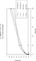


Classifications
-
- C—CHEMISTRY; METALLURGY
- C07—ORGANIC CHEMISTRY
- C07K—PEPTIDES
- C07K16/00—Immunoglobulins [IGs], e.g. monoclonal or polyclonal antibodies
- C07K16/40—Immunoglobulins [IGs], e.g. monoclonal or polyclonal antibodies against enzymes
-
- A—HUMAN NECESSITIES
- A61—MEDICAL OR VETERINARY SCIENCE; HYGIENE
- A61P—SPECIFIC THERAPEUTIC ACTIVITY OF CHEMICAL COMPOUNDS OR MEDICINAL PREPARATIONS
- A61P11/00—Drugs for disorders of the respiratory system
-
- A—HUMAN NECESSITIES
- A61—MEDICAL OR VETERINARY SCIENCE; HYGIENE
- A61P—SPECIFIC THERAPEUTIC ACTIVITY OF CHEMICAL COMPOUNDS OR MEDICINAL PREPARATIONS
- A61P27/00—Drugs for disorders of the senses
- A61P27/02—Ophthalmic agents
- A61P27/10—Ophthalmic agents for accommodation disorders, e.g. myopia
-
- A—HUMAN NECESSITIES
- A61—MEDICAL OR VETERINARY SCIENCE; HYGIENE
- A61P—SPECIFIC THERAPEUTIC ACTIVITY OF CHEMICAL COMPOUNDS OR MEDICINAL PREPARATIONS
- A61P37/00—Drugs for immunological or allergic disorders
- A61P37/02—Immunomodulators
- A61P37/06—Immunosuppressants, e.g. drugs for graft rejection
-
- A—HUMAN NECESSITIES
- A61—MEDICAL OR VETERINARY SCIENCE; HYGIENE
- A61K—PREPARATIONS FOR MEDICAL, DENTAL OR TOILETRY PURPOSES
- A61K39/00—Medicinal preparations containing antigens or antibodies
- A61K2039/505—Medicinal preparations containing antigens or antibodies comprising antibodies
-
- A—HUMAN NECESSITIES
- A61—MEDICAL OR VETERINARY SCIENCE; HYGIENE
- A61K—PREPARATIONS FOR MEDICAL, DENTAL OR TOILETRY PURPOSES
- A61K39/00—Medicinal preparations containing antigens or antibodies
- A61K2039/545—Medicinal preparations containing antigens or antibodies characterised by the dose, timing or administration schedule
-
- C—CHEMISTRY; METALLURGY
- C07—ORGANIC CHEMISTRY
- C07K—PEPTIDES
- C07K2317/00—Immunoglobulins specific features
- C07K2317/50—Immunoglobulins specific features characterized by immunoglobulin fragments
- C07K2317/54—F(ab')2
-
- C—CHEMISTRY; METALLURGY
- C07—ORGANIC CHEMISTRY
- C07K—PEPTIDES
- C07K2317/00—Immunoglobulins specific features
- C07K2317/50—Immunoglobulins specific features characterized by immunoglobulin fragments
- C07K2317/56—Immunoglobulins specific features characterized by immunoglobulin fragments variable (Fv) region, i.e. VH and/or VL
- C07K2317/565—Complementarity determining region [CDR]
-
- C—CHEMISTRY; METALLURGY
- C07—ORGANIC CHEMISTRY
- C07K—PEPTIDES
- C07K2317/00—Immunoglobulins specific features
- C07K2317/70—Immunoglobulins specific features characterized by effect upon binding to a cell or to an antigen
- C07K2317/76—Antagonist effect on antigen, e.g. neutralization or inhibition of binding
-
- C—CHEMISTRY; METALLURGY
- C07—ORGANIC CHEMISTRY
- C07K—PEPTIDES
- C07K2317/00—Immunoglobulins specific features
- C07K2317/90—Immunoglobulins specific features characterized by (pharmaco)kinetic aspects or by stability of the immunoglobulin
-
- C—CHEMISTRY; METALLURGY
- C07—ORGANIC CHEMISTRY
- C07K—PEPTIDES
- C07K2317/00—Immunoglobulins specific features
- C07K2317/90—Immunoglobulins specific features characterized by (pharmaco)kinetic aspects or by stability of the immunoglobulin
- C07K2317/92—Affinity (KD), association rate (Ka), dissociation rate (Kd) or EC50 value
Abstract
一局面において、本発明は、HSCT-IPSに罹患しているかもしくはこれを発症するリスクがあるヒト対象、および/またはHSCT-CLSに罹患しているかもしくはこれを発症するリスクがあるヒト対象、および/またはHSCT-FOに罹患しているかもしくはこれを発症するリスクがあるヒト対象、および/またはHSCT-ESに罹患しているかもしくはこれを発症するリスクがあるヒト対象において、MASP-2依存性補体活性化の作用を阻害する方法を提供する。方法は、それを必要とする対象に、MASP-2依存性補体活性化を阻害するのに有効な量のMASP-2阻害物質を投与する工程を含む。TIFF2023503611000065.tif129170In one aspect, the present invention provides a human subject having or at risk of developing HSCT-IPS and/or a human subject having or at risk of developing HSCT-CLS, and /or MASP-2 dependent complementation in human subjects with or at risk of developing HSCT-FO and/or in human subjects with or at risk of developing HSCT-ES Methods of inhibiting the effects of somatic activation are provided. The method comprises administering to a subject in need thereof an amount of a MASP-2 inhibitor effective to inhibit MASP-2 dependent complement activation. TIFF2023503611000065.tif129170
Description
関連出願との相互参照
本願は、参照によりその全体が本明細書に組み入れられる、2019年11月26日付けで出願された米国仮出願第62/940,720号および2019年11月26日付けで出願された米国仮出願第62/940,735号の恩典を主張する。
CROSS-REFERENCE TO RELATED APPLICATIONS This application is filed on November 26, 2019 and U.S. Provisional Application No. 62/940,720, filed November 26, 2019, which is hereby incorporated by reference in its entirety. claims the benefit of filed U.S. Provisional Application No. 62/940,735.
配列表に関する記載
本願に関連する配列表はハードコピーの代わりにテキスト形式で提供され、本明細書に参照により組み入れられる。配列表を含むテキストファイルの名前は、MP_1_0307_PCT_Sequence_Listing_20201120_ST25.txtであり;このファイルは115 KBであり、2020年11月20日に作成され、本明細書の出願と共にEFS-Webを介して提出されている。
STATEMENT REGARDING SEQUENCE LISTING The Sequence Listing associated with this application is provided in text format in lieu of hard copy and is incorporated herein by reference. The name of the text file containing the Sequence Listing is MP_1_0307_PCT_Sequence_Listing_20201120_ST25.txt; this file is 115 KB, created on November 20, 2020 and filed via EFS-Web with the filing herein. .
背景
補体系は、ヒトおよび他の脊椎動物において微生物感染症および他の急性侵襲に対する免疫応答を開始、増幅、および組織化するための初期作用機構を提供する(M.K. Liszewski and J. P. Atkinson, 1993, in Fundamental Immunology, Third Edition, W.E, Paul編, Raven Press, Ltd., New York(非特許文献1))。補体活性化は潜在的な病原体に対する有益な第一線の防御を提供するが、防御免疫応答を促進する補体の活性は宿主にとって潜在的な脅威となることもある(K.R, Kalli, et al., Springer Semin. Immunopathol. 15:417-431, 1994(非特許文献2); B.P. Morgan, Eur. J. Clinical Investig. 24:219-228, 1994(非特許文献3))。例えば、C3およびC5タンパク質分解産物は好中球を動員および活性化する。活性化好中球は宿主防御に不可欠であるが見境なく破壊酵素を放出し、臓器損傷を引き起こすことがある。さらに、補体活性化によって、近くの宿主細胞ならびに微生物標的の表面に溶解性補体成分が沈着し、その結果、宿主細胞が溶解する場合がある。
BACKGROUND The complement system provides an early mechanism of action for initiating, amplifying, and orchestrating the immune response to microbial infections and other acute insults in humans and other vertebrates (MK Liszewski and JP Atkinson, 1993, in Fundamental Immunology, Third Edition, WE, Paul, Ed., Raven Press, Ltd., New York (Non-Patent Document 1)). Complement activation provides a beneficial first-line defense against potential pathogens, but complement activity, which promotes protective immune responses, can also pose a potential threat to the host (KR, Kalli, et al. al., Springer Semin. Immunopathol. 15:417-431, 1994 (Non-Patent Document 2); BP Morgan, Eur. J. Clinical Investig. 24:219-228, 1994 (Non-Patent Document 3)). For example, the C3 and C5 proteolytic products recruit and activate neutrophils. Activated neutrophils are essential for host defense but can indiscriminately release destructive enzymes and cause organ damage. In addition, complement activation can result in the deposition of soluble complement components on the surface of nearby host cells as well as microbial targets, resulting in host cell lysis.
補体系はまた、心筋梗塞、脳卒中、ARDS、再灌流傷害、敗血症ショック、熱傷後の毛細血管漏出、心肺バイパス術後炎症、移植片拒絶、慢性関節リウマチ、多発性硬化症、重症筋無力症、およびアルツハイマー病を含む、非常に多くの急性疾患状態および慢性疾患状態の発生に関係している。これらの状態のほぼ全てにおいて、補体は原因でないが、発生に関与するいくつかの因子の1つである。それにもかかわらず、補体活性化は主要な病理学的機構であると考えられ、これらの疾患状態の多くにおける臨床管理のための有効なポイントということになる。様々な疾患状態において補体媒介性組織損傷が重要であるという認識の高まりは、有効な補体阻害薬物の必要性を強調する。現在に至るまで、抗C5抗体であるエクリズマブ(Solaris(登録商標))がヒトでの使用に認可されている唯一の補体標的化薬物である。だが、C5は、補体系の「下流」に位置する、いくつかのエフェクター分子の1つであり、C5が遮断されても補体系活性化は阻害されない。従って、補体活性化の開始工程の阻害因子は「下流」補体阻害因子よりもかなり優位に立っていると考えられる。 The complement system is also involved in myocardial infarction, stroke, ARDS, reperfusion injury, septic shock, post-burn capillary leak, post-cardiopulmonary bypass inflammation, graft rejection, rheumatoid arthritis, multiple sclerosis, myasthenia gravis, and has been implicated in the development of numerous acute and chronic disease states, including Alzheimer's disease. In nearly all of these conditions complement is not causative, but is one of several factors involved in development. Nonetheless, complement activation is thought to be a major pathological mechanism, making it a valid point for clinical management in many of these disease states. The growing awareness of the importance of complement-mediated tissue damage in various disease states emphasizes the need for effective complement-inhibiting drugs. To date, the anti-C5 antibody eculizumab (Solaris®) is the only complement-targeting drug approved for human use. However, C5 is one of several effector molecules located 'downstream' of the complement system, and blockade of C5 does not inhibit complement system activation. Thus, inhibitors of the initiation step of complement activation appear to have a significant advantage over "downstream" complement inhibitors.
現在、補体系は3つの異なる経路:古典経路、レクチン経路、および第二経路を介して活性化できることが広く認められている。古典経路は、通常、外来粒子(すなわち、抗原)に結合した宿主抗体からなる複合体によって誘発され、従って、特異的抗体反応を発生させるために抗原への前曝露を必要とする。古典経路の活性化は宿主による以前の獲得免疫応答に左右されるので、古典経路は後天免疫系の一部である。対照的に、レクチン経路および第二経路はいずれも獲得免疫とは無関係であり、自然免疫系の一部である。 It is now widely accepted that the complement system can be activated via three different pathways: the classical pathway, the lectin pathway, and the alternative pathway. The classical pathway is usually triggered by a complex consisting of a host antibody bound to a foreign particle (ie antigen) and thus requires pre-exposure to the antigen in order to generate a specific antibody response. The classical pathway is part of the acquired immune system, as activation of the classical pathway depends on previous adaptive immune responses by the host. In contrast, both the lectin pathway and the alternative pathway are independent of adaptive immunity and are part of the innate immune system.
補体系の活性化により、セリンプロテアーゼ酵素前駆体の連続して起こる活性化がもたらされる。古典経路活性化の第一段階は、特異的認識分子C1qと、抗原に結合したIgG分子およびIgM分子との結合である。C1qは、C1と呼ばれる複合体としてC1rおよびC1sセリンプロテアーゼプロ酵素と結合する。C1qと免疫複合体が結合すると、C1rのArg-Ile部位が自己タンパク分解によって切断された後に、C1rによって媒介されるC1sの切断および活性化が起こり、それによって、C4およびC2を切断する能力が獲得される。C4は、C4aおよびC4bと呼ばれる2つの断片に切断され、同様に、C2はC2aおよびC2bに切断される。C4b断片は、隣接するヒドロキシル基またはアミノ基と共有結合を結合し、活性化C2のC2a断片との非共有結合的相互作用を介してC3コンバターゼ(C4b2a)を生成することができる。C3コンバターゼ(C4b2a)は、C5コンバターゼ(C4b2a3b)の生成をもたらす、C3aおよびC3b小成分へのタンパク質切断によって、C3を活性化し、C5コンバターゼ(C4b2a3b)は、C5を切断することによって、細胞膜を破壊して細胞溶解をもたらすことができる膜侵襲複合体(C5bがC6、C7、C8、およびC9と組み合わされ、「MAC」とも呼ばれる)の形成をもたらす。C3およびC4の活性化型(C3bおよびC4b)は共有結合により外来標的表面に沈着し、複数の食細胞上にある補体受容体によって認識される。 Activation of the complement system results in sequential activation of serine protease zymogens. The first step in classical pathway activation is the binding of the specific recognition molecule C1q to antigen-bound IgG and IgM molecules. C1q binds C1r and C1s serine protease proenzymes in a complex called C1. Upon binding of C1q to the immune complex, autoproteolytic cleavage of the Arg-Ile site of C1r is followed by C1r-mediated cleavage and activation of C1s, thereby reducing its ability to cleave C4 and C2. Acquired. C4 is cleaved into two fragments called C4a and C4b and similarly C2 is cleaved into C2a and C2b. The C4b fragment can covalently bind adjacent hydroxyl or amino groups to generate a C3 convertase (C4b2a) through non-covalent interactions with the C2a fragment of activated C2. The C3 convertase (C4b2a) activates C3 by proteolytic cleavage into C3a and C3b subcomponents leading to the generation of the C5 convertase (C4b2a3b), and the C5 convertase (C4b2a3b) disrupts the cell membrane by cleaving C5. resulting in the formation of the membrane attack complex (C5b combines with C6, C7, C8, and C9, also called "MAC"), which can lead to cell lysis. The activated forms of C3 and C4 (C3b and C4b) are covalently deposited on foreign target surfaces and recognized by complement receptors on multiple phagocytic cells.
独立して、レクチン経路を介した補体系活性化における第一段階も、特異的認識分子の結合と、それに続く、関連するセリンプロテアーゼプロ酵素の活性化である。しかしながら、C1qによる免疫複合体の結合ではなく、レクチン経路の認識分子は、総称してレクチンと呼ばれる炭水化物結合タンパク質(マンナン結合レクチン(MBL)、H-フィコリン、M-フィコリン、L-フィコリン、およびC型レクチンCL-11)の一群を含む。J. Lu et al., Biochim. Biophys. Acta 1572:387-400, (2002)(非特許文献4); Holmskov et al., Annu. Rev. Immunol. 21:547-578(2003)(非特許文献5); Teh et al., Immunology 101:225-232(2000)(非特許文献6))を参照されたい。J. Luet et al., Biochim Biophys Acta 1572:387-400(2002)(非特許文献4); Holmskov et al., Annu. Rev. Immunol. 21:547-578(2003)(非特許文献5); Teh et al., Immunology 101:225-232(2000)(非特許文献6); Hansen et al., J, Immunol 185(10):6096-6104(2010)(非特許文献7)も参照されたい。 Independently, the first step in complement system activation via the lectin pathway is also the binding of specific recognition molecules and the subsequent activation of associated serine protease proenzymes. However, rather than binding immune complexes by C1q, the recognition molecules of the lectin pathway are carbohydrate-binding proteins collectively called lectins (mannan-binding lectin (MBL), H-ficolin, M-ficolin, L-ficolin, and C type lectin CL-11). J. Lu et al., Biochim. Biophys. Acta 1572:387-400, (2002) (Non-Patent Document 4); Holmskov et al., Annu. Rev. Immunol. Reference 5); Teh et al., Immunology 101:225-232 (2000) (Non-Patent Reference 6)). J. Luet et al., Biochim Biophys Acta 1572:387-400 (2002) (Non-Patent Document 4); Holmskov et al., Annu. Rev. Immunol. see also Teh et al., Immunology 101:225-232(2000); Hansen et al., J, Immunol 185(10):6096-6104(2010). sea bream.
Ikedaらは、C1qと同様にMBLが酵母マンナンでコーティングされた赤血球と結合すると、C4依存的に補体系を活性化できることを初めて証明した(Ikeda et al., J. Biol. Chem. 262:7451-7454, (1987)(非特許文献8))。コレクチンタンパク質ファミリーのメンバーであるMBLは、3-ヒドロキシ基および4-ヒドロキシ基がピラノース環の赤道結合面に配向されている炭水化物と結合するカルシウム依存性レクチンである。従って、MBLの目立ったリガンドはD-マンノースおよびN-アセチル-D-グルコサミンであるのに対して、この立体的要件に合わない炭水化物はMBLに対して検出不可能な親和性を有する(Weis et al., Nature 360:127-134, (1992)(非特許文献9))。MBLと一価糖との相互作用は極めて弱く、解離定数は典型的に1桁のミリモル範囲内である。MBLは、アビディティによって、すなわち、互いに近くに位置する複数の単糖残基と同時に相互作用することによってグリカンリガンドと緊密で特異的な結合を実現する(Lee et al., Archiv. Biochem. Biophys. 299:129-136, (1992)(非特許文献10))。MBLは、一般的に、微生物、例えば、細菌、酵母、寄生生物、およびある特定のウイルスを装飾する炭水化物パターンを認識する。対照的に、MBLは、通常、哺乳動物の血漿糖タンパク質上および細胞表面糖タンパク質上に存在する「成熟」複合糖質を装飾する最後から2番目の糖および最後の糖であるD-ガラクトースおよびシアル酸を認識しない。この結合特異性は、「外来」表面の認識を促進し、「自己活性化」からの保護を助けると考えられる。しかしながら、MBLは、哺乳動物細胞の小胞体中およびゴルジ中で隔離されたN結合糖タンパク質および糖脂質における高マンノース「前駆」グリカンのクラスターに高親和性で結合する(Maynard et al., J. Biol. Chem. 257:3788-3794, (1982)(非特許文献11))。従って、損傷細胞は、MBL結合を介したレクチン経路活性化の潜在的な標的である。 Ikeda et al. demonstrated for the first time that MBL, like C1q, can activate the complement system in a C4-dependent manner when bound to yeast mannan-coated erythrocytes (Ikeda et al., J. Biol. Chem. 262:7451). -7454, (1987) (non-patent document 8)). MBL, a member of the collectin protein family, is a calcium-dependent lectin that binds carbohydrates with the 3- and 4-hydroxy groups oriented on the equatorial plane of the pyranose ring. Thus, the prominent ligands for MBL are D-mannose and N-acetyl-D-glucosamine, whereas carbohydrates that do not meet this steric requirement have undetectable affinity for MBL (Weis et al. al., Nature 360:127-134, (1992) (Non-Patent Document 9)). The interaction between MBL and monovalent sugars is extremely weak, with dissociation constants typically in the single millimolar range. MBL achieves tight and specific binding to glycan ligands by avidity, ie, by simultaneously interacting with multiple monosaccharide residues located close to each other (Lee et al., Archiv. Biochem. Biophys. 299:129-136, (1992) (Non-Patent Document 10)). MBL generally recognizes carbohydrate patterns that decorate microorganisms such as bacteria, yeast, parasites, and certain viruses. In contrast, MBL has the penultimate and final sugars, D-galactose and Does not recognize sialic acid. This binding specificity is believed to facilitate recognition of 'foreign' surfaces and help protect against 'self-activation'. However, MBL binds with high affinity to clusters of high-mannose "precursor" glycans on N-linked glycoproteins and glycolipids sequestered in the endoplasmic reticulum and Golgi of mammalian cells (Maynard et al., J. Biol. Chem. 257:3788-3794, (1982) (Non-Patent Document 11)). Damaged cells are therefore potential targets for lectin pathway activation via MBL binding.
フィコリンは、フィブリノゲン様ドメインと呼ばれる、MBLとは異なるタイプのレクチンドメインを有する。フィコリンはCa++非依存的に糖残基に結合する。ヒトでは、3種類のフィコリン(L-フィコリン、M-フィコリン、およびH-フィコリン)が特定されている。2種類の血清フィコリンであるL-フィコリンおよびH-フィコリンは共通してN-アセチル-D-グルコサミンに対する特異性を有する。しかしながら、H-フィコリンはN-アセチル-D-ガラクトサミンにも結合する。L-フィコリン、H-フィコリン、CL-11、およびMBLの糖特異性が異なることは、異なるレクチンが補い合い、重複によって異なる複合糖質を標的とし得ることを意味する。この考えは、レクチン経路にある公知のレクチンのうちL-フィコリンだけが、全てのグラム陽性細菌に見られる細胞壁複合糖質であるリポテイコ酸に特異的に結合するという最近の報告によって裏付けられている(Lynch et al., J. Immunol. 172:1198-1202, (2004)(非特許文献12))。コレクチン(すなわち、MBL)およびフィコリンはアミノ酸配列において有意な類似性を有さない。しかしながら、これらの2つのタンパク質グループは、類似したドメイン構成を有し、かつ、C1qと同様に集合して、多部位結合の可能性を最大にするオリゴマー構造を構築する。 Ficolins have a different type of lectin domain than MBL, called a fibrinogen-like domain. Ficolin binds to sugar residues in a Ca ++ -independent manner. In humans, three types of ficolin (L-ficolin, M-ficolin, and H-ficolin) have been identified. Two types of serum ficolin, L-ficolin and H-ficolin, share a specificity for N-acetyl-D-glucosamine. However, H-ficolin also binds N-acetyl-D-galactosamine. The different sugar specificities of L-ficolin, H-ficolin, CL-11, and MBL mean that different lectins can complement each other and target different glycoconjugates by redundancy. This idea is supported by recent reports that of the known lectins in the lectin pathway, only L-ficolin specifically binds to lipoteichoic acid, a cell wall glycoconjugate found in all Gram-positive bacteria. (Lynch et al., J. Immunol. 172:1198-1202, (2004) (Non-Patent Document 12)). Collectins (ie, MBL) and ficolins do not share significant similarities in amino acid sequences. However, these two protein groups have similar domain organization and assemble similarly to C1q to build oligomeric structures that maximize the potential for multisite binding.
MBLの血清中濃度は健常集団においてかなり変動し、これは、MBL遺伝子のプロモーター領域およびコード領域の両方にある多型/変異によって遺伝的に制御される。急性期タンパク質として、炎症中にMBL発現はさらに上方制御される。L-フィコリンは、MBLの濃度とほぼ同じ濃度で血清中に存在する。従って、レクチン経路のL-フィコリン分岐は強さが場合によってはMBL部門に匹敵する。MBLおよびフィコリンはオプソニンとしても機能することができる。このために、食細胞は、MBLによって装飾された表面およびフィコリンによって装飾された表面を標的とすることが可能になる(Jack et al., J Leukoc Biol., 77(3):328-36(2004)(非特許文献13), Matsushita and Fujita, Immunobiology, 205(4-5):490-7(2002)(非特許文献14), Aoyagi et al., J. Immunol, 174(1):418-25(2005)(非特許文献15)を参照されたい)。このオプソニン化は、これらのタンパク質と食細胞受容体との相互作用を必要とする(Kuhlman et al., J. Exp. Med. 169:1733, (1989)(非特許文献16); Matsushita et al., J. Biol. Chem. 271:2448-54, (1996)(非特許文献17))。食細胞受容体の正体は証明されていない。 Serum concentrations of MBL vary considerably in healthy populations and are genetically controlled by polymorphisms/mutations in both the promoter and coding regions of the MBL gene. As an acute phase protein, MBL expression is further upregulated during inflammation. L-ficolin is present in serum at approximately the same concentration as that of MBL. Thus, the L-ficolin branch of the lectin pathway is comparable in strength to the MBL branch in some cases. MBL and ficolins can also function as opsonins. This enables phagocytic cells to target surfaces decorated with MBL and surfaces decorated with ficolin (Jack et al., J Leukoc Biol., 77(3):328-36). 2004) (non-patent document 13), Matsushita and Fujita, Immunobiology, 205(4-5):490-7(2002) (non-patent document 14), Aoyagi et al., J. Immunol, 174(1):418 -25 (2005) (see Non-Patent Document 15)). This opsonization requires interaction of these proteins with phagocyte receptors (Kuhlman et al., J. Exp. Med. 169:1733, (1989); Matsushita et al. ., J. Biol. Chem. 271:2448-54, (1996) (Non-Patent Document 17)). The identity of the phagocyte receptor has not been proven.
ヒトMBLは、そのコラーゲン様ドメインを介して、MBL関連セリンプロテアーゼ(MASP)と呼ばれる独特のC1r/C1s様セリンプロテアーゼと特異的な、かつ高親和性の相互作用を生じさせる。現在に至るまで、3種類のMASPが述べられている。第1に、単一の酵素「MASP」が、補体カスケードの開始(すなわち、C2およびC4の切断)を担う酵素として特定および特徴決定された(Matsushita et al., J Exp Med 176(6):1497-1502(1992)(非特許文献18):Ji et al., J. Immunol 150:571-578, (1993)(非特許文献19))。その後に、MASP活性が実際には、2種類のプロテアーゼ:MASP-1およびMASP-2の混合であることが明らかにされた(Thiel et al., Nature 386:506-510, (1997)(非特許文献20))。しかしながら、補体活性化にはMBL-MASP-2複合体だけでも十分であることが証明された(Vorup-Jensen et al., J. Immunol 165:2093-2100, (2000)(非特許文献21))。さらに、MASP-2だけが高い割合でC2およびC4を切断した(Ambrus et al., J. Immunol, 170:1374-1382, (2003)(非特許文献22))。従って、MASP-2は、C4およびC2を活性化してC3コンバターゼであるC4b2aを生成するのを担うプロテアーゼである。これは、2種類の特異的なセリンプロテアーゼ(C1rおよびC1s)の協調作用が補体系活性化につながる古典経路のC1複合体とは大きな違いである。さらに、第3の新規プロテアーゼであるMASP-3が単離されている(Dahl, M.R. et al., Immunity 15:127-35, 2001(非特許文献23))。MASP-1およびMASP-3は同じ遺伝子のオルタナティブスプライシング産物である。 Human MBL, through its collagen-like domain, makes a specific and high-affinity interaction with a unique C1r/C1s-like serine protease termed MBL-associated serine protease (MASP). To date, three types of MASP have been described. First, a single enzyme, 'MASP', was identified and characterized as the enzyme responsible for initiation of the complement cascade (i.e. cleavage of C2 and C4) (Matsushita et al., J Exp Med 176(6) : 1497-1502 (1992) (Non-Patent Document 18): Ji et al., J. Immunol 150:571-578, (1993) (Non-Patent Document 19)). It was later shown that MASP activity is actually a mixture of two proteases: MASP-1 and MASP-2 (Thiel et al., Nature 386:506-510, (1997) Patent document 20)). However, it was demonstrated that the MBL-MASP-2 complex alone is sufficient for complement activation (Vorup-Jensen et al., J. Immunol 165:2093-2100, (2000) )). Furthermore, only MASP-2 cleaved C2 and C4 at a high rate (Ambrus et al., J. Immunol, 170:1374-1382, (2003)). MASP-2 is therefore the protease responsible for activating C4 and C2 to generate the C3 convertase, C4b2a. This is in contrast to the C1 complex of the classical pathway, where the concerted action of two specific serine proteases (C1r and C1s) leads to complement system activation. In addition, a third novel protease, MASP-3, has been isolated (Dahl, M.R. et al., Immunity 15:127-35, 2001). MASP-1 and MASP-3 are alternative splicing products of the same gene.
MASPは、C1複合体の酵素成分であるC1rおよびC1sのドメイン構成と同一のドメイン構成を有する(Sim et al., Biochem. Soc. Trans. 28:545, (2000)(非特許文献24))。これらのドメインは、N末端C1r/C1s/ウニ(sea urchin)VEGF/骨形成タンパク質(CUB)ドメイン、上皮細胞成長因子様ドメイン、第2のCUBドメイン、補体制御タンパク質ドメインの縦列配列、およびセリンプロテアーゼドメインを含む。C1プロテアーゼと同様に、MASP-2の活性化は、セリンプロテアーゼドメインに隣接するArg-Ile結合の切断によって起こる。この切断によって、酵素はジスルフィド結合したA鎖およびB鎖に分けられる。後者はセリンプロテアーゼドメインからなる。 MASP has the same domain configuration as that of C1r and C1s, which are the enzymatic components of the C1 complex (Sim et al., Biochem. Soc. Trans. 28:545, (2000) (Non-Patent Document 24)). . These domains include an N-terminal C1r/C1s/sea urchin VEGF/bone morphogenetic protein (CUB) domain, an epidermal growth factor-like domain, a second CUB domain, a tandem array of complement regulatory protein domains, and a serine Contains a protease domain. Similar to the C1 protease, MASP-2 activation occurs through cleavage of the Arg-Ile bond adjacent to the serine protease domain. This cleavage separates the enzyme into disulfide-linked A and B chains. The latter consists of a serine protease domain.
MBLはまた、MASP2の触媒活性を欠く、19kDaのMBL関連タンパク質(MAp19)または低分子MBL関連タンパク質(sMAP)として公知のオルタナティブスプライシング型MASP-2と結合することもできる(Stover, J. Immunol. 162:3481-90, (1999)(非特許文献25); Takahashi et al., Int. Immunol. 11:859-863, (1999)(非特許文献26))。MAp19は、MASP-2の最初の2つのドメインに続いて、4個の独特のアミノ酸からなる余分な配列を含む。Map19の機能は不明である(Degn et al., J. Immunol. Methods. 2011(非特許文献27))。MASP-1遺伝子およびMASP-2遺伝子は、それぞれ、ヒト第3染色体および第1染色体に位置する(Schwaeble et al., Immunobiology 205:455-466, (2002)(非特許文献28))。
MBL can also associate with alternatively spliced forms of MASP-2 known as the 19 kDa MBL-associated protein (MAp19) or the small MBL-associated protein (sMAP), which lack the catalytic activity of MASP2 (Stover, J. Immunol. 162:3481-90, (1999) (Non-Patent Document 25); Takahashi et al., Int. Immunol. 11:859-863, (1999) (Non-Patent Document 26)). MAp19 contains an extra sequence of four unique amino acids following the first two domains of MASP-2. The function of Map19 is unknown (Degn et al., J. Immunol. Methods. 2011 (Non-Patent Document 27)). The MASP-1 and MASP-2 genes are located on
いくつかの証拠から、異なるMBL-MASP複合体があり、かつ血清中のMASPの大部分がMBLと複合体を形成しないことが示唆されている(Thiel. et al., J. Immunol. 165:878-887, (2000)(非特許文献29))。H-フィコリンおよびL-フィコリンはいずれもMBLと同様に全てのMASPに結合し、レクチン補体経路を活性化する(Dahl et al., Immunity 15:127-35, (2001)(非特許文献23); Matsushita et al., J. Immunol. 168:3502-3506, (2002)(非特許文献30))。レクチン経路および古典経路はいずれも共通のC3コンバターゼ(C4b2a)を形成し、2つの経路はこの段階で1つになる。 Several lines of evidence suggest that there are different MBL-MASP complexes and that the majority of MASP in serum does not form a complex with MBL (Thiel. et al., J. Immunol. 165: 878-887, (2000) (Non-Patent Document 29)). Both H-ficolin and L-ficolin, like MBL, bind to all MASPs and activate the lectin complement pathway (Dahl et al., Immunity 15:127-35, (2001) ); Matsushita et al., J. Immunol. 168:3502-3506, (2002) (Non-Patent Document 30)). Both the lectin pathway and the classical pathway form a common C3 convertase (C4b2a) and the two pathways merge at this stage.
レクチン経路は、ナイーブな宿主における感染症に対する宿主防御において主な役割を有すると広く考えられている。宿主防御においてMBLが関与する強力な証拠が、機能的MBLの血清中レベルが低い患者の分析から得られた(Kilpatrick, Biochim. Biophys. Acta 1572:401-413, (2002)(非特許文献31))。このような患者は反復性の細菌感染症および真菌感染症に対して感受性を示す。これらの症状は、通常、若年期に、母系由来抗体価が漸減する時であるが抗体反応の全レパートリーが発達する前の見かけの(apparent)脆弱期間に現れる。この症候群は、MBLコラーゲン部分におけるいくつかの部位での変異に起因する場合が多く、該変異はMBLオリゴマーの適切な形成を妨害する。しかしながら、MBLは補体に関係なくオプソニンとして機能することができるので、感染症に対する感受性の増大がどの程度まで補体活性化の障害によるものであるかは分かっていない。 The lectin pathway is widely believed to have a major role in host defense against infections in naive hosts. Strong evidence for the involvement of MBL in host defense comes from the analysis of patients with low serum levels of functional MBL (Kilpatrick, Biochim. Biophys. Acta 1572:401-413, (2002). )). Such patients are susceptible to recurrent bacterial and fungal infections. These symptoms usually appear in early life, when maternally derived antibody titers taper but before the full repertoire of antibody responses has developed, a period of apparent weakness. This syndrome is often due to mutations at several sites in the MBL collagen portion, which interfere with the proper formation of MBL oligomers. However, it is not known to what extent the increased susceptibility to infections is due to impaired complement activation, as MBL can function as an opsonin independent of complement.
古典経路およびレクチン経路とは対照的に、C1qおよびレクチンが他の2つの段階において果たす認識機能を、第二経路の開始因子が実行することは見出されていない。現在、第二経路は低レベルの代謝回転活性化を自発的に受けることが広く認められている。この代謝回転活性化は、自発的補体活性化を抑える適切な分子エレメントを欠く外来表面または他の異常表面(細菌、酵母、ウイルスに感染した細胞、または損傷組織)において容易に増幅することができる。第二経路活性化に直接関与する4種類の血漿タンパク質:C3、B因子およびD因子、ならびにプロペルジンがある。 In contrast to the classical and lectin pathways, the initiation factors of the alternative pathway have not been found to perform the cognitive functions that C1q and lectins perform in the other two steps. It is now widely accepted that the alternative pathway spontaneously undergoes low levels of turnover activation. This turnover activation can be readily amplified on foreign or other abnormal surfaces (bacteria, yeast, virus-infected cells, or damaged tissue) that lack the appropriate molecular elements to suppress spontaneous complement activation. can. There are four plasma proteins directly involved in alternative pathway activation: C3, Factors B and D, and Properdin.
古典補体経路および第二補体経路の両方が非感染性ヒト疾患の発生に関係していることを示す幅広い証拠があるが、レクチン経路の役割は評価され始めたばかりである。最近の研究から、レクチン経路の活性化は虚血/再灌流傷害における補体活性化および関連炎症を担っている可能性があるという証拠が得られた。Collard et al.,(2000)は、酸化ストレスに供された培養内皮細胞はMBLに結合し、ヒト血清に曝露されるとC3沈着を示すと報告した(Collard et al., Am. J. Pathol 156:1549-1556,(2000)(非特許文献32))。さらに、ヒト血清を遮断抗MBLモノクローナル抗体で処理すると、MBL結合および補体活性化が阻害された。これらの知見をラット心筋虚血-再灌流モデルに広げた。このモデルでは、ラットMBLに対する遮断抗体で処置されたラットは、冠状動脈閉塞時に対照抗体処置ラットよりも有意に少ない心筋損傷を示した(Jordan et al., Circulation, 104:1413-1418,(2001)(非特許文献33))。酸化ストレス後の血管内皮とのMBL結合の分子機構は不明である。最近の研究から、酸化ストレス後のレクチン経路の活性化は血管内皮サイトケラチンとのMBL結合によって媒介されるが、複合糖質によって媒介されない可能性があることが示唆されている(Collard e al., Am. J. Pathol. 159:1045-1054,(2001)(非特許文献34))。他の研究は古典経路および第二経路が虚血/再灌流傷害の発生に関係していることを示しており、この疾患におけるレクチン経路の役割は議論の余地が残されている(Riedermann, N.C. et al., Am. J. Pathol. 162:363-367, 2003(非特許文献35))。 Although there is extensive evidence implicating both the classical and alternative complement pathways in the development of non-infectious human disease, the role of the lectin pathway is just beginning to be evaluated. Recent studies have provided evidence that activation of the lectin pathway may be responsible for complement activation and associated inflammation in ischemia/reperfusion injury. Collard et al., (2000) reported that cultured endothelial cells subjected to oxidative stress bind MBL and exhibit C3 deposition when exposed to human serum (Collard et al., Am. J. Pathol. 156:1549-1556, (2000) (Non-Patent Document 32)). Furthermore, treatment of human serum with a blocking anti-MBL monoclonal antibody inhibited MBL binding and complement activation. We extended these findings to a rat myocardial ischemia-reperfusion model. In this model, rats treated with a blocking antibody against rat MBL showed significantly less myocardial damage than control antibody-treated rats during coronary artery occlusion (Jordan et al., Circulation, 104:1413-1418, (2001). ) (Non-Patent Document 33)). The molecular mechanism of MBL binding to vascular endothelium after oxidative stress is unknown. Recent studies suggest that activation of the lectin pathway after oxidative stress is mediated by MBL binding to endothelial cytokeratin, but may not be mediated by glycoconjugates (Collard et al. , Am. J. Pathol. 159:1045-1054, (2001) (Non-Patent Document 34)). Other studies have implicated the classical and alternative pathways in the development of ischemia/reperfusion injury, leaving the role of the lectin pathway in this disease controversial (Riedermann, N.C. et al., Am. J. Pathol. 162:363-367, 2003 (Non-Patent Document 35)).
最近の研究により、第二経路活性化酵素であるD因子を酵素前駆体型から酵素活性型に変換するためにはMASP-1が(おそらくMASP-3も)必要とされることが示されている(Takahashi M. et al., J Exp Med 207(1):29-37(2010)(非特許文献36)を参照されたい)。このプロセスの生理学的重要性は、MASP-1/3欠損マウスの血漿中に機能的な第二経路活性が存在しないことで強調される。タンパク質分解によって天然C3からC3bが生成されるには第二経路が機能することが必要である。第二経路C3コンバターゼ(C3bBb)が必須のサブユニットであるC3bを含有するので、第二経路を介した最初のC3bの起源に関する疑問は不可解な問題であり、膨大な研究を活気づけてきた。 Recent studies have shown that MASP-1 (and possibly MASP-3 as well) is required for the conversion of the alternative pathway activating enzyme factor D from its proenzyme form to its active form. (See Takahashi M. et al., J Exp Med 207(1):29-37(2010) (36)). The physiological importance of this process is underscored by the absence of functional alternative pathway activity in the plasma of MASP-1/3-deficient mice. The proteolytic production of C3b from native C3 requires the functioning of the alternative pathway. Since the alternative pathway C3 convertase (C3bBb) contains an essential subunit, C3b, the question of the origin of the first C3b via the alternative pathway is a puzzling question and has fueled a great deal of research.
C3は、(C4およびα-2マクログロブリンと共に)チオエステル結合として公知の稀な翻訳後修飾を含有するタンパク質のファミリーに属する。チオエステル基は、3アミノ酸離れたシステインのスルフヒドリル基とチオエステル共有結合を形成する末端カルボニル基を有するグルタミンからなる。この結合は不安定であり、求電子性グルタミル-チオエステルはヒドロキシル基またはアミノ基などの求核性部分と反応し、従って、他の分子と共有結合を形成することができる。チオエステル結合は、インタクトなC3の疎水性ポケットの中に隔離された場合にかなり安定している。しかしながら、タンパク質分解によってC3がC3aおよびC3bに切断されると、反応性の高いチオエステル結合がC3b上に露出し、ヒドロキシル基またはアミノ基を含む隣接する部分による求核攻撃後に、C3bは標的と共有結合する。C3チオエステルは、C3bと補体標的との共有結合における詳細に記録が残された役割に加えて、第二経路の誘発において中心的な役割も有すると考えられている。広く認められている「アイドリング理論(tick-over theory)」によれば、第二経路は、加水分解チオエステルを有するC3(iC3; C3(H2O))およびB因子から形成される液相コンバターゼiC3Bbの発生によって開始される(Lachmann, P.J., et al., Springer Semin. Immunopathol. 7:143-162, (1984)(非特許文献37))。C3b様C3(H2O)は、天然C3から、このタンパク質にある内部チオエステルのゆっくりとした自発性的加水分解によって生成される(Pangburn, M.K., et al., J. Exp. Med. 154:856-867, 1981(非特許文献38))。C3(H2O)Bbコンバターゼの活性によってC3b分子は標的表面に沈着され、それによって、第二経路が開始される。
C3 belongs to a family of proteins that (along with C4 and α-2 macroglobulin) contain a rare post-translational modification known as a thioester bond. The thioester group consists of glutamine with a terminal carbonyl group that forms a covalent thioester bond with the sulfhydryl group of a
第二経路活性化の開始因子についてはほとんど知られていない。活性化因子は、酵母細胞壁(ザイモサン)、多くの純粋多糖、ウサギ赤血球、ある特定の免疫グロブリン、ウイルス、菌類、細菌、動物腫瘍細胞、寄生生物、および損傷細胞を含むと考えられている。これらの活性化因子に共通する唯一の特徴は炭水化物の存在であるが、炭水化物構造の複雑性および多様性のために、認められた共通の分子決定基を証明することは難しい。第二経路活性化は、この経路の阻害性調節成分、例えば、H因子、I因子、DAF、およびCR1と、第二経路の唯一の正の制御因子であるプロペルジンとの間の微妙なバランスによって制御されることが広く認められている(Schwaeble W.J. and Reid K.B., Immunol Today 20(1):17-21(1999)(非特許文献39)を参照されたい)。 Little is known about the initiators of alternative pathway activation. Activators are believed to include yeast cell walls (zymosan), many pure polysaccharides, rabbit red blood cells, certain immunoglobulins, viruses, fungi, bacteria, animal tumor cells, parasites, and damaged cells. The only common feature of these activators is the presence of carbohydrates, but due to the complexity and diversity of carbohydrate structures, it is difficult to demonstrate recognized common molecular determinants. Alternative pathway activation is controlled by a delicate balance between the inhibitory regulatory components of this pathway, such as factor H, factor I, DAF, and CR1, and the only positive regulator of the alternative pathway, properdin. are widely accepted to be regulated (see Schwaeble W.J. and Reid K.B., Immunol Today 20(1):17-21 (1999)).
前記の明らかに無秩序な活性化機構に加えて、生成されたC3bがB因子と共に、さらなる第二経路C3コンバターゼ(C3bBb)の形成に関与することができるので、第二経路はレクチン/古典経路C3コンバターゼ(C4b2a)の強力な増幅ループも提供することができる。第二経路C3コンバターゼはプロペルジン結合によって安定化される。プロペルジンは、第二経路C3コンバターゼの半減期を6~10倍延長する。C3bを第二経路C3コンバターゼに添加すると、第二経路C5コンバターゼが形成される。 In addition to the apparently chaotic activation mechanism described above, the generated C3b, together with factor B, can participate in the formation of additional alternative pathway C3 convertases (C3bBb), thus allowing the alternative pathway to lectin/classical pathway C3 A strong amplification loop for the convertase (C4b2a) can also be provided. Alternative pathway C3 convertase is stabilized by properdin binding. Properdin prolongs the half-life of alternative pathway C3 convertase by 6-10 fold. Addition of C3b to the alternative pathway C3 convertase forms the alternative pathway C5 convertase.
3つ全ての経路(すなわち、古典経路、レクチン経路、および第二経路)は、複数の炎症誘発作用を有する産物を形成するために切断されるC5において1つになると考えられてきた。1つになった経路は終末補体経路と呼ばれてきた。C5aは、平滑筋緊張および血管緊張の変化ならびに血管透過性を誘導する最も強力なアナフィラトキシンである。それはまた好中球および単球の強力なケモタキシンおよび活性化因子である。C5aを介した細胞活性化は、サイトカイン、加水分解酵素、アラキドン酸代謝産物、および活性酸素種を含む複数種のさらなる炎症メディエーターの放出を誘導することによって、炎症反応を著しく増幅することができる。C5が切断されると、膜侵襲複合体(MAC)としても公知のC5b-9が形成される。現在、溶解を引き起こすのに十分でない(sublytic)MAC沈着が、溶解性ポア形成複合体としての役割に加えて炎症において重要な役割を果たし得るという強力な証拠がある。 All three pathways (ie, the classical pathway, the lectin pathway, and the alternative pathway) have been thought to come together in C5, which is cleaved to form products with multiple pro-inflammatory effects. The combined pathway has been called the terminal complement pathway. C5a is the most potent anaphylatoxin that induces changes in smooth muscle and vascular tone and vascular permeability. It is also a potent chemotaxin and activator of neutrophils and monocytes. C5a-mediated cellular activation can significantly amplify the inflammatory response by inducing the release of multiple additional inflammatory mediators, including cytokines, hydrolases, arachidonic acid metabolites, and reactive oxygen species. Cleavage of C5 forms C5b-9, also known as the membrane attack complex (MAC). There is now strong evidence that MAC deposition that is sublytic to cause lysis may play an important role in inflammation in addition to its role as a lytic pore-forming complex.
補体系は、免疫防御における必須の役割に加えて、多くの臨床状態における組織損傷の一因となる。従って、これらの副作用を阻止するために治療上有効な補体阻害因子を開発する差し迫った必要性がある。 In addition to its essential role in immune defense, the complement system contributes to tissue damage in many clinical conditions. Therefore, there is an urgent need to develop therapeutically effective complement inhibitors to block these side effects.
概要
本概要は、以下の詳細な説明においてさらに説明される幅広い概念を簡単な形で紹介するために提供される。本概要は、特許請求の範囲に記載された対象の重要な特徴を特定することを意図されていることも、また、特許請求の範囲に記載された対象の範囲の決定を助けるものとして用いられることを意図されていることもない。
SUMMARY This Summary is provided to introduce in a simplified form a broad concept that is further described below in the Detailed Description. This summary is intended to identify key features of the claimed subject matter and may also be used as an aid in determining the scope of the claimed subject matter. Nor is it intended.
一局面において、本発明は、造血幹細胞移植後の特発性肺炎症候群(IPS)(HSCT-IPS)に罹患しているヒト対象を治療する方法であって、MASP-2依存性補体活性化を阻害するのに有効な量のMASP-2阻害抗体またはその抗原結合断片を含む組成物を該対象に投与し、それによってHSCT-IPSの少なくとも1つの症状を改善する工程を含む、方法を提供する。一態様において、方法は、同種造血幹細胞移植を以前に受けたことがある対象に組成物を投与する工程を含む。一態様において、方法は、自家造血幹細胞移植を以前に受けたことがある対象に組成物を投与する工程を含む。一態様において、方法は、特発性肺炎症候群(IPS)に罹患しているヒト対象を特定する工程を、MASP-2依存性補体活性化を阻害するのに有効な量のMASP-2阻害抗体またはその抗原結合断片を含む組成物を該対象に投与する工程の前に、さらに含む。一態様において、特発性肺炎症候群(IPS)に罹患している対象は、DAHを有さないと判定される。一態様において、MASP-2阻害抗体は、モノクローナル抗体、またはヒトMASP-2に特異的に結合するその断片である。一態様において、MASP-2阻害抗体またはその断片は、組換え抗体、低下したエフェクター機能を有している抗体、キメラ抗体、ヒト化抗体、およびヒト抗体からなる群より選択される。一態様において、MASP-2阻害抗体は古典経路を実質的に阻害しない。一態様において、MASP-2阻害抗体は、90%ヒト血清中でのC3b沈着を30 nMまたはそれ未満のIC50で阻害する。一態様において、MASP-2阻害抗体は対象に全身送達される。一態様において、MASP-2阻害抗体またはその抗原結合断片は、SEQ ID NO:67に示したアミノ酸配列のCDR-H1、CDR-H2、およびCDR-H3を含む重鎖可変領域、ならびにSEQ ID NO:70に示したアミノ酸配列のCDR-L1、CDR-L2、およびCDR-L3を含む軽鎖可変領域を含む。一態様において、MASP-2阻害抗体またはその抗原結合断片は、SEQ ID NO:67に示したアミノ酸配列を含む重鎖可変領域およびSEQ ID NO:70に示したアミノ酸配列を含む軽鎖可変領域を含む。一部の態様において、方法は、HSCT-IPSに罹患している対象に、SEQ ID NO:67に示したアミノ酸配列を含む重鎖可変領域およびSEQ ID NO:70に示したアミノ酸配列を含む軽鎖可変領域を含む、MASP-2阻害抗体またはその抗原結合断片を含む組成物を、1 mg/kg~10 mg/kg(すなわち、1 mg/kg、2 mg/kg、3 mg/kg、4 mg/kg、5 mg/kg、6 mg/kg、7 mg/kg、8 mg/kg、9 mg/kg、または10 mg/kg)の投与量で、少なくとも週1回(例えば、少なくとも週2回または少なくとも週3回)、少なくとも2週間、または少なくとも3週間、または少なくとも4週間、または少なくとも5週間、または少なくとも6週間、または少なくとも7週間、または少なくとも8週間、または少なくとも9週間、または少なくとも10週間、または少なくとも11週間、または少なくとも12週間の期間にわたって投与する工程を含む。一態様において、MASP-2阻害抗体の投与量は約4 mg/kg(すなわち、3.6 mg/kg~4.4 mg/kg)である。一態様において、MASP-2阻害抗体の投与量は、約300 mg~約450 mg(すなわち、約300 mg~約400 mg、または約350 mg~約400 mg)、例えば、約300 mg、約305 mg、約310 mg、約315 mg、約320 mg、約325 mg、約330 mg、約335 mg、約340 mg、約345 mg、約350 mg、約355 mg、約360 mg、約365 mg、約370 mg、約375 mg、約380 mg、約385 mg、約390 mg、約395 mg、約400 mg、約405 mg、約410 mg、約415 mg、約420 mg、約425 mg、約430 mg、約435 mg、約440 mg、約445 mg、または約450 mgの固定用量である。一態様において、MASP-2阻害抗体の投与量は約370 mg(±10%)の固定用量である。一態様において、方法は、約370 mg(±10%)での固定投与量のMASP-2阻害抗体を、HSCT-IPSに罹患している対象に、少なくとも8週間の治療期間にわたって週2回静脈内投与する工程を含む。 In one aspect, the invention provides a method of treating a human subject suffering from idiopathic pneumonia syndrome (IPS) after hematopoietic stem cell transplantation (HSCT-IPS), comprising: administering to said subject a composition comprising an inhibitory effective amount of a MASP-2 inhibitory antibody or antigen-binding fragment thereof, thereby ameliorating at least one symptom of HSCT-IPS . In one aspect, the method comprises administering the composition to a subject who has previously undergone allogeneic hematopoietic stem cell transplantation. In one aspect, the method comprises administering the composition to a subject who has previously undergone autologous hematopoietic stem cell transplantation. In one embodiment, the method comprises identifying a human subject suffering from idiopathic pulmonary syndrome (IPS) by administering an amount of a MASP-2 inhibitory antibody effective to inhibit MASP-2 dependent complement activation. or prior to the step of administering to the subject a composition comprising the antigen-binding fragment thereof. In one aspect, a subject with idiopathic pulmonary syndrome (IPS) is determined not to have DAH. In one embodiment, the MASP-2 inhibitory antibody is a monoclonal antibody or fragment thereof that specifically binds to human MASP-2. In one embodiment, the MASP-2 inhibitory antibody or fragment thereof is selected from the group consisting of recombinant antibodies, antibodies with reduced effector function, chimeric antibodies, humanized antibodies and human antibodies. In one embodiment, the MASP-2 inhibitory antibody does not substantially inhibit the classical pathway. In one embodiment, the MASP-2 inhibitory antibody inhibits C3b deposition in 90% human serum with an IC50 of 30 nM or less. In one embodiment, the MASP-2 inhibitory antibody is systemically delivered to the subject. In one embodiment, the MASP-2 inhibitory antibody or antigen-binding fragment thereof comprises a heavy chain variable region comprising the CDR-H1, CDR-H2, and CDR-H3 of the amino acid sequence shown in SEQ ID NO:67 and SEQ ID NO : a light chain variable region comprising CDR-L1, CDR-L2, and CDR-L3 of the amino acid sequence shown in 70. In one embodiment, the MASP-2 inhibitory antibody or antigen-binding fragment thereof has a heavy chain variable region comprising the amino acid sequence set forth in SEQ ID NO:67 and a light chain variable region comprising the amino acid sequence set forth in SEQ ID NO:70. include. In some embodiments, the method comprises administering to a subject suffering from HSCT-IPS a heavy chain variable region comprising the amino acid sequence set forth in SEQ ID NO:67 and a light chain variable region comprising the amino acid sequence set forth in SEQ ID NO:70. A composition comprising a MASP-2 inhibitory antibody or antigen-binding fragment thereof, including a chain variable region, at 1 mg/kg to 10 mg/kg (i.e., 1 mg/kg, 2 mg/kg, 3 mg/kg, 4 mg/kg, 5 mg/kg, 6 mg/kg, 7 mg/kg, 8 mg/kg, 9 mg/kg, or 10 mg/kg) at least once a week (e.g., at least twice a week). or at least 3 times a week), at least 2 weeks, or at least 3 weeks, or at least 4 weeks, or at least 5 weeks, or at least 6 weeks, or at least 7 weeks, or at least 8 weeks, or at least 9 weeks, or at least 10 weeks administering for a period of weeks, or at least 11 weeks, or at least 12 weeks. In one embodiment, the dose of MASP-2 inhibitory antibody is about 4 mg/kg (ie, 3.6 mg/kg to 4.4 mg/kg). In one aspect, the dose of the MASP-2 inhibitory antibody is about 300 mg to about 450 mg (i.e., about 300 mg to about 400 mg, or about 350 mg to about 400 mg), such as about 300 mg, about 305 mg mg, about 310 mg, about 315 mg, about 320 mg, about 325 mg, about 330 mg, about 335 mg, about 340 mg, about 345 mg, about 350 mg, about 355 mg, about 360 mg, about 365 mg, About 370 mg, about 375 mg, about 380 mg, about 385 mg, about 390 mg, about 395 mg, about 400 mg, about 405 mg, about 410 mg, about 415 mg, about 420 mg, about 425 mg, about 430 mg, about 435 mg, about 440 mg, about 445 mg, or about 450 mg fixed dose. In one embodiment, the dose of MASP-2 inhibitory antibody is a fixed dose of about 370 mg (±10%). In one embodiment, the method includes administering a fixed dose of a MASP-2 inhibitory antibody at about 370 mg (±10%) intravenously twice weekly to a subject suffering from HSCT-IPS for a treatment period of at least 8 weeks. administering internally.
別の局面において、本発明は、造血幹細胞移植後の毛細血管漏出症候群(CLS)(HSCT-CLS)に罹患しているヒト対象を治療する方法であって、MASP-2依存性補体活性化を阻害するのに有効な量のMASP-2阻害抗体またはその抗原結合断片を含む組成物を該対象に投与し、それによってHSCT-CLSの少なくとも1つの症状を改善する工程を含む、方法を提供する。一態様において、方法は、同種造血幹細胞移植を以前に受けたことがある対象に組成物を投与する工程を含む。一態様において、方法は、自家造血幹細胞移植を以前に受けたことがある対象に組成物を投与する工程を含む。一態様において、方法は、毛細血管漏出症候群(CLS)に罹患しているヒト対象を特定する工程を、MASP-2依存性補体活性化を阻害するのに有効な量のMASP-2阻害抗体またはその抗原結合断片を含む組成物を該対象に投与する工程の前に、さらに含む。一態様において、CLSに罹患している対象は、VODを有さないと判定される。一態様において、MASP-2阻害抗体は、モノクローナル抗体、またはヒトMASP-2に特異的に結合するその断片である。一態様において、MASP-2阻害抗体またはその断片は、組換え抗体、低下したエフェクター機能を有している抗体、キメラ抗体、ヒト化抗体、およびヒト抗体からなる群より選択される。一態様において、MASP-2阻害抗体は古典経路を実質的に阻害しない。一態様において、MASP-2阻害抗体は、90%ヒト血清中でのC3b沈着を30 nMまたはそれ未満のIC50で阻害する。一態様において、MASP-2阻害抗体は対象に全身送達される。一態様において、MASP-2阻害抗体またはその抗原結合断片は、SEQ ID NO:67に示したアミノ酸配列のCDR-H1、CDR-H2、およびCDR-H3を含む重鎖可変領域、ならびにSEQ ID NO:70に示したアミノ酸配列のCDR-L1、CDR-L2、およびCDR-L3を含む軽鎖可変領域を含む。一態様において、MASP-2阻害抗体またはその抗原結合断片は、SEQ ID NO:67に示したアミノ酸配列を含む重鎖可変領域およびSEQ ID NO:70に示したアミノ酸配列を含む軽鎖可変領域を含む。一部の態様において、方法は、HSCT-CLSに罹患している対象に、SEQ ID NO:67に示したアミノ酸配列を含む重鎖可変領域およびSEQ ID NO:70に示したアミノ酸配列を含む軽鎖可変領域を含む、MASP-2阻害抗体またはその抗原結合断片を含む組成物を、1 mg/kg~10 mg/kg(すなわち、1 mg/kg、2 mg/kg、3 mg/kg、4 mg/kg、5 mg/kg、6 mg/kg、7 mg/kg、8 mg/kg、9 mg/kg、または10 mg/kg)の投与量で、少なくとも週1回(例えば、少なくとも週2回または少なくとも週3回)、少なくとも2週間、または少なくとも3週間、または少なくとも4週間、または少なくとも5週間、または少なくとも6週間、または少なくとも7週間、または少なくとも8週間、または少なくとも9週間、または少なくとも10週間、または少なくとも11週間、または少なくとも12週間の期間にわたって投与する工程を含む。一態様において、MASP-2阻害抗体の投与量は約4 mg/kg(すなわち、3.6 mg/kg~4.4 mg/kg)である。一態様において、MASP-2阻害抗体の投与量は、約300 mg~約450 mg(すなわち、約300 mg~約400 mg、または約350 mg~約400 mg)、例えば、約300 mg、約305 mg、約310 mg、約315 mg、約320 mg、約325 mg、約330 mg、約335 mg、約340 mg、約345 mg、約350 mg、約355 mg、約360 mg、約365 mg、約370 mg、約375 mg、約380 mg、約385 mg、約390 mg、約395 mg、約400 mg、約405 mg、約410 mg、約415 mg、約420 mg、約425 mg、約430 mg、約435 mg、約440 mg、約445 mg、または約450 mgの固定用量である。一態様において、MASP-2阻害抗体の投与量は約370 mg(±10%)の固定用量である。一態様において、方法は、約370 mg(±10%)での固定投与量のMASP-2阻害抗体を、HSCT-CLSに罹患している対象に、少なくとも8週間の治療期間にわたって週2回静脈内投与する工程を含む。 In another aspect, the invention provides a method of treating a human subject suffering from post-hematopoietic stem cell transplantation capillary leak syndrome (CLS) (HSCT-CLS), comprising: administering to said subject a composition comprising a MASP-2 inhibitory antibody or antigen-binding fragment thereof in an amount effective to inhibit do. In one aspect, the method comprises administering the composition to a subject who has previously undergone allogeneic hematopoietic stem cell transplantation. In one aspect, the method comprises administering the composition to a subject who has previously undergone autologous hematopoietic stem cell transplantation. In one embodiment, the method comprises identifying a human subject with capillary leak syndrome (CLS), comprising administering an amount of a MASP-2 inhibitory antibody effective to inhibit MASP-2 dependent complement activation. or prior to the step of administering to the subject a composition comprising the antigen-binding fragment thereof. In one aspect, a subject with CLS is determined not to have VOD. In one embodiment, the MASP-2 inhibitory antibody is a monoclonal antibody or fragment thereof that specifically binds to human MASP-2. In one embodiment, the MASP-2 inhibitory antibody or fragment thereof is selected from the group consisting of recombinant antibodies, antibodies with reduced effector function, chimeric antibodies, humanized antibodies and human antibodies. In one embodiment, the MASP-2 inhibitory antibody does not substantially inhibit the classical pathway. In one embodiment, the MASP-2 inhibitory antibody inhibits C3b deposition in 90% human serum with an IC50 of 30 nM or less. In one embodiment, the MASP-2 inhibitory antibody is systemically delivered to the subject. In one embodiment, the MASP-2 inhibitory antibody or antigen-binding fragment thereof comprises a heavy chain variable region comprising the CDR-H1, CDR-H2, and CDR-H3 of the amino acid sequence shown in SEQ ID NO:67 and SEQ ID NO : a light chain variable region comprising CDR-L1, CDR-L2, and CDR-L3 of the amino acid sequence shown in 70. In one embodiment, the MASP-2 inhibitory antibody or antigen-binding fragment thereof has a heavy chain variable region comprising the amino acid sequence set forth in SEQ ID NO:67 and a light chain variable region comprising the amino acid sequence set forth in SEQ ID NO:70. include. In some embodiments, the method comprises administering to a subject suffering from HSCT-CLS a heavy chain variable region comprising the amino acid sequence set forth in SEQ ID NO:67 and a light chain variable region comprising the amino acid sequence set forth in SEQ ID NO:70. A composition comprising a MASP-2 inhibitory antibody or antigen-binding fragment thereof, including a chain variable region, at 1 mg/kg to 10 mg/kg (i.e., 1 mg/kg, 2 mg/kg, 3 mg/kg, 4 mg/kg, 5 mg/kg, 6 mg/kg, 7 mg/kg, 8 mg/kg, 9 mg/kg, or 10 mg/kg) at least once a week (e.g., at least twice a week). or at least 3 times a week), at least 2 weeks, or at least 3 weeks, or at least 4 weeks, or at least 5 weeks, or at least 6 weeks, or at least 7 weeks, or at least 8 weeks, or at least 9 weeks, or at least 10 weeks administering for a period of weeks, or at least 11 weeks, or at least 12 weeks. In one embodiment, the dose of MASP-2 inhibitory antibody is about 4 mg/kg (ie, 3.6 mg/kg to 4.4 mg/kg). In one aspect, the dose of the MASP-2 inhibitory antibody is about 300 mg to about 450 mg (i.e., about 300 mg to about 400 mg, or about 350 mg to about 400 mg), such as about 300 mg, about 305 mg mg, about 310 mg, about 315 mg, about 320 mg, about 325 mg, about 330 mg, about 335 mg, about 340 mg, about 345 mg, about 350 mg, about 355 mg, about 360 mg, about 365 mg, About 370 mg, about 375 mg, about 380 mg, about 385 mg, about 390 mg, about 395 mg, about 400 mg, about 405 mg, about 410 mg, about 415 mg, about 420 mg, about 425 mg, about 430 mg, about 435 mg, about 440 mg, about 445 mg, or about 450 mg fixed dose. In one embodiment, the dose of MASP-2 inhibitory antibody is a fixed dose of about 370 mg (±10%). In one embodiment, the method includes administering a fixed dose of a MASP-2 inhibitory antibody at about 370 mg (±10%) intravenously twice weekly to a subject with HSCT-CLS for a treatment period of at least 8 weeks. administering internally.
別の局面において、本発明は、造血幹細胞移植後の体液過剰(FO)(HSCT-FO)に罹患しているヒト対象を治療する方法であって、MASP-2依存性補体活性化を阻害するのに有効な量のMASP-2阻害抗体またはその抗原結合断片を含む組成物を該対象に投与し、それによってHSCT-FOの少なくとも1つの症状を改善する工程を含む、方法を提供する。一態様において、方法は、同種造血幹細胞移植を以前に受けたことがある対象に組成物を投与する工程を含む。一態様において、方法は、自家造血幹細胞移植を以前に受けたことがある対象に組成物を投与する工程を含む。一態様において、方法は、体液過剰(FO)に罹患しているヒト対象を特定する工程を、MASP-2依存性補体活性化を阻害するのに有効な量のMASP-2阻害抗体またはその抗原結合断片を含む組成物を該対象に投与する工程の前に、さらに含む。一態様において、MASP-2阻害抗体は、モノクローナル抗体、またはヒトMASP-2に特異的に結合するその断片である。一態様において、MASP-2阻害抗体またはその断片は、組換え抗体、低下したエフェクター機能を有している抗体、キメラ抗体、ヒト化抗体、およびヒト抗体からなる群より選択される。一態様において、MASP-2阻害抗体は古典経路を実質的に阻害しない。一態様において、MASP-2阻害抗体は、90%ヒト血清中でのC3b沈着を30 nMまたはそれ未満のIC50で阻害する。一態様において、MASP-2阻害抗体は対象に全身送達される。一態様において、MASP-2阻害抗体またはその抗原結合断片は、SEQ ID NO:67に示したアミノ酸配列のCDR-H1、CDR-H2、およびCDR-H3を含む重鎖可変領域、ならびにSEQ ID NO:70に示したアミノ酸配列のCDR-L1、CDR-L2、およびCDR-L3を含む軽鎖可変領域を含む。一態様において、MASP-2阻害抗体またはその抗原結合断片は、SEQ ID NO:67に示したアミノ酸配列を含む重鎖可変領域およびSEQ ID NO:70に示したアミノ酸配列を含む軽鎖可変領域を含む。一部の態様において、方法は、HSCT-FOに罹患している対象に、SEQ ID NO:67に示したアミノ酸配列を含む重鎖可変領域およびSEQ ID NO:70に示したアミノ酸配列を含む軽鎖可変領域を含む、MASP-2阻害抗体またはその抗原結合断片を含む組成物を、1 mg/kg~10 mg/kg(すなわち、1 mg/kg、2 mg/kg、3 mg/kg、4 mg/kg、5 mg/kg、6 mg/kg、7 mg/kg、8 mg/kg、9 mg/kg、または10 mg/kg)の投与量で、少なくとも週1回(例えば、少なくとも週2回または少なくとも週3回)、少なくとも2週間、または少なくとも3週間、または少なくとも4週間、または少なくとも5週間、または少なくとも6週間、または少なくとも7週間、または少なくとも8週間、または少なくとも9週間、または少なくとも10週間、または少なくとも11週間、または少なくとも12週間の期間にわたって投与する工程を含む。一態様において、MASP-2阻害抗体の投与量は約4 mg/kg(すなわち、3.6 mg/kg~4.4 mg/kg)である。一態様において、MASP-2阻害抗体の投与量は、約300 mg~約450 mg(すなわち、約300 mg~約400 mg、または約350 mg~約400 mg)、例えば、約300 mg、約305 mg、約310 mg、約315 mg、約320 mg、約325 mg、約330 mg、約335 mg、約340 mg、約345 mg、約350 mg、約355 mg、約360 mg、約365 mg、約370 mg、約375 mg、約380 mg、約385 mg、約390 mg、約395 mg、約400 mg、約405 mg、約410 mg、約415 mg、約420 mg、約425 mg、約430 mg、約435 mg、約440 mg、約445 mg、または約450 mgの固定用量である。一態様において、MASP-2阻害抗体の投与量は約370 mg(±10%)の固定用量である。一態様において、方法は、約370 mg(±10%)での固定投与量のMASP-2阻害抗体を、HSCT-FOに罹患している対象に、少なくとも8週間の治療期間にわたって週2回静脈内投与する工程を含む。 In another aspect, the invention provides a method of treating a human subject suffering from fluid overload (FO) after hematopoietic stem cell transplantation (HSCT-FO) comprising inhibiting MASP-2 dependent complement activation administering to said subject a composition comprising an amount of a MASP-2 inhibitory antibody or antigen-binding fragment thereof effective to ameliorate at least one symptom of HSCT-FO. In one aspect, the method comprises administering the composition to a subject who has previously undergone allogeneic hematopoietic stem cell transplantation. In one aspect, the method comprises administering the composition to a subject who has previously undergone autologous hematopoietic stem cell transplantation. In one embodiment, the method comprises identifying a human subject suffering from fluid overload (FO), comprising an amount of a MASP-2 inhibitory antibody or thereof effective to inhibit MASP-2 dependent complement activation. It further comprises prior to the step of administering to said subject a composition comprising an antigen-binding fragment. In one embodiment, the MASP-2 inhibitory antibody is a monoclonal antibody or fragment thereof that specifically binds to human MASP-2. In one embodiment, the MASP-2 inhibitory antibody or fragment thereof is selected from the group consisting of recombinant antibodies, antibodies with reduced effector function, chimeric antibodies, humanized antibodies and human antibodies. In one embodiment, the MASP-2 inhibitory antibody does not substantially inhibit the classical pathway. In one embodiment, the MASP-2 inhibitory antibody inhibits C3b deposition in 90% human serum with an IC50 of 30 nM or less. In one embodiment, the MASP-2 inhibitory antibody is systemically delivered to the subject. In one embodiment, the MASP-2 inhibitory antibody or antigen-binding fragment thereof comprises a heavy chain variable region comprising the CDR-H1, CDR-H2, and CDR-H3 of the amino acid sequence shown in SEQ ID NO:67 and SEQ ID NO : a light chain variable region comprising CDR-L1, CDR-L2, and CDR-L3 of the amino acid sequence shown in 70. In one embodiment, the MASP-2 inhibitory antibody or antigen-binding fragment thereof has a heavy chain variable region comprising the amino acid sequence set forth in SEQ ID NO:67 and a light chain variable region comprising the amino acid sequence set forth in SEQ ID NO:70. include. In some embodiments, the method comprises administering to a subject suffering from HSCT-FO a heavy chain variable region comprising the amino acid sequence set forth in SEQ ID NO:67 and a light chain variable region comprising the amino acid sequence set forth in SEQ ID NO:70. A composition comprising a MASP-2 inhibitory antibody or antigen-binding fragment thereof, including a chain variable region, at 1 mg/kg to 10 mg/kg (i.e., 1 mg/kg, 2 mg/kg, 3 mg/kg, 4 mg/kg, 5 mg/kg, 6 mg/kg, 7 mg/kg, 8 mg/kg, 9 mg/kg, or 10 mg/kg) at least once a week (e.g., at least twice a week). or at least 3 times a week), at least 2 weeks, or at least 3 weeks, or at least 4 weeks, or at least 5 weeks, or at least 6 weeks, or at least 7 weeks, or at least 8 weeks, or at least 9 weeks, or at least 10 weeks administering for a period of weeks, or at least 11 weeks, or at least 12 weeks. In one embodiment, the dose of MASP-2 inhibitory antibody is about 4 mg/kg (ie, 3.6 mg/kg to 4.4 mg/kg). In one aspect, the dose of the MASP-2 inhibitory antibody is about 300 mg to about 450 mg (i.e., about 300 mg to about 400 mg, or about 350 mg to about 400 mg), such as about 300 mg, about 305 mg mg, about 310 mg, about 315 mg, about 320 mg, about 325 mg, about 330 mg, about 335 mg, about 340 mg, about 345 mg, about 350 mg, about 355 mg, about 360 mg, about 365 mg, About 370 mg, about 375 mg, about 380 mg, about 385 mg, about 390 mg, about 395 mg, about 400 mg, about 405 mg, about 410 mg, about 415 mg, about 420 mg, about 425 mg, about 430 mg, about 435 mg, about 440 mg, about 445 mg, or about 450 mg fixed dose. In one embodiment, the dose of MASP-2 inhibitory antibody is a fixed dose of about 370 mg (±10%). In one embodiment, the method includes administering a fixed dose of a MASP-2 inhibitory antibody at about 370 mg (±10%) intravenously twice weekly to a subject suffering from HSCT-FO for a treatment period of at least 8 weeks. administering internally.
別の局面において、本発明は、造血幹細胞移植後の生着症候群(ES)(HSCT-ES)に罹患しているヒト対象を治療する方法であって、MASP-2依存性補体活性化を阻害するのに有効な量のMASP-2阻害抗体またはその抗原結合断片を含む組成物を該対象に投与し、それによってHSCT-ESの少なくとも1つの症状を改善する工程を含む、方法を提供する。一態様において、方法は、同種造血幹細胞移植を以前に受けたことがある対象に組成物を投与する工程を含む。一態様において、方法は、自家造血幹細胞移植を以前に受けたことがある対象に組成物を投与する工程を含む。一態様において、方法は、移植後の生着症候群(HSCT-ES)に罹患しているヒト対象を特定する工程を、MASP-2依存性補体活性化を阻害するのに有効な量のMASP-2阻害抗体またはその抗原結合断片を含む組成物を該対象に投与する工程の前に、さらに含む。一態様において、MASP-2阻害抗体は、モノクローナル抗体、またはヒトMASP-2に特異的に結合するその断片である。一態様において、MASP-2阻害抗体またはその断片は、組換え抗体、低下したエフェクター機能を有している抗体、キメラ抗体、ヒト化抗体、およびヒト抗体からなる群より選択される。一態様において、MASP-2阻害抗体は古典経路を実質的に阻害しない。一態様において、MASP-2阻害抗体は、90%ヒト血清中でのC3b沈着を30 nMまたはそれ未満のIC50で阻害する。一態様において、MASP-2阻害抗体は対象に全身送達される。一態様において、MASP-2阻害抗体またはその抗原結合断片は、SEQ ID NO:67に示したアミノ酸配列のCDR-H1、CDR-H2、およびCDR-H3を含む重鎖可変領域、ならびにSEQ ID NO:70に示したアミノ酸配列のCDR-L1、CDR-L2、およびCDR-L3を含む軽鎖可変領域を含む。一態様において、MASP-2阻害抗体またはその抗原結合断片は、SEQ ID NO:67に示したアミノ酸配列を含む重鎖可変領域およびSEQ ID NO:70に示したアミノ酸配列を含む軽鎖可変領域を含む。一部の態様において、方法は、HSCT-ESに罹患している対象に、SEQ ID NO:67に示したアミノ酸配列を含む重鎖可変領域およびSEQ ID NO:70に示したアミノ酸配列を含む軽鎖可変領域を含む、MASP-2阻害抗体またはその抗原結合断片を含む組成物を、1 mg/kg~10 mg/kg(すなわち、1 mg/kg、2 mg/kg、3 mg/kg、4 mg/kg、5 mg/kg、6 mg/kg、7 mg/kg、8 mg/kg、9 mg/kg、または10 mg/kg)の投与量で、少なくとも週1回(例えば、少なくとも週2回または少なくとも週3回)、少なくとも2週間、または少なくとも3週間、または少なくとも4週間、または少なくとも5週間、または少なくとも6週間、または少なくとも7週間、または少なくとも8週間、または少なくとも9週間、または少なくとも10週間、または少なくとも11週間、または少なくとも12週間の期間にわたって投与する工程を含む。一態様において、MASP-2阻害抗体の投与量は約4 mg/kg(すなわち、3.6 mg/kg~4.4 mg/kg)である。一態様において、MASP-2阻害抗体の投与量は、約300 mg~約450 mg(すなわち、約300 mg~約400 mg、または約350 mg~約400 mg)、例えば、約300 mg、約305 mg、約310 mg、約315 mg、約320 mg、約325 mg、約330 mg、約335 mg、約340 mg、約345 mg、約350 mg、約355 mg、約360 mg、約365 mg、約370 mg、約375 mg、約380 mg、約385 mg、約390 mg、約395 mg、約400 mg、約405 mg、約410 mg、約415 mg、約420 mg、約425 mg、約430 mg、約435 mg、約440 mg、約445 mg、または約450 mgの固定用量である。一態様において、MASP-2阻害抗体の投与量は約370 mg(±10%)の固定用量である。一態様において、方法は、約370 mg(±10%)での固定投与量のMASP-2阻害抗体を、HSCT-ESに罹患している対象に、少なくとも8週間の治療期間にわたって、週2回静脈内投与する工程を含む。 In another aspect, the invention provides a method of treating a human subject suffering from engraftment syndrome (ES) after hematopoietic stem cell transplantation (HSCT-ES), comprising: administering to said subject a composition comprising an inhibitory effective amount of a MASP-2 inhibitory antibody or antigen-binding fragment thereof, thereby ameliorating at least one symptom of HSCT-ES. . In one aspect, the method comprises administering the composition to a subject who has previously undergone allogeneic hematopoietic stem cell transplantation. In one aspect, the method comprises administering the composition to a subject who has previously undergone autologous hematopoietic stem cell transplantation. In one embodiment, the method comprises identifying a human subject suffering from post-transplant engraftment syndrome (HSCT-ES) by adding an amount of MASP effective to inhibit MASP-2-dependent complement activation. Prior to the step of administering to said subject a composition comprising a -2 inhibitory antibody or antigen-binding fragment thereof. In one embodiment, the MASP-2 inhibitory antibody is a monoclonal antibody or fragment thereof that specifically binds to human MASP-2. In one embodiment, the MASP-2 inhibitory antibody or fragment thereof is selected from the group consisting of recombinant antibodies, antibodies with reduced effector function, chimeric antibodies, humanized antibodies and human antibodies. In one embodiment, the MASP-2 inhibitory antibody does not substantially inhibit the classical pathway. In one embodiment, the MASP-2 inhibitory antibody inhibits C3b deposition in 90% human serum with an IC50 of 30 nM or less. In one embodiment, the MASP-2 inhibitory antibody is systemically delivered to the subject. In one embodiment, the MASP-2 inhibitory antibody or antigen-binding fragment thereof comprises a heavy chain variable region comprising the CDR-H1, CDR-H2, and CDR-H3 of the amino acid sequence shown in SEQ ID NO:67 and SEQ ID NO : a light chain variable region comprising CDR-L1, CDR-L2, and CDR-L3 of the amino acid sequence shown in 70. In one embodiment, the MASP-2 inhibitory antibody or antigen-binding fragment thereof has a heavy chain variable region comprising the amino acid sequence set forth in SEQ ID NO:67 and a light chain variable region comprising the amino acid sequence set forth in SEQ ID NO:70. include. In some embodiments, the method comprises administering to a subject suffering from HSCT-ES a heavy chain variable region comprising the amino acid sequence set forth in SEQ ID NO:67 and a light chain variable region comprising the amino acid sequence set forth in SEQ ID NO:70. A composition comprising a MASP-2 inhibitory antibody or antigen-binding fragment thereof, including a chain variable region, at 1 mg/kg to 10 mg/kg (i.e., 1 mg/kg, 2 mg/kg, 3 mg/kg, 4 mg/kg, 5 mg/kg, 6 mg/kg, 7 mg/kg, 8 mg/kg, 9 mg/kg, or 10 mg/kg) at least once a week (e.g., at least twice a week). or at least 3 times a week), at least 2 weeks, or at least 3 weeks, or at least 4 weeks, or at least 5 weeks, or at least 6 weeks, or at least 7 weeks, or at least 8 weeks, or at least 9 weeks, or at least 10 weeks administering for a period of weeks, or at least 11 weeks, or at least 12 weeks. In one embodiment, the dose of MASP-2 inhibitory antibody is about 4 mg/kg (ie, 3.6 mg/kg to 4.4 mg/kg). In one aspect, the dose of the MASP-2 inhibitory antibody is about 300 mg to about 450 mg (i.e., about 300 mg to about 400 mg, or about 350 mg to about 400 mg), such as about 300 mg, about 305 mg mg, about 310 mg, about 315 mg, about 320 mg, about 325 mg, about 330 mg, about 335 mg, about 340 mg, about 345 mg, about 350 mg, about 355 mg, about 360 mg, about 365 mg, About 370 mg, about 375 mg, about 380 mg, about 385 mg, about 390 mg, about 395 mg, about 400 mg, about 405 mg, about 410 mg, about 415 mg, about 420 mg, about 425 mg, about 430 mg, about 435 mg, about 440 mg, about 445 mg, or about 450 mg fixed dose. In one embodiment, the dose of MASP-2 inhibitory antibody is a fixed dose of about 370 mg (±10%). In one embodiment, the method comprises administering a fixed dose of a MASP-2 inhibitory antibody at about 370 mg (±10%) twice weekly to a subject with HSCT-ES for a treatment period of at least 8 weeks. Including the step of intravenously administering.
本発明の方法、組成物、および医薬は、本明細書においてさらに記載されるようなHSCT-IPS(すなわち、幹細胞移植後のIPS)に罹患しているかもしくはこれを発症するリスクがあるヒト、および/またはHSCT-CLS(すなわち、幹細胞移植後のCLS)に罹患しているかもしくはこれを発症するリスクがある対象、および/またはHSCT-FO(すなわち、幹細胞移植後のFO)に罹患しているかもしくはこれを発症するリスクがある対象、および/またはHSCT-ES(すなわち、幹細胞移植後のES)に罹患しているかもしくはこれを発症するリスクがある対象を含む、哺乳動物対象においてインビボでMASP-2依存性補体活性化の有害作用を阻害するのに有用である。別の局面において、本発明は、MASP-2阻害物質、例えばMASP-2阻害抗体の治療的有効量および薬学的に許容される担体を含む、MASP-2依存性補体活性化の有害作用を阻害するための組成物を提供する。薬学的担体中に治療的有効量のMASP-2阻害物質を含む、それを必要とする生きている対象においてMASP-2依存性補体活性化の有害作用を阻害するのに使用するための医薬を製造するための方法も提供される。本明細書において以下に記載される状態、疾患、および障害のそれぞれの治療のための、MASP-2依存性補体活性化を阻害するのに使用するための医薬を製造するための方法も提供される。 The methods, compositions, and medicaments of the present invention are useful in humans suffering from or at risk of developing HSCT-IPS (i.e., IPS after stem cell transplantation) as further described herein, and /or subject suffering from or at risk of developing HSCT-CLS (i.e. CLS after stem cell transplantation) and/or suffering from HSCT-FO (i.e. FO after stem cell transplantation) or MASP-2 in vivo in mammalian subjects, including subjects at risk of developing it and/or subjects suffering from or at risk of developing HSCT-ES (i.e., ES after stem cell transplantation) Useful for inhibiting the adverse effects of dependent complement activation. In another aspect, the invention provides a method for treating the adverse effects of MASP-2 dependent complement activation comprising a therapeutically effective amount of a MASP-2 inhibitory agent, e.g., a MASP-2 inhibitory antibody, and a pharmaceutically acceptable carrier. Compositions for inhibiting are provided. A medicament for use in inhibiting the adverse effects of MASP-2 dependent complement activation in a living subject in need thereof comprising a therapeutically effective amount of a MASP-2 inhibitor in a pharmaceutical carrier Also provided is a method for manufacturing a Also provided are methods for manufacturing a medicament for use in inhibiting MASP-2 dependent complement activation for the treatment of each of the conditions, diseases and disorders described herein below. be done.
本発明の前述の局面および付随する利点の多くは、添付の図面と共に用いられる際に以下の詳細な説明を参照することによってさらによく理解されるようになるので、さらに容易に認識されると考えられる。
配列表の説明
SEQ ID NO:1 ヒトMAp19 cDNA
SEQ ID NO:2 ヒトMAp19タンパク質(リーダーあり)
SEQ ID NO:3 ヒトMAp19タンパク質(成熟)
SEQ ID NO:4 ヒトMASP-2 cDNA
SEQ ID NO:5 ヒトMASP-2タンパク質(リーダーあり)
SEQ ID NO:6 ヒトMASP-2タンパク質(成熟)
SEQ ID NO:7 ヒトMASP-2 gDNA(エキソン1-6)
抗原:(MASP-2成熟タンパク質を基準にした)
SEQ ID NO:8 CUBI配列(aa1~121)
SEQ ID NO:9 CUBEGF配列(aa1~166)
SEQ ID NO:10 CUBEGFCUBII(aa1~293)
SEQ ID NO:11 EGF領域(aa122~166)
SEQ ID NO:12 セリンプロテアーゼドメイン(aa429~671)
SEQ ID NO:13 セリンプロテアーゼドメイン不活性(Ser618からAlaへの変異を有するaa610~625)

SEQ ID NO:20 MBL完全長cDNA
SEQ ID NO:21 MBL完全長タンパク質
SEQ ID NO:22 OGK-X-GP(コンセンサス結合)

SEQ ID NO:30 CUBI-EGFドメインのcDNA(SEQ ID NO:4のヌクレオチド22~680)
SEQ ID NO:31
![]()
SEQ ID NO:32
![]()
SEQ ID NO:33
![]()
クローニングプライマー:

SEQ ID NO:48は、9aaペプチド結合である。
発現ベクター:
SEQ ID NO:49は、MASP-2ミニ遺伝子インサートである。
SEQ ID NO:50は、マウスMASP-2 cDNAである。
SEQ ID NO:51は、マウスMASP-2タンパク質(w/リーダー)である。
SEQ ID NO:52は、成熟マウスMASP-2タンパク質である。
SEQ ID NO:53は、ラットMASP-2 cDNAである。
SEQ ID NO:54は、ラットMASP-2タンパク質(w/リーダー)である。
SEQ ID NO:55は、成熟ラットMASP-2タンパク質である。
SEQ ID NO:56~59は、ヒトMASP-2Aを生成するために用いられるヒトMASP-2の部位特異的変異誘発用オリゴヌクレオチドである。
SEQ ID NO:60~63は、マウスMASP-2Aを生成するために用いられるマウスMASP-2の部位特異的変異誘発用オリゴヌクレオチドである。
SEQ ID NO:64~65は、ラットMASP-2Aを生成するために用いられるラットMASP-2の部位特異的変異誘発用オリゴヌクレオチドである。
SEQ ID NO:66 17D20_dc35VH21N11VL(OMS646)重鎖可変領域(VH)(シグナルペプチドなし)をコードするDNA
SEQ ID NO:67 17D20_dc35VH21N11VL(OMS646)重鎖可変領域(VH)ポリペプチド
SEQ ID NO:68 17N16mc重鎖可変領域(VH)ポリペプチド
SEQ ID NO:69: 17D20_dc35VH21N11VL(OMS646)軽鎖可変領域(VL)をコードするDNA
SEQ ID NO:70: 17D20_dc35VH21N11VL(OMS646)軽鎖可変領域(VL)ポリペプチド
SEQ ID NO:71: 17N16_dc17N9軽鎖可変領域(VL)ポリペプチド
Description of sequence listing
SEQ ID NO:1 Human MAp19 cDNA
SEQ ID NO:2 Human MAp19 protein (with leader)
SEQ ID NO:3 Human MAp19 protein (mature)
SEQ ID NO:4 human MASP-2 cDNA
SEQ ID NO:5 Human MASP-2 protein (with leader)
SEQ ID NO:6 Human MASP-2 protein (mature)
SEQ ID NO:7 human MASP-2 gDNA (exons 1-6)
Antigen: (relative to MASP-2 mature protein)
SEQ ID NO:8 CUBI Sequence (aa1-121)
SEQ ID NO:9 CUBEGF Sequence (aa1-166)
SEQ ID NO: 10 CUBEGFCUBII (aa1-293)
SEQ ID NO:11 EGF region (aa122-166)
SEQ ID NO:12 serine protease domain (aa 429-671)
SEQ ID NO:13 Serine protease domain inactive (aa 610-625 with mutation Ser618 to Ala)
SEQ ID NO:20 MBL full-length cDNA
SEQ ID NO:21 MBL full-length protein
SEQ ID NO:22 OGK-X-GP (consensus binding)
SEQ ID NO:30 CUBI-EGF domain cDNA (nucleotides 22-680 of SEQ ID NO:4)
SEQ ID NO:31
![]()
SEQ ID NO:32
![]()
SEQ ID NO:33
![]()
Cloning primer:
SEQ ID NO:48 is a 9aa peptide bond.
Expression vector:
SEQ ID NO:49 is the MASP-2 minigene insert.
SEQ ID NO:50 is mouse MASP-2 cDNA.
SEQ ID NO:51 is the mouse MASP-2 protein (w/leader).
SEQ ID NO:52 is the mature mouse MASP-2 protein.
SEQ ID NO:53 is the rat MASP-2 cDNA.
SEQ ID NO:54 is the rat MASP-2 protein (w/leader).
SEQ ID NO:55 is the mature rat MASP-2 protein.
SEQ ID NOs:56-59 are oligonucleotides for site-directed mutagenesis of human MASP-2 used to generate human MASP-2A.
SEQ ID NOs:60-63 are oligonucleotides for site-directed mutagenesis of mouse MASP-2 used to generate mouse MASP-2A.
SEQ ID NOs:64-65 are oligonucleotides for site-directed mutagenesis of rat MASP-2 used to generate rat MASP-2A.
SEQ ID NO:66 DNA encoding 17D20_dc35VH21N11VL (OMS646) heavy chain variable region (VH) (without signal peptide)
SEQ ID NO:67 17D20_dc35VH21N11VL (OMS646) heavy chain variable region (VH) polypeptide
SEQ ID NO:68 17N16mc heavy chain variable region (VH) polypeptide
SEQ ID NO:69: DNA encoding 17D20_dc35VH21N11VL (OMS646) light chain variable region (VL)
SEQ ID NO:70: 17D20_dc35VH21N11VL (OMS646) light chain variable region (VL) polypeptide
SEQ ID NO:71: 17N16_dc17N9 light chain variable region (VL) polypeptide
詳細な説明
本発明は、古典経路を完全な状態のままにしておきながら、レクチン媒介MASP-2経路を阻害することが可能だという本発明者らによる驚くべき発見に基づく。本発明はまた、免疫系の古典的(C1q依存性)経路成分を完全な状態のままにしておきながら、レクチン媒介補体経路活性化に関連した細胞障害を阻害するための治療標的としてのMASP-2の使用について説明する。
DETAILED DESCRIPTION The present invention is based on the surprising discovery by the inventors that it is possible to inhibit the lectin-mediated MASP-2 pathway while leaving the classical pathway intact. The present invention also provides MASP as a therapeutic target for inhibiting cytotoxicity associated with lectin-mediated complement pathway activation while leaving classical (C1q-dependent) pathway components of the immune system intact. Explain the use of -2.
I. 定義
本明細書において特に定義がない限り、本明細書において用いられる全ての用語は、本発明の当業者によって理解されるものと同じ意味を有する。本発明を説明するために明細書および特許請求の範囲において用いられる用語をわかりやすくするために、以下の定義が提供される。
I. Definitions Unless otherwise defined herein, all terms used herein have the same meaning as understood by one of ordinary skill in the art to which this invention pertains. To clarify the terms used in the specification and claims to describe the invention, the following definitions are provided.
本明細書で使用する「MASP-2依存性補体活性化」という用語は、レクチン経路C3コンバターゼC4b2aの形成とそれに続く、C3切断産物C3bの蓄積時のC5コンバターゼC4b2a(C3b)nの形成につながる、生理学的条件下(すなわち、Ca++の存在下)で起こるレクチン経路のMASP-2依存性活性化を含み、主に、オプソニン化を引き起こすことが判明されている。 As used herein, the term "MASP-2-dependent complement activation" refers to the formation of the lectin pathway C3 convertase C4b2a and subsequent formation of the C5 convertase C4b2a(C3b)n upon accumulation of the C3 cleavage product C3b. leading to MASP-2-dependent activation of the lectin pathway that occurs under physiological conditions (ie, in the presence of Ca ++ ) and has been found to primarily cause opsonization.
本明細書で使用する「第二経路」という用語は、例えば、真菌細胞壁および酵母細胞壁に由来するザイモサン、グラム陰性細菌の外膜に由来するリポ多糖(LPS)、およびウサギ赤血球、ならびに多くの純粋な多糖、ウサギ赤血球、ウイルス、細菌、動物腫瘍細胞、寄生生物、および損傷細胞によって誘発され、従来、補体因子C3からC3bの自発的タンパク質分解生成から生じると考えられてきた補体活性化を指す。 The term "alternative pathway" as used herein includes, for example, zymosan from fungal and yeast cell walls, lipopolysaccharide (LPS) from the outer membrane of Gram-negative bacteria, and rabbit red blood cells, and many pure Polysaccharides, rabbit erythrocytes, viruses, bacteria, animal tumor cells, parasites, and damaged cells induce complement activation, which has traditionally been thought to result from spontaneous proteolytic production of complement factors C3 through C3b. Point.
本明細書で使用する「レクチン経路」という用語は、マンナン結合レクチン(MBL)、CL-11、およびフィコリン(H-フィコリン、M-フィコリン、またはL-フィコリン)を含む血清炭水化物結合タンパク質および非血清炭水化物結合タンパク質の特異的結合を介して起こる補体活性化を指す。 As used herein, the term "lectin pathway" refers to serum carbohydrate-binding proteins and non-serum proteins, including mannan-binding lectin (MBL), CL-11, and ficolin (H-ficolin, M-ficolin, or L-ficolin). Refers to complement activation that occurs through specific binding of carbohydrate-binding proteins.
本明細書で使用する「古典経路」という用語は、外来粒子に結合している抗体によって誘発され、認識分子C1qの結合を必要とする補体活性化を指す。 As used herein, the term "classical pathway" refers to complement activation induced by antibodies bound to foreign particles and requiring binding of the recognition molecule C1q.
本明細書で使用する「MASP-2阻害物質」という用語は、MASP-2に結合するかまたはMASP-2と直接相互作用して、MASP-2依存性補体活性化を効果的に阻害する任意の作用物質を指し、抗MASP-2抗体およびそのMASP-2結合断片、天然ペプチドおよび合成ペプチド、低分子、可溶性MASP-2受容体、発現阻害因子、ならびに単離された天然阻害因子を含み、レクチン経路において別の認識分子(例えば、MBL、H-フィコリン、M-フィコリン、またはL-フィコリン)との結合においてMASP-2と競合するペプチドも包含するが、このような他の認識分子に結合する抗体を包含しない。本発明の方法において有用なMASP-2阻害物質は、MASP-2依存性補体活性化を20%超、例えば、50%超、例えば、90%超低下させ得る。一態様において、MASP-2阻害物質は、MASP-2依存性補体活性化を90%超低下させる(すなわち、MASP-2補体活性化を10%またはそれ未満しかもたらさない)。 As used herein, the term "MASP-2 inhibitor" binds to or directly interacts with MASP-2 and effectively inhibits MASP-2 dependent complement activation. Refers to any agent, including anti-MASP-2 antibodies and MASP-2 binding fragments thereof, natural and synthetic peptides, small molecules, soluble MASP-2 receptors, expression inhibitors, and isolated natural inhibitors. , also includes peptides that compete with MASP-2 for binding to another recognition molecule (e.g., MBL, H-ficolin, M-ficolin, or L-ficolin) in the lectin pathway, but not to such other recognition molecules. Does not include binding antibodies. A MASP-2 inhibitor useful in the methods of the invention may reduce MASP-2 dependent complement activation by more than 20%, such as by more than 50%, such as by more than 90%. In one embodiment, the MASP-2 inhibitor reduces MASP-2 dependent complement activation by greater than 90% (ie, results in MASP-2 complement activation by 10% or less).
本明細書で使用する「抗体」という用語は、標的ポリペプチド、例えば、MASP-2ポリペプチドまたはその一部に特異的に結合する、任意の抗体産生哺乳動物(例えば、マウス、ラット、ウサギ、およびヒトを含む霊長類)に由来するかあるいはハイブリドーマ、ファージセレクション、組換え発現、またはトランスジェニック動物(または抗体もしくは抗体断片を産生する他の方法)に由来する抗体およびその抗体断片を包含する。「抗体」という用語は、抗体源、または抗体が作製されるやり方の点で(例えば、ハイブリドーマ、ファージセレクション、組換え発現、トランスジェニック動物、ペプチド合成などによって)限定されることが意図されない。例示的な抗体は、ポリクローナル抗体、モノクローナル抗体、および組換え抗体;汎(pan)特異的、多重特異性抗体(例えば、二重特異性抗体、三重特異性抗体);ヒト化抗体:マウス抗体;キメラ、マウス-ヒト、マウス-霊長類、霊長類-ヒトモノクローナル抗体;および抗イディオタイプ抗体を含み、任意のインタクトな抗体またはその断片でもよい。本明細書で使用する「抗体」という用語は、インタクトなポリクローナル抗体またはモノクローナル抗体だけでなく、その断片(例えば、dAb、Fab、Fab'、F(ab') 2、Fv)、単鎖(ScFv)、その合成変種、天然変種、抗体部分と、必要とされる特異性の抗原結合断片を含む融合タンパク質、ヒト化抗体、キメラ抗体、および必要とされる特異性の抗原結合部位または断片(エピトープ認識部位)を含む免疫グロブリン分子の他の任意の改変された構成も包含する。 As used herein, the term "antibody" refers to any antibody-producing mammal (e.g., mouse, rat, rabbit, and primates, including humans) or derived from hybridomas, phage selection, recombinant expression, or transgenic animals (or other methods of producing antibodies or antibody fragments). The term "antibody" is not intended to be limited in terms of the source of the antibody, or the manner in which the antibody is produced (eg, by hybridoma, phage selection, recombinant expression, transgenic animals, peptide synthesis, etc.). Exemplary antibodies include polyclonal antibodies, monoclonal antibodies, and recombinant antibodies; pan-specific, multispecific antibodies (e.g., bispecific antibodies, trispecific antibodies); humanized antibodies: murine antibodies; It can be any intact antibody or fragment thereof, including chimeric, mouse-human, mouse-primate, primate-human monoclonal antibodies; and anti-idiotypic antibodies. The term "antibody" as used herein includes not only intact polyclonal or monoclonal antibodies, but also fragments thereof (e.g., dAb, Fab, Fab', F(ab') 2 , Fv), single chains (ScFv ), synthetic variants thereof, natural variants, fusion proteins comprising antibody portions and antigen-binding fragments of the required specificity, humanized antibodies, chimeric antibodies, and antigen-binding sites or fragments of the required specificity (epitope It also includes any other modified configuration of the immunoglobulin molecule containing the recognition site).
「モノクローナル抗体」は均質な抗体集団を指し、モノクローナル抗体は、エピトープの選択的結合に関与するアミノ酸(天然および非天然)からなる。モノクローナル抗体は標的抗原に対して高度に特異的である。「モノクローナル抗体」という用語は、インタクトなモノクローナル抗体および完全長モノクローナル抗体だけでなく、その断片(例えば、Fab、Fab'、F(ab') 2、Fv)、単鎖(ScFv)、その変種、抗原結合部分を含む融合タンパク質、ヒト化モノクローナル抗体、キメラモノクローナル抗体、ならびに必要とされる特異性およびエピトープに結合する能力の抗原結合断片(エピトープ認識部位)を含む免疫グロブリン分子の他の任意の改変された構成も包含する。この用語は、抗体源、または抗体が作製されるやり方の点で(例えば、ハイブリドーマ、ファージセレクション、組換え発現、トランスジェニック動物などによって)限定されることが意図されない。この用語は、「抗体」の定義で前述された免疫グロブリン全体および断片などを含む。 A "monoclonal antibody" refers to a homogeneous population of antibodies; a monoclonal antibody consists of amino acids (natural and non-natural) involved in selective binding of an epitope. Monoclonal antibodies are highly specific for target antigens. The term "monoclonal antibody" includes intact and full-length monoclonal antibodies, as well as fragments thereof (e.g., Fab, Fab', F(ab') 2 , Fv), single chain (ScFv), variants thereof, Fusion proteins containing antigen-binding portions, humanized monoclonal antibodies, chimeric monoclonal antibodies, and any other modification of immunoglobulin molecules containing antigen-binding fragments (epitope recognition sites) with the required specificity and ability to bind an epitope. It also includes the The term is not intended to be limited as to the source of the antibody, or the manner in which the antibody is produced (eg, by hybridoma, phage selection, recombinant expression, transgenic animals, etc.). The term includes whole immunoglobulins and fragments and the like as previously described in the definition of "antibody."
本明細書で使用する「抗体断片」という用語は、完全長抗体、例えば、抗MASP-2抗体に由来または関連する、一般的には、その抗原結合領域または可変領域を含む、部分を指す。抗体断片の例示的な例には、Fab、Fab'、F(ab) 2、F(ab') 2、およびFv断片、scFv断片、ダイアボディ、直鎖抗体、単鎖抗体分子、ならびに抗体断片から形成された多重特異性抗体が含まれる。 As used herein, the term "antibody fragment" refers to a portion derived from or related to a full-length antibody, eg, an anti-MASP-2 antibody, generally including the antigen-binding or variable region thereof. Illustrative examples of antibody fragments include Fab, Fab', F(ab) 2 , F(ab') 2 , and Fv fragments, scFv fragments, diabodies, linear antibodies, single-chain antibody molecules, and antibody fragments. Included are multispecific antibodies formed from.
本明細書で使用する「単鎖Fv」または「scFv」抗体断片は、抗体のVHドメインまたはVLドメインを含み、これらのドメインは1本のポリペプチド鎖に存在する。一般的に、Fvポリペプチドは、VHドメインとVLドメインとの間にポリペプチドリンカーをさらに含み、このためにscFvは抗原結合のために望ましい構造を形成することができる。 As used herein, "single-chain Fv" or "scFv" antibody fragments comprise the VH or VL domain of antibody, wherein these domains are present in a single polypeptide chain. Generally, the Fv polypeptide further comprises a polypeptide linker between the VH and VL domains, which allows the scFv to form the desired structure for antigen binding.
本明細書で使用する「キメラ抗体」は、非ヒト種(例えば、げっ歯類)抗体に由来する可変ドメインおよび相補性決定領域を含有するが、抗体分子の残りがヒト抗体に由来する、組換えタンパク質である。 As used herein, a "chimeric antibody" is a recombinant antibody that contains the variable domains and complementarity determining regions derived from a non-human species (e.g., rodent) antibody, while the remainder of the antibody molecule is derived from a human antibody. replacement protein.
本明細書で使用する「ヒト化抗体」は、ヒト抗体フレームワークに移植された、非ヒト免疫グロブリンに由来する特異的な相補性決定領域に一致する最小配列を含むキメラ抗体である。ヒト化抗体は、典型的には、抗体相補性決定領域のみが非ヒトに由来する組換えタンパク質である。 As used herein, a "humanized antibody" is a chimeric antibody that contains minimal sequences matching specific complementarity determining regions derived from a non-human immunoglobulin grafted onto a human antibody framework. Humanized antibodies are typically recombinant proteins in which only the antibody complementarity determining regions are of non-human origin.
本明細書で使用する「マンナン結合レクチン」(「MBL」)という用語は、マンナン結合タンパク質(「MBP」)と同意義である。 As used herein, the term "mannan-binding lectin" ("MBL") is synonymous with mannan-binding protein ("MBP").
本明細書で使用する「膜侵襲複合体」(「MAC」)は、膜に入り込み、膜を破壊する、5種類の終末補体成分の複合体(C5bとC6、C7、C8、およびC9との組み合わせ)(C5b-9とも呼ばれる)を指す。 As used herein, "membrane attack complex" ("MAC") refers to a complex of five terminal complement components (C5b and C6, C7, C8, and C9) that enter and disrupt membranes. combination) (also called C5b-9).
本明細書で使用する「対象」は、ヒト、非ヒト霊長類、イヌ、ネコ、ウマ、ヒツジ、ヤギ、ウシ、ウサギ、ブタ、およびげっ歯類を含むが、それに限定されるわけではない、全ての哺乳動物を含む。 "Subject" as used herein includes, but is not limited to, humans, non-human primates, dogs, cats, horses, sheep, goats, cows, rabbits, pigs, and rodents. Includes all mammals.
本明細書で使用するアミノ酸残基の略語は以下の通りである:アラニン(Ala;A)、アスパラギン(Asn;N)、アスパラギン酸(Asp;D)、アルギニン(Arg;R)、システイン(Cys;C)、グルタミン酸(Glu;E)、グルタミン(Gln;Q)、グリシン(Gly;G)、ヒスチジン(His;H)、イソロイシン(Ile;I)、ロイシン(Leu;L)、リジン(Lys;K)、メチオニン(Met;M)、フェニルアラニン(Phe;F)、プロリン(Pro;P)、セリン(Ser;S)、スレオニン(Thr;T)、トリプトファン(Trp;W)、チロシン(Tyr;Y)、およびバリン(Val;V)。 Abbreviations for amino acid residues used herein are as follows: Alanine (Ala; A), Asparagine (Asn; N), Aspartic acid (Asp; D), Arginine (Arg; R), Cysteine (Cys ;C), glutamic acid (Glu;E), glutamine (Gln;Q), glycine (Gly;G), histidine (His;H), isoleucine (Ile;I), leucine (Leu;L), lysine (Lys; K), Methionine (Met; M), Phenylalanine (Phe; F), Proline (Pro; P), Serine (Ser; S), Threonine (Thr; T), Tryptophan (Trp; W), Tyrosine (Tyr; Y) ), and valine (Val; V).
最も広い意味では、天然アミノ酸は、それぞれのアミノ酸の側鎖の化学特性に基づいてグループに分けることができる。「疎水性」アミノ酸とは、Ile、Leu、Met、Phe、Trp、Tyr、Val、Ala、CysまたはProを意味する。「親水性」アミノ酸とは、Gly、Asn、Gln、Ser、Thr、Asp、Glu、Lys、Arg、またはHisを意味する。このアミノ酸グループは、以下のように、さらにサブグループに分けることができる。「無電荷親水性」アミノ酸とは、Ser、Thr、Asn、またはGlnを意味する。「酸性」アミノ酸とはGluまたはAspを意味する。「塩基性」アミノ酸とはLys、Arg、またはHisを意味する。 In the broadest sense, naturally occurring amino acids can be divided into groups based on the chemical properties of the side chains of the respective amino acids. By "hydrophobic" amino acid is meant Ile, Leu, Met, Phe, Trp, Tyr, Val, Ala, Cys or Pro. A "hydrophilic" amino acid means Gly, Asn, Gln, Ser, Thr, Asp, Glu, Lys, Arg, or His. This amino acid group can be further divided into subgroups as follows. By "uncharged hydrophilic" amino acid is meant Ser, Thr, Asn, or Gln. By "acidic" amino acid is meant Glu or Asp. A "basic" amino acid means Lys, Arg, or His.
本明細書で使用する「保存的アミノ酸置換」という用語は、以下のそれぞれのグループ内のアミノ酸間の置換によって例示される:(1)グリシン、アラニン、バリン、ロイシン、およびイソロイシン、(2)フェニルアラニン、チロシン、およびトリプトファン、(3)セリンおよびスレオニン、(4)アスパラギン酸およびグルタミン酸、(5)グルタミンおよびアスパラギン、ならびに(6)リジン、アルギニンおよびヒスチジン。 The term "conservative amino acid substitution" as used herein is exemplified by substitutions between amino acids within the following groups: (1) glycine, alanine, valine, leucine and isoleucine, (2) phenylalanine. , tyrosine, and tryptophan, (3) serine and threonine, (4) aspartic acid and glutamic acid, (5) glutamine and asparagine, and (6) lysine, arginine, and histidine.
本明細書で使用する「オリゴヌクレオチド」という用語は、リボ核酸(RNA)もしくはデオキシリボ核酸(DNA)またはその模倣物のオリゴマーまたはポリマーを指す。この用語は、天然のヌクレオチド、糖、およびヌクレオシド間(バックボーン)共有結合からなるオリゴヌクレオ塩基(oligonucleobase)、ならびに非天然改変を有するオリゴヌクレオチドもカバーする。 As used herein, the term "oligonucleotide" refers to an oligomer or polymer of ribonucleic acid (RNA) or deoxyribonucleic acid (DNA) or mimetics thereof. The term also covers oligonucleobases consisting of naturally occurring nucleotides, sugars, and internucleoside (backbone) covalent linkages, as well as oligonucleotides with non-naturally occurring modifications.
本明細書で使用する「エピトープ」は、抗体が結合する、タンパク質(例えば、ヒトMASP-2タンパク質)上の部位を指す。「重複エピトープ」は、直鎖エピトープおよび非直鎖エピトープを含む、少なくとも1個(例えば、2個、3個、4個、5個、または6個)の共通アミノ酸残基を含む。 As used herein, "epitope" refers to the site on a protein (eg, the human MASP-2 protein) to which an antibody binds. An "overlapping epitope" includes at least one (eg, 2, 3, 4, 5, or 6) common amino acid residues, including linear and non-linear epitopes.
本明細書で使用する「ポリペプチド」、「ペプチド」、および「タンパク質」という用語は同義に用いられ、長さにも翻訳後修飾に関係なく任意のペプチド結合アミノ酸鎖を意味する。本明細書に記載のMASP-2タンパク質は野生型タンパク質を含有してもよく、野生型タンパク質でもよく、50個以下(例えば、1個以下、2個以下、3個以下、4個以下、5個以下、6個以下、7個以下、8個以下、9個以下、10個以下、12個以下、15個以下、20個以下、25個以下、30個以下、35個以下、40個以下、または50個以下)の保存的アミノ酸置換を有する変種でもよい。保存的置換は、典型的には、以下のグループ内の置換を含む;グリシンおよびアラニン;バリン、イソロイシン、およびロイシン;アスパラギン酸およびグルタミン酸;アスパラギン、グルタミン、セリン、およびスレオニン;リジン、ヒスチジン、およびアルギニン;ならびにフェニルアラニンおよびチロシン。 As used herein, the terms "polypeptide," "peptide" and "protein" are used interchangeably and refer to any peptide-linked amino acid chain, regardless of length or post-translational modification. The MASP-2 proteins described herein may include wild-type proteins, may be wild-type proteins, and may contain 50 or fewer (e.g., 1 or fewer, 2 or fewer, 3 or fewer, 4 or fewer, 5 ≤ 6 ≤ 7 ≤ 8 ≤ 9 ≤ 10 ≤ 12 ≤ 15 ≤ 20 ≤ 25 ≤ 30 ≤ 35 ≤ 40 , or 50 or fewer) conservative amino acid substitutions. Conservative substitutions typically include substitutions within the following groups; glycine and alanine; valine, isoleucine, and leucine; aspartic acid and glutamic acid; asparagine, glutamine, serine, and threonine; lysine, histidine, and arginine. and phenylalanine and tyrosine.
一部の態様において、ヒトMASP-2タンパク質は、SEQ ID NO:5に示されたアミノ酸配列を有するヒトMASP-2タンパク質と70(例えば、71、72、73、74、75、76、77、78、79、80、81、82、83、84、85、86、87、88、89、90、91、92、93、94、95、96、97、98、99、もしくは100)%同一、または70(例えば、71、72、73、74、75、76、77、78、79、80、81、82、83、84、85、86、87、88、89、90、91、92、93、94、95、96、97、98、99、もしくは100)%超同一のアミノ酸配列を有してもよい。 In some embodiments, the human MASP-2 protein is a human MASP-2 protein having the amino acid sequence shown in SEQ ID NO:5 and 70 (e.g., 71, 72, 73, 74, 75, 76, 77, 78, 79, 80, 81, 82, 83, 84, 85, 86, 87, 88, 89, 90, 91, 92, 93, 94, 95, 96, 97, 98, 99, or 100) percent identical, or 70 (for example, 71, 72, 73, 74, 75, 76, 77, 78, 79, 80, 81, 82, 83, 84, 85, 86, 87, 88, 89, 90, 91, 92, 93 , 94, 95, 96, 97, 98, 99, or more than 100)% identical amino acid sequences.
一部の態様において、ペプチド断片は、長さが少なくとも6個(例えば、少なくとも7個、8個、9個、10個、11個、12個、13個、14個、15個、16個、17個、18個、19個、20個、21個、22個、23個、24個、25個、26個、27個、28個、29個、30個、31個、32個、33個、34個、35個、36個、37個、38個、39個、40個、41個、42個、43個、44個、45個、46個、47個、48個、49個、50個、55個、60個、65個、70個、75個、80個、85個、90個、95個、100個、110個、120個、130個、140個、150個、160個、170個、180個、190個、200個、250個、300個、350個、400個、450個、500個、もしくは600個、またはそれ以上の)アミノ酸残基(例えば、SEQ ID NO:5の少なくとも6個の連続したアミノ酸残基)でもよい。一部の態様において、ヒトMASP-2タンパク質の抗原性ペプチド断片は、長さが500個未満(例えば、450個未満、400個未満、350個未満、325個未満、300個未満、275個未満、250個未満、225個未満、200個未満、190個未満、180個未満、170個未満、160個未満、150個未満、140個未満、130個未満、120個未満、110個未満、100個未満、95個未満、90個未満、85個未満、80個未満、75個未満、70個未満、65個未満、60個未満、55個未満、50個未満、49個未満、48個未満、47個未満、46個未満、45個未満、44個未満、43個未満、42個未満、41個未満、40個未満、39個未満、38個未満、37個未満、36個未満、35個未満、34個未満、33個未満、32個未満、31個未満、30個未満、29個未満、28個未満、27個未満、26個未満、25個未満、24個未満、23個未満、22個未満、21個未満、20個未満、19個未満、18個未満、17個未満、16個未満、15個未満、14個未満、13個未満、12個未満、11個未満、10個未満、9個未満、8個未満、7個未満、または6個未満)のアミノ酸残基(例えば、SEQ ID NO:5のいずれか1つにある500個未満の連続したアミノ酸残基)である。 In some embodiments, the peptide fragment is at least 6 in length (e.g., at least 7, 8, 9, 10, 11, 12, 13, 14, 15, 16, 17, 18, 19, 20, 21, 22, 23, 24, 25, 26, 27, 28, 29, 30, 31, 32, 33 , 34, 35, 36, 37, 38, 39, 40, 41, 42, 43, 44, 45, 46, 47, 48, 49, 50 55, 60, 65, 70, 75, 80, 85, 90, 95, 100, 110, 120, 130, 140, 150, 160, 170, 180, 190, 200, 250, 300, 350, 400, 450, 500, or 600 or more amino acid residues (e.g., SEQ ID NO:5 at least 6 consecutive amino acid residues). In some embodiments, the antigenic peptide fragments of the human MASP-2 protein are less than 500 in length (e.g., less than 450, less than 400, less than 350, less than 325, less than 300, less than 275 , less than 250, less than 225, less than 200, less than 190, less than 180, less than 170, less than 160, less than 150, less than 140, less than 130, less than 120, less than 110, 100 less than, less than 95, less than 90, less than 85, less than 80, less than 75, less than 70, less than 65, less than 60, less than 55, less than 50, less than 49, less than 48 , less than 47, less than 46, less than 45, less than 44, less than 43, less than 42, less than 41, less than 40, less than 39, less than 38, less than 37, less than 36, 35 less than 34 less than 33 less than 32 less than 31 less than 30 less than 29 less than 28 less than 27 less than 26 less than 25 less than 24 less than 23 , less than 22, less than 21, less than 20, less than 19, less than 18, less than 17, less than 16, less than 15, less than 14, less than 13, less than 12, less than 11, 10 less than 1, less than 9, less than 8, less than 7, or less than 6) amino acid residues (e.g., less than 500 contiguous amino acid residues in any one of SEQ ID NO:5) be.
パーセント(%)アミノ酸配列同一性は、最大のパーセント配列同一性を実現するように配列をアラインメントし、必要であればギャップを導入した後の、参照配列中のアミノ酸と同一の候補配列中のアミノ酸のパーセントと定義される。パーセント配列同一性を求める目的で、当技術分野における技術の範囲内の様々なやり方で、例えば、公的利用可能なコンピュータソフトウェア、例えば、BLAST、BLAST-2、ALIGN、ALIGN-2、またはMegalign(DNASTAR)ソフトウェアを用いて、アラインメントを実現することができる。比較されている配列の完全長にわたって最大アラインメントを実現するのに必要な、任意のアルゴリズムを含む、アラインメントを測定するための適切なパラメータは公知の方法によって決定することができる。 Percent (%) amino acid sequence identity is the amino acids in a candidate sequence that are identical to amino acids in a reference sequence after aligning the sequences to achieve the maximum percent sequence identity and introducing gaps if necessary. defined as the percentage of For purposes of determining percent sequence identity, various methods within the skill in the art, such as publicly available computer software such as BLAST, BLAST-2, ALIGN, ALIGN-2, or Megalign ( DNASTAR) software can be used to achieve the alignment. Appropriate parameters for measuring alignment, including any algorithms necessary to achieve maximal alignment over the full length of the sequences being compared, can be determined by known methods.
II. 本発明の概略
レクチン(MBL、M-フィコリン、H-フィコリン、L-フィコリン、およびCL-11)は先天的補体系を誘発する特異的認識分子であり、この系は、レクチン開始経路、およびレクチンによって開始される終末補体エフェクター分子活性化を増幅する関連する終末経路増幅ループを含む。C1qは後天的補体系を誘発する特異的認識分子であり、この系は、古典的開始経路、およびC1qによって開始される終末補体エフェクター分子活性化を増幅する関連する終末経路増幅ループを含む。本発明者らは、これらの2つの主要な補体活性化系を、それぞれ、レクチン依存性補体系およびC1q依存性補体系と呼ぶ。
II. SUMMARY OF THE INVENTION Lectins (MBL, M-ficolin, H-ficolin, L-ficolin, and CL-11) are specific recognition molecules that trigger the innate complement system, which regulates the lectin initiation pathway, and associated terminal pathway amplification loops that amplify lectin-initiated terminal complement effector molecule activation. C1q is a specific recognition molecule that triggers the acquired complement system, which includes the classical initiation pathway and associated terminal pathway amplification loops that amplify C1q-initiated terminal complement effector molecule activation. We refer to these two major complement activation systems as the lectin-dependent complement system and the C1q-dependent complement system, respectively.
補体系は、免疫防御における必須の役割に加えて、多くの臨床状態における組織損傷の一因となる。従って、これらの副作用を阻止するために治療上有効な補体阻害因子を開発する差し迫った必要性がある。古典経路を完全な状態のままにしておきながら、レクチン媒介MASP-2経路を阻害することが可能だという認識の下で、補体の免疫防御能力を完全に遮断することなく、ある特定の病態の原因となる補体活性化系のみを特異的に阻害することが極めて望ましいことに気がついた。例えば、補体活性化が主にレクチン依存性補体系によって媒介される疾患状態では、この系だけを特異的に阻害することが有利であると考えられる。このために、免疫複合体処理を取り扱い、かつ感染症に対する宿主防御を助けるためにC1q依存性補体活性化系は完全な状態のままになると考えられる。 In addition to its essential role in immune defense, the complement system contributes to tissue damage in many clinical conditions. Therefore, there is an urgent need to develop therapeutically effective complement inhibitors to block these side effects. Recognizing that it is possible to inhibit the lectin-mediated MASP-2 pathway while leaving the classical pathway intact, it is possible to inhibit certain pathologies without completely blocking the immune protective capacity of complement. We have found that it would be highly desirable to specifically inhibit only the complement activation system responsible for . For example, in disease states where complement activation is mediated primarily by the lectin-dependent complement system, it would be advantageous to specifically inhibit only this system. This is thought to leave the C1q-dependent complement activation system intact to handle immune complex processing and aid in host defense against infection.
レクチン依存性補体系を特異的に阻害する治療用物質の開発において対象となる好ましいタンパク質成分はMASP-2である。レクチン依存性補体系の公知の全てのタンパク質成分(MBL、H-フィコリン、M-フィコリン、L-フィコリン、MASP-2、C2-C9、B因子、D因子、およびプロペルジン)のうち、レクチン依存性補体系に特有にあり、かつ系が機能するのに必要とされるものはMASP-2しかない。レクチン(MBL、H-フィコリン、M-フィコリン、L-フィコリン、およびCL-11)もレクチン依存性補体系において特有の成分である。しかしながら、これらのレクチン成分のいずれか1つが消失しても、レクチン重複性のために系の活性化は必ずしも阻害されるとは限らないと考えられる。レクチン依存性補体活性化系の阻害を保証するためには5種類全てのレクチンを阻害することが必要であると考えられる。さらに、MBLおよびフィコリンは補体とは無関係にオプソニン活性も有することが公知であるので、レクチン機能を阻害すると、感染症に対する、この有益な宿主防御機構が失われてしまうと考えられる。対照的に、MASP-2が阻害標的である場合には、この補体非依存的レクチンオプソニン活性は完全な状態のままであると考えられる。レクチン依存性補体活性化系を阻害する治療標的としてのMASP-2の付加的な利益は、補体タンパク質の最も低い血漿中濃度の中にMASP-2血漿中濃度(約500ng/ml)があることである。従って、完全な阻害を得るためには、それに応じた低い濃度の高親和性MASP-2阻害因子で十分である可能性がある(Moller-Kristensen, M., et al., J. Immunol Methods 282:159-167, 2003)。 A preferred protein component of interest in developing therapeutics that specifically inhibit the lectin-dependent complement system is MASP-2. Of all known protein components of the lectin-dependent complement system (MBL, H-ficolin, M-ficolin, L-ficolin, MASP-2, C2-C9, factor B, factor D, and properdin), the lectin-dependent Only MASP-2 is unique to the complement system and required for the system to function. Lectins (MBL, H-ficolin, M-ficolin, L-ficolin, and CL-11) are also unique components in the lectin-dependent complement system. However, loss of any one of these lectin components would not necessarily inhibit activation of the system due to lectin redundancy. Inhibition of all five lectins may be necessary to ensure inhibition of the lectin-dependent complement activation system. Furthermore, since MBL and ficolins are also known to have complement-independent opsonic activity, inhibition of lectin function would abolish this beneficial host defense mechanism against infection. In contrast, this complement-independent lectin opsonic activity appears to remain intact when MASP-2 is the inhibitory target. An additional benefit of MASP-2 as a therapeutic target to inhibit the lectin-dependent complement activation system is that MASP-2 plasma concentrations (~500 ng/ml) are among the lowest plasma concentrations of complement proteins. There is something. Accordingly, correspondingly low concentrations of high-affinity MASP-2 inhibitors may be sufficient to obtain complete inhibition (Moller-Kristensen, M., et al., J. Immunol Methods 282 : 159-167, 2003).
III. 血栓性微小血管症におけるMASP-2の役割、ならびにMASP-2阻害物質を用いた治療方法
概説
血栓性微小血管症(TMA)は小さな血管中の血餅を特徴とする病態である(Benz. K.; et al., Curr Opin Nephrol Hypertens 19(3):242-7(2010))。基礎をなす血管内皮に対するストレスまたは損傷が一次ドライバー(primary driver)であると考えられている。TMAの臨床および実験室での所見には、血小板減少症、貧血、紫斑病、および腎不全が含まれる。古典的なTMAは溶血性尿毒症症候群(HUS)および血栓性血小板減少性紫斑病(TTP)である。TMAの特徴的な基礎をなす病理学的特徴は、細動脈および細静脈における血小板活性化および微小血栓形成である。微小血管内皮に対する損傷またはストレスによって少なくとも部分的に開始された補体活性化はまた、劇症型抗リン脂質抗体症候群(CAPS)、全身デゴス病、ならびに、癌に続発するTMA、癌化学療法に続発するTMA、および移植に続発するTMAを含む他のTMAに関与している。
III. The Role of MASP-2 in Thrombotic Microangiopathy and Methods of Treatment Using MASP-2 Inhibitors
Overview
Thrombotic microangiopathy (TMA) is a condition characterized by blood clots in small blood vessels (Benz. K.; et al., Curr Opin Nephrol Hypertens 19(3):242-7 (2010)). Stress or injury to the underlying vascular endothelium is believed to be the primary driver. Clinical and laboratory findings of TMA include thrombocytopenia, anemia, purpura, and renal failure. The classic TMAs are hemolytic uremic syndrome (HUS) and thrombotic thrombocytopenic purpura (TTP). The characteristic underlying pathologic features of TMA are platelet activation and microthrombus formation in arterioles and venules. Complement activation, initiated at least in part by damage or stress to the microvascular endothelium, has also been implicated in fulminant antiphospholipid syndrome (CAPS), systemic degos disease, and TMA secondary to cancer, cancer chemotherapy. It has been implicated in other TMAs, including secondary TMA, and TMA secondary to transplantation.
特定の補体成分に遺伝的欠損がある患者の研究によって、腎炎宿主における補体の病理学的役割について直接の証拠が得られた。多くの報告において、腎損傷と補体調節H因子欠損との関係性が述べられている(Ault, B.H., Nephrol. 14:1045-1053, 2000; Levy, M., et al., Kidney Int. 30:949-56, 1986; Pickering, M.C., et al., Nat. Genet, 31:424-8, 2002)。H因子欠損によって、活性化に関連してB因子およびC3が消費されるために、これらの成分の血漿中レベルが低くなる。これらの患者の血清中のC5b-9の循環レベルも増大する。このことは補体活性化を意味する。膜性増殖性糸球体腎炎(MPGN)および特発性溶血性尿毒症症候群(HUS)はH因子欠損またはH因子変異と関連する。H因子欠損ブタ(Jansen, J.H., et al., Kidney Int. 53:331-49, 1998)およびH因子ノックアウトマウス(Pickering, M.C., 2002)はMPGN様の症状を示す。このことは補体調節におけるH因子の重要性を裏付けている。他の補体成分の欠損が、全身性エリテマトーデス(SLE)の発症に続発する腎臓病に関連する(Walport, M.J., Davies, et al., Ann. N.Y. Acad. Sci. 815:267-81, 1997)。C1q、C4、およびC2の欠損は、免疫複合体およびアポトーシス物質の不完全なクリアランスに関連する機構を介したSLE発症の強力な素因である。これらのSLE患者の多くでは、糸球体全体の免疫複合体の沈着を特徴とするループス腎炎が起こる。 Studies of patients with genetic defects in specific complement components have provided direct evidence for the pathological role of complement in nephritic hosts. Many reports have described the relationship between renal damage and complement regulatory factor H deficiency (Ault, B.H., Nephrol. 14:1045-1053, 2000; Levy, M., et al., Kidney Int. 30:949-56, 1986; Pickering, M.C., et al., Nat. Genet, 31:424-8, 2002). Factor H deficiency results in low plasma levels of factor B and C3 due to activation-related consumption of these components. Circulating levels of C5b-9 in the sera of these patients are also increased. This implies complement activation. Membranoproliferative glomerulonephritis (MPGN) and idiopathic hemolytic uremic syndrome (HUS) are associated with factor H deficiency or mutation. Factor H-deficient pigs (Jansen, J.H., et al., Kidney Int. 53:331-49, 1998) and factor H knockout mice (Pickering, M.C., 2002) exhibit MPGN-like symptoms. This confirms the importance of factor H in complement regulation. Deficiencies in other complement components are associated with kidney disease secondary to the development of systemic lupus erythematosus (SLE) (Walport, M.J., Davies, et al., Ann. N.Y. Acad. Sci. 815:267-81, 1997). ). Deficiencies in C1q, C4, and C2 are potent predisposing agents for the development of SLE through mechanisms associated with defective clearance of immune complexes and apoptotic agents. Many of these SLE patients develop lupus nephritis, which is characterized by immune complex deposition throughout the glomerulus.
aHUS
非典型溶血性尿毒症症候群(aHUS)は、「血栓性微小血管症」と呼ばれる一群の状態の一部である。非典型HUS(aHUS)において、この疾患は不完全な補体調節に関連し、散発性または家族性であり得る。家族性aHUS症例は、補体因子H、I因子、B因子、メンブレンコファクターCD46、ならびに補体因子H関連タンパク質1(CFHR1)および補体因子H関連タンパク質3(CFHR3)を含む、補体活性化タンパク質または補体調節タンパク質をコードする遺伝子の変異に関連する(Zipfel, P.F., et al., PloS Genetics 3(3):e41(2007))。aHUSに関連する、この広範な遺伝子変異の統合的な特徴は、細胞表面または組織表面における補体活性化の増強の素因である。従って、本発明の一局面は、有効量のMASP-2阻害物質を投与することによって、H因子欠損に関連したaHUSに罹患している患者を治療することを含む。本発明の別の局面は、有効量のMASP-2阻害物質を投与することによって、I因子、B因子、メンブレンコファクターCD46、CFHR1、またはCFHR3の欠損に関連したHUSに罹患している患者を治療することを含む。
aHUS
Atypical hemolytic uremic syndrome (aHUS) is part of a group of conditions called "thrombotic microangiopathies." In atypical HUS (aHUS), the disease is associated with defective complement regulation and can be sporadic or familial. Familial aHUS cases are associated with complement activity, including complement factor H, factor I, factor B, membrane cofactor CD46, and complement factor H-related protein 1 (CFHR1) and complement factor H-related protein 3 (CFHR3). It is associated with mutations in genes encoding glycoproteins or complement regulatory proteins (Zipfel, PF, et al., PloS Genetics 3(3):e41 (2007)). An integral feature of this widespread genetic mutation associated with aHUS is a predisposition to enhanced complement activation at the cell or tissue surface. Accordingly, one aspect of the present invention involves treating a patient suffering from aHUS associated with factor H deficiency by administering an effective amount of a MASP-2 inhibitor. Another aspect of the invention is to treat a patient suffering from HUS associated with deficiency of factor I, factor B, membrane cofactor CD46, CFHR1, or CFHR3 by administering an effective amount of a MASP-2 inhibitor. Including treating.
最近、多様な変異体補体因子セットによって引き起こされるaHUSにおける補体活性化の増強の基礎をなす分子病態生理学の理解に向けてかなりの進歩があった。この機構はH因子変異について最もよく理解されている。H因子は、溶解状態で、ならびに宿主細胞表面において補体活性化の負の制御因子として作用する、20個のショートコンセンサスリピート(SCR)ドメインを含む豊富な血清タンパク質である。それは、I因子および他のコファクターと共に活性化型C3を標的とし、その不活性化を促進し、さらなる補体活性化を未然に防ぐ。宿主細胞表面にある補体活性化を効果的に制御するために、H因子は宿主細胞と相互作用する必要があり、これはSCRドメイン16~20によって媒介される。現在に至るまで述べられた、aHUSに関連した全てのH因子変異は、(SCR)ドメイン16~20を含むC末端領域においてクラスター化している。これらの変異H因子タンパク質は溶解状態ではC3活性化制御において完全に機能するが、宿主細胞表面と相互作用することができず、その結果、細胞表面上でのC3活性化を制御することができない(Exp Med 204(6):1249-56(2007))。従って、変異H因子タンパク質は宿主細胞表面と相互作用せず、従って、微小血管内皮を含む宿主細胞表面での補体活性化を効果的に低下できないので、ある特定のH因子変異はaHUSと関連する。結果として、初期C3活性化が起こると、微小血管内皮表面でのその後の補体活性化は、H因子変異を有する患者では勢いが変わることなく進行する。この制御されていない補体活性化は、最終的には、血管内皮に対する進行性の損傷、その後の血小板凝集および微小血管凝固、ならびに部分的に閉塞した微小血管を通るRBC通過のずり応力によって引き起こされる溶血につながる。従って、aHUS疾患症状発現ならびに臨床および実験室での所見は、微小血管内皮表面での負の補体調節の欠陥と直接関連がある。 Recently, considerable progress has been made towards understanding the molecular pathophysiology underlying enhanced complement activation in aHUS caused by diverse mutant complement factor sets. This mechanism is best understood for factor H mutations. Factor H is an abundant serum protein containing 20 short consensus repeat (SCR) domains that acts as a negative regulator of complement activation in lysis as well as on the host cell surface. Together with factor I and other cofactors, it targets activated C3, promotes its inactivation, and obviates further complement activation. To effectively regulate complement activation on the host cell surface, factor H must interact with the host cell, which is mediated by SCR domains 16-20. All aHUS-associated factor H mutations described to date cluster in the C-terminal region, including (SCR) domains 16-20. Although these mutant factor H proteins are fully functional in soluble regulation of C3 activation, they are unable to interact with the host cell surface and consequently are unable to regulate C3 activation on the cell surface. (Exp Med 204(6):1249-56(2007)). Therefore, certain factor H mutations are associated with aHUS because mutant factor H proteins do not interact with the host cell surface and are therefore unable to effectively reduce complement activation on host cell surfaces, including microvascular endothelium. do. Consequently, once initial C3 activation occurs, subsequent complement activation at the microvascular endothelial surface proceeds unabated in patients with factor H mutations. This uncontrolled complement activation is ultimately caused by progressive damage to the vascular endothelium, subsequent platelet aggregation and microvascular coagulation, and shear stress of RBC passage through partially occluded microvessels. lead to hemolysis. Thus, aHUS disease manifestations and clinical and laboratory findings are directly related to defective negative complement regulation at the microvascular endothelial surface.
H因子変異に類似して、負の補体モジュレーターであるI因子およびメンブレンコファクタータンパク質(CD46)の機能喪失型変異もaHUSと関連がある。aHUSは、B因子、C3タンパク質の機能獲得変異に関連することが見出されたので、これらのタンパク質については反対のことが観察されている(Pediatr Nephrol 25(12):2431-42(2010))。従って、多数のまとまったデータは補体活性化がaHUS発生に関係していることを示している。この考えは、aHUSの治療において終末補体タンパク質C5を遮断するモノクローナル抗体であるエクリズマブの治療有効性によって最も説得力をもって裏付けられる。 Analogous to factor H mutations, loss-of-function mutations in the negative complement modulator factor I and membrane cofactor protein (CD46) are also associated with aHUS. The opposite has been observed for these proteins, as aHUS was found to be associated with gain-of-function mutations in factor B and C3 proteins (Pediatr Nephrol 25(12):2431-42 (2010) ). A large body of data therefore implicates complement activation in aHUS development. This idea is most compellingly supported by the therapeutic efficacy of eculizumab, a monoclonal antibody that blocks the terminal complement protein C5, in the treatment of aHUS.
aHUSにおけるエフェクター機構としての補体の中心的な役割が広く認められているが、補体活性化を開始するトリガーおよび関与する分子経路は未解決である。前記の変異を有する全ての個体がaHUSを発症するとは限らない。実際に、家族研究から、aHUSの浸透度は約50%しかないことが示唆されている(Ann Hum Genet 74(1):17-26(2010))。この疾患の自然経過から、aHUSは、ほとんどの場合、感染エピソードまたは損傷などの開始事象後に発症することが示唆されている。感染性作用物質は補体系を活性化することが周知である。既存の獲得免疫の非存在下で、感染性作用物質による補体活性化は主にレクチン経路を介して開始され得る。従って、感染症によって誘発されるレクチン経路活性化は、aHUSの素因のある個体における補体活性化の後の病理学的増幅のための開始トリガーになる可能性があり、最終的には、疾患進行につながる可能性がある。従って、本発明の別の局面は、有効量のMASP-2阻害物質を投与することによって、感染症に続発するaHUSに罹患している患者を治療することを含む。 Although the central role of complement as an effector mechanism in aHUS is widely recognized, the triggers that initiate complement activation and the molecular pathways involved remain elusive. Not all individuals with the above mutations develop aHUS. Indeed, family studies suggest that the penetrance of aHUS is only about 50% (Ann Hum Genet 74(1):17-26 (2010)). The natural history of the disease suggests that aHUS most often develops after an initiating event such as an episode of infection or injury. Infectious agents are known to activate the complement system. In the absence of pre-existing acquired immunity, complement activation by infectious agents can be initiated primarily through the lectin pathway. Thus, infection-induced lectin pathway activation may be the initiation trigger for pathological amplification following complement activation in aHUS-predisposed individuals, ultimately leading to disease. may lead to progression. Accordingly, another aspect of the invention includes treating a patient suffering from aHUS secondary to infection by administering an effective amount of a MASP-2 inhibitor.
宿主組織に対する他の形態の損傷、特に、血管内皮に対する損傷がレクチン経路を介して補体を活性化する。酸化ストレスを受けているヒト血管内皮細胞は、例えば、レクチンに結合し、補体レクチン経路を活性化する表面部分を発現することによって反応する(Am J. Pathol 156(6):1549-56(2000))。虚血/再灌流後の血管損傷もまたインビボでレクチン経路を介して補体を活性化する(Scand J Immunol 61(5):426-34(2005))。この状況におけるレクチン経路活性化は、宿主に対して病理学的結果を招き、MASP-2遮断によるレクチン経路阻害はさらなる宿主組織損傷および有害事象を阻止する(Sehwaeble PNAS 2011)。 Other forms of injury to host tissue, particularly injury to the vascular endothelium, activate complement via the lectin pathway. Human vascular endothelial cells undergoing oxidative stress respond, for example, by expressing surface moieties that bind to lectins and activate the complement lectin pathway (Am J. Pathol 156(6):1549-56). 2000)). Vascular injury after ischemia/reperfusion also activates complement in vivo through the lectin pathway (Scand J Immunol 61(5):426-34 (2005)). Lectin pathway activation in this context leads to pathological consequences for the host, and lectin pathway inhibition by MASP-2 blockade prevents further host tissue damage and adverse events (Sehwaeble PNAS 2011).
従って、aHUSを突然引き起こす他のプロセスも補体レクチン経路を活性化することが公知である。従って、レクチン経路は、aHUSの遺伝的素因のある個体において無秩序なやり方で不適切に増幅され、従って、aHUS病理発生を開始する初期補体活性化機構であり得る可能性が高い。推論によって、抗MASP-2抗体を含む、レクチン経路を介して補体活性化を遮断する作用物質は、aHUS感受性個体において疾患進行を阻止するかまたは増悪を減少させると予想される。 Therefore, other processes that abruptly cause aHUS are also known to activate the complement lectin pathway. It is therefore likely that the lectin pathway is inappropriately amplified in a chaotic manner in individuals genetically predisposed to aHUS and thus may be the early complement activation mechanism that initiates aHUS pathogenesis. By reasoning, agents that block complement activation via the lectin pathway, including anti-MASP-2 antibodies, are expected to arrest disease progression or reduce exacerbation in aHUS-susceptible individuals.
この考えのさらなる裏付けとして、最近の研究から、aHUSの小児症例における重要な病原学的作用物質として肺炎連鎖球菌(S.pneumonia)が特定された(Nephrology(Carlton), 17:48-52(2012); Pediatr Infect Dis J. 30(9):736-9(2011))。この特定の病因学には、好ましくない予後と高い死亡率および長期の病的状態があるように思われる。特に、これらの症例には、aHUSの素因であることが公知の同時発生的な補体遺伝子変異の証拠がなく、微小血管障害症、尿毒症、および溶血の症状発現につながる非腸性感染症が伴った。肺炎連鎖球菌は補体活性化において特に有効であり、主としてレクチン経路を介して補体を活性化することに気付くことが重要である。従って、肺炎球菌感染症に関連した非腸性HUSの場合、微小血管障害症、尿毒症、および溶血の症状発現は主としてレクチン経路活性化によって動かされると予想され、抗MASP-2抗体を含むレクチン経路を遮断する作用物質は、これらの患者においてaHUS進行を阻止し、疾患重篤度を減少させると予想される。従って、本発明の別の局面は、有効量のMASP-2阻害物質を投与することによって、肺炎連鎖球菌感染症に関連した非腸性aHUSに罹患している患者を治療することを含む。 Further supporting this idea, a recent study identified S. pneumoniae as an important pathogenic agent in pediatric cases of aHUS (Nephrology (Carlton), 17:48-52 (2012)). ); Pediatr Infect Dis J. 30(9):736-9(2011)). This particular etiology appears to have an unfavorable prognosis and high mortality and long-term morbidity. Notably, these cases had no evidence of concurrent complement gene mutations known to predispose to aHUS, and non-enteric infections leading to manifestations of microangiopathy, uremia, and hemolysis. was accompanied by It is important to note that Streptococcus pneumoniae is particularly effective at complement activation, activating complement primarily via the lectin pathway. Thus, in the case of non-enteric HUS associated with pneumococcal infection, the manifestations of microangiopathy, uremia, and hemolysis are expected to be driven primarily by lectin pathway activation, including anti-MASP-2 antibodies. Agents that block the pathway are expected to arrest aHUS progression and reduce disease severity in these patients. Accordingly, another aspect of the invention includes treating a patient suffering from non-enteric aHUS associated with S. pneumoniae infection by administering an effective amount of a MASP-2 inhibitor.
前述に従って、一部の態様において、aHUSに関連した腎不全を発症するリスクを有する対象の状況において、該対象における腎不全を寛解または予防するのに有効な量のMASP-2阻害物質を、該腎不全を寛解または予防するのに有効な期間にわたって投与する工程を含む、aHUSを発症する可能性またはaHUSに関連した腎不全を発症する可能性を低下させるための方法が提供される。一部の態様において、該方法は、対象が、aHUSに関連した症状を発現する前に、aHUSを発症するリスクを有するどうか判定する工程をさらに含む。他の態様において、該方法は、対象が、aHUSを示す少なくとも1つまたは複数の症状の発現(例えば、対象に貧血、血小板減少症、および/もしくは腎機能不全の存在)、ならびに/または、対象から得られた生検材料において血栓性微小血管症の存在に基づいて、aHUSを発症するリスクを有するかどうかを判定する工程を含む。対象がaHUSを発症するリスクを有するかどうかの判定は、対象がaHUSを発症する遺伝的素因を有するかどうかを判定することを含み、これは、(例えば、対象の遺伝子型を収めたデータベースから)遺伝情報を評価することによって、あるいはaHUSに関連した遺伝子マーカーの有無を判定するために対象に対して少なくとも1つの遺伝子スクリーニング試験を実施することによって(すなわち、ゲノムシークエンシングもしくは遺伝子特異的分析(例えば、PCR分析)により、補体因子H(CFH)、I因子(CFI)、B因子(CFB)、メンブレンコファクターCD46、C3、補体因子H関連タンパク質1(CFHR1)、もしくはTHBD(抗凝固タンパク質トロンボジュリン(thrombodulin)をコードする)、もしくは補体因子H関連タンパク質3(CFHR3)、または補体因子H関連タンパク質4(CFHR4)をコードする遺伝子中のaHUSに関連した遺伝子変異の有無を判定することによって)、および/あるいは対象がaHUS家族歴を有するかどうか判定することによって実施されてもよい。aHUSに関連した遺伝子変異の有無について遺伝子スクリーニングする方法は十分に確立している。例えば、 Noris M et al. 「Atypical Hemolytic-Uremic Syndrome」, 2007 Nov 16 [2011年5月10日更新]. In:Pagon RA, Bird TD, Dolan CR, et al.,編. GeneReviews(商標), Seattle(WA):University of Washington, Seattleを参照されたい。 In accordance with the foregoing, in some embodiments, in the context of a subject at risk of developing renal failure associated with aHUS, an amount of a MASP-2 inhibitor effective to ameliorate or prevent renal failure in said subject is administered to said subject. Methods are provided for reducing the likelihood of developing aHUS or aHUS-associated renal failure comprising administering for a period of time effective to ameliorate or prevent renal failure. In some embodiments, the method further comprises determining whether the subject is at risk of developing aHUS prior to developing symptoms associated with aHUS. In other embodiments, the method comprises the subject developing at least one or more symptoms indicative of aHUS (e.g., the subject has anemia, thrombocytopenia, and/or renal insufficiency) and/or determining whether one is at risk of developing aHUS based on the presence of thrombotic microangiopathy in a biopsy obtained from Determining whether a subject is at risk for developing aHUS includes determining whether the subject has a genetic predisposition to developing aHUS, which can be performed (e.g., from a database containing the subject's genotypes). ) by assessing the genetic information or by performing at least one genetic screening test on a subject to determine the presence or absence of genetic markers associated with aHUS (i.e., genome sequencing or gene-specific analysis ( e.g., PCR analysis) reveals complement factor H (CFH), factor I (CFI), factor B (CFB), membrane cofactors CD46, C3, complement factor H-related protein 1 (CFHR1), or THBD (anticoagulant determine the presence of aHUS-associated genetic mutations in genes encoding the protein thrombodulin), or complement factor H-related protein 3 (CFHR3), or complement factor H-related protein 4 (CFHR4) by doing) and/or by determining whether the subject has a family history of aHUS. Methods for genetic screening for aHUS-associated genetic mutations are well established. See, eg, Noris M et al. "Atypical Hemolytic-Uremic Syndrome", 2007 Nov 16 [Updated May 10, 2011]. In: Pagon RA, Bird TD, Dolan CR, et al., eds. GeneReviews™, See Seattle (WA): University of Washington, Seattle.
例えば、全体的に見て、補体因子H(CFH)変異を有する人の中の疾患浸透度は48%であり、CD46変異の浸透度は53%であり、CFI変異の浸透度は50%であり、C3変異の浸透度は56%であり、THBD変異の浸透度は64%である(Caprioli J. et al., Blood, 108:1267-79(2006); Noris et al., Clin J Am Soc Nephrol 5:1844-59(2010))。Caprioli et al., (2006)、前記に記載されたように、補体因子H(CFH)変異を有するかなりの数の個体がaHUSを一度も発症せず、生理学的条件において補体活性化の影響から宿主を守るためには、これらの個体における最適以下のCFH活性で十分であると想定される。しかしながら、補体を活性化し、正常よりも多い量のC3bを生成する作用物質に曝露された場合に、C3bが血管内皮細胞上に沈着しないようにするためには、最適以下のCFH活性では十分でない。 For example, overall, the disease penetrance among individuals with complement factor H (CFH) mutations is 48%, the penetrance of CD46 mutations is 53%, and the penetrance of CFI mutations is 50%. , the C3 mutation has a penetrance of 56% and the THBD mutation has a penetrance of 64% (Caprioli J. et al., Blood, 108:1267-79 (2006); Noris et al., Clin J Am Soc Nephrol 5:1844-59 (2010)). As described in Caprioli et al., (2006), supra, a significant number of individuals with complement factor H (CFH) mutations never develop aHUS, and under physiological conditions, complement activation is impaired. It is assumed that suboptimal CFH activity in these individuals is sufficient to protect the host from effects. However, suboptimal CFH activity is sufficient to prevent C3b from depositing on vascular endothelial cells when exposed to agents that activate complement and generate higher than normal amounts of C3b. not.
従って、一態様において、非H因子依存的非典型溶血性尿毒症症候群に罹患している対象または該非H因子依存的非典型溶血性尿毒症症候群を発症するリスクを有する対象におけるMASP-2依存性補体活性化を阻害するための方法であって、MASP-2依存性補体活性化を阻害するのに有効な量のMASP-2阻害物質を含む組成物を該対象に投与する工程を含む、前記方法が提供される。別の態様において、貧血、血小板減少症、またはクレアチニン増加の有無を判定するために対象を定期的にモニタリングする工程、ならびに、貧血、血小板減少症、またはクレアチニン増加が存在するとの判定に基づいてMASP-2阻害物質で治療する工程を含む、H因子依存的非典型溶血性尿毒症症候群を発症するリスクを有する該対象におけるMASP-2依存性補体活性化を阻害するための方法が提供される。別の態様においてaHUS臨床症状の誘発に関連することが公知である事象、例えば、薬物曝露(例えば、化学療法)、感染症(例えば、細菌感染症)、悪性腫瘍、損傷、臓器移植もしくは組織移植、または妊娠の前、最中、または後にMASP-2阻害物質を投与する工程を含む、H因子非依存的aHUSを発症するリスクを有する対象がaHUSに関連した臨床症状に罹患する可能性を低下させるための方法が提供される。 Thus, in one aspect, MASP-2 dependence in a subject having or at risk of developing non-factor H dependent atypical hemolytic uremic syndrome A method for inhibiting complement activation comprising administering to said subject a composition comprising an amount of a MASP-2 inhibitor effective to inhibit MASP-2 dependent complement activation. , the method is provided. In another embodiment, periodically monitoring the subject to determine the presence or absence of anemia, thrombocytopenia, or increased creatinine and, based on determination that anemia, thrombocytopenia, or increased creatinine is present, MASP Methods are provided for inhibiting MASP-2 dependent complement activation in a subject at risk of developing factor H dependent atypical hemolytic uremic syndrome comprising treating with a -2 inhibitor. . In another embodiment, an event known to be associated with the induction of aHUS clinical symptoms, such as drug exposure (e.g., chemotherapy), infection (e.g., bacterial infection), malignancy, injury, organ or tissue transplantation , or reducing the likelihood that a subject at risk of developing factor H-independent aHUS will suffer clinical symptoms associated with aHUS, comprising administering a MASP-2 inhibitor before, during, or after pregnancy A method is provided for causing the
一態様において、貧血、血小板減少症、またはクレアチニン増加の有無を判定するために、aHUSを発症するリスクを有する対象を定期的にモニタリングする工程、および、貧血、血小板減少症、またはクレアチニン増加が存在するとの判定に基づいてMASP-2阻害物質で治療する工程を含む、aHUSに関連した臨床症状に該対象が罹患する可能性を低下させるための方法が提供される。 In one embodiment, periodically monitoring a subject at risk of developing aHUS to determine if they have anemia, thrombocytopenia, or increased creatinine, and anemia, thrombocytopenia, or increased creatinine is present. Methods are provided for reducing the likelihood that the subject will suffer from clinical symptoms associated with aHUS, comprising treating with a MASP-2 inhibitor based on a determination that the subject.
別の態様において、aHUS臨床症状の誘発に関連することが公知である事象、例えば、薬物曝露(例えば、化学療法)、感染症(例えば、細菌感染症)、悪性腫瘍、損傷、臓器移植もしくは組織移植、または妊娠の前、最中、または後にMASP-2阻害物質を投与する工程を含む、aHUSに関連した臨床症状にaHUSを発症するリスクを有する対象が罹患する可能性を低下させるための方法が提供される。 In another embodiment, an event known to be associated with the induction of aHUS clinical symptoms, such as drug exposure (e.g., chemotherapy), infection (e.g., bacterial infection), malignancy, injury, organ transplantation or tissue A method for reducing the likelihood that a subject at risk of developing aHUS will develop clinical symptoms associated with aHUS comprising administering a MASP-2 inhibitor prior to, during, or after transplantation or pregnancy. is provided.
一部の態様において、MASP-2阻害物質は、aHUS臨床症状の誘発に関連した事象の前、最中、または後に、少なくとも1日間、2日間、3日間、4日間、またはそれより長い期間、投与され、かつ、状態が回復するまでまたは管理されるまで医師によって決定されるように繰り返されてもよい。前aHUSの状況において、MASP-2阻害物質は、例えば、動脈内投与、静脈内投与、筋肉内投与、吸入投与、鼻投与、皮下投与、または他の非経口投与によって対象に全身投与されてもよい。 In some embodiments, the MASP-2 inhibitor is administered for at least 1 day, 2 days, 3 days, 4 days, or longer before, during, or after an event associated with the induction of clinical symptoms of aHUS, It may be administered and repeated as determined by a physician until the condition improves or is controlled. In the setting of pre-aHUS, a MASP-2 inhibitor may be administered systemically to a subject, e.g., by intraarterial, intravenous, intramuscular, inhalation, nasal, subcutaneous, or other parenteral administration. good.
一部の態様では、aHUSの初期診断の状況において、またはaHUSの診断と一致する1つもしくは複数の症状(例えば、貧血、血小板減少症、および/もしくは腎機能不全の存在)を示す対象において、対象は、第一選択の療法としてプラスマフェレーシスの非存在下で、またはプラスマフェレーシスと組み合わせて有効量のMASP-2阻害物質(例えば、抗MASP-2抗体)で治療される。第一選択の療法として、MASP-2阻害物質は、例えば、動脈内投与、静脈内投与、筋肉内投与、吸入投与、鼻投与、皮下投与、または他の非経口投与によって対象に全身投与されてもよい。一部の態様において、出血、感染症、および障害への曝露、ならびに/もしくは血漿ドナーに固有のアレルギーを含む潜在的なプラスマフェレーシス合併症を回避するためにプラスマフェレーシスの非存在下で、またはプラスマフェレーシスを他の点で嫌がる対象において、またはプラスマフェレーシスを利用できない状況において、MASP-2阻害物質は第一選択の療法として対象に投与される。 In some embodiments, in the setting of an initial diagnosis of aHUS, or in a subject who exhibits one or more symptoms consistent with a diagnosis of aHUS (e.g., presence of anemia, thrombocytopenia, and/or renal insufficiency), Subjects are treated with an effective amount of a MASP-2 inhibitor (eg, an anti-MASP-2 antibody) in the absence or in combination with plasmapheresis as first line therapy. As first-line therapy, MASP-2 inhibitors are administered systemically to a subject, e.g., by intraarterial, intravenous, intramuscular, inhalation, nasal, subcutaneous, or other parenteral administration. good too. In some embodiments, in the absence of plasmapheresis to avoid potential plasmapheresis complications, including exposure to bleeding, infections, and disorders, and/or allergies inherent in plasma donors, Or in subjects who are otherwise averse to plasmapheresis, or in situations where plasmapheresis is not available, a MASP-2 inhibitor is administered to the subject as a first line therapy.
一部の態様において、前記方法は、カテーテルを介して(例えば、静脈内に)aHUSに罹患している対象にMASP-2阻害物質を第1の期間(例えば、少なくとも1日~1週間または2週間)投与した後に、該対象にMASP-2阻害物質を第2の期間(例えば、少なくとも2週間またはそれより長い慢性期)皮下投与する工程を含む。一部の態様において、第1の期間および/または第2の期間における投与はプラスマフェレーシスの非存在下で行われる。一部の態様において、該方法は、治療前に、および任意で治療中に、対象における少なくとも1種類の補体因子(例えば、C3、C5)のレベルを決定する工程をさらに含み、標準値または健常対照対象と比較して低下した少なくとも1種類の補体因子のレベルの決定は、MASP-2阻害物質による継続治療の必要性を示す。 In some embodiments, the method includes administering a MASP-2 inhibitor to a subject with aHUS via a catheter (eg, intravenously) for a first period of time (eg, at least 1 day to 1 week or 2 months). weeks) followed by subcutaneously administering to the subject a MASP-2 inhibitor for a second period (eg, a chronic period of at least 2 weeks or longer). In some embodiments, administration in the first period and/or the second period is performed in the absence of plasmapheresis. In some embodiments, the method further comprises determining the level of at least one complement factor (e.g., C3, C5) in the subject prior to treatment and optionally during treatment, the standard value or Determination of decreased levels of at least one complement factor compared to healthy control subjects indicates the need for continued treatment with a MASP-2 inhibitor.
一部の態様において、前記方法は、aHUSに罹患している対象または該aHUSを発症するリスクを有する対象に、MASP-2阻害物質、例えば、抗MASP-2抗体を、静脈内、筋肉内、または好ましくは皮下のいずれかで投与する工程を含む。治療は慢性であり、かつ毎日~毎月、好ましくは2週間ごとに実施されてもよい。抗MASP-2抗体は単独で投与されてもよく、エクリザマブ(eculizamab)などC5阻害因子と組み合わせて投与されてもよい。 In some embodiments, the method includes administering a MASP-2 inhibitor, e.g., an anti-MASP-2 antibody, to a subject having or at risk of developing aHUS intravenously, intramuscularly, or preferably subcutaneously. Treatment is chronic and may be performed daily to monthly, preferably every two weeks. Anti-MASP-2 antibodies may be administered alone or in combination with a C5 inhibitor such as eculizamab.
HUS
非典型HUSと同様に、典型HUSは、TMAの臨床および実験室での所見を全て示す。しかしながら、典型HUSは、多くの場合、小児疾患であり、通常、家族要素も、補体遺伝子変異との直接的な関連性もない。典型HUSの原因は、ある特定の腸病原体の感染症と密接に関連している。患者は、典型的には、急性腎不全、ヘモグロビン尿症、および血小板減少症を呈し、典型的には、血性下痢のエピソードをたどる。この状態は、志賀赤痢菌(Shigella dissenteria)、サルモネラ属(Salmonella)、または志賀毒素様産生腸管出血性大腸菌株、例えば、大腸菌O157:H7の腸感染症によって引き起こされる。これらの病原体は、汚染された食品または水供給から得られる。HUSは医学的緊急事態であり、死亡率は5~10%である。生存者のかなりの部分が慢性腎臓疾患を発症し(Corrigan and Boineau, Pediatr Rev 22(11):365-9(2011))、腎臓移植を必要とする場合がある。
HUS
Like atypical HUS, typical HUS exhibits all the clinical and laboratory findings of TMA. However, typical HUS is often a childhood disease and usually has no familial component or direct association with complement gene mutations. The causes of typical HUS are closely related to infection with certain enteric pathogens. Patients typically present with acute renal failure, hemoglobinuria, and thrombocytopenia, typically following episodes of bloody diarrhea. The condition is caused by intestinal infection with Shigella dissenteria, Salmonella, or Shiga toxin-like producing enterohemorrhagic E. coli strains such as E. coli O157:H7. These pathogens are obtained from contaminated food or water supplies. HUS is a medical emergency with a mortality rate of 5-10%. A significant proportion of survivors develop chronic kidney disease (Corrigan and Boineau, Pediatr Rev 22(11):365-9 (2011)) and may require kidney transplantation.
典型HUSにおける微小血管凝固は主として腎臓微小血管系において発生するが、腎臓微小血管系だけで発生するとは限らない。基礎をなす病態生理は志賀毒素(STX)によって媒介される。STXは腸疾患微生物によって腸管腔に排出され、腸障壁を横断し、血流に進入し、ブロボトリアオジル(blobotriaosyl)セラミド受容体CD77を介して血管内皮細胞に結合し(Boyd and Lingwood Nephron 51:207(1989))、CD77は糸球体内皮上に優先的に発現し、STXの毒作用を媒介する。STXは内皮に結合すると、血管内皮を傷つけ、白血球を活性化し、vWF依存性血栓形成を引き起こす一連の事象を誘導する(Forsyth et al., Lancet 2:411-414(1989); Zoja et al., Kidney Int. 62:846-856(2002); Zanchi et al., J. Immunol 183:1460-1469(2008); Morigi et al., Blood 98:1828-1835(2001); Guessou et al., Infect. Immun., 73:8306-8316(2005))。これらの微小血栓は腎臓および他の臓器の小動脈および毛細血管を妨害または閉塞する。狭くなった血管をRBCが押し分けて進むので、微小血栓による小動脈および毛細血管の中の血流の閉塞によって、RBCに印加されるずり応力が増大する。この結果、ずり応力によってRBCが破壊され、分裂赤血球と呼ばれるRBC断片が形成されることがある。分裂赤血球の存在はHUSにおける特徴的な所見である。この機構は微小血管障害性溶血(microangiopathic hemolysis)として公知である。さらに、血流閉塞によって虚血が引き起こされ、罹患臓器へのさらなる損傷を引き起こす補体媒介性炎症反応が開始する。 Microvascular coagulation in typical HUS occurs primarily in, but not exclusively, the renal microvasculature. The underlying pathophysiology is mediated by Shiga toxin (STX). STX is excreted into the intestinal lumen by enteropathic microorganisms, crosses the intestinal barrier, enters the bloodstream, and binds to vascular endothelial cells via the blobotriaosyl ceramide receptor CD77 (Boyd and Lingwood Nephron 51: 207 (1989)), CD77 is preferentially expressed on the glomerular endothelium and mediates the toxic effects of STX. When STX binds to the endothelium, it injures the vascular endothelium, activates leukocytes, and induces a series of events leading to vWF-dependent thrombus formation (Forsyth et al., Lancet 2:411-414 (1989); Zoja et al. Zanchi et al., J. Immunol 183:1460-1469 (2008); Morigi et al., Blood 98:1828-1835 (2001); Guessou et al., Infect. Immun., 73:8306-8316 (2005)). These microthrombi block or occlude arterioles and capillaries in the kidneys and other organs. Occlusion of blood flow in arterioles and capillaries by microthrombi increases the shear stress applied to the RBCs as they push through the narrowed vessels. As a result, shear stress can disrupt RBCs and form RBC fragments called schizocytes. The presence of schizocytes is a characteristic finding in HUS. This mechanism is known as microangiopathic hemolysis. In addition, blood flow occlusion causes ischemia, initiating a complement-mediated inflammatory response that causes further damage to the affected organ.
補体のレクチン経路は、2つの主な機構:(1)内皮損傷によって引き起こされる、MASP-2を介した直接的な凝固カスケード活性化、および(2)微小血管血流の初期閉塞に起因する虚血によって誘導される、レクチンを介したその後の補体活性化によって、HUS発生に寄与する。 The lectin pathway of complement results from two main mechanisms: (1) direct MASP-2-mediated activation of the coagulation cascade triggered by endothelial damage and (2) initial occlusion of microvascular blood flow. Subsequent lectin-mediated complement activation induced by ischemia contributes to HUS development.
STXは微小血管内皮細胞を傷つけ、傷つけられた内皮細胞は、補体系を活性化することが公知である。前記で詳述したように、内皮細胞損傷後の補体活性化は主としてレクチン経路によって動かされる。酸化ストレスを受けたヒト血管内皮細胞は、レクチンに結合し、補体レクチン経路を活性化する表面部分を発現することによって反応する(Collard et al., Am J Pathol.156(5):1549-56(2000))。虚血再灌流後の血管損傷もまたインビボでレクチン経路を介して補体を活性化する(Scand J Immunol 61(5):426-34(2005))。この状況におけるレクチン経路の活性化は宿主に対して病理学的結果を招き、MASP-2遮断によるレクチン経路の阻害は、さらなる宿主組織損傷および有害事象を阻止する(Schwaeble et al., PNAS(2011))。補体活性化に加えて、MASP-2のレクチン依存性活性化はプロトロンビンを切断してトロンビンを形成し、凝固を促進することが示されている。従って、傷つけられた内皮細胞による補体レクチン経路の活性化は凝固系を直接、活性化することができる。従って、MASP-2を介したプロソンビン(prothombin)活性化による補体レクチン経路は、STXによる初期内皮損傷と、HUSにおいて発生する凝固および微小血管血栓症とを関連付ける主要な分子経路である可能性が高い。従って、MASP-2機能を遮断する抗体を含むが、これに限定されないレクチン経路阻害因子は、HUSに罹患している患者における微小血管凝固、血栓症、および溶血を阻止または軽減すると予想される。実際に、抗MASP-2抗体の投与によって典型HUSモデルのマウスが著しく保護される。実施例36に記載のように、および図45に示したように、STXおよびLPSに曝露された対照マウスは全て重篤なHUSを発症し、48時間以内に瀕死になったか、または死亡した。他方で、さらに図45に示したように、抗MASP-2抗体で処置され、次いで、STXおよびLPSに曝露されたマウスは全て生存した(フィッシャー直接確率法p<0.01;N=5)。従って、抗MASP-2療法によって、このHUSモデルのマウスが著しく保護される。MASP-2抗体などのMASP-2阻害物質の投与はHUS患者の治療において有効であり、腸疾患大腸菌または他のSTX産生病原体による感染症によって引き起こされる微小血管凝固、血栓症、および溶血からの保護を提供すると予想される。 STX damages microvascular endothelial cells, and damaged endothelial cells are known to activate the complement system. As detailed above, complement activation after endothelial cell injury is driven primarily by the lectin pathway. Human vascular endothelial cells subjected to oxidative stress respond by expressing surface moieties that bind to lectins and activate the complement lectin pathway (Collard et al., Am J Pathol. 156(5):1549- 56 (2000)). Vascular injury after ischemia-reperfusion also activates complement in vivo via the lectin pathway (Scand J Immunol 61(5):426-34 (2005)). Activation of the lectin pathway in this setting leads to pathological consequences for the host, and inhibition of the lectin pathway by MASP-2 blockade prevents further host tissue damage and adverse events (Schwaeble et al., PNAS (2011) )). In addition to complement activation, lectin-dependent activation of MASP-2 has been shown to cleave prothrombin to form thrombin and promote coagulation. Therefore, activation of the complement lectin pathway by injured endothelial cells can directly activate the coagulation system. Thus, the complement lectin pathway via MASP-2-mediated prothombin activation may be a major molecular pathway linking early endothelial damage by STX with the coagulation and microvascular thrombosis that occur in HUS. high. Thus, lectin pathway inhibitors, including but not limited to antibodies that block MASP-2 function, are expected to prevent or reduce microvascular coagulation, thrombosis, and hemolysis in patients with HUS. Indeed, administration of anti-MASP-2 antibodies significantly protects mice in the classic HUS model. As described in Example 36 and shown in Figure 45, control mice exposed to STX and LPS all developed severe HUS and became moribund or died within 48 hours. On the other hand, as further shown in Figure 45, all mice treated with anti-MASP-2 antibody and then exposed to STX and LPS survived (Fisher's exact test p<0.01; N=5). Thus, anti-MASP-2 therapy significantly protects mice in this HUS model. Administration of MASP-2 inhibitors, such as MASP-2 antibodies, is effective in treating HUS patients and protects against microvascular coagulation, thrombosis, and hemolysis caused by infection with enteropathic E. coli or other STX-producing pathogens. is expected to provide
本明細書でSTXによって引き起こされるHUSについて示されたが、抗MASP-2療法は、他の毒性物質によって引き起こされる内皮損傷によるHUS様症候群にも有益であると予想される。これには、マイトマイシン、チクロピジン、シクプラチン(cycplatin)、キニーネ、シクロスポリン、ブレオマイシンなどの作用物質、ならびに他の化学療法薬物および免疫抑制薬物が含まれる。従って、抗MASP-2抗体療法またはMASP-2活性を阻害する他のモダリティーは、凝固、血栓形成、およびRBC破壊を効果的に阻止または制限し、HUSおよび他のTMA関連疾患(すなわち、aHUSおよびTTP)における腎不全を阻止すると予想される。 Although shown herein for HUS caused by STX, anti-MASP-2 therapy is also expected to benefit HUS-like syndromes due to endothelial damage caused by other toxic agents. This includes agents such as mitomycin, ticlopidine, cycplatin, quinine, cyclosporine, bleomycin, and other chemotherapeutic and immunosuppressive drugs. Therefore, anti-MASP-2 antibody therapy or other modalities that inhibit MASP-2 activity effectively block or limit coagulation, thrombus formation, and RBC destruction, leading to HUS and other TMA-associated diseases (i.e., aHUS and It is expected to prevent renal failure in TTP).
HUSに罹患している患者は下痢および嘔吐を呈することが多く、通常、患者の血小板数は少なく(血小板減少症)、RBCは少ない(貧血)。前HUS下痢期は典型的には約4日続き、この期間の間、HUSを発症するリスクを有する対象は、典型的には、重篤な下痢に加えて、以下の症状のうちの1つまたは複数を示す:血管内赤血球破壊のスミア証拠を伴う30%未満のヘマトクリットレベル、血小板減少症(血小板数<150×103/mm3)、および/または、腎機能障害の存在(年齢の基準範囲の上限を超える血清クレアチニン濃度)。乏尿症(>1日にわたって≦0.5mL/kg/hの尿量)の存在を、HUSの発症への進行の尺度として使用することができる(C. Hickey et al., Arch Pediatr Adolesc Med 165(10):884-889(2011)を参照されたい)。典型的には、大腸菌細菌(大腸菌O157:H7)または赤痢菌属(Shigella)またはサルモネラ属種の感染症の存在についての試験が行われる。腸原性(enterogenic)大腸菌(例えば、大腸菌O157:H7)の感染症についての試験で陽性と判定された対象では、抗生物質の使用は禁忌となる。なぜなら、抗生物質を使用すると、STX産生の増加によってHUSを発症するリスクが高まる可能性があるからである(Wong C. et al., N Engl J. Med 342:1930-136(2000)を参照されたい)。赤痢菌属またはサルモネラ属についての試験で陽性と判定された対象の場合、感染症を取り除くために、典型的には、抗生物質が投与される。HUSの他の十分に確立した第一選択の療法には、体積膨張(volume expansion)、透析、およびプラスマフェレーシスが含まれる。 Patients with HUS often present with diarrhea and vomiting and usually have low platelet counts (thrombocytopenia) and low RBCs (anaemia). The pre-HUS diarrhea phase typically lasts about 4 days, and during this period, subjects at risk of developing HUS typically have severe diarrhea plus one of the following symptoms: or multiple: hematocrit level <30% with smear evidence of intravascular red blood cell destruction, thrombocytopenia (platelet count <150 x 103 / mm3 ), and/or presence of renal dysfunction (age criteria) serum creatinine concentration above the upper end of the range). The presence of oliguria (urine volume <0.5 mL/kg/h over >1 day) can be used as a measure of progression to development of HUS (C. Hickey et al., Arch Pediatr Adolesc Med 165 (10):884-889 (2011)). Typically, tests are performed for the presence of E. coli bacteria (E. coli O157:H7) or Shigella or Salmonella spp. infections. Antibiotic use is contraindicated in subjects who test positive for enterogenic E. coli (eg, E. coli O157:H7) infection. This is because antibiotic use may increase the risk of developing HUS due to increased STX production (see Wong C. et al., N Engl J. Med 342:1930-136 (2000)). want to be). For subjects who test positive for Shigella or Salmonella, antibiotics are typically administered to clear the infection. Other well-established first-line therapies for HUS include volume expansion, dialysis, and plasmapheresis.
前述に従って、一部の態様において、前HUS期に関連した1つまたは複数の症状に罹患している対象およびHUSを発症するリスクを有する対象(すなわち、対象は、以下のうちの1つまたは複数を示す:下痢、血管内赤血球破壊のスミア証拠を伴う30%未満のヘマトクリットレベル、血小板減少症(150×103/mm3未満の血小板数)、および/または、腎機能障害の存在(年齢の基準範囲の上限を超える血清クレアチニン濃度))の状況において、腎機能障害を寛解または予防するのに有効な量のMASP-2阻害物質を、該腎機能障害を寛解または予防するのに有効な期間にわたって投与する工程を含む、該対象におけるHUSを発症するリスクを低下させるための方法または腎不全の可能性を低下させる方法が提供される。一部の態様において、MASP-2阻害物質は、少なくとも1日、2日、3日、4日、またはそれ以上の期間にわたって投与され、状態が回復するまで、または管理されるまで医師によって決定されるように繰り返されてもよい。前HUSの状況において、MASP-2阻害物質は、例えば、動脈内投与、静脈内投与、筋肉内投与、吸入投与、鼻投与、経口投与、皮下投与、もしくは他の非経口投与によって対象に全身投与されてもよい。 In accordance with the foregoing, in some embodiments, a subject suffering from one or more symptoms associated with pre-HUS and at risk of developing HUS (i.e., the subject is one or more of indicates: diarrhea, hematocrit level < 30 % with smear evidence of intravascular red blood cell destruction, thrombocytopenia (platelet count <150 × 103 /mm3), and/or presence of renal dysfunction (age A MASP-2 inhibitor in an amount effective to ameliorate or prevent renal impairment in the setting of serum creatinine concentrations above the upper limit of the reference range) for a period of time effective to ameliorate or prevent said renal impairment. Methods are provided for reducing the risk of developing HUS or reducing the likelihood of renal failure in said subject, comprising administering over a period of time. In some embodiments, the MASP-2 inhibitor is administered for a period of at least 1 day, 2 days, 3 days, 4 days, or more, determined by a physician until the condition improves or is managed. may be repeated as In the context of pre-HUS, the MASP-2 inhibitor is administered systemically to the subject, e.g., by intraarterial, intravenous, intramuscular, inhalation, nasal, oral, subcutaneous, or other parenteral administration. may be
殺菌性抗生物質、特に、βラクタムによる大腸菌O157:H7感染症の治療はHUSを発症するリスクの増大と関連付けられている(Smith et al., Pediatr Infect Dis J 31(1):37-41(2012)。一部の態様において、抗生物質の使用が禁忌となる腸大腸菌(例えば、大腸菌O157:H7)に感染していることが分かっている、前HUS期に関連した症状に罹患している対象の状況において、該対象における乏尿症の存在を抑制または阻止するのに有効な量のMASP-2阻害物質を、該乏尿症の存在を抑制または阻止するのに有効な第1の期間(例えば、少なくとも1日、2日、3日、または4日)にわたって投与する工程を含む、該対象におけるHUSを発症するリスクを低下させるためのまたは腎不全の可能性を低下させる方法が提供され、該第1の期間のMASP-2阻害物質の投与は抗生物質の非存在下で行われる。一部の態様において、該方法は、第2の期間(例えば、少なくとも1~2週間)にわたってMASP-2阻害物質を抗生物質と組み合わせて対象に投与する工程をさらに含む。 Treatment of E. coli O157:H7 infections with bactericidal antibiotics, particularly β-lactams, is associated with an increased risk of developing HUS (Smith et al., Pediatr Infect Dis J 31(1):37-41). 2012).In some embodiments, suffering from conditions associated with the pre-HUS stage known to be infected with enteric E. coli (e.g., E. coli O157:H7) for which antibiotic use is contraindicated. In the context of a subject, an amount of a MASP-2 inhibitor effective to reduce or prevent the presence of oliguria in the subject for a first period of time effective to reduce or prevent the presence of oliguria. Methods are provided for reducing the risk of developing HUS or reducing the likelihood of renal failure in said subject comprising administering for (e.g., at least 1, 2, 3, or 4 days) In some embodiments, the administration of MASP-2 inhibitor for the first period of time is performed in the absence of an antibiotic, hi some embodiments, the method comprises administering MASP-2 inhibitor for a second period of time (e.g., at least 1-2 weeks). Further comprising administering the -2 inhibitor in combination with the antibiotic to the subject.
他の態様において、前HUS期に関連した症状に罹患し、赤痢菌属またはサルモネラ属に感染していると分かっている対象の状況において、該対象において乏尿症の存在を抑制または阻止するのに有効な量のMASP-2阻害物質を、該乏尿症の存在を抑制または阻止するのに有効な期間にわたって投与する工程を含む、該対象におけるHUSを発症するリスクを低下させるための方法または腎不全の可能性を低下させる方法が提供され、MASP-2阻害物質の投与は適切な抗生物質の存在下または非存在下である。 In other embodiments, in the setting of a subject known to be infected with Shigella spp. or Salmonella spp., the presence of oliguria in a subject suffering from symptoms associated with pre-HUS is reduced or prevented. A method for reducing the risk of developing HUS in said subject comprising administering an amount of a MASP-2 inhibitor effective to reduce or prevent the presence of said oliguria for a period of time effective to A method of reducing the likelihood of renal failure is provided wherein administration of a MASP-2 inhibitor is in the presence or absence of a suitable antibiotic.
一部の態様において、HUSの初期診断の状況において、またはHUSの診断と一致する1つもしくは複数の症状(例えば、腎不全、もしくは低フィブリノゲンの非存在下での微小血管障害性溶血性貧血、もしくは血小板減少症の存在)を示す対象において、対象は、第一選択の療法としてプラスマフェレーシスの非存在下で、またはプラスマフェレーシスと組み合わせて有効量のMASP-2阻害物質(例えば、抗MASP-2抗体)で治療される。第一選択の療法として、MASP-2阻害物質は、例えば、動脈内投与、静脈内投与、筋肉内投与、吸入投与、鼻投与、皮下投与、もしくは他の非経口投与によって対象に全身投与されてもよい。一部の態様において、出血、感染症、および障害への曝露、ならびに/もしくは血漿ドナーに固有のアレルギーなどのプラスマフェレーシス合併症を回避するためにプラスマフェレーシスの非存在下で、またはプラスマフェレーシスを他の点で嫌がる対象において、またはプラスマフェレーシスを利用できない状況において、MASP-2阻害物質は第一選択の療法として対象に投与される。 In some embodiments, in the setting of an initial diagnosis of HUS, or one or more symptoms consistent with a diagnosis of HUS (e.g., renal failure, or microangiopathic hemolytic anemia in the absence of low fibrinogen, or the presence of thrombocytopenia), subjects will receive an effective amount of a MASP-2 inhibitor (e.g., anti-MASP -2 antibody). As first-line therapy, MASP-2 inhibitors are administered systemically to a subject, e.g., by intraarterial, intravenous, intramuscular, inhalation, nasal, subcutaneous, or other parenteral administration. good too. In some embodiments, plasmapheresis is administered in the absence or plasmapheresis to avoid plasmapheresis complications such as exposure to bleeding, infections, and disorders, and/or allergies inherent in plasma donors. In subjects who are otherwise cis-averse or in situations where plasmapheresis is not available, a MASP-2 inhibitor is administered to the subject as a first line therapy.
一部の態様において、前記方法は、カテーテルを介して(例えば、静脈内に)HUSに罹患している対象にMASP-2阻害物質を第1の期間(例えば、少なくとも1日~1週間または2週間続く急性期)投与する工程を含み、その後に、該対象にMASP-2阻害物質を第2の期間(例えば、少なくとも2週間またはそれ以上の慢性期)皮下投与する工程を含む。一部の態様において、第1の期間および/または第2の期間における投与はプラスマフェレーシスの非存在下で行われる。一部の態様において、該方法は、治療前に、任意で治療中に、対象における少なくとも1種類の補体因子(例えば、C3、C5)のレベルを決定する工程をさらに含み、標準値または健常対照対象と比較して低下した該少なくとも1種類の補体因子のレベルの決定は、治療の必要性を示し、正常レベルの決定は改善を示す。 In some embodiments, the method includes administering a MASP-2 inhibitor to a subject with HUS via a catheter (eg, intravenously) for a first period of time (eg, at least 1 day to 1 week or 2 months). an acute phase lasting weeks), followed by administering a MASP-2 inhibitor subcutaneously to said subject for a second period (eg, a chronic phase of at least 2 weeks or longer). In some embodiments, administration in the first period and/or the second period is performed in the absence of plasmapheresis. In some embodiments, the method further comprises determining levels of at least one complement factor (e.g., C3, C5) in the subject prior to, and optionally during, the normal or healthy Determining levels of the at least one complement factor that are decreased relative to control subjects indicates a need for treatment and determining normal levels indicates improvement.
一部の態様において、前記方法は、HUSに罹患している対象または該HUSを発症するリスクを有する対象に、MASP-2阻害物質、例えば、抗MASP-2抗体を皮下投与または静脈内投与する工程を含む。治療は、好ましくは、毎日であるが、毎週または毎月と頻度が少なくてもよい。治療は、少なくとも1週間、および3ヶ月と長く続く。抗MASP-2抗体は単独で投与されてもよく、エクリズマブなどのC5阻害因子と組み合わせて投与されてもよい。 In some embodiments, the method subcutaneously or intravenously administers a MASP-2 inhibitor, e.g., an anti-MASP-2 antibody, to a subject suffering from HUS or at risk of developing said HUS. Including process. Treatment is preferably daily, but may be less frequent, such as weekly or monthly. Treatment lasts at least 1 week and as long as 3 months. Anti-MASP-2 antibodies may be administered alone or in combination with a C5 inhibitor such as eculizumab.
TTP:
血栓性血小板減少性紫斑病(TTP)は、凝固カスケードまたは補体系を活性化する自己免疫または遺伝性機能不全によって引き起こされる、命にかかわる血液凝固系障害である(George, JN, N Engl J Med; 354:1927-35(2006))。これは、体中の小さな血管における、非常に多くの微少な血塊すなわち血栓症の原因となる。赤血球は、赤血球膜を傷つける、ずり応力に供され、これは血管内溶血を引き起こす。結果として生じる血流減少および内皮損傷は、脳、心臓、および腎臓を含む臓器損傷の原因となる。TTPは、臨床的に、血小板減少症、微小血管障害性溶血性貧血、神経学的変化、腎不全、および発熱を特徴とする。血漿交換前の時代、急性エピソード中の死亡率は90%であった。血漿交換を用いても6ヶ月における生存は約80%である。
TTPs:
Thrombotic thrombocytopenic purpura (TTP) is a life-threatening blood coagulation disorder caused by autoimmune or inherited dysfunction that activates the coagulation cascade or complement system (George, JN, N Engl J Med. 354:1927-35 (2006)). This causes numerous microscopic clots or thrombosis in small blood vessels throughout the body. Red blood cells are subjected to shear stress that damages the red blood cell membrane, which causes intravascular hemolysis. The resulting reduced blood flow and endothelial damage cause organ damage, including the brain, heart, and kidney. TTP is clinically characterized by thrombocytopenia, microangiopathic hemolytic anemia, neurological changes, renal failure, and fever. In the pre-plasmapheresis era, the mortality rate during acute episodes was 90%. Survival at 6 months is about 80% even with plasmapheresis.
TTPは、フォン・ウィルブランド因子(vWF)の大きな多量体から小さなユニットへの切断を担うメタロプロテアーゼである酵素ADAMTS-13の遺伝的阻害または後天的阻害から生じ得る。ADAMTS-13阻害または欠損は最終的に凝固の増大をもたらす(Tsai, H. J Am Soc Nephrol 14:1072-1081, (2003))。ADAMTS-13はvWF活性を調節する。その非存在下では、vWFは、血小板に結合する可能性の高い大きな多量体を形成し、患者を微小血管系における血小板凝集および血栓症にかかりやすくする。 TTP can result from genetic or acquired inhibition of the enzyme ADAMTS-13, a metalloprotease responsible for cleaving von Willebrand factor (vWF) from large multimers into smaller units. ADAMTS-13 inhibition or deficiency ultimately leads to increased coagulation (Tsai, H. J Am Soc Nephrol 14:1072-1081, (2003)). ADAMTS-13 regulates vWF activity. In its absence, vWF forms large multimers that likely bind to platelets, predisposing patients to platelet aggregation and thrombosis in the microvasculature.
アップショー・シュールマン症候群(USS、先天性TTPとも呼ばれる)は、ADAMTS13遺伝子変異による先天性ADAMTS13活性欠損症である(Schulman et al., Blood, 16(1):943-57, 1960; Upshaw et al., New Engl. J. Med, 298 (24):1350-2, 1978)。先天性TTPを有する個体において非常に多くのADAMTS13変異が特定されている(Kinoshita et al., International Journal of Hematology, 74:101-108 (2001); Levy et al., Nature, 413 (6855):488-494 (2001); Kokame et al., PNAS 99(18):11902-11907 (2002); Savasan et al., Blood, 101:4449-4451 (2003); Matsumoto et al., Blood, 103:1305-1310 (2004)およびFujimura et al., Brit. J. Haemat 144:742-754 (2008))。USSを有する対象は典型的には正常ADAMTS13活性の5~10%を有する(Kokame et al., PNAS 99(18):11902-11907, 2002)。後天性TTPとUSSにはいくつかの類似点があるが、USSには臨床的特徴にいくつかの重要な相違点がある。USSは通常、乳児期または小児期に現れ、重篤な高ビリルビン血症と出生直後のクームズ試験陰性、新鮮血漿注入に対する応答、および頻繁な再発を特徴とする(Savasan et al., Blood, 101:4449-4451, 2003)。いくつかの場合では、この遺伝性ADAMTS13欠損を有する患者は出生時に軽度の表現型があり、フォン・ウィルブランド因子レベルが高い臨床状況、例えば、感染時または妊娠時にしか、TTPに関連する症状を発症しない。例えば、Fujimuraらは、USSが遺伝的に確認された6つの家族から、初回妊娠中に障害があると診断された9人の日本人女性を報告した。15件の妊娠のそれぞれにおいて2番目~3番目の三半期の間に血小板減少症が起こり、多くの場合、その後にTTPが起こった。これらの女性全員はADAMTS13活性が重度に欠損していることが見出された(Fujimura et al., Brit. J. Haemat 144:742-754, 2008)。 Upshaw-Schullman syndrome (USS, also called congenital TTP) is a congenital deficiency of ADAMTS13 activity due to a mutation in the ADAMTS13 gene (Schulman et al., Blood, 16(1):943-57, 1960; Upshaw et al. al., New Engl. J. Med, 298(24):1350-2, 1978). Numerous ADAMTS13 mutations have been identified in individuals with congenital TTP (Kinoshita et al., International Journal of Hematology, 74:101-108 (2001); Levy et al., Nature, 413 (6855): 488-494 (2001); Kokame et al., PNAS 99(18):11902-11907 (2002); Savasan et al., Blood, 101:4449-4451 (2003); Matsumoto et al., Blood, 103: 1305-1310 (2004) and Fujimura et al., Brit. J. Haemat 144:742-754 (2008)). Subjects with USS typically have 5-10% of normal ADAMTS13 activity (Kokame et al., PNAS 99(18):11902-11907, 2002). Although there are some similarities between acquired TTP and USS, USS has some important differences in clinical features. USS usually presents in infancy or childhood and is characterized by severe hyperbilirubinemia and a negative Coombs test immediately after birth, response to fresh plasma infusions, and frequent relapses (Savasan et al., Blood, 101). :4449-4451, 2003). In some cases, patients with this hereditary ADAMTS13 deficiency have a mild phenotype at birth and develop symptoms associated with TTP only in clinical settings with elevated von Willebrand factor levels, e.g., during infection or pregnancy. does not develop. For example, Fujimura et al. reported nine Japanese women diagnosed with the disorder during their first pregnancy from six families with genetically confirmed USS. Thrombocytopenia occurred during the second and third trimesters in each of the 15 pregnancies, often followed by TTP. All these women were found to be severely deficient in ADAMTS13 activity (Fujimura et al., Brit. J. Haemat 144:742-754, 2008).
前述に従って、一部の態様では、アップショー・シュールマン症候群(USS)を有する対象(すなわち、対象がADAMTS13活性を欠損していると分かっており、かつ/または対象が1つもしくは複数のADAMTS13遺伝子変異を有すると分かっている)の状況において、TTPに関連する1つまたは複数の臨床症状を寛解させるのにまたは予防するのに有効な量のMASP-2阻害物質(例えば、MASP-2抗体)を、該臨床症状を寛解させるのにまたは予防するのに有効な期間にわたって投与する工程を含む、先天性TTPに関連する臨床症状(例えば、血小板減少症、貧血、発熱、および/または腎不全)を発症する可能性を低下させるための方法が提供される。一部の態様において、該方法は、対象が、TTPに関連する任意の症状を発現する前に、またはTTPを示す少なくとも1つもしくは複数の症状の発現(例えば、貧血、血小板減少症、および/もしくは腎不全の存在)に基づいて、先天性TTPに関連する症状を発症するリスクを有するかどうか判定する工程をさらに含む。対象が、先天性TTPに関連する症状を発症するリスクを有するかどうか(すなわち、対象がUSSを有するかどうか)判定する工程は、対象が、ADAMTS13をコードする遺伝子に変異を有するかどうか判定すること、および/または対象がADAMTS13活性を欠損しているかどうか判定すること、および/または対象がUSSの家族歴を有するかどうか判定することを含む。USSに関連する遺伝子変異の有無について遺伝子スクリーニングする方法は十分に確立されている。例えば、Kinoshita et al., International Journal of Hematology, 74:101-108 (2001); Levy et al., Nature, 413 (6855):488-494 (2001); Kokame et al., PNAS 99(18):11902-11907 (2002); Savasan et al., Blood, 101:4449-4451 (2003); Matsumoto et al., Blood, 103:1305-1310 (2004) および Fujimura et al., Brit. J. Haemat 144:742-754 (2008)を参照されたい。 In accordance with the foregoing, in some embodiments, a subject has Upshaw-Schullman Syndrome (USS) (i.e., the subject has been found to be deficient in ADAMTS13 activity and/or the subject has one or more ADAMTS13 gene known to have a mutation), an amount of a MASP-2 inhibitor (e.g., a MASP-2 antibody) effective to ameliorate or prevent one or more clinical symptoms associated with TTP for a period of time effective to ameliorate or prevent clinical symptoms associated with congenital TTP (e.g., thrombocytopenia, anemia, fever, and/or renal failure). Methods are provided for reducing the likelihood of developing In some embodiments, the method comprises the subject prior to developing any symptoms associated with TTP, or the development of at least one or more symptoms indicative of TTP (e.g., anemia, thrombocytopenia, and/or or presence of renal insufficiency). Determining whether the subject is at risk of developing a condition associated with congenital TTP (i.e., whether the subject has USS) determines whether the subject has a mutation in the gene encoding ADAMTS13. and/or determining whether the subject lacks ADAMTS13 activity and/or determining whether the subject has a family history of USS. Methods of genetic screening for the presence of genetic mutations associated with USS are well established. For example, Kinoshita et al., International Journal of Hematology, 74:101-108 (2001); Levy et al., Nature, 413 (6855):488-494 (2001); Kokame et al., PNAS 99(18) :11902-11907 (2002); Savasan et al., Blood, 101:4449-4451 (2003); Matsumoto et al., Blood, 103:1305-1310 (2004) and Fujimura et al., Brit. J. Haemat 144:742-754 (2008).
一態様において、貧血、血小板減少症、またはクレアチニン増加の有無を判定するために、USSと診断された対象を定期的にモニタリングする工程、および、貧血、血小板減少症、もしくはクレアチニン増加が存在するとの判定に基づいて、またはTTP臨床症状の誘発に関連することが公知である事象、例えば、薬物曝露(例えば、化学療法)、感染症(例えば、細菌感染症)、悪性腫瘍、損傷、移植、もしくは妊娠の存在に基づいてMASP-2阻害物質(例えば、MASP-2抗体)で治療する工程を含む、TTPに関連する臨床症状に該対象が罹患する可能性を低下させるための方法が提供される。 In one embodiment, periodically monitoring a subject diagnosed with USS to determine the presence or absence of anemia, thrombocytopenia, or increased creatinine; based on determination or known to be associated with the induction of TTP clinical symptoms, such as drug exposure (e.g., chemotherapy), infection (e.g., bacterial infection), malignancy, injury, transplantation, or Methods are provided for reducing the likelihood that the subject will suffer from clinical symptoms associated with TTP, comprising treating with a MASP-2 inhibitor (e.g., a MASP-2 antibody) based on the presence of pregnancy. .
別の態様において、TTPに関連する1つまたは複数の臨床症状を寛解させるのにまたは予防するのに有効な量のMASP-2阻害物質(例えば、MASP-2抗体)を、該臨床症状を寛解させるのにまたは予防するのに有効な期間にわたって投与する工程を含む、USSを有する対象およびTTPに関連する臨床症状に罹患している対象を治療するための方法が提供される。 In another embodiment, an amount of a MASP-2 inhibitor (e.g., a MASP-2 antibody) effective to ameliorate or prevent one or more clinical symptoms associated with TTP is used to ameliorate said clinical symptoms. Methods are provided for treating subjects with USS and subjects suffering from clinical symptoms associated with TTP, comprising administering for a period of time effective to prevent or prevent.
TTPは、ADAMTS-13に対する自己抗体が原因で発症することもある。さらに、TTPは、乳癌、胃腸管癌、もしくは前立腺癌(George. JN., Oncology(Wiiliston Park). 25:908-14(2011))、妊娠(第二期もしくは分娩後), George JN., Curr Opin Hematol 10:339-344(2003))の間に発症し得るか、または疾患、例えば、HIVもしくは全身性エリテマトーデスのような自己免疫疾患に関連する(Hamasaki K, et al., Clin Rheumatol. 22:355-8(2003))。TTPはまた、ヘパリン、キニーネ、免疫性成分、癌化学療法剤(ブレオマイシン、シスプラチン、シトシンアラビノシド、ダウノマイシン、ゲムシタビン、マイトマイシンC、およびタモキシフェン)、シクロスポリンA、経口避妊薬、ペニシリン、リファンピン、ならびにチクロピジンおよびクロピドグレルを含む抗血小板薬物を含む、ある特定の薬物療法によって引き起こされることもある(Azarm, T. et al., J Res Med Sci., 16:353-357(2011))。TTPに関連した他の要因または状態は、毒素、例えば、ハチ毒、敗血症、脾臓血球貯留(splenic sequestration)、移植、脈管炎、血管手術、ならびに連鎖球菌肺炎およびサイトメガロウイルスのような感染症である(Moake JL., N Engl J Med., 347:589-600(2002))。肺炎連鎖球菌感染症に関連した内皮細胞損傷の結果として、一過的な機能的ADAMTS-13欠損によるTTPが発生することがある(Pediair Nephrol., 26:631-5(2011))。 TTP can also be caused by autoantibodies against ADAMTS-13. Furthermore, TTP is associated with breast cancer, gastrointestinal cancer, or prostate cancer (George. JN., Oncology (Wiiliston Park). 25:908-14 (2011)), pregnancy (second trimester or postpartum), George JN., Curr Opin Hematol 10:339-344 (2003)) or associated with disease, e.g., autoimmune diseases such as HIV or systemic lupus erythematosus (Hamasaki K, et al., Clin Rheumatol. 22:355-8 (2003)). TTP is also used in heparin, quinine, immune components, cancer chemotherapy agents (bleomycin, cisplatin, cytosine arabinoside, daunomycin, gemcitabine, mitomycin C, and tamoxifen), cyclosporine A, oral contraceptives, penicillin, rifampin, and ticlopidine. and antiplatelet drugs, including clopidogrel (Azarm, T. et al., J Res Med Sci., 16:353-357 (2011)). Other factors or conditions associated with TTP include toxins such as bee venom, sepsis, splenic sequestration, transplantation, vasculitis, vascular surgery, and infections such as streptococcal pneumonia and cytomegalovirus. (Moake JL., N Engl J Med., 347:589-600 (2002)). Endothelial cell damage associated with S. pneumoniae infection can result in TTP due to transient functional ADAMTS-13 deficiency (Pediair Nephrol., 26:631-5 (2011)).
血漿交換はTTPの標準治療法である(Rock GA, et al., N Engl J Med 325:393-397(1991))。血漿交換によって、遺伝的欠陥を有する患者ではADAMTS-13活性が取り替えられ、後天性自己免疫TTPを有する患者ではADAMTS-13自己抗体が除去される(Tsai, H-M, Hematol Oncol Clin North Am., 21(4):609-v(2007))。免疫抑制薬物などの、さらなる作用物質が療法に日常的に加えられる(George, JN, N Engl J Med, 354:1927-35(2006))。しかしながら、血漿交換は患者の約20%にしか成功せず、患者の1/3超において再発が生じ、プラスマフェレーシスは高価であり、技術的に大変な労力を要する。さらに、多くの患者は血漿交換に耐えられない。結果として、依然として、さらなる、かつより優れたTTP治療法が非常に必要とされている。 Plasmapheresis is the standard treatment for TTP (Rock GA, et al., N Engl J Med 325:393-397 (1991)). Plasma exchange replaces ADAMTS-13 activity in patients with genetic defects and eliminates ADAMTS-13 autoantibodies in patients with acquired autoimmune TTP (Tsai, H-M, Hematol Oncol Clin North Am., 21 (4):609-v (2007)). Additional agents are routinely added to therapy, such as immunosuppressive drugs (George, JN, N Engl J Med, 354:1927-35 (2006)). However, plasmapheresis is successful in only about 20% of patients, recurrence occurs in more than one-third of patients, and plasmapheresis is expensive and technically demanding. Moreover, many patients are intolerant to plasmapheresis. As a result, there is still a great need for additional and better TTP treatments.
TTPは血液凝固カスケードの障害であるので、補体系アンタゴニストによる治療が疾患の安定化および治療の助けとなり得る。補体第二経路の病理学的活性化がaHUSと結びつけられているが、TTPにおける補体活性化の役割はあまりはっきりとしていない。ADAMTS13の機能的欠損はTTPの感受性に重要であるが、急性エピソードを引き起こすには十分でない。環境要因および/または他の遺伝的変異がTTP症状発現の一因となり得る。例えば、凝固カスケード、vWF、血小板機能、内皮血管表面の成分、または補体系の調節に関与するタンパク質をコードする遺伝子が急性血栓性微小血管症の発症に関係している可能性がある(Galbusera, M. et al., Haematologica, 94; 166-170(2009))。特に、補体活性化は重要な役割を果たすことが示されている。ADAMTS-13欠損に関連した血栓性微小血管症に由来する血清はC3およびMAC沈着ならびにその後の好中球活性化を引き起こすことが示されており、これらは補体不活性化によって抑制され得る(Ruiz-Torres MP, et al., Thromb Haemost, 93:443-52(2005))。さらに、古典的/レクチン経路および第二経路の活性化と一致して、TTPの急性エピソード中にC4d、C3bBbP、およびC3aのレベルが増大することが最近示された(M. Reti et al., J Thromb Haemost. Feb 28.(2012) doi:10.1111/j.1538-7836.2012.04674.x.[印刷より先の電子出版物])。急性エピソードにおける、この補体活性化量の増加は終末経路活性化を開始し、TTPのさらなる増悪の原因となり得る。 Since TTP is a disorder of the blood clotting cascade, treatment with complement system antagonists may help stabilize and cure the disease. Although pathological activation of the alternative pathway has been implicated in aHUS, the role of complement activation in TTP is less clear. Functional deficiency in ADAMTS13 is important for TTP susceptibility but not sufficient to trigger acute episodes. Environmental factors and/or other genetic variations may contribute to TTP manifestations. For example, genes encoding proteins involved in the regulation of the coagulation cascade, vWF, platelet function, components of the endothelial vessel surface, or the complement system may be implicated in the development of acute thrombotic microangiopathy (Galbusera, M. et al., Haematologica, 94; 166-170 (2009)). In particular, complement activation has been shown to play an important role. Serum from thrombotic microangiopathy associated with ADAMTS-13 deficiency has been shown to cause C3 and MAC deposition and subsequent neutrophil activation, which can be suppressed by complement inactivation ( Ruiz-Torres MP, et al., Thromb Haemost, 93:443-52 (2005)). Furthermore, levels of C4d, C3bBbP, and C3a were recently shown to increase during acute episodes of TTP, consistent with activation of the canonical/lectin and alternative pathways (M. Reti et al., J Thromb Haemost. Feb 28.(2012) doi:10.1111/j.1538-7836.2012.04674.x.[Electronic publication prior to print]). In acute episodes, this increased amount of complement activation initiates terminal pathway activation and may cause further exacerbation of TTP.
TTPにおけるADAMTS-13およびvWFの役割は、明らかに、微小血管障害症での血小板の活性化および凝集ならびにずり応力および沈着におけるその後の役割を担っている。活性化された血小板は補体の古典経路および第二経路と相互作用し、これらを誘発する。血小板によって媒介される補体活性化は炎症メディエーターであるC3aおよびC5aを増加させる(Peerschke E et al., Mol Immunol, 47:2170-5(2010))。従って、血小板は遺伝性TTPまたは自己免疫TTPにおける古典的補体活性化の標的として役立ち得る。 The role of ADAMTS-13 and vWF in TTP clearly has a subsequent role in platelet activation and aggregation and shear stress and deposition in microangiopathy. Activated platelets interact with and trigger the classical and alternative pathways of complement. Platelet-mediated complement activation increases the inflammatory mediators C3a and C5a (Peerschke E et al., Mol Immunol, 47:2170-5 (2010)). Platelets may therefore serve as targets for classical complement activation in hereditary or autoimmune TTP.
前記のように、MASP-2を介したプロソンビン(prothombin)活性化による補体レクチン経路は、HUSにおいて発生する内皮損傷と凝固および微小血管血栓症とを関連付ける主要な分子経路である。同様に、補体レクチン経路の活性化はTTPにおいて凝固系を直接、動かし得る。レクチン経路活性化は、TTPにおいてADAMTS-13欠損によって引き起こされる初期内皮損傷に反応して開始され得る。従って、MASP-2機能を遮断する抗体を含むが、これに限定されないレクチン経路阻害因子は、TTPに罹患している患者において微小血管凝固、血栓症、および溶血に関連した微小血管障害症を軽減すると予想される。 As mentioned above, the complement lectin pathway by MASP-2-mediated prothombin activation is the major molecular pathway linking endothelial damage occurring in HUS with coagulation and microvascular thrombosis. Similarly, activation of the complement lectin pathway can drive the coagulation system directly in TTP. Lectin pathway activation may be initiated in response to early endothelial injury caused by ADAMTS-13 deficiency in TTP. Therefore, lectin pathway inhibitors, including but not limited to antibodies that block MASP-2 function, alleviate microangiopathy associated with microvascular coagulation, thrombosis, and hemolysis in patients with TTP. It is expected that
TTPに罹患している患者は、典型的には、以下のうちの1つまたは複数を伴って緊急治療室にいる:紫斑病、腎不全、低血小板、貧血、および/または脳卒中を含む血栓症。TTPの現行の治療標準は、2週間もしくはそれ以上の期間にわたる、典型的には1週間に3回であるが毎日までの、交換プラスマフェレーシスの血管内カテーテル送達(例えば、静脈内カテーテルまたは他の形態のカテーテル)を伴う。対象が、ADAMTS13の阻害因子(すなわち、ADAMTS13に対する内因性抗体)の存在についての試験で陽性と判定された場合には、プラスマフェレーシスは免疫抑制療法(例えば、コルチコステロイド、リツキサン、またはシクロスポリン)と組み合わせて行われ得る。難治性TTP(TTP患者の約20%)を有する対象は少なくとも2週間のプラスマフェレーシス療法に反応しない。 Patients with TTP are typically in the emergency room with one or more of the following: thrombosis, including purpura, renal failure, low platelets, anemia, and/or stroke . The current standard of care for TTP is intravascular catheter delivery of exchange plasmapheresis (e.g., intravenous catheter or other with a catheter in the form of If the subject tests positive for the presence of an inhibitor of ADAMTS13 (i.e., endogenous antibodies against ADAMTS13), plasmapheresis is treated with immunosuppressive therapy (e.g., corticosteroids, Rituxan, or cyclosporine). can be done in combination with Subjects with refractory TTP (approximately 20% of TTP patients) do not respond to plasmapheresis therapy for at least 2 weeks.
前述に従って、一態様において、TTPの初期診断の状況において、またはTTPの診断と一致する1つもしくは複数の症状(例えば、中枢神経系合併症、重篤な血小板減少症(アスピリンを服用していない場合、5000/μL未満もしくは5000/μLの血小板数、アスピリンを服用している場合、20,000/μL未満もしくは20,000/μLの血小板数)、重篤な心臓合併症、重篤な肺合併症、胃腸管梗塞、または壊疽)を示す対象において、プラスマフェレーシスの非存在下で第一選択の療法として、またはプラスマフェレーシスと組み合わせて有効量のMASP-2阻害物質(例えば、抗MASP-2抗体)で対象を治療するための方法が提供される。第一選択の療法として、MASP-2阻害物質は、例えば、動脈内投与、静脈内投与、筋肉内投与、吸入投与、鼻投与、皮下投与、もしくは他の非経口投与によって対象に全身投与されてもよい。一部の態様において、出血、感染症、ならびに血漿ドナーに固有の障害および/もしくはアレルギーへの曝露などの潜在的なプラスマフェレーシス合併症を回避するためにプラスマフェレーシスの非存在下で、またはプラスマフェレーシスを他の点で嫌がる対象において、またはプラスマフェレーシスを利用できない状況において、MASP-2阻害物質は第一選択の療法として対象に投与される。一部の態様において、MASP-2阻害物質は、免疫抑制剤(例えば、コルチコステロイド、リツキサン、もしくはシクロスポリン)と組み合わせて(同時投与を含む)、および/または高濃度のADAMTS-13と組み合わせてTTPに罹患している対象に投与される。 In accordance with the foregoing, in one embodiment, in the setting of an initial diagnosis of TTP, or one or more symptoms consistent with a diagnosis of TTP (e.g., central nervous system complications, severe thrombocytopenia (not taking aspirin platelet count <5000/µL or <5000/µL if taking aspirin; platelet count <20,000/µL or <20,000/µL if taking aspirin); serious cardiac complications; serious pulmonary complications; an effective amount of a MASP-2 inhibitor (e.g., an anti-MASP-2 antibody) as first-line therapy in the absence of plasmapheresis or in combination with plasmapheresis in subjects exhibiting vascular infarction, or gangrene) A method is provided for treating a subject with As first-line therapy, MASP-2 inhibitors are administered systemically to a subject, e.g., by intraarterial, intravenous, intramuscular, inhalation, nasal, subcutaneous, or other parenteral administration. good too. In some embodiments, in the absence of plasmapheresis to avoid potential plasmapheresis complications such as exposure to bleeding, infections, and disorders inherent in plasma donors and/or allergies; or In subjects who are otherwise averse to plasmapheresis or in situations where plasmapheresis is not available, a MASP-2 inhibitor is administered to the subject as a first line therapy. In some embodiments, the MASP-2 inhibitor is in combination (including co-administration) with an immunosuppressant (e.g., corticosteroid, Rituxan, or cyclosporine) and/or in combination with high concentrations of ADAMTS-13 administered to a subject suffering from TTP.
一部の態様において、前記方法は、カテーテルを介して(例えば、静脈内に)TTPに罹患している対象にMASP-2阻害物質を第1の期間(例えば、少なくとも1日~1週間または2週間続く急性期)投与する工程、その後に、該対象にMASP-2阻害物質を第2の期間(例えば、少なくとも2週間またはそれ以上の慢性期)皮下投与する工程を含む。一部の態様において、第1の期間および/または第2の期間における投与はプラスマフェレーシスの非存在下で行われる。一部の態様において、該方法は、TTPに関連した1つまたは複数の症状に対象が罹患しないように対象を維持するために用いられる。 In some embodiments, the method includes administering a MASP-2 inhibitor to a subject with TTP via a catheter (eg, intravenously) for a first period of time (eg, at least 1 day to 1 week or 2 months). administering a MASP-2 inhibitor subcutaneously to the subject for a second period (eg, a chronic period of at least 2 weeks or longer). In some embodiments, administration in the first period and/or the second period is performed in the absence of plasmapheresis. In some embodiments, the method is used to keep a subject free from one or more symptoms associated with TTP.
別の態様において、TTPの1つまたは複数の症状を緩和するのに有効な量のMASP-2阻害因子を投与することによって、難治性TTPに罹患している対象(すなわち、少なくとも2週間のプラスマフェレーシス療法に反応しない対象)を治療するための方法が提供される。一態様において、MASP-2阻害因子(例えば、抗MASP-2抗体)は、慢性的に少なくとも2週間またはそれ以上の期間にわたって、皮下投与または他の非経口投与を介して難治性TTPを有する対象に投与される。投与は、状態が回復するまで、または管理されるまで医師によって決められたように繰り返されてもよい。 In another embodiment, a subject suffering from refractory TTP (i.e., plasma for at least 2 weeks) is treated by administering an amount of a MASP-2 inhibitor effective to alleviate one or more symptoms of TTP. Methods are provided for treating subjects who do not respond to pheresis therapy). In one embodiment, a MASP-2 inhibitor (e.g., an anti-MASP-2 antibody) is administered chronically for at least two weeks or longer via subcutaneous or other parenteral administration to subjects with refractory TTP. administered to Administration may be repeated as determined by a physician until the condition improves or is controlled.
一部の態様において、前記方法は、治療前に、任意で治療中に、対象における少なくとも1種類の補体因子(例えば、C3、C5)のレベルを決定する工程をさらに含み、標準値または健常対照対象と比較して低下した該少なくとも1種類の補体因子のレベルの決定は、MASP-2阻害物質による継続治療の必要性を示す。 In some embodiments, the method further comprises determining levels of at least one complement factor (e.g., C3, C5) in the subject prior to, and optionally during, the normal or healthy Determining the level of the at least one complement factor decreased compared to control subjects indicates the need for continued treatment with a MASP-2 inhibitor.
一部の態様において、前記方法は、TTPに罹患している対象または該TTPを発症するリスクを有する対象にMASP-2阻害物質、例えば、抗MASP-2抗体を皮下投与または静脈内投与する工程を含む。治療は、好ましくは、毎日であるが、隔週と頻度が少なくてもよい。治療は、対象の血小板数が少なくとも2日間連続して150,000/mlを超えるまで続けられる。抗MASP-2抗体は単独で投与されてもよく、エクリズマブなどのC5阻害因子と組み合わせて投与されてもよい。 In some embodiments, the method comprises subcutaneously or intravenously administering a MASP-2 inhibitor, e.g., an anti-MASP-2 antibody, to a subject suffering from TTP or at risk of developing said TTP. including. Treatment is preferably daily, but may be less frequent, such as every other week. Treatment is continued until the subject's platelet count exceeds 150,000/ml for at least two consecutive days. Anti-MASP-2 antibodies may be administered alone or in combination with a C5 inhibitor such as eculizumab.
一態様において、MASP-2阻害抗体は、以下の特徴のうちの少なくとも1つまたは複数を示す:該抗体が、10nMもしくはそれ未満のKDでヒトMASP-2を結合させること、該抗体が、MASP-2のCCP1ドメイン中のエピトープを結合させること、該抗体が、インビトロアッセイ法において1%ヒト血清中でのC3b沈着を10nMもしくはそれ未満のIC50で阻害すること、該抗体が、90%ヒト血清中でのC3b沈着を30nMもしくはそれ未満のIC50で阻害すること、該抗体が、Fv、Fab、Fab'、F(ab)2、およびF(ab')2からなる群より選択される抗体断片であること、該抗体が単鎖分子であること、該抗体がIgG2分子であること、該抗体がIgG1分子であること、該抗体が、S228P変異を含むIgG4分子であること、ならびに/または該抗体が古典経路を実質的に阻害しないこと。一態様において、該抗体はMASP-2に結合し、レクチン経路を選択的に阻害し、第二経路を実質的に阻害しない。一態様において、該抗体はMASP-2に結合し、レクチン経路を選択的に阻害し、古典経路も第二経路も実質的に阻害しない(すなわち、古典経路および補体第二経路を完全な状態のままにしておきながらレクチン経路を阻害する)。 In one embodiment, the MASP-2 inhibitory antibody exhibits at least one or more of the following characteristics: the antibody binds human MASP-2 with a K D of 10 nM or less; binding an epitope in the CCP1 domain of MASP-2, the antibody inhibits C3b deposition in 1% human serum with an IC50 of 10 nM or less in an in vitro assay, the antibody inhibiting C3b deposition in human serum with an IC 50 of 30 nM or less, said antibody being selected from the group consisting of Fv, Fab, Fab', F(ab) 2 and F(ab') 2 said antibody is a single chain molecule, said antibody is an IgG2 molecule, said antibody is an IgG1 molecule, said antibody is an IgG4 molecule comprising the S228P mutation, and /or that the antibody does not substantially inhibit the classical pathway. In one embodiment, the antibody binds to MASP-2 and selectively inhibits the lectin pathway and does not substantially inhibit the alternative pathway. In one embodiment, the antibody binds to MASP-2, selectively inhibits the lectin pathway, and does not substantially inhibit the classical or alternative pathway (i.e., the classical and alternative pathway are intact). inhibit the lectin pathway while leaving the
一態様において、MASP-2阻害抗体は、TTPに罹患している対象に由来する血清中での血栓形成を未処理血清と比較して少なくとも30%、例えば、少なくとも40%、例えば、少なくとも50%、例えば、少なくとも60%、例えば、少なくとも70%、例えば、少なくとも80%、例えば、少なくとも85%、例えば、少なくとも90%、例えば、少なくとも95%最大99%まで阻害する。一部の態様において、MASP-2阻害抗体は、TTPに罹患している対象由来の血清中での血栓形成を、血清中でのC5b-9沈着に対する阻害効果よりも少なくとも20パーセント以上(例えば、少なくとも30%、少なくとも40%、少なくとも50%)高いレベルで阻害する。 In one embodiment, the MASP-2 inhibitory antibody reduces thrombus formation in serum from a subject with TTP by at least 30%, such as at least 40%, such as at least 50% compared to untreated serum. such as at least 60%, such as at least 70%, such as at least 80%, such as at least 85%, such as at least 90%, such as at least 95%, up to 99%. In some embodiments, the MASP-2 inhibitory antibody inhibits thrombus formation in serum from a subject with TTP by at least 20 percent more than its inhibitory effect on C5b-9 deposition in serum (e.g., at least 30%, at least 40%, at least 50%) to a high level.
一態様において、MASP-2阻害抗体は、TTP患者由来の血清中での血栓形成を未処理血清と比較して少なくとも30%、例えば、少なくとも40%、例えば、少なくとも50%、例えば、少なくとも60%、例えば、少なくとも70%、例えば、少なくとも80%、例えば、少なくとも85%、例えば、少なくとも90%、例えば、少なくとも95%最大99%まで阻害する。 In one embodiment, the MASP-2 inhibitory antibody reduces thrombus formation in serum from TTP patients by at least 30%, such as at least 40%, such as at least 50%, such as at least 60% compared to untreated serum. such as at least 70%, such as at least 80%, such as at least 85%, such as at least 90%, such as at least 95%, up to 99%.
一態様において、MASP-2阻害抗体は静脈内カテーテルまたは他のカテーテル送達方法を介して対象に投与される。 In one embodiment, a MASP-2 inhibitory antibody is administered to a subject via an intravenous catheter or other catheter delivery method.
一態様において、本発明は、TTPに罹患している対象における血栓形成を阻害する方法であって、ある量のMASP-2阻害抗体またはその抗原結合断片を含む組成物を該対象に投与する工程を含み、該抗体またはその抗原結合断片が、(I)(a)(i)SEQ ID NO:67の31~35のアミノ酸配列を含む重鎖CDR-H1;および(ii)SEQ ID NO:67の50~65のアミノ酸配列を含む重鎖CDR-H2;および(iii)SEQ ID NO:67の95~102のアミノ酸配列を含む重鎖CDR-H3を含む、重鎖可変領域、ならびに、(b)(i)SEQ ID NO:70の24~34のアミノ酸配列を含む軽鎖CDR-L1;および(ii)SEQ ID NO:70の50~56のアミノ酸配列を含む軽鎖CDR-L2;および(iii)SEQ ID NO:70の89~97のアミノ酸配列を含む軽鎖CDR-L3を含む、軽鎖可変領域、または、(II)SEQ ID NO:67と少なくとも90%の同一性(例えば、SEQ ID NO:67と少なくとも91%、少なくとも92%、少なくとも93%、少なくとも94%、少なくとも95%、少なくとも96%、少なくとも97%、少なくとも98%、少なくとも99%の同一性)を有する重鎖可変領域およびSEQ ID NO:70と少なくとも90%の同一性(例えば、少なくとも91%、少なくとも92%、少なくとも93%、少なくとも94%、少なくとも95%、少なくとも96%、少なくとも97%、少なくとも98%、少なくとも99%の同一性)を有する軽鎖可変領域を含むそれらの変種を含む、方法を提供する。 In one aspect, the invention provides a method of inhibiting thrombus formation in a subject with TTP, comprising administering to the subject a composition comprising an amount of a MASP-2 inhibitory antibody or antigen-binding fragment thereof. wherein said antibody or antigen-binding fragment thereof comprises (I) (a) (i) a heavy chain CDR-H1 comprising the amino acid sequence from 31 to 35 of SEQ ID NO:67; and (ii) SEQ ID NO:67 and (iii) a heavy chain CDR-H3 comprising the 95-102 amino acid sequence of SEQ ID NO:67, and (b ) (i) a light chain CDR-L1 comprising the amino acid sequence from 24 to 34 of SEQ ID NO:70; and (ii) a light chain CDR-L2 comprising the amino acid sequence from 50 to 56 of SEQ ID NO:70; iii) a light chain variable region comprising the light chain CDR-L3 comprising the amino acid sequence 89-97 of SEQ ID NO:70, or (II) at least 90% identity to SEQ ID NO:67 (e.g., SEQ a heavy chain variable region having at least 91%, at least 92%, at least 93%, at least 94%, at least 95%, at least 96%, at least 97%, at least 98%, at least 99% identity with ID NO:67) and at least 90% identical to SEQ ID NO:70 (e.g., at least 91%, at least 92%, at least 93%, at least 94%, at least 95%, at least 96%, at least 97%, at least 98%, at least 99% % identity), including those variants comprising the light chain variable region.
一部の態様において、前記方法は、SEQ ID NO:67に示したアミノ酸配列を含む重鎖可変領域を含む、ある量のMASP-2阻害抗体またはその抗原結合断片を含む組成物を対象に投与する工程を含む。一部の態様において、該方法は、SEQ ID NO:70に示したアミノ酸配列を含む軽鎖可変領域を含む、ある量のMASP-2阻害抗体またはその抗原結合断片を含む組成物を対象に投与する工程を含む。 In some embodiments, the method comprises administering to the subject a composition comprising an amount of a MASP-2 inhibitory antibody or antigen-binding fragment thereof comprising a heavy chain variable region comprising the amino acid sequence set forth in SEQ ID NO:67. including the step of In some embodiments, the method comprises administering to the subject a composition comprising an amount of a MASP-2 inhibitory antibody or antigen-binding fragment thereof comprising a light chain variable region comprising the amino acid sequence set forth in SEQ ID NO:70. including the step of
一部の態様において、前記方法は、SEQ ID NO:67に示した重鎖可変領域とSEQ ID NO:70に示した軽鎖可変領域とを含む参照抗体OMS646によって認識されるヒトMASP-2上エピトープの少なくとも一部を特異的に認識するMASP-2阻害抗体またはその抗原結合断片を含む組成物を、対象に投与する工程を含む。 In some embodiments, the method comprises a human MASP-2 recognized by reference antibody OMS646 comprising a heavy chain variable region as set forth in SEQ ID NO:67 and a light chain variable region as set forth in SEQ ID NO:70. administering to the subject a composition comprising a MASP-2 inhibitory antibody or antigen-binding fragment thereof that specifically recognizes at least a portion of the epitope.
デゴス病
悪性萎縮性丘疹症としても公知のデゴス病は、皮膚、胃腸管、およびCNSの小さな血管の内皮を冒す極めて珍しいTMAである。この脈管障害は細静脈および細動脈(artiole)の閉塞を引き起こし、その結果として皮膚病変、腸管虚血、ならびに脳卒中、てんかん、および認知障害を含むCNS障害をもたらす。皮膚における結合組織壊死は小さな動脈の血栓性閉塞によるものである。しかしながら、デゴス病の原因は不明である。脈管炎、凝固障害、または原発性内皮細胞機能不全が結び付けられてきた。デゴス病の50%生存率は2~3年しかない。デゴス病に有効な治療法はないが、症状を軽減するために抗血小板薬物、抗凝血剤、および免疫抑制剤が利用される。
Degos' disease , also known as atrophic papulosis malignant, is an extremely rare TMA that affects the endothelium of the skin, gastrointestinal tract, and small blood vessels of the CNS. This vasculopathy causes occlusion of venules and artioles, resulting in skin lesions, intestinal ischemia, and CNS disorders including stroke, epilepsy, and cognitive impairment. Connective tissue necrosis in the skin is due to thrombotic occlusion of small arteries. However, the cause of Degos disease is unknown. Vasculitis, coagulopathy, or primary endothelial cell dysfunction have been implicated. The 50% survival rate for Degos disease is only 2-3 years. There is no effective treatment for Degos disease, but antiplatelet drugs, anticoagulants, and immunosuppressants are utilized to relieve symptoms.
デゴス病の機序は未知であるが、補体経路が結び付けられてきた。Margoらは、4人のデゴス病末期患者の皮膚血管、胃腸管血管、および脳血管に、目立ったC5b-9沈着があることを確認した(Margo et al., Am J Clin Pathol 135(4):599-610, 2011)。エクリズマブを用いた実験的治療は初めのうちは皮膚および腸の病変部の治療に有効であったが、全身疾患の進行を阻止しなかった(Garrett-Bakelman F. et al., 「C5b-9 is a potential effector in the pathophysiology of Degos disease; a case report of treatment with eculizumab」 (Abstract), Jerusalem: International Society of Hematology; 2010, Poster #156;およびPolito J. et al, 「Early detection of systemic Degos disease (DD) or malignant atrophic papulosis (MAP) may increase survival」 (Abstract), San Antonio, TX: American College of Gastroenterology; 2010, Poster #1205を参照されたい)。 The mechanism of Degos disease is unknown, but the complement pathway has been implicated. Margo et al. identified prominent C5b-9 deposits in cutaneous, gastrointestinal, and cerebral vessels in four patients with terminal Degos disease (Margo et al., Am J Clin Pathol 135(4) :599-610, 2011). Experimental treatment with eculizumab was initially effective in treating skin and intestinal lesions, but did not prevent progression of systemic disease (Garrett-Bakelman F. et al., C5b-9 a case report of treatment with eculizumab” (Abstract), Jerusalem: International Society of Hematology; 2010, Poster #156; and Polito J. et al, “Early detection of systemic Degos disease. (DD) or malignant atrophic papulosis (MAP) may increase survival" (Abstract), San Antonio, TX: American College of Gastroenterology; 2010, Poster #1205).
デゴス病に罹患している多くの患者は血液凝固欠陥を有する。皮膚にある小さな動脈の血栓性閉塞が、この疾患に特有のものである。この疾患には補体経路が結びつけられているので、他のTMAについて本明細書で説明したように、MASP-2機能を遮断する抗体を含むが、これに限定されないレクチン経路阻害因子が、デゴス病に罹患している患者の治療において有益なことが期待される。 Many patients with Degos disease have blood clotting defects. Thrombotic occlusion of small arteries in the skin is characteristic of this disease. Since the disease is linked to the complement pathway, lectin pathway inhibitors, including, but not limited to, antibodies that block MASP-2 function, as described herein for other TMAs, are recommended for degos. It is expected to be beneficial in treating patients suffering from disease.
従って、別の態様において、本発明は、薬学的担体中のMASP-2抗体などのMASP-2阻害物質の治療的有効量を含む組成物を、デゴス病にまたはデゴス病に起因する状態に罹患している対象に投与することによってデゴス病を治療するための方法を提供する。MASP-2阻害物質は、例えば、動脈内投与、静脈内投与、筋肉内投与、吸入投与、皮下投与、もしくは他の非経口投与によって、またはことによっては非ペプチド性作用物質の場合の経口投与によって、デゴス病またはデゴス病に起因する状態に罹患している対象に全身投与される。抗MASP-2抗体は単独で投与されてもよく、C5阻害因子、例えば、エクリザマブ(eculizamab)と併用投与されてもよい。 Accordingly, in another aspect, the present invention provides a composition comprising a therapeutically effective amount of a MASP-2 inhibitor, such as a MASP-2 antibody, in a pharmaceutical carrier for treatment with Degos disease or a condition caused by Degos disease. Methods are provided for treating Degos disease by administering to a subject suffering from MASP-2 inhibitors may be administered, for example, by intraarterial, intravenous, intramuscular, inhalation, subcutaneous, or other parenteral administration, or possibly by oral administration in the case of non-peptidic agents. is administered systemically to a subject suffering from Degos disease or a condition caused by Degos disease. Anti-MASP-2 antibodies may be administered alone or in combination with a C5 inhibitor, eg eculizamab.
一態様において、MASP-2阻害抗体は、以下の特徴のうちの少なくとも1つまたは複数を示す:該抗体が、10nMもしくはそれ未満のKDでヒトMASP-2を結合させること、該抗体が、MASP-2のCCP1ドメイン中のエピトープを結合させること、該抗体が、インビトロアッセイ法において1%ヒト血清中でのC3b沈着を10nMもしくはそれ未満のIC50で阻害すること、該抗体が、90%ヒト血清中でのC3b沈着を30nMもしくはそれ未満のIC50で阻害すること、該抗体が、Fv、Fab、Fab'、F(ab)2、およびF(ab')2からなる群より選択される抗体断片であること、該抗体が単鎖分子であること、該抗体がIgG2分子であること、該抗体がIgG1分子であること、該抗体が、S228P変異を含むIgG4分子であること、ならびに/または該抗体が古典経路を実質的に阻害しないこと。一態様において、該抗体はMASP-2に結合し、レクチン経路を選択的に阻害し、第二経路を実質的に阻害しない。一態様において、該抗体はMASP-2に結合し、レクチン経路を選択的に阻害し、古典経路も第二経路も実質的に阻害しない(すなわち、古典経路および補体第二経路を完全な状態のままにしておきながらレクチン経路を阻害する)。 In one embodiment, the MASP-2 inhibitory antibody exhibits at least one or more of the following characteristics: the antibody binds human MASP-2 with a K D of 10 nM or less; binding an epitope in the CCP1 domain of MASP-2, the antibody inhibits C3b deposition in 1% human serum with an IC50 of 10 nM or less in an in vitro assay, the antibody inhibiting C3b deposition in human serum with an IC 50 of 30 nM or less, said antibody being selected from the group consisting of Fv, Fab, Fab', F(ab) 2 and F(ab') 2 said antibody is a single chain molecule, said antibody is an IgG2 molecule, said antibody is an IgG1 molecule, said antibody is an IgG4 molecule comprising the S228P mutation, and /or that the antibody does not substantially inhibit the classical pathway. In one embodiment, the antibody binds to MASP-2 and selectively inhibits the lectin pathway and does not substantially inhibit the alternative pathway. In one embodiment, the antibody binds to MASP-2, selectively inhibits the lectin pathway, and does not substantially inhibit the classical or alternative pathway (i.e., the classical and alternative pathway are intact). inhibit the lectin pathway while leaving the
一態様において、MASP-2阻害抗体は、デゴス病に罹患している対象由来の血清中での血栓形成を未処理血清と比較して少なくとも30%、例えば、少なくとも40%、例えば、少なくとも50%、例えば、少なくとも60%、例えば、少なくとも70%、例えば、少なくとも80%、例えば、少なくとも85%、例えば、少なくとも90%、例えば、少なくとも95%最大99%まで阻害する。一部の態様において、MASP-2阻害抗体は、デゴス病に罹患している対象由来の血清中での血栓形成を、血清中でのC5b-9沈着に対する阻害効果よりも少なくとも20パーセント以上(例えば、少なくとも30%、少なくとも40%、少なくとも50%)高いレベルで阻害する。 In one embodiment, the MASP-2 inhibitory antibody reduces thrombus formation in serum from a subject with Degos disease by at least 30%, such as at least 40%, such as at least 50% compared to untreated serum. such as at least 60%, such as at least 70%, such as at least 80%, such as at least 85%, such as at least 90%, such as at least 95%, up to 99%. In some embodiments, the MASP-2 inhibitory antibody inhibits thrombus formation in serum from a subject with Degos disease by at least 20 percent more than its inhibitory effect on C5b-9 deposition in serum (e.g., , at least 30%, at least 40%, at least 50%) inhibits at high levels.
一態様において、MASP-2阻害抗体は、デゴス病患者由来の血清中での血栓形成を未処理血清と比較して少なくとも30%、例えば、少なくとも40%、例えば、少なくとも50%、例えば、少なくとも60%、例えば、少なくとも70%、例えば、少なくとも80%、例えば、少なくとも85%、例えば、少なくとも90%、例えば、少なくとも95%最大99%まで阻害する。 In one embodiment, the MASP-2 inhibitory antibody reduces thrombus formation in serum from Degos disease patients by at least 30%, such as at least 40%, such as at least 50%, such as at least 60% compared to untreated serum. %, such as at least 70%, such as at least 80%, such as at least 85%, such as at least 90%, such as at least 95%, up to 99%.
一態様において、MASP-2阻害抗体は静脈内カテーテルまたは他のカテーテル送達方法を介して対象に投与される。 In one embodiment, a MASP-2 inhibitory antibody is administered to a subject via an intravenous catheter or other catheter delivery method.
一態様において、本発明は、デゴス病に罹患している対象における血栓形成を阻害する方法であって、ある量のMASP-2阻害抗体またはその抗原結合断片を含む組成物を該対象に投与する工程を含み、該抗体またはその抗原結合断片が、(I)(a)(i)SEQ ID NO:67の31~35のアミノ酸配列を含む重鎖CDR-H1;および(ii)SEQ ID NO:67の50~65のアミノ酸配列を含む重鎖CDR-H2;および(iii)SEQ ID NO:67の95~102のアミノ酸配列を含む重鎖CDR-H3を含む、重鎖可変領域、ならびに、(b)(i)SEQ ID NO:70の24~34のアミノ酸配列を含む軽鎖CDR-L1;および(ii)SEQ ID NO:70の50~56のアミノ酸配列を含む軽鎖CDR-L2;および(iii)SEQ ID NO:70の89~97のアミノ酸配列を含む軽鎖CDR-L3を含む、軽鎖可変領域、または、(II)SEQ ID NO:67と少なくとも90%の同一性(例えば、SEQ ID NO:67と少なくとも91%、少なくとも92%、少なくとも93%、少なくとも94%、少なくとも95%、少なくとも96%、少なくとも97%、少なくとも98%、少なくとも99%の同一性)を有する重鎖可変領域およびSEQ ID NO:70と少なくとも90%の同一性(例えば、少なくとも91%、少なくとも92%、少なくとも93%、少なくとも94%、少なくとも95%、少なくとも96%、少なくとも97%、少なくとも98%、少なくとも99%の同一性)を有する軽鎖可変領域を含むそれらの変種を含む、方法を提供する。 In one aspect, the invention provides a method of inhibiting thrombus formation in a subject with Degos disease, comprising administering to the subject an amount of a composition comprising a MASP-2 inhibitory antibody or antigen-binding fragment thereof. wherein said antibody or antigen-binding fragment thereof comprises (I) (a) (i) a heavy chain CDR-H1 sequence of amino acids 31-35 of SEQ ID NO:67; and (ii) SEQ ID NO: a heavy chain variable region comprising a heavy chain CDR-H2 comprising the 50-65 amino acid sequence of SEQ ID NO:67; and (iii) a heavy chain CDR-H3 comprising the 95-102 amino acid sequence of SEQ ID NO:67, and ( b) (i) a light chain CDR-L1 comprising the amino acid sequence from 24 to 34 of SEQ ID NO:70; and (ii) a light chain CDR-L2 comprising the amino acid sequence from 50 to 56 of SEQ ID NO:70; and (iii) a light chain variable region comprising the light chain CDR-L3 comprising the amino acid sequence 89-97 of SEQ ID NO:70; or (II) at least 90% identity to SEQ ID NO:67 (e.g. a heavy chain variable having at least 91%, at least 92%, at least 93%, at least 94%, at least 95%, at least 96%, at least 97%, at least 98%, at least 99% identity to SEQ ID NO:67 Regions and at least 90% identical to SEQ ID NO:70 (e.g., at least 91%, at least 92%, at least 93%, at least 94%, at least 95%, at least 96%, at least 97%, at least 98%, at least 99% identity), including those variants comprising the light chain variable region.
一部の態様において、前記方法は、SEQ ID NO:67に示したアミノ酸配列を含む重鎖可変領域を含む、ある量のMASP-2阻害抗体またはその抗原結合断片を含む組成物を対象に投与する工程を含む。一部の態様において、該方法は、SEQ ID NO:70に示したアミノ酸配列を含む軽鎖可変領域を含む、ある量のMASP-2阻害抗体またはその抗原結合断片を含む組成物を対象に投与する工程を含む。 In some embodiments, the method comprises administering to the subject a composition comprising an amount of a MASP-2 inhibitory antibody or antigen-binding fragment thereof comprising a heavy chain variable region comprising the amino acid sequence set forth in SEQ ID NO:67. including the step of In some embodiments, the method comprises administering to the subject a composition comprising an amount of a MASP-2 inhibitory antibody or antigen-binding fragment thereof comprising a light chain variable region comprising the amino acid sequence set forth in SEQ ID NO:70. including the step of
一部の態様において、前記方法は、SEQ ID NO:67に示した重鎖可変領域とSEQ ID NO:70に示した軽鎖可変領域とを含む参照抗体OMS646によって認識されるヒトMASP-2上エピトープの少なくとも一部を特異的に認識するMASP-2阻害抗体またはその抗原結合断片を含む組成物を、対象に投与する工程を含む。 In some embodiments, the method comprises a human MASP-2 recognized by reference antibody OMS646 comprising a heavy chain variable region as set forth in SEQ ID NO:67 and a light chain variable region as set forth in SEQ ID NO:70. administering to the subject a composition comprising a MASP-2 inhibitory antibody or antigen-binding fragment thereof that specifically recognizes at least a portion of the epitope.
劇症型抗リン脂質抗体症候群(CAPS)
劇症型抗リン脂質抗体症候群(CAPS)は抗リン脂質抗体(APLA)症候群の極端な異型である。CAPSは病原性抗体による静脈血栓症および動脈血栓症を特徴とする。CAPSは、多臓器血栓症、虚血、および臓器不全を伴うTMAである。他のTMAと同様に、様々な臓器にある小さな血管の閉塞を特徴とする。CAPSの死亡率は約50%と高く、感染症または外傷に関連することが多い。患者は、抗リン脂質抗体、一般的にはIgGを有する。
Fulminant antiphospholipid antibody syndrome (CAPS)
Fulminant antiphospholipid antibody syndrome (CAPS) is an extreme variant of antiphospholipid antibody (APLA) syndrome. CAPS is characterized by venous and arterial thrombosis due to pathogenic antibodies. CAPS is TMA with multiple organ thrombosis, ischemia, and organ failure. Like other TMAs, it is characterized by occlusion of small blood vessels in various organs. CAPS has a high mortality rate of approximately 50% and is often associated with infection or trauma. Patients have antiphospholipid antibodies, commonly IgG.
臨床上、CAPSは、小さな血管閉塞の病理組織学的証拠を示す少なくとも3つの臓器または組織が関与する。末梢血栓症は、CNS系、心血管系、腎臓系、または肺系にある静脈および動脈が関与することがある。患者は、抗生物質、抗凝血剤、コルチコステロイド、血漿交換、および静脈内免疫グロブリンで治療される。それにもかかわらず、多臓器不全が原因で死亡する場合がある。 Clinically, CAPS involves at least three organs or tissues showing histopathologic evidence of small vessel occlusion. Peripheral thrombosis may involve veins and arteries located in the CNS, cardiovascular, renal, or pulmonary system. Patients are treated with antibiotics, anticoagulants, corticosteroids, plasmapheresis, and intravenous immunoglobulin. Nonetheless, death may result from multiple organ failure.
補体経路がCAPSに関係している。例えば、動物モデルでの試験により、補体阻害が、CAPSに関連する血栓症を阻止するための有効な手段になり得ることが示されている(Shapira L. et al., Arthritis Rheum 64(8):2719-23, 2012)。さらに、Shapiraらによってさらに報告されたように、CAPSに罹患している対象に、補体経路を遮断する用量のエクリズマブを投与すると急性進行性の血栓事象が食い止められ、血小板減少症が逆転した(Lim W., Curr Opin Hematol 18(5):361-5, 2011も参照されたい)。従って、他のTMAについて本明細書で説明したように、MASP-2機能を遮断する抗体を含むが、これに限定されないレクチン経路阻害因子が、CAPSに罹患している患者の治療において有益なことが期待される。 The complement pathway is involved in CAPS. For example, studies in animal models have shown that complement inhibition can be an effective means of preventing thrombosis associated with CAPS (Shapira L. et al., Arthritis Rheum 64(8). ): 2719-23, 2012). Moreover, as further reported by Shapira et al., administration of eculizumab at doses that block the complement pathway in subjects with CAPS halted acute progressive thrombotic events and reversed thrombocytopenia ( See also Lim W., Curr Opin Hematol 18(5):361-5, 2011). Therefore, lectin pathway inhibitors, including but not limited to antibodies that block MASP-2 function, as described herein for other TMAs, may be beneficial in treating patients with CAPS. There is expected.
従って、別の態様において、本発明は、薬学的担体中のMASP-2抗体などのMASP-2阻害物質の治療的有効量を含む組成物を、CAPSにまたはCAPSに起因する状態に罹患している対象に投与することによってCAPSを治療するための方法を提供する。MASP-2阻害物質は、例えば、動脈内投与、静脈内投与、筋肉内投与、吸入投与、皮下投与、もしくは他の非経口投与によって、またはことによっては非ペプチド性作用物質の場合の経口投与によって、CAPSまたはCAPSに起因する状態に罹患している対象に全身投与される。抗MASP-2抗体は単独で投与されてもよく、C5阻害因子、例えば、エクリザマブと併用投与されてもよい。 Accordingly, in another aspect, the present invention provides a composition comprising a therapeutically effective amount of a MASP-2 inhibitor, such as a MASP-2 antibody, in a pharmaceutical carrier for use in patients suffering from or resulting from CAPS. Methods are provided for treating CAPS by administering to a subject with MASP-2 inhibitors may be administered, for example, by intraarterial, intravenous, intramuscular, inhalation, subcutaneous, or other parenteral administration, or possibly by oral administration in the case of non-peptidic agents. , administered systemically to a subject suffering from CAPS or a condition resulting from CAPS. An anti-MASP-2 antibody may be administered alone or in combination with a C5 inhibitor such as eclizamab.
一態様において、MASP-2阻害抗体は、以下の特徴のうちの少なくとも1つまたは複数を示す:該抗体が、10nMもしくはそれ未満のKDでヒトMASP-2を結合させること、該抗体が、MASP-2のCCP1ドメイン中のエピトープを結合させること、該抗体が、インビトロアッセイ法において1%ヒト血清中でのC3b沈着を10nMもしくはそれ未満のIC50で阻害すること、該抗体が、90%ヒト血清中でのC3b沈着を30nMもしくはそれ未満のIC50で阻害すること、該抗体が、Fv、Fab、Fab'、F(ab)2、およびF(ab')2からなる群より選択される抗体断片であること、該抗体が単鎖分子であること、該抗体がIgG2分子であること、該抗体がIgG1分子であること、該抗体が、S228P変異を含むIgG4分子であること、ならびに/または該抗体が古典経路を実質的に阻害しないこと。一態様において、該抗体はMASP-2に結合し、レクチン経路を選択的に阻害し、第二経路を実質的に阻害しない。一態様において、該抗体はMASP-2に結合し、レクチン経路を選択的に阻害し、古典経路も第二経路も実質的に阻害しない(すなわち、古典経路および補体第二経路を完全な状態のままにしておきながらレクチン経路を阻害する)。 In one embodiment, the MASP-2 inhibitory antibody exhibits at least one or more of the following characteristics: the antibody binds human MASP-2 with a K D of 10 nM or less; binding an epitope in the CCP1 domain of MASP-2, the antibody inhibits C3b deposition in 1% human serum with an IC50 of 10 nM or less in an in vitro assay, the antibody inhibiting C3b deposition in human serum with an IC 50 of 30 nM or less, said antibody being selected from the group consisting of Fv, Fab, Fab', F(ab) 2 and F(ab') 2 said antibody is a single chain molecule, said antibody is an IgG2 molecule, said antibody is an IgG1 molecule, said antibody is an IgG4 molecule comprising the S228P mutation, and /or that the antibody does not substantially inhibit the classical pathway. In one embodiment, the antibody binds to MASP-2 and selectively inhibits the lectin pathway and does not substantially inhibit the alternative pathway. In one embodiment, the antibody binds to MASP-2, selectively inhibits the lectin pathway, and does not substantially inhibit the classical or alternative pathway (i.e., the classical and alternative pathway are intact). inhibit the lectin pathway while leaving the
一態様において、MASP-2阻害抗体は、CAPSに罹患している対象由来の血清中での血栓形成を未処理血清と比較して少なくとも30%、例えば、少なくとも40%、例えば、少なくとも50%、例えば、少なくとも60%、例えば、少なくとも70%、例えば、少なくとも80%、例えば、少なくとも85%、例えば、少なくとも90%、例えば、少なくとも95%最大99%まで阻害する。一部の態様において、MASP-2阻害抗体は、CAPSに罹患している対象由来の血清中での血栓形成を、血清中でのC5b-9沈着に対する阻害効果よりも少なくとも20パーセント以上(例えば、少なくとも30%、少なくとも40%、少なくとも50%)高いレベルで阻害する。 In one embodiment, the MASP-2 inhibitory antibody reduces thrombus formation in serum from a subject with CAPS by at least 30%, such as at least 40%, such as at least 50% compared to untreated serum, For example, inhibiting by at least 60%, such as at least 70%, such as at least 80%, such as at least 85%, such as at least 90%, such as at least 95% up to 99%. In some embodiments, the MASP-2 inhibitory antibody inhibits thrombus formation in serum from a subject with CAPS by at least 20 percent more than its inhibitory effect on C5b-9 deposition in serum (e.g., at least 30%, at least 40%, at least 50%) to a high level.
一態様において、MASP-2阻害抗体は、CAPS患者由来の血清中での血栓形成を未処理血清と比較して少なくとも30%、例えば、少なくとも40%、例えば、少なくとも50%、例えば、少なくとも60%、例えば、少なくとも70%、例えば、少なくとも80%、例えば、少なくとも85%、例えば、少なくとも90%、例えば、少なくとも95%最大99%まで阻害する。 In one embodiment, the MASP-2 inhibitory antibody reduces thrombus formation in serum from CAPS patients by at least 30%, such as at least 40%, such as at least 50%, such as at least 60% compared to untreated serum. such as at least 70%, such as at least 80%, such as at least 85%, such as at least 90%, such as at least 95%, up to 99%.
一態様において、MASP-2阻害抗体は静脈内カテーテルまたは他のカテーテル送達方法を介して対象に投与される。 In one embodiment, the MASP-2 inhibitory antibody is administered to the subject via an intravenous catheter or other catheter delivery method.
一態様において、本発明は、CAPSに罹患している対象における血栓形成を阻害する方法であって、ある量のMASP-2阻害抗体またはその抗原結合断片を含む組成物を該対象に投与する工程を含み、該抗体またはその抗原結合断片が、(I)(a)(i)SEQ ID NO:67の31~35のアミノ酸配列を含む重鎖CDR-H1;および(ii)SEQ ID NO:67の50~65のアミノ酸配列を含む重鎖CDR-H2;および(iii)SEQ ID NO:67の95~102のアミノ酸配列を含む重鎖CDR-H3を含む、重鎖可変領域、ならびに、(b)(i)SEQ ID NO:70の24~34のアミノ酸配列を含む軽鎖CDR-L1;および(ii)SEQ ID NO:70の50~56のアミノ酸配列を含む軽鎖CDR-L2;および(iii)SEQ ID NO:70の89~97のアミノ酸配列を含む軽鎖CDR-L3を含む、軽鎖可変領域、または、(II)SEQ ID NO:67と少なくとも90%の同一性(例えば、SEQ ID NO:67と少なくとも91%、少なくとも92%、少なくとも93%、少なくとも94%、少なくとも95%、少なくとも96%、少なくとも97%、少なくとも98%、少なくとも99%の同一性)を有する重鎖可変領域およびSEQ ID NO:70と少なくとも90%の同一性(例えば、少なくとも91%、少なくとも92%、少なくとも93%、少なくとも94%、少なくとも95%、少なくとも96%、少なくとも97%、少なくとも98%、少なくとも99%の同一性)を有する軽鎖可変領域を含むそれらの変種を含む、方法を提供する。 In one aspect, the invention provides a method of inhibiting thrombus formation in a subject suffering from CAPS, comprising administering to the subject a composition comprising an amount of a MASP-2 inhibitory antibody or antigen-binding fragment thereof. wherein said antibody or antigen-binding fragment thereof comprises (I) (a) (i) a heavy chain CDR-H1 comprising the amino acid sequence from 31 to 35 of SEQ ID NO:67; and (ii) SEQ ID NO:67 and (iii) a heavy chain CDR-H3 comprising the 95-102 amino acid sequence of SEQ ID NO:67, and (b ) (i) a light chain CDR-L1 comprising the amino acid sequence from 24 to 34 of SEQ ID NO:70; and (ii) a light chain CDR-L2 comprising the amino acid sequence from 50 to 56 of SEQ ID NO:70; iii) a light chain variable region comprising the light chain CDR-L3 comprising the amino acid sequence 89-97 of SEQ ID NO:70, or (II) at least 90% identity to SEQ ID NO:67 (e.g., SEQ a heavy chain variable region having at least 91%, at least 92%, at least 93%, at least 94%, at least 95%, at least 96%, at least 97%, at least 98%, at least 99% identity with ID NO:67) and at least 90% identical to SEQ ID NO:70 (e.g., at least 91%, at least 92%, at least 93%, at least 94%, at least 95%, at least 96%, at least 97%, at least 98%, at least 99% % identity), including those variants comprising the light chain variable region.
一部の態様において、前記方法は、SEQ ID NO:67に示したアミノ酸配列を含む重鎖可変領域を含む、ある量のMASP-2阻害抗体またはその抗原結合断片を含む組成物を対象に投与する工程を含む。一部の態様において、該方法は、SEQ ID NO:70に示したアミノ酸配列を含む軽鎖可変領域を含む、ある量のMASP-2阻害抗体またはその抗原結合断片を含む組成物を対象に投与する工程を含む。 In some embodiments, the method comprises administering to the subject a composition comprising an amount of a MASP-2 inhibitory antibody or antigen-binding fragment thereof comprising a heavy chain variable region comprising the amino acid sequence set forth in SEQ ID NO:67. including the step of In some embodiments, the method comprises administering to the subject a composition comprising an amount of a MASP-2 inhibitory antibody or antigen-binding fragment thereof comprising a light chain variable region comprising the amino acid sequence set forth in SEQ ID NO:70. including the step of
一部の態様において、前記方法は、SEQ ID NO:67に示した重鎖可変領域とSEQ ID NO:70に示した軽鎖可変領域とを含む参照抗体OMS646によって認識されるヒトMASP-2上エピトープの少なくとも一部を特異的に認識するMASP-2阻害抗体またはその抗原結合断片を含む組成物を、対象に投与する工程を含む。 In some embodiments, the method comprises a human MASP-2 recognized by reference antibody OMS646 comprising a heavy chain variable region as set forth in SEQ ID NO:67 and a light chain variable region as set forth in SEQ ID NO:70. administering to the subject a composition comprising a MASP-2 inhibitory antibody or antigen-binding fragment thereof that specifically recognizes at least a portion of the epitope.
癌に続発するTMA
あらゆるタイプの全身悪性腫瘍がTMAの臨床的および病理的な症状発現の原因となり得る(例えば、Batts and Lazarus, Bone Marrow Transplantation 40:709-719, 2007を参照されたい)。癌関連TMAは肺で認められることが多く、腫瘍塞栓に関連するように思われる(Francis KK et al., Commun Oncol 2:339-43, 2005)。腫瘍塞栓は血流を低下させ、従って、罹患した小動脈および細静脈において低灌流状態をまねくことがある。結果として生じた組織ストレスおよび損傷は補体のレクチン経路を局所的に活性化すると予想される。次に、活性化レクチン経路はプロトロンビンからトロンビンへのMASP-2依存的切断を介して凝固カスケードを活性化して、TMAに特有の血栓形成促進状態をまねくことがある。この状況でMASP-2を阻害すると局所的なトロンビン活性化が低下し、それによって、血栓形成促進状態を軽減すると予想される。
TMA secondary to cancer
Any type of systemic malignancy can contribute to the clinical and pathological manifestations of TMA (see, eg, Batts and Lazarus, Bone Marrow Transplantation 40:709-719, 2007). Cancer-associated TMA is often found in the lung and appears to be associated with tumor embolism (Francis KK et al., Commun Oncol 2:339-43, 2005). Tumor emboli can reduce blood flow and thus lead to hypoperfusion conditions in the affected arterioles and venules. The resulting tissue stress and injury is expected to locally activate the complement lectin pathway. The activated lectin pathway may then activate the coagulation cascade through MASP-2-dependent cleavage of prothrombin to thrombin, leading to the prothrombotic state characteristic of TMA. Inhibition of MASP-2 in this setting is expected to reduce local thrombin activation, thereby alleviating the prothrombotic state.
よって、他のTMAについて本明細書で説明したように、MASP-2機能を遮断する抗体を含むが、これに限定されないレクチン経路阻害因子が、癌に続発するTMAに罹患している患者の治療において有益なことが期待される。 Thus, lectin pathway inhibitors, including but not limited to antibodies that block MASP-2 function, as described herein for other TMAs, may be useful in the treatment of patients with TMA secondary to cancer. expected to be beneficial in
従って、別の態様において、本発明は、薬学的担体中のMASP-2抗体などのMASP-2阻害物質の治療的有効量を含む組成物を、癌に続発するTMAに罹患している対象または該TMAを発症するリスクを有する対象に投与することによって、癌に続発するTMAを治療または予防するための方法を提供する。MASP-2阻害物質は、例えば、動脈内投与、静脈内投与、筋肉内投与、吸入投与、皮下投与、もしくは他の非経口投与によって、またはことによっては非ペプチド性作用物質の場合の経口投与によって、癌に続発するTMAに罹患している対象または該TMAを発症するリスクを有する対象に全身投与される。抗MASP-2抗体は単独で投与されてもよく、C5阻害因子、例えば、エクリザマブと併用投与されてもよい。 Accordingly, in another aspect, the invention provides a composition comprising a therapeutically effective amount of a MASP-2 inhibitor, such as a MASP-2 antibody, in a pharmaceutical carrier for use in a subject suffering from TMA secondary to cancer or Methods are provided for treating or preventing TMA secondary to cancer by administering to a subject at risk of developing said TMA. MASP-2 inhibitors may be administered, for example, by intraarterial, intravenous, intramuscular, inhalation, subcutaneous, or other parenteral administration, or possibly by oral administration in the case of non-peptidic agents. is administered systemically to a subject suffering from or at risk of developing TMA secondary to cancer. An anti-MASP-2 antibody may be administered alone or in combination with a C5 inhibitor such as eclizamab.
一態様において、MASP-2阻害抗体は、以下の特徴のうちの少なくとも1つまたは複数を示す:該抗体が、10nMもしくはそれ未満のKDでヒトMASP-2を結合させること、該抗体が、MASP-2のCCP1ドメイン中のエピトープを結合させること、該抗体が、インビトロアッセイ法において1%ヒト血清中でのC3b沈着を10nMもしくはそれ未満のIC50で阻害すること、該抗体が、90%ヒト血清中でのC3b沈着を30nMもしくはそれ未満のIC50で阻害すること、該抗体が、Fv、Fab、Fab'、F(ab)2、およびF(ab')2からなる群より選択される抗体断片であること、該抗体が単鎖分子であること、該抗体がIgG2分子であること、該抗体がIgG1分子であること、該抗体が、S228P変異を含むIgG4分子であること、ならびに/または該抗体が古典経路を実質的に阻害しないこと。一態様において、該抗体はMASP-2に結合し、レクチン経路を選択的に阻害し、第二経路を実質的に阻害しない。一態様において、該抗体はMASP-2に結合し、レクチン経路を選択的に阻害し、古典経路も第二経路も実質的に阻害しない(すなわち、古典経路および補体第二経路を完全な状態のままにしておきながらレクチン経路を阻害する)。 In one embodiment, the MASP-2 inhibitory antibody exhibits at least one or more of the following characteristics: the antibody binds human MASP-2 with a K D of 10 nM or less; binding an epitope in the CCP1 domain of MASP-2, the antibody inhibits C3b deposition in 1% human serum with an IC50 of 10 nM or less in an in vitro assay, the antibody inhibiting C3b deposition in human serum with an IC 50 of 30 nM or less, said antibody being selected from the group consisting of Fv, Fab, Fab', F(ab) 2 and F(ab') 2 said antibody is a single chain molecule, said antibody is an IgG2 molecule, said antibody is an IgG1 molecule, said antibody is an IgG4 molecule comprising the S228P mutation, and /or that the antibody does not substantially inhibit the classical pathway. In one embodiment, the antibody binds to MASP-2 and selectively inhibits the lectin pathway and does not substantially inhibit the alternative pathway. In one embodiment, the antibody binds to MASP-2, selectively inhibits the lectin pathway, and does not substantially inhibit the classical or alternative pathway (i.e., the classical and alternative pathway are intact). inhibit the lectin pathway while leaving the
一態様において、MASP-2阻害抗体は、癌に続発するTMAに罹患している対象由来の血清中での血栓形成を未処理血清と比較して少なくとも30%、例えば、少なくとも40%、例えば、少なくとも50%、例えば、少なくとも60%、例えば、少なくとも70%、例えば、少なくとも80%、例えば、少なくとも85%、例えば、少なくとも90%、例えば、少なくとも95%最大99%まで阻害する。 In one embodiment, the MASP-2 inhibitory antibody reduces thrombus formation in serum from a subject with TMA secondary to cancer by at least 30%, e.g., at least 40% compared to untreated serum, e.g., Inhibits by at least 50%, such as at least 60%, such as at least 70%, such as at least 80%, such as at least 85%, such as at least 90%, such as at least 95% up to 99%.
一態様において、MASP-2阻害抗体は、癌に続発するTMAに罹患している対象由来の血清中での血栓形成を未処理血清と比較して少なくとも30%、例えば、少なくとも40%、例えば、少なくとも50%、例えば、少なくとも60%、例えば、少なくとも70%、例えば、少なくとも80%、例えば、少なくとも85%、例えば、少なくとも90%、例えば、少なくとも95%最大99%まで阻害する。 In one embodiment, the MASP-2 inhibitory antibody reduces thrombus formation in serum from a subject with TMA secondary to cancer by at least 30%, e.g., at least 40% compared to untreated serum, e.g., Inhibits by at least 50%, such as at least 60%, such as at least 70%, such as at least 80%, such as at least 85%, such as at least 90%, such as at least 95% up to 99%.
一態様において、MASP-2阻害抗体は静脈内カテーテルまたは他のカテーテル送達方法を介して対象に投与される。 In one embodiment, a MASP-2 inhibitory antibody is administered to a subject via an intravenous catheter or other catheter delivery method.
一態様において、本発明は、癌に続発するTMAに罹患している対象における血栓形成を阻害する方法であって、ある量のMASP-2阻害抗体またはその抗原結合断片を含む組成物を該対象に投与する工程を含み、該抗体またはその抗原結合断片が、(I)(a)(i)SEQ ID NO:67の31~35のアミノ酸配列を含む重鎖CDR-H1;および(ii)SEQ ID NO:67の50~65のアミノ酸配列を含む重鎖CDR-H2;および(iii)SEQ ID NO:67の95~102のアミノ酸配列を含む重鎖CDR-H3を含む、重鎖可変領域、ならびに、(b)(i)SEQ ID NO:70の24~34のアミノ酸配列を含む軽鎖CDR-L1;および(ii)SEQ ID NO:70の50~56のアミノ酸配列を含む軽鎖CDR-L2;および(iii)SEQ ID NO:70の89~97のアミノ酸配列を含む軽鎖CDR-L3を含む、軽鎖可変領域、または、(II)SEQ ID NO:67と少なくとも90%の同一性(例えば、SEQ ID NO:67と少なくとも91%、少なくとも92%、少なくとも93%、少なくとも94%、少なくとも95%、少なくとも96%、少なくとも97%、少なくとも98%、少なくとも99%の同一性)を有する重鎖可変領域およびSEQ ID NO:70と少なくとも90%の同一性(例えば、少なくとも91%、少なくとも92%、少なくとも93%、少なくとも94%、少なくとも95%、少なくとも96%、少なくとも97%、少なくとも98%、少なくとも99%の同一性)を有する軽鎖可変領域を含むそれらの変種を含む、方法を提供する。 In one aspect, the invention provides a method of inhibiting thrombus formation in a subject with TMA secondary to cancer, comprising administering to the subject a composition comprising an amount of a MASP-2 inhibitory antibody or antigen-binding fragment thereof. wherein said antibody or antigen-binding fragment thereof comprises (I) (a) (i) a heavy chain CDR-H1 comprising the amino acid sequence from 31 to 35 of SEQ ID NO:67; and (ii) SEQ a heavy chain variable region comprising a heavy chain CDR-H2 comprising the 50-65 amino acid sequence of ID NO:67; and (iii) a heavy chain CDR-H3 comprising the 95-102 amino acid sequence of SEQ ID NO:67; and (b) (i) a light chain CDR-L1 comprising the amino acid sequence from 24 to 34 of SEQ ID NO:70; and (ii) a light chain CDR-L1 comprising the amino acid sequence from 50 to 56 of SEQ ID NO:70. and (iii) a light chain variable region comprising the light chain CDR-L3 comprising the amino acid sequence from 89 to 97 of SEQ ID NO:70, or (II) at least 90% identity to SEQ ID NO:67. (e.g., at least 91%, at least 92%, at least 93%, at least 94%, at least 95%, at least 96%, at least 97%, at least 98%, at least 99% identical to SEQ ID NO:67) A heavy chain variable region and at least 90% identical to SEQ ID NO:70 (e.g., at least 91%, at least 92%, at least 93%, at least 94%, at least 95%, at least 96%, at least 97%, at least 98% %, at least 99% identity).
一部の態様において、前記方法は、SEQ ID NO:67に示したアミノ酸配列を含む重鎖可変領域を含む、ある量のMASP-2阻害抗体またはその抗原結合断片を含む組成物を対象に投与する工程を含む。一部の態様において、該方法は、SEQ ID NO:70に示したアミノ酸配列を含む軽鎖可変領域を含む、ある量のMASP-2阻害抗体またはその抗原結合断片を含む組成物を対象に投与する工程を含む。 In some embodiments, the method comprises administering to the subject a composition comprising an amount of a MASP-2 inhibitory antibody or antigen-binding fragment thereof comprising a heavy chain variable region comprising the amino acid sequence set forth in SEQ ID NO:67. including the step of In some embodiments, the method comprises administering to the subject a composition comprising an amount of a MASP-2 inhibitory antibody or antigen-binding fragment thereof comprising a light chain variable region comprising the amino acid sequence set forth in SEQ ID NO:70. including the step of
一部の態様において、前記方法は、SEQ ID NO:67に示した重鎖可変領域とSEQ ID NO:70に示した軽鎖可変領域とを含む参照抗体OMS646によって認識されるヒトMASP-2上エピトープの少なくとも一部を特異的に認識するMASP-2阻害抗体またはその抗原結合断片を含む組成物を、対象に投与する工程を含む。 In some embodiments, the method comprises a human MASP-2 recognized by reference antibody OMS646 comprising a heavy chain variable region as set forth in SEQ ID NO:67 and a light chain variable region as set forth in SEQ ID NO:70. administering to the subject a composition comprising a MASP-2 inhibitory antibody or antigen-binding fragment thereof that specifically recognizes at least a portion of the epitope.
癌化学療法に続発するTMA
化学療法後TMA(chemotherapy-associated TM)は、化学療法剤、例えば、ゲムシタビン(gemcytabin)、マイトマイシン、オキサリプラチンなどで治療した悪性新生物の病歴を有する患者の2~10%において発症する、血小板減少症、微小血管性溶血性貧血、および腎臓機能不全を伴う状態である。化学療法後TMAは高い死亡率、不良な臨床成績に関連する(例えば、Blake-Haskins et al., Clin Cancer Res 17(18):5858-5866, 2011を参照されたい)。
TMA secondary to cancer chemotherapy
Post-chemotherapy TMA (chemotherapy-associated TM) is a thrombocytopenia that develops in 2-10% of patients with a history of malignant neoplasms treated with chemotherapeutic agents such as gemcytabine, mitomycin, oxaliplatin, etc. It is a condition associated with disease, microvascular hemolytic anemia, and renal insufficiency. Postchemotherapy TMA is associated with high mortality, poor clinical outcome (see, eg, Blake-Haskins et al., Clin Cancer Res 17(18):5858-5866, 2011).
化学療法後TMAの原因は微小血管内皮に対する非特異的で有毒な傷害を含むと考えられている。マイトマイシン誘導性TMAの動物モデルにおいて内皮細胞に対する直接的な損傷が示されている(Dlott J. et al., Ther Apher Dial 8:102-11, 2004)。様々な機構を介した内皮細胞損傷が補体のレクチン経路を活性化することが示されている。例えば、Stahlらは、酸化ストレスに曝露された内皮細胞がインビトロおよびインビボの両方で補体のレクチン経路を活性化することを示している(Collard et al., Am J Pathol. 156(5):1549-56, 2000; La Bonte et al., J Immunol. 15;188(2):885-91, 2012)。インビボでは、このプロセスは血栓症(thombosis)につながり、レクチン経路を阻害すると血栓症が阻止されることが示されている(La Bonte et al. J Immunol. 15;188(2):885-91, 2012)。さらに、本明細書の実施例37~39において証明したように、微小血管系に対する局所的損傷と、その後のTMA応答発生を誘導するためにFITC-Dexの局所的な光励起が用いられるTMAマウスモデルにおいて、本発明者らは、MASP-2阻害によってTMAを阻止できることを示している。従って、化学療法剤による微小血管内皮損傷によって補体のレクチン経路が活性化されることがあり、次いで、補体のレクチン経路の活性化によって局所的な血栓形成促進状態が生じて、TMA応答が促進される。レクチン経路の活性化および血栓形成促進状態の生成はMASP-2依存性であるので、MASP-2機能を遮断する抗体を含むが、これに限定されないMASP-2阻害因子がTMA応答を軽減し、癌化学療法後TMAのリスクを低下させることが期待される。 The cause of postchemotherapy TMA is thought to involve nonspecific toxic injury to the microvascular endothelium. Direct damage to endothelial cells has been shown in animal models of mitomycin-induced TMA (Dlott J. et al., Ther Apher Dial 8:102-11, 2004). Endothelial cell injury through various mechanisms has been shown to activate the complement lectin pathway. For example, Stahl et al. have shown that endothelial cells exposed to oxidative stress activate the complement lectin pathway both in vitro and in vivo (Collard et al., Am J Pathol. 156(5): 1549-56, 2000; La Bonte et al., J Immunol. 15;188(2):885-91, 2012). In vivo, this process leads to thrombosis, and inhibition of the lectin pathway has been shown to block thrombosis (La Bonte et al. J Immunol. 15;188(2):885-91). 2012). Additionally, as demonstrated in Examples 37-39 herein, a TMA mouse model in which local photoexcitation of FITC-Dex is used to induce local damage to the microvasculature and subsequent generation of the TMA response , we show that TMA can be blocked by MASP-2 inhibition. Thus, microvascular endothelial damage by chemotherapeutic agents can activate the complement lectin pathway, which in turn produces a local prothrombotic state that inhibits the TMA response. Promoted. Since activation of the lectin pathway and generation of the prothrombotic state is MASP-2 dependent, MASP-2 inhibitors, including but not limited to antibodies that block MASP-2 function, attenuate the TMA response, It is expected to reduce the risk of TMA after cancer chemotherapy.
従って、別の態様において、本発明は、薬学的担体中のMASP-2抗体などのMASP-2阻害物質の治療的有効量を含む組成物を、化学療法に続発するTMAに罹患している対象または該TMAを発症するリスクを有する対象に投与することによって、化学療法に続発するTMAを治療または予防するための方法を提供する。MASP-2阻害物質は、例えば、動脈内投与、静脈内投与、筋肉内投与、吸入投与、皮下投与、もしくは他の非経口投与によって、またはことによっては非ペプチド性作用物質の場合の経口投与によって、化学療法を受けたことがある対象、受けている対象、または受ける予定である対象に全身投与される。抗MASP-2抗体は単独で投与されてもよく、C5阻害因子、例えば、エクリザマブと併用投与されてもよい。 Accordingly, in another aspect, the present invention provides a composition comprising a therapeutically effective amount of a MASP-2 inhibitor, such as a MASP-2 antibody, in a pharmaceutical carrier to a subject suffering from TMA secondary to chemotherapy. Alternatively, a method for treating or preventing TMA secondary to chemotherapy is provided by administering to a subject at risk of developing said TMA. MASP-2 inhibitors may be administered, for example, by intraarterial, intravenous, intramuscular, inhalation, subcutaneous, or other parenteral administration, or possibly by oral administration in the case of non-peptidic agents. , administered systemically to a subject who has received, is receiving, or is to receive chemotherapy. An anti-MASP-2 antibody may be administered alone or in combination with a C5 inhibitor such as eclizamab.
一態様において、MASP-2阻害抗体は、以下の特徴のうちの少なくとも1つまたは複数を示す:該抗体が、10nMもしくはそれ未満のKDでヒトMASP-2を結合させること、該抗体が、MASP-2のCCP1ドメイン中のエピトープを結合させること、該抗体が、インビトロアッセイ法において1%ヒト血清中でのC3b沈着を10nMもしくはそれ未満のIC50で阻害すること、該抗体が、90%ヒト血清中でのC3b沈着を30nMもしくはそれ未満のIC50で阻害すること、該抗体が、Fv、Fab、Fab'、F(ab)2、およびF(ab')2からなる群より選択される抗体断片であること、該抗体が単鎖分子であること、該抗体がIgG2分子であること、該抗体がIgG1分子であること、該抗体が、S228P変異を含むIgG4分子であること、ならびに/または該抗体が古典経路を実質的に阻害しないこと。一態様において、該抗体はMASP-2に結合し、レクチン経路を選択的に阻害し、第二経路を実質的に阻害しない。一態様において、該抗体はMASP-2に結合し、レクチン経路を選択的に阻害し、古典経路も第二経路も実質的に阻害しない(すなわち、古典経路および補体第二経路を完全な状態のままにしておきながらレクチン経路を阻害する)。 In one embodiment, the MASP-2 inhibitory antibody exhibits at least one or more of the following characteristics: the antibody binds human MASP-2 with a K D of 10 nM or less; binding an epitope in the CCP1 domain of MASP-2, the antibody inhibits C3b deposition in 1% human serum with an IC50 of 10 nM or less in an in vitro assay, the antibody inhibiting C3b deposition in human serum with an IC 50 of 30 nM or less, said antibody being selected from the group consisting of Fv, Fab, Fab', F(ab) 2 and F(ab') 2 said antibody is a single chain molecule, said antibody is an IgG2 molecule, said antibody is an IgG1 molecule, said antibody is an IgG4 molecule comprising the S228P mutation, and /or that the antibody does not substantially inhibit the classical pathway. In one embodiment, the antibody binds to MASP-2 and selectively inhibits the lectin pathway and does not substantially inhibit the alternative pathway. In one embodiment, the antibody binds to MASP-2, selectively inhibits the lectin pathway, and does not substantially inhibit the classical or alternative pathway (i.e., the classical and alternative pathway are intact). inhibit the lectin pathway while leaving the
一態様において、MASP-2阻害抗体は、癌化学療法に続発するTMAに罹患している対象由来の血清中での血栓形成を未処理血清と比較して少なくとも30%、例えば、少なくとも40%、例えば、少なくとも50%、例えば、少なくとも60%、例えば、少なくとも70%、例えば、少なくとも80%、例えば、少なくとも85%、例えば、少なくとも90%、例えば、少なくとも95%最大99%まで阻害する。 In one embodiment, the MASP-2 inhibitory antibody reduces thrombus formation in serum from a subject suffering from TMA secondary to cancer chemotherapy by at least 30%, e.g., at least 40% compared to untreated serum, For example, inhibiting by at least 50%, such as at least 60%, such as at least 70%, such as at least 80%, such as at least 85%, such as at least 90%, such as at least 95% up to 99%.
一態様において、MASP-2阻害抗体は、癌化学療法に続発するTMAに罹患している対象からの血清中での血栓形成を未処理血清と比較して少なくとも30%、例えば、少なくとも40%、例えば、少なくとも50%、例えば、少なくとも60%、例えば、少なくとも70%、例えば、少なくとも80%、例えば、少なくとも85%、例えば、少なくとも90%、例えば、少なくとも95%最大99%まで阻害する。 In one embodiment, the MASP-2 inhibitory antibody reduces thrombus formation in serum from a subject suffering from TMA secondary to cancer chemotherapy by at least 30%, e.g., at least 40% compared to untreated serum, For example, inhibiting by at least 50%, such as at least 60%, such as at least 70%, such as at least 80%, such as at least 85%, such as at least 90%, such as at least 95% up to 99%.
一態様において、MASP-2阻害抗体は静脈内カテーテルまたは他のカテーテル送達方法を介して対象に投与される。 In one embodiment, the MASP-2 inhibitory antibody is administered to the subject via an intravenous catheter or other catheter delivery method.
一態様において、本発明は、癌化学療法に続発するTMAに罹患している対象における血栓形成を阻害する方法であって、ある量のMASP-2阻害抗体またはその抗原結合断片を含む組成物を該対象に投与する工程を含み、該抗体またはその抗原結合断片が、(I)(a)(i)SEQ ID NO:67の31~35のアミノ酸配列を含む重鎖CDR-H1;および(ii)SEQ ID NO:67の50~65のアミノ酸配列を含む重鎖CDR-H2;および(iii)SEQ ID NO:67の95~102のアミノ酸配列を含む重鎖CDR-H3を含む、重鎖可変領域、ならびに、(b)(i)SEQ ID NO:70の24~34のアミノ酸配列を含む軽鎖CDR-L1;および(ii)SEQ ID NO:70の50~56のアミノ酸配列を含む軽鎖CDR-L2;および(iii)SEQ ID NO:70の89~97のアミノ酸配列を含む軽鎖CDR-L3を含む、軽鎖可変領域、または、(II)SEQ ID NO:67と少なくとも90%の同一性(例えば、SEQ ID NO:67と少なくとも91%、少なくとも92%、少なくとも93%、少なくとも94%、少なくとも95%、少なくとも96%、少なくとも97%、少なくとも98%、少なくとも99%の同一性)を有する重鎖可変領域およびSEQ ID NO:70と少なくとも90%の同一性(例えば、少なくとも91%、少なくとも92%、少なくとも93%、少なくとも94%、少なくとも95%、少なくとも96%、少なくとも97%、少なくとも98%、少なくとも99%の同一性)を有する軽鎖可変領域を含むそれらの変種を含む、方法を提供する。 In one aspect, the invention provides a method of inhibiting thrombus formation in a subject suffering from TMA secondary to cancer chemotherapy comprising an amount of a MASP-2 inhibitory antibody or antigen-binding fragment thereof. administering to said subject, wherein said antibody or antigen-binding fragment thereof comprises (I) (a) (i) a heavy chain CDR-H1 comprising the amino acid sequence from 31 to 35 of SEQ ID NO:67; and (ii) ) a heavy chain CDR-H2 comprising the amino acid sequence from 50 to 65 of SEQ ID NO:67; and (iii) a heavy chain variable CDR-H3 comprising the amino acid sequence from 95 to 102 of SEQ ID NO:67. and (b) (i) a light chain CDR-L1 comprising the amino acid sequence from 24 to 34 of SEQ ID NO:70; and (ii) a light chain comprising the amino acid sequence from 50 to 56 of SEQ ID NO:70. and (iii) a light chain CDR-L3 comprising the amino acid sequence from 89 to 97 of SEQ ID NO:70, or (II) SEQ ID NO:67 and at least 90% identity (e.g., at least 91%, at least 92%, at least 93%, at least 94%, at least 95%, at least 96%, at least 97%, at least 98%, at least 99% identity to SEQ ID NO:67) and at least 90% identity to SEQ ID NO:70 (e.g., at least 91%, at least 92%, at least 93%, at least 94%, at least 95%, at least 96%, at least 97%, Methods are provided, including those variants comprising light chain variable regions having at least 98%, at least 99% identity).
一部の態様において、前記方法は、SEQ ID NO:67に示したアミノ酸配列を含む重鎖可変領域を含む、ある量のMASP-2阻害抗体またはその抗原結合断片を含む組成物を対象に投与する工程を含む。一部の態様において、該方法は、SEQ ID NO:70に示したアミノ酸配列を含む軽鎖可変領域を含む、ある量のMASP-2阻害抗体またはその抗原結合断片を含む組成物を対象に投与する工程を含む。 In some embodiments, the method comprises administering to the subject a composition comprising an amount of a MASP-2 inhibitory antibody or antigen-binding fragment thereof comprising a heavy chain variable region comprising the amino acid sequence set forth in SEQ ID NO:67. including the step of In some embodiments, the method comprises administering to the subject a composition comprising an amount of a MASP-2 inhibitory antibody or antigen-binding fragment thereof comprising a light chain variable region comprising the amino acid sequence set forth in SEQ ID NO:70. including the step of
一部の態様において、前記方法は、SEQ ID NO:67に示した重鎖可変領域とSEQ ID NO:70に示した軽鎖可変領域とを含む参照抗体OMS646によって認識されるヒトMASP-2上エピトープの少なくとも一部を特異的に認識するMASP-2阻害抗体またはその抗原結合断片を含む組成物を、対象に投与する工程を含む。 In some embodiments, the method comprises a human MASP-2 recognized by reference antibody OMS646 comprising a heavy chain variable region as set forth in SEQ ID NO:67 and a light chain variable region as set forth in SEQ ID NO:70. administering to the subject a composition comprising a MASP-2 inhibitory antibody or antigen-binding fragment thereof that specifically recognizes at least a portion of the epitope.
移植に続発するTMA
移植後TMA(transplantation-associated TMA)(TA-TMA)は、移植患者、例えば、同種造血幹細胞移植レシピエントにおいて起こり得る破滅的な症候群である(例えば、Batts and Lazarus, Bone Marrow Transplantation 40:709-719, 2007を参照されたい)。この状態の原因は十分に理解されていないが、結果として内皮細胞損傷になる応答の集まりが関与する可能性が高い(Laskin B.L. et al., Blood 118(6):1452-62, 2011)。前記で議論したように、内皮細胞損傷は、レクチン経路の活性化および血栓形成促進環境の生成のための典型的な刺激である。
TMA secondary to transplantation
Transplantation-associated TMA (TA-TMA) is a potentially devastating syndrome that can occur in transplant patients, such as allogeneic hematopoietic stem cell transplant recipients (eg Batts and Lazarus, Bone Marrow Transplantation 40:709- 719, 2007). The cause of this condition is not well understood, but likely involves a collection of responses that result in endothelial cell damage (Laskin BL et al., Blood 118(6):1452-62, 2011). As discussed above, endothelial cell damage is a typical stimulus for activation of the lectin pathway and generation of a prothrombotic environment.
最近のデータから、TA-TMA発生におけるレクチン経路を介した補体活性化の役割がさらに裏付けられる。Laskinらは、組織学的TA-TMAを有する対象の腎臓細動脈C4d沈着(75%)が対照(8%)と比較してはるかによく見られることを証明した(Laskin B.L., et al., Transplantation, 27; 96(2):217-23, 2013)。従って、C4dは、レクチン経路または古典経路を介した局所的な補体結合を意味する、TA-TMAの病理学的マーカーであり得る。 Recent data further support the role of complement activation via the lectin pathway in TA-TMA development. Laskin et al. demonstrated that renal arteriole C4d deposits (75%) were much more common in subjects with histological TA-TMA compared to controls (8%) (Laskin B.L., et al., Transplantation, 27; 96(2):217-23, 2013). Therefore, C4d may be a pathological marker for TA-TMA, implying local complement fixation via the lectin pathway or the classical pathway.
レクチン経路の活性化および血栓形成促進状態の生成はMASP-2依存性であるので、MASP-2機能を遮断する抗体を含むが、これに限定されないMASP-2阻害因子がTMA応答を軽減し、移植後TMA(TA-TMA)のリスクを低下させることが期待される。 Since activation of the lectin pathway and generation of the prothrombotic state is MASP-2 dependent, MASP-2 inhibitors, including but not limited to antibodies that block MASP-2 function, attenuate the TMA response, It is expected to reduce the risk of post-transplant TMA (TA-TMA).
従って、別の態様において、本発明は、薬学的担体中のMASP-2抗体などのMASP-2阻害物質の治療的有効量を含む組成物を、移植に続発するTMAに罹患している対象または該TMAを発症するリスクを有する対象に投与することによって、移植に続発するTMAを治療または予防するための方法を提供する。MASP-2阻害物質は、例えば、動脈内投与、静脈内投与、筋肉内投与、吸入投与、皮下投与、もしくは他の非経口投与によって、またはことによっては非ペプチド性作用物質の場合の経口投与によって、移植措置を受けたことがある対象、受けている対象、または移植措置を受ける予定である対象に全身投与される。抗MASP-2抗体は単独で投与されてもよく、C5阻害因子、例えば、エクリザマブと併用投与されてもよい。一部の態様において、本発明は、同種幹細胞移植を受ける前、受けている間、または受けた後に、対象に、ある量のMASP-2阻害物質、例えば、MASP-2阻害抗体を含む組成物を投与する工程を含む、同種幹細胞移植に続発するTMAを治療または予防するための方法を提供する。 Accordingly, in another aspect, the invention provides a composition comprising a therapeutically effective amount of a MASP-2 inhibitor, such as a MASP-2 antibody, in a pharmaceutical carrier for use in a subject suffering from TMA secondary to transplantation or Methods are provided for treating or preventing TMA secondary to transplantation by administering to a subject at risk of developing said TMA. MASP-2 inhibitors may be administered, for example, by intraarterial, intravenous, intramuscular, inhalation, subcutaneous, or other parenteral administration, or possibly by oral administration in the case of non-peptidic agents. , administered systemically to a subject who has undergone a transplant procedure, is undergoing a transplant procedure, or is scheduled to undergo a transplant procedure. An anti-MASP-2 antibody may be administered alone or in combination with a C5 inhibitor such as eclizamab. In some embodiments, the invention provides a composition comprising an amount of a MASP-2 inhibitor, e.g., a MASP-2 inhibitor antibody, in a subject before, during, or after receiving an allogeneic stem cell transplant. A method for treating or preventing TMA secondary to allogeneic stem cell transplantation comprising administering
一態様において、MASP-2阻害抗体は、以下の特徴のうちの少なくとも1つまたは複数を示す:該抗体が、10nMもしくはそれ未満のKDでヒトMASP-2を結合させること、該抗体が、MASP-2のCCP1ドメイン中のエピトープを結合させること、該抗体が、インビトロアッセイ法において1%ヒト血清中でのC3b沈着を10nMもしくはそれ未満のIC50で阻害すること、該抗体が、90%ヒト血清中でのC3b沈着を30nMもしくはそれ未満のIC50で阻害すること、該抗体が、Fv、Fab、Fab'、F(ab)2、およびF(ab')2からなる群より選択される抗体断片であること、該抗体が単鎖分子であること、該抗体がIgG2分子である、該抗体がIgG1分子であること、該抗体が、S228P変異を含むIgG4分子であること、ならびに/または該抗体が古典経路を実質的に阻害しないこと。一態様において、該抗体はMASP-2に結合し、レクチン経路を選択的に阻害し、第二経路を実質的に阻害しない。一態様において、該抗体はMASP-2に結合し、レクチン経路を選択的に阻害し、古典経路も第二経路も実質的に阻害しない(すなわち、古典経路および補体第二経路を完全な状態のままにしておきながらレクチン経路を阻害する)。 In one embodiment, the MASP-2 inhibitory antibody exhibits at least one or more of the following characteristics: the antibody binds human MASP-2 with a K D of 10 nM or less; binding an epitope in the CCP1 domain of MASP-2, the antibody inhibits C3b deposition in 1% human serum with an IC50 of 10 nM or less in an in vitro assay, the antibody inhibiting C3b deposition in human serum with an IC 50 of 30 nM or less, said antibody being selected from the group consisting of Fv, Fab, Fab', F(ab) 2 and F(ab') 2 said antibody is a single chain molecule, said antibody is an IgG2 molecule, said antibody is an IgG1 molecule, said antibody is an IgG4 molecule comprising the S228P mutation, and/ or that the antibody does not substantially inhibit the classical pathway. In one embodiment, the antibody binds to MASP-2 and selectively inhibits the lectin pathway and does not substantially inhibit the alternative pathway. In one embodiment, the antibody binds to MASP-2, selectively inhibits the lectin pathway, and does not substantially inhibit the classical or alternative pathway (i.e., the classical and alternative pathway are intact). inhibit the lectin pathway while leaving the
一態様において、MASP-2阻害抗体は、移植に続発するTMAに罹患している対象由来の血清中での血栓形成を未処理血清と比較して少なくとも30%、例えば、少なくとも40%、例えば、少なくとも50%、例えば、少なくとも60%、例えば、少なくとも70%、例えば、少なくとも80%、例えば、少なくとも85%、例えば、少なくとも90%、例えば、少なくとも95%最大99%まで阻害する。 In one embodiment, the MASP-2 inhibitory antibody reduces thrombus formation in serum from a subject suffering from TMA secondary to transplantation by at least 30%, e.g., at least 40% compared to untreated serum, e.g., Inhibits by at least 50%, such as at least 60%, such as at least 70%, such as at least 80%, such as at least 85%, such as at least 90%, such as at least 95% up to 99%.
一態様において、MASP-2阻害抗体は、移植に続発するTMAに罹患している対象由来の血清中での血栓形成を未処理血清と比較して少なくとも30%、例えば、少なくとも40%、例えば、少なくとも50%、例えば、少なくとも60%、例えば、少なくとも70%、例えば、少なくとも80%、例えば、少なくとも85%、例えば、少なくとも90%、例えば、少なくとも95%最大99%まで阻害する。 In one embodiment, the MASP-2 inhibitory antibody reduces thrombus formation in serum from a subject suffering from TMA secondary to transplantation by at least 30%, e.g., at least 40% compared to untreated serum, e.g., Inhibits by at least 50%, such as at least 60%, such as at least 70%, such as at least 80%, such as at least 85%, such as at least 90%, such as at least 95% up to 99%.
一態様において、MASP-2阻害抗体は静脈内カテーテルまたは他のカテーテル送達方法を介して対象に投与される。 In one embodiment, the MASP-2 inhibitory antibody is administered to the subject via an intravenous catheter or other catheter delivery method.
一態様において、本発明は、移植に続発するTMAに罹患している対象における血栓形成を阻害する方法であって、ある量のMASP-2阻害抗体またはその抗原結合断片を含む組成物を該対象に投与する工程を含み、該抗体またはその抗原結合断片が、(I)(a)(i)SEQ ID NO:67の31~35のアミノ酸配列を含む重鎖CDR-H1;および(ii)SEQ ID NO:67の50~65のアミノ酸配列を含む重鎖CDR-H2;および(iii)SEQ ID NO:67の95~102のアミノ酸配列を含む重鎖CDR-H3を含む、重鎖可変領域、ならびに、(b)(i)SEQ ID NO:70の24~34のアミノ酸配列を含む軽鎖CDR-L1;および(ii)SEQ ID NO:70の50~56のアミノ酸配列を含む軽鎖CDR-L2;および(iii)SEQ ID NO:70の89~97のアミノ酸配列を含む軽鎖CDR-L3を含む、軽鎖可変領域、または、(II)SEQ ID NO:67と少なくとも90%の同一性(例えば、SEQ ID NO:67と少なくとも91%、少なくとも92%、少なくとも93%、少なくとも94%、少なくとも95%、少なくとも96%、少なくとも97%、少なくとも98%、少なくとも99%の同一性)を有する重鎖可変領域およびSEQ ID NO:70と少なくとも90%の同一性(例えば、少なくとも91%、少なくとも92%、少なくとも93%、少なくとも94%、少なくとも95%、少なくとも96%、少なくとも97%、少なくとも98%、少なくとも99%の同一性)を有する軽鎖可変領域を含むそれらの変種を含む、方法を提供する。 In one aspect, the invention provides a method of inhibiting thrombus formation in a subject suffering from TMA secondary to transplantation comprising administering to the subject a composition comprising an amount of a MASP-2 inhibitory antibody or antigen-binding fragment thereof. wherein said antibody or antigen-binding fragment thereof comprises (I) (a) (i) a heavy chain CDR-H1 comprising the amino acid sequence from 31 to 35 of SEQ ID NO:67; and (ii) SEQ a heavy chain variable region comprising a heavy chain CDR-H2 comprising the 50-65 amino acid sequence of ID NO:67; and (iii) a heavy chain CDR-H3 comprising the 95-102 amino acid sequence of SEQ ID NO:67; and (b) (i) a light chain CDR-L1 comprising the amino acid sequence from 24 to 34 of SEQ ID NO:70; and (ii) a light chain CDR-L1 comprising the amino acid sequence from 50 to 56 of SEQ ID NO:70. and (iii) a light chain variable region comprising the light chain CDR-L3 comprising the amino acid sequence from 89 to 97 of SEQ ID NO:70, or (II) at least 90% identity to SEQ ID NO:67. (e.g., at least 91%, at least 92%, at least 93%, at least 94%, at least 95%, at least 96%, at least 97%, at least 98%, at least 99% identical to SEQ ID NO:67) A heavy chain variable region and at least 90% identical to SEQ ID NO:70 (e.g., at least 91%, at least 92%, at least 93%, at least 94%, at least 95%, at least 96%, at least 97%, at least 98% %, at least 99% identity).
一部の態様において、前記方法は、SEQ ID NO:67に示したアミノ酸配列を含む重鎖可変領域を含む、ある量のMASP-2阻害抗体またはその抗原結合断片を含む組成物を対象に投与する工程を含む。一部の態様において、該方法は、SEQ ID NO:70に示したアミノ酸配列を含む軽鎖可変領域を含む、ある量のMASP-2阻害抗体またはその抗原結合断片を含む組成物を対象に投与する工程を含む。 In some embodiments, the method comprises administering to the subject a composition comprising an amount of a MASP-2 inhibitory antibody or antigen-binding fragment thereof comprising a heavy chain variable region comprising the amino acid sequence set forth in SEQ ID NO:67. including the step of In some embodiments, the method comprises administering to the subject a composition comprising an amount of a MASP-2 inhibitory antibody or antigen-binding fragment thereof comprising a light chain variable region comprising the amino acid sequence set forth in SEQ ID NO:70. including the step of
一部の態様において、前記方法は、SEQ ID NO:67に示した重鎖可変領域とSEQ ID NO:70に示した軽鎖可変領域とを含む参照抗体OMS646によって認識されるヒトMASP-2上エピトープの少なくとも一部を特異的に認識するMASP-2阻害抗体またはその抗原結合断片を含む組成物を、対象に投与する工程を含む。 In some embodiments, the method comprises a human MASP-2 recognized by reference antibody OMS646 comprising a heavy chain variable region as set forth in SEQ ID NO:67 and a light chain variable region as set forth in SEQ ID NO:70. administering to the subject a composition comprising a MASP-2 inhibitory antibody or antigen-binding fragment thereof that specifically recognizes at least a portion of the epitope.
IV. 他の疾患および状態におけるMASP-2の役割、ならびにMASP-2阻害物質を用いた治療方法
腎臓状態
補体系の活性化は、メサンギウム増殖性糸球体腎炎(IgA腎症、ベルジェ病)(Endo, M., et al., Clin. Nephrology 55:185-191, 2001)、膜性糸球体腎炎(Kerjashki, D., Arch B Cell Pathol. 58:253-71, 1990; Brenchley, P.E., et al., Kidney Int., 41:933-7, 1992; Salant, D.J., et al., Kidney Int. 55:976-84, 1989)、膜性増殖性糸球体腎炎(メサンギウム毛細管性糸球体腎炎)(Bartlow, B.G.. et al., Kidney Int. 15:294-300, 1979; Meri, S. et al., J. Exp. Med. 175:939-50, 1992)、急性感染後糸球体腎炎(連鎖球菌感染後糸球体腎炎)、クリオグロブリン血症糸球体腎炎(Ohsawa, I., et al., Clin Immunol, 101:59-66, 2001)、ループス腎炎(Gatenby, P.A., Autoimmunity 11:61-6, 1991)、およびヘーノホ・シェーンライン紫斑病腎炎(Endo, M., et al., Am. J. Kidney Dis. 35:401-407, 2000)を含む多種多様な腎臓病の発生に関係している。数十年間にわたって腎臓病における補体の関与が認められてきたが、腎臓病の発病期、発症期、および回復期におけるその正確な役割については、いまだに重大な議論となっている。正常条件下での補体の寄与は宿主にとって有益であるが、補体の不適切な活性化および沈着が組織損傷の一因となる場合がある。
IV. ROLE OF MASP-2 IN OTHER DISEASES AND CONDITIONS AND METHODS OF TREATMENT USING MASP-2 INHIBITORS Renal Conditions , M., et al., Clin. Nephrology 55:185-191, 2001), membranous glomerulonephritis (Kerjashki, D., Arch B Cell Pathol. 58:253-71, 1990; Brenchley, PE, et al. ., Kidney Int., 41:933-7, 1992; Salant, DJ, et al., Kidney Int. 55:976-84, 1989), membranoproliferative glomerulonephritis Bartlow, BG. et al., Kidney Int. 15:294-300, 1979; Meri, S. et al., J. Exp. Med. 175:939-50, 1992), acute postinfectious glomerulonephritis (linked post-coccal glomerulonephritis), cryoglobulinemia glomerulonephritis (Ohsawa, I., et al., Clin Immunol, 101:59-66, 2001), lupus nephritis (Gatenby, PA, Autoimmunity 11:61-6 , 1991), and Henoch-Schoenlein purpura nephritis (Endo, M., et al., Am. J. Kidney Dis. 35:401-407, 2000). there is Although the involvement of complement in kidney disease has been recognized for decades, its precise role in the onset, onset, and recovery of kidney disease is still subject to considerable debate. Although the contribution of complement under normal conditions is beneficial to the host, inappropriate activation and deposition of complement can contribute to tissue damage.
糸球体の炎症である糸球体腎炎は、多くの場合、糸球体構造または尿細管構造への免疫複合体の沈着によって開始され、次いで、この沈着によって補体活性化、炎症、および組織損傷が誘発されるという多くの証拠がある。KahnおよびSinniahは、様々な形の糸球体腎炎の患者から採取された生検材料中の尿細管基底膜におけるC5b-9沈着が増加していることを証明した(Kahn, T.N., et al., Histopath. 26:351-6, 1995)。IgA腎症(IgA nephrology)患者の研究において(Alexopoulos, A., et al., Nephrol. Dial. Transplant 10:1166-1172, 1995)、尿細管上皮/基底膜構造におけるC5b-9沈着は血漿中クレアチニンレベルと相関関係にあった。膜性腎症の別の研究から、臨床成績と尿中sC5b-9レベルとの関係性が証明された(Kon, S. P., et al., Kidney Int. 48:1953-58, 1995)。高いsC5b-9レベルは予後不良と正の相関関係があった。Lehto et al.は、膜性糸球体腎炎患者に由来する尿中に、原形質膜中の膜侵襲複合体を阻害する補体制御因子であるCD59、ならびにC5b-9の高いレベルを測定した(Lehto, T., et al., Kidney Int. 47; 1403-11, 1995)。これらの同じ患者から採取された生検試料の病理組織学的分析から糸球体におけるC3およびC9タンパク質の沈着が証明されたのに対して、これらの組織におけるCD59発現は正常腎臓組織と比較して減少した。これらの様々な研究から、進行中の補体媒介性糸球体腎炎は、組織損傷の程度および疾患予後と相関する補体タンパク質の尿排泄をもたらすことが示唆された。 Glomerular inflammation, glomerulonephritis, is often initiated by the deposition of immune complexes on glomerular or tubular structures, which then induces complement activation, inflammation, and tissue damage. There is a lot of evidence that it does. Kahn and Sinniah demonstrated increased C5b-9 deposition in the tubular basement membrane in biopsies taken from patients with various forms of glomerulonephritis (Kahn, T.N., et al., Histopath. 26:351-6, 1995). In a study of patients with IgA nephrology (Alexopoulos, A., et al., Nephrol. Dial. Transplant 10:1166-1172, 1995), C5b-9 deposition in tubular epithelial/basement membrane structures was Correlated with creatinine levels. Another study in membranous nephropathy demonstrated a relationship between clinical outcome and urinary sC5b-9 levels (Kon, S. P., et al., Kidney Int. 48:1953-58, 1995). High sC5b-9 levels were positively correlated with poor prognosis. Lehto et al. measured high levels of CD59, a complement regulator that inhibits the membrane attack complex in the plasma membrane, as well as C5b-9 in urine from patients with membranous glomerulonephritis ( Lehto, T., et al., Kidney Int. 47; 1403-11, 1995). Histopathological analysis of biopsies taken from these same patients demonstrated deposition of C3 and C9 proteins in glomeruli, whereas CD59 expression in these tissues was significantly higher than normal kidney tissue. Diminished. These various studies suggested that ongoing complement-mediated glomerulonephritis results in urinary excretion of complement proteins that correlate with the extent of tissue damage and disease prognosis.
糸球体腎炎の様々な動物モデルにおける補体活性化の阻害からも、この疾患の原因における補体活性化の重要性が証明された。膜性増殖性糸球体腎炎(MPGN)モデルにおいて、(C5b-9を形成することができない)C6欠損ラットに抗Th1抗血清を注入すると、C6+正常ラットよりも糸球体細胞増殖が90%少なくなり、血小板およびマクロファージ浸潤が80%少なくなり、コラーゲンIV型合成(メサンギウム基質拡大のマーカー)が減少し、タンパク尿がおよび50%少なくなった(Brandt.J., et al., Kidney Int. 49:335-343, 1996)。これらの結果は、このラット抗胸腺細胞血清モデルにおける補体による主要な組織損傷メディエーターとしてのC5b-9を意味する。別の糸球体腎炎モデルでは、段階的な投与量のウサギ抗ラット糸球体基底膜を注入すると多形核白血球(PMN)の用量依存的な流入が発生し、これは(補体を消費する)コブラ毒因子による前処理によって減弱した(Scandrett, A.L., et al., Am. J. Physiol 268:F256-F265, 1995)。コブラ毒因子で処置されたラットは、対照ラットよりも減少した組織病的変化、減少した長期タンパク尿、低いクレアチニンレベルも示した。ラットにおける3種類のGNモデル(抗胸腺細胞血清、ConA 抗ConA、および受動的ヘイマン腎炎)を用いて、Couser et al.は、組換えsCR1タンパク質を使用することによって補体を阻害するアプローチの潜在的な治療有効性を証明した(Couser, W.G., et al., J. Am. Soc. Nephrol. 5:1888-94, 1995)。sCR1で処置されたラットは、対照ラットと比べて、PMN、血小板、およびマクロファージ流入の有意な減少、メサンギウム融解およびタンパク尿の減少を示した。NZB/W F1マウスモデルにおいて抗C5 MoAbを使用することによって、糸球体腎炎における補体活性化の重要性のさらなる証拠が得られている。抗C5 MoAbはC5切断を阻害し、従って、C5aおよびC5b-9の生成を遮断する。抗C5 MoAbを用いた6ヶ月間の連続療法によって、糸球体腎炎経過が有意に寛解した。炎症促進成分へのヒト補体成分C5の切断を阻止するヒト化抗C5 MoAbモノクローナル抗体(5G1.1)は、糸球体腎炎の潜在的な治療法として、Alexion Pharmaceuticals, Inc., New Haven, Connecticutにより開発されている最中である。 Inhibition of complement activation in various animal models of glomerulonephritis also demonstrated the importance of complement activation in the cause of this disease. In a membranous proliferative glomerulonephritis (MPGN) model, infusion of anti-Th1 antiserum into C6-deficient rats (incapable of forming C5b-9) resulted in 90% less glomerular cell proliferation than C6+ normal rats. , 80% less platelet and macrophage infiltration, decreased collagen type IV synthesis (a marker of mesangial matrix expansion), and 50% less proteinuria (Brandt. J., et al., Kidney Int. 49: 335-343, 1996). These results implicate C5b-9 as the major mediator of tissue damage by complement in this rat anti-thymocyte serum model. In another glomerulonephritis model, injection of graded doses of rabbit anti-rat glomerular basement membrane resulted in a dose-dependent influx of polymorphonuclear leukocytes (PMNs), which (complement-consuming) It was attenuated by pretreatment with cobra venom factor (Scandrett, A.L., et al., Am. J. Physiol 268:F256-F265, 1995). Rats treated with cobra venom factor also showed reduced histopathological changes, reduced long-term proteinuria, and lower creatinine levels than control rats. Using three GN models in rats (anti-thymocyte serum, ConA anti-ConA, and passive Heymann's nephritis), Couser et al. demonstrated therapeutic efficacy (Couser, W.G., et al., J. Am. Soc. Nephrol. 5:1888-94, 1995). Rats treated with sCR1 showed significantly reduced PMN, platelet and macrophage influx, reduced mesangial lysis and proteinuria compared to control rats. Further evidence of the importance of complement activation in glomerulonephritis has been obtained by using an anti-C5 MoAb in the NZB/W F1 mouse model. Anti-C5 MoAbs inhibit C5 cleavage and thus block the generation of C5a and C5b-9. Six months of continuous therapy with anti-C5 MoAb significantly ameliorated the glomerulonephritic course. A humanized anti-C5 MoAb monoclonal antibody (5G1.1) that blocks cleavage of the human complement component C5 to its pro-inflammatory components has been shown to be a potential treatment for glomerulonephritis.Alexion Pharmaceuticals, Inc., New Haven, Connecticut It is in the process of being developed by
特定の補体成分の遺伝的欠損がある患者の研究によって、腎損傷における補体の病理学的役割について直接の証拠が得られた。多くの報告によって、腎臓病と補体制御H因子の欠損との関係が実証されている(Ault, B.H.. Nephrol. 14:1045-1053, 2000; Levy, M., et al., Kidney Int. 30:949-56, 1986; Pickering, M.C., et al., Nat. Genet, 31:424-8, 2002)。H因子が欠損すると、B因子およびC3の血漿中レベルが低下し、C5b-9が消費される。非定型膜性増殖性糸球体腎炎(MPGN)および特発性溶血性尿毒症症候群(HUS)が両方ともH因子欠損と関連づけられている。H因子欠損ブタ(Jansen, J.H., et al., Kidney Int. 55:331-49, 1998)およびH因子ノックアウトマウス(Pickering, M.C., 2002)はMPGN様の症状を示す。このことは、補体調節におけるH因子の重要性を裏付けている。他の補体成分の欠損が、全身性エリテマトーデス(SLE)の発症に続発する腎臓病と関連づけられている(Walport, M.J., Davies, et al., Ann. N. Y, Acad. Sci. 815:267-81, 1997)。C1q、C4、およびC2の欠損は、免疫複合体およびアポトーシス材料の不完全なクリアランスに関連する機構を介したSLE発症の強力な素因である。これらのSLE患者の多くにおいて、糸球体全体の免疫複合体沈着を特徴とするループス腎炎が発生する。 Studies of patients with genetic deficiencies of specific complement components have provided direct evidence for the pathological role of complement in renal injury. A number of reports have demonstrated a relationship between kidney disease and deficiency of complement regulatory factor H (Ault, B.H. Nephrol. 14:1045-1053, 2000; Levy, M., et al., Kidney Int. 30:949-56, 1986; Pickering, M.C., et al., Nat. Genet, 31:424-8, 2002). In factor H deficiency, plasma levels of factor B and C3 are reduced and C5b-9 is consumed. Atypical membranous proliferative glomerulonephritis (MPGN) and idiopathic hemolytic uremic syndrome (HUS) have both been associated with factor H deficiency. Factor H-deficient pigs (Jansen, J.H., et al., Kidney Int. 55:331-49, 1998) and factor H knockout mice (Pickering, M.C., 2002) exhibit MPGN-like symptoms. This confirms the importance of factor H in complement regulation. Deficiencies in other complement components have been associated with kidney disease secondary to the development of systemic lupus erythematosus (SLE) (Walport, M.J., Davies, et al., Ann. N. Y, Acad. Sci. 815: 267-81, 1997). Deficiencies in C1q, C4, and C2 are potent predisposing agents for the development of SLE through mechanisms associated with defective clearance of immune complexes and apoptotic material. Many of these SLE patients develop lupus nephritis, which is characterized by immune complex deposition throughout the glomerulus.
補体成分に対する自己抗体が患者において特定されたことによって、補体活性化と腎臓病を結び付けるさらなる証拠が得られている。これらの自己抗体の一部は腎臓病と直接、関係づけられている(Trouw, L.A., et al., Mol. Immunol. 38:199-206, 2001)。これらの多数の自己抗体は腎臓病とかなり高度の相関関係を示すので、この活性を示すために腎炎因子(NeF)という用語が導入された。臨床研究において、腎炎因子陽性患者の約50%がMPGNを発症した(Spitzer, R.E. et al., Clin. Immunol. Immunopathol. 64:177-83, 1992)。C3NeFは、第二経路C3コンバターゼ(C3bBb)に対する自己抗体であり、このコンバターゼを安定化し、それによって第二経路活性化を促進する(Daha, M.R., et al., J. Immunol. 116:1-7, 1976)。同様に、古典経路C3コンバターゼ(C4b2a)に対して特異性を有する自己抗体はC4NeFと呼ばれ、このコンバターゼを安定化し、それによって古典経路の活性化を促進する(Daha, M.R. et al., J. Immunol. 125:2051-2054, 1980; Halbwachs, L., et al., J, Clin. Invest. 65:1249-56, 1980)。抗C1q自己抗体はSLE患者における腎炎と関連することが述べられている(Hovath, L. et al., Clin. Exp. Rheumatol, 19:667-72, 2001 ; Siegert, C, et al., J. Rheumatol 18:230-34, 1991; Siegert, C, et al., Clin. Exp. Rheumatol. 10:19-23, 1992)。これらの抗C1q自己抗体の力価の増加は腎炎の拡大を予測すると報告された(Coremans, I.E. et al., Am. J. Kidney Dis. 26:595-601, 1995)。SLE患者の死後腎臓から溶出された免疫沈着から、これらの抗C1q自己抗体の蓄積が明らかになった(Mannick, M, et al., Arthritis Rheumatol. 40:1504-11, 1997)。これらの事実は全て、これらの自己抗体の病理学的役割を示唆している。しかしながら、抗C1q自己抗体を有する全ての患者が腎臓病を発症するとは限らず、健常個体の中には低力価の抗C1q自己抗体を有するものもいる(Siegert, C.E., et al., Clin. Immunol. Immunopathol. 67:204-9, 1993)。 Further evidence linking complement activation to kidney disease has been provided by the identification of autoantibodies against complement components in patients. Some of these autoantibodies have been directly associated with kidney disease (Trouw, L.A., et al., Mol. Immunol. 38:199-206, 2001). Since many of these autoantibodies show such a high degree of correlation with kidney disease, the term nephritic factor (NeF) was introduced to describe this activity. In clinical studies, about 50% of nephritic factor-positive patients developed MPGN (Spitzer, R.E. et al., Clin. Immunol. Immunopathol. 64:177-83, 1992). C3NeF is an autoantibody against the alternative pathway C3 convertase (C3bBb) and stabilizes this convertase, thereby promoting alternative pathway activation (Daha, M.R., et al., J. Immunol. 116:1- 7, 1976). Similarly, an autoantibody with specificity for the classical pathway C3 convertase (C4b2a), termed C4NeF, stabilizes this convertase and thereby promotes activation of the classical pathway (Daha, M.R. et al., J. Immunol. 125:2051-2054, 1980; Halbwachs, L., et al., J, Clin. Invest. 65:1249-56, 1980). Anti-C1q autoantibodies have been associated with nephritis in SLE patients (Hovath, L. et al., Clin. Exp. Rheumatol, 19:667-72, 2001; Siegert, C, et al., J. Rheumatol 18:230-34, 1991; Siegert, C, et al., Clin. Exp. Rheumatol. 10:19-23, 1992). Increased titers of these anti-C1q autoantibodies have been reported to predict the spread of nephritis (Coremans, I.E. et al., Am. J. Kidney Dis. 26:595-601, 1995). Immunoprecipitates eluted from postmortem kidneys of SLE patients revealed accumulation of these anti-C1q autoantibodies (Mannick, M, et al., Arthritis Rheumatol. 40:1504-11, 1997). All these facts suggest a pathological role for these autoantibodies. However, not all patients with anti-C1q autoantibodies develop renal disease, and some healthy individuals have low titers of anti-C1q autoantibodies (Siegert, C.E., et al., Clin Immunol. Immunopathol. 67:204-9, 1993).
補体活性化の第二経路および古典経路に加えて、レクチン経路も腎臓病において重要な病理学的役割を有する可能性がある。ヘーノホ・シェーンライン紫斑病腎炎(Endo, M. et al., Am. J. Kidney Dis. 35:401-407, 2000)、クリオグロブリン血症糸球体腎炎(Ohsawa, I., et al., Clin. Immunol. 101:59-66, 2001)、およびIgAニューロパチー(Endo, M., et al., Clin. Nephrology 55:185-191, 2001)を含む、いくつかの異なる腎臓病と診断された患者から得られた腎臓生検材料における免疫組織化学的技法によって、高レベルのMBL、MBL関連セリンプロテアーゼ、および補体活性化産物が検出されている。従って、数十年間にわたって補体と腎臓病との関係が公知であったという事実にもかかわらず、これらの腎臓病に補体が正確にどのように影響を及ぼすのかというデータは完璧には程遠い。 In addition to the alternative and classical pathways of complement activation, the lectin pathway may also have important pathological roles in kidney disease. Henoho-Schoenlein purpura nephritis (Endo, M. et al., Am. J. Kidney Dis. 35:401-407, 2000), cryoglobulinemia glomerulonephritis (Ohsawa, I., et al., Clin Immunol. 101:59-66, 2001), and patients diagnosed with several different kidney diseases, including IgA neuropathy (Endo, M., et al., Clin. Nephrology 55:185-191, 2001). High levels of MBL, MBL-associated serine proteases, and complement activation products have been detected by immunohistochemical techniques in renal biopsies obtained from . Thus, despite the fact that the relationship between complement and kidney disease has been known for decades, the data on exactly how complement affects these kidney diseases is far from complete. .
血液障害
敗血症は、侵入微生物に対する患者の抑えきれないほど激しい反応によって引き起こされる。補体系の主な機能は、侵入細菌および他の病原体に対する炎症反応を組織化することである。この生理学的役割と一致して、非常に多くの研究において、補体活性化は敗血症の発生において主要な役割を有することが示されている(Bone, R.C., Annals. Internal, Med. 115:457-469, 1991)。敗血症の臨床徴候の定義は絶えず発展している。敗血症は、通常、感染症に対する全身宿主反応と定義される。しかしながら、何度も、敗血症症状を有する患者において感染症の臨床証拠(例えば、陽性の細菌血液培養)は発見されていない。この矛盾は、1992年にConsensus Conferenceにおいて初めて考慮に入れられ、この際に、「全身炎症反応症候群」(SIRS)という用語が確立され、これには細菌感染症の定義可能な存在が必要とされなくなった(Bone, R.C., et al., Crit. Care Med. 20:724-726, 1992)。現在、敗血症およびSIRSには炎症反応の調節不能が伴うという一般的な合意がある。これを簡単に見直すために、本発明者らは、敗血症の臨床上の定義を、重篤な敗血症、敗血症ショック、およびSIRSも含むと考える。
Blood Disorders Sepsis is caused by a patient's uncontrollably violent reaction to invading microorganisms. A major function of the complement system is to orchestrate the inflammatory response to invading bacteria and other pathogens. Consistent with this physiological role, numerous studies have shown that complement activation has a major role in the development of sepsis (Bone, RC, Annals. Internal, Med. 115:457). -469, 1991). The definition of clinical signs of sepsis is constantly evolving. Sepsis is commonly defined as a systemic host reaction to infection. However, many times no clinical evidence of infection (eg, positive bacterial blood cultures) has been found in patients with sepsis symptoms. This contradiction was first taken into account at the Consensus Conference in 1992, when the term "systemic inflammatory response syndrome" (SIRS) was established, requiring the definable presence of a bacterial infection. Gone (Bone, RC, et al., Crit. Care Med. 20:724-726, 1992). There is now general agreement that sepsis and SIRS are accompanied by dysregulation of the inflammatory response. To briefly review this, we consider the clinical definition of sepsis to also include severe sepsis, septic shock, and SIRS.
1980年代後半より前の敗血症患者における最も優勢な感染源はグラム陰性細菌であった。グラム陰性細菌細胞壁の主成分であるリポ多糖(LPS)は、動物に注射されると、様々な細胞タイプからの炎症メディエーターの放出を刺激し、急性感染症状を誘導することが公知であった(Haeney, M.R., et al., Antimicrobial Chemotherapy 41(Suppl. A):41-6, 1998)。興味深いことに、原因微生物のスペクトルは、今のところ、はっきりしない理由のために、1970年代後半および1980年代では主にグラム陰性細菌から、現在では主にグラム陽性細菌に変わったように見える(Martin, G.S., et al., N. Eng. J. Med. 348:1546-54, 2003)。 Prior to the late 1980s, Gram-negative bacteria were the most prevalent source of infection in patients with sepsis. Lipopolysaccharide (LPS), a major component of Gram-negative bacterial cell walls, was known to stimulate the release of inflammatory mediators from various cell types and induce acute infectious symptoms when injected into animals ( Haeney, M.R., et al., Antimicrobial Chemotherapy 41 (Suppl. A):41-6, 1998). Interestingly, the spectrum of causative organisms appears to have changed from predominantly Gram-negative bacteria in the late 1970s and 1980s to predominantly Gram-positive bacteria today, for reasons that are currently unclear (Martin , G.S., et al., N. Eng. J. Med. 348:1546-54, 2003).
多くの研究から、炎症の媒介、ならびにショック、特に、敗血症ショックおよび出血性ショックの特徴への寄与において補体活性化の重要性が示されている。通常、グラム陰性生物およびグラム陽性生物はいずれも敗血症ショックを引き起こす。LPSは、主に第二経路を介した強力な補体活性化因子であるが、抗体によって媒介される古典経路活性化も生じる(Fearon, D.T., et al., N. Engl. J. Med. 292:937-400, 1975)。グラム陽性細胞壁の主成分はペプチドグリカンおよびリポテイコ酸であり、両成分とも強力な補体第二経路活性化因子であるが、特異的抗体の存在下では古典的補体経路も活性化することができる(Joiner, K.A., et al., Ann. Rev. Immunol. 2:461-2, 1984)。 Numerous studies have shown the importance of complement activation in mediating inflammation and contributing to the hallmarks of shock, particularly septic and hemorrhagic shock. Both Gram-negative and Gram-positive organisms commonly cause septic shock. LPS is a potent complement activator, primarily via the alternative pathway, although antibody-mediated classical pathway activation also occurs (Fearon, D.T., et al., N. Engl. J. Med. 292:937-400, 1975). The major components of Gram-positive cell walls are peptidoglycan and lipoteichoic acid, both of which are potent alternative complement pathway activators, but can also activate the classical complement pathway in the presence of specific antibodies. (Joiner, K.A., et al., Ann. Rev. Immunol. 2:461-2, 1984).
敗血症中にも起こり得る様々な炎症反応をアナフィラトキシンであるC3aおよびC5aが媒介することに研究者が気付いた際に、補体系は敗血症の発生に最初に関係付けられた。これらのアナフィラトキシンは、敗血症ショックにおいて中心的な役割を果たす事象である血管拡張および微小血管透過性の増大を誘起する(Schumacher, W.A., et al., Agents Actions 34:345-349, 1991)。さらに、アナフィラトキシンは、気管支痙攣、マスト細胞からのヒスタミン放出、および血小板凝集を誘導する。さらに、これらは、顆粒球に対して非常に多くの作用、例えば、走化性、凝集、接着、リソソーム酵素の放出、毒性のあるスーパーオキシドアニオンの生成、およびロイコトリエン形成を発揮する(Shin, H.S., et al., Science 162:361-363, 1968; Vogt, W., Complement 3:177-86, 1986)。これらの生物作用は、ショックまたは急性呼吸窮迫症候群(ARDS)などの敗血症合併症の発症において役割を果たすと考えられている(Hammerschmidt, D.E., et al., Lancet 1:947-949, 1980; Slotman, G.T., et al., Surgery 99:744-50, 1986)。さらに、高レベルのアナフィラトキシンC3aは敗血症における致死的転帰と関連づけられている(Hack, C.E., et al., Am. J. Med. 86:20-26, 1989)。一部のショック動物モデルでは、ある特定の補体欠損系統(例えば、C5欠損系統)はLPS注入の影響に対する耐性がより高い(Hseuh, W, et al., Immunol. 70:309-14, 1990)。 The complement system was first implicated in the development of sepsis when researchers realized that the anaphylatoxins C3a and C5a mediate a variety of inflammatory responses that can also occur during sepsis. These anaphylatoxins induce vasodilation and increased microvascular permeability, events that play a central role in septic shock (Schumacher, W.A., et al., Agents Actions 34:345-349, 1991). In addition, anaphylatoxins induce bronchospasm, histamine release from mast cells, and platelet aggregation. Furthermore, they exert numerous effects on granulocytes, such as chemotaxis, aggregation, adhesion, release of lysosomal enzymes, generation of toxic superoxide anions, and leukotriene formation (Shin, H.S. , et al., Science 162:361-363, 1968; Vogt, W., Complement 3:177-86, 1986). These biological effects are thought to play a role in the development of sepsis complications such as shock or acute respiratory distress syndrome (ARDS) (Hammerschmidt, D.E., et al., Lancet 1:947-949, 1980; Slotman , G.T., et al., Surgery 99:744-50, 1986). Furthermore, high levels of anaphylatoxin C3a have been associated with fatal outcomes in sepsis (Hack, C.E., et al., Am. J. Med. 86:20-26, 1989). In some animal models of shock, certain complement-deficient strains (e.g., C5-deficient strains) are more resistant to the effects of LPS infusion (Hseuh, W, et al., Immunol. 70:309-14, 1990). ).
げっ歯類における敗血症の発病の最中の抗体によるC5a生成の遮断は生存率を著しく向上させることが示されている(Czermak, B.J., et al., Nat. Med. 5:788-792, 1999)。抗体または低分子阻害因子を用いてC5a受容体(C5aR)が遮断された場合に、同様の所見がなされた(Huber-Lang, M.S., et al., FASEB J. 16:1567-74, 2002; Riedemann, N.C., et al., J. Clin. Invest. 110:101-8, 2002)。サルにおける初期実験研究から、C5aの抗体遮断は大腸菌(E. coli)誘導性の敗血症ショックおよび成人呼吸窮迫症候群を減弱したことが示唆されている(Hangen, D.H. et al., J. Surg. Res. 46:195-9, 1989; Stevens, J.H., et al., J. Clin. Invest. 77:1812-16, 1986)。あまり重篤でない敗血症の患者および生存者と比較して、敗血症にかかったヒトでは、C5aは増大し、多臓器不全を伴う有意に低い生存率と関連づけられた(Nakae, H,, et al., Res. Commun. Chem. Pathol. Pharmacol. 84:189-95, 1994; Nakae, et al., Surg. Today 26:225-29, 1996; Bengtson, A., et al., Arch. Surg. 123:645-649, 1988)。敗血症の間にC5aが有害作用を発揮する機構はまだ詳細に調べられていないが、最近のデータから、敗血症の間にC5aが生成されると、血液好中球の自然免疫機能(Huber-Lang, M.S., et al., J. Immunol. 169:3223-31 , 2002)、呼吸バーストを発現する能力、およびサイトカインを生成する能力(Riedemann, N.C., et al., Immunity 19:193-202, 2003)が著しく損なわれることが示唆されている。さらに、敗血症の間のC5a生成は凝血促進作用を有するように見える(Laudes, I.J., et al., Am. J. Pathol. 160:1867-75, 2002)。補体調節タンパク質CI INHもまた敗血症およびARDSの動物モデルにおいて有効性を示した(Dickneite, G., BehringIns. Mitt. 93:299-305, 1993)。 Antibody blockade of C5a generation during the onset of sepsis in rodents has been shown to significantly improve survival (Czermak, B.J., et al., Nat. Med. 5:788-792, 1999). ). Similar findings were made when the C5a receptor (C5aR) was blocked using antibodies or small molecule inhibitors (Huber-Lang, M.S., et al., FASEB J. 16:1567-74, 2002; Riedemann, N.C., et al., J. Clin. Invest. 110:101-8, 2002). Initial experimental studies in monkeys suggest that antibody blockade of C5a attenuated E. coli-induced septic shock and adult respiratory distress syndrome (Hangen, D.H. et al., J. Surg. Res. 46:195-9, 1989; Stevens, J.H., et al., J. Clin. Invest. 77:1812-16, 1986). Compared with patients and survivors of less severe sepsis, C5a was elevated in humans with sepsis and was associated with significantly lower survival with multiple organ failure (Nakae, H, et al. 84:189-95, 1994; Nakae, et al., Surg. Today 26:225-29, 1996; Bengtson, A., et al., Arch. Surg. :645-649, 1988). Although the mechanisms by which C5a exerts its adverse effects during sepsis have not yet been explored in detail, recent data suggest that C5a production during sepsis enhances the innate immune function of blood neutrophils (Huber-Lang , M.S., et al., J. Immunol. 169:3223-31, 2002), the ability to express a respiratory burst, and the ability to produce cytokines (Riedemann, N.C., et al., Immunity 19:193-202, 2003). ) is significantly impaired. Furthermore, C5a generation during sepsis appears to have a procoagulant effect (Laudes, I.J., et al., Am. J. Pathol. 160:1867-75, 2002). The complement regulatory protein CI INH has also shown efficacy in animal models of sepsis and ARDS (Dickneite, G., BehringIns. Mitt. 93:299-305, 1993).
レクチン経路は敗血症の発生においても役割を有し得る。MBLは、グラム陰性細菌およびグラム陽性細菌を含む臨床上重要なある範囲の微生物に結合し、レクチン経路を活性化することが示されている(Neth, O., et al., Infect. Immun, 68:688, 2000)。リポテイコ酸(LTA)は、LPSのグラム陽性対応物だとますますみなされている。これは、単核食細胞および全血からのサイトカイン放出を誘導する強力な免疫賦活薬である(Morath, S., et al., J. Exp. Med. 195:1635, 2002; Morath, S., et al., Infect. Immun. 70:938, 2002)。最近、L-フィコリンが、黄色ブドウ球菌(Staphylococcus aureus)を含む非常に多くのグラム陽性細菌種から単離されたLTAに特異的に結合し、レクチン経路を活性化することが証明された(Lynch, N.J., et al., J. Immunol. 172:1198-02, 2004)。MBLはまた、ポリグリセロリン酸鎖がグリコシル基で置換されているエンテロコッカス属(Enterococcus)の種に由来するLTAに結合するが、黄色ブドウ球菌を含む他の9種に由来するLTAに結合しないことも示されている(Polotsky, V.Y., et al., Infect. Immun. 64:380, 1996)。 The lectin pathway may also have a role in the development of sepsis. MBL has been shown to bind to a range of clinically important microorganisms, including Gram-negative and Gram-positive bacteria, and to activate the lectin pathway (Neth, O., et al., Infect. Immun, 68:688, 2000). Lipoteichoic acid (LTA) is increasingly regarded as the Gram-positive counterpart of LPS. It is a potent immunostimulatory agent that induces cytokine release from mononuclear phagocytes and whole blood (Morath, S., et al., J. Exp. Med. 195:1635, 2002; Morath, S. , et al., Infect. Immun. 70:938, 2002). Recently, it was demonstrated that L-ficolin specifically binds to LTA isolated from numerous Gram-positive bacterial species, including Staphylococcus aureus, and activates the lectin pathway (Lynch , N.J., et al., J. Immunol. 172:1198-02, 2004). MBL also binds LTA from Enterococcus species in which the polyglycerophosphate chain is substituted with glycosyl groups, but not LTA from nine other species, including Staphylococcus aureus. (Polotsky, V.Y., et al., Infect. Immun. 64:380, 1996).
従って、本発明の一局面は、薬学的担体中に治療的有効量のMASP-2阻害物質を含む組成物を、重篤な敗血症、敗血症ショック、敗血症に起因する急性呼吸窮迫症候群、および全身炎症反応症候群を含むが、それに限定されるわけではない、敗血症または敗血症に起因する状態に罹患している対象に投与することによって、敗血症または敗血症に起因する状態を治療するための方法を提供する。薬学的担体中に治療的有効量のMASP-2阻害物質を含む組成物を、出血性ショック、溶血性貧血、自己免疫血栓性血小板減少性紫斑病(TTP)、溶血性尿毒症症候群(HUS)、非典型溶血性尿毒症症候群(aHUS)、または他の骨髄/血液破壊状態を含む他の血液障害に罹患している対象に投与することによって、このような状態を治療するための関連する方法が提供される。MASP-2阻害物質は、例えば、動脈内投与、静脈内投与、筋肉内投与、吸入(特に、ARDSの場合)、皮下投与、もしくは他の非経口投与によって対象に全身投与されるか、またはことによっては非ペプチド作用物質の場合の経口投与によって対象に投与される。MASP-2阻害物質組成物は、敗血症および/またはショックの後遺症と闘うために1種類または複数種のさらなる治療用物質と組み合わされてもよい。進行した敗血症もしくはショックまたはそれに起因する窮迫状態の場合、MASP-2阻害組成物は、適宜、速効性の剤形で、例えば、MASP-2阻害物質組成物を含有する溶液のボーラスの静脈内送達または動脈内送達によって投与されてもよい。状態が回復するまで、医師によって決められたように反復投与が行われてもよい。 Accordingly, one aspect of the present invention is the use of a composition comprising a therapeutically effective amount of a MASP-2 inhibitor in a pharmaceutical carrier to treat severe sepsis, septic shock, sepsis-induced acute respiratory distress syndrome, and systemic inflammation. Methods are provided for treating sepsis or sepsis-induced conditions, including but not limited to reaction syndrome, by administering to a subject suffering from sepsis or sepsis-induced conditions. A composition comprising a therapeutically effective amount of a MASP-2 inhibitor in a pharmaceutical carrier is used to treat hemorrhagic shock, hemolytic anemia, autoimmune thrombotic thrombocytopenic purpura (TTP), hemolytic uremic syndrome (HUS). , atypical hemolytic uremic syndrome (aHUS), or other hematologic disorders, including other myelo/blood-compromising conditions, by administering to subjects suffering from such conditions. is provided. A MASP-2 inhibitor may be administered systemically to a subject, e.g., by intraarterial, intravenous, intramuscular, inhalation (particularly for ARDS), subcutaneous, or other parenteral administration, or Some are administered to the subject by oral administration in the case of non-peptide agents. MASP-2 inhibitor compositions may be combined with one or more additional therapeutic agents to combat the sequelae of sepsis and/or shock. In cases of advanced sepsis or shock or distress resulting therefrom, the MASP-2 inhibitor composition is optionally administered in a fast-acting dosage form, e.g., intravenous delivery of a bolus of a solution containing the MASP-2 inhibitor composition. Alternatively, it may be administered by intra-arterial delivery. Repeated doses may be given as determined by a physician until the condition improves.
凝固障害
播種性血管内凝固(「DIC」)、例えば、著しい身体外傷に続発するDICにおける補体系の役割について証拠が得られてきた。
Coagulant Disorders Evidence has emerged for the role of the complement system in disseminated intravascular coagulation (“DIC”), eg, DIC secondary to significant physical trauma.
以前の研究により、C4-/-マウスは腎臓再灌流傷害から保護されないことが示されている(Zhou, W., et al.,「Predominant role for C5b-9 in renal ischemia/reperfusion injury」, J Clin Invest 105:1363-1371(2000))。C4-/-マウスが古典経路またはレクチン経路のいずれかを介して補体を活性化できる可能性があるかどうか調べるために、古典経路活性化経路またはレクチン経路活性化経路に特異的なアッセイ法においてC4-/-血漿中のC3代謝回転が測定された。古典経路を介した活性化誘発時にC3切断は観察されなかったが、C4欠損血清中に極めて効率の高いレクチン経路依存性C3活性化が観察された(図30)。第二経路活性化に関する以前に発表された多くの論文によれば、3つ全ての経路について許容される実験条件下でも、マンナン上およびザイモサン上のC3b沈着はMASP-2-/-マウスにおいて大きく損なわれていることが分かる。マンナンまたはザイモサンの代わりに免疫グロブリン複合体でコーティングされたウェル内で同じ血清を使用した場合に、MASP-2+/+マウス血清およびMASP-2-/-血清においてC3b沈着およびB因子切断は見られたが、C1q枯渇血清では見られない。これにより、初期C3bが古典的活性を介して提供された場合に、第二経路活性化はMASP-2-/-血清中で促進されることが示される。図30Cは、C4欠損血漿中で、レクチン経路依存的にC3を効率的に活性化できるという驚くべき知見を示す。 Previous studies have shown that C4-/- mice are not protected from renal reperfusion injury (Zhou, W., et al., "Predominant role for C5b-9 in renal ischemia/reperfusion injury", J. Clin Invest 105:1363-1371 (2000)). Assays specific for the classical or lectin pathway activation pathways to determine whether C4−/− mice may be able to activate complement via either the classical or lectin pathways measured C3 turnover in C4-/- plasma. Although no C3 cleavage was observed upon induction of activation via the classical pathway, highly efficient lectin pathway-dependent C3 activation was observed in C4-deficient sera (FIG. 30). According to many previously published papers on alternative pathway activation, C3b deposition on mannan and on zymosan is significantly greater in MASP-2−/− mice, even under permissive experimental conditions for all three pathways. found to be damaged. No C3b deposition and factor B cleavage was seen in MASP-2+/+ and MASP-2−/− mouse sera when the same sera were used in wells coated with immunoglobulin conjugates instead of mannan or zymosan. but not in C1q-depleted serum. This indicates that alternative pathway activation is enhanced in MASP-2−/− serum when early C3b is provided via canonical activity. FIG. 30C shows the surprising finding that C3 can be efficiently activated in a lectin pathway-dependent manner in C4-deficient plasma.
この「C4バイパス」は、血漿と可溶性のマンナンまたはマンノースとのプレインキュベーションによってレクチン経路活性化を阻害することによって失われる。 This 'C4 bypass' is lost by inhibiting lectin pathway activation by pre-incubation of plasma with soluble mannan or mannose.
補体系の異常な非免疫性活性化はヒトにとって潜在的に有害であり、血液学的経路活性化において、特に、炎症経路および血液学的経路の両方が活性化される重篤な外傷状況においても重要な役割を果たしている可能性がある。正常な健康状態において、C3変換は総血漿C3タンパク質の<5%である。敗血症および免疫複合体疾患を含む激しい感染症では、C3変換は自然に約30%に回復し、補体レベルは、利用増大およびプール分配の変化のために往々にして正常よりも低い。30%超の速やかなC3経路活性化は、一般的に、血管拡張および組織に対する体液喪失の明らかな臨床証拠を示す。30%超のC3変換では、開始機構は主に非免疫性であり、結果として生じる臨床徴候は患者に有害である。健康状態および管理された疾患における補体C5レベルはC3よりもかなり安定しているように見える。C5レベルの著しい減少およびまたは変換は、異常な多発外傷(例えば、交通事故)およびショック肺症候群の可能性の高い発症に対する患者の反応に関連する。従って、血管プールのうちの30%超の補体C3活性化、もしくは任意のC5関与の補体C3活性化いずれに関する、またはその両方に関する任意の証拠は、患者における有害な病理学的変化の前触れである可能性が高いとみなされ得る。 Aberrant non-immune activation of the complement system is potentially detrimental to humans and in hematologic pathway activation, particularly in severe trauma situations where both inflammatory and hematologic pathways are activated. may also play an important role. In normal health, C3 conversion is <5% of total plasma C3 protein. In acute infections, including sepsis and immune complex disease, C3 conversion is spontaneously restored to about 30% and complement levels are often below normal due to increased utilization and altered pool partitioning. Rapid C3 pathway activation above 30% generally indicates clear clinical evidence of vasodilation and tissue fluid loss. Above 30% C3 conversion, the initiation mechanism is predominantly non-immune and the resulting clinical manifestations are adverse to the patient. Complement C5 levels in health and controlled disease appear to be much more stable than C3. A significant reduction and/or conversion of C5 levels is associated with a patient's response to unusual multiple trauma (eg, a motor vehicle accident) and the likely development of shock pulmonary syndrome. Therefore, any evidence of either or both complement C3 activation of greater than 30% of the vascular pool, or any C5-mediated complement C3 activation, portends adverse pathological changes in the patient. can be considered likely to be
C3およびC5は両方とも、マスト細胞および好塩基球に作用して血管拡張性化学物質を放出させるアナフィラトキシン(C3aおよびC5a)を遊離する。これらは多形核細胞(PMN)を免疫学的混乱(有益な反応)の中心に導く走化性勾配を設けるが、C5aがこれらの食細胞に対して特異的なクランピング(凝集)作用を有し、食細胞が反応部位からランダムに離れて行かないようにするので相違する。感染症の正常制御ではC3はC5を活性化する。しかしながら、多発外傷では、C5は広範囲に活性化され、C5aアナフィラトキシンを全身に生じるように見える。この制御されていない活性のために血管系内に多形体が群がり、次いで、これらの塊は肺毛細血管に押し流され、閉塞し、スーパーオキシド遊離の結果として局所損傷作用を生じる。理論に拘束されるものではないが、この機構は、おそらく、急性呼吸窮迫症候群(ARDS)の発生において重要である。だが、最近、この考えには異論が差し挟まれている。C3aアナフィラトキシンはインビトロでは強力な血小板凝集因子(aggregator)であることが示され得るが、インビボで、これらの関与はあまり明確でなく、創傷治癒における血小板物質およびプラスミンの放出は二次的にしか補体C3に関与しない可能性がある。DICを生成するにはC3活性化の長期増大が必要である可能性がある。 Both C3 and C5 release anaphylatoxins (C3a and C5a) that act on mast cells and basophils to release vasodilating chemicals. They set up a chemotactic gradient that directs polymorphonuclear cells (PMNs) to centers of immunological perturbation (beneficial responses), whereas C5a exerts a specific clumping (aggregating) action on these phagocytic cells. different because it prevents phagocytic cells from randomly leaving the reaction site. In normal control of infection, C3 activates C5. However, in multiple trauma, C5 appears to be extensively activated, producing C5a anaphylatoxin systemically. This uncontrolled activity crowds the polymorph within the vasculature, and these clumps are then swept into and occluded pulmonary capillaries, producing local damaging effects as a result of superoxide release. Without being bound by theory, this mechanism is likely important in the development of acute respiratory distress syndrome (ARDS). Recently, however, this idea has been challenged. Although C3a anaphylatoxins can be shown to be potent platelet aggregators in vitro, in vivo their involvement is less clear, with release of platelet substance and plasmin only secondary in wound healing. May not be involved in complement C3. Prolonged increases in C3 activation may be required to produce DIC.
外傷とDICとの関係を説明することができる、前記で概説された活性化補体成分の細胞作用および血管作用に加えて、新しく浮上しつつある科学的発見が、補体系と凝固系との間の直接的な分子的関係および機能クロストークを特定した。裏付けるデータがC3欠損マウスにおける研究から得られている。C3はそれぞれの補体経路の共通成分であるので、C3欠損マウスは全ての補体機能を欠くと予想される。しかしながら、驚いたことに、C3欠損マウスは終末補体成分を完璧に活性化することができる(Huber-Lang, M., et al., 「Generation of C5a in the absence of C3: a new complement activation pathway」, Nat. Med 12:682-687(2006))。深く掘り下げた研究から、終末補体成分のC3非依存的活性化は凝固カスケードの律速酵素であるトロンビンによって媒介されることが明らかになった(Huber et al., 2006)。初期補体活性化後のトロンビン活性化を媒介する分子成分は依然として謎である。 In addition to the cellular and vasoactive effects of activated complement components outlined above that may explain the relationship between trauma and DIC, an emerging scientific discovery is the interaction between the complement and coagulation systems. identified direct molecular relationships and functional crosstalk between Supporting data come from studies in C3-deficient mice. Since C3 is a common component of each complement pathway, C3-deficient mice are expected to lack all complement functions. Surprisingly, however, C3-deficient mice are able to completely activate terminal complement components (Huber-Lang, M., et al., "Generation of C5a in the absence of C3: a new complement activation pathway”, Nat. Med 12:682-687 (2006)). In-depth studies have revealed that C3-independent activation of terminal complement components is mediated by thrombin, the rate-limiting enzyme of the coagulation cascade (Huber et al., 2006). The molecular components that mediate thrombin activation after initial complement activation remain enigmatic.
本発明者らは、補体カスケードと凝固カスケードとの間のクロストークの分子基盤だと考えられるものを解明し、2つの系をつなぐ中心的な制御点としてMASP-2を特定した。MASP-2の基質特異性についての生化学的研究から、周知のC2およびC4補体タンパク質に加えて、可能性のある基質としてプロトロンビンが特定された。MASP-2はプロトロンビンの機能関連部位を特異的に切断し、凝固カスケードの律速酵素であるトロンビンを生成する(Krarup, A., et al., 「Simultaneous Activation of Complement and Coagulation by MBL-Associated Serine Protease 2」, PLoS. ONE. 2;e623(2007))。MASP-2によって生成されたトロンビンは、規定された再構成インビトロ系においてフィブリン沈着を促進することができる。このことから、MASP-2切断の機能的関連性が証明される(Krarup et al., 2007)。本明細書の以下の実施例において議論されるように、本発明者らは、レクチン経路活性化後の正常げっ歯類血清中のトロンビン活性化を記録に残すことによって、この発見の生理学的意義をさらに確証し、このプロセスが中和MASP-2モノクローナル抗体によって遮断されることを証明した。
The inventors have elucidated what is believed to be the molecular basis of the crosstalk between the complement and coagulation cascades and identified MASP-2 as a central control point linking the two systems. Biochemical studies of the substrate specificity of MASP-2 have identified prothrombin as a potential substrate in addition to the well-known C2 and C4 complement proteins. MASP-2 specifically cleaves the function-associated site of prothrombin to generate thrombin, the rate-limiting enzyme in the coagulation cascade (Krarup, A., et al., "Simultaneous Activation of Complement and Coagulation by MBL-
MASP-2は、補体系および凝固系の両方の活性化を促進することができる、レクチン経路における中心的な分岐点である可能性がある。レクチン経路活性化は多くのタイプの外傷性損傷に対する生理学的反応であるので、本発明者らは、同時に発生する全身性の炎症(補体成分によって媒介される)および散在性の凝固(凝固経路を介して媒介される)を、MASP-2が両経路を活性化する能力によって説明できると考えている。これらの知見から、DICの発生におけるMASP-2の役割ならびにDICの治療または予防におけるMASP-2阻害の治療利益がはっきりと示唆される。MASP-2は補体系と凝固系との分子的関連を提供している可能性があり、外傷の状況において発生するようにレクチン経路活性化はMASP-2-トロンビン軸を介して凝固系活性化を直接、開始することができ、そのため、外傷とDICとの機構的関連を提供することができる。本発明の一局面によれば、MASP-2の阻害によってレクチン経路活性化が阻害され、アナフィラトキシンC3aおよびC5aの生成が減少すると考えられる。DICを発生するにはC3活性化の長期増大が必要であると考えられる。 MASP-2 may be a central branch point in the lectin pathway that can promote activation of both the complement and coagulation systems. Since lectin pathway activation is a physiological response to many types of traumatic injury, we hypothesized that concurrent systemic inflammation (mediated by complement components) and disseminated coagulation (coagulation pathway mediated through ) may be explained by the ability of MASP-2 to activate both pathways. These findings clearly suggest a role for MASP-2 in the development of DIC and a therapeutic benefit of MASP-2 inhibition in treating or preventing DIC. MASP-2 may provide a molecular link between the complement and coagulation systems, and lectin pathway activation is mediated through the MASP-2-thrombin axis, as occurs in the context of trauma. can be directly initiated, thus providing a mechanistic link between trauma and DIC. According to one aspect of the invention, inhibition of MASP-2 is believed to inhibit lectin pathway activation and reduce production of anaphylatoxins C3a and C5a. Long-term increases in C3 activation are thought to be required to develop DIC.
微小循環性凝固(毛細血管および小血管の中の血餅(blot clot))は、このような敗血症ショックの状況において発生する。実施例17ならびに図18および図19に記載のように、敗血症のMASP-2(-/-)マウスモデルの保護表現型によって証明されたように、敗血症ショックにおけるレクチン経路の役割が明らかにされている。さらに、実施例15ならびに図16Aおよび図16Bにおいて証明されたように、MASP-2(-/-)マウスは、微小血管における限局性凝固のモデルである播種性血管内凝固(DIC)の限局性シュワルツマン反応モデルにおいて保護される。 Microcirculatory clotting (blot clots in capillaries and small vessels) occurs in such situations of septic shock. The role of the lectin pathway in septic shock has been demonstrated as evidenced by the protective phenotype of the MASP-2(-/-) mouse model of sepsis, as described in Example 17 and Figures 18 and 19. there is Furthermore, as demonstrated in Example 15 and Figures 16A and 16B, MASP-2(-/-) mice exhibit focal disseminated intravascular coagulation (DIC), a model of focal coagulation in microvessels. protected in the Schwartzman reaction model.
V. MASP-2阻害物質
一局面において、本発明は、血栓性微小血管症に罹患している対象または該血栓性微小血管症を発症するリスクを有する対象におけるMASP-2依存性補体活性化を阻害する方法を提供する。MASP-2阻害物質は、生きている対象におけるMASP-2依存性補体活性化を阻害するのに有効な量で投与される。本発明のこの局面の実施において、代表的なMASP-2阻害物質には、MASP-2の生物学的活性を阻害する分子(例えば、低分子阻害因子、抗MASP-2抗体、またはMASP-2と相互作用するか、もしくはタンパク質間相互作用を妨害する遮断ペプチド)、ならびに、MASP-2発現を減少させ、それによって、MASP-2がレクチン補体経路を活性化しないようにする分子(例えば、MASP-2アンチセンス核酸分子、MASP-2特異的RNAi分子、およびMASP-2リボザイム)が含まれる。MASP-2阻害物質は一次療法として単独で用いられてもよく、他の医学的処置の治療利益を向上させる補助療法として他の治療剤と併用されてもよい。
V. MASP-2 INHIBITORS In one aspect, the present invention relates to MASP-2 dependent complement activation in subjects suffering from or at risk of developing thrombotic microangiopathy. provide a method for inhibiting A MASP-2 inhibitor is administered in an effective amount to inhibit MASP-2 dependent complement activation in a living subject. In the practice of this aspect of the invention, representative MASP-2 inhibitors include molecules that inhibit the biological activity of MASP-2 (e.g., small molecule inhibitors, anti-MASP-2 antibodies, or MASP-2 blocking peptides that interact with or interfere with protein-protein interactions), as well as molecules that reduce MASP-2 expression and thereby prevent MASP-2 from activating the lectin complement pathway (e.g. MASP-2 antisense nucleic acid molecules, MASP-2 specific RNAi molecules, and MASP-2 ribozymes). MASP-2 inhibitors may be used alone as first line therapy or in combination with other therapeutic agents as adjunctive therapy to enhance the therapeutic benefit of other medical treatments.
MASP-2依存性補体活性化の阻害は、本発明の方法に従うMASP-2阻害物質の投与の結果として生じた補体系成分の以下の変化のうちの少なくとも1つを特徴とする:MASP-2依存性補体活性化系産物C4b、C3a、C5a、および/もしくはC5b-9(MAC)の生成もしくは産生の阻害(例えば、実施例2に記載のように測定される)、感作されていないウサギ赤血球もしくはモルモット赤血球を用いた溶血アッセイ法において評価される補体活性化の低下(例えば、実施例33に記載のように測定される)、C4切断およびC4b沈着の減少(例えば、実施例2に記載のように測定される)、またはC3切断およびC3b沈着の減少(例えば、実施例2に記載のように測定される)。 Inhibition of MASP-2-dependent complement activation is characterized by at least one of the following changes in complement system components resulting from administration of a MASP-2 inhibitor according to the methods of the invention: MASP- production or inhibition of the production of the 2-dependent complement activation system products C4b, C3a, C5a, and/or C5b-9 (MAC) (e.g., measured as described in Example 2); Reduced complement activation (e.g. measured as described in Example 33), C4 cleavage and C4b deposition (e.g. 2), or a reduction in C3 cleavage and C3b deposition (eg, measured as described in Example 2).
本発明によれば、MASP-2依存性補体活性化系の阻害において有効なMASP-2阻害物質が用いられる。本発明のこの局面の実施において有用なMASP-2阻害物質には、例えば、抗MASP-2抗体およびその断片、MASP-2阻害ペプチド、低分子、MASP-2可溶性受容体、ならびに発現阻害因子が含まれる。MASP-2阻害物質は、MASP-2の生物学的機能を遮断することによってMASP-2依存性補体活性化系を阻害してもよい。例えば、阻害物質は、MASP-2タンパク質間相互作用を効果的に遮断してもよく、MASP-2二量体化または集合を妨害してもよく、Ca2+結合を遮断してもよく、MASP-2セリンプロテアーゼ活性部位を妨害してもよく、MASP-2タンパク質発現を減少させてもよい。 According to the present invention, MASP-2 inhibitors effective in inhibiting the MASP-2 dependent complement activation system are used. MASP-2 inhibitors useful in practicing this aspect of the invention include, for example, anti-MASP-2 antibodies and fragments thereof, MASP-2 inhibitory peptides, small molecules, MASP-2 soluble receptors, and expression inhibitors. included. A MASP-2 inhibitor may inhibit the MASP-2 dependent complement activation system by blocking the biological function of MASP-2. For example, the inhibitor may effectively block MASP-2 protein-protein interactions, may interfere with MASP-2 dimerization or assembly, may block Ca 2+ binding, The MASP-2 serine protease active site may be disrupted and MASP-2 protein expression may be reduced.
一部の態様において、MASP-2阻害物質は、C1q依存性補体活性化系の機能を損なわずにMASP-2補体活性化を選択的に阻害する。 In some embodiments, the MASP-2 inhibitor selectively inhibits MASP-2 complement activation without impairing the function of the C1q-dependent complement activation system.
一態様において、本発明の方法において有用なMASP-2阻害物質は、補体系の他の抗原よりも少なくとも10倍高い親和性で、SEQ ID NO:6を含むポリペプチドに特異的に結合する特異的MASP-2阻害物質である。別の態様において、MASP-2阻害物質は、補体系の他の抗原よりも少なくとも100倍高い合親和性で、SEQ ID NO:6を含むポリペプチドに特異的に結合する。MASP-2阻害物質の結合親和性は適切な結合アッセイ法を用いて決定することができる。 In one embodiment, a MASP-2 inhibitor useful in the methods of the invention is a specific antibody that specifically binds a polypeptide comprising SEQ ID NO:6 with at least 10-fold greater affinity than other antigens of the complement system. It is a potent MASP-2 inhibitor. In another embodiment, the MASP-2 inhibitor specifically binds a polypeptide comprising SEQ ID NO:6 with at least 100-fold higher binding affinity than other antigens of the complement system. The binding affinity of MASP-2 inhibitors can be determined using a suitable binding assay.
MASP-2ポリペプチドは、C1補体系のプロテアーゼであるMASP-1、MASP-3、ならびにC1rおよびC1sに類似した分子構造を示す。SEQ ID NO:4に示されたcDNA分子は、MASP-2(SEQ ID NO:5に示されたアミノ酸配列からなる)の代表例をコードし、分泌後に切断されて成熟型ヒトMASP-2(SEQ ID NO:6)を生じる、リーダー配列(aa1~15)を有するヒトMASP-2ポリペプチドを提供する。図2に示したように、ヒトMASP2遺伝子は12個のエキソンを含む。ヒトMASP-2 cDNAは、エキソンB、C、D、F、G、H、I、J、K、およびLによってコードされる。図2に示したように、オルタナティブスプライスから、エキソンB、C、D、およびEから生じる(SEQ ID NO:1)によってコードされるMBL関連タンパク質19(「MAp19」、「sMAP」とも呼ばれる)(SEQ ID NO:2)と呼ばれる20kDaタンパク質が生じる。SEQ ID NO:50に示されたcDNA分子はマウスMASP-2(SEQ ID NO:51に示されたアミノ酸配列からなる)をコードし、分泌後に切断されて成熟型マウスMASP-2(SEQ ID NO:52)を生じる、リーダー配列を有するマウスMASP-2ポリペプチドを提供する。SEQ ID NO:53に示されたcDNA分子はラットMASP-2(SEQ ID NO:54に示されたアミノ酸配列からなる)をコードし、分泌後に切断されて成熟型ラットMASP-2(SEQ ID NO:55)を生じる、リーダー配列を有するラットMASP-2ポリペプチドを提供する。 The MASP-2 polypeptide exhibits molecular structural similarity to the C1 complement system proteases MASP-1, MASP-3, and C1r and C1s. The cDNA molecule shown in SEQ ID NO:4 encodes a representative example of MASP-2 (consisting of the amino acid sequence shown in SEQ ID NO:5) and is cleaved after secretion to give mature human MASP-2 ( A human MASP-2 polypeptide with a leader sequence (aa1-15) is provided, yielding SEQ ID NO:6). As shown in Figure 2, the human MASP2 gene contains 12 exons. Human MASP-2 cDNA is encoded by exons B, C, D, F, G, H, I, J, K, and L. MBL-associated protein 19 (“MAp19,” also called “sMAP”) encoded by (SEQ ID NO:1) arises from alternative splices, exons B, C, D, and E, as shown in FIG. A 20 kDa protein called SEQ ID NO:2) is generated. The cDNA molecule shown in SEQ ID NO:50 encodes mouse MASP-2 (consisting of the amino acid sequence shown in SEQ ID NO:51) and is cleaved after secretion to give mature mouse MASP-2 (SEQ ID NO:51). :52) with a leader sequence. The cDNA molecule shown in SEQ ID NO:53 encodes rat MASP-2 (consisting of the amino acid sequence shown in SEQ ID NO:54) and is cleaved after secretion to give mature rat MASP-2 (SEQ ID NO:54). :55), rat MASP-2 polypeptides with leader sequences are provided.
当業者であれば、SEQ ID NO:4、 SEQ ID NO:50、およびSEQ ID NO:53に開示される配列は、それぞれ、ヒトMASP-2、マウスMASP-2、およびラットMASP-2の単一の対立遺伝子であり、対立遺伝子変化およびオルタナティブスプライシングが生じると予想されることを認めると考えられる。サイレント変異を含有する対立遺伝子変種および変異によってアミノ酸配列が変化する対立遺伝子変種を含む、SEQ ID NO:4、SEQ ID NO:50、およびSEQ ID NO:53に示したヌクレオチド配列の対立遺伝子変種は本発明の範囲内である。MASP-2配列の対立遺伝子変種は、標準的な手順に従って、異なる個体に由来するcDNAライブラリーまたはゲノムライブラリーをプローブすることによってクローニングすることができる。 Those skilled in the art will appreciate that the sequences disclosed in SEQ ID NO:4, SEQ ID NO:50, and SEQ ID NO:53 are the simplest forms of human MASP-2, mouse MASP-2, and rat MASP-2, respectively. It will be appreciated that it is a single allele and that allelic variations and alternative splicing are expected to occur. Allelic variants of the nucleotide sequences shown in SEQ ID NO:4, SEQ ID NO:50, and SEQ ID NO:53, including allelic variants containing silent mutations and allelic variants in which the amino acid sequence is altered by mutation, are It is within the scope of the present invention. Allelic variants of MASP-2 sequences can be cloned by probing cDNA or genomic libraries from different individuals according to standard procedures.
ヒトMASP-2タンパク質(SEQ ID NO:6)のドメインが図1および図2Aに示され、N末端C1r/C1s/ウニVegf/骨形成タンパク質(CUBI)ドメイン(SEQ ID NO:6のaa1~121)、上皮細胞成長因子様ドメイン(aa122~166)、別のCUBIドメイン(aa167~293)、ならびに補体対照タンパク質ドメインの縦列配列およびセリンプロテアーゼドメインを含む。MASP2遺伝子のオルタナティブスプライシングから図1に示したMAp19が得られる。MAp19は、図1に示したように、MASP-2のN末端CUBI-EGF領域とエキソンEに由来する4個のさらなる残基(EQSL)を含有する非酵素タンパク質である。 The domains of the human MASP-2 protein (SEQ ID NO:6) are shown in Figures 1 and 2A, and include the N-terminal C1r/C1s/sea urchin Vegf/bone morphogenetic protein (CUBI) domain (aa1-121 of SEQ ID NO:6). ), an epidermal growth factor-like domain (aa122-166), another CUBI domain (aa167-293), and a tandem array of complement control protein domains and a serine protease domain. Alternative splicing of the MASP2 gene results in MAp19 shown in FIG. MAp19 is a non-enzymatic protein containing the N-terminal CUBI-EGF region of MASP-2 and four additional residues from exon E (EQSL), as shown in FIG.
いくつかのタンパク質が、タンパク質間相互作用を介してMASP-2に結合するかまたはMASP-2と相互作用することが示されている。例えば、MASP-2は、レクチンタンパク質であるMBL、H-フィコリン、およびL-フィコリンに結合し、これらとCa2+依存性複合体を形成することが公知である。それぞれのMASP-2/レクチン複合体は、タンパク質C4およびC2のMASP-2依存性切断を介して補体を活性化することが示されている(Ikeda, K., et al., J. Biol Chem. 262:7451-7454, 1987; Matsushita, M., et al., J. Exp. Med 176:197-2284, 2000; Matsushita, M., et al., J. Immunol.168:3502-3506, 2002)。研究により、MASP-2のCUB1-EGFドメインはMASP-2とMBLとの結合に不可欠なことが示されている(Thielens, N.M., et al., J. Immunol. 166:5068, 2001)。CUB1EGFCUBIIドメインは、活性MBL複合体の形成に必要なMASP-2二量体化を媒介することも示されている(Wallis, R., et al., J. Biol. Chem. 275:30962-30969, 2000)。従って、MASP-2依存性補体活性化に重要なことが公知であるMASP-2標的領域に結合するMASP-2阻害物質、または該MASP-2標的領域を妨害するMASP-2阻害物質を特定することができる。 Several proteins have been shown to bind or interact with MASP-2 through protein-protein interactions. For example, MASP-2 is known to bind to and form Ca 2+ -dependent complexes with the lectin proteins MBL, H-ficolin, and L-ficolin. Each MASP-2/lectin complex has been shown to activate complement via MASP-2-dependent cleavage of proteins C4 and C2 (Ikeda, K., et al., J. Biol. Chem. 262:7451-7454, 1987; Matsushita, M., et al., J. Exp. Med 176:197-2284, 2000; Matsushita, M., et al., J. Immunol. 2002). Studies have shown that the CUB1-EGF domain of MASP-2 is essential for the binding of MASP-2 to MBL (Thielens, NM, et al., J. Immunol. 166:5068, 2001). The CUB1EGFCUBII domain has also been shown to mediate the MASP-2 dimerization required for the formation of active MBL complexes (Wallis, R., et al., J. Biol. Chem. 275:30962-30969 2000). Thus, identifying MASP-2 inhibitors that bind to or interfere with MASP-2 target regions known to be important for MASP-2-dependent complement activation can do.
抗MASP-2抗体
本発明のこの局面の一部の態様において、MASP-2阻害物質は、MASP-2依存性補体活性化系を阻害する抗MASP-2抗体を含む。本発明のこの局面において有用な抗MASP-2抗体は、任意の抗体産生哺乳動物に由来するポリクローナル抗体、モノクローナル抗体、または組換え抗体を含み、多重特異的、キメラ、ヒト化、抗イディオタイプ、および抗体断片でもよい。抗体断片には、本明細書においてさらに説明されるように、Fab、Fab'、F(ab) 2、F(ab') 2、Fv断片、scFv断片、および単鎖抗体が含まれる。
Anti-MASP-2 Antibodies In some embodiments of this aspect of the invention, the MASP-2 inhibitor comprises an anti-MASP-2 antibody that inhibits the MASP-2 dependent complement activation system. Anti-MASP-2 antibodies useful in this aspect of the invention include polyclonal, monoclonal, or recombinant antibodies derived from any antibody-producing mammal, and are multispecific, chimeric, humanized, anti-idiotypic, and antibody fragments. Antibody fragments include Fab, Fab', F(ab) 2 , F(ab') 2 , Fv fragments, scFv fragments, and single chain antibodies, as further described herein.
いくつかの抗MASP-2抗体が文献において述べられており、一部は以下の表1に列挙される。以前に述べられた、これらの抗MASP-2抗体のMASP-2依存性補体活性化系を阻害する能力を、本明細書に記載のアッセイ法を用いてスクリーニングすることができる。例えば、本明細書の実施例10および11にさらに詳述されるように、MASP-2依存性補体活性化を遮断する抗ラットMASP-2 Fab2抗体が特定されている。MASP-2阻害物質として機能する抗MASP-2抗体が特定されたら、以下でさらに述べられるように、抗イディオタイプ抗体を作製するのに使用することができ、他のMASP-2結合分子を特定するのに使用することができる。 Several anti-MASP-2 antibodies have been described in the literature and some are listed in Table 1 below. The ability of these anti-MASP-2 antibodies to inhibit the MASP-2 dependent complement activation system, previously described, can be screened using the assays described herein. For example, as further detailed in Examples 10 and 11 herein, anti-rat MASP-2 Fab2 antibodies have been identified that block MASP-2 dependent complement activation. Once anti-MASP-2 antibodies that function as MASP-2 inhibitors have been identified, they can be used to generate anti-idiotypic antibodies to identify other MASP-2 binding molecules, as further described below. can be used to
(表1)文献からのMASP-2特異的抗体

エフェクター機能が低下した抗MASP-2抗体
本発明のこの局面の一部の態様では、古典的補体経路の活性化から生じ得る炎症を緩和するために、抗MASP-2抗体は低下したエフェクター機能を有する。IgG分子が古典的補体経路を誘発する能力は、この分子のFc部分内にあることが示されている(Duncan, A.R.. et al., Nature 332:738-740 1988)。この分子のFc部分が酵素切断によって除去されているIgG分子には、このエフェクター機能がない(Harlow, Antibodies: A Laboratory Manual, Cold Spring Harbor Laboratory, New York, 1988を参照されたい)。従って、エフェクター機能を最小化する遺伝子操作Fc配列を有することによって、またはヒトIgG2もしくはIgG4アイソタイプにすることによって、この分子のFc部分を欠いた結果としてエフェクター機能が低下した抗体を作製することができる。
Anti-MASP-2 Antibodies with Reduced Effector Function In some embodiments of this aspect of the invention, anti-MASP-2 antibodies with reduced effector function are used to reduce inflammation that can result from activation of the classical complement pathway. have The ability of IgG molecules to trigger the classical complement pathway has been shown to reside within the Fc portion of the molecule (Duncan, AR. et al., Nature 332:738-740 1988). IgG molecules from which the Fc portion of the molecule has been removed by enzymatic cleavage lack this effector function (see Harlow, Antibodies: A Laboratory Manual, Cold Spring Harbor Laboratory, New York, 1988). Thus, by having a genetically engineered Fc sequence that minimizes effector function, or by being human IgG2 or IgG4 isotype, the Fc portion of this molecule is lacking, resulting in antibodies with reduced effector function. can be done.
エフェクター機能が低下した抗体は、本明細書の実施例9に記載のように、ならびにJolliffe et al., Int'l Rev. Immunol. 10:241-250, 1993およびRodrigues et al., J. Immunol. 151:6954-6961, 1998にも記載のように、IgG重鎖のFc部分の標準的な分子生物学的操作によって作製することができる。エフェクター機能が低下した抗体はまた、補体を活性化しかつ/またはFc受容体と相互作用する能力が低下したヒトIgG2およびIgG4アイソタイプも含む(Ravetch, J. V., et al., Annu. Rev. Immunol. 9:457-492, 1991 ; Isaacs, J.D., et al., J. Immunol. 148:3062-3071, 1992; van de Winkel, J.G., et al., Immunol Today 14:215-221, 1993)。IgG2またはIgG4アイソタイプからなる、ヒトMASP-2に特異的なヒト化抗体または完全ヒト抗体は、Vaughan, T.J., et al., Nature Biotechnical 16:535-539, 1998に記載のように当業者に公知のいくつかの方法の1つによって作製することができる。 Antibodies with reduced effector functions are described in Example 9 herein and in Jolliffe et al., Int'l Rev. Immunol. 10:241-250, 1993 and Rodrigues et al., J. Immunol. 151:6954-6961, 1998, by standard molecular biology manipulations of the Fc portion of the IgG heavy chain. Antibodies with reduced effector function also include human IgG2 and IgG4 isotypes with reduced ability to activate complement and/or interact with Fc receptors (Ravetch, J. V., et al., Annu. Rev. Immunol. 9:457-492, 1991; Isaacs, J.D., et al., J. Immunol. 148:3062-3071, 1992; van de Winkel, J.G., et al., Immunol Today 14:215-221, 1993). Humanized or fully human antibodies specific for human MASP-2 of the IgG2 or IgG4 isotype are known to those skilled in the art as described in Vaughan, T.J., et al., Nature Biotechnical 16:535-539, 1998. can be made by one of several methods of
抗MASP-2抗体の作製
抗MASP-2抗体は、MASP-2ポリペプチド(例えば、完全長MASP-2)または抗原性MASP-2エピトープ含有ペプチド(例えば、MASP-2ポリペプチドの一部)を用いて作製することができる。免疫原性ペプチドは5アミノ酸残基と小さくてもよい。例えば、本発明の方法において有用な抗MASP-2抗体を誘導するために、SEQ ID NO:6の全アミノ酸配列を含むMASP-2ポリペプチドが用いられてもよい。タンパク質間相互作用に関与することが公知である特定のMASP-2ドメイン、例えば、CUBIおよびCUBIEGFドメイン、ならびにセリン-プロテアーゼ活性部位を含む領域が、実施例3に記載のように組換えポリペプチドとして発現され、抗原として用いられてもよい。さらに、MASP-2ポリペプチド(SEQ ID NO:6)の少なくとも6アミノ酸の部分を含むペプチドもまた、MASP-2抗体を誘導するのに有用である。MASP-2抗体を誘導するのに有用なMASP-2由来抗原のさらなる例を以下の表2に示した。抗体を産生させるのに用いられるMASP-2ペプチドおよびポリペプチドは、実施例5~7においてさらに述べられるように、天然ポリペプチドまたは組換えペプチドもしくは合成ペプチドおよび触媒的に不活性な組換えポリペプチド、例えば、MASP-2Aとして単離されてもよい。本発明のこの局面の一部の態様において、抗MASP-2抗体は、実施例8および9に記載のように、ならびに以下でさらに述べられるようにトランスジェニックマウス系統を用いて得られる。
Generation of Anti-MASP-2 Antibodies Anti-MASP-2 antibodies can be produced by targeting a MASP-2 polypeptide (eg, full-length MASP-2) or an antigenic MASP-2 epitope-containing peptide (eg, a portion of a MASP-2 polypeptide). can be made using Immunogenic peptides can be as small as 5 amino acid residues. For example, a MASP-2 polypeptide comprising the entire amino acid sequence of SEQ ID NO:6 may be used to induce anti-MASP-2 antibodies useful in the methods of the invention. Certain MASP-2 domains known to be involved in protein-protein interactions, e.g. It may be expressed and used as an antigen. Additionally, peptides comprising at least a 6 amino acid portion of the MASP-2 polypeptide (SEQ ID NO:6) are also useful for inducing MASP-2 antibodies. Further examples of MASP-2 derived antigens useful for inducing MASP-2 antibodies are shown in Table 2 below. MASP-2 peptides and polypeptides used to raise antibodies can be natural or recombinant or synthetic and catalytically inactive recombinant polypeptides, as further described in Examples 5-7. , for example, may be isolated as MASP-2A. In some embodiments of this aspect of the invention, anti-MASP-2 antibodies are obtained using transgenic mouse strains as described in Examples 8 and 9 and as further described below.
抗MASP-2抗体の作製において有用な抗原はまた、融合ポリペプチド、例えば、MASP-2またはその一部と免疫グロブリンポリペプチドまたはマルトース結合タンパク質との融合も含む。ポリペプチド免疫原は完全長分子またはその一部でもよい。ポリペプチド部分がハプテン様である場合には、このような部分は、免疫のために、都合よく、巨大分子担体(例えば、キーホールリンペットヘモシアニン(KLH)、ウシ血清アルブミン(BSA)、または破傷風トキソイド)に接合または連結されてもよい。 Antigens useful in generating anti-MASP-2 antibodies also include fusion polypeptides, such as fusions of MASP-2 or a portion thereof with an immunoglobulin polypeptide or maltose binding protein. A polypeptide immunogen may be a full-length molecule or a portion thereof. Where polypeptide moieties are hapten-like, such moieties are conveniently combined with macromolecular carriers such as keyhole limpet hemocyanin (KLH), bovine serum albumin (BSA), or tetanus for immunization. toxoid).
(表2)MASP-2由来抗原

ポリクローナル抗体
MASP-2に対するポリクローナル抗体は、当業者に周知の方法を用いて、動物をMASP-2ポリペプチドまたはその免疫原性部分で免疫することによって調製することができる。例えば、Green et al.,「Production of Polyclonal Antisera」, Immunochemical Protocols(Manson, ed.), 105頁を参照されたい。実施例6においてさらに述べられる。MASP-2ポリペプチドの免疫原性は、ミネラルゲル、例えば、水酸化アルミニウムまたはフロイントアジュバント(完全もしくは不完全)、界面活性物質、例えば、リゾレシチン、プルロニックポリオール、ポリアニオン、油エマルジョン、キーホールリンペットヘモシアニン、およびジニトロフェノールを含むアジュバントを用いて高まることができる。ポリクローナル抗体は、典型的には、動物、例えば、ウマ、ウシ、イヌ、ニワトリ、ラット、マウス、ウサギ、モルモット、ヤギ、またはヒツジにおいて産生される。または、本発明において有用な抗MASP-2抗体はまた、ヒトに近い霊長類に由来してもよい。ヒヒにおいて診断および治療に有用な抗体を産生するための一般的な技法は、例えば、Goldenberg et al.,国際特許公報WO91/11465、およびLosman, M.J., et al., Int. J. Cancer 46:310, 1990において見られ得る。次いで、免疫学的に活性な抗体を含有する血清が、当技術分野において周知の標準的な手順を用いて、このような免疫動物の血液から生成される。
polyclonal antibody
Polyclonal antibodies to MASP-2 can be prepared by immunizing animals with a MASP-2 polypeptide or an immunogenic portion thereof using methods well known to those of skill in the art. See, for example, Green et al., "Production of Polyclonal Antisera", Immunochemical Protocols (Manson, ed.),
モノクローナル抗体
一部の態様において、MASP-2阻害物質は抗MASP-2モノクローナル抗体である。抗MASP-2モノクローナル抗体は、単一のMASP-2エピトープに対して作製されているので高度に特異的である。本明細書で使用する「モノクローナル」という修飾語は、抗体が実質的に均一な抗体集団から得られているという特徴を示し、特定の方法による抗体の作製を必要とすると解釈してはならない。モノクローナル抗体は、連続培養細胞株による抗体分子の作製を提供する任意の技法、例えば、Kohler, G., et al., Nature 256:495, 1975に記載のハイブリドーマ法を用いて得ることができる。または、モノクローナル抗体は、組換えDNA法(例えば、Cabillyに対する米国特許第4,816,567号を参照されたい)によって作製されてもよい。モノクローナル抗体は、Clackson, T., et al., Nature 352:624-628, 1991、およびMarks, J.D., et al., J. Mol Biol. 222:581-597, 1991に記載の技法を用いてファージ抗体ライブラリーから単離することもできる。このような抗体は、IgG、IgM、IgE、IgA、IgDを含む任意の免疫グロブリンクラスおよびその任意のサブクラスの抗体でよい。
Monoclonal Antibodies In some embodiments, the MASP-2 inhibitor is an anti-MASP-2 monoclonal antibody. Anti-MASP-2 monoclonal antibodies are highly specific as they are directed against a single MASP-2 epitope. As used herein, the modifier "monoclonal" indicates that the antibody is characterized as being obtained from a substantially homogeneous population of antibodies and should not be construed as requiring production of the antibody by a particular method. Monoclonal antibodies may be obtained using any technique that provides for the production of antibody molecules by continuous cell lines, for example the hybridoma method described in Kohler, G., et al., Nature 256:495, 1975. Alternatively, monoclonal antibodies may be made by recombinant DNA methods (see, eg, US Pat. No. 4,816,567 to Cabilly). Monoclonal antibodies were prepared using techniques described in Clackson, T., et al., Nature 352:624-628, 1991 and Marks, JD, et al., J. Mol Biol. 222:581-597, 1991. It can also be isolated from phage antibody libraries. Such antibodies may be of any immunoglobulin class including IgG, IgM, IgE, IgA, IgD and any subclass thereof.
例えば、モノクローナル抗体は、適切な哺乳動物(例えば、BALB/cマウス)に、MASP-2ポリペプチドまたはその一部を含む組成物を注射することによって得ることができる。予め決められた期間の後に、脾臓細胞をマウスから取り出し、細胞培地に懸濁する。次いで、脾臓細胞を不死細胞株と融合して、ハイブリドーマを形成する。形成されたハイブリドーマを細胞培養において増殖させ、MASP-2に対するモノクローナルを産生する能力についてスクリーニングする。抗MASP-2モノクローナル抗体の作製についてさらに述べている例を実施例7に示す(Current Protocols in Immunology, Vol.1., John Wiley & Sons, 2.5.1-2.6.7頁, 1991も参照されたい)。 For example, monoclonal antibodies can be obtained by injecting a suitable mammal (eg, a BALB/c mouse) with a composition comprising a MASP-2 polypeptide or portion thereof. After a predetermined period of time, splenocytes are removed from the mice and suspended in cell culture medium. Spleen cells are then fused with an immortal cell line to form hybridomas. The hybridomas formed are grown in cell culture and screened for their ability to produce monoclonals against MASP-2. An example further describing the generation of anti-MASP-2 monoclonal antibodies is provided in Example 7 (see also Current Protocols in Immunology, Vol. 1., John Wiley & Sons, pp. 2.5.1-2.6.7, 1991). ).
抗原曝露に反応して特異的ヒト抗体を産生するように操作されたトランスジェニックマウスを用いて、ヒトモノクローナル抗体を得ることができる。この技法では、ヒト免疫グロブリン重鎖遺伝子座および軽鎖遺伝子座の要素を、内因性免疫グロブリン重鎖遺伝子座および軽鎖遺伝子座の標的破壊を含有する胚性幹細胞株に由来するマウスの系統に導入する。このトランスジェニックマウスは、ヒト抗原、例えば、本明細書に記載のMASP-2抗原に特異的なヒト抗体を合成することができ、実施例7においてさらに述べられるように従来のケーラー・ミルステイン技術を用いて、このような動物に由来するB細胞を適切なミエローマ細胞株と融合することによってヒトMASP-2抗体分泌ハイブリドーマを作製するのに使用することができる。ヒト免疫グロブリンゲノムを有するトランスジェニックマウスは、(例えば、Abgenix, Inc., Fremont, CA.およびMedarex, Inc., Annandale, N.J.から)市販されている。トランスジェニックマウスからヒト抗体を得るための方法は、例えば、Green, L.L., et al., Nature Genet. 7:13, 1994; Lonberg, N., et al., Nature 368:856, 1994;およびTaylor, L.D., et al., Int. Immun. 6:579, 3994によって述べられている。 Human monoclonal antibodies can be obtained using transgenic mice that have been engineered to produce specific human antibodies in response to antigen challenge. In this technique, elements of the human immunoglobulin heavy and light chain loci are transferred to strains of mice derived from embryonic stem cell lines containing targeted disruptions of the endogenous immunoglobulin heavy and light chain loci. Introduce. The transgenic mice are capable of synthesizing human antibodies specific for human antigens, such as the MASP-2 antigen described herein, using conventional Koehler-Milstein techniques as further described in Example 7. can be used to generate human MASP-2 antibody-secreting hybridomas by fusing B cells from such animals with an appropriate myeloma cell line. Transgenic mice with human immunoglobulin genomes are commercially available (eg, from Abgenix, Inc., Fremont, Calif. and Medarex, Inc., Annandale, N.J.). Methods for obtaining human antibodies from transgenic mice are described, for example, in Green, L.L., et al., Nature Genet. 7:13, 1994; Lonberg, N., et al., Nature 368:856, 1994; , L.D., et al., Int. Immun. 6:579, 3994.
モノクローナル抗体は、十分に確立した様々な技法によってハイブリドーマ培養物から単離および精製することができる。このような単離法には、プロテインA Sepharoseを用いたアフィニティクロマトグラフィー、サイズ排除クロマトグラフィー、およびイオン交換クロマトグラフィーが含まれる(例えば、Coliganの2.7.1-2.7.12頁および2.9.1-2.9.3頁; Baines et al., 「Purification of Immunoglobulin G(IgG)」, Methods in Molecular Biology, The Humana Press, Inc., Vol.10, 79-104頁, 1992を参照されたい)。 Monoclonal antibodies can be isolated and purified from hybridoma cultures by a variety of well-established techniques. Such isolation methods include affinity chromatography using Protein A Sepharose, size exclusion chromatography, and ion exchange chromatography (e.g. Coligan, pages 2.7.1-2.7.12 and 2.9.1- 2.9.3; see Baines et al., "Purification of Immunoglobulin G (IgG)", Methods in Molecular Biology, The Humana Press, Inc., Vol. 10, pp. 79-104, 1992).
ポリクローナル抗体、モノクローナル抗体、またはファージ由来抗体が作製されたら、最初に特異的MASP-2結合について試験される。MASP-2に特異的に結合する抗体を検出するために、当業者に公知の様々なアッセイ法を使用することができる。例示的なアッセイ法には、標準的な方法によるウエスタンブロットまたは免疫沈降分析(例えば、Ausubel et al.,に記載)、免疫電気泳動、酵素結合免疫吸着測定法、ドットブロット、阻害アッセイ法または競合アッセイ法、およびサンドイッチアッセイ法(Harlow and Land, Antibodies:A Laboratory Manual, Cold Spring Harbor Laboratory Press, 1988に記載)が含まれる。MASP-2に特異的に結合する抗体が特定されたら、抗MASP-2抗体は、いくつかのアッセイ法の1つ、例えば、レクチン特異的C4切断アッセイ法(実施例2に記載)、C3b沈着アッセイ法(実施例2に記載)、またはC4b沈着アッセイ法(実施例2に記載)においてMASP-2阻害物質として機能する能力について試験される。 Once polyclonal, monoclonal, or phage-derived antibodies are generated, they are first tested for specific MASP-2 binding. Various assays known to those of skill in the art can be used to detect antibodies that specifically bind to MASP-2. Exemplary assays include Western blot or immunoprecipitation analysis by standard methods (eg, described in Ausubel et al.,), immunoelectrophoresis, enzyme-linked immunosorbent assay, dot blot, inhibition assay or competition assay. assays, and sandwich assays (described in Harlow and Land, Antibodies: A Laboratory Manual, Cold Spring Harbor Laboratory Press, 1988). Once antibodies that specifically bind to MASP-2 are identified, anti-MASP-2 antibodies can be tested in one of several assays, e.g., the lectin-specific C4 cleavage assay (described in Example 2), C3b deposition. It is tested for its ability to function as a MASP-2 inhibitor in an assay (described in Example 2), or in a C4b deposition assay (described in Example 2).
抗MASP-2モノクローナル抗体の親和性は当業者によって容易に決定することができる(例えば、Scatchard, A., NY Acad. Sci. 51:660-672, 1949を参照されたい)。一態様において、本発明の方法に有用な抗MASP-2モノクローナル抗体は、<100nM、好ましくは、<10nM、最も好ましくは<2nMの結合親和性でMASP-2に結合する。一部の態様において、本発明の方法において有用なMASP-2阻害モノクローナル抗体は、(I)(a)(i)SEQ ID NO:67の31~35のアミノ酸配列を含む重鎖CDR-H1;および(ii)SEQ ID NO:67の50~65のアミノ酸配列を含む重鎖CDR-H2;および(iii)SEQ ID NO:67の95~102のアミノ酸配列を含む重鎖CDR-H3を含む、重鎖可変領域、ならびに、(b)(i)SEQ ID NO:70の24~34のアミノ酸配列を含む軽鎖CDR-L1;および(ii)SEQ ID NO:70の50~56のアミノ酸配列を含む軽鎖CDR-L2;および(iii)SEQ ID NO:70の89~97のアミノ酸配列を含む軽鎖CDR-L3を含む、軽鎖可変領域、または、(II)SEQ ID NO:67と少なくとも90%の同一性(例えば、SEQ ID NO:67と少なくとも91%、少なくとも92%、少なくとも93%、少なくとも94%、少なくとも95%、少なくとも96%、少なくとも97%、少なくとも98%、少なくとも99%の同一性)を有する重鎖可変領域およびSEQ ID NO:70と少なくとも90%の同一性(例えば、少なくとも91%、少なくとも92%、少なくとも93%、少なくとも94%、少なくとも95%、少なくとも96%、少なくとも97%、少なくとも98%、少なくとも99%の同一性)を有する軽鎖可変領域を含むそれらの変種を含む、MASP-2阻害モノクローナル抗体またはその抗原結合断片である。 The affinity of anti-MASP-2 monoclonal antibodies can be readily determined by one skilled in the art (see, eg, Scatchard, A., NY Acad. Sci. 51:660-672, 1949). In one embodiment, an anti-MASP-2 monoclonal antibody useful in the methods of the invention binds MASP-2 with a binding affinity of <100 nM, preferably <10 nM, most preferably <2 nM. In some embodiments, a MASP-2 inhibiting monoclonal antibody useful in the methods of the invention is (I) (a) (i) a heavy chain CDR-H1 comprising the amino acid sequence from 31 to 35 of SEQ ID NO:67; and (ii) a heavy chain CDR-H2 comprising the amino acid sequence from 50 to 65 of SEQ ID NO:67; and (iii) a heavy chain CDR-H3 comprising the amino acid sequence from 95 to 102 of SEQ ID NO:67. a heavy chain variable region and (b) (i) a light chain CDR-L1 comprising the amino acid sequence from 24 to 34 of SEQ ID NO:70; and (ii) the amino acid sequence from 50 to 56 of SEQ ID NO:70. and (iii) a light chain CDR-L3 comprising the amino acid sequence from 89 to 97 of SEQ ID NO:70, or (II) SEQ ID NO:67 and at least 90% identity (e.g., at least 91%, at least 92%, at least 93%, at least 94%, at least 95%, at least 96%, at least 97%, at least 98%, at least 99% with SEQ ID NO:67 identity) and at least 90% identity (e.g., at least 91%, at least 92%, at least 93%, at least 94%, at least 95%, at least 96%, at least 97%, at least 98%, at least 99% identity), MASP-2 inhibitory monoclonal antibodies or antigen-binding fragments thereof, including variants thereof comprising a light chain variable region.
キメラ/ヒト化抗体
本発明の方法において有用なモノクローナル抗体には、重鎖および/または軽鎖の一部が、特定の種に由来する抗体または特定の抗体クラスもしくはサブクラスに属する抗体の対応する配列と同一または相同であるが、鎖の残りが、別の種に由来する抗体または別の抗体クラスもしくはサブクラスに属する抗体の対応する配列と同一または相同である、キメラ抗体、ならびに、このような抗体の断片が含まれる(Cabillyに対する米国特許第4,816,567号;およびMorrison, S.L., et al., Proc. Nat'l Acad. Sci. USA 81:6851-6855, 1984)。
Chimeric/Humanized Antibodies Monoclonal antibodies useful in the methods of the invention include corresponding sequences of heavy and/or light chain portions of antibodies derived from a particular species or belonging to a particular antibody class or subclass. but the remainder of the chain is identical or homologous to the corresponding sequence of an antibody from another species or belonging to another antibody class or subclass, as well as chimeric antibodies such as (U.S. Pat. No. 4,816,567 to Cabilly; and Morrison, SL, et al., Proc. Nat'l Acad. Sci. USA 81:6851-6855, 1984).
本発明において有用なキメラ抗体の一形態は、ヒト化モノクローナル抗MASP-2抗体である。非ヒト(例えば、マウス)抗体のヒト化型は、非ヒト免疫グロブリンに由来する最小配列を含有するキメラ抗体である。ヒト化モノクローナル抗体は、マウス免疫グロブリンの可変重鎖および可変軽鎖に由来する非ヒト(例えば、マウス)相補性決定領域(CDR)をヒト可変ドメインに導入することによって作製される。次いで、典型的に、非ヒト対応物のフレームワーク領域においてヒト抗体残基が代用される。さらに、ヒト化抗体は、レシピエント抗体にもドナー抗体にも見られない残基を含んでもよい。これらの改変は、抗体の性能にさらに磨きをかけるためになされる。一般的に、ヒト化抗体は、少なくとも1つの、および典型的には2つの可変ドメインの実質的に全てを含む。超可変ループの全てまたは実質的に全てが非ヒト免疫グロブリンの超可変ループに対応し、Fvフレームワーク領域の全てまたは実質的に全てがヒト免疫グロブリン配列のFvフレームワーク領域に対応する。ヒト化抗体はまた、任意で、免疫グロブリン定常領域(Fc)の少なくとも一部、典型的には、ヒト免疫グロブリンの免疫グロブリン定常領域(Fc)の少なくとも一部を含む。さらなる詳細については、Jones, P.T, et al., Nature 321:522-525, 1986; Reichmann, L., et al., Nature 332:323-329, 1988;およびPresta, Curr. Op. Struct. Biol. 2:593-596, 1992を参照されたい。 One form of chimeric antibody useful in the present invention is a humanized monoclonal anti-MASP-2 antibody. Humanized forms of non-human (eg, murine) antibodies are chimeric antibodies that contain minimal sequence derived from non-human immunoglobulin. Humanized monoclonal antibodies are produced by introducing non-human (eg, murine) complementarity determining regions (CDRs) derived from the variable heavy and light chains of a murine immunoglobulin into human variable domains. Human antibody residues are then typically substituted in the framework regions of their non-human counterparts. Furthermore, humanized antibodies may comprise residues that are found neither in the recipient antibody nor in the donor antibody. These modifications are made to further refine antibody performance. Generally, a humanized antibody will comprise substantially all of at least one, and typically two, variable domains. All or substantially all of the hypervariable loops correspond to those of non-human immunoglobulins and all or substantially all of the Fv framework regions correspond to those of human immunoglobulin sequences. The humanized antibody optionally also will comprise at least a portion of an immunoglobulin constant region (Fc), typically that of a human immunoglobulin. For further details, see Jones, P.T, et al., Nature 321:522-525, 1986; Reichmann, L., et al., Nature 332:323-329, 1988; and Presta, Curr. Op. Struct. 2:593-596, 1992.
本発明において有用なヒト化抗体には、少なくともMASP-2結合CDR3領域を含むヒトモノクローナル抗体が含まれる。さらに、IgA抗体またはIgM抗体ならびにヒトIgG抗体を作製するためにFc部分が交換されてもよい。このようなヒト化抗体は、ヒトMASP-2を特異的に認識するが、ヒトにおいて抗体それ自体に対する免疫応答を引き起こさないので特に臨床において有用であると考えられる。その結果、このようなヒト化抗体は、ヒトでのインビボ投与に特に反復投与または長期投与が必要な場合にはより適している。 Humanized antibodies useful in the present invention include human monoclonal antibodies that contain at least the MASP-2 binding CDR3 region. Additionally, the Fc portion may be exchanged to generate IgA or IgM antibodies as well as human IgG antibodies. Such humanized antibodies are considered particularly clinically useful because they specifically recognize human MASP-2 but do not provoke an immune response in humans against the antibody itself. As a result, such humanized antibodies are more suitable for in vivo administration in humans, especially when repeated or long-term administration is required.
マウス抗MASP-2モノクローナル抗体からヒト化抗MASP-2抗体の作製の一例が本明細書の実施例6において示される。ヒト化モノクローナル抗体を作製する技法は、例えば、Jones, P.T, et al., Nature 321:522, 1986; Carter, P., et al., Proc. Nat'l Acad. Sci. USA 89:4285, 1992; Sandhu, J.S., Crit. Rev. Biotech. 12:437, 1992; Singer, I.I., et al., J. Immun. 150:2844, 1993; Sudhir (ed.), Antibody Engineering Protocols, Humana Press, Inc., 1995; Kelley, 「Engineering Therapeutic Antibodies」, Protein Engineering:Principles and Practice, Cleland et al.(eds.), John Wiley & Sons, Inc., 399-434頁, 1996;およびQueen, 1997に対する米国特許第5,693,762号にも記載されている。さらに、Protein Design Labs (Mountain View, CA)などの特定のマウス抗体領域からヒト化抗体を合成する商業的実体がある。 An example of generating a humanized anti-MASP-2 antibody from a murine anti-MASP-2 monoclonal antibody is provided in Example 6 herein. Techniques for making humanized monoclonal antibodies are described, for example, in Jones, P.T, et al., Nature 321:522, 1986; Carter, P., et al., Proc. Nat'l Acad. Sci. USA 89:4285, 1992; Sandhu, J.S., Crit. Rev. Biotech. 12:437, 1992; Singer, I.I., et al., J. Immun. ., 1995; Kelley, "Engineering Therapeutic Antibodies", Protein Engineering: Principles and Practice, Cleland et al. (eds.), John Wiley & Sons, Inc., pp. 399-434, 1996; Also described in US Pat. No. 5,693,762. Additionally, there are commercial entities that synthesize humanized antibodies from specific murine antibody regions, such as Protein Design Labs (Mountain View, Calif.).
組換え抗体
抗MASP-2抗体は組換え法を用いて作製することもできる。例えば、ヒト抗体断片(VH、VL、Fv、Fd、Fab、またはF(ab') 2)を作製するようにヒト免疫グロブリン発現ライブラリー(例えば、Stratagene, Corp., La Jolla, CAから入手可能)を用いてヒト抗体を作製することができる。次いで、キメラ抗体の作製法に類似した技法を用いて、これらの断片を用いてヒト抗体全体を構築する。
Recombinant Antibodies Anti-MASP-2 antibodies can also be produced using recombinant methods. For example, from a human immunoglobulin expression library (e.g., Stratagene, Corp., La Jolla, Calif.) to generate human antibody fragments ( VH , VL , Fv, Fd, Fab, or F(ab') 2 ). available) can be used to generate human antibodies. These fragments are then used to construct whole human antibodies using techniques similar to those used to make chimeric antibodies.
抗イディオタイプ抗体
望ましい阻害活性を有する抗MASP-2抗体が特定されたら、これらの抗体を用いて、当技術分野において周知の技法を用いてMASP-2の一部に似ている抗イディオタイプ抗体を生成することができる。例えば、Greenspan, N.S., et al., FASEB J. 7:437, 1993を参照されたい。例えば、MASP-2に結合し、補体活性化に必要とされるMASP-2タンパク質相互作用を完全に阻害する抗体を用いて、MASP-2タンパク質上のMBL結合部位に似ている、従って、MASP-2の結合リガンド、例えば、MBLに結合し、これを中和する抗イディオタイプを生成することができる。
Anti-Idiotypic Antibodies Once anti-MASP-2 antibodies with the desired inhibitory activity have been identified, these antibodies can be used to generate anti-idiotypic antibodies that resemble portions of MASP-2 using techniques well known in the art. can be generated. See, eg, Greenspan, NS, et al., FASEB J. 7:437, 1993. For example, with an antibody that binds to MASP-2 and completely inhibits the MASP-2 protein interaction required for complement activation, mimicking the MBL binding site on the MASP-2 protein, thus: Anti-idiotypes can be generated that bind to and neutralize the binding ligands of MASP-2, eg, MBL.
免疫グロブリン断片
本発明の方法において有用なMASP-2阻害物質は、インタクトな免疫グロブリン分子だけでなく、抗体断片から形成された、Fab、Fab'、F(ab) 2、F(ab') 2、およびFv断片、scFv断片、ダイアボディ、直鎖抗体、単鎖抗体分子、ならびに多重特異性抗体を含む周知の断片も包含する。
Immunoglobulin Fragments MASP-2 inhibitors useful in the methods of the invention include intact immunoglobulin molecules as well as Fab, Fab', F(ab) 2 , F(ab') 2 formed from antibody fragments. , and well-known fragments including Fv fragments, scFv fragments, diabodies, linear antibodies, single chain antibody molecules, and multispecific antibodies.
抗体とそのエピトープの結合には抗体分子の小さな部分であるパラトープしか関与しないことは当技術分野において周知である(例えば、Clark, W.R., The Experimental Foundations of Modern Immunology, Wiley & Sons, Inc., NY, 1986を参照されたい)。抗体のpFc'およびFc領域は古典的補体経路のエフェクターであるが、抗原結合に関与しない。pFc'領域が酵素切断されている抗体、またはpFc'領域なしで作製されている抗体はF(ab')2断片と呼ばれ、インタクトな抗体の抗原結合部位を両方とも保持する。単離されたF(ab') 2断片は、その2つの抗原結合部位のために二価モノクローナル断片と呼ばれる。同様に、Fc領域が酵素切断されている抗体、またはFc領域なしで作製されている抗体はFab断片と呼ばれ、インタクトな抗体分子の抗原結合部位のうちの1つを保持する。 It is well known in the art that only a small portion of the antibody molecule, the paratope, is involved in the binding of an antibody to its epitope (e.g. Clark, WR, The Experimental Foundations of Modern Immunology, Wiley & Sons, Inc., NY , 1986). The pFc' and Fc regions of antibodies are effectors of the classical complement pathway, but are not involved in antigen binding. Antibodies in which the pFc' region has been enzymatically cleaved or made without the pFc' region are termed F(ab') 2 fragments and retain both antigen-binding sites of an intact antibody. The isolated F(ab') 2 fragment is called a bivalent monoclonal fragment because of its two antigen-binding sites. Similarly, antibodies whose Fc region has been enzymatically cleaved or which have been produced without an Fc region are termed Fab fragments and retain one of the antigen binding sites of an intact antibody molecule.
抗体断片は、従来の方法による抗体全体のタンパク質加水分解、例えば、ペプシン消化またはパパイン消化によって得ることができる。例えば、抗体断片は、抗体をペプシンで酵素切断して、F(ab') 2と呼ばれる5S断片を得ることによって作製することができる。この断片は、3.5S Fab'一価断片を生じるチオール還元剤を用いてさらに切断することができる。任意で、ジスルフィド結合を切断する、スルフヒドリル基のブロック基を用いて、切断反応を行うことができる。代替として、ペプシンを用いた酵素切断によって、2つの一価Fab断片および1つのFc断片が直接、生成される。これらの方法は、例えば、Goldenbergに対する米国特許第4,331,647号; Nisonoff, A., et al., Arch. Biochem. Biophys. 89:230, 1960; Porter, R.R., Biochem, J. 73:119, 1959; Edelman, et al., Methods in Enzymology 1:422, Academic Press, 1967;ならびにColiganの2.8.1~2.8.10頁および2.10.~2.10.4頁に記載されている。 Antibody fragments can be obtained by proteolytic hydrolysis of whole antibodies, eg, pepsin or papain digestion, by conventional methods. For example, antibody fragments can be produced by enzymatic cleavage of antibodies with pepsin to provide a 5S fragment denoted F(ab') 2 . This fragment can be further cleaved using a thiol reducing agent to yield 3.5S Fab' monovalent fragments. Optionally, the cleavage reaction can be performed using a blocking group for sulfhydryl groups that cleaves disulfide linkages. Alternatively, enzymatic cleavage with pepsin produces two monovalent Fab fragments and one Fc fragment directly. These methods are described, for example, in U.S. Pat. No. 4,331,647 to Goldenberg; Nisonoff, A., et al., Arch. Biochem. Biophys. 89:230, 1960; Porter, RR, Biochem, J. 73:119, 1959; Edelman, et al., Methods in Enzymology 1:422, Academic Press, 1967; and Coligan, pages 2.8.1-2.8.10 and 2.10.-2.10.4.
一部の態様において、FcとFcγ受容体が結合すると開始する古典的補体経路の活性化を回避するためには、Fc領域の無い抗体断片を使用することが好ましい。Fcγ受容体相互作用を回避するMoAbを作製することができる、いくつかの方法がある。例えば、モノクローナル抗体のFc領域をタンパク質分解酵素による部分消化(例えば、フィシン消化)を用いて化学的に除去し、それによって、例えば、抗原結合抗体断片、例えば、Fab断片またはF(ab) 2断片を生成することができる(Mariani, M., et al., Mol. Immunol. 28:69-71, 1991)。または、Fcγ受容体に結合しないヒトγ4 IgGアイソタイプを、本明細書に記載のようにヒト化抗体の構築中に使用することができる。Fcドメインの無い抗体、単鎖抗体、および抗原結合ドメインはまた、本明細書に記載の組換え法を用いて操作することもできる。 In some embodiments, it is preferred to use antibody fragments without the Fc region to avoid activation of the classical complement pathway, which is initiated upon binding of Fc and Fcγ receptors. There are several methods by which MoAbs can be generated that avoid Fcγ receptor interactions. For example, the Fc region of a monoclonal antibody is chemically removed using partial proteolytic digestion (e.g., ficin digestion), thereby yielding, for example, antigen-binding antibody fragments, such as Fab or F(ab) 2 fragments. (Mariani, M., et al., Mol. Immunol. 28:69-71, 1991). Alternatively, human γ4 IgG isotypes that do not bind Fcγ receptors can be used during construction of humanized antibodies as described herein. Fc domainless antibodies, single chain antibodies, and antigen binding domains can also be engineered using the recombinant methods described herein.
単鎖抗体断片
代替的に、重鎖Fv領域および軽鎖Fv領域が接続されている、MASP-2に特異的なペプチド単鎖結合分子を作製することができる。Fv断片は、単鎖抗原結合タンパク質(scFv)を形成するようにペプチドリンカーで接続されてもよい。これらの単鎖抗原結合タンパク質は、オリゴヌクレオチドで接続されている、VHおよびVLドメインをコードするDNA配列を含む、構造遺伝子を構築することによって調製される。構造遺伝子は発現ベクターに挿入され、その後に、大腸菌などの宿主細胞に導入される。組換え宿主細胞は、2つのVドメインを架橋するリンカーペプチドを有する1本のポリペプチド 鎖を合成する。scFvを作製するための方法は、例えば、Whitlow, et al.,「Methods:A Companion to Methods in Enzymology」2:97, 1991; Bird, et al., Science 242:423, 1988; Ladnerに対する米国特許第4,946,778号; Pack, P., et al., Bio/Technology 11:1271, 1993に記載されている。
Single Chain Antibody Fragments Alternatively, peptide single chain binding molecules specific for MASP-2 can be generated in which the heavy and light chain Fv regions are joined. Fv fragments may be joined with a peptide linker to form a single-chain antigen binding protein (scFv). These single-chain antigen binding proteins are prepared by constructing a structural gene comprising DNA sequences encoding the VH and VL domains connected by an oligonucleotide. Structural genes are inserted into expression vectors, which are then introduced into host cells such as E. coli. A recombinant host cell synthesizes a single polypeptide chain with a linker peptide bridging the two V domains. Methods for making scFv are described, for example, in Whitlow, et al., "Methods: A Companion to Methods in Enzymology" 2:97, 1991; Bird, et al., Science 242:423, 1988; 4,946,778; Pack, P., et al., Bio/Technology 11:1271, 1993.
例示的な例として、MASP-2特異的scFvは、インビトロでリンパ球をMASP-2ポリペプチドに曝露し、(例えば、固定化または標識されたMASP-2タンパク質またはペプチドを使用することによって)ファージベクター中または類似ベクター中の抗体ディスプレイライブラリーを選択することによって得ることができる。潜在的なMASP-2ポリペプチド結合ドメインを有するポリペプチドをコードする遺伝子は、ファージまたは細菌、例えば、大腸菌にディスプレイされたランダムペプチドライブラリーをスクリーニングによって得ることができる。これらのランダムペプチドディスプレイライブラリーを用いて、MASP-2と相互作用するペプチドをスクリーニングすることができる。このようなランダムペプチドディスプレイライブラリーを作製およびスクリーニングするための技法は当技術分野において周知である(Lardnerに対する米国特許第5,223,409号; Ladnerに対する米国特許第4,946,778号; Ladnerに対する米国特許第5,403,484号; Ladnerに対する米国特許第5,571,698号;およびKay et al., Phage Display of Peptides and Proteins Academic Press, Inc., 1996)。このようなライブラリーをスクリーニングするためのランダムペプチドディスプレイライブラリーおよびキットは、例えば、CLONTECH Laboratories, Inc.(Palo Alto, Calif.)、Invitrogen Inc.(San Diego, Calif.)、New England Biolabs, Inc.(Beverly, Mass.)、およびPharmacia LKB Biotechnology Inc.(Piscataway, N.J.)から市販されている。 As an illustrative example, MASP-2-specific scFv can be prepared by exposing lymphocytes to MASP-2 polypeptides in vitro and generating phages (eg, by using immobilized or labeled MASP-2 proteins or peptides). It can be obtained by selecting an antibody display library in a vector or a similar vector. Genes encoding polypeptides with potential MASP-2 polypeptide binding domains can be obtained by screening random peptide libraries displayed in phage or bacteria, eg, E. coli. These random peptide display libraries can be used to screen for peptides that interact with MASP-2. Techniques for generating and screening such random peptide display libraries are well known in the art (U.S. Patent No. 5,223,409 to Lardner; U.S. Patent No. 4,946,778 to Ladner; U.S. Patent No. 5,403,484 to Ladner; No. 5,571,698 to Kay et al., Phage Display of Peptides and Proteins Academic Press, Inc., 1996). Random peptide display libraries and kits for screening such libraries are available, for example, from CLONTECH Laboratories, Inc. (Palo Alto, Calif.), Invitrogen Inc. (San Diego, Calif.), New England Biolabs, Inc. (Beverly, Mass.), and Pharmacia LKB Biotechnology Inc. (Piscataway, N.J.).
本発明のこの局面において有用な抗MASP-2抗体断片の別の形態は、MASP-2抗原上のエピトープに結合し、MASP-2依存性補体活性化を阻害する、単一の相補性決定領域(CDR)をコードするペプチドである。CDRペプチド(「最小認識ユニット」)は、関心対象の抗体のCDRをコードする遺伝子を構築することによって得ることができる。このような遺伝子は、例えば、ポリメラーゼ鎖反応を用いて抗体産生細胞のRNAから可変領域を合成することによって調製される(例えば、Larrick et al., Methods; A Companion to Methods in Enzymology 2:106, 1991; Courtenay-Luck, 「Genetic Manipulation of Monoclonal Antibodies」, Monoclonal Antibodies:Production, Engineering and Clinical Application, Ritter et al., (eds.), 166頁, Cambridge University Press, 1995;およびWard et al.,「Genetic Manipulation and Expression of Antibodies」, Monoclonal Antibodies:Principles and Applications, Birch et al., (eds,), 137頁, Wiley-Liss, Inc., 1995を参照されたい)。 Another form of anti-MASP-2 antibody fragment useful in this aspect of the invention is a single complementarity-determining antibody that binds to an epitope on the MASP-2 antigen and inhibits MASP-2-dependent complement activation. Peptide encoding regions (CDRs). CDR peptides (“minimal recognition units”) can be obtained by constructing the genes encoding the CDRs of the antibody of interest. Such genes are prepared, for example, by using the polymerase chain reaction to synthesize the variable region from RNA of antibody-producing cells (see, eg, Larrick et al., Methods; A Companion to Methods in Enzymology 2:106, 1991; Courtenay-Luck, "Genetic Manipulation of Monoclonal Antibodies", Monoclonal Antibodies: Production, Engineering and Clinical Application, Ritter et al., (eds.), p. 166, Cambridge University Press, 1995; and Ward et al., " Genetic Manipulation and Expression of Antibodies", Monoclonal Antibodies: Principles and Applications, Birch et al., (eds,), p. 137, Wiley-Liss, Inc., 1995).
MASP-2依存性補体活性化を阻害するために、本明細書に記載のMASP-2抗体は、それを必要とする対象に投与される。一部の態様において、MASP-2阻害物質は、エフェクター機能が低下した、高親和性ヒトまたはヒト化モノクローナル抗MASP-2抗体である。 To inhibit MASP-2 dependent complement activation, the MASP-2 antibodies described herein are administered to a subject in need thereof. In some embodiments, the MASP-2 inhibitor is a high affinity human or humanized monoclonal anti-MASP-2 antibody with reduced effector function.
ペプチド阻害因子
本発明のこの局面の一部の態様において、MASP-2阻害物質は、MASP-2依存性補体活性化系を阻害する、単離された天然ペプチド阻害因子および合成ペプチド阻害因子を含む単離されたMASP-2ペプチド阻害因子を含む。本明細書で使用する「単離されたMASP-2ペプチド阻害因子」という用語は、MASP-2に結合することによって、レクチン経路の別の認識分子(例えば、MBL、H-フィコリン、M-フィコリン、もしくはL-フィコリン)への結合についてMASP-2と競合することによって、および/またはMASP-2と直接相互作用してMASP-2依存性補体活性化を阻害することによって、MASP-2依存性補体活性化を阻害するペプチドを指し、該ペプチドは実質的に純粋であり、かつ天然で一緒に見出され得る他の物質を現実的でかつ目的の用途に適した程度まで本質的に含まない。
Peptide Inhibitors In some embodiments of this aspect of the invention, the MASP-2 inhibitors include isolated natural peptide inhibitors and synthetic peptide inhibitors that inhibit the MASP-2 dependent complement activation system. including isolated MASP-2 peptide inhibitors containing. As used herein, the term "isolated MASP-2 peptide inhibitor" refers to the binding of MASP-2 to other recognition molecules of the lectin pathway (e.g., MBL, H-ficolin, M-ficolin). , or L-ficolin) and/or interacting directly with MASP-2 to inhibit MASP-2-dependent complement activation. Refers to a peptide that inhibits sexual complement activation, said peptide being substantially pure and essentially free of other substances with which it may be found in nature to the extent practical and suitable for the intended use. Not included.
ペプチド阻害因子は、タンパク質間相互作用および触媒部位を妨害するためにインビボで首尾よく用いられてきた。例えば、最近、LFA-1と構造上関連する接着分子に対するペプチド阻害因子が凝固障害における臨床使用に認可された(Ohman, E.M., et al., European Heart J. 16:50-55, 1995)。インテグリン依存性接着を阻止または妨害する短い直鎖ペプチド(<30アミノ酸)が述べられている(Murayama, O., et al., J. Biochem. 120:445-51, 1996)。インテグリン依存性接着を遮断するために、長さが25~200アミノ酸残基に及ぶ長いペプチドも首尾よく用いられてきた(Zhang, L., et al., J. Biol. Chem. 271(47):29953-57, 1996)。一般的に、長いペプチド阻害因子は短いペプチドよりも高い親和性および/またはゆっくりとしたオフレート(off-rate)を有し、従って、強力な阻害因子になり得る。環式ペプチド阻害因子もまたヒト炎症疾患治療のためのインビボで有効なインテグリン阻害因子であると示されている(Jackson, D.Y., et al., J. Med. Chem. 40:3359-68, 1997)。環式ペプチドを作製する方法の1つは、ペプチドの末端アミノ酸がシステインであり、それによって、末端アミノ酸間のジスルフィド結合によってペプチドが環状形態で存在することが可能になるペプチド合成を伴う。この環状形態は、造血性新生物の治療のためにインビボでの親和性および半減期を改善することが示されている(例えば、Larsonに対する米国特許第6,649,592号)。 Peptide inhibitors have been successfully used in vivo to disrupt protein-protein interactions and catalytic sites. For example, peptide inhibitors to adhesion molecules structurally related to LFA-1 have recently been approved for clinical use in coagulopathies (Ohman, E.M., et al., European Heart J. 16:50-55, 1995). Short linear peptides (<30 amino acids) that block or interfere with integrin-dependent adhesion have been described (Murayama, O., et al., J. Biochem. 120:445-51, 1996). Long peptides ranging in length from 25 to 200 amino acid residues have also been successfully used to block integrin-dependent adhesion (Zhang, L., et al., J. Biol. Chem. 271(47) :29953-57, 1996). In general, long peptide inhibitors have higher affinities and/or slower off-rates than short peptides, and thus can be potent inhibitors. Cyclic peptide inhibitors have also been shown to be effective integrin inhibitors in vivo for the treatment of human inflammatory diseases (Jackson, D.Y., et al., J. Med. Chem. 40:3359-68, 1997). ). One method of making cyclic peptides involves peptide synthesis in which the terminal amino acid of the peptide is a cysteine, thereby allowing the peptide to exist in a cyclic form due to the disulfide bond between the terminal amino acids. This cyclic form has been shown to improve in vivo affinity and half-life for the treatment of hematopoietic neoplasms (eg, US Pat. No. 6,649,592 to Larson).
合成MASP-2ペプチド阻害因子
本発明のこの局面の方法において有用なMASP-2阻害ペプチドは、MASP-2機能に重要な標的領域を模倣するアミノ酸配列によって例示される。本発明の方法の実施において有用な阻害ペプチドはサイズが約5アミノ酸~約300アミノ酸に及ぶ。表3は、本発明のこの局面の実施において有用であり得る例示的な阻害ペプチドのリストを提供する。例えば、レクチン特異的C4切断アッセイ法(実施例2に記載)およびC3b沈着アッセイ法(実施例2に記載)を含む、いくつかのアッセイ法の1つにおいて、候補MASP-2阻害ペプチドがMASP-2阻害物質として機能する能力を試験することができる。
Synthetic MASP-2 Peptide Inhibitors MASP-2 inhibitory peptides useful in the methods of this aspect of the invention are exemplified by amino acid sequences that mimic target regions important for MASP-2 function. Inhibitory peptides useful in practicing the methods of the invention range in size from about 5 amino acids to about 300 amino acids. Table 3 provides a list of exemplary inhibitory peptides that may be useful in practicing this aspect of the invention. For example, in one of several assays, including the lectin-specific C4 cleavage assay (described in Example 2) and the C3b deposition assay (described in Example 2), candidate MASP-2 inhibitory peptides were identified as MASP- 2 can be tested for its ability to function as an inhibitor.
一部の態様において、MASP-2阻害ペプチドはMASP-2ポリペプチドに由来し、完全長成熟MASP-2タンパク質(SEQ ID NO:6)、またはMASP-2タンパク質の特定のドメイン、例えば、CUBIドメイン(SEQ ID NO:8)、CUBIEGFドメイン(SEQ ID NO:9)、EGFドメイン(SEQ ID NO:11)、およびセリンプロテアーゼドメイン(SEQ ID NO:12)より選択される。前記のように、CUBEGFCUBII領域は二量体化およびMBLとの結合に必要なことが示されている(Thielens et al., 前記)。特に、MASP-2のCUBIドメインにあるペプチド配列TFRSDYN(SEQ ID NO:16)は、Asp105からGly105へのホモ変異を有し、その結果、MBL複合体からMASP-2が失われるヒトを特定した研究においてMBLとの結合に関与することが示されている(Stengaard-Pedersen, K., et al., New England J. Med. 349:554-560, 2003)。 In some embodiments, the MASP-2 inhibitory peptide is derived from a MASP-2 polypeptide, the full-length mature MASP-2 protein (SEQ ID NO:6), or a specific domain of the MASP-2 protein, such as the CUBI domain. (SEQ ID NO:8), CUBIEGF domain (SEQ ID NO:9), EGF domain (SEQ ID NO:11), and serine protease domain (SEQ ID NO:12). As mentioned above, the CUBEGFCUBII region has been shown to be required for dimerization and binding to MBL (Thielens et al., supra). In particular, the peptide sequence TFRSDYN (SEQ ID NO:16) in the CUBI domain of MASP-2 identified a human with a homozygous mutation from Asp105 to Gly105, resulting in loss of MASP-2 from the MBL complex. It has been shown in studies to be involved in binding to MBL (Stengaard-Pedersen, K., et al., New England J. Med. 349:554-560, 2003).
一部の態様において、MASP-2阻害ペプチドは、MASP-2に結合し、レクチン補体経路に関与するレクチンタンパク質に由来する。マンナン結合レクチン(MBL)、L-フィコリン、M-フィコリン、およびH-フィコリンを含む、この経路に関与する、いくつかの異なるレクチンが特定されている(Ikeda, K., et al., J. Biol. Chem. 262:7451-7454, 1987; Matsushita, M., et al., J. Exp. Med. 176:1497-2284, 2000; Matsushita, M., et al., J. Immunol. 168:3502-3506, 2002)。これらのレクチンは、それぞれが炭水化物認識ドメインを有するN末端コラーゲン様繊維を有するホモ三量体サブユニットのオリゴマーとして血清中に存在する。これらの異なるレクチンはMASP-2に結合することが示されており、レクチン/MASP-2複合体はタンパク質C4およびC2を切断することによって補体を活性化する。H-フィコリンは、24アミノ酸のアミノ末端領域、11個のGly-Xaa-Yaaリピートを有するコラーゲン様ドメイン、12アミノ酸のネック(neck)ドメイン、および207アミノ酸のフィブリノゲン様ドメインを有する(Matsushita, M., et al., J. Immunol. 168:3502-3506, 2002)。H-フィコリンはGlcNAcに結合し、ネズミチフス菌(S.typhimurium)、S.ミネソタ(S.minnesota)、および大腸菌に由来するLPSでコーティングされたヒト赤血球を凝集させる。H-フィコリンはMASP-2およびMAp19に結合し、レクチン経路を活性化することが示されている。すなわち、L-フィコリン/P35もまたGlcNAcに結合し、ヒト血清中のMASP-2およびMAp19に結合することが示されている。この複合体はレクチン経路を活性化することが示されている(Matsushita, M., et al., J. Immunol. 164:2281, 2000)。従って、本発明において有用なMASP-2阻害ペプチドは、MBLタンパク質(SEQ ID NO:21)、H-フィコリンタンパク質(GenBankアクセッション番号NM_173452)、M-フィコリンタンパク質(GenBankアクセッション番号O00602)、およびL-フィコリンタンパク質(GenBankアクセッション番号NM_015838)より選択される少なくとも5個のアミノ酸の領域を含んでもよい。 In some embodiments, the MASP-2 inhibitory peptide is derived from a lectin protein that binds MASP-2 and participates in the lectin complement pathway. Several different lectins have been identified that are involved in this pathway, including mannan-binding lectin (MBL), L-ficolin, M-ficolin, and H-ficolin (Ikeda, K., et al., J. Biol. Chem. 262:7451-7454, 1987; Matsushita, M., et al., J. Exp. Med. 176:1497-2284, 2000; 3502-3506, 2002). These lectins are present in serum as oligomers of homotrimeric subunits with N-terminal collagen-like fibers, each with a carbohydrate recognition domain. These different lectins have been shown to bind MASP-2, and the lectin/MASP-2 complex activates complement by cleaving proteins C4 and C2. H-ficolin has a 24-amino acid amino-terminal region, a collagen-like domain with 11 Gly-Xaa-Yaa repeats, a 12-amino acid neck domain, and a 207-amino acid fibrinogen-like domain (Matsushita, M. , et al., J. Immunol. 168:3502-3506, 2002). H-ficolin binds to GlcNAc and agglutinates LPS-coated human erythrocytes from S. typhimurium, S. minnesota, and E. coli. H-ficolin has been shown to bind MASP-2 and MAp19 and activate the lectin pathway. Thus, L-ficolin/P35 also binds GlcNAc and has been shown to bind MASP-2 and MAp19 in human serum. This complex has been shown to activate the lectin pathway (Matsushita, M., et al., J. Immunol. 164:2281, 2000). Accordingly, MASP-2 inhibitory peptides useful in the present invention include MBL protein (SEQ ID NO:21), H-ficolin protein (GenBank Accession No. NM_173452), M-ficolin protein (GenBank Accession No. O00602), and L-ficolin protein (GenBank Accession No. NM_015838).
より具体的には、科学者達は、MBL上のMASP-2結合部位が、MBPのコラーゲン様ドメインのC末端部分にあるヒンジとネックとの間にある12個のGly-X-Yトリプレット
![]()
![]()
![]()
![]()
![]()
![]()
MASP-2阻害ペプチドはまた、H-フィコリンのコンセンサスMASP-2結合領域に由来する配列
![]()
![]()
![]()
![]()
MASP-2阻害ペプチドはまた、抗トロンビンIIIのC末端部分と連結したC4切断部位である
![]()
![]()
(表3)例示的なMASP-2阻害ペプチド


C4切断部位に由来するペプチドならびにMASP-2セリンプロテアーゼ部位を阻害する他のペプチドは、不可逆的プロテアーゼインヒビターになるように化学的に改変することができる。例えば、適切な改変は、C末端、Asp、もしくはGluにおける、または官能側鎖に付加されるハロメチルケトン(Br、Cl、I、F);アミノ基または他の官能側鎖にあるハロアセチル(または他のαハロアセチル)基;アミノ末端もしくはカルボキシ末端または官能側鎖にあるエポキシド含有基またはイミン含有基;あるいはアミノ末端もしくはカルボキシ末端または官能側鎖にあるイミデートエステルを含んでもよいが、必ずしもこれに限定されない。このような改変を行えば、ペプチドの共有結合によって酵素を永久に阻害するという利点が得られると考えられる。これによって有効量が少なくなる可能性があり、かつ/またはペプチド阻害因子の投与頻度の低下が必要になる可能性がある。 Peptides derived from the C4 cleavage site, as well as other peptides that inhibit the MASP-2 serine protease site, can be chemically modified to become irreversible protease inhibitors. For example, suitable modifications include halomethyl ketones (Br, Cl, I, F) at the C-terminus, Asp, or Glu, or appended to functional side chains; haloacetyls (or epoxide-containing or imine-containing groups at the amino or carboxy terminus or functional side chains; or imidate esters at the amino or carboxy terminus or functional side chains; Not limited. Such modifications would have the advantage of permanently inhibiting the enzyme by covalent attachment of the peptide. This may result in lower effective doses and/or may require less frequent administration of the peptide inhibitor.
前記の阻害ペプチドに加えて、本発明の方法において有用なMASP-2阻害ペプチドには、本明細書に記載のように得られた抗MASP-2 MoAbのMASP-2結合CDR3領域を含有するペプチドが含まれる。ペプチド合成において使用するためのCDR領域の配列は、当技術分野において公知の方法によって決定することができる。重鎖可変領域は、一般的に長さが100~150アミノ酸のペプチドである。軽鎖可変領域は、一般的に長さが80~130アミノ酸のペプチドである。重鎖可変領域内および軽鎖可変領域内のCDR配列には、当業者によって容易に配列決定され得る、たった約3~25アミノ酸の配列が含まれる。 In addition to the inhibitory peptides described above, MASP-2 inhibitory peptides useful in the methods of the invention include peptides containing the MASP-2 binding CDR3 region of an anti-MASP-2 MoAb obtained as described herein. is included. The sequences of CDR regions for use in peptide synthesis can be determined by methods known in the art. Heavy chain variable regions are peptides generally 100-150 amino acids in length. Light chain variable regions are peptides generally between 80 and 130 amino acids in length. CDR sequences within the heavy and light chain variable regions comprise sequences of only about 3-25 amino acids, which can be readily sequenced by those skilled in the art.
当業者であれば、MASP-2阻害ペプチドの実質的に相同なバリエーションもMASP-2阻害活性を示すことを認めると考えられる。例示的なバリエーションには、本ペプチドのカルボキシ末端部分またはアミノ末端部分に挿入、欠失、交換、および/またはさらなるアミノ酸を有するペプチド、ならびにその混合物が含まれるが、必ずしもこれに限定されない。従って、MASP-2阻害活性を有する相同なペプチドは本発明の方法において有用だとみなされる。記載のペプチドはまた、重複モチーフおよび保存的置換による他の改変も含んでよい。保存的変種は本明細書の他の場所において説明され、あるアミノ酸を、類似した電荷、サイズ、または疎水性などのアミノ酸と交換することを含む。 Those skilled in the art will appreciate that substantially homologous variations of MASP-2 inhibitory peptides also exhibit MASP-2 inhibitory activity. Exemplary variations include, but are not necessarily limited to, peptides having insertions, deletions, exchanges, and/or additional amino acids at the carboxy- or amino-terminal portion of the peptide, and mixtures thereof. Therefore, homologous peptides with MASP-2 inhibitory activity are considered useful in the methods of the invention. The described peptides may also contain other modifications by overlapping motifs and conservative substitutions. Conservative variants are described elsewhere herein and involve exchanging an amino acid with an amino acid of similar charge, size, or hydrophobicity.
インタクトなタンパク質中のセグメントにさらによく似せるために、MASP-2阻害ペプチドは、溶解度を増加するように、および/または正電荷もしくは負電荷を最大にするように改変されてもよい。誘導体は、望ましいMASP-2阻害特性を機能的に保持する限り、本明細書において開示されたペプチドの正確な一次アミノ酸構造を有してもよく、有さなくてもよい。改変は、一般的に公知である20種類のアミノ酸の1つまたは別のアミノ酸を用いたアミノ酸置換、補助的な望ましい特徴、例えば、酵素分解に対する耐性を有する誘導体化アミノ酸もしくは置換アミノ酸またはDアミノ酸を用いたアミノ酸置換、あるいは天然コンフォメーションおよび1個のアミノ酸、複数のアミノ酸、またはペプチドの機能を模倣する別の分子もしくは化合物、例えば、炭水化物を用いた置換;アミノ酸欠失;一般的に公知である20種類のアミノ酸の1つまたは別のアミノ酸を用いたアミノ酸挿入、補助的な望ましい特徴、例えば、酵素分解に対する耐性を有する誘導体化アミノ酸もしくは置換アミノ酸またはDアミノ酸を用いたアミノ酸挿入、あるいは天然コンフォメーションおよび1個のアミノ酸、複数のアミノ酸、またはペプチドの機能を模倣する別の分子もしくは化合物、例えば、炭水化物を用いた置換;あるいは天然コンフォメーション、電荷分布、および親ペプチドの機能を模倣する別の分子または化合物、例えば、炭水化物または核酸モノマーを用いた置換を含んでもよい。ペプチドはまたアセチル化またはアミド化によって改変されてもよい。 To more closely resemble segments in intact proteins, MASP-2 inhibitory peptides may be modified to increase solubility and/or to maximize positive or negative charge. Derivatives may or may not have the exact primary amino acid structure of the peptides disclosed herein, so long as they functionally retain the desired MASP-2 inhibitory properties. Modifications include amino acid substitutions with one or another amino acid of the 20 commonly known amino acids, derivatized or substituted amino acids that have ancillary desirable characteristics, such as resistance to enzymatic degradation, or D-amino acids. Amino acid substitutions using or substitutions with another molecule or compound, such as a carbohydrate, that mimics the natural conformation and function of an amino acid, amino acids, or peptides; amino acid deletions; commonly known Amino acid insertions using one or another of the 20 amino acids, amino acid insertions using auxiliary desirable characteristics, e.g., derivatized or substituted amino acids that are resistant to enzymatic degradation, or D-amino acids, or natural conformations and another molecule or compound that mimics the function of one amino acid, multiple amino acids, or peptide, e.g., substitution with a carbohydrate; or another molecule that mimics the native conformation, charge distribution, and function of the parent peptide. Or it may involve substitution with compounds such as carbohydrates or nucleic acid monomers. Peptides may also be modified by acetylation or amidation.
誘導体阻害ペプチドの合成は、ペプチド生合成、炭水化物生合成などの公知の技法に頼ってもよい。出発点として、当業者は、関心対象のペプチドのコンフォメーションを決定するために適切なコンピュータプログラムに頼ることがある。本明細書において開示されたペプチドのコンフォメーションが分かったら、当業者は、親ペプチドの基本コンフォメーションおよび電荷分布を保持するが、親ペプチドに存在しない特徴または親ペプチドに見られる特徴以上に強化された特徴を有し得る誘導体を作製するために、合理的設計のやり方で、1つまたは複数の部位に、どんな種類の置換を加えることができるかを決定することができる。候補誘導体分子が特定されたら、MASP-2阻害物質として機能するかどうか判定するために、本明細書に記載のアッセイ法を用いて誘導体を試験することができる。 Synthesis of derivative inhibitory peptides may rely on known techniques such as peptide biosynthesis, carbohydrate biosynthesis. As a starting point, one skilled in the art may resort to suitable computer programs to determine the conformation of the peptide of interest. Knowing the conformation of the peptides disclosed herein, one of ordinary skill in the art can determine which peptides retain the basic conformation and charge distribution of the parent peptide, but are enhanced with features not present in the parent peptide or over features found in the parent peptide. It can be determined in a rational design manner what kind of substitutions can be made at one or more sites in order to generate derivatives that may have specific characteristics. Once candidate derivative molecules have been identified, the derivatives can be tested using the assays described herein to determine whether they function as MASP-2 inhibitors.
MASP-2阻害ペプチドのスクリーニング
MASP-2の重要な結合領域の分子構造を模倣し、MASP-2の補体活性を阻害するペプチドを生成およびスクリーニングするために、分子モデリングおよび合理的分子設計も使用することができる。以前に述べられたように、モデリングに用いられる分子構造には、抗MASP-2モノクローナル抗体のCDR領域、ならびに二量体化に必要な領域、MBLとの結合に関与する領域、およびセリンプロテアーゼ活性部位を含む、MASP-2機能に重要なことが公知の標的領域が含まれる。ある特定の標的に結合するペプチドを特定する方法は当技術分野において周知である。例えば、ある特定の分子に結合する巨大分子構造、例えば、ペプチドの新規構築のために分子インプリンティングを使用することができる。例えば、Shea, K.J.,「Molecular Imprinting of Synthetic Network Polymers:The De Novo synthesis of Macromolecular Binding and Catalytic Sties」, TRIP 2(5) 1994を参照されたい。
Screening for MASP-2 inhibitory peptides
Molecular modeling and rational design can also be used to generate and screen peptides that mimic the molecular structure of the critical binding region of MASP-2 and inhibit complement activity of MASP-2. As previously stated, the molecular structures used for modeling included the CDR regions of the anti-MASP-2 monoclonal antibody, as well as regions required for dimerization, regions involved in binding to MBL, and serine protease activity. Included are target regions known to be important for MASP-2 function, including sites. Methods for identifying peptides that bind to a particular target are well known in the art. For example, molecular imprinting can be used for the de novo construction of macromolecular structures, eg peptides, that bind to a particular molecule. See, for example, Shea, KJ, "Molecular Imprinting of Synthetic Network Polymers: The De Novo synthesis of Macromolecular Binding and Catalytic Sties", TRIP 2(5) 1994.
例示的な例として、MASP-2結合ペプチドの模倣物を調製する方法の1つは以下の通りである。公知のMASP-2結合ペプチド、またはMASP-2阻害を示す抗MASP-2抗体の結合領域の機能的モノマー(鋳型)が重合される。次いで、鋳型は取り出され、その後に、鋳型によって残された空隙の中に別のクラスのモノマーが重合されて、鋳型に類似した、1つまたは複数の望ましい特性を示す新たな分子が得られる。このようにペプチドを調製することに加えて、MASP-2阻害物質である他のMASP-2結合分子、例えば、多糖、ヌクレオシド、薬物、核タンパク質、リポタンパク質、炭水化物、糖タンパク質、ステロイド、脂質、および他の生物学的に活性な材料も調製することができる。この方法は、天然対応物よりも安定性が高い多種多様な生物学的模倣物の設計に有用である。なぜなら、これらの生物学的模倣物は、典型的に、機能的モノマーのフリーラジカル重合によって調製され、その結果、非生分解性バックボーンを有する化合物が得られるからである。 As an illustrative example, one method of preparing mimetics of MASP-2 binding peptides is as follows. Functional monomers (templates) of known MASP-2 binding peptides or binding regions of anti-MASP-2 antibodies exhibiting MASP-2 inhibition are polymerized. The template is then removed, after which another class of monomer is polymerized in the voids left by the template, resulting in new molecules that resemble the template and exhibit one or more desired properties. In addition to preparing peptides in this manner, other MASP-2 binding molecules that are MASP-2 inhibitors, such as polysaccharides, nucleosides, drugs, nucleoproteins, lipoproteins, carbohydrates, glycoproteins, steroids, lipids, and other biologically active materials can also be prepared. This method is useful for designing a wide variety of biomimetics that are more stable than their natural counterparts. This is because these biomimetics are typically prepared by free-radical polymerization of functional monomers, resulting in compounds with non-biodegradable backbones.
ペプチド合成
MASP-2阻害ペプチドは、当技術分野において周知の技法、例えば、J. Amer. Chem. Soc. 85:2149-2154, 1963においてMerrifieldによって最初に述べられた固相合成技法を用いて調製することができる。自動合成は、例えば、Applied Biosystems 431 A Peptide Synthesizer(Foster City, Calif.)を用いて、製造業者により提供される説明書に従って実現することができる。他の技法は、例えば、Bodanszky, M., et al., Peptide Synthesis, 第二版, John Wiley & Sons, 1976ならびに当業者に公知の他の参照文献において見られ得る。
peptide synthesis
MASP-2 inhibitory peptides may be prepared using techniques well known in the art, such as the solid phase synthesis technique first described by Merrifield in J. Amer. Chem. Soc. 85:2149-2154, 1963. can be done. Automated synthesis can be accomplished, for example, using an Applied Biosystems 431 A Peptide Synthesizer (Foster City, Calif.) following instructions provided by the manufacturer. Other techniques can be found, for example, in Bodanszky, M., et al., Peptide Synthesis, Second Edition, John Wiley & Sons, 1976 and other references known to those of skill in the art.
ペプチドはまた、当業者に公知の標準的な遺伝子工学的技法を用いて調製することもできる。例えば、ペプチドは、ペプチドをコードする核酸を発現ベクターに挿入し、DNAを発現させ、必要とされるアミノ酸の存在下でDNAをペプチドに翻訳することによって酵素的に作製することができる。次いで、ペプチドを、クロマトグラフィー法もしくは電気泳動法を用いて精製するか、またはペプチドをコードする配列を、担体タンパク質をコードする核酸配列とインフレーム(in phase)で発現ベクターに挿入することによって、ペプチドと融合し、後で切断することができる担体タンパク質によって精製する。融合タンパク質ペプチドは、クロマトグラフィー法、電気泳動法、または免疫学的技法(例えば、抗体を介した樹脂と担体タンパク質との結合)を用いて単離されてもよい。ペプチドは、化学的方法論を用いて、または酵素的に、例えば、加水分解酵素によって切断することができる。 Peptides can also be prepared using standard genetic engineering techniques known to those of skill in the art. For example, peptides can be made enzymatically by inserting a nucleic acid encoding the peptide into an expression vector, expressing the DNA, and translating the DNA into the peptide in the presence of the required amino acids. The peptide is then purified using chromatographic or electrophoretic methods, or by inserting the sequence encoding the peptide into an expression vector in phase with the nucleic acid sequence encoding the carrier protein. It is fused to the peptide and purified by a carrier protein that can later be cleaved. Fusion protein peptides may be isolated using chromatographic, electrophoretic, or immunological techniques (eg, antibody-mediated binding of a resin to a carrier protein). Peptides can be cleaved using chemical methodologies or enzymatically, eg, by hydrolases.
本発明の方法において有用なMASP-2阻害ペプチドはまた、従来技法に従って組換え宿主細胞において産生することもできる。MASP-2阻害ペプチドをコードする配列を発現させるために、ペプチドをコードする核酸分子を、発現ベクター内で転写発現を制御する調節配列に機能的に連結し、次いで、宿主細胞に導入しなければならない。プロモーターおよびエンハンサーなどの転写調節配列に加えて、発現ベクターは、翻訳調節配列、および発現ベクターを有する細胞の選択に適したマーカー遺伝子を含んでもよい。 MASP-2 inhibitory peptides useful in the methods of the invention can also be produced in recombinant host cells according to conventional techniques. In order to express a sequence encoding a MASP-2 inhibitory peptide, the nucleic acid molecule encoding the peptide must be operably linked to regulatory sequences that control transcriptional expression in an expression vector and then introduced into a host cell. not. In addition to transcriptional regulatory sequences such as promoters and enhancers, expression vectors may also contain translational regulatory sequences, and marker genes suitable for selection of cells harboring the expression vector.
MASP-2阻害ペプチドをコードする核酸分子は、「遺伝子合成機(gene machine)」によりホスホルアミダイト法などのプロトコールを用いて合成することができる。遺伝子または遺伝子断片の合成などの適用に化学合成二本鎖DNAが必要である場合には、それぞれの相補鎖が別々に作製される。短い遺伝子(60~80塩基対)の作製は技術的に簡単であり、相補鎖を合成し、次いで、これらをアニーリングすることによって達成することができる。さらに大きな遺伝子を作製するために、合成遺伝子(二本鎖)がモジュール形式で、長さが20~100ヌクレオチドの一本鎖断片から組み立てられる。ポリヌクレオチド合成に関する総説については、例えば、Glick and Pasternak,「Molecular Biotechnology, Principles and Applications of Recombinant DNA」, ASM Press, 1994; Itakura, K., et al., Annu. Rev, Biochem. 53:323, 1984;およびClimie, S., et al., Proc. Nal'l Acad. Sci. USA 87:633, 1990を参照されたい。 Nucleic acid molecules encoding MASP-2 inhibitory peptides can be synthesized on a "gene machine" using protocols such as the phosphoramidite method. When chemically synthesized double-stranded DNA is required for an application such as the synthesis of a gene or gene fragment, each complementary strand is made separately. The construction of short genes (60-80 base pairs) is technically straightforward and can be accomplished by synthesizing complementary strands and then annealing them. To make larger genes, synthetic genes (double-stranded) are assembled in modular fashion from single-stranded fragments 20-100 nucleotides in length. For reviews on polynucleotide synthesis, see, eg, Glick and Pasternak, "Molecular Biotechnology, Principles and Applications of Recombinant DNA", ASM Press, 1994; Itakura, K., et al., Annu. Rev, Biochem. 53:323, 1984; and Climie, S., et al., Proc. Nal'l Acad. Sci. USA 87:633, 1990.
低分子阻害因子
一部の態様において、MASP-2阻害物質は、低分子量を有する天然物質および合成物質を含む低分子阻害因子、例えば、ペプチド、ペプチドミメティック、および非ペプチド阻害因子(オリゴヌクレオチドおよび有機化合物を含む)である。MASP-2低分子阻害因子は、抗MASP-2抗体の可変領域の分子構造に基づいて作製することができる。
Small Molecule Inhibitors In some embodiments, the MASP-2 inhibitor is a small molecule inhibitor, including natural and synthetic substances having a low molecular weight, such as peptides, peptidomimetics, and non-peptide inhibitors (oligonucleotides and including organic compounds). MASP-2 small molecule inhibitors can be generated based on the molecular structure of the variable regions of anti-MASP-2 antibodies.
低分子阻害因子はまた、コンピュータ計算薬物設計(computational drug design)を用いて、MASP-2結晶構造に基づいて設計および生成されてもよい(Kuntz I.D., et al., Science 257:1078, 1992)。ラットMASP-2の結晶構造は説明されている(Feinberg, H., et al., EMBO J. 22:2348-2359, 2003)。Kuntzらに記載の方法を用いて、MASP-2に結合すると予想される低分子構造のリストを出力するコンピュータプログラム、例えば、DOCKの入力として、MASP-2結晶構造座標が用いられる。このようなコンピュータプログラムの使用は当業者に周知である。例えば、プログラムDOCKを用いて、CambridgeCrystallographicデータベースに見られる化合物と酵素結合部位とのフィット(fit)を評価することによってHIV-1プロテアーゼインヒビターである独特の非ペプチドリガンドを特定するために、HIV-1プロテアーゼインヒビターの結晶構造が用いられた(Kuntz, I.D., et al., J. Mol. Biol. 161:269-288, 1982; DesJarlais, R.L., et al., PNAS 87:6644-6648, 1990)。 Small molecule inhibitors may also be designed and generated based on the MASP-2 crystal structure using computational drug design (Kuntz I.D., et al., Science 257:1078, 1992). . The crystal structure of rat MASP-2 has been described (Feinberg, H., et al., EMBO J. 22:2348-2359, 2003). The MASP-2 crystal structure coordinates are used as input for a computer program, eg, DOCK, which outputs a list of small molecule structures predicted to bind to MASP-2 using the method described by Kuntz et al. The use of such computer programs is well known to those skilled in the art. For example, to identify unique non-peptide ligands that are HIV-1 protease inhibitors by evaluating the fit between compounds found in the CambridgeCrystallographic database and enzyme binding sites using the program DOCK, HIV-1 Crystal structures of protease inhibitors were used (Kuntz, I.D., et al., J. Mol. Biol. 161:269-288, 1982; DesJarlais, R.L., et al., PNAS 87:6644-6648, 1990).
コンピュータ計算方法によって潜在的なMASP-2阻害因子として特定された低分子構造のリストは、MASP-2結合アッセイ法、例えば、実施例10に記載のMASP-2結合アッセイ法を用いてスクリーニングされる。次いで、MASP-2に結合すると見出された低分子は、MASP-2依存性補体活性化を阻害するかどうか判定するために、機能アッセイ法、例えば、実施例2に記載の機能アッセイ法においてアッセイされる。 A list of small molecule structures identified as potential MASP-2 inhibitors by computational methods is screened using a MASP-2 binding assay, such as the MASP-2 binding assay described in Example 10. . Small molecules found to bind to MASP-2 are then subjected to functional assays, such as those described in Example 2, to determine whether they inhibit MASP-2-dependent complement activation. is assayed at.
MASP-2可溶性受容体
他の適切なMASP-2阻害物質は、当業者に公知の技法を用いて作製することができるMASP-2可溶性受容体を含むと考えられる。
MASP-2 Soluble Receptors Other suitable MASP-2 inhibitors would include MASP-2 soluble receptors, which can be made using techniques known to those of skill in the art.
MASP-2の発現阻害因子
本発明のこの局面の別の態様において、MASP-2阻害物質は、MASP-2依存性補体活性化を阻害することができるMASP-2発現阻害因子である。本発明のこの局面の実施において、代表的なMASP-2発現阻害因子には、MASP-2アンチセンス核酸分子(例えば、アンチセンスmRNA、アンチセンスDNA、またはアンチセンスオリゴヌクレオチド)、MASP-2リボザイム、およびMASP-2 RNAi分子が含まれる。
Expression Inhibitor of MASP-2 In another embodiment of this aspect of the invention, the MASP-2 inhibitor is a MASP-2 expression inhibitor capable of inhibiting MASP-2 dependent complement activation. In practicing this aspect of the invention, representative MASP-2 expression inhibitors include MASP-2 antisense nucleic acid molecules (eg, antisense mRNA, antisense DNA, or antisense oligonucleotides), MASP-2 ribozymes. , and MASP-2 RNAi molecules.
アンチセンスRNA分子およびDNA分子は、MASP-2 mRNAにハイブリダイズし、MASP-2タンパク質の翻訳を阻止することによってMASP-2 mRNAの翻訳を直接遮断するように働く。アンチセンス核酸分子はMASP-2の発現を妨害することができるという条件で、多数の異なるやり方で構築されてもよい。例えば、アンチセンス核酸分子は、MASP-2 cDNA(SEQ ID NO:4)のコード領域(またはその一部)の相補鎖を転写できるように、正常な転写方向に対して、MASP-2 cDNA(SEQ ID NO:4)のコード領域(またはその一部)を逆にすることによって構築することができる。 Antisense RNA and DNA molecules act to directly block translation of MASP-2 mRNA by hybridizing to MASP-2 mRNA and blocking translation of MASP-2 protein. An antisense nucleic acid molecule may be constructed in a number of different ways, provided that it is capable of interfering with expression of MASP-2. For example, an antisense nucleic acid molecule can be oriented relative to the normal transcriptional orientation to allow transcription of the complementary strand of the coding region (or portion thereof) of MASP-2 cDNA (SEQ ID NO:4). SEQ ID NO:4) can be constructed by inverting the coding region (or part thereof).
アンチセンス核酸分子は、通常、1つの標的遺伝子または複数の標的遺伝子の少なくとも一部と実質的に同一である。しかしながら、発現を阻害するために核酸は完全に同一である必要はない。一般的に、高い相同性を用いて、短いアンチセンス核酸分子の使用を補うことができる。最小パーセント同一性は典型的には約65%超であるが、さらに高いパーセント同一性によって、内因性配列の発現がより効果的に抑制される場合がある。約80%超のかなり高いパーセント同一性が典型的に好ましいが、約95%の同一性~完全な同一性が典型的に最も好ましい。 An antisense nucleic acid molecule is typically substantially identical to at least a portion of a target gene or genes. However, nucleic acids need not be completely identical to inhibit expression. In general, high homology can be used to complement the use of short antisense nucleic acid molecules. The minimum percent identity is typically greater than about 65%, although higher percent identities may more effectively suppress expression of endogenous sequences. Fairly high percent identities, greater than about 80%, are typically preferred, while about 95% to complete identity are typically most preferred.
アンチセンス核酸分子は、標的遺伝子と同じイントロンパターンまたはエキソンパターンを有する必要はない。標的遺伝子の非コードセグメントが、標的遺伝子発現のアンチセンス抑制の実現の点でコードセグメントと等しく有効な場合がある。少なくとも約8個かそれくらいのヌクレオチドのDNA配列がアンチセンス核酸分子として用いられ得るが、さらに長い配列が好ましい。本発明において、有用なMASP-2阻害物質の代表例は、SEQ ID NO:4に示された核酸配列からなるMASP-2 cDNAの相補鎖と少なくとも90パーセント同一のアンチセンスMASP-2核酸分子である。SEQ ID NO:4に示された核酸配列は、SEQ ID NO:5に示されたアミノ酸配列からなるMASP-2タンパク質をコードする。 An antisense nucleic acid molecule need not have the same intron or exon pattern as the target gene. Non-coding segments of a target gene may be equally effective as coding segments in effecting antisense suppression of target gene expression. DNA sequences of at least about eight or so nucleotides can be used as antisense nucleic acid molecules, although longer sequences are preferred. Representative examples of MASP-2 inhibitors useful in the present invention are antisense MASP-2 nucleic acid molecules that are at least 90 percent identical to the complementary strand of MASP-2 cDNA consisting of the nucleic acid sequence shown in SEQ ID NO:4. be. The nucleic acid sequence shown in SEQ ID NO:4 encodes the MASP-2 protein consisting of the amino acid sequence shown in SEQ ID NO:5.
MASP-2 mRNAに結合するアンチセンスオリゴヌクレオチドの標的化は、MASP-2タンパク質合成のレベルを低下させるのに用いられ得る別の機構である。例えば、ポリガラクツロナーゼおよびムスカリン2型アセチルコリン受容体の合成は、それぞれのmRNA配列に対するアンチセンスオリゴヌクレオチドによって阻害される(Chengに対する米国特許第5,739,119号、およびShewmakerに対する米国特許第5,759,829号)。さらに、アンチセンス阻害の例は、核タンパク質サイクリン、多罪耐性遺伝子(MDG1)、ICAM-1、E-セレクチン、STK-1、線条体GABAA受容体、およびヒトEGFを用いて証明されている(例えば、Baracchiniに対する米国特許第5,801,154号; Bakerに対する米国特許第5,789,573号; Considineに対する米国特許第5,718,709号;およびReubensteinに対する米国特許第5,610,288号を参照されたい)。
Targeting antisense oligonucleotides that bind to MASP-2 mRNA is another mechanism that can be used to reduce the level of MASP-2 protein synthesis. For example, the synthesis of polygalacturonase and
どのオリゴヌクレオチドが本発明において有用であるか当業者が判断することができる系が述べられている。この系は、転写物内の配列の到達性の指標としてRnaseH切断を用いて標的mRNA内の適切な部位をプローブすることを伴う。Scherr, M., et al., Nucleic Acids Res. 25:5079-5085, 1998; Lloyd, et al., Nucleic Acids Res. 29:3665-3673, 2001。RNAseHに対して脆弱な部位を作製するために、MASP-2転写物のある特定の領域に相補的なアンチセンスオリゴヌクレオチドの混合物が、MASP-2を発現する細胞抽出物、例えば、肝細胞に添加され、ハイブリダイズされる。この方法は、宿主細胞内の標的mRNAとの特異的結合を減少させるかまたは防止する二量体、ヘアピン、または他の二次構造を形成する相対的な能力に基づいて、アンチセンス組成物の最適配列選択を予測することができるコンピュータ支援配列選択と組み合わせることができる。これらの二次構造分析および標的部位選択の検討は、OLIGOプライマー分析ソフトウェア(Rychlik, I., 1997)およびBLASTN 2.0.5アルゴリズムソフトウェア(Altschul, S.F., et al., Nucl. Acids Res. 25:3389-3402, 1997)を用いて行うことができる。標的配列に対するアンチセンス化合物は好ましくは長さが約8~約50のヌクレオチドを含む。約9~約35などのヌクレオチドを含むアンチセンスオリゴヌクレオチドが特に好ましい。本発明者らは、アンチセンスオリゴヌクレオチドに基づく本発明の方法の実施のために、9~35ヌクレオチド(すなわち、長さが9、10、11、12、13、14、15、16、17、18、19、20、21、22、23、24、25、26、27、28、29、30、31、32、33、34、または35塩基ほど)の範囲内の全てのオリゴヌクレオチド組成物が非常に好ましいことを意図する。MASP-2 mRNAの非常に好ましい標的領域は、AUG翻訳開始コドンにあるかまたはAUG翻訳開始コドンの近くにある標的領域、およびmRNAの5'領域に実質的に相補的なこれらの配列、例えば、MASP-2遺伝子ヌクレオチド配列(SEQ ID NO:4)の-10~+10領域である。例示的なMASP-2発現阻害因子を表4に示した。 A system is described by which one skilled in the art can determine which oligonucleotides are useful in the present invention. This system involves probing appropriate sites within the target mRNA using RnaseH cleavage as an indicator of sequence accessibility within the transcript. Scherr, M., et al., Nucleic Acids Res. 25:5079-5085, 1998; Lloyd, et al., Nucleic Acids Res. 29:3665-3673, 2001. To create a site vulnerable to RNAseH, a mixture of antisense oligonucleotides complementary to certain regions of the MASP-2 transcript is added to cell extracts, e.g., hepatocytes, expressing MASP-2. added and hybridized. This method is based on the relative ability of antisense compositions to form dimers, hairpins, or other secondary structures that reduce or prevent specific binding to target mRNAs in host cells. It can be combined with computer-assisted sequence selection that can predict optimal sequence selection. These secondary structure analyzes and target site selection studies were performed using the OLIGO primer analysis software (Rychlik, I., 1997) and the BLASTN 2.0.5 algorithm software (Altschul, S.F., et al., Nucl. Acids Res. 25:3389). -3402, 1997). Antisense compounds directed against a target sequence preferably contain from about 8 to about 50 nucleotides in length. Antisense oligonucleotides containing from about 9 to about 35, etc. nucleotides are particularly preferred. We have used 9-35 nucleotides (ie, 9, 10, 11, 12, 13, 14, 15, 16, 17, 12, 13, 14, 15, 16, 17, 18, 19, 20, 21, 22, 23, 24, 25, 26, 27, 28, 29, 30, 31, 32, 33, 34, or 35 bases). It is intended to be highly preferred. Highly preferred target regions of MASP-2 mRNA are target regions at or near the AUG translation initiation codon, and those sequences substantially complementary to the 5' region of the mRNA, such as The -10 to +10 region of the MASP-2 gene nucleotide sequence (SEQ ID NO:4). Exemplary MASP-2 expression inhibitors are shown in Table 4.
(表4)MASP-2の例示的な発現阻害因子

前述のように、本明細書で使用する「オリゴヌクレオチド」という用語は、リボ核酸(RNA)もしくはデオキシリボ核酸(DNA)またはその模倣物のオリゴマーまたはポリマーを指す。この用語はまた、天然ヌクレオチド、糖、および共有結合のヌクレオシド間(バックボーン)結合からなるオリゴヌクレオ塩基、ならびに非天然の改変を有するオリゴヌクレオチドをカバーする。これらの改変があると、天然オリゴヌクレオチドでは提供されない、ある特定の望ましい特性、例えば、低い毒性、ヌクレアーゼ分解に対する高い安定性、および多量の細胞取り込みを導入することが可能になる。例示的な態様において、本発明のアンチセンス化合物は、リン酸置換基がホスホロチオエートによって置換されている、アンチセンスオリゴヌクレオチドの寿命を延ばすホスホジエステルバックボーン改変だけ天然DNAと異なる。同様に、オリゴヌクレオチドの一端または両端は、核酸鎖内の隣接する塩基対の間にインターカレートする1種類または複数種のアクリジン誘導体で置換されてもよい。 As noted above, the term "oligonucleotide" as used herein refers to an oligomer or polymer of ribonucleic acid (RNA) or deoxyribonucleic acid (DNA) or mimetics thereof. The term also covers oligonucleobases consisting of naturally occurring nucleotides, sugars, and covalent internucleoside (backbone) linkages, as well as oligonucleotides with non-naturally occurring modifications. These modifications make it possible to introduce certain desirable properties that are not provided by natural oligonucleotides, such as low toxicity, high stability against nuclease degradation, and high cellular uptake. In an exemplary embodiment, the antisense compounds of the invention differ from natural DNA only by a phosphodiester backbone modification that extends the life of the antisense oligonucleotide, wherein the phosphate substituents are replaced by phosphorothioates. Similarly, one or both ends of an oligonucleotide may be substituted with one or more acridine derivatives that intercalate between adjacent base pairs within the nucleic acid strand.
アンチセンスに代わる別のものは「RNA干渉」(RNAi)の使用である。二本鎖RNA(dsRNA)はインビボで哺乳動物において遺伝子サイレンシングを誘発することができる。RNAiおよびコサプレッションの天然機能は、レトロトランスポゾンなどの可動遺伝要素、および活性化した場合に宿主細胞内で異常なRNAまたはdsRNAを産生するウイルスによる侵入からのゲノムの保護であるように思われる(例えば、Jensen, J., et al., Nat. Genet. 27:209-12, 1999を参照されたい)。二本鎖RNA分子は、それぞれの長さが約19~25(例えば、19~23ヌクレオチド)である、二本鎖RNA分子を形成することができる2本のRNA鎖を合成することによって調製することができる。例えば、本発明の方法において有用なdsRNA分子は、表4に列挙された配列およびその相補鎖に対応するRNAを含んでもよい。好ましくは、少なくとも1本のRNA鎖は1~5ヌクレオチドの3'オーバーハングを有する。合成されたRNA鎖は、二本鎖分子を形成する条件下で組み合わされる。このRNA配列は、25ヌクレオチドまたはそれ未満の全長でSEQ ID NO:4の少なくとも8個のヌクレオチド部分を含んでもよい。ある特定の標的に対するsiRNA配列の設計は当技術分野における通常の技術の範囲内である。siRNA配列を設計し、少なくとも70%の発現ノックダウンを保証する商業サービスが利用可能である(Qiagen, Valencia, Calif)。 Another alternative to antisense is the use of "RNA interference" (RNAi). Double-stranded RNA (dsRNA) can induce gene silencing in mammals in vivo. A natural function of RNAi and cosuppression appears to be the protection of the genome from invasion by mobile genetic elements such as retrotransposons, and viruses that produce abnormal RNA or dsRNA within the host cell when activated ( See, e.g., Jensen, J., et al., Nat. Genet. 27:209-12, 1999). A double-stranded RNA molecule is prepared by synthesizing two RNA strands that are capable of forming a double-stranded RNA molecule, each about 19-25 (eg, 19-23 nucleotides) in length. be able to. For example, dsRNA molecules useful in the methods of the invention may include RNAs corresponding to the sequences listed in Table 4 and their complementary strands. Preferably, at least one RNA strand has a 3' overhang of 1-5 nucleotides. The synthesized RNA strands are combined under conditions to form a double-stranded molecule. The RNA sequence may comprise at least an 8 nucleotide portion of SEQ ID NO:4 with a total length of 25 nucleotides or less. The design of siRNA sequences for a particular target is within the ordinary skill in the art. Commercial services are available that design siRNA sequences and guarantee at least 70% expression knockdown (Qiagen, Valencia, Calif).
dsRNAは薬学的組成物として投与され、核酸が望ましい標的細胞に導入される公知の方法によって実施することができる。一般的に用いられる遺伝子導入法には、リン酸カルシウム、DEAE-デキストラン、エレクトロポレーション、マイクロインジェクション、およびウイルス法が含まれる。このような方法は、Ausubel et al., Current Protocols in Molecular Biology, John Wiley & Sons, Inc., 1993に開示される。 The dsRNA is administered as a pharmaceutical composition, which can be carried out by known methods by which the nucleic acid is introduced into the desired target cells. Commonly used gene transfer methods include calcium phosphate, DEAE-dextran, electroporation, microinjection, and viral methods. Such methods are disclosed in Ausubel et al., Current Protocols in Molecular Biology, John Wiley & Sons, Inc., 1993.
MASP-2の量および/または生物学的活性を減少させるために、リボザイム、例えば、MASP-2 mRNAを標的とするリボザイムも利用することができる。リボザイムは、リボザイム配列に完全または部分的に相同な配列を有する核酸分子を切断することができる触媒RNA分子である。標的RNAと特異的に対になり、特定の位置のホスホジエステルバックボーンを切断し、それによって、標的RNAを機能的に不活化するRNAリボザイムをコードするリボザイムトランスジーンを設計することができる。この切断を実施する際に、リボザイムそのものは変化せず、従って、再利用し、他の分子を切断することができる。アンチセンスRNAの中にリボザイム配列を含めると、RNA切断活性がアンチセンスRNAに付与され、それによって、アンチセンス構築物の活性が増大する。 Ribozymes, such as those that target MASP-2 mRNA, can also be utilized to reduce the amount and/or biological activity of MASP-2. Ribozymes are catalytic RNA molecules that are capable of cleaving nucleic acid molecules that have sequences fully or partially homologous to the ribozyme sequence. A ribozyme transgene can be designed that encodes an RNA ribozyme that specifically pairs with a target RNA and cleaves the phosphodiester backbone at specific positions, thereby functionally inactivating the target RNA. The ribozyme itself is unchanged in performing this cleavage and can therefore be reused to cleave other molecules. Inclusion of the ribozyme sequences in the antisense RNA confers RNA cleaving activity to the antisense RNA, thereby increasing the activity of the antisense construct.
本発明の実施において有用なリボザイムは、典型的には、標的MASP-2 mRNAの少なくとも一部とヌクレオチド配列において相補的な、少なくとも約9個のヌクレオチドのハイブリダイズ領域、および標的MASP-2 mRNAを切断するように適合された触媒領域を含む(一般的に、EPA No.0321201; WO88/04300; Haseloff, J., et al., Nature 534:585-591, 1988; Fedor, M.J., et al., Proc. Natl. Acad, Sci. USA 87:1668-1672, 1990; Cech, T.R., et al., Ann. Rev, Biochem. 55:599-629, 1986を参照されたい)。 Ribozymes useful in the practice of the present invention typically comprise a hybridizing region of at least about 9 nucleotides complementary in nucleotide sequence to at least a portion of the target MASP-2 mRNA, and a target MASP-2 mRNA. containing a catalytic region adapted to cleave (generally, EPA No. 0321201; WO88/04300; Haseloff, J., et al., Nature 534:585-591, 1988; Fedor, M.J., et al. , Proc. Natl. Acad, Sci. USA 87:1668-1672, 1990; Cech, T.R., et al., Ann. Rev, Biochem. 55:599-629, 1986).
リボザイムは、リボザイム配列を組み込んだRNAオリゴヌクレオチドの形で細胞に直接、標的化されてもよく、望ましいリボザイムRNAをコードする発現ベクターとして細胞に導入されてもよい。リボザイムは、アンチセンスポリヌクレオチドについて述べられたやり方とほとんど同じやり方で使用および適用することができる。 Ribozymes may be targeted directly to cells in the form of RNA oligonucleotides incorporating ribozyme sequences, or may be introduced into cells as expression vectors encoding the desired ribozyme RNA. Ribozymes can be used and applied in much the same way as described for antisense polynucleotides.
本発明の方法において有用なアンチセンスRNAおよびDNA、リボザイム、ならびにRNAi分子は、DNA分子およびRNA分子を合成するための当技術分野において公知の任意の方法によって調製することができる。これらには、当技術分野において周知のオリゴデオキシリボヌクレオチドおよびオリゴリボヌクレオチドを化学合成するための技法、例えば、固相ホスホルアミダイト化学合成が含まれる。または、RNA分子は、アンチセンスRNA分子をコードするDNA配列のインビトロ転写およびインビボ転写によって作製されてもよい。このようなDNA配列は、T7またはSP6ポリメラーゼプロモーターなどの適切なRNAポリメラーゼプロモーターを組み込んだ多種多様なベクターに組み込まれてもよい。または、使用されるプロモーターに応じて構成的または誘導的にアンチセンスRNAを合成するアンチセンスcDNA構築物を細胞株に安定して導入することができる。 Antisense RNA and DNA, ribozymes, and RNAi molecules useful in the methods of the invention can be prepared by any method known in the art for synthesizing DNA and RNA molecules. These include techniques for the chemical synthesis of oligodeoxyribonucleotides and oligoribonucleotides well known in the art, such as solid-phase phosphoramidite chemical synthesis. Alternatively, RNA molecules may be generated by in vitro and in vivo transcription of DNA sequences encoding the antisense RNA molecule. Such DNA sequences may be incorporated into a wide variety of vectors that incorporate suitable RNA polymerase promoters such as the T7 or SP6 polymerase promoters. Alternatively, antisense cDNA constructs can be stably introduced into cell lines that synthesize antisense RNA either constitutively or inducibly, depending on the promoter used.
安定性を増大させかつ半減期を延長する手段として、DNA分子の様々な周知の改変を導入することができる。有用な改変には、分子の5'末端および/もしくは3'末端へのリボヌクレオチドもしくはデオキシリボヌクレオチドの隣接配列の付加、またはオリゴデオキシリボヌクレオチドバックボーン内のホスホジエステラーゼ結合ではなくホスホロチオエートもしくは2'O-メチルの使用が含まれるが、これに限定されない。 Various well-known modifications of the DNA molecule can be introduced as a means of increasing stability and half-life. Useful modifications include the addition of flanking sequences of ribonucleotides or deoxyribonucleotides to the 5' and/or 3' ends of the molecule, or the use of phosphorothioates or 2'O-methyl rather than phosphodiesterase linkages within the oligodeoxyribonucleotide backbone. including but not limited to.
VI. 薬学的組成物および送達方法
投薬
別の局面において、本発明は、治療的有効量のMASP-2阻害物質および薬学的に許容される担体を含む組成物を対象に投与することを含む、本明細書において開示された疾患または状態に罹患している対象におけるMASP-2依存性補体活性化の副作用を阻害するための組成物を提供する。MASP-2依存性補体活性化に関連した状態を治療または寛解させるために、MASP-2阻害物質を、治療上有効な用量で、それを必要とする対象に投与することができる。治療上有効な用量は、疾患または状態に関連した症状を寛解させるのに十分なMASP-2阻害物質の量を指す。
VI. Pharmaceutical Compositions and Methods of Delivery Dosing In another aspect, the invention comprises administering to a subject a composition comprising a therapeutically effective amount of a MASP-2 inhibitor and a pharmaceutically acceptable carrier, Compositions are provided for inhibiting side effects of MASP-2 dependent complement activation in a subject suffering from a disease or condition disclosed herein. To treat or ameliorate a condition associated with MASP-2 dependent complement activation, a MASP-2 inhibitor can be administered to a subject in need thereof at a therapeutically effective dose. A therapeutically effective dose refers to that amount of MASP-2 inhibitor sufficient to ameliorate symptoms associated with the disease or condition.
MASP-2阻害物質の毒性および治療有効性は、実験動物モデル、例えば、実施例1に記載のヒトMASP-2トランスジーンを発現するマウスMASP-2-/-マウスモデルを用いた標準的な薬学的手順によって求めることができる。このような動物モデルを用いて、NOAEL(無毒性量)およびMED(最小有効量)を標準的な方法を用いて求めることができる。NOAEL効果とMED効果との用量比が治癒比であり、比NOAEL/MEDとして表される。大きな治癒比または指数を示すMASP-2阻害物質が最も好ましい。細胞培養アッセイ法および動物研究から得られたデータを、ヒトでの使用のために、ある範囲の投与量の処方において使用することができる。MASP-2阻害物質の投与量は、好ましくは、毒性がほとんどない、または毒性がない、MEDを含む循環濃度の範囲内にある。投与量は、使用される剤形および利用される投与経路に応じて、この範囲内で変化し得る。 Toxicity and therapeutic efficacy of MASP-2 inhibitors can be evaluated using standard pharmaceutical methods using experimental animal models, e.g., the mouse MASP-2-/- mouse model expressing the human MASP-2 transgene described in Example 1. It can be obtained by a systematic procedure. Using such animal models, a NOAEL (no observed adverse effect level) and MED (minimum effective dose) can be determined using standard methods. The dose ratio between the NOAEL effect and the MED effect is the curative ratio, expressed as the ratio NOAEL/MED. Most preferred are MASP-2 inhibitors that exhibit large therapeutic ratios or indices. The data obtained from cell culture assays and animal studies can be used in formulating a range of dosage for human use. The dosage of MASP-2 inhibitors lies preferably within a range of circulating concentrations that include the MED with little or no toxicity. The dosage may vary within this range depending on the dosage form employed and the route of administration utilized.
任意の化合物製剤について、動物モデルを用いて、治療上有効な用量を評価することができる。例えば、MEDを含む循環血漿中濃度範囲に達する用量を動物モデルにおいて処方することができる。血漿中のMASP-2阻害物質の定量レベルはまた、例えば、高速液体クロマトグラフィーによって測定することができる。 For any compound formulation, a therapeutically effective dose can be evaluated using animal models. For example, a dose can be formulated in animal models to achieve a circulating plasma concentration range that includes the MED. Quantitative levels of MASP-2 inhibitors in plasma can also be measured, for example, by high performance liquid chromatography.
毒性研究に加えて、有効投与量はまた、生きている対象に存在するMASP-2タンパク質の量およびMASP-2阻害物質の結合親和性に基づいて評価されてもよい。正常ヒト対象におけるMASP-2レベルは500ng/mlの範囲内の低レベルで血清中に存在し、ある特定の対象におけるMASP-2レベルは、Moller-Kristensen M., et al., J. Immunol Methods 282:159-167, 2003に記載の定量MASP-2アッセイ法を用いて決定することができる。 In addition to toxicity studies, effective dosages may also be estimated based on the amount of MASP-2 protein present in living subjects and the binding affinity of the MASP-2 inhibitor. MASP-2 levels in normal human subjects are present in serum at low levels in the range of 500 ng/ml, and MASP-2 levels in certain subjects have been reported by Moller-Kristensen M., et al., J. Immunol Methods 282:159-167, 2003 using the quantitative MASP-2 assay.
一般的に、MASP-2阻害物質を含む、投与される組成物の投与量は、対象の年齢、体重、身長、性別、全身状態(general medical condition)、および既往歴などの要因に応じて変化する。例示として、抗MASP-2抗体などのMASP-2阻害物質は、約0.010~10.0mg/kg対象体重、好ましくは0.010~1.0mg/kg対象体重、より好ましくは0.010~0.1mg/kg対象体重の投与量範囲内で投与することができる。一部の態様において、組成物は、抗MASP-2抗体およびMASP-2阻害ペプチドの組み合わせを含む。 In general, dosages of administered compositions, including MASP-2 inhibitors, vary depending on factors such as age, weight, height, sex, general medical condition, and medical history of the subject. do. Illustratively, a MASP-2 inhibitor, such as an anti-MASP-2 antibody, is about 0.010-10.0 mg/kg subject body weight, preferably 0.010-1.0 mg/kg subject body weight, more preferably 0.010-0.1 mg/kg subject body weight. It can be administered within a dosage range. In some embodiments, the composition comprises a combination of an anti-MASP-2 antibody and a MASP-2 inhibitory peptide.
ある特定の対象における本発明のMASP-2阻害組成物および方法の治療有効性、ならびに適切な投与量は、当業者に周知の補体アッセイ法に従って決定することができる。補体は非常に多くの特異的産物を生成する。過去十年間に、小さな活性化断片C3a、C4a、およびC5a、ならびに大きな活性化断片iC3b、C4d、Bb、およびsC5b-9を含む、これらの活性化産物の大部分について高感度かつ特異的なアッセイが開発され、市販されている。これらのアッセイ法の大部分は、断片の上に露出しているが、新抗原(ネオ抗原)が形成される天然タンパク質の上には露出していない新抗原(ネオ抗原)と反応するモノクローナル抗体を利用する。このために、これらのアッセイ法は非常に簡単かつ特異的である。大部分はELISA技術に頼っているが、C3aおよびC5aの場合は、ラジオイムノアッセイ法が依然として用いられる場合もある。これらの後者のアッセイ法は、処理されていない断片、および循環中に見られる主な形態である、これらの「desArg」断片を両方とも測定する。処理されていない断片およびC5adesArgは細胞表面受容体と結合することによって迅速に切断され、従って、非常に低い濃度で存在する。これに対して、C3adesArgは細胞に結合せず、血漿中に蓄積する。C3a測定は、高感度の経路依存的な補体活性化指標を提供する。第二経路活性化はBb断片を測定することによって評価することができる。膜侵襲経路活性化の液相産物であるsC5b-9の検出は、補体が最後まで活性化されているという証拠を提供する。レクチン経路および古典経路はいずれも同じ活性化産物であるC4aおよびC4dを生成するので、これらの2つの断片を測定しても、これらの2つの経路のどちらが活性化産物を生成したかという情報は得られない。 The therapeutic efficacy of the MASP-2 inhibitory compositions and methods of the invention in a particular subject, as well as appropriate dosages, can be determined according to complement assays well known to those skilled in the art. Complement produces a large number of specific products. Over the past decade, sensitive and specific assays have been developed for most of these activation products, including the small activation fragments C3a, C4a, and C5a, and the large activation fragments iC3b, C4d, Bb, and sC5b-9. has been developed and is commercially available. Most of these assays are monoclonal antibodies that react with neoantigens (neoantigens) exposed on the fragment but not on the native protein from which the neoantigen (neoantigen) is formed. take advantage of Because of this, these assays are very simple and specific. Most rely on ELISA techniques, but in the case of C3a and C5a, radioimmunoassay methods are still sometimes used. These latter assays measure both the unprocessed fragment and these 'desArg' fragments, which are the predominant forms found in circulation. Unprocessed fragments and C5a desArg are rapidly cleaved by binding to cell surface receptors and are therefore present at very low concentrations. In contrast, C3a desArg is not bound to cells and accumulates in plasma. C3a measurements provide a sensitive, pathway-dependent indicator of complement activation. Alternative pathway activation can be assessed by measuring the Bb fragment. Detection of sC5b-9, the fluid phase product of membrane attack pathway activation, provides evidence that complement is terminally activated. Since both the lectin pathway and the classical pathway produce the same activation products, C4a and C4d, measuring these two fragments provides no information about which of these two pathways produced the activation product. I can't get it.
MASP-2依存性補体活性化の阻害は、本発明の方法によるMASP-2阻害物質の投与の結果として生じる補体系成分の以下の変化のうちの少なくとも1つを特徴とする:MASP-2依存性補体活性化系の産物C4b、C3a、C5a、および/もしくはC5b-9(MAC)の生成または産生の阻害(例えば、実施例2に記載のように測定される)、C4切断およびC4b沈着の減少(例えば、実施例10に記載のように測定される)、またはC3切断およびC3b沈着の減少(例えば、実施例10に記載のように測定される)。 Inhibition of MASP-2 dependent complement activation is characterized by at least one of the following changes in complement system components resulting from administration of a MASP-2 inhibitor according to the methods of the invention: MASP-2 Generation or inhibition of production of dependent complement activation system products C4b, C3a, C5a, and/or C5b-9 (MAC) (measured, for example, as described in Example 2), C4 cleavage and C4b Reduction in deposition (eg measured as described in Example 10), or reduction in C3 cleavage and C3b deposition (eg measured as described in Example 10).
さらなる作用物質
MASP-2阻害物質を含む組成物および方法は、任意で、MASP-2阻害物質の活性を増大し得る1種類または複数種のさらなる治療用物質、または関連する治療機能を相加的もしくは相乗的に提供する1種類または複数種のさらなる治療用物質を含んでもよい。例えば、ADAM-TS13の阻害因子が陽性である、TTPに罹患している対象を治療する状況において、1種類または複数種のMASP-2阻害物質は1種類または複数種の免疫抑制剤と組み合わせて(同時投与を含めて)投与されてもよい。適切な免疫抑制剤には、コルチコステロイド、リツキサン、シクロスポリンなどが含まれる。HUSもしくはaHUSに罹患している対象または該HUSもしくは該aHUSを発症するリスクを有する対象を治療する状況において、1種類または複数種のMASP-2阻害物質は適切な抗生物質と組み合わせて(同時投与を含めて)投与されてもよい。aHUSに罹患している対象または該aHUSを発症するリスクを有する対象を治療する状況において、1種類または複数種のMASP-2阻害物質は、他の補体阻害物質、例えば、エクリズマブ(Soliris)、TT-30、B因子に対する抗体、または終末補体成分もしくは第二経路増幅を阻害する他の作用物質と組み合わせて(同時投与を含めて)投与されてもよい。
Further agents
Compositions and methods comprising MASP-2 inhibitors may optionally include one or more additional therapeutic agents that may increase the activity of the MASP-2 inhibitor, or the associated therapeutic functions additively or synergistically. may include one or more additional therapeutic agents provided for. For example, in the context of treating a subject with TTP who is positive for an inhibitor of ADAM-TS13, one or more MASP-2 inhibitors may be combined with one or more immunosuppressants. may be administered (including co-administered). Suitable immunosuppressants include corticosteroids, Rituxan, cyclosporine, and the like. In the context of treating a subject suffering from or at risk of developing HUS or aHUS, one or more MASP-2 inhibitors are combined (co-administered) with an appropriate antibiotic. ) may be administered. In the context of treating a subject with or at risk of developing aHUS, the one or more MASP-2 inhibitors may be combined with other complement inhibitors such as eculizumab (Soliris), It may also be administered in combination (including co-administration) with antibodies to TT-30, factor B, or other agents that inhibit terminal complement components or alternative pathway amplification.
望ましい治療結果を実現するために、さらなる作用物質の含有および選択が決定されると考えられる。一部の態様において、MASP-2阻害物質は1種類または複数種の抗炎症剤および/または鎮痛剤と組み合わせて投与されてもよい。適切な抗炎症剤および/または鎮痛剤には、セロトニン受容体アンタゴニスト;セロトニン受容体アゴニスト;ヒスタミン受容体アンタゴニスト;ブラジキニン受容体アンタゴニスト;カリクレイン阻害因子;ニューロキニン1およびニューロキニン2受容体サブタイプアンタゴニストを含むタキキニン受容体アンタゴニスト;カルシトニン遺伝子関連ペプチド(CGRP)受容体アンタゴニスト;インターロイキン受容体アンタゴニスト;PLA2アイソフォーム阻害因子およびPLCγアイソフォーム阻害因子を含むホスホリパーゼ阻害因子、シクロオキシゲナーゼ(COX)阻害因子(COX-1阻害因子、COX-2阻害因子、または非選択的COX-1阻害因子および-2阻害因子のいずれでもよい)、リポオキシゲナーゼ阻害因子を含む、アラキドン酸代謝産物合成経路において活性のある酵素の阻害因子;エイコサノイドEP-1およびEP-4受容体サブタイプアンタゴニストならびにトロンボキサン受容体サブタイプアンタゴニストを含む、プロスタノイド受容体アンタゴニスト;ロイコトリエンB4受容体サブタイプアンタゴニストおよびロイコトリエンD4受容体サブタイプアンタゴニストを含む、ロイコトリエン受容体アンタゴニスト;μオピオイド、δオピオイド、およびκオピオイド受容体サブタイプアゴニストを含む、オピオイド受容体アゴニスト;P2X受容体アンタゴニストおよびP2Y受容体アゴニストを含む、プリン受容体アゴニストおよびアンタゴニスト;アデノシン三リン酸(ATP)感受性カリウムチャンネルオープナー;MAPキナーゼ阻害因子;ニコチン性アセチルコリン阻害因子;ならびにαアドレナリン受容体アゴニスト(α-1、α-2、ならびに非選択的α-1およびα-2アゴニストを含む)が含まれる。 The inclusion and selection of additional agents will be determined to achieve the desired therapeutic outcome. In some embodiments, a MASP-2 inhibitor may be administered in combination with one or more anti-inflammatory agents and/or analgesics. Suitable anti-inflammatory and/or analgesic agents include serotonin receptor antagonists; serotonin receptor agonists; histamine receptor antagonists; bradykinin receptor antagonists; kallikrein inhibitors; neurokinin 1 and neurokinin 2 receptor subtype antagonists. calcitonin gene-related peptide (CGRP) receptor antagonists; interleukin receptor antagonists; phospholipase inhibitors, including PLA 2 isoform inhibitor and PLC γ isoform inhibitor; cyclooxygenase (COX) inhibitors (COX); -1 inhibitors, COX-2 inhibitors, or non-selective COX-1 inhibitors and -2 inhibitors), enzymes active in the arachidonic acid metabolite synthesis pathway, including lipoxygenase inhibitors. inhibitors; prostanoid receptor antagonists, including eicosanoid EP-1 and EP- 4 receptor subtype antagonists and thromboxane receptor subtype antagonists; leukotriene B4 receptor subtype antagonists and leukotriene D4 receptor subtype antagonists leukotriene receptor antagonists, including; opioid receptor agonists, including mu, delta, and kappa opioid receptor subtype agonists; purinergic receptor agonists and antagonists, including P2X receptor antagonists and P2Y receptor agonists adenosine triphosphate (ATP)-sensitive potassium channel openers; MAP kinase inhibitors; nicotinic acetylcholine inhibitors; including agonists).
本発明のMASP-2阻害物質はまた、1種類または複数種の他の補体阻害因子と、例えば、C5の阻害因子と組み合わせて投与されてもよい。現在に至るまで、C5に対する抗体のエクリズマブ(Solaris(登録商標))は、ヒトでの使用に認可されている唯一の補体標的化薬物である。しかしながら、薬理学的作用物質の中にはインビボで補体を遮断するものもあることが示されている。K76COOHおよびメシル酸ナファムスタットは動物移植モデルにおいて、かなりの有効性を示した2種類の作用物質である(Miyagawa, S., et al., Transplant Proc. 24:483-484, 1992)。低分子量ヘパリンも補体活性の調節において有効なことが示されている(Edens, R.E., et al., Complement Today, pp. 96-120, Basel:Karger, 1993)。これらの低分子阻害因子は、本発明のMASP-2阻害物質と併用するための作用物質として有用な場合があると考えられている。 The MASP-2 inhibitors of the invention may also be administered in combination with one or more other complement inhibitors, such as inhibitors of C5. To date, the antibody against C5, eculizumab (Solaris®), is the only complement-targeting drug approved for human use. However, some pharmacological agents have been shown to block complement in vivo. K76COOH and nafamstat mesylate are two agents that have shown considerable efficacy in animal transplantation models (Miyagawa, S., et al., Transplant Proc. 24:483-484, 1992). Low molecular weight heparin has also been shown to be effective in modulating complement activity (Edens, R.E., et al., Complement Today, pp. 96-120, Basel: Karger, 1993). It is believed that these small molecule inhibitors may be useful as agents for use in combination with the MASP-2 inhibitors of the invention.
他の天然の補体阻害因子が本発明のMASP-2阻害物質と組み合わせて有用な場合がある。補体の生物学的阻害因子には可溶性補体因子1(sCR1)が含まれる。これは、ヒト細胞の外膜において見ることができる天然阻害因子である。他の膜阻害因子には、DAF、MCP、およびCD59が含まれる。組換え型の抗補体活性がインビトロおよびインビボで試験されている。sCR1は、血液を新たに移植された臓器に灌流させてから数分以内に、補体系(第二および古典)が超活発拒絶反応症候群(hyperactive rejection syndrome)のトリガーとなる異種移植において有効なことが示されている(Piatt, J.L., et al., Immunol. Today 11:450-6, 1990; Marino, I.R., et al., Transplant Proc. 1071:6, 1990; Johnstone, P.S., et al., Transplantation 54:573-6, 1992)。sCR1の使用は移植臓器を保護し、移植臓器の生存期間を延ばす。これは、補体経路が臓器生存の発生に関与していることを意味する(Leventhal, J.R., et al., Transplantation 55:857-66, 1993; Pruitt, S.K., et al., Transplantation 57:363-70, 1994)。 Other naturally occurring complement inhibitors may be useful in combination with the MASP-2 inhibitors of the present invention. Biological inhibitors of complement include soluble complement factor 1 (sCR1). It is a natural inhibitor that can be found in the outer membrane of human cells. Other membrane inhibitors include DAF, MCP, and CD59. Recombinant anti-complement activity has been tested in vitro and in vivo. sCR1 is effective in xenotransplantation where the complement system (secondary and classical) triggers hyperactive rejection syndrome within minutes of perfusing the newly transplanted organ with blood. (Piatt, J.L., et al., Immunol. Today 11:450-6, 1990; Marino, I.R., et al., Transplant Proc. 1071:6, 1990; Johnstone, P.S., et al., Transplantation 54:573-6, 1992). The use of sCR1 protects the transplanted organ and prolongs the survival of the transplanted organ. This implies that the complement pathway is involved in the development of organ survival (Leventhal, J.R., et al., Transplantation 55:857-66, 1993; Pruitt, S.K., et al., Transplantation 57:363). -70, 1994).
本発明の組成物と併用するのに適したさらなる補体阻害因子には、例として、MoAb、例えば、Alexion Pharmaceuticals, Inc., New Haven, Connecticutによって開発されている抗C5抗体(例えば、エクリズマブ)、および抗プロペルジンMoAbが含まれる。 Additional complement inhibitors suitable for use in combination with the compositions of the invention include, by way of example, MoAbs such as the anti-C5 antibodies (e.g. eculizumab) being developed by Alexion Pharmaceuticals, Inc., New Haven, Connecticut. , and anti-properdin MoAbs.
薬学的担体および送達ビヒクル
一般的に、他の任意の選択された治療用物質と組み合わされた本発明のMASP-2阻害物質組成物は、適宜、薬学的に許容される担体中に含まれる。担体は、無毒で、生体適合性があり、MASP-2阻害物質(およびそれと組み合わされた他の任意の治療用物質)の生物学的活性に悪影響を及ぼさないように選択される。ペプチド用の例示的な薬学的に許容される担体は、Yamadaに対する米国特許第5,211,657号に記載されている。本発明において有用な抗MASP-2抗体および阻害ペプチドは、経口投与、非経口投与、または外科的投与を可能にする、固体、半固体、ゲル、液体、または気体の形をした調製物、例えば、錠剤、カプセル、散剤、顆粒、軟膏、溶液、デポジトリ(depository)、吸入剤、および注射剤に処方されてもよい。本発明はまた、医療装置などをコーティングすることによる組成物の局所投与も意図する。
Pharmaceutical Carriers and Delivery Vehicles Generally, the MASP-2 inhibitor compositions of the invention, in combination with any other selected therapeutic agent, are suitably contained in a pharmaceutically acceptable carrier. Carriers are selected so as to be non-toxic, biocompatible, and not adversely affect the biological activity of the MASP-2 inhibitor (and any other therapeutic agent with which it is combined). Exemplary pharmaceutically acceptable carriers for peptides are described in US Pat. No. 5,211,657 to Yamada. Anti-MASP-2 antibodies and inhibitory peptides useful in the present invention are prepared in solid, semi-solid, gel, liquid, or gas form preparations that allow oral, parenteral, or surgical administration, e.g. , tablets, capsules, powders, granules, ointments, solutions, depositories, inhalants, and injections. The present invention also contemplates topical administration of the compositions by coating medical devices and the like.
注射、注入、または灌注、および局部送達を介した非経口送達に適した担体には、蒸留水、生理的リン酸緩衝食塩水、通常のリンガー液もしくは乳酸加リンガー液、デキストロース液、ハンクス液、またはプロパンジオールが含まれる。さらに、溶媒または分散媒として滅菌不揮発性油が使用されることもある。この目的のために、合成モノグリセリドまたはジグリセリドを含む、任意の生体適合性油を使用することができる。さらに、オレイン酸などの脂肪酸が注射液の調製において有用である。担体および作用物質は、液体、懸濁液、重合可能もしくは重合不可能なゲル、ペースト、または軟膏として配合されてもよい。 Suitable carriers for parenteral delivery via injection, infusion, or irrigation, and topical delivery include distilled water, physiological phosphate-buffered saline, normal or lactated Ringer's solution, dextrose solution, Hank's solution, or propanediol. In addition, sterile fixed oils may be employed as a solvent or dispersion medium. For this purpose any biocompatible oil can be employed including synthetic mono- or diglycerides. Additionally, fatty acids such as oleic acid are useful in the preparation of injectable solutions. Carriers and agents may be formulated as liquids, suspensions, polymerizable or non-polymerizable gels, pastes, or ointments.
担体はまた、作用物質の送達を持続(すなわち、延長、遅延、もしくは調節)するために、または治療用物質の送達、取り込み、安定性、もしくは薬物動態を増強するために送達ビヒクルを含んでもよい。このような送達ビヒクルは、非限定的な例として、タンパク質、リポソーム、炭水化物、合成有機化合物、無機化合物、ポリマーまたはコポリマーのヒドロゲル、およびポリマーミセルからなる、微粒子、マイクロスフェア、ナノスフェア、またはナノ粒子を含んでもよい。適切なヒドロゲルおよびミセル送達系には、WO2004/009664A2に開示されるPEO:PHB:PEOコポリマーおよびコポリマー/シクロデキストリン複合体、ならびに米国特許出願公開第2002/0019369A1号に開示されるPEOおよびPEO/シクロデキストリン複合体が含まれる。このようなヒドロゲルは目的の作用部位に局所に注射されてもよく、持効性デポーを形成するように皮下または筋肉内に注射されてもよい。 Carriers may also include delivery vehicles to prolong (i.e., prolong, delay, or modulate) delivery of agents, or to enhance delivery, uptake, stability, or pharmacokinetics of therapeutic agents. . Such delivery vehicles include, as non-limiting examples, microparticles, microspheres, nanospheres, or nanoparticles composed of proteins, liposomes, carbohydrates, synthetic organic compounds, inorganic compounds, polymer or copolymer hydrogels, and polymeric micelles. may contain. Suitable hydrogel and micelle delivery systems include PEO:PHB:PEO copolymers and copolymer/cyclodextrin complexes disclosed in WO2004/009664A2, and PEO and PEO/cyclodextrin complexes disclosed in US Patent Application Publication No. 2002/0019369A1. Includes dextrin complexes. Such hydrogels may be injected locally at the desired site of action, subcutaneously or intramuscularly to form a long-acting depot.
関節内送達の場合、MASP-2阻害物質は、注射可能な前記の液体担体もしくはゲル担体、注射可能な前記の持効性送達ビヒクル、またはヒアルロン酸もしくはヒアルロン酸誘導体の中に入れて運ばれてもよい。 For intra-articular delivery, the MASP-2 inhibitor is carried in the injectable liquid or gel carrier described above, the injectable sustained release delivery vehicle described above, or hyaluronic acid or a hyaluronic acid derivative. good too.
非ペプチド作用物質の経口投与の場合、MASP-2阻害物質は、スクロース、コーンスターチ、またはセルロースなどの不活性な増量剤または希釈剤の中に入れて運ばれてもよい。 For oral administration of non-peptide agents, the MASP-2 inhibitor may be carried in an inert filler or diluent such as sucrose, corn starch, or cellulose.
局部投与の場合、MASP-2阻害物質は、軟膏、ローション剤、クリーム、ゲル、点眼薬、坐剤、スプレー、液体もしくは粉末の中に入れて運ばれてもよく、ゲルまたはマイクロカプセル送達系の中に入れて経皮パッチを介して運ばれてもよい。 For topical administration, MASP-2 inhibitors may be carried in ointments, lotions, creams, gels, eye drops, suppositories, sprays, liquids or powders, in gel or microcapsule delivery systems. It may be placed in and delivered via a transdermal patch.
エアゾール剤、定量吸入器、ドライパウダー吸入器、およびネブライザーを含む様々な鼻送達系および肺送達系が開発中であり、それぞれ、エアゾール剤、吸入剤、または噴霧送達ビヒクルの中に入れて本発明の送達に適切に合わることができる。 A variety of nasal and pulmonary delivery systems are under development, including aerosols, metered dose inhalers, dry powder inhalers, and nebulizers, respectively, in an aerosol, inhalant, or nebulized delivery vehicle, and the present invention. can be suitably adapted for the delivery of
くも膜下腔内(IT)送達または脳室内(ICV)送達の場合、適切に滅菌した送達系(例えば、液体;ゲル、懸濁液など)を用いて、本発明の組成物を投与することができる。 For intrathecal (IT) or intracerebroventricular (ICV) delivery, the compositions of the invention can be administered using a suitably sterile delivery system (e.g., liquids; gels, suspensions, etc.). can.
本発明の組成物はまた、生体適合性の賦形剤、例えば、分散剤または湿潤剤、懸濁剤、希釈剤、緩衝液、浸透促進剤、乳化剤、結合剤、増粘剤、調味料(経口投与の場合)を含んでもよい。 The compositions of the present invention may also contain biocompatible excipients such as dispersing or wetting agents, suspending agents, diluents, buffers, penetration enhancers, emulsifiers, binders, thickeners, flavoring agents ( for oral administration).
抗体およびペプチドのための薬学的担体
抗MASP-2抗体および阻害ペプチドに関してより具体的には、例示的な製剤を、水、油、食塩水、グリセロール、またはエタノールなどの滅菌液体でもよい薬学的担体と共に、生理学的に許容される希釈剤に溶解した化合物の注射投与量の溶液または懸濁液として非経口投与することができる。さらに、抗MASP-2抗体および阻害ペプチドを含む組成物中には、補助物質、例えば、湿潤剤または乳化剤、界面活性剤、pH緩衝物質などが存在してもよい。薬学的組成物のさらなる成分には、石油(例えば、動物由来、野菜由来、または合成由来の石油)、例えば、ダイズ油および鉱油が含まれる。一般的に、グリコール、例えば、プロピレングリコールまたはポリエチレングリコールが注射液に好ましい液体担体である。
Pharmaceutical Carriers for Antibodies and Peptides More specifically with respect to anti-MASP-2 antibodies and inhibitory peptides, exemplary formulations are pharmaceutical carriers that can be sterile liquids such as water, oil, saline, glycerol, or ethanol. In addition, parenteral administration can be as an injection dosage solution or suspension of the compound dissolved in a physiologically acceptable diluent. Additionally, auxiliary substances, such as wetting or emulsifying agents, surfactants, pH buffering substances, and the like, can be present in compositions comprising anti-MASP-2 antibodies and inhibitory peptides. Additional components of pharmaceutical compositions include petroleum oils (eg, of animal, vegetable, or synthetic origin), such as soybean oil and mineral oil. In general, glycols such as propylene glycol or polyethylene glycol are preferred liquid carriers for injectable solutions.
抗MASP-2抗体および阻害ペプチドはまた、活性物質を徐放または拍動放出(pulsatile release)するように処方することができるデポー注射剤または移植片調製物の形で投与することができる。 Anti-MASP-2 antibodies and inhibitory peptides can also be administered in the form of depot injection or implant preparations that can be formulated to provide a sustained or pulsatile release of the active agent.
発現阻害因子のための薬学的に許容される担体
本発明の方法において有用な発現阻害因子に関してより具体的には、前記の発現阻害因子および薬学的に許容される担体または希釈剤を含む組成物が提供される。該組成物はコロイド分散系をさらに含んでもよい。
Pharmaceutically Acceptable Carriers for Expression Inhibitors More specifically with respect to expression inhibitors useful in the methods of the present invention, compositions comprising said expression inhibitors and a pharmaceutically acceptable carrier or diluent is provided. The composition may further comprise a colloidal dispersion.
発現阻害因子を含む薬学的組成物には、溶液、エマルジョン、およびリポソーム含有製剤が含まれ得るが、これに限定されない。これらの組成物は、前もって形成された液体、自己乳化固体、および自己乳化半固体を含むが、これに限定されない様々な成分から生成することができる。このような組成物の調製は、典型的には、発現阻害因子を、以下のうちの1つまたは複数と組み合わせることを含む:緩衝液、酸化防止剤、低分子量ポリペプチド、タンパク質、アミノ酸、グルコース、スクロース、またはデキストリンを含む炭水化物、EDTAなどのキレート剤、グルタチオン、ならびに他の安定剤および賦形剤。適切な希釈剤の例は、中性緩衝食塩水または非特異的血清アルブミンと混合された食塩水である。 Pharmaceutical compositions containing expression-inhibiting agents can include, but are not limited to, solutions, emulsions, and liposome-containing formulations. These compositions can be formed from a variety of ingredients including, but not limited to, preformed liquids, self-emulsifying solids, and self-emulsifying semi-solids. Preparation of such compositions typically involves combining the expression-inhibiting factor with one or more of the following: buffers, antioxidants, small polypeptides, proteins, amino acids, glucose. carbohydrates including , sucrose, or dextrins, chelating agents such as EDTA, glutathione, and other stabilizers and excipients. Examples of suitable diluents are neutral buffered saline or saline mixed with non-specific serum albumin.
一部の態様において、前記組成物は、典型的には、ある液体を液滴の形で別の液体の中に分散させた不均一系であるエマルジョンとして調製および処方することができる(Idson, Pharmaceutical Dosage Forms, Vol.1, Rieger and Banker (eds.), Marcek Dekker, Inc., N.Y., 1988を参照されたい)。エマルジョン製剤において用いられる天然乳化剤の例には、アラビアゴム、蜜ろう、ラノリン、レシチン、およびホスファチドが含まれる。 In some embodiments, the composition can be prepared and formulated as an emulsion, which is a heterogeneous system, typically one liquid dispersed in the form of droplets in another liquid (Idson, Pharmaceutical Dosage Forms, Vol. 1, Rieger and Banker (eds.), Marcek Dekker, Inc., N.Y., 1988). Examples of natural emulsifiers used in emulsion formulations include gum arabic, beeswax, lanolin, lecithin and phosphatides.
一態様において、核酸を含む組成物をマイクロエマルジョンとして処方することができる。本明細書で使用するマイクロエマルジョンは、単一の、光学的に等方な、かつ熱力学的に安定した液体溶液である、水、油、および両親媒性物質の系を指す(Rosoff, Pharmaceutical Dosage Forms, Vol.1を参照されたい)。本発明の方法はまた、アンチセンスオリゴヌクレオチドを望ましい部位に導入および送達するためにリポソームを使用することがある。 In one aspect, a composition comprising nucleic acids can be formulated as a microemulsion. Microemulsion, as used herein, refers to a system of water, oil, and amphiphiles that is a single, optically isotropic, and thermodynamically stable liquid solution (Rosoff, Pharmaceutical See Dosage Forms, Vol. 1). The methods of the invention may also use liposomes to introduce and deliver the antisense oligonucleotides to the desired site.
局部投与用の発現阻害因子の薬学的組成物および製剤は、経皮パッチ、軟膏、ローション剤、クリーム、ゲル、点眼薬、坐剤、スプレー、液体、および粉末を含んでもよい。従来の薬学的担体、ならびに水性、粉末、または油性の主剤および増粘剤などが用いられてもよい。 Pharmaceutical compositions and formulations of expression inhibitors for topical administration may include transdermal patches, ointments, lotions, creams, gels, eye drops, suppositories, sprays, liquids, and powders. Conventional pharmaceutical carriers and aqueous, powder or oil bases and thickeners and the like may be used.
投与の方法
MASP-2阻害物質を含む薬学的組成物は、局所投与方法または全身投与方法が、治療されている状態に最も適しているかどうかに応じて多くのやり方で投与することができる。さらに、体外再灌流法に関して本明細書において前述したように、MASP-2阻害物質は、本発明の組成物の導入を介して再循環血液または血漿に投与することができる。さらに、本発明の組成物を移植可能な医療装置の表面に、または移植可能な医療装置にコーティングするかまたは組み込むことによって、本発明の組成物を送達することができる。
Method of administration
Pharmaceutical compositions containing MASP-2 inhibitors can be administered in a number of ways depending on whether local or systemic methods of administration are most appropriate for the condition being treated. Additionally, as previously described herein with respect to extracorporeal reperfusion methods, MASP-2 inhibitors can be administered to recirculating blood or plasma via introduction of the compositions of the invention. Additionally, the compositions of the present invention can be delivered by coating or incorporating the compositions of the present invention onto the surface of, or into, an implantable medical device.
全身送達
本明細書で使用する「全身送達」および「全身投与」という用語は、筋肉内(IM)投与経路、皮下投与経路、静脈内(IV)投与経路、動脈内投与経路、吸入投与経路、舌下投与経路、頬投与経路、局部投与経路、経皮投与経路、鼻投与経路、直腸投与経路、腟投与経路、および送達作用物質を1つまたは複数の目的の治療作用部位に効果的に分散させる他の投与経路を含む、経口経路および非経口経路を含むが、これに限定されないことが意図される。本組成物の好ましい全身送達経路には、静脈内経路、筋肉内経路、皮下経路、および吸入経路が含まれる。本発明の特定の組成物において用いられる選択された作用物質の正確な全身投与経路は、部分的には、ある特定の投与経路に関連した代謝変換経路に対する作用物質感受性を説明するように決定されることが理解されると考えられる。例えば、ペプチド物質は、最も適切には、経口以外の経路によって投与することができる。
Systemic Delivery As used herein, the terms "systemic delivery" and "systemic administration" refer to intramuscular (IM) routes of administration, subcutaneous routes of administration, intravenous (IV) routes of administration, intraarterial routes of administration, inhalation routes of administration, Effective distribution of sublingual, buccal, topical, transdermal, nasal, rectal, vaginal and delivery agents to one or more desired therapeutic sites of action It is intended to include, but not be limited to, oral and parenteral routes, including other routes of administration that allow Preferred systemic delivery routes for the composition include intravenous, intramuscular, subcutaneous, and inhalation routes. The precise route of systemic administration of a selected agent for use in a particular composition of the invention will be determined, in part, by describing the agent's sensitivity to metabolic transformation pathways associated with a particular route of administration. be understood. For example, peptide agents may most suitably be administered by routes other than oral.
MASP-2阻害抗体およびポリペプチドを、それを必要とする対象に任意の適切な手段によって送達することができる。MASP-2抗体およびポリペプチドの送達方法は、経口投与経路、肺投与経路、非経口投与経路(例えば、筋肉内投与経路、腹腔内投与経路、静脈内(IV)投与経路、もしくは皮下注射投与経路)、吸入投与経路(例えば、細粉製剤を介した吸入投与経路)、経皮投与経路、鼻投与経路、腟投与経路、直腸投与経路、または舌下投与経路を含み、それぞれの投与経路に適した剤形で処方することができる。 MASP-2 inhibitory antibodies and polypeptides can be delivered to a subject in need thereof by any suitable means. Methods of delivery of MASP-2 antibodies and polypeptides include oral, pulmonary, parenteral (e.g., intramuscular, intraperitoneal, intravenous (IV), or subcutaneous injection routes). ), inhalation routes (e.g., via fine powder formulations), transdermal, nasal, vaginal, rectal, or sublingual routes of administration, and are suitable for each route of administration. It can be prescribed in different dosage forms.
代表的な例として、ポリペプチドを吸収することができる身体の膜、例えば、鼻、胃腸、および直腸の膜に適用することによって、MASP-2阻害抗体およびペプチドを生体内に導入することができる。ポリペプチドは典型的には浸透促進剤と共に吸収性の膜に適用される(例えば、Lee, V.H.L., Crit. Rev. Ther. Drug Carrier Sys. 5:69, 1988; Lee, V.H.L., J. Controlled Release 13:213, 1990; Lee, V.H.L,, Ed., Peptide and Protein Drug Delivery, Marcel Dekker, New York(1991); DeBoer, A.G., et al., J. Controlled Release 13:241, 1990を参照されたい)。例えば、STDHFは、胆汁塩と構造が類似し、鼻送達用の浸透促進剤として用いられてきたステロイド性界面活性剤であるフシジン酸合成誘導体である(Lee, W.A., Biopharm. 22, Nov./Dec. 1990)。 As a representative example, MASP-2 inhibitory antibodies and peptides can be introduced in vivo by application to body membranes that are capable of absorbing the polypeptide, such as nasal, gastrointestinal, and rectal membranes. . Polypeptides are typically applied to absorbent membranes with penetration enhancers (e.g., Lee, V.H.L., Crit. Rev. Ther. Drug Carrier Sys. 5:69, 1988; Lee, V.H.L., J. Controlled Release 13:213, 1990; Lee, V.H.L,, Ed., Peptide and Protein Drug Delivery, Marcel Dekker, New York (1991); DeBoer, A.G., et al., J. Controlled Release 13:241, 1990. ). For example, STDHF is a synthetic derivative of fusidic acid, a steroidal surfactant similar in structure to bile salts and has been used as a penetration enhancer for nasal delivery (Lee, W.A., Biopharm. 22, Nov./ Dec. 1990).
酵素分解からポリペプチドを保護するために、MASP-2阻害抗体およびポリペプチドを脂質などの別の分子と結合させて導入することができる。例えば、ある特定のタンパク質を体内の酵素加水分解から保護し、従って、半減期を延長するために、ポリマー、特にポリエチレングリコール(PEG)の共有結合が用いられてきた(Fuertges, P., et al., J. Controlled Release 11:139, 1990)。タンパク質送達のための多くのポリマー系が報告されている(Bae, Y.H., et al., J. Controlled Release 9:271, 1989; Hori, R., et al., Pharm. Res. 6:813, 1989:Yamakawa, L, et al., J. Pharm. Sci. 79:505, 1990; Yoshihiro, I., et al., Controlled Release 10:195, 1989; Asano, M., et al., J. Controlled Release 9:111, 1989; Rosenblatt, J., et al., J. Controlled Release 9:195, 1989; Makino, K., J. Controlled Release 12:235, 1990; Takakura, Y., et al., J. Pharm. Sci. 78:117, 1989; Takakura, Y., et al., J. Pharm. Sci. 78:219, 1989)。 MASP-2 inhibitory antibodies and polypeptides can be introduced conjugated to another molecule, such as a lipid, to protect the polypeptide from enzymatic degradation. For example, covalent attachment of polymers, particularly polyethylene glycol (PEG), has been used to protect certain proteins from enzymatic hydrolysis in the body, thus extending their half-life (Fuertges, P., et al. ., J. Controlled Release 11:139, 1990). Many polymer systems for protein delivery have been reported (Bae, Y.H., et al., J. Controlled Release 9:271, 1989; Hori, R., et al., Pharm. Res. 6:813, 1989: Yamakawa, L, et al., J. Pharm. Sci. 79:505, 1990; Yoshihiro, I., et al., Controlled Release 10:195, 1989; Controlled Release 9:111, 1989; Rosenblatt, J., et al., J. Controlled Release 9:195, 1989; Makino, K., J. Controlled Release 12:235, 1990; Sci., J. Pharm. Sci. 78:117, 1989; Takakura, Y., et al., J. Pharm. Sci. 78:219, 1989).
最近、血清安定性および循環半減期が改善したリポソームが開発された(例えば、Webbに対する米国特許第5,741,516号を参照されたい)。さらに、潜在的な薬物担体としてのリポソームおよびリポソーム様調製の様々な方法が詳しく調べられている(例えば、Szokaに対する米国特許第5,567,434号;Yagiに対する米国特許第5,552,157号;Nakamoriに対する米国特許第5,565,213号;Shinkarenkoに対する米国特許第5,738,868号;およびGaoに対する米国特許第5,795,587号を参照されたい)。 Recently, liposomes with improved serum stability and circulation half-life have been developed (see, eg, US Pat. No. 5,741,516 to Webb). In addition, liposomes as potential drug carriers and various methods of liposome-like preparation have been investigated (e.g., US Patent No. 5,567,434 to Szoka; US Patent No. 5,552,157 to Yagi; US Patent No. 5,565,213 to Nakamori). ; U.S. Pat. No. 5,738,868 to Shinkarenko; and U.S. Pat. No. 5,795,587 to Gao).
経皮適用の場合、MASP-2阻害抗体およびポリペプチドは担体および/またはアジュバントなどの他の適切な成分と組み合わされてもよい。このような他の成分が、目的の投与のために薬学的に許容される必要があり、かつ組成物の活性成分の活性を分解することができないことを除けば、該成分がどういったものかには制限はない。適切なビヒクルの例には、精製コラーゲンを含む、または含まない、軟膏、クリーム、ゲル、または懸濁液が含まれる。MASP-2阻害抗体およびポリペプチドはまた、好ましくは、液体または半液体の形で、経皮パッチ、硬膏、および包帯に含浸されてもよい。 For transdermal applications, the MASP-2 inhibitory antibodies and polypeptides may be combined with other suitable ingredients such as carriers and/or adjuvants. Such other ingredients must be pharmaceutically acceptable for the intended administration, and must be incapable of degrading the activity of the active ingredients of the composition. There is no limit to whether Examples of suitable vehicles include ointments, creams, gels or suspensions with or without purified collagen. MASP-2 inhibitory antibodies and polypeptides may also be impregnated into transdermal patches, plasters, and bandages, preferably in liquid or semi-liquid form.
本発明の組成物は、望ましいレベルの治療効果を維持するよう決定された間隔で定期的に全身投与されてもよい。例えば、組成物は、例えば、皮下注射によって2~4週間ごとにまたはそれより少ない頻度で投与されてもよい。投与計画は、作用物質の組み合わせの作用に影響を及ぼし得る様々な要因を考慮して医師によって決定されると考えられる。これらの要因は、治療されている状態の進行の程度、患者の年齢、性別、および体重、ならびに他の臨床要因を含むと考えられる。それぞれの個々の作用物質の投与量は、組成物に含まれるMASP-2阻害物質、ならびに任意の薬物送達ビヒクル(例えば、持効性送達ビヒクル)の存在および内容の関数として変化すると考えられる。さらに、送達作用物質の投与頻度および薬物動態学的挙動の変動の原因となるように投与量を調節することができる。 The compositions of the invention may be administered systemically at regular intervals determined to maintain the desired level of therapeutic effect. For example, the composition may be administered every 2-4 weeks or less frequently, eg, by subcutaneous injection. Dosage regimens will be determined by the physician in view of various factors that may affect the action of the combination of agents. These factors would include the degree of progression of the condition being treated, the patient's age, sex, and weight, as well as other clinical factors. The dosage of each individual agent will vary as a function of the presence and content of the MASP-2 inhibitor and any drug delivery vehicles (eg, sustained delivery vehicles) included in the composition. In addition, doses can be adjusted to account for variations in administration frequency and pharmacokinetic behavior of the delivery agent.
局所送達
本明細書で使用する「局所」という用語は、目的の限局作用の部位の中に、または目的の限局作用の部位の周囲に薬物を適用することを包含し、例えば、皮膚または他の患部組織への局部送達、眼送達、くも膜下腔内(IT)、脳室内(ICV)、関節内、洞内、頭蓋内、もしくは小胞内の投与、留置、または灌注を含んでもよい。局所投与は、低用量の投与が全身副作用を回避するために、ならびに局所送達部位に活性物質を送達および濃縮するタイミングをより正確に制御するために好ましい場合がある。局所投与は、代謝、血流などの患者間のばらつきに関係なく標的部位において既知濃度を供給する。改善された投与量制御は直接的な送達方法によっても提供される。
Topical Delivery As used herein, the term "topical" includes application of the drug into or around the site of intended localized action, e.g., skin or other It may include local delivery to the affected tissue, ocular delivery, intrathecal (IT), intraventricular (ICV), intraarticular, intrasinus, intracranial, or intravesicular administration, placement, or irrigation. Local administration may be preferred in order to administer low doses to avoid systemic side effects and to more precisely control the timing of delivery and concentration of the active agent at the local delivery site. Local administration provides a known concentration at the target site regardless of patient-to-patient variability in metabolism, blood flow, and the like. Improved dose control is also provided by direct delivery methods.
MASP-2阻害物質の局所送達は、疾患または状態を治療するための外科的方法の状況において、例えば、動脈バイパス外科手術、アテレクトミー、レーザー処置、超音波処置、バルーン血管形成術、およびステント留置などの処置中に実現することができる。例えば、バルーン血管形成術と共にMASP-2阻害因子を対象に投与することができる。バルーン血管形成術は、収縮したバルーンを有するカテーテルを動脈に挿入することを伴う。収縮したバルーンはアテローム性動脈硬化巣の近くに配置され、プラークが血管壁に押しつけられるように膨張される。結果として、バルーン表面は血管の表面にある血管内皮細胞の層と接触する。MASP-2阻害物質は、アテローム性動脈硬化巣の部位に該物質を放出できるようにバルーン血管形成術カテーテルに取り付けられてもよい。該物質は、当技術分野において公知の標準的な手順に従ってバルーンカテーテルに取り付けることができる。例えば、バルーンが膨張されるまで、該物質はバルーンカテーテルの一区画に保管されてもよく、膨張時に該物質は局所環境に放出される。または、バルーンが膨張された場合に、該物質は動脈壁細胞と接触するようにバルーン表面に含浸されてもよい。該物質はまた、穴のあいたバルーンカテーテル、例えば、Flugelman, M.Y., et al., Circulation 85:1110-1117, 1992に開示される穴のあいたバルーンカテーテルの中に入れて送達されてもよい。治療用タンパク質をバルーン血管形成術カテーテルに取り付けるための例示的な手順については、公開されたPCT出願WO95/23161も参照されたい。同様に、MASP-2阻害物質は、ステントに適用されるゲルまたはポリマーコーティングの中に含まれてもよく、血管留置後にステントがMASP-2阻害物質を溶出するようにステント材料に組み込まれてもよい。 Local delivery of MASP-2 inhibitors is useful in the context of surgical procedures for treating diseases or conditions, such as arterial bypass surgery, atherectomy, laser procedures, ultrasound procedures, balloon angioplasty, and stent placement. can be achieved during the procedure of For example, a MASP-2 inhibitor can be administered to a subject in conjunction with balloon angioplasty. Balloon angioplasty involves inserting a catheter with a deflated balloon into an artery. A deflated balloon is placed near the atherosclerotic plaque and inflated so that the plaque is pressed against the vessel wall. As a result, the balloon surface contacts the layer of vascular endothelial cells on the surface of the blood vessel. A MASP-2 inhibitor may be attached to a balloon angioplasty catheter to enable release of the agent to the site of atherosclerotic plaque. The material can be attached to the balloon catheter according to standard procedures known in the art. For example, the substance may be stored in a compartment of a balloon catheter until the balloon is inflated, upon which the substance is released into the local environment. Alternatively, the material may impregnate the surface of the balloon so that it contacts the arterial wall cells when the balloon is inflated. The substance may also be delivered in a perforated balloon catheter, such as the perforated balloon catheter disclosed in Flugelman, M.Y., et al., Circulation 85:1110-1117, 1992. See also published PCT application WO95/23161 for exemplary procedures for attaching therapeutic proteins to balloon angioplasty catheters. Similarly, the MASP-2 inhibitor may be included in a gel or polymer coating applied to the stent, or incorporated into the stent material such that the stent elutes the MASP-2 inhibitor after placement in the vessel. good.
関節炎および他の筋骨格障害の治療において用いられるMASP-2阻害組成物は関節内注射によって局所送達されてもよい。このような組成物は、適宜、持効性送達ビヒクルを含んでもよい。局所送達が望ましいことがある場合のさらなる例として、泌尿生殖器状態の治療において用いられるMASP-2阻害組成物は、適宜、膀胱内に、または別の泌尿生殖器構造の中に点滴注入されてもよい。 MASP-2 inhibitory compositions used in the treatment of arthritis and other musculoskeletal disorders may be locally delivered by intra-articular injection. Such compositions may optionally include a sustained release delivery vehicle. As a further example of when local delivery may be desirable, MASP-2 inhibitory compositions used in the treatment of urogenital conditions may be instilled into the bladder or into another urogenital structure as appropriate. .
医療装置へのコーティング
MASP-2阻害物質、例えば、抗体および阻害ペプチドは、移植可能なまたは取り付け可能な医療装置の表面上(表面の中)に固定化されてもよい。改変された表面は典型的には動物身体への移植の後に生組織と接触する。「移植可能なまたは取り付け可能な医療装置」とは、装置(例えば、ステントおよび移植可能な薬物送達装置)の通常の手術中に動物身体の組織に移植されるかまたは動物身体の組織に取り付けられる任意の装置が意図される。このような移植可能なまたは取り付け可能な医療装置は、例えば、ニトロセルロース、ジアゾセルロース、ガラス、ポリスチレン、ポリ塩化ビニル、ポリプロピレン、ポリエチレン、デキストラン、Sepharose、寒天、デンプン、ナイロン、ステンレス鋼、チタン、ならびに生分解性および/または生体適合性ポリマーから作製することができる。タンパク質と装置との連結は、連結されたタンパク質の生物学的活性を破壊しない任意の技法によって、例えば、タンパク質のN末端残基、C末端残基の一方または両方を装置に取り付けることによって実現することができる。タンパク質の1つまたは複数の内部部位において取り付けが行われてもよい。複数の取り付け(タンパク質の内部および末端)も使用することができる。タンパク質固定化のために官能基(例えば、カルボキシル、アミド、アミノ、エーテル、ヒドロキシル、シアノ、ニトリド、スルファナミド(sulfanamido)、アセチリニック(acetylinic)、エポキシド、シラニック(silanic)、アンヒドリック(anhydric)、スクシニミック(succinimic)、アジド)を含めるように、移植可能なまたは取り付け可能な医療装置の表面を改変することができる。カップリング化学には、MASP-2抗体または阻害ペプチド上において利用可能な官能基とのエステル、エーテル、アミド、アジドおよびスルファナミド誘導体、シアナート(cyanate)、ならびに他の連結の形成が含まれるが、これに限定されない。MASP-2抗体または阻害断片はまた、アフィニティタグ配列を、タンパク質、例えば、GST(D.B. Smith and K.S. Johnson, Gene 67:31, 1988)、ポリヒスチジン(E. Hochuli et al., J. Chromatog. 411:77, 1987)、またはビオチンに付加することによって非共有結合によって取り付けることもできる。このようなアフィニティタグは、タンパク質と装置とを可逆的に取り付けるために用いられてもよい。
Coating for medical devices
MASP-2 inhibitors, such as antibodies and inhibitory peptides, may be immobilized on (into) the surface of an implantable or attachable medical device. The modified surface typically contacts living tissue after implantation into the animal body. An "implantable or attachable medical device" means a device (e.g., stents and implantable drug delivery devices) that is implanted into or attached to tissue of the animal body during routine surgery. Any device is contemplated. Such implantable or attachable medical devices include, for example, nitrocellulose, diazocellulose, glass, polystyrene, polyvinyl chloride, polypropylene, polyethylene, dextran, Sepharose, agar, starch, nylon, stainless steel, titanium, and It can be made from biodegradable and/or biocompatible polymers. Linking the protein to the device is accomplished by any technique that does not destroy the biological activity of the linked protein, such as by attaching one or both of the N-terminal, C-terminal residues of the protein to the device. be able to. Attachment may occur at one or more internal sites of the protein. Multiple attachments (internal and terminal of the protein) can also be used. Functional groups (e.g., carboxyl, amido, amino, ether, hydroxyl, cyano, nitride, sulfanamido, acetylinic, epoxide, silanic, anhydric, succinimic) for protein immobilization ), azide), the surface of an implantable or attachable medical device can be modified. Coupling chemistries include forming esters, ethers, amides, azide and sulphanamide derivatives, cyanates, and other linkages with functional groups available on the MASP-2 antibody or inhibitory peptide. is not limited to MASP-2 antibodies or inhibitory fragments may also bind affinity tag sequences to proteins such as GST (DB Smith and KS Johnson, Gene 67:31, 1988), polyhistidine (E. Hochuli et al., J. Chromatog. 411 :77, 1987), or may be attached non-covalently by addition to biotin. Such affinity tags may be used to reversibly attach proteins and devices.
タンパク質はまた、例えば、医療装置の表面の共有結合活性化によって、装置本体の表面に共有結合により取り付けることもできる。代表的な例として、以下の反応基対(対の一方のメンバーが装置本体の表面に存在し、対の他方のメンバーがマトリックス細胞タンパク質上に存在する)のいずれかによって、マトリックス細胞タンパク質を装置本体に取り付けることができる:エステル結合を生じるヒドロキシル/カルボン酸;エステル結合を生じるヒドロキシル/無水物;ウレタン結合を生じるヒドロキシル/イソシアネート。マトリックス細胞タンパク質の沈着を可能にするために、有用な反応基を有さない装置本体の表面を高周波放電プラズマ(RFGD)エッチングで処理して反応基を生成することができる(例えば、酸素含有基を導入するための酸素プラズマ処理;アミン基を導入するためのプロピルアミノプラズマ処理)。 Proteins can also be covalently attached to the surface of the device body, eg, by covalent activation of the surface of the medical device. As a representative example, the matrix cell protein can be deviceed by any of the following reactive group pairs (one member of the pair is present on the surface of the device body and the other member of the pair is present on the matrix cell protein): Can be attached to the body: hydroxyl/carboxylic acid yielding ester linkages; hydroxyl/anhydride yielding ester linkages; hydroxyl/isocyanate yielding urethane linkages. To enable deposition of matrix cell proteins, surfaces of the device body that do not have useful reactive groups can be treated with radio frequency discharge plasma (RFGD) etching to generate reactive groups (e.g., oxygen-containing groups). propylamino plasma treatment to introduce amine groups).
核酸分子を含むMASP-2阻害物質、例えば、ペプチド阻害因子をコードするアンチセンス、RNAi、またはDNAを、装置本体に取り付けられた多孔性マトリックスに埋め込むことができる。表層を作製するのに有用な代表的な多孔性マトリックスは、様々な商業的供給業者から得られることができるような腱もしくは真皮コラーゲンから調製された多孔性マトリックス(例えば、SigmaおよびCollagen Corporation)、またはJefferiesに対する米国特許第4,394,370号およびKoezukaに対する同第4,975,527号に記載のように調製されたコラーゲンマトリックスである。あるコラーゲン材料はUltraFiber(商標)と呼ばれ、Norian Corp.(Mountain View, California)から入手できる。 MASP-2 inhibitors, including nucleic acid molecules, such as antisense, RNAi, or DNA encoding peptide inhibitors, can be embedded in a porous matrix attached to the body of the device. Exemplary porous matrices useful for making the surface layer are porous matrices prepared from tendon or dermal collagen, such as can be obtained from various commercial suppliers (e.g., Sigma and Collagen Corporation), or a collagen matrix prepared as described in US Pat. Nos. 4,394,370 to Jefferies and 4,975,527 to Koezuka. One collagen material is called UltraFiber™ and is available from Norian Corp. (Mountain View, Calif.).
所望であれば、ある特定のポリマーマトリックスが使用されてもよく、例えば、Uristに対する米国特許第4,526,909号および同第4,563,489号において開示されるようにアクリルエステルポリマーおよび乳酸ポリマーを含む。有用なポリマーの具体例は、オルトエステル、無水物、プロピレン-コフマレートのポリマー、または1種類もしくは複数種のα-ヒドロキシカルボン酸モノマー(例えば、α-ヒドロキシ酢酸(グリコール酸)および/もしくはα-ヒドロキシプロピオン酸(乳酸))のポリマーである。 If desired, certain polymer matrices may be used, including acrylic ester polymers and lactic acid polymers, for example, as disclosed in US Pat. Nos. 4,526,909 and 4,563,489 to Urist. Specific examples of useful polymers are polymers of orthoesters, anhydrides, propylene-cofmarates, or one or more α-hydroxycarboxylic acid monomers such as α-hydroxyacetic acid (glycolic acid) and/or α-hydroxy It is a polymer of propionic acid (lactic acid).
治療レジメン
予防用途では、薬学的組成物は、MASP-2依存性補体活性化に関連した状態にかかりやすい対象、またはそうでなければ該状態のリスクを有する対象に、該状態の症状を発症するリスクを排除するのにまたは低下させるのに十分な量で投与される。治療用途では、薬学的組成物は、MASP-2依存性補体活性化に関連した状態に罹患していると疑われる対象、または該状態に既に罹患している対象に、状態の症状を軽減するのに、または少なくとも部分的に緩和するのに十分な治療的有効量で投与される。予防レジメンおよび治療レジメンの両方において、MASP-2阻害物質を含む組成物は、対象において十分な治療成績が得られるまで、いくつかの投与量に分けて投与されてもよい。本発明のMASP-2阻害組成物の適用は、急性状態、例えば、再灌流傷害もしくは他の外傷の治療の場合は、組成物の単回投与によって、または限定された一連の投与によって行われてもよい。または、組成物は、慢性状態、例えば、関節炎または乾癬の治療の場合は、長期間にわたって定期的な間隔で投与されてもよい。
Treatment Regimens For prophylactic use, the pharmaceutical composition is used to induce symptoms of a condition associated with MASP-2 dependent complement activation in a subject susceptible to, or otherwise at risk for, the condition. is administered in an amount sufficient to eliminate or reduce the risk of In therapeutic applications, the pharmaceutical composition may be used in a subject suspected of having, or already suffering from, a condition associated with MASP-2-dependent complement activation to alleviate symptoms of the condition. It is administered in a therapeutically effective amount sufficient to relieve, or at least partially alleviate. In both prophylactic and therapeutic regimens, compositions containing MASP-2 inhibitors may be administered in divided doses until a satisfactory therapeutic outcome is obtained in the subject. Application of the MASP-2 inhibitory compositions of the present invention may be by a single administration of the composition or by a limited series of administrations in the treatment of acute conditions, such as reperfusion injury or other trauma. good too. Alternatively, the composition may be administered at regular intervals over an extended period of time in the treatment of chronic conditions such as arthritis or psoriasis.
本発明の方法および組成物は、典型的には診断上および治療上の医療処置ならびに外科的処置に起因する炎症および関連プロセスを阻害するのに使用することができる。このようなプロセスを阻害するために、本発明のMASP-2阻害組成物は周術期に(periprocedurally)適用されてもよい。本明細書で使用する「周術期に」は、術前および/または術中および/または術後に、すなわち、処置の前、処置の前および最中に、処置の前および後に、処置の前、最中、および後に、処置の最中に、処置の最中および後に、または、処置の後に阻害組成物を投与することを指す。周術期の適用は、例えば、部位への注射または連続灌注もしくは間欠灌注によって、あるいは全身投与によって手術部位または処置部位に組成物を局所投与することによって行われてもよい。MASP-2阻害物質溶液の局所周術期送達に適した方法はDemopulosに対する米国特許第6,420,432号およびDemopulosに対する同第6,645,168号に開示される。MASP-2阻害物質を含む軟骨保護組成物の局所送達に適した方法は国際PCT特許出願WO01/07067A2に開示される。MASP-2阻害物質を含む軟骨保護組成物の標的全身送達に適した方法および組成物は国際PCT特許出願WO03/063799A2に開示される。 The methods and compositions of the invention can be used to inhibit inflammation and related processes that typically result from diagnostic and therapeutic medical and surgical procedures. To inhibit such processes, the MASP-2 inhibitory compositions of the invention may be applied periprocedurally. As used herein, "perioperatively" means preoperatively and/or intraoperatively and/or postoperatively, i.e., before treatment, before and during treatment, before and after treatment, before treatment , during and after, refers to administering an inhibitory composition during treatment, during and after treatment, or after treatment. Perioperative application may be by administering the composition locally to the surgical or treatment site, for example, by injection or continuous or intermittent irrigation at the site, or by systemic administration. Suitable methods for local perioperative delivery of MASP-2 inhibitor solutions are disclosed in US Pat. Nos. 6,420,432 to Demopulos and 6,645,168 to Demopulos. Suitable methods for topical delivery of chondroprotective compositions containing MASP-2 inhibitors are disclosed in International PCT Patent Application WO01/07067A2. Methods and compositions suitable for targeted systemic delivery of chondroprotective compositions containing MASP-2 inhibitors are disclosed in International PCT Patent Application WO03/063799A2.
本発明の一局面において、血栓性微小血管症(TMA)に罹患している対象または該TMAを発症するリスクを有する対象に薬学的組成物が投与される。一態様において、TMAは、溶血性尿毒症症候群(HUS)、血栓性血小板減少性紫斑病(TTP)、および非典型溶血性尿毒症症候群(aHUS)からなる群より選択される。一態様において、TMAはaHUSである。一態様において、該組成物はaHUS患者に疾患の急性期に投与される。一態様において、該組成物は、寛解期のaHUS患者(すなわち、急性期aHUSのエピソードから回復した対象または部分的に回復した対象。例えば、このような寛解は、例えば、本明細書に参照により組み入れられる、Loirat C et al., Orphanet Journal of Rare Diseases 6:60, 2011に記載のように、多い血小板数および/または低い血清中LDH濃度によって証明される)に投与される。一態様において、対象は、(i)癌に続発するTMA;(ii)化学療法に続発するTMA;または(iii)移植(例えば、臓器移植、例えば、腎臓移植もしくは同種造血幹細胞移植)に続発するTMAであるTMAに罹患しているか、または該TMAを発症するリスクを有する。一態様において、対象は、アップショー・シュールマン症候群(USS)に罹患しているか、または該USSを発症するリスクを有する。一態様において、対象は、デゴス病に罹患しているか、または該デゴス病を発症するリスクを有する。一態様において、対象は、劇症型抗リン脂質抗体症候群(CAPS)に罹患しているか、または該CAPS発症するリスクを有する。治療的用途では、薬学的組成物は、血栓形成を阻害するのに、状態の症状を軽減するのに、または状態の症状を少なくとも部分的に緩和するのに十分な治療的有効量で、TMAに罹患している対象または該TMAを発症するリスクを有する対象に投与される。 In one aspect of the invention, a pharmaceutical composition is administered to a subject suffering from or at risk of developing thrombotic microangiopathy (TMA). In one embodiment, the TMA is selected from the group consisting of hemolytic uremic syndrome (HUS), thrombotic thrombocytopenic purpura (TTP), and atypical hemolytic uremic syndrome (aHUS). In one aspect, the TMA is aHUS. In one embodiment, the composition is administered to aHUS patient during the acute phase of the disease. In one embodiment, the composition is administered to an aHUS patient in remission (i.e., a subject who has recovered or partially recovered from an episode of acute aHUS. (as evidenced by high platelet counts and/or low serum LDH concentrations, as described in Loirat C et al., Orphanet Journal of Rare Diseases 6:60, 2011). In one embodiment, the subject is (i) TMA secondary to cancer; (ii) TMA secondary to chemotherapy; or (iii) transplantation (e.g., organ transplantation, such as kidney transplantation or allogeneic hematopoietic stem cell transplantation). Having or at risk of developing TMA that is TMA. In one embodiment, the subject has or is at risk of developing Upshaw-Schullman Syndrome (USS). In one aspect, the subject has or is at risk of developing Degos' disease. In one embodiment, the subject has or is at risk of developing fulminant antiphospholipid antibody syndrome (CAPS). In therapeutic applications, the pharmaceutical composition comprises TMA in a therapeutically effective amount sufficient to inhibit thrombus formation, alleviate symptoms of the condition, or at least partially alleviate symptoms of the condition. is administered to a subject suffering from or at risk of developing said TMA.
予防レジメンおよび治療レジメンの両方において、MASP-2阻害物質を含む組成物は、対象において十分な治療成績が得られるまで、いくつかの投与量に分けて投与されてもよい。本発明の一態様において、MASP-2阻害物質は抗MASP-2抗体を含み、抗MASP-2抗体は、適切には、0.1mg~10,000mg、より適切には1.0mg~5,000mg、より適切には10.0mg~2,000mg、より適切には10.0mg~1,000mg、さらにより適切には50.0mg~500mgの投与量で成人患者(例えば、70kgの平均成人体重)に投与されてもよい。小児患者の場合、投与量は患者の体重に比例して調節することができる。本発明のMASP-2阻害組成物の適用は、TMAの治療の場合は、組成物の単回投与によって、または限定された一連の投与によって行われてもよい。または、組成物は、TMAの治療の場合は、長期間にわたって、定期的な間隔で、例えば、毎日、週2回、毎週、隔週、毎月、または隔月、投与されてもよい。 In both prophylactic and therapeutic regimens, compositions containing MASP-2 inhibitors may be administered in divided doses until a satisfactory therapeutic outcome is obtained in the subject. In one aspect of the invention, the MASP-2 inhibitor comprises an anti-MASP-2 antibody, suitably from 0.1 mg to 10,000 mg, more suitably from 1.0 mg to 5,000 mg, more suitably may be administered to an adult patient (eg, an average adult weight of 70 kg) at a dose of 10.0 mg to 2,000 mg, more suitably 10.0 mg to 1,000 mg, even more suitably 50.0 mg to 500 mg. For pediatric patients, the dosage can be adjusted proportionally to the patient's weight. Application of the MASP-2 inhibitory compositions of the invention may be by a single administration of the composition or by a limited series of administrations in the treatment of TMA. Alternatively, the composition may be administered at regular intervals, eg, daily, twice weekly, weekly, biweekly, monthly, or bimonthly, for treatment of TMA, over an extended period of time.
一部の態様において、TMAに罹患している対象または該TMAを発症するリスクを有する対象は、補体タンパク質C5の切断を阻害する終末補体阻害因子による治療を以前に受けたことがあるかまたは現在受けている。一部の態様において、前記方法は、MASP-2阻害因子を含む本発明の組成物を対象に投与する工程を含み、補体タンパク質C5の切断を阻害する終末補体阻害因子を対象に投与する工程をさらに含む。一部の態様において、終末補体阻害因子はヒト化抗C5抗体またはその抗原結合断片である。一部の態様において、終末補体阻害因子はエクリズマブである。 In some embodiments, has the subject suffering from TMA or at risk of developing said TMA previously been treated with a terminal complement inhibitor that inhibits the cleavage of complement protein C5? or currently receiving. In some embodiments, the method comprises administering to the subject a composition of the invention comprising a MASP-2 inhibitor, and administering to the subject a terminal complement inhibitor that inhibits cleavage of complement protein C5. Further comprising steps. In some embodiments, the terminal complement inhibitor is a humanized anti-C5 antibody or antigen-binding fragment thereof. In some embodiments, the terminal complement inhibitor is eculizumab.
本発明の一局面において、薬学的組成物は、aHUSにかかりやすい対象、またはそうでなければ該aHUSを発症するリスクを有する対象に、状態の症状を発症するリスクを排除するのにまたは低下させるのに十分な量で投与される。治療用途では、薬学的組成物は、aHUSに罹患していると疑われる対象、または該aHUSに既に罹患している対象に、状態の症状を軽減するのに、または少なくとも部分的に低下させるのに十分な治療的有効量で投与される。本発明の一局面において、投与前に、(i)貧血、(ii)血小板減少症、(iii)腎機能不全、および(iv)クレアチニン増加を含むaHUSの1つまたは複数の症状を示すかどうか判定するために、対象は検査されてもよい。次いで、本発明の組成物は、これらの症状を改善するのに有効な量でかつ十分な時間投与される。 In one aspect of the invention, a pharmaceutical composition is used to eliminate or reduce the risk of developing symptoms of a condition in a subject susceptible to, or otherwise at risk of developing, aHUS. is administered in an amount sufficient to In therapeutic applications, the pharmaceutical composition is administered to a subject suspected of having or already having aHUS to alleviate or at least partially reduce the symptoms of the condition. is administered in a therapeutically effective amount sufficient for In one aspect of the invention, whether prior to administration one or more symptoms of aHUS including (i) anemia, (ii) thrombocytopenia, (iii) renal insufficiency, and (iv) increased creatinine are exhibited. To determine, the subject may be examined. A composition of the invention is then administered in an effective amount and for a sufficient period of time to ameliorate these symptoms.
本発明の別の局面において、本発明のMASP-2阻害組成物は、aHUSを発症する高いリスクを有する対象を予防的に治療し、それによって、対象がaHUSを送達する可能性を低下させるために使用することができる。aHUSに関連することが公知である遺伝子マーカーの、対象における存在は、最初に、対象から得られた試料に対して遺伝子スクリーニング試験を実施して、aHUSに関連した少なくとも1種類の遺伝子マーカー、補体因子H(CFH)、I因子(CFI)、B因子(CFB)、メンブレンコファクターCD46、C3、補体因子H関連タンパク質(CFHR1)、抗凝固タンパク質トロンボジュリン(THBD)、補体因子H関連タンパク質3(CFHR3)、または補体因子H関連タンパク質4(CFHR4)の存在を特定することによって、判定される。次いで、aHUSの少なくとも1つの症状、例えば、貧血、血小板減少症、腎機能不全、およびクレアチニン増加の有無を判定するために、対象は定期的に(例えば、毎月、年4回、年2回、または毎年)モニタリングする。これらの症状のうちの少なくとも1つが存在するとの判定に基づいて、対象に、MASP-2依存性補体活性化を阻害するのに有効な量のMASP-2阻害物質を、1つまたは複数の該症状を改善するのに有効な量でかつ十分な時間投与することができる。本発明のなおさらなる局面において、スクリーニングされ、aHUSに関連した遺伝子マーカーの1つを有すると判定されたために、aHUSを発症するリスクの高い対象は、薬物曝露、感染症(例えば、細菌感染症)、悪性腫瘍、損傷、臓器移植または組織移植、および妊娠を含む、aHUS臨床症状の誘発に関連した事象の発生についてモニタリングされてもよい。 In another aspect of the invention, the MASP-2 inhibitory compositions of the invention prophylactically treat subjects at high risk of developing aHUS, thereby reducing the likelihood that the subject will deliver aHUS. can be used for The presence in a subject of genetic markers known to be associated with aHUS is determined by first performing a genetic screening test on a sample obtained from the subject to identify at least one genetic marker associated with aHUS, body factor H (CFH), factor I (CFI), factor B (CFB), membrane cofactor CD46, C3, complement factor H-related protein (CFHR1), anticoagulant protein thrombodulin (THBD), complement factor H-related Determined by identifying the presence of protein 3 (CFHR3), or complement factor H-related protein 4 (CFHR4). Subjects are then periodically (e.g., monthly, quarterly, twice yearly, or annually). Upon determination that at least one of these symptoms is present, the subject is administered one or more MASP-2 inhibitors in an amount effective to inhibit MASP-2-dependent complement activation. It can be administered in an effective amount and for a sufficient period of time to ameliorate the symptoms. In still further aspects of the invention, subjects at high risk of developing aHUS because they have been screened and determined to have one of the genetic markers associated with aHUS are subject to drug exposure, infection (e.g., bacterial infection) , malignancy, injury, organ or tissue transplantation, and pregnancy.
本発明のなおさらなる局面において、MASP-2依存性補体活性化を阻害するのに有効な量のMASP-2阻害物質を含む組成物は、感染症に続発する非典型溶血性尿毒症症候群(aHUS)に罹患している対象または該aHUSを発症するリスクを有する対象に投与することができる。例えば、肺炎連鎖球菌感染症に関連した非腸性aHUSに罹患している患者または該非腸性aHUSを発症するリスクを有する患者は本発明の組成物を用いて治療されてもよい。 In a still further aspect of the invention, a composition comprising an effective amount of a MASP-2 inhibitor to inhibit MASP-2 dependent complement activation is used to treat atypical hemolytic uremic syndrome secondary to infection (atypical hemolytic uremic syndrome). aHUS) or to a subject at risk of developing said aHUS. For example, patients with or at risk of developing non-enteric aHUS associated with S. pneumoniae infection may be treated with the compositions of the invention.
本発明のなおさらなる局面において、aHUSに罹患している対象は、最初に、第1の期間、例えば、1時間、12時間、1日、2日、または3日、カテーテルライン、例えば、静脈内カテーテルラインまたは皮下カテーテルラインを通って投与される本発明のMASP-2阻害組成物を用いて治療されてもよい。次いで、対象は、第2の期間、定期的な皮下注射、例えば、毎日、週2回、毎週、隔週、毎月、または隔月の注射によって投与されるMASP-2阻害組成物を用いて治療されてもよい。 In still further aspects of the invention, a subject suffering from aHUS is first administered a first period of time, e.g., 1 hour, 12 hours, 1 day, 2 days, or 3 days, through a catheter line, e.g. It may also be treated with a MASP-2 inhibitory composition of the invention administered through a catheter line or a subcutaneous catheter line. The subject is then treated for a second period with a MASP-2 inhibitory composition administered by regular subcutaneous injections, e.g., daily, twice weekly, weekly, biweekly, monthly, or bimonthly injections. good too.
本発明のなおさらなる局面において、本発明のMASP-2阻害組成物は、出血、感染症、ならびに血漿ドナーに固有の障害および/もしくはアレルギーへの曝露を含む潜在的なプラスマフェレーシス合併症を回避するためにプラスマフェレーシスの非存在下で、またはプラスマフェレーシスを他の点で嫌がる対象において、またはプラスマフェレーシスを利用できない状況において、aHUSに罹患している対象(すなわち、aHUS症状がプラスマフェレーシスで治療されたことがなく、かつMASP-2阻害組成物による治療時にプラスマフェレーシスで治療されない対象)に投与されてもよい。 In still further aspects of the invention, the MASP-2 inhibitory compositions of the invention avoid potential plasmapheresis complications including exposure to bleeding, infections, and disorders and/or allergies inherent in plasma donors. Subjects with aHUS (i.e., if aHUS symptoms are associated with subjects naive to cis and not treated with plasmapheresis at the time of treatment with the MASP-2 inhibitory composition).
本発明のなおさらなる局面において、プラスマフェレーシスで患者を治療すると同時に、本発明のMASP-2阻害組成物はaHUSに罹患している対象に投与されてもよい。次いで、例えば、血漿交換後に、または血漿交換と交互に、プラスマフェレーシス治療を受けている対象にMASP-2阻害組成物を投与することができる。 In still further aspects of the invention, the MASP-2 inhibitory compositions of the invention may be administered to a subject suffering from aHUS concurrently with treating the patient with plasmapheresis. A MASP-2 inhibitory composition can then be administered to a subject undergoing plasmapheresis treatment, eg, after or alternating with plasmapheresis.
本発明のなおさらなる局面において、aHUSに罹患しているかまたは該aHUSを発症するリスクを有し、かつ本発明のMASP-2阻害組成物で治療されている、対象は、少なくとも1種類の補体因子のレベルを、定期的に、例えば、12時間ごとにまたは毎日、決定することによってモニタリングすることができ、標準値または健常対象と比較して低下した該少なくとも1種類の補体因子のレベルの決定は、組成物による継続治療の必要性を示す。 In a still further aspect of the invention, a subject suffering from or at risk of developing aHUS and being treated with a MASP-2 inhibitory composition of the invention is treated with at least one complement Levels of the factor can be monitored by determining periodically, e.g., every 12 hours or daily, to determine whether the level of the at least one complement factor is decreased compared to a standard value or a healthy subject. A determination indicates the need for continued treatment with the composition.
予防レジメンおよび治療レジメンの両方において、MASP-2阻害物質を含む組成物は、対象において十分な治療成績が得られるまで、いくつかの投与量に分けて投与されてもよい。本発明の一態様において、MASP-2阻害物質は抗MASP-2抗体を含み、抗MASP-2抗体は、適切には、0.1mg~10,000mg、より適切には1.0mg~5,000mg、より適切には10.0mg~2,000mg、より適切には10.0mg~1,000mg、さらにより適切には50.0mg~500mgの投与量で成人患者(例えば、70kgの平均成人体重)に投与されてもよい。小児患者の場合、投与量は患者の体重に比例して調節することができる。本発明のMASP-2阻害組成物の適用は、aHUSの治療の場合は、組成物の単回投与によって、または限定された一連の投与によって行われてもよい。または、組成物は、aHUSの治療の場合は、長期間にわたって、定期的な間隔で、例えば、毎日、週2回、毎週、隔週、毎月、または隔月、投与されてもよい。 In both prophylactic and therapeutic regimens, compositions containing MASP-2 inhibitors may be administered in divided doses until a satisfactory therapeutic outcome is obtained in the subject. In one aspect of the invention, the MASP-2 inhibitor comprises an anti-MASP-2 antibody, suitably from 0.1 mg to 10,000 mg, more suitably from 1.0 mg to 5,000 mg, more suitably may be administered to an adult patient (eg, an average adult weight of 70 kg) at a dose of 10.0 mg to 2,000 mg, more suitably 10.0 mg to 1,000 mg, even more suitably 50.0 mg to 500 mg. For pediatric patients, the dosage can be adjusted proportionally to the patient's weight. Application of the MASP-2 inhibitory compositions of the present invention may be by a single administration of the composition or by a limited series of administrations in the treatment of aHUS. Alternatively, the composition may be administered at regular intervals, eg, daily, twice weekly, weekly, biweekly, monthly, or bimonthly, over an extended period of time for the treatment of aHUS.
一部の態様において、aHUSに罹患している対象は、補体タンパク質C5の切断を阻害する終末補体阻害因子による治療を以前に受けたことがあるかまたは現在受けている。一部の態様において、前記方法は、MASP-2阻害因子を含む本発明の組成物を対象に投与する工程を含み、補体タンパク質C5の切断を阻害する終末補体阻害因子を対象に投与する工程をさらに含む。一部の態様において、終末補体阻害因子はヒト化抗C5抗体またはその抗原結合断片である。一部の態様において、終末補体阻害因子はエクリズマブである。 In some embodiments, the subject with aHUS has previously undergone or is currently undergoing treatment with a terminal complement inhibitor that inhibits the cleavage of complement protein C5. In some embodiments, the method comprises administering to the subject a composition of the invention comprising a MASP-2 inhibitor, and administering to the subject a terminal complement inhibitor that inhibits cleavage of complement protein C5. Further comprising steps. In some embodiments, the terminal complement inhibitor is a humanized anti-C5 antibody or antigen-binding fragment thereof. In some embodiments, the terminal complement inhibitor is eculizumab.
本発明の一局面において、薬学的組成物は、HUSにかかりやすい対象、またはそうでなければ該HUSを発症するリスクを有する対象に、状態の症状を発症するリスクを排除するのにまたは低下させるのに十分な量で投与される。治療用途では、薬学的組成物は、HUSに罹患していると疑われる対象または該HUSに既に罹患している対象に、状態の症状を軽減するのにまたは少なくとも部分的に緩和するのに十分な治療的有効量で投与される。 In one aspect of the invention, the pharmaceutical composition eliminates or reduces the risk of developing symptoms of a condition in a subject susceptible to, or otherwise at risk of developing, HUS. is administered in an amount sufficient to In therapeutic applications, the pharmaceutical composition is administered to a subject suspected of having HUS or already having said HUS, sufficient to alleviate or at least partially alleviate the symptoms of the condition. administered in a therapeutically effective amount.
本発明の別の局面では、HUSを発症するリスクを有する対象において腎機能障害を発症する可能性は、MASP-2依存性補体活性化を阻害するのに有効な量の本発明のMASP-2阻害組成物を対象に投与することによって低下させることができる。例えば、HUSを発症するリスクがあり、かつ本発明のMASP-2阻害組成物で治療される対象は、下痢、血管内赤血球破壊のスミア証拠を伴う30%未満のヘマトクリットレベル、血小板減少、およびクレアチニンレベル上昇を含むHUSに関連した1つまたは複数の症状を示し得る。さらなる例として、HUSを発症するリスクがあり、かつ本発明のMASP-2阻害組成物で治療される対象は、大腸菌、赤痢菌属、またはサルモネラ属に感染してもよい。大腸菌、赤痢菌属、またはサルモネラ属に感染している、このような対象は、抗生物質治療と同時に本発明のMASP-2阻害組成物を用いて治療されてもよく、特に、抗生物質治療が禁忌となる腸大腸菌の場合は、抗生物質で同時に治療することなくMASP-2阻害組成物を用いて治療されてもよい。抗生物質で治療されたことのある、腸原性大腸菌に感染している対象は、HUSを発症するリスクが高い場合があり、このリスクを下げるために、適宜、本発明のMASP-2阻害組成物で治療されてもよい。腸原性大腸菌に感染している対象は、第1の期間、抗生物質の非存在下で本発明のMASP-2阻害組成物で治療され、次いで、第2の期間、本発明のMASP-2阻害組成物および抗生物質の両方で治療されてもよい。 In another aspect of the invention, the likelihood of developing renal dysfunction in a subject at risk of developing HUS is determined by administering an amount of MASP-2 of the invention effective to inhibit MASP-2-dependent complement activation. 2 can be reduced by administering an inhibitory composition to the subject. For example, subjects at risk of developing HUS and treated with a MASP-2 inhibitory composition of the invention have diarrhea, hematocrit levels less than 30% with smear evidence of intravascular red blood cell destruction, thrombocytopenia, and creatinine May exhibit one or more symptoms associated with HUS, including elevated levels. By way of further example, a subject at risk of developing HUS and treated with a MASP-2 inhibitory composition of the invention may be infected with E. coli, Shigella, or Salmonella. Such subjects infected with E. coli, Shigella, or Salmonella may be treated with the MASP-2 inhibitory compositions of the invention concurrently with antibiotic therapy, particularly antibiotic therapy. Contraindicated cases of enteric E. coli may be treated with a MASP-2 inhibitory composition without concomitant treatment with antibiotics. Subjects infected with enterogenic E. coli who have been treated with antibiotics may be at increased risk of developing HUS, and to reduce this risk, the MASP-2 inhibitory compositions of the present invention are optionally administered. may be treated with A subject infected with enterogenic E. coli is treated with a MASP-2 inhibitory composition of the invention in the absence of antibiotics for a first period of time, and then treated with a MASP-2 of the invention for a second period of time. It may be treated with both inhibitory compositions and antibiotics.
本発明のなおさらなる局面において、HUSに罹患している対象は、最初に、第1の期間、例えば、1時間、12時間、1日間、2日間、または3日間、カテーテルライン、例えば、静脈内カテーテルラインまたは皮下カテーテルラインを通って投与される本発明のMASP-2阻害組成物を用いて治療されてもよい。次いで、対象は、第2の期間、定期的な皮下注射、例えば、毎日、週2回、毎週、隔週、毎月、または隔月の注射によって投与されるMASP-2阻害組成物を用いて治療されてもよい。 In a still further aspect of the invention, a subject suffering from HUS is first administered a catheter line, e.g., intravenous It may also be treated with a MASP-2 inhibitory composition of the invention administered through a catheter line or a subcutaneous catheter line. The subject is then treated for a second period with a MASP-2 inhibitory composition administered by regular subcutaneous injections, e.g., daily, twice weekly, weekly, biweekly, monthly, or bimonthly injections. good too.
本発明のなおさらなる局面において、本発明のMASP-2阻害組成物は、出血、感染、および障害への曝露、ならびに/もしくは血漿ドナーに固有のアレルギーを含む潜在的なプラスマフェレーシス合併症を回避するためにプラスマフェレーシスの非存在下で、またはプラスマフェレーシスを他の点で嫌がる対象において、またはプラスマフェレーシスを利用できない状況において、HUSに罹患している対象(すなわち、HUS症状がプラスマフェレーシスで治療されたことがなく、かつMASP-2阻害組成物による治療時にプラスマフェレーシスで治療されない対象)に投与されてもよい。 In yet a further aspect of the invention, the MASP-2 inhibitory compositions of the invention avoid potential plasmapheresis complications, including exposure to bleeding, infection, and injury, and/or allergies inherent in plasma donors. Subjects with HUS (i.e., if HUS symptoms are associated with subjects naive to cis and not treated with plasmapheresis at the time of treatment with the MASP-2 inhibitory composition).
本発明のなおさらなる局面において、本発明のMASP-2阻害組成物は、プラスマフェレーシスによって患者を治療すると同時に、HUSに罹患している対象に投与されてもよい。次いで、例えば、血漿交換後に、または血漿交換と交互に、プラスマフェレーシス治療を受けている対象にMASP-2阻害組成物を投与することができる。 In still further aspects of the invention, the MASP-2 inhibitory compositions of the invention may be administered to a subject suffering from HUS at the same time as treating the patient with plasmapheresis. A MASP-2 inhibitory composition can then be administered to a subject undergoing plasmapheresis treatment, eg, after or alternating with plasmapheresis.
本発明のなおさらなる局面において、HUSに罹患しているかまたは該HUSを発症するリスクがあり、かつ本発明のMASP-2阻害組成物で治療されている、対象は、少なくとも1種類の補体因子のレベルを、定期的に、例えば、12時間ごとにまたは毎日、決定することによってモニタリングすることができ、標準値または健常対象と比較して低下した該少なくとも1種類の補体因子のレベルの決定は、組成物による継続治療の必要性を示す。 In a still further aspect of the invention, a subject suffering from or at risk of developing HUS and being treated with a MASP-2 inhibitory composition of the invention comprises at least one complement factor can be monitored by determining the levels of the at least one complement factor periodically, e.g., every 12 hours or daily, and determining levels of said at least one complement factor that are reduced compared to normal values or healthy subjects indicates the need for continued treatment with the composition.
予防レジメンおよび治療レジメンの両方において、MASP-2阻害物質を含む組成物は、対象において十分な治療成績が得られるまで、いくつかの投与量に分けて投与されてもよい。本発明の一態様において、MASP-2阻害物質は抗MASP-2抗体を含み、抗MASP-2抗体は、適切には、0.1mg~10,000mg、より適切には1.0mg~5,000mg、より適切には10.0mg~2,000mg、より適切には10.0mg~1,000mg、さらにより適切には50.0mg~500mgの投与量で成人患者(例えば、70kgの平均成人体重)に投与されてもよい。小児患者の場合、投与量は患者の体重に比例して調節することができる。本発明のMASP-2阻害組成物の適用は、HUSの治療の場合は、組成物の単回投与によって、または限定された一連の投与によって行われてもよい。または、組成物は、HUSの治療の場合は、長期間にわたって、定期的な間隔で、例えば、毎日、週2回、毎週、隔週、毎月、または隔月、投与されてもよい。 In both prophylactic and therapeutic regimens, compositions containing MASP-2 inhibitors may be administered in divided doses until a satisfactory therapeutic outcome is obtained in the subject. In one aspect of the invention, the MASP-2 inhibitor comprises an anti-MASP-2 antibody, suitably from 0.1 mg to 10,000 mg, more suitably from 1.0 mg to 5,000 mg, more suitably may be administered to an adult patient (eg, an average adult weight of 70 kg) at a dose of 10.0 mg to 2,000 mg, more suitably 10.0 mg to 1,000 mg, even more suitably 50.0 mg to 500 mg. For pediatric patients, the dosage can be adjusted proportionally to the patient's weight. Application of the MASP-2 inhibitory compositions of the invention may be by a single administration of the composition or by a limited series of administrations for the treatment of HUS. Alternatively, the composition may be administered at regular intervals, eg, daily, twice weekly, weekly, biweekly, monthly, or bimonthly, over an extended period of time for the treatment of HUS.
一部の態様において、HUSに罹患している対象は、補体タンパク質C5の切断を阻害する終末補体阻害因子による治療を以前に受けたことがあるかまたは現在受けている。一部の態様において、前記方法は、MASP-2阻害因子を含む本発明の組成物を対象に投与する工程を含み、補体タンパク質C5の切断を阻害する終末補体阻害因子を対象に投与する工程をさらに含む。一部の態様において、終末補体阻害因子はヒト化抗C5抗体またはその抗原結合断片である。一部の態様において、終末補体阻害因子はエクリズマブである。 In some embodiments, the subject with HUS has previously undergone or is currently undergoing treatment with a terminal complement inhibitor that inhibits the cleavage of complement protein C5. In some embodiments, the method comprises administering to the subject a composition of the invention comprising a MASP-2 inhibitor, and administering to the subject a terminal complement inhibitor that inhibits cleavage of complement protein C5. Further comprising steps. In some embodiments, the terminal complement inhibitor is a humanized anti-C5 antibody or antigen-binding fragment thereof. In some embodiments, the terminal complement inhibitor is eculizumab.
本発明の一局面において、薬学的組成物は、TTPにかかりやすい対象、またはそうでなければ該TTPを発症するリスクを有する対象に、状態の症状を発症するリスクを排除するのにまたは低下させるのに十分な量で投与される。治療用途では、薬学的組成物は、TTPに罹患していると疑われる対象またはTTPに既に罹患している対象に、状態の症状を軽減するのにまたは少なくとも部分的に緩和するのに十分な治療的有効量で投与される。 In one aspect of the invention, a pharmaceutical composition is used to eliminate or reduce the risk of developing symptoms of a condition in a subject susceptible to, or otherwise at risk of developing, TTP. is administered in an amount sufficient to In therapeutic applications, the pharmaceutical composition is administered to a subject suspected of having TTP or already having TTP with a dose sufficient to alleviate or at least partially alleviate the symptoms of the condition. It is administered in a therapeutically effective amount.
本発明の別の局面において、中枢神経系合併症、血小板減少、重篤な心臓合併症、重篤な肺合併症、胃腸管梗塞、および壊疽を含むTTPの症状のうちの1つまたは複数を示す対象は、本発明のMASP-2阻害組成物を用いて治療されてもよい。本発明の別の局面において、低レベルのADAMTS13を有すると判定され、ADAMTS13の阻害因子(すなわち、抗体)の存在についての試験でも陽性と判定された対象は、本発明のMASP-2阻害組成物を用いて治療されてもよい。本発明のなおさらなる局面において、ADAMTS13の阻害因子の存在についての試験で陽性と判定された対象は、本発明のMASP-2阻害組成物による治療と同時に免疫抑制剤(例えば、コルチコステロイド、リツキサン、またはシクロスポリン)を用いて治療されてもよい。本発明のなおさらなる局面において、低レベルのADAMTS13を有すると判定され、ADAMTS13の阻害因子の存在についての試験で陽性と判定された対象は、本発明のMASP-2阻害組成物による治療と同時にADAMTS13を用いて治療されてもよい。 In another aspect of the invention, one or more of the symptoms of TTP, including central nervous system complications, thrombocytopenia, severe cardiac complications, severe pulmonary complications, gastrointestinal infarction, and gangrene. The indicated subjects may be treated with the MASP-2 inhibitory compositions of the invention. In another aspect of the invention, a subject determined to have low levels of ADAMTS13 and who also tests positive for the presence of an inhibitor of ADAMTS13 (i.e., an antibody) is administered a MASP-2 inhibitory composition of the invention. may be treated with In still further aspects of the invention, subjects who test positive for the presence of inhibitors of ADAMTS13 are administered immunosuppressive agents (e.g., corticosteroids, Rituxan) concurrently with treatment with a MASP-2 inhibitory composition of the invention. , or cyclosporine). In still a further aspect of the invention, subjects determined to have low levels of ADAMTS13 and who test positive for the presence of an inhibitor of ADAMTS13 are treated with ADAMTS13 concurrently with treatment with a MASP-2 inhibitory composition of the invention. may be treated with
本発明のなおさらなる局面において、TTPに罹患している対象は、最初に、第1の期間、例えば、1時間、12時間、1日間、2日間、または3日間、カテーテルライン、例えば、静脈内カテーテルラインまたは皮下カテーテルラインを通って投与される本発明のMASP-2阻害組成物を用いて治療されてもよい。次いで、対象は、第2の期間、定期的な皮下注射、例えば、毎日、週2回、毎週、隔週、毎月、または隔月の注射によって投与されるMASP-2阻害組成物を用いて治療されてもよい。 In still a further aspect of the invention, a subject suffering from TTP is first administered a catheter line, e.g., intravenous It may also be treated with a MASP-2 inhibitory composition of the invention administered through a catheter line or a subcutaneous catheter line. The subject is then treated for a second period with a MASP-2 inhibitory composition administered by regular subcutaneous injections, e.g., daily, twice weekly, weekly, biweekly, monthly, or bimonthly injections. good too.
本発明のなおさらなる局面において、本発明のMASP-2阻害組成物は、出血、感染、および障害への曝露、ならびに/もしくは血漿ドナーに固有のアレルギーを含む潜在的なプラスマフェレーシス合併症を回避するためにプラスマフェレーシスの非存在下で、またはプラスマフェレーシスを他の点で嫌がる対象において、またはプラスマフェレーシスを利用できない状況において、HUSに罹患している対象(すなわち、TTP症状がプラスマフェレーシスで治療されたことがなく、かつMASP-2阻害組成物による治療時にプラスマフェレーシスで治療されない対象)に投与されてもよい。 In yet a further aspect of the invention, the MASP-2 inhibitory compositions of the invention avoid potential plasmapheresis complications, including exposure to bleeding, infection, and injury, and/or allergies inherent in plasma donors. Subjects with HUS (i.e., TTP symptoms are associated with plasmapheresis) in the absence of plasmapheresis, in subjects who are otherwise averse to plasmapheresis, or in situations in which plasmapheresis is not available, in order to subjects naive to cis and not treated with plasmapheresis at the time of treatment with the MASP-2 inhibitory composition).
本発明のなおさらなる局面において、プラスマフェレーシスで患者を治療すると同時に、本発明のMASP-2阻害組成物はTTPに罹患している対象に投与されてもよい。次いで、例えば、血漿交換後に、または血漿交換と交互に、プラスマフェレーシス治療を受けている対象にMASP-2阻害組成物を投与することができる。 In still further aspects of the invention, the MASP-2 inhibitory compositions of the invention may be administered to a subject suffering from TTP while treating the patient with plasmapheresis. A MASP-2 inhibitory composition can then be administered to a subject undergoing plasmapheresis treatment, eg, after or alternating with plasmapheresis.
本発明のなおさらなる局面において、難治性TTP、すなわち、プラスマフェレーシスなどの他の治療に十分に反応したことがないTTPの症状に罹患している対象は、さらなるプラスマフェレーシスと共に、またはさらなるプラスマフェレーシスを伴わずに本発明のMASP-2阻害組成物で治療されてもよい。 In yet a further aspect of the invention, subjects suffering from symptoms of refractory TTP, i.e., TTP that has not responded adequately to other treatments such as plasmapheresis, are treated with further plasmapheresis or with further plasmapheresis. Treatment with the MASP-2 inhibitory compositions of the invention may be performed without pheresis.
本発明のなおさらなる局面において、TTPに罹患しているかまたは該TTPを発症するリスクがあり、かつ本発明のMASP-2阻害組成物で治療されている、対象は、少なくとも1種類の補体因子のレベルを、定期的に、例えば、12時間ごとにまたは毎日、決定することによってモニタリングすることができ、標準値または健常対象と比較して低下した該少なくとも1種類の補体因子のレベルの決定は、組成物による継続治療の必要性を示す。 In a still further aspect of the invention, a subject suffering from or at risk of developing TTP and being treated with a MASP-2 inhibitory composition of the invention comprises at least one complement factor can be monitored by determining the levels of the at least one complement factor periodically, e.g., every 12 hours or daily, and determining levels of said at least one complement factor that are reduced compared to normal values or healthy subjects indicates the need for continued treatment with the composition.
予防レジメンおよび治療レジメンの両方において、MASP-2阻害物質を含む組成物は、対象において十分な治療成績が得られるまで、いくつかの投与量に分けて投与されてもよい。本発明の一態様において、MASP-2阻害物質は抗MASP-2抗体を含み、抗MASP-2抗体は、適切には、0.1mg~10,000mg、より適切には1.0mg~5,000mg、より適切には10.0mg~2,000mg、より適切には10.0mg~1,000mg、さらにより適切には50.0mg~500mgの投与量で成人患者(例えば、70kgの平均成人体重)に投与されてもよい。小児患者の場合、投与量は患者の体重に比例して調節することができる。本発明のMASP-2阻害組成物の適用は、TTPの治療の場合は、組成物の単回投与によって、または限定された一連の投与によって行われてもよい。または、組成物は、TTPの治療の場合は、長期間にわたって、定期的な間隔で、例えば、毎日、週2回、毎週、隔週、毎月、または隔月、投与されてもよい。 In both prophylactic and therapeutic regimens, compositions containing MASP-2 inhibitors may be administered in divided doses until a satisfactory therapeutic outcome is obtained in the subject. In one aspect of the invention, the MASP-2 inhibitor comprises an anti-MASP-2 antibody, suitably from 0.1 mg to 10,000 mg, more suitably from 1.0 mg to 5,000 mg, more suitably may be administered to an adult patient (eg, an average adult weight of 70 kg) at a dose of 10.0 mg to 2,000 mg, more suitably 10.0 mg to 1,000 mg, even more suitably 50.0 mg to 500 mg. For pediatric patients, the dosage can be adjusted proportionally to the patient's weight. Application of the MASP-2 inhibitory compositions of the invention may be by a single administration of the composition or by a limited series of administrations in the treatment of TTP. Alternatively, the composition may be administered at regular intervals, eg, daily, twice weekly, weekly, biweekly, monthly, or bimonthly, over an extended period of time for the treatment of TTP.
一部の態様において、TTPに罹患している対象は、補体タンパク質C5の切断を阻害する終末補体阻害因子による治療を以前に受けたことがあるかまたは現在受けている。一部の態様において、前記方法は、MASP-2阻害因子を含む本発明の組成物を対象に投与する工程を含み、補体タンパク質C5の切断を阻害する終末補体阻害因子を対象に投与する工程をさらに含む。一部の態様において、終末補体阻害因子はヒト化抗C5抗体またはその抗原結合断片である。一部の態様において、終末補体阻害因子はエクリズマブである。 In some embodiments, the subject with TTP has previously undergone or is currently undergoing treatment with a terminal complement inhibitor that inhibits the cleavage of complement protein C5. In some embodiments, the method comprises administering to the subject a composition of the invention comprising a MASP-2 inhibitor, and administering to the subject a terminal complement inhibitor that inhibits cleavage of complement protein C5. Further comprising steps. In some embodiments, the terminal complement inhibitor is a humanized anti-C5 antibody or antigen-binding fragment thereof. In some embodiments, the terminal complement inhibitor is eculizumab.
VI. 実施例
以下の実施例は、本発明の実施について意図された最良の形態の単なる例示であり、本発明を限定すると解釈してはならない。本明細書中の全ての文献引用が明確に参照により組み入れられる。
VI. Examples The following examples are merely illustrative of the best mode contemplated for carrying out the invention and should not be construed as limiting the invention. All literature citations herein are expressly incorporated by reference.
実施例1
本実施例は、MASP-2が欠損しているが(MASP-2-/-)MAp19は十分な(MAp19+/+)マウス系統の生成について述べる。
Example 1
This example describes the generation of mouse strains deficient in MASP-2 (MASP-2-/-) but sufficient in MAp19 (MAp19+/+).
材料および方法: 図3に示したように、セリンプロテアーゼドメインをコードするエキソンを含む、マウスMASP-2のC末端をコードする3つのエキソンを破壊するために、ターゲティングベクターpKO-NTKV1901を設計した。pKO-NTKV1901を用いて、マウスES細胞株E14.1a(SV129Ola)をトランスフェクトした。ネオマイシン耐性およびチミジンキナーゼ感受性のクローンを選択した。600個のESクローンをスクリーニングした。このうち4個の異なるクローンが特定され、図3に示したように、サザンブロットによって、予想された選択的標的化および組換え事象を含むことが立証された。胚移植によって、これら4個の陽性クローンからキメラを生成した。次いで、キメラを遺伝的バックグラウンドC57/BL6において戻し交配して、トランスジェニック雄を作製した。トランスジェニック雄を雌と交配させてF1を得た。子孫の50%は、破壊されたMASP-2遺伝子についてヘテロ接合性を示した。ヘテロマウスを交雑させて、ホモMASP-2欠損子孫、ヘテロマウス、および野生型マウスをそれぞれ1:2:1の比で得た。 Materials and Methods: As shown in Figure 3, the targeting vector pKO-NTKV1901 was designed to disrupt the three exons encoding the C-terminus of mouse MASP-2, including the exon encoding the serine protease domain. Mouse ES cell line E14.1a (SV129Ola) was transfected with pKO-NTKV1901. Neomycin-resistant and thymidine kinase-sensitive clones were selected. 600 ES clones were screened. Of these, four distinct clones were identified and verified by Southern blot to contain the expected selective targeting and recombination events, as shown in FIG. Chimeras were generated from these four positive clones by embryo transfer. The chimeras were then backcrossed on the genetic background C57/BL6 to generate transgenic males. Transgenic males were mated with females to obtain F1. 50% of offspring were heterozygous for the disrupted MASP-2 gene. Heterogeneous mice were crossed to obtain homozygous MASP-2-deficient offspring, heterozygous mice, and wild-type mice in a 1:2:1 ratio, respectively.
結果および表現型: 結果として生じたホモMASP-2-/-欠損マウスは生存可能であり、生殖能力があることが見出され、正しい標的化事象を確認するためにサザンブロットによって、MASP-2 mRNAの非存在を確認するためにノザンブロットによって、MASP-2タンパク質の非存在を確認するためにウエスタンブロットによって、MASP-2欠損であることが立証された(データ示さず)。LightCycler装置による時間分解型RT-PCRを用いて、MAp19 mRNAの存在およびMASP-2 mRNAの非存在がさらに確認された。MASP-2-/-マウスは、予想通り、MAp19、MASP-1、およびMASP-3のmRNAおよびタンパク質を発現し続ける(データ示さず)。MASP-2-/-マウスにおけるプロペルジン、B因子、D因子、C4、C2、およびC3のmRNAの存在および量をLightCycler分析によって評価し、野生型同腹仔対照のものと同一であることが見出された(データ示さず)。ホモMASP-2-/-マウスに由来する血漿は、実施例2にさらに記載のように、レクチン経路を介した補体活性化が完全に欠損している。 Results and Phenotype: The resulting homozygous MASP-2−/− deficient mice were found to be viable and fertile and tested for MASP-2 by Southern blot to confirm the correct targeting event. MASP-2 deficiency was established by Northern blot to confirm the absence of mRNA and by Western blot to confirm the absence of MASP-2 protein (data not shown). The presence of MAp19 mRNA and absence of MASP-2 mRNA was further confirmed using time-resolved RT-PCR on the LightCycler instrument. MASP-2−/− mice continue to express MAp19, MASP-1, and MASP-3 mRNA and protein as expected (data not shown). The presence and abundance of properdin, factor B, factor D, C4, C2, and C3 mRNA in MASP-2-/- mice was assessed by LightCycler analysis and found to be identical to that of wild-type littermate controls. (data not shown). Plasma from homozygous MASP-2−/− mice is completely deficient in complement activation via the lectin pathway, as further described in Example 2.
純粋なC57BL6バックグラウンドのMASP-2-/-系統の生成:MASP-2-/-マウスをC57BL6純系と9世代にわたって戻し交配した後に、MASP-2-/-系統を実験動物モデルとして使用した。 Generation of MASP-2-/- strains on pure C57BL6 background: MASP-2-/- strains were used as experimental animal models after MASP-2-/- mice were backcrossed with pure C57BL6 strains for 9 generations.
マウスMASP-2-/-, MAp19+/+であり、ヒトMASP-2トランスジーン(マウスMASP-2ノックアウトおよびヒトMASP-2ノックイン)を発現するトランスジェニックマウス系統も以下のように生成した。 Transgenic mouse lines that are mouse MASP-2−/−, MAp19+/+ and express human MASP-2 transgenes (mouse MASP-2 knockout and human MASP-2 knockin) were also generated as follows.
材料および方法: 図4に示したように、最初の3つのエキソン(エキソン1~エキソン3)を含むヒトMASP2遺伝子のプロモーター領域の後に、次の8つのエキソンからなるコード配列に相当するcDNA配列を含み、それによって、その内因性プロモーターによって駆動される完全長MASP-2タンパク質をコードする「ミニhMASP-2」と呼ばれるヒトMASP-2をコードするミニ遺伝子(SEQ ID NO:49)を構築した。欠損マウスMASP2遺伝子を、遺伝子導入により発現されるヒトMASP-2で置換するために、ミニhMASP-2構築物をMASP-2-/-受精卵に注入した。
Materials and Methods: As shown in Figure 4, after the promoter region of the human MASP2 gene containing the first three exons (
実施例2
本実施例はレクチン経路を介した補体活性化にはMASP-2が必要であることを証明する。
Example 2
This example demonstrates that MASP-2 is required for complement activation via the lectin pathway.
方法および材料:
レクチン経路特異的C4切断アッセイ法: C4切断アッセイ法は、Petersen, S.V., et al., J. Immunol Methods 257:107(2001)によって述べられており、L-フィコリンに結合する黄色ブドウ球菌由来リポテイコ酸(LTA)に起因するレクチン経路活性化を測定する。下記のようにプレートをLPSおよびマンナンまたはザイモサンでコーティングした後に、MASP-2-/-マウスに由来する血清を添加することによって、Petersen et al., (2001)に記載のアッセイ法がMBLを介したレクチン経路活性化を測定するように適合化された。このアッセイ法を、古典経路によるC4切断の可能性を取り除くようにも変更した。これは、レクチン経路認識成分とそのリガンドとの高親和性結合を可能にするが、内因性C4の活性化を阻止し、それによって、C1複合体を解離することによって古典経路の関与を排除する、1M NaClを含有する試料希釈緩衝液を使用することによって達成された。簡単に述べると、変更されたアッセイ法では、血清試料(高塩(1M NaCl)緩衝液で希釈した)をリガンドコーティングプレートに添加した後に、生理学的濃度の塩を含む緩衝液に溶解した一定量の精製C4を添加した。MASP-2を含有する結合した認識複合体はC4を切断し、その結果、C4bが沈着する。
Methods and Materials:
Lectin Pathway-Specific C4 Cleavage Assay: A C4 cleavage assay has been described by Petersen, SV, et al., J. Immunol Methods 257:107 (2001), in which S. aureus lipoteichococcus binds to L-ficolin. Lectin pathway activation caused by acid (LTA) is measured. The assay described in Petersen et al., (2001) mediated MBL by adding sera from MASP-2−/− mice after coating the plates with LPS and mannan or zymosan as described below. was adapted to measure lectin pathway activation using This assay was also modified to eliminate the possibility of C4 cleavage by the classical pathway. This allows high-affinity binding of lectin pathway recognition components to their ligands, but blocks activation of endogenous C4, thereby eliminating classical pathway involvement by dissociating the C1 complex. , was achieved by using a sample dilution buffer containing 1 M NaCl. Briefly, the modified assay involves adding serum samples (diluted in high salt (1 M NaCl) buffer) to ligand-coated plates followed by aliquots dissolved in buffer containing physiological concentrations of salt. of purified C4 was added. The bound recognition complex containing MASP-2 cleaves C4, resulting in the deposition of C4b.
アッセイ方法;
(1) Nunc Maxisorbマイクロタイタープレート(Maxisorb, Nunc, カタログ番号442404, Fisher Scientific)を、コーティング緩衝液(15mM Na2CO3, 35mM NaHCO3, pH9.6)で希釈した1μg/mlマンナン(M7504 Sigma)または他の任意のリガンド(例えば、以下の列挙したリガンド)でコーティングした。
assay method;
(1) Nunc Maxisorb microtiter plates (Maxisorb, Nunc, Cat. No. 442404 , Fisher Scientific) were diluted with 1 μg/ml mannan (M7504 Sigma ) or any other ligand (eg, the ligands listed below).
以下の試薬をアッセイ法において使用した:
a.マンナン(100μlコーティング緩衝液中に1μg/ウェルのマンナン(M7504 Sigma));
b.ザイモサン(100μlコーティング緩衝液中に1μg/ウェルのザイモサン(Sigma));
c.LTA(100μlコーティング緩衝液中に1μg/ウェルまたは20μlメタノール中に2μg/ウェル);
d.コーティング緩衝液中に1μgのH-フィコリン特異的Mab 4H5;
e.エロコッカス・ビリダンス(Aerococcus viridans)に由来するPSA(100μlコーティング緩衝液中に2μg/ウェル);
f.コーティング緩衝液中に100μl/ウェルのホルマリン固定黄色ブドウ球菌DSM20233(OD550=0.5)。
The following reagents were used in the assay method:
a. Mannan (1 μg/well mannan (M7504 Sigma) in 100 μl coating buffer);
b. Zymosan (1 μg/well zymosan (Sigma) in 100 μl coating buffer);
c. LTA (1 μg/well in 100 μl coating buffer or 2 μg/well in 20 μl methanol);
d. 1 μg H-ficolin specific Mab 4H5 in coating buffer;
e. PSA from Aerococcus viridans (2 μg/well in 100 μl coating buffer);
f. 100 μl/well formalin-fixed Staphylococcus aureus DSM20233 (OD 550 =0.5) in coating buffer.
(2) プレートを4℃で一晩インキュベートした。 (2) Plates were incubated overnight at 4°C.
(3) 一晩のインキュベーション後、プレートを0.1%HSA-TBSブロッキング緩衝液(0.1%(w/v)HSAを含む10mM Tris-CL、140mM NaCl、1.5mM NaN3、pH7.4)とともに1~3時間インキュベートし、次いで、プレートをTBS/tween/Ca2+(0.05%Tween20および5mM CaCl2、1mM MgCl2、pH7.4を含むTBS)で3回洗浄することによって、残存するタンパク質結合部位を飽和させた。
(3) After overnight incubation, plates were plated with 0.1% HSA-TBS blocking buffer (10 mM Tris-CL, 140 mM NaCl, 1.5 mM NaN 3 , pH 7.4, containing 0.1% (w/v) HSA) for 1-3 weeks. After incubating for 3 hours, the remaining protein binding sites were removed by washing the
(4) 試験しようとする血清試料をMBL結合緩衝液(1M NaCl)で希釈し、希釈試料をプレートに添加し、4℃で一晩インキュベートした。緩衝液だけが入っているウェルを陰性対照として使用した。 (4) Serum samples to be tested were diluted in MBL binding buffer (1M NaCl), diluted samples were added to plates and incubated overnight at 4°C. Wells containing buffer alone were used as negative controls.
(5) 4℃で一晩のインキュベーション後、プレートをTBS/tween/Ca2+で3回洗浄した。次いで、ヒトC4(100μl/ウェル。1μg/ml。BBS(4mMバルビタール、145mM NaCl、2mM CaCl2、1mM MgCl2、pH7.4)で希釈した)をプレートに添加し、37℃で90分間インキュベートした。プレートをTBS/tween/Ca2+で3回、再洗浄した。 (5) After overnight incubation at 4°C, plates were washed 3 times with TBS/tween/Ca2 + . Human C4 (100 μl/well; 1 μg/ml; diluted in BBS (4 mM barbital, 145 mM NaCl, 2 mM CaCl 2 , 1 mM MgCl 2 , pH 7.4)) was then added to the plates and incubated at 37° C. for 90 minutes. . Plates were rewashed 3 times with TBS/tween/Ca2 + .
(6) C4b沈着を、アルカリホスファターゼ結合ニワトリ抗ヒトC4c(TBS/tween/Ca2+で1:1000に希釈した)で検出し、アルカリホスファターゼ結合ニワトリ抗ヒトC4cをプレートに添加し、室温で90分間インキュベートした。次いで、プレートをTBS/tween/Ca2+で3回、再洗浄した。 (6) C4b deposits were detected with alkaline phosphatase-conjugated chicken anti-human C4c (diluted 1:1000 in TBS/tween/Ca 2+ ), alkaline phosphatase-conjugated chicken anti-human C4c was added to the plate and incubated for 90 minutes at room temperature. Incubate for 1 minute. Plates were then rewashed three times with TBS/tween/Ca2 + .
(7) 100μlのp-ニトロフェニルリン酸基質溶液を添加し、室温で20分間インキュベートし、マイクロタイタープレートリーダーにおいてOD405を読み取ることによって、アルカリホスファターゼを検出した。 (7) Alkaline phosphatase was detected by adding 100 μl of p-nitrophenyl phosphate substrate solution, incubating for 20 minutes at room temperature and reading OD 405 in a microtiter plate reader.
結果: 図5AおよびBは、MASP-2+/+(十字)、MASP-2+/-(黒丸)、およびMASP-2-/-(黒三角)の血清希釈液中のマンナン(図5A)およびザイモサン(図5B)におけるC4b沈着の量を示す。図5Cは、野生型血清に対して正規化されたC4b沈着量の測定に基づく、野生型マウス(n=5)と比較した、MASP-2-/+マウス(n=5)およびMASP-2-/-マウス(n=4)の、ザイモサン(白色の棒)またはマンナン(影付きの棒)でコーティングされたプレート上での相対的C4コンバターゼ活性を示す。エラーバーは標準偏差を示す。図5A~Cに示したように、MASP-2-/-マウスに由来する血漿は、マンナンコーティングプレート上およびザイモサンコーティングプレート上でのレクチン経路を介した補体活性化が完全に欠損している。これらの結果から、MASP-2はレクチン経路のエフェクター成分であることがはっきりと証明される。 Results: Figures 5A and B show mannan in serum dilutions of MASP-2+/+ (crosses), MASP-2+/- (filled circles), and MASP-2-/- (filled triangles) (Fig. 5A). and zymosan (Fig. 5B). Figure 5C shows MASP-2-/+ mice (n=5) and MASP-2 compared to wild-type mice (n=5) based on measurements of C4b deposition normalized to wild-type serum. Relative C4 convertase activity of -/- mice (n=4) on plates coated with zymosan (white bars) or mannan (shaded bars) is shown. Error bars indicate standard deviation. As shown in Figures 5A-C, plasma from MASP-2-/- mice is completely deficient in complement activation via the lectin pathway on mannan- and zymosan-coated plates. . These results clearly demonstrate that MASP-2 is an effector component of the lectin pathway.
組換えMASP-2はMASP-2-/-マウスに由来する血清中でレクチン経路依存性C4活性化を再構成する
MASP-2の非存在がMASP-2-/-マウスにおけるレクチン経路依存性C4活性化消失の直接の原因であることを証明するために、血清試料への組換えMASP-2タンパク質の添加の効果を前記のC4切断アッセイ法において調べた。機能的に活性なマウスMASP-2組換えタンパク質および触媒不活性マウスMASP-2A(セリンプロテアーゼドメイン中の活性部位セリン残基がアラニン残基で置換された)組換えタンパク質を以下の実施例3に記載のように産生および精製した。4匹のMASP-2-/-マウスからプールされた血清を、漸増タンパク質濃度の組換えマウスMASP-2または不活性組換えマウスMASP-2Aとプレインキュベートし、C4コンバターゼ活性を前記のようにアッセイした。
Recombinant MASP-2 reconstitutes lectin pathway-dependent C4 activation in serum from MASP-2-/- mice
Effect of adding recombinant MASP-2 protein to serum samples to demonstrate that the absence of MASP-2 is directly responsible for the loss of lectin pathway-dependent C4 activation in MASP-2−/− mice. was examined in the C4 cleavage assay described above. Functionally active mouse MASP-2 recombinant protein and catalytically inactive mouse MASP-2A (active site serine residues in the serine protease domain replaced with alanine residues) recombinant proteins are described in Example 3 below. Produced and purified as described. Pooled sera from four MASP-2−/− mice were preincubated with increasing protein concentrations of recombinant mouse MASP-2 or inactive recombinant mouse MASP-2A and assayed for C4 convertase activity as described above. bottom.
結果: 図6に示したように、機能的に活性なマウス組換えMASP-2タンパク質(白三角として示した)をMASP-2-/-マウスから得られた血清に添加すると、レクチン経路依存性C4活性化がタンパク質濃度依存的に回復したのに対して、触媒不活性マウスMASP-2Aタンパク質(星として示した)はC4活性化を回復しなかった。図6に示した結果は、プールされた野生型マウス血清を用いて観察されたC4活性化(点線として示した)に対して正規化されている。 Results: As shown in Figure 6, addition of functionally active mouse recombinant MASP-2 protein (shown as open triangles) to serum obtained from MASP-2-/- mice resulted in lectin pathway-dependent C4 activation was restored in a protein concentration-dependent manner, whereas catalytically inactive mouse MASP-2A protein (indicated as a star) did not restore C4 activation. Results shown in Figure 6 are normalized to C4 activation (shown as dotted line) observed with pooled wild-type mouse sera.
実施例3
本実施例は、組換え完全長ヒトMASP-2、ラットおよびマウスのMASP-2、MASP-2に由来するポリペプチド、ならびに触媒不活化変異型MASP-2の組換え発現およびタンパク質産生について述べる。
Example 3
This example describes recombinant expression and protein production of recombinant full-length human MASP-2, rat and mouse MASP-2, polypeptides derived from MASP-2, and catalytically inactivated mutant MASP-2.
完全長ヒトMASP-2、マウスMASP-2、およびラットMASP-2の発現:
ヒトMASP-2の完全長cDNA配列(SEQ ID NO:4)を、CMVエンハンサー/プロモーター領域の制御下で真核生物発現を駆動する哺乳動物発現ベクターpCI-Neo(Promega)にもサブクローニングした(Kaufman R.J. et al., Nucleic Acids Research 19:4485-90, 1991; Kaufman, Methods in Emymology, 185:537-66(1991)に記載)。完全長マウスcDNA(SEQ ID NO:50)およびラットMASP-2 cDNA(SEQ ID NO:53)をそれぞれpED発現ベクターにサブクローニングした。次いで、Maniatis et al., 1989に記載の標準的なリン酸カルシウムトランスフェクション手順を用いて、MASP-2発現ベクターを、付着性のチャイニーズハムスター卵巣細胞株DXB1にトランスフェクトした。これらの構築物でトランスフェクトされた細胞は非常にゆっくりと増殖した。このことは、コードされたプロテアーゼが細胞傷害性であることを意味する。
Expression of full-length human MASP-2, mouse MASP-2, and rat MASP-2:
The full-length cDNA sequence of human MASP-2 (SEQ ID NO:4) was also subcloned into the mammalian expression vector pCI-Neo (Promega), which drives eukaryotic expression under the control of the CMV enhancer/promoter region (Kaufman RJ et al., Nucleic Acids Research 19:4485-90, 1991; Kaufman, Methods in Emymology, 185:537-66 (1991)). Full-length mouse cDNA (SEQ ID NO:50) and rat MASP-2 cDNA (SEQ ID NO:53) were each subcloned into the pED expression vector. The MASP-2 expression vector was then transfected into the adherent Chinese hamster ovary cell line DXB1 using standard calcium phosphate transfection procedures as described by Maniatis et al., 1989. Cells transfected with these constructs grew very slowly. This means that the encoded protease is cytotoxic.
別のアプローチでは、MASP-2の内因性プロモーターによって駆動されるヒトMASP-2 cDNAを含有するミニ遺伝子構築物(SEQ ID NO:49)をチャイニーズハムスター卵巣細胞(CHO)に一過的にトランスフェクトした。ヒトMASP-2タンパク質を培養培地に分泌させ、下記のように単離した。 In another approach, Chinese hamster ovary cells (CHO) were transiently transfected with a minigene construct (SEQ ID NO:49) containing the human MASP-2 cDNA driven by the endogenous promoter of MASP-2. . Human MASP-2 protein was secreted into the culture medium and isolated as described below.
完全長触媒不活性MASP-2の発現:
原理: 認識小成分MBLまたはフィコリン(L-フィコリン、H-フィコリン、もしくはM-フィコリンのいずれか)がそれぞれの炭水化物パターンに結合した後に、MASP-2は自己触媒切断によって活性化される。血清からのMASP-2の単離手順中に、または組換え発現後の精製中に、MASP-2を活性化する自己触媒切断が起こることが多い。抗原として使用するための、より安定したタンパク質調製物を得るために、ラットではプロテアーゼドメインの触媒三残基(catalytic triad)に存在するセリン残基をアラニン残基で(SEQ ID NO:55 Ser617からAla617)、またはマウスでは(SEQ ID NO:52 Ser617からAla617)で;ヒトでは(SEQ ID NO:3 Ser618からAla618)で置換することによって、MASP-2Aと呼ばれる触媒不活性型MASP-2を作製した。
Expression of full-length catalytically inactive MASP-2:
Principle: After binding of the recognition subcomponent MBL or ficolin (either L-ficolin, H-ficolin, or M-ficolin) to the respective carbohydrate pattern, MASP-2 is activated by autocatalytic cleavage. Autocatalytic cleavage that activates MASP-2 often occurs during the isolation procedure of MASP-2 from serum or during purification after recombinant expression. To obtain a more stable protein preparation for use as antigen, in rats the serine residues present in the catalytic triad of the protease domain were replaced with alanine residues (from SEQ ID NO:55 Ser617). Ala617), or in mice with (SEQ ID NO:52 Ser617 to Ala617); in humans with (SEQ ID NO:3 Ser618 to Ala618) to create a catalytically inactive form of MASP-2 called MASP-2A. bottom.
触媒不活性なヒトMASP-2Aタンパク質およびマウスMASP-2Aタンパク質を生成するために、表5に示したオリゴヌクレオチドを用いて部位特異的変異誘発を行った。酵素的に活性なセリンをコードするヒトcDNAおよびマウスcDNAの領域にアニーリングするように表5のオリゴヌクレオチドを設計した。セリンコドンをアラニンコドンに変えるためにオリゴヌクレオチドはミスマッチを含有する。例えば、開始コドンから酵素的に活性なセリンまで、このセリンから停止コドンまでの領域を増幅して、Ser618からAla618への変異を含有する変異MASP-2Aからの完全オープンリーディングフレームを生成するために、PCRオリゴヌクレオチドSEQ ID NO:56~59をヒトMASP-2 cDNA(SEQ ID NO:4)と組み合わせて使用した。アガロースゲル電気泳動およびバンド調製の後にPCR産物を精製し、標準的なテーリング手順を用いて単一アデノシン重複を作製した。次いで、アデノシン尾部のあるMASP-2AをpGEM-T easyベクターにクローニングし、形質転換によって大腸菌に導入した。 Site-directed mutagenesis was performed using the oligonucleotides shown in Table 5 to generate catalytically inactive human and mouse MASP-2A proteins. The oligonucleotides in Table 5 were designed to anneal to regions of human and mouse cDNAs encoding enzymatically active serines. The oligonucleotide contains mismatches to change serine codons to alanine codons. For example, to amplify the region from the start codon to the enzymatically active serine to the stop codon to generate a complete open reading frame from mutant MASP-2A containing the Ser618 to Ala618 mutation. , PCR oligonucleotides SEQ ID NO:56-59 were used in combination with human MASP-2 cDNA (SEQ ID NO:4). PCR products were purified after agarose gel electrophoresis and band preparation and single adenosine overlaps were generated using standard tailing procedures. The adenosine-tailed MASP-2A was then cloned into the pGEM-T easy vector and introduced into E. coli by transformation.
SEQ ID NO:64およびSEQ ID NO:65をキナーゼ処理し、これらの2つのオリゴヌクレオチドを等モル量で組み合わせ、100℃で2分間、加熱し、室温までゆっくりと冷却することによってアニーリングすることによって、触媒不活性ラットMASP-2Aタンパク質を生成した。結果として生じたアニーリング断片はPst1およびXba1適合末端を有し、野生型ラットMASP-2 cDNA(SEQ ID NO:53)のPst1-Xba1断片の代わりに挿入して、ラットMASP-2Aを生成した。
![]()
![]()
下記のように、ヒトMASP-2A、マウスMASP-2A、およびラットMASP-2Aをそれぞれ哺乳動物発現ベクターpEDまたはpCI-Neoにさらにサブクローニングし、チャイニーズハムスター卵巣細胞株DXB1にトランスフェクトした。 Human MASP-2A, mouse MASP-2A, and rat MASP-2A were further subcloned into the mammalian expression vectors pED or pCI-Neo, respectively, and transfected into the Chinese hamster ovary cell line DXB1, as described below.
別のアプローチでは、Chen et al., J. Biol. Chem., 276(28):25894-25902, 2001に記載の方法を用いて触媒不活性型MASP-2を構築する。簡単に述べると、完全長ヒトMASP-2 cDNAを含有するプラスミド(Thiel et al., Nature 386:506, 1997に記載)をXho1およびEcoR1で消化し、MASP-2 cDNA(SEQ ID NO:4として本明細書に記載)をpFastBac1バキュロウイルストランスファーベクター(Life Technologies, NY)の対応する制限部位にクローニングする。次いで、ペプチド領域アミノ酸610~625をコードする二本鎖オリゴヌクレオチド(SEQ ID NO:13)を天然領域アミノ酸610~625で置換して、不活性プロテアーゼドメインを有するMASP-2完全長ポリペプチドを作製することによって、Ser618にあるMASP-2セリンプロテアーゼ活性部位をAla618に変える。 In another approach, the method described in Chen et al., J. Biol. Chem., 276(28):25894-25902, 2001 is used to construct catalytically inactive MASP-2. Briefly, a plasmid containing the full-length human MASP-2 cDNA (described in Thiel et al., Nature 386:506, 1997) was digested with Xho1 and EcoR1 to generate the MASP-2 cDNA (as SEQ ID NO:4). described herein) are cloned into the corresponding restriction sites of the pFastBac1 baculovirus transfer vector (Life Technologies, NY). The double-stranded oligonucleotide (SEQ ID NO:13) encoding the peptide region amino acids 610-625 is then replaced with the native region amino acids 610-625 to create a MASP-2 full-length polypeptide with an inactive protease domain. to change the MASP-2 serine protease active site at Ser618 to Ala618.
ヒトMasp-2に由来するポリペプチド領域を含有する発現プラスミドの構築
MASP-2の様々なドメインを分泌させるために、MASP-2シグナルペプチド(SEQ ID NO:5の残基1-15)を用いて以下の構築物を作製する。MASP-2(SEQ ID NO:6)の残基1~121をコードする領域(N末端CUBIドメインに対応する)を増幅するPCRによって、ヒトMASP-2 CUB1ドメイン(SEQ ID NO:8)を発現する構築物を作製する。MASP-2(SEQ ID NO:6)の残基1~166をコードする領域(N末端CUB1EGFドメインに対応する)を増幅するPCRによって、ヒトMASP-2 CUBIEGFドメイン(SEQ ID NO:9)を発現する構築物を作製する。MASP-2(SEQ ID NO:6)の残基1~293をコードする領域(N末端CUBIEGFCUBIIドメインに対応する)を増幅するPCRによって、ヒトMASP-2 CUBIEGFCUBIIドメイン(SEQ ID NO:10)を発現する構築物を作製する。確立されたPCR法に従って、VentRポリメラーゼおよび鋳型としてpBS-MASP-2を用いたPCRによって前述のドメインを増幅する。センスプライマーの5'プライマー配列
![]()
To secrete the various domains of MASP-2, the following constructs are made using the MASP-2 signal peptide (residues 1-15 of SEQ ID NO:5). Express the human MASP-2 CUB1 domain (SEQ ID NO:8) by PCR amplifying the region encoding residues 1-121 of MASP-2 (SEQ ID NO:6) (corresponding to the N-terminal CUBI domain) create a construct that Express the human MASP-2 CUBIEGF domain (SEQ ID NO:9) by PCR amplifying the region encoding residues 1-166 of MASP-2 (SEQ ID NO:6) (corresponding to the N-terminal CUB1EGF domain) create a construct that Express the human MASP-2 CUBIEGFCUBII domain (SEQ ID NO:10) by PCR amplifying the region encoding residues 1-293 of MASP-2 (SEQ ID NO:6) (corresponding to the N-terminal CUBIEGFCUBII domain) create a construct that The aforementioned domains are amplified by PCR using Vent R polymerase and pBS-MASP-2 as template according to established PCR methods. 5' primer sequence of sense primer
![]()
(表5)MASP-2 PCRプライマー

MASP-2の組換え真核生物発現、ならびに酵素的に不活性なマウスMASP-2A、ラットMASP-2A、およびヒトMASP-2Aのタンパク質産生
標準的なリン酸カルシウムトランスフェクション手順(Maniatis et al., 1989)を用いて、前記のMASP-2発現構築物およびMASP-2A発現構築物をDXB1細胞にトランスフェクトした。調製物が他の血清タンパク質で確実に汚染されないようにするために、MASP-2Aを無血清培地中で産生させた。1日おきに(計4回)、培地をコンフルエント細胞から回収した。組換えMASP-2Aレベルの平均は、3種類の種それぞれについて培地1リットルにつき約1.5mgであった。
Recombinant Eukaryotic Expression of MASP-2 and Protein Production of Enzymatically Inactive Mouse MASP-2A, Rat MASP-2A, and Human MASP-2A Standard Calcium Phosphate Transfection Procedure (Maniatis et al., 1989) ) was used to transfect the MASP-2 and MASP-2A expression constructs described above into DXB1 cells. MASP-2A was produced in serum-free medium to ensure that the preparation was not contaminated with other serum proteins. Medium was harvested from confluent cells every other day (4 times in total). Recombinant MASP-2A levels averaged approximately 1.5 mg per liter of medium for each of the three species.
MASP-2Aタンパク質の精製: MASP-2A(前記のSer-Ala変異体)を、MBP-A-アガロースカラムでのアフィニティクロマトグラフィーによって精製した。この戦略によって、外部タグを使用することなく迅速な精製が可能になった。MASP-2A(等量のローディングバッファー(150mM NaClおよび25mM CaCl2を含有する50mM Tris-Cl, pH7.5で希釈した培地100~200ml)を、10mlのローディングバッファーで予め平衡状態にしたMBP-アガロースアフィニティカラム(4ml)にロードした。さらに10mlのローディングバッファーで洗浄した後に、1.25M NaClおよび10mM EDTAを含有する50mM Tris-Cl, pH7.5を用いて、タンパク質を1ml画分中に溶出させた。MASP-2Aを含有する画分をSDS-ポリアクリルアミドゲル電気泳動によって特定した。必要に応じて、MASP-2Aを、MonoQカラム(HR5/5)でのイオン交換クロマトグラフィーによってさらに精製した。タンパク質を、50mM NaClを含有する50mM Tris-Cl pH7.5を用いて透析し、同じ緩衝液で平衡状態にしたカラムにロードした。洗浄後、結合しているMASP-2Aを、10mlにわたって0.05~1MのNaCl勾配で溶出させた。 Purification of MASP-2A protein: MASP-2A (Ser-Ala mutant as described above) was purified by affinity chromatography on an MBP-A-agarose column. This strategy allowed rapid purification without the use of external tags. MBP-agarose pre-equilibrated with MASP-2A (equal volume of loading buffer (100-200 ml medium diluted in 50 mM Tris-Cl, pH 7.5 containing 150 mM NaCl and 25 mM CaCl2) with 10 ml loading buffer). The affinity column (4 ml) was loaded and after washing with an additional 10 ml of loading buffer the protein was eluted in 1 ml fractions with 50 mM Tris-Cl, pH 7.5 containing 1.25 M NaCl and 10 mM EDTA. Fractions containing MASP-2A were identified by SDS-polyacrylamide gel electrophoresis.When necessary, MASP-2A was further purified by ion-exchange chromatography on a MonoQ column (HR5/5).Protein was dialyzed against 50 mM Tris-Cl pH 7.5 containing 50 mM NaCl and loaded onto a column equilibrated with the same buffer After washing, the bound MASP-2A was diluted from 0.05 to 1 M over 10 ml. was eluted with a NaCl gradient of
結果: 収量0.25~0.5mgのMASP-2Aタンパク質を培地200mlから得た。MALDI-MSによって求められた分子量77.5kDaは、グリコシル化のために非修飾ポリペプチドの算出値(73.5kDa)よりも大きい。それぞれのN-グリコシル化部位におけるグリカンの付着が、観察された質量の原因となっている。MASP-2AはSDS-ポリアクリルアミドゲル上でシングルバンドとして移動し、このことから、MASP-2Aは生合成中にタンパク質分解処理されないことが証明される。平衡超遠心分離によって求められた重量平均分子量はグリコシル化ポリペプチドのホモ二量体の算出値と一致する。 Results: A yield of 0.25-0.5 mg of MASP-2A protein was obtained from 200 ml of medium. The molecular weight of 77.5 kDa determined by MALDI-MS is higher than the calculated value for the unmodified polypeptide (73.5 kDa) due to glycosylation. Glycan attachment at each N-glycosylation site accounts for the observed mass. MASP-2A migrates as a single band on SDS-polyacrylamide gels, demonstrating that MASP-2A is not proteolytically processed during biosynthesis. The weight average molecular weight determined by equilibrium ultracentrifugation is consistent with the calculated homodimer of the glycosylated polypeptide.
組換えヒトMASP-2ポリペプチドの産生
組換えMASP-2およびMASP2A由来ポリペプチドを産生するための別の方法は、Thielens, N.M., et al., J. Immunol 166:5068-5077, 2001に記載されている。簡単に述べると、ヨトウガ(Spodoptera frugiperda)昆虫細胞(Novagen, Madison, WIから得たReady-Plaque Sf9細胞)を、50IU/mlペニシリンおよび50mg/mlストレプトマイシン(Life Technologies)を添加したSf900II無血清培地(Life Technologies)中で増殖および維持する。イラクサギンウワバ(Trichoplusia ni)(High Five)昆虫細胞(Jadwiga Chroboczek, Institut de Biologie Structurale, Grenoble, Franceにより提供された)を、50 IU/mlペニシリンおよび50mg/mlストレプトマイシンで添加した、10%FCS(Dominique Dutscher, Brumath, France)を含有するTC100培地(Life Technologies)中で維持する。組換えバキュロウイルスを、Bac-to-Bacシステム(Life Technologies)を用いて生成する。バクミドDNAを、Qiagen midiprep精製系(Qiagen)を用いて精製し、製造業者のプロトコールに記載のようにSf900 II SFM培地(Life Technologies)に溶解したセルフェクション(cellfectin)を用いてSf9昆虫細胞をトランスフェクトするのに使用する。組換えウイルス粒子を4日後に収集し、ウイルスプラークアッセイ法によって滴定し、King and Possee, The Baculovirus Expression System:A Laboratory Guide, Chapman and Hall Ltd., London, pp.111-114, 1992に記載のように増幅する。
Production of Recombinant Human MASP-2 Polypeptides Another method for producing recombinant MASP-2 and MASP2A-derived polypeptides is described in Thielens, NM, et al., J. Immunol 166:5068-5077, 2001. It is Briefly, Spodoptera frugiperda insect cells (Ready-Plaque Sf9 cells obtained from Novagen, Madison, Wis.) were grown in Sf900II serum-free medium supplemented with 50 IU/ml penicillin and 50 mg/ml streptomycin (Life Technologies). Life Technologies). Trichoplusia ni (High Five) insect cells (provided by Jadwiga Chroboczek, Institut de Biologie Structurale, Grenoble, France) were spiked with 50 IU/ml penicillin and 50 mg/ml streptomycin in 10% FCS ( Maintain in TC100 medium (Life Technologies) containing Dominique Dutscher, Brumath, France). Recombinant baculovirus is generated using the Bac-to-Bac system (Life Technologies). Bacmid DNA was purified using the Qiagen midiprep purification system (Qiagen) and transfected into Sf9 insect cells using cellfectin dissolved in Sf900 II SFM medium (Life Technologies) as described in the manufacturer's protocol. used to effect. Recombinant viral particles were harvested after 4 days and titrated by viral plaque assay, as described in King and Possee, The Baculovirus Expression System: A Laboratory Guide, Chapman and Hall Ltd., London, pp.111-114, 1992. Amplify as follows.
High Five細胞(1.75×10⁷細胞/175cm2組織培養フラスコ)に、Sf900 II SFM培地中、感染効率2で、MASP-2ポリペプチドを含有する組換えウイルスを28℃で96時間、感染させる。上清を遠心分離によって収集し、ジイソプロピルホスホロフルオリデートを1mMの最終濃度まで添加する。 High Five cells (1.75×10 7 cells/175 cm 2 tissue culture flask) are infected with recombinant virus containing MASP-2 polypeptide at an infection efficiency of 2 in Sf900 II SFM medium for 96 hours at 28°C. The supernatant is collected by centrifugation and diisopropylphosphorofluoridate is added to a final concentration of 1 mM.
MASP-2ポリペプチドを培地中に分泌させる。培養上清を、50mM NaCl、1mM CaCl2、50mM塩酸トリエタノールアミン, pH8.1に対して透析し、同じ緩衝液で平衡状態にしたQ-Sepharose Fast Flowカラム(Amersham Pharmacia Biotech)(2.8×12cm)に1.5ml/minでロードする。溶出は、同じ緩衝液に溶解した350mM NaClまでの1.2リットル直線勾配を適用することによって行う。組換えMASP-2ポリペプチドを含有する画分をウエスタンブロット分析によって特定し、60%(w/v)まで(NH4) 2SO4を添加することによって沈降させ、4℃で一晩静置する。ペレットを、145mM NaCl、1mM CaCl2、50mM塩酸トリエタノールアミン, pH7.4に再懸濁し、同じ緩衝液で平衡状態にしたTSK G3000 SWGカラム(7.5×600mm)(Tosohaas, Montgomeryville, PA)に適用する。次いで、精製されたポリペプチドを、Microsepマイクロコンセントレーター(microconcentrator)(m.w.カットオフ=10,000)(Filtron, Karlstein, Germany)での限外濾過によって0.3mg/mlまで濃縮する。 The MASP-2 polypeptide is secreted into the medium. Culture supernatants were dialyzed against 50 mM NaCl, 1 mM CaCl2, 50 mM triethanolamine hydrochloride, pH 8.1 and applied to a Q-Sepharose Fast Flow column (Amersham Pharmacia Biotech) (2.8 x 12 cm) equilibrated with the same buffer. ) at 1.5 ml/min. Elution is performed by applying a 1.2 liter linear gradient to 350 mM NaCl dissolved in the same buffer. Fractions containing recombinant MASP-2 polypeptide were identified by Western blot analysis, precipitated by adding ( NH4) 2SO4 to 60% (w/v) and left overnight at 4 °C. do. The pellet was resuspended in 145 mM NaCl, 1 mM CaCl2, 50 mM triethanolamine hydrochloride, pH 7.4 and applied to a TSK G3000 SWG column (7.5 x 600 mm) (Tosohaas, Montgomeryville, PA) equilibrated with the same buffer. do. The purified polypeptide is then concentrated to 0.3 mg/ml by ultrafiltration on a Microsep microconcentrator (mw cutoff=10,000) (Filtron, Karlstein, Germany).
実施例4
本実施例はMASP-2ポリペプチドに対するポリクローナル抗体を作製する方法について述べる。
Example 4
This example describes methods for generating polyclonal antibodies against MASP-2 polypeptides.
材料および方法;
MASP-2抗原: 以下の単離されたMASP-2ポリペプチドを用いてウサギを免疫することによって、ポリクローナル抗ヒトMASP-2抗血清を作製する:血清から単離されたヒトMASP-2(SEQ ID NO:6);実施例3に記載の組換えヒトMASP-2(SEQ ID NO:6)、不活性プロテアーゼドメイン(SEQ ID NO:13)を含有するMASP-2A;ならびに前記の実施例3に記載のように発現された、組換えCUBI(SEQ ID NO:8)、CUBEGFI(SEQ ID NO:9)、およびCUBEGFCUBII(SEQ ID NO:10)。
material and method;
MASP-2 antigen: Polyclonal anti-human MASP-2 antiserum is generated by immunizing rabbits with the following isolated MASP-2 polypeptides: human MASP-2 isolated from serum (SEQ recombinant human MASP-2 (SEQ ID NO:6), MASP-2A containing an inactive protease domain (SEQ ID NO:13) as described in Example 3; and Example 3 above. Recombinant CUBI (SEQ ID NO:8), CUBEGFI (SEQ ID NO:9), and CUBEGFCUBII (SEQ ID NO:10), expressed as described in.
ポリクローナル抗体: BCG(カルメット・ゲラン杆菌ワクチン)で初回刺激を受けた6週齢ウサギを、滅菌食塩水に溶解した100μgのMASP-2ポリペプチド100μg/mlの注射によって免疫する。注射を4週間ごとに行い、実施例5に記載のようにELISAアッセイ法によって抗体価をモニタリングする。プロテインAアフィニティクロマトグラフィーによる抗体精製のために、培養上清を収集する。
Polyclonal Antibodies: BCG (Bacille Calmette-Guérin vaccine)-primed 6-week-old rabbits are immunized by injection of 100 μg MASP-2
実施例5
本実施例は、ラットMASP-2ポリペプチドまたはヒトMASP-2ポリペプチドに対するマウスモノクローナル抗体を作製するための方法について述べる。
Example 5
This example describes methods for generating mouse monoclonal antibodies against rat MASP-2 or human MASP-2 polypeptides.
材料および方法:
雄A/Jマウス(Harlan, Houston, Tex.)、8~12週齢に、完全フロイントアジュバント(Difco Laboratories, Detroit, Mich.)を含む200μlのリン酸緩衝食塩水(PBS)pH7.4に溶解したヒトまたはラットのrMASP-2またはrMASP-2Aポリペプチド(実施例3に記載のように作った)100μgを皮下注射する。2週間の間隔で2回、マウスに、不完全フロイントアジュバントに溶解したヒトまたはラットのrMASP-2またはrMASP-2Aポリペプチド50μgを皮下注射する。4週目に、マウスに、PBSに溶解したヒトまたはラットのrMASP-2またはrMASP-2Aポリペプチド50μgを注射し、4日後に融合する。
material and method:
Male A/J mice (Harlan, Houston, Tex.), 8-12 weeks of age, dissolved in 200 μl of phosphate-buffered saline (PBS) pH 7.4 containing complete Freund's adjuvant (Difco Laboratories, Detroit, Mich.) 100 μg of human or rat rMASP-2 or rMASP-2A polypeptide (made as described in Example 3) is injected subcutaneously. Twice at two week intervals, mice are injected subcutaneously with 50 μg of human or rat rMASP-2 or rMASP-2A polypeptide dissolved in incomplete Freund's adjuvant. At
それぞれの融合について、免疫したマウスの脾臓からシングルセル懸濁液を調製し、Sp2/0ミエローマ細胞との融合に使用する。50%ポリエチレングリコール(M.W.1450)(Kodak, Rochester, N.Y.)および5%ジメチルスルホキシド(Sigma Chemical Co., St. Louis. Mo.)を含有する培地中で、5×10⁸個のSp2/0および5×108個の脾臓細胞を融合する。次いで、10%胎仔ウシ血清、100単位/mlのペニシリン、100μg/mlのストレプトマイシン、0.1mMヒポキサンチン、0.4μMアミノプテリン、および16μMチミジンを添加したIscove培地(Gibco, Grand Island, N.Y.)に溶解して、細胞を1.5×10⁵個の脾臓細胞/懸濁液200μlの濃度まで調節する。200マイクロリットルの細胞懸濁液を、約20個の96ウェルマイクロカルチャープレートの各ウェルに添加する。約10日後に、ELISAアッセイ法における精製因子MASP-2との反応性についてスクリーニングするために培養上清を取り出す。 For each fusion, a single cell suspension is prepared from the spleens of immunized mice and used for fusion with Sp2/0 myeloma cells. 5×10 8 Sp2/0 and 5 in media containing 50% polyethylene glycol (MW1450) (Kodak, Rochester, NY) and 5% dimethylsulfoxide (Sigma Chemical Co., St. Louis. Mo.). Fuse x10 8 spleen cells. It was then dissolved in Iscove's medium (Gibco, Grand Island, NY) supplemented with 10% fetal bovine serum, 100 units/ml penicillin, 100 μg/ml streptomycin, 0.1 mM hypoxanthine, 0.4 μM aminopterin, and 16 μM thymidine. to a concentration of 1.5×10 5 spleen cells/200 μl of suspension. Add 200 microliters of cell suspension to each well of approximately twenty 96-well microculture plates. After approximately 10 days, culture supernatants are removed for screening for reactivity with the purified factor MASP-2 in an ELISA assay.
ELISAアッセイ法: 50ng/mlの精製hMASP-2 50μlまたはラットrMASP-2(もしくはrMASP-2A)を室温で一晩、添加することによって、Immulon2(Dynatech Laboratories, Chantilly, Va.)マイクロテストプレートのウェルをコーティングする。コーティング用のMASP-2濃度が低いので、高親和性抗体の選択が可能である。プレートをパチンとはじくことによって、コーティング溶液を除去した後に、非特異的部位をブロックするために、PBSに溶解した200μlのBLOTTO(無脂肪ドライミルク)を各ウェルに1時間、添加する。次いで、1時間後、ウェルを緩衝液PBST(0.05%Tween20を含有するPBS)で洗浄する。それぞれの融合ウェルから50マイクロリットルの培養上清を収集し、50μlのBLOTTOと混合し、次いで、マイクロテストプレートの個々のウェルに添加する。1時間のインキュベーション後に、ウェルをPBSTで洗浄する。次いで、結合したマウス抗体を、西洋ワサビペルオキシダーゼ(HRP)結合ヤギ抗マウスIgG(Fc特異的)(Jackson ImmunoResearch Laboratories, West Grove, Pa.)との反応によって検出し、BLOTTOで1:2,000に希釈する。発色させるために、0.1%3,3,5,5テトラメチルベンジジン(Sigma, St. Louis, Mo.)および0.0003%過酸化水素(Sigma)を含有するペルオキシダーゼ基質溶液をウェルに30分間、添加する。反応を50μlの2M H2SO4/ウェルを添加することによって止める。反応混合物の450nmでの光学密度をBioTek ELISA Reader(BioTek Instruments, Winooski, Vt.)で読み取る。 ELISA Assay: Immulon2 (Dynatech Laboratories, Chantilly, Va.) microtest plate wells by adding 50 μl of 50 ng/ml purified hMASP-2 or rat rMASP-2 (or rMASP-2A) overnight at room temperature. to coat. The low MASP-2 concentration for coating allows for the selection of high affinity antibodies. After removing the coating solution by snapping the plate, 200 μl of BLOTTO (non-fat dry milk) in PBS is added to each well for 1 hour to block non-specific sites. After 1 hour, the wells are then washed with buffer PBST (PBS containing 0.05% Tween20). Fifty microliters of culture supernatant is collected from each fusion well, mixed with 50 μl of BLOTTO, and then added to individual wells of the microtest plate. After the 1 hour incubation, the wells are washed with PBST. Bound mouse antibodies are then detected by reaction with horseradish peroxidase (HRP)-conjugated goat anti-mouse IgG (Fc-specific) (Jackson ImmunoResearch Laboratories, West Grove, Pa.) and diluted 1:2,000 in BLOTTO. . For color development, a peroxidase substrate solution containing 0.1% 3,3,5,5 tetramethylbenzidine (Sigma, St. Louis, Mo.) and 0.0003% hydrogen peroxide (Sigma) is added to the wells for 30 minutes. . The reaction is stopped by adding 50 μl of 2M H2SO4 /well. The optical density at 450 nm of the reaction mixture is read on a BioTek ELISA Reader (BioTek Instruments, Winooski, Vt.).
MASP-2結合アッセイ法:
前記のMASP-2 ELISAアッセイ法の試験において陽性と判定された培養上清を、MASP-2に対するMASP-2阻害物質の結合親和性を決定するために結合アッセイ法において試験することができる。阻害物質が補体系の他の抗原に結合するかどうか判定するために類似アッセイ法も使用することができる。
MASP-2 binding assay:
Culture supernatants that test positive in the MASP-2 ELISA assay test described above can be tested in a binding assay to determine the binding affinity of the MASP-2 inhibitor for MASP-2. Similar assays can also be used to determine whether inhibitors bind to other antigens of the complement system.
ポリスチレンマイクロタイタープレートウェル(96ウェル培地結合プレート, Corning Costar, Cambridge, MA)を、リン酸緩衝食塩水(PBS)pH7.4に溶解したMASP-2(20ng/100μl/ウェル, Advanced Research Technology, San Diego, CA)で4℃において一晩コーティングする。MASP-2溶液を吸引した後に、ウェルを、1%ウシ血清アルブミン(BSA; Sigma Chemical)を含有するPBSで室温において2時間ブロックする。MASP-2コーティングのないウェルはバックグラウンド対照として役立つ。ブロッキング溶液に溶解した様々な濃度のハイブリドーマ上清または精製抗MASP-2 MoAbのアリコートをウェルに添加する。室温で2時間のインキュベーション後に、ウェルをPBSで大規模にリンスする。ブロッキング溶液に溶解したペルオキシダーゼ結合ヤギ抗マウスIgG(Sigma Chemical)を添加し、室温で1時間インキュベートすることによって、MASP-2に結合した抗MASP-2 MoAbを検出する。プレートをPBSで徹底的に再度リンスし、100μlの3,3',5,5'テトラメチルベンジジン(TMB)基質(Kirkegaard and Perry Laboratories, Gaithersburg, MD)を添加する。TMBの反応を、100μlの1Mリン酸を添加することによってクエンチし、プレートをマイクロプレートリーダー(SPECTRA MAX 250, Molecular Devices, Sunnyvale, CA)において450nmで読み取る。
Polystyrene microtiter plate wells (96-well media binding plate, Corning Costar, Cambridge, Mass.) were spiked with MASP-2 (20 ng/100 μl/well, Advanced Research Technology, San Francisco) dissolved in phosphate-buffered saline (PBS) pH 7.4. Diego, CA) overnight at 4°C. After aspirating the MASP-2 solution, the wells are blocked with PBS containing 1% bovine serum albumin (BSA; Sigma Chemical) for 2 hours at room temperature. Wells without MASP-2 coating serve as background controls. Aliquots of various concentrations of hybridoma supernatants or purified anti-MASP-2 MoAbs dissolved in blocking solution are added to the wells. After 2 hours of incubation at room temperature, the wells are extensively rinsed with PBS. Anti-MASP-2 MoAb bound to MASP-2 is detected by adding peroxidase-conjugated goat anti-mouse IgG (Sigma Chemical) in blocking solution and incubating for 1 hour at room temperature. Rinse the plate thoroughly again with PBS and add 100 μl of 3,3′,5,5′ tetramethylbenzidine (TMB) substrate (Kirkegaard and Perry Laboratories, Gaithersburg, Md.). The TMB reaction is quenched by adding 100 μl of 1 M phosphoric acid and the plate is read at 450 nm in a microplate reader (
次いで、陽性ウェル由来の培養上清を、機能アッセイ法、例えば、実施例2に記載のC4切断アッセイ法において補体活性化を阻害する能力について試験する。次いで、陽性ウェル中の細胞を限界希釈によってクローニングする。MoAbを、前記のようにELISAアッセイ法においてhMASP-2との反応性について再試験する。選択されたハイブリドーマをスピナーフラスコの中で増殖させ、プロテインAアフィニティクロマトグラフィーによる抗体精製のために、使用済みの培養上清を収集する。 Culture supernatants from positive wells are then tested for their ability to inhibit complement activation in a functional assay, such as the C4 cleavage assay described in Example 2. Cells in positive wells are then cloned by limiting dilution. MoAbs are retested for reactivity with hMASP-2 in an ELISA assay as described above. Selected hybridomas are grown in spinner flasks and spent culture supernatants are collected for antibody purification by Protein A affinity chromatography.
実施例6
本実施例は、ヒト化マウス抗MASP-2抗体および抗体断片の生成および作製について述べる。
Example 6
This example describes the generation and production of humanized murine anti-MASP-2 antibodies and antibody fragments.
実施例5に記載のように、雄A/Jマウスにおいてマウス抗MASP-2モノクローナル抗体を生成する。次いで、マウス抗体は、マウス抗体の免疫原性を弱めるために、マウス定常領域をそのヒト対応物で置換して抗体のキメラIgGおよびFab断片を生成することによって、下記のようにヒト化され、該抗体のキメラIgGおよびFab断片は、本発明によるヒト対象におけるMASP-2依存性補体活性化の副作用を阻害するのに有用である。 Mouse anti-MASP-2 monoclonal antibodies are generated in male A/J mice as described in Example 5. The murine antibodies are then humanized by substituting the murine constant regions with their human counterparts to generate chimeric IgG and Fab fragments of the antibodies in order to reduce the immunogenicity of the murine antibodies, as described below. Chimeric IgG and Fab fragments of the antibodies are useful for inhibiting side effects of MASP-2 dependent complement activation in human subjects according to the invention.
1. マウスハイブリドーマ細胞に由来する抗MASP-2可変領域遺伝子のクローニング
RNAzolを用いて製造業者のプロトコール(Biotech, Houston, Tex.)に従って、総RNAを、抗MASP-2 MoAbを分泌するハイブリドーマ細胞(実施例7に記載のように得た)から単離する。プライマーとしてオリゴdTを用いて第一鎖cDNAを総RNAから合成する。免疫グロブリン定常C領域に由来する3'プライマー、および5'プライマーとしてリーダーペプチドまたはマウスVH遺伝子もしくはVK遺伝子の最初のフレームワーク領域に由来する縮重プライマーセットを用いてPCRを行う。アンカーPCRは、Chen and Platsucas(Chen, P.F., Scand. J. Immunol. 35:539-549, 1992)に記載のように行う。VK遺伝子をクローニングするために、Not1-MAK1プライマー
![]()
![]()
![]()
![]()
Total RNA is isolated from anti-MASP-2 MoAb-secreting hybridoma cells (obtained as described in Example 7) using RNAzol according to the manufacturer's protocol (Biotech, Houston, Tex.). First strand cDNA is synthesized from total RNA using oligo dT as a primer. PCR is performed using a 3' primer derived from an immunoglobulin constant C region and a degenerate primer set derived from the leader peptide or the first framework region of the mouse VH or VK gene as the 5' primer. Anchor PCR is performed as described by Chen and Platsucas (Chen, PF, Scand. J. Immunol. 35:539-549, 1992). Not1- MAK1 primers for cloning the VK gene
![]()
![]()
![]()
![]()
VH遺伝子をクローニングするために、Not1 MAG1プライマー
![]()
![]()
![]()
![]()
2. キメラMASP-2 IgGおよびFab用の発現ベクターの構築
Kozakコンセンサス配列をヌクレオチド配列の5'末端に付加し、スプライスドナーを3'末端に添加するためのPCR反応の鋳型として、前記のクローニングされたVH遺伝子およびVK遺伝子を使用する。PCRエラーが存在しないことを確認するために配列を分析した後に、VH遺伝子およびVK遺伝子を、それぞれ、ヒトC.γ1を含有する発現ベクターカセットおよびヒトC.κを含有する発現ベクターカセットに挿入して、pSV2neoVH-huCγ1およびpSV2neoV-huCγを得る。重鎖ベクターおよび軽鎖ベクターのCsCl勾配精製プラスミドDNAを用いて、エレクトロポレーションによってCOS細胞をトランスフェクトする。48時間後に、約200ng/mlのキメラIgGの存在を確認するために、培養上清をELISAによって試験する。細胞を回収し、総RNAを調製する。プライマーとしてオリゴdTを用いて、総RNAから第一鎖cDNAを合成する。Fd DNA断片およびκDNA断片を生成するために、このcDNAをPCRにおける鋳型として使用する。Fd遺伝子の場合、5'プライマーとして
![]()
![]()
The cloned V H and V K genes are used as templates in a PCR reaction to add the Kozak consensus sequence to the 5′ end of the nucleotide sequence and the splice donor to the 3′ end. After sequence analysis to confirm the absence of PCR errors, the V H and V K genes were transferred to expression vector cassettes containing human C.γ1 and human C.κ, respectively. Insert to obtain pSV2neoV H -huCγ1 and pSV2neoV-huCγ. CsCl gradient purified plasmid DNA of heavy and light chain vectors are used to transfect COS cells by electroporation. After 48 hours, culture supernatants are tested by ELISA for the presence of approximately 200 ng/ml chimeric IgG. Harvest cells and prepare total RNA. First strand cDNA is synthesized from total RNA using oligo dT as a primer. This cDNA is used as a template in PCR to generate the Fd and κ DNA fragments. For the Fd gene, as the 5' primer
![]()
![]()
κ遺伝子の場合、5'プライマーとして
![]()
![]()
![]()
![]()
重鎖と軽鎖の間のジスルフィド結合を有する組換えFabを得るために、ヒトIgG1のヒンジ領域に由来する9個のさらなるアミノ酸(EPKSCDKTH SEQ ID NO:48)のコード配列を含むように、前記のFd遺伝子を延長することができる。Fd遺伝子の3'末端にある30アミノ酸をコードするBstEII-BamHI DNAセグメントを、延長したFdをコードするDNAセグメントと取り替えて、pSV2dhfrFd/9aaを得ることができる。
In order to obtain a recombinant Fab with disulfide bonds between the heavy and light chains, the The Fd gene can be extended. A BstEII-BamHI
3. キメラ抗MASP-2 IgGの発現および精製
キメラ抗MASP-2 IgGを分泌する細胞株を生成するために、NSO細胞を、エレクトロポレーションによってpSV2neoVH-huC.γ1およびpSV2neoV-huCκの精製プラスミドDNAでトランスフェクトする。トランスフェクト細胞を、0.7mg/ml G418の存在下で選択する。細胞を、血清含有培地を用いて250mlスピナーフラスコ中で増殖させる。
3. Expression and Purification of Chimeric Anti-MASP-2 IgG To generate cell lines secreting chimeric anti-MASP-2 IgG, NSO cells were electroporated with purified plasmids of pSV2neoV H -huC.γ1 and pSV2neoV-huCκ. Transfect with DNA. Transfected cells are selected in the presence of 0.7mg/ml G418. Cells are grown in 250 ml spinner flasks using serum-containing medium.
100mlスピナー培養物の培養上清を、10ml PROSEP-Aカラム(Bioprocessing, Inc., Princeton, NJ.)にロードする。カラムを10ベッド体積のPBSで洗浄する。結合している抗体を50mMクエン酸緩衝液, pH3.0で溶出させる。pHを7.0に調節するために、等量の1M Hepes, pH8.0を、精製抗体を含有する画分に添加する。残留塩を、Millipore膜限外濾過(M.W.カットオフ:3,000)によるPBSを用いた緩衝液交換によって除去する。精製抗体のタンパク質濃度をBCA法(Pierce)によって求める。 Culture supernatants of 100 ml spinner cultures are loaded onto 10 ml PROSEP-A columns (Bioprocessing, Inc., Princeton, NJ.). Wash the column with 10 bed volumes of PBS. Bound antibody is eluted with 50 mM citrate buffer, pH 3.0. An equal volume of 1 M Hepes, pH 8.0 is added to the fractions containing purified antibody to adjust the pH to 7.0. Residual salts are removed by buffer exchange with PBS by Millipore membrane ultrafiltration (M.W. cutoff: 3,000). Protein concentrations of purified antibodies are determined by the BCA method (Pierce).
4. キメラ抗MASP-2 Fabの発現および精製
キメラ抗MASP-2 Fabを分泌する細胞株を生成するために、CHO細胞を、エレクトロポレーションによってpSV2dhfrFd(またはpSV2dhfrFd/9aa)およびpSV2neoκの精製プラスミドDNAでトランスフェクトする。トランスフェクト細胞をG418およびメトトレキセートの存在下で選択する。選択された細胞株を漸増濃度のメトトレキセートの中で増幅する。細胞を限界希釈によってシングルセルサブクローニングする。次いで、高産生シングルセルサブクローニング細胞株を、無血清培地を用いて100mlスピナーフラスコ中で増殖させる。
4. Expression and Purification of Chimeric Anti-MASP-2 Fabs To generate cell lines that secrete chimeric anti-MASP-2 Fabs, CHO cells were transfected with pSV2dhfrFd (or pSV2dhfrFd/9aa) and pSV2neoκ purified plasmid DNA by electroporation. Transfect with Transfected cells are selected in the presence of G418 and methotrexate. Selected cell lines are amplified in increasing concentrations of methotrexate. Cells are single-cell subcloned by limiting dilution. High-producing single-cell subcloned cell lines are then grown in 100 ml spinner flasks using serum-free medium.
キメラ抗MASP-2 Fabを、MASP-2 MoAbに対するマウス抗イディオタイプMoAbを用いたアフィニティクロマトグラフィーによって精製する。抗イディオタイプMASP-2 MoAbは、キーホールリンペットヘモシアニン(KLH)と結合したマウス抗MASP-2 MoAbでマウスを免疫し、ヒトMASP-2と競合することができる特異的MoAb結合をスクリーニングすることによって作製することができる。精製のために、cFabまたはcFab/9aaを産生するCHO細胞のスピナー培養物由来の上清100mlを、抗イディオタイプMASP-2 MoAbと結合したアフィニティカラムにロードする。次いで、カラムをPBSで徹底的に洗浄した後に、結合しているFabを50mMジエチルアミン、pH11.5で溶出させる。残留塩を前記のように緩衝液交換によって除去する。精製Fabのタンパク質濃度をBCA法(Pierce)によって求める。 Chimeric anti-MASP-2 Fabs are purified by affinity chromatography using mouse anti-idiotypic MoAb against MASP-2 MoAb. Anti-idiotypic MASP-2 MoAbs were screened for specific MoAb binding that could compete with human MASP-2 by immunizing mice with mouse anti-MASP-2 MoAbs conjugated with keyhole limpet hemocyanin (KLH). can be made by For purification, 100 ml supernatant from spinner cultures of CHO cells producing cFab or cFab/9aa are loaded onto an affinity column coupled with anti-idiotypic MASP-2 MoAb. Bound Fab is then eluted with 50 mM diethylamine, pH 11.5 after extensive washing of the column with PBS. Residual salts are removed by buffer exchange as described above. Protein concentrations of purified Fabs are determined by the BCA method (Pierce).
キメラMASP-2 IgG、cFab、およびcFAb/9aaがMASP-2依存性補体経路を阻害する能力は実施例2または実施例7に記載の阻害アッセイ法を用いることによって求めることができる。 The ability of chimeric MASP-2 IgG, cFab, and cFAb/9aa to inhibit the MASP-2-dependent complement pathway can be determined using the inhibition assays described in Example 2 or Example 7.
実施例7
本実施例は、L-フィコリン/P35、H-フィコリン、M-フィコリン、またはマンナンを介したMASP-2依存性補体活性化を遮断することができるMASP-2阻害物質を特定するための機能スクリーニングとして用いられるインビトロC4切断アッセイ法について述べる。
Example 7
This example functions to identify MASP-2 inhibitors capable of blocking MASP-2-dependent complement activation via L-ficolin/P35, H-ficolin, M-ficolin, or mannan. An in vitro C4 cleavage assay used as a screen is described.
C4切断アッセイ法: C4切断アッセイ法は、Petersen, S.V., et al., J. Immunol. Methods 257:107, 2001によって述べられており、L-フィコリンに結合する黄色ブドウ球菌由来リポテイコ酸(LTA)に起因するレクチン経路活性化を測定する。 C4 Cleavage Assay: The C4 cleavage assay is described by Petersen, S.V., et al., J. Immunol. measure lectin pathway activation caused by
試薬: ホルマリン固定黄色ブドウ球菌(DSM20233)を以下のように調製する。細菌をトリプティックソイ血液培地中で37℃において一晩増殖させ、PBSで3回洗浄し、次いで、PBS/0.5%ホルマリン中で室温において1時間固定し、PBSでさらに3回洗浄した後に、コーティング緩衝液(15mM Na2Co3、35mM NaHCO3、pH9.6)に再懸濁する。 Reagents: Formalin-fixed Staphylococcus aureus (DSM20233) is prepared as follows. Bacteria were grown in tryptic soy blood medium overnight at 37° C., washed 3 times with PBS, then fixed in PBS/0.5% formalin for 1 hour at room temperature, washed 3 more times with PBS, and plated in coating buffer. solution (15 mM Na 2 Co 3 , 35 mM NaHCO 3 , pH 9.6).
アッセイ法: Nunc MaxiSorbマイクロタイタープレート(Nalgene Nunc International, Rochester, NY)のウェルを、コーティング緩衝液に溶解した1ugのL-フィコリンと共に、コーティング緩衝液に溶解した100μlのホルマリン固定黄色ブドウ球菌DSM20233(OD550=0.5)でコーティングする。一晩のインキュベーション後、ウェルを、TBS(10mM Tris-HCl、140mM NaCl pH7.4)に溶解した0.1%ヒト血清アルブミン(HSA)でブロックし、次いで、0.05%Tween20および5mM CaCl2を含有するTBS(洗浄液緩衝液)で洗浄する。ヒト血清試料を、内因性C4の活性化を阻止し、C1複合体(C1q、C1r、およびC1sからなる)を解離する、20mM Tris-HCl、1M NaCl、10mM CaCl2、0.05%Triton X-100、0.1%HSA、pH7.4で希釈する。抗MASP-2 MoAbおよび阻害ペプチドを含むMASP-2阻害物質を様々な濃度で血清試料に添加する。希釈試料をプレートに添加し、4℃で一晩インキュベートする。24時間後、プレートを洗浄緩衝液で徹底的に洗浄する。次いで、100μlの4mMバルビタール、145mM NaCl、2mM CaCl2、1mM MgCl2、pH7.4に溶解した、0.1μgの精製ヒトC4(Dodds, A.W., Methods Enzymol. 223:46, 1993に記載のように得た)を各ウェルに添加する。37℃で1.5時間後に、プレートを再洗浄し、C4b沈着をアルカリホスファターゼ結合ニワトリ抗ヒトC4c(Immunsystem, Uppsala, Swedenから得た)を用いて検出し、比色分析基質ρ-ニトロフェニルリン酸を用いて測定する。
Assay: Wells of a Nunc MaxiSorb microtiter plate (Nalgene Nunc International, Rochester, NY) were plated with 1 ug of L-ficolin dissolved in coating buffer with 100 μl of formalin-fixed Staphylococcus aureus DSM20233 (OD) dissolved in coating buffer. 550 =0.5). After overnight incubation, wells were blocked with 0.1% human serum albumin (HSA) dissolved in TBS (10 mM Tris-HCl, 140 mM NaCl pH 7.4) followed by TBS containing 0.05% Tween20 and 5 mM CaCl2. Wash with (wash buffer). Human serum samples were treated with 20 mM Tris-HCl, 1 M NaCl, 10 mM CaCl2, 0.05
マンナン上でのC4アッセイ法: MBLを介したレクチン経路活性化を測定するために、プレートをLSPおよびマンナンでコーティングした後に、様々なMASP-2阻害物質と混合した血清を添加することによって、前記のアッセイ法を適合化させる。 C4 Assay on Mannan: To measure MBL-mediated lectin pathway activation, plates were coated with LSP and mannan followed by addition of serum mixed with various MASP-2 inhibitors. adapt the assay method of
H-フィコリン(Hakata Ag)上でのC4アッセイ法: H-フィコリンを介したレクチン経路活性化を測定するために、プレートをLPSおよびH-フィコリンでコーティングした後に、様々なMASP-2阻害物質と混合した血清を添加することによって、前記のアッセイ法を適合化させる。 C4 Assay on H-ficolin (Hakata Ag): To measure H-ficolin-mediated lectin pathway activation, plates were coated with LPS and H-ficolin followed by cytotoxicity with various MASP-2 inhibitors. The assay is adapted by adding mixed sera.
実施例8
以下のアッセイ法は、野生型マウスおよびMASP-2-/-マウスにおける古典経路活性化の存在を証明する。
Example 8
The following assay demonstrates the presence of classical pathway activation in wild-type and MASP-2-/- mice.
方法: マイクロタイタープレート(Maxisorb, Nunc, カタログ番号442404, Fisher Scientific)を、10mM Tris、140mM NaCl、pH7.4に溶解した0.1%ヒト血清アルブミンで室温において1時間コーティングした後に、TBS/tween/Ca2+で1:1000に希釈したヒツジ抗全血清(whole serum)抗血清(Scottish Antibody Production Unit, Carluke, Scotland)と4℃で一晩インキュベートすることによって、免疫複合体をインサイチューで生成した。血清試料を野生型およびMASP-2-/-マウスから得て、コーティングされたプレートに添加した。野生型血清試料およびMASP-2-/-血清試料からC1qを枯渇させた対照試料を調製した。C1q枯渇マウス血清は、ウサギ抗ヒトC1q IgG(Dako, Glostrup, Denmark)でコーティングされたプロテインA結合Dynabeads(Dynal Biotech, Oslo, Norway)を用いて供給業者の説明書に従って調製した。プレートを37℃で90分間インキュベートした。結合しているC3bを、TBS/tw/Ca++で1:1000に希釈したポリクローナル抗ヒトC3c抗体(DakoA062)を用いて検出した。二次抗体はヤギ抗ウサギIgGである。 Method: Microtiter plates (Maxisorb, Nunc, Cat. No. 442404, Fisher Scientific) were coated with 0.1% human serum albumin in 10 mM Tris, 140 mM NaCl, pH 7.4 for 1 hour at room temperature followed by TBS/tween/Ca. Immune complexes were generated in situ by overnight incubation at 4° C. with sheep anti-whole serum antiserum (Scottish Antibody Production Unit, Carluke, Scotland) diluted 1:1000 in 2+ . Serum samples were obtained from wild type and MASP-2−/− mice and added to coated plates. C1q-depleted control samples were prepared from wild-type and MASP-2−/− serum samples. C1q-depleted mouse serum was prepared using protein A-conjugated Dynabeads (Dynal Biotech, Oslo, Norway) coated with rabbit anti-human C1q IgG (Dako, Glostrup, Denmark) according to the supplier's instructions. Plates were incubated at 37°C for 90 minutes. Bound C3b was detected with a polyclonal anti-human C3c antibody (DakoA062) diluted 1:1000 in TBS/tw/Ca ++ . The secondary antibody is goat anti-rabbit IgG.
結果: 図7は、野生型血清、MASP-2-/-血清、C1q枯渇野生型血清、およびC1q枯渇MASP-2-/-血清中のIgGでコーティングされたプレート上の相対的C3b沈着レベルを示す。これらの結果からMASP-2-/-マウス系統において古典経路は損なわれていないことが証明される。 Results: Figure 7 shows the relative C3b deposition levels on IgG-coated plates in wild-type serum, MASP-2-/- serum, C1q-depleted wild-type serum, and C1q-depleted MASP-2-/- serum. show. These results demonstrate that the classical pathway is intact in the MASP-2−/− mouse strain.
実施例9
以下のアッセイ法を用いて、免疫複合体によって古典経路が開始する条件下でMASP-2阻害物質の効果を分析することによって、MASP-2阻害物質が古典経路を遮断するかどうか試験する。
Example 9
The following assay is used to test whether MASP-2 inhibitors block the classical pathway by analyzing the effect of MASP-2 inhibitors under conditions in which the classical pathway is initiated by immune complexes.
方法: 免疫複合体によって古典経路が開始する補体活性化の状態に対するMASP-2阻害物質の効果を試験するために、90%NHSを含有する3つ組の試料50μlを、10μg/ml免疫複合体(IC)またはPBSの存在下で37℃においてインキュベートする。37℃でのインキュベーション中に、200nM抗プロペルジンモノクローナル抗体を含有する3つ組の対応する試料(+/-IC)も含める。37℃で2時間のインキュベーション後に、さらなる補体活性化を止めるために、13mM EDTAを全ての試料に添加し、すぐに、試料を5℃まで冷却する。次いで、試料を-70℃で保管した後に、ELISAキット(Quidelカタログ番号A015およびA009)を用いて製造業者の説明書に従って補体活性化産物(C3aおよびsC5b-9)をアッセイする。 Methods: To test the effect of MASP-2 inhibitors on the state of complement activation initiated by the classical pathway by immune complexes, 50 μl of triplicate samples containing 90% NHS were added to 10 μg/ml immune complexes. Incubate at 37° C. in the presence of body (IC) or PBS. Also included are triplicate matched samples (+/-IC) containing 200 nM anti-properdin monoclonal antibody during incubation at 37°C. After 2 hours of incubation at 37°C, 13mM EDTA is added to all samples to stop further complement activation and the samples are immediately cooled to 5°C. Samples are then stored at −70° C. before assaying for complement activation products (C3a and sC5b-9) using ELISA kits (Quidel catalog numbers A015 and A009) according to the manufacturer's instructions.
実施例10
本実施例はMASP-2活性を遮断する高親和性抗MASP-2 Fab2抗体断片の特定について述べる。
Example 10
This example describes the identification of high affinity anti-MASP-2 Fab2 antibody fragments that block MASP-2 activity.
背景および原理: MASP-2は、MBLおよびフィコリンの結合部位、セリンプロテアーゼ触媒部位、タンパク質分解基質C2の結合部位、タンパク質分解基質C4の結合部位、MASP-2酵素前駆体自己活性化のためのMASP-2切断部位、ならびに2つのCa++結合部位を含む、多くの別個の機能ドメインを有する複合タンパク質である。高親和性でMASP-2に結合するFab2抗体断片を特定し、特定されたFab2断片がMASP-2機能活性を遮断できるかどうか判定するために機能アッセイ法において試験した。 BACKGROUND AND PRINCIPLE: MASP-2 is a binding site for MBL and ficolin, a serine protease catalytic site, a binding site for proteolytic substrate C2, a binding site for proteolytic substrate C4, and a MASP for MASP-2 zymogen autoactivation. It is a complex protein with many distinct functional domains, including a -2 cleavage site, as well as two Ca ++ binding sites. Fab2 antibody fragments that bind MASP-2 with high affinity were identified and tested in functional assays to determine whether the identified Fab2 fragments were able to block MASP-2 functional activity.
MASP-2機能活性を遮断するためには、抗体またはFab2抗体断片は、MASP-2機能活性に必要とされるMASP-2上の構造エピトープに結合し、これを妨害しなければならない。従って、高親和性結合抗MASP-2 Fab2の多くまたは全ては、それらが、MASP-2機能活性に直接関与するMASP-2上の構造エピトープに結合する場合を除いて、MASP-2機能活性を阻害しないことがある。 In order to block MASP-2 functional activity, the antibody or Fab2 antibody fragment must bind to and interfere with a structural epitope on MASP-2 that is required for MASP-2 functional activity. Therefore, many or all of the high-affinity binding anti-MASP-2 Fab2s inhibit MASP-2 functional activity, except when they bind to structural epitopes on MASP-2 that are directly involved in MASP-2 functional activity. may not interfere.
レクチン経路C3コンバターゼ形成の阻害を測定する機能アッセイ法を用いて、抗MASP-2 Fab2の「遮断活性」を評価した。レクチン経路におけるMASP-2の最も重要な生理学的役割は、レクチン媒介補体経路の次の機能成分、すなわち、レクチン経路C3コンバターゼを生成することであることが公知である。レクチン経路C3コンバターゼは、C3をC3aおよびC3bにタンパク分解によって切断する重要な酵素複合体(C4bC2a)である。MASP-2はレクチン経路C3コンバターゼ(C4bC2a)の構造成分ではない。しかしながら、レクチン経路C3コンバターゼを構成する2つのタンパク質成分(C4b、C2a)を生成するために、MASP-2の機能活性が必要とされる。さらに、MASP-2がレクチン経路C3コンバターゼを生成するためには、前記で列挙されたMASP-2の別個の機能活性の全てが必要であるように見える。これらの理由で、抗MASP-2 Fab2の「遮断活性」の評価において使用するための好ましいアッセイ法は、レクチン経路C3コンバターゼ形成の阻害を測定する機能アッセイ法だと考えられる。 A functional assay that measures inhibition of lectin pathway C3 convertase formation was used to assess the "blocking activity" of anti-MASP-2 Fab2. It is known that the most important physiological role of MASP-2 in the lectin pathway is to generate the next functional component of the lectin-mediated complement pathway, the lectin pathway C3 convertase. The lectin pathway C3 convertase is a key enzyme complex (C4bC2a) that proteolytically cleaves C3 into C3a and C3b. MASP-2 is not a structural component of the lectin pathway C3 convertase (C4bC2a). However, functional activity of MASP-2 is required to generate the two protein components (C4b, C2a) that make up the lectin pathway C3 convertase. Furthermore, all of the distinct functional activities of MASP-2 listed above appear to be required for MASP-2 to generate the lectin pathway C3 convertase. For these reasons, the preferred assay for use in evaluating the "blocking activity" of anti-MASP-2 Fab2 is considered a functional assay that measures inhibition of lectin pathway C3 convertase formation.
高親和性Fab2の生成: ヒト軽鎖抗体可変配列および重鎖抗体可変配列のファージディスプレイライブラリー、ならびに関心対象の選択されたリガンドと反応するFab2を特定するための自動抗体選択技術を用いて、ラットMASP-2タンパク質(SEQ ID NO:55)に対する高親和性Fab2を作製した。抗体スクリーニングのために、既知量のラットMASP-2(約1mg、>85%純粋)タンパク質を利用した。親和性が最も高い抗体を選択するために3回の増幅を利用した。ELISAスクリーニングのために、抗体断片を発現する約250個の異なるヒットを選んだ。この後に、異なる抗体のユニークさ(uniqueness)を決定するために高親和性ヒットを配列決定した。 Generation of high-affinity Fab2: Using phage display libraries of human light and heavy chain antibody variable sequences and automated antibody selection techniques to identify Fab2s reactive with selected ligands of interest, A high affinity Fab2 was generated against the rat MASP-2 protein (SEQ ID NO:55). For antibody screening, known amounts of rat MASP-2 (approximately 1 mg, >85% pure) protein were utilized. Three rounds of amplification were used to select the antibodies with the highest affinity. Approximately 250 different hits expressing antibody fragments were picked for ELISA screening. After this, the high affinity hits were sequenced to determine the uniqueness of the different antibodies.
50個のユニークな抗MASP-2抗体を精製し、以下でさらに詳述するように、それぞれの精製Fab2抗体250μgをMASP-2結合親和性の特徴決定および補体経路の機能試験に使用した。 Fifty unique anti-MASP-2 antibodies were purified and 250 μg of each purified Fab2 antibody was used for MASP-2 binding affinity characterization and complement pathway functional testing, as described in further detail below.
抗MASP-2 Fab2の阻害(遮断)活性の評価に用いられるアッセイ法
1. レクチン経路C3コンバターゼ形成の阻害を測定するためのアッセイ法:
背景: レクチン経路C3コンバターゼは、C3を2つの強力な炎症誘発断片であるアナフィラトキシンC3aおよびオプソニンC3bにタンパク分解によって切断する酵素複合体(C4bC2a)である。C3コンバターゼの形成は、炎症を媒介する点でレクチン経路の重要な段階であるように思われる。MASP-2はレクチン経路C3コンバターゼ(C4bC2a)の構造成分ではない。従って、抗MASP-2抗体(またはFab2)は、既にあるC3コンバターゼの活性を直接阻害しない。しかしながら、レクチン経路C3コンバターゼを構成する2つのタンパク質成分(C4b、C2a)を生成するために、MASP-2セリンプロテアーゼ活性が必要とされる。従って、MASP-2機能活性を阻害する抗MASP-2 Fab2(すなわち、遮断抗MASP-2 Fab2)はレクチン経路C3コンバターゼの新規形成を阻害する。C3は、その構造の一部として、珍しく、かつ高反応性のチオエステル基を含有する。このアッセイ法ではC3がC3コンバターゼによって切断されると、C3b上のチオエステル基は、エステル結合またはアミド結合を介してプラスチックウェルの底に固定化された巨大分子上のヒドロキシル基またはアミノ基と共有結合を形成することができ、従って、ELISAアッセイ法におけるC3bの検出が容易になる。
Assays used to evaluate the inhibitory (blocking) activity of anti-MASP-2 Fab2
1. Assays for measuring inhibition of lectin pathway C3 convertase formation:
Background: The lectin pathway C3 convertase is an enzyme complex (C4bC2a) that proteolytically cleaves C3 into two potent pro-inflammatory fragments, anaphylatoxin C3a and opsonin C3b. Formation of the C3 convertase appears to be a key step in the lectin pathway in mediating inflammation. MASP-2 is not a structural component of the lectin pathway C3 convertase (C4bC2a). Therefore, anti-MASP-2 antibodies (or Fab2) do not directly inhibit pre-existing C3 convertase activity. However, MASP-2 serine protease activity is required to generate the two protein components (C4b, C2a) that make up the lectin pathway C3 convertase. Therefore, an anti-MASP-2 Fab2 that inhibits MASP-2 functional activity (ie, a blocking anti-MASP-2 Fab2) inhibits de novo formation of the lectin pathway C3 convertase. C3 contains an unusual and highly reactive thioester group as part of its structure. In this assay, when C3 is cleaved by C3 convertase, the thioester group on C3b covalently bonds via an ester or amide bond to a hydroxyl or amino group on a macromolecule immobilized on the bottom of a plastic well. , thus facilitating the detection of C3b in ELISA assays.
酵母マンナンはレクチン経路の公知の活性化因子である。C3コンバターゼ形成を測定する以下の方法では、マンナンでコーティングされたプラスチックウェルを希釈ラット血清と37℃で30分間インキュベートして、レクチン経路を活性化した。次いで、ウェルを洗浄し、標準的なELISA法を用いて、ウェル上に固定化されたC3bについてアッセイした。このアッセイ法において生成されたC3bの量は、レクチン経路C3コンバターゼの新規形成を直接反映するものである。このアッセイ法では、選択された濃度の抗MASP-2 Fab2がC3コンバターゼ形成を阻害し、その結果として起きるC3b生成を阻害する能力を試験した。 Yeast mannan is a known activator of the lectin pathway. In the following method of measuring C3 convertase formation, mannan-coated plastic wells were incubated with diluted rat serum for 30 min at 37° C. to activate the lectin pathway. Wells were then washed and assayed for C3b immobilized on the wells using standard ELISA methods. The amount of C3b produced in this assay directly reflects de novo formation of the lectin pathway C3 convertase. This assay tested the ability of selected concentrations of anti-MASP-2 Fab2 to inhibit C3 convertase formation and consequent C3b production.
方法:
96ウェルCostar Medium Bindingプレートを、1ug/50Tl/ウェルで、50mM炭酸緩衝液、pH9.5で希釈したマンナンと5℃で一晩インキュベートした。一晩のインキュベーション後、200Tl PBSで各ウェルを3回洗浄した。次いで、ウェルを、PBSに溶解した100Tl/ウェルの1%ウシ血清アルブミンでブロックし、穏やかに混合しながら室温で1時間インキュベートした。次いで、各ウェルを200TlのPBSで3回洗浄した。抗MASP-2 Fab2試料を、5Cで、Ca++およびMg++を含有するGVB緩衝液(4.0mMバルビタール、141mM NaCl、1.0mM MgCl2、2.0mM CaCl2、0.1%ゼラチン、pH7.4)で選択された濃度まで希釈した。0.5%ラット血清を5Cで前記の試料に添加し、100Tlを各ウェルに移した。プレートに蓋をし、補体活性化を可能にするために37C水浴中で30分間インキュベートした。37C水浴から、氷と水の混合物を含む容器にプレートを移すことによって、反応を止めた。各ウェルを、PBS-Tween20(0.05%Tween20を含むPBS)で200Tlで5回洗浄し、次いで、200TlのPBSで2回洗浄した。2.0mg/mlウシ血清アルブミンを含有するPBSに溶解した100Tl/ウェルの一次抗体(ウサギ抗ヒトC3c、DAKO A0062)1:10,000希釈液を添加し、穏やかに混合しながら室温で1時間インキュベートした。各ウェルを5×200TlのPBSで洗浄した。2.0mg/mlウシ血清アルブミンを含有するPBSに溶解した、100Tl/ウェルの二次抗体(ペルオキシダーゼ結合ヤギ抗ウサギIgG, American Qualex A102PU)1:10,000希釈液を添加し、シェーカーに載せて穏やかに混合しながら室温で1時間インキュベートした。各ウェルをPBSで200Tlで5回洗浄した。100Tl/ウェルのペルオキシダーゼ基質TMB(Kirkegaard & Perry Laboratories)を添加し、室温で10分間インキュベートした。100Tl/ウェルの1.0M H3PO4を添加することによってペルオキシダーゼ反応を止め、OD450を測定した。
Method:
A 96-well Costar Medium Binding plate was incubated at 1ug/50Tl/well with mannan diluted in 50mM carbonate buffer, pH 9.5 overnight at 5°C. After overnight incubation, each well was washed 3 times with 200Tl PBS. Wells were then blocked with 100 Tl/well of 1% bovine serum albumin in PBS and incubated for 1 hour at room temperature with gentle mixing. Each well was then washed 3 times with 200 Tl of PBS. Anti-MASP- 2 Fab2 samples were treated at 5C in GVB buffer containing Ca ++ and Mg ++ (4.0 mM barbital, 141 mM NaCl, 1.0 mM MgCl2, 2.0 mM CaCl2, 0.1% gelatin, pH 7.4). to the selected concentration. 0.5% rat serum was added to the above samples at 5C and 100 Tl was transferred to each well. Plates were covered and incubated in a 37C water bath for 30 minutes to allow complement activation. The reaction was stopped by transferring the plate from the 37C water bath to a container containing a mixture of ice and water. Each well was washed 5 times with 200Tl of PBS-Tween20 (PBS containing 0.05% Tween20) and then twice with 200Tl of PBS. 100 Tl/well primary antibody (rabbit anti-human C3c, DAKO A0062) 1:10,000 dilution in PBS containing 2.0 mg/ml bovine serum albumin was added and incubated for 1 hour at room temperature with gentle mixing. Each well was washed with 5 x 200 Tl of PBS. Add 100 Tl/well secondary antibody (peroxidase-conjugated goat anti-rabbit IgG, American Qualex A102PU) 1:10,000 dilution in PBS containing 2.0 mg/ml bovine serum albumin and place on shaker to mix gently. Incubate for 1 hour at room temperature. Each well was washed 5 times with 200Tl of PBS. 100 Tl/well peroxidase substrate TMB (Kirkegaard & Perry Laboratories) was added and incubated for 10 minutes at room temperature. The peroxidase reaction was stopped by adding 100 Tl/well of 1.0 MH 3 PO 4 and OD 450 was measured.
2. MASP-2依存性C4切断の阻害を測定するためのアッセイ法:
背景: MASP-2のセリンプロテアーゼ活性は高度に特異的であり、MASP-2のタンパク質基質はC2およびC4の2種類しか特定されていない。C4の切断によってC4aおよびC4bが生成される。抗MASP-2 Fab2は、C4切断に直接関与するMASP-2上の構造エピトープ(例えば、C4のMASP-2結合部位;MASP-2セリンプロテアーゼ触媒部位)に結合し、それによって、MASP-2のC4切断機能活性を阻害する可能性がある。
2. Assays for measuring inhibition of MASP-2 dependent C4 cleavage:
Background: The serine protease activity of MASP-2 is highly specific, and only two protein substrates for MASP-2, C2 and C4, have been identified. Cleavage of C4 produces C4a and C4b. Anti-MASP-2 Fab2 binds to structural epitopes on MASP-2 that are directly involved in C4 cleavage (e.g., the MASP-2 binding site of C4; the MASP-2 serine protease catalytic site), thereby activating MASP-2. May inhibit C4 cleavage functional activity.
酵母マンナンはレクチン経路の公知の活性化因子である。MASP-2のC4切断活性を測定する以下の方法では、マンナンでコーティングされたプラスチックウェルを希釈ラット血清と37Cで30分間インキュベートして、レクチン経路を活性化した。このELISA法において用いられる一次抗体はヒトC4しか認識しないので、希釈ラット血清にヒトC4(1.0Tg/ml)も添加した。次いで、ウェルを洗浄し、標準的なELISA方法を用いて、ウェルに固定化されたヒトC4bについてアッセイした。このアッセイ法において生成されたC4bの量はMASP-2依存性C4切断活性の尺度である。このアッセイ法では、選択された濃度の抗MASP-2 Fab2がC4切断を阻害する能力を試験した。 Yeast mannan is a known activator of the lectin pathway. In the following method for measuring the C4 cleaving activity of MASP-2, mannan-coated plastic wells were incubated with diluted rat serum for 30 minutes at 37C to activate the lectin pathway. Since the primary antibody used in this ELISA method only recognizes human C4, human C4 (1.0 Tg/ml) was also added to the diluted rat serum. The wells were then washed and assayed for human C4b immobilized on the wells using standard ELISA methods. The amount of C4b generated in this assay is a measure of MASP-2 dependent C4 cleavage activity. This assay tested the ability of selected concentrations of anti-MASP-2 Fab2 to inhibit C4 cleavage.
方法: 96ウェルCostar Medium Bindingプレートを、1.0Tg/50Tl/ウェルで、50mM炭酸緩衝液、pH9.5で希釈したマンナンと5Cで一晩インキュベートした。200Tl PBSで各ウェルを3回洗浄した。次いで、ウェルを、PBSに溶解した100Tl/ウェルの1%ウシ血清アルブミンでブロックし、穏やかに混合しながら室温で1時間インキュベートした。各ウェルを200TlのPBSで3回洗浄した。抗MASP-2 Fab2試料を、5Cで、Ca++およびMg++を含有するGVB緩衝液(4.0mMバルビタール、141mM NaCl、1.0mM MgCl2、2.0mM CaCl2、0.1%ゼラチン、pH7.4)で選択された濃度まで希釈した。これらの試料に1.0Tg/ml/ヒトC4(Quidel)も含めた。前記の試料に0.5%ラット血清を5Cで添加し、100Tlを各ウェルに移した。プレートに蓋をし、補体を活性化するために37C水浴中で30分間インキュベートした。37C水浴から、氷と水の混合物を含む容器にプレートを移すことによって、反応を止めた。各ウェルを、PBS-Tween20(0.05%Tween20を含むPBS)で200Tlで5回洗浄した。次いで、各ウェルを200TlのPBSで2回洗浄した。2.0mg/mlウシ血清アルブミン(BSA)を含有するPBSに溶解した、100Tl/ウェルのビオチン結合ニワトリ抗ヒトC4c(Immunsystem AB, Uppsala, Sweden)1:700希釈液を添加し、穏やかに混合しながら室温で1時間インキュベートした。各ウェルを200TlのPBSで5回洗浄した。2.0mg/ml BSAを含有するPBSに溶解した、100Tl/ウェルの0.1Tg/mlのペルオキシダーゼ結合ストレプトアビジン(Pierce Chemical#21126)を添加し、シェーカーに載せて穏やかに混合しながら室温で1時間インキュベートした。各ウェルを200TlのPBSで5回洗浄した。100Tl/ウェルのペルオキシダーゼ基質TMB(Kirkegaard & Perry Laboratories)を添加し、室温で16分間インキュベートした。100Tl/ウェルの1.0M H3PO4を添加することによってペルオキシダーゼ反応を止め、OD450を測定した。 Method: 96-well Costar Medium Binding plates were incubated overnight at 5C with mannan diluted in 50 mM carbonate buffer, pH 9.5 at 1.0 Tg/50 Tl/well. Each well was washed 3 times with 200Tl PBS. Wells were then blocked with 100 Tl/well of 1% bovine serum albumin in PBS and incubated for 1 hour at room temperature with gentle mixing. Each well was washed 3 times with 200 Tl of PBS. Anti-MASP- 2 Fab2 samples were treated at 5C in GVB buffer containing Ca ++ and Mg ++ (4.0 mM barbital, 141 mM NaCl, 1.0 mM MgCl2, 2.0 mM CaCl2, 0.1% gelatin, pH 7.4). to the selected concentration. 1.0 Tg/ml/human C4 (Quidel) was also included in these samples. 0.5% rat serum was added to the above samples at 5C and 100 Tl was transferred to each well. Plates were covered and incubated in a 37C water bath for 30 minutes to activate complement. The reaction was stopped by transferring the plate from the 37C water bath to a container containing a mixture of ice and water. Each well was washed 5 times with 200 Tl of PBS-Tween20 (PBS containing 0.05% Tween20). Each well was then washed twice with 200 Tl of PBS. Add 100 Tl/well biotin-conjugated chicken anti-human C4c (Immunsystem AB, Uppsala, Sweden) 1:700 dilution in PBS containing 2.0 mg/ml bovine serum albumin (BSA) with gentle mixing. Incubated for 1 hour at room temperature. Each well was washed 5 times with 200 Tl of PBS. Add 100 Tl/well of 0.1 Tg/ml peroxidase-conjugated streptavidin (Pierce Chemical #21126) in PBS containing 2.0 mg/ml BSA and incubate on shaker for 1 hour at room temperature with gentle mixing. bottom. Each well was washed 5 times with 200 Tl of PBS. 100 Tl/well peroxidase substrate TMB (Kirkegaard & Perry Laboratories) was added and incubated for 16 minutes at room temperature. The peroxidase reaction was stopped by adding 100 Tl/well of 1.0 MH 3 PO 4 and OD 450 was measured.
3. 抗ラットMASP-2 Fab2と「天然」ラットMASP-2との結合アッセイ法
背景: MASP-2は、通常、特異的レクチン分子(マンノース結合タンパク質(MBL)およびフィコリン)も含むMASP-2二量体複合体として血漿中に存在する。従って、抗MASP-2 Fab2と生理学的に関連する形態のMASP-2との結合の研究に興味があるのであれば、精製組換えMASP-2ではなく、Fab2と血漿中の「天然」MASP-2との相互作用が用いられる結合アッセイ法を開発することが重要である。この結合アッセイ法では、最初に、10%ラット血清に由来する「天然」MASP-2-MBL複合体をマンナンコーティングウェルに固定化した。次いで、標準的なELISA法を用いて、固定化「天然」MASP-2に対する様々な抗MASP-2 Fab2の結合親和性を研究した。
3. Binding Assay of Anti-rat MASP-2 Fab2 with 'Native' Rat MASP-2 Background: MASP-2 is commonly associated with MASP-2 binding proteins that also contain specific lectin molecules (mannose-binding protein (MBL) and ficolin). It exists in plasma as a meric complex. Therefore, if one is interested in studying the binding of anti-MASP-2 Fab2 to physiologically relevant forms of MASP-2, Fab2 and "native" MASP-2 in plasma should be used instead of purified recombinant MASP-2. It is important to develop binding assays in which interactions with 2 are used. In this binding assay, 'native' MASP-2-MBL complexes derived from 10% rat serum were first immobilized to mannan-coated wells. The binding affinities of various anti-MASP-2 Fab2s to immobilized 'native' MASP-2 were then studied using standard ELISA methods.
方法: 96ウェルCostar High Bindingプレートを、1Tg/50Tl/ウェルで、50mM炭酸緩衝液、pH9.5で希釈したマンナンと5℃で一晩インキュベートした。200TlのPBSで各ウェルを3回洗浄した。ウェルを100Tl/ウェルの、PBST(0.05%Tween20を含むPBS)に溶解した0.5%無脂肪ドライミルクでブロックし、穏やかに混合しながら室温で1時間インキュベートした。各ウェルを200TlのTBS/Tween/Ca++洗浄緩衝液(5.0mM CaCl2を含有するTris緩衝食塩水、0.05%Tween20、pH7.4)で3回洗浄した。High Salt Binding Buffer(20mM Tris、1.0M NaCl、10mM CaCl2、0.05%Triton-X100、0.1%(w/v)ウシ血清アルブミン、pH7.4)に溶解した10%ラット血清を氷上で調製した。100Tl/ウェルを添加し、5℃で一晩インキュベートした。ウェルを200TlのTBS/Tween/Ca++洗浄緩衝液で3回洗浄した。次いで、ウェルを200TlのPBSで2回洗浄した。Ca++およびMg++を含有するGVB緩衝液(4.0mMバルビタール、141mM NaCl、1.0mM MgCl2、2.0mM CaCl2、0.1%ゼラチン、pH7.4)で希釈した100Tl/ウェルの選択された濃度の抗MASP-2 Fab2を添加し、穏やかに混合しながら室温で1時間インキュベートした。各ウェルを200TlのPBSで5回洗浄した。2.0mg/mlウシ血清アルブミンを含むPBSで1:5000に希釈した100Tl/ウェルのHRP結合ヤギ抗Fab2(Biogenesisカタログ番号0500-0099)を添加し、穏やかに混合しながら室温で1時間インキュベートした。各ウェルを200TlのPBSで5回洗浄した。100Tl/ウェルのペルオキシダーゼ基質TMB(Kirkegaard & Perry Laboratories)を添加し、室温で70分間インキュベートした。100Tl/ウェルの1.0M H3PO4を添加することによってペルオキシダーゼ反応を止め、OD450を測定した。 Method: 96-well Costar High Binding plates were incubated at 1Tg/50Tl/well with mannan diluted in 50mM carbonate buffer, pH 9.5 overnight at 5°C. Each well was washed 3 times with 200 Tl of PBS. Wells were blocked with 100 Tl/well of 0.5% fat-free dry milk in PBST (PBS containing 0.05% Tween20) and incubated for 1 hour at room temperature with gentle mixing. Each well was washed three times with 200 Tl of TBS/Tween/Ca ++ wash buffer (Tris - buffered saline containing 5.0 mM CaCl2, 0.05% Tween20, pH 7.4). 10% rat serum dissolved in High Salt Binding Buffer (20 mM Tris, 1.0 M NaCl, 10 mM CaCl2, 0.05% Triton - X100, 0.1% (w/v) bovine serum albumin, pH 7.4) was prepared on ice. 100 Tl/well was added and incubated overnight at 5°C. Wells were washed 3 times with 200 Tl of TBS/Tween/Ca ++ wash buffer. The wells were then washed twice with 200Tl of PBS. Selected concentrations of 100 Tl/well diluted in GVB buffer containing Ca ++ and Mg ++ (4.0 mM barbital, 141 mM NaCl, 1.0 mM MgCl2, 2.0 mM CaCl2 , 0.1% gelatin, pH 7.4) of anti-MASP-2 Fab2 was added and incubated for 1 hour at room temperature with gentle mixing. Each well was washed 5 times with 200 Tl of PBS. 100 Tl/well of HRP-conjugated goat anti-Fab2 (Biogenesis catalog number 0500-0099) diluted 1:5000 in PBS containing 2.0 mg/ml bovine serum albumin was added and incubated for 1 hour at room temperature with gentle mixing. Each well was washed 5 times with 200 Tl of PBS. 100 Tl/well peroxidase substrate TMB (Kirkegaard & Perry Laboratories) was added and incubated for 70 minutes at room temperature. The peroxidase reaction was stopped by adding 100 Tl/well of 1.0 MH 3 PO 4 and OD 450 was measured.
結果:
ELISAスクリーニングのために、高親和性でラットMASP-2タンパク質と反応した約250個の異なるFab2を選んだ。異なる抗体のユニークさを決定するために、これらの高親和性Fab2を配列決定した。さらなる分析のために、50個のユニークな抗MASP-2抗体を精製した。それぞれの精製Fab2抗体250μgを、MASP-2結合親和性の特徴決定および補体経路の機能試験に使用した。この分析の結果を以下の表6に示した。
result:
Approximately 250 different Fab2s that reacted with high affinity with the rat MASP-2 protein were selected for ELISA screening. These high affinity Fab2s were sequenced to determine the uniqueness of the different antibodies. 50 unique anti-MASP-2 antibodies were purified for further analysis. 250 μg of each purified Fab2 antibody was used for MASP-2 binding affinity characterization and complement pathway functional testing. The results of this analysis are shown in Table 6 below.
(表6)レクチン経路補体活性化を遮断する抗MASP-2 FAB2

表6に示したように、試験した50個の抗MASP-2 Fab2のうち17個のFab2が、10nM Fab2に等しい、または10nM Fab2未満のIC50でC3コンバターゼ形成を強力に阻害するMASP-2遮断Fab2であると特定された(34%の陽性ヒット率)。17個のFab2のうち8個のIC50はnM以下の範囲である。さらに、表6に示したMASP-2遮断Fab2のうち17個全てが、レクチン経路C3コンバターゼアッセイ法においてC3コンバターゼ形成の本質的に完全な阻害を示した。図8Aは、試験された他のFab2抗体を代表するFab2抗体#11についてのC3コンバターゼ形成アッセイ法の結果を図示する。この結果を表6に示した。それぞれのMASP-2分子がFab2に結合している場合でも、「遮断」Fab2がMASP-2機能をほんのわずかにしか阻害しない場合があるのは理論上可能なので、これは重要な考慮事項である。
As shown in Table 6, 17 of the 50 anti-MASP-2 Fab2s tested potently inhibited C3 convertase formation with an IC50 equal to or less than 10 nM Fab2. It was identified as a blocking Fab2 (34% positive hit rate). 8 out of 17 Fab2s have IC 50s in the sub-nM range. Furthermore, all 17 of the MASP-2 blocking Fab2s shown in Table 6 showed essentially complete inhibition of C3 convertase formation in the lectin pathway C3 convertase assay. Figure 8A depicts the results of the C3 convertase formation assay for
マンナンはレクチン経路の公知の活性化因子であるが、ラット血清中に抗マンナン抗体が存在することでも古典経路が活性化し、古典経路C3コンバターゼを介してC3bが生成され得ることも理論上可能である。しかしながら、本実施例において列挙された17個の遮断抗MASP-2 Fab2はそれぞれC3b生成を強力に阻害する(>95%)。従って、このことから、レクチン経路C3コンバターゼに対する、このアッセイ法の特異性が証明される。 Although mannan is a known activator of the lectin pathway, it is theoretically possible that the presence of anti-mannan antibodies in rat serum could also activate the classical pathway and generate C3b via the classical pathway C3 convertase. be. However, each of the 17 blocking anti-MASP-2 Fab2s listed in this example strongly inhibits C3b generation (>95%). This therefore demonstrates the specificity of this assay for the lectin pathway C3 convertase.
それぞれの遮断Fab2の見かけのKdを算出するために、17個全ての遮断Fab2を用いて結合アッセイ法も行った。遮断Fab2のうちの6個についての、天然ラットMASP-2に対する抗ラットMASP-2 Fab2の結合アッセイ法の結果も表6に示した。図8Bは、Fab2抗体#11を用いた結合アッセイ法の結果を図示する。他のFab2についても同様の結合アッセイ法を行った。この結果を表6に示した。一般的に、6個のFab2のそれぞれと「天然」MASP-2の結合について得られた見かけのKdは、C3コンバターゼ機能アッセイ法におけるFab2のIC50と妥当によく一致する。MASP-2はそのプロテアーゼ活性が活性化されると「不活性」型から「活性」型へとコンフォメーション変化を受けるという証拠がある(Feinberg et al., EMBO J 22:2348-59(2003); Gal et al., J. Biol.Chem. 250:33435-44(2005))。C3コンバターゼ形成アッセイ法において用いられる正常ラット血漿中には、MASP-2は主に「不活性な」酵素前駆体コンフォメーションの状態で存在する。対照的に、結合アッセイ法では、MASP-2は、固定化マンナンと結合したMBLとの複合体の一部として存在する。従って、MASP-2は「活性」コンフォメーション状態にあると考えられる(Petersen et al., J. Immunol Methods 257:107-16, 2001)。その結果、これらの2つの機能アッセイ法において試験された17個の遮断Fab2のそれぞれについてIC50とKdの間に厳密な対応関係が予想されるとは限らないと考えられる。なぜなら、それぞれのアッセイ法において、Fab2は異なるコンフォメーション型のMASP-2を結合するからである。にもかかわらず、Fab2#88を除いて、2つのアッセイ法において試験された他の16個のFab2のそれぞれについてIC50と見かけのKdの間に妥当に密接な対応関係があるように思われる(表6を参照されたい)。
Binding assays were also performed with all 17 blocked Fab2s to calculate the apparent Kd of each blocked Fab2. The results of the anti-rat MASP-2 Fab2 binding assay to native rat MASP-2 for 6 of the blocked Fab2s are also shown in Table 6. Figure 8B depicts the results of a binding assay using
MASP-2によって媒介されるC4切断の阻害について遮断Fab2のいくつかを評価した。図8Cは、Fab2#41による阻害、IC50=0.81nMを示すC4切断アッセイ法の結果を図示する(表6を参照されたい)。図9に示したように、試験されたFab2の全てが、C3コンバターゼアッセイ法において得られたIC50とほぼ同じIC50でC4切断を阻害することが見出された(表6を参照されたい)。
Several blocking Fab2s were evaluated for inhibition of C4 cleavage mediated by MASP-2. FIG. 8C depicts the results of the C4 cleavage assay showing inhibition by
マンナンはレクチン経路の公知の活性化因子であるが、ラット血清中に抗マンナン抗体が存在することでも古典経路が活性化し、それによって、C1sを介したC4切断によってC4bが生成され得ることも理論上可能である。しかしながら、C4b生成を強力に阻害する(>95%)、いくつかの抗MASP-2 Fab2が特定されている。従って、このことから、MASP-2によって媒介されるC4切断に対する、このアッセイ法の特異性が証明される。C4はC3と同様に、その構造の一部として、珍しく、かつ高反応性のチオエステル基を含有する。このアッセイ法においてC4がMASP-2によって切断されると、C4b上のチオエステル基は、エステル結合またはアミド結合を介してプラスチックウェルの底に固定化された巨大分子上のヒドロキシル基またはアミノ基と共有結合を形成することができ、従って、ELISA法におけるC4bの検出が容易になる。 Although mannan is a known activator of the lectin pathway, it is also theorized that the presence of anti-mannan antibodies in rat serum may also activate the classical pathway, thereby generating C4b by C1s-mediated C4 cleavage. above is possible. However, several anti-MASP-2 Fab2s have been identified that potently inhibit C4b generation (>95%). Thus, this demonstrates the specificity of this assay for C4 cleavage mediated by MASP-2. C4, like C3, contains an unusual and highly reactive thioester group as part of its structure. When C4 is cleaved by MASP-2 in this assay, the thioester group on C4b shares covalent hydroxyl or amino groups on macromolecules immobilized to the bottom of plastic wells via ester or amide bonds. A bond can be formed, thus facilitating the detection of C4b in an ELISA method.
これらの結果から、C4およびC3コンバターゼ活性を両方とも機能的に遮断する、ラットMASP-2タンパク質に対する高親和性FAB2が作製され、それによって、レクチン経路活性化が阻止されることがはっきりと証明される。 These results clearly demonstrate that a high-affinity FAB2 is generated against the rat MASP-2 protein that functionally blocks both C4 and C3 convertase activities, thereby blocking lectin pathway activation. be.
実施例11
本実施例は、実施例10に記載のように生成された遮断抗ラットMASP-2 Fab2抗体のいくつかのエピトープマッピングについて述べる。
Example 11
This example describes epitope mapping of some of the blocking anti-rat MASP-2 Fab2 antibodies generated as described in Example 10.
方法:
図10に示したように、全てN末端6×Hisタグを有する以下のタンパク質を、pED4ベクターを用いてCHO細胞において発現させた:
ラットMASP-2A、活性中心にあるセリンをアラニンに変えることによって不活性化された完全長MASP-2タンパク質(S613A);
ラットMASP-2K、自己活性化を低下させるように変えられた完全長MASP-2タンパク質(R424K);
CUBI-II、CUBIドメイン、EGF様ドメイン、およびCUBIIドメインしか含まないラットMASP-2 N末端断片;ならびに
CUBI/EGF様、CUBIドメインおよびEGF様ドメインしか含まないラットMASP-2 N末端断片。
Method:
As shown in Figure 10, the following proteins, all with N-terminal 6xHis tags, were expressed in CHO cells using the pED4 vector:
Rat MASP-2A, full-length MASP-2 protein inactivated by changing serine to alanine in the active center (S613A);
rat MASP-2K, full-length MASP-2 protein altered to reduce autoactivation (R424K);
a rat MASP-2 N-terminal fragment containing only CUBI-II, the CUBI domain, the EGF-like domain, and the CUBII domain; and
CUBI/EGF-like, rat MASP-2 N-terminal fragment containing only CUBI and EGF-like domains.
以前に述べられたように(Chen et al., J. Biol. Chem. 276:25894-02(2001))、これらのタンパク質をニッケル-アフィニティクロマトグラフィーによって培養上清から精製した。 These proteins were purified from culture supernatants by nickel-affinity chromatography as previously described (Chen et al., J. Biol. Chem. 276:25894-02 (2001)).
ラットMASP-2のCCPIIおよびセリンプロテアーゼドメインを含有するC末端ポリペプチド(CCPII-SP)を、pTrxFus(Invitrogen)を用いてチオレドキシン融合タンパク質として大腸菌において発現させた。タンパク質を、Thiobondアフィニティ樹脂を用いて細胞溶解産物から精製した。チオレドキシン融合パートナーを陰性対照として空のpTrxFusから発現させた。 A C-terminal polypeptide containing the CCPII and serine protease domains of rat MASP-2 (CCPII-SP) was expressed in E. coli as a thioredoxin fusion protein using pTrxFus (Invitrogen). Proteins were purified from cell lysates using Thiobond affinity resin. A thioredoxin fusion partner was expressed from empty pTrxFus as a negative control.
全ての組換えタンパク質をTBS緩衝液で透析し、280nmのODを測定することによって濃度を求めた。 All recombinant proteins were dialyzed against TBS buffer and concentration determined by measuring OD at 280 nm.
ドットブロット分析:
前述したおよび図10に示した5個の組換えMASP-2ポリペプチドの段階希釈液(ならびにCCPII-セリンプロテアーゼポリペプチドの陰性対照としてチオレドキシンポリペプチド)をニトロセルロース膜上にスポットした。スポットされたタンパク質の量は5倍段階で100ng~6.4pgであった。後の実験において、スポットされたタンパク質の量は、再度、5倍段階で50ng~16pgであった。膜を、TBS(ブロッキング緩衝液)に溶解した5%スキムミルク粉末でブロックし、次いで、ブロッキング緩衝液(5.0mM Ca2+を含有する)に溶解した1.0μg/mlの抗MASP-2 Fab2とインキュベートした。結合しているFab2を、HRP結合抗ヒトFab(AbD/Serotec;1/10,000に希釈した)およびECL検出キット(Amersham)を用いて検出した。1枚の膜を、陽性対照としてポリクローナルウサギ抗ヒトMASP-2 Ab(Stover et al., J Immunol 163:6848-59(1999)に記載)とインキュベートした。この場合、結合しているAbを、HRP結合ヤギ抗ウサギIgG(Dako; 1/2,000に希釈した)を用いて検出した。
Dot blot analysis:
Serial dilutions of the five recombinant MASP-2 polypeptides described above and shown in Figure 10 (as well as the thioredoxin polypeptide as a negative control for the CCPII-serine protease polypeptide) were spotted onto nitrocellulose membranes. The amount of protein spotted ranged from 100ng to 6.4pg for the 5x steps. In later experiments, the amount of protein spotted was again between 50ng and 16pg in 5-fold steps. Membranes were blocked with 5% skimmed milk powder dissolved in TBS (blocking buffer) and then incubated with 1.0 μg/ml anti-MASP-2 Fab2 dissolved in blocking buffer (containing 5.0 mM Ca 2+ ). bottom. Bound Fab2 was detected using HRP-conjugated anti-human Fab (AbD/Serotec; diluted 1/10,000) and ECL detection kit (Amersham). One membrane was incubated with a polyclonal rabbit anti-human MASP-2 Ab (described in Stover et al., J Immunol 163:6848-59 (1999)) as a positive control. In this case, bound Ab was detected with HRP-conjugated goat anti-rabbit IgG (Dako; diluted 1/2,000).
MASP-2結合アッセイ法
ELISAプレートを、炭酸緩衝液(pH9.0)に溶解した1.0μg/ウェルの組換えMASP-2AまたはCUBI-IIポリペプチドで4℃において一晩コーティングした。ウェルを、TBSに溶解した1%BSAでブロックし、次いで、5.0mM Ca2+を含有するTBSに溶解した抗MASP-2 Fab2の段階希釈液を添加した。プレートをRTで1時間インキュベートした。TBS/tween/Ca2+で3回洗浄した後、TBS/Ca2+で1/10,000に希釈したHRP結合抗ヒトFab(AbD/Serotec)を添加し、プレートをRTでさらに1時間インキュベートした。結合している抗体を、TMBペルオキシダーゼ基質キット(Biorad)を用いて検出した。
MASP-2 binding assay
ELISA plates were coated overnight at 4° C. with 1.0 μg/well of recombinant MASP-2A or CUBI-II polypeptide dissolved in carbonate buffer (pH 9.0). Wells were blocked with 1% BSA in TBS, then serial dilutions of anti-MASP-2 Fab2 in TBS containing 5.0 mM Ca 2+ were added. Plates were incubated for 1 hour at RT. After washing three times with TBS/tween/Ca 2+ , HRP-conjugated anti-human Fab (AbD/Serotec) diluted 1/10,000 in TBS/Ca 2+ was added and the plates were incubated for another hour at RT. Bound antibody was detected using the TMB peroxidase substrate kit (Biorad).
結果:
Fab2と様々なMASP-2ポリペプチドとの反応性を証明したドットブロット分析の結果を以下の表7に示した。表7に示した数値は、ほぼ最大半量のシグナル強度を得るのに必要とされる、スポットされたタンパク質の量を示す。示したように、(チオレドキシン融合パートナー単独を除く)全てのポリペプチドが、陽性対照Ab(ポリクローナル抗ヒトMASP-2血清、ウサギにおいて産生された)によって認識された。
result:
The results of dot blot analysis demonstrating reactivity of Fab2 with various MASP-2 polypeptides are shown in Table 7 below. The numbers shown in Table 7 indicate the amount of spotted protein required to obtain approximately half-maximal signal intensity. As shown, all polypeptides (except the thioredoxin fusion partner alone) were recognized by the positive control Ab (polyclonal anti-human MASP-2 serum, raised in rabbits).
(表7)ドットブロットにおける様々な組換えラットMASP-2ポリペプチドとの反応性

全てのFab2がMASP-2AならびにMASP-2Kと反応した(データ示さず)。Fab2の大半はCCPII-SPポリペプチドを認識したが、N末端断片を認識しなかった。2つの例外はFab2#60およびFab2#57である。Fab2#60はMASP-2AおよびCUBI-II断片を認識するが、CUBI/EGF様ポリペプチドもCCPII-SPポリペプチドも認識しない。このことから、Fab2#60は、CUBII中のエピトープまたはCUBIIとEGF様ドメインにまたがるエピトープに結合することが示唆される。Fab2#57は、MASP-2Aを認識するが、試験されたいかなるMASP-2断片も認識することはなく、このFab2がCCP1中のエピトープを認識することを示す。Fab2#40および#49は完全なMASP-2Aにしか結合しなかった。図11に示したELISA結合アッセイ法において、Fab2#60は、わずかに低い見かけの親和性ではあるがCUBI-IIポリペプチドにも結合した。
All Fab2 reacted with MASP-2A as well as MASP-2K (data not shown). Most of Fab2 recognized the CCPII-SP polypeptide, but not the N-terminal fragment. Two exceptions are
これらの知見から、MASP-2タンパク質の複数の領域に対するユニークな遮断Fab2が特定されたことが証明される。 These findings demonstrate the identification of unique blocking Fab2s against multiple regions of the MASP-2 protein.
実施例12
本実施例はマウス腎臓虚血/再灌流モデルにおけるMASP-2-/-マウスの分析について述べる。
Example 12
This example describes the analysis of MASP-2-/- mice in a mouse renal ischemia/reperfusion model.
原理/背景: 体温での腎臓における虚血-再灌流(I/R)損傷は、血液量減少性ショック、腎臓動脈閉塞、およびクロスクランピング(cross-clamping)法を含む多数の臨床状態において関連がある。 Rationale/Background: Ischemia-reperfusion (I/R) injury in the kidney at body temperature is relevant in numerous clinical conditions, including hypovolemic shock, renal artery occlusion, and cross-clamping procedures. There is
腎臓虚血-再灌流(I/R)は、50%までの死亡率に関連する急性腎不全の重要な原因である (Levy et al., JAMA 275:1489-94, 1996; Thadhani et al., N. Engl. J. Med. 334:1448-60, 1996)。移植後腎不全は、腎臓移植後によく見られ、かつ脅威となる合併症である(Nicholson et al., Kidney Int. 55:2585-91, 2000)。腎臓I/R損傷の有効な治療法は現在利用できず、血液透析が利用可能な唯一の治療法である。腎臓I/R損傷の病態生理学は複雑である。最近の研究により、腎臓I/R損傷の発生において補体活性化のレクチン経路が重要な役割を有し得ることが示されている(deVries et al., Am. J. Path. 165:1677-88, 2004)。 Renal ischemia-reperfusion (I/R) is an important cause of acute renal failure associated with up to 50% mortality (Levy et al., JAMA 275:1489-94, 1996; Thadhani et al. , N. Engl. J. Med. 334:1448-60, 1996). Post-transplant renal failure is a common and threatening complication after kidney transplantation (Nicholson et al., Kidney Int. 55:2585-91, 2000). No effective treatment for renal I/R injury is currently available and hemodialysis is the only available treatment. The pathophysiology of renal I/R injury is complex. Recent studies have shown that the lectin pathway of complement activation may have an important role in the development of renal I/R injury (deVries et al., Am. J. Path. 165:1677- 88, 2004).
方法:
MASP-2(-/-)マウスを実施例1に記載のように生成し、少なくとも10世代にわたってC57B1/6と戻し交配した。6匹の雄MASP-2(-/-)および6匹の野生型(+/+)マウス、体重22~25gに、Hypnovel(6.64 mg/kg; Roche products Ltd. Welwyn Garden City, UK)の腹腔内注射を投与し、その後に、イソフルラン(Abbott Laboratories Ltd., Kent, UK)吸入によって麻酔をかけた。イソフルランは、肝毒性がほとんどない穏やかな吸入麻酔薬であるので選択された。濃度は正確に作製され、長期麻酔の後でも動物は速やかに回復する。Hypnovelは動物において神経遮断性鎮痛の状態を生じ、少量のイソフルランが投与される必要があることを意味するので投与された。一定の体温を維持するために動物の真下に暖かいパッドを敷いた。次に、腹部縦切開を行い、一対の開創器を用いて体腔を開いた。右腎および左腎の腎静脈および腎動脈の上部および下部にある結合組織を取り除き、毛細血管瘤鉗子を55分間、適用することによって腎茎をきつく締めた。この虚血期間は、当初、この研究室で行われた先行研究に基づいた(Zhou et al., J. Clin. Invest. 105:1363-71(2000))。さらに、虚血滴定(ischemic titration)後に55分の標準的な虚血時間が選択され、55分で、5%未満の低い死亡率で、可逆的でもある損傷が一貫して得られることが見出された。閉塞後、0.4mlの暖かい食塩水(37℃)を腹腔に入れ、次いで、虚血期間に腹部を閉じた。毛細血管瘤鉗子を取り外した後、腎臓に血液が再び流れ始めたことを示す色の変化まで腎臓を観察した。さらに0.4mlの暖かい食塩水を腹腔に入れ、開口部を縫合した。その後、動物をケージに戻した。鉗子を取り外して24時間後に尾血液試料を採取し、48時間でマウスを屠殺し、さらなる血液試料を収集した。
Method:
MASP-2(-/-) mice were generated as described in Example 1 and backcrossed to C57B1/6 for at least 10 generations. 6 male MASP-2 (-/-) and 6 wild-type (+/+) mice, weighing 22-25 g, were injected intraperitoneally with Hypnovel (6.64 mg/kg; Roche products Ltd. Welwyn Garden City, UK). An internal injection was administered followed by anesthesia by isoflurane (Abbott Laboratories Ltd., Kent, UK) inhalation. Isoflurane was chosen because it is a mild inhalation anesthetic with little hepatotoxicity. Concentrations are precisely produced and animals recover quickly even after prolonged anesthesia. Hypnovel was administered because it produced a state of neuroleptic analgesia in animals, implying that small doses of isoflurane needed to be administered. A warm pad was placed underneath the animal to maintain a constant body temperature. A longitudinal abdominal incision was then made and the body cavity was opened using a pair of retractors. The connective tissue above and below the renal vein and artery of the right and left kidneys was removed and the renal pedicle was tightened by applying microaneurysm forceps for 55 minutes. This ischemic period was originally based on previous studies performed in this laboratory (Zhou et al., J. Clin. Invest. 105:1363-71 (2000)). In addition, a standard ischemic time of 55 minutes was chosen after ischemic titration, and it was found that 55 minutes consistently resulted in injury that was also reversible, with mortality rates as low as <5%. served. After occlusion, 0.4 ml of warm saline (37° C.) was placed into the peritoneal cavity and then the abdomen was closed for an ischemic period. After removing the microaneurysm forceps, the kidneys were observed for a change in color indicating that the kidneys began to flow again. An additional 0.4 ml of warm saline was placed into the abdominal cavity and the opening was sutured. Animals were then returned to their cages. A tail blood sample was taken 24 hours after the forceps were removed and at 48 hours the mice were sacrificed and additional blood samples were collected.
腎損傷の評価: 6匹の雄MASP-2(-/-)および6匹のWT(+/+)マウスにおいて、再灌流してから24時間後および48時間後の腎機能を評価した。血中クレアチニン測定値を質量分析によって求めた。血中クレアチニン測定値は、再現性のある腎機能指数(感度<1.0μmol/L)を提供する。図12は、再灌流してから24時間後および48時間後の野生型C57B1/6対照およびMASP-2(-/-)の血中尿素窒素クリアランスを図示する。図12に示したように、MASP-2(-/-)マウスは、24時間および48時間で野生型対照マウスと比較して血中尿素量の有意な低下を示した。このことから、虚血再灌流傷害モデルにおける腎臓損傷からの保護機能効果が示された。 Assessment of renal injury: Renal function was assessed 24 and 48 hours after reperfusion in 6 male MASP-2(-/-) and 6 WT(+/+) mice. Blood creatinine measurements were determined by mass spectrometry. Blood creatinine measurements provide a reproducible index of renal function (sensitivity <1.0 μmol/L). Figure 12 illustrates blood urea nitrogen clearance of wild-type C57B1/6 controls and MASP-2(-/-) 24 and 48 hours after reperfusion. As shown in Figure 12, MASP-2(-/-) mice showed a significant reduction in blood urea levels compared to wild-type control mice at 24 and 48 hours. This indicated a protective function effect from renal damage in the ischemia-reperfusion injury model.
全体的に見て、外科的処置および虚血発作の24時間後および48時間後に、WT(+/+)マウスおよびMASP-2(-/-)マウスの両方において血中尿素の増加が見られた。別途、非虚血WT(+/+)外科手術動物における血中尿素レベルが5.8mmol/Lと求められた。図12に示されたデータに加えて、1匹のMASP-2(-/-)動物が、24時間で6.8mmol/Lおよび48時間で9.6mmol/Lの値で、虚血発作からのほぼ完全な保護を示した。この動物は、虚血性損傷が存在しなかった可能性がある、潜在的なアウトライアーとして、群分析から排除した。従って、図12に示した最終分析は5匹のMASP-2(-/-)マウスおよび6匹のWT(+/+)マウスを含み、MASP-2(-/-)マウスにおいて24時間および48時間で血中尿素の統計的に有意な低下が見られた(スチューデントt検定p<0.05)。これらの知見により、MASP-2活性阻害には、虚血性損傷による腎臓損傷からの保護効果または治療効果があると予想されることが示される。 Overall, an increase in blood urea was seen in both WT(+/+) and MASP-2(-/-) mice at 24 and 48 hours after surgical procedures and ischemic stroke. rice field. Separately, blood urea levels in non-ischemic WT(+/+) surgical animals were determined to be 5.8 mmol/L. In addition to the data presented in FIG. 12, one MASP-2(-/-) animal was almost free from ischemic stroke with values of 6.8 mmol/L at 24 hours and 9.6 mmol/L at 48 hours. showed complete protection. This animal was excluded from group analysis as a potential outlier, as it is possible that no ischemic injury was present. Therefore, the final analysis shown in Figure 12 included 5 MASP-2(-/-) and 6 WT(+/+) mice, with 24 h and 48 h in MASP-2(-/-) mice. There was a statistically significant decrease in blood urea at time (Student's t-test p<0.05). These findings indicate that inhibition of MASP-2 activity is expected to have a protective or therapeutic effect against renal damage due to ischemic injury.
実施例13
本実施例はマウス黄斑変性モデルにおけるMASP-2-/-の結果について述べる。
Example 13
This example describes the results of MASP-2-/- in a mouse model of macular degeneration.
原理/背景: 加齢黄斑変性(AMD)は、先進国における55歳以降の失明の第1位の原因である。AMDは、2つの主な型:新生血管型(ウェット)AMDおよび萎縮型(ドライ)AMDで発生する。新生血管型(ウェット)型はAMDに関連した重篤な失明の90%を占めるが、AMD個体の約20%しかウェット型を発症しない。AMDの臨床上の顕著な特徴には、複数のドルーゼン、地図状萎縮、および脈絡膜血管新生(CNV)が含まれる。2004年12月に、FDAは、ウェット(新生血管)型AMDを治療するための、血管内皮増殖因子(VEGF)を特異的な標的とし、その作用を遮断する新種の眼用薬物であるMacugen(ペガプタニブ)を認可した(Ng et al., Nat Rev. Drug Discov 5:123-32(2006))。Macugenは、AMD患者サブグループを対象とした有望な新しい治療選択肢であるが、依然として、この複雑な疾患のさらなる治療法を開発することが差し迫って必要とされている。複数の独立した研究分野が、補体活性化の中心的な役割をAMD発生に結びつけている。最も重篤な型のAMDである脈絡膜血管新生(CNV)の発生には補体経路の活性化が関与している可能性がある。
Rationale/Background: Age-related macular degeneration (AMD) is the leading cause of blindness after
25年以上前に、Ryanは、動物におけるCNVのレーザー誘導性損傷モデルについて述べた(Ryan, S.J., Tr. Am. Opth. Soc. LXXVII:707-745, 1979)。当初、このモデルはアカゲザルを用いて開発されたが、その後、類似のCNVモデルを開発するために、マウスを含む様々な研究動物において同じ技術が使用されてきた(Tobe et al., Am. J. Pathol. 153:1641-46, 1998)。このモデルでは、ブルッフ膜を破壊するためにレーザー光凝固が用いられ、これはCNV様の膜を形成する行為である。レーザー誘導性モデルはヒト状態の重要な特徴の多くを捕らえる(最近の総説については、Ambati et al., Survey Ophthalmology 48:251-293, 2003を参照されたい)。現在、レーザー誘導性マウスモデルは十分に確立しており、大規模な、かつ増加し続けている研究プロジェクトにおける実験基盤として用いられる。レーザー誘導性モデルは、このモデルを用いた発生および薬物阻害の前臨床試験がヒトにおけるCNVに関連するほどヒトCNVと十分な生物学的類似性を共有すると一般に受け入れられている。 Over 25 years ago, Ryan described a laser-induced injury model of CNV in animals (Ryan, S.J., Tr. Am. Opth. Soc. LXXVII:707-745, 1979). Initially, this model was developed using rhesus monkeys, but the same technique has since been used in various research animals, including mice, to develop similar CNV models (Tobe et al., Am. J. Pathol. 153:1641-46, 1998). In this model, laser photocoagulation is used to disrupt Bruch's membrane, an act that forms a CNV-like membrane. Laser-induced models capture many of the key features of the human condition (for a recent review see Ambati et al., Survey Ophthalmology 48:251-293, 2003). The laser-induced mouse model is now well established and used as an experimental platform in a large and growing number of research projects. It is generally accepted that the laser-induced model shares sufficient biological similarity with human CNV that preclinical studies of development and drug inhibition using this model are relevant to CNV in humans.
方法:
MASP-2-/-マウスを実施例1に記載のように生成し、10世代にわたってC57Bl/6と戻し交配した。本研究では、新生血管AMDの促進モデルであるレーザー誘導性CNVの経過においてMASP-2(-/-)雄マウスおよびMASP-2(+/+)雄マウスを評価した場合の結果を、組織損傷の尺度としての走査型レーザー共焦点顕微鏡観察によるレーザー誘導性CNVの体積、ならびにレーザー損傷後のELISAによる網膜色素上皮(RPE)/脈絡膜におけるCNVに関与する強力な血管新生因子であるVEGFレベルの決定に焦点を当てて比較した。
Method:
MASP-2−/− mice were generated as described in Example 1 and backcrossed to C57BI/6 for 10 generations. In this study, we evaluated the outcome of evaluating MASP-2(-/-) and MASP-2(+/+) male mice during the course of laser-induced CNV, a model of accelerated neovascular AMD, to assess tissue damage. Determination of laser-induced CNV volume by scanning laser confocal microscopy as a measure of as well as VEGF levels, a potent angiogenic factor involved in CNV in the retinal pigment epithelium (RPE)/choroid by ELISA after laser injury focused on and compared.
脈絡膜血管新生(CNV)の誘導: 0日目に、薬物群の割り付けが知らされていない一人の人が、それぞれの動物の両眼に対してレーザー光凝固(532nm、200mW、100ms、75μm; Oculight GL, Iridex, Mountain View, CA)を行った。標準的なやり方で、スリットランプ送達系およびコンタクトレンズとしてカバーガラスを用いて視神経周囲にレーザースポットを適用した。レーザー損傷の形態エンドポイントは、ブルッフ膜の破壊と相関すると考えられている兆候であるキャビテーションバルブ(cavitation bubble)の出現であった。詳細な方法および評価されたエンドポイントは以下の通りである。
Induction of choroidal neovascularization (CNV): On
フルオレセイン血管造影: レーザー光凝固の1週間後に、フルオレセイン血管造影をカメラおよびイメージングシステム(TRC501Aカメラ; ImageNet 2.01システム; Topcon, Paramus, NJ)を用いて行った。0.1mlの2.5%フルオレセインナトリウムを腹腔内注射した後に、写真を、眼底カメラレンズと接触している20-Dレンズによって取り込んだ。レーザー光凝固または血管造影に関与していない網膜の専門家がフルオレセイン血管造影図を盲検形式で一気に評価した。 Fluorescein Angiography: One week after laser photocoagulation, fluorescein angiography was performed using a camera and imaging system (TRC501A camera; ImageNet 2.01 system; Topcon, Paramus, NJ). After intraperitoneal injection of 0.1 ml of 2.5% sodium fluorescein, pictures were captured with a 20-D lens in contact with the fundus camera lens. Fluorescein angiograms were evaluated in a blinded fashion by a retinal specialist not involved in laser photocoagulation or angiography.
脈絡膜血管新生(CNV)の体積: レーザー損傷の1週間後に、眼を摘出し、4%パラホルムアルデヒドで4℃において30分間固定した。前区を取り出すことによって眼杯を得て、PBSで3回洗浄した後に、メタノール系列で脱水および再水和した。緩衝液(1%ウシ血清アルブミンおよび0.5%TritonX-100を含有するPBS)で2回、室温で30分間ブロッキングした後に、眼杯を、0.2%BSAおよび0.1%TritonX-100を含有するPBSで希釈した、0.5%FITC-イソレクチンB4(Vector laboratories, Burlingame, CA)と4℃で一晩インキュベートした。0.5%FITC-イソレクチンB4は内皮細胞の表面にある末端β-D-ガラクトース残基に結合し、マウス脈管構造を選択的に標識する。0.1%TritonX-100を含有するPBSで2回、洗浄した後に、神経感覚網膜をそっと剥がし、視神経から切り離した。4つの弛緩放射状切開部(relaxing radial incision)を作り、残っているRPE-脈絡膜-強膜複合体を、antifade培地(Immu-Mount Vectashield Mounting Medium; Vector Laboratories)に入れてフラットマウント(flatmount)し、カバーガラスをかけた。 Volume of choroidal neovascularization (CNV): One week after laser injury, eyes were enucleated and fixed with 4% paraformaldehyde for 30 minutes at 4°C. Eyecups were obtained by removing the anterior segment and washed three times with PBS, followed by dehydration and rehydration in a methanol series. After blocking twice with buffer (PBS containing 1% bovine serum albumin and 0.5% TritonX-100) for 30 minutes at room temperature, eyecups are diluted with PBS containing 0.2% BSA and 0.1% TritonX-100. was incubated overnight at 4° C. with 0.5% FITC-isolectin B4 (Vector laboratories, Burlingame, Calif.). 0.5% FITC-isolectin B4 binds to terminal β-D-galactose residues on the surface of endothelial cells and selectively labels mouse vasculature. After washing twice with PBS containing 0.1% TritonX-100, the neurosensory retina was gently peeled away and separated from the optic nerve. Four relaxing radial incisions were made and the remaining RPE-choroid-scleral complex was flatmounted in antifade medium (Immu-Mount Vectashield Mounting Medium; Vector Laboratories), A cover glass was applied.
フラットマウントを走査型レーザー共焦点顕微鏡(TCS SP; Leica, Heidelberg, Germany)によって調べた。青色アルゴン波長(488nm)で励起し、515~545nmの発光を取り込むことによって血管を視覚化した。全てのイメージング研究に40×油浸対物レンズを使用した。RPE-脈絡膜-強膜複合体の表面から水平光学切片(1μm段階)を得た。病変とつながっている周囲の脈絡膜血管ネットワークを特定することができる最も深い焦平面が病変の床(floor)であると判断された。レーザーの標的となった領域内にあり、この基準面の表面にある任意の血管がCNVと判断された。それぞれの切片の画像をデジタル保存した。CNV関連蛍光の面積を、顕微鏡ソフトウェア(TCS SP; Leica)によるコンピュータ画像分析によって測定した。それぞれの水平切片にある全蛍光面積の合計をCNV体積の指数として使用した。治療群の割り付けが知らされていない作業者がイメージングを行った。 Flat mounts were examined by a scanning laser confocal microscope (TCS SP; Leica, Heidelberg, Germany). Vessels were visualized by excitation with the blue argon wavelength (488 nm) and acquisition of emission from 515-545 nm. A 40× oil immersion objective was used for all imaging studies. Horizontal optical sections (1 μm steps) were obtained from the surface of the RPE-choroid-sclera complex. The deepest focal plane in which the surrounding choroidal vascular network connecting the lesion could be identified was determined to be the lesion floor. Any blood vessel within the laser-targeted area and on the surface of this reference plane was judged to be a CNV. Images of each section were digitally saved. The area of CNV-associated fluorescence was measured by computer image analysis with microscope software (TCS SP; Leica). The sum of all fluorescent areas in each horizontal section was used as an index of CNV volume. Imaging was performed by a worker blinded to treatment group assignment.
CNVを発症している各レーザー病変の確率は、属する群(マウス、眼、およびレーザースポット)の影響を受けるので、病変体積の平均を、線形混合モデルとスプリットプロット反復測定(split plot repeated-measures)デザインを用いて比較した。全プロット因子(whole plot factor)は動物が属する遺伝群であったのに対して、スプリットプロット因子は眼であった。統計的有意性は0.05水準と決められた。平均のポストホック比較を、多重比較のためのボンフェローニ調整(Bonferroni adjustment)を用いて構築した。 Since the probability of each laser lesion developing CNV is affected by the group to which it belongs (mouse, eye, and laser spot), mean lesion volumes were analyzed using linear mixed models and split plot repeated-measures. ) design. The whole plot factor was the genetic group to which the animal belonged, while the split plot factor was the eye. Statistical significance was determined at the 0.05 level. Post-hoc comparisons of means were constructed using Bonferroni adjustment for multiple comparisons.
VEGF ELISA. 12個のレーザースポットによる損傷の3日後に、RPE-脈絡膜複合体を、溶解緩衝液(20mMイミダゾールHCl、10mM KCl、1mM MgCL2、10mM EGTA、1%Triton X-100、10mM NaF、1mMモリブデン酸Na、および1mM EDTAとプロテアーゼインヒビター)に入れて氷上で15分間、超音波処理した。上清中のVEGFタンパク質レベルを、450~570nm(Emax; Molecular Devices, Sunnyvale, CA)で全てのスプライスバリアントを認識するELISAキット(R&D Systems, Minneapolis, MN)によって求め、総タンパク質に対して正規化した。光凝固にもイメージングにも血管造影にも関与しなかった作業者が、2つ組の測定を盲検形式で行った。VEGF数を少なくとも3回の独立した実験の平均+/-SEMとして表し、マン・ホイットニーU検定を用いて比較した。P<0.05で帰無仮説を棄却した。 VEGF ELISA. Three days after injury with 12 laser spots, RPE-choroidal complexes were analyzed in lysis buffer (20 mM imidazole HCl, 10 mM KCl, 1 mM MgCL2 , 10 mM EGTA, 1% Triton X-100, 10 mM NaF, 1 mM Na-molybdate, and 1 mM EDTA with protease inhibitors) and sonicated for 15 minutes on ice. VEGF protein levels in supernatants were determined by an ELISA kit (R&D Systems, Minneapolis, MN) that recognizes all splice variants at 450-570 nm (Emax; Molecular Devices, Sunnyvale, Calif.) and normalized to total protein. bottom. Duplicate measurements were performed in a blinded fashion by an operator who was not involved in photocoagulation, imaging, or angiography. VEGF counts were expressed as the mean +/- SEM of at least three independent experiments and compared using the Mann-Whitney U test. We rejected the null hypothesis at P<0.05.
結果:
VEGFレベルの評価:
図13Aは、0日目にC57Bl6野生型マウスおよびMASP-2(-/-)マウスから単離されたRPE-脈絡膜複合体におけるVEGFタンパク質レベルを図示する。図13Aに示したように、VEGFレベルの評価は、C57bl野生型対照マウスに対するMASP-2(-/-)マウスにおけるVEGFベースラインレベルの減少を示す。図13Bは、レーザー誘導性損傷の3日後に測定されたVEGFタンパク質レベルを図示する。図13Bに示したように、VEGFレベルはレーザー誘導性損傷の3日後に野生型(+/+)マウスでは有意に増加し、発表された研究(Nozaki et al., Proc. Natl. Acad. Sci. USA 103:2328-33(2006))と一致する。しかしながら、驚いたことに、MASP-2(-/-)マウスでは非常に低レベルのVEGFが見られた。
result:
VEGF level assessment:
FIG. 13A illustrates VEGF protein levels in RPE-choroidal complexes isolated from C57B16 wild-type and MASP-2(-/-) mice on
脈絡膜血管新生(CNV)の評価:
レーザー誘導性黄斑変性後のVEGFレベルの低下に加えて、レーザー損傷の前後にCNV面積が求められた。図14は、レーザー誘導性損傷後7日目にC57bl野生型マウスおよびMASP-2(-/-)マウスにおいて測定されたCNV体積を図示する。図14に示したように、MASP-2(-/-)マウスは、レーザー誘導性損傷後7日目に野生型対照マウスと比較してCNV面積の約30%の縮小を示した。
Evaluation of choroidal neovascularization (CNV):
In addition to decreasing VEGF levels after laser-induced macular degeneration, CNV area was determined before and after laser injury. Figure 14 graphically depicts CNV volumes measured in C57bl wild-type and MASP-2(-/-)
これらの知見により、MASP(-/-)マウス対野生型(+/+)対照から分かるようなVEGFに減少およびCNVの縮小が示され、かつ、阻害因子によるMASP-2の遮断が黄斑変性治療において予防効果または治療効果を有することが示される。 These findings demonstrate a reduction in VEGF and a reduction in CNVs as seen from MASP(-/-) mice versus wild-type (+/+) controls, and blockade of MASP-2 by inhibitors is a treatment for macular degeneration. It is shown to have a preventive or therapeutic effect in
実施例14
本実施例は、トロンビン活性化が生理学的条件下でレクチン経路活性化後に起こることを証明し、MASP-2が関与する程度を証明する。正常ラット血清中で、レクチン経路が活性化されると、補体が活性化されると同時に(C4沈着として評価される)、トロンビンが活性化される(トロンビン沈着として評価される)。図15Aおよび図15Bから分かるように、この系におけるトロンビン活性化はMASP-2遮断抗体(Fab2形式)によって阻害され、補体活性化の阻害濃度反応曲線(図15A)と一致する阻害濃度反応曲線(図15B)を示す。これらのデータから、外傷において発生するようにレクチン経路が活性化されると、MASP-2に完全に依存するプロセスにおいて補体系および凝固系が活性化されることが示唆される。推論によって、MASP2遮断抗体は、過度の全身凝固、例えば、重大な外傷症例における死亡につながる顕著な特徴の1つである播種性血管内凝固の症例の緩和において有効であると判明する可能性がある。
Example 14
This example demonstrates that thrombin activation occurs after lectin pathway activation under physiological conditions and demonstrates the extent to which MASP-2 is involved. In normal rat serum, activation of the lectin pathway leads to activation of complement (assessed as C4 deposition) as well as activation of thrombin (assessed as thrombin deposition). As can be seen from Figures 15A and 15B, thrombin activation in this system was inhibited by a MASP-2 blocking antibody (Fab2 format), with inhibitory concentration-response curves consistent with those of complement activation (Figure 15A). (Fig. 15B). These data suggest that activation of the lectin pathway, as occurs in trauma, activates the complement and coagulation systems in a process completely dependent on MASP-2. By inference, MASP2-blocking antibodies may prove effective in mitigating cases of excessive systemic coagulation, e.g., disseminated intravascular coagulation, one of the hallmarks leading to death in severe trauma cases. be.
実施例15
本実施例は、播種性血管内凝固(「DIC」)におけるレクチン経路の役割を評価するために、MASP-2-/-欠損マウスおよびMASP-2+/+十分マウスにおけるDICの限局性シュワルツマン反応モデルを用いて得られた結果を提供する。
Example 15
This example presents focal Schwartzman's studies of DIC in MASP-2−/− deficient and MASP-2+/+ sufficient mice to assess the role of the lectin pathway in disseminated intravascular coagulation (“DIC”). Results obtained using reaction models are provided.
背景/原理:
前記のように、MASP-2の遮断はレクチン経路活性化を阻害し、アナフィラトキシンであるC3aおよびC5aの生成を減少させる。C3aアナフィラトキシンはインビトロでは強力な血小板凝集因子であることが示され得るが、インビボにおいて、これらの関与はあまり明確でなく、創傷治癒における血小板物質およびプラスミンの放出は二次的にしか補体C3を必要としない可能性がある。本実施例では、播種性血管内凝固を生成するためにはC3活性化の長期増大が必要かどうかを取り扱うために、レクチン経路の役割をMASP-2(-/-)マウスおよびWT(+/+)マウスにおいて分析した。
Background/Rationale:
As noted above, blockade of MASP-2 inhibits lectin pathway activation and reduces production of the anaphylatoxins C3a and C5a. Although C3a anaphylatoxins can be shown to be potent platelet aggregation factors in vitro, their involvement in vivo is less clear, with platelet substance and plasmin release in wound healing only secondary to complement C3. may not be required. In this example, we investigated the role of the lectin pathway in MASP-2(-/-) mice and WT(+/ +) analyzed in mice.
方法:
この研究において用いられるMASP-2(-/-)マウスを実施例1に記載のように生成し、少なくとも10世代にわたってC57Bl/6と戻し交配した。
Method:
MASP-2(-/-) mice used in this study were generated as described in Example 1 and backcrossed to C57BI/6 for at least 10 generations.
この実験では限局性シュワルツマン反応モデルを使用した。限局性シュワルツマン反応(LSR)はリポ多糖(LPS)誘導性反応であり、自然免疫系の細胞性要素および体液性要素からの寄与が十分に特徴決定されている。補体へのLSRの依存性は十分に証明されている(Polak, L. et al., Nature 223:738-739(1969); Fong J.S., et al., J Exp Med 134:642-655(1971))。LSRモデルでは、マウスをTNFα(500ng、陰嚢内)で4時間、刺激し、次いで、マウスに麻酔をかけ、精巣挙筋の生体内顕微鏡のために準備した。観察のために、血流(1~4mm/s)が良好な後毛細管小静脈(15~60μm直径)のネットワークを選択した。動物を、好中球または血小板を選択的に標識するように蛍光抗体で処置した。血管ネットワークを連続的にスキャンし、後の分析のために全ての血管の画像をデジタル記録した。微小循環の基礎状態を記録するために、マウスに、LPS(100μg)を単独で、または以下に列挙した作用物質と一緒に1回、静脈内注射した。次いで、同じ血管ネットワークを、1時間、10分ごとにスキャンした。フルオロフォアの特異的蓄積を、バックグラウンド蛍光を差し引くことによって特定し、画像を閾値化(thresholding)することによって強調した。記録された画像から反応の大きさを測定した。シュワルツマン反応の一次尺度は凝集物データであった。 A focal Schwartzman reaction model was used in this experiment. The focal Schwartzman reaction (LSR) is a lipopolysaccharide (LPS)-induced reaction with well-characterized contributions from cellular and humoral components of the innate immune system. The dependence of LSR on complement has been well documented (Polak, L. et al., Nature 223:738-739 (1969); Fong J.S., et al., J Exp Med 134:642-655). 1971)). In the LSR model, mice were stimulated with TNFα (500 ng, intrascrotal) for 4 hours, then mice were anesthetized and prepared for intravital microscopy of the cremaster muscle. A network of postcapillary venules (15-60 μm diameter) with good blood flow (1-4 mm/s) was selected for observation. Animals were treated with fluorescent antibodies to selectively label neutrophils or platelets. The vascular network was continuously scanned and images of all vessels were digitally recorded for later analysis. To record the basal state of microcirculation, mice received a single intravenous injection of LPS (100 μg) alone or together with the agents listed below. The same vascular network was then scanned every 10 minutes for 1 hour. Specific accumulation of fluorophores was identified by subtracting background fluorescence and enhanced by thresholding the images. The magnitude of the response was measured from the recorded images. The primary measure of the Schwartzman reaction was aggregate data.
この研究では、公知の補体経路枯渇物質、コブラ毒因子(CVF)、または終末経路阻害因子(C5aRアンタゴニスト)に曝露されたMASP-2+/+十分マウスまたは野生型マウスを比較した。結果(図16A)から、CVFならびにC5aRアンタゴニストは両方とも脈管構造内の凝集物の出現を阻止したことが証明される。さらに、MASP-2-/-欠損マウス(図16B)も、レクチン経路の関与を裏付ける、限局性シュワルツマン反応の完全阻害を示した。これらの結果から、DIC生成におけるMASP-2の役割がはっきりと証明され、DICの治療および予防のためのMASP-2阻害因子の使用が裏付けられる。 This study compared MASP-2+/+ sufficient mice or wild-type mice exposed to a known complement pathway depleting agent, cobra venom factor (CVF), or terminal pathway inhibitor (C5aR antagonist). The results (Figure 16A) demonstrate that both CVF as well as the C5aR antagonist blocked the appearance of aggregates within the vasculature. Moreover, MASP-2−/− deficient mice (FIG. 16B) also showed complete inhibition of the focal Schwartzman reaction, supporting the involvement of the lectin pathway. These results clearly demonstrate a role for MASP-2 in DIC generation and support the use of MASP-2 inhibitors for the treatment and prevention of DIC.
実施例16
本実施例はマウス腎臓移植モデルにおけるMASP-2(-/-)マウスの分析について述べる。
Example 16
This example describes the analysis of MASP-2(-/-) mice in a mouse kidney transplantation model.
背景/原理:
腎臓移植の機能的帰結におけるMASP-2の役割をマウスモデルを用いて評価した。
Background/Rationale:
A mouse model was used to assess the role of MASP-2 in the functional consequences of kidney transplantation.
方法:
腎臓移植の機能的帰結を、6匹のWT(+/+)移植レシピエント(B6)および6匹のMASP-2(-/-)移植レシピエントを用いた一側性腎摘出レシピエントマウスへの単一腎臓同系移植を用いて評価した。移植された腎臓の機能を評価するために、移植して5日後に、残っている元からある腎臓をレシピエントから取り出し、24時間後に血中尿素窒素(BUN)レベルを測定することによって腎機能を評価した。
Method:
Functional consequences of kidney transplantation into unilateral nephrectomy recipient mice with 6 WT (+/+) transplant recipients (B6) and 6 MASP-2 (-/-) transplant recipients. was evaluated using a single renal syngeneic transplant. To assess the function of the transplanted kidney, the remaining original kidney was removed from the
結果:
図17は、WT(+/+)レシピエントおよびMASP-2(-/-)レシピエントにおける腎臓移植6日後の腎臓の血中尿素窒素(BUN)レベルを図示する。図17に示したように、腎不全を示すBUNレベルのかなりの増大がWT(+/+)(B6)移植レシピエントにおいて観察された(マウスの正常BUNレベルは<5mMである)。対照的に、MASP-2(-/-)同系移植レシピエントマウスは実質的に低いBUNレベルを示した。このことは腎機能の改善を示唆している。これらの結果はWT(+/+)腎臓ドナー由来の移植片を用いて得られたことに注目する。このことから、治療利益を実現するためには、移植レシピエントだけに機能的レクチン経路が存在しないことで十分であることが示唆される。
result:
FIG. 17 graphically illustrates renal blood urea nitrogen (BUN)
まとめると、これらの結果により、MASP-2阻害を介した一過的なレクチン経路阻害が、腎臓移植における病的状態および遅延した移植片機能を軽減する方法を提供し、このアプローチは他の移植の場において有用である可能性が高いことが示される。 Taken together, these results suggest that transient lectin pathway inhibition via MASP-2 inhibition provides a way to alleviate morbidity and delayed graft function in renal transplantation, and that this approach may be useful in other transplants. It is shown that it is likely to be useful in the field of
実施例17
本実施例は、MASP-2(-/-)マウスがマウス複数菌敗血症腹膜炎(Polymicrobial Septic Peritonitis)モデルにおける敗血症ショックに対して耐性があることを証明する。
Example 17
This example demonstrates that MASP-2(-/-) mice are resistant to septic shock in a murine Polymicrobial Septic Peritonitis model.
背景/原理:
感染症におけるMASP-2(-/-)の潜在的な効果を評価するために、複数菌敗血症腹膜炎モデルである腸管穿孔(CLP)モデルを評価した。このモデルは、ヒト敗血症腹膜炎の経過を最も正確に模倣すると考えられている。腸管穿孔(CLP)モデルは、盲腸を結紮し、針で穴を開け、細菌が腹腔に連続して漏出し、リンパドレナージを通って血液に到達し、次いで、全ての腹部臓器に分配され、多臓器不全および敗血症ショックを引き起こすモデルである(Eskandari et al.,J Immunol 148(9):2724-2730(1992))。CLPモデルは患者において観察される敗血症の経過を模倣し、初期の超炎症反応の後に顕著な低炎症期を誘導する。この期間、動物は細菌曝露に対して高感受性になる(Wichterman et al., J. Surg. Res. 29(2):189-201(1980))。
Background/Rationale:
To evaluate the potential effects of MASP-2(-/-) in infections, we evaluated the intestinal perforation (CLP) model, a polymicrobial septic peritonitis model. This model is believed to most closely mimic the course of human septic peritonitis. In the intestinal perforation (CLP) model, the cecum is ligated and punctured with a needle, and bacteria continuously leak into the peritoneal cavity, reach the blood through lymphatic drainage, and then distribute to all abdominal organs, resulting in multiple It is a model that causes organ failure and septic shock (Eskandari et al., J Immunol 148(9):2724-2730 (1992)). The CLP model mimics the course of sepsis observed in patients, inducing an initial hyperinflammatory response followed by a marked hypoinflammatory phase. During this period, animals become hypersensitive to bacterial challenge (Wichterman et al., J. Surg. Res. 29(2):189-201 (1980)).
方法:
腸管穿孔(CLP)モデルを用いた複数菌感染症の死亡率をWT(+/+)(n=18)およびMASP-2(-/-)(n=16)マウスにおいて測定した。簡単に述べると、MASP-2欠損マウスおよびその野生型同腹仔に麻酔をかけ、盲腸を体外に出し、遠位端の30%上側で結紮した。その後に、盲腸に0.4mm直径の針で1回、穴を開けた。次いで、盲腸を腹腔の元の場所に戻し、皮膚を鉗子で閉じた。CLPに供されたマウスの生存を、CLP後、14日間にわたってモニタリングした。細菌量を測定するために、CLPの16時間後にマウスにおいて腹膜洗浄液を収集した。腹膜洗浄液の段階希釈液をPBSで調製し、ミュラー・ヒントンプレートに接種し、その後に、嫌気条件下で37℃において24時間インキュベートした。この後に細菌量を求めた。
Method:
Mortality of polymicrobial infection using an intestinal perforation (CLP) model was determined in WT(+/+) (n=18) and MASP-2(-/-) (n=16) mice. Briefly, MASP-2-deficient mice and their wild-type littermates were anesthetized and the cecum was exteriorized and ligated 30% above the distal end. Thereafter, the cecum was punctured once with a 0.4 mm diameter needle. The cecum was then replaced in the abdominal cavity and the skin was closed with forceps. Survival of mice subjected to CLP was monitored for 14 days after CLP. Peritoneal lavage fluid was collected in
CLPの16時間後にWT(+/+)マウスおよびMASP-2(-/-)マウスの肺および脾臓において、定量リアルタイムポリメラーゼ鎖反応(qRT-PCR)によって細菌感染症に対するTNF-αサイトカイン反応も測定した。WT(+/+)マウスおよびMASP-2(-/-)マウスにおけるCLPの16時間後のTNF-α血清レベルをサンドイッチELISAでも定量した。
TNF-α cytokine responses to bacterial infection were also measured by quantitative real-time polymerase chain reaction (qRT-PCR) in lungs and spleens of WT(+/+) and MASP-2(-/-)
結果:
図18は、CLP手順後の日数の関数としてのCLP処置動物の生存率パーセントを図示する。図18に示したように、MASP-2(-/-)マウスにおけるレクチン経路欠損は、腸管穿孔モデルを用いた複数菌感染症後のマウス死亡率をWT(+/+)マウスと比較して増加させない。しかしながら、図19に示したように、MASP-2(-/-)マウスは、WT(+/+)同腹仔と比較した場合にCLP後に腹膜洗浄液中に有意に多い細菌量(細菌数の約1000倍の増加)を示した。これらの結果により、MASP-2(-/-)欠損マウスは敗血症ショックに対して耐性があることが示される。C3沈着がMASP-2依存性であると証明されたので、このモデルにおけるMASP-2欠損マウスの細菌クリアランスの低下は、C3bによって媒介される食作用の障害が原因である可能性がある。
result:
Figure 18 graphically depicts the percent survival of CLP-treated animals as a function of the number of days after the CLP procedure. As shown in Figure 18, lectin pathway deficiency in MASP-2(-/-) mice was associated with increased mouse mortality following polymicrobial infection using an intestinal perforation model compared to WT(+/+) mice. Do not increase. However, as shown in Figure 19, MASP-2(-/-) mice had significantly higher bacterial load in their peritoneal lavage fluid after CLP when compared to WT(+/+) littermates (approximately 1000-fold increase). These results indicate that MASP-2(-/-)-deficient mice are resistant to septic shock. As C3 deposition was demonstrated to be MASP-2 dependent, the reduced bacterial clearance of MASP-2-deficient mice in this model may be due to impaired C3b-mediated phagocytosis.
MASP-2(-/-)マウスにおける細菌感染症に対するTNF-αサイトカイン反応はWT(+/+)対照と比較して大きくないことが判明した(データ示さず)。CLPの16時間後、WT(+/+)マウスにおけるTNF-αの血清中濃度がMASP-2(-/-)マウスとは対照的に有意に高いことも判明し、MASP-2(-/-)マウスでは、TNF-αの血清中レベルはほとんど変化しないままであった。これらの結果から、敗血症状態に対する激しい炎症反応はMASP-2(-/-)マウスにおいて和らげられ、高細菌数の存在下で動物が生存できたと示唆される。 It was found that the TNF-α cytokine response to bacterial infection in MASP-2(-/-) mice was not greater compared to WT(+/+) controls (data not shown). We also found that 16 h after CLP, serum levels of TNF-α in WT(+/+) mice were significantly higher in contrast to MASP-2(-/-) mice, indicating that MASP-2(-/ -) In mice, serum levels of TNF-α remained largely unchanged. These results suggest that the intense inflammatory response to septic conditions was attenuated in MASP-2(-/-) mice, allowing the animals to survive in the presence of high bacterial counts.
まとめると、これらの結果から、敗血症の症例におけるレクチン経路補体活性化の潜在的な有害作用、および手に負えない敗血症を有する患者における高い死亡率が証明される。これらの結果から、MASP-2欠損が炎症性免疫応答を調節し、敗血症の間に炎症メディエーターの発現レベルを低下させることがさらに証明される。従って、MASP-2に対する阻害モノクローナル抗体の投与によるMASP-2(-/-)の阻害は、敗血症ショックに罹患している対象における炎症反応を減少させるのに有効であると考えられる。 Taken together, these results demonstrate the potential adverse effects of lectin pathway complement activation in cases of sepsis and high mortality in patients with intractable sepsis. These results further demonstrate that MASP-2 deficiency modulates inflammatory immune responses and reduces the expression levels of inflammatory mediators during sepsis. Therefore, inhibition of MASP-2(-/-) by administration of an inhibitory monoclonal antibody directed against MASP-2 may be effective in reducing the inflammatory response in subjects suffering from septic shock.
実施例18
本実施例は、マウス鼻腔内感染モデルにおけるMASP-2(-/-)マウスの分析について述べる。
Example 18
This example describes the analysis of MASP-2(-/-) mice in a mouse intranasal infection model.
背景/原理:
緑膿菌(Pseudomonas aeruginosa)は、広範囲の感染、特に、免疫無防備状態の個体における広範囲の感染の原因となるグラム陰性日和見性ヒト細菌病原体である。緑膿菌は、後天的な院内感染症、特に、院内肺炎の主な原因である。緑膿菌はまた嚢胞性線維症(CF)患者における高い罹病率および死亡率の原因でもある。緑膿菌肺感染症は、広範囲の組織損傷をもたらす強力な好中球動員および激しい肺炎症を特徴とする(Palanki M.S. et al., J. Med. Chem 51:1546-1559(2008))。
Background/Rationale:
Pseudomonas aeruginosa is a Gram-negative opportunistic human bacterial pathogen responsible for a wide range of infections, particularly in immunocompromised individuals. Pseudomonas aeruginosa is a major cause of acquired nosocomial infections, especially nosocomial pneumonia. Pseudomonas aeruginosa is also responsible for high morbidity and mortality in cystic fibrosis (CF) patients. Pseudomonas aeruginosa pulmonary infection is characterized by strong neutrophil recruitment and intense pulmonary inflammation leading to extensive tissue damage (Palanki MS et al., J. Med. Chem 51:1546-1559 (2008)).
本実施例では、MASP-2(-/-)マウスにおけるレクチン経路の除去が細菌感染に対するマウスの感受性を増加させるかどうか判定するために研究に着手した。 In this example, a study was undertaken to determine whether ablation of the lectin pathway in MASP-2(-/-) mice increases the susceptibility of mice to bacterial infection.
方法:
22匹のWT(+/+)マウス、22匹のMASP-2(-/-)マウス、および11匹のC3(-/-)マウスに緑膿菌細菌株が鼻腔内投与された。マウスを感染後6日にわたってモニタリングし、生存率パーセントを示すカプラン・マイヤープロットを構築した。
Method:
22 WT(+/+), 22 MASP-2(-/-) and 11 C3(-/-) mice were intranasally administered Pseudomonas aeruginosa bacterial strains. Mice were monitored for 6 days post-infection and Kaplan-Meier plots showing percent survival were constructed.
結果:
図20は、感染6日後のWT(+/+)、MASP-2(-/-)、またはC3(-/-)マウスの生存率パーセントのカプラン・マイヤープロットである。図20に示したように、MASP-2(-/-)マウス対WT(+/+)マウスにおいて差違は観察されなかった。しかしながら、C3(-/-)マウスにおいて古典的(C1q)経路を除去すると、細菌感染に対する重篤な感受性が生じた。これらの結果により、MASP-2阻害は細菌感染に対する感受性を増加させないことが証明され、古典的補体経路を用いて感染症と闘う患者の能力を損なうことなく、MASP-2を阻害することによって、外傷患者における望ましくない炎症性合併症を減少させることが可能であることが示される。
result:
Figure 20 is a Kaplan-Meier plot of percent survival of WT(+/+), MASP-2(-/-) or C3(-/-)
実施例19
本実施例は、実施例10に記載のように特定された代表的な高親和性抗MASP-2 Fab2抗体の薬力学的分析について述べる。
Example 19
This example describes a pharmacodynamic analysis of representative high-affinity anti-MASP-2 Fab2 antibodies identified as described in Example 10.
背景/原理:
実施例10に記載のように、ラットレクチン経路を遮断する高親和性抗体を特定するために、ラットMASP-2タンパク質を用いてファージディスプレイライブラリーをパンニングした。このライブラリーは大きな免疫学的多様性を提供するように設計され、完全ヒトイムノグロビン(immunoglobin)遺伝子配列を用いて構築された。実施例10に記載のように、ラットMASP-2タンパク質と高親和性で結合する約250個の個々のファージクローンをELISAスクリーニングによって特定した。これらのクローンの配列決定によって、50個のユニークなMASP-2抗体コードファージが特定された。これらのクローンからFab2タンパク質を発現させ、精製し、MASP-2結合親和性およびレクチン補体経路機能阻害について分析した。
Background/Rationale:
To identify high-affinity antibodies that block the rat lectin pathway, a phage display library was panned using the rat MASP-2 protein, as described in Example 10. This library was designed to provide great immunological diversity and was constructed using complete human immunoglobin gene sequences. Approximately 250 individual phage clones that bound rat MASP-2 protein with high affinity were identified by ELISA screening, as described in Example 10. Sequencing of these clones identified 50 unique MASP-2 antibody-encoding phages. Fab2 protein was expressed from these clones, purified and analyzed for MASP-2 binding affinity and inhibition of lectin complement pathway function.
実施例10の表6に示したように、この分析の結果として、機能遮断活性を有する17個の抗MASP-2 Fab2を特定した(遮断抗体については34%のヒット率)。Fab2によるレクチン補体経路の機能阻害は、MASP-2によるC4切断の直接の尺度であるC4沈着のレベルにおいて明らかであった。重要なことに、C3コンバターゼ活性を評価した場合に、阻害は同じように明らかであった。このことから、レクチン補体経路の機能遮断が証明される。実施例10に記載のように特定された17個のMASP-2遮断Fab2は、10nM Fab2に等しい、または10nM Fab2未満のIC50値でC3コンバターゼ形成を強力に阻害する。特定された17個のFab2のうち8個のIC50はnM以下の範囲である。さらに、図8A~Cに示したように、および実施例10の表6にまとめたように、MASP-2遮断Fab2のうち17個全てがレクチン経路C3コンバターゼアッセイ法においてC3コンバターゼ形成の本質的に完全な阻害を示した。さらに、表6に示した17個の遮断抗MASP-2 Fab2はそれぞれC3b生成を強力に阻害する(>95%)。従って、レクチン経路C3コンバターゼを対象としたこのアッセイ法の特異性が証明される。 As shown in Table 6 of Example 10, the results of this analysis identified 17 anti-MASP-2 Fab2s with blocking activity (34% hit rate for blocking antibodies). Functional inhibition of the lectin complement pathway by Fab2 was evident at the level of C4 deposition, a direct measure of C4 cleavage by MASP-2. Importantly, inhibition was equally evident when C3 convertase activity was assessed. This demonstrates functional blockade of the lectin complement pathway. The 17 MASP-2 blocking Fab2s identified as described in Example 10 potently inhibit C3 convertase formation with IC 50 values equal to or less than 10 nM Fab2. Eight of the 17 Fab2s identified have IC 50s in the sub-nM range. Furthermore, as shown in Figures 8A-C and summarized in Table 6 of Example 10, all 17 of the MASP-2 blocking Fab2s were essential for C3 convertase formation in the lectin pathway C3 convertase assay. showed complete inhibition at . Moreover, each of the 17 blocking anti-MASP-2 Fab2s shown in Table 6 potently inhibits C3b generation (>95%). Thus, the specificity of this assay for the lectin pathway C3 convertase is demonstrated.
ラットIgG2cおよびマウスIgG2a完全長抗体アイソタイプ変種はFab2#11から得られた。本実施例は、薬力学パラメータについての、これらのアイソタイプのインビボ特徴決定について述べる。
Rat IgG2c and mouse IgG2a full-length antibody isotype variants were obtained from
方法:
実施例10に記載のように、ラットMASP-2タンパク質を用いてFabファージディスプレイライブラリーをパンニングした。これから、Fab2#11を特定した。ラットIgG2cおよびマウスIgG2a完全長抗体アイソタイプ変種はFab2#11から得られた。ラットIgG2cおよびマウスIgG2a完全長抗体アイソタイプを、以下の通り薬力学パラメータについてインビボで特徴決定した。
Method:
A Fab phage display library was panned as described in Example 10 using the rat MASP-2 protein. From this,
マウスにおけるインビボ研究:
インビボでの血漿レクチン経路活性に対する抗MASP-2抗体投与の効果を調べるために、マウスにおいて薬力学的研究を行った。この研究では、0.3mg/kgまたは1.0mg/kgのマウス抗MASP-2 MoAb(Fab2#11に由来するマウスIgG2a完全長抗体アイソタイプ)の皮下(sc)投与後および腹腔内(ip)投与後の様々な時点で、C4沈着をレクチン経路アッセイ法においてエクスビボで測定した。
In vivo studies in mice:
A pharmacodynamic study was performed in mice to examine the effect of anti-MASP-2 antibody administration on plasma lectin pathway activity in vivo. In this study, following subcutaneous (sc) and intraperitoneal (ip) administration of 0.3 mg/kg or 1.0 mg/kg mouse anti-MASP-2 MoAb (mouse IgG2a full-length antibody isotype derived from Fab2#11) At various time points, C4 deposition was measured ex vivo in a lectin pathway assay.
図21は、0.3mg/kgまたは1.0mg/kgのマウス抗MASP-2 MoAbの皮下投与後の様々な時点でマウス(n=3マウス/群)から採取された未希釈血清試料においてエクスビボで測定されたレクチン経路特異的C4b沈着を図示する。抗体投与前に収集されたマウス由来の血清試料は陰性対照(100%活性)として役立ったのに対して、100nMの同じ遮断抗MASP-2抗体をインビトロで添加した血清を陽性対照(0%活性)として使用した。 Figure 21 is measured ex vivo in undiluted serum samples taken from mice (n=3 mice/group) at various time points after subcutaneous administration of 0.3 mg/kg or 1.0 mg/kg mouse anti-MASP-2 MoAb. Figure 2 illustrates lectin pathway-specific C4b deposition. A serum sample from mice collected prior to antibody administration served as a negative control (100% activity), whereas serum spiked in vitro with 100 nM of the same blocking anti-MASP-2 antibody was taken as a positive control (0% activity). ) was used as
図21に示した結果から、1.0mg/kg用量のマウス抗MASP-2 MoAbの皮下投与後に迅速かつ完全なC4b沈着阻害が証明される。0.3mg/kg用量のマウス抗MASP-2 MoAbの皮下投与後にC4b沈着の部分阻害が見られた。 The results shown in Figure 21 demonstrate rapid and complete inhibition of C4b deposition following subcutaneous administration of a 1.0 mg/kg dose of mouse anti-MASP-2 MoAb. Partial inhibition of C4b deposition was seen after subcutaneous administration of a 0.3 mg/kg dose of mouse anti-MASP-2 MoAb.
0.6mg/kgのマウス抗MASP-2 MoAbをマウスに単回ip投与した後、レクチン経路回復の時間経過が3週間続いた。図22に示したように、抗体投与後に、レクチン経路活性が急激に低下し、それに続いて、ip投与後、約7日続く完全なレクチン経路阻害が生じた。レクチンのゆっくりとした回復。 After a single ip administration of 0.6 mg/kg mouse anti-MASP-2 MoAb to mice, the time course of lectin pathway recovery lasted 3 weeks. As shown in Figure 22, there was a sharp decline in lectin pathway activity after antibody administration, followed by complete lectin pathway inhibition lasting about 7 days after ip administration. Slow recovery of lectins.
これらの結果から、Fab2#11に由来するマウス抗MASP-2 Moabは全身送達された場合に、マウスのレクチン経路を用量反応の様式で阻害することが証明される。 These results demonstrate that Fab2#11-derived mouse anti-MASP-2 Moabs inhibit the mouse lectin pathway in a dose-response manner when delivered systemically.
実施例20
本実施例は、加齢黄斑変性のマウスモデルにおける有効性についてのFab2#11に由来するマウス抗MASP-2 Moabの分析について述べる。
Example 20
This example describes the analysis of Fab2#11-derived mouse anti-MASP-2 Moabs for efficacy in a mouse model of age-related macular degeneration.
背景/原理:
実施例10に記載のように、ラットMASP-2タンパク質を用いてFabファージディスプレイライブラリーをパンニングした。これから、Fab2#11を機能的に活性な抗体として特定した。ラットIgG2cおよびマウスIgG2aアイソタイプの完全長抗体はFab2#11から得られた。実施例19に記載のように、マウスIgG2aアイソタイプの完全長抗MASP-2抗体の薬力学パラメータについて特徴決定した。本実施例では、Fab2#11に由来するマウス抗MASP-2完全長抗体を、Bora P.S. et al., J Immunol 174:491-497(2005)に記載の加齢黄斑変性(AMD)のマウスモデルにおいて分析した。
Background/Rationale:
A Fab phage display library was panned as described in Example 10 using the rat MASP-2 protein. From this,
方法:
実施例19に記載のようにFab2#11に由来するマウスIgG2a完全長抗MASP-2抗体アイソタイプを、以下の変更を加えて実施例13に記載のように加齢黄斑変性(AMD)のマウスモデルにおいて試験した。
Method:
Mouse IgG2a full-length anti-MASP-2 antibody isotype derived from
マウス抗MASP-2 MoAbの投与
アイソタイプ対照MoAb処置と一緒に、2つの異なる用量(0.3mg/kgおよび1.0mg/kg)のマウス抗MASP-2 MoAbをCNV誘導の16時間前にWT(+/+)マウス(n=8マウス/群)にip注射した。
Administration of murine anti-MASP-2 MoAbs Two different doses (0.3 mg/kg and 1.0 mg/kg) of murine anti-MASP-2 MoAbs were given to WT(+/ +) mice (n=8 mice/group) were injected ip.
脈絡膜血管新生(CNV)の誘導
実施例13に記載のように、脈絡膜血管新生(CNV)の誘導およびCNV体積の測定をレーザー光凝固を用いて行った。
Induction of Choroidal Neovascularization (CNV) As described in Example 13, induction of choroidal neovascularization (CNV) and measurement of CNV volume were performed using laser photocoagulation.
結果:
図23は、アイソタイプ対照MoAbまたはマウス抗MASP-2 MoAb(0.3mg/kgおよび1.0mg/kg)で処置されたマウスにおけるレーザー損傷の7日後に測定されたCNV面積を図示する。図23に示したように、1.0mg/kg抗MASP-2 MoAbで前処置されたマウスでは、レーザー処置の7日後に統計的に有意な(p<0.01)約50%のCNV縮小が観察された。図23にさらに示したように、0.3mg/kg用量の抗MASP-2 MoAbがCNV縮小において有効でないことが観察された。実施例19に記載のように、およびに図21に示したように、0.3mg/kg用量の抗MASP-2 MoAbは、皮下投与後に部分的および一過的なC4b沈着阻害を有すると示されたことに注目する。
result:
FIG. 23 depicts CNV areas measured 7 days after laser injury in mice treated with isotype control MoAb or mouse anti-MASP-2 MoAb (0.3 mg/kg and 1.0 mg/kg). As shown in Figure 23, a statistically significant (p<0.01) ~50% CNV reduction was observed in mice pretreated with 1.0 mg/kg anti-MASP-2
本実施例に記載の結果から、抗MASP-2 MoAbなどの阻害因子によるMASP-2の遮断には、黄斑変性の治療において予防効果および/または治療効果があることが証明される。これらの結果は、野生型対照マウスと比較してMASP-2(-/-)マウスにおいてレーザー処置の7日後に30%のCNV縮小が観察された、実施例13に記載のMASP-2(-/-)マウスで行われた研究において観察された結果と一致することに注目する。さらに、本実施例の結果から、全身送達された抗MASP-2抗体は眼において局所的な治療利益を提供することがさらに証明され、それによって、全身投与経路がAMD患者を治療する可能性が強調される。要約すると、これらの結果から、AMD治療におけるMASP-2 MoAbの使用を支持する証拠が得られる。 The results described in this example demonstrate that blocking MASP-2 by inhibitors, such as anti-MASP-2 MoAbs, has prophylactic and/or therapeutic effects in the treatment of macular degeneration. These results demonstrate the MASP-2(- /-) is consistent with the results observed in studies performed in mice. Moreover, the results of this example further demonstrate that systemically delivered anti-MASP-2 antibodies provide local therapeutic benefit in the eye, thereby suggesting that systemic routes of administration have the potential to treat AMD patients. emphasized. In summary, these results provide supporting evidence for the use of MASP-2 MoAbs in treating AMD.
実施例21
本実施例は、MASP-2欠損マウスが髄膜炎菌の感染の後の髄膜炎菌誘導死から保護され、野生型対照マウスと比較して菌血症のクリアランスを増強したことを証明する。
Example 21
This example demonstrates that MASP-2-deficient mice are protected from meningococcal-induced death following N. meningitidis infection and have enhanced clearance of bacteremia compared to wild-type control mice. .
原理: 髄膜炎菌は、髄膜炎および他の形態の髄膜炎菌性疾患、例えば、髄膜炎菌血症における役割で知られている従属栄養グラム陰性双球菌細菌である。髄膜炎菌は、小児期における罹病および死亡の主な原因の一つである。重篤な合併症には、敗血症、ウォーターハウス・フリーデリクセン症候群、副腎不全、および播種性血管内凝固(DIC)が含まれる。例えば、Rintala E. et al., Critical Care Medicine 28(7):2373-2378(2000)を参照されたい。本実施例では、MASP-2欠損マウスが髄膜炎菌誘導死にかかりやすいかどうか取り扱うために、MASP-2(-/-)マウスおよびWT(+/+)マウスにおいてレクチン経路の役割を分析した。 Rationale: Meningococci are heterotrophic Gram-negative diplococcal bacteria known for their role in meningitis and other forms of meningococcal disease, such as meningococcal disease. Meningococci are one of the leading causes of morbidity and mortality in childhood. Serious complications include sepsis, Waterhouse-Friederiksen syndrome, adrenal insufficiency, and disseminated intravascular coagulation (DIC). See, eg, Rintala E. et al., Critical Care Medicine 28(7):2373-2378 (2000). In this example, we analyzed the role of the lectin pathway in MASP-2(-/-) and WT(+/+) mice to address whether MASP-2-deficient mice are susceptible to meningococcal-induced death. .
方法:
MASP-2ノックアウトマウスを実施例1に記載のように生成し、少なくとも10世代にわたってC57Bl/6と戻し交配した。400mg/kgの鉄デキストランに溶解した投与量5×108cfu/100μl、2×108cfu/100μl、または3×107cfu/100μlの髄膜炎菌血清群A Z2491を用いた静脈内注射によって、10週齢MASP-2 KOマウス(n=10)および野生型C57/B6マウス(n=10)に接種した。感染後のマウスの生存を、72時間の期間にわたってモニタリングした。感染を検証し、血清からの細菌クリアランスの割合を決定するために、感染後1時間おきにマウスから血液試料を採取し、分析して、髄膜炎菌の血清レベル(log cfu/ml)を求めた。
Method:
MASP-2 knockout mice were generated as described in Example 1 and backcrossed to C57B1/6 for at least 10 generations. Intravenous injection with meningococcal serogroup A Z2491 at a dose of 5 x 10⁸ cfu/100 μl, 2 x 10⁸ cfu/100 μl, or 3 x 10⁷ cfu/100 μl dissolved in 400 mg/kg iron dextran 10-week-old MASP-2 KO mice (n=10) and wild-type C57/B6 mice (n=10) were inoculated by. Survival of mice after infection was monitored over a period of 72 hours. To verify infection and determine the rate of bacterial clearance from the serum, blood samples were taken from the mice every hour after infection and analyzed to determine serum levels of meningococci (log cfu/ml). asked.
結果:
図24Aは、感染用量5×10⁸/100μl cfuの髄膜炎菌を投与した後のMASP-2 KOマウスおよびWTマウスの生存率パーセントを図示する。図24Aに示したように、最高用量5×10⁸/100μl cfuの髄膜炎菌に感染させた後、感染後72時間の期間全体を通じてMASP-2 KO マウスの100%が生存した。対照的に、感染24時間後にWTマウスのうち20%しか生存していなかった。これらの結果から、MASP-2欠損マウスは髄膜炎菌誘導死から保護されることが証明される。
result:
FIG. 24A depicts percent survival of MASP-2 KO and WT mice after administration of an infectious dose of 5×10 8 /100 μl cfu of meningococci. As shown in FIG. 24A, 100% of MASP-2 KO mice survived throughout the 72 hour post-infection period after infection with the highest dose of 5×10 8 /100 μl cfu of meningococci. In contrast, only 20% of WT mice survived 24 hours post-infection. These results demonstrate that MASP-2-deficient mice are protected from meningococcal-induced death.
図24Bは、5×10⁸cfu/100μlの髄膜炎菌に感染させたMASP-2 KOマウスおよびWTマウスから採取された血液試料中の様々な時点で回収された髄膜炎菌のlog cfu/mlを図示する。図24Bに示したように、WTマウスでは、血中の髄膜炎菌のレベルは感染24時間後に約6.5log cfu/mlのピークに達し、感染48時間後に0まで落ちた。対照的に、MASP-2 KOマウスでは、髄膜炎菌のレベルは感染6時間後に約3.5log cfu/mlのピークに達し、感染後36時間までに0まで落ちた。
FIG. 24B shows the log cfu of meningococci collected at various time points in blood samples taken from MASP-2 KO and WT mice infected with 5×10 8 cfu/100 μl meningococci. /ml. As shown in Figure 24B, in WT mice, levels of meningococcal meningococci in blood peaked at approximately 6.5 log cfu/ml at 24 hours post-infection and fell to 0 at 48 hours post-infection. In contrast, in MASP-2 KO mice, meningococcal levels peaked at approximately 3.5 log cfu/
図25Aは、2×10⁸cfu/100μlの髄膜炎菌に感染させた後のMASP-2 KOマウスおよびWTマウスの生存率パーセントを図示する。図25Aに示したように、用量2×10⁸cfu/100μlの髄膜炎菌に感染させた後、感染後72時間の期間全体を通じてMASP-2 KOマウスの100%が生存した。対照的に、感染24時間後にWTマウスのうち80%しか生存していなかった。図24Aに示した結果と一致して、これらの結果から、MASP-2欠損マウスは髄膜炎菌誘導死から保護されることがさらに証明される。 FIG. 25A depicts percent survival of MASP-2 KO and WT mice after infection with 2×10 8 cfu/100 μl meningococcus. As shown in FIG. 25A, 100% of MASP-2 KO mice survived throughout the 72 hour post-infection period after infection with a dose of 2×10 8 cfu/100 μl of meningococci. In contrast, only 80% of WT mice survived 24 hours post-infection. Consistent with the results shown in Figure 24A, these results further demonstrate that MASP-2-deficient mice are protected from meningococcal-induced death.
図25Bは、2×10⁸cfu/100μlの髄膜炎菌に感染させたWTマウスから採取された血液試料中の様々な時点で回収された髄膜炎菌のlog cfu/mlを図示する。図25Bに示したように、2×108cfuに感染させたWTマウスの血中の髄膜炎菌のレベルは感染12時間後に約4log cfu/mlのピークに達し、感染24時間後に0まで落ちた。図25Cは、2×108cfu/100μlの髄膜炎菌に感染させたMASP-2 KOマウスから採取された血液試料中の様々な時点で回収された髄膜炎菌のlog cfu/mlを図示する。図25Cに示したように、2×108cfuに感染させたMASP-2 KOマウスの血中の髄膜炎菌のレベルは感染2時間後に約3.5log cfu/mlのピークレベルに達し、感染3時間後に0まで落ちた。図24Bに示した結果と一致して、これらの結果から、MASP-2 KOマウスにWTマウスと同用量の髄膜炎菌を感染させたが、MASP-2 KOマウスはWTと比較して菌血症のクリアランスを増強したことが証明される。
FIG. 25B depicts the log cfu/ml of meningococci collected at various time points in blood samples taken from WT mice infected with 2×10 8 cfu/100 μl meningococci. As shown in Figure 25B, levels of N. meningitidis in the blood of WT mice infected with 2 x 10⁸ cfu peaked at approximately 4 log cfu/ml at 12 hours post-infection and decreased to 0 at 24 hours post-infection. fell. FIG. 25C shows the log cfu/ml of meningococci recovered at various time points in blood samples drawn from MASP-2 KO mice infected with 2×10 8 cfu/100 μl meningococci. Illustrate. As shown in Figure 25C, levels of meningococci in the blood of MASP-2 KO mice infected with 2 x 10⁸ cfu reached a peak level of approximately 3.5 log cfu/
最低用量の3×10⁷cfu/100μlの髄膜炎菌に感染させた後のMASP-2 KOマウスおよびWTマウスの生存率パーセントは72時間の期間で100%であった(データ示さず)。 The percent survival of MASP-2 KO and WT mice after infection with the lowest dose of 3×10 7 cfu/100 μl meningococcus was 100% over a 72 hour period (data not shown).
考察
これらの結果により、MASP-2欠損マウスは髄膜炎菌誘導死から保護され、WTマウスと比較して菌血症のクリアランスを増強したことが示される。従って、これらの結果を考慮して、MASP-2 MoAbなどのMASP-2阻害因子の治療適用は、髄膜炎菌細菌の感染症(すなわち、敗血症およびDIC)を治療するのに、予防するのに、またはその影響を軽減するのに有効であると予想される。さらに、これらの結果によって、MASP-2 MoAbなどのMASP-2阻害因子の治療適用が、対象に髄膜炎菌感染症にかかるリスクを増大させないことが示される。
Discussion These results show that MASP-2-deficient mice are protected from meningococcal-induced death and have enhanced clearance of bacteremia compared to WT mice. Therefore, in view of these results, the therapeutic application of MASP-2 inhibitors, such as MASP-2 MoAbs, may be useful for treating, preventing, and treating meningococcal bacterial infections (i.e., sepsis and DIC). expected to be effective in reducing or reducing its impact. Furthermore, these results indicate that therapeutic application of a MASP-2 inhibitor, such as a MASP-2 MoAb, does not increase the risk of a subject contracting meningococcal infection.
実施例22
本実施例は、新規レクチン経路媒介のおよびMASP-2依存性のC4バイパス補体C3活性化の発見について述べる。
Example 22
This example describes the discovery of a novel lectin pathway-mediated and MASP-2-dependent C4-bypassing complement C3 activation.
原理:
補体活性化阻害因子を用いて心筋虚血/再灌流傷害(MIRI)を制限する主な治療利益は、20年前に、心筋梗塞の実験ラットモデルにおいて説得力をもって証明された。細胞表面補体受容体1型(CR1)の可溶性切断型誘導体である組換えsCR1が静脈内投与され、その効果がMIRIのラットインビボモデルにおいて評価された。sCR1による治療は梗塞体積を40%超縮小させた(Weisman, H.F., et al., Science 249:146-151(1990))。その後、この組換え阻害因子の治療可能性は臨床試験において証明され、MI患者にsCR1を投与すると虚血後心臓における収縮不全が阻止されることが示された(Shandelya, S., et al., Circulation 87:536-546(1993))。しかしながら、虚血組織における補体活性化につながる主な機構は、適切な実験モデルが無く、酸素欠乏細胞の補体活性化につながる分子プロセスならびに異なる補体活性化経路間のクロストークおよび相乗作用が十分に理解されていないことが主な原因で最終的に明確にされていない。
principle:
The major therapeutic benefit of limiting myocardial ischemia/reperfusion injury (MIRI) with complement activation inhibitors was convincingly demonstrated 20 years ago in an experimental rat model of myocardial infarction. Recombinant sCR1, a soluble truncated derivative of cell surface complement receptor type 1 (CR1), was administered intravenously and its effects were evaluated in a rat in vivo model of MIRI. Treatment with sCR1 reduced infarct volume by more than 40% (Weisman, HF, et al., Science 249:146-151 (1990)). The therapeutic potential of this recombinant inhibitor was subsequently demonstrated in clinical trials, where administration of sCR1 to MI patients blocked contractile dysfunction in the postischemic heart (Shandelya, S., et al. , Circulation 87:536-546 (1993)). However, the primary mechanisms leading to complement activation in ischemic tissue lack adequate experimental models and are the molecular processes leading to complement activation in oxygen-deficient cells as well as the crosstalk and synergy between different complement activation pathways. has not been finally clarified, mainly because it is not well understood.
免疫応答の基本成分として補体系は、抗体依存性機構および抗体非依存性機構の両方を介して侵入微生物に対する保護を提供する。補体系は、走化性、食作用、細胞接着、およびB細胞分化を含む免疫応答の中で多くの細胞性相互作用および体液性相互作用を組織化する。3つの異なる経路:古典経路、第二経路、およびレクチン経路が補体カスケードを開始する。古典経路の認識小成分であるC1qは、様々な標的、最も顕著には免疫複合体に結合して、関連するセリンプロテアーゼであるC1rおよびC1sの段階的活性化を開始し、獲得免疫系が関与した後の病原体および免疫複合体を排除するための主な機構を提供する。C1qと免疫複合体が結合すると、C1r酵素前駆体二量体がその活性型に変換されて、C1sを切断し、それによってC1sを活性化する。C1sは2つの切断段階でC1q結合を補体活性化に変える。すなわち、最初に、C1sはC4をC4aおよびC4bに変換し、次いで、C4bに結合するC2を切断してC3コンバターゼC4b2aを形成する。この複合体は豊富な血漿成分C3をC3aおよびC3bに変換する。C4b2a複合体の近くにC3bが蓄積すると、基質特異性がC3からC5へと変化して、C5コンバターゼC4b2a(C3b)nが形成される。古典経路活性化を介して生成されるC3コンバターゼ複合体およびC5コンバターゼ複合体は、レクチン経路活性化経路を介して生成されるものと同一である。第二経路では、成分C3の自発的な低レベル加水分解により、タンパク質断片が細胞表面に沈着し、外来細胞において補体活性化が誘発されるのに対して、宿主組織の表面にある細胞関連調節タンパク質は活性化を防ぎ、従って、自己損傷を阻止することになる。第二経路と同様にレクチン経路は免疫複合体の非存在下で活性化されることがある。多分子レクチン経路活性化複合体が、病原体関連分子パターン(PAMP)、主に、細菌病原体、真菌病原体、もしくはウイルス病原体の表面に存在する炭水化物構造、またはアポトーシス細胞、壊死細胞、悪性細胞、もしくは酸素欠乏細胞の表面に存在する異常なグリコシルパターンに結合することによって、活性化は開始される(Collard, C.D., et al., Am. J. Pathol. 156:1549-1556(2000); Walport, M.J., N. Engl. J. Med. 344:1058-1066(2001); Schwaeble, W., et al., Immunobiology 205:455-466(2002);およびFujita, T., Nat. Rev. Immunol. 2:346-353(2002))。 As a fundamental component of the immune response, the complement system provides protection against invading microorganisms through both antibody-dependent and antibody-independent mechanisms. The complement system orchestrates many cellular and humoral interactions in the immune response, including chemotaxis, phagocytosis, cell adhesion, and B-cell differentiation. Three different pathways initiate the complement cascade: the classical pathway, the alternative pathway, and the lectin pathway. C1q, a recognition subcomponent of the classical pathway, binds to various targets, most notably immune complexes, and initiates the stepwise activation of related serine proteases C1r and C1s, implicating the adaptive immune system. provide the primary mechanism for eliminating post-mortem pathogens and immune complexes. Upon binding of C1q and the immune complex, the C1r zymogen dimer is converted to its active form and cleaves C1s, thereby activating C1s. C1s converts C1q binding to complement activation in two cleavage steps. First, C1s converts C4 to C4a and C4b, then cleaves C2 bound to C4b to form the C3 convertase C4b2a. This complex converts the abundant plasma component C3 to C3a and C3b. Accumulation of C3b near the C4b2a complex changes substrate specificity from C3 to C5, forming the C5 convertase C4b2a(C3b) n . The C3 and C5 convertase complexes generated via classical pathway activation are identical to those generated via the lectin pathway activation pathway. In the alternative pathway, spontaneous low-level hydrolysis of component C3 deposits protein fragments on the cell surface and induces complement activation in foreign cells, whereas cell-associated proteins on the surface of host tissues Regulatory proteins will prevent activation and thus prevent self-damage. Like the alternative pathway, the lectin pathway can be activated in the absence of immune complexes. Multimolecular lectin pathway activation complexes are associated with pathogen-associated molecular patterns (PAMPs), primarily carbohydrate structures present on the surface of bacterial, fungal, or viral pathogens, or apoptotic, necrotic, malignant, or oxygenated cells. Activation is initiated by binding to abnormal glycosyl patterns present on the surface of depleted cells (Collard, CD, et al., Am. J. Pathol. 156:1549-1556 (2000); Walport, MJ J. Med. 344:1058-1066 (2001); Schwaeble, W., et al., Immunobiology 205:455-466 (2002); and Fujita, T., Nat. :346-353 (2002)).
マンナン結合レクチン(MBL)は、MBL関連セリンプロテアーゼ(MASP)と呼ばれる新規のセリンプロテアーゼのグループと複合体を形成することが示された最初の炭水化物認識小成分であり、その発見の順番に従って番号が付けられた(すなわち、MASP-1、MASP-2、およびMASP-3)。ヒトでは、レクチン経路活性化複合体は、異なる炭水化物結合特異性を有する4種類の代替の炭水化物認識小成分、すなわち、MBL2、および3つの異なるフィコリンファミリーメンバー、すなわち、L-フィコリン、H-フィコリン、およびM-フィコリン、ならびにMASPを用いて形成することができる。2つの形態のMBLであるMBL AおよびMBL Cならびにフィコリン-Aは、マウス血漿中およびラット血漿中でMASPとレクチン活性化経路複合体を形成する。以前に、本発明者らは、ヒト、マウス、およびラットにおいて、MASP-2、およびMAp19またはsMAPと呼ばれる19kDaのさらなる切断型MASP-2遺伝子産物をクローニングおよび特徴決定した(Thiel, S., et al., Nature 386:506-510(1997); Stover, C.M., et al., J. Immunol. 162:3481-3490(1999); Takahashi, M., et al., Int. Immunol. 11:859-863(1999):およびStover, C.M., et al., J. Immunol. 163:6848-6859(1999))。MAp19/sMAPはプロテアーゼ活性が無いが、MASPと炭水化物認識複合体との結合において競合することによってレクチン経路活性化を調節する可能性がある(Iwaki, D. et al., J. Immunol. 77:8626-8632(2006))。 Mannan-binding lectins (MBLs) were the first carbohydrate-recognizing subcomponents shown to form complexes with a novel group of serine proteases called MBL-associated serine proteases (MASPs), numbered according to their order of discovery. (ie, MASP-1, MASP-2, and MASP-3). In humans, the lectin pathway-activating complex consists of four alternative carbohydrate-recognition subcomponents with different carbohydrate-binding specificities, namely MBL2, and three different ficolin family members, namely L-ficolin, H-ficolin. , and M-ficolin, and MASP. Two forms of MBL, MBL A and MBL C and ficolin-A, form lectin activation pathway complexes with MASP in mouse and rat plasma. Previously, we cloned and characterized MASP-2 and an additional truncated MASP-2 gene product of 19 kDa called MAp19 or sMAP in humans, mice, and rats (Thiel, S., et al. al., Nature 386:506-510 (1997); Stover, C.M., et al., J. Immunol. 162:3481-3490 (1999); Takahashi, M., et al., Int. Immunol. -863 (1999): and Stover, C.M., et al., J. Immunol. 163:6848-6859 (1999)). Although MAp19/sMAP lacks protease activity, it may regulate lectin pathway activation by competing with MASP for binding to carbohydrate recognition complexes (Iwaki, D. et al., J. Immunol. 77: 8626-8632 (2006)).
レクチン経路認識複合体の結合を補体活性化に変えるためには3種類のMASPのうちMASP-2だけが必要とされることを示唆する証拠がある(Thiel, S., et al. (1997); Vorup-Jensen, T., et al., J. Immunol. 165:2093-2100(2000); Thiel, S., et al., J. Immunol. 165:878-887(2000); Rossi, V., et al., J. Biol Chem. 276:40880-40887(2001))。この結論は、ごく最近になって述べられた、MASP-1およびMASP-3が欠損しているマウス系統の表現型によって強調される。MASP-1/3欠損マウスは、インビトロでのレクチン経路媒介補体活性化の開始の遅延に加えてレクチン経路機能活性を保持する。MASP-1およびMASP-3を欠損した血清を組換えMASP-1で再構成すると、このレクチン経路活性化の遅延が克服される。このことは、MASP-1がMASP-2活性化を促進し得ることを意味している(Takahashi, M., et al., J. Immunol. 180:6132-6138(2008))。ごく最近の研究により、第二経路活性化酵素D因子をその酵素前駆体型から酵素活性型に変換するためにはMASP-1が(おそらくMASP-3も)必要とされることが示されている(Takahashi, M., et al., J. Exp. Med. 207:29-37(2010))。このプロセスの生理学的な重要性は、MASP-1/3欠損マウスの血漿中に第二経路機能活性を有さないことによって強調される。 Evidence suggests that of the three MASPs, only MASP-2 is required to convert binding of the lectin pathway recognition complex to complement activation (Thiel, S., et al. (1997). ); Vorup-Jensen, T., et al., J. Immunol. 165:2093-2100 (2000); Thiel, S., et al., J. Immunol. V., et al., J. Biol Chem. 276:40880-40887 (2001)). This conclusion is underscored by the most recently described phenotype of mouse strains deficient in MASP-1 and MASP-3. MASP-1/3-deficient mice retain lectin pathway functional activity in addition to delayed onset of lectin pathway-mediated complement activation in vitro. Reconstitution of MASP-1 and MASP-3 deficient serum with recombinant MASP-1 overcomes this delayed lectin pathway activation. This implies that MASP-1 can promote MASP-2 activation (Takahashi, M., et al., J. Immunol. 180:6132-6138 (2008)). Most recent studies have shown that MASP-1 (and possibly MASP-3 as well) is required to convert the alternative pathway-activating enzyme factor D from its proenzyme form to its enzymatically active form. (Takahashi, M., et al., J. Exp. Med. 207:29-37 (2010)). The physiological importance of this process is underscored by the absence of alternative pathway functional activity in the plasma of MASP-1/3-deficient mice.
レクチン経路炭水化物認識小成分MBL AおよびMBL Cの複合標的欠損を有する、最近作製されたマウス系統は、残存しているマウスレクチン経路認識小成分フィコリンAを介して依然としてレクチン経路活性化を開始し得る(Takahashi, K., et al., Microbes Infect. 4:773-784(2002))。MASP-2欠損マウスにおける残存するレクチン経路機能活性を全く有さないので、健康および疾患における自然体液性免疫のこのエフェクター部門の役割を研究するための決定的なモデルが提供される。 A recently generated mouse strain with a combined target deficiency of the lectin pathway carbohydrate recognition subcomponents MBL A and MBL C can still initiate lectin pathway activation via the remaining mouse lectin pathway recognition subcomponent ficolin A. (Takahashi, K., et al., Microbes Infect. 4:773-784 (2002)). Having no residual lectin pathway functional activity in MASP-2-deficient mice provides a definitive model for studying the role of this effector arm of innate humoral immunity in health and disease.
C4欠損マウス系統およびMASP-2欠損マウス系統が利用できるので、本発明者らは、新規のレクチン経路特異的であるが、MASP-2依存性のC4バイパス補体C3活性化経路を規定することができた。この新規のレクチン経路媒介C4バイパス活性化経路の虚血後組織消失への不可欠な寄与はMIRIにおけるMASP-2欠損の顕著な保護表現型によって強調される。これに対して、同じモデルにおいて試験されたC4欠損マウスは保護を示さない。 With the availability of C4-deficient and MASP-2-deficient mouse strains, we were able to define a novel lectin pathway-specific but MASP-2-dependent C4-bypassing complement C3 activation pathway. was made. The essential contribution of this novel lectin pathway-mediated C4 bypass activation pathway to postischemic tissue loss is underscored by the pronounced protective phenotype of MASP-2 deficiency in MIRI. In contrast, C4-deficient mice tested in the same model show no protection.
本実施例において、本発明者らは、新規のレクチン経路媒介およびMASP-2依存性のC4バイパス補体C3活性化について述べる。この新たな活性化経路の生理学的関連性は、心筋虚血/再灌流傷害(MIRI)の実験モデルにおけるMASP-2欠損の保護表現型によって証明される。これに対して、C4欠損動物は保護されなかった。 In this example, we describe a novel lectin pathway-mediated and MASP-2-dependent C4-bypassing complement C3 activation. The physiological relevance of this new activation pathway is evidenced by the protective phenotype of MASP-2 deficiency in an experimental model of myocardial ischemia/reperfusion injury (MIRI). In contrast, C4-deficient animals were not protected.
方法:
MASP-2欠損マウスは肉眼的異常を示さない
MASP-2欠損マウスを実施例1に記載のように生成した。ヘテロ(+/-)MASP-2欠損マウスおよびホモ(-/-)MASP-2欠損マウスはいずれも健康であり、かつ生殖能力があり、肉眼的異常を示さない。それらの平均余命はWT同腹仔の平均余命(>18ヶ月)とほぼ同じである。疾患実験モデルにおいてこれらのマウスの表現型を試験する前に、本発明者らのMASP-2-/-系統を11世代にわたってC57BL/6バックグラウンドと戻し交配した。MASP-2 mRNAが全く存在しないことが、ポリA+で選択された肝臓RNA調製物のノザンブロッティングによって確認されたのに対して、MAp19またはsMAP(MASP2遺伝子の切断型オルタナティブスプライシング産物)をコードする1.2kb mRNAは豊富に発現していた。
Method:
MASP-2-deficient mice show no gross abnormalities
MASP-2 deficient mice were generated as described in Example 1. Both heterozygous ( +/- ) and homozygous ( -/- ) MASP-2-deficient mice are healthy and fertile and show no gross abnormalities. Their life expectancy is similar to that of WT littermates (>18 months). Prior to testing the phenotype of these mice in an experimental disease model, our MASP-2 −/− line was backcrossed to the C57BL/6 background for 11 generations. The complete absence of MASP-2 mRNA was confirmed by Northern blotting of polyA+ selected liver RNA preparations, whereas 1.2 encoding MAp19 or sMAP (a truncated alternative splicing product of the MASP2 gene). kb mRNA was abundantly expressed.
MASP-2(B鎖)のセリンプロテアーゼドメインのコード配列またはA鎖のコード配列の残りに特異的なプライマーペアを用いたqRT-PCR分析から、B鎖をコードするmRNAはMASP-2-/-マウスにおいて検出できないが、破壊されたA鎖mRNA転写物の存在量はかなり多いことが分かった。同様に、MASP-2+/-マウスおよびMASP-2-/-マウスにおいて、MAp19/sMAPをコードするmRNAの存在量は多い。それぞれの遺伝子型の5匹の動物についてのELISAによって求められた血漿中MASP-2レベルは、WT対照については300ng/ml(範囲260~330ng/ml)、ヘテロマウスについては360ng/ml(範囲330~395ng/ml)、MASP-2-/-マウスでは検出できなかった。qRT-PCRを用いて、mRNA発現プロファイルは明らかにされ、MASP-2-/-マウスが、MASP-2十分同腹仔とほぼ同じ存在量で、MBL A、MBL C、フィコリンA、MASP-1、MASP-3、C1q、C1rA、C1sA、B因子、D因子、C4、およびC3のmRNAを発現することを証明した(データ示さず)。 qRT-PCR analysis using primer pairs specific for the serine protease domain coding sequence of MASP-2 (B chain) or the remainder of the A chain coding sequence revealed that the mRNA encoding the B chain was MASP-2 −/− Although not detectable in mice, the abundance of disrupted A-chain mRNA transcripts was found to be significantly higher. Similarly, the abundance of mRNA encoding MAp19/sMAP is high in MASP-2 +/− and MASP-2 −/− mice. Plasma MASP-2 levels determined by ELISA for 5 animals of each genotype were 300 ng/ml (range 260-330 ng/ml) for WT controls and 360 ng/ml (range 330 ng/ml) for heterozygous mice. ~395ng/ml), not detectable in MASP-2 -/- mice. Using qRT-PCR, mRNA expression profiles were revealed in which MASP-2 −/− mice expressed MBL A, MBL C, ficolin A, MASP-1, and It was demonstrated to express mRNAs for MASP-3, C1q, C1rA, C1sA, factor B, factor D, C4 and C3 (data not shown).
MASP-2-/-(n=8)およびMASP-2+/+(n=7)同腹仔の血漿中C3レベルを、市販のマウスC3 ELISAキット(Kamiya, Biomedical, Seattle, WA)を用いて測定した。MASP-2欠損マウスのC3レベル(平均0.84mg/ml、+/-0.34)はWT対照のC3レベル(平均0.92、+/-0.37)とほぼ同じであった。 Plasma C3 levels in MASP-2 −/− (n=8) and MASP-2 +/+ (n=7) littermates were measured using a commercially available mouse C3 ELISA kit (Kamiya, Biomedical, Seattle, WA). It was measured. C3 levels in MASP-2-deficient mice (mean 0.84 mg/ml, +/-0.34) were similar to C3 levels in WT controls (mean 0.92, +/-0.37).
結果:
MASP-2はレクチン経路機能活性に不可欠である
実施例2に記載のように、および図5に示したように、MASP-2-/-血漿のインビトロ分析から、C4活性化のためにマンナンコーティング表面およびザイモサンコーティング表面を活性化した場合に、レクチン経路機能活性を全く有さないことが分かった。同様に、MASP-2-/-血漿中には、N-アセチルグルコサミンでコーティングされた表面上においてレクチン経路依存性のC4切断もC3切断も検出できなかった。N-アセチルグルコサミンはMBL A、MBL C、およびフィコリンAに結合し、これらを介して活性化を誘発する(データ示さず)。
result:
MASP-2-/-マウスの血清および血漿の分析から、レクチン経路を介して補体を活性化するためにMASP-2は本質的に必要とされることがはっきりと証明された。しかしながら、レクチン経路機能活性が完全に欠損していても、他の補体活性化経路は完全な状態のままである。すなわち、MASP-2-/-血漿は、古典経路(図26A)および第二経路(図26B)を介して依然として補体を活性化することができる。図26Aおよび図26Bでは、記号「*」記号は、WT(MASP-2(+/+))由来の血清を示す。記号「●」は、WT由来の血清(C1q枯渇)を示す。記号「□」は、MASP-2(-/-)由来の血清を示す。記号「△」は、MASP-2(-/-)由来の血清(C1q枯渇)を示す。 Analysis of serum and plasma of MASP-2-/- mice clearly demonstrated that MASP-2 is essentially required to activate complement via the lectin pathway. However, even with complete loss of lectin pathway functional activity, other complement activation pathways remain intact. That is, MASP-2−/− plasma is still able to activate complement via the classical pathway (FIG. 26A) and the alternative pathway (FIG. 26B). In Figures 26A and 26B, the symbol " * " symbol indicates serum from WT (MASP-2(+/+)). The symbol "●" indicates serum from WT (C1q depleted). The symbol "□" indicates sera from MASP-2(-/-). The symbol "Δ" indicates serum from MASP-2(-/-) (C1q depleted).
図26Aは、MASP-2-/-マウスが機能的な古典経路を保持していることを図示する。C3b沈着を、免疫複合体でコーティングされたマイクロタイタープレート上でアッセイした(BSAでコーティングし、次いで、ヤギ抗BSA IgGを添加することによって生成した)。図26Bは、MASP-2欠損マウスが機能的な第二経路を保持していることを図示する。C3b沈着を、第二経路だけを活性化する条件(Mg2+およびEGTAを含有する緩衝液)下でザイモサンコーティングマイクロタイタープレート上でアッセイした。図26Aおよび図26Bに示した結果は2つ組の平均であり、3回の独立した実験を代表している。血漿源について同じ記号を始めから終わりまで使用した。これらの結果により、第二経路を直接誘発するが、古典経路およびレクチン経路を不活性化するように設計された実験条件下で図26Bに示した結果において証明されたように、機能的な第二経路がMASP-2欠損マウスにおいて存在することが示される。 Figure 26A illustrates that MASP-2-/- mice retain a functional classical pathway. C3b deposition was assayed on immunoconjugate-coated microtiter plates (generated by coating with BSA and then adding goat anti-BSA IgG). Figure 26B illustrates that MASP-2 deficient mice retain a functional alternative pathway. C3b deposition was assayed on zymosan-coated microtiter plates under conditions that activate only the alternative pathway (buffer containing Mg 2+ and EGTA). Results shown in Figures 26A and 26B are means of duplicates and are representative of three independent experiments. The same symbols were used throughout for the plasma source. These results demonstrate that under experimental conditions designed to directly induce the alternative pathway but inactivate the classical and lectin pathways, a functional alternative pathway was demonstrated, as demonstrated in the results shown in FIG. 26B. It is shown that two pathways exist in MASP-2 deficient mice.
補体活性化のレクチン経路は心筋虚血/再灌流損傷(MIRI)における炎症組織消失に決定的に寄与する
MIRIへのレクチン経路機能活性の寄与を研究するために、本発明者らは、冠状動脈左前下行枝(LAD)の一過的な結紮および再灌流後のMIRIモデルにおいてMASP-2-/-マウスおよびWT同腹仔対照を比較した。補体C4の有無はMIRIにおける虚血組織消失の程度に影響を及ぼさない。本発明者らは、実験MIRI後の心筋壊死域に対するC4欠損の影響を評価した。図27Aおよび図27Bに示したように、C4欠損マウスおよびそのWT同腹仔の両方において同一の心筋壊死域が観察された。図27Aは、C4-/-マウス(n=6)および対応するWT同腹仔対照(n=7)におけるLAD結紮および再灌流後のMIRI誘導性組織消失を図示する。図27BはAARの関数としてのINFを図示し、C4-/-マウスがWT対照(破線)と同程度にMIRIを受けやすいことをはっきりと証明している。
The lectin pathway of complement activation critically contributes to inflammatory tissue loss in myocardial ischemia/reperfusion injury (MIRI)
To study the contribution of lectin pathway functional activity to MIRI, we performed MASP-2 −/− mice in a MIRI model after transient ligation and reperfusion of the left anterior descending coronary artery (LAD). and WT littermate controls were compared. The presence or absence of complement C4 does not affect the degree of ischemic tissue loss in MIRI. We evaluated the effect of C4 deficiency on myocardial necrosis area after experimental MIRI. As shown in Figures 27A and 27B, identical areas of myocardial necrosis were observed in both C4-deficient mice and their WT littermates. FIG. 27A illustrates MIRI-induced tissue loss after LAD ligation and reperfusion in C4−/− mice (n=6) and corresponding WT littermate controls (n=7). FIG. 27B depicts INF as a function of AAR, clearly demonstrating that C4−/− mice are as susceptible to MIRI as WT controls (dashed line).
これらの結果から、C4欠損マウスはMIRIから保護されないことが証明される。この結果は、主なC4活性化断片であるC4bが古典経路およびレクチン経路C3コンバターゼC4b2aの必須成分であるという広く認められている考えと対立するので思いもよらないものであった。従って、本発明者らは、C4欠損マウスおよびヒト血漿中に、残存するレクチン経路特異的な補体C3活性化を検出できるかどうか評価した。 These results demonstrate that C4-deficient mice are not protected from MIRI. This result was unexpected as it contradicts the widely accepted notion that the major C4 activating fragment, C4b, is an essential component of the classical and lectin pathway C3 convertase C4b2a. We therefore assessed whether residual lectin pathway-specific complement C3 activation could be detected in C4-deficient mice and human plasma.
レクチン経路は新規のMASP-2依存性C4バイパス活性化経路を介してC4の存在下で補体C3を活性化することができる
C4欠損モルモット血清中にC4バイパス活性化経路が存在することを示した歴史的報告に勇気づけられて(May, J.E., and M Frank, J. Immunol. 111:1671-1677(1973))、本発明者らは、C4欠損マウスに、残存する古典経路機能活性またはレクチン経路機能活性があり得るかどうかを分析し、第二経路の寄与を排除する経路特異的アッセイ条件下でC3活性化をモニタリングした。
The lectin pathway can activate complement C3 in the presence of C4 via a novel MASP-2-dependent C4 bypass activation pathway
Encouraged by historical reports showing the existence of a C4 bypass activation pathway in C4-deficient guinea pig serum (May, JE, and M Frank, J. Immunol. 111:1671-1677 (1973)), the present invention We analyzed whether there could be residual classical or lectin pathway functional activity in C4-deficient mice and monitored C3 activation under pathway-specific assay conditions that ruled out alternative pathway contributions. .
第二経路活性化を阻止する血漿濃度(1.25%およびそれ未満)のカルシウム再添加血漿を用いて、C3b沈着をマンナンコーティングマイクロタイタープレート上でアッセイした。C3切断は古典経路活性化について試験されたC4欠損血漿中では検出できなかったが(データ示さず)、レクチン経路を介して補体活性化を開始した場合に、C4欠損マウス血漿中に強力な残存するC3切断活性が観察された。レクチン経路依存性は、C4欠損血漿希釈液を可溶性マンナンとプレインキュベーションした後のC3切断の競合阻害によって証明される(図28Aを参照されたい)。図28A~Dに示したように、C4の非存在下でMASP-2依存性のC3活性化が観察された。図28Aは、C4+/+(十字)およびC4-/-(白丸)マウス血漿によるC3b沈着を図示する。アッセイ前にC4-/-血漿を過剰な(1μg/ml)液相マンナンとプレインキュベーションすると、C3沈着が完全に阻害される(黒丸)。結果は3回の独立した実験を代表する。図28Bは、野生型、MASP-2欠損(白四角)、およびC4-/-マウス血漿(1%)を様々な濃度の抗ラットMASP-2 mAbM11(横座標)と混合し、C3b沈着をマンナンコーティングプレート上でアッセイした実験の結果を図示する。結果は4回のアッセイ法の平均(±SD)である(2つのそれぞれのタイプの血漿の2つ組)。図28Cは、ヒト血漿:プールされたNHS(十字)、C4-/-血漿(白丸)、およびC4-/-血漿(黒丸)を1μg/mlマンナンとプレインキュベートした実験の結果を図示する。結果は3回の独立した実験を代表する。図28Dは、抗ヒトMASP-2 mAbH3によるC4十分ヒト血漿およびC4欠損ヒト血漿(1%)中でのC3b沈着の阻害(3つ組の平均±SD)を図示する。図28Bに示したように、同時にアッセイされたMASP-2-/-血漿においてレクチン経路依存性C3活性化は検出されなかった。このことは、このC4バイパスC3活性化経路がMASP-2依存性であることを意味する。 C3b deposition was assayed on mannan-coated microtiter plates using recalcified plasma at plasma concentrations that block alternative pathway activation (1.25% and below). Although C3 cleavage was not detectable in C4-deficient plasma tested for classical pathway activation (data not shown), it was potently detected in C4-deficient mouse plasma when complement activation was initiated via the lectin pathway. Residual C3 cleaving activity was observed. Lectin pathway dependence is evidenced by competitive inhibition of C3 cleavage after pre-incubation of C4-deficient plasma diluents with soluble mannan (see Figure 28A). As shown in Figures 28A-D, MASP-2 dependent C3 activation was observed in the absence of C4. Figure 28A illustrates C3b deposition by C4+/+ (crosses) and C4-/- (open circles) mouse plasma. Pre-incubation of C4−/− plasma with excess (1 μg/ml) liquid-phase mannan prior to assay completely inhibits C3 deposition (filled circles). Results are representative of three independent experiments. FIG. 28B depicts wild-type, MASP-2-deficient (open squares), and C4 −/− mouse plasma (1%) mixed with various concentrations of anti-rat MASP-2 mAb M11 (abscissa) and C3b deposits FIG. 4 depicts the results of experiments assayed on coated plates. Results are the mean (±SD) of four assays (duplicates of each type of plasma). FIG. 28C depicts the results of an experiment in which human plasma: pooled NHS (crosses), C4−/− plasma (open circles), and C4−/− plasma (filled circles) were preincubated with 1 μg/ml mannan. Results are representative of three independent experiments. FIG. 28D illustrates inhibition of C3b deposition (mean±SD of triplicates) in C4-rich and C4-deficient human plasma (1%) by anti-human MASP-2 mAb H3. As shown in Figure 28B, no lectin pathway-dependent C3 activation was detected in MASP-2-/- plasma assayed at the same time. This implies that this C4-bypass C3 activation pathway is MASP-2 dependent.
これらの知見をさらに確証するために、本発明者らは、組換えヒトMASP-2AおよびラットMASP-2A(抗原の自己溶解性分解を阻止するために、部位特異的変異誘発によって活性プロテアーゼドメインのセリン残基がアラニン残基で置換された)に対する親和性スクリーニングによってファージディスプレイ抗体ライブラリーから単離された一連の組換え型阻害mAbを作った。MASP-2に対する組換え抗体(AbH3およびAbM11)を、抗原として組換えヒトMASP-2AおよびラットMASP-2A(Chen, C.B. and Wallis, J. Biol. Chem. 276:25894-25902(2001))を用いてコンビナトリアル抗体ライブラリー(Knappik, A., et al., J. Mol. Biol. 296:57-86(2000))から単離した。マウス血漿中でレクチン経路を介したC4活性化およびC3活性化を強力に阻害した抗ラットFab2断片(IC50約1nM)を完全長IgG2a抗体に変換した。ポリクローナル抗マウスMASP-2A抗血清をラットにおいて産生させた。これらのツールによって、以下でさらに述べるように、本発明者らは、この新規のレクチン経路特異的C4バイパスC3活性化経路のMASP-2依存性を確認することができた。 To further corroborate these findings, we performed recombinant human MASP-2A and rat MASP-2A (the active protease domain of the active protease domain by site-directed mutagenesis to prevent autolytic degradation of the antigen). A series of recombinant inhibitory mAbs were generated that were isolated from a phage display antibody library by affinity screening against serine residues replaced with alanine residues. Recombinant antibodies against MASP-2 (AbH3 and AbM11) were used as antigens with recombinant human MASP-2A and rat MASP-2A (Chen, C.B. and Wallis, J. Biol. Chem. 276:25894-25902 (2001)). from a combinatorial antibody library (Knappik, A., et al., J. Mol. Biol. 296:57-86 (2000)). An anti-rat Fab2 fragment (IC50 about 1 nM) that potently inhibited C4 and C3 activation via the lectin pathway in mouse plasma was converted to a full-length IgG2a antibody. A polyclonal anti-mouse MASP-2A antiserum was raised in rats. These tools allowed us to confirm the MASP-2 dependence of this novel lectin pathway-specific C4-bypass C3 activation pathway, as described further below.
図28Bに示したように、マウスMASP-2およびラットMASP-2に選択的に結合する阻害モノクローナル抗体M211は、C4欠損マウスにおけるC4バイパスC3活性化およびレクチン経路を介したWTマウス血漿のC3活性化を類似するIC50値で濃度依存的に阻害した。全てのアッセイ法を、第二経路活性化経路を機能不全にする血漿高度希釈液で行った(最も高い血漿濃度は1.25%であった)。 As shown in Figure 28B, the inhibitory monoclonal antibody M211, which selectively binds mouse MASP-2 and rat MASP-2, inhibits C4-bypass C3 activation in C4-deficient mice and C3 activity in WT mouse plasma via the lectin pathway. It inhibited the quenching in a concentration-dependent manner with similar IC 50 values. All assays were performed in plasma hyperdilutions that render the alternative pathway activation pathway dysfunctional (highest plasma concentration was 1.25%).
ヒトにおける類似するレクチン経路特異的C4バイパスC3活性化の存在を調べるために、本発明者らは、両方のヒトC4遺伝子(すなわち、C4AおよびC4B)の遺伝子欠損があり、その結果、C4が完全に存在しないドナーの血漿を分析した(Yang, Y., et al., J. Immunol., 173:2803-2814(2004))。図28Cは、この患者の血漿が、(第二活性化経路を機能不全にする)血漿高度希釈液中で効率的にC3を活性化することを示す。マンナンコーティングプレート上でのレクチン経路特異的なC3活性化形式が、過剰濃度の液相マンナンを添加することによってマウスC4欠損血漿(図28A)およびヒトC4欠損血漿(図28C)において証明された。ヒトC4欠損血漿中での、このC3活性化機構のMASP-2依存性を、ヒトMASP-2に特異的に結合し、MASP-2機能活性を除去するモノクローナル抗体であるAbH3を用いて評価した。図28Dに示したように、AbH3は、C4十分ヒト血漿およびC4欠損ヒト血漿の両方においてC3b(およびC3dg)沈着を同等の有効性で阻害した。 To examine the existence of a similar lectin pathway-specific C4-bypass C3 activation in humans, we performed genetic deletions of both human C4 genes (i.e., C4A and C4B), resulting in complete C4 We analyzed the plasma of donors who were not present in the study (Yang, Y., et al., J. Immunol., 173:2803-2814 (2004)). FIG. 28C shows that this patient's plasma efficiently activates C3 in plasma hyperdilution (which renders the second activation pathway dysfunctional). A lectin pathway-specific form of C3 activation on mannan-coated plates was demonstrated in mouse C4-deficient plasma (FIG. 28A) and human C4-deficient plasma (FIG. 28C) by adding excess concentrations of liquid-phase mannan. The MASP-2 dependence of this C3 activation mechanism in human C4-deficient plasma was assessed using AbH3, a monoclonal antibody that specifically binds to human MASP-2 and abolishes MASP-2 functional activity. . As shown in Figure 28D, AbH3 inhibited C3b (and C3dg) deposition with comparable efficacy in both C4-sufficient and C4-deficient human plasma.
C4バイパスC3活性化における他の補体成分の可能性のある役割を評価するために、本発明者らは、レクチン経路特異的アッセイ条件下および古典経路特異的アッセイ条件下で、MASP-2-/-、C4-/-、およびC1q-/-血漿(対照として)と共に、MASP-1/3-/-マウスおよびBf/C2-/-マウスの血漿を試験した。C3切断の相対量を、WT血漿を使用した場合に沈着したC3の量に対してプロットした。 To assess the possible role of other complement components in C4-bypassing C3 activation, we tested MASP-2- Plasma from MASP-1/3-/- and Bf/C2-/- mice was tested, along with /-, C4-/-, and C1q-/- plasma (as controls). The relative amount of C3 cleavage was plotted against the amount of C3 deposited when WT plasma was used.
図29Aは、レクチン活性化経路特異的アッセイ条件下または古典的活性化経路特異的アッセイ条件下で試験された、様々な補体欠損マウス系統に由来する血漿中でのC3コンバターゼ活性の比較分析を図示する。WTマウス(n=6)、MASP-2-/-マウス(n=4)、MASP-1/3-/-マウス(n=2)、C4-/-マウス(n=8)、C4/MASP-1/3-/-マウス(n=8)、Bf/C2-/-(n=2)、およびC1q-/-マウス(n=2)の希釈血漿試料(1%)を同時に試験した。Bf/C2-/-血漿を2.5μg/ml組換えラットC2で再構成すると(Bf/C2-/-+C2)、C3b沈着が回復した。結果は平均(±SD)である。**p<0.01(WT血漿と比較)。図29Aに示したように、レクチン経路特異的アッセイ条件下で試験されたC4-/-血漿では、かなりのC3沈着が見られるが、古典経路特異的条件下では見られない。また、MASP-2欠損血漿ではレクチン経路活性化経路を介したC3沈着は見られなかったが、同じ血漿が古典経路を介してC3を沈着させた。MASP-1/3-/-血漿では、C3沈着はレクチン特異的アッセイ条件下および古典経路特異的アッセイ条件下の両方で発生した。C4およびMASP-1/3の複合欠損を有する血漿ではレクチン経路特異的条件を用いても古典経路特異的条件を用いてもC3沈着は見られなかった。C2/Bf-/-血漿ではレクチン経路を介しても古典経路を介してもC3沈着は検出することができない。しかしながら、C2/Bf-/-マウス血漿を組換えC2で再構成すると、レクチン経路を介したC3切断および古典経路を介したC3切断が回復した。C1q-/-血漿を用いてアッセイ条件を検証した。 FIG. 29A shows a comparative analysis of C3 convertase activity in plasma from various complement-deficient mouse strains tested under lectin activation pathway-specific assay conditions or classical activation pathway-specific assay conditions. Illustrate. WT mice (n=6), MASP-2-/- mice (n=4), MASP-1/3-/- mice (n=2), C4-/- mice (n=8), C4/MASP Diluted plasma samples (1%) of -1/3-/- mice (n=8), Bf/C2-/- (n=2) and C1q-/- mice (n=2) were tested simultaneously. Reconstitution of Bf/C2−/− plasma with 2.5 μg/ml recombinant rat C2 (Bf/C2−/−+C2) restored C3b deposition. Results are mean (±SD). ** p<0.01 compared to WT plasma. As shown in Figure 29A, significant C3 deposition is seen in C4-/- plasma tested under lectin pathway-specific assay conditions, but not under classical pathway-specific conditions. Also, MASP-2-deficient plasma did not deposit C3 via the lectin pathway-activated pathway, whereas the same plasma deposited C3 via the classical pathway. In MASP-1/3-/- plasma, C3 deposition occurred under both lectin-specific and classical pathway-specific assay conditions. Plasma with a combined deficiency of C4 and MASP-1/3 showed no C3 deposition using either lectin pathway-specific or classical pathway-specific conditions. No C3 deposits can be detected in C2/Bf-/- plasma either via the lectin pathway or via the classical pathway. However, reconstitution of C2/Bf−/− mouse plasma with recombinant C2 restored C3 cleavage via the lectin pathway and C3 cleavage via the classical pathway. C1q-/- plasma was used to validate assay conditions.
図29Bは、レクチン活性化経路特異的アッセイ条件下で試験された(1%血漿。結果は3回の独立した実験を代表する)、様々な補体欠損マウス系統WT、fB-/-、C4-/-、MASP-1/3-/-に由来する血漿、およびMASP-2-/-血漿中のC3コンバターゼ活性の時間分解型キネティクスを図示する。図29Bに示したように、MASP-2-/-血漿中でC3切断は見られなかったが、fB-/-血漿はWT血漿とほぼ同じキネティクスでC3を切断した。C4-/-ならびにMASP-1/3欠損血漿において、C3からC3b(およびC3dg)へのレクチン経路依存性変換の大幅な遅延が見られた。MASP-1/3-/-血漿におけるこのC3活性化遅延は、最近、MASP-3依存性ではなくMASP-1依存性であることが示された(Takahashi, M., et al., J. Immunol. 180:6132-6138(2008))。 Figure 29B shows various complement-deficient mouse strains WT, fB-/-, C4 tested under lectin activation pathway-specific assay conditions (1% plasma; results are representative of 3 independent experiments). Figure 2 illustrates the time-resolved kinetics of C3 convertase activity in -/-, MASP-1/3-/- derived plasma, and MASP-2-/- plasma. As shown in Figure 29B, no C3 cleavage was seen in MASP-2-/- plasma, whereas fB-/- plasma cleaved C3 with nearly the same kinetics as WT plasma. In C4-/- and MASP-1/3-deficient plasma, we found a significant delay in the lectin pathway-dependent conversion of C3 to C3b (and C3dg). This delayed C3 activation in MASP-1/3-/- plasma was recently shown to be MASP-1-dependent rather than MASP-3-dependent (Takahashi, M., et al., J. Immunol. 180:6132-6138 (2008)).
考察:
本実施例に記載の結果から、MASP-2機能活性は、C4存在下およびC4非存在下の両方でレクチン経路を介したC3活性化に不可欠なことが強く示唆される。さらに、この新規のレクチン経路特異的なC4バイパスC3活性化経路が働くためにはC2およびMASP-1が必要とされる。MASP-2-/-血漿ならびにC4-/-血漿におけるレクチン経路機能活性の比較分析から、以前は認められていなかった、C4非依存性であるが、MASP-2依存性の補体C3活性化経路の存在が明らかになり、C4が全く存在しなくてもレクチン経路依存形式でC3を活性化できることが分かった。この新規のMASP-2依存性C3コンバターゼの詳細な分子組成および活性化事象の順序はまだ解明されていないが、本発明者らの結果は、このC4バイパス活性化経路がさらに補体C2ならびにMASP-1の存在を必要とすることを意味する。C4およびMASP-1/3複合欠損を有するマウスの血漿中で、レクチン経路を介したC3切断活性が失われたのは、MASP-1がMASP-2を直接切断し、活性化することによってMASP-2依存性補体活性化を増強するという、ごく最近、述べられた役割から説明がつく可能性がある(Takahashi, M., et al., J. Immunol. 180:6132-6138(2008))。同様に、MASP-1は、C2を切断する能力によってMASP-2機能活性を助ける可能性がある(Moller-Kristensen, et al., Int. Immunol. 19:141-149(2007))。両活性が、MASP-1/3欠損血漿がレクチン活性化経路を介してC3を切断する速度の低下と、C4バイパス活性化経路を介してC3変換を維持するのにMASP-1が必要とされ得る理由の説明となる可能性がある。
Considerations:
The results described in this example strongly suggest that MASP-2 functional activity is essential for C3 activation via the lectin pathway both in the presence and absence of C4. Moreover, C2 and MASP-1 are required for this novel lectin pathway-specific C4-bypass C3 activation pathway to work. Comparative analysis of lectin pathway functional activity in MASP-2-/- and C4-/- plasma revealed a previously unappreciated C4-independent but MASP-2-dependent complement C3 activation The existence of a pathway was revealed, and we found that C3 could be activated in a lectin pathway-dependent manner even in the complete absence of C4. Although the detailed molecular composition and sequence of activation events of this novel MASP-2-dependent C3 convertase remain to be elucidated, our results suggest that this C4-bypass activation pathway is further linked to complement C2 and MASP. means that -1 must be present. Loss of lectin pathway-mediated C3 cleavage activity in the plasma of mice with combined C4 and MASP-1/3 deficiency is due to MASP-1 directly cleaving and activating MASP-2. A possible explanation is the more recently described role of enhancing -2-dependent complement activation (Takahashi, M., et al., J. Immunol. 180:6132-6138 (2008) ). Similarly, MASP-1 may aid MASP-2 functional activity through its ability to cleave C2 (Moller-Kristensen, et al., Int. Immunol. 19:141-149 (2007)). Both activities reduce the rate at which MASP-1/3-deficient plasma cleaves C3 via the lectin activation pathway, and MASP-1 is required to maintain C3 conversion via the C4 bypass activation pathway. It may explain why you get it.
組換えラットC2をC2/fB-/-血漿に添加すると、再構成された血漿がマンナンコーティングプレート上でC3を活性化する能力を回復したので、C2/fB-/-血漿がレクチン経路を介してC3を活性化できないことがC2依存性であると示された。 Addition of recombinant rat C2 to C2/fB-/- plasma restored the ability of reconstituted plasma to activate C3 on mannan-coated plates, thus reactivating C2/fB-/- plasma via the lectin pathway. The inability to activate C3 by activating C3 was shown to be C2-dependent.
C4欠損が古典的補体活性化経路を特異的に破壊すると同時に、レクチン経路が、MASP-2依存性C4バイパス活性化経路を介して生理学的に重要なレベルのC3コンバターゼ活性を保持するという知見から、実験的肺炎連鎖球菌感染症(Brown, J. S., et al., Proc. Natl. Acad. Sci. U. S. A. 99:16969-16974(2002);実験的アレルギー脳脊髄炎(Boos, L.A., et al., Glia 49:158-160(2005);およびC3依存性マウス肝臓再生モデル(Clark, A., et al., Mol. Immunol. 45:3125-3132(2008))を含む様々な疾患モデルにおけるレクチン経路の役割の再評価が求められた。後者のグループは、C4-/-マウスにおいて、抗体を介したB因子機能活性枯渇によって第二経路をインビボ阻害しても、C3切断依存的な肝臓再生に影響を及ぼさなかったので、C4欠損マウスが第二経路に依存せずにC3を活性化できることを証明した(Clark, A., et al., (2008))。このレクチン経路媒介C4バイパスC3活性化経路はまた、本発明者らのMIRIモデルならびに以前に述べられた腎臓同種移植片拒絶モデルにおけるC4欠損の保護表現型の欠如の説明となる可能性がある(Lin, T., et al., Am. J. Pathol. 168:1241-1248(2006))。対照的に、本発明者らの最近の結果から、腎臓移植モデルにおけるMASP-2-/-マウスの重要な保護表現型が独立して証明されている(Farrar, C.A., et al., Mol. Immunol. 46:2832(2009))。 Findings that C4 deficiency specifically disrupts the classical complement activation pathway while the lectin pathway retains physiologically relevant levels of C3 convertase activity through the MASP-2-dependent C4 bypass activation pathway from experimental Streptococcus pneumoniae infection (Brown, J. S., et al., Proc. Natl. Acad. Sci. U.S.A. 99:16969-16974 (2002); experimental allergic encephalomyelitis (Boos, L.A., et al. , Glia 49:158-160 (2005); and lectins in various disease models, including a C3-dependent mouse liver regeneration model (Clark, A., et al., Mol. Immunol. 45:3125-3132 (2008)). A re-evaluation of the role of the pathway was called for, the latter group suggesting that in vivo inhibition of the alternative pathway by antibody-mediated depletion of factor B functional activity in C4−/− mice does not result in C3 cleavage-dependent liver regeneration. C4-deficient mice were able to activate C3 independently of the alternative pathway (Clark, A., et al., (2008)). Activation pathways may also explain the lack of protective phenotype of C4 deficiency in our MIRI model as well as the previously described renal allograft rejection model (Lin, T., et al. ., Am. J. Pathol. 168:1241-1248 (2006)).In contrast, our recent results indicate that a key protective phenotype of MASP-2−/− mice in a kidney transplantation model is Independently proven (Farrar, C.A., et al., Mol. Immunol. 46:2832 (2009)).
要約すると、この実施例の結果から、MASP-2依存性C4バイパスC3活性化は、C4の利用可能性がC3活性化を限定する条件下で重要であり得る生理学的に関連する機構であるという考えが裏付けられる。 In summary, the results of this example indicate that MASP-2-dependent C4-bypass C3 activation is a physiologically relevant mechanism that may be important under conditions where C4 availability limits C3 activation. the idea is supported.
実施例23
本実施例は、WT(+/+)、MASP-2(-/-)、F11(-/-)、F11/C4(-/-)、およびC4(-/-)マウスにおけるトロンビン基質によるC3活性化およびマンナン上へのC3沈着について述べる。
Example 23
This example demonstrates C3 by thrombin substrates in WT(+/+), MASP-2(-/-), F11(-/-), F11/C4(-/-), and C4(-/-) mice. Activation and C3 deposition on mannans are described.
原理:
実施例14に記載のように、トロンビン活性化は、生理学的条件下でレクチン経路活性化後に起こり、かつ、MASP-2が関与する程度を証明することが判明した。C3は補体系活性化において中心的な役割を果たす。C3活性化は古典補体活性化経路および第二補体活性化経路の両方に必要とされる。C3がトロンビン基質によって活性化されるかどうかを判定するために、実験を行った。
principle:
As described in Example 14, thrombin activation was found to occur after lectin pathway activation under physiological conditions and demonstrates the extent to which MASP-2 is involved. C3 plays a central role in complement system activation. C3 activation is required for both the classical and alternative complement activation pathways. Experiments were performed to determine whether C3 is activated by thrombin substrates.
方法:
トロンビン基質によるC3活性化
C3活性化を、以下の活性化型トロンビン基質の存在下で測定した:ヒトFCXIa、ヒトFVIIa、ウシFXa、ヒトFXa、ヒト活性化タンパク質C、およびヒトトロンビン。C3を様々なトロンビン基質とインキュベートし、次いで、10%SDS-ポリアクリルアミドゲル上で還元条件下で分離した。セルロース膜を用いた電気泳動移動後に、膜をモノクローナルビオチン結合ラット抗マウスC3とインキュベートし、ストレプトアビジン-HRPキットを用いて検出し、ECL試薬を用いて発色させた。
Method:
C3 activation by thrombin substrate
C3 activation was measured in the presence of the following activating thrombin substrates: human FCXIa, human FVIIa, bovine FXa, human FXa, human activating protein C, and human thrombin. C3 was incubated with various thrombin substrates and then separated on a 10% SDS-polyacrylamide gel under reducing conditions. After electrophoretic transfer with cellulose membranes, membranes were incubated with monoclonal biotin-conjugated rat anti-mouse C3, detected with a streptavidin-HRP kit and developed with ECL reagents.
結果:
C3の活性化には、インタクトなa鎖が切断型a'鎖および可溶性C3aに切断されることが必要である(図30に示していない)。図30は、トロンビン基質によるヒトC3活性化に関するウエスタンブロット分析の結果を示す。切断されていないC3α鎖および活性化産物a'鎖を矢印で示した。図30に示したように、インビトロで、C3を活性化型ヒト凝固因子XIおよび第X因子ならびに活性化ウシ凝固因子Xとインキュベートすると、補体プロテアーゼの非存在下でC3を切断することができる。
result:
Activation of C3 requires cleavage of the intact a-chain into a truncated a'-chain and soluble C3a (not shown in Figure 30). Figure 30 shows the results of Western blot analysis for human C3 activation by thrombin substrate. The uncleaved C3α chain and the activated product a' chain are indicated by arrows. As shown in Figure 30, in vitro incubation of C3 with activated human coagulation factors XI and X and activated bovine coagulation factor X can cleave C3 in the absence of complement proteases. .
マンナン上へのC3沈着
WT、MASP-2(-/-)、F11(-/-)、F11(-/-)/C4(-/-)、およびC4(-/-)から得られた血清試料に対してC3沈着アッセイ法を行った。F11は、凝固因子XIをコードする遺伝子である。C3活性化を測定するために、マイクロタイタープレートをマンナン(1μg/ウェル)でコーティングし、次いで、TBS/tween/Ca2+に溶解したヒツジ抗HSA血清(2μg/ml)を添加した。プレートを、TBSに溶解した0.1%HSAでブロックし、前記のように洗浄した。血漿試料を、4mMバルビタール、145mM NaCl、2mM CaCl2、1mM MgCl2、pH7.4で希釈し、プレートに添加し、37℃で1.5時間インキュベートした。洗浄後、結合しているC3bを、ウサギ抗ヒトC3c(Dako)を用いて、その後にアルカリホスファターゼ結合ヤギ抗ウサギIgGおよびpNPPを用いて検出した。
C3 deposition on mannan
C3 deposition on serum samples obtained from WT, MASP-2(-/-), F11(-/-), F11(-/-)/C4(-/-), and C4(-/-) Assays were performed. F11 is the gene encoding clotting factor XI. To measure C3 activation, microtiter plates were coated with mannan (1 μg/well) and then sheep anti-HSA serum (2 μg/ml) dissolved in TBS/tween/Ca 2+ was added. Plates were blocked with 0.1% HSA in TBS and washed as above. Plasma samples were diluted in 4 mM barbital, 145 mM NaCl, 2 mM CaCl2, 1 mM MgCl2, pH 7.4, added to plates and incubated at 37°C for 1.5 hours. After washing, bound C3b was detected with rabbit anti-human C3c (Dako) followed by alkaline phosphatase-conjugated goat anti-rabbit IgG and pNPP.
結果:
図31は、WT、MASP-2(-/-)、F11(-/-)、F11(-/-)/C4(-/-)、およびC4(-/-)から得られた血清試料に対するC3沈着アッセイ法の結果を示す。図31に示したように、C4が全く存在しなくても機能的レクチン経路がある。さらに図31に示したように、この新規のレクチン経路依存性補体活性化は凝固因子XIを必要とする。
result:
Figure 31 shows serum samples obtained from WT, MASP-2(-/-), F11(-/-), F11(-/-)/C4(-/-), and C4(-/-). Figure 3 shows the results of the C3 deposition assay. As shown in Figure 31, there is a functional lectin pathway even if C4 is not present at all. Furthermore, as shown in Figure 31, this novel lectin pathway-dependent complement activation requires coagulation factor XI.
考察:
この実験において得られた結果の前に、補体のレクチン経路は活性のためにC4を必要とすると当業者に考えられていた。従って、C4ノックアウトマウス(およびC4欠損ヒト)由来のデータは、このような生物が(古典経路欠損に加えて)レクチン経路を欠損していると仮定して解釈された。本結果から、この考えは間違っていると証明された。従って、C4欠損動物の表現型に基づいて、ある特定の疾患状況においてレクチン経路が重要でないことを示唆した過去の研究の結論は間違っている可能性がある。本実施例に記載のデータにより、全血清の生理学的な文脈の中で、レクチン経路は凝固カスケードの成分を活性化できることも示される。従って、MASP-2が関与する、補体と凝固との間のクロストークがあることが証明される。
Considerations:
Prior to the results obtained in this experiment, it was believed by those skilled in the art that the lectin pathway of complement required C4 for activity. Therefore, data from C4 knockout mice (and C4-deficient humans) were interpreted assuming that such organisms were deficient in the lectin pathway (in addition to being deficient in the classical pathway). This result proves that this idea is wrong. Therefore, the conclusions of previous studies suggesting that the lectin pathway is dispensable in certain disease settings based on the phenotype of C4-deficient animals may be erroneous. The data presented in this example also demonstrate that the lectin pathway can activate components of the coagulation cascade in the physiological context of whole serum. Therefore, it is demonstrated that there is a cross-talk between complement and coagulation involving MASP-2.
実施例24
本実施例は、発作性夜間血色素尿症(PNH)患者から得られた血液試料に由来する赤血球の溶解に対する抗MASP-2抗体の効果を評価する方法について述べる。
Example 24
This example describes a method to assess the effect of anti-MASP-2 antibodies on the lysis of red blood cells from blood samples obtained from paroxysmal nocturnal hemoglobinuria (PNH) patients.
背景/原理:
発作性夜間血色素尿症(PNH)はマルキアファーヴァ・ミケーリ症候群とも呼ばれ、補体誘導性の血管内溶血性貧血を特徴とする、後天的な、潜在的に命にかかわる血液疾患である。PNHの顕著な特徴は、補体第二経路が無秩序に活性化した結果である慢性的な血管内溶血である。Lindorfor, M.A., et al., Blood 115(11)(2010)。PNHにおける貧血は血流中の赤血球の破壊が原因である。PNHの症状には、尿中のヘモグロビンの出現による赤色尿、および血栓症が含まれる。PNHは自然発症することがあり、これは「一次PNH」と呼ばれるか、または再生不良性貧血などの他の骨髄障害の状況では「二次PNH」と呼ばれる。PNHの治療には、貧血の場合は輸血、血栓症の場合は血液凝固阻止、および補体系を阻害することによって免疫破壊から血球を保護するモノクローナル抗体エクリズマブ(Soliris)の使用(Hillmen P. et al., N. Engl. J. Med. 350(6) 552-9(2004))が含まれる。しかしながら、エクリズマブで治療されたPNH患者のかなりの部分には、臨床的に意義のある免疫を介した溶血性貧血が残る。なぜなら、この抗体は補体第二経路の活性化を遮断しないからである。
Background/Rationale:
Paroxysmal nocturnal hemoglobinuria (PNH), also called Marchiafava-Micheli syndrome, is an acquired, potentially life-threatening blood disorder characterized by complement-induced intravascular hemolytic anemia. A hallmark of PNH is chronic intravascular hemolysis, the result of uncontrolled activation of the alternative complement pathway. Lindorfor, MA, et al., Blood 115(11) (2010). Anemia in PNH is caused by the destruction of red blood cells in the bloodstream. Symptoms of PNH include red urine due to the appearance of hemoglobin in the urine, and thrombosis. PNH may occur spontaneously, which is called "primary PNH" or, in the setting of other bone marrow disorders, such as aplastic anemia, "secondary PNH". Treatment of PNH includes blood transfusions for anemia, anticoagulation for thrombosis, and the use of the monoclonal antibody eculizumab (Soliris), which protects blood cells from immune destruction by inhibiting the complement system (Hillmen P. et al. ., N. Engl. J. Med. 350(6) 552-9 (2004)). However, a significant proportion of eculizumab-treated PNH patients remain with clinically significant immune-mediated hemolytic anemia. This is because this antibody does not block activation of the alternative complement pathway.
本実施例は、ABO型が同じ酸性化正常ヒト血清とインキュベートされた、PNH患者(Solirisで治療されていない)から得られた血液試料に由来する赤血球の溶解に対する抗MASP-2抗体の効果を評価する方法について述べる。 This example demonstrates the effect of anti-MASP-2 antibodies on the lysis of red blood cells from blood samples obtained from PNH patients (not treated with Soliris) incubated with acidified normal human serum of the same ABO type. Describe the method of evaluation.
方法:
試薬:
正常ドナーに由来する赤血球およびPNHに罹患している患者(Solirisで治療されていない)に由来する赤血球を静脈穿刺によって得て、本明細書に参照により組み入れられる、Wilcox, L.A., et al., Blood 78:820-829(1991)に記載のように調製する。レクチン経路の機能遮断活性を有する抗MASP-2抗体は、実施例10に記載のように生成することができる。
Method:
reagent:
Erythrocytes from normal donors and from patients with PNH (not treated with Soliris) were obtained by venipuncture, Wilcox, LA, et al., incorporated herein by reference. Prepared as described in Blood 78:820-829 (1991). Anti-MASP-2 antibodies with lectin pathway function-blocking activity can be generated as described in Example 10.
溶血分析:
PNH患者に由来する赤血球の溶血を遮断する能力に対する抗MASP-2抗体の効果を判定するための方法は、Lindorfer, M.A., et al., Blood 15(11):2283-91(2010)およびWilcox, L.A., et al., Blood 78:820-829(191)に記載の方法を用いて行われる。両参照文献とも本明細書に参照により組み入れられる。Lindorfer et al.に記載のように、PNH患者試料に由来する赤血球を遠心分離し、バフィーコートを吸引し、それぞれの実験の前に細胞をゼラチンベロナール緩衝液(GVB)で洗浄する。以下のように、赤血球を、APCを介した溶解に対する感受性について試験する。ABO型が同じ正常ヒト血清を、0.15mM CaCl2および0.5mM MgCl2を含有するGVB(GVB+2)で希釈し、pH6.4まで酸性化し(酸性化NHS、aNHS)、これを用いて、赤血球を50%aNHSに溶解して1.6%のヘマトクリットまで再構成する。次いで、混合物を37℃でインキュベートし、1時間後、赤血球を遠心分離によってペレット化する。回収された上清のアリコートの光学密度を405nMで測定し、これを用いて溶解パーセントを算出する。酸性化血清-EDTA中で再構成された試料を同様に処理し、これを用いて、バックグラウンドの補体によって媒介されなかった溶解(典型的には、3%未満)を規定する。完全溶解(100%)は、赤血球を蒸留水中でインキュベートした後に求められる。
Hemolytic analysis:
Methods for determining the effect of anti-MASP-2 antibodies on the ability to block hemolysis of red blood cells from PNH patients are described in Lindorfer, MA, et al., Blood 15(11):2283-91 (2010) and Wilcox. , LA, et al., Blood 78:820-829 (191). Both references are incorporated herein by reference. Erythrocytes from PNH patient samples are centrifuged, the buffy coat is aspirated, and cells are washed with gelatin veronal buffer (GVB) before each experiment, as described by Lindorfer et al. Red blood cells are tested for susceptibility to APC-mediated lysis as follows. ABO-same normal human serum was diluted with GVB containing 0.15 mM CaCl 2 and 0.5 mM MgCl 2 (GVB +2 ) and acidified to pH 6.4 (acidified NHS, aNHS), which was used to Red blood cells are lysed in 50% aNHS and reconstituted to a hematocrit of 1.6%. The mixture is then incubated at 37° C. and after 1 hour red blood cells are pelleted by centrifugation. The optical density of an aliquot of the recovered supernatant is measured at 405 nM and used to calculate percent lysis. Samples reconstituted in acidified serum-EDTA are similarly treated and used to define background non-complement-mediated lysis (typically less than 3%). Complete lysis (100%) is determined after incubating erythrocytes in distilled water.
PNH赤血球の溶血に対する抗MASP-2抗体の効果を判定するために、PNH患者に由来する赤血球を漸増濃度の抗MASP-2抗体の存在下でaNHS中でインキュベートし、その後に、溶血の存在/量を定量する。 To determine the effect of anti-MASP-2 antibodies on hemolysis of PNH erythrocytes, erythrocytes from PNH patients were incubated in aNHS in the presence of increasing concentrations of anti-MASP-2 antibodies, followed by the presence/absence of hemolysis. Quantify quantity.
抗MASP-2抗体が後の補体第二経路活性化を遮断することが示されているという事実を考慮して、抗MASP-2抗体は、第二経路を介したPNH赤血球溶血の遮断において有効であり、PNHに罹患している患者を治療する治療剤として有用だと予想される。 Given the fact that anti-MASP-2 antibodies have been shown to block subsequent alternative pathway activation of complement, anti-MASP-2 antibodies may be useful in blocking alternative pathway-mediated PNH erythrocyte hemolysis. It is expected to be effective and useful as a therapeutic agent to treat patients with PNH.
実施例25
本実施例は、クリオグロブリン血症に罹患している患者から得られた血液試料中のクリオグロブリンによる補体活性化に対する抗MASP-2遮断抗体の効果を評価する方法について述べる。
Example 25
This example describes a method to assess the effect of anti-MASP-2 blocking antibodies on complement activation by cryoglobulins in blood samples obtained from patients suffering from cryoglobulinemia.
背景/原理:
クリオグロブリン血症は血清中のクリオグロブリンの存在を特徴とする。クリオグロブリンは、低温で可逆的に凝集する1種類または混合性の免疫グロブリン(典型的にはIgM抗体)である。凝集は、古典経路の補体活性化および血管床、特に、周辺部における炎症につながる。クリオグロブリン血症の臨床症状には脈管炎および糸球体腎炎が含まれる。
Background/Rationale:
Cryoglobulinemia is characterized by the presence of cryoglobulins in the serum. Cryoglobulins are single or mixed immunoglobulins (typically IgM antibodies) that reversibly aggregate at low temperatures. Aggregation leads to classical pathway complement activation and inflammation in the vascular bed, especially in the periphery. Clinical manifestations of cryoglobulinemia include vasculitis and glomerulonephritis.
クリオグロブリン血症は、クリオグロブリンの組成に基づいて以下のように分類されることがある。I型クリオグロブリン血症、すなわち単純クリオグロブリン血症(simple cryoglobulinemia)は、モノクローナル免疫グロブリン、通常、免疫グロブリンM(IgM)の結果である。II型およびIII型クリオグロブリン血症(混合型クリオグロブリン血症)はリウマチ因子(RF)を含む。リウマチ因子(RF)は、通常、ポリクローナルIgGのFc部分と複合体を形成したIgMである。 Cryoglobulinemia may be classified based on cryoglobulin composition as follows: Type I cryoglobulinemia, or simple cryoglobulinemia, is the result of a monoclonal immunoglobulin, usually immunoglobulin M (IgM). Types II and III cryoglobulinemia (mixed cryoglobulinemia) involve rheumatoid factor (RF). Rheumatoid factor (RF) is usually IgM complexed with the Fc portion of polyclonal IgG.
クリオグロブリン血症に関連した状態には、C型肝炎感染症、リンパ球増殖性障害、および他の自己免疫疾患が含まれる。クリオグロブリンを含有する免疫複合体は、おそらく補体を活性化する能力により、全身炎症の臨床症候群の原因となる。IgG免疫複合体は、通常、補体の古典経路を活性化するが、IgMを含有する複合体はレクチン経路を介して補体を活性化することもできる(Zhang, M.. et al., Mol Immunol 44(1-3):103-110(2007)およびZhang. M., et al., J. Immunol. 177(7):4727-34(2006))。 Conditions associated with cryoglobulinemia include hepatitis C infection, lymphoproliferative disorders, and other autoimmune diseases. Immune complexes containing cryoglobulin are responsible for the clinical syndrome of systemic inflammation, presumably through their ability to activate complement. IgG immune complexes normally activate the classical complement pathway, but complexes containing IgM can also activate complement via the lectin pathway (Zhang, M.. et al., Mol Immunol 44(1-3):103-110 (2007) and Zhang. M., et al., J. Immunol. 177(7):4727-34 (2006)).
免疫組織化学的研究から、クリオグロブリン免疫複合体はレクチン経路成分を含有することがさらに証明されている。クリオグロブリン血症糸球体腎炎患者由来の生検材料はインサイチューでレクチン経路活性化の免疫組織化学的証拠を示した(Ohsawa, I., et al., Clin Immunol 101(1):59-66(2001))。これらの結果から、レクチン経路はクリオグロブリン疾患における炎症および有害事象の一因となり得ることが示唆される。 Immunohistochemical studies further demonstrate that cryoglobulin immune complexes contain lectin pathway components. Biopsies from patients with cryoglobulinemia glomerulonephritis showed immunohistochemical evidence of lectin pathway activation in situ (Ohsawa, I., et al., Clin Immunol 101(1):59-66). (2001)). These results suggest that the lectin pathway may contribute to inflammation and adverse events in cryoglobulin diseases.
方法:
クリオグロブリン血症の副作用を遮断する能力に対する抗MASP-2抗体の効果を判定するための方法は、本明細書に参照により組み入れられるNg Y.C. et al., Arthritis and Rheumatism 31(1):99-107(1988)に記載のように液相C3変換アッセイ法を用いて行われる。Ng et al.,に記載のように、本態性混合型クリオグロブリン血症(EMC)では、モノクローナルリウマチ因子(mRF)、通常、IgMはポリクローナルIgGと複合体を形成して、特徴的な寒冷沈降物免疫複合体(IC)(II型クリオグロブリン)を形成する。免疫グロブリンおよびC3は、皮膚、神経、および腎臓などの患部組織の血管壁にあることが証明されている。Ng et al.,に記載のように、125I標識mRFを、血清(正常ヒト血清およびクリオグロブリン血症に罹患している患者から得られた血清)に添加し、37℃でインキュベートし、赤血球との結合を測定する。
Method:
Methods for determining the effect of anti-MASP-2 antibodies on their ability to block the side effects of cryoglobulinemia are described in Ng YC et al., Arthritis and Rheumatism 31(1):99-1, herein incorporated by reference. 107 (1988) using a liquid phase C3 conversion assay. As described by Ng et al., in essential mixed cryoglobulinemia (EMC), monoclonal rheumatoid factor (mRF), usually IgM, complexes with polyclonal IgG, resulting in a characteristic cryoprecipitation form immune complexes (IC) (type II cryoglobulin). Immunoglobulins and C3 have been documented in the walls of blood vessels in diseased tissues such as skin, nerves, and kidneys. As described by Ng et al., 125 I-labeled mRF was added to serum (normal human serum and serum obtained from patients with cryoglobulinemia), incubated at 37°C, and red blood cells to measure binding to
液相C3変換は、血清(正常ヒト血清およびクリオグロブリン血症に罹患している患者から得られた血清)中で、以下のICの存在下または非存在下で、抗MASP-2抗体の存在下または非存在下で判定される:BSA-抗BSA、mRF、mRF+IgG、またはクリオグロブリン。ICへのC3およびC4の固定は、F(ab')2抗C3およびF(ab')2抗C4を用いた共沈アッセイ法を用いて測定される。 Liquid-phase C3 conversion was determined in serum (normal human serum and serum obtained from patients with cryoglobulinemia) in the presence or absence of the following ICs and the presence of anti-MASP-2 antibodies: determined in the absence or presence of: BSA-anti-BSA, mRF, mRF+IgG, or cryoglobulin. Fixation of C3 and C4 to IC is measured using a co-precipitation assay with F(ab')2 anti-C3 and F(ab')2 anti-C4.
抗MASP-2抗体がレクチン経路活性化を遮断することが示されているという事実を考慮して、抗MASP-2抗体は、クリオグロブリン血症に関連した補体媒介性副作用の遮断において有効であり、クリオグロブリン血症に罹患している患者を治療する治療剤として有用だと予想される。 Given the fact that anti-MASP-2 antibodies have been shown to block lectin pathway activation, anti-MASP-2 antibodies may be effective in blocking complement-mediated side effects associated with cryoglobulinemia. and is expected to be useful as a therapeutic agent to treat patients suffering from cryoglobulinemia.
実施例26
本実施例は、貧血として現れた寒冷凝集素症を有する患者から得られた血液試料に対する抗MASP-2抗体の効果を評価する方法について述べる。
Example 26
This example describes a method to assess the effect of anti-MASP-2 antibodies on blood samples obtained from patients with cryoagglutinin disease that manifests as anemia.
背景/原理:
寒冷凝集素症(CAD)は自己免疫性溶血性貧血の一種である。寒冷凝集素抗体(通常、IgM)は低温によって活性化され、赤血球に結合し、赤血球を凝集させる。寒冷凝集素抗体は補体と一緒になって、赤血球の表面にある抗原を攻撃する。これは、細網内皮系による赤血球クリアランスを誘発する赤血球オプソニン化(opsoniation)(溶血)を引き起こす。凝集が起こる温度は患者ごとに異なる。
Background/Rationale:
Cold agglutinin disease (CAD) is a type of autoimmune hemolytic anemia. Cryoagglutinin antibodies (usually IgM) are activated by cold temperatures, bind to red blood cells, and cause them to agglutinate. Cryoagglutinin antibodies, together with complement, attack antigens on the surface of red blood cells. This causes red blood cell opsoniation (hemolysis) that triggers red blood cell clearance by the reticuloendothelial system. The temperature at which aggregation occurs varies from patient to patient.
CADは貧血として現れる。赤血球破壊の速度が、骨髄が十分な数の酸素運搬細胞を産生する能力を上回った場合に、貧血が起こる。CADは、感染性疾患(マイコプラズマ性肺炎、ムンプス、単核球症)、リンパ球増殖性疾患(リンパ腫、慢性リンパ性白血病)、または結合組織障害などの基礎疾患または障害によって引き起こされることがあり、「二次CAD」と呼ばれる。一次CAD患者は低悪性度のリンパ球増殖性骨髄障害を有するとみなされる。一次CADおよび二次CADは後天性の状態である。 CAD presents as anemia. Anemia occurs when the rate of red blood cell destruction exceeds the ability of the bone marrow to produce sufficient numbers of oxygen-carrying cells. CAD can be caused by an underlying disease or disorder such as an infectious disease (mycoplasmal pneumonia, mumps, mononucleosis), a lymphoproliferative disease (lymphoma, chronic lymphocytic leukemia), or a connective tissue disorder, It is called "Secondary CAD". Patients with primary CAD are considered to have low-grade lymphoproliferative myelopathy. Primary CAD and secondary CAD are acquired conditions.
方法:
試薬:
正常ドナーに由来する赤血球およびCADに罹患している患者を静脈穿刺によって得る。レクチン経路の機能遮断活性を有する抗MASP-2抗体は実施例10に記載のように生成することができる。
Method:
reagent:
Erythrocytes from normal donors and patients with CAD are obtained by venipuncture. Anti-MASP-2 antibodies with lectin pathway function-blocking activity can be generated as described in Example 10.
寒冷凝集素(cold aggultinin)媒介のレクチン経路活性化を遮断する抗MASP-2抗体の効果は、以下のように判定することができる。血液群I陽性患者由来の赤血球を、抗MASP-2抗体の存在下または非存在下で寒冷凝集素(すなわち、IgM抗体)で感作する。次いで、C3結合を測定することによって、赤血球がレクチン経路を活性化する能力について試験する。 The effect of anti-MASP-2 antibodies in blocking cold aggultinin-mediated lectin pathway activation can be determined as follows. Erythrocytes from blood group I positive patients are sensitized with cryoagglutinin (ie, IgM antibodies) in the presence or absence of anti-MASP-2 antibodies. Erythrocytes are then tested for their ability to activate the lectin pathway by measuring C3 binding.
抗MASP-2抗体がレクチン経路活性化を遮断することが示されているという事実を考慮して、抗MASP-2抗体は、寒冷凝集素症に関連した補体媒介性副作用の遮断において有効であり、寒冷凝集素症に罹患している患者を治療する治療剤として有用だと予想される。 Given the fact that anti-MASP-2 antibodies have been shown to block lectin pathway activation, anti-MASP-2 antibodies may be effective in blocking complement-mediated side effects associated with cold agglutininosis. and is expected to be useful as a therapeutic agent to treat patients suffering from cold agglutinin disease.
実施例27
本実施例は、非典型溶血性尿毒症症候群(aHUS)マウスから得られた血液試料中の赤血球の溶解に対する抗MASP-2抗体の効果を評価する方法について述べる。
Example 27
This example describes a method to assess the effect of anti-MASP-2 antibodies on the lysis of red blood cells in blood samples obtained from atypical hemolytic uremic syndrome (aHUS) mice.
背景/原理:
非典型溶血性尿毒症症候群(aHUS)は、溶血性貧血、血小板減少症、ならびに腎臓および他の臓器の微小循環中の血小板血栓によって引き起こされる腎不全を特徴とする。aHUSは不完全な補体調節に関連し、散発性または家族性であり得る。aHUSは、補体因子H、メンブレンコファクターBおよびI因子、ならびに補体因子H関連1(CFHR1)および補体因子H関連3(CFHR3)を含む補体活性化をコードする遺伝子の変異に関連する。Zipfel, P.F., et al., PloS Genetics 3(3):e41(2007)。本実施例は、aHUSマウスから得られた血液試料に由来する赤血球の溶解に対する抗MASP-2抗体の効果を評価する方法について述べる。
Background/Rationale:
Atypical hemolytic uremic syndrome (aHUS) is characterized by renal failure caused by hemolytic anemia, thrombocytopenia, and platelet thrombosis in the microcirculation of the kidney and other organs. aHUS is associated with defective complement regulation and can be sporadic or familial. aHUS is associated with mutations in genes encoding complement activation, including complement factor H, membrane cofactors B and I, and complement factor H-related 1 (CFHR1) and complement factor H-related 3 (CFHR3) do. Zipfel, PF, et al., PloS Genetics 3(3):e41 (2007). This example describes a method to assess the effect of anti-MASP-2 antibodies on the lysis of red blood cells from blood samples obtained from aHUS mice.
方法:
内因性マウスfH遺伝子が、aHUS患者においてよく見られる変異体型fHをコードするヒトホモログで置換されている、この疾患のマウスモデルにおいて、抗MASP-2抗体がaHUSを治療する効果を判定することができる。本明細書に参照により組み入れられる、Pickering M.C. et al., J. Exp. Med. 204(6):1249-1256(2007)を参照されたい。Pickering et al.,に記載のように、このようなマウスはaHUS様の病態を発症する。aHUS治療のための抗MASP-2抗体の効果を評価するために、抗MASP-2抗体を変異aHUSマウスに投与し、抗MASP-2 ab処置対照および未処置対照から得られた赤血球の溶解を比較する。抗MASP-2抗体が、レクチン経路活性化を遮断することが示されているという事実を考慮して、抗MASP-2抗体は、aHUSに罹患している哺乳動物対象における赤血球の溶解の遮断において有効だと予想される。
Method:
The efficacy of anti-MASP-2 antibodies to treat aHUS can be determined in a mouse model of this disease in which the endogenous mouse fH gene is replaced with a human homolog encoding a mutant form of fH that is common in patients with aHUS. . See Pickering MC et al., J. Exp. Med. 204(6):1249-1256 (2007), incorporated herein by reference. As described by Pickering et al., such mice develop aHUS-like pathology. To assess the effect of anti-MASP-2 antibodies for aHUS treatment, anti-MASP-2 antibodies were administered to mutant aHUS mice and lysis of erythrocytes obtained from anti-MASP-2 ab treated and untreated controls was performed. compare. Given the fact that anti-MASP-2 antibodies have been shown to block lectin pathway activation, anti-MASP-2 antibodies have been shown to be effective in blocking erythrocyte lysis in mammalian subjects with aHUS. expected to be valid.
実施例28
本実施例は緑内障治療のために抗MASP-2抗体の効果を評価する方法について述べる。
Example 28
This example describes methods for evaluating the efficacy of anti-MASP-2 antibodies for the treatment of glaucoma.
原理/背景:
制御されていない補体活性化は、緑内障における網膜神経節細胞(RGC)、RGCのシナプス、およびRGCの軸索に対する変性損傷の進行の一因となることが示されている。Tezel G. et al., Invest Ophthalmol Vis Sci 51:5071-5082(2010)を参照されたい。例えば、ヒト組織の病理組織学的研究および様々な動物モデルを用いたインビボ研究から、緑内障網膜においてC1qおよびC3を含む補体成分が合成され、終末補体複合体が形成されることが証明されている(Stasi K. et al., Invest Ophthalmol Vis Sci 47:1024-1029(2006)、Kuehn M.H. et al., Exp Eye Res 83:620-628(2006)を参照されたい)。Kuehn M.H. et al., Experimental Eye Research 87:89-95(2008)においてさらに記載されるように、補体の合成および沈着は網膜I/Rによって誘導され、補体カスケードの破壊はRGC変性を遅延する。この研究において、補体成分C3が標的破壊されたマウスは、正常動物と比較した場合に一過的な網膜I/R後にRGC変性の遅延を示すことが見出された。
Rationale/background:
Uncontrolled complement activation has been shown to contribute to the progression of degenerative damage to retinal ganglion cells (RGCs), RGC synapses, and RGC axons in glaucoma. See Tezel G. et al., Invest Ophthalmol Vis Sci 51:5071-5082 (2010). For example, histopathological studies of human tissues and in vivo studies using various animal models have demonstrated that complement components, including C1q and C3, are synthesized in the glaucomatous retina to form the terminal complement complex. (See Stasi K. et al., Invest Ophthalmol Vis Sci 47:1024-1029 (2006); Kuehn MH et al., Exp Eye Res 83:620-628 (2006)). As further described in Kuehn MH et al., Experimental Eye Research 87:89-95 (2008), complement synthesis and deposition are induced by retinal I/R, and disruption of the complement cascade delays RGC degeneration. do. In this study, mice with targeted disruption of complement component C3 were found to exhibit delayed RGC degeneration after transient retinal I/R when compared to normal animals.
方法:
RGC変性に対する抗MASP-2抗体の効果を判定するための方法は、本明細書に参照により組み入れられる、Kuehn M.H. et al., Experimental Eye Research 87:89-95(2008)に記載のように網膜I/Rの動物モデルにおいて行われる。Kuehn et al.,に記載のように、網膜虚血は、動物に麻酔をかけ、次いで、リン酸緩衝食塩水を含有するリザーバーとつながっている30ゲージ針を角膜に通して眼の前眼房に挿入することによって誘導される。次いで、網膜脈管構造を通る循環を完全に阻止するのに十分な104mmHgの眼内圧となるように、食塩水リザーバーを持ち上げる。眼内虚血の増大は虹彩および網膜の脱色(blanching)によって確認され、虚血は左眼のみで45分間維持される。右目は対照として役立ち、カニューレ処置を受けない。次いで、虚血発作の1週間後または3週間後にマウスを安楽死させる。虚血発作前に投与される抗MASP抗体の効果を評価するために、抗MASP-2抗体をマウスの眼に局所投与するか、全身投与する。
Method:
A method for determining the effect of anti-MASP-2 antibodies on RGC degeneration is retinal as described in Kuehn MH et al., Experimental Eye Research 87:89-95 (2008), incorporated herein by reference. It is performed in an animal model of I/R. As described by Kuehn et al., retinal ischemia is performed by anesthetizing the animal and then passing a 30-gauge needle through the cornea connected to a reservoir containing phosphate-buffered saline. is induced by inserting into The saline reservoir is then raised to an intraocular pressure of 104 mmHg, sufficient to completely block circulation through the retinal vasculature. Increased intraocular ischemia is confirmed by iris and retina blanching, and ischemia is maintained for 45 min in the left eye only. The right eye serves as a control and receives no cannulation. Mice are then euthanized 1 or 3 weeks after the ischemic stroke. To assess the effects of anti-MASP antibodies administered prior to ischemic stroke, anti-MASP-2 antibodies are administered locally to the eye of mice or administered systemically.
眼の免疫組織化学は、補体沈着を検出するためにC1qおよびC3に対する抗体を用いて行われる。標準的な電子顕微鏡法を用いて視神経損傷も評価することができる。生存している網膜RGCの定量はγシヌクレイン標識を用いて行われる。 Ocular immunohistochemistry is performed with antibodies against C1q and C3 to detect complement deposition. Optic nerve damage can also be assessed using standard electron microscopy. Quantification of viable retinal RGCs is performed using gamma-synuclein labeling.
結果:
Kuehn et al.に記載のように、正常対照マウスでは、一過的な網膜虚血が、視神経の変成変化ならびに免疫組織化学により検出可能なC1qおよびC3の網膜沈着をもたらす。対照的に、C3欠損マウスは軸索変性の著しい減少を示し、誘導の1週間後に、ごくわずかなレベルの視神経損傷しか示さなかった。これらの結果に基づいて、このアッセイ法がMASP-2ノックアウトマウスにおいて行われた場合、および抗MASP-2抗体が虚血発作前に正常マウスに投与された場合には同様の結果が観察されると予想される。
result:
As described by Kuehn et al., in normal control mice, transient retinal ischemia leads to degenerative changes in the optic nerve and retinal deposits of C1q and C3 detectable by immunohistochemistry. In contrast, C3-deficient mice exhibited markedly reduced axonal degeneration and only minimal levels of optic nerve damage one week after induction. Based on these results, similar results are observed when this assay is performed in MASP-2 knockout mice and when anti-MASP-2 antibodies are administered to normal mice prior to ischemic stroke. It is expected to be.
実施例29
本実施例は、抗MASP-2抗体などのMASP-2阻害因子が放射線曝露の治療および/または急性放射線症候群の治療、寛解、もしくは予防に有効であることを証明する。
Example 29
This example demonstrates that MASP-2 inhibitors, such as anti-MASP-2 antibodies, are effective in treating radiation exposure and/or treating, ameliorating, or preventing acute radiation syndrome.
原理:
高線量の電離放射線に曝露されると、2つの主な機構:骨髄に対する毒性および胃腸症候群によって死が引き起こされる。骨髄毒性は全ての血液細胞を減少させ、生物は感染症および出血によって死亡しやすくなる。胃腸症候群の方がひどく、消化管上皮層が破壊され、腸内分泌機能が失われることが原因で腸のバリア機能が失われることで誘導される。これは、死を招き得る敗血症および関連する全身炎症反応症候群につながる。
principle:
Exposure to high doses of ionizing radiation causes death by two main mechanisms: bone marrow toxicity and gastrointestinal syndrome. Myelotoxicity depletes all blood cells, leaving the organism susceptible to death from infection and hemorrhage. Gastrointestinal syndrome is more severe and is induced by loss of intestinal barrier function due to disruption of the gastrointestinal epithelial lining and loss of enteroendocrine function. This leads to potentially fatal sepsis and the associated systemic inflammatory response syndrome.
補体のレクチン経路は、組織損傷および外来表面(すなわち、細菌)への曝露に反応して炎症を起こさせる自然免疫機構である。この経路を遮断すると、虚血性の腸組織損傷または敗血症ショックのマウスモデルにおける良好な転帰が得られる。レクチン経路は、放射線誘導性組織損傷に反応して過度の、かつ有害な炎症を誘発し得ると仮説が立てられている。従って、レクチン経路の遮断は、二次損傷を減少させて、急性放射線曝露後の生存期間を延長させる可能性がある。 The lectin pathway of complement is an innate immune mechanism that initiates inflammation in response to tissue injury and exposure to foreign surfaces (ie, bacteria). Blocking this pathway results in favorable outcomes in mouse models of ischemic intestinal tissue injury or septic shock. It has been hypothesized that the lectin pathway may induce excessive and detrimental inflammation in response to radiation-induced tissue damage. Blocking the lectin pathway may therefore reduce secondary damage and prolong survival after acute radiation exposure.
本実施例に記載のように行われる研究の目的は、抗マウスMASP-2抗体を投与することによる、放射線損傷マウスモデルにおける生存に対するレクチン経路遮断の効果を評価することであった。 The purpose of studies conducted as described in this example was to assess the effect of lectin pathway blockade on survival in a mouse model of radiation injury by administering an anti-mouse MASP-2 antibody.
方法および材料:
材料 この研究において用いられた試験物品は、トランスフェクトされた哺乳動物細胞において産生されたレクチン補体経路のMASP-2タンパク質成分を遮断する、(i)高親和性抗マウスMASP-2抗体(mAbM11)および(ii)高親和性抗ヒトMASP-2抗体(mAbH6)であった。投与濃度は、1mg/kgの抗マウスMASP-2抗体(mAbM11)、5mg/kgの抗ヒトMASP-2抗体(mAbH6)、または滅菌食塩水であった。それぞれの投与セッションのために、十分な量の新鮮な投与溶液を調製した。
Methods and Materials:
Materials The test article used in this study is (i) a high affinity anti-mouse MASP-2 antibody (mAb M11) that blocks the MASP-2 protein component of the lectin complement pathway produced in transfected mammalian cells. ) and (ii) a high affinity anti-human MASP-2 antibody (mAbH6). Dosing concentrations were 1 mg/kg anti-mouse MASP-2 antibody (mAbM11), 5 mg/kg anti-human MASP-2 antibody (mAbH6), or sterile saline. A sufficient amount of fresh dosing solution was prepared for each dosing session.
動物. 若い成獣雄Swiss-WebsterマウスをHarlan Laboratories(Houston, TX)から得た。動物を、Alpha-Dri床敷を入れたソリッドボトムケージに収容し、保証されたPMI 5002 Rodent Diet(Animal Specialties, Inc., Hubbard OR)および水を自由に与えた。温度をモニタリングし、動物飼育室を12時間明/12時間暗の光周期で稼働した。 Animals. Young adult male Swiss-Webster mice were obtained from Harlan Laboratories (Houston, TX). Animals were housed in solid-bottom cages with Alpha-Dri bedding and fed certified PMI 5002 Rodent Diet (Animal Specialties, Inc., Hubbard OR) and water ad libitum. Temperature was monitored and the animal room was operated on a 12 hour light/12 hour dark photoperiod.
照射. マウスを施設内で2週間、順化させた後に、320kV高安定X線発生装置、金属セラミックX線管、可変X線ビームコリメータ、およびフィルターを備えたTherapax X-RAD 320システム(Precision X-ray Incorporated, East Haven, CT)を用いて、10匹からなる群における線量率0.78Gy/minの全身曝露によって6.5Gyおよび7.0Gyの放射線を照射した。線量レベルは、LD50/30が6.5~7.0Gyであることを示した、同じ系統のマウスを用いて行われた以前の研究(データ示さず)に基づいて選択された。
Irradiation. Mice were acclimated in the facility for 2 weeks before exposure to a
薬物製剤および投与. プロトコールに従って、適切な体積の濃縮原液を氷冷食塩水で希釈して0.2mg/mlの抗マウスMASP-2抗体(mAbM11)または0.5mg/mlの抗ヒトMASP-2抗体(mAbH6)の投与溶液を調製した。抗MASP-2抗体mAbM11およびmAbH6の投与は、動物体重に基づいて1mg/kgのmAbM11、5mg/kgのmAbH6、または食塩水ビヒクルを送達するように25ゲージ針を用いてIP注射を介して行った。 Drug Formulation and Administration. According to protocol, an appropriate volume of concentrated stock solution was diluted with ice-cold saline to give 0.2 mg/ml anti-mouse MASP-2 antibody (mAbM11) or 0.5 mg/ml anti-human MASP-2 antibody (mAbH6). ) was prepared. Administration of anti-MASP-2 antibodies mAbM11 and mAbH6 was via IP injection using a 25 gauge needle to deliver 1 mg/kg mAbM11, 5 mg/kg mAbH6, or saline vehicle based on animal weight. rice field.
研究デザイン. 表8に記載のように、マウスを群に無作為に割り当てた。体重および温度を毎日、測定および記録した。照射後7日目に群7、群11、および群13のマウスを屠殺し、血液を深麻酔下での心臓穿刺によって収集した。照射後30日目に生存している動物を同様に屠殺し、血液を収集した。収集された血液試料から、プロトコールに従って血漿を調製し、分析のためにスポンサーに返した。
Study Design. Mice were randomly assigned to groups as described in Table 8. Body weight and temperature were measured and recorded daily.
(表8)試験群

統計解析. カプラン・マイヤー生存曲線を作成し、ログランク法およびウィルコクソン法を用いて処置群間の平均生存期間を比較するのに使用した。平均と標準偏差、または平均と平均の標準誤差を報告する。統計比較は、照射された対照動物と個々の処置群との間で両側独立t検定(two-tailed unpaired t-test)を用いて行った。 Statistical Analysis. Kaplan-Meier survival curves were generated and used to compare mean survival times between treatment groups using log-rank and Wilcoxon methods. Report the mean and standard deviation, or the mean and standard error of the mean. Statistical comparisons were performed between irradiated control animals and individual treatment groups using a two-tailed unpaired t-test.
結果
7.0Gy曝露群および6.5Gy曝露群のカプラン・マイヤー生存率プロットを、それぞれ、図32Aおよび図32Bに示し、以下の表9にまとめた。全体的に見て、照射前に抗マウスMASP-2 ab(mAbM11)で処置すると、ビヒクル処置された照射対照動物と比較して照射マウスの生存率が6.5Gy(20%増加)および7.0Gy(30%増加)曝露レベルの両方において増加した。6.5Gy曝露レベルでは、照射後に抗マウスMASP-2 abで処置すると、ビヒクル対照照射動物と比較して生存率が中程度に(15%)増加した。
result
Kaplan-Meier survival plots for the 7.0 Gy and 6.5 Gy exposed groups are shown in Figures 32A and 32B, respectively, and summarized in Table 9 below. Overall, treatment with anti-mouse MASP-2 ab (mAb M11) prior to irradiation increased survival of irradiated mice by 6.5 Gy (20% increase) and 7.0 Gy (20% increase) compared to vehicle-treated irradiated control animals. 30% increase) at both exposure levels. At the 6.5 Gy exposure level, treatment with anti-mouse MASP-2 ab after irradiation moderately (15%) increased survival compared to vehicle control irradiated animals.
比較すると、7.0Gy曝露レベルでの処置動物は全て、ビヒクル処置された照射対照動物と比較して生存率の増加を示した。生存における最大の変化はmAbH6を与えた動物において生じ、対照動物と比較して45%増加した。さらに、7.0Gy曝露レベルでは、ビヒクル処置された照射対照動物における死亡はまず照射後8日目に発生したのに対して、mAbH6処置群における死亡はまず照射後15日目に発生し、対照動物よりも7日延びた。7.0Gy曝露レベルでは、mAbH6を与えたマウスの死亡までの期間の平均(27.3±1.3日)は対照動物(20.7±2.0日)と比較して有意に延びた(p=0.0087)。
In comparison, all treated animals at the 7.0 Gy exposure level showed increased survival compared to vehicle-treated irradiated control animals. The greatest change in survival occurred in animals given mAbH6, a 45% increase compared to control animals. Furthermore, at the 7.0 Gy exposure level, death in vehicle-treated irradiated control animals first occurred on
本研究全体を通じて照射前の日(-1日目)と比較した体重変化パーセントを記録した。全ての照射動物において一過的な体重減少が生じた。対照と比較して、mAbM11処置またはmAbH6処置による変化の差の証拠はなかった(データ示さず)。研究終了時に、生存している動物は全て開始(-1日目)体重から体重増加を示した。 Percent body weight change compared to the day before irradiation (day -1) was recorded throughout the study. Transient weight loss occurred in all irradiated animals. There was no evidence of a difference in changes with mAbM11 or mAbH6 treatment compared to controls (data not shown). At the end of the study, all surviving animals showed weight gain from the starting (Day -1) weight.
(表9)放射線に曝露された試験動物の生存率

考察
急性放射線症候群は、3つの規定された亜症候群(subsyndrome)からなる:造血亜症候群、胃腸亜症候群、および脳血管亜症候群。観察される症候群は放射線線量によって決まり、造血影響は、1Gyを超える著しい部分的放射線曝露または全身放射線曝露を受けたヒトにおいて観察される。造血症候群は、免疫系に対する損傷と同時に発生する、血球数、赤血球および白血球ならびに血小板の変化を伴う汎血球減少につながる重篤な骨髄機能低下を特徴とする。底が発生した場合に、末梢血に好中球および血小板はほとんど存在せず、好中球減少症、発熱、敗血症の合併症、および制御できない出血が死につながる。
Discussion Acute radiation syndrome consists of three defined subsyndrome: hematopoietic, gastrointestinal, and cerebrovascular. The syndromes observed are dependent on radiation dose, and hematopoietic effects are observed in humans with significant partial or whole-body radiation exposure exceeding 1 Gy. Hematopoietic syndromes are characterized by severe bone marrow depression leading to pancytopenia with changes in blood cell counts, red and white blood cells, and platelets, concomitant with damage to the immune system. There are few neutrophils and platelets in the peripheral blood when bedrock occurs, and neutropenia, fever, complications of sepsis, and uncontrolled bleeding lead to death.
本研究では、mAbH6の投与は、7.0Gyで照射されたSwiss-Webster雄マウスにおける全身X線照射の生存能力を増大させることが見出された。とりわけ、7.0Gy曝露レベルでは、mAbH6を与えた動物の80%が30日まで生存したのに対して、ビヒクル処置された対照照射動物の35%が30日まで生存した。重要なことに、この処置群の最初の死亡日は照射後15日目まで発生せず、ビヒクル処置された対照照射動物において観察された最初の死亡日よりも7日延びた。不思議なことに、低いX線曝露(6.5Gy)では、mAbH6の投与は、ビヒクル処置された対照照射動物と比較して生存能力にも死亡の遅延にも影響を及ぼすようにみえなかった。曝露レベル間の、この反応の違いには複数の理由がある可能性があるが、全ての仮説の検証に、微生物培養および血液学的パラメータのための暫定的な試料収集を含めて、さらなる研究が必要である可能性がある。単に、群に割り付けられた動物の数によって、処置に関連したわずかな差が見落とされた可能性があるというのが1つの説明である可能性がある。例えば、群のサイズがn=20であれば、65%(6.5Gy曝露でmAbH6)と80%(7.0Gy曝露でmAbH6)との間の生存における差は3匹の動物である。他方で、35%(7.0Gy曝露でビヒクル対照)と80%(7.0Gy曝露でmAbH6)との間の差は9匹の動物であり、処置に関連した差の確かな証拠を提供する。 In the present study, administration of mAbH6 was found to increase whole-body X-ray viability in Swiss-Webster male mice irradiated with 7.0 Gy. Notably, at the 7.0 Gy exposure level, 80% of animals given mAbH6 survived to 30 days, compared to 35% of vehicle-treated control irradiated animals. Importantly, the first day of death in this treatment group did not occur until 15 days post-irradiation, 7 days longer than the first day of death observed in vehicle-treated control irradiated animals. Curiously, at low X-ray exposure (6.5 Gy), administration of mAbH6 did not appear to affect viability or death delay compared to vehicle-treated control irradiated animals. There may be multiple reasons for this difference in response between exposure levels, but testing of all hypotheses, including interim sample collection for microbial culture and hematological parameters, requires further studies. may be required. One explanation could be that minor treatment-related differences may have been overlooked simply by the number of animals assigned to groups. For example, if the group size is n=20, the difference in survival between 65% (mAbH6 at 6.5 Gy exposure) and 80% (mAbH6 at 7.0 Gy exposure) is 3 animals. On the other hand, the difference between 35% (vehicle control at 7.0 Gy exposure) and 80% (mAbH6 at 7.0 Gy exposure) was 9 animals, providing solid evidence of a treatment-related difference.
これらの結果から、抗MASP-2抗体は、急性放射線症候群の有害作用のリスクを有する哺乳動物対象または急性放射線症候群の有害作用に罹患している哺乳動物対象の治療において有効であることが証明される。 These results demonstrate that anti-MASP-2 antibodies are effective in treating mammalian subjects at risk of or suffering from acute radiation syndrome adverse effects. be.
実施例30
本実施例は、MASP-2欠損マウスが、髄膜炎菌血清群Aまたは髄膜炎菌血清群Bに感染した後に髄膜炎菌誘導死から保護されることを証明する。
Example 30
This example demonstrates that MASP-2-deficient mice are protected from meningococcal serogroup A or meningococcal serogroup B infection following meningococcal serogroup B infection.
方法:
MASP-2ノックアウトマウス(MASP-2 KOマウス)を実施例1に記載のように生成した。100μl体積で投与量2.6×10⁷CFUの髄膜炎菌血清群A Z2491を腹腔内(i.p.)注射することによって、10週齢MASP-2 KOマウス(n=10)および野生型(WT)C57/BL6マウス(n=10)に接種した。感染用量を、最終濃度400mg/kgの鉄デキストランと一緒にマウスに投与した。72時間の期間にわたって感染後のマウスの生存をモニタリングした。
Method:
MASP-2 knockout mice (MASP-2 KO mice) were generated as described in Example 1. Ten week old MASP-2 KO mice (n=10) and wild-type (WT) C57 were injected intraperitoneally (ip) with meningococcal serogroup A Z2491 at a dose of 2.6×10 7 CFU in a volume of 100 μl. /BL6 mice (n=10) were inoculated. Infectious doses were administered to mice along with iron dextran at a final concentration of 400 mg/kg. Survival of mice post-infection was monitored over a period of 72 hours.
別の実験では、100μl体積で投与量6×10⁶CFUの髄膜炎菌血清群B MC58株をi.p.注射することによって、10週齢MASP-2 KOマウス(n=10)および野生型C57/BL6マウス(n=10)に接種した。感染用量を、最終用量400mg/kgの鉄デキストランと一緒にマウスに投与した。72時間の期間にわたって感染後のマウスの生存をモニタリングした。感染後72時間の期間中に、以下の表10に記載の疾病スコアリングパラメータに基づいて、WTマウスおよびMASP-2 KOマウスの疾病スコアも求めた。この疾病スコアリングはFransen et al., (2010)の手法に基づき、これにわずかな変更を加えた。 In a separate experiment, 10-week-old MASP-2 KO mice (n=10) and wild-type C57/C57/10-week-old mice were injected ip with meningococcal serogroup B strain MC58 at a dose of 6×10 6 CFU in a volume of 100 μl. BL6 mice (n=10) were inoculated. Infectious doses were administered to mice together with a final dose of 400 mg/kg iron dextran. Survival of mice post-infection was monitored over a period of 72 hours. Disease scores were also determined for WT and MASP-2 KO mice during the 72 hour post-infection period based on the disease scoring parameters described in Table 10 below. This disease scoring was based on the method of Fransen et al., (2010) with minor modifications.
(表10)感染マウスにおける臨床徴候に関連した疾患スコアリング

感染を検証し、血清からの細菌クリアランスの割合を決定するために、感染後1時間おきにマウスから血液試料を採取し、分析して、髄膜炎菌の血清レベル(log cfu/mL)を求めた。 To verify infection and determine the rate of bacterial clearance from the serum, blood samples were taken from the mice every hour after infection and analyzed to determine serum levels of meningococci (log cfu/mL). asked.
結果:
図33は、感染用量2.6×10⁷cfuの髄膜炎菌血清群A Z2491を投与させた後のMASP-2 KOマウスおよびWTマウスの生存率パーセントを図示したカプラン・マイヤープロットである。図33に示したように、感染後72時間の期間全体を通じて100%のMASP-2 KOマウスが生存した。対照的に、感染24時間後、WTマウスの80%しか生存しておらず(p=0.012)、感染72時間後、WTマウスの50%しか生存していなかった。これらの結果から、MASP-2欠損マウスは髄膜炎菌血清群A Z2491誘導死から保護されることが証明される。
result:
FIG. 33 is a Kaplan-Meier plot depicting percent survival of MASP-2 KO and WT mice following administration of an infectious dose of 2.6×10 7 cfu of meningococcal serogroup A Z2491. As shown in Figure 33, 100% of MASP-2 KO mice survived throughout the 72 hour post-infection period. In contrast, 24 hours post-infection only 80% of WT mice were alive (p=0.012) and 72 hours post-infection only 50% of WT mice were alive. These results demonstrate that MASP-2-deficient mice are protected from meningococcal serogroup A Z2491-induced death.
図34は、感染用量6×10⁶cfuの髄膜炎菌血清群B MC58株を投与した後のMASP-2 KOマウスおよびWTマウスの生存率パーセントを図示したカプラン・マイヤープロットである。図34に示したように、感染後72時間の期間全体を通じてMASP-2 KOマウスの90%が生存した。対照的に、感染24時間後、WTマウスの20%しか生存していなかった(p=0.0022)。これらの結果から、MASP-2欠損マウスは髄膜炎菌血清群B MC58株誘導死から保護されることが証明される。 FIG. 34 is a Kaplan-Meier plot depicting percent survival of MASP-2 KO and WT mice following administration of an infectious dose of 6×10 6 cfu of meningococcal serogroup B strain MC58. As shown in Figure 34, 90% of MASP-2 KO mice survived the entire 72 hour post-infection period. In contrast, only 20% of WT mice survived 24 hours after infection (p=0.0022). These results demonstrate that MASP-2-deficient mice are protected from meningococcal serogroup B MC58 strain-induced death.
図35は、6×10⁶cfuの髄膜炎菌血清群B MC58株にi.p.感染させた後のMASP-2 KOマウスおよびWTマウスから採取された血液試料中の様々な時点で回収された髄膜炎菌血清群B MC58株のlog cfu/mLを図示する(両マウス群において様々な時点でのn=3)。結果を平均±SEMとして表した。図35に示したように、WTマウスにおいて、血中の髄膜炎菌レベルは感染24時間後に約6.0 log cfu/mLのピークに達し、感染後36時間までに約4.0 log cfu/mLまで落ちた。対照的に、MASP-2 KOマウスにおいて、髄膜炎菌レベルは感染12時間後に約4.0 log cfu/mLのピークに達し、感染後36時間までに約1.0 log cfu/mLまで落ちた。(記号「*」はp<0.05を示す;記号「**」はp=0.0043を示す)。これらの結果から、MASP-2 KOマウスにWTマウスと同用量の髄膜炎菌血清群B MC58株を感染させたが、MASP-2 KOマウスはWTと比較して菌血症のクリアランスを増強したことが証明される。
FIG. 35. Marrow harvested at various time points in blood samples taken from MASP-2 KO and WT mice after ip infection with 6×10 6 cfu of meningococcal serogroup B strain MC58. The log cfu/mL of meningitidis serogroup B strain MC58 is illustrated (n=3 at different time points in both groups of mice). Results were expressed as mean±SEM. As shown in Figure 35, in WT mice, meningococcal levels in blood peaked at approximately 6.0 log cfu/mL at 24 hours post-infection and declined to approximately 4.0 log cfu/mL by 36 hours post-infection. rice field. In contrast, in MASP-2 KO mice, meningococcal levels peaked at approximately 4.0 log cfu/
図36は、6×10⁶cfuの髄膜炎菌血清群B MC58株による感染の3時間後、6時間後、12時間後、および24時間後のMASP-2 KOマウスおよびWTマウスの平均疾病スコアを図示する。図36に示したように、MASP-2欠損マウスは感染に対して高い耐性を示し、感染の6時間後(記号「*」はp=0.0411を示す)、12時間後(記号「**」はp=0.0049を示す)、および24時間後(記号「***」はp=0.0049を示す)の疾病スコアは非常にWTマウスと比較して非常に低かった。図36の結果を平均±SEMとして表した。 Figure 36. Mean disease in MASP-2 KO and WT mice at 3, 6, 12 and 24 hours after infection with 6 x 10⁶ cfu meningococcal serogroup B strain MC58. Illustrate the score. As shown in Figure 36, MASP-2-deficient mice were highly resistant to infection, 6 hours (symbol " * " indicates p=0.0411), 12 hours (symbol " ** ") post infection. indicates p=0.0049), and after 24 hours (symbol " *** " indicates p=0.0049) disease scores were very low compared to WT mice. The results in Figure 36 were expressed as mean ± SEM.
要約すると、本実施例の結果から、MASP-2欠損マウスは、髄膜炎菌血清群Aまたは髄膜炎菌血清群Bの感染の後に髄膜炎菌誘導死から保護されることが証明される。 In summary, the results of this example demonstrate that MASP-2-deficient mice are protected from meningococcal serogroup A or meningococcal serogroup B infection following meningococcal serogroup B infection. be.
実施例31
本実施例は、髄膜炎菌の感染の後の抗MASP-2抗体投与が髄膜炎菌に感染したマウスの生存率を増加させることを証明する。
Example 31
This example demonstrates that anti-MASP-2 antibody administration after meningococcal infection increases survival of meningococcal-infected mice.
背景/原理:
実施例10に記載のように、ラットMASP-2タンパク質を用いてFabファージディスプレイライブラリーをパンニングした。これから、Fab2#11を、機能的に活性な抗体として特定した。ラットIgG2cおよびマウスIgG2aアイソタイプの完全長抗体はFab2#11から生成された。(実施例19に記載のように)マウスIgG2aアイソタイプの完全長抗MASP-2抗体の薬力学パラメータについて特徴決定した。
Background/Rationale:
A Fab phage display library was panned as described in Example 10 using the rat MASP-2 protein. From this,
本実施例では、Fab2#11に由来するマウス抗MASP-2完全長抗体を髄膜炎菌感染のマウスモデルにおいて分析した。
In this example, a mouse anti-MASP-2 full-length antibody derived from
方法:
前記のように生成された、Fab2#11に由来するマウスIgG2a完全長抗MASP-2抗体アイソタイプを、以下のように髄膜炎菌感染のマウスモデルにおいて試験した。
Method:
The Fab2#11-derived murine IgG2a full-length anti-MASP-2 antibody isotype generated as described above was tested in a mouse model of meningococcal infection as follows.
感染後のマウス抗MASP-2モノクローナル抗体(MoAb)の投与
高用量(4×10⁶cfu)の髄膜炎菌血清群B MC58株によるi.p.注射の3時間後に、9週間齢C57/BL6 Charles Riverマウスを、阻害性マウス抗MASP-2抗体(1.0mg/kg)(n=12)または対照アイソタイプ抗体(n=10)で処置した。
Administration of Mouse Anti-MASP-2 Monoclonal Antibody (MoAb) Post-Infection Nine-week-old C57/BL6
結果:
図37は、感染用量4×10⁶cfuの髄膜炎菌血清群B MC58株を投与した後、感染3時間後に阻害性抗MASP-2抗体(1.0mg/kg)または対照アイソタイプ抗体を投与したマウスの生存率パーセントを図示したカプラン・マイヤーである。図37に示したように、感染後72時間の期間全体を通じて抗MASP-2抗体で処置されたマウスの90%は生存した。対照的に、感染後72時間の期間全体を通じてアイソタイプ対照抗体で処置されたマウスの50%しか生存しなかった。記号「*」は、2つの生存曲線の比較によって求められた場合、p=0.0301を示す。
result:
FIG. 37 shows an infectious dose of 4×10 6 cfu of meningococcal serogroup B strain MC58, followed by inhibitory anti-MASP-2 antibody (1.0 mg/kg) or control
これらの結果から、抗MASP-2抗体投与は、髄膜炎菌に感染した対象において治療して生存率を向上させるのに有効であることが証明される。 These results demonstrate that anti-MASP-2 antibody administration is effective in treating and improving survival in meningococcal-infected subjects.
本明細書において証明されたように、髄膜炎菌に感染した対象の治療における抗MASP-2抗体の使用は、感染後3時間後に投与された場合に有効であり、感染後、24時間~48時間以内で有効であると予想される。髄膜炎菌性疾患(髄膜炎菌血症または髄膜炎)は医学的緊急事態であり、髄膜炎菌性疾患が疑われたら(すなわち、髄膜炎菌が病原学的作用物質であるとはっきりと識別される前に)、治療は典型的に速やかに開始されると考えられる。 As demonstrated herein, the use of anti-MASP-2 antibodies in the treatment of meningococcal-infected subjects is effective when administered 3 hours post-infection, and 24 hours post-infection. Expected to be effective within 48 hours. Meningococcal disease (meningococcal disease or meningitis) is a medical emergency and should be suspected (i.e., meningococcal disease is the pathogenic agent). Treatment is typically considered to begin promptly, before it can be positively identified as being present.
実施例30において証明されたMASP-2ノックアウトマウスでの結果を考慮して、髄膜炎菌の感染の前の抗MASP-2抗体投与もまた感染症を予防するのにまたは感染症の重篤度を向上するのに有効であると考えられる。 Considering the results in MASP-2 knockout mice demonstrated in Example 30, anti-MASP-2 antibody administration prior to meningococcal infection was also effective in preventing infection or reducing the severity of infection. It is considered to be effective in improving the degree of
実施例32
本実施例は、抗MASP-2抗体の投与がヒト血清中の髄膜炎菌感染症を治療するのに有効であることを証明する。
Example 32
This example demonstrates that administration of anti-MASP-2 antibodies is effective in treating meningococcal infections in human serum.
原理:
機能的MBLの血清レベルが低い患者は反復性の細菌感染および真菌感染に対して高い感受性を示す(Kilpatrick et al., Biochim Biophys Acta 1572:401-413(2002))。髄膜炎菌はMBLによって認識されることが公知であり、MBL欠損血清はナイセリア属を溶解しないことが示されている。
principle:
Patients with low serum levels of functional MBL show increased susceptibility to recurrent bacterial and fungal infections (Kilpatrick et al., Biochim Biophys Acta 1572:401-413 (2002)). Meningococci are known to be recognized by MBL, and MBL-deficient serum has been shown not to lyse Neisseria.
実施例30および31に記載の結果を考慮して、補体欠損血清および対照ヒト血清における髄膜炎菌感染症を治療する抗MASP-2抗体投与の有効性を決定するために、一連の実験を行った。補体経路を保存するために高濃度の血清(20%)中で実験を行った。 Given the results described in Examples 30 and 31, a series of experiments were conducted to determine the efficacy of anti-MASP-2 antibody administration in treating meningococcal infections in complement-deficient and control human sera. did Experiments were performed in high concentrations of serum (20%) to preserve the complement pathway.
方法:
1. 様々な補体欠損ヒト血清におけるおよびヒト抗MASP-2抗体で処理されたヒト血清における血清殺菌活性
この実験では、以下の補体欠損ヒト血清および対照ヒト血清を使用した。
Method:
1. Serum bactericidal activity in various complement-deficient human sera and in human sera treated with human anti-MASP-2 antibodies The following complement-deficient and control human sera were used in this experiment.
(表11)試験されたヒト血清試料(図38に示した)

ヒトMASP-2に対する組換え抗体は、抗原として組換えヒトMASP-2A(Chen, C.B. and Wallis, J. Biol. Chem. 276:25894-25902(2001))を用いてコンビナトリアル抗体ライブラリー(Knappik, A., et al., J. Mol Biol. 296:57-86(2000))から単離した。ヒト血清中でレクチン経路を介したC4活性化およびC3活性化を強力に阻害した抗ヒトscFv断片(IC50約20nM)を特定し、完全長ヒトIgG4抗体に変換した。 Recombinant antibodies against human MASP-2 were generated in a combinatorial antibody library (Knappik, 2001) using recombinant human MASP-2A (Chen, C.B. and Wallis, J. Biol. Chem. 276:25894-25902 (2001)) as antigen. A., et al., J. Mol Biol. 296:57-86 (2000)). An anti-human scFv fragment (IC50 ~20 nM) that potently inhibited lectin pathway-mediated C4 and C3 activation in human serum was identified and converted to a full-length human IgG4 antibody.
髄膜炎菌血清群B-MC58を、阻害性ヒト抗MASP-2抗体(100μl総体積中に3μg)を添加して、または添加せずに、表11に示した様々な血清とそれぞれ20%の血清濃度で37℃において振盪しながらインキュベートした。試料を、以下の時点:0分、30分、60分、および90分の間隔において採取し、プレーティングし、次いで、生菌数を求めた。熱失活ヒト血清を陰性対照として使用した。 Meningococcal serogroup B-MC58 was treated with or without inhibitory human anti-MASP-2 antibody (3 μg in 100 μl total volume) at 20% each with various sera shown in Table 11. of serum concentration at 37° C. with shaking. Samples were taken at the following time points: 0, 30, 60, and 90 minutes intervals, plated, and viable counts were determined. Heat-inactivated human serum was used as a negative control.
結果:
図38は、表11に示したヒト血清試料中の様々な時点で回収された髄膜炎菌血清群B-MC58の生菌数のlog cfu/mLを図示する。表12は図38のスチューデントt検定結果を示す。
result:
38 graphically illustrates the log cfu/mL of viable counts of meningococcal serogroup B-MC58 recovered at various time points in the human serum samples shown in Table 11. FIG. Table 12 shows the Student's t-test results for FIG.
(表12)図38のスチューデントt検定結果(60分の時点)

図38および表12に示したように、ヒト抗MASP-2阻害抗体を添加することによって、ヒト20%血清における髄膜炎菌の補体依存性死滅は有意に増大した。 As shown in Figure 38 and Table 12, addition of human anti-MASP-2 inhibitory antibody significantly increased complement-dependent killing of meningococci in human 20% serum.
2. 20%(v/v)のMASP-2欠損マウス血清における髄膜炎菌の補体依存性死滅
この実験では、以下の補体欠損マウス血清および対照マウス血清を使用した。
2. Complement-dependent killing of meningococci in 20% (v/v) MASP-2-deficient mouse serum The following complement-deficient and control mouse sera were used in this experiment.
(表13)(図39に示したように)試験されたマウス血清試料

髄膜炎菌血清群B-MC58を、様々な補体欠損マウス血清とそれぞれ20%の血清濃度で37℃において振盪しながらインキュベートした。試料を、以下の時点:0分、15分、30分、60分、90分、および120分の間隔において採取し、プレーティングし、次いで、生菌数を求めた。熱失活ヒト血清を陰性対照として使用した。 Meningococcal serogroup B-MC58 was incubated with various complement-deficient mouse sera at a serum concentration of 20% each at 37° C. with shaking. Samples were taken at the following time points: 0 minutes, 15 minutes, 30 minutes, 60 minutes, 90 minutes, and 120 minutes, plated and then viable counts were determined. Heat-inactivated human serum was used as a negative control.
結果:
図39は、表13に示したマウス血清試料中の様々な時点で回収された髄膜炎菌血清群B-MC58の生菌数のlog cfu/mLを図示する。図39に示したように、MASP-2-/-マウス血清の髄膜炎菌に対する殺菌活性レベルはWTマウス血清よりも高い。記号「**」はp=0.0058を示す。記号「***」はp=0.001を示す。表14は図39のスチューデントt検定結果を示す。
result:
39 graphically illustrates the log cfu/mL of viable counts of meningococcal serogroup B-MC58 recovered at various time points in the mouse serum samples shown in Table 13. FIG. As shown in Figure 39, MASP-2-/- mouse sera have higher levels of bactericidal activity against meningococci than WT mouse sera. The symbol " ** " indicates p=0.0058. The symbol " *** " indicates p=0.001. Table 14 shows the Student's t-test results for FIG.
(表14)図39のスチューデントt検定の結果

要約すると、本実施例の結果から、MASP-2-/-血清の髄膜炎菌に対する殺菌活性レベルはWT血清よりも高いことが証明される。 In summary, the results of this example demonstrate that MASP-2-/- serum has a higher level of bactericidal activity against meningococci than WT serum.
実施例33
本実施例は、発作性夜間血色素尿症(PNH)のマウスモデルから得られた血液試料に由来する赤血球の溶解に対するMASP-2欠損の阻害作用を証明する。
Example 33
This example demonstrates the inhibitory effect of MASP-2 deficiency on the lysis of red blood cells from blood samples obtained from a mouse model of paroxysmal nocturnal hemoglobinuria (PNH).
原理/背景:
発作性夜間血色素尿症(PNH)はマルキアファーヴァ・ミケーリ症候群とも呼ばれ、補体誘導性の血管内溶血性貧血を特徴とする、後天的な、潜在的に命にかかわる血液疾患である。PNHの顕著な特徴は、補体制御因子CD55およびCD59がPNH赤血球上に存在しないために補体第二経路が無秩序に活性化した結果である慢性的な補体媒介性血管内溶血と、その後に起こるヘモグロビン尿症および貧血である。Lindorfer, M.A., et al., Blood 115(11)(2010)、Risitano, A.M, Mini-Reviews in Medicinal Chemistry, 11:528-535(2011)。PNHにおける貧血は血流中の赤血球の破壊が原因である。PNHの症状には、尿中のヘモグロビンの出現による赤色尿、背部痛、疲労、息切れ、および血栓症が含まれる。PNHは自然発症することがあり、これは「一次PNH」と呼ばれるか、または再生不良性貧血などの他の骨髄障害の状況では「二次PNH」と呼ばれる。PNHの治療には、貧血の場合は輸血、血栓症の場合は血液凝固阻止、および補体系を阻害することによって免疫破壊から血球を保護するモノクローナル抗体エクリズマブ(Soliris(登録商標))の使用(Hillmen P. et al., N. Engl. J. Med. 350(6) 552-9(2004))が含まれる。エクリズマブ(Soliris(登録商標))は、補体成分C5を標的とし、C5コンバターゼによるC5切断を遮断し、それによって、C5aの産生およびMACの集合を阻止するヒト化モノクローナル抗体である。エクリズマブによるPNH患者の治療は、乳酸デヒドロゲナーゼ(LDH)によって測定されるように血管内溶血を減少させ、そのため、患者の約半分におけるヘモグロビン安定化および輸血非依存性につながった(Hillmen P, et al., Mini-Reviews in Medicinal Chemistry, vol11(6)(2011))。エクリズマブ療法を受けているほぼ全員の患者においてLDHレベルが正常またはほぼ正常になったが(血管内溶血の管理のため)、患者の約1/3しか11gr/dL超のヘモグロビン値に達せず、エクリズマブを服用した残りの患者は中程度から重度の(すなわち、輸血依存性)貧血をほぼ同じ割合で示し続ける(Risitano A.M. et al., Blood 113:4094-100(2009))。Risitano et al., Mini-Reviews in Medicinal Chemistry 11:528-535(2011)に記載のように、エクリズマブを服用しているPNH患者は、PNH赤血球のかなりの部分に結合しているC3断片を含んだ(が、未治療患者は含んでいなかった)ことが証明された。このことから、膜に結合しているC3はPNH赤血球に対してオプソニンとして働き、その結果、特異的C3受容体を介して細網内皮細胞内に閉じ込められ、その後に、血管外溶血が起こるという結論が導かれた。従って、C3断片を介した血管外溶血を発症している患者は赤血球輸血を必要とし続けるので、これらの患者には、エクリズマブの使用の他に治療方針が必要とされる。
Rationale/background:
Paroxysmal nocturnal hemoglobinuria (PNH), also called Marchiafava-Micheli syndrome, is an acquired, potentially life-threatening blood disorder characterized by complement-induced intravascular hemolytic anemia. A hallmark of PNH is chronic complement-mediated intravascular hemolysis, the result of uncontrolled activation of the alternative complement pathway due to the absence of the complement regulators CD55 and CD59 on PNH erythrocytes, followed by Hemoglobinuria and anemia that occur in Lindorfer, MA, et al., Blood 115(11) (2010); Risitano, AM, Mini-Reviews in Medicinal Chemistry, 11:528-535 (2011). Anemia in PNH is caused by the destruction of red blood cells in the bloodstream. Symptoms of PNH include red urine, back pain, fatigue, shortness of breath, and thrombosis due to the appearance of hemoglobin in the urine. PNH may occur spontaneously, which is called "primary PNH" or, in the setting of other bone marrow disorders, such as aplastic anemia, "secondary PNH". Treatment of PNH includes blood transfusions in the case of anemia, anticoagulation in the case of thrombosis, and the use of the monoclonal antibody eculizumab (Soliris®), which protects blood cells from immune destruction by inhibiting the complement system (Hillmen P. et al., N. Engl. J. Med. 350(6) 552-9 (2004)). Eculizumab (Soliris®) is a humanized monoclonal antibody that targets complement component C5 and blocks C5 cleavage by the C5 convertase, thereby preventing C5a production and MAC assembly. Treatment of PNH patients with eculizumab reduced intravascular hemolysis as measured by lactate dehydrogenase (LDH), thus leading to hemoglobin stabilization and transfusion independence in about half of the patients (Hillmen P, et al. ., Mini-Reviews in Medicinal Chemistry, vol11(6)(2011)). Although LDH levels became normal or near-normal in nearly all patients receiving eculizumab therapy (due to control of intravascular hemolysis), only approximately one-third of patients achieved hemoglobin levels greater than 11 gr/dL, The remaining patients taking eculizumab continue to exhibit moderate to severe (ie, transfusion-dependent) anemia at approximately the same rate (Risitano AM et al., Blood 113:4094-100 (2009)). As described by Risitano et al., Mini-Reviews in Medicinal Chemistry 11:528-535 (2011), PNH patients taking eculizumab contain a C3 fragment that is bound to a significant portion of PNH erythrocytes. (but did not include treatment-naïve patients). This suggests that membrane-bound C3 acts as an opsonin for PNH erythrocytes, resulting in trapping within reticuloendothelial cells via specific C3 receptors, followed by extravascular hemolysis. A conclusion was drawn. Therefore, patients who develop C3 fragment-mediated extravascular hemolysis continue to require red blood cell transfusions, and these patients require a therapeutic regimen in addition to the use of eculizumab.
本実施例は、PNHのマウスモデルから得られた血液試料に由来する赤血球の溶解に対するMASP-2欠損血清およびMASP-2阻害物質で処理された血清の作用を評価する方法について説明し、PNHに罹患している対象を治療するMASP-2阻害の有効性を証明し、エクリズマブなどのC5阻害因子を用いた療法を受けているPNH対象におけるC3断片を介した血管外溶血の作用を寛解させるためのMASP-2阻害因子の使用も支持する。 This example describes a method to assess the effects of MASP-2-deficient and MASP-2 inhibitor-treated sera on the lysis of red blood cells from blood samples obtained from a mouse model of PNH. To demonstrate the efficacy of MASP-2 inhibition to treat diseased subjects and to ameliorate the effects of C3 fragment-mediated extravascular hemolysis in PNH subjects undergoing therapy with C5 inhibitors such as eculizumab We also support the use of MASP-2 inhibitors.
方法:
PNH動物モデル:
血液試料を、CrryおよびC3が欠損している遺伝子ターゲティングマウス(Crry/C3-/-)ならびにCD55/CD59欠損マウスから得た。これらのマウスには、それぞれの表面補体制御因子がなく、従って、これらのマウスの赤血球はPNHヒト血球と同様に自発的な補体自己溶解を受けやすい。
Method:
PNH animal models:
Blood samples were obtained from gene targeting mice deficient in Crry and C3 (Crry/C3-/-) and CD55/CD59 deficient mice. These mice lack the respective surface complement regulators and thus the erythrocytes of these mice are susceptible to spontaneous complement autolysis similar to PNH human hemocytes.
これらの赤血球をさらに感作させるために、これらの細胞をマンナンでコーティングして、およびマンナンでコーティングせずに使用し、次いで、WT C56/BL6血漿、MBLヌル血漿、MASP-2-/-血漿、ヒトNHS、ヒトMBL-/-血漿、およびヒト抗MASP-2抗体で処理したNHSの中で溶血するかどうか試験した。 To further sensitize these red blood cells, these cells were used with and without mannan coating, followed by WT C56/BL6 plasma, MBL null plasma, MASP-2-/- plasma. , human NHS, human MBL-/- plasma, and NHS treated with human anti-MASP-2 antibody.
1. MASP-2欠損/枯渇血清ならびに対照におけるCrry/C3二重欠損マウス赤血球およびCD55/CD59二重欠損マウス赤血球の溶血アッセイ法
1日目. マウスRBC(±マンナンコーティング)の調製
含まれる材料: 新鮮なマウス血液、BBS/Mg2+/Ca2+(4.4mMバルビツール酸、1.8mMナトリウムバルビトン、145mM NaCl、pH7.4、5mM Mg2+、5mM Ca2+)、塩化クロム、CrCl3・6H2O(BBS/Mg2+/Ca2+に溶解して0.5mg/mL)、およびマンナン、BBS/Mg2+/Ca2+に溶解して100μg/mL。
1. Hemolytic assay of Crry/C3 double-deficient and CD55/CD59 double-deficient mouse erythrocytes in MASP-2-deficient/depleted serum and controls.
Materials included: fresh mouse blood, BBS/Mg2 + /Ca2 + (4.4mM barbiturates, 1.8mM sodium barbitone, 145mM NaCl, pH7.4, 5mM Mg2 + , 5mM Ca2 + ), chloride Chromium, CrCl3.6H2O ( 0.5 mg/mL dissolved in BBS/Mg2+/Ca2+) and mannan, 100 μg/mL dissolved in BBS/Mg2+/Ca2+.
全血(2mL)を冷却遠心分離機に入れて2000×g、4℃で1~2分間スピンダウンした。血漿およびバフィーコートを吸引した。次いで、RBCペレットを2mL氷冷BBS/ゼラチン/Mg2+/Ca2+に再懸濁し、遠心分離工程を繰り返すことによって、試料を3回洗浄した。3回目の洗浄後、ペレットを4mLのBBS/Mg2+/Ca2+に再懸濁した。2mLのRBCアリコートを非コーティング対照として取っておいた。残りの2mLには、2mLのCrCl3および2mLのマンナンを添加し、試料を穏やかに混合しながら室温で5分間インキュベートした。7.5mLのBBS/ゼラチン/Mg2+/Ca2+を添加することによって反応を終結させた。試料を前記のようにスピンダウンし、2mLのBBS/ゼラチン/Mg2+/Ca2+に再懸濁し、前記のようにさらに2回洗浄し、次いで、4℃で保管した。 Whole blood (2 mL) was placed in a refrigerated centrifuge and spun down at 2000 xg, 4°C for 1-2 minutes. Plasma and buffy coat were aspirated. Samples were then washed three times by resuspending the RBC pellet in 2 mL ice-cold BBS/gelatin/Mg2+/Ca2+ and repeating the centrifugation step. After the third wash, the pellet was resuspended in 4 mL BBS/Mg2+/Ca2+. A 2 mL RBC aliquot was set aside as an uncoated control. To the remaining 2 mL, 2 mL CrCl3 and 2 mL mannan were added and the sample was incubated for 5 minutes at room temperature with gentle mixing. The reaction was terminated by adding 7.5 mL of BBS/gelatin/Mg2+/Ca2+. Samples were spun down as above, resuspended in 2 mL BBS/gelatin/Mg2+/Ca2+, washed two more times as above, and then stored at 4°C.
2日目. 溶血アッセイ法
材料は、BBS/ゼラチン/Mg2+/Ca2+(前記)、試験血清、96ウェル丸底プレートおよび96ウェル平底プレート、ならびに410~414nmで96ウェルプレートを読み取る分光光度計を含んだ。
最初に、RBC濃度を求め、細胞を109/mLまで調整し、この濃度で保管した。使用前に、アッセイ緩衝液を108/mLまで希釈し、次いで、100ul/ウェルを使用した。溶血を410~414nm(541nmより大きな感度を可能にする)で測定した。試験血清希釈液を氷冷BBS/ゼラチン/Mg2+/Ca2+に溶解して調製した。100μlのそれぞれの血清希釈液をピペットで丸底プレートに入れた(プレートレイアウトを参照されたい)。100μlの適切に希釈したRBC調製物を添加し(すなわち、108/mL)(プレートレイアウトを参照されたい)、37℃で約1時間インキュベートし、溶解のために観察した(この時点でプレートの写真を撮ってもよい)。次いで、プレートを最大速度で5分間スピンダウンした。液相のうち100μlを吸引し、平底プレートに移し、ODを410~414nmで記録した。RBCペレットを保持した(後で、これらを水で溶解して、反対の結果を得ることができる)。 First, the RBC concentration was determined and cells were adjusted to 10 9 /mL and stored at this concentration. Assay buffer was diluted to 10 8 /mL before use, then 100 ul/well was used. Hemolysis was measured at 410-414 nm (allowing greater sensitivity than 541 nm). Test serum dilutions were prepared in ice-cold BBS/gelatin/Mg2+/Ca2+. 100 μl of each serum dilution was pipetted into a round bottom plate (see plate layout). 100 μl of appropriately diluted RBC preparation was added (i.e., 10 8 /mL) (see plate layout), incubated at 37° C. for approximately 1 hour, and observed for lysis (at this point the plate was You can take pictures). The plate was then spun down for 5 minutes at maximum speed. 100 μl of the liquid phase was aspirated, transferred to a flat bottom plate and the OD recorded at 410-414 nm. The RBC pellets were retained (later these can be dissolved in water to get the opposite result).
実験#1:
CD55/CD59二重欠損マウスおよびCrry/C3二重欠損マウスの血液から新鮮血を得て、前記のプロトコールに詳述したように赤血球を調製した。細胞を分け、細胞の半分をマンナンでコーティングし、もう半分を処理せずにしておき、最終濃度を1×108/mLまで調整した。このうち100μlを溶血アッセイ法において使用した。溶血アッセイ法は前記のように行った。
Experiment #1:
Fresh blood was obtained from the blood of CD55/CD59 double-deficient and Crry/C3 double-deficient mice and red blood cells were prepared as detailed in the protocol above. Cells were split and half of the cells were coated with mannan, the other half was left untreated and adjusted to a final concentration of 1×10 8 /mL. 100 μl of this was used in the hemolytic assay. Hemolytic assays were performed as described above.
実験#1の結果: レクチン経路はPNH動物モデルにおける赤血球溶解に関与する
初期実験において、非コーティングWTマウス赤血球は、いかなるマウス血清中でも溶解されないことが判明した。さらに、マンナンコーティングCrry-/-マウス赤血球はWTマウス血清中ではゆっくりと溶解されるが(37℃で3時間超)、MBLヌル血清中では溶解されないことが判明した(データ示さず)。
Results of Experiment #1: In initial experiments in which the lectin pathway is involved in erythrocyte lysis in a PNH animal model , uncoated WT mouse erythrocytes were found not to be lysed in any mouse serum. Furthermore, it was found that mannan-coated Crry−/− mouse erythrocytes were slowly lysed in WT mouse serum (>3 hours at 37° C.) but not in MBL null serum (data not shown).
マンナンコーティングCrry-/-マウス赤血球はヒト血清中では迅速に溶解されるが、熱失活NHS中では溶解されないことが判明した。重要なことに、マンナンコーティングCrry-/-マウス赤血球は、1/640まで希釈されたNHS中で溶解された(すなわち、1/40、1/80、1/160、1/320、および1/640希釈液は全て溶解した)(データ示さず)。この希釈液中では、第二経路は働かない(8%血清濃度を下回るとAP機能活性は著しく低下する)。 Mannan-coated Crry-/- mouse erythrocytes were found to be rapidly lysed in human serum but not in heat-inactivated NHS. Importantly, mannan-coated Crry−/− mouse erythrocytes were lysed in NHS diluted to 1/640 (i.e., 1/40, 1/80, 1/160, 1/320, and 1/320). All 640 dilutions dissolved) (data not shown). In this dilution, the alternative pathway is inactive (AP functional activity is markedly reduced below 8% serum concentration).
実験#1からの結論
マンナンコーティングCrry-/-マウス赤血球は、MBLを含まない高度に希釈したヒト血清ではなく、MBLを含む高度に希釈したヒト血清の中で非常によく溶解される。試験した全ての血清濃度における効率的な溶解は、第二経路がこの溶解に関与せず、必要もされないことを意味している。MBL欠損マウス血清およびヒト血清がマンナンコーティングCrry-/-マウス赤血球を溶解できないことにより、古典経路も、観察された溶解と全く関係がないことが示される。レクチン経路認識分子(すなわち、MBL)が必要とされるので、この溶解はレクチン経路によって媒介される。
Conclusions from
実験#2:
Crry/C3二重欠損マウスおよびCD55/CD59二重欠損マウスから新鮮血を得て、マンナンコーティングCrry-/-マウス赤血球を、前記のように溶血アッセイ法において以下の血清の存在下で分析した:MBLヌル;WT;ヒト抗MASP-2抗体で前処理されたNHS;および対照として熱失活NHS。
Experiment #2:
Fresh blood was obtained from Crry/C3 double-deficient and CD55/CD59 double-deficient mice and mannan-coated Crry-/- mouse erythrocytes were analyzed in a hemolytic assay as described above in the presence of the following sera: MBL null; WT; NHS pretreated with human anti-MASP-2 antibody; and heat-inactivated NHS as control.
実験#2の結果: MASP-2阻害因子はPNH動物モデルにおける赤血球溶解を阻止する
マンナンコーティングCrry-/-マウス赤血球と共にNHSを、1/640まで(すなわち、1/40、1/80、1/160、1/320、および1/640に)希釈された希釈液、ヒトMBL-/-血清、抗MASP-2mAbで前処理されたNHS、および対照として熱失活NHSの中でインキュベートした。
Results from Experiment #2: MASP-2 inhibitors block erythrocyte lysis in a PNH animal model, reducing NHS with mannan-coated Crry-/- mouse erythrocytes by 1/640 (i.e., 1/40, 1/80, 1/40, 160, 1/320, and 1/640), human MBL-/- serum, NHS pretreated with anti-MASP-2 mAb, and heat-inactivated NHS as a control.
ELISAマイクロタイタープレートをスピンダウンして、溶解されなかった赤血球を丸底ウェルプレートの底に収集した。各ウェルの上清を収集し、ELISAリーダーにおいてOD415nmで読み取ることによって、溶解された赤血球から放出されたヘモグロビンの量を測定した。 The ELISA microtiter plate was spun down to collect unlysed red blood cells in the bottom of the round bottom well plate. The supernatant of each well was collected and the amount of hemoglobin released from lysed red blood cells was measured by reading at OD415 nm in an ELISA reader.
対照熱失活NHS(陰性対照)では、予想したとおり溶解は観察されなかった。1/8および1/16に希釈されたMBL-/-ヒト血清はマンナンコーティングマウス赤血球を溶解した。1/8および1/16に希釈された抗MASP-2-抗体で前処理されたNHSはマンナンコーティングマウス赤血球を溶解したのに対して、WTヒト血清は1/32の希釈までマンナンコーティングマウス赤血球を溶解した。 No lysis was observed in the control heat-inactivated NHS (negative control) as expected. MBL-/- human serum diluted 1/8 and 1/16 lysed mannan-coated mouse erythrocytes. NHS pretreated with anti-MASP-2-antibodies diluted 1/8 and 1/16 lysed mannan-coated mouse erythrocytes, whereas WT human serum lysed mannan-coated mouse erythrocytes up to a dilution of 1/32. was dissolved.
図40は、熱失活(HI)NHS、MBL-/-、抗MASP-2抗体で前処理されたNHS、およびNHS対照由来の血清における、ある範囲の血清濃度のヒト血清によるマンナンコーティングマウス赤血球の溶血(溶解マウス赤血球(Cryy/C3-/-)から上清へのヘモグロビン放出によって測定した。測光法によって測定した)を図示する。 Figure 40. Mannan-coated mouse erythrocytes with a range of serum concentrations of human serum in sera from heat-inactivated (HI) NHS, MBL-/-, NHS pretreated with anti-MASP-2 antibody, and NHS controls. hemolysis (measured by hemoglobin release into the supernatant from lysed mouse red blood cells (Cryy/C3-/-); measured by photometry).
図40に示した結果から、抗MASP-2抗体を用いたMASP-2阻害はCH50を有意に変え、自己由来補体活性化からの保護が不完全な感作赤血球の補体媒介性溶解を阻害したことが証明される。 From the results shown in Figure 40, MASP-2 inhibition using an anti-MASP-2 antibody significantly altered CH50 and complement-mediated lysis of sensitized erythrocytes that were incompletely protected from autologous complement activation. is proven to inhibit
実験#3
非コーティングCrry-/-マウス赤血球における、Crry/C3二重欠損マウスおよびCD55/CD59二重欠損マウスから得られた新鮮血を、前記のように溶血アッセイ法において、以下の血清の存在下で分析した:MBL-/-;WT血清;抗MASP-2抗体で前処理されたNHS、および対照として熱失活NHS。
Fresh blood obtained from Crry/C3 double-deficient mice and CD55/CD59 double-deficient mice on uncoated Crry-/- mouse erythrocytes was analyzed in a hemolytic assay as described above in the presence of the following sera: WT serum; NHS pretreated with anti-MASP-2 antibody and heat-inactivated NHS as control.
結果:
図41は、熱失活(HI)NHS、MBL-/-、抗MASP-2抗体で前処理されたNHS、およびNHS対照由来の血清における、ある範囲の血清濃度のヒト血清による非コーティングマウス赤血球の溶血(溶解WTマウス赤血球から上清へのヘモグロビン放出によって測定した。測光法によって測定した)を図示する。図41に示したように、阻害性MASP-2は非感作WTマウス赤血球の補体媒介性溶解を阻害することが証明された。
result:
Figure 41 shows uncoated mouse erythrocytes with a range of serum concentrations of human serum in sera from heat-inactivated (HI) NHS, MBL-/-, NHS pretreated with anti-MASP-2 antibody, and NHS controls. hemolysis (measured by hemoglobin release into the supernatant from lysed WT mouse erythrocytes; measured by photometry). As shown in Figure 41, inhibitory MASP-2 was demonstrated to inhibit complement-mediated lysis of non-sensitized WT mouse erythrocytes.
図42は、熱失活(HI)NHS、MBL-/-、抗MASP-2抗体で前処理されたNHS、およびNHS対照由来の血清における、ある範囲の血清濃度のヒト血清による非コーティングマウス赤血球の溶血(溶解マウス赤血球(CD55/59-/-)から上清へのヘモグロビン放出によって測定した。測光法によって測定した)を図示する。 Figure 42 shows uncoated mouse erythrocytes with a range of serum concentrations of human serum in sera from heat-inactivated (HI) NHS, MBL-/-, NHS pretreated with anti-MASP-2 antibody, and NHS controls. hemolysis (measured by hemoglobin release into the supernatant from lysed mouse erythrocytes (CD55/59-/-); measured by photometry).
(表12)血清濃度として表したCH50値

要約すると、本実施例の結果から、阻害MASP-2は、自己由来補体活性化からの保護が不完全な感作および非感作赤血球の補体媒介性溶解を阻害することが証明される。従って、MASP-2阻害因子はPNHに罹患している対象を治療するのに使用することができ、エクリズマブ(Soliris(登録商標))などのC5阻害因子による治療を受けているPNH患者における血管外溶血を寛解させるため(すなわち、血管外溶血を阻害するため、血管外溶血を予防するため、または血管外溶血の重篤度を減少させるため)にも使用することができる。 In summary, the results of this example demonstrate that inhibitory MASP-2 inhibits complement-mediated lysis of sensitized and non-sensitized erythrocytes that are incompletely protected from autologous complement activation. . Therefore, MASP-2 inhibitors can be used to treat subjects with PNH and extravascular studies in PNH patients receiving treatment with C5 inhibitors such as eculizumab (Soliris®). It can also be used to ameliorate hemolysis (ie, to inhibit extravascular hemolysis, prevent extravascular hemolysis, or reduce the severity of extravascular hemolysis).
実施例34
本実施例は、実施例29において前述された研究に対する後続の研究について述べ、MASP-2抗体などのMASP-2阻害因子が、放射線曝露の治療および/または急性放射線症候群の治療、寛解、もしくは予防に有効であるという、さらなる証拠を提供する。
Example 34
This example describes follow-on studies to those previously described in Example 29, in which MASP-2 inhibitors, such as MASP-2 antibodies, have been shown to treat radiation exposure and/or treat, ameliorate, or prevent acute radiation syndrome. provide further evidence that it is effective in
原理: 実施例29に記載の初期研究では、6.5Gy曝露レベルおよび7.0Gy曝露レベルの両方において、照射前にマウスに抗MASP-2抗体で処置すると、ビヒクル処置された照射対照動物と比較して照射マウスの生存率が増加することが証明された。実施例29において、6.5Gy曝露レベルでは、照射後に抗MASP-2抗体で処置すると、ビヒクル対照照射動物と比較して生存率が中程度に増加することがさらに証明された。本実施例は、最初の研究の結果を確認するために行われた別の放射線研究について述べる。 Rationale: In an initial study described in Example 29, treatment of mice with anti-MASP-2 antibody prior to irradiation at both 6.5 and 7.0 Gy exposure levels resulted in Increased survival of irradiated mice was demonstrated. Example 29 further demonstrated that at the 6.5 Gy exposure level, treatment with anti-MASP-2 antibodies after irradiation modestly increased survival compared to vehicle control irradiated animals. This example describes another radiation study that was conducted to confirm the results of the first study.
方法:
研究Aのデザイン:
Swiss Websterマウス(n=50)を電離放射線(8.0Gy)に曝露させた。放射線曝露の18時間前および2時間後に実施され、その後、毎週、実施された抗MASP-2抗体療法(mAbH6 5mg/kg)の死亡率に対する効果を評価した。
Method:
Study A design:
Swiss Webster mice (n=50) were exposed to ionizing radiation (8.0 Gy). The effect on mortality of anti-MASP-2 antibody therapy (
研究Aの結果:
図43に示したように、抗MASP-2抗体mAbH6の投与によって、8.0Gyに曝露されたマウスの生存率が増加し、調整された生存率中央値はビヒクル対照を与えたマウスと比較して4日から6日へと増加し、死亡率はビヒクル対照を与えたマウスと比較して12%減少した(ログランク検定、p=0.040)。
Results of Study A:
As shown in Figure 43, administration of the anti-MASP-2 antibody mAbH6 increased the survival of mice exposed to 8.0 Gy, with an adjusted median survival of From
研究Bのデザイン:
Swiss Websterマウス(n=50)を、以下の群において、電離放射線(8.0Gy)に曝露させた:(I:ビヒクル)食塩水対照;(II:低)抗MASP-2抗体mAbH6(5mg/kg)を照射18時間前および照射2時間後に投与;(III;高)mAbH6(10mg/kg)を照射18時間前および照射2時間後に投与;ならびに(IV:高、照射後)mAbH6(10mg/kg)を照射2時間後のみ投与。
Study B design:
Swiss Webster mice (n=50) were exposed to ionizing radiation (8.0 Gy) in the following groups: (I: vehicle) saline control; (II: low) anti-MASP-2 antibody mAbH6 (5 mg/kg); ) administered 18 hours pre-irradiation and 2 hours post-irradiation; (III; high) mAbH6 (10 mg/kg) administered 18 hours pre-irradiation and 2 hours post-irradiation; and (IV: high, post-irradiation) mAbH6 (10 mg/kg ) was administered only 2 hours after irradiation.
研究Bの結果:
照射前および照射後の抗MASP-2抗体の投与は、ビヒクル対照を与えた動物と比較して生存平均を4日から5日に調節した。抗MASP-2抗体処置マウスにおける死亡率はビヒクル対照マウスと比較して6~12%減少した。さらに、重大で有害な治療効果は観察されなかったことに注目する(データ示さず)。
Study B results:
Administration of pre- and post-irradiation anti-MASP-2 antibodies modulated survival mean from 4 days to 5 days compared to animals receiving vehicle controls. Mortality in anti-MASP-2 antibody treated mice was reduced by 6-12% compared to vehicle control mice. Furthermore, we note that no significant adverse treatment effects were observed (data not shown).
要約すると、本実施例において示した結果は実施例29に示した結果と一致する。これから、抗MASP-2抗体は、急性放射線症候群の有害作用のリスクを有する哺乳動物対象、または急性放射線症候群の有害作用に罹患している哺乳動物対象の治療において有効であることがさらに証明される。 In summary, the results presented in this example are consistent with those presented in Example 29. This further demonstrates that anti-MASP-2 antibodies are effective in treating mammalian subjects at risk of, or suffering from, acute radiation syndrome adverse effects. .
実施例35
この研究は、LPS(リポ多糖)誘導性血栓症のマウスモデルにおいてMASP-2-欠損の効果を調べる。
Example 35
This study examines the effects of MASP-2-deficiency in a mouse model of LPS (lipopolysaccharide)-induced thrombosis.
原理:
志賀毒素を産生する大腸菌に感染することによって引き起こされる溶血性尿毒症症候群(HUS)は、小児における急性腎不全の第1位の原因である。本実施例では、MASP-2阻害が血管内血栓の形成を阻害または予防するのに有効であるかどうか判定するために、MASP-2-/-(KO)マウスにおいてLPS誘導性血栓症(微小血管凝固)のシュワルツマンモデルを行った。
principle:
Hemolytic uremic syndrome (HUS), caused by infection with Shiga toxin-producing E. coli, is the leading cause of acute renal failure in children. In this example, LPS-induced thrombosis (microscopic A Schwartzman model of vascular coagulation was performed.
方法:
MASP-2-/-(n=9)およびWT(n=10)マウスをLPS誘導性血栓症(微小血管凝固)のシュワルツマンモデルにおいて分析した。マウスにセラチアLPSを投与し、経時的に血栓形成をモニタリングした。微小血栓およびLPS誘導性微小血管凝固の発生率の比較を行った。
Method:
MASP-2-/- (n=9) and WT (n=10) mice were analyzed in the Schwartzman model of LPS-induced thrombosis (microvascular coagulation). Mice were administered Serratia LPS and monitored for thrombus formation over time. A comparison of the incidence of microthrombi and LPS-induced microvascular coagulation was performed.
結果:
とりわけ、セラチアLPS後に、試験された全てのMASP-2-/-マウス(9/9)が血管内血栓を形成しなかった。対照的に、同時に試験された10匹のWTマウスのうち7匹に微小血栓が検出された(p=0.0031、フィッシャー直接確率法)。図44に示したように、MASP-2-/-マウスおよびWTマウスにおいてLPS感染後の微小血管閉塞の発病までの時間を測定した。60分にわたって測定された血栓形成を伴うWTマウスのパーセントを示し、血栓形成が約15分と早い段階で検出された。WTマウスの80%までが60分で血栓形成を示した。対照的に、図44に示したように、どのMASP-2-/-にも60分で血栓形成がなかった(ログランク:p=0.0005)。
result:
Notably, all tested MASP-2−/− mice (9/9) did not form intravascular thrombi after Serratia LPS. In contrast, microthrombi were detected in 7 of 10 WT mice tested concurrently (p=0.0031, Fisher's exact method). As shown in Figure 44, the time to onset of microvascular occlusion after LPS infection was measured in MASP-2-/- and WT mice. Percentage of WT mice with thrombus formation measured over 60 minutes is shown, with thrombus formation detected as early as approximately 15 minutes. Up to 80% of WT mice showed thrombus formation at 60 minutes. In contrast, as shown in Figure 44, none of the MASP-2-/- thrombosis at 60 minutes (log-rank: p=0.0005).
これらの結果から、MASP-2阻害はHUSモデルにおける血管内血栓の発症を防ぐことが証明される。 These results demonstrate that MASP-2 inhibition prevents the development of intravascular thrombosis in the HUS model.
実施例36
本実施例は、精製志賀毒素2(STX2)+LPSの腹腔内同時注射を用いたHUSマウスモデルにおける抗MASP-2抗体の効果について述べる。
Example 36
This example describes the effects of anti-MASP-2 antibodies in a HUS mouse model using intraperitoneal co-injection of purified Shiga toxin 2 (STX2) plus LPS.
背景:
精製志賀毒素2(STX2)+LPSの腹腔内同時注射を用いてHUSマウスモデルを開発した。マウス腎臓の生化学分析およびマイクロアレイ分析から、STX2+LPS曝露は、どちらか一方の作用物質単独の効果とは異なることが明らかになった。これらのマウスの血液分析および血清分析から、好中球増加症、血小板減少症、赤血球溶血、ならびに血清クレアチニンおよび血中尿素窒素の増加が示された。さらに、マウス腎臓の組織学分析および電子顕微鏡から、糸球体フィブリン沈着、赤血球うっ血、微小血栓形成、および糸球体の超微細変化が証明された。このHUSモデルは、C57BL/6マウスにおいて、ヒト疾患を規定する血小板減少症、溶血性貧血、および腎不全を含むヒトHUS病態の全ての臨床症状を誘導することが証明された(J. Immunol. 187(1):172-80(2011))。
background:
A HUS mouse model was developed using intraperitoneal co-injection of purified Shiga toxin 2 (STX2) plus LPS. Biochemical and microarray analyzes of mouse kidneys revealed that STX2+LPS exposure differed from the effects of either agent alone. Blood and serum analyzes of these mice showed neutrophilia, thrombocytopenia, red blood cell hemolysis, and increased serum creatinine and blood urea nitrogen. In addition, histological analysis and electron microscopy of mouse kidneys demonstrated glomerular fibrin deposition, erythrocyte congestion, microthrombus formation, and glomerular ultrafine changes. This HUS model was demonstrated in C57BL/6 mice to induce all clinical manifestations of human HUS pathology, including thrombocytopenia, hemolytic anemia, and renal failure that define human disease (J. Immunol. 187(1):172-80(2011)).
方法:
体重が18~20gのC57BL/6雌マウスをCharles River Laboratoriesから購入し、2つの群に分けた(各群に5匹のマウス)。一方のマウス群を、総体積150μl食塩水で希釈した組換え抗MASP-2抗体mAbM11(100μg/マウス;5mg/kg体重の最終濃度に相当する)による腹腔内(i.p.)注射によって前処置した。対照群には、いかなる抗体も含まない食塩水を与えた。抗MASP-2抗体 mAbM11のi.p注射の6時間後に、全てのマウスに、総体積150μlに溶解した、致死未満量(3μg/動物;150μg/kg体重に相当する)の霊菌(Serratia marcescens)LPS(L6136; Sigma-Aldrich, St. Louis, MO)および4.5ng/動物の用量の(225ng/kgに相当する)のSTX2(LD50用量の2倍)の複合i.p.注射を与えた。対照には食塩水注射を使用した。
Method:
C57BL/6 female mice weighing 18-20 g were purchased from Charles River Laboratories and divided into two groups (5 mice in each group). One group of mice was pretreated by intraperitoneal (ip) injection with recombinant anti-MASP-2 antibody mAb M11 (100 μg/mouse; corresponding to a final concentration of 5 mg/kg body weight) diluted in a total volume of 150 μl saline. A control group received saline without any antibody. Six hours after ip injection of the anti-MASP-2 antibody mAbM11, all mice received a sublethal dose (3 μg/animal; corresponding to 150 μg/kg body weight) of Serratia marcescens LPS dissolved in a total volume of 150 μl. (L6136; Sigma-Aldrich, St. Louis, Mo.) and a dose of 4.5 ng/animal (equivalent to 225 ng/kg) of STX2 (twice the LD50 dose) were given combined ip injections. Saline injection was used as a control.
投与後6時間ごとにマウスの生存をモニタリングした。マウスがHUS病態の嗜眠段階に達したらすぐに選別した。36時間後に、全てのマウスを選別し、免疫組織化学および走査型電子顕微鏡のために両腎臓を取り出した。実験の終わりに血液試料を心臓穿刺によって採取した。血清を分離し、処置群および対照群においてBUNおよび血清クレアチニンレベルを測定するために-80℃で凍結状態に保った。 Survival of mice was monitored every 6 hours after dosing. Mice were culled as soon as they reached the lethargic stage of HUS pathology. After 36 hours, all mice were culled and both kidneys were removed for immunohistochemistry and scanning electron microscopy. Blood samples were taken by cardiac puncture at the end of the experiment. Serum was separated and kept frozen at −80° C. for measurement of BUN and serum creatinine levels in treated and control groups.
免疫組織化学:
それぞれのマウス腎臓の1/3を4%パラホルムアルデヒドで24時間、固定し、処理し、パラフィン包埋した。厚さが3マイクロメートルの切片を切断し、後でHE染色液で染色するために帯電スライドの上に置いた。
Immunohistochemistry:
One-third of each mouse kidney was fixed in 4% paraformaldehyde for 24 hours, processed and paraffin-embedded.
電子顕微鏡:
腎臓の中央部分を約1~2mm3のブロックに切断し、1×PBSに溶解した2.5%グルタルアルデヒドで4℃において一晩固定した。その後、レスター大学電子顕微鏡施設(University of Leicester Electron Microscopy Facility)が固定組織を処理した。
electronic microscope:
The central part of the kidney was cut into blocks of approximately 1-2 mm 3 and fixed with 2.5% glutaraldehyde in 1×PBS at 4° C. overnight. Fixed tissues were then processed by the University of Leicester Electron Microscopy Facility.
クリオスタット切片:
腎臓の残りの1/3を約1~2mm3のブロックに切断し、液体窒素で瞬間凍結し、クリオスタット切片用およびmRNA分析用に-80℃に保った。
Cryostat section:
The remaining one-third of the kidney was cut into blocks of approximately 1-2 mm 3 , snap frozen in liquid nitrogen and kept at −80° C. for cryostat sectioning and for mRNA analysis.
結果:
図45は、STX/LPSによって誘導されるモデルにおける、食塩水処置対照マウス(n=5)および抗MASP-2抗体処置マウス(n=5)の経時的(時間)な生存率パーセントを図示する。とりわけ、図45に示したように、全ての対照マウスが42時間までに死亡した。きわだって対照的に、100%の抗MASP-2抗体処置マウスが実験の時間経過全体を通じて生存した。図45に示した結果と一致して、死亡した、または重篤な疾患の徴候により間引かなければならなかった未処置マウスの全てに重大な糸球体損傷があったのに対して、全ての抗MASP-2処置マウスの糸球体は正常にみえたことが観察された(データ示さず)。これらの結果から、血栓性微小血管症(TMA)、例えば、溶血性尿毒症症候群(HUS)、非典型HUS(aHUS)、もしくは血栓性血小板減少性紫斑病(TTP)に罹患している対象、またはTMA、例えば、HUS、aHUS、もしくはTTPを発症するリスクを有する対象を治療するために、抗MASP-2抗体などのMASP-2阻害因子が使用できることが証明される。
result:
Figure 45 illustrates percent survival over time (hours) of saline-treated control mice (n=5) and anti-MASP-2 antibody-treated mice (n=5) in the STX/LPS-induced model. . Notably, as shown in Figure 45, all control mice died by 42 hours. In sharp contrast, 100% of anti-MASP-2 antibody treated mice survived the entire time course of the experiment. Consistent with the results shown in Figure 45, all untreated mice that died or had to be culled due to signs of severe disease had significant glomerular injury, whereas all It was observed that the glomeruli of anti-MASP-2 treated mice appeared normal (data not shown). From these results, subjects suffering from thrombotic microangiopathy (TMA), such as hemolytic uremic syndrome (HUS), atypical HUS (aHUS), or thrombotic thrombocytopenic purpura (TTP), Alternatively, it is demonstrated that MASP-2 inhibitors, such as anti-MASP-2 antibodies, can be used to treat subjects at risk of developing TMA, eg, HUS, aHUS, or TTP.
実施例37
本実施例は、血栓症のマウスFITC-デキストラン/光誘導性内皮細胞損傷モデルにおけるMASP-2欠損およびMASP-2阻害の効果について述べる。
Example 37
This example describes the effects of MASP-2 deficiency and MASP-2 inhibition in a mouse FITC-dextran/light-induced endothelial cell injury model of thrombosis.
背景/原理: 実施例35および実施例36において証明したように、MASP-2欠損(MASP-2 KO)および(阻害性MASP-2抗体の投与を介した)MASP-2阻害によって典型HUSモデルにおいてマウスが保護されたのに対して、STXおよびLPSに曝露された対照マウスは全て重篤なHUSを発症し、瀕死になり、48時間以内に死亡した。例えば、図54に示したように、MASP-2阻害抗体で処置し、次いで、STXおよびLPSに曝露されたマウスは全て生き残った(フィッシャー直接p<0.01; N=5)。従って、抗MASP-2療法は、このHUSモデルにおいてマウスを保護する。 Background/Rationale: As demonstrated in Example 35 and Example 36, MASP-2 deficiency (MASP-2 KO) and MASP-2 inhibition (via administration of an inhibitory MASP-2 antibody) reduced Mice were protected, whereas control mice exposed to STX and LPS all developed severe HUS, became moribund and died within 48 hours. For example, as shown in Figure 54, all mice treated with a MASP-2 inhibitory antibody and then exposed to STX and LPS survived (Fisher exact p<0.01; N=5). Therefore, anti-MASP-2 therapy protects mice in this HUS model.
HUS、aHUS、TTP、および他の原因を有するTMAを治療するためのMASP-2阻害因子の利益をさらに証明する目的で、血栓性微小血管症(TMA)のフルオレセインイソチオシアネート(FITC)-デキストラン誘導性内皮細胞損傷モデルにおけるMASP-2欠損およびMASP-2阻害の効果を分析するために以下の実験を行った。 Fluorescein isothiocyanate (FITC)-dextran induction of thrombotic microangiopathy (TMA) with the aim of further demonstrating the benefits of MASP-2 inhibitors for treating TMA with HUS, aHUS, TTP, and other causes The following experiments were performed to analyze the effects of MASP-2 deficiency and MASP-2 inhibition in a model of endothelial cell injury.
方法:
生体顕微鏡観察
Frommhold et al., BMC Immunology 12:56-68, 2011に記載のように生体顕微鏡観察用にマウスを調製した。簡単に述べると、ケタミン(125mg/kg体重, Ketanest, Pfitzer GmbH, Karlsruhe, Germany)およびキシラジン(12.5mg/kg体重; Rompun, Bayer, Leverkusen, Germany)を腹腔内(i.p.)注射することによってマウスを麻酔し、体温を37℃に維持するためにヒーティングパッドの上に置いた。生体顕微鏡観察は、食塩水液浸系対物レンズ(saline immersion objective)(SW40/0.75開口数, Zeiss, Jena, Germany)を備えた正立顕微鏡(Leica, Wetzlar, Germany)上で行った。呼吸しやすくするために、PE90チューブ(Becton Dickson and Company, Sparks, MD, USA)を用いてマウスに挿管した。血液サンプリングおよび全身モノクローナル抗体(mAb)投与のために、PE10チューブ(Becton Dickson and Company, Sparks, MD, USA)を用いて左頚動脈にカニューレ処置した。
Method:
Intravital microscopy
Mice were prepared for intravital microscopy as described in Frommhold et al., BMC Immunology 12:56-68, 2011. Briefly, mice were treated by intraperitoneal (ip) injection of ketamine (125 mg/kg body weight, Ketanest, Pfitzer GmbH, Karlsruhe, Germany) and xylazine (12.5 mg/kg body weight; Rompun, Bayer, Leverkusen, Germany). They were anesthetized and placed on a heating pad to maintain body temperature at 37°C. Biomicroscopy was performed on an upright microscope (Leica, Wetzlar, Germany) equipped with a saline immersion objective (SW40/0.75 numerical aperture, Zeiss, Jena, Germany). Mice were intubated using PE90 tubing (Becton Dickson and Company, Sparks, Md., USA) to facilitate breathing. The left carotid artery was cannulated with PE10 tubing (Becton Dickson and Company, Sparks, Md., USA) for blood sampling and systemic monoclonal antibody (mAb) administration.
精巣挙筋調製
Sperandio et al., Blood, 97:3812-3819, 2001に記載のように、生体顕微鏡観察用に精巣挙筋を外科的に調製した。簡単に述べると、陰嚢を切り開き、精巣挙筋を固定化した。縦方向に切開し、カバーガラス上で筋肉を広げた後に、精巣上体および精巣を動かし、ピンで側面に留めて、顕微鏡で精巣挙筋の微小循環が全て見えるようにした。Panasonic S-VHSレコーダーに搭載されているCCDカメラ(CF8/1; Kappa, Gleichen, Germany)によって精巣挙筋の細静脈を記録した。Frommhold et al., BMC Immunology 12:56-68, 20112011により以前に述べられたように、温度管理された(35℃重炭酸緩衝食塩水)を精巣挙筋に灌流させた。
cremaster muscle preparation
The cremaster muscle was surgically prepared for intravital microscopy as described in Sperandio et al., Blood, 97:3812-3819, 2001. Briefly, the scrotum was dissected open and the cremaster muscle was immobilized. After making a longitudinal incision and spreading the muscle on a coverslip, the epididymis and testis were dislodged and pinned laterally so that microcirculation of the cremaster muscle was fully visible under the microscope. Cremaster venules were recorded by a CCD camera (CF8/1; Kappa, Gleichen, Germany) mounted on a Panasonic S-VHS recorder. The cremaster muscle was perfused with temperature-controlled (35° C. bicarbonate buffered saline) as previously described by Frommhold et al., BMC Immunology 12:56-68, 20112011.
光励起FITCデキストラン損傷モデル
精巣挙筋細静脈および小動脈の内皮の制御された光線量依存的な血管損傷を、光毒性(FITC)-デキストラン(カタログ番号FD150S, Sigma Aldrich, Poole, U.K.)の光励起によって誘導した。この処置は局所的な血栓症を引き起こす。光毒性試薬として、FITC-デキストランの10%w/v溶液60μLを、左頚動脈アクセスを通して注射し、10分間、循環血液全体にわたって均質に広げた。十分に灌流された細静脈を選択した後に、再現性のある管理されたやり方で血栓症を刺激するために、FITC-デキストラン蛍光と内皮表面に対する低~中程度の光毒性を誘導するように低~中強度(800~1500)のハロゲン光を関心対象の血管に集中させた。ハロゲンランプ(12V, 100W, Zeiss, Oberkochen, Germany)を用いて、FITC-デキストランの励起に必要な光毒性光強度を発生させた。蛍光色素の光誘導性励起に起因する光毒性は、ある閾値の光強度および/または照射期間を必要とし、内皮表面を直接加熱することによって、またはSteinbauer et al., Langenbecks Arch Surg 385:290-298, 2000に記載のように反応性酸素ラジカルを発生させることによって引き起こされる。
Light-excited FITC-Dextran Injury Model Controlled light-dose-dependent vascular injury of the endothelium of cremaster venules and arterioles was demonstrated by photoexcitation of phototoxic (FITC)-dextran (catalog number FD150S, Sigma Aldrich, Poole, UK). induced. This procedure causes local thrombosis. As a phototoxic reagent, 60 μL of a 10% w/v solution of FITC-dextran was injected through the left carotid artery access and spread homogeneously throughout the circulation for 10 minutes. After selection of well-perfused venules, a low dose to induce FITC-dextran fluorescence and low-to-moderate phototoxicity to the endothelial surface was used to stimulate thrombosis in a reproducible and controlled manner. Halogen light of ~moderate intensity (800-1500) was focused on the vessel of interest. A halogen lamp (12 V, 100 W, Zeiss, Oberkochen, Germany) was used to generate the phototoxic light intensity required for excitation of FITC-dextran. Phototoxicity resulting from light-induced excitation of fluorochromes requires a certain threshold light intensity and/or duration of irradiation, either by direct heating of the endothelial surface or by Steinbauer et al., Langenbecks Arch Surg 385:290- 298, 2000 by generating reactive oxygen radicals.
各血管に適用される光の強度を、調整のために、低出力測定用の波長補正ダイオード検出器(Labmaster LM-2, Coherent, Auburn, USA)によって測定した。ビデオスキャンのオフライン分析を、コンピュータ支援微小循環分析システム(CAMAS, Dr. Zeintl, Heidelberg)によって行った。赤血球速度を、Zeintl et al., Int J Microcirc Clin Exp, 8(3):293-302, 2000に記載のように測定した。 The intensity of light applied to each vessel was measured by a wavelength-compensated diode detector (Labmaster LM-2, Coherent, Auburn, USA) for low power measurements for adjustment. Off-line analysis of video scans was performed by a computer-assisted microcirculatory analysis system (CAMAS, Dr. Zeintl, Heidelberg). Erythrocyte velocity was measured as described by Zeintl et al., Int J Microcirc Clin Exp, 8(3):293-302, 2000.
血栓症誘導前のモノクローナル抗ヒトMASP-2阻害抗体(mAbH6)およびビヒクル対照の適用
盲検試験デザインを用いて、生体顕微鏡観察の精巣挙筋モデルにおける血栓症の光毒性誘導の16時間前に、9週齢の雄C57BL/6 WT同腹子マウスに、MASP-2機能活性阻害因子である組換えモノクローナルヒトMASP-2抗体(mAbH6)(10mg/kg体重の最終濃度で与えた)または等量のアイソタイプ対照抗体(MASP-2阻害活性なし)をi.p.注射した。血栓症誘導の1時間前にmAbH6または対照抗体をもう1回投与した。このモデルではMASP-2ノックアウト(KO)マウスも評価した。
Application of monoclonal anti-human MASP-2 inhibitory antibody (mAbH6) and vehicle control prior to thrombosis induction. Nine-week-old male C57BL/6 WT littermate mice were given a recombinant monoclonal human MASP-2 antibody (mAbH6), an inhibitor of MASP-2 functional activity (given at a final concentration of 10 mg/kg body weight) or an equivalent amount of An isotype control antibody (no MASP-2 inhibitory activity) was injected ip. Another dose of mAbH6 or control antibody was given 1 hour before thrombosis induction. MASP-2 knockout (KO) mice were also evaluated in this model.
mAbH6(組換えヒトMASP-2に対して作製した)は強力なヒトMASP-2機能活性阻害因子であり、種特異性があるためにマウスMASP-2と交差反応し、マウスMASP-2に結合し、マウスMASP-2を阻害するが、親和性は低い(データは示さず)。マウスMASP-2に対するmAbH6の低親和性を補うために、種特異性の変化およびマウスMASP-2に対する低親和性を克服して、インビボ条件下でマウスMASP-2機能活性を効果的に妨げるようにmAbH6を高濃度(10mg/kg体重)で与えた。 mAb H6 (made against recombinant human MASP-2) is a potent inhibitor of human MASP-2 functional activity, cross-reacts with and binds to mouse MASP-2 due to its species specificity and inhibits mouse MASP-2, but with low affinity (data not shown). To compensate for the low affinity of mAb H6 for murine MASP-2, we designed a mouse to overcome the species-specific variation and low affinity for murine MASP-2 to effectively interfere with murine MASP-2 functional activity under in vivo conditions. were given mAbH6 at high concentration (10 mg/kg body weight).
この盲検試験では、試験した1本1本の細静脈(venuole)が完全に閉塞するのに必要な時間(選択基準は、比較可能な直径および血流速度によるものであった)を記録した。 In this blinded study, the time required for complete occlusion of each single venule tested (selection criteria were due to comparable diameters and blood flow velocities) was recorded. .
生体顕微鏡観察ビデオ記録を用いて、微小血管閉塞を有するマウスのパーセント、発病時間、および閉塞までの時間を60分間の観察期間にわたって評価した。 Intravital microscopy video recordings were used to assess the percentage of mice with microvascular occlusion, time to disease onset, and time to occlusion over a 60 minute observation period.
結果:
図46は、FITC/デキストランUVモデルにおいて、FITC/デキストラン注射の16時間前および1時間前に投与されるアイソタイプ対照またはヒトMASP-2抗体mAbH6(10mg/kg)で処置した後の、損傷誘導後の時間の関数としての微小血管閉塞を有するマウスのパーセントを図示する。図46に示したように、アイソタイプ対照抗体を与えた野生型マウスの85%が30分またはそれ未満で閉塞したのに対して、ヒトMASP-2抗体(mAbH6)で前処置した野生型マウスの19%しか同じ期間内に閉塞しなかった。ヒトMASP-2抗体処置群において最終的に閉塞したマウスにおいて閉塞までの時間は延びた。さらに、MASP-2 mAbH6処置マウスのうち3匹は60分間の観察期間内に全く閉塞しなかった(すなわち、血栓性閉塞から保護された)ことが留意される。
result:
Figure 46. Post-injury induction in the FITC/dextran UV model after treatment with isotype control or human MASP-2 antibody mAb H6 (10 mg/kg) administered 16 hours and 1 hour prior to FITC/dextran injection. The percentage of mice with microvascular occlusion as a function of time is illustrated. As shown in Figure 46, 85% of wild-type mice given the isotype control antibody were occluded in 30 minutes or less, whereas wild-type mice pretreated with a human MASP-2 antibody (mAbH6) Only 19% were occluded within the same period. The time to occlusion was prolonged in mice that eventually occluded in the human MASP-2 antibody treated group. Furthermore, it is noted that 3 of the MASP-2 mAbH6 treated mice did not occlude at all (ie, were protected from thrombotic occlusion) within the 60 minute observation period.
図47は、ヒトMASP-2抗体(mAbH6)で処置したマウスおよびアイソタイプ対照抗体で処置したマウスの閉塞時間を分単位で図示する。データは、平均値(水平バー)および標準誤差バー(垂直バー)を伴ったスキャッター・ドットとして報告した。この図は、閉塞を観察することができたマウスにおける閉塞時間を示す。従って、60分間の観察中に閉塞しなかった3匹のMASP-2抗体処置マウスは、この分析に含めなかった(閉塞しなかった対照処置マウスは無かった)。分析に使用した統計検定は独立t検定であった。記号「*」はp=0.0129を示す。図47に示したように、閉塞した4匹のMASP-2抗体(mAbH6)処置マウスでは、MASP-2抗体処置によって、低光強度(800~1500)を用いた血栓症のFITC-デキストラン/光誘導性内皮細胞損傷モデルにおける静脈閉塞時間は、アイソタイプ対照抗体で処置したマウスと比較して有意に延びた。アイソタイプ対照の完全閉塞時間の平均は19.75分であったのに対して、MASP-2抗体処置群の完全閉塞時間の平均は32.5分であった。 Figure 47 illustrates occlusion time in minutes for mice treated with a human MASP-2 antibody (mAbH6) and mice treated with an isotype control antibody. Data are reported as scatter dots with mean (horizontal bars) and standard error bars (vertical bars). This figure shows occlusion time in mice in which occlusion could be observed. Therefore, the 3 MASP-2 antibody-treated mice that did not occlude during the 60 min observation were not included in this analysis (no control-treated mice did not occlude). The statistical test used for analysis was the unpaired t-test. The symbol " * " indicates p=0.0129. As shown in Figure 47, in four MASP-2 antibody (mAbH6) treated mice with occlusion, MASP-2 antibody treatment resulted in FITC-dextran/light reduction of thrombosis using low light intensity (800-1500). Venous occlusion time in the induced endothelial cell injury model was significantly prolonged compared to isotype control antibody treated mice. The mean total occlusion time for the isotype control was 19.75 minutes, whereas the mean total occlusion time for the MASP-2 antibody treated group was 32.5 minutes.
図48は、低光強度(800~1500)を用いた血栓症のFITC-デキストラン/光誘導性内皮細胞損傷モデルにおける、野生型マウス、MASP-2 KOマウス、および血栓症誘導の16時間前に10mg/kgをi.p.投与し、次いで再び1時間前にi.v.投与することでヒトMASP-2抗体(mAbH6)前処置した野生型マウスの閉塞までの時間を分単位で図示する。図48には、閉塞した動物しか含めなかった。アイソタイプ対照抗体を与えた野生型マウスについてはn=2;MASP-2 KOについてはn=2;およびヒトMASP-2抗体(mAbH6)を与えた野生型マウスについてはn=4。記号「*」はp<0.01を示す。図48に示したように、MASP-2欠損およびMASP-2阻害(10mg/kgのmAbH6)によって、低光強度(800~1500)を用いた血栓症のFITC-デキストラン/光誘導性内皮細胞損傷モデルにおける静脈閉塞時間が延びた。 Figure 48 shows wild-type mice, MASP-2 KO mice, and 16 hours prior to thrombosis induction in a FITC-dextran/light-induced endothelial cell injury model of thrombosis using low light intensity (800-1500). FIG. 4 graphically depicts time to occlusion in minutes for wild-type mice pretreated with human MASP-2 antibody (mAbH6) by ip administration of 10 mg/kg and then iv administration again 1 hour prior. Figure 48 included only animals with occlusion. n=2 for wild-type mice given isotype control antibody; n=2 for MASP-2 KO; and n=4 for wild-type mice given human MASP-2 antibody (mAbH6). The symbol " * " indicates p<0.01. As shown in Figure 48, MASP-2 deficiency and MASP-2 inhibition (10 mg/kg mAbH6) resulted in FITC-dextran/light-induced endothelial cell damage of thrombosis using low light intensity (800-1500). Prolonged venous occlusion time in the model.
結論:
本実施例の結果から、レクチン経路を遮断するMASP-2阻害物質(例えば、MASP-2機能を遮断する抗体)は、TMAマウスモデルにおける多数の微小血管障害のうちの顕著な特徴である微小血管凝固および血栓症を阻害することがさらに証明される。従って、HUS、aHUS、TTP、または他の微小血管障害に罹患している患者においてMASP-2阻害物質、例えば、MASP-2阻害抗体の投与が有効な療法であり、微小血管凝固および血栓症を防止すると期待される。
Conclusion:
The results of this example demonstrate that MASP-2 inhibitors that block the lectin pathway (e.g., antibodies that block MASP-2 function) are a hallmark of numerous microangiopathies in the TMA mouse model. It is further demonstrated to inhibit coagulation and thrombosis. Therefore, administration of MASP-2 inhibitors, such as MASP-2 inhibitory antibodies, is an effective therapy in patients suffering from HUS, aHUS, TTP, or other microvascular disorders, reducing microvascular coagulation and thrombosis. expected to prevent it.
実施例38
本実施例は、血小板が豊富なヒト血漿中で、ヒトMASP-2阻害抗体(mAbH6)が血小板機能に影響を及ぼさないことを証明した試験について述べる。
Example 38
This example describes studies that demonstrated that a human MASP-2 inhibitory antibody (mAb H6) had no effect on platelet function in platelet-rich human plasma.
背景/原理: 実施例37に記載のように、ヒトMASP-2阻害抗体(mAbH6)を用いたMASP-2阻害によって、血栓症のFITC-デキストラン/光誘導性内皮細胞損傷モデルにおける静脈閉塞時間が延びることが証明された。MASP-2阻害抗体(mAbH6)が血小板機能に影響を及ぼすかどうか判定するために以下の実験を行った。 Background/Rationale: As described in Example 37, MASP-2 inhibition using a human MASP-2 inhibitory antibody (mAbH6) increased venous occlusion time in a FITC-dextran/light-induced endothelial cell injury model of thrombosis. proven to extend. The following experiments were performed to determine whether the MASP-2 inhibitory antibody (mAbH6) affects platelet function.
方法: ADP誘導性血小板凝集に対するヒトmAbH6 MASP-2抗体の効果を以下の通りに試験した。濃度が1μg/mlまたは0.1μg/mlのヒトMASP-2 mAbH6の溶液40μLを、新鮮に調製された血小板が豊富なヒト血漿360μLに添加した。アイソタイプ対照抗体を陰性対照として使用した。抗体を血漿に添加した後に、最終濃度が2μMのADPを添加することによって血小板活性化を誘導した。1mLキュベットの中で小さな磁石を用いて溶液を攪拌することによってアッセイ法を開始した。2チャンネルChrono-log Platelet Aggregometer Model 700 Whole Blood/Optical Lumi-Aggregometerにおいて血小板凝集を測定した。 Methods: The effect of human mAb H6 MASP-2 antibody on ADP-induced platelet aggregation was tested as follows. A 40 μL solution of human MASP-2 mAb H6 at a concentration of 1 μg/ml or 0.1 μg/ml was added to 360 μL of freshly prepared platelet-rich human plasma. An isotype control antibody was used as a negative control. After addition of antibody to plasma, platelet activation was induced by adding ADP to a final concentration of 2 μM. The assay was initiated by stirring the solution with a small magnet in a 1 mL cuvette. Platelet aggregation was measured on a 2-channel Chrono-log Platelet Aggregometer Model 700 Whole Blood/Optical Lumi-Aggregometer.
結果:
溶液中での凝集パーセントを5分間にわたって測定した。結果を以下の表13に示した。
result:
The percent aggregation in solution was measured over 5 minutes. The results are shown in Table 13 below.
(表13)5分間にわたる血小板凝集

上記の表13に示したように、対照抗体またはMASP-2 mAbH6抗体で処置したADP誘導性血小板の凝集間で有意差は観察されなかった。これらの結果から、ヒトMASP-2抗体(mAbH6)は血小板機能に影響を及ぼさないことが証明される。従って、ヒトMASP-2阻害抗体(mAbH6)を用いたMASP-2阻害によって、血栓症のFITC-デキストラン/光誘導性内皮細胞損傷モデルにおける静脈閉塞時間が延びることを証明した実施例37に記載の結果は血小板機能に対するmAbH6の効果によるものではなかった。従って、MASP-2阻害は血小板機能に直接影響を及ぼすことなく血栓症を阻止する。このことから既存の抗血栓剤とは異なる治療機構が明らかになった。 As shown in Table 13 above, no significant differences were observed between ADP-induced platelet aggregation treated with control antibody or MASP-2 mAb H6 antibody. These results demonstrate that the human MASP-2 antibody (mAbH6) does not affect platelet function. Thus, as described in Example 37, demonstrating that MASP-2 inhibition using a human MASP-2 inhibitory antibody (mAbH6) prolongs venous occlusion time in a FITC-dextran/light-induced endothelial cell injury model of thrombosis. The results were not due to the effects of mAbH6 on platelet function. Therefore, MASP-2 inhibition blocks thrombosis without directly affecting platelet function. This clarified a therapeutic mechanism different from that of existing antithrombotic agents.
実施例39
本実施例は、TMAマウスモデルにおける血栓形成および血管閉塞に対するMASP-2阻害の効果について述べる。
Example 39
This example describes the effect of MASP-2 inhibition on thrombus formation and vascular occlusion in a TMA mouse model.
背景/原理: レクチン経路は、内皮細胞ストレスまたは損傷の状況での補体系活性化において中心的な役割を果たす。この活性化は、aHUSを呈する多くの患者において機能不全になっている第二経路によって迅速に増幅される。従って、MASP-2およびレクチン経路の活性化を阻止すると、膜侵襲複合体形成、血小板活性化、および白血球動員につながる一連の酵素反応が止まると予想される。この効果によって組織損傷が制限される。 Background/Rationale : The lectin pathway plays a central role in complement system activation in situations of endothelial cell stress or injury. This activation is rapidly amplified by the alternative pathway, which is dysfunctional in many patients with aHUS. Blocking activation of the MASP-2 and lectin pathways would therefore be expected to halt a series of enzymatic reactions leading to membrane attack complex formation, platelet activation, and leukocyte recruitment. This effect limits tissue damage.
さらに、MASP-2は第Xa因子様活性を有し、プロトロンビンを切断してトロンビンを形成する。このMASP-2によって動かされる凝固系活性化は止血を不均衡にし、TMAの病態の原因となる可能性がある。従って、補体系および凝固系の活性化を遮断するMASP-2阻害因子、例えば、MASP-2阻害抗体を用いてMASP-2を阻害すると、aHUSおよび他のTMA関連状態における転帰が改善すると期待される。 In addition, MASP-2 has factor Xa-like activity and cleaves prothrombin to form thrombin. This MASP-2-driven activation of the coagulation system unbalances hemostasis and may contribute to the pathology of TMA. Therefore, inhibition of MASP-2 using MASP-2 inhibitors that block activation of the complement and coagulation systems, such as MASP-2 inhibitory antibodies, is expected to improve outcomes in aHUS and other TMA-related conditions. be.
実施例37に記載のように、ヒトMASP-2阻害抗体(mAbH6)を用いたMASP-2阻害によって、血栓症のFITC-デキストラン/光誘導性内皮細胞損傷モデルにおける静脈閉塞時間が延びたことが証明された。このTMAモデルでは、FITC-デキストランのIV注射によってマウスに感光性が与えられ、その後に、マウス精巣挙筋の微小血管系にあるFITC-デキストランが局所的に光活性化された(Thorlacius H et al., Eur J Clin. Invest 30(9):804-10, 2000; Agero et al., Toxicon 50(5):698-706, 2007)。 As described in Example 37, MASP-2 inhibition using a human MASP-2 inhibitory antibody (mAbH6) prolonged venous occlusion time in a FITC-dextran/light-induced endothelial cell injury model of thrombosis. Proven. In this TMA model, mice were photosensitized by IV injection of FITC-dextran, followed by local photoactivation of FITC-dextran in the microvasculature of the mouse cremaster muscle (Thorlacius H et al. ., Eur J Clin. Invest 30(9):804-10, 2000; Agero et al., Toxicon 50(5):698-706, 2007).
TMAマウスモデルにおいて、MASP-2阻害抗体(mAbH6)が血栓形成および血管閉塞に対して用量反応効果を有するかどうか判定するために以下の実験を行った。 The following experiments were performed to determine whether a MASP-2 inhibitory antibody (mAbH6) has a dose-response effect on thrombus formation and vascular occlusion in the TMA mouse model.
方法: フルオレセインイソチオシアネート標識デキストラン(FITC-デキストラン)を光活性化することによってC57Bl/6マウスの精巣挙筋の微小血管系において局所的な血栓症を誘導した。実施例37に記載の方法と以下の変更を用いて、生体顕微鏡観察を使用して血栓形成および血管閉塞の発現を測定した。TMA誘導の1時間前にマウス群にmAbH6(2mg/kg、10mg/kg、もしくは20mg/kg)またはアイソタイプ対照抗体(20mg/kg)を静脈内(iv)注射によって投与した。血栓形成発現までの時間および完全血管閉塞までの時間を記録した。30~60分間にわたって録画した生体顕微鏡観察イメージのビデオ再生分析を用いて、血管サイズ、血流速度、光強度、血小板接着に相当するものである血栓形成発現の速度、血栓形成発現までの時間、完全血管閉塞の速度、および完全血管閉塞までの時間を評価した。統計解析はSigmaPlot v12.0を用いて行った。 METHODS: Local thrombosis was induced in the microvasculature of cremaster muscle of C57Bl/6 mice by photoactivating fluorescein isothiocyanate-labeled dextran (FITC-dextran). Intravital microscopy was used to measure the development of thrombus formation and vascular occlusion using the method described in Example 37 with the following modifications. One hour prior to TMA induction, groups of mice were administered mAbH6 (2 mg/kg, 10 mg/kg, or 20 mg/kg) or an isotype control antibody (20 mg/kg) by intravenous (iv) injection. Time to onset of thrombus formation and time to complete vessel occlusion were recorded. Video playback analysis of intravital microscopy images recorded over 30-60 minutes was used to determine vessel size, blood flow velocity, light intensity, rate of thrombus onset, which corresponds to platelet adhesion, time to thrombus onset, Rate of complete vessel occlusion and time to complete vessel occlusion were assessed. Statistical analysis was performed using SigmaPlot v12.0.
結果:
血栓形成の開始
図49は、漸増量のヒトMASP-2阻害抗体(2mg/kg、10mg/kg、もしくは20mg/kgのmAbH6)またはアイソタイプ対照抗体で処置したFITC-デキストラン誘導性血栓性微小血管症マウスにおける時間の関数としての血栓を有するマウスのパーセントを示したカプラン・マイヤープロットである。図49に示したように、血栓形成の開始は対照処置マウスと比べてmAbH6処置マウスにおいて用量依存的に遅延した。
Result :
Initiation of thrombus formation Figure 49. FITC-dextran-induced thrombotic microangiopathy treated with increasing doses of human MASP-2 inhibitory antibody (2 mg/kg, 10 mg/kg, or 20 mg/kg mAbH6) or isotype control antibody. Kaplan-Meier plot showing the percentage of mice with thrombi as a function of time in mice. As shown in Figure 49, the initiation of thrombus formation was dose-dependently delayed in mAbH6-treated mice compared to control-treated mice.
図50は、mAbH6用量の関数としての血栓形成発現までの時間の中央値(分)を図示する(*対照と比較してp<0.01である)。図50に示したように、血栓形成発現までの時間の中央値は、漸増量のmAbH6を用いて対照群の6.8分から20mg/kg mAbH6処置群の17.7分まで延びた(p<0.01)。基礎をなす実験データおよび統計解析を表14および表15に示した。 FIG. 50 depicts the median time (min) to onset of thrombus formation as a function of mAbH6 dose ( * p<0.01 compared to control). As shown in Figure 50, the median time to onset of thrombus formation increased from 6.8 minutes in the control group with increasing doses of mAbH6 to 17.7 minutes in the 20 mg/kg mAbH6 treated group (p<0.01). The underlying experimental data and statistical analysis are presented in Tables 14 and 15.
ビデオグラフィック録画の評価に基づいて記録したマウス一匹一匹の血栓形成発現までの時間を以下の表14に詳述した。 The time to onset of thrombus formation for each mouse recorded based on evaluation of videographic recordings is detailed in Table 14 below.
(表14)光色素誘導性損傷後の血栓形成発現までの時間

対照とmAbH6処置動物との間で閉塞発現までの時間を比較した統計解析を以下の表15に示した。 Statistical analyzes comparing the time to onset of obstruction between control and mAbH6 treated animals are shown in Table 15 below.
(表15)発現までの時間: FITC Dex用量反応試験からのデータ

中央値(分)およびその95%CIはカプラン・マイヤー推定法に基づいた。
NE=推定不能
*p値はダネット・フ(Dunnett-Hsu)多重比較によって調整した。
Table 15. Time to Onset: Data from FITC Dex Dose-Response Study
NE=not estimable
* p-values were adjusted by Dunnett-Hsu multiple comparisons.
微小血管閉塞
図51は、漸増量のヒトMASP-2阻害抗体(2mg/kg、10mg/kg、もしくは20mg/kgのmAbH6)またはアイソタイプ対照抗体で処置したFITC-デキストラン誘導性血栓性微小血管症マウスにおける時間の関数としての微小血管閉塞を有するマウスのパーセントを示したカプラン・マイヤープロットである。図51に示したように、mAbH6処置群における完全微小血管閉塞は対照マウスと比較して遅延した。
Microvascular occlusion Figure 51 shows FITC-dextran-induced thrombotic microangiopathy mice treated with increasing doses of human MASP-2 inhibitory antibody (2 mg/kg, 10 mg/kg, or 20 mg/kg mAbH6) or isotype control antibody. 10 is a Kaplan-Meier plot showing the percentage of mice with microvascular occlusion as a function of time at 12 h. As shown in Figure 51, complete microvascular occlusion was delayed in the mAbH6 treated group compared to control mice.
図52は、mAbH6用量の関数としての微小血管閉塞までの時間の中央値を図示する(*対照と比較してp<0.05)。図52に示したように、完全微小血管閉塞までの時間の中央値は対照群の23.3分から2mg/kg mAbH6処置群の38.6分まで延びた(p<0.05)。2mg/kg mAbH6処置群と同様に、10mg/kgまたは20mg/kgのmAbH6を投与した(完全に微小血管が閉塞するための時間の中央値はそれぞれ40.3分および38分であった)。基礎をなす実験データおよび統計解析を表16および表17に示した。 Figure 52 illustrates the median time to microvascular occlusion as a function of mAbH6 dose ( * p<0.05 compared to control). As shown in Figure 52, the median time to complete microvascular occlusion increased from 23.3 minutes in the control group to 38.6 minutes in the 2 mg/kg mAbH6 treated group (p<0.05). 10 mg/kg or 20 mg/kg mAbH6 was administered as in the 2 mg/kg mAbH6 treated group (median time to complete microvascular occlusion was 40.3 and 38 minutes, respectively). The underlying experimental data and statistical analysis are presented in Tables 16 and 17.
ビデオグラフィック録画の一次評価に基づいて記録したマウス一匹一匹の完全血管閉塞までの時間を以下の表16に詳述した。 The time to complete vessel occlusion for each mouse recorded based on the primary evaluation of the videographic recordings is detailed in Table 16 below.
(表16)光色素誘導性損傷後の完全閉塞までの時間

対照とmAbH6処置動物との間で完全閉塞までの時間を比較した統計解析を以下の表17に示した。 Statistical analysis comparing the time to complete occlusion between control and mAbH6 treated animals is shown in Table 17 below.
(表17)完全微小血管閉塞までの時間:FITC Dex用量反応試験からのデータ

中央値(分)およびその95%CIはカプラン・マイヤー推定法に基づいた。
NE=推定不能
*p値はダネット・フ多重比較によって調整した。
Table 17. Time to Complete Microvascular Occlusion: Data from FITC Dex Dose-Response Study
NE=not estimable
* p-values were adjusted by Dunnett-Hu multiple comparisons.
概要
表18にまとめたように、血栓形成の開始は対照処置マウスと比べてmAbH6処置マウスにおいて用量依存的に遅延した(発現までの時間の中央値10.4~17.7分対6.8分)。完全閉塞までの時間の中央値は、対照処置群と比べて全mAbH6処置群において有意に遅延した(表18)。
As summarized in Summary Table 18, the initiation of thrombus formation was dose-dependently delayed in mAbH6-treated mice compared to control-treated mice (median time to onset 10.4-17.7 min vs. 6.8 min). Median time to complete occlusion was significantly delayed in all mAbH6-treated groups compared to control-treated groups (Table 18).
(表18)血栓形成および完全閉塞の発現までの時間の中央値

*対照と比較してp<0.05(ウィルコクソンを多重比較用のダネット・フによって調整した)。
Table 18. Median time to development of thrombus formation and complete occlusion
* p<0.05 compared to control (Wilcoxon adjusted by Dunnett Hu for multiple comparisons).
これらの結果から、MASP-2に結合し、かつ補体系のレクチン経路を遮断するヒトモノクローナル抗体であるmAbH6はTMAの実験マウスモデルにおいて微小血管血栓症を用量依存的に緩和したことが証明される。従って、MASP-2阻害物質、例えば、MASP-2阻害抗体の投与は、HUS、aHUS、TTP、または他の微小血管障害、例えば、劇症型抗リン脂質抗体症候群(CAPS)、全身デゴス病、ならびに癌に続発するTMA、癌化学療法に続発するTMA、および移植に続発するTMAを含む他のTMAに罹患している患者において有効な療法であり、微小血管凝固および血栓症を防止すると期待される。 These results demonstrate that mAbH6, a human monoclonal antibody that binds to MASP-2 and blocks the lectin pathway of the complement system, dose-dependently attenuates microvascular thrombosis in an experimental mouse model of TMA. . Thus, administration of a MASP-2 inhibitor, such as a MASP-2 inhibitory antibody, may be associated with HUS, aHUS, TTP, or other microangiopathies such as fulminant antiphospholipid syndrome (CAPS), systemic Degos disease, and is an effective therapy in patients with other TMA, including TMA secondary to cancer, TMA secondary to cancer chemotherapy, and TMA secondary to transplantation, and is expected to prevent microvascular coagulation and thrombosis. be.
実施例40
本実施例は、免疫系の古典的(C1q依存的)経路および第二経路成分を完全な状態のままにしておきながら、MASP-2に結合し、かつレクチンを介した補体活性化を阻害する完全ヒトscFv抗体をファージディスプレイを用いて特定することについて述べる。
Example 40
This example demonstrates binding to MASP-2 and inhibition of lectin-mediated complement activation while leaving the classical (C1q-dependent) and alternative pathway components of the immune system intact. We describe the identification of fully human scFv antibodies using phage display.
概略:
ファージディスプレイライブラリーをスクリーニングすることによって完全ヒト高親和性MASP-2抗体を特定した。抗体の可変軽鎖断片および可変重鎖断片をscFv形式および完全長IgG形式で単離した。ヒトMASP-2抗体は、免疫系の古典的(C1q依存的)経路成分を完全な状態のままにしておきながら、レクチン経路を介した補体第二経路活性化に関連する細胞損傷を阻害するのに有用である。一部の態様において、このMASP-2阻害抗体は、以下の特徴を有する:(a)ヒトMASP-2に対する親和性が高い(例えば、10nMまたはそれ未満のKD)、および(b)30nMまたはそれ未満のIC50で90%ヒト血清中でMASP-2依存性補体活性を阻害すること。
Brief :
A fully human high-affinity MASP-2 antibody was identified by screening a phage display library. Antibody variable light and heavy chain fragments were isolated in scFv and full-length IgG formats. A human MASP-2 antibody inhibits cell damage associated with activation of the alternative complement pathway via the lectin pathway while leaving the classical (C1q-dependent) pathway components of the immune system intact. is useful for In some embodiments, the MASP-2 inhibitory antibody has the following characteristics: (a) high affinity for human MASP-2 (e.g., K D of 10 nM or less), and (b) 30 nM or Inhibit MASP-2 dependent complement activation in 90% human serum with an IC50 less than that.
方法:
完全長触媒不活性MASP-2の発現:
ヒトMASP-2ポリペプチドをコードするヒトMASP-2完全長cDNA配列(SEQ ID NO:4)とリーダー配列(SEQ ID NO:5)を哺乳動物発現ベクターpCI-Neo(Promega)にサブクローニングした。哺乳動物発現ベクターpCI-Neo(Promega)は、CMVエンハンサー/プロモーター領域の制御下で真核生物において発現を駆動する(Kaufman R.J. et al., Nucleic Acids Research 19:4485-90, 1991; Kaufman, Methods in Enzymology, 185:537-66 (1991)に記載)。
Method :
Expression of full-length catalytically inactive MASP-2:
The human MASP-2 full-length cDNA sequence (SEQ ID NO:4) and leader sequence (SEQ ID NO:5) encoding the human MASP-2 polypeptide were subcloned into the mammalian expression vector pCI-Neo (Promega). The mammalian expression vector pCI-Neo (Promega) drives expression in eukaryotes under the control of the CMV enhancer/promoter region (Kaufman RJ et al., Nucleic Acids Research 19:4485-90, 1991; Kaufman, Methods in Enzymology, 185:537-66 (1991)).
触媒不活性ヒトMASP-2Aタンパク質を作製するために、本明細書に参照により組み入れられるUS2007/0172483に記載のように部位特異的変異誘発を行った。アガロースゲル電気泳動後にPCR産物を精製し、バンド調製物および1アデノシンオーバーラップを標準的なテーリング法を用いて作製した。次いで、アデノシンテールMASP-2AをpGEM-T easyベクターにクローニングし、形質転換によって大腸菌(E.coli)に導入した。ヒトMASP-2Aを哺乳動物発現ベクターpEDまたはpCI-Neoのいずれかにさらにサブクローニングした。 To generate a catalytically inactive human MASP-2A protein, site-directed mutagenesis was performed as described in US2007/0172483, incorporated herein by reference. PCR products were purified after agarose gel electrophoresis and band preparations and 1 adenosine overlaps were made using standard tailing methods. The adenosine tail MASP-2A was then cloned into the pGEM-T easy vector and introduced into E. coli by transformation. Human MASP-2A was further subcloned into either the mammalian expression vector pED or pCI-Neo.
標準的なリン酸カルシウムトランスフェクション法(Maniatis et al.,1989)を用いて、前記のMASP-2A発現構築物をDXB1細胞に導入した。確実に調製物が他の血清タンパク質で汚染されないように、MASP-2Aを無血清培地中で産生させた。培地をコンフルエント細胞から一日おきに収集した(合計4回)。組換えMASP-2Aレベルの平均は培養培地1リットルあたり約1.5mgであった。MBP-A-アガロースカラムを用いたアフィニティクロマトグラフィーによってMASP-2A(前記のSer-Ala変異体)を精製した。 The MASP-2A expression construct described above was introduced into DXB1 cells using a standard calcium phosphate transfection method (Maniatis et al., 1989). MASP-2A was produced in serum-free medium to ensure that the preparation was not contaminated with other serum proteins. Media was collected from confluent cells every other day (4 times total). Recombinant MASP-2A levels averaged approximately 1.5 mg per liter of culture medium. MASP-2A (Ser-Ala mutant as described above) was purified by affinity chromatography using an MBP-A-agarose column.
パニング/scFv変換およびフィルタースクリーニングによって特定したScFv候補クローンに対するMASP-2A ELISA
ヒト免疫グロブリン軽鎖可変領域配列および重鎖可変領域配列のファージディスプレイライブラリーを抗原パニングに供し、その後に、ヒトMASP-2タンパク質に対する高親和性scFv抗体を特定するために自動抗体スクリーニングおよび選択を行った。HIS-タグ化MASP-2Aまたはビオチン-タグ化MASP-2Aに対するscFvファージライブラリーパニングを3回行った。3回目のパニングを最初にMBLで溶出させ、次いでTEA(アルカリ性)で溶出させた。標的MASP-2Aに対するscFv断片を示すファージの特異的濃縮をモニタリングするために、固定化MASP-2Aに対するポリクローナルファージELISAを行った。3回目のパニングからのscFv遺伝子をpHOG発現ベクターにクローニングし、MASP-2Aに対する特異的クローンを探すためにスモールスケールフィルタースクリーニングに供した。
MASP-2A ELISA against ScFv candidate clones identified by panning/scFv conversion and filter screening
A phage display library of human immunoglobulin light and heavy chain variable region sequences was subjected to antigen panning, followed by automated antibody screening and selection to identify high-affinity scFv antibodies against the human MASP-2 protein. went. Three rounds of scFv phage library panning against HIS-tagged MASP-2A or biotin-tagged MASP-2A were performed. The third round of panning was eluted first with MBL and then with TEA (alkaline). To monitor the specific enrichment of phage displaying scFv fragments against target MASP-2A, polyclonal phage ELISA against immobilized MASP-2A was performed. The scFv genes from the third round of panning were cloned into the pHOG expression vector and subjected to small-scale filter screening to look for specific clones against MASP-2A.
3回目のパニングからのscFv断片をコードするプラスミドを含有する細菌コロニーをつつき、ニトロセルロース膜上で格子状にし(grid)、非誘導培地上で一晩増殖させてマスタープレートを作製した。3回目のパニングから合計18,000個のコロニーをつつき、分析した。半分は競合的溶出由来であり、半分は、その後のTEA溶出由来であった。MASP-2Aに対するscFvファージミドライブラリーのパニングとそれに続くscFv変換およびフィルタースクリーニングによって137個の陽性クローンが得られた。137個のうち108個のクローンが、MASP-2結合についてのELISAアッセイ法において陽性であった(データは示さず)。このうち45個のクローンを、正常ヒト血清中でMASP-2活性を妨げる能力についてさらに分析した。 Bacterial colonies containing plasmids encoding scFv fragments from the third round of panning were picked, gridded on nitrocellulose membranes and grown overnight on non-inducing medium to generate master plates. A total of 18,000 colonies from the third round of panning were picked and analyzed. Half were from competitive elutions and half were from subsequent TEA elutions. Panning of the scFv phagemid library against MASP-2A followed by scFv conversion and filter screening yielded 137 positive clones. 108 clones out of 137 were positive in the ELISA assay for MASP-2 binding (data not shown). Of these, 45 clones were further analyzed for their ability to block MASP-2 activity in normal human serum.
レクチン経路C3コンバターゼ形成の阻害を測定するためのアッセイ法
レクチン経路C3コンバターゼ形成の阻害を測定する機能アッセイ法を用いて、MASP-2 scFv候補クローンの「ブロッキング活性」を評価した。レクチン経路C3コンバターゼを構成する2つのタンパク質成分(C4b、C2a)を生成するためにはMASP-2セリンプロテアーゼ活性が必要とされる。従って、MASP-2機能活性を阻害するMASP-2 scFv(すなわち、ブロッキングMASP-2 scFv)はレクチン経路C3コンバターゼの新規形成を阻害する。C3は、その構造の一部として珍しい高反応性のチオエステル基を含有する。このアッセイ法においてC3コンバターゼによりC3が切断されると、C3b上にあるチオエステル基は、エステル結合またはアミド結合を介して、プラスチックウェルの底に固定化された高分子上にあるヒドロキシル基またはアミノ基と共有結合を形成することができ、従って、ELISAアッセイ法におけるC3b検出が容易になる。
Assays for Measuring Inhibition of Lectin Pathway C3 Convertase Formation A functional assay that measures inhibition of lectin pathway C3 convertase formation was used to assess the "blocking activity" of MASP-2 scFv candidate clones. MASP-2 serine protease activity is required to generate the two protein components (C4b, C2a) that make up the lectin pathway C3 convertase. Therefore, a MASP-2 scFv that inhibits MASP-2 functional activity (ie, a blocking MASP-2 scFv) inhibits de novo formation of the lectin pathway C3 convertase. C3 contains an unusual and highly reactive thioester group as part of its structure. Upon cleavage of C3 by C3 convertase in this assay, the thioester group on C3b is transferred via an ester or amide bond to a hydroxyl or amino group on the macromolecule immobilized on the bottom of the plastic well. can form a covalent bond with , thus facilitating C3b detection in an ELISA assay.
酵母マンナンは公知のレクチン経路アクチベーターである。C3コンバターゼの形成を測定する以下の方法では、マンナンでコーティングしたプラスチックウェルを希釈ヒト血清とインキュベートして、レクチン経路を活性化した。次いで、ウェルを洗浄し、標準的なELISA法を用いてウェル上に固定化したC3bについてアッセイした。このアッセイ法において生成したC3bの量は、レクチン経路C3コンバターゼの新規形成を直接反映したものである。このアッセイ法では、選択された濃度のMASP-2 scFvクローンが、C3コンバターゼ形成およびその結果として起きるC3b生成を阻害する能力を試験した。 Yeast mannan is a known lectin pathway activator. In the following method of measuring C3 convertase formation, mannan-coated plastic wells were incubated with diluted human serum to activate the lectin pathway. Wells were then washed and assayed for C3b immobilized on the wells using standard ELISA methods. The amount of C3b produced in this assay is a direct reflection of de novo formation of the lectin pathway C3 convertase. This assay tested the ability of selected concentrations of MASP-2 scFv clones to inhibit C3 convertase formation and consequent C3b production.
方法:
前記のように特定した45個の候補クローンを発現させ、精製し、同じストック濃度まで希釈し、確実に全クローンに等量の緩衝液があるように、Ca++およびMg++を含有するGVB緩衝液(4.0mMバルビタール、141mM NaCl、1.0mM MgCl2、2.0mM CaCl2、0.1%ゼラチン、pH7.4)で再希釈した。scFvクローンをそれぞれ、2μg/mLの濃度で3つ組で試験した。陽性対照はOMS100 Fab2であり、0.4μg/mLで試験した。scFv/IgGクローンの存在下および非存在下でC3c形成をモニタリングした。
Method :
The 45 candidate clones identified as described above were expressed, purified, diluted to the same stock concentration, and containing Ca ++ and Mg ++ to ensure equal volumes of buffer for all clones. Rediluted in GVB buffer (4.0 mM barbital, 141 mM NaCl, 1.0 mM MgCl2, 2.0 mM CaCl2, 0.1% gelatin, pH 7.4). Each scFv clone was tested in triplicate at a concentration of 2 μg/mL. The positive control was OMS100 Fab2, tested at 0.4 μg/mL. C3c formation was monitored in the presence and absence of scFv/IgG clones.
マンナンを、50mM炭酸緩衝液(15mM Na2CO3+35mM NaHCO3+1.5mM NaN3)、pH9.5で20μg/mL(1μg/ウェル)の濃度まで希釈し、ELISAプレートに4℃で一晩コーティングした。翌日、マンナンコーティングプレートをPBS 200μlで3回洗浄した。次いで、1%HSAブロッキング溶液100μlをウェルに添加し、室温で1時間インキュベートした。プレートはPBS 200μlで3回洗浄され、かつ、試料の添加までPBS 200μlを加えて氷上で保管された。 Mannan was diluted to a concentration of 20 μg/mL (1 μg/well) in 50 mM carbonate buffer (15 mM Na2CO3 + 35 mM NaHCO3 + 1.5 mM NaN3) , pH 9.5 and plated onto ELISA plates overnight at 4°C. coated. The next day, the mannan-coated plate was washed 3 times with 200 μl of PBS. 100 μl of 1% HSA blocking solution was then added to the wells and incubated for 1 hour at room temperature. Plates were washed 3 times with 200 μl PBS and kept on ice with 200 μl PBS added until sample addition.
正常ヒト血清をCaMgGVB緩衝液で0.5%まで希釈し、この緩衝液に、scFvクローンまたはOMS100 Fab2陽性対照を0.01μg/mL;1μg/mL(OMS100対照のみ)および10μg/mLにおいて3つ組で添加し、氷上で45分間プレインキュベートした後に、ブロックされたELISAプレートに添加した。37℃で1時間インキュベートすることによって反応を開始した。プレートを氷浴に移すことによって反応を止めた。ウサギα-マウスC3c抗体の後にヤギα-ウサギHRPを用いてC3b沈着が検出された。陰性対照は、抗体を含まない緩衝液(抗体なし=最大C3b沈着)であった。陽性対照は、EDTAを含む緩衝液(C3b沈着なし)であった。バックグラウンドは、ウェルがマンナンを含まないこと以外は同じアッセイ法を行うことによって決定された。マンナンを含有するウェルのシグナルから、マンナンを含まないプレートに対するバックグラウンドシグナルを差し引いた。カットオフ基準は、関連のないscFvクローン(VZV)および緩衝液のみの活性の半分に設定した。 Normal human serum was diluted to 0.5% in CaMgGVB buffer and to this buffer scFv clones or OMS100 Fab2 positive controls were added in triplicate at 0.01 μg/mL; 1 μg/mL (OMS100 control only) and 10 μg/mL. and pre-incubated on ice for 45 minutes prior to addition to blocked ELISA plates. The reaction was initiated by incubating at 37°C for 1 hour. The reaction was stopped by transferring the plate to an ice bath. C3b deposits were detected using rabbit α-mouse C3c antibody followed by goat α-rabbit HRP. Negative control was buffer without antibody (no antibody = maximum C3b deposition). Positive control was buffer containing EDTA (no C3b deposition). Background was determined by performing the same assay except that the wells contained no mannan. The background signal for plates without mannan was subtracted from the signal for wells containing mannan. Cut-off criteria were set at half the activity of irrelevant scFv clones (VZV) and buffer alone.
結果:
カットオフ基準に基づいて、合計13個のクローンがMASP-2活性を妨げることが見出された。>50%の経路抑制を生じた13個全てのクローンを選択および配列決定して、10個のユニークなクローンを得た。10個全てのクローンが同じ軽鎖サブクラス、λ3を有することが見出されたが、3つの異なる重鎖サブクラス:VH2、VH3、およびVH6を有することが見出された。機能アッセイ法において、10個の候補scFvクローンから5個が、0.5%ヒト血清を用いて、25nM目標基準よりも少ないIC50nM値を示した。
Result :
A total of 13 clones were found to interfere with MASP-2 activity based on cut-off criteria. All 13 clones that produced >50% pathway suppression were selected and sequenced to yield 10 unique clones. All ten clones were found to have the same light chain subclass, λ3, but three different heavy chain subclasses: VH2, VH3, and VH6. In functional assays, 5 out of 10 candidate scFv clones showed IC 50 nM values below the 25 nM target criterion using 0.5% human serum.
有効性が向上した抗体を特定するために、前記のように特定した3個の母scFvクローンを軽鎖シャッフリングに供した。このプロセスには、6人の健常ドナーに由来するナイーブヒトλ軽鎖(VL)ライブラリーと対になった、母クローンそれぞれのVHからなるコンビナトリアルライブラリーの作製を伴った。次いで、結合親和性および/または機能が改善されたscFvクローンがあるかどうか、このライブラリーをスクリーニングした。 To identify antibodies with improved potency, the three parental scFv clones identified above were subjected to light chain shuffling. This process involved generating a combinatorial library consisting of the VHs of each maternal clone paired with a naive human lambda light chain (VL) library derived from six healthy donors. This library was then screened for scFv clones with improved binding affinity and/or function.
(表19)主要な娘クローンおよびそれらのそれぞれの母クローン(全てscFv形式)のIC50 (nM)で表した機能的有効性の比較

上記の表19に示した母クローンおよび娘クローンの重鎖可変領域(VH)配列を以下に示した。 The heavy chain variable region (VH) sequences of the mother and daughter clones shown in Table 19 above are shown below.
Kabat CDR(31-35(H1)、50-65(H2)、および95-107(H3))を太字で示した。Chothia CDR(26-32(H1)、52-56(H2)、および95-101(H3))に下線を引いた。 Kabat CDRs (31-35 (H1), 50-65 (H2), and 95-107 (H3)) are shown in bold. Chothia CDRs (26-32 (H1), 52-56 (H2), and 95-101 (H3)) are underlined.
17D20_35VH-21N11VL重鎖可変領域(VH)(SEQ ID NO:66によってコードされる、SEQ ID NO:67)

d17N9重鎖可変領域(VH)(SEQ ID NO:68)

母クローンおよび娘クローンの軽鎖可変領域(VL)配列を以下に示した。 The light chain variable region (VL) sequences of the mother and daughter clones are shown below.
Kabat CDR(24~34(L1);50~56(L2);および89~97(L3)を太字で示した。Chothia CDR(24~34(L1);50~56(L2);および89~97(L3)に下線を引いた。KabatシステムでナンバリングしてもChothiaシステムでナンバリングしても、これらの領域は同一である。 Kabat CDRs (24-34 (L1); 50-56 (L2); and 89-97 (L3) are shown in bold; Chothia CDRs (24-34 (L1); 50-56 (L2); 97 (L3) is underlined, these regions are identical whether numbered by the Kabat or Chothia system.
17D20m_d3521N11軽鎖可変領域(VL)(SEQ ID NO:69によってコードされる、SEQ ID NO:70)

17N16m_d17N9軽鎖可変領域(VL)(SEQ ID NO:71)

MASP-2抗体であるOMS100およびMoAb_d3521N11VL(SEQ ID NO:67に示した重鎖可変領域とSEQ ID NO:70に示した軽鎖可変領域を含む。「OMS646」および「mAbH6」とも呼ぶ)はいずれも高い親和性でヒトMASP-2に結合し、機能的補体活性を妨げる能力を有することが証明されており、これらをドットブロット分析によってエピトープ結合について分析した。これらの結果により、OMS646抗体およびOMS100抗体はMASP-2に対して高度に特異的であり、MASP-1/3に結合しないことが示される。どちらの抗体も、MAp19にも、MASP-2のCCP1ドメインを含有しないMASP-2断片にも結合しなかった。このことから結合部位はCCP1を含むと結論付けられた。 The MASP-2 antibodies OMS100 and MoAb_d3521N11VL (comprising the heavy chain variable region shown in SEQ ID NO:67 and the light chain variable region shown in SEQ ID NO:70, also referred to as "OMS646" and "mAbH6") are both have also been shown to bind human MASP-2 with high affinity and have the ability to interfere with functional complement activity, and were analyzed for epitope binding by dot blot analysis. These results indicate that the OMS646 and OMS100 antibodies are highly specific for MASP-2 and do not bind to MASP-1/3. Neither antibody bound to MAp19 or the MASP-2 fragment that does not contain the CCP1 domain of MASP-2. From this it was concluded that the binding site involves CCP1.
MASP-2抗体OMS646は、C1s、C1r、またはMASP-1と比較して5000倍超の選択性で組換えMASP-2に強く結合することが決定された(Kd60~250pM)(以下の表20を参照されたい)。 The MASP-2 antibody OMS646 was determined to bind strongly to recombinant MASP-2 with over 5000-fold selectivity over C1s, C1r, or MASP-1 (Kd 60-250 pM) (Table 20 below). ).
(表20)固相ELISA試験によって評価した場合のOMS646 MASP-2抗体-MASP-2相互作用の親和性および特異性

OMS646はレクチン依存的な終末補体成分活性化を特異的に遮断する
方法:
膜侵襲複合体(MAC)沈着に及ぼすOMS646の影響を、レクチン経路、古典経路、および第二経路の経路特異的条件を用いて分析した。この目的のために、Wieslab Comp300補体スクリーニングキット(Wieslab, Lund, Sweden)を製造業者の説明書に従って使用した。
OMS646 specifically blocks lectin-dependent terminal complement component activation Methods:
The effects of OMS646 on membrane attack complex (MAC) deposition were analyzed using pathway-specific conditions for the lectin, classical, and alternative pathways. For this purpose the Wieslab Comp300 complement screening kit (Wieslab, Lund, Sweden) was used according to the manufacturer's instructions.
結果:
図53Aは、レクチン経路特異的アッセイ条件下、抗MASP-2抗体(OMS646)の存在下または非存在下でのMAC沈着レベルを図示する。図53Bは、古典経路特異的アッセイ条件下、抗MASP-2抗体(OMS646)の存在下または非存在下でのMAC沈着レベルを図示する。図53Cは、第二経路特異的アッセイ条件下、抗MASP-2抗体(OMS646)の存在下または非存在下でのMAC沈着レベルを図示する。
Result :
Figure 53A depicts MAC deposition levels in the presence or absence of anti-MASP-2 antibody (OMS646) under lectin pathway-specific assay conditions. Figure 53B depicts MAC deposition levels in the presence or absence of anti-MASP-2 antibody (OMS646) under classical pathway-specific assay conditions. Figure 53C depicts MAC deposition levels in the presence or absence of anti-MASP-2 antibody (OMS646) under alternative pathway-specific assay conditions.
図53Aに示したように、OMS646は、レクチン経路を介したMAC沈着活性化を約1nMのIC50値で遮断する。しかしながら、OMS646は、古典経路を介した活性化から生じたMAC沈着(図53B)にも第二経路を介した活性化から生じたMAC沈着(図53C)にも影響を及ぼさなかった。 As shown in Figure 53A, OMS646 blocks activation of MAC deposition via the lectin pathway with an IC50 value of approximately 1 nM. However, OMS646 had no effect on MAC deposition resulting from activation via the classical pathway (Figure 53B) or activation via the alternative pathway (Figure 53C).
マウスに静脈内(IV)投与または皮下(SC)投与した後のOMS646の薬物動態学および薬物動力学
マウスにおける28日間の単回投与PK/PD試験においてOMS646の薬物動態学(PK)および薬物動力学(PD)を評価した。この試験では、5mg/kgおよび15mg/kgの用量レベルの皮下(SC)投与OMS646ならびに5mg/kgの用量レベルの静脈内(IV)投与OMS646を試験した。
Pharmacokinetics and pharmacokinetics of OMS646 after intravenous (IV) or subcutaneous (SC) administration in mice. Physics (PD) was assessed. This study tested subcutaneously (SC) administered OMS646 at dose levels of 5 mg/kg and 15 mg/kg and intravenously (IV) administered OMS646 at a dose level of 5 mg/kg.
OMS646のPKプロファイルに関して、図54は、示された用量のOMS646を投与した後の時間の関数としてのOMS646濃度(n=3動物/群の平均)を図示する。図54に示したように、5mg/kg SCのOMS646は、投与してから約1~2日後に5~6ug/mLの最大血漿中濃度に達した。5mg/kg SCのOMS646のバイオアベイラビリティは約60%であった。図54にさらに示したように、15mg/kg SCのOMS646は、投与してから約1~2日後に10~12ug/mLの最大血漿中濃度に達した。全群について、OMS646は体循環からゆっくりと除去され、終末相半減期(terminal half-life)は約8~10日であった。OMS646のプロファイルはマウスにおけるヒト抗体に典型的である。 With respect to the PK profile of OMS646, FIG. 54 depicts OMS646 concentration as a function of time after administration of the indicated doses of OMS646 (mean of n=3 animals/group). As shown in Figure 54, OMS646 at 5 mg/kg SC reached a maximum plasma concentration of 5-6 ug/mL approximately 1-2 days after dosing. The bioavailability of OMS646 at 5 mg/kg SC was approximately 60%. As further shown in FIG. 54, 15 mg/kg SC of OMS646 reached a maximum plasma concentration of 10-12 ug/mL approximately 1-2 days after dosing. For all groups, OMS646 was slowly cleared from the systemic circulation with a terminal half-life of approximately 8-10 days. The profile of OMS646 is typical of human antibodies in mice.
OMS646のPD活性を図55Aおよび図55Bに図示した。図55Aおよび55Bは、5mg/kg IV(図55A)群および5mg/kg SC(図55B)群における、それぞれのマウスのPD応答(全身レクチン経路活性の低下)を示す。破線はアッセイ法のベースラインを示す(最大阻害;アッセイ前に、インビトロで過剰なOMS646が添加されたナイーブマウス血清)。図55Aに示したように、5mg/kgのOMS646をIV投与した後に全身レクチン経路活性はすぐに、ほぼ検出不可能なレベルまで低下した。レクチン経路活性は28日間の観察期間にわたってわずかな回復しか示さなかった。図55Bに示したように、5mg/kgのOMS646 SCが投与されたマウスでは、時間依存的なレクチン経路活性阻害が観察された。薬物を投与してから24時間以内にレクチン経路活性はほぼ検出不可能なレベルまで低下し、少なくとも7日間、低いレベルのままであった。レクチン経路活性は時間と共に徐々に増加したが、28日間の観察期間内に投与前のレベルに戻らなかった。15mg/kg SCを投与した後に観察されたレクチン経路活性対時間プロファイルは5mg/kg SC用量とほぼ同じであった(データは示さず)。これはPDエンドポイントの飽和を示している。さらに、データから、IVまたはSCのいずれかで投与された5mg/kgのOMS646の毎週用量が、マウスにおける全身レクチン経路活性の連続的な抑制を達成するのに十分であることが示された。 The PD activity of OMS646 is illustrated in Figures 55A and 55B. Figures 55A and 55B show the PD response (reduced systemic lectin pathway activity) of each mouse in the 5 mg/kg IV (Figure 55A) and 5 mg/kg SC (Figure 55B) groups. The dashed line indicates the baseline of the assay (maximal inhibition; naive mouse serum supplemented with excess OMS646 in vitro prior to assay). As shown in Figure 55A, systemic lectin pathway activity immediately decreased to nearly undetectable levels after IV administration of 5 mg/kg OMS646. Lectin pathway activity showed only a modest recovery over the 28-day observation period. As shown in Figure 55B, time-dependent inhibition of lectin pathway activity was observed in mice administered 5 mg/kg OMS646 SC. Lectin pathway activity decreased to nearly undetectable levels within 24 hours of drug administration and remained at low levels for at least 7 days. Lectin pathway activity increased gradually over time, but did not return to pre-treatment levels within the 28-day observation period. The lectin pathway activity versus time profile observed after administration of 15 mg/kg SC was similar to the 5 mg/kg SC dose (data not shown). This indicates saturation of the PD endpoint. Furthermore, the data indicated that a weekly dose of 5 mg/kg OMS646 administered either IV or SC was sufficient to achieve continuous suppression of systemic lectin pathway activity in mice.
実施例41
本実施例により、MASP-2阻害抗体(OMS646)は、非典型溶血性尿毒症症候群(aHUS)の急性期および寛解期中に得たこの疾患を有する患者由来の血清に曝露された後の活性化ヒト微小血管内皮細胞(HMEC-1)表面におけるaHUS血清誘導性補体C5b-9沈着を阻害することが証明される。
Example 41
This example demonstrates that a MASP-2 inhibitory antibody (OMS646) is activated after exposure to sera from patients with atypical hemolytic uremic syndrome (aHUS) obtained during the acute and remission phases of this disease. It is demonstrated to inhibit aHUS serum-induced complement C5b-9 deposition on human microvascular endothelial cell (HMEC-1) surfaces.
背景/原理: MASP-2に特異的に結合し、かつレクチン経路活性化を阻害するMASP-2抗体であるOMS646の存在下または非存在下で、疾患の(1)急性期中および(2)寛解期中に得たaHUS患者血清に曝露された後の活性化HMEC-1細胞表面におけるaHUS血清誘導性補体C5b-9沈着を分析するために以下の試験を行った。 Background/Rationale : (1) During the acute phase of disease and (2) in remission in the presence or absence of OMS646, a MASP-2 antibody that specifically binds MASP-2 and inhibits lectin pathway activation. The following studies were performed to analyze aHUS serum-induced complement C5b-9 deposition on the surface of activated HMEC-1 cells after exposure to aHUS patient sera obtained in phase.
方法:
患者: この試験のために、国際HUS/TTP登録簿(International Registry of HUS/TTP)に収められ、Mario Negri研究所、移植・希少疾患免疫学・遺伝学研究室(Laboratory of Immunology and Genetics of Transplantation and Rare Diseases of the Mario Negri Institute)によって遺伝子型が同定されたaHUS患者の中から、疾患の急性期および寛解中に試験する4人のaHUS患者を選択した。1人のaHUS患者はヘテロ接合性p.R1210C補体因子H(CFH)変異を有しており、1人のaHUS患者は抗CFH自己抗体を有していたが、他の2人のaHUS患者にはCFHの変異も抗体も見出されなかった。
Method:
Patients : Patients entered into the International Registry of HUS/TTP for this study, Mario Negri Institute, Laboratory of Immunology and Genetics of Transplantation and Rare Diseases of the Mario Negri Institute), four patients with aHUS were selected to be tested during acute and remission of disease. One aHUS patient had a heterozygous p.R1210C complement factor H (CFH) mutation, one aHUS patient had anti-CFH autoantibodies, and two other aHUS patients No CFH mutations or antibodies were found in
表21および表22は、本試験で分析した4人のaHUS患者における補体遺伝子変異および抗CFH自己抗体をスクリーニングした結果と、急性期中または寛解時に測定した臨床データおよび生化学データをまとめたものである。 Tables 21 and 22 summarize the results of screening for complement gene mutations and anti-CFH autoantibodies in the four aHUS patients analyzed in this study, as well as clinical and biochemical data measured during acute phase or in remission. is.
(表21)本試験における4人のaHUS患者の臨床パラメータ

(表22)本試験における4人のaHUS患者の補体パラメータ

実験方法: 真皮由来ヒト微小血管内皮細胞株(HMEC-1)に由来する細胞をガラススライド上にプレートして、コンフルエントな場合に使用した。コンフルエントHMEC-1細胞を10μM ADP(アデノシン二リン酸)で10分間活性化し、次いで、疾患の急性期に収集した前記の表23および表24に記載した4人のaHUS患者もしくは寛解期にある同じaHUS患者または4人の健常対照対象由来の血清と4時間インキュベートした。前記の実施例40のように作製したMASP-2阻害抗体であるOMS646(100μg/mL)の存在下もしくは非存在下で、または補体阻害の陽性対照である可溶型補体受容体1(sCR1)(150μg/mL)の存在下で、血清を試験培地(0.5%BSAを含むHBSS)で1:2に希釈した。インキュベーション段階終了時に、HMEC-1細胞をウサギ抗ヒト補体C5b-9で処理した後にFITC結合二次抗体で処理した。それぞれの実験において、aHUS患者血清(急性期および寛解)と同時に1人の健常対照由来の血清を試験した。内皮細胞表面にある蛍光染色を取得するために共焦点倒立レーザー顕微鏡を使用した。試料1個につき15視野を取得し、ソフトウェアImageJに組み込まれている特定の機能を用いて自動端線検出(edge detection)によって蛍光染色が占める面積を評価し、分析した1視野あたりのピクセル2で表した。最低値および最高値を示した視野を算出から排除した。 Experimental method : Cells derived from the dermis-derived human microvascular endothelial cell line (HMEC-1) were plated on glass slides and used when confluent. Confluent HMEC-1 cells were activated with 10 μM ADP (adenosine diphosphate) for 10 minutes and then collected during the acute phase of disease from the 4 aHUS patients described in Tables 23 and 24 above or the same cells in remission. Incubated for 4 hours with sera from aHUS patients or 4 healthy control subjects. In the presence or absence of OMS646 (100 μg/mL), a MASP-2 inhibitory antibody prepared as in Example 40 above, or soluble complement receptor 1 (positive control for complement inhibition) Sera were diluted 1:2 in test medium (HBSS containing 0.5% BSA) in the presence of sCR1) (150 μg/mL). At the end of the incubation step, HMEC-1 cells were treated with rabbit anti-human complement C5b-9 followed by FITC-conjugated secondary antibody. In each experiment, aHUS patient sera (acute and remission) as well as sera from one healthy control were tested. A confocal inverted laser microscope was used to acquire fluorescent staining on the endothelial cell surface. Acquire 15 fields per sample and assess the area occupied by fluorescent staining by automatic edge detection using a specific function built into the software ImageJ, analyzing at 2 pixels per field. expressed. Fields with the lowest and highest values were excluded from the calculation.
統計解析(一方向ANOVAに続いて多重比較用チューキー(Tukey)検定)のために、それぞれの患者および対照について、それぞれの実験条件において考慮された13視野のピクセル2で示した結果を使用した。 For statistical analysis (one-way ANOVA followed by Tukey's test for multiple comparisons), the results in pixel 2 of the 13 fields considered in each experimental condition were used for each patient and control.
結果:
4人のaHUS患者由来の血清を用いた補体沈着分析の結果を以下の表23Aにまとめた。4人の健常対象由来の血清を用いた結果を以下の表23Bにまとめた。
result:
The results of complement deposition assays using sera from four aHUS patients are summarized in Table 23A below. Results using sera from four healthy subjects are summarized in Table 23B below.
(表23A)ADP活性化HMEC-1細胞上でのaHUS血清誘導性C5b-9沈着に対する補体阻害因子の効果

(表23B)ADP活性化HMEC-1細胞上でのC5b-9沈着に対する4人の健常対照対象(aHUSに罹患していない)由来の血清に対する補体阻害因子の効果

表23Aおよび表23Bについて、データは平均±SEである。°P<0.001対対照;*P<0.001、**P<0.01対aHUS急性期未処理;§P<0.001、§§P<0.01、§§§P<0.05対aHUS寛解期未処理。 For Tables 23A and 23B, data are mean ± SE. °P<0.001 vs control; * P<0.001, ** P<0.01 vs aHUS acute untreated; §P<0.001, §§P<0.01, §§§P<0.05 vs aHUS remission untreated.
図56は、ADP活性化HMEC-1細胞上でのaHUS血清誘導性C5b-9沈着に対するMASP-2抗体(OMS646)およびsCR1の阻害効果を図示する。図56では、データは平均±SEである。°P<0.0001対対照;*P<0.0001対aHUS急性期未処理;^P<0.0001対aHUS急性期+sCR1;§P<0.0001対aHUS寛解期未処理、および#P<0.0001対aHUS寛解期+sCR1。 Figure 56 illustrates the inhibitory effect of MASP-2 antibody (OMS646) and sCR1 on aHUS serum-induced C5b-9 deposition on ADP-activated HMEC-1 cells. In Figure 56, data are mean ± SE. °P < 0.0001 vs control; * P < 0.0001 vs aHUS acute untreated; ^P < 0.0001 vs aHUS acute + sCR1; sCR1.
表23A、23B、および図56に示したように、(急性期中または寛解中のいずれかに収集した)aHUS患者由来の血清に静止状態で4時間曝露されたADP刺激HMEC-1細胞は、共焦点顕微鏡観察によって検出した場合に細胞表面に非常に濃いC5b-9沈着を示した。C5b-9被覆面積を測定することによって、aHUS血清が急性期に収集されたか寛解中に収集されたかに関係なく、aHUS患者由来の血清に曝露された細胞上には、健常対照対象由来の血清に曝露された細胞よりも有意に多い量のC5b-9沈着が観察された。急性期と寛解との間に血清誘導性内皮C5b-9沈着の差は観察されなかった。 As shown in Tables 23A, 23B, and Figure 56, ADP-stimulated HMEC-1 cells exposed to sera from aHUS patients (collected either during the acute phase or in remission) in a quiescent state for 4 hours It showed very dense C5b-9 deposits on the cell surface as detected by focal microscopy. By measuring C5b-9 coverage, sera from healthy control subjects were found on cells exposed to sera from aHUS patients, regardless of whether the aHUS sera were collected in the acute phase or during remission. Significantly higher amounts of C5b-9 deposition were observed than cells exposed to . No difference in serum-induced endothelial C5b-9 deposition was observed between acute and remission.
表23A、23B、および図56にさらに示したように、MASP-2抗体OMS646を(急性期中または寛解中に患者から得た)aHUS血清に添加すると、未処理aHUS血清と比較して内皮細胞表面におけるC5b-9沈着は有意に減少した。しかしながら、C5b-9沈着に対するOMS646の阻害効果は、補体汎阻害因子sCR1によって生じた効果よりも大きくなかった。実際に、OMS646の存在下でのaHUS血清誘導性C5b-9沈着とsCR1の存在下でのaHUS血清誘導性C5b-9沈着との間に統計的に有意な差が観察された(図56ならびに表23Aおよび表23B)。 As further shown in Tables 23A, 23B, and Figure 56, addition of the MASP-2 antibody OMS646 to aHUS serum (obtained from a patient during acute phase or in remission) resulted in increased endothelial cell surface growth compared to untreated aHUS serum. C5b-9 deposition was significantly reduced in However, the inhibitory effect of OMS646 on C5b-9 deposition was not greater than that produced by the pan complement inhibitor sCR1. Indeed, a statistically significant difference was observed between aHUS serum-induced C5b-9 deposition in the presence of OMS646 and aHUS serum-induced C5b-9 deposition in the presence of sCR1 (Figure 56 and Tables 23A and 23B).
4人のaHUS患者の平均として算出した場合に、(100%とした同じ患者由来の未処理血清によって誘導されたC5b-9沈着と比較した)補体阻害因子の存在下で観察されたC5b-9沈着の減少のパーセントは以下の通りであった。
急性期:
sCR1(150μg/ml):C5b-9沈着における91%減少
OMS646(100μg/ml):C5b-9沈着における40%減少
寛解期:
sCR1(150μg/ml):C5b-9沈着における91%減少
OMS646(100μg/ml):C5b-9沈着における54%減少
Observed C5b-9 deposition in the presence of complement inhibitors (compared to C5b-9 deposition induced by untreated serum from the same patient taken as 100%) when calculated as the mean of 4 aHUS patients The percent reduction in 9 deposits was as follows.
Acute phase :
sCR1 (150 μg/ml): 91% reduction in C5b-9 deposition
OMS646 (100 μg/ml): 40% reduction in C5b-9 deposition
Remission :
sCR1 (150 μg/ml): 91% reduction in C5b-9 deposition
OMS646 (100 μg/ml): 54% reduction in C5b-9 deposition
結論:本実施例に記載の結果から、活性化微小血管内皮細胞によって補体のレクチン経路が刺激され、この刺激は、aHUSに特有の悪化した補体活性化応答の重大な促進因子であることが証明される。このレクチン経路刺激と、結果として生じた悪化した補体活性化応答はaHUSの急性期および臨床寛解中の両方で起こることも証明される。さらに、この発見は、aHUSに関連する特定の補体欠陥に限定されるようには見えない。本実施例においてさらに証明されるように、MASP-2阻害抗体、例えば、OMS646を用いたレクチン経路の選択的阻害によって、原因が多岐にわたるaHUS患者において補体沈着が減少する。 CONCLUSIONS : The results presented in this example demonstrate that the complement lectin pathway is stimulated by activated microvascular endothelial cells and that this stimulation is a critical promoter of the exacerbated complement activation response characteristic of aHUS. is proved. It is also demonstrated that this lectin pathway stimulation and the resulting exacerbated complement activation response occurs both in the acute phase of aHUS and during clinical remission. Moreover, this finding does not appear to be restricted to specific complement defects associated with aHUS. As further demonstrated in this example, selective inhibition of the lectin pathway using a MASP-2 inhibitory antibody, such as OMS646, reduces complement deposition in patients with aHUS of multiple causes.
実施例42
本実施例により、MASP-2阻害抗体(OMS646)は、aHUSの(1)急性期中および(2)寛解期中に得たaHUS患者血清に曝露された後の活性化ヒト微小血管内皮細胞(HMEC-1)表面におけるaHUS血清誘導性血小板凝集および血栓形成を阻害することが証明される。
Example 42
According to this example, a MASP-2 inhibitory antibody (OMS646) inhibits activated human microvascular endothelial cells (HMEC- 1) Proven to inhibit aHUS serum-induced platelet aggregation and thrombus formation on surfaces.
方法:
患者:3人のaHUS患者(実施例41の表21、表22、表23A、および表23Bに記載の患者番号1、番号2、および番号4)(1人の患者はヘテロ接合性p.R1210C CFH変異を有していたが、他の2人の患者には変異も抗CFH抗体も見出されなかった)を疾患の急性期および寛解中に試験した。この試験のために、国際HUS/TTP登録簿に収められ、Mario Negri研究所、移植・希少疾患免疫学・遺伝学研究室によって遺伝子型が同定された患者の中から患者を選択した。灌流実験の血液ドナーとして5人の健常対象も選択した。
Method:
Patients : 3 aHUS patients (
方法: コンフルエントHMEC-1細胞を10μM ADPで10分間活性化し、次いで、疾患の急性期に収集した3人のaHUS患者(実施例41記載の患者番号1、2、および4)由来の血清もしくは寛解時の同じ患者由来の血清と、または健常対象由来の対照血清と3時間インキュベートした。MASP-2阻害抗体である実施例40に記載のように作製したOMS646(100μg/mL)または補体阻害の陽性対照であるsCR1(150μg/mL)の存在下または非存在下で、血清を試験培地(0.5%BSAを含むHBSS)で1:2に希釈した。患者番号1および番号2については、さらなるウェルを、100μg/mLの関連のないアイソタイプ対照抗体または20μg/mLのOMS646を含む試験培地で1:2に希釈した血清(急性期および寛解)とインキュベートした(後者については、症例番号1は寛解中のみ試験し、症例番号2は急性期および寛解時両方を試験した)。
Methods : Confluent HMEC-1 cells were activated with 10 μM ADP for 10 minutes, then sera or remissions from 3 aHUS patients (
インキュベーション段階終了時に、HMEC-1細胞をフローチャンバーに入れて、健常対象から得たヘパリン添加全血(10UI/mL)(血小板を標識する蛍光色素メパクリンを含有する)と共に、微小循環中で発生するずれ応力(60ダイン/cm2、3分)で灌流させた。灌流してから3分後に、内皮細胞単層をアセトンで固定した。共焦点倒立レーザー顕微鏡によって、内皮細胞表面にある血小板血栓の画像を試料1個につき15枚取得した。血栓が占める面積はImageJソフトウェアを用いて評価した。最低値および最高値を示した視野を算出から排除した。 At the end of the incubation step, HMEC-1 cells were placed in a flow chamber to develop in the microcirculation with heparinized whole blood (10 UI/mL) obtained from healthy subjects (containing the platelet-labeling fluorescent dye mepacrine). Perfusion was performed with shear stress (60 dynes/cm 2 for 3 minutes). Three minutes after perfusion, endothelial cell monolayers were fixed with acetone. Fifteen images of platelet thrombi on the surface of endothelial cells were obtained per sample using a confocal inverted laser microscope. The area occupied by thrombi was assessed using ImageJ software. Fields with the lowest and highest values were excluded from the calculation.
統計解析(一方向ANOVAに続いて多重比較用チューキー検定)のために、それぞれの患者および対照について、それぞれの実験条件において考慮された13視野のピクセル2で示した結果を使用した。 For statistical analysis (one-way ANOVA followed by Tukey's test for multiple comparisons), the results in pixel 2 of the 13 fields considered in each experimental condition were used for each patient and control.
結果:
3人のaHUS患者由来の血清を用いた血栓形成実験の結果を以下の表24Aにまとめた。5人の健常対象由来の血清を用いた結果を表24Bにまとめた。
result:
The results of thrombus formation experiments with sera from 3 aHUS patients are summarized in Table 24A below. Results using sera from 5 healthy subjects are summarized in Table 24B.
(表24A)ADP活性化HMEC-1細胞上でのaHUS血清誘導性血栓形成に対する補体阻害因子の効果(ピクセル2±SE)

(表24B)ADP活性化HMEC-1細胞上での血栓形成アッセイ法における5人の健常対照対象(aHUSに罹患していない)由来の血清に対する補体阻害因子の効果(ピクセル2±SE)

表24Aおよび表24Bについて、データは平均±SEである。°P<0.001対対照;*P<0.001、***P<0.05対aHUS急性期未処理;§P<0.001、§§§P<0.05対aHUS寛解期未処理。 For Tables 24A and 24B, data are mean ± SE. °P<0.001 vs control; * P<0.001, *** P<0.05 vs aHUS acute phase untreated; §P<0.001, §§§P<0.05 vs aHUS remission untreated.
図57は、ADP活性化HMEC-1細胞上でのaHUS血清誘導性血栓形成に対するMASP-2抗体(OMS646)およびsCR1の効果を図示する。図57において、示したデータは平均±SEである。°P<0.0001、°°P<0.01対対照;*P<0.0001、**P<0.01対aHUS急性期未処理;§P<0.0001対aHUS寛解期未処理。 Figure 57 illustrates the effect of MASP-2 antibody (OMS646) and sCR1 on aHUS serum-induced thrombus formation on ADP-activated HMEC-1 cells. In Figure 57, the data shown are the mean ± SE. °P<0.0001, °°P<0.01 vs control; * P<0.0001, ** P<0.01 vs aHUS acute untreated; §P<0.0001 vs aHUS remission untreated.
表24Aおよび図57に示したように、健常対照対象由来の血清に曝露された細胞と比較して、急性期中または寛解時に収集したaHUS血清で処理したHMEC-1細胞において血栓被覆面積の著しい増加が観察された(表24Bおよび図57)。図57および表24Aに示したように、OMS646は(100μg/mlおよび20μg/mlのいずれも)、急性期に採取したaHUS血清に予め曝露された細胞上での血栓形成を部分的に阻害した。抗血栓形成効果はOMS646の2つの異なる用量間で同等であり、sCR1の効果と差が無かった(図57および表24A)。関連のないアイソタイプ対照抗体の添加にはaHUS血清誘導性血栓形成に対する阻害効果が無かった。 As shown in Table 24A and Figure 57, a marked increase in thrombus-covered area in HMEC-1 cells treated with aHUS serum collected during the acute phase or in remission compared to serum-exposed cells from healthy control subjects. was observed (Table 24B and Figure 57). As shown in FIG. 57 and Table 24A, OMS646 (both 100 μg/ml and 20 μg/ml) partially inhibited thrombus formation on cells pre-exposed to aHUS serum collected acutely. . The anti-thrombotic effect was comparable between the two different doses of OMS646 and did not differ from that of sCR1 (Figure 57 and Table 24A). Addition of an irrelevant isotype control antibody had no inhibitory effect on aHUS serum-induced thrombus formation.
図57および表24Aにさらに示したように、OMS646の阻害効果は寛解期に収集したaHUS血清の方がずっと明らかであった。実際に、100μg/mlおよび20μg/ml用量のいずれでもOMS646を、寛解時に収集したaHUS患者血清に添加すると、sCR1添加によって観察された血栓形成と同じように血栓形成がほぼ完全に阻害された。関連のないアイソタイプ対照抗体は有意な阻害効果を示さなかった。 As further shown in Figure 57 and Table 24A, the inhibitory effect of OMS646 was much more evident in aHUS sera collected during remission. Indeed, addition of both 100 μg/ml and 20 μg/ml doses of OMS646 to aHUS patient sera collected in remission almost completely inhibited thrombus formation similar to that observed with sCR1 addition. An irrelevant isotype control antibody showed no significant inhibitory effect.
3人のaHUS患者の平均として算出した場合に、補体阻害因子を用いて記録した、(100%とした同じ患者由来の未処理血清によって誘導された血栓と比較した)血栓沈着によって覆われたHMEC-1表面の減少のパーセントは以下の通りであった。
急性期:
sCR1(150μg/ml):60%減少
OMS646(100μg/ml):57%減少
OMS646(20μg/ml):45%減少
寛解期:
sCR1(150μg/ml):85%減少
OMS646(100μg/ml):79%減少
OMS646(20μg/ml):89%減少
Covered by thrombus deposits (compared to thrombi induced by untreated serum from the same patient taken as 100%) recorded using complement inhibitors when calculated as the mean of 3 patients with aHUS The percent reduction of HMEC-1 surface was as follows.
Acute phase :
sCR1 (150 μg/ml): 60% decrease
OMS646 (100 μg/ml): 57% reduction
OMS646 (20 μg/ml): 45% reduction
Remission:
sCR1 (150 μg/ml): 85% reduction
OMS646 (100 μg/ml): 79% reduction
OMS646 (20 μg/ml): 89% reduction
結果の考察:
本実施例における結果から、MASP-2阻害抗体、例えば、(実施例40に記載のように作製された)OMS646はHMEC-1細胞におけるaHUS血清誘導性血栓形成に対して強力な阻害効果を有することが証明される。驚いたことに、血栓形成に対するOMS646の阻害効果は、(実施例41に記載のように)HMEC-1において誘導されたC5b-9沈着に対する効果よりも大きかった。100μg/mlおよび20μg/ml用量のOMS646を、寛解時に収集したaHUS患者血清に添加することで血栓形成がほぼ完全に阻害されたことも驚くべきことである。別の驚くべき発見は、OMS646は急性期および寛解中いずれも、広範囲のかつほぼ完璧な補体系阻害因子である陽性対照sCR1と同じくらいの効果があったことが観察されたことである(Weisman H. et al., Science 249:146-151, 1990; Lazar H. et al., Circulation 100:1438-1442, 1999)。
Discussion of the results :
From the results in this example, a MASP-2 inhibitory antibody such as OMS646 (produced as described in Example 40) has a potent inhibitory effect on aHUS serum-induced thrombus formation in HMEC-1 cells. It is proved. Surprisingly, the inhibitory effect of OMS646 on thrombus formation was greater than its effect on C5b-9 deposition induced in HMEC-1 (as described in Example 41). It is also surprising that addition of 100 μg/ml and 20 μg/ml doses of OMS646 to aHUS patient sera collected in remission almost completely inhibited thrombus formation. Another surprising finding was that OMS646 was observed to be as effective as the positive control sCR1, a broad-spectrum and near-perfect complement inhibitor, both in acute phase and in remission (Weisman et al. H. et al., Science 249:146-151, 1990; Lazar H. et al., Circulation 100:1438-1442, 1999).
健常対象由来の対照血清もHMEC-1細胞において中程度の血栓形成を誘導したことが留意される。本発明者らは、OMS646を用いてもsCR1を用いても、対照血清によって誘導された血栓形成に対して、一貫した阻害効果を観察しなかった。特定の理論に拘束されるつもりはないが、対照によって誘導される血栓は、対照血清とインキュベートしたHMEC-1において観察された非常に弱いC5b-9沈着により裏付けられるように補体に依存しないと考えられる(実施例41を参照されたい)。 It is noted that control sera from healthy subjects also induced moderate thrombus formation in HMEC-1 cells. We observed no consistent inhibitory effect on thrombus formation induced by control sera with OMS646 or sCR1. Without intending to be bound by theory, it is believed that control-induced thrombi are independent of complement as evidenced by the very weak C5b-9 deposition observed in HMEC-1 incubated with control serum. Possible (see Example 41).
結論:
結論として、MASP-2阻害抗体、例えば、OMS646の観察された抗血栓効果は、(実施例41に記載し、図56に示したように)この実験系において観察されたC5b-9沈着に対するOMS646の阻害効果に基づいて予想されたものよりもかなり大きいように見える。例えば、「C5a/C5aR interaction mediates complement activation and thrombosis on endothelial cells in atypical hemolytic uremic syndrome (aHUS)」という表題のGastoldi et al., Immunobiology 217:1129-1222 Abstract 48 (2012)に記載のように、C5b-9沈着を阻害(60%減少)するC5抗体の添加によって、HMEC-1における血栓形成が同等の程度(60%減少)まで制限されたことが決定された。対照的に、MASP-2阻害抗体(100μg/mLのOMS646)は(急性期=40%減少;寛解期=54%減少)の平均値でC5b-9沈着を阻害し、これよりもかなり高いパーセント(急性期=57%減少;寛解期=79%減少)で血栓形成を阻害した。比較すると、OMS646は陽性対照の補体阻害因子(150μg/mLのsCR1、C5b-9沈着の急性期阻害=91%減少;寛解期=91%減少)よりも低いパーセントで補体沈着を阻害したが、血栓形成阻害の点では陽性対照であるsCR1 (150μg/mLのsCR1、急性期=60%減少;寛解期=85%減少)と同じぐらい有効であった。これらの結果から、MASP-2阻害抗体(例えば、OMS646)は、急性期と寛解期の両期中のaHUS対象から得た血清中での血栓形成の阻害において驚くべき効果があることが証明される。
Conclusion:
In conclusion, the observed anti-thrombotic effect of a MASP-2 inhibitory antibody, such as OMS646, on C5b-9 deposition observed in this experimental system (as described in Example 41 and shown in Figure 56) appears to be much larger than expected based on the inhibitory effect of For example, C5b, as described in Gastoldi et al., Immunobiology 217:1129-1222 Abstract 48 (2012), entitled "C5a/C5aR interaction mediates complement activation and thrombosis on endothelial cells in atypical hemolytic uremic syndrome (aHUS)." It was determined that addition of the C5 antibody, which inhibits -9 deposition (60% reduction), limited thrombus formation in HMEC-1 to a similar extent (60% reduction). In contrast, a MASP-2 inhibitory antibody (100 μg/mL OMS646) inhibited C5b-9 deposition with a mean value of (acute phase=40% reduction; remission phase=54% reduction) and a much higher percentage. (acute phase = 57% reduction; remission phase = 79% reduction) inhibited thrombus formation. By comparison, OMS646 inhibited complement deposition at a lower percentage than the positive control complement inhibitor (150 μg/mL sCR1, acute phase inhibition of C5b-9 deposition = 91% reduction; remission phase = 91% reduction). was as effective as the positive control sCR1 (150 μg/mL sCR1, acute phase = 60% reduction; remission phase = 85% reduction) in inhibiting thrombus formation. These results demonstrate that MASP-2 inhibitory antibodies (e.g., OMS646) are surprisingly effective in inhibiting thrombus formation in serum from aHUS subjects during both acute and remission. .
前述に従って、一態様において、本発明は、血栓性微小血管症(TMA)に罹患している対象または該TMAを発症するリスクを有する対象に、MASP-2依存性補体活性化を阻害するのに有効な量のMASP-2阻害抗体を含む組成物を投与する工程を含む、該対象における血栓形成を阻害する方法を提供する。一態様において、TMAは、溶血性尿毒症症候群(HUS)、血栓性血小板減少性紫斑病(TTP)、および非典型溶血性尿毒症症候群(aHUS)からなる群より選択される。一態様において、TMAはaHUSである。一態様において、該組成物はaHUS患者に疾患の急性期に投与される。一態様において、該組成物は、寛解期のaHUS患者(すなわち、急性期aHUSのエピソードから回復した対象または部分的に回復した対象。例えば、このような寛解は、例えば、本明細書に参照により組み入れられる、Loirat C et al., Orphanet Journal of Rare Diseases 6:60, 2011に記載のように多い血小板数および/または低い血清中LDH濃度によって証明される)に投与される。 In accordance with the foregoing, in one aspect, the present invention provides a subject having thrombotic microangiopathy (TMA) or at risk of developing said TMA with the ability to inhibit MASP-2 dependent complement activation. A method of inhibiting thrombus formation in a subject comprising administering an amount of a composition comprising a MASP-2 inhibitory antibody effective to the subject. In one embodiment, the TMA is selected from the group consisting of hemolytic uremic syndrome (HUS), thrombotic thrombocytopenic purpura (TTP), and atypical hemolytic uremic syndrome (aHUS). In one aspect, the TMA is aHUS. In one embodiment, the composition is administered to aHUS patient during the acute phase of the disease. In one embodiment, the composition is administered to an aHUS patient in remission (i.e., a subject who has recovered or partially recovered from an episode of acute aHUS. (as evidenced by high platelet counts and/or low serum LDH concentrations as described in Loirat C et al., Orphanet Journal of Rare Diseases 6:60, 2011).
一態様において、MASP-2阻害抗体は、以下の特徴のうちの少なくとも1つまたは複数を示す:該抗体が、10nMもしくはそれ未満のKDでヒトMASP-2を結合させること、該抗体が、MASP-2のCCP1ドメイン中のエピトープを結合させること、該抗体が、インビトロアッセイ法において1%ヒト血清中でのC3b沈着を10nMもしくはそれ未満のIC50で阻害すること、該抗体が、90%ヒト血清中でのC3b沈着を30nMもしくはそれ未満のIC50で阻害すること、該抗体が、Fv、Fab、Fab'、F(ab)2、およびF(ab')2からなる群より選択される抗体断片であること、該抗体が単鎖分子であること、該抗体がIgG2分子であること、該抗体がIgG1分子であること、該抗体が、S228P変異を含むIgG4分子であること、ならびに/または該抗体が古典経路を実質的に阻害しないこと。一態様において、該抗体はMASP-2に結合し、レクチン経路を選択的に阻害し、第二経路を実質的に阻害しない。一態様において、該抗体はMASP-2に結合し、レクチン経路を選択的に阻害し、古典経路も第二経路も実質的に阻害しない(すなわち、古典経路および補体第二経路を完全な状態のままにしておきながらレクチン経路を阻害する)。 In one embodiment, the MASP-2 inhibitory antibody exhibits at least one or more of the following characteristics: the antibody binds human MASP-2 with a K D of 10 nM or less; binding an epitope in the CCP1 domain of MASP-2, the antibody inhibits C3b deposition in 1% human serum with an IC50 of 10 nM or less in an in vitro assay, the antibody inhibiting C3b deposition in human serum with an IC 50 of 30 nM or less, said antibody being selected from the group consisting of Fv, Fab, Fab', F(ab) 2 and F(ab') 2 said antibody is a single chain molecule, said antibody is an IgG2 molecule, said antibody is an IgG1 molecule, said antibody is an IgG4 molecule comprising the S228P mutation, and /or that the antibody does not substantially inhibit the classical pathway. In one embodiment, the antibody binds to MASP-2 and selectively inhibits the lectin pathway and does not substantially inhibit the alternative pathway. In one embodiment, the antibody binds to MASP-2, selectively inhibits the lectin pathway, and does not substantially inhibit the classical or alternative pathway (i.e., the classical and alternative pathway are intact). inhibit the lectin pathway while leaving the
一態様において、MASP-2阻害抗体は、TMA、例えば、aHUS(急性期または寛解期)に罹患している対象由来の血清中での血栓形成を未処理血清と比較して少なくとも30%、例えば、少なくとも40%、例えば、少なくとも50%、例えば、少なくとも60%、例えば、少なくとも70%、例えば、少なくとも80%、例えば、少なくとも85%、例えば、少なくとも90%、例えば、少なくとも95%最大99%まで阻害する。一部の態様において、MASP-2阻害抗体は、aHUSに罹患している対象由来の血清中での血栓形成を、血清中でのC5b-9沈着に対する阻害効果よりも少なくとも20パーセント以上(例えば、少なくとも30%、少なくとも40%、少なくとも50%)高いレベルで阻害する。 In one embodiment, the MASP-2 inhibitory antibody reduces thrombus formation in serum from subjects suffering from TMA, e.g., aHUS (acute or remission), by at least 30% compared to untreated serum, e.g. , at least 40%, such as at least 50%, such as at least 60%, such as at least 70%, such as at least 80%, such as at least 85%, such as at least 90%, such as at least 95% up to 99% impede. In some embodiments, the MASP-2 inhibitory antibody inhibits thrombus formation in serum from a subject with aHUS by at least 20 percent more than its inhibitory effect on C5b-9 deposition in serum (e.g., at least 30%, at least 40%, at least 50%) to a high level.
一態様において、MASP-2阻害抗体は、寛解期にあるaHUS患者由来の血清中での血栓形成を未処理血清と比較して少なくとも30%、例えば、少なくとも40%、例えば、少なくとも50%、例えば、少なくとも60%、例えば、少なくとも70%、例えば、少なくとも80%、例えば、少なくとも85%、例えば、少なくとも90%、例えば、少なくとも95%最大99%まで阻害する。一部の態様において、MASP-2阻害抗体は、寛解期にあるaHUS患者における血清中での血栓形成を、血清中でのC5b-9沈着に対する阻害効果よりも少なくとも20パーセント以上(例えば、少なくとも30%、少なくとも40%、少なくとも50%)高いレベルで阻害する。 In one embodiment, the MASP-2 inhibitory antibody reduces thrombus formation in serum from aHUS patients in remission by at least 30%, e.g., at least 40%, e.g., at least 50%, e.g. , at least 60%, such as at least 70%, such as at least 80%, such as at least 85%, such as at least 90%, such as at least 95%, up to 99%. In some embodiments, the MASP-2 inhibitory antibody inhibits serum thrombus formation in aHUS patients in remission by at least 20 percent more than its inhibitory effect on C5b-9 deposition in serum (e.g., by at least 30 percent). %, at least 40%, at least 50%) inhibits at high levels.
一態様において、MASP-2阻害抗体は静脈内カテーテルまたは他のカテーテル送達方法を介して対象に投与される。 In one embodiment, a MASP-2 inhibitory antibody is administered to a subject via an intravenous catheter or other catheter delivery method.
一態様において、本発明は、TMAに罹患している対象における血栓形成を阻害する方法であって、ある量のMASP-2阻害抗体またはその抗原結合断片を含む組成物を該対象に投与する工程を含み、該抗体またはその抗原結合断片が、(I)(a)(i)SEQ ID NO:67の31~35のアミノ酸配列を含む重鎖CDR-H1;および(ii)SEQ ID NO:67の50~65のアミノ酸配列を含む重鎖CDR-H2;および(iii)SEQ ID NO:67の95~102のアミノ酸配列を含む重鎖CDR-H3を含む、重鎖可変領域、ならびに、(b)(i)SEQ ID NO:70の24~34のアミノ酸配列を含む軽鎖CDR-L1;および(ii)SEQ ID NO:70の50~56のアミノ酸配列を含む軽鎖CDR-L2;および(iii)SEQ ID NO:70の89~97のアミノ酸配列を含む軽鎖CDR-L3を含む、軽鎖可変領域、または、(II)SEQ ID NO:67と少なくとも90%の同一性(例えば、SEQ ID NO:67と少なくとも91%、少なくとも92%、少なくとも93%、少なくとも94%、少なくとも95%、少なくとも96%、少なくとも97%、少なくとも98%、少なくとも99%の同一性)を有する重鎖可変領域およびSEQ ID NO:70と少なくとも90%の同一性(例えば、少なくとも91%、少なくとも92%、少なくとも93%、少なくとも94%、少なくとも95%、少なくとも96%、少なくとも97%、少なくとも98%、少なくとも99%の同一性)を有する軽鎖可変領域を含むそれらの変種を含む、方法を提供する。 In one aspect, the invention provides a method of inhibiting thrombus formation in a subject suffering from TMA, comprising administering to the subject a composition comprising an amount of a MASP-2 inhibitory antibody or antigen-binding fragment thereof. wherein said antibody or antigen-binding fragment thereof comprises (I) (a) (i) a heavy chain CDR-H1 comprising the amino acid sequence from 31 to 35 of SEQ ID NO:67; and (ii) SEQ ID NO:67 and (iii) a heavy chain CDR-H3 comprising the 95-102 amino acid sequence of SEQ ID NO:67, and (b ) (i) a light chain CDR-L1 comprising the amino acid sequence from 24 to 34 of SEQ ID NO:70; and (ii) a light chain CDR-L2 comprising the amino acid sequence from 50 to 56 of SEQ ID NO:70; iii) a light chain variable region comprising the light chain CDR-L3 comprising the amino acid sequence 89-97 of SEQ ID NO:70, or (II) at least 90% identity to SEQ ID NO:67 (e.g., SEQ a heavy chain variable region having at least 91%, at least 92%, at least 93%, at least 94%, at least 95%, at least 96%, at least 97%, at least 98%, at least 99% identity with ID NO:67) and at least 90% identical to SEQ ID NO:70 (e.g., at least 91%, at least 92%, at least 93%, at least 94%, at least 95%, at least 96%, at least 97%, at least 98%, at least 99% % identity), including those variants comprising the light chain variable region.
一態様において、TMAは、非典型溶血性尿毒症症候群(aHUS)(急性期または寛解期のいずれか)、HUS、およびTTPからなる群より選択される。一態様において、対象はaHUSの急性期にある。一態様において、対象はaHUSの寛解期にある。 In one embodiment, the TMA is selected from the group consisting of atypical hemolytic uremic syndrome (aHUS) (either acute or in remission), HUS, and TTP. In one embodiment, the subject is in the acute phase of aHUS. In one embodiment, the subject is in remission of aHUS.
一部の態様において、前記方法は、SEQ ID NO:67に示したアミノ酸配列を含む重鎖可変領域を含む、ある量のMASP-2阻害抗体またはその抗原結合断片を含む組成物を対象に投与する工程を含む。一部の態様において、該方法は、SEQ ID NO:70に示したアミノ酸配列を含む軽鎖可変領域を含む、ある量のMASP-2阻害抗体またはその抗原結合断片を含む組成物を対象に投与する工程を含む。 In some embodiments, the method comprises administering to the subject a composition comprising an amount of a MASP-2 inhibitory antibody or antigen-binding fragment thereof comprising a heavy chain variable region comprising the amino acid sequence set forth in SEQ ID NO:67. including the step of In some embodiments, the method comprises administering to the subject a composition comprising an amount of a MASP-2 inhibitory antibody or antigen-binding fragment thereof comprising a light chain variable region comprising the amino acid sequence set forth in SEQ ID NO:70. including the step of
一部の態様において、前記方法は、SEQ ID NO:67に示した重鎖可変領域とSEQ ID NO:70に示した軽鎖可変領域とを含む参照抗体OMS646によって認識されるヒトMASP-2上エピトープの少なくとも一部を特異的に認識するMASP-2阻害抗体またはその抗原結合断片を含む組成物を、対象に投与する工程を含む。結合メンバー間の競合は、インビトロで、例えば、ELISAを用いて、および/または同じエピトープもしくは重複エピトープに結合する特異的結合メンバーを特定できるように、他の非タグ化結合メンバーの存在下で検出することができる特定のレポーター分子を一方の結合メンバーにタグ化することによって容易にアッセイされ得る。従って、ヒトMASP-2との結合において参照抗体OMS646と競合するヒト抗体抗原結合部位を含む特異的抗体またはその抗原結合断片が本発明において提供される。 In some embodiments, the method comprises a human MASP-2 recognized by reference antibody OMS646 comprising a heavy chain variable region as set forth in SEQ ID NO:67 and a light chain variable region as set forth in SEQ ID NO:70. administering to the subject a composition comprising a MASP-2 inhibitory antibody or antigen-binding fragment thereof that specifically recognizes at least a portion of the epitope. Competition between binding members is detected in vitro, e.g., using ELISA and/or in the presence of other untagged binding members so that specific binding members that bind to the same or overlapping epitopes can be identified. can be easily assayed by tagging one binding member with a specific reporter molecule capable of Accordingly, provided in the present invention are specific antibodies or antigen-binding fragments thereof comprising human antibody antigen-binding sites that compete with the reference antibody OMS646 for binding to human MASP-2.
実施例43
本実施例により、ヒトMASP-2阻害抗体(OMS646)は、真皮由来初代ヒト微小血管内皮細胞(MVEC)において、TMA患者血漿を介したアポトーシス誘導を阻害できることが証明される。
Example 43
This example demonstrates that a human MASP-2 inhibitory antibody (OMS646) can inhibit TMA patient plasma-mediated apoptosis induction in dermis-derived primary human microvascular endothelial cells (MVECs).
背景/原理:
TMAの病態生理は、様々な要因によって誘導される内皮細胞損傷と、それに続く、血小板血栓および/またはフィブリン血栓による細い血管(例えば、細い小動脈および毛細血管)の閉塞を伴うことが公知である(Hirt-Minkowsk P. et al., Nephron Clin Pract 114:c219-c235, 2010; Goldberg R.J. et al., Am J Kidney Dis 56(6):1168-1174, 2010)。MVECは、TMA関連障害を有する患者由来の血漿にインビトロで曝露された場合にアポトーシス損傷を受けることが示されている(Stefanescu et al., Blood Vol 112 (2):340-349, 2008; Mitra D. et al., Blood 89:1224-1234, 1997を参照されたい)。TMAに関連するアポトーシス損傷は、このような患者の組織生検材料(皮膚、骨、骨髄、脾臓、腎臓、回腸)から得たMVECにおいて実証されている。MVECに対するアポトーシス傷害はMVEC中の膜結合型補体調節タンパク質のレベルを低下させることも示されている(例えば、Mold and Morris, Immunology 102:359-364, 2001; Christmas et al., Immunology 119:522, 2006を参照されたい)。
Background/Rationale:
The pathophysiology of TMA is known to involve endothelial cell damage induced by a variety of factors and subsequent occlusion of small blood vessels (e.g., small arterioles and capillaries) by platelet thrombi and/or fibrin thrombi. (Hirt-Minkowsk P. et al., Nephron Clin Pract 114:c219-c235, 2010; Goldberg RJ et al., Am J Kidney Dis 56(6):1168-1174, 2010). MVEC have been shown to undergo apoptotic damage when exposed in vitro to plasma from patients with TMA-related disorders (Stefanescu et al., Blood Vol 112 (2):340-349, 2008; Mitra D. et al., Blood 89:1224-1234, 1997). Apoptotic damage associated with TMA has been demonstrated in MVECs obtained from tissue biopsies (skin, bone, bone marrow, spleen, kidney, ileum) of such patients. Apoptotic injury to MVECs has also been shown to reduce the levels of membrane-bound complement regulatory proteins in MVECs (e.g., Mold and Morris, Immunology 102:359-364, 2001; Christmas et al., Immunology 119: 522, 2006).
終末補体成分が関与する正のフィードバックループは、非典型溶血性尿毒症症候群(aHUS)、ならびに劇症型抗リン脂質抗体症候群(CAPS)、デゴス病に関連するTMA、ならびに癌に続発するTMA、癌化学療法に続発するTMA、自己免疫に続発するTMA、および移植に続発するTMAを含むTMAの病態生理に関与すると考えられている。これらの状態はそれぞれ、mAbエクリズマブを用いた抗C5療法に応答することが公知であるか、または、mAbエクリズマブを用いた抗C5療法に応答すると考えられている(Chapin J. et al., Brit. J. Hematol 157:772-774, 2012; Tsai et al., Br J Haematol 162(4):558-559, 2013); Magro C. M. et al., Journal of Rare Diseases 8:185, 2013)。 Positive feedback loops involving terminal complement components are associated with atypical hemolytic uremic syndrome (aHUS), as well as fulminant antiphospholipid syndrome (CAPS), TMA associated with Degos disease, and TMA secondary to cancer , is thought to be involved in the pathophysiology of TMA, including TMA secondary to cancer chemotherapy, TMA secondary to autoimmunity, and TMA secondary to transplantation. Each of these conditions is known to respond to anti-C5 therapy with the mAb eculizumab or is thought to respond to anti-C5 therapy with the mAb eculizumab (Chapin J. et al., Brit. J. Hematol 157:772-774, 2012; Tsai et al., Br J Haematol 162(4):558-559, 2013); Magro C. M. et al., Journal of Rare Diseases 8:185, 2013).
ヒトMASP-2阻害抗体(OMS646)が、aHUS、ADAMTS13欠損関連血栓性血小板減少性紫斑病(TTP)、CAPSおよび全身デゴス病、ならびに癌に続発するTMA、移植に続発するTMA、自己免疫疾患に続発するTMA、および化学療法に続発するTMAに罹患している患者から得た血漿試料中での初代ヒト真皮MVECにおけるTMA患者血漿を介したアポトーシス誘導を遮断する能力を分析するために以下の実験を行った。 A human MASP-2 inhibitory antibody (OMS646) is associated with aHUS, ADAMTS13 deficiency-associated thrombotic thrombocytopenic purpura (TTP), CAPS and systemic degos disease, as well as TMA secondary to cancer, TMA secondary to transplantation, and autoimmune diseases. The following experiments were performed to analyze the ability to block TMA patient plasma-mediated apoptosis induction in primary human dermal MVECs in plasma samples obtained from patients suffering from secondary TMA and TMA secondary to chemotherapy. did
方法:
本明細書に参照により組み入れられる、Stefanescu R. et al., Blood Vol 112 (2):340-349, 2008に記載のように、真皮由来初代ヒトMVECにおけるTMA患者血漿を介したアポトーシス誘導を遮断するMASP-2阻害抗体(OMS646)の有効性を分析するためにインビトロアッセイ法を行った。このアッセイ法において使用した血漿試料は、健常対照対象のコレクションから、および急性期または回復期の血栓性微小血管症を有する個体から得た。TMA患者における細小血管症の存在は、末梢血スミア上に分裂赤血球を検出することによって評価した。さらに、Stefanescu R. et al., Blood Vol 112 (2):340-349, 2008に記載のようにTTPを評価した。
Method :
Blocking TMA patient plasma-mediated apoptosis induction in dermis-derived primary human MVECs, as described in Stefanescu R. et al., Blood Vol 112 (2):340-349, 2008, incorporated herein by reference An in vitro assay was performed to analyze the efficacy of the MASP-2 inhibitory antibody (OMS646) to Plasma samples used in this assay were obtained from a collection of healthy control subjects and from individuals with acute or convalescent thrombotic microangiopathy. The presence of microangiopathy in TMA patients was assessed by detecting schizocytes on peripheral blood smears. Additionally, TTP was assessed as described in Stefanescu R. et al., Blood Vol 112 (2):340-349, 2008.
内皮細胞(EC)培養
Stefanescuらに記載のように、真皮由来初代ヒトMVECはScienCell Research Labs (San Diego, CA)から購入した。MVECは継代数5および6回までCD34を発現した(Blood 89:1224-1234, 1997)。MVECは、内皮細胞増殖補助剤、ペニシリン、ストレプトマイシンおよび15%胎仔ウシ血清を含有するECM1001培地(ScienCell Research Labs)に溶解した0.1%ゼラチン水溶液でコーティングしたポリスチレンフラスコ中で維持した。全MVECを継代数2~6回において使用した。継代培養は0.25%トリプシン-EDTAへの5~10分間の曝露を伴った。
Endothelial cell (EC) culture
Dermis-derived primary human MVECs were purchased from ScienCell Research Labs (San Diego, Calif.) as described by Stefanescu et al. MVEC expressed CD34 through
アポトーシスアッセイ法
TTP/HUS血漿誘導性アポトーシスに対して感受性であることが公知である代表的な真皮由来初代ヒトMVECをリン酸緩衝食塩水(PBS)で洗浄し、0.1%ゼラチン水溶液でコーティングした12ウェルプレートのチャンバーに0.15×106生細胞/mLでプレートした。プレートされたMVEC細胞を完全培地中で24時間、飢餓状態にし、次いで、MASP-2 mAb OMS646(150μg/mL)の存在下または非存在下で様々な濃度(2%~20%v/v)のTMA患者血漿試料または健常ドナー血漿に18時間、曝露し、次いで、細胞をトリプシン処理によって収集した。各TMA患者試料を2つ組で分析した。血漿を介したアポトーシスの程度を、ヨウ化プロピジウム(PI)染色を用いて評価した。>5×103の細胞をサイトフルオログラフ(cytofluorograph)で分析し、A0ピークをコンピュータソフトウェア(MCycle Av, Phoenix Flow Systems, San Diego, CA)によって明らかにした。酵素結合免疫測定法(ELISA)ベースの細胞溶解産物由来細胞質ヒストン結合DNA断片の定量も製造業者(Roche Diagnostics, Mannheim, Germany)の指示に従って行った。
Apoptosis assay
Representative dermal-derived primary human MVECs known to be susceptible to TTP/HUS plasma-induced apoptosis were washed with phosphate-buffered saline (PBS) and placed in 12-well plates coated with 0.1% gelatin in water. Chambers were plated at 0.15×10 6 viable cells/mL. Plated MVEC cells were starved in complete medium for 24 hours, then MASP-2 mAb OMS646 (150 μg/mL) in the presence or absence of various concentrations (2%-20% v/v). of TMA patient plasma samples or healthy donor plasma for 18 hours, then cells were harvested by trypsinization. Each TMA patient sample was analyzed in duplicate. The extent of plasma-mediated apoptosis was assessed using propidium iodide (PI) staining. >5×10 3 cells were analyzed on a cytofluorograph and the A0 peak revealed by computer software (MCycle Av, Phoenix Flow Systems, San Diego, Calif.). Enzyme-linked immunosorbent assay (ELISA)-based quantification of cytoplasmic histone-bound DNA fragments from cell lysates was also performed according to the manufacturer's instructions (Roche Diagnostics, Mannheim, Germany).
結果:
MASP-2 mAb(OMS646)の存在下でのTMA患者血漿誘導性MVECアポトーシスアッセイ法の結果を以下の表25に示した。
Result :
The results of the TMA patient plasma-induced MVEC apoptosis assay in the presence of MASP-2 mAb (OMS646) are shown in Table 25 below.
(表25)MASP-2 mAb(OMS646)の存在下で真皮由来の初代ヒトMVECに対して試験したTMA患者血漿


「APLA」=劇症型抗リン脂質抗体症候群(CAPS)に関連する抗リン脂質抗体
「SLE」=全身性エリテマトーデス
「CVA」=脳血管発作(脳卒中)
Table 25. TMA patient plasma tested against dermis-derived primary human MVECs in the presence of MASP-2 mAb (OMS646)
"APLA" = antiphospholipid antibodies associated with fulminant antiphospholipid antibody syndrome (CAPS) "SLE" = systemic lupus erythematosus "CVA" = cerebrovascular accident (stroke)
Stefanescu R. et al., Blood Vol 112 (2):340-349, 2008において報告された結果と一致して、13個のTMA患者血漿試料の存在下、MASP-2抗体の非存在下で真皮由来初代MVECの有意なアポトーシスが観察された。健常ヒト対象由来の対照血漿試料も同時に試験され、MVECにおいてアポトーシスを誘導しなかった(データは示さず)。表25に示したように、MASP-2阻害mAb(OMS646)は、試験した13個の患者血漿試料のうちの6個(46%)において、初代MVECにおけるTMA患者血漿を介したアポトーシス誘導を阻害した(表25の「レスポンダー」)。特に、MASP-2阻害mAb(OMS646)は、aHUS、TTP、デゴス病、SLE、移植、およびAPLA(CAPS)に罹患している患者から得た血漿中でのアポトーシスを阻害したことが留意される。このアッセイ法において試験した、MASP-2 mAbがアポトーシスを遮断しなかった7個の患者試料(表25の「非レスポンダー」)については、アポトーシスはいくつかの経路によって誘導することができ、このような経路の全てが補体依存性であるとは限らないことが留意される。例えば、Stefanescu R. et al., Blood Vol 112 (2):340-349, 2008において述べられたように、ECアッセイ法におけるアポトーシスは、アポトーシス誘導に必要な傷害レベルの決定において役割を果たしている可能性のある血漿因子によって影響を受けるEC活性化基礎状態に依存する。Stefanescu R.らにおいてさらに述べられたように、アポトーシスを調節することができる、さらなる因子、例えば、サイトカインおよび様々な補体系成分がTMA患者血漿中に存在する可能性がある。従って、これらの複雑な因子が原因で、TMA血漿誘導性アポトーシスを示した血漿試料の全てにおいてMASP-2抗体がブロッキング効果を示さなかったことは驚くべきことではない。 Consistent with results reported in Stefanescu R. et al., Blood Vol 112 (2):340-349, 2008, in the presence of 13 TMA patient plasma samples and in the absence of MASP-2 antibody Significant apoptosis of derived primary MVEC was observed. Control plasma samples from healthy human subjects were also tested at the same time and did not induce apoptosis in MVECs (data not shown). As shown in Table 25, a MASP-2 blocking mAb (OMS646) inhibited TMA patient plasma-mediated apoptosis induction in primary MVECs in 6 of 13 patient plasma samples tested (46%). (“Responders” in Table 25). In particular, it is noted that a MASP-2 blocking mAb (OMS646) inhibited apoptosis in plasma obtained from patients suffering from aHUS, TTP, Degos disease, SLE, transplantation, and APLA (CAPS). . For the 7 patient samples tested in this assay in which the MASP-2 mAb did not block apoptosis ("Non-responders" in Table 25), apoptosis could be induced by several pathways; It is noted that not all pathways are complement dependent. For example, as described in Stefanescu R. et al., Blood Vol 112 (2):340-349, 2008, apoptosis in the EC assay may play a role in determining the level of injury required to induce apoptosis. It depends on the EC activation basal state, which is influenced by the relevant plasma factors. As further stated in Stefanescu R. et al., additional factors, such as cytokines and various complement system components, may be present in TMA patient plasma that can modulate apoptosis. Therefore, due to these complicating factors, it is not surprising that the MASP-2 antibody showed no blocking effect in all of the plasma samples that showed TMA plasma-induced apoptosis.
さらに、この点に関して、TMA血漿誘導性アポトーシスアッセイ法と抗C5抗体エクリズマブを用いて同様の分析が行われ、よく似た結果が観察されたことが留意される(Chapin et al., Blood (ASH Annual Meeting Abstracts): Abstract #3342, 120: 2012を参照されたい)。かなり成功した商品であるエクリズマブの臨床有効性は、このモデルにおいて証明された有効性よりも大きいように見える。このことから、このインビトロモデルは補体阻害薬物の臨床能力を過小評価している可能性があることが示唆される。 Furthermore, in this regard, it is noted that similar analyzes were performed using the TMA plasma-induced apoptosis assay and the anti-C5 antibody eculizumab, and very similar results were observed (Chapin et al., Blood (ASH Annual Meeting Abstracts): Abstract #3342, 120: 2012). The clinical efficacy of eculizumab, a fairly successful commercial product, appears to be greater than the efficacy demonstrated in this model. This suggests that this in vitro model may underestimate the clinical potential of complement inhibitory drugs.
これらの結果から、MASP-2阻害抗体、例えば、OMS646は、TMA、例えば、aHUS、TTP、デゴス病、SLE、移植、およびAPLA(CAPS)に罹患している患者から得た血漿中でのTMA血漿誘導性アポトーシスの阻害に有効であることが証明される。内皮損傷およびアポトーシスがTMA、例えば、特発性TTPおよび散発性HUSの病態において重要な役割を果たすことが分かっている(Kim et al., Microvascular Research vol 62(2):83-93, 2001)。Dangらに記載のように、アポトーシスはTTP患者の赤脾髄において証明されたが、健常対照対象では証明されなかった(Dang et al., Blood 93(4):1264-1270, 1999)。アポトーシスの証拠はHUS 患者のMVEC由来腎糸球体細胞でも観察されている(Arends M. J. et al., Hum Pathol 20:89, 1989)。従って、MASP-2阻害物質、例えば、MASP-2阻害抗体(例えば、OMS646)の投与は、TMA、例えば、aHUS、TTP、または他の微小血管症障害、例えば、CAPS、全身デゴス病を含む他のTMA、および癌に続発するTMA;化学療法に続発するTMA、または移植に続発するTMAに罹患している患者において有効な療法であると期待される。 These results suggest that MASP-2 inhibitory antibodies, such as OMS646, are effective in treating TMA in plasma obtained from patients suffering from TMA, such as aHUS, TTP, Degos disease, SLE, transplantation, and APLA (CAPS). It proves effective in inhibiting plasma-induced apoptosis. Endothelial injury and apoptosis have been shown to play an important role in the pathogenesis of TMA, eg idiopathic TTP and sporadic HUS (Kim et al., Microvascular Research vol 62(2):83-93, 2001). As described by Dang et al., apoptosis was demonstrated in the red pulp of TTP patients, but not healthy control subjects (Dang et al., Blood 93(4):1264-1270, 1999). Evidence of apoptosis has also been observed in MVEC-derived renal glomerular cells from HUS patients (Arends M. J. et al., Hum Pathol 20:89, 1989). Thus, administration of a MASP-2 inhibitor, such as a MASP-2 inhibitory antibody (e.g., OMS646) may be useful in treating TMA, e.g., aHUS, TTP, or other microangiopathic disorders such as CAPS, systemic Degos disease, or other and TMA secondary to cancer; it is expected to be an effective therapy in patients suffering from TMA secondary to chemotherapy or TMA secondary to transplantation.
前述に従って、一態様において、本発明は、MASP-2依存性補体活性化を阻害するのに有効な量のMASP-2阻害抗体を含む組成物を、血栓性微小血管症(TMA)に罹患している対象または該TMAを発症するリスクを有する対象に投与する工程を含む、該対象における内皮細胞損傷および/もしくは内皮細胞アポトーシスならびに/または血栓形成を阻害する方法を提供する。一態様において、TMAは、非典型溶血性尿毒症症候群(aHUS)、血栓性血小板減少性紫斑病(TTP)、および溶血性尿毒症症候群(HUS)からなる群より選択される。一態様において、TMAはaHUSである。一態様において、該組成物はaHUS患者に疾患の急性期に投与される。一態様において、該組成物は寛解期のaHUS患者(すなわち、急性期aHUSのエピソードから回復した対象または部分的に回復した対象。例えば、このような寛解は、例えば、本明細書に参照により組み入れられる、Loirat C et al., Orphanet Journal of Rare Diseases 6:60, 2011に記載のように多い血小板数および/または低い血清中LDH濃度によって証明される)に投与される。 In accordance with the foregoing, in one aspect, the present invention provides a composition comprising a MASP-2 inhibitory antibody in an amount effective to inhibit MASP-2 dependent complement activation in patients with thrombotic microangiopathy (TMA). administering to a subject suffering from or at risk of developing said TMA, a method of inhibiting endothelial cell damage and/or endothelial cell apoptosis and/or thrombus formation in said subject. In one embodiment, the TMA is selected from the group consisting of atypical hemolytic uremic syndrome (aHUS), thrombotic thrombocytopenic purpura (TTP), and hemolytic uremic syndrome (HUS). In one aspect, the TMA is aHUS. In one embodiment, the composition is administered to aHUS patient during the acute phase of the disease. In one embodiment, the composition is administered to an aHUS patient in remission (i.e., a subject who has recovered or partially recovered from an episode of acute aHUS. For example, such remission is, for example, incorporated herein by reference). (as evidenced by high platelet counts and/or low serum LDH concentrations as described in Loirat C et al., Orphanet Journal of Rare Diseases 6:60, 2011).
一態様において、対象は、(i)癌に続発するTMA;(ii)化学療法に続発するTMA;または(iii)移植(例えば、臓器移植、例えば、腎臓移植もしくは同種造血幹細胞移植)に続発するTMAであるTMAに罹患しているか、または該TMAを発症するリスクを有する。一態様において、対象は、アップショー・シュールマン症候群(USS)に罹患しているか、または該USSを発症するリスクを有する。一態様において、対象は、デゴス病に罹患しているか、または該デゴス病を発症するリスクを有する。一態様において、対象は、劇症型抗リン脂質抗体症候群(CAPS)に罹患しているか、または該CAPSを発症するリスクを有する。 In one embodiment, the subject is (i) TMA secondary to cancer; (ii) TMA secondary to chemotherapy; or (iii) transplantation (e.g., organ transplantation, such as kidney transplantation or allogeneic hematopoietic stem cell transplantation). Having or at risk of developing TMA that is TMA. In one embodiment, the subject has or is at risk of developing Upshaw-Schullman Syndrome (USS). In one aspect, the subject has or is at risk of developing Degos' disease. In one embodiment, the subject has or is at risk of developing fulminant antiphospholipid antibody syndrome (CAPS).
本明細書において開示された態様のいずれかによれば、MASP-2阻害抗体は、以下の特徴のうちの少なくとも1つまたは複数を示す:該抗体が、10nMもしくはそれ未満のKDでヒトMASP-2を結合させること、該抗体が、MASP-2のCCP1ドメイン中のエピトープを結合させること、該抗体が、インビトロアッセイ法において1%ヒト血清中でのC3b沈着を10nMもしくはそれ未満のIC50で阻害すること、該抗体が、90%ヒト血清中でのC3b沈着を30nMもしくはそれ未満のIC50で阻害すること、該抗体が、Fv、Fab、Fab'、F(ab)2、およびF(ab')2からなる群より選択される抗体断片であること、該抗体が単鎖分子であること、該抗体がIgG2分子であること、該抗体がIgG1分子であること、該抗体が、S228P変異を含むIgG4分子であること、ならびに/または該抗体が古典経路を実質的に阻害しないこと。一態様において、該抗体はMASP-2に結合し、レクチン経路を選択的に阻害し、第二経路を実質的に阻害しない。一態様において、該抗体はMASP-2に結合し、レクチン経路を選択的に阻害し、古典経路を実質的に阻害しない(すなわち、古典的補体経路を完全な状態のままにしておきながらレクチン経路を阻害する)。 According to any of the embodiments disclosed herein, the MASP-2 inhibitory antibody exhibits at least one or more of the following characteristics: the antibody exhibits human MASP inhibition with a K D of 10 nM or less. -2, the antibody binds an epitope in the CCP1 domain of MASP-2, the antibody has an IC 50 of 10 nM or less for C3b deposition in 1% human serum in an in vitro assay the antibody inhibits C3b deposition in 90% human serum with an IC50 of 30 nM or less; the antibody comprises Fv, Fab, Fab', F(ab) 2 , and F (ab') an antibody fragment selected from the group consisting of 2 , the antibody is a single-chain molecule, the antibody is an IgG2 molecule, the antibody is an IgG1 molecule, the antibody is An IgG4 molecule containing the S228P mutation and/or the antibody does not substantially inhibit the classical pathway. In one embodiment, the antibody binds to MASP-2 and selectively inhibits the lectin pathway and does not substantially inhibit the alternative pathway. In one embodiment, the antibody binds to MASP-2, selectively inhibits the lectin pathway, and does not substantially inhibit the classical pathway (i.e., the lectin inhibits the lectin pathway while leaving the classical complement pathway intact). pathway).
一態様において、MASP-2阻害抗体は、TMA、例えば、aHUS(急性期もしくは寛解期)、溶血性尿毒症症候群(HUS)、血栓性血小板減少性紫斑病(TTP)、癌に続発するTMA;化学療法に続発するTMA;移植(例えば、臓器移植、例えば、腎臓移植もしくは同種造血幹細胞移植)に続発するTMAに罹患している対象由来の血清中での、またはアップショー・シュールマン症候群(USS)に罹患している対象由来の血清中での、またはデゴス病に罹患している対象由来の血清中での、または劇症型抗リン脂質抗体症候群(CAPS)に罹患している対象における血漿誘導性MVECアポトーシスを阻害し、血漿誘導性MVECアポトーシスは未処理血清と比較して少なくとも5%、例えば、少なくとも10%、例えば、少なくとも20%、例えば、少なくとも30%、例えば、少なくとも40%、例えば、少なくとも50%、例えば、少なくとも60%、例えば、少なくとも70%、例えば、少なくとも80%、例えば、少なくとも85%、例えば、少なくとも90%、例えば、少なくとも95%最大99%まで阻害される。一部の態様において、MASP-2阻害抗体は、TMA(例えば、例えば、aHUS(急性期もしくは寛解期)、溶血性尿毒症症候群(HUS)、血栓性血小板減少性紫斑病(TTP)、癌に続発するTMA;化学療法に続発するTMA;移植(例えば、臓器移植、例えば、腎臓移植もしくは同種造血幹細胞移植)に続発するTMAに罹患している対象由来の血清中での、またはアップショー・シュールマン症候群(USS)に罹患している対象由来の血清中での、またはデゴス病に罹患している対象由来すの血清中での、または劇症型抗リン脂質抗体症候群(CAPS))に罹患している対象における血栓形成を、血清中でのC5b-9沈着に対する阻害効果よりも少なくとも20パーセント以上(例えば、少なくとも30%、少なくとも40%、少なくとも50%)高いレベルで阻害する。 In one embodiment, the MASP-2 inhibitory antibody is used for TMA, e.g., aHUS (acute or remission), hemolytic uremic syndrome (HUS), thrombotic thrombocytopenic purpura (TTP), TMA secondary to cancer; TMA secondary to chemotherapy; in sera from subjects suffering from TMA secondary to transplantation (e.g., organ transplantation, e.g., kidney or allogeneic hematopoietic stem cell transplantation), or Upshaw-Schullman syndrome (USS ), in serum from subjects with Degos disease, or in plasma in subjects with fulminant antiphospholipid antibody syndrome (CAPS) inhibits induced MVEC apoptosis and plasma-induced MVEC apoptosis is reduced by at least 5%, such as at least 10%, such as at least 20%, such as at least 30%, such as at least 40%, such as at least 40%, compared to untreated serum; is inhibited by at least 50%, such as at least 60%, such as at least 70%, such as at least 80%, such as at least 85%, such as at least 90%, such as at least 95% up to 99%. In some embodiments, the MASP-2 inhibitory antibody is useful in TMA (e.g., aHUS (acute or remission), hemolytic uremic syndrome (HUS), thrombotic thrombocytopenic purpura (TTP), cancer). TMA secondary to chemotherapy; TMA secondary to transplantation (e.g., organ transplantation, e.g., kidney transplantation or allogeneic hematopoietic stem cell transplantation) in sera from subjects suffering from TMA, or Upshaw Sur In sera from subjects with Mann's syndrome (USS) or in sera from subjects with Degos disease or with fulminant antiphospholipid antibody syndrome (CAPS) thrombus formation in a subject suffering from thrombosis at a level that is at least 20 percent or more (eg, at least 30%, at least 40%, at least 50%) greater than the inhibitory effect on C5b-9 deposition in serum.
一態様において、MASP-2阻害抗体は静脈内カテーテルまたは他のカテーテル送達方法を介して対象に投与される。 In one embodiment, a MASP-2 inhibitory antibody is administered to a subject via an intravenous catheter or other catheter delivery method.
一態様において、本発明は、TMA(例えば、aHUS(急性期もしくは寛解期)、溶血性尿毒症症候群(HUS)、血栓性血小板減少性紫斑病(TTP)、癌に続発するTMA;化学療法に続発するTMA;移植(例えば、臓器移植、例えば、腎臓移植もしくは同種造血幹細胞移植)に続発するTMAに罹患している対象における、またはアップショー・シュールマン症候群(USS)に罹患している対象由来の血清中での、またはデゴス病に罹患している対象由来の血清中での、または劇症型抗リン脂質抗体症候群(CAPS))に罹患している対象における血栓形成を阻害する方法であって、ある量のMASP-2阻害抗体またはその抗原結合断片を含む組成物を該対象に投与する工程を含み、該抗体またはその抗原結合断片が、(I)(a)(i)SEQ ID NO:67の31~35のアミノ酸配列を含む重鎖CDR-H1;および(ii)SEQ ID NO:67の50~65のアミノ酸配列を含む重鎖CDR-H2;および(iii)SEQ ID NO:67の95~102のアミノ酸配列を含む重鎖CDR-H3を含む、重鎖可変領域、ならびに(b)(i)SEQ ID NO:70の24~34のアミノ酸配列を含む軽鎖CDR-L1;および(ii)SEQ ID NO:70の50~56のアミノ酸配列を含む軽鎖CDR-L2;および(iii)SEQ ID NO:70の89~97のアミノ酸配列を含む軽鎖CDR-L3を含む、軽鎖可変領域、または、(II)SEQ ID NO:67と少なくとも90%の同一性(例えば、SEQ ID NO:67と少なくとも91%、少なくとも92%、少なくとも93%、少なくとも94%、少なくとも95%、少なくとも96%、少なくとも97%、少なくとも98%、少なくとも99%の同一性)を有する重鎖可変領域およびSEQ ID NO:70と少なくとも90%の同一性(例えば、少なくとも91%、少なくとも92%、少なくとも93%、少なくとも94%、少なくとも95%、少なくとも96%、少なくとも97%、少なくとも98%、少なくとも99%の同一性)を有する軽鎖可変領域を含むそれらの変種を含む、方法を提供する。 In one aspect, the present invention provides TMA (e.g., aHUS (acute or remission), hemolytic uremic syndrome (HUS), thrombotic thrombocytopenic purpura (TTP), TMA secondary to cancer; to chemotherapy). Secondary TMA; in subjects with TMA secondary to transplantation (e.g., organ transplantation, e.g., kidney or allogeneic hematopoietic stem cell transplantation) or from subjects with Upshaw-Schullman syndrome (USS) or in serum from a subject with Degos disease or in a subject with fulminant antiphospholipid antibody syndrome (CAPS). administering to said subject an amount of a composition comprising a MASP-2 inhibitory antibody or antigen-binding fragment thereof, wherein said antibody or antigen-binding fragment thereof comprises (I)(a)(i) SEQ ID NO (ii) heavy chain CDR-H2 comprising the amino acid sequence 50-65 of SEQ ID NO:67; and (iii) SEQ ID NO:67. and (b) (i) a light chain CDR-L1 comprising the 24-34 amino acid sequence of SEQ ID NO:70; and (ii) a light chain CDR-L2 comprising the amino acid sequence from 50 to 56 of SEQ ID NO:70; and (iii) a light chain CDR-L3 comprising the amino acid sequence from 89 to 97 of SEQ ID NO:70. or (II) at least 90% identity to SEQ ID NO:67 (e.g., at least 91%, at least 92%, at least 93%, at least 94%, at least 95%, at least 96%, at least 97%, at least 98%, at least 99% identity) and at least 90% identity (e.g., at least 91%, at least 92%, at least 93%, at least 94%, at least 95%, at least 96%, at least 97%, at least 98%, at least 99% identity).
一態様において、対象は、癌に続発するTMA;化学療法に続発するTMA;移植(例えば、臓器移植、例えば、腎臓移植または同種造血幹細胞移植)に続発するTMAからなる群より選択されるTMA、アップショー・シュールマン症候群(USS)、デゴス病、および劇症型抗リン脂質抗体症候群(CAPS)に罹患している。 In one embodiment, the subject is a TMA selected from the group consisting of TMA secondary to cancer; TMA secondary to chemotherapy; TMA secondary to transplantation (e.g., organ transplantation, such as kidney transplantation or allogeneic hematopoietic stem cell transplantation); He has Upshaw-Schullman syndrome (USS), Degos disease, and fulminant antiphospholipid antibody syndrome (CAPS).
一部の態様において、前記方法は、SEQ ID NO:67に示したアミノ酸配列を含む重鎖可変領域を含む、ある量のMASP-2阻害抗体またはその抗原結合断片を含む組成物を対象に投与する工程を含む。一部の態様において、該方法は、SEQ ID NO:70に示したアミノ酸配列を含む軽鎖可変領域を含む、ある量のMASP-2阻害抗体またはその抗原結合断片を含む組成物を対象に投与する工程を含む。 In some embodiments, the method comprises administering to the subject a composition comprising an amount of a MASP-2 inhibitory antibody or antigen-binding fragment thereof comprising a heavy chain variable region comprising the amino acid sequence set forth in SEQ ID NO:67. including the step of In some embodiments, the method comprises administering to the subject a composition comprising an amount of a MASP-2 inhibitory antibody or antigen-binding fragment thereof comprising a light chain variable region comprising the amino acid sequence set forth in SEQ ID NO:70. including the step of
一部の態様において、前記方法は、SEQ ID NO:67に示した重鎖可変領域とSEQ ID NO:70に示した軽鎖可変領域とを含む参照抗体OMS646によって認識されるヒトMASP-2上エピトープの少なくとも一部を特異的に認識するMASP-2阻害抗体またはその抗原結合断片を含む組成物を、対象に投与する工程を含む。 In some embodiments, the method comprises a human MASP-2 recognized by reference antibody OMS646 comprising a heavy chain variable region as set forth in SEQ ID NO:67 and a light chain variable region as set forth in SEQ ID NO:70. administering to the subject a composition comprising a MASP-2 inhibitory antibody or antigen-binding fragment thereof that specifically recognizes at least a portion of the epitope.
実施例44
本実施例は、血栓性微小血管症(TMA)をもつ成人において完全ヒトモノクローナルMASP-2阻害抗体の安全性および臨床的有効性を評価することを目的とした継続中の第2相臨床試験の初期結果について説明する。
Example 44
This example presents the results of an
背景:
TMAは、身体の臓器、最も一般的には腎臓および脳の微小循環中に、血小板が凝集したものである過剰な血栓(凝血塊)があることを特徴とする、命にかかわる、まれな消耗性の障害の一群である。
background:
TMA is a rare, life-threatening wasting characterized by the presence of excessive blood clots (clots), which are clumps of platelets, in the microcirculation of the body's organs, most commonly the kidneys and brain. A group of sexual disorders.
方法:
非盲検第2相臨床試験の第1ステージを、原発性非定型溶血性尿毒症症候群(aHUS)、血漿療法抵抗性aHUS、および血栓性血小板減少性紫斑病(TTP)をもつ対象において行った。この第2相臨床試験には、この疾患の、命にかかわる特質を考えてプラセボアーム(arm)は無かった。
Method:
The first stage of an open-
対象は、スクリーニング時に18歳以上であり、以下のTMAの1つがあると診断された場合にのみ本試験に組み入れられた。
1)原発性aHUS。原発性aHUSは臨床的に診断され、かつ血漿中のADAMTS13活性が>10%である。文書に記録された補体変異(complement mutation)または抗CFH抗体があっても、なくても患者に適格性がある。患者は血漿療法(血漿交換または血漿注入)に対する応答に従って分類される。
○本試験の目的のために、血漿療法抵抗性aHUS患者は以下の全てに適合する。
(a)スクリーニング前に少なくとも4回の血漿療法処置があったのにもかかわらず、スクリーニング血小板数<150,000/μL
(b)微小血管症性溶血の証拠(分裂赤血球の存在、血清乳酸デヒドロゲナーゼ(LDH)>正常値上限(ULN)、ハプトグロビン<LLN);および
(c)血清クレアチニン>ULN
○慢性血漿療法応答性aHUS患者(血漿療法感受性)は、血清クレアチニン>ULNであり、OMS646の1回目の投薬前に4週間にわたって少なくとも週1回の血漿療法を必要としなければならない
2)TTPには以下の全てがあると定義される:
○血小板数<150,000/μL;
○微小血管症性溶血の証拠(分裂赤血球の存在、血清LDH>ULN、またはハプトグロビン<LLN);および
○TTPの現在のエピソードの間に、または病歴を通してADAMTS13活性≦10%
注: 対象がスクリーニング前の3ヶ月以内にエクリズマブ療法を受けていれば試験から除外した。患者を血漿療法抵抗性だとみなすための基準は、本試験での適格性を確かめるためのものであった。本発明のさらなる局面では、MASP-2阻害因子で処置しようとする血漿療法抵抗性患者は以前に血漿療法によって少なくとも1回処置され、このような血漿療法処置の後に、依然として、このような血漿療法処置によって1種類または複数種のaHUS臨床マーカーが十分に減っていないか、または無くなっていなかった。
Subjects were included in the study only if they were 18 years of age or older at screening and were diagnosed with one of the following TMAs:
1) Primary aHUS . Primary aHUS is clinically diagnosed and has >10% ADAMTS13 activity in plasma. Patients are eligible with or without documented complement mutations or anti-CFH antibodies. Patients are classified according to their response to plasma therapy (plasma exchange or plasma infusion).
o For the purposes of this study, patients with plasma-refractory aHUS meet all of the following:
(a) Screening platelet count <150,000/μL despite at least 4 plasma therapy treatments prior to screening
(b) evidence of microangiopathic hemolysis (presence of schizocytes, serum lactate dehydrogenase (LDH) > upper limit of normal (ULN), haptoglobin <LLN); and
(c) serum creatinine>ULN
o Chronic plasma-responsive aHUS patients (plasma therapy sensitive) must have serum creatinine > ULN and require plasma therapy at least weekly for 4 weeks prior to the first dose of OMS646
2) TTP is defined as having all of the following:
o Platelet count <150,000/μL;
o Evidence of microangiopathic hemolysis (presence of schizocytes, serum LDH > ULN, or haptoglobin <LLN); and o ADAMTS13 activity < 10% during current episode of TTP or throughout history
Note: Subjects were excluded from the study if they received eculizumab therapy within 3 months prior to screening. The criteria for considering a patient as plasma refractory were to confirm eligibility for the study. In a further aspect of the invention, a plasma refractory patient to be treated with a MASP-2 inhibitor has been previously treated with plasma therapy at least once, and after such plasma therapy treatment, such plasma therapy is still administered. One or more clinical markers of aHUS were not sufficiently reduced or eliminated by treatment.
本試験で使用したモノクローナル抗体OMS646は、ヒトMASP-2に対して作られた完全ヒトIgG4 mAbである。実施例40において証明したように、OMS646は組換えMASP-2に強く結合し(100pMの範囲の見かけの平衡解離定数)、相同タンパク質であるCls、Clr、およびMASP-1と比べて5,000倍を超える選択性を示す。機能アッセイでは、OMS646はヒトレクチン経路をナノモル効力(約3nMの50%阻害をもたらす濃度[IC50])で阻害するが、古典的経路には有意な影響を及ぼさない。静脈内(IV)注射または皮下(SC)注射によってマウス、非ヒト霊長類、およびヒトに投与されたOMS646は、エクスビボアッセイでのレクチン経路活性化の抑制と関連付けられる高い血漿中濃度をもたらした。実施例42においてさらに述べたように、OMS646処置はTMAのインビトロモデルではC5b-9沈着と血栓形成を低減し、TMAマウスモデルでは血栓形成を低減した。従って、OMS646には治療有用性があることが証明された。 The monoclonal antibody OMS646 used in this study is a fully human IgG4 mAb directed against human MASP-2. As demonstrated in Example 40, OMS646 binds strongly to recombinant MASP-2 (apparent equilibrium dissociation constant in the range of 100 pM), 5,000-fold more than the homologous proteins Cls, Clr, and MASP-1. exhibit superior selectivity. In functional assays, OMS646 inhibits the human lectin pathway with nanomolar potency (concentration producing 50% inhibition [IC 50 ] of approximately 3 nM), but has no significant effect on the classical pathway. OMS646 administered to mice, non-human primates, and humans by intravenous (IV) or subcutaneous (SC) injection resulted in high plasma concentrations associated with suppression of lectin pathway activation in ex vivo assays. As further described in Example 42, OMS646 treatment reduced C5b-9 deposition and thrombus formation in an in vitro model of TMA and reduced thrombus formation in a TMA mouse model. Therefore, OMS646 has demonstrated therapeutic utility.
本試験では、OMS646原薬は100mg/mLの濃度で供給され、IV投与のために、さらに希釈した。用量調製のために注射器を用いて、計算された適切な容積のOMS646 100mg/mL注射溶液をバイアルから抜き取った。調製して4時間以内に注入用バック(infusion bag)を投与した。
In this study, OMS646 drug substance was supplied at a concentration of 100 mg/mL and was further diluted for IV administration. Using a syringe for dose preparation, the appropriate calculated volume of
本試験のステージ1では、OMS646を1コホートにつき3人の対象からなる漸増用量コホートに投与した。以下の表26に示したように、OMS646をステージ1の各対象に週1回、4回投薬した。
In
(表26)ステージ1のための投与計画

ステージ1試験デザインの概略図

希釈した試験薬物を30分間にわたって静脈内注入した。 Diluted test drug was infused intravenously over 30 minutes.
一次エンドポイント
コプライマリー(co-primary)エンドポイントは以下の通りであった。
●AE、生命徴候、ECG、および臨床検査によって評価した安全性
●血小板数の変化によって評価した臨床活性
Primary Endpoints Co-primary endpoints were as follows.
- Safety as assessed by AE, vital signs, ECG, and laboratory tests - Clinical activity as assessed by changes in platelet count
二次エンドポイント
二次エンドポイントは以下の通りであった。
●TMA臨床活性
○血清LDH
○血清ハプトグロビン
○ヘモグロビン
○血清クレアチニン
○TMA関連症状
○血漿療法(血漿交換または血漿注入)の必要性
○透析の必要性
Secondary Endpoints Secondary endpoints were as follows.
●TMA clinical activity ○Serum LDH
○ Serum haptoglobin ○ Hemoglobin ○ Serum creatinine ○ TMA-related symptoms ○ Need for plasma therapy (plasma exchange or plasma infusion) ○ Need for dialysis
許可された併用療法
●血漿療法抵抗性aHUS-試験責任者が医学的適応症だと判断した場合に、OMS646処置中に血漿療法が許可された。
●慢性血漿療法応答性aHUS-試験責任者は、TMAの改善の徴候、例えば、血小板数の増加、LDHの減少、ハプトグロビンの増加、ヘモグロビンの増加、クレアチニンの減少があるまで血漿療法を継続すべきだと助言された。この時に、試験責任者は、血漿療法を中断できるかどうか評価するために、血漿療法の保留とTMAパラメータのモニタリングを考慮するように助言された。
●TTP-試験責任者が医学的適応症だと判断した場合に、血漿療法が許可された。
●試験責任者は、腎臓透析療法を標準治療に従って管理すべきだと助言された。
●本試験中にエクリズマブは投与されなかった。
Permitted Concomitant Therapies Plasma Refractory aHUS—Plasma therapy was permitted during OMS646 treatment if deemed medically indicated by the investigator.
Chronic Plasma Therapy Responsive aHUS- Investigators should continue plasma therapy until there is evidence of improvement in TMA, e.g., increased platelet count, decreased LDH, increased haptoglobin, increased hemoglobin, decreased creatinine was advised. At this time, investigators were advised to consider withholding plasma therapy and monitoring TMA parameters to assess whether plasma therapy could be discontinued.
• TTP - Plasma therapy was allowed if the Principal Investigator judged it to be a medical indication.
• The investigator was advised that renal dialysis therapy should be managed according to standard of care.
• No eculizumab was administered during this study.
結果:
第1の対象コホートは、最低用量のOMS646で処置した3人のaHUS患者からなった。この試験コホートの患者全員においてTMA疾患マーカー全体で改善が観察された。疾患活性マーカーとして、血小板数、血清乳酸デヒドロゲナーゼ(LDH)、および血清ハプトグロビンを測定した。ベースラインレベルと比較した時に、患者全員において血小板数は改善した。血清LDHレベルは、1人の患者では正常なままであり、別の患者では正常範囲の近くまで大幅に減少し、第3の患者では高いままであった。ハプトグロビンは2人の患者では改善し、1人の患者では正常に戻った。クレアチニンレベルも、独立した腎機能をもつ1人の患者において改善した。
result:
The first subject cohort consisted of 3 aHUS patients treated with the lowest dose of OMS646. Improvements across TMA disease markers were observed in all patients in this study cohort. Platelet counts, serum lactate dehydrogenase (LDH), and serum haptoglobin were measured as disease activity markers. Platelet counts improved in all patients when compared to baseline levels. Serum LDH levels remained normal in one patient, decreased significantly to near the normal range in another patient, and remained elevated in a third patient. Haptoglobin improved in 2 patients and returned to normal in 1 patient. Creatinine levels also improved in one patient with independent renal function.
デザインしたように、この臨床試験の第2の中用量コホートにおいて3人の患者を処置した。2人の患者には血漿療法抵抗性aHUSがあり、1人の患者には血栓性血小板減少性紫斑病(TTP)がある。試験登録前および試験登録時にaHUS患者は2人とも腎臓透析を受けていた。第2または中用量のコホートでは2人の血漿療法抵抗性aHUS患者は以下を示した。
●血小板数の平均が47%増加し、その結果、両患者とも数は正常範囲になった。
●分裂赤血球(schiztocyte)数の平均が86%減少し、1人の患者では分裂赤血球は消失した。
●ハプトグロビンの平均は71%増加し、両患者とも処置の間に正常範囲に達し、1人の患者は最後の投薬の1週間後に正常よりわずかに下回った。
●LDHの平均レベルは5%減少し、両患者のレベルは正常範囲よりわずかに上回ったままであった。
As designed, three patients were treated in the second moderate dose cohort of this clinical trial. Two patients had plasma-refractory aHUS and one patient had thrombotic thrombocytopenic purpura (TTP). Both patients with aHUS were undergoing renal dialysis before and at study entry. Two plasma-refractory aHUS patients in the second or mid-dose cohort showed:
• Mean platelet count increased by 47%, resulting in normal range counts in both patients.
• Mean 86% reduction in schiztocyte count, with schiztocyte disappearance in one patient.
• Mean haptoglobin increased by 71%, reaching normal range during treatment in both patients, with one patient being slightly below normal one week after the last dose.
• Mean levels of LDH decreased by 5% and levels remained slightly above the normal range in both patients.
中用量コホートTTP患者は試験に入る前に繰り返し血漿注入療法を必要とした。検査室パラメータは首尾一貫して改善しなかったが、この患者はOMS646による処置を受けている間に血漿療法を必要とせず、また、処置が終了してから現在に至るまで必要としていない。 Medium-dose cohort TTP patients required repeated plasma infusion therapy before entry into the study. Although laboratory parameters did not consistently improve, this patient did not require plasma therapy while receiving treatment with OMS646 and has not required it to date since treatment was completed.
処置期間全体を通して患者全員における薬物の忍容性は良好であった。第2または中用量のコホートからの肯定的なデータに基づいて、第3または高用量のコホートを開始した。aHUS患者は試験処置期間を既に終了している。患者全員について参照されたデータには、最後の投薬後、1週間までの測定が含まれる。 The drug was well tolerated in all patients throughout the treatment period. A third or high dose cohort was initiated based on positive data from the second or intermediate dose cohort. aHUS patients have completed the study treatment period. Data referenced for all patients include measurements up to 1 week after the last dose.
第3の(高用量)コホートにいる1人目の患者、すなわち、C型肝炎、クリオグロブリン血症およびリンパ腫を含む、複雑にするさらなる障害をもつ、血漿療法抵抗性aHUS患者もまたOMS646による処置を終了している。OMS646処置前に、この患者は、繰り返し透析を必要とした。処置全体を通して、およびOMS646経過が終了した後に、これまでのところ、この患者は透析を中断したままである。血液学的パラメータおよび腎臓パラメータは以下を示した。
●血小板数は63%改善し、正常レベルに戻った。
●分裂赤血球(shistocyte)は100%減少した。
●ハプトグロビンは検出不可能なレベルから増加し、正常に戻った。
●LDHは43%減少し、その結果、レベルは正常よりわずかに上回った。
●クレアチニンレベルは24%低下した。
The first patient in the third (high-dose) cohort, a plasma-refractory aHUS patient with additional complicating disorders including hepatitis C, cryoglobulinemia and lymphoma, was also treated with OMS646. finished. Prior to OMS646 treatment, this patient required repeated dialysis. So far, this patient remains off dialysis throughout the treatment and after the OMS646 course is completed. Hematological and renal parameters showed the following.
●The platelet count improved by 63% and returned to normal levels.
●Shistocytes decreased by 100%.
• Haptoglobin increased from undetectable levels and returned to normal.
• LDH decreased by 43%, resulting in levels slightly above normal.
• Creatinine levels decreased by 24%.
第1のコホートおよび第2のコホートと同様に薬物の忍容性は良好であった。 The drug was well tolerated as in the first and second cohorts.
この非盲検第2相臨床試験の一次エンドポイントは血小板数の変化である。中用量および高用量コホートにいる3人のaHUS患者全員(2人は中用量コホート、1人は高用量コホート)で血小板数が正常に戻り、ベースラインからの約68,000血小板/mLの統計的に有意な平均の増加があった(p=0.0055)。
The primary endpoint of this open-
要約すると、この第2相臨床試験から今までに得られたデータは、原発性aHUS患者、血漿療法抵抗性aHUS患者、およびTTP患者におけるOMS646の効力を示す。
In summary, data obtained to date from this
前述に従って、一態様では、本発明は、血漿療法抵抗性aHUSに罹患している対象を治療する方法であって、MASP-2依存性補体活性化を阻害するのに有効な量のMASP-2阻害抗体を含む組成物を該対象に投与する工程を含む、方法を提供する。一部の態様において、前記方法は、血漿療法抵抗性aHUSに罹患している対象を特定する工程を、MASP-2阻害抗体を含む組成物を該対象に投与する工程の前に、さらに含む。 In accordance with the foregoing, in one aspect, the invention provides a method of treating a subject with plasma-resistant aHUS, comprising an amount of MASP-2-dependent complement activation effective to inhibit MASP-2-dependent complement activation. A method is provided comprising administering to the subject a composition comprising the 2 inhibitory antibodies. In some embodiments, the method further comprises identifying a subject with plasma-resistant aHUS, prior to administering a composition comprising a MASP-2 inhibitory antibody to the subject.
本明細書中の開示された態様のいずれかによれば、MASP-2阻害抗体は、以下の特徴:前記抗体は10nMまたはそれ未満のKDでヒトMASP-2に結合する、前記抗体はMASP-2のCCP1ドメイン内にあるエピトープに結合する、前記抗体はインビトロアッセイにおいて1%ヒト血清中でのC3b沈着を10nMまたはそれ未満のIC50で阻害する、前記抗体は90%ヒト血清中でのC3b沈着を30nMまたはそれ未満のIC50で阻害する、前記抗体は、Fv、Fab、Fab'、F(ab)2、およびF(ab')2からなる群より選択される抗体断片である、前記抗体は単鎖分子である、前記抗体はIgG2分子である、前記抗体はIgG1分子である、前記抗体はIgG4分子である、IgG4分子はS228P変異を含む、の少なくとも1つまたは複数を示す。一態様において、前記抗体はMASP-2に結合し、レクチン経路を選択的に阻害し、古典経路を実質的に阻害しない(すなわち、古典的補体経路を完全な状態のままにしておきながらレクチン経路を阻害する)。 According to any of the herein disclosed aspects, the MASP-2 inhibitory antibody has the following characteristics: said antibody binds human MASP-2 with a K D of 10 nM or less, said antibody -2, said antibody inhibits C3b deposition in 1% human serum with an IC50 of 10 nM or less in an in vitro assay, said antibody said antibody that inhibits C3b deposition with an IC50 of 30 nM or less is an antibody fragment selected from the group consisting of Fv, Fab, Fab', F(ab) 2 , and F(ab') 2 ; Said antibody is a single chain molecule; said antibody is an IgG2 molecule; said antibody is an IgG1 molecule; said antibody is an IgG4 molecule; In one embodiment, the antibody binds to MASP-2, selectively inhibits the lectin pathway, and does not substantially inhibit the classical pathway (i.e., the lectin inhibits the lectin pathway while leaving the classical complement pathway intact). pathway).
一態様において、MASP-2阻害抗体は、aHUSに関連する少なくとも1つまたは複数の臨床パラメータを改善する、例えば、血小板数を増加させる、LDHを減少させる、ハプトグロビンを増加させる、ヘモグロビンを増加させる、および/またはクレアチニンを減少させるのに有効な量で投与される。 In one aspect, the MASP-2 inhibitory antibody ameliorates at least one or more clinical parameters associated with aHUS, e.g., increases platelet count, decreases LDH, increases haptoglobin, increases hemoglobin, and/or in an amount effective to reduce creatinine.
一部の態様において、前記方法は、第1の期間(例えば、少なくとも1日~1週間または2週間)にわたって、カテーテルを介して(例えば、静脈内に)、血漿療法抵抗性aHUSに罹患している対象にMASP-2阻害抗体を投与する工程を含み、その後に、第2の期間(例えば、少なくとも2週間またはそれより長い慢性期)にわたってMASP-2阻害抗体を対象に皮下投与する工程を含む。一部の態様において、第1の期間および/または第2の期間の投与は血漿療法の非存在下で行われる。一部の態様において、第1の期間および/または第2の期間の投与は血漿療法の存在下で行われる。 In some embodiments, the method comprises administering plasma-refractory aHUS via a catheter (eg, intravenously) for a first period of time (eg, at least 1 day to 1 week or 2 weeks). administering a MASP-2-inhibiting antibody to a subject who is ill, followed by administering the MASP-2-inhibiting antibody subcutaneously to the subject for a second period of time (e.g., a chronic phase of at least 2 weeks or longer). . In some embodiments, administration for the first period and/or the second period is performed in the absence of plasma therapy. In some embodiments, administration for the first period and/or the second period is performed in the presence of plasma therapy.
一部の態様において、前記方法は、血漿療法抵抗性aHUSに罹患している対象にMASP-2阻害抗体を静脈内投与、筋肉内投与、または好ましくは皮下投与する工程を含む。処置は長期にわたってもよく、毎日~毎月投与されるが、好ましくは2週間ごとに投与される。抗MASP-2阻害抗体は単独で投与されてもよく、C5阻害因子、例えば、エクリザマブと併用投与されてもよい。 In some embodiments, the method comprises administering a MASP-2 inhibitory antibody intravenously, intramuscularly, or preferably subcutaneously to a subject with plasma-refractory aHUS. Treatment may be chronic and is administered daily to monthly, but preferably every two weeks. An anti-MASP-2 inhibitory antibody may be administered alone or in combination with a C5 inhibitor such as eclizamab.
一態様において、前記方法は、血漿療法抵抗性aHUSに罹患している対象を治療する工程を含み、これは、(I)(a)i)SEQ ID NO:67の31-35からのアミノ酸配列を含む重鎖CDR-H1;およびii)SEQ ID NO:67の50-65からのアミノ酸配列を含む重鎖CDR-H2;およびiii)SEQ ID NO:67の95-107からのアミノ酸配列を含む重鎖CDR-H3を含む、重鎖可変領域、ならびにb)i)SEQ ID NO:70の24-34からのアミノ酸配列を含む軽鎖CDR-L1;およびii)SEQ ID NO:70の50-56からのアミノ酸配列を含む軽鎖CDR-L2;およびiii)SEQ ID NO:70の89-97からのアミノ酸配列を含む軽鎖CDR-L3を含む、軽鎖可変領域、あるいは(II)SEQ ID NO:67と少なくとも90%の同一性(例えば、SEQ ID NO:67と少なくとも91%、少なくとも92%、少なくとも93%、少なくとも94%、少なくとも95%、少なくとも96%、少なくとも97%、少なくとも98%、少なくとも99%の同一性)を有する重鎖可変領域、およびSEQ ID NO:70と少なくとも90%の同一性(例えば、少なくとも91%、少なくとも92%、少なくとも93%、少なくとも94%、少なくとも95%、少なくとも96%、少なくとも97%、少なくとも98%、少なくとも99%の同一性)を有する軽鎖可変領域を含む、その変種を含む、ある量のMASP-2阻害抗体またはその抗原結合断片を含む組成物を該対象に投与する工程を含む。 In one embodiment, the method comprises treating a subject with plasma-resistant aHUS, which comprises (I)(a)i) the amino acid sequence from 31-35 of SEQ ID NO:67 and ii) a heavy chain CDR-H2 comprising the amino acid sequence from 50-65 of SEQ ID NO:67; and iii) the amino acid sequence from 95-107 of SEQ ID NO:67. a heavy chain variable region comprising heavy chain CDR-H3, and b) i) a light chain CDR-L1 comprising amino acid sequences from 24-34 of SEQ ID NO:70; and ii) 50- of SEQ ID NO:70. or (II) a light chain variable region comprising a light chain CDR-L2 comprising amino acid sequences from 56; and iii) a light chain CDR-L3 comprising amino acid sequences from 89-97 of SEQ ID NO:70; at least 90% identity to NO:67 (e.g., at least 91%, at least 92%, at least 93%, at least 94%, at least 95%, at least 96%, at least 97%, at least 98% identity to SEQ ID NO:67 , at least 99% identity) and at least 90% identity (e.g., at least 91%, at least 92%, at least 93%, at least 94%, at least 95%) to SEQ ID NO:70 , at least 96%, at least 97%, at least 98%, at least 99% identity). administering a substance to said subject.
一部の態様において、前記方法は、SEQ ID NO:67に示したアミノ酸配列を含む重鎖可変領域およびSEQ ID NO:70に示したアミノ酸配列を含む軽鎖可変領域を含む、ある量のMASP-2阻害抗体またはその抗原結合断片を含む組成物を対象に投与する工程を含む。 In some embodiments, the method comprises an amount of MASP comprising a heavy chain variable region comprising the amino acid sequence set forth in SEQ ID NO:67 and a light chain variable region comprising the amino acid sequence set forth in SEQ ID NO:70. administering to the subject a composition comprising the -2 inhibitory antibody or antigen-binding fragment thereof.
一部の態様において、前記方法は、SEQ ID NO:67に示した重鎖可変領域およびSEQ ID NO:70に示した軽鎖可変領域を含む参照抗体OMS646が認識する、ヒトMASP-2上のエピトープの少なくとも一部を特異的に認識する、MASP-2阻害抗体またはその抗原結合断片を含む組成物を対象に投与する工程を含む。 In some embodiments, the method comprises the addition of the heavy chain variable region shown in SEQ ID NO:67 and the light chain variable region shown in SEQ ID NO:70, which the reference antibody OMS646 recognizes on human MASP-2. administering to the subject a composition comprising a MASP-2 inhibitory antibody or antigen-binding fragment thereof that specifically recognizes at least a portion of the epitope.
実施例45
本実施例は、実施例44で述べた、血栓性微小血管症(TMA)をもつ成人において完全ヒトモノクローナルMASP-2阻害抗体の安全性および臨床的有効性を評価することを目的とした継続中の第2相臨床試験において得た、さらなる結果について説明する。
Example 45
This example is ongoing to evaluate the safety and clinical efficacy of the fully human monoclonal MASP-2 inhibitory antibody in adults with thrombotic microangiopathy (TMA), as described in Example 44. Further results obtained in the
背景:
実施例44に記載のように、TMAは、身体の臓器、最も一般的には腎臓および脳の微小循環中に、血小板が凝集したものである過剰な血栓(凝血塊)があることを特徴とする、命にかかわる、まれな消耗性の障害の一群である。本明細書に記載のように、移植後TMA(TA-TMA)は、造血幹細胞移植(HSCT)レシピエントなどの移植患者において起こる可能性のある壊滅的な症候群である。本実施例は、造血幹細胞移植関連TMAに罹患している患者において完全ヒトモノクローナルMASP-2阻害抗体の安全性および臨床的有効性を評価することを目的とした第2相臨床試験の初期結果について説明する。
background:
As described in Example 44, TMA is characterized by excessive thrombi (clots), aggregates of platelets, in the microcirculation of the body's organs, most commonly the kidneys and brain. It is a group of rare, debilitating disorders that can be life-threatening. As described herein, post-transplant TMA (TA-TMA) is a potentially devastating syndrome that occurs in transplant patients, such as hematopoietic stem cell transplant (HSCT) recipients. This example describes early results of a
方法:
実施例44に記載のように、第2相TMA試験はMASP-2阻害抗体OMS646の3段階の用量範囲のステージと、それに続く一定用量ステージからなる。実施例44においてさらに述べたように、複数のaHUS患者(patent)と1人のTTP患者から肯定的な結果が得られ、効力尺度が首尾一貫して強固に改善された。低用量コホートと同様に、処置期間全体を通して中用量コホートおよび高用量コホートの患者全員においてOMS646の忍容性は良好であった。前臨床毒性試験は終了しており、安全問題は無いことが証明された。このため、臨床試験において長期投薬することが可能になった。
Method:
As described in Example 44, the
実施例44に記載のように、aHUS患者およびTTP患者からOMS646第2相TMA臨床試験からのデータを得た。実施例44に記載の方法を用いた高用量コホートにいる、さらなる造血幹細胞移植関連TMA患者については、投薬は現在、終了している。この人はリンパ腫の病歴のある患者であり、このために造血幹細胞移植を受けた。彼の移植後経過は、血小板輸血を必要とするTMAを含む、命にかかわる多数の障害によってこじれていた。輸血したのにもかかわらず彼の幹細胞移植関連TMAは持続し、OMS646第2相試験に登録された。
Data from the
結果:
実施例44に記載のように4週間の投薬期間(高用量コホート)後に、幹細胞移植関連TMA患者は以下を示した。
●血小板数4倍になり、その結果、血小板数は100,000を上回った。
●ハプトグロビンレベルは2倍超になり、正常になった。
●血管内の損傷の尺度である血漿乳酸デヒドロゲナーゼレベルは35%減少したが、依然として正常を上回っていた。
●分裂赤血球の数はたった1のままであった。
result:
After a 4-week dosing period (high-dose cohort) as described in Example 44, stem cell transplant-associated TMA patients showed:
●The platelet count quadrupled, resulting in a platelet count of over 100,000.
• Haptoglobin levels more than doubled to normal.
● Plasma lactate dehydrogenase levels, a measure of intravascular damage, decreased by 35% but remained above normal.
• The number of schizocytes remained at only 1.
OMS646を用いた投薬全体を通して、およびOMS646処置が終了してから、この患者は血小板輸血もプラスマフェレーシスも全く必要としなかった。 Throughout dosing with OMS646 and since OMS646 treatment was completed, this patient did not require any platelet transfusions or plasmapheresis.
要約すると、(実施例44および本実施例に記載のように)この第2相臨床試験から今まで得られたデータは、原発性aHUS患者、血漿療法抵抗性aHUS患者、TTP患者、および造血幹細胞移植に関連するTMAをもつ患者におけるOMS646の効力を示す。
In summary, the data obtained to date from this
前述に従って、一態様では、本発明は、造血幹細胞移植に関連するTMAに罹患しているヒト対象を治療する方法であって、MASP-2依存性補体活性化を阻害するのに有効な量のMASP-2阻害抗体を含む組成物を該対象に投与する工程を含む、方法を提供する。一態様において、対象は、血小板輸血および/またはプラスマフェレーシスによる治療に対して抵抗性の、造血幹細胞移植に関連するTMAに罹患している。一態様において、前記方法は、血小板輸血および/またはプラスマフェレーシスによる治療に対して抵抗性の、造血幹細胞移植に関連するTMAに罹患しているヒト対象を特定する工程を、MASP-2依存性補体活性化を阻害するのに有効な量のMASP-2阻害抗体を含む組成物を該対象に投与する工程の前に、さらに含む。 In accordance with the foregoing, in one aspect, the invention provides a method of treating a human subject suffering from TMA associated with hematopoietic stem cell transplantation, comprising: administering to said subject a composition comprising a MASP-2 inhibitory antibody of . In one embodiment, the subject has TMA associated with hematopoietic stem cell transplantation that is refractory to treatment with platelet transfusion and/or plasmapheresis. In one embodiment, the method comprises identifying a human subject suffering from hematopoietic stem cell transplantation-associated TMA that is refractory to treatment with platelet transfusion and/or plasmapheresis using a MASP-2-dependent Further comprising, prior to the step of administering to said subject, a composition comprising an amount of a MASP-2 inhibitory antibody effective to inhibit complement activation.
本明細書中の開示された態様のいずれかによれば、MASP-2阻害抗体は、以下の特徴:前記抗体は10nMまたはそれ未満のKDでヒトMASP-2に結合する、前記抗体はMASP-2のCCP1ドメイン内にあるエピトープに結合する、前記抗体はインビトロアッセイにおいて1%ヒト血清中でのC3b沈着を10nMまたはそれ未満のIC50で阻害する、前記抗体は90%ヒト血清中でのC3b沈着を30nMまたはそれ未満のIC50で阻害する、前記抗体は、Fv、Fab、Fab'、F(ab)2、およびF(ab')2からなる群より選択される抗体断片である、前記抗体は単鎖分子である、前記抗体はIgG2分子である、前記抗体はIgG1分子である、前記抗体はIgG4分子である、IgG4分子はS228P変異を含む、の少なくとも1つまたは複数を示す。一態様において、前記抗体はMASP-2に結合し、レクチン経路を選択的に阻害し、古典経路を実質的に阻害しない(すなわち、古典的補体経路を完全な状態のままにしておきながらレクチン経路を阻害する)。 According to any of the herein disclosed aspects, the MASP-2 inhibitory antibody has the following characteristics: said antibody binds human MASP-2 with a K D of 10 nM or less, said antibody -2, said antibody inhibits C3b deposition in 1% human serum with an IC50 of 10 nM or less in an in vitro assay, said antibody said antibody that inhibits C3b deposition with an IC50 of 30 nM or less is an antibody fragment selected from the group consisting of Fv, Fab, Fab', F(ab) 2 , and F(ab') 2 ; Said antibody is a single chain molecule; said antibody is an IgG2 molecule; said antibody is an IgG1 molecule; said antibody is an IgG4 molecule; In one embodiment, the antibody binds to MASP-2, selectively inhibits the lectin pathway, and does not substantially inhibit the classical pathway (i.e., the lectin inhibits the lectin pathway while leaving the classical complement pathway intact). pathway).
一態様において、MASP-2阻害抗体は、造血幹細胞移植に関連するTMAに関連する少なくとも1つまたは複数の臨床パラメータを改善する、例えば、血小板数を増加(例えば、治療前の血小板数の少なくとも2倍、少なくとも3倍、少なくとも4倍に増加)させる、ハプトグロビンを増加させる、および/または乳酸デヒドロゲナーゼを減少させるのに有効な量で投与される。 In one embodiment, the MASP-2 inhibitory antibody ameliorates at least one or more clinical parameters associated with TMA associated with hematopoietic stem cell transplantation, e.g., increases platelet count (e.g., pretreatment platelet count of at least 2 at least 3-fold, at least 4-fold), increase haptoglobin, and/or decrease lactate dehydrogenase.
一部の態様において、前記方法は、第1の期間(例えば、少なくとも1日~1週間または2週間)にわたって、カテーテルを介して(例えば、静脈内に)、造血幹細胞移植に関連するTMAに罹患している対象にMASP-2阻害抗体を投与する工程を含み、その後に、MASP-2阻害抗体を第2の期間(例えば、少なくとも2週間またはそれより長い慢性期)にわたって対象に皮下投与する工程を含む。一部の態様において、第1の期間および/または第2の期間の投与は血漿療法の非存在下で行われる。一部の態様において、第1の期間および/または第2の期間の投与は血漿療法の存在下で行われる。 In some embodiments, the method comprises administering TMA associated with hematopoietic stem cell transplantation via a catheter (eg, intravenously) for a first period of time (eg, at least 1 day to 1 week or 2 weeks). administering a MASP-2 inhibitory antibody to a subject who is suffering, followed by administering the MASP-2 inhibitory antibody subcutaneously to the subject for a second period of time (e.g., a chronic phase of at least 2 weeks or longer). including. In some embodiments, administration for the first period and/or the second period is performed in the absence of plasma therapy. In some embodiments, administration for the first period and/or the second period is performed in the presence of plasma therapy.
一部の態様において、前記方法は、造血幹細胞移植に関連するTMAに罹患している対象にMASP-2阻害抗体を静脈内投与、筋肉内投与、または好ましくは皮下投与する工程を含む。処置は長期にわたってもよく、毎日~毎月投与されるが、好ましくは2週間ごとに投与される。MASP-2阻害抗体は単独で投与されてもよく、C5阻害因子、例えば、エクリザマブと併用投与されてもよい。 In some embodiments, the method comprises administering a MASP-2 inhibitory antibody intravenously, intramuscularly, or preferably subcutaneously to a subject suffering from TMA associated with hematopoietic stem cell transplantation. Treatment may be chronic and is administered daily to monthly, but preferably every two weeks. A MASP-2 inhibitory antibody may be administered alone or in combination with a C5 inhibitor such as eclizamab.
一態様において、前記方法は、造血幹細胞移植に関連するTMAに罹患している対象を治療する工程を含み、これは、(I)(a)i)SEQ ID NO:67の31-35からのアミノ酸配列を含む重鎖CDR-H1;およびii)SEQ ID NO:67の50-65からのアミノ酸配列を含む重鎖CDR-H2;およびiii)SEQ ID NO:67の95-107からのアミノ酸配列を含む重鎖CDR-H3を含む、重鎖可変領域、ならびにb)i)SEQ ID NO:70の24-34からのアミノ酸配列を含む軽鎖CDR-L1;およびii)SEQ ID NO:70の50-56からのアミノ酸配列を含む軽鎖CDR-L2;およびiii)SEQ ID NO:70の89-97からのアミノ酸配列を含む軽鎖CDR-L3を含む、軽鎖可変領域、あるいは(II)SEQ ID NO:67と少なくとも90%の同一性(例えば、SEQ ID NO:67と少なくとも91%、少なくとも92%、少なくとも93%、少なくとも94%、少なくとも95%、少なくとも96%、少なくとも97%、少なくとも98%、少なくとも99%の同一性)を有する重鎖可変領域、およびSEQ ID NO:70と少なくとも90%の同一性(例えば、少なくとも91%、少なくとも92%、少なくとも93%、少なくとも94%、少なくとも95%、少なくとも96%、少なくとも97%、少なくとも98%、少なくとも99%の同一性)を有する軽鎖可変領域を含む、その変種を含む、ある量のMASP-2阻害抗体またはその抗原結合断片を含む組成物を該対象に投与する工程を含む。 In one embodiment, the method comprises treating a subject suffering from TMA associated with hematopoietic stem cell transplantation, which comprises (I)(a)i) from 31-35 of SEQ ID NO:67. and ii) a heavy chain CDR-H2 comprising an amino acid sequence from 50-65 of SEQ ID NO:67; and iii) an amino acid sequence from 95-107 of SEQ ID NO:67. and b) i) a light chain CDR-L1 comprising the amino acid sequence from 24-34 of SEQ ID NO:70; and ii) of SEQ ID NO:70. or (II) a light chain variable region comprising a light chain CDR-L2 comprising amino acid sequences from 50-56; and iii) a light chain CDR-L3 comprising amino acid sequences from 89-97 of SEQ ID NO:70; At least 90% identity to SEQ ID NO:67 (e.g., at least 91%, at least 92%, at least 93%, at least 94%, at least 95%, at least 96%, at least 97%, at least 98%, at least 99% identity) and at least 90% identity to SEQ ID NO:70 (e.g., at least 91%, at least 92%, at least 93%, at least 94%, at least 95%, at least 96%, at least 97%, at least 98%, at least 99% identity) an amount of a MASP-2 inhibitory antibody or antigen-binding fragment thereof, including variants thereof comprising a light chain variable region having administering to said subject a composition comprising:
一部の態様において、前記方法は、SEQ ID NO:67に示したアミノ酸配列を含む重鎖可変領域およびSEQ ID NO:70に示したアミノ酸配列を含む軽鎖可変領域を含む、ある量のMASP-2阻害抗体またはその抗原結合断片を含む組成物を対象に投与する工程を含む。 In some embodiments, the method comprises an amount of MASP comprising a heavy chain variable region comprising the amino acid sequence set forth in SEQ ID NO:67 and a light chain variable region comprising the amino acid sequence set forth in SEQ ID NO:70. administering to the subject a composition comprising the -2 inhibitory antibody or antigen-binding fragment thereof.
一部の態様において、前記方法は、SEQ ID NO:67に示した重鎖可変領域およびSEQ ID NO:70に示した軽鎖可変領域を含む参照抗体OMS646が認識する、ヒトMASP-2上のエピトープの少なくとも一部を特異的に認識する、MASP-2阻害抗体またはその抗原結合断片を含む組成物を対象に投与する工程を含む。 In some embodiments, the method comprises the addition of the heavy chain variable region shown in SEQ ID NO:67 and the light chain variable region shown in SEQ ID NO:70, which the reference antibody OMS646 recognizes on human MASP-2. administering to the subject a composition comprising a MASP-2 inhibitory antibody or antigen-binding fragment thereof that specifically recognizes at least a portion of the epitope.
実施例46
本実施例は、実施例44および実施例45で述べた、血栓性微小血管症(TMA)をもつ成人において完全ヒトモノクローナルMASP-2阻害抗体の安全性および臨床的有効性を評価することを目的とした継続中の第2相臨床試験において得た、さらなる結果について説明する。
Example 46
This example aims to evaluate the safety and clinical efficacy of the fully human monoclonal MASP-2 inhibitory antibody in adults with thrombotic microangiopathy (TMA) as described in Examples 44 and 45. Additional results from an
背景:
実施例45に記載のように、移植後TMA(TA-TMA)は、造血幹細胞移植(HSCT)レシピエントなどの移植患者において起こる可能性のある壊滅的な症候群である。造血幹細胞移植関連TMA(HSCT-TMA)は、内皮損傷によって誘発される、命にかかわる合併症である。腎臓は最もよく冒される臓器であるが、HSCT-TMAは、肺、腸、心臓、および脳も関与する多臓器疾患(multi-system disease)になる場合がある。軽度TMAの発生でさえ、長期の腎臓機能低下と結び付けられる。同種異系後(post-allogeneic)HSCT関連TMAの発症は様々な診断基準および前処置および移植片対宿主病予防レジメンに基づいて頻度が異なり、カルシニューリン阻害因子が、関係する、最もよくある薬物である(Ho VT et al., Biol Blood Marrow Transplant, 11(8):571-5, 2005)。免疫抑制カルシニューリン阻害因子レジメンを変更すると、カルシニューリン阻害因子投与を縮小または中断して数週間以内に一部の患者ではTMAが改善される可能性がある。
background:
As described in Example 45, post-transplant TMA (TA-TMA) is a potentially devastating syndrome that occurs in transplant patients, such as hematopoietic stem cell transplant (HSCT) recipients. Hematopoietic stem cell transplant-associated TMA (HSCT-TMA) is a life-threatening complication induced by endothelial injury. Although the kidneys are the most commonly affected organ, HSCT-TMA can be a multi-system disease involving the lungs, intestine, heart, and brain. Even mild TMA episodes are associated with long-term renal decline. The incidence of post-allogeneic HSCT-associated TMA varies in frequency based on various diagnostic criteria and conditioning and graft-versus-host disease prophylaxis regimens, with calcineurin inhibitors being the most common drugs implicated. Yes (Ho VT et al., Biol Blood Marrow Transplant, 11(8):571-5, 2005). Altering the immunosuppressive calcineurin inhibitor regimen may improve TMA in some patients within weeks of tapering or discontinuing calcineurin inhibitor administration.
TMAは、命にかかわる可能性のあるHSCT合併症であり、主として、免疫抑制剤(例えば、カルシニューリン阻害因子)の忌避および継続中の感染症の処置を含む誘因寛解、ならびに血液透析などの支持療法によって現在、管理されている。多くのHSCT-TMA患者は免疫抑制剤の低減または中断によく応答するが、保存療法処置にもかかわらず持続性HSCT-TMAをもつ患者集団がいる(すなわち、TMAは免疫抑制剤の低減または中断に応答しない)。これらの保存療法処置に応答しない患者の予後は不良である。血漿交換は効力を示したことがなく、他の療法は承認されていない。 TMA is a potentially life-threatening HSCT complication, primarily triggered remission, including avoidance of immunosuppressive agents (e.g., calcineurin inhibitors) and treatment of ongoing infections, and supportive care such as hemodialysis. is currently managed by Although many HSCT-TMA patients respond well to reduction or discontinuation of immunosuppressants, there is a population of patients with persistent HSCT-TMA despite conservative treatment not respond to). Patients who do not respond to these conservative treatments have a poor prognosis. Plasmapheresis has never been shown to be effective and no other therapy is approved.
実施例45は、持続性造血幹細胞移植関連TMA(HSCT-TMA)に罹患している患者において完全ヒトモノクローナルMASP-2阻害抗体(OMS646)の安全性および臨床的有効性を評価することを目的とした第2相臨床試験の肯定的な初期結果について説明する。本実施例は、保存療法処置に対して抵抗性の持続性HSCT-TMAに罹患している、さらなる3人の対象においてOMS646の安全性および臨床的有効性を評価することを目的とした継続中の第2相臨床試験のさらなる結果について説明する。
Example 45 was designed to evaluate the safety and clinical efficacy of a fully human monoclonal MASP-2 inhibitory antibody (OMS646) in patients with sustained hematopoietic stem cell transplantation-associated TMA (HSCT-TMA). We describe the positive initial results of a
方法:
実施例44に記載のように、第2相TMA試験は、MASP-2阻害抗体OMS646の3段階の用量範囲のステージと、それに続く一定用量ステージからなる。処置期間全体を通して、低用量コホート、中用量コホート、および高用量コホートの患者全員におけるOMS646の忍容性は良好であった。前臨床毒性試験は終了しており、安全問題は無いことが証明された。このため、臨床試験において長期投薬することが可能になった。
Method:
As described in Example 44, the
第2相TMA試験は、保存療法処置に対して抵抗性の持続性HSCT関連TMAに罹患している対象を含む。保存療法処置に対して抵抗性の持続性HSCT関連TMAは、本試験の目的では、免疫抑制剤(例えば、カルシニューリン阻害因子処置)を低減もしくは中断して少なくとも2週間後に、または移植して少なくとも30日後に、以下:
-血小板減少(血小板数<150,000/μL);および
-微小血管症性溶血性貧血の証拠(分裂赤血球の存在、血清乳酸デヒドロゲナーゼ(LDH)>正常値上限(ULN)、またはハプトグロビン<正常値下限(LLN)
の全てがあると定義される。
The
- Thrombocytopenia (platelet count <150,000/μL); and
- Evidence of microangiopathic hemolytic anemia (presence of schizocytes, serum lactate dehydrogenase (LDH) > upper limit of normal (ULN), or haptoglobin < lower limit of normal (LLN)
is defined as having all of
HSCT関連TMAに対する許可された併用療法
●試験責任者が医学的適応症だと判断した場合に、OMS646処置中に血漿療法が許可される。血漿療法を与えた患者には、さらなる半用量のOMS646を投与することができた。
●試験責任者は、腎臓透析療法を標準治療に従って管理すべきだと助言された。
●本試験中にエクリズマブは投与されなかった。
Permitted Concomitant Therapies for HSCT-Related TMA • Plasma therapy is permitted during OMS646 treatment if deemed medically indicated by the Investigator. Patients receiving plasma therapy could receive an additional half-dose of OMS646.
• The investigator was advised that renal dialysis therapy should be managed according to standard of care.
• No eculizumab was administered during this study.
継続中のTMA試験は、標準的な治療措置に対して抵抗性の持続性HSCT-TMA(すなわち、カルシニューリン阻害因子処置を縮小または中断して少なくとも2週間後の持続性TMA)に罹患している対象を組み入れている。 Ongoing TMA trials have persistent HSCT-TMA refractory to standard therapeutic measures (i.e., persistent TMA after at least 2 weeks of tapering or discontinuing calcineurin inhibitor treatment) Includes target.
実施例45に記載のように、実施例44に記載の方法を用いて、高用量コホート(4mg/kg OMS646を4週間にわたって週1回、IV投与した)にいる持続性HSCT-TMAに罹患している患者(患者#1)に対して投薬を終了した。 Patients with persistent HSCT-TMA in the high-dose cohort (4 mg/kg OMS646 administered IV weekly for 4 weeks) were tested using the method described in Example 44, as described in Example 45. Patient (Patient #1) was terminated.
下記で説明するように、持続性HSCT-TMAに罹患している、さらなる4人の患者について、投薬は現在終了している。
患者#1(実施例44に記載)をOMS646(4mg/kg IV週1回)で4週間処置した。
患者#2および#3をOMS646(4mg/kg IV週1回)で8週間処置した。
患者#4および#5をOMS646(4mg/kg IV週1回)で、それぞれ、2週間および3週間処置した。患者#4および#5は、それぞれ、2週間後および3週間後に試験から脱落し、処置に応答せず、悪化した。これらの患者のうち1人は、試験に組み入れられた時にクレアチニンが非常に高かったことに留意する。
Medication is now being completed for four additional patients with persistent HSCT-TMA, as described below.
Patient #1 (described in Example 44) was treated with OMS646 (4 mg/kg IV once weekly) for 4 weeks.
結果:
5人の持続性HSCT-TMA患者を本試験に登録した。全対象が成人であり、血液腫瘍のためにHSCTを受けた。図58、図59、および図60は、MASP-2阻害抗体OMS646で処置された後のベースラインからのTMA変数の変化を示す。これらの図は5人の患者全員のデータを示し、このうち2人は2~3週間後に試験を中断し、その後に一方は再発し、他方は対症療法が与えられた。図58、図59、および図60に認められるように、与えられた、可能な最後の処置は7週目に行われた。
result:
Five patients with persistent HSCT-TMA were enrolled in this study. All subjects were adults and underwent HSCT for hematologic malignancies. Figures 58, 59, and 60 show changes in TMA variables from baseline after treatment with the MASP-2 inhibitory antibody OMS646. These figures show data for all 5 patients, 2 of whom were discontinued from the study after 2-3 weeks, after which one relapsed and the other was given symptomatic treatment. As can be seen in Figures 58, 59 and 60, the last possible treatment given was at
図58は、MASP-2阻害抗体(OMS646)で処置した後の、持続性造血幹細胞移植関連血栓性微小血管症(HSCT-TMA)に罹患している対象におけるベースラインからの血小板数の経時変化(週)の平均を図示する。 Figure 58. Platelet count over time from baseline in subjects with persistent hematopoietic stem cell transplantation-associated thrombotic microangiopathy (HSCT-TMA) after treatment with a MASP-2 inhibitory antibody (OMS646). (week) average.
図59は、MASP-2阻害抗体(OMS646)で処置した後の、持続性(HSCT-TMA)に罹患している対象におけるベースラインからのLDHの経時変化(週)の平均を図示する。 Figure 59 graphically depicts the mean time course (weeks) of LDH from baseline in subjects with persistence (HSCT-TMA) after treatment with a MASP-2 inhibitory antibody (OMS646).
図60は、MASP-2阻害抗体(OMS646)で処置した後の、持続性(HSCT-TMA)に罹患している対象におけるベースラインからのハプトグロビンの経時変化(週)の平均を図示する。 Figure 60 depicts the mean time course (weeks) of haptoglobin from baseline in subjects with persistence (HSCT-TMA) after treatment with a MASP-2 inhibitory antibody (OMS646).
図59および図60に示したように、処置の間にLDHおよびハプトグロビンの統計的に有意な改善が観察された。図58に示したように、この少数の患者において血小板数は改善したが、統計的有意性に達しなかった。処置を終了した3人の患者のうち、1人はクレアチニンの改善を示さなかったが、併用する腎毒性薬剤が与えられていた。他の2人の患者ではクレアチニンは改善したか、または正常なままであった。長期経過観察時に1人の患者は移植の失敗を経験し、2回目の移植を待っている最中である。他の2人の患者は依然として安定している。 As shown in Figures 59 and 60, statistically significant improvements in LDH and haptoglobin were observed during treatment. As shown in Figure 58, platelet counts improved in this small number of patients but did not reach statistical significance. Of the 3 patients who completed treatment, 1 showed no improvement in creatinine but was given concomitant nephrotoxic agents. Creatinine improved or remained normal in the other two patients. One patient experienced transplant failure during long-term follow-up and is awaiting a second transplant. Two other patients remain stable.
対象のうち2人において、骨髄抑制および腎毒性に関連する薬物療法の交絡の可能性がある影響があったのにもかかわらず、持続性HSCT-TMAに罹患している対象のうち3人(このうち1人は実施例45で述べられている)においてOMS646による有益な処置効果が観察され、下記でさらに説明される。とりわけ、3人の応答者のそれぞれにおいて、処置効果は、OMS646で処置して約3週間後に初めて見られた。 Three of the subjects with persistent HSCT-TMA ( Beneficial treatment effects with OMS646 were observed in one of these, one of whom is described in Example 45, and are further described below. Notably, in each of the three responders, treatment effects were first seen after approximately 3 weeks of treatment with OMS646.
患者#1(実施例45に記載のように4週間処置した)。簡単にまとめると、血小板数は4倍になり、その結果、血小板数は100,000/μLを上回った。ハプトグロビンレベルは2倍超になり、正常になった。血漿乳酸デヒドロゲナーゼレベルは35%減少したが、依然として正常を上回った。分裂赤血球の数はたった1のままであった。 Patient #1 (treated for 4 weeks as described in Example 45). In brief, the platelet count quadrupled, resulting in a platelet count of over 100,000/μL. Haptoglobin levels more than doubled and became normal. Plasma lactate dehydrogenase levels decreased by 35% but were still above normal. The number of schizocytes remained only 1.
患者#2(8週間処置した):血小板数は処置に応答しなかったが、患者は、バルガンシクロビ(valganciclovi)を用いた併用処置に続発する骨髄抑制と、もっと後で移植の失敗にも苦しんだ。血漿乳酸デヒドロゲナーゼレベルは712U/Lから、256U/Lと低いレベルまで減少した。ハプトグロビンレベルは、検出不可能なレベルから、250mg/dLと高いレベルまで増加した。 Patient #2 (treated for 8 weeks): Platelet count did not respond to treatment, but patient also suffered myelosuppression secondary to concomitant treatment with valganciclovi and later transplant failure . Plasma lactate dehydrogenase levels decreased from 712 U/L to as low as 256 U/L. Haptoglobin levels increased from undetectable levels to levels as high as 250 mg/dL.
患者#3(8週間処置した):血小板数は13,500/μLから133,000/μLに増加した。血漿乳酸デヒドロゲナーゼレベルは537U/Lから225U/Lに減少し、ハプトグロビンレベルは、検出不可能なレベルから、181mg/dLと高いレベルまで増加した。 Patient #3 (treated for 8 weeks): Platelet count increased from 13,500/μL to 133,000/μL. Plasma lactate dehydrogenase levels decreased from 537 U/L to 225 U/L and haptoglobin levels increased from undetectable to as high as 181 mg/dL.
結果の概要:
OMS646による投薬を終了した、持続性HSCT関連TMAに罹患している3人の患者について、データから、強力な、かつ首尾一貫した効力シグナルが証明された。処置の間にLDHおよびハプトグロビンの統計的に有意な改善が観察された。この少数の患者において血小板数は改善したが、統計的有意性に達しなかった。処置を終了した3人の患者のうち1人はクレアチニンの改善を示さなかったが、併用する腎毒性薬剤が与えられていた。他の2人の患者ではクレアチニンは改善したか、または正常なままであった。
Summary of results:
Data demonstrated a strong and consistent efficacy signal for 3 patients with persistent HSCT-associated TMA who completed dosing with OMS646. A statistically significant improvement in LDH and haptoglobin was observed during treatment. Platelet counts improved in this small number of patients, but did not reach statistical significance. One of the three patients who completed treatment did not show improvement in creatinine but was given concomitant nephrotoxic agents. Creatinine improved or remained normal in the other two patients.
結論:
OMS646は、保存療法処置に応答しなかった持続性HSCT-TMAに罹患している患者においてTMAマーカーを改善した。OMS646で処置したHSCT-TMA患者は、処置するのが最も難しいもののいくつかの改善を示した。これによって、保存療法処置にもかかわらず、ハイリスク持続性HSCT-TMAをもつ患者においてOMS646に治療効果があるという臨床証拠が証明された。
Conclusion:
OMS646 improved TMA markers in patients with persistent HSCT-TMA who had not responded to conservative treatment. HSCT-TMA patients treated with OMS646 showed improvement in some of the most difficult to treat. This provided clinical evidence of therapeutic efficacy for OMS646 in patients with high-risk persistent HSCT-TMA despite conservative treatment.
前述に従って、一態様では、本発明は、造血幹細胞移植に関連するTMAに罹患しているヒト対象を治療する方法であって、MASP-2依存性補体活性化を阻害するのに有効な量のMASP-2阻害抗体を含む組成物を該対象に投与する工程を含む、方法を提供する。一態様において、対象は、保存療法処置に対して抵抗性の、造血幹細胞移植に関連する持続性TMAに罹患している。一態様において、前記方法は、保存療法処置に対して抵抗性の、造血幹細胞移植に関連する持続性TMAに罹患しているヒト対象を特定する工程を、MASP-2依存性補体活性化を阻害するのに有効な量のMASP-2阻害抗体を含む組成物を該対象に投与する工程の前に、さらに含む。 In accordance with the foregoing, in one aspect, the invention provides a method of treating a human subject suffering from TMA associated with hematopoietic stem cell transplantation, comprising: administering to said subject a composition comprising a MASP-2 inhibitory antibody of . In one embodiment, the subject has persistent TMA associated with hematopoietic stem cell transplantation that is refractory to conservative treatment. In one embodiment, the method comprises identifying a human subject with persistent TMA associated with hematopoietic stem cell transplantation that is refractory to conservative therapy treatment, comprising: Further comprising, prior to the step of administering to said subject a composition comprising an inhibitory effective amount of a MASP-2 inhibitory antibody.
本明細書中の開示された態様のいずれかによれば、MASP-2阻害抗体は、以下の特徴:前記抗体は10nMまたはそれ未満のKDでヒトMASP-2に結合する、前記抗体はMASP-2のCCP1ドメイン内にあるエピトープに結合する、前記抗体はインビトロアッセイにおいて1%ヒト血清中でのC3b沈着を10nMまたはそれ未満のIC50で阻害する、前記抗体は90%ヒト血清中でのC3b沈着を30nMまたはそれ未満のIC50で阻害する、前記抗体は、Fv、Fab、Fab'、F(ab)2、およびF(ab')2からなる群より選択される抗体断片である、前記抗体は単鎖分子である、前記抗体はIgG2分子である、前記抗体はIgG1分子である、前記抗体はIgG4分子である、IgG4分子はS228P変異を含む、の少なくとも1つまたは複数を示す。一態様において、前記抗体はMASP-2に結合し、レクチン経路を選択的に阻害し、古典経路を実質的に阻害しない(すなわち、古典的補体経路を完全な状態のままにしておきながらレクチン経路を阻害する)。 According to any of the herein disclosed aspects, the MASP-2 inhibitory antibody has the following characteristics: said antibody binds human MASP-2 with a K D of 10 nM or less, said antibody -2, said antibody inhibits C3b deposition in 1% human serum with an IC50 of 10 nM or less in an in vitro assay, said antibody said antibody that inhibits C3b deposition with an IC50 of 30 nM or less is an antibody fragment selected from the group consisting of Fv, Fab, Fab', F(ab) 2 , and F(ab') 2 ; Said antibody is a single chain molecule; said antibody is an IgG2 molecule; said antibody is an IgG1 molecule; said antibody is an IgG4 molecule; In one embodiment, the antibody binds to MASP-2, selectively inhibits the lectin pathway, and does not substantially inhibit the classical pathway (i.e., the lectin inhibits the lectin pathway while leaving the classical complement pathway intact). pathway).
一態様において、MASP-2阻害抗体は、造血幹細胞移植に関連するTMAに関連する少なくとも1つまたは複数の臨床パラメータを改善する、例えば、血小板数を増加(例えば、治療前の血小板数の少なくとも2倍、少なくとも3倍、少なくとも4倍に増加)させる、ハプトグロビンを増加させる、および/または乳酸デヒドロゲナーゼを減少させるのに有効な量で投与される。 In one embodiment, the MASP-2 inhibitory antibody ameliorates at least one or more clinical parameters associated with TMA associated with hematopoietic stem cell transplantation, e.g., increases platelet count (e.g., pretreatment platelet count of at least 2 at least 3-fold, at least 4-fold), increase haptoglobin, and/or decrease lactate dehydrogenase.
一部の態様において、前記方法は、第1の期間(例えば、少なくとも1日~1週間もしくは2週間もしくは3週間もしくは4週間またはそれより長く)にわたって、カテーテルを介して(例えば、静脈内に)、造血幹細胞移植に関連するTMAに罹患している対象にMASP-2阻害抗体を投与する工程を含み、その後に、MASP-2阻害抗体を第2の期間(例えば、少なくとも2週間またはそれより長い慢性期)にわたって対象に皮下投与する工程を含む。一部の態様において、第1の期間および/または第2の期間の投与は血漿療法の非存在下で行われる。一部の態様において、第1の期間および/または第2の期間の投与は血漿療法の存在下で行われる。 In some embodiments, the method includes administering via a catheter (eg, intravenously) for a first period of time (eg, at least 1 day to 1 week, or 2 weeks, or 3 weeks, or 4 weeks, or longer). , administering a MASP-2 inhibitory antibody to a subject suffering from TMA associated with hematopoietic stem cell transplantation, followed by administration of the MASP-2 inhibitory antibody for a second period (e.g., at least two weeks or longer). administering subcutaneously to the subject over the course of the chronic phase). In some embodiments, administration for the first period and/or the second period is performed in the absence of plasma therapy. In some embodiments, administration for the first period and/or the second period is performed in the presence of plasma therapy.
一部の態様において、前記方法は、造血幹細胞移植に関連するTMAに罹患している対象にMASP-2阻害抗体を静脈内投与、筋肉内投与、または皮下投与する工程を含む。処置は長期にわたってもよく、毎日~毎月投与されるが、好ましくは、少なくとも2週間ごとに、または少なくとも週1回、例えば、週2回もしくは週3回投与される。MASP-2阻害抗体は単独で投与されてもよく、C5阻害因子、例えば、エクリザマブと併用投与されてもよい。 In some embodiments, the method comprises administering a MASP-2 inhibitory antibody intravenously, intramuscularly, or subcutaneously to a subject suffering from TMA associated with hematopoietic stem cell transplantation. Treatment may be chronic and is administered daily to monthly, but preferably is administered at least every two weeks or at least once a week, eg twice or three times a week. A MASP-2 inhibitory antibody may be administered alone or in combination with a C5 inhibitor such as eclizamab.
一態様において、前記方法は、造血幹細胞移植に関連する持続性TMAに罹患している対象を治療する工程を含み、これは、SEQ ID NO:67に示したアミノ酸配列のCDR-H1、CDR-H2、およびCDR-H3を含む重鎖可変領域、ならびにSEQ ID NO:70に示したアミノ酸配列のCDR-L1、CDR-L2、およびCDR-L3を含む軽鎖可変領域を含む、ある量のMASP-2阻害抗体またはその抗原結合断片を含む組成物を該対象に投与する工程を含む。一部の態様において、前記組成物は、(a)i)SEQ ID NO:67の31-35からのアミノ酸配列を含む重鎖CDR-H1;およびii)SEQ ID NO:67の50-65からのアミノ酸配列を含む重鎖CDR-H2;およびiii)SEQ ID NO:67の95-107からのアミノ酸配列を含む重鎖CDR-H3を含む、重鎖可変領域、ならびにb)i)SEQ ID NO:70の24-34からのアミノ酸配列を含む軽鎖CDR-L1;およびii)SEQ ID NO:70の50-56からのアミノ酸配列を含む軽鎖CDR-L2;およびiii)SEQ ID NO:70の89-97からのアミノ酸配列を含む軽鎖CDR-L3を含む、軽鎖可変領域、あるいは(II)SEQ ID NO:67と少なくとも90%の同一性(例えば、SEQ ID NO:67と少なくとも91%、少なくとも92%、少なくとも93%、少なくとも94%、少なくとも95%、少なくとも96%、少なくとも97%、少なくとも98%、少なくとも99%の同一性)を有する重鎖可変領域、およびSEQ ID NO:70と少なくとも90%の同一性(例えば、少なくとも91%、少なくとも92%、少なくとも93%、少なくとも94%、少なくとも95%、少なくとも96%、少なくとも97%、少なくとも98%、少なくとも99%の同一性)を有する軽鎖可変領域を含む、その変種を含む、MASP-2阻害抗体を含む。 In one embodiment, the method comprises treating a subject suffering from persistent TMA associated with hematopoietic stem cell transplantation, which comprises CDR-H1, CDR- A quantity of MASP comprising a heavy chain variable region comprising H2, and CDR-H3, and a light chain variable region comprising CDR-L1, CDR-L2 and CDR-L3 of the amino acid sequence shown in SEQ ID NO:70 administering to said subject a composition comprising a -2 inhibitory antibody or antigen-binding fragment thereof. In some embodiments, the composition comprises (a) i) a heavy chain CDR-H1 comprising an amino acid sequence from 31-35 of SEQ ID NO:67; and ii) from 50-65 of SEQ ID NO:67. and iii) a heavy chain CDR-H3 comprising the amino acid sequence from 95-107 of SEQ ID NO:67, and b) i) a heavy chain variable region comprising SEQ ID NO:67. ii) a light chain CDR-L2 comprising amino acid sequences from 50-56 of SEQ ID NO:70; and iii) SEQ ID NO:70. or (II) at least 90% identity to SEQ ID NO:67 (e.g., SEQ ID NO:67 and at least 91 %, at least 92%, at least 93%, at least 94%, at least 95%, at least 96%, at least 97%, at least 98%, at least 99% identity) and SEQ ID NO:70 at least 90% identity with MASP-2 inhibitory antibodies, including variants thereof, comprising a light chain variable region having a
一部の態様において、前記方法は、SEQ ID NO:67に示したアミノ酸配列を含む重鎖可変領域およびSEQ ID NO:70に示したアミノ酸配列を含む軽鎖可変領域を含む、ある量のMASP-2阻害抗体またはその抗原結合断片を含む組成物を対象に投与する工程を含む。 In some embodiments, the method comprises an amount of MASP comprising a heavy chain variable region comprising the amino acid sequence set forth in SEQ ID NO:67 and a light chain variable region comprising the amino acid sequence set forth in SEQ ID NO:70. administering to the subject a composition comprising the -2 inhibitory antibody or antigen-binding fragment thereof.
一部の態様において、前記方法は、SEQ ID NO:67に示した重鎖可変領域およびSEQ ID NO:70に示した軽鎖可変領域を含む参照抗体OMS646が認識する、ヒトMASP-2上のエピトープの少なくとも一部を特異的に認識する、MASP-2阻害抗体またはその抗原結合断片を含む組成物を対象に投与する工程を含む。 In some embodiments, the method comprises the addition of the heavy chain variable region shown in SEQ ID NO:67 and the light chain variable region shown in SEQ ID NO:70, which the reference antibody OMS646 recognizes on human MASP-2. administering to the subject a composition comprising a MASP-2 inhibitory antibody or antigen-binding fragment thereof that specifically recognizes at least a portion of the epitope.
一部の態様において、前記方法は、保存療法処置に対して抵抗性の造血幹細胞移植に関連する持続性TMAに罹患している対象を含む、HSCTに関連するTMAに罹患しているか、またはこれを発症するリスクがある対象に、SEQ ID NO:67に示したアミノ酸配列を含む重鎖可変領域およびSEQ ID NO:70に示したアミノ酸配列を含む軽鎖可変領域を含む、MASP-2阻害抗体またはその抗原結合断片を含む組成物を、1mg/kg~10mg/kg(すなわち、1mg/kg、2mg/kg、3mg/kg、4mg/kg、5mg/kg、6mg/kg、7mg/kg、8mg/kg、9mg/kg、または10mg/kg)の投与量で、少なくとも週1回(例えば、少なくとも週2回または少なくとも週3回)、少なくとも3週間、または少なくとも4週間、または少なくとも5週間、または少なくとも6週間、または少なくとも7週間、または少なくとも8週間の期間にわたって投与する工程を含む。 In some embodiments, the method has or has HSCT-associated TMA, including subjects with persistent hematopoietic stem cell transplant-associated TMA refractory to conservative therapy treatment. A MASP-2 inhibitory antibody comprising a heavy chain variable region comprising the amino acid sequence set forth in SEQ ID NO:67 and a light chain variable region comprising the amino acid sequence set forth in SEQ ID NO:70 for subjects at risk of developing or a composition comprising an antigen-binding fragment thereof from 1 mg/kg to 10 mg/kg (i.e., 1 mg/kg, 2 mg/kg, 3 mg/kg, 4 mg/kg, 5 mg/kg, /kg, 9 mg/kg, or 10 mg/kg) at least once a week (e.g., at least twice a week or at least three times a week) for at least 3 weeks, or at least 4 weeks, or at least 5 weeks, or administering for a period of at least 6 weeks, or at least 7 weeks, or at least 8 weeks.
実施例47
本実施例は、MASP-2阻害抗体、OMS646による移植片対宿主病(GVHD)関連微小血管症の有効な治療を証明する症例研究について説明する。
Example 47
This example describes a case study demonstrating effective treatment of graft-versus-host disease (GVHD)-associated microangiopathy with the MASP-2 inhibitory antibody, OMS646.
背景:
移植片対宿主病は、造血幹細胞移植(HSCT)のよく見られる合併症である。急性型および慢性型のどちらも存在し、レシピエント患者を異組織として認識するドナー免疫細胞に起因する。これは、レシピエント患者に対する免疫応答を誘発する。急性GvHDは、同種移植を受ける患者の最大で50%以上に生じる。急性GvHDは、最も一般的には皮膚、胃腸管、および肝臓を標的とするが、腎臓、眼、肺、および血液細胞にも影響を及ぼすおそれがある。慢性GvHDは、同時移植を受ける患者のおよそ40%で起こり、最も一般的には皮膚、肝臓、眼、胃腸管、および肺に影響を及ぼす。急性GvHDおよび慢性GvHDのどちらも、著しい病的状態および死亡率と関連している。
background:
Graft-versus-host disease is a common complication of hematopoietic stem cell transplantation (HSCT). Both acute and chronic forms exist and are caused by donor immune cells recognizing the recipient patient as foreign tissue. This elicits an immune response against the recipient patient. Acute GvHD occurs in up to 50% or more of patients undergoing allogeneic transplantation. Acute GvHD most commonly targets the skin, gastrointestinal tract, and liver, but can also affect the kidneys, eyes, lungs, and blood cells. Chronic GvHD occurs in approximately 40% of patients undergoing co-transplantation and most commonly affects the skin, liver, eyes, gastrointestinal tract, and lungs. Both acute and chronic GvHD are associated with significant morbidity and mortality.
方法および結果
T細胞性急性リンパ性白血病(T-ALL)に冒されている第二完全寛解(CR)期の46歳男性は、シクロホスファミドによる完全な骨髄破壊的前処置、および全身照射レジメン(TBI-Cy)、および9/10のHLA適合度(遺伝子座Aに抗原性ミスマッチ)を有する血縁関係がない35歳女性ドナーからの末梢血幹細胞(PBSC)同種HSCT移植を受けた。移植片対宿主病(GVHD)予防は、前処置時の抗胸腺細胞グロブリン(ATG、5 mg/kg)、シクロスポリンA(CSA)、および短期コースのメトトレキセート(すなわち、+1、+3、+6日目にメトトレキセート(低CD34+による用量の低下))に基づいた。彼は、強固な血液学的再構成を達成した(生着:+15日目にWBC>1000/mmc、好中球>500/mmc;+18日目に血小板>20000/mmc、+21日目に50000/mmc)。キメリズムが、+30、+60、+90、+180、+354日目に完全ドナー型で観察された(骨髄および末梢血、CD3+およびCD3-画分)。完全寛解が、+30、+60、+90、+180日目に確認された(免疫表現型、分子MRD)。
Methods and Results
A 46-year-old man in second complete remission (CR) affected by T-cell acute lymphocytic leukemia (T-ALL) underwent complete myeloablative conditioning with cyclophosphamide, followed by a total-body irradiation regimen (TBI). -Cy), and received a peripheral blood stem cell (PBSC) allogeneic HSCT transplant from an unrelated 35-year-old female donor with an HLA match of 9/10 (antigenic mismatch at locus A). Graft-versus-host disease (GVHD) prophylaxis includes pretreatment antithymocyte globulin (ATG, 5 mg/kg), cyclosporine A (CSA), and a short course of methotrexate (i.e., +1, +3, +6 based on methotrexate (dose reduction due to low CD34+) on
移植後+35日目に、彼は、消化管(GI)GVHD(嘔吐、食欲不振、下痢、腹痛を呈する;結腸鏡検査:びまん性びらん;組織学的検査:びまん性粘膜潰瘍、固有層のリンパ顆粒球性炎症、腺と腺窩のねじれ、アポトーシス小体)を発症し、サイトメガロウイルス(CMV)性大腸炎(腸生検材料に対する陽性の免疫組織化学的検査およびリアルタイムPCR;全身性のCMV再活性化(末梢血に対して2614 UI/mL)により決定された)を伴う急性GvHD、ステージ4(消化管)、全般的グレード4と診断された。2つの状態は、メチルプレドニゾロン(1 mg/Kg)、ホスカルネット、およびガンシクロビルによって良好な反応で同時に治療され、ステロイドは退薬まで減量した。
On day +35 post-transplant, he presented with gastrointestinal (GI) GVHD (vomiting, anorexia, diarrhea, abdominal pain; colonoscopy: diffuse erosions; developed lymphogranulocytic inflammation, glandular and cryptic tortuosity, apoptotic bodies) and cytomegalovirus (CMV) colitis (positive immunohistochemistry and real-time PCR on intestinal biopsies; He was diagnosed with acute GvHD, stage 4 (gastrointestinal),
移植後+121日目に、彼は、ステロイド抵抗性GI GVHD(嘔吐、食欲不振、下痢、腹痛を呈する;組織学的所見(胃および十二指腸))の再発を経験し、遅発性急性GI GvHDステージ4(消化管)、全般的グレード4と診断され、登録済み臨床試験(ClinicalTrials.Gov Identifier NCT02032446)にしたがってCSAの継続およびペントスタチンおよび間葉系幹細胞の連続投与によって、良好な反応で治療された。
On day +121 post-transplant, he experienced a recurrence of steroid-resistant GI GVHD (presenting with vomiting, anorexia, diarrhea, and abdominal pain; histologic findings (stomach and duodenum)) and developed late-onset acute GI GvHD. Diagnosed as stage 4 (gastrointestinal),
移植後+210日目に、患者は、体重減少、著しい無力症、全身性筋委縮、四肢不全麻痺、深部腱反射の低下、両側性ふくらはぎ感覚異常、神経因性膀胱、および排尿(minctional)(すなわち、尿意)刺激なしでの尿失禁を呈した。臨床検査および脳脊髄液、神経放射学的および電気生理学的所見は、軸索性、感覚運動性、および自律神経障害性多発ニューロパチーの病像を説明した。神経放射線学は、正常なMRI(脳および脊椎)を示し、脳脊髄液は正常であった。T-ALLの再発、感染、血管損傷、栄養障害、脱髄性障害、および可逆性後白質脳症症候群(PRES)は除外された。高用量の免疫グロブリンを利点がなくても投与した。腎機能低下を伴わない大血管障害性溶血性貧血(6.7 g/dL)、網赤血球増加(335×109/L)、LDHの増加(1635 U/L)、および検出限界以下のハプトグロビンの特徴を有する随伴性血小板減少症(6×109/L)もまた、記録された。直接抗グロブリン試験は陰性だった。分裂赤血球が末梢血スミア上に観察され(2~3/視野 50×)、同様に、抗B abの非存在(主要不適合性患者/ドナー)、正常なADAMTS13活性、抗ADAMTS13 Abの非存在、正常な凝固試験、および正常なクレアチニン値を有するタンパク尿も観察された。彼は、内視鏡検査で確認された結腸潰瘍を伴うメレナも呈し;病理組織学的試験は、粘膜浮腫、線維症、腔内フィブリン沈着を伴う拡張性小血管、腺委縮、および微生物学的所見を伴わないアポトーシス小体を明らかにした。このように、持続性GI GVHDの証拠が存在したが、臨床的および病理組織学的病像は、結腸移植関連血栓性微小血管症の診断とも一致した。反復性GI GVHDのために、CSAは減量させたが、退薬させず(トラフレベル100-150 ng/mL)、臨床的変化または血液学的変化は得られなかった。
On day +210 post-transplant, the patient had weight loss, marked asthenia, generalized muscle wasting, quadriplegia, decreased deep tendon reflexes, bilateral calf paresthesia, neurogenic bladder, and minctional ( That is, she exhibited urinary incontinence without urge to urinate. Clinical examination and cerebrospinal fluid, neuroradiological and electrophysiological findings explained the picture of axonal, sensorimotor, and autonomic neuropathic polyneuropathy. Neuroradiology showed normal MRI (brain and spine) and cerebrospinal fluid was normal. Recurrent T-ALL, infection, vascular injury, malnutrition, demyelinating disorders, and posterior reversible leukoencephalopathy syndrome (PRES) were excluded. High doses of immunoglobulin were given without benefit. Characterized by macroangiopathic hemolytic anemia (6.7 g/dL) without renal decline, reticulocytosis (335 × 10 9 /L), increased LDH (1635 U/L), and subdetectable haptoglobin A concomitant thrombocytopenia (6×10 9 /L) was also recorded. A direct antiglobulin test was negative. Schistocytes were observed on peripheral blood smears (2-3/
移植後+263日目に、補体活性化のレクチン経路を阻害するMASP-2阻害抗体であるOMS646による治療を伴う、前記の実施例45および46に記載の臨床試験に、患者を登録した。図61に示すように、薬物の最初2回の毎週用量後に、メレナおよび溶血の消散ならびに血小板数の上昇を伴う、臨床および検査反応を観察した。4回用量後、興味深いことに、感覚運動障害の臨床的改善および電気生理学的改善の両方を伴う、著しい神経学的改善も記録した。OMS646の8回用量後、GvHD反応は、CSAを減量しているにもかかわらず維持され、毒性は確認されなかった。移植後+354日目までに、患者は、さらなる神経学的改善および排尿 (すなわち、尿意)欠乏の初期回復を示した。この患者により達成された迅速な臨床的有効性は、OMS646の投与の結果生じる補体媒介性内皮損傷の有効な阻害は、GVHD関連TMAの治療に極めて効果的である可能性があることを示唆する。 On day +263 post-transplant, patients were enrolled in clinical trials described in Examples 45 and 46 above with treatment with OMS646, a MASP-2 inhibitory antibody that inhibits the lectin pathway of complement activation. As shown in Figure 61, clinical and laboratory responses were observed after the first two weekly doses of drug, with resolution of melena and hemolysis and elevated platelet counts. Interestingly, after 4 doses, significant neurological improvement was also noted, accompanied by both clinical and electrophysiological improvement in sensorimotor deficits. After 8 doses of OMS646, GvHD responses were maintained despite tapering off CSA and no toxicity was observed. By day +354 post-transplant, the patient showed further neurological improvement and initial recovery from voiding (ie, the urge to void). Rapid clinical efficacy achieved by this patient suggests that effective inhibition of complement-mediated endothelial damage resulting from administration of OMS646 may be highly effective in treating GVHD-associated TMA do.
要約すると、本実施例は、MASP-2阻害抗体(OMS646)による治療後にどちらも消散する、共存するHSCT-TMAおよびGvHDを有する移植後HSCT患者の症例報告について説明する。OMS646による治療の前に、患者は、ステロイド抵抗性GvHD、サイトメガロウイルス感染、およびHSCT-TMAの複数のエピソードによって複雑化した困難な移植後経過を有した。GvHDの2つの事前エピソード後に、患者は血性下痢を呈した。腸生検材料は、HSCT-TMAおよびGvHDの両方を示した。感染は同定されなかった。特に、患者は、異常知覚、四肢麻痺、および神経因性膀胱の新たに発症した神経症状も有し、これらは、GvHDおよびTMAの神経学的な症状発現として報告されている。患者は、四肢麻痺のために歩行できず、輸血を少なくとも1日1回必要とした。血液学的マーカーは、血小板減少症、乳酸デヒドロゲナーゼ(LDH)の上昇、および分裂赤血球を有するHSCT-TMAを示した。患者は臨床試験に参加し、OMS646を受け始めた。彼の免疫抑制剤(シクロスポリン)は2週間前に減少されており、彼は、彼のステロイド抵抗性GvHDの病歴を考慮してごく低用量のコルチコステロイドのみを受けた。彼は他のGvHD治療を受けなかった。彼の血性下痢は消散し、彼の血液学的マーカーは、2回のOMS646用量後に改善した。4回のOMS646用量後、彼は歩行できた。彼は8週のOMS646治療を完了し、自宅で経過良好に過ごしている。HSCT-TMAおよびGvHDの全ての徴候および症状は消散している。彼の神経症状は、改善し続けている。OMS646治療前に、この患者は悪化しており、早期死亡の高い危険性があった。OMS646による治療後、GvHD、HSCT-TMA 、および神経症状の改善が観察され、患者は自宅で経過良好に過ごしている。 In summary, this example describes a case report of a post-transplant HSCT patient with coexisting HSCT-TMA and GvHD, both resolving after treatment with a MASP-2 inhibitory antibody (OMS646). Prior to treatment with OMS646, the patient had a difficult post-transplant course complicated by multiple episodes of steroid-resistant GvHD, cytomegalovirus infection, and HSCT-TMA. After two prior episodes of GvHD, the patient presented with bloody diarrhea. Intestinal biopsies showed both HSCT-TMA and GvHD. No infection was identified. Notably, the patient also had new-onset neurological manifestations of paresthesia, quadriplegia, and neurogenic bladder, which have been reported as neurological manifestations of GvHD and TMA. The patient was unable to walk due to quadriplegia and required blood transfusions at least once daily. Hematological markers showed HSCT-TMA with thrombocytopenia, elevated lactate dehydrogenase (LDH), and schizocytes. The patient entered a clinical trial and began receiving OMS646. His immunosuppressant (cyclosporine) had been tapered two weeks earlier and he received only very low doses of corticosteroids given his history of steroid-resistant GvHD. He received no other GvHD treatment. His bloody diarrhea resolved and his hematological markers improved after two doses of OMS646. After 4 doses of OMS646 he was able to walk. He completed eight weeks of OMS646 treatment and is doing well at home. All signs and symptoms of HSCT-TMA and GvHD have resolved. His neurological symptoms continue to improve. Prior to OMS646 treatment, this patient had deteriorated and was at high risk of early death. After treatment with OMS646, improvements in GvHD, HSCT-TMA, and neurological symptoms were observed, and the patient is doing well at home.
前述によれば、一態様において、本発明は、移植片対宿主病(GVHD) に罹患しているか、またはこれを発症するリスクがあるヒト対象を治療する方法であって、MASP-2依存性補体活性化を阻害するのに有効な量のMASP-2阻害抗体を含む組成物を該対象に投与し、それによって、GVHDを治療するもしくはGVHDを発症するリスクを低下させるか、またはGVHDに関連する1つまたは複数の症状の重篤度を低下させる工程を含む、方法を提供する。一態様において、GVHDは活性型の急性GVHDである。一態様において、GVHDは慢性GVHDである。一態様において、GVHDはステロイド抵抗性(すなわち、ステロイド治療にもかかわらず持続性)である。一態様において、GVHDは消化管(GI)GVHDである。一態様において、前記方法は、MASP-2阻害抗体による治療の前に対象におけるGVHDの存在を確かめる工程をさらに含む。 In accordance with the foregoing, in one aspect, the present invention provides a method of treating a human subject suffering from or at risk of developing graft-versus-host disease (GVHD), comprising: administering to the subject a composition comprising an amount of a MASP-2 inhibitory antibody effective to inhibit complement activation, thereby treating GVHD, reducing the risk of developing GVHD, or treating GVHD; Methods are provided that include reducing the severity of one or more associated symptoms. In one embodiment, the GVHD is acute GVHD in active form. In one aspect, the GVHD is chronic GVHD. In one aspect, the GVHD is steroid-resistant (ie, persistent despite steroid treatment). In one aspect, the GVHD is gastrointestinal (GI) GVHD. In one embodiment, the method further comprises confirming the presence of GVHD in the subject prior to treatment with a MASP-2 inhibitory antibody.
一態様において、対象は、造血幹細胞移植に関連するGVHDに罹患しているか、またはこれを発症するリスクがある。一態様において、対象は、造血幹細胞移植に関連するTMAに関連するGVHDに罹患しているか、またはこれを発症するリスクがある。一態様において、対象は、T細胞性急性リンパ性白血病などの白血病に罹患している。 In one embodiment, the subject has or is at risk of developing GVHD associated with hematopoietic stem cell transplantation. In one embodiment, the subject has or is at risk of developing GVHD associated with TMA associated with hematopoietic stem cell transplantation. In one embodiment, the subject has leukemia, such as T-cell acute lymphoblastic leukemia.
一態様において、対象は、GvHDおよび/またはTMAに関連する1つまたは複数の神経症状、例えば、無力症、異常知覚、四肢麻痺、感覚運動障害、自律神経障害性多発ニューロパチー、および/または神経因性膀胱などに罹患しており、MASP-2阻害抗体は、神経症状の1つまたは複数を寛解するのに十分な量かつ期間で投与される。 In one embodiment, the subject has one or more neurological symptoms associated with GvHD and/or TMA, e.g. A MASP-2 inhibitory antibody is administered in an amount and for a duration sufficient to ameliorate one or more of the neurological symptoms.
本明細書において開示の態様のいずれかによれば、MASP-2阻害抗体は、以下の特徴の少なくとも1つまたは複数を呈する:前記抗体は10 nMまたはそれ未満のKDでヒトMASP-2を結合させる、前記抗体はMASP-2のCCP1ドメイン中のエピトープを結合させる、前記抗体はインビトロアッセイ法において1%ヒト血清中でのC3b沈着を10 nMまたはそれ未満のIC50で阻害する、前記抗体は90%ヒト血清中でのC3b沈着を30nMまたはそれ未満のIC50で阻害する、該抗体はFv、Fab、Fab'、F(ab)2、およびF(ab')2からなる群より選択される抗体断片である、該抗体は単鎖分子である、前記抗体はIgG2分子である、前記抗体はIgG1分子である、前記抗体はIgG4分子である、IgG4分子はS228P変異を含む。一態様において、抗体は、MASP-2に結合し、レクチン経路を選択的に阻害し、古典経路を実質的に阻害しない(すなわち、古典補体経路を完全な状態のままにしておきながらレクチン経路を阻害する)。 According to any of the embodiments disclosed herein, the MASP-2 inhibitory antibody exhibits at least one or more of the following characteristics: said antibody inhibits human MASP-2 with a K D of 10 nM or less; said antibody binds an epitope in the CCP1 domain of MASP-2, said antibody inhibits C3b deposition in 1% human serum with an IC50 of 10 nM or less in an in vitro assay inhibits C3b deposition in 90% human serum with an IC50 of 30 nM or less, wherein said antibody is selected from the group consisting of Fv, Fab, Fab', F(ab) 2 , and F(ab') 2 said antibody is a single chain molecule said antibody is an IgG2 molecule said antibody is an IgG1 molecule said antibody is an IgG4 molecule said IgG4 molecule comprises the S228P mutation. In one embodiment, the antibody binds to MASP-2, selectively inhibits the lectin pathway, and does not substantially inhibit the classical pathway (i.e., the lectin pathway is inhibited while leaving the classical complement pathway intact). ).
一態様において、MASP-2阻害抗体は、GVHDに関連する少なくとも1つまたは複数の臨床パラメータを改善するのに有効な量で投与される。 In one aspect, the MASP-2 inhibitory antibody is administered in an amount effective to ameliorate at least one or more clinical parameters associated with GVHD.
一部の態様において、前記方法は、造血幹細胞移植に関連するGVHDに罹患している対象にMASP-2阻害抗体を静脈内、筋肉内、または皮下のいずれかに投与する工程を含む。治療は慢性でありかつ毎日~毎月投与されてもよいが、好ましくは、少なくとも2週間毎、または少なくとも週1回、例えば週2回または週3回投与されてもよい。MASP-2阻害抗体は、単独でまたはエクリザマブなどのC5阻害因子との組み合わせで投与されてもよい。 In some embodiments, the method comprises administering a MASP-2 inhibitory antibody either intravenously, intramuscularly, or subcutaneously to a subject suffering from GVHD associated with hematopoietic stem cell transplantation. Treatment may be chronic and administered daily to monthly, but preferably may be administered at least every two weeks, or at least once a week, eg twice or three times a week. A MASP-2 inhibitory antibody may be administered alone or in combination with a C5 inhibitor such as eclizamab.
一態様において、前記方法は、造血幹細胞移植に関連するGVHDに罹患している対象を治療する工程を含み、これは、SEQ ID NO:67に示したアミノ酸配列のCDR-H1、CDR-H2、およびCDR-H3を含む重鎖可変領域、ならびにSEQ ID NO:70に示したアミノ酸配列のCDR-L1、CDR-L2、およびCDR-L3を含む軽鎖可変領域を含む、ある量のMASP-2阻害抗体またはその抗原結合断片を含む組成物を該対象に投与する工程を含む。一部の態様において、組成物は、(a) i) SEQ ID NO:67の31~35のアミノ酸配列を含む重鎖CDR-H1;およびii) SEQ ID NO:67の50~65のアミノ酸配列を含む重鎖CDR-H2;およびiii) SEQ ID NO:67の95~107のアミノ酸配列を含む重鎖CDR-H3を含む重鎖可変領域、ならびに(b) i) SEQ ID NO:70の24~34のアミノ酸配列を含む軽鎖CDR-L1;およびii) SEQ ID NO:70の50~56のアミノ酸配列を含む軽鎖CDR-L2;およびiii) SEQ ID NO:70の89~97のアミノ酸配列を含む軽鎖CDR-L3を含む軽鎖可変領域を含む、MASP-2阻害抗体、または(II) SEQ ID NO:67と少なくとも90%の同一性(例えば、SEQ ID NO:67と少なくとも91%、少なくとも92%、少なくとも93%、少なくとも94%、少なくとも95%、少なくとも96%、少なくとも97%、少なくとも98%、少なくとも99%の同一性)を有する重鎖可変領域、およびSEQ ID NO:70と少なくとも90%の同一性(例えば、少なくとも91%、少なくとも92%、少なくとも93%、少なくとも94%、少なくとも95%、少なくとも96%、少なくとも97%、少なくとも98%、少なくとも99%の同一性)を有する軽鎖可変領域を含む、その変種を含む。 In one embodiment, the method comprises treating a subject suffering from GVHD associated with hematopoietic stem cell transplantation, which comprises the amino acid sequences CDR-H1, CDR-H2, of the amino acid sequence set forth in SEQ ID NO:67. and a heavy chain variable region comprising CDR-H3, and a light chain variable region comprising CDR-L1, CDR-L2 and CDR-L3 of the amino acid sequence shown in SEQ ID NO:70. administering to said subject a composition comprising an inhibitory antibody or antigen-binding fragment thereof. In some embodiments, the composition comprises (a) i) a heavy chain CDR-H1 comprising the amino acid sequence from 31 to 35 of SEQ ID NO:67; and ii) the amino acid sequence from 50 to 65 of SEQ ID NO:67. and iii) a heavy chain variable region comprising a heavy chain CDR-H3 comprising the amino acid sequence from 95 to 107 of SEQ ID NO:67, and (b) i) 24 of SEQ ID NO:70 ii) the light chain CDR-L2 comprising the amino acid sequence from 50 to 56 of SEQ ID NO:70; and iii) the amino acids from 89 to 97 of SEQ ID NO:70. (II) a MASP-2 inhibitory antibody comprising a light chain variable region comprising a light chain CDR-L3 comprising a sequence or (II) at least 90% identical to SEQ ID NO:67 (e.g. %, at least 92%, at least 93%, at least 94%, at least 95%, at least 96%, at least 97%, at least 98%, at least 99% identity) and SEQ ID NO:70 at least 90% identity with It includes variants thereof that contain a light chain variable region with
一部の態様において、前記方法は、SEQ ID NO:67に示したアミノ酸配列を含む重鎖可変領域およびSEQ ID NO:70に示したアミノ酸配列を含む軽鎖可変領域を含む、ある量のMASP-2阻害抗体またはその抗原結合断片を含む組成物を対象に投与する工程を含む。 In some embodiments, the method comprises an amount of MASP comprising a heavy chain variable region comprising the amino acid sequence set forth in SEQ ID NO:67 and a light chain variable region comprising the amino acid sequence set forth in SEQ ID NO:70. administering to the subject a composition comprising the -2 inhibitory antibody or antigen-binding fragment thereof.
一部の態様において、前記方法は、SEQ ID NO:67に示した重鎖可変領域およびSEQ ID NO:70に示した軽鎖可変領域を含む参照抗体OMS646よって認識されるヒトMASP-2上のエピトープの少なくとも一部を特異的に認識する、MASP-2阻害抗体またはその抗原結合断片を含む組成物を対象に投与する工程を含む。 In some embodiments, the method comprises the addition of a heavy chain variable region as shown in SEQ ID NO:67 and a light chain variable region as shown in SEQ ID NO:70 on human MASP-2 recognized by reference antibody OMS646. administering to the subject a composition comprising a MASP-2 inhibitory antibody or antigen-binding fragment thereof that specifically recognizes at least a portion of the epitope.
一部の態様において、前記方法は、GVHD関連TMAに罹患している対象を含む、GVHDに罹患しているか、またはこれを発症するリスクがある対象、例えば、HSCTを受ける予定である、受けている、または受けたことがある対象などに、SEQ ID NO:67に示したアミノ酸配列を含む重鎖可変領域およびSEQ ID NO:70に示したアミノ酸配列を含む軽鎖可変領域を含むMASP-2阻害抗体またはその抗原結合断片を含む組成物を、1 mg/kg~10 mg/kg(すなわち、1 mg/kg、2 mg/kg、3 mg/kg、4 mg/kg、5 mg/kg、6 mg/kg、7 mg/kg、8 mg/kg、9 mg/kg、または10 mg/kg)の投与量で、少なくとも3週間、または少なくとも4週間、または少なくとも5週間、または少なくとも6週間、または少なくとも7週間、または少なくとも8週間の期間にわたって少なくとも週1回(例えば、少なくとも週2回、または少なくとも週3回)投与する工程を含む。 In some embodiments, the method includes subjects having or at risk of developing GVHD, including subjects having GVHD-associated TMA, e.g. MASP-2 comprising a heavy chain variable region comprising the amino acid sequence shown in SEQ ID NO:67 and a light chain variable region comprising the amino acid sequence shown in SEQ ID NO:70, such as those who have or have undergone A composition comprising an inhibitory antibody or antigen-binding fragment thereof is dosed at 1 mg/kg to 10 mg/kg (i.e., 1 mg/kg, 2 mg/kg, 3 mg/kg, 4 mg/kg, 5 mg/kg, 6 mg/kg, 7 mg/kg, 8 mg/kg, 9 mg/kg, or 10 mg/kg) for at least 3 weeks, or at least 4 weeks, or at least 5 weeks, or at least 6 weeks; Or at least once a week (eg, at least twice a week, or at least three times a week) for a period of at least 7 weeks, or at least 8 weeks.
実施例48
本実施例は、MASP-2阻害抗体、OMS646による造血幹細胞移植後のTMAおよびびまん性肺胞出血(DAH)の有効な治療を証明する症例研究について説明する。
Example 48
This example describes a case study demonstrating effective treatment of TMA and diffuse alveolar hemorrhage (DAH) after hematopoietic stem cell transplantation with the MASP-2 inhibitory antibody, OMS646.
背景: 造血幹細胞移植 (HSCT)は、実質的な内皮損傷を誘導し、血栓性微小血管症(TMA)、移植片対宿主病(GvHD)、びまん性肺胞出血(DAH)、および肝静脈閉塞症(VOD)などの重篤なHSCT後合併症の一因となる(そのそれぞれが本明細書に参照により組み入れられる、E. Carreras and M. Diaz-Ricart, Bone Marrow Transplant vol 46:1495-1502, 2011; Akil et al., Biol Blood Marrow Transplant 21:1739-1745, 2015;およびVion et al., Semin Thromb Hemost 41(06):629-643, 2015を参照)。びまん性肺胞出血(DAH)は、造血幹細胞移植(HSCT)レシピエントにおいて発生する可能性がある症候群である。HSCTレシピエントにおけるDAHの診断基準としては、肺浸潤、呼吸困難、および低酸素症が挙げられる(本明細書に参照により組み入れられる、E. Carreras and M. Diaz-Ricart, Bone Marrow Transplant vol 46:1495-1502, 2011を参照)。DAHの疑わしい発症原因は、好中球移植を加えたHSCT前処置による肺毛細管内皮損傷および不顕性感染であり、肺胞内に赤血球を漏出させる(E. Carreras and M. Diaz-Ricart, Bone Marrow Transplant vol 46:1495-1502, 2011)。肝静脈閉塞症(VOD)は、類洞閉塞症候群(SOS)としても公知であり、造血幹細胞移植(HSCT)患者において発症する可能性がある別の症候群である。VODの臨床徴候としては、体液貯留による著しい体重の増加、肝臓サイズの増加、および血中のビリルビン(birubin)のレベルの上昇が挙げられる(本明細書に参照により組み入れられる、E. Carreras and M. Diaz-Ricart, Bone Marrow Transplant vol 46:1495-1502, 2011を参照)。 Background: Hematopoietic stem cell transplantation (HSCT) induces substantial endothelial injury, resulting in thrombotic microangiopathy (TMA), graft-versus-host disease (GvHD), diffuse alveolar hemorrhage (DAH), and hepatic venous occlusion. contribute to serious post-HSCT complications such as dysentery (VOD) (E. Carreras and M. Diaz-Ricart, Bone Marrow Transplant vol 46:1495-1502, each of which is incorporated herein by reference). , 2011; Akil et al., Biol Blood Marrow Transplant 21:1739-1745, 2015; and Vion et al., Semin Thromb Hemost 41(06):629-643, 2015). Diffuse alveolar hemorrhage (DAH) is a syndrome that can occur in hematopoietic stem cell transplantation (HSCT) recipients. Diagnostic criteria for DAH in HSCT recipients include pulmonary infiltrates, dyspnea, and hypoxia (E. Carreras and M. Diaz-Ricart, Bone Marrow Transplant vol 46:1495, incorporated herein by reference). -1502, 2011). The suspected cause of DAH is pulmonary capillary endothelial damage and subclinical infection due to HSCT pretreatment with neutrophil transplantation, which causes extravasation of erythrocytes into the alveoli (E. Carreras and M. Diaz-Ricart, Bone Marrow Transplant vol 46:1495-1502, 2011). Hepatic venous occlusion (VOD), also known as sinusoidal obstruction syndrome (SOS), is another syndrome that can occur in hematopoietic stem cell transplant (HSCT) patients. Clinical signs of VOD include significant weight gain due to fluid retention, increased liver size, and elevated levels of bilirubin in the blood (E. Carreras and M. See Diaz-Ricart, Bone Marrow Transplant vol 46:1495-1502, 2011).
方法および結果:
人口統計学および過去の病歴
患者は、この実施例において「コンパッショネートユース患者♯1」と呼ばれる、治療開始時に14歳の白人女性であった。患者はダイアモンド・ブラックファン貧血およびエンプティセラ症候群の病歴を有していた。
Methods and Results :
Demographics and past medical history Patient was a 14 year old Caucasian female at the start of treatment, referred to as "Compassionate
患者の転帰
患者は877日目に生存している。
Patient Outcomes The patient is alive on day 877.
移植を必要とする疾患
患者はダイアモンド・ブラックファン貧血の病歴を有する。彼女は、乳児期から3週間毎のRBC注入を必要とし、鉄過剰症を以前に発症していた。
Patients with diseases requiring transplantation have a history of Diamond-Blackfan anemia. She required RBC injections every 3 weeks from infancy and had a previous history of iron overload.
移植
患者は2015年9月に自身の幹細胞移植を受けた。彼女は、9/10適合度の血縁関係のないドナーからPBSC移植を受けた。好中球生着はおよそ35日目までに生じた。血小板生着は184日目までに達成されなかった。彼女は、1年超にわたって血小板注入およびRBC注入を必要とした。
The transplant patient received his own stem cell transplant in September 2015. She received a PBSC transplant from a 9/10 matched unrelated donor. Neutrophil engraftment occurred by approximately
移植後合併症
患者は複雑な移植後経過を有した。退院後まもなく、彼女は67日目に息切れおよび肺炎の疑いのために再入院した。彼女は162日目に退院した。彼女は167日目に再入院し、水痘帯状疱疹再活性化をアシクロビルによって治療し、多発性漿膜炎をコルチコステロイドによって治療した。彼女は、持続性血小板減少症および貧血、LDH上昇、ハプトグロビン減少、分裂赤血球、ならびに直接抗体試験陰性を有した。TMAの診断が行われた。免疫抑制は、MMFおよびプレドニゾンに変えられた。彼女はまた、HHV6およびEBV活性化も経験した。彼女は197日目に退院した。
Post- transplant complications Patients had a complicated post-transplant course. Shortly after discharge, she was readmitted on day 67 for shortness of breath and suspected pneumonia. She was discharged on day 162. She was readmitted on day 167 and treated with acyclovir for varicella-zoster reactivation and corticosteroids for polyserous serositis. She had persistent thrombocytopenia and anemia, elevated LDH, decreased haptoglobin, schizophrenia, and a negative direct antibody test. A diagnosis of TMA was made. Immunosuppression was changed to MMF and prednisone. She also experienced HHV6 and EBV activation. She was discharged on day 197.
229日目に、彼女は、持続性TMAおよび重篤な呼吸窮迫により再入院した。彼女は、CPAPおよびコルチコステロイドによって治療されるびまん性肺胞出血と診断された。231日目に、彼女は、酸素化の悪化を伴う胸水貯留および心膜液貯留を有した。彼女の血圧は増加した。彼女は、BP制御のためにACE阻害因子、カルシウムチャネルブロッカー、ベータブロッカー、利尿剤、およびニトログリセリンを必要とした。彼女は発作を経験し、クロニジンが加えられた。脳のMRIは、彼女のRBC輸血歴由来の続発性ヘモクロマトーシスと一致する脳中の鉄を示した。血液透析を231日目に開始した。259日目に、彼女は、持続する腎臓機能不全および悪化する肺の状態のためにエクリズマブ治療を始めた。彼女のTMAは改善したが、彼女は急性肺水腫を発症し、エクリズマブは中断された。彼女のTMAは悪化し、彼女は、改善が見られずに269日目、276日目、および 278日目に血漿交換を受けた。彼女はデフィブロチドによって治療され、デフィブロチドは凝固障害のためかつTMA改善がないために中断された。彼女は再び、およそ320日目に開始する低用量エクリズマブを受けた。エクリズマブは、肺水腫のために再び中止された。TMAは持続した。彼女はクロストリジウム・ディフィシル(C. difficile)感染も発症した。彼女は、プレドニゾン、MMF、ボリコナゾール、クロトリマゾール(cotrimazole)、アムロジピン、エリスロポエチン、ウルソジオール、およびオメプラゾールを受け379日目に退院した。
On day 229, she was readmitted with persistent TMA and severe respiratory distress. She was diagnosed with diffuse alveolar hemorrhage treated with CPAP and corticosteroids. On day 231, she had pleural and pericardial effusions with worsening oxygenation. Her blood pressure increased. She required ACE inhibitors, calcium channel blockers, beta blockers, diuretics, and nitroglycerin for BP control. She experienced seizures and clonidine was added. A brain MRI showed brain iron consistent with secondary hemochromatosis from her RBC transfusion history. Hemodialysis was started on day 231. On Day 259, she began eculizumab treatment because of persistent renal insufficiency and worsening pulmonary condition. Her TMA improved, but she developed acute pulmonary edema and eculizumab was discontinued. Her TMA deteriorated and she underwent plasmapheresis on days 269, 276, and 278 without improvement. She was treated with defibrotide, which was discontinued due to coagulopathy and lack of TMA improvement. She again received low-dose eculizumab beginning on approximately
彼女は、発熱、悪寒、および呼吸困難によって380日目に再入院した。彼女は、クレブシエラ・ニューモニエ(K. pneumoniae)肺炎およびフェシウム菌(E. faecium)敗血症を有した。彼女のMMFは停止した。彼女は、タゾシン、メロペネム、およびバンコマイシンで治療された。持続性菌血症によって、彼女のセントラルラインは変わり、彼女の抗生物質は、リネゾリド、ダプトマイシン、およびクリンダマイシンに変わった。彼女のTMAは、週3回の血液透析および毎日の血小板輸血を必要とし、持続した。肺水腫はびまん性肺胞出血と診断された(反応のないコルチコステロイドで治療される)。404日目に、OMS646治療がコンパッショネートユースプロトコール下で始まった。本明細書において記載されるように、OMS646は、補体活性化のレクチン経路を阻害するMASP-2阻害抗体である。 She was readmitted on day 380 with fever, chills, and dyspnea. She had K. pneumoniae pneumonia and E. faecium sepsis. Her MMF stopped. She was treated with tazocine, meropenem, and vancomycin. Persistent bacteremia changed her central line and her antibiotics to linezolid, daptomycin, and clindamycin. Her TMA was sustained, requiring thrice weekly hemodialysis and daily platelet transfusions. Pulmonary edema was diagnosed as diffuse alveolar hemorrhage (treated with unresponsive corticosteroids). On day 404, OMS646 treatment began under a compassionate use protocol. As described herein, OMS646 is a MASP-2 inhibitory antibody that inhibits the lectin pathway of complement activation.
彼女は、4 mg/kg OMS646の用量で週3回治療された。彼女の酸素は中断され、彼女のコルチコステロイド用量は減少した。彼女の検査TMAマーカーは改善した。彼女は414日目に退院した。 She was treated with a dose of 4 mg/kg OMS646 three times weekly. Her oxygen was interrupted and her corticosteroid dose decreased. Her laboratory TMA markers improved. She was discharged on day 414.
彼女は、外来OMS646治療を週3回継続し、416日目に、彼女の血液透析は週1回に減少した。彼女の降圧薬処方も、アムロジピン、クロニジン、およびフロセミドのみに減少させた。423日目に、彼女は血液透析を中断でき、428日目に、彼女のOMS646は、彼女のコルチコステロイドおよびフロセミド用量の低下と共に、週2回に減少した。彼女の血小板輸血は、この期間の間に実質的に減らされた。
She continued on
彼女は、RSV感染を発症した486日目まで良好な状態を継続した。彼女のクレアチニンおよびLDHは増大し、かつ彼女の血小板数およびハプトグロビンは低下した。彼女は再発性TMAと診断され、OMS646治療は週3回に増加した。彼女のTMAはOMS646治療の増加によって消散した。 She remained well until day 486 when she developed RSV infection. Her creatinine and LDH increased, and her platelet count and haptoglobin decreased. She was diagnosed with recurrent TMA and OMS646 treatment was increased to three times weekly. Her TMA resolved with increasing OMS646 treatment.
彼女は血液透析不要のままであり、彼女の最後の血小板輸血は約630日目に行われた。彼女は現在良好な状態であり、OMS646、デフェロキサミン(desfuroxamine)、レボチロキシン、セベラマー、オーグメンチン、レベチラセタム、アロプリノール、アムロジピン、ネビボロール(nevibolol)、クロニジン、およびエリスロポエチンによる治療を受けている。彼女は、ヘモクロマトーシスのために静脈切開を受けている。 She remained hemodialysis-free and her final platelet transfusion was performed on approximately day 630. She is currently well and is on treatment with OMS646, desfuroxamine, levothyroxine, sevelamer, augmentin, levetiracetam, allopurinol, amlodipine, nevibolol, clonidine, and erythropoietin. She has undergone a phlebotomy for hemochromatosis.
TMAエピソード
この患者は、TMAの持続および/または再発を伴う、困難な経過を有した。彼女の経過は多臓器TMAと一致し、彼女は、別の内皮損傷症候群であるびまん性肺胞出血(DAH)を有した。彼女は最初、エクリズマブに反応したが、該治療に許容性を示さなかった。OMS646治療を開始する前に、彼女は血液透析を受け、輸血を毎日必要とした。OMS646治療後まもなく、彼女のTMAは消散し、彼女のDAHは消散し、彼女は透析を中止することができた。彼女はその後、血小板数およびヘモグロビン値の安定を維持するまたはそれらを増加しながら、血小板およびRBC輸血を実質的に減少し、その後中止することができた。これらの結果は以下のように図62A~62Eに示される:
TMA Episode This patient had a difficult course with persistent and/or recurrent TMA. Her course was consistent with multisystem TMA, and she had another endothelial injury syndrome, diffuse alveolar hemorrhage (DAH). She initially responded to eculizumab but did not tolerate the treatment. Before starting OMS646 treatment, she was on hemodialysis and required daily blood transfusions. Shortly after OMS646 treatment, her TMA resolved, her DAH resolved, and she was able to discontinue dialysis. She was then able to substantially reduce and then discontinue platelet and RBC transfusions while maintaining stable or increasing platelet counts and hemoglobin levels. These results are shown in Figures 62A-62E as follows:
図62Aは、コンパッショネートユース患者♯1における経時的なクレアチニンのレベルを図示し、ここで、垂直線はMASP-2阻害抗体(OMS646)による治療の開始を示す。
Figure 62A depicts creatinine levels over time in Compassionate
図62Bは、コンパッショネートユース患者♯1における経時的なハプトグロビンのレベルを図示し、ここで、垂直線はMASP-2阻害抗体(OMS646)による治療の開始を示す。
FIG. 62B depicts haptoglobin levels over time in Compassionate
図62Cは、コンパッショネートユース患者♯1における経時的なヘモグロビンのレベルを図示し、ここで、垂直線はMASP-2阻害抗体(OMS646)による治療の開始を示す。
FIG. 62C depicts hemoglobin levels over time in Compassionate
図62Dは、コンパッショネートユース患者♯1における経時的なLDHのレベルを図示し、ここで、垂直線はMASP-2阻害抗体(OMS646)による治療の開始を示す。
FIG. 62D depicts levels of LDH over time in Compassionate
図62Eは、コンパッショネートユース患者♯1における経時的な血小板のレベルを図示し、ここで、垂直線はMASP-2阻害抗体(OMS646)による治療の開始を示す。
FIG. 62E depicts platelet levels over time in Compassionate
全体としては、この患者の経過は、TMAおよびDAHの成功したOMS646治療を実証し、患者は酸素療法、血液透析、および輸血を中止することができた。 Overall, this patient's course demonstrated successful OMS646 treatment of TMA and DAH, and the patient was able to discontinue oxygen therapy, hemodialysis, and blood transfusions.
概要
要約すると、本実施例は、エクリズマブ治療に許容性を示さなかったが、OMS646治療のコンパッショネートユースによく反応した思春期の少女(コンパッショネートユース患者♯1)のHCST-TMA症例報告について説明する。OMS646治療後まもなく、彼女のTMAは消散し、彼女のDAHは消散し、彼女は透析を中止することができた。実施例47において説明するように、ステロイド抵抗性GvHDおよびサイトメガロウイルス感染を含む、困難な移植後経過を有した患者についての説明する、別のHSCT-TMA症例報告が提供された。彼は、保存的手段に反応しないTMAを発症し、複数の神経学的な合併症と共存するGvHDを有し、歩行できなかった。実施例47において説明するように、OMS646治療後、彼のTMAおよびGvHDは消散し、彼の神経学的な合併症は改善した。彼は職場に復帰でき、彼の神経学的な状態は改善し続けている。
Overview In summary, this example describes the HCST-TMA case report of an adolescent girl who did not tolerate eculizumab treatment but responded well to compassionate use of OMS646 treatment (compassionate use patient #1). do. Shortly after OMS646 treatment, her TMA resolved, her DAH resolved, and she was able to discontinue dialysis. Another HSCT-TMA case report was provided describing a patient who had a difficult post-transplant course, including steroid-resistant GvHD and cytomegalovirus infection, as described in Example 47. He developed TMA unresponsive to conservative measures, had GvHD coexisting with multiple neurological complications, and was nonambulatory. After treatment with OMS646, his TMA and GvHD resolved and his neurological complications improved, as described in Example 47. He was able to return to work and his neurological condition continues to improve.
これらの重病の患者におけるGvHDの消散およびびまん性肺胞出血の消散と組み合わせての、HSCTレシピエントにおける全生存およびTMAマーカーの改善は、これらの症候群においてレクチン経路が果たす役割、および幹細胞移植後TMA、移植片対宿主病、およびびまん性肺胞出血の治療および/または予防におけるMASP-2阻害抗体 OMS646の使用の有効性を示す。 Improved overall survival and TMA markers in HSCT recipients, combined with resolution of GvHD and resolution of diffuse alveolar hemorrhage in these critically ill patients, suggests a role for the lectin pathway in these syndromes and post-stem cell transplant TMA. , graft-versus-host disease, and efficacy of the use of the MASP-2 inhibitory antibody OMS646 in the treatment and/or prevention of diffuse alveolar hemorrhage.
前述によれば、一態様において、本発明は、造血幹細胞移植に関連する(すなわち、HSCTレシピエントにおける)びまん性肺胞出血に罹患しているヒト対象を治療する方法であって、MASP-2依存性補体活性化を阻害するのに有効な量のMASP-2阻害抗体を含む組成物を該対象に投与する工程を含む、方法を提供する。一態様において、前記方法は、HSCTに関連するびまん性肺胞出血に罹患しているヒト対象を特定する工程を、MASP-2依存性補体活性化を阻害するのに有効な量のMASP-2阻害抗体を含む組成物を該対象に投与する工程の前に、さらに含む。 In accordance with the foregoing, in one aspect, the present invention provides a method of treating a human subject suffering from diffuse alveolar hemorrhage associated with hematopoietic stem cell transplantation (i.e., in an HSCT recipient), comprising administering MASP-2 Methods are provided comprising administering to said subject a composition comprising an amount of a MASP-2 inhibitory antibody effective to inhibit dependent complement activation. In one embodiment, the method comprises the step of identifying a human subject suffering from diffuse alveolar hemorrhage associated with HSCT, comprising administering MASP-2 in an amount effective to inhibit MASP-2-dependent complement activation. prior to the step of administering to said subject a composition comprising the 2 inhibitory antibody.
本明細書において開示の態様のいずれかによれば、MASP-2阻害抗体は、以下の特徴の少なくとも1つまたは複数を呈する:前記抗体は10 nMまたはそれ未満のKDでヒトMASP-2を結合させる、前記抗体はMASP-2のCCP1ドメイン中のエピトープを結合させる、前記抗体はインビトロアッセイ法において1%ヒト血清中でのC3b沈着を10 nMまたはそれ未満のIC50で阻害する、前記抗体は90%ヒト血清中でのC3b沈着を30 nMまたはそれ未満のIC50で阻害する、該抗体はFv、Fab、Fab'、F(ab)2、およびF(ab')2からなる群より選択される抗体断片である、該抗体は単鎖分子である、前記抗体はIgG2分子である、前記抗体はIgG1分子である、前記抗体はIgG4分子である、IgG4分子はS228P変異を含む。一態様において、抗体は、MASP-2に結合し、レクチン経路を選択的に阻害し、古典経路を実質的に阻害しない(すなわち、古典補体経路を完全な状態のままにしておきながらレクチン経路を阻害する)。 According to any of the embodiments disclosed herein, the MASP-2 inhibitory antibody exhibits at least one or more of the following characteristics: said antibody inhibits human MASP-2 with a K D of 10 nM or less; said antibody binds an epitope in the CCP1 domain of MASP-2, said antibody inhibits C3b deposition in 1% human serum with an IC50 of 10 nM or less in an in vitro assay inhibits C3b deposition in 90% human serum with an IC50 of 30 nM or less, said antibody from the group consisting of Fv, Fab, Fab', F(ab) 2 , and F(ab') 2 The selected antibody fragment is the antibody is a single chain molecule, the antibody is an IgG2 molecule, the antibody is an IgG1 molecule, the antibody is an IgG4 molecule, the IgG4 molecule comprises the S228P mutation. In one embodiment, the antibody binds to MASP-2, selectively inhibits the lectin pathway, and does not substantially inhibit the classical pathway (i.e., the lectin pathway is inhibited while leaving the classical complement pathway intact). ).
一態様において、MASP-2阻害抗体は、HSCTレシピエントにおいてびまん性肺胞出血に関連する少なくとも1つまたは複数の臨床パラメータを改善するのに有効な量で投与される。一部の態様において、前記方法は、びまん性肺胞出血に罹患している対象にMASP-2阻害抗体を静脈内、筋肉内、または皮下に投与する工程を含む。治療は慢性であり、かつ毎日~毎月投与されてもよいが、好ましくは、少なくとも2週間毎、または少なくとも週1回、例えば週2回または週3回投与されてもよい。MASP-2阻害抗体は、単独で、またはエクリザマブなどのC5阻害因子との組み合わせで投与されてもよい。 In one aspect, the MASP-2 inhibitory antibody is administered in an amount effective to ameliorate at least one or more clinical parameters associated with diffuse alveolar hemorrhage in a HSCT recipient. In some embodiments, the method comprises administering a MASP-2 inhibitory antibody intravenously, intramuscularly, or subcutaneously to a subject suffering from diffuse alveolar hemorrhage. Treatment may be chronic and may be administered daily to monthly, but preferably may be administered at least every two weeks, or at least once a week, eg twice a week or three times a week. A MASP-2 inhibitory antibody may be administered alone or in combination with a C5 inhibitor such as eculizumab.
一態様において、前記方法は、造血幹細胞移植に関連するびまん性肺胞出血に罹患している対象を治療する工程を含み、これは、SEQ ID NO:67に示したアミノ酸配列のCDR-H1、CDR-H2、およびCDR-H3を含む重鎖可変領域、ならびにSEQ ID NO:70に示したアミノ酸配列のCDR-L1、CDR-L2、およびCDR-L3を含む軽鎖可変領域を含む、MASP-2阻害抗体またはその抗原結合断片のある量を含む組成物を該対象に投与する工程を含む。一部の態様において、組成物は、(a)i)SEQ ID NO:67の31~35のアミノ酸配列を含む重鎖CDR-H1;およびii)SEQ ID NO:67の50~65のアミノ酸配列を含む重鎖CDR-H2;およびiii)SEQ ID NO:67の95~107のアミノ酸配列を含む重鎖CDR-H3を含む重鎖可変領域、ならびに(b) i)SEQ ID NO:70の24~34のアミノ酸配列を含む軽鎖CDR-L1;およびii)SEQ ID NO:70の50~56のアミノ酸配列を含む軽鎖CDR-L2;およびiii)SEQ ID NO:70の89~97のアミノ酸配列を含む軽鎖CDR-L3を含む軽鎖可変領域を含むMASP-2阻害抗体、または(II)SEQ ID NO:67と少なくとも 90%の同一性(例えば、SEQ ID NO:67と少なくとも 91%、少なくとも 92%、少なくとも 93%、少なくとも 94%、少なくとも 95%、少なくとも 96%、少なくとも 97%、少なくとも 98%、少なくとも 99%の同一性)を有する重鎖可変領域およびSEQ ID NO:70と少なくとも 90%の同一性(例えば、少なくとも 91%、少なくとも 92%、少なくとも 93%、少なくとも 94%、少なくとも 95%、少なくとも 96%、少なくとも 97%、少なくとも 98%、少なくとも 99%同一性)を有する軽鎖可変領域を含むそれらの変種を含む。 In one embodiment, the method comprises treating a subject suffering from diffuse alveolar hemorrhage associated with hematopoietic stem cell transplantation, which comprises the CDR-H1 of amino acid sequence shown in SEQ ID NO:67, MASP-, comprising a heavy chain variable region comprising CDR-H2 and CDR-H3 and a light chain variable region comprising CDR-L1, CDR-L2 and CDR-L3 of the amino acid sequence shown in SEQ ID NO:70 administering to said subject a composition comprising an amount of a 2 inhibitory antibody or antigen-binding fragment thereof. In some embodiments, the composition comprises (a) i) a heavy chain CDR-H1 comprising the amino acid sequence 31-35 of SEQ ID NO:67; and ii) the amino acid sequence 50-65 of SEQ ID NO:67. and iii) a heavy chain variable region comprising a heavy chain CDR-H3 comprising the amino acid sequence from 95 to 107 of SEQ ID NO:67, and (b) i) 24 of SEQ ID NO:70. ii) the light chain CDR-L2 comprising the amino acid sequence from 50 to 56 of SEQ ID NO:70; and iii) the amino acids from 89 to 97 of SEQ ID NO:70. or (II) at least 90% identity to SEQ ID NO:67 (e.g., at least 91% identity to SEQ ID NO:67). , at least 92%, at least 93%, at least 94%, at least 95%, at least 96%, at least 97%, at least 98%, at least 99% identity) and at least Light chains with 90% identity (e.g., at least 91%, at least 92%, at least 93%, at least 94%, at least 95%, at least 96%, at least 97%, at least 98%, at least 99% identity) Including those variants containing the variable region.
一部の態様において、前記方法は、SEQ ID NO:67に示したアミノ酸配列を含む重鎖可変領域およびSEQ ID NO:70に示したアミノ酸配列を含む軽鎖可変領域を含む、MASP-2阻害抗体またはその抗原結合断片のある量を含む組成物を対象に投与する工程を含む。 In some embodiments, the method comprises a heavy chain variable region comprising the amino acid sequence set forth in SEQ ID NO:67 and a light chain variable region comprising the amino acid sequence set forth in SEQ ID NO:70. This includes administering to the subject a composition comprising an amount of the antibody or antigen-binding fragment thereof.
一部の態様において、前記方法は、SEQ ID NO:67に示した重鎖可変領域およびSEQ ID NO:70に示した軽鎖可変領域を含む参照抗体OMS646によって認識されるヒトMASP-2上のエピトープの少なくとも一部を特異的に認識するMASP-2阻害抗体またはその抗原結合断片を含む組成物を対象に投与する工程を含む。 In some embodiments, the method comprises the addition of the heavy chain variable region shown in SEQ ID NO:67 and the light chain variable region shown in SEQ ID NO:70 on human MASP-2 recognized by the reference antibody OMS646. administering to the subject a composition comprising a MASP-2 inhibitory antibody or antigen-binding fragment thereof that specifically recognizes at least a portion of the epitope.
一部の態様において、前記方法は、HSCTに関連するびまん性肺胞出血に罹患しているか、またはこれを発症するリスクがある対象に、SEQ ID NO:67に示したアミノ酸配列を含む重鎖可変領域およびSEQ ID NO:70に示したアミノ酸配列を含む軽鎖可変領域を含むMASP-2阻害抗体またはその抗原結合断片を含む組成物を、1 mg/kg~10 mg/kg(すなわち、1 mg/kg、2 mg/kg、3 mg/kg、4 mg/kg、5 mg/kg、6 mg/kg、7 mg/kg、8 mg/kg、9 mg/kg、または10 mg/kg)の投与量で、少なくとも3週間、または少なくとも4週間、または少なくとも5週間、または少なくとも6週間、または少なくとも7週間、または少なくとも8週間の期間にわたって少なくとも週1回(例えば、少なくとも週2回、または少なくとも週3回)投与する工程を含む。 In some embodiments, the method comprises administering to a subject suffering from or at risk of developing diffuse alveolar hemorrhage associated with HSCT a heavy chain comprising the amino acid sequence set forth in SEQ ID NO:67. A composition comprising a MASP-2 inhibitory antibody or antigen-binding fragment thereof comprising a variable region and a light chain variable region comprising the amino acid sequence shown in SEQ ID NO:70 is dosed at 1 mg/kg to 10 mg/kg (ie, 1 mg/kg, 2 mg/kg, 3 mg/kg, 4 mg/kg, 5 mg/kg, 6 mg/kg, 7 mg/kg, 8 mg/kg, 9 mg/kg, or 10 mg/kg) at a dosage of at least once a week (e.g., at least twice a week, or at least three times a week).
前述によれば、別の態様において、本発明は、造血幹細胞移植に関連する(すなわち、HSCTレシピエントにおいて)肝静脈閉塞症(VOD)に罹患しているヒト対象を治療する方法であって、MASP-2依存性補体活性化を阻害するのに有効な量のMASP-2阻害抗体を含む組成物を該対象に投与する工程を含む、方法を提供する。一態様において、前記方法は、HSCTに関連する肝静脈閉塞症を罹患しているヒト対象を特定する工程を、MASP-2依存性補体活性化を阻害するのに有効な量のMASP-2阻害抗体を含む組成物を該対象に投与する工程の前に、さらに含む。 In accordance with the foregoing, in another aspect, the present invention provides a method of treating a human subject suffering from hepatic veno-occlusive disease (VOD) associated with hematopoietic stem cell transplantation (i.e., in an HSCT recipient), comprising: Methods are provided comprising administering to said subject a composition comprising an amount of a MASP-2 inhibitory antibody effective to inhibit MASP-2 dependent complement activation. In one embodiment, the method comprises identifying a human subject with hepatic veno-occlusive disease associated with HSCT, comprising administering an amount of MASP-2 effective to inhibit MASP-2-dependent complement activation. Further comprising prior to the step of administering to said subject a composition comprising an inhibitory antibody.
本明細書において開示の態様のいずれかによれば、MASP-2阻害抗体は、以下の特徴の少なくとも1つまたは複数を呈する:前記抗体は10 nMまたはそれ未満のKDでヒトMASP-2を結合させる、前記抗体はMASP-2のCCP1ドメイン中のエピトープを結合させる、前記抗体はインビトロアッセイ法において1%ヒト血清中でのC3b沈着を10 nMまたはそれ未満のIC50で阻害する、前記抗体は90%ヒト血清中でのC3b沈着を30nMまたはそれ未満のIC50で阻害する、該抗体はFv、Fab、Fab'、F(ab)2、およびF(ab')2からなる群より選択される抗体断片である、該抗体は単鎖分子である、前記抗体はIgG2分子である、前記抗体はIgG1分子である、前記抗体はIgG4分子である、IgG4分子はS228P変異を含む。一態様において、抗体は、MASP-2に結合し、レクチン経路を選択的に阻害し、古典経路を実質的に阻害しない(すなわち、古典補体経路を完全な状態のままにしておきながらレクチン経路を阻害する)。 According to any of the embodiments disclosed herein, the MASP-2 inhibitory antibody exhibits at least one or more of the following characteristics: said antibody inhibits human MASP-2 with a K D of 10 nM or less; said antibody binds an epitope in the CCP1 domain of MASP-2, said antibody inhibits C3b deposition in 1% human serum with an IC50 of 10 nM or less in an in vitro assay inhibits C3b deposition in 90% human serum with an IC50 of 30 nM or less, wherein said antibody is selected from the group consisting of Fv, Fab, Fab', F(ab) 2 , and F(ab') 2 said antibody is a single chain molecule said antibody is an IgG2 molecule said antibody is an IgG1 molecule said antibody is an IgG4 molecule said IgG4 molecule comprises the S228P mutation. In one embodiment, the antibody binds to MASP-2, selectively inhibits the lectin pathway, and does not substantially inhibit the classical pathway (i.e., the lectin pathway is inhibited while leaving the classical complement pathway intact). ).
一態様において、MASP-2阻害抗体は、HSCTレシピエントにおいて肝静脈閉塞症に関連する少なくとも1つまたは複数の臨床パラメータを改善するのに有効な量で投与される。一部の態様において、前記方法は、肝静脈閉塞症に罹患している対象にMASP-2阻害抗体を静脈内、筋肉内、または皮下のいずれかに投与する工程を含む。治療は慢性であり、かつ毎日~毎月投与されてもよいが、好ましくは、少なくとも2週間毎、または少なくとも週1回、例えば週2回または週3回投与されてもよい。MASP-2阻害抗体は、単独で、またはエクリザマブなどのC5阻害因子との組み合わせで投与されてもよい。 In one embodiment, the MASP-2 inhibitory antibody is administered in an amount effective to ameliorate at least one or more clinical parameters associated with hepatic veno-occlusive disease in an HSCT recipient. In some embodiments, the method comprises administering a MASP-2 inhibitory antibody either intravenously, intramuscularly, or subcutaneously to a subject suffering from hepatic veno-occlusive disease. Treatment may be chronic and may be administered daily to monthly, but preferably may be administered at least every two weeks, or at least once a week, eg twice a week or three times a week. A MASP-2 inhibitory antibody may be administered alone or in combination with a C5 inhibitor such as eculizumab.
一態様において、前記方法は、造血幹細胞移植に関連する肝静脈閉塞症に罹患している対象を治療する工程を含み、これは、SEQ ID NO:67に示したアミノ酸配列のCDR-H1、CDR-H2、およびCDR-H3を含む重鎖可変領域、ならびにSEQ ID NO:70に示したアミノ酸配列のCDR-L1、CDR-L2、およびCDR-L3を含む軽鎖可変領域を含むある量のMASP-2阻害抗体またはその抗原結合断片を含む組成物を該対象に投与する工程を含む。一部の態様において、組成物は、(a)i)SEQ ID NO:67の31~35のアミノ酸配列を含む重鎖CDR-H1;およびii)SEQ ID NO:67の50~65のアミノ酸配列を含む重鎖CDR-H2;およびiii)SEQ ID NO:67の95~107のアミノ酸配列を含む重鎖CDR-H3を含む重鎖可変領域、ならびにb)i)SEQ ID NO:70の24~34のアミノ酸配列を含む軽鎖CDR-L1;およびii)SEQ ID NO:70の50~56のアミノ酸配列を含む軽鎖CDR-L2;およびiii)SEQ ID NO:70の89~97のアミノ酸配列を含む軽鎖CDR-L3を含む軽鎖可変領域を含むMASP-2阻害抗体、または(II)SEQ ID NO:67と少なくともの90%同一性(例えば、SEQ ID NO:67と少なくとも 91%、少なくとも 92%、少なくとも 93%、少なくとも 94%、少なくとも 95%、少なくとも 96%、少なくとも 97%、少なくとも 98%、少なくとも 99%の同一性)を有する重鎖可変領域およびSEQ ID NO:70と少なくとも 90%の同一性(例えば、少なくとも 91%、少なくとも 92%、少なくとも 93%、少なくとも 94%、少なくとも 95%、少なくとも 96%、少なくとも 97%、少なくとも 98%、少なくとも 99%の同一性)を有する軽鎖可変領域を含むその変種を含む。 In one embodiment, the method comprises treating a subject suffering from hepatic veno-occlusive disease associated with hematopoietic stem cell transplantation, which comprises CDR-H1, CDR of the amino acid sequence shown in SEQ ID NO:67. -H2, and a quantity of MASP comprising a heavy chain variable region comprising CDR-H3 and a light chain variable region comprising CDR-L1, CDR-L2 and CDR-L3 of the amino acid sequence shown in SEQ ID NO:70 administering to said subject a composition comprising a -2 inhibitory antibody or antigen-binding fragment thereof. In some embodiments, the composition comprises (a) i) a heavy chain CDR-H1 comprising the amino acid sequence 31-35 of SEQ ID NO:67; and ii) the amino acid sequence 50-65 of SEQ ID NO:67. and iii) a heavy chain variable region comprising a heavy chain CDR-H3 comprising the amino acid sequence from 95 to 107 of SEQ ID NO:67, and b) i) from 24 to 24 of SEQ ID NO:70. ii) the light chain CDR-L2 comprising the amino acid sequence from 50 to 56 of SEQ ID NO:70; and iii) the amino acid sequence from 89 to 97 of SEQ ID NO:70. or (II) at least 90% identity to SEQ ID NO:67 (e.g., at least 91% identity to SEQ ID NO:67, at least 92%, at least 93%, at least 94%, at least 95%, at least 96%, at least 97%, at least 98%, at least 99% identity) and at least 90 with SEQ ID NO:70 % identity (e.g., at least 91%, at least 92%, at least 93%, at least 94%, at least 95%, at least 96%, at least 97%, at least 98%, at least 99% identity) Including variants thereof containing the variable region.
一部の態様において、前記方法は、SEQ ID NO:67に示したアミノ酸配列を含む重鎖可変領域およびSEQ ID NO:70に示したアミノ酸配列を含む軽鎖可変領域を含むある量のMASP-2阻害抗体またはその抗原結合断片を含む組成物を、HSCTに関連する肝静脈閉塞症に罹患している対象に投与する工程を含む。 In some embodiments, the method comprises an amount of MASP-1 comprising a heavy chain variable region comprising the amino acid sequence set forth in SEQ ID NO:67 and a light chain variable region comprising the amino acid sequence set forth in SEQ ID NO:70. administering a composition comprising a 2-blocking antibody or antigen-binding fragment thereof to a subject suffering from hepatic veno-occlusive disease associated with HSCT.
一部の態様において、前記方法は、SEQ ID NO:67に示した重鎖可変領域およびSEQ ID NO:70に示した軽鎖可変領域を含む参照抗体OMS646によって認識されるヒトMASP-2上のエピトープの少なくとも一部を特異的に認識するMASP-2阻害抗体またはその抗原結合断片を含む組成物を対象に投与する工程を含む。 In some embodiments, the method comprises the addition of the heavy chain variable region shown in SEQ ID NO:67 and the light chain variable region shown in SEQ ID NO:70 on human MASP-2 recognized by the reference antibody OMS646. administering to the subject a composition comprising a MASP-2 inhibitory antibody or antigen-binding fragment thereof that specifically recognizes at least a portion of the epitope.
一部の態様において、前記方法は、HSCTに関連する肝静脈閉塞症に罹患しているか、またはこれを発症するリスクがある対象に、SEQ ID NO:67に示したアミノ酸配列を含む重鎖可変領域およびSEQ ID NO:70に示したアミノ酸配列を含む軽鎖可変領域を含むMASP-2阻害抗体またはその抗原結合断片を含む組成物を、1 mg/kg~10 mg/kg(すなわち、1 mg/kg、2 mg/kg、3 mg/kg、4 mg/kg、5 mg/kg、6 mg/kg、7 mg/kg、8 mg/kg、9 mg/kg、または10 mg/kg)の投与量で、少なくとも3週間、または少なくとも4週間、または少なくとも5週間、または少なくとも6週間、または少なくとも7週間、または少なくとも8週間の期間にわたって少なくとも週1回(例えば、少なくとも週2回、または少なくとも週3回)投与する工程を含む。
In some embodiments, the method includes administering a heavy chain variable comprising the amino acid sequence set forth in SEQ ID NO:67 to a subject suffering from or at risk of developing hepatic veno-occlusive disease associated with HSCT. and a composition comprising a MASP-2 inhibitory antibody or antigen-binding fragment thereof comprising a light chain variable region comprising the amino acid sequence shown in SEQ ID NO:70 /kg, 2 mg/kg, 3 mg/kg, 4 mg/kg, 5 mg/kg, 6 mg/kg, 7 mg/kg, 8 mg/kg, 9 mg/kg, or 10 mg/kg) at least once a week (e.g., at least twice a week, or at least a week for a period of at least 3 weeks, or at least 4 weeks, or at least 5 weeks, or at least 6 weeks or at least 7
実施例49
本実施例は、造血幹細胞移植後の特発性肺炎症候群(IPS)(HSCT)に罹患している対象の治療におけるMASP-2阻害モノクローナル抗体、OMS646を用いた試験を記載する。
Example 49
This example describes a study using the MASP-2 inhibitory monoclonal antibody, OMS646, in the treatment of subjects with idiopathic pneumonia syndrome (IPS) (HSCT) after hematopoietic stem cell transplantation.
背景/原理:
特発性肺炎症候群(IPS)は、非感染性特発性肺炎とも呼ばれ、心機能障害もしくは腎機能障害、医原誘導性循環過負荷、または感染の非存在下で生じる広範な肺胞損傷として定義される。IPSは典型的には、移植後の最初の100日で呼吸困難、低酸素血症、発熱、および肺炎の臨床的特徴を示し、造血幹細胞(HSCT)レシピエントの最大10%に生じる(Wieruszewski et al., World J. Crit. Car Med 7(5):62-72, 2018を参照)。この肺損傷は自家または同種HSCT後に生じ得る。IPSは同種HSCTでより多くみられ、典型的には移植後初期段階(すなわち、移植後4日~120日)に生じる。
Background/Rationale:
Idiopathic pneumonia syndrome (IPS), also called noninfectious idiopathic pneumonia, is defined as cardiac or renal dysfunction, iatrogen-induced circulatory overload, or extensive alveolar damage occurring in the absence of infection. be done. IPS typically presents with clinical features of dyspnea, hypoxemia, fever, and pneumonia in the first 100 days after transplantation and occurs in up to 10% of hematopoietic stem cell (HSCT) recipients (Wieruszewski et al. al., World J. Crit. Car Med 7(5):62-72, 2018). This lung injury can occur after autologous or allogeneic HSCT. IPS is more common in allogeneic HSCT and typically occurs in the early posttransplant phase (ie,
同種HSCT後のIPSは、コルチコステロイドに対する応答不良および進行性の呼吸不全を伴う(Pagliuca S. et al., Blood Advances vol 3 (15):2424-2435, 2019)。Fukuda T. et al., Blood 102:2777-2785, 2003に記載されるように、通常の前処置および骨髄非破壊的前処置を伴う同種HSCT後にIPSを発症した81名の患者の臨床経過および臨床成績を分析し、IPS発症までの時間の中央値が、通常のレジメンまたは骨髄非破壊的レジメンを伴うHSCT後のそれぞれで22日目(範囲、4~106日目)および16日目(範囲、7~34日目)であったことが決定された。IPSからの死亡率は80%と高く、人工呼吸器による呼吸補助を必要とする患者ではさらに高い。死亡までの時間の中央値は、IPSの発症から14日であった(Fukuda et al., 2003)。IPSに対する現行の標準療法としては、静脈内コルチコステロイドが挙げられるが、IPSと診断された患者におけるコルチコステロイド療法に対する反応は、混合型だが、限定された有効性を示しており(Fukuda T. et al., Blood 102:2777-2785, 2003)、予後は極めて不良なままである(75%~85%の死亡率)、Clark J.G. et al., Am Rev Respir Dis. vol 147 (6) pages:1601-1606, 1993; Afessa et al., Bone Marrow Transplantation 28: 425-434, 2001を参照。
IPS after allogeneic HSCT is associated with poor response to corticosteroids and progressive respiratory failure (Pagliuca S. et al., Blood Advances vol 3 (15):2424-2435, 2019). The clinical course and Clinical outcomes were analyzed and the median time to onset of IPS was 22 (range, days 4-106) and 16 (range,
IPSを有する患者の1つの小さなサブセットでは、一般に骨髄回復の発生を伴うHSCT直後の時期(すなわち、移植後12~19日)に発症する、びまん性肺胞出血(DAH)としても公知の急性肺出血または出血性肺胞炎が生じる。Panoskaltsis-Mortari et al., Am J Respir Crit Care Med vol 183:1262-1279, 2011を参照。DAHの診断は、びまん性両側性肺胞浸潤、BAL液の進行性の血性還流(bloodier return)、および>20%のBAL液中のヘモデジリン沈着マクロファージの存在によって特徴づけられる(Robbins et al., Am J Med 87:511-518, 1989)。DAH形態のIPSと診断された患者の小さなサブセットでは、高用量ステロイドは、有効性を増大させ、死亡率を改善させ得る(91%対67%)。Metcalf J.P. et al., Am J Med 96:327-334, 1994。 In a small subset of patients with IPS, acute pulmonary hemorrhage, also known as diffuse alveolar hemorrhage (DAH), commonly presents in the immediate post-HSCT period (i.e., 12–19 days post-transplant) with bone marrow recovery occurring Hemorrhagic or hemorrhagic alveolitis occurs. See Panoskaltsis-Mortari et al., Am J Respir Crit Care Med vol 183:1262-1279, 2011. Diagnosis of DAH is characterized by diffuse bilateral alveolar infiltrates, progressive bloodier return of the BAL fluid, and the presence of >20% hemodiligrin-deposited macrophages in the BAL fluid (Robbins et al., Am J Med 87:511-518, 1989). In a small subset of patients diagnosed with the DAH form of IPS, high-dose steroids may increase efficacy and improve mortality (91% vs. 67%). Metcalf J.P. et al., Am J Med 96:327-334, 1994.
HSCT後のIPS(HSCT-IPS)のジーンオントロジー濃縮分析は、急性期反応シグナル伝達タンパク質、補体系(すなわち、CD55、C3、CFI、CFH、C5)、および凝固系(すなわち、KNG1、PLG、PROS1、F2)が関与し、おそらく利用される調製用前処置レジメンの強度によって二次的に生じることを示している(Bhargava M. et al., Biol Marrow Transplant 22: 1383-1390, 2016)。マウスモデルでは、内皮細胞活性化および損傷がIPSの発症の要因であることが示されている(Cooke et al., Biol Blood Marrow Transplant 14:23-32, 2008)。HSCTを受けた313名の小児患者の後ろ向き研究では、そのうち9名がIPSを発症し、そのうち8名について肺生検を実施しており、8名の患者全員が、内皮細胞活性化損傷に関連しているとして解釈される基礎疾患プロセスを示したと判定された(Altmann T. et al., Bone Marrow Transplantation 53:515-518, 2018)。 Gene ontology enrichment analysis of post-HSCT IPS (HSCT-IPS) identified acute phase response signaling proteins, complement system (i.e. CD55, C3, CFI, CFH, C5) and coagulation system (i.e. KNG1, PLG, PROS1). , F2) are involved, possibly secondary to the intensity of the preparative conditioning regimen utilized (Bhargava M. et al., Biol Marrow Transplant 22: 1383-1390, 2016). Mouse models have shown that endothelial cell activation and injury are factors in the development of IPS (Cooke et al., Biol Blood Marrow Transplant 14:23-32, 2008). In a retrospective study of 313 pediatric patients undergoing HSCT, 9 of whom developed IPS, 8 of whom underwent lung biopsy, and all 8 patients were associated with endothelial cell activation injury. It was determined that the patient demonstrated an underlying disease process that was interpreted as having an underlying disease (Altmann T. et al., Bone Marrow Transplantation 53:515-518, 2018).
レクチン経路は、組織ストレスおよび損傷の自然免疫センサーとして機能し、内皮細胞ストレスまたは損傷の状況において補体を活性化する役割を有すると考えられる。加えて、レクチン補体カスケードは凝固カスケードと相互作用する。特に、MASP-2は、第Xa因子と同じようにプロトロンビンを活性化し、活性トロンビンを生じることができる。したがって、MASP-2の阻害は、HSCT-IPSのその有益な作用の一部としてレクチン媒介性凝固系活性化を制限し得る。本明細書において記載されるように、試験は、MASP-2阻害抗体OMS646が、非典型溶血性尿毒症症候群(aHUS)を有する患者からのヒト血清で活性化した内皮細胞上のC5b-9沈着または血栓形成を阻害することを実証している。レクチン経路は、HSCT-IPSにおいて補体活性化を開始および永続化するのに重要な役割を果たし得、OMS646などのMASP-2阻害抗体を介するレクチン経路の阻害は、HSCT-IPSに関連する補体媒介性および凝固系媒介性の両方の肺損傷に対応し得る。 The lectin pathway functions as an innate immune sensor of tissue stress and injury and is thought to have a role in activating complement in situations of endothelial cell stress or injury. Additionally, the lectin complement cascade interacts with the coagulation cascade. In particular, MASP-2 can activate prothrombin and generate active thrombin in the same way as factor Xa. Thus, inhibition of MASP-2 may limit lectin-mediated coagulation activation as part of its beneficial effects of HSCT-IPS. As described herein, a study demonstrated that the MASP-2 inhibitory antibody OMS646 reduced C5b-9 deposition on human serum-activated endothelial cells from patients with atypical hemolytic uremic syndrome (aHUS). or to inhibit thrombus formation. The lectin pathway may play an important role in initiating and perpetuating complement activation in HSCT-IPS, and inhibition of the lectin pathway via MASP-2 inhibitory antibodies, such as OMS646, may be associated with complementation in HSCT-IPS. Both body-mediated and coagulation system-mediated lung injury can be addressed.
方法:
以下の試験は、造血幹細胞移植後の特発性肺炎症候群(IPS)(HSCT-IPS)に罹患している対象の治療におけるOMS646の使用を分析するために行われる。
Method:
The following study is conducted to analyze the use of OMS646 in the treatment of subjects with idiopathic pneumonia syndrome (IPS) after hematopoietic stem cell transplantation (HSCT-IPS).
方法は、HSCT後(すなわち、HSCTの4日後~120日後)に発症しかつ以下の基準の1つまたは複数を満たす非感染性呼吸器臨床症候群として定義される、HSCT-IPSに罹患している対象を特定する工程を伴う(Clark J.G. et al., Am Rev Respir Dis. 1993 Jun: vol 147 No. 6 Pt 1):1601-1606に記載される):
(1)以下の1つまたは複数を含む、広範囲な肺胞損傷の証明:
(a)胸部X線写真またはコンピュータ断層撮影(CT)スキャンでの多葉性浸潤;
(b)肺炎の症状および徴候(例えば、咳、呼吸困難、ラ音);および/または
(c)肺胞-動脈酸素勾配の増加または酸素補給の必要性によって表される肺生理機能の異常の証拠;および
(2)対象における活動性下気道感染症の非存在。
活動性下気道感染症の存在の評価の方法としては、例えば以下が挙げられる:
(a)気管支肺胞洗浄液中の有意な細菌病原体の非存在、および/または広域スペクトル抗生物質による改善の欠如;
(b)気管支肺胞洗浄液中の病原性非細菌性微生物の非存在(例えば、ルーチン的細菌、ウイルス、および真菌培養、シェルバイアルCMV培養、CMB封入体、真菌およびカリニ肺炎菌の細胞診、呼吸器多核体ウイルス、パラインフルエンザウイルスおよび他の生物体の検出方法);
(c)患者の状態が可能であれば、経気管支生検;
(d)任意で、2回目の確証的な感染の陰性試験を行う、初回の気管支肺胞洗浄による否定の2~14日後に通常実施される。
Methods have HSCT-IPS, defined as a non-infectious respiratory clinical syndrome with onset post-HSCT (i.e., 4 days to 120 days post-HSCT) and meeting one or more of the following criteria: It involves identifying a subject (described in Clark JG et al., Am Rev Respir Dis. 1993 Jun: vol 147 No. 6 Pt 1):1601-1606):
(1) evidence of extensive alveolar damage, including one or more of the following:
(a) Multilobar infiltrates on chest radiograph or computed tomography (CT) scan;
(b) symptoms and signs of pneumonia (e.g., cough, dyspnea, rales); and/or (c) abnormalities in pulmonary physiology as indicated by increased alveolar-arterial oxygen gradients or the need for supplemental oxygen. evidence; and (2) absence of active lower respiratory tract infection in the subject.
Methods of assessment for the presence of active lower respiratory tract infection include, for example:
(a) absence of significant bacterial pathogens in bronchoalveolar lavage fluid and/or lack of improvement with broad-spectrum antibiotics;
(b) absence of pathogenic non-bacterial organisms in bronchoalveolar lavage fluid (e.g., routine bacterial, viral, and fungal cultures, shell vial CMV cultures, CMB inclusions, fungal and carinii pneumoniae cytology, respiratory detection methods for polynuclear virus, parainfluenza virus and other organisms);
(c) Transbronchial biopsy, if the patient's condition permits;
(d) Optionally, a second confirmatory negative test for infection, usually performed 2-14 days after the first negative bronchoalveolar lavage.
治療施与
HSCT-IPSに罹患している対象は、>50kgの体重を有する成人では静脈内注入を介して370 mg OMS646が投与される。小児患者または50 kg以下の患者は、体重に対する用量調節を必要とし、4 mg/kgで投与される。治療は、治療に対する反応によって、8週間~12週間にわたって週2回施与される。患者が、4週間にわたって維持される反応を達成する場合、投与頻度は、週2回処置されている患者において週1回に減らされてもよい。
Treatment administration
Subjects with HSCT-IPS receive 370 mg OMS646 via intravenous infusion in adults with a body weight >50 kg. Pediatric patients or patients weighing 50 kg or less require dose adjustment for body weight and are dosed at 4 mg/kg. Treatment is given twice weekly for 8 to 12 weeks, depending on response to treatment. If the patient achieves a response that is sustained over 4 weeks, the dosing frequency may be reduced to once weekly in patients being treated twice weekly.
治療に対する陽性反応は、HSCT-IPSの1つまたは複数の症状において改善が観察されるときに判定される。 A positive response to treatment is determined when improvement is observed in one or more symptoms of HSCT-IPS.
前記にしたがって、一局面において、本発明は、造血幹細胞移植後の特発性肺炎症候群(IPS)(HSCT-IPS)に罹患しているヒト対象を治療する方法であって、MASP-2依存性補体活性化を阻害するのに有効な量のMASP-2阻害抗体またはその抗原結合断片を含む組成物を該対象に投与し、それによってHSCT-IPSの少なくとも1つの症状を改善する工程を含む、方法を提供する。一態様において、方法は、同種造血幹細胞移植を以前に受けたことがある対象に組成物を投与する工程を含む。一態様において、方法は、自家造血幹細胞移植を以前に受けたことがある対象に組成物を投与する工程を含む。 In accordance with the foregoing, in one aspect, the present invention provides a method of treating a human subject suffering from idiopathic pneumonia syndrome (IPS) after hematopoietic stem cell transplantation (HSCT-IPS), comprising: administering to said subject a composition comprising an amount of a MASP-2 inhibitory antibody or antigen-binding fragment thereof effective to inhibit body activation, thereby ameliorating at least one symptom of HSCT-IPS; provide a way. In one aspect, the method comprises administering the composition to a subject who has previously undergone allogeneic hematopoietic stem cell transplantation. In one aspect, the method comprises administering the composition to a subject who has previously undergone autologous hematopoietic stem cell transplantation.
一態様において、方法は、特発性肺炎症候群(IPS)に罹患しているヒト対象を特定する工程を、MASP-2依存性補体活性化を阻害するのに有効な量のMASP-2阻害抗体またはその抗原結合断片を含む組成物を該対象に投与する工程の前に、さらに含む。一態様において、特発性肺炎症候群(IPS)に罹患している対象は、DAHを有さないと判定される。 In one embodiment, the method comprises identifying a human subject suffering from idiopathic pulmonary syndrome (IPS) by administering an amount of a MASP-2 inhibitory antibody effective to inhibit MASP-2 dependent complement activation. or prior to the step of administering to the subject a composition comprising the antigen-binding fragment thereof. In one aspect, a subject with idiopathic pulmonary syndrome (IPS) is determined not to have DAH.
一態様において、MASP-2阻害抗体は、モノクローナル抗体、またはヒトMASP-2に特異的に結合するその断片である。一態様において、MASP-2阻害抗体またはその断片は、組換え抗体、低下したエフェクター機能を有している抗体、キメラ抗体、ヒト化抗体、およびヒト抗体からなる群より選択される。一態様において、MASP-2阻害抗体は古典経路を実質的に阻害しない。一態様において、MASP-2阻害抗体は、90%ヒト血清中でのC3b沈着を30 nMまたはそれ未満のIC50で阻害する。一態様において、MASP-2阻害抗体は対象に全身送達される。一態様において、MASP-2阻害抗体またはその抗原結合断は、SEQ ID NO:67に示したアミノ酸配列のCDR-H1、CDR-H2、およびCDR-H3を含む重鎖可変領域、ならびにSEQ ID NO:70に示したアミノ酸配列のCDR-L1、CDR-L2、およびCDR-L3を含む軽鎖可変領域を含む。一態様において、MASP-2阻害抗体またはその抗原結合断片は、SEQ ID NO:67に示したアミノ酸配列を含む重鎖可変領域およびSEQ ID NO:70に示したアミノ酸配列を含む軽鎖可変領域を含む。 In one embodiment, the MASP-2 inhibitory antibody is a monoclonal antibody or fragment thereof that specifically binds to human MASP-2. In one embodiment, the MASP-2 inhibitory antibody or fragment thereof is selected from the group consisting of recombinant antibodies, antibodies with reduced effector function, chimeric antibodies, humanized antibodies and human antibodies. In one embodiment, the MASP-2 inhibitory antibody does not substantially inhibit the classical pathway. In one embodiment, the MASP-2 inhibitory antibody inhibits C3b deposition in 90% human serum with an IC50 of 30 nM or less. In one embodiment, the MASP-2 inhibitory antibody is systemically delivered to the subject. In one embodiment, the MASP-2 inhibitory antibody, or antigen-binding disruption thereof, comprises a heavy chain variable region comprising the amino acid sequence CDR-H1, CDR-H2, and CDR-H3 of SEQ ID NO:67 and SEQ ID NO:67. : a light chain variable region comprising CDR-L1, CDR-L2, and CDR-L3 of the amino acid sequence shown in 70. In one embodiment, the MASP-2 inhibitory antibody or antigen-binding fragment thereof has a heavy chain variable region comprising the amino acid sequence set forth in SEQ ID NO:67 and a light chain variable region comprising the amino acid sequence set forth in SEQ ID NO:70. include.
一部の態様において、方法は、HSCT-IPSに罹患している対象に、SEQ ID NO:67に示したアミノ酸配列を含む重鎖可変領域およびSEQ ID NO:70に示したアミノ酸配列を含む軽鎖可変領域を含む、MASP-2阻害抗体またはその抗原結合断片を含む組成物を、1 mg/kg~10 mg/kg(すなわち、1 mg/kg、2 mg/kg、3 mg/kg、4 mg/kg、5 mg/kg、6 mg/kg、7 mg/kg、8 mg/kg、9 mg/kg、または10 mg/kg)の投与量で、少なくとも週1回(例えば、少なくとも週2回または少なくとも週3回)、少なくとも2週間、または少なくとも3週間、または少なくとも4週間、または少なくとも5週間、または少なくとも6週間、または少なくとも7週間、または少なくとも8週間、または少なくとも9週間、または少なくとも10週間、または少なくとも11週間、または少なくとも12週間の期間にわたって投与する工程を含む。 In some embodiments, the method comprises administering to a subject suffering from HSCT-IPS a heavy chain variable region comprising the amino acid sequence set forth in SEQ ID NO:67 and a light chain variable region comprising the amino acid sequence set forth in SEQ ID NO:70. A composition comprising a MASP-2 inhibitory antibody or antigen-binding fragment thereof, including a chain variable region, at 1 mg/kg to 10 mg/kg (i.e., 1 mg/kg, 2 mg/kg, 3 mg/kg, 4 mg/kg, 5 mg/kg, 6 mg/kg, 7 mg/kg, 8 mg/kg, 9 mg/kg, or 10 mg/kg) at least once a week (e.g., at least twice a week). or at least 3 times a week), at least 2 weeks, or at least 3 weeks, or at least 4 weeks, or at least 5 weeks, or at least 6 weeks, or at least 7 weeks, or at least 8 weeks, or at least 9 weeks, or at least 10 weeks administering for a period of weeks, or at least 11 weeks, or at least 12 weeks.
一態様において、MASP-2阻害抗体の投与量は約4 mg/kg(すなわち、3.6 mg/kg~4.4 mg/kg)である。 In one embodiment, the dose of MASP-2 inhibitory antibody is about 4 mg/kg (ie, 3.6 mg/kg to 4.4 mg/kg).
一態様において、MASP-2阻害抗体の投与量は、約300 mg~約450 mg(すなわち、約300 mg~約400 mg、または約350 mg~約400 mg)、例えば、約300 mg、約305 mg、約310 mg、約315 mg、約320 mg、約325 mg、約330 mg、約335 mg、約340 mg、約345 mg、約350 mg、約355 mg、約360 mg、約365 mg、約370 mg、約375 mg、約380 mg、約385 mg、約390 mg、約395 mg、約400 mg、約405 mg、約410 mg、約415 mg、約420 mg、約425 mg、約430 mg、約435 mg、約440 mg、約445 mg、または約450 mgの固定用量である。一態様において、MASP-2阻害抗体の投与量は約370 mg(±10%)の固定用量である。 In one aspect, the dose of the MASP-2 inhibitory antibody is about 300 mg to about 450 mg (i.e., about 300 mg to about 400 mg, or about 350 mg to about 400 mg), such as about 300 mg, about 305 mg mg, about 310 mg, about 315 mg, about 320 mg, about 325 mg, about 330 mg, about 335 mg, about 340 mg, about 345 mg, about 350 mg, about 355 mg, about 360 mg, about 365 mg, About 370 mg, about 375 mg, about 380 mg, about 385 mg, about 390 mg, about 395 mg, about 400 mg, about 405 mg, about 410 mg, about 415 mg, about 420 mg, about 425 mg, about 430 mg, about 435 mg, about 440 mg, about 445 mg, or about 450 mg fixed dose. In one embodiment, the dose of MASP-2 inhibitory antibody is a fixed dose of about 370 mg (±10%).
一態様において、方法は、約370 mg(±10%)での固定投与量のMASP-2阻害抗体を、HSCT-IPSに罹患している対象に、少なくとも8週間の治療期間にわたって週2回静脈内投与する工程を含む。 In one embodiment, the method includes administering a fixed dose of a MASP-2 inhibitory antibody at about 370 mg (±10%) intravenously twice weekly to a subject suffering from HSCT-IPS for a treatment period of at least 8 weeks. administering internally.
実施例50
本実施例は、造血幹細胞移植(HSCT)後の毛細血管漏出症候群(CLS)に罹患している対象の治療におけるMASP-2阻害モノクローナル抗体、OMS646を用いた試験を記載する。
Example 50
This example describes a study using the MASP-2 inhibitory monoclonal antibody, OMS646, in treating subjects with capillary leak syndrome (CLS) after hematopoietic stem cell transplantation (HSCT).
背景/原理:
毛細血管漏出症候群(CLS)は、造血幹細胞移植(HSCT)の重篤な合併症であり、典型的にはフロセミドなどの利尿剤による治療に反応しない、HSCT後の最初の15日での、体重増加(>3% 24時間以内)および全身性浮腫性症候群(腹水、胸水、または心膜炎)の発症によって臨床的に特徴づけられる。Pagliuca S. et al., Blood Advances vol 3 (15): 2424-2435, 2019。主なCLSの病理は、間質腔への血管内液の喪失をもたらす毛細血管内皮の損傷である。その最も重篤な形態では、CLSは、重度の循環虚脱および多臓器系不全をもたらし得る(Lucchini G. et al., Pediatr Transplant 20(8):1132-1136, 2016)。CLSを有する対象はまた、腎機能不全、頻脈、低血圧、および/または低アルブミン血症も呈し得る。Pagliuca S. et al., Blood Advances vol 3 (15): 2424-2435, 2019を参照。体重の増加は、肝静脈閉塞症(VOD)の定義に含まれる症状だが、CLSは、肝細静脈中のフィブリンが豊富な血栓および/またはビリルビン血漿レベルの増加を含む、VODにおける病理所見に基づき、VODと識別することができる(Shulman H. et al., Am J Pathol 127: 549-558, 1987)。CLSは、同種および自家骨髄移植レシピエントにおいて発生する(例えば、Cahill R.A. et al., Bone Marrow Transplant 18(1):177-84, 1996; Cornell R. F et al., Biol Blood Marrow Transplant 21:2061-2068, 2015を参照)。
Background/Rationale:
Capillary leak syndrome (CLS) is a serious complication of hematopoietic stem cell transplantation (HSCT) that is typically unresponsive to treatment with diuretics such as furosemide and is It is characterized clinically by an increase (>3% within 24 hours) and development of a generalized edematous syndrome (ascites, pleural effusion, or pericarditis). Pagliuca S. et al., Blood Advances vol 3 (15): 2424-2435, 2019. The primary pathology of CLS is capillary endothelium damage leading to loss of intravascular fluid to the interstitial space. In its most severe form, CLS can lead to severe circulatory collapse and multi-organ system failure (Lucchini G. et al., Pediatr Transplant 20(8):1132-1136, 2016). Subjects with CLS may also exhibit renal insufficiency, tachycardia, hypotension, and/or hypoalbuminemia. See Pagliuca S. et al., Blood Advances vol 3 (15): 2424-2435, 2019. Although weight gain is a symptom included in the definition of hepatic veno-occlusive disease (VOD), CLS is based on pathologic findings in VOD, including fibrin-rich thrombi in hepatic venules and/or increased bilirubin plasma levels. , can be distinguished from VOD (Shulman H. et al., Am J Pathol 127: 549-558, 1987). CLS occurs in allogeneic and autologous bone marrow transplant recipients (eg, Cahill RA et al., Bone Marrow Transplant 18(1):177-84, 1996; Cornell R. F et al., Biol Blood Marrow Transplant 21: 2061-2068, 2015).
補体C4の活性化産物(C4d)、C3a、および終末補体複合体の増加した血漿レベルが、骨髄移植後のCLSにおいて存在することが観察されている(Salat C. et al., Ann Hematol 71:271-274, 1995)。小児の試験では、無併発性の骨髄移植を有する小児では、C1-INHの血漿活性は、1.3~1.5倍の増加を示し、骨髄移植後11日目に最大値を有することが観察された。これに対して、CLSを有する小児は、骨髄移植の2週間後に15±7%の減少を有し、骨髄移植後CLSは、古典的補体活性化およびC1-INHの相対的欠乏が一因であるという仮説を導いた(Nurnberger W. et al., Ann Hematol 67:17-21, 1996)。その後の試験(Nurnberger W. et al., Ann Hematol 75:95-101, 1997)において、精製されたC1-INH濃縮物が、骨髄移植後にCLSを有する15名の患者に投与され、背景対照(historic control)として評価された骨髄移植後にCLSを有する7名の患者と比較した。ヒト精製C1-INH濃縮物による治療は、CLSの診断後4時間以内に開始された。補体活性化パラメータC4dおよびC5aは、骨髄移植前に正常範囲内にあり、CLSの診断時に有意な増加を示した。C1-INHの初回注入後、C4dおよびC5aのどちらも、骨髄移植前の出発範囲まで減少し、安定した状態を保った。これに対して、対照患者の補体レベルは、C4dの増加およびC1-INH活性の減少を示した。C1-INHを受けている患者は、処置の開始の約1日後に体重の減少を経験し、体重の正常化は、1名の患者以外の全ての患者でC1-INHによる治療の中止後ですら継続した。CLSの1年後、C1-INH濃縮物治療群の累積死亡率は、背景対照群の7名中6名と比較して、15名中6名であった(p<0.001)。Nurnberger W. et al., Ann Hematol 75:95-101, 1997を参照。
Increased plasma levels of the activation product of complement C4 (C4d), C3a, and the terminal complement complex have been observed to be present in CLS after bone marrow transplantation (Salat C. et al., Ann Hematol 71:271-274, 1995). In pediatric studies, C1-INH plasma activity was observed to show a 1.3- to 1.5-fold increase in children with uncomplicated bone marrow transplantation, with a maximum at
方法:
以下の試験を、造血幹細胞移植後の毛細血管漏出症候群(CLS)(HSCT-CLS)に罹患している対象の治療におけるOMS646の使用を分析するために行った。
Method:
The following study was conducted to analyze the use of OMS646 in the treatment of subjects suffering from post-hematopoietic stem cell transplantation capillary leak syndrome (CLS) (HSCT-CLS).
方法は、HSCT後(すなわち、HSCT後の最初の15日)に発症する臨床症候群として定義され、かつPagliuca S. et al., Blood Advances vol 3 (15): 2424-2435, 2019に記載されるような以下の基準を満たす、HSCT-CLSに罹患している対象を特定する工程を伴う:
i)典型的には利尿剤治療(例えば、フロセミド)に反応しない体重増加(>3% 24時間以内)および全身性浮腫性症候群(腹水、胸水、または心膜炎);ならびに任意で
ii)CLSを有する対象はまた、以下の1つまたは複数を呈する可能性がある:腎機能不全、頻脈、低血圧、および/または低アルブミン血症。
Methods are defined as clinical syndromes that develop after HSCT (i.e., the first 15 days after HSCT) and are described in Pagliuca S. et al., Blood Advances vol 3 (15): 2424-2435, 2019 It involves identifying subjects with HSCT-CLS who meet the following criteria such as:
i) Weight gain (>3% within 24 hours) and generalized edematous syndrome (ascites, pleural effusion, or pericarditis) typically unresponsive to diuretic therapy (e.g., furosemide); and optionally
ii) Subjects with CLS may also exhibit one or more of the following: renal insufficiency, tachycardia, hypotension, and/or hypoalbuminemia.
一部の態様において、方法は、VODを有さないと判定されるHSCT-CLSに罹患している対象を特定する工程を伴う。上記に記載されるように、CLSは、肝細静脈中のフィブリンが豊富な血栓および/またはビリルビン血漿レベルの増加を含む、VODにおける病理所見に基づき、VODと判別することができる(Shulman H. et al., Am J Pathol 127: 549-558, 1987)。 In some embodiments, the method involves identifying a subject with HSCT-CLS who is determined not to have VOD. As described above, CLS can be distinguished from VOD based on pathologic findings in VOD, including fibrin-rich thrombi in hepatic venules and/or increased bilirubin plasma levels (Shulman H. et al., Am J Pathol 127: 549-558, 1987).
治療施与
HSCT-CLSに罹患している対象は、>50kgの体重を有する成人では静脈内注入を介して370 mg OMS646が投与される。小児患者または50 kg以下の患者は、体重に対する用量調節を必要とし、4 mg/kgで投与される。治療は、治療に対する反応によって、8週間~12週間にわたって週2回施与される。患者が、4週間にわたって維持される反応を達成する場合、投与頻度は、週2回処置されている患者において週1回に減らされてもよい。
Treatment administration
Subjects with HSCT-CLS will receive 370 mg OMS646 via intravenous infusion in adults with a body weight >50 kg. Pediatric patients or patients weighing 50 kg or less require dose adjustment for body weight and are dosed at 4 mg/kg. Treatment is given twice weekly for 8 to 12 weeks, depending on response to treatment. If the patient achieves a response that is sustained over 4 weeks, the dosing frequency may be reduced to once weekly in patients being treated twice weekly.
治療に対する陽性反応は、HSCT-CLSの1つまたは複数の症状において改善が観察されるときに判定される。 A positive response to treatment is determined when improvement is observed in one or more symptoms of HSCT-CLS.
前記にしたがって、一局面において、本発明は、造血幹細胞移植後の毛細血管漏出症候群(CLS)(HSCT-CLS)に罹患しているヒト対象を治療する方法であって、MASP-2依存性補体活性化を阻害するのに有効な量のMASP-2阻害抗体またはその抗原結合断片を含む組成物を該対象に投与し、それによってHSCT-CLSの少なくとも1つの症状を改善する工程を含む、方法を提供する。 In accordance with the foregoing, in one aspect, the invention provides a method of treating a human subject suffering from post-hematopoietic stem cell transplantation capillary leak syndrome (CLS) (HSCT-CLS), comprising: administering to said subject a composition comprising an amount of a MASP-2 inhibitory antibody or antigen-binding fragment thereof effective to inhibit cellular activation, thereby ameliorating at least one symptom of HSCT-CLS; provide a way.
一態様において、方法は、同種造血幹細胞移植を以前に受けたことがある対象に組成物を投与する工程を含む。一態様において、方法は、自家造血幹細胞移植を以前に受けたことがある対象に組成物を投与する工程を含む。 In one aspect, the method comprises administering the composition to a subject who has previously undergone allogeneic hematopoietic stem cell transplantation. In one aspect, the method comprises administering the composition to a subject who has previously undergone autologous hematopoietic stem cell transplantation.
一態様において、方法は、毛細血管漏出症候群(CLS)に罹患しているヒト対象を特定する工程を、MASP-2依存性補体活性化を阻害するのに有効な量のMASP-2阻害抗体またはその抗原結合断片を含む組成物を該対象に投与する工程の前に、さらに含む。一態様において、CLSに罹患している対象は、VODを有さないと判定される。 In one embodiment, the method comprises identifying a human subject with capillary leak syndrome (CLS), comprising administering an amount of a MASP-2 inhibitory antibody effective to inhibit MASP-2 dependent complement activation. or prior to the step of administering to the subject a composition comprising the antigen-binding fragment thereof. In one aspect, a subject with CLS is determined not to have VOD.
一態様において、MASP-2阻害抗体は、モノクローナル抗体、またはヒトMASP-2に特異的に結合するその断片である。一態様において、MASP-2阻害抗体またはその断片は、組換え抗体、低下したエフェクター機能を有している抗体、キメラ抗体、ヒト化抗体、およびヒト抗体からなる群より選択される。一態様において、MASP-2阻害抗体は古典経路を実質的に阻害しない。一態様において、MASP-2阻害抗体は、90%ヒト血清中でのC3b沈着を30 nMまたはそれ未満のIC50で阻害する。一態様において、MASP-2阻害抗体は対象に全身送達される。一態様において、MASP-2阻害抗体またはその抗原結合断片は、SEQ ID NO:67に示したアミノ酸配列のCDR-H1、CDR-H2、およびCDR-H3を含む重鎖可変領域、ならびにSEQ ID NO:70に示したアミノ酸配列のCDR-L1、CDR-L2、およびCDR-L3を含む軽鎖可変領域を含む。一態様において、MASP-2阻害抗体またはその抗原結合断片は、SEQ ID NO:67に示したアミノ酸配列を含む重鎖可変領域およびSEQ ID NO:70に示したアミノ酸配列を含む軽鎖可変領域を含む。 In one embodiment, the MASP-2 inhibitory antibody is a monoclonal antibody or fragment thereof that specifically binds to human MASP-2. In one embodiment, the MASP-2 inhibitory antibody or fragment thereof is selected from the group consisting of recombinant antibodies, antibodies with reduced effector function, chimeric antibodies, humanized antibodies and human antibodies. In one embodiment, the MASP-2 inhibitory antibody does not substantially inhibit the classical pathway. In one embodiment, the MASP-2 inhibitory antibody inhibits C3b deposition in 90% human serum with an IC50 of 30 nM or less. In one embodiment, the MASP-2 inhibitory antibody is systemically delivered to the subject. In one embodiment, the MASP-2 inhibitory antibody or antigen-binding fragment thereof comprises a heavy chain variable region comprising the CDR-H1, CDR-H2, and CDR-H3 of the amino acid sequence shown in SEQ ID NO:67 and SEQ ID NO : a light chain variable region comprising CDR-L1, CDR-L2, and CDR-L3 of the amino acid sequence shown in 70. In one embodiment, the MASP-2 inhibitory antibody or antigen-binding fragment thereof has a heavy chain variable region comprising the amino acid sequence set forth in SEQ ID NO:67 and a light chain variable region comprising the amino acid sequence set forth in SEQ ID NO:70. include.
一部の態様において、方法は、HSCT-CLSに罹患している対象に、SEQ ID NO:67に示したアミノ酸配列を含む重鎖可変領域およびSEQ ID NO:70に示したアミノ酸配列を含む軽鎖可変領域を含む、MASP-2阻害抗体またはその抗原結合断片を含む組成物を、1 mg/kg~10 mg/kg(すなわち、1 mg/kg、2 mg/kg、3 mg/kg、4 mg/kg、5 mg/kg、6 mg/kg、7 mg/kg、8 mg/kg、9 mg/kg、または10 mg/kg)の投与量で、少なくとも週1回(例えば、少なくとも週2回または少なくとも週3回)、少なくとも2週間、または少なくとも3週間、または少なくとも4週間、または少なくとも5週間、または少なくとも6週間、または少なくとも7週間、または少なくとも8週間、または少なくとも9週間、または少なくとも10週間、または少なくとも11週間、または少なくとも12週間の期間にわたって投与する工程を含む。 In some embodiments, the method comprises administering to a subject suffering from HSCT-CLS a heavy chain variable region comprising the amino acid sequence set forth in SEQ ID NO:67 and a light chain variable region comprising the amino acid sequence set forth in SEQ ID NO:70. A composition comprising a MASP-2 inhibitory antibody or antigen-binding fragment thereof, including a chain variable region, at 1 mg/kg to 10 mg/kg (i.e., 1 mg/kg, 2 mg/kg, 3 mg/kg, 4 mg/kg, 5 mg/kg, 6 mg/kg, 7 mg/kg, 8 mg/kg, 9 mg/kg, or 10 mg/kg) at least once a week (e.g., at least twice a week). or at least 3 times a week), at least 2 weeks, or at least 3 weeks, or at least 4 weeks, or at least 5 weeks, or at least 6 weeks, or at least 7 weeks, or at least 8 weeks, or at least 9 weeks, or at least 10 weeks administering for a period of weeks, or at least 11 weeks, or at least 12 weeks.
一態様において、MASP-2阻害抗体の投与量は約4 mg/kg(すなわち、3.6 mg/kg~4.4 mg/kg)である。 In one embodiment, the dose of MASP-2 inhibitory antibody is about 4 mg/kg (ie, 3.6 mg/kg to 4.4 mg/kg).
一態様において、MASP-2阻害抗体の投与量は、約300 mg~約450 mg(すなわち、約300 mg~約400 mg、または約350 mg~約400 mg)、例えば、約300 mg、約305 mg、約310 mg、約315 mg、約320 mg、約325 mg、約330 mg、約335 mg、約340 mg、約345 mg、約350 mg、約355 mg、約360 mg、約365 mg、約370 mg、約375 mg、約380 mg、約385 mg、約390 mg、約395 mg、約400 mg、約405 mg、約410 mg、約415 mg、約420 mg、約425 mg、約430 mg、約435 mg、約440 mg、約445 mg、または約450 mgの固定用量である。一態様において、MASP-2阻害抗体の投与量は約370 mg(±10%)の固定用量である。 In one aspect, the dose of the MASP-2 inhibitory antibody is about 300 mg to about 450 mg (i.e., about 300 mg to about 400 mg, or about 350 mg to about 400 mg), such as about 300 mg, about 305 mg mg, about 310 mg, about 315 mg, about 320 mg, about 325 mg, about 330 mg, about 335 mg, about 340 mg, about 345 mg, about 350 mg, about 355 mg, about 360 mg, about 365 mg, About 370 mg, about 375 mg, about 380 mg, about 385 mg, about 390 mg, about 395 mg, about 400 mg, about 405 mg, about 410 mg, about 415 mg, about 420 mg, about 425 mg, about 430 mg, about 435 mg, about 440 mg, about 445 mg, or about 450 mg fixed dose. In one embodiment, the dose of MASP-2 inhibitory antibody is a fixed dose of about 370 mg (±10%).
一態様において、方法は、約370 mg(±10%)での固定投与量のMASP-2阻害抗体を、HSCT-CLSに罹患している対象に、少なくとも8週間の治療期間にわたって週2回静脈内投与する工程を含む。 In one embodiment, the method includes administering a fixed dose of a MASP-2 inhibitory antibody at about 370 mg (±10%) intravenously twice weekly to a subject with HSCT-CLS for a treatment period of at least 8 weeks. administering internally.
実施例51
本実施例は、造血幹細胞移植(HSCT)後の体液過剰(FO)に罹患している対象の治療におけるMASP-2阻害抗体、OMS646を用いた試験を記載する。
Example 51
This example describes a study using the MASP-2 inhibitory antibody, OMS646, in treating subjects with fluid overload (FO) after hematopoietic stem cell transplantation (HSCT).
背景/原理:
体液過剰(FO)は、造血幹細胞移植後の初期(すなわち、HSCT後の最初の30日)に発生する症候群であり、臓器毒性の有無にかかわらず、体液除去を必要とするさまざまな程度の体重増加および浮腫によって特徴づけられる。一部の例では、FOは、患者が静脈内水分補給および化学療法による前処置を受ける入院から最初の7日で生じる。
Background/Rationale:
Fluid overload (FO) is a syndrome that occurs early after hematopoietic stem cell transplantation (i.e., the first 30 days after HSCT) with varying degrees of body weight requiring fluid removal, with or without organ toxicity. Characterized by increase and edema. In some cases, FO occurs in the first 7 days of hospitalization when the patient is pretreated with intravenous hydration and chemotherapy.
以下のようなRondon G. et al., Biol Blood Marrow Transplant 23:2166-2171, 2017)に記載されるFOの4つのグレードが、認識されている:
グレード1:ベースラインから<10%の体重増加、無症状の浮腫または軽症の浮腫、利尿剤療法または静脈内補液の低減をおそらく必要とする;
グレード2:症候性の体液貯留、ベースラインから≧10%~<20%の体重増加ありまたはなし、継続的な利尿剤療法を必要とする;
グレード3:ベースラインから≧20%の体重増加、利尿剤療法に反応しない、さらなる治療を必要とする潜在的な腎機能不全、肺機能不全、または心機能不全を伴う;
グレード4:2以上の臓器系の進行性の機能不全または集中治療を必要とする。
Four grades of FO, described in Rondon G. et al., Biol Blood Marrow Transplant 23:2166-2171, 2017) are recognized as follows:
Grade 1: <10% weight gain from baseline, asymptomatic or mild edema, possibly requiring diuretic therapy or reduction in intravenous fluids;
Grade 2: Symptomatic fluid retention, with or without weight gain ≥10% to <20% from baseline, requiring continuous diuretic therapy;
Grade 3: Weight gain ≥20% from baseline, unresponsive to diuretic therapy, with underlying renal, pulmonary, or cardiac insufficiency requiring further therapy;
Grade 4: Progressive dysfunction of 2 or more organ systems or requiring intensive care.
各患者についての上記グレードは、別の明白な原因または症候群の非存在下での入院日とHSCT後30日目との間に観察される最大のグレードに基づく。 The above grades for each patient are based on the maximum grade observed between the date of admission and 30 days post-HSCT in the absence of another overt cause or syndrome.
Rondon et al., 2017に記載されるように、同種HSCTレシピエントにおけるFO毒性の有病率、重症度、および予後値を評価するために、後ろ向き研究を行った。この研究の一次エンドポイントは、FOの有病率および重症度ならびに非再発死亡率に対するその影響であった。FOの全ての症例は、移植後30日目にまたはその前に診断され、入院からFO診断までの時間の中央値は2~6日であった。≧2のグレードのFOは、非再発死亡率の増加および生存率の低下と強い相関があった。この結果と一致して、Bouchard et al., Kidney Int 76(4):422-7, 2009は、10%を超える体重の増加として定義されるFOを有する患者は、有意に高い呼吸不全、人工呼吸の必要性、およびより多い敗血症を有したと報告している。 A retrospective study was conducted to assess the prevalence, severity, and prognostic value of FO toxicity in allogeneic HSCT recipients, as described in Rondon et al., 2017. The primary endpoints of this study were the prevalence and severity of FO and its impact on non-relapse mortality. All cases of FO were diagnosed on or before 30 days after transplantation, with a median time from admission to FO diagnosis of 2-6 days. Grade ≥2 FO was strongly associated with increased non-relapse mortality and decreased survival. Consistent with this result, Bouchard et al., Kidney Int 76(4):422-7, 2009 found that patients with FO, defined as >10% weight gain, had significantly higher respiratory failure, artificial He reported having a need to breathe and more sepsis.
方法:
以下の試験は、造血幹細胞移植後の体液過剰(FO)(HSCT-FO)に罹患している対象の治療におけるOMS646の使用を分析するために行われる。
Method:
The following study is conducted to analyze the use of OMS646 in the treatment of subjects suffering from fluid overload (FO) after hematopoietic stem cell transplantation (HSCT-FO).
方法は、HSCT後(すなわち、HSCT後の最初の30日)に発症しており、かつ以下の基準(Rondon G. et al., Biol Blood Marrow Transplant 23:2166-2171, 2017)の1つまたは複数を満たす臨床症候群として定義される、HSCT-FOに罹患している対象を特定する工程を伴う:
グレード1:ベースラインから<10%の体重増加、無症状の浮腫または軽症の浮腫、利尿剤療法または静脈内補液の低減をおそらく必要とする;
グレード2:症候性の体液貯留、ベースラインから≧10%~<20%の体重増加ありまたはなし、継続的な利尿剤療法を必要とする;
グレード3:ベースラインから≧20%の体重増加、利尿剤療法に反応しない、さらなる治療を必要とする潜在的な腎機能不全、肺機能不全、または心機能不全を伴う;
グレード4:ベースラインから≧20%の体重増加、2以上の臓器系の進行性の機能不全または集中治療を必要とすることを伴う。
Methods were post-HSCT (i.e., first 30 days post-HSCT) and had one of the following criteria (Rondon G. et al., Biol Blood Marrow Transplant 23:2166-2171, 2017) OR It involves identifying a subject suffering from HSCT-FO, defined as a clinical syndrome that satisfies more than one:
Grade 1: <10% weight gain from baseline, asymptomatic or mild edema, possibly requiring diuretic therapy or reduction in intravenous fluids;
Grade 2: Symptomatic fluid retention, with or without weight gain ≥10% to <20% from baseline, requiring continuous diuretic therapy;
Grade 3: Weight gain ≥20% from baseline, unresponsive to diuretic therapy, with underlying renal, pulmonary, or cardiac insufficiency requiring further therapy;
Grade 4: Weight gain ≥20% from baseline, with progressive dysfunction of 2 or more organ systems or need for intensive care.
治療施与
HSCT-FOに罹患している対象は、>50kgの体重を有する成人では静脈内注入を介して370 mg OMS646が投与される。小児患者または50 kg以下の患者は、体重に対する用量調節を必要とし、4 mg/kgで投与される。治療は、治療に対する反応によって、8週間~12週間にわたって週2回施与される。患者が、4週間にわたって維持される反応を達成する場合、投与頻度は、週2回処置されている患者において週1回に減らされてもよい。
Treatment administration
Subjects with HSCT-FO receive 370 mg OMS646 via intravenous infusion in adults with a body weight >50 kg. Pediatric patients or patients weighing 50 kg or less require dose adjustment for body weight and are dosed at 4 mg/kg. Treatment is given twice weekly for 8 to 12 weeks, depending on response to treatment. If the patient achieves a response that is sustained over 4 weeks, the dosing frequency may be reduced to once weekly in patients being treated twice weekly.
治療に対する陽性反応は、FOの1つまたは複数の症状において改善が観察されるときに判定される。 A positive response to treatment is determined when improvement is observed in one or more symptoms of FO.
前記にしたがって、一局面において、本発明は、造血幹細胞移植後の体液過剰(FO)(HSCT-FO)に罹患しているヒト対象を治療する方法であって、MASP-2依存性補体活性化を阻害するのに有効な量のMASP-2阻害抗体またはその抗原結合断片を含む組成物を該対象に投与し、それによってHSCT-FOの少なくとも1つの症状を改善する工程を含む、方法を提供する。 In accordance with the foregoing, in one aspect, the invention provides a method of treating a human subject suffering from fluid overload (FO) after hematopoietic stem cell transplantation (HSCT-FO), comprising: administering to said subject a composition comprising a MASP-2 inhibitory antibody or antigen-binding fragment thereof in an amount effective to inhibit clotting, thereby ameliorating at least one symptom of HSCT-FO. offer.
一態様において、方法は、同種造血幹細胞移植を以前に受けたことがある対象に組成物を投与する工程を含む。一態様において、方法は、自家造血幹細胞移植を以前に受けたことがある対象に組成物を投与する工程を含む。 In one aspect, the method comprises administering the composition to a subject who has previously undergone allogeneic hematopoietic stem cell transplantation. In one aspect, the method comprises administering the composition to a subject who has previously undergone autologous hematopoietic stem cell transplantation.
一態様において、方法は、体液過剰(FO)に罹患しているヒト対象を特定する工程を、MASP-2依存性補体活性化を阻害するのに有効な量のMASP-2阻害抗体またはその抗原結合断片を含む組成物を該対象に投与する工程の前に、さらに含む。 In one embodiment, the method comprises identifying a human subject suffering from fluid overload (FO), comprising an amount of a MASP-2 inhibitory antibody or thereof effective to inhibit MASP-2 dependent complement activation. It further comprises prior to the step of administering to said subject a composition comprising an antigen-binding fragment.
一態様において、MASP-2阻害抗体は、モノクローナル抗体、またはヒトMASP-2に特異的に結合するその断片である。一態様において、MASP-2阻害抗体またはその断片は、組換え抗体、低下したエフェクター機能を有している抗体、キメラ抗体、ヒト化抗体、およびヒト抗体からなる群より選択される。一態様において、MASP-2阻害抗体は古典経路を実質的に阻害しない。一態様において、MASP-2阻害抗体は、90%ヒト血清中でのC3b沈着を30 nMまたはそれ未満のIC50で阻害する。一態様において、MASP-2阻害抗体は対象に全身送達される。一態様において、MASP-2阻害抗体またはその抗原結合断片は、SEQ ID NO:67に示したアミノ酸配列のCDR-H1、CDR-H2、およびCDR-H3を含む重鎖可変領域、ならびにSEQ ID NO:70に示したアミノ酸配列のCDR-L1、CDR-L2、およびCDR-L3を含む軽鎖可変領域を含む。一態様において、MASP-2阻害抗体またはその抗原結合断片は、SEQ ID NO:67に示したアミノ酸配列を含む重鎖可変領域およびSEQ ID NO:70に示したアミノ酸配列を含む軽鎖可変領域を含む。 In one embodiment, the MASP-2 inhibitory antibody is a monoclonal antibody or fragment thereof that specifically binds to human MASP-2. In one embodiment, the MASP-2 inhibitory antibody or fragment thereof is selected from the group consisting of recombinant antibodies, antibodies with reduced effector function, chimeric antibodies, humanized antibodies and human antibodies. In one embodiment, the MASP-2 inhibitory antibody does not substantially inhibit the classical pathway. In one embodiment, the MASP-2 inhibitory antibody inhibits C3b deposition in 90% human serum with an IC50 of 30 nM or less. In one embodiment, the MASP-2 inhibitory antibody is systemically delivered to the subject. In one embodiment, the MASP-2 inhibitory antibody or antigen-binding fragment thereof comprises a heavy chain variable region comprising the CDR-H1, CDR-H2, and CDR-H3 of the amino acid sequence shown in SEQ ID NO:67 and SEQ ID NO : a light chain variable region comprising CDR-L1, CDR-L2, and CDR-L3 of the amino acid sequence shown in 70. In one embodiment, the MASP-2 inhibitory antibody or antigen-binding fragment thereof has a heavy chain variable region comprising the amino acid sequence set forth in SEQ ID NO:67 and a light chain variable region comprising the amino acid sequence set forth in SEQ ID NO:70. include.
一部の態様において、方法は、HSCT-FOに罹患している対象に、SEQ ID NO:67に示したアミノ酸配列を含む重鎖可変領域およびSEQ ID NO:70に示したアミノ酸配列を含む軽鎖可変領域を含む、MASP-2阻害抗体またはその抗原結合断片を含む組成物を、1 mg/kg~10 mg/kg(すなわち、1 mg/kg、2 mg/kg、3 mg/kg、4 mg/kg、5 mg/kg、6 mg/kg、7 mg/kg、8 mg/kg、9 mg/kg、または10 mg/kg)の投与量で、少なくとも週1回(例えば、少なくとも週2回または少なくとも週3回)、少なくとも2週間、または少なくとも3週間、または少なくとも4週間、または少なくとも5週間、または少なくとも6週間、または少なくとも7週間、または少なくとも8週間、または少なくとも9週間、または少なくとも10週間、または少なくとも11週間、または少なくとも12週間の期間にわたって投与する工程を含む。 In some embodiments, the method comprises administering to a subject suffering from HSCT-FO a heavy chain variable region comprising the amino acid sequence set forth in SEQ ID NO:67 and a light chain variable region comprising the amino acid sequence set forth in SEQ ID NO:70. A composition comprising a MASP-2 inhibitory antibody or antigen-binding fragment thereof, including a chain variable region, at 1 mg/kg to 10 mg/kg (i.e., 1 mg/kg, 2 mg/kg, 3 mg/kg, 4 mg/kg, 5 mg/kg, 6 mg/kg, 7 mg/kg, 8 mg/kg, 9 mg/kg, or 10 mg/kg) at least once a week (e.g., at least twice a week). or at least 3 times a week), at least 2 weeks, or at least 3 weeks, or at least 4 weeks, or at least 5 weeks, or at least 6 weeks, or at least 7 weeks, or at least 8 weeks, or at least 9 weeks, or at least 10 weeks administering for a period of weeks, or at least 11 weeks, or at least 12 weeks.
一態様において、MASP-2阻害抗体の投与量は約4 mg/kg(すなわち、3.6 mg/kg~4.4 mg/kg)である。 In one embodiment, the dose of MASP-2 inhibitory antibody is about 4 mg/kg (ie, 3.6 mg/kg to 4.4 mg/kg).
一態様において、MASP-2阻害抗体の投与量は、約300 mg~約450 mg(すなわち、約300 mg~約400 mg、または約350 mg~約400 mg)、例えば、約300 mg、約305 mg、約310 mg、約315 mg、約320 mg、約325 mg、約330 mg、約335 mg、約340 mg、約345 mg、約350 mg、約355 mg、約360 mg、約365 mg、約370 mg、約375 mg、約380 mg、約385 mg、約390 mg、約395 mg、約400 mg、約405 mg、約410 mg、約415 mg、約420 mg、約425 mg、約430 mg、約435 mg、約440 mg、約445 mg、または約450 mgの固定用量である。一態様において、MASP-2阻害抗体の投与量は約370 mg(±10%)の固定用量である。 In one aspect, the dose of the MASP-2 inhibitory antibody is about 300 mg to about 450 mg (i.e., about 300 mg to about 400 mg, or about 350 mg to about 400 mg), such as about 300 mg, about 305 mg mg, about 310 mg, about 315 mg, about 320 mg, about 325 mg, about 330 mg, about 335 mg, about 340 mg, about 345 mg, about 350 mg, about 355 mg, about 360 mg, about 365 mg, About 370 mg, about 375 mg, about 380 mg, about 385 mg, about 390 mg, about 395 mg, about 400 mg, about 405 mg, about 410 mg, about 415 mg, about 420 mg, about 425 mg, about 430 mg, about 435 mg, about 440 mg, about 445 mg, or about 450 mg fixed dose. In one embodiment, the dose of MASP-2 inhibitory antibody is a fixed dose of about 370 mg (±10%).
一態様において、方法は、約370 mg(±10%)での固定投与量のMASP-2阻害抗体を、HSCT-FOに罹患している対象に、少なくとも8週間の治療期間にわたって週2回静脈内投与する工程を含む。 In one embodiment, the method includes administering a fixed dose of a MASP-2 inhibitory antibody at about 370 mg (±10%) intravenously twice weekly to a subject suffering from HSCT-FO for a treatment period of at least 8 weeks. administering internally.
実施例52
本実施例は、造血幹細胞移植後の生着症候群(ES)(HSCT-ES)に罹患している対象の治療におけるMASP-2阻害モノクローナル抗体、OMS646を用いた試験を記載する。
Example 52
This example describes a study using the MASP-2 inhibitory monoclonal antibody, OMS646, in the treatment of subjects suffering from engraftment syndrome (ES) after hematopoietic stem cell transplantation (HSCT-ES).
背景/原理:
生着症候群(ES)は通常、自家HSCT後、好中球生着期(すなわち、生着の7日以内)に発生する。この状況で、自家HSCT後のESの発生率は5%~25%の範囲に及ぶ。ESは場合によっては、同種HSCT後にも診断される。ESは、コルチコステロイドが症状の開始後初期に投与される場合に、コルチコステロイドに反応するが、治療が遅れた場合には、ESは、不可逆性の多臓器不全症候群に進化し得る(Carreras E. et al., BMT 45:1417-1422, 2010)。
Background/Rationale:
Engraftment syndrome (ES) usually occurs during the neutrophil engraftment period (ie, within 7 days of engraftment) after autologous HSCT. In this setting, the incidence of ES after autologous HSCT ranges from 5% to 25%. ES is also occasionally diagnosed after allogeneic HSCT. ES responds to corticosteroids when corticosteroids are administered early after the onset of symptoms, but if treatment is delayed, ES can evolve into an irreversible multiple organ dysfunction syndrome ( Carreras E. et al., BMT 45:1417-1422, 2010).
HSCT-ESは、顆粒球回復と一致し、血管漏出、臓器不全、および発熱を引き起こす、内皮損傷、活性化した白血球、および炎症促進性サイトカインによって媒介される可能性がある(Spitzer T.R., BMT 50:469-475, 2015; Pagliuca S. et al., Blood Advances vol 3(15):2424-2435, 2019)。 HSCT-ES may be mediated by endothelial damage, activated leukocytes, and pro-inflammatory cytokines that cause vascular leakage, organ failure, and fever, consistent with granulocyte recovery (Spitzer T.R., BMT 50 :469-475, 2015; Pagliuca S. et al., Blood Advances vol 3(15):2424-2435, 2019).
方法:
以下の試験は、造血幹細胞移植後の生着症候群(HSCT-ES)に罹患している対象の治療におけるOMS646の使用を分析するために行われる。
Method:
The following study is conducted to analyze the use of OMS646 in the treatment of subjects suffering from engraftment syndrome after hematopoietic stem cell transplantation (HSCT-ES).
方法は、好中球生着期(すなわち、生着の7日以内)に発生し、かつSpitzer, T.R., Bone Marrow Transplant vol 27:893-898, 2001に記載されるHSCT-ESの診断の基準を満たす臨床症候群として定義されるHSCT-ESに罹患している対象を特定する工程を伴う。HSCT-ESの診断の本質的要素は、以下のような大基準3つまたは大基準2つと小基準1つである:
大基準:
(a)非感染性の発熱(すなわち、同定可能な感染性病因を伴わない>38.3℃の体温);
(b)皮疹(すなわち、体表面積の25%超におよびかつ非薬剤性の紅皮症様皮疹(erythrodermatous rash));および/または
(c)この診断および低酸素血症と一致するびまん性肺浸潤が現れる、非心原性肺水腫。
小基準:
(a)≧2mg/dLの総ビリルビンまたは正常値の2倍超のトランスアミナーゼレベルのいずれかを伴う肝障害;
(b)腎障害(ベースラインの2倍超の血清クレアチニン);
(c)ベースライン体重の>2.5%の体重増加;および/または
(d)原因不明の一過性脳症。
Methods were performed using criteria for diagnosis of HSCT-ES occurring during the neutrophil engraftment stage (i.e., within 7 days of engraftment) and described in Spitzer, TR, Bone Marrow Transplant vol 27:893-898, 2001. identifying a subject with HSCT-ES, defined as a clinical syndrome that satisfies The essential components of the diagnosis of HSCT-ES are 3 major criteria or 2 major criteria and 1 minor criteria, as follows:
Major criteria:
(a) non-infectious fever (i.e. body temperature >38.3°C with no identifiable infectious etiology);
(b) Rash (i.e., non-drug erythrodermatous rash covering >25% body surface area); and/or (c) Diffuse pulmonary infiltrates consistent with this diagnosis and hypoxemia appear, noncardiogenic pulmonary edema.
Minor criteria:
(a) liver injury with either total bilirubin ≥2 mg/dL or transaminase levels >2x normal;
(b) renal impairment (serum creatinine >2x baseline);
(c) weight gain >2.5% of baseline body weight; and/or (d) transient encephalopathy of unknown cause.
治療施与
HSCT-ESに罹患している対象は、>50kgの体重を有する成人では静脈内注入を介して370 mg OMS646が投与される。小児患者または50 kg以下の患者は、体重に対する用量調節を必要とし、4 mg/kgで投与される。治療は、治療に対する反応によって、8週間~12週間にわたって週2回施与される。患者が、4週間にわたって維持される反応を達成する場合、投与頻度は、週2回処置されている患者において週1回に減らされてもよい。
Treatment administration
Subjects with HSCT-ES will receive 370 mg OMS646 via intravenous infusion in adults with a body weight >50 kg. Pediatric patients or patients weighing 50 kg or less require dose adjustment for body weight and are dosed at 4 mg/kg. Treatment is given twice weekly for 8 to 12 weeks, depending on response to treatment. If the patient achieves a response that is sustained over 4 weeks, the dosing frequency may be reduced to once weekly in patients being treated twice weekly.
治療に対する陽性反応は、HSCT-ESの1つまたは複数の症状において改善が観察されるときに判定される。 A positive response to treatment is determined when improvement is observed in one or more symptoms of HSCT-ES.
前記にしたがって、一局面において、本発明は、造血幹細胞移植後の生着症候群(ES)(HSCT-ES)に罹患しているヒト対象を治療する方法であって、MASP-2依存性補体活性化を阻害するのに有効な量のMASP-2阻害抗体またはその抗原結合断片を含む組成物を該対象に投与し、それによってHSCT-ESの少なくとも1つの症状を改善する工程を含む、方法を提供する。 In accordance with the foregoing, in one aspect, the present invention provides a method of treating a human subject suffering from engraftment syndrome (ES) after hematopoietic stem cell transplantation (HSCT-ES), comprising: administering to said subject a composition comprising an amount of a MASP-2 inhibitory antibody or antigen-binding fragment thereof effective to inhibit activation, thereby ameliorating at least one symptom of HSCT-ES. I will provide a.
一態様において、方法は、同種造血幹細胞移植を以前に受けたことがある対象に組成物を投与する工程を含む。一態様において、方法は、自家造血幹細胞移植を以前に受けたことがある対象に組成物を投与する工程を含む。 In one aspect, the method comprises administering the composition to a subject who has previously undergone allogeneic hematopoietic stem cell transplantation. In one aspect, the method comprises administering the composition to a subject who has previously undergone autologous hematopoietic stem cell transplantation.
一態様において、方法は、移植後の生着症候群(HSCT-ES)に罹患しているヒト対象を特定する工程を、MASP-2依存性補体活性化を阻害するのに有効な量のMASP-2阻害抗体またはその抗原結合断片を含む組成物を該対象に投与する工程の前に、さらに含む。 In one embodiment, the method comprises identifying a human subject suffering from post-transplant engraftment syndrome (HSCT-ES) by adding an amount of MASP effective to inhibit MASP-2-dependent complement activation. Prior to the step of administering to said subject a composition comprising a -2 inhibitory antibody or antigen-binding fragment thereof.
一態様において、MASP-2阻害抗体は、モノクローナル抗体、またはヒトMASP-2に特異的に結合するその断片である。一態様において、MASP-2阻害抗体またはその断片は、組換え抗体、低下したエフェクター機能を有している抗体、キメラ抗体、ヒト化抗体、およびヒト抗体からなる群より選択される。一態様において、MASP-2阻害抗体は古典経路を実質的に阻害しない。一態様において、MASP-2阻害抗体は、90%ヒト血清中でのC3b沈着を30 nMまたはそれ未満のIC50で阻害する。一態様において、MASP-2阻害抗体は対象に全身送達される。一態様において、MASP-2阻害抗体またはその抗原結合断片は、SEQ ID NO:67に示したアミノ酸配列のCDR-H1、CDR-H2、およびCDR-H3を含む重鎖可変領域、ならびにSEQ ID NO:70に示したアミノ酸配列のCDR-L1、CDR-L2、およびCDR-L3を含む軽鎖可変領域を含む。一態様において、MASP-2阻害抗体またはその抗原結合断片は、SEQ ID NO:67に示したアミノ酸配列を含む重鎖可変領域およびSEQ ID NO:70に示したアミノ酸配列を含む軽鎖可変領域を含む。 In one embodiment, the MASP-2 inhibitory antibody is a monoclonal antibody or fragment thereof that specifically binds to human MASP-2. In one embodiment, the MASP-2 inhibitory antibody or fragment thereof is selected from the group consisting of recombinant antibodies, antibodies with reduced effector function, chimeric antibodies, humanized antibodies and human antibodies. In one embodiment, the MASP-2 inhibitory antibody does not substantially inhibit the classical pathway. In one embodiment, the MASP-2 inhibitory antibody inhibits C3b deposition in 90% human serum with an IC50 of 30 nM or less. In one embodiment, the MASP-2 inhibitory antibody is systemically delivered to the subject. In one embodiment, the MASP-2 inhibitory antibody or antigen-binding fragment thereof comprises a heavy chain variable region comprising the CDR-H1, CDR-H2, and CDR-H3 of the amino acid sequence shown in SEQ ID NO:67 and SEQ ID NO : a light chain variable region comprising CDR-L1, CDR-L2, and CDR-L3 of the amino acid sequence shown in 70. In one embodiment, the MASP-2 inhibitory antibody or antigen-binding fragment thereof has a heavy chain variable region comprising the amino acid sequence set forth in SEQ ID NO:67 and a light chain variable region comprising the amino acid sequence set forth in SEQ ID NO:70. include.
一部の態様において、方法は、HSCT-ESに罹患している対象に、SEQ ID NO:67に示したアミノ酸配列を含む重鎖可変領域およびSEQ ID NO:70に示したアミノ酸配列を含む軽鎖可変領域を含む、MASP-2阻害抗体またはその抗原結合断片を含む組成物を、1 mg/kg~10 mg/kg(すなわち、1 mg/kg、2 mg/kg、3 mg/kg、4 mg/kg、5 mg/kg、6 mg/kg、7 mg/kg、8 mg/kg、9 mg/kg、または10 mg/kg)の投与量で、少なくとも週1回(例えば、少なくとも週2回または少なくとも週3回)、少なくとも2週間、または少なくとも3週間、または少なくとも4週間、または少なくとも5週間、または少なくとも6週間、または少なくとも7週間、または少なくとも8週間、または少なくとも9週間、または少なくとも10週間、または少なくとも11週間、または少なくとも12週間の期間にわたって投与する工程を含む。 In some embodiments, the method comprises administering to a subject suffering from HSCT-ES a heavy chain variable region comprising the amino acid sequence set forth in SEQ ID NO:67 and a light chain variable region comprising the amino acid sequence set forth in SEQ ID NO:70. A composition comprising a MASP-2 inhibitory antibody or antigen-binding fragment thereof, including a chain variable region, at 1 mg/kg to 10 mg/kg (i.e., 1 mg/kg, 2 mg/kg, 3 mg/kg, 4 mg/kg, 5 mg/kg, 6 mg/kg, 7 mg/kg, 8 mg/kg, 9 mg/kg, or 10 mg/kg) at least once a week (e.g., at least twice a week). or at least 3 times a week), at least 2 weeks, or at least 3 weeks, or at least 4 weeks, or at least 5 weeks, or at least 6 weeks, or at least 7 weeks, or at least 8 weeks, or at least 9 weeks, or at least 10 weeks administering for a period of weeks, or at least 11 weeks, or at least 12 weeks.
一態様において、MASP-2阻害抗体の投与量は約4 mg/kg(すなわち、3.6 mg/kg~4.4 mg/kg)である。 In one embodiment, the dose of MASP-2 inhibitory antibody is about 4 mg/kg (ie, 3.6 mg/kg to 4.4 mg/kg).
一態様において、MASP-2阻害抗体の投与量は、約300 mg~約450 mg(すなわち、約300 mg~約400 mg、または約350 mg~約400 mg)、例えば、約300 mg、約305 mg、約310 mg、約315 mg、約320 mg、約325 mg、約330 mg、約335 mg、約340 mg、約345 mg、約350 mg、約355 mg、約360 mg、約365 mg、約370 mg、約375 mg、約380 mg、約385 mg、約390 mg、約395 mg、約400 mg、約405 mg、約410 mg、約415 mg、約420 mg、約425 mg、約430 mg、約435 mg、約440 mg、約445 mg、または約450 mgの固定用量である。一態様において、MASP-2阻害抗体の投与量は約370 mg(±10%)の固定用量である。 In one aspect, the dose of the MASP-2 inhibitory antibody is about 300 mg to about 450 mg (i.e., about 300 mg to about 400 mg, or about 350 mg to about 400 mg), such as about 300 mg, about 305 mg mg, about 310 mg, about 315 mg, about 320 mg, about 325 mg, about 330 mg, about 335 mg, about 340 mg, about 345 mg, about 350 mg, about 355 mg, about 360 mg, about 365 mg, About 370 mg, about 375 mg, about 380 mg, about 385 mg, about 390 mg, about 395 mg, about 400 mg, about 405 mg, about 410 mg, about 415 mg, about 420 mg, about 425 mg, about 430 mg, about 435 mg, about 440 mg, about 445 mg, or about 450 mg fixed dose. In one embodiment, the dose of MASP-2 inhibitory antibody is a fixed dose of about 370 mg (±10%).
一態様において、方法は、約370 mg(±10%)での固定投与量のMASP-2阻害抗体を、HSCT-ESに罹患している対象に、少なくとも8週間の治療期間にわたって週2回静脈内投与する工程を含む。 In one embodiment, the method includes administering a fixed dose of a MASP-2 inhibitory antibody at about 370 mg (±10%) intravenously twice weekly to a subject with HSCT-ES for a treatment period of at least 8 weeks. administering internally.
例示的な態様が例示および説明されているが、本発明の精神および範囲から逸脱することなく様々な変更が可能なことが理解されると考えられる。 While exemplary embodiments have been illustrated and described, it will be appreciated that various changes can be made therein without departing from the spirit and scope of the invention.
排他的な権利または特権を主張する本発明の態様は、添付の特許請求の範囲で規定される。 The aspects of the invention in which exclusive rights or privileges are claimed are defined in the appended claims.
Claims (62)
Applications Claiming Priority (5)
| Application Number | Priority Date | Filing Date | Title |
|---|---|---|---|
| US201962940720P | 2019-11-26 | 2019-11-26 | |
| US201962940735P | 2019-11-26 | 2019-11-26 | |
| US62/940,720 | 2019-11-26 | ||
| US62/940,735 | 2019-11-26 | ||
| PCT/US2020/062082 WO2021108447A1 (en) | 2019-11-26 | 2020-11-24 | Methods for treating and/or preventing idiopathic pneumonia syndrome (ips) and/or capillary leak syndrome (cls) and/or engraftment syndrome (es) and/or fluid overload (fo) associated with hematopoietic stem cell transplant |
Publications (2)
| Publication Number | Publication Date |
|---|---|
| JP2023503611A true JP2023503611A (en) | 2023-01-31 |
| JPWO2021108447A5 JPWO2021108447A5 (en) | 2023-07-27 |
Family
ID=76130747
Family Applications (1)
| Application Number | Title | Priority Date | Filing Date |
|---|---|---|---|
| JP2022530672A Pending JP2023503611A (en) | 2019-11-26 | 2020-11-24 | Methods for treating and/or preventing idiopathic pneumonia syndrome (IPS) and/or capillary leak syndrome (CLS) and/or engraftment syndrome (ES) and/or fluid overload (FO) associated with hematopoietic stem cell transplantation |
Country Status (13)
| Country | Link |
|---|---|
| US (1) | US20220064330A1 (en) |
| EP (1) | EP4065165A1 (en) |
| JP (1) | JP2023503611A (en) |
| KR (1) | KR20220104201A (en) |
| CN (1) | CN115335077A (en) |
| AU (1) | AU2020391175A1 (en) |
| BR (1) | BR112022010078A2 (en) |
| CA (1) | CA3159152A1 (en) |
| CL (1) | CL2022001355A1 (en) |
| IL (1) | IL293363A (en) |
| JO (1) | JOP20220113A1 (en) |
| MX (1) | MX2022006334A (en) |
| WO (1) | WO2021108447A1 (en) |
Family Cites Families (7)
| Publication number | Priority date | Publication date | Assignee | Title |
|---|---|---|---|---|
| US7919094B2 (en) * | 2004-06-10 | 2011-04-05 | Omeros Corporation | Methods for treating conditions associated with MASP-2 dependent complement activation |
| MY194810A (en) * | 2016-01-05 | 2022-12-16 | Omeros Corp | Methods for inhibiting fibrosis in a subject in need thereof |
| KR20190036520A (en) * | 2016-06-27 | 2019-04-04 | 아칠리온 파르마세우티칼스 인코포레이티드 | Quinazoline and indole compounds for the treatment of medical disorders |
| WO2018075484A1 (en) * | 2016-10-17 | 2018-04-26 | Medical University Of South Carolina | Compositions and methods for treating and preventing transplant-associated injury |
| WO2018170145A1 (en) * | 2017-03-14 | 2018-09-20 | Bioverativ Usa Inc. | Methods for treating complement-mediated diseases and disorders |
| TWI818919B (en) * | 2017-08-15 | 2023-10-21 | 美商歐米諾斯公司 | Methods for treating and/or preventing graft-versus-host disease and/or diffuse alveolar hemorrhage and/or veno-occlusive disease associated with hematopoietic stem cell transplant |
| WO2019108456A1 (en) * | 2017-12-01 | 2019-06-06 | Children's Hospital Medical Center | Compositions for interferon blockade and methods of using same |
-
2020
- 2020-11-24 JP JP2022530672A patent/JP2023503611A/en active Pending
- 2020-11-24 AU AU2020391175A patent/AU2020391175A1/en active Pending
- 2020-11-24 BR BR112022010078A patent/BR112022010078A2/en unknown
- 2020-11-24 CA CA3159152A patent/CA3159152A1/en active Pending
- 2020-11-24 IL IL293363A patent/IL293363A/en unknown
- 2020-11-24 WO PCT/US2020/062082 patent/WO2021108447A1/en active Application Filing
- 2020-11-24 KR KR1020227020672A patent/KR20220104201A/en active Search and Examination
- 2020-11-24 JO JOP/2022/0113A patent/JOP20220113A1/en unknown
- 2020-11-24 EP EP20892727.7A patent/EP4065165A1/en active Pending
- 2020-11-24 MX MX2022006334A patent/MX2022006334A/en unknown
- 2020-11-24 CN CN202080094868.6A patent/CN115335077A/en active Pending
- 2020-11-24 US US17/103,672 patent/US20220064330A1/en not_active Abandoned
-
2022
- 2022-05-24 CL CL2022001355A patent/CL2022001355A1/en unknown
Also Published As
| Publication number | Publication date |
|---|---|
| BR112022010078A2 (en) | 2022-12-13 |
| CN115335077A (en) | 2022-11-11 |
| CA3159152A1 (en) | 2021-06-03 |
| IL293363A (en) | 2022-07-01 |
| US20220064330A1 (en) | 2022-03-03 |
| CL2022001355A1 (en) | 2023-05-19 |
| KR20220104201A (en) | 2022-07-26 |
| AU2020391175A1 (en) | 2022-07-07 |
| WO2021108447A1 (en) | 2021-06-03 |
| JOP20220113A1 (en) | 2023-01-30 |
| MX2022006334A (en) | 2022-06-22 |
| EP4065165A1 (en) | 2022-10-05 |
Similar Documents
| Publication | Publication Date | Title |
|---|---|---|
| JP7397037B2 (en) | Methods for treating conditions associated with MASP-2-dependent complement activation | |
| JP6584476B2 (en) | Methods for treating conditions associated with MASP-2-dependent complement activation | |
| JP6814802B2 (en) | Methods for treating conditions associated with MASP-2 dependent complement activation | |
| TWI818919B (en) | Methods for treating and/or preventing graft-versus-host disease and/or diffuse alveolar hemorrhage and/or veno-occlusive disease associated with hematopoietic stem cell transplant | |
| US20170166659A1 (en) | Methods for Treating Conditions Associated with MASP-2 Dependent Complement Activation | |
| JP2023503611A (en) | Methods for treating and/or preventing idiopathic pneumonia syndrome (IPS) and/or capillary leak syndrome (CLS) and/or engraftment syndrome (ES) and/or fluid overload (FO) associated with hematopoietic stem cell transplantation | |
| EA046178B1 (en) | METHODS FOR TREATING AND/OR PREVENTING GRAFT AGAINST HOST REACTION AND/OR DIFFUSE ALVEOLAR BLEEDING AND/OR VENO-OCCLUSIVE DISEASE ASSOCIATED WITH HEMAPOIETIC STEM CELL GRAFT | |
| OA18664A (en) | Methods for treating conditions associated with masp-2 dependent complement activation |
Legal Events
| Date | Code | Title | Description |
|---|---|---|---|
| A521 | Request for written amendment filed |
Free format text: JAPANESE INTERMEDIATE CODE: A523 Effective date: 20220629 |
|
| A521 | Request for written amendment filed |
Free format text: JAPANESE INTERMEDIATE CODE: A523 Effective date: 20230718 |
|
| A621 | Written request for application examination |
Free format text: JAPANESE INTERMEDIATE CODE: A621 Effective date: 20231106 |
|
| RD04 | Notification of resignation of power of attorney |
Free format text: JAPANESE INTERMEDIATE CODE: A7424 Effective date: 20240215 |