KR20220104155A - Digital Imaging Systems and Methods - Google Patents
Digital Imaging Systems and Methods Download PDFInfo
- Publication number
- KR20220104155A KR20220104155A KR1020227015312A KR20227015312A KR20220104155A KR 20220104155 A KR20220104155 A KR 20220104155A KR 1020227015312 A KR1020227015312 A KR 1020227015312A KR 20227015312 A KR20227015312 A KR 20227015312A KR 20220104155 A KR20220104155 A KR 20220104155A
- Authority
- KR
- South Korea
- Prior art keywords
- images
- focus
- obtaining
- sample
- image
- Prior art date
Links
Images


Classifications
-
- G—PHYSICS
- G02—OPTICS
- G02B—OPTICAL ELEMENTS, SYSTEMS OR APPARATUS
- G02B7/00—Mountings, adjusting means, or light-tight connections, for optical elements
- G02B7/28—Systems for automatic generation of focusing signals
- G02B7/36—Systems for automatic generation of focusing signals using image sharpness techniques, e.g. image processing techniques for generating autofocus signals
-
- G—PHYSICS
- G02—OPTICS
- G02B—OPTICAL ELEMENTS, SYSTEMS OR APPARATUS
- G02B21/00—Microscopes
- G02B21/24—Base structure
- G02B21/241—Devices for focusing
- G02B21/244—Devices for focusing using image analysis techniques
-
- G—PHYSICS
- G02—OPTICS
- G02B—OPTICAL ELEMENTS, SYSTEMS OR APPARATUS
- G02B21/00—Microscopes
- G02B21/36—Microscopes arranged for photographic purposes or projection purposes or digital imaging or video purposes including associated control and data processing arrangements
- G02B21/365—Control or image processing arrangements for digital or video microscopes
- G02B21/367—Control or image processing arrangements for digital or video microscopes providing an output produced by processing a plurality of individual source images, e.g. image tiling, montage, composite images, depth sectioning, image comparison
-
- H—ELECTRICITY
- H04—ELECTRIC COMMUNICATION TECHNIQUE
- H04N—PICTORIAL COMMUNICATION, e.g. TELEVISION
- H04N23/00—Cameras or camera modules comprising electronic image sensors; Control thereof
- H04N23/60—Control of cameras or camera modules
- H04N23/67—Focus control based on electronic image sensor signals
- H04N23/676—Bracketing for image capture at varying focusing conditions
-
- H—ELECTRICITY
- H04—ELECTRIC COMMUNICATION TECHNIQUE
- H04N—PICTORIAL COMMUNICATION, e.g. TELEVISION
- H04N23/00—Cameras or camera modules comprising electronic image sensors; Control thereof
- H04N23/95—Computational photography systems, e.g. light-field imaging systems
- H04N23/958—Computational photography systems, e.g. light-field imaging systems for extended depth of field imaging
- H04N23/959—Computational photography systems, e.g. light-field imaging systems for extended depth of field imaging by adjusting depth of field during image capture, e.g. maximising or setting range based on scene characteristics
-
- H04N5/232133—
-
- G—PHYSICS
- G02—OPTICS
- G02B—OPTICAL ELEMENTS, SYSTEMS OR APPARATUS
- G02B21/00—Microscopes
- G02B21/34—Microscope slides, e.g. mounting specimens on microscope slides
Abstract
슬라이드의 표면과 직교하지않는 각도를 형성하는 광축을 구비한 대물 렌즈를 갖는 카메라를 사용하여, 슬라이드의 표면에 부착되고 슬라이드 표면에 대해 고르지않은 높이를 갖는 표본내에 분포된 물체의 이미지를 얻는 시스템 및 방법으로서, 상기 방법은, 표본의 제1 선형 부분의 제1 다수의 이미지를 얻는 단계; 제1 다수의 이미지에서 캡처된 표본의 선형 부분내의 물체의 초점을 평가하는 단계; 및 제1 선형 부분, 또는 제1 선형 부분과 상이한 표본의 제2 선형 부분의 제2 다수의 이미지를 얻는 단계를 포함하고, 제1 다수의 이미지에서 캡처된 물체의 평가된 초점을 기반으로 제2 다수의 이미지를 얻는 동안 슬라이드 표면에 대한 대물 렌즈의 높이는 변경된다.A system for obtaining images of objects distributed within a specimen attached to the surface of the slide and having an uneven height with respect to the surface of the slide using a camera having an objective lens having an optical axis forming an angle that is not perpendicular to the surface of the slide; and A method comprising: obtaining a first plurality of images of a first linear portion of a specimen; assessing a focus of the object within the linear portion of the sample captured in the first plurality of images; and obtaining a second plurality of images of the first linear portion, or a second linear portion of the sample different from the first linear portion, based on the estimated focus of the object captured in the first plurality of images. During the acquisition of multiple images, the height of the objective lens relative to the slide surface is changed.
Description
본 개시물은 일반적으로 화상 촬영기(imager)의 피사계 심도를 초과하는 두께를 갖는 세포학적(세포) 표본과 같은, 슬라이드의 표면에 부착된 표본의 디지털 이미지를 획득하기 위한 시스템 및 방법에 관한 것이다.The present disclosure relates generally to systems and methods for acquiring digital images of specimens attached to the surface of a slide, such as cytological (cell) specimens having a thickness that exceeds the depth of field of an imager.
임의의 목적으로 여기에서 확인된 모든 미국 및 PCT 특허 및 공보는 그 전체가 참고로 포함된다.All US and PCT patents and publications identified herein for any purpose are incorporated by reference in their entirety.
세포학은 세포의 형성, 구조, 및 기능에 대한 연구를 다루는 생물학의 한 분야이다. 실험실 설정에 적용되는 바와 같이, 세포학자, 세포공학자, 및 다른 의료 전문가는 환자의 세포의 샘플의 시각적 검사를 기반으로 환자의 상태에 대한 의학적 진단을 하며, 이러한 샘플은 여기에서 "세포학적" 표본으로 불린다. 전형적인 세포학적 기술은 자궁경부암 발생의 전조인 비정상 세포의 존재를 감지하기 위해 여성의 자궁경부(celvix)로부터 긁어내어 분석하는 "자궁경부 세포 검사(pap smear test)"이다. 세포학적 기술은 또한 인체의 다른 부분에서 비정상 세포 및 질병을 감지하는데 사용된다.Cytology is the branch of biology that deals with the study of the formation, structure, and function of cells. As applied in the laboratory setting, cytologists, cell engineers, and other medical professionals make a medical diagnosis of a patient's condition based on visual examination of a sample of the patient's cells, which sample is referred to herein as a "cytological" sample. is called A typical cytological technique is the "pap smear test," in which a woman's cervix is scraped and analyzed to detect the presence of abnormal cells that are precursors to the development of cervical cancer. Cytological techniques are also used to detect abnormal cells and diseases in other parts of the body.
세포학적 기술은 분석을 위한 세포 샘플 수집이 일반적으로 생체조직검사(biopsies)와 같은 전통적인 외과적인 병리학적 시술보다 덜 침습적이기 때문에 널리 사용되어, 여기에서 병리학적 표본으로 불리는 단단한 조직 샘플이 스프링 장착식 변환가능한 탐침, 고정식 캐뉼러 등을 갖는 특수 생체조직검사 바늘을 사용하여 환자로부터 적출된다. 세포 샘플은 예를 들어, 부위를 긁거나 닦아서, 또는 흉강(chest cavity), 방광(bladder), 척추관(spinal canal), 또는 다른 다른 적절한 부위로부터 체액을 흡인하기 위해 바늘을 사용하는, 다양한 기술에 의해 환자로부터 얻어질 수 있다. 획득한 세포 샘플은 일반적으로 방부제 용액에 넣고 순차적으로 용액으로부터 추출하여 유리 슬라이드로 옮겨진다. 후속 염색 및 검사를 용이하게하기 위해 세포가 유리 슬라이드상의 제자리에 유지되게하기 위해 고정액이 세포 샘플에 도포된다.Cytological techniques are widely used because collection of cell samples for analysis is generally less invasive than traditional surgical pathological procedures such as biopsies, where hard tissue samples called pathological specimens are spring-loaded. It is extracted from the patient using a special biopsy needle with a convertible probe, fixed cannula, etc. Cell samples can be prepared in a variety of techniques, e.g., by scraping or wiping the site, or using a needle to aspirate fluid from the chest cavity, bladder, spinal canal, or other suitable site. can be obtained from the patient. The obtained cell sample is usually placed in a preservative solution and sequentially extracted from the solution and transferred to a glass slide. A fixative is applied to the cell sample to allow the cells to remain in place on a glass slide to facilitate subsequent staining and examination.
일반적으로 슬라이드상의 세포는 개별 세포를 검사할 수 있도록 적절한 공간 분포를 갖는 것이 바람직하다. 단일 층의 세포가 일반적으로 선호된다. 따라서, 많은 세포(예를 들어 수만개)를 포함하는 액체 샘플로부터 세포학적적 표본을 준비하는 것은 일반적으로 기계적 분산, 액체 전단(liquidic shear), 또는 다른 기술에 의해 서로로부터 세포를 먼저 분리되어, 얇은 단일층의 세포가 수집되고 슬라이드상에 놓여지도록 할 수 있다. 이러한 방식으로, 세포공학자는 환자 샘플에서 임의의 비정상 세포의 존재를 보다 쉽게 식별할 수 있다. 적절한 수의 세포가 평가되었는지를 확인하기 위해 세포를 계수(count)할 수도 있다.In general, it is desirable that the cells on the slide have an appropriate spatial distribution so that individual cells can be examined. A single layer of cells is generally preferred. Thus, preparing cytological specimens from liquid samples containing many cells (eg, tens of thousands) is usually accomplished by first separating the cells from each other by mechanical dispersion, liquid shear, or other techniques, resulting in thin, single cells. A layer of cells can be collected and placed on a slide. In this way, the cell engineer can more readily identify the presence of any abnormal cells in a patient sample. Cells may be counted to ensure that the appropriate number of cells has been assessed.
액체 샘플 용기로부터 얇은 단일층의 세포를 생성한 다음 이 얇은 층을 시각적 검사에 유리한 "표본 슬라이드"로 옮기기 위한 특정 방법 및 장치가 미국 특허 제5,143,627호, 제5,240,606호, 제5,269,918호, 제5,282,978호, 제6,562,299호, 제6,572,824호 및 제7,579,190호에 개시되어 있다. 이 특허들에 개시된 한 방법에 따르면, 샘플 용기에 저장되고 보존액에 떠있는 환자의 세포는 용기에 삽입된 회전하는 샘플 수집기를 사용하여 분산된다. 샘플 수집기에 제어된 진공이 적용되어 원하는 양 및 공간적 분포의 세포기 필터에 대해 수집될 때까지 스크린 필터를 통해 액체를 배출시킨다. 그 후, 샘플 수집기는 샘플 용기 및 유리 슬라이드에 대해 가해진 필터 부분으로부터 제거되어, 수집된 세포를 수집된 것과 실질적으로 동일한 공간 분포로 슬라이드에 옮긴다. 이 특허들 중 하나 이상의 교시에 따라 제조된 장치는 매사추세츠주 말보로에 위치한 Hologic, Inc.에 의해 제조되고 판매되는, ThinPrep® 2000 프로세서(환자 샘플로부터 한번에 처리된 표본 슬라이드), 및 ThinPrep® 5000 프로세서(환자 샘플로부터 처리된 표본 슬라이드 묶음)처럼, 상업적으로 성공하였다. 미국특허 제7,556,777호 및 제7,771,662호도 참고하였다. Specific methods and apparatus for generating a thin monolayer of cells from a liquid sample container and then transferring this thin layer to a "specimen slide" advantageous for visual inspection are disclosed in U.S. Patent Nos. 5,143,627, 5,240,606, 5,269,918, 5,282,978 , 6,562,299, 6,572,824 and 7,579,190. According to one method disclosed in these patents, the patient's cells stored in a sample vessel and floating in the preservative are dispersed using a rotating sample collector inserted into the vessel. A controlled vacuum is applied to the sample collector to drain the liquid through the screen filter until it is collected against the cytoplasmic filter of the desired amount and spatial distribution. The sample collector is then removed from the sample vessel and the filter portion applied to the glass slide, transferring the collected cells to the slide in a spatial distribution substantially equal to that collected. Devices manufactured in accordance with the teachings of one or more of these patents include a ThinPrep® 2000 processor (specimen slides processed at one time from a patient sample), and a ThinPrep® 5000 processor, manufactured and sold by Hologic, Inc. of Marlborough, Mass. Specimen slide bundles processed from patient samples) have been commercially successful. See also US Pat. Nos. 7,556,777 and 7,771,662.
표본 슬라이드가 일단 준비되면, 표본은 다양한 조명원의 유무에 상관없이 일반적으로 확대되어 세포공학자에 의해 시각적으로 검사될 수 있다. 추가적으로, 또는 대안적으로, 자동화된 슬라이드 이미징 시스템은 세포학적 검사 과정을 돕기 위해 사용된다. 예를 들어, 자동화된 슬라이드 이미징 시스템은 슬라이드에 고정된 세포학적 표본내의 개별 세포들의 전부 또는 실질적으로 전부의 이미지를 캡처하고, 면밀한 검사를 위해 슬라이드상의 잠재적으로 가장 관련성이 있는 세포들을 세포공학자에게 보여주기 위해 이미지 처리 기술을 사용하여 세포들의 예비 평가를 수행한다. 이러한 이미징 시스템의 예들은 미국특허 제7,587,078호, 제6,665,060호, 제7,006,674호, 제7,369,304호 및 제7,590,492호에 개시되어 있다. 확대된 실제 표본 슬라이드의 검사에 의해서든 또는 표본의 확대된 이미지의 검사에 의해서든, 표본은 일반적으로 "정상" 또는 "비정상"으로 세포공학자에 의해 분류되는데, 비정상 샘플은 일반적으로 자궁경부/질 세포 진단 보고를 위한 베테스다 시스템(Bethesda System)에 의해 정의된 주 카테고리중 하나에 속한다. Once the specimen slides are prepared, the specimens can generally be enlarged and visually inspected by a cell engineer with or without various light sources. Additionally, or alternatively, an automated slide imaging system is used to assist in the cytological examination process. For example, an automated slide imaging system captures images of all or substantially all of the individual cells in a cytological specimen immobilized on a slide, and shows the cell engineer the potentially most relevant cells on the slide for closer examination. Perform a preliminary evaluation of cells using image processing techniques to give Examples of such imaging systems are disclosed in US Pat. Nos. 7,587,078, 6,665,060, 7,006,674, 7,369,304 and 7,590,492. Whether by examination of an enlarged actual specimen slide or examination of a magnified image of the specimen, specimens are generally classified by the cell engineer as "normal" or "abnormal," and abnormal samples are typically cervical/vaginal. It belongs to one of the main categories defined by the Bethesda System for Cell Diagnostic Reporting.
그러나, 생물학적 표본의 디지털 이미지를 얻기 위한 종래의 시스템 및 방법에 관련된 다수의 단점이 존재한다. 첫째, 종래 시스템 및 방법은 전체 표본을 시캠하는 동안 중지하고 초점을 맞추는데 필요한 시간때문에 느린 수집 시간으로 어려움을 겪는다. 또한, 초점을 맞추기 위해 멈추지 않는 종래 시스템 및 방법은 일반적으로 표본을 가로지르는 초점의 단일 평면만 제공한다. 세포학적 및 병리학적 표본을 포함하는 생물학적 표본은 실제로 3차원(즉, 깊이를 갖는)이다. 따라서, 생물학적 표본의 디지털 이미지를 얻는데 필요한 고배율 및 초점 조리개로 인해, 이미지의 피사계 심도(depth of field)는 매우 제한적이다. 따라서, 초점 평면에서 피사계 심도 외부의 표본 부분은 초점이 맞지 않거나 이미지에서 보이지 않을 것이다. 다수의 상이한 심도의 표본에서 초점이 맞춰진 디지털 이미지를 얻기 위해, 초점 평면은 표본 또는 카메라를 이동시키거나 초점 렌즈를 조정함으로써 조정되어야 한다. 그러나, 이는 각각의 초점 평면에 대해 표본을 추가 스캔하는것이 필요하거나, 다시 초점을 맞추는 것을 주기적으로 중단하는 것이 필요하며, 획득 시간이 더 느려진다.However, there are a number of drawbacks associated with conventional systems and methods for obtaining digital images of biological specimens. First, prior art systems and methods suffer from slow acquisition times due to the time required to stop and focus while viewing the entire specimen. In addition, prior art systems and methods that do not stop to focus generally only provide a single plane of focus across the specimen. Biological specimens, including cytological and pathological specimens, are in fact three-dimensional (ie, having depth). Therefore, due to the high magnification and focal stop required to obtain digital images of biological specimens, the depth of field of the images is very limited. Thus, portions of the sample outside the depth of field at the focal plane will be out of focus or not visible in the image. In order to obtain a focused digital image at multiple different depth-of-field specimens, the focal plane must be adjusted by moving the specimen or camera or by adjusting the focus lens. However, this requires additional scanning of the specimen for each focal plane, or periodic stopping of refocusing, and slower acquisition times.
종래 기술 이미징 시스템의 전술한 많은 문제는 PCT 출원 공개 WO 2020/091965A2(PCT/US19/55458, 2019년 10월 9일 출원)에 개시되고 설명된 이미징 시스템 및 방법에 의해 다뤄지고 해결된다. WO2020/091965A2에 개시된 이미징 시스템 및 방법의 주요 측면은 아래에서 설명되고 논의되며, 표본의 전체 두께를 포함하는 초점 범위내에서 이미지를 캡처하기 위해 슬라이드 표면에 대해 경사진 렌즈 대물렌즈를 갖는 카메라의 단일 스캔 통과로 초점이 맞춰진 표본내의 다양한 깊이에서 세포의 이미지를 캡처하는 능력을 포함한다. 그러나, 일부 표본 슬라이드에서 발생하고 WO2020/091965A2에서 다루지 않는 특정 문제는 표본의 두께, 즉, 슬라이드 표면에 대한 표본의 높이가 예를 들어, 균일하지 않은 커버-슬립 또는 고유 슬라이드 준비의 다른 측면으로 인해 균일하지 않을 때이다. 이런 일이 발생할 때, 표본의 세포의 일부-아마도 상당한 량-는 초점 범위를 벗어날 것이다. 따라서, WO2020/091965A2에 개시된 이미징 시스템 및 기술에 대한 추가 개선은 초점을 벗어난 세포의 문제를 해결하는데 도움이 될 것이다. Many of the aforementioned problems of prior art imaging systems are addressed and solved by the imaging systems and methods disclosed and described in PCT Application Publication WO 2020/091965A2 (PCT/US19/55458, filed Oct. 9, 2019). The main aspects of the imaging system and method disclosed in WO2020/091965A2 are described and discussed below, a single unit of a camera having a lens objective tilted with respect to the slide surface to capture images within a focal range that includes the entire thickness of the specimen. Scan passes include the ability to capture images of cells at various depths within the focused specimen. However, a specific problem that arises with some specimen slides and is not addressed in WO2020/091965A2 is that the thickness of the specimen, i.e. the height of the specimen relative to the slide surface, is due, for example, to non-uniform cover-slips or other aspects of native slide preparation. when it is not uniform. When this happens, some of the cells in the sample—perhaps a significant amount—will be out of focus. Therefore, further improvements to the imaging system and technique disclosed in WO2020/091965A2 will help to solve the problem of out-of-focus cells.
본 발명의 목적은 슬라이드의 표면과 직교하지않는 각도를 형성하는 광축을 구비한 대물 렌즈를 갖는 카메라를 사용하여, 슬라이드의 표면에 부착되고 슬라이드 표면에 대해 고르지않은 높이를 갖는 표본내에 분포된 물체의 이미지를 얻는 시스템 및 방법을 제공하는 것이다.It is an object of the present invention to use a camera having an objective lens having an optical axis that forms an angle non-orthogonal to the surface of the slide, so as to obtain an object attached to the surface of the slide and distributed in a specimen having an uneven height with respect to the surface of the slide. It is to provide a system and method for obtaining an image.
개시된 발명의 일 양태에 따르면, 슬라이드의 표면과 비-직교 각도를 형성하는 광축을 구비한 대물 렌즈를 갖는 카메라를 사용하여 슬라이드의 표면에 부착된 표본내에 분포된 물체의 이미지를 얻기 위한 방법이 개시되며, 표본은 슬라이드 표면에 대해 불균일한 높이를 갖고, 상기 방법은 (ⅰ) 표본의 제1 선형 부분의 제1 다수의 이미지를 얻는 단계; (ⅱ) 제1 다수의 이미지 안에 캡처된 표본의 선형 부분 내의 물체의 초점을 평가하는 단계; (ⅲ) 제1 표본과 다른 표본의 제1 선형 부분 또는 제2 선형 부분의 제2 다수의 이미지를 얻는 단계를 포함하고, 슬라이드 표면에 대한 대물 렌즈의 높이는 제1 다수의 이미지에서 캡처된 물체의 평가된 초점을 기반으로 제2 다수의 이미지를 획득하는 동안 변경된다. 선택적으로, 슬라이드 표면에 대한 대물 렌즈의 높이는 제1 다수의 이미지를 획득하는 동안 실질적으로 일정할 수 있다. 선택적으로, 제2 선형 부분은 제1 선형 부분에 바로 인접한다.According to one aspect of the disclosed invention, a method is disclosed for obtaining an image of an object distributed in a specimen attached to the surface of a slide using a camera having an objective lens having an optical axis forming a non-orthogonal angle with the surface of the slide. wherein the specimen has a non-uniform height with respect to the slide surface, the method comprising the steps of (i) obtaining a first plurality of images of a first linear portion of the specimen; (ii) evaluating the focus of the object within the linear portion of the sample captured within the first plurality of images; (iii) obtaining a second plurality of images of the first linear portion or the second linear portion of the sample different from the first sample, wherein the height of the objective lens relative to the slide surface is the height of the object captured in the first plurality of images. Changed during acquisition of a second plurality of images based on the evaluated focus. Optionally, the height of the objective lens relative to the slide surface may be substantially constant during acquisition of the first plurality of images. Optionally, the second linear portion is immediately adjacent to the first linear portion.
다양한 실시예에서, 제1 다수의 이미지에서 캡처된 물체의 초점을 평가하는 단계는 초점을 벗어난 물체의 전체 수가 임계 수를 초과하는지를 결정하는 단계, 및 다음으로, (ⅰ) 제1 다수의 이미지에서 초점을 벗어난 물체의 전체 수가 임계 수를 초과하는 경우 제1 선형 부분의 제2 다수의 이미지를 얻는 단계, 또는 (ⅱ) 제1 다수의 이미지에서 초점을 벗어난 물체의 전체 수가 임계 수를 초과하지 않는 경우 제2 선형 부분의 제2 다수의 이미지를 얻는 단계를 포함한다.In various embodiments, evaluating the focus of the captured object in the first plurality of images comprises determining if the total number of out-of-focus objects exceeds a threshold number, and then: (i) in the first plurality of images obtaining a second plurality of images of the first linear portion if the total number of out-of-focus objects exceeds the threshold number, or (ii) wherein the total number of out-of-focus objects in the first plurality of images does not exceed the threshold number. obtaining a second plurality of images of the case second linear portion.
다양한 실시예에서, 제1 다수의 이미지에서 캡처된 물체의 초점을 평가하는 단계는 슬라이드 표면에 대해 초점을 벗어난 물체의 각각의 높이를 결정하고, 초점을 벗어난 각각의 물체가 제1 다수의 이미지에서 얻는 동안 대물 렌즈의 초점 범위 밖에 있는 슬라이드 표면에 대한 높이에 위치하는지를 결정하는 단계를 포함한다. 바람직하게는, 제1 다수의 이미지에서 물체의 초점을 평가하는 단계는 초점을 벗어난 각각의 물체가 제1 다수의 이미지를 얻는 동안, 각각 대물 렌즈의 초점 범위의 최대 높이보다 높거나 최소 높이보다 낮은 슬라이드 표면에 대한 높이에 위치하는지 여부를 결정하는 단계를 포함한다.In various embodiments, evaluating the focus of the captured object in the first plurality of images determines a respective height of the out-of-focus object relative to the slide surface, wherein each out-of-focus object in the first plurality of images and determining whether it is located at a height relative to the slide surface that is outside the focal range of the objective lens during acquisition. Preferably, the step of evaluating the focus of the object in the first plurality of images comprises each object being out of focus higher than or lower than the minimum height of the focal range of the objective lens respectively while acquiring the first plurality of images and determining whether it is located at a height relative to the slide surface.
다양한 실시예에서, 제1 다수의 이미지에서 물체의 초점을 평가하는 단계는 제1 선형 부분내에서 초점을 벗어난 물체의 각각의 위치를 결정하는 단계를 포함한다.In various embodiments, evaluating the focus of the object in the first plurality of images includes determining a respective position of the out-of-focus object within the first linear portion.
다양한 실시예에서, 카메라 및 슬라이드 중 하나 또는 둘 모두는 이미지를 획득하는 동안 다른 것에 대해 이동하고, 슬라이드 표면에 대한 대물 렌즈의 높이는 제1 또는 제2 선형 부분 각각의 길이방향 위치에 대한 카메라의 선형 위치의 함수로서 슬라이드 표면에 대한 카메라의 높이를 증가 및/또는 감소시킴으로써 변경된다. 특히, 슬라이드 표면에 대한 대물 렌즈의 높이는, 이미지를 획득하는 동안, 카메라에 대해 슬라이드를 수직으로 이동시키거나, 슬라이드에 카메라를 수직으로 이동시키거나, 둘 모두에 의해 변경될 수 있다.In various embodiments, one or both of the camera and the slide move relative to the other during image acquisition, and the height of the objective lens relative to the slide surface is the linear position of the camera relative to the longitudinal position of each of the first or second linear portions. Changed by increasing and/or decreasing the height of the camera relative to the slide surface as a function of position. In particular, the height of the objective lens relative to the slide surface can be changed during image acquisition by moving the slide perpendicular to the camera, moving the camera perpendicular to the slide, or both.
다양한 실시예에서, 제1 선형 부분의 제2 다수의 이미지가 얻어질때, 상기 방법은 제2 다수의 이미지에서 캡처된 물체의 초점을 평가하는 단계; 및 제2 선형 부분의 제3 다수의 이미지를 얻는 단계를 더 포함하고, 슬라이드 표면에 대한 대물 렌즈의 높이는 제2 다수의 이미지에서 캡처된 물체의 평가된 초점을 기반으로 제3 다수의 이미지를 얻는 동안 변경된다.In various embodiments, when a second plurality of images of the first linear portion are obtained, the method further comprises: evaluating a focus of a captured object in the second plurality of images; and obtaining a third plurality of images of the second linear portion, wherein the height of the objective lens relative to the slide surface obtains the third plurality of images based on the estimated focus of the object captured in the second plurality of images. is changed during
여기에 개시된 본 발명의 다른 양태에 따르면, 슬라이드의 표면과 비-직교 각도를 형성하는 광축을 구비한 대물 렌즈를 갖는 카메라를 사용하여 슬라이드의 표면에 부착된 표본내에 분포된 물체의 이미지를 얻는 방법이 개시되며, 표본은 슬라이드 표면에 대해 불균일한 높이를 갖고, 상기 방법은 (a) 표본의 선형 부분의 제1 다수의 이미지를 얻는 단계; (b) 제1 다수의 이미지에서 캡처된 물체의 초점을 평가하는 단계; (c) 표본의 동일하거나 다른 선형 부분의 제2 다수의 이미지를 얻는 단계로서, 슬라이드 표면에 대한 대물 렌즈의 높이는 제1 다수의 이미지에서 캡처된 물체의 평가된 초점을 기반으로 제2 다수의 이미지를 얻는 동안 변경되는, 단계; 및 (d) 실질적으로 전체 표본의 이미지가 얻어질때까지 단계 (a) - (c)를 반복하는 단계를 포함한다. 제2 다수의 이미지가 표본의 상이한 선형 부분으로부터 얻어질 때, 이러한 상이한 선형 부분은 제1 다수의 이미지가 얻어진 선형 부분에 바로 인접할 수 있다.According to another aspect of the invention disclosed herein, a method of obtaining an image of an object distributed in a specimen attached to the surface of a slide using a camera having an objective lens having an optical axis that forms a non-orthogonal angle with the surface of the slide Disclosed is a specimen having a non-uniform height with respect to a slide surface, the method comprising the steps of (a) obtaining a first plurality of images of a linear portion of the specimen; (b) evaluating the focus of the captured object in the first plurality of images; (c) obtaining a second plurality of images of the same or different linear portion of the specimen, wherein the height of the objective lens relative to the slide surface is based on the estimated focus of the object captured in the first plurality of images. being changed while getting a step; and (d) repeating steps (a) - (c) until an image of substantially the entire specimen is obtained. When the second plurality of images are obtained from different linear portions of the sample, these different linear portions may be directly adjacent to the linear portions from which the first plurality of images were obtained.
다양한 실시예에서, 제1 다수의 이미지에서 캡처된 물체의 초점을 평가하는 단계는 초점을 벗어난 물체의 전체 수가 임계 수를 초과하는지 여부를 결정하는 단계를 포함할 수 있다.In various embodiments, evaluating the focus of the captured object in the first plurality of images may include determining whether a total number of out-of-focus objects exceeds a threshold number.
바람직한 실시예에서, 제1 다수의 이미지의 물체의 초점을 평가하는 단계는 각각의 초점을 벗어난 물체가 제1 다수의 이미지에서 얻는 동안 물체의 초점 범위의 최대 높이보다 높거나 최소 높이보다 낮은 슬라이드 표면에 대한 높이에 각각 위치하는지 결정하는 단계뿐만 아니라 각각의 선형 부분내의 초점을 벗어난 물체의 각각의 위치를 결정하는 단계를 포함한다.In a preferred embodiment, the step of assessing the focus of the object of the first plurality of images comprises a slide surface that is higher than or lower than the minimum height of the focal range of the object while each out-of-focus object is acquired in the first plurality of images. determining each position of the out-of-focus object within each linear portion as well as determining each position at a height with respect to .
바람직한 실시예에서, 카메라 및 슬라이드 중 하나 또는 둘 모두는 각각의 제1 및 제2 다수의 이미지를 얻는 동안 서로에 대해 측방향으로 이동하며, 슬라이드 표면에 대한 대물 렌즈의 높이는 각각의 선형 부분의 길이방향 위치에 대한 카메라의 선형 위치의 함수로서 슬라이드 표면에 대해 카메라의 높이를 증가 및/또는 감소시킴으로써 변경된다. 다시 말해서, 슬라이드 표면에 대한 대물 렌즈의 높이는 이미지를 얻는 동안, 카메라에 대해 수직으로 슬라이드를 이동하거나, 슬라이드에 대해 수직으로 카메라를 이동함으로써, 또는 둘 모두에 의해 변경될 수 있다.In a preferred embodiment, one or both of the camera and slide move laterally with respect to each other while acquiring the respective first and second plurality of images, the height of the objective with respect to the slide surface is the length of each linear part. It is altered by increasing and/or decreasing the height of the camera relative to the slide surface as a function of the linear position of the camera relative to the directional position. In other words, the height of the objective lens relative to the slide surface can be changed during image acquisition by moving the slide perpendicular to the camera, by moving the camera perpendicular to the slide, or both.
개시된 본 발명의 다른 양태에 따르면, 슬라이드의 표면에 부착된 표본내에 분포된 물체의 이미지를 얻는 시스템이 제공되며, 표본은 슬라이드 표면에 대해 균일하지 않으한 높이를 갖고, 상기 시스템은 광축을 구비한 대물 렌즈를 갖는 카메라를 포함하고, 카메라는 광축이 슬라이드의 표면과 비-직교 각도를 형성하도록 위치한다. 상기 시스템은 카메라와 동작가능하게 결합된 이미지 프로세서를 더 포함하며, 이미지 프로세서는 카메라에 의해 얻어진 표본의 제1 선형 부분의 제1 다수의 이미지를 수신하고; 제1 다수의 이미지에서 캡처된 표본의 선형 부분내의 물체의 초점을 평가하고; 제1 표본과 상이한 표본의 제1 선형 부분 또는 제2 선형 부분의 제2 다수의 이미지를 카메라가 얻을 수 있도록 구성되고, 슬라이드 표면에 대한 대물 렌즈의 높이는 제1 다수의 이미지에서 캡처된 물체의 평가된 초점을 기반으로 제2 다수의 이미지를 얻는 동안 변경된다.According to another aspect of the disclosed invention, there is provided a system for obtaining an image of an object distributed within a specimen attached to a surface of a slide, the specimen having a non-uniform height with respect to the slide surface, the system comprising an optical axis; A camera having an objective lens, wherein the camera is positioned such that the optical axis forms a non-orthogonal angle with the surface of the slide. The system further comprises an image processor operatively coupled to the camera, the image processor to receive a first plurality of images of a first linear portion of the specimen obtained by the camera; assess the focus of the object within the linear portion of the sample captured in the first plurality of images; wherein the camera is configured to obtain a second plurality of images of the first linear portion or the second linear portion of the sample different from the first sample, the height of the objective lens relative to the slide surface for evaluating the object captured in the first plurality of images It is changed during acquisition of a second plurality of images based on the focused focus.
일 실시예에서, 슬라이드 표면에 대한 대물 랜즈의 높이는 제1 다수의 이미지를 얻는 동안 실질적으로 일정하다. In one embodiment, the height of the objective lens relative to the slide surface is substantially constant during acquisition of the first plurality of images.
일 실시예에서, 이미지 프로세서는 초점이 맞지않는 물체가 임계 수를 초과하는지 여부를 결정함으로써 적어도 부분적으로 제1 다수의 이미지에서 캡처된 물체의 초점을 평가한다.In one embodiment, the image processor evaluates the focus of the captured object in the first plurality of images, at least in part, by determining whether the out-of-focus object exceeds a threshold number.
일 실시예에서, 이미지 프로세서는 슬라이드 표면에 대헤 초점이 맞지않는 물체의 각각의 높이를 결정함으로써 적어도 부분적으로 제1 다수의 이미지에서 캡처된 물체의 초점을 평가한다.In one embodiment, the image processor evaluates the focus of the captured object in the first plurality of images, at least in part, by determining a respective height of the object out of focus relative to the slide surface.
일 실시예에서, 이미지 프로세서는 각각의 초점을 벗어난 물체가 제1 다수의 이미지를 얻는 동안 물체의 초점 범위의 외부인 슬라이드 표면에 대해 높이에 위치하는지 여부를 결정함으로써 적어도 부분적으로 제1 다수의 이미지의 물체의 초점을 평가한다.In one embodiment, the image processor determines whether each out-of-focus object is located at a height with respect to the slide surface that is outside the focal range of the object during acquisition of the first plurality of images, at least in part, by determining whether the Evaluate the focus of the object.
일 실시예에서, 이미지 프로세서는 각각의 초점을 벗어난 물체들이 제1 다수의 이미지를 얻는 동안 각각 대물 렌즈의 초점 범위의 최대 높이보다 높거나 최소 높이보다 낮은 슬라이드 표면에 대해 높이에 위치하는지 여부를 결정함으로써 적어도 부분적으로 제1 다수의 이미지의 물체의 초점을 평가한다. In one embodiment, the image processor determines whether each of the out-of-focus objects is located at a height relative to the slide surface that is higher than or less than a minimum height, respectively, of the focal range of the objective lens during acquisition of the first plurality of images. thereby evaluating, at least in part, a focus of an object of the first plurality of images.
일 실시예에서, 이미지 프로세서는 제1 선형 부분내의 초점을 벗어난 물체의 각각의 위치를 결정함으로써 적어도 부분적으로 제1 다수의 이미지의 물체의 초점을 평가한다.In one embodiment, the image processor evaluates the focus of the object of the first plurality of images, at least in part, by determining a respective position of the out-of-focus object within the first linear portion.
다양한 실시예에서, 카메라 및 슬라이드중 하나 또는 둘 모두는 바람직하게는 이미지를 얻는 동안 다른 하나에 대해 측방향으로 이동하도록 구성된다.In various embodiments, one or both of the camera and slide are preferably configured to move laterally relative to the other while acquiring an image.
제한없이, 일 실시예에서, 슬라이드 표면에 대한 대물 렌즈의 높이는 각각의 선형 부분의 길이방향 위치에 대해 카메라의 선형 부분의 함수로서 카메라에 대한 슬라이드 표면의 높이를 증가 및/또는 감소시킴으로써 변경된다. 동일하거나 다른 실시예에서, 슬라이드 표면에 대한 대물 렌즈의 높이는 제1 또는 제2 선형 부분 각각의 길이방향 위치에 대해 카메라의 선형 위치의 함수로서 슬라이드 표면에 대해 카메라의 높이를 증가 및/또는 감소시킴으로써 변경된다.Without limitation, in one embodiment, the height of the objective lens relative to the slide surface is varied by increasing and/or decreasing the height of the slide surface relative to the camera as a function of the linear portion of the camera relative to the longitudinal position of each linear portion. In the same or other embodiments, the height of the objective lens relative to the slide surface may be obtained by increasing and/or decreasing the height of the camera relative to the slide surface as a function of the linear position of the camera relative to the longitudinal position of each of the first or second linear portions. is changed
제1 또는 제2 선형 부분의 제2 다수의 이미지가 얻어질 수 있다. 일 실시예에서, 제1 선형 부분의 제2 다수의 이미지가 얻어지며, 이미지 프로세서는 제2 다수의 이미지에서 캡처된 물체의 초점을 평가하고; 카메라가 제2 선형 부분의 제3 다수의 이미지를 얻게 하도록 추가로 구성되며, 슬라이드 표면에 대한 대물 렌즈의 높이는 제2 다수의 이미지에서 캡처된 물체의 평가된 초점을 기반으로 제3 다수의 이미지를 얻는 동안 변경된다. 제한없이, 제2 선형 부분은 제1 선형 부분에 바로 인접할 수 있다. A second plurality of images of the first or second linear portion may be obtained. In one embodiment, a second plurality of images of the first linear portion are obtained, the image processor evaluating a focus of the captured object in the second plurality of images; The camera is further configured to obtain a third plurality of images of the second linear portion, wherein the height of the objective lens relative to the slide surface is configured to obtain a third plurality of images based on the estimated focus of the object captured in the second plurality of images. It changes while you get it. Without limitation, the second linear portion may be directly adjacent to the first linear portion.
개시된 실시예의 다른 그리고 추가적인 양태 및 특징은 첨부된 도면을 고려하여 이어지는 상세한 설명으로부터 명백해질 것이다. Other and additional aspects and features of the disclosed embodiments will become apparent from the detailed description that follows in consideration of the accompanying drawings.
본 발명에 따르는 시스템 및 방법은 슬라이드의 표면과 직교하지않는 각도를 형성하는 광축을 구비한 대물 렌즈를 갖는 카메라를 사용하여, 슬라이드의 표면에 부착되고 슬라이드 표면에 대해 고르지않은 높이를 갖는 표본내에 분포된 물체의 이미지를 얻을 수 있다.A system and method according to the present invention employs a camera having an objective lens having an optical axis forming an angle non-orthogonal to the surface of the slide, and is attached to the surface of the slide and distributed in a specimen having an uneven height with respect to the surface of the slide. An image of the object can be obtained.
실시예의 전술한 양태 및 다른 양태는 첨부된 도면을 참고로 더욱 상세하게 설명되며, 동일한 참조 번호는 동일한 요소를 나타내고 동일한 요소에 대한 설명은 관련된 모든 살명된 실시예에 대해 적용가능하다.
도1은 자동화된 디지털 이미징 시스템에서 사용하기 위한 표본 슬라이드를 나타낸다.
도2는 도1의 슬라이드의 표본 영역의 단면도이다.
도3은 세포 함량의 상대적 높이에 대한 연구 결과의 표이다.
도4a 및 4b는 각각 표본에 걸친 세포 함량의 상대적 높이를 나타내는 3D 초점 지도(focus map) 및 열 지도(heat map)이다.
도5는 일 실시예에 따르는 표본 슬라이드를 스캔하기 위해 디지털 화상촬영기에 의해 사용되는 구불구불한 스캔 패턴을 예시하는 개략도이다.
도6은 슬라이드에 대한 화상촬영기의 경사각을 예시하는 개략도이다.
도7은 초점 병합 동작을 나타내는 그래프이다.
도8a 내지 8c는 세포 경로, 체적 스캔에 의해 덮인 영역, Z 곡선을 따르는 체적 스캔에 의해 덮인 영역을 나타내는 표본 영역의 단면도이다.
도9a 및 9b는 각각 Z 곡선을 따르지 않고 얻어진 표본 이미지, 및 Z 곡선을 따라 얻어진 표본 이미지이다.
도10은 Z 곡선을 따르는 표본의 이미디를 얻는 방법의 흐름도이다. The foregoing and other aspects of the embodiments are described in greater detail with reference to the accompanying drawings, in which like reference numerals denote like elements and descriptions of like elements are applicable to all associated embodiments.
1 shows a specimen slide for use in an automated digital imaging system.
Fig. 2 is a cross-sectional view of a sample area of the slide of Fig. 1;
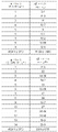
3 is a table of study results for the relative height of cell content.
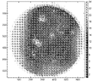
Figures 4a and 4b are 3D focus maps and heat maps, respectively, showing the relative heights of cell content across the sample.
5 is a schematic diagram illustrating a serpentine scan pattern used by a digital imager to scan a specimen slide according to one embodiment.
Fig. 6 is a schematic diagram illustrating the inclination angle of the imager with respect to the slide.
7 is a graph illustrating a focus merging operation.
8A-8C are cross-sectional views of the sample area showing the cellular pathway, the area covered by the volumetric scan, and the area covered by the volumetric scan along the Z curve.
9A and 9B are a sample image obtained without following the Z curve, and a sample image obtained along the Z curve, respectively.
Fig. 10 is a flowchart of a method for obtaining an image of a sample that follows the Z curve.
디지털 전체 슬라이드 이미징(whole slide imaging, WSI) 시스템의 출현은 병리학 또는 세포학 분야에 혁명을 일으키도록 설정되었다. 고품질 전체 슬라이드 이미지를 빠르게 얻을 수 있는 능력은 성공적인 임상적 작업흐름, 특히 자궁경부암 검사(Pap test)와 같은 대용량 검진 적용을 위해 중요한 단계가 될 것이다. 액체- 기반 세포학 슬라이드는 검토자에게 거의 단층을 제공하지만, 세포학은 본질적으로 3차원이다. 이 슬라이드는 고배율 현미경 대문렌즈의 피사계 심도(Depth of Field, DOF)보다 10배 높을 수 있는 밀접하게 병치된 재료의 초점 깊이로 인해 WSI가 어려울수 있다. 이러한 이유로, 세포학 슬라이드는 병리학 조직 슬라이드보다 이미지화하기에 더 어렵다. 필름 커버슬립을 구비한 슬라이드는 슬라이드 세포 스폿 영역을 가로지르는 곡률로 인해 스캐닝 깊이 요건을 추가할 수도 있다. 이처럼, 세포학적 표본에서 관심 대상과 표본이 부착되어 있는 유리 슬라이드 사이의 거리는 현미경 대물렌즈의 DOF에 대해 크게 변경될 수 있다. 대부분의 현재 WSI 시스템은 고품질 이미지를 얻도록 다수의 초점 면을 커버하기 위해 반복된 스캔을 요구하므로, 이미징 시간을 크게 증가시킨다. 이처럼, 세포 슬라이드에 대한 높은 초점 품질 이미지를 효과적으로 얻는 것은 어렵다. 여기에 개시된 것은 고품질 인-포커스 이미지를 얻기 위해 이러한 슬라이드를 효과적으로 스캔하기 위한 시스템 및 방법이다.The advent of digital whole slide imaging (WSI) systems is set to revolutionize the field of pathology or cytology. The ability to quickly obtain high-quality full slide images will be an important step for successful clinical workflows, especially for high-volume screening applications such as Pap tests. While liquid-based cytology slides provide reviewers with almost monolayers, cytology is essentially three-dimensional. This slide can be difficult for WSI due to the depth of focus of closely juxtaposed materials, which can be ten times higher than the depth of field (DOF) of high magnification microscope gate lenses. For this reason, cytology slides are more difficult to image than pathological tissue slides. Slides with film coverslips may add to the scanning depth requirement due to the curvature across the slide cell spot area. As such, in a cytological specimen, the distance between the object of interest and the glass slide to which the specimen is attached can vary significantly with respect to the DOF of the microscope objective. Most current WSI systems require repeated scans to cover multiple focal planes to obtain high quality images, greatly increasing imaging time. As such, it is difficult to effectively obtain high-focus quality images for cell slides. Disclosed herein is a system and method for effectively scanning such slides to obtain high quality in-focus images.
도1을 참조하면, 현미경 글라이드(102)의 예시적인 실시예가 도시되어 있다. 현미경 슬라이드(102)는 슬라이드 식별 영역(112), 표본 영역(114), 및 기준 표시(116)를 갖는 직사각형 유리판(110)(또는 다른 적절한 재료)이다. 현미경 슬라이드(102)는 약 75mm x 25mm, 또는 다른 적절한 크기인 표준 크기의 현미경 슬라이드일 수 있다. 현미경 슬라이드(102)는 슬라이드(102)의 취급 및 배치를 용이하게 하기 위해 경사진 모서리를 가질 수 있다. 표본 영역(114)은 대략 최대 22mm까지의 직경을 갖는 원일 수 있다. 슬라이드 영역(102)상의 전체 표본 영역(114)이 이미지화될 수 있다. 슬라이드 식별 영역(112)은 길이가 대략 최대 25~28 mm까지일 수 있다. 슬라이드 식별 부분(112)은 바코드, ID 번호, 및/또는 다른 정보로 인쇄될 수도 있다. 표본 영역(114)은 유리 슬라이드(102)의 깨끗한 영역으로 남겨진다. 기준 표시(116)는 슬라이드(102)의 위치 및/또는 방향 및 화상촬영기에 대한 그 특징을 결정하기 위해 슬라이드(102)상의 기준 지점으로서 화상촬영기에 의해 사용될 수 있다. 3차원 체적내에 분포된 다수의 물체를 포함하는 표본(119)는 슬라이드(102), 일반적으로 표본 영역(114)내에 부착되지만, 일부 경우에 표존은 표본 영역(114) 외부로 연장될 수 있다. 표본(119)의 3차원 체적은 길이(l), 폭(w) 및 두께 또는 깊이(d)를 갖는다. 두께(d)는 슬라이드(102)의 표면에 대해 z-축을 정의한다. 표본(119)은 물체가 세포인 세포학적 표본, 물체가 조직 구조인 고체 조직 샘플과 같은 임의의 적절한 표본일 수 있다.1 , an exemplary embodiment of a
도1에 도시된 바와 같이, 커버슬립(115)은 표본 영역(114)의 표본(119)을 덮는데 사용될 수 있다. 표본 커버슬립(115)은 커버슬립(115)을 통해 표본(119)의 이미지를 얻기에 충분히 투명하다. 다시 말해서, 커버 슬립(115)은 화상촬영기가 커버슬립(115)을 통해 이미지를 얻는 것을 방해하지 않는다. 커버슬립(115)은 표본을 보존하고 오염으로부터 보호하고 다른 물체를 오염시키는 것을 보호하고, 표본(119)을 편평하게 제 자리에 유지하는 역할을 한다. 커버슬립(115)은 두께(117)를 갖는다. As shown in FIG. 1 , a
도2에 도시된 바와 같이, 유리 슬라이드(110)와 커버슬립(115) 사이에 배치된 표본(119)은 두께(120)를 갖는다. 세포(122)는 다양한 깊이, 또는 z-축 위치에서 표본 층 전체에 분산되어 있다. 표본은 전체 표본에 걸쳐, 표본의 물체(세포(122)와 같은)의 이미지를 얻는데 사용되는 광학계의 피사계 심도를 초과하는 두께를 가질 수 있다. 이는 특히 개별 세포들이 수집되는 액체 기반 세포학적 표본에서 발생할 수 있다. 표본(119)의 상부에 배치된 커버슬립(115)은 유리 또는 플라스틱으로 제조될 수 있으며, 얇은 접착제 층으로 슬라이드(110)에 접착된다. 세포는 접착제 안에서 떠서(float) 유리(110) 위로 올라가게 하는 경향이 있다는 것이 관찰되었다. 또한, 커버슬립(115)은 항상 완벽하게 편평한 것은 아니며, 종종 물결모양(undulation), 둔덕(mounds) 및 골(valley)을 나타낸다. 이는 접착제의 일관성없는 두께를 야기하여 유리 기판(110)으로부터, 따라서 대물 렌즈로부터 세포들의 거리가 변하게 하며, 때로는 피사계 심도 이상을 수용할 수 있다. 여기에 개시된 것은 주어진 시야에서 대물 렌즈의 피사계 심도보다 두꺼운 표본을 계수화(digitize)하는 문제를 해결하는 세포학 슬라이드를 효과적으로 스캔하기 위한 디지털 이미징 방법 및 시스템이다. As shown in FIG. 2 ,
세포는 장착 매체안에 떠있을 수 있고 또한 적층될 수 있기 때문에, 세포학 슬라이드는 본질적으로 3차원이다. 현미경 대물렌즈는 이러한 작은 피사계 심도(DOF)를 갖기 때문에, 세포들은 단일 이미지의 초점에 모두 캡처될 수는 없다. 실제로, 개별 세포들은 단일 DOF보다 두꺼울 수 있다. NA 0.75의 40X 현미경 대물 렌즈는 2마이크론보다 작은 피사계 심도를 갖는다. 가요성 필름(플라스틱) 커버슬립을 구비한 세포학 슬라이드는 세포 스폿 영역에 걸친 곡률로 인해 더 많은 스캐닝(즉, 이미지 획득) 깊이를 필요로 할 수 있다. 세포학 슬라이드(액상세포진(ThinPrep) 슬라이드와 같은)상의 모든 세포의 고품질 이미지를 캡처하기 위해, 더 넓은 범위의 초점이 필요하다.Because cells can float in the mounting medium and can also be stacked, cytology slides are essentially three-dimensional. Because microscope objectives have such a small depth of field (DOF), the cells cannot all be captured in the focus of a single image. Indeed, individual cells may be thicker than a single DOF. A 40X microscope objective of NA 0.75 has a depth of field of less than 2 microns. Cytology slides with flexible film (plastic) coverslips may require more scanning (ie, image acquisition) depth due to the curvature over the cell spot area. To capture high-quality images of all cells on a cytology slide (such as a ThinPrep slide), a wider range of focus is required.
일 예에서, 23개의 ThinPrep Pap 슬라이드는 세포 준비 깊이 데이터를 수집하기 위해 디지털 카메라를 구비한 컴퓨터-제어 현미경(Hologic ThinPrep Integrated Imager)상에서 스캔되었다. 먼저, 세포 스폿 영역은 컴퓨터구동 XY 스테이지를 사용하여 스캔되었다. 각각의 위치에서, Z 높이의 범위 범위(>40 마이크론)에서 다량의 이미지가 캡처되었다. 모든 이미지는 작은 영역(35㎛2)으로 분할되고 브레너 초점 점수 메트릭(Brenner focus score metric)은 Z 스택의 모든 레벨에 대해 평가되었다. 이 조각에 대한 최적의 초점이 결정되었다. 슬라이드상에 인쇄된 기준 표시에 초점을 맞춤으로써, 전체 슬라이드 유리 평면을 결정하고, 초점 데이터로부터 빼서 세포 내용의 상대 높이를 결정하였다.In one example, 23 ThinPrep Pap slides were scanned on a computer-controlled microscope (Hologic ThinPrep Integrated Imager) equipped with a digital camera to collect cell preparation depth data. First, the cell spot area was scanned using a computer-driven XY stage. At each location, a large number of images were captured in a range of Z heights (>40 microns). All images were segmented into small regions (35 μm 2 ) and the Brenner focus score metric was evaluated for all levels of the Z-stack. The optimal focus for this piece was determined. By focusing on a reference mark printed on the slide, the total slide glass plane was determined and subtracted from the focus data to determine the relative height of the cell contents.
도3의 표는 이 23개의 슬라이드로부터 얻어진 데이터를 요약한 것이다. 이 표는 액상세포진(ThinPrep) 슬라이드의 평균 세포 깊이가 유리 커버슬립을 구비한 슬라이드에 대해 11.09마이크론이고 필름 커버슬립을 구비한 슬라이드에 대해 23.6마이크론이었다는 것을 나타낸다. 일부 경우, 세포 깊이는 40마이크론보다 클 수 있다. 유리 버커슬립을 갖더라도, 슬라이드내의 새포 높이의 국부적 변화는 현미경 대물렌즈의 피사계 심도의 7배에 달한다. 각각의 슬라이드에 대한 표면 플롯 초점 지도(surface plot focus map)를 생성하고 검토하였다. 세포 스폿 영역의 범위에 거쳐 곡률의 효과를 나타내는, 필름 커버슬립 슬라이드에 대한 도4a 및 4b에 일 예가 도시되어 있다. 특히, 도4a 및 4b에 도시된 예에 대해, 표본의 중간 부분의 세포들은 유리 슬라이드에 더 가깝지만, 표본의 가장자리 둘레의 세포들은 커버슬립에 더 가깝다. The table of Figure 3 summarizes the data obtained from these 23 slides. This table shows that the average cell depth of the liquid cytology (ThinPrep) slides was 11.09 microns for slides with glass coverslips and 23.6 microns for slides with film coverslips. In some cases, the cell depth may be greater than 40 microns. Even with a glass buckler slip, the local variation in cell height within the slide amounts to 7 times the depth of field of the microscope objective. A surface plot focus map was generated and reviewed for each slide. An example is shown in Figures 4a and 4b for a film coverslip slide, demonstrating the effect of curvature over a range of cell spot areas. In particular, for the example shown in Figures 4a and 4b, the cells in the middle part of the specimen are closer to the glass slide, whereas the cells around the edge of the specimen are closer to the coverslip.
도5에 도시된 바와 같이, 이미징동안, 슬라이드(102)는 XY 슬라이드 스테이지에 의해 이동하여 전체 표본 영역(114)(또는 전체 실제 표본(119)의 미리결정된 영역, 예를 들어, 실제 표본(119)이 표본 영역(114)과 상이한 영역을 다루고 실제 표본(119)의 경계가 미리 결정된 경우)에 걸쳐 카메라의 시야를 스캔한다. XY 슬라이드 스테이지는 슬라이드(102)를 전후의 구불구불한 경로로 이동하여 각각의 통과시 표본의 잘린자리(swath), 열 또는 선형 부분의 마이크로 이미지를 캡처한다. 잘린자리를 캡처하기 위해, XY 슬라이드 스테이지는 슬라이드(102)를 연속적으로 이동하고 카메라는 스테이지 인코더 위치에 따라 각각의 트리거 지점에 도달할 때 이미지를 캡처하기 위해 XY 슬라이드 스테이지에 의해 트리거된다. 초고속 카메라가 사용되어 슬라이드와 카메라 사이의 상대적 움직임은 연속적이다. 이처럼 고속에서, 슬라이드의 선형 부분을 따라 이미지를 얻는 것은 행의 "스캐닝"으로 불릴 수 있다. 각각의 연속적인 잘린자리의 시작이 이전의 잘린자리의 끝과 가깝도록, 구불구불한 경로가 전체 표본(119)를 스캔하는데 필요한 시간을 최소화하는데 사용된다. 슬라이드(112)가 잘린자리를 따라 이동함에 따라, 카메라는 표본(119)의 마이크로 이미지를 캡처한다. 즉, 화상 촬영기(imager)는 도5에 도시된 것처럼, 스캔 패턴에 따라 전체 표본 영역을 다루는 다수의 이미지를 캡처한다. 스캔 패턴은 도5의 수평 화살표에 의해 표시된 다수의 선형 부분을 포함한다. 표본은 도5의 사각형(132)에 의해 표시된 다수의 초점 구역으로 분할된다. 이처럼, 이미지가 얻어지는 각각의 선형 부분(130)은 다수의 초점 구역(132)을 포함한다. 각각의 선형 부분(130)은 30-70개의 초점 구역을 포함한다. 다른 예에서, 슬라이드(102)가 이동하는 것이 아니라, 슬라이드(102)가 정지상태로 유지되는 동안 카메라는 스캔 패턴을 따라 이동한다. 슬라이드(102)가 이동하였는지 또는 카메라가 이동하였는지 여부에 상관없이, 슬라이드(102)는 카메라에 대해 이동한다.5 , during imaging, the
대부분의 현재 WSI 시스템은 한번에 단일 초점 평면을 스캔한다. 일반적인 스캐너는 1분에 15 x 15mm 스캔을 완료한다. 이 속도로, 14개의 초점 표면의 깊이까지 원형 ThinPrep 세포 스폿 영역을 스캔하는 것은 적어도 26분이 걸린다. 슬라이드 디지타이저의 처리량을 극적으로 증가시키기 위해, 일부 시스템은 대물 렌즈 및 유리 슬라이드 사이의 경사 각도로 설계된다. 이 접근 방식을 사용하여, 각각 약간 상이한 깊이에서 별개의 층을 동시에 계수화하는데 피사계 심도가 사용될 수 있다. 그런 다음, 층들은 초점 병합(focus merging)이라고 불리는 작업에서 단일 합성 초점 층으로 축소될 수 있으며, 초점 병합은 최상의 초점을 나타내는 층(또는 층의 부분)을 선택하고 이 초점이 잘맞는 영역을 함께 연결하는 것을 포함한다. 이 접근방식을 사용하여, 초점을 맞추기 위해 멈추지 않고 대물 렌즈아래에서 슬라이드가 연속적으로 이동할 수 있다. 다양한 층이 계수화되고 병합되며, 기본적으로 획득후 초점조정(post-acquisition focusing)을 수행하는 시스템이 구축될 수 있다. 이러한 시스템에 대한 유일한 제한이 대물렌즈의 피사계 심도이면, 이는 다시 이미지화될 수 있는 표본의 최대 두께로 변환된다. 이러한 시스템의 분명한 이점은 정지 및 초점조정(focusing)이 필요하지 않다는 것이다.Most current WSI systems scan a single focal plane at a time. A typical scanner completes 15 x 15mm scans in 1 minute. At this rate, scanning a circular ThinPrep cell spot area to the depth of 14 focal surfaces takes at least 26 minutes. To dramatically increase the throughput of a slide digitizer, some systems are designed with an angle of inclination between the objective lens and the glass slide. Using this approach, the depth of field can be used to simultaneously count distinct layers, each at slightly different depths. The layers can then be reduced to a single composite focus layer in an operation called focus merging, which selects the layer (or part of the layer) that exhibits the best focus and combines these well-focused areas together. includes connecting. Using this approach, the slide can be moved continuously under the objective lens without stopping to focus. Various layers are counted and merged, and a system that basically performs post-acquisition focusing can be built. If the only limitation for such a system is the depth of field of the objective, this translates back to the maximum thickness of the specimen that can be imaged. A clear advantage of such a system is that no stopping and focusing are required.
경사-평면 체적 스캐닝 방법을 사용하면 전체 세포 콘텐츠 영역을 스캐닝하기 위한 획득 시간을 상당히 감소시킨다. ThinPrep Pap 슬라이드는 대략 2.5분안에 완료될 수 있다. 도6에 도시된 바와 같이, 이미지화 광학계 및 카메라(202)는 슬라이드(102)에 대해 경사져 있다. 카메라 프레임의 한쪽 가장자리에 있는 이미지의 구역은 카메라 프레임의 다른 가장자리에 있는 구역보다 슬라이드 유리에 더 가까운 이미지를 얻는다. 일 예에서, 48밀리라디안의 경사각 및 0.5mm의 슬라이드에서의 이미지 프레임은 24마이크론인 0.5 x sin(0.048)의 스캔 깊이를 제공한다.Using the oblique-plane volumetric scanning method significantly reduces the acquisition time for scanning the entire cell content area. ThinPrep Pap slides can be completed in approximately 2.5 minutes. As shown in FIG. 6 , the imaging optics and
도6에 가장 잘 도시된 바와 같이, 대물렌즈의 광학축(204)은 스캐닝 방향에서 슬라이드(102)의 평면의 직교에 대해 경사각(206)으로 경사져있다(도5 참조). 다시 말해서, 슬라이드 표면에서의 카메라 및 광학계의 결과적인 광학축(204)은 슬라이드(102)의 평면에 직교하지 않는다. WO2020/091965A2에 더욱 상세히 설명된 바와 같이, 경사각(206)은 슬라이드(102)상의 표본(119)의 체적 이미지를 화상촬영기가 얻도록 허용한다(즉, 표본(119)의 깊이까지 연장되는 이미지). 다시 말해서, 마이크로 이미지는 표본(119)의 직교 각도에서 촬영된 이미지의 경우 초점의 유일한 단일 평면대신에, 슬라이드(102)상의 표본(109)의 상이한 깊이에 있는 특징들의 초점이맞춰진(in-focus) 이미지를 포함한다. 이미징 스테이션은 각각의 마이크로 이미지가 슬라이드(102)의 표면 아래에 놓이는 슬라이드(102)의 깊이의 적어도 일부분을 포함하는 마이크로 이미지를 얻도록 구성될 수 있다. 커버슬립(115)이 슬라이드(102)상에 사용될 경우, 이미징 스테이션은 각각의 마이크로 이미지가 커버슬립(115)의 깊이의 적어도 일부분을 포함하는 마이크로 이미지를 얻도록 구성될 수 있다.As best shown in Fig. 6, the
카메라가 연속적으로 이동하면, 카메라가 자신의 폭의 1/14만큼 이동할때마다 새로운 이미지를 얻도록 트리거된다. 초고속(>100fps) 카메라가 사용된다. 이 중첩 이미지들은 도7에 도시된 바와 같이, 그리고 WO2020/091965에 더욱 상세히 설명된 바와 같이, 14개의 초점이 맞춰진 평면 이미지를 얻도록 얇게잘리고(sliced) 재조립된다. 저장 공간을 최적화하기 위해, 초점 평면은 다양한 평면으로부터 초점이 맞춰진 픽셀을 선택함으로써 단일 확장된 피사계 심도 이미지로 조합된다. 이미지 처리는 GPU 하드웨어 가속을 사용하여 실시간으로 이뤄진다.As the camera moves continuously, it is triggered to acquire a new image every time the camera moves 1/14 of its width. A high-speed (>100fps) camera is used. These superimposed images are sliced and reassembled to obtain 14 focused planar images, as shown in Figure 7, and as described in more detail in WO2020/091965. To optimize storage space, focal planes are combined into a single expanded depth-of-field image by selecting focused pixels from various planes. Image processing is done in real time using GPU hardware acceleration.
커버슬립은 완벽하게 편평하지는 않지만, 평탄도의 변화는 본질적으로 점진적이다. 이는 커버슬립이 슬라이드의 중앙에 혹(hump)을 형성하거나, 슬라이드 표면을 가로지르는 다수의 물결무늬(wave)로 인해 형성될 수 있지만, 커버슬립과 아래에 있는 유리사이의 거리의 급격한 변화는 없다는 것을 의미한다. 예를 들어, 완벽하게 편평한 커버슬립(115)은 도8a에 도시되어 있지만, 물결모양 또는 울툴불퉁한 표면을 갖는 커버슬립은 도8b 및 8c에 도시되어 있다.Although the coverslip is not perfectly flat, the change in flatness is gradual in nature. This can be caused by the coverslip forming a hump in the center of the slide, or by multiple waves across the slide surface, but there is no drastic change in the distance between the coverslip and the underlying glass. means that For example, a perfectly
이 느리게-변화하는 갭은 획득후 초점조정된 이미징 시스템이 광학계 단독에 의해 제공되는 것보다 훨씬 더 유효한 피사계 심도를 갖도록 한다. 기본적으로, 슬라이드가 스캔될때, 일반적으로 래스터 전-후 패턴(raster back-and-forth pattern, 도5에 도시된)을 사용하여, 각각의 통과(pass)의 끝에서(다음 행 또는 잘린자리를 조사하기 위해 스캔의 방향을 반전시키기 전에), 수집된 층은 분석되고 최적의 초점이 초점 병합 작업도중에 결정된다. 물결모양의 커버슬립을 따라, 생물학적 재료가 위로 표류하면, 최상의 초점이 이전의 잘린자리에서 발견되는 층은 스캔될 다음의 잘린자리에 대한 최상의 초점 패턴을 예측하는데 사용될 수 있다. 관심있는 개별 물체는 초점 병합 도중에 발견되고 그 깊이가 기록된다. 그런 다음 각각의 행은 다음 행에 대한 최상의 초점을 예측하는데 사용된다.This slow-changing gap allows the post-acquisition focused imaging system to have a much more effective depth of field than provided by the optics alone. Basically, when a slide is scanned, at the end of each pass (using a raster back-and-forth pattern (shown in Figure 5)) Before reversing the direction of the scan to investigate), the collected layers are analyzed and the optimal focus is determined during the focus merging operation. As the biological material drifts up along the wavy coverslip, the layer in which the best focus is found in the previous clip can be used to predict the best focus pattern for the next clip to be scanned. Individual objects of interest are found during focal merging and their depths recorded. Each row is then used to predict the best focus for the next row.
잘린 자리의 단부에서 초점을 벗어난(out-of focus) 구역을 검출할 때, 잘린 자리는 초점을 벗어난 구역이 현재 초점에 있는 다른 높이에서 재스캔될 수 있다. 그런다음 결과적인 초점 높이는 다음 잘린 자리를 알려주는데 사용되며, 이는 다시 잘린 자리를 재스캔해야하는 횟수를 최소화한다.Upon detecting an out-of focus area at the end of the clipping site, the clipping site may be rescanned at a different height at which the out-of-focus area is currently in focus. The resulting focal height is then used to signal the next clipping, which minimizes the number of times the clippings have to be rescanned again.
이러한 시스템의 장점은 정지하여 초점을 맞출 필요가 없고, 종종 초점이 맞지 않는 구역으로 다시 돌아가 재스캔해야할 필요도 없다는 사실로 인해 훨씬 높은 처리량(더 낮은 스캔 시간)이 달성될 수 있다는 것이다. 대신에 시스템은 전체 표본이 계수화(및 초점 병합)될때까지 단순히 앞 뒤로 스캔한다. 횡단하는 동안, 대물 렌즈는 이전의 잘린 자리로부터 주어진 초점 지도에 따라 위 아래로 구동되며, 유리 슬라이드 표면으로부터 그들의 변화하는 거리에도 불구하고 모두 초점이 맞도록 피사계 심도내에서 관심 대상을 유지하도록 모두 설계되었다. The advantage of such a system is that much higher throughput (lower scan times) can be achieved due to the fact that there is no need to stop and focus, and often no need to go back and rescan the out of focus area. Instead, the system simply scans back and forth until the entire sample has been digitized (and focus merged). While traversing, the objective is driven up and down according to a given focus map from the previous clipping site, all designed to keep the object of interest within the depth of field so that they are all in focus despite their varying distances from the glass slide surface. became
필름 커버 슬립을 구비한 슬라이드상에서 발견된 더 큰 전체 세포 깊이 범위를 처리하기 위해, 이미징 광학계는 도8a 내지 8c에 도시된 바와 같이, 곡률을 따르도록 Z 축에서 구동될 수 있다. 세포 깊이(802)는 물결 모양이며 일반적으로 경사져 있다. 전술된 바와 같이 경사진 카메라를 사용하는 체적 스캔은 단일 깊이의 초점에서 이루어진 스캔보다 더 두꺼운 스캔된 구역(804)를 제공하지만, 세포 경로(802)의 하단 부분(806) 및 상단 부분(808)의 초점이 맞는(in-focus) 이미지를 제공하는데 실패한다. 810을 따르는 Z 곡선을 사용한 체적 스캔은 표본의 전체 세포 경로(802)를 캡처한다. 카메라 시야의 크기의 영역에서 국부적 세포 깊이는 경사진-평면 스캠 깊이내에 존재하지만, 더 먼 거리에서는 초점깊이의 더 큰 변화가 필요할 수 있다.To handle the larger total cell depth range found on slides with film coverslips, the imaging optics can be driven in the Z-axis to follow the curvature, as shown in Figures 8a-8c.
곡선-추종 스캐닝 방법은 국소적 초점 오류를 최소화하여, 더 높은 품질의 WSI 이미지를 제공한다. 도9a 및 9b의 이미지를 생성하기 위해 사용되는 슬라이드는 40마이크론 이상의 Z 초점 깊이를 갖는다. Z 곡선을 추종하지 않는 체적 스캔은 도9a에 도시되어 있다. Z 곡선을 추종하는 체적 스캔은 도9b에 도시되어 있다. 도9b의 이미지는 도9a의 이미지보다 더 선명하고 더 초점이 맞는다. The curve-following scanning method minimizes local focus errors, providing higher quality WSI images. The slides used to create the images of FIGS. 9A and 9B have a Z focal depth of 40 microns or greater. A volume scan that does not follow the Z curve is shown in Figure 9a. A volume scan following the Z curve is shown in Fig. 9b. The image of FIG. 9B is sharper and more focused than the image of FIG. 9A.
이제 도10을 살펴보면, 슬라이드의 표면에 부착된 표본내에 분포된 물체의 이미지를 얻기 위한 방법(500)이 설명될 것이다. 특히, 이미지는 슬라이드 표면에 대해 고르지않은 높이를 갖고 광학계의 피사계 심도를 초과하는 두께를 갖는 표본에 대해 Z 곡선을 추종하여 체적 스캔을 사용함으로써 얻어진다. 방법(500)의 제1 단계(502)에서, 슬라이드 표면상의 분포 위치는 초기 초점 높이(Z)를 결정하기 위해 샘플화된다. 다음으로, 단계(504)에서, 제1 선형 부분의 각각의 초점 구역에 대해 단계(502)에서 결정된 초기 초점 높이를 사용하여 표본의 제1 선형 부분을 따라 복수의 이미지가 얻어진다. 초기 초점 높이는 제1 선형 부분을 따라 일정하게 유지되는데, 이는 슬라이드 표면에 대한 대물 렌즈의 높이가 단계(504)에서 표본의 제1 선형 부분에서 이미지를 얻는 동안 실질적으로 일정하다는 것을 의미한다. 표본의 각각의 행, 또는 선형 부분은 다수의 초점 구역을 포함한다.Turning now to Figure 10, a
예를 들어, 도5를 참조하면, 선형 부분을 따르는 각각의 정사각형은 초점 구역일 수 있다. 화상촬영기에 의해 스캔될 각각의 선형 부분은 예를 들어 30-70개의 초점 구역을 포함할 수 있다. 대안적으로, 단계(504)에서처럼 실질적으로 일정한 초점 높이를 사용하는 대신, 단계(502)에서 샘플링된 분포 위치가 사용되어 제1 선형 부분에 대한 Z 곡선을 결정하고, 제1 선형 부분을 따르는 이미지는 Z 곡선을 추종함으로써 얻어질 수 있다. 이처럼, 제1 선형 부분을 따라 이미지를 얻는 동안, 대물 렌즈는 Z 곡선에 따라 슬라이드에 대해 Z-축을 따라 위아래로 이동한다.For example, referring to Figure 5, each square along the linear portion may be a focal zone. Each linear portion to be scanned by the imager may comprise, for example, 30-70 focal zones. Alternatively, instead of using a substantially constant focal height as in
다음으로, 단계(506)에서, 방금 스캔된 선형 부분의 각각의 초점 구역에 대해, 최상의 초점은 이미지안에 캡처된 물체가 초점이 맞는지 여부를 평가함으로써 결정된다. 평가는 초점이 맞지 않는 물체가 대물 렌즈의 초점이 맞는 범위를 벗어난 슬라이드 표면에 대한 높이에서 표본안에 위치하는지 여부를 결정하는 것을 포함한다. 평가는 또한 표본의 선형 부분을 따라 그들의 상대 위치를 기반으로 초점을 벗어난 물체를 식별하는 것을 포함할 수 있다. 즉, 선형 부분을 따르는 각각의 초점 구역에 대해, 이미지는 어느 초점 평면이 최상 초점을 갖는지를 결정하기 위해 평가된다. 도7에 도시된 바와 같이, 경사 각도 이미지 획득을 사용하여, 이미지는 12개의 초점 평면에서 한번에 캡처된다. 예를 들어, 최상 초점이 최상단 또는 최하단 초점 평면(초점 평면 1 또는 12)에 존재하는 것으로 발견되면, 슬라이드에 대한 대물 렌즈의 Z-위치는 더 우수한 초점을 가진 이미지를 얻기 위해 각각 위아래로 이동할 필요가 있다. 또한, 최상 초점이 최상단 또는 최하단 초점 평면에 존재하는 것으로 발견되면, 표본의 다음의 인접한 선형 부분에서 이미지를 얻는 동안, 슬라이드에 대한 대물 렌즈의 Z-축 위치는 각각 위 아래로 이동할 필요가 있다.Next, in
너무 많은 물체가 초점을 벗어나 있는 경우(즉, 초점을 벗어난 물체의 수가 미리결정된 임계수를 초과할 경우), 초점을 벗어난 각각의 물체에 대한 초점 높이(Z)는 조정되고 선형 부분은 단계(508)에서 새로운 초점 높이에 따라 재스캔된다. 카메라가 초점 높이 곡선을 따라갈때, 슬라이드 표면에 대한 대물 렌즈의 z-축 위치는 표본의 선형 부분을 따라 카메라의 상대 위치의 함수로서 슬라이드 표면에 대한 카메라의 높이를 증가/감소시킴으로써 변경된다. 슬라이드에 대한 대물 렌즈의 z-축 위치는 표본의 선형 부분을 따라 한 초점으로부터 다음 초점까지 변경될 수 있다. 일 실시예에서, 슬라이드(110)는 대물 렌즈의 z-축 위치가 고정된채로 유지되는 동안 위아래로 이동한다. 대안적인 실시예에서, 대물 렌즈는 슬라이드의 z-축 위치가 일정하게 유지되는 동안 위아래로 이동한다.If too many objects are out of focus (i.e., the number of out-of-focus objects exceeds a predetermined threshold number), the focal height Z for each out-of-focus object is adjusted and the linear portion is
초점 구역의 대부분, 또는 전부가 초점이 맞으면, 단계(510)에서, 각각의 초점 구역에 대한 다음 값이 현재 행의 각각의 초점 구역에 대한 최상의 초점을 기반으로 계산된다. 다음 행에 대한 초점 구역 중 일부는 커버슬립에 더 가까운 관심 대상의 물체를 추적하기 위해 위로 이동하지만(즉, z-위치가 커버슬립에 더 가깝고 유리 슬라이드로부터 멀어지도록), 초점 구역의 일부는 유리 슬라이드에 더욱 가까운 관심 대상을 추적하기 위해 다음 행에 대해 아래로 이동한다. 다음으로, 단계(512)에서, 다음 선형 부분은 단계(510)에서 계산된 초점 곡선을 사용하여 스캔된다. 단계(506-512)는 전체 표본의 이미지가 얻어질때까지 표본의 각각의 선형 부분에 대해 반복된다. 즉, 표본의 각각의 선형 부분에 대해 이미지가 얻어진 후, 이 이미지들의 물체의 초점이 평가된다(단계 506). 평가를 기반으로, 표본의 다음 선형 부분에 대한 초점 곡선이 결정되고(단계 510), 방금 스캔된 선형 부분에 바로 인접한 다음 선형 부분의 물체의 이미지를 얻는데(단계 512) 사용된다. 대안적으로, 너무 많은 물체가 초점을 벗어나있는 것으로 초점 평가가 밝혀지면, 초점 곡선이 조정되고, 동일한 선형 부분은 조정된 초점 곡선을 사용하여 다시 스캔된다(단계 508).If most or all of the focus zones are in focus, in step 510 a next value for each focus zone is calculated based on the best focus for each focus zone in the current row. For the next row, some of the focus areas are moved up to track the object of interest closer to the coverslip (i.e., the z-position is closer to the coverslip and away from the glass slide), but some of the focus areas are glass Move down to the next row to track the interest closer to the slide. Next, in
특정 실시예가 도시되고 설명되었지만, 상기 설명은 이 실시예의 범주를 제한하기 위한 것이 아니며, 이러한 개시는 오직 설명 및 예시를 목적으로 하여 제공되었다는 것을 이해해야 한다. 따라서 이하의 청구범위의 범주를 벗어나지 않고 개시된 실시예에 대한 다양한 변경 및 수정이 이뤄질 수 있다.While specific embodiments have been shown and described, it is to be understood that the foregoing description is not intended to limit the scope of these embodiments, and this disclosure has been provided for purposes of illustration and description only. Accordingly, various changes and modifications may be made to the disclosed embodiments without departing from the scope of the following claims.
Claims (35)
상기 방법은:
표본의 제1 선형 부분의 제1 다수의 이미지를 얻는 단계;
제1 다수의 이미지에서 캡처된 표본의 선형 부분내의 물체의 초점을 평가하는 단계; 및
제1 선형 부분, 또는 제1 선형 부분과 상이한 표본의 제2 선형 부분의 제2 다수의 이미지를 얻는 단계를 포함하고, 제1 다수의 이미지에서 캡처된 물체의 평가된 초점을 기반으로 제2 다수의 이미지를 얻는 동안 슬라이드 표면에 대한 대물 렌즈의 높이가 변경되는
표본내에 분포된 물체의 이미지를 얻는 방법.A method of obtaining an image of an object attached to the surface of a slide and distributed in a specimen having an uneven height with respect to the surface of the slide, using a camera having an objective lens having an optical axis forming an angle that is not perpendicular to the surface of the slide ,
The method is:
obtaining a first plurality of images of a first linear portion of the sample;
assessing a focus of an object within a linear portion of the sample captured in the first plurality of images; and
obtaining a second plurality of images of the first linear portion, or a second linear portion of the sample different from the first linear portion, wherein the second plurality of images are based on the estimated focus of the object captured in the first plurality of images. The height of the objective lens with respect to the slide surface is changed while acquiring an image of
A method of obtaining an image of an object distributed within a sample.
슬라이드 표면에 대한 대물 렌즈의 높이는 제1 다수의 이미지를 얻는 동안 실질적으로 일정한
표본내에 분포된 물체의 이미지를 얻는 방법.According to claim 1,
The height of the objective lens relative to the slide surface is substantially constant during acquisition of the first plurality of images.
A method of obtaining an image of an object distributed within a sample.
제1 다수의 이미지에서 캡처된 물체의 초점을 평가하는 단계는 초점을 벗어난 물체의 총수가 임계수를 초과하는지 여부를 결정하는 단계를 포함하는
표본내에 분포된 물체의 이미지를 얻는 방법.3. The method of claim 1 or 2,
Evaluating the focus of the captured object in the first plurality of images includes determining whether a total number of out-of-focus objects exceeds a threshold number.
A method of obtaining an image of an object distributed within a sample.
제1 다수의 이미지에서 캡처된 물체의 초점을 평가하는 단계는 슬라이드 표면에 대해 초점을 벗어난 물체의 각각의 높이를 결정하는 단계를 포함하는
표본내에 분포된 물체의 이미지를 얻는 방법.4. The method of claim 1 or 3,
Evaluating the focus of the captured object in the first plurality of images comprises determining a respective height of the out-of-focus object relative to the slide surface.
A method of obtaining an image of an object distributed within a sample.
제1 다수의 이미지의 물체의 초점을 평가하는 단계는 제1 다수의 이미지를 얻는 동안 물체의 초점이 맞는 범위 밖에 존재하는 슬라이드 표면에 대한 높이에 각각의 초점을 벗어난 물체가 위치하는지 여부를 결정하는 단계를 포함하는
표본내에 분포된 물체의 이미지를 얻는 방법.5. The method of claim 4,
The step of evaluating the focus of the object of the first plurality of images includes determining whether each out-of-focus object is located at a height relative to the slide surface that is outside the in-focus range of the object during the acquisition of the first plurality of images. step containing
A method of obtaining an image of an object distributed within a sample.
제1 다수의 이미지의 물체의 초점을 평가하는 단계는 제1 다수의 이미지를 얻는 동안 대물 렌즈의 초점이 맞는 범위의 최대 높이보다 높거나 최소 높이보다 낮은 슬라이드 표면에 대한 높이에 각각의 초점을 벗어난 물체가 각각 위치하는지 여부를 결정하는 단계를 포함하는
표본내에 분포된 물체의 이미지를 얻는 방법.6. The method according to claim 4 or 5,
Evaluating the focus of the object in the first plurality of images includes each out-of-focus height relative to the slide surface greater than or less than the minimum height of the in-focus range of the objective lens during acquisition of the first plurality of images. determining whether the objects are each located
A method of obtaining an image of an object distributed within a sample.
제1 다수의 이미지의 물체의 초점을 평가하는 단계는 제1 선형 부분내의 초점을 벗어난 물체의 각각의 위치를 결정하는 단계를 포함하는
표본내에 분포된 물체의 이미지를 얻는 방법.7. The method according to any one of claims 1 to 6,
Evaluating the focus of the object in the first plurality of images includes determining a respective position of the out-of-focus object within the first linear portion.
A method of obtaining an image of an object distributed within a sample.
카메라 및 슬라이드 중 하나 또는 둘 모두는 이미지를 얻는 동안 서로에 대해 측방향으로 이동하는
표본내에 분포된 물체의 이미지를 얻는 방법.8. The method according to any one of claims 1 to 7,
One or both of the camera and slide move laterally relative to each other during image acquisition.
A method of obtaining an image of an object distributed within a sample.
슬라이드 표면에 대한 대물 렌즈의 높이는 각각의 선형 부분의 길이방향 위치에 대한 카메라의 선형 위치의 함수로서 카메라에 대해 슬라이드 표면의 높이를 증가 및/또는 감소시킴으로써 변경되는
표본내에 분포된 물체의 이미지를 얻는 방법.9. The method of claim 8,
The height of the objective lens relative to the slide surface is varied by increasing and/or decreasing the height of the slide surface relative to the camera as a function of the linear position of the camera relative to the longitudinal position of each linear part.
A method of obtaining an image of an object distributed within a sample.
슬라이드 표면에 대한 대물 렌즈의 높이는 제1 또는 제2 선형 부분 각각의 길이방향 위치에 대한 카메라의 선형 위치의 함수로서 슬라이드 표면에 대해 카메라의 높이를 증가 및/또는 감소시킴으로써 변경되는
표본내에 분포된 물체의 이미지를 얻는 방법.9. The method of claim 8,
The height of the objective lens relative to the slide surface is varied by increasing and/or decreasing the height of the camera relative to the slide surface as a function of the linear position of the camera relative to the longitudinal position of each of the first or second linear portions.
A method of obtaining an image of an object distributed within a sample.
제2 다수의 이미지는 제2 선형 부분에서 얻어지는
표본내에 분포된 물체의 이미지를 얻는 방법.11. The method according to any one of claims 1 to 10,
The second plurality of images is obtained from the second linear portion.
A method of obtaining an image of an object distributed within a sample.
제2 다수의 이미지는 제2 선형 부분에서 얻어지고,
상기 방법은,
제2 다수의 이미지에서 캡처된 물체의 초점을 평가하는 단계; 및
제2 선형 부분의 제3 다수의 이미지를 얻는 단계를 추가로 포함하고,
슬라이드 표면에 대한 대물 렌즈의 높이는 제2 다수의 이미지에서 캡처된 물체의 평가된 초점을 기반으로 제3 다수의 이미지를 얻는 동안 변경되는
표본내에 분포된 물체의 이미지를 얻는 방법.11. The method according to any one of claims 1 to 10,
a second plurality of images are obtained from a second linear portion,
The method is
evaluating the focus of the captured object in the second plurality of images; and
further comprising obtaining a third plurality of images of the second linear portion;
The height of the objective lens relative to the slide surface is changed during acquisition of the third plurality of images based on the estimated focus of the object captured in the second plurality of images.
A method of obtaining an image of an object distributed within a sample.
제2 선형 부분은 제1 선형 부분에 바로 인접한
표본내에 분포된 물체의 이미지를 얻는 방법.13. The method according to any one of claims 1 to 12,
The second linear portion is immediately adjacent to the first linear portion.
A method of obtaining an image of an object distributed within a sample.
제1 다수의 이미지의 초점을 벗어난 물체의 총수가 임계수를 초과하는 경우 제1 선형 부분의 제2 다수의 이미지를 얻는 단계; 및
제1 다수의 이미지의 초점을 벗어난 물체의 총수가 임계수를 초과하지 않는 경우 제2 선형 부분의 제2 다수의 이미지를 얻는 단계를 추가로 포함하는
표본내에 분포된 물체의 이미지를 얻는 방법.4. The method of claim 3,
obtaining a second plurality of images of the first linear portion if the total number of out-of-focus objects in the first plurality of images exceeds a threshold number; and
obtaining a second plurality of images of the second linear portion if the total number of out-of-focus objects in the first plurality of images does not exceed the threshold number
A method of obtaining an image of an object distributed within a sample.
상기 방법은:
(a) 표본의 선형 부분의 제1 다수의 이미지를 얻는 단계;
(b) 제1 다수의 이미지에서 캡처된 물체의 초점을 평가하는 단계; 및
(c) 표본의 동일하거나 상이한 선형 부분의 제2 다수의 이미지를 얻는 단계로서, 제1 다수의 이미지에서 캡처된 물체의 평가된 초점을 기반으로 제2 다수의 이미지를 얻는 동안 슬라이드 표면에 대한 대물 렌즈의 높이가 변경되는, 단계; 및
(d) 실질적으로 전페 표본의 이미지가 얻어질때까지 단계 (a) 내지 (c)를 반복하는 단계를 포함하는
표본내에 분포된 물체의 이미지를 얻는 방법.A method of obtaining an image of an object attached to the surface of a slide and distributed in a specimen having an uneven height with respect to the surface of the slide, using a camera having an objective lens having an optical axis forming an angle that is not perpendicular to the surface of the slide ,
The method is:
(a) obtaining a first plurality of images of a linear portion of the specimen;
(b) evaluating the focus of the captured object in the first plurality of images; and
(c) obtaining a second plurality of images of the same or different linear portion of the specimen, the objective relative to the slide surface while obtaining a second plurality of images based on the estimated focus of the object captured in the first plurality of images the height of the lens is changed; and
(d) repeating steps (a) to (c) until an image of a substantially whole specimen is obtained.
A method of obtaining an image of an object distributed within a sample.
제1 다수의 이미지에서 캡처된 물체의 초점을 평가하는 단계는 초점을 벗어난 물체의 총수가 임계수를 초과하는지 여부를 결정하는 단계를 포함하는
표본내에 분포된 물체의 이미지를 얻는 방법.16. The method of claim 15,
Evaluating the focus of the captured object in the first plurality of images includes determining whether a total number of out-of-focus objects exceeds a threshold number.
A method of obtaining an image of an object distributed within a sample.
제1 다수의 이미지의 물체의 초점을 평가하는 단계는 제1 다수의 이미지를 얻는 동안 대물 렌즈의 초점이 맞는 범위의 최대 높이보다 높거나 최소 높이보다 낮은 슬라이드 표면에 대한 높이에 각각의 초점을 벗어난 물체가 각각 위치하는지 여부를 결정하는 단계를 포함하는
표본내에 분포된 물체의 이미지를 얻는 방법.17. The method of claim 15 or 16,
Evaluating the focus of the object in the first plurality of images includes each out-of-focus height relative to the slide surface greater than or less than the minimum height of the in-focus range of the objective lens during acquisition of the first plurality of images. determining whether the objects are each located
A method of obtaining an image of an object distributed within a sample.
제1 다수의 이미지의 물체의 초점을 평가하는 단계는 각각의 선형 부분내의 초점을 벗어난 물체의 각각의 위치를 결정하는 단계를 포함하는
표본내에 분포된 물체의 이미지를 얻는 방법.18. The method according to any one of claims 15 to 17,
Evaluating the focus of the object of the first plurality of images includes determining a respective position of the out-of-focus object within each linear portion.
A method of obtaining an image of an object distributed within a sample.
카메라 및 슬라이드 중 하나는 각각의 제1 및 제2 다수의 이미지를 얻는 동안 서로에 대해 측방향으로 이동하는
표본내에 분포된 물체의 이미지를 얻는 방법.19. The method according to any one of claims 15 to 18,
One of the cameras and slides moves laterally with respect to each other while acquiring the respective first and second plurality of images.
A method of obtaining an image of an object distributed within a sample.
슬라이드 표면에 대한 대물 렌즈의 높이는 각각의 선형 부분의 길이방향 위치에 대한 카메라의 선형 위치의 함수로서 카메라에 대해 슬라이드 표면의 높이를 증가 및/또는 감소시킴으로써 변경되는
표본내에 분포된 물체의 이미지를 얻는 방법.20. The method of claim 19,
The height of the objective lens relative to the slide surface is varied by increasing and/or decreasing the height of the slide surface relative to the camera as a function of the linear position of the camera relative to the longitudinal position of each linear part.
A method of obtaining an image of an object distributed within a sample.
슬라이드 표면에 대한 대물 렌즈의 높이는 각각의 선형 부분 길이방향 위치에 대한 카메라의 선형 위치의 함수로서 슬라이드 표면애 대해 카메라의 높이를 증가 및/또는 감소시킴으로써 변경되는
표본내에 분포된 물체의 이미지를 얻는 방법.20. The method of claim 19,
The height of the objective lens relative to the slide surface is varied by increasing and/or decreasing the height of the camera relative to the slide surface as a function of the linear position of the camera for each linear partial longitudinal position.
A method of obtaining an image of an object distributed within a sample.
제2 다수의 이미지는 상이한 선형 부분에서 얻어지고,
상이한 선형 부분은 제1 다수의 이미지가 얻어진 선형 부분에 바로 인접한
표본내에 분포된 물체의 이미지를 얻는 방법.22. The method according to any one of claims 15 to 21,
a second plurality of images are obtained from different linear parts,
The different linear portion is immediately adjacent to the linear portion from which the first plurality of images were obtained.
A method of obtaining an image of an object distributed within a sample.
상기 시스템은,
하나의 광축을 구비한 대물 렌즈를 갖는 카메라로서, 카메라는 광축이 슬라이드의 표면과 직교하지 않는 각도를 형성하도록 위치되는, 카메라; 및
카메라와 동작가능하게 결합된 이미지 프로세서를 포함하고,
이미지 프로세서는,
카메라에 의해 얻어진 표본의 제1 선형 부분의 제1 다수의 이미지를 수신하고;
제1 다수의 이미지에서 캡처된 표본의 선형 부분내의 물체의 초점을 평가하고; 및
제1 선형 부분 또는 제1 선형 부분과 상이한 표본의 제2 선형 부분의 제2 다수의 이미지를 카메라가 얻게 하도록 구성되고,
제1 다수의 이미지에서 캡처된 물체의 평가된 초점을 기반으로 제2 다수의 이미지를 얻는 동안 슬라이드 표면에 대한 대물 렌즈의 높이가 변경되는
표본내에 분포된 물체의 이미지를 얻기 위한 시스템. A system for obtaining an image of an object attached to a surface of a slide and distributed within a specimen having an uneven height with respect to the slide surface, the system comprising:
The system is
a camera having an objective lens having one optical axis, the camera being positioned such that the optical axis forms an angle that is not orthogonal to the surface of the slide; and
an image processor operatively coupled to the camera;
image processor,
receive a first plurality of images of a first linear portion of the specimen obtained by the camera;
assess the focus of the object within the linear portion of the sample captured in the first plurality of images; and
configured to cause the camera to acquire a second plurality of images of the first linear portion or a second linear portion of the sample different from the first linear portion,
The height of the objective lens with respect to the slide surface is changed while acquiring the second plurality of images based on the estimated focus of the object captured in the first plurality of images.
A system for obtaining images of objects distributed within a sample.
슬라이드 표면에 대한 대물 렌즈의 높이는 제1 다수의 이미지를 얻는 동안 실질적으로 일정한
표본내에 분포된 물체의 이미지를 얻기 위한 시스템.24. The method of claim 23,
The height of the objective lens relative to the slide surface is substantially constant during acquisition of the first plurality of images.
A system for obtaining images of objects distributed within a sample.
이미지 프로세서는 초점을 벗어난 물체의 총수가 임계수를 초과하는지 여부를 결정함으로써 적어도 부분적으로 제1 다수의 이미지에서 캡처된 물체의 초점을 평가하는
표본내에 분포된 물체의 이미지를 얻기 위한 시스템.25. The method of claim 23 or 24,
The image processor is configured to evaluate a focus of an object captured in the first plurality of images, at least in part, by determining whether a total number of out-of-focus objects exceeds a threshold number.
A system for obtaining images of objects distributed within a sample.
이미지 프로세서는 슬라이드 표면에 대한 초점을 벗어난 물체의 각각의 높이를 결정함으로써 적어도 부분적으로 제1 다수의 이미지에서 캡처된 물체의 초점을 평가하는
표본내에 분포된 물체의 이미지를 얻기 위한 시스템.26. The method of claim 23 or 25,
The image processor is configured to evaluate focus of the captured object in the first plurality of images, at least in part, by determining a height of each of the out-of-focus objects relative to the slide surface.
A system for obtaining images of objects distributed within a sample.
이미지 프로세서는, 제1 다수의 이미지를 얻는 동안 물체의 초점이 맞는 범위 밖에 존재하는 슬라이드 표면에 대한 높이에 각각의 초점을 벗어난 물체가 위치하는지 여부를 결정함으로써, 적어도 부분적으로 제1 다수의 이미지의 물체의 초점을 평가하는
표본내에 분포된 물체의 이미지를 얻기 위한 시스템.27. The method of claim 26,
The image processor is configured to determine whether each out-of-focus object is located at a height relative to a slide surface that is outside the in-focus range of the object during acquisition of the first plurality of images, thereby to evaluate the focus of an object
A system for obtaining images of objects distributed within a sample.
이미지 프로세서는, 제1 다수의 이미지를 얻는 동안 대물 렌즈의 초점이 맞는 범위의 최대 높이보다 높거나 최소 높이보다 낮은 슬라이드 표면에 대한 높이에 각각의 초점을 벗어난 물체가 각각 위치하는지 여부를 결정함으로써, 적어도 부분적으로 제1 다수의 이미지의 물체의 초점을 평가하는
표본내에 분포된 물체의 이미지를 얻기 위한 시스템.29. The method of claim 27 or 28,
The image processor is configured to determine whether each out-of-focus object is respectively located at a height relative to the slide surface that is higher than a maximum height of the in-focus range of the objective lens or less than a minimum height during acquisition of the first plurality of images; assessing at least in part a focus of an object of the first plurality of images;
A system for obtaining images of objects distributed within a sample.
이미지 프로세서는, 제1 선형 부분내의 초점을 벗어난 물체의 각각의 위치를 결정함으로써, 적어도 부분적으로 제1 다수의 이미지의 물체의 초점을 평가하는
표본내에 분포된 물체의 이미지를 얻기 위한 시스템.29. The method of claim 23 or 28,
The image processor is configured to evaluate the focus of the object of the first plurality of images, at least in part, by determining each position of the out-of-focus object within the first linear portion.
A system for obtaining images of objects distributed within a sample.
카메라 및 슬라이드 중 하나 또는 둘 모두는 이미지를 얻는 동안 서로에 대해 측방향으로 이동하도록 구성되는
표본내에 분포된 물체의 이미지를 얻기 위한 시스템.30. The method of claim 23 or 29,
one or both of the camera and the slide are configured to move laterally relative to each other during image acquisition.
A system for obtaining images of objects distributed within a sample.
슬라이드 표면에 대한 대물 렌즈의 높이는 각각의 선형 부분의 길이방향 위치에 대한 카메라의 선형 위치의 함수로서 카메라에 대해 슬라이드 표면의 높이를 증가 및/또는 감소시킴으로써 변경되는
표본내에 분포된 물체의 이미지를 얻기 위한 시스템.31. The method of claim 30,
The height of the objective lens relative to the slide surface is varied by increasing and/or decreasing the height of the slide surface relative to the camera as a function of the linear position of the camera relative to the longitudinal position of each linear part.
A system for obtaining images of objects distributed within a sample.
슬라이드 표면에 대한 대물 렌즈의 높이는 제1 또는 제2 선형 부분 각각의 길이방향 위치에 대한 카메라의 선형 위치의 함수로서 슬라이드 표면에 대해 카메라의 높이를 증가 및/또는 감소시킴으로써 변경되는
표본내에 분포된 물체의 이미지를 얻기 위한 시스템.31. The method of claim 30,
The height of the objective lens relative to the slide surface is varied by increasing and/or decreasing the height of the camera relative to the slide surface as a function of the linear position of the camera relative to the longitudinal position of each of the first or second linear portions.
A system for obtaining images of objects distributed within a sample.
제2 다수의 이미지는 제2 선형 부분에서 얻어지는
표본내에 분포된 물체의 이미지를 얻기 위한 시스템.33. The method according to any one of claims 23 to 32,
The second plurality of images is obtained from the second linear portion.
A system for obtaining images of objects distributed within a sample.
제2 다수의 이미지는 제1 선형 부분에서 얻어지고,
이미지 프로세서는,
제2 다수의 이미지에서 캡처된 물체의 초점을 평가하고; 및
제2 선형 부분의 제3 다수의 이미지를 카메라가 얻게 하도록 추가로 구성되고,
제2 다수의 이미지에서 캡처된 물체의 평가된 초점을 기반으로 제3 다수의 이미지를 얻는 동안 슬라이드 표면에 대한 대물 렌즈의 높이가 변경되는
표본내에 분포된 물체의 이미지를 얻기 위한 시스템.33. The method according to any one of claims 23 to 32,
a second plurality of images are obtained from the first linear portion,
image processor,
evaluate the focus of the captured object in the second plurality of images; and
further configured to cause the camera to obtain a third plurality of images of the second linear portion,
The height of the objective lens with respect to the slide surface is changed during acquisition of the third plurality of images based on the estimated focus of the object captured in the second plurality of images.
A system for obtaining images of objects distributed within a sample.
제2 선형 부분은 제1 선형 부분에 바로 인접한
표본내에 분포된 물체의 이미지를 얻기 위한 시스템.35. The method according to any one of claims 23 to 34,
The second linear portion is immediately adjacent to the first linear portion.
A system for obtaining images of objects distributed within a sample.
Applications Claiming Priority (3)
| Application Number | Priority Date | Filing Date | Title |
|---|---|---|---|
| US201962940163P | 2019-11-25 | 2019-11-25 | |
| US62/940,163 | 2019-11-25 | ||
| PCT/US2020/061832 WO2021108321A1 (en) | 2019-11-25 | 2020-11-23 | Digital imaging system and method |
Publications (1)
| Publication Number | Publication Date |
|---|---|
| KR20220104155A true KR20220104155A (en) | 2022-07-26 |
Family
ID=73856292
Family Applications (1)
| Application Number | Title | Priority Date | Filing Date |
|---|---|---|---|
| KR1020227015312A KR20220104155A (en) | 2019-11-25 | 2020-11-23 | Digital Imaging Systems and Methods |
Country Status (10)
| Country | Link |
|---|---|
| US (1) | US20220276463A1 (en) |
| EP (1) | EP4066038A1 (en) |
| JP (1) | JP2023502792A (en) |
| KR (1) | KR20220104155A (en) |
| CN (1) | CN114667470A (en) |
| AU (1) | AU2020391126A1 (en) |
| CA (1) | CA3157382A1 (en) |
| IL (1) | IL293254A (en) |
| TW (1) | TWI817063B (en) |
| WO (1) | WO2021108321A1 (en) |
Families Citing this family (5)
| Publication number | Priority date | Publication date | Assignee | Title |
|---|---|---|---|---|
| WO2019046304A1 (en) | 2017-08-28 | 2019-03-07 | Matthias Wagner | Microfluidic laser-activated intracellular delivery systems and methods |
| DE102021101439A1 (en) * | 2021-01-22 | 2022-07-28 | Carl Zeiss Microscopy Gmbh | MICROSCOPY SYSTEM AND METHOD OF ROTATIONAL CHECKING OF A MICROSCOPE CAMERA |
| EP4304780A1 (en) * | 2021-03-07 | 2024-01-17 | Cellino Biotech, Inc. | Platforms and systems for automated cell culture |
| US11931737B2 (en) | 2021-09-02 | 2024-03-19 | Cellino Biotech, Inc. | Platforms and systems for automated cell culture |
| EP4198892A1 (en) * | 2021-12-14 | 2023-06-21 | Leica Microsystems CMS GmbH | Method for determining boundaries of a z-stack of images of an object, corresponding optical instrument and computer program therefore |
Family Cites Families (22)
| Publication number | Priority date | Publication date | Assignee | Title |
|---|---|---|---|---|
| US5269918A (en) | 1990-07-09 | 1993-12-14 | Cytyc Corporation | Clinical cartridge apparatus |
| US5240606A (en) | 1990-07-09 | 1993-08-31 | Cytyc Corporation | Apparatus for preparing cells for examination |
| US5282978A (en) | 1990-07-09 | 1994-02-01 | Cytyc Corporation | Specimen processor method and apparatus |
| US5143627A (en) | 1990-07-09 | 1992-09-01 | Cytyc Corporation | Method and apparatus for preparing cells for examination |
| CA2229175A1 (en) * | 1998-02-06 | 1999-08-06 | Morphometrix Technologies Inc. | Automatic focus system |
| US6562299B1 (en) | 1998-09-18 | 2003-05-13 | Cytyc Corporation | Method and apparatus for preparing cytological specimens |
| US6572824B1 (en) | 1998-09-18 | 2003-06-03 | Cytyc Corporation | Method and apparatus for preparing cytological specimens |
| US6665060B1 (en) | 1999-10-29 | 2003-12-16 | Cytyc Corporation | Cytological imaging system and method |
| US7006674B1 (en) | 1999-10-29 | 2006-02-28 | Cytyc Corporation | Apparatus and methods for verifying the location of areas of interest within a sample in an imaging system |
| US7369304B2 (en) | 1999-10-29 | 2008-05-06 | Cytyc Corporation | Cytological autofocusing imaging systems and methods |
| US7668362B2 (en) * | 2000-05-03 | 2010-02-23 | Aperio Technologies, Inc. | System and method for assessing virtual slide image quality |
| US7771662B2 (en) | 2001-10-19 | 2010-08-10 | Hologic, Inc | Vial system and method for processing liquid-based specimens |
| KR101106358B1 (en) | 2003-06-12 | 2012-01-18 | 사이틱 코포레이션 | System for organizing multiple objects of interest in field of interest |
| US7556777B2 (en) | 2005-03-08 | 2009-07-07 | Cytyc Corporation | Specimen vial cap handler and slide labeler |
| US7587078B2 (en) | 2005-05-02 | 2009-09-08 | Cytyc Corporation | Automated image analysis |
| US8648959B2 (en) * | 2010-11-11 | 2014-02-11 | DigitalOptics Corporation Europe Limited | Rapid auto-focus using classifier chains, MEMS and/or multiple object focusing |
| JP6147006B2 (en) * | 2013-01-09 | 2017-06-14 | オリンパス株式会社 | Imaging apparatus, microscope system, and imaging method |
| EP3037861A1 (en) * | 2014-12-23 | 2016-06-29 | Commissariat à l'Énergie Atomique et aux Énergies Alternatives | Imaging method, and system, for obtaining an super-resolution image of an object |
| CN108369337B (en) * | 2015-12-09 | 2021-03-30 | 文塔纳医疗系统公司 | Image scanning apparatus and method of operating the same |
| JP2019537067A (en) * | 2016-12-01 | 2019-12-19 | バークレー ライツ,インコーポレイテッド | Apparatus, system, and method for imaging a minute object |
| JP6899963B2 (en) * | 2017-09-29 | 2021-07-07 | ライカ バイオシステムズ イメージング インコーポレイテッドLeica Biosystems Imaging, Inc. | Real-time autofocus scanning |
| WO2020091965A2 (en) | 2018-11-02 | 2020-05-07 | Hologic, Inc. | Digital imaging system and method |
-
2020
- 2020-11-23 TW TW109140962A patent/TWI817063B/en active
- 2020-11-23 KR KR1020227015312A patent/KR20220104155A/en unknown
- 2020-11-23 WO PCT/US2020/061832 patent/WO2021108321A1/en unknown
- 2020-11-23 AU AU2020391126A patent/AU2020391126A1/en active Pending
- 2020-11-23 EP EP20828403.4A patent/EP4066038A1/en active Pending
- 2020-11-23 IL IL293254A patent/IL293254A/en unknown
- 2020-11-23 JP JP2022530666A patent/JP2023502792A/en active Pending
- 2020-11-23 CN CN202080077802.6A patent/CN114667470A/en active Pending
- 2020-11-23 CA CA3157382A patent/CA3157382A1/en active Pending
-
2022
- 2022-05-20 US US17/749,985 patent/US20220276463A1/en active Pending
Also Published As
| Publication number | Publication date |
|---|---|
| IL293254A (en) | 2022-07-01 |
| TW202138860A (en) | 2021-10-16 |
| WO2021108321A1 (en) | 2021-06-03 |
| US20220276463A1 (en) | 2022-09-01 |
| CN114667470A (en) | 2022-06-24 |
| CA3157382A1 (en) | 2021-06-03 |
| JP2023502792A (en) | 2023-01-25 |
| TWI817063B (en) | 2023-10-01 |
| AU2020391126A1 (en) | 2022-04-28 |
| EP4066038A1 (en) | 2022-10-05 |
Similar Documents
| Publication | Publication Date | Title |
|---|---|---|
| TWI817063B (en) | Digital imaging system and method | |
| US6900426B2 (en) | Reverse focusing methods and systems | |
| TWI478101B (en) | Method for assessing image focus quality | |
| US7133543B2 (en) | Automated scanning method for pathology samples | |
| JP5343086B2 (en) | Apparatus, method for calibrating a microscope and stage comprising a calibration device | |
| US8314837B2 (en) | System and method for imaging with enhanced depth of field | |
| US20110090327A1 (en) | System and method for imaging with enhanced depth of field | |
| US20110091125A1 (en) | System and method for imaging with enhanced depth of field | |
| TWI363189B (en) | Method and system for locating and focusing on fiducial marks on specimen slides | |
| US11810280B2 (en) | Enhanced extended depth of focusing on biological samples | |
| CN111665617A (en) | Focusing method and system | |
| US20210025789A1 (en) | Tissue sectioning system | |
| US20200233199A1 (en) | Printed coverslip for identifying reference focal plane for light microscopy | |
| KR101882696B1 (en) | Examination method of sputum smear sample for automatic inspection system consisting of focus-tunable microscope |