KR20220049618A - Vascular Disease Treatment Compositions and Methods - Google Patents
Vascular Disease Treatment Compositions and Methods Download PDFInfo
- Publication number
- KR20220049618A KR20220049618A KR1020227010049A KR20227010049A KR20220049618A KR 20220049618 A KR20220049618 A KR 20220049618A KR 1020227010049 A KR1020227010049 A KR 1020227010049A KR 20227010049 A KR20227010049 A KR 20227010049A KR 20220049618 A KR20220049618 A KR 20220049618A
- Authority
- KR
- South Korea
- Prior art keywords
- meso
- vpcs
- cells
- population
- pluripotent stem
- Prior art date
Links
Images


Classifications
-
- A—HUMAN NECESSITIES
- A61—MEDICAL OR VETERINARY SCIENCE; HYGIENE
- A61K—PREPARATIONS FOR MEDICAL, DENTAL OR TOILETRY PURPOSES
- A61K35/00—Medicinal preparations containing materials or reaction products thereof with undetermined constitution
- A61K35/12—Materials from mammals; Compositions comprising non-specified tissues or cells; Compositions comprising non-embryonic stem cells; Genetically modified cells
- A61K35/44—Vessels; Vascular smooth muscle cells; Endothelial cells; Endothelial progenitor cells
-
- A—HUMAN NECESSITIES
- A61—MEDICAL OR VETERINARY SCIENCE; HYGIENE
- A61K—PREPARATIONS FOR MEDICAL, DENTAL OR TOILETRY PURPOSES
- A61K47/00—Medicinal preparations characterised by the non-active ingredients used, e.g. carriers or inert additives; Targeting or modifying agents chemically bound to the active ingredient
- A61K47/02—Inorganic compounds
-
- A—HUMAN NECESSITIES
- A61—MEDICAL OR VETERINARY SCIENCE; HYGIENE
- A61K—PREPARATIONS FOR MEDICAL, DENTAL OR TOILETRY PURPOSES
- A61K47/00—Medicinal preparations characterised by the non-active ingredients used, e.g. carriers or inert additives; Targeting or modifying agents chemically bound to the active ingredient
- A61K47/06—Organic compounds, e.g. natural or synthetic hydrocarbons, polyolefins, mineral oil, petrolatum or ozokerite
- A61K47/26—Carbohydrates, e.g. sugar alcohols, amino sugars, nucleic acids, mono-, di- or oligo-saccharides; Derivatives thereof, e.g. polysorbates, sorbitan fatty acid esters or glycyrrhizin
-
- A—HUMAN NECESSITIES
- A61—MEDICAL OR VETERINARY SCIENCE; HYGIENE
- A61P—SPECIFIC THERAPEUTIC ACTIVITY OF CHEMICAL COMPOUNDS OR MEDICINAL PREPARATIONS
- A61P9/00—Drugs for disorders of the cardiovascular system
- A61P9/10—Drugs for disorders of the cardiovascular system for treating ischaemic or atherosclerotic diseases, e.g. antianginal drugs, coronary vasodilators, drugs for myocardial infarction, retinopathy, cerebrovascula insufficiency, renal arteriosclerosis
-
- A—HUMAN NECESSITIES
- A61—MEDICAL OR VETERINARY SCIENCE; HYGIENE
- A61P—SPECIFIC THERAPEUTIC ACTIVITY OF CHEMICAL COMPOUNDS OR MEDICINAL PREPARATIONS
- A61P9/00—Drugs for disorders of the cardiovascular system
- A61P9/14—Vasoprotectives; Antihaemorrhoidals; Drugs for varicose therapy; Capillary stabilisers
-
- C—CHEMISTRY; METALLURGY
- C12—BIOCHEMISTRY; BEER; SPIRITS; WINE; VINEGAR; MICROBIOLOGY; ENZYMOLOGY; MUTATION OR GENETIC ENGINEERING
- C12N—MICROORGANISMS OR ENZYMES; COMPOSITIONS THEREOF; PROPAGATING, PRESERVING, OR MAINTAINING MICROORGANISMS; MUTATION OR GENETIC ENGINEERING; CULTURE MEDIA
- C12N5/00—Undifferentiated human, animal or plant cells, e.g. cell lines; Tissues; Cultivation or maintenance thereof; Culture media therefor
- C12N5/06—Animal cells or tissues; Human cells or tissues
- C12N5/0602—Vertebrate cells
- C12N5/069—Vascular Endothelial cells
- C12N5/0692—Stem cells; Progenitor cells; Precursor cells
-
- C—CHEMISTRY; METALLURGY
- C12—BIOCHEMISTRY; BEER; SPIRITS; WINE; VINEGAR; MICROBIOLOGY; ENZYMOLOGY; MUTATION OR GENETIC ENGINEERING
- C12N—MICROORGANISMS OR ENZYMES; COMPOSITIONS THEREOF; PROPAGATING, PRESERVING, OR MAINTAINING MICROORGANISMS; MUTATION OR GENETIC ENGINEERING; CULTURE MEDIA
- C12N2501/00—Active agents used in cell culture processes, e.g. differentation
- C12N2501/10—Growth factors
- C12N2501/115—Basic fibroblast growth factor (bFGF, FGF-2)
-
- C—CHEMISTRY; METALLURGY
- C12—BIOCHEMISTRY; BEER; SPIRITS; WINE; VINEGAR; MICROBIOLOGY; ENZYMOLOGY; MUTATION OR GENETIC ENGINEERING
- C12N—MICROORGANISMS OR ENZYMES; COMPOSITIONS THEREOF; PROPAGATING, PRESERVING, OR MAINTAINING MICROORGANISMS; MUTATION OR GENETIC ENGINEERING; CULTURE MEDIA
- C12N2501/00—Active agents used in cell culture processes, e.g. differentation
- C12N2501/10—Growth factors
- C12N2501/15—Transforming growth factor beta (TGF-β)
-
- C—CHEMISTRY; METALLURGY
- C12—BIOCHEMISTRY; BEER; SPIRITS; WINE; VINEGAR; MICROBIOLOGY; ENZYMOLOGY; MUTATION OR GENETIC ENGINEERING
- C12N—MICROORGANISMS OR ENZYMES; COMPOSITIONS THEREOF; PROPAGATING, PRESERVING, OR MAINTAINING MICROORGANISMS; MUTATION OR GENETIC ENGINEERING; CULTURE MEDIA
- C12N2501/00—Active agents used in cell culture processes, e.g. differentation
- C12N2501/10—Growth factors
- C12N2501/155—Bone morphogenic proteins [BMP]; Osteogenins; Osteogenic factor; Bone inducing factor
-
- C—CHEMISTRY; METALLURGY
- C12—BIOCHEMISTRY; BEER; SPIRITS; WINE; VINEGAR; MICROBIOLOGY; ENZYMOLOGY; MUTATION OR GENETIC ENGINEERING
- C12N—MICROORGANISMS OR ENZYMES; COMPOSITIONS THEREOF; PROPAGATING, PRESERVING, OR MAINTAINING MICROORGANISMS; MUTATION OR GENETIC ENGINEERING; CULTURE MEDIA
- C12N2501/00—Active agents used in cell culture processes, e.g. differentation
- C12N2501/10—Growth factors
- C12N2501/16—Activin; Inhibin; Mullerian inhibiting substance
-
- C—CHEMISTRY; METALLURGY
- C12—BIOCHEMISTRY; BEER; SPIRITS; WINE; VINEGAR; MICROBIOLOGY; ENZYMOLOGY; MUTATION OR GENETIC ENGINEERING
- C12N—MICROORGANISMS OR ENZYMES; COMPOSITIONS THEREOF; PROPAGATING, PRESERVING, OR MAINTAINING MICROORGANISMS; MUTATION OR GENETIC ENGINEERING; CULTURE MEDIA
- C12N2501/00—Active agents used in cell culture processes, e.g. differentation
- C12N2501/10—Growth factors
- C12N2501/165—Vascular endothelial growth factor [VEGF]
-
- C—CHEMISTRY; METALLURGY
- C12—BIOCHEMISTRY; BEER; SPIRITS; WINE; VINEGAR; MICROBIOLOGY; ENZYMOLOGY; MUTATION OR GENETIC ENGINEERING
- C12N—MICROORGANISMS OR ENZYMES; COMPOSITIONS THEREOF; PROPAGATING, PRESERVING, OR MAINTAINING MICROORGANISMS; MUTATION OR GENETIC ENGINEERING; CULTURE MEDIA
- C12N2506/00—Differentiation of animal cells from one lineage to another; Differentiation of pluripotent cells
- C12N2506/45—Differentiation of animal cells from one lineage to another; Differentiation of pluripotent cells from artificially induced pluripotent stem cells
-
- C—CHEMISTRY; METALLURGY
- C12—BIOCHEMISTRY; BEER; SPIRITS; WINE; VINEGAR; MICROBIOLOGY; ENZYMOLOGY; MUTATION OR GENETIC ENGINEERING
- C12N—MICROORGANISMS OR ENZYMES; COMPOSITIONS THEREOF; PROPAGATING, PRESERVING, OR MAINTAINING MICROORGANISMS; MUTATION OR GENETIC ENGINEERING; CULTURE MEDIA
- C12N2513/00—3D culture
-
- C—CHEMISTRY; METALLURGY
- C12—BIOCHEMISTRY; BEER; SPIRITS; WINE; VINEGAR; MICROBIOLOGY; ENZYMOLOGY; MUTATION OR GENETIC ENGINEERING
- C12N—MICROORGANISMS OR ENZYMES; COMPOSITIONS THEREOF; PROPAGATING, PRESERVING, OR MAINTAINING MICROORGANISMS; MUTATION OR GENETIC ENGINEERING; CULTURE MEDIA
- C12N2533/00—Supports or coatings for cell culture, characterised by material
- C12N2533/50—Proteins
- C12N2533/54—Collagen; Gelatin
-
- C—CHEMISTRY; METALLURGY
- C12—BIOCHEMISTRY; BEER; SPIRITS; WINE; VINEGAR; MICROBIOLOGY; ENZYMOLOGY; MUTATION OR GENETIC ENGINEERING
- C12N—MICROORGANISMS OR ENZYMES; COMPOSITIONS THEREOF; PROPAGATING, PRESERVING, OR MAINTAINING MICROORGANISMS; MUTATION OR GENETIC ENGINEERING; CULTURE MEDIA
- C12N2533/00—Supports or coatings for cell culture, characterised by material
- C12N2533/90—Substrates of biological origin, e.g. extracellular matrix, decellularised tissue
Abstract
본 발명은 일반적으로 새로운 중배엽-유래 혈관 전구 세포(meso-VPCs) 및 상기 meso-VPCs의 생성 방법에 관한 것이다. 본 발명은 또한, 피험자에게 meso-VPCs를 투여시켜, 중증 사지 허혈과 같은, 혈관 질병을 치료하는 방법에 관한 것이다. The present invention relates generally to novel mesodermal-derived vascular progenitor cells (meso-VPCs) and methods for generating such meso-VPCs. The present invention also relates to a method of treating a vascular disease, such as severe limb ischemia, by administering meso-VPCs to a subject.
Description
본 출원은, 2019년 8월 28일자로 출원된, 미국 가출원 제62/892,724호의 우선권을 주장하며, 이의 전체적인 내용은 참조로서 여기에 혼입된다. This application claims priority to U.S. Provisional Application No. 62/892,724, filed on August 28, 2019, the entire contents of which are incorporated herein by reference.
본 발명은 새로운 중배엽-유래 혈관 전구 세포(mesoderm-derived vascular progenitor cells: meso-VPCs) 및 meso-VPCs의 생성 방법에 관한 것이다. 본 발명은 또한 상기 meso-VPCs를 사용하여, 허혈(ischemia)과 같은, 혈관 질병을 치료하는 방법에 관한 것이다. The present invention relates to novel mesoderm-derived vascular progenitor cells (meso-VPCs) and methods for generating meso-VPCs. The present invention also relates to a method for treating a vascular disease, such as ischemia, using the meso-VPCs.
혈관 질병은 신체의 혈관(blood vessels) 네트워크에 영향을 미치는 상태이다. 7,800만 명 이상의 미국인은 가장 흔한 형태의 혈관 질병인, 고혈압을 앓고 있다. 부가하여, 말초 동맥 질환(PAD)은, 훨씬 더 많은 수의 진단되지 않은 사례와 함께, 미국에서 1200-1500만 명에 영향을 미친다. Vascular disease is a condition that affects the body's network of blood vessels. More than 78 million Americans have high blood pressure, the most common form of vascular disease. In addition, peripheral arterial disease (PAD) affects 1.2-15 million people in the United States, with an even greater number of undiagnosed cases.
말초 동맥 질환(PAD)은, 심장에서 다른 기관 및 조직으로 혈액을 운반하는 혈관이 좁아지거나 막히는 것이다. 이것은 주로 동맥에 지방 플라크(fatty plaque)의 축적에 의해 발생되며, 이를 죽상경화증(atherosclerosis)이라고 한다. PAD는 모든 혈관에서 발생할 수 있지만, 팔보다 다리에서 더 흔한다. Peripheral arterial disease (PAD) is a narrowing or blockage of the blood vessels that carry blood from the heart to other organs and tissues. It is mainly caused by the accumulation of fatty plaques in the arteries, which is called atherosclerosis. PAD can occur in any blood vessel, but it is more common in the legs than the arms.
허혈은, 치료하지 않으면, 조직사(tissue death)로 이어질 수 있는, 조직, 기관, 또는 팔다리에 대한 동맥혈 공급에서 중단을 포함하는 말초 동맥 질환에 의해 발생된 상태이다. 이것은, 색전증, 죽상경화 동맥의 혈전증 또는 외상으로 인해 발생될 수 있다. 정맥 유출 폐색 및 저-혈류 상태와 같은, 정맥 문제는 급성 동맥 허혈을 유발할 수 있다. 다리에서 허혈은 활동시 다리 통증 또는 경련(파행), 피부색에서 변화, 종기 또는 궤양 및 다리 피로감으로 이어질 수 있다. 혈액순환의 총 소실은 괴저 및 사지(limb)의 소실로 이어질 수 있다. Ischemia is a condition caused by peripheral arterial disease involving an interruption in the arterial blood supply to a tissue, organ, or limb, which, if left untreated, can lead to tissue death. This can result from an embolism, thrombosis of an atherosclerotic artery, or trauma. Venous problems, such as venous outflow occlusion and hypo-flow conditions, can cause acute arterial ischemia. Ischemia in the legs can lead to leg pain or cramps with activity (claudication), changes in skin color, boils or ulcers, and leg fatigue. Total loss of blood circulation can lead to gangrene and loss of limbs.
허혈과 같은 혈관 질병에 대한 치료는 제한적이다. 대부분의 치료 방법은 침습적 수술 절차를 포함하지만, 다른 방법은 기존 상태의 진행의 방지에 중점을 둔다. 따라서, 허혈과 같은 혈관 질병에 대한 개선된 치료를 위한 당업계에 요구가 여전히 있다. Treatment for vascular diseases such as ischemia is limited. Most treatment methods involve invasive surgical procedures, while others focus on preventing the progression of the pre-existing condition. Accordingly, there is still a need in the art for improved treatments for vascular diseases such as ischemia.
본 발명은 다능성 줄기 세포의 시험관내 분화에 의해 중배엽-유래 혈관 전구 세포(meso-VPCs)를 생성시키는 새로운 방법에 관한 것이다. 본 발명은 또한, 본 발명에 따른 meso-VPCs를 사용하여, 혈관 질병, 예를 들어, 중증 사지 허혈을 치료하는 방법을 더욱 제공한다. The present invention relates to a novel method for generating mesodermal-derived vascular progenitor cells (meso-VPCs) by in vitro differentiation of pluripotent stem cells. The present invention further provides a method of treating a vascular disease, eg, severe limb ischemia, using the meso-VPCs according to the present invention.
따라서, 하나의 관점에서, 본 발명은, 다능성 줄기 세포로부터 중배엽-유래 혈관 전구 세포(meso-VPCs)의 개체군을 생성시키는 방법을 제공하며, 여기서, 상기 방법은, 혈관 내피 성장 인자(VEGF), 섬유아세포 성장 인자(FGF), 골형성 단백질 4(BMP4), 및 전환 성장 인자-베타(TGF-β) 타입 I 수용체의 소분자 억제제로 이루어진 군으로부터 선택된 하나 이상의 인자(factors)를 포함하는 배지에서, 비-접착(non-adherent) 또는 저접착 조건하에서 다능성 줄기 세포로부터 유래된 중배엽 세포를 배양하는 단계를 포함하고, 이에 의해 중배엽-유래 혈관 전구 세포(meso-VPCs)의 개체군을 생성시킨다. Accordingly, in one aspect, the present invention provides a method of generating a population of mesodermal-derived vascular progenitor cells (meso-VPCs) from pluripotent stem cells, wherein the method comprises: vascular endothelial growth factor (VEGF) , in a medium containing one or more factors selected from the group consisting of small molecule inhibitors of fibroblast growth factor (FGF), bone morphogenetic protein 4 (BMP4), and transforming growth factor-beta (TGF-β) type I receptor. , culturing mesoderm cells derived from pluripotent stem cells under non-adherent or low-adherent conditions, thereby generating a population of mesodermal-derived vascular progenitor cells (meso-VPCs).
하나의 구현예에서, 중배엽 세포는, 액티빈-A(Activin-A), 혈관 내피 성장 인자(VEGF), 섬유아세포 성장 인자(FGF), 및 골형성 단백질 4(BMP4)로 이루어진 군으로부터 선택된 하나 이상의 중배엽 유도 성장 인자를 포함하는 배지에서 다능성 줄기 세포를 배양시켜 다능성 줄기 세포로부터 유래된다. In one embodiment, the mesoderm cell is one selected from the group consisting of activin-A (Activin-A), vascular endothelial growth factor (VEGF), fibroblast growth factor (FGF), and bone morphogenetic protein 4 (BMP4). The pluripotent stem cells are derived from the pluripotent stem cells by culturing the pluripotent stem cells in a medium containing the above-mentioned mesoderm-derived growth factors.
하나의 구현예에서, meso-VPCs는 바스큘로노이드(vasculonoid)로서 생성된다. 또 다른 구현예에서, meso-VPC는 단일 세포로 분리된다. In one embodiment, the meso-VPCs are produced as vasculonoids. In another embodiment, the meso-VPC is isolated as a single cell.
하나의 구현예에서, 중배엽 유도 성장 인자는 액티빈-A, VEGF165, FGF-2 및 BMP4를 포함한다. 하나의 구현예에서, 액티빈-A는 약 5-15 ng/㎖의 농도로 사용된다. 하나의 구현예에서, VEGF165는 약 5-25 ng/㎖의 농도로 사용된다. 하나의 구현예에서, FGF-2는 약 5-25 ng/㎖의 농도로 사용된다. 하나의 구현예에서, BMP4는 약 5-50 ng/㎖의 농도로 사용된다. 하나의 구현예에서, 상기 방법은 약 24시간의 배양 후에 배양 배지로부터 액티빈-A를 제거하는 단계를 더욱 포함한다. In one embodiment, the mesoderm derived growth factor comprises activin-A, VEGF165, FGF-2 and BMP4. In one embodiment, activin-A is used at a concentration of about 5-15 ng/ml. In one embodiment, VEGF165 is used at a concentration of about 5-25 ng/ml. In one embodiment, FGF-2 is used at a concentration of about 5-25 ng/ml. In one embodiment, BMP4 is used at a concentration of about 5-50 ng/ml. In one embodiment, the method further comprises removing activin-A from the culture medium after about 24 hours of incubation.
하나의 구현예에서, 다능성 줄기 세포는 세포외 기질 표면(extracellular matrix surface) 상에서 배양된다. 하나의 구현예에서, 세포외 기질 표면은 마트리겔-코팅된(Matrigel-coated) 표면이다. 하나의 구현예에서, 다능성 줄기 세포는 약 3일 내지 약 5일 동안 배양된다. In one embodiment, the pluripotent stem cells are cultured on an extracellular matrix surface. In one embodiment, the extracellular matrix surface is a Matrigel-coated surface. In one embodiment, the pluripotent stem cells are cultured for about 3 days to about 5 days.
하나의 구현예에서, 전환 성장 인자-베타(TGF-β) 타입 I 수용체의 소분자 억제제는 SB431542이다. 또 다른 구현예에서, 하나 이상의 인자는, VEGF165, FGF-2, BMP4, 및 SB431542를 포함한다. 하나의 구현예에서, 하나 이상의 인자는 포스콜린(Forskolin)을 더욱 포함한다. 하나의 구현예에서, 포스콜린은 약 2-10 μM의 농도로 사용된다. 하나의 구현예에서, VEGF165는 약 10-50 ng/㎖의 농도로 사용된다. 하나의 구현예에서, FGF-2는 약 10-50 ng/㎖의 농도로 사용된다. 하나의 구현예에서, BMP4는 약 10-50 ng/㎖의 농도로 사용된다. 하나의 구현예에서, SB431542는 약 5-20 μM의 농도로 사용된다. In one embodiment, the small molecule inhibitor of transforming growth factor-beta (TGF-β) type I receptor is SB431542. In another embodiment, the one or more factors include VEGF165, FGF-2, BMP4, and SB431542. In one embodiment, the one or more factors further comprise Forskolin. In one embodiment, forskolin is used at a concentration of about 2-10 μM. In one embodiment, VEGF165 is used at a concentration of about 10-50 ng/ml. In one embodiment, FGF-2 is used at a concentration of about 10-50 ng/ml. In one embodiment, BMP4 is used at a concentration of about 10-50 ng/ml. In one embodiment, SB431542 is used at a concentration of about 5-20 μM.
하나의 구현예에서, 중배엽 세포를 배양하는 단계는 약 3일 내지 약 7일 동안 수행된다. In one embodiment, the step of culturing the mesoderm cells is performed for about 3 days to about 7 days.
하나의 구현예에서, 중배엽 세포를 배양하는 단계는 5% CO2 및 20% O2의 정상산소 조건(normoxia condition)하에서 수행된다. In one embodiment, the step of culturing the mesoderm cells is performed under normoxia conditions of 5% CO 2 and 20% O 2 .
하나의 구현예에서, 다능성 줄기 세포의 배양은 5% CO2 및 20% O2의 정상산소 조건하에서 수행된다. In one embodiment, the culturing of the pluripotent stem cells is performed under normoxic conditions of 5% CO 2 and 20% O 2 .
하나의 구현예에서, 비-접착 또는 저접착 조건은 초-저 부착 표면 상에 있다. In one embodiment, the non-adhesion or low adhesion conditions are on the ultra-low adhesion surface.
하나의 관점에서, 본 발명은, 다능성 줄기 세포로부터 중배엽-유래 혈관 전구 세포(meso-VPCs)의 개체군을 생성시키는 방법을 제공하며, 여기서, 상기 방법은, (a) 혈관 내피 성장 인자(VEGF), 섬유아세포 성장 인자(FGF), 및 골형성 단백질 4(BMP4)로 이루어진 군으로부터 선택된 하나 이상의 인자를 포함하는 배지에서, 세포외 기질 표면 상에서 다능성 줄기 세포로부터 유래된 중배엽 세포를 배양하는 단계; 및 (b) 단계 (a)에서 생성된 세포를, 혈관 내피 성장 인자(VEGF), 섬유아세포 성장 인자(FGF), 골형성 단백질 4(BMP4), 및 전환 성장 인자-베타(TGF-β) 타입 I 수용체의 소분자 억제제로 이루어진 군으로부터 선택된 하나 이상의 인자를 포함하는 배지에서, 세포외 기질 표면 상에서 배양하는 단계를 포함하고, 이에 의해 중배엽-유래 혈관 전구 세포의 개체군을 생성시킨다. In one aspect, the present invention provides a method of generating a population of mesodermal-derived vascular progenitor cells (meso-VPCs) from pluripotent stem cells, wherein the method comprises: (a) vascular endothelial growth factor (VEGF) ), fibroblast growth factor (FGF), and culturing the mesoderm cells derived from pluripotent stem cells on the extracellular matrix surface in a medium containing one or more factors selected from the group consisting of 4 (BMP4). ; And (b) the cells generated in step (a), vascular endothelial growth factor (VEGF), fibroblast growth factor (FGF), osteogenic protein 4 (BMP4), and transforming growth factor-beta (TGF-β) type culturing on the extracellular matrix surface in a medium comprising one or more factors selected from the group consisting of small molecule inhibitors of I receptors, thereby generating a population of mesoderm-derived vascular progenitor cells.
하나의 구현예에서, 중배엽 세포는, 액티빈-A, 혈관 내피 성장 인자(VEGF), 섬유아세포 성장 인자(FGF), 및 골형성 단백질 4(BMP4)로 이루어진 군으로부터 선택된 하나 이상의 중배엽 유도 성장 인자를 포함하는 배지에서 다능성 줄기 세포를 배양시켜 다능성 줄기 세포로부터 유래된다. In one embodiment, the mesoderm cell is selected from the group consisting of activin-A, vascular endothelial growth factor (VEGF), fibroblast growth factor (FGF), and bone morphogenetic protein 4 (BMP4) one or more mesodermal-derived growth factors It is derived from the pluripotent stem cells by culturing the pluripotent stem cells in a medium containing.
하나의 구현예에서, 상기 방법은 meso-VPCs의 개체군을 단일 세포로 분리시키는 단계를 더욱 포함한다. In one embodiment, the method further comprises isolating the population of meso-VPCs into single cells.
하나의 구현예에서, 중배엽 유도 성장 인자는, 액티빈-A, VEGF165, FGF-2 및 BMP4를 포함한다. 하나의 구현예에서, 액티빈-A는 약 5-15 ng/㎖의 농도로 사용된다. 하나의 구현예에서, VEGF165는 약 5-25 ng/㎖의 농도로 사용된다. 하나의 구현예에서, FGF-2는 약 5-25 ng/㎖의 농도로 사용된다. 하나의 구현예에서, BMP4는 약 5-50 ng/㎖의 농도로 사용된다. 하나의 구현예에서, 상기 방법은 약 24시간의 배양 후에 배양 배지로부터 액티빈-A를 제거하는 단계를 더욱 포함한다. In one embodiment, the mesoderm derived growth factor comprises activin-A, VEGF165, FGF-2 and BMP4. In one embodiment, activin-A is used at a concentration of about 5-15 ng/ml. In one embodiment, VEGF165 is used at a concentration of about 5-25 ng/ml. In one embodiment, FGF-2 is used at a concentration of about 5-25 ng/ml. In one embodiment, BMP4 is used at a concentration of about 5-50 ng/ml. In one embodiment, the method further comprises removing activin-A from the culture medium after about 24 hours of incubation.
하나의 구현예에서, 단계 (a)에서 세포외 기질 표면은 콜라겐 Ⅳ-코팅 표면이다. In one embodiment, the extracellular matrix surface in step (a) is a collagen IV-coated surface.
하나의 구현예에서, 다능성 줄기 세포는 약 3일 내지 약 5일 동안 배양된다. In one embodiment, the pluripotent stem cells are cultured for about 3 days to about 5 days.
하나의 구현예에서, 단계 (a)에서 하나 이상의 인자는, VEGF165, FGF-2, 및 BMP4를 포함한다. In one embodiment, the one or more factors in step (a) include VEGF165, FGF-2, and BMP4.
하나의 구현예에서, 전환 성장 인자-베타(TGF-β) 타입 I 수용체의 소분자 억제제는 SB431542이다. 또 다른 구현예에서, 단계 (b)에서 하나 이상의 인자는, VEGF165, FGF-2, BMP4, 및 SB431542를 포함한다. In one embodiment, the small molecule inhibitor of transforming growth factor-beta (TGF-β) type I receptor is SB431542. In another embodiment, the one or more factors in step (b) include VEGF165, FGF-2, BMP4, and SB431542.
하나의 구현예에서, 단계 (a)에서 하나 이상의 인자는 포스콜린을 더욱 포함한다. In one embodiment, the one or more factors in step (a) further comprise forskolin.
하나의 구현예에서, 단계 (b)에서 하나 이상의 인자는 포스콜린을 더욱 포함한다. In one embodiment, the one or more factors in step (b) further comprise forskolin.
하나의 구현예에서, 상기 포스콜린은 약 2-10 μM의 농도로 사용된다. In one embodiment, the forskolin is used at a concentration of about 2-10 μM.
하나의 구현예에서, VEGF165는 약 10-50 ng/㎖의 농도로 사용된다. In one embodiment, VEGF165 is used at a concentration of about 10-50 ng/ml.
하나의 구현예에서, FGF-2는 약 10-50 ng/㎖의 농도로 사용된다. In one embodiment, FGF-2 is used at a concentration of about 10-50 ng/ml.
하나의 구현예에서, BMP4는 약 10-50 ng/㎖의 농도로 사용된다. In one embodiment, BMP4 is used at a concentration of about 10-50 ng/ml.
하나의 구현예에서, SB431542는 약 5-20 μM의 농도로 사용된다. In one embodiment, SB431542 is used at a concentration of about 5-20 μM.
하나의 구현예에서, 단계 (a) 및 (b)에서 세포외 기질 표면은 콜라겐-Ⅳ-코팅 표면이다. In one embodiment, the extracellular matrix surface in steps (a) and (b) is a collagen-IV-coated surface.
하나의 구현예에서, 단계 (a)에서 배양하는 단계는 약 1일 동안 수행된다. In one embodiment, the culturing in step (a) is performed for about 1 day.
하나의 구현예에서, 단계 (b)에서 배양하는 단계는 약 4일 내지 약 7일 동안 수행된다. In one embodiment, the culturing in step (b) is performed for about 4 days to about 7 days.
하나의 구현예에서, 단계 (a)에서 배양하는 단계는 5% CO2 및 20% O2의 정상산소 조건하에서 수행된다. In one embodiment, the culturing in step (a) is performed under normoxic conditions of 5% CO 2 and 20% O 2 .
하나의 구현예에서, 단계 (b)에서 배양하는 단계는 5% CO2 및 5% O2의 저산소 조건(hypoxia condition)하에서 수행된다. In one embodiment, the culturing in step (b) is performed under hypoxia conditions of 5% CO 2 and 5% O 2 .
하나의 구현예에서, 다능성 줄기 세포의 배양은 5% CO2 및 20% O2의 정상산소 조건하에서 수행된다. In one embodiment, the culturing of the pluripotent stem cells is performed under normoxic conditions of 5% CO 2 and 20% O 2 .
하나의 구현예에서, 다능성 줄기 세포는 인간 배아 줄기 세포이다. In one embodiment, the pluripotent stem cell is a human embryonic stem cell.
하나의 구현예에서, 다능성 줄기 세포는 인간 유도 다능성 줄기 세포이다. In one embodiment, the pluripotent stem cell is a human induced pluripotent stem cell.
하나의 구현예에서, 본 발명의 방법 중 어느 하나에 따라 생성된 meso-VPCs의 개체군은, CD31/PECAM1, CD309/KDR, CD43, CD144, CD34, CD184/CXCR4, CD146, 및 PDGFRb로 이루어진 군으로부터 선택된 세포-표면 마커(markers)의 적어도 하나를 발현시킨다. In one embodiment, the population of meso-VPCs generated according to any one of the methods of the invention is selected from the group consisting of CD31/PECAM1, CD309/KDR, CD43, CD144, CD34, CD184/CXCR4, CD146, and PDGFRb. At least one of the selected cell-surface markers is expressed.
하나의 구현예에서, 본 발명의 방법 중 어느 하나에 따라 생성된 meso-VPCs의 개체군은, 세포-표면 마커인 (a) CD146, CD31/PECAM1, 및 CD309/KDR; 또는 (b) CD31/PECAM1, CD309/KDR, CD146, 및 (i) CD144, CD34, CD184/CXCR4, CD43, 또는 PDGFRb 중 적어도 하나; (ⅱ) CD34, CD184/CXCR4, 및 PDGFRb; (ⅲ) CD184/CXCR4; (ⅳ) PDGFRb; (v) CD144 및 CD184/CXCR4; (ⅵ) CD184/CXCR4 및 CD43; 또는 (ⅶ) CC184/CXCFR4를 발현시킨다. In one embodiment, the population of meso-VPCs generated according to any one of the methods of the present invention comprises cell-surface markers (a) CD146, CD31/PECAM1, and CD309/KDR; or (b) CD31/PECAM1, CD309/KDR, CD146, and (i) at least one of CD144, CD34, CD184/CXCR4, CD43, or PDGFRb; (ii) CD34, CD184/CXCR4, and PDGFRb; (iii) CD184/CXCR4; (iv) PDGFRb; (v) CD144 and CD184/CXCR4; (vi) CD184/CXCR4 and CD43; or (vii) expressing CC184/CXCFR4.
하나의 구현예에서, 본 발명의 방법 중 어느 하나에 따라 생성된 meso-VPCs의 개체군은, (a) CXCR7, CD45, 및 NG2로 이루어진 군으로부터 선택된 하나 이상의 세포-표면 마커; (b) CXCR7, CD45, 및 NG2; 또는 (c) CD144, CD34, CD184/CXCR4, CXCR7, CD43, CD45, PDGFRb, 및 NG2로 이루어진 군으로부터 선택된 하나 이상의 세포-표면 마커의 제한적이거나 전혀 없는 검출을 나타낸다. In one embodiment, the population of meso-VPCs generated according to any one of the methods of the invention comprises: (a) one or more cell-surface markers selected from the group consisting of CXCR7, CD45, and NG2; (b) CXCR7, CD45, and NG2; or (c) limited or no detection of one or more cell-surface markers selected from the group consisting of CD144, CD34, CD184/CXCR4, CXCR7, CD43, CD45, PDGFRb, and NG2.
하나의 구현예에서, 본 발명의 방법 중 어느 하나에 따라 생성된 meso-VPCs의 개체군은, hsa-miR-3917, hsa-miR-450a-2-3p, hsa-miR-542-5p, hsa-miR-126-5p, hsa-miR-125a-5p, hsa-miR-24-3p, hsa-let-7e-5p, hsa-miR-99a-5p, hsa-miR-223-5p, hsa-miR-142-3p, hsa-miR-483-5p, hsa-miR-483-3p, miR 214, miR 335-3p, 및 miR-199a-3p로부터 선택된 적어도 하나의 miRNA 마커를 발현시킨다. In one embodiment, the population of meso-VPCs generated according to any one of the methods of the invention is hsa-miR-3917, hsa-miR-450a-2-3p, hsa-miR-542-5p, hsa- miR-126-5p, hsa-miR-125a-5p, hsa-miR-24-3p, hsa-let-7e-5p, hsa-miR-99a-5p, hsa-miR-223-5p, hsa-miR- at least one miRNA marker selected from 142-3p, hsa-miR-483-5p, hsa-miR-483-3p, miR 214, miR 335-3p, and miR-199a-3p is expressed.
하나의 구현예에서, 본 발명의 방법 중 어느 하나에 따라 생성된 meso-VPCs의 개체군은, hsa-let-7e-3p, hsa-miR-99a-3p, hsa-miR-133a-5p, hsa-miR-11399, hsa-miR-196b-3p, hsa-miR-5690, 및 hsa-miR-7151-3p로부터 선택된 적어도 하나의 miRNA 마커의 제한적이거나 전혀 없는 발현을 나타낸다. In one embodiment, the population of meso-VPCs generated according to any one of the methods of the invention is hsa-let-7e-3p, hsa-miR-99a-3p, hsa-miR-133a-5p, hsa- limited or no expression of at least one miRNA marker selected from miR-11399, hsa-miR-196b-3p, hsa-miR-5690, and hsa-miR-7151-3p.
하나의 구현예에서, 본 발명의 방법 중 어느 하나에 따라 생성된 meso-VPCs의 개체군은, hsa-miR-3917, hsa-miR-450a-2-3p, 및 hsa-miR-542-5p를 발현시킨다. In one embodiment, the population of meso-VPCs generated according to any one of the methods of the invention express hsa-miR-3917, hsa-miR-450a-2-3p, and hsa-miR-542-5p make it
하나의 구현예에서, 본 발명의 방법 중 어느 하나에 따라 생성된 meso-VPCs의 개체군은, mir126, mir125a-5p, mir24, 및 mir483-5p로 이루어진 군으로부터 선택된 적어도 하나의 miRNA 마커에 대해 양성인 적어도 하나의 meso-VPC를 포함한다. 하나의 구현예에서, miRNA 마커는 mir483-5p이다. In one embodiment, the population of meso-VPCs generated according to any one of the methods of the invention is at least positive for at least one miRNA marker selected from the group consisting of mir126, mir125a-5p, mir24, and mir483-5p. Includes one meso-VPC. In one embodiment, the miRNA marker is mir483-5p.
하나의 구현예에서, 본 발명의 방법 중 어느 하나에 따라 생성된 meso-VPCs의 개체군은, mir367, mir302a, mir302b, mir302c, mirLet7-e, mir223, mir99a, mir142-3p, 및 mir133a로 이루어진 군으로부터 선택된 적어도 하나의 miRNA 마커에 대해 제한적이거나 전혀 없는 발현을 나타내는 적어도 하나의 meso-VPC를 포함한다. In one embodiment, the population of meso-VPCs produced according to any one of the methods of the present invention is from the group consisting of mir367, mir302a, mir302b, mir302c, mirLet7-e, mir223, mir99a, mir142-3p, and mir133a. at least one meso-VPC exhibiting limited or no expression for the selected at least one miRNA marker.
하나의 구현예에서, 본 발명의 방법은, meso-VPC의 분화에 의해 혈관 내피 세포를 생성시키는 단계를 더욱 포함한다. In one embodiment, the method of the present invention further comprises generating vascular endothelial cells by differentiation of meso-VPCs.
하나의 구현예에서, 분화는 피브로넥틴-코팅된(fibronectin-coated) 표면 상에서 수행된다. In one embodiment, differentiation is performed on a fibronectin-coated surface.
하나의 관점에서, 본 발명은, 본 발명의 방법 중 어느 하나에 의해 생성된 meso-VPCs의 개체군을 포함하는 조성물을 제공한다. In one aspect, the present invention provides a composition comprising a population of meso-VPCs produced by any one of the methods of the present invention.
하나의 관점에서, 본 발명은, 다능성 줄기 세포로부터 유래된 중배엽 세포의 시험관내 분화에 의해 생성된 중배엽-유래 혈관 전구 세포(meso-VPCs)의 개체군을 포함하는 조성물을 제공하며, 여기서, 상기 meso-VPCs의 개체군은, CD31/PECAM1, CD309/KDR, CD43, CD144, CD34, CD184/CXCR4, CD146, 및 PDGFRb로 이루어진 군으로부터 선택된 적어도 하나의 세포-표면 마커를 발현시킨다. In one aspect, the present invention provides a composition comprising a population of mesodermal-derived vascular progenitor cells (meso-VPCs) produced by in vitro differentiation of mesodermal cells derived from pluripotent stem cells, wherein said The population of meso-VPCs expresses at least one cell-surface marker selected from the group consisting of CD31/PECAM1, CD309/KDR, CD43, CD144, CD34, CD184/CXCR4, CD146, and PDGFRb.
하나의 구현예에서, meso-VPCs의 개체군을 포함하는 조성물은, CD31/PECAM1, CD309/KDR, CD43, CD144, CD34, CD184/CXCR4, CD146, 및 PDGFRb로 이루어진 군으로부터 선택된 적어도 2개의 세포-표면 마커를 발현시킨다. In one embodiment, the composition comprising a population of meso-VPCs comprises at least two cell-surfaces selected from the group consisting of CD31/PECAM1, CD309/KDR, CD43, CD144, CD34, CD184/CXCR4, CD146, and PDGFRb. express the marker.
하나의 구현예에서, meso-VPCs의 개체군을 포함하는 조성물은, 세포-표면 마커인 CD146, CD31/PECAM1, 및 CD309/KDR을 발현시킨다. In one embodiment, the composition comprising a population of meso-VPCs expresses the cell-surface markers CD146, CD31/PECAM1, and CD309/KDR.
하나의 구현예에서, meso-VPCs의 개체군을 포함하는 조성물은, 세포-표면 마커인 CD31/PECAM1, CD309/KDR, CD146, 및 (i) CD144, CD34, CD184/CXCR4, CD43, 또는 PDGFRb 중 적어도 하나; (ⅱ) CD34, CD184/CXCR4, 및 PDGFRb; (ⅲ) CD184/CXCR4; (ⅳ) PDGFRb; (v) CD144 및 CD184/CXCR4; (ⅵ) CD184/CXCR4 및 CD43; 또는 (ⅶ) CC184/CXCFR4를 발현시킨다. In one embodiment, the composition comprising a population of meso-VPCs comprises at least one of the cell-surface markers CD31/PECAM1, CD309/KDR, CD146, and (i) CD144, CD34, CD184/CXCR4, CD43, or PDGFRb. one; (ii) CD34, CD184/CXCR4, and PDGFRb; (iii) CD184/CXCR4; (iv) PDGFRb; (v) CD144 and CD184/CXCR4; (vi) CD184/CXCR4 and CD43; or (vii) expressing CC184/CXCFR4.
하나의 구현예에서, meso-VPCs의 개체군을 포함하는 조성물은, (a) CXCR7, CD45, 및 NG2로 이루어진 군으로부터 선택된 하나 이상의 세포 표면 마커; (b) CXCR7, CD45 및 NG2; 또는 (c) CD144, CD34, CD184/CXCR4, CXCR7, CD43, CD45, PDGFRb, 및 NG2로 이루어진 군으로부터 선택된 하나 이상의 세포 표면 마커의 제한적이거나 전혀 없는 검출을 나타낸다. In one embodiment, a composition comprising a population of meso-VPCs comprises: (a) one or more cell surface markers selected from the group consisting of CXCR7, CD45, and NG2; (b) CXCR7, CD45 and NG2; or (c) limited or no detection of one or more cell surface markers selected from the group consisting of CD144, CD34, CD184/CXCR4, CXCR7, CD43, CD45, PDGFRb, and NG2.
하나의 구현예에서, meso-VPCs의 개체군을 포함하는 조성물은, hsa-miR-3917, hsa-miR-450a-2-3p, hsa-miR-542-5p, hsa-miR-126-5p, hsa-miR-125a-5p, hsa-miR-24-3p, hsa-let-7e-5p, hsa-miR-99a-5p, hsa-miR-223-5p, hsa-miR-142-3p, hsa-miR-483-5p, hsa-miR-483-3p, miR 214, miR 335-3p, 및 miR-199a-3p로부터 선택된 적어도 하나의 miRNA 마커를 발현시킨다. In one embodiment, the composition comprising a population of meso-VPCs comprises: hsa-miR-3917, hsa-miR-450a-2-3p, hsa-miR-542-5p, hsa-miR-126-5p, hsa -miR-125a-5p, hsa-miR-24-3p, hsa-let-7e-5p, hsa-miR-99a-5p, hsa-miR-223-5p, hsa-miR-142-3p, hsa-miR at least one miRNA marker selected from -483-5p, hsa-miR-483-3p, miR 214, miR 335-3p, and miR-199a-3p is expressed.
하나의 구현예에서, meso-VPCs의 개체군을 포함하는 조성물은, hsa-let-7e-3p, hsa-miR-99a-3p, hsa-miR-133a-5p, hsa-miR-11399, hsa-miR-196b-3p, hsa-miR-5690, 및 hsa-miR-7151-3p로부터 선택된 적어도 하나의 miRNA 마커의 제한적이거나 전혀 없는 발현을 나타낸다. In one embodiment, the composition comprising a population of meso-VPCs comprises: hsa-let-7e-3p, hsa-miR-99a-3p, hsa-miR-133a-5p, hsa-miR-11399, hsa-miR limited or no expression of at least one miRNA marker selected from -196b-3p, hsa-miR-5690, and hsa-miR-7151-3p.
하나의 구현예에서, meso-VPCs의 개체군을 포함하는 조성물은, hsa-miR-3917, hsa-miR-450a-2-3p, 및 hsa-miR-542-5p를 발현시킨다. In one embodiment, the composition comprising a population of meso-VPCs expresses hsa-miR-3917, hsa-miR-450a-2-3p, and hsa-miR-542-5p.
하나의 구현예에서, meso-VPCs의 개체군은 meso-VPCs의 바스큘로노이드를 포함한다. In one embodiment, the population of meso-VPCs comprises basculonoids of meso-VPCs.
하나의 구현예에서, meso-VPCs의 개체군은 meso-VPCs의 단일 세포를 포함한다. In one embodiment, the population of meso-VPCs comprises single cells of meso-VPCs.
하나의 구현예에서, 본 발명은, 다능성 줄기 세포로부터 유래된 중배엽 세포의 시험관내 분화에 의해 생성된 meso-VPC를 제공하며, 여기서, 상기 meso-VPC는 mir126, mir125a-5p, mir24, 및 mir483-5p로 이루어진 군에서 선택된 적어도 하나의 miRNA 마커에 대해 양성이다. In one embodiment, the present invention provides meso-VPCs produced by in vitro differentiation of mesodermal cells derived from pluripotent stem cells, wherein the meso-VPCs are mir126, mir125a-5p, mir24, and positive for at least one miRNA marker selected from the group consisting of mir483-5p.
하나의 구현예에서, meso-VPC는 miRNA 마커인 mir483-5p에 대해 양성이다. In one embodiment, the meso-VPC is positive for the miRNA marker mir483-5p.
하나의 구현예에서, meso-VPC는, mir367, mir302a, mir302b, mir302c, mirLet7-e, mir223, mir99a, mir142-3p, 및 mir133a로 이루어진 군으로부터 선택된 적어도 하나의 miRNA 마커에 대해 음성이다. In one embodiment, the meso-VPC is negative for at least one miRNA marker selected from the group consisting of mir367, mir302a, mir302b, mir302c, mirLet7-e, mir223, mir99a, mir142-3p, and mir133a.
하나의 구현예에서, 다능성 줄기 세포는 인간 다능성 줄기 세포이다. In one embodiment, the pluripotent stem cell is a human pluripotent stem cell.
하나의 구현예에서, 다능성 줄기 세포는 인간 배아 줄기 세포(hESC)이다. In one embodiment, the pluripotent stem cell is a human embryonic stem cell (hESC).
하나의 구현예에서, 다능성 줄기 세포는 인간 유도 다능성 줄기 세포(hiPSC)이다. In one embodiment, the pluripotent stem cell is a human induced pluripotent stem cell (hiPSC).
하나의 구현예에서, 다능성 줄기 세포는, 먼저 중배엽 세포로 분화되고, 결과적으로, meso-VPC로 분화된다. In one embodiment, the pluripotent stem cell differentiates first into a mesodermal cell and, consequently, into a meso-VPC.
하나의 관점에서, 본 발명은, 본 발명의 meso-VPCs의 개체군 또는 상기 meso-VPCs 중 어느 하나를 포함하는 조성물을 포함하는 약학 조성물을 제공한다. In one aspect, the present invention provides a pharmaceutical composition comprising a population of meso-VPCs or a composition comprising any one of the meso-VPCs of the present invention.
하나의 관점에서, 본 발명은, 피험자에서 혈관 질병 또는 장애를 치료하는 방법을 제공하며, 상기 방법은, 유효량의 본 발명의 meso-VPCs의 개체군 또는 중배엽-유래 혈관 전구 세포(meso-VPCs)를 포함하는 조성물 중 어느 하나, 또는 본 발명의 약학 조성물 중 어느 하나를 피험자에서 투여하는 단계를 포함하고, 이에 의해 피험자에서 혈관 질병 또는 장애를 치료한다. In one aspect, the invention provides a method of treating a vascular disease or disorder in a subject, said method comprising administering an effective amount of a population of meso-VPCs or mesodermal-derived vascular progenitor cells (meso-VPCs) of the invention administering to a subject any one of the compositions comprising, or any one of the pharmaceutical compositions of the present invention, thereby treating a vascular disease or disorder in the subject.
하나의 구현예에서, 혈관 질병 또는 장애는, 죽상동맥경화증, 말초 동맥 질환(PAD), 경동맥 질병, 정맥 질병, 혈전, 대동맥류, 섬유근 형성이상, 림프부종, 및 혈관 손상으로 이루어진 군으로부터 선택된다. In one embodiment, the vascular disease or disorder is selected from the group consisting of atherosclerosis, peripheral arterial disease (PAD), carotid artery disease, venous disease, thrombus, aortic aneurysm, fibromuscular dysplasia, lymphedema, and vascular injury. do.
하나의 구현예에서, 말초 동맥 질환은, 중증 사지 허혈, 장 허혈 증후군, 신장 동맥 질병, 슬와 포착 증후군, 레이노 현상(Raynaud's phenomenon), 버거병(Buerger's disease)으로 이루어진 군으로부터 선택된다. In one embodiment, the peripheral arterial disease is selected from the group consisting of severe limb ischemia, intestinal ischemia syndrome, renal artery disease, popliteal entrapment syndrome, Raynaud's phenomenon, Buerger's disease.
하나의 구현예에서, 말초 동맥 질환은 중증 사지 허혈이다. In one embodiment, the peripheral arterial disease is severe limb ischemia.
하나의 구현예에서, meso-VPCs의 개체군을 포함하는 조성물, meso-VPC, 또는 약학 조성물은 근육내로 또는 전신적으로 투여된다. In one embodiment, the composition, meso-VPC, or pharmaceutical composition comprising a population of meso-VPCs is administered intramuscularly or systemically.
하나의 구현예에서, meso-VPCs의 개체군을 포함하는 조성물, meso-VPC, 또는 약학 조성물의 투여는 피험자에서 혈류를 증가시킨다. In one embodiment, administration of a composition comprising a population of meso-VPCs, meso-VPCs, or pharmaceutical composition increases blood flow in a subject.
하나의 구현예에서, meso-VPCs의 개체군을 포함하는 조성물, meso-VPC, 또는 약학 조성물의 투여는 피험자에서 혈관신생 및/또는 혈관형성(vasculogenesis)을 촉진시킨다. In one embodiment, administration of a composition, meso-VPC, or pharmaceutical composition comprising a population of meso-VPCs promotes angiogenesis and/or vasculogenesis in a subject.
하나의 구현예에서, meso-VPCs의 개체군을 포함하는 조성물, meso-VPC, 또는 약학 조성물의 투여는 피험자에서 허혈성 중증도(ischemic severity)를 감소시킨다. In one embodiment, administration of a composition, meso-VPC, or pharmaceutical composition comprising a population of meso-VPCs reduces ischemic severity in a subject.
하나의 구현예에서, meso-VPCs의 개체군을 포함하는 조성물, meso-VPC, 또는 약학 조성물의 투여는 피험자에서 사지의 괴사 면적(necrosis area)을 감소시킨다. In one embodiment, administration of a composition, meso-VPC, or pharmaceutical composition comprising a population of meso-VPCs reduces necrosis area of a limb in a subject.
하나의 구현예에서, 피험자에게 약 1x10⁴ 내지 약 1x10¹³ meso-VPCs는 투여된다. In one embodiment, about 1x10 4 to about 1x10 13 meso-VPCs are administered to the subject.
하나의 구현예에서, meso-VPC는 약학 조성물에 투여된다. In one embodiment, the meso-VPC is administered in a pharmaceutical composition.
하나의 구현예에서, 약학 조성물은, (a) 생리학적 pH에서 용액을 유지하는, 버퍼; (b) 적어도 5%(w/v) 글루코스; 및 (c) 생리학적 삼투질 농도(physiologically osmolality)에서 용액을 유지하는 삼투 활성제(osmotically active agent)를 포함한다. In one embodiment, the pharmaceutical composition comprises: (a) a buffer, which maintains the solution at physiological pH; (b) at least 5% (w/v) glucose; and (c) an osmotically active agent that maintains the solution at physiologically osmolality.
하나의 구현예에서, 글루코스는 D-글루코스(덱스트로스)이다. In one embodiment, the glucose is D-glucose (dextrose).
하나의 구현예에서, 삼투 활성제는 염이다. In one embodiment, the osmotically active agent is a salt.
하나의 구현예에서, 염은 염화나트륨이다. In one embodiment, the salt is sodium chloride.
도 1은, 인간 다능성 줄기 세포를 중배엽 세포로 시험관내 분화시키는 절차의 개략도이다.
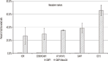
도 2a는, 중배엽 계통(mesoderm lineage)으로의 분화를 확인하는, 인간 유도 다능성 줄기 세포주 GMP1에서 분화된 중배엽 세포에서 세포-표면 마커인 KDR, CD56/NCAM1, APLNR/APJ, GARP, 또는 CD13의 발현을 나타내는 그래프이다.
도 2b는, 중배엽 계통으로의 분화를 확인하는, 인간 유도 다능성 줄기 세포주 GMP1로부터 분화된 중배엽 세포에서 다능성, 내배엽, 외배엽, 및 혈액혈관(hematovascular) 세포-표면 마커들의 제한적이거나 전혀 없는 발현을 나타내는 그래프이다.
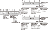
도 3은, 중배엽 세포로 인간 다능성 줄기 세포의 시험관내 분화(좌측), 및 Meso-3D-Vasculonoid VPC1 프로토콜(우측 위), 또는 Meso-3D-Vasculonoid VPC2 프로토콜(우측 아래)를 사용하여 중배엽-유래 혈관 전구 세포(meso-VPCs)로 중배엽 세포의 시험관내 분화에 대한 절차의 개략도이다.
도 4는, 중배엽 세포로 인간 다능성 줄기 세포의 시험관내 분화(좌측), 및 Meso-2D VPC2 프로토콜(우측 위), 또는 Meso-2D VPC3 프로토콜(우측 아래)를 사용하여 중배엽-유래 혈관 전구 세포(meso-VPCs)로 중배엽 세포의 시험관내 분화에 대한 절차의 개략도이다.
도 5는, 내피 계통(endothelial lineage)으로 추가 분화를 겪는 Meso-3D-Vasculonoid 프로토콜에 의해 생성된 meso-VPCs의 능력을 나타내는 현미경 이미지의 패널이다. 상단 패널은, 수확 전 5일째 meso-VPCs의 모폴로지(morphology)를 나타낸다. 중간 패널은, 피브로넥틴-코팅된 플레이트 및 내피 분화를 촉진하는 배지를 사용하여 meso-VPCs의 내피 분화를 나타낸다. 하단 패널은, meso-VPCs에 의해 형성된 모세관-유사(capillary-like) 마트리겔 네트워크를 나타낸다.
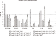
도 6a는, Meso-3D-Vasculonoid-VPC1, Meso-3D-Vasculonoid-VPC2, Meso-2D-VPC2, 또는 Meso-2D-VPC3 프로토콜을 사용하여 생성된 meso-VPCs에서 세포-표면 마커인 CD31/PECAM1, CD309/KDR, CXCR4/CD184, CD43, CD146, 및 PDGFRb의 발현을 나타낸 그래프이다.
도 6b는, 선택된 세포-표면 마커에 대해 양성인 비교 헤모제닉 내피 세포(hemogenic endothelial cells: HE) 또는 혈관모세포(HB) 및 meso-VPCs의 분획(fractions)을 나타내는 히트-맵(heat-map)이다. 미분화된 다능성 줄기 세포(J1 및 GMP1) 및 인간 제대 정맥 내피 세포(HUVECs)와의 비교는 또한 나타낸다.
도 6c는, 비교 헤모제닉 내피 세포(HE), 비교 혈관모세포(HB), 미분화된 다능성 줄기 세포(J1 및 GMP1), 인간 제대 정맥 내피 세포(HUVECs), 또는 Meso-3D-Vasculonoid 프로토콜 또는 Meso-2D 프로토콜에 의해 생성된 meso-VPCs의 혈관 세포-표면 마커 발현 프로파일들을 나타내는 주성분 분석(PCA) 플롯이다.
도 7은, 내피 계통으로 추가 분화를 겪는 Meso-2D 프로토콜에 의해 생성된 meso-VPCs의 능력을 나타내는 현미경 이미지의 패널이다. 상단 패널은, 수확 전 7일째에 meso-VPCs의 모폴로지를 나타낸다. 중간 패널은, 피브로넥틴-코팅된 플레이트 및 내피 분화를 촉진하는 배지를 사용하여 meso-VPCs의 내피 분화를 나타낸다. 하단 패널은, meso-VPCs에 의해 형성된 모세관-유사 마트리겔 네트워크를 나타낸다.
도 8은, 실시예 9에 기재된 대로 meso-VPCs로 처리된 동물에서 증가된 혈류를 나타내는 그래프이다. 구체적으로, 동물은 모의-수술(1M)되거나, 또는 비히클 대조군(2M), J1-HDF Meso-2D VPC2(3M), J-HDF Meso-3D Vasculonoid VPC2(4M), GMP1HDF Meso-2D VPC2(5M), GMP1-HDF Meso-3D Vasculonoid VPC2(6M) 또는 GMP1-HDF Meso-3D Vasculonoid VPC1(7M)로 처리된다.
도 9는, 실시예 9에 기재된 대로 meso-VPCs로 처리된 동물에서 혈관 밀도에서 변화를 나타내는 그래프이다. 구체적으로, 동물은, 비히클 대조군(2M), J1-HDF Meso-2D VPC2(3M TI1), J-HDF Meso-3D Vasculonoid VPC2(4M TI2), GMP1HDF Meso-2D VPC2(5M TI3), GMP1-HDF Meso-3D Vasculonoid VPC2(6M TI4) 또는 GMP1-HDF Meso-3D Vasculonoid VPC1(7M TI5)로 처리된다.
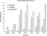
도 10은, meso-VPCs로 처리된 동물에서 작은 모세혈관 형성, 총 혈관 수, 및 혈류 시험에 대한 지표인, CD34+ 염색의 조합된 정량적 결과를 나타내는 그래프이다. 구체적으로, 동물은, 모의-수술(1M)되거나, 또는 비히클 대조군(2M), J1-HDF Meso-2D VPC2(3M TI1), J-HDF Meso-3D Vasculonoid VPC2(4M TI2), GMP1HDF Meso-2D VPC2(5M TI3), GMP1-HDF Meso-3D Vasculonoid VPC2(6M TI4) 또는 GMP1-HDF Meso-3D Vasculonoid VPC1(7M TI5)로 처리된다.
도 11은, 레이저 도플러(Laser Doppler)에 의해 측정된 혈류와 meso-VPCs로 처리된 동물의 각 그룹의 평균 모세혈관 밀도 사이에 강력하고 통계적으로 유의한 상관관계를 나타낸다.
도 12a는, J1 세포의 개체군 및 J1-유래 HE 세포의 개체군과 비교하는, hsa-miR-3917, hsa-miR-450a-2-3p, 및 hsa-miR-542-5p를 포함하는, 3개의 복제물로부터 J1-유래 Meso-3D Vasculonoid VPC2 세포의 개체군에서 발견된 고유(unique) 인간 miRNAs를 나타내는 플롯 및 그래프를 제공한다. 도 12a는 또한, hsa-miR-11399, hsa-miR-196b-3p, hsa-miR-5690, 및 hsa-miR-7151-3p를 포함하는, J1-유래 HE 세포의 개체군에서 발견되는 고유 인간 miRNAs를 나타낸다. "발현된"는 3개의 복제물 모두에서 0보다 큰 정규화된 발현이다.
도 12b는, 단일 세포에서 이전에 분석된 J1-유래 Meso-3D Vasculonoid VPC2 세포의 개체군에서 miRNA의 발현 수준을 나타낸 그래프로서, hsa-miR-126-5p, hsa-miR-125a-5p, 및 hsa-miR-24-3p가 J1 세포의 개체군 및 J1-유래 Meso-3D Vasculonoid VPC2 세포의 개체군 모두에서 발현되는 것을 나타낸다.
도 12c는, J1-유래 Meso-3D Vasculonoid VPC2 세포의 개체군이 hsa-let-7e-5p, hsa-miR-99a-5p, hsa-miR-223-5p, 및 hsa-miR-142-3p를 발현시키지만, hsa-let-7e-3p, hsa-miR-99a-3p, 및 hsa-miR-133a-5p를 발현시키지 않거나 낮은 발현을 갖는 것을 나타내는 그래프이다.
도 12d는, J1-유래 Meso-3D Vasculonoid VPC2 세포의 개체군이 hsa-miR-483-5p 및 hsa-miR-483-3p를 발현시키는 것을 나타내는 그래프이다.
도 13은, 단일 세포 RNA-seq 분석에서 단일 J1 또는 HUVEC 세포와 비교하여 J1-유래 Meso-3D Vasculonoid VPC2 세포 샘플에서 가장 상향- 또는 하향-조절된(down-regulated) 유전자의 발현을 나타낸 그래프이다.
도 14a는, 14일 후에 DAPI 및 UAE1 염색에 의한 J1-유래 Meso-3D Vasculonoid VPC2 바스큘로노이드들의 포매된 응집체(embedded aggregates)로부터 확장된 광범위한 혈관 네트워크를 나타내는 저배율(10x 대물렌즈)의 이미지이다.
도 14b는, 단일 세포("단일 세포")로 분리된 J1-유래 Meso-3D Vasculonoid VPC2 바스큘로노이드들("복수") 또는 J1-유래 Meso-3D Vasculonoid VPC2 세포들이 해동 후 정상산소(20% O2)(좌측 패널) 또는 저산소(5% O2)(우측 패널)하에서 시험관내 CLI-모방 조건(CLI-mimicking conditions)에서 배양된 경우, 바스큘로노이드들이 단일 세포로 동결보존된 J1-유래 Meso-3D Vasculonoid VPC2 세포에 비해 더 나은 세포 생존률을 나타내는 것을 보여주는 그래프이다.
도 14c는, 비히클 처리 그룹(GS2 배지만)과 비교된 연구 전반에 걸쳐 J1-유래 Meso-3D Vasculonoid VPC2 단일 세포("sc") 또는 바스큘로노이드들의 투여 후 혈류에서 통계적으로 유의한 개선을 나타내는 그래프이다; 이-원 분산분석(two-way ANOVA) 후 투키 검정(Tukey's test).
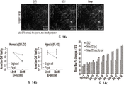
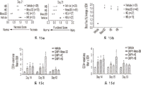
도 15a는, Meso-3D Vasculonoid VPC2 세포로 처리된 동물이 HE 및 HB 세포와 비교하여 21일째에 더 나은 평균 괴사(좌측 패널) 및 기능 점수(우측 패널)를 가짐을 나타내는 그래프이다. 일-원 분산분석 후 던네트 검증(Dunnett's test). 평균 +/- SEM.
도 15b는, 비히클과 비교하여, Meso-3D Vasculonoid VPC2 세포, HE, 및 HB 세포들로 처리된 동물에서 63일째의 혈류 개선을 나타내는 그래프이다. *p<0.05 대 비히클. 평균 +/- s.d. 이-원 분산분석 후 투키 검정.
도 15c는, Meso-3D Vasculonoid VPC2 세포, HE, 및 HB 세포들로 처리된 동물의 대퇴사두근(quadriceps)에서 CD34+ 혈관 성장을 나타내는 그래프이다. *p<0.05 대 비히클. 평균 +/- SEM. 이-원 분산분석 후 수정되지 않은 피셔 LSD 검정(Fisher's LSD test).
도 15d는, Meso-3D Vasculonoid VPC2 세포, HE, 및 HB 세포들의 투여 후 비복근(grastrocnemius)에서 개선을 나타내는 그래프이다. *p<0.05 대 비히클. 평균 +/- SEM. 이-원 분산분석 후 수정되지 않은 피셔 LSD 검정.
도 16a는, 세포의 장-기간 생착(engraftment)을 나타내는, 처리 후 63일 및 180일차에 Ku80+ 염색에 의한 생착된 공여자 GMP1-Meso3D Vasculonoid VPC2 세포를 나타내는 그래프이다.
도 16b는, 35일 및 63일까지, Meso-3D Vasculonoid VPC2 세포가 Ku80+ 염색에 의해 생착을 나타내는 그래프이다.
도 16c는, Balb/c 누드 마우스(nude mice)에서 HLI 수술 63일 후, 장-기간 생착(Ku80+), 인간 맥관구조(vasculature)의 형성(UEA1+ 혈관), 및 측분비 숙주 혈관 성장의 촉진(IB4+ 및 SMA+ 혈관)을 보여주는 주사된 Meso-3D Vasculonoid VPC2s의 형광 이미지이다. 1 is a schematic diagram of a procedure for in vitro differentiation of human pluripotent stem cells into mesoderm cells.
Figure 2a shows the cell-surface markers of KDR, CD56/NCAM1, APLNR/APJ, GARP, or CD13 in mesoderm cells differentiated in the human induced pluripotent stem cell line GMP1, confirming differentiation into the mesoderm lineage. It is a graph showing expression.
2B shows limited or no expression of pluripotent, endoderm, ectoderm, and hematovascular cell-surface markers in mesoderm cells differentiated from the human induced pluripotent stem cell line GMP1, confirming differentiation into mesodermal lineages. It is a graph representing
Figure 3 shows the in vitro differentiation of human pluripotent stem cells into mesoderm cells (left) and mesodermal-using the Meso-3D-Vasculonoid VPC1 protocol (top right), or Meso-3D-Vasculonoid VPC2 protocol (bottom right). Schematic of the procedure for the in vitro differentiation of mesoderm cells into derived vascular progenitor cells (meso-VPCs).
4 shows in vitro differentiation of human pluripotent stem cells into mesodermal cells (left), and mesodermal-derived vascular progenitor cells using Meso-2D VPC2 protocol (top right), or Meso-2D VPC3 protocol (bottom right). Schematic of the procedure for the in vitro differentiation of mesoderm cells into (meso-VPCs).
5 is a panel of microscopic images showing the ability of meso-VPCs generated by the Meso-3D-Vasculonoid protocol to undergo further differentiation into endothelial lineage. The top panel shows the morphology of meso-
6A shows the cell-surface marker CD31/PECAM1 in meso-VPCs generated using the Meso-3D-Vasculonoid-VPC1, Meso-3D-Vasculonoid-VPC2, Meso-2D-VPC2, or Meso-2D-VPC3 protocol. , a graph showing the expression of CD309/KDR, CXCR4/CD184, CD43, CD146, and PDGFRb.
6B is a heat-map showing fractions of comparative hemogenic endothelial cells (HE) or hemangioblasts (HB) and meso-VPCs that are positive for selected cell-surface markers. . Comparisons with undifferentiated pluripotent stem cells (J1 and GMP1) and human umbilical vein endothelial cells (HUVECs) are also shown.
6C shows comparative hemogenic endothelial cells (HE), comparative hemangioblasts (HB), undifferentiated pluripotent stem cells (J1 and GMP1), human umbilical vein endothelial cells (HUVECs), or Meso-3D-Vasculonoid protocol or Meso Principal component analysis (PCA) plots showing the vascular cell-surface marker expression profiles of meso-VPCs generated by the -2D protocol.
7 is a panel of microscopic images showing the ability of meso-VPCs generated by the Meso-2D protocol to undergo further differentiation into endothelial lineages. The top panel shows the morphology of meso-VPCs at 7 days before harvest. The middle panel shows the endothelial differentiation of meso-VPCs using fibronectin-coated plates and media that promotes endothelial differentiation. The lower panel shows the capillary-like Matrigel network formed by meso-VPCs.
8 is a graph showing increased blood flow in animals treated with meso-VPCs as described in Example 9. FIG. Specifically, animals were simulated-operative (1M), or vehicle control (2M), J1-HDF Meso-2D VPC2 (3M), J-HDF Meso-3D Vasculonoid VPC2 (4M), GMP1HDF Meso-2D VPC2 (5M). ), GMP1-HDF Meso-3D Vasculonoid VPC2 (6M) or GMP1-HDF Meso-3D Vasculonoid VPC1 (7M).
9 is a graph showing changes in blood vessel density in animals treated with meso-VPCs as described in Example 9. FIG. Specifically, animals were, vehicle control (2M), J1-HDF Meso-2D VPC2 (3M TI1), J-HDF Meso-3D Vasculonoid VPC2 (4M TI2), GMP1HDF Meso-2D VPC2 (5M TI3), GMP1-HDF Treated with Meso-3D Vasculonoid VPC2 (6M TI4) or GMP1-HDF Meso-3D Vasculonoid VPC1 (7M TI5).
10 is a graph showing the combined quantitative results of CD34 + staining, indices for small capillary formation, total blood vessel count, and blood flow tests in animals treated with meso-VPCs. Specifically, animals were simulated-operative (1M), or vehicle control (2M), J1-HDF Meso-2D VPC2 (3M TI1), J-HDF Meso-3D Vasculonoid VPC2 (4M TI2), GMP1HDF Meso-2D Processed as VPC2 (5M TI3), GMP1-HDF Meso-3D Vasculonoid VPC2 (6M TI4) or GMP1-HDF Meso-3D Vasculonoid VPC1 (7M TI5).
11 shows a strong and statistically significant correlation between blood flow measured by Laser Doppler and the mean capillary density of each group of animals treated with meso-VPCs.
12A shows a comparison of three populations of J1 cells and a population of J1-derived HE cells, comprising hsa-miR-3917, hsa-miR-450a-2-3p, and hsa-miR-542-5p. Plots and graphs are provided representing the unique human miRNAs found in a population of J1-derived Meso-3D Vasculonoid VPC2 cells from replicates. 12A also shows native human miRNAs found in a population of J1-derived HE cells, including hsa-miR-11399, hsa-miR-196b-3p, hsa-miR-5690, and hsa-miR-7151-3p. indicates "Expressed" is normalized expression greater than zero in all three replicates.
12B is a graph showing the expression levels of miRNAs in a population of J1-derived Meso-3D Vasculonoid VPC2 cells previously analyzed in single cells, hsa-miR-126-5p, hsa-miR-125a-5p, and hsa. -miR-24-3p is expressed in both the population of J1 cells and the population of J1-derived Meso-3D Vasculonoid VPC2 cells.
12C shows that a population of J1-derived Meso-3D Vasculonoid VPC2 cells express hsa-let-7e-5p, hsa-miR-99a-5p, hsa-miR-223-5p, and hsa-miR-142-3p. However, it is a graph showing that hsa-let-7e-3p, hsa-miR-99a-3p, and hsa-miR-133a-5p are not expressed or have low expression.
12D is a graph showing that a population of J1-derived Meso-3D Vasculonoid VPC2 cells express hsa-miR-483-5p and hsa-miR-483-3p.
13 is a graph showing the expression of the most up- or down-regulated genes in J1-derived Meso-3D Vasculonoid VPC2 cell samples compared to single J1 or HUVEC cells in single cell RNA-seq analysis. .
14A is an image at low magnification (10x objective) showing an extensive vascular network expanded from embedded aggregates of J1-derived Meso-3D Vasculonoid VPC2 vasculonoids by DAPI and UAE1 staining after 14 days. .
14B shows that either J1-derived Meso-3D Vasculonoid VPC2 vasculonoids (“pluralities”) or J1-derived Meso-3D Vasculonoid VPC2 cells isolated into single cells (“single cell”) were subjected to normoxia (20) after thawing. J1 in which basculonoids were cryopreserved as single cells when cultured in % O 2 ) (left panel) or in vitro CLI-mimicking conditions under hypoxia (5% O 2 ) (right panel) - It is a graph showing a better cell viability compared to the derived Meso-3D Vasculonoid VPC2 cells.
14C shows a statistically significant improvement in blood flow following administration of J1-derived Meso-3D Vasculonoid VPC2 single cells (“sc”) or vasculonoids across studies compared to vehicle treated group (GS2 medium only). It is a graph; Two-way ANOVA followed by Tukey's test.
15A is a graph showing that animals treated with Meso-3D Vasculonoid VPC2 cells had better mean necrosis (left panel) and functional score (right panel) at
15B is a graph showing improvement in blood flow at
15C is a graph showing CD34 + vascular growth in quadriceps of animals treated with Meso-3D Vasculonoid VPC2 cells, HE, and HB cells. *p<0.05 versus vehicle. Mean +/- SEM. Uncorrected Fisher's LSD test after two-way ANOVA.
15D is a graph showing improvement in grastrocnemius after administration of Meso-3D Vasculonoid VPC2 cells, HE, and HB cells. *p<0.05 versus vehicle. Mean +/- SEM. Uncorrected Fisher LSD test after two-way ANOVA.
FIG. 16A is a graph showing engrafted donor GMP1-Meso3D Vasculonoid VPC2 cells by Ku80+ staining at
16B is a graph showing engraftment of Meso-3D Vasculonoid VPC2 cells by Ku80+ staining by
Figure 16c shows long-term engraftment (Ku80+), formation of human vasculature (UEA1+ blood vessels), and promotion of paracrine host vascular growth in Balb/c
I. 정의 I. Definition
본 발명이 좀 더 쉽게 이해될 수 있도록, 특정 용어는 먼저 정의된다. 파라미터의 값 또는 값의 범위가 인용될 때마다, 인용된 값에 대한 중간 값 및 범위도 본 발명의 일부인 것으로 의도된다는 점에 또한 유의되어야 한다. In order that the present invention may be more readily understood, certain terms are first defined. It should also be noted that whenever a value or range of values for a parameter is recited, intermediate values and ranges for the recited values are also intended to be part of the present invention.
하기 상세한 설명에서, 설명의 목적을 위해, 특정 번호, 물질, 및 구성은, 본 발명의 완전한 이해를 제공하기 위해 서술된다. 그러나, 본 발명이 이러한 특정 세부사항 없이 실시될 수 있다는 점은 기술분야의 당업자에게 명백할 것이다. 몇몇 사례에서, 잘-알려진 특색(features)은 본 발명을 모호하게 하지 않도록 생략되거나 단순화될 수 있다. 더군다나, 본 명세서에서 "하나의 구현예" 또는 "일 구현예"와 같은, 문구에 대한 언급은, 구현예와 관련하여 기재된 특정 특색, 구조, 또는 특징이 본 발명의 적어도 하나의 구현예에 포함된다는 것을 의미한다. 본 명세서에 다양한 위치에서 "하나의 구현예에서"와 같은, 문구의 출현은 반드시 동일한 구현예를 모두 지칭하는 것은 아니다. In the following detailed description, for purposes of explanation, specific numbers, materials, and configurations are set forth in order to provide a thorough understanding of the invention. It will be apparent, however, to one skilled in the art that the present invention may be practiced without these specific details. In some instances, well-known features may be omitted or simplified so as not to obscure the present invention. Moreover, reference herein to a phrase, such as “in one embodiment” or “an embodiment,” means that a particular feature, structure, or characteristic described in connection with the embodiment is included in at least one embodiment of the invention. means to be The appearances of a phrase, such as “in one embodiment,” in various places in this specification are not necessarily all referring to the same embodiment.
여기에 사용된 용어들의 단수 형태는, 별도의 언급이 없는 한, 대상의 하나 또는 하나 초과(즉, 적어도 하나)를 지칭하는 것으로 여기에서 사용된다. 예로서, "요소(element)"는 하나의 요소 또는 하나를 초과하는 요소를 지칭한다. As used herein, the singular forms of the terms are used herein to refer to one or more than one (ie, at least one) of the subject, unless stated otherwise. By way of example, “element” refers to one element or more than one element.
용어 "포함한" 또는 "포함하는"은, 본 개시에 필수적이지만, 불특정 요소를, 필수 여부에 관계없이, 포함할 수 있는, 조성물, 방법, 및 이들의 각각의 구성요소(들)와 관련하여 여기에서 사용된다. The terms “comprising” or “comprising” are used herein with respect to compositions, methods, and their respective component(s), which are essential to the present disclosure, but which may include, whether essential or not, unspecified elements. is used in
여기에 사용된 바와 같은 "다능성 세포", "다능성 줄기 세포", 및 "PSCs"는, 세포들이 미분화 상태를 유지하면서 시험관내에서 장기적이거나 사실상 무기한 증식할 수 있고, 안정한(바람직하게는, 정상) 핵형(karyotype)을 나타내며, 적절한 조건하에서 3가지 배엽층(germ layers) 모두(즉, 외배엽, 중배엽 및 내배엽)로 분화하는 능력을 갖는 세포를 광범위하게 지칭한다. 통상적으로, 다능성 세포들은, (a) 면역결핍(SCID) 마우스에 이식될 때 기형종(teratomas)을 유도할 수 있고; (b) 3가지 배엽층 모두의 세포 타입(예를 들어, 외배엽, 중배엽 및 내배엽 세포 타입)으로 분화할 수 있으며, 그리고 (c) 적어도 하나의 hES 세포 마커(예컨대, Oct-4, 알칼리성 포스파타제(alkaline phosphatase), SSEA 3 표면 항원(SSEA 3 surface antigen), SSEA 4 표면 항원, NANOG, TRA 1 60, TRA 1 81, SOX2, REX1)를 발현시킨다. 대표적인 다능성 세포는, Oct-4, 알칼리성 포스파타제, SSEA 3 표면 항원, SSEA 4 표면 항원, TRA 1 60, 및/또는 TRA 1 81을 발현시킬 수 있다. 부가적인 대표 다능성 세포는, 배아 줄기 세포, 유도 다능성 줄기(iPS) 세포, 배아-유래 세포, 배아 생식(EG) 세포로부터 (예를 들어, FGF-2, LIF 및 SCF의 존재하에 배양시켜) 생성된 다능성 세포, 단위생식 ES 세포, 배양된 내세포괴 세포(ICM)로부터 생성된 ES 세포, 할구(blastomere)로부터 생성된 ES 세포, 및 핵 이식에 의해 생성된 ES 세포(예를 들어, 수용 난모세포로 이식된 체세포 핵)를 포함하지만, 이에 제한되는 것은 아니다. 대표적인 다능성 세포는, 배아의 파괴 없이 생성될 수 있다. 예를 들어, 유도 다능성 세포는 배아 파괴 없이 얻어진 세포로부터 생성될 수 있다. 또 다른 예로서, 다능성 세포는, (잔여 배아에 해를 끼치지 않고 달성될 수 있는) 생검된 할구로부터 생성될 수 있고; 선택적으로, 잔여 배아는 냉동보존, 배양, 및/또는 적절한 숙주에 이식될 수 있다. (공급원에 관계없이) 다능성 세포는, 유전적으로 변형되거나 달리 변형될 수 있다. As used herein, "pluripotent cells", "pluripotent stem cells", and "PSCs" refer to stable (preferably, Normal) karyotype and broadly refers to cells that have the ability to differentiate into all three germ layers (ie, ectoderm, mesoderm and endoderm) under appropriate conditions. Typically, pluripotent cells are capable of (a) inducing teratomas when transplanted into immunodeficient (SCID) mice; (b) capable of differentiating into cell types of all three germ layers (e.g., ectoderm, mesoderm, and endoderm cell types), and (c) at least one hES cell marker (e.g., Oct-4, alkaline phosphatase ( alkaline phosphatase),
여기에 사용된 바와 같은, "배아" 또는 "배아의"는, 모계 숙주(maternal host)의 자궁막에 이식되지 않은 발달 중인 세포괴를 광범위하게 지칭한다. "배아 세포"는, 배아로부터 단리되거나 배아에 함유된 세포이다. 이것은 또한 2-세포기(cell stage)에서 일찍이 얻어진 할구, 및 응집된 할구를 포함한다. As used herein, “embryonic” or “embryonic” refers broadly to a developing cell mass that has not been implanted in the uterine lining of a maternal host. An “embryonic cell” is a cell isolated from or contained in an embryo. It also includes blastomeres obtained early in the two-cell stage, and aggregated blastomeres.
"배아 줄기 세포"(ES 세포 또는 ESC)는, 배아 세포(예컨대, 배양된 내세포괴 세포 또는 배양된 할구)로부터 생성된 다능성 세포를 포괄한다. 종종, 이러한 세포들은 세포주로서 연속 계대되거나 계대되었다. 배아 줄기 세포는, 여기에 기재된 바와 같이 중배엽 세포 및 meso-VPCs를 생성시키는 절차에서 다능성 줄기 세포로 사용될 수 있다. 예를 들어, ES 세포는, 정자 또는 정자 DNA로 난자의 수정, 핵 이식(체세포 핵 이식 포함), 또는 단위생식과 같은, (생식 또는 무생식 수단에 의한 것을 포함하는) 임의의 방법에 의해 생성된 배아로부터의 유도를 포함하는 당업계에 알려진 방법에 의해 생성될 수 있다. 또 다른 예로서, 배아 줄기 세포는 또한, 비-배아 세포가 절차에서 사용되는 경우에도, 체세포 핵 이식에 의해 생성된 세포를 포함한다. 예를 들어, ES 세포는, 배반포 단계 배아의 ICM, 뿐만 아니라 하나 이상의 할구로부터 유래된 배아 줄기 세포로부터 유래될 수 있다. 이러한 배아 줄기 세포는, 체세포 핵 이식(SCNT), 단위생식, 및 동정생식(androgenesis)을 포함하는 무성생식 수단에 의해, 또는 수정에 의해 생성된 배아 물질로부터 발생될 수 있다. 위에서 더욱 논의된 바와 같이, ES 세포들은, 유전적으로 변형되거나 달리 변형될 수 있다. "Embryonic stem cells" (ES cells or ESCs) encompass pluripotent cells generated from embryonic cells (eg, cultured inner cell mass cells or cultured blastomeres). Often, these cells have been serially passaged or passaged as a cell line. Embryonic stem cells can be used as pluripotent stem cells in procedures to generate mesodermal cells and meso-VPCs as described herein. For example, ES cells are produced by any method (including by reproductive or non-reproductive means), such as fertilization of an egg with sperm or sperm DNA, nuclear transfer (including somatic cell nuclear transfer), or unit reproduction. It can be produced by methods known in the art, including derivation from a mature embryo. As another example, embryonic stem cells also include cells produced by somatic cell nuclear transfer, even when non-embryonic cells are used in the procedure. For example, the ES cells may be derived from the ICM of a blastocyst stage embryo, as well as embryonic stem cells derived from one or more blastomeres. Such embryonic stem cells can be generated from embryonic material produced by fertilization or by asexual means, including somatic cell nuclear transfer (SCNT), unitary reproduction, and androgenesis. As discussed further above, ES cells may be genetically modified or otherwise modified.
ES 세포들은, 예를 들어, 유전자 조작, 자발적 이형 접합성 상실에 대한 스크리닝, 등을 통해 하나 이상의 HLA 유전자에서 동형접합 또는 이형접합으로 발생될 수 있다. 배아 줄기 세포는, 이들의 공급원 또는 이들을 생성하는데 사용된 특정 방법에 관계없이, 통상적으로 다음 속성 중 하나 이상을 보유한다: (i) 3가지 배엽층 모두의 세포로 분화하는 능력, (ⅱ) 적어도 Oct-4 및 알칼리성 포스파타제의 발현, 및 (ⅲ) 면역저하 동물으로 이식된 경우 기형종을 생성하는 능력. 본 발명의 구현예에서 사용될 수 있는 배아 줄기 세포는, CT2, MA01, MA09, ACT-4, No. 3, J1, H1, H7, H9, H14 및 ACT30 배아 줄기 세포와 같은, 인간 ES 세포("hESC" 또는 "hES 세포")를 포함하지만, 이에 제한되는 것은 아니다. 부가적인 대표적인 세포주는, NED1, NED2, NED3, NED4, NED5, 및 NED7을 포함한다. 또한, NIH 인간 배아 줄기 세포 등록(Human Embryonic Stem Cell Registry)를 참조. 사용될 수 있는 대표적인 인간 배아 줄기 세포주는 J1 세포이다. ES cells can be generated homozygous or heterozygous in one or more HLA genes, for example, through genetic manipulation, screening for spontaneous loss of heterozygosity, and the like. Embryonic stem cells, regardless of their source or the particular method used to generate them, typically possess one or more of the following attributes: (i) the ability to differentiate into cells of all three germ layers, (ii) at least Expression of Oct-4 and alkaline phosphatase, and (iii) ability to produce teratoma when transplanted into immunocompromised animals. Embryonic stem cells that can be used in the embodiment of the present invention are CT2, MA01, MA09, ACT-4, No. 3, human ES cells (“hESC” or “hES cells”), such as, but not limited to, J1, H1, H7, H9, H14 and ACT30 embryonic stem cells. Additional representative cell lines include NED1, NED2, NED3, NED4, NED5, and NED7. See also NIH Human Embryonic Stem Cell Registry. An exemplary human embryonic stem cell line that may be used is J1 cells.
대표적인 인간 배아 줄기 세포(hESC) 마커는, 알칼리성 포스파타제, Oct-4, Nanog, 단계-특이적 배아 항원-3(SSEA-3), 단계-특이적 배아 항원-4(SSEA-4), TRA-1-60, TRA-1-81, TRA-2-49/6E, Sox2, 성장 및 분화 인자 3(GDF3), 감소된 발현 1(REX1), 섬유아세포 성장 인자 4(FGF4), 배아 세포-특이적 유전자 1(ESG1), 발달 다능성-연관 유전자 2(DPPA2), DPPA4, 텔로머라제 역전사효소(hTERT), SALL4, E-CADHERIN, 클러스터 지정 30(CD30), Cripto(TDGF-1), GCTM-2, 제네시스(Genesis), 생식 세포 핵 인자, 및 줄기 세포 인자(SCF 또는 c-Kit 리간드)를 포함하지만, 이에 제한되는 것은 아니다. 부가적으로, 배아 줄기 세포는, Oct-4, 알칼리성 포스파타제, SSEA 3 표면 항원, SSEA 4 표면 항원, TRA 1 60, 및/또는 TRA 1 81을 발현시킬 수 있다. Representative human embryonic stem cell (hESC) markers are alkaline phosphatase, Oct-4, Nanog, stage-specific embryonic antigen-3 (SSEA-3), stage-specific embryonic antigen-4 (SSEA-4), TRA- 1-60, TRA-1-81, TRA-2-49/6E, Sox2, growth and differentiation factor 3 (GDF3), reduced expression 1 (REX1), fibroblast growth factor 4 (FGF4), germ cell-specific enemy gene 1 (ESG1), developmental pluripotency-associated gene 2 (DPPA2), DPPA4, telomerase reverse transcriptase (hTERT), SALL4, E-CADHERIN, cluster designation 30 (CD30), Cripto (TDGF-1), GCTM -2, Genesis, Germ Cell Nuclear Factor, and Stem Cell Factor (SCF or c-Kit ligand). Additionally, the embryonic stem cells may express Oct-4, alkaline phosphatase,
ESCs는 초기에, 뮤린(murine) 배아 피더 세포(embryonic feeder cells: MEF)와 같은, 피더 세포 또는 인간 진피 섬유아세포(HDF)와 같은, 인간 피더 세포의 존재 또는 부재하에, ESCs의 다능성을 유지하는 당업계에 알려진 임의의 배양 배지으로 배양될 수 있다. MEF 세포들 또는 인간 피더 세포들은, 공동-배양에서 ESCs를 시딩(seeding)하기 전에, 예를 들어, 미토마이신 C(mitomycin C)에 노출, 감마 조사, 또는 임의의 기타 알려진 방법에 의해, 유사분열적으로 비활성화될 수 있으며, 따라서, MEFs는 배양에서 증식하지 않는다. 부가적으로, ESC 세포 배양물은 현미경으로 검사될 수 있고, 비 ESC 세포 모폴로지를 함유하는 콜로니는 선택될 수 있고, 예를 들어, 줄기 세포 절단 도구를 사용하여, 레이저 절제(laser ablation)에 의해, 또는 기타 수단을 사용하여 폐기될 수 있다. 통상적으로, 배아체 형성(embryoid body formation)을 위한 시딩을 위한 ESCs의 수확의 시점 이후에, 부가적인 MEF 세포 또는 인간 피더 세포들은 사용되지 않는다. ESCs initially maintain the pluripotency of ESCs in the presence or absence of human feeder cells, such as feeder cells or human dermal fibroblasts (HDF), such as murine embryonic feeder cells (MEF). It can be cultured in any culture medium known in the art. MEF cells or human feeder cells are subjected to mitosis prior to seeding ESCs in co-culture, for example, by exposure to mitomycin C, gamma irradiation, or any other known method. can be inactivated, and therefore MEFs do not proliferate in culture. Additionally, ESC cell cultures can be examined microscopically and colonies containing non-ESC cell morphologies can be selected, for example, by laser ablation, using a stem cell cutting tool. , or other means. Typically, after the point of harvest of ESCs for seeding for embryooid body formation, no additional MEF cells or human feeder cells are used.
선택적으로, hES 세포들은, 세포외 기질(예를 들어, Matrigel®, 라미닌(laminin), 또는 iMatrix-511 또는 여기에 개시되거나 당업계에 알려진 임의의 다른 세포외 기질)과 같은, 고체 표면 상에서 피더가 없는 조건(feeder-free conditions)하에서 당업계에 알려진 임의의 방법, 예를 들어, Klimanskaya et al., Lancet 365:1636-1641 (2005)에 의해 배양될 수 있다. 따라서, 여기에 기재된 방법에 사용된 hES 세포들은, 피더가 없는 배양물 상에서 배양될 수 있다. Optionally, hES cells are fed onto a solid surface, such as an extracellular matrix (eg, Matrigel®, laminin, or iMatrix-511 or any other extracellular matrix disclosed herein or known in the art). It can be cultured by any method known in the art under feeder-free conditions, for example, by Klimanskaya et al., Lancet 365:1636-1641 (2005). Thus, hES cells used in the methods described herein can be cultured on feeder-free cultures.
여기에서 사용된 바와 같은, "배아-유래 세포"(EDC)는, 다능성 상실배-유래 세포, 내세포괴, 배순(embryonic shield), 또는 배반엽상층(epiblast)의 것을 포함하는 배반포-유래 세포, 또는 원시 내배엽, 외배엽, 및 중배엽을 포함하는 초기 배아의 다른 다능성 줄기 세포 및 이들의 파생물을 광범위하게 지칭한다. "EDC"는 또한 다양한 발달의 단계로부터 응집된 단일 할구 또는 배아로부터 할구 및 세포괴를 포함하지만, 세포주로서 계대된 인간 배아 줄기 세포는 제외한다. As used herein, "embryo-derived cell" (EDC) is a blastocyst-derived cell, including that of a pluripotent morula-derived cell, an inner cell mass, an embryonic shield, or an epiblast. , or other pluripotent stem cells of the early embryo, including primitive endoderm, ectoderm, and mesoderm, and their derivatives. "EDC" also includes blastomeres and cell masses from single blastomeres or embryos aggregated from various stages of development, but excludes human embryonic stem cells that have been passaged as cell lines.
여기에 사용된 바와 같은 "유도된 다능성 줄기 세포" 또는 "iPSCs" 또는 "iPS 세포"는, 체세포를 재프로그래밍하여 발생된 다능성 줄기 세포를 지칭한다. iPSCs는 인자의 조합("재프로그래밍 인자")을 발현시키거나 발현을 유도하여 발생될 수 있다. iPS 세포는, 태아, 출생후, 신생아, 청소년, 또는 성인 체세포들을 사용하여 발생될 수 있다. iPS 세포들은 세포 은행에서 얻어질 수 있다. 선택적으로, iPS 세포는, 혈관 전구 세포(VPCs) 또는 또 다른 세포 타입으로의 분화를 시작하기 전에 (당업계에 알려진 절차에 의해) 새로이 발생될 수 있다. iPS 세포의 생성은, 분화된 세포의 생성에서 초기 단계일 수 있다. iPS 세포들은, 조직-일치 VPCs(tissue-matched VPCs)를 발생시킬 목표로 특정 환자 또는 일치된 공여자 유래의 물질을 사용하여 구체적으로 발생될 수 있다. iPS 세포들은, 의도된 수용자에서 실질적으로 면역원성이 아닌 세포, 예를 들어, 자가 세포 또는 의도된 수용자에 조직적합성이 있는 세포로부터 생성될 수 있다. 위에서 더욱 논의된 바와 같이("다능성 세포" 참조), iPS 세포를 포함하는 다능성 세포는, 유전적으로 변형되거나 달리 변형될 수 있다. 사용될 수 있는 대표적인 인간 iPSC 세포주는 GMP1 세포이다. “Induced pluripotent stem cells” or “iPSCs” or “iPS cells” as used herein refer to pluripotent stem cells generated by reprogramming somatic cells. iPSCs can be generated by expressing or inducing expression of a combination of factors (“reprogramming factors”). iPS cells can be generated using fetal, postnatal, neonatal, juvenile, or adult somatic cells. iPS cells can be obtained from a cell bank. Optionally, iPS cells can be newly generated (by procedures known in the art) prior to initiating differentiation into vascular progenitor cells (VPCs) or another cell type. Generation of iPS cells may be an early step in the generation of differentiated cells. iPS cells can be specifically generated using material from a specific patient or matched donor with the goal of generating tissue-matched VPCs. iPS cells can be generated from cells that are not substantially immunogenic in the intended recipient, eg, autologous cells or cells that are histocompatibility in the intended recipient. As discussed further above (see “pluripotent cells”), pluripotent cells, including iPS cells, may be genetically modified or otherwise modified. An exemplary human iPSC cell line that may be used is GMP1 cells.
또 다른 실시예로서, 유도 다능성 줄기 세포는, 세포를 하나 이상의 재프로그래밍 인자와 접촉시켜 체세포 또는 다른 세포를 재프로그래밍하여 발생될 수 있다. 예를 들어, 재프로그래밍 인자(들)는, 세포에 의해, 예를 들어, 세포에 첨가된 외인성 핵산으로부터, 또는 소분자, microRNA, 또는 해당 유전자의 발현을 촉진하거나 유도하는 이와 유사한 것과 같은 인자에 반응하여 내인성 유전자로부터 발현될 수 있다(Suh and Blelloch, Development 138, 1653-1661 (2011); Miyoshi et al., Cell Stem Cell (2011), doi:10.1016/j.stem.2011.05.001; Sancho-Martinez et al., Journal of Molecular Cell Biology (2011) 1-3; Anokye-Danso et al., Cell Stem Cell 8, 376-388, April 8, 2011; Orkin and Hochedlinger, Cell 145, 835-850, June 10, 2011, 또는 Warren et al., Scientific Reports, 10.1038/srep00657, September 14, 2012, 참조, 이들 각각은 전체적으로 여기에 참조로서 혼입됨). 재프로그래밍 인자는, 예를 들어, 배양 배지에 첨가시켜 외인성 공급원으로부터 제공될 수 있고, 세포 진입 펩티드(cell entry peptides), 단백질 또는 핵산 형질감염제(transfection agents), 리포펙션(lipofection), 전기천공, 생물학적 입자 전달 시스템(유전자 총), 미세주입, 및 이와 유사한 것에 대한 커플링(coupling)을 통한 것과 같은 당업계에 알려진 방법에 의해 세포 내로 도입될 수 있다. 특정 구현예에서, 체세포를 다능성 줄기 세포로 재프로그래밍하는데 사용될 수 있는 인자는, 예를 들어, Oct4(때때로 Oct 3/4로 지칭됨), Sox2, c-Myc, 및 Klf4의 조합을 포함한다. 다른 구현예에서, 체세포를 다능성 줄기 세포로 재프로그래밍하는데 사용될 수 있는 인자는, 예를 들어, Oct-4, Sox2, Nanog, 및 Lin28의 조합을 포함한다. 다른 구현예에서, 체세포는 적어도 2개의 재프로그래밍 인자, 적어도 3개의 재프로그래밍 인자, 또는 4개의 재프로그래밍 인자를 발현시켜 재프로그래밍된다. 다른 구현예에서, 체세포는, Oct4, Sox2, MYC, Klf4, Nanog, 및 Lin28을 발현시켜 재프로그래밍된다. 다른 구현예에서, 부가적인 재프로그래밍 인자는 동정되고, 단독으로 또는 하나 이상의 공지된 재프로그래밍 인자와 조합하여 체세포를 다능성 줄기 세포로 재프로그래밍하는데 사용된다. iPS 세포들은 통상적으로 배아 줄기 세포와 동일한 마커의 발현에 의해 동정될 수 있지만, 특정 iPS 세포주는 이의 발현 프로파일에서 다를 수 있다. As another example, induced pluripotent stem cells can be generated by reprogramming a somatic cell or other cell by contacting the cell with one or more reprogramming factors. For example, the reprogramming factor(s) may be responsive to a factor such as a small molecule, microRNA, or the like that promotes or induces expression of a gene of interest, e.g., by a cell, e.g., from an exogenous nucleic acid added to the cell. and can be expressed from endogenous genes (Suh and Blelloch, Development 138, 1653-1661 (2011); Miyoshi et al. , Cell Stem Cell (2011), doi:10.1016/j.stem.2011.05.001; Sancho-Martinez) et al. , Journal of Molecular Cell Biology (2011) 1-3; Anokye-Danso et al. ,
유도 다능성 줄기 세포는 체세포에서 하나 이상의 재프로그래밍 인자의 발현 또는 발현을 유도하여 생성될 수 있다. 구현예에서, 체세포는, 섬유아세포, 예컨대, 진피 섬유아세포, 활액 섬유아세포, 또는 폐 섬유아세포, 또는 비-섬유아세포 체세포이다. 하나의 구현예에서, 체세포는, 전술된 바와 같이 적어도 1, 2, 3, 4, 5개의 재프로그래밍 인자를 발현시켜 재프로그래밍된다. 또 다른 구현예에서, 재프로그래밍 인자의 발현은, 재프로그래밍 인자의 발현을 유도하는, 소 유기 분자 작용제(small organic molecule agents)와 같은, 적어도 하나의 작용제를 체세포와 접촉시켜 유도될 수 있다. An induced pluripotent stem cell can be generated by inducing the expression or expression of one or more reprogramming factors in a somatic cell. In an embodiment, the somatic cell is a fibroblast, such as a dermal fibroblast, a synovial fibroblast, or a lung fibroblast, or a non-fibroblast somatic cell. In one embodiment, the somatic cell is reprogrammed by expressing at least 1, 2, 3, 4, 5 reprogramming factors as described above. In another embodiment, expression of the reprogramming factor can be induced by contacting the somatic cell with at least one agent, such as small organic molecule agents, that induces expression of the reprogramming factor.
체세포는 또한 조합적 접근법(combinatorial approach)을 사용하여 재프로그래밍될 수 있고, 여기서, 재프로그래밍 인자는 (예를 들어, 바이러스 벡터, 플라스미드, 및 이와 유사한 것을 사용하여) 발현되고, 재프로그래밍 인자의 발현은 (예를 들어, 소 유기 분자를 사용하여) 유도된다. 예를 들어, 재프로그래밍 인자는, 레트로바이러스 벡터 또는 렌티바이러스 벡터와 같은, 바이러스 벡터를 사용한 감염에 의해 체세포에서 발현될 수 있다. 또한, 재프로그래밍 인자는, 에피솜 플라스미드 또는 mRNA와 같은, 비-결합 벡터(non-integrative vector)를 사용하여 체세포에서 발현될 수 있다. 예를 들어, Yu et al., Science. 2009 May 8;324(5928):797-801, 참조, 이의 전체적인 내용은 참조로서 여기에 혼입된다. 재프로그래밍 인자가 비-결합 벡터를 사용하여 발현되는 경우, 인자는, 전기천공, 형질감염, 또는 벡터로 체세포의 형질전환을 사용하여 세포에서 발현될 수 있다. Somatic cells may also be reprogrammed using a combinatorial approach, wherein the reprogramming factor is expressed (eg, using viral vectors, plasmids, and the like) and expression of the reprogramming factor is derived (eg, using small organic molecules). For example, reprogramming factors can be expressed in somatic cells by infection with a viral vector, such as a retroviral vector or a lentiviral vector. In addition, reprogramming factors can be expressed in somatic cells using non-integrative vectors, such as episomal plasmids or mRNA. For example, Yu et al. , Science. 2009 May 8;324(5928):797-801, see, the entire contents of which are incorporated herein by reference. Where the reprogramming factor is expressed using a non-binding vector, the factor can be expressed in the cell using electroporation, transfection, or transformation of the somatic cell with the vector.
일단 재프로그래밍 인자가 세포에서 발현되면, 세포는 당업계에 알려진 임의의 방법에 의해 배양될 수 있다. 시간이 지남에 따라, ES 특징을 갖는 세포는 배양 접시에 나타난다. 세포는, 예를 들어, ES 모폴로지에 기초하여, 또는 선택 가능하거나 검출 가능한 마커의 발현에 기초하여 선택되고 계대배양될 수 있다. 세포는 배양되어 ES 세포와 유사한 세포의 배양물을 생성할 수 있다-이들은 추정 iPS 세포(putative iPS cells)이다. iPS 세포들은 통상적으로 다른 배아 줄기 세포와 동일한 마커의 발현에 의해 동정될 수 있지만, 특정 iPS 세포주는 발현 프로파일에서 다를 수 있다. 대표적인 iPS 세포들은, Oct-4, 알칼리성 포스파타제, SSEA 3 표면 항원, SSEA 4 표면 항원, TRA 1 60, 및/또는 TRA 1 81을 발현시킬 수 있다. Once the reprogramming factor is expressed in the cell, the cell can be cultured by any method known in the art. Over time, cells with ES characteristics appear in the culture dish. Cells can be selected and passaged, for example, based on ES morphology, or based on expression of a selectable or detectable marker. Cells can be cultured to produce a culture of cells similar to ES cells—these are putative iPS cells. Although iPS cells can usually be identified by expression of the same markers as other embryonic stem cells, a particular iPS cell line may differ in its expression profile. Representative iPS cells can express Oct-4, alkaline phosphatase,
iPS 세포의 다능성을 확인하기 위해, 세포는 하나 이상의 다능성 분석에서 시험될 수 있다. 예를 들어, 세포는 ES 세포 마커의 발현에 대해 시험될 수 있으며; 세포는 SCID 마우스에 이식된 경우 기형종을 생성하는 능력에 대해 평가될 수 있고; 세포는 3개의 모든 배엽층의 세포 타입을 생성하도록 분화하는 능력에 대해 평가될 수 있다. 일단 다능성 iPS 세포가 얻어지면, 이것은 중배엽 세포 및 혈관 전구 세포, 예를 들어, 중배엽-유래 혈관 전구 세포를 생성시키는데 사용될 수 있다. To confirm the pluripotency of iPS cells, the cells can be tested in one or more pluripotency assays. For example, cells can be tested for expression of ES cell markers; Cells can be assessed for their ability to produce teratoma when transplanted into SCID mice; Cells can be assessed for their ability to differentiate to generate cell types of all three germ layers. Once pluripotent iPS cells are obtained, they can be used to generate mesoderm cells and vascular progenitor cells, such as mesodermal-derived vascular progenitor cells.
여기에 사용된 바와 같은, "중배엽"은 모든 좌우대칭 동물의 아주 초기 배아에서 3개의 기본 배엽층 중 하나를 지칭한다. 중배엽은 간엽, 중피, 비-상피 혈구(non-epithelial blood cells) 및 체강소체(coelomocytes)를 형성한다. 초기 중배엽 개입(commitment)은 상피에서 간엽으로의 전이에서 발생하며, 그 후 지정된 중배엽 계통 세포는 장배형성(gastrulation)이 진행됨에 따라 안쪽으로 이동한다. 중배엽 계통의 세포는, 여러 지정된 세포 타입으로 분화할 수 있는 혈관모세포 및 다분화성(multipotent) 중간엽 줄기 세포를 포함하는, 혈관계 및 림프계를 형성하도록 미리 정해진다. 중배엽은 배아외 중배엽 및 그 다음 배아 내장 중배엽의 형성을 통해 혈관형성을 일으킨다. 혈관 내피 성장 인자(VEGF) 및 태반 성장 인자(PIGF 또는 PGF)와 같은, 성장 인자는, 새로운 혈관의 성장 및 발달을 자극시킨다. 하나의 구현예에서, 중배엽 계통의 세포는 혈관 전구체 세포 또는 혈관 전구 세포로 미리 정해진다. 하나의 구현예에서, 다능성 줄기 세포, 예를 들어, hESCs 또는 iPSCs, 예를 들어, hiPSCs는 중배엽 계통 세포, 예를 들어, 중배엽 전구체 세포로 분화될 수 있다. 따라서, 용어 "중배엽"은, 세포의 성숙도와 상관없이, 다능성 줄기 세포로부터 유래된 중배엽 계통 세포도 포함하고, 따라서 상기 용어는, 중배엽 전구체 세포를 포함하는, 다양한 수준의 성숙도의 중배엽 세포를 포괄한다. As used herein, "mesoderm" refers to one of the three basal germ layers in the very early embryo of all symmetric animals. The mesoderm forms mesenchymal, mesothelial, non-epithelial blood cells and coelomocytes. Early mesodermal commitment occurs at the epithelial to mesenchymal transition, after which designated mesodermal lineage cells migrate inward as gastrulation progresses. Cells of the mesodermal lineage are predetermined to form the vasculature and lymphatic system, including hemangioblasts and multipotent mesenchymal stem cells capable of differentiating into several designated cell types. The mesoderm undergoes angiogenesis through the formation of the extraembryonic mesoderm and then the embryonic visceral mesoderm. Growth factors, such as vascular endothelial growth factor (VEGF) and placental growth factor (PIGF or PGF), stimulate the growth and development of new blood vessels. In one embodiment, the cells of the mesodermal lineage are predefined as vascular progenitor cells or vascular progenitor cells. In one embodiment, pluripotent stem cells, eg, hESCs or iPSCs, eg, hiPSCs, are capable of differentiating into mesodermal lineage cells, eg, mesodermal progenitor cells. Accordingly, the term "mesoderm" also encompasses cells of the mesodermal lineage derived from pluripotent stem cells, regardless of the maturity of the cells, and thus the term encompasses mesoderm cells of varying degrees of maturity, including mesodermal progenitor cells. do.
대표적인 중배엽 마커는, CD309/KDR, CD56/NCAM1, APLNR/APJ, GARP, CD13, N-카드헤린, 액티빈 A, 액티빈 AB, 액티빈 AC, 액티빈 B, 액티빈 C, BMP 및 기타 액티빈 수용체 활성화제들(activators), BMP 및 기타 액티빈 수용체 억제제들, BMP-2, BMP-2/BMP-4, BMP-2/BMP-6 이종이량체(Heterodimer), BMP-2/BMP-7 이종이량체, BMP-2a, BMP-4, BMP-6, BMP-7, 크립틱(Cryptic), FABP4/A-FABP, FGF-5, GDF-1, GDF-3, INHBA, INHBB, 노달(Nodal), TGF-베타, TGF-베타 1, TGF-베타 1, 2, 3, TGF-베타 1.2, TGF-베타 1/1.2, TGF-베타 2, TGF-베타 2/1.2, TGF-베타 3, TGF-베타 수용체 억제제, Wnt-3a, Wnt-8a, MESDC2, Nicalin, Brachyury, EOMES, FoxC1, FoxF1, Goosecoid, HAND1, MIXL1, Slug, Snail, TBX6, Twist-1, 및 Twist-2를 포함하지만, 이에 제한되는 것은 아니다. 하나의 구현예에서, 중배엽 세포는, CD309/KDR, CD56/NCAM1, APLNR/APJ, GARP, 및 CD13으로부터 선택된 하나 이상의 마커에 대해 양성인, 중배엽 전구체 세포이다. Representative mesoderm markers are CD309/KDR, CD56/NCAM1, APLNR/APJ, GARP, CD13, N-cadherin, activin A, activin AB, activin AC, activin B, activin C, BMP and other axons. Tivin receptor activators, BMP and other activin receptor inhibitors, BMP-2, BMP-2/BMP-4, BMP-2/BMP-6 Heterodimer, BMP-2/BMP- 7 Heterodimer, BMP-2a, BMP-4, BMP-6, BMP-7, Crypto, FABP4/A-FABP, FGF-5, GDF-1, GDF-3, INHBA, INHBB, Nodal (Nodal), TGF-beta, TGF-
여기에 사용된 바와 같은, "혈관형성"은 새로운 혈관의 형성을 지칭한다. 혈관형성은, 중배엽으로부터 유래된 내피의 형성을 포함한다. 여기에 사용된 바와 같은, "혈관신생"은, 기-존 혈관으로부터 혈관의 형성을 지칭한다. 예를 들어, Developmental Biology by Gilbert, Scott F. Sunderland (MA): Sinauer Associates, Inc.; c2000, and Molecular Biology of the Cell 4th ed. Alberts, Bruce; Johnson, Alexander; Lewis, Julian; Raff, Martin; Roberts, Keith; Walter, Peter New York and London: Garland Science; c2002, 참조. As used herein, “angiogenesis” refers to the formation of new blood vessels. Angiogenesis involves the formation of endothelium derived from the mesoderm. As used herein, “angiogenesis” refers to the formation of blood vessels from pre-existing blood vessels. See, eg, Developmental Biology by Gilbert, Scott F. Sunderland (MA): Sinauer Associates, Inc.; c2000, and Molecular Biology of the Cell 4th ed. Alberts, Bruce; Johnson, Alexander; Lewis, Julian; Raff, Martin; Roberts, Keith; Walter, Peter New York and London: Garland Science; c2002, cf.
여기에 사용된 바와 같은, "혈관 전구 세포"(VPCs)는, 다른 혈액-혈관 세포 계통 중에서, 내피 세포, 평활근 세포, 및 혈관주위세포(pericytes)로 분화할 수 있는 능력을 갖는 세포를 지칭한다. 하나의 구현예에서, 혈관 전구 세포는 중배엽-유래 혈관 전구 세포(meso-VPCs)이다. As used herein, “vascular progenitor cells” (VPCs) refer to cells that have the ability to differentiate into endothelial cells, smooth muscle cells, and pericytes, among other blood-vascular cell lineages. . In one embodiment, the vascular progenitor cells are mesodermal-derived vascular progenitor cells (meso-VPCs).
여기에 사용된 바와 같은, "중배엽-유래 혈관 전구 세포"(meso-VPCs)는, 다능성 줄기 세포, 예를 들어, ESCs 또는 iPSCs의 시험관내 분화에 의해 유래된 중배엽 세포로부터 발생된 VPCs를 지칭한다. Meso-VPCs는, 여기에서 더욱 기재된 바와 같이, 하나 이상의 세포-표면 마커의 발현에 의해 동정될 수 있다. 하나의 구현예에서, 중배엽-유래 혈관 전구 세포는, 결과적으로, meso-VPCs로 분화되는, 중배엽 세포로 다능성 줄기 세포, 예를 들어, ESCs 또는 iPSCs의 시험관내 분화로부터 발생된다. As used herein, “mesoderm-derived vascular progenitor cells” (meso-VPCs) refer to VPCs generated from mesodermal cells derived by the in vitro differentiation of pluripotent stem cells, e.g., ESCs or iPSCs. do. Meso-VPCs can be identified by expression of one or more cell-surface markers, as further described herein. In one embodiment, the mesodermal-derived vascular progenitor cells arise from the in vitro differentiation of pluripotent stem cells, eg, ESCs or iPSCs, into mesodermal cells, which in turn differentiate into meso-VPCs.
Meso-VPCs는, 마우스 PSCs 및 인간 PSCs 모두에서 시험관내에서 유래될 수 있다. Meso-VPCs는, 조혈(hematopoietic) 및 내피 세포 계통으로 분화할 수 있으며 평활근 세포가 될 수도 있다. 본 발명의 meso-VPCs의 개체군은, CD31/PECAM1, CD309/KDR, CD43, CD144, CD34, CD184/CXCR4, CD146, 및 PDGFRb와 같은, 적어도 하나의 마커에 대해 양성일 수 있다. 하나의 구현예에서, meso-VPC의 개체군은, 상기-동정된 마커 중들 1, 2, 3, 4, 5, 6, 7, 또는 8개에 대해 양성이다. 하나의 구현예에서, meso-VPC의 개체군은, CD146, CD31/PECAM1, 및 CD309/KDR에 대해 양성이다. 또 다른 구현예에서, meso-VPCs의 개체군은, CD31/PECAM1, CD309/KDR, CD146, 및 (i) CD144, CD34, CD184/CXCR4, CD43, 또는 PDGFRb 중 적어도 하나, (ⅱ) CD34, CD184/CXCR4, 및 PDGFRb; (ⅲ) CD184/CXCR4; (ⅳ) PDGFRb; (v) CD144 및 CD184/CXCR4; (ⅵ) CD184/CXCR4 및 CD43; 또는 (ⅶ) CC184/CXCFR4를 발현시킨다. 구현예에서, meso-VPCs의 개체군은, hsa-miR-3917, hsa-miR-450a-2-3p, hsa-miR-542-5p, hsa-miR-126-5p, hsa-miR-125a-5p, hsa-miR-24-3p, hsa-let-7e-5p, hsa-miR-99a-5p, hsa-miR-223-5p, hsa-miR-142-3p, hsa-miR-483-5p, hsa-miR-483-3p, miR 214, miR 335-3p, 및 miR-199a-3p로부터 선택된 적어도 1, 적어도 2, 적어도 3, 적어도 4, 적어도 5, 적어도 6, 적어도 7, 적어도 8, 적어도 9, 적어도 10, 적어도 11, 적어도 12, 적어도 13, 적어도 14, 또는 적어도 15 miRNA 마커들을 발현시킨다. 하나의 구현예에서, meso-VPCs의 개체군은, hsa-miR-3917, hsa-miR-450a-2-3p, 및 hsa-miR-542-5p를 발현시킨다. 구현예에서, meso-VPCs의 개체군은, 조성물에서 meso-VPCs의 적어도 약 20%가 마커를 발현시키는 경우, 특정 마커를 발현시키는 것으로 간주된다. 하나의 구현예에서, 본 발명의 meso-VPC는, mir125a-5p, mir24, 및 mir483-5p로 이루어진 군으로부터 선택된 적어도 1, 적어도 2, 적어도 3, 또는 적어도 4개의 miRNA 마커에 대해 양성이다. 하나의 구현예에서, miRNA 마커는 mir483-5p이다. 하나의 구현예에서, meso-VPCs의 개체군은, mir126, mir125a-5p, mir24, 및 mir483-5p로 이루어진 군으로부터 선택된 적어도 1, 적어도 2, 적어도 3, 또는 적어도 4개의 miRNA 마커에 대해 양성인 적어도 하나의 meso-VPC를 포함한다. 하나의 구현예에서, miRNA 마커는 mir483-5p이다. 하나의 구현예에서, meso-VPCs의 개체군은, HE 세포의 개체군보다 더 높은 수준으로 CD31 및 KDR을 발현시킨다. 또 다른 구현예에서, meso-VPCs의 개체군은, HE 세포의 개체군보다 더 낮은 수준으로 CD146을 발현시킨다. 또 다른 구현예에서, meso-VPCs의 개체군은, HE 세포의 개체군보다 낮은 수준으로 CD184/CXCR4를 발현시킨다. Meso-VPCs can be derived in vitro from both mouse PSCs and human PSCs. Meso-VPCs can differentiate into hematopoietic and endothelial cell lineages and can also become smooth muscle cells. The population of meso-VPCs of the invention may be positive for at least one marker, such as CD31/PECAM1, CD309/KDR, CD43, CD144, CD34, CD184/CXCR4, CD146, and PDGFRb. In one embodiment, the population of meso-VPCs is positive for 1, 2, 3, 4, 5, 6, 7, or 8 of the above-identified markers. In one embodiment, the population of meso-VPCs is positive for CD146, CD31/PECAM1, and CD309/KDR. In another embodiment, the population of meso-VPCs comprises: CD31/PECAM1, CD309/KDR, CD146, and (i) at least one of CD144, CD34, CD184/CXCR4, CD43, or PDGFRb, (ii) CD34, CD184/ CXCR4, and PDGFRb; (iii) CD184/CXCR4; (iv) PDGFRb; (v) CD144 and CD184/CXCR4; (vi) CD184/CXCR4 and CD43; or (vii) expressing CC184/CXCFR4. In an embodiment, the population of meso-VPCs is hsa-miR-3917, hsa-miR-450a-2-3p, hsa-miR-542-5p, hsa-miR-126-5p, hsa-miR-125a-5p , hsa-miR-24-3p, hsa-let-7e-5p, hsa-miR-99a-5p, hsa-miR-223-5p, hsa-miR-142-3p, hsa-miR-483-5p, hsa - at least 1, at least 2, at least 3, at least 4, at least 5, at least 6, at least 7, at least 8, at least 9, express at least 10, at least 11, at least 12, at least 13, at least 14, or at least 15 miRNA markers. In one embodiment, the population of meso-VPCs expresses hsa-miR-3917, hsa-miR-450a-2-3p, and hsa-miR-542-5p. In an embodiment, a population of meso-VPCs is considered to express a particular marker if at least about 20% of the meso-VPCs in the composition express the marker. In one embodiment, the meso-VPC of the invention is positive for at least 1, at least 2, at least 3, or at least 4 miRNA markers selected from the group consisting of mir125a-5p, mir24, and mir483-5p. In one embodiment, the miRNA marker is mir483-5p. In one embodiment, the population of meso-VPCs is at least one positive for at least 1, at least 2, at least 3, or at least 4 miRNA markers selected from the group consisting of mir126, mir125a-5p, mir24, and mir483-5p. of meso-VPC. In one embodiment, the miRNA marker is mir483-5p. In one embodiment, the population of meso-VPCs express CD31 and KDR at higher levels than the population of HE cells. In another embodiment, the population of meso-VPCs expresses CD146 at a lower level than the population of HE cells. In another embodiment, the population of meso-VPCs expresses CD184/CXCR4 at a lower level than the population of HE cells.
구현예들 중 어느 하나에서, meso-VPCs의 개체군은, CXCR7, CD45, 및 NG2 중 1개, 2개 또는 3개의 제한적이거나 전혀 없는 검출을 나타낸다. 구현예들 중 어느 하나에서, meso-VPCs의 개체군은, CXCR7, CD45, 및 NG2 모두의 제한적이거나 전혀 없는 검출을 나타낸다. 구현예들 중 어느 하나에서, meso-VPCs의 개체군은, CD144, CD34, CD184/CXCR4, CXCR7, CD43, CD45, PDGFRb, 또는 NG2 중 적어도 1, 적어도 2, 적어도 3, 적어도 4, 적어도 5, 적어도 6, 적어도 7, 또는 적어도 8개의 제한적이거나 전혀 없는 검출을 나타낸다. 구현예에서, meso-VPCs의 개체군은, hsa-let-7e-3p, hsa-miR-99a-3p, hsa-miR-133a-5p, hsa-miR-11399, hsa-miR-196b-3p, hsa-miR-5690, 및 hsa-miR-7151-3p로부터 선택된 적어도 1, 적어도 2, 적어도 3, 적어도 4, 적어도 5, 적어도 6, 또는 적어도 7개의 miRNA 마커들의 제한적이거나 전혀 없는 발현을 나타낸다. 하나의 구현예에서, 조성물에서 meso-VPCs의 약 20% 미만이 마커를 발현시키는 경우, meso-VPCs의 개체군은 마커의 제한적이거나 전혀 없는 검출을 나타내는 것으로 간주된다. 하나의 구현예에서, 본 발명의 meso-VPC는, mir367, mir302a, mir302b, mir302c, mirLet7-e, mir223, mir99a, mir142-3p, 및 mir133a로 이루어진 군으로부터 선택된 적어도 1, 적어도 2, 적어도 3, 적어도 4, 적어도 5, 적어도 6, 적어도 7, 적어도 8, 또는 적어도 9개의 miRNA 마커들에 대해 제한적이거나 전혀 없는 발현을 나타낸다. 본 발명의 구현예에서, meso-VPCs의 개체군은, mir367, mir302a, mir302b, mir302c, mirLet7-e, mir223, mir99a, mir142-3p, 및 mir133a로 이루어진 군으로부터 선택된 적어도 1, 적어도 2, 적어도 3, 적어도 4, 적어도 5, 적어도 6, 적어도 7, 적어도 8, 또는 적어도 9개의 miRNA 마커들에 대해 제한적이거나 전혀 없는 발현을 나타내는 적어도 하나의 meso-VPC를 포함한다. In any of the embodiments, the population of meso-VPCs exhibits limited or no detection of one, two or three of CXCR7, CD45, and NG2. In any of the embodiments, the population of meso-VPCs exhibits limited or no detection of all of CXCR7, CD45, and NG2. In any one of the embodiments, the population of meso-VPCs is at least 1, at least 2, at least 3, at least 4, at least 5, at least of CD144, CD34, CD184/CXCR4, CXCR7, CD43, CD45, PDGFRb, or
여기에 사용된 바와 같은, "바스큘로노이드"는, 세포, 예를 들어, 세포 배양 동안 형성된 중배엽-유래 혈관 전구 세포(meso-VPCs)의 콜로니-유사 응집체(colony-like aggregate)를 지칭한다. 하나의 구현예에서, 바스큘로노이드는, 3D-바스큘로노이드 분화 플랫폼(platform)을 사용하여 생성된 meso-VPCs에 의해 형성된다. As used herein, “basculonoid” refers to a colony-like aggregate of cells, such as meso-derived vascular progenitor cells (meso-VPCs) formed during cell culture. . In one embodiment, basculonoids are formed by meso-VPCs generated using a 3D-vasculonoid differentiation platform.
여기에 사용된 바와 같은, "치료법", "치료적", "치료하는", "치료하다" 또는 "치료"는, 질병을 치료하고, 질병 또는 이의 임상 증상의 발달을 정지 또는 감소시키며, 및/또는 질병을 경감시켜, 질병 또는 이의 임상 증상의 퇴행을 유발하는 것을 광범위하게 지칭한다. "치료법", "치료적", "치료하는", "치료하다" 또는 "치료"는, 질병, 질병의 징후, 및/또는 질병의 증상의 예방, 방지, 치료, 치유, 구제, 감소, 완화, 및/또는 경감의 제공을 포괄한다. "치료법", "치료적", "치료하는", "치료하다" 또는 "치료"는, 진행중인 질병 징후 및/또는 증상이 있는 환자에서 징후 및/또는 증상의 완화를 포괄한다. "치료법", "치료적", "치료하는", "치료하다" 또는 "치료"는 또한 "예방" 및 "방지"를 포괄한다. 예방은, 환자에서 질병의 치료 이후에 발생하는 질병을 방지하거나 또는 환자에서 질병의 발생률 또는 중증도를 감소시키는 것을 포함한다. 치료의 목적을 위해, 용어 "감소된", "치료적", "치료하는", "치료하다" 또는 "치료"는, 징후 및/또는 증상의 임상적으로 유의한 감소를 광범위하게 지칭한다. "치료법", "치료적", "치료하는", "치료하다" 또는 "치료"는, 도지거나 또는 재발하는 징후 및/또는 증상을 치료하는 것을 포함한다. "치료법", "치료적", "치료하는", "치료하다" 또는 "치료"는, 언제든지 징후 및/또는 증상의 출현을 배제할 뿐만 아니라 기존 징후 및/또는 증상을 감소 및 기존 징후 및/또는 증상을 제거하는 것을 포괄하지만, 이에 제한되는 것은 아니다. "치료법", "치료적", "치료하는", "치료하다" 또는 "치료"는, 만성 질병("유지") 및 급성 질병을 치료하는 것을 포함한다. 예를 들어, 치료는 징후 및/또는 증상의 도짐 또는 재발을 치료하거나 방지하는 것을 포함한다. 하나의 구현예에서, 치료는, 중증 사지 허혈과 같은, 혈관 질병의 징후 및/또는 증상에서 임상적으로 유의한 감소를 포함한다. As used herein, “treatment”, “therapeutic”, “treating”, “treat” or “treatment” means treating a disease, arresting or reducing the development of the disease or its clinical symptoms, and It refers broadly to alleviating a disease, thereby causing regression of the disease or its clinical symptoms. “Treatment”, “therapeutic”, “treating”, “treat” or “treatment” refers to preventing, preventing, treating, curing, relieving, reducing, ameliorating a disease, symptoms, and/or symptoms of a disease. , and/or the provision of relief. "Treatment", "therapeutic", "treating", "treat" or "treatment" encompasses alleviation of signs and/or symptoms in a patient with ongoing disease signs and/or symptoms. “Treatment”, “therapeutic”, “treating”, “treat” or “treatment” also encompasses “prevention” and “prevention”. Prevention includes preventing a disease from occurring after treatment of the disease in a patient or reducing the incidence or severity of a disease in a patient. For the purposes of treatment, the terms “reduced”, “therapeutic”, “treating”, “treat” or “treatment” refer broadly to a clinically significant reduction in signs and/or symptoms. “Treatment,” “therapeutic,” “treating,” “treat,” or “treatment” includes treating signs and/or symptoms that are relieved or recurring. “Treatment”, “therapeutic”, “treating”, “treat” or “treatment” means at any time to exclude the appearance of signs and/or symptoms as well as reduce and reduce existing signs and/or symptoms. or eliminating the symptoms, but is not limited thereto. “Treatment”, “therapeutic”, “treating”, “treat” or “treatment” includes treating chronic disease (“maintenance”) and acute disease. For example, treatment includes treating or preventing the occurrence or recurrence of signs and/or symptoms. In one embodiment, the treatment comprises a clinically significant reduction in signs and/or symptoms of a vascular disease, such as severe limb ischemia.
여기에 사용된 바와 같은, "병리의 정상화"는, 질병으로부터 결과하는 비정상적인 구조 및/또는 기능을 보다 정상적인 상태로 되돌리는 것을 지칭한다. 정상화는, 질병으로부터 결과하는 조직, 기관, 또는 세포 타입의 구조 및/또는 기능에서 비정상적인 것을 고쳐서, 병리의 진행이 제어되고 개선될 수 있음을 시사한다. 예를 들어, 본 발명의 meso-VPCs로 치료한 후, 혈관 질병, 예를 들어, 중증 사지 허혈의 결과로서 하지의 비정상은 개선, 교정, 및/또는 전환될 수 있다. As used herein, “normalization of pathology” refers to returning the abnormal structure and/or function resulting from a disease to a more normal state. Normalization suggests that the progression of pathology can be controlled and ameliorated by correcting abnormalities in the structure and/or function of a tissue, organ, or cell type resulting from a disease. For example, following treatment with the meso-VPCs of the present invention, abnormalities in the lower extremities as a result of vascular disease, eg, severe limb ischemia, can be ameliorated, corrected, and/or converted.
여기에서 사용된 바와 같은, "혈관 질병"은, 혈관(동맥 및 정맥)의 모든 비정상적인 상태를 지칭한다. 심장 밖의 혈관 질병은 어디에서나 나타날 수 있다. 가장 흔한 혈관 질병은, 뇌졸중, 말초 동맥 질환(PAD), 복부 대동맥류(AAA), 경동맥 질병(CAD), 동정맥 기형(AVM), 중증 사지 허혈(CLI), 폐색전증(혈전), 심부 정맥 혈전증(DVT), 만성 정맥 부전증(CVI), 및 정맥류이다. 하나의 구현예에서, 혈관 질병은 말초 동맥 질환(PAD)이다. 하나의 구현예에서, 혈관 질병은, 중증 사지 허혈(CLI)과 같은, 허혈성 질병이다. 하나의 구현예에서, 혈관 질병은, 죽상동맥경화증, 말초 동맥 질환(PAD), 경동맥 질병, 정맥 질병, 혈전, 대동맥류, 섬유근 이형성증, 림프부종, 또는 혈관 손상이다. 하나의 구현예에서, 혈관 질병은, 말초 동맥 질환, 예컨대, 중증 사지 허혈(CLI), 장 허혈 증후군, 신장 동맥 질병, 슬와 포착 증후군, 레이노 현상, 또는 버거병이다. As used herein, “vascular disease” refers to any abnormal condition of blood vessels (arteries and veins). Vascular disease outside the heart can appear anywhere. The most common vascular diseases are stroke, peripheral arterial disease (PAD), abdominal aortic aneurysm (AAA), carotid artery disease (CAD), arteriovenous malformation (AVM), severe limb ischemia (CLI), pulmonary embolism (thrombus), deep vein thrombosis ( DVT), chronic venous insufficiency (CVI), and varicose veins. In one embodiment, the vascular disease is peripheral arterial disease (PAD). In one embodiment, the vascular disease is an ischemic disease, such as severe limb ischemia (CLI). In one embodiment, the vascular disease is atherosclerosis, peripheral arterial disease (PAD), carotid artery disease, venous disease, thrombus, aortic aneurysm, fibromuscular dysplasia, lymphedema, or vascular injury. In one embodiment, the vascular disease is a peripheral arterial disease, such as severe limb ischemia (CLI), intestinal ischemic syndrome, renal artery disease, popliteal entrapment syndrome, Raynaud's phenomenon, or Berger's disease.
Ⅱ. 중배엽-유래 혈관 전구 세포(Meso-VPCs)의 시험관내 발생 II. In Vitro Generation of Mesodermal-Derived Vascular Progenitor Cells (Meso-VPCs)
본 발명은, 다능성 줄기 세포로부터 유래된 중배엽 세포로부터 중배엽-유래 혈관 전구 세포(meso-VPCs)를 생성시키는 방법을 제공한다. 상기 방법은, 하나 이상의 중배엽 유도 성장 인자를 함유하는 배지에서 다능성 줄기 세포를 배양하여 중배엽 세포를 생성시키는, 배양 단계, 및 중배엽-유래 혈관 전구 세포(meso-VPCs)로 중배엽 세포의 분화를 지시하는 하나 이상의 인자를 함유하는 배지에서, 적절한 표면에서 중배엽 세포를 배양하는 단계를 포함한다. 몇몇 구현예에서, 상기 방법은 복수의 meso-VPCs를 단일 세포로 분리시키는 단계를 더욱 포함한다. The present invention provides a method for generating mesodermal-derived vascular progenitor cells (meso-VPCs) from mesoderm cells derived from pluripotent stem cells. The method comprises culturing pluripotent stem cells in a medium containing one or more mesodermal-derived growth factors to produce mesodermal cells, and directs differentiation of mesodermal cells into mesodermal progenitor cells (meso-VPCs). culturing the mesoderm cells on an appropriate surface in a medium containing one or more factors. In some embodiments, the method further comprises isolating the plurality of meso-VPCs into single cells.
본 발명에 사용되는 다능성 줄기 세포는, 위에서 제시된 방법들 중 어느 하나에 의해 얻어지고 배양될 수 있다. 하나의 구현예에서, 다능성 줄기 세포, 예를 들어, 인간 배아 줄기 세포(hESCs) 또는 인간 유도 다능성 줄기 세포(hiPSCs)는, 피더가 없는(FF) 조건에서 배양되고 세포외 기질 상에 플레이팅된다. 하나의 구현예에서, 다능성 줄기 세포는, 피더 배양 조건에서 배양되고 세포외 기질 상에 플레이팅된다. The pluripotent stem cells used in the present invention can be obtained and cultured by any one of the methods presented above. In one embodiment, pluripotent stem cells, e.g., human embryonic stem cells (hESCs) or human induced pluripotent stem cells (hiPSCs), are cultured in feeder-free (FF) conditions and played on the extracellular matrix. is ticked In one embodiment, the pluripotent stem cells are cultured in feeder culture conditions and plated on the extracellular matrix.
몇몇 구현예에서, 세포외 기질은, 라미닌, 피브로넥틴, 비트로넥틴, 프로테오글리칸, 엔탁틴, 콜라겐, 콜라겐 I, 콜라겐 IV, 헤파란 설페이트, EHS(Engelbreth-Holm-Swarm) 마우스 육종 세포로부터의 가용성 제제, Matrigel®(Corning), 젤라틴, 및 인간 기저막 추출물로 이루어진 군으로부터 선택된다. 하나의 구현예에서, 세포외 기질은, 인간을 포함하는, 임의의 포유동물 기원으로부터 유래될 수 있다. 하나의 구현예에서, 다능성 줄기 세포를 배양하기 위한 세포외 기질 표면은 마트리겔-코팅 표면이다. In some embodiments, the extracellular matrix comprises laminin, fibronectin, vitronectin, proteoglycan, entactin, collagen, collagen I, collagen IV, heparan sulfate, a soluble agent from Engelbreth-Holm-Swarm (EHS) mouse sarcoma cells, Matrigel® (Corning), gelatin, and human basement membrane extract. In one embodiment, the extracellular matrix may be from any mammalian origin, including humans. In one embodiment, the extracellular matrix surface for culturing pluripotent stem cells is a matrigel-coated surface.
몇몇 구현예에서, 다능성 줄기 세포는, 전분화능을 지원하는데 적합한 배지에서 배양되고, 임의의 이러한 배지는 당업계에 공지되어 있다. 몇몇 구현예에서, 전분화능을 지원하는 배지는 Nutristem™이다. 몇몇 구현예에서, 전분화능을 지원하는 배지는 TeSR™이다. 몇몇 구현예에서, 전분화능을 지원하는 배지는 StemFit™이다. 다른 구현예에서, 전분화능을 지원하는 배지는, Knockout™ 혈청 대체물(Gibco), LIF, bFGF, 또는 임의의 다른 인자로 보충될 수 있는, Knockout™ DMEM(Gibco)이다. 이들 대표적인 배지 각각은, 당업계에 공지되어 있고, 상업적으로 이용 가능하다. 또 다른 구현예에서, 전분화능을 지원하는 배지는, bFGF 또는 임의의 다른 인자로 보충될 수 있다. 하나의 구현예에서, bFGF는, 저농도(예를 들어, 4 ng/㎖)로 보충될 수 있다. 다른 구현예에서, bFGF는 더 높은 농도(예를 들어, 100 ng/㎖)로 보충될 수 있다. 하나의 구현예에서, 배지는 무-혈청이다. 또 다른 구현예에서, 배지는 혈청을 포함한다. In some embodiments, the pluripotent stem cells are cultured in a medium suitable to support pluripotency, any such medium known in the art. In some embodiments, the medium that supports pluripotency is Nutristem™. In some embodiments, the medium that supports pluripotency is TeSR™. In some embodiments, the medium that supports pluripotency is StemFit™. In another embodiment, the medium that supports pluripotency is Knockout™ DMEM (Gibco), which may be supplemented with Knockout™ serum replacement (Gibco), LIF, bFGF, or any other factor. Each of these representative media are known in the art and are commercially available. In another embodiment, the medium that supports pluripotency may be supplemented with bFGF or any other factor. In one embodiment, bFGF may be supplemented at low concentrations (eg, 4 ng/ml). In other embodiments, bFGF may be supplemented at a higher concentration (eg, 100 ng/ml). In one embodiment, the medium is serum-free. In another embodiment, the medium comprises serum.
다능성 줄기 세포는 당업계에 알려진 임의의 적합한 용기에서 배양, 계대 또는 수확될 수 있다. 대표적인 조직 배양 용기는, 15 ㎝ 조직 배양 플레이트, 10 ㎝ 조직 배양 플레이트, 3 ㎝ 조직 배양 플레이트, 6-웰 조직 배양 플레이트, 12-웰 조직 배양 플레이트, 24-웰 조직 배양 플레이트, 48-웰 조직 배양 플레이트, 96-웰 조직 배양 플레이트, T-25 조직 배양 플라스크, T-75 조직 배양 플라스크를 포함한다. 하나의 구현예에서, 다능성 줄기 세포는 6-웰 조직 배양 플레이트에서 배양된다. Pluripotent stem cells can be cultured, passaged, or harvested in any suitable vessel known in the art. Representative tissue culture vessels are: 15 cm tissue culture plate, 10 cm tissue culture plate, 3 cm tissue culture plate, 6-well tissue culture plate, 12-well tissue culture plate, 24-well tissue culture plate, 48-well tissue culture plate, 96-well tissue culture plate, T-25 tissue culture flask, T-75 tissue culture flask. In one embodiment, the pluripotent stem cells are cultured in a 6-well tissue culture plate.
몇몇 구현예에서, 배지 교환은, 다능성 줄기 세포의 최적 조건을 유지하기 위해 배양의 약 1, 2, 3, 4, 5 또는 6일 후에 수행된다. 배지 교환을 위해, 출발 조건과 동일한 배양 배지는 사용될 수 있거나, 또는 배지는 배양 필요에 따라 조정될 수 있다. 몇몇 구현예에서, 다능성 줄기 세포는, 약 1, 2, 3, 4, 5, 6, 7, 8, 또는 9일 후, 또는 세포 배양물이 약 60-90% 컨플루언시(confluency)에 도달할 때, 분할 및 계대된다. 세포 계대를 위해, 출발 조건과 동일한 배양 배지는 사용될 수 있거나, 또는 배지는 배양 필요에 따라 조정될 수 있다. 세포는, 1:1, 1:2, 1:3, 1:4, 1:5, 1:6, 1:7, 1:8, 1:9, 1:10, 1:11, 1:12, 1:13, 1:14, 1:15, 1:16, 1:17, 1:18, 1:19, 또는 1:20의 희석 비율로 분할 및 계대될 수 있다. 하나의 구현예에서, 다능성 줄기 세포는 1:3의 희석 비율로 계대된다. In some embodiments, the medium change is performed after about 1, 2, 3, 4, 5, or 6 days of culture to maintain optimal conditions for the pluripotent stem cells. For medium exchange, the same culture medium as the starting conditions may be used, or the medium may be adjusted according to culture needs. In some embodiments, the pluripotent stem cells are at about 60-90% confluency after about 1, 2, 3, 4, 5, 6, 7, 8, or 9 days, or when the cell culture is at about 60-90% confluency. When reaching , it is divided and passaged. For cell passage, the same culture medium as the starting conditions may be used, or the medium may be adjusted according to the culture needs. Cells are, 1:1, 1:2, 1:3, 1:4, 1:5, 1:6, 1:7, 1:8, 1:9, 1:10, 1:11, 1:12 , 1:13, 1:14, 1:15, 1:16, 1:17, 1:18, 1:19, or 1:20 dilution ratios. In one embodiment, the pluripotent stem cells are passaged at a dilution ratio of 1:3.
몇몇 구현예에서, 다능성 줄기 세포는, 약 5% CO2 및 약 20% O2의 정상산소 조건, 또는 다능성 줄기 세포의 성장에 적합한 기타 알려진 조건하에서 배양될 수 있다. In some embodiments, pluripotent stem cells can be cultured under normoxic conditions of about 5% CO 2 and about 20% O 2 , or other known conditions suitable for growth of pluripotent stem cells.
몇몇 구현예에서, 다능성 줄기 세포는, 세포의 피더층(feeder layer)이 배양물에 함유되지 않는, 피더가 없는 조건하에 배양 배지에서 배양, 계대 또는 수확된다. 몇몇 구현예에서, 다능성 줄기 세포는, 피더 배양 조건하에 배양 배지에서 배양, 계대 또는 수확되며, 여기서, 피더 세포의 층, 예컨대, 인간 진피 섬유아세포(HDFs), 또는 당업자에게 공지된 다른 세포 타입들은 배양물에 함유된다. In some embodiments, pluripotent stem cells are cultured, passaged, or harvested in a culture medium under feeder-free conditions in which a feeder layer of cells is not contained in the culture. In some embodiments, the pluripotent stem cells are cultured, passaged, or harvested in culture medium under feeder culture conditions, wherein a layer of feeder cells, such as human dermal fibroblasts (HDFs), or other cell types known to those of skill in the art are contained in the culture.
다능성 줄기 세포의 시험관내 분화에 의해 중배엽 세포를 생성하기 위해, 다능성 줄기 세포, 예를 들어, hESCs 또는 hiPSCs는, 적합한 표면, 예를 들어, 세포외 기질 표면에서 배양된다. 몇몇 구현예에서, 세포외 기질은, 라미닌, 피브로넥틴, 비트로넥틴, 프로테오글리칸, 엔탁틴, 콜라겐, 콜라겐 I, 콜라겐 IV, 헤파란 설페이트, EHS(Engelbreth-Holm-Swarm) 마우스 육종 세포로부터의 가용성 제제, 마트리겔, 젤라틴, 및 인간 기저막 추출물로 이루어진 군으로부터 선택된다. 하나의 구현예에서, 세포외 기질은, 인간을 포함하는, 임의의 포유동물 기원으로부터 유래될 수 있다. 하나의 구현예에서, 중배엽 세포로 다능성 줄기 세포의 시험관내 분화를 위한 세포외 기질 표면은 마트리겔-코팅 표면이다. To generate mesoderm cells by in vitro differentiation of pluripotent stem cells, pluripotent stem cells, eg, hESCs or hiPSCs, are cultured on a suitable surface, eg, the extracellular matrix surface. In some embodiments, the extracellular matrix comprises laminin, fibronectin, vitronectin, proteoglycan, entactin, collagen, collagen I, collagen IV, heparan sulfate, a soluble agent from Engelbreth-Holm-Swarm (EHS) mouse sarcoma cells, matrigel, gelatin, and human basement membrane extract. In one embodiment, the extracellular matrix may be from any mammalian origin, including humans. In one embodiment, the extracellular matrix surface for in vitro differentiation of pluripotent stem cells into mesodermal cells is a matrigel-coated surface.
하나의 구현예에서, 다능성 줄기 세포는, 분화를 유도하기 전에 세포가 정착되도록 배양 배지에서 약 1시간 내지 약 24시간 동안 플레이팅되고 배양된다. 중배엽 세포로 다능성 줄기 세포의 분화를 유도하기 위해, 다능성 줄기 세포는, 전술한 바와 같이 적절한 표면, 예를 들어, 세포외 기질 표면 상에 배양 배지에서 배양된다. In one embodiment, the pluripotent stem cells are plated and cultured in culture medium for about 1 hour to about 24 hours to allow the cells to settle before inducing differentiation. To induce differentiation of pluripotent stem cells into mesodermal cells, the pluripotent stem cells are cultured in a culture medium on an appropriate surface, eg, an extracellular matrix surface, as described above.
중배엽 세포로 다능성 줄기 세포의 분화를 유도하기 위한 배양 배지는, 분화를 지원하는 임의의 배지일 수 있고, 당업계에 알려진 배양 배지일 수 있다. 몇몇 구현예에서, 배양 배지는, 혈액-혈관 배양 및/또는 확장(expansion)을 지원하는 임의의 배지일 수 있으며, Stemline® Ⅱ(Sigma), StemSpan™ SFEMII(StemCell Technologies), StemSpan™ AFC(StemCell Technologies), Minimal Essential Media(MEM)(Gibco), 및 αMEM을 포함하지만, 이에 제한되는 것은 아니다. 하나의 구현예에서, 배양 배지는 무-혈청이다. 또 다른 구현예에서, 배양 배지는 혈청을 포함한다. 배양 배지는, 하나 이상의 중배엽 유도 성장 인자, 예컨대, 액티빈-A, 혈관 내피 성장 인자(VEGF), 섬유아세포 성장 인자(FGF), 및 골형성 단백질 4(BMP4)를 더욱 포함할 수 있다. 하나의 구현예에서, 상기 방법에 사용된 VEGF는 VEGF165이다. 하나의 구현예에서, 상기 방법에 사용된 FGF는 염기성 FGF(bFGF)이다. 하나의 구현예에서, 다능성 줄기 세포는, 액티빈-A, VEGF165, bFGF 및 BMP4를 포함하는 배양 배지에서 배양된다. 하나의 구현예에서, 배양 기간은 약 1, 2, 3, 4, 5, 6, 또는 7일이다. 하나의 구현예에서, 배양 기간은 약 4일이다. 하나의 구현예에서, 배양 배지는 약 24시간의 배양 후에 교체되고, 액티빈-A가 없는 배양 배지로 교체된다. The culture medium for inducing differentiation of pluripotent stem cells into mesodermal cells may be any medium that supports differentiation, and may be a culture medium known in the art. In some embodiments, the culture medium can be any medium that supports blood-vascular culture and/or expansion, Stemline® II (Sigma), StemSpan™ SFEMII (StemCell Technologies), StemSpan™ AFC (StemCell) Technologies), Minimal Essential Media (MEM) (Gibco), and αMEM. In one embodiment, the culture medium is serum-free. In another embodiment, the culture medium comprises serum. The culture medium may further comprise one or more mesoderm derived growth factors, such as activin-A, vascular endothelial growth factor (VEGF), fibroblast growth factor (FGF), and bone morphogenetic protein 4 (BMP4). In one embodiment, the VEGF used in the method is VEGF165. In one embodiment, the FGF used in the method is basic FGF (bFGF). In one embodiment, the pluripotent stem cells are cultured in a culture medium comprising activin-A, VEGF165, bFGF and BMP4. In one embodiment, the incubation period is about 1, 2, 3, 4, 5, 6, or 7 days. In one embodiment, the incubation period is about 4 days. In one embodiment, the culture medium is replaced after about 24 hours of culture and is replaced with a culture medium without activin-A.
VEGF, 예를 들어, VEGF165는, 약 1 ng/㎖ 내지 약 100 ng/㎖, 또는 좀더 바람직하게는, 약 5 ng/㎖ 내지 약 20 ng/㎖의 농도로 사용될 수 있다. 하나의 구현예에서, VEGF는, 약 1 ng/㎖, 약 2 ng/㎖, 약 3 ng/㎖, 약 4 ng/㎖, 약 5 ng/㎖, 약 10 ng/㎖, 약 15 ng/㎖, 또는 약 20 ng/㎖의 농도로 사용된다. 액티빈-A는, 약 1 ng/㎖ 내지 약 100 ng/㎖, 좀더 바람직하게는, 약 5 ng/㎖ 내지 약 20 ng/㎖의 농도로 사용될 수 있다. 하나의 구현예에서, 액티빈-A는, 약 1 ng/㎖, 약 2 ng/㎖, 약 3 ng/㎖, 약 4 ng/㎖, 약 5 ng/㎖, 약 10 ng/㎖, 약 15 ng/㎖, 또는 약 20 ng/㎖의 농도로 사용된다. FGF, 예를 들어, bFGF는, 약 1 ng/㎖ 내지 약 100 ng/㎖, 또는 좀더 바람직하게는, 약 5 ng/㎖ 내지 약 20 ng/㎖의 농도로 사용될 수 있다. 하나의 구현예에서, FGF는, 약 1 ng/㎖, 약 2 ng/㎖, 약 3 ng/㎖, 약 4 ng/㎖, 약 5 ng/㎖, 약 10 ng/㎖, 약 15 ng/㎖, 또는 약 20 ng/㎖의 농도로 사용된다. BMP4는, 약 1 ng/㎖ 내지 약 100 ng/㎖, 또는 좀더 바람직하게는, 약 5 ng/㎖ 내지 약 35 ng/㎖의 농도로 사용될 수 있다. 하나의 구현예에서, BMP4는, 약 1 ng/㎖, 약 2 ng/㎖, 약 3 ng/㎖, 약 4 ng/㎖, 약 5 ng/㎖, 약 10 ng/㎖, 약 15 ng/㎖, 약 20 ng/㎖, 약 25 ng/㎖, 약 30 ng/㎖, 또는 약 35 ng/㎖의 농도로 사용된다. 하나의 구현예에서, VEGF는 10 ng/㎖의 농도로 사용되고, 액티빈-A는 10 ng/㎖의 농도로 사용되며, FGF는 10 ng/㎖의 농도로 사용되고, BMP4는 25 ng/㎖의 농도로 사용된다. VEGF, for example VEGF165, may be used at a concentration of about 1 ng/ml to about 100 ng/ml, or more preferably, about 5 ng/ml to about 20 ng/ml. In one embodiment, VEGF is about 1 ng/ml, about 2 ng/ml, about 3 ng/ml, about 4 ng/ml, about 5 ng/ml, about 10 ng/ml, about 15 ng/ml , or at a concentration of about 20 ng/ml. Activin-A may be used at a concentration of about 1 ng/ml to about 100 ng/ml, more preferably, about 5 ng/ml to about 20 ng/ml. In one embodiment, activin-A is about 1 ng/ml, about 2 ng/ml, about 3 ng/ml, about 4 ng/ml, about 5 ng/ml, about 10 ng/ml, about 15 ng/ml, or about 20 ng/ml. FGF, for example bFGF, may be used at a concentration of about 1 ng/ml to about 100 ng/ml, or more preferably, about 5 ng/ml to about 20 ng/ml. In one embodiment, FGF is about 1 ng/ml, about 2 ng/ml, about 3 ng/ml, about 4 ng/ml, about 5 ng/ml, about 10 ng/ml, about 15 ng/ml , or at a concentration of about 20 ng/ml. BMP4 may be used at a concentration of about 1 ng/ml to about 100 ng/ml, or more preferably, about 5 ng/ml to about 35 ng/ml. In one embodiment, BMP4 is about 1 ng/ml, about 2 ng/ml, about 3 ng/ml, about 4 ng/ml, about 5 ng/ml, about 10 ng/ml, about 15 ng/ml , about 20 ng/ml, about 25 ng/ml, about 30 ng/ml, or about 35 ng/ml. In one embodiment, VEGF is used at a concentration of 10 ng/ml, activin-A is used at a concentration of 10 ng/ml, FGF is used at a concentration of 10 ng/ml, and BMP4 is used at a concentration of 25 ng/ml. concentration is used.
중배엽 세포로 다능성 줄기 세포의 분화는, 약 5% CO2 및 약 20% O2의 정상산소 조건, 또는 다능성 줄기 세포의 분화에 적합한 다른 공지된 조건하에서 수행될 수 있다. Differentiation of pluripotent stem cells into mesodermal cells can be performed under normoxic conditions of about 5% CO 2 and about 20% O 2 , or other known conditions suitable for differentiation of pluripotent stem cells.
중배엽 세포로 다능성 줄기 세포의 분화는 당업계에 알려진 임의의 적합한 용기에서 수행될 수 있다. 대표적인 조직 배양 용기는, 15 ㎝ 조직 배양 플레이트, 10 ㎝ 조직 배양 플레이트, 3 ㎝ 조직 배양 플레이트, 6-웰 조직 배양 플레이트, 12-웰 조직 배양 플레이트, 24-웰 조직 배양 플레이트, 48-웰 조직 배양 플레이트, 96-웰 조직 배양 플레이트, T-25 조직 배양 플라스크, 및 T-75 조직 배양 플라스크를 포함하지만, 이에 제한되는 것은 아니다. 하나의 구현예에서, 중배엽 세포로 다능성 줄기 세포의 분화는, 10 ㎝ 조직 배양 플레이트에서 수행된다. Differentiation of pluripotent stem cells into mesodermal cells can be performed in any suitable vessel known in the art. Representative tissue culture vessels are: 15 cm tissue culture plate, 10 cm tissue culture plate, 3 cm tissue culture plate, 6-well tissue culture plate, 12-well tissue culture plate, 24-well tissue culture plate, 48-well tissue culture plates, 96-well tissue culture plates, T-25 tissue culture flasks, and T-75 tissue culture flasks. In one embodiment, differentiation of pluripotent stem cells into mesoderm cells is performed in 10 cm tissue culture plates.
중배엽 세포는 추가 사용을 위해 단일 세포로 더욱 분리될 수 있다. 하나의 구현예에서, 다능성 줄기 세포의 시험관내 분화에 의해 생성된 중배엽 세포는, 효소 처리에 의해 단일 세포로 분리된다. The mesoderm cells can be further isolated into single cells for further use. In one embodiment, the mesoderm cells generated by the in vitro differentiation of pluripotent stem cells are separated into single cells by enzymatic treatment.
하나의 구현예에서, 중배엽 세포는, CD309/KDR, CD56/NCAM1, APLNR/APJ, GARP, 및 CD13을 포함하는 군으로부터 선택된 적어도 1, 적어도 2, 적어도 3, 적어도 4, 또는 적어도 5개의 마커를 발현시킨다. In one embodiment, the mesoderm cell comprises at least 1, at least 2, at least 3, at least 4, or at least 5 markers selected from the group comprising CD309/KDR, CD56/NCAM1, APLNR/APJ, GARP, and CD13. make it manifest
중배엽 세포는 또한 N-카드헤린, 액티빈 A, 액티빈 AB, 액티빈 AC, 액티빈 B, 액티빈 C, BMP 및 기타 액티빈 수용체 활성화제들, BMP 및 기타 액티빈 수용체 억제제들, BMP-2, BMP-2/BMP-4, BMP-2/BMP-6 이종이량체, BMP-2/BMP-7 이종이량체, BMP-2a, BMP-4, BMP-6, BMP-7, 크립틱, FABP4/A-FABP, FGF-5, GDF-1, GDF-3, INHBA, INHBB, 노달, TGF-베타, TGF-베타 1, TGF-베타 1, 2, 3, TGF-베타 1.2, TGF-베타 1/1.2, TGF-베타 2, TGF-베타 2/1.2, TGF-베타 3, TGF-베타 수용체 억제제, Wnt-3a, Wnt-8a, MESDC2, Nicalin, Brachyury, EOMES, FoxC1, FoxF1, Goosecoid, HAND1, MIXL1, Slug, Snail, TBX6, Twist-1, 및 Twist-2로 이루어진 군에서 선택된 하나 이상의 다른 중배엽 마커를 발현시킬 수 있다. Mesoderm cells also contain N-cadherin, activin A, activin AB, activin AC, activin B, activin C, BMP and other activin receptor activators, BMP and other activin receptor inhibitors, BMP- 2, BMP-2/BMP-4, BMP-2/BMP-6 heterodimer, BMP-2/BMP-7 heterodimer, BMP-2a, BMP-4, BMP-6, BMP-7, cryptic , FABP4/A-FABP, FGF-5, GDF-1, GDF-3, INHBA, INHBB, Nodal, TGF-beta, TGF-
본 발명의 방법에 의해 생성된 중배엽 세포는, 여기에 개시된 2개의 플랫폼: 3D-바스큘로노이드 분화 플랫폼 또는 2D 분화 플랫폼 중 하나를 사용하여 중배엽-유래 혈관 전구 세포(meso-VPCs)로 더욱 분화된다. The mesoderm cells generated by the methods of the present invention can be further differentiated into mesodermal-derived vascular progenitor cells (meso-VPCs) using one of the two platforms disclosed herein: a 3D-vasculonoid differentiation platform or a 2D differentiation platform. do.
3D-바스큘로노이드 분화 플랫폼은, meso-VPCs로, 다능성 줄기 세포, 예를 들어, hESCs 또는 hiPSCs로부터 생성된 중배엽 세포의 시험관내 분화를 위한 방법을 제공한다. The 3D-basculonoid differentiation platform provides a method for the in vitro differentiation of mesodermal cells generated from pluripotent stem cells, such as hESCs or hiPSCs, into meso-VPCs.
3D-바스큘로노이드 분화 플랫폼의 방법은, 비-접착 또는 저접착 조건하에 배양 배지, 예를 들어, 초-저 부착 표면 또는 현탁 배양(suspension culture)에서 중배엽 세포를 배양시켜 수행되며, 여기서, 상기 배양 배지는 분화를 지원하는 임의의 배양 배지일 수 있고, 당업계에 알려진 배양 배지일 수 있다. 몇몇 구현예에서, 배양 배지는 혈액-혈관 배양 및/또는 확장을 지원하는 임의의 배지일 수 있으며, Stemline® Ⅱ(Sigma), StemSpan™ SFEMII(StemCell Technologies), StemSpan™ AFC(StemCell Technologies), Minimal Essential Media(MEM)(Gibco), 및 αMEM을 포함하지만, 이에 제한되는 것은 아니다. 하나의 구현예에서, 배양 배지는 무-혈청이다. 또 다른 구현예에서, 배양 배지는 혈청을 포함한다. 배양 배지는, meso-VPCs로 중배엽 세포의 분화를 유도하는 하나 이상의 인자, 예를 들어, 혈관 내피 성장 인자(VEGF), 섬유아세포 성장 인자(FGF), 골형성 단백질 4(BMP4), 전환 성장 인자-베타(TGF-β) 타입 I 수용체의 소분자 억제제, 및 포스콜린을 더욱 포함할 수 있다. 하나의 구현예에서, 상기 방법에 사용된 VEGF는 VEGF165이다. 하나의 구현예에서, 상기 방법에 사용된 FGF는 염기성 FGF(bFGF)이다. 하나의 구현예에서, 전환 성장 인자-베타(TGF-β) 타입 I 수용체의 소분자 억제제는 SB431542이다. 하나의 구현예에서, 다능성 줄기 세포는, VEGF165, bFGF, BMP4, 및 SB431542를 포함하는 배양 배지에서 배양된다. 하나의 구현예에서, 다능성 줄기 세포는, VEGF165, bFGF, BMP4, SB431542, 및 포스콜린을 포함하는 배양 배지에서 배양된다. 하나의 구현예에서, 배양 기간은, 약 1, 2, 3, 4, 5, 6, 7, 8, 9, 또는 10일이다. 하나의 구현예에서, 배양 기간은 약 5일이다. 하나의 구현예에서, 배양 배지는, 분화의 개시 약 2일 후 및 약 4일 후에 교체된다. The method of the 3D-basculonoid differentiation platform is performed by culturing mesoderm cells in a culture medium, e.g., an ultra-low adhesion surface or suspension culture, under non-adherent or low adhesion conditions, wherein: The culture medium may be any culture medium that supports differentiation, and may be a culture medium known in the art. In some embodiments, the culture medium can be any medium that supports blood-vascular culture and/or expansion, Stemline® II (Sigma), StemSpan™ SFEMII (StemCell Technologies), StemSpan™ AFC (StemCell Technologies), Minimal Essential Media (MEM) (Gibco), and αMEM. In one embodiment, the culture medium is serum-free. In another embodiment, the culture medium comprises serum. The culture medium may contain one or more factors that induce differentiation of mesodermal cells into meso-VPCs, for example, vascular endothelial growth factor (VEGF), fibroblast growth factor (FGF), bone morphogenetic protein 4 (BMP4), transforming growth factor. - a small molecule inhibitor of beta (TGF-β) type I receptor, and forskolin. In one embodiment, the VEGF used in the method is VEGF165. In one embodiment, the FGF used in the method is basic FGF (bFGF). In one embodiment, the small molecule inhibitor of transforming growth factor-beta (TGF-β) type I receptor is SB431542. In one embodiment, the pluripotent stem cells are cultured in a culture medium comprising VEGF165, bFGF, BMP4, and SB431542. In one embodiment, the pluripotent stem cells are cultured in a culture medium comprising VEGF165, bFGF, BMP4, SB431542, and forskolin. In one embodiment, the culture period is about 1, 2, 3, 4, 5, 6, 7, 8, 9, or 10 days. In one embodiment, the incubation period is about 5 days. In one embodiment, the culture medium is changed after about 2 days and about 4 days after initiation of differentiation.
VEGF, 예를 들어, VEGF165는, 약 1 ng/㎖ 내지 약 100 ng/㎖, 또는 좀더 바람직하게는, 약 10 ng/㎖ 내지 약 100 ng/㎖의 농도로 사용될 수 있다. 하나의 구현예에서, VEGF는, 약 1 ng/㎖, 5 ng/㎖, 10 ng/㎖, 15 ng/㎖, 20 ng/㎖, 25 ng/㎖, 30 ng/㎖, 35 ng/㎖, 40 ng/㎖, 45 ng/㎖, 50 ng/㎖, 55 ng/㎖, 60 ng/㎖, 65 ng/㎖, 70 ng/㎖, 75 ng/㎖, 80 ng/㎖, 85 ng/㎖, 90 ng/㎖, 95 ng/㎖, 또는 100 ng/㎖의 농도로 사용된다. FGF, 예를 들어, bFGF는, 약 1 ng/㎖ 내지 약 100 ng/㎖, 또는 좀더 바람직하게는, 약 10 ng/㎖ 내지 약 100 ng/㎖의 농도로 사용될 수 있다. 하나의 구현예에서, FGF는, 약 1 ng/㎖, 5 ng/㎖, 10 ng/㎖, 15 ng/㎖, 20 ng/㎖, 25 ng/㎖, 30 ng/㎖, 35 ng/㎖, 40 ng/㎖, 45 ng/㎖, 50 ng/㎖, 55 ng/㎖, 60 ng/㎖, 65 ng/㎖, 70 ng/㎖, 75 ng/㎖, 80 ng/㎖, 85 ng/㎖, 90 ng/㎖, 95 ng/㎖, 또는 100 ng/㎖의 농도로 사용된다. BMP4는, 약 1 ng/㎖ 내지 약 100 ng/㎖, 좀더 바람직하게는, 약 10 ng/㎖ 내지 약 100 ng/㎖의 농도로 사용될 수 있다. 하나의 구현예에서, BMP4는, 약 1 ng/㎖, 5 ng/㎖, 10 ng/㎖, 15 ng/㎖, 20 ng/㎖, 25 ng/㎖, 30 ng/㎖, 35 ng/㎖, 40 ng/㎖, 45 ng/㎖, 50 ng/㎖, 55 ng/㎖, 60 ng/㎖, 65 ng/㎖, 70 ng/㎖, 75 ng/㎖, 80 ng/㎖, 85 ng/㎖, 90 ng/㎖, 95 ng/㎖, 또는 100 ng/㎖의 농도로 사용된다. 전환 성장 인자-베타(TGF-β) 타입 I 수용체의 소분자 억제제, 예를 들어, SB431542는, 약 0.1 μM 내지 약 100 μM, 또는 좀더 바람직하게는, 약 1 μM 내지 약 100 μM의 농도로 사용될 수 있다. 하나의 구현예에서, 전환 성장 인자-베타(TGF-β) 타입 I 수용체의 소분자 억제제는, 약 0.1 μM, 1 μM, 2 μM, 3 μM, 4 μM, 5 μM, 6 μM, 7 μM, 8 μM, 9 μM, 10 μM, 15 μM, 20 μM, 25 μM, 30 μM, 35 μM, 40 μM, 45 μM, 50 μM, 55 μM, 60 μM, 65 μM, 70 μM, 75 μM, 80 μM, 85 μM, 90 μM, 95 μM, 또는 100 μM의 농도로 사용된다. 포스콜린은 약 0.1 μM 내지 약 10 μM의 농도로 사용될 수 있다. 하나의 구현예에서, 포스콜린은, 약 0.1 μM, 0.5 μM, 1 μM, 1.5 μM, 2 μM, 2.5 μM, 3 μM, 3.5 μM, 4 μM, 4.5 μM, 5 μM, 5.5 μM, 6 μM, 6.5 μM, 7 μM, 7.5 μM, 8 μM, 8.5 μM, 9 μM, 9.5 μM, 또는 10 μM의 농도로 사용된다. VEGF, for example VEGF165, may be used at a concentration of about 1 ng/ml to about 100 ng/ml, or more preferably, about 10 ng/ml to about 100 ng/ml. In one embodiment, VEGF is about 1 ng/ml, 5 ng/ml, 10 ng/ml, 15 ng/ml, 20 ng/ml, 25 ng/ml, 30 ng/ml, 35 ng/ml, 40 ng/ml, 45 ng/ml, 50 ng/ml, 55 ng/ml, 60 ng/ml, 65 ng/ml, 70 ng/ml, 75 ng/ml, 80 ng/ml, 85 ng/ml, A concentration of 90 ng/ml, 95 ng/ml, or 100 ng/ml is used. FGF, for example bFGF, may be used at a concentration of about 1 ng/ml to about 100 ng/ml, or more preferably, about 10 ng/ml to about 100 ng/ml. In one embodiment, FGF is about 1 ng/ml, 5 ng/ml, 10 ng/ml, 15 ng/ml, 20 ng/ml, 25 ng/ml, 30 ng/ml, 35 ng/ml, 40 ng/ml, 45 ng/ml, 50 ng/ml, 55 ng/ml, 60 ng/ml, 65 ng/ml, 70 ng/ml, 75 ng/ml, 80 ng/ml, 85 ng/ml, A concentration of 90 ng/ml, 95 ng/ml, or 100 ng/ml is used. BMP4 may be used at a concentration of about 1 ng/ml to about 100 ng/ml, more preferably, about 10 ng/ml to about 100 ng/ml. In one embodiment, BMP4 is about 1 ng/ml, 5 ng/ml, 10 ng/ml, 15 ng/ml, 20 ng/ml, 25 ng/ml, 30 ng/ml, 35 ng/ml, 40 ng/ml, 45 ng/ml, 50 ng/ml, 55 ng/ml, 60 ng/ml, 65 ng/ml, 70 ng/ml, 75 ng/ml, 80 ng/ml, 85 ng/ml, A concentration of 90 ng/ml, 95 ng/ml, or 100 ng/ml is used. A small molecule inhibitor of the transforming growth factor-beta (TGF-β) type I receptor, such as SB431542, can be used at a concentration of about 0.1 μM to about 100 μM, or more preferably, about 1 μM to about 100 μM. there is. In one embodiment, the small molecule inhibitor of transforming growth factor-beta (TGF-β) type I receptor is about 0.1 μM, 1 μM, 2 μM, 3 μM, 4 μM, 5 μM, 6 μM, 7 μM, 8 μM, 9 μM, 10 μM, 15 μM, 20 μM, 25 μM, 30 μM, 35 μM, 40 μM, 45 μM, 50 μM, 55 μM, 60 μM, 65 μM, 70 μM, 75 μM, 80 μM, A concentration of 85 μM, 90 μM, 95 μM, or 100 μM is used. Forskolin may be used at a concentration of about 0.1 μM to about 10 μM. In one embodiment, forskolin is about 0.1 μM, 0.5 μM, 1 μM, 1.5 μM, 2 μM, 2.5 μM, 3 μM, 3.5 μM, 4 μM, 4.5 μM, 5 μM, 5.5 μM, 6 μM, 6.5 μM, 7 μM, 7.5 μM, 8 μM, 8.5 μM, 9 μM, 9.5 μM, or 10 μM.
하나의 구현예에서, VEGF는 약 50 ng/㎖의 농도로 사용되며, FGF는 약 50 ng/㎖의 농도로 사용되고, BMP4는 약 25 ng/㎖의 농도로 사용되며, 소분자 억제제는 약 10 μM의 농도로 사용되고, 포스콜린은 약 2 μM의 농도로 사용된다. In one embodiment, VEGF is used at a concentration of about 50 ng/ml, FGF is used at a concentration of about 50 ng/ml, BMP4 is used at a concentration of about 25 ng/ml, and the small molecule inhibitor is about 10 μM , and forskolin is used at a concentration of about 2 μM.
3D-바스큘로노이드 분화 플랫폼을 사용한 meso-VPCs로 중배엽 세포의 분화는, 약 5% CO2 및 약 20% O2의 정상산소 조건, 또는 다능성 줄기 세포의 분화에 적합한 다른 공지된 조건하에서 수행될 수 있다. Differentiation of mesodermal cells into meso-VPCs using the 3D-basculonoid differentiation platform was performed under normoxic conditions of about 5% CO 2 and about 20% O 2 , or other known conditions suitable for the differentiation of pluripotent stem cells. can be performed.
3D-바스큘로노이드 분화 플랫폼을 사용한 meso-VPCs로 중배엽 세포의 분화는 당업계에 알려진 임의의 적합한 용기에서 수행될 수 있다. 대표적인 조직 배양 용기는, 15 ㎝ 조직 배양 플레이트, 10 ㎝ 조직 배양 플레이트, 3 ㎝ 조직 배양 플레이트, 6-웰 조직 배양 플레이트, 12-웰 조직 배양 플레이트, 24-웰 조직 배양 플레이트, 48-웰 조직 배양 플레이트, 96-웰 조직 배양 플레이트, T-25 조직 배양 플라스크, 및 T-75 조직 배양 플라스크를 포함하지만, 이에 제한되는 것은 아니다. 하나의 구현예에서, 3D-바스큘로노이드 분화 플랫폼을 사용한 meso-VPCs로 중배엽 세포의 분화는 10 ㎝ 조직 배양 플레이트에서 수행된다. Differentiation of mesodermal cells into meso-VPCs using the 3D-basculonoid differentiation platform can be performed in any suitable vessel known in the art. Representative tissue culture vessels are: 15 cm tissue culture plate, 10 cm tissue culture plate, 3 cm tissue culture plate, 6-well tissue culture plate, 12-well tissue culture plate, 24-well tissue culture plate, 48-well tissue culture plates, 96-well tissue culture plates, T-25 tissue culture flasks, and T-75 tissue culture flasks. In one embodiment, differentiation of mesodermal cells into meso-VPCs using a 3D-basculonoid differentiation platform is performed in 10 cm tissue culture plates.
3D-바스큘로노이드 분화 플랫폼을 사용한 meso-VPCs로 중배엽 세포의 분화는, 세포가 배양 용기에 최소한으로 접착되는 비-접착 또는 저접착 조건하에서 수행될 수 있다. 하나의 구현예에서, 3D-바스큘로노이드 분화 플랫폼을 사용한 meso-VPCs로 중배엽 세포의 분화는 초-저 부착 표면 또는 현탁 배양 상에서 수행된다. Differentiation of mesodermal cells into meso-VPCs using the 3D-basculonoid differentiation platform can be performed under non-adhesive or low-adhesion conditions where cells are minimally adhered to the culture vessel. In one embodiment, differentiation of mesodermal cells into meso-VPCs using a 3D-basculonoid differentiation platform is performed on an ultra-low adhesion surface or suspension culture.
몇몇 구현예에서, 3D-바스큘로노이드 분화 플랫폼에 의해 생성된 meso-VPCs는 바스큘로노이드를 형성한다. 여기에서 사용된 바와 같은, 바스큘로노이드는, 세포 응집체, 예를 들어, 혈관 세포 계통, 예를 들어, meso-VPCs에 의해 형성된 콜로니-유사 응집체를 지칭한다. 바스큘로노이드의 모폴로지는, 혈관 세포를 생성시키는데 사용되는 방법에 따라 다를 수 있다. 본 발명은 단일 세포를 얻기 위해 바스큘로노이드에서 복수의 세포를 분리시키는 방법을 더욱 제공한다. 하나의 구현예에서, 3D-바스큘로노이드 분화 플랫폼에 의해 생성된 meso-VPCs는 단일 세포들로 더욱 분리될 수 있다. 하나의 구현예에서, 바스큘로노이드에서 복수의 meso-VPCs는 효소 처리에 의해 단일 세포로 분리된다. In some embodiments, the meso-VPCs generated by the 3D-vasculonoid differentiation platform form basculonoids. As used herein, basculonoid refers to an aggregate of cells, eg, colony-like aggregates formed by vascular cell lineages, eg, meso-VPCs. The morphology of basculonoids may differ depending on the method used to generate the vascular cells. The present invention further provides a method for isolating a plurality of cells from a basculonoid to obtain a single cell. In one embodiment, the meso-VPCs generated by the 3D-basculonoid differentiation platform can be further isolated into single cells. In one embodiment, the plurality of meso-VPCs in the basculonoid are separated into single cells by enzymatic treatment.
2D 분화 플랫폼은, meso-VPCs로, 다능성 줄기 세포, 예를 들어, hESCs 또는 hiPSCs로부터 생성된 중배엽 세포의 시험관내 분화를 위한 방법을 제공한다. The 2D differentiation platform provides a method for the in vitro differentiation of mesodermal cells generated from pluripotent stem cells, such as hESCs or hiPSCs, into meso-VPCs.
2D 분화 플랫폼의 방법들은, 적절한 표면, 예를 들어, 세포외 기질 표면상에 배양 배지에서 중배엽 세포를 배양시켜 수행된다. 몇몇 구현예에서, 세포외 기질은, 라미닌, 피브로넥틴, 비트로넥틴, 프로테오글리칸, 엔탁틴, 콜라겐, 콜라겐 I, 콜라겐 IV, 헤파란 설페이트, EHS(Engelbreth-Holm-Swarm) 마우스 육종 세포로부터의 가용성 제제, 마트리겔, 젤라틴, 및 인간 기저막 추출물로 이루어진 군으로부터 선택된다. 하나의 구현예에서, 세포외 기질은, 인간을 포함하는, 임의의 포유동물 기원으로부터 유래될 수 있다. 하나의 구현예에서, 중배엽 세포의 시험관내 분화를 위한 세포외 기질 표면은 콜라겐 Ⅳ-코팅 표면이다. Methods of the 2D differentiation platform are performed by culturing mesoderm cells in culture medium on an appropriate surface, for example, the extracellular matrix surface. In some embodiments, the extracellular matrix comprises laminin, fibronectin, vitronectin, proteoglycan, entactin, collagen, collagen I, collagen IV, heparan sulfate, a soluble agent from Engelbreth-Holm-Swarm (EHS) mouse sarcoma cells, matrigel, gelatin, and human basement membrane extract. In one embodiment, the extracellular matrix may be from any mammalian origin, including humans. In one embodiment, the extracellular matrix surface for in vitro differentiation of mesoderm cells is a collagen IV-coated surface.
배양 배지는, 분화를 지원하는 임의의 배양 배지일 수 있고, 당업계에 알려진 배양 배지일 수 있다. 몇몇 구현예에서, 배양 배지는 혈액-혈관 배양 및/또는 확장을 지원하는 임의의 배지일 수 있으며, Stemline® Ⅱ(Sigma), StemSpan™ SFEMII(StemCell Technologies), StemSpan™ AFC(StemCell Technologies), Minimal Essential Media(MEM)(Gibco), 및 αMEM을 포함하지만, 이에 제한되는 것은 아니다. 하나의 구현예에서, 배양 배지는 무-혈청이다. 또 다른 구현예에서, 배양 배지는 혈청을 포함한다. 배양 배지는, meso-VPCs로 중배엽 세포의 분화를 유도하는 하나 이상의 인자를 더욱 포함할 수 있다. 상기 인자는, 혈관 내피 성장 인자(VEGF), 섬유아세포 성장 인자(FGF), 골형성 단백질 4(BMP4), 전환 성장 인자-베타(TGF-β) 타입 I 수용체의 소분자 억제제, 및 포스콜린으로부터 선택된다. 하나의 구현예에서, 상기 방법에 사용된 VEGF는 VEGF165이다. 하나의 구현예에서, 상기 방법에 사용된 FGF는 염기성 FGF(bFGF)이다. 하나의 구현예에서, 전환 성장 인자-베타(TGF-β) 타입 I 수용체의 소분자 억제제는 SB431542이다. The culture medium may be any culture medium that supports differentiation, and may be a culture medium known in the art. In some embodiments, the culture medium can be any medium that supports blood-vascular culture and/or expansion, Stemline® II (Sigma), StemSpan™ SFEMII (StemCell Technologies), StemSpan™ AFC (StemCell Technologies), Minimal Essential Media (MEM) (Gibco), and αMEM. In one embodiment, the culture medium is serum-free. In another embodiment, the culture medium comprises serum. The culture medium may further include one or more factors that induce differentiation of mesodermal cells into meso-VPCs. the factor is selected from vascular endothelial growth factor (VEGF), fibroblast growth factor (FGF), bone morphogenetic protein 4 (BMP4), a small molecule inhibitor of transforming growth factor-beta (TGF-β) type I receptor, and forskolin do. In one embodiment, the VEGF used in the method is VEGF165. In one embodiment, the FGF used in the method is basic FGF (bFGF). In one embodiment, the small molecule inhibitor of transforming growth factor-beta (TGF-β) type I receptor is SB431542.
하나의 구현예에서, 중배엽 세포를 분화시켜 meso-VPCs를 얻기 위한 2D 분화 플랫폼의 방법들은 2단계를 포함한다. 중배엽 세포는, 당업계에 알려진 배양 배지일 수 있고 분화를 지원하는 배양 배지에서 먼저 분화된다. 몇몇 구현예에서, 배양 배지는, 혈액-혈관 배양 및/또는 확장을 지원하는 임의의 배지일 수 있으며, Stemline® Ⅱ(Sigma), StemSpan™ SFEMII(StemCell Technologies), StemSpan™ AFC(StemCell Technologies), Minimal Essential Media(MEM)(Gibco), 및 αMEM을 포함하지만, 이에 제한되는 것은 아니다. 하나의 구현예에서, 배양 배지는 무-혈청이다. 또 다른 구현예에서, 배양 배지는 혈청을 포함한다. 배양 배지는, 혈관 내피 성장 인자(VEGF), 섬유아세포 성장 인자(FGF), 골형성 단백질 4(BMP4), 및 포스콜린으로부터 선택된 하나 이상의 인자를 더욱 포함할 수 있다. 하나의 구현예에서, 배양 배지는, VEGF165, bFGF, 및 BMP4를 포함한다. 하나의 구현예에서, 배양 배지는, VEGF165, bFGF, BMP4, 및 포스콜린를 포함한다. 이러한 단계에서 배양은 약 12시간 내지 약 2일 동안 수행된다. 하나의 구현예에서, 중배엽 세포를 meso-VPC로 분화시키기 위한 2D 분화 플랫폼의 제1 단계는 약 1일 동안 수행된다. In one embodiment, the methods of the 2D differentiation platform for differentiating mesodermal cells to obtain meso-VPCs include two steps. The mesoderm cells are first differentiated in a culture medium that can be a culture medium known in the art and supports differentiation. In some embodiments, the culture medium can be any medium that supports blood-vascular culture and/or expansion, including Stemline® II (Sigma), StemSpan™ SFEMII (StemCell Technologies), StemSpan™ AFC (StemCell Technologies), Minimal Essential Media (MEM) (Gibco), and αMEM. In one embodiment, the culture medium is serum-free. In another embodiment, the culture medium comprises serum. The culture medium may further include one or more factors selected from vascular endothelial growth factor (VEGF), fibroblast growth factor (FGF), bone morphogenetic protein 4 (BMP4), and forskolin. In one embodiment, the culture medium comprises VEGF165, bFGF, and BMP4. In one embodiment, the culture medium comprises VEGF165, bFGF, BMP4, and forskolin. In this step, the culture is carried out for about 12 hours to about 2 days. In one embodiment, the first step of the 2D differentiation platform for differentiating mesodermal cells into meso-VPCs is performed for about 1 day.
VEGF, 예를 들어, VEGF165는, 약 1 ng/㎖ 내지 약 100 ng/㎖, 또는 좀더 바람직하게는, 10 ng/㎖ 내지 약 100 ng/㎖의 농도로 사용될 수 있다. 하나의 구현예에서, VEGF는, 약 1 ng/㎖, 5 ng/㎖, 10 ng/㎖, 15 ng/㎖, 20 ng/㎖, 25 ng/㎖, 30 ng/㎖, 35 ng/㎖, 40 ng/㎖, 45 ng/㎖, 50 ng/㎖, 55 ng/㎖, 60 ng/㎖, 65 ng/㎖, 70 ng/㎖, 75 ng/㎖, 80 ng/㎖, 85 ng/㎖, 90 ng/㎖, 95 ng/㎖, 또는 100 ng/㎖의 농도로 사용된다. FGF, 예를 들어, bFGF는, 약 1 ng/㎖ 내지 약 100 ng/㎖, 또는 좀더 바람직하게는, 약 10 ng/㎖ 내지 약 100 ng/㎖의 농도로 사용될 수 있다. 하나의 구현예에서, FGF는, 약 1 ng/㎖, 5 ng/㎖, 10 ng/㎖, 15 ng/㎖, 20 ng/㎖, 25 ng/㎖, 30 ng/㎖, 35 ng/㎖, 40 ng/㎖, 45 ng/㎖, 50 ng/㎖, 55 ng/㎖, 60 ng/㎖, 65 ng/㎖, 70 ng/㎖, 75 ng/㎖, 80 ng/㎖, 85 ng/㎖, 90 ng/㎖, 95 ng/㎖, 또는 100 ng/㎖의 농도로 사용된다. 하나의 구현예에서, BMP4는, 약 1 ng/㎖, 5 ng/㎖, 10 ng/㎖, 15 ng/㎖, 20 ng/㎖, 25 ng/㎖, 30 ng/㎖, 35 ng/㎖, 40 ng/㎖, 45 ng/㎖, 50 ng/㎖, 55 ng/㎖, 60 ng/㎖, 65 ng/㎖, 70 ng/㎖, 75 ng/㎖, 80 ng/㎖, 85 ng/㎖, 90 ng/㎖, 95 ng/㎖, 또는 100 ng/㎖의 농도로 사용된다. 포스콜린은, 약 0.1 μM 내지 약 10 μM의 농도로 사용될 수 있다. 하나의 구현예에서, 포스콜린은, 약 0.1 μM, 0.5 μM, 1 μM, 1.5 μM, 2 μM, 2.5 μM, 3 μM, 3.5 μM, 4 μM, 4.5 μM, 5 μM, 5.5 μM, 6 μM, 6.5 μM, 7 μM, 7.5 μM, 8 μM, 8.5 μM, 9 μM, 9.5 μM, 또는 10 μM의 농도로 사용된다. VEGF, for example VEGF165, may be used at a concentration of about 1 ng/ml to about 100 ng/ml, or more preferably, 10 ng/ml to about 100 ng/ml. In one embodiment, VEGF is about 1 ng/ml, 5 ng/ml, 10 ng/ml, 15 ng/ml, 20 ng/ml, 25 ng/ml, 30 ng/ml, 35 ng/ml, 40 ng/ml, 45 ng/ml, 50 ng/ml, 55 ng/ml, 60 ng/ml, 65 ng/ml, 70 ng/ml, 75 ng/ml, 80 ng/ml, 85 ng/ml, A concentration of 90 ng/ml, 95 ng/ml, or 100 ng/ml is used. FGF, for example bFGF, may be used at a concentration of about 1 ng/ml to about 100 ng/ml, or more preferably, about 10 ng/ml to about 100 ng/ml. In one embodiment, FGF is about 1 ng/ml, 5 ng/ml, 10 ng/ml, 15 ng/ml, 20 ng/ml, 25 ng/ml, 30 ng/ml, 35 ng/ml, 40 ng/ml, 45 ng/ml, 50 ng/ml, 55 ng/ml, 60 ng/ml, 65 ng/ml, 70 ng/ml, 75 ng/ml, 80 ng/ml, 85 ng/ml, A concentration of 90 ng/ml, 95 ng/ml, or 100 ng/ml is used. In one embodiment, BMP4 is about 1 ng/ml, 5 ng/ml, 10 ng/ml, 15 ng/ml, 20 ng/ml, 25 ng/ml, 30 ng/ml, 35 ng/ml, 40 ng/ml, 45 ng/ml, 50 ng/ml, 55 ng/ml, 60 ng/ml, 65 ng/ml, 70 ng/ml, 75 ng/ml, 80 ng/ml, 85 ng/ml, A concentration of 90 ng/ml, 95 ng/ml, or 100 ng/ml is used. Forskolin may be used at a concentration of about 0.1 μM to about 10 μM. In one embodiment, forskolin is about 0.1 μM, 0.5 μM, 1 μM, 1.5 μM, 2 μM, 2.5 μM, 3 μM, 3.5 μM, 4 μM, 4.5 μM, 5 μM, 5.5 μM, 6 μM, 6.5 μM, 7 μM, 7.5 μM, 8 μM, 8.5 μM, 9 μM, 9.5 μM, or 10 μM.
하나의 구현예에서, VEGF는 약 50 ng/㎖의 농도로 사용되고, FGF는 약 50 ng/㎖의 농도로 사용되며, BMP4는 약 25 ng/㎖의 농도로 사용되고, 포스콜린은 약 2 μM의 농도로 사용된다. In one embodiment, VEGF is used at a concentration of about 50 ng/ml, FGF is used at a concentration of about 50 ng/ml, BMP4 is used at a concentration of about 25 ng/ml, and forskolin is used at a concentration of about 2 μM concentration is used.
2D 분화 플랫폼을 사용한 meso-VPCs로 중배엽 세포의 분화의 제1 단계는, 약 5% CO2 및 약 20% O2의 정상산소 조건, 또는 중배엽 세포의 분화에 적합한 다른 공지된 조건하에서 수행될 수 있다. The first step of differentiation of mesodermal cells into meso-VPCs using a 2D differentiation platform can be performed under normoxic conditions of about 5% CO 2 and about 20% O 2 , or other known conditions suitable for the differentiation of mesodermal cells. there is.
2D 분화 플랫폼의 제2 단계는, 분화를 지원하는 배양 배지에서, 제1 단계에서 얻은 세포를 meso-VPCs로 더욱 분화시킨다. 몇몇 구현예에서, 배양 배지는 혈액-혈관 배양 및/또는 확장을 지원하는 임의의 배지일 수 있으며, Stemline® Ⅱ(Sigma), StemSpan™ SFEMII(StemCell Technologies), StemSpan™ AFC(StemCell Technologies), Minimal Essential Media(MEM)(Gibco), 및 αMEM을 포함하지만, 이에 제한되는 것은 아니다. 하나의 구현예에서, 배양 배지는 무-혈청이다. 또 다른 구현예에서, 배양 배지는 혈청을 포함한다. 배양 배지는, 혈관 내피 성장 인자(VEGF), 섬유아세포 성장 인자(FGF), 골형성 단백질 4(BMP4), 전환 성장 인자-베타(TGF-β) 타입 I 수용체의 소분자 억제제, 및/또는 포스콜린과 같은, 하나 이상의 인자를 더욱 포함할 수 있다. 하나의 구현예에서, 배양 배지는, VEGF165, bFGF, BMP4, 및 SB431542를 포함한다. 하나의 구현예에서, 배양 배지는, VEGF165, bFGF, BMP4, SB431542, 및 포스콜린을 포함한다. 이러한 단계에서 배양은 약 1, 2, 3, 4, 5, 6, 7, 8, 9, 또는 10일 동안 수행된다. 하나의 구현예에서, 중배엽 세포를 meso-VPCs로 분화시키기 위한 2D 분화 플랫폼의 제2 단계는 약 6일 동안 수행된다. 하나의 구현예에서, 배양 배지는 2D 분화 플랫폼의 제2 단계의 시작 약 2일 후 및 약 4일 후에 교체된다. The second step of the 2D differentiation platform further differentiates the cells obtained in the first step into meso-VPCs in a culture medium that supports differentiation. In some embodiments, the culture medium can be any medium that supports blood-vascular culture and/or expansion, Stemline® II (Sigma), StemSpan™ SFEMII (StemCell Technologies), StemSpan™ AFC (StemCell Technologies), Minimal Essential Media (MEM) (Gibco), and αMEM. In one embodiment, the culture medium is serum-free. In another embodiment, the culture medium comprises serum. The culture medium is a small molecule inhibitor of vascular endothelial growth factor (VEGF), fibroblast growth factor (FGF), bone morphogenetic protein 4 (BMP4), transforming growth factor-beta (TGF-β) type I receptor, and/or forskolin It may further include one or more factors, such as In one embodiment, the culture medium comprises VEGF165, bFGF, BMP4, and SB431542. In one embodiment, the culture medium comprises VEGF165, bFGF, BMP4, SB431542, and forskolin. Culturing at this stage is carried out for about 1, 2, 3, 4, 5, 6, 7, 8, 9, or 10 days. In one embodiment, the second step of the 2D differentiation platform for differentiating mesodermal cells into meso-VPCs is performed for about 6 days. In one embodiment, the culture medium is changed about 2 days and about 4 days after the start of the second stage of the 2D differentiation platform.
VEGF, 예를 들어, VEGF165는, 약 1 ng/㎖ 내지 약 100 ng/㎖, 또는 좀더 바람직하게는, 10 ng/㎖ 내지 약 100 ng/㎖의 농도로 사용될 수 있다. 하나의 구현예에서, VEGF는, 약 1 ng/㎖, 5 ng/㎖, 10 ng/㎖, 15 ng/㎖, 20 ng/㎖, 25 ng/㎖, 30 ng/㎖, 35 ng/㎖, 40 ng/㎖, 45 ng/㎖, 50 ng/㎖, 55 ng/㎖, 60 ng/㎖, 65 ng/㎖, 70 ng/㎖, 75 ng/㎖, 80 ng/㎖, 85 ng/㎖, 90 ng/㎖, 95 ng/㎖, 또는 100 ng/㎖의 농도로 사용된다. FGF, 예를 들어, bFGF는, 약 1 ng/㎖ 내지 약 100 ng/㎖, 또는 좀더 바람직하게는, 약 10 ng/㎖ 내지 약 100 ng/㎖의 농도로 사용될 수 있다. 하나의 구현예에서, FGF는, 약 1 ng/㎖, 5 ng/㎖, 10 ng/㎖, 15 ng/㎖, 20 ng/㎖, 25 ng/㎖, 30 ng/㎖, 35 ng/㎖, 40 ng/㎖, 45 ng/㎖, 50 ng/㎖, 55 ng/㎖, 60 ng/㎖, 65 ng/㎖, 70 ng/㎖, 75 ng/㎖, 80 ng/㎖, 85 ng/㎖, 90 ng/㎖, 95 ng/㎖, 또는 100 ng/㎖의 농도로 사용된다. BMP4는, 약 1 ng/㎖ 내지 약 100 ng/㎖, 좀더 바람직하게는, 약 10 ng/㎖ 내지 약 100 ng/㎖의 농도로 사용될 수 있다. 하나의 구현예에서, BMP4는, 약 1 ng/㎖, 5 ng/㎖, 10 ng/㎖, 15 ng/㎖, 20 ng/㎖, 25 ng/㎖, 30 ng/㎖, 35 ng/㎖, 40 ng/㎖, 45 ng/㎖, 50 ng/㎖, 55 ng/㎖, 60 ng/㎖, 65 ng/㎖, 70 ng/㎖, 75 ng/㎖, 80 ng/㎖, 85 ng/㎖, 90 ng/㎖, 95 ng/㎖, 또는 100 ng/㎖의 농도로 사용된다. 전환 성장 인자-베타(TGF-β) 타입 I 수용체의 소분자 억제제, 예를 들어, SB431542는, 약 0.1 μM 내지 약 100 μM, 또는 좀더 바람직하게는, 약 1 μM 내지 약 100 μM의 농도로 사용될 수 있다. 하나의 구현예에서, 전환 성장 인자-베타(TGF-β) 타입 I 수용체의 소분자 억제제는, 약 0.1 μM, 1 μM, 2 μM, 3 μM, 4 μM, 5 μM, 6 μM, 7 μM, 8 μM, 9 μM, 10 μM, 15 μM, 20 μM, 25 μM, 30 μM, 35 μM, 40 μM, 45 μM, 50 μM, 55 μM, 60 μM, 65 μM, 70 μM, 75 μM, 80 μM, 85 μM, 90 μM, 95 μM, 또는 100 μM의 농도로 사용된다. 포스콜린은, 약 0.1 μM 내지 약 10 μM의 농도로 사용될 수 있다. 하나의 구현예에서, 포스콜린은, 약 0.1 μM, 0.5 μM, 1 μM, 1.5 μM, 2 μM, 2.5 μM, 3 μM, 3.5 μM, 4 μM, 4.5 μM, 5 μM, 5.5 μM, 6 μM, 6.5 μM, 7 μM, 7.5 μM, 8 μM, 8.5 μM, 9 μM, 9.5 μM, 또는 10 μM의 농도로 사용된다. VEGF, for example VEGF165, may be used at a concentration of about 1 ng/ml to about 100 ng/ml, or more preferably, 10 ng/ml to about 100 ng/ml. In one embodiment, VEGF is about 1 ng/ml, 5 ng/ml, 10 ng/ml, 15 ng/ml, 20 ng/ml, 25 ng/ml, 30 ng/ml, 35 ng/ml, 40 ng/ml, 45 ng/ml, 50 ng/ml, 55 ng/ml, 60 ng/ml, 65 ng/ml, 70 ng/ml, 75 ng/ml, 80 ng/ml, 85 ng/ml, A concentration of 90 ng/ml, 95 ng/ml, or 100 ng/ml is used. FGF, for example bFGF, may be used at a concentration of about 1 ng/ml to about 100 ng/ml, or more preferably, about 10 ng/ml to about 100 ng/ml. In one embodiment, FGF is about 1 ng/ml, 5 ng/ml, 10 ng/ml, 15 ng/ml, 20 ng/ml, 25 ng/ml, 30 ng/ml, 35 ng/ml, 40 ng/ml, 45 ng/ml, 50 ng/ml, 55 ng/ml, 60 ng/ml, 65 ng/ml, 70 ng/ml, 75 ng/ml, 80 ng/ml, 85 ng/ml, A concentration of 90 ng/ml, 95 ng/ml, or 100 ng/ml is used. BMP4 may be used at a concentration of about 1 ng/ml to about 100 ng/ml, more preferably, about 10 ng/ml to about 100 ng/ml. In one embodiment, BMP4 is about 1 ng/ml, 5 ng/ml, 10 ng/ml, 15 ng/ml, 20 ng/ml, 25 ng/ml, 30 ng/ml, 35 ng/ml, 40 ng/ml, 45 ng/ml, 50 ng/ml, 55 ng/ml, 60 ng/ml, 65 ng/ml, 70 ng/ml, 75 ng/ml, 80 ng/ml, 85 ng/ml, A concentration of 90 ng/ml, 95 ng/ml, or 100 ng/ml is used. A small molecule inhibitor of the transforming growth factor-beta (TGF-β) type I receptor, such as SB431542, can be used at a concentration of about 0.1 μM to about 100 μM, or more preferably, about 1 μM to about 100 μM. there is. In one embodiment, the small molecule inhibitor of transforming growth factor-beta (TGF-β) type I receptor is about 0.1 μM, 1 μM, 2 μM, 3 μM, 4 μM, 5 μM, 6 μM, 7 μM, 8 μM, 9 μM, 10 μM, 15 μM, 20 μM, 25 μM, 30 μM, 35 μM, 40 μM, 45 μM, 50 μM, 55 μM, 60 μM, 65 μM, 70 μM, 75 μM, 80 μM, A concentration of 85 μM, 90 μM, 95 μM, or 100 μM is used. Forskolin may be used at a concentration of about 0.1 μM to about 10 μM. In one embodiment, forskolin is about 0.1 μM, 0.5 μM, 1 μM, 1.5 μM, 2 μM, 2.5 μM, 3 μM, 3.5 μM, 4 μM, 4.5 μM, 5 μM, 5.5 μM, 6 μM, 6.5 μM, 7 μM, 7.5 μM, 8 μM, 8.5 μM, 9 μM, 9.5 μM, or 10 μM.
하나의 구현예에서, VEGF는 약 50 ng/㎖의 농도로 사용되며, FGF는 약 50 ng/㎖의 농도로 사용되고, BMP4는 약 25 ng/㎖의 농도로 사용되며, 소분자 억제제는 약 10 μM의 농도로 사용되고, 포스콜린은 약 2 μM의 농도로 사용된다. In one embodiment, VEGF is used at a concentration of about 50 ng/ml, FGF is used at a concentration of about 50 ng/ml, BMP4 is used at a concentration of about 25 ng/ml, and the small molecule inhibitor is about 10 μM , and forskolin is used at a concentration of about 2 μM.
2D 분화 플랫폼을 사용한 meso-VPCs로 중배엽 세포의 분화의 제2 단계는, 약 5% CO2 and 약 5% O2의 저산소 조건, 또는 혈관 전구 세포로의 분화에 적합한 다른 공지된 조건하에서 수행될 수 있다. The second step of differentiation of mesodermal cells into meso-VPCs using the 2D differentiation platform can be performed under hypoxic conditions of about 5% CO 2 and about 5% O 2 , or other known conditions suitable for differentiation into vascular progenitor cells. can
2D 분화 플랫폼을 사용한 meso-VPCs로 중배엽 세포의 2-단계 분화는 당업계에 알려진 임의의 적합한 용기에서 수행될 수 있다. 대표적인 조직 배양 용기는, 15 ㎝ 조직 배양 플레이트, 10 ㎝ 조직 배양 플레이트, 3 ㎝ 조직 배양 플레이트, 6-웰 조직 배양 플레이트, 12-웰 조직 배양 플레이트, 24-웰 조직 배양 플레이트, 48-웰 조직 배양 플레이트, 96-웰 조직 배양 플레이트, T-25 조직 배양 플라스크, 및 T-75 조직 배양 플라스크를 포함하지만, 이에 제한되는 것은 아니다. 하나의 구현예에서, 2D 분화 플랫폼을 사용한 meso-VPCs로 중배엽 세포의 분화는 T-75 조직 배양 플라스크에서 수행된다. Two-step differentiation of mesodermal cells into meso-VPCs using a 2D differentiation platform can be performed in any suitable vessel known in the art. Representative tissue culture vessels are: 15 cm tissue culture plate, 10 cm tissue culture plate, 3 cm tissue culture plate, 6-well tissue culture plate, 12-well tissue culture plate, 24-well tissue culture plate, 48-well tissue culture plates, 96-well tissue culture plates, T-25 tissue culture flasks, and T-75 tissue culture flasks. In one embodiment, the meso-VPCs of mesodermal cells using a 2D differentiation platform Differentiation is performed in T-75 tissue culture flasks.
2D 분화 플랫폼을 사용한 meso-VPCs로 중배엽 세포의 분화는 임의의 적합한 표면에서 수행될 수 있다. 하나의 구현예에서, 2D 분화 플랫폼을 사용한 meso-VPCs로 중배엽 세포의 분화는 세포외 기질 표면에서 수행된다. 하나의 구현예에서, 세포외 기질 표면은 콜라겐 Ⅳ-코팅 표면이다. Differentiation of mesodermal cells into meso-VPCs using a 2D differentiation platform can be performed on any suitable surface. In one embodiment, differentiation of mesodermal cells into meso-VPCs using a 2D differentiation platform is performed on the extracellular matrix surface. In one embodiment, the extracellular matrix surface is a collagen IV-coated surface.
하나의 구현예에서, 2D 분화 플랫폼에 의해 생성된 meso-VPCs는 효소 처리에 의해 단일 세포로 더욱 분리될 수 있다. In one embodiment, the meso-VPCs generated by the 2D differentiation platform can be further isolated into single cells by enzymatic treatment.
본 발명의 몇몇 구현예에서, 각 단계에서 생성된 중배엽 세포 또는 meso-VPCs는, 분자 마커, 예를 들어, 세포-표면 마커 또는 miRNA 마커의 특정 발현 프로파일을 갖는 세포를 선택하기 위해, 당업계에 알려진 방법, 예를 들어, 유세포 분석법에 의해 더욱 분류될 수 있다. 본 발명의 방법에 의해 생성된 세포를 특징화하는 방법은 이하 더욱 제공된다. In some embodiments of the present invention, the mesodermal cells or meso-VPCs produced at each stage are used in the art to select cells with a specific expression profile of a molecular marker, e.g., a cell-surface marker or a miRNA marker. It can be further classified by known methods, for example, flow cytometry. Methods for characterizing cells produced by the methods of the present invention are further provided below.
Ⅲ. Meso-VPCs의 특징 및 조성 Ⅲ. Characteristics and composition of Meso-VPCs
본 발명은, 여기에 개시된 방법을 사용하여 다능성 줄기 세포로부터 유래된 중배엽 세포의 시험관내 분화에 의해 얻어진 중배엽-유래 혈관 전구 세포(meso-VPCs)를 제공한다. 하나의 구현예에서, 다능성 줄기 세포는 먼저 중배엽 세포로 분화되고, 결과적으로, meso-VPCs로 분화된다. 특정 표현형 마커의 발현 수준은, 유세포 분석/형광-활성화 세포 분류(FACS), 단일 세포 mRNA 프로파일링, 또는 면역조직화학과 같은, 당업계에 알려진 임의의 방법에 의해 결정될 수 있다. 특정 유전자의 발현은, RT-PCR 및 RNA-Seq와 같은, 당업계에 알려진 임의의 방법에 의해 결정될 수 있다. The present invention provides mesodermal-derived vascular progenitor cells (meso-VPCs) obtained by in vitro differentiation of mesoderm cells derived from pluripotent stem cells using the methods disclosed herein. In one embodiment, the pluripotent stem cells first differentiate into mesodermal cells and, consequently, into meso-VPCs. The expression level of a particular phenotypic marker can be determined by any method known in the art, such as flow cytometry/fluorescence-activated cell sorting (FACS), single cell mRNA profiling, or immunohistochemistry. Expression of a specific gene can be determined by any method known in the art, such as RT-PCR and RNA-Seq.
하나의 구현예에서, 본 발명의 meso-VPCs의 개체군은, CD31/PECAM1, CD309/KDR, CD43, CD144, CD34, CD184/CXCR4, CD146, 및 PDGFRb로 이루어진 군으로부터 선택된 적어도 1, 적어도 2, 적어도 3, 적어도 4, 적어도 5, 적어도 6, 적어도 7, 또는 적어도 8개의 마커를 발현시킨다. 하나의 구현예에서, meso-VPCs의 개체군은 CD31/PECAM1, CD309/KDR 및 CD146을 발현시킨다. 또 다른 구현예에서, meso-VPCs의 개체군은, CD31/PECAM1, CD309/KDR, CD146, 및 (i) CD144, CD34, CD184/CXCR4, CD43, 또는 PDGFRb 중 적어도 하나, (ⅱ) CD34, CD184/CXCR4, 및 PDGFRb; (ⅲ) CD184/CXCR4; (ⅳ) PDGFRb; (v) CD144 및 CD184/CXCR4; (ⅵ) CD184/CXCR4 및 CD43; 또는 (ⅶ) CC184/CXCFR4를 발현시킨다. 구현예에서, meso-VPCs의 개체군은, 조성물에서 meso-VPCs의 적어도 약 20%가 마커를 발현시키는 경우, 특정 마커를 발현시키는 것으로 간주된다. In one embodiment, the population of meso-VPCs of the invention is at least 1, at least 2, at least selected from the group consisting of CD31/PECAM1, CD309/KDR, CD43, CD144, CD34, CD184/CXCR4, CD146, and PDGFRb. 3, at least 4, at least 5, at least 6, at least 7, or at least 8 markers are expressed. In one embodiment, the population of meso-VPCs expresses CD31/PECAM1, CD309/KDR and CD146. In another embodiment, the population of meso-VPCs comprises: CD31/PECAM1, CD309/KDR, CD146, and (i) at least one of CD144, CD34, CD184/CXCR4, CD43, or PDGFRb, (ii) CD34, CD184/ CXCR4, and PDGFRb; (iii) CD184/CXCR4; (iv) PDGFRb; (v) CD144 and CD184/CXCR4; (vi) CD184/CXCR4 and CD43; or (vii) expressing CC184/CXCFR4. In an embodiment, a population of meso-VPCs is considered to express a particular marker if at least about 20% of the meso-VPCs in the composition express the marker.
구현예들 중 어느 하나에서, meso-VPCs의 개체군은, CXCR7, CD45, 및 NG2 중 하나 이상의 제한적이거나 전혀 없는 검출을 나타낸다. 구현예들 중 어느 하나에서, meso-VPCs의 개체군은, CXCR7, CD45, 및 NG2 모두의 제한적이거나 전혀 없는 검출을 나타낸다. 구현예들 중 어느 하나에서, meso-VPCs의 개체군은, CD144, CD34, CD184/CXCR4, CXCR7, CD43, CD45, PDGFRb, 또는 NG2 중 하나 이상의 제한적이거나 전혀 없는 검출을 나타낸다. 하나의 구현예에서, 조성물에서 meso-VPCs의 약 20% 미만이 마커를 발현시키는 경우, meso-VPCs의 개체군은 마커의 제한적이거나 전혀 없는 검출을 나타내는 것으로 간주된다. In any of the embodiments, the population of meso-VPCs exhibits limited or no detection of one or more of CXCR7, CD45, and NG2. In any of the embodiments, the population of meso-VPCs exhibits limited or no detection of all of CXCR7, CD45, and NG2. In any one of the embodiments, the population of meso-VPCs exhibits limited or no detection of one or more of CD144, CD34, CD184/CXCR4, CXCR7, CD43, CD45, PDGFRb, or NG2. In one embodiment, a population of meso-VPCs is considered to exhibit limited or no detection of the marker if less than about 20% of the meso-VPCs in the composition express the marker.
하나의 구현예에서, 조성물에서 meso-VPCs의 적어도 약 20%, 25%, 30%, 35%, 40%, 45%, 50%, 55%, 60%, 65%, 70%, 75%, 80%, 85%, 90%, 95%, 98%, 99%, 또는 100%는, CD31/PECAM1, CD309/KDR, CD43, CD144, CD34, CD184/CXCR4, CD146, 및 PDGFRb로 이루어진 군으로부터 선택된 적어도 1, 적어도 2, 적어도 3, 적어도 4, 적어도 5, 적어도 6, 적어도 7, 또는 적어도 8개의 마커를 발현시킨다. 본 발명의 하나의 구현예에서, 본 발명의 조성물에서 meso-VPCs의 적어도 약 20%, 25%, 30%, 35%, 40%, 45%, 50%, 55%, 60%, 65%, 70%, 75%, 80%, 85%, 90%, 95%, 98%, 99%, 또는 100%는 CD31/PECAM1, CD309/KDR, 및 CD146을 발현시킨다. 본 발명의 하나의 구현예에서, 본 발명의 조성물에서 meso-VPCs의 적어도 약 20%, 25%, 30%, 35%, 40%, 45%, 50%, 55%, 60%, 65%, 70%, 75%, 80%, 85%, 90%, 95%, 98%, 99%, 또는 100%는, CD31/PECAM1, CD309/KDR, CD146, 및 (i) CD144, CD34, CD184/CXCR4, CD43, 또는 PDGFRb 중 적어도 하나; (ⅱ) CD34, CD184/CXCR4, 및 PDGFRb; (ⅲ) CD184/CXCR4; (ⅳ) PDGFRb; (v) CD144 및 CD184/CXCR4; (ⅵ) CD184/CXCR4 및 CD43; 또는 (ⅶ) CC184/CXCFR4를 발현시킨다. In one embodiment, at least about 20%, 25%, 30%, 35%, 40%, 45%, 50%, 55%, 60%, 65%, 70%, 75% of the meso-VPCs in the composition, 80%, 85%, 90%, 95%, 98%, 99%, or 100% is selected from the group consisting of CD31/PECAM1, CD309/KDR, CD43, CD144, CD34, CD184/CXCR4, CD146, and PDGFRb. express at least 1, at least 2, at least 3, at least 4, at least 5, at least 6, at least 7, or at least 8 markers. In one embodiment of the invention, at least about 20%, 25%, 30%, 35%, 40%, 45%, 50%, 55%, 60%, 65% of the meso-VPCs in the composition of the invention, 70%, 75%, 80%, 85%, 90%, 95%, 98%, 99%, or 100% express CD31/PECAM1, CD309/KDR, and CD146. In one embodiment of the invention, at least about 20%, 25%, 30%, 35%, 40%, 45%, 50%, 55%, 60%, 65% of the meso-VPCs in the composition of the invention, 70%, 75%, 80%, 85%, 90%, 95%, 98%, 99%, or 100%, CD31/PECAM1, CD309/KDR, CD146, and (i) CD144, CD34, CD184/CXCR4 , CD43, or PDGFRb; (ii) CD34, CD184/CXCR4, and PDGFRb; (iii) CD184/CXCR4; (iv) PDGFRb; (v) CD144 and CD184/CXCR4; (vi) CD184/CXCR4 and CD43; or (vii) expressing CC184/CXCFR4.
구현예들 중 어느 하나에서, 본 발명의 조성물에서 meso-VPCs의 약 20%, 15%, 10%, 5%, 4%, 3%, 2%, 또는 1%는 CXCR7, CD45, 및 NG2 중 하나 이상을 발현시킨다. 구현예들 중 어느 하나에서, 본 발명의 조성물에서 meso-VPCs의 약 20%, 15%, 10%, 5%, 4%, 3%, 2%, 또는 1% 미만은 CXCR7, CD45, 및 NG2 모두를 발현시킨다. 구현예들 중 어느 하나에서, 본 발명의 조성물에서 meso-VPCs의 약 20%, 15%, 10%, 5%, 4%, 3%, 2%, 또는 1% 미만은 CD144, CD34, CD184/CXCR4, CXCR7, CD43, CD45, PDGFRb, 또는 NG2 중 하나 이상을 발현시킨다. In any one of the embodiments, about 20%, 15%, 10%, 5%, 4%, 3%, 2%, or 1% of the meso-VPCs in the composition of the present invention is one of CXCR7, CD45, and NG2. express more than one. In any one of the embodiments, less than about 20%, 15%, 10%, 5%, 4%, 3%, 2%, or 1% of the meso-VPCs in the composition of the invention are CXCR7, CD45, and NG2 manifest all. In any one of the embodiments, less than about 20%, 15%, 10%, 5%, 4%, 3%, 2%, or 1% of the meso-VPCs in the composition of the invention is CD144, CD34, CD184/ express one or more of CXCR4, CXCR7, CD43, CD45, PDGFRb, or NG2.
본 발명의 meso-VPCs는 단일 세포 miRNA 프로파일을 더욱 특징으로 할 수 있다. 하나의 구현예에서, 본 발명의 meso-VPCs는, mir126, mir125a-5p, mir24, 및 mir483-5p로 이루어진 군으로부터 선택된 적어도 1, 적어도 2, 적어도 3, 또는 적어도 4개의 miRNA 마커에 대해 양성이다. 구현예들 중 어느 하나에서, meso-VPCs는, mir367, mir302a, mir302b, mir302c, mirLet7-e, mir223, mir99a, mir142-3p, 및 mir133a로 이루어진 군으로부터 선택된 마커의 적어도 1, 적어도 2, 적어도 3, 적어도 4, 적어도 5, 적어도 6, 적어도 7, 적어도 8, 또는 적어도 9개에 대해 음성이다. 하나의 구현예에서, meso-VPCs는, mir126, mir125a-5p, mir24, 및 mir483-5p에 대해 양성이다. 또 다른 구현예에서, meso-VPCs는 mir483-5p에 대해 양성이다. The meso-VPCs of the present invention can be further characterized by a single cell miRNA profile. In one embodiment, the meso-VPCs of the invention are positive for at least 1, at least 2, at least 3, or at least 4 miRNA markers selected from the group consisting of mir126, mir125a-5p, mir24, and mir483-5p. . In any one of the embodiments, the meso-VPCs are at least 1, at least 2, at least 3 of a marker selected from the group consisting of mir367, mir302a, mir302b, mir302c, mirLet7-e, mir223, mir99a, mir142-3p, and mir133a. , at least 4, at least 5, at least 6, at least 7, at least 8, or at least 9 are negative. In one embodiment, the meso-VPCs are positive for mir126, mir125a-5p, mir24, and mir483-5p. In another embodiment, the meso-VPCs are positive for mir483-5p.
하나의 구현예에서, 조성물에서 meso-VPCs의 약 50%, 55%, 60%, 65%, 70%, 75%, 80%, 85%, 90%, 95%, 98%, 99%, 또는 100%는, mir126, mir125a-5p, mir24, 및 mir483-5p로 이루어진 군으로부터 선택된 적어도 1, 적어도 2, 적어도 3, 또는 적어도 4개의 miRNA 마커에 대해 양성이다. 본 발명의 하나의 구현예에서, 조성물에서 meso-VPCs의 적어도 50%, 55%, 60%, 65%, 70%, 75%, 80%, 85%, 90%, 95%, 98%, 99%, 또는 100%는, mir126, mir125a-5p, mir24, 및 mir483-5p로 이루어진 군으로부터 선택된 적어도 1, 적어도 2, 적어도 3, 또는 적어도 4개의 마커에 대해 양성이다. 구현예들 중 어느 하나에서, 조성물에서 meso-VPCs의 약 50%, 45%, 40%, 35%, 30%, 25%, 20%, 15%, 10%, 5%, 4%, 3%, 2%, 또는 1% 미만은, mir367, mir302a, mir302b, mir302c, mirLet7-e, mir223, mir99a, mir142-3p, 및 mir133a로 이루어진 군으로부터 선택된 마커의 적어도 1, 적어도 2, 적어도 3, 적어도 4, 적어도 5, 적어도 6, 적어도 7, 적어도 8, 또는 적어도 9개를 발현시킨다. 본 발명의 하나의 구현예에서, 조성물에서 meso-VPCs의 적어도 50%, 55%, 60%, 65%, 70%, 75%, 80%, 85%, 90%, 95%, 98%, 99%, 또는 100%는, mir126, mir125a-5p, mir24, 및 mir483-5p에 대해 양성이다. 또 다른 구현예에서, 조성물에서 meso-VPCs의 적어도 50%, 55%, 60%, 65%, 70%, 75%, 80%, 85%, 90%, 95%, 98%, 99%, 또는 100%는 mir483-5p에 대해 양성이다. In one embodiment, about 50%, 55%, 60%, 65%, 70%, 75%, 80%, 85%, 90%, 95%, 98%, 99%, or 100% is positive for at least 1, at least 2, at least 3, or at least 4 miRNA markers selected from the group consisting of mir126, mir125a-5p, mir24, and mir483-5p. In one embodiment of the invention, at least 50%, 55%, 60%, 65%, 70%, 75%, 80%, 85%, 90%, 95%, 98%, 99 of the meso-VPCs in the composition %, or 100%, are positive for at least 1, at least 2, at least 3, or at least 4 markers selected from the group consisting of mir126, mir125a-5p, mir24, and mir483-5p. In any one of the embodiments, about 50%, 45%, 40%, 35%, 30%, 25%, 20%, 15%, 10%, 5%, 4%, 3% of the meso-VPCs in the composition , 2%, or less than 1% is at least 1, at least 2, at least 3, at least 4 of a marker selected from the group consisting of mir367, mir302a, mir302b, mir302c, mirLet7-e, mir223, mir99a, mir142-3p, and mir133a , at least 5, at least 6, at least 7, at least 8, or at least 9. In one embodiment of the invention, at least 50%, 55%, 60%, 65%, 70%, 75%, 80%, 85%, 90%, 95%, 98%, 99 of the meso-VPCs in the composition %, or 100%, are positive for mir126, mir125a-5p, mir24, and mir483-5p. In another embodiment, at least 50%, 55%, 60%, 65%, 70%, 75%, 80%, 85%, 90%, 95%, 98%, 99%, or 100% are positive for mir483-5p.
하나의 구현예에서, meso-VPCs의 개체군은, hsa-miR-3917, hsa-miR-450a-2-3p, hsa-miR-542-5p, hsa-miR-126-5p, hsa-miR-125a-5p, hsa-miR-24-3p, hsa-let-7e-5p, hsa-miR-99a-5p, hsa-miR-223-5p, hsa-miR-142-3p, hsa-miR-483-5p, hsa-miR-483-3p, miR 214, miR 335-3p, 및 miR-199a-3p로부터 선택된 적어도 1, 적어도 2, 적어도 3, 적어도 4, 적어도 5, 적어도 6, 적어도 7, 적어도 8, 적어도 9, 적어도 10, 적어도 11, 적어도 12, 적어도 13, 적어도 14, 또는 적어도 15개의 miRNA 마커를 발현시킨다. 하나의 구현예에서, meso-VPCs의 개체군은, hsa-miR-3917, hsa-miR-450a-2-3p, 및 hsa-miR-542-5p를 발현시킨다. 구현예에서, meso-VPCs의 개체군은, 조성물에서 meso-VPCs의 적어도 약 20%가 마커를 발현시키는 경우, 특정 마커를 발현시키는 것으로 간주된다. In one embodiment, the population of meso-VPCs is hsa-miR-3917, hsa-miR-450a-2-3p, hsa-miR-542-5p, hsa-miR-126-5p, hsa-miR-125a -5p, hsa-miR-24-3p, hsa-let-7e-5p, hsa-miR-99a-5p, hsa-miR-223-5p, hsa-miR-142-3p, hsa-miR-483-5p , at least 1, at least 2, at least 3, at least 4, at least 5, at least 6, at least 7, at least 8, at least selected from hsa-miR-483-3p, miR 214, miR 335-3p, and miR-199a-3p. 9, at least 10, at least 11, at least 12, at least 13, at least 14, or at least 15 miRNA markers. In one embodiment, the population of meso-VPCs expresses hsa-miR-3917, hsa-miR-450a-2-3p, and hsa-miR-542-5p. In an embodiment, a population of meso-VPCs is considered to express a particular marker if at least about 20% of the meso-VPCs in the composition express the marker.
하나의 구현예에서, 조성물에서 meso-VPCs의 약 50%, 55%, 60%, 65%, 70%, 75%, 80%, 85%, 90%, 95%, 98%, 99%, 또는 100%는, hsa-miR-3917, hsa-miR-450a-2-3p, hsa-miR-542-5p, hsa-miR-126-5p, hsa-miR-125a-5p, hsa-miR-24-3p, hsa-let-7e-5p, hsa-miR-99a-5p, hsa-miR-223-5p, hsa-miR-142-3p, hsa-miR-483-5p, hsa-miR-483-3p, miR 214, miR 335-3p, 및 miR-199a-3p로부터 선택된 적어도 1, 적어도 2, 적어도 3, 적어도 4, 적어도 5, 적어도 6, 적어도 7, 적어도 8, 적어도 9, 적어도 10, 적어도 11, 적어도 12, 적어도 13, 적어도 14, 또는 적어도 15개의 miRNA 마커를 발현시킨다. 본 발명의 하나의 구현예에서, 조성물에서 meso-VPCs의 적어도 50%, 55%, 60%, 65%, 70%, 75%, 80%, 85%, 90%, 95%, 98%, 99%, 또는 100%는, hsa-miR-3917, hsa-miR-450a-2-3p, hsa-miR-542-5p, hsa-miR-126-5p, hsa-miR-125a-5p, hsa-miR-24-3p, hsa-let-7e-5p, hsa-miR-99a-5p, hsa-miR-223-5p, hsa-miR-142-3p, hsa-miR-483-5p, hsa-miR-483-3p, miR 214, miR 335-3p, 및 miR-199a-3p로부터 선택된 적어도 1, 적어도 2, 적어도 3, 적어도 4, 적어도 5, 적어도 6, 적어도 7, 적어도 8, 적어도 9, 적어도 10, 적어도 11, 적어도 12, 적어도 13, 적어도 14, 또는 적어도 15개의 miRNA 마커를 발현시킨다. 하나의 구현예에서, 조성물에서 meso-VPCs의 약 50%, 55%, 60%, 65%, 70%, 75%, 80%, 85%, 90%, 95%, 98%, 99%, 또는 100%는, hsa-miR-3917, hsa-miR-450a-2-3p, 및 hsa-miR-542-5p를 발현시킨다. 하나의 구현예에서, 조성물에서 meso-VPCs의 적어도 50%, 55%, 60%, 65%, 70%, 75%, 80%, 85%, 90%, 95%, 98%, 99%, 또는 100%는, hsa-miR-3917, hsa-miR-450a-2-3p, 및 hsa-miR-542-5p를 발현시킨다. In one embodiment, about 50%, 55%, 60%, 65%, 70%, 75%, 80%, 85%, 90%, 95%, 98%, 99%, or 100%, hsa-miR-3917, hsa-miR-450a-2-3p, hsa-miR-542-5p, hsa-miR-126-5p, hsa-miR-125a-5p, hsa-miR-24- 3p, hsa-let-7e-5p, hsa-miR-99a-5p, hsa-miR-223-5p, hsa-miR-142-3p, hsa-miR-483-5p, hsa-miR-483-3p, at least 1, at least 2, at least 3, at least 4, at least 5, at least 6, at least 7, at least 8, at least 9, at least 10, at least 11, at least selected from miR 214, miR 335-3p, and miR-199a-3p 12, at least 13, at least 14, or at least 15 miRNA markers. In one embodiment of the invention, at least 50%, 55%, 60%, 65%, 70%, 75%, 80%, 85%, 90%, 95%, 98%, 99 of the meso-VPCs in the composition %, or 100%, is hsa-miR-3917, hsa-miR-450a-2-3p, hsa-miR-542-5p, hsa-miR-126-5p, hsa-miR-125a-5p, hsa-miR -24-3p, hsa-let-7e-5p, hsa-miR-99a-5p, hsa-miR-223-5p, hsa-miR-142-3p, hsa-miR-483-5p, hsa-miR-483 - at least 1, at least 2, at least 3, at least 4, at least 5, at least 6, at least 7, at least 8, at least 9, at least 10, at least selected from -3p, miR 214, miR 335-3p, and miR-199a-3p 11, at least 12, at least 13, at least 14, or at least 15 miRNA markers. In one embodiment, about 50%, 55%, 60%, 65%, 70%, 75%, 80%, 85%, 90%, 95%, 98%, 99%, or 100% express hsa-miR-3917, hsa-miR-450a-2-3p, and hsa-miR-542-5p. In one embodiment, at least 50%, 55%, 60%, 65%, 70%, 75%, 80%, 85%, 90%, 95%, 98%, 99%, or 100% express hsa-miR-3917, hsa-miR-450a-2-3p, and hsa-miR-542-5p.
하나의 구현예에서, meso-VPCs의 개체군은, hsa-let-7e-3p, hsa-miR-99a-3p, hsa-miR-133a-5p, hsa-miR-11399, hsa-miR-196b-3p, hsa-miR-5690, 및 hsa-miR-7151-3p로부터 선택된 적어도 1, 적어도 2, 적어도 3, 적어도 4, 적어도 5, 적어도 6, 또는 적어도 7개의 miRNA 마커의 제한적이거나 전혀 없는 발현을 나타낸다. 하나의 구현예에서, meso-VPCs의 개체군은, hsa-let-7e-3p, hsa-miR-99a-3p, 및 hsa-miR-133a-5p의 제한적이거나 전혀 없는 발현을 나타낸다. 구현예에서, meso-VPCs의 개체군은, hsa-miR-11399, hsa-miR-196b-3p, hsa-miR-5690, 및 hsa-miR-7151-3p의 제한적이거나 전혀 없는 발현을 나타낸다. 구현예에서, meso-VPCs의 개체군은, 조성물에서 meso-VPCs의 약 20% 미만이 마커를 발현시키는 경우, 마커의 제한적이거나 전혀 없는 검출을 나타내는 것으로 간주된다. In one embodiment, the population of meso-VPCs is hsa-let-7e-3p, hsa-miR-99a-3p, hsa-miR-133a-5p, hsa-miR-11399, hsa-miR-196b-3p , at least 1, at least 2, at least 3, at least 4, at least 5, at least 6, or at least 7 miRNA markers selected from hsa-miR-5690, and hsa-miR-7151-3p. In one embodiment, the population of meso-VPCs exhibits limited or no expression of hsa-let-7e-3p, hsa-miR-99a-3p, and hsa-miR-133a-5p. In an embodiment, the population of meso-VPCs exhibits limited or no expression of hsa-miR-11399, hsa-miR-196b-3p, hsa-miR-5690, and hsa-miR-7151-3p. In an embodiment, a population of meso-VPCs is considered to exhibit limited or no detection of a marker if less than about 20% of the meso-VPCs in the composition express the marker.
하나의 구현예에서, 조성물에서 meso-VPCs의 약 50%, 55%, 60%, 65%, 70%, 75%, 80%, 85%, 90%, 95%, 98%, 99%, 또는 100%는, hsa-let-7e-3p, hsa-miR-99a-3p, 및 hsa-miR-133a-5p로부터 선택된 적어도 1, 적어도 2, 또는 적어도 3개의 miRNA 마커의 제한적이거나 전혀 없는 발현을 나타낸다. 하나의 구현예에서, 조성물에서 meso-VPCs의 적어도 50%, 55%, 60%, 65%, 70%, 75%, 80%, 85%, 90%, 95%, 98%, 99%, 또는 100%는, hsa-let-7e-3p, hsa-miR-99a-3p, 및 hsa-miR-133a-5p로부터 선택된 적어도 1, 적어도 2, 또는 적어도 3개의 miRNA 마커의 제한적이거나 전혀 없는 발현을 나타낸다. In one embodiment, about 50%, 55%, 60%, 65%, 70%, 75%, 80%, 85%, 90%, 95%, 98%, 99%, or 100% indicates limited or no expression of at least 1, at least 2, or at least 3 miRNA markers selected from hsa-let-7e-3p, hsa-miR-99a-3p, and hsa-miR-133a-5p . In one embodiment, at least 50%, 55%, 60%, 65%, 70%, 75%, 80%, 85%, 90%, 95%, 98%, 99%, or 100% indicates limited or no expression of at least 1, at least 2, or at least 3 miRNA markers selected from hsa-let-7e-3p, hsa-miR-99a-3p, and hsa-miR-133a-5p .
전술된 특징에 부가하여, 본 발명의 meso-VPCs는, 혈관 전구 세포의 기타 특성, 예를 들어, 내피 세포, 평활근 세포, 및 조혈 세포와 같은, 혈관 세포로 분화하는 효능을 보유한다. 하나의 구현예에서, 본 발명의 meso-VPCs는, 혈관 내피 세포로 분화하는 효능을 보유한다. meso-VPCs의 다른 혈관 세포 특성은, 예를 들어, 마트리겔 및 AcLDL 흡수 분석(uptake assays)에 의해 결정될 수 있다. In addition to the aforementioned characteristics, the meso-VPCs of the present invention retain other properties of vascular progenitor cells, for example, the ability to differentiate into vascular cells, such as endothelial cells, smooth muscle cells, and hematopoietic cells. In one embodiment, the meso-VPCs of the present invention retain the efficacy of differentiating into vascular endothelial cells. Other vascular cell properties of meso-VPCs can be determined, for example, by Matrigel and AcLDL uptake assays.
하나의 구현예에서, 본 발명의 meso-VPCs는, 조약돌(cobblestone) 내피-유사 모폴로지와 같은 혈관 세포의 모폴로지를 갖는다. 본 발명의 meso-VPCs를 특징화하는 다른 방법들은, 염색체 무결성(chromosomal integrity)을 결정하기 위한 핵형 분석(karyotyping)을 포함한다. In one embodiment, the meso-VPCs of the invention have a morphology of vascular cells, such as a cobblestone endothelium-like morphology. Other methods of characterizing the meso-VPCs of the present invention include karyotyping to determine chromosomal integrity.
하나의 구현예에서, 본 발명의 meso-VPCs는, 다능성 줄기 세포 및 중배엽 세포에 대하여 실질적으로 정제된다. 또 다른 구현예에서, 본 발명의 meso-VPCs는, 다능성 줄기 세포 및 중배엽 세포에 대하여 실질적으로 정제되어 상기 세포들이 적어도 약 70%, 75%, 80%, 85%, 90%, 91%, 92%, 93%, 94%, 95%, 96%, 97%, 98%, 99%, 또는 100%의 meso-VPCs를 포함한다. 다능성 줄기 세포는 여기에 기재된 임의의 다능성 줄기 세포일 수 있다. In one embodiment, the meso-VPCs of the present invention are substantially purified for pluripotent stem cells and mesodermal cells. In another embodiment, the meso-VPCs of the invention are substantially purified for pluripotent stem cells and mesoderm cells such that the cells are at least about 70%, 75%, 80%, 85%, 90%, 91%, 92%, 93%, 94%, 95%, 96%, 97%, 98%, 99%, or 100% of meso-VPCs. The pluripotent stem cell may be any of the pluripotent stem cells described herein.
meso-VPCs는, 약 30%, 25%, 20%, 15%, 10%, 9%, 8%, 7%, 6%, 5%, 4%, 3%, 2%, 1%, 0.9%, 0.8%, 0.7%, 0.6%, 0.5%, 0.4%, 0.3%, 0.2%, 0.1%, 0.09%, 0.08%, 0.07%, 0.06%, 0.05%, 0.04%, 0.03%, 0.02%, 0.01%, 0.009%, 0.008%, 0.007%, 0.006%, 0.005%, 0.004%, 0.003%, 0.002%, 0.001%, 0.0009%, 0.0008%, 0.0007%, 0.0006%, 0.0005%, 0.0004%, 0.0003%, 0.0002%, 또는 0.0001% 미만으로 다능성 줄기 세포 및 중배엽 세포를 포함할 수 있다. 조성물은 다능성 줄기 세포 및 중배엽 세포가 없을 수 있다. meso-VPCs are approximately 30%, 25%, 20%, 15%, 10%, 9%, 8%, 7%, 6%, 5%, 4%, 3%, 2%, 1%, 0.9% , 0.8%, 0.7%, 0.6%, 0.5%, 0.4%, 0.3%, 0.2%, 0.1%, 0.09%, 0.08%, 0.07%, 0.06%, 0.05%, 0.04%, 0.03%, 0.02%, 0.01 %, 0.009%, 0.008%, 0.007%, 0.006%, 0.005%, 0.004%, 0.003%, 0.002%, 0.001%, 0.0009%, 0.0008%, 0.0007%, 0.0006%, 0.0005%, 0.0004%, 0.0003%, less than 0.0002%, or 0.0001%, pluripotent stem cells and mesoderm cells. The composition may be free of pluripotent stem cells and mesoderm cells.
Ⅳ. Meso-VPCs를 포함하는 약학 조성물 IV. Pharmaceutical Compositions Comprising Meso-VPCs
본 발명은 여기에 기재된 meso-VPCs 중 어느 하나를 포함하는 약학 조성물을 제공한다. 본 발명의 meso-VPCs를 포함하는 약학 조성물은, 약학적으로 허용가능한 담체와 함께 제형화될 수 있다. 예를 들어, 본 발명의 meso-VPCs는, 단독으로 또는 약학 제형의 성분으로서 투여될 수 있으며, 여기서, 상기 meso-VPCs는 의약에서 사용하기 위한 임의의 편리한 방식으로 투여를 위해 제형화될 수 있다. 본 개시에 적합한 담체는 전통적으로 사용되는 것들, 예를 들어, 특히 용액용(등장성인 경우) 대표적인 액체 담체인, 물, 염수, 수성 덱스트로스, 락토오스, 링거 용액, 버퍼 용액, 히알루로난(hyaluronan) 및 글리콜을 포함한다. The present invention provides a pharmaceutical composition comprising any one of the meso-VPCs described herein. The pharmaceutical composition comprising the meso-VPCs of the present invention may be formulated together with a pharmaceutically acceptable carrier. For example, the meso-VPCs of the invention may be administered alone or as a component of a pharmaceutical formulation, wherein the meso-VPCs may be formulated for administration in any convenient manner for use in medicine. . Carriers suitable for the present disclosure include those conventionally used, for example, water, saline, aqueous dextrose, lactose, Ringer's solution, buffer solution, hyaluronan, which are representative liquid carriers, especially for solutions (if isotonic). ) and glycols.
다른 대표적인 담체 또는 부형제는, 예를 들어, Hardman, et al. (2001) Goodman and Gilman's The Pharmacological Basis of Therapeutics, McGraw-Hill, New York, N. Y.; Gennaro (2000) Remington: The Science and Practice of Pharmacy, Lippincott, Williams, and Wilkins, New York, N. Y.; Avis, et al. (eds.) (1993) Pharmaceutical Dosage Forms: Parenteral Medications, Marcel Dekker, NY; Lieberman, et al. (eds.) (1990) Pharmaceutical Dosage Forms: Tablets, Marcel Dekker, NY; Lieberman, et al. (eds.) (1990) Pharmaceutical Dosage Forms: Disperse Systems, Marcel Dekker, NY; and Weiner and Kotkoskie (2000) Excipient Toxicity and Safety, Marcel Dekker, Inc., New York, N. Y.에 기재되어 있다. Other representative carriers or excipients are described, for example, in Hardman, et al. (2001) Goodman and Gilman's The Pharmacological Basis of Therapeutics, McGraw-Hill, New York, NY; Gennaro (2000) Remington: The Science and Practice of Pharmacy, Lippincott, Williams, and Wilkins, New York, NY; Avis, et al. (eds.) (1993) Pharmaceutical Dosage Forms: Parenteral Medications, Marcel Dekker, NY; Lieberman, et al. (eds.) (1990) Pharmaceutical Dosage Forms: Tablets, Marcel Dekker, NY; Lieberman, et al. (eds.) (1990) Pharmaceutical Dosage Forms: Disperse Systems, Marcel Dekker, NY; and Weiner and Kotkoskie (2000) Excipient Toxicity and Safety, Marcel Dekker, Inc., New York, NY.
meso-VPCs를 포함하는 약학 조성물은, 분산액, 현탁액, 에멀젼, 사용하기 바로 전에 멸균 주사액 또는 분산액으로 선택적으로 재구성된 멸균 분말, 항산화제, 버퍼, 살균제, 용질 또는 현탁 및 증점제로 이루어진 군으로부터 선택되는 하나 이상의 약학적으로 허용가능한 멸균 등장성 수용액 또는 비-수성 용액과 조합하여 제형화될 수 있다. Pharmaceutical compositions comprising meso-VPCs are selected from the group consisting of dispersions, suspensions, emulsions, sterile powders optionally reconstituted immediately prior to use into sterile injectable solutions or dispersions, antioxidants, buffers, bactericides, solutes or suspending and thickening agents. It may be formulated in combination with one or more pharmaceutically acceptable sterile isotonic or non-aqueous solutions.
본 개시의 대표적인 약학 조성물은, 혈관 질병 또는 장애를 앓고 있는 환자와 같은, 인간 환자를 치료하는데 사용하기에 적합한 임의의 제형일 수 있다. 하나의 구현예에서, meso-VPCs를 포함하는 약학 조성물은, 주사가능한 물질, 예를 들어, 근육내 주사에 적합한 물질로서 제형화된다. meso-VPCs를 포함하는 약학 조성물은, 생리학적 삼투질 농도에서 용액을 유지하는 삼투 활성제를 더욱 함유하는, 생리학적 pH의 버퍼 용액에 투여될 수 있다. 하나의 구현예에서, meso-VPCs를 포함하는 약학 조성물은, 적어도 5%(w/v) 글루코스를 포함하는 버퍼에 투여될 수 있다. 하나의 구현예에서, meso-VPCs를 포함하는 약학 조성물은 염화나트륨을 포함하는 버퍼에 투여될 수 있다. 당업계에 알려진 다른 시약은 또한 약학 조성물을 제형화하기 위해 사용될 수 있다. 하나의 구현예에서, 약학 조성물을 제형화하기 위해 사용되는 버퍼 또는 용액은 사용 전에 멸균된다. Representative pharmaceutical compositions of the present disclosure may be in any formulation suitable for use in treating a human patient, such as a patient suffering from a vascular disease or disorder. In one embodiment, the pharmaceutical composition comprising meso-VPCs is formulated as an injectable material, eg, a material suitable for intramuscular injection. Pharmaceutical compositions comprising meso-VPCs may be administered in a buffer solution of physiological pH, further containing an osmotically active agent that maintains the solution at physiological osmolality. In one embodiment, the pharmaceutical composition comprising meso-VPCs may be administered in a buffer comprising at least 5% (w/v) glucose. In one embodiment, the pharmaceutical composition comprising meso-VPCs may be administered in a buffer comprising sodium chloride. Other reagents known in the art may also be used to formulate the pharmaceutical composition. In one embodiment, the buffer or solution used to formulate the pharmaceutical composition is sterile prior to use.
여기에 기재된 방법들에 사용된 meso-VPCs를 포함하는 약학 조성물은, 현탁액, 겔, 콜로이드, 슬러리, 또는 혼합물로 전달될 수 있다. 또한, 전달시, 냉동보존된 meso-VPCs는, 상업적으로 이용 가능한 균형잡힌 염용액(balanced salt solution)으로 재현탁되어 주사(예를 들어, 일회분 또는 정맥내)로 투여를 위한 원하는 삼투질 농도 및 농도를 달성할 수 있다. meso-VPCs를 포함하는 약학 조성물은, 예를 들어, 하나 이상의 주사를 통해, 내구성 불활성 매트릭스(inert matrix)와의 혼합물로 피험자에게 전달될 수 있다. 천연 또는 합성 수-불용성 중합체인, 히드로겔과 같은 내구성 불활성 매트릭스는, 투여의 부위에서 세포의 성장 및 증식을 위한 스캐폴드(scaffolds)를 제공할 수 있다. 하나의 구현예에서, meso-VPCs를 포함하는 약학 조성물은 히알루로난 히드로겔로 투여된다. 하나의 구현예에서, meso-VPCs를 포함하는 약학 조성물은, 메틸셀룰로스 히드로겔로 투여된다. 세포 성장 및 증식을 위한 내구성 불활성 매트릭스 스캐폴드를 제공하는 당업계에 알려진 다른 적합한 물질은 또한 여기에 기재된 방법에 사용될 수 있다. Pharmaceutical compositions comprising meso-VPCs used in the methods described herein may be delivered as a suspension, gel, colloid, slurry, or mixture. Additionally, upon delivery, cryopreserved meso-VPCs are resuspended in a commercially available balanced salt solution to the desired osmolality for administration by injection (eg, as a bolus or intravenous). and concentrations. Pharmaceutical compositions comprising meso-VPCs can be delivered to a subject in admixture with a durable inert matrix, eg, via one or more injections. Durable inert matrices, such as hydrogels, natural or synthetic water-insoluble polymers, can provide scaffolds for the growth and proliferation of cells at the site of administration. In one embodiment, the pharmaceutical composition comprising meso-VPCs is administered as a hyaluronan hydrogel. In one embodiment, the pharmaceutical composition comprising meso-VPCs is administered as a methylcellulose hydrogel. Other suitable materials known in the art that provide durable inert matrix scaffolds for cell growth and proliferation can also be used in the methods described herein.
meso-VPCs를 포함하는 약학 조성물은, 예를 들어, 주사기를 통한 하나 이상의 주사에 의해 전달될 수 있다. 선택적으로, meso-VPCs를 포함하는 약학 조성물은, 당업계에 알려진 다른 적합한 방법에 의해 전달될 수 있다. 적절한 전달 방법들은, meso-VPCs의 성장과 생존을 더욱 촉진하고, 투여의 부위에서 세포 소실을 방지할 수 있다. 특정 구현예에서, 적절한 전달 방법은, 투여의 부위에서 meso-VPCs를 유지하는 것을 돕고, 세포 성장을 위한 최적의 환경을 제공하는 것을 돕는다. 따라서, meso-VPCs를 포함하는 약학 조성물은 또한, 약학 조성물에서 meso-VPCs에 대한 충분한 지지를 제공하는, 예를 들어, 히드로겔 튜브(hydrogel tube), 히드로겔 시트, 천연 또는 인공 물질로 제조된 생물공학적 패치, 또는 세포 시트로 제형화될 수 있다. 하나의 구현예에서, meso-VPCs를 포함하는 약학 조성물은 히드로겔 튜브의 형태로 전달된다. 하나의 구현예에서, meso-VPCs를 포함하는 약학 조성물은, 히드로겔 시트의 형태로 전달된다. 하나의 구현예에서, meso-VPCs를 포함하는 약학 조성물은, 생물공학적 패치의 형태로 전달된다. 하나의 구현예에서, meso-VPCs를 포함하는 약학 조성물은, 세포 시트의 형태로 전달된다. 당업계에 알려진 임의의 다른 적합한 방법은 또한 여기에 기재된 약학 조성물의 전달에 사용될 수 있다. Pharmaceutical compositions comprising meso-VPCs can be delivered by one or more injections, for example via a syringe. Optionally, pharmaceutical compositions comprising meso-VPCs can be delivered by other suitable methods known in the art. Appropriate delivery methods can further promote the growth and survival of meso-VPCs and prevent cell loss at the site of administration. In certain embodiments, appropriate delivery methods help maintain meso-VPCs at the site of administration and provide an optimal environment for cell growth. Thus, pharmaceutical compositions comprising meso-VPCs can also be prepared from, for example, hydrogel tubes, hydrogel sheets, natural or artificial materials, which provide sufficient support for meso-VPCs in the pharmaceutical composition. It may be formulated as a biotechnological patch, or a cell sheet. In one embodiment, the pharmaceutical composition comprising meso-VPCs is delivered in the form of a hydrogel tube. In one embodiment, the pharmaceutical composition comprising meso-VPCs is delivered in the form of a hydrogel sheet. In one embodiment, the pharmaceutical composition comprising meso-VPCs is delivered in the form of a biotechnological patch. In one embodiment, the pharmaceutical composition comprising meso-VPCs is delivered in the form of a cell sheet. Any other suitable method known in the art may also be used for delivery of the pharmaceutical compositions described herein.
약학 조성물은 통상적으로 제조 및 보관의 조건하에서 무균 및 안정해야 한다. 조성물은, 용액, 마이크로에멀젼, 리포솜, 또는 다른 규칙적인 구조(ordered structure)로 제형화될 수 있다. 담체는, 예를 들어, 물, 에탄올, 폴리올(예를 들어, 글리세롤, 프로필렌 글리콜 및 액체 폴리에틸렌 글리콜, 및 이와 유사한 것), 및 이들의 적합한 혼합물을 함유하는 용매 또는 분산매(dispersion medium)일 수 있다. 적절한 유동성은, 예를 들어, 레시틴(lecithin)과 같은 코팅의 사용에 의해, 분산의 경우에 필요한 입자 크기의 유지에 의해 및 계면활성제의 사용에 의해, 유지될 수 있다. 많은 경우에, 등장화제(isotonic agents), 예를 들어, 당, 폴리알코올, 예컨대 만니톨, 솔비톨, 또는 염화나트륨을 조성물에 포함시키는 것은 바람직할 것이다. 주사가능한 조성물의 연장된 흡수(Prolonged absorption)는, 조성물에 흡수를 지연시키는 작용제, 예를 들어, 모노스테아레이트 염 및 젤라틴을 포함시켜 야기될 수 있다. 게다가, 가용성 인자는, 시간 방출 제형(time release formulation), 예를 들어, 서방성 중합체를 포함하는 조성물로 투여될 수 있다. 활성 화합물은, 임플란트(implants) 및 마이크로캡슐화된 전달 시스템을 포함하는, 제어된 방출 제형과 같이, 빠른 방출로부터 화합물을 보호할 담체와 함께 제조될 수 있다. 에틸렌 비닐 아세테이트, 폴리무수물, 폴리글리콜산, 콜라겐, 폴리오르토에스테르, 폴리락트산 및 폴리락틱(polylactic), 폴리글리콜 공중합체(PLG)와 같은, 생분해성, 생체적합성 중합체는 사용될 수 있다. 이러한 제형의 제조를 위한 많은 방법들은 특허를 받았거나 일반적으로 당업자에게 알려져 있다. Pharmaceutical compositions must ordinarily be sterile and stable under the conditions of manufacture and storage. The composition may be formulated as a solution, microemulsion, liposome, or other ordered structure. The carrier can be a solvent or dispersion medium containing, for example, water, ethanol, polyol (e.g., glycerol, propylene glycol and liquid polyethylene glycol, and the like), and suitable mixtures thereof. . Proper fluidity can be maintained, for example, by the use of a coating such as lecithin, by the maintenance of the required particle size in the case of dispersion and by the use of surfactants. In many cases, it will be desirable to include isotonic agents, for example, sugars, polyalcohols such as mannitol, sorbitol, or sodium chloride in the composition. Prolonged absorption of the injectable compositions can be brought about by including in the composition an agent which delays absorption, for example, monostearate salts and gelatin. In addition, the solubility factor may be administered in a time release formulation, for example, a composition comprising a sustained release polymer. The active compound may be prepared with a carrier that will protect the compound from rapid release, such as controlled release formulations, including implants and microencapsulated delivery systems. Biodegradable, biocompatible polymers may be used, such as ethylene vinyl acetate, polyanhydrides, polyglycolic acid, collagen, polyorthoesters, polylactic acid and polylactic, polyglycol copolymer (PLG). Many methods for the preparation of such formulations are patented or generally known to those skilled in the art.
본 발명의 하나의 관점은, 적어도 104, 105, 106, 107, 108,109, 1010, 1011, 1012, 또는 1013의 meso-VPCs 및 약학적으로 허용가능한 담체를 포함하는, 포유동물 환자, 예를 들어, 인간 환자에 사용하는데 적합한 약학 조성물에 관한 것이다. meso-VPCs의 약학적 제제의 투여를 위한 농도는, 효과적인 임의의 양일 수 있고, 예를 들어, PSCs가 실질적으로 없을 수 있다. 예를 들어, 약학 조성물은 여기에 기재된 meso-VPCs의 수 및 타입을 포함할 수 있다. 특정 구현예에서, meso-VPCs의 약학 조성물은, 이를 필요로 하는 숙주에 전신 투여를 위해 meso-VPCs의 약 1x10⁴ 내지 약 1x10⁵, 약 1x10⁵ 내지 약 1x10⁶, 약 1 x 10⁶ 내지 약 1 x 10⁷, 약 1 x 10⁷ 내지 약 1 x 10⁸, 약 1 x 10⁸ 내지 약 1 x 10⁹, 약 1 x 10⁹ 내지 약 1 x 10¹⁰, 약 1 x 10¹⁰ 내지 약 1 x 10¹¹, 약 1 x 10¹¹ 내지 약 1 x 10¹², 또는 약 1 x 10¹² 내지 약 1 x 10¹³ 또는 이를 필요로 하는 숙주에 국소 투여를 위해 상기 meso-VPCs의 약 1 x 10⁴ 내지 약 1 x 10⁵, 약 1 x 10⁵ 내지 약 1 x 10⁶, 1 x 10⁶ 내지 약 1 x 10⁷, 약 1 x 10⁷ 내지 약 1 x 10⁸, 약 1 x 10⁸ 내지 약 1 x 10⁹, 약 1 x 10⁹ 내지 약 1 x 10¹⁰, 약 1 x 10¹⁰ 내지 약 1 x 10¹¹, 약 1 x 10¹¹ 내지 약 1 x 10¹², 또는 약 1 x 10¹² 내지 약 1 x 10¹³를 포함한다. One aspect of the present invention provides at least 10 4 , 10 5 , 10 6 , 10 7 , 10 8 , 10 9 , 10 10 , 10 11 , 10 12 , or 10 13 meso-VPCs and a pharmaceutically acceptable carrier. It relates to a pharmaceutical composition suitable for use in a mammalian patient, for example a human patient, comprising: The concentration for administration of the pharmaceutical formulation of meso-VPCs may be any amount effective, eg, may be substantially free of PSCs. For example, a pharmaceutical composition can include the number and type of meso-VPCs described herein. In certain embodiments, the pharmaceutical composition of meso-VPCs comprises about 1x10 4 to about 1x10 5 , about 1x10 5 to about 1x10 6 , about 1 x 10 6 to about meso-VPCs for systemic administration to a host in need thereof. 1 x 10 7 , about 1 x 10 7 to about 1 x 10 8 , about 1 x 10 8 to about 1 x 10 9 , about 1 x 10 9 to about 1 x 10 10 , about 1 x 10 10 to about 1 x 10 11 , about 1 x 10 11 to about 1 x 10 12 , or about 1 x 10 12 to about 1 x 10 13 or about 1 x 10 4 to about 1 x 10 4 of said meso-VPCs for topical administration to a host in need thereof 1 x 10 5 , about 1 x 10 5 to about 1 x 10 6 , 1 x 10 6 to about 1 x 10 7 , about 1 x 10 7 to about 1 x 10 8 , about 1 x 10 8 to about 1 x 10 9 , about 1 x 10 9 to about 1 x 10 10 , about 1 x 10 10 to about 1 x 10 11 , about 1 x 10 11 to about 1 x 10 12 , or about 1 x 10 12 to about 1 x 10 13 includes
V. 혈관 질병의 치료 방법 V. METHODS OF TREATMENT OF vascular diseases
여기에 기재된 meso-VPCs 및 meso-VPCs를 포함하는 약학 조성물은, 세포-기반 치료를 위해 사용될 수 있다. 특히, 본 발명은, 혈관 질병, 예를 들어, 중증 사지 허혈을 치료하는 방법을 제공한다. 상기 방법은 이를 필요로 하는 피험자에게 유효량의 meso-VPCs를 투여하는 단계를 포함하며, 여기서, 상기 meso-VPCs는 다능성 줄기 세포로부터 유래된 중배엽 세포의 시험관내 분화에 의해 얻어진다. 하나의 구현예에서, 다능성 줄기 세포는 중배엽 세포로 분화되고, 결과적으로, meso-VPCs로 분화된다. The meso-VPCs and pharmaceutical compositions comprising meso-VPCs described herein can be used for cell-based therapy. In particular, the present invention provides a method of treating a vascular disease, eg, severe limb ischemia. The method comprises administering to a subject in need thereof an effective amount of meso-VPCs, wherein the meso-VPCs are obtained by in vitro differentiation of mesodermal cells derived from pluripotent stem cells. In one embodiment, the pluripotent stem cells differentiate into mesodermal cells and, consequently, into meso-VPCs.
혈관 질병은, 혈관(동맥 및 정맥)의 비정상적인 상태를 지칭한다. 심장 밖의 혈관 질병은 어디에서나 나타날 수 있다. 가장 흔한 혈관 질병은, 뇌졸중, 말초 동맥 질환(PAD), 복부 대동맥류(AAA), 경동맥 질병(CAD), 동정맥 기형(AVM), 중증 사지 허혈(CLI), 폐색전증(혈전), 심부 정맥 혈전증(DVT), 만성 정맥 부전증(CVI), 및 정맥류이다. 하나의 구현예에서, 혈관 질병은 말초 동맥 질환(PAD)이다. 하나의 구현예에서, 혈관 질병은, 중증 사지 허혈(CLI)과 같은, 허혈성 질병이다. 하나의 구현예에서, 혈관 질병은, 죽상동맥경화증, 말초 동맥 질환(PAD), 경동맥 질병, 정맥 질병, 혈전, 대동맥류, 섬유근 이형성증, 림프부종, 또는 혈관 손상이다. 하나의 구현예에서, 혈관 질병은, 말초 동맥 질환, 예컨대, 중증 사지 허혈(CLI), 장 허혈 증후군, 신장 동맥 질병, 슬와 포착 증후군, 레이노 현상, 또는 버거병이다. Vascular disease refers to an abnormal condition of blood vessels (arteries and veins). Vascular disease outside the heart can appear anywhere. The most common vascular diseases are stroke, peripheral arterial disease (PAD), abdominal aortic aneurysm (AAA), carotid artery disease (CAD), arteriovenous malformation (AVM), severe limb ischemia (CLI), pulmonary embolism (thrombus), deep vein thrombosis ( DVT), chronic venous insufficiency (CVI), and varicose veins. In one embodiment, the vascular disease is peripheral arterial disease (PAD). In one embodiment, the vascular disease is an ischemic disease, such as severe limb ischemia (CLI). In one embodiment, the vascular disease is atherosclerosis, peripheral arterial disease (PAD), carotid artery disease, venous disease, thrombus, aortic aneurysm, fibromuscular dysplasia, lymphedema, or vascular injury. In one embodiment, the vascular disease is a peripheral arterial disease, such as severe limb ischemia (CLI), intestinal ischemic syndrome, renal artery disease, popliteal entrapment syndrome, Raynaud's phenomenon, or Berger's disease.
meso-VPCs 또는 약학 조성물은, 피험자에서 임의의 혈관 질병을 치료하는데 사용될 수 있다. 하나의 구현예에서, meso-VPCs 또는 약학 조성물은 말초 동맥 질환을 치료하는데 사용된다. 하나의 구현예에서, meso-VPCs 또는 약학 조성물은, 중증 사지 허혈(CLI), 장 허혈 증후군, 신장 동맥 질병, 슬와 포착 증후군, 레이노 현상, 및 버거병을 포함하는, 말초 동맥 질환을 치료하는데 사용된다. 하나의 구현예에서, meso-VPCs 또는 약학 조성물은 중증 사지 허혈(CLI)을 치료하는데 사용된다. The meso-VPCs or pharmaceutical composition can be used to treat any vascular disease in a subject. In one embodiment, the meso-VPCs or pharmaceutical composition is used to treat peripheral arterial disease. In one embodiment, the meso-VPCs or pharmaceutical composition is used to treat peripheral arterial disease, including severe limb ischemia (CLI), intestinal ischemic syndrome, renal artery disease, popliteal entrapment syndrome, Raynaud's phenomenon, and Berger's disease. . In one embodiment, the meso-VPCs or pharmaceutical composition is used to treat severe limb ischemia (CLI).
본 발명의 meso-VPCs 또는 약학 조성물은 전신적으로 또는 국소적으로 투여될 수 있다. 상기 meso-VPCs 또는 약학 조성물은, 치료될 특정 병리에 따라, 정맥내, 두개내(intracranial), 근육내, 복강내, 또는 기타 투여의 경로를 통한 주사, 또는 국소 이식을 포함하지만, 이에 제한되지 않는, 당업계에 알려진 방식을 사용하여 투여될 수 있다. 하나의 구현예에서, 상기 meso-VPCs 또는 약학 조성물은 근육내 투여된다. The meso-VPCs or pharmaceutical composition of the present invention may be administered systemically or locally. The meso-VPCs or pharmaceutical compositions include, but are not limited to, injection via intravenous, intracranial, intramuscular, intraperitoneal, or other routes of administration, or local transplantation, depending on the particular pathology to be treated. Otherwise, it can be administered using methods known in the art. In one embodiment, the meso-VPCs or pharmaceutical composition is administered intramuscularly.
본 발명의 meso-VPCs 또는 약학 조성물은, 전달 장치가 활용되는, 국소 이식을 통해 투여될 수 있다. 본 발명의 전달 장치는 생체적합성 및 생분해성이다. 본 발명의 전달 장치는, 생체적합성 섬유, 생체적합성 얀(yarns), 생체적합성 발포체, 지방족 폴리에스테르, 폴리(아미노산), 코폴리(에테르-에스테르), 폴리알킬렌 옥살레이트, 폴리아미드, 티로신 유래 폴리카보네이트, 폴리(이미노카보네이트), 폴리오르토에스테르, 폴리옥사에스테르, 폴리아미도에스테르, 아민기를 함유하는 폴리옥사에스테르, 폴리(무수물), 폴리포스파젠, 바이오중합체; 락티드, 글리콜리드, 엡실론-카프로락톤, 파라-디옥사논, 트리메틸렌 카보네이트의 단독중합체 및 공중합체; 원섬유 콜라겐(fibrillar collagen), 비-원섬유 콜라겐, 펩신으로 처리되지 않은 콜라겐, 다른 중합체와 조합된 콜라겐, 성장 인자, 세포외 기질 단백질, 생물학적으로 관련된 펩티드 단편, 간세포 성장 인자, 혈소판-유래 성장 인자, 혈소판 풍부 혈장, 인슐린 성장 인자, 성장 분화 인자, 혈관 내피 세포-유래 성장 인자, 니코틴아미드, 글루카곤 유사 펩타이드, 테나신-C(tenascin-C), 라미닌, 항-거부제(anti-rejection agents), 진통제, 항-산화제, 항-아폽토시스제(anti-apoptotic agents), 항-염증제 및 세포증식억제제로 이루어진 군으로부터 선택된 물질을 사용하여 제조될 수 있다. The meso-VPCs or pharmaceutical compositions of the present invention may be administered via local implantation, utilizing a delivery device. The delivery device of the present invention is biocompatible and biodegradable. The delivery device of the present invention is derived from biocompatible fibers, biocompatible yarns, biocompatible foams, aliphatic polyesters, poly(amino acids), copoly(ether-esters), polyalkylene oxalates, polyamides, tyrosine polycarbonate, poly(iminocarbonate), polyorthoester, polyoxaester, polyamidoester, polyoxaester containing an amine group, poly(anhydride), polyphosphazene, biopolymer; homopolymers and copolymers of lactide, glycolide, epsilon-caprolactone, para-dioxanone, trimethylene carbonate; Fibrillar collagen, non-fibrillar collagen, collagen not treated with pepsin, collagen in combination with other polymers, growth factors, extracellular matrix proteins, biologically related peptide fragments, hepatocyte growth factor, platelet-derived growth Factor, platelet rich plasma, insulin growth factor, growth differentiation factor, vascular endothelial cell-derived growth factor, nicotinamide, glucagon-like peptide, tenascin-C, laminin, anti-rejection agents ), analgesics, anti-oxidants, anti-apoptotic agents, anti-inflammatory agents and cytostatic agents.
특정 치료 요법, 투여의 경로, 및 보조 요법은, 특정 병리, 병리의 중증도, 및 환자의 전반적인 건강에 기초하여 조정될 수 있다. 상기 meso-VPCs 또는 약학 조성물의 투여는, 병리의 징후의 중증도를 감소시키거나 및/또는 병리의 징후의 추가 퇴화를 방지하는데 효과적일 수 있다. The specific treatment regimen, route of administration, and adjuvant therapy may be adjusted based on the specific pathology, the severity of the pathology, and the general health of the patient. Administration of the meso-VPCs or pharmaceutical composition may be effective in reducing the severity of signs of pathology and/or preventing further regression of signs of pathology.
본 발명의 치료 방식은, 단일 용량의 meso-VPCs 또는 약학 조성물의 투여를 포함할 수 있다. 선택적으로, 여기에 기재된 치료 방식은, meso-VPCs 또는 약학 조성물이 일정 기간에 걸쳐 여러 번 투여되는 치료의 과정을 포함할 수 있다. 대표적인 치료의 과정은, 매주, 격주로, 매월, 분기별로, 격년으로, 또는 매년 치료를 포함할 수 있다. 선택적으로, 치료는 단계적으로 진행될 수 있고, 이에 의해 다중 투여량이 초기에 요구되고(예를 들어, 첫 주 동안 매일 투여량), 이후에 더 적은 빈도의 투여량이 요구된다. The treatment modality of the present invention may include administration of a single dose of meso-VPCs or a pharmaceutical composition. Optionally, the treatment modalities described herein may include a course of treatment in which the meso-VPCs or pharmaceutical composition are administered multiple times over a period of time. An exemplary course of treatment may include treatment weekly, biweekly, monthly, quarterly, biennially, or annually. Optionally, treatment may be staged, whereby multiple doses are initially required (eg, daily doses for the first week), followed by less frequent doses.
하나의 구현예에서, meso-VPCs 또는 약학 조성물은, 환자의 일생에 걸쳐 주기적으로 1회 이상 환자에게 투여된다. 본 발명의 또 다른 구현예에서, meso-VPCs 또는 약학 조성물은, 1년에 1회, 6-12개월마다 1회, 3-6개월마다 1회, 1-3개월마다 1회, 또는 1-4주마다 1회 투여된다. 선택적으로, 좀더 빈번한 투여는 특정 상태 또는 장애에 대해 바람직할 수 있다. 하나의 구현예에서, meso-VPCs 또는 약학 조성물은, 장치를 통해 환자의 일생에 걸쳐 1회, 1회를 초과하여 주기적으로 투여되거나, 또는 치료될 특정 환자 및 환자의 병리에 대해 필요에 따라 투여된다. 유사하게, 시간 경과에 따라 변화하는 치료 요법은 고려된다. 예를 들어, 좀더 빈번한 치료는 처음에 요구될 수 있다(예를 들어, 매일 또는 매주 치료). 시간이 지남에 따라, 환자의 상태가 개선됨에 따라, 덜 빈번한 치료가 요구되거나 또는 심지어 더 이상의 치료는 요구되지 않을 수 있다. In one embodiment, the meso-VPCs or pharmaceutical composition are administered to the patient at least once periodically over the patient's lifetime. In another embodiment of the present invention, the meso-VPCs or pharmaceutical composition is administered once a year, once every 6-12 months, once every 3-6 months, once every 1-3 months, or 1- It is administered once every 4 weeks. Optionally, more frequent dosing may be desirable for a particular condition or disorder. In one embodiment, the meso-VPCs or pharmaceutical composition are administered via the device once, more than once over the patient's lifetime, or as needed for the particular patient to be treated and the pathology of the patient. do. Similarly, treatment regimens that change over time are contemplated. For example, more frequent treatment may be required initially (eg, daily or weekly treatment). Over time, as the patient's condition improves, less frequent treatment may be required, or even no longer treatment may be required.
몇몇 구현예에서, 약 1x10⁴, 약 1x10⁵, 약 1.5x10⁵, 약 2x10⁵, 약 5x10⁵, 약 1x10⁶, 약 5x10⁶, 약 10 million, 약 20 million, 약 40 million, 약 60 million, 약 80 million, 약 100 million, 약 120 million, 약 140 million, 약 160 million, 약 180 million, 약 200 million, 약 220 million, 약 240 million, 약 260 million, 약 280 million, 약 300 million, 약 320 million, 약 340 million, 약 360 million, 약 380 million, 약 400 million, 약 420 million, 약 440 million, 약 460 million, 약 480 million, 약 500 million, 약 520 million, 약 540 million, 약 560 million, 약 580 million, 약 600 million, 약 620 million, 약 640 million, 약 660 million, 약 680 million, 약 700 million, 약 720 million, 약 740 million, 약 760 million, 약 780 million, 약 800 million, 약 820 million, 약 840 million, 약 860 million, 약 880 million, 약 900 million, 약 920 million, 약 940 million, 약 960 million, 또는 약 980 million의 meso-VPCs는 피험자에 투여된다. 몇몇 구현예에서, 약 1 billion, 약 2 billion, 약 3 billion, 약 4 billion 또는 약 5 billion 이상의 meso-VPCs는 투여된다. 몇몇 구현예에서, meso-VPCs의 수는, 약 20 million 내지 약 4 billion의 meso-VPCs, 약 40 million 내지 약 1 billion의 meso-VPCs, 약 60 million 내지 약 750 million의 meso-VPCs, 약 80 million 내지 약 400 million의 meso-VPCs, 약 100 million 내지 약 350 million의 meso-VPCs, 및 약 175 million 내지 약 250 million의 meso-VPCs의 범위이다. In some embodiments, about 1x10 4 , about 1x10 5 , about 1.5x10 5 , about 2x10 5 , about 5x10 5 , about 1x10 6 , about 5x10 6 , about 10 million, about 20 million, about 40 million, about 60 million, About 80 million, about 100 million, about 120 million, about 140 million, about 160 million, about 180 million, about 200 million, about 220 million, about 240 million, about 260 million, about 280 million, about 300 million, about 320 million, about 340 million, about 360 million, about 380 million, about 400 million, about 420 million, about 440 million, about 460 million, about 480 million, about 500 million, about 520 million, about 540 million, about 560 million, About 580 million, about 600 million, about 620 million, about 640 million, about 660 million, about 680 million, about 700 million, about 720 million, about 740 million, about 760 million, about 780 million, about 800 million, about 820 million, about 840 million, about 860 million, about 880 million, about 900 million, about 920 million, about 940 million, about 960 million, or about 980 million of meso-VPCs are administered to the subject. In some embodiments, at least about 1 billion, about 2 billion, about 3 billion, about 4 billion, or about 5 billion or more meso-VPCs are administered. In some embodiments, the number of meso-VPCs is from about 20 million to about 4 billion meso-VPCs, from about 40 million to about 1 billion meso-VPCs, from about 60 million to about 750 million meso-VPCs, about 80 million to about 400 million meso-VPCs, about 100 million to about 350 million meso-VPCs, and about 175 million to about 250 million meso-VPCs.
여기에 기재된 방법들은, 당업계에 알려진 방법을 사용하여 치료 또는 예방의 효능을 모니터링하는 단계를 더욱 포함할 수 있다. 하나의 구현예에서, meso-VPCs 또는 약학 조성물의 투여는, 피험자에서 혈류를 증가시킨다. 하나의 구현예에서, meso-VPCs 또는 약학 조성물의 투여는, 피험자에서 혈관신생 및 혈관형성과 같은 혈관화(vascularization)를 촉진한다. 하나의 구현예에서, meso-VPCs 또는 약학 조성물의 투여는, 피험자에서 허혈성 중증도를 감소시킨다. 하나의 구현예에서, meso-VPCs 또는 약학 조성물의 투여는, 피험자에서 괴사 면적을 감소시킨다. 피험자에서 다른 신체적 및 기능적 변화는 또한 혈관 질병의 치료 방법의 효능을 결정하기 위해 측정 및 정량화될 수 있다. The methods described herein may further comprise monitoring the efficacy of treatment or prophylaxis using methods known in the art. In one embodiment, administration of the meso-VPCs or pharmaceutical composition increases blood flow in the subject. In one embodiment, administration of the meso-VPCs or pharmaceutical composition promotes vascularization, such as angiogenesis and angiogenesis, in a subject. In one embodiment, administration of the meso-VPCs or pharmaceutical composition reduces ischemic severity in a subject. In one embodiment, administration of the meso-VPCs or pharmaceutical composition reduces the area of necrosis in a subject. Other physical and functional changes in a subject can also be measured and quantified to determine the efficacy of a method of treating a vascular disease.
Ⅵ. 키트 Ⅵ. kit
몇몇 구현예에서, 본 발명은, 하나 이상의 개별 구획에, 본 발명의 meso-VPCs 또는 약학 조성물을 포함하는 키트를 제공한다. 키트는, 부가적인 성분, 예를 들어, 겔화제, 연화제, 계면활성제, 습윤제, 점도 향상제, 유화제를 하나 이상의 구획에 더욱 포함할 수 있다. 키트는, 진단 또는 치료 적용들을 위한 meso-VPCs 또는 약학 조성물을 제형화하기 위한 설명서를 선택적으로 포함할 수 있다. 키트는 또한, 혈관 장애 및/또는 질병의 치료에서, 성분을 개별적으로 또는 함께, 사용하기 위한 설명서를 포함할 수 있다. 하나의 구현예에서, 본 발명의 키트는, meso-VPCs를 포함하는 약학 조성물의 주사를 위한 주사기를 포함한다. In some embodiments, the present invention provides a kit comprising, in one or more separate compartments, meso-VPCs or pharmaceutical compositions of the present invention. The kit may further comprise in one or more compartments additional components such as gelling agents, emollients, surfactants, wetting agents, viscosity enhancing agents, emulsifying agents. The kit may optionally include instructions for formulating the meso-VPCs or pharmaceutical composition for diagnostic or therapeutic applications. The kit may also include instructions for use of the components, individually or together, in the treatment of a vascular disorder and/or disease. In one embodiment, the kit of the present invention comprises a syringe for injection of a pharmaceutical composition comprising meso-VPCs.
몇몇 구현예에서, 본 발명은 본 발명의 meso-VPCs를 선별, 배양, 증식, 유지, 및/또는 이식하기 위한 시약과 함께 상기 meso-VPCs를 포함하는 키트를 제공한다. 세포 선별 키트, 배양 키트, 증식 키트, 이식 키트의 대표적인 예로는 당업계에 알려져 있다. 세포는 또한 이의 유전자 생성물의 발현을 위해 양성 선택, 음성 선택, 또는 이들의 조합을 사용하여 샘플에서 농축될 수 있다. In some embodiments, the present invention provides a kit comprising the meso-VPCs of the present invention together with reagents for selecting, culturing, propagating, maintaining, and/or transplanting the meso-VPCs. Representative examples of cell selection kits, culture kits, proliferation kits, and transplantation kits are known in the art. Cells can also be enriched in a sample using positive selection, negative selection, or a combination thereof for expression of their gene product.
본 발명은, 하기 실시예에 의해 더욱 예시되지만, 이에 제한되는 것은 아니다. 본 출원 전반에 걸쳐 인용된 모든 참고문헌, 특허 및 공개된 특허 출원의 전체 내용, 뿐만 아니라 도면은, 참조로서 여기에 혼입된다. The present invention is further illustrated by, but not limited to, the following examples. The entire contents of all references, patents and published patent applications cited throughout this application, as well as the drawings, are incorporated herein by reference.
실시예 Example
실시예 1: 인간 다능성 줄기 세포의 배양 및 중배엽 세포로의 분화Example 1: Culturing of Human Pluripotent Stem Cells and Differentiation into Mesodermal Cells
독점적인 인간 배아 줄기 세포(hES)주인, J1, 및 인간 유도 다능성 줄기 세포(hiPS)주인, GMP1은 본 연구에서 사용된다. 세포는, 세포는, 5% CO2 + 20% O2 정상산소 조건하의 37℃에서 피더 배양 조건(이하 "HDF"라고 함)을 위해 마트리겔(Corning) + 인간 진피 섬유아세포 또는 HDF로 또는 피더가 없는 배양 조건(이하 "FF"라고 함)을 위해 마트리겔로 사전-코팅된 6웰 조직 배양 플레이트의 mTeSR1 완전 배지(Stem Cell Technologies)에서 유지된다(도 1). 배지 교체는 세포의 플레이팅(0일) 후 1일, 2일 및 3일에 수행된다. 세포는 4일째 또는 세포 컨플루언시가 60-70%에 도달했을 때 계대된다. 계대를 위해, 1 ㎖/well의 디스파제(Dispase)(1 U/㎖, STEMCELL Technologies)는 FF-배양된 인간 다능성 줄기 세포에 대해 사용되거나 또는 1 ㎖/well의 세포 분리 버퍼(CDB)(Gibco)는 HDF-배양된 인간 다능성 줄기 세포에 대해 사용된다. 세포는, 콜로니의 가장자리가 플레이트에서 들어올릴 때까지 또는 37℃에서 5-7분 동안 인큐베이션된다. 디스파제 또는 CDB-함유 배지는 플레이트로부터 조심스럽게 흡인되고, 세포는 효소 또는 버퍼의 잔류량을 제거하기 위해 DMEM-F12(Gibco)로 조심스럽게 세척된다. 신선한 mTeSR1 완전 배지는 그 다음 강력한 세척을 사용하고 주의하면서 일회용 세포 스크레이퍼(cell scraper)로 긁어내어 공기 방울의 형성을 방지하면서 플레이트에서 콜로니를 수집한 다음, 실온(RT)에서 5분 동안 300 x g에서 원심분리하여 세포 펠렛(pellet)을 얻는데 사용된다. 상청액(supernatant)의 제거 후, 세포 펠렛은 mTeSR1 완전 배지에 재-현탁되고, 1 ㎖의 이러한 균일하게 혼합된 세포 현탁액은, 2 ㎖의 mTeSR1 완전 배지를 함유하는 (FF 배양을 위해 마트리겔로 또는 전술된 바와 같이 마트리겔 + HDF로 사전 코팅된) 6웰 조직 배양 플레이트의 각 웰에 첨가된다. FF 배양의 경우 소 세포 덩어리(small cell clumps)로 대략 50만 세포 또는 HDF 배양의 경우 소 세포 덩어리로 250만 세포는 각 웰에 고르게 분포된다. 세포는 그 다음 임의의 스월링(swirling)을 피하면서 복합적인 좌-우 흔들림 동작을 사용하여 웰 내에서 펼쳐진다. 배양물은 성장 품질 및 모폴로지에 대해 매일 점검된다. A proprietary human embryonic stem cell (hES) line, J1, and a human induced pluripotent stem cell (hiPS) line, GMP1, are used in this study. Cells were fed with Matrigel (Corning) + human dermal fibroblasts or HDF or feeder for feeder culture conditions (hereinafter referred to as “HDF”) at 37° C. under 5% CO 2 + 20% O 2 normoxic conditions. It is maintained in mTeSR1 complete medium (Stem Cell Technologies) in 6-well tissue culture plates pre-coated with Matrigel for culture conditions (hereinafter referred to as "FF") without fluorescence (Fig. 1). Medium change is performed on
다능성 줄기 세포를 중배엽 세포로 분화시키기 위해, 5 ㎖ 마트리겔/dish를 첨가하여 마트리겔 사전-코팅된 10 ㎝ 조직 배양 접시(Corning)은 준비된다. 10 ㎖의 mTeSR1 완전 배지/10-㎝ 접시는, 마트리겔 코팅 표면이 건조되는 것을 방지하기 위해 각 접시에서 부착되지 않은 마트리겔을 제거한 후에 바로 첨가된다. 각 마트리겔 사전-코팅된 10-㎝ 접시는, 10 ㎖의 TeSR1 완전 배지에 고르게 분포된 FF 또는 HDF-배양된 GMP1 세포 배양물 유래의 소 세포 덩어리로 대략 150만 세포(또는 HDF-배양된 J1 세포 배양물 유래의 소 세포 덩어리로 대략 300,000 세포/10-㎝ 접시)가 시딩된다. 세포는 그 다음 스월링을 피하면서 복합적인 좌-우 흔들림 동작을 사용하여 접시 내에서 펼쳐지고, 플레이트는 다음 24시간(D-1) 동안 5% CO2 + 20% O2 정상산소 조건하의 37℃에서 인큐베이션된다(도 1). 분화의 D0에서, mTeSR1 완전 배지는, 중배엽 유도 성장 인자, 액티빈 A(10 ng/㎖; Humanzyme), FGF-2(10 ng/㎖; Humanzyme), VEGF165(10 ng/㎖, Humanzyme), 및 BMP4(25 ng/㎖, Humanzyme)의 칵테일(cocktail)을 함유하는 12 ㎖/10-㎝ 접시 Stemline Ⅱ 배지(Sigma)로 교체된다. 분화의 D1에서, 액티빈-A는 중배엽 칵테일로부터 제거되고, 배지는, 중배엽 세포의 발생 및 증식을 촉진하기 위해 FGF-2(10 ng/㎖), VEGF165(10 ng/㎖), 및 BMP4(25 ng/㎖)을 함유하는 12 ㎖/dish의 신선한 Stemline Ⅱ 배지로 교체된다. 최종 배지 교체는, FGF-2(10 ng/㎖), VEGF165(10 ng/㎖), 및 BMP4(25 ng/㎖)를 함유하는 15 ㎖/dish의 신선한 Stemline Ⅱ 배지를 첨가시켜 분화의 D3에 이루어진다. 배양은, 항상 37℃에서 5% CO2 + 20% O2 정상산소 조건하에서, 4일째까지 계속된다(도 1). 세포들은 그 다음 Stempro Acutase 효소(Gibco)를 사용하여 이들을 단일 세포로 분리시켜 수확된다. (FACS 및 q-PCR 분석에 의한) 세포 특징화는, D4 수확된 세포의 중배엽 특징의 존재를 확인하기 위해 수행된다(도 2a-b). To differentiate pluripotent stem cells into mesoderm cells, a 10 cm tissue culture dish (Corning) pre-coated with Matrigel by adding 5 ml Matrigel/dish is prepared. 10 ml of mTeSR1 complete medium/10-cm dishes are added immediately after removing unattached Matrigel from each dish to prevent drying of the matrigel coated surface. Each matrigel pre-coated 10-cm dish contains approximately 1.5 million cells (or HDF-cultured J1) with a small cell mass from either FF or HDF-cultured GMP1 cell cultures evenly distributed in 10 ml of TeSR1 complete medium. Approximately 300,000 cells/10-cm dish) are seeded with a small cell mass from cell culture. Cells are then spread within the plate using a complex left-right shaking motion avoiding swirling, and the plate is then spread at 37° C. under 5% CO 2 + 20% O 2 normoxic conditions for the next 24 h (D-1). incubated in (Fig. 1). At DO of differentiation, mTeSR1 complete medium contains mesoderm derived growth factor, activin A (10 ng/ml; Humanzyme), FGF-2 (10 ng/ml; Humanzyme), VEGF165 (10 ng/ml, Humanzyme), and A 12 ml/10-cm dish containing a cocktail of BMP4 (25 ng/ml, Humanzyme) is replaced with Stemline II medium (Sigma). At D1 of differentiation, activin-A is removed from the mesoderm cocktail and the medium is FGF-2 (10 ng/ml), VEGF165 (10 ng/ml), and BMP4 (10 ng/ml) to promote the development and proliferation of mesoderm cells ( 25 ng/ml) of fresh Stemline II medium at 12 ml/dish. The final medium change was at D3 of differentiation by addition of 15 ml/dish fresh Stemline II medium containing FGF-2 (10 ng/ml), VEGF165 (10 ng/ml), and BMP4 (25 ng/ml). is done Cultivation is continued until
실시예 2: 3D-바스큘로노이드 분화 플랫폼을 통한 인간 중배엽 세포의 혈관 전구 세포(MESO-VPCs)로의 분화Example 2: Differentiation of Human Mesoderm Cells into Vascular Progenitor Cells (MESO-VPCs) via 3D-Basculonoid Differentiation Platform
새로운 3D 바스큘로노이드 분화 플랫폼은, 혈관 전구 세포의 발생 및 증식을 촉진하는 인자의 존재하에 초-저접착 조직 배양 접시(Corning)를 사용하는 VPC 분화 배지에 실시예 1에서 얻은 중배엽 세포를 현탁시켜 개발된다(도 3). D0에서, 100만 개의 미분류된(unsorted) D4 중배엽 세포는, 초-저 부착 6웰 플레이트의 각 웰에서 현탁되고, 정상산소 상태(37℃, 5% CO2 및 20% O2)에서 VPC 3D 분화 배지(50 ng/㎖ VEGF165, 50 ng/㎖ FGF-2, 25 ng/㎖ BMP4, 10 μM SB431542 및 2 μM 포스콜린 포함("Meso-3D Vasculonoid VPC1" 프로토콜) 또는 포스콜린 미포함("Meso-3D Vasculonoid VPC2" 프로토콜)를 함유하는 Stemline Ⅱ 배지)에서 분화된다. 각각의 배지는 D2 및 D4에 교체되고, 5일째에 분화 배양은 완료된다. 분화의 5일 후, 두 프로토콜로부터의 MESO-VPCs는, Stempro Acutase 효소를 사용하여 이들을 단일 세포로 분리시켜 수확된다. 세포들은 그 다음 계수되고, 생존력은 측정된 후 동결보존된다. The novel 3D basculonoid differentiation platform suspends the mesoderm cells obtained in Example 1 in a VPC differentiation medium using an ultra-low adhesion tissue culture dish (Corning) in the presence of factors promoting the development and proliferation of vascular progenitor cells. to be developed (FIG. 3). At D0, 1 million unsorted D4 mesoderm cells were suspended in each well of an ultra-low attachment 6-well plate, and
실시예 3: 2D-분화 플랫폼을 통한 인간 중배엽 세포의 혈관 전구 세포(MESO-VPCs)로의 분화Example 3: Differentiation of Human Mesoderm Cells into Vascular Progenitor Cells (MESO-VPCs) via a 2D-differentiation platform
새로운 2D-기반 VPC 분화 플랫폼은 또한 실시예 1에 따라 생성된 중배엽 세포를 부착성 인간 세포외 기질(콜라겐 Ⅳ 코팅된 조직 배양 접시) 상에 시딩하여 개발된다. D0에서, (위로부터의) 120만 개의 미분류된 D4 중배엽 세포는, 인간 콜라겐 IV-코팅된(5 mg/㎠) T-175 플라스크(Corning) 상에 시딩되고, 2개의 다른(Meso-2D VPC2 및 Meso-2D VPC3) 분화 프로토콜을 사용하여 VPC 2D 분화 배지에서 분화된다(도 4). Meso-2D VPC2 프로토콜의 경우, 50 ng/㎖ VEGF165, 50 ng/㎖ FGF-2 및 25 ng/㎖ BMP4를 함유하는 Stemline Ⅱ 배지는 D0(40 ㎖/flask)에서 사용되고, 50 ng/㎖ VEGF165, 50 ng/㎖ FGF-2, 25 ng/㎖ BMP4 + 10 μM SB431542를 함유하는 Stemline Ⅱ 배지는 D1(45 ㎖/flask)으로부터 D7까지 사용된다. Meso-2D VPC3 프로토콜의 경우, 50 ng/㎖ VEGF165, 50 ng/㎖ FGF-2, 25 ng/㎖ BMP4 및 2 μM 포스콜린을 함유하는 Stemline Ⅱ 배지는 D0에서 사용되고, 50 ng/㎖ VEGF165, 50 ng/㎖ FGF-2, 25 ng/㎖ BMP4 및 2 μM 포스콜린 + 10 μM SB431542를 함유하는 Stemline Ⅱ 배지는 D1으로부터 D7까지 사용된다. D0 세포들은, 정상산소 조건(37℃, 5% CO2 및 20% O2)에서 배양된다. D1-D7 세포들은, 분화의 D3(50 ㎖/flask) 및 D5(60 ㎖/flask)에서 수행된 배지 변화와 함께 저산소 조건(37℃, 5% CO2 및 5% O2)에서 배양된다. 분화의 7일 후, Meso-2D VPC2 및 Meso-2D VPC3 프로토콜로부터의 MESO-VPCs는, Stempro Acutase 효소를 사용한 효소적 분리에 의해 단일 세포로 수확된다. 세포들은 그 다음 계수되고, 생존력을 측정한 후 실시예 2에 기재된 바와 같이 동결보존된다. A novel 2D-based VPC differentiation platform is also developed by seeding mesoderm cells generated according to Example 1 onto adherent human extracellular matrix (collagen IV coated tissue culture dishes). At DO, 1.2 million unsorted D4 mesoderm cells (from top) were seeded onto human collagen IV-coated (5 mg/cm 2 ) T-175 flasks (Corning) and two other (Meso-2D VPC2) and Meso-2D VPC3) differentiation protocol in
실시예 4: 마트리겔/AcLDL 분석Example 4: Matrigel/AcLDL analysis
실시예 2-3로부터의 동결보존된 Meso-VPCs는, 37℃ 수조(2-3분)에서 신속하게 해동된다. 세포는 그 다음 10 ㎖의 내피 세포 또는 EC 배지(LifeLine Cell Technology의 VascuLife® VEGF 배지)를 갖는 15 ㎖ 원뿔형 튜브로 옮겨지고, 5분 동안 300 x g에서 원심분리된다. 원심분리 후, 상청액은 제거되고, 세포는 세포 계수를 위해 1 ㎖의 신선한 EC 배지에 재현탁된다. 세포들은, Nexcelom Bioscience의 Trypan blue 및 K2 Cellometer를 사용하여 계수된다. 총 18 ㎖의 세포 현탁액은, EC 배지를 사용하여 10,000-20,000 살아있는 MESO-VPCs/㎖의 농도로 준비된다. 3 ㎖/well의 이러한 세포 현탁액은, 마트리겔 및 AcLDL 흡수 분석을 위한 세포를 준비하기 위해 정상산소 조건(37℃, 5% CO2 및 20% O2)에서 3-4일 동안 피브로넥틴(FN) 코팅된 6웰 플레이트 상에 플레이팅된다. Cryopreserved Meso-VPCs from Examples 2-3 are rapidly thawed in a 37°C water bath (2-3 min). Cells are then transferred to 15 ml conical tubes with 10 ml of endothelial cells or EC medium (VascuLife® VEGF medium from LifeLine Cell Technology) and centrifuged at 300×g for 5 minutes. After centrifugation, the supernatant is removed and the cells are resuspended in 1 ml of fresh EC medium for cell counting. Cells are counted using Nexcelom Bioscience's Trypan blue and K2 Cellometer. A total of 18 ml of cell suspension is prepared at a concentration of 10,000-20,000 live MESO-VPCs/ml using EC medium. This cell suspension at 3 ml/well was fibronectin (FN) for 3-4 days under normoxic conditions (37° C., 5% CO 2 and 20% O 2 ) to prepare cells for Matrigel and AcLDL uptake assays. Plated on coated 6-well plates.
마트리겔/AcLDL 분석의 경우, 250 ㎕의 기저막 마트리겔(Corning)은, NuncTM 4웰 플레이트(Thermo Scientific)의 각 웰에 첨가되고, 플레이트는 실온에서 30분 동안 인큐베이션된다. 플레이트가 코팅되면, 세포는 웰당 250 ㎕의 EC 배지에 5.0×10⁴ 세포의 밀도로 시딩된다. 플레이팅의 2-3시간 후, 배지는 AcLDL(Molecular Probes)을 함유하는 신선한 250 ㎕의 EC 배지(5 ㎕의 AcLDL + 245 ㎕의 EC 배지)로 교체된다. 플레이트는 정상산소 조건하에서 밤새 인큐베이션된다. 24시간의 인큐베이션 후, AcLDL-함유 배지는 제거되고, 플레이트는 D-PBS로 3회 세척되며, 신선한 250 ㎕의 EC 배지/웰은 첨가된다. 마지막으로, 현미경 사진은 Keyence Microscope를 사용하여 4배의 배율로 각 웰에서 촬영된다. For the Matrigel/AcLDL assay, 250 μl of basement membrane Matrigel (Corning) is added to each well of a Nunc™ 4-well plate (Thermo Scientific), and the plate is incubated for 30 minutes at room temperature. Once the plates are coated, cells are seeded at a density of 5.0×10 4 cells in 250 μl EC medium per well. After 2-3 hours of plating, the medium is replaced with 250 μl of fresh EC medium (5 μl of AcLDL+245 μl of EC medium) containing AcLDL (Molecular Probes). Plates are incubated overnight under normoxic conditions. After 24 h of incubation, the AcLDL-containing medium is removed, the plate is washed 3 times with D-PBS, and 250 μl of fresh EC medium/well is added. Finally, micrographs are taken from each well at 4X magnification using a Keyence Microscope.
실시예 5: 유세포분석기 분석Example 5: Flow Cytometry Analysis
5일째 수확된 Meso-3D Vasculonoid VPC 및 7일째 수확된 Meso-2D VPC(상기 실시예 2-3)의 냉동보존된 바이알(vials)은, 해동되고, 세포 계수를 위해 EC 배지에서 단일 세포 현탁액으로 제조된다. 세포 계수가 수행된 후, 세포들은 FACS 버퍼(2% FBS를 함유하는 D-PBS)에서 재현탁된다. 세포의 분취량(100,000-200,000 세포/FACS 분석 샘플)은, 표면 마커 항체 염색을 위해 100 ㎕의 FACS 버퍼에서 제조된다. 항-인간 CD31/PECAM1(BioLegend), CD34(BD Biosciences), CD144/VE-Cadh(BioLegend), CD309/KDR(BioLegend), CD43(BD Biosciences), CD45(BD Biosciences), CD184/CXCR4(BD Biosciences), CXCR7(BioLegend), CD146(BioLegend), NG2(BD Biosciences), 및 PDGFRb(BioLegend) 단일클론 항체는, 100 ㎕의 총 부피에서 5 ㎕/샘플로 사용된다. 세포들은 얼음 상에서 20-30분 동안 항체와 함께 인큐베이션된다. 인큐베이션 후, 세포들은 1 ㎖의 FACS 버퍼로 결합되지 않은 항체를 제거하기 위해 세척된다. 세포들은 그 다음 300 x g에서 5분 동안 원심분리되고, 상청액은 제거된 다음, 1:1000 희석도로 프로피디움 요오드화물(PI, Sigma)을 포함하는 신선한 100 ㎕의 FACS 버퍼에서 재현탁된다. PI는 세포 현탁액에 첨가되고, FACS 분석 동안 사멸된 세포의 배제를 위해 사용된다. SONY SA3800 스펙트럼 분석기는 분석에 사용된다. 보상(Compensation)은, 양성(HUVECs) 및 음성(미분화된 J1 또는 GMP1 세포들) 대조군을 사용하여 설정된다. Cryopreserved vials of Meso-3D Vasculonoid VPC harvested on
실시예 6: 비교 세포Example 6: Comparative Cells
본 발명의 Meso-VPCs와 비교하기 위해, 헤모제닉 내피 세포(HE) 및 혈관모세포(HB)는, 예를 들어, 미국 특허 제9,938,500호; 미국 특허 제9,410,123호; WO 2013/082543; WO 2014/100779; 미국 특허 제9,993,503호; 및 미국 가출원 제62/892,712호(2019년 8월 28일 출원) 및 이의 우선권을 주장하는 PCT 출원에서, 이전에 설명된 바와 같이 HE 및 HB 프로토콜을 사용하는 인간 배아 줄기 세포(예를 들어, J1 hESCs) 또는 인간 유도 다능성 줄기 세포(예를 들어, GMP-1 iPSCs)로부터 발생되며, 상기 특허들의 전체적인 내용들은 참조로서 여기에 혼입된다. 간단히 말해서, HE 발생을 위해, hESCs 또는 iPSCs는, 단일 세포 응집체를 얻기 위해 Gibco® Cell Dissociation Buffer(CDB)로 분리된다. 세포는 10 μM의 최종 농도로 Y-27632(Stemgent)를 함유하는 mTeSR™1 배지(STEMCELL Technologies)에서 400,000 cells/10㎖의 최종 밀도로 재현탁된다. 10㎖의 세포 현탁액은 콜라겐 IV-코팅된 10 ㎝ 플레이트에 옮겨진다(-1일). 플레이트는 정상산소 인큐베이터에 밤새 놓아둔다. 다음날(0일), mTeSR™1/Y-27632 배지는, 각 10 ㎝ 플레이트로부터 조심스럽게 제거되고, 10㎖의 BVF-M 배지[Stemline® Ⅱ Hematopoietic Stem Cell Expansion Medium(Sigma); 25 ng/㎖ BMP4(Humanzyme); 50 ng/㎖ VEGF165(Humanzyme); 50 ng/㎖ FGF2(Humanzyme)]로 대체된다. 플레이트는 저산소 챔버(5% CO2/5% O2)에서 2일 동안 인큐베이션된다. 2일차에, 배지는 흡인되고, 새로운 10-12㎖의 BVF-M은 각 10 ㎝ 플레이트에 첨가된다. 4일차에, 배지는 다시 흡인되고, 새로운 10-15㎖의 BVF-M은 각 10 ㎝ 플레이트에 첨가된다. 6일차에, 세포들은 이식 및/또는 추가 시험을 위해 수확된다. 배지는 각 플레이트로부터 흡인되고, 플레이트는 10㎖의 D-PBS(Gibco)를 첨가하고 D-PBS를 흡인하여 세척된다. 5㎖의 StemPro Accutase(Gibco)는, 각 10 ㎝ 플레이트에 첨가되고, 정상산소 CO2 인큐베이터(5% CO2/20% O2)에서 3-5분 동안 인큐베이션된다. 세포는 5㎖ 피펫으로 5회 피펫팅된 후, P1000 피펫으로 약 5회 피펫팅된다. 세포들은 그 다음 30 ㎛ 세포 여과기(cell strainer)를 통해 걸러지고 수집 튜브로 옮겨진다. 각각의 10 ㎝ 플레이트는, 10㎖의 EGM-2 배지(Lonza) 또는 Stemline® Ⅱ Hematopoietic Stem Cell Expansion Medium(Sigma)로 다시 헹구어지고, 세포는 30 ㎛ 세포 여과기를 통해 통과되며 수집 튜브에서 수집된다. 튜브는 5분 동안 120-250 x g에서 원심분리된다. 세포는 그 다음 EGM-2 배지 또는 Stemline® Ⅱ Hematopoietic Stem Cell Expansion Medium(Sigma)로 재현탁되고 계수된다. 계수 후, 세포는 하향 회전(5분 동안 250 x g)되고, 3x10⁶ cells/㎖의 농도에서 동결 배지(10% DMSO + 열 불활성화된 FBS)로 재현탁된다. 냉동 스톡(stocks)을 생성시키기 위해, 세포 현탁액은 냉동바이알(cryovial)당 2㎖의 FBS(Hyclone) 및 DMSO(Sigma)에 분취된다(6x10⁶ cells/2㎖/vial). For comparison with the Meso-VPCs of the present invention, hemogenic endothelial cells (HE) and hemangioblasts (HB) are described, for example, in U.S. Patent Nos. 9,938,500; US Pat. No. 9,410,123; WO 2013/082543; WO 2014/100779; US Pat. No. 9,993,503; and in U.S. Provisional Application No. 62/892,712, filed on August 28, 2019, and in the PCT application claiming priority thereto, human embryonic stem cells (e.g., J1) using HE and HB protocols as previously described. hESCs) or human induced pluripotent stem cells (eg, GMP-1 iPSCs), the entire contents of which are incorporated herein by reference. Briefly, for HE development, hESCs or iPSCs are isolated with Gibco® Cell Dissociation Buffer (CDB) to obtain single cell aggregates. Cells are resuspended in
혈관모세포(HB) 발생을 위해, hESCs 또는 iPSCs는 4mg/㎖ 콜라게나제 IV(Gibco)로 분리되어 세포 덩어리를 얻은 다음, BV-M 배지[Stemline® Ⅱ Hematopoietic Stem Cell Expansion Medium(Sigma); 25 ng/㎖ BMP4(Humanzyme); 50 ng/㎖ VEGF165(Humanzyme)]에서 재현탁되고, 웰당 약 750,000 - 1,200,000 세포의 밀도로 초-저 부착 표면 6 웰 플레이트(Corning) 상으로 플레이팅된다. 플레이트는 정상산소 CO2 인큐베이터에서 48시간 동안 인큐베이터에 놓아두어 배아체 형성을 가능하게 한다(0-2일). 각 웰에서 배지 및 세포는 그 다음 수집되고, 120-300g에서 3분 동안 원심분리된다. 상청액의 절반은 제거되고, 50 ng/㎖ bFGF를 함유하는 2㎖의 BV-M으로 교체된다. 따라서, 세포 현탁액에서 bFGF의 최종 농도는 약 25mg/㎖이며, 4㎖의 세포 현탁액은 초-저 부착 표면 6 웰 플레이트의 각 웰 상으로 플레이팅되고, 다시 48시간(Days 2-4일) 동안 정상산소 CO2 인큐베이터에 놓아두어 지속적인 배아체 형성을 가능하게 한다. 4일째에, 배아체는 15㎖ 튜브에 수집되고, 120-300 x g에서 2분 동안 원심분리되며, D-PBS로 세척되고, StemPro Accutase(Gibco)를 사용하여 단일 세포 현탁액으로 나누어진다. FBS(Hyclone)는 아큐타제(Accutase)를 불활성화시키기 위해 사용되고, 단일 세포는 세포 여과기 통해 통과되며, 원심분리되고, 약 1 x 10⁶ cells/㎖로 Stemline Ⅱ 배지(Sigma)에서 재현탁된다. 약 3 x 10⁶ 세포는, 30㎖의 Methocult BGM 배지[MethoCult™ SF H4536 (no EPO)(StemCell Technologies); penicillin/streptomycin(Gibco); ExCyte Cell Growth Supplement (1:100) (Millipore); 50 ng/㎖ Flt3 ligand(PeproTech); 50ngm/ml VEGF(Humanzyme); 50 ng/㎖ TPO(PeproTech); 30 ng/㎖ bFGF(Humanzyme)]에서 혼합되고, 초-저 부착 표면 10 ㎝ 접시(Corning) 상에 플레이딩되며, 정상산소 CO2 인큐베이터에서 7일(4-11일) 동안 인큐베이션되어 혈관모세포의 형성을 가능하게 한다. 11일째에, 혈관모세포는 이식 및/또는 추가 시험을 위해 수확된다. 혈관모세포는 메틸셀룰로오스를 D-PBS(Gibco)로 희석시켜 수집된다. 세포 혼합물은 300 x g에서 15분 동안 2회 원심분리되고, 30㎖의 EGM2 BulletKit 배지(Lonza) 또는 StemlineⅡ에 재현탁되며, 세포는 전술된 바와 같이 계수되고 동결된다. For hemangioblast (HB) development, hESCs or iPSCs were isolated with 4 mg/ml collagenase IV (Gibco) to obtain a cell mass, followed by BV-M medium [Stemline® II Hematopoietic Stem Cell Expansion Medium (Sigma); 25 ng/ml BMP4 (Humanzyme); 50 ng/ml VEGF165 (Humanzyme)] and plated onto
실시예 7A: 3D 바스큘로노이드 분화 플랫폼은 혈관 전구 특성을 갖는 세포를 발생Example 7A: 3D Basculonoid Differentiation Platform Generates Cells with Vascular Progenitor Properties
실시예 2에 기재된 바와 같이, Meso-VPCs는 5일 동안 정상산소 조건(37℃, 5% CO2 및 20% O2)하에서 2개의 다른 3D 분화 프로토콜(Meso-3D Vasculonoid VPC1 및 Meso-3D Vasculonoid VPC2)을 사용하여 발생된다(도 3). 시딩된 중배엽 세포는, 생존 가능한 상태로 유지되고, 빠르면 1일째에 세포 응집체를 형성한다(데이터는 나타내지 않음). 이들 세포 응집체(이하 "바스큘로노이드"라고 함)는, 5일째에 크기가 증가하고(도 5; 상단 패널), 이 시점에서 수확된다. Meso-3D Vasculonoid VPC1 프로토콜은, Meso-3D Vasculonoid VPC2 프로토콜에 비해 더 큰 바스큘로노이드 응집체를 생성시킨다. 세포 수확 후, 세포는, 내피 계통으로의 추가 분화를 겪을 수 있는 이들의 능력을 결정하기 위해 FN 코팅된 플레이트에 재-플레이팅된다. 도 5의 중간 패널에서 나타낸 바와 같이, 세포가 내피 분화를 촉진하는 배지에서 FN 코팅된 플레이트에서 배양되는 경우, 이들은 내피 세포의 통상적인 조약돌 모폴로지를 획득한다. Meso-3D VPCs는 또한 마트리겔에 대해 네트워크와 같은 모세관의 형성을 위한 강력한 능력을 나타내고, AcLDL 흡수를 나타냈다(도 5, 하단 패널). VPC2 세포는 VPC1 세포에 비해 더 높은 관 형성(tube formation)에 대한 능력을 나타냈다(도 5; 하단 패널). As described in Example 2, Meso-VPCs were subjected to two different 3D differentiation protocols (Meso-3D Vasculonoid VPC1 and Meso-3D Vasculonoid) under normoxic conditions (37° C., 5% CO 2 and 20% O 2 ) for 5 days. VPC2) (Fig. 3). The seeded mesoderm cells remain viable and form cell aggregates as early as day 1 (data not shown). These cell aggregates (hereinafter referred to as “basculonoids”) increase in size on day 5 ( FIG. 5 ; top panel), at which point they are harvested. The Meso-3D Vasculonoid VPC1 protocol produces larger vasculonoid aggregates compared to the Meso-3D Vasculonoid VPC2 protocol. After cell harvest, cells are re-plated on FN coated plates to determine their ability to undergo further differentiation into endothelial lineages. As shown in the middle panel of Figure 5, when the cells are cultured on FN-coated plates in a medium that promotes endothelial differentiation, they acquire the typical cobblestone morphology of endothelial cells. Meso-3D VPCs also exhibited a strong ability for the formation of network-like capillaries on matrigel and showed AcLDL uptake (Fig. 5, bottom panel). VPC2 cells showed a higher capacity for tube formation compared to VPC1 cells ( FIG. 5 ; bottom panel).
부가하여, 혈관 마커에 대한 FACS 분석은, J1 및 GMP1-유래 Meso-3D Vasculonoid VPC1 및 VPC2 세포 모두가 내피 마커 KDR, CD31 뿐만 아니라 내피/혈관주위세포(CD146)의 강력한 발현(>20%)(도 6a) 및 조혈 마커 CD43의 낮은 발현을 나타냄을 보여준다. 이들의 광범위한 혈관 마커 발현 프로파일은, 미분화된 다능성 줄기 세포(GMP1 및 J1) 또는 HUVEC 세포에서 관찰된 발현 프로파일 및 다른 PSC 유래 세포(예를 들어, HB 및 HE)에서 관찰된 발현 프로파일과 구별된다(도 6b-c). 이러한 분화된 세포의 염색체 안정성은, G-밴딩 핵형 분석(G-banding karyotype analysis)에 의해 수행되며, 세포는 Meso-3D Vasculonoid VPC1 및 Meso-3D Vasculonoid VPC2 프로토콜을 통한 hES 및 hiPS 세포의 분화가 분화 동안에 염색체 안정성을 변경하지 않음을 나타내는 정상 핵형을 나타낸다(데이터는 나타내지 않음). In addition, FACS analysis of vascular markers showed that both J1 and GMP1-derived Meso-3D Vasculonoid VPC1 and VPC2 cells showed potent expression (>20%) of endothelial markers KDR, CD31 as well as endothelial/pericytes (CD146) ( 6a) and low expression of the hematopoietic marker CD43. Their broad vascular marker expression profile distinguishes them from those observed in undifferentiated pluripotent stem cells (GMP1 and J1) or HUVEC cells and from those observed in other PSC-derived cells (e.g., HB and HE). (Fig. 6b-c). Chromosomal stability of these differentiated cells is performed by G-banding karyotype analysis, and the cells undergo differentiation of hES and hiPS cells through the Meso-3D Vasculonoid VPC1 and Meso-3D Vasculonoid VPC2 protocols. Normal karyotype indicating no alteration of chromosomal stability during the period (data not shown).
실시예 7B: 2D 분화 플랫폼은 혈관 전구 특성을 갖는 세포를 발생Example 7B: 2D Differentiation Platform Generates Cells with Vascular Progenitor Properties
실시예 3에 기재된 바와 같이, Meso-VPCs는, 총 7일 동안 정상산소 및 저산소증 배양 조건하에서 iPS 세포(GMP1) 및 hES 세포(J1)로부터 2개의 다른 2D 분화 프로토콜(Meso-2D VPC2 및 Meso-2D VPC3)을 사용하여 발생된다(도 4). 콜라겐 Ⅳ 코팅된 표면에 부착된 시딩된 중배엽 세포는, 2D 분화된 부착 세포 배양물로서 7일(수확일)까지 더 크게 밀집된 세포 콜로니로 성장 및 증식된다(도 7 상단 패널). Meso-2D VPC2 프로토콜은, J1 및 GMP1-유래 세포 모두에 대해 Meso-2D VPC3 프로토콜에 비해 더 밀집된 세포 콜로니를 발생시킨다(콜로니는 더 "뽀족뽀족하거나" 또는 "뒤엉김"). 7일째에 세포 수확 후 및 FN-코팅된 플레이트 상에서 추가 배양 및 내피 배양 배지에 노출시, 세포는, 조약돌 내피-유사 모폴로지(도 7; 중간 패널) 및 마트리겔 및 AcLDL 흡수에 대한 모세관 유사 네트워크의 형성에 대한 능력(도 7; 하단 패널)을 포함하지만, 그러나, Meso-3D 세포보다 더 낮은 스케일(도 5 및 도 7 하단 패널들의 비교)로, 통상적인 혈관 전구 특성을 나타낸다. As described in Example 3, Meso-VPCs were subjected to two different 2D differentiation protocols (Meso-2D VPC2 and Meso-VPC2) from iPS cells (GMP1) and hES cells (J1) under normoxic and hypoxia culture conditions for a total of 7 days. 2D VPC3) (Fig. 4). The seeded mesoderm cells adhered to the collagen IV coated surface grow and proliferate into larger dense cell colonies by day 7 (harvest day) as 2D differentiated adherent cell cultures (Fig. 7 top panel). The Meso-2D VPC2 protocol gives rise to more dense cell colonies than the Meso-2D VPC3 protocol for both J1 and GMP1-derived cells (the colonies are more “clear” or “entangled”). After cell harvest on
부가하여, 혈관 마커에 대한 FACS 분석은, J1 및 GMP1-유래 Meso-2D VPC1 및 VPC2 세포 모두가 CD146의 높은 발현, 내피 마커들 KDR, CD31의 강력한 발현(>20%) 및 PDGFRb의 검출가능한(10-40%) 발현을 나타내는 것을 보여준다. 이러한 발현 프로파일은, 미분화된 다능성 줄기 세포 또는 HUVEC 세포에서 관찰된 발현 프로파일 및 다른 PSC 유래 혈관 전구 세포(예를 들어, HB & HE)에서 관찰된 발현 프로파일과 구별된다(도 6b 및 6c). Meso-3D 세포와 비교하여, Meso-2D VPCs는, 더 높은 수준의 CD146 및 고유 발현의 PDGFR을 발현시켜 혈관주위세포 분화에 대한 더 높은 경향을 시사한다(도 6a-b). 게다가, Meso-3Ds와 달리, Meso-2D VPCs는, 혈액 마커 CD45 또는 CD43 중 어느 하나를 발현시키지 않는다. In addition, FACS analysis of vascular markers showed that both J1 and GMP1-derived Meso-2D VPC1 and VPC2 cells had high expression of CD146, robust expression of the endothelial markers KDR, CD31 (>20%) and detectable (>20%) of PDGFRb ( 10-40%) expression. This expression profile is distinct from that observed in undifferentiated pluripotent stem cells or HUVEC cells and in other PSC-derived vascular progenitor cells (eg, HB & HE) ( FIGS. 6B and 6C ). Compared to Meso-3D cells, Meso-2D VPCs expressed higher levels of CD146 and native expression of PDGFR, suggesting a higher propensity for pericyte differentiation (Fig. 6a-b). Moreover, unlike Meso-3Ds, Meso-2D VPCs do not express either the blood markers CD45 or CD43.
실시예 8: 단일 세포 miRNA 프로파일Example 8: Single cell miRNA profile
다능성 또는 혈관 세포 동일성과 관련된 96 microRNA의 발현의 수준을 평가하기 위해 단일 세포 qRT-PCR 분석을 사용한 부가적인 분석은 하기에 기재된 대로 수행된다. TaqMan 유전자 발현 분석(Applied Biosystems)은 96개의 인간 miRNAs에 대해 주문된다. 10X 분석은 25 ㎕의 20X Taqman 분석을 25 ㎕의 2X 분석 로딩 시약(Fluidigm)과 혼합하여 50 ㎕ 부피의 최종 스톡에 대해 준비된다. 66,000 내지 250,000 cells/㎖의 범위에서 (동결 또는 새로이 수확된) 세포의 분취량은 준비된다. 세포들은 실온에서 10분 동안 LIVE/DEAD 염색 용액(LIVE/DEAD 생존력/세포독성 키트)과 함께 인큐베이션된다. 세포는 그 다음 세척되고, 배지에 현탁되며, 40 ㎛ 필터를 통해 여과된다. 세포 계수는 세포 계수기를 사용하여 생존력 및 세포 농도에 대해 수행된다. 세포 혼합물은 세포(60 ㎕)와 현탁액 시약(40 ㎕)(Fluidigm)을 3:2의 비율로 혼합시켜 제조된다. 6 ㎕의 세포 현탁액 혼합물은, 중간 세포(10-17 ㎛) 또는 대형 세포(17-25 ㎛)에 대한 프라이밍된 C1 Single-Cell Autoprep IFC 미세유체 칩 상에 로딩된 다음, 상기 칩은 "STA: Cell Load(1782x/1783x/1784x)" 스크립트(script)를 사용하여 Fluidigm C1 기기에서 처리된다. 본 단계에서는 96개의 캡처 챔버(capture chambers) 각각에서 하나의 세포를 캡처한다. 상기 칩은 그 다음 Keyence Microscope로 옮겨지고, 각 챔버는 스캔되어 세포의 생/사 상태, 캡처된 이중(doublet)/세포 응집체 및 단일 세포 캡처의 수가 기록된다. C1에 대한 세포 용해, 역전사, 및 전증폭을 위해, Harvest 시약, Lysis 최종 혼합물, RT 최종 혼합물 및 Preamp 혼합물은, 제조업체의 프로토콜에 따라 C1 칩의 지정된 웰에 첨가된다. IFC는 그 다음 C1에 배치되고, "STA:miRNA Preamp(1782x/1783x/1784x)" 스크립트는 사용된다. cDNA 수확은 다음날 아침에 완료되도록 프로그래밍된다. cDNA는 C1 칩의 각 챔버에서 12.5 ㎕의 C1DNA 희석 시약이 사전-로딩된 새로운 96웰 플레이트로 옮겨진다. 음성(no template) 대조군 및 양성 대조군과 같은 튜브 대조군은, 제조업체의 설명서에 따라 각 실험에 대해 준비된다. 사전증폭된 cDNA 샘플은, 96.96 Dynamic Array™ IFC 및 BioMark™ HD 시스템을 사용하여 qPCR에 의해 분석된다. JUNO 기기에서 IFC 프라이밍 처리 후 cDNA 샘플 혼합물의 로딩 및 10X 분석은 제조업체의 프로토콜에 따라 수행된다. IFC는 그 다음 Biomark™ HD 시스템에 배치되고, PCR은 프로토콜 "GE96x96 miRNA Standard v1.pcl"을 사용하여 수행된다. 데이터 분석은, Fluidigm에서 제공하는 실시간 PCR Analysis 소프트웨어를 사용하여 수행된다. 사멸 세포, 복제물 등은, 분석에서 제거되고, 선형 파생 기준선(Linear Derivative Baseline) 및 사용자 검출기 Ct 임계값(User Detector Ct Threshold) 기반 방법들은 분석을 위해 사용된다. 데이터는 히트맵 뷰에서 보고, CSV 파일로 이출된다(exported). "R" 소프트웨어는 그 다음 "FSO" 파일을 결과하는 "Outlier Identification" 분석을 수행하기 위해 사용된 다음, "Automatic Analysis"에 대한 설명서를 따른다. Additional assays using single cell qRT-PCR assays to assess the level of expression of 96 microRNAs associated with pluripotency or vascular cell identity are performed as described below. TaqMan gene expression analysis (Applied Biosystems) is ordered for 96 human miRNAs. A 10X assay is prepared for a final stock in a volume of 50 μl by mixing 25 μl of a 20X Taqman assay with 25 μl of 2X Assay Loading Reagent (Fluidigm). Aliquots of cells (frozen or freshly harvested) in the range of 66,000 to 250,000 cells/ml are prepared. Cells are incubated with LIVE/DEAD staining solution (LIVE/DEAD Viability/Cytotoxicity Kit) for 10 min at room temperature. Cells are then washed, suspended in medium, and filtered through a 40 μm filter. Cell counts are performed for viability and cell concentration using a cell counter. A cell mixture is prepared by mixing cells (60 μl) and suspension reagent (40 μl) (Fluidigm) in a 3:2 ratio. 6 μl of the cell suspension mixture was loaded onto a primed C1 Single-Cell Autoprep IFC microfluidic chip for medium cells (10-17 μm) or large cells (17-25 μm), and then the chip was “STA: Cell Load (1782x/1783x/1784x)" script to be processed on the Fluidigm C1 instrument. In this step, one cell is captured in each of the 96 capture chambers. The chip is then transferred to a Keyence Microscope, and each chamber is scanned and the number of cell live/dead status, doublet/cell aggregates captured and single cell captures recorded. For cell lysis, reverse transcription, and preamplification for C1, Harvest reagent, Lysis final mixture, RT final mixture, and Preamp mixture are added to the designated wells of the C1 chip according to the manufacturer's protocol. The IFC is then placed on C1, and the "STA:miRNA Preamp(1782x/1783x/1784x)" script is used. The cDNA harvest is programmed to complete the next morning. cDNA is transferred from each chamber of the C1 chip to a new 96-well plate pre-loaded with 12.5 μl of C1DNA dilution reagent. Tube controls, such as no template control and positive control, are prepared for each experiment according to the manufacturer's instructions. Preamplified cDNA samples are analyzed by qPCR using a 96.96 Dynamic Array™ IFC and BioMark™ HD system. Loading and 10X analysis of the cDNA sample mixture after IFC priming on the JUNO instrument is performed according to the manufacturer's protocol. The IFC is then placed in the Biomark™ HD system, and PCR is performed using the protocol "GE96x96 miRNA Standard v1.pcl". Data analysis is performed using real-time PCR Analysis software provided by Fluidigm. Apoptotic cells, duplicates, etc. are removed from the analysis, and Linear Derivative Baseline and User Detector Ct Threshold based methods are used for analysis. Data is viewed in the heat map view and exported to a CSV file. The "R" software is then used to perform an "Outlier Identification" analysis which results in an "FSO" file, followed by the instructions for "Automatic Analysis".
결과result
표 1에 나타난 바와 같이, MESO-VPCs(3D 또는 2D)는, 다능성 줄기 세포 miRNA 마커(mir376, mir302a, mir302b 및 mir 302c)에 대해 음성이고, mir126, mir125a-5p 및 mir24와 같은 HUVEC에서 발현되는 내피 miRNA 마커에 대해 양성이다. 여전히, MESO-VPCs는 HUVEC 특이적 miRNA(mirLet7-e, mir223 및 mir99a)에 대해 음성이다. 마지막으로, MESO-VPCs는, miRNA 483-5P의 고유 발현을 나타내고, HB 및 HE 고유 miRNA인 mir142-3p 및 133a에 대해 각각 음성이다. As shown in Table 1, MESO-VPCs (3D or 2D) were negative for pluripotent stem cell miRNA markers (mir376, mir302a, mir302b and mir 302c) and expressed in HUVECs such as mir126, mir125a-5p and mir24. is positive for the endothelial miRNA marker. Still, MESO-VPCs are negative for HUVEC-specific miRNAs (mirLet7-e, mir223 and mir99a). Finally, MESO-VPCs show native expression of miRNA 483-5P and are negative for HB and HE native miRNAs mir142-3p and 133a, respectively.
실시예 9: 뒷-다리 허혈 모델에서 생체내 연구Example 9: In Vivo Study in Hind-Leg Ischemia Model
말초 동맥 질환(PAD)은, 사지, 보통 다리에 혈액 공급이 부분적으로 또는 전체적으로 차단되어 조직의 혈류 장애 및 저산소증으로 이어지는, 말초 혈관 질병(PVD)의 한 형태이다. PAD가 진행되는 경우, 이것은 피부 궤양, 괴저, 및 불가피한 절단이 있는 중증 사지 허혈(CLI)의 단계에 도달한다. 뒷다리 허혈 동물 모델은, 다양한 치료 접근법을 평가하는데 사용된다. 본 연구에서, 안정적인 중증 허혈성 모델(Ishikane et al. (2008) Stem Cells, 16:2625-2633)은, meso-VPCs의 효능을 평가하고, 허혈성 사지에서 혈류 회복 및 공여 세포 통합(cell incorporation)의 징후에서 개선을 입증하기 위해 사용된다. 마우스에서 뒷다리 허혈의 유도는, 장골 및 대퇴 동맥의 근위 말단의 두 결찰(ligations) 및 두 결찰들 사이에 이의 절개를 포함한다. 수술은 혈류를 방해하고 결과적으로 중증 허혈성 손상을 일으킨다. Peripheral arterial disease (PAD) is a form of peripheral vascular disease (PVD) in which the blood supply to the limbs, usually the legs, is partially or wholly blocked, leading to impaired blood flow and hypoxia in tissues. When PAD progresses, it reaches the stage of severe limb ischemia (CLI) with skin ulcers, gangrene, and unavoidable amputation. The hindlimb ischemia animal model is used to evaluate various therapeutic approaches. In this study, a stable severe ischemic model (Ishikane et al. (2008) Stem Cells, 16:2625-2633) evaluated the efficacy of meso-VPCs, and the effects of blood flow restoration and donor cell incorporation in the ischemic limb. Used to demonstrate improvement in symptoms. Induction of hindlimb ischemia in mice involves two ligations of the proximal ends of the iliac and femoral arteries and their dissection between the two ligations. Surgery obstructs blood flow and results in severe ischemic damage.
종(Species)Species
그룹 평균 체중의 +/- 20%의 범위 내에 최소 및 최대 체중을 갖는 연구 개시 시점에서 6-8주령의 마우스/Balb/cOlaHsd-Foxn1nu(Charles River Laboratories) Mice/Balb/cOlaHsd-Foxn1 nu (Charles River Laboratories) 6-8 weeks old at study initiation with minimum and maximum body weights in the range of +/- 20% of group mean body weight.
시험 물질(Test Articles)Test Articles
시험 항목 1 = 실시예 3에 따라 제조된 J1-HDF Meso-2D VPC2
시험 물질 2 = 실시예 2에 따라 제조된 J1-HDF Meso-3D Vasculonoid VPC2
시험 물질 3 = 실시예 3에 따라 제조된 GMP-1-HDF Meso-2D VPC2
시험 물질 4 = 실시예 2에 따라 제조된 GMP1-HDF Meso-3D Vasculonoid VPC2
시험 물질 5 = 실시예 2에 따라 제조된 GMP1-HDF Meso-3D Vasculonoid VPC1
비히클(음성 대조군)vehicle (negative control)
GS2(WO 2017/031312호에 기재된 무-세포 배지, 이의 전체적인 내용은 여기에 참조로서 혼입됨)[GS2 552.2㎖의 경우: 0.9% Sodium Choride Irrigation USP(Baxter Healthcare 또는 Hospira)(408.6㎖); 5% 글루코스/0.9% 염화나트륨, 주사제 USP(Baxter 또는 Braun)(33.2㎖), 및 BSS Irrigation Solution(Alcon)(110.4㎖)] GS2 (cell-free medium as described in WO 2017/031312, the entire contents of which is incorporated herein by reference) [for GS2 552.2 ml: 0.9% Sodium Choride Irrigation USP (Baxter Healthcare or Hospira) (408.6 ml); 5% Glucose/0.9% Sodium Chloride, Injection USP (Baxter or Braun) (33.2 mL), and BSS Irrigation Solution (Alcon) (110.4 mL)]
연구 설계 및 시각표Study design and timeline
연구는 하기 연구 설계(표 2) 및 시각표(표 3)에 따라 수행된다. The study is conducted according to the following study design (Table 2) and timeline (Table 3).
IM = 허혈성 사지에 국소 근육내 주사IM = local intramuscular injection in the ischemic limb
실험 절차experimental procedure
질병률(Morbidity) 및 사망률 관찰Observation of Morbidity and Mortality
동물은 수술 당일 및 그 후 하루에 두 번(주말 동안 하루에 한 번) 지속적으로 모니터링된다. Animals are continuously monitored on the day of surgery and twice a day thereafter (once a day during weekends).
체중 weight
체중은 치료 전 및 치료 후 일주일에 한 번 기록된다. Body weight is recorded before treatment and once a week after treatment.
HLI 수술 절차 HLI surgical procedure
마취 및 무통 하에, 마우스는 복부 쪽이 위로 향하도록 놓인다. Under anesthesia and analgesia, mice are placed ventral side up.
수술 당일(0일)에, 우측 뒷-다리의 사타구니 부위의 피부는 절개된다. 대퇴 동맥은 6-0 실크 실로 두 번 결찰되고, 두 결찰들 사이에서 절단된다. 상처는 5-0 비크릴(Vicryl) 흡수성 실로 봉합되고 마우스는 회복된다. On the day of surgery (Day 0), the skin in the groin area of the right hind-limb is incised. The femoral artery is ligated twice with 6-0 silk thread and cut between the two ligatures. The wound is closed with 5-0 Vicryl absorbent thread and the mouse is recovered.
시험 물질 관리 절차Test Substance Control Procedures
수술 직후 0일에, 각 동물은 수술 상처의 근위측 및 원위측의 2개 부위에 근육내 주사된다. 동물은 각 부위에 50㎕, 동물당 총 100㎕가 주사된다. 마우스당 총량은 1 M cells/mouse이다. On
혈류 측정 절차blood flow measurement procedure
각 마우스에 대한 양 다리에서 혈류는 선정 기준으로 수술 전, 수술 직후, 및 치료 직전에 비-접촉 Peri-Med LASER Doppler로 측정됨(혈류가 손상되지 않은 다리와 비교하여 적어도 30% 감소된 동물만 포함됨) 및 연구 일수: 수술 후 7, 14, 21, 28 및 35일. 혈류 측정은 수술 후 허혈성 사지에서 혈류 대 정상 사지의 혈류의 비 및 우측 사지의 혈류 대 좌측 사지의 혈류의 비로 표현된다. Blood flow in both legs for each mouse was measured with a non-contact Peri-Med LASER Doppler prior to surgery, immediately after surgery, and immediately prior to treatment as inclusion criteria (only animals with reduced blood flow at least 30% compared to the intact leg) included) and study days: 7, 14, 21, 28, and 35 days post surgery. Blood flow measurements are expressed as the ratio of blood flow in the ischemic limb to normal limb blood flow and the ratio of blood flow in the right limb to blood flow in the left limb after surgery.
혈관 이미징 절차Vascular Imaging Procedure
3개의 시점(수술 후 7, 21 및 35일) 동안 그룹당 3마리의 마우스에 대한 양 다리(대퇴부 및 경골 부위)의 혈관 이미징은 RSOM Explorer P50"(i-Thera Medical) 이미징 시스템으로 측정된다. RSOM(Raster Scanning Optoacoustic Mesoscopy) Explorer P50은, 구형으로 초점이 맞춰진 50 MHz 검출기 및 532 ㎚에서 나노초 레이저 펄스를 이용한 조명으로 작동한다. 80초의 획득 시간은, 5x5㎜의 시야, 3㎜의 침투(penetration) 및 40㎛/10㎛의 축방향/외측 해상도의 이미징을 가능하게 한다. Vascular imaging of both legs (femur and tibia) for 3 mice per group for 3 time points (7, 21 and 35 days post-surgery) measured with an RSOM Explorer P50" (i-Thera Medical) imaging system. RSOM (Raster Scanning Optoacoustic Mesoscopy) Explorer P50 operates with a spherically focused 50 MHz detector and illumination with nanosecond laser pulses at 532 nm Acquisition time of 80 s, field of view of 5x5 mm, penetration of 3 mm and an axial/external resolution of 40 μm/10 μm.
허혈 중증도 절차의 육안 평가 Visual assessment of the ischemic severity procedure
허혈성 사지의 육안 평가는, 표 4에 따른 괴사 면적에 대한 형태학적 등급을 사용하여 수술 후 7일부터 시작하여 매주 수행된다(Goto et al. Tokai J. Exp. Clin. Med. 2006. 31:128-132, 참조). Gross evaluation of ischemic limbs is performed weekly starting from
사지 기능 절차(limb function procedure)의 생체내 평가In vivo evaluation of limb function procedures
허혈성 사지의 손상된 사용에 대한 반-정량적 평가는, 표 5에서의 다음의 척도를 사용하여 수술 후 7일부터 시작하여 매주 수행된다(Stabile et al. Circulation. 2003. 108:205-210, 참조). A semi-quantitative assessment of impaired use of an ischemic limb is performed weekly, starting from
사지 기능은 부분 또는 전체 사지 절단의 경우에 "해당 없음" 또는 "N/A"로 등급이 매겨진다. 이러한 경우에, 혈류 측정은 통계 분석에 포함되지 않는다. Limb function is rated as "N/A" or "N/A" in case of partial or total limb amputation. In this case, blood flow measurements are not included in the statistical analysis.
동물 희생 및 조직 고정(tissue fixation)Animal sacrifice and tissue fixation
36일째에, 마우스는 희생된다. 양쪽 뒷-다리의 비복근은 수집되고, 포르말린에 고정되며, 파라핀에 포매된다(그룹당 5마리 동물). 그룹당 3마리의 동물로부터, 근육은 OCT 포매되고, 냉동되며, 추가 배송(shipment)을 위해 저장된다. 포매된 근육 샘플은 절단되고, H&E + IHC Isolectin B4-HRP 접합으로 염색되며 병리학자에 의해 평가된다. 인간 특이적 항체(Stem 121)에 대한 IHC는, 조직에서 인간 세포의 존재에 대해 수행된다. CD34에 대한 ICH 염색 및 혈관 밀도 평가는 수행된다. On
결과result
사망률 death rate
연구 기간 동안 14마리의 동물은 사망했다. 그 중: 한 마리의 마우스는 수술 동안에 사망했다. HLI 수술 후 11일 이내에 13마리의 마우스는 이들의 케이지에서 사망한 채로 발견된다. 그 중: 그룹 1M으로부터 마우스 번호: 19, 40, 99, 100 및 101; 그룹 2M으로부터 마우스 번호 97; 그룹 4M으로부터 마우스 번호: 69, 72, 88 및 89; 그룹 6M으로부터 마우스 번호: 38 및 50; 그룹 7M으로부터 마우스 번호 28. 20마리의 마우스는 다리 절단으로 인한 인본주의적 이유로 안락사되었다(그룹 2M으로부터 마우스 번호: 58 및 98; 그룹 3M으로부터 마우스 번호: 61, 63, 64, 90, 91, 92, 93 및 95; 그룹 4M으로부터 마우스 번호: 81; 그룹 5M으로부터 마우스 번호 21, 그룹 6M으로부터 마우스 번호: 36, 37, 47 및 53, 그룹 7M으로부터 마우스 번호: 29, 30, 32 및 34. 각 시점에서 생존한 모든 동물은 그 시점에서 평가된다. During the study period, 14 animals died. Among them: One mouse died during surgery. Within 11 days after HLI surgery, 13 mice are found dead in their cages. Among them: mouse numbers from
체중 weight
체중은 연구 35일까지 모니터링된다. 체중은 수술 후 첫 주 동안에 감소했지만, 2 주 동안에 회복되기 시작하여 마지막 주에는 거의 완전한 회복에 도달했다. 모든 동물 그룹은 동시에 회수된다. GraphPad Prism 5 소프트웨어를 사용하여 수행된 Bonferroni 사-후 비교가 수반되는 이-원 분산분석(ANOVA)은 모든 그룹 사이에 체중에서 통계적으로 유의한 차이를 나타내지 않는다. Body weight is monitored until
혈류 측정 blood flow measurement
혈류는 시험 항목 처리 전에 평가되며, 현저한 변화는 그 후 모든 수술된 동물에서 관찰된다. 혈류에서 상당한 개선은, 비히클 처리된 그룹(2M)과 비교하여 모든 처리된 그룹(3-7M)에서 연구 전반에 걸쳐 관찰된다. 이러한 개선은 그룹 3M의 경우 21일째부터 및 다른 처리된 그룹의 경우 28일부터 35일까지 우측 수술된 다리에 대해 통계적으로 유의하다(이-원 ANOVA에 이은 Bonferroni 다중 비교)(도 8). Blood flow is assessed prior to test article treatment, and significant changes are observed thereafter in all operated animals. Significant improvements in blood flow were observed throughout the study in all treated groups (3-7M) compared to vehicle treated group (2M). This improvement is statistically significant for the right operated leg from
혈관 이미징 vascular imaging
3개의 시점(수술 후 7, 21 및 35일) 동안 그룹당 3마리의 마우스에 대해 양쪽 다리(대퇴부 및 경골 부위)에서 혈관 이미징은 RSOM Explorer P50(i-Thera Medical) 이미징 시스템을 사용하여 측정된다. Vascular imaging in both legs (femoral and tibia) for 3 mice per group for 3 time points (7, 21 and 35 days post-surgery) was measured using an RSOM Explorer P50 (i-Thera Medical) imaging system.
여러 분석 방법들은 허혈성 뒷다리에서 소혈관 밀도에서 가능한 증가를 평가하기 위해 사용된다. 마지막으로, 100개의 최고 섹션(highest section)의 통합은 혈관 신생을 보다 안정적으로 보기 위해 사용된다. 결과는 7일의 퍼센트로서 35일의 요약으로 제시된다. 데이터를 보다 명확하게 하기 위해, 모든 그룹의 평균은 비히클 그룹으로부터의 증가 또는 감소와 비교하여 제시된다. 비히클 처리 그룹(2M)과 비교하여, 소혈관 밀도에서 개선은 처리 그룹(3, 4, 6 및 7M)에서 연구 전반에 걸쳐 관찰된다(도 9). Several analytical methods are used to evaluate possible increases in small vessel density in the ischemic hindlimb. Finally, integration of the 100 highest sections is used to more reliably view angiogenesis. Results are presented as a summary of 35 days as a percentage of 7 days. To make the data clearer, the mean of all groups is presented relative to the increase or decrease from the vehicle group. Compared to the vehicle treatment group (2M), improvements in small vessel density are observed throughout the study in the treatment groups (3, 4, 6 and 7M) ( FIG. 9 ).
허혈성 중증도의 육안 평가 Visual assessment of ischemic severity
허혈성 사지는 괴사 면적에 대한 등급 형태학적 척도(graded morphological scale)를 사용하여 7일부터 35일까지 육안으로 평가된다. 발 절단은 모든 그룹의 동물에서 관찰되며, 그룹 4M 및 5M에서 가장 낮았다(표 6 및 7 참조). The ischemic limb is evaluated visually from
사지 기능의 평가Assessment of limb function
허혈성 사지의 사용 장애에 대한 반-정량적 평가는, 등급 기능 척도(graded functional scale)를 사용하여 7일부터 35일까지 수행된다. 사지 기능에서 자발적인 개선은 모든 동물 그룹에서 발견된다. 그럼에도 불구하고, 그룹 4M 및 5M에서 시험 항목으로 처리된 동물은, 비히클 처리된(2M) 대조군에 비해 더 나은 기능 개선을 나타냈다(표 8 및 9 참조). A semi-quantitative assessment of ischemic limb use impairment was performed from
조직학 결과histological results
모든 슬라이드는 H&E 및 Masson Trichrome 염색으로 염색되며, 한명의 병리학자에 의해 검사된다. 이러한 평가는 반-정량적 분석으로 수행된다(아래 등급 참조). CD34+ 고-해상도 조직학 사진은 정량적 이미지 분석을 위해 전송된다. All slides are stained with H&E and Masson Trichrome stains and examined by one pathologist. This evaluation is performed as a semi-quantitative analysis (see rating below). CD34 + high-resolution histology pictures are sent for quantitative image analysis.
근육 위축 등급: Muscle Atrophy Grade:
0 = 위축이 전혀 없음. 0 = No atrophy at all.
1 = 매우 약한 위축(근육 섬유의 최대 10%) 1 = very mild atrophy (up to 10% of muscle fibers)
2 = 가벼운 위축(근육 섬유의 >10% 및 <25%) 2 = mild atrophy (>10% and <25% of muscle fibers)
3 = 중등도 위축(근육 섬유의 >25% 및 <75%) 3 = moderate atrophy (>25% and <75% of muscle fibers)
4 = 중증도 위축(근육 섬유의 >75% 및 <100%) 4 = moderate atrophy (>75% and <100% of muscle fibers)
염증(대식세포 및 위성 세포(Satellite cells)) 등급: Inflammation (macrophages and satellite cells) grade:
0 = 염증성 침윤(inflammatory infiltration)이 전혀 없다. 0 = No inflammatory infiltration.
1 = X20 HPF당 최대 10개 세포의 증가와 함께 약한 세포 침윤. 1 = Weak cell infiltration with an increase of up to 10 cells per X20 HPF.
2 = X20 HPF당 10-20개 세포의 증가로 중간 정도의 세포 침윤. 2 = Moderate cell infiltration with an increase of 10-20 cells per X20 HPF.
3 = X20 HPF당 20-50개 세포의 증가로 높은 세포 침윤. 3 = High cell infiltration with an increase of 20-50 cells per X20 HPF.
4 = X20 HPF당 >50개 세포의 증가로 매우 높은 세포 침윤. 4 = Very high cell infiltration with an increase of >50 cells per X20 HPF.
모든 동물 그룹에서, 근육 섬유에서 중등도 내지 중증도 위축은 관찰된다. 근세포의 퇴행성 지방 변화와 위성 세포 및 대식세포의 증가는 관찰된다. 몇몇 경우에, 섬유 조직 및 림프구 침윤의 현저한 증가가 있다. 몇몇 동물은 또한 약간의 영양실조 무기질 침착(dystrophic mineralization)을 나타냈다. 그룹 2M 및 3M은 그룹 4M, 5M 및 6M에 비해 일반적으로 더 심각한 변화를 나타냈다. 그룹 7M은 중간 변화를 나타냈다. In all animal groups, moderate to moderate atrophy in muscle fibers is observed. Degenerative fat changes in myocytes and increases in satellite cells and macrophages are observed. In some cases, there is a marked increase in fibrous tissue and lymphocyte infiltration. Some animals also showed some dystrophic mineralization.
면역조직화학 및 모세혈관 밀도의 분석 Analysis of immunohistochemistry and capillary density
염색된 부분은 평가되고, CCD 카메라(DMX1200F, Nikon)에 연결된 Plan Fluor 대물렌즈가 장착된 형광 현미경(E600; Nikon, Tokyo, Japan)으로 사진이 촬영된다. Cy3은 밝은 적색 형광: Ex(max): 543nm; Em(max): 570nm를 나타내는 반면, 플루오레세인 Dextran은 강렬한 녹색 형광(Ex(max): 488nm; Em(max): 530nM)을 나타낸다. 디지털 이미지는 수집되고, Image Pro+ 소프트웨어를 사용하여 분석된다. 근육 샘플의 두 부분은, 그룹 1M 및 7M으로부터의 5마리 동물에서 동일한 부위에서 채취된다. 혈관의 면적은 측정된다. 밀도는 시야당 평균 모세관의 수로 표현된다. 총 혈관은 측정 면적에서 모든 혈관을 나타낸다. CD-34 양성 모세혈관의 수는, 연구의 36일째에 대조군 2M에 비해 모든 처리된 그룹에서 더 많았다. CD-34 양성 염색은, 작은 모세혈관 형성에 대한 징후로 간주되며, 따라서 얻어진 결과는 세포로 처리된 동물 그룹에서 관찰된 혈류 개선을 뒷받침한다. 레이저 도플러로 측정된 혈류와 모세혈관 밀도 사이에는 통계적으로 유의한 강한 상관관계가 있다(도 10 및 11 참조). Stained areas were evaluated and pictures were taken with a fluorescence microscope (E600; Nikon, Tokyo, Japan) equipped with a Plan Fluor objective connected to a CCD camera (DMX1200F, Nikon). Cy3 is bright red fluorescence: Ex(max): 543 nm; Em(max): 570 nm, whereas fluorescein Dextran shows intense green fluorescence (Ex(max): 488 nm; Em(max): 530 nM). Digital images are acquired and analyzed using Image Pro+ software. Two portions of muscle samples are taken at the same site in 5 animals from
논의Argument
허혈성 사지에 대한 시험 물질의 IM 투여는, 주로 치료된 그룹 4M 및 5M에서, 사지 기능의 약간의 개선, 혈류에서 개선(레이저 도플러를 통해 모니터링), RSOM 이미징에서 개선 및 혈관의 정량적 조직학에서 개선을 나타냈다. 치료는 비히클 치료된 대조군과 비교하여 모든 치료된 그룹에서 연구의 종료일(36일째)까지 혈액 관류를 회복시켰다(최고의 그룹 - 4M - 정상 값의 최대 78%). 이러한 혈액 관류 회복은, RSOM 이미징 분석의 결과 및 수술된 뒷-다리에서 모세혈관 밀도에 대한 면역조직화학 결과와 매우 상관관계가 있다. 그룹에 대한 평가는 4M이 최고임을 나타내며, 6M과 7M이 그 뒤에 근접한다. 비복근 파라핀-포매된 슬라이드의 STEM 121 염색은, 인간 줄기-세포를 나타내지 못했지만, 이들은 주사 부위에 더 가까운 대퇴사두근에서 가시화된다. IM administration of test substance to ischemic limbs, mainly in treated
실시예 10: Meso-3D Vasculonoid VPC2 세포의 벌크 소형 RNA-seq 분석Example 10: Bulk Small RNA-seq Analysis of Meso-3D Vasculonoid VPC2 Cells
Meso-3D Vasculonoid VPC2 세포는 실시예 2에 따라 발생된다. 약 1-2백만 세포의 펠렛은 용해되고, RNA를 분리하고, 시퀀싱하며, 생물정보학적으로 정렬되고, 알려진 인간 전사체(약 2000 miRNAs)에 걸쳐 소형 RNA 발현에 대해 분석된다. 도 12a는, J1 세포의 개체군 및 J1-유래 HE 세포의 개체군과 비교한, hsa-miR-3917, hsa-miR-450a-2-3p, 및 hsa-miR-542-5p를 포함한, 3개의 복제물로부터의 J1-유래 Meso-3D Vasculonoid VPC2 세포의 개체군에서 발견되는 고유 인간 miRNAs를 나타낸다. 도 12a는 또한, hsa-miR-11399, hsa-miR-196b-3p, hsa-miR-5690, 및 hsa-miR-7151-3p를 포함하는, J1-유래 HE 세포의 개체군에서 발견되는 고유 인간 miRNAs를 나타낸다. Meso-3D Vasculonoid VPC2 cells are generated according to Example 2. A pellet of approximately 1-2 million cells is lysed, RNA isolated, sequenced, bioinformatically aligned, and analyzed for small RNA expression across known human transcripts (approximately 2000 miRNAs). 12A shows three replicates, including hsa-miR-3917, hsa-miR-450a-2-3p, and hsa-miR-542-5p, compared to a population of J1 cells and a population of J1-derived HE cells. Shows the native human miRNAs found in a population of J1-derived Meso-3D Vasculonoid VPC2 cells from 12A also shows native human miRNAs found in a population of J1-derived HE cells, including hsa-miR-11399, hsa-miR-196b-3p, hsa-miR-5690, and hsa-miR-7151-3p. indicates
부가적으로, 벌크 소형 RNA-seq 분석은, miR 214가 J1-유래 HE 및 Meso-3D Vasculonoid VPC2 세포 모두에서 높은 수준으로 발현되고, miR 335-5p가 J1 및 J1-유래 HE 세포에서 높은 수준으로 발현되는 반면, miR 335-3p는 J1-유래 HE 및 Meso-3D Vasculonoid VPC2 세포에서 높은 수준으로 발현되는 것을 나타낸다. 유사하게, miR 199a-3p는, J1-유래 HE 및 Meso-3D Vasculonoid VPC2 세포 모두에서 더 높은 수준으로 발현된다(데이터는 나타내지 않음). Additionally, bulk small RNA-seq analysis showed that miR 214 was expressed at high levels in both J1-derived HE and Meso-3D Vasculonoid VPC2 cells, and miR 335-5p was expressed at high levels in both J1- and J1-derived HE cells. In contrast, miR 335-3p was expressed at high levels in J1-derived HE and Meso-3D Vasculonoid VPC2 cells. Similarly, miR 199a-3p is expressed at higher levels in both J1-derived HE and Meso-3D Vasculonoid VPC2 cells (data not shown).
도 12b는, 단일 세포에 대해 이전에 분석된 J1-유래 Meso-3D Vasculonoid VPC2 세포의 개체군에서 miRNAs의 발현 수준을 나타낸다. 도 12b는, hsa-miR-126-5p, hsa-miR-125a-5p, 및 hsa-miR-24-3p가 J1 세포의 개체군 및 J1-유래 Meso-3D Vasculonoid VPC2 세포의 개체군 모두에서 발현되는 것을 나타낸다. 도 12c는, J1-유래 Meso-3D Vasculonoid VPC2 세포의 개체군이 hsa-let-7e-5p, hsa-miR-99a-5p, hsa-miR-223-5p, 및 hsa-miR-142-3p를 발현시키고, hsa-let-7e-3p, hsa-miR-99a-3p, 및 hsa-miR-133a-5p를 발현시키지 않거나 낮은 발현을 갖는 것을 나타낸다. 도 12d는 또한 J1-유래 Meso-3D Vasculonoid VPC2 세포의 개체군이 hsa-miR-483-5p 및 hsa-miR-483-3p를 발현시키는 것을 나타낸다. 12B shows the expression levels of miRNAs in a population of J1-derived Meso-3D Vasculonoid VPC2 cells previously analyzed for single cells. 12B shows that hsa-miR-126-5p, hsa-miR-125a-5p, and hsa-miR-24-3p are expressed in both a population of J1 cells and a population of J1-derived Meso-3D Vasculonoid VPC2 cells. indicates. 12C shows that a population of J1-derived Meso-3D Vasculonoid VPC2 cells express hsa-let-7e-5p, hsa-miR-99a-5p, hsa-miR-223-5p, and hsa-miR-142-3p. and does not express hsa-let-7e-3p, hsa-miR-99a-3p, and hsa-miR-133a-5p or has low expression. 12D also shows that a population of J1-derived Meso-3D Vasculonoid VPC2 cells express hsa-miR-483-5p and hsa-miR-483-3p.
실시예 11: Meso-3D Vasculonoid VPC2 세포의 단일 세포 RNA-seq 분석Example 11: Single cell RNA-seq analysis of Meso-3D Vasculonoid VPC2 cells
단일 세포 RNA-seq 분석은 또한 실시예 2에 따라 발생된 J1-유래 Meso-3D Vasculonoid VPC2 세포에 대해 수행된다. 각 세포 타입(J1 세포, J1-유래 Meso-3D Vasculonoid VPC2 세포 및 HUVEC)에 대해 약 3,700-8,000개의 단일 세포는, 10X Genomics(Pleasonton, CA) 플랫폼 및 이의 Cell Ranger 분석 파이프라인을 사용한 단일 세포 시퀀싱을 위해 캡처되고, 처리되며, 분석된다. 추가 데이터 QC 및 분석은, R 패키지 Seurat를 사용하여 수행된다(Butler et al., Nature Biotechnology 36:411-420 (2018); Stuart et al., Cell 177:1888-1902 (2019)). 이러한 분석 중 하나는, J1 세포, J1-유래 Meso-3D Vasculonoid VPC2 세포 및 HUVEC의 통합 분석을 수행하여 3개의 샘플 중 상위 차등 발현 유전자를 동정하는 것이다. 이러한 분석은, Stuart et al., Cell 177:1888-1902 (2019) 및 또한 https://satijalab.org/seurat/v3.0/pancreas_integration_label_transfer.html에 기재된 지침을 따른다. 10개 이상의 세포에서 발현된 유전자 및 적어도 200개의 검출된 유전자가 있는 세포만이 분석에서 유지된다. 도 13은, 단일 J1 또는 HUVEC 세포와 비교하여 J1-유래 Meso-3D Vasculonoid VPC2 세포 샘플에서 가장 상향- 또는 하향-조절된 유전자의 발현을 나타낸다. Single cell RNA-seq analysis was also performed on J1-derived Meso-3D Vasculonoid VPC2 cells generated according to Example 2. Approximately 3,700-8,000 single cells for each cell type (J1 cells, J1-derived Meso-3D Vasculonoid VPC2 cells and HUVECs) were obtained for single cell sequencing using the 10X Genomics (Pleasonton, CA) platform and its Cell Ranger analysis pipeline. captured, processed and analyzed for Additional data QC and analysis are performed using the R package Seurat (Butler et al., Nature Biotechnology 36:411-420 (2018); Stuart et al., Cell 177:1888-1902 (2019)). One of these assays was to perform an integrated analysis of J1 cells, J1-derived Meso-3D Vasculonoid VPC2 cells and HUVECs to identify the top differentially expressed genes among the three samples. This analysis follows the guidelines described in Stuart et al., Cell 177:1888-1902 (2019) and also https://satijalab.org/seurat/v3.0/pancreas_integration_label_transfer.html. Only cells with genes expressed in 10 or more cells and with at least 200 detected genes are retained in the assay. 13 shows the expression of most up- or down-regulated genes in J1-derived Meso-3D Vasculonoid VPC2 cell samples compared to single J1 or HUVEC cells.
실시예 12: 바스큘로노이드는 시험관내에서 증가된 세포 생존율을 나타내고 생체내에서 효능을 나타냄Example 12: Basculonoids show increased cell viability in vitro and efficacy in vivo
J1-유래 Meso-3D Vasculonoid VPC2 세포의 바스큘로노이드는 실시예 2에 따라 발생되지만, 세포가 단일 세포로 분리되지 않고 동결보존되어 세포가 응집된 형태를 유지한다. Vasculonoids from J1-derived Meso-3D Vasculonoid VPC2 cells were generated according to Example 2, but the cells were not separated into single cells and were cryopreserved to maintain an aggregated morphology of the cells.
약 150개의 분리되지 않은 Meso-3D Vasculonoid VPC2(약 1,500,000개의 분리된 단일 세포에 해당)는 1:1 비의 콜라겐 I 및 성장 인자-감소된 마트리겔에서 혼합되고, 96-웰 플레이트의 4개 웰에 플레이팅된다. 겔은 37℃에서 30분 동안 응고시킨 다음, 20 ng/㎖ FGF, 25 ng/㎖ BMP4, 45 ng/㎖ VEGF, 및 10 μM SB431542가 보충된 50 ㎕ 완전 VascuLife® 기본 배지(Lifeline® Cell Technology, Frederick, MD)로 덮어진다. 바스큘로노이드는 14일 동안 배양된다. 겔은 4% PFA로 고정되고, 0.05% Triton-X로 최대 4시간 동안 투과되며, 인간 특이적 내피 세포 마커인, 로다민 접합 Ulex europaeus I(UEA1)로 밤새 염색된다. 겔은 철저히 세척되고 핵 마커인, DAPI로 대조염색된다(counterstained). 겔은 Leica SP8 공초점 현미경으로 이미지화된다. 도 14a는, 14일 후 J1-유래된 Meso-3D Vasculonoid VPC2 바스큘로노이드들의 포매된 응집체로부터 연장되는 광범위한 혈관 네트워크를 저배율(10x 대물렌즈)에서 나타낸다. Approximately 150 unisolated Meso-3D Vasculonoid VPC2 (corresponding to approximately 1,500,000 isolated single cells) were mixed in a 1:1 ratio of collagen I and growth factor-reduced Matrigel, 4 wells of a 96-well plate. is plated on The gel was allowed to clot at 37° C. for 30 min, then 50 μl complete VascuLife® basal medium (Lifeline® Cell Technology, Frederick, MD). Basculonoids are cultured for 14 days. Gels are fixed with 4% PFA, permeabilized with 0.05% Triton-X for up to 4 h, and stained overnight with a human-specific endothelial cell marker, rhodamine-conjugated Ulex europaeus I (UEA1). The gel is washed thoroughly and counterstained with the nuclear marker, DAPI. The gel is imaged with a Leica SP8 confocal microscope. 14A shows at low magnification (10× objective) an extensive vascular network extending from embedded aggregates of J1-derived Meso-3D Vasculonoid VPC2 vasculonoids after 14 days.
다음으로, 분리된(또는 "단일") 및 분리되지 않은(또는 바스큘로노이드 또는 "복수") Meso-3D Vasculonoid VPC2 세포들은, 100 ㎕ 배지에 조직 배양 처리된 96-웰 플레이트(웰당 약 14,000개의 단일 세포) 또는 초-저 부착 96-웰 플레이트(웰당 약 70개의 복수 세포 또는 바스큘로노이드, 웰당 약 14,000개의 단일 세포에 해당)에 시딩된다. CLI-모방 조건(즉, 고혈당 및/또는 저산소증)을 시험하기 위해, 세포는, 대조군으로 5.5mM D-글루코스를 갖는 완전 VascuLife® 기본 배지(Lifeline® Cell Technology, Frederick, MD) 또는 고농도 글루코스(30mM)를 갖는 완전 VascuLife® 기본 배지로 72시간 동안 정상(20% O2) 또는 저산소(5% O2) 산소 조건에서 배양된다. 72시간 후, 상대 세포 생존율은, 제조사의 지시에 따라 45분 동안 100 ㎕의 CellTiter-Glo® 시약(Promega, Madison, WI)으로 각 웰을 인큐베이션하여 측정된다. 발광은 측정되고, 세포 생존율의 판독으로서 각 산소 조건에서 단일 세포 및 복수 세포 모두에 대해 5.5mM 대조군으로 정규화된다. 도 14b는, 이러한 바스큘로노이드가 전술된 대로 해동 후 정상산소 상태(20% O2) 또는 저산소상태(5% O2) 하에 시험관내 CLI-모방 조건에서 배양된 경우, 바스큘로노이드가 단일 세포로 냉동보존되었던 J1-유래 Meso-3D Vasculonoid VPC2 세포에 비해 더 나은 세포 생존율을 나타냄을 보여준다. Next, isolated (or “single”) and non-isolated (or basculonoid or “plural”) Meso-3D Vasculonoid VPC2 cells were cultured in 100 μl medium in a 96-well plate (approximately 14,000 per well). single cells) or ultra-low attachment 96-well plates (approximately 70 ascites cells or basculonoids per well, equivalent to about 14,000 single cells per well). To test for CLI-mimicking conditions (i.e., hyperglycemia and/or hypoxia), cells were cultured in complete VascuLife® basal medium (Lifeline® Cell Technology, Frederick, MD) with 5.5 mM D-glucose as a control or high glucose (30 mM). ) in complete VascuLife® basal medium with normal (20% O 2 ) or hypoxic (5% O 2 ) oxygenation conditions for 72 hours. After 72 hours, relative cell viability is determined by incubating each well with 100 μl of CellTiter-Glo® reagent (Promega, Madison, Wis.) for 45 minutes according to the manufacturer's instructions. Luminescence is measured and normalized to 5.5 mM control for both single and multiple cells at each oxygen condition as a readout of cell viability. 14B shows that when these basculonoids were cultured in in vitro CLI-mimicking conditions under normoxic (20% O 2 ) or hypoxic (5% O 2 ) conditions after thawing as described above, basculonoids were It shows better cell viability compared to J1-derived Meso-3D Vasculonoid VPC2 cells cryopreserved as single cells.
생체내 효능을 시험하기 위해, GMP1-Meso3D VPCs는, 단일 세포(Meso3D sc; 동물당 총 100만개의 단일 세포)로서 또는 분리되지 않은 복수 세포 또는 바스큘로노이드(Meso3D vasculonoid; 동물당 25,000개, 대략 동물당 총 100만개의 단일 세포에 해당)로서 실시예 9에 상세히 설명된 바와 같이 뒷다리 허혈을 유도한 후 Balb/c 누드 마우스(그룹당 n = 15)의 대퇴사두근으로 주사된다. 혈류는 수술 직후 및 그 후 64일차까지 매주 레이저 도플러 관류 이미징(LDPI)으로 평가된다. 도 14c는, 비히클 처리 그룹(GS2 배지만)과 비교된 연구 전반에 걸쳐 단일 세포 또는 바스큘로노이드들의 투여 후 혈류에서 통계적으로 유의한 개선을 나타낸다; 이-원 분산분석 후 투키 검정. To test efficacy in vivo, GMP1-Meso3D VPCs were prepared either as single cells (Meso3D sc; 1 million single cells total per animal) or as unisolated ascites cells or vasculonoids (Meso3D vasculonoid; 25,000 per animal; (corresponding to a total of approximately 1 million single cells per animal) and injected into the quadriceps of Balb/c nude mice (n=15 per group) after inducing hindlimb ischemia as detailed in Example 9. Blood flow is assessed by laser Doppler perfusion imaging (LDPI) immediately after surgery and weekly thereafter until
실시예 13: HLI 모델에서 Meso-3D Vasculonoid VPC2 세포의 장기간 효과Example 13: Long-Term Effects of Meso-3D Vasculonoid VPC2 Cells in the HLI Model
Meso-3D Vasculonoid VPC2 세포는 실시예 2에 따라 (분리된 단일 세포로서) 발생되고, 실시예 9에 기재된 HLI 동물 모델에 투여되며, 장기간 효과에 대해 관찰된다. 본 연구에서, 1백만개의 GMP1-유래 세포(GMP1 Meso3D vasculonoid VPC2, GMP1-HE, 및 GMP1-HB)는 HLI 수술 후 우측 대퇴사두근 내로 GS2 배지 또는 GS2 배지 단독(비히클)에 동물에 대해 주사된다(n = 12-19마리 mice/grpup). 사지 괴사 및 기능적 점수는 실시예 9에 기재된 대로 수행된다. 몇몇 세포 타입의 경우, 독립적인 분화 실험으로부터 생성된 하나를 초과하는 세포의 로트(lot)가 사용되었으므로, 하나를 초과하는 로트의 동일한 세포 타입에서 얻은 데이터가 조합된 경우, 더 많은 동물 수를 볼 수 있다. 데이터는, 2개의 독립적이고 반복적인 연구에 대해 평균을 낸, 평균 ± sem이다. *p<0.05 대 비히클 대조군(GS2 배지), 일-원 ANOVA 후 던네트 검증에 의함. Meso-3D Vasculonoid VPC2 cells were generated (as isolated single cells) according to Example 2, administered to the HLI animal model described in Example 9, and observed for long-term effects. In this study, 1 million GMP1-derived cells (GMP1 Meso3D vasculonoid VPC2, GMP1-HE, and GMP1-HB) were injected into the right quadriceps after HLI surgery for animals in GS2 medium or GS2 medium alone (vehicle) (vehicle). n = 12-19 mice/grpup). Limb necrosis and functional scoring are performed as described in Example 9. For some cell types, more than one lot of cells generated from independent differentiation experiments were used, so when data from more than one lot of the same cell type are combined, a higher number of animals will be seen. can Data are mean ± sem, averaged over two independent and repeated studies. *p<0.05 versus vehicle control (GS2 medium), by Dunnett validation after one-way ANOVA.
도 15a는, meso-3D 바스큘로노이드 VPC2 세포로 처리된 동물이 HE 및 HB 세포와 비교하여 21일째에 더 나은 평균 괴사 및 기능 점수를 가졌음을 나타낸다. 도 15b는, 비히클과 비교하여, meso-3D 바스큘로노이드 VPC2 세포, HE, 및 HB 세포로 처리된 동물에서 63일째에 혈류 개선을 나타낸다. 대퇴사두근(도 15c) 및 비복근(도 15d)에서 CD34 혈관 성장은, 3가지 세포 타입 모두에서 개선을 나타내었고, HBs는 약 35일째에 더 나은 성장을 나타냈다. 그러나, 63일까지, 3가지 세포 타입 모두는, 유사하게 성장을 촉진하는 것으로 나타났으며, meso-3D 바스큘로노이드 VPC2 세포는 HEs 및 HBs보다 비복근에서 성장을 약간 더 잘 촉진시킨다. 15A shows that animals treated with meso-3D basculonoid VPC2 cells had better mean necrosis and functional scores at
Meso-3D 바스큘로노이드 VPC2 세포는 또한 처리 후 63일을 초과하는 장기간의 생착을 나타냈다(도 16a). 본 연구에서, GS2 배지 또는 GS2 배지 단독(비히클)에 동물당 100만개의 세포는 HLI 수술 후 우측 대퇴사두근 내로 주사된다(비히클 = 18마리 마우스, GMP1-Meso3D Lot #1 = 19마리 마우스, GMP1-Meso3D Lot #2 = 18마리 마우스, GMP1-Meso3D Lot #3 = 19마리 마우스, GMP1-HE = 19마리 마우스 및 J1-HE = 19마리 마우스). 주사 부위는 그 다음 문신으로 표시된다. 14일, 35일, 63일, 180일째에, 수술한 우측 뒷-다리의 대퇴사두근은 수집되고, PFA에 고정되며, 파라핀에 포매되고, 인간-특이적 마커인, Ku80에 대해 염색된다. 동물당 2개의 이미지(20x 배율)는, 각 그룹에서 n=1만 있었던 14일째 J1-HE를 제외하고, 각 그룹에서 적어도 n=3로, 분석된다. 데이터는 다음의 반-정량적 척도를 사용하여 블라인드된(blinded), 독립적인 조직병리학자에 의해 평균 ± s.e.m을 나타낸다: 0 = 양성 Ku-80 세포가 존재하지 않음; 1 = <5개의 양성 Ku-80 세포 존재; 2 = >5 및 <15개의 양성 Ku-80 세포 존재; 3 = >15 및 <50 양성 Ku-80 세포 존재; 4 = >50개의 양성 Ku-80 세포 존재. 도 16a는, 처리 후 63일 및 180일째에 이식된 공여자 GMP1-Meso3D 바스큘로노이드 VPC2 세포를 나타내어, 비록 GMP-1-유래 HEs가 180일째에 더 나은 생착을 나타낼지라도, 이는 세포의 장-기간 생착을 보여준다. Meso-3D basculonoid VPC2 cells also showed long-term engraftment exceeding 63 days after treatment ( FIG. 16A ). In this study, 1 million cells per animal in GS2 medium or GS2 medium alone (vehicle) are injected into the right quadriceps after HLI surgery (vehicle = 18 mice, GMP1-
제2 연구(도 16b)에서, GS2 배지에 동물당 100만개의 세포는 HLI 수술 후 우측 대퇴사두근 내로 주사된다(GMP1-Meso3D 바스큘로노이드 VPC2 세포 = 16마리 마우스, GMP1-HE Lot #1 = 16마리 마우스, GMP1-HE Lot #2 = 17마리 마우스, GMP1-HB Lot #1 = 16마리 마우스, GMP1-HB Lot #2 = 16마리 마우스). 주사 부위는 그 다음 문신으로 표시된다. 14일, 35일, 63일째에, 수술한 우측 뒷-다리의 대퇴사두근은 수집되고, PFA에 고정되며, 파라핀에 포매되고, 인간-특이적 마커인, Ku80에 대해 염색된다. Olympus BX60 광학 현미경을 사용하여 촬영된, 2개의 이미지(20x 배율)는, 각 그룹에서 적어도 n=2로, 동물에 대해 분석된다. Ku80+ 세포는 블라인드된, 독립적인 조직병리학자에 의해 정량화된다. 데이터는 평균 ± s.e.m.을 나타낸다. 도 16b는, 35일 및 63일까지, GMP-1 유래 HEs의 1개 로트가 63일에 더 나은 생착을 나타내지만, meso-3D 바스큘로노이드 VPC2 세포가 생착을 나타내는 것을 보여준다. In the second study ( FIG. 16B ), 1 million cells per animal in GS2 medium were injected into the right quadriceps after HLI surgery (GMP1-Meso3D basculonoid VPC2 cells = 16 mice, GMP1-
또 다른 연구(도 16c)에서, GS2 배지에서 동물당 100만 세포는, HLI 수술 후 우측 대퇴사두근에 주사된다(GMP1-Meso3D Vasculonoid VPC2 세포 = 2개 로트로부터 24-25마리 마우스). 63일째에, 수술된 우측 뒷-다리의 대퇴사두근는 수집되고, PFA에 고정되며, 파라핀에 포매된다. 섹션들은 그 다음 isolectin-B4(마우스 내피 세포의 마커) 및 Ulex europaeus I(UEA1, 인간 내피 세포의 마커) 또는 Ku80(범 인간(pan human) 특이적 마커), UEA1, 및 평활근 α-액틴(SMA, 마우스와 인간 모두에 대한 평활근 마커)으로 염색된다. DAPI는 사용하여 핵을 카운터-라벨(counter-label)하는데 사용된다. 도 16c는, Balb/c 누드 마우스에서 HLI 수술 63일 후 장-기간 생착(Ku80+), 인간 맥관구조의 형성(UEA1+ 혈관), 및 측분비 숙주 혈관 성장의 촉진(IB4+ 및 SMA+ 혈관)을 나타내는 주사된 Meso3D 바스큘로노이드 VPC2s의 형광 이미지를 나타낸다. In another study ( FIG. 16C ), 1 million cells per animal in GS2 medium were injected into the right quadriceps after HLI surgery (GMP1-Meso3D Vasculonoid VPC2 cells = 24-25 mice from 2 lots). At
균등물 equivalent
당업자는 일상적인 실험을 사용하여 여기에 기재된 본 발명의 특정 구현예에 대한 많은 균등물을 인식하거나 확인할 수 있을 것이다. 이러한 균등물은 다음의 청구범위에 의해 포괄되는 것으로 의도된다. 본 출원 전반에 걸쳐 인용된 모든 참고 문헌, 특허 및 공개된 특허 출원의 내용은 참조로서 여기에 혼입된다. Those skilled in the art will recognize, or be able to ascertain using no more than routine experimentation, many equivalents to the specific embodiments of the invention described herein. It is intended that such equivalents be covered by the following claims. The contents of all references, patents and published patent applications cited throughout this application are hereby incorporated by reference.
Claims (99)
혈관 내피 성장 인자(VEGF), 섬유아세포 성장 인자(FGF), 골형성 단백질 4(BMP4), 및 전환 성장 인자-베타(TGF-β) 타입 I 수용체의 소분자 억제제로 이루어진 군으로부터 선택된 하나 이상의 인자를 포함하는 배지에서, 비-접착 또는 저접착 조건하에서 다능성 줄기 세포로부터 유래된 중배엽 세포를 배양하는 단계를 포함하고, 이에 의해 중배엽-유래 혈관 전구 세포(meso-VPCs)의 개체군을 생성시키는, 다능성 줄기 세포로부터 중배엽-유래 혈관 전구 세포(meso-VPCs)의 개체군을 생성시키는 방법. A method for generating a population of mesodermal-derived vascular progenitor cells (meso-VPCs) from pluripotent stem cells, the method comprising:
one or more factors selected from the group consisting of small molecule inhibitors of vascular endothelial growth factor (VEGF), fibroblast growth factor (FGF), bone morphogenetic protein 4 (BMP4), and transforming growth factor-beta (TGF-β) type I receptor; culturing mesoderm cells derived from pluripotent stem cells under non-adherent or low adhesion conditions in a medium comprising A method for generating a population of mesodermal-derived vascular progenitor cells (meso-VPCs) from pluripotent stem cells.
상기 중배엽 세포는, 액티빈-A, 혈관 내피 성장 인자(VEGF), 섬유아세포 성장 인자(FGF), 및 골형성 단백질 4(BMP4)로 이루어진 군으로부터 선택된 하나 이상의 중배엽 유도 성장 인자를 포함하는 배지에서 다능성 줄기 세포를 배양시켜 다능성 줄기 세포로부터 유래되는, 다능성 줄기 세포로부터 중배엽-유래 혈관 전구 세포(meso-VPCs)의 개체군을 생성시키는 방법. The method according to claim 1,
The mesoderm cells, activin-A, vascular endothelial growth factor (VEGF), fibroblast growth factor (FGF), and one or more mesoderm-derived growth factors selected from the group consisting of bone morphogenetic protein 4 (BMP4) in a medium containing A method of culturing pluripotent stem cells to generate a population of mesodermal-derived vascular progenitor cells (meso-VPCs) from pluripotent stem cells, which are derived from the pluripotent stem cells.
상기 meso-VPCs는, 바스큘로노이드로서 생성되는, 다능성 줄기 세포로부터 중배엽-유래 혈관 전구 세포(meso-VPCs)의 개체군을 생성시키는 방법. The method according to claim 1 or 2,
wherein the meso-VPCs are generated as basculonoids, a method for generating a population of mesodermal-derived vascular progenitor cells (meso-VPCs) from pluripotent stem cells.
상기 바스큘로노이드에 meso-VPCs를 단일 세포로 분리시키는 단계를 더욱 포함하는, 다능성 줄기 세포로부터 중배엽-유래 혈관 전구 세포(meso-VPCs)의 개체군을 생성시키는 방법. 4. The method according to claim 3,
A method for generating a population of mesodermal-derived vascular progenitor cells (meso-VPCs) from pluripotent stem cells, further comprising the step of isolating the basculonoid meso-VPCs into single cells.
상기 중배엽 유도 성장 인자는, 액티빈-A, VEGF165, FGF-2 및 BMP4를 포함하는, 다능성 줄기 세포로부터 중배엽-유래 혈관 전구 세포(meso-VPCs)의 개체군을 생성시키는 방법. 5. The method according to any one of claims 2 to 4,
The method for generating a population of mesodermal-derived vascular progenitor cells (meso-VPCs) from pluripotent stem cells, wherein the mesoderm-derived growth factor comprises activin-A, VEGF165, FGF-2 and BMP4.
상기 액티빈-A는 약 5-15 ng/㎖의 농도로 사용되는, 다능성 줄기 세포로부터 중배엽-유래 혈관 전구 세포(meso-VPCs)의 개체군을 생성시키는 방법. 6. The method of claim 5,
and wherein the activin-A is used at a concentration of about 5-15 ng/ml.
상기 VEGF165는, 약 5-25 ng/㎖의 농도로 사용되는, 다능성 줄기 세포로부터 중배엽-유래 혈관 전구 세포(meso-VPCs)의 개체군을 생성시키는 방법. 6. The method of claim 5,
The method of generating a population of mesodermal-derived vascular progenitor cells (meso-VPCs) from pluripotent stem cells, wherein the VEGF165 is used at a concentration of about 5-25 ng/ml.
상기 FGF-2는 5-25 ng/㎖의 농도로 사용되는, 다능성 줄기 세포로부터 중배엽-유래 혈관 전구 세포(meso-VPCs)의 개체군을 생성시키는 방법. 6. The method of claim 5,
A method for generating a population of mesodermal-derived vascular progenitor cells (meso-VPCs) from pluripotent stem cells, wherein the FGF-2 is used at a concentration of 5-25 ng/ml.
상기 BMP4는 약 5-50 ng/㎖의 농도로 사용되는, 다능성 줄기 세포로부터 중배엽-유래 혈관 전구 세포(meso-VPCs)의 개체군을 생성시키는 방법. 6. The method of claim 5,
and wherein the BMP4 is used at a concentration of about 5-50 ng/ml.
약 24시간의 배양 후에 배양 배지로부터 액티빈-A를 제거하는 단계를 더욱 포함하는, 다능성 줄기 세포로부터 중배엽-유래 혈관 전구 세포(meso-VPCs)의 개체군을 생성시키는 방법. 10. The method of any one of claims 2-9,
A method of generating a population of mesodermal-derived vascular progenitor cells (meso-VPCs) from pluripotent stem cells, further comprising removing activin-A from the culture medium after about 24 hours of culture.
상기 다능성 줄기 세포는 세포외 기질 표면 상에서 배양되는, 다능성 줄기 세포로부터 중배엽-유래 혈관 전구 세포(meso-VPCs)의 개체군을 생성시키는 방법. 11. The method of any one of claims 2-10,
A method for generating a population of mesodermal-derived vascular progenitor cells (meso-VPCs) from pluripotent stem cells, wherein the pluripotent stem cells are cultured on an extracellular matrix surface.
상기 세포외 기질 표면은 마트리겔-코팅 표면인, 다능성 줄기 세포로부터 중배엽-유래 혈관 전구 세포(meso-VPCs)의 개체군을 생성시키는 방법. 12. The method of claim 11,
wherein the extracellular matrix surface is a matrigel-coated surface.
상기 다능성 줄기 세포는 약 3일 내지 약 5일 동안 배양되는, 다능성 줄기 세포로부터 중배엽-유래 혈관 전구 세포(meso-VPCs)의 개체군을 생성시키는 방법. 13. The method of any one of claims 2-12,
A method for generating a population of mesodermal-derived vascular progenitor cells (meso-VPCs) from pluripotent stem cells, wherein the pluripotent stem cells are cultured for about 3 days to about 5 days.
상기 전환 성장 인자-베타(TGF-β) 타입 I 수용체의 소분자 억제제는 SB431542인, 다능성 줄기 세포로부터 중배엽-유래 혈관 전구 세포(meso-VPCs)의 개체군을 생성시키는 방법. 14. The method of any one of claims 1-13,
wherein the small molecule inhibitor of transforming growth factor-beta (TGF-β) type I receptor is SB431542.
상기 하나 이상의 인자는, VEGF165, FGF-2, BMP4, 및 SB431542를 포함하는, 다능성 줄기 세포로부터 중배엽-유래 혈관 전구 세포(meso-VPCs)의 개체군을 생성시키는 방법. 15. The method of any one of claims 1-14,
A method for generating a population of mesodermal-derived vascular progenitor cells (meso-VPCs) from pluripotent stem cells, wherein the one or more factors include VEGF165, FGF-2, BMP4, and SB431542.
상기 하나 이상의 인자는 포스콜린을 더욱 포함하는, 다능성 줄기 세포로부터 중배엽-유래 혈관 전구 세포(meso-VPCs)의 개체군을 생성시키는 방법. 16. The method of any one of claims 1-15,
and wherein said one or more factors further comprise forskolin.
상기 포스콜린은 약 2-10 μM의 농도로 사용되는, 다능성 줄기 세포로부터 중배엽-유래 혈관 전구 세포(meso-VPCs)의 개체군을 생성시키는 방법. 17. The method of claim 16,
A method for generating a population of mesodermal-derived vascular progenitor cells (meso-VPCs) from pluripotent stem cells, wherein the forskolin is used at a concentration of about 2-10 μM.
상기 VEGF165는 약 10-50 ng/㎖의 농도로 사용되는, 다능성 줄기 세포로부터 중배엽-유래 혈관 전구 세포(meso-VPCs)의 개체군을 생성시키는 방법. 18. The method of any one of claims 15-17,
The method for generating a population of mesodermal-derived vascular progenitor cells (meso-VPCs) from pluripotent stem cells, wherein the VEGF165 is used at a concentration of about 10-50 ng/ml.
상기 FGF-2는 약 10-50 ng/㎖의 농도로 사용되는, 다능성 줄기 세포로부터 중배엽-유래 혈관 전구 세포(meso-VPCs)의 개체군을 생성시키는 방법. 18. The method of any one of claims 15-17,
A method for generating a population of mesodermal-derived vascular progenitor cells (meso-VPCs) from pluripotent stem cells, wherein the FGF-2 is used at a concentration of about 10-50 ng/ml.
상기 BMP4는 약 10-50 ng/㎖의 농도로 사용되는, 다능성 줄기 세포로부터 중배엽-유래 혈관 전구 세포(meso-VPCs)의 개체군을 생성시키는 방법. 18. The method of any one of claims 15-17,
and wherein the BMP4 is used at a concentration of about 10-50 ng/ml.
상기 SB431542는 약 5-20 μM의 농도로 사용되는, 다능성 줄기 세포로부터 중배엽-유래 혈관 전구 세포(meso-VPCs)의 개체군을 생성시키는 방법. 21. The method of any one of claims 14-20,
and wherein the SB431542 is used at a concentration of about 5-20 μM.
상기 중배엽 세포를 배양하는 단계는 약 3일 내지 약 7일 동안 수행되는, 다능성 줄기 세포로부터 중배엽-유래 혈관 전구 세포(meso-VPCs)의 개체군을 생성시키는 방법. 22. The method of any one of claims 1-21,
A method for generating a population of mesodermal progenitor cells (meso-VPCs) from pluripotent stem cells, wherein the culturing of the mesoderm cells is performed for about 3 days to about 7 days.
상기 중배엽 세포를 배양하는 단계는 5% CO2 및 20% O2의 정상산소 조건하에서 수행되는, 다능성 줄기 세포로부터 중배엽-유래 혈관 전구 세포(meso-VPCs)의 개체군을 생성시키는 방법. 23. The method of any one of claims 1-22,
The method of generating a population of mesodermal-derived vascular progenitor cells (meso-VPCs) from pluripotent stem cells, wherein the culturing of the mesoderm cells is performed under normoxic conditions of 5% CO 2 and 20% O 2 .
상기 다능성 줄기 세포의 배양은 5% CO2 및 20% O2의 정상산소 조건하에서 수행되는, 다능성 줄기 세포로부터 중배엽-유래 혈관 전구 세포(meso-VPCs)의 개체군을 생성시키는 방법. 24. The method of any one of claims 2-23,
A method for generating a population of mesodermal-derived vascular progenitor cells (meso-VPCs) from pluripotent stem cells, wherein the culturing of the pluripotent stem cells is performed under normoxic conditions of 5% CO 2 and 20% O 2 .
상기 비-접착 또는 저접착 조건은 초-저 부착 표면 상에 있는, 다능성 줄기 세포로부터 중배엽-유래 혈관 전구 세포(meso-VPCs)의 개체군을 생성시키는 방법. 25. The method of any one of claims 1-24,
wherein said non-adherent or low-adherence conditions are on an ultra-low adhesion surface to generate a population of mesodermal-derived vascular progenitor cells (meso-VPCs) from pluripotent stem cells.
(a) 혈관 내피 성장 인자(VEGF), 섬유아세포 성장 인자(FGF), 및 골형성 단백질 4(BMP4)로 이루어진 군으로부터 선택된 하나 이상의 인자를 포함하는 배지에서, 세포외 기질 표면 상에서 다능성 줄기 세포로부터 유래된 중배엽 세포를 배양하는 단계; 및
(b) 단계 (a)에서 생성된 세포를, 혈관 내피 성장 인자(VEGF), 섬유아세포 성장 인자(FGF), 골형성 단백질 4(BMP4), 및 전환 성장 인자-베타(TGF-β) 타입 I 수용체의 소분자 억제제로 이루어진 군으로부터 선택된 하나 이상의 인자를 포함하는 배지에서, 세포외 기질 표면 상에서 배양하는 단계를 포함하고,
이에 의해 중배엽-유래 혈관 전구 세포의 개체군을 생성시키는, 다능성 줄기 세포로부터 중배엽-유래 혈관 전구 세포(meso-VPCs)의 개체군을 생성시키는 방법. A method for generating a population of mesodermal-derived vascular progenitor cells (meso-VPCs) from pluripotent stem cells, the method comprising:
(a) pluripotent stem cells on the extracellular matrix surface in a medium comprising one or more factors selected from the group consisting of vascular endothelial growth factor (VEGF), fibroblast growth factor (FGF), and bone morphogenetic protein 4 (BMP4) Culturing the mesoderm cells derived from; and
(b) vascular endothelial growth factor (VEGF), fibroblast growth factor (FGF), bone morphogenetic protein 4 (BMP4), and transforming growth factor-beta (TGF-β) type I culturing on the surface of the extracellular matrix in a medium containing one or more factors selected from the group consisting of small molecule inhibitors of receptors,
A method for generating a population of mesodermal-derived vascular progenitor cells (meso-VPCs) from pluripotent stem cells, thereby generating a population of mesodermal-derived vascular progenitor cells.
상기 중배엽 세포는, 액티빈-A, 혈관 내피 성장 인자(VEGF), 섬유아세포 성장 인자(FGF), 및 골형성 단백질 4(BMP4)로 이루어진 군으로부터 선택된 하나 이상의 중배엽 유도 성장 인자를 포함하는 배지에서 다능성 줄기 세포를 배양시켜 다능성 줄기 세포로부터 유래되는, 다능성 줄기 세포로부터 중배엽-유래 혈관 전구 세포(meso-VPCs)의 개체군을 생성시키는 방법. 27. The method of claim 26,
The mesoderm cells, activin-A, vascular endothelial growth factor (VEGF), fibroblast growth factor (FGF), and one or more mesoderm-derived growth factors selected from the group consisting of bone morphogenetic protein 4 (BMP4) in a medium containing A method of culturing pluripotent stem cells to generate a population of mesodermal-derived vascular progenitor cells (meso-VPCs) from pluripotent stem cells, which are derived from the pluripotent stem cells.
상기 방법은 meso-VPCs의 개체군을 단일 세포로 분리시키는 단계를 더욱 포함하는, 다능성 줄기 세포로부터 중배엽-유래 혈관 전구 세포(meso-VPCs)의 개체군을 생성시키는 방법. 28. The method of claim 26 or 27,
wherein the method further comprises isolating the population of meso-VPCs into single cells.
상기 중배엽 유도 성장 인자는 액티빈-A, VEGF165, FGF-2 및 BMP4를 포함하는, 다능성 줄기 세포로부터 중배엽-유래 혈관 전구 세포(meso-VPCs)의 개체군을 생성시키는 방법. 29. The method of claim 27 or 28,
A method for generating a population of mesodermal-derived vascular progenitor cells (meso-VPCs) from pluripotent stem cells, wherein the mesoderm-derived growth factor comprises activin-A, VEGF165, FGF-2 and BMP4.
상기 액티빈-A는 약 5-15 ng/㎖의 농도로 사용되는, 다능성 줄기 세포로부터 중배엽-유래 혈관 전구 세포(meso-VPCs)의 개체군을 생성시키는 방법. 30. The method of claim 29,
and wherein the activin-A is used at a concentration of about 5-15 ng/ml.
상기 VEGF165는 약 5-25 ng/㎖의 농도로 사용되는, 다능성 줄기 세포로부터 중배엽-유래 혈관 전구 세포(meso-VPCs)의 개체군을 생성시키는 방법. 30. The method of claim 29,
The method for generating a population of mesodermal-derived vascular progenitor cells (meso-VPCs) from pluripotent stem cells, wherein the VEGF165 is used at a concentration of about 5-25 ng/ml.
상기 FGF-2는 약 5-25 ng/㎖의 농도로 사용되는, 다능성 줄기 세포로부터 중배엽-유래 혈관 전구 세포(meso-VPCs)의 개체군을 생성시키는 방법. 30. The method of claim 29,
A method for generating a population of mesodermal-derived vascular progenitor cells (meso-VPCs) from pluripotent stem cells, wherein the FGF-2 is used at a concentration of about 5-25 ng/ml.
상기 BMP4는 약 5-50 ng/㎖의 농도로 사용되는, 다능성 줄기 세포로부터 중배엽-유래 혈관 전구 세포(meso-VPCs)의 개체군을 생성시키는 방법. 30. The method of claim 29,
and wherein the BMP4 is used at a concentration of about 5-50 ng/ml.
약 24시간의 배양 후에 배양 배지로부터 액티빈-A를 제거하는 단계를 더욱 포함하는, 다능성 줄기 세포로부터 중배엽-유래 혈관 전구 세포(meso-VPCs)의 개체군을 생성시키는 방법. 34. The method of any one of claims 29-33,
A method of generating a population of mesodermal-derived vascular progenitor cells (meso-VPCs) from pluripotent stem cells, further comprising removing activin-A from the culture medium after about 24 hours of culture.
단계 (a)에서 세포외 기질 표면은 콜라겐 Ⅳ-코팅 표면인, 다능성 줄기 세포로부터 중배엽-유래 혈관 전구 세포(meso-VPCs)의 개체군을 생성시키는 방법. 35. The method of any one of claims 26-34,
A method for generating a population of mesodermal-derived vascular progenitor cells (meso-VPCs) from pluripotent stem cells, wherein the extracellular matrix surface in step (a) is a collagen IV-coated surface.
상기 다능성 줄기 세포는 약 3일 내지 약 5일 동안 배양되는, 다능성 줄기 세포로부터 중배엽-유래 혈관 전구 세포(meso-VPCs)의 개체군을 생성시키는 방법. 36. The method of any one of claims 27-35,
A method for generating a population of mesodermal-derived vascular progenitor cells (meso-VPCs) from pluripotent stem cells, wherein the pluripotent stem cells are cultured for about 3 days to about 5 days.
단계 (a)의 하나 이상의 인자는 VEGF165, FGF-2, 및 BMP4를 포함하는, 다능성 줄기 세포로부터 중배엽-유래 혈관 전구 세포(meso-VPCs)의 개체군을 생성시키는 방법. 37. The method of any one of claims 26-36,
A method for generating a population of mesodermal-derived vascular progenitor cells (meso-VPCs) from pluripotent stem cells, wherein the one or more factors of step (a) include VEGF165, FGF-2, and BMP4.
상기 전환 성장 인자-베타(TGF-β) 타입 I 수용체의 소분자 억제제는 SB431542인, 다능성 줄기 세포로부터 중배엽-유래 혈관 전구 세포(meso-VPCs)의 개체군을 생성시키는 방법. 38. The method of any one of claims 26-37,
wherein the small molecule inhibitor of transforming growth factor-beta (TGF-β) type I receptor is SB431542.
단계 (b)에서 하나 이상의 인자는 VEGF165, FGF-2, BMP4, 및 SB431542를 포함하는, 다능성 줄기 세포로부터 중배엽-유래 혈관 전구 세포(meso-VPCs)의 개체군을 생성시키는 방법. 39. The method of any one of claims 26-38,
A method for generating a population of mesodermal-derived vascular progenitor cells (meso-VPCs) from pluripotent stem cells, wherein the one or more factors in step (b) include VEGF165, FGF-2, BMP4, and SB431542.
단계 (a)에서 하나 이상의 인자는 포스콜린을 더욱 포함하는, 다능성 줄기 세포로부터 중배엽-유래 혈관 전구 세포(meso-VPCs)의 개체군을 생성시키는 방법. 40. The method of any one of claims 26-39,
A method for generating a population of mesodermal-derived vascular progenitor cells (meso-VPCs) from pluripotent stem cells, wherein the at least one factor in step (a) further comprises forskolin.
단계 (b)에서 하나 이상의 인자는 포스콜린을 더욱 포함하는, 다능성 줄기 세포로부터 중배엽-유래 혈관 전구 세포(meso-VPCs)의 개체군을 생성시키는 방법. 41. The method of any one of claims 26-40,
A method for generating a population of mesodermal-derived vascular progenitor cells (meso-VPCs) from pluripotent stem cells, wherein the at least one factor in step (b) further comprises forskolin.
상기 포스콜린은 약 2-10 μM의 농도로 사용되는, 다능성 줄기 세포로부터 중배엽-유래 혈관 전구 세포(meso-VPCs)의 개체군을 생성시키는 방법. 42. The method of claim 40 or 41,
A method for generating a population of mesodermal-derived vascular progenitor cells (meso-VPCs) from pluripotent stem cells, wherein the forskolin is used at a concentration of about 2-10 μM.
상기 VEGF165는 약 10-50 ng/㎖의 농도로 사용되는, 다능성 줄기 세포로부터 중배엽-유래 혈관 전구 세포(meso-VPCs)의 개체군을 생성시키는 방법. 43. The method of any one of claims 37-42,
The method for generating a population of mesodermal-derived vascular progenitor cells (meso-VPCs) from pluripotent stem cells, wherein the VEGF165 is used at a concentration of about 10-50 ng/ml.
상기 FGF-2는 약 10-50 ng/㎖의 농도로 사용되는, 다능성 줄기 세포로부터 중배엽-유래 혈관 전구 세포(meso-VPCs)의 개체군을 생성시키는 방법. 43. The method of any one of claims 37-42,
A method for generating a population of mesodermal-derived vascular progenitor cells (meso-VPCs) from pluripotent stem cells, wherein the FGF-2 is used at a concentration of about 10-50 ng/ml.
상기 BMP4는 약 10-50 ng/㎖의 농도로 사용되는, 다능성 줄기 세포로부터 중배엽-유래 혈관 전구 세포(meso-VPCs)의 개체군을 생성시키는 방법. 43. The method of any one of claims 37-42,
and wherein the BMP4 is used at a concentration of about 10-50 ng/ml.
상기 SB431542는 약 5-20 μM의 농도로 사용되는, 다능성 줄기 세포로부터 중배엽-유래 혈관 전구 세포(meso-VPCs)의 개체군을 생성시키는 방법. 40. The method of claim 38 or 39,
and wherein the SB431542 is used at a concentration of about 5-20 μM.
단계 (a) 및 (b)에서 세포외 기질 표면은 콜라겐-Ⅳ-코팅 표면인, 다능성 줄기 세포로부터 중배엽-유래 혈관 전구 세포(meso-VPCs)의 개체군을 생성시키는 방법. 47. The method of any one of claims 26-46,
A method for generating a population of mesodermal-derived vascular progenitor cells (meso-VPCs) from pluripotent stem cells, wherein the extracellular matrix surface in steps (a) and (b) is a collagen-IV-coated surface.
단계 (a)에서 배양하는 단계는 약 1일 동안 수행되는, 다능성 줄기 세포로부터 중배엽-유래 혈관 전구 세포(meso-VPCs)의 개체군을 생성시키는 방법. 48. The method of any one of claims 26-47,
A method for generating a population of mesodermal-derived vascular progenitor cells (meso-VPCs) from pluripotent stem cells, wherein the culturing in step (a) is performed for about 1 day.
단계 (b)에서 배양하는 단계는 약 4일 내지 약 7일 동안 수행되는, 다능성 줄기 세포로부터 중배엽-유래 혈관 전구 세포(meso-VPCs)의 개체군을 생성시키는 방법. 49. The method of any one of claims 26-48,
A method for generating a population of mesodermally-derived vascular progenitor cells (meso-VPCs) from pluripotent stem cells, wherein the culturing in step (b) is performed for about 4 to about 7 days.
단계 (a)에서 배양하는 단계는 5% CO2 및 20% O2의 정상산소 조건하에서 수행되는, 다능성 줄기 세포로부터 중배엽-유래 혈관 전구 세포(meso-VPCs)의 개체군을 생성시키는 방법. 50. The method of any one of claims 26-49,
A method for generating a population of mesodermal-derived vascular progenitor cells (meso-VPCs) from pluripotent stem cells, wherein the culturing in step (a) is performed under normoxic conditions of 5% CO 2 and 20% O 2 .
단계 (b)에서 배양하는 단계는 5% CO2 및 5% O2의 저산소 조건하에서 수행되는, 다능성 줄기 세포로부터 중배엽-유래 혈관 전구 세포(meso-VPCs)의 개체군을 생성시키는 방법. 51. The method of any one of claims 26-50,
A method for generating a population of mesodermal progenitor cells (meso-VPCs) from pluripotent stem cells, wherein the culturing in step (b) is performed under hypoxic conditions of 5% CO 2 and 5% O 2 .
상기 다능성 줄기 세포의 배양은 5% CO2 및 20% O2의 정상산소 조건하에서 수행되는, 다능성 줄기 세포로부터 중배엽-유래 혈관 전구 세포(meso-VPCs)의 개체군을 생성시키는 방법. 52. The method of any one of claims 27-51,
A method for generating a population of mesodermal-derived vascular progenitor cells (meso-VPCs) from pluripotent stem cells, wherein the culturing of the pluripotent stem cells is performed under normoxic conditions of 5% CO 2 and 20% O 2 .
상기 다능성 줄기 세포는 인간 배아 줄기 세포인, 다능성 줄기 세포로부터 중배엽-유래 혈관 전구 세포(meso-VPCs)의 개체군을 생성시키는 방법. 53. The method of any one of claims 1-52,
A method for generating a population of mesodermal-derived vascular progenitor cells (meso-VPCs) from pluripotent stem cells, wherein the pluripotent stem cells are human embryonic stem cells.
상기 다능성 줄기 세포는 인간 유도 다능성 줄기 세포인, 다능성 줄기 세포로부터 중배엽-유래 혈관 전구 세포(meso-VPCs)의 개체군을 생성시키는 방법. 53. The method of any one of claims 1-52,
A method for generating a population of mesodermal-derived vascular progenitor cells (meso-VPCs) from pluripotent stem cells, wherein the pluripotent stem cells are human induced pluripotent stem cells.
상기 meso-VPCs의 개체군은 CD31/PECAM1, CD309/KDR, CD43, CD144, CD34, CD184/CXCR4, CD146, 및 PDGFRb로 이루어진 군으로부터 선택된 세포-표면 마커의 적어도 하나를 발현시키는, 다능성 줄기 세포로부터 중배엽-유래 혈관 전구 세포(meso-VPCs)의 개체군을 생성시키는 방법. 55. The method of any one of claims 1-54,
wherein the population of meso-VPCs is selected from the group consisting of CD31/PECAM1, CD309/KDR, CD43, CD144, CD34, CD184/CXCR4, CD146, and PDGFRb. A method of generating a population of mesodermal-derived vascular progenitor cells (meso-VPCs).
상기 meso-VPC의 개체군은, 세포-표면 마커인 (a) CD146, CD31/PECAM1, 및 CD309/KDR; 또는 (b) CD31/PECAM1, CD309/KDR, CD146, 및 (i) CD144, CD34, CD184/CXCR4, CD43, 또는 PDGFRb 중 적어도 하나; (ⅱ) CD34, CD184/CXCR4, 및 PDGFRb; (ⅲ) CD184/CXCR4; (ⅳ) PDGFRb; (v) CD144 및 CD184/CXCR4; (ⅵ) CD184/CXCR4 및 CD43; 또는 (ⅶ) CC184/CXCFR4를 발현시키는, 다능성 줄기 세포로부터 중배엽-유래 혈관 전구 세포(meso-VPCs)의 개체군을 생성시키는 방법. 56. The method of claim 55,
The population of meso-VPCs includes cell-surface markers (a) CD146, CD31/PECAM1, and CD309/KDR; or (b) CD31/PECAM1, CD309/KDR, CD146, and (i) at least one of CD144, CD34, CD184/CXCR4, CD43, or PDGFRb; (ii) CD34, CD184/CXCR4, and PDGFRb; (iii) CD184/CXCR4; (iv) PDGFRb; (v) CD144 and CD184/CXCR4; (vi) CD184/CXCR4 and CD43; or (vii) a method of generating a population of mesodermal-derived vascular progenitor cells (meso-VPCs) from pluripotent stem cells expressing CC184/CXCFR4.
상기 meso-VPC의 개체군은, (a) CXCR7, CD45, 및 NG2로 이루어진 군으로부터 선택된 하나 이상의 세포-표면 마커; (b) CXCR7, CD45, 및 NG2; 또는 (c) CD144, CD34, CD184/CXCR4, CXCR7, CD43, CD45, PDGFRb, 및 NG2로 이루어진 군으로부터 선택된 하나 이상의 세포-표면 마커의 제한적이거나 전혀 없는 검출을 나타내는, 다능성 줄기 세포로부터 중배엽-유래 혈관 전구 세포(meso-VPCs)의 개체군을 생성시키는 방법. 57. The method of any one of claims 1-56,
The population of meso-VPCs comprises: (a) one or more cell-surface markers selected from the group consisting of CXCR7, CD45, and NG2; (b) CXCR7, CD45, and NG2; or (c) mesoderm-derived from a pluripotent stem cell exhibiting limited or no detection of one or more cell-surface markers selected from the group consisting of CD144, CD34, CD184/CXCR4, CXCR7, CD43, CD45, PDGFRb, and NG2. A method of generating a population of vascular progenitor cells (meso-VPCs).
상기 meso-VPCs의 개체군은, hsa-miR-3917, hsa-miR-450a-2-3p, hsa-miR-542-5p, hsa-miR-126-5p, hsa-miR-125a-5p, hsa-miR-24-3p, hsa-let-7e-5p, hsa-miR-99a-5p, hsa-miR-223-5p, hsa-miR-142-3p, hsa-miR-483-5p, hsa-miR-483-3p, miR 214, miR 335-3p, 및 miR-199a-3p로부터 선택된 적어도 하나의 miRNA 마커를 발현시키는, 다능성 줄기 세포로부터 중배엽-유래 혈관 전구 세포(meso-VPCs)의 개체군을 생성시키는 방법. 58. The method of any one of claims 1-57,
The population of meso-VPCs is hsa-miR-3917, hsa-miR-450a-2-3p, hsa-miR-542-5p, hsa-miR-126-5p, hsa-miR-125a-5p, hsa- miR-24-3p, hsa-let-7e-5p, hsa-miR-99a-5p, hsa-miR-223-5p, hsa-miR-142-3p, hsa-miR-483-5p, hsa-miR- generating a population of mesodermal-derived vascular progenitor cells (meso-VPCs) from pluripotent stem cells expressing at least one miRNA marker selected from 483-3p, miR 214, miR 335-3p, and miR-199a-3p Way.
상기 meso-VPCs의 개체군은, hsa-let-7e-3p, hsa-miR-99a-3p, hsa-miR-133a-5p, hsa-miR-11399, hsa-miR-196b-3p, hsa-miR-5690, 및 hsa-miR-7151-3p로부터 선택된 적어도 하나의 miRNA 마커의 제한적이거나 전혀 없는 발현을 나타내는, 다능성 줄기 세포로부터 중배엽-유래 혈관 전구 세포(meso-VPCs)의 개체군을 생성시키는 방법. 59. The method of any one of claims 1-58,
The population of meso-VPCs is, hsa-let-7e-3p, hsa-miR-99a-3p, hsa-miR-133a-5p, hsa-miR-11399, hsa-miR-196b-3p, hsa-miR- 5690, and a method for generating a population of mesodermal-derived vascular progenitor cells (meso-VPCs) from pluripotent stem cells that exhibit limited or no expression of at least one miRNA marker selected from hsa-miR-7151-3p.
상기 meso-VPCs의 개체군은 hsa-miR-3917, hsa-miR-450a-2-3p, 및 hsa-miR-542-5p를 발현시키는, 다능성 줄기 세포로부터 중배엽-유래 혈관 전구 세포(meso-VPCs)의 개체군을 생성시키는 방법. 60. The method of any one of claims 1-59,
The population of meso-VPCs is derived from pluripotent stem cells (meso-VPCs) expressing hsa-miR-3917, hsa-miR-450a-2-3p, and hsa-miR-542-5p. ) to generate a population of
상기 meso-VPCs의 개체군은, mir126, mir125a-5p, mir24, 및 mir483-5p로 이루어진 군으로부터 선택된 적어도 하나의 miRNA 마커에 대해 양성인 적어도 하나의 meso-VPC를 포함하는, 다능성 줄기 세포로부터 중배엽-유래 혈관 전구 세포(meso-VPCs)의 개체군을 생성시키는 방법. 61. The method of any one of claims 1-60,
wherein the population of meso-VPCs comprises at least one meso-VPC positive for at least one miRNA marker selected from the group consisting of mir126, mir125a-5p, mir24, and mir483-5p. A method of generating a population of derived vascular progenitor cells (meso-VPCs).
상기 miRNA 마커는 mir483-5p인, 다능성 줄기 세포로부터 중배엽-유래 혈관 전구 세포(meso-VPCs)의 개체군을 생성시키는 방법. 62. The method of claim 61,
wherein the miRNA marker is mir483-5p;
상기 meso-VPCs의 개체군은, mir367, mir302a, mir302b, mir302c, mirLet7-e, mir223, mir99a, mir142-3p, 및 mir133a로 이루어진 군으로부터 선택된 적어도 하나의 miRNA 마커에 대해 제한적이거나 전혀 없는 발현을 나타내는 적어도 하나의 meso-VPC를 포함하는, 다능성 줄기 세포로부터 중배엽-유래 혈관 전구 세포(meso-VPCs)의 개체군을 생성시키는 방법. 63. The method of any one of claims 1-62,
The population of meso-VPCs has at least one that exhibits limited or no expression for at least one miRNA marker selected from the group consisting of mir367, mir302a, mir302b, mir302c, mirLet7-e, mir223, mir99a, mir142-3p, and mir133a. A method for generating a population of mesodermal-derived vascular progenitor cells (meso-VPCs) from pluripotent stem cells comprising one meso-VPC.
상기 meso-VPC의 분화에 의해 혈관 내피 세포를 생성시키는 단계를 더욱 포함하는, 다능성 줄기 세포로부터 중배엽-유래 혈관 전구 세포(meso-VPCs)의 개체군을 생성시키는 방법. 64. The method of any one of claims 1-63,
A method for generating a population of mesodermal-derived vascular progenitor cells (meso-VPCs) from pluripotent stem cells, further comprising the step of generating vascular endothelial cells by differentiation of the meso-VPCs.
상기 분화는 피브로넥틴-코팅 표면에서 수행되는, 다능성 줄기 세포로부터 중배엽-유래 혈관 전구 세포(meso-VPCs)의 개체군을 생성시키는 방법. 65. The method of claim 64,
and wherein said differentiation is performed on a fibronectin-coated surface.
상기 meso-VPCs의 개체군은, CD31/PECAM1, CD309/KDR, CD43, CD144, CD34, CD184/CXCR4, CD146, 및 PDGFRb로 이루어진 군으로부터 선택된 적어도 2개의 세포-표면 마커를 발현시키는, 조성물. 68. The method of claim 67,
wherein the population of meso-VPCs expresses at least two cell-surface markers selected from the group consisting of CD31/PECAM1, CD309/KDR, CD43, CD144, CD34, CD184/CXCR4, CD146, and PDGFRb.
상기 meso-VPCs의 개체군은 세포-표면 마커인 CD146, CD31/PECAM1, 및 CD309/KDR을 발현시키는, 조성물. 69. The method of any one of claims 67-68,
wherein the population of meso-VPCs express the cell-surface markers CD146, CD31/PECAM1, and CD309/KDR.
상기 meso-VPC의 개체군은, 세포-표면 마커인 CD31/PECAM1, CD309/KDR, CD146, 및 (i) CD144, CD34, CD184/CXCR4, CD43, 또는 PDGFRb 중 적어도 하나; (ⅱ) CD34, CD184/CXCR4, 및 PDGFRb; (ⅲ) CD184/CXCR4; (ⅳ) PDGFRb; (v) CD144 및 CD184/CXCR4; (ⅵ) CD184/CXCR4 및 CD43; 또는 (ⅶ) CC184/CXCFR4를 발현시키는, 조성물. 70. The method of any one of claims 67-69,
The population of meso-VPCs comprises a cell-surface marker CD31/PECAM1, CD309/KDR, CD146, and (i) at least one of CD144, CD34, CD184/CXCR4, CD43, or PDGFRb; (ii) CD34, CD184/CXCR4, and PDGFRb; (iii) CD184/CXCR4; (iv) PDGFRb; (v) CD144 and CD184/CXCR4; (vi) CD184/CXCR4 and CD43; or (vii) expressing CC184/CXCFR4.
상기 meso-VPCs의 개체군은, (a) CXCR7, CD45, 및 NG2로 이루어진 군으로부터 선택된 하나 이상의 세포-표면 마커; (b) CXCR7, CD45 및 NG2; 또는 (c) CD144, CD34, CD184/CXCR4, CXCR7, CD43, CD45, PDGFRb, 및 NG2로 이루어진 군으로부터 선택된 하나 이상의 세포-표면 마커의 제한적이거나 전혀 없는 검출을 나타내는, 조성물. 71. The method of any one of claims 67-70,
The population of meso-VPCs comprises: (a) one or more cell-surface markers selected from the group consisting of CXCR7, CD45, and NG2; (b) CXCR7, CD45 and NG2; or (c) limited or no detection of one or more cell-surface markers selected from the group consisting of CD144, CD34, CD184/CXCR4, CXCR7, CD43, CD45, PDGFRb, and NG2.
상기 meso-VPCs의 개체군은, hsa-miR-3917, hsa-miR-450a-2-3p, hsa-miR-542-5p, hsa-miR-126-5p, hsa-miR-125a-5p, hsa-miR-24-3p, hsa-let-7e-5p, hsa-miR-99a-5p, hsa-miR-223-5p, hsa-miR-142-3p, hsa-miR-483-5p, hsa-miR-483-3p, miR 214, miR 335-3p, 및 miR-199a-3p로부터 선택된 적어도 하나의 miRNA 마커를 발현시키는, 조성물. 72. The method of any one of claims 67-71,
The population of meso-VPCs is hsa-miR-3917, hsa-miR-450a-2-3p, hsa-miR-542-5p, hsa-miR-126-5p, hsa-miR-125a-5p, hsa- miR-24-3p, hsa-let-7e-5p, hsa-miR-99a-5p, hsa-miR-223-5p, hsa-miR-142-3p, hsa-miR-483-5p, hsa-miR- A composition expressing at least one miRNA marker selected from 483-3p, miR 214, miR 335-3p, and miR-199a-3p.
상기 meso-VPCs의 개체군은, hsa-let-7e-3p, hsa-miR-99a-3p, hsa-miR-133a-5p, hsa-miR-11399, hsa-miR-196b-3p, hsa-miR-5690, 및 hsa-miR-7151-3p로부터 선택되는 적어도 하나의 miRNA 마커의 제한적이거나 전혀 없는 발현을 나타내는, 조성물. 73. The method of any one of claims 67-72,
The population of meso-VPCs is, hsa-let-7e-3p, hsa-miR-99a-3p, hsa-miR-133a-5p, hsa-miR-11399, hsa-miR-196b-3p, hsa-miR- 5690, and at least one miRNA marker selected from hsa-miR-7151-3p.
상기 meso-VPCs의 개체군은 hsa-miR-3917, hsa-miR-450a-2-3p, 및 hsa-miR-542-5p를 발현시키는, 조성물. 74. The method of any one of claims 67-73,
wherein the population of meso-VPCs expresses hsa-miR-3917, hsa-miR-450a-2-3p, and hsa-miR-542-5p.
상기 meso-VPCs의 개체군은 meso-VPCs의 바스큘로노이드를 포함하는, 조성물. 75. The method of any one of claims 67-74,
wherein the population of meso-VPCs comprises basculonoids of meso-VPCs.
상기 meso-VPCs의 개체군은 meso-VPCs의 단일 세포를 포함하는, 조성물. 75. The method of any one of claims 67-74,
wherein the population of meso-VPCs comprises single cells of meso-VPCs.
상기 meso-VPC는 miRNA 마커인 mir483-5p에 대해 양성인, meso-VPC. 78. The method of claim 77,
The meso-VPC is positive for the miRNA marker mir483-5p, meso-VPC.
상기 meso-VPC는, mir367, mir302a, mir302b, mir302c, mirLet7-e, mir223, mir99a, mir142-3p, 및 mir133a로 이루어진 군으로부터 선택된 적어도 하나의 miRNA 마커에 대해 음성인, meso-VPC. 79. The method of claim 77 or 78,
The meso-VPC is negative for at least one miRNA marker selected from the group consisting of mir367, mir302a, mir302b, mir302c, mirLet7-e, mir223, mir99a, mir142-3p, and mir133a.
상기 다능성 줄기 세포는 인간 다능성 줄기 세포인, 조성물 또는 meso-VPC. 80. The method of any one of claims 66-79,
The pluripotent stem cell is a human pluripotent stem cell, composition or meso-VPC.
상기 다능성 줄기 세포는 인간 배아 줄기 세포(hESC)인, 조성물 또는 meso-VPC. 81. The method of claim 80,
The pluripotent stem cell is a human embryonic stem cell (hESC), composition or meso-VPC.
상기 다능성 줄기 세포는 인간 유도 다능성 줄기 세포(hiPSC)인, 조성물 또는 meso-VPC. 81. The method of claim 80,
The composition or meso-VPC, wherein the pluripotent stem cell is a human induced pluripotent stem cell (hiPSC).
상기 다능성 줄기 세포는 먼저 중배엽 세포로 분화되고, 결과적으로, meso-VPC로 분화되는, 조성물 또는 meso-VPC. 83. The method of any one of claims 66-82,
The composition or meso-VPC, wherein the pluripotent stem cells are first differentiated into mesodermal cells and, consequently, into meso-VPCs.
상기 혈관 질병 또는 장애는, 죽상동맥경화증, 말초 동맥 질환(PAD), 경동맥 질병, 정맥 질병, 혈전, 대동맥류, 섬유근 형성이상, 림프부종, 및 혈관 손상으로 이루어진 군으로부터 선택되는, 피험자에서 혈관 질병 또는 장애를 치료하는 방법. 86. The method of claim 85,
wherein the vascular disease or disorder is selected from the group consisting of atherosclerosis, peripheral arterial disease (PAD), carotid artery disease, venous disease, thrombus, aortic aneurysm, fibromuscular dysplasia, lymphedema, and vascular injury in a subject. A method of treating a disease or disorder.
상기 말초 동맥 질환은, 중증 사지 허혈, 장 허혈 증후군, 신장 동맥 질병, 슬와 포착 증후군, 레이노 현상, 버거병으로 이루어진 군으로부터 선택되는, 피험자에서 혈관 질병 또는 장애를 치료하는 방법. 87. The method of claim 86,
wherein said peripheral arterial disease is selected from the group consisting of severe limb ischemia, intestinal ischemic syndrome, renal artery disease, popliteal entrapment syndrome, Raynaud's phenomenon, Berger's disease.
상기 말초 동맥 질환은 중증 사지 허혈인, 피험자에서 혈관 질병 또는 장애를 치료하는 방법. 88. The method of claim 87,
A method of treating a vascular disease or disorder in a subject, wherein the peripheral arterial disease is severe limb ischemia.
상기 조성물, meso-VPC, 또는 약학 조성물은 근육내로 또는 전신적으로 투여되는, 피험자에서 혈관 질병 또는 장애를 치료하는 방법. 89. The method of any one of claims 85-88,
A method of treating a vascular disease or disorder in a subject, wherein the composition, meso-VPC, or pharmaceutical composition is administered intramuscularly or systemically.
상기 조성물, meso-VPC, 또는 약학 조성물의 투여는 피험자에서 혈류를 증가시키는, 피험자에서 혈관 질병 또는 장애를 치료하는 방법. 89. The method of any one of claims 85-89,
A method of treating a vascular disease or disorder in a subject, wherein administration of the composition, meso-VPC, or pharmaceutical composition increases blood flow in the subject.
상기 조성물, meso-VPC, 또는 약학 조성물의 투여는 피험자에서 혈관신생 및/또는 혈관형성을 촉진시키는, 피험자에서 혈관 질병 또는 장애를 치료하는 방법. 91. The method of any one of claims 85-90,
A method of treating a vascular disease or disorder in a subject, wherein administration of the composition, meso-VPC, or pharmaceutical composition promotes angiogenesis and/or angiogenesis in the subject.
상기 조성물, meso-VPC, 또는 약학 조성물의 투여는 피험자에서 허혈성 중증도를 감소시키는, 피험자에서 혈관 질병 또는 장애를 치료하는 방법. 92. The method of any one of claims 85-91,
A method of treating a vascular disease or disorder in a subject, wherein administration of the composition, meso-VPC, or pharmaceutical composition reduces ischemic severity in the subject.
상기 조성물, meso-VPC, 또는 약학 조성물의 투여는 피험자에서 사지의 괴사 면적을 감소시키는, 피험자에서 혈관 질병 또는 장애를 치료하는 방법. 93. The method of any one of claims 85-92,
A method of treating a vascular disease or disorder in a subject, wherein administration of the composition, meso-VPC, or pharmaceutical composition reduces the area of necrosis of a limb in the subject.
상기 피험자에게 약 1x10⁴ 내지 약 1x10¹³ meso-VPCs가 투여되는, 피험자에서 혈관 질병 또는 장애를 치료하는 방법. 94. The method of any one of claims 85-93,
A method of treating a vascular disease or disorder in a subject, wherein the subject is administered about 1x10 4 to about 1x10 13 meso-VPCs.
상기 meso-VPC는 약학 조성물에 투여되는, 피험자에서 혈관 질병 또는 장애를 치료하는 방법. 95. The method of any one of claims 85-94,
A method of treating a vascular disease or disorder in a subject, wherein the meso-VPC is administered in a pharmaceutical composition.
상기 약학 조성물은:
(a) 생리학적 pH에서 용액을 유지하는, 버퍼;
(b) 적어도 5%(w/v) 글루코스; 및
(c) 생리학적 삼투질 농도에서 용액을 유지하는 삼투 활성제를 포함하는, 피험자에서 혈관 질병 또는 장애를 치료하는 방법. 96. The method of claim 95,
The pharmaceutical composition comprises:
(a) a buffer, which maintains the solution at physiological pH;
(b) at least 5% (w/v) glucose; and
(c) a method of treating a vascular disease or disorder in a subject comprising an osmotically active agent that maintains the solution at a physiological osmolality.
상기 글루코스는 D-글루코스(덱스트로스)인, 피험자에서 혈관 질병 또는 장애를 치료하는 방법. 97. The method of claim 96,
wherein the glucose is D-glucose (dextrose).
상기 삼투 활성제는 염인, 피험자에서 혈관 질병 또는 장애를 치료하는 방법. 97. The method of claim 96,
wherein the osmotic active agent is a salt.
상기 염은 염화나트륨인, 피험자에서 혈관 질병 또는 장애를 치료하는 방법. 97. The method of claim 96,
wherein the salt is sodium chloride.
Applications Claiming Priority (3)
| Application Number | Priority Date | Filing Date | Title |
|---|---|---|---|
| US201962892724P | 2019-08-28 | 2019-08-28 | |
| US62/892,724 | 2019-08-28 | ||
| PCT/US2020/048076 WO2021041591A1 (en) | 2019-08-28 | 2020-08-27 | Compositions and methods of treating vascular diseases |
Publications (1)
| Publication Number | Publication Date |
|---|---|
| KR20220049618A true KR20220049618A (en) | 2022-04-21 |
Family
ID=72428364
Family Applications (1)
| Application Number | Title | Priority Date | Filing Date |
|---|---|---|---|
| KR1020227010049A KR20220049618A (en) | 2019-08-28 | 2020-08-27 | Vascular Disease Treatment Compositions and Methods |
Country Status (12)
| Country | Link |
|---|---|
| US (1) | US20220288131A1 (en) |
| EP (1) | EP4022038A1 (en) |
| JP (1) | JP2022545737A (en) |
| KR (1) | KR20220049618A (en) |
| CN (1) | CN115003794A (en) |
| AU (1) | AU2020337444A1 (en) |
| BR (1) | BR112022003659A2 (en) |
| CA (1) | CA3151636A1 (en) |
| IL (1) | IL290891A (en) |
| MX (1) | MX2022002418A (en) |
| TW (1) | TW202122573A (en) |
| WO (1) | WO2021041591A1 (en) |
Families Citing this family (3)
| Publication number | Priority date | Publication date | Assignee | Title |
|---|---|---|---|---|
| AU2016308818B2 (en) | 2015-08-18 | 2022-04-21 | Astellas Institute For Regenerative Medicine | Clinical formulations |
| CN114231481B (en) * | 2021-12-21 | 2023-07-18 | 中国人民解放军总医院 | Chemical induction method for taking reprogramming Cheng Zhenpi fibroblast as endothelial progenitor cell |
| WO2024020399A1 (en) * | 2022-07-19 | 2024-01-25 | Lung Biotechnology Pbc | Methods for differentiating endothelial cells |
Family Cites Families (6)
| Publication number | Priority date | Publication date | Assignee | Title |
|---|---|---|---|---|
| JP5630781B2 (en) | 2006-04-14 | 2014-11-26 | アドバンスド セル テクノロジー、インコーポレイテッド | Hemangio-colony forming cells |
| KR101923290B1 (en) | 2008-05-06 | 2018-11-28 | 아스텔라스 인스티튜트 포 리제너러티브 메디슨 | Hemangio colony forming cells and non-engrafting hemangio cells |
| JP6124880B2 (en) * | 2011-06-09 | 2017-05-10 | エフ.ホフマン−ラ ロシュ アーゲーF. Hoffmann−La Roche Aktiengesellschaft | Methods for differentiation of pluripotent stem cells into vascular bed cells |
| EP3449929A1 (en) | 2011-11-30 | 2019-03-06 | Astellas Institute for Regenerative Medicine | Mesenchymal stromal cells and uses related thereto |
| EP2934555B1 (en) | 2012-12-21 | 2021-09-22 | Astellas Institute for Regenerative Medicine | Methods for production of platelets from pluripotent stem cells |
| AU2016308818B2 (en) | 2015-08-18 | 2022-04-21 | Astellas Institute For Regenerative Medicine | Clinical formulations |
-
2020
- 2020-08-27 CA CA3151636A patent/CA3151636A1/en active Pending
- 2020-08-27 KR KR1020227010049A patent/KR20220049618A/en unknown
- 2020-08-27 BR BR112022003659A patent/BR112022003659A2/en unknown
- 2020-08-27 WO PCT/US2020/048076 patent/WO2021041591A1/en active Application Filing
- 2020-08-27 MX MX2022002418A patent/MX2022002418A/en unknown
- 2020-08-27 TW TW109129322A patent/TW202122573A/en unknown
- 2020-08-27 EP EP20768457.2A patent/EP4022038A1/en active Pending
- 2020-08-27 JP JP2022513534A patent/JP2022545737A/en active Pending
- 2020-08-27 CN CN202080075551.8A patent/CN115003794A/en active Pending
- 2020-08-27 AU AU2020337444A patent/AU2020337444A1/en active Pending
- 2020-08-27 US US17/632,723 patent/US20220288131A1/en active Pending
-
2022
- 2022-02-24 IL IL290891A patent/IL290891A/en unknown
Also Published As
| Publication number | Publication date |
|---|---|
| AU2020337444A1 (en) | 2022-03-24 |
| CN115003794A (en) | 2022-09-02 |
| US20220288131A1 (en) | 2022-09-15 |
| BR112022003659A2 (en) | 2022-05-24 |
| MX2022002418A (en) | 2022-03-22 |
| EP4022038A1 (en) | 2022-07-06 |
| TW202122573A (en) | 2021-06-16 |
| JP2022545737A (en) | 2022-10-28 |
| WO2021041591A1 (en) | 2021-03-04 |
| IL290891A (en) | 2022-04-01 |
| CA3151636A1 (en) | 2021-03-04 |
Similar Documents
| Publication | Publication Date | Title |
|---|---|---|
| Díaz-Prado et al. | Human amniotic membrane as an alternative source of stem cells for regenerative medicine | |
| US7247477B2 (en) | Methods for the in-vitro identification, isolation and differentiation of vasculogenic progenitor cells | |
| KR20220049618A (en) | Vascular Disease Treatment Compositions and Methods | |
| Zhang et al. | The role of engineered tendon matrix in the stemness of tendon stem cells in vitro and the promotion of tendon-like tissue formation in vivo | |
| US20180216077A1 (en) | Differentiation of human ips cells to human alveolar type ii via definitive endoderm | |
| JP2023052646A (en) | Rpe cell populations and methods of generating the same | |
| KR102590429B1 (en) | Placenta-derived substrates and methods and uses thereof | |
| TW201815399A (en) | Methods of generating mesenchymal stromal cells using hemangioblasts | |
| US20080194023A1 (en) | Generating vascular smooth muscle cells in vitro from ES cells | |
| KR20170077117A (en) | Retinal ganglion cells and progenitors thereof | |
| US20090275011A1 (en) | Sessile stem cells | |
| JP2021502830A (en) | Island cell production composition and usage | |
| JP2018502575A (en) | Evaluation method of retinal pigment epithelial cell population | |
| KR101574659B1 (en) | Method for producing endocrine aggregate of insulin-producing beta cells differentiated from human pluripotent stem cells | |
| JP6909230B2 (en) | Methods for Differentiation and Purification of Pancreatic Endocrine Cells | |
| US20110076254A1 (en) | Porous scaffolds for stem cell renewal | |
| CN111630155B (en) | Islet cell preparation compositions and methods of use | |
| Nason-Tomaszewski | Engineering Folliculogenesis in a Biomimetic Matrix | |
| Gholobova | Formation of endothelial networks in human tissue-engineered skeletal muscle | |
| KR20240042355A (en) | High-performance matured intestinal organoid regenerative medicine derived from pluripotent stem cells | |
| KR20240042354A (en) | Stromal cell layer around intestinal organoids for enhancing engraftment and regenerative ability and use thereof | |
| AU2004220699B2 (en) | Novel Methods for the In-Vitro Identification, Isolation and Differentiation of Vasculogenic Progenitor Cells | |
| Pilling | Conversion of human fibroblasts to endothelial-like cells via dedifferentiation to mesodermal progenitors in 3D condensates | |
| Glaser | Defining the Microenvironment for Directing Embryonic Stem Cells into Endothelial Cells | |
| KR20170084495A (en) | Method of simultaneously differentiation of stem cells derived endothelial cell and hemangioblast without sorting |